EP2968621B1 - Nanoparticules multimodales à base de silice - Google Patents
Nanoparticules multimodales à base de silice Download PDFInfo
- Publication number
- EP2968621B1 EP2968621B1 EP14763612.0A EP14763612A EP2968621B1 EP 2968621 B1 EP2968621 B1 EP 2968621B1 EP 14763612 A EP14763612 A EP 14763612A EP 2968621 B1 EP2968621 B1 EP 2968621B1
- Authority
- EP
- European Patent Office
- Prior art keywords
- nanoparticle
- nanoparticles
- tumor
- peg
- silica
- Prior art date
- Legal status (The legal status is an assumption and is not a legal conclusion. Google has not performed a legal analysis and makes no representation as to the accuracy of the status listed.)
- Active
Links
- 239000002105 nanoparticle Substances 0.000 title claims description 626
- VYPSYNLAJGMNEJ-UHFFFAOYSA-N Silicium dioxide Chemical compound O=[Si]=O VYPSYNLAJGMNEJ-UHFFFAOYSA-N 0.000 title claims description 291
- 239000000377 silicon dioxide Substances 0.000 title claims description 142
- 206010028980 Neoplasm Diseases 0.000 claims description 305
- 239000003446 ligand Substances 0.000 claims description 102
- 238000003384 imaging method Methods 0.000 claims description 94
- 229920001223 polyethylene glycol Polymers 0.000 claims description 92
- 239000003814 drug Substances 0.000 claims description 83
- 238000000034 method Methods 0.000 claims description 70
- 230000027455 binding Effects 0.000 claims description 67
- 229940124597 therapeutic agent Drugs 0.000 claims description 65
- 210000004369 blood Anatomy 0.000 claims description 53
- 239000008280 blood Substances 0.000 claims description 53
- 241000282414 Homo sapiens Species 0.000 claims description 45
- -1 poly(ethylene glycol) Polymers 0.000 claims description 40
- 239000002872 contrast media Substances 0.000 claims description 39
- 238000000799 fluorescence microscopy Methods 0.000 claims description 38
- 239000003795 chemical substances by application Substances 0.000 claims description 34
- 230000015572 biosynthetic process Effects 0.000 claims description 31
- 239000007850 fluorescent dye Substances 0.000 claims description 31
- 238000003786 synthesis reaction Methods 0.000 claims description 31
- 238000012634 optical imaging Methods 0.000 claims description 24
- 229920000620 organic polymer Polymers 0.000 claims description 24
- 230000001225 therapeutic effect Effects 0.000 claims description 24
- 239000013522 chelant Substances 0.000 claims description 23
- 230000037396 body weight Effects 0.000 claims description 18
- 108090000623 proteins and genes Proteins 0.000 claims description 18
- UBQYURCVBFRUQT-UHFFFAOYSA-N N-benzoyl-Ferrioxamine B Chemical compound CC(=O)N(O)CCCCCNC(=O)CCC(=O)N(O)CCCCCNC(=O)CCC(=O)N(O)CCCCCN UBQYURCVBFRUQT-UHFFFAOYSA-N 0.000 claims description 15
- ZCYVEMRRCGMTRW-RNFDNDRNSA-N Iodine I-131 Chemical compound [131I] ZCYVEMRRCGMTRW-RNFDNDRNSA-N 0.000 claims description 14
- WDLRUFUQRNWCPK-UHFFFAOYSA-N Tetraxetan Chemical compound OC(=O)CN1CCN(CC(O)=O)CCN(CC(O)=O)CCN(CC(O)=O)CC1 WDLRUFUQRNWCPK-UHFFFAOYSA-N 0.000 claims description 14
- OHSVLFRHMCKCQY-NJFSPNSNSA-N lutetium-177 Chemical compound [177Lu] OHSVLFRHMCKCQY-NJFSPNSNSA-N 0.000 claims description 12
- 238000002595 magnetic resonance imaging Methods 0.000 claims description 12
- 210000004881 tumor cell Anatomy 0.000 claims description 12
- 239000003112 inhibitor Substances 0.000 claims description 11
- 239000000439 tumor marker Substances 0.000 claims description 11
- 238000002603 single-photon emission computed tomography Methods 0.000 claims description 10
- 239000002246 antineoplastic agent Substances 0.000 claims description 8
- 210000002889 endothelial cell Anatomy 0.000 claims description 7
- 108010020346 Polyglutamic Acid Proteins 0.000 claims description 6
- 108020004459 Small interfering RNA Proteins 0.000 claims description 6
- 238000005415 bioluminescence Methods 0.000 claims description 6
- 230000029918 bioluminescence Effects 0.000 claims description 6
- 239000003102 growth factor Substances 0.000 claims description 6
- 229920001606 poly(lactic acid-co-glycolic acid) Polymers 0.000 claims description 6
- 229920002643 polyglutamic acid Polymers 0.000 claims description 6
- 239000004055 small Interfering RNA Substances 0.000 claims description 6
- MWUXSHHQAYIFBG-UHFFFAOYSA-N Nitric oxide Chemical class O=[N] MWUXSHHQAYIFBG-UHFFFAOYSA-N 0.000 claims description 5
- 235000000346 sugar Nutrition 0.000 claims description 5
- UUUHXMGGBIUAPW-UHFFFAOYSA-N 1-[1-[2-[[5-amino-2-[[1-[5-(diaminomethylideneamino)-2-[[1-[3-(1h-indol-3-yl)-2-[(5-oxopyrrolidine-2-carbonyl)amino]propanoyl]pyrrolidine-2-carbonyl]amino]pentanoyl]pyrrolidine-2-carbonyl]amino]-5-oxopentanoyl]amino]-3-methylpentanoyl]pyrrolidine-2-carbon Chemical compound C1CCC(C(=O)N2C(CCC2)C(O)=O)N1C(=O)C(C(C)CC)NC(=O)C(CCC(N)=O)NC(=O)C1CCCN1C(=O)C(CCCN=C(N)N)NC(=O)C1CCCN1C(=O)C(CC=1C2=CC=CC=C2NC=1)NC(=O)C1CCC(=O)N1 UUUHXMGGBIUAPW-UHFFFAOYSA-N 0.000 claims description 4
- VILCJCGEZXAXTO-UHFFFAOYSA-N 2,2,2-tetramine Chemical compound NCCNCCNCCN VILCJCGEZXAXTO-UHFFFAOYSA-N 0.000 claims description 4
- 102000010834 Extracellular Matrix Proteins Human genes 0.000 claims description 4
- 108010037362 Extracellular Matrix Proteins Proteins 0.000 claims description 4
- 102000004270 Peptidyl-Dipeptidase A Human genes 0.000 claims description 4
- 108090000882 Peptidyl-Dipeptidase A Proteins 0.000 claims description 4
- 239000000556 agonist Substances 0.000 claims description 4
- 239000002256 antimetabolite Substances 0.000 claims description 4
- 210000002744 extracellular matrix Anatomy 0.000 claims description 4
- 229940021182 non-steroidal anti-inflammatory drug Drugs 0.000 claims description 4
- 229920000642 polymer Polymers 0.000 claims description 4
- 150000008163 sugars Chemical class 0.000 claims description 4
- 108010035532 Collagen Proteins 0.000 claims description 3
- 102000008186 Collagen Human genes 0.000 claims description 3
- 108700011259 MicroRNAs Proteins 0.000 claims description 3
- 229920000954 Polyglycolide Polymers 0.000 claims description 3
- 230000001093 anti-cancer Effects 0.000 claims description 3
- 230000000340 anti-metabolite Effects 0.000 claims description 3
- 230000000118 anti-neoplastic effect Effects 0.000 claims description 3
- 229940088710 antibiotic agent Drugs 0.000 claims description 3
- 229940100197 antimetabolite Drugs 0.000 claims description 3
- 239000004599 antimicrobial Substances 0.000 claims description 3
- 229940034982 antineoplastic agent Drugs 0.000 claims description 3
- 229920001436 collagen Polymers 0.000 claims description 3
- 239000003018 immunosuppressive agent Substances 0.000 claims description 3
- 150000002632 lipids Chemical class 0.000 claims description 3
- 239000002679 microRNA Substances 0.000 claims description 3
- 229920000747 poly(lactic acid) Polymers 0.000 claims description 3
- 239000004633 polyglycolic acid Substances 0.000 claims description 3
- 239000011118 polyvinyl acetate Substances 0.000 claims description 3
- 239000002516 radical scavenger Substances 0.000 claims description 3
- 108020000948 Antisense Oligonucleotides Proteins 0.000 claims description 2
- 229940123457 Free radical scavenger Drugs 0.000 claims description 2
- 229940121710 HMGCoA reductase inhibitor Drugs 0.000 claims description 2
- 108010016731 PPAR gamma Proteins 0.000 claims description 2
- 229940122388 Thrombin inhibitor Drugs 0.000 claims description 2
- 239000003242 anti bacterial agent Substances 0.000 claims description 2
- 230000002927 anti-mitotic effect Effects 0.000 claims description 2
- 230000000702 anti-platelet effect Effects 0.000 claims description 2
- 230000001028 anti-proliverative effect Effects 0.000 claims description 2
- 239000003146 anticoagulant agent Substances 0.000 claims description 2
- 239000003080 antimitotic agent Substances 0.000 claims description 2
- 239000003963 antioxidant agent Substances 0.000 claims description 2
- 239000000074 antisense oligonucleotide Substances 0.000 claims description 2
- 238000012230 antisense oligonucleotides Methods 0.000 claims description 2
- 230000010261 cell growth Effects 0.000 claims description 2
- 229960003444 immunosuppressant agent Drugs 0.000 claims description 2
- 239000002840 nitric oxide donor Substances 0.000 claims description 2
- 239000000041 non-steroidal anti-inflammatory agent Substances 0.000 claims description 2
- 208000010110 spontaneous platelet aggregation Diseases 0.000 claims description 2
- 239000002294 steroidal antiinflammatory agent Substances 0.000 claims description 2
- 230000003637 steroidlike Effects 0.000 claims description 2
- 150000003431 steroids Chemical class 0.000 claims description 2
- 239000003868 thrombin inhibitor Substances 0.000 claims description 2
- 229960000103 thrombolytic agent Drugs 0.000 claims description 2
- 230000002537 thrombolytic effect Effects 0.000 claims description 2
- 238000012546 transfer Methods 0.000 claims description 2
- 239000003357 wound healing promoting agent Substances 0.000 claims description 2
- QPCDCPDFJACHGM-UHFFFAOYSA-N N,N-bis{2-[bis(carboxymethyl)amino]ethyl}glycine Chemical compound OC(=O)CN(CC(O)=O)CCN(CC(=O)O)CCN(CC(O)=O)CC(O)=O QPCDCPDFJACHGM-UHFFFAOYSA-N 0.000 claims 1
- 102000000536 PPAR gamma Human genes 0.000 claims 1
- 239000002245 particle Substances 0.000 description 171
- 210000004027 cell Anatomy 0.000 description 168
- 230000000694 effects Effects 0.000 description 89
- 210000001519 tissue Anatomy 0.000 description 82
- 239000000523 sample Substances 0.000 description 78
- 210000005005 sentinel lymph node Anatomy 0.000 description 70
- 238000002600 positron emission tomography Methods 0.000 description 67
- 108090000765 processed proteins & peptides Proteins 0.000 description 64
- 241000699670 Mus sp. Species 0.000 description 62
- 238000002347 injection Methods 0.000 description 62
- 239000007924 injection Substances 0.000 description 62
- 239000011162 core material Substances 0.000 description 53
- 201000010099 disease Diseases 0.000 description 52
- 208000037265 diseases, disorders, signs and symptoms Diseases 0.000 description 52
- 238000001727 in vivo Methods 0.000 description 52
- 238000011282 treatment Methods 0.000 description 50
- 239000000975 dye Substances 0.000 description 48
- 201000001441 melanoma Diseases 0.000 description 48
- 201000011510 cancer Diseases 0.000 description 46
- 102000006495 integrins Human genes 0.000 description 40
- 108010044426 integrins Proteins 0.000 description 40
- 239000000700 radioactive tracer Substances 0.000 description 38
- 230000008685 targeting Effects 0.000 description 38
- 210000000056 organ Anatomy 0.000 description 37
- 241001465754 Metazoa Species 0.000 description 36
- 206010061289 metastatic neoplasm Diseases 0.000 description 36
- 210000002700 urine Anatomy 0.000 description 33
- 238000013507 mapping Methods 0.000 description 32
- 102000005962 receptors Human genes 0.000 description 30
- 108020003175 receptors Proteins 0.000 description 30
- 238000001514 detection method Methods 0.000 description 29
- 230000004807 localization Effects 0.000 description 29
- IYMAXBFPHPZYIK-BQBZGAKWSA-N Arg-Gly-Asp Chemical compound NC(N)=NCCC[C@H](N)C(=O)NCC(=O)N[C@@H](CC(O)=O)C(O)=O IYMAXBFPHPZYIK-BQBZGAKWSA-N 0.000 description 28
- 238000002060 fluorescence correlation spectroscopy Methods 0.000 description 27
- 239000008194 pharmaceutical composition Substances 0.000 description 27
- 238000002679 ablation Methods 0.000 description 26
- 239000000562 conjugate Substances 0.000 description 26
- 230000001413 cellular effect Effects 0.000 description 25
- 238000011534 incubation Methods 0.000 description 25
- 230000000875 corresponding effect Effects 0.000 description 24
- 230000003902 lesion Effects 0.000 description 24
- 238000012879 PET imaging Methods 0.000 description 23
- 230000001965 increasing effect Effects 0.000 description 23
- 238000004458 analytical method Methods 0.000 description 22
- 230000029142 excretion Effects 0.000 description 22
- 230000001926 lymphatic effect Effects 0.000 description 22
- 230000003287 optical effect Effects 0.000 description 21
- 125000000524 functional group Chemical group 0.000 description 20
- 241000699666 Mus <mouse, genus> Species 0.000 description 18
- 230000014509 gene expression Effects 0.000 description 18
- 238000012544 monitoring process Methods 0.000 description 18
- 150000001875 compounds Chemical class 0.000 description 17
- 239000011258 core-shell material Substances 0.000 description 17
- 238000000684 flow cytometry Methods 0.000 description 17
- 230000006870 function Effects 0.000 description 17
- 238000005259 measurement Methods 0.000 description 17
- 108091003079 Bovine Serum Albumin Proteins 0.000 description 16
- 230000008901 benefit Effects 0.000 description 16
- 102000004169 proteins and genes Human genes 0.000 description 16
- 206010027476 Metastases Diseases 0.000 description 14
- 238000006243 chemical reaction Methods 0.000 description 14
- 239000010949 copper Substances 0.000 description 14
- 210000004185 liver Anatomy 0.000 description 14
- 230000001394 metastastic effect Effects 0.000 description 14
- 102000004196 processed proteins & peptides Human genes 0.000 description 14
- 239000000126 substance Substances 0.000 description 14
- 238000003556 assay Methods 0.000 description 13
- 238000012512 characterization method Methods 0.000 description 13
- 238000002591 computed tomography Methods 0.000 description 13
- 108010045325 cyclic arginine-glycine-aspartic acid peptide Proteins 0.000 description 13
- 239000000203 mixture Substances 0.000 description 13
- 235000018102 proteins Nutrition 0.000 description 13
- 230000004044 response Effects 0.000 description 13
- 229960000958 deferoxamine Drugs 0.000 description 12
- 229940079593 drug Drugs 0.000 description 12
- 210000003734 kidney Anatomy 0.000 description 12
- 210000001165 lymph node Anatomy 0.000 description 12
- 230000014759 maintenance of location Effects 0.000 description 12
- 239000002243 precursor Substances 0.000 description 12
- 239000000243 solution Substances 0.000 description 12
- 229940098773 bovine serum albumin Drugs 0.000 description 11
- 238000013461 design Methods 0.000 description 11
- LOKCTEFSRHRXRJ-UHFFFAOYSA-I dipotassium trisodium dihydrogen phosphate hydrogen phosphate dichloride Chemical compound P(=O)(O)(O)[O-].[K+].P(=O)(O)([O-])[O-].[Na+].[Na+].[Cl-].[K+].[Cl-].[Na+] LOKCTEFSRHRXRJ-UHFFFAOYSA-I 0.000 description 11
- 238000009826 distribution Methods 0.000 description 11
- 238000011156 evaluation Methods 0.000 description 11
- 238000000338 in vitro Methods 0.000 description 11
- 230000002757 inflammatory effect Effects 0.000 description 11
- 239000002953 phosphate buffered saline Substances 0.000 description 11
- 102220049163 rs35498994 Human genes 0.000 description 11
- 210000003932 urinary bladder Anatomy 0.000 description 11
- XLYOFNOQVPJJNP-UHFFFAOYSA-N water Substances O XLYOFNOQVPJJNP-UHFFFAOYSA-N 0.000 description 11
- 238000010521 absorption reaction Methods 0.000 description 10
- 238000013459 approach Methods 0.000 description 10
- 230000001186 cumulative effect Effects 0.000 description 10
- 238000002296 dynamic light scattering Methods 0.000 description 10
- 239000012634 fragment Substances 0.000 description 10
- 230000003166 hypermetabolic effect Effects 0.000 description 10
- 238000002372 labelling Methods 0.000 description 10
- 230000037361 pathway Effects 0.000 description 10
- 238000007674 radiofrequency ablation Methods 0.000 description 10
- 238000001959 radiotherapy Methods 0.000 description 10
- 230000009870 specific binding Effects 0.000 description 10
- 238000012384 transportation and delivery Methods 0.000 description 10
- 230000036325 urinary excretion Effects 0.000 description 10
- 241000699660 Mus musculus Species 0.000 description 9
- 230000035508 accumulation Effects 0.000 description 9
- 238000009825 accumulation Methods 0.000 description 9
- 108010072041 arginyl-glycyl-aspartic acid Proteins 0.000 description 9
- 230000008045 co-localization Effects 0.000 description 9
- 238000005859 coupling reaction Methods 0.000 description 9
- 230000003247 decreasing effect Effects 0.000 description 9
- 210000000865 mononuclear phagocyte system Anatomy 0.000 description 9
- 231100000252 nontoxic Toxicity 0.000 description 9
- 230000003000 nontoxic effect Effects 0.000 description 9
- 230000036962 time dependent Effects 0.000 description 9
- 239000003981 vehicle Substances 0.000 description 9
- AOJJSUZBOXZQNB-TZSSRYMLSA-N Doxorubicin Chemical compound O([C@H]1C[C@@](O)(CC=2C(O)=C3C(=O)C=4C=CC=C(C=4C(=O)C3=C(O)C=21)OC)C(=O)CO)[C@H]1C[C@H](N)[C@H](O)[C@H](C)O1 AOJJSUZBOXZQNB-TZSSRYMLSA-N 0.000 description 8
- 108050001286 Somatostatin Receptor Proteins 0.000 description 8
- 102000011096 Somatostatin receptor Human genes 0.000 description 8
- 238000013170 computed tomography imaging Methods 0.000 description 8
- 230000008878 coupling Effects 0.000 description 8
- 238000010168 coupling process Methods 0.000 description 8
- 238000004980 dosimetry Methods 0.000 description 8
- 239000000499 gel Substances 0.000 description 8
- 239000000543 intermediate Substances 0.000 description 8
- 238000001990 intravenous administration Methods 0.000 description 8
- 238000011580 nude mouse model Methods 0.000 description 8
- 210000002381 plasma Anatomy 0.000 description 8
- 238000000163 radioactive labelling Methods 0.000 description 8
- 238000007920 subcutaneous administration Methods 0.000 description 8
- FAPWRFPIFSIZLT-UHFFFAOYSA-M Sodium chloride Chemical compound [Na+].[Cl-] FAPWRFPIFSIZLT-UHFFFAOYSA-M 0.000 description 7
- DPOPAJRDYZGTIR-UHFFFAOYSA-N Tetrazine Chemical group C1=CN=NN=N1 DPOPAJRDYZGTIR-UHFFFAOYSA-N 0.000 description 7
- 238000000376 autoradiography Methods 0.000 description 7
- 238000001574 biopsy Methods 0.000 description 7
- 239000003153 chemical reaction reagent Substances 0.000 description 7
- 239000011248 coating agent Substances 0.000 description 7
- 238000000576 coating method Methods 0.000 description 7
- 230000005284 excitation Effects 0.000 description 7
- 238000010253 intravenous injection Methods 0.000 description 7
- 230000008569 process Effects 0.000 description 7
- 230000005855 radiation Effects 0.000 description 7
- 210000003491 skin Anatomy 0.000 description 7
- 230000004083 survival effect Effects 0.000 description 7
- WHNFPRLDDSXQCL-UAZQEYIDSA-N α-msh Chemical compound C([C@@H](C(=O)N[C@@H](CO)C(=O)N[C@@H](CCSC)C(=O)N[C@@H](CCC(O)=O)C(=O)N[C@@H](CC=1NC=NC=1)C(=O)N[C@@H](CC=1C=CC=CC=1)C(=O)N[C@@H](CCCNC(N)=N)C(=O)N[C@@H](CC=1C2=CC=CC=C2NC=1)C(=O)NCC(=O)N[C@@H](CCCCN)C(=O)N1[C@@H](CCC1)C(=O)N[C@@H](C(C)C)C(N)=O)NC(=O)[C@H](CO)NC(C)=O)C1=CC=C(O)C=C1 WHNFPRLDDSXQCL-UAZQEYIDSA-N 0.000 description 7
- KCXVZYZYPLLWCC-UHFFFAOYSA-N EDTA Chemical compound OC(=O)CN(CC(O)=O)CCN(CC(O)=O)CC(O)=O KCXVZYZYPLLWCC-UHFFFAOYSA-N 0.000 description 6
- PEEHTFAAVSWFBL-UHFFFAOYSA-N Maleimide Chemical compound O=C1NC(=O)C=C1 PEEHTFAAVSWFBL-UHFFFAOYSA-N 0.000 description 6
- 206010027480 Metastatic malignant melanoma Diseases 0.000 description 6
- 102100034256 Mucin-1 Human genes 0.000 description 6
- BOTDANWDWHJENH-UHFFFAOYSA-N Tetraethyl orthosilicate Chemical compound CCO[Si](OCC)(OCC)OCC BOTDANWDWHJENH-UHFFFAOYSA-N 0.000 description 6
- 230000005856 abnormality Effects 0.000 description 6
- 239000002253 acid Substances 0.000 description 6
- 229940024606 amino acid Drugs 0.000 description 6
- 150000001540 azides Chemical class 0.000 description 6
- 230000006399 behavior Effects 0.000 description 6
- 230000002146 bilateral effect Effects 0.000 description 6
- 210000004556 brain Anatomy 0.000 description 6
- 230000021615 conjugation Effects 0.000 description 6
- 230000002596 correlated effect Effects 0.000 description 6
- 239000002552 dosage form Substances 0.000 description 6
- 238000012377 drug delivery Methods 0.000 description 6
- 238000005516 engineering process Methods 0.000 description 6
- 210000002216 heart Anatomy 0.000 description 6
- 238000007490 hematoxylin and eosin (H&E) staining Methods 0.000 description 6
- 238000011503 in vivo imaging Methods 0.000 description 6
- 230000004054 inflammatory process Effects 0.000 description 6
- 210000005240 left ventricle Anatomy 0.000 description 6
- 210000003141 lower extremity Anatomy 0.000 description 6
- 208000021039 metastatic melanoma Diseases 0.000 description 6
- 238000013508 migration Methods 0.000 description 6
- 210000003205 muscle Anatomy 0.000 description 6
- 230000017074 necrotic cell death Effects 0.000 description 6
- NLKNQRATVPKPDG-UHFFFAOYSA-M potassium iodide Chemical compound [K+].[I-] NLKNQRATVPKPDG-UHFFFAOYSA-M 0.000 description 6
- 239000000843 powder Substances 0.000 description 6
- 229940002612 prodrug Drugs 0.000 description 6
- 239000000651 prodrug Substances 0.000 description 6
- 239000012217 radiopharmaceutical Substances 0.000 description 6
- 229940121896 radiopharmaceutical Drugs 0.000 description 6
- 230000002799 radiopharmaceutical effect Effects 0.000 description 6
- 230000002829 reductive effect Effects 0.000 description 6
- 230000035945 sensitivity Effects 0.000 description 6
- NHXLMOGPVYXJNR-ATOGVRKGSA-N somatostatin Chemical compound C([C@H]1C(=O)N[C@H](C(N[C@@H](CO)C(=O)N[C@@H](CSSC[C@@H](C(=O)N[C@@H](CCCCN)C(=O)N[C@@H](CC(N)=O)C(=O)N[C@@H](CC=2C=CC=CC=2)C(=O)N[C@@H](CC=2C=CC=CC=2)C(=O)N[C@@H](CC=2C3=CC=CC=C3NC=2)C(=O)N[C@@H](CCCCN)C(=O)N[C@H](C(=O)N1)[C@@H](C)O)NC(=O)CNC(=O)[C@H](C)N)C(O)=O)=O)[C@H](O)C)C1=CC=CC=C1 NHXLMOGPVYXJNR-ATOGVRKGSA-N 0.000 description 6
- 230000003068 static effect Effects 0.000 description 6
- UCSJYZPVAKXKNQ-HZYVHMACSA-N streptomycin Chemical compound CN[C@H]1[C@H](O)[C@@H](O)[C@H](CO)O[C@H]1O[C@@H]1[C@](C=O)(O)[C@H](C)O[C@H]1O[C@@H]1[C@@H](NC(N)=N)[C@H](O)[C@@H](NC(N)=N)[C@H](O)[C@H]1O UCSJYZPVAKXKNQ-HZYVHMACSA-N 0.000 description 6
- 238000002560 therapeutic procedure Methods 0.000 description 6
- 210000001685 thyroid gland Anatomy 0.000 description 6
- 230000001988 toxicity Effects 0.000 description 6
- 231100000419 toxicity Toxicity 0.000 description 6
- 238000012800 visualization Methods 0.000 description 6
- ZBNZXTGUTAYRHI-UHFFFAOYSA-N Dasatinib Chemical compound C=1C(N2CCN(CCO)CC2)=NC(C)=NC=1NC(S1)=NC=C1C(=O)NC1=C(C)C=CC=C1Cl ZBNZXTGUTAYRHI-UHFFFAOYSA-N 0.000 description 5
- 102100041003 Glutamate carboxypeptidase 2 Human genes 0.000 description 5
- 101000892862 Homo sapiens Glutamate carboxypeptidase 2 Proteins 0.000 description 5
- 101001133056 Homo sapiens Mucin-1 Proteins 0.000 description 5
- 206010061218 Inflammation Diseases 0.000 description 5
- 239000002067 L01XE06 - Dasatinib Substances 0.000 description 5
- OKKJLVBELUTLKV-UHFFFAOYSA-N Methanol Chemical compound OC OKKJLVBELUTLKV-UHFFFAOYSA-N 0.000 description 5
- 229930012538 Paclitaxel Natural products 0.000 description 5
- 239000012980 RPMI-1640 medium Substances 0.000 description 5
- 239000006146 Roswell Park Memorial Institute medium Substances 0.000 description 5
- 208000027418 Wounds and injury Diseases 0.000 description 5
- 231100000987 absorbed dose Toxicity 0.000 description 5
- 231100000215 acute (single dose) toxicity testing Toxicity 0.000 description 5
- 238000010171 animal model Methods 0.000 description 5
- IVRMZWNICZWHMI-UHFFFAOYSA-N azide group Chemical group [N-]=[N+]=[N-] IVRMZWNICZWHMI-UHFFFAOYSA-N 0.000 description 5
- 230000005754 cellular signaling Effects 0.000 description 5
- 230000006378 damage Effects 0.000 description 5
- 229960002448 dasatinib Drugs 0.000 description 5
- 238000002059 diagnostic imaging Methods 0.000 description 5
- 238000010790 dilution Methods 0.000 description 5
- 239000012895 dilution Substances 0.000 description 5
- 238000002474 experimental method Methods 0.000 description 5
- 230000002550 fecal effect Effects 0.000 description 5
- 239000012091 fetal bovine serum Substances 0.000 description 5
- 238000002073 fluorescence micrograph Methods 0.000 description 5
- 238000010348 incorporation Methods 0.000 description 5
- 230000008595 infiltration Effects 0.000 description 5
- 238000001764 infiltration Methods 0.000 description 5
- 238000001802 infusion Methods 0.000 description 5
- 208000014674 injury Diseases 0.000 description 5
- 210000004072 lung Anatomy 0.000 description 5
- 239000000463 material Substances 0.000 description 5
- 230000001404 mediated effect Effects 0.000 description 5
- 230000002503 metabolic effect Effects 0.000 description 5
- 230000005012 migration Effects 0.000 description 5
- 229960001592 paclitaxel Drugs 0.000 description 5
- 238000012636 positron electron tomography Methods 0.000 description 5
- 239000000047 product Substances 0.000 description 5
- 239000002096 quantum dot Substances 0.000 description 5
- 230000002285 radioactive effect Effects 0.000 description 5
- 238000012216 screening Methods 0.000 description 5
- 238000001228 spectrum Methods 0.000 description 5
- 206010041823 squamous cell carcinoma Diseases 0.000 description 5
- 238000001356 surgical procedure Methods 0.000 description 5
- RCINICONZNJXQF-MZXODVADSA-N taxol Chemical compound O([C@@H]1[C@@]2(C[C@@H](C(C)=C(C2(C)C)[C@H](C([C@]2(C)[C@@H](O)C[C@H]3OC[C@]3([C@H]21)OC(C)=O)=O)OC(=O)C)OC(=O)[C@H](O)[C@@H](NC(=O)C=1C=CC=CC=1)C=1C=CC=CC=1)O)C(=O)C1=CC=CC=C1 RCINICONZNJXQF-MZXODVADSA-N 0.000 description 5
- 230000004580 weight loss Effects 0.000 description 5
- 238000001262 western blot Methods 0.000 description 5
- STQGQHZAVUOBTE-UHFFFAOYSA-N 7-Cyan-hept-2t-en-4,6-diinsaeure Natural products C1=2C(O)=C3C(=O)C=4C(OC)=CC=CC=4C(=O)C3=C(O)C=2CC(O)(C(C)=O)CC1OC1CC(N)C(O)C(C)O1 STQGQHZAVUOBTE-UHFFFAOYSA-N 0.000 description 4
- ZCYVEMRRCGMTRW-UHFFFAOYSA-N 7553-56-2 Chemical compound [I] ZCYVEMRRCGMTRW-UHFFFAOYSA-N 0.000 description 4
- 208000004434 Calcinosis Diseases 0.000 description 4
- 238000005698 Diels-Alder reaction Methods 0.000 description 4
- 108010026389 Gramicidin Proteins 0.000 description 4
- 241000282412 Homo Species 0.000 description 4
- 101000620359 Homo sapiens Melanocyte protein PMEL Proteins 0.000 description 4
- 102100022430 Melanocyte protein PMEL Human genes 0.000 description 4
- 102100034216 Melanocyte-stimulating hormone receptor Human genes 0.000 description 4
- 241001529936 Murinae Species 0.000 description 4
- NWIBSHFKIJFRCO-WUDYKRTCSA-N Mytomycin Chemical compound C1N2C(C(C(C)=C(N)C3=O)=O)=C3[C@@H](COC(N)=O)[C@@]2(OC)[C@@H]2[C@H]1N2 NWIBSHFKIJFRCO-WUDYKRTCSA-N 0.000 description 4
- 241000283973 Oryctolagus cuniculus Species 0.000 description 4
- 206010060862 Prostate cancer Diseases 0.000 description 4
- 208000000236 Prostatic Neoplasms Diseases 0.000 description 4
- VWQVUPCCIRVNHF-OUBTZVSYSA-N Yttrium-90 Chemical compound [90Y] VWQVUPCCIRVNHF-OUBTZVSYSA-N 0.000 description 4
- 238000002835 absorbance Methods 0.000 description 4
- 238000013019 agitation Methods 0.000 description 4
- 150000001336 alkenes Chemical class 0.000 description 4
- 235000001014 amino acid Nutrition 0.000 description 4
- 150000001413 amino acids Chemical class 0.000 description 4
- 239000000427 antigen Substances 0.000 description 4
- 108091007433 antigens Proteins 0.000 description 4
- 102000036639 antigens Human genes 0.000 description 4
- GXJABQQUPOEUTA-RDJZCZTQSA-N bortezomib Chemical compound C([C@@H](C(=O)N[C@@H](CC(C)C)B(O)O)NC(=O)C=1N=CC=NC=1)C1=CC=CC=C1 GXJABQQUPOEUTA-RDJZCZTQSA-N 0.000 description 4
- 230000002308 calcification Effects 0.000 description 4
- 238000011088 calibration curve Methods 0.000 description 4
- 230000012292 cell migration Effects 0.000 description 4
- 239000007795 chemical reaction product Substances 0.000 description 4
- 230000004087 circulation Effects 0.000 description 4
- 230000009137 competitive binding Effects 0.000 description 4
- 230000000295 complement effect Effects 0.000 description 4
- 239000006071 cream Substances 0.000 description 4
- 125000004122 cyclic group Chemical group 0.000 description 4
- 238000006352 cycloaddition reaction Methods 0.000 description 4
- STQGQHZAVUOBTE-VGBVRHCVSA-N daunorubicin Chemical compound O([C@H]1C[C@@](O)(CC=2C(O)=C3C(=O)C=4C=CC=C(C=4C(=O)C3=C(O)C=21)OC)C(C)=O)[C@H]1C[C@H](N)[C@H](O)[C@H](C)O1 STQGQHZAVUOBTE-VGBVRHCVSA-N 0.000 description 4
- 229960000975 daunorubicin Drugs 0.000 description 4
- 238000009792 diffusion process Methods 0.000 description 4
- 231100000673 dose–response relationship Toxicity 0.000 description 4
- 229960004679 doxorubicin Drugs 0.000 description 4
- 230000003511 endothelial effect Effects 0.000 description 4
- 150000002148 esters Chemical class 0.000 description 4
- 239000012530 fluid Substances 0.000 description 4
- 108091006047 fluorescent proteins Proteins 0.000 description 4
- 102000034287 fluorescent proteins Human genes 0.000 description 4
- 229940088597 hormone Drugs 0.000 description 4
- 239000005556 hormone Substances 0.000 description 4
- 230000001976 improved effect Effects 0.000 description 4
- 230000005764 inhibitory process Effects 0.000 description 4
- 230000003834 intracellular effect Effects 0.000 description 4
- 229910052740 iodine Inorganic materials 0.000 description 4
- 239000007788 liquid Substances 0.000 description 4
- 230000007774 longterm Effects 0.000 description 4
- 238000004519 manufacturing process Methods 0.000 description 4
- 230000007246 mechanism Effects 0.000 description 4
- 238000010603 microCT Methods 0.000 description 4
- 229960001156 mitoxantrone Drugs 0.000 description 4
- KKZJGLLVHKMTCM-UHFFFAOYSA-N mitoxantrone Chemical compound O=C1C2=C(O)C=CC(O)=C2C(=O)C2=C1C(NCCNCCO)=CC=C2NCCNCCO KKZJGLLVHKMTCM-UHFFFAOYSA-N 0.000 description 4
- 238000009206 nuclear medicine Methods 0.000 description 4
- 229960002700 octreotide Drugs 0.000 description 4
- 210000003463 organelle Anatomy 0.000 description 4
- 230000007170 pathology Effects 0.000 description 4
- 239000012071 phase Substances 0.000 description 4
- 238000000746 purification Methods 0.000 description 4
- 230000005258 radioactive decay Effects 0.000 description 4
- 238000011160 research Methods 0.000 description 4
- 230000000717 retained effect Effects 0.000 description 4
- 238000013207 serial dilution Methods 0.000 description 4
- 239000011780 sodium chloride Substances 0.000 description 4
- 239000007787 solid Substances 0.000 description 4
- 238000002798 spectrophotometry method Methods 0.000 description 4
- 210000000952 spleen Anatomy 0.000 description 4
- 230000002123 temporal effect Effects 0.000 description 4
- 238000004809 thin layer chromatography Methods 0.000 description 4
- 150000003573 thiols Chemical class 0.000 description 4
- 238000013042 tunel staining Methods 0.000 description 4
- 229940121358 tyrosine kinase inhibitor Drugs 0.000 description 4
- 239000005483 tyrosine kinase inhibitor Substances 0.000 description 4
- DEQANNDTNATYII-OULOTJBUSA-N (4r,7s,10s,13r,16s,19r)-10-(4-aminobutyl)-19-[[(2r)-2-amino-3-phenylpropanoyl]amino]-16-benzyl-n-[(2r,3r)-1,3-dihydroxybutan-2-yl]-7-[(1r)-1-hydroxyethyl]-13-(1h-indol-3-ylmethyl)-6,9,12,15,18-pentaoxo-1,2-dithia-5,8,11,14,17-pentazacycloicosane-4-carboxa Chemical compound C([C@@H](N)C(=O)N[C@H]1CSSC[C@H](NC(=O)[C@H]([C@@H](C)O)NC(=O)[C@H](CCCCN)NC(=O)[C@@H](CC=2C3=CC=CC=C3NC=2)NC(=O)[C@H](CC=2C=CC=CC=2)NC1=O)C(=O)N[C@H](CO)[C@H](O)C)C1=CC=CC=C1 DEQANNDTNATYII-OULOTJBUSA-N 0.000 description 3
- FPVKHBSQESCIEP-UHFFFAOYSA-N (8S)-3-(2-deoxy-beta-D-erythro-pentofuranosyl)-3,6,7,8-tetrahydroimidazo[4,5-d][1,3]diazepin-8-ol Natural products C1C(O)C(CO)OC1N1C(NC=NCC2O)=C2N=C1 FPVKHBSQESCIEP-UHFFFAOYSA-N 0.000 description 3
- FJQZXCPWAGYPSD-UHFFFAOYSA-N 1,3,4,6-tetrachloro-3a,6a-diphenylimidazo[4,5-d]imidazole-2,5-dione Chemical compound ClN1C(=O)N(Cl)C2(C=3C=CC=CC=3)N(Cl)C(=O)N(Cl)C12C1=CC=CC=C1 FJQZXCPWAGYPSD-UHFFFAOYSA-N 0.000 description 3
- ZCXUVYAZINUVJD-AHXZWLDOSA-N 2-deoxy-2-((18)F)fluoro-alpha-D-glucose Chemical compound OC[C@H]1O[C@H](O)[C@H]([18F])[C@@H](O)[C@@H]1O ZCXUVYAZINUVJD-AHXZWLDOSA-N 0.000 description 3
- WYWHKKSPHMUBEB-UHFFFAOYSA-N 6-Mercaptoguanine Natural products N1C(N)=NC(=S)C2=C1N=CN2 WYWHKKSPHMUBEB-UHFFFAOYSA-N 0.000 description 3
- 206010000117 Abnormal behaviour Diseases 0.000 description 3
- QTBSBXVTEAMEQO-UHFFFAOYSA-N Acetic acid Chemical compound CC(O)=O QTBSBXVTEAMEQO-UHFFFAOYSA-N 0.000 description 3
- QGZKDVFQNNGYKY-UHFFFAOYSA-N Ammonia Chemical compound N QGZKDVFQNNGYKY-UHFFFAOYSA-N 0.000 description 3
- 206010002091 Anaesthesia Diseases 0.000 description 3
- IJGRMHOSHXDMSA-UHFFFAOYSA-N Atomic nitrogen Chemical compound N#N IJGRMHOSHXDMSA-UHFFFAOYSA-N 0.000 description 3
- MLDQJTXFUGDVEO-UHFFFAOYSA-N BAY-43-9006 Chemical compound C1=NC(C(=O)NC)=CC(OC=2C=CC(NC(=O)NC=3C=C(C(Cl)=CC=3)C(F)(F)F)=CC=2)=C1 MLDQJTXFUGDVEO-UHFFFAOYSA-N 0.000 description 3
- 102000004506 Blood Proteins Human genes 0.000 description 3
- 108010017384 Blood Proteins Proteins 0.000 description 3
- 208000003174 Brain Neoplasms Diseases 0.000 description 3
- CURLTUGMZLYLDI-UHFFFAOYSA-N Carbon dioxide Chemical compound O=C=O CURLTUGMZLYLDI-UHFFFAOYSA-N 0.000 description 3
- RYGMFSIKBFXOCR-UHFFFAOYSA-N Copper Chemical compound [Cu] RYGMFSIKBFXOCR-UHFFFAOYSA-N 0.000 description 3
- 108010069514 Cyclic Peptides Proteins 0.000 description 3
- 102000001189 Cyclic Peptides Human genes 0.000 description 3
- UHDGCWIWMRVCDJ-CCXZUQQUSA-N Cytarabine Chemical compound O=C1N=C(N)C=CN1[C@H]1[C@@H](O)[C@H](O)[C@@H](CO)O1 UHDGCWIWMRVCDJ-CCXZUQQUSA-N 0.000 description 3
- 108020004414 DNA Chemical group 0.000 description 3
- LFQSCWFLJHTTHZ-UHFFFAOYSA-N Ethanol Chemical compound CCO LFQSCWFLJHTTHZ-UHFFFAOYSA-N 0.000 description 3
- GHASVSINZRGABV-UHFFFAOYSA-N Fluorouracil Chemical compound FC1=CNC(=O)NC1=O GHASVSINZRGABV-UHFFFAOYSA-N 0.000 description 3
- 102100041033 Golgin subfamily B member 1 Human genes 0.000 description 3
- 108010043121 Green Fluorescent Proteins Proteins 0.000 description 3
- 102000004144 Green Fluorescent Proteins Human genes 0.000 description 3
- XDXDZDZNSLXDNA-TZNDIEGXSA-N Idarubicin Chemical compound C1[C@H](N)[C@H](O)[C@H](C)O[C@H]1O[C@@H]1C2=C(O)C(C(=O)C3=CC=CC=C3C3=O)=C3C(O)=C2C[C@@](O)(C(C)=O)C1 XDXDZDZNSLXDNA-TZNDIEGXSA-N 0.000 description 3
- XDXDZDZNSLXDNA-UHFFFAOYSA-N Idarubicin Natural products C1C(N)C(O)C(C)OC1OC1C2=C(O)C(C(=O)C3=CC=CC=C3C3=O)=C3C(O)=C2CC(O)(C(C)=O)C1 XDXDZDZNSLXDNA-UHFFFAOYSA-N 0.000 description 3
- PIWKPBJCKXDKJR-UHFFFAOYSA-N Isoflurane Chemical compound FC(F)OC(Cl)C(F)(F)F PIWKPBJCKXDKJR-UHFFFAOYSA-N 0.000 description 3
- 239000002147 L01XE04 - Sunitinib Substances 0.000 description 3
- 239000005511 L01XE05 - Sorafenib Substances 0.000 description 3
- 101150015860 MC1R gene Proteins 0.000 description 3
- 102400000740 Melanocyte-stimulating hormone alpha Human genes 0.000 description 3
- 101710200814 Melanotropin alpha Proteins 0.000 description 3
- 206010067482 No adverse event Diseases 0.000 description 3
- 108010016076 Octreotide Proteins 0.000 description 3
- 206010061336 Pelvic neoplasm Diseases 0.000 description 3
- 208000007660 Residual Neoplasm Diseases 0.000 description 3
- HEMHJVSKTPXQMS-UHFFFAOYSA-M Sodium hydroxide Chemical compound [OH-].[Na+] HEMHJVSKTPXQMS-UHFFFAOYSA-M 0.000 description 3
- 238000000692 Student's t-test Methods 0.000 description 3
- 208000024770 Thyroid neoplasm Diseases 0.000 description 3
- IVTVGDXNLFLDRM-HNNXBMFYSA-N Tomudex Chemical compound C=1C=C2NC(C)=NC(=O)C2=CC=1CN(C)C1=CC=C(C(=O)N[C@@H](CCC(O)=O)C(O)=O)S1 IVTVGDXNLFLDRM-HNNXBMFYSA-N 0.000 description 3
- 108010031318 Vitronectin Proteins 0.000 description 3
- 102100035140 Vitronectin Human genes 0.000 description 3
- 150000001412 amines Chemical class 0.000 description 3
- 230000037005 anaesthesia Effects 0.000 description 3
- 208000007502 anemia Diseases 0.000 description 3
- QVGXLLKOCUKJST-UHFFFAOYSA-N atomic oxygen Chemical compound [O] QVGXLLKOCUKJST-UHFFFAOYSA-N 0.000 description 3
- 229960003005 axitinib Drugs 0.000 description 3
- RITAVMQDGBJQJZ-FMIVXFBMSA-N axitinib Chemical compound CNC(=O)C1=CC=CC=C1SC1=CC=C(C(\C=C\C=2N=CC=CC=2)=NN2)C2=C1 RITAVMQDGBJQJZ-FMIVXFBMSA-N 0.000 description 3
- 230000004071 biological effect Effects 0.000 description 3
- 230000000903 blocking effect Effects 0.000 description 3
- 230000017531 blood circulation Effects 0.000 description 3
- 210000001185 bone marrow Anatomy 0.000 description 3
- 239000003054 catalyst Substances 0.000 description 3
- 230000022131 cell cycle Effects 0.000 description 3
- 239000002738 chelating agent Substances 0.000 description 3
- 238000004891 communication Methods 0.000 description 3
- 230000002860 competitive effect Effects 0.000 description 3
- 238000001218 confocal laser scanning microscopy Methods 0.000 description 3
- 229910052802 copper Inorganic materials 0.000 description 3
- 230000001955 cumulated effect Effects 0.000 description 3
- 229960000684 cytarabine Drugs 0.000 description 3
- 230000034994 death Effects 0.000 description 3
- 231100000517 death Toxicity 0.000 description 3
- 230000001419 dependent effect Effects 0.000 description 3
- 238000011161 development Methods 0.000 description 3
- 230000018109 developmental process Effects 0.000 description 3
- 238000003745 diagnosis Methods 0.000 description 3
- 238000010494 dissociation reaction Methods 0.000 description 3
- 230000005593 dissociations Effects 0.000 description 3
- 238000000295 emission spectrum Methods 0.000 description 3
- 230000007717 exclusion Effects 0.000 description 3
- 230000001747 exhibiting effect Effects 0.000 description 3
- 210000003608 fece Anatomy 0.000 description 3
- GIUYCYHIANZCFB-FJFJXFQQSA-N fludarabine phosphate Chemical compound C1=NC=2C(N)=NC(F)=NC=2N1[C@@H]1O[C@H](COP(O)(O)=O)[C@@H](O)[C@@H]1O GIUYCYHIANZCFB-FJFJXFQQSA-N 0.000 description 3
- 229960002949 fluorouracil Drugs 0.000 description 3
- 230000004927 fusion Effects 0.000 description 3
- 210000001035 gastrointestinal tract Anatomy 0.000 description 3
- 239000005090 green fluorescent protein Substances 0.000 description 3
- 238000003306 harvesting Methods 0.000 description 3
- 230000002209 hydrophobic effect Effects 0.000 description 3
- 229960000908 idarubicin Drugs 0.000 description 3
- 210000004969 inflammatory cell Anatomy 0.000 description 3
- 230000003993 interaction Effects 0.000 description 3
- 238000007918 intramuscular administration Methods 0.000 description 3
- PNDPGZBMCMUPRI-UHFFFAOYSA-N iodine Chemical compound II PNDPGZBMCMUPRI-UHFFFAOYSA-N 0.000 description 3
- 239000011630 iodine Substances 0.000 description 3
- XMBWDFGMSWQBCA-RNFDNDRNSA-M iodine-131(1-) Chemical compound [131I-] XMBWDFGMSWQBCA-RNFDNDRNSA-M 0.000 description 3
- 229960002725 isoflurane Drugs 0.000 description 3
- 238000012417 linear regression Methods 0.000 description 3
- 108010053687 macrogolgin Proteins 0.000 description 3
- 210000002540 macrophage Anatomy 0.000 description 3
- 125000005439 maleimidyl group Chemical group C1(C=CC(N1*)=O)=O 0.000 description 3
- 230000036210 malignancy Effects 0.000 description 3
- 238000007726 management method Methods 0.000 description 3
- 239000011159 matrix material Substances 0.000 description 3
- GLVAUDGFNGKCSF-UHFFFAOYSA-N mercaptopurine Chemical compound S=C1NC=NC2=C1NC=N2 GLVAUDGFNGKCSF-UHFFFAOYSA-N 0.000 description 3
- 210000005036 nerve Anatomy 0.000 description 3
- 230000001537 neural effect Effects 0.000 description 3
- 230000009251 neurologic dysfunction Effects 0.000 description 3
- 208000015015 neurological dysfunction Diseases 0.000 description 3
- 230000007935 neutral effect Effects 0.000 description 3
- CFODQUSMSYDHBS-UHFFFAOYSA-N octreotate Chemical compound O=C1NC(CC=2C=CC=CC=2)C(=O)NC(CC=2[C]3C=CC=CC3=NC=2)C(=O)NC(CCCCN)C(=O)NC(C(C)O)C(=O)NC(C(=O)NC(C(O)C)C(O)=O)CSSCC1NC(=O)C(N)CC1=CC=CC=C1 CFODQUSMSYDHBS-UHFFFAOYSA-N 0.000 description 3
- 239000002674 ointment Substances 0.000 description 3
- 238000011275 oncology therapy Methods 0.000 description 3
- 239000001301 oxygen Substances 0.000 description 3
- 229910052760 oxygen Inorganic materials 0.000 description 3
- WBXPDJSOTKVWSJ-ZDUSSCGKSA-N pemetrexed Chemical compound C=1NC=2NC(N)=NC(=O)C=2C=1CCC1=CC=C(C(=O)N[C@@H](CCC(O)=O)C(O)=O)C=C1 WBXPDJSOTKVWSJ-ZDUSSCGKSA-N 0.000 description 3
- 229960002340 pentostatin Drugs 0.000 description 3
- FPVKHBSQESCIEP-JQCXWYLXSA-N pentostatin Chemical compound C1[C@H](O)[C@@H](CO)O[C@H]1N1C(N=CNC[C@H]2O)=C2N=C1 FPVKHBSQESCIEP-JQCXWYLXSA-N 0.000 description 3
- 239000000863 peptide conjugate Substances 0.000 description 3
- 238000002360 preparation method Methods 0.000 description 3
- 239000003755 preservative agent Substances 0.000 description 3
- 208000037920 primary disease Diseases 0.000 description 3
- 230000003439 radiotherapeutic effect Effects 0.000 description 3
- 229960004432 raltitrexed Drugs 0.000 description 3
- 239000011541 reaction mixture Substances 0.000 description 3
- 238000001525 receptor binding assay Methods 0.000 description 3
- 230000029058 respiratory gaseous exchange Effects 0.000 description 3
- 229910052702 rhenium Inorganic materials 0.000 description 3
- WUAPFZMCVAUBPE-UHFFFAOYSA-N rhenium atom Chemical compound [Re] WUAPFZMCVAUBPE-UHFFFAOYSA-N 0.000 description 3
- PYWVYCXTNDRMGF-UHFFFAOYSA-N rhodamine B Chemical compound [Cl-].C=12C=CC(=[N+](CC)CC)C=C2OC2=CC(N(CC)CC)=CC=C2C=1C1=CC=CC=C1C(O)=O PYWVYCXTNDRMGF-UHFFFAOYSA-N 0.000 description 3
- 239000000741 silica gel Substances 0.000 description 3
- 229910002027 silica gel Inorganic materials 0.000 description 3
- 239000002904 solvent Substances 0.000 description 3
- 229960003787 sorafenib Drugs 0.000 description 3
- 241000894007 species Species 0.000 description 3
- 230000002269 spontaneous effect Effects 0.000 description 3
- 239000007921 spray Substances 0.000 description 3
- 238000010186 staining Methods 0.000 description 3
- 210000002784 stomach Anatomy 0.000 description 3
- 229960005322 streptomycin Drugs 0.000 description 3
- 229960001052 streptozocin Drugs 0.000 description 3
- ZSJLQEPLLKMAKR-GKHCUFPYSA-N streptozocin Chemical compound O=NN(C)C(=O)N[C@H]1[C@@H](O)O[C@H](CO)[C@@H](O)[C@@H]1O ZSJLQEPLLKMAKR-GKHCUFPYSA-N 0.000 description 3
- 229960001796 sunitinib Drugs 0.000 description 3
- WINHZLLDWRZWRT-ATVHPVEESA-N sunitinib Chemical compound CCN(CC)CCNC(=O)C1=C(C)NC(\C=C/2C3=CC(F)=CC=C3NC\2=O)=C1C WINHZLLDWRZWRT-ATVHPVEESA-N 0.000 description 3
- 239000000829 suppository Substances 0.000 description 3
- 239000000375 suspending agent Substances 0.000 description 3
- 239000000725 suspension Substances 0.000 description 3
- 238000012353 t test Methods 0.000 description 3
- WGTODYJZXSJIAG-UHFFFAOYSA-N tetramethylrhodamine chloride Chemical compound [Cl-].C=12C=CC(N(C)C)=CC2=[O+]C2=CC(N(C)C)=CC=C2C=1C1=CC=CC=C1C(O)=O WGTODYJZXSJIAG-UHFFFAOYSA-N 0.000 description 3
- 125000003396 thiol group Chemical group [H]S* 0.000 description 3
- 201000002510 thyroid cancer Diseases 0.000 description 3
- MNRILEROXIRVNJ-UHFFFAOYSA-N tioguanine Chemical compound N1C(N)=NC(=S)C2=NC=N[C]21 MNRILEROXIRVNJ-UHFFFAOYSA-N 0.000 description 3
- 229960003087 tioguanine Drugs 0.000 description 3
- 238000003325 tomography Methods 0.000 description 3
- 238000011200 topical administration Methods 0.000 description 3
- 238000013519 translation Methods 0.000 description 3
- OUYCCCASQSFEME-UHFFFAOYSA-N tyrosine Natural products OC(=O)C(N)CC1=CC=C(O)C=C1 OUYCCCASQSFEME-UHFFFAOYSA-N 0.000 description 3
- 125000001493 tyrosinyl group Chemical group [H]OC1=C([H])C([H])=C(C([H])=C1[H])C([H])([H])C([H])(N([H])[H])C(*)=O 0.000 description 3
- 230000002792 vascular Effects 0.000 description 3
- 230000035899 viability Effects 0.000 description 3
- JWZZKOKVBUJMES-UHFFFAOYSA-N (+-)-Isoprenaline Chemical compound CC(C)NCC(O)C1=CC=C(O)C(O)=C1 JWZZKOKVBUJMES-UHFFFAOYSA-N 0.000 description 2
- AGNGYMCLFWQVGX-AGFFZDDWSA-N (e)-1-[(2s)-2-amino-2-carboxyethoxy]-2-diazonioethenolate Chemical compound OC(=O)[C@@H](N)CO\C([O-])=C\[N+]#N AGNGYMCLFWQVGX-AGFFZDDWSA-N 0.000 description 2
- 125000003088 (fluoren-9-ylmethoxy)carbonyl group Chemical group 0.000 description 2
- MYRTYDVEIRVNKP-UHFFFAOYSA-N 1,2-Divinylbenzene Chemical group C=CC1=CC=CC=C1C=C MYRTYDVEIRVNKP-UHFFFAOYSA-N 0.000 description 2
- WXTMDXOMEHJXQO-UHFFFAOYSA-N 2,5-dihydroxybenzoic acid Chemical compound OC(=O)C1=CC(O)=CC=C1O WXTMDXOMEHJXQO-UHFFFAOYSA-N 0.000 description 2
- NTEJDFWMDRHQJE-UHFFFAOYSA-N 2-[1,2,8-tris(carboxymethyl)-1,2,5,8-tetrazecan-5-yl]acetic acid Chemical compound OC(=O)CN1CCN(CC(O)=O)CCN(CC(O)=O)N(CC(O)=O)CC1 NTEJDFWMDRHQJE-UHFFFAOYSA-N 0.000 description 2
- LVYLCBNXHHHPSB-UHFFFAOYSA-N 2-hydroxyethyl salicylate Chemical compound OCCOC(=O)C1=CC=CC=C1O LVYLCBNXHHHPSB-UHFFFAOYSA-N 0.000 description 2
- NDMPLJNOPCLANR-UHFFFAOYSA-N 3,4-dihydroxy-15-(4-hydroxy-18-methoxycarbonyl-5,18-seco-ibogamin-18-yl)-16-methoxy-1-methyl-6,7-didehydro-aspidospermidine-3-carboxylic acid methyl ester Natural products C1C(CC)(O)CC(CC2(C(=O)OC)C=3C(=CC4=C(C56C(C(C(O)C7(CC)C=CCN(C67)CC5)(O)C(=O)OC)N4C)C=3)OC)CN1CCC1=C2NC2=CC=CC=C12 NDMPLJNOPCLANR-UHFFFAOYSA-N 0.000 description 2
- RBTBFTRPCNLSDE-UHFFFAOYSA-N 3,7-bis(dimethylamino)phenothiazin-5-ium Chemical compound C1=CC(N(C)C)=CC2=[S+]C3=CC(N(C)C)=CC=C3N=C21 RBTBFTRPCNLSDE-UHFFFAOYSA-N 0.000 description 2
- HDBQZGJWHMCXIL-UHFFFAOYSA-N 3,7-dihydropurine-2-thione Chemical compound SC1=NC=C2NC=NC2=N1 HDBQZGJWHMCXIL-UHFFFAOYSA-N 0.000 description 2
- FWBHETKCLVMNFS-UHFFFAOYSA-N 4',6-Diamino-2-phenylindol Chemical compound C1=CC(C(=N)N)=CC=C1C1=CC2=CC=C(C(N)=N)C=C2N1 FWBHETKCLVMNFS-UHFFFAOYSA-N 0.000 description 2
- AOJJSUZBOXZQNB-VTZDEGQISA-N 4'-epidoxorubicin Chemical compound O([C@H]1C[C@@](O)(CC=2C(O)=C3C(=O)C=4C=CC=C(C=4C(=O)C3=C(O)C=21)OC)C(=O)CO)[C@H]1C[C@H](N)[C@@H](O)[C@H](C)O1 AOJJSUZBOXZQNB-VTZDEGQISA-N 0.000 description 2
- NTFOSUUWGCDXEF-UHFFFAOYSA-N 4-[5-(2,5-dimethylphenyl)-3-(trifluoromethyl)pyrazol-1-yl]benzenesulfonamide Chemical compound CC1=CC=C(C)C(C=2N(N=C(C=2)C(F)(F)F)C=2C=CC(=CC=2)S(N)(=O)=O)=C1 NTFOSUUWGCDXEF-UHFFFAOYSA-N 0.000 description 2
- KDCGOANMDULRCW-UHFFFAOYSA-N 7H-purine Chemical compound N1=CNC2=NC=NC2=C1 KDCGOANMDULRCW-UHFFFAOYSA-N 0.000 description 2
- HDZZVAMISRMYHH-UHFFFAOYSA-N 9beta-Ribofuranosyl-7-deazaadenin Natural products C1=CC=2C(N)=NC=NC=2N1C1OC(CO)C(O)C1O HDZZVAMISRMYHH-UHFFFAOYSA-N 0.000 description 2
- ZAINTDRBUHCDPZ-UHFFFAOYSA-M Alexa Fluor 546 Chemical compound [H+].[Na+].CC1CC(C)(C)NC(C(=C2OC3=C(C4=NC(C)(C)CC(C)C4=CC3=3)S([O-])(=O)=O)S([O-])(=O)=O)=C1C=C2C=3C(C(=C(Cl)C=1Cl)C(O)=O)=C(Cl)C=1SCC(=O)NCCCCCC(=O)ON1C(=O)CCC1=O ZAINTDRBUHCDPZ-UHFFFAOYSA-M 0.000 description 2
- IGAZHQIYONOHQN-UHFFFAOYSA-N Alexa Fluor 555 Chemical compound C=12C=CC(=N)C(S(O)(=O)=O)=C2OC2=C(S(O)(=O)=O)C(N)=CC=C2C=1C1=CC=C(C(O)=O)C=C1C(O)=O IGAZHQIYONOHQN-UHFFFAOYSA-N 0.000 description 2
- QGZKDVFQNNGYKY-UHFFFAOYSA-O Ammonium Chemical compound [NH4+] QGZKDVFQNNGYKY-UHFFFAOYSA-O 0.000 description 2
- 208000018084 Bone neoplasm Diseases 0.000 description 2
- ZOXJGFHDIHLPTG-UHFFFAOYSA-N Boron Chemical compound [B] ZOXJGFHDIHLPTG-UHFFFAOYSA-N 0.000 description 2
- GAGWJHPBXLXJQN-UORFTKCHSA-N Capecitabine Chemical compound C1=C(F)C(NC(=O)OCCCCC)=NC(=O)N1[C@H]1[C@H](O)[C@H](O)[C@@H](C)O1 GAGWJHPBXLXJQN-UORFTKCHSA-N 0.000 description 2
- 241000283707 Capra Species 0.000 description 2
- 201000009030 Carcinoma Diseases 0.000 description 2
- DLGOEMSEDOSKAD-UHFFFAOYSA-N Carmustine Chemical compound ClCCNC(=O)N(N=O)CCCl DLGOEMSEDOSKAD-UHFFFAOYSA-N 0.000 description 2
- PTOAARAWEBMLNO-KVQBGUIXSA-N Cladribine Chemical compound C1=NC=2C(N)=NC(Cl)=NC=2N1[C@H]1C[C@H](O)[C@@H](CO)O1 PTOAARAWEBMLNO-KVQBGUIXSA-N 0.000 description 2
- VMQMZMRVKUZKQL-UHFFFAOYSA-N Cu+ Chemical compound [Cu+] VMQMZMRVKUZKQL-UHFFFAOYSA-N 0.000 description 2
- 239000012624 DNA alkylating agent Substances 0.000 description 2
- 229920002307 Dextran Polymers 0.000 description 2
- 206010061818 Disease progression Diseases 0.000 description 2
- 101710181478 Envelope glycoprotein GP350 Proteins 0.000 description 2
- 102000004190 Enzymes Human genes 0.000 description 2
- 108090000790 Enzymes Proteins 0.000 description 2
- HTIJFSOGRVMCQR-UHFFFAOYSA-N Epirubicin Natural products COc1cccc2C(=O)c3c(O)c4CC(O)(CC(OC5CC(N)C(=O)C(C)O5)c4c(O)c3C(=O)c12)C(=O)CO HTIJFSOGRVMCQR-UHFFFAOYSA-N 0.000 description 2
- ULGZDMOVFRHVEP-RWJQBGPGSA-N Erythromycin Chemical compound O([C@@H]1[C@@H](C)C(=O)O[C@@H]([C@@]([C@H](O)[C@@H](C)C(=O)[C@H](C)C[C@@](C)(O)[C@H](O[C@H]2[C@@H]([C@H](C[C@@H](C)O2)N(C)C)O)[C@H]1C)(C)O)CC)[C@H]1C[C@@](C)(OC)[C@@H](O)[C@H](C)O1 ULGZDMOVFRHVEP-RWJQBGPGSA-N 0.000 description 2
- LYCAIKOWRPUZTN-UHFFFAOYSA-N Ethylene glycol Chemical group OCCO LYCAIKOWRPUZTN-UHFFFAOYSA-N 0.000 description 2
- 102100037813 Focal adhesion kinase 1 Human genes 0.000 description 2
- 229930195503 Fortimicin Natural products 0.000 description 2
- 102000003688 G-Protein-Coupled Receptors Human genes 0.000 description 2
- 108090000045 G-Protein-Coupled Receptors Proteins 0.000 description 2
- 230000010337 G2 phase Effects 0.000 description 2
- 108010010803 Gelatin Proteins 0.000 description 2
- 229930182566 Gentamicin Natural products 0.000 description 2
- CEAZRRDELHUEMR-URQXQFDESA-N Gentamicin Chemical compound O1[C@H](C(C)NC)CC[C@@H](N)[C@H]1O[C@H]1[C@H](O)[C@@H](O[C@@H]2[C@@H]([C@@H](NC)[C@@](C)(O)CO2)O)[C@H](N)C[C@@H]1N CEAZRRDELHUEMR-URQXQFDESA-N 0.000 description 2
- PEDCQBHIVMGVHV-UHFFFAOYSA-N Glycerine Chemical compound OCC(O)CO PEDCQBHIVMGVHV-UHFFFAOYSA-N 0.000 description 2
- 206010018691 Granuloma Diseases 0.000 description 2
- AIJTTZAVMXIJGM-UHFFFAOYSA-N Grepafloxacin Chemical compound C1CNC(C)CN1C(C(=C1C)F)=CC2=C1C(=O)C(C(O)=O)=CN2C1CC1 AIJTTZAVMXIJGM-UHFFFAOYSA-N 0.000 description 2
- NYHBQMYGNKIUIF-UUOKFMHZSA-N Guanosine Chemical compound C1=NC=2C(=O)NC(N)=NC=2N1[C@@H]1O[C@H](CO)[C@@H](O)[C@H]1O NYHBQMYGNKIUIF-UUOKFMHZSA-N 0.000 description 2
- 208000032843 Hemorrhage Diseases 0.000 description 2
- 238000006736 Huisgen cycloaddition reaction Methods 0.000 description 2
- SIKJAQJRHWYJAI-UHFFFAOYSA-N Indole Chemical compound C1=CC=C2NC=CC2=C1 SIKJAQJRHWYJAI-UHFFFAOYSA-N 0.000 description 2
- 102000008607 Integrin beta3 Human genes 0.000 description 2
- 108010020950 Integrin beta3 Proteins 0.000 description 2
- XEEYBQQBJWHFJM-UHFFFAOYSA-N Iron Chemical compound [Fe] XEEYBQQBJWHFJM-UHFFFAOYSA-N 0.000 description 2
- FBOZXECLQNJBKD-ZDUSSCGKSA-N L-methotrexate Chemical compound C=1N=C2N=C(N)N=C(N)C2=NC=1CN(C)C1=CC=C(C(=O)N[C@@H](CCC(O)=O)C(O)=O)C=C1 FBOZXECLQNJBKD-ZDUSSCGKSA-N 0.000 description 2
- 239000002136 L01XE07 - Lapatinib Substances 0.000 description 2
- 239000002118 L01XE12 - Vandetanib Substances 0.000 description 2
- UIARLYUEJFELEN-LROUJFHJSA-N LSM-1231 Chemical compound C12=C3N4C5=CC=CC=C5C3=C3C(=O)NCC3=C2C2=CC=CC=C2N1[C@]1(C)[C@](CO)(O)C[C@H]4O1 UIARLYUEJFELEN-LROUJFHJSA-N 0.000 description 2
- 241000124008 Mammalia Species 0.000 description 2
- 102000002274 Matrix Metalloproteinases Human genes 0.000 description 2
- 108010000684 Matrix Metalloproteinases Proteins 0.000 description 2
- 108010015302 Matrix metalloproteinase-9 Proteins 0.000 description 2
- 102100030412 Matrix metalloproteinase-9 Human genes 0.000 description 2
- VFKZTMPDYBFSTM-KVTDHHQDSA-N Mitobronitol Chemical compound BrC[C@@H](O)[C@@H](O)[C@H](O)[C@H](O)CBr VFKZTMPDYBFSTM-KVTDHHQDSA-N 0.000 description 2
- NQTADLQHYWFPDB-UHFFFAOYSA-N N-Hydroxysuccinimide Chemical class ON1C(=O)CCC1=O NQTADLQHYWFPDB-UHFFFAOYSA-N 0.000 description 2
- ZDZOTLJHXYCWBA-VCVYQWHSSA-N N-debenzoyl-N-(tert-butoxycarbonyl)-10-deacetyltaxol Chemical compound O([C@H]1[C@H]2[C@@](C([C@H](O)C3=C(C)[C@@H](OC(=O)[C@H](O)[C@@H](NC(=O)OC(C)(C)C)C=4C=CC=CC=4)C[C@]1(O)C3(C)C)=O)(C)[C@@H](O)C[C@H]1OC[C@]12OC(=O)C)C(=O)C1=CC=CC=C1 ZDZOTLJHXYCWBA-VCVYQWHSSA-N 0.000 description 2
- UFWIBTONFRDIAS-UHFFFAOYSA-N Naphthalene Chemical compound C1=CC=CC2=CC=CC=C21 UFWIBTONFRDIAS-UHFFFAOYSA-N 0.000 description 2
- 238000011887 Necropsy Methods 0.000 description 2
- 208000003788 Neoplasm Micrometastasis Diseases 0.000 description 2
- KKMPSGJPCCJYRV-UHFFFAOYSA-N Nitidine Chemical compound C1=C2C3=[N+](C)C=C4C=C(OC)C(OC)=CC4=C3C=CC2=CC2=C1OCO2 KKMPSGJPCCJYRV-UHFFFAOYSA-N 0.000 description 2
- 208000001132 Osteoporosis Diseases 0.000 description 2
- 208000018737 Parkinson disease Diseases 0.000 description 2
- 229930182555 Penicillin Natural products 0.000 description 2
- JGSARLDLIJGVTE-MBNYWOFBSA-N Penicillin G Chemical compound N([C@H]1[C@H]2SC([C@@H](N2C1=O)C(O)=O)(C)C)C(=O)CC1=CC=CC=C1 JGSARLDLIJGVTE-MBNYWOFBSA-N 0.000 description 2
- 108010089430 Phosphoproteins Proteins 0.000 description 2
- 102000007982 Phosphoproteins Human genes 0.000 description 2
- KMSKQZKKOZQFFG-HSUXVGOQSA-N Pirarubicin Chemical compound O([C@H]1[C@@H](N)C[C@@H](O[C@H]1C)O[C@H]1C[C@@](O)(CC=2C(O)=C3C(=O)C=4C=CC=C(C=4C(=O)C3=C(O)C=21)OC)C(=O)CO)[C@H]1CCCCO1 KMSKQZKKOZQFFG-HSUXVGOQSA-N 0.000 description 2
- 239000002202 Polyethylene glycol Substances 0.000 description 2
- HFVNWDWLWUCIHC-GUPDPFMOSA-N Prednimustine Chemical compound O=C([C@@]1(O)CC[C@H]2[C@H]3[C@@H]([C@]4(C=CC(=O)C=C4CC3)C)[C@@H](O)C[C@@]21C)COC(=O)CCCC1=CC=C(N(CCCl)CCCl)C=C1 HFVNWDWLWUCIHC-GUPDPFMOSA-N 0.000 description 2
- JUJWROOIHBZHMG-UHFFFAOYSA-N Pyridine Chemical compound C1=CC=NC=C1 JUJWROOIHBZHMG-UHFFFAOYSA-N 0.000 description 2
- SMWDFEZZVXVKRB-UHFFFAOYSA-N Quinoline Chemical compound N1=CC=CC2=CC=CC=C21 SMWDFEZZVXVKRB-UHFFFAOYSA-N 0.000 description 2
- AHHFEZNOXOZZQA-ZEBDFXRSSA-N Ranimustine Chemical compound CO[C@H]1O[C@H](CNC(=O)N(CCCl)N=O)[C@@H](O)[C@H](O)[C@H]1O AHHFEZNOXOZZQA-ZEBDFXRSSA-N 0.000 description 2
- 241000700159 Rattus Species 0.000 description 2
- 229940123934 Reductase inhibitor Drugs 0.000 description 2
- 102000000505 Ribonucleotide Reductases Human genes 0.000 description 2
- 108010041388 Ribonucleotide Reductases Proteins 0.000 description 2
- 230000018199 S phase Effects 0.000 description 2
- 238000011579 SCID mouse model Methods 0.000 description 2
- BLRPTPMANUNPDV-UHFFFAOYSA-N Silane Chemical group [SiH4] BLRPTPMANUNPDV-UHFFFAOYSA-N 0.000 description 2
- 102000005157 Somatostatin Human genes 0.000 description 2
- 108010056088 Somatostatin Proteins 0.000 description 2
- CZMRCDWAGMRECN-UGDNZRGBSA-N Sucrose Chemical compound O[C@H]1[C@H](O)[C@@H](CO)O[C@@]1(CO)O[C@@H]1[C@H](O)[C@@H](O)[C@H](O)[C@@H](CO)O1 CZMRCDWAGMRECN-UGDNZRGBSA-N 0.000 description 2
- 229930006000 Sucrose Natural products 0.000 description 2
- 241000282898 Sus scrofa Species 0.000 description 2
- 229940123237 Taxane Drugs 0.000 description 2
- IQFYYKKMVGJFEH-XLPZGREQSA-N Thymidine Chemical compound O=C1NC(=O)C(C)=CN1[C@@H]1O[C@H](CO)[C@@H](O)C1 IQFYYKKMVGJFEH-XLPZGREQSA-N 0.000 description 2
- 208000033781 Thyroid carcinoma Diseases 0.000 description 2
- 102000004338 Transferrin Human genes 0.000 description 2
- 108090000901 Transferrin Proteins 0.000 description 2
- 102000007537 Type II DNA Topoisomerases Human genes 0.000 description 2
- 108010046308 Type II DNA Topoisomerases Proteins 0.000 description 2
- JXLYSJRDGCGARV-WWYNWVTFSA-N Vinblastine Natural products O=C(O[C@H]1[C@](O)(C(=O)OC)[C@@H]2N(C)c3c(cc(c(OC)c3)[C@]3(C(=O)OC)c4[nH]c5c(c4CCN4C[C@](O)(CC)C[C@H](C3)C4)cccc5)[C@@]32[C@H]2[C@@]1(CC)C=CCN2CC3)C JXLYSJRDGCGARV-WWYNWVTFSA-N 0.000 description 2
- 241000700605 Viruses Species 0.000 description 2
- 238000010317 ablation therapy Methods 0.000 description 2
- 230000002378 acidificating effect Effects 0.000 description 2
- USZYSDMBJDPRIF-SVEJIMAYSA-N aclacinomycin A Chemical compound O([C@H]1[C@@H](O)C[C@@H](O[C@H]1C)O[C@H]1[C@H](C[C@@H](O[C@H]1C)O[C@H]1C[C@]([C@@H](C2=CC=3C(=O)C4=CC=CC(O)=C4C(=O)C=3C(O)=C21)C(=O)OC)(O)CC)N(C)C)[C@H]1CCC(=O)[C@H](C)O1 USZYSDMBJDPRIF-SVEJIMAYSA-N 0.000 description 2
- 229960004176 aclarubicin Drugs 0.000 description 2
- DZBUGLKDJFMEHC-UHFFFAOYSA-N acridine Chemical compound C1=CC=CC2=CC3=CC=CC=C3N=C21 DZBUGLKDJFMEHC-UHFFFAOYSA-N 0.000 description 2
- 239000004480 active ingredient Substances 0.000 description 2
- 230000001154 acute effect Effects 0.000 description 2
- 231100000403 acute toxicity Toxicity 0.000 description 2
- 230000007059 acute toxicity Effects 0.000 description 2
- 230000001464 adherent effect Effects 0.000 description 2
- 239000000853 adhesive Substances 0.000 description 2
- 230000001070 adhesive effect Effects 0.000 description 2
- 239000000443 aerosol Substances 0.000 description 2
- 230000004075 alteration Effects 0.000 description 2
- 125000003277 amino group Chemical group 0.000 description 2
- 239000012491 analyte Substances 0.000 description 2
- 230000033115 angiogenesis Effects 0.000 description 2
- MWPLVEDNUUSJAV-UHFFFAOYSA-N anthracene Chemical compound C1=CC=CC2=CC3=CC=CC=C3C=C21 MWPLVEDNUUSJAV-UHFFFAOYSA-N 0.000 description 2
- 229940045799 anthracyclines and related substance Drugs 0.000 description 2
- 230000006907 apoptotic process Effects 0.000 description 2
- BIDUPMYXGFNAEJ-APGVDKLISA-N astromicin Chemical compound O[C@@H]1[C@H](N(C)C(=O)CN)[C@@H](OC)[C@@H](O)[C@H](N)[C@H]1O[C@@H]1[C@H](N)CC[C@@H]([C@H](C)N)O1 BIDUPMYXGFNAEJ-APGVDKLISA-N 0.000 description 2
- 238000011717 athymic nude mouse Methods 0.000 description 2
- 229950011321 azaserine Drugs 0.000 description 2
- CUFNKYGDVFVPHO-UHFFFAOYSA-N azulene Chemical compound C1=CC=CC2=CC=CC2=C1 CUFNKYGDVFVPHO-UHFFFAOYSA-N 0.000 description 2
- TZCXTZWJZNENPQ-UHFFFAOYSA-L barium sulfate Chemical compound [Ba+2].[O-]S([O-])(=O)=O TZCXTZWJZNENPQ-UHFFFAOYSA-L 0.000 description 2
- WPYMKLBDIGXBTP-UHFFFAOYSA-N benzoic acid Chemical compound OC(=O)C1=CC=CC=C1 WPYMKLBDIGXBTP-UHFFFAOYSA-N 0.000 description 2
- IOJUPLGTWVMSFF-UHFFFAOYSA-N benzothiazole Chemical compound C1=CC=C2SC=NC2=C1 IOJUPLGTWVMSFF-UHFFFAOYSA-N 0.000 description 2
- JENBJSNDXVSJCK-UHFFFAOYSA-N bicyclo[2.2.1]hept-2-ene;tetrazine Chemical compound C1=CN=NN=N1.C1C2CCC1C=C2 JENBJSNDXVSJCK-UHFFFAOYSA-N 0.000 description 2
- 230000001588 bifunctional effect Effects 0.000 description 2
- 238000012984 biological imaging Methods 0.000 description 2
- 230000031018 biological processes and functions Effects 0.000 description 2
- 229920001222 biopolymer Polymers 0.000 description 2
- OHJMTUPIZMNBFR-UHFFFAOYSA-N biuret Chemical compound NC(=O)NC(N)=O OHJMTUPIZMNBFR-UHFFFAOYSA-N 0.000 description 2
- 230000008499 blood brain barrier function Effects 0.000 description 2
- 210000004204 blood vessel Anatomy 0.000 description 2
- 210000001218 blood-brain barrier Anatomy 0.000 description 2
- 210000000988 bone and bone Anatomy 0.000 description 2
- 229910052796 boron Inorganic materials 0.000 description 2
- 229960001467 bortezomib Drugs 0.000 description 2
- 210000000481 breast Anatomy 0.000 description 2
- IFKLAQQSCNILHL-QHAWAJNXSA-N butorphanol Chemical compound N1([C@@H]2CC3=CC=C(C=C3[C@@]3([C@]2(CCCC3)O)CC1)O)CC1CCC1 IFKLAQQSCNILHL-QHAWAJNXSA-N 0.000 description 2
- 229960001113 butorphanol Drugs 0.000 description 2
- 238000004422 calculation algorithm Methods 0.000 description 2
- 238000004364 calculation method Methods 0.000 description 2
- 229910002092 carbon dioxide Inorganic materials 0.000 description 2
- YAYRGNWWLMLWJE-UHFFFAOYSA-L carboplatin Chemical compound O=C1O[Pt](N)(N)OC(=O)C11CCC1 YAYRGNWWLMLWJE-UHFFFAOYSA-L 0.000 description 2
- 229960004562 carboplatin Drugs 0.000 description 2
- 239000000969 carrier Substances 0.000 description 2
- 229960000590 celecoxib Drugs 0.000 description 2
- RZEKVGVHFLEQIL-UHFFFAOYSA-N celecoxib Chemical compound C1=CC(C)=CC=C1C1=CC(C(F)(F)F)=NN1C1=CC=C(S(N)(=O)=O)C=C1 RZEKVGVHFLEQIL-UHFFFAOYSA-N 0.000 description 2
- 230000004709 cell invasion Effects 0.000 description 2
- 230000003833 cell viability Effects 0.000 description 2
- 210000003850 cellular structure Anatomy 0.000 description 2
- HOKIDJSKDBPKTQ-GLXFQSAKSA-N cephalosporin C Chemical compound S1CC(COC(=O)C)=C(C(O)=O)N2C(=O)[C@@H](NC(=O)CCC[C@@H](N)C(O)=O)[C@@H]12 HOKIDJSKDBPKTQ-GLXFQSAKSA-N 0.000 description 2
- VYXSBFYARXAAKO-WTKGSRSZSA-N chembl402140 Chemical compound Cl.C1=2C=C(C)C(NCC)=CC=2OC2=C\C(=N/CC)C(C)=CC2=C1C1=CC=CC=C1C(=O)OCC VYXSBFYARXAAKO-WTKGSRSZSA-N 0.000 description 2
- 238000002512 chemotherapy Methods 0.000 description 2
- MYSWGUAQZAJSOK-UHFFFAOYSA-N ciprofloxacin Chemical compound C12=CC(N3CCNCC3)=C(F)C=C2C(=O)C(C(=O)O)=CN1C1CC1 MYSWGUAQZAJSOK-UHFFFAOYSA-N 0.000 description 2
- 229960002436 cladribine Drugs 0.000 description 2
- 238000004624 confocal microscopy Methods 0.000 description 2
- 230000001268 conjugating effect Effects 0.000 description 2
- ZYGHJZDHTFUPRJ-UHFFFAOYSA-N coumarin Chemical compound C1=CC=C2OC(=O)C=CC2=C1 ZYGHJZDHTFUPRJ-UHFFFAOYSA-N 0.000 description 2
- 239000003431 cross linking reagent Substances 0.000 description 2
- OPTASPLRGRRNAP-UHFFFAOYSA-N cytosine Chemical compound NC=1C=CNC(=O)N=1 OPTASPLRGRRNAP-UHFFFAOYSA-N 0.000 description 2
- 229940127089 cytotoxic agent Drugs 0.000 description 2
- 230000007423 decrease Effects 0.000 description 2
- 238000011982 device technology Methods 0.000 description 2
- 229960001259 diclofenac Drugs 0.000 description 2
- DCOPUUMXTXDBNB-UHFFFAOYSA-N diclofenac Chemical compound OC(=O)CC1=CC=CC=C1NC1=C(Cl)C=CC=C1Cl DCOPUUMXTXDBNB-UHFFFAOYSA-N 0.000 description 2
- 230000005750 disease progression Effects 0.000 description 2
- 239000002270 dispersing agent Substances 0.000 description 2
- 238000002224 dissection Methods 0.000 description 2
- 229960003668 docetaxel Drugs 0.000 description 2
- VYFYYTLLBUKUHU-UHFFFAOYSA-N dopamine Chemical compound NCCC1=CC=C(O)C(O)=C1 VYFYYTLLBUKUHU-UHFFFAOYSA-N 0.000 description 2
- ZWAOHEXOSAUJHY-ZIYNGMLESA-N doxifluridine Chemical compound O[C@@H]1[C@H](O)[C@@H](C)O[C@H]1N1C(=O)NC(=O)C(F)=C1 ZWAOHEXOSAUJHY-ZIYNGMLESA-N 0.000 description 2
- 239000006196 drop Substances 0.000 description 2
- 230000009977 dual effect Effects 0.000 description 2
- 239000003995 emulsifying agent Substances 0.000 description 2
- 239000000839 emulsion Substances 0.000 description 2
- 210000002472 endoplasmic reticulum Anatomy 0.000 description 2
- 108010048367 enhanced green fluorescent protein Proteins 0.000 description 2
- 229940088598 enzyme Drugs 0.000 description 2
- 239000002532 enzyme inhibitor Substances 0.000 description 2
- 102000052116 epidermal growth factor receptor activity proteins Human genes 0.000 description 2
- 108700015053 epidermal growth factor receptor activity proteins Proteins 0.000 description 2
- 229960001904 epirubicin Drugs 0.000 description 2
- 229930013356 epothilone Natural products 0.000 description 2
- 125000004185 ester group Chemical group 0.000 description 2
- VJJPUSNTGOMMGY-MRVIYFEKSA-N etoposide Chemical compound COC1=C(O)C(OC)=CC([C@@H]2C3=CC=4OCOC=4C=C3[C@@H](O[C@H]3[C@@H]([C@@H](O)[C@@H]4O[C@H](C)OC[C@H]4O3)O)[C@@H]3[C@@H]2C(OC3)=O)=C1 VJJPUSNTGOMMGY-MRVIYFEKSA-N 0.000 description 2
- 229960005420 etoposide Drugs 0.000 description 2
- ZVYVPGLRVWUPMP-FYSMJZIKSA-N exatecan Chemical compound C1C[C@H](N)C2=C(CN3C4=CC5=C(C3=O)COC(=O)[C@]5(O)CC)C4=NC3=CC(F)=C(C)C1=C32 ZVYVPGLRVWUPMP-FYSMJZIKSA-N 0.000 description 2
- 229950009429 exatecan Drugs 0.000 description 2
- 230000028023 exocytosis Effects 0.000 description 2
- 235000019197 fats Nutrition 0.000 description 2
- 230000002349 favourable effect Effects 0.000 description 2
- 229960002878 flomoxef Drugs 0.000 description 2
- UHRBTBZOWWGKMK-DOMZBBRYSA-N flomoxef Chemical compound O([C@@H]1[C@@](C(N1C=1C(O)=O)=O)(NC(=O)CSC(F)F)OC)CC=1CSC1=NN=NN1CCO UHRBTBZOWWGKMK-DOMZBBRYSA-N 0.000 description 2
- GNBHRKFJIUUOQI-UHFFFAOYSA-N fluorescein Chemical compound O1C(=O)C2=CC=CC=C2C21C1=CC=C(O)C=C1OC1=CC(O)=CC=C21 GNBHRKFJIUUOQI-UHFFFAOYSA-N 0.000 description 2
- MHMNJMPURVTYEJ-UHFFFAOYSA-N fluorescein-5-isothiocyanate Chemical compound O1C(=O)C2=CC(N=C=S)=CC=C2C21C1=CC=C(O)C=C1OC1=CC(O)=CC=C21 MHMNJMPURVTYEJ-UHFFFAOYSA-N 0.000 description 2
- 238000001943 fluorescence-activated cell sorting Methods 0.000 description 2
- OVBPIULPVIDEAO-LBPRGKRZSA-N folic acid Chemical compound C=1N=C2NC(N)=NC(=O)C2=NC=1CNC1=CC=C(C(=O)N[C@@H](CCC(O)=O)C(O)=O)C=C1 OVBPIULPVIDEAO-LBPRGKRZSA-N 0.000 description 2
- 238000009472 formulation Methods 0.000 description 2
- 210000000232 gallbladder Anatomy 0.000 description 2
- 239000008273 gelatin Substances 0.000 description 2
- 229920000159 gelatin Polymers 0.000 description 2
- 235000019322 gelatine Nutrition 0.000 description 2
- 235000011852 gelatine desserts Nutrition 0.000 description 2
- 229960005277 gemcitabine Drugs 0.000 description 2
- SDUQYLNIPVEERB-QPPQHZFASA-N gemcitabine Chemical compound O=C1N=C(N)C=CN1[C@H]1C(F)(F)[C@H](O)[C@@H](CO)O1 SDUQYLNIPVEERB-QPPQHZFASA-N 0.000 description 2
- 229960002518 gentamicin Drugs 0.000 description 2
- 239000011521 glass Substances 0.000 description 2
- 208000005017 glioblastoma Diseases 0.000 description 2
- 230000024924 glomerular filtration Effects 0.000 description 2
- SQQCWHCJRWYRLB-AGNGBHFPSA-N glucosulfone Chemical compound C1=CC(NC([C@H](O)[C@@H](O)[C@H](O)[C@H](O)CO)S(O)(=O)=O)=CC=C1S(=O)(=O)C1=CC=C(NC([C@H](O)[C@@H](O)[C@H](O)[C@H](O)CO)S(O)(=O)=O)C=C1 SQQCWHCJRWYRLB-AGNGBHFPSA-N 0.000 description 2
- 229950009858 glucosulfone Drugs 0.000 description 2
- 229960004905 gramicidin Drugs 0.000 description 2
- IUAYMJGZBVDSGL-XNNAEKOYSA-N gramicidin S Chemical compound C([C@@H]1C(=O)N2CCC[C@H]2C(=O)N[C@H](C(=O)N[C@@H](CCCN)C(=O)N[C@H](C(N[C@H](CC=2C=CC=CC=2)C(=O)N2CCC[C@H]2C(=O)N[C@H](C(=O)N[C@@H](CCCN)C(=O)N[C@@H](CC(C)C)C(=O)N1)C(C)C)=O)CC(C)C)C(C)C)C1=CC=CC=C1 IUAYMJGZBVDSGL-XNNAEKOYSA-N 0.000 description 2
- 229950009774 gramicidin s Drugs 0.000 description 2
- ZWCXYZRRTRDGQE-SORVKSEFSA-N gramicidina Chemical compound C1=CC=C2C(C[C@H](NC(=O)[C@@H](CC(C)C)NC(=O)[C@H](CC=3C4=CC=CC=C4NC=3)NC(=O)[C@@H](CC(C)C)NC(=O)[C@H](CC=3C4=CC=CC=C4NC=3)NC(=O)[C@@H](CC(C)C)NC(=O)[C@H](CC=3C4=CC=CC=C4NC=3)NC(=O)[C@H](C(C)C)NC(=O)[C@H](C(C)C)NC(=O)[C@@H](C(C)C)NC(=O)[C@H](C)NC(=O)[C@H](NC(=O)[C@H](C)NC(=O)CNC(=O)[C@@H](NC=O)C(C)C)CC(C)C)C(=O)NCCO)=CNC2=C1 ZWCXYZRRTRDGQE-SORVKSEFSA-N 0.000 description 2
- 229960000642 grepafloxacin Drugs 0.000 description 2
- 230000012010 growth Effects 0.000 description 2
- UYTPUPDQBNUYGX-UHFFFAOYSA-N guanine Chemical compound O=C1NC(N)=NC2=C1N=CN2 UYTPUPDQBNUYGX-UHFFFAOYSA-N 0.000 description 2
- 229960003884 hetacillin Drugs 0.000 description 2
- DXVUYOAEDJXBPY-NFFDBFGFSA-N hetacillin Chemical compound C1([C@@H]2C(=O)N(C(N2)(C)C)[C@H]2[C@H]3SC([C@@H](N3C2=O)C(O)=O)(C)C)=CC=CC=C1 DXVUYOAEDJXBPY-NFFDBFGFSA-N 0.000 description 2
- 238000004128 high performance liquid chromatography Methods 0.000 description 2
- 239000001257 hydrogen Substances 0.000 description 2
- 229910052739 hydrogen Inorganic materials 0.000 description 2
- 230000006872 improvement Effects 0.000 description 2
- 238000011065 in-situ storage Methods 0.000 description 2
- 230000001939 inductive effect Effects 0.000 description 2
- 208000015181 infectious disease Diseases 0.000 description 2
- 230000004968 inflammatory condition Effects 0.000 description 2
- 238000003780 insertion Methods 0.000 description 2
- 230000037431 insertion Effects 0.000 description 2
- 230000010354 integration Effects 0.000 description 2
- 210000000936 intestine Anatomy 0.000 description 2
- 238000011835 investigation Methods 0.000 description 2
- XMBWDFGMSWQBCA-OIOBTWANSA-N iodane Chemical compound [124IH] XMBWDFGMSWQBCA-OIOBTWANSA-N 0.000 description 2
- 229960000798 isepamicin Drugs 0.000 description 2
- UDIIBEDMEYAVNG-ZKFPOVNWSA-N isepamicin Chemical compound O1C[C@@](O)(C)[C@H](NC)[C@@H](O)[C@H]1O[C@@H]1[C@@H](O)[C@H](O[C@@H]2[C@@H]([C@@H](O)[C@H](O)[C@@H](CN)O2)O)[C@@H](N)C[C@H]1NC(=O)[C@@H](O)CN UDIIBEDMEYAVNG-ZKFPOVNWSA-N 0.000 description 2
- 229940039009 isoproterenol Drugs 0.000 description 2
- 230000000155 isotopic effect Effects 0.000 description 2
- XJSFLOJWULLJQS-NGVXBBESSA-N josamycin Chemical compound CO[C@H]1[C@H](OC(C)=O)CC(=O)O[C@H](C)C\C=C\C=C\[C@H](O)[C@H](C)C[C@H](CC=O)[C@@H]1O[C@H]1[C@H](O)[C@@H](N(C)C)[C@H](O[C@@H]2O[C@@H](C)[C@H](OC(=O)CC(C)C)[C@](C)(O)C2)[C@@H](C)O1 XJSFLOJWULLJQS-NGVXBBESSA-N 0.000 description 2
- 229960004144 josamycin Drugs 0.000 description 2
- 229960000318 kanamycin Drugs 0.000 description 2
- 229930027917 kanamycin Natural products 0.000 description 2
- SBUJHOSQTJFQJX-NOAMYHISSA-N kanamycin Chemical compound O[C@@H]1[C@@H](O)[C@H](O)[C@@H](CN)O[C@@H]1O[C@H]1[C@H](O)[C@@H](O[C@@H]2[C@@H]([C@@H](N)[C@H](O)[C@@H](CO)O2)O)[C@H](N)C[C@@H]1N SBUJHOSQTJFQJX-NOAMYHISSA-N 0.000 description 2
- 229930182823 kanamycin A Natural products 0.000 description 2
- 229960004891 lapatinib Drugs 0.000 description 2
- BCFGMOOMADDAQU-UHFFFAOYSA-N lapatinib Chemical compound O1C(CNCCS(=O)(=O)C)=CC=C1C1=CC=C(N=CN=C2NC=3C=C(Cl)C(OCC=4C=C(F)C=CC=4)=CC=3)C2=C1 BCFGMOOMADDAQU-UHFFFAOYSA-N 0.000 description 2
- 229950001845 lestaurtinib Drugs 0.000 description 2
- 230000000670 limiting effect Effects 0.000 description 2
- 239000006210 lotion Substances 0.000 description 2
- 210000003563 lymphoid tissue Anatomy 0.000 description 2
- 210000003712 lysosome Anatomy 0.000 description 2
- 230000001868 lysosomic effect Effects 0.000 description 2
- 230000003211 malignant effect Effects 0.000 description 2
- 239000002609 medium Substances 0.000 description 2
- 210000001440 melanophage Anatomy 0.000 description 2
- 229960001924 melphalan Drugs 0.000 description 2
- SGDBTWWWUNNDEQ-LBPRGKRZSA-N melphalan Chemical compound OC(=O)[C@@H](N)CC1=CC=C(N(CCCl)CCCl)C=C1 SGDBTWWWUNNDEQ-LBPRGKRZSA-N 0.000 description 2
- 239000012528 membrane Substances 0.000 description 2
- 210000004379 membrane Anatomy 0.000 description 2
- 229960001428 mercaptopurine Drugs 0.000 description 2
- 229910021645 metal ion Inorganic materials 0.000 description 2
- 230000009401 metastasis Effects 0.000 description 2
- 229960000485 methotrexate Drugs 0.000 description 2
- 229960000907 methylthioninium chloride Drugs 0.000 description 2
- HPNSFSBZBAHARI-UHFFFAOYSA-N micophenolic acid Natural products OC1=C(CC=C(C)CCC(O)=O)C(OC)=C(C)C2=C1C(=O)OC2 HPNSFSBZBAHARI-UHFFFAOYSA-N 0.000 description 2
- 244000005700 microbiome Species 0.000 description 2
- 238000000386 microscopy Methods 0.000 description 2
- CFCUWKMKBJTWLW-BKHRDMLASA-N mithramycin Chemical compound O([C@@H]1C[C@@H](O[C@H](C)[C@H]1O)OC=1C=C2C=C3C[C@H]([C@@H](C(=O)C3=C(O)C2=C(O)C=1C)O[C@@H]1O[C@H](C)[C@@H](O)[C@H](O[C@@H]2O[C@H](C)[C@H](O)[C@H](O[C@@H]3O[C@H](C)[C@@H](O)[C@@](C)(O)C3)C2)C1)[C@H](OC)C(=O)[C@@H](O)[C@@H](C)O)[C@H]1C[C@@H](O)[C@H](O)[C@@H](C)O1 CFCUWKMKBJTWLW-BKHRDMLASA-N 0.000 description 2
- 229960005485 mitobronitol Drugs 0.000 description 2
- 229960004857 mitomycin Drugs 0.000 description 2
- 239000003068 molecular probe Substances 0.000 description 2
- 238000012895 mono-exponential function Methods 0.000 description 2
- BQJCRHHNABKAKU-KBQPJGBKSA-N morphine Chemical compound O([C@H]1[C@H](C=C[C@H]23)O)C4=C5[C@@]12CCN(C)[C@@H]3CC5=CC=C4O BQJCRHHNABKAKU-KBQPJGBKSA-N 0.000 description 2
- 229960000951 mycophenolic acid Drugs 0.000 description 2
- HPNSFSBZBAHARI-RUDMXATFSA-N mycophenolic acid Chemical compound OC1=C(C\C=C(/C)CCC(O)=O)C(OC)=C(C)C2=C1C(=O)OC2 HPNSFSBZBAHARI-RUDMXATFSA-N 0.000 description 2
- YOHYSYJDKVYCJI-UHFFFAOYSA-N n-[3-[[6-[3-(trifluoromethyl)anilino]pyrimidin-4-yl]amino]phenyl]cyclopropanecarboxamide Chemical compound FC(F)(F)C1=CC=CC(NC=2N=CN=C(NC=3C=C(NC(=O)C4CC4)C=CC=3)C=2)=C1 YOHYSYJDKVYCJI-UHFFFAOYSA-N 0.000 description 2
- IKJTUVOMGWWUPM-UHFFFAOYSA-N n-benzyl-1,2,4,5-tetrazin-3-amine Chemical compound C=1C=CC=CC=1CNC1=NN=CN=N1 IKJTUVOMGWWUPM-UHFFFAOYSA-N 0.000 description 2
- NETZHAKZCGBWSS-CEDHKZHLSA-N nalbuphine Chemical compound C([C@]12[C@H]3OC=4C(O)=CC=C(C2=4)C[C@@H]2[C@]1(O)CC[C@@H]3O)CN2CC1CCC1 NETZHAKZCGBWSS-CEDHKZHLSA-N 0.000 description 2
- 229960000805 nalbuphine Drugs 0.000 description 2
- 239000002086 nanomaterial Substances 0.000 description 2
- 229950008835 neratinib Drugs 0.000 description 2
- JWNPDZNEKVCWMY-VQHVLOKHSA-N neratinib Chemical compound C=12C=C(NC(=O)\C=C\CN(C)C)C(OCC)=CC2=NC=C(C#N)C=1NC(C=C1Cl)=CC=C1OCC1=CC=CC=N1 JWNPDZNEKVCWMY-VQHVLOKHSA-N 0.000 description 2
- 230000004770 neurodegeneration Effects 0.000 description 2
- 208000015122 neurodegenerative disease Diseases 0.000 description 2
- 229910052757 nitrogen Inorganic materials 0.000 description 2
- XHWRWCSCBDLOLM-UHFFFAOYSA-N nolatrexed Chemical compound CC1=CC=C2NC(N)=NC(=O)C2=C1SC1=CC=NC=C1 XHWRWCSCBDLOLM-UHFFFAOYSA-N 0.000 description 2
- 230000009871 nonspecific binding Effects 0.000 description 2
- 108020004707 nucleic acids Proteins 0.000 description 2
- 102000039446 nucleic acids Human genes 0.000 description 2
- 150000007523 nucleic acids Chemical class 0.000 description 2
- 239000002777 nucleoside Substances 0.000 description 2
- FJTPHHNWVXNMEK-IEOVAKBOSA-N octathiocane;technetium-99 Chemical compound [99Tc].S1SSSSSSS1 FJTPHHNWVXNMEK-IEOVAKBOSA-N 0.000 description 2
- 230000014207 opsonization Effects 0.000 description 2
- 238000005457 optimization Methods 0.000 description 2
- 150000002894 organic compounds Chemical class 0.000 description 2
- 244000052769 pathogen Species 0.000 description 2
- 230000001717 pathogenic effect Effects 0.000 description 2
- 230000001575 pathological effect Effects 0.000 description 2
- 239000008188 pellet Substances 0.000 description 2
- 229960005079 pemetrexed Drugs 0.000 description 2
- 230000000149 penetrating effect Effects 0.000 description 2
- 230000035515 penetration Effects 0.000 description 2
- 229940049954 penicillin Drugs 0.000 description 2
- 230000010412 perfusion Effects 0.000 description 2
- 230000002093 peripheral effect Effects 0.000 description 2
- 230000035699 permeability Effects 0.000 description 2
- 238000013379 physicochemical characterization Methods 0.000 description 2
- 239000002504 physiological saline solution Substances 0.000 description 2
- 229960001221 pirarubicin Drugs 0.000 description 2
- 238000013439 planning Methods 0.000 description 2
- BASFCYQUMIYNBI-UHFFFAOYSA-N platinum Chemical compound [Pt] BASFCYQUMIYNBI-UHFFFAOYSA-N 0.000 description 2
- 229960003171 plicamycin Drugs 0.000 description 2
- 230000003389 potentiating effect Effects 0.000 description 2
- 238000012910 preclinical development Methods 0.000 description 2
- 229960004694 prednimustine Drugs 0.000 description 2
- 238000002203 pretreatment Methods 0.000 description 2
- CPTBDICYNRMXFX-UHFFFAOYSA-N procarbazine Chemical compound CNNCC1=CC=C(C(=O)NC(C)C)C=C1 CPTBDICYNRMXFX-UHFFFAOYSA-N 0.000 description 2
- 229960000624 procarbazine Drugs 0.000 description 2
- 238000004393 prognosis Methods 0.000 description 2
- 230000000750 progressive effect Effects 0.000 description 2
- 230000002035 prolonged effect Effects 0.000 description 2
- 210000002307 prostate Anatomy 0.000 description 2
- 238000000159 protein binding assay Methods 0.000 description 2
- RXWNCPJZOCPEPQ-NVWDDTSBSA-N puromycin Chemical compound C1=CC(OC)=CC=C1C[C@H](N)C(=O)N[C@H]1[C@@H](O)[C@H](N2C3=NC=NC(=C3N=C2)N(C)C)O[C@@H]1CO RXWNCPJZOCPEPQ-NVWDDTSBSA-N 0.000 description 2
- BBEAQIROQSPTKN-UHFFFAOYSA-N pyrene Chemical compound C1=CC=C2C=CC3=CC=CC4=CC=C1C2=C43 BBEAQIROQSPTKN-UHFFFAOYSA-N 0.000 description 2
- 239000002287 radioligand Substances 0.000 description 2
- 238000011362 radionuclide therapy Methods 0.000 description 2
- 229960002185 ranimustine Drugs 0.000 description 2
- ZAHRKKWIAAJSAO-UHFFFAOYSA-N rapamycin Natural products COCC(O)C(=C/C(C)C(=O)CC(OC(=O)C1CCCCN1C(=O)C(=O)C2(O)OC(CC(OC)C(=CC=CC=CC(C)CC(C)C(=O)C)C)CCC2C)C(C)CC3CCC(O)C(C3)OC)C ZAHRKKWIAAJSAO-UHFFFAOYSA-N 0.000 description 2
- 239000002464 receptor antagonist Substances 0.000 description 2
- 229940044551 receptor antagonist Drugs 0.000 description 2
- 230000009467 reduction Effects 0.000 description 2
- VHXNKPBCCMUMSW-FQEVSTJZSA-N rubitecan Chemical compound C1=CC([N+]([O-])=O)=C2C=C(CN3C4=CC5=C(C3=O)COC(=O)[C@]5(O)CC)C4=NC2=C1 VHXNKPBCCMUMSW-FQEVSTJZSA-N 0.000 description 2
- 229950009213 rubitecan Drugs 0.000 description 2
- CGFVUVWMYIHGHS-UHFFFAOYSA-N saintopin Chemical compound C1=C(O)C=C2C=C(C(=O)C=3C(=C(O)C=C(C=3)O)C3=O)C3=C(O)C2=C1O CGFVUVWMYIHGHS-UHFFFAOYSA-N 0.000 description 2
- YGSDEFSMJLZEOE-UHFFFAOYSA-N salicylic acid Chemical compound OC(=O)C1=CC=CC=C1O YGSDEFSMJLZEOE-UHFFFAOYSA-N 0.000 description 2
- WVYADZUPLLSGPU-UHFFFAOYSA-N salsalate Chemical compound OC(=O)C1=CC=CC=C1OC(=O)C1=CC=CC=C1O WVYADZUPLLSGPU-UHFFFAOYSA-N 0.000 description 2
- 238000013391 scatchard analysis Methods 0.000 description 2
- 210000002966 serum Anatomy 0.000 description 2
- 230000019491 signal transduction Effects 0.000 description 2
- 230000011664 signaling Effects 0.000 description 2
- 229960002930 sirolimus Drugs 0.000 description 2
- QFJCIRLUMZQUOT-HPLJOQBZSA-N sirolimus Chemical compound C1C[C@@H](O)[C@H](OC)C[C@@H]1C[C@@H](C)[C@H]1OC(=O)[C@@H]2CCCCN2C(=O)C(=O)[C@](O)(O2)[C@H](C)CC[C@H]2C[C@H](OC)/C(C)=C/C=C/C=C/[C@@H](C)C[C@@H](C)C(=O)[C@H](OC)[C@H](O)/C(C)=C/[C@@H](C)C(=O)C1 QFJCIRLUMZQUOT-HPLJOQBZSA-N 0.000 description 2
- 238000001542 size-exclusion chromatography Methods 0.000 description 2
- 150000003384 small molecules Chemical class 0.000 description 2
- GEHJYWRUCIMESM-UHFFFAOYSA-L sodium sulfite Chemical compound [Na+].[Na+].[O-]S([O-])=O GEHJYWRUCIMESM-UHFFFAOYSA-L 0.000 description 2
- 229960000260 solasulfone Drugs 0.000 description 2
- WAGUNVVOQBKLDL-UHFFFAOYSA-J solasulfone Chemical compound [Na+].[Na+].[Na+].[Na+].C=1C=C(S(=O)(=O)C=2C=CC(NC(CC(C=3C=CC=CC=3)S([O-])(=O)=O)S([O-])(=O)=O)=CC=2)C=CC=1NC(S(=O)(=O)[O-])CC(S([O-])(=O)=O)C1=CC=CC=C1 WAGUNVVOQBKLDL-UHFFFAOYSA-J 0.000 description 2
- 229960000553 somatostatin Drugs 0.000 description 2
- 229940075620 somatostatin analogue Drugs 0.000 description 2
- 230000003595 spectral effect Effects 0.000 description 2
- 230000007480 spreading Effects 0.000 description 2
- 238000003892 spreading Methods 0.000 description 2
- 239000003381 stabilizer Substances 0.000 description 2
- 238000007619 statistical method Methods 0.000 description 2
- 210000000130 stem cell Anatomy 0.000 description 2
- 238000013517 stratification Methods 0.000 description 2
- PVYJZLYGTZKPJE-UHFFFAOYSA-N streptonigrin Chemical compound C=1C=C2C(=O)C(OC)=C(N)C(=O)C2=NC=1C(C=1N)=NC(C(O)=O)=C(C)C=1C1=CC=C(OC)C(OC)=C1O PVYJZLYGTZKPJE-UHFFFAOYSA-N 0.000 description 2
- 230000035882 stress Effects 0.000 description 2
- 239000005720 sucrose Substances 0.000 description 2
- 238000013268 sustained release Methods 0.000 description 2
- 239000012730 sustained-release form Substances 0.000 description 2
- 229920001059 synthetic polymer Polymers 0.000 description 2
- 238000007910 systemic administration Methods 0.000 description 2
- 230000009885 systemic effect Effects 0.000 description 2
- 239000003826 tablet Substances 0.000 description 2
- 229960001674 tegafur Drugs 0.000 description 2
- WFWLQNSHRPWKFK-ZCFIWIBFSA-N tegafur Chemical compound O=C1NC(=O)C(F)=CN1[C@@H]1OCCC1 WFWLQNSHRPWKFK-ZCFIWIBFSA-N 0.000 description 2
- NRUKOCRGYNPUPR-QBPJDGROSA-N teniposide Chemical compound COC1=C(O)C(OC)=CC([C@@H]2C3=CC=4OCOC=4C=C3[C@@H](O[C@H]3[C@@H]([C@@H](O)[C@@H]4O[C@@H](OC[C@H]4O3)C=3SC=CC=3)O)[C@@H]3[C@@H]2C(OC3)=O)=C1 NRUKOCRGYNPUPR-QBPJDGROSA-N 0.000 description 2
- 229960001278 teniposide Drugs 0.000 description 2
- 238000012360 testing method Methods 0.000 description 2
- 239000002562 thickening agent Substances 0.000 description 2
- 239000003734 thymidylate synthase inhibitor Substances 0.000 description 2
- RWQNBRDOKXIBIV-UHFFFAOYSA-N thymine Chemical compound CC1=CNC(=O)NC1=O RWQNBRDOKXIBIV-UHFFFAOYSA-N 0.000 description 2
- 208000013077 thyroid gland carcinoma Diseases 0.000 description 2
- 230000003868 tissue accumulation Effects 0.000 description 2
- UCFGDBYHRUNTLO-QHCPKHFHSA-N topotecan Chemical compound C1=C(O)C(CN(C)C)=C2C=C(CN3C4=CC5=C(C3=O)COC(=O)[C@]5(O)CC)C4=NC2=C1 UCFGDBYHRUNTLO-QHCPKHFHSA-N 0.000 description 2
- 229960000303 topotecan Drugs 0.000 description 2
- 231100000041 toxicology testing Toxicity 0.000 description 2
- 239000003053 toxin Substances 0.000 description 2
- 231100000765 toxin Toxicity 0.000 description 2
- 108700012359 toxins Proteins 0.000 description 2
- 239000012581 transferrin Substances 0.000 description 2
- 102000027257 transmembrane receptors Human genes 0.000 description 2
- 108091008578 transmembrane receptors Proteins 0.000 description 2
- HDZZVAMISRMYHH-LITAXDCLSA-N tubercidin Chemical compound C1=CC=2C(N)=NC=NC=2N1[C@@H]1O[C@@H](CO)[C@H](O)[C@H]1O HDZZVAMISRMYHH-LITAXDCLSA-N 0.000 description 2
- 229960000241 vandetanib Drugs 0.000 description 2
- UHTHHESEBZOYNR-UHFFFAOYSA-N vandetanib Chemical compound COC1=CC(C(/N=CN2)=N/C=3C(=CC(Br)=CC=3)F)=C2C=C1OCC1CCN(C)CC1 UHTHHESEBZOYNR-UHFFFAOYSA-N 0.000 description 2
- 230000008728 vascular permeability Effects 0.000 description 2
- 229940099039 velcade Drugs 0.000 description 2
- 210000000264 venule Anatomy 0.000 description 2
- HOFQVRTUGATRFI-XQKSVPLYSA-N vinblastine Chemical compound C([C@@H](C[C@]1(C(=O)OC)C=2C(=CC3=C([C@]45[C@H]([C@@]([C@H](OC(C)=O)[C@]6(CC)C=CCN([C@H]56)CC4)(O)C(=O)OC)N3C)C=2)OC)C[C@@](C2)(O)CC)N2CCC2=C1N=C1[C]2C=CC=C1 HOFQVRTUGATRFI-XQKSVPLYSA-N 0.000 description 2
- 229960003048 vinblastine Drugs 0.000 description 2
- OGWKCGZFUXNPDA-XQKSVPLYSA-N vincristine Chemical compound C([N@]1C[C@@H](C[C@]2(C(=O)OC)C=3C(=CC4=C([C@]56[C@H]([C@@]([C@H](OC(C)=O)[C@]7(CC)C=CCN([C@H]67)CC5)(O)C(=O)OC)N4C=O)C=3)OC)C[C@@](C1)(O)CC)CC1=C2NC2=CC=CC=C12 OGWKCGZFUXNPDA-XQKSVPLYSA-N 0.000 description 2
- 229960004528 vincristine Drugs 0.000 description 2
- OGWKCGZFUXNPDA-UHFFFAOYSA-N vincristine Natural products C1C(CC)(O)CC(CC2(C(=O)OC)C=3C(=CC4=C(C56C(C(C(OC(C)=O)C7(CC)C=CCN(C67)CC5)(O)C(=O)OC)N4C=O)C=3)OC)CN1CCC1=C2NC2=CC=CC=C12 OGWKCGZFUXNPDA-UHFFFAOYSA-N 0.000 description 2
- UGGWPQSBPIFKDZ-KOTLKJBCSA-N vindesine Chemical compound C([C@@H](C[C@]1(C(=O)OC)C=2C(=CC3=C([C@]45[C@H]([C@@]([C@H](O)[C@]6(CC)C=CCN([C@H]56)CC4)(O)C(N)=O)N3C)C=2)OC)C[C@@](C2)(O)CC)N2CCC2=C1N=C1[C]2C=CC=C1 UGGWPQSBPIFKDZ-KOTLKJBCSA-N 0.000 description 2
- 229960004355 vindesine Drugs 0.000 description 2
- FKBHRUQOROFRGD-IELIFDKJSA-N vinorelbine Chemical compound C1N(CC=2[C]3C=CC=CC3=NC=22)CC(CC)=C[C@H]1C[C@]2(C(=O)OC)C1=CC([C@]23[C@H]([C@@]([C@H](OC(C)=O)[C@]4(CC)C=CCN([C@H]34)CC2)(O)C(=O)OC)N2C)=C2C=C1OC FKBHRUQOROFRGD-IELIFDKJSA-N 0.000 description 2
- 229960002066 vinorelbine Drugs 0.000 description 2
- 210000000605 viral structure Anatomy 0.000 description 2
- 230000003442 weekly effect Effects 0.000 description 2
- 238000005303 weighing Methods 0.000 description 2
- 108091005957 yellow fluorescent proteins Proteins 0.000 description 2
- 229960000641 zorubicin Drugs 0.000 description 2
- FBTUMDXHSRTGRV-ALTNURHMSA-N zorubicin Chemical compound O([C@H]1C[C@@](O)(CC=2C(O)=C3C(=O)C=4C=CC=C(C=4C(=O)C3=C(O)C=21)OC)C(\C)=N\NC(=O)C=1C=CC=CC=1)[C@H]1C[C@H](N)[C@H](O)[C@H](C)O1 FBTUMDXHSRTGRV-ALTNURHMSA-N 0.000 description 2
- WHNFPRLDDSXQCL-UHFFFAOYSA-N α-melanotropin Chemical class C=1N=CNC=1CC(C(=O)NC(CC=1C=CC=CC=1)C(=O)NC(CCCNC(N)=N)C(=O)NC(CC=1C2=CC=CC=C2NC=1)C(=O)NCC(=O)NC(CCCCN)C(=O)N1C(CCC1)C(=O)NC(C(C)C)C(N)=O)NC(=O)C(CCC(O)=O)NC(=O)C(CCSC)NC(=O)C(CO)NC(=O)C(NC(=O)C(CO)NC(C)=O)CC1=CC=C(O)C=C1 WHNFPRLDDSXQCL-UHFFFAOYSA-N 0.000 description 2
- NNJPGOLRFBJNIW-HNNXBMFYSA-N (-)-demecolcine Chemical compound C1=C(OC)C(=O)C=C2[C@@H](NC)CCC3=CC(OC)=C(OC)C(OC)=C3C2=C1 NNJPGOLRFBJNIW-HNNXBMFYSA-N 0.000 description 1
- YKSVGLFNJPQDJE-YDMQLZBCSA-N (19E,21E,23E,25E,27E,29E,31E)-33-[(2R,3S,4R,5S,6R)-4-amino-3,5-dihydroxy-6-methyloxan-2-yl]oxy-17-[7-(4-aminophenyl)-5-hydroxy-4-methyl-7-oxoheptan-2-yl]-1,3,5,7,37-pentahydroxy-18-methyl-9,13,15-trioxo-16,39-dioxabicyclo[33.3.1]nonatriaconta-19,21,23,25,27,29,31-heptaene-36-carboxylic acid Chemical compound CC(CC(C)C1OC(=O)CC(=O)CCCC(=O)CC(O)CC(O)CC(O)CC2(O)CC(O)C(C(CC(O[C@@H]3O[C@H](C)[C@@H](O)[C@@H](N)[C@@H]3O)\C=C\C=C\C=C\C=C\C=C\C=C\C=C\C1C)O2)C(O)=O)C(O)CC(=O)C1=CC=C(N)C=C1 YKSVGLFNJPQDJE-YDMQLZBCSA-N 0.000 description 1
- SIJFZOSSGXKJCI-JSDQXVQQSA-N (1S,3S,5S,7R,9R,13R,18S,19Z,21Z,23Z,25Z,27Z,29Z,31Z,33R,35S,36R,37S)-33-[(2R,3S,4S,5S,6R)-5-amino-3,4-dihydroxy-6-methyloxan-2-yl]oxy-1,3,5,7,9,13,37-heptahydroxy-17-[(2S)-5-hydroxy-7-[4-(methylamino)phenyl]-7-oxoheptan-2-yl]-18,36-dimethyl-16,39-dioxabicyclo[33.3.1]nonatriaconta-19,21,23,25,27,29,31-heptaene-11,15-dione Chemical compound C[C@H]1/C=C\C=C/C=C\C=C/C=C\C=C/C=C\[C@@H](C[C@H]2[C@@H]([C@H](C[C@@](O2)(C[C@H](C[C@H](C[C@H](C[C@H](CC(=O)C[C@H](CC(=O)OC1[C@@H](C)CCC(CC(=O)C3=CC=C(C=C3)NC)O)O)O)O)O)O)O)O)C)O[C@H]4[C@H]([C@H]([C@@H]([C@H](O4)C)N)O)O SIJFZOSSGXKJCI-JSDQXVQQSA-N 0.000 description 1
- YXBQLONCIPUQKO-UJPOAAIJSA-N (1r)-1-[(3ar,5r,6s,6ar)-6-[3-(dimethylamino)propoxy]-2,2-dimethyl-3a,5,6,6a-tetrahydrofuro[2,3-d][1,3]dioxol-5-yl]ethane-1,2-diol Chemical compound O1C(C)(C)O[C@@H]2[C@@H](OCCCN(C)C)[C@@H]([C@H](O)CO)O[C@@H]21 YXBQLONCIPUQKO-UJPOAAIJSA-N 0.000 description 1
- MNULEGDCPYONBU-WMBHJXFZSA-N (1r,4s,5e,5'r,6'r,7e,10s,11r,12s,14r,15s,16s,18r,19s,20r,21e,25s,26r,27s,29s)-4-ethyl-11,12,15,19-tetrahydroxy-6'-[(2s)-2-hydroxypropyl]-5',10,12,14,16,18,20,26,29-nonamethylspiro[24,28-dioxabicyclo[23.3.1]nonacosa-5,7,21-triene-27,2'-oxane]-13,17,23-trio Polymers O([C@@H]1CC[C@@H](/C=C/C=C/C[C@H](C)[C@@H](O)[C@](C)(O)C(=O)[C@H](C)[C@@H](O)[C@H](C)C(=O)[C@H](C)[C@@H](O)[C@H](C)/C=C/C(=O)O[C@H]([C@H]2C)[C@H]1C)CC)[C@]12CC[C@@H](C)[C@@H](C[C@H](C)O)O1 MNULEGDCPYONBU-WMBHJXFZSA-N 0.000 description 1
- MNULEGDCPYONBU-DJRUDOHVSA-N (1s,4r,5z,5'r,6'r,7e,10s,11r,12s,14r,15s,18r,19r,20s,21e,26r,27s)-4-ethyl-11,12,15,19-tetrahydroxy-6'-(2-hydroxypropyl)-5',10,12,14,16,18,20,26,29-nonamethylspiro[24,28-dioxabicyclo[23.3.1]nonacosa-5,7,21-triene-27,2'-oxane]-13,17,23-trione Polymers O([C@H]1CC[C@H](\C=C/C=C/C[C@H](C)[C@@H](O)[C@](C)(O)C(=O)[C@H](C)[C@@H](O)C(C)C(=O)[C@H](C)[C@H](O)[C@@H](C)/C=C/C(=O)OC([C@H]2C)C1C)CC)[C@]12CC[C@@H](C)[C@@H](CC(C)O)O1 MNULEGDCPYONBU-DJRUDOHVSA-N 0.000 description 1
- FKHUGQZRBPETJR-RXSRXONKSA-N (2r)-2-[[(4r)-4-[[(2s)-2-[[(2r)-2-[(3r,4r,5s,6r)-3-acetamido-2,5-dihydroxy-6-(hydroxymethyl)oxan-4-yl]oxypropanoyl]amino]propanoyl]amino]-5-amino-5-oxopentanoyl]amino]-6-(octadecanoylamino)hexanoic acid Chemical compound CCCCCCCCCCCCCCCCCC(=O)NCCCC[C@H](C(O)=O)NC(=O)CC[C@H](C(N)=O)NC(=O)[C@H](C)NC(=O)[C@@H](C)O[C@H]1[C@H](O)[C@@H](CO)OC(O)[C@@H]1NC(C)=O FKHUGQZRBPETJR-RXSRXONKSA-N 0.000 description 1
- CSMYOORPUGPKAP-IBGZPJMESA-N (2r)-3-(acetamidomethylsulfanyl)-2-(9h-fluoren-9-ylmethoxycarbonylamino)propanoic acid Chemical compound C1=CC=C2C(COC(=O)N[C@@H](CSCNC(=O)C)C(O)=O)C3=CC=CC=C3C2=C1 CSMYOORPUGPKAP-IBGZPJMESA-N 0.000 description 1
- XEQLFNPSYWZPOW-NUOYRARPSA-N (2r)-4-amino-n-[(1r,2s,3r,4r,5s)-5-amino-4-[(2r,3r,4r,5s,6r)-3-amino-6-(aminomethyl)-4,5-dihydroxyoxan-2-yl]oxy-3-[(2r,3r,4s,5r)-3,4-dihydroxy-5-(hydroxymethyl)oxolan-2-yl]oxy-2-hydroxycyclohexyl]-2-hydroxybutanamide Chemical compound O([C@@H]1[C@@H](N)C[C@H]([C@@H]([C@H]1O[C@@H]1[C@@H]([C@H](O)[C@@H](CO)O1)O)O)NC(=O)[C@H](O)CCN)[C@H]1O[C@H](CN)[C@@H](O)[C@H](O)[C@H]1N XEQLFNPSYWZPOW-NUOYRARPSA-N 0.000 description 1
- VUAFHZCUKUDDBC-SCSAIBSYSA-N (2s)-2-[(2-methyl-2-sulfanylpropanoyl)amino]-3-sulfanylpropanoic acid Chemical compound CC(C)(S)C(=O)N[C@H](CS)C(O)=O VUAFHZCUKUDDBC-SCSAIBSYSA-N 0.000 description 1
- IEJSCSAMMLUINT-NRFANRHFSA-N (2s)-2-[[4-[(2,7-dimethyl-4-oxo-1h-quinazolin-6-yl)methyl-prop-2-ynylamino]-2-fluorobenzoyl]amino]-4-(2h-tetrazol-5-yl)butanoic acid Chemical compound C([C@H](NC(=O)C1=CC=C(C=C1F)N(CC#C)CC=1C=C2C(=O)N=C(NC2=CC=1C)C)C(O)=O)CC=1N=NNN=1 IEJSCSAMMLUINT-NRFANRHFSA-N 0.000 description 1
- FLWWDYNPWOSLEO-HQVZTVAUSA-N (2s)-2-[[4-[1-(2-amino-4-oxo-1h-pteridin-6-yl)ethyl-methylamino]benzoyl]amino]pentanedioic acid Chemical compound C=1N=C2NC(N)=NC(=O)C2=NC=1C(C)N(C)C1=CC=C(C(=O)N[C@@H](CCC(O)=O)C(O)=O)C=C1 FLWWDYNPWOSLEO-HQVZTVAUSA-N 0.000 description 1
- WYTZZXDRDKSJID-UHFFFAOYSA-N (3-aminopropyl)triethoxysilane Chemical compound CCO[Si](OCC)(OCC)CCCN WYTZZXDRDKSJID-UHFFFAOYSA-N 0.000 description 1
- FJLGEFLZQAZZCD-MCBHFWOFSA-M (3R,5S)-fluvastatin(1-) Chemical compound C12=CC=CC=C2N(C(C)C)C(\C=C\[C@@H](O)C[C@@H](O)CC([O-])=O)=C1C1=CC=C(F)C=C1 FJLGEFLZQAZZCD-MCBHFWOFSA-M 0.000 description 1
- NLFFJIIRAGZISV-LKMNLCDCSA-N (3S)-3,6-diamino-N-[(3S,6Z,9S,12S,15S)-3-[(4R,6S)-2-amino-6-hydroxy-1,4,5,6-tetrahydropyrimidin-4-yl]-6-[(carbamoylamino)methylidene]-9,12-bis(hydroxymethyl)-2,5,8,11,14-pentaoxo-1,4,7,10,13-pentazacyclohexadec-15-yl]hexanamide (3R,4R)-3,6-diamino-N-[(3S,6Z,9S,12S,15S)-3-[(4R,6S)-2-amino-6-hydroxy-1,4,5,6-tetrahydropyrimidin-4-yl]-6-[(carbamoylamino)methylidene]-9,12-bis(hydroxymethyl)-2,5,8,11,14-pentaoxo-1,4,7,10,13-pentazacyclohexadec-15-yl]-4-hydroxyhexanamide (3R,4R)-3,6-diamino-N-[(3S,6Z,9S,12S,15S)-3-[(4R)-2-amino-1,4,5,6-tetrahydropyrimidin-4-yl]-6-[(carbamoylamino)methylidene]-9,12-bis(hydroxymethyl)-2,5,8,11,14-pentaoxo-1,4,7,10,13-pentazacyclohexadec-15-yl]-4-hydroxyhexanamide Chemical compound NCCC[C@H](N)CC(=O)N[C@H]1CNC(=O)[C@@H](NC(=O)\C(NC(=O)[C@H](CO)NC(=O)[C@H](CO)NC1=O)=C\NC(N)=O)[C@H]1C[C@H](O)N=C(N)N1.NCC[C@@H](O)[C@H](N)CC(=O)N[C@H]1CNC(=O)[C@@H](NC(=O)\C(NC(=O)[C@H](CO)NC(=O)[C@H](CO)NC1=O)=C\NC(N)=O)[C@H]1CCN=C(N)N1.NCC[C@@H](O)[C@H](N)CC(=O)N[C@H]1CNC(=O)[C@@H](NC(=O)\C(NC(=O)[C@H](CO)NC(=O)[C@H](CO)NC1=O)=C\NC(N)=O)[C@H]1C[C@H](O)N=C(N)N1 NLFFJIIRAGZISV-LKMNLCDCSA-N 0.000 description 1
- HPWIIERXAFODPP-GHBBWTPBSA-N (3r,4r)-3,6-diamino-n-[(3s,6z,9s,12s,15s)-3-[(6r)-2-amino-1,4,5,6-tetrahydropyrimidin-6-yl]-6-[(carbamoylamino)methylidene]-9,12-bis(hydroxymethyl)-2,5,8,11,14-pentaoxo-1,4,7,10,13-pentazacyclohexadec-15-yl]-4-hydroxyhexanamide Chemical compound N1C(=O)\C(=C\NC(N)=O)NC(=O)[C@H](CO)NC(=O)[C@H](CO)NC(=O)[C@@H](NC(=O)C[C@@H](N)[C@H](O)CCN)CNC(=O)[C@@H]1[C@@H]1NC(=N)NCC1 HPWIIERXAFODPP-GHBBWTPBSA-N 0.000 description 1
- VCOPTHOUUNAYKQ-WBTCAYNUSA-N (3s)-3,6-diamino-n-[[(2s,5s,8e,11s,15s)-15-amino-11-[(6r)-2-amino-1,4,5,6-tetrahydropyrimidin-6-yl]-8-[(carbamoylamino)methylidene]-2-(hydroxymethyl)-3,6,9,12,16-pentaoxo-1,4,7,10,13-pentazacyclohexadec-5-yl]methyl]hexanamide;(3s)-3,6-diamino-n-[[(2s,5s,8 Chemical compound N1C(=O)\C(=C/NC(N)=O)NC(=O)[C@H](CNC(=O)C[C@@H](N)CCCN)NC(=O)[C@H](C)NC(=O)[C@@H](N)CNC(=O)[C@@H]1[C@@H]1NC(N)=NCC1.N1C(=O)\C(=C/NC(N)=O)NC(=O)[C@H](CNC(=O)C[C@@H](N)CCCN)NC(=O)[C@H](CO)NC(=O)[C@@H](N)CNC(=O)[C@@H]1[C@@H]1NC(N)=NCC1 VCOPTHOUUNAYKQ-WBTCAYNUSA-N 0.000 description 1
- MNULEGDCPYONBU-YNZHUHFTSA-N (4Z,18Z,20Z)-22-ethyl-7,11,14,15-tetrahydroxy-6'-(2-hydroxypropyl)-5',6,8,10,12,14,16,28,29-nonamethylspiro[2,26-dioxabicyclo[23.3.1]nonacosa-4,18,20-triene-27,2'-oxane]-3,9,13-trione Polymers CC1C(C2C)OC(=O)\C=C/C(C)C(O)C(C)C(=O)C(C)C(O)C(C)C(=O)C(C)(O)C(O)C(C)C\C=C/C=C\C(CC)CCC2OC21CCC(C)C(CC(C)O)O2 MNULEGDCPYONBU-YNZHUHFTSA-N 0.000 description 1
- FAMUIRDLAWWMCQ-AQFAATAFSA-N (4s,4as,5as,6s,12ar)-n-[[4-[n-(diaminomethylidene)carbamimidoyl]piperazin-1-yl]methyl]-4-(dimethylamino)-1,6,10,11,12a-pentahydroxy-6-methyl-3,12-dioxo-4,4a,5,5a-tetrahydrotetracene-2-carboxamide Chemical compound OC([C@@]1(O)C(=O)C=2[C@@H]([C@](C3=CC=CC(O)=C3C=2O)(C)O)C[C@H]1[C@@H](C1=O)N(C)C)=C1C(=O)NCN1CCN(C(=N)N=C(N)N)CC1 FAMUIRDLAWWMCQ-AQFAATAFSA-N 0.000 description 1
- QGKMIGUHVLGJBR-UHFFFAOYSA-M (4z)-1-(3-methylbutyl)-4-[[1-(3-methylbutyl)quinolin-1-ium-4-yl]methylidene]quinoline;iodide Chemical compound [I-].C12=CC=CC=C2N(CCC(C)C)C=CC1=CC1=CC=[N+](CCC(C)C)C2=CC=CC=C12 QGKMIGUHVLGJBR-UHFFFAOYSA-M 0.000 description 1
- MNULEGDCPYONBU-VVXVDZGXSA-N (5e,5'r,7e,10s,11r,12s,14s,15r,16r,18r,19s,20r,21e,26r,29s)-4-ethyl-11,12,15,19-tetrahydroxy-6'-[(2s)-2-hydroxypropyl]-5',10,12,14,16,18,20,26,29-nonamethylspiro[24,28-dioxabicyclo[23.3.1]nonacosa-5,7,21-triene-27,2'-oxane]-13,17,23-trione Polymers C([C@H](C)[C@@H](O)[C@](C)(O)C(=O)[C@@H](C)[C@H](O)[C@@H](C)C(=O)[C@H](C)[C@@H](O)[C@H](C)/C=C/C(=O)OC([C@H]1C)[C@H]2C)\C=C\C=C\C(CC)CCC2OC21CC[C@@H](C)C(C[C@H](C)O)O2 MNULEGDCPYONBU-VVXVDZGXSA-N 0.000 description 1
- XRBSKUSTLXISAB-XVVDYKMHSA-N (5r,6r,7r,8r)-8-hydroxy-7-(hydroxymethyl)-5-(3,4,5-trimethoxyphenyl)-5,6,7,8-tetrahydrobenzo[f][1,3]benzodioxole-6-carboxylic acid Chemical compound COC1=C(OC)C(OC)=CC([C@@H]2C3=CC=4OCOC=4C=C3[C@H](O)[C@@H](CO)[C@@H]2C(O)=O)=C1 XRBSKUSTLXISAB-XVVDYKMHSA-N 0.000 description 1
- XSPUSVIQHBDITA-KXDGEKGBSA-N (6r,7r)-7-[[(2e)-2-(2-amino-1,3-thiazol-4-yl)-2-methoxyiminoacetyl]amino]-3-[(5-methyltetrazol-2-yl)methyl]-8-oxo-5-thia-1-azabicyclo[4.2.0]oct-2-ene-2-carboxylic acid Chemical compound S([C@@H]1[C@@H](C(N1C=1C(O)=O)=O)NC(=O)/C(=N/OC)C=2N=C(N)SC=2)CC=1CN1N=NC(C)=N1 XSPUSVIQHBDITA-KXDGEKGBSA-N 0.000 description 1
- WDLWHQDACQUCJR-ZAMMOSSLSA-N (6r,7r)-7-[[(2r)-2-azaniumyl-2-(4-hydroxyphenyl)acetyl]amino]-8-oxo-3-[(e)-prop-1-enyl]-5-thia-1-azabicyclo[4.2.0]oct-2-ene-2-carboxylate Chemical compound C1([C@@H](N)C(=O)N[C@H]2[C@@H]3N(C2=O)C(=C(CS3)/C=C/C)C(O)=O)=CC=C(O)C=C1 WDLWHQDACQUCJR-ZAMMOSSLSA-N 0.000 description 1
- XRBSKUSTLXISAB-UHFFFAOYSA-N (7R,7'R,8R,8'R)-form-Podophyllic acid Natural products COC1=C(OC)C(OC)=CC(C2C3=CC=4OCOC=4C=C3C(O)C(CO)C2C(O)=O)=C1 XRBSKUSTLXISAB-UHFFFAOYSA-N 0.000 description 1
- MINDHVHHQZYEEK-UHFFFAOYSA-N (E)-(2S,3R,4R,5S)-5-[(2S,3S,4S,5S)-2,3-epoxy-5-hydroxy-4-methylhexyl]tetrahydro-3,4-dihydroxy-(beta)-methyl-2H-pyran-2-crotonic acid ester with 9-hydroxynonanoic acid Natural products CC(O)C(C)C1OC1CC1C(O)C(O)C(CC(C)=CC(=O)OCCCCCCCCC(O)=O)OC1 MINDHVHHQZYEEK-UHFFFAOYSA-N 0.000 description 1
- FDKXTQMXEQVLRF-ZHACJKMWSA-N (E)-dacarbazine Chemical compound CN(C)\N=N\c1[nH]cnc1C(N)=O FDKXTQMXEQVLRF-ZHACJKMWSA-N 0.000 description 1
- RXZBMPWDPOLZGW-XMRMVWPWSA-N (E)-roxithromycin Chemical compound O([C@@H]1[C@@H](C)C(=O)O[C@@H]([C@@]([C@H](O)[C@@H](C)C(=N/OCOCCOC)/[C@H](C)C[C@@](C)(O)[C@H](O[C@H]2[C@@H]([C@H](C[C@@H](C)O2)N(C)C)O)[C@H]1C)(C)O)CC)[C@H]1C[C@@](C)(OC)[C@@H](O)[C@H](C)O1 RXZBMPWDPOLZGW-XMRMVWPWSA-N 0.000 description 1
- MXOAEAUPQDYUQM-QMMMGPOBSA-N (S)-chlorphenesin Chemical compound OC[C@H](O)COC1=CC=C(Cl)C=C1 MXOAEAUPQDYUQM-QMMMGPOBSA-N 0.000 description 1
- 108091032973 (ribonucleotides)n+m Proteins 0.000 description 1
- DDMOUSALMHHKOS-UHFFFAOYSA-N 1,2-dichloro-1,1,2,2-tetrafluoroethane Chemical compound FC(F)(Cl)C(F)(F)Cl DDMOUSALMHHKOS-UHFFFAOYSA-N 0.000 description 1
- BCMCBBGGLRIHSE-UHFFFAOYSA-N 1,3-benzoxazole Chemical compound C1=CC=C2OC=NC2=C1 BCMCBBGGLRIHSE-UHFFFAOYSA-N 0.000 description 1
- HJTAZXHBEBIQQX-UHFFFAOYSA-N 1,5-bis(chloromethyl)naphthalene Chemical compound C1=CC=C2C(CCl)=CC=CC2=C1CCl HJTAZXHBEBIQQX-UHFFFAOYSA-N 0.000 description 1
- ASOKPJOREAFHNY-UHFFFAOYSA-N 1-Hydroxybenzotriazole Chemical compound C1=CC=C2N(O)N=NC2=C1 ASOKPJOREAFHNY-UHFFFAOYSA-N 0.000 description 1
- UHDGCWIWMRVCDJ-UHFFFAOYSA-N 1-beta-D-Xylofuranosyl-NH-Cytosine Natural products O=C1N=C(N)C=CN1C1C(O)C(O)C(CO)O1 UHDGCWIWMRVCDJ-UHFFFAOYSA-N 0.000 description 1
- LHCIROHUTQLZCZ-UHFFFAOYSA-N 10-(4-aminobutyl)-19-[(2-amino-3-phenylpropanoyl)amino]-n-(1,3-dihydroxybutan-2-yl)-7-(1-hydroxyethyl)-16-[(4-hydroxyphenyl)methyl]-13-(1h-indol-3-ylmethyl)-6,9,12,15,18-pentaoxo-1,2-dithia-5,8,11,14,17-pentazacycloicosane-4-carboxamide Chemical compound O=C1NC(CC=2C=CC(O)=CC=2)C(=O)NC(CC=2C3=CC=CC=C3NC=2)C(=O)NC(CCCCN)C(=O)NC(C(C)O)C(=O)NC(C(=O)NC(CO)C(O)C)CSSCC1NC(=O)C(N)CC1=CC=CC=C1 LHCIROHUTQLZCZ-UHFFFAOYSA-N 0.000 description 1
- PNDPGZBMCMUPRI-HVTJNCQCSA-N 10043-66-0 Chemical compound [131I][131I] PNDPGZBMCMUPRI-HVTJNCQCSA-N 0.000 description 1
- 238000010176 18-FDG-positron emission tomography Methods 0.000 description 1
- HIYWOHBEPVGIQN-UHFFFAOYSA-N 1h-benzo[g]indole Chemical compound C1=CC=CC2=C(NC=C3)C3=CC=C21 HIYWOHBEPVGIQN-UHFFFAOYSA-N 0.000 description 1
- BVTVLEZDVMAWPR-UHFFFAOYSA-N 1h-pyrrolo[2,3-a]acridine Chemical class C1=CC=CC2=CC3=C(NC=C4)C4=CC=C3N=C21 BVTVLEZDVMAWPR-UHFFFAOYSA-N 0.000 description 1
- PDYBUYVOPAJLKP-UHFFFAOYSA-M 2,3,10,11-tetramethoxy-8-methylisoquinolino[2,1-b]isoquinolin-7-ium;chloride Chemical compound [Cl-].C1=C(OC)C(OC)=CC2=CC3=C(C=C(C(OC)=C4)OC)C4=CC=[N+]3C(C)=C21 PDYBUYVOPAJLKP-UHFFFAOYSA-M 0.000 description 1
- OSPDFCCRSXNNRK-UHFFFAOYSA-N 2,3-dichloro-4-hydroxybenzenesulfonic acid Chemical class OC1=CC=C(S(O)(=O)=O)C(Cl)=C1Cl OSPDFCCRSXNNRK-UHFFFAOYSA-N 0.000 description 1
- YMHOBZXQZVXHBM-UHFFFAOYSA-N 2,5-dimethoxy-4-bromophenethylamine Chemical compound COC1=CC(CCN)=C(OC)C=C1Br YMHOBZXQZVXHBM-UHFFFAOYSA-N 0.000 description 1
- ACTOXUHEUCPTEW-BWHGAVFKSA-N 2-[(4r,5s,6s,7r,9r,10r,11e,13e,16r)-6-[(2s,3r,4r,5s,6r)-5-[(2s,4r,5s,6s)-4,5-dihydroxy-4,6-dimethyloxan-2-yl]oxy-4-(dimethylamino)-3-hydroxy-6-methyloxan-2-yl]oxy-10-[(2s,5s,6r)-5-(dimethylamino)-6-methyloxan-2-yl]oxy-4-hydroxy-5-methoxy-9,16-dimethyl-2-o Chemical compound O([C@H]1/C=C/C=C/C[C@@H](C)OC(=O)C[C@@H](O)[C@@H]([C@H]([C@@H](CC=O)C[C@H]1C)O[C@H]1[C@@H]([C@H]([C@H](O[C@@H]2O[C@@H](C)[C@H](O)[C@](C)(O)C2)[C@@H](C)O1)N(C)C)O)OC)[C@@H]1CC[C@H](N(C)C)[C@@H](C)O1 ACTOXUHEUCPTEW-BWHGAVFKSA-N 0.000 description 1
- QXLQZLBNPTZMRK-UHFFFAOYSA-N 2-[(dimethylamino)methyl]-1-(2,4-dimethylphenyl)prop-2-en-1-one Chemical compound CN(C)CC(=C)C(=O)C1=CC=C(C)C=C1C QXLQZLBNPTZMRK-UHFFFAOYSA-N 0.000 description 1
- OBYNJKLOYWCXEP-UHFFFAOYSA-N 2-[3-(dimethylamino)-6-dimethylazaniumylidenexanthen-9-yl]-4-isothiocyanatobenzoate Chemical compound C=12C=CC(=[N+](C)C)C=C2OC2=CC(N(C)C)=CC=C2C=1C1=CC(N=C=S)=CC=C1C([O-])=O OBYNJKLOYWCXEP-UHFFFAOYSA-N 0.000 description 1
- XILVEPYQJIOVNB-UHFFFAOYSA-N 2-[3-(trifluoromethyl)anilino]benzoic acid 2-(2-hydroxyethoxy)ethyl ester Chemical compound OCCOCCOC(=O)C1=CC=CC=C1NC1=CC=CC(C(F)(F)F)=C1 XILVEPYQJIOVNB-UHFFFAOYSA-N 0.000 description 1
- JKMHFZQWWAIEOD-UHFFFAOYSA-N 2-[4-(2-hydroxyethyl)piperazin-1-yl]ethanesulfonic acid Chemical compound OCC[NH+]1CCN(CCS([O-])(=O)=O)CC1 JKMHFZQWWAIEOD-UHFFFAOYSA-N 0.000 description 1
- NYWSLZMTZNODJM-MCGDBQAWSA-N 2-[5-[(4e,20e)-35-butyl-19-[(2s,3s,4s,5r)-3,4-dihydroxy-5-(hydroxymethyl)oxolan-2-yl]oxy-10,12,14,16,18,22,26,30,34-nonahydroxy-3,5,21,33-tetramethyl-36-oxo-1-oxacyclohexatriaconta-4,20-dien-2-yl]-4-hydroxyhexyl]guanidine Chemical compound OC1CC(O)CC(O)CC(O)CC(O)CCCC\C(C)=C\C(C)C(C(C)C(O)CCCN=C(N)N)OC(=O)C(CCCC)C(O)C(C)CCC(O)CCCC(O)CCCC(O)\C(C)=C\C1O[C@@H]1[C@@H](O)[C@H](O)[C@@H](CO)O1 NYWSLZMTZNODJM-MCGDBQAWSA-N 0.000 description 1
- PJKVJJYMWOCLIJ-UHFFFAOYSA-N 2-amino-6-methyl-5-pyridin-4-ylsulfanyl-1h-quinazolin-4-one;hydron;dichloride Chemical compound Cl.Cl.CC1=CC=C2NC(N)=NC(=O)C2=C1SC1=CC=NC=C1 PJKVJJYMWOCLIJ-UHFFFAOYSA-N 0.000 description 1
- PMPQRINMBYHVSP-MDZDMXLPSA-N 2-chloro-1-[(z)-n'-chlorocarbamimidoyl]iminoguanidine Chemical compound Cl/N=C(/N)\N=N\C(\N)=N\Cl PMPQRINMBYHVSP-MDZDMXLPSA-N 0.000 description 1
- AOYNUTHNTBLRMT-SLPGGIOYSA-N 2-deoxy-2-fluoro-aldehydo-D-glucose Chemical compound OC[C@@H](O)[C@@H](O)[C@H](O)[C@@H](F)C=O AOYNUTHNTBLRMT-SLPGGIOYSA-N 0.000 description 1
- XUSKJHCMMWAAHV-SANMLTNESA-N 220913-32-6 Chemical compound C1=C(O)C=C2C([Si](C)(C)C(C)(C)C)=C(CN3C4=CC5=C(C3=O)COC(=O)[C@]5(O)CC)C4=NC2=C1 XUSKJHCMMWAAHV-SANMLTNESA-N 0.000 description 1
- OLYFGLXPXPABOP-XXIQNXCHSA-N 220997-99-9 Chemical compound Cl.C([C@]1(O)CC)C(=O)OCC(C(N2CC3=4)=O)=C1C=C2C3=NC1=CC(Cl)=C(C)C=C1C=4CN1CCC(C)CC1 OLYFGLXPXPABOP-XXIQNXCHSA-N 0.000 description 1
- KIUMMUBSPKGMOY-UHFFFAOYSA-N 3,3'-Dithiobis(6-nitrobenzoic acid) Chemical compound C1=C([N+]([O-])=O)C(C(=O)O)=CC(SSC=2C=C(C(=CC=2)[N+]([O-])=O)C(O)=O)=C1 KIUMMUBSPKGMOY-UHFFFAOYSA-N 0.000 description 1
- KKAJSJJFBSOMGS-UHFFFAOYSA-N 3,6-diamino-10-methylacridinium chloride Chemical compound [Cl-].C1=C(N)C=C2[N+](C)=C(C=C(N)C=C3)C3=CC2=C1 KKAJSJJFBSOMGS-UHFFFAOYSA-N 0.000 description 1
- 108700014849 3-Tyr-octreotide Proteins 0.000 description 1
- ZLJOKYGJNOQXDP-OZUBPDBUSA-N 3-[(z)-[(3ar,4r,5r,6as)-4-[(e,3s)-3-cyclohexyl-3-hydroxyprop-1-enyl]-5-hydroxy-3,3a,4,5,6,6a-hexahydrocyclopenta[b]furan-2-ylidene]methyl]benzoic acid Chemical compound O([C@H]1C[C@@H](O)[C@@H]([C@H]1C1)/C=C/[C@@H](O)C2CCCCC2)\C1=C/C1=CC=CC(C(O)=O)=C1 ZLJOKYGJNOQXDP-OZUBPDBUSA-N 0.000 description 1
- WEVYNIUIFUYDGI-UHFFFAOYSA-N 3-[6-[4-(trifluoromethoxy)anilino]-4-pyrimidinyl]benzamide Chemical compound NC(=O)C1=CC=CC(C=2N=CN=C(NC=3C=CC(OC(F)(F)F)=CC=3)C=2)=C1 WEVYNIUIFUYDGI-UHFFFAOYSA-N 0.000 description 1
- QWZHDKGQKYEBKK-UHFFFAOYSA-N 3-aminochromen-2-one Chemical class C1=CC=C2OC(=O)C(N)=CC2=C1 QWZHDKGQKYEBKK-UHFFFAOYSA-N 0.000 description 1
- MJKVTPMWOKAVMS-UHFFFAOYSA-N 3-hydroxy-1-benzopyran-2-one Chemical class C1=CC=C2OC(=O)C(O)=CC2=C1 MJKVTPMWOKAVMS-UHFFFAOYSA-N 0.000 description 1
- MCSXGCZMEPXKIW-UHFFFAOYSA-N 3-hydroxy-4-[(4-methyl-2-nitrophenyl)diazenyl]-N-(3-nitrophenyl)naphthalene-2-carboxamide Chemical compound Cc1ccc(N=Nc2c(O)c(cc3ccccc23)C(=O)Nc2cccc(c2)[N+]([O-])=O)c(c1)[N+]([O-])=O MCSXGCZMEPXKIW-UHFFFAOYSA-N 0.000 description 1
- HNPVERUJGFNNRV-UHFFFAOYSA-N 3-iodophthalic acid Chemical compound OC(=O)C1=CC=CC(I)=C1C(O)=O HNPVERUJGFNNRV-UHFFFAOYSA-N 0.000 description 1
- AUDYZXNUHIIGRB-UHFFFAOYSA-N 3-thiophen-2-ylpyrrole-2,5-dione Chemical compound O=C1NC(=O)C(C=2SC=CC=2)=C1 AUDYZXNUHIIGRB-UHFFFAOYSA-N 0.000 description 1
- MITHMOYLTXMLRB-UHFFFAOYSA-N 4-(4-aminophenyl)sulfinylaniline Chemical compound C1=CC(N)=CC=C1S(=O)C1=CC=C(N)C=C1 MITHMOYLTXMLRB-UHFFFAOYSA-N 0.000 description 1
- CDUAVAXMQCAYTC-UHFFFAOYSA-N 4-[(4-aminophenyl)sulfonylamino]-2-hydroxybenzoic acid Chemical compound C1=CC(N)=CC=C1S(=O)(=O)NC1=CC=C(C(O)=O)C(O)=C1 CDUAVAXMQCAYTC-UHFFFAOYSA-N 0.000 description 1
- XXJWYDDUDKYVKI-UHFFFAOYSA-N 4-[(4-fluoro-2-methyl-1H-indol-5-yl)oxy]-6-methoxy-7-[3-(1-pyrrolidinyl)propoxy]quinazoline Chemical compound COC1=CC2=C(OC=3C(=C4C=C(C)NC4=CC=3)F)N=CN=C2C=C1OCCCN1CCCC1 XXJWYDDUDKYVKI-UHFFFAOYSA-N 0.000 description 1
- SAVXTCZPVGDUCR-UHFFFAOYSA-N 4-[4-(4-aminophenyl)sulfonylanilino]-4-oxobutanoic acid;2-(2-hydroxyethylamino)ethanol Chemical compound OCCNCCO.C1=CC(N)=CC=C1S(=O)(=O)C1=CC=C(NC(=O)CCC(O)=O)C=C1 SAVXTCZPVGDUCR-UHFFFAOYSA-N 0.000 description 1
- HLCZHPINLSRYNY-UHFFFAOYSA-N 4-[4-(aminomethyl)phenyl]sulfonylaniline Chemical compound C1=CC(CN)=CC=C1S(=O)(=O)C1=CC=C(N)C=C1 HLCZHPINLSRYNY-UHFFFAOYSA-N 0.000 description 1
- HVBSAKJJOYLTQU-UHFFFAOYSA-N 4-aminobenzenesulfonic acid Chemical compound NC1=CC=C(S(O)(=O)=O)C=C1 HVBSAKJJOYLTQU-UHFFFAOYSA-N 0.000 description 1
- TVZGACDUOSZQKY-LBPRGKRZSA-N 4-aminofolic acid Chemical compound C1=NC2=NC(N)=NC(N)=C2N=C1CNC1=CC=C(C(=O)N[C@@H](CCC(O)=O)C(O)=O)C=C1 TVZGACDUOSZQKY-LBPRGKRZSA-N 0.000 description 1
- DBCAQXHNJOFNGC-UHFFFAOYSA-N 4-bromo-1,1,1-trifluorobutane Chemical group FC(F)(F)CCCBr DBCAQXHNJOFNGC-UHFFFAOYSA-N 0.000 description 1
- MNULEGDCPYONBU-UHFFFAOYSA-N 4-ethyl-11,12,15,19-tetrahydroxy-6'-(2-hydroxypropyl)-5',10,12,14,16,18,20,26,29-nonamethylspiro[24,28-dioxabicyclo[23.3.1]nonacosa-5,7,21-triene-27,2'-oxane]-13,17,23-trione Polymers CC1C(C2C)OC(=O)C=CC(C)C(O)C(C)C(=O)C(C)C(O)C(C)C(=O)C(C)(O)C(O)C(C)CC=CC=CC(CC)CCC2OC21CCC(C)C(CC(C)O)O2 MNULEGDCPYONBU-UHFFFAOYSA-N 0.000 description 1
- JXYITCJMBRETQX-UHFFFAOYSA-N 4-ethynylaniline Chemical group NC1=CC=C(C#C)C=C1 JXYITCJMBRETQX-UHFFFAOYSA-N 0.000 description 1
- CPDOMNNHJSTWKL-UHFFFAOYSA-N 4-hydroxy-3-[1-(4-hydroxy-2-oxochromen-3-yl)ethyl]chromen-2-one Chemical compound C1=CC=C2OC(=O)C(C(C=3C(OC4=CC=CC=C4C=3O)=O)C)=C(O)C2=C1 CPDOMNNHJSTWKL-UHFFFAOYSA-N 0.000 description 1
- UICBHOXXGLYZJH-UHFFFAOYSA-N 5,6-dihydroisoquinolino[2,1-b]isoquinolin-7-ium Chemical class C1=CC=C2CC[N+]3=CC4=CC=CC=C4C=C3C2=C1 UICBHOXXGLYZJH-UHFFFAOYSA-N 0.000 description 1
- PJJGZPJJTHBVMX-UHFFFAOYSA-N 5,7-Dihydroxyisoflavone Chemical compound C=1C(O)=CC(O)=C(C2=O)C=1OC=C2C1=CC=CC=C1 PJJGZPJJTHBVMX-UHFFFAOYSA-N 0.000 description 1
- IDPUKCWIGUEADI-UHFFFAOYSA-N 5-[bis(2-chloroethyl)amino]uracil Chemical compound ClCCN(CCCl)C1=CNC(=O)NC1=O IDPUKCWIGUEADI-UHFFFAOYSA-N 0.000 description 1
- XAUDJQYHKZQPEU-KVQBGUIXSA-N 5-aza-2'-deoxycytidine Chemical compound O=C1N=C(N)N=CN1[C@@H]1O[C@H](CO)[C@@H](O)C1 XAUDJQYHKZQPEU-KVQBGUIXSA-N 0.000 description 1
- NMUSYJAQQFHJEW-KVTDHHQDSA-N 5-azacytidine Chemical compound O=C1N=C(N)N=CN1[C@H]1[C@H](O)[C@H](O)[C@@H](CO)O1 NMUSYJAQQFHJEW-KVTDHHQDSA-N 0.000 description 1
- RYYCJUAHISIHTL-UHFFFAOYSA-N 5-azaorotic acid Chemical compound OC(=O)C1=NC(=O)NC(=O)N1 RYYCJUAHISIHTL-UHFFFAOYSA-N 0.000 description 1
- VJXSSYDSOJBUAV-UHFFFAOYSA-N 6-(2,5-dimethoxy-benzyl)-5-methyl-pyrido[2,3-d]pyrimidine-2,4-diamine Chemical compound COC1=CC=C(OC)C(CC=2C(=C3C(N)=NC(N)=NC3=NC=2)C)=C1 VJXSSYDSOJBUAV-UHFFFAOYSA-N 0.000 description 1
- WYXSYVWAUAUWLD-SHUUEZRQSA-N 6-azauridine Chemical compound O[C@@H]1[C@H](O)[C@@H](CO)O[C@H]1N1C(=O)NC(=O)C=N1 WYXSYVWAUAUWLD-SHUUEZRQSA-N 0.000 description 1
- 229960005538 6-diazo-5-oxo-L-norleucine Drugs 0.000 description 1
- YCWQAMGASJSUIP-YFKPBYRVSA-N 6-diazo-5-oxo-L-norleucine Chemical compound OC(=O)[C@@H](N)CCC(=O)C=[N+]=[N-] YCWQAMGASJSUIP-YFKPBYRVSA-N 0.000 description 1
- WUWFMDMBOJLQIV-UHFFFAOYSA-N 7-(3-aminopyrrolidin-1-yl)-1-(2,4-difluorophenyl)-6-fluoro-4-oxo-1,4-dihydro-1,8-naphthyridine-3-carboxylic acid Chemical compound C1C(N)CCN1C(C(=C1)F)=NC2=C1C(=O)C(C(O)=O)=CN2C1=CC=C(F)C=C1F WUWFMDMBOJLQIV-UHFFFAOYSA-N 0.000 description 1
- 101710151806 72 kDa type IV collagenase Proteins 0.000 description 1
- 102100026802 72 kDa type IV collagenase Human genes 0.000 description 1
- FUXVKZWTXQUGMW-FQEVSTJZSA-N 9-Aminocamptothecin Chemical compound C1=CC(N)=C2C=C(CN3C4=CC5=C(C3=O)COC(=O)[C@]5(O)CC)C4=NC2=C1 FUXVKZWTXQUGMW-FQEVSTJZSA-N 0.000 description 1
- SHGAZHPCJJPHSC-ZVCIMWCZSA-N 9-cis-retinoic acid Chemical compound OC(=O)/C=C(\C)/C=C/C=C(/C)\C=C\C1=C(C)CCCC1(C)C SHGAZHPCJJPHSC-ZVCIMWCZSA-N 0.000 description 1
- 208000030507 AIDS Diseases 0.000 description 1
- 108010009137 Ac-Cys-Cys-Glu-His-D-Phe-Arg-Trp-Cys-Lys-Pro-Val-NH2 Proteins 0.000 description 1
- 102100022900 Actin, cytoplasmic 1 Human genes 0.000 description 1
- 108010085238 Actins Proteins 0.000 description 1
- 229940080778 Adenosine deaminase inhibitor Drugs 0.000 description 1
- 108010088751 Albumins Proteins 0.000 description 1
- 102000009027 Albumins Human genes 0.000 description 1
- OGSPWJRAVKPPFI-UHFFFAOYSA-N Alendronic Acid Chemical compound NCCCC(O)(P(O)(O)=O)P(O)(O)=O OGSPWJRAVKPPFI-UHFFFAOYSA-N 0.000 description 1
- 239000012103 Alexa Fluor 488 Substances 0.000 description 1
- 239000012112 Alexa Fluor 633 Substances 0.000 description 1
- 239000012114 Alexa Fluor 647 Substances 0.000 description 1
- 101000935845 Aliivibrio fischeri Blue fluorescence protein Proteins 0.000 description 1
- 208000024827 Alzheimer disease Diseases 0.000 description 1
- APKFDSVGJQXUKY-KKGHZKTASA-N Amphotericin-B Natural products O[C@H]1[C@@H](N)[C@H](O)[C@@H](C)O[C@H]1O[C@H]1C=CC=CC=CC=CC=CC=CC=C[C@H](C)[C@@H](O)[C@@H](C)[C@H](C)OC(=O)C[C@H](O)C[C@H](O)CC[C@@H](O)[C@H](O)C[C@H](O)C[C@](O)(C[C@H](O)[C@H]2C(O)=O)O[C@H]2C1 APKFDSVGJQXUKY-KKGHZKTASA-N 0.000 description 1
- 206010002329 Aneurysm Diseases 0.000 description 1
- WZPBZJONDBGPKJ-UHFFFAOYSA-N Antibiotic SQ 26917 Natural products O=C1N(S(O)(=O)=O)C(C)C1NC(=O)C(=NOC(C)(C)C(O)=O)C1=CSC(N)=N1 WZPBZJONDBGPKJ-UHFFFAOYSA-N 0.000 description 1
- 108091023037 Aptamer Proteins 0.000 description 1
- 101100067974 Arabidopsis thaliana POP2 gene Proteins 0.000 description 1
- 241001440311 Armada Species 0.000 description 1
- 108010024976 Asparaginase Proteins 0.000 description 1
- 102000015790 Asparaginase Human genes 0.000 description 1
- DCXYFEDJOCDNAF-UHFFFAOYSA-N Asparagine Natural products OC(=O)C(N)CC(N)=O DCXYFEDJOCDNAF-UHFFFAOYSA-N 0.000 description 1
- BHELIUBJHYAEDK-OAIUPTLZSA-N Aspoxicillin Chemical compound C1([C@H](C(=O)N[C@@H]2C(N3[C@H](C(C)(C)S[C@@H]32)C(O)=O)=O)NC(=O)[C@H](N)CC(=O)NC)=CC=C(O)C=C1 BHELIUBJHYAEDK-OAIUPTLZSA-N 0.000 description 1
- 241000416162 Astragalus gummifer Species 0.000 description 1
- 201000001320 Atherosclerosis Diseases 0.000 description 1
- XUKUURHRXDUEBC-KAYWLYCHSA-N Atorvastatin Chemical compound C=1C=CC=CC=1C1=C(C=2C=CC(F)=CC=2)N(CC[C@@H](O)C[C@@H](O)CC(O)=O)C(C(C)C)=C1C(=O)NC1=CC=CC=C1 XUKUURHRXDUEBC-KAYWLYCHSA-N 0.000 description 1
- XUKUURHRXDUEBC-UHFFFAOYSA-N Atorvastatin Natural products C=1C=CC=CC=1C1=C(C=2C=CC(F)=CC=2)N(CCC(O)CC(O)CC(O)=O)C(C(C)C)=C1C(=O)NC1=CC=CC=C1 XUKUURHRXDUEBC-UHFFFAOYSA-N 0.000 description 1
- 208000023275 Autoimmune disease Diseases 0.000 description 1
- 108091005950 Azurite Proteins 0.000 description 1
- 108010001478 Bacitracin Proteins 0.000 description 1
- 208000035143 Bacterial infection Diseases 0.000 description 1
- XPCFTKFZXHTYIP-PMACEKPBSA-N Benazepril Chemical compound C([C@@H](C(=O)OCC)N[C@@H]1C(N(CC(O)=O)C2=CC=CC=C2CC1)=O)CC1=CC=CC=C1 XPCFTKFZXHTYIP-PMACEKPBSA-N 0.000 description 1
- 239000005711 Benzoic acid Substances 0.000 description 1
- GYFSYEVKFOOLFZ-UHFFFAOYSA-N Berberrubine Chemical compound [Cl-].C1=C2CC[N+]3=CC4=C(O)C(OC)=CC=C4C=C3C2=CC2=C1OCO2 GYFSYEVKFOOLFZ-UHFFFAOYSA-N 0.000 description 1
- VGGGPCQERPFHOB-MCIONIFRSA-N Bestatin Chemical compound CC(C)C[C@H](C(O)=O)NC(=O)[C@@H](O)[C@H](N)CC1=CC=CC=C1 VGGGPCQERPFHOB-MCIONIFRSA-N 0.000 description 1
- DWRXFEITVBNRMK-UHFFFAOYSA-N Beta-D-1-Arabinofuranosylthymine Natural products O=C1NC(=O)C(C)=CN1C1C(O)C(O)C(CO)O1 DWRXFEITVBNRMK-UHFFFAOYSA-N 0.000 description 1
- MNIPYSSQXLZQLJ-UHFFFAOYSA-N Biofenac Chemical compound OC(=O)COC(=O)CC1=CC=CC=C1NC1=C(Cl)C=CC=C1Cl MNIPYSSQXLZQLJ-UHFFFAOYSA-N 0.000 description 1
- 108010006654 Bleomycin Proteins 0.000 description 1
- 206010005949 Bone cancer Diseases 0.000 description 1
- 241000283690 Bos taurus Species 0.000 description 1
- 206010006187 Breast cancer Diseases 0.000 description 1
- 208000026310 Breast neoplasm Diseases 0.000 description 1
- VOVIALXJUBGFJZ-KWVAZRHASA-N Budesonide Chemical compound C1CC2=CC(=O)C=C[C@]2(C)[C@@H]2[C@@H]1[C@@H]1C[C@H]3OC(CCC)O[C@@]3(C(=O)CO)[C@@]1(C)C[C@@H]2O VOVIALXJUBGFJZ-KWVAZRHASA-N 0.000 description 1
- COVZYZSDYWQREU-UHFFFAOYSA-N Busulfan Chemical compound CS(=O)(=O)OCCCCOS(C)(=O)=O COVZYZSDYWQREU-UHFFFAOYSA-N 0.000 description 1
- 229930183180 Butirosin Natural products 0.000 description 1
- RXLFUOTZAXSVJO-TYYBGVCCSA-N C1=CN=NN=N1.C1CCC\C=C\CC1 Chemical compound C1=CN=NN=N1.C1CCC\C=C\CC1 RXLFUOTZAXSVJO-TYYBGVCCSA-N 0.000 description 1
- KHFUQWURHSKTPO-LBYUQGKWSA-N CC(C)c1cc(\N=N\c2ccc(cc2)S(=O)(=O)c2ccc(cc2)\N=N\c2cc(C(C)C)c(O)cc2C)c(C)cc1O Chemical compound CC(C)c1cc(\N=N\c2ccc(cc2)S(=O)(=O)c2ccc(cc2)\N=N\c2cc(C(C)C)c(O)cc2C)c(C)cc1O KHFUQWURHSKTPO-LBYUQGKWSA-N 0.000 description 1
- MUAOHYJGHYFDSA-YZMLMZOASA-N CCCCC1C\C=C\C=C\C=C\C=C\[C@@H](C[C@@H]2O[C@@](O)(C[C@H](O)[C@H]2C(O)=O)C[C@@H](O)C[C@H]2O[C@@H]2\C=C\C(=O)O1)O[C@@H]1O[C@H](C)[C@@H](O)[C@H](N)[C@@H]1O Chemical compound CCCCC1C\C=C\C=C\C=C\C=C\[C@@H](C[C@@H]2O[C@@](O)(C[C@H](O)[C@H]2C(O)=O)C[C@@H](O)C[C@H]2O[C@@H]2\C=C\C(=O)O1)O[C@@H]1O[C@H](C)[C@@H](O)[C@H](N)[C@@H]1O MUAOHYJGHYFDSA-YZMLMZOASA-N 0.000 description 1
- QAGYKUNXZHXKMR-UHFFFAOYSA-N CPD000469186 Natural products CC1=C(O)C=CC=C1C(=O)NC(C(O)CN1C(CC2CCCCC2C1)C(=O)NC(C)(C)C)CSC1=CC=CC=C1 QAGYKUNXZHXKMR-UHFFFAOYSA-N 0.000 description 1
- 108010051834 CTTHWGFTLC peptide Proteins 0.000 description 1
- FVLVBPDQNARYJU-XAHDHGMMSA-N C[C@H]1CCC(CC1)NC(=O)N(CCCl)N=O Chemical compound C[C@H]1CCC(CC1)NC(=O)N(CCCl)N=O FVLVBPDQNARYJU-XAHDHGMMSA-N 0.000 description 1
- 101100436078 Caenorhabditis elegans asm-2 gene Proteins 0.000 description 1
- 101100315627 Caenorhabditis elegans tyr-3 gene Proteins 0.000 description 1
- OYPRJOBELJOOCE-UHFFFAOYSA-N Calcium Chemical compound [Ca] OYPRJOBELJOOCE-UHFFFAOYSA-N 0.000 description 1
- 241000282465 Canis Species 0.000 description 1
- GAGWJHPBXLXJQN-UHFFFAOYSA-N Capecitabine Natural products C1=C(F)C(NC(=O)OCCCCC)=NC(=O)N1C1C(O)C(O)C(C)O1 GAGWJHPBXLXJQN-UHFFFAOYSA-N 0.000 description 1
- 108010065839 Capreomycin Proteins 0.000 description 1
- 229930188120 Carbomycin Natural products 0.000 description 1
- SHHKQEUPHAENFK-UHFFFAOYSA-N Carboquone Chemical compound O=C1C(C)=C(N2CC2)C(=O)C(C(COC(N)=O)OC)=C1N1CC1 SHHKQEUPHAENFK-UHFFFAOYSA-N 0.000 description 1
- 208000024172 Cardiovascular disease Diseases 0.000 description 1
- QYQDKDWGWDOFFU-IUODEOHRSA-N Cefotiam Chemical compound CN(C)CCN1N=NN=C1SCC1=C(C(O)=O)N2C(=O)[C@@H](NC(=O)CC=3N=C(N)SC=3)[C@H]2SC1 QYQDKDWGWDOFFU-IUODEOHRSA-N 0.000 description 1
- 102000020313 Cell-Penetrating Peptides Human genes 0.000 description 1
- 108010051109 Cell-Penetrating Peptides Proteins 0.000 description 1
- 108091005944 Cerulean Proteins 0.000 description 1
- JWBOIMRXGHLCPP-UHFFFAOYSA-N Chloditan Chemical compound C=1C=CC=C(Cl)C=1C(C(Cl)Cl)C1=CC=C(Cl)C=C1 JWBOIMRXGHLCPP-UHFFFAOYSA-N 0.000 description 1
- MKQWTWSXVILIKJ-LXGUWJNJSA-N Chlorozotocin Chemical compound OC[C@@H](O)[C@@H](O)[C@H](O)[C@H](C=O)NC(=O)N(N=O)CCCl MKQWTWSXVILIKJ-LXGUWJNJSA-N 0.000 description 1
- 239000004099 Chlortetracycline Substances 0.000 description 1
- 108091005960 Citrine Proteins 0.000 description 1
- 102000005853 Clathrin Human genes 0.000 description 1
- 108010019874 Clathrin Proteins 0.000 description 1
- 108010078777 Colistin Proteins 0.000 description 1
- 102000012422 Collagen Type I Human genes 0.000 description 1
- 108010022452 Collagen Type I Proteins 0.000 description 1
- 208000035473 Communicable disease Diseases 0.000 description 1
- MIKUYHXYGGJMLM-GIMIYPNGSA-N Crotonoside Natural products C1=NC2=C(N)NC(=O)N=C2N1[C@H]1O[C@@H](CO)[C@H](O)[C@@H]1O MIKUYHXYGGJMLM-GIMIYPNGSA-N 0.000 description 1
- JPVYNHNXODAKFH-UHFFFAOYSA-N Cu2+ Chemical compound [Cu+2] JPVYNHNXODAKFH-UHFFFAOYSA-N 0.000 description 1
- 108091005943 CyPet Proteins 0.000 description 1
- CMSMOCZEIVJLDB-UHFFFAOYSA-N Cyclophosphamide Chemical compound ClCCN(CCCl)P1(=O)NCCCO1 CMSMOCZEIVJLDB-UHFFFAOYSA-N 0.000 description 1
- PMATZTZNYRCHOR-CGLBZJNRSA-N Cyclosporin A Chemical compound CC[C@@H]1NC(=O)[C@H]([C@H](O)[C@H](C)C\C=C\C)N(C)C(=O)[C@H](C(C)C)N(C)C(=O)[C@H](CC(C)C)N(C)C(=O)[C@H](CC(C)C)N(C)C(=O)[C@@H](C)NC(=O)[C@H](C)NC(=O)[C@H](CC(C)C)N(C)C(=O)[C@H](C(C)C)NC(=O)[C@H](CC(C)C)N(C)C(=O)CN(C)C1=O PMATZTZNYRCHOR-CGLBZJNRSA-N 0.000 description 1
- 229930105110 Cyclosporin A Natural products 0.000 description 1
- 108010036949 Cyclosporine Proteins 0.000 description 1
- UHDGCWIWMRVCDJ-PSQAKQOGSA-N Cytidine Natural products O=C1N=C(N)C=CN1[C@@H]1[C@@H](O)[C@@H](O)[C@H](CO)O1 UHDGCWIWMRVCDJ-PSQAKQOGSA-N 0.000 description 1
- NYHBQMYGNKIUIF-UHFFFAOYSA-N D-guanosine Natural products C1=2NC(N)=NC(=O)C=2N=CN1C1OC(CO)C(O)C1O NYHBQMYGNKIUIF-UHFFFAOYSA-N 0.000 description 1
- 102000003915 DNA Topoisomerases Human genes 0.000 description 1
- 108090000323 DNA Topoisomerases Proteins 0.000 description 1
- 230000002112 DNA intercalation Effects 0.000 description 1
- 229940123014 DNA polymerase inhibitor Drugs 0.000 description 1
- 229940123780 DNA topoisomerase I inhibitor Drugs 0.000 description 1
- 229940124087 DNA topoisomerase II inhibitor Drugs 0.000 description 1
- MQJKPEGWNLWLTK-UHFFFAOYSA-N Dapsone Chemical compound C1=CC(N)=CC=C1S(=O)(=O)C1=CC=C(N)C=C1 MQJKPEGWNLWLTK-UHFFFAOYSA-N 0.000 description 1
- NNJPGOLRFBJNIW-UHFFFAOYSA-N Demecolcine Natural products C1=C(OC)C(=O)C=C2C(NC)CCC3=CC(OC)=C(OC)C(OC)=C3C2=C1 NNJPGOLRFBJNIW-UHFFFAOYSA-N 0.000 description 1
- FMTDIUIBLCQGJB-UHFFFAOYSA-N Demethylchlortetracyclin Natural products C1C2C(O)C3=C(Cl)C=CC(O)=C3C(=O)C2=C(O)C2(O)C1C(N(C)C)C(O)=C(C(N)=O)C2=O FMTDIUIBLCQGJB-UHFFFAOYSA-N 0.000 description 1
- 229930185464 Dermostatin Natural products 0.000 description 1
- 206010012689 Diabetic retinopathy Diseases 0.000 description 1
- 239000004338 Dichlorodifluoromethane Substances 0.000 description 1
- BWGNESOTFCXPMA-UHFFFAOYSA-N Dihydrogen disulfide Chemical compound SS BWGNESOTFCXPMA-UHFFFAOYSA-N 0.000 description 1
- ASXBYYWOLISCLQ-UHFFFAOYSA-N Dihydrostreptomycin Natural products O1C(CO)C(O)C(O)C(NC)C1OC1C(CO)(O)C(C)OC1OC1C(N=C(N)N)C(O)C(N=C(N)N)C(O)C1O ASXBYYWOLISCLQ-UHFFFAOYSA-N 0.000 description 1
- JWCSIUVGFCSJCK-CAVRMKNVSA-N Disodium Moxalactam Chemical compound N([C@]1(OC)C(N2C(=C(CSC=3N(N=NN=3)C)CO[C@@H]21)C(O)=O)=O)C(=O)C(C(O)=O)C1=CC=C(O)C=C1 JWCSIUVGFCSJCK-CAVRMKNVSA-N 0.000 description 1
- 239000006144 Dulbecco’s modified Eagle's medium Substances 0.000 description 1
- 229910052692 Dysprosium Inorganic materials 0.000 description 1
- 108091005941 EBFP Proteins 0.000 description 1
- 108091005947 EBFP2 Proteins 0.000 description 1
- 108091005942 ECFP Proteins 0.000 description 1
- 102000001301 EGF receptor Human genes 0.000 description 1
- 101150029707 ERBB2 gene Proteins 0.000 description 1
- XXPXYPLPSDPERN-UHFFFAOYSA-N Ecteinascidin 743 Natural products COc1cc2C(NCCc2cc1O)C(=O)OCC3N4C(O)C5Cc6cc(C)c(OC)c(O)c6C(C4C(S)c7c(OC(=O)C)c(C)c8OCOc8c37)N5C XXPXYPLPSDPERN-UHFFFAOYSA-N 0.000 description 1
- 108010061435 Enalapril Proteins 0.000 description 1
- 206010014733 Endometrial cancer Diseases 0.000 description 1
- 206010014759 Endometrial neoplasm Diseases 0.000 description 1
- 241000792859 Enema Species 0.000 description 1
- SAMRUMKYXPVKPA-VFKOLLTISA-N Enocitabine Chemical compound O=C1N=C(NC(=O)CCCCCCCCCCCCCCCCCCCCC)C=CN1[C@H]1[C@@H](O)[C@H](O)[C@@H](CO)O1 SAMRUMKYXPVKPA-VFKOLLTISA-N 0.000 description 1
- 108010038532 Enviomycin Proteins 0.000 description 1
- 241000283073 Equus caballus Species 0.000 description 1
- 208000010228 Erectile Dysfunction Diseases 0.000 description 1
- 101000935842 Escherichia coli O127:H6 (strain E2348/69 / EPEC) Major structural subunit of bundle-forming pilus Proteins 0.000 description 1
- 229910052693 Europium Inorganic materials 0.000 description 1
- 206010015866 Extravasation Diseases 0.000 description 1
- 229940124783 FAK inhibitor Drugs 0.000 description 1
- 241000282324 Felis Species 0.000 description 1
- 108090000386 Fibroblast Growth Factor 1 Proteins 0.000 description 1
- 102100031706 Fibroblast growth factor 1 Human genes 0.000 description 1
- 102100037362 Fibronectin Human genes 0.000 description 1
- 108010067306 Fibronectins Proteins 0.000 description 1
- 229930183931 Filipin Natural products 0.000 description 1
- PXGOKWXKJXAPGV-UHFFFAOYSA-N Fluorine Chemical compound FF PXGOKWXKJXAPGV-UHFFFAOYSA-N 0.000 description 1
- 206010017533 Fungal infection Diseases 0.000 description 1
- AGJUUQSLGVCRQA-SWOUQTJZSA-N Fungichromin Chemical compound CCCCC[C@@H](O)[C@@H]1[C@@H](O)C[C@@H](O)C[C@@H](O)C[C@@H](O)C[C@@H](O)C[C@@H](O)[C@@H](O)[C@H](O)\C(C)=C\C=C\C=C\C=C\C=C\[C@H](O)[C@@H](C)OC1=O AGJUUQSLGVCRQA-SWOUQTJZSA-N 0.000 description 1
- MZHMKNKHHJVDLK-UHFFFAOYSA-N Fungichromin Natural products CCCCCC(O)C1C(O)CC(O)CC(O)CC(O)CC(O)CC(O)C(O)C(O)C(=CC=CC=CC=CC=CC(C)C(C)OC1=O)C MZHMKNKHHJVDLK-UHFFFAOYSA-N 0.000 description 1
- 230000010190 G1 phase Effects 0.000 description 1
- 229910052688 Gadolinium Inorganic materials 0.000 description 1
- GYHNNYVSQQEPJS-UHFFFAOYSA-N Gallium Chemical compound [Ga] GYHNNYVSQQEPJS-UHFFFAOYSA-N 0.000 description 1
- 229940121800 Gelatinase inhibitor Drugs 0.000 description 1
- DNYGXMICFMACRA-XHEDQWPISA-N Gentamicin C2b Chemical compound O1[C@H](CNC)CC[C@@H](N)[C@H]1O[C@H]1[C@H](O)[C@@H](O[C@@H]2[C@@H]([C@@H](NC)[C@@](C)(O)CO2)O)[C@H](N)C[C@@H]1N DNYGXMICFMACRA-XHEDQWPISA-N 0.000 description 1
- ZPLQIPFOCGIIHV-UHFFFAOYSA-N Gimeracil Chemical compound OC1=CC(=O)C(Cl)=CN1 ZPLQIPFOCGIIHV-UHFFFAOYSA-N 0.000 description 1
- 208000032612 Glial tumor Diseases 0.000 description 1
- 201000010915 Glioblastoma multiforme Diseases 0.000 description 1
- 206010018338 Glioma Diseases 0.000 description 1
- 201000005569 Gout Diseases 0.000 description 1
- 239000007995 HEPES buffer Substances 0.000 description 1
- HTTJABKRGRZYRN-UHFFFAOYSA-N Heparin Chemical compound OC1C(NC(=O)C)C(O)OC(COS(O)(=O)=O)C1OC1C(OS(O)(=O)=O)C(O)C(OC2C(C(OS(O)(=O)=O)C(OC3C(C(O)C(O)C(O3)C(O)=O)OS(O)(=O)=O)C(CO)O2)NS(O)(=O)=O)C(C(O)=O)O1 HTTJABKRGRZYRN-UHFFFAOYSA-N 0.000 description 1
- 208000028782 Hereditary disease Diseases 0.000 description 1
- 101100118549 Homo sapiens EGFR gene Proteins 0.000 description 1
- 101000692455 Homo sapiens Platelet-derived growth factor receptor beta Proteins 0.000 description 1
- 101000741544 Homo sapiens Properdin Proteins 0.000 description 1
- 101000657326 Homo sapiens Protein TANC2 Proteins 0.000 description 1
- 101001079872 Homo sapiens RING finger protein 112 Proteins 0.000 description 1
- 101000932478 Homo sapiens Receptor-type tyrosine-protein kinase FLT3 Proteins 0.000 description 1
- PMMYEEVYMWASQN-DMTCNVIQSA-N Hydroxyproline Chemical compound O[C@H]1CN[C@H](C(O)=O)C1 PMMYEEVYMWASQN-DMTCNVIQSA-N 0.000 description 1
- 206010020565 Hyperaemia Diseases 0.000 description 1
- 206010062767 Hypophysitis Diseases 0.000 description 1
- 206010021143 Hypoxia Diseases 0.000 description 1
- 108010021625 Immunoglobulin Fragments Proteins 0.000 description 1
- 102000008394 Immunoglobulin Fragments Human genes 0.000 description 1
- MIFYHUACUWQUKT-UHFFFAOYSA-N Isopenicillin N Natural products OC(=O)C1C(C)(C)SC2C(NC(=O)CCCC(N)C(O)=O)C(=O)N21 MIFYHUACUWQUKT-UHFFFAOYSA-N 0.000 description 1
- UETNIIAIRMUTSM-UHFFFAOYSA-N Jacareubin Natural products CC1(C)OC2=CC3Oc4c(O)c(O)ccc4C(=O)C3C(=C2C=C1)O UETNIIAIRMUTSM-UHFFFAOYSA-N 0.000 description 1
- 108010019437 Janus Kinase 2 Proteins 0.000 description 1
- 208000007766 Kaposi sarcoma Diseases 0.000 description 1
- DCXYFEDJOCDNAF-REOHCLBHSA-N L-asparagine Chemical compound OC(=O)[C@@H](N)CC(N)=O DCXYFEDJOCDNAF-REOHCLBHSA-N 0.000 description 1
- ZDXPYRJPNDTMRX-VKHMYHEASA-N L-glutamine Chemical compound OC(=O)[C@@H](N)CCC(N)=O ZDXPYRJPNDTMRX-VKHMYHEASA-N 0.000 description 1
- 229930182816 L-glutamine Natural products 0.000 description 1
- KDXKERNSBIXSRK-YFKPBYRVSA-N L-lysine Chemical compound NCCCC[C@H](N)C(O)=O KDXKERNSBIXSRK-YFKPBYRVSA-N 0.000 description 1
- 239000005517 L01XE01 - Imatinib Substances 0.000 description 1
- 239000005411 L01XE02 - Gefitinib Substances 0.000 description 1
- 239000005551 L01XE03 - Erlotinib Substances 0.000 description 1
- 239000005536 L01XE08 - Nilotinib Substances 0.000 description 1
- 239000002145 L01XE14 - Bosutinib Substances 0.000 description 1
- XAGMUUZPGZWTRP-ZETCQYMHSA-N LSM-5745 Chemical compound C([C@@H](N1C2=C(C(C(C(O)=O)=C1)=O)C=C1F)C)OC2=C1C1(N)CC1 XAGMUUZPGZWTRP-ZETCQYMHSA-N 0.000 description 1
- GUBGYTABKSRVRQ-QKKXKWKRSA-N Lactose Natural products OC[C@H]1O[C@@H](O[C@H]2[C@H](O)[C@@H](O)C(O)O[C@@H]2CO)[C@H](O)[C@@H](O)[C@H]1O GUBGYTABKSRVRQ-QKKXKWKRSA-N 0.000 description 1
- 108090001090 Lectins Proteins 0.000 description 1
- 102000004856 Lectins Human genes 0.000 description 1
- 240000007472 Leucaena leucocephala Species 0.000 description 1
- 235000010643 Leucaena leucocephala Nutrition 0.000 description 1
- IEMDOFXTVAPVLX-YWQHLDGFSA-N Leucomycin A1 Chemical compound CO[C@H]1[C@H](O)CC(=O)O[C@H](C)C\C=C\C=C\[C@H](O)[C@H](C)C[C@H](CC=O)[C@@H]1O[C@H]1[C@H](O)[C@@H](N(C)C)[C@H](O[C@@H]2O[C@@H](C)[C@H](OC(=O)CC(C)C)[C@](C)(O)C2)[C@@H](C)O1 IEMDOFXTVAPVLX-YWQHLDGFSA-N 0.000 description 1
- 108010000817 Leuprolide Proteins 0.000 description 1
- OJMMVQQUTAEWLP-UHFFFAOYSA-N Lincomycin Natural products CN1CC(CCC)CC1C(=O)NC(C(C)O)C1C(O)C(O)C(O)C(SC)O1 OJMMVQQUTAEWLP-UHFFFAOYSA-N 0.000 description 1
- 239000000232 Lipid Bilayer Substances 0.000 description 1
- 239000000867 Lipoxygenase Inhibitor Substances 0.000 description 1
- 229940122142 Lipoxygenase inhibitor Drugs 0.000 description 1
- 108010007859 Lisinopril Proteins 0.000 description 1
- GQYIWUVLTXOXAJ-UHFFFAOYSA-N Lomustine Chemical compound ClCCN(N=O)C(=O)NC1CCCCC1 GQYIWUVLTXOXAJ-UHFFFAOYSA-N 0.000 description 1
- MUAOHYJGHYFDSA-UHFFFAOYSA-N Lucensomycin Natural products C1C(C(C(O)C2)C(O)=O)OC2(O)CC(O)CC2OC2C=CC(=O)OC(CCCC)CC=CC=CC=CC=CC1OC1OC(C)C(O)C(N)C1O MUAOHYJGHYFDSA-UHFFFAOYSA-N 0.000 description 1
- 229910052765 Lutetium Inorganic materials 0.000 description 1
- 208000007433 Lymphatic Metastasis Diseases 0.000 description 1
- 206010025323 Lymphomas Diseases 0.000 description 1
- 238000000585 Mann–Whitney U test Methods 0.000 description 1
- 102100027754 Mast/stem cell growth factor receptor Kit Human genes 0.000 description 1
- 101800001751 Melanocyte-stimulating hormone alpha Proteins 0.000 description 1
- 102000000440 Melanoma-associated antigen Human genes 0.000 description 1
- 108050008953 Melanoma-associated antigen Proteins 0.000 description 1
- 206010027459 Metastases to lymph nodes Diseases 0.000 description 1
- DMUAPQTXSSNEDD-QALJCMCCSA-N Midecamycin Chemical compound C1[C@](O)(C)[C@@H](OC(=O)CC)[C@H](C)O[C@H]1O[C@H]1[C@H](N(C)C)[C@@H](O)[C@H](O[C@@H]2[C@H]([C@H](OC(=O)CC)CC(=O)O[C@H](C)C/C=C/C=C/[C@H](O)[C@H](C)C[C@@H]2CC=O)OC)O[C@@H]1C DMUAPQTXSSNEDD-QALJCMCCSA-N 0.000 description 1
- 208000019695 Migraine disease Diseases 0.000 description 1
- 108010008707 Mucin-1 Proteins 0.000 description 1
- 101100346932 Mus musculus Muc1 gene Proteins 0.000 description 1
- 241000699658 Mus musculus domesticus Species 0.000 description 1
- 208000031888 Mycoses Diseases 0.000 description 1
- OVBPIULPVIDEAO-UHFFFAOYSA-N N-Pteroyl-L-glutaminsaeure Natural products C=1N=C2NC(N)=NC(=O)C2=NC=1CNC1=CC=C(C(=O)NC(CCC(O)=O)C(O)=O)C=C1 OVBPIULPVIDEAO-UHFFFAOYSA-N 0.000 description 1
- 108010057466 NF-kappa B Proteins 0.000 description 1
- 102000003945 NF-kappa B Human genes 0.000 description 1
- 238000005481 NMR spectroscopy Methods 0.000 description 1
- 229930193140 Neomycin Natural products 0.000 description 1
- JZFPYUNJRRFVQU-UHFFFAOYSA-N Niflumic acid Chemical compound OC(=O)C1=CC=CN=C1NC1=CC=CC(C(F)(F)F)=C1 JZFPYUNJRRFVQU-UHFFFAOYSA-N 0.000 description 1
- KGTDRFCXGRULNK-UHFFFAOYSA-N Nogalamycin Natural products COC1C(OC)(C)C(OC)C(C)OC1OC1C2=C(O)C(C(=O)C3=C(O)C=C4C5(C)OC(C(C(C5O)N(C)C)O)OC4=C3C3=O)=C3C=C2C(C(=O)OC)C(C)(O)C1 KGTDRFCXGRULNK-UHFFFAOYSA-N 0.000 description 1
- 208000008589 Obesity Diseases 0.000 description 1
- 239000004104 Oleandomycin Substances 0.000 description 1
- RZPAKFUAFGMUPI-UHFFFAOYSA-N Oleandomycin Natural products O1C(C)C(O)C(OC)CC1OC1C(C)C(=O)OC(C)C(C)C(O)C(C)C(=O)C2(OC2)CC(C)C(OC2C(C(CC(C)O2)N(C)C)O)C1C RZPAKFUAFGMUPI-UHFFFAOYSA-N 0.000 description 1
- 102000015636 Oligopeptides Human genes 0.000 description 1
- 108010038807 Oligopeptides Proteins 0.000 description 1
- 229930187135 Olivomycin Natural products 0.000 description 1
- VNQABZCSYCTZMS-UHFFFAOYSA-N Orthoform Chemical compound COC(=O)C1=CC=C(O)C(N)=C1 VNQABZCSYCTZMS-UHFFFAOYSA-N 0.000 description 1
- ZCQWOFVYLHDMMC-UHFFFAOYSA-N Oxazole Chemical compound C1=COC=N1 ZCQWOFVYLHDMMC-UHFFFAOYSA-N 0.000 description 1
- UQCNKQCJZOAFTQ-ISWURRPUSA-N Oxymorphone Chemical compound O([C@H]1C(CC[C@]23O)=O)C4=C5[C@@]12CCN(C)[C@@H]3CC5=CC=C4O UQCNKQCJZOAFTQ-ISWURRPUSA-N 0.000 description 1
- 239000004100 Oxytetracycline Substances 0.000 description 1
- 108091008606 PDGF receptors Proteins 0.000 description 1
- TYMABNNERDVXID-DLYFRVTGSA-N Panipenem Chemical compound C([C@@H]1[C@H](C(N1C=1C(O)=O)=O)[C@H](O)C)C=1S[C@H]1CCN(C(C)=N)C1 TYMABNNERDVXID-DLYFRVTGSA-N 0.000 description 1
- 229930040373 Paraformaldehyde Natural products 0.000 description 1
- 208000030852 Parasitic disease Diseases 0.000 description 1
- UOZODPSAJZTQNH-UHFFFAOYSA-N Paromomycin II Natural products NC1C(O)C(O)C(CN)OC1OC1C(O)C(OC2C(C(N)CC(N)C2O)OC2C(C(O)C(O)C(CO)O2)N)OC1CO UOZODPSAJZTQNH-UHFFFAOYSA-N 0.000 description 1
- AGJUUQSLGVCRQA-UHFFFAOYSA-N Pentamycin Natural products CCCCCC(O)C1C(O)CC(O)CC(O)CC(O)CC(O)CC(O)C(O)C(O)C(C)=CC=CC=CC=CC=CC(O)C(C)OC1=O AGJUUQSLGVCRQA-UHFFFAOYSA-N 0.000 description 1
- 108010057150 Peplomycin Proteins 0.000 description 1
- 102100038825 Peroxisome proliferator-activated receptor gamma Human genes 0.000 description 1
- 206010057249 Phagocytosis Diseases 0.000 description 1
- 229940099471 Phosphodiesterase inhibitor Drugs 0.000 description 1
- OAICVXFJPJFONN-UHFFFAOYSA-N Phosphorus Chemical compound [P] OAICVXFJPJFONN-UHFFFAOYSA-N 0.000 description 1
- 108010004729 Phycoerythrin Proteins 0.000 description 1
- 206010061538 Pituitary tumour benign Diseases 0.000 description 1
- 102100026547 Platelet-derived growth factor receptor beta Human genes 0.000 description 1
- 108010064218 Poly (ADP-Ribose) Polymerase-1 Proteins 0.000 description 1
- 102100023712 Poly [ADP-ribose] polymerase 1 Human genes 0.000 description 1
- 108010040201 Polymyxins Proteins 0.000 description 1
- TUZYXOIXSAXUGO-UHFFFAOYSA-N Pravastatin Natural products C1=CC(C)C(CCC(O)CC(O)CC(O)=O)C2C(OC(=O)C(C)CC)CC(O)C=C21 TUZYXOIXSAXUGO-UHFFFAOYSA-N 0.000 description 1
- 241000288906 Primates Species 0.000 description 1
- 229940079156 Proteasome inhibitor Drugs 0.000 description 1
- 102100034784 Protein TANC2 Human genes 0.000 description 1
- 201000004681 Psoriasis Diseases 0.000 description 1
- CZPWVGJYEJSRLH-UHFFFAOYSA-N Pyrimidine Chemical compound C1=CN=CN=C1 CZPWVGJYEJSRLH-UHFFFAOYSA-N 0.000 description 1
- 102100020718 Receptor-type tyrosine-protein kinase FLT3 Human genes 0.000 description 1
- 102000034527 Retinoid X Receptors Human genes 0.000 description 1
- 108010038912 Retinoid X Receptors Proteins 0.000 description 1
- 238000006742 Retro-Diels-Alder reaction Methods 0.000 description 1
- URWAJWIAIPFPJE-UHFFFAOYSA-N Rickamicin Natural products O1CC(O)(C)C(NC)C(O)C1OC1C(O)C(OC2C(CC=C(CN)O2)N)C(N)CC1N URWAJWIAIPFPJE-UHFFFAOYSA-N 0.000 description 1
- HJYYPODYNSCCOU-ZDHWWVNNSA-N Rifamycin SV Natural products COC1C=COC2(C)Oc3c(C)c(O)c4c(O)c(NC(=O)C(=C/C=C/C(C)C(O)C(C)C(O)C(C)C(OC(=O)C)C1C)C)cc(O)c4c3C2=O HJYYPODYNSCCOU-ZDHWWVNNSA-N 0.000 description 1
- 108010081391 Ristocetin Proteins 0.000 description 1
- VYWWNRMSAPEJLS-MDWYKHENSA-N Rokitamycin Chemical compound C1[C@](OC(=O)CC)(C)[C@@H](OC(=O)CCC)[C@H](C)O[C@H]1O[C@H]1[C@H](N(C)C)[C@@H](O)[C@H](O[C@@H]2[C@H]([C@H](O)CC(=O)O[C@H](C)C/C=C/C=C/[C@H](O)[C@H](C)C[C@@H]2CC=O)OC)O[C@@H]1C VYWWNRMSAPEJLS-MDWYKHENSA-N 0.000 description 1
- 101100123851 Saccharomyces cerevisiae (strain ATCC 204508 / S288c) HER1 gene Proteins 0.000 description 1
- 190014017285 Satraplatin Chemical compound 0.000 description 1
- 229940124639 Selective inhibitor Drugs 0.000 description 1
- GIIZNNXWQWCKIB-UHFFFAOYSA-N Serevent Chemical compound C1=C(O)C(CO)=CC(C(O)CNCCCCCCOCCCCC=2C=CC=CC=2)=C1 GIIZNNXWQWCKIB-UHFFFAOYSA-N 0.000 description 1
- 108010003723 Single-Domain Antibodies Proteins 0.000 description 1
- 229930192786 Sisomicin Natural products 0.000 description 1
- 208000000453 Skin Neoplasms Diseases 0.000 description 1
- 206010071051 Soft tissue mass Diseases 0.000 description 1
- 239000004187 Spiramycin Substances 0.000 description 1
- 229920002472 Starch Polymers 0.000 description 1
- PJANXHGTPQOBST-VAWYXSNFSA-N Stilbene Natural products C=1C=CC=CC=1/C=C/C1=CC=CC=C1 PJANXHGTPQOBST-VAWYXSNFSA-N 0.000 description 1
- 208000006011 Stroke Diseases 0.000 description 1
- 239000000219 Sympatholytic Substances 0.000 description 1
- QJJXYPPXXYFBGM-LFZNUXCKSA-N Tacrolimus Chemical compound C1C[C@@H](O)[C@H](OC)C[C@@H]1\C=C(/C)[C@@H]1[C@H](C)[C@@H](O)CC(=O)[C@H](CC=C)/C=C(C)/C[C@H](C)C[C@H](OC)[C@H]([C@H](C[C@H]2C)OC)O[C@@]2(O)C(=O)C(=O)N2CCCC[C@H]2C(=O)O1 QJJXYPPXXYFBGM-LFZNUXCKSA-N 0.000 description 1
- NAVMQTYZDKMPEU-UHFFFAOYSA-N Targretin Chemical compound CC1=CC(C(CCC2(C)C)(C)C)=C2C=C1C(=C)C1=CC=C(C(O)=O)C=C1 NAVMQTYZDKMPEU-UHFFFAOYSA-N 0.000 description 1
- 108010053950 Teicoplanin Proteins 0.000 description 1
- BPEGJWRSRHCHSN-UHFFFAOYSA-N Temozolomide Chemical compound O=C1N(C)N=NC2=C(C(N)=O)N=CN21 BPEGJWRSRHCHSN-UHFFFAOYSA-N 0.000 description 1
- CBPNZQVSJQDFBE-FUXHJELOSA-N Temsirolimus Chemical compound C1C[C@@H](OC(=O)C(C)(CO)CO)[C@H](OC)C[C@@H]1C[C@@H](C)[C@H]1OC(=O)[C@@H]2CCCCN2C(=O)C(=O)[C@](O)(O2)[C@H](C)CC[C@H]2C[C@H](OC)/C(C)=C/C=C/C=C/[C@@H](C)C[C@@H](C)C(=O)[C@H](OC)[C@H](O)/C(C)=C/[C@@H](C)C(=O)C1 CBPNZQVSJQDFBE-FUXHJELOSA-N 0.000 description 1
- FZWLAAWBMGSTSO-UHFFFAOYSA-N Thiazole Chemical compound C1=CSC=N1 FZWLAAWBMGSTSO-UHFFFAOYSA-N 0.000 description 1
- WKDDRNSBRWANNC-UHFFFAOYSA-N Thienamycin Natural products C1C(SCCN)=C(C(O)=O)N2C(=O)C(C(O)C)C21 WKDDRNSBRWANNC-UHFFFAOYSA-N 0.000 description 1
- NSFFHOGKXHRQEW-UHFFFAOYSA-N Thiostrepton B Natural products N1C(=O)C(C)NC(=O)C(=C)NC(=O)C(C)NC(=O)C(C(C)CC)NC(C(C2=N3)O)C=CC2=C(C(C)O)C=C3C(=O)OC(C)C(C=2SC=C(N=2)C2N=3)NC(=O)C(N=4)=CSC=4C(C(C)(O)C(C)O)NC(=O)C(N=4)CSC=4C(=CC)NC(=O)C(C(C)O)NC(=O)C(N=4)=CSC=4C21CCC=3C1=NC(C(=O)NC(=C)C(=O)NC(=C)C(N)=O)=CS1 NSFFHOGKXHRQEW-UHFFFAOYSA-N 0.000 description 1
- FOCVUCIESVLUNU-UHFFFAOYSA-N Thiotepa Chemical compound C1CN1P(N1CC1)(=S)N1CC1 FOCVUCIESVLUNU-UHFFFAOYSA-N 0.000 description 1
- 208000007536 Thrombosis Diseases 0.000 description 1
- 229940122149 Thymidylate synthase inhibitor Drugs 0.000 description 1
- 239000000365 Topoisomerase I Inhibitor Substances 0.000 description 1
- 239000000317 Topoisomerase II Inhibitor Substances 0.000 description 1
- 229920001615 Tragacanth Polymers 0.000 description 1
- GYDJEQRTZSCIOI-UHFFFAOYSA-N Tranexamic acid Chemical compound NCC1CCC(C(O)=O)CC1 GYDJEQRTZSCIOI-UHFFFAOYSA-N 0.000 description 1
- 206010066901 Treatment failure Diseases 0.000 description 1
- YCPOZVAOBBQLRI-WDSKDSINSA-N Treosulfan Chemical compound CS(=O)(=O)OC[C@H](O)[C@@H](O)COS(C)(=O)=O YCPOZVAOBBQLRI-WDSKDSINSA-N 0.000 description 1
- UMILHIMHKXVDGH-UHFFFAOYSA-N Triethylene glycol diglycidyl ether Chemical compound C1OC1COCCOCCOCCOCC1CO1 UMILHIMHKXVDGH-UHFFFAOYSA-N 0.000 description 1
- OKKRPWIIYQTPQF-UHFFFAOYSA-N Trimethylolpropane trimethacrylate Chemical group CC(=C)C(=O)OCC(CC)(COC(=O)C(C)=C)COC(=O)C(C)=C OKKRPWIIYQTPQF-UHFFFAOYSA-N 0.000 description 1
- 190014017283 Triplatin tetranitrate Chemical compound 0.000 description 1
- GLNADSQYFUSGOU-GPTZEZBUSA-J Trypan blue Chemical compound [Na+].[Na+].[Na+].[Na+].C1=C(S([O-])(=O)=O)C=C2C=C(S([O-])(=O)=O)C(/N=N/C3=CC=C(C=C3C)C=3C=C(C(=CC=3)\N=N\C=3C(=CC4=CC(=CC(N)=C4C=3O)S([O-])(=O)=O)S([O-])(=O)=O)C)=C(O)C2=C1N GLNADSQYFUSGOU-GPTZEZBUSA-J 0.000 description 1
- 108090000631 Trypsin Proteins 0.000 description 1
- 102000004142 Trypsin Human genes 0.000 description 1
- 229930185860 Tuberactinomycin Natural products 0.000 description 1
- 108010021428 Type 1 Melanocortin Receptor Proteins 0.000 description 1
- 102100033444 Tyrosine-protein kinase JAK2 Human genes 0.000 description 1
- VGQOVCHZGQWAOI-UHFFFAOYSA-N UNPD55612 Natural products N1C(O)C2CC(C=CC(N)=O)=CN2C(=O)C2=CC=C(C)C(O)=C12 VGQOVCHZGQWAOI-UHFFFAOYSA-N 0.000 description 1
- 108091008605 VEGF receptors Proteins 0.000 description 1
- 108010059993 Vancomycin Proteins 0.000 description 1
- 102100033177 Vascular endothelial growth factor receptor 2 Human genes 0.000 description 1
- 241000545067 Venus Species 0.000 description 1
- 208000036142 Viral infection Diseases 0.000 description 1
- QCWXUUIWCKQGHC-UHFFFAOYSA-N Zirconium Chemical compound [Zr] QCWXUUIWCKQGHC-UHFFFAOYSA-N 0.000 description 1
- FQVHOULQCKDUCY-OGHXVOSASA-N [(2s,3s,4r,6s)-6-[(2r,3s,4r,5r,6s)-6-[[(1s,3r,7r,8s,9s,10r,12r,14e,16s)-7-acetyloxy-8-methoxy-3,12-dimethyl-5,13-dioxo-10-(2-oxoethyl)-4,17-dioxabicyclo[14.1.0]heptadec-14-en-9-yl]oxy]-4-(dimethylamino)-5-hydroxy-2-methyloxan-3-yl]oxy-4-hydroxy-2,4-dimeth Chemical compound O([C@@H]1[C@@H](C)O[C@H]([C@@H]([C@H]1N(C)C)O)O[C@H]1[C@@H](CC=O)C[C@@H](C)C(=O)/C=C/[C@@H]2O[C@H]2C[C@@H](C)OC(=O)C[C@H]([C@@H]1OC)OC(C)=O)[C@H]1C[C@@](C)(O)[C@@H](OC(=O)CC(C)C)[C@H](C)O1 FQVHOULQCKDUCY-OGHXVOSASA-N 0.000 description 1
- DGEZNRSVGBDHLK-UHFFFAOYSA-N [1,10]phenanthroline Chemical compound C1=CN=C2C3=NC=CC=C3C=CC2=C1 DGEZNRSVGBDHLK-UHFFFAOYSA-N 0.000 description 1
- XSMVECZRZBFTIZ-UHFFFAOYSA-M [2-(aminomethyl)cyclobutyl]methanamine;2-oxidopropanoate;platinum(4+) Chemical compound [Pt+4].CC([O-])C([O-])=O.NCC1CCC1CN XSMVECZRZBFTIZ-UHFFFAOYSA-M 0.000 description 1
- 230000002159 abnormal effect Effects 0.000 description 1
- 238000011481 absorbance measurement Methods 0.000 description 1
- 238000004847 absorption spectroscopy Methods 0.000 description 1
- 238000000862 absorption spectrum Methods 0.000 description 1
- 229960004420 aceclofenac Drugs 0.000 description 1
- FKKUMFTYSTZUJG-UHFFFAOYSA-N acediasulfone Chemical compound C1=CC(N)=CC=C1S(=O)(=O)C1=CC=C(NCC(O)=O)C=C1 FKKUMFTYSTZUJG-UHFFFAOYSA-N 0.000 description 1
- 229950010964 acediasulfone Drugs 0.000 description 1
- MKUXAQIIEYXACX-UHFFFAOYSA-N aciclovir Chemical compound N1C(N)=NC(=O)C2=C1N(COCCO)C=N2 MKUXAQIIEYXACX-UHFFFAOYSA-N 0.000 description 1
- 229960004150 aciclovir Drugs 0.000 description 1
- 150000007513 acids Chemical class 0.000 description 1
- 229930188522 aclacinomycin Natural products 0.000 description 1
- 229940023020 acriflavine Drugs 0.000 description 1
- 230000004913 activation Effects 0.000 description 1
- 230000006978 adaptation Effects 0.000 description 1
- 238000007259 addition reaction Methods 0.000 description 1
- 239000000654 additive Substances 0.000 description 1
- 208000009956 adenocarcinoma Diseases 0.000 description 1
- 239000002487 adenosine deaminase inhibitor Substances 0.000 description 1
- 230000010062 adhesion mechanism Effects 0.000 description 1
- 229950008644 adicillin Drugs 0.000 description 1
- 239000002671 adjuvant Substances 0.000 description 1
- 238000009098 adjuvant therapy Methods 0.000 description 1
- 230000002411 adverse Effects 0.000 description 1
- 239000003732 agents acting on the eye Substances 0.000 description 1
- 230000002776 aggregation Effects 0.000 description 1
- 238000004220 aggregation Methods 0.000 description 1
- NDAUXUAQIAJITI-UHFFFAOYSA-N albuterol Chemical compound CC(C)(C)NCC(O)C1=CC=C(O)C(CO)=C1 NDAUXUAQIAJITI-UHFFFAOYSA-N 0.000 description 1
- 230000001476 alcoholic effect Effects 0.000 description 1
- 150000001298 alcohols Chemical class 0.000 description 1
- 150000001299 aldehydes Chemical class 0.000 description 1
- 229950006704 aldesulfone Drugs 0.000 description 1
- NEDPPCHNEOMTJV-UHFFFAOYSA-N aldesulfone Chemical compound C1=CC(NCS(=O)O)=CC=C1S(=O)(=O)C1=CC=C(NCS(O)=O)C=C1 NEDPPCHNEOMTJV-UHFFFAOYSA-N 0.000 description 1
- 229960000548 alemtuzumab Drugs 0.000 description 1
- 229940062527 alendronate Drugs 0.000 description 1
- 229960001445 alitretinoin Drugs 0.000 description 1
- 150000001345 alkine derivatives Chemical class 0.000 description 1
- 150000005215 alkyl ethers Chemical group 0.000 description 1
- 229940045714 alkyl sulfonate alkylating agent Drugs 0.000 description 1
- 150000008052 alkyl sulfonates Chemical class 0.000 description 1
- SHGAZHPCJJPHSC-YCNIQYBTSA-N all-trans-retinoic acid Chemical compound OC(=O)\C=C(/C)\C=C\C=C(/C)\C=C\C1=C(C)CCCC1(C)C SHGAZHPCJJPHSC-YCNIQYBTSA-N 0.000 description 1
- FPHLBGOJWPEVME-UHFFFAOYSA-N alminoprofen Chemical compound OC(=O)C(C)C1=CC=C(NCC(C)=C)C=C1 FPHLBGOJWPEVME-UHFFFAOYSA-N 0.000 description 1
- 229960004663 alminoprofen Drugs 0.000 description 1
- 229960000473 altretamine Drugs 0.000 description 1
- BIIVYFLTOXDAOV-YVEFUNNKSA-N alvocidib Chemical compound O[C@@H]1CN(C)CC[C@@H]1C1=C(O)C=C(O)C2=C1OC(C=1C(=CC=CC=1)Cl)=CC2=O BIIVYFLTOXDAOV-YVEFUNNKSA-N 0.000 description 1
- 229950010817 alvocidib Drugs 0.000 description 1
- SOYCMDCMZDHQFP-UHFFFAOYSA-N amfenac Chemical compound NC1=C(CC(O)=O)C=CC=C1C(=O)C1=CC=CC=C1 SOYCMDCMZDHQFP-UHFFFAOYSA-N 0.000 description 1
- 229950008930 amfenac Drugs 0.000 description 1
- 229960004821 amikacin Drugs 0.000 description 1
- LKCWBDHBTVXHDL-RMDFUYIESA-N amikacin Chemical compound O([C@@H]1[C@@H](N)C[C@H]([C@@H]([C@H]1O)O[C@@H]1[C@@H]([C@@H](N)[C@H](O)[C@@H](CO)O1)O)NC(=O)[C@@H](O)CCN)[C@H]1O[C@H](CN)[C@@H](O)[C@H](O)[C@H]1O LKCWBDHBTVXHDL-RMDFUYIESA-N 0.000 description 1
- 229960003896 aminopterin Drugs 0.000 description 1
- 229960001599 aminoquinuride Drugs 0.000 description 1
- 229950010999 amiprilose Drugs 0.000 description 1
- 229910021529 ammonia Inorganic materials 0.000 description 1
- 229960003022 amoxicillin Drugs 0.000 description 1
- LSQZJLSUYDQPKJ-NJBDSQKTSA-N amoxicillin Chemical compound C1([C@@H](N)C(=O)N[C@H]2[C@H]3SC([C@@H](N3C2=O)C(O)=O)(C)C)=CC=C(O)C=C1 LSQZJLSUYDQPKJ-NJBDSQKTSA-N 0.000 description 1
- APKFDSVGJQXUKY-INPOYWNPSA-N amphotericin B Chemical compound O[C@H]1[C@@H](N)[C@H](O)[C@@H](C)O[C@H]1O[C@H]1/C=C/C=C/C=C/C=C/C=C/C=C/C=C/[C@H](C)[C@@H](O)[C@@H](C)[C@H](C)OC(=O)C[C@H](O)C[C@H](O)CC[C@@H](O)[C@H](O)C[C@H](O)C[C@](O)(C[C@H](O)[C@H]2C(O)=O)O[C@H]2C1 APKFDSVGJQXUKY-INPOYWNPSA-N 0.000 description 1
- 229960003942 amphotericin b Drugs 0.000 description 1
- 229960000723 ampicillin Drugs 0.000 description 1
- AVKUERGKIZMTKX-NJBDSQKTSA-N ampicillin Chemical compound C1([C@@H](N)C(=O)N[C@H]2[C@H]3SC([C@@H](N3C2=O)C(O)=O)(C)C)=CC=CC=C1 AVKUERGKIZMTKX-NJBDSQKTSA-N 0.000 description 1
- 239000003708 ampul Substances 0.000 description 1
- 229960002550 amrubicin Drugs 0.000 description 1
- VJZITPJGSQKZMX-XDPRQOKASA-N amrubicin Chemical compound O([C@H]1C[C@](CC2=C(O)C=3C(=O)C4=CC=CC=C4C(=O)C=3C(O)=C21)(N)C(=O)C)[C@H]1C[C@H](O)[C@H](O)CO1 VJZITPJGSQKZMX-XDPRQOKASA-N 0.000 description 1
- 229960001220 amsacrine Drugs 0.000 description 1
- XCPGHVQEEXUHNC-UHFFFAOYSA-N amsacrine Chemical compound COC1=CC(NS(C)(=O)=O)=CC=C1NC1=C(C=CC=C2)C2=NC2=CC=CC=C12 XCPGHVQEEXUHNC-UHFFFAOYSA-N 0.000 description 1
- 229960001694 anagrelide Drugs 0.000 description 1
- OTBXOEAOVRKTNQ-UHFFFAOYSA-N anagrelide Chemical compound N1=C2NC(=O)CN2CC2=C(Cl)C(Cl)=CC=C21 OTBXOEAOVRKTNQ-UHFFFAOYSA-N 0.000 description 1
- 239000002269 analeptic agent Substances 0.000 description 1
- 229940035676 analgesics Drugs 0.000 description 1
- 210000003484 anatomy Anatomy 0.000 description 1
- BBDAGFIXKZCXAH-CCXZUQQUSA-N ancitabine Chemical compound N=C1C=CN2[C@@H]3O[C@H](CO)[C@@H](O)[C@@H]3OC2=N1 BBDAGFIXKZCXAH-CCXZUQQUSA-N 0.000 description 1
- 229950000242 ancitabine Drugs 0.000 description 1
- 230000002491 angiogenic effect Effects 0.000 description 1
- 239000000730 antalgic agent Substances 0.000 description 1
- 210000004198 anterior pituitary gland Anatomy 0.000 description 1
- VGQOVCHZGQWAOI-HYUHUPJXSA-N anthramycin Chemical compound N1[C@@H](O)[C@@H]2CC(\C=C\C(N)=O)=CN2C(=O)C2=CC=C(C)C(O)=C12 VGQOVCHZGQWAOI-HYUHUPJXSA-N 0.000 description 1
- RWZYAGGXGHYGMB-UHFFFAOYSA-N anthranilic acid Chemical compound NC1=CC=CC=C1C(O)=O RWZYAGGXGHYGMB-UHFFFAOYSA-N 0.000 description 1
- 230000000844 anti-bacterial effect Effects 0.000 description 1
- 230000003466 anti-cipated effect Effects 0.000 description 1
- 230000003510 anti-fibrotic effect Effects 0.000 description 1
- 230000000843 anti-fungal effect Effects 0.000 description 1
- 230000002924 anti-infective effect Effects 0.000 description 1
- 230000002785 anti-thrombosis Effects 0.000 description 1
- 229940125681 anticonvulsant agent Drugs 0.000 description 1
- 239000001961 anticonvulsive agent Substances 0.000 description 1
- 229940125708 antidiabetic agent Drugs 0.000 description 1
- 239000003472 antidiabetic agent Substances 0.000 description 1
- 229940121375 antifungal agent Drugs 0.000 description 1
- 229960005475 antiinfective agent Drugs 0.000 description 1
- 229940045719 antineoplastic alkylating agent nitrosoureas Drugs 0.000 description 1
- 229960004676 antithrombotic agent Drugs 0.000 description 1
- XMQVYNAURODYCQ-SLFBBCNNSA-N apalcillin Chemical compound C1([C@@H](NC(=O)C=2C(=C3N=CC=CC3=NC=2)O)C(=O)N[C@H]2[C@H]3SC([C@@H](N3C2=O)C(O)=O)(C)C)=CC=CC=C1 XMQVYNAURODYCQ-SLFBBCNNSA-N 0.000 description 1
- 229950001979 apalcillin Drugs 0.000 description 1
- HRWVXKVRSNICJQ-GMJIGYHYSA-N apicycline Chemical compound O=C([C@@]1(O)C(O)=C2[C@@H]([C@](C3=CC=CC(O)=C3C2=O)(C)O)C[C@H]1[C@@H](C=1O)N(C)C)C=1C(=O)NC(C(O)=O)N1CCN(CCO)CC1 HRWVXKVRSNICJQ-GMJIGYHYSA-N 0.000 description 1
- 229950008405 apicycline Drugs 0.000 description 1
- XZNUGFQTQHRASN-XQENGBIVSA-N apramycin Chemical compound O([C@H]1O[C@@H]2[C@H](O)[C@@H]([C@H](O[C@H]2C[C@H]1N)O[C@@H]1[C@@H]([C@@H](O)[C@H](N)[C@@H](CO)O1)O)NC)[C@@H]1[C@@H](N)C[C@@H](N)[C@H](O)[C@H]1O XZNUGFQTQHRASN-XQENGBIVSA-N 0.000 description 1
- 229950006334 apramycin Drugs 0.000 description 1
- 239000007864 aqueous solution Substances 0.000 description 1
- 239000007900 aqueous suspension Substances 0.000 description 1
- 239000008135 aqueous vehicle Substances 0.000 description 1
- 229960005397 arbekacin Drugs 0.000 description 1
- MKKYBZZTJQGVCD-XTCKQBCOSA-N arbekacin Chemical compound O([C@@H]1[C@@H](N)C[C@H]([C@@H]([C@H]1O)O[C@@H]1[C@@H]([C@@H](N)[C@H](O)[C@@H](CO)O1)O)NC(=O)[C@@H](O)CCN)[C@H]1O[C@H](CN)CC[C@H]1N MKKYBZZTJQGVCD-XTCKQBCOSA-N 0.000 description 1
- KXNPVXPOPUZYGB-XYVMCAHJSA-N argatroban Chemical compound OC(=O)[C@H]1C[C@H](C)CCN1C(=O)[C@H](CCCN=C(N)N)NS(=O)(=O)C1=CC=CC2=C1NC[C@H](C)C2 KXNPVXPOPUZYGB-XYVMCAHJSA-N 0.000 description 1
- 229960003856 argatroban Drugs 0.000 description 1
- 150000001491 aromatic compounds Chemical class 0.000 description 1
- GOLCXWYRSKYTSP-UHFFFAOYSA-N arsenic trioxide Inorganic materials O1[As]2O[As]1O2 GOLCXWYRSKYTSP-UHFFFAOYSA-N 0.000 description 1
- 229940003446 arsphenamine Drugs 0.000 description 1
- VLAXZGHHBIJLAD-UHFFFAOYSA-N arsphenamine Chemical compound [Cl-].[Cl-].C1=C(O)C([NH3+])=CC([As]=[As]C=2C=C([NH3+])C(O)=CC=2)=C1 VLAXZGHHBIJLAD-UHFFFAOYSA-N 0.000 description 1
- 210000001367 artery Anatomy 0.000 description 1
- 229960003272 asparaginase Drugs 0.000 description 1
- DCXYFEDJOCDNAF-UHFFFAOYSA-M asparaginate Chemical compound [O-]C(=O)C(N)CC(N)=O DCXYFEDJOCDNAF-UHFFFAOYSA-M 0.000 description 1
- 229960001230 asparagine Drugs 0.000 description 1
- 235000009582 asparagine Nutrition 0.000 description 1
- 229960000202 aspoxicillin Drugs 0.000 description 1
- 238000003149 assay kit Methods 0.000 description 1
- 229910052789 astatine Inorganic materials 0.000 description 1
- RYXHOMYVWAEKHL-UHFFFAOYSA-N astatine atom Chemical compound [At] RYXHOMYVWAEKHL-UHFFFAOYSA-N 0.000 description 1
- 239000012298 atmosphere Substances 0.000 description 1
- 229960005370 atorvastatin Drugs 0.000 description 1
- MOTJMGVDPWRKOC-QPVYNBJUSA-N atrasentan Chemical compound C1([C@H]2[C@@H]([C@H](CN2CC(=O)N(CCCC)CCCC)C=2C=C3OCOC3=CC=2)C(O)=O)=CC=C(OC)C=C1 MOTJMGVDPWRKOC-QPVYNBJUSA-N 0.000 description 1
- 229950010993 atrasentan Drugs 0.000 description 1
- 238000011888 autopsy Methods 0.000 description 1
- 210000001099 axilla Anatomy 0.000 description 1
- 229960002756 azacitidine Drugs 0.000 description 1
- GRHLMSBCOPRFNA-UHFFFAOYSA-M azanide 2-oxidoacetate platinum(4+) Chemical compound N[Pt]1(N)OCC(=O)O1 GRHLMSBCOPRFNA-UHFFFAOYSA-M 0.000 description 1
- 229960002278 azidamfenicol Drugs 0.000 description 1
- SGRUZFCHLOFYHZ-MWLCHTKSSA-N azidamfenicol Chemical compound [N-]=[N+]=NCC(=O)N[C@H](CO)[C@H](O)C1=CC=C([N+]([O-])=O)C=C1 SGRUZFCHLOFYHZ-MWLCHTKSSA-N 0.000 description 1
- 150000001541 aziridines Chemical class 0.000 description 1
- 229960004099 azithromycin Drugs 0.000 description 1
- MQTOSJVFKKJCRP-BICOPXKESA-N azithromycin Chemical compound O([C@@H]1[C@@H](C)C(=O)O[C@@H]([C@@]([C@H](O)[C@@H](C)N(C)C[C@H](C)C[C@@](C)(O)[C@H](O[C@H]2[C@@H]([C@H](C[C@@H](C)O2)N(C)C)O)[C@H]1C)(C)O)CC)[C@H]1C[C@@](C)(OC)[C@@H](O)[C@H](C)O1 MQTOSJVFKKJCRP-BICOPXKESA-N 0.000 description 1
- WZPBZJONDBGPKJ-VEHQQRBSSA-N aztreonam Chemical compound O=C1N(S([O-])(=O)=O)[C@@H](C)[C@@H]1NC(=O)C(=N/OC(C)(C)C(O)=O)\C1=CSC([NH3+])=N1 WZPBZJONDBGPKJ-VEHQQRBSSA-N 0.000 description 1
- 229960003644 aztreonam Drugs 0.000 description 1
- 229960003071 bacitracin Drugs 0.000 description 1
- 229930184125 bacitracin Natural products 0.000 description 1
- CLKOFPXJLQSYAH-ABRJDSQDSA-N bacitracin A Chemical compound C1SC([C@@H](N)[C@@H](C)CC)=N[C@@H]1C(=O)N[C@@H](CC(C)C)C(=O)N[C@H](CCC(O)=O)C(=O)N[C@@H]([C@@H](C)CC)C(=O)N[C@@H]1C(=O)N[C@H](CCCN)C(=O)N[C@@H]([C@@H](C)CC)C(=O)N[C@H](CC=2C=CC=CC=2)C(=O)N[C@@H](CC=2N=CNC=2)C(=O)N[C@H](CC(O)=O)C(=O)N[C@@H](CC(N)=O)C(=O)NCCCC1 CLKOFPXJLQSYAH-ABRJDSQDSA-N 0.000 description 1
- 230000001580 bacterial effect Effects 0.000 description 1
- 208000022362 bacterial infectious disease Diseases 0.000 description 1
- PERZMHJGZKHNGU-JGYWJTCASA-N bambermycin Chemical compound O([C@H]1[C@H](NC(C)=O)[C@@H](O)[C@@H]([C@H](O1)CO[C@H]1[C@@H]([C@@H](O)[C@H](O)[C@@H](CO)O1)O)O[C@@H]1O[C@@H]([C@H]([C@H](O)[C@H]1NC(C)=O)O[C@H]1[C@@H]([C@@H](O)[C@@H](O)[C@H](O1)C(=O)NC=1C(CCC=1O)=O)O)C)[C@H]1[C@@H](OP(O)(=O)OC[C@@H](OC\C=C(/C)CC\C=C\C(C)(C)CCC(=C)C\C=C(/C)CCC=C(C)C)C(O)=O)O[C@H](C(O)=O)[C@@](C)(O)[C@@H]1OC(N)=O PERZMHJGZKHNGU-JGYWJTCASA-N 0.000 description 1
- 229950007118 bambermycin Drugs 0.000 description 1
- 229910052788 barium Inorganic materials 0.000 description 1
- DSAJWYNOEDNPEQ-UHFFFAOYSA-N barium atom Chemical compound [Ba] DSAJWYNOEDNPEQ-UHFFFAOYSA-N 0.000 description 1
- 230000004888 barrier function Effects 0.000 description 1
- 229960004530 benazepril Drugs 0.000 description 1
- 229960002707 bendamustine Drugs 0.000 description 1
- YTKUWDBFDASYHO-UHFFFAOYSA-N bendamustine Chemical compound ClCCN(CCCl)C1=CC=C2N(C)C(CCCC(O)=O)=NC2=C1 YTKUWDBFDASYHO-UHFFFAOYSA-N 0.000 description 1
- SEXRCKWGFSXUOO-UHFFFAOYSA-N benzo[a]phenazine Chemical class C1=CC=C2N=C3C4=CC=CC=C4C=CC3=NC2=C1 SEXRCKWGFSXUOO-UHFFFAOYSA-N 0.000 description 1
- WWXLKPRGAXWHFO-UHFFFAOYSA-N benzo[e]perimidin-7-one Chemical class C1=CC(C(=O)C=2C3=CC=CC=2)=C2C3=NC=NC2=C1 WWXLKPRGAXWHFO-UHFFFAOYSA-N 0.000 description 1
- MILBWDCEFBJDDS-UHFFFAOYSA-N benzo[i]phenanthridine Chemical compound C1=CC=CC2=NC=C3C4=CC=CC=C4C=CC3=C21 MILBWDCEFBJDDS-UHFFFAOYSA-N 0.000 description 1
- 235000010233 benzoic acid Nutrition 0.000 description 1
- GLYPKDKODVRYGP-UHFFFAOYSA-O berberrubine Natural products C1=C2CC[N+]3=CC4=C(O)C(OC)=CC=C4C=C3C2=CC2=C1OCO2 GLYPKDKODVRYGP-UHFFFAOYSA-O 0.000 description 1
- IQFYYKKMVGJFEH-UHFFFAOYSA-N beta-L-thymidine Natural products O=C1NC(=O)C(C)=CN1C1OC(CO)C(O)C1 IQFYYKKMVGJFEH-UHFFFAOYSA-N 0.000 description 1
- 229960000397 bevacizumab Drugs 0.000 description 1
- 229960002938 bexarotene Drugs 0.000 description 1
- DQNIWUUHJSXGHW-UHFFFAOYSA-N bialamicol Chemical compound C=CCC1=C(O)C(CN(CC)CC)=CC(C=2C=C(CN(CC)CC)C(O)=C(CC=C)C=2)=C1 DQNIWUUHJSXGHW-UHFFFAOYSA-N 0.000 description 1
- 229950000260 bialamicol Drugs 0.000 description 1
- MRMBZHPJVKCOMA-YJFSRANCSA-N biapenem Chemical compound C1N2C=NC=[N+]2CC1SC([C@@H]1C)=C(C([O-])=O)N2[C@H]1[C@@H]([C@H](O)C)C2=O MRMBZHPJVKCOMA-YJFSRANCSA-N 0.000 description 1
- 229960003169 biapenem Drugs 0.000 description 1
- 239000000560 biocompatible material Substances 0.000 description 1
- 238000012925 biological evaluation Methods 0.000 description 1
- 230000008033 biological extinction Effects 0.000 description 1
- 230000008512 biological response Effects 0.000 description 1
- 238000001815 biotherapy Methods 0.000 description 1
- 229960002685 biotin Drugs 0.000 description 1
- 239000011616 biotin Substances 0.000 description 1
- 229960001561 bleomycin Drugs 0.000 description 1
- OYVAGSVQBOHSSS-UAPAGMARSA-O bleomycin A2 Chemical compound N([C@H](C(=O)N[C@H](C)[C@@H](O)[C@H](C)C(=O)N[C@@H]([C@H](O)C)C(=O)NCCC=1SC=C(N=1)C=1SC=C(N=1)C(=O)NCCC[S+](C)C)[C@@H](O[C@H]1[C@H]([C@@H](O)[C@H](O)[C@H](CO)O1)O[C@@H]1[C@H]([C@@H](OC(N)=O)[C@H](O)[C@@H](CO)O1)O)C=1N=CNC=1)C(=O)C1=NC([C@H](CC(N)=O)NC[C@H](N)C(N)=O)=NC(N)=C1C OYVAGSVQBOHSSS-UAPAGMARSA-O 0.000 description 1
- 230000004397 blinking Effects 0.000 description 1
- 238000010241 blood sampling Methods 0.000 description 1
- 239000001045 blue dye Substances 0.000 description 1
- 108091005948 blue fluorescent proteins Proteins 0.000 description 1
- UBPYILGKFZZVDX-UHFFFAOYSA-N bosutinib Chemical compound C1=C(Cl)C(OC)=CC(NC=2C3=CC(OC)=C(OCCCN4CCN(C)CC4)C=C3N=CC=2C#N)=C1Cl UBPYILGKFZZVDX-UHFFFAOYSA-N 0.000 description 1
- 229960003736 bosutinib Drugs 0.000 description 1
- BFCRRLMMHNLSCP-UHFFFAOYSA-N brodimoprim Chemical compound COC1=C(Br)C(OC)=CC(CC=2C(=NC(N)=NC=2)N)=C1 BFCRRLMMHNLSCP-UHFFFAOYSA-N 0.000 description 1
- 229960000252 brodimoprim Drugs 0.000 description 1
- 229960003655 bromfenac Drugs 0.000 description 1
- ZBPLOVFIXSTCRZ-UHFFFAOYSA-N bromfenac Chemical compound NC1=C(CC(O)=O)C=CC=C1C(=O)C1=CC=C(Br)C=C1 ZBPLOVFIXSTCRZ-UHFFFAOYSA-N 0.000 description 1
- 229960004272 bucillamine Drugs 0.000 description 1
- 229960004436 budesonide Drugs 0.000 description 1
- 239000007853 buffer solution Substances 0.000 description 1
- FLWFHHFTIRLFPV-UHFFFAOYSA-N bumadizone Chemical compound C=1C=CC=CC=1N(C(=O)C(C(O)=O)CCCC)NC1=CC=CC=C1 FLWFHHFTIRLFPV-UHFFFAOYSA-N 0.000 description 1
- 229960003354 bumadizone Drugs 0.000 description 1
- RMRJXGBAOAMLHD-IHFGGWKQSA-N buprenorphine Chemical compound C([C@]12[C@H]3OC=4C(O)=CC=C(C2=4)C[C@@H]2[C@]11CC[C@]3([C@H](C1)[C@](C)(O)C(C)(C)C)OC)CN2CC1CC1 RMRJXGBAOAMLHD-IHFGGWKQSA-N 0.000 description 1
- 229960001736 buprenorphine Drugs 0.000 description 1
- GLYPKDKODVRYGP-UHFFFAOYSA-N burberrubine Natural products C12=CC=3OCOC=3C=C2CCN2C1=CC1=CC=C(OC)C(=O)C1=C2 GLYPKDKODVRYGP-UHFFFAOYSA-N 0.000 description 1
- 229960002092 busulfan Drugs 0.000 description 1
- WDICTQVBXKADBP-UHFFFAOYSA-N butethamine Chemical compound CC(C)CNCCOC(=O)C1=CC=C(N)C=C1 WDICTQVBXKADBP-UHFFFAOYSA-N 0.000 description 1
- 229950009376 butethamine Drugs 0.000 description 1
- 229950004527 butirosin Drugs 0.000 description 1
- 239000011575 calcium Substances 0.000 description 1
- 229910052791 calcium Inorganic materials 0.000 description 1
- 239000001506 calcium phosphate Substances 0.000 description 1
- 229910000389 calcium phosphate Inorganic materials 0.000 description 1
- 235000011010 calcium phosphates Nutrition 0.000 description 1
- 230000009400 cancer invasion Effects 0.000 description 1
- 208000035269 cancer or benign tumor Diseases 0.000 description 1
- 229960004348 candicidin Drugs 0.000 description 1
- 229960004117 capecitabine Drugs 0.000 description 1
- 229960004602 capreomycin Drugs 0.000 description 1
- 239000002775 capsule Substances 0.000 description 1
- 229960000830 captopril Drugs 0.000 description 1
- FAKRSMQSSFJEIM-RQJHMYQMSA-N captopril Chemical compound SC[C@@H](C)C(=O)N1CCC[C@H]1C(O)=O FAKRSMQSSFJEIM-RQJHMYQMSA-N 0.000 description 1
- 235000013877 carbamide Nutrition 0.000 description 1
- 229960003669 carbenicillin Drugs 0.000 description 1
- FPPNZSSZRUTDAP-UWFZAAFLSA-N carbenicillin Chemical compound N([C@H]1[C@H]2SC([C@@H](N2C1=O)C(O)=O)(C)C)C(=O)C(C(O)=O)C1=CC=CC=C1 FPPNZSSZRUTDAP-UWFZAAFLSA-N 0.000 description 1
- 239000000298 carbocyanine Substances 0.000 description 1
- 150000001718 carbodiimides Chemical class 0.000 description 1
- 229950005779 carbomycin Drugs 0.000 description 1
- 239000001569 carbon dioxide Substances 0.000 description 1
- 229960004424 carbon dioxide Drugs 0.000 description 1
- 125000002915 carbonyl group Chemical group [*:2]C([*:1])=O 0.000 description 1
- 229960002115 carboquone Drugs 0.000 description 1
- 125000005606 carbostyryl group Chemical group 0.000 description 1
- 125000003178 carboxy group Chemical group [H]OC(*)=O 0.000 description 1
- 150000007942 carboxylates Chemical class 0.000 description 1
- 150000001733 carboxylic acid esters Chemical class 0.000 description 1
- 229940045200 cardioprotective agent Drugs 0.000 description 1
- 239000012659 cardioprotective agent Substances 0.000 description 1
- 239000002327 cardiovascular agent Substances 0.000 description 1
- 229940125692 cardiovascular agent Drugs 0.000 description 1
- XREUEWVEMYWFFA-CSKJXFQVSA-N carminomycin Chemical compound C1[C@H](N)[C@H](O)[C@H](C)O[C@H]1O[C@@H]1C2=C(O)C(C(=O)C3=C(O)C=CC=C3C3=O)=C3C(O)=C2C[C@@](O)(C(C)=O)C1 XREUEWVEMYWFFA-CSKJXFQVSA-N 0.000 description 1
- XREUEWVEMYWFFA-UHFFFAOYSA-N carminomycin I Natural products C1C(N)C(O)C(C)OC1OC1C2=C(O)C(C(=O)C3=C(O)C=CC=C3C3=O)=C3C(O)=C2CC(O)(C(C)=O)C1 XREUEWVEMYWFFA-UHFFFAOYSA-N 0.000 description 1
- 229960005243 carmustine Drugs 0.000 description 1
- 210000001715 carotid artery Anatomy 0.000 description 1
- IVUMCTKHWDRRMH-UHFFFAOYSA-N carprofen Chemical compound C1=CC(Cl)=C[C]2C3=CC=C(C(C(O)=O)C)C=C3N=C21 IVUMCTKHWDRRMH-UHFFFAOYSA-N 0.000 description 1
- 229960003184 carprofen Drugs 0.000 description 1
- 229950001725 carubicin Drugs 0.000 description 1
- UIMOJFJSJSIGLV-JNHMLNOCSA-N carumonam Chemical compound O=C1N(S(O)(=O)=O)[C@H](COC(=O)N)[C@@H]1NC(=O)C(=N/OCC(O)=O)\C1=CSC(N)=N1 UIMOJFJSJSIGLV-JNHMLNOCSA-N 0.000 description 1
- 229960000662 carumonam Drugs 0.000 description 1
- 229960000419 catumaxomab Drugs 0.000 description 1
- 229960002412 cediranib Drugs 0.000 description 1
- 229960005361 cefaclor Drugs 0.000 description 1
- QYIYFLOTGYLRGG-GPCCPHFNSA-N cefaclor Chemical compound C1([C@H](C(=O)N[C@@H]2C(N3C(=C(Cl)CS[C@@H]32)C(O)=O)=O)N)=CC=CC=C1 QYIYFLOTGYLRGG-GPCCPHFNSA-N 0.000 description 1
- 229960004841 cefadroxil Drugs 0.000 description 1
- NBFNMSULHIODTC-CYJZLJNKSA-N cefadroxil monohydrate Chemical compound O.C1([C@@H](N)C(=O)N[C@H]2[C@@H]3N(C2=O)C(=C(CS3)C)C(O)=O)=CC=C(O)C=C1 NBFNMSULHIODTC-CYJZLJNKSA-N 0.000 description 1
- FUBBGQLTSCSAON-PBFPGSCMSA-N cefaloglycin Chemical compound C1([C@@H](N)C(=O)N[C@H]2[C@@H]3N(C2=O)C(=C(CS3)COC(=O)C)C(O)=O)=CC=CC=C1 FUBBGQLTSCSAON-PBFPGSCMSA-N 0.000 description 1
- 229950004030 cefaloglycin Drugs 0.000 description 1
- 229960003012 cefamandole Drugs 0.000 description 1
- OLVCFLKTBJRLHI-AXAPSJFSSA-N cefamandole Chemical compound CN1N=NN=C1SCC1=C(C(O)=O)N2C(=O)[C@@H](NC(=O)[C@H](O)C=3C=CC=CC=3)[C@H]2SC1 OLVCFLKTBJRLHI-AXAPSJFSSA-N 0.000 description 1
- 229960002420 cefatrizine Drugs 0.000 description 1
- ACXMTAJLYQCRGF-PBFPGSCMSA-N cefatrizine Chemical compound S([C@@H]1[C@@H](C(N1C=1C(O)=O)=O)NC(=O)[C@H](N)C=2C=CC(O)=CC=2)CC=1CSC1=CN=N[N]1 ACXMTAJLYQCRGF-PBFPGSCMSA-N 0.000 description 1
- 229960001817 cefbuperazone Drugs 0.000 description 1
- SMSRCGPDNDCXFR-CYWZMYCQSA-N cefbuperazone Chemical compound O=C1C(=O)N(CC)CCN1C(=O)N[C@H]([C@H](C)O)C(=O)N[C@]1(OC)C(=O)N2C(C(O)=O)=C(CSC=3N(N=NN=3)C)CS[C@@H]21 SMSRCGPDNDCXFR-CYWZMYCQSA-N 0.000 description 1
- JUVHVMCKLDZLGN-TVNFHGJBSA-N cefclidin Chemical compound N([C@@H]1C(N2C(=C(C[N+]34CCC(CC3)(CC4)C(N)=O)CS[C@@H]21)C([O-])=O)=O)C(=O)\C(=N/OC)C1=NSC(N)=N1 JUVHVMCKLDZLGN-TVNFHGJBSA-N 0.000 description 1
- 229950011467 cefclidin Drugs 0.000 description 1
- 229960003719 cefdinir Drugs 0.000 description 1
- RTXOFQZKPXMALH-GHXIOONMSA-N cefdinir Chemical compound S1C(N)=NC(C(=N\O)\C(=O)N[C@@H]2C(N3C(=C(C=C)CS[C@@H]32)C(O)=O)=O)=C1 RTXOFQZKPXMALH-GHXIOONMSA-N 0.000 description 1
- 229960004069 cefditoren Drugs 0.000 description 1
- AFZFFLVORLEPPO-UVYJNCLZSA-N cefditoren pivoxil Chemical compound S([C@@H]1[C@@H](C(N1C=1C(=O)OCOC(=O)C(C)(C)C)=O)NC(=O)\C(=N/OC)C=2N=C(N)SC=2)CC=1\C=C/C=1SC=NC=1C AFZFFLVORLEPPO-UVYJNCLZSA-N 0.000 description 1
- 229960002100 cefepime Drugs 0.000 description 1
- HVFLCNVBZFFHBT-ZKDACBOMSA-N cefepime Chemical compound S([C@@H]1[C@@H](C(N1C=1C([O-])=O)=O)NC(=O)\C(=N/OC)C=2N=C(N)SC=2)CC=1C[N+]1(C)CCCC1 HVFLCNVBZFFHBT-ZKDACBOMSA-N 0.000 description 1
- 229960004041 cefetamet Drugs 0.000 description 1
- MQLRYUCJDNBWMV-GHXIOONMSA-N cefetamet Chemical compound N([C@@H]1C(N2C(=C(C)CS[C@@H]21)C(O)=O)=O)C(=O)\C(=N/OC)C1=CSC(N)=N1 MQLRYUCJDNBWMV-GHXIOONMSA-N 0.000 description 1
- 229960002129 cefixime Drugs 0.000 description 1
- OKBVVJOGVLARMR-QSWIMTSFSA-N cefixime Chemical compound S1C(N)=NC(C(=N\OCC(O)=O)\C(=O)N[C@@H]2C(N3C(=C(C=C)CS[C@@H]32)C(O)=O)=O)=C1 OKBVVJOGVLARMR-QSWIMTSFSA-N 0.000 description 1
- 229960001958 cefodizime Drugs 0.000 description 1
- XDZKBRJLTGRPSS-BGZQYGJUSA-N cefodizime Chemical compound S([C@@H]1[C@@H](C(N1C=1C(O)=O)=O)NC(=O)\C(=N/OC)C=2N=C(N)SC=2)CC=1CSC1=NC(C)=C(CC(O)=O)S1 XDZKBRJLTGRPSS-BGZQYGJUSA-N 0.000 description 1
- 229960004489 cefonicid Drugs 0.000 description 1
- DYAIAHUQIPBDIP-AXAPSJFSSA-N cefonicid Chemical compound S([C@@H]1[C@@H](C(N1C=1C(O)=O)=O)NC(=O)[C@H](O)C=2C=CC=CC=2)CC=1CSC1=NN=NN1CS(O)(=O)=O DYAIAHUQIPBDIP-AXAPSJFSSA-N 0.000 description 1
- 229960004682 cefoperazone Drugs 0.000 description 1
- GCFBRXLSHGKWDP-XCGNWRKASA-N cefoperazone Chemical compound O=C1C(=O)N(CC)CCN1C(=O)N[C@H](C=1C=CC(O)=CC=1)C(=O)N[C@@H]1C(=O)N2C(C(O)=O)=C(CSC=3N(N=NN=3)C)CS[C@@H]21 GCFBRXLSHGKWDP-XCGNWRKASA-N 0.000 description 1
- 229960004261 cefotaxime Drugs 0.000 description 1
- GPRBEKHLDVQUJE-VINNURBNSA-N cefotaxime Chemical compound N([C@@H]1C(N2C(=C(COC(C)=O)CS[C@@H]21)C(O)=O)=O)C(=O)/C(=N/OC)C1=CSC(N)=N1 GPRBEKHLDVQUJE-VINNURBNSA-N 0.000 description 1
- 229960005495 cefotetan Drugs 0.000 description 1
- SRZNHPXWXCNNDU-RHBCBLIFSA-N cefotetan Chemical compound N([C@]1(OC)C(N2C(=C(CSC=3N(N=NN=3)C)CS[C@@H]21)C(O)=O)=O)C(=O)C1SC(=C(C(N)=O)C(O)=O)S1 SRZNHPXWXCNNDU-RHBCBLIFSA-N 0.000 description 1
- 229960001242 cefotiam Drugs 0.000 description 1
- 229960002642 cefozopran Drugs 0.000 description 1
- QDUIJCOKQCCXQY-WHJQOFBOSA-N cefozopran Chemical compound N([C@@H]1C(N2C(=C(CN3C4=CC=CN=[N+]4C=C3)CS[C@@H]21)C([O-])=O)=O)C(=O)\C(=N/OC)C1=NSC(N)=N1 QDUIJCOKQCCXQY-WHJQOFBOSA-N 0.000 description 1
- LNZMRLHZGOBKAN-KAWPREARSA-N cefpimizole Chemical compound N1=CNC(C(=O)N[C@@H](C(=O)N[C@@H]2C(N3C(=C(C[N+]=4C=CC(CCS(O)(=O)=O)=CC=4)CS[C@@H]32)C([O-])=O)=O)C=2C=CC=CC=2)=C1C(=O)O LNZMRLHZGOBKAN-KAWPREARSA-N 0.000 description 1
- 229950004036 cefpimizole Drugs 0.000 description 1
- 229960005446 cefpiramide Drugs 0.000 description 1
- PWAUCHMQEXVFJR-PMAPCBKXSA-N cefpiramide Chemical compound C1=NC(C)=CC(O)=C1C(=O)N[C@H](C=1C=CC(O)=CC=1)C(=O)N[C@@H]1C(=O)N2C(C(O)=O)=C(CSC=3N(N=NN=3)C)CS[C@@H]21 PWAUCHMQEXVFJR-PMAPCBKXSA-N 0.000 description 1
- DKOQGJHPHLTOJR-WHRDSVKCSA-N cefpirome Chemical compound N([C@@H]1C(N2C(=C(C[N+]=3C=4CCCC=4C=CC=3)CS[C@@H]21)C([O-])=O)=O)C(=O)\C(=N/OC)C1=CSC(N)=N1 DKOQGJHPHLTOJR-WHRDSVKCSA-N 0.000 description 1
- 229960000466 cefpirome Drugs 0.000 description 1
- 229960002580 cefprozil Drugs 0.000 description 1
- 229960002588 cefradine Drugs 0.000 description 1
- 229960003844 cefroxadine Drugs 0.000 description 1
- RDMOROXKXONCAL-UEKVPHQBSA-N cefroxadine Chemical compound C1([C@@H](N)C(=O)N[C@H]2[C@@H]3N(C2=O)C(=C(CS3)OC)C(O)=O)=CCC=CC1 RDMOROXKXONCAL-UEKVPHQBSA-N 0.000 description 1
- 229960000484 ceftazidime Drugs 0.000 description 1
- NMVPEQXCMGEDNH-TZVUEUGBSA-N ceftazidime pentahydrate Chemical compound O.O.O.O.O.S([C@@H]1[C@@H](C(N1C=1C([O-])=O)=O)NC(=O)\C(=N/OC(C)(C)C(O)=O)C=2N=C(N)SC=2)CC=1C[N+]1=CC=CC=C1 NMVPEQXCMGEDNH-TZVUEUGBSA-N 0.000 description 1
- 229950000679 cefteram Drugs 0.000 description 1
- 229960004086 ceftibuten Drugs 0.000 description 1
- UNJFKXSSGBWRBZ-BJCIPQKHSA-N ceftibuten Chemical compound S1C(N)=NC(C(=C\CC(O)=O)\C(=O)N[C@@H]2C(N3C(=CCS[C@@H]32)C(O)=O)=O)=C1 UNJFKXSSGBWRBZ-BJCIPQKHSA-N 0.000 description 1
- 229960004755 ceftriaxone Drugs 0.000 description 1
- VAAUVRVFOQPIGI-SPQHTLEESA-N ceftriaxone Chemical compound S([C@@H]1[C@@H](C(N1C=1C(O)=O)=O)NC(=O)\C(=N/OC)C=2N=C(N)SC=2)CC=1CSC1=NC(=O)C(=O)NN1C VAAUVRVFOQPIGI-SPQHTLEESA-N 0.000 description 1
- CXHKZHZLDMQGFF-ZSDSSEDPSA-N cefuzonam Chemical compound S([C@@H]1[C@@H](C(N1C=1C(O)=O)=O)NC(=O)\C(=N/OC)C=2N=C(N)SC=2)CC=1CSC1=CN=NS1 CXHKZHZLDMQGFF-ZSDSSEDPSA-N 0.000 description 1
- 229950000807 cefuzonam Drugs 0.000 description 1
- 230000021164 cell adhesion Effects 0.000 description 1
- 238000004113 cell culture Methods 0.000 description 1
- 230000018486 cell cycle phase Effects 0.000 description 1
- 230000006369 cell cycle progression Effects 0.000 description 1
- 230000030833 cell death Effects 0.000 description 1
- 239000013592 cell lysate Substances 0.000 description 1
- 239000002771 cell marker Substances 0.000 description 1
- 210000000170 cell membrane Anatomy 0.000 description 1
- 230000033077 cellular process Effects 0.000 description 1
- 230000004700 cellular uptake Effects 0.000 description 1
- 208000015114 central nervous system disease Diseases 0.000 description 1
- 238000005119 centrifugation Methods 0.000 description 1
- 229940106164 cephalexin Drugs 0.000 description 1
- ZAIPMKNFIOOWCQ-UEKVPHQBSA-N cephalexin Chemical compound C1([C@@H](N)C(=O)N[C@H]2[C@@H]3N(C2=O)C(=C(CS3)C)C(O)=O)=CC=CC=C1 ZAIPMKNFIOOWCQ-UEKVPHQBSA-N 0.000 description 1
- RDLPVSKMFDYCOR-UEKVPHQBSA-N cephradine Chemical compound C1([C@@H](N)C(=O)N[C@H]2[C@@H]3N(C2=O)C(=C(CS3)C)C(O)=O)=CCC=CC1 RDLPVSKMFDYCOR-UEKVPHQBSA-N 0.000 description 1
- 229960005395 cetuximab Drugs 0.000 description 1
- 230000009920 chelation Effects 0.000 description 1
- BGTFCAQCKWKTRL-YDEUACAXSA-N chembl1095986 Chemical compound C1[C@@H](N)[C@@H](O)[C@H](C)O[C@H]1O[C@@H]([C@H]1C(N[C@H](C2=CC(O)=CC(O[C@@H]3[C@H]([C@@H](O)[C@H](O)[C@@H](CO)O3)O)=C2C=2C(O)=CC=C(C=2)[C@@H](NC(=O)[C@@H]2NC(=O)[C@@H]3C=4C=C(C(=C(O)C=4)C)OC=4C(O)=CC=C(C=4)[C@@H](N)C(=O)N[C@@H](C(=O)N3)[C@H](O)C=3C=CC(O4)=CC=3)C(=O)N1)C(O)=O)=O)C(C=C1)=CC=C1OC1=C(O[C@@H]3[C@H]([C@H](O)[C@@H](O)[C@H](CO[C@@H]5[C@H]([C@@H](O)[C@H](O)[C@@H](C)O5)O)O3)O[C@@H]3[C@H]([C@@H](O)[C@H](O)[C@@H](CO)O3)O[C@@H]3[C@H]([C@H](O)[C@@H](CO)O3)O)C4=CC2=C1 BGTFCAQCKWKTRL-YDEUACAXSA-N 0.000 description 1
- HZCWPKGYTCJSEB-UHFFFAOYSA-N chembl118841 Chemical compound C12=CC(OC)=CC=C2NC2=C([N+]([O-])=O)C=CC3=C2C1=NN3CCCN(C)C HZCWPKGYTCJSEB-UHFFFAOYSA-N 0.000 description 1
- JQXXHWHPUNPDRT-BQVAUQFYSA-N chembl1523493 Chemical compound O([C@](C1=O)(C)O\C=C/[C@@H]([C@H]([C@@H](OC(C)=O)[C@H](C)[C@H](O)[C@H](C)[C@@H](O)[C@@H](C)/C=C\C=C(C)/C(=O)NC=2C(O)=C3C(O)=C4C)C)OC)C4=C1C3=C(O)C=2C=NN1CCN(C)CC1 JQXXHWHPUNPDRT-BQVAUQFYSA-N 0.000 description 1
- DDTDNCYHLGRFBM-YZEKDTGTSA-N chembl2367892 Chemical compound CC(=O)N[C@H]1[C@@H](O)[C@H](O)[C@H](CO)O[C@H]1O[C@@H]([C@H]1C(N[C@@H](C2=CC(O)=CC(O[C@@H]3[C@H]([C@H](O)[C@H](O)[C@@H](CO)O3)O)=C2C=2C(O)=CC=C(C=2)[C@@H](NC(=O)[C@@H]2NC(=O)[C@@H]3C=4C=C(O)C=C(C=4)OC=4C(O)=CC=C(C=4)[C@@H](N)C(=O)N[C@H](CC=4C=C(Cl)C(O5)=CC=4)C(=O)N3)C(=O)N1)C(O)=O)=O)C(C=C1Cl)=CC=C1OC1=C(O[C@H]3[C@H]([C@@H](O)[C@H](O)[C@H](CO)O3)NC(C)=O)C5=CC2=C1 DDTDNCYHLGRFBM-YZEKDTGTSA-N 0.000 description 1
- ROWSTIYZUWEOMM-UHFFFAOYSA-N chembl488755 Chemical compound C12=CC=CC=C2C(=O)C2=C1C1=CC=C(O)C=C1N=C2NCCN(C)C ROWSTIYZUWEOMM-UHFFFAOYSA-N 0.000 description 1
- 239000007806 chemical reaction intermediate Substances 0.000 description 1
- 239000003638 chemical reducing agent Substances 0.000 description 1
- 230000000973 chemotherapeutic effect Effects 0.000 description 1
- 229940044683 chemotherapy drug Drugs 0.000 description 1
- 229960004630 chlorambucil Drugs 0.000 description 1
- JCKYGMPEJWAADB-UHFFFAOYSA-N chlorambucil Chemical compound OC(=O)CCCC1=CC=C(N(CCCl)CCCl)C=C1 JCKYGMPEJWAADB-UHFFFAOYSA-N 0.000 description 1
- 229960005091 chloramphenicol Drugs 0.000 description 1
- WIIZWVCIJKGZOK-RKDXNWHRSA-N chloramphenicol Chemical compound ClC(Cl)C(=O)N[C@H](CO)[C@H](O)C1=CC=C([N+]([O-])=O)C=C1 WIIZWVCIJKGZOK-RKDXNWHRSA-N 0.000 description 1
- CYDMQBQPVICBEU-UHFFFAOYSA-N chlorotetracycline Natural products C1=CC(Cl)=C2C(O)(C)C3CC4C(N(C)C)C(O)=C(C(N)=O)C(=O)C4(O)C(O)=C3C(=O)C2=C1O CYDMQBQPVICBEU-UHFFFAOYSA-N 0.000 description 1
- 229960001480 chlorozotocin Drugs 0.000 description 1
- 229960003993 chlorphenesin Drugs 0.000 description 1
- 229960004475 chlortetracycline Drugs 0.000 description 1
- CYDMQBQPVICBEU-XRNKAMNCSA-N chlortetracycline Chemical compound C1=CC(Cl)=C2[C@](O)(C)[C@H]3C[C@H]4[C@H](N(C)C)C(O)=C(C(N)=O)C(=O)[C@@]4(O)C(O)=C3C(=O)C2=C1O CYDMQBQPVICBEU-XRNKAMNCSA-N 0.000 description 1
- 235000019365 chlortetracycline Nutrition 0.000 description 1
- 239000000812 cholinergic antagonist Substances 0.000 description 1
- 239000000544 cholinesterase inhibitor Substances 0.000 description 1
- 238000004587 chromatography analysis Methods 0.000 description 1
- ZYVSOIYQKUDENJ-WKSBCEQHSA-N chromomycin A3 Chemical compound O([C@@H]1C[C@@H](O[C@H](C)[C@@H]1OC(C)=O)OC=1C=C2C=C3C[C@H]([C@@H](C(=O)C3=C(O)C2=C(O)C=1C)O[C@@H]1O[C@H](C)[C@@H](O)[C@H](O[C@@H]2O[C@H](C)[C@@H](O)[C@H](O[C@@H]3O[C@@H](C)[C@H](OC(C)=O)[C@@](C)(O)C3)C2)C1)[C@H](OC)C(=O)[C@@H](O)[C@@H](C)O)[C@@H]1C[C@@H](O)[C@@H](OC)[C@@H](C)O1 ZYVSOIYQKUDENJ-WKSBCEQHSA-N 0.000 description 1
- 208000037976 chronic inflammation Diseases 0.000 description 1
- 208000037893 chronic inflammatory disorder Diseases 0.000 description 1
- 229960001265 ciclosporin Drugs 0.000 description 1
- DHSUYTOATWAVLW-WFVMDLQDSA-N cilastatin Chemical compound CC1(C)C[C@@H]1C(=O)N\C(=C/CCCCSC[C@H](N)C(O)=O)C(O)=O DHSUYTOATWAVLW-WFVMDLQDSA-N 0.000 description 1
- 229960004912 cilastatin Drugs 0.000 description 1
- 229960003405 ciprofloxacin Drugs 0.000 description 1
- 229960004316 cisplatin Drugs 0.000 description 1
- DQLATGHUWYMOKM-UHFFFAOYSA-L cisplatin Chemical compound N[Pt](N)(Cl)Cl DQLATGHUWYMOKM-UHFFFAOYSA-L 0.000 description 1
- 239000011035 citrine Substances 0.000 description 1
- 229960002626 clarithromycin Drugs 0.000 description 1
- AGOYDEPGAOXOCK-KCBOHYOISA-N clarithromycin Chemical compound O([C@@H]1[C@@H](C)C(=O)O[C@@H]([C@@]([C@H](O)[C@@H](C)C(=O)[C@H](C)C[C@](C)([C@H](O[C@H]2[C@@H]([C@H](C[C@@H](C)O2)N(C)C)O)[C@H]1C)OC)(C)O)CC)[C@H]1C[C@@](C)(OC)[C@@H](O)[C@H](C)O1 AGOYDEPGAOXOCK-KCBOHYOISA-N 0.000 description 1
- 229930193282 clathrin Natural products 0.000 description 1
- 238000012650 click reaction Methods 0.000 description 1
- QGPKADBNRMWEQR-UHFFFAOYSA-N clinafloxacin Chemical compound C1C(N)CCN1C1=C(F)C=C2C(=O)C(C(O)=O)=CN(C3CC3)C2=C1Cl QGPKADBNRMWEQR-UHFFFAOYSA-N 0.000 description 1
- 229950001320 clinafloxacin Drugs 0.000 description 1
- 229960002227 clindamycin Drugs 0.000 description 1
- KDLRVYVGXIQJDK-AWPVFWJPSA-N clindamycin Chemical compound CN1C[C@H](CCC)C[C@H]1C(=O)N[C@H]([C@H](C)Cl)[C@@H]1[C@H](O)[C@H](O)[C@@H](O)[C@@H](SC)O1 KDLRVYVGXIQJDK-AWPVFWJPSA-N 0.000 description 1
- 229960000928 clofarabine Drugs 0.000 description 1
- WDDPHFBMKLOVOX-AYQXTPAHSA-N clofarabine Chemical compound C1=NC=2C(N)=NC(Cl)=NC=2N1[C@@H]1O[C@H](CO)[C@@H](O)[C@@H]1F WDDPHFBMKLOVOX-AYQXTPAHSA-N 0.000 description 1
- 229960004094 clomocycline Drugs 0.000 description 1
- BXVOHUQQUBSHLD-XCTBDMBQSA-N clomocycline Chemical compound C1=CC(Cl)=C2[C@](O)(C)[C@H]3C[C@H]4[C@H](N(C)C)C(=O)C(=C(/O)NCO)/C(=O)[C@@]4(O)C(=O)C3=C(O)C2=C1O BXVOHUQQUBSHLD-XCTBDMBQSA-N 0.000 description 1
- 230000001112 coagulating effect Effects 0.000 description 1
- 229940110456 cocoa butter Drugs 0.000 description 1
- 235000019868 cocoa butter Nutrition 0.000 description 1
- 229960003346 colistin Drugs 0.000 description 1
- 229940096422 collagen type i Drugs 0.000 description 1
- 239000000084 colloidal system Substances 0.000 description 1
- 239000003086 colorant Substances 0.000 description 1
- 238000004040 coloring Methods 0.000 description 1
- 238000004440 column chromatography Methods 0.000 description 1
- 238000002648 combination therapy Methods 0.000 description 1
- 238000012875 competitive assay Methods 0.000 description 1
- 238000004590 computer program Methods 0.000 description 1
- 238000009833 condensation Methods 0.000 description 1
- 230000005494 condensation Effects 0.000 description 1
- 238000010276 construction Methods 0.000 description 1
- 229940124558 contraceptive agent Drugs 0.000 description 1
- 239000003433 contraceptive agent Substances 0.000 description 1
- 229960000956 coumarin Drugs 0.000 description 1
- 235000001671 coumarin Nutrition 0.000 description 1
- BUCJFFQZPGTGPX-UHFFFAOYSA-N coumetarol Chemical compound C1=CC=C2OC(=O)C(C(C=3C(OC4=CC=CC=C4C=3O)=O)COC)=C(O)C2=C1 BUCJFFQZPGTGPX-UHFFFAOYSA-N 0.000 description 1
- 229950001111 coumetarol Drugs 0.000 description 1
- 239000006059 cover glass Substances 0.000 description 1
- 229940111134 coxibs Drugs 0.000 description 1
- 238000011461 current therapy Methods 0.000 description 1
- 208000030381 cutaneous melanoma Diseases 0.000 description 1
- 108010082025 cyan fluorescent protein Proteins 0.000 description 1
- 229960004244 cyclacillin Drugs 0.000 description 1
- HGBLNBBNRORJKI-WCABBAIRSA-N cyclacillin Chemical compound N([C@H]1[C@H]2SC([C@@H](N2C1=O)C(O)=O)(C)C)C(=O)C1(N)CCCCC1 HGBLNBBNRORJKI-WCABBAIRSA-N 0.000 description 1
- 239000002875 cyclin dependent kinase inhibitor Substances 0.000 description 1
- 229940043378 cyclin-dependent kinase inhibitor Drugs 0.000 description 1
- PZAQDVNYNJBUTM-UHFFFAOYSA-L cyclohexane-1,2-diamine;7,7-dimethyloctanoate;platinum(2+) Chemical compound [Pt+2].NC1CCCCC1N.CC(C)(C)CCCCCC([O-])=O.CC(C)(C)CCCCCC([O-])=O PZAQDVNYNJBUTM-UHFFFAOYSA-L 0.000 description 1
- 239000003255 cyclooxygenase 2 inhibitor Substances 0.000 description 1
- 229960004397 cyclophosphamide Drugs 0.000 description 1
- 229930182912 cyclosporin Natural products 0.000 description 1
- 235000018417 cysteine Nutrition 0.000 description 1
- XUJNEKJLAYXESH-UHFFFAOYSA-N cysteine Natural products SCC(N)C(O)=O XUJNEKJLAYXESH-UHFFFAOYSA-N 0.000 description 1
- UHDGCWIWMRVCDJ-ZAKLUEHWSA-N cytidine Chemical compound O=C1N=C(N)C=CN1[C@H]1[C@H](O)[C@@H](O)[C@H](CO)O1 UHDGCWIWMRVCDJ-ZAKLUEHWSA-N 0.000 description 1
- 229940104302 cytosine Drugs 0.000 description 1
- 229960003901 dacarbazine Drugs 0.000 description 1
- 229960000860 dapsone Drugs 0.000 description 1
- 238000007405 data analysis Methods 0.000 description 1
- 229960003603 decitabine Drugs 0.000 description 1
- 229960002398 demeclocycline Drugs 0.000 description 1
- FMTDIUIBLCQGJB-SEYHBJAFSA-N demeclocycline Chemical compound C1([C@@H](O)[C@H]2C3)=C(Cl)C=CC(O)=C1C(=O)C2=C(O)[C@@]1(O)[C@@H]3[C@H](N(C)C)C(O)=C(C(N)=O)C1=O FMTDIUIBLCQGJB-SEYHBJAFSA-N 0.000 description 1
- 229960005052 demecolcine Drugs 0.000 description 1
- 230000001627 detrimental effect Effects 0.000 description 1
- 206010012601 diabetes mellitus Diseases 0.000 description 1
- 238000000502 dialysis Methods 0.000 description 1
- 229950002043 diathymosulfone Drugs 0.000 description 1
- 229960003807 dibekacin Drugs 0.000 description 1
- JJCQSGDBDPYCEO-XVZSLQNASA-N dibekacin Chemical compound O1[C@H](CN)CC[C@@H](N)[C@H]1O[C@H]1[C@H](O)[C@@H](O[C@@H]2[C@@H]([C@@H](N)[C@H](O)[C@@H](CO)O2)O)[C@H](N)C[C@@H]1N JJCQSGDBDPYCEO-XVZSLQNASA-N 0.000 description 1
- PXBRQCKWGAHEHS-UHFFFAOYSA-N dichlorodifluoromethane Chemical compound FC(F)(Cl)Cl PXBRQCKWGAHEHS-UHFFFAOYSA-N 0.000 description 1
- 235000019404 dichlorodifluoromethane Nutrition 0.000 description 1
- 229940042935 dichlorodifluoromethane Drugs 0.000 description 1
- 229940087091 dichlorotetrafluoroethane Drugs 0.000 description 1
- DOBMPNYZJYQDGZ-UHFFFAOYSA-N dicoumarol Chemical compound C1=CC=CC2=C1OC(=O)C(CC=1C(OC3=CC=CC=C3C=1O)=O)=C2O DOBMPNYZJYQDGZ-UHFFFAOYSA-N 0.000 description 1
- 229960001912 dicoumarol Drugs 0.000 description 1
- 235000005911 diet Nutrition 0.000 description 1
- 230000037213 diet Effects 0.000 description 1
- ZBCBWPMODOFKDW-UHFFFAOYSA-N diethanolamine Chemical compound OCCNCCO ZBCBWPMODOFKDW-UHFFFAOYSA-N 0.000 description 1
- 230000004069 differentiation Effects 0.000 description 1
- LFQCJSBXBZRMTN-OAQYLSRUSA-N diflomotecan Chemical compound CC[C@@]1(O)CC(=O)OCC(C2=O)=C1C=C1N2CC2=CC3=CC(F)=C(F)C=C3N=C21 LFQCJSBXBZRMTN-OAQYLSRUSA-N 0.000 description 1
- HUPFGZXOMWLGNK-UHFFFAOYSA-N diflunisal Chemical compound C1=C(O)C(C(=O)O)=CC(C=2C(=CC(F)=CC=2)F)=C1 HUPFGZXOMWLGNK-UHFFFAOYSA-N 0.000 description 1
- 229960000616 diflunisal Drugs 0.000 description 1
- 239000003166 dihydrofolate reductase inhibitor Substances 0.000 description 1
- 229960002222 dihydrostreptomycin Drugs 0.000 description 1
- ASXBYYWOLISCLQ-HZYVHMACSA-N dihydrostreptomycin Chemical compound CN[C@H]1[C@H](O)[C@@H](O)[C@H](CO)O[C@H]1O[C@@H]1[C@](CO)(O)[C@H](C)O[C@H]1O[C@@H]1[C@@H](NC(N)=N)[C@H](O)[C@@H](NC(N)=N)[C@H](O)[C@H]1O ASXBYYWOLISCLQ-HZYVHMACSA-N 0.000 description 1
- 229910001873 dinitrogen Inorganic materials 0.000 description 1
- 229960004100 dirithromycin Drugs 0.000 description 1
- WLOHNSSYAXHWNR-NXPDYKKBSA-N dirithromycin Chemical compound O([C@@H]1[C@@H](C)C(=O)O[C@@H]([C@@]([C@H]2O[C@H](COCCOC)N[C@H]([C@@H]2C)[C@H](C)C[C@@](C)(O)[C@H](O[C@H]2[C@@H]([C@H](C[C@@H](C)O2)N(C)C)O)[C@H]1C)(C)O)CC)[C@H]1C[C@@](C)(OC)[C@@H](O)[C@H](C)O1 WLOHNSSYAXHWNR-NXPDYKKBSA-N 0.000 description 1
- 230000006806 disease prevention Effects 0.000 description 1
- UUCMDZWCRNZCOY-UHFFFAOYSA-N ditazole Chemical compound O1C(N(CCO)CCO)=NC(C=2C=CC=CC=2)=C1C1=CC=CC=C1 UUCMDZWCRNZCOY-UHFFFAOYSA-N 0.000 description 1
- 229960005067 ditazole Drugs 0.000 description 1
- 239000003534 dna topoisomerase inhibitor Substances 0.000 description 1
- 229960003638 dopamine Drugs 0.000 description 1
- 239000003136 dopamine receptor stimulating agent Substances 0.000 description 1
- 230000007783 downstream signaling Effects 0.000 description 1
- 229950005454 doxifluridine Drugs 0.000 description 1
- 229960003722 doxycycline Drugs 0.000 description 1
- XQTWDDCIUJNLTR-CVHRZJFOSA-N doxycycline monohydrate Chemical compound O.O=C1C2=C(O)C=CC=C2[C@H](C)[C@@H]2C1=C(O)[C@]1(O)C(=O)C(C(N)=O)=C(O)[C@@H](N(C)C)[C@@H]1[C@H]2O XQTWDDCIUJNLTR-CVHRZJFOSA-N 0.000 description 1
- 229940000406 drug candidate Drugs 0.000 description 1
- 239000003937 drug carrier Substances 0.000 description 1
- 238000002651 drug therapy Methods 0.000 description 1
- KBQHZAAAGSGFKK-UHFFFAOYSA-N dysprosium atom Chemical compound [Dy] KBQHZAAAGSGFKK-UHFFFAOYSA-N 0.000 description 1
- FSIRXIHZBIXHKT-MHTVFEQDSA-N edatrexate Chemical compound C=1N=C2N=C(N)N=C(N)C2=NC=1CC(CC)C1=CC=C(C(=O)N[C@@H](CCC(O)=O)C(O)=O)C=C1 FSIRXIHZBIXHKT-MHTVFEQDSA-N 0.000 description 1
- 229950006700 edatrexate Drugs 0.000 description 1
- 239000008157 edible vegetable oil Substances 0.000 description 1
- 229960001776 edrecolomab Drugs 0.000 description 1
- 229960002759 eflornithine Drugs 0.000 description 1
- VLCYCQAOQCDTCN-UHFFFAOYSA-N eflornithine Chemical compound NCCCC(N)(C(F)F)C(O)=O VLCYCQAOQCDTCN-UHFFFAOYSA-N 0.000 description 1
- 238000001962 electrophoresis Methods 0.000 description 1
- BKJIXTWSNXCKJH-UHFFFAOYSA-N elesclomol Chemical compound C=1C=CC=CC=1C(=S)N(C)NC(=O)CC(=O)NN(C)C(=S)C1=CC=CC=C1 BKJIXTWSNXCKJH-UHFFFAOYSA-N 0.000 description 1
- 229950003247 elesclomol Drugs 0.000 description 1
- YZQRAQOSAPWELU-UHFFFAOYSA-O elliptinium Chemical compound C[N+]1=CC=C2C(C)=C(NC=3C4=CC(O)=CC=3)C4=C(C)C2=C1 YZQRAQOSAPWELU-UHFFFAOYSA-O 0.000 description 1
- 229950007539 elliptinium Drugs 0.000 description 1
- MGQRRMONVLMKJL-KWJIQSIXSA-N elsamitrucin Chemical compound O1[C@H](C)[C@H](O)[C@H](OC)[C@@H](N)[C@H]1O[C@@H]1[C@](O)(C)[C@@H](O)[C@@H](C)O[C@H]1OC1=CC=CC2=C(O)C(C(O3)=O)=C4C5=C3C=CC(C)=C5C(=O)OC4=C12 MGQRRMONVLMKJL-KWJIQSIXSA-N 0.000 description 1
- 229950002339 elsamitrucin Drugs 0.000 description 1
- 238000004993 emission spectroscopy Methods 0.000 description 1
- GBXSMTUPTTWBMN-XIRDDKMYSA-N enalapril Chemical compound C([C@@H](C(=O)OCC)N[C@@H](C)C(=O)N1[C@@H](CCC1)C(O)=O)CC1=CC=CC=C1 GBXSMTUPTTWBMN-XIRDDKMYSA-N 0.000 description 1
- 229960000873 enalapril Drugs 0.000 description 1
- 230000002121 endocytic effect Effects 0.000 description 1
- 230000001159 endocytotic effect Effects 0.000 description 1
- 201000003908 endometrial adenocarcinoma Diseases 0.000 description 1
- 208000016052 endometrial endometrioid adenocarcinoma Diseases 0.000 description 1
- 210000001163 endosome Anatomy 0.000 description 1
- 239000007920 enema Substances 0.000 description 1
- 229940095399 enema Drugs 0.000 description 1
- 229950010996 enfenamic acid Drugs 0.000 description 1
- 229950011487 enocitabine Drugs 0.000 description 1
- 229960002549 enoxacin Drugs 0.000 description 1
- IDYZIJYBMGIQMJ-UHFFFAOYSA-N enoxacin Chemical compound N1=C2N(CC)C=C(C(O)=O)C(=O)C2=CC(F)=C1N1CCNCC1 IDYZIJYBMGIQMJ-UHFFFAOYSA-N 0.000 description 1
- 230000010235 enterohepatic circulation Effects 0.000 description 1
- 229950000219 enviomycin Drugs 0.000 description 1
- 230000007613 environmental effect Effects 0.000 description 1
- 229960002457 epicillin Drugs 0.000 description 1
- RPBAFSBGYDKNRG-NJBDSQKTSA-N epicillin Chemical compound C1([C@@H](N)C(=O)N[C@H]2[C@H]3SC([C@@H](N3C2=O)C(O)=O)(C)C)=CCC=CC1 RPBAFSBGYDKNRG-NJBDSQKTSA-N 0.000 description 1
- 210000002615 epidermis Anatomy 0.000 description 1
- 210000002919 epithelial cell Anatomy 0.000 description 1
- HESCAJZNRMSMJG-KKQRBIROSA-N epothilone A Chemical class C/C([C@@H]1C[C@@H]2O[C@@H]2CCC[C@@H]([C@@H]([C@@H](C)C(=O)C(C)(C)[C@@H](O)CC(=O)O1)O)C)=C\C1=CSC(C)=N1 HESCAJZNRMSMJG-KKQRBIROSA-N 0.000 description 1
- 150000003883 epothilone derivatives Chemical class 0.000 description 1
- 229960001433 erlotinib Drugs 0.000 description 1
- AAKJLRGGTJKAMG-UHFFFAOYSA-N erlotinib Chemical compound C=12C=C(OCCOC)C(OCCOC)=CC2=NC=NC=1NC1=CC=CC(C#C)=C1 AAKJLRGGTJKAMG-UHFFFAOYSA-N 0.000 description 1
- HCZKYJDFEPMADG-UHFFFAOYSA-N erythro-nordihydroguaiaretic acid Natural products C=1C=C(O)C(O)=CC=1CC(C)C(C)CC1=CC=C(O)C(O)=C1 HCZKYJDFEPMADG-UHFFFAOYSA-N 0.000 description 1
- 229960003276 erythromycin Drugs 0.000 description 1
- 229960001842 estramustine Drugs 0.000 description 1
- FRPJXPJMRWBBIH-RBRWEJTLSA-N estramustine Chemical compound ClCCN(CCCl)C(=O)OC1=CC=C2[C@H]3CC[C@](C)([C@H](CC4)O)[C@@H]4[C@@H]3CCC2=C1 FRPJXPJMRWBBIH-RBRWEJTLSA-N 0.000 description 1
- SEGSDVUVOWIWFX-UHFFFAOYSA-N ethyl biscoumacetate Chemical compound C1=CC=C2C(=O)C(C(C=3C(C4=CC=CC=C4OC=3O)=O)C(=O)OCC)=C(O)OC2=C1 SEGSDVUVOWIWFX-UHFFFAOYSA-N 0.000 description 1
- 229960002822 ethyl biscoumacetate Drugs 0.000 description 1
- STVZJERGLQHEKB-UHFFFAOYSA-N ethylene glycol dimethacrylate Chemical group CC(=C)C(=O)OCCOC(=O)C(C)=C STVZJERGLQHEKB-UHFFFAOYSA-N 0.000 description 1
- WHRIKZCFRVTHJH-UHFFFAOYSA-N ethylhydrazine Chemical compound CCNN WHRIKZCFRVTHJH-UHFFFAOYSA-N 0.000 description 1
- 229960005293 etodolac Drugs 0.000 description 1
- XFBVBWWRPKNWHW-UHFFFAOYSA-N etodolac Chemical compound C1COC(CC)(CC(O)=O)C2=N[C]3C(CC)=CC=CC3=C21 XFBVBWWRPKNWHW-UHFFFAOYSA-N 0.000 description 1
- 229960001493 etofenamate Drugs 0.000 description 1
- 229960005237 etoglucid Drugs 0.000 description 1
- OGPBJKLSAFTDLK-UHFFFAOYSA-N europium atom Chemical compound [Eu] OGPBJKLSAFTDLK-UHFFFAOYSA-N 0.000 description 1
- 230000005281 excited state Effects 0.000 description 1
- 230000036251 extravasation Effects 0.000 description 1
- 210000003414 extremity Anatomy 0.000 description 1
- 239000003889 eye drop Substances 0.000 description 1
- 229940012356 eye drops Drugs 0.000 description 1
- OOKZVPUCASIEBL-UHFFFAOYSA-O fagaronine Chemical compound C1=C(OC)C(OC)=CC2=C[N+](C)=C3C(C=C(C(=C4)O)OC)=C4C=CC3=C21 OOKZVPUCASIEBL-UHFFFAOYSA-O 0.000 description 1
- 229960001596 famotidine Drugs 0.000 description 1
- XUFQPHANEAPEMJ-UHFFFAOYSA-N famotidine Chemical compound NC(N)=NC1=NC(CSCCC(N)=NS(N)(=O)=O)=CS1 XUFQPHANEAPEMJ-UHFFFAOYSA-N 0.000 description 1
- DOBLSWXRNYSVDC-UHFFFAOYSA-N fenalcomine Chemical compound C1=CC(C(O)CC)=CC=C1OCCNC(C)CC1=CC=CC=C1 DOBLSWXRNYSVDC-UHFFFAOYSA-N 0.000 description 1
- 229950009129 fenalcomine Drugs 0.000 description 1
- HAWWPSYXSLJRBO-UHFFFAOYSA-N fendosal Chemical compound C1=C(O)C(C(=O)O)=CC(N2C(=CC=3C4=CC=CC=C4CCC=32)C=2C=CC=CC=2)=C1 HAWWPSYXSLJRBO-UHFFFAOYSA-N 0.000 description 1
- 229950005416 fendosal Drugs 0.000 description 1
- PVOOBRUZWPQOER-UHFFFAOYSA-N fepradinol Chemical compound OCC(C)(C)NCC(O)C1=CC=CC=C1 PVOOBRUZWPQOER-UHFFFAOYSA-N 0.000 description 1
- 229950008205 fepradinol Drugs 0.000 description 1
- 239000002871 fertility agent Substances 0.000 description 1
- 230000001605 fetal effect Effects 0.000 description 1
- IMQSIXYSKPIGPD-NKYUYKLDSA-N filipin Chemical compound CCCCC[C@H](O)[C@@H]1[C@@H](O)C[C@@H](O)C[C@@H](O)C[C@@H](O)C[C@@H](O)C[C@@H](O)C[C@H](O)\C(C)=C\C=C\C=C\C=C\C=C\[C@H](O)[C@@H](C)OC1=O IMQSIXYSKPIGPD-NKYUYKLDSA-N 0.000 description 1
- 229950000152 filipin Drugs 0.000 description 1
- IMQSIXYSKPIGPD-UHFFFAOYSA-N filipin III Natural products CCCCCC(O)C1C(O)CC(O)CC(O)CC(O)CC(O)CC(O)CC(O)C(C)=CC=CC=CC=CC=CC(O)C(C)OC1=O IMQSIXYSKPIGPD-UHFFFAOYSA-N 0.000 description 1
- 239000012467 final product Substances 0.000 description 1
- 235000019374 flavomycin Nutrition 0.000 description 1
- 229960000961 floxuridine Drugs 0.000 description 1
- ODKNJVUHOIMIIZ-RRKCRQDMSA-N floxuridine Chemical compound C1[C@H](O)[C@@H](CO)O[C@H]1N1C(=O)NC(=O)C(F)=C1 ODKNJVUHOIMIIZ-RRKCRQDMSA-N 0.000 description 1
- 229960000390 fludarabine Drugs 0.000 description 1
- 229960004369 flufenamic acid Drugs 0.000 description 1
- LPEPZBJOKDYZAD-UHFFFAOYSA-N flufenamic acid Chemical compound OC(=O)C1=CC=CC=C1NC1=CC=CC(C(F)(F)F)=C1 LPEPZBJOKDYZAD-UHFFFAOYSA-N 0.000 description 1
- GVEPBJHOBDJJJI-UHFFFAOYSA-N fluoranthrene Natural products C1=CC(C2=CC=CC=C22)=C3C2=CC=CC3=C1 GVEPBJHOBDJJJI-UHFFFAOYSA-N 0.000 description 1
- 238000001506 fluorescence spectroscopy Methods 0.000 description 1
- 229910052731 fluorine Inorganic materials 0.000 description 1
- 239000011737 fluorine Substances 0.000 description 1
- 229960003765 fluvastatin Drugs 0.000 description 1
- 239000006260 foam Substances 0.000 description 1
- 229960000304 folic acid Drugs 0.000 description 1
- 235000019152 folic acid Nutrition 0.000 description 1
- 239000011724 folic acid Substances 0.000 description 1
- 229960004783 fotemustine Drugs 0.000 description 1
- YAKWPXVTIGTRJH-UHFFFAOYSA-N fotemustine Chemical compound CCOP(=O)(OCC)C(C)NC(=O)N(CCCl)N=O YAKWPXVTIGTRJH-UHFFFAOYSA-N 0.000 description 1
- 238000005194 fractionation Methods 0.000 description 1
- 238000004108 freeze drying Methods 0.000 description 1
- 239000012737 fresh medium Substances 0.000 description 1
- 238000007306 functionalization reaction Methods 0.000 description 1
- 230000002538 fungal effect Effects 0.000 description 1
- UIWYJDYFSGRHKR-UHFFFAOYSA-N gadolinium atom Chemical compound [Gd] UIWYJDYFSGRHKR-UHFFFAOYSA-N 0.000 description 1
- 229910052733 gallium Inorganic materials 0.000 description 1
- 239000007789 gas Substances 0.000 description 1
- 230000002496 gastric effect Effects 0.000 description 1
- 239000004083 gastrointestinal agent Substances 0.000 description 1
- 229940125695 gastrointestinal agent Drugs 0.000 description 1
- 229960002584 gefitinib Drugs 0.000 description 1
- XGALLCVXEZPNRQ-UHFFFAOYSA-N gefitinib Chemical compound C=12C=C(OCCCN3CCOCC3)C(OC)=CC2=NC=NC=1NC1=CC=C(F)C(Cl)=C1 XGALLCVXEZPNRQ-UHFFFAOYSA-N 0.000 description 1
- 238000001641 gel filtration chromatography Methods 0.000 description 1
- 239000007903 gelatin capsule Substances 0.000 description 1
- 239000002406 gelatinase inhibitor Substances 0.000 description 1
- 238000002523 gelfiltration Methods 0.000 description 1
- 239000003349 gelling agent Substances 0.000 description 1
- 229960000578 gemtuzumab Drugs 0.000 description 1
- 239000003193 general anesthetic agent Substances 0.000 description 1
- 229940005494 general anesthetics Drugs 0.000 description 1
- DNYGXMICFMACRA-UHFFFAOYSA-N gentamicin C1A Natural products O1C(CNC)CCC(N)C1OC1C(O)C(OC2C(C(NC)C(C)(O)CO2)O)C(N)CC1N DNYGXMICFMACRA-UHFFFAOYSA-N 0.000 description 1
- 229960005219 gentisic acid Drugs 0.000 description 1
- UIVFUQKYVFCEKJ-OPTOVBNMSA-N gimatecan Chemical compound C1=CC=C2C(\C=N\OC(C)(C)C)=C(CN3C4=CC5=C(C3=O)COC(=O)[C@]5(O)CC)C4=NC2=C1 UIVFUQKYVFCEKJ-OPTOVBNMSA-N 0.000 description 1
- 229950009073 gimatecan Drugs 0.000 description 1
- 229950009822 gimeracil Drugs 0.000 description 1
- LGAJOMLFGCSBFF-XVBLYABRSA-N glucametacin Chemical compound COC1=CC2=C(C=C1)N(C(=O)C1=CC=C(Cl)C=C1)C(C)=C2CC(=O)N[C@H]1C(O)O[C@H](CO)[C@@H](O)[C@@H]1O LGAJOMLFGCSBFF-XVBLYABRSA-N 0.000 description 1
- 229960004410 glucametacin Drugs 0.000 description 1
- 229960002989 glutamic acid Drugs 0.000 description 1
- 235000011187 glycerol Nutrition 0.000 description 1
- 229960002389 glycol salicylate Drugs 0.000 description 1
- 239000003163 gonadal steroid hormone Substances 0.000 description 1
- 238000005469 granulation Methods 0.000 description 1
- 230000003179 granulation Effects 0.000 description 1
- 229950007488 guamecycline Drugs 0.000 description 1
- 229940029575 guanosine Drugs 0.000 description 1
- IDINUJSAMVOPCM-UHFFFAOYSA-N gusperimus Chemical compound NCCCNCCCCNC(=O)C(O)NC(=O)CCCCCCN=C(N)N IDINUJSAMVOPCM-UHFFFAOYSA-N 0.000 description 1
- 229960002706 gusperimus Drugs 0.000 description 1
- 150000004820 halides Chemical class 0.000 description 1
- 230000036541 health Effects 0.000 description 1
- 229910001385 heavy metal Inorganic materials 0.000 description 1
- 229960002897 heparin Drugs 0.000 description 1
- 229920000669 heparin Polymers 0.000 description 1
- 150000002390 heteroarenes Chemical class 0.000 description 1
- UUVWYPNAQBNQJQ-UHFFFAOYSA-N hexamethylmelamine Chemical compound CN(C)C1=NC(N(C)C)=NC(N(C)C)=N1 UUVWYPNAQBNQJQ-UHFFFAOYSA-N 0.000 description 1
- 208000029824 high grade glioma Diseases 0.000 description 1
- 238000013537 high throughput screening Methods 0.000 description 1
- 238000010231 histologic analysis Methods 0.000 description 1
- 230000001632 homeopathic effect Effects 0.000 description 1
- 210000005260 human cell Anatomy 0.000 description 1
- 150000002429 hydrazines Chemical class 0.000 description 1
- 229920001477 hydrophilic polymer Polymers 0.000 description 1
- 125000002887 hydroxy group Chemical group [H]O* 0.000 description 1
- NPZTUJOABDZTLV-UHFFFAOYSA-N hydroxybenzotriazole Substances O=C1C=CC=C2NNN=C12 NPZTUJOABDZTLV-UHFFFAOYSA-N 0.000 description 1
- DHCUQNSUUYMFGX-UHFFFAOYSA-N hydroxytetracaine Chemical compound CCCCNC1=CC=C(C(=O)OCCN(C)C)C(O)=C1 DHCUQNSUUYMFGX-UHFFFAOYSA-N 0.000 description 1
- 229950000638 hydroxytetracaine Drugs 0.000 description 1
- 239000003326 hypnotic agent Substances 0.000 description 1
- 230000000147 hypnotic effect Effects 0.000 description 1
- 229940075628 hypomethylating agent Drugs 0.000 description 1
- 230000007954 hypoxia Effects 0.000 description 1
- 229960001101 ifosfamide Drugs 0.000 description 1
- HOMGKSMUEGBAAB-UHFFFAOYSA-N ifosfamide Chemical compound ClCCNP1(=O)OCCCN1CCCl HOMGKSMUEGBAAB-UHFFFAOYSA-N 0.000 description 1
- HIFJCPQKFCZDDL-ACWOEMLNSA-N iloprost Chemical compound C1\C(=C/CCCC(O)=O)C[C@@H]2[C@@H](/C=C/[C@@H](O)C(C)CC#CC)[C@H](O)C[C@@H]21 HIFJCPQKFCZDDL-ACWOEMLNSA-N 0.000 description 1
- 229960002240 iloprost Drugs 0.000 description 1
- KTUFNOKKBVMGRW-UHFFFAOYSA-N imatinib Chemical compound C1CN(C)CCN1CC1=CC=C(C(=O)NC=2C=C(NC=3N=C(C=CN=3)C=3C=NC=CC=3)C(C)=CC=2)C=C1 KTUFNOKKBVMGRW-UHFFFAOYSA-N 0.000 description 1
- 229960002411 imatinib Drugs 0.000 description 1
- 229960002182 imipenem Drugs 0.000 description 1
- ZSKVGTPCRGIANV-ZXFLCMHBSA-N imipenem Chemical compound C1C(SCC\N=C\N)=C(C(O)=O)N2C(=O)[C@H]([C@H](O)C)[C@H]21 ZSKVGTPCRGIANV-ZXFLCMHBSA-N 0.000 description 1
- 208000026278 immune system disease Diseases 0.000 description 1
- 238000003365 immunocytochemistry Methods 0.000 description 1
- 230000002055 immunohistochemical effect Effects 0.000 description 1
- 238000003364 immunohistochemistry Methods 0.000 description 1
- 239000002955 immunomodulating agent Substances 0.000 description 1
- 229940121354 immunomodulator Drugs 0.000 description 1
- 229940125721 immunosuppressive agent Drugs 0.000 description 1
- 201000001881 impotence Diseases 0.000 description 1
- 238000000099 in vitro assay Methods 0.000 description 1
- UZFAGXNQUOXUBW-UHFFFAOYSA-N indeno[2,1-c]quinolin-7-one Chemical class C1=NC2=CC=CC=C2C2=C1C(=O)C1=CC=CC=C12 UZFAGXNQUOXUBW-UHFFFAOYSA-N 0.000 description 1
- CBVCZFGXHXORBI-PXQQMZJSSA-N indinavir Chemical compound C([C@H](N(CC1)C[C@@H](O)C[C@@H](CC=2C=CC=CC=2)C(=O)N[C@H]2C3=CC=CC=C3C[C@H]2O)C(=O)NC(C)(C)C)N1CC1=CC=CN=C1 CBVCZFGXHXORBI-PXQQMZJSSA-N 0.000 description 1
- 229960001936 indinavir Drugs 0.000 description 1
- 229910052738 indium Inorganic materials 0.000 description 1
- APFVFJFRJDLVQX-UHFFFAOYSA-N indium atom Chemical compound [In] APFVFJFRJDLVQX-UHFFFAOYSA-N 0.000 description 1
- PZOUSPYUWWUPPK-UHFFFAOYSA-N indole Natural products CC1=CC=CC2=C1C=CN2 PZOUSPYUWWUPPK-UHFFFAOYSA-N 0.000 description 1
- RKJUIXBNRJVNHR-UHFFFAOYSA-N indolenine Natural products C1=CC=C2CC=NC2=C1 RKJUIXBNRJVNHR-UHFFFAOYSA-N 0.000 description 1
- 230000006698 induction Effects 0.000 description 1
- 230000002458 infectious effect Effects 0.000 description 1
- 208000027866 inflammatory disease Diseases 0.000 description 1
- 239000002348 inosinate dehydrogenase inhibitor Substances 0.000 description 1
- QROONAIPJKQFMC-UHFFFAOYSA-N intoplicine Chemical compound C1=C(O)C=CC2=C(C=3C(NCCCN(C)C)=NC=C(C)C=3N3)C3=CC=C21 QROONAIPJKQFMC-UHFFFAOYSA-N 0.000 description 1
- 229950005428 intoplicine Drugs 0.000 description 1
- 238000001361 intraarterial administration Methods 0.000 description 1
- 238000007913 intrathecal administration Methods 0.000 description 1
- PGLTVOMIXTUURA-UHFFFAOYSA-N iodoacetamide Chemical compound NC(=O)CI PGLTVOMIXTUURA-UHFFFAOYSA-N 0.000 description 1
- 150000002500 ions Chemical class 0.000 description 1
- 230000003447 ipsilateral effect Effects 0.000 description 1
- UWKQSNNFCGGAFS-XIFFEERXSA-N irinotecan Chemical compound C1=C2C(CC)=C3CN(C(C4=C([C@@](C(=O)OC4)(O)CC)C=4)=O)C=4C3=NC2=CC=C1OC(=O)N(CC1)CCC1N1CCCCC1 UWKQSNNFCGGAFS-XIFFEERXSA-N 0.000 description 1
- 229960004768 irinotecan Drugs 0.000 description 1
- 229910052742 iron Inorganic materials 0.000 description 1
- 230000001788 irregular Effects 0.000 description 1
- 230000002427 irreversible effect Effects 0.000 description 1
- 208000028867 ischemia Diseases 0.000 description 1
- 208000023589 ischemic disease Diseases 0.000 description 1
- 230000000302 ischemic effect Effects 0.000 description 1
- 238000002955 isolation Methods 0.000 description 1
- 150000002540 isothiocyanates Chemical class 0.000 description 1
- 230000002147 killing effect Effects 0.000 description 1
- 239000008101 lactose Substances 0.000 description 1
- FPKOGTAFKSLZLD-FQEVSTJZSA-N lamifiban Chemical compound C1=CC(C(=N)N)=CC=C1C(=O)N[C@H](C(=O)N1CCC(CC1)OCC(O)=O)CC1=CC=C(O)C=C1 FPKOGTAFKSLZLD-FQEVSTJZSA-N 0.000 description 1
- 229950003178 lamifiban Drugs 0.000 description 1
- JTEGQNOMFQHVDC-NKWVEPMBSA-N lamivudine Chemical compound O=C1N=C(N)C=CN1[C@H]1O[C@@H](CO)SC1 JTEGQNOMFQHVDC-NKWVEPMBSA-N 0.000 description 1
- 229960001627 lamivudine Drugs 0.000 description 1
- 210000002429 large intestine Anatomy 0.000 description 1
- 229950005692 larotaxel Drugs 0.000 description 1
- SEFGUGYLLVNFIJ-QDRLFVHASA-N larotaxel dihydrate Chemical compound O.O.O([C@@H]1[C@@]2(C[C@@H](C(C)=C(C2(C)C)[C@H](C([C@@]23[C@H]1[C@@]1(CO[C@@H]1C[C@@H]2C3)OC(C)=O)=O)OC(=O)C)OC(=O)[C@H](O)[C@@H](NC(=O)OC(C)(C)C)C=1C=CC=CC=1)O)C(=O)C1=CC=CC=C1 SEFGUGYLLVNFIJ-QDRLFVHASA-N 0.000 description 1
- 229960000433 latamoxef Drugs 0.000 description 1
- 238000002386 leaching Methods 0.000 description 1
- 239000002523 lectin Substances 0.000 description 1
- 210000005162 left hepatic lobe Anatomy 0.000 description 1
- 231100000518 lethal Toxicity 0.000 description 1
- 230000001665 lethal effect Effects 0.000 description 1
- GFIJNRVAKGFPGQ-LIJARHBVSA-N leuprolide Chemical compound CCNC(=O)[C@@H]1CCCN1C(=O)[C@H](CCCNC(N)=N)NC(=O)[C@H](CC(C)C)NC(=O)[C@@H](CC(C)C)NC(=O)[C@@H](NC(=O)[C@H](CO)NC(=O)[C@H](CC=1C2=CC=CC=C2NC=1)NC(=O)[C@H](CC=1N=CNC=1)NC(=O)[C@H]1NC(=O)CC1)CC1=CC=C(O)C=C1 GFIJNRVAKGFPGQ-LIJARHBVSA-N 0.000 description 1
- 229960004338 leuprorelin Drugs 0.000 description 1
- OJMMVQQUTAEWLP-KIDUDLJLSA-N lincomycin Chemical compound CN1C[C@H](CCC)C[C@H]1C(=O)N[C@H]([C@@H](C)O)[C@@H]1[C@H](O)[C@H](O)[C@@H](O)[C@@H](SC)O1 OJMMVQQUTAEWLP-KIDUDLJLSA-N 0.000 description 1
- 229960005287 lincomycin Drugs 0.000 description 1
- 239000002502 liposome Substances 0.000 description 1
- 229960002394 lisinopril Drugs 0.000 description 1
- RLAWWYSOJDYHDC-BZSNNMDCSA-N lisinopril Chemical compound C([C@H](N[C@@H](CCCCN)C(=O)N1[C@@H](CCC1)C(O)=O)C(O)=O)CC1=CC=CC=C1 RLAWWYSOJDYHDC-BZSNNMDCSA-N 0.000 description 1
- 238000010859 live-cell imaging Methods 0.000 description 1
- 229950008991 lobaplatin Drugs 0.000 description 1
- 239000003589 local anesthetic agent Substances 0.000 description 1
- 229960005015 local anesthetics Drugs 0.000 description 1
- 238000002714 localization assay Methods 0.000 description 1
- 229960002422 lomefloxacin Drugs 0.000 description 1
- ZEKZLJVOYLTDKK-UHFFFAOYSA-N lomefloxacin Chemical compound FC1=C2N(CC)C=C(C(O)=O)C(=O)C2=CC(F)=C1N1CCNC(C)C1 ZEKZLJVOYLTDKK-UHFFFAOYSA-N 0.000 description 1
- 229960002247 lomustine Drugs 0.000 description 1
- 229960003538 lonidamine Drugs 0.000 description 1
- WDRYRZXSPDWGEB-UHFFFAOYSA-N lonidamine Chemical compound C12=CC=CC=C2C(C(=O)O)=NN1CC1=CC=C(Cl)C=C1Cl WDRYRZXSPDWGEB-UHFFFAOYSA-N 0.000 description 1
- 239000007937 lozenge Substances 0.000 description 1
- FBQPGGIHOFZRGH-UHFFFAOYSA-N lucanthone Chemical compound S1C2=CC=CC=C2C(=O)C2=C1C(C)=CC=C2NCCN(CC)CC FBQPGGIHOFZRGH-UHFFFAOYSA-N 0.000 description 1
- 229950005239 lucanthone Drugs 0.000 description 1
- 229950005519 lucimycin Drugs 0.000 description 1
- RVFGKBWWUQOIOU-NDEPHWFRSA-N lurtotecan Chemical compound O=C([C@]1(O)CC)OCC(C(N2CC3=4)=O)=C1C=C2C3=NC1=CC=2OCCOC=2C=C1C=4CN1CCN(C)CC1 RVFGKBWWUQOIOU-NDEPHWFRSA-N 0.000 description 1
- 229950002654 lurtotecan Drugs 0.000 description 1
- OHSVLFRHMCKCQY-UHFFFAOYSA-N lutetium atom Chemical compound [Lu] OHSVLFRHMCKCQY-UHFFFAOYSA-N 0.000 description 1
- AHEVKYYGXVEWNO-UEPZRUIBSA-N lymecycline Chemical compound C1=CC=C2[C@](O)(C)[C@H]3C[C@H]4[C@H](N(C)C)C(O)=C(C(=O)NCNCCCC[C@H](N)C(O)=O)C(=O)[C@@]4(O)C(O)=C3C(=O)C2=C1O AHEVKYYGXVEWNO-UEPZRUIBSA-N 0.000 description 1
- 229960004196 lymecycline Drugs 0.000 description 1
- 210000004324 lymphatic system Anatomy 0.000 description 1
- 210000004698 lymphocyte Anatomy 0.000 description 1
- 239000012139 lysis buffer Substances 0.000 description 1
- 230000002132 lysosomal effect Effects 0.000 description 1
- 108091005949 mKalama1 Proteins 0.000 description 1
- 229920002521 macromolecule Polymers 0.000 description 1
- 208000002780 macular degeneration Diseases 0.000 description 1
- 238000012423 maintenance Methods 0.000 description 1
- 201000011614 malignant glioma Diseases 0.000 description 1
- WPBNNNQJVZRUHP-UHFFFAOYSA-L manganese(2+);methyl n-[[2-(methoxycarbonylcarbamothioylamino)phenyl]carbamothioyl]carbamate;n-[2-(sulfidocarbothioylamino)ethyl]carbamodithioate Chemical compound [Mn+2].[S-]C(=S)NCCNC([S-])=S.COC(=O)NC(=S)NC1=CC=CC=C1NC(=S)NC(=O)OC WPBNNNQJVZRUHP-UHFFFAOYSA-L 0.000 description 1
- MQXVYODZCMMZEM-ZYUZMQFOSA-N mannomustine Chemical compound ClCCNC[C@@H](O)[C@@H](O)[C@H](O)[C@H](O)CNCCCl MQXVYODZCMMZEM-ZYUZMQFOSA-N 0.000 description 1
- 229950008612 mannomustine Drugs 0.000 description 1
- UUVIQYKKKBJYJT-ZYUZMQFOSA-N mannosulfan Chemical compound CS(=O)(=O)OC[C@@H](OS(C)(=O)=O)[C@@H](O)[C@H](O)[C@H](OS(C)(=O)=O)COS(C)(=O)=O UUVIQYKKKBJYJT-ZYUZMQFOSA-N 0.000 description 1
- 229960000733 mannosulfan Drugs 0.000 description 1
- 239000003550 marker Substances 0.000 description 1
- 229960003951 masoprocol Drugs 0.000 description 1
- HCZKYJDFEPMADG-TXEJJXNPSA-N masoprocol Chemical compound C([C@H](C)[C@H](C)CC=1C=C(O)C(O)=CC=1)C1=CC=C(O)C(O)=C1 HCZKYJDFEPMADG-TXEJJXNPSA-N 0.000 description 1
- 108010082117 matrigel Proteins 0.000 description 1
- 229960004961 mechlorethamine Drugs 0.000 description 1
- HAWPXGHAZFHHAD-UHFFFAOYSA-N mechlorethamine Chemical compound ClCCN(C)CCCl HAWPXGHAZFHHAD-UHFFFAOYSA-N 0.000 description 1
- 229960000826 meclocycline Drugs 0.000 description 1
- GGQJXCQBBONZFX-IWVLMIASSA-N meclocycline Chemical compound C=C([C@H]1[C@@H]2O)C3=C(Cl)C=CC(O)=C3C(=O)C1=C(O)[C@@]1(O)[C@@H]2[C@H](N(C)C)C(O)=C(C(N)=O)C1=O GGQJXCQBBONZFX-IWVLMIASSA-N 0.000 description 1
- 238000011177 media preparation Methods 0.000 description 1
- 229960003464 mefenamic acid Drugs 0.000 description 1
- LWYJUZBXGAFFLP-OCNCTQISSA-N menogaril Chemical compound O1[C@@]2(C)[C@H](O)[C@@H](N(C)C)[C@H](O)[C@@H]1OC1=C3C(=O)C(C=C4C[C@@](C)(O)C[C@H](C4=C4O)OC)=C4C(=O)C3=C(O)C=C12 LWYJUZBXGAFFLP-OCNCTQISSA-N 0.000 description 1
- 229950002676 menogaril Drugs 0.000 description 1
- 229960000667 mepartricin Drugs 0.000 description 1
- QSHDDOUJBYECFT-UHFFFAOYSA-N mercury Chemical compound [Hg] QSHDDOUJBYECFT-UHFFFAOYSA-N 0.000 description 1
- 229910052753 mercury Inorganic materials 0.000 description 1
- 229960002260 meropenem Drugs 0.000 description 1
- DMJNNHOOLUXYBV-PQTSNVLCSA-N meropenem Chemical compound C=1([C@H](C)[C@@H]2[C@H](C(N2C=1C(O)=O)=O)[C@H](O)C)S[C@@H]1CN[C@H](C(=O)N(C)C)C1 DMJNNHOOLUXYBV-PQTSNVLCSA-N 0.000 description 1
- KBOPZPXVLCULAV-UHFFFAOYSA-N mesalamine Chemical compound NC1=CC=C(O)C(C(O)=O)=C1 KBOPZPXVLCULAV-UHFFFAOYSA-N 0.000 description 1
- 229960004963 mesalazine Drugs 0.000 description 1
- 208000030159 metabolic disease Diseases 0.000 description 1
- 230000004060 metabolic process Effects 0.000 description 1
- 239000002207 metabolite Substances 0.000 description 1
- 208000037819 metastatic cancer Diseases 0.000 description 1
- 208000011575 metastatic malignant neoplasm Diseases 0.000 description 1
- 208000010658 metastatic prostate carcinoma Diseases 0.000 description 1
- XZWYZXLIPXDOLR-UHFFFAOYSA-N metformin Chemical compound CN(C)C(=N)NC(N)=N XZWYZXLIPXDOLR-UHFFFAOYSA-N 0.000 description 1
- 229960003105 metformin Drugs 0.000 description 1
- 229940042016 methacycline Drugs 0.000 description 1
- MHIGBKBJSQVXNH-IWVLMIASSA-N methacycline Chemical compound C=C([C@H]1[C@@H]2O)C3=CC=CC(O)=C3C(=O)C1=C(O)[C@@]1(O)[C@@H]2[C@H](N(C)C)C(O)=C(C(N)=O)C1=O MHIGBKBJSQVXNH-IWVLMIASSA-N 0.000 description 1
- 125000000956 methoxy group Chemical group [H]C([H])([H])O* 0.000 description 1
- ALPPGSBMHVCELA-WHUUVLPESA-N methyl (19E,21E,23E,25E,27E,29E,31E)-33-[(2R,3S,4S,5S,6R)-4-amino-3,5-dihydroxy-6-methyloxan-2-yl]oxy-17-[7-(4-aminophenyl)-5-hydroxy-7-oxoheptan-2-yl]-1,3,5,7,9,13,37-heptahydroxy-18-methyl-11,15-dioxo-16,39-dioxabicyclo[33.3.1]nonatriaconta-19,21,23,25,27,29,31-heptaene-36-carboxylate methyl (19E,21E,23E,25E,27E,29E,31E)-33-[(2R,3S,4S,5S,6R)-4-amino-3,5-dihydroxy-6-methyloxan-2-yl]oxy-1,3,5,7,9,13,37-heptahydroxy-17-[5-hydroxy-7-[4-(methylamino)phenyl]-7-oxoheptan-2-yl]-18-methyl-11,15-dioxo-16,39-dioxabicyclo[33.3.1]nonatriaconta-19,21,23,25,27,29,31-heptaene-36-carboxylate Chemical compound CC1\C=C\C=C\C=C\C=C\C=C\C=C\C=C\C(O[C@H]2[C@H]([C@@H](N)[C@H](O)[C@@H](C)O2)O)CC(O2)C(C(=O)OC)C(O)CC2(O)CC(O)CC(O)CC(O)CC(O)CC(=O)CC(O)CC(=O)OC1C(C)CCC(O)CC(=O)C1=CC=C(N)C=C1.C1=CC(NC)=CC=C1C(=O)CC(O)CCC(C)C1C(C)/C=C/C=C/C=C/C=C/C=C/C=C/C=C/C(O[C@H]2[C@H]([C@@H](N)[C@H](O)[C@@H](C)O2)O)CC(O2)C(C(=O)OC)C(O)CC2(O)CC(O)CC(O)CC(O)CC(O)CC(=O)CC(O)CC(=O)O1 ALPPGSBMHVCELA-WHUUVLPESA-N 0.000 description 1
- 125000002496 methyl group Chemical group [H]C([H])([H])* 0.000 description 1
- 125000000250 methylamino group Chemical group [H]N(*)C([H])([H])[H] 0.000 description 1
- IUBSYMUCCVWXPE-UHFFFAOYSA-N metoprolol Chemical compound COCCC1=CC=C(OCC(O)CNC(C)C)C=C1 IUBSYMUCCVWXPE-UHFFFAOYSA-N 0.000 description 1
- 229960002237 metoprolol Drugs 0.000 description 1
- 239000000693 micelle Substances 0.000 description 1
- 229960004744 micronomicin Drugs 0.000 description 1
- 229960002757 midecamycin Drugs 0.000 description 1
- 206010027599 migraine Diseases 0.000 description 1
- 229960004023 minocycline Drugs 0.000 description 1
- DYKFCLLONBREIL-KVUCHLLUSA-N minocycline Chemical compound C([C@H]1C2)C3=C(N(C)C)C=CC(O)=C3C(=O)C1=C(O)[C@@]1(O)[C@@H]2[C@H](N(C)C)C(O)=C(C(N)=O)C1=O DYKFCLLONBREIL-KVUCHLLUSA-N 0.000 description 1
- 230000000116 mitigating effect Effects 0.000 description 1
- 229960003539 mitoguazone Drugs 0.000 description 1
- MXWHMTNPTTVWDM-NXOFHUPFSA-N mitoguazone Chemical compound NC(N)=N\N=C(/C)\C=N\N=C(N)N MXWHMTNPTTVWDM-NXOFHUPFSA-N 0.000 description 1
- VFKZTMPDYBFSTM-GUCUJZIJSA-N mitolactol Chemical compound BrC[C@H](O)[C@@H](O)[C@@H](O)[C@H](O)CBr VFKZTMPDYBFSTM-GUCUJZIJSA-N 0.000 description 1
- 229950010913 mitolactol Drugs 0.000 description 1
- 229960000350 mitotane Drugs 0.000 description 1
- 239000003607 modifier Substances 0.000 description 1
- 239000000178 monomer Substances 0.000 description 1
- FOYWNSCCNCUEPU-UHFFFAOYSA-N mopidamol Chemical compound C12=NC(N(CCO)CCO)=NC=C2N=C(N(CCO)CCO)N=C1N1CCCCC1 FOYWNSCCNCUEPU-UHFFFAOYSA-N 0.000 description 1
- 229950010718 mopidamol Drugs 0.000 description 1
- 229960005181 morphine Drugs 0.000 description 1
- 230000000877 morphologic effect Effects 0.000 description 1
- 201000003152 motion sickness Diseases 0.000 description 1
- 239000002324 mouth wash Substances 0.000 description 1
- 201000003731 mucosal melanoma Diseases 0.000 description 1
- 210000004400 mucous membrane Anatomy 0.000 description 1
- 201000006417 multiple sclerosis Diseases 0.000 description 1
- 229960003128 mupirocin Drugs 0.000 description 1
- 229930187697 mupirocin Natural products 0.000 description 1
- DDHVILIIHBIMQU-YJGQQKNPSA-L mupirocin calcium hydrate Chemical compound O.O.[Ca+2].C[C@H](O)[C@H](C)[C@@H]1O[C@H]1C[C@@H]1[C@@H](O)[C@@H](O)[C@H](C\C(C)=C\C(=O)OCCCCCCCCC([O-])=O)OC1.C[C@H](O)[C@H](C)[C@@H]1O[C@H]1C[C@@H]1[C@@H](O)[C@@H](O)[C@H](C\C(C)=C\C(=O)OCCCCCCCCC([O-])=O)OC1 DDHVILIIHBIMQU-YJGQQKNPSA-L 0.000 description 1
- 229940035363 muscle relaxants Drugs 0.000 description 1
- 239000003158 myorelaxant agent Substances 0.000 description 1
- HOUSDILKOJMENG-UHFFFAOYSA-N n,n'-bis(4-amino-2-methylquinolin-6-yl)urea Chemical compound N1=C(C)C=C(N)C2=CC(NC(=O)NC3=CC4=C(N)C=C(N=C4C=C3)C)=CC=C21 HOUSDILKOJMENG-UHFFFAOYSA-N 0.000 description 1
- ZIUHHBKFKCYYJD-UHFFFAOYSA-N n,n'-methylenebisacrylamide Chemical group C=CC(=O)NCNC(=O)C=C ZIUHHBKFKCYYJD-UHFFFAOYSA-N 0.000 description 1
- JORAUNFTUVJTNG-BSTBCYLQSA-N n-[(2s)-4-amino-1-[[(2s,3r)-1-[[(2s)-4-amino-1-oxo-1-[[(3s,6s,9s,12s,15r,18s,21s)-6,9,18-tris(2-aminoethyl)-3-[(1r)-1-hydroxyethyl]-12,15-bis(2-methylpropyl)-2,5,8,11,14,17,20-heptaoxo-1,4,7,10,13,16,19-heptazacyclotricos-21-yl]amino]butan-2-yl]amino]-3-h Chemical compound CC(C)CCCCC(=O)N[C@@H](CCN)C(=O)N[C@H]([C@@H](C)O)CN[C@@H](CCN)C(=O)N[C@H]1CCNC(=O)[C@H]([C@@H](C)O)NC(=O)[C@H](CCN)NC(=O)[C@H](CCN)NC(=O)[C@H](CC(C)C)NC(=O)[C@@H](CC(C)C)NC(=O)[C@H](CCN)NC1=O.CCC(C)CCCCC(=O)N[C@@H](CCN)C(=O)N[C@H]([C@@H](C)O)CN[C@@H](CCN)C(=O)N[C@H]1CCNC(=O)[C@H]([C@@H](C)O)NC(=O)[C@H](CCN)NC(=O)[C@H](CCN)NC(=O)[C@H](CC(C)C)NC(=O)[C@@H](CC(C)C)NC(=O)[C@H](CCN)NC1=O JORAUNFTUVJTNG-BSTBCYLQSA-N 0.000 description 1
- XBGNERSKEKDZDS-UHFFFAOYSA-N n-[2-(dimethylamino)ethyl]acridine-4-carboxamide Chemical compound C1=CC=C2N=C3C(C(=O)NCCN(C)C)=CC=CC3=CC2=C1 XBGNERSKEKDZDS-UHFFFAOYSA-N 0.000 description 1
- 229960003808 nadifloxacin Drugs 0.000 description 1
- JYJTVFIEFKZWCJ-UHFFFAOYSA-N nadifloxacin Chemical compound FC1=CC(C(C(C(O)=O)=C2)=O)=C3N2C(C)CCC3=C1N1CCC(O)CC1 JYJTVFIEFKZWCJ-UHFFFAOYSA-N 0.000 description 1
- UYXHCVFXDBNRQW-UHFFFAOYSA-N naepaine Chemical compound CCCCCNCCOC(=O)C1=CC=C(N)C=C1 UYXHCVFXDBNRQW-UHFFFAOYSA-N 0.000 description 1
- 229950009121 naepaine Drugs 0.000 description 1
- 239000002539 nanocarrier Substances 0.000 description 1
- 239000002114 nanocomposite Substances 0.000 description 1
- 229960003255 natamycin Drugs 0.000 description 1
- 239000004311 natamycin Substances 0.000 description 1
- NCXMLFZGDNKEPB-FFPOYIOWSA-N natamycin Chemical compound O[C@H]1[C@@H](N)[C@H](O)[C@@H](C)O[C@H]1O[C@H]1/C=C/C=C/C=C/C=C/C[C@@H](C)OC(=O)/C=C/[C@H]2O[C@@H]2C[C@H](O)C[C@](O)(C[C@H](O)[C@H]2C(O)=O)O[C@H]2C1 NCXMLFZGDNKEPB-FFPOYIOWSA-N 0.000 description 1
- 235000010298 natamycin Nutrition 0.000 description 1
- 239000006199 nebulizer Substances 0.000 description 1
- 210000003739 neck Anatomy 0.000 description 1
- 230000001338 necrotic effect Effects 0.000 description 1
- 229950007221 nedaplatin Drugs 0.000 description 1
- QAGYKUNXZHXKMR-HKWSIXNMSA-N nelfinavir Chemical compound CC1=C(O)C=CC=C1C(=O)N[C@H]([C@H](O)CN1[C@@H](C[C@@H]2CCCC[C@@H]2C1)C(=O)NC(C)(C)C)CSC1=CC=CC=C1 QAGYKUNXZHXKMR-HKWSIXNMSA-N 0.000 description 1
- 229960000884 nelfinavir Drugs 0.000 description 1
- 229960004927 neomycin Drugs 0.000 description 1
- 210000005170 neoplastic cell Anatomy 0.000 description 1
- 229960000808 netilmicin Drugs 0.000 description 1
- ZBGPYVZLYBDXKO-HILBYHGXSA-N netilmycin Chemical compound O([C@@H]1[C@@H](N)C[C@H]([C@@H]([C@H]1O)O[C@@H]1[C@]([C@H](NC)[C@@H](O)CO1)(C)O)NCC)[C@H]1OC(CN)=CC[C@H]1N ZBGPYVZLYBDXKO-HILBYHGXSA-N 0.000 description 1
- 230000000626 neurodegenerative effect Effects 0.000 description 1
- 210000004498 neuroglial cell Anatomy 0.000 description 1
- 229960000916 niflumic acid Drugs 0.000 description 1
- 229960001346 nilotinib Drugs 0.000 description 1
- HHZIURLSWUIHRB-UHFFFAOYSA-N nilotinib Chemical compound C1=NC(C)=CN1C1=CC(NC(=O)C=2C=C(NC=3N=C(C=CN=3)C=3C=NC=CC=3)C(C)=CC=2)=CC(C(F)(F)F)=C1 HHZIURLSWUIHRB-UHFFFAOYSA-N 0.000 description 1
- 229960001420 nimustine Drugs 0.000 description 1
- VFEDRRNHLBGPNN-UHFFFAOYSA-N nimustine Chemical compound CC1=NC=C(CNC(=O)N(CCCl)N=O)C(N)=N1 VFEDRRNHLBGPNN-UHFFFAOYSA-N 0.000 description 1
- SGXXNSQHWDMGGP-IZZDOVSWSA-N nizatidine Chemical compound [O-][N+](=O)\C=C(/NC)NCCSCC1=CSC(CN(C)C)=N1 SGXXNSQHWDMGGP-IZZDOVSWSA-N 0.000 description 1
- 229960004872 nizatidine Drugs 0.000 description 1
- KGTDRFCXGRULNK-JYOBTZKQSA-N nogalamycin Chemical compound CO[C@@H]1[C@@](OC)(C)[C@@H](OC)[C@H](C)O[C@H]1O[C@@H]1C2=C(O)C(C(=O)C3=C(O)C=C4[C@@]5(C)O[C@H]([C@H]([C@@H]([C@H]5O)N(C)C)O)OC4=C3C3=O)=C3C=C2[C@@H](C(=O)OC)[C@@](C)(O)C1 KGTDRFCXGRULNK-JYOBTZKQSA-N 0.000 description 1
- 229950009266 nogalamycin Drugs 0.000 description 1
- 229950000891 nolatrexed Drugs 0.000 description 1
- 239000002687 nonaqueous vehicle Substances 0.000 description 1
- 229960001180 norfloxacin Drugs 0.000 description 1
- OGJPXUAPXNRGGI-UHFFFAOYSA-N norfloxacin Chemical compound C1=C2N(CC)C=C(C(O)=O)C(=O)C2=CC(F)=C1N1CCNCC1 OGJPXUAPXNRGGI-UHFFFAOYSA-N 0.000 description 1
- 238000012633 nuclear imaging Methods 0.000 description 1
- 238000010899 nucleation Methods 0.000 description 1
- 150000003833 nucleoside derivatives Chemical class 0.000 description 1
- 125000003835 nucleoside group Chemical class 0.000 description 1
- 229960000988 nystatin Drugs 0.000 description 1
- VQOXZBDYSJBXMA-NQTDYLQESA-N nystatin A1 Chemical compound O[C@H]1[C@@H](N)[C@H](O)[C@@H](C)O[C@H]1O[C@H]1/C=C/C=C/C=C/C=C/CC/C=C/C=C/[C@H](C)[C@@H](O)[C@@H](C)[C@H](C)OC(=O)C[C@H](O)C[C@H](O)C[C@H](O)CC[C@@H](O)[C@H](O)C[C@](O)(C[C@H](O)[C@H]2C(O)=O)O[C@H]2C1 VQOXZBDYSJBXMA-NQTDYLQESA-N 0.000 description 1
- 235000020824 obesity Nutrition 0.000 description 1
- 229960000435 oblimersen Drugs 0.000 description 1
- MIMNFCVQODTQDP-NDLVEFNKSA-N oblimersen Chemical compound O=C1NC(=O)C(C)=CN1[C@@H]1O[C@H](COP(S)(=O)O[C@@H]2[C@H](O[C@H](C2)N2C3=NC=NC(N)=C3N=C2)COP(O)(=S)O[C@@H]2[C@H](O[C@H](C2)N2C(N=C(N)C=C2)=O)COP(O)(=S)O[C@@H]2[C@H](O[C@H](C2)N2C(N=C(N)C=C2)=O)COP(O)(=S)O[C@@H]2[C@H](O[C@H](C2)N2C3=C(C(NC(N)=N3)=O)N=C2)COP(O)(=S)O[C@@H]2[C@H](O[C@H](C2)N2C(N=C(N)C=C2)=O)COP(O)(=S)O[C@@H]2[C@H](O[C@H](C2)N2C3=C(C(NC(N)=N3)=O)N=C2)COP(O)(=S)O[C@@H]2[C@H](O[C@H](C2)N2C(NC(=O)C(C)=C2)=O)COP(O)(=S)O[C@@H]2[C@H](O[C@H](C2)N2C3=C(C(NC(N)=N3)=O)N=C2)COP(O)(=S)O[C@@H]2[C@H](O[C@H](C2)N2C(N=C(N)C=C2)=O)COP(O)(=S)O[C@@H]2[C@H](O[C@H](C2)N2C3=C(C(NC(N)=N3)=O)N=C2)COP(O)(=S)O[C@@H]2[C@H](O[C@H](C2)N2C3=NC=NC(N)=C3N=C2)COP(O)(=S)O[C@@H]2[C@H](O[C@H](C2)N2C(N=C(N)C=C2)=O)COP(O)(=S)O[C@@H]2[C@H](O[C@H](C2)N2C(N=C(N)C=C2)=O)COP(O)(=S)O[C@@H]2[C@H](O[C@H](C2)N2C(N=C(N)C=C2)=O)COP(O)(=S)O[C@@H]2[C@H](O[C@H](C2)N2C(NC(=O)C(C)=C2)=O)COP(O)(=S)O[C@@H]2[C@H](O[C@H](C2)N2C(N=C(N)C=C2)=O)COP(O)(=S)O[C@@H]2[C@H](O[C@H](C2)N2C(NC(=O)C(C)=C2)=O)CO)[C@@H](O)C1 MIMNFCVQODTQDP-NDLVEFNKSA-N 0.000 description 1
- 229920002113 octoxynol Polymers 0.000 description 1
- 229960002351 oleandomycin Drugs 0.000 description 1
- RZPAKFUAFGMUPI-KGIGTXTPSA-N oleandomycin Chemical compound O1[C@@H](C)[C@H](O)[C@@H](OC)C[C@@H]1O[C@@H]1[C@@H](C)C(=O)O[C@H](C)[C@H](C)[C@H](O)[C@@H](C)C(=O)[C@]2(OC2)C[C@H](C)[C@H](O[C@H]2[C@@H]([C@H](C[C@@H](C)O2)N(C)C)O)[C@H]1C RZPAKFUAFGMUPI-KGIGTXTPSA-N 0.000 description 1
- 235000019367 oleandomycin Nutrition 0.000 description 1
- 229930191479 oligomycin Natural products 0.000 description 1
- MNULEGDCPYONBU-AWJDAWNUSA-N oligomycin A Polymers O([C@H]1CC[C@H](/C=C/C=C/C[C@@H](C)[C@H](O)[C@@](C)(O)C(=O)[C@@H](C)[C@H](O)[C@@H](C)C(=O)[C@@H](C)[C@H](O)[C@@H](C)/C=C/C(=O)O[C@@H]([C@@H]2C)[C@@H]1C)CC)[C@@]12CC[C@H](C)[C@H](C[C@@H](C)O)O1 MNULEGDCPYONBU-AWJDAWNUSA-N 0.000 description 1
- CZDBNBLGZNWKMC-MWQNXGTOSA-N olivomycin Chemical compound O([C@@H]1C[C@@H](O[C@H](C)[C@@H]1O)OC=1C=C2C=C3C[C@H]([C@@H](C(=O)C3=C(O)C2=C(O)C=1)O[C@H]1O[C@@H](C)[C@H](O)[C@@H](OC2O[C@@H](C)[C@H](O)[C@@H](O)C2)C1)[C@H](OC)C(=O)[C@@H](O)[C@@H](C)O)[C@H]1C[C@H](O)[C@H](OC)[C@H](C)O1 CZDBNBLGZNWKMC-MWQNXGTOSA-N 0.000 description 1
- 229950005848 olivomycin Drugs 0.000 description 1
- QQBDLJCYGRGAKP-FOCLMDBBSA-N olsalazine Chemical compound C1=C(O)C(C(=O)O)=CC(\N=N\C=2C=C(C(O)=CC=2)C(O)=O)=C1 QQBDLJCYGRGAKP-FOCLMDBBSA-N 0.000 description 1
- 229960004110 olsalazine Drugs 0.000 description 1
- 230000000771 oncological effect Effects 0.000 description 1
- 238000001543 one-way ANOVA Methods 0.000 description 1
- 229940125702 ophthalmic agent Drugs 0.000 description 1
- 229950001094 ortataxel Drugs 0.000 description 1
- BWKDAMBGCPRVPI-ZQRPHVBESA-N ortataxel Chemical compound O([C@@H]1[C@]23OC(=O)O[C@H]2[C@@H](C(=C([C@@H](OC(C)=O)C(=O)[C@]2(C)[C@@H](O)C[C@H]4OC[C@]4([C@H]21)OC(C)=O)C3(C)C)C)OC(=O)[C@H](O)[C@@H](NC(=O)OC(C)(C)C)CC(C)C)C(=O)C1=CC=CC=C1 BWKDAMBGCPRVPI-ZQRPHVBESA-N 0.000 description 1
- 229950006098 orthocaine Drugs 0.000 description 1
- 201000008482 osteoarthritis Diseases 0.000 description 1
- 229950000193 oteracil Drugs 0.000 description 1
- 230000002611 ovarian Effects 0.000 description 1
- 230000002018 overexpression Effects 0.000 description 1
- 229960005113 oxaceprol Drugs 0.000 description 1
- DAWBXZHBYOYVLB-UHFFFAOYSA-J oxalate;zirconium(4+) Chemical compound [Zr+4].[O-]C(=O)C([O-])=O.[O-]C(=O)C([O-])=O DAWBXZHBYOYVLB-UHFFFAOYSA-J 0.000 description 1
- DWAFYCQODLXJNR-BNTLRKBRSA-L oxaliplatin Chemical compound O1C(=O)C(=O)O[Pt]11N[C@@H]2CCCC[C@H]2N1 DWAFYCQODLXJNR-BNTLRKBRSA-L 0.000 description 1
- 229960001756 oxaliplatin Drugs 0.000 description 1
- 229960005118 oxymorphone Drugs 0.000 description 1
- 229960000625 oxytetracycline Drugs 0.000 description 1
- IWVCMVBTMGNXQD-PXOLEDIWSA-N oxytetracycline Chemical compound C1=CC=C2[C@](O)(C)[C@H]3[C@H](O)[C@H]4[C@H](N(C)C)C(O)=C(C(N)=O)C(=O)[C@@]4(O)C(O)=C3C(=O)C2=C1O IWVCMVBTMGNXQD-PXOLEDIWSA-N 0.000 description 1
- 235000019366 oxytetracycline Nutrition 0.000 description 1
- 229940094443 oxytocics prostaglandins Drugs 0.000 description 1
- LSQZJLSUYDQPKJ-UHFFFAOYSA-N p-Hydroxyampicillin Natural products O=C1N2C(C(O)=O)C(C)(C)SC2C1NC(=O)C(N)C1=CC=C(O)C=C1 LSQZJLSUYDQPKJ-UHFFFAOYSA-N 0.000 description 1
- 238000001139 pH measurement Methods 0.000 description 1
- 210000000496 pancreas Anatomy 0.000 description 1
- 229950011346 panipenem Drugs 0.000 description 1
- 229960001972 panitumumab Drugs 0.000 description 1
- FJKROLUGYXJWQN-UHFFFAOYSA-N papa-hydroxy-benzoic acid Natural products OC(=O)C1=CC=C(O)C=C1 FJKROLUGYXJWQN-UHFFFAOYSA-N 0.000 description 1
- 229920002866 paraformaldehyde Polymers 0.000 description 1
- 230000005298 paramagnetic effect Effects 0.000 description 1
- 230000002445 parasympatholytic effect Effects 0.000 description 1
- 238000007911 parenteral administration Methods 0.000 description 1
- 229960001914 paromomycin Drugs 0.000 description 1
- UOZODPSAJZTQNH-LSWIJEOBSA-N paromomycin Chemical compound N[C@@H]1[C@@H](O)[C@H](O)[C@H](CN)O[C@@H]1O[C@H]1[C@@H](O)[C@H](O[C@H]2[C@@H]([C@@H](N)C[C@@H](N)[C@@H]2O)O[C@@H]2[C@@H]([C@@H](O)[C@H](O)[C@@H](CO)O2)N)O[C@@H]1CO UOZODPSAJZTQNH-LSWIJEOBSA-N 0.000 description 1
- 230000036961 partial effect Effects 0.000 description 1
- 239000006072 paste Substances 0.000 description 1
- 235000010603 pastilles Nutrition 0.000 description 1
- 230000008506 pathogenesis Effects 0.000 description 1
- 238000010827 pathological analysis Methods 0.000 description 1
- 229960002625 pazufloxacin Drugs 0.000 description 1
- 229960001744 pegaspargase Drugs 0.000 description 1
- 108010001564 pegaspargase Proteins 0.000 description 1
- 230000006320 pegylation Effects 0.000 description 1
- 206010034260 pelvic mass Diseases 0.000 description 1
- MIFYHUACUWQUKT-GPUHXXMPSA-N penicillin N Chemical compound OC(=O)[C@H]1C(C)(C)S[C@@H]2[C@H](NC(=O)CCC[C@@H](N)C(O)=O)C(=O)N21 MIFYHUACUWQUKT-GPUHXXMPSA-N 0.000 description 1
- 229960000339 pentamycin Drugs 0.000 description 1
- QIMGFXOHTOXMQP-GFAGFCTOSA-N peplomycin Chemical compound N([C@H](C(=O)N[C@H](C)[C@@H](O)[C@H](C)C(=O)N[C@@H]([C@H](O)C)C(=O)NCCC=1SC=C(N=1)C=1SC=C(N=1)C(=O)NCCCN[C@@H](C)C=1C=CC=CC=1)[C@@H](O[C@H]1[C@H]([C@@H](O)[C@H](O)[C@H](CO)O1)O[C@@H]1[C@H]([C@@H](OC(N)=O)[C@H](O)[C@@H](CO)O1)O)C=1NC=NC=1)C(=O)C1=NC([C@H](CC(N)=O)NC[C@H](N)C(N)=O)=NC(N)=C1C QIMGFXOHTOXMQP-GFAGFCTOSA-N 0.000 description 1
- 229950003180 peplomycin Drugs 0.000 description 1
- 239000000813 peptide hormone Substances 0.000 description 1
- 238000010647 peptide synthesis reaction Methods 0.000 description 1
- 230000002085 persistent effect Effects 0.000 description 1
- 125000002080 perylenyl group Chemical group C1(=CC=C2C=CC=C3C4=CC=CC5=CC=CC(C1=C23)=C45)* 0.000 description 1
- CSHWQDPOILHKBI-UHFFFAOYSA-N peryrene Natural products C1=CC(C2=CC=CC=3C2=C2C=CC=3)=C3C2=CC=CC3=C1 CSHWQDPOILHKBI-UHFFFAOYSA-N 0.000 description 1
- 210000001539 phagocyte Anatomy 0.000 description 1
- 230000008782 phagocytosis Effects 0.000 description 1
- 230000000144 pharmacologic effect Effects 0.000 description 1
- DMABBVCVVXMJDH-UHFFFAOYSA-N phenamidine Chemical compound C1=CC(C(=N)N)=CC=C1OC1=CC=C(C(N)=N)C=C1 DMABBVCVVXMJDH-UHFFFAOYSA-N 0.000 description 1
- 150000005041 phenanthrolines Chemical class 0.000 description 1
- 239000002571 phosphodiesterase inhibitor Substances 0.000 description 1
- 239000000049 pigment Substances 0.000 description 1
- QWYZFXLSWMXLDM-UHFFFAOYSA-M pinacyanol iodide Chemical compound [I-].C1=CC2=CC=CC=C2N(CC)C1=CC=CC1=CC=C(C=CC=C2)C2=[N+]1CC QWYZFXLSWMXLDM-UHFFFAOYSA-M 0.000 description 1
- XATZHCXBMKRRDO-REHNUXHNSA-N pipacycline Chemical compound O=C([C@@]1(O)C(O)=C2[C@@H]([C@](C3=CC=CC(O)=C3C2=O)(C)O)C[C@H]1[C@@H](C=1O)N(C)C)C=1C(=O)NCN1CCN(CCO)CC1 XATZHCXBMKRRDO-REHNUXHNSA-N 0.000 description 1
- 229950001465 pipacycline Drugs 0.000 description 1
- JOHZPMXAZQZXHR-UHFFFAOYSA-N pipemidic acid Chemical compound N1=C2N(CC)C=C(C(O)=O)C(=O)C2=CN=C1N1CCNCC1 JOHZPMXAZQZXHR-UHFFFAOYSA-N 0.000 description 1
- 229960001732 pipemidic acid Drugs 0.000 description 1
- BMIJYAZXNZEMLI-UHFFFAOYSA-N piridocaine Chemical compound NC1=CC=CC=C1C(=O)OCCC1NCCCC1 BMIJYAZXNZEMLI-UHFFFAOYSA-N 0.000 description 1
- 229950001038 piridocaine Drugs 0.000 description 1
- 229950001030 piritrexim Drugs 0.000 description 1
- 230000001817 pituitary effect Effects 0.000 description 1
- 210000003635 pituitary gland Anatomy 0.000 description 1
- 229960004403 pixantrone Drugs 0.000 description 1
- PEZPMAYDXJQYRV-UHFFFAOYSA-N pixantrone Chemical compound O=C1C2=CN=CC=C2C(=O)C2=C1C(NCCN)=CC=C2NCCN PEZPMAYDXJQYRV-UHFFFAOYSA-N 0.000 description 1
- 229920003023 plastic Polymers 0.000 description 1
- 239000004033 plastic Substances 0.000 description 1
- 229910052697 platinum Inorganic materials 0.000 description 1
- 150000003058 platinum compounds Chemical class 0.000 description 1
- 231100000572 poisoning Toxicity 0.000 description 1
- 230000000607 poisoning effect Effects 0.000 description 1
- 229920000729 poly(L-lysine) polymer Polymers 0.000 description 1
- 229920001481 poly(stearyl methacrylate) Polymers 0.000 description 1
- XDJYMJULXQKGMM-UHFFFAOYSA-N polymyxin E1 Natural products CCC(C)CCCCC(=O)NC(CCN)C(=O)NC(C(C)O)C(=O)NC(CCN)C(=O)NC1CCNC(=O)C(C(C)O)NC(=O)C(CCN)NC(=O)C(CCN)NC(=O)C(CC(C)C)NC(=O)C(CC(C)C)NC(=O)C(CCN)NC1=O XDJYMJULXQKGMM-UHFFFAOYSA-N 0.000 description 1
- KNIWPHSUTGNZST-UHFFFAOYSA-N polymyxin E2 Natural products CC(C)CCCCC(=O)NC(CCN)C(=O)NC(C(C)O)C(=O)NC(CCN)C(=O)NC1CCNC(=O)C(C(C)O)NC(=O)C(CCN)NC(=O)C(CCN)NC(=O)C(CC(C)C)NC(=O)C(CC(C)C)NC(=O)C(CCN)NC1=O KNIWPHSUTGNZST-UHFFFAOYSA-N 0.000 description 1
- 229920001184 polypeptide Polymers 0.000 description 1
- 238000010837 poor prognosis Methods 0.000 description 1
- 239000011148 porous material Substances 0.000 description 1
- 150000004032 porphyrins Chemical class 0.000 description 1
- 231100000683 possible toxicity Toxicity 0.000 description 1
- 238000012987 post-synthetic modification Methods 0.000 description 1
- 238000009258 post-therapy Methods 0.000 description 1
- 229960002965 pravastatin Drugs 0.000 description 1
- TUZYXOIXSAXUGO-PZAWKZKUSA-M pravastatin(1-) Chemical compound C1=C[C@H](C)[C@H](CC[C@@H](O)C[C@@H](O)CC([O-])=O)[C@H]2[C@@H](OC(=O)[C@@H](C)CC)C[C@H](O)C=C21 TUZYXOIXSAXUGO-PZAWKZKUSA-M 0.000 description 1
- 230000002335 preservative effect Effects 0.000 description 1
- 229950010664 primycin Drugs 0.000 description 1
- NYWSLZMTZNODJM-SDUQVVOESA-N primycin Natural products CCCC[C@H]1[C@H](O)[C@H](C)CC[C@@H](O)CCC[C@@H](O)CCC[C@@H](O)C(=C[C@H](O[C@H]2O[C@H](CO)[C@@H](O)[C@@H]2O)[C@@H](O)C[C@H](O)C[C@@H](O)C[C@H](O)C[C@H](O)CCCCC(=C[C@@H](C)[C@@H](OC1=O)[C@H](C)[C@H](O)CCCNC(=N)N)C)C NYWSLZMTZNODJM-SDUQVVOESA-N 0.000 description 1
- XYWJNTOURDMTPI-UHFFFAOYSA-N procodazole Chemical compound C1=CC=C2NC(CCC(=O)O)=NC2=C1 XYWJNTOURDMTPI-UHFFFAOYSA-N 0.000 description 1
- 229950000989 procodazole Drugs 0.000 description 1
- 230000002062 proliferating effect Effects 0.000 description 1
- 230000035755 proliferation Effects 0.000 description 1
- 230000001737 promoting effect Effects 0.000 description 1
- ABBQGOCHXSPKHJ-WUKNDPDISA-N prontosil Chemical compound NC1=CC(N)=CC=C1\N=N\C1=CC=C(S(N)(=O)=O)C=C1 ABBQGOCHXSPKHJ-WUKNDPDISA-N 0.000 description 1
- 239000003380 propellant Substances 0.000 description 1
- XJMOSONTPMZWPB-UHFFFAOYSA-M propidium iodide Chemical compound [I-].[I-].C12=CC(N)=CC=C2C2=CC=C(N)C=C2[N+](CCC[N+](C)(CC)CC)=C1C1=CC=CC=C1 XJMOSONTPMZWPB-UHFFFAOYSA-M 0.000 description 1
- 125000001436 propyl group Chemical group [H]C([*])([H])C([H])([H])C([H])([H])[H] 0.000 description 1
- 150000003180 prostaglandins Chemical class 0.000 description 1
- 239000003207 proteasome inhibitor Substances 0.000 description 1
- 239000003528 protein farnesyltransferase inhibitor Substances 0.000 description 1
- 229930001510 protoberberine alkaloid Natural products 0.000 description 1
- WOLQREOUPKZMEX-UHFFFAOYSA-N pteroyltriglutamic acid Chemical compound C=1N=C2NC(N)=NC(=O)C2=NC=1CNC1=CC=C(C(=O)NC(CCC(=O)NC(CCC(=O)NC(CCC(O)=O)C(O)=O)C(O)=O)C(O)=O)C=C1 WOLQREOUPKZMEX-UHFFFAOYSA-N 0.000 description 1
- 229950010131 puromycin Drugs 0.000 description 1
- PBMFSQRYOILNGV-UHFFFAOYSA-N pyridazine Chemical compound C1=CC=NN=C1 PBMFSQRYOILNGV-UHFFFAOYSA-N 0.000 description 1
- UMJSCPRVCHMLSP-UHFFFAOYSA-N pyridine Natural products COC1=CC=CN=C1 UMJSCPRVCHMLSP-UHFFFAOYSA-N 0.000 description 1
- 238000003908 quality control method Methods 0.000 description 1
- 238000011158 quantitative evaluation Methods 0.000 description 1
- 238000011155 quantitative monitoring Methods 0.000 description 1
- 238000006862 quantum yield reaction Methods 0.000 description 1
- 238000010791 quenching Methods 0.000 description 1
- 230000000171 quenching effect Effects 0.000 description 1
- GPMSLJIYNWBYEL-TYNCELHUSA-N quinacillin Chemical compound C1=CC=C2N=C(C(O)=O)C(C(=O)N[C@H]3[C@H]4SC([C@@H](N4C3=O)C(O)=O)(C)C)=NC2=C1 GPMSLJIYNWBYEL-TYNCELHUSA-N 0.000 description 1
- 229950009721 quinacillin Drugs 0.000 description 1
- JSDRRTOADPPCHY-HSQYWUDLSA-N quinapril Chemical compound C([C@@H](C(=O)OCC)N[C@@H](C)C(=O)N1[C@@H](CC2=CC=CC=C2C1)C(O)=O)CC1=CC=CC=C1 JSDRRTOADPPCHY-HSQYWUDLSA-N 0.000 description 1
- 229960001455 quinapril Drugs 0.000 description 1
- 238000011363 radioimmunotherapy Methods 0.000 description 1
- VMXUWOKSQNHOCA-LCYFTJDESA-N ranitidine Chemical compound [O-][N+](=O)/C=C(/NC)NCCSCC1=CC=C(CN(C)C)O1 VMXUWOKSQNHOCA-LCYFTJDESA-N 0.000 description 1
- 229960000620 ranitidine Drugs 0.000 description 1
- 238000011552 rat model Methods 0.000 description 1
- 239000000985 reactive dye Substances 0.000 description 1
- 230000009257 reactivity Effects 0.000 description 1
- 229940044601 receptor agonist Drugs 0.000 description 1
- 239000000018 receptor agonist Substances 0.000 description 1
- 230000000306 recurrent effect Effects 0.000 description 1
- 238000004064 recycling Methods 0.000 description 1
- 239000001044 red dye Substances 0.000 description 1
- 238000010244 region-of-interest analysis Methods 0.000 description 1
- 230000001105 regulatory effect Effects 0.000 description 1
- 230000013878 renal filtration Effects 0.000 description 1
- 238000009877 rendering Methods 0.000 description 1
- 230000008439 repair process Effects 0.000 description 1
- 230000001850 reproductive effect Effects 0.000 description 1
- 238000002271 resection Methods 0.000 description 1
- 239000011347 resin Substances 0.000 description 1
- 229920005989 resin Polymers 0.000 description 1
- 230000000241 respiratory effect Effects 0.000 description 1
- 230000003307 reticuloendothelial effect Effects 0.000 description 1
- 238000012552 review Methods 0.000 description 1
- 206010039073 rheumatoid arthritis Diseases 0.000 description 1
- 229960003485 ribostamycin Drugs 0.000 description 1
- 229930190553 ribostamycin Natural products 0.000 description 1
- NSKGQURZWSPSBC-UHFFFAOYSA-N ribostamycin A Natural products NC1C(O)C(O)C(CN)OC1OC1C(OC2C(C(O)C(CO)O2)O)C(O)C(N)CC1N NSKGQURZWSPSBC-UHFFFAOYSA-N 0.000 description 1
- NSKGQURZWSPSBC-VVPCINPTSA-R ribostamycin(4+) Chemical compound [NH3+][C@@H]1[C@@H](O)[C@H](O)[C@@H](C[NH3+])O[C@@H]1O[C@H]1[C@H](O[C@H]2[C@@H]([C@H](O)[C@@H](CO)O2)O)[C@@H](O)[C@H]([NH3+])C[C@@H]1[NH3+] NSKGQURZWSPSBC-VVPCINPTSA-R 0.000 description 1
- 229950003104 rifamide Drugs 0.000 description 1
- 229960001225 rifampicin Drugs 0.000 description 1
- HJYYPODYNSCCOU-ODRIEIDWSA-N rifamycin SV Chemical compound OC1=C(C(O)=C2C)C3=C(O)C=C1NC(=O)\C(C)=C/C=C/[C@H](C)[C@H](O)[C@@H](C)[C@@H](O)[C@@H](C)[C@H](OC(C)=O)[C@H](C)[C@@H](OC)\C=C\O[C@@]1(C)OC2=C3C1=O HJYYPODYNSCCOU-ODRIEIDWSA-N 0.000 description 1
- VFYNXKZVOUXHDX-VDPUEHCXSA-N rifamycin b diethylamide Chemical compound CC1=C(O)C(C=2O)=C3C(OCC(=O)N(CC)CC)=CC=2NC(=O)\C(C)=C/C=C/[C@H](C)[C@H](O)[C@@H](C)[C@@H](O)[C@@H](C)[C@H](OC(C)=O)[C@H](C)[C@@H](OC)\C=C\O[C@@]2(C)OC1=C3C2=O VFYNXKZVOUXHDX-VDPUEHCXSA-N 0.000 description 1
- 229940109171 rifamycin sv Drugs 0.000 description 1
- WDZCUPBHRAEYDL-GZAUEHORSA-N rifapentine Chemical compound O([C@](C1=O)(C)O/C=C/[C@@H]([C@H]([C@@H](OC(C)=O)[C@H](C)[C@H](O)[C@H](C)[C@@H](O)[C@@H](C)\C=C\C=C(C)/C(=O)NC=2C(O)=C3C(O)=C4C)C)OC)C4=C1C3=C(O)C=2\C=N\N(CC1)CCN1C1CCCC1 WDZCUPBHRAEYDL-GZAUEHORSA-N 0.000 description 1
- 229960002599 rifapentine Drugs 0.000 description 1
- 229960003040 rifaximin Drugs 0.000 description 1
- NZCRJKRKKOLAOJ-XRCRFVBUSA-N rifaximin Chemical compound OC1=C(C(O)=C2C)C3=C4N=C5C=C(C)C=CN5C4=C1NC(=O)\C(C)=C/C=C/[C@H](C)[C@H](O)[C@@H](C)[C@@H](O)[C@@H](C)[C@H](OC(C)=O)[C@H](C)[C@@H](OC)\C=C\O[C@@]1(C)OC2=C3C1=O NZCRJKRKKOLAOJ-XRCRFVBUSA-N 0.000 description 1
- 229950004257 ristocetin Drugs 0.000 description 1
- IKQNRQOUOZJHTR-UWBRJAPDSA-N ritipenem Chemical compound S1C(COC(N)=O)=C(C(O)=O)N2C(=O)[C@H]([C@H](O)C)[C@H]21 IKQNRQOUOZJHTR-UWBRJAPDSA-N 0.000 description 1
- 229950004286 ritipenem Drugs 0.000 description 1
- 229960004641 rituximab Drugs 0.000 description 1
- 229960001170 rokitamycin Drugs 0.000 description 1
- HJORMJIFDVBMOB-UHFFFAOYSA-N rolipram Chemical compound COC1=CC=C(C2CC(=O)NC2)C=C1OC1CCCC1 HJORMJIFDVBMOB-UHFFFAOYSA-N 0.000 description 1
- 229950005741 rolipram Drugs 0.000 description 1
- 229960005009 rolitetracycline Drugs 0.000 description 1
- HMEYVGGHISAPJR-IAHYZSEUSA-N rolitetracycline Chemical compound O=C([C@@]1(O)C(O)=C2[C@@H]([C@](C3=CC=CC(O)=C3C2=O)(C)O)C[C@H]1[C@@H](C=1O)N(C)C)C=1C(=O)NCN1CCCC1 HMEYVGGHISAPJR-IAHYZSEUSA-N 0.000 description 1
- 229950003733 romurtide Drugs 0.000 description 1
- 108700033545 romurtide Proteins 0.000 description 1
- 229960005224 roxithromycin Drugs 0.000 description 1
- DFPJEJKMDZFZHC-GHVJWSGMSA-N salazosulfadimidine Chemical compound CC1=CC(C)=NC(NS(=O)(=O)C2=CC=C(C=C2)\N=N\C2=CC(C(O)=O)=C(O)C=C2)=N1 DFPJEJKMDZFZHC-GHVJWSGMSA-N 0.000 description 1
- 229950009916 salazosulfadimidine Drugs 0.000 description 1
- 229960002052 salbutamol Drugs 0.000 description 1
- CQRYARSYNCAZFO-UHFFFAOYSA-N salicyl alcohol Chemical compound OCC1=CC=CC=C1O CQRYARSYNCAZFO-UHFFFAOYSA-N 0.000 description 1
- 229960001860 salicylate Drugs 0.000 description 1
- YGSDEFSMJLZEOE-UHFFFAOYSA-M salicylate Chemical compound OC1=CC=CC=C1C([O-])=O YGSDEFSMJLZEOE-UHFFFAOYSA-M 0.000 description 1
- 229960004889 salicylic acid Drugs 0.000 description 1
- 210000003079 salivary gland Anatomy 0.000 description 1
- 229960004017 salmeterol Drugs 0.000 description 1
- 229960000953 salsalate Drugs 0.000 description 1
- XDVCLKFLRAWGIT-ADOAZJKMSA-N sancycline Chemical compound C([C@H]1C2)C3=CC=CC(O)=C3C(=O)C1=C(O)[C@@]1(O)[C@@H]2[C@H](N(C)C)C(O)=C(C(N)=O)C1=O XDVCLKFLRAWGIT-ADOAZJKMSA-N 0.000 description 1
- 229950000614 sancycline Drugs 0.000 description 1
- 229960005399 satraplatin Drugs 0.000 description 1
- 238000004626 scanning electron microscopy Methods 0.000 description 1
- 231100000241 scar Toxicity 0.000 description 1
- 239000000932 sedative agent Substances 0.000 description 1
- 229940125723 sedative agent Drugs 0.000 description 1
- 229950000055 seliciclib Drugs 0.000 description 1
- BTIHMVBBUGXLCJ-OAHLLOKOSA-N seliciclib Chemical compound C=12N=CN(C(C)C)C2=NC(N[C@@H](CO)CC)=NC=1NCC1=CC=CC=C1 BTIHMVBBUGXLCJ-OAHLLOKOSA-N 0.000 description 1
- 229950003647 semaxanib Drugs 0.000 description 1
- WUWDLXZGHZSWQZ-WQLSENKSSA-N semaxanib Chemical compound N1C(C)=CC(C)=C1\C=C/1C2=CC=CC=C2NC\1=O WUWDLXZGHZSWQZ-WQLSENKSSA-N 0.000 description 1
- 239000004065 semiconductor Substances 0.000 description 1
- 229960003440 semustine Drugs 0.000 description 1
- 238000011896 sensitive detection Methods 0.000 description 1
- 238000007493 shaping process Methods 0.000 description 1
- 229910000077 silane Inorganic materials 0.000 description 1
- 229960005456 sisomicin Drugs 0.000 description 1
- URWAJWIAIPFPJE-YFMIWBNJSA-N sisomycin Chemical compound O1C[C@@](O)(C)[C@H](NC)[C@@H](O)[C@H]1O[C@@H]1[C@@H](O)[C@H](O[C@@H]2[C@@H](CC=C(CN)O2)N)[C@@H](N)C[C@H]1N URWAJWIAIPFPJE-YFMIWBNJSA-N 0.000 description 1
- 201000000849 skin cancer Diseases 0.000 description 1
- 231100000444 skin lesion Toxicity 0.000 description 1
- 210000000813 small intestine Anatomy 0.000 description 1
- 230000005586 smoking cessation Effects 0.000 description 1
- 238000002415 sodium dodecyl sulfate polyacrylamide gel electrophoresis Methods 0.000 description 1
- 235000010265 sodium sulphite Nutrition 0.000 description 1
- 210000004872 soft tissue Anatomy 0.000 description 1
- 239000007790 solid phase Substances 0.000 description 1
- 238000001179 sorption measurement Methods 0.000 description 1
- 229960004954 sparfloxacin Drugs 0.000 description 1
- DZZWHBIBMUVIIW-DTORHVGOSA-N sparfloxacin Chemical compound C1[C@@H](C)N[C@@H](C)CN1C1=C(F)C(N)=C2C(=O)C(C(O)=O)=CN(C3CC3)C2=C1F DZZWHBIBMUVIIW-DTORHVGOSA-N 0.000 description 1
- 229960000268 spectinomycin Drugs 0.000 description 1
- UNFWWIHTNXNPBV-WXKVUWSESA-N spectinomycin Chemical compound O([C@@H]1[C@@H](NC)[C@@H](O)[C@H]([C@@H]([C@H]1O1)O)NC)[C@]2(O)[C@H]1O[C@H](C)CC2=O UNFWWIHTNXNPBV-WXKVUWSESA-N 0.000 description 1
- 229960001294 spiramycin Drugs 0.000 description 1
- 235000019372 spiramycin Nutrition 0.000 description 1
- 229930191512 spiramycin Natural products 0.000 description 1
- 230000000087 stabilizing effect Effects 0.000 description 1
- 238000010561 standard procedure Methods 0.000 description 1
- 239000012086 standard solution Substances 0.000 description 1
- 239000008107 starch Substances 0.000 description 1
- 235000019698 starch Nutrition 0.000 description 1
- 238000001370 static light scattering Methods 0.000 description 1
- PJANXHGTPQOBST-UHFFFAOYSA-N stilbene Chemical compound C=1C=CC=CC=1C=CC1=CC=CC=C1 PJANXHGTPQOBST-UHFFFAOYSA-N 0.000 description 1
- 235000021286 stilbenes Nutrition 0.000 description 1
- 230000004936 stimulating effect Effects 0.000 description 1
- 229950008210 succisulfone Drugs 0.000 description 1
- ZELCNSAUMHNSSU-ISLYRVAYSA-N sulfachrysoidine Chemical compound OC(=O)c1cc(N)cc(N)c1\N=N\c1ccc(S(N)(=O)=O)cc1 ZELCNSAUMHNSSU-ISLYRVAYSA-N 0.000 description 1
- 229960001343 sulfachrysoidine Drugs 0.000 description 1
- RAMPGXSXWLFXFU-UHFFFAOYSA-N sulfadiasulfone Chemical compound CC(=O)NS(=O)(=O)C1=CC(N)=CC=C1S(=O)(=O)C1=CC=C(N)C=C1 RAMPGXSXWLFXFU-UHFFFAOYSA-N 0.000 description 1
- 229950009341 sulfadiasulfone Drugs 0.000 description 1
- UPCBSVILVWKHIG-UHFFFAOYSA-N sulfaloxic acid Chemical compound C1=CC(S(=O)(=O)NC(=O)NCO)=CC=C1NC(=O)C1=CC=CC=C1C(O)=O UPCBSVILVWKHIG-UHFFFAOYSA-N 0.000 description 1
- 229950001027 sulfaloxic acid Drugs 0.000 description 1
- 229950008188 sulfamidochrysoidine Drugs 0.000 description 1
- NCEXYHBECQHGNR-QZQOTICOSA-N sulfasalazine Chemical compound C1=C(O)C(C(=O)O)=CC(\N=N\C=2C=CC(=CC=2)S(=O)(=O)NC=2N=CC=CC=2)=C1 NCEXYHBECQHGNR-QZQOTICOSA-N 0.000 description 1
- 229960001940 sulfasalazine Drugs 0.000 description 1
- NCEXYHBECQHGNR-UHFFFAOYSA-N sulfasalazine Natural products C1=C(O)C(C(=O)O)=CC(N=NC=2C=CC(=CC=2)S(=O)(=O)NC=2N=CC=CC=2)=C1 NCEXYHBECQHGNR-UHFFFAOYSA-N 0.000 description 1
- XTQHKBHJIVJGKJ-UHFFFAOYSA-N sulfur monoxide Chemical class S=O XTQHKBHJIVJGKJ-UHFFFAOYSA-N 0.000 description 1
- 229910052815 sulfur oxide Inorganic materials 0.000 description 1
- DHCDFWKWKRSZHF-UHFFFAOYSA-N sulfurothioic S-acid Chemical compound OS(O)(=O)=S DHCDFWKWKRSZHF-UHFFFAOYSA-N 0.000 description 1
- YBBRCQOCSYXUOC-UHFFFAOYSA-N sulfuryl dichloride Chemical compound ClS(Cl)(=O)=O YBBRCQOCSYXUOC-UHFFFAOYSA-N 0.000 description 1
- 239000013589 supplement Substances 0.000 description 1
- 239000004094 surface-active agent Substances 0.000 description 1
- 238000011477 surgical intervention Methods 0.000 description 1
- 230000000948 sympatholitic effect Effects 0.000 description 1
- 230000002194 synthesizing effect Effects 0.000 description 1
- 239000006188 syrup Substances 0.000 description 1
- 235000020357 syrup Nutrition 0.000 description 1
- 238000012385 systemic delivery Methods 0.000 description 1
- 229960001967 tacrolimus Drugs 0.000 description 1
- QJJXYPPXXYFBGM-SHYZHZOCSA-N tacrolimus Natural products CO[C@H]1C[C@H](CC[C@@H]1O)C=C(C)[C@H]2OC(=O)[C@H]3CCCCN3C(=O)C(=O)[C@@]4(O)O[C@@H]([C@H](C[C@H]4C)OC)[C@@H](C[C@H](C)CC(=C[C@@H](CC=C)C(=O)C[C@H](O)[C@H]2C)C)OC QJJXYPPXXYFBGM-SHYZHZOCSA-N 0.000 description 1
- 229950002177 taprostene Drugs 0.000 description 1
- 229910052713 technetium Inorganic materials 0.000 description 1
- GKLVYJBZJHMRIY-UHFFFAOYSA-N technetium atom Chemical compound [Tc] GKLVYJBZJHMRIY-UHFFFAOYSA-N 0.000 description 1
- 229960001608 teicoplanin Drugs 0.000 description 1
- 229960001114 temocillin Drugs 0.000 description 1
- BVCKFLJARNKCSS-DWPRYXJFSA-N temocillin Chemical compound N([C@]1(OC)C(N2[C@H](C(C)(C)S[C@@H]21)C(O)=O)=O)C(=O)C(C(O)=O)C=1C=CSC=1 BVCKFLJARNKCSS-DWPRYXJFSA-N 0.000 description 1
- 229960004964 temozolomide Drugs 0.000 description 1
- 229960000235 temsirolimus Drugs 0.000 description 1
- QFJCIRLUMZQUOT-UHFFFAOYSA-N temsirolimus Natural products C1CC(O)C(OC)CC1CC(C)C1OC(=O)C2CCCCN2C(=O)C(=O)C(O)(O2)C(C)CCC2CC(OC)C(C)=CC=CC=CC(C)CC(C)C(=O)C(OC)C(O)C(C)=CC(C)C(=O)C1 QFJCIRLUMZQUOT-UHFFFAOYSA-N 0.000 description 1
- IWVCMVBTMGNXQD-UHFFFAOYSA-N terramycin dehydrate Natural products C1=CC=C2C(O)(C)C3C(O)C4C(N(C)C)C(O)=C(C(N)=O)C(=O)C4(O)C(O)=C3C(=O)C2=C1O IWVCMVBTMGNXQD-UHFFFAOYSA-N 0.000 description 1
- IMCGHZIGRANKHV-AJNGGQMLSA-N tert-butyl (3s,5s)-2-oxo-5-[(2s,4s)-5-oxo-4-propan-2-yloxolan-2-yl]-3-propan-2-ylpyrrolidine-1-carboxylate Chemical compound O1C(=O)[C@H](C(C)C)C[C@H]1[C@H]1N(C(=O)OC(C)(C)C)C(=O)[C@H](C(C)C)C1 IMCGHZIGRANKHV-AJNGGQMLSA-N 0.000 description 1
- MODVSQKJJIBWPZ-VLLPJHQWSA-N tesetaxel Chemical compound O([C@H]1[C@@H]2[C@]3(OC(C)=O)CO[C@@H]3CC[C@@]2(C)[C@H]2[C@@H](C3=C(C)[C@@H](OC(=O)[C@H](O)[C@@H](NC(=O)OC(C)(C)C)C=4C(=CC=CN=4)F)C[C@]1(O)C3(C)C)O[C@H](O2)CN(C)C)C(=O)C1=CC=CC=C1 MODVSQKJJIBWPZ-VLLPJHQWSA-N 0.000 description 1
- 229950009016 tesetaxel Drugs 0.000 description 1
- BPEWUONYVDABNZ-DZBHQSCQSA-N testolactone Chemical compound O=C1C=C[C@]2(C)[C@H]3CC[C@](C)(OC(=O)CC4)[C@@H]4[C@@H]3CCC2=C1 BPEWUONYVDABNZ-DZBHQSCQSA-N 0.000 description 1
- 229960005353 testolactone Drugs 0.000 description 1
- WSWJIZXMAUYHOE-UHFFFAOYSA-N tetroxoprim Chemical compound C1=C(OC)C(OCCOC)=C(OC)C=C1CC1=CN=C(N)N=C1N WSWJIZXMAUYHOE-UHFFFAOYSA-N 0.000 description 1
- 229960004809 tetroxoprim Drugs 0.000 description 1
- MPLHNVLQVRSVEE-UHFFFAOYSA-N texas red Chemical compound [O-]S(=O)(=O)C1=CC(S(Cl)(=O)=O)=CC=C1C(C1=CC=2CCCN3CCCC(C=23)=C1O1)=C2C1=C(CCC1)C3=[N+]1CCCC3=C2 MPLHNVLQVRSVEE-UHFFFAOYSA-N 0.000 description 1
- OTVAEFIXJLOWRX-NXEZZACHSA-N thiamphenicol Chemical compound CS(=O)(=O)C1=CC=C([C@@H](O)[C@@H](CO)NC(=O)C(Cl)Cl)C=C1 OTVAEFIXJLOWRX-NXEZZACHSA-N 0.000 description 1
- 229960003053 thiamphenicol Drugs 0.000 description 1
- KVEZIRCKNOTGKY-UHFFFAOYSA-N thiazosulfone Chemical compound S1C(N)=NC=C1S(=O)(=O)C1=CC=C(N)C=C1 KVEZIRCKNOTGKY-UHFFFAOYSA-N 0.000 description 1
- 230000008719 thickening Effects 0.000 description 1
- ATGUDZODTABURZ-UHFFFAOYSA-N thiolan-2-ylideneazanium;chloride Chemical compound Cl.N=C1CCCS1 ATGUDZODTABURZ-UHFFFAOYSA-N 0.000 description 1
- 229940063214 thiostrepton Drugs 0.000 description 1
- NSFFHOGKXHRQEW-AIHSUZKVSA-N thiostrepton Chemical compound C([C@]12C=3SC=C(N=3)C(=O)N[C@H](C(=O)NC(/C=3SC[C@@H](N=3)C(=O)N[C@H](C=3SC=C(N=3)C(=O)N[C@H](C=3SC=C(N=3)[C@H]1N=1)[C@@H](C)OC(=O)C3=CC(=C4C=C[C@H]([C@@H](C4=N3)O)N[C@H](C(N[C@@H](C)C(=O)NC(=C)C(=O)N[C@@H](C)C(=O)N2)=O)[C@@H](C)CC)[C@H](C)O)[C@](C)(O)[C@@H](C)O)=C\C)[C@@H](C)O)CC=1C1=NC(C(=O)NC(=C)C(=O)NC(=C)C(N)=O)=CS1 NSFFHOGKXHRQEW-AIHSUZKVSA-N 0.000 description 1
- 229930188070 thiostrepton Natural products 0.000 description 1
- NSFFHOGKXHRQEW-OFMUQYBVSA-N thiostrepton A Natural products CC[C@H](C)[C@@H]1N[C@@H]2C=Cc3c(cc(nc3[C@H]2O)C(=O)O[C@H](C)[C@@H]4NC(=O)c5csc(n5)[C@@H](NC(=O)[C@H]6CSC(=N6)C(=CC)NC(=O)[C@@H](NC(=O)c7csc(n7)[C@]8(CCC(=N[C@@H]8c9csc4n9)c%10nc(cs%10)C(=O)NC(=C)C(=O)NC(=C)C(=O)N)NC(=O)[C@H](C)NC(=O)C(=C)NC(=O)[C@H](C)NC1=O)[C@@H](C)O)[C@](C)(O)[C@@H](C)O)[C@H](C)O NSFFHOGKXHRQEW-OFMUQYBVSA-N 0.000 description 1
- 229960001196 thiotepa Drugs 0.000 description 1
- 229940104230 thymidine Drugs 0.000 description 1
- 229940113082 thymine Drugs 0.000 description 1
- YFTWHEBLORWGNI-UHFFFAOYSA-N tiamiprine Chemical compound CN1C=NC([N+]([O-])=O)=C1SC1=NC(N)=NC2=C1NC=N2 YFTWHEBLORWGNI-UHFFFAOYSA-N 0.000 description 1
- 229950011457 tiamiprine Drugs 0.000 description 1
- 229960004659 ticarcillin Drugs 0.000 description 1
- OHKOGUYZJXTSFX-KZFFXBSXSA-N ticarcillin Chemical compound C=1([C@@H](C(O)=O)C(=O)N[C@H]2[C@H]3SC([C@@H](N3C2=O)C(O)=O)(C)C)C=CSC=1 OHKOGUYZJXTSFX-KZFFXBSXSA-N 0.000 description 1
- VAMSVIZLXJOLHZ-QWFSEIHXSA-N tigemonam Chemical compound O=C1N(OS(O)(=O)=O)C(C)(C)[C@@H]1NC(=O)C(=N/OCC(O)=O)\C1=CSC(N)=N1 VAMSVIZLXJOLHZ-QWFSEIHXSA-N 0.000 description 1
- 229950010206 tigemonam Drugs 0.000 description 1
- WRGOVNKNTPWHLZ-UHFFFAOYSA-N tioclomarol Chemical compound C=1C=C(Cl)C=CC=1C(O)CC(C=1C(OC2=CC=CC=C2C=1O)=O)C1=CC=C(Cl)S1 WRGOVNKNTPWHLZ-UHFFFAOYSA-N 0.000 description 1
- 229960001060 tioclomarol Drugs 0.000 description 1
- COKMIXFXJJXBQG-NRFANRHFSA-N tirofiban Chemical compound C1=CC(C[C@H](NS(=O)(=O)CCCC)C(O)=O)=CC=C1OCCCCC1CCNCC1 COKMIXFXJJXBQG-NRFANRHFSA-N 0.000 description 1
- 229960003425 tirofiban Drugs 0.000 description 1
- 230000009772 tissue formation Effects 0.000 description 1
- 229960000707 tobramycin Drugs 0.000 description 1
- NLVFBUXFDBBNBW-PBSUHMDJSA-S tobramycin(5+) Chemical compound [NH3+][C@@H]1C[C@H](O)[C@@H](C[NH3+])O[C@@H]1O[C@H]1[C@H](O)[C@@H](O[C@@H]2[C@@H]([C@@H]([NH3+])[C@H](O)[C@@H](CO)O2)O)[C@H]([NH3+])C[C@@H]1[NH3+] NLVFBUXFDBBNBW-PBSUHMDJSA-S 0.000 description 1
- 229960002905 tolfenamic acid Drugs 0.000 description 1
- YEZNLOUZAIOMLT-UHFFFAOYSA-N tolfenamic acid Chemical compound CC1=C(Cl)C=CC=C1NC1=CC=CC=C1C(O)=O YEZNLOUZAIOMLT-UHFFFAOYSA-N 0.000 description 1
- 229940044693 topoisomerase inhibitor Drugs 0.000 description 1
- 229960005267 tositumomab Drugs 0.000 description 1
- 229950008187 tosufloxacin Drugs 0.000 description 1
- 231100000331 toxic Toxicity 0.000 description 1
- 230000002588 toxic effect Effects 0.000 description 1
- PKVRCIRHQMSYJX-AIFWHQITSA-N trabectedin Chemical compound C([C@@]1(C(OC2)=O)NCCC3=C1C=C(C(=C3)O)OC)S[C@@H]1C3=C(OC(C)=O)C(C)=C4OCOC4=C3[C@H]2N2[C@@H](O)[C@H](CC=3C4=C(O)C(OC)=C(C)C=3)N(C)[C@H]4[C@@H]21 PKVRCIRHQMSYJX-AIFWHQITSA-N 0.000 description 1
- 229960000977 trabectedin Drugs 0.000 description 1
- 235000010487 tragacanth Nutrition 0.000 description 1
- 239000000196 tragacanth Substances 0.000 description 1
- 229940116362 tragacanth Drugs 0.000 description 1
- URYYVOIYTNXXBN-OWOJBTEDSA-N trans-cyclooctene Chemical compound C1CCC\C=C\CC1 URYYVOIYTNXXBN-OWOJBTEDSA-N 0.000 description 1
- 230000037317 transdermal delivery Effects 0.000 description 1
- 230000009466 transformation Effects 0.000 description 1
- 230000007704 transition Effects 0.000 description 1
- 229910052723 transition metal Inorganic materials 0.000 description 1
- 150000003624 transition metals Chemical class 0.000 description 1
- 230000032895 transmembrane transport Effects 0.000 description 1
- 230000032258 transport Effects 0.000 description 1
- 210000003956 transport vesicle Anatomy 0.000 description 1
- 229960000575 trastuzumab Drugs 0.000 description 1
- 229960003181 treosulfan Drugs 0.000 description 1
- IUCJMVBFZDHPDX-UHFFFAOYSA-N tretamine Chemical compound C1CN1C1=NC(N2CC2)=NC(N2CC2)=N1 IUCJMVBFZDHPDX-UHFFFAOYSA-N 0.000 description 1
- 229950001353 tretamine Drugs 0.000 description 1
- 229960001727 tretinoin Drugs 0.000 description 1
- 150000004654 triazenes Chemical class 0.000 description 1
- PXSOHRWMIRDKMP-UHFFFAOYSA-N triaziquone Chemical compound O=C1C(N2CC2)=C(N2CC2)C(=O)C=C1N1CC1 PXSOHRWMIRDKMP-UHFFFAOYSA-N 0.000 description 1
- 229960004560 triaziquone Drugs 0.000 description 1
- QORWJWZARLRLPR-UHFFFAOYSA-H tricalcium bis(phosphate) Chemical compound [Ca+2].[Ca+2].[Ca+2].[O-]P([O-])([O-])=O.[O-]P([O-])([O-])=O QORWJWZARLRLPR-UHFFFAOYSA-H 0.000 description 1
- CYRMSUTZVYGINF-UHFFFAOYSA-N trichlorofluoromethane Chemical compound FC(Cl)(Cl)Cl CYRMSUTZVYGINF-UHFFFAOYSA-N 0.000 description 1
- 229940029284 trichlorofluoromethane Drugs 0.000 description 1
- GWBUNZLLLLDXMD-UHFFFAOYSA-H tricopper;dicarbonate;dihydroxide Chemical compound [OH-].[OH-].[Cu+2].[Cu+2].[Cu+2].[O-]C([O-])=O.[O-]C([O-])=O GWBUNZLLLLDXMD-UHFFFAOYSA-H 0.000 description 1
- IEDVJHCEMCRBQM-UHFFFAOYSA-N trimethoprim Chemical compound COC1=C(OC)C(OC)=CC(CC=2C(=NC(N)=NC=2)N)=C1 IEDVJHCEMCRBQM-UHFFFAOYSA-N 0.000 description 1
- 229960001082 trimethoprim Drugs 0.000 description 1
- NOYPYLRCIDNJJB-UHFFFAOYSA-N trimetrexate Chemical compound COC1=C(OC)C(OC)=CC(NCC=2C(=C3C(N)=NC(N)=NC3=CC=2)C)=C1 NOYPYLRCIDNJJB-UHFFFAOYSA-N 0.000 description 1
- 229960001099 trimetrexate Drugs 0.000 description 1
- 229950002860 triplatin tetranitrate Drugs 0.000 description 1
- FYZXEMANQYHCFX-UHFFFAOYSA-K tripotassium;2-[2-[bis(carboxylatomethyl)amino]ethyl-(carboxymethyl)amino]acetate Chemical compound [K+].[K+].[K+].OC(=O)CN(CC([O-])=O)CCN(CC([O-])=O)CC([O-])=O FYZXEMANQYHCFX-UHFFFAOYSA-K 0.000 description 1
- 229960000875 trofosfamide Drugs 0.000 description 1
- UMKFEPPTGMDVMI-UHFFFAOYSA-N trofosfamide Chemical compound ClCCN(CCCl)P1(=O)OCCCN1CCCl UMKFEPPTGMDVMI-UHFFFAOYSA-N 0.000 description 1
- KHAUBYTYGDOYRU-IRXASZMISA-N trospectomycin Chemical compound CN[C@H]([C@H]1O2)[C@@H](O)[C@@H](NC)[C@H](O)[C@H]1O[C@H]1[C@]2(O)C(=O)C[C@@H](CCCC)O1 KHAUBYTYGDOYRU-IRXASZMISA-N 0.000 description 1
- 229950000976 trospectomycin Drugs 0.000 description 1
- 229960000497 trovafloxacin Drugs 0.000 description 1
- WVPSKSLAZQPAKQ-CDMJZVDBSA-N trovafloxacin Chemical compound C([C@H]1[C@@H]([C@H]1C1)N)N1C(C(=CC=1C(=O)C(C(O)=O)=C2)F)=NC=1N2C1=CC=C(F)C=C1F WVPSKSLAZQPAKQ-CDMJZVDBSA-N 0.000 description 1
- 239000012588 trypsin Substances 0.000 description 1
- 108700030422 tuberactinomycin Proteins 0.000 description 1
- 230000036326 tumor accumulation Effects 0.000 description 1
- 230000004614 tumor growth Effects 0.000 description 1
- 238000013413 tumor xenograft mouse model Methods 0.000 description 1
- 150000004917 tyrosine kinase inhibitor derivatives Chemical class 0.000 description 1
- 229950009811 ubenimex Drugs 0.000 description 1
- 210000003606 umbilical vein Anatomy 0.000 description 1
- 229960001055 uracil mustard Drugs 0.000 description 1
- 150000003672 ureas Chemical class 0.000 description 1
- 210000001635 urinary tract Anatomy 0.000 description 1
- 239000002996 urinary tract agent Substances 0.000 description 1
- 238000002255 vaccination Methods 0.000 description 1
- 238000010200 validation analysis Methods 0.000 description 1
- 229960000653 valrubicin Drugs 0.000 description 1
- ZOCKGBMQLCSHFP-KQRAQHLDSA-N valrubicin Chemical compound O([C@H]1C[C@](CC2=C(O)C=3C(=O)C4=CC=CC(OC)=C4C(=O)C=3C(O)=C21)(O)C(=O)COC(=O)CCCC)[C@H]1C[C@H](NC(=O)C(F)(F)F)[C@H](O)[C@H](C)O1 ZOCKGBMQLCSHFP-KQRAQHLDSA-N 0.000 description 1
- 229960003165 vancomycin Drugs 0.000 description 1
- MYPYJXKWCTUITO-LYRMYLQWSA-N vancomycin Chemical compound O([C@@H]1[C@@H](O)[C@H](O)[C@@H](CO)O[C@H]1OC1=C2C=C3C=C1OC1=CC=C(C=C1Cl)[C@@H](O)[C@H](C(N[C@@H](CC(N)=O)C(=O)N[C@H]3C(=O)N[C@H]1C(=O)N[C@H](C(N[C@@H](C3=CC(O)=CC(O)=C3C=3C(O)=CC=C1C=3)C(O)=O)=O)[C@H](O)C1=CC=C(C(=C1)Cl)O2)=O)NC(=O)[C@@H](CC(C)C)NC)[C@H]1C[C@](C)(N)[C@H](O)[C@H](C)O1 MYPYJXKWCTUITO-LYRMYLQWSA-N 0.000 description 1
- MYPYJXKWCTUITO-UHFFFAOYSA-N vancomycin Natural products O1C(C(=C2)Cl)=CC=C2C(O)C(C(NC(C2=CC(O)=CC(O)=C2C=2C(O)=CC=C3C=2)C(O)=O)=O)NC(=O)C3NC(=O)C2NC(=O)C(CC(N)=O)NC(=O)C(NC(=O)C(CC(C)C)NC)C(O)C(C=C3Cl)=CC=C3OC3=CC2=CC1=C3OC1OC(CO)C(O)C(O)C1OC1CC(C)(N)C(O)C(C)O1 MYPYJXKWCTUITO-UHFFFAOYSA-N 0.000 description 1
- 208000019553 vascular disease Diseases 0.000 description 1
- 229940124549 vasodilator Drugs 0.000 description 1
- 239000003071 vasodilator agent Substances 0.000 description 1
- 210000003462 vein Anatomy 0.000 description 1
- NMDYYWFGPIMTKO-HBVLKOHWSA-N vinflunine Chemical compound C([C@@](C1=C(C2=CC=CC=C2N1)C1)(C2=C(OC)C=C3N(C)[C@@H]4[C@@]5(C3=C2)CCN2CC=C[C@]([C@@H]52)([C@H]([C@]4(O)C(=O)OC)OC(C)=O)CC)C(=O)OC)[C@H]2C[C@@H](C(C)(F)F)CN1C2 NMDYYWFGPIMTKO-HBVLKOHWSA-N 0.000 description 1
- 229960000922 vinflunine Drugs 0.000 description 1
- 230000003612 virological effect Effects 0.000 description 1
- WAEXFXRVDQXREF-UHFFFAOYSA-N vorinostat Chemical compound ONC(=O)CCCCCCC(=O)NC1=CC=CC=C1 WAEXFXRVDQXREF-UHFFFAOYSA-N 0.000 description 1
- 229960000237 vorinostat Drugs 0.000 description 1
- 235000012431 wafers Nutrition 0.000 description 1
- 239000012130 whole-cell lysate Substances 0.000 description 1
- 238000012447 xenograft mouse model Methods 0.000 description 1
- 229950000339 xinafoate Drugs 0.000 description 1
- 229910052727 yttrium Inorganic materials 0.000 description 1
- VWQVUPCCIRVNHF-UHFFFAOYSA-N yttrium atom Chemical compound [Y] VWQVUPCCIRVNHF-UHFFFAOYSA-N 0.000 description 1
- HBOMLICNUCNMMY-XLPZGREQSA-N zidovudine Chemical compound O=C1NC(=O)C(C)=CN1[C@@H]1O[C@H](CO)[C@@H](N=[N+]=[N-])C1 HBOMLICNUCNMMY-XLPZGREQSA-N 0.000 description 1
- 229960002555 zidovudine Drugs 0.000 description 1
- 229910052726 zirconium Inorganic materials 0.000 description 1
- QCWXUUIWCKQGHC-YPZZEJLDSA-N zirconium-89 Chemical compound [89Zr] QCWXUUIWCKQGHC-YPZZEJLDSA-N 0.000 description 1
Images
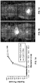
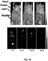
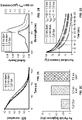
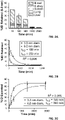
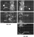

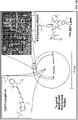

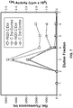

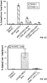


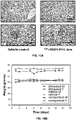
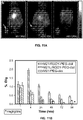
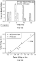


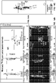
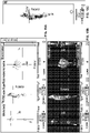
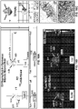
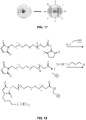
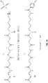
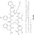


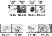
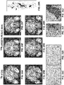
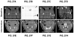

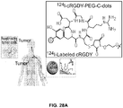
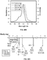
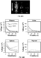
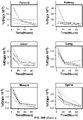
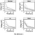
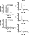


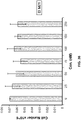


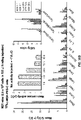
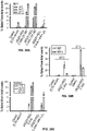

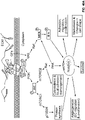

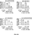

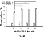
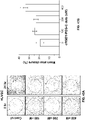

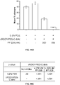

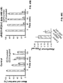
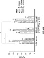
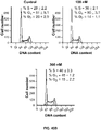
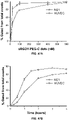
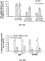


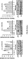


Classifications
-
- A—HUMAN NECESSITIES
- A61—MEDICAL OR VETERINARY SCIENCE; HYGIENE
- A61K—PREPARATIONS FOR MEDICAL, DENTAL OR TOILETRY PURPOSES
- A61K51/00—Preparations containing radioactive substances for use in therapy or testing in vivo
- A61K51/12—Preparations containing radioactive substances for use in therapy or testing in vivo characterised by a special physical form, e.g. emulsion, microcapsules, liposomes, characterized by a special physical form, e.g. emulsions, dispersions, microcapsules
- A61K51/1241—Preparations containing radioactive substances for use in therapy or testing in vivo characterised by a special physical form, e.g. emulsion, microcapsules, liposomes, characterized by a special physical form, e.g. emulsions, dispersions, microcapsules particles, powders, lyophilizates, adsorbates, e.g. polymers or resins for adsorption or ion-exchange resins
- A61K51/1244—Preparations containing radioactive substances for use in therapy or testing in vivo characterised by a special physical form, e.g. emulsion, microcapsules, liposomes, characterized by a special physical form, e.g. emulsions, dispersions, microcapsules particles, powders, lyophilizates, adsorbates, e.g. polymers or resins for adsorption or ion-exchange resins microparticles or nanoparticles, e.g. polymeric nanoparticles
-
- A—HUMAN NECESSITIES
- A61—MEDICAL OR VETERINARY SCIENCE; HYGIENE
- A61K—PREPARATIONS FOR MEDICAL, DENTAL OR TOILETRY PURPOSES
- A61K49/00—Preparations for testing in vivo
- A61K49/0002—General or multifunctional contrast agents, e.g. chelated agents
-
- A—HUMAN NECESSITIES
- A61—MEDICAL OR VETERINARY SCIENCE; HYGIENE
- A61K—PREPARATIONS FOR MEDICAL, DENTAL OR TOILETRY PURPOSES
- A61K49/00—Preparations for testing in vivo
- A61K49/001—Preparation for luminescence or biological staining
- A61K49/0063—Preparation for luminescence or biological staining characterised by a special physical or galenical form, e.g. emulsions, microspheres
- A61K49/0069—Preparation for luminescence or biological staining characterised by a special physical or galenical form, e.g. emulsions, microspheres the agent being in a particular physical galenical form
- A61K49/0089—Particulate, powder, adsorbate, bead, sphere
- A61K49/0091—Microparticle, microcapsule, microbubble, microsphere, microbead, i.e. having a size or diameter higher or equal to 1 micrometer
- A61K49/0093—Nanoparticle, nanocapsule, nanobubble, nanosphere, nanobead, i.e. having a size or diameter smaller than 1 micrometer, e.g. polymeric nanoparticle
-
- A—HUMAN NECESSITIES
- A61—MEDICAL OR VETERINARY SCIENCE; HYGIENE
- A61P—SPECIFIC THERAPEUTIC ACTIVITY OF CHEMICAL COMPOUNDS OR MEDICINAL PREPARATIONS
- A61P35/00—Antineoplastic agents
-
- G—PHYSICS
- G01—MEASURING; TESTING
- G01N—INVESTIGATING OR ANALYSING MATERIALS BY DETERMINING THEIR CHEMICAL OR PHYSICAL PROPERTIES
- G01N33/00—Investigating or analysing materials by specific methods not covered by groups G01N1/00 - G01N31/00
- G01N33/48—Biological material, e.g. blood, urine; Haemocytometers
- G01N33/50—Chemical analysis of biological material, e.g. blood, urine; Testing involving biospecific ligand binding methods; Immunological testing
- G01N33/53—Immunoassay; Biospecific binding assay; Materials therefor
- G01N33/543—Immunoassay; Biospecific binding assay; Materials therefor with an insoluble carrier for immobilising immunochemicals
- G01N33/54313—Immunoassay; Biospecific binding assay; Materials therefor with an insoluble carrier for immobilising immunochemicals the carrier being characterised by its particulate form
- G01N33/54346—Nanoparticles
-
- G—PHYSICS
- G01—MEASURING; TESTING
- G01N—INVESTIGATING OR ANALYSING MATERIALS BY DETERMINING THEIR CHEMICAL OR PHYSICAL PROPERTIES
- G01N33/00—Investigating or analysing materials by specific methods not covered by groups G01N1/00 - G01N31/00
- G01N33/48—Biological material, e.g. blood, urine; Haemocytometers
- G01N33/50—Chemical analysis of biological material, e.g. blood, urine; Testing involving biospecific ligand binding methods; Immunological testing
- G01N33/53—Immunoassay; Biospecific binding assay; Materials therefor
- G01N33/543—Immunoassay; Biospecific binding assay; Materials therefor with an insoluble carrier for immobilising immunochemicals
- G01N33/551—Immunoassay; Biospecific binding assay; Materials therefor with an insoluble carrier for immobilising immunochemicals the carrier being inorganic
- G01N33/552—Glass or silica
-
- G—PHYSICS
- G01—MEASURING; TESTING
- G01N—INVESTIGATING OR ANALYSING MATERIALS BY DETERMINING THEIR CHEMICAL OR PHYSICAL PROPERTIES
- G01N33/00—Investigating or analysing materials by specific methods not covered by groups G01N1/00 - G01N31/00
- G01N33/48—Biological material, e.g. blood, urine; Haemocytometers
- G01N33/50—Chemical analysis of biological material, e.g. blood, urine; Testing involving biospecific ligand binding methods; Immunological testing
- G01N33/53—Immunoassay; Biospecific binding assay; Materials therefor
- G01N33/574—Immunoassay; Biospecific binding assay; Materials therefor for cancer
-
- G—PHYSICS
- G01—MEASURING; TESTING
- G01N—INVESTIGATING OR ANALYSING MATERIALS BY DETERMINING THEIR CHEMICAL OR PHYSICAL PROPERTIES
- G01N33/00—Investigating or analysing materials by specific methods not covered by groups G01N1/00 - G01N31/00
- G01N33/48—Biological material, e.g. blood, urine; Haemocytometers
- G01N33/50—Chemical analysis of biological material, e.g. blood, urine; Testing involving biospecific ligand binding methods; Immunological testing
- G01N33/58—Chemical analysis of biological material, e.g. blood, urine; Testing involving biospecific ligand binding methods; Immunological testing involving labelled substances
- G01N33/582—Chemical analysis of biological material, e.g. blood, urine; Testing involving biospecific ligand binding methods; Immunological testing involving labelled substances with fluorescent label
-
- G—PHYSICS
- G01—MEASURING; TESTING
- G01N—INVESTIGATING OR ANALYSING MATERIALS BY DETERMINING THEIR CHEMICAL OR PHYSICAL PROPERTIES
- G01N33/00—Investigating or analysing materials by specific methods not covered by groups G01N1/00 - G01N31/00
- G01N33/48—Biological material, e.g. blood, urine; Haemocytometers
- G01N33/50—Chemical analysis of biological material, e.g. blood, urine; Testing involving biospecific ligand binding methods; Immunological testing
- G01N33/58—Chemical analysis of biological material, e.g. blood, urine; Testing involving biospecific ligand binding methods; Immunological testing involving labelled substances
- G01N33/585—Chemical analysis of biological material, e.g. blood, urine; Testing involving biospecific ligand binding methods; Immunological testing involving labelled substances with a particulate label, e.g. coloured latex
- G01N33/587—Nanoparticles
-
- G—PHYSICS
- G01—MEASURING; TESTING
- G01N—INVESTIGATING OR ANALYSING MATERIALS BY DETERMINING THEIR CHEMICAL OR PHYSICAL PROPERTIES
- G01N33/00—Investigating or analysing materials by specific methods not covered by groups G01N1/00 - G01N31/00
- G01N33/48—Biological material, e.g. blood, urine; Haemocytometers
- G01N33/50—Chemical analysis of biological material, e.g. blood, urine; Testing involving biospecific ligand binding methods; Immunological testing
- G01N33/58—Chemical analysis of biological material, e.g. blood, urine; Testing involving biospecific ligand binding methods; Immunological testing involving labelled substances
- G01N33/60—Chemical analysis of biological material, e.g. blood, urine; Testing involving biospecific ligand binding methods; Immunological testing involving labelled substances involving radioactive labelled substances
Definitions
- the present invention relates to fluorescent silica-based nanoparticles, and fluorescent silica-based nanoparticles for use in methods of detecting tumor cells.
- Fluorescence imaging probes typically consist of single conventional fluorophore (e.g., organic dyes, fluorescent proteins), fluorescent proteins (e.g., GFP) and semiconductor quantum dots (Q-dots).
- Single fluorophores are usually not stable and have limited brightness for imaging. Similar to dyes, the fluorescent proteins tend to exhibit excited state interactions which can lead to stochastic blinking, quenching and photobleaching.
- Q-dots are generally made from heavy metal ions such as Pb 2+ or Cd 2+ and, therefore, are toxic. Burns et al. "Fluorescent core-shell silica nanoparticles: towards "Lab on a Particle” architectures for nanobiotechnology", Chem. Soc. Rev., 2006, 35, 1028-1042 .
- Fluorescent nanoparticles having an electrically conducting shell and a silica core are known and have utility in modulated delivery of a therapeutic agent.
- U.S. Patent Nos. 6,344,272 , and 6,428,811 are known and have utility in modulated delivery of a therapeutic agent.
- U.S. Patent Nos. 6,344,272 , and 6,428,811 are known and have utility in modulated delivery of a therapeutic agent.
- U.S. Patent Nos. 6,344,272 , and 6,428,811 A shortcoming of existing fluorescent nanoparticles is their limited brightness and their low detectability as fluorescent probes in dispersed systems.
- US2013/039848 A1 discloses fluorescent silica-based nanoparticles for detection, characterization, monitoring and treatment of a disease such as cancer.
- the present multifunctional fluorescent silica-based nanoparticles offer many advantages over other fluorescent probes.
- the nanoparticles are non-toxic, and have excellent photophysical properties (including fluorescent efficiency and photostability), high biocompatibility, and unique pharmacokinetics for molecular diagnostics and therapeutics.
- the nanoparticles are relatively small in size, and have a surface PEG coating that offers excellent renal clearance.
- the fluorescent nanoparticles of the present invention contain a fluorescent core and silica shell.
- the core-shell architectures, the great surface area and diverse surface chemistry of the nanoparticle permit multiple functionalities simultaneously delivered to a target cell.
- the nanoparticle can be functionalized with targeting moieties, contrast agents for medical imaging, therapeutic agents, or other agents.
- the targeting moieties on the surface of the nanoparticle may be tumor ligands, which, when combined with nanoparticle-conjugated therapeutic agents, makes the nanoparticle an ideal vehicle for targeting and potentially treating cancer.
- the silica-based nanoparticle may be labeled with contrast agents for PET, SPECT, CT, MRI, and optical imaging.
- the invention is defined in the claims.
- the invention provides a fluorescent silica-based nanoparticle comprising:
- the invention also provides a plurality of fluorescent silica-based nanoparticles for use in a method for detecting tumor cells comprising steps of:
- the nanoparticle may be administered subdermally, peritumorally, orally, intravenously, nasally, subcutaneously, intramuscularly or transdermally.
- the present invention also provides for a fluorescent silica-based nanoparticle as described in the claims wherein after administration of the nanoparticle to a subject, renal clearance of the nanoparticle ranges from about 90% ID to about 100% ID in about 24 hours.
- the present invention provides a fluorescent silica-based nanoparticle as described in the claims further comprising a contrast agent or a chelate attached to the nanoparticle.
- the diameter of the nanoparticle ranges from about 1 nm to about 8 nm.
- the organic polymers that may be attached to the nanoparticle include poly(ethylene glycol) (PEG), polylactate, polylactic acids, sugars, lipids, polyglutamic acid (PGA), polyglycolic acid, poly(lactic-co-glycolic acid) (PLGA), Polyvinyl acetate (PVA), or the combinations thereof.
- PEG poly(ethylene glycol)
- polylactate polylactic acids
- sugars sugars
- lipids polyglutamic acid
- PGA polyglycolic acid
- PLGA poly(lactic-co-glycolic acid)
- PVA Polyvinyl acetate
- a contrast agent such as a radionuclide including 89 Zr, 64 Cu, 68 Ga, 86 Y, 124 I and 177 Lu, may be attached to the nanoparticle.
- the nanoparticle may be attached to a chelate, for example, DFO, DOTA, TETA and DTPA, that is adapted to bind a radionuclide.
- the nanoparticle of the present invention may be detected by positron emission tomography (PET), single photon emission computed tomography (SPECT), computerized tomography (CT), magnetic resonance imaging (MRI), optical imaging (such as fluorescence imaging including near-infrared fluorescence (NIRF) imaging), bioluminescence imaging, or combinations thereof.
- PET positron emission tomography
- SPECT single photon emission computed tomography
- CT computerized tomography
- MRI magnetic resonance imaging
- optical imaging such as fluorescence imaging including near-infrared fluorescence (NIRF) imaging
- bioluminescence imaging or combinations thereof.
- a therapeutic agent may be attached to the nanoparticle.
- the therapeutic agents include antibiotics, antimicrobials, antiproliferatives, antineoplastics, antioxidants, endothelial cell growth factors, thrombin inhibitors, immunosuppressants, anti-platelet aggregation agents, collagen synthesis inhibitors, therapeutic antibodies, nitric oxide donors, antisense oligonucleotides, wound healing agents, therapeutic gene transfer constructs, extracellular matrix components, vasodialators, thrombolytics, antimetabolites, growth factor agonists, antimitotics, statin, steroids, steroidal and non-steroidal anti-inflammatory agents, angiotensin converting enzyme (ACE) inhibitors, free radical scavengers, PPAR-gamma agonists, small interfering RNA (siRNA), microRNA, and anti-cancer chemotherapeutic agents.
- the therapeutic agents encompassed by the present invention also include radionuclides, for example, 90 Y, 131 I and
- blood residence half-time of the nanoparticle may range from about 2 hours to about 25 hours, from about 3 hours to about 15 hours, or from about 4 hours to about 10 hours.
- Tumor residence half-time of the nanoparticle after administration of the nanoparticle to a subject may range from about 5 hours to about 5 days, from about 10 hours to about 4 days, or from about 15 hours to about 3.5 days.
- the ratio of tumor residence half-time to blood residence half-time of the nanoparticle after administration of the nanoparticle to a subject may range from about 2 to about 30, from about 3 to about 20, or from about 4 to about 15.
- Also disclosed herein are fluorescent silica-based nanoparticles wherein renal clearance of the nanoparticle after administration of the nanoparticle to a subject may range from about 10% ID (initial dose) to about 100% ID in about 24 hours, from about 30% ID to about 80% ID in about 24 hours, or from about 40% ID to about 70% ID in about 24 hours.
- blood residence half-time of the nanoparticle ranges from about 2 hours to about 25 hours
- tumor residence half-time of the nanoparticle ranges from about 5 hours to about 5 days
- renal clearance of the nanoparticle is about 80% ID in about 24 hours.
- nanoparticles in the amount of about 100 times of the human dose equivalent are administered to a subject, substantially no anemia, weight loss, agitation, increased respiration, GI disturbance, abnormal behavior, neurological dysfunction, abnormalities in hematology, abnormalities in clinical chemistries, drug-related lesions in organ pathology, mortality, or combinations thereof, is observed in the subject in about 10 to about 14 days.
- the present invention also provides a fluorescent silica-based nanoparticle according to the claims, wherein, after administration of the nanoparticle to a subject, blood residence half-time of the nanoparticle may range from about 2 hours to about 25 hours, or from about 2 hours to about 15 hours; tumor residence half-time of the nanoparticle may range from about 5 hours to about 2 days, and renal clearance of the nanoparticle is about 80% ID in about 24 hours.
- the number of ligands attached to the nanoparticle may range from about 1 to about 20, or from about 1 to about 10.
- the diameter of the nanoparticle may be between about 1 nm and about 8 nm.
- a contrast agent such as a radionuclide, may be attached to the nanoparticle.
- a chelate may be attached to the nanoparticle.
- the nanoparticle may be detected by PET, SPECT, CT, MRI, optical imaging, bioluminescence imaging, or combinations thereof.
- a therapeutic agent may be attached to the nanoparticle.
- blood residence half-time of the nanoparticle may also range from about 3 hours to about 15 hours, or from about 4 hours to about 10 hours.
- Tumor residence half-time of the nanoparticle after administration of the nanoparticle to a subject may also range from about 10 hours to about 4 days, or from about 15 hours to about 3.5 days.
- the ratio of tumor residence half-time to blood residence half-time of the nanoparticle after administration of the nanoparticle to a subject may range from about 2 to about 30, from about 3 to about 20, or from about 4 to about 15. Renal clearance of the nanoparticle may also range from about 80% ID to about 90% ID in about 24 hours after administration of the nanoparticle to a subject.
- a fluorescent silica-based nanoparticle according to the claims, wherein after administration of the nanoparticle to a subject, the ratio of tumor residence half-time to blood residence half-time of the nanoparticle ranges from about 2 to about 30, and renal clearance of the nanoparticle is about 80% ID in about 24 hours.
- the fluorescent silica-based nanoparticle described herein is useful in a method for detecting a component of a cell comprising the steps of: (a) contacting the cell with a fluorescent silica-based nanoparticle comprising a silica-based core comprising a fluorescent compound positioned within the silica-based core; a silica shell surrounding at least a portion of the core; an organic polymer attached to the nanoparticle; from about 1 to about 30 ligands attached to the nanoparticle; and a contrast agent or a chelate attached to the nanoparticle; and (b) monitoring the binding of the nanoparticle to the cell or a cellular component by at least one imaging technique.
- the fluorescent silica-based nanoparticles described herein can be used in a method for targeting a tumor cell comprising administering to a cancer patient, an effective amount of a fluorescent silica-based nanoparticle comprising a silica-based core comprising a fluorescent compound positioned within the silica-based core; a silica shell surrounding at least a portion of the core; an organic polymer attached to the nanoparticle; a ligand attached to the nanoparticle and capable of binding a tumor marker; and at least one therapeutic agent.
- the nanoparticle may be radiolabeled.
- the nanoparticle may be administered to the patient by, but not restricted to, the following routes: oral, intravenous, nasal, subcutaneous, local, intramuscular or transdermal.
- the present invention provides a fluorescent silica-based nanoparticle according to the claims that allows for precise detection, characterization, monitoring and treatment of a disease such as cancer.
- the invention also provides a plurality of fluorescent silica-based nanoparticles according to the claims for use in a method for detecting tumor cells.
- the method may contain the following steps: (a) administering to a patient a plurality of fluorescent silica-based nanoparticles in a dose ranging from about 0.01 nanomole/kg body weight to about 1 nanomole/kg body weight, from about 0.05 nanomole/kg body weight to about 0.9 nanomole/kg body weight, from about 0.1 nanomole/kg body weight to about 0.9 nanomole/kg body weight, from about 0.2 nanomole/kg body weight to about 0.8 nanomole/kg body weight, from about 0.3 nanomole/kg body weight to about 0.7 nanomole/kg body weight, from about 0.04 nanomole/kg body weight to about 0.6 nanomole/kg body weight, from about 0.2 nanomole/kg body weight to about 0.5 nanomole/kg body weight, and (b) detecting the nanoparticles.
- the nanoparticle further comprises at least one therapeutic agent.
- the nanoparticle has a range of diameters including between about 1 nm and about 15 nm, or between about 1 nm and about 8 nm.
- the nanoparticle has a fluorescent compound positioned within the nanoparticle, and has greater brightness and fluorescent quantum yield than the free fluorescent compound.
- the nanoparticle also exhibits high biostability and biocompatibility.
- an organic polymer such as poly(ethylene glycol) (PEG).
- PEG poly(ethylene glycol)
- the nanoparticle is conjugated to a ligand, which is capable of binding to a cellular component (e.g., the cell membrane or other intracellular component) associated with the specific cell type, such as a tumor marker or a signaling pathway intermediate.
- a therapeutic agent may be attached to the nanoparticle.
- the nanoparticle may also be conjugated to a contrast agent, such as a radionuclide.
- the properties of the nanoparticles lead to bulk excretion through the kidneys, increased potency relative to a native peptide ligand, enhanced uptake, and preferential accumulation in tumors compared with normal tissues. This, along with the lack of in vivo toxicity, has resulted in a unique product resulting in its translation to the clinic.
- the present particles can be used to preferentially detect and localize tumors.
- nanomolar particle tracer doses administered in a microdosing regime accumulate and preferentially localize at sites of disease, although not optimized for targeted detection.
- the present nanoparticles may exhibit distinct and reproducible human pharmacokinetic signatures in which renal clearance predominates (e.g., renal clearance of the nanoparticle is greater than about 90% ID in about 24 hours after administration of the nanoparticle to a subject) without significant RES uptake (e.g., less than about 10%).
- renal clearance predominates e.g., renal clearance of the nanoparticle is greater than about 90% ID in about 24 hours after administration of the nanoparticle to a subject
- significant RES uptake e.g., less than about 10%
- the present nanoparticles are excellent diagnostic probes, exhibiting optimal physicochemical properties in humans that enable them to "target and clear” the body over relatively short time intervals (e.g., hours, days, etc.).
- the present particles bearing multiple actively targeted ligands demonstrate enhanced binding affinity to the cellular targets compared to the affinity of a ligand alone.
- the present particles binds to a cellular target from about 2 to about 20 fold greater, from about 3 to about 15 fold greater, from about 5 to about 10 ibid greater than a ligand alone.
- dual-modality, targeted particles specifically assess tumor burden and can discriminate metastatic tumor from chronic inflammatory disease in large animal models of metastatic melanoma.
- Targeted particles may enhance receptor binding affinity and avidity, increase plasma residence times, bioavailability and tumor retention, and/or promote intracellular delivery via internalization.
- the utilization of such targeted probes within surgical (or medical) oncology settings may enable highly selective treatment of cancer-bearing tissues, potentially reducing attendant complication rates.
- the present particles may be advantageous over passively targeted nanocarriers, as the latter penetrate and non-specifically accumulate within the tumor interstitium by enhanced permeability and retention (EPR) effects.
- EPR enhanced permeability and retention
- Particle-based imaging systems for cancer diagnostics should be non-toxic, and selectively detect sites of primary and metastatic disease while exhibiting relatively rapid renal clearance. Under these conditions, the likelihood of potential toxicity will be reduced given the smaller area under the plasma concentration-time curve.
- these renal clearance properties may be achieved by ultrasmall particle-based platforms or macromolecular systems that meet effective renal glomerular filtration size cutoffs of 10 nm or less. Absence of single-dose acute toxicity and the minimization of such risks will also be important
- a platform design should maximize safety through rapid whole-body clearance and the selection of biokinetic profiles that minimize nonspecific uptake in the reticuloendothelial system (RES), thus reducing potential adverse exposures.
- RES reticuloendothelial system
- the present particles are safe and stable in vivo.
- the particles exhibit distinctly unique and reproducible PK signatures defined by renal excretion. Coupled with preferential uptake and localization of the probe at sites of disease, these particles can be used in cancer diagnostics.
- the nanoparticle may have both a ligand and a contrast agent.
- the ligand allows for the nanoparticle to target a specific cell type through the specific binding between the ligand and the cellular component. This targeting, combined with multimodal imaging, has multiple uses.
- the nanoparticles can be used to map metastatic disease, such as mapping sentinel lymph nodes (SLN), as well as identifing tumor margins or neural structures, enabling the surgeon to resect malignant lesions under direct visualization and to obviate complications during the surgical procedure.
- the ligand may also facilitate entry of the nanoparticle into the cell or barrier transport, for example, for assaying the intracellular environment.
- the nanoparticle can be coupled with a ligand and a therapeutic agent with or without a radiolabel.
- the radiolabel can additionally serve as a therapeutic agent for creating a theranostic platform. This coupling allows the therapeutic agent to be delivered to the specific cell type through the specific binding between the ligand and the cellular component. This specific targeting of the therapeutic agent ensures selective treatment of the disease site with minimum side effects.
- the fluorescent nanoparticle of the present invention includes a silica-based core comprising a fluorescent compound positioned within the core, and a silica shell on the core.
- the silica shell surrounds at least a portion of the core.
- the core of the nanoparticle may contain the reaction product of a reactive fluorescent compound and a co-reactive organo-silane compound.
- the core of the nanoparticle may contain the reaction product of a reactive fluorescent compound and a co-reactive organo-silane compound, and silica.
- the shell of the nanoparticle can be the reaction product of a silica forming compound.
- the shell of the nanoparticle may have a range of layers.
- the silica shell may be from about 1 to about 20 layers, from about 1 to about 15 layers, from about 1 to about 10 layers, or from about 1 to about 5 layers.
- the silica shell of the nanoparticle may cover only a portion of nanoparticle or the entire particle.
- the silica shell may cover about 1 to about 100 percent, from about 10 to about 80 percent, from about 20 to about 60 percent, or from about 30 to about 50 percent of the nanoparticle.
- the silica shell can be either solid, i.e., substantially non-porous, meso-porous, such as semi-porous, or porous.
- the present fluorescent nanoparticle may be synthesized by the steps of: covalently conjugating a fluorescent compound, such as a reactive fluorescent dye, with the reactive moeties including, but not limited to, maleimide, iodoacetamide, thiosulfate, amine, N-Hydroxysuccimide ester, 4-sulfo-2,3,5,6-tetrafluorophenyl (STP) ester, sulfosuccinimidyl ester, sulfodichlorophenol esters, sulfonyl chloride, hydroxyl, isothiocyanate, carboxyl, to an organo-silane compound, such as a co-reactive organo-silane compound, to form a fluorescent silica precursor, and reacting the fluorescent silica precursor to form a fluorescent core; covalently conjugating a fluorescent compound, such as a reactive fluorescent dye, to an organo-silane compound, such as a co-reactive organ
- the synthesis of the fluorescent monodisperse core-shell nanoparticles is based on a two-step process.
- the near-infrared organic dye molecules e.g., tetramethylrhodamine isothiocynate (TRITC)
- TRITC tetramethylrhodamine isothiocynate
- the silica gel monomers are added to form a denser silica network around the fluorescent core material, providing shielding from solvent interactions that can be detrimental to photostability.
- the versatility of the preparative route allows for the incorporation of different fluorescent compounds, such as fluorescent organic compounds or dyes, depending on the intended nanoparticle application.
- the fluorescent compounds that may be incorporated in the dye-rich core can cover the entire UV-Vis to near-IR absorption and emission spectrum.
- the dye precursor is added to a reaction vessel that contains appropriate amounts of ammonia, water and solvent and allowed to react overnight.
- the dye precursor is synthesized by addition reaction between a specific near-infrared dye of interest and 3-aminopropyltriethoxysilane in molar ratio of 1:50, in exclusion of moisture.
- tetraethylorthosilicate TEOS is subsequently added to grow the silica shell that surrounded the core.
- the synthesis of the expanded core-shell nanoparticle is accomplished by co-condensing TEOS with the dye precursor and allowing the mixture to react overnight. After the synthesis of the expanded core is completed, additional TEOS is added to grow the silica shell that surrounded the core.
- the synthesis of the homogenous nanoparticles is accomplished by co-condensing all the reagents, the dye precursor and TEOS and allowing the mixture to react overnight.
- the nanoparticles may incorporate any known fluorescent compound, such as fluorescent organic compound, dyes, pigments, or combinations thereof.
- fluorescent organic compound such as fluorescent organic compound, dyes, pigments, or combinations thereof.
- suitable chemically reactive fluorescent dyes are known, see for example MOLECULAR PROBES HANDBOOK OF FLUORESCENT PROBES AND RESEARCH CHEMICALS, 6th ed., R. P. Haugland, ed. (1996 ).
- a typical fluorophore is, for example, a fluorescent aromatic or heteroaromatic compound such as is a pyrene, an anthracene, a naphthalene, an acridine, a stilbene, an indole or benzindole, an oxazole or benzoxazole, a thiazole or benzothiazole, a 4-amino-7-nitrobenz-2-oxa-l ,3-diazole (NBD), a cyanine, a carbocyanine, a carbostyryl, a porphyrin, a salicylate, an anthranilate, an azulene, a perylene, a pyridine, a quinoline, a coumarin (including hydroxycoumarins and aminocoumarins and fluorinated derivatives thereof), and like compounds, see for example U.S.
- a fluorescent aromatic or heteroaromatic compound such as is a pyrene, an anth
- Cy5 a near infrared fluorescent (NIRF) dye
- NIRF near infrared fluorescent
- Non-limiting fluorescent compound that may be used in the present invention include, Cy5, Cy5.5 (also known as Cy5++), Cy2, fluorescein isothiocyanate (FITC), tetramethylrhodamine isothiocyanate (TRITC), phycoerythrin, Cy7, fluorescein (FAM), Cy3, Cy3.5 (also known as Cy3++), Texas Red, LightCycler-Red 640, LightCycler Red 705, tetramethylrhodamine (TMR), rhodamine, rhodamine derivative (ROX), hexachlorofluorescein (HEX), rhodamine 6G (R6G), the rhodamine derivative JA133, Alexa Fluorescent Dyes (such as Alexa Fluor 488, Alexa Fluor 546, Alexa Fluor 633, Alexa Fluor 555, and Alexa Fluor 647), 4',6-diamidino-2-phenylindole (DAPI),
- Fluorescent compound that can be used also include fluorescent proteins, such as GFP (green fluorescent protein), enhanced GFP (EGFP), blue fluorescent protein and derivatives (BFP, EBFP, EBFP2, Azurite, mKalama1), cyan fluorescent protein and derivatives (CFP, ECFP, Cerulean, CyPet) and yellow fluorescent protein and derivatives (YFP, Citrine, Venus, YPet).
- fluorescent proteins such as GFP (green fluorescent protein), enhanced GFP (EGFP), blue fluorescent protein and derivatives (BFP, EBFP, EBFP2, Azurite, mKalama1), cyan fluorescent protein and derivatives (CFP, ECFP, Cerulean, CyPet) and yellow fluorescent protein and derivatives (YFP, Citrine, Venus, YPet).
- GFP green fluorescent protein
- EGFP enhanced GFP
- BFP blue fluorescent protein and derivatives
- BFP blue fluorescent protein and derivatives
- CFP cyan fluorescent protein and derivatives
- CFP
- the silica shell surface of the nanoparticles can be modified by using known cross-linking agents to introduce surface functional groups.
- Crosslinking agents include, but are not limited to, divinyl benzene, ethylene glycol dimethacrylate, trimethylol propane trimethacrylate, N,N'-methylene-bis-acrylamide, alkyl ethers, sugars, peptides, DNA fragments, or other known functionally equivalent agents.
- the ligand may be conjugated to the nanoparticle of the present invention by, for example, through coupling reactions using carbodiimide, carboxylates, esters, alcohols, carbamides, aldehydes, amines, sulfur oxides, nitrogen oxides, halides, or any other suitable compound known in the art.
- U.S. Patent No. 6,268,222 U.S. Patent No. 6,268,222 .
- An organic polymer is attached to the present nanoparticle, e.g., attached to the surface of the nanoparticle.
- An organic polymer may be attached to the silica shell of the present nanoparticle.
- the organic polymer that may be used in the present invention include PEG, polylactate, polylactic acids, sugars, lipids, polyglutamic acid (PGA), polyglycolic acid, poly(lactic-co-glycolic acid) (PLGA), polyvinyl acetate (PVA), and the combinations thereof.
- the attachment of the organic polymer to the nanoparticle may be accomplished by a covalent bond or non-covalent bond, such as by ionic bond, hydrogen bond, hydrophobic bond, coordination, adhesive, and physical absorption.
- the nanoparticle is covalently conjugated with PEG, which prevents adsorption of serum proteins, facilitates efficient urinary excretion and decreases aggregation of the nanoparticle.
- the surface of the nanoparticle may be modified to incorporate at least one functional group.
- the organic polymer (e.g., PEG) attached to the nanoparticle may be modified to incorporate at least one functional group.
- the functional group can be a maleimide or N -Hydroxysuccinimide (NHS) ester.
- NHS N -Hydroxysuccinimide
- a ligand according to the claims is attached to the present nanoparticle.
- the ligand is capable of binding to at least one cellular component.
- the cellular component to which the ligands attached to the nanoparticles are capable of binding may be associated with specific cell types or have elevated levels in specific cell types, such as cancer cells or cells specific to particular tissues and organs. Accordingly, the nanoparticle can target a specific cell type, and/or provides a targeted delivery for the treatment and diagnosis of a disease.
- the term "ligand” refers to a molecule or entity that can be used to identify, detect, target, monitor, or modify a physical state or condition, such as a disease state or condition.
- a ligand may be used to detect the presence or absence of a particular receptor, expression level of a particular receptor, or metabolic levels of a particular receptor.
- a ligand can be, for example, a peptide, a protein, a protein fragment, a peptide hormone, a sugar (i.e., lectins), a biopolymer, a synthetic polymer, an antigen, an antibody, an antibody fragment (e.g., Fab, nanobodies), an aptamer, a virus or viral component, a receptor, a hapten, an enzyme, a hormone, a chemical compound, a pathogen, a microorganism or a component thereof, a toxin, a surface modifier, such as a surfactant to alter the surface properties or histocompatability of the nanoparticle or of an analyte when a nanoparticle associates therewith, and combinations thereof.
- a surface modifier such as a surfactant to alter the surface properties or histocompatability
- the ligands are antibodies such as monoclonal or polyclonal antibodies.
- the ligands are receptor ligands.
- the ligand is poly-L-lysine (pLysine).
- An antigen may be attached to the nanoparticle.
- the antigen-attached nanoparticle may be used for vaccination.
- component of a cell refers to, for example, a receptor, an antibody, a hapten, an enzyme, a hormone, a biopolymer, an antigen, a nucleic acid (DNA or RNA), a microorganism, a virus, a pathogen, a toxin, combinations thereof, and like components.
- the component of a cell may be positioned on the cell (e.g., a transmembrane receptor) or inside the cell.
- the component of a cell is a tumor marker.
- tumor marker refers to a molecule, entity or substance that is expressed or overexpressed in a cancer cell but not normal cell. For example, the overexpression of certain receptors is associated with many types of cancer.
- a ligand capable of binding to a tumor marker may be conjugated to the surface of the present nanoparticle, so that the nanoparticle can specifically target the tumor cell.
- a ligand may be attached to the present nanoparticle directly or through a linker.
- the attachment of the ligand to the nanoparticle may be accomplished by a covalent bond or non-covalent bond, such as by ionic bond, hydrogen bond, hydrophobic bond, coordination, adhesive, and physical absorption.
- the ligand may be coated onto the surface of the nanoparticle.
- the ligand may be imbibed into the surface of the nanoparticle.
- the ligand may be attached to the surface of the fluorescent nanoparticle, or may be attached to the core when the shell is porous or is covering a portion of the core.
- the linker can be any suitable molecules, such as a functionalized PEG.
- the PEGs can have multiple functional groups for attachment to the nanoparticle and ligands.
- the particle can have different types of functionalized PEGs bearing different functional groups that can be attached to multiple ligands. This can enhance multivalency effects and/or contrast at the target site, which allows the design and optimization of a complex multimodal platform with improved targeted detection, treatment, and sensing in vivo.
- ligands may be attached to the nanopanicle.
- tripeptide Arg-Gly-Asp may be attached to the nanoparticle disclosed herein but not claimed.
- cyclic peptide cRGD (which may contain other amino acid(s), e.g., cRGDY) may be attached to the nanoparticle.
- Any linear, cyclic or branched peptide containing the RGD sequence is within the scope of the present disclosure.
- RGD binds to ⁇ v ⁇ 3 integrin, which is overexpressed at the surface of activated endothelial cells during angiogenesis and in various types of tumor cells.
- EPPT1 may be the ligand attached to the nanoparticle disclosed herein but not claimed.
- EPPT1 derived from the monoclonal antibody (ASM2) binding site, targets underglycosylated MUC1 (uMUC 1).
- MUC1 a transmembrane receptor, is heavily glycosylated in normal tissues; however, it is overexpressed and aberrantly underglycosylated in almost all human epithelial cell adenocarcinomas, and is implicated in tumor pathogenesis.
- Moore et al In vivo targeting of underglycosylated MUC-1 tumor antigen using a multimodal imaging probe. Cancer Res. 2004; 64:1821-7 . Patel et al.
- MUC 1 plays a role in tumor maintenance in aggressive thryroid carcinomas. Surgery. 2005; 138:994-1001 .
- Specific antibodies including monoclonal antibodies against uMUC1 may alternatively be conjugated to the nanoparticle disclosed herein in order to target uMUC1.
- peptide analogues of ⁇ -melanotropin stimulating hormone are the ligands attached to the nanoparticle.
- Peptide analogues of ⁇ -MSH are capable of binding to melanocortin-1 receptors (MC1R), a family of G-protein-coupled receptors overexpressed in melanoma cells. Loir et al. Cell Mol.Biol. (Noisy-le-grand) 1999, 45:1083-1092 .
- octreotate a peptide analog of 14-amino acid somatostatin
- octreotate is the ligand attached to the nanoparticle.
- Octreotide which has a longer half-life than somatostatin, is capable of binding to somatostatin receptor (SSTR).
- SSTR somatostatin receptor
- SSTR a member of the G-protein coupled receptor family, is overexpressed on the surface of several human tumors. Reubi et al. Distribution of Somatostatin Receptors in Normal and Tumor-Tissue. Metab. Clin. Exp. 1990;39:78-81 .
- Reubi et al Somatostatin receptors and their subtypes in human tumors and in peritumoral vessels. Metab. Clin.
- somatostatin analogs may alternatively be conjugated to the nanoparticle disclosed herein but not claimed to target SSTR, such as Tyr3-octreotide (Y3-OC), octreotate (TATE), Tyr3-octreotate (Y3-TATE), and 111 In-DTPA-OC.
- Y3-OC Tyr3-octreotide
- TATE octreotate
- Y3-TATE Tyr3-octreotate
- 111 In-DTPA-OC 111 In-DTPA-OC.
- SLN mapping may be used in diagnosing, staging and treating cancer.
- J591 is an anti-prostate-specific membrane antigen (i.e., anti-PSMA) monoclonal antibody. J591 has been previously used to detect and stage prostate cancer. Tagawa et al., Anti-prostate-specific membrane antigen-based radioimmunotherapy for prostate cancer, Cancer, 2010, 116(4 Suppl): 1075-83 .
- the F(ab')2 fragment of J591 may be used as a ligand attached to the nanoparticle disclosed herein but not claimed.
- the nanoparticle may also be radiolabeled to create a dual-modality probe.
- nanoparticles bearing the F(ab')2 fragment of J591 are used in diagnosing prostate cancer or endometrial cancer (e.g., endometrial endometrioid adenocarcinoma).
- nanoparticles bearing the F(ab')2 fragment of J591 can be used to target brain tumor neovasculature for treatment, disease progression monitoring. Brain tumor neovasculature has been found to overexpress PSMA, as shown from prior immunohistochemistry evaluations of excised high grade glioma specimens.
- Cyclic peptides containing the sequence HWGF are potent and selective inhibitors of MMP-2 and MMP-9 but not of several other MMP family members.
- Peptide CTTHWGFTLC inhibits the migration of human endothelial cells and tumor cells. Moreover, it prevents tumor growth and invasion in animal models and improves survival of mice bearing human tumors.
- CTTHWGFTLC-displaying phage specifically targets angiogenic blood vessels in vivo.
- This peptide and its extension GRENYGHCTTHWGFTLC or GRENYGHCTTHWGFTLS can be used as ligands to be attached to the nanoparticle disclosed herein but not claimed.
- the peptides can also be radiolabelled, e.g., radioiodinated.
- MMPI matrix metalloproteinase peptide inhibitor
- the number of ligands attached to the nanoparticle may range from about 1 to about 20, from about 2 to about 15, from about 3 to about 10, from about 1 to about 10, or from about 1 to about 6.
- the small number of the ligands attached to the nanoparticle helps maintain the hydrodynamic diameter of the present nanoparticle which meets the renal clearance cutoff size range.
- the number of ligands measured may be an average number of ligands attached to more than one nanoparticle. Alternatively, one nanoparticle may be measured to determine the number of ligands attached.
- the number of ligands attached to the nanoparticle can be measured by any suitable methods, which may or may not be related to the properties of the ligands.
- the number of cRGD peptides bound to the particle as disclosed herein but not claimed may be estimated using FCS-based measurements of absolute particle concentrations and the starting concentration of the reagents for cRGD peptide.
- Average number of RGD peptides per nanoparticle and coupling efficiency of RGD to functionalized PEG groups can be assessed colorimetrically under alkaline conditions and Biuret spectrophotometric methods.
- the number of ligands attached to the nanoparticle may also be measured by other suitable methods.
- a contrast agent may be attached to the present nanoparticle for medical or biological imaging.
- the term “contrast agent” refers to a substance, molecule or compound used to enhance the visibility of structures or fluids in medical or biological imaging.
- the term “contrast agent” also refers to a contrast-producing molecule.
- the imaging techniques encompassed by the present invention include positron emission tomography (PET), single photon emission computed tomography (SPECT), computerized tomography (CT), magnetic resonance imaging (MRI), optical bioluminescence imaging, optical fluorescence imaging, and combinations thereof.
- PET positron emission tomography
- SPECT single photon emission computed tomography
- CT computerized tomography
- MRI magnetic resonance imaging
- optical bioluminescence imaging optical fluorescence imaging
- optical fluorescence imaging and combinations thereof.
- the contrast agent encompassed by the present invention may be any molecule, substance or compound known in the art for PET, SPECT, CT, MRI, and optical imaging.
- the contrast agent may be radionuclides, radiometals, positron emitters, beta emitters, gamma emitters, alpha emitters, paramagnetic metal ions, and supraparamagnetic metal ions.
- the contrast agents include, but are not limited to, iodine, fluorine, copper, zirconium, lutetium, astatine, yttrium, gallium, indium, technetium, gadolinium, dysprosium, iron, manganese, barium and barium sulfate.
- the radionuclides that may be used as the contrast agent attached to the nanoparticle of the present invention include, but are not limited to, 89 Zr, 64 Cu, 68 Ga, 86 Y, 124 I and 177 Lu.
- the contrast agent may be directly conjugated to the nanoparticle.
- the contrast agent may be indirectly conjugated to the nanoparticle, by attaching to linkers or chelates.
- the chelate may be adapted to bind a radionuclide.
- the chelates that can be attached to the present nanoparticle may include, but are not limited to, 1,4,7,10-tetraazacyclododecane- 1.4,7,10-tetraaceric acid (DOTA), diethylenetriaminepentaacetic (DTPA), desferrioxamine (DFO) and triethylenetetramine (TETA).
- Suitable means for imaging, detecting, recording or measuring the present nanoparticles may also include, for example, a flow cytometer, a laser scanning cytometer, a fluorescence micro-plate reader, a fluorescence microscope, a confocal microscope, a bright-Held microscope, a high content scanning system, and like devices. More than one imaging techniques may be used at the same time or consecutively to detect the present nanoparticles. In one embodiment, optical imaging is used as a sensitive, high-throughput screening tool to acquire multiple time points in the same subject, permitting semi-quantitative evaluations of tumor marker levels.
- PET is needed to achieve adequate depth penetration for acquiring volumetric data, and to detect, quantitate, and monitor changes in receptor and/or other cellular marker levels as a means of assessing disease progression or improvement, as well as stratifying patients to suitable treatment protocols.
- a therapeutic agent may be attached to the fluorescent nanoparticle, for example, for targeted treatment of a disease.
- the therapeutic agent may be delivered to a diseased site in a highly specific or localized manner with release of the therapeutic agent in the disease site. Alternatively, the therapeutic agent may not be released.
- the fluorescent nanoparticle conjugated with the ligand can be used for targeted delivery of a therapeutic agent to a desired location in a variety of systems, such as on, or within, a cell or cell component, within the body of an organism, such as a human, or across the blood-brain barrier.
- the therapeutic agent may be attached to the nanoparticle directly or indirectly.
- the therapeutic agent can be absorbed into the interstices or pores of the silica shell, or coated onto the silica shell of the fluorescent nanoparticle.
- the therapeutic agent can be associated with the fluorescent core, such as by physical absorption or by bonding interaction.
- the therapeutic agent may be associated with the ligand that is attached to the fluorescent nanoparticle.
- the therapeutic agent may also be associated with the organic polymer or the contrast agent
- the therapeutic agent may be attached to the nanoparticle through PEG.
- the PEGs can have multiple functional groups for attachment to the nanoparticle and therapeutic agent.
- the particle can have different types of functionalized PEGs bearing different functional groups that can be attached to multiple therapeutic agents.
- the therapeutic agent may be attached to the nanoparticle covalently or non-covalently.
- therapeutic agent refers to a substance that may be used in the diagnosis, cure, mitigation, treatment, or prevention of disease in a human or another animal.
- therapeutic agents include substances recognized in the official United States Pharmacopeia, official Homeopathic Pharmacopeia of the United States, official National Formulary, or any supplement thereof.
- Therapeutic agents that can be incorporated with the fluorescent nanoparticles or the ligated-fluorescent nanoparticles of the invention include nucleosides, nucleoside analogs, small interfering RNA (siRNA), microRNA, oligopeptides, polypeptides, antibodies, COX-2 inhibitors, apoptosis promoters, urinary tract agents, vaginal agents, vasodilators neurodegenerative agents (e.g., Parkinson's disease), obesity agents, ophthalmic agents, osteoporosis agents, para-sympatholytics, para-sympathometics, antianesthetics, prostaglandins, psychotherapeutic agents, respiratory agents, sedatives, hypnotics, skin and mucous membrane agents, anti-bacterials, anti-fungals, antineoplastics, cardioprotective agents, cardiovascular agents, anti-thrombotics, central nervous system stimulants, cholinesterase inhibitors, contraceptives, dopamine receptor agonists, erectile dysfunction
- Therapeutic agents that may be attached to the present nanoparticle include, but are not limited to, DNA alkylating agents, topoisomerase inhibitors, endoplasmic reticulum stress inducing agents, a platinum compound, an antimetabolite, vincalkaloids, taxanes, epothilones, enzyme inhibitors, receptor antagonists, therapeutic antibodies, tyrosine kinase inhibitors, boron radiosensitizers (i.e. velcade), and chemotherapeutic combination therapies.
- Non-limiting examples of DNA alkylating agents are nitrogen mustards, such as Mechlorethamine, Cyclophosphamide (Ifosfamide, Trofosfamide), Chlorambucil (Melphalan, Prednimustine), Bendamustine, Uramustine and Estramustine; nitrosoureas, such as Carmustine (BCNU), Lomustine (Semustine), Fotemustine, Nimustine, Ranimustine and Streptozocin; alkyl sulfonates, such as Busulfan (Mannosulfan, Treosulfan); Aziridines, such as Carboquone, ThioTEPA, Triaziquone, Triethylenemelamine; Hydrazines (Procarbazine); Triazenes such as dacarbazine and Temozolomide; Altretamine and Mitobronitol.
- nitrogen mustards such as Mechlorethamine, Cyclophosphamide (Ifosfamide,
- Topoisomerase I inhibitors include Campothecin derivatives including CPT-II (irinotecan), SN-38, APC, NPC, campothecin, topotecan, exatecan mesylate, 9-nitrocamptothecin, 9-aminocamptothecin, lurtotecan, rubitecan, silatecan, gimatecan, diflomotecan, extatecan, BN-80927, DX-8951f, and MAG-CPT as decribed in Pommier Y. (2006) Nat Rev. Cancer 6(10):789-802 and U.S. Patent Publication No.
- Topoisomerase II inhibitors include, but are not limited to Etoposide and Teniposide.
- Dual topoisomerase I and II inhibitors include, but are not limited to, Saintopin and other Naphthecenediones, DACA and other Acridine-4-Carboxamindes, Intoplicine and other Benzopyridoindoles, TAS-103 and other 7H-indeno[2,1-c]Quinoline-7-ones, Pyrazoloacridine, XR 11576 and other Benzophenazines, XR 5944 and other Dimeric compounds, 7-oxo-7H-dibenz[f,ij]lsoquinolines and 7-oxo-7H-benzo[e]Perimidines, and Anthracenyl-amino Acid Conjugates as described in Denny and Baguley (2003) Curr.
- Top. Med. Chem. 3(3):339-353 Some agents inhibit Topoisomerase II and have DNA intercalation activity such as, but not limited to, Anthracyclines (Aclarubicin, Daunorubicin, Doxorubicin, Epirubicin, Idarubicin, Amrubicin, Pirarubicin, Valrubicin, Zorubicin) and Antracenediones (Mitoxantrone and Pixantrone).
- Anthracyclines Aclarubicin, Daunorubicin, Doxorubicin, Epirubicin, Idarubicin, Amrubicin, Pirarubicin, Valrubicin, Zorubicin
- Antracenediones Mitoxantrone and Pixantrone
- endoplasmic reticulum stress inducing agents include, but are not limited to, dimethyl-celecoxib (DMC), nelfinavir, celecoxib, and boron radiosensitizers (i.e. velcade (Bortezomib)).
- DMC dimethyl-celecoxib
- nelfinavir nelfinavir
- celecoxib nelfinavir
- boron radiosensitizers i.e. velcade (Bortezomib)
- Non-limiting examples of platinum based compound include Carboplatin, Cisplatin, Nedaplatin, Oxaliplatin, Triplatin tetranitrate, Satraplatin, Aroplatin, Lobaplatin, and JM-216. (see McKeage et al. (1997) J. Clin. Oncol. 201 :1232-1237 and in general, CHEMOTHERAPY FOR GYNECOLOGICAL NEOPLASM, CURRENT THERAPY AND NOVEL APPROACHES, in the Series Basic and Clinical Oncology, Angioli et al. Eds., 2004 ).
- Non-limiting examples of antimetabolite agents include Folic acid based, i.e. dihydrofolate reductase inhibitors, such as Aminopterin, Methotrexate and Pemetrexed; thymidylate synthase inhibitors, such as Raltitrexed, Pemetrexed; Purine based, i.e.
- an adenosine deaminase inhibitor such as Pentostatin, a thiopurine, such as Thioguanine and Mercaptopurine, a halogenated/ribonucleotide reductase inhibitor, such as Cladribine, Clofarabine, Fludarabine, or a guanine/guanosine: thiopurine, such as Thioguanine; or Pyrimidine based, i.e.
- cytosine/cytidine hypomethylating agent, such as Azacitidine and Decitabine, a DNA polymerase inhibitor, such as Cytarabine, a ribonucleotide reductase inhibitor, such as Gemcitabine, or a thymine/thymidine: thymidylate synthase inhibitor, such as a Fluorouracil (5-FU).
- hypomethylating agent such as Azacitidine and Decitabine
- a DNA polymerase inhibitor such as Cytarabine
- a ribonucleotide reductase inhibitor such as Gemcitabine
- thymine/thymidine thymidylate synthase inhibitor, such as a Fluorouracil (5-FU).
- 5-FU Equivalents to 5-FU include prodrugs, analogs and derivative thereof such as 5' -deoxy-5-fluorouridine (doxifluroidine), 1-tetrahydrofuranyl-5-fiuorouracil (ftorafur), Capecitabine (Xeloda), S-1 (MBMS-247616, consisting of tegafur and two modulators, a 5-chloro-2,4dihydroxypyridine and potassium oxonate), ralititrexed (tomudex), nolatrexed (Thymitaq, AG337), LY231514 and ZD9331, as described for example in Papamicheal (1999) The Oncologist 4:478-487 .
- 5' -deoxy-5-fluorouridine doxifluroidine
- 1-tetrahydrofuranyl-5-fiuorouracil ftorafur
- Capecitabine Xeloda
- S-1 MBMS-247616, consisting of
- vincalkaloids examples include, but are not limited to Vinblastine, Vincristine, Vinflunine, Vindesine and Vinorelbine.
- taxanes examples include, but are not limited to docetaxel, Larotaxel, Ortataxel, Paclitaxel and Tesetaxel.
- An example of an epothilone is iabepilone.
- enzyme inhibitors include, but are not limited to farnesyltransferase inhibitors (Tipifamib); CDK inhibitor (Alvocidib, Seliciclib); proteasome inhibitor (Bortezomib); phosphodiesterase inhibitor (Anagrelide; rolipram); IMP dehydrogenase inhibitor (Tiazofiirine); and lipoxygenase inhibitor (Masoprocol).
- receptor antagonists include, but are not limited to ERA (Atrasentan); retinoid X receptor (Bexarotene); and a sex steroid (Testolactone).
- therapeutic antibodies include, but are not limited to anti-HER1/EGFR (Cetuximab, Panitumumab); Anti-HER2/neu (erbB2) receptor (Trastuzumab); Anti-EpCAM (Catumaxomab, Edrecolomab) Anti-VEGF-A (Bevacizumab); Anti-CD20 (Rituximab, Tositumomab, Ibritumomab); Anti-CD52 (Alemtuzumab); and Anti-CD33 (Gemtuzumab).
- U.S. Patent Nos. 5,776,427 and 7.601,355 are examples of therapeutic antibodies.
- tyrosine kinase inhibitors include, but are not limited to inhibitors to ErbB: HER1/EGFR (Erlotinib, Gefitinib, Lapatinib, Vandetanib, Sunitinib, Neratinib); HER2/neu (Lapatinib, Neratinib); RTK class III: C-kit (Axitinib, Sunitinib, Sorafenib), FLT3 (Lestaurtinib), PDGFR (Axitinib, Sunitinib, Sorafenib); and VEGFR (Vandetanib, Semaxanib, Cediranib, Axitinib, Sorafenib), bcr-abl (Imatinib, Nilotinib, Dasatinib); Src (Bosutinib) and Janus kinase 2 (Lestaurtinib).
- ErbB HER1/EG
- Chemotherapeutic agents that can be attached to the present nanoparticle may also include amsacrine, Trabectedin, retinoids (Alitretinoin, Tretinoin), Arsenic trioxide, asparagine depleter Asparaginase/ Pegaspargase), Celecoxib, Demecolcine, Elesclomol, Elsamitrucin, Etoglucid, Lonidamine, Lucanthone, Mitoguazone, Mitotane, Oblimersen, Temsirolimus, and Vorinostat.
- Examples of specific therapeutic agents that can be linked, ligated, or associated with the fluorescent nanoparticles of the invention are flomoxef; fortimicin(s); gentamicin(s); glucosulfone solasulfone; gramicidin S; gramicidin(s); grepafloxacin; guamocycline; hetacillin; isepamicin; josamycin; kanamycin(s); flomoxef; fortimicin(s); gentamicin(s); glucosulfone solasulfone; gramicidin S; gramicidin(s); grepafloxacin; guamecycline; hetacillin; isepamicin; josamycin; kanamycin(s); bacitracin; bambermycin(s); biapenem; brodimoprim; butirosin; capreomycin; carbenicillin; carbomycin; carumonam;
- streptozocin doxorubicin; daunorubicin; plicamycin; idarubicin; mitomycin C; pentostatin; mitoxantrone; cytarabine; fludarabine phosphate; butorphanol; nalbuphine.
- the therapeutic agent may also include radionuclides when the present nanoparticle is used in targeted radiotherapy.
- low energy betaemitting radionuclides such as 177 Lu-chelated constructs, is associated with the nanoparticle and used to treat relatively small tumor burdens or micrometastatic disease.
- higher energy beta emitters such as yttrium-90 ( 90 Y)
- Iodine-131 131 I may also be used for radiotherapy.
- the surface of the nanoparticle may be modified to incorporate at least one functional group.
- the organic polymer (e.g., PEG) attached to the nanoparticle may be modified to incorporate at least one functional group.
- the functional group can be a maleimide or N -Hydroxysuccinimide (NHS) ester.
- NHS N -Hydroxysuccinimide
- a therapeutic agent is attached to the nanoparticle (surface or the organic polymer coating) via an NHS ester functional group.
- tyrosine kinase inhibitor such as dasatinib (BMS) or chemotherapeutic agent (e.g., taxol)
- BMS dasatinib
- chemotherapeutic agent e.g., taxol
- the nanoparticle has been found to have unique biological properties, including relatively rapid clearance from the blood compared to tumors and subsequent tumor tissue accumulation of the therapeutic agent, which suggests that a prodrug approach is feasible.
- the functionalized nanoparticle permits drugs to be dosed multiple times, ensuring that the drug concentration in the tumor is greater than that specified by the IC-50 in tumor tissue, yet will not be dose-limiting to other organ tissues, such as the heart, liver or kidney.
- the therapeutic agent and nanoparticle can be radiolabeled or optically labelled separately, allowing independent monitoring of the therapeutic agent and the nanoparticle.
- radiofluorinated (i.e., 18 F) dasatinib is coupled with PEG-3400 moieties attached to the nanoparticle via NHS ester linkages.
- Radiofluorine is crucial for being able to independently monitor time-dependent changes in the distribution and release of the drug from the radioiodinated ( 124 I) fluorescent (Cy5) nanoparticle. In this way, we can separately monitor the prodrug (dasatinib) and nanoparticle. This permits optimization of the prodrug design compared with methods in the prior art where no dual-labeling approach is used.
- radiotherapeutic iodine molecules i.e., I-131
- other therapeutic gamma or alpha emitters are conjugated with PEG via a maleimide functional group, where the therapeutic agent may not dissociate from the PEG in vivo .
- the surface of the nanoparticle may be modified to incorporate a functional group.
- the nanoparticle may also be modified with organic polymers (e.g., PEGs) or chelates that can incorporate a functional group.
- the ligand, contrast agent, or therapeutic agent is modified to incorporate a functional group that is able to react with the functional group on the nanoparticle, or on the PEGs or chelating agents attached to the nanoparticle under suitable conditions. Accordingly, any ligand, contrast agent or therapeutic agent that has the reactive functional group is able to be readily conjugated to the nanoparticle.
- click chemistry This generalizable approach is referred herein as "click chemistry", which would allow for a great deal of versatility to explore multimodality applications.
- click chemistry two molecular components may be joined via a selective, rapid, clean, bioorthogonal, and/or biocompatible reaction.
- Kolb et al. (2001) Click Chemistry: Diverse Chemical Function from a Few Good Reactions.
- Lim et al., (2010) Bioorthogonal chemistry recent progress and future directions.
- Chemical Communications 46, 1589-1600 Sletten et al., (2009) Bioorthogonal chemistry: fishing for selectivity in a sea of functionality.
- Angewandte Chemie International Edition 48, 6973-6998 Angewandte Chemie International Edition 48, 6973-6998 .
- Any suitable reaction mechanism may be adapted in the present invention for "click chemistry", so long as facile and controlled attachment of the ligand, contrast agent or chelate to the nanoparticle can be achieved.
- a free triple bond is introduced onto PEG, which is already covalently conjugated with the shell of the nanoparticle.
- an azide bond is introduced onto the desired ligand (or contrast agent, chelate).
- click chemistry is the Cu(I)-catalyzed [3+2] Huisgen cycloaddition between an azide and alkyne.
- Moses et al. (2007) The growing applications of click chemistry. Chemical Society Reviews 36, 1249-1262 . Glaser et al., (2009) 'Click labelling' in PET radiochemistry. Journal of Labelled Compounds & Radiopharmaceuticals 52,407-414 .
- Mindt et al. (2009) A "Click Chemistry” Approach to the Efficient Synthesis of Multiple Imaging Probes Derived from a Single Precursor. Bioconjugate Chemistry 20, 1940-1949 .
- a maleimide functional group and a thiol group may be introduced onto the nanoparticle and the desired ligand (or contrast agent, chelate), with the nanoparticle having the maleimide functional group, the ligand (or contrast agent, chelate) having the thiol group, or vice versa.
- the double bond of maleimide readily reacts with the thiol group to form a stable carbon-sulfur bond.
- an activated ester functional group e.g., a succinimidyl ester group, and an amine group may be introduced onto the nanoparticle and the desired ligand, contrast agent or chelate.
- the click chemistry involves the inverse electron demand Diels-Alder reaction between a tetrazine moiety and a strained alkene ( Figure 21c ).
- a tetrazine moiety and a strained alkene ( Figure 21c ).
- Devaraj et al. (2009) Fast and Sensitive Pretargeted Labeling of Cancer Cells through a Tetrazine/trans-Cyclooctene Cycloaddition.
- Angewandte Chemie-International Edition 48, 7013-7016 Devaraj et al., (2008) Tetrazine-Based Cycloadditions: Application to Pretargeted Live Cell Imaging. Bioconjugate Chemistry 19,2297-2299 .
- a tetrazine moiety and a strained alkene may be introduced onto the nanoparticle and the desired ligand (or contrast agent, chelate), with the nanoparticle having the tetrazine moiety, the ligand (or contrast agent, chelate) having the strained alkene, or vice versa.
- a number of different tetrazine—strained alkene pairs can be used for the reaction, including, e.g., the combination of 3-(4-benzylamino)-1,2,4,5-tetrazine (Tz) and either norbornene- or trans-cyclooctene-dervatives.
- the surface of the nanoparticles may be decorated with tetrazine moieties, which can subsequently be conjugated to norbomene-modified ligand (or contrast agent, or chelate) ( Figures 21d and 21e ).
- the nanoparticles are coated with tetrazine, which can then be modified with the DOTA chelates using the tetrazine-norbomene ligation, and radiolabeled with 64 Cu.
- the blood residence half-time of the nanoparticles may range from about 2 hours to about 25 hours, from about 3 hours to about 20 hours, from about 3 hours to about 15 hours, from about 4 hours to about 10 hours, or from about 5 hours to about 6 hours.
- Longer blood residence half-time means longer circulation, which allows more nanoparticles to accumulate at the target site in vivo.
- Blood residence half-time may be evaluated as follows. The nanoparticles are first administered to a subject (e.g., a mouse, a miniswine or a human). At various time points post administration, blood samples are taken to measure nanoparticle concentrations through suitable methods.
- blood residence half-time of the nanoparticle may range from about 2 hours to about 15 hours, or from about 4 hours to about 10 hours.
- Tumor residence half-time of the nanoparticle after administration of the nanoparticle to a subject may range from about 5 hours to about 2 days, from about 10 hours to about 4 days, or from about 15 hours to about 3.5 days.
- the ratio of tumor residence half-time to blood-residence halftime of the nanoparticle after administration of the nanoparticle to a subject may range from about 2 to about 30, from about 3 to about 20, or from about 4 to about 15.
- Renal clearance of the nanoparticle after administration of the nanoparticle to a subject ranges from about 80% ID (initial dose) to about 100% ID in about 24 hours. Also disclosed herein (but not claimed) are fluorescent silica-based nanoparticles wherein renal clearance of the nanoparticle after administration of the nanoparticle to a subject may range from about 10% ID to about 100% ID in about 24 hours, 30% ID to about 80% ID in about 24 hours, or from about 40% ID to about 70% ID in about 24 hours.
- blood residence half-time of the nanoparticle ranges from about 2 hours to about 25 hours
- tumor residence half-time of the nanoparticle ranges from about 5 hours to about 5 days
- renal clearance of the nanoparticle is about 80% ID in about 24 hours.
- the tumor residence half-time of the present nanoparticles may range from about 5 hours to about 5 days, from about 10 hours to about 4 days, from about 15 hours to about 3.5 days, from about 20 hours to about 3 days, from about 2.5 days to about 3.1 days, from about 1 day to 3 days, or about 73.5 hours.
- the ratio of the tumor residence half-time to the blood residence half-time of the nanoparticle may range from about 2 to about 30, from about 3 to about 20, from about 4 to about 15, from about 4 to about 10, from about 10 to about 15, or about 13.
- the present invention provides for a fluorescent silica-based nanoparticle according to the claims comprising a silica-based core comprising a fluorescent compound positioned within the silica-based core; a silica shell surrounding at least a portion of the core; an organic polymer attached to the nanoparticle; and a ligand attached to the nanoparticle, wherein the nanoparticle has a diameter between about 1 nm and about 15 nm.
- blood residence half-time of the nanoparticle may range from about 2 hours to about 25 hours, or from about 2 hours to about 15 hours; tumor residence half-time of the nanoparticle may range from about 5 hours to about 2 days; and renal clearance of the nanoparticle is about 80% ID in about 24 hours.
- the number of ligands attached to the nanoparticle may range from about 1 to about 10.
- the diameter of the nanoparticle may be between about 1 nm and about 8 nm.
- a contrast agent such as a radionuclide, may be attached to the nanoparticle.
- a chelate may be attached to the nanoparticle.
- the nanoparticle may be detected by PET, SPECT, CT, MRI, optical imagine, bioluminescence imaging, or combinations thereof.
- a therapeutic agent may be attached to the nanoparticle.
- blood residence half-time of the nanoparticle may also range from about 3 hours to about 15 hours, or from about 4 hours to about 10 hours.
- Tumor residence half-time of the nanoparticle after administration of the nanoparticle to a subject may also range from about 10 hours to about 4 days, or from about 15 hours to about 3.5 days.
- the ratio of tumor residence half-time to blood residence half-time of the nanoparticle after administration of the nanoparticle to a subject may range from about 2 to about 30, from about 3 to about 20, or from about 4 to about 15.
- the percentage of the injected dose per gram (%ID/g) values are measured by sacrificing groups of mice at specified times following administration of the nanoparticles. Blood, tumor, and organs are harvested, weighed, and counted in a scintillation ⁇ -counter. The %ID/g values are corrected for radioactive decay to the time of injection. The resulting time-activity concentration data for each tissue are fit to a decreasing monoexponential function to estimate tissue/organ T 1/2 values.
- Also disclosed herein are fluorescent silica-based nanoparticles wherein the renal clearance may range from about 45% ID (initial dose) to greater than 90% ID in about 24 hours, from about 20% ID to about 90% ID in about 24 hours, from about 30% ID to about 80% ID in about 24 hours, from about 40% ID to about 70% ID in about 24 hours, from about 40% ID to about 60% ID in about 24 hours, from about 40% ID to about 50% ID in about 24 hours, or about 43%ID in about 24 hours, from about 10% ID to about 100% ID in about 24 hours, from about 40% ID to about 100% ID in about 24 hours.
- the renal clearance ranges from about 80% ID to about 100% ID in about 24 hours, from about 90% ID to about 95% ID in about 24 hours, from about 90% ID, to about 100% ID in about 24 hours, or from about 80% ID to about 95% ID in about 24 hours. Renal clearance may be evaluated as follows.
- the nanoparticles are first administered to a subject (e.g., a mouse, a miniswine or a human). At various time points post administration, urine samples are taken to measure nanoparticle concentrations through suitable methods.
- renal clearance e.g., the fraction of nanoparticles excreted in the urine over time
- a subject is administered with the present nanoparticles, and urine samples collected over a certain time period (e.g., 168 hours).
- Particle concentrations at each time point are determined using fluorometric analyses and a serial dilution calibration curve generated from background-corrected fluorescence signal measurements of urine samples mixed with known particle concentrations (%ID). Concentration values, along with estimates of average daily mouse urine volumes, are used to compute cumulative %ID/g urine excreted.
- renal clearance of radiolabeled nanoparticles is assayed by measuring urine specimen activities (counts per minute) over similar time intervals using, for example, ⁇ -counting, and after nanoparticle administration to compute cumulative urine excretion.
- feces are collected in metabolic cages over similar time intervals after administration of the nanoparticles and specimen activities determined using a ⁇ -counter.
- nanoparticles in the amount of about 100 times of the human dose equivalent are administered to a subject, substantially no anemia, weight loss, agitation, increased respiration, GI disturbance, abnormal behavior, neurological dysfunction, abnormalities in hematology, abnormalities in clinical chemistries, drug-related lesions in organ pathology, mortality, or combination thereof are observed in about 10 to about 14 days.
- the multivalency enhancement of the nanoparticle may range from about 1.5 fold to about 10 fold, from about 2 told to about 8 fold, from about 2 fold to about 6 fold, from about 2 fold to about 4 fold, or about 2 fold.
- the nanoparticles of the present invention show unexpected in vitro and in vivo physicochemical and biological parameters in view of the prior art.
- the blood residence half-time estimated for the ligand-attached nanoparticles e.g., about 5.5 hrs for the non-claimed cRGD-attached nanoparticles
- the corresponding ligand e.g., about 13 minutes for cRGD.
- Extended blood residence half-times may enhance probe bioavailability, facilitate tumor targeting, and yield higher tumor uptake over longer time periods.
- the tumor residence half-time for the targeted nanoparticles is about 13 times greater than blood residence half-time
- the tumor residence half-time for the non-targeted nanoparticles i.e., corresponding nanoparticles not attached with ligands
- This difference suggests substantially greater tumor tissue accumulation of the targeted nanoparticles compared with the non-targeted nanoparticles.
- the present nanoparticles show unexpected high-affinity binding (e.g., K d 0.51 nM and IC 50 1.2 nM for the non-claimed cRGD-attached nanoparticle), multivalency enhancement (e.g., more than 2 fold enhancement for the non-claimed cRGD-attached nanoparticles compared to cRGD peptide alone), significant differential tumor uptake (e.g., the non-claimed cRGD-attached PEG-nanoparticles show about 3 to 4 fold increase in differential tumor uptake relative to the PEG-coated nanoparticles over 72 hrs post-administration), and significant tumor contrast relative to normal muscle (e.g., about 3 to 5 fold over 72 hrs post-administration) based on tumor-to-muscle uptake ratios.
- high-affinity binding e.g., K d 0.51 nM and IC 50 1.2 nM for the non-claimed cRGD-attached nanoparticle
- multivalency enhancement
- three-fold activity-concentration increases were found for ligand-attached nanoparticles in integrin-expressing tumors over controls (e.g., ligand-attached nanoparticles in non-integrin expressing tumors, or corresponding nanoparticles not attached with ligands in integrin-expressing tumors) at the time of maximum tumor uptake (about 4 hrs post-injection of the nanoparticles).
- tumor-to-muscle uptake ratios for targeted nanoparticles reveal enhanced tumor tissue contrast relative to normal muscle, compared with decreased tumor tissue contrast relative to normal muscle for non-targeted nanoparticles (i.e., corresponding nanoparticles not attached with ligands), suggesting that the targeted nanoparticles are tumor-selective.
- the targeted and non-targeted nanoparticles both show efficient renal excretion over the same time period. Nearly half of the injected dose is excreted over the first 24 hrs post-injection and about 72% by 96 hrs, suggesting that the bulk of excretion occurred in the first day post-injection. By contrast, fecal excretion profiles of the targeted nanoparticles indicate that, on average, 7% and 15% of the injected dose is eliminated over 24 and 96 hrs, respectively.
- the physicochemical and biological parameters of the non-toxic nanoparticles expand the range of their potential biomedical applications.
- the applications include (a) long-term monitoring: the extended blood circulation time and corresponding bioavailability of the nanoparticles highlight their versatility for both early and long-term monitoring of various stages of disease management (such as diagnostic screening, pre-treatment evaluation, therapeutic intervention, and post-treatment monitoring) without restrictions imposed by toxicity considerations; (b) improved tumor penetration: the clearance properties of the targeted nanoparticles (e.g., their renal clearance is slower that of the molecular probes in the prior art) will be useful for various types of biological applications.
- the nanoparticles would be particularly useful in cases of poorly vascularized and relatively inaccessible solid tumors in which localization of agents is typically slow after systemic administration;
- multimodal imaging capabilities these modalities can be combined at multiple scales (i.e., whole body to cellular levels) for acquiring complementary, depth-sensitive biological information.
- SLN mapping deep nodes can be mapped by PET in terms of their distribution and number, while more precise and detailed localization of superficial nodes can be obtained by fluorescence imaging; and
- targeted therapeutics longer clearance of the targeted nanoparticles from tumor compared to that from blood may be exploited for combined diagnostic/therapeutic applications, in which the nanoparticles can serve as a radiotherapeutic or drug delivery vehicle.
- the claimed nanoparticles can be provided in a pharmaceutical composition comprising the present nanoparticle.
- the pharmaceutical compositions of the invention may be administered orally in the form of a suitable pharmaceutical unit dosage form.
- the pharmaceutical compositions of the invention may be prepared in many forms that include tablets, hard or soft gelatin capsules, aqueous solutions, suspensions, and liposomes and other slow-release formulations, such as shaped polymeric gels.
- Suitable modes of administration for the present nanoparticle or composition include, but are not limited to, oral, intravenous, rectal, sublingual, mucosal, nasal, ophthalmic, subcutaneous, intramuscular, transdermal, intradermal, subdermal, peritumoral, spinal, intrathecal, intra-articular, intra-arterial, sub-arachnoid, bronchial, and lymphatic administration, and other dosage forms for systemic delivery of active ingredients.
- the present pharmaceutical composition may be administered by any method known in the art, including, without limitation, transdermal (passive via patch, gel, cream, ointment or iontophoretic); intravenous (bolus, infusion); subcutaneous (infusion, depot); transmucosal (buccal and sublingual, e.g., orodispersible tablets, wafers, film, and effervescent formulations; conjunctival (eyedrops); rectal (suppository, enema)); or intradermal (bolus, infusion, depot).
- the composition may be delivered topically.
- Oral liquid pharmaceutical compositions may be in the form of, for example, aqueous or oily suspensions, solutions, emulsions, syrups or elixirs, or may be presented as a dry product for constitution with water or other suitable vehicle before use.
- Such liquid pharmaceutical compositions may contain conventional additives such as suspending agents, emulsifying agents, non-aqueous vehicles (which may include edible oils), or preservatives.
- the nanoparticle pharmaceutical compositions of the invention may also be formulated for parenteral administration (e,g., by injection, for example, bolus injection or continuous infusion) and may be presented in unit dosage form in ampules, pre-filled syringes, small volume infusion containers or multi-dose containers with an added preservative.
- the pharmaceutical compositions may take such forms as suspensions, solutions, or emulsions in oily or aqueous vehicles, and may contain formulating agents such as suspending, stabilizing and/or dispersing agents.
- the pharmaceutical compositions of the invention may be in powder form, obtained by aseptic isolation of sterile solid or by lyophilization from solution, for constitution with a suitable vehicle, e.g., sterile, pyrogen-free water, before use.
- the pharmaceutical compositions may be formulated as ointments, creams or lotions, or as the active ingredient of a transdermal patch.
- Suitable transdermal delivery systems are disclosed, for example, in A. Fisher et al. (U.S. Pat. No. 4,788,603 ), or R. Bawa et al. (U.S. Pat. Nos. 4,931,279 ; 4,668,506 ; and 4,713,224 ).
- Ointments and creams may, for example, be formulated with an aqueous or oily base with the addition of suitable thickening and/or gelling agents.
- Lotions may be formulated with an aqueous or oily base and will in general also contain one or more emulsifying agents, stabilizing agents, dispersing agents, suspending agents, thickening agents, or coloring agents.
- the pharmaceutical compositions can also be delivered via ionophoresis, e.g., as disclosed in U.S. Pat. Nos. 4,140,122 ; 4,383,529 ; or 4,051,842 .
- compositions suitable for topical administration in the mouth include unit dosage forms such as lozenges comprising a pharmaceutical composition of the invention in a flavored base, usually sucrose and acadia or tragacanth; pastilles comprising the pharmaceutical composition in an inert base such as gelatin and glycerin or sucrose and acacia; mucoadherent gels, and mouthwashes comprising the pharmaceutical composition in a suitable liquid carrier.
- the pharmaceutical compositions can be administered as drops, gels ( S. Chrai et al., U.S. Pat. No. 4,255,415 ), gums ( S. L. Lin et al., U.S. Pat. No. 4,136,177 ) or via a prolonged-release ocular insert ( A. S. Michaels, U.S. Pat. No. 3,867,519 and H. M. Haddad et al., U.S. Pat. No. 3,870,791 ).
- compositions can be adapted to give sustained release of a therapeutic compound employed, e.g., by combination with certain hydrophilic polymer matrices, e.g., comprising natural gels, synthetic polymer gels or mixtures thereof.
- compositions suitable for rectal administration wherein the carrier is a solid are most preferably presented as unit dose suppositories.
- Suitable carriers include cocoa butter and other materials commonly used in the art, and the suppositories may be conveniently formed by admixture of the pharmaceutical composition with the softened or melted carrier(s) followed by chilling and shaping in molds.
- compositions suitable for vaginal administration may be presented as pessaries, tampons, creams, gels, pastes, foams or sprays containing, in addition to the nanoparticles and the therapeutic agent, such carriers are well known in the art.
- the pharmaceutical compositions according to the invention are conveniently delivered from an insufflator, nebulizer or a pressurized pack or other convenient means of delivering an aerosol spray.
- Pressurized packs may comprise a suitable propellant such as dichlorodifluoromethane, trichlorofluoromethane, dichlorotetrafluoroethane, carbon dioxide or other suitable gas.
- the dosage unit may be determined by providing a valve to deliver a metered amount.
- the pharmaceutical compositions of the invention may take the form of a dry powder composition, for example, a powder mix of the pharmaceutical composition and a suitable powder base such as lactose or starch.
- the powder composition may be presented in unit dosage form in, for example, capsules or cartridges or, e.g., gelatin or blister packs from which the powder may be administered with the aid of an inhalator or insufflator.
- the pharmaceutical compositions of the invention may be administered via a liquid spray, such as via a plastic bottle atomizer.
- a liquid spray such as via a plastic bottle atomizer.
- these are the Mistometer ® (isoproterenol inhaler-Wintrop) and the Medihaler ® (isoproterenol inhaler--Riker).
- compositions of the invention may also contain other adjuvants such as flavorings, colorings, anti-microbial agents, or preservatives.
- the amount of the pharmaceutical compositions required for use in treatment will vary not only with the therapeutic agent selected but also with the route of administration, the nature of the condition being treated and the age and condition of the patient and will be ultimately at the discretion of the attendant physician or clinician. For evaluations of these factors, see J. F. Brien et al., Europ. J. Clin. Pharmacol., 14, 133 (1978 ); and Physicians' Desk Reference, Charles E. Baker, Jr., Pub., Medical Economics Co., Oradell, N.J. (41st ed., 1987 ). Generally, the dosages of the therapeutic agent when used in combination with the fluorescent nanoparticles of the present invention can be lower than when the therapeutic agent is administered alone or in conventional pharmaceutical dosage forms.
- the high specificity of the fluorescent nanoparticle for a target site can provide a relatively highly localized concentration of a therapeutic agent, or alternatively, a sustained release of a therapeutic agent over an extended time period.
- the present nanoparticles or compositions can be administered to a subject.
- the subject can be a mammal, preferably a human. Mammals include, but are not limited to, murines, rats, rabbits, simians, bovines, ovine, swine, canines, feline, farm animals, sport animals, pets, equine, and primates.
- the fluorescent silica-based nanoparticle described herein is useful in a method for detecting a component of a cell comprising the steps of: (a) contacting the cell with a fluorescent silica-based nanoparticle comprising a silica-based core comprising a fluorescent compound positioned within the silica-based core; a silica shell surrounding at least a portion of the core; an organic polymer attached to the nanoparticle; from about 1 to about 25 ligands (from about 1 to about 20 ligands, or from about 1 to about 10 ligands, or other ranges; see discussions herewithin) attached to the nanoparticle; and a contrast agent or a chelate attached to the nanoparticle; and (b) monitoring the binding of the nanoparticle to the cell or a cellular component (and/or its potential intracellular uptake) by at least one imaging technique.
- the imaging technique may be PET, SPECT, CT, MRI, optical bioluminescence or fluorescence imaging, and combinations thereof.
- the location of the cellular component can be detected and determined inside a metabolically active whole cell, in a whole cell lysate, in a permeabilized cell, in a fixed cell, or with a partially purified cell component in a cell-free environment.
- the amount and the duration of the contacting can depend, for example, on the diagnostic or therapeutic objectives of the treatment method, such as fluorescent or antibody-mediated detection of upregulated signaling pathway intermediates (i.e., Akt, NF- ⁇ B), disease states or conditions, the delivery of a therapeutic agent, or both.
- the amount and the duration of the contacting can also depend on the relative concentration of the fluorescent nanoparticle to the target analyte, particle incubation time, and the state of the cell for treatment.
- the fluorescent silica-based nanoparticles described herein can be used in a method for targeting a tumor cell comprising administering to a cancer patient an effective amount of a fluorescent silica-based nanoparticle comprising a silica-based core comprising a fluorescent compound positioned within the silica-based core; a silica shell surrounding at least a portion of the core; an organic polymer attached to the nanoparticle; a ligand attached to the nanoparticle and capable of binding a tumor marker; and at least one therapeutic agent.
- the nanoparticle may be radiolabeled.
- the nanoparticle may be radiolabelled using any suitable techniques. The radiolabelling may be automated.
- the nanoparticle is radiolabelled using the FASTlab radiochemistry synthesis platform (GE Healthcare). The synthesis parameters may be fine-tuned to achieve high reproducibility, radiochemical yield/purity, labeling efficiency, and relatively short synthesis times. New particle tracers may be accommodated on the same FASTlab module.
- the nanoparticle may be administered to the patient by, but not restricted to, the following routes: oral, intravenous, nasal, subcutaneous, local, intramuscular or transdermal.
- the invention can be used to help a physician or surgeon to identify and characterize areas of disease, such as cancers and inflammatory/infectious processes, including, but not restricted to, cancers of the skin (melanoma), head & neck, prostate, brain, and bowels, to distinguish diseased and normal tissue, such as detecting tumor margins that are difficult to detect using an ordinary operating microscope, e.g., in brain surgery, to help dictate a therapeutic or surgical intervention, e.g., by determining whether a lesion is cancerous and should be removed or non-cancerous and left alone, or in surgically staging a disease, e.g., intraoperative lymph node staging, sentinel lymph node (SLN) mapping, e.g., nerve-sparing procedures for preserving vital neural structures (intraparotid nerves).
- a disease e.g., intraoperative lymph node staging, sentinel lymph node (SLN) mapping, e.g., nerve-sparing procedures for preserving vital neural structures (
- the invention may be used in metastatic disease detection, treatment response monitoring, SLN mapping/targeting, nerve sparing procedures, residual disease detection, targeted delivery of therapeutics (combined diagnostic/therapeutic platform), local delivery of non-targeted, drug-bearing nanoparticles (catheter delivery), blood-brain barrier therapeutics, treatment of inflammatory/ischemic diseases (i.e., brain, heart, urinary tract, bladder), combined treatment and sensing of disease (e.g., Ratiometric pH sensing, oxygen sensing), etc.
- inflammatory/ischemic diseases i.e., brain, heart, urinary tract, bladder
- combined treatment and sensing of disease e.g., Ratiometric pH sensing, oxygen sensing
- the invention can also be used in the detection, characterization and/or determination of the localization of a disease, especially early disease, the severity of a disease or a disease-associated condition, the staging of a disease, and/or monitoring a disease.
- the presence, absence, or level of an emitted signal can be indicative of a disease state.
- the invention can also be used to monitor and/or guide various therapeutic interventions, such as surgical and catheter-based procedures, and monitoring drug therapy, including cell based therapies.
- the invention can also be used in prognosis of a disease or disease condition.
- Cellular subpopulations residing within or marginating the disease site such as stem-like cells (“cancer stem cells”) and/or inflammatory/phagocytic cells may be identified and characterized using the invention.
- examples of such disease or disease conditions that can be detected or monitored include cancer (for example, melanoma, thyroid, colorectal, ovarian, lung, breast, prostate, cervical, skin, brain, gastrointestinal, mouth, kidney, esophageal, bone cancer), that can be used to identify subjects that have an increased susceptibility for developing cancer and/or malignancies, i.e., they are predisposed to develop cancer and/or malignancies, inflammation (for example, inflammatory conditions induced by the presence of cancerous lesions), cardiovascular disease (for example, atherosclerosis and inflammatory conditions of blood vessels, ischemia, stroke, thrombosis), dermatologic disease (for example, Kaposi's Sarcoma, psoriasis), ophthalmic disease (for example, macular degeneration, diabetic retinopathy), infectious disease (for example, bacterial, viral, fungal and parasitic infections, including Acquired Immunodeficiency Syndrome), immunologic disease (for
- cancer for example, melanoma, thyroid, colorec
- the invention can be used, for example, to determine the presence and/or localization of tumor and/or co-resident stem-like cells ("cancer stem cells"), the presence and/or localization of inflammatory cells, including the presence of activated macrophages, for instance in peritumoral regions, the presence and in localization of vascular disease including areas at risk for acute occlusion (i.e., vulnerable plaques) in coronary and peripheral arteries, regions of expanding aneurysms, unstable plaque in carotid arteries, and ischemic areas.
- the invention can also be used in identification and evaluation of cell death, injury, apoptosis, necrosis, hypoxia and angiogenesis. PCT/US2006/049222 .
- Nanoparticles containing an NIR-emitting dye were synthesized and functionalized by PEGylation according to well-established protocols as disclosed in PCT/US2008/074894 and Stober et al. Controlled growth of monodispersed silica spheres in the micron size range. Colloid Interface Sci, 1968; 26:62-69 . Ohnishi et al. J. Mol. Imaging 2005, 4:172-181 . Cy5 malemide was reacted with a co-reactive organo silane compound, (3-Mercaptopropyl)tromethoxysilane to form a fluorescent silica precursor.
- This fluorescent silica precursor was co-condensed with tetraethylorthosilicate to form a fluorescent silica based core.
- a PEG-silane compound with methoxy-terminated poly(ethylene glycol) chains (PEG, -0.5 kDa) Methoxy(Polyethy leneoxy) Propyl] -Trimethoxysilane, was added to the fluorescent silica based core to form a PEG coating on the core.
- PEG-coated nanoparticles were dialyzed to physiological saline (0.15M NaCl in H2O) through 3500 MWCO Snakeskin Dialysis Membranes and sterile-filtered.
- Fluorescent core-shell silica nanoparticles having a hydrodynamic radius of about 3 nm, were synthesized. These nanoparticles were found to be in the 6- 10 nm diameter range, as shown by dynamic light scattering (DLS) results ( Figure la), In vivo whole-body NIR fluorescence imaging of bare (no PEG coat) silica nanoparticles, on the order of 6-nm and 3.3-nm, in nude mice showed considerable renal clearance 45 min post-injection with a significant accumulation remaining in the liver ( Figure 1b ). Eventual excretion into the enterohepatic circulation occurred during the ensuing 24 h.
- DLS dynamic light scattering
- Serial co-registered PET-CT imaging 24-hr after injection of 124 I-labeled PEG coated nanoparticles demonstrated a small amount of residual bladder activity, as well as activity overlying the liver/gastrointestinal tract (center), flanked by independently acquired microCT and microPET scans.
- Serial microPET images confirmed findings on NIR fluorescence imaging.
- the half-time of blood residence of the 124 I-labeled PEGylated nanoparticles based on time-dependent changes in blood activity over a 96-hour period was found to be 7.3 hours.
- the half-time of blood residence was found to be 5.6 hours.
- FCS fluorescence correlation spectroscopy
- silica-encapsulated dye molecules exhibited photophysical enhancements over free dye that scaled with particle size, including significant increases in brightness, as determined by absorption and emission spectroscopy ( Figure 2b ) and FCS ( Figure 2c ).
- the free dye Compared to the free dye, the 3.3 and 6.0 nm diameter nanoparticles exhibited 2- and 3-fold increases in photobleaching half-life, respectively, when irradiated with a high power 635 nm laser ( Figure 2d ).
- these nanoparticle probes were found to be both brighter and more photostable than their free dye counterparts.
- ex-vivo analysis of tissue homogenates and fluids was performed using a fluorescence plate reader, which allowed calibrated quantitation of variations observed in NIR fluorescence imaging.
- Samples were grouped as "retained” (liver, kidney, lung, spleen homogenates, and blood) and "excreted” (urine) sources of particle fluorescence, were background-corrected and were converted to percent of the initial dose (% ID) per animal based on calibration curves.
- Tissue analysis showed minimal particle retention in major organs, with most of the fluorescence attributed to circulating blood ( Figure 3a ).
- cyclic RGD pentapeptide (RGDYC) was conjugated to the nanoparticle via a Cys-maleimide linkage.
- the tyrosine linker, Y was used to subsequently attach a radiolabel.
- Male athymic nude mice were injected subcutaneously into their flanks with C6 rat glioma cells. At -0.5 cm in diameter, mice were IV-injected with either bare silica nanoparticies ( Fig 4A ) or PEG-ylated RGD nanoparticles ( Fig 4B , ⁇ 500nm/kg).
- Figure 4 shows the in vivo biodistribution in non-tumor-bearing and tumor-bearing mice using whole body optical imaging.
- Particles were prepared by a modified Stöber-type silica condensation as described previously. Wiesner et al., PEG-coated Core-shell Silica Nanoparticles and Mathods of Manufactire and Use, PCT/US2008/74894 . Larson, et al., Silica nanoparticle architecture determines radiative properties of encapsulated chromophores. Chem. Mater. 20,2677-2684 (2008 ). Bogush, et al., Preparation of Monodisperse Silica Particles: Control of Size and Mass Fraction. J. Non-Cryst. Solids, 104, 95-106 (1988 ).
- cRGD peptides were attached to functionalized PEG chains via a cysteine-maleimide linkage, and the number of cRGD peptides bound to the particle was estimated using FCS-based measurements of absolute particle concentrations and the starting concentration of the reagents for cRGD peptide.
- FCS fluorescence correlation spectroscopy
- Particles dialyzed to water were diluted into physiological saline (0.15 M NaCl in H 2 O) and measured on a Zeiss LSM 510 Confocor 2 FCS using HeNe 633-nm excitation. The instrument was calibrated for size prior to all measurements. Diffusion time differences were used to evaluate variations in the hydrodynamic sizes of the dye and particle species. Relative brightness comparisons of the free dye and the PEG- and the RGDY-PEG nanoparticles were performed using count rates per molecule/particle.
- Radiolabeling of the cRGDY-PEG and PEG-nanoparticles was performed using the IODOGEN method (Pierce, Rockford, IL). Piatyszek, et al., Iodo-gen mediated radioiodination of nucleic acids. J. Anal. Biochem. 172, 356-359 (1988 ). Activities were measured by gamma ( ⁇ )-counting and fluorescence measured using a Varian fluorometer (excitation 650 nm/emission 680).
- Human melanoma M21 and M21 variant (M21-L, ⁇ v negative) cell lines were maintained in RPMI 1640 media/10% fetal BSA, 2 mM L-glutamine penicillin, and streptomycin (Core Media Preparation Facility, Memorial Sloan Kettering Cancer Center, New York).
- Human umbilical venous cord endothelial cells (HUVECs) were cultured in M199 media/10% fetal bovine serum, 20 ⁇ g/ml endothelial cell growth factor, 50 ⁇ g/ml heparin, penicillin and streptomycin.
- M21 cells 3.0 - 4.0 ⁇ 105 cells/well
- BSA bovine serum albumin
- 124 I-cRGD-PEG-nanoparticles (0 - 4.0 ng/ml) were added to wells and cells incubated (25°C, 4 hours), washed with RPMI 1640 media/0.5% BSA, and dissolved in 0.2 M NaOH.
- Radioactivity was assayed using a 1480 Automatic Gamma Counter (Perkin Elmer) calibrated for iodine-124.
- Nonspecific binding was determined in the presence of a 1000-fold excess of cRGD (Peptides International, Louisville, KY). Scatchard plots of the binding data were generated and analyzed using linear regression analyses (Microsoft Excel 2007) to derive receptor-binding parameters (Kd, Bmax, IC50).
- mice Male athymic nu/nu mice (6-8 weeks old, Taconic Farms Inc, Hudson, NY) were provided with water containing potassium iodide solution to block uptake by the thyroid gland of any free radioiodine in vivo, and maintained on a Harlan Teklad Global Diet 2016, ad libitum, as detailed elsewhere 10.
- equal volumes of cells ⁇ 5 ⁇ 10 6 /100 ⁇ l
- matrigel were co-injected subcutaneously into the hindleg in different mice. Tumor sizes were regularly measured with calipers, yielding average tumor volumes of 200 mm 3 .
- Time-dependent activity concentrations were measured by sacrificing groups of mice at specified times following i.v. injection of 124 I-cPGDY-PEG-natioparticles or 124 I-PEG-nanoparticles ( ⁇ 20 ⁇ Ci/mouse) and harvesting, weighing, and counting blood, tumor, and organs in a scintillation ⁇ -counter calibrated for 124 I.
- the resulting time-activity concentration data for each tissue were fit to a decreasing monoexponential function to determine the values of T 1/2 and A, the tissue/organ residence half time and zero-time intercept, respectively, of the function.
- Concentration values were then used to compute the cumulative %ID/g urine excreted over time.
- Time-activity functions derived for each tissue were analytically integrated (with inclusion of the effect of radioactive decay) to yield the corresponding cumulative activity (i.e. the total number of radioactive decays).
- 124 I mouse organ absorbed doses were then calculated by multiplying the cumulative activity by the 124 I equilibrium dose constant for non-penetrating radiations (positrons), assuming complete local absorption of such radiations and ignoring the contribution of penetrating radiations (i.e., ⁇ -rays).
- positrons non-penetrating radiations
- penetrating radiations i.e., ⁇ -rays.
- mice normal-organ cumulated activities were converted to human normal-organ cumulated activities by adjustment for the differences in total-body and organ masses between mice and humans (70-kg Standard Man). Cristy, et al., Specific absorbed fractions of energy at various ages from internal photon sources (1-VII). Oak Ridge National Laboratory Report ORNL/TM-8381/V1-7 Springfield, VA: National Technical Information Service, Dept of Commerce; 1987 .
- the human normal-organ cumulated activities calculated were entered into the OLINDA dosimetry computer program to calculate, using the formalism of the Medical Internal Dosimetry (MIRD) Committee of the Society of Nuclear Medicine, the Standard-Man organ absorbed doses.
- MIRD Medical Internal Dosimetry
- Acute toxicity testing was performed in six groups of male and female B6D2F1 mice (7 wks old, Jackson Laboratories, Bar Harbor, ME).
- Imaging was performed using a dedicated small-animal PET scanner (Focus 120 microPET; Concorde Microsystems, Ten, TN). Mice bearing M21 or M21L hindleg tumors were maintained under 2% isoflurane anesthesia in oxygen at 2 L/min during the entire scanning period.
- One-hour list-mode acquisitions were initiated at the time of i.v. injection of 200 ⁇ Ci of 121 I-cRGDY-PEG-nanoparticles or 124 I-PEG-nanoparticles in all mice, followed by serial 30 min static images over a 96-hour interval. Image data were corrected for non-uniformity of the scanner response, dead time count losses, random counts, and physical decay to the time of injection.
- Voxel count rates in the reconstructed images were converted to activity concentrations (%ID/g) by use of a measured system calibration factor.
- Three-dimensional region-of-interest (ROI) analysis of the reconstructed images was performed by use of ASIPro software (Concorde Microsystems, Arlington, TN) to determine the mean, maximum, and SD of probe uptake in the tumors.
- Tumor-to- muscle activity concentration ratios were derived by dividing the image-derived tumor %ID/g values by the ⁇ -counter muscle %ID/g values.
- mice bearing hindleg tumors were injected by 4-quadrant, peritumoral administration using equal volumes of a 50- ⁇ l cRGDY-PEG-dot sample and allowed to perambulate freely. Following a 30 min to 1-hr interval, mice were anesthetized with a 2% isofluorine/98% oxygen mixture, and a superficial paramidline incision was made vertically along the ventral aspect of the mouse to surgically expose the region from the hindlimb to the axilla ipsilateral to the tumor.
- locoregional nodes i.e., inguinal, axillary
- draining lymphatics including axillary region
- a macroscopic fluorescence microscope fitted with 650 ⁇ 20 nm NIR excitation and 710-nm long-pass emission filters.
- Whole-body optical images (Cambridge Research Instruments Maestro imager) were additionally acquired and spectrally deconvolved as reported previously. Burns, et al., Fluorescent Silica Nanoparticles with Efficient Urinary Excretion for Nanomedicine, Nano Letters 9, 442-8 (2009 ).
- Cy5 dye encapsulating core-shell silica nanoparticles (emission maxima >650 nm), coated with methoxy-terminated polyethylene glycol (PEG) chains (PEG -0.5 kDa), were prepared according to previously published protocols.
- Burns, et al. Fluorescent Silica Nanoparticles with Efficient Urinary Excretion for Nanomedicine, Nano Letters, 9, 442-8 (2009 ).
- Ow, et al. Bright and stable core-shell fluorescent silica nanoparticles. Nano Lett, 5, 113-117 (2005 ).
- the neutral PEG coating prevented non-specific uptake by the reticuloendothelial system (opsonization).
- bifunctional PEGs enabled attachment of small numbers ( ⁇ 6-7 per particle) of ⁇ v ⁇ 3 integrin-targeting cyclic arginine-glycine-aspartic acid (cRGDY) peptide ligands to maintain a small hydrodynamic size facilitating efficient renal clearance.
- cRGDY cyclic arginine-glycine-aspartic acid
- Peptide ligands were additionally labeled with 124 I through the use of a tyrosine linker to provide a signal which can be quantitatively imaged in three dimensions by PET ( 124 I -cRGDY-PEG-dots, Fig.6A ); an important practical advantage of relatively long-lived 124 I (physical half-life: 4.2 d) is that sufficient signal persists long enough to allow radiodetection up to at least several days postadministration, when background activity has largely cleared and tumor-tobackground contrast is maximized. Purity of the radiolabeled targeted nanoparticle was >95% by radio thin layer chromatography. Stability of the non-radiolabeled targeted nanoparticle is about 1 year by FCS measurements.
- Particle is excreted intact in the urine by FCS analyses.
- “dot” and “nanoparticle” are used interchangeably.
- a PEG-coated particle containing a tyrosine residue for 124 I labeling served as the control probe ( 124 I -PEG-dots).
- Purification of the radiolabeled samples by size exclusion chromatography ( Fig. 7 ) resulted in radiochemical yields of >95%.
- Hydrodynamic diameters of ⁇ 7 nm i.d. were measured for non-radioactive cRGDY-PEG-dots and PEGdots by fluorescence correlation spectroscopy (FCS) ( Fig. 6B and 6C ).
- the relative brightness of the cRGDY-PEG-dots was determined, on average, to be 200% greater than that of the free dye ( Fig. 6C ), consistent with earlier results.
- Bums, et al. Fluorescent Silica Nanoparticles with Efficient Urinary Excretion for Nanomedicine, Nano Letters. 9, 442-8 (2009 ).
- Larson, et al. Silica nanoparticle architecture determines radiative properties of encapsulated chromophores. Chem. Mater. 20, 2677-2684 (2008 ). Based on these physicochemical properties, we anticipated achieving a favorable balance between selective tumor uptake and retention versus renal clearance of the targeted particle, thus maximizing target-tissue localization while minimizing normal-tissue toxicity and radiation doses.
- ⁇ v ⁇ 3 integrin-overexpressing (M21) and nonexpressing (M21L) melanoma and human umbilical vein endothelial (HUVECs) cell lines were used. Highly specific linear and saturable binding of the cRGDY-PEG-dots was observed over a range of particle concentrations (0 to 8 ng/ml) and incubation times (up to 5-hrs), with maximum differential binding at 4-hr and ⁇ 2.0 ng/ml particle concentration (data not shown) using flow cytometry.
- Receptor-binding specificity of 124 I -cRGDY-PEG dots was tested using ⁇ -counting methods after initially incubating M21 cells with excess non-radiolabeled cRGD and then adding various concentrations of the radiolabeled targeted probe ( Fig. 8A ). Scatchard analysis of the binding data yielded a dissociation equilibrium constant, Kd, of 0.51 nM ( Fig. 8A , inset) and receptor concentration, Bmax, of 2.5 pM. Based on the Bmax value, the ⁇ v ⁇ 3 integrin receptor density was estimated to be 1.0 ⁇ 10 4 per M21 cell, in reasonable agreement with the previously published estimate of 5.6 ⁇ 10 4 for this cell line.
- T 1/2 value of 7.3 ⁇ 1.2 hrs was determined for the 124 I-PEO-dot.
- T 1/2 value decreased slightly to 5.6 ⁇ 0.15 hrs, but was accompanied by greater probe bioavailability (Table 2, column 3).
- the tumor T 1/2 value for the 124 I-cRGDY-PEG-dot was found to be about 13 times greater than that for blood, versus only a 5-fold difference for the 124 I-PEG-dot (Table 2, columns 2 and 5).
- Four untreated mice (two males and two females) were used as reference. The conclusion of the studies was that no adverse events were observed during dosing or the following 14-days observation period.
- Efficient renal excretion was found for the ⁇ 7-nm diameter targeted and nontargeted probes over a 168-hr time period by fluorometric analyses of urine samples. Fluorescence signals were background-corrected and converted to particle concentrations (%ID/ ⁇ l) based on a serial dilution calibration scheme ( FIG. 9C , inset; Table 4, column 2). Burns, et al., Fluorescent Silica Nanoparticles with Efficient Urinary Excretion for Nanomedicine, Nano Letters. 9,442-8 (2009 ). Concentration values, along with agedependent conservative estimates of the average urine excretion rate, permitted the cumulative %ID excreted to be computed (Table 4, column 4).
- PET imaging of integrin expression in M21 and M21L subcutaneous hindleg xenograft mouse models was performed at multiple time points p.i. following i.v. injection of 124 I-cRGDY-PEG-dots or 124 I-PEG-dots (control).
- Representative whole-body coronal microPET images at 4 hrs (left: M21 tumor; middle: M21L tumor) and 24 hrs (right: M21 tumor) p.i. are shown in Fig. 11A .
- the specific targeting of the ⁇ v ⁇ 3 integrin-overexpressing M21 tumor is clearly visible from these images.
- Peritumoral administration of the targeted probe revealed drainage into and persistent visualization of adjacent inguinal and popliteal nodes over this interval, with smaller and/or more distant nodes and lymphatics more difficult to visualize.
- the non-targeted probe yielded shorter-term ( ⁇ 1 hr) visualization of local nodes with progressively weaker fluorescence signal observed (data not shown). Upon surgical exposure, this observation was found to be the result of more rapid particle diffusion from the tumor site, as compared with the extended retention observed with the targeted probe.
- the small size of the targeted probe not only permits the first draining (or sentinel node), proximal to the tumor to be visualized, but also enables visualization of more distant nodes and of the pattern of lymphatic drainage to be visualized.
- the relatively long blood T 1/2 value of 7.3 ⁇ 1.2 hrs estimated for the 124 I-PEG-dot tracer may be related to the chemically neutral PEG-coated surface, rendering the probe biologically inert and significantly less susceptible to phagocytosis by the reticuloendothelial system. That a reduction in the T 1/2 value to 5.6 ⁇ 0.15 hrs was found for the 124 I-cRGDY-PEG-dot tracer is most likely the result of recognition by target integrins and/or more active macrophage activity.
- This non-specific mode of uptake reflects a relatively small portion of the overall tumor uptake at earlier p.i. time points based on the observed %ID/g increases in mice receiving the control tracer ( 124 I-PEG-dots, Fig. 11B ). At 1-hr p.i., no significant %ID/g increases were seen in the M21 tumors over the controls. This observation may reflect the effects of differential perfusion in the first hour, with tumor accumulation and retention primarily seen at later p.i. times (i.e., 24 hrs).
- One advantage of a combined optical-PET probe is the ability to assess anatomic structures having sizes at or well below the resolution limit of the PET scanner (i.e., the so-called partial-volume effect), which may undermine detection and quantitation of activity in lesions.
- assessment of metastatic disease in small local/regional nodes, important clinically for melanoma staging and treatment may not be adequately resolved by PET imaging, given that the size of the nodes observed are typically on the order of system spatial resolution (1-2 mm).
- NIR near-infrared
- deep nodes can be mapped by PET in terms of their distribution and number while more precise and detailed localization of superficial nodes can be obtained by NIR fluorescence imaging.
- the relatively prolonged residence of the targeted probe from tumor relative to that from blood, in addition to its multivalency enhancement, may be exploited for future theranostic applications as a radiotherapeutic or drug delivery vehicle.
- Example 5 Fluorescent silica nanoparticles conjugated with ⁇ v ⁇ 3 integrintargeting peptide and/or uMUC1-targeting peptide (thyroid cancer and squamous cell carcinoma (SCC) models)
- a cRGD peptide (Peptides International), having a cysteine end functionality, will be attached to the PEG-ylated nanoparticle via a thiol-maleimide linkage.
- the nanoparticles can optionally further be functionalized by a synthetic peptide ligand. EPPT1.
- the nanoparticles will be characterized on the basis of particle size, size distribution, and photobleaching.
- the nanoparticle conjugates will be iodinated via tyrosine linkers to create a radiolabeled ( 124 I) (T 1/2 -4 d) and stable ( 127 I) form by using Iodogen (Pierce, Rockford, IL).
- the end product will be purified by using size exclusion chromatography.
- ⁇ v ⁇ 3 integrin and uMUC1 expression patterns in thyroid and squamous cell carcinoma (SCC) cell lines will be evaluated against known ⁇ v ⁇ 3 integrin-negative and ⁇ v ⁇ 3 integrin-positive (M21-L and M21 human melanoma cell lines, respectively) and uMUC1-negative and uMUC1-positive (U87 28 , H-29 cell lines, respectively) controls using anti-integrin and anti-uMUC1 antibodies.
- Cell lines highly expressing ⁇ v ⁇ 3 -integrin and/or MUC1 will be selected for differential binding studies with RGD- and RGD-EPPT-nanoparticles, as well as for in vivo imaging.
- Quantitative cell binding assays will assess the labeling efficiency of tumor cells, and biodistribution studies assaying uptake in tumor, organs, and fluids will be performed using radioiodinated nanoparticle conjugates ( 124 I-RGD-nanoparticles, 124 I-RGD-EPPT nanoparticles).
- radioiodinated nanoparticle conjugates 124 I-RGD-nanoparticles, 124 I-RGD-EPPT nanoparticles.
- each nanoparticle conjugate will also be iodinated to create a radiolabeled ( 124 I) and stable ( 127 I) form.
- Quantitative Cell Binding Assay Quantitative Cell Binding Assay. Labeling efficiency will be assessed by incubating fixed numbers of carcinoma cells highly expressing ⁇ v ⁇ 3 -integrin and/or MUC1, with pre-selected concentrations of 124 I-labeled nanoparticle conjugates for 1-hr in a humidified CO 2 atmosphere at 37°C. Cells are extensively washed, lysed with 0.1% Triton X, with cell lysates counted in a gamma counter.
- PET-NIRP in vivo multimodality
- optical NIRF imaging can be used to evaluate relative differences in the biodistribution of progressively functionalized nanoparticle conjugates in vivo with increased sensitivity and temporal resolution.
- Semiquantitative data on tumor-specific targeting can also be derived.
- mice are euthanized, and major tissues/organs dissected, weighed, and placed in 6-well plates for ex-vivo imaging. Fluorescence emission will be analyzed using regions-of-interest (ROIs) over tumor, selected tissues, and reference injectates, employing spectral unmixing algorithms to eliminate autofluorescence. Dividing average fluorescence intensities of tissues by injectate values will permit comparisons to be made among the various tissues/organs for each injected nanoparticle conjugate.
- ROIs regions-of-interest
- ROI analysis of reconstructed images is performed using ASIPro TM software (Concorde Microsystems, TN) to determine the mean and SD of radiotracer uptake (%ID/g) in tumors, other organs/tissues, and left ventricle (LV). Additional data will be obtained from static images at 24-, 48-, and 72-hr post-injection time points. A three-compartment, fourparameter kinetic model will be used to characterize tracer behavior in vivo. For this analysis, arterial input is measured using an ROI placed over the LV.
- Such transtormarive technologies can significantly impact standard intraoperative cancer care by providing state-of-the-art targeted visualization tools for facilitating metastatic SLN detection and enabling accurate delineation of node(s) from adjoining anatomy to minimize risk of injury to crucial structures.
- Benefits include extended real-time in vivo intraoperative mapping of nodal disease spread and tumor extent in the head and neck. Deep nodes can be mapped by PET, while precise and detailed localization of superficial nodes can be obtained by NIR fluorescence imaging. The small size of the particle probe may also extend the lower limit of nodal sizes that can be sensitively detected.
- the net effect of the proposed non-toxic, multimodal platform, along with the application of combined diagnostic/treatment procedures, has important implications for disease staging, prognosis, and clinical outcome for this highly lethal disease.
- melanoma In addition to melanoma, a number of other tumors (i.e., breast, lung, and brain) overexpress ⁇ v ⁇ 3 integrin receptors and could serve as disease targets. Metastatic melanoma has a very poor prognosis, with a median survival of less than 1 year. Successful management relies on early identification with adequate surgical excision of the cancer. Surgical removal of the primary disease, screening, and treatment for regional lymph node spread is standard-of-care in the US to accurately stage disease and tailor treatment. The recently revised staging guidelines recognize the presence of microscopic nodal metastases as a hallmark of advanced stage disease leading to dramatically reduced survival. Knowledge of pathologic nodal status is critical for early risk. stratification, improved outcome predictions, and selection of patient subgroups likely to benefit from adjuvant treatment (therapeutic nodal dissection, chemotherapy) or clinical trials.
- adjuvant treatment therapeutic nodal dissection, chemotherapy
- Sentinel Lymph Node (SLN) Mapping SLN mapping techniques, routinely used in staging melanoma, identify the specific node(s) that are at highest risk of tumor metastases. This procedure identifies patients harboring metastatic disease for further treatment.
- Standard-ol-care techniques rely on injection of radioactive technetium ( 99m Tc) sulfur colloid dye around the primary tumor for SLN localization, followed by the intraoperative use of a gamma probe to measure radioactivity in lymphatic structures within an exposed nodal basin. Blue dye injected about the primary tumor can help delineate small SLN(s) from adjacent tissue, but the technique is unreliable and liable to complications.
- 99m Tc radioactive technetium
- Nanoparticles are typically larger than antibodies and radiopharmaceuticals ( ⁇ 10 kDa), and are associated with slower transmembrane transport, increased reticuloendothelial system (RES) uptake, and enhanced non-specific uptake due to altered tumor vascular permeability.
- the 7 nm diameter targeted nauoparticles used for this SLN mapping study are roughly comparable to the average diameter of an albumin molecule and 2-3 times smaller than the average diameter of a typical antibody. Relative to peptide tracers, the targeted particle probe is less prone to extravasation and is associated with extended circulation half times that enhance tumor targeting.
- 124I-cRGDY-PEG-dots demonstrate key in vitro and in vivo properties in M21 tumors necessary for clinical translation.
- miniswine were subdermally injected in a 4-quadrant pattern about the tumor site (head and neck sites preferentially) with multimodal 124 I-RGD-PEG-dots 48 hrs after 13 F-FDG PET, and a second dynamic PET-CT scan performed to assess for additional nodal metastases.
- Miniswine were taken to the operating room for identification of nodes.
- Optical fluorescence imaging was performed using large field-of-view near infrared fluorescence camera system, smaller field-of-view modified endoscope, and a modified stereomacroscope for obtaining higher resolution fluorescence images within the exposed surgical bed.
- Validation of the fluorescent signal was performed intraoperatively by gamma counting with a clinically-approved hand-held PET device within the operative bed to localize targeted dots transdermally, acquired intraoperatively from skin and the nodes within and nodal basin.
- the primary melanoma skin lesion was excised, and an incision made to allow access to the sentinel node(s). Nodal identity was confirmed using hand held PET and multi-scale optical imaging systems, and the nodes in question excised. Specimens were sent for histological assessment for metastases and optical confocal microscopy to confirm the presence of both malignancy and nanoparticle fluorescence.
- the entire lymph node basin was excised and further evaluated using histological methods (with immunohistochemical markers for melanoma as needed), fluorescence microscopy, and the hand-held PET probe for correlative purposes. This step helped identify any other malignant nodes within the nodal basin and the number of 124 I-RGD-PEG-dots present in adjacent nodes by their appearance on imaging.
- 124 I-RGD-PEG-dots was administered subcutaneously into the limbs of the animal sequentially. Transit of the 124 I-RGD-PEG-dots to the inguinal/axillary nodes was followed using the optical imaging system and hand held PET probes to confirm the duration of transit along the lymphatic pathways. The draining nodal basins was exposed surgically and the pattern of lymph node drainage observed. The sentinel lymph node was harvested from each site to confirm the lymphatic nature of the tissue. Animals were euthanized, and any further lesions noted on imaging were excised in the necropsy room of the animal facility.
- 18 F-fluorodeoxyglucose ( 18 F-FDG) PET-CTscan revealed a primary melanomatous lesion adjacent to the spine on the upper back, as well as a single node in the neck, posteriorly on the right side of the animal, which were both FDG-avid, and suspicious for metastatic disease. This finding was confirmed after subdermal, 4-quadrant injection of 124 I-RGD-PEG-dots about the tumor site, which additionally identified two more hypermetabolic nodes, as well as the draining lymphatics. Final scan interpretation pointed to 3 potential metastatic nodes.
- Figure 13a shows the experimental setup of using spontaneous miniswine melanoma model for mapping lymph node basins and regional lymphatics draining the site of a known primary melanoma tumor.
- This intermediate size miniswine model is needed to simulate the application of sentinel lymph node (SLN) biopsy procedures in humans, and more accurately recapitulates human disease.
- Figure 13b shows small field-of-view PET image 5 minutes after subdermal injection of multimodal particles ( 124 I-RGD-PEG-dots) about the tumor site.
- the tumor region, lymph nodes, and the lymphatics draining the tumor site are seen as areas of increased activity (black).
- Figure 14 shows whole-body dynamic 18 F-fluorodeoxygluocose ( 18 F-FDG) PET scan ( Figure 14a ) and fused 18 F-FDG PET-CTscans ( Figure 14b ) demonstrating sagittal, coronal, and axial images through the site of nodal disease in the neck.
- the 18 F-FDG PET scan was performed to map sites of metastatic disease after intravenous administration and prior to administration of the radiolabeled nanoparticle probe.
- a single hypermetabolic node is seen in the neck posteriorly on the right side of the animal (arrows, axial images, upper/lower panels), also identified on the whole body miniswine image ( Figure 14c ).
- Figure 15 shows the same image sets as in Figure 14 , but at the level of the primary melanoma lesion, adjacent to the spine on the upper back.
- the PET-avid lesion is identified (arrows, axial images, upper/lower panels), as well as on the whole body miniswine image ( Figure 15c ).
- Figure 16 shows high resolution dynamic PET ( Figure 16a ) and fused PET-CT images ( Figure 16b ) following subdermal, 4-quadrant injection of 124 I-RGD-PEG-dots about the tumor site, simulating clinical protocol, over a 1 hour time period.
- Three hypermetabolic lymph nodes (arrows) were found in the neck, suggesting metastatic disease.
- the excised right posterior SLN was excised and whole body near infrared (NIR) fluorescence imaging was performed. Cy5 fluorescence signal was detectable within the resected node ( Figure 16c , top, Cy5 imaging) on whole-body optical imaging.
- NIR near infrared
- the targeting peptide and the radiolabel on the nanoparticle surface will be exchanged to determine target specificity, binding affinity/avidity, and detection sensitivity.
- Dose escalation studies with 131 I-RGD nanoparticles will be performed and treatment response will be monitored weekly, over the course of six weeks, using 18 F-FDG PET.
- Time-dependent tumor uptake and dosimetry of the nanoparticle platform will be performed using planar gamma camera imaging. In vivo imaging data will be correlated with gamma counting of excised tumor specimens.
- mice Male nude mice (6-8 wks, Charles River Labs, MA) will be used for generating hind leg xenograft models after injection of M21 human melanoma cells (5 ⁇ 10 5 in PBS). Tumors will be allowed to grow 10-14 days until 0.5-0.9 cm 3 in size.
- 131 I-based targeted radiotherapy studies The therapeutic radionuclide 131 I will be used as a radiolabel for targeted radiotherapy. In estimating the highest possible 131 I dose resulting in no animal deaths and less than 20% weight loss (MTD), a dose escalation study will be carried out in tumor-bearing nude mice. For a 200 rad dose to blood54, an administered activity of 10 MBq is required, which would deliver a dose of 270 rad to tumor. 4 doses of 10MBq each will be administered to achieve a tumor dose greater than 1000 rad with dose fractionation designed to allow repair and sparing of bone marrow. 131 I allows for planar gamma camera imaging using a pinhole collimator to measure the time-dependent tumor uptake and dosimetry of the nanoparticles. 18 F-FDG PET allows for quantitative monitoring of tumor response, thus providing complementary information.
- a therapy study with the 131 I -RGD- nanoparticle conjugate will be conducted.
- Treatment response/progression will be quantified on the basis of tumor volume (via caliper measurements). All mice from the treatment groups will also be imaged once per week ( ⁇ 1 hr sessions) by SPECT imaging (Gamma Medica) over a 6 week period.
- ROI analyses of reconstructed images will be performed using ASIPro TM software (Concorde Microsystems, TN) to determine the mean and SD of radiotracer uptake (%ID/g) in tumors. Animals will be sacrificed at the termination of the study and tumors excised for gamma counting.
- PEG-ylated nanoparticles will be conjugated with targeting peptides and macrocyclic chelates binding high-specific activity radiolabels.
- High purity two-arm activated commercially available PEGs will be attached to the silica shell of the nanoparticle using standard procedures.
- Either of the two functionalized PEG groups (NHS esters or maleimide) will be available for further conjugation with either the peptide-chelate construct, cyclic peptide Re-[Cys3,4,10,D-Phe7] ⁇ -MSH3-13 (ReCCMSH(Arg11)), or 1,4,7,10-tetraazacyclododecane- N,N',N"N′′′ -tetraacetic acid (DOTA) linker chelators.
- the covalent attachment of derivatized PEGs to the nanoparticle surface will be performed in such a manner as to expose different functional groups for linking DOTA and peptide-chelate constructs, as discussed below.
- Functionalized nanoparticles will be synthesized by establishing covalent linkages of the following moieties with the derivatized PEG groups:
- 64 Cu-DOTA-ReCCMSH(Arg11)-nanoparticles will be compared with the native 64 Cu-DOTA-ReCCMSH(Arg11) construct to test targeting capabilities of the nanoparticles.
- the MC1R receptor-positive B16/F1 murine melanoma lines will be used.
- the IC 50 values of ReCCMSH(Arg11) peptide, the concentration of peptide required to inhibit 50% of radioligand binding, will be determined using 125 I-(Tyr2)-NDP7, a radioiodinated ⁇ -MSH analog with picomolar affinity for the MCIR.
- Nonspecific binding will be determined by incubating cells and 125 I-(Tyr2)-NDP with non-radioactive NDP at a final concentration of 10 ⁇ M. Scatchard plots will be obtained by plotting the ratio of specific binding to free 125 I-(Tyr2)-NDP vs. concentration of specific binding (fmol/million cells); B max, the maximum number of binding sites, is the X intercept of the linear regression line.
- B16/F1 murine melanoma lines (5 ⁇ 10 5 in PBS) will be injected subcutaneously into the hind legs of Male nude mice (6-8 week old). The tumors will be allowed to grow 10-14 days until 0.5-0.9 cm 3 in size.
- mice To examine in vivo uptake specificity, an additional group of mice (2-h time point) will be pre-injected with 20 ⁇ g of NDP to act as a receptor block immediately prior to the injection of the 64 Cu-DOTA-ReCCMSH(Arg11) nanoparticle conjugate. Major organs and tissues will be weighed and gamma-counted, and the percentage-injected dose per gram (%ID/g) determined.
- NIR fluorescence tomographic imaging
- FMT 225 Visen, Woburn, MA
- FMT 225 Visen, Woburn, MA
- Mice will be kept under continuous isoflurane anesthesia, and placed in a portable multimodal-imaging cassette (compatible with both our FMT 2500 and Focus 120 microPET) for FMT scanning before and after injection (1, 2, 4, 6, 12, 24, 48 and 72 hours).
- the NIR fluorescence image measured over a 1-10 minute period, will be reconstructed using the Visen proprietary software and superimposed onto a normal photograph of the mouse.
- the imaging data is quantitative, as the measured intensity is directly related to the NIR fluorophore concentration, enabling parametric maps of absolute fluorophore concentrations to be generated for co-registeration with the acquired PET imaging data.
- Resulting list-mode data will be reconstructed in a 128 ⁇ 128 ⁇ 96 matrix by filtered back-projection.
- Region-of-interest (ROI) analyses of reconstructed images are performed using ASIPro TM software (Concorde Microsystems, TN) to determine the mean and SD of radiotracer uptake (%ID/g) in tumors, other organs/tissues, and left ventricle (LV).
- Tracer kinetic modeling of the data will permit estimation of pharmacokinetic parameters, including delivery, clearance, and volume of distribution.
- an arterial blood input is measured using an ROI placed over the LV (as a measure of blood activity). Additional data will be obtained from static images at 24 hr, 48 hr, 72 hr post-injection time points.
- Fluorescence microscopy and autoradiography of tissues A combination of optical imaging technologies exhibiting progressively smaller spatial scales (i.e., whole body fluorescence imaging, fluorescence macroscopy, and in vivo fluorescence confocal laser scanning microscopy) will be utilized for imaging tumors in live, intact animals at 72-h post-injection. Mice will be maintained under continuous isofluorane anesthesia, thus enabling detection and localization of fluorescence signal from the whole animal/organ level to the cellular level over a range of magnifications. Whole animal/macroscopic imaging will be performed with fluorescence stereomicroscope (Visen; Nikon SMZ1500) fitted with Cy5 fluorescence filter sets and CCD cameras.
- fluorescence stereomicroscope Visen; Nikon SMZ1500
- Fluorescence confocal laser scanning microscopy capabilities will be developed. Mice will subsequently be euthanized for autoradiography in order to map tracer biodistributions at high resolution throughout the tumor volume. Tumors will be excised, flashfrozen, serially sectioned (1 ⁇ 0 ⁇ sections) and slide-mounted, with alternating slices placed in contact with a phosphor plate in a light-tight cassette (up to 1 wk). H&E staining will be performed on remaining consecutive sections. Autoradiographic findings will be correlated with PET imaging data and histological results.
- the therapeutic radionuclides 177 Lu or 90 Y may alternatively be used for targeted radiotherapy.
- a dose escalation study will be carried out in tumor-bearing nude mice. Doses of radiopharmaceutical suspected to be at (or near) the MTD based on literature values for 177 Lu will be evaluated.
- Example 10 Fluorescent nanoparticles functionalized to conjugate with ligand and contrast agent via "click chemistry"
- ligand e.g., peptides
- contrast agent e.g., radionuclides
- Nanoparticle synthesis and characterization The PEG groups that will be covalently attached will be produced following the scheme in Figure 14 . PEG will be covalently attached to the nanoparticle via the silane group. Standard chemical pathways will be used for the production of the functionalized PEG with triple bonds.
- the first step will employ the well studied reaction of activated carboxylic ester with aliphatic amine ( Figure 18 ).
- activated carboxylic ester with aliphatic amine Figure 18
- another suitable triple-bond bearing amine for example, p-aminophenylacetylene, can be used.
- the second step of the synthesis also relies on a well-known conjugation reaction.
- the functionalized nanoparticle contains both (A) desferrioxamine B (DFO) for subsequent high-specific activity radiolabeling with the positron-emitter zirconium-89 ( 89 Zr) and (B) the SSTR-targeting peptide, octreotate.
- DFO desferrioxamine B
- 89 Zr positron-emitter zirconium-89
- B the SSTR-targeting peptide, octreotate
- DFO with an azide group will be produced by reaction of DFO-B with pazido benzoic acid) ( Figure 19 ) and purified.
- the "click chemistry" reaction is a 1,3-dipolar cycloaddition at room temperature and the conditions are often referred to as "Huigsen Conditions". Although the reactions can generally be completed at room temperature in ethanol, it may be appropriate to heat the reaction.
- the catalyst is often Cu(I)Br, but alternatives include Cu(I)I or Cu(II)SO4 (with a reductant). Knor et al.
- the Fmoc-protected amino acids will be purchased commercially unless otherwise stated; the pre-packed amino acids will be obtained from Perkin-Elmer (Norwalk, CT), while those unavailable in pre-packed form, such as the Damino acids and Fmoc-Cys(Acm) will be supplied by BACHEM Bioscience, Inc. (King of Prussia, PA) or Novabiochem (San Diego, CA).
- the azide group (for the "click” chemistry) will be introduced into the peptide backbone via coupling of an azide-containing acid to the N-terminus of the peptide, while the peptide is still protected and attached to the resin ( Figure 20B ).
- 89 Zr Average number of DFO chelates peptide per nanoparticle will be determined by performing a standard isotopic dilution assay with 89 Zr (or 68 Ga). 89 Zr will be produced on cyclotron and purified. Briefly, 10 concentrations of 89Zr-oxalate will be added to solutions containing a known amount of DFO-derived nanoparticles. Following a 30 min. room temperature incubation, the solutions will be spotted on silica gel coated glass plates, developed in 1:1 10% ammonium acetate-to-methanol (with EDTA) and analyzed by radio-TLC.
- 89 Zr -DFO-derived nanoparticles will remain at the origin, nonspecifically bound 89 Zr bound to EDTA will migrate.
- the percent labeling efficiency will be plotted as a function of total nanomoles of 89 Zr added to the reaction mixture. The number of chelates attached to the nanoparticle can then be determined from the inflection point of this curve.
- Average number of Tyr3-octreotate peptide per nanoparticle will be determined by assaying the disulfide bridge of Tyr3-octreoate. Briefly, the disulfide bonds of the Tyr3-octreotate can be cleaved quantitatively by excess sodium sulfite at pH 9.5 and room temperature. DTNB or Elman's reagent can be used to quantitate thiols in proteins by absorption measurements. It readily forms a mixed disulfide with thiols, liberating the chromophore 5-merapto-2-nitrobenxoic acid (absorption maximum 410 nm). Only protein thiols that are accessible to this water-soluble reagent are modified. Alternatively, the Measure-iT TM Thiol Assay Kit from Invitrogen can be used.
- Subcutaneous xenograft models using AR42J tumor-bearing female SCID mice will be generated. Briefly, AR42J cells (1 ⁇ 10 7 ), will be injected subcutaneously into the flanks of female SCID mice. The tumors will be allowed to grow 10-12 days until 0.5-0.9 cm 3 in size.
- Radiolabeling of the DFO-nanoparticle by 89 Zr is expected to proceed in ⁇ 15 min. at room temperature. Non-specifically bound 89 Zr will be removed by addition of EDTA followed by a gel filtration step.
- Receptor binding assays The receptor binding assays will be performed using 89 Zr-DFO-nanoparticles on membranes obtained from AR42J tumors.
- the competing ligands, natZr-DFO-Nanoparticles and natZr-DFO-octreotate will be prepared by the reaction of high purity natural zirconium oxalate with DFO-octreotate and DFO-Nanoparticles, respectively. Purity of the final products will be confirmed by HPLC.
- IC50 values will be determined according to previously published methods, using the Millipore MultiScreen assay system (Bedford, MA). Data analysis will be performed using the programs GraFit (Erithacus Software, U.K.), LIGAND (NIH, Bethesda, MD), and GraphPad PRISMTM (San Diego, CA).
- the AR42J cells will be harvested from monolayers with Cell Dissociation Solution (Sigma Chemical Co., St. Louis, MO) and resuspended in fresh DMEM media at a concentration of 2 ⁇ 106 cells/mL. An aliquot of about 0.3 pmol of 89 Zr-DFO-nanoparticles will be added to 10 mL of cells, incubated at 37 °C with continuous agitation. At 1, 5, 15, 30, 45, 60 and 120 min triplicate 200- ⁇ L aliquots will be removed and placed in ice. The cells will immediately be isolated by centrifugation, and the % uptake of the compound into the cells will be calculated.
- Cell Dissociation Solution Sigma Chemical Co., St. Louis, MO
- An aliquot of about 0.3 pmol of 89 Zr-DFO-nanoparticles will be added to 10 mL of cells, incubated at 37 °C with continuous agitation. At 1, 5, 15, 30, 45, 60 and 120 min triplicate 200- ⁇
- mice injected with the native radiolabeled peptide 89 Zr-DFO-octreotate ⁇ 10 ⁇ Ci, 0.20 ⁇ g
- mice pre-injected with a blockade of Tyr3-octreotate 150 ⁇ g
- Tissues including blood, lung, liver, spleen, kidney, adrenals (STTR positive) muscle, skin, fat, heart, brain, bone, pancreas (STTR positive), small intestine, large intestine, and AR42J tumor will be counted.
- the percentage injected dose per gram (%ID/g) and percentage injected dose per organ (%ID/organ) will be calculated by comparison to a weighed, counted standard solution.
- In vivo NIRF imaging Serial imaging will be performed using the MaestroTM In Vivo Fluorescence Imaging System (CRI, Woburn, MA) at 0, 0.5, 1, 2, 4, 6, 12, 24, 48 and 72 hrs. At 72-hr, mice will be euthanized, and major tissues/organs dissected, weighed, and placed in 6-well plates for ex-vivo imaging. Fluorescence emission will be analyzed using regions-of-interest (ROIs) over tumor, selected tissues, and reference injectates, employing spectral unmixing algorithms to eliminate autofluorescence. Fluorescence intensities and standard deviations (SD) will be averaged for groups of 5 animals. Dividing average fluorescence intensities of tissues by injectate values will permit comparisons to be made among the various tissues/organs for each injected nanoparticle conjugate.
- ROIs regions-of-interest
- SD standard deviations
- Animals will initially be imaged by acquiring multiple, successive 10-minute scans continuously from the time of injection over a 1-hr time frame, followed by 10-min static data acquisitions at 2, 4, 24, 48 and 72-hrs post-injection.
- Standard uptake values SUVs
- ROI regions of interest
- Co-registration of the PET images will be achieved in combination with a microCAT-II camera (Imtek Inc., Knoxville, TN), which provides high-resolution X-ray CT anatomical images.
- the image registration between microCT and PET images will be accomplished by using a landmark registration technique and AMIRA image display software (AMIRA, TGS Inc, San Diego, CA).
- the registration method proceeds by rigid transformation of the microCT images from landmarks provided by fiducials directly attached to the animal bed.
- the biodistribution and dynamic PET data will provide the temporal concentration of 89 Zr-DFO-octreotate-nanoparticle in tissue which will allow for characterization of pharmacokinetic parameters of the agent.
- Fluorescence microscopy and autoradiography of tissues ex vivo Localization of nanoparticle conjugates in tissues will be performed on frozen sections. Imaging by microPET will allow us to evaluate fully the global distribution in tumors and other non-target tissues. Following the acute stage of the imaging trial, autoradiography will also be performed on the tumors, and this data will be correlated to both the PET imaging and histological results. Consecutive slices ( ⁇ 10 ⁇ m) will be taken, alternating slices for autoradiography and for histological analysis. These sections will also be analyzed by multichannel fluorescence microscopy in the NIR channel.
- the goal of this study is to evaluate the binding and internalization of the present nanoparticles to assess their localization in subcellular organelles and exocytosis. This will help study the fate of functionalized particles with different targeting moieties and attached therapies.
- diagnostic nanoparticles e.g., non-targeted PEG-coated versus cRGD-PEG-coated nanoparticles
- therapeutic nanoparticles e.g., cRGD-PEG-nanoparticles attached to iodine for radiotherapy, attached to tyrosine kinase inhibitors, or attached to chemotherapeutic drugs such as Taxol.
- Melanoma cells including human M21 and mouse B16 cells ( ⁇ 2 ⁇ 10 5 cells/well), were plated in 8-well chamber slides (1.7 cm 2 /well) slides or 24 well plates (1.9 cm 2 /well) with a 12 mm rounded coverglass and incubated at 37°C overnight.
- cRGD-PEG dots 0.075 mg/ml
- Co-localization Assays/Dye-bound markers In order to identify endocytic vesicles involved in C dot internalization, colocalization assays in living cells were performed using dye-bound markers. Cells were coincubated with nanoparticles and different dyes. The dyes include: 100 nM Lysotracker red for 30 min to label acidic organelles along endosomal pathway; 2 ⁇ g/mL transferrin Alexa 488 conjugate to label recycling and sorting endosomes (clathrin-dependent pathway); 1mg/mL70kDa dextran-FITC conjugate at 37 °C for 30 min to label macropinosomes.
- Co-localization/Organelle-specific antibodies will be performed with known markers for Golgi and lysosomes.
- Giantin Abcam, rabbit polyclonal, I:2000
- GM-130 BD Pharmingen, 1 ⁇ g/ml
- Lysosomes LC3B (Cell Signaling, rabbit polyclonal, 0.5 ⁇ g/ml) will be used.
- GM-130 staining cells will be blocked for 30 min in Mouse IgG Blocking reagent (Vector Labs, Cat#: MKB-2213) in PBS.
- the primary antibody incubation (monoclonal anti-GM130, from BD Pharmingen; Cat#610822, concentration 1ug/mL) will be done for 3 hours, followed by 60 minutes incubation of biotinylated mouse secondary antibody (Vector Labs, MOM Kit BMK-2202), in 1:200 dilution.
- Detection will be performed with Secondary Antibody Blocker, Blocker D, Streptavidin-HRP D (Ventana Medical Systems) and DAB Detection Kit (Ventana Medical Systems) according to manufacturer instructions.
- nanoparticles will be incubated with cRGD-PEG-nanoparticles at 4°C, 25°C, and 37°C to assess fraction of surface bound versus internalized particles.
- nanoparticles (0.075 mg/ml) will be incubated for 4 hours and chamber slides washed with PBS, followed by addition of fresh media. At time intervals of 0.5, 1.0, 1.5, 2.5, 4.5, 8.0 hrs, cells will be washed, typsinized, and fluorescence signal of cells and media measured by fluorimetry. In dose-response studies, cells will be incubated over a range of concentrations and incubation times, and assayed using flow cytometry. In viability studies, cell viability will be measured using a trypan blue exclusion assay before and after incubation to assess for toxicity.
- a right neck node on axial CT imaging ( Figure 25e ) was seen to be PET-avid on co-registered PET-CT ( Figure 25f ); additional bilateral nodes on a more superior CT image ( Figure 25g ) were also hypermetabolic on fused PET-CT ( Figure 25h ).
- left neck calcifications ( Figures 25e, 25g ) showed no PET activity on co-registered scans ( Figure 25f, 25h ).
- Corresponding H&E-stained SLN tissue sections revealed dark melanomatous clusters on low-power (box, Figure 25i ) and high-power views ( Figure 25j ), seen to be comprised of melanoma cells and melanophages.
- Accurate post-therapy imaging typically acquired 1-3 months after an ablation procedure, traditionally utilized contrast enhanced volumetric imaging, such as CT or MRI.
- These techniques suffer from a number of drawbacks.
- they are limited to identifying the presence of abnormal enhancement or growth in the size of the tumor area, considered primary indicators of residual tumor or recurrent disease.
- Diffuse rim enhancement about the ablation zone on post-procedural evaluations may be related to inflammation and hyperemia in the ablation zone, and often does not necessarily represent residual tumor.
- Increasing enhancement notably irregular or nodular, is considered suspicious for tumor.
- these interpretations are controversial, as an ablation zone can look larger than expected for several months post-procedure, and enhancement might also reflect granulation or scar tissue formation.
- the hypermetabolic left pelvic tumor is also shown ( Figure 27b ), noting additional particle tracer flow within a draining lymphatic channel on fused PET-CT images ( Figure 27c, 27d ).
- Additional serial CT scans were acquired to localize the node ( Figure 27e ) prior to the ablation procedure and guide RFA probe insertion ( Figure 27f ) into the node (below level of crosshairs).
- the PET-avid SLN was seen just posterior to crosshairs ( Figure 27g ).
- a partial node ablation was performed for 12 minutes using a 2 cm active tip RFA probe (Cool-tip ablation system, Covidien plc, Dublin, Ireland).
- Post-ablation PET-CT showed mildly reduced tracer activity in the ablation zone, anterior to the electrode tip ( Figure 27h ).
- Pre- and post-ablation imaging findings were confirmed histologically.
- H&E staining of pre-ablated core biopsy tissue from the SLN confirmed diffuse metastatic tumor infiltration on low-power ( Figure 27i ) and high-power ( Figure 27j ) views.
- Post ablation the extent of metastatic infiltration decreased on H&E stained nodal tissue, seen on corresponding low- ( Figure 27k ) and high-power views ( Figure 27l ).
- Coagulative necrosis and lymphoid tissue were also identified, along with multifocal hemorrhages ( Figures 27k, 27I , respectively).
- TUNEL stained high-power views prior to ablation reveal scattered neoplastic cells ( Figure 27m ).
- focal areas of necrosis red were seen on both low- ( Figure 27n ) and high-power ( Figure 27o ) views.
- Lymph node metastases are a powerful predictor of outcome for melanoma. Early detection of micrometastases in regional lymph nodes using SLN mapping may permit the timely stratification of patients to appropriate treatment arms, and can potentially improve patient outcomes.
- SLN mapping and biopsy techniques rely on the use of radioactivity-based identification of SLNs, a number of limitations of this technology exist. These include low spatial resolution, reduced staging accuracy, absence of target specificity, slow tracer clearance that may obscure the surgical field, and the lack of accurate intraoperative visualization to prevent injury to vital structures lying in close proximity to SLNs.
- One such dual-modality platform a clinically-translated integrin-targeting silica nanoparticle developed for both optical and PET imaging, meets a number of key design criteria - small size, superior brightness, enhanced tumor tissue retention, and low background signal - that make it an ideal agent for SLN localization and staging during SLN biopsy procedures when coupled with portable, real-time optical camera systems.
- the ability to discriminate metastatic disease from tissue inflammatory changes in melanoma models, which are often co-existing processes, may provide a more accurate and reliable marker for the assessment of treatment response in the future. Further investigation in a broader set of cancer types and treatments is warranted using either surgically-based or interventiotially-driven therapies.
- Multimodal nanoparticle bearing HuJ591-F(ab')2 fragments are novel diagnostic probes tor binding prostate specific membrane antigen (PSMA).
- PSMA prostate specific membrane antigen
- F(ab')2 constructs 100ug of F(ab')2 was added to an eppendorf tube in 100 ⁇ l PBS, followed by incubation with 4 ⁇ l of 2mg/ml Traut's reagent for 1h at room temperature. Maleimide-PEG (6mg) dissolved in buffer solution was then added. The solution was incubated overnight and purified with a PD-10 column prior to particle attachment. F(ab')2 fragments are being radiolabeled with iodine-124 ( 124 I) to create a dual-modality particle platform, and the specific activity, purity, and radiochemical yield will be derived. Particle size and concentration will additionally be assessed by FCS.
- Example 14 In-Human Dual-Modality Silica Nanoparticles for Integrin-Targeting in Melanoma
- Nanomaterials in particular nanoparticle probes, possess unique physicochemical and biological properties, as well as surface versatility. By exploiting their surface versatility, biocompatible, multifunctional particles can be selectively modified with molecular markers that recognize and localize key cancer targets. Of increasing importance in operative settings is the need for more robust optically-driven multifunctional particle probes which can improve real-time targeted detection of local/regional disease spread about the primary tumor site, thus facilitating treatment. Real-time delineation of disease from critical neural and/or vascular structures in intraoperative settings will also be paramount.
- Duncan, R The dawning era of polymer therapeutics, Nat Rev Drug Discov 2, 347-360 (2003 ).
- a first, -7-nm fluorescence nanoparticle, modified as a hybrid (optical/PET) targeted platform by the attachment of radiolabels and cyclic arginine-glycine-aspartic acid-tyrosine (cRGDY) peptide is not only well-tolerated in metastatic melanoma patients, but may preferentially detect and localize presumed integrin-expressing tumors in a microdosing regime with high signal-to-noise ratios. No significant serum protein binding is observed, and the integrity of the particle and its surface components are maintained in vivo.
- This dual modality probe constitutes a platform that can be tailored to specific tumor types which may improve optical and/or PET-based lesion detection and cancer staging in humans, as well as drug delivery, potentially leading to better and more personalized cancer care.
- Multimodal silica nanoparticles are effective cancer-targeted probes in a model of human melanoma, J Clin Invest, 121, 2768-2780 (2011 ).
- Activated integrins induce many structural and signaling changes within the cell, in addition to regulating differentiation pathways, mediating adhesion properties, and promoting cell migration, survival, and cell cycle progression.
- Hood et al. Role of integrins in cell invasion and migration, Nat Rev Cancer, 2, 91-100 (2002 ).
- the attachment of nuclear imaging labels, such as 124 I, via tyrosine residues amplifies signal sensitivity for serial PET imaging.
- the final product a highly biocompatible and biostable tumor-selective particle tracer ( 121 I-cRGDY-PEG-C-dots), has previously provided a read-out of integrin receptor status by PET imaging in human melanoma xenografts, thus defining a distinct class of renally-cleared theranostic platforms for nanomedicine. Jokerst et al., Molecular imaging with theranostic nanoparticles, Acc Chem Res, 44, 1050-1060 (2011 ).
- a first-in-human clinical trial was initiated, employing PET to quantitatively assess the time-dependent tumor uptake and biodistribution, radiation dosimetry, and safety of this agent in a cohort of five patients with metastatic melanoma. Implicit in the rationale of using PET imaging is the preclinical evidence that the particle radiotracer has no pharmacologic, radiogenic, or other demonstrable biologic effect. Collins, J.M. Phase 0 clinical studies in oncology, Clin Pharmacol Ther, 85, 204-207 (2009 ). Kummar et al., Phase 0 clinical trials: recommendations from the Task Force on Methodology for the Development of innovative Cancer Therapies, Eur J Cancer, 45,741-746 (2009 ).
- the particle does not exhibit properties typical of reticuloendothelial agents (i.e., technetium-99m sulfur colloid), whose uptake reflects macrophage function, principally in liver. Based on prior data acquired for cRGD radiotracers, no unexpected foci of activity were observed. Haubner, R., et al. Synthesis and biological evaluation of a (99m)Tc-labelled cyclic RGD peptide for imaging the alphavbeta3 expression, Nuklear Kunststoff, 43, 26-32 (2004 ).
- PET imaging results may be explained on the basis of findings in a prior study showing increased ⁇ v ⁇ 3 integrin-expression in the parenchyma of a subset of adenomas, as well as enhanced integrin expression levels in adenomatous stromal cells in relation to normal connective tissue cells.
- Adenomatous transformation of the human anterior pituitary is associated with alterations in integrin expression, Int J Cancer, 67, 45-53 (1996 ).
- the standard uptake values is defined as the activity per gram of tissue divided by the administered activity per gram of body mass. Time-dependent changes in the %ID/g and dosimetry of this lesion, in relation to major organs and tissues, are shown in Figures 28c and 28d .
- Safety measures included monitoring of uptake in normal organs, as well as laboratory toxicity indicators. Secondary objectives were to estimate radiation doses and assess plasma and urine metabolic activity. Safety assessments were based on dosimetry, lack of clinical symptoms, and the absence of any laboratory indications of particle (drug) toxicity.
- Targeted detection did not serve as a study endpoint, and dose escalation procedures to achieve maximum uptake at sites of disease were therefore not incorporated into the trial design (i.e., no attempt was made to adjust particle doses to optimize tumor targeting).
- preferential localization and accumulation of the particle tracer occurred in several tumors.
- PET imaging results showed progressive net accumulation of particle tracer activity at the site of the lesion. The results of this study suggest that our systemically injected particles are well tolerated and exhibit a distinctly unique 'maciomolecular' PK signature, where bulk renal clearance predominates without significant RES uptake.
- particles were prepared by a modified Stöber-type silica condensation.
- Bogush,et al. Preparation of monodisperse silica particles: Control of size and mass fraction, J Non-Cryst Solids, 104, 95-106 (1988 ).
- Herz, et al. Large stokes-shift fluorescent silica nanoparticles with enhanced emission over free dye for single excitation multiplexing, Macromol Rapid Commun, 30, 1907-1910 (2009 ).
- Sadasivan,et al. Alcoholic solvent effect on silica synthesis-NMR and DLS investigation, J Sol-Gel Sci Technol, 12, 5-14 (1998 ).
- Bifunctional PEGs were derivatized with silanes for attachment to the silica surface and for peptide coupling.
- cRGDY peptides containing the sequence cyclo-(Arg-Gly-Asp-Tyr) and bearing cysteine residues (Peptide International) were attached to functionalized PEG chains via a cysteine-maleimide linkage.
- Radiolabeling of cRGDY-PEG-C-dots was performed using the IODOGEN method (Pierce).
- Venous blood and urine specimens were collected at approximately 30-min, 3-hr, 24-hr, 72-hr, and up to 2 weeks p.i. of 124 I-cRGDYPEG-C-dots. Following centrifugation of whole blood specimens (4000 rpm, 10 min), plasma supernatant, along with urine specimens, were assayed in a scintillation well counter (1480 Automatic Gamma Counter, Perkin Elmer, Shelton CT) calibrated for 124 I. RadioTLC analyses were additionally performed on biological specimens. RadioTLC analyses of the particle tracer, native peptide (cRGDY) labeled with 131 I, and free iodine ( 131 I) served as controls to facilitate interpretation.
- cRGDY native peptide
- 131 I free iodine
- the standard radiation dosimetry method is an adaptation of that promulgated by the MIRD (Medical Internal Radionuclide Dosimetry) Committee, accounting for the physical properties of the administered radionuclides ( 124 I) as well as the biological properties (pharmacokinetics and biodistribution) of the radiopharmaceutical in individual patients.
- the 124 I emissions and their respective frequencies and energies are obtained from the MIRD Radionuclide Data and Decay Scheme publications.
- Serial whole-body PET scans enabled derivation of normal-organ absorbed dose (rad and rad/mCi) estimates using ROI-derived time-activity data. PET scans were acquired with all parameters identical, including the scan time.
- the total-body and organ ROI data i.e., mean standard uptake values (SUVs) were converted to activities (i.e., fraction of the injected dose).
- activities i.e., fraction of the injected dose.
- the foregoing image-derived time-activity data were fit to exponential functions using a least-squares fitting algorithm and the resulting time-activity functions analytically integrated, incorporating the effect of physical decay of 124 I to yield the cumulated activities (or residence times) in ⁇ Ci-hr/ ⁇ Ci in the organs and total body.
- Cumulated activities were used to calculate 124 I-labeled particle mean absorbed doses to the organs (rad/mCi) and effective dose (rem/mCi) for the 70-kg Standard Man anatomic model by employing the OLINDA EXM MIRD program. Loevinger et al., MIRD Primer for Absorbed Pose Calculations. Society of Nuclear Medicine, New York, NY, 1991 .
- Serum protein binding assays Whole-blood specimens were collected in serum separator tubes from a metastatic melanoma miniswine model (Sinclair Research Center, MO), followed by centrifugation (4000 rpm, 10 min) to isolate the plasma fraction. An aliquot of serum was set aside for gamma counting; the remaining traction was treated with ethanol (200 proof, Decon Labs, King of Prussia, PA), vortexed until cloudy, and placed on dry ice (5 min) to promote precipitation of serum proteins.
- the N-Ac-Cys-(Ahx) 2 -D-Lys-ReCCMSH (or alpha MSH) peptide used for nanoparticle conjugation has the structure shown in Figure 32 . It is attached to the nanoparticle via the N-terminal cysteine thiol. A spacer of two amino hexanoic acid units separates the nanoparticle attachment point from the D-Lys-ReCCMSH targeting molecule. The original ReCCMSH targeting molecule is shown in Figure 33 . It was designed to target radionuclide to melanoma tumors for imaging and therapy.
- the MSH peptide analog could be directly radiolabeled with 99m Tc or 188 Re (at the site of the Re) or via a metal chelator appended to the amino terminus.
- the alpha MSH peptide analog shown in Figure 32 is quite different from the original MSH analog shown in Figure 33 .
- the original MSH molecule could not be attached to a nanoparticle.
- the nanoparticle MSH peptide contains a free amino group containing side chain for radiolabeling. This is currently a D-Lysine but could be an amino group terminated side chain of 1 to many carbons in length.
- the conjugation of the N-Ac-Cys-(Abx) 2 -D-Lys-ReCCMSH (or alpha-MSH) to the nanoparticle allows the nanoparticle to target and bind melanoma cells and tumors.
- the resulting particles, or alpha-MSH-PEG-Cy5-C dots were about 6 - 7 nm i.d. using FCS and contained, on average, about 2.6 dyes per panicle.
- the number of alpha-MSH ligands per particle was estimated at ⁇ 10.
- N-Ac-Cys-(Ahx) 2 -D-Lys-ReCCMSH (or alpha-MSH) peptide-bound nanoparticle conjugate had better affinity for the B16/F1 cells than the original DOTA-ReCCMSH and much larger affinity than the scramble peptide nanoparticle.
- Dose-response data was additionally obtained as a function of targeted particle concentrations ( Fig. 35A ) and incubation times ( Fig. 35B ) for both B16F10 and M21 melanoma cell lines. Saturation concentrations for these lines, based on flow cytometry studies, were found to be on the order of ⁇ 100 nM for these two cell types, and at least 2 hr incubation times were needed to maximize binding.
- 125 I- radiolabeled alpha-MSH conjugated nanoparticles demonstrated bulk renal excretion over a 24 hr period in both B16F10 and M21 murine xenograft models (Figs.37A, 37B); no appreciable particle tracer was measured at later time points (i.e., >24 hrs).
- neither B16F10 nor M21 xenograft models showed significant accumulation of the targeted particle probe in the reticuloendothelial system (i.e., not an RES agent), nor in the kidney ( Figs. 38A , 38B ), the latter organ typically a site that accumulates alpha-MSH non-specifically given its net positive charge.
- the attachment of alpha-MSH to the particle probe significantly improved its renal clearance properties and eliminated accumulation within the kidneys.
- the ⁇ v ⁇ 3 integrin receptor plays a key role in vascular remodeling and angiogenesis, demonstrating increased expression on endothelial and tumor cell surfaces for a variety of tumor cell lines. This leads to the use of this receptor as a target for diagnostic and therapeutic purposes.
- the cRGDY-PEG-C dots were tested for their ability to bind to melanoma cells (M21) or to human umbilical vascular endothelial cells (HUVEC) as a function of concentration and time.
- Initial dose-response studies showed a progressive increase in the binding of cRGDY-PEG-C dots to M21 and HUVEC cells as a function of concentration by flow cytometry ( Figure 39 , Figure 47A ).
- Particle binding demonstrated saturation at about 100 nM for both cell lines, with mean % gated values of about 80% for M2I cells and 96% for HUVEC cells.
- Dose-response behavior was additionally investigated as a function of particle incubation times for both cell types after incubating with 100 nM cRGDY-PEG-C dots. Maximum binding was observed at 2 hours post-incubation, remaining relatively constant thereafter ( Figure 47B ).
- the binding of a ligand to the ⁇ v ⁇ 3 integrin receptor is known to initiate signal transduction pathways.
- integrin clustering occurs which leads to autophosphorylation of the non-receptor kinase FAK at tyrosine 397, a binding site for the Src family kinases.
- Recruitment of Src kinase results in the phosphorylation of FAK at tyrosine 576/577 and FAK at tyrosine 925 in the carboxyl-terminal region.
- the phosphorylation of FAK at tyrosine 397 is also known to activate numerous signaling cascades and downstream pathway intermediates, such as the Ras-Mitogen Activated Protein (MAPK) signaling, which induces activation of Raf/MEK/Erk and phosphatidylinositol 3-kinase (PI3K)/AKT pathways (AKT, mTOR, S6K) ( Figure 40A ).
- MAPK Ras-Mitogen Activated Protein
- the ⁇ v ⁇ 3 integrin receptor which is highly expressed on many types of tumor and angiogenic cells, is known to modulate a number of downstream biological processes, including cell migration, adhesion, proliferation, invasion, and angiogenesis.
- cRGDY-PEG-C dots alter the migration of M21 and HUVEC cells.
- An initial set of experiments examined time-dependent changes in M21 cell migration, as reflected in mean areas of closure, using iime-lapse imaging at three successively higher particle concentrations (0 nM - 400 nM; 37°C) during a 96-hour time period ( Figure 42A , i-xx).
- the RGD tripeptide is known to be a component of extracellular matrix constituents, fibronectin and vitronectin.
- a competitive binding assay was performed using M21 cells (10 4 cells/well) pre-incubated (25°C, 30 minutes) with or without 400 nM cRGDY-PEG-C dots and fibronectin, after transferring cells to fibronectin-coated wells. About ⁇ 60% of the cells that were not pre-incubated with cRGDY-PEG-C-dots attached and spread on fibronectin coated plates in the first 30 minutes.
- RPM1 1640 fetal bovine serum (FBS), penicillin, streptomycin and HBSS (without calcium and magnesium containing 0.25% trypsin and 0.05% EDTA) were obtained from the Core Media Preparation Facility, Memorial Sloan Kettering Cancer Center (New York, NY).
- Anti-polyclonal rabbit phospho-Erk1/2 (pErk1/2 Thr 202/Tyr204 ), phospho-AKT (p-Akt Ser473 ) phospho-Src (p-Src Tyr419 ), phospho-p70 S6 (p-p70S6 Thr389 ), phospho-MEK1/2 (p-MEK1/2 Ser217/221 ), phosho-FAK- Tyr397 , phospho-FAK- Tyr576/577 phosho-FAK Tyr925 , Erk, AKT, Src, p70S6, MEK1/2 (47E6) and FAK were obtained from Cell Signaling Technology (Danvers, MA).
- Goat anti-rabbit IgG, Goat anti-mouse IgG horseradish peroxidase (HRP) conjugates were acquired from Santa Cruz Biotechnology (Santa Cruz, CA).
- Propidium iodide, RNase A, Transferrin-Alexa Fluor 488 conjugate, FITC-Dextran, Fluorescein, LysoTracker Red DND-99, LysoTracker Green DND-26, pHrodo Red Dextran and Hoechst were procured from Invitrogen-Life Technologies (Carlsbad, CA).
- PF-573228 was obtained from TOCRIS bioscience (Ellisville, MO).
- Cyclo (Arg-Gly-Asp-D-Tyr-Cys) was from Peptides International (Louisville, KY).
- cRGDY-PEG-C dots and PEG-dots Fluorescent particles, containing the organic dye Cy5, were prepared by a modified Stöber-type silica condensation, as previously described. Tyrosine residues were conjugated to PEG chains for attachment of radioiodine.
- Human melanoma cell line M21 and M21 variant M21L (a v negative) were obtained from D.A. Cheresh (University of California, San Diego, CA). Cells were maintained in RPMI 1640 supplemented with 10% fetal bovine serum, 2 mM L-glutamine and Penicillin Streptomycin. Human Umbilical Vein Endothelial Cells (HUVECs) were obtained from LONZA (Walkersville, MD) cultured in EGM-2 medium containing 2% FBS, and growth factors LONZA.
- Imaging was acquired following co-incubation of targeted (or control) particles at several concentrations with the following dye markers) for 4 hours: 70-kDa dextran- FITC conjugate (1 mg/mL, 37°C for 30 min; Invitrogen) to label macropinosomes, Lysotracker Red (100 nM; Invitrogen) to label the endocytotic pathway (i.e., acidic organelles), and transferrin Alexa488 (2 ⁇ g/mL.
- lysis buffer (10 mM Tris, pH-8.5, 150 mM NaCl, 1 mM EDTA, 1% Triton X-100; ACROS organics, NJ), 1% Na deoxycholate, 0.1% SDS, Complete TM protease inhibitors (Roche, Indianapolis, IN), and phosphatase inhibitor cocktail Tablet -PhosSTOP (Roche, Indianapolis, IN). Lysates were centrifuged (10 min, 4°C). Protein concentrations were determined by the bicinchoninic acid assay (BCA, Thermo Scientific, Rockford, IL).
- Cell Proliferation Cells were split (1 ⁇ 10 4 cells/well) in a 96-well plate coated with collagen as described in the in-vitro cell binding studies. Different concentrations of cRGDY-PEG-C-dots were added (25-200 nM) for 24-48 hours at 37°C. Then, 20 ml of the proliferation reagent WST-1 (Roche, Indianapolis, IN) was added to the plate (37°C, 1 hour). For determination of optical densities, we used a SpectraMax5 micro plate reader (Molecular Devices, Sunnyvale, CA). Absorbance was measured at 440 nm.
- M21 cell s M21 cells were seeded (6 ⁇ 10 4 cells/well) using a migration kit (Oris TM Collagen 1 coated plate, PLATYPUS TEC). Twenty-four hours after seeding the cells, stoppers in the plate were removed. Fresh culture media (100 ⁇ l) supplemented with 0.2% FBS was introduced and cRGDY-PEG-C-dots were added at several concentrations: 25, 100 and 400 nM. Every 24 hours thereafter, media was replaced, along with new particles, over a 72 hr time interval.
- a migration kit Oris TM Collagen 1 coated plate, PLATYPUS TEC.
- HUVEC cells Prior to incubating the plate at 37°C overnight, time zero images were captured by the Axiovert 200M microscope (Carl Zeiss) using a 5x (.15NA) objective and using a scan slide module in the Metamorph software (molecular devices, PA). Serial microscopy was then performed and images captured every 24 hrs for a total of 96 hours post-incubation. The data were analyzed by using imaged software.
- HUVEC cells HUVEC cells were additionally seeded (5 ⁇ 10 4 cells/well) and, 24 hours later, incubated with several particle concentrations (100, 200, and 400 nM) after replacement of the media. A similar microscopy procedure was performed as that for M21 cells, with serial imaging acquired 20 hours later.
- Adhesion and spreading assay The effect of cRGDY-PEG-C-dots on the binding of M21 cells to fibronectin coated plates was evaluated by initially coating 96-well micro titer plates with fibronectin in PBS (5 ⁇ g/ml), followed by 200 ⁇ l RPMI/0.5% BSA (37°C, 1 hour,). Cells (1-3 ⁇ 10 4 cells/100 ⁇ l/well) were incubated (25°C, 30 minutes), with or without 400 nM of cRGDY-PEG-C-dots in RPMI/0.1% BSA, and added to fibronectin-coated wells (37°C. 30-120 minutes).
- Time lapse was performed (37°C, 2 hours) and images were captured by Axiovert 200M microscope (Carl Zeiss) using a 20x (.15NA) objective and using a scan slide module in the Metamorph Software (Molecular Devices).
- Quantitative analyses In order to quantify the differences in the size and intensity between Western blot bands, we performed densitometry of phosphorylated and total protein intermediates using Photoshop CS2 (Adobe, San Jose, CA). Bands were scanned at 300 dpi (Scanjet 7650, Hewlett Packard, Palo Alto, CA), and converted to grayscale. The lasso tool was then used to draw a region of interest (ROI) within the boundaries of each band in order to derive the following: area (number of pixels), mean grayscale value within the selected area (0-255) and the associated standard deviation.
- ROI region of interest
- Phase contrast images captured for migration studies were analyzed using lmageJ 1.45s (National Institutes of Health, rsbweb.nih.gov/ij/) in order to quantify the extent of cell migration (i.e., area closure) for M21 cells and HUVECs.
- an enclosed area was drawn adjacent to the rim of attached cells seen in each image after stopper removal.
- the enclosed area for each image was measured (pixels) and used to calculate percent closure relative to time zero (following particle addition and media replacement) as follows: difference in area at a given time point (24, 48, 72 or 96 hr) and at time zero divided by the same area at time zero multiplied by 100.
- the resulting values were averaged and a standard error computed for each group.
- Example 17 Sphingomyelin liposomes for enhanced tumor delivery of silica diagnostic/therapeutic particles
- sub-10 nm particles e.g., C dots
- Drug-bound particles have been developed by attaching drugs to particle-bound bifunctionalized PEG chains.
- Model drugs utilized for this proof-of-concept work are receptor tyrosine kinase (RTK) inhibitors.
- RTK receptor tyrosine kinase
- Delivery and accumulation of particles therapeutic payloads at the target site can be maximized by employing dose escalation strategies to assess for enhanced particle uptake and distribution within tumors by optical and/or PET imaging.
- An alternative approach to enhance particle load delivery utilizes liposomal formulations to encapsulate therapeutic particle probes. Liposomes passively target tumors via the enhanced permeability and retention (EPR) effect while protecting payloads from degradation in the circulation.
- EPR enhanced permeability and retention
- either cRGDY or linker-drug conjugates will be attached to the particle surface as an active targeting mechanism to maximize delivery to the target site.
- Ceramide has significantly different membrane properties than the parent molecule sphingomyelin.
- Sphingomyelin-containing liposomes are cleaved with SMase, there is a dramatic change in the membrane rigidity.
- Sphingomyelin is a suitable lipid for liposome formulation; this is not true for ceramide, as its incorporation as a liposome building block leads to liposome leakage. This leakage will lead to release of liposomal contents.
- liposome encapsulation significantly increases particles delivery to the tumor site relative to non-encapsulated particle probes
- optical and PET imaging approaches to monitor uptake of both sphingomyelinase liposome encapsulated and non-encapsulated particle batches in EGFRmt+ expressing brain tumor xenograft models (i.e., H1650, A431) both liposomes and particle probes will contain optical markers for visualization.
- the foregoing methods for detecting and targeting stressed cells involve a formulation comprised of bilayer liposomes each having an initial outer layer of liposome-forming lipids, sphingomyelin, or a mixture thereof; a second inner layer of liposome-forming lipids, sphingomyelin or a mixture thereof; the first and second layers forming a bilayer liposome and defining an interior space therein; wherein the interior space contains a silica nanoparticle.
- the silica nanoparticle and/or liposome will be compromised of optical markers and/or PET labels (dye, fluorophore, radiolabel, contrast agent), an enzyme substrate, and/or therapeutic agents, including cytotoxic drugs, DNA segments, or a radiotracer indicator label.
- optical markers and/or PET labels die, fluorophore, radiolabel, contrast agent
- therapeutic agents including cytotoxic drugs, DNA segments, or a radiotracer indicator label.
- the marker- and/or drug-labeled silica particles, contained within sphingomyelin liposomes contact a cell sample under conditions wherein sphingomyelinase is present in or released by the cell.
- This enzymatic contact will, in turn, hydrolyze the sphingomyelin in the liposome to release the silica nanoparticle and/or marker label and/or drug from the hydrophilic (interior) or hydrophobic (bilayer) compartments of the liposome.
Claims (20)
- Pluralité de nanoparticules fluorescentes à base de silice pour une utilisation dans un procédé de détection de cellules tumorales comprenant les étapes de :(a) mise à disposition d'une pluralité de nanoparticules fluorescentes à base de silice, lesdites nanoparticules ayant été administrées à un sujet humain en une dose dans la plage d'environ 0,01 nanomole/kg de poids corporel à environ 1 nanomole/kg de poids corporel, chacune des nanoparticules comprenant :un noyau à base de silice ;un composé fluorescent à l'intérieur du noyau ; etune enveloppe de silice entourant au moins une partie du noyau ;la nanoparticule étant revêtue par un polymère organique, de 1 à 20 ligands étant fixés sur la nanoparticule revêtue par un polymère pour la liaison à un marqueur de tumeur, les ligands possédant la structure N-Ac-Cys-(Ahx)2-D-Lys-ReCCMSH, etchacune des nanoparticules possédant un diamètre dans une plage de 1 nm à 15 nm ; et(b) détection des nanoparticules,
les nanoparticules étant excrétées par clairance rénale, par le sujet humain, dans une plage de 90 % de dose initiale (ID) à 100 % d'ID dans les 24 heures après l'administration. - Nanoparticule fluorescente à base de silice comprenant :un noyau à base de silice comprenant un composé fluorescent situé à l'intérieur du noyau à base de silice ;une enveloppe de silice entourant au moins une partie du noyau ; un polymère organique fixé à la nanoparticule ; etun ligand possédant la structure N-Ac-Cys-(Ahx)2-D-Lys-ReCCMSH fixé à la nanoparticule,la nanoparticule possédant un diamètre compris entre environ 1 nm et environ 15 nm, et après administration de la nanoparticule à un sujet, la clairance rénale de la nanoparticule se situant dans la plage d'environ 80 % d'ID (dose initiale) à environ 100 % d'ID en environ 24 heures.
- Pluralité de nanoparticules pour une utilisation selon la revendication 1 ou nanoparticule selon la revendication 2, la nanoparticule possédant un diamètre compris entre environ 1 nm et environ 8 nm.
- Pluralité de nanoparticules pour une utilisation selon la revendication 1 ou nanoparticule selon la revendication 2, le polymère organique étant choisi dans le groupe constitué de polyéthylène glycol (PEG), de polylactate, de poly(acide lactique), de sucres, de lipides, de poly(acide glutamique), (PGA), de poly(acide glycolique), de poly(acide lactique-co-glycolique) (PLGA), de poly(acétate de vinyle) (PVA), et des combinaisons correspondantes.
- Pluralité de nanoparticules pour une utilisation selon la revendication 1 ou nanoparticule selon la revendication 2, le nombre de ligands fixés à la nanoparticule se situant dans la plage d'environ 1 à environ 10.
- Pluralité de nanoparticules pour une utilisation selon la revendication 1 ou nanoparticule selon la revendication 2, un agent de contraste ou un chélate étant fixé à la nanoparticule.
- Pluralité de nanoparticules pour une utilisation selon la revendication 6 ou nanoparticule selon la revendication 6, l'agent de contraste étant un radionucléide.
- Pluralité de nanoparticules pour une utilisation selon la revendication 7 ou nanoparticule selon la revendication 7, le radionucléide étant choisi dans le groupe constitué par 89Zr, 64Cu, 68Ga, 86Y, 124I et 177Lu.
- Pluralité de nanoparticules pour une utilisation selon la revendication 6 ou nanoparticule selon la revendication 6, le chélate étant conçu pour se lier à un radionucléide.
- Pluralité de nanoparticules pour une utilisation selon la revendication 6 ou nanoparticule selon la revendication 6, le chélate étant choisi dans le groupe constitué par DFO, DOTA, TETA et DTPA.
- Pluralité de nanoparticules pour une utilisation selon la revendication 1 ou nanoparticule selon la revendication 2, la nanoparticule étant détectable par PET, SPECT, CT, MRI, imagerie optique, imagerie de bioluminescence, ou des combinaisons correspondantes.
- Pluralité de nanoparticules pour une utilisation selon la revendication 11 ou nanoparticule selon la revendication 11, l'imagerie optique étant une imagerie par fluorescence.
- Agent thérapeutique fixé à une nanoparticule fluorescente à base de silice, pour une utilisation en médecine ; ladite nanoparticule comprenant :un noyau à base de silice ;un composé fluorescent à l'intérieur du noyau ; etune enveloppe de silice entourant au moins une partie du noyau ;la nanoparticule étant revêtue par un polymère organique, de 1 à 20 ligands étant fixés sur la nanoparticule revêtue par un polymère pour la liaison à un marqueur de tumeur, les ligands possédant la structure N-Ac-Cys-(Ahx)2-D-Lys-ReCCMSH, et chacune des nanoparticules possédant un diamètre dans une plage de 1 nm à 15 nm ;les nanoparticules étant excrétées par clairance rénale dans une plage de 90 % de dose initiale (ID) à 100 % d'ID dans les 24 heures après l'administration à un sujet humain.
- Agent thérapeutique pour une utilisation selon la revendication 13, l'agent thérapeutique étant choisi dans le groupe constitué par des antibiotiques, des agents antimicrobiens, des agents antiprolifératifs, des agents antinéoplasiques, des antioxydants, des facteurs de croissance de cellules endothéliales, des inhibiteurs de thrombine, des immunosuppresseurs, des agents anti-agrégation de plaquettes, des inhibiteurs de la synthèse de collagène, des anticorps thérapeutiques, des donneurs d'oxyde nitrique, des oligonucléotides antisens, des agents de cicatrisation de plaies, des constructions de transfert génique thérapeutique, des composants de matrice extracellulaire, des vasodilatateurs, des agents thrombolytiques, des agents anti-métabolites, des agonistes de facteurs de croissance, des agents antimitotiques, une statine, des stéroïdes, des agents anti-inflammatoires stéroïdiens et non stéroïdiens, des inhibiteurs de l'enzyme de conversion d'angiotensine (ACE), des agents de piégeage de radicaux libres, des agonistes de PPAR-gamma, des petits ARN interférents (ARNsi), un microARN, et des agents chimiothérapeutiques anticancéreux.
- Agent thérapeutique pour une utilisation selon la revendication 13, l'agent thérapeutique étant radiomarqué.
- Agent thérapeutique pour une utilisation selon la revendication 15, l'agent thérapeutique étant lié au radiofluor 18F.
- Agent thérapeutique pour une utilisation selon la revendication 13, l'agent thérapeutique étant un radionucléide.
- Agent thérapeutique pour une utilisation selon la revendication 17, le radionucléide étant choisi dans le groupe constitué par 90Y, 131I et 177Lu.
- Pluralité de nanoparticules pour une utilisation selon la revendication 1 ou nanoparticule selon la revendication 2, le composé fluorescent étant Cy5 ou Cy5.5.
- Pluralité de nanoparticules pour une utilisation selon la revendication 1 ou nanoparticule selon la revendication 2, une demi-vie de séjour dans le sang de la nanoparticule après administration étant de 2 heures à 25 heures (par ex., de 3 heures à 15 heures, par ex., de 4 heures à 10 heures) ou une demi-vie de séjour dans une tumeur de la nanoparticule après administration étant de 5 heures à 5 jours (par ex., de 10 heures à 4 jours, par ex., de 15 heures à 3,5 jours).
Applications Claiming Priority (3)
| Application Number | Priority Date | Filing Date | Title |
|---|---|---|---|
| US201361794414P | 2013-03-15 | 2013-03-15 | |
| PCT/US2014/030401 WO2014145606A1 (fr) | 2013-03-15 | 2014-03-17 | Nanoparticules multimodales à base de silice |
| US14/215,879 US9999694B2 (en) | 2009-07-02 | 2014-03-17 | Multimodal silica-based nanoparticles |
Publications (3)
| Publication Number | Publication Date |
|---|---|
| EP2968621A1 EP2968621A1 (fr) | 2016-01-20 |
| EP2968621A4 EP2968621A4 (fr) | 2016-11-16 |
| EP2968621B1 true EP2968621B1 (fr) | 2022-08-10 |
Family
ID=51538041
Family Applications (1)
| Application Number | Title | Priority Date | Filing Date |
|---|---|---|---|
| EP14763612.0A Active EP2968621B1 (fr) | 2013-03-15 | 2014-03-17 | Nanoparticules multimodales à base de silice |
Country Status (9)
| Country | Link |
|---|---|
| US (4) | US9999694B2 (fr) |
| EP (1) | EP2968621B1 (fr) |
| JP (1) | JP6412918B2 (fr) |
| CN (1) | CN105307687A (fr) |
| AU (1) | AU2014232907B2 (fr) |
| BR (1) | BR112015020782A2 (fr) |
| CA (1) | CA2900363C (fr) |
| RU (1) | RU2015132626A (fr) |
| WO (1) | WO2014145606A1 (fr) |
Families Citing this family (64)
| Publication number | Priority date | Publication date | Assignee | Title |
|---|---|---|---|---|
| ES2639310T3 (es) | 2009-07-02 | 2017-10-26 | Sloan-Kettering Institute For Cancer Research | Nanopartículas fluorescentes basadas en sílice |
| EP2747773B1 (fr) | 2011-08-25 | 2017-10-11 | University of Central Florida Research Foundation, Inc. | Compositions renfermant un peptide dérivé de l'extrémité c terminale de bax |
| AU2014232907B2 (en) | 2013-03-15 | 2017-12-21 | Cornell University | Multimodal silica-based nanoparticles |
| CN106455979A (zh) | 2013-12-31 | 2017-02-22 | 纪念斯隆-凯特琳癌症中心 | 用于荧光源实时多通道成像的系统、方法和设备 |
| US10111963B2 (en) | 2014-05-29 | 2018-10-30 | Memorial Sloan Kettering Cancer Center | Nanoparticle drug conjugates |
| US9840553B2 (en) | 2014-06-28 | 2017-12-12 | Kodiak Sciences Inc. | Dual PDGF/VEGF antagonists |
| CA2962062A1 (fr) | 2014-07-18 | 2016-01-21 | University Of Central Florida Research Foundation, Inc. | Procedes et compositions comprenant un peptide ct20 |
| WO2016049210A1 (fr) * | 2014-09-24 | 2016-03-31 | Indiana University Research And Technology Corporation | Cellules présentatrice d'antigène artificiel et leurs procédés de production et d'utilisation |
| AU2015327928A1 (en) | 2014-10-03 | 2017-05-04 | Nanotics, Llc | Compositions and methods for inhibiting the biological activity of soluble biomolecules |
| US9968688B2 (en) | 2014-11-12 | 2018-05-15 | Verily Life Sciences Llc | Shielded targeting agents, methods, and in vivo diagnostic system |
| AU2015362663B2 (en) | 2014-12-15 | 2020-07-09 | Cornell University | Cyclic peptides with enhanced nerve-binding selectivity, nanoparticles bound with said cyclic peptides, and use of same for real-time in vivo nerve tissue imaging |
| WO2016134474A1 (fr) * | 2015-02-25 | 2016-09-01 | London Health Sciences Centre Research Inc. | Segmentation automatisée de sections histologiques de quantification de système vasculaire |
| CN113876962A (zh) | 2015-04-07 | 2022-01-04 | 纪念斯隆-凯特琳癌症中心 | 纳米粒子免疫偶联物 |
| US10973925B2 (en) | 2015-04-28 | 2021-04-13 | University Of Central Florida Research Foundation Inc. | Methods and compositions for theranostic nanoparticles |
| US10209242B2 (en) | 2015-05-21 | 2019-02-19 | Emit Imaging, Inc. | Fluorescence histo-tomography (FHT) systems and methods |
| EP3302568B1 (fr) | 2015-05-29 | 2023-12-06 | Memorial Sloan Kettering Cancer Center | Procédés de traitement utilisant des nanoparticules très petites pour induire la mort cellulaire de cellules cancéreuses privées de nutriments par ferroptose |
| EP3324819A1 (fr) * | 2015-07-23 | 2018-05-30 | Koninklijke Philips N.V. | Guidage d'endoscope à partir de tranches planes interactives d'une image de volume |
| KR20180048586A (ko) * | 2015-08-31 | 2018-05-10 | 인비크로, 엘엘씨 | 형광 조직-단층촬영(fht) 시스템 및 방법 |
| EP3165153A1 (fr) | 2015-11-05 | 2017-05-10 | Deutsches Krebsforschungszentrum Stiftung des Öffentlichen Rechts | Système de chirurgie assistée par fluorescence |
| JP2018534320A (ja) * | 2015-11-20 | 2018-11-22 | クリスタル・デリバリー・ビー・ブイ | 能動的標的指向性を有するナノ粒子 |
| JP2019509252A (ja) | 2015-12-15 | 2019-04-04 | メモリアル スローン ケタリング キャンサー センター | 組織の区別、例えば、手術中の可視化のための、イメージングシステムおよび方法 |
| RU2744860C2 (ru) | 2015-12-30 | 2021-03-16 | Кодиак Сайенсиз Инк. | Антитела и их конъюгаты |
| WO2017117274A1 (fr) * | 2015-12-31 | 2017-07-06 | City Of Hope | Nanoparticules pour la détection du cancer |
| WO2017176762A1 (fr) | 2016-04-06 | 2017-10-12 | Nanotics, Llc | Particules comprenant des sous-particules ou des échafaudages d'acide nucléique |
| US20210220494A1 (en) * | 2016-04-29 | 2021-07-22 | Memorial Sloan Kettering Cancer Center | Compositions and methods for targeted particle penetration, distribution, and response in malignant brain tumors |
| JP2019530485A (ja) | 2016-07-07 | 2019-10-24 | メモリアル スローン ケタリング キャンサー センター | 粒子駆動型、知識ベースかつ予測的がんラジオゲノミクスのためのイメージングシステムおよび方法 |
| EP3484454B1 (fr) | 2016-07-14 | 2021-05-19 | University of Central Florida Research Foundation, Inc. | Nanoparticules polymères dendritiques chargées d'un complexe bi-dota |
| US20200246441A1 (en) * | 2016-08-19 | 2020-08-06 | Amrita Vishwa Vidyapeetham | Compositions and methods for autoimmune disease treatment |
| JP6793290B2 (ja) * | 2016-09-30 | 2020-12-02 | 学校法人甲南学園 | 被験物質の皮膚感作性の評価方法、及び樹脂固定ペプチド |
| CA3045007A1 (fr) | 2016-11-30 | 2018-06-07 | Memorial Sloan Kettering Cancer Center | Nanoparticules ultra-petites fonctionnalisees par un inhibiteur et methodes associees |
| US11306187B2 (en) * | 2017-02-21 | 2022-04-19 | Research & Business Foundation Sungkyunkwan University | Polymer-iron oxide nano-complex, uses thereof and preparation method thereof |
| JP6898603B2 (ja) * | 2017-04-07 | 2021-07-07 | ビュー ポイント メディカル, インコーポレーテッドView Point Medical, Inc. | マルチモードイメージングマーカー |
| AU2018271781A1 (en) * | 2017-05-25 | 2019-12-12 | Cornell University | Ultrasmall nanoparticles labeled with Zirconium-89 and methods thereof |
| US20210145985A1 (en) | 2017-06-23 | 2021-05-20 | Memorial Sloan Kettering Cancer Center | Method of imaging in vivo tissues using nanoparticles comprising a reference dye and a sensor dye |
| CN107589085B (zh) * | 2017-08-16 | 2020-03-31 | 广西师范大学 | 一种用适配体调控纳米二氧化硅活性-吸收光谱测定Hg2+的方法 |
| CN107607502B (zh) * | 2017-08-30 | 2019-10-01 | 江苏大学 | 一种利用多色荧光碳点同时且可视化检测多种抗生素的方法及多种抗生素的荧光检测指示卡 |
| JPWO2019059379A1 (ja) * | 2017-09-21 | 2020-11-05 | 国立大学法人北海道大学 | 生体検査装置、生体検査方法及び条件決定方法 |
| US20200383943A1 (en) | 2017-12-04 | 2020-12-10 | Memorial Sloan Kettering Cancer Center | Methods of cancer treatment via regulated ferroptosis |
| JP7296589B2 (ja) * | 2017-12-05 | 2023-06-23 | Jsr株式会社 | 親水化色素含有粒子、色素含有粒子、親水化色素含有粒子の製造方法および生体内を観察する方法 |
| JPWO2019142913A1 (ja) * | 2018-01-18 | 2021-03-04 | 国立大学法人 東京大学 | 3次元組織内のグルコース濃度マッピングのための蛍光マイクロ粒子 |
| WO2019213458A1 (fr) * | 2018-05-02 | 2019-11-07 | Cornell University | Nanophotosensibilisateurs inorganiques et leurs procédés de fabrication et d'utilisation |
| US20210121569A1 (en) | 2018-05-02 | 2021-04-29 | Memorial Sloan Kettering Cancer Center | Nanotherapeutic systems and methods using particle-driven photodynamic therapy (pdt) |
| EP3811054A1 (fr) * | 2018-06-21 | 2021-04-28 | Codiak BioSciences, Inc. | Procédés de mesure de vésicules extracellulaires et de nanoparticules dans des matrices complexes par diffusion de lumière |
| US11172903B2 (en) | 2018-08-01 | 2021-11-16 | Uih America, Inc. | Systems and methods for determining kinetic parameters in dynamic positron emission tomography imaging |
| WO2020051032A1 (fr) * | 2018-09-07 | 2020-03-12 | The Regents Of The University Of California | Nanoréacteur à enveloppe de silice mésoporeuse et scellable |
| WO2020081642A1 (fr) * | 2018-10-16 | 2020-04-23 | University Of North Texas Health Science Center | Procédé pour l'ingénierie de nanoparticules ciblées |
| CN111317829B (zh) * | 2018-12-14 | 2022-07-19 | 复旦大学附属肿瘤医院 | 一种spect/fi双模态分子影像探针及其制备方法 |
| US20220155293A1 (en) * | 2019-03-20 | 2022-05-19 | Biosquare Inc. | Standard material composition for verifying bioanalyzer and standard strip using same |
| CN110101292A (zh) * | 2019-05-16 | 2019-08-09 | 珠海格力电器股份有限公司 | 米饭烹饪控制方法、装置、存储介质和电饭煲 |
| WO2020252215A1 (fr) * | 2019-06-12 | 2020-12-17 | Cellfe, Inc. | Procédés et systèmes de marquage et d'imagerie de cellule |
| WO2021041560A1 (fr) | 2019-08-28 | 2021-03-04 | View Point Medical, Inc. | Détection de marqueur ultrasonore, marqueurs et systèmes, procédés et articles associés |
| CN110551497B (zh) * | 2019-09-15 | 2022-06-10 | 四川农业大学 | 一种分子印迹量子点磷光探针的制备方法及其应用 |
| CA3157509A1 (fr) | 2019-10-10 | 2021-04-15 | Kodiak Sciences Inc. | Procedes de traitement d'un trouble oculaire |
| ES2827327B2 (es) | 2019-11-20 | 2022-05-12 | Univ Valencia Politecnica | Sistema teranostico para difusion dirigida a celulas cancerosas de agentes terapeuticos y de imagen |
| US11882992B2 (en) | 2019-11-27 | 2024-01-30 | View Point Medical, Inc. | Composite tissue markers detectable via multiple detection modalities including radiopaque element |
| US11903767B2 (en) | 2019-11-27 | 2024-02-20 | View Point Medical, Inc. | Composite tissue markers detectable via multiple detection modalities |
| CA3163311A1 (fr) * | 2019-12-31 | 2021-07-08 | Nai-Kong V. Cheung | Compositions multimodales de fluor-cy3/5/7-dota-haptene, diagnostic, chirurgie et radioimmunotherapie guidees par fluorescence |
| RU2733884C1 (ru) * | 2020-01-24 | 2020-10-07 | Автономная некоммерческая образовательная организация высшего образования Сколковский институт науки и технологий | Нано- и микрочастицы для изоляции специфических субпопуляций экзосом и их анализа |
| US20230241259A1 (en) * | 2020-06-10 | 2023-08-03 | Memorial Sloan Kettering Cancer Center | IMAGING COMPOUNDS SELECTIVE FOR NaV1.7 |
| AU2021370647A1 (en) | 2020-10-27 | 2023-06-08 | Elucida Oncology, Inc. | Folate receptor targeted nanoparticle drug conjugates and uses thereof |
| CN114182358A (zh) * | 2021-10-28 | 2022-03-15 | 西北工业大学 | 一种抗tau蛋白荧光多肽纳米颗粒阵列、制备方法及用途 |
| WO2023096853A1 (fr) * | 2021-11-24 | 2023-06-01 | The Curators Of The University Of Missouri | Nanoparticules de gélatine ultra-petites, structures composites et procédé de synthèse |
| CN114308153B (zh) * | 2021-12-17 | 2023-06-02 | 华南农业大学 | 一种用于检测那非类物质的固相萃取微流控芯片及用于检测那非类物质的系统 |
| WO2023181025A1 (fr) * | 2022-03-22 | 2023-09-28 | B. G. Negev Technologies And Applications Ltd., At Ben-Gurion University | Liant phosphate à base de peptides pour traitement de l'hyperphosphatémie |
Family Cites Families (57)
| Publication number | Priority date | Publication date | Assignee | Title |
|---|---|---|---|---|
| US5776427A (en) | 1992-03-05 | 1998-07-07 | Board Of Regents, The University Of Texas System | Methods for targeting the vasculature of solid tumors |
| US6344272B1 (en) | 1997-03-12 | 2002-02-05 | Wm. Marsh Rice University | Metal nanoshells |
| GB2330907A (en) | 1997-10-28 | 1999-05-05 | Applied Imaging Int Ltd | A karyotyper and methods for producing karyotypes |
| DE69907630T2 (de) | 1998-01-22 | 2004-02-26 | Luminex Corp., Austin | Mikropartikel mit multiplen fluoreszenz-signalen |
| US6428811B1 (en) | 1998-03-11 | 2002-08-06 | Wm. Marsh Rice University | Temperature-sensitive polymer/nanoshell composites for photothermally modulated drug delivery |
| US6254852B1 (en) | 1999-07-16 | 2001-07-03 | Dupont Pharmaceuticals Company | Porous inorganic targeted ultrasound contrast agents |
| US7279150B2 (en) | 2002-01-24 | 2007-10-09 | Barnes-Jewish Hospital | Chelating agents with lipophilic carriers |
| WO2003066066A1 (fr) * | 2002-02-01 | 2003-08-14 | Vanderbilt University | Procedes d'administration d'un medicament cible |
| US8620410B2 (en) * | 2002-03-12 | 2013-12-31 | Beth Israel Deaconess Medical Center | Multi-channel medical imaging system |
| US20040101822A1 (en) | 2002-11-26 | 2004-05-27 | Ulrich Wiesner | Fluorescent silica-based nanoparticles |
| US20080102036A1 (en) | 2003-06-04 | 2008-05-01 | Poss Kirtland G | Biocompatible Fluorescent Silicon Nanoparticles |
| US20060173362A1 (en) | 2004-10-08 | 2006-08-03 | The Cleveland Clinic Foundation And Vanderbilt University | Methods of medical imaging using quantum dots |
| US7653427B2 (en) * | 2004-11-12 | 2010-01-26 | Intra-Medical Imaging LLC | Method and instrument for minimally invasive sentinel lymph node location and biopsy |
| JP2009501237A (ja) | 2005-03-14 | 2009-01-15 | ボード オブ リージェンツ オブ ザ ユニバーシティー オブ テキサス システム | 生物活性fus1ペプチドおよびナノ粒子−ポリペプチド複合体 |
| US20070065359A1 (en) | 2005-03-14 | 2007-03-22 | Shiladitya Sengupta | Nanocells for diagnosis and treatment of diseases and disorders |
| US8084001B2 (en) | 2005-05-02 | 2011-12-27 | Cornell Research Foundation, Inc. | Photoluminescent silica-based sensors and methods of use |
| US7601355B2 (en) | 2005-06-01 | 2009-10-13 | Northwestern University | Compositions and methods for altering immune function |
| EP1893245A4 (fr) * | 2005-06-24 | 2009-06-24 | Univ Pennsylvania | Pegylation radiomarquee de ligands destines a etre utilises comme agents d'imagerie |
| FR2888753B1 (fr) | 2005-07-21 | 2008-04-04 | Commissariat Energie Atomique | Vecteur cible avec fonction d'imagerie activable |
| EP1973575B1 (fr) | 2005-12-22 | 2019-07-24 | Visen Medical, Inc. | Nanoparticules d'oxyde métallique fluorescentes biocompatibles |
| WO2008044138A1 (fr) | 2006-10-12 | 2008-04-17 | Syddansk Universitet | Nanocapteur optique permettant la détection des espèces réactives de l'oxygène |
| WO2008049118A2 (fr) * | 2006-10-19 | 2008-04-24 | The General Hospital Corporation | Dispositif et procédé d'obtention et de fourniture d'informations d'image associées à au moins une portion d' échantillon et permettant de réaliser une telle portion |
| US7902332B2 (en) | 2006-11-30 | 2011-03-08 | General Electric Company | Fluorine-labeled compounds |
| WO2008073856A2 (fr) | 2006-12-08 | 2008-06-19 | Massachusetts Institute Of Technology | Administration de nanoparticules et/ou d'agents à des cellules |
| US20080255403A1 (en) | 2007-04-13 | 2008-10-16 | Ethicon Endo-Surgery, Inc. | Magnetic nanoparticle therapies |
| EP1995327A1 (fr) | 2007-05-21 | 2008-11-26 | Humboldt Universität zu Berlin | Sonde pour détecter une séquence d'acide nucléique particulière |
| US20110028662A1 (en) * | 2007-08-31 | 2011-02-03 | Hybrid Silica Technologies, Inc. | Peg-coated core-shell silica nanoparticles and methods of manufacture and use |
| DE102007052517A1 (de) | 2007-10-29 | 2009-04-30 | Autoimmun Diagnostika Gmbh | ELISPOT-Verfahren mit zwei Filtersystemen |
| US8968699B2 (en) * | 2007-11-15 | 2015-03-03 | The Regents Of The University Of California | Switchable nano-vehicle delivery systems, and methods for making and using them |
| EP2232244A1 (fr) | 2007-12-21 | 2010-09-29 | President and Fellows of Harvard College | Résolution d'image en limite de sous-diffraction en trois dimensions |
| US8389679B2 (en) * | 2009-02-05 | 2013-03-05 | The Regents Of The University Of California | Targeted antimicrobial moieties |
| ES2700870T3 (es) | 2009-04-15 | 2019-02-19 | Univ Cornell | Nanopartículas de sílice fluorescentes mejoradas a través de densificación de sílice |
| ES2639310T3 (es) | 2009-07-02 | 2017-10-26 | Sloan-Kettering Institute For Cancer Research | Nanopartículas fluorescentes basadas en sílice |
| EP2512522A4 (fr) | 2009-12-16 | 2013-09-25 | Brigham & Womens Hospital | Particules pour administration de plusieurs agents |
| GB201121288D0 (en) | 2011-12-12 | 2012-01-25 | Univ Muenster Wilhelms | Functionalised silicon nanoparticles |
| US9006987B2 (en) | 2012-05-07 | 2015-04-14 | Lighting Science Group, Inc. | Wall-mountable luminaire and associated systems and methods |
| ES2704160T3 (es) | 2012-06-22 | 2019-03-14 | Univ Cornell | Nanopartículas de óxido nanoporosas y procedimientos de fabricación y utilización de las mismas |
| WO2014011973A2 (fr) | 2012-07-13 | 2014-01-16 | The Trustees Of Columbia University In The City Of New York | Composés létaux sélectifs pour ras oncogène à base de quinazolinone et leur utilisation |
| CN112587658A (zh) | 2012-07-18 | 2021-04-02 | 博笛生物科技有限公司 | 癌症的靶向免疫治疗 |
| CN105122065B (zh) | 2013-02-20 | 2018-05-25 | 康奈尔大学 | 多层荧光纳米颗粒及其制备和使用方法 |
| AU2014232907B2 (en) | 2013-03-15 | 2017-12-21 | Cornell University | Multimodal silica-based nanoparticles |
| CN106455979A (zh) | 2013-12-31 | 2017-02-22 | 纪念斯隆-凯特琳癌症中心 | 用于荧光源实时多通道成像的系统、方法和设备 |
| US10111963B2 (en) | 2014-05-29 | 2018-10-30 | Memorial Sloan Kettering Cancer Center | Nanoparticle drug conjugates |
| US10799604B2 (en) | 2014-07-25 | 2020-10-13 | Northeastern University | Biopolymer-nanoparticle composite implant for tumor cell tracking |
| EP3029032A1 (fr) * | 2014-12-05 | 2016-06-08 | Centre National de la Recherche Scientifique (CNRS) | Dérivés do2pa bifonctionnels, chélates possédant des cations métalliques et leurs utilisation |
| AU2015362663B2 (en) | 2014-12-15 | 2020-07-09 | Cornell University | Cyclic peptides with enhanced nerve-binding selectivity, nanoparticles bound with said cyclic peptides, and use of same for real-time in vivo nerve tissue imaging |
| CN113876962A (zh) | 2015-04-07 | 2022-01-04 | 纪念斯隆-凯特琳癌症中心 | 纳米粒子免疫偶联物 |
| EP3291840A4 (fr) | 2015-05-04 | 2019-01-02 | Cornell University | Nanoparticules extrêmement petites, et procédés de fabrication et méthodes d'utilisation correspondants |
| EP3302568B1 (fr) | 2015-05-29 | 2023-12-06 | Memorial Sloan Kettering Cancer Center | Procédés de traitement utilisant des nanoparticules très petites pour induire la mort cellulaire de cellules cancéreuses privées de nutriments par ferroptose |
| WO2017044701A1 (fr) | 2015-09-11 | 2017-03-16 | Memorial Sloan Kettering Cancer Center | Méthodes et compositions pour le traitement du cancer |
| CA3045007A1 (fr) | 2016-11-30 | 2018-06-07 | Memorial Sloan Kettering Cancer Center | Nanoparticules ultra-petites fonctionnalisees par un inhibiteur et methodes associees |
| US20200155710A1 (en) | 2017-04-10 | 2020-05-21 | Cornell University | Sulfur- or heavy atom-containing nanoparticles, methods of making same, and uses thereof |
| CN111182891B (zh) | 2017-05-19 | 2022-08-09 | 康奈尔大学 | 官能化纳米颗粒及其制备和使用方法 |
| US20200138829A1 (en) | 2017-05-24 | 2020-05-07 | Ferro Therapeutics, Inc. | Methods of cancer treatment |
| AU2018271781A1 (en) | 2017-05-25 | 2019-12-12 | Cornell University | Ultrasmall nanoparticles labeled with Zirconium-89 and methods thereof |
| US20210145985A1 (en) | 2017-06-23 | 2021-05-20 | Memorial Sloan Kettering Cancer Center | Method of imaging in vivo tissues using nanoparticles comprising a reference dye and a sensor dye |
| US20200383943A1 (en) | 2017-12-04 | 2020-12-10 | Memorial Sloan Kettering Cancer Center | Methods of cancer treatment via regulated ferroptosis |
-
2014
- 2014-03-17 AU AU2014232907A patent/AU2014232907B2/en active Active
- 2014-03-17 JP JP2016503393A patent/JP6412918B2/ja active Active
- 2014-03-17 CN CN201480016246.6A patent/CN105307687A/zh active Pending
- 2014-03-17 BR BR112015020782A patent/BR112015020782A2/pt active Search and Examination
- 2014-03-17 US US14/215,879 patent/US9999694B2/en active Active
- 2014-03-17 EP EP14763612.0A patent/EP2968621B1/fr active Active
- 2014-03-17 RU RU2015132626A patent/RU2015132626A/ru not_active Application Discontinuation
- 2014-03-17 CA CA2900363A patent/CA2900363C/fr active Active
- 2014-03-17 WO PCT/US2014/030401 patent/WO2014145606A1/fr active Application Filing
-
2017
- 2017-09-25 US US15/714,189 patent/US10039847B2/en active Active
-
2018
- 2018-06-15 US US16/009,267 patent/US10548998B2/en active Active
-
2019
- 2019-12-13 US US16/714,182 patent/US11419955B2/en active Active
Also Published As
| Publication number | Publication date |
|---|---|
| RU2015132626A3 (fr) | 2018-03-23 |
| WO2014145606A1 (fr) | 2014-09-18 |
| CA2900363C (fr) | 2023-10-10 |
| RU2015132626A (ru) | 2017-04-25 |
| US9999694B2 (en) | 2018-06-19 |
| AU2014232907A1 (en) | 2015-08-20 |
| JP6412918B2 (ja) | 2018-10-24 |
| US20180326103A1 (en) | 2018-11-15 |
| AU2014232907B2 (en) | 2017-12-21 |
| US10039847B2 (en) | 2018-08-07 |
| JP2016516729A (ja) | 2016-06-09 |
| US11419955B2 (en) | 2022-08-23 |
| US20140248210A1 (en) | 2014-09-04 |
| US10548998B2 (en) | 2020-02-04 |
| EP2968621A4 (fr) | 2016-11-16 |
| BR112015020782A2 (pt) | 2017-10-10 |
| EP2968621A1 (fr) | 2016-01-20 |
| WO2014145606A9 (fr) | 2015-04-16 |
| CA2900363A1 (fr) | 2014-09-18 |
| US20200376149A1 (en) | 2020-12-03 |
| US20180093000A1 (en) | 2018-04-05 |
| CN105307687A (zh) | 2016-02-03 |
Similar Documents
| Publication | Publication Date | Title |
|---|---|---|
| US11419955B2 (en) | Multimodal silica-based nanoparticles | |
| US10548997B2 (en) | Fluorescent silica-based nanoparticles | |
| Chen et al. | Target-or-clear zirconium-89 labeled silica nanoparticles for enhanced cancer-directed uptake in melanoma: a comparison of radiolabeling strategies | |
| Morlieras et al. | Functionalization of small rigid platforms with cyclic RGD peptides for targeting tumors overexpressing αvβ3-integrins | |
| Ma et al. | Modular and orthogonal post-PEGylation surface modifications by insertion enabling penta-functional ultrasmall organic-silica hybrid nanoparticles | |
| CN105073142B (zh) | 多模态粒子、其方法和用途 | |
| Chen et al. | Melanocortin-1 receptor-targeting ultrasmall silica nanoparticles for dual-modality human melanoma imaging | |
| EP2671841B1 (fr) | Nanoparticule revêtue d'un ligand introduit avec une chaîne hydrophobe longue et son procédé de préparation | |
| Colombé et al. | Gold nanoclusters as a contrast agent for image-guided surgery of head and neck tumors | |
| US20230158180A1 (en) | Multimodal silica-based nanoparticles | |
| ES2929315T3 (es) | Nanopartículas multimodales a base de sílice | |
| Sudam | Multifunctional platforms for cancer theranosis | |
| THAKARE | Multifunctional platforms for cancer theranosis |
Legal Events
| Date | Code | Title | Description |
|---|---|---|---|
| PUAI | Public reference made under article 153(3) epc to a published international application that has entered the european phase |
Free format text: ORIGINAL CODE: 0009012 |
|
| 17P | Request for examination filed |
Effective date: 20150921 |
|
| AK | Designated contracting states |
Kind code of ref document: A1 Designated state(s): AL AT BE BG CH CY CZ DE DK EE ES FI FR GB GR HR HU IE IS IT LI LT LU LV MC MK MT NL NO PL PT RO RS SE SI SK SM TR |
|
| AX | Request for extension of the european patent |
Extension state: BA ME |
|
| DAX | Request for extension of the european patent (deleted) | ||
| REG | Reference to a national code |
Ref country code: DE Ref legal event code: R079 Ref document number: 602014084573 Country of ref document: DE Free format text: PREVIOUS MAIN CLASS: A61K0051000000 Ipc: A61K0051120000 |
|
| A4 | Supplementary search report drawn up and despatched |
Effective date: 20161019 |
|
| RIC1 | Information provided on ipc code assigned before grant |
Ipc: A61K 103/30 20060101ALI20161013BHEP Ipc: A61K 101/02 20060101ALI20161013BHEP Ipc: A61K 49/00 20060101ALI20161013BHEP Ipc: A61K 51/12 20060101AFI20161013BHEP Ipc: A61P 35/00 20060101ALI20161013BHEP Ipc: G01N 33/574 20060101ALI20161013BHEP Ipc: A61K 9/50 20060101ALI20161013BHEP Ipc: G01N 33/58 20060101ALI20161013BHEP Ipc: A61K 103/10 20060101ALI20161013BHEP Ipc: G01N 33/60 20060101ALI20161013BHEP Ipc: B82Y 15/00 20110101ALI20161013BHEP Ipc: G01N 33/552 20060101ALI20161013BHEP Ipc: A61B 5/00 20060101ALI20161013BHEP Ipc: B82Y 5/00 20110101ALI20161013BHEP Ipc: G01N 33/543 20060101ALI20161013BHEP |
|
| REG | Reference to a national code |
Ref country code: HK Ref legal event code: DE Ref document number: 1220395 Country of ref document: HK |
|
| STAA | Information on the status of an ep patent application or granted ep patent |
Free format text: STATUS: EXAMINATION IS IN PROGRESS |
|
| 17Q | First examination report despatched |
Effective date: 20181116 |
|
| STAA | Information on the status of an ep patent application or granted ep patent |
Free format text: STATUS: EXAMINATION IS IN PROGRESS |
|
| RAP1 | Party data changed (applicant data changed or rights of an application transferred) |
Owner name: MEMORIAL SLOAN-KETTERING CANCER CENTER Owner name: THE CURATORS OF THE UNIVERSITY OF MISSOURI Owner name: CORNELL UNIVERSITY |
|
| GRAP | Despatch of communication of intention to grant a patent |
Free format text: ORIGINAL CODE: EPIDOSNIGR1 |
|
| STAA | Information on the status of an ep patent application or granted ep patent |
Free format text: STATUS: GRANT OF PATENT IS INTENDED |
|
| INTG | Intention to grant announced |
Effective date: 20220223 |
|
| GRAS | Grant fee paid |
Free format text: ORIGINAL CODE: EPIDOSNIGR3 |
|
| GRAA | (expected) grant |
Free format text: ORIGINAL CODE: 0009210 |
|
| STAA | Information on the status of an ep patent application or granted ep patent |
Free format text: STATUS: THE PATENT HAS BEEN GRANTED |
|
| AK | Designated contracting states |
Kind code of ref document: B1 Designated state(s): AL AT BE BG CH CY CZ DE DK EE ES FI FR GB GR HR HU IE IS IT LI LT LU LV MC MK MT NL NO PL PT RO RS SE SI SK SM TR |
|
| REG | Reference to a national code |
Ref country code: AT Ref legal event code: REF Ref document number: 1510011 Country of ref document: AT Kind code of ref document: T Effective date: 20220815 Ref country code: CH Ref legal event code: EP |
|
| REG | Reference to a national code |
Ref country code: IE Ref legal event code: FG4D |
|
| REG | Reference to a national code |
Ref country code: DE Ref legal event code: R096 Ref document number: 602014084573 Country of ref document: DE |
|
| REG | Reference to a national code |
Ref country code: ES Ref legal event code: FG2A Ref document number: 2929315 Country of ref document: ES Kind code of ref document: T3 Effective date: 20221128 |
|
| REG | Reference to a national code |
Ref country code: NL Ref legal event code: MP Effective date: 20220810 |
|
| REG | Reference to a national code |
Ref country code: LT Ref legal event code: MG9D |
|
| PG25 | Lapsed in a contracting state [announced via postgrant information from national office to epo] |
Ref country code: SE Free format text: LAPSE BECAUSE OF FAILURE TO SUBMIT A TRANSLATION OF THE DESCRIPTION OR TO PAY THE FEE WITHIN THE PRESCRIBED TIME-LIMIT Effective date: 20220810 Ref country code: RS Free format text: LAPSE BECAUSE OF FAILURE TO SUBMIT A TRANSLATION OF THE DESCRIPTION OR TO PAY THE FEE WITHIN THE PRESCRIBED TIME-LIMIT Effective date: 20220810 Ref country code: PT Free format text: LAPSE BECAUSE OF FAILURE TO SUBMIT A TRANSLATION OF THE DESCRIPTION OR TO PAY THE FEE WITHIN THE PRESCRIBED TIME-LIMIT Effective date: 20221212 Ref country code: NO Free format text: LAPSE BECAUSE OF FAILURE TO SUBMIT A TRANSLATION OF THE DESCRIPTION OR TO PAY THE FEE WITHIN THE PRESCRIBED TIME-LIMIT Effective date: 20221110 Ref country code: NL Free format text: LAPSE BECAUSE OF FAILURE TO SUBMIT A TRANSLATION OF THE DESCRIPTION OR TO PAY THE FEE WITHIN THE PRESCRIBED TIME-LIMIT Effective date: 20220810 Ref country code: LV Free format text: LAPSE BECAUSE OF FAILURE TO SUBMIT A TRANSLATION OF THE DESCRIPTION OR TO PAY THE FEE WITHIN THE PRESCRIBED TIME-LIMIT Effective date: 20220810 Ref country code: LT Free format text: LAPSE BECAUSE OF FAILURE TO SUBMIT A TRANSLATION OF THE DESCRIPTION OR TO PAY THE FEE WITHIN THE PRESCRIBED TIME-LIMIT Effective date: 20220810 Ref country code: FI Free format text: LAPSE BECAUSE OF FAILURE TO SUBMIT A TRANSLATION OF THE DESCRIPTION OR TO PAY THE FEE WITHIN THE PRESCRIBED TIME-LIMIT Effective date: 20220810 |
|
| REG | Reference to a national code |
Ref country code: AT Ref legal event code: MK05 Ref document number: 1510011 Country of ref document: AT Kind code of ref document: T Effective date: 20220810 |
|
| PG25 | Lapsed in a contracting state [announced via postgrant information from national office to epo] |
Ref country code: PL Free format text: LAPSE BECAUSE OF FAILURE TO SUBMIT A TRANSLATION OF THE DESCRIPTION OR TO PAY THE FEE WITHIN THE PRESCRIBED TIME-LIMIT Effective date: 20220810 Ref country code: IS Free format text: LAPSE BECAUSE OF FAILURE TO SUBMIT A TRANSLATION OF THE DESCRIPTION OR TO PAY THE FEE WITHIN THE PRESCRIBED TIME-LIMIT Effective date: 20221210 Ref country code: HR Free format text: LAPSE BECAUSE OF FAILURE TO SUBMIT A TRANSLATION OF THE DESCRIPTION OR TO PAY THE FEE WITHIN THE PRESCRIBED TIME-LIMIT Effective date: 20220810 Ref country code: GR Free format text: LAPSE BECAUSE OF FAILURE TO SUBMIT A TRANSLATION OF THE DESCRIPTION OR TO PAY THE FEE WITHIN THE PRESCRIBED TIME-LIMIT Effective date: 20221111 |
|
| PG25 | Lapsed in a contracting state [announced via postgrant information from national office to epo] |
Ref country code: SM Free format text: LAPSE BECAUSE OF FAILURE TO SUBMIT A TRANSLATION OF THE DESCRIPTION OR TO PAY THE FEE WITHIN THE PRESCRIBED TIME-LIMIT Effective date: 20220810 Ref country code: RO Free format text: LAPSE BECAUSE OF FAILURE TO SUBMIT A TRANSLATION OF THE DESCRIPTION OR TO PAY THE FEE WITHIN THE PRESCRIBED TIME-LIMIT Effective date: 20220810 Ref country code: DK Free format text: LAPSE BECAUSE OF FAILURE TO SUBMIT A TRANSLATION OF THE DESCRIPTION OR TO PAY THE FEE WITHIN THE PRESCRIBED TIME-LIMIT Effective date: 20220810 Ref country code: CZ Free format text: LAPSE BECAUSE OF FAILURE TO SUBMIT A TRANSLATION OF THE DESCRIPTION OR TO PAY THE FEE WITHIN THE PRESCRIBED TIME-LIMIT Effective date: 20220810 Ref country code: AT Free format text: LAPSE BECAUSE OF FAILURE TO SUBMIT A TRANSLATION OF THE DESCRIPTION OR TO PAY THE FEE WITHIN THE PRESCRIBED TIME-LIMIT Effective date: 20220810 |
|
| PGFP | Annual fee paid to national office [announced via postgrant information from national office to epo] |
Ref country code: FR Payment date: 20230208 Year of fee payment: 10 |
|
| REG | Reference to a national code |
Ref country code: DE Ref legal event code: R097 Ref document number: 602014084573 Country of ref document: DE |
|
| PG25 | Lapsed in a contracting state [announced via postgrant information from national office to epo] |
Ref country code: SK Free format text: LAPSE BECAUSE OF FAILURE TO SUBMIT A TRANSLATION OF THE DESCRIPTION OR TO PAY THE FEE WITHIN THE PRESCRIBED TIME-LIMIT Effective date: 20220810 Ref country code: EE Free format text: LAPSE BECAUSE OF FAILURE TO SUBMIT A TRANSLATION OF THE DESCRIPTION OR TO PAY THE FEE WITHIN THE PRESCRIBED TIME-LIMIT Effective date: 20220810 |
|
| PGFP | Annual fee paid to national office [announced via postgrant information from national office to epo] |
Ref country code: IT Payment date: 20230213 Year of fee payment: 10 Ref country code: GB Payment date: 20230209 Year of fee payment: 10 Ref country code: DE Payment date: 20230207 Year of fee payment: 10 |
|
| PLBE | No opposition filed within time limit |
Free format text: ORIGINAL CODE: 0009261 |
|
| STAA | Information on the status of an ep patent application or granted ep patent |
Free format text: STATUS: NO OPPOSITION FILED WITHIN TIME LIMIT |
|
| PG25 | Lapsed in a contracting state [announced via postgrant information from national office to epo] |
Ref country code: AL Free format text: LAPSE BECAUSE OF FAILURE TO SUBMIT A TRANSLATION OF THE DESCRIPTION OR TO PAY THE FEE WITHIN THE PRESCRIBED TIME-LIMIT Effective date: 20220810 |
|
| P01 | Opt-out of the competence of the unified patent court (upc) registered |
Effective date: 20230530 |
|
| 26N | No opposition filed |
Effective date: 20230511 |
|
| PGFP | Annual fee paid to national office [announced via postgrant information from national office to epo] |
Ref country code: ES Payment date: 20230406 Year of fee payment: 10 |
|
| PG25 | Lapsed in a contracting state [announced via postgrant information from national office to epo] |
Ref country code: SI Free format text: LAPSE BECAUSE OF FAILURE TO SUBMIT A TRANSLATION OF THE DESCRIPTION OR TO PAY THE FEE WITHIN THE PRESCRIBED TIME-LIMIT Effective date: 20220810 |
|
| PG25 | Lapsed in a contracting state [announced via postgrant information from national office to epo] |
Ref country code: MC Free format text: LAPSE BECAUSE OF FAILURE TO SUBMIT A TRANSLATION OF THE DESCRIPTION OR TO PAY THE FEE WITHIN THE PRESCRIBED TIME-LIMIT Effective date: 20220810 |
|
| REG | Reference to a national code |
Ref country code: CH Ref legal event code: PL |
|
| REG | Reference to a national code |
Ref country code: BE Ref legal event code: MM Effective date: 20230331 |
|
| PG25 | Lapsed in a contracting state [announced via postgrant information from national office to epo] |
Ref country code: LU Free format text: LAPSE BECAUSE OF NON-PAYMENT OF DUE FEES Effective date: 20230317 |
|
| REG | Reference to a national code |
Ref country code: IE Ref legal event code: MM4A |
|
| PG25 | Lapsed in a contracting state [announced via postgrant information from national office to epo] |
Ref country code: LI Free format text: LAPSE BECAUSE OF NON-PAYMENT OF DUE FEES Effective date: 20230331 Ref country code: IE Free format text: LAPSE BECAUSE OF NON-PAYMENT OF DUE FEES Effective date: 20230317 Ref country code: CH Free format text: LAPSE BECAUSE OF NON-PAYMENT OF DUE FEES Effective date: 20230331 |
|
| PG25 | Lapsed in a contracting state [announced via postgrant information from national office to epo] |
Ref country code: BE Free format text: LAPSE BECAUSE OF NON-PAYMENT OF DUE FEES Effective date: 20230331 |