EP2882496B1 - Behandlung und diagnose von melanomen - Google Patents
Behandlung und diagnose von melanomen Download PDFInfo
- Publication number
- EP2882496B1 EP2882496B1 EP13829165.3A EP13829165A EP2882496B1 EP 2882496 B1 EP2882496 B1 EP 2882496B1 EP 13829165 A EP13829165 A EP 13829165A EP 2882496 B1 EP2882496 B1 EP 2882496B1
- Authority
- EP
- European Patent Office
- Prior art keywords
- alkyl
- cells
- mir
- cancer
- melanoma
- Prior art date
- Legal status (The legal status is an assumption and is not a legal conclusion. Google has not performed a legal analysis and makes no representation as to the accuracy of the status listed.)
- Active
Links
- 201000001441 melanoma Diseases 0.000 title claims description 138
- 238000011282 treatment Methods 0.000 title claims description 51
- 238000003745 diagnosis Methods 0.000 title description 11
- 206010028980 Neoplasm Diseases 0.000 claims description 176
- 102000004311 liver X receptors Human genes 0.000 claims description 159
- 108090000865 liver X receptors Proteins 0.000 claims description 159
- 230000014509 gene expression Effects 0.000 claims description 122
- 201000011510 cancer Diseases 0.000 claims description 115
- 239000000556 agonist Substances 0.000 claims description 64
- 230000000694 effects Effects 0.000 claims description 61
- 150000003839 salts Chemical class 0.000 claims description 32
- 230000001028 anti-proliverative effect Effects 0.000 claims description 21
- 239000003112 inhibitor Substances 0.000 claims description 18
- 230000001965 increasing effect Effects 0.000 claims description 17
- FDKXTQMXEQVLRF-ZHACJKMWSA-N (E)-dacarbazine Chemical compound CN(C)\N=N\c1[nH]cnc1C(N)=O FDKXTQMXEQVLRF-ZHACJKMWSA-N 0.000 claims description 15
- 206010006187 Breast cancer Diseases 0.000 claims description 10
- 208000037819 metastatic cancer Diseases 0.000 claims description 10
- 208000011575 metastatic malignant neoplasm Diseases 0.000 claims description 10
- 208000026310 Breast neoplasm Diseases 0.000 claims description 8
- 206010033128 Ovarian cancer Diseases 0.000 claims description 6
- 206010061535 Ovarian neoplasm Diseases 0.000 claims description 6
- 208000006265 Renal cell carcinoma Diseases 0.000 claims description 5
- 229960003901 dacarbazine Drugs 0.000 claims description 5
- 208000002154 non-small cell lung carcinoma Diseases 0.000 claims description 5
- 208000029729 tumor suppressor gene on chromosome 11 Diseases 0.000 claims description 5
- GPXBXXGIAQBQNI-UHFFFAOYSA-N vemurafenib Chemical compound CCCS(=O)(=O)NC1=CC=C(F)C(C(=O)C=2C3=CC(=CN=C3NC=2)C=2C=CC(Cl)=CC=2)=C1F GPXBXXGIAQBQNI-UHFFFAOYSA-N 0.000 claims description 5
- 229960003862 vemurafenib Drugs 0.000 claims description 5
- 206010009944 Colon cancer Diseases 0.000 claims description 4
- 208000015347 renal cell adenocarcinoma Diseases 0.000 claims description 4
- 208000029742 colonic neoplasm Diseases 0.000 claims description 3
- 239000012275 CTLA-4 inhibitor Substances 0.000 claims description 2
- 229940045513 CTLA4 antagonist Drugs 0.000 claims description 2
- 101001117317 Homo sapiens Programmed cell death 1 ligand 1 Proteins 0.000 claims description 2
- 239000012270 PD-1 inhibitor Substances 0.000 claims description 2
- 239000012668 PD-1-inhibitor Substances 0.000 claims description 2
- 102100024216 Programmed cell death 1 ligand 1 Human genes 0.000 claims description 2
- 239000002829 mitogen activated protein kinase inhibitor Substances 0.000 claims description 2
- 229940121655 pd-1 inhibitor Drugs 0.000 claims description 2
- 102000013918 Apolipoproteins E Human genes 0.000 claims 1
- 108010025628 Apolipoproteins E Proteins 0.000 claims 1
- 229940125431 BRAF inhibitor Drugs 0.000 claims 1
- 229940124647 MEK inhibitor Drugs 0.000 claims 1
- 210000004027 cell Anatomy 0.000 description 300
- 102100029470 Apolipoprotein E Human genes 0.000 description 139
- 101710095339 Apolipoprotein E Proteins 0.000 description 136
- 108091025686 miR-199a stem-loop Proteins 0.000 description 118
- 108090000765 processed proteins & peptides Proteins 0.000 description 104
- 238000000034 method Methods 0.000 description 103
- 108090000623 proteins and genes Proteins 0.000 description 94
- 108091083769 miR-199a-1 stem-loop Proteins 0.000 description 89
- 108091047470 miR-199a-2 stem-loop Proteins 0.000 description 89
- 108091048350 miR-199a-3 stem-loop Proteins 0.000 description 89
- 108091056793 miR-199a-4 stem-loop Proteins 0.000 description 89
- 102000004196 processed proteins & peptides Human genes 0.000 description 86
- 150000001875 compounds Chemical class 0.000 description 84
- 108091083606 miR-1908 stem-loop Proteins 0.000 description 81
- 229920001184 polypeptide Polymers 0.000 description 79
- IAZDPXIOMUYVGZ-UHFFFAOYSA-N Dimethylsulphoxide Chemical compound CS(C)=O IAZDPXIOMUYVGZ-UHFFFAOYSA-N 0.000 description 78
- 241000282414 Homo sapiens Species 0.000 description 78
- 206010061289 metastatic neoplasm Diseases 0.000 description 78
- 230000001394 metastastic effect Effects 0.000 description 77
- 206010027476 Metastases Diseases 0.000 description 72
- 208000037265 diseases, disorders, signs and symptoms Diseases 0.000 description 67
- 230000009401 metastasis Effects 0.000 description 67
- 108091032973 (ribonucleotides)n+m Proteins 0.000 description 66
- NAXSRXHZFIBFMI-UHFFFAOYSA-N GW 3965 Chemical compound OC(=O)CC1=CC=CC(OCCCN(CC(C=2C=CC=CC=2)C=2C=CC=CC=2)CC=2C(=C(C=CC=2)C(F)(F)F)Cl)=C1 NAXSRXHZFIBFMI-UHFFFAOYSA-N 0.000 description 65
- 210000004072 lung Anatomy 0.000 description 60
- 230000008685 targeting Effects 0.000 description 60
- 101000866014 Homo sapiens DnaJ homolog subfamily A member 4 Proteins 0.000 description 59
- 102100029715 DnaJ homolog subfamily A member 4 Human genes 0.000 description 58
- 150000007523 nucleic acids Chemical class 0.000 description 56
- 125000003118 aryl group Chemical group 0.000 description 54
- 230000003511 endothelial effect Effects 0.000 description 52
- 241000699670 Mus sp. Species 0.000 description 51
- 239000003795 chemical substances by application Substances 0.000 description 50
- 230000007115 recruitment Effects 0.000 description 49
- -1 hormonal agents Substances 0.000 description 48
- 102000039446 nucleic acids Human genes 0.000 description 48
- 108020004707 nucleic acids Proteins 0.000 description 48
- 125000000217 alkyl group Chemical group 0.000 description 47
- 125000001424 substituent group Chemical group 0.000 description 45
- 125000004169 (C1-C6) alkyl group Chemical group 0.000 description 44
- 208000035475 disorder Diseases 0.000 description 41
- 108091027967 Small hairpin RNA Proteins 0.000 description 40
- 101001043564 Homo sapiens Prolow-density lipoprotein receptor-related protein 1 Proteins 0.000 description 35
- 102100021923 Prolow-density lipoprotein receptor-related protein 1 Human genes 0.000 description 35
- 101001039207 Homo sapiens Low-density lipoprotein receptor-related protein 8 Proteins 0.000 description 34
- 239000004055 small Interfering RNA Substances 0.000 description 33
- 101000896414 Homo sapiens Nuclear nucleic acid-binding protein C1D Proteins 0.000 description 32
- 108091028043 Nucleic acid sequence Proteins 0.000 description 32
- 239000000203 mixture Substances 0.000 description 32
- 125000000623 heterocyclic group Chemical group 0.000 description 31
- 239000002679 microRNA Substances 0.000 description 31
- 239000013598 vector Substances 0.000 description 31
- 102100040705 Low-density lipoprotein receptor-related protein 8 Human genes 0.000 description 30
- 108010039419 Connective Tissue Growth Factor Proteins 0.000 description 29
- 230000004709 cell invasion Effects 0.000 description 29
- 235000005911 diet Nutrition 0.000 description 29
- 230000037213 diet Effects 0.000 description 29
- 210000002889 endothelial cell Anatomy 0.000 description 29
- 230000009545 invasion Effects 0.000 description 29
- 102000004169 proteins and genes Human genes 0.000 description 29
- 102100031168 CCN family member 2 Human genes 0.000 description 28
- 125000001072 heteroaryl group Chemical group 0.000 description 28
- 239000000523 sample Substances 0.000 description 28
- 108010029485 Protein Isoforms Proteins 0.000 description 27
- 102000001708 Protein Isoforms Human genes 0.000 description 27
- 108020004414 DNA Proteins 0.000 description 26
- 201000010099 disease Diseases 0.000 description 26
- 108091070501 miRNA Proteins 0.000 description 26
- 235000018102 proteins Nutrition 0.000 description 26
- 238000011529 RT qPCR Methods 0.000 description 25
- 230000029918 bioluminescence Effects 0.000 description 25
- 238000005415 bioluminescence Methods 0.000 description 25
- 229910052736 halogen Inorganic materials 0.000 description 25
- 238000012360 testing method Methods 0.000 description 25
- 230000004614 tumor growth Effects 0.000 description 25
- 108020004459 Small interfering RNA Proteins 0.000 description 24
- 235000020940 control diet Nutrition 0.000 description 24
- 150000002367 halogens Chemical class 0.000 description 24
- SGIWFELWJPNFDH-UHFFFAOYSA-N n-(2,2,2-trifluoroethyl)-n-{4-[2,2,2-trifluoro-1-hydroxy-1-(trifluoromethyl)ethyl]phenyl}benzenesulfonamide Chemical compound C1=CC(C(O)(C(F)(F)F)C(F)(F)F)=CC=C1N(CC(F)(F)F)S(=O)(=O)C1=CC=CC=C1 SGIWFELWJPNFDH-UHFFFAOYSA-N 0.000 description 24
- 125000001997 phenyl group Chemical group [H]C1=C([H])C([H])=C(*)C([H])=C1[H] 0.000 description 24
- 238000003384 imaging method Methods 0.000 description 23
- 238000003556 assay Methods 0.000 description 22
- 239000012634 fragment Substances 0.000 description 22
- 238000001727 in vivo Methods 0.000 description 22
- 241000699666 Mus <mouse, genus> Species 0.000 description 21
- 125000004432 carbon atom Chemical group C* 0.000 description 21
- 230000002401 inhibitory effect Effects 0.000 description 21
- 238000004393 prognosis Methods 0.000 description 20
- 238000000338 in vitro Methods 0.000 description 19
- 206010027480 Metastatic malignant melanoma Diseases 0.000 description 18
- 125000000304 alkynyl group Chemical group 0.000 description 18
- 229910052739 hydrogen Inorganic materials 0.000 description 18
- 238000002347 injection Methods 0.000 description 18
- 239000007924 injection Substances 0.000 description 18
- 108010082117 matrigel Proteins 0.000 description 18
- 208000021039 metastatic melanoma Diseases 0.000 description 18
- 125000003342 alkenyl group Chemical group 0.000 description 17
- 125000005843 halogen group Chemical group 0.000 description 17
- 239000001257 hydrogen Substances 0.000 description 17
- 238000012228 RNA interference-mediated gene silencing Methods 0.000 description 16
- 125000003545 alkoxy group Chemical group 0.000 description 16
- 125000000753 cycloalkyl group Chemical group 0.000 description 16
- 239000003814 drug Substances 0.000 description 16
- 230000009368 gene silencing by RNA Effects 0.000 description 16
- 239000008194 pharmaceutical composition Substances 0.000 description 16
- 125000003275 alpha amino acid group Chemical group 0.000 description 15
- 235000001014 amino acid Nutrition 0.000 description 15
- 230000033115 angiogenesis Effects 0.000 description 15
- 125000003729 nucleotide group Chemical group 0.000 description 15
- 230000001225 therapeutic effect Effects 0.000 description 15
- 238000002560 therapeutic procedure Methods 0.000 description 15
- 210000001519 tissue Anatomy 0.000 description 15
- JCXJVPUVTGWSNB-UHFFFAOYSA-N Nitrogen dioxide Chemical compound O=[N]=O JCXJVPUVTGWSNB-UHFFFAOYSA-N 0.000 description 14
- 230000000295 complement effect Effects 0.000 description 14
- 239000002773 nucleotide Substances 0.000 description 14
- XFXPMWWXUTWYJX-UHFFFAOYSA-N Cyanide Chemical compound N#[C-] XFXPMWWXUTWYJX-UHFFFAOYSA-N 0.000 description 13
- 229940024606 amino acid Drugs 0.000 description 13
- 239000000427 antigen Substances 0.000 description 13
- 108091007433 antigens Proteins 0.000 description 13
- 102000036639 antigens Human genes 0.000 description 13
- 230000004927 fusion Effects 0.000 description 13
- 125000004404 heteroalkyl group Chemical group 0.000 description 13
- 108091023818 miR-7 stem-loop Proteins 0.000 description 13
- 108091003079 Bovine Serum Albumin Proteins 0.000 description 12
- 238000011789 NOD SCID mouse Methods 0.000 description 12
- 230000004913 activation Effects 0.000 description 12
- 150000001413 amino acids Chemical class 0.000 description 12
- 229940098773 bovine serum albumin Drugs 0.000 description 12
- 230000012292 cell migration Effects 0.000 description 12
- 239000013604 expression vector Substances 0.000 description 12
- 150000002431 hydrogen Chemical class 0.000 description 12
- 108020004999 messenger RNA Proteins 0.000 description 12
- 125000000449 nitro group Chemical group [O-][N+](*)=O 0.000 description 12
- 229910052757 nitrogen Inorganic materials 0.000 description 12
- 230000002018 overexpression Effects 0.000 description 12
- 230000001105 regulatory effect Effects 0.000 description 12
- 125000000539 amino acid group Chemical group 0.000 description 11
- 125000005842 heteroatom Chemical group 0.000 description 11
- 239000002502 liposome Substances 0.000 description 11
- 230000004083 survival effect Effects 0.000 description 11
- ZLJZDYOBXVOTSA-XMMPIXPASA-N 2-[3-[(3r)-3-[[2-chloro-3-(trifluoromethyl)phenyl]methyl-(2,2-diphenylethyl)amino]butoxy]phenyl]acetic acid Chemical compound C([C@@H](C)N(CC(C=1C=CC=CC=1)C=1C=CC=CC=1)CC=1C(=C(C=CC=1)C(F)(F)F)Cl)COC1=CC=CC(CC(O)=O)=C1 ZLJZDYOBXVOTSA-XMMPIXPASA-N 0.000 description 10
- 238000002965 ELISA Methods 0.000 description 10
- KAESVJOAVNADME-UHFFFAOYSA-N Pyrrole Chemical group C=1C=CNC=1 KAESVJOAVNADME-UHFFFAOYSA-N 0.000 description 10
- 238000000692 Student's t-test Methods 0.000 description 10
- 239000003636 conditioned culture medium Substances 0.000 description 10
- 230000006870 function Effects 0.000 description 10
- 229910052760 oxygen Inorganic materials 0.000 description 10
- 229920006395 saturated elastomer Polymers 0.000 description 10
- 210000003491 skin Anatomy 0.000 description 10
- 230000001629 suppression Effects 0.000 description 10
- 210000004881 tumor cell Anatomy 0.000 description 10
- 108010047041 Complementarity Determining Regions Proteins 0.000 description 9
- 125000001931 aliphatic group Chemical group 0.000 description 9
- 125000003710 aryl alkyl group Chemical group 0.000 description 9
- 230000030279 gene silencing Effects 0.000 description 9
- 230000005764 inhibitory process Effects 0.000 description 9
- 125000002496 methyl group Chemical group [H]C([H])([H])* 0.000 description 9
- 102000005962 receptors Human genes 0.000 description 9
- 108020003175 receptors Proteins 0.000 description 9
- 229910052717 sulfur Inorganic materials 0.000 description 9
- 230000009885 systemic effect Effects 0.000 description 9
- 239000003981 vehicle Substances 0.000 description 9
- KYWWJENKIMRJBI-UHFFFAOYSA-N 2-[(2-chloro-4-fluorophenyl)methyl]-3-(4-fluorophenyl)-7-(trifluoromethyl)indazole Chemical compound C1=CC(F)=CC=C1C1=C2C=CC=C(C(F)(F)F)C2=NN1CC1=CC=C(F)C=C1Cl KYWWJENKIMRJBI-UHFFFAOYSA-N 0.000 description 8
- YLQBMQCUIZJEEH-UHFFFAOYSA-N Furan Chemical group C=1C=COC=1 YLQBMQCUIZJEEH-UHFFFAOYSA-N 0.000 description 8
- 101000603962 Homo sapiens Oxysterols receptor LXR-alpha Proteins 0.000 description 8
- 108060003951 Immunoglobulin Proteins 0.000 description 8
- 108060001084 Luciferase Proteins 0.000 description 8
- 102100038476 Oxysterols receptor LXR-alpha Human genes 0.000 description 8
- YTPLMLYBLZKORZ-UHFFFAOYSA-N Thiophene Chemical group C=1C=CSC=1 YTPLMLYBLZKORZ-UHFFFAOYSA-N 0.000 description 8
- 125000004093 cyano group Chemical group *C#N 0.000 description 8
- 229940079593 drug Drugs 0.000 description 8
- 235000019000 fluorine Nutrition 0.000 description 8
- 125000001153 fluoro group Chemical group F* 0.000 description 8
- 210000004602 germ cell Anatomy 0.000 description 8
- 125000002887 hydroxy group Chemical group [H]O* 0.000 description 8
- 102000018358 immunoglobulin Human genes 0.000 description 8
- 238000010253 intravenous injection Methods 0.000 description 8
- 239000003446 ligand Substances 0.000 description 8
- IJGRMHOSHXDMSA-UHFFFAOYSA-N nitrogen Substances N#N IJGRMHOSHXDMSA-UHFFFAOYSA-N 0.000 description 8
- 239000000243 solution Substances 0.000 description 8
- 238000006467 substitution reaction Methods 0.000 description 8
- 125000006664 (C1-C3) perfluoroalkyl group Chemical group 0.000 description 7
- 125000006552 (C3-C8) cycloalkyl group Chemical group 0.000 description 7
- 108010060215 Apolipoprotein E3 Proteins 0.000 description 7
- 102000008128 Apolipoprotein E3 Human genes 0.000 description 7
- 125000000882 C2-C6 alkenyl group Chemical group 0.000 description 7
- 239000005089 Luciferase Substances 0.000 description 7
- 108700011259 MicroRNAs Proteins 0.000 description 7
- 125000004429 atom Chemical group 0.000 description 7
- 239000002585 base Substances 0.000 description 7
- 125000001797 benzyl group Chemical group [H]C1=C([H])C([H])=C(C([H])=C1[H])C([H])([H])* 0.000 description 7
- 125000002619 bicyclic group Chemical group 0.000 description 7
- 125000003178 carboxy group Chemical group [H]OC(*)=O 0.000 description 7
- 230000001413 cellular effect Effects 0.000 description 7
- 239000003085 diluting agent Substances 0.000 description 7
- 239000003937 drug carrier Substances 0.000 description 7
- 238000005516 engineering process Methods 0.000 description 7
- 150000002632 lipids Chemical class 0.000 description 7
- 238000005259 measurement Methods 0.000 description 7
- 210000000056 organ Anatomy 0.000 description 7
- 238000002360 preparation method Methods 0.000 description 7
- 125000002924 primary amino group Chemical group [H]N([H])* 0.000 description 7
- 230000008569 process Effects 0.000 description 7
- 230000001177 retroviral effect Effects 0.000 description 7
- 238000007920 subcutaneous administration Methods 0.000 description 7
- 125000000472 sulfonyl group Chemical group *S(*)(=O)=O 0.000 description 7
- 208000024891 symptom Diseases 0.000 description 7
- 125000000171 (C1-C6) haloalkyl group Chemical group 0.000 description 6
- 102000040650 (ribonucleotides)n+m Human genes 0.000 description 6
- FCEHBMOGCRZNNI-UHFFFAOYSA-N 1-benzothiophene Chemical group C1=CC=C2SC=CC2=C1 FCEHBMOGCRZNNI-UHFFFAOYSA-N 0.000 description 6
- 108020005345 3' Untranslated Regions Proteins 0.000 description 6
- 238000011740 C57BL/6 mouse Methods 0.000 description 6
- 108091026890 Coding region Proteins 0.000 description 6
- 102000053602 DNA Human genes 0.000 description 6
- 108700005091 Immunoglobulin Genes Proteins 0.000 description 6
- SIKJAQJRHWYJAI-UHFFFAOYSA-N Indole Chemical compound C1=CC=C2NC=CC2=C1 SIKJAQJRHWYJAI-UHFFFAOYSA-N 0.000 description 6
- OUYCCCASQSFEME-QMMMGPOBSA-N L-tyrosine Chemical compound OC(=O)[C@@H](N)CC1=CC=C(O)C=C1 OUYCCCASQSFEME-QMMMGPOBSA-N 0.000 description 6
- 241001465754 Metazoa Species 0.000 description 6
- 206010061902 Pancreatic neoplasm Diseases 0.000 description 6
- JUJWROOIHBZHMG-UHFFFAOYSA-N Pyridine Chemical group C1=CC=NC=C1 JUJWROOIHBZHMG-UHFFFAOYSA-N 0.000 description 6
- 108700008625 Reporter Genes Proteins 0.000 description 6
- 238000004458 analytical method Methods 0.000 description 6
- 125000004104 aryloxy group Chemical group 0.000 description 6
- 239000012472 biological sample Substances 0.000 description 6
- 230000015572 biosynthetic process Effects 0.000 description 6
- 210000004204 blood vessel Anatomy 0.000 description 6
- 230000021164 cell adhesion Effects 0.000 description 6
- HVYWMOMLDIMFJA-DPAQBDIFSA-N cholesterol Chemical compound C1C=C2C[C@@H](O)CC[C@]2(C)[C@@H]2[C@@H]1[C@@H]1CC[C@H]([C@H](C)CCCC(C)C)[C@@]1(C)CC2 HVYWMOMLDIMFJA-DPAQBDIFSA-N 0.000 description 6
- 239000002299 complementary DNA Substances 0.000 description 6
- 230000000875 corresponding effect Effects 0.000 description 6
- 230000003247 decreasing effect Effects 0.000 description 6
- RAXXELZNTBOGNW-UHFFFAOYSA-N imidazole Chemical group C1=CNC=N1 RAXXELZNTBOGNW-UHFFFAOYSA-N 0.000 description 6
- 230000006698 induction Effects 0.000 description 6
- 238000007912 intraperitoneal administration Methods 0.000 description 6
- HQKMJHAJHXVSDF-UHFFFAOYSA-L magnesium stearate Chemical compound [Mg+2].CCCCCCCCCCCCCCCCCC([O-])=O.CCCCCCCCCCCCCCCCCC([O-])=O HQKMJHAJHXVSDF-UHFFFAOYSA-L 0.000 description 6
- 208000015486 malignant pancreatic neoplasm Diseases 0.000 description 6
- 239000003550 marker Substances 0.000 description 6
- 238000013508 migration Methods 0.000 description 6
- 238000010899 nucleation Methods 0.000 description 6
- 201000002528 pancreatic cancer Diseases 0.000 description 6
- 208000008443 pancreatic carcinoma Diseases 0.000 description 6
- 102000040430 polynucleotide Human genes 0.000 description 6
- 108091033319 polynucleotide Proteins 0.000 description 6
- 239000002157 polynucleotide Substances 0.000 description 6
- 230000035755 proliferation Effects 0.000 description 6
- 239000007787 solid Substances 0.000 description 6
- 241001430294 unidentified retrovirus Species 0.000 description 6
- 125000006701 (C1-C7) alkyl group Chemical group 0.000 description 5
- DODQJNMQWMSYGS-QPLCGJKRSA-N 4-[(z)-1-[4-[2-(dimethylamino)ethoxy]phenyl]-1-phenylbut-1-en-2-yl]phenol Chemical compound C=1C=C(O)C=CC=1C(/CC)=C(C=1C=CC(OCCN(C)C)=CC=1)/C1=CC=CC=C1 DODQJNMQWMSYGS-QPLCGJKRSA-N 0.000 description 5
- 102100023387 Endoribonuclease Dicer Human genes 0.000 description 5
- 101000907904 Homo sapiens Endoribonuclease Dicer Proteins 0.000 description 5
- 101000803403 Homo sapiens Vimentin Proteins 0.000 description 5
- HNDVDQJCIGZPNO-YFKPBYRVSA-N L-histidine Chemical compound OC(=O)[C@@H](N)CC1=CN=CN1 HNDVDQJCIGZPNO-YFKPBYRVSA-N 0.000 description 5
- KDXKERNSBIXSRK-YFKPBYRVSA-N L-lysine Chemical compound NCCCC[C@H](N)C(O)=O KDXKERNSBIXSRK-YFKPBYRVSA-N 0.000 description 5
- KDXKERNSBIXSRK-UHFFFAOYSA-N Lysine Natural products NCCCCC(N)C(O)=O KDXKERNSBIXSRK-UHFFFAOYSA-N 0.000 description 5
- 239000004472 Lysine Substances 0.000 description 5
- 102000007399 Nuclear hormone receptor Human genes 0.000 description 5
- 108020005497 Nuclear hormone receptor Proteins 0.000 description 5
- 229910006069 SO3H Inorganic materials 0.000 description 5
- VYPSYNLAJGMNEJ-UHFFFAOYSA-N Silicium dioxide Chemical compound O=[Si]=O VYPSYNLAJGMNEJ-UHFFFAOYSA-N 0.000 description 5
- 102000001742 Tumor Suppressor Proteins Human genes 0.000 description 5
- 108010040002 Tumor Suppressor Proteins Proteins 0.000 description 5
- 125000002252 acyl group Chemical group 0.000 description 5
- 230000015556 catabolic process Effects 0.000 description 5
- 230000008859 change Effects 0.000 description 5
- 235000012000 cholesterol Nutrition 0.000 description 5
- 229940127089 cytotoxic agent Drugs 0.000 description 5
- 230000007423 decrease Effects 0.000 description 5
- 238000006731 degradation reaction Methods 0.000 description 5
- 238000012217 deletion Methods 0.000 description 5
- 230000037430 deletion Effects 0.000 description 5
- 230000001747 exhibiting effect Effects 0.000 description 5
- 230000002349 favourable effect Effects 0.000 description 5
- 108020001507 fusion proteins Proteins 0.000 description 5
- 102000037865 fusion proteins Human genes 0.000 description 5
- HNDVDQJCIGZPNO-UHFFFAOYSA-N histidine Natural products OC(=O)C(N)CC1=CN=CN1 HNDVDQJCIGZPNO-UHFFFAOYSA-N 0.000 description 5
- 102000057393 human VIM Human genes 0.000 description 5
- 238000002493 microarray Methods 0.000 description 5
- 230000005012 migration Effects 0.000 description 5
- 238000010232 migration assay Methods 0.000 description 5
- 125000002950 monocyclic group Chemical group 0.000 description 5
- 230000035772 mutation Effects 0.000 description 5
- 125000001624 naphthyl group Chemical group 0.000 description 5
- 230000000683 nonmetastatic effect Effects 0.000 description 5
- 150000003904 phospholipids Chemical class 0.000 description 5
- 230000002265 prevention Effects 0.000 description 5
- 230000004044 response Effects 0.000 description 5
- 230000011664 signaling Effects 0.000 description 5
- 239000002904 solvent Substances 0.000 description 5
- 241000894007 species Species 0.000 description 5
- 238000010186 staining Methods 0.000 description 5
- 210000000130 stem cell Anatomy 0.000 description 5
- 239000000725 suspension Substances 0.000 description 5
- 125000005913 (C3-C6) cycloalkyl group Chemical group 0.000 description 4
- 101150037123 APOE gene Proteins 0.000 description 4
- 108020004491 Antisense DNA Proteins 0.000 description 4
- 108020005544 Antisense RNA Proteins 0.000 description 4
- 102000007592 Apolipoproteins Human genes 0.000 description 4
- 108010071619 Apolipoproteins Proteins 0.000 description 4
- 125000003601 C2-C6 alkynyl group Chemical group 0.000 description 4
- OKTJSMMVPCPJKN-UHFFFAOYSA-N Carbon Chemical group [C] OKTJSMMVPCPJKN-UHFFFAOYSA-N 0.000 description 4
- 108010051109 Cell-Penetrating Peptides Proteins 0.000 description 4
- 102000020313 Cell-Penetrating Peptides Human genes 0.000 description 4
- DHMQDGOQFOQNFH-UHFFFAOYSA-N Glycine Chemical compound NCC(O)=O DHMQDGOQFOQNFH-UHFFFAOYSA-N 0.000 description 4
- 101000603958 Homo sapiens Oxysterols receptor LXR-beta Proteins 0.000 description 4
- 208000008839 Kidney Neoplasms Diseases 0.000 description 4
- 241000713666 Lentivirus Species 0.000 description 4
- 206010058467 Lung neoplasm malignant Diseases 0.000 description 4
- 108091005461 Nucleic proteins Proteins 0.000 description 4
- MUBZPKHOEPUJKR-UHFFFAOYSA-N Oxalic acid Chemical compound OC(=O)C(O)=O MUBZPKHOEPUJKR-UHFFFAOYSA-N 0.000 description 4
- 102100038477 Oxysterols receptor LXR-beta Human genes 0.000 description 4
- CZPWVGJYEJSRLH-UHFFFAOYSA-N Pyrimidine Chemical compound C1=CN=CN=C1 CZPWVGJYEJSRLH-UHFFFAOYSA-N 0.000 description 4
- SMWDFEZZVXVKRB-UHFFFAOYSA-N Quinoline Chemical compound N1=CC=CC2=CC=CC=C21 SMWDFEZZVXVKRB-UHFFFAOYSA-N 0.000 description 4
- 206010038389 Renal cancer Diseases 0.000 description 4
- NAVMQTYZDKMPEU-UHFFFAOYSA-N Targretin Chemical compound CC1=CC(C(CCC2(C)C)(C)C)=C2C=C1C(=C)C1=CC=C(C(O)=O)C=C1 NAVMQTYZDKMPEU-UHFFFAOYSA-N 0.000 description 4
- 125000002877 alkyl aryl group Chemical group 0.000 description 4
- 239000005557 antagonist Substances 0.000 description 4
- 239000002246 antineoplastic agent Substances 0.000 description 4
- 230000008901 benefit Effects 0.000 description 4
- 229960002938 bexarotene Drugs 0.000 description 4
- 210000004556 brain Anatomy 0.000 description 4
- 230000004663 cell proliferation Effects 0.000 description 4
- 239000003153 chemical reaction reagent Substances 0.000 description 4
- 230000000973 chemotherapeutic effect Effects 0.000 description 4
- 230000002074 deregulated effect Effects 0.000 description 4
- 238000001514 detection method Methods 0.000 description 4
- ZUOUZKKEUPVFJK-UHFFFAOYSA-N diphenyl Chemical compound C1=CC=CC=C1C1=CC=CC=C1 ZUOUZKKEUPVFJK-UHFFFAOYSA-N 0.000 description 4
- 239000002552 dosage form Substances 0.000 description 4
- 239000003623 enhancer Substances 0.000 description 4
- 238000009472 formulation Methods 0.000 description 4
- 238000003197 gene knockdown Methods 0.000 description 4
- 230000012010 growth Effects 0.000 description 4
- 230000036541 health Effects 0.000 description 4
- 238000009396 hybridization Methods 0.000 description 4
- 125000001041 indolyl group Chemical group 0.000 description 4
- 208000027866 inflammatory disease Diseases 0.000 description 4
- 239000007925 intracardiac injection Substances 0.000 description 4
- 201000010982 kidney cancer Diseases 0.000 description 4
- 201000005202 lung cancer Diseases 0.000 description 4
- 208000020816 lung neoplasm Diseases 0.000 description 4
- 210000004324 lymphatic system Anatomy 0.000 description 4
- 230000001404 mediated effect Effects 0.000 description 4
- 229930182817 methionine Natural products 0.000 description 4
- 125000004433 nitrogen atom Chemical group N* 0.000 description 4
- 231100000252 nontoxic Toxicity 0.000 description 4
- 230000003000 nontoxic effect Effects 0.000 description 4
- 238000003199 nucleic acid amplification method Methods 0.000 description 4
- 239000003921 oil Substances 0.000 description 4
- 235000019198 oils Nutrition 0.000 description 4
- 125000004043 oxo group Chemical group O=* 0.000 description 4
- 239000001301 oxygen Substances 0.000 description 4
- 239000000546 pharmaceutical excipient Substances 0.000 description 4
- 239000013612 plasmid Substances 0.000 description 4
- 229920001223 polyethylene glycol Polymers 0.000 description 4
- 238000003752 polymerase chain reaction Methods 0.000 description 4
- 230000002441 reversible effect Effects 0.000 description 4
- 239000000126 substance Substances 0.000 description 4
- 235000000346 sugar Nutrition 0.000 description 4
- 239000002512 suppressor factor Substances 0.000 description 4
- 229940124597 therapeutic agent Drugs 0.000 description 4
- 229930192474 thiophene Chemical group 0.000 description 4
- 238000013518 transcription Methods 0.000 description 4
- 230000035897 transcription Effects 0.000 description 4
- 125000002023 trifluoromethyl group Chemical group FC(F)(F)* 0.000 description 4
- MTCFGRXMJLQNBG-REOHCLBHSA-N (2S)-2-Amino-3-hydroxypropansäure Chemical compound OC[C@H](N)C(O)=O MTCFGRXMJLQNBG-REOHCLBHSA-N 0.000 description 3
- 125000004191 (C1-C6) alkoxy group Chemical group 0.000 description 3
- IIZPXYDJLKNOIY-JXPKJXOSSA-N 1-palmitoyl-2-arachidonoyl-sn-glycero-3-phosphocholine Chemical compound CCCCCCCCCCCCCCCC(=O)OC[C@H](COP([O-])(=O)OCC[N+](C)(C)C)OC(=O)CCC\C=C/C\C=C/C\C=C/C\C=C/CCCCC IIZPXYDJLKNOIY-JXPKJXOSSA-N 0.000 description 3
- QTBSBXVTEAMEQO-UHFFFAOYSA-N Acetic acid Chemical compound CC(O)=O QTBSBXVTEAMEQO-UHFFFAOYSA-N 0.000 description 3
- 239000004475 Arginine Substances 0.000 description 3
- 201000009030 Carcinoma Diseases 0.000 description 3
- 238000007808 Cell invasion assay Methods 0.000 description 3
- 229920002261 Corn starch Polymers 0.000 description 3
- 108010009392 Cyclin-Dependent Kinase Inhibitor p16 Proteins 0.000 description 3
- 241000588724 Escherichia coli Species 0.000 description 3
- LFQSCWFLJHTTHZ-UHFFFAOYSA-N Ethanol Chemical compound CCO LFQSCWFLJHTTHZ-UHFFFAOYSA-N 0.000 description 3
- JZNWSCPGTDBMEW-UHFFFAOYSA-N Glycerophosphorylethanolamin Natural products NCCOP(O)(=O)OCC(O)CO JZNWSCPGTDBMEW-UHFFFAOYSA-N 0.000 description 3
- UFHFLCQGNIYNRP-UHFFFAOYSA-N Hydrogen Chemical compound [H][H] UFHFLCQGNIYNRP-UHFFFAOYSA-N 0.000 description 3
- 108010067060 Immunoglobulin Variable Region Proteins 0.000 description 3
- ODKSFYDXXFIFQN-BYPYZUCNSA-P L-argininium(2+) Chemical compound NC(=[NH2+])NCCC[C@H]([NH3+])C(O)=O ODKSFYDXXFIFQN-BYPYZUCNSA-P 0.000 description 3
- CKLJMWTZIZZHCS-REOHCLBHSA-N L-aspartic acid Chemical compound OC(=O)[C@@H](N)CC(O)=O CKLJMWTZIZZHCS-REOHCLBHSA-N 0.000 description 3
- AGPKZVBTJJNPAG-WHFBIAKZSA-N L-isoleucine Chemical compound CC[C@H](C)[C@H](N)C(O)=O AGPKZVBTJJNPAG-WHFBIAKZSA-N 0.000 description 3
- KZSNJWFQEVHDMF-BYPYZUCNSA-N L-valine Chemical compound CC(C)[C@H](N)C(O)=O KZSNJWFQEVHDMF-BYPYZUCNSA-N 0.000 description 3
- GUBGYTABKSRVRQ-QKKXKWKRSA-N Lactose Natural products OC[C@H]1O[C@@H](O[C@H]2[C@H](O)[C@@H](O)C(O)O[C@@H]2CO)[C@H](O)[C@@H](O)[C@H]1O GUBGYTABKSRVRQ-QKKXKWKRSA-N 0.000 description 3
- 241000829100 Macaca mulatta polyomavirus 1 Species 0.000 description 3
- 108091026807 MiR-214 Proteins 0.000 description 3
- 241001529936 Murinae Species 0.000 description 3
- 101100055876 Mus musculus Apoe gene Proteins 0.000 description 3
- 108091034117 Oligonucleotide Proteins 0.000 description 3
- 241000283973 Oryctolagus cuniculus Species 0.000 description 3
- ZCQWOFVYLHDMMC-UHFFFAOYSA-N Oxazole Chemical compound C1=COC=N1 ZCQWOFVYLHDMMC-UHFFFAOYSA-N 0.000 description 3
- 208000034038 Pathologic Neovascularization Diseases 0.000 description 3
- 239000002202 Polyethylene glycol Substances 0.000 description 3
- 241000288906 Primates Species 0.000 description 3
- OFOBLEOULBTSOW-UHFFFAOYSA-N Propanedioic acid Natural products OC(=O)CC(O)=O OFOBLEOULBTSOW-UHFFFAOYSA-N 0.000 description 3
- DNIAPMSPPWPWGF-UHFFFAOYSA-N Propylene glycol Chemical compound CC(O)CO DNIAPMSPPWPWGF-UHFFFAOYSA-N 0.000 description 3
- 206010060862 Prostate cancer Diseases 0.000 description 3
- 208000000236 Prostatic Neoplasms Diseases 0.000 description 3
- RWRDLPDLKQPQOW-UHFFFAOYSA-N Pyrrolidine Chemical group C1CCNC1 RWRDLPDLKQPQOW-UHFFFAOYSA-N 0.000 description 3
- 102000000574 RNA-Induced Silencing Complex Human genes 0.000 description 3
- 108010016790 RNA-Induced Silencing Complex Proteins 0.000 description 3
- 108020004511 Recombinant DNA Proteins 0.000 description 3
- 241000283984 Rodentia Species 0.000 description 3
- MTCFGRXMJLQNBG-UHFFFAOYSA-N Serine Natural products OCC(N)C(O)=O MTCFGRXMJLQNBG-UHFFFAOYSA-N 0.000 description 3
- 208000000453 Skin Neoplasms Diseases 0.000 description 3
- 102000040945 Transcription factor Human genes 0.000 description 3
- 108091023040 Transcription factor Proteins 0.000 description 3
- 108700025716 Tumor Suppressor Genes Proteins 0.000 description 3
- 102000044209 Tumor Suppressor Genes Human genes 0.000 description 3
- 102100033254 Tumor suppressor ARF Human genes 0.000 description 3
- KZSNJWFQEVHDMF-UHFFFAOYSA-N Valine Natural products CC(C)C(N)C(O)=O KZSNJWFQEVHDMF-UHFFFAOYSA-N 0.000 description 3
- 241000700605 Viruses Species 0.000 description 3
- 230000001594 aberrant effect Effects 0.000 description 3
- 230000002159 abnormal effect Effects 0.000 description 3
- 239000002253 acid Substances 0.000 description 3
- 230000002378 acidificating effect Effects 0.000 description 3
- 239000004480 active ingredient Substances 0.000 description 3
- 239000013543 active substance Substances 0.000 description 3
- 238000007792 addition Methods 0.000 description 3
- 230000008484 agonism Effects 0.000 description 3
- 125000002723 alicyclic group Chemical group 0.000 description 3
- 229910052784 alkaline earth metal Inorganic materials 0.000 description 3
- AWUCVROLDVIAJX-UHFFFAOYSA-N alpha-glycerophosphate Natural products OCC(O)COP(O)(O)=O AWUCVROLDVIAJX-UHFFFAOYSA-N 0.000 description 3
- 125000003277 amino group Chemical group 0.000 description 3
- 230000003321 amplification Effects 0.000 description 3
- 230000000692 anti-sense effect Effects 0.000 description 3
- 239000003816 antisense DNA Substances 0.000 description 3
- 238000013459 approach Methods 0.000 description 3
- ODKSFYDXXFIFQN-UHFFFAOYSA-N arginine Natural products OC(=O)C(N)CCCNC(N)=N ODKSFYDXXFIFQN-UHFFFAOYSA-N 0.000 description 3
- QVGXLLKOCUKJST-UHFFFAOYSA-N atomic oxygen Chemical compound [O] QVGXLLKOCUKJST-UHFFFAOYSA-N 0.000 description 3
- 230000001580 bacterial effect Effects 0.000 description 3
- 125000004196 benzothienyl group Chemical group S1C(=CC2=C1C=CC=C2)* 0.000 description 3
- 230000033228 biological regulation Effects 0.000 description 3
- 229910052799 carbon Inorganic materials 0.000 description 3
- 239000000969 carrier Substances 0.000 description 3
- 230000010261 cell growth Effects 0.000 description 3
- KRKNYBCHXYNGOX-UHFFFAOYSA-N citric acid Chemical compound OC(=O)CC(O)(C(O)=O)CC(O)=O KRKNYBCHXYNGOX-UHFFFAOYSA-N 0.000 description 3
- 238000001246 colloidal dispersion Methods 0.000 description 3
- 230000002153 concerted effect Effects 0.000 description 3
- 239000013068 control sample Substances 0.000 description 3
- 239000008120 corn starch Substances 0.000 description 3
- 230000001186 cumulative effect Effects 0.000 description 3
- 230000001419 dependent effect Effects 0.000 description 3
- 238000002405 diagnostic procedure Methods 0.000 description 3
- 230000004069 differentiation Effects 0.000 description 3
- 239000002270 dispersing agent Substances 0.000 description 3
- 239000003995 emulsifying agent Substances 0.000 description 3
- 239000000839 emulsion Substances 0.000 description 3
- 150000002148 esters Chemical class 0.000 description 3
- 238000002474 experimental method Methods 0.000 description 3
- 239000000499 gel Substances 0.000 description 3
- 238000001415 gene therapy Methods 0.000 description 3
- 230000002068 genetic effect Effects 0.000 description 3
- PZOUSPYUWWUPPK-UHFFFAOYSA-N indole Natural products CC1=CC=CC2=C1C=CN2 PZOUSPYUWWUPPK-UHFFFAOYSA-N 0.000 description 3
- RKJUIXBNRJVNHR-UHFFFAOYSA-N indolenine Natural products C1=CC=C2CC=NC2=C1 RKJUIXBNRJVNHR-UHFFFAOYSA-N 0.000 description 3
- 230000001939 inductive effect Effects 0.000 description 3
- 230000000977 initiatory effect Effects 0.000 description 3
- 230000003993 interaction Effects 0.000 description 3
- 229960000310 isoleucine Drugs 0.000 description 3
- AGPKZVBTJJNPAG-UHFFFAOYSA-N isoleucine Natural products CCC(C)C(N)C(O)=O AGPKZVBTJJNPAG-UHFFFAOYSA-N 0.000 description 3
- 125000000842 isoxazolyl group Chemical group 0.000 description 3
- 239000008101 lactose Substances 0.000 description 3
- 239000000787 lecithin Substances 0.000 description 3
- 229940067606 lecithin Drugs 0.000 description 3
- 235000010445 lecithin Nutrition 0.000 description 3
- 210000000265 leukocyte Anatomy 0.000 description 3
- 239000007788 liquid Substances 0.000 description 3
- 239000000314 lubricant Substances 0.000 description 3
- 239000006166 lysate Substances 0.000 description 3
- 235000019359 magnesium stearate Nutrition 0.000 description 3
- 230000003211 malignant effect Effects 0.000 description 3
- 239000000463 material Substances 0.000 description 3
- 238000002703 mutagenesis Methods 0.000 description 3
- 231100000350 mutagenesis Toxicity 0.000 description 3
- 238000006386 neutralization reaction Methods 0.000 description 3
- QJGQUHMNIGDVPM-UHFFFAOYSA-N nitrogen group Chemical group [N] QJGQUHMNIGDVPM-UHFFFAOYSA-N 0.000 description 3
- WTJKGGKOPKCXLL-RRHRGVEJSA-N phosphatidylcholine Chemical compound CCCCCCCCCCCCCCCC(=O)OC[C@H](COP([O-])(=O)OCC[N+](C)(C)C)OC(=O)CCCCCCCC=CCCCCCCCC WTJKGGKOPKCXLL-RRHRGVEJSA-N 0.000 description 3
- 150000008104 phosphatidylethanolamines Chemical class 0.000 description 3
- BASFCYQUMIYNBI-UHFFFAOYSA-N platinum Substances [Pt] BASFCYQUMIYNBI-UHFFFAOYSA-N 0.000 description 3
- 229920000642 polymer Polymers 0.000 description 3
- 239000002243 precursor Substances 0.000 description 3
- 210000001236 prokaryotic cell Anatomy 0.000 description 3
- 230000002062 proliferating effect Effects 0.000 description 3
- 125000002098 pyridazinyl group Chemical group 0.000 description 3
- UMJSCPRVCHMLSP-UHFFFAOYSA-N pyridine Natural products COC1=CC=CN=C1 UMJSCPRVCHMLSP-UHFFFAOYSA-N 0.000 description 3
- 238000011002 quantification Methods 0.000 description 3
- 238000011084 recovery Methods 0.000 description 3
- 238000012216 screening Methods 0.000 description 3
- 239000002356 single layer Substances 0.000 description 3
- 201000000849 skin cancer Diseases 0.000 description 3
- 238000010561 standard procedure Methods 0.000 description 3
- 238000003786 synthesis reaction Methods 0.000 description 3
- 230000009897 systematic effect Effects 0.000 description 3
- 238000012353 t test Methods 0.000 description 3
- VZCYOOQTPOCHFL-UHFFFAOYSA-N trans-butenedioic acid Natural products OC(=O)C=CC(O)=O VZCYOOQTPOCHFL-UHFFFAOYSA-N 0.000 description 3
- 230000009261 transgenic effect Effects 0.000 description 3
- 241000701161 unidentified adenovirus Species 0.000 description 3
- 239000004474 valine Substances 0.000 description 3
- 230000002792 vascular Effects 0.000 description 3
- XLYOFNOQVPJJNP-UHFFFAOYSA-N water Substances O XLYOFNOQVPJJNP-UHFFFAOYSA-N 0.000 description 3
- 230000003442 weekly effect Effects 0.000 description 3
- PUPZLCDOIYMWBV-UHFFFAOYSA-N (+/-)-1,3-Butanediol Chemical compound CC(O)CCO PUPZLCDOIYMWBV-UHFFFAOYSA-N 0.000 description 2
- ASGMFNBUXDJWJJ-JLCFBVMHSA-N (1R,3R)-3-[[3-bromo-1-[4-(5-methyl-1,3,4-thiadiazol-2-yl)phenyl]pyrazolo[3,4-d]pyrimidin-6-yl]amino]-N,1-dimethylcyclopentane-1-carboxamide Chemical group BrC1=NN(C2=NC(=NC=C21)N[C@H]1C[C@@](CC1)(C(=O)NC)C)C1=CC=C(C=C1)C=1SC(=NN=1)C ASGMFNBUXDJWJJ-JLCFBVMHSA-N 0.000 description 2
- GHYOCDFICYLMRF-UTIIJYGPSA-N (2S,3R)-N-[(2S)-3-(cyclopenten-1-yl)-1-[(2R)-2-methyloxiran-2-yl]-1-oxopropan-2-yl]-3-hydroxy-3-(4-methoxyphenyl)-2-[[(2S)-2-[(2-morpholin-4-ylacetyl)amino]propanoyl]amino]propanamide Chemical group C1(=CCCC1)C[C@@H](C(=O)[C@@]1(OC1)C)NC([C@H]([C@@H](C1=CC=C(C=C1)OC)O)NC([C@H](C)NC(CN1CCOCC1)=O)=O)=O GHYOCDFICYLMRF-UTIIJYGPSA-N 0.000 description 2
- 125000004178 (C1-C4) alkyl group Chemical group 0.000 description 2
- 125000004209 (C1-C8) alkyl group Chemical group 0.000 description 2
- LBUJPTNKIBCYBY-UHFFFAOYSA-N 1,2,3,4-tetrahydroquinoline Chemical compound C1=CC=C2CCCNC2=C1 LBUJPTNKIBCYBY-UHFFFAOYSA-N 0.000 description 2
- TZCPCKNHXULUIY-RGULYWFUSA-N 1,2-distearoyl-sn-glycero-3-phosphoserine Chemical compound CCCCCCCCCCCCCCCCCC(=O)OC[C@H](COP(O)(=O)OC[C@H](N)C(O)=O)OC(=O)CCCCCCCCCCCCCCCCC TZCPCKNHXULUIY-RGULYWFUSA-N 0.000 description 2
- GOJUJUVQIVIZAV-UHFFFAOYSA-N 2-amino-4,6-dichloropyrimidine-5-carbaldehyde Chemical group NC1=NC(Cl)=C(C=O)C(Cl)=N1 GOJUJUVQIVIZAV-UHFFFAOYSA-N 0.000 description 2
- 101150092476 ABCA1 gene Proteins 0.000 description 2
- 102000055510 ATP Binding Cassette Transporter 1 Human genes 0.000 description 2
- 108700005241 ATP Binding Cassette Transporter 1 Proteins 0.000 description 2
- 208000022211 Arteriovenous Malformations Diseases 0.000 description 2
- CIWBSHSKHKDKBQ-JLAZNSOCSA-N Ascorbic acid Chemical compound OC[C@H](O)[C@H]1OC(=O)C(O)=C1O CIWBSHSKHKDKBQ-JLAZNSOCSA-N 0.000 description 2
- DCXYFEDJOCDNAF-UHFFFAOYSA-N Asparagine Natural products OC(=O)C(N)CC(N)=O DCXYFEDJOCDNAF-UHFFFAOYSA-N 0.000 description 2
- 208000003174 Brain Neoplasms Diseases 0.000 description 2
- OYPRJOBELJOOCE-UHFFFAOYSA-N Calcium Chemical compound [Ca] OYPRJOBELJOOCE-UHFFFAOYSA-N 0.000 description 2
- 241000282836 Camelus dromedarius Species 0.000 description 2
- 241000283707 Capra Species 0.000 description 2
- 241000282693 Cercopithecidae Species 0.000 description 2
- 229940127007 Compound 39 Drugs 0.000 description 2
- 238000000018 DNA microarray Methods 0.000 description 2
- FEWJPZIEWOKRBE-JCYAYHJZSA-N Dextrotartaric acid Chemical compound OC(=O)[C@H](O)[C@@H](O)C(O)=O FEWJPZIEWOKRBE-JCYAYHJZSA-N 0.000 description 2
- 208000000461 Esophageal Neoplasms Diseases 0.000 description 2
- WQZGKKKJIJFFOK-GASJEMHNSA-N Glucose Natural products OC[C@H]1OC(O)[C@H](O)[C@@H](O)[C@@H]1O WQZGKKKJIJFFOK-GASJEMHNSA-N 0.000 description 2
- WHUUTDBJXJRKMK-UHFFFAOYSA-N Glutamic acid Natural products OC(=O)C(N)CCC(O)=O WHUUTDBJXJRKMK-UHFFFAOYSA-N 0.000 description 2
- 102000005720 Glutathione transferase Human genes 0.000 description 2
- 108010070675 Glutathione transferase Proteins 0.000 description 2
- ZWZWYGMENQVNFU-UHFFFAOYSA-N Glycerophosphorylserin Natural products OC(=O)C(N)COP(O)(=O)OCC(O)CO ZWZWYGMENQVNFU-UHFFFAOYSA-N 0.000 description 2
- 239000004471 Glycine Substances 0.000 description 2
- 241000713858 Harvey murine sarcoma virus Species 0.000 description 2
- 101001051093 Homo sapiens Low-density lipoprotein receptor Proteins 0.000 description 2
- 101000666934 Homo sapiens Very low-density lipoprotein receptor Proteins 0.000 description 2
- VEXZGXHMUGYJMC-UHFFFAOYSA-N Hydrochloric acid Chemical compound Cl VEXZGXHMUGYJMC-UHFFFAOYSA-N 0.000 description 2
- DGAQECJNVWCQMB-PUAWFVPOSA-M Ilexoside XXIX Chemical compound C[C@@H]1CC[C@@]2(CC[C@@]3(C(=CC[C@H]4[C@]3(CC[C@@H]5[C@@]4(CC[C@@H](C5(C)C)OS(=O)(=O)[O-])C)C)[C@@H]2[C@]1(C)O)C)C(=O)O[C@H]6[C@@H]([C@H]([C@@H]([C@H](O6)CO)O)O)O.[Na+] DGAQECJNVWCQMB-PUAWFVPOSA-M 0.000 description 2
- 206010061598 Immunodeficiency Diseases 0.000 description 2
- 108010054477 Immunoglobulin Fab Fragments Proteins 0.000 description 2
- 102000001706 Immunoglobulin Fab Fragments Human genes 0.000 description 2
- 102000017727 Immunoglobulin Variable Region Human genes 0.000 description 2
- 208000007766 Kaposi sarcoma Diseases 0.000 description 2
- ONIBWKKTOPOVIA-BYPYZUCNSA-N L-Proline Chemical compound OC(=O)[C@@H]1CCCN1 ONIBWKKTOPOVIA-BYPYZUCNSA-N 0.000 description 2
- QNAYBMKLOCPYGJ-REOHCLBHSA-N L-alanine Chemical compound C[C@H](N)C(O)=O QNAYBMKLOCPYGJ-REOHCLBHSA-N 0.000 description 2
- DCXYFEDJOCDNAF-REOHCLBHSA-N L-asparagine Chemical compound OC(=O)[C@@H](N)CC(N)=O DCXYFEDJOCDNAF-REOHCLBHSA-N 0.000 description 2
- WHUUTDBJXJRKMK-VKHMYHEASA-N L-glutamic acid Chemical compound OC(=O)[C@@H](N)CCC(O)=O WHUUTDBJXJRKMK-VKHMYHEASA-N 0.000 description 2
- ZDXPYRJPNDTMRX-VKHMYHEASA-N L-glutamine Chemical compound OC(=O)[C@@H](N)CCC(N)=O ZDXPYRJPNDTMRX-VKHMYHEASA-N 0.000 description 2
- ROHFNLRQFUQHCH-YFKPBYRVSA-N L-leucine Chemical compound CC(C)C[C@H](N)C(O)=O ROHFNLRQFUQHCH-YFKPBYRVSA-N 0.000 description 2
- FFEARJCKVFRZRR-BYPYZUCNSA-N L-methionine Chemical compound CSCC[C@H](N)C(O)=O FFEARJCKVFRZRR-BYPYZUCNSA-N 0.000 description 2
- COLNVLDHVKWLRT-QMMMGPOBSA-N L-phenylalanine Chemical compound OC(=O)[C@@H](N)CC1=CC=CC=C1 COLNVLDHVKWLRT-QMMMGPOBSA-N 0.000 description 2
- AYFVYJQAPQTCCC-GBXIJSLDSA-N L-threonine Chemical compound C[C@@H](O)[C@H](N)C(O)=O AYFVYJQAPQTCCC-GBXIJSLDSA-N 0.000 description 2
- QIVBCDIJIAJPQS-VIFPVBQESA-N L-tryptophane Chemical compound C1=CC=C2C(C[C@H](N)C(O)=O)=CNC2=C1 QIVBCDIJIAJPQS-VIFPVBQESA-N 0.000 description 2
- ROHFNLRQFUQHCH-UHFFFAOYSA-N Leucine Natural products CC(C)CC(N)C(O)=O ROHFNLRQFUQHCH-UHFFFAOYSA-N 0.000 description 2
- 102100024640 Low-density lipoprotein receptor Human genes 0.000 description 2
- 206010025323 Lymphomas Diseases 0.000 description 2
- 241000124008 Mammalia Species 0.000 description 2
- 238000007807 Matrigel invasion assay Methods 0.000 description 2
- 206010027406 Mesothelioma Diseases 0.000 description 2
- 241000713869 Moloney murine leukemia virus Species 0.000 description 2
- YNAVUWVOSKDBBP-UHFFFAOYSA-N Morpholine Chemical group C1COCCN1 YNAVUWVOSKDBBP-UHFFFAOYSA-N 0.000 description 2
- 101100187471 Mus musculus Nr1h3 gene Proteins 0.000 description 2
- ZDZOTLJHXYCWBA-VCVYQWHSSA-N N-debenzoyl-N-(tert-butoxycarbonyl)-10-deacetyltaxol Chemical compound O([C@H]1[C@H]2[C@@](C([C@H](O)C3=C(C)[C@@H](OC(=O)[C@H](O)[C@@H](NC(=O)OC(C)(C)C)C=4C=CC=CC=4)C[C@]1(O)C3(C)C)=O)(C)[C@@H](O)C[C@H]1OC[C@]12OC(=O)C)C(=O)C1=CC=CC=C1 ZDZOTLJHXYCWBA-VCVYQWHSSA-N 0.000 description 2
- 229910002651 NO3 Inorganic materials 0.000 description 2
- 206010029260 Neuroblastoma Diseases 0.000 description 2
- 208000007256 Nevus Diseases 0.000 description 2
- NHNBFGGVMKEFGY-UHFFFAOYSA-N Nitrate Chemical compound [O-][N+]([O-])=O NHNBFGGVMKEFGY-UHFFFAOYSA-N 0.000 description 2
- 206010030155 Oesophageal carcinoma Diseases 0.000 description 2
- 229910019142 PO4 Inorganic materials 0.000 description 2
- 101150084935 PTER gene Proteins 0.000 description 2
- 229930012538 Paclitaxel Natural products 0.000 description 2
- NBIIXXVUZAFLBC-UHFFFAOYSA-N Phosphoric acid Chemical compound OP(O)(O)=O NBIIXXVUZAFLBC-UHFFFAOYSA-N 0.000 description 2
- ONIBWKKTOPOVIA-UHFFFAOYSA-N Proline Natural products OC(=O)C1CCCN1 ONIBWKKTOPOVIA-UHFFFAOYSA-N 0.000 description 2
- WTKZEGDFNFYCGP-UHFFFAOYSA-N Pyrazole Chemical group C=1C=NNC=1 WTKZEGDFNFYCGP-UHFFFAOYSA-N 0.000 description 2
- 238000002123 RNA extraction Methods 0.000 description 2
- 108010038912 Retinoid X Receptors Proteins 0.000 description 2
- 102000034527 Retinoid X Receptors Human genes 0.000 description 2
- 241000714474 Rous sarcoma virus Species 0.000 description 2
- 240000004808 Saccharomyces cerevisiae Species 0.000 description 2
- 206010039491 Sarcoma Diseases 0.000 description 2
- 102000003800 Selectins Human genes 0.000 description 2
- 108090000184 Selectins Proteins 0.000 description 2
- 108091060271 Small temporal RNA Proteins 0.000 description 2
- FAPWRFPIFSIZLT-UHFFFAOYSA-M Sodium chloride Chemical compound [Na+].[Cl-] FAPWRFPIFSIZLT-UHFFFAOYSA-M 0.000 description 2
- DBMJMQXJHONAFJ-UHFFFAOYSA-M Sodium laurylsulphate Chemical compound [Na+].CCCCCCCCCCCCOS([O-])(=O)=O DBMJMQXJHONAFJ-UHFFFAOYSA-M 0.000 description 2
- 208000005718 Stomach Neoplasms Diseases 0.000 description 2
- QAOWNCQODCNURD-UHFFFAOYSA-L Sulfate Chemical compound [O-]S([O-])(=O)=O QAOWNCQODCNURD-UHFFFAOYSA-L 0.000 description 2
- NINIDFKCEFEMDL-UHFFFAOYSA-N Sulfur Chemical compound [S] NINIDFKCEFEMDL-UHFFFAOYSA-N 0.000 description 2
- QAOWNCQODCNURD-UHFFFAOYSA-N Sulfuric acid Chemical compound OS(O)(=O)=O QAOWNCQODCNURD-UHFFFAOYSA-N 0.000 description 2
- AYFVYJQAPQTCCC-UHFFFAOYSA-N Threonine Natural products CC(O)C(N)C(O)=O AYFVYJQAPQTCCC-UHFFFAOYSA-N 0.000 description 2
- 239000004473 Threonine Substances 0.000 description 2
- 108700019146 Transgenes Proteins 0.000 description 2
- QIVBCDIJIAJPQS-UHFFFAOYSA-N Tryptophan Natural products C1=CC=C2C(CC(N)C(O)=O)=CNC2=C1 QIVBCDIJIAJPQS-UHFFFAOYSA-N 0.000 description 2
- 206010046865 Vaccinia virus infection Diseases 0.000 description 2
- 102000005789 Vascular Endothelial Growth Factors Human genes 0.000 description 2
- 108010019530 Vascular Endothelial Growth Factors Proteins 0.000 description 2
- 102100039066 Very low-density lipoprotein receptor Human genes 0.000 description 2
- 102000013127 Vimentin Human genes 0.000 description 2
- 108010065472 Vimentin Proteins 0.000 description 2
- ATBOMIWRCZXYSZ-XZBBILGWSA-N [1-[2,3-dihydroxypropoxy(hydroxy)phosphoryl]oxy-3-hexadecanoyloxypropan-2-yl] (9e,12e)-octadeca-9,12-dienoate Chemical compound CCCCCCCCCCCCCCCC(=O)OCC(COP(O)(=O)OCC(O)CO)OC(=O)CCCCCCC\C=C\C\C=C\CCCCC ATBOMIWRCZXYSZ-XZBBILGWSA-N 0.000 description 2
- 239000011149 active material Substances 0.000 description 2
- 239000000654 additive Substances 0.000 description 2
- 235000004279 alanine Nutrition 0.000 description 2
- 235000010443 alginic acid Nutrition 0.000 description 2
- 229920000615 alginic acid Polymers 0.000 description 2
- 125000004183 alkoxy alkyl group Chemical group 0.000 description 2
- 125000003282 alkyl amino group Chemical group 0.000 description 2
- 125000005213 alkyl heteroaryl group Chemical group 0.000 description 2
- 229940100198 alkylating agent Drugs 0.000 description 2
- 239000002168 alkylating agent Substances 0.000 description 2
- 208000026935 allergic disease Diseases 0.000 description 2
- 230000004075 alteration Effects 0.000 description 2
- 150000001412 amines Chemical class 0.000 description 2
- 239000004037 angiogenesis inhibitor Substances 0.000 description 2
- 230000002491 angiogenic effect Effects 0.000 description 2
- 238000002583 angiography Methods 0.000 description 2
- 210000004102 animal cell Anatomy 0.000 description 2
- 230000000340 anti-metabolite Effects 0.000 description 2
- 229940100197 antimetabolite Drugs 0.000 description 2
- 239000002256 antimetabolite Substances 0.000 description 2
- 230000006907 apoptotic process Effects 0.000 description 2
- 239000007900 aqueous suspension Substances 0.000 description 2
- 238000003491 array Methods 0.000 description 2
- 230000005744 arteriovenous malformation Effects 0.000 description 2
- 125000002102 aryl alkyloxo group Chemical group 0.000 description 2
- 125000005160 aryl oxy alkyl group Chemical group 0.000 description 2
- 235000009582 asparagine Nutrition 0.000 description 2
- 229960001230 asparagine Drugs 0.000 description 2
- 235000003704 aspartic acid Nutrition 0.000 description 2
- 238000011717 athymic nude mouse Methods 0.000 description 2
- 125000000852 azido group Chemical group *N=[N+]=[N-] 0.000 description 2
- 239000011324 bead Substances 0.000 description 2
- RFRXIWQYSOIBDI-UHFFFAOYSA-N benzarone Chemical compound CCC=1OC2=CC=CC=C2C=1C(=O)C1=CC=C(O)C=C1 RFRXIWQYSOIBDI-UHFFFAOYSA-N 0.000 description 2
- 125000000499 benzofuranyl group Chemical group O1C(=CC2=C1C=CC=C2)* 0.000 description 2
- IOJUPLGTWVMSFF-UHFFFAOYSA-N benzothiazole Chemical compound C1=CC=C2SC=NC2=C1 IOJUPLGTWVMSFF-UHFFFAOYSA-N 0.000 description 2
- 125000003354 benzotriazolyl group Chemical group N1N=NC2=C1C=CC=C2* 0.000 description 2
- 125000004541 benzoxazolyl group Chemical group O1C(=NC2=C1C=CC=C2)* 0.000 description 2
- WQZGKKKJIJFFOK-VFUOTHLCSA-N beta-D-glucose Chemical compound OC[C@H]1O[C@@H](O)[C@H](O)[C@@H](O)[C@@H]1O WQZGKKKJIJFFOK-VFUOTHLCSA-N 0.000 description 2
- OQFSQFPPLPISGP-UHFFFAOYSA-N beta-carboxyaspartic acid Natural products OC(=O)C(N)C(C(O)=O)C(O)=O OQFSQFPPLPISGP-UHFFFAOYSA-N 0.000 description 2
- 235000010290 biphenyl Nutrition 0.000 description 2
- 239000004305 biphenyl Substances 0.000 description 2
- 210000004369 blood Anatomy 0.000 description 2
- 239000008280 blood Substances 0.000 description 2
- 210000000988 bone and bone Anatomy 0.000 description 2
- 239000011575 calcium Substances 0.000 description 2
- 229910052791 calcium Inorganic materials 0.000 description 2
- 239000002775 capsule Substances 0.000 description 2
- 125000003917 carbamoyl group Chemical group [H]N([H])C(*)=O 0.000 description 2
- 125000000609 carbazolyl group Chemical group C1(=CC=CC=2C3=CC=CC=C3NC12)* 0.000 description 2
- 239000001768 carboxy methyl cellulose Substances 0.000 description 2
- 230000000453 cell autonomous effect Effects 0.000 description 2
- 238000004113 cell culture Methods 0.000 description 2
- 239000013592 cell lysate Substances 0.000 description 2
- 229920002678 cellulose Polymers 0.000 description 2
- 239000001913 cellulose Substances 0.000 description 2
- 235000010980 cellulose Nutrition 0.000 description 2
- 150000001841 cholesterols Chemical class 0.000 description 2
- LQGUBLBATBMXHT-UHFFFAOYSA-N chrysophanol Chemical compound C1=CC=C2C(=O)C3=CC(C)=CC(O)=C3C(=O)C2=C1O LQGUBLBATBMXHT-UHFFFAOYSA-N 0.000 description 2
- 125000000259 cinnolinyl group Chemical group N1=NC(=CC2=CC=CC=C12)* 0.000 description 2
- 238000010367 cloning Methods 0.000 description 2
- 238000011260 co-administration Methods 0.000 description 2
- 239000003086 colorant Substances 0.000 description 2
- 238000002648 combination therapy Methods 0.000 description 2
- 229940125797 compound 12 Drugs 0.000 description 2
- 229940127573 compound 38 Drugs 0.000 description 2
- 239000006071 cream Substances 0.000 description 2
- 208000030381 cutaneous melanoma Diseases 0.000 description 2
- 125000000596 cyclohexenyl group Chemical group C1(=CCCCC1)* 0.000 description 2
- 125000000113 cyclohexyl group Chemical group [H]C1([H])C([H])([H])C([H])([H])C([H])(*)C([H])([H])C1([H])[H] 0.000 description 2
- 125000002433 cyclopentenyl group Chemical group C1(=CCCC1)* 0.000 description 2
- 125000001511 cyclopentyl group Chemical group [H]C1([H])C([H])([H])C([H])([H])C([H])(*)C1([H])[H] 0.000 description 2
- 239000002254 cytotoxic agent Substances 0.000 description 2
- 231100000599 cytotoxic agent Toxicity 0.000 description 2
- 230000034994 death Effects 0.000 description 2
- 231100000517 death Toxicity 0.000 description 2
- NNBZCPXTIHJBJL-UHFFFAOYSA-N decalin Chemical compound C1CCCC2CCCCC21 NNBZCPXTIHJBJL-UHFFFAOYSA-N 0.000 description 2
- 230000002950 deficient Effects 0.000 description 2
- 230000002939 deleterious effect Effects 0.000 description 2
- 238000011161 development Methods 0.000 description 2
- 230000018109 developmental process Effects 0.000 description 2
- 239000006185 dispersion Substances 0.000 description 2
- 238000009826 distribution Methods 0.000 description 2
- MOTZDAYCYVMXPC-UHFFFAOYSA-N dodecyl hydrogen sulfate Chemical compound CCCCCCCCCCCCOS(O)(=O)=O MOTZDAYCYVMXPC-UHFFFAOYSA-N 0.000 description 2
- 229940043264 dodecyl sulfate Drugs 0.000 description 2
- 230000002922 epistatic effect Effects 0.000 description 2
- 201000004101 esophageal cancer Diseases 0.000 description 2
- 235000019441 ethanol Nutrition 0.000 description 2
- MMXKVMNBHPAILY-UHFFFAOYSA-N ethyl laurate Chemical compound CCCCCCCCCCCC(=O)OCC MMXKVMNBHPAILY-UHFFFAOYSA-N 0.000 description 2
- 210000003527 eukaryotic cell Anatomy 0.000 description 2
- 208000030533 eye disease Diseases 0.000 description 2
- 239000000796 flavoring agent Substances 0.000 description 2
- 230000004907 flux Effects 0.000 description 2
- 125000002485 formyl group Chemical group [H]C(*)=O 0.000 description 2
- 239000012458 free base Substances 0.000 description 2
- 125000002541 furyl group Chemical group 0.000 description 2
- 206010017758 gastric cancer Diseases 0.000 description 2
- 208000005017 glioblastoma Diseases 0.000 description 2
- 239000008103 glucose Substances 0.000 description 2
- 235000013922 glutamic acid Nutrition 0.000 description 2
- 239000004220 glutamic acid Substances 0.000 description 2
- ZDXPYRJPNDTMRX-UHFFFAOYSA-N glutamine Natural products OC(=O)C(N)CCC(N)=O ZDXPYRJPNDTMRX-UHFFFAOYSA-N 0.000 description 2
- 150000002334 glycols Chemical class 0.000 description 2
- ZRALSGWEFCBTJO-UHFFFAOYSA-O guanidinium Chemical compound NC(N)=[NH2+] ZRALSGWEFCBTJO-UHFFFAOYSA-O 0.000 description 2
- 125000001188 haloalkyl group Chemical group 0.000 description 2
- 125000003187 heptyl group Chemical group [H]C([*])([H])C([H])([H])C([H])([H])C([H])([H])C([H])([H])C([H])([H])C([H])([H])[H] 0.000 description 2
- 125000004051 hexyl group Chemical group [H]C([H])([H])C([H])([H])C([H])([H])C([H])([H])C([H])([H])C([H])([H])* 0.000 description 2
- 210000004408 hybridoma Anatomy 0.000 description 2
- 230000002209 hydrophobic effect Effects 0.000 description 2
- 239000007943 implant Substances 0.000 description 2
- 230000006872 improvement Effects 0.000 description 2
- 238000011065 in-situ storage Methods 0.000 description 2
- PQNFLJBBNBOBRQ-UHFFFAOYSA-N indane Chemical compound C1=CC=C2CCCC2=C1 PQNFLJBBNBOBRQ-UHFFFAOYSA-N 0.000 description 2
- 125000003453 indazolyl group Chemical group N1N=C(C2=C1C=CC=C2)* 0.000 description 2
- 230000002757 inflammatory effect Effects 0.000 description 2
- 238000001802 infusion Methods 0.000 description 2
- 238000003780 insertion Methods 0.000 description 2
- 230000037431 insertion Effects 0.000 description 2
- 230000002452 interceptive effect Effects 0.000 description 2
- 238000001990 intravenous administration Methods 0.000 description 2
- IQZZFVDIZRWADY-UHFFFAOYSA-N isocoumarin Chemical compound C1=CC=C2C(=O)OC=CC2=C1 IQZZFVDIZRWADY-UHFFFAOYSA-N 0.000 description 2
- 238000002955 isolation Methods 0.000 description 2
- 125000005956 isoquinolyl group Chemical group 0.000 description 2
- 125000001786 isothiazolyl group Chemical group 0.000 description 2
- 230000003902 lesion Effects 0.000 description 2
- 125000005647 linker group Chemical group 0.000 description 2
- 210000004185 liver Anatomy 0.000 description 2
- 239000006210 lotion Substances 0.000 description 2
- 238000003468 luciferase reporter gene assay Methods 0.000 description 2
- 210000001165 lymph node Anatomy 0.000 description 2
- 239000012139 lysis buffer Substances 0.000 description 2
- VZCYOOQTPOCHFL-UPHRSURJSA-N maleic acid Chemical compound OC(=O)\C=C/C(O)=O VZCYOOQTPOCHFL-UPHRSURJSA-N 0.000 description 2
- 238000007726 management method Methods 0.000 description 2
- 238000004519 manufacturing process Methods 0.000 description 2
- 239000002609 medium Substances 0.000 description 2
- 230000002503 metabolic effect Effects 0.000 description 2
- 230000008883 metastatic behaviour Effects 0.000 description 2
- 125000001360 methionine group Chemical group N[C@@H](CCSC)C(=O)* 0.000 description 2
- 125000000956 methoxy group Chemical group [H]C([H])([H])O* 0.000 description 2
- 239000000693 micelle Substances 0.000 description 2
- 239000011859 microparticle Substances 0.000 description 2
- 239000004005 microsphere Substances 0.000 description 2
- 238000010369 molecular cloning Methods 0.000 description 2
- 238000012544 monitoring process Methods 0.000 description 2
- 238000010172 mouse model Methods 0.000 description 2
- 230000003472 neutralizing effect Effects 0.000 description 2
- ZQPPMHVWECSIRJ-KTKRTIGZSA-N oleic acid Chemical compound CCCCCCCC\C=C/CCCCCCCC(O)=O ZQPPMHVWECSIRJ-KTKRTIGZSA-N 0.000 description 2
- 239000004006 olive oil Substances 0.000 description 2
- 235000008390 olive oil Nutrition 0.000 description 2
- PIDFDZJZLOTZTM-KHVQSSSXSA-N ombitasvir Chemical group COC(=O)N[C@@H](C(C)C)C(=O)N1CCC[C@H]1C(=O)NC1=CC=C([C@H]2N([C@@H](CC2)C=2C=CC(NC(=O)[C@H]3N(CCC3)C(=O)[C@@H](NC(=O)OC)C(C)C)=CC=2)C=2C=CC(=CC=2)C(C)(C)C)C=C1 PIDFDZJZLOTZTM-KHVQSSSXSA-N 0.000 description 2
- 238000005457 optimization Methods 0.000 description 2
- 125000002971 oxazolyl group Chemical group 0.000 description 2
- 229960001592 paclitaxel Drugs 0.000 description 2
- VLTRZXGMWDSKGL-UHFFFAOYSA-N perchloric acid Chemical compound OCl(=O)(=O)=O VLTRZXGMWDSKGL-UHFFFAOYSA-N 0.000 description 2
- 125000005010 perfluoroalkyl group Chemical group 0.000 description 2
- 238000002823 phage display Methods 0.000 description 2
- 125000000951 phenoxy group Chemical group [H]C1=C([H])C([H])=C(O*)C([H])=C1[H] 0.000 description 2
- COLNVLDHVKWLRT-UHFFFAOYSA-N phenylalanine Natural products OC(=O)C(N)CC1=CC=CC=C1 COLNVLDHVKWLRT-UHFFFAOYSA-N 0.000 description 2
- NBIIXXVUZAFLBC-UHFFFAOYSA-K phosphate Chemical compound [O-]P([O-])([O-])=O NBIIXXVUZAFLBC-UHFFFAOYSA-K 0.000 description 2
- 239000010452 phosphate Substances 0.000 description 2
- 229910052697 platinum Inorganic materials 0.000 description 2
- 229920000729 poly(L-lysine) polymer Polymers 0.000 description 2
- 125000003367 polycyclic group Chemical group 0.000 description 2
- 230000001124 posttranscriptional effect Effects 0.000 description 2
- 239000003755 preservative agent Substances 0.000 description 2
- 229940002612 prodrug Drugs 0.000 description 2
- 239000000651 prodrug Substances 0.000 description 2
- AAEVYOVXGOFMJO-UHFFFAOYSA-N prometryn Chemical compound CSC1=NC(NC(C)C)=NC(NC(C)C)=N1 AAEVYOVXGOFMJO-UHFFFAOYSA-N 0.000 description 2
- 238000000746 purification Methods 0.000 description 2
- 125000003373 pyrazinyl group Chemical group 0.000 description 2
- 125000004076 pyridyl group Chemical group 0.000 description 2
- 125000000714 pyrimidinyl group Chemical group 0.000 description 2
- 125000000168 pyrrolyl group Chemical group 0.000 description 2
- 125000005493 quinolyl group Chemical group 0.000 description 2
- 125000001567 quinoxalinyl group Chemical group N1=C(C=NC2=CC=CC=C12)* 0.000 description 2
- 238000003753 real-time PCR Methods 0.000 description 2
- 208000016691 refractory malignant neoplasm Diseases 0.000 description 2
- 208000037803 restenosis Diseases 0.000 description 2
- 210000002966 serum Anatomy 0.000 description 2
- 230000019491 signal transduction Effects 0.000 description 2
- 239000000377 silicon dioxide Substances 0.000 description 2
- 231100000444 skin lesion Toxicity 0.000 description 2
- 150000003384 small molecules Chemical class 0.000 description 2
- 239000011734 sodium Substances 0.000 description 2
- 229910052708 sodium Inorganic materials 0.000 description 2
- 235000019333 sodium laurylsulphate Nutrition 0.000 description 2
- 206010041823 squamous cell carcinoma Diseases 0.000 description 2
- 201000011549 stomach cancer Diseases 0.000 description 2
- 210000002330 subarachnoid space Anatomy 0.000 description 2
- KDYFGRWQOYBRFD-UHFFFAOYSA-N succinic acid Chemical compound OC(=O)CCC(O)=O KDYFGRWQOYBRFD-UHFFFAOYSA-N 0.000 description 2
- KZNICNPSHKQLFF-UHFFFAOYSA-N succinimide Chemical compound O=C1CCC(=O)N1 KZNICNPSHKQLFF-UHFFFAOYSA-N 0.000 description 2
- 150000008163 sugars Chemical class 0.000 description 2
- BDHFUVZGWQCTTF-UHFFFAOYSA-M sulfonate Chemical compound [O-]S(=O)=O BDHFUVZGWQCTTF-UHFFFAOYSA-M 0.000 description 2
- 239000011593 sulfur Substances 0.000 description 2
- 125000004434 sulfur atom Chemical group 0.000 description 2
- 239000004094 surface-active agent Substances 0.000 description 2
- 239000003765 sweetening agent Substances 0.000 description 2
- 239000003826 tablet Substances 0.000 description 2
- RCINICONZNJXQF-MZXODVADSA-N taxol Chemical compound O([C@@H]1[C@@]2(C[C@@H](C(C)=C(C2(C)C)[C@H](C([C@]2(C)[C@@H](O)C[C@H]3OC[C@]3([C@H]21)OC(C)=O)=O)OC(=O)C)OC(=O)[C@H](O)[C@@H](NC(=O)C=1C=CC=CC=1)C=1C=CC=CC=1)O)C(=O)C1=CC=CC=C1 RCINICONZNJXQF-MZXODVADSA-N 0.000 description 2
- 125000001712 tetrahydronaphthyl group Chemical group C1(CCCC2=CC=CC=C12)* 0.000 description 2
- 125000003831 tetrazolyl group Chemical group 0.000 description 2
- 125000001544 thienyl group Chemical group 0.000 description 2
- 239000012049 topical pharmaceutical composition Substances 0.000 description 2
- 230000001988 toxicity Effects 0.000 description 2
- 231100000419 toxicity Toxicity 0.000 description 2
- 238000001890 transfection Methods 0.000 description 2
- 238000012546 transfer Methods 0.000 description 2
- 230000032258 transport Effects 0.000 description 2
- OUYCCCASQSFEME-UHFFFAOYSA-N tyrosine Natural products OC(=O)C(N)CC1=CC=C(O)C=C1 OUYCCCASQSFEME-UHFFFAOYSA-N 0.000 description 2
- 208000007089 vaccinia Diseases 0.000 description 2
- 210000005166 vasculature Anatomy 0.000 description 2
- 210000005048 vimentin Anatomy 0.000 description 2
- 239000013603 viral vector Substances 0.000 description 2
- 238000001262 western blot Methods 0.000 description 2
- LSPHULWDVZXLIL-UHFFFAOYSA-N (+/-)-Camphoric acid Chemical compound CC1(C)C(C(O)=O)CCC1(C)C(O)=O LSPHULWDVZXLIL-UHFFFAOYSA-N 0.000 description 1
- BHQCQFFYRZLCQQ-UHFFFAOYSA-N (3alpha,5alpha,7alpha,12alpha)-3,7,12-trihydroxy-cholan-24-oic acid Natural products OC1CC2CC(O)CCC2(C)C2C1C1CCC(C(CCC(O)=O)C)C1(C)C(O)C2 BHQCQFFYRZLCQQ-UHFFFAOYSA-N 0.000 description 1
- 125000004400 (C1-C12) alkyl group Chemical group 0.000 description 1
- 125000006274 (C1-C3)alkoxy group Chemical group 0.000 description 1
- 125000006728 (C1-C6) alkynyl group Chemical group 0.000 description 1
- 125000004766 (C3-C6) cyclohaloalkyl group Chemical group 0.000 description 1
- WRIDQFICGBMAFQ-UHFFFAOYSA-N (E)-8-Octadecenoic acid Natural products CCCCCCCCCC=CCCCCCCC(O)=O WRIDQFICGBMAFQ-UHFFFAOYSA-N 0.000 description 1
- UKAUYVFTDYCKQA-UHFFFAOYSA-N -2-Amino-4-hydroxybutanoic acid Natural products OC(=O)C(N)CCO UKAUYVFTDYCKQA-UHFFFAOYSA-N 0.000 description 1
- ZGYZCSRPTRJWLK-UHFFFAOYSA-N 1,2,3,7,8,8a-hexahydronaphthalene Chemical compound C1=CCCC2CCCC=C21 ZGYZCSRPTRJWLK-UHFFFAOYSA-N 0.000 description 1
- KTZQTRPPVKQPFO-UHFFFAOYSA-N 1,2-benzoxazole Chemical group C1=CC=C2C=NOC2=C1 KTZQTRPPVKQPFO-UHFFFAOYSA-N 0.000 description 1
- KILNVBDSWZSGLL-KXQOOQHDSA-N 1,2-dihexadecanoyl-sn-glycero-3-phosphocholine Chemical compound CCCCCCCCCCCCCCCC(=O)OC[C@H](COP([O-])(=O)OCC[N+](C)(C)C)OC(=O)CCCCCCCCCCCCCCC KILNVBDSWZSGLL-KXQOOQHDSA-N 0.000 description 1
- PORPENFLTBBHSG-MGBGTMOVSA-N 1,2-dihexadecanoyl-sn-glycerol-3-phosphate Chemical compound CCCCCCCCCCCCCCCC(=O)OC[C@H](COP(O)(O)=O)OC(=O)CCCCCCCCCCCCCCC PORPENFLTBBHSG-MGBGTMOVSA-N 0.000 description 1
- SNKAWJBJQDLSFF-NVKMUCNASA-N 1,2-dioleoyl-sn-glycero-3-phosphocholine Chemical compound CCCCCCCC\C=C/CCCCCCCC(=O)OC[C@H](COP([O-])(=O)OCC[N+](C)(C)C)OC(=O)CCCCCCC\C=C/CCCCCCCC SNKAWJBJQDLSFF-NVKMUCNASA-N 0.000 description 1
- NRJAVPSFFCBXDT-HUESYALOSA-N 1,2-distearoyl-sn-glycero-3-phosphocholine Chemical compound CCCCCCCCCCCCCCCCCC(=O)OC[C@H](COP([O-])(=O)OCC[N+](C)(C)C)OC(=O)CCCCCCCCCCCCCCCCC NRJAVPSFFCBXDT-HUESYALOSA-N 0.000 description 1
- FTNJQNQLEGKTGD-UHFFFAOYSA-N 1,3-benzodioxole Chemical compound C1=CC=C2OCOC2=C1 FTNJQNQLEGKTGD-UHFFFAOYSA-N 0.000 description 1
- BCMCBBGGLRIHSE-UHFFFAOYSA-N 1,3-benzoxazole Chemical compound C1=CC=C2OC=NC2=C1 BCMCBBGGLRIHSE-UHFFFAOYSA-N 0.000 description 1
- ZOBPZXTWZATXDG-UHFFFAOYSA-N 1,3-thiazolidine-2,4-dione Chemical compound O=C1CSC(=O)N1 ZOBPZXTWZATXDG-UHFFFAOYSA-N 0.000 description 1
- 150000004057 1,4-benzoquinones Chemical class 0.000 description 1
- 125000005940 1,4-dioxanyl group Chemical group 0.000 description 1
- RYCNUMLMNKHWPZ-SNVBAGLBSA-N 1-acetyl-sn-glycero-3-phosphocholine Chemical compound CC(=O)OC[C@@H](O)COP([O-])(=O)OCC[N+](C)(C)C RYCNUMLMNKHWPZ-SNVBAGLBSA-N 0.000 description 1
- PZNPLUBHRSSFHT-RRHRGVEJSA-N 1-hexadecanoyl-2-octadecanoyl-sn-glycero-3-phosphocholine Chemical class CCCCCCCCCCCCCCCCCC(=O)O[C@@H](COP([O-])(=O)OCC[N+](C)(C)C)COC(=O)CCCCCCCCCCCCCCC PZNPLUBHRSSFHT-RRHRGVEJSA-N 0.000 description 1
- 125000004343 1-phenylethyl group Chemical group [H]C1=C([H])C([H])=C(C([H])=C1[H])C([H])(*)C([H])([H])[H] 0.000 description 1
- HYZJCKYKOHLVJF-UHFFFAOYSA-N 1H-benzimidazole Chemical compound C1=CC=C2NC=NC2=C1 HYZJCKYKOHLVJF-UHFFFAOYSA-N 0.000 description 1
- AWBOSXFRPFZLOP-UHFFFAOYSA-N 2,1,3-benzoxadiazole Chemical compound C1=CC=CC2=NON=C21 AWBOSXFRPFZLOP-UHFFFAOYSA-N 0.000 description 1
- 125000004206 2,2,2-trifluoroethyl group Chemical group [H]C([H])(*)C(F)(F)F 0.000 description 1
- IMSODMZESSGVBE-UHFFFAOYSA-N 2-Oxazoline Chemical compound C1CN=CO1 IMSODMZESSGVBE-UHFFFAOYSA-N 0.000 description 1
- PCAWBEDTJCPNOG-UHFFFAOYSA-N 2-[1-[2-chloro-4-[3-fluoro-4-(hydroxymethyl)-5-methylsulfonylphenyl]phenyl]-2-[2-(2,6-dichlorophenyl)propan-2-yl]imidazol-4-yl]propan-2-ol Chemical compound N=1C(C(C)(O)C)=CN(C=2C(=CC(=CC=2)C=2C=C(C(CO)=C(F)C=2)S(C)(=O)=O)Cl)C=1C(C)(C)C1=C(Cl)C=CC=C1Cl PCAWBEDTJCPNOG-UHFFFAOYSA-N 0.000 description 1
- KILNVBDSWZSGLL-UHFFFAOYSA-O 2-[2,3-di(hexadecanoyloxy)propoxy-hydroxyphosphoryl]oxyethyl-trimethylazanium Chemical compound CCCCCCCCCCCCCCCC(=O)OCC(COP(O)(=O)OCC[N+](C)(C)C)OC(=O)CCCCCCCCCCCCCCC KILNVBDSWZSGLL-UHFFFAOYSA-O 0.000 description 1
- OSCBEESEHIRILI-UHFFFAOYSA-N 2-[2-[1-(2,6-dichlorophenyl)ethyl]-1-[2-fluoro-4-[3-fluoro-4-(hydroxymethyl)-5-methylsulfonylphenyl]phenyl]imidazol-4-yl]propan-2-ol Chemical compound ClC=1C=CC=C(Cl)C=1C(C)C1=NC(C(C)(C)O)=CN1C(C(=C1)F)=CC=C1C1=CC(F)=C(CO)C(S(C)(=O)=O)=C1 OSCBEESEHIRILI-UHFFFAOYSA-N 0.000 description 1
- HNAJDMYOTDNOBK-UHFFFAOYSA-N 2-[2-[2-(2,6-dichlorophenyl)propan-2-yl]-1-[2-fluoro-4-[3-fluoro-4-(hydroxymethyl)-5-methylsulfonylphenyl]phenyl]imidazol-4-yl]propan-2-ol Chemical compound N=1C(C(C)(O)C)=CN(C=2C(=CC(=CC=2)C=2C=C(C(CO)=C(F)C=2)S(C)(=O)=O)F)C=1C(C)(C)C1=C(Cl)C=CC=C1Cl HNAJDMYOTDNOBK-UHFFFAOYSA-N 0.000 description 1
- LODWYHGIAQYCAL-UHFFFAOYSA-N 2-[2-[2-(2,6-dichlorophenyl)propan-2-yl]-1-[4-[3-fluoro-4-(hydroxymethyl)-5-methylsulfonylphenyl]phenyl]imidazol-4-yl]propan-2-ol Chemical compound N=1C(C(C)(O)C)=CN(C=2C=CC(=CC=2)C=2C=C(C(CO)=C(F)C=2)S(C)(=O)=O)C=1C(C)(C)C1=C(Cl)C=CC=C1Cl LODWYHGIAQYCAL-UHFFFAOYSA-N 0.000 description 1
- IQFPWMNABYUYJC-UHFFFAOYSA-N 2-[2-[2-(2-chloro-3-fluorophenyl)propan-2-yl]-1-[4-[3-fluoro-4-(hydroxymethyl)-5-methylsulfonylphenyl]phenyl]imidazol-4-yl]propan-2-ol Chemical compound N=1C(C(C)(O)C)=CN(C=2C=CC(=CC=2)C=2C=C(C(CO)=C(F)C=2)S(C)(=O)=O)C=1C(C)(C)C1=CC=CC(F)=C1Cl IQFPWMNABYUYJC-UHFFFAOYSA-N 0.000 description 1
- WNZQDUSMALZDQF-UHFFFAOYSA-N 2-benzofuran-1(3H)-one Chemical compound C1=CC=C2C(=O)OCC2=C1 WNZQDUSMALZDQF-UHFFFAOYSA-N 0.000 description 1
- 125000004200 2-methoxyethyl group Chemical group [H]C([H])([H])OC([H])([H])C([H])([H])* 0.000 description 1
- YBDQLHBVNXARAU-UHFFFAOYSA-N 2-methyloxane Chemical group CC1CCCCO1 YBDQLHBVNXARAU-UHFFFAOYSA-N 0.000 description 1
- JWUJQDFVADABEY-UHFFFAOYSA-N 2-methyltetrahydrofuran Chemical group CC1CCCO1 JWUJQDFVADABEY-UHFFFAOYSA-N 0.000 description 1
- 229940080296 2-naphthalenesulfonate Drugs 0.000 description 1
- 125000000094 2-phenylethyl group Chemical group [H]C1=C([H])C([H])=C(C([H])=C1[H])C([H])([H])C([H])([H])* 0.000 description 1
- LQJBNNIYVWPHFW-UHFFFAOYSA-N 20:1omega9c fatty acid Natural products CCCCCCCCCCC=CCCCCCCCC(O)=O LQJBNNIYVWPHFW-UHFFFAOYSA-N 0.000 description 1
- BRMWTNUJHUMWMS-UHFFFAOYSA-N 3-Methylhistidine Natural products CN1C=NC(CC(N)C(O)=O)=C1 BRMWTNUJHUMWMS-UHFFFAOYSA-N 0.000 description 1
- QBWKPGNFQQJGFY-QLFBSQMISA-N 3-[(1r)-1-[(2r,6s)-2,6-dimethylmorpholin-4-yl]ethyl]-n-[6-methyl-3-(1h-pyrazol-4-yl)imidazo[1,2-a]pyrazin-8-yl]-1,2-thiazol-5-amine Chemical group N1([C@H](C)C2=NSC(NC=3C4=NC=C(N4C=C(C)N=3)C3=CNN=C3)=C2)C[C@H](C)O[C@H](C)C1 QBWKPGNFQQJGFY-QLFBSQMISA-N 0.000 description 1
- HVCOBJNICQPDBP-UHFFFAOYSA-N 3-[3-[3,5-dihydroxy-6-methyl-4-(3,4,5-trihydroxy-6-methyloxan-2-yl)oxyoxan-2-yl]oxydecanoyloxy]decanoic acid;hydrate Chemical compound O.OC1C(OC(CC(=O)OC(CCCCCCC)CC(O)=O)CCCCCCC)OC(C)C(O)C1OC1C(O)C(O)C(O)C(C)O1 HVCOBJNICQPDBP-UHFFFAOYSA-N 0.000 description 1
- BMYNFMYTOJXKLE-UHFFFAOYSA-N 3-azaniumyl-2-hydroxypropanoate Chemical compound NCC(O)C(O)=O BMYNFMYTOJXKLE-UHFFFAOYSA-N 0.000 description 1
- ZRPLANDPDWYOMZ-UHFFFAOYSA-N 3-cyclopentylpropionic acid Chemical compound OC(=O)CCC1CCCC1 ZRPLANDPDWYOMZ-UHFFFAOYSA-N 0.000 description 1
- XMIIGOLPHOKFCH-UHFFFAOYSA-M 3-phenylpropionate Chemical compound [O-]C(=O)CCC1=CC=CC=C1 XMIIGOLPHOKFCH-UHFFFAOYSA-M 0.000 description 1
- 125000006201 3-phenylpropyl group Chemical group [H]C1=C([H])C([H])=C(C([H])=C1[H])C([H])([H])C([H])([H])C([H])([H])* 0.000 description 1
- WEQPBCSPRXFQQS-UHFFFAOYSA-N 4,5-dihydro-1,2-oxazole Chemical compound C1CC=NO1 WEQPBCSPRXFQQS-UHFFFAOYSA-N 0.000 description 1
- FWMNVWWHGCHHJJ-SKKKGAJSSA-N 4-amino-1-[(2r)-6-amino-2-[[(2r)-2-[[(2r)-2-[[(2r)-2-amino-3-phenylpropanoyl]amino]-3-phenylpropanoyl]amino]-4-methylpentanoyl]amino]hexanoyl]piperidine-4-carboxylic acid Chemical compound C([C@H](C(=O)N[C@H](CC(C)C)C(=O)N[C@H](CCCCN)C(=O)N1CCC(N)(CC1)C(O)=O)NC(=O)[C@H](N)CC=1C=CC=CC=1)C1=CC=CC=C1 FWMNVWWHGCHHJJ-SKKKGAJSSA-N 0.000 description 1
- UZOFELREXGAFOI-UHFFFAOYSA-N 4-methylpiperidine Chemical group CC1CCNCC1 UZOFELREXGAFOI-UHFFFAOYSA-N 0.000 description 1
- 229940117976 5-hydroxylysine Drugs 0.000 description 1
- FHVDTGUDJYJELY-UHFFFAOYSA-N 6-{[2-carboxy-4,5-dihydroxy-6-(phosphanyloxy)oxan-3-yl]oxy}-4,5-dihydroxy-3-phosphanyloxane-2-carboxylic acid Chemical compound O1C(C(O)=O)C(P)C(O)C(O)C1OC1C(C(O)=O)OC(OP)C(O)C1O FHVDTGUDJYJELY-UHFFFAOYSA-N 0.000 description 1
- QSBYPNXLFMSGKH-UHFFFAOYSA-N 9-Heptadecensaeure Natural products CCCCCCCC=CCCCCCCCC(O)=O QSBYPNXLFMSGKH-UHFFFAOYSA-N 0.000 description 1
- 206010069754 Acquired gene mutation Diseases 0.000 description 1
- 229920001817 Agar Polymers 0.000 description 1
- GUBGYTABKSRVRQ-XLOQQCSPSA-N Alpha-Lactose Chemical compound O[C@@H]1[C@@H](O)[C@@H](O)[C@@H](CO)O[C@H]1O[C@@H]1[C@@H](CO)O[C@H](O)[C@H](O)[C@H]1O GUBGYTABKSRVRQ-XLOQQCSPSA-N 0.000 description 1
- QGZKDVFQNNGYKY-UHFFFAOYSA-O Ammonium Chemical compound [NH4+] QGZKDVFQNNGYKY-UHFFFAOYSA-O 0.000 description 1
- 208000003120 Angiofibroma Diseases 0.000 description 1
- 241000416162 Astragalus gummifer Species 0.000 description 1
- 201000001320 Atherosclerosis Diseases 0.000 description 1
- 208000037260 Atherosclerotic Plaque Diseases 0.000 description 1
- 101710192393 Attachment protein G3P Proteins 0.000 description 1
- 206010003827 Autoimmune hepatitis Diseases 0.000 description 1
- 241000271566 Aves Species 0.000 description 1
- 241000714235 Avian retrovirus Species 0.000 description 1
- 108090001008 Avidin Proteins 0.000 description 1
- 241000894006 Bacteria Species 0.000 description 1
- 208000035143 Bacterial infection Diseases 0.000 description 1
- 206010004146 Basal cell carcinoma Diseases 0.000 description 1
- 206010005003 Bladder cancer Diseases 0.000 description 1
- BTBUEUYNUDRHOZ-UHFFFAOYSA-N Borate Chemical compound [O-]B([O-])[O-] BTBUEUYNUDRHOZ-UHFFFAOYSA-N 0.000 description 1
- FERIUCNNQQJTOY-UHFFFAOYSA-M Butyrate Chemical compound CCCC([O-])=O FERIUCNNQQJTOY-UHFFFAOYSA-M 0.000 description 1
- FERIUCNNQQJTOY-UHFFFAOYSA-N Butyric acid Natural products CCCC(O)=O FERIUCNNQQJTOY-UHFFFAOYSA-N 0.000 description 1
- 101150013553 CD40 gene Proteins 0.000 description 1
- 102000004631 Calcineurin Human genes 0.000 description 1
- 108010042955 Calcineurin Proteins 0.000 description 1
- 229920002134 Carboxymethyl cellulose Polymers 0.000 description 1
- 208000005623 Carcinogenesis Diseases 0.000 description 1
- 208000009458 Carcinoma in Situ Diseases 0.000 description 1
- 102000003952 Caspase 3 Human genes 0.000 description 1
- 108090000397 Caspase 3 Proteins 0.000 description 1
- 102000011727 Caspases Human genes 0.000 description 1
- 108010076667 Caspases Proteins 0.000 description 1
- 241000700199 Cavia porcellus Species 0.000 description 1
- 206010008342 Cervix carcinoma Diseases 0.000 description 1
- 239000004380 Cholic acid Substances 0.000 description 1
- 208000006332 Choriocarcinoma Diseases 0.000 description 1
- 108010077544 Chromatin Proteins 0.000 description 1
- 208000006545 Chronic Obstructive Pulmonary Disease Diseases 0.000 description 1
- 208000032544 Cicatrix Diseases 0.000 description 1
- KRKNYBCHXYNGOX-UHFFFAOYSA-K Citrate Chemical compound [O-]C(=O)CC(O)(CC([O-])=O)C([O-])=O KRKNYBCHXYNGOX-UHFFFAOYSA-K 0.000 description 1
- 208000001333 Colorectal Neoplasms Diseases 0.000 description 1
- 241000938605 Crocodylia Species 0.000 description 1
- 102000006311 Cyclin D1 Human genes 0.000 description 1
- 108010058546 Cyclin D1 Proteins 0.000 description 1
- 102000004127 Cytokines Human genes 0.000 description 1
- 108090000695 Cytokines Proteins 0.000 description 1
- FBPFZTCFMRRESA-KVTDHHQDSA-N D-Mannitol Chemical compound OC[C@@H](O)[C@@H](O)[C@H](O)[C@H](O)CO FBPFZTCFMRRESA-KVTDHHQDSA-N 0.000 description 1
- RGHNJXZEOKUKBD-SQOUGZDYSA-M D-gluconate Chemical compound OC[C@@H](O)[C@@H](O)[C@H](O)[C@@H](O)C([O-])=O RGHNJXZEOKUKBD-SQOUGZDYSA-M 0.000 description 1
- 108020003215 DNA Probes Proteins 0.000 description 1
- 239000003298 DNA probe Substances 0.000 description 1
- 230000004543 DNA replication Effects 0.000 description 1
- 241000702421 Dependoparvovirus Species 0.000 description 1
- 201000004624 Dermatitis Diseases 0.000 description 1
- 208000007342 Diabetic Nephropathies Diseases 0.000 description 1
- 206010012689 Diabetic retinopathy Diseases 0.000 description 1
- MYMOFIZGZYHOMD-UHFFFAOYSA-N Dioxygen Chemical compound O=O MYMOFIZGZYHOMD-UHFFFAOYSA-N 0.000 description 1
- 241000255581 Drosophila <fruit fly, genus> Species 0.000 description 1
- LVGKNOAMLMIIKO-UHFFFAOYSA-N Elaidinsaeure-aethylester Natural products CCCCCCCCC=CCCCCCCCC(=O)OCC LVGKNOAMLMIIKO-UHFFFAOYSA-N 0.000 description 1
- 241000196324 Embryophyta Species 0.000 description 1
- 206010014561 Emphysema Diseases 0.000 description 1
- 206010014733 Endometrial cancer Diseases 0.000 description 1
- 206010014759 Endometrial neoplasm Diseases 0.000 description 1
- 201000009273 Endometriosis Diseases 0.000 description 1
- 229940121889 Endothelin A receptor antagonist Drugs 0.000 description 1
- 241000701959 Escherichia virus Lambda Species 0.000 description 1
- 239000001856 Ethyl cellulose Substances 0.000 description 1
- ZZSNKZQZMQGXPY-UHFFFAOYSA-N Ethyl cellulose Chemical compound CCOCC1OC(OC)C(OCC)C(OCC)C1OC1C(O)C(O)C(OC)C(CO)O1 ZZSNKZQZMQGXPY-UHFFFAOYSA-N 0.000 description 1
- 241000206602 Eukaryota Species 0.000 description 1
- 108091029865 Exogenous DNA Proteins 0.000 description 1
- 108060002716 Exonuclease Proteins 0.000 description 1
- 241000282326 Felis catus Species 0.000 description 1
- 101710182396 Fibroblast growth factor receptor 3 Proteins 0.000 description 1
- 102100027842 Fibroblast growth factor receptor 3 Human genes 0.000 description 1
- BDAGIHXWWSANSR-UHFFFAOYSA-M Formate Chemical compound [O-]C=O BDAGIHXWWSANSR-UHFFFAOYSA-M 0.000 description 1
- 241000700662 Fowlpox virus Species 0.000 description 1
- VZCYOOQTPOCHFL-OWOJBTEDSA-N Fumaric acid Chemical compound OC(=O)\C=C\C(O)=O VZCYOOQTPOCHFL-OWOJBTEDSA-N 0.000 description 1
- 239000001828 Gelatine Substances 0.000 description 1
- 101710179596 Gene 3 protein Proteins 0.000 description 1
- 108700028146 Genetic Enhancer Elements Proteins 0.000 description 1
- 208000010412 Glaucoma Diseases 0.000 description 1
- 206010018338 Glioma Diseases 0.000 description 1
- 244000068988 Glycine max Species 0.000 description 1
- 235000010469 Glycine max Nutrition 0.000 description 1
- 229930186217 Glycolipid Natural products 0.000 description 1
- 208000003807 Graves Disease Diseases 0.000 description 1
- 208000015023 Graves' disease Diseases 0.000 description 1
- NYHBQMYGNKIUIF-UUOKFMHZSA-N Guanosine Chemical compound C1=NC=2C(=O)NC(N)=NC=2N1[C@@H]1O[C@H](CO)[C@@H](O)[C@H]1O NYHBQMYGNKIUIF-UUOKFMHZSA-N 0.000 description 1
- 208000002250 Hematologic Neoplasms Diseases 0.000 description 1
- 241000238631 Hexapoda Species 0.000 description 1
- 102000003893 Histone acetyltransferases Human genes 0.000 description 1
- 108090000246 Histone acetyltransferases Proteins 0.000 description 1
- 102000003964 Histone deacetylase Human genes 0.000 description 1
- 108090000353 Histone deacetylase Proteins 0.000 description 1
- 241000282412 Homo Species 0.000 description 1
- 101000992159 Homo sapiens Oncomodulin-1 Proteins 0.000 description 1
- 101000951234 Homo sapiens Solute carrier family 49 member 4 Proteins 0.000 description 1
- PMMYEEVYMWASQN-DMTCNVIQSA-N Hydroxyproline Chemical compound O[C@H]1CN[C@H](C(O)=O)C1 PMMYEEVYMWASQN-DMTCNVIQSA-N 0.000 description 1
- 108010021625 Immunoglobulin Fragments Proteins 0.000 description 1
- 102000008394 Immunoglobulin Fragments Human genes 0.000 description 1
- 208000029523 Interstitial Lung disease Diseases 0.000 description 1
- 102000010638 Kinesin Human genes 0.000 description 1
- 108010063296 Kinesin Proteins 0.000 description 1
- XUJNEKJLAYXESH-REOHCLBHSA-N L-Cysteine Chemical compound SC[C@H](N)C(O)=O XUJNEKJLAYXESH-REOHCLBHSA-N 0.000 description 1
- AHLPHDHHMVZTML-BYPYZUCNSA-N L-Ornithine Chemical compound NCCC[C@H](N)C(O)=O AHLPHDHHMVZTML-BYPYZUCNSA-N 0.000 description 1
- UKAUYVFTDYCKQA-VKHMYHEASA-N L-homoserine Chemical compound OC(=O)[C@@H](N)CCO UKAUYVFTDYCKQA-VKHMYHEASA-N 0.000 description 1
- JVTAAEKCZFNVCJ-UHFFFAOYSA-M Lactate Chemical compound CC(O)C([O-])=O JVTAAEKCZFNVCJ-UHFFFAOYSA-M 0.000 description 1
- 102000004895 Lipoproteins Human genes 0.000 description 1
- 108090001030 Lipoproteins Proteins 0.000 description 1
- WHXSMMKQMYFTQS-UHFFFAOYSA-N Lithium Chemical compound [Li] WHXSMMKQMYFTQS-UHFFFAOYSA-N 0.000 description 1
- FYYHWMGAXLPEAU-UHFFFAOYSA-N Magnesium Chemical compound [Mg] FYYHWMGAXLPEAU-UHFFFAOYSA-N 0.000 description 1
- OFOBLEOULBTSOW-UHFFFAOYSA-L Malonate Chemical compound [O-]C(=O)CC([O-])=O OFOBLEOULBTSOW-UHFFFAOYSA-L 0.000 description 1
- 229930195725 Mannitol Natural products 0.000 description 1
- 206010027145 Melanocytic naevus Diseases 0.000 description 1
- 102000018697 Membrane Proteins Human genes 0.000 description 1
- 108010052285 Membrane Proteins Proteins 0.000 description 1
- 206010059282 Metastases to central nervous system Diseases 0.000 description 1
- 206010027458 Metastases to lung Diseases 0.000 description 1
- AFVFQIVMOAPDHO-UHFFFAOYSA-N Methanesulfonic acid Chemical compound CS(O)(=O)=O AFVFQIVMOAPDHO-UHFFFAOYSA-N 0.000 description 1
- 208000003445 Mouth Neoplasms Diseases 0.000 description 1
- 208000034578 Multiple myelomas Diseases 0.000 description 1
- JDHILDINMRGULE-LURJTMIESA-N N(pros)-methyl-L-histidine Chemical compound CN1C=NC=C1C[C@H](N)C(O)=O JDHILDINMRGULE-LURJTMIESA-N 0.000 description 1
- GXCLVBGFBYZDAG-UHFFFAOYSA-N N-[2-(1H-indol-3-yl)ethyl]-N-methylprop-2-en-1-amine Chemical compound CN(CCC1=CNC2=C1C=CC=C2)CC=C GXCLVBGFBYZDAG-UHFFFAOYSA-N 0.000 description 1
- 150000001204 N-oxides Chemical class 0.000 description 1
- 102000003945 NF-kappa B Human genes 0.000 description 1
- 108010057466 NF-kappa B Proteins 0.000 description 1
- 108091061960 Naked DNA Proteins 0.000 description 1
- 229930193140 Neomycin Natural products 0.000 description 1
- 208000034176 Neoplasms, Germ Cell and Embryonal Diseases 0.000 description 1
- 206010029113 Neovascularisation Diseases 0.000 description 1
- PVNIIMVLHYAWGP-UHFFFAOYSA-N Niacin Chemical compound OC(=O)C1=CC=CN=C1 PVNIIMVLHYAWGP-UHFFFAOYSA-N 0.000 description 1
- 239000000020 Nitrocellulose Substances 0.000 description 1
- 101150095442 Nr1h2 gene Proteins 0.000 description 1
- 101710163270 Nuclease Proteins 0.000 description 1
- 108020004711 Nucleic Acid Probes Proteins 0.000 description 1
- 239000005642 Oleic acid Substances 0.000 description 1
- ZQPPMHVWECSIRJ-UHFFFAOYSA-N Oleic acid Natural products CCCCCCCCC=CCCCCCCCC(O)=O ZQPPMHVWECSIRJ-UHFFFAOYSA-N 0.000 description 1
- 108700020796 Oncogene Proteins 0.000 description 1
- 102100031945 Oncomodulin-1 Human genes 0.000 description 1
- AHLPHDHHMVZTML-UHFFFAOYSA-N Orn-delta-NH2 Natural products NCCCC(N)C(O)=O AHLPHDHHMVZTML-UHFFFAOYSA-N 0.000 description 1
- UTJLXEIPEHZYQJ-UHFFFAOYSA-N Ornithine Natural products OC(=O)C(C)CCCN UTJLXEIPEHZYQJ-UHFFFAOYSA-N 0.000 description 1
- 102000004316 Oxidoreductases Human genes 0.000 description 1
- 108090000854 Oxidoreductases Proteins 0.000 description 1
- 238000002944 PCR assay Methods 0.000 description 1
- 241000282320 Panthera leo Species 0.000 description 1
- 235000019483 Peanut oil Nutrition 0.000 description 1
- 108010033276 Peptide Fragments Proteins 0.000 description 1
- 102000007079 Peptide Fragments Human genes 0.000 description 1
- 102000004160 Phosphoric Monoester Hydrolases Human genes 0.000 description 1
- 108090000608 Phosphoric Monoester Hydrolases Proteins 0.000 description 1
- OAICVXFJPJFONN-UHFFFAOYSA-N Phosphorus Chemical compound [P] OAICVXFJPJFONN-UHFFFAOYSA-N 0.000 description 1
- 206010035226 Plasma cell myeloma Diseases 0.000 description 1
- 108010039918 Polylysine Proteins 0.000 description 1
- ZLMJMSJWJFRBEC-UHFFFAOYSA-N Potassium Chemical compound [K] ZLMJMSJWJFRBEC-UHFFFAOYSA-N 0.000 description 1
- XBDQKXXYIPTUBI-UHFFFAOYSA-M Propionate Chemical compound CCC([O-])=O XBDQKXXYIPTUBI-UHFFFAOYSA-M 0.000 description 1
- 108091008109 Pseudogenes Proteins 0.000 description 1
- 102000057361 Pseudogenes Human genes 0.000 description 1
- 201000004681 Psoriasis Diseases 0.000 description 1
- 206010037649 Pyogenic granuloma Diseases 0.000 description 1
- 108020004518 RNA Probes Proteins 0.000 description 1
- 239000003391 RNA probe Substances 0.000 description 1
- 239000013614 RNA sample Substances 0.000 description 1
- 206010038933 Retinopathy of prematurity Diseases 0.000 description 1
- 108010083644 Ribonucleases Proteins 0.000 description 1
- 102000006382 Ribonucleases Human genes 0.000 description 1
- 108091028664 Ribonucleotide Proteins 0.000 description 1
- 241001303601 Rosacea Species 0.000 description 1
- 229910006074 SO2NH2 Inorganic materials 0.000 description 1
- 235000019485 Safflower oil Nutrition 0.000 description 1
- 241000293869 Salmonella enterica subsp. enterica serovar Typhimurium Species 0.000 description 1
- 206010039710 Scleroderma Diseases 0.000 description 1
- 108091081021 Sense strand Proteins 0.000 description 1
- XUIMIQQOPSSXEZ-UHFFFAOYSA-N Silicon Chemical group [Si] XUIMIQQOPSSXEZ-UHFFFAOYSA-N 0.000 description 1
- 102100037945 Solute carrier family 49 member 4 Human genes 0.000 description 1
- 241000256251 Spodoptera frugiperda Species 0.000 description 1
- 229920002472 Starch Polymers 0.000 description 1
- 241000187747 Streptomyces Species 0.000 description 1
- 229930006000 Sucrose Natural products 0.000 description 1
- CZMRCDWAGMRECN-UGDNZRGBSA-N Sucrose Chemical compound O[C@H]1[C@H](O)[C@@H](CO)O[C@@]1(CO)O[C@@H]1[C@H](O)[C@@H](O)[C@H](O)[C@@H](CO)O1 CZMRCDWAGMRECN-UGDNZRGBSA-N 0.000 description 1
- 208000031673 T-Cell Cutaneous Lymphoma Diseases 0.000 description 1
- FEWJPZIEWOKRBE-UHFFFAOYSA-N Tartaric acid Natural products [H+].[H+].[O-]C(=O)C(O)C(O)C([O-])=O FEWJPZIEWOKRBE-UHFFFAOYSA-N 0.000 description 1
- 108091046869 Telomeric non-coding RNA Proteins 0.000 description 1
- 208000024313 Testicular Neoplasms Diseases 0.000 description 1
- 206010057644 Testis cancer Diseases 0.000 description 1
- 239000004098 Tetracycline Substances 0.000 description 1
- FZWLAAWBMGSTSO-UHFFFAOYSA-N Thiazole Chemical compound C1=CSC=N1 FZWLAAWBMGSTSO-UHFFFAOYSA-N 0.000 description 1
- ZMZDMBWJUHKJPS-UHFFFAOYSA-M Thiocyanate anion Chemical compound [S-]C#N ZMZDMBWJUHKJPS-UHFFFAOYSA-M 0.000 description 1
- 108010022394 Threonine synthase Proteins 0.000 description 1
- 208000024770 Thyroid neoplasm Diseases 0.000 description 1
- YXFVVABEGXRONW-UHFFFAOYSA-N Toluene Chemical compound CC1=CC=CC=C1 YXFVVABEGXRONW-UHFFFAOYSA-N 0.000 description 1
- 229920001615 Tragacanth Polymers 0.000 description 1
- 108700029229 Transcriptional Regulatory Elements Proteins 0.000 description 1
- 208000003721 Triple Negative Breast Neoplasms Diseases 0.000 description 1
- GLNADSQYFUSGOU-GPTZEZBUSA-J Trypan blue Chemical compound [Na+].[Na+].[Na+].[Na+].C1=C(S([O-])(=O)=O)C=C2C=C(S([O-])(=O)=O)C(/N=N/C3=CC=C(C=C3C)C=3C=C(C(=CC=3)\N=N\C=3C(=CC4=CC(=CC(N)=C4C=3O)S([O-])(=O)=O)S([O-])(=O)=O)C)=C(O)C2=C1N GLNADSQYFUSGOU-GPTZEZBUSA-J 0.000 description 1
- 102000004243 Tubulin Human genes 0.000 description 1
- 108090000704 Tubulin Proteins 0.000 description 1
- 102100040245 Tumor necrosis factor receptor superfamily member 5 Human genes 0.000 description 1
- 208000007097 Urinary Bladder Neoplasms Diseases 0.000 description 1
- 208000006105 Uterine Cervical Neoplasms Diseases 0.000 description 1
- 201000005969 Uveal melanoma Diseases 0.000 description 1
- 241000251539 Vertebrata <Metazoa> Species 0.000 description 1
- 229940122803 Vinca alkaloid Drugs 0.000 description 1
- 108020005202 Viral DNA Proteins 0.000 description 1
- 108010003533 Viral Envelope Proteins Proteins 0.000 description 1
- 208000036142 Viral infection Diseases 0.000 description 1
- 229930003316 Vitamin D Natural products 0.000 description 1
- QYSXJUFSXHHAJI-XFEUOLMDSA-N Vitamin D3 Natural products C1(/[C@@H]2CC[C@@H]([C@]2(CCC1)C)[C@H](C)CCCC(C)C)=C/C=C1\C[C@@H](O)CCC1=C QYSXJUFSXHHAJI-XFEUOLMDSA-N 0.000 description 1
- 208000013058 Weber syndrome Diseases 0.000 description 1
- QNEPTKZEXBPDLF-JDTILAPWSA-N [(3s,8s,9s,10r,13r,14s,17r)-10,13-dimethyl-17-[(2r)-6-methylheptan-2-yl]-2,3,4,7,8,9,11,12,14,15,16,17-dodecahydro-1h-cyclopenta[a]phenanthren-3-yl] carbonochloridate Chemical compound C1C=C2C[C@@H](OC(Cl)=O)CC[C@]2(C)[C@@H]2[C@@H]1[C@@H]1CC[C@H]([C@H](C)CCCC(C)C)[C@@]1(C)CC2 QNEPTKZEXBPDLF-JDTILAPWSA-N 0.000 description 1
- GELXFVQAWNTGPQ-UHFFFAOYSA-N [N].C1=CNC=N1 Chemical compound [N].C1=CNC=N1 GELXFVQAWNTGPQ-UHFFFAOYSA-N 0.000 description 1
- 238000010521 absorption reaction Methods 0.000 description 1
- DPXJVFZANSGRMM-UHFFFAOYSA-N acetic acid;2,3,4,5,6-pentahydroxyhexanal;sodium Chemical compound [Na].CC(O)=O.OCC(O)C(O)C(O)C(O)C=O DPXJVFZANSGRMM-UHFFFAOYSA-N 0.000 description 1
- 125000000641 acridinyl group Chemical group C1(=CC=CC2=NC3=CC=CC=C3C=C12)* 0.000 description 1
- 230000009471 action Effects 0.000 description 1
- 230000003213 activating effect Effects 0.000 description 1
- 125000004423 acyloxy group Chemical group 0.000 description 1
- 208000009956 adenocarcinoma Diseases 0.000 description 1
- 230000001464 adherent effect Effects 0.000 description 1
- WNLRTRBMVRJNCN-UHFFFAOYSA-L adipate(2-) Chemical compound [O-]C(=O)CCCCC([O-])=O WNLRTRBMVRJNCN-UHFFFAOYSA-L 0.000 description 1
- 210000000577 adipose tissue Anatomy 0.000 description 1
- 239000002671 adjuvant Substances 0.000 description 1
- 230000002411 adverse Effects 0.000 description 1
- 239000000443 aerosol Substances 0.000 description 1
- 239000008272 agar Substances 0.000 description 1
- 239000011543 agarose gel Substances 0.000 description 1
- 206010064930 age-related macular degeneration Diseases 0.000 description 1
- 230000002776 aggregation Effects 0.000 description 1
- 238000004220 aggregation Methods 0.000 description 1
- 229940072056 alginate Drugs 0.000 description 1
- 239000000783 alginic acid Substances 0.000 description 1
- 229960001126 alginic acid Drugs 0.000 description 1
- 150000004781 alginic acids Chemical class 0.000 description 1
- 239000003513 alkali Substances 0.000 description 1
- 229910052783 alkali metal Inorganic materials 0.000 description 1
- 125000004450 alkenylene group Chemical group 0.000 description 1
- 125000004171 alkoxy aryl group Chemical group 0.000 description 1
- 125000005119 alkyl cycloalkyl group Chemical group 0.000 description 1
- 125000004414 alkyl thio group Chemical group 0.000 description 1
- 125000002947 alkylene group Chemical group 0.000 description 1
- 125000004419 alkynylene group Chemical group 0.000 description 1
- SHGAZHPCJJPHSC-YCNIQYBTSA-N all-trans-retinoic acid Chemical compound OC(=O)\C=C(/C)\C=C\C=C(/C)\C=C\C1=C(C)CCCC1(C)C SHGAZHPCJJPHSC-YCNIQYBTSA-N 0.000 description 1
- 150000001370 alpha-amino acid derivatives Chemical class 0.000 description 1
- 235000008206 alpha-amino acids Nutrition 0.000 description 1
- WNROFYMDJYEPJX-UHFFFAOYSA-K aluminium hydroxide Chemical compound [OH-].[OH-].[OH-].[Al+3] WNROFYMDJYEPJX-UHFFFAOYSA-K 0.000 description 1
- 229910000147 aluminium phosphate Inorganic materials 0.000 description 1
- 125000003368 amide group Chemical group 0.000 description 1
- 150000003862 amino acid derivatives Chemical class 0.000 description 1
- 125000004103 aminoalkyl group Chemical group 0.000 description 1
- AVKUERGKIZMTKX-NJBDSQKTSA-N ampicillin Chemical compound C1([C@@H](N)C(=O)N[C@H]2[C@H]3SC([C@@H](N3C2=O)C(O)=O)(C)C)=CC=CC=C1 AVKUERGKIZMTKX-NJBDSQKTSA-N 0.000 description 1
- 229960000723 ampicillin Drugs 0.000 description 1
- 210000003484 anatomy Anatomy 0.000 description 1
- 238000002399 angioplasty Methods 0.000 description 1
- 238000010171 animal model Methods 0.000 description 1
- 229940045799 anthracyclines and related substance Drugs 0.000 description 1
- 125000005428 anthryl group Chemical group [H]C1=C([H])C([H])=C2C([H])=C3C(*)=C([H])C([H])=C([H])C3=C([H])C2=C1[H] 0.000 description 1
- 239000003242 anti bacterial agent Substances 0.000 description 1
- 230000001093 anti-cancer Effects 0.000 description 1
- 230000000259 anti-tumor effect Effects 0.000 description 1
- 229940088710 antibiotic agent Drugs 0.000 description 1
- 230000000890 antigenic effect Effects 0.000 description 1
- 239000013059 antihormonal agent Substances 0.000 description 1
- 239000003080 antimitotic agent Substances 0.000 description 1
- 239000003972 antineoplastic antibiotic Substances 0.000 description 1
- 229940127079 antineoplastic immunimodulatory agent Drugs 0.000 description 1
- 239000003963 antioxidant agent Substances 0.000 description 1
- 235000006708 antioxidants Nutrition 0.000 description 1
- 239000003886 aromatase inhibitor Substances 0.000 description 1
- 229940046844 aromatase inhibitors Drugs 0.000 description 1
- 206010003246 arthritis Diseases 0.000 description 1
- 239000000823 artificial membrane Substances 0.000 description 1
- 125000005228 aryl sulfonate group Chemical group 0.000 description 1
- 125000005110 aryl thio group Chemical group 0.000 description 1
- 229940072107 ascorbate Drugs 0.000 description 1
- 235000010323 ascorbic acid Nutrition 0.000 description 1
- 239000011668 ascorbic acid Substances 0.000 description 1
- 229940009098 aspartate Drugs 0.000 description 1
- XRWSZZJLZRKHHD-WVWIJVSJSA-N asunaprevir Chemical compound O=C([C@@H]1C[C@H](CN1C(=O)[C@@H](NC(=O)OC(C)(C)C)C(C)(C)C)OC1=NC=C(C2=CC=C(Cl)C=C21)OC)N[C@]1(C(=O)NS(=O)(=O)C2CC2)C[C@H]1C=C XRWSZZJLZRKHHD-WVWIJVSJSA-N 0.000 description 1
- 125000002393 azetidinyl group Chemical group 0.000 description 1
- 125000004069 aziridinyl group Chemical group 0.000 description 1
- 210000003719 b-lymphocyte Anatomy 0.000 description 1
- 208000022362 bacterial infectious disease Diseases 0.000 description 1
- 230000006399 behavior Effects 0.000 description 1
- 229940077388 benzenesulfonate Drugs 0.000 description 1
- SRSXLGNVWSONIS-UHFFFAOYSA-M benzenesulfonate Chemical compound [O-]S(=O)(=O)C1=CC=CC=C1 SRSXLGNVWSONIS-UHFFFAOYSA-M 0.000 description 1
- 229940050390 benzoate Drugs 0.000 description 1
- 125000004601 benzofurazanyl group Chemical group N1=C2C(=NO1)C(=CC=C2)* 0.000 description 1
- 125000004618 benzofuryl group Chemical group O1C(=CC2=C1C=CC=C2)* 0.000 description 1
- WPYMKLBDIGXBTP-UHFFFAOYSA-N benzoic acid Chemical compound OC(=O)C1=CC=CC=C1 WPYMKLBDIGXBTP-UHFFFAOYSA-N 0.000 description 1
- 125000001164 benzothiazolyl group Chemical group S1C(=NC2=C1C=CC=C2)* 0.000 description 1
- 125000003236 benzoyl group Chemical group [H]C1=C([H])C([H])=C(C([H])=C1[H])C(*)=O 0.000 description 1
- 125000001584 benzyloxycarbonyl group Chemical group C(=O)(OCC1=CC=CC=C1)* 0.000 description 1
- XMIIGOLPHOKFCH-UHFFFAOYSA-N beta-phenylpropanoic acid Natural products OC(=O)CCC1=CC=CC=C1 XMIIGOLPHOKFCH-UHFFFAOYSA-N 0.000 description 1
- 210000003445 biliary tract Anatomy 0.000 description 1
- 201000009036 biliary tract cancer Diseases 0.000 description 1
- 208000020790 biliary tract neoplasm Diseases 0.000 description 1
- 239000011230 binding agent Substances 0.000 description 1
- 230000004071 biological effect Effects 0.000 description 1
- 239000013060 biological fluid Substances 0.000 description 1
- 239000000090 biomarker Substances 0.000 description 1
- 230000000903 blocking effect Effects 0.000 description 1
- 210000000601 blood cell Anatomy 0.000 description 1
- 210000001124 body fluid Anatomy 0.000 description 1
- 239000010839 body fluid Substances 0.000 description 1
- 230000037396 body weight Effects 0.000 description 1
- 201000007293 brain stem infarction Diseases 0.000 description 1
- 210000005013 brain tissue Anatomy 0.000 description 1
- 210000000481 breast Anatomy 0.000 description 1
- 229910052794 bromium Inorganic materials 0.000 description 1
- 239000006172 buffering agent Substances 0.000 description 1
- FATUQANACHZLRT-KMRXSBRUSA-L calcium glucoheptonate Chemical compound [Ca+2].OC[C@@H](O)[C@@H](O)[C@H](O)[C@@H](O)C(O)C([O-])=O.OC[C@@H](O)[C@@H](O)[C@H](O)[C@@H](O)C(O)C([O-])=O FATUQANACHZLRT-KMRXSBRUSA-L 0.000 description 1
- MIOPJNTWMNEORI-UHFFFAOYSA-N camphorsulfonic acid Chemical compound C1CC2(CS(O)(=O)=O)C(=O)CC1C2(C)C MIOPJNTWMNEORI-UHFFFAOYSA-N 0.000 description 1
- 230000036952 cancer formation Effects 0.000 description 1
- 230000005907 cancer growth Effects 0.000 description 1
- 125000002837 carbocyclic group Chemical group 0.000 description 1
- 125000004623 carbolinyl group Chemical group 0.000 description 1
- 125000002915 carbonyl group Chemical group [*:2]C([*:1])=O 0.000 description 1
- 235000010948 carboxy methyl cellulose Nutrition 0.000 description 1
- 150000007942 carboxylates Chemical class 0.000 description 1
- 150000001732 carboxylic acid derivatives Chemical class 0.000 description 1
- 150000001733 carboxylic acid esters Chemical class 0.000 description 1
- 150000001735 carboxylic acids Chemical class 0.000 description 1
- 239000008112 carboxymethyl-cellulose Substances 0.000 description 1
- 231100000504 carcinogenesis Toxicity 0.000 description 1
- 239000004359 castor oil Substances 0.000 description 1
- 235000019438 castor oil Nutrition 0.000 description 1
- 230000003197 catalytic effect Effects 0.000 description 1
- 150000001768 cations Chemical class 0.000 description 1
- 210000000170 cell membrane Anatomy 0.000 description 1
- 230000036755 cellular response Effects 0.000 description 1
- 229920002301 cellulose acetate Polymers 0.000 description 1
- 235000013339 cereals Nutrition 0.000 description 1
- 229930183167 cerebroside Natural products 0.000 description 1
- 150000001784 cerebrosides Chemical class 0.000 description 1
- 201000010881 cervical cancer Diseases 0.000 description 1
- 210000003679 cervix uteri Anatomy 0.000 description 1
- 238000006243 chemical reaction Methods 0.000 description 1
- 230000003399 chemotactic effect Effects 0.000 description 1
- 229910052801 chlorine Inorganic materials 0.000 description 1
- 125000002668 chloroacetyl group Chemical group ClCC(=O)* 0.000 description 1
- 235000019416 cholic acid Nutrition 0.000 description 1
- BHQCQFFYRZLCQQ-OELDTZBJSA-N cholic acid Chemical compound C([C@H]1C[C@H]2O)[C@H](O)CC[C@]1(C)[C@@H]1[C@@H]2[C@@H]2CC[C@H]([C@@H](CCC(O)=O)C)[C@@]2(C)[C@@H](O)C1 BHQCQFFYRZLCQQ-OELDTZBJSA-N 0.000 description 1
- 229960002471 cholic acid Drugs 0.000 description 1
- 210000003483 chromatin Anatomy 0.000 description 1
- 230000019113 chromatin silencing Effects 0.000 description 1
- 230000002759 chromosomal effect Effects 0.000 description 1
- 230000001684 chronic effect Effects 0.000 description 1
- 208000020832 chronic kidney disease Diseases 0.000 description 1
- 208000019425 cirrhosis of liver Diseases 0.000 description 1
- 210000001728 clone cell Anatomy 0.000 description 1
- 239000011248 coating agent Substances 0.000 description 1
- 229940110456 cocoa butter Drugs 0.000 description 1
- 235000019868 cocoa butter Nutrition 0.000 description 1
- 239000000084 colloidal system Substances 0.000 description 1
- 210000001072 colon Anatomy 0.000 description 1
- 230000005757 colony formation Effects 0.000 description 1
- 238000004440 column chromatography Methods 0.000 description 1
- 239000003184 complementary RNA Substances 0.000 description 1
- 229940125904 compound 1 Drugs 0.000 description 1
- 229940125782 compound 2 Drugs 0.000 description 1
- 229940125961 compound 24 Drugs 0.000 description 1
- 229940125846 compound 25 Drugs 0.000 description 1
- 229940126214 compound 3 Drugs 0.000 description 1
- 239000002872 contrast media Substances 0.000 description 1
- 239000012050 conventional carrier Substances 0.000 description 1
- 238000007796 conventional method Methods 0.000 description 1
- 239000002285 corn oil Substances 0.000 description 1
- 235000005687 corn oil Nutrition 0.000 description 1
- 239000003246 corticosteroid Substances 0.000 description 1
- 229960001334 corticosteroids Drugs 0.000 description 1
- 239000002537 cosmetic Substances 0.000 description 1
- 235000012343 cottonseed oil Nutrition 0.000 description 1
- 239000002385 cottonseed oil Substances 0.000 description 1
- 229940111134 coxibs Drugs 0.000 description 1
- 201000007241 cutaneous T cell lymphoma Diseases 0.000 description 1
- 208000035250 cutaneous malignant susceptibility to 1 melanoma Diseases 0.000 description 1
- 125000000392 cycloalkenyl group Chemical group 0.000 description 1
- 125000001995 cyclobutyl group Chemical group [H]C1([H])C([H])([H])C([H])(*)C1([H])[H] 0.000 description 1
- 125000000582 cycloheptyl group Chemical group [H]C1([H])C([H])([H])C([H])([H])C([H])([H])C([H])(*)C([H])([H])C1([H])[H] 0.000 description 1
- 125000000640 cyclooctyl group Chemical group [H]C1([H])C([H])([H])C([H])([H])C([H])([H])C([H])(*)C([H])([H])C([H])([H])C1([H])[H] 0.000 description 1
- 239000003255 cyclooxygenase 2 inhibitor Substances 0.000 description 1
- 125000001559 cyclopropyl group Chemical group [H]C1([H])C([H])([H])C1([H])* 0.000 description 1
- XUJNEKJLAYXESH-UHFFFAOYSA-N cysteine Natural products SCC(N)C(O)=O XUJNEKJLAYXESH-UHFFFAOYSA-N 0.000 description 1
- 235000018417 cysteine Nutrition 0.000 description 1
- 230000006378 damage Effects 0.000 description 1
- YSMODUONRAFBET-UHFFFAOYSA-N delta-DL-hydroxylysine Natural products NCC(O)CCC(N)C(O)=O YSMODUONRAFBET-UHFFFAOYSA-N 0.000 description 1
- KXGVEGMKQFWNSR-UHFFFAOYSA-N deoxycholic acid Natural products C1CC2CC(O)CCC2(C)C2C1C1CCC(C(CCC(O)=O)C)C1(C)C(O)C2 KXGVEGMKQFWNSR-UHFFFAOYSA-N 0.000 description 1
- 239000005547 deoxyribonucleotide Substances 0.000 description 1
- 125000002637 deoxyribonucleotide group Chemical group 0.000 description 1
- 208000033679 diabetic kidney disease Diseases 0.000 description 1
- 238000002059 diagnostic imaging Methods 0.000 description 1
- 238000010586 diagram Methods 0.000 description 1
- 235000007882 dietary composition Nutrition 0.000 description 1
- 235000014113 dietary fatty acids Nutrition 0.000 description 1
- 125000000723 dihydrobenzofuranyl group Chemical group O1C(CC2=C1C=CC=C2)* 0.000 description 1
- 125000005436 dihydrobenzothiophenyl group Chemical group S1C(CC2=C1C=CC=C2)* 0.000 description 1
- 125000005435 dihydrobenzoxazolyl group Chemical group O1C(NC2=C1C=CC=C2)* 0.000 description 1
- 102000004419 dihydrofolate reductase Human genes 0.000 description 1
- 125000004852 dihydrofuranyl group Chemical group O1C(CC=C1)* 0.000 description 1
- 125000005047 dihydroimidazolyl group Chemical group N1(CNC=C1)* 0.000 description 1
- 125000001070 dihydroindolyl group Chemical group N1(CCC2=CC=CC=C12)* 0.000 description 1
- 125000005049 dihydrooxadiazolyl group Chemical group O1N(NC=C1)* 0.000 description 1
- 125000005050 dihydrooxazolyl group Chemical group O1C(NC=C1)* 0.000 description 1
- 125000005051 dihydropyrazinyl group Chemical group N1(CC=NC=C1)* 0.000 description 1
- 125000005052 dihydropyrazolyl group Chemical group N1(NCC=C1)* 0.000 description 1
- 125000004655 dihydropyridinyl group Chemical group N1(CC=CC=C1)* 0.000 description 1
- 125000005053 dihydropyrimidinyl group Chemical group N1(CN=CC=C1)* 0.000 description 1
- 125000005054 dihydropyrrolyl group Chemical group [H]C1=C([H])C([H])([H])C([H])([H])N1* 0.000 description 1
- 125000005044 dihydroquinolinyl group Chemical group N1(CC=CC2=CC=CC=C12)* 0.000 description 1
- 125000005056 dihydrothiazolyl group Chemical group S1C(NC=C1)* 0.000 description 1
- 125000005057 dihydrothienyl group Chemical group S1C(CC=C1)* 0.000 description 1
- 125000005058 dihydrotriazolyl group Chemical group N1(NNC=C1)* 0.000 description 1
- CITHEXJVPOWHKC-UHFFFAOYSA-N dimyristoyl phosphatidylcholine Chemical compound CCCCCCCCCCCCCC(=O)OCC(COP([O-])(=O)OCC[N+](C)(C)C)OC(=O)CCCCCCCCCCCCC CITHEXJVPOWHKC-UHFFFAOYSA-N 0.000 description 1
- 229960003724 dimyristoylphosphatidylcholine Drugs 0.000 description 1
- ZGSPNIOCEDOHGS-UHFFFAOYSA-L disodium [3-[2,3-di(octadeca-9,12-dienoyloxy)propoxy-oxidophosphoryl]oxy-2-hydroxypropyl] 2,3-di(octadeca-9,12-dienoyloxy)propyl phosphate Chemical compound [Na+].[Na+].CCCCCC=CCC=CCCCCCCCC(=O)OCC(OC(=O)CCCCCCCC=CCC=CCCCCC)COP([O-])(=O)OCC(O)COP([O-])(=O)OCC(OC(=O)CCCCCCCC=CCC=CCCCCC)COC(=O)CCCCCCCC=CCC=CCCCCC ZGSPNIOCEDOHGS-UHFFFAOYSA-L 0.000 description 1
- PMMYEEVYMWASQN-UHFFFAOYSA-N dl-hydroxyproline Natural products OC1C[NH2+]C(C([O-])=O)C1 PMMYEEVYMWASQN-UHFFFAOYSA-N 0.000 description 1
- 239000003534 dna topoisomerase inhibitor Substances 0.000 description 1
- 229960003668 docetaxel Drugs 0.000 description 1
- POULHZVOKOAJMA-UHFFFAOYSA-M dodecanoate Chemical compound CCCCCCCCCCCC([O-])=O POULHZVOKOAJMA-UHFFFAOYSA-M 0.000 description 1
- 229940000406 drug candidate Drugs 0.000 description 1
- 241001493065 dsRNA viruses Species 0.000 description 1
- 230000009977 dual effect Effects 0.000 description 1
- 239000012636 effector Substances 0.000 description 1
- 229940089666 egg yolk phosphatides Drugs 0.000 description 1
- 235000013601 eggs Nutrition 0.000 description 1
- 230000001804 emulsifying effect Effects 0.000 description 1
- 238000005538 encapsulation Methods 0.000 description 1
- 230000001159 endocytotic effect Effects 0.000 description 1
- 210000004696 endometrium Anatomy 0.000 description 1
- 239000003062 endothelin A receptor antagonist Substances 0.000 description 1
- 230000002708 enhancing effect Effects 0.000 description 1
- 230000007613 environmental effect Effects 0.000 description 1
- 230000006862 enzymatic digestion Effects 0.000 description 1
- 230000002255 enzymatic effect Effects 0.000 description 1
- 210000002615 epidermis Anatomy 0.000 description 1
- 230000007608 epigenetic mechanism Effects 0.000 description 1
- YSMODUONRAFBET-UHNVWZDZSA-N erythro-5-hydroxy-L-lysine Chemical compound NC[C@H](O)CC[C@H](N)C(O)=O YSMODUONRAFBET-UHNVWZDZSA-N 0.000 description 1
- 210000003238 esophagus Anatomy 0.000 description 1
- CCIVGXIOQKPBKL-UHFFFAOYSA-M ethanesulfonate Chemical compound CCS([O-])(=O)=O CCIVGXIOQKPBKL-UHFFFAOYSA-M 0.000 description 1
- 125000005677 ethinylene group Chemical group [*:2]C#C[*:1] 0.000 description 1
- 235000019325 ethyl cellulose Nutrition 0.000 description 1
- 229920001249 ethyl cellulose Polymers 0.000 description 1
- 125000004494 ethyl ester group Chemical group 0.000 description 1
- 125000001495 ethyl group Chemical group [H]C([H])([H])C([H])([H])* 0.000 description 1
- LVGKNOAMLMIIKO-QXMHVHEDSA-N ethyl oleate Chemical compound CCCCCCCC\C=C/CCCCCCCC(=O)OCC LVGKNOAMLMIIKO-QXMHVHEDSA-N 0.000 description 1
- 229940093471 ethyl oleate Drugs 0.000 description 1
- 238000011156 evaluation Methods 0.000 description 1
- 230000029142 excretion Effects 0.000 description 1
- 102000013165 exonuclease Human genes 0.000 description 1
- 238000000605 extraction Methods 0.000 description 1
- 229930195729 fatty acid Natural products 0.000 description 1
- 239000000194 fatty acid Substances 0.000 description 1
- 230000004129 fatty acid metabolism Effects 0.000 description 1
- 150000004665 fatty acids Chemical class 0.000 description 1
- 239000012530 fluid Substances 0.000 description 1
- 229910052731 fluorine Inorganic materials 0.000 description 1
- 238000002594 fluoroscopy Methods 0.000 description 1
- 230000002538 fungal effect Effects 0.000 description 1
- 230000005021 gait Effects 0.000 description 1
- 150000002270 gangliosides Chemical class 0.000 description 1
- 229920000159 gelatin Polymers 0.000 description 1
- 235000019322 gelatine Nutrition 0.000 description 1
- 238000010363 gene targeting Methods 0.000 description 1
- 230000008303 genetic mechanism Effects 0.000 description 1
- 229940050410 gluconate Drugs 0.000 description 1
- 235000001727 glucose Nutrition 0.000 description 1
- 125000005456 glyceride group Chemical group 0.000 description 1
- ZEMPKEQAKRGZGQ-XOQCFJPHSA-N glycerol triricinoleate Natural products CCCCCC[C@@H](O)CC=CCCCCCCCC(=O)OC[C@@H](COC(=O)CCCCCCCC=CC[C@@H](O)CCCCCC)OC(=O)CCCCCCCC=CC[C@H](O)CCCCCC ZEMPKEQAKRGZGQ-XOQCFJPHSA-N 0.000 description 1
- 125000003827 glycol group Chemical group 0.000 description 1
- 230000013595 glycosylation Effects 0.000 description 1
- 238000006206 glycosylation reaction Methods 0.000 description 1
- 150000004820 halides Chemical class 0.000 description 1
- 230000003862 health status Effects 0.000 description 1
- 239000003481 heat shock protein 90 inhibitor Substances 0.000 description 1
- 201000011066 hemangioma Diseases 0.000 description 1
- 206010073071 hepatocellular carcinoma Diseases 0.000 description 1
- 231100000844 hepatocellular carcinoma Toxicity 0.000 description 1
- MNWFXJYAOYHMED-UHFFFAOYSA-N heptanoic acid Chemical compound CCCCCCC(O)=O MNWFXJYAOYHMED-UHFFFAOYSA-N 0.000 description 1
- 125000004405 heteroalkoxy group Chemical group 0.000 description 1
- 125000005553 heteroaryloxy group Chemical group 0.000 description 1
- 125000005368 heteroarylthio group Chemical group 0.000 description 1
- 150000002391 heterocyclic compounds Chemical class 0.000 description 1
- IPCSVZSSVZVIGE-UHFFFAOYSA-M hexadecanoate Chemical compound CCCCCCCCCCCCCCCC([O-])=O IPCSVZSSVZVIGE-UHFFFAOYSA-M 0.000 description 1
- 125000004634 hexahydroazepinyl group Chemical group N1(CCCCCC1)* 0.000 description 1
- 125000004836 hexamethylene group Chemical group [H]C([H])([*:2])C([H])([H])C([H])([H])C([H])([H])C([H])([H])C([H])([H])[*:1] 0.000 description 1
- FUZZWVXGSFPDMH-UHFFFAOYSA-N hexanoic acid Chemical compound CCCCCC(O)=O FUZZWVXGSFPDMH-UHFFFAOYSA-N 0.000 description 1
- 238000004128 high performance liquid chromatography Methods 0.000 description 1
- 238000010562 histological examination Methods 0.000 description 1
- 229940125697 hormonal agent Drugs 0.000 description 1
- 230000003054 hormonal effect Effects 0.000 description 1
- 239000005556 hormone Substances 0.000 description 1
- 229940088597 hormone Drugs 0.000 description 1
- 235000003642 hunger Nutrition 0.000 description 1
- 229940042795 hydrazides for tuberculosis treatment Drugs 0.000 description 1
- XMBWDFGMSWQBCA-UHFFFAOYSA-N hydrogen iodide Chemical compound I XMBWDFGMSWQBCA-UHFFFAOYSA-N 0.000 description 1
- ZMZDMBWJUHKJPS-UHFFFAOYSA-N hydrogen thiocyanate Natural products SC#N ZMZDMBWJUHKJPS-UHFFFAOYSA-N 0.000 description 1
- QAOWNCQODCNURD-UHFFFAOYSA-M hydrogensulfate Chemical compound OS([O-])(=O)=O QAOWNCQODCNURD-UHFFFAOYSA-M 0.000 description 1
- XLYOFNOQVPJJNP-UHFFFAOYSA-M hydroxide Chemical compound [OH-] XLYOFNOQVPJJNP-UHFFFAOYSA-M 0.000 description 1
- XSEOYPMPHHCUBN-FGYWBSQSSA-N hydroxylated lecithin Chemical compound CCCCCCCCCCCCCCCCCC(=O)OC(COP([O-])(=O)OCC[N+](C)(C)C)COC(=O)CCCCCCC[C@@H](O)[C@H](O)CCCCCCCC XSEOYPMPHHCUBN-FGYWBSQSSA-N 0.000 description 1
- CBOIHMRHGLHBPB-UHFFFAOYSA-N hydroxymethyl Chemical compound O[CH2] CBOIHMRHGLHBPB-UHFFFAOYSA-N 0.000 description 1
- 229960002591 hydroxyproline Drugs 0.000 description 1
- 230000001969 hypertrophic effect Effects 0.000 description 1
- 210000000987 immune system Anatomy 0.000 description 1
- 238000003018 immunoassay Methods 0.000 description 1
- 230000005847 immunogenicity Effects 0.000 description 1
- 238000002991 immunohistochemical analysis Methods 0.000 description 1
- 238000011532 immunohistochemical staining Methods 0.000 description 1
- 239000000677 immunologic agent Substances 0.000 description 1
- 229940124541 immunological agent Drugs 0.000 description 1
- 239000002955 immunomodulating agent Substances 0.000 description 1
- 230000002584 immunomodulator Effects 0.000 description 1
- 229940121354 immunomodulator Drugs 0.000 description 1
- 230000002637 immunotoxin Effects 0.000 description 1
- 229940051026 immunotoxin Drugs 0.000 description 1
- 239000002596 immunotoxin Substances 0.000 description 1
- 231100000608 immunotoxin Toxicity 0.000 description 1
- 230000001976 improved effect Effects 0.000 description 1
- 238000007850 in situ PCR Methods 0.000 description 1
- 201000001371 inclusion conjunctivitis Diseases 0.000 description 1
- 125000003392 indanyl group Chemical group C1(CCC2=CC=CC=C12)* 0.000 description 1
- 125000003387 indolinyl group Chemical group N1(CCC2=CC=CC=C12)* 0.000 description 1
- 239000007972 injectable composition Substances 0.000 description 1
- 238000001361 intraarterial administration Methods 0.000 description 1
- 238000007917 intracranial administration Methods 0.000 description 1
- 238000007918 intramuscular administration Methods 0.000 description 1
- 238000007919 intrasynovial administration Methods 0.000 description 1
- 238000007913 intrathecal administration Methods 0.000 description 1
- 229910052740 iodine Inorganic materials 0.000 description 1
- 238000005342 ion exchange Methods 0.000 description 1
- 230000007794 irritation Effects 0.000 description 1
- SUMDYPCJJOFFON-UHFFFAOYSA-N isethionic acid Chemical compound OCCS(O)(=O)=O SUMDYPCJJOFFON-UHFFFAOYSA-N 0.000 description 1
- 125000001977 isobenzofuranyl group Chemical group C=1(OC=C2C=CC=CC12)* 0.000 description 1
- 125000000959 isobutyl group Chemical group [H]C([H])([H])C([H])(C([H])([H])[H])C([H])([H])* 0.000 description 1
- 125000002462 isocyano group Chemical group *[N+]#[C-] 0.000 description 1
- 125000000904 isoindolyl group Chemical group C=1(NC=C2C=CC=CC12)* 0.000 description 1
- QXJSBBXBKPUZAA-UHFFFAOYSA-N isooleic acid Natural products CCCCCCCC=CCCCCCCCCC(O)=O QXJSBBXBKPUZAA-UHFFFAOYSA-N 0.000 description 1
- 125000001449 isopropyl group Chemical group [H]C([H])([H])C([H])(*)C([H])([H])[H] 0.000 description 1
- 125000002183 isoquinolinyl group Chemical group C1(=NC=CC2=CC=CC=C12)* 0.000 description 1
- 239000007951 isotonicity adjuster Substances 0.000 description 1
- CTAPFRYPJLPFDF-UHFFFAOYSA-N isoxazole Chemical compound C=1C=NOC=1 CTAPFRYPJLPFDF-UHFFFAOYSA-N 0.000 description 1
- 229960000318 kanamycin Drugs 0.000 description 1
- 229930027917 kanamycin Natural products 0.000 description 1
- SBUJHOSQTJFQJX-NOAMYHISSA-N kanamycin Chemical compound O[C@@H]1[C@@H](O)[C@H](O)[C@@H](CN)O[C@@H]1O[C@H]1[C@H](O)[C@@H](O[C@@H]2[C@@H]([C@@H](N)[C@H](O)[C@@H](CO)O2)O)[C@H](N)C[C@@H]1N SBUJHOSQTJFQJX-NOAMYHISSA-N 0.000 description 1
- 229930182823 kanamycin A Natural products 0.000 description 1
- 101150044508 key gene Proteins 0.000 description 1
- 210000003734 kidney Anatomy 0.000 description 1
- 229940001447 lactate Drugs 0.000 description 1
- 229940099584 lactobionate Drugs 0.000 description 1
- JYTUSYBCFIZPBE-AMTLMPIISA-N lactobionic acid Chemical compound OC(=O)[C@H](O)[C@@H](O)[C@@H]([C@H](O)CO)O[C@@H]1O[C@H](CO)[C@H](O)[C@H](O)[C@H]1O JYTUSYBCFIZPBE-AMTLMPIISA-N 0.000 description 1
- 210000000867 larynx Anatomy 0.000 description 1
- 229940070765 laurate Drugs 0.000 description 1
- 208000032839 leukemia Diseases 0.000 description 1
- 208000012987 lip and oral cavity carcinoma Diseases 0.000 description 1
- 230000037356 lipid metabolism Effects 0.000 description 1
- 229910052744 lithium Inorganic materials 0.000 description 1
- 201000007270 liver cancer Diseases 0.000 description 1
- 208000019423 liver disease Diseases 0.000 description 1
- 208000014018 liver neoplasm Diseases 0.000 description 1
- 210000002751 lymph Anatomy 0.000 description 1
- 230000001926 lymphatic effect Effects 0.000 description 1
- 229940124302 mTOR inhibitor Drugs 0.000 description 1
- 229920002521 macromolecule Polymers 0.000 description 1
- 208000002780 macular degeneration Diseases 0.000 description 1
- 239000011777 magnesium Substances 0.000 description 1
- 229910052749 magnesium Inorganic materials 0.000 description 1
- VTHJTEIRLNZDEV-UHFFFAOYSA-L magnesium dihydroxide Chemical compound [OH-].[OH-].[Mg+2] VTHJTEIRLNZDEV-UHFFFAOYSA-L 0.000 description 1
- 239000000347 magnesium hydroxide Substances 0.000 description 1
- 229910001862 magnesium hydroxide Inorganic materials 0.000 description 1
- 159000000003 magnesium salts Chemical class 0.000 description 1
- 229940049920 malate Drugs 0.000 description 1
- 239000011976 maleic acid Substances 0.000 description 1
- BJEPYKJPYRNKOW-UHFFFAOYSA-N malic acid Chemical compound OC(=O)C(O)CC(O)=O BJEPYKJPYRNKOW-UHFFFAOYSA-N 0.000 description 1
- 239000003628 mammalian target of rapamycin inhibitor Substances 0.000 description 1
- 239000000594 mannitol Substances 0.000 description 1
- 235000010355 mannitol Nutrition 0.000 description 1
- 229950002736 marizomib Drugs 0.000 description 1
- 239000011159 matrix material Substances 0.000 description 1
- 210000002752 melanocyte Anatomy 0.000 description 1
- 239000012528 membrane Substances 0.000 description 1
- 210000004779 membrane envelope Anatomy 0.000 description 1
- 206010027191 meningioma Diseases 0.000 description 1
- 229910052751 metal Inorganic materials 0.000 description 1
- 239000002184 metal Substances 0.000 description 1
- 239000003475 metalloproteinase inhibitor Substances 0.000 description 1
- 229920000609 methyl cellulose Polymers 0.000 description 1
- 239000001923 methylcellulose Substances 0.000 description 1
- 108091047177 miR-199 stem-loop Proteins 0.000 description 1
- 108091025545 miR-199-1 stem-loop Proteins 0.000 description 1
- 108091027240 miR-199-2 stem-loop Proteins 0.000 description 1
- 108091074284 miR-199-4 stem-loop Proteins 0.000 description 1
- 239000003094 microcapsule Substances 0.000 description 1
- 230000003278 mimic effect Effects 0.000 description 1
- 150000007522 mineralic acids Chemical class 0.000 description 1
- 201000005962 mycosis fungoides Diseases 0.000 description 1
- 208000010125 myocardial infarction Diseases 0.000 description 1
- 125000004108 n-butyl group Chemical group [H]C([H])([H])C([H])([H])C([H])([H])C([H])([H])* 0.000 description 1
- 125000001280 n-hexyl group Chemical group C(CCCCC)* 0.000 description 1
- 125000000740 n-pentyl group Chemical group [H]C([H])([H])C([H])([H])C([H])([H])C([H])([H])C([H])([H])* 0.000 description 1
- 125000004123 n-propyl group Chemical group [H]C([H])([H])C([H])([H])C([H])([H])* 0.000 description 1
- 239000002088 nanocapsule Substances 0.000 description 1
- KVBGVZZKJNLNJU-UHFFFAOYSA-M naphthalene-2-sulfonate Chemical compound C1=CC=CC2=CC(S(=O)(=O)[O-])=CC=C21 KVBGVZZKJNLNJU-UHFFFAOYSA-M 0.000 description 1
- 229960004927 neomycin Drugs 0.000 description 1
- 210000005170 neoplastic cell Anatomy 0.000 description 1
- 230000001613 neoplastic effect Effects 0.000 description 1
- 201000003142 neovascular glaucoma Diseases 0.000 description 1
- 235000001968 nicotinic acid Nutrition 0.000 description 1
- 239000011664 nicotinic acid Substances 0.000 description 1
- 229920001220 nitrocellulos Polymers 0.000 description 1
- 108091027963 non-coding RNA Proteins 0.000 description 1
- 102000042567 non-coding RNA Human genes 0.000 description 1
- 239000000346 nonvolatile oil Substances 0.000 description 1
- 238000010606 normalization Methods 0.000 description 1
- 239000002853 nucleic acid probe Substances 0.000 description 1
- 239000002777 nucleoside Substances 0.000 description 1
- 150000003833 nucleoside derivatives Chemical class 0.000 description 1
- 235000015097 nutrients Nutrition 0.000 description 1
- QIQXTHQIDYTFRH-UHFFFAOYSA-N octadecanoic acid Chemical compound CCCCCCCCCCCCCCCCCC(O)=O QIQXTHQIDYTFRH-UHFFFAOYSA-N 0.000 description 1
- 230000009437 off-target effect Effects 0.000 description 1
- 239000002674 ointment Substances 0.000 description 1
- 229940049964 oleate Drugs 0.000 description 1
- 238000002515 oligonucleotide synthesis Methods 0.000 description 1
- 238000011275 oncology therapy Methods 0.000 description 1
- 150000007524 organic acids Chemical class 0.000 description 1
- 235000005985 organic acids Nutrition 0.000 description 1
- 150000002894 organic compounds Chemical class 0.000 description 1
- 230000008520 organization Effects 0.000 description 1
- 229960003104 ornithine Drugs 0.000 description 1
- 210000001672 ovary Anatomy 0.000 description 1
- WCPAKWJPBJAGKN-UHFFFAOYSA-N oxadiazole Chemical group C1=CON=N1 WCPAKWJPBJAGKN-UHFFFAOYSA-N 0.000 description 1
- 125000001715 oxadiazolyl group Chemical group 0.000 description 1
- 235000006408 oxalic acid Nutrition 0.000 description 1
- 125000003566 oxetanyl group Chemical group 0.000 description 1
- 125000004430 oxygen atom Chemical group O* 0.000 description 1
- 210000000496 pancreas Anatomy 0.000 description 1
- 239000002245 particle Substances 0.000 description 1
- 239000006072 paste Substances 0.000 description 1
- 230000037361 pathway Effects 0.000 description 1
- 239000000312 peanut oil Substances 0.000 description 1
- 230000035515 penetration Effects 0.000 description 1
- 239000000816 peptidomimetic Substances 0.000 description 1
- 239000002304 perfume Substances 0.000 description 1
- 210000005259 peripheral blood Anatomy 0.000 description 1
- 239000011886 peripheral blood Substances 0.000 description 1
- JRKICGRDRMAZLK-UHFFFAOYSA-L peroxydisulfate Chemical compound [O-]S(=O)(=O)OOS([O-])(=O)=O JRKICGRDRMAZLK-UHFFFAOYSA-L 0.000 description 1
- 230000002085 persistent effect Effects 0.000 description 1
- 230000000144 pharmacologic effect Effects 0.000 description 1
- 239000012071 phase Substances 0.000 description 1
- 125000005561 phenanthryl group Chemical group 0.000 description 1
- 239000008363 phosphate buffer Substances 0.000 description 1
- 125000002467 phosphate group Chemical group [H]OP(=O)(O[H])O[*] 0.000 description 1
- 125000001095 phosphatidyl group Chemical group 0.000 description 1
- 229910052698 phosphorus Inorganic materials 0.000 description 1
- 239000011574 phosphorus Substances 0.000 description 1
- 230000026731 phosphorylation Effects 0.000 description 1
- 238000006366 phosphorylation reaction Methods 0.000 description 1
- XKJCHHZQLQNZHY-UHFFFAOYSA-N phthalimide Chemical compound C1=CC=C2C(=O)NC(=O)C2=C1 XKJCHHZQLQNZHY-UHFFFAOYSA-N 0.000 description 1
- 230000000704 physical effect Effects 0.000 description 1
- 230000001766 physiological effect Effects 0.000 description 1
- 239000002504 physiological saline solution Substances 0.000 description 1
- 229940075930 picrate Drugs 0.000 description 1
- OXNIZHLAWKMVMX-UHFFFAOYSA-M picrate anion Chemical compound [O-]C1=C([N+]([O-])=O)C=C([N+]([O-])=O)C=C1[N+]([O-])=O OXNIZHLAWKMVMX-UHFFFAOYSA-M 0.000 description 1
- 230000019612 pigmentation Effects 0.000 description 1
- 229950010765 pivalate Drugs 0.000 description 1
- IUGYQRQAERSCNH-UHFFFAOYSA-N pivalic acid Chemical compound CC(C)(C)C(O)=O IUGYQRQAERSCNH-UHFFFAOYSA-N 0.000 description 1
- 210000002381 plasma Anatomy 0.000 description 1
- 239000013600 plasmid vector Substances 0.000 description 1
- 238000002264 polyacrylamide gel electrophoresis Methods 0.000 description 1
- 208000030761 polycystic kidney disease Diseases 0.000 description 1
- 229920000656 polylysine Polymers 0.000 description 1
- 229920000136 polysorbate Polymers 0.000 description 1
- 238000010837 poor prognosis Methods 0.000 description 1
- 239000013641 positive control Substances 0.000 description 1
- 230000004481 post-translational protein modification Effects 0.000 description 1
- 229910052700 potassium Inorganic materials 0.000 description 1
- 239000011591 potassium Substances 0.000 description 1
- 159000000001 potassium salts Chemical class 0.000 description 1
- 229920001592 potato starch Polymers 0.000 description 1
- 239000000843 powder Substances 0.000 description 1
- 238000002203 pretreatment Methods 0.000 description 1
- 208000025638 primary cutaneous T-cell non-Hodgkin lymphoma Diseases 0.000 description 1
- 230000037452 priming Effects 0.000 description 1
- 238000012545 processing Methods 0.000 description 1
- 210000002307 prostate Anatomy 0.000 description 1
- 201000002025 prostate sarcoma Diseases 0.000 description 1
- 239000003528 protein farnesyltransferase inhibitor Substances 0.000 description 1
- 230000009145 protein modification Effects 0.000 description 1
- 208000009305 pseudorabies Diseases 0.000 description 1
- 208000002815 pulmonary hypertension Diseases 0.000 description 1
- 125000004309 pyranyl group Chemical group O1C(C=CC=C1)* 0.000 description 1
- 125000003226 pyrazolyl group Chemical group 0.000 description 1
- ILVXOBCQQYKLDS-UHFFFAOYSA-N pyridine N-oxide Chemical compound [O-][N+]1=CC=CC=C1 ILVXOBCQQYKLDS-UHFFFAOYSA-N 0.000 description 1
- 125000001453 quaternary ammonium group Chemical group 0.000 description 1
- 125000002294 quinazolinyl group Chemical group N1=C(N=CC2=CC=CC=C12)* 0.000 description 1
- 125000002943 quinolinyl group Chemical group N1=C(C=CC2=CC=CC=C12)* 0.000 description 1
- 125000004621 quinuclidinyl group Chemical group N12C(CC(CC1)CC2)* 0.000 description 1
- 150000003254 radicals Chemical class 0.000 description 1
- 230000003439 radiotherapeutic effect Effects 0.000 description 1
- 238000002708 random mutagenesis Methods 0.000 description 1
- 239000000018 receptor agonist Substances 0.000 description 1
- 229940044601 receptor agonist Drugs 0.000 description 1
- 238000003259 recombinant expression Methods 0.000 description 1
- 238000010188 recombinant method Methods 0.000 description 1
- 210000000664 rectum Anatomy 0.000 description 1
- 230000014493 regulation of gene expression Effects 0.000 description 1
- 230000022532 regulation of transcription, DNA-dependent Effects 0.000 description 1
- 238000002271 resection Methods 0.000 description 1
- 230000004043 responsiveness Effects 0.000 description 1
- 108091008146 restriction endonucleases Proteins 0.000 description 1
- 229930002330 retinoic acid Natural products 0.000 description 1
- 102000003702 retinoic acid receptors Human genes 0.000 description 1
- 108090000064 retinoic acid receptors Proteins 0.000 description 1
- 238000003757 reverse transcription PCR Methods 0.000 description 1
- 206010039073 rheumatoid arthritis Diseases 0.000 description 1
- 239000003161 ribonuclease inhibitor Substances 0.000 description 1
- 239000002342 ribonucleoside Substances 0.000 description 1
- 239000002336 ribonucleotide Substances 0.000 description 1
- 125000002652 ribonucleotide group Chemical group 0.000 description 1
- 229920002477 rna polymer Polymers 0.000 description 1
- 201000004700 rosacea Diseases 0.000 description 1
- 239000003813 safflower oil Substances 0.000 description 1
- 235000005713 safflower oil Nutrition 0.000 description 1
- 235000002020 sage Nutrition 0.000 description 1
- NGWSFRIPKNWYAO-SHTIJGAHSA-N salinosporamide A Chemical compound C([C@@H]1[C@H](O)[C@]23C(=O)O[C@]2([C@H](C(=O)N3)CCCl)C)CCC=C1 NGWSFRIPKNWYAO-SHTIJGAHSA-N 0.000 description 1
- 210000003079 salivary gland Anatomy 0.000 description 1
- 231100000241 scar Toxicity 0.000 description 1
- 230000037387 scars Effects 0.000 description 1
- 125000002914 sec-butyl group Chemical group [H]C([H])([H])C([H])([H])C([H])(*)C([H])([H])[H] 0.000 description 1
- 208000011581 secondary neoplasm Diseases 0.000 description 1
- 239000004017 serum-free culture medium Substances 0.000 description 1
- 239000008159 sesame oil Substances 0.000 description 1
- 235000011803 sesame oil Nutrition 0.000 description 1
- 229910052710 silicon Inorganic materials 0.000 description 1
- 229910052814 silicon oxide Inorganic materials 0.000 description 1
- 238000002741 site-directed mutagenesis Methods 0.000 description 1
- 208000000649 small cell carcinoma Diseases 0.000 description 1
- AWUCVROLDVIAJX-GSVOUGTGSA-N sn-glycerol 3-phosphate Chemical compound OC[C@@H](O)COP(O)(O)=O AWUCVROLDVIAJX-GSVOUGTGSA-N 0.000 description 1
- 235000019812 sodium carboxymethyl cellulose Nutrition 0.000 description 1
- 229920001027 sodium carboxymethylcellulose Polymers 0.000 description 1
- 230000000392 somatic effect Effects 0.000 description 1
- 230000037439 somatic mutation Effects 0.000 description 1
- 239000003549 soybean oil Substances 0.000 description 1
- 235000012424 soybean oil Nutrition 0.000 description 1
- 150000003408 sphingolipids Chemical class 0.000 description 1
- 239000007921 spray Substances 0.000 description 1
- 208000017572 squamous cell neoplasm Diseases 0.000 description 1
- 230000000087 stabilizing effect Effects 0.000 description 1
- 235000019698 starch Nutrition 0.000 description 1
- 230000037351 starvation Effects 0.000 description 1
- 150000003431 steroids Chemical class 0.000 description 1
- 210000002784 stomach Anatomy 0.000 description 1
- 239000007929 subcutaneous injection Substances 0.000 description 1
- 238000010254 subcutaneous injection Methods 0.000 description 1
- 239000000758 substrate Substances 0.000 description 1
- KDYFGRWQOYBRFD-UHFFFAOYSA-L succinate(2-) Chemical compound [O-]C(=O)CCC([O-])=O KDYFGRWQOYBRFD-UHFFFAOYSA-L 0.000 description 1
- 239000001384 succinic acid Substances 0.000 description 1
- 239000005720 sucrose Substances 0.000 description 1
- 229940124530 sulfonamide Drugs 0.000 description 1
- 150000003456 sulfonamides Chemical class 0.000 description 1
- 239000000829 suppository Substances 0.000 description 1
- 239000000375 suspending agent Substances 0.000 description 1
- 201000004595 synovitis Diseases 0.000 description 1
- 238000007910 systemic administration Methods 0.000 description 1
- 239000000454 talc Substances 0.000 description 1
- 229910052623 talc Inorganic materials 0.000 description 1
- 239000011975 tartaric acid Substances 0.000 description 1
- 235000002906 tartaric acid Nutrition 0.000 description 1
- 229940095064 tartrate Drugs 0.000 description 1
- 229940063683 taxotere Drugs 0.000 description 1
- LMBFAGIMSUYTBN-MPZNNTNKSA-N teixobactin Chemical compound C([C@H](C(=O)N[C@@H]([C@@H](C)CC)C(=O)N[C@@H](CO)C(=O)N[C@H](CCC(N)=O)C(=O)N[C@H]([C@@H](C)CC)C(=O)N[C@@H]([C@@H](C)CC)C(=O)N[C@@H](CO)C(=O)N[C@H]1C(N[C@@H](C)C(=O)N[C@@H](C[C@@H]2NC(=N)NC2)C(=O)N[C@H](C(=O)O[C@H]1C)[C@@H](C)CC)=O)NC)C1=CC=CC=C1 LMBFAGIMSUYTBN-MPZNNTNKSA-N 0.000 description 1
- 230000002123 temporal effect Effects 0.000 description 1
- 125000000999 tert-butyl group Chemical group [H]C([H])([H])C(*)(C([H])([H])[H])C([H])([H])[H] 0.000 description 1
- 201000003120 testicular cancer Diseases 0.000 description 1
- 210000001550 testis Anatomy 0.000 description 1
- 125000006169 tetracyclic group Chemical group 0.000 description 1
- 229960002180 tetracycline Drugs 0.000 description 1
- 229930101283 tetracycline Natural products 0.000 description 1
- 235000019364 tetracycline Nutrition 0.000 description 1
- 150000003522 tetracyclines Chemical class 0.000 description 1
- UWHCKJMYHZGTIT-UHFFFAOYSA-N tetraethylene glycol Chemical compound OCCOCCOCCOCCO UWHCKJMYHZGTIT-UHFFFAOYSA-N 0.000 description 1
- YBDQLHBVNXARAU-ZCFIWIBFSA-N tetrahydro-2-methyl-2H-pyran Chemical group C[C@@H]1CCCCO1 YBDQLHBVNXARAU-ZCFIWIBFSA-N 0.000 description 1
- 125000003718 tetrahydrofuranyl group Chemical group 0.000 description 1
- 125000005958 tetrahydrothienyl group Chemical group 0.000 description 1
- 238000011285 therapeutic regimen Methods 0.000 description 1
- 125000001113 thiadiazolyl group Chemical group 0.000 description 1
- 125000000335 thiazolyl group Chemical group 0.000 description 1
- 230000008719 thickening Effects 0.000 description 1
- 239000002562 thickening agent Substances 0.000 description 1
- 239000003734 thymidylate synthase inhibitor Substances 0.000 description 1
- 201000002510 thyroid cancer Diseases 0.000 description 1
- 210000001685 thyroid gland Anatomy 0.000 description 1
- JOXIMZWYDAKGHI-UHFFFAOYSA-N toluene-4-sulfonic acid Chemical compound CC1=CC=C(S(O)(=O)=O)C=C1 JOXIMZWYDAKGHI-UHFFFAOYSA-N 0.000 description 1
- 238000011200 topical administration Methods 0.000 description 1
- 230000000699 topical effect Effects 0.000 description 1
- 229940044693 topoisomerase inhibitor Drugs 0.000 description 1
- 206010044325 trachoma Diseases 0.000 description 1
- 239000000196 tragacanth Substances 0.000 description 1
- 235000010487 tragacanth Nutrition 0.000 description 1
- 229940116362 tragacanth Drugs 0.000 description 1
- 238000012033 transcriptional gene silencing Methods 0.000 description 1
- 238000011830 transgenic mouse model Methods 0.000 description 1
- 238000013519 translation Methods 0.000 description 1
- 230000009752 translational inhibition Effects 0.000 description 1
- 230000014621 translational initiation Effects 0.000 description 1
- 229960001727 tretinoin Drugs 0.000 description 1
- 125000001425 triazolyl group Chemical group 0.000 description 1
- 125000006168 tricyclic group Chemical group 0.000 description 1
- 125000000876 trifluoromethoxy group Chemical group FC(F)(F)O* 0.000 description 1
- 208000022679 triple-negative breast carcinoma Diseases 0.000 description 1
- 230000005740 tumor formation Effects 0.000 description 1
- 229940121358 tyrosine kinase inhibitor Drugs 0.000 description 1
- 239000005483 tyrosine kinase inhibitor Substances 0.000 description 1
- 210000003606 umbilical vein Anatomy 0.000 description 1
- ZDPHROOEEOARMN-UHFFFAOYSA-N undecanoic acid Chemical compound CCCCCCCCCCC(O)=O ZDPHROOEEOARMN-UHFFFAOYSA-N 0.000 description 1
- 241000701447 unidentified baculovirus Species 0.000 description 1
- 241001529453 unidentified herpesvirus Species 0.000 description 1
- 210000003932 urinary bladder Anatomy 0.000 description 1
- 201000005112 urinary bladder cancer Diseases 0.000 description 1
- 210000004291 uterus Anatomy 0.000 description 1
- NQPDZGIKBAWPEJ-UHFFFAOYSA-N valeric acid Chemical class CCCCC(O)=O NQPDZGIKBAWPEJ-UHFFFAOYSA-N 0.000 description 1
- 239000002525 vasculotropin inhibitor Substances 0.000 description 1
- PXXNTAGJWPJAGM-UHFFFAOYSA-N vertaline Natural products C1C2C=3C=C(OC)C(OC)=CC=3OC(C=C3)=CC=C3CCC(=O)OC1CC1N2CCCC1 PXXNTAGJWPJAGM-UHFFFAOYSA-N 0.000 description 1
- 230000009385 viral infection Effects 0.000 description 1
- 210000002845 virion Anatomy 0.000 description 1
- 235000019166 vitamin D Nutrition 0.000 description 1
- 239000011710 vitamin D Substances 0.000 description 1
- 150000003710 vitamin D derivatives Chemical class 0.000 description 1
- 229940046008 vitamin d Drugs 0.000 description 1
- 239000001993 wax Substances 0.000 description 1
Images

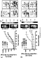
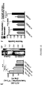

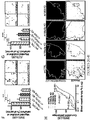
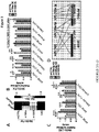

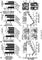
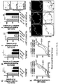
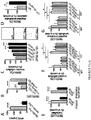
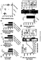
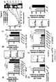
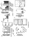
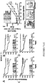


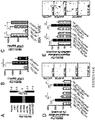


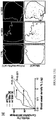

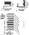
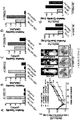
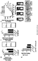
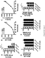
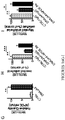
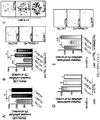
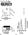
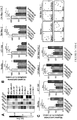
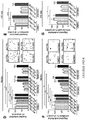
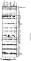
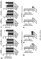
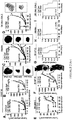


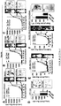

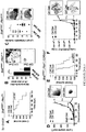
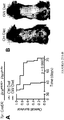

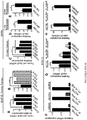


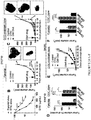


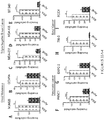


Classifications
-
- A—HUMAN NECESSITIES
- A61—MEDICAL OR VETERINARY SCIENCE; HYGIENE
- A61K—PREPARATIONS FOR MEDICAL, DENTAL OR TOILETRY PURPOSES
- A61K31/00—Medicinal preparations containing organic active ingredients
- A61K31/185—Acids; Anhydrides, halides or salts thereof, e.g. sulfur acids, imidic, hydrazonic or hydroximic acids
- A61K31/19—Carboxylic acids, e.g. valproic acid
- A61K31/195—Carboxylic acids, e.g. valproic acid having an amino group
-
- A—HUMAN NECESSITIES
- A61—MEDICAL OR VETERINARY SCIENCE; HYGIENE
- A61K—PREPARATIONS FOR MEDICAL, DENTAL OR TOILETRY PURPOSES
- A61K31/00—Medicinal preparations containing organic active ingredients
- A61K31/13—Amines
- A61K31/135—Amines having aromatic rings, e.g. ketamine, nortriptyline
- A61K31/136—Amines having aromatic rings, e.g. ketamine, nortriptyline having the amino group directly attached to the aromatic ring, e.g. benzeneamine
-
- A—HUMAN NECESSITIES
- A61—MEDICAL OR VETERINARY SCIENCE; HYGIENE
- A61K—PREPARATIONS FOR MEDICAL, DENTAL OR TOILETRY PURPOSES
- A61K31/00—Medicinal preparations containing organic active ingredients
- A61K31/16—Amides, e.g. hydroxamic acids
- A61K31/18—Sulfonamides
-
- A—HUMAN NECESSITIES
- A61—MEDICAL OR VETERINARY SCIENCE; HYGIENE
- A61K—PREPARATIONS FOR MEDICAL, DENTAL OR TOILETRY PURPOSES
- A61K31/00—Medicinal preparations containing organic active ingredients
- A61K31/21—Esters, e.g. nitroglycerine, selenocyanates
- A61K31/265—Esters, e.g. nitroglycerine, selenocyanates of carbonic, thiocarbonic, or thiocarboxylic acids, e.g. thioacetic acid, xanthogenic acid, trithiocarbonic acid
-
- A—HUMAN NECESSITIES
- A61—MEDICAL OR VETERINARY SCIENCE; HYGIENE
- A61K—PREPARATIONS FOR MEDICAL, DENTAL OR TOILETRY PURPOSES
- A61K31/00—Medicinal preparations containing organic active ingredients
- A61K31/33—Heterocyclic compounds
- A61K31/395—Heterocyclic compounds having nitrogen as a ring hetero atom, e.g. guanethidine or rifamycins
- A61K31/41—Heterocyclic compounds having nitrogen as a ring hetero atom, e.g. guanethidine or rifamycins having five-membered rings with two or more ring hetero atoms, at least one of which being nitrogen, e.g. tetrazole
- A61K31/415—1,2-Diazoles
- A61K31/4155—1,2-Diazoles non condensed and containing further heterocyclic rings
-
- A—HUMAN NECESSITIES
- A61—MEDICAL OR VETERINARY SCIENCE; HYGIENE
- A61K—PREPARATIONS FOR MEDICAL, DENTAL OR TOILETRY PURPOSES
- A61K31/00—Medicinal preparations containing organic active ingredients
- A61K31/33—Heterocyclic compounds
- A61K31/395—Heterocyclic compounds having nitrogen as a ring hetero atom, e.g. guanethidine or rifamycins
- A61K31/41—Heterocyclic compounds having nitrogen as a ring hetero atom, e.g. guanethidine or rifamycins having five-membered rings with two or more ring hetero atoms, at least one of which being nitrogen, e.g. tetrazole
- A61K31/415—1,2-Diazoles
- A61K31/416—1,2-Diazoles condensed with carbocyclic ring systems, e.g. indazole
-
- A—HUMAN NECESSITIES
- A61—MEDICAL OR VETERINARY SCIENCE; HYGIENE
- A61K—PREPARATIONS FOR MEDICAL, DENTAL OR TOILETRY PURPOSES
- A61K31/00—Medicinal preparations containing organic active ingredients
- A61K31/33—Heterocyclic compounds
- A61K31/395—Heterocyclic compounds having nitrogen as a ring hetero atom, e.g. guanethidine or rifamycins
- A61K31/41—Heterocyclic compounds having nitrogen as a ring hetero atom, e.g. guanethidine or rifamycins having five-membered rings with two or more ring hetero atoms, at least one of which being nitrogen, e.g. tetrazole
- A61K31/4164—1,3-Diazoles
- A61K31/4174—Arylalkylimidazoles, e.g. oxymetazolin, naphazoline, miconazole
-
- A—HUMAN NECESSITIES
- A61—MEDICAL OR VETERINARY SCIENCE; HYGIENE
- A61K—PREPARATIONS FOR MEDICAL, DENTAL OR TOILETRY PURPOSES
- A61K31/00—Medicinal preparations containing organic active ingredients
- A61K31/33—Heterocyclic compounds
- A61K31/395—Heterocyclic compounds having nitrogen as a ring hetero atom, e.g. guanethidine or rifamycins
- A61K31/435—Heterocyclic compounds having nitrogen as a ring hetero atom, e.g. guanethidine or rifamycins having six-membered rings with one nitrogen as the only ring hetero atom
- A61K31/47—Quinolines; Isoquinolines
-
- A—HUMAN NECESSITIES
- A61—MEDICAL OR VETERINARY SCIENCE; HYGIENE
- A61K—PREPARATIONS FOR MEDICAL, DENTAL OR TOILETRY PURPOSES
- A61K31/00—Medicinal preparations containing organic active ingredients
- A61K31/33—Heterocyclic compounds
- A61K31/395—Heterocyclic compounds having nitrogen as a ring hetero atom, e.g. guanethidine or rifamycins
- A61K31/535—Heterocyclic compounds having nitrogen as a ring hetero atom, e.g. guanethidine or rifamycins having six-membered rings with at least one nitrogen and one oxygen as the ring hetero atoms, e.g. 1,2-oxazines
- A61K31/5375—1,4-Oxazines, e.g. morpholine
- A61K31/5377—1,4-Oxazines, e.g. morpholine not condensed and containing further heterocyclic rings, e.g. timolol
-
- A—HUMAN NECESSITIES
- A61—MEDICAL OR VETERINARY SCIENCE; HYGIENE
- A61K—PREPARATIONS FOR MEDICAL, DENTAL OR TOILETRY PURPOSES
- A61K31/00—Medicinal preparations containing organic active ingredients
- A61K31/66—Phosphorus compounds
- A61K31/675—Phosphorus compounds having nitrogen as a ring hetero atom, e.g. pyridoxal phosphate
-
- A—HUMAN NECESSITIES
- A61—MEDICAL OR VETERINARY SCIENCE; HYGIENE
- A61K—PREPARATIONS FOR MEDICAL, DENTAL OR TOILETRY PURPOSES
- A61K38/00—Medicinal preparations containing peptides
- A61K38/16—Peptides having more than 20 amino acids; Gastrins; Somatostatins; Melanotropins; Derivatives thereof
- A61K38/17—Peptides having more than 20 amino acids; Gastrins; Somatostatins; Melanotropins; Derivatives thereof from animals; from humans
- A61K38/1703—Peptides having more than 20 amino acids; Gastrins; Somatostatins; Melanotropins; Derivatives thereof from animals; from humans from vertebrates
- A61K38/1709—Peptides having more than 20 amino acids; Gastrins; Somatostatins; Melanotropins; Derivatives thereof from animals; from humans from vertebrates from mammals
-
- A—HUMAN NECESSITIES
- A61—MEDICAL OR VETERINARY SCIENCE; HYGIENE
- A61K—PREPARATIONS FOR MEDICAL, DENTAL OR TOILETRY PURPOSES
- A61K38/00—Medicinal preparations containing peptides
- A61K38/16—Peptides having more than 20 amino acids; Gastrins; Somatostatins; Melanotropins; Derivatives thereof
- A61K38/17—Peptides having more than 20 amino acids; Gastrins; Somatostatins; Melanotropins; Derivatives thereof from animals; from humans
- A61K38/177—Receptors; Cell surface antigens; Cell surface determinants
-
- A—HUMAN NECESSITIES
- A61—MEDICAL OR VETERINARY SCIENCE; HYGIENE
- A61K—PREPARATIONS FOR MEDICAL, DENTAL OR TOILETRY PURPOSES
- A61K45/00—Medicinal preparations containing active ingredients not provided for in groups A61K31/00 - A61K41/00
- A61K45/06—Mixtures of active ingredients without chemical characterisation, e.g. antiphlogistics and cardiaca
-
- A—HUMAN NECESSITIES
- A61—MEDICAL OR VETERINARY SCIENCE; HYGIENE
- A61P—SPECIFIC THERAPEUTIC ACTIVITY OF CHEMICAL COMPOUNDS OR MEDICINAL PREPARATIONS
- A61P17/00—Drugs for dermatological disorders
-
- A—HUMAN NECESSITIES
- A61—MEDICAL OR VETERINARY SCIENCE; HYGIENE
- A61P—SPECIFIC THERAPEUTIC ACTIVITY OF CHEMICAL COMPOUNDS OR MEDICINAL PREPARATIONS
- A61P27/00—Drugs for disorders of the senses
- A61P27/02—Ophthalmic agents
-
- A—HUMAN NECESSITIES
- A61—MEDICAL OR VETERINARY SCIENCE; HYGIENE
- A61P—SPECIFIC THERAPEUTIC ACTIVITY OF CHEMICAL COMPOUNDS OR MEDICINAL PREPARATIONS
- A61P29/00—Non-central analgesic, antipyretic or antiinflammatory agents, e.g. antirheumatic agents; Non-steroidal antiinflammatory drugs [NSAID]
-
- A—HUMAN NECESSITIES
- A61—MEDICAL OR VETERINARY SCIENCE; HYGIENE
- A61P—SPECIFIC THERAPEUTIC ACTIVITY OF CHEMICAL COMPOUNDS OR MEDICINAL PREPARATIONS
- A61P35/00—Antineoplastic agents
-
- A—HUMAN NECESSITIES
- A61—MEDICAL OR VETERINARY SCIENCE; HYGIENE
- A61P—SPECIFIC THERAPEUTIC ACTIVITY OF CHEMICAL COMPOUNDS OR MEDICINAL PREPARATIONS
- A61P35/00—Antineoplastic agents
- A61P35/02—Antineoplastic agents specific for leukemia
-
- A—HUMAN NECESSITIES
- A61—MEDICAL OR VETERINARY SCIENCE; HYGIENE
- A61P—SPECIFIC THERAPEUTIC ACTIVITY OF CHEMICAL COMPOUNDS OR MEDICINAL PREPARATIONS
- A61P35/00—Antineoplastic agents
- A61P35/04—Antineoplastic agents specific for metastasis
-
- A—HUMAN NECESSITIES
- A61—MEDICAL OR VETERINARY SCIENCE; HYGIENE
- A61P—SPECIFIC THERAPEUTIC ACTIVITY OF CHEMICAL COMPOUNDS OR MEDICINAL PREPARATIONS
- A61P43/00—Drugs for specific purposes, not provided for in groups A61P1/00-A61P41/00
-
- A—HUMAN NECESSITIES
- A61—MEDICAL OR VETERINARY SCIENCE; HYGIENE
- A61P—SPECIFIC THERAPEUTIC ACTIVITY OF CHEMICAL COMPOUNDS OR MEDICINAL PREPARATIONS
- A61P9/00—Drugs for disorders of the cardiovascular system
-
- C—CHEMISTRY; METALLURGY
- C07—ORGANIC CHEMISTRY
- C07C—ACYCLIC OR CARBOCYCLIC COMPOUNDS
- C07C217/00—Compounds containing amino and etherified hydroxy groups bound to the same carbon skeleton
- C07C217/54—Compounds containing amino and etherified hydroxy groups bound to the same carbon skeleton having etherified hydroxy groups bound to carbon atoms of at least one six-membered aromatic ring and amino groups bound to acyclic carbon atoms or to carbon atoms of rings other than six-membered aromatic rings of the same carbon skeleton
-
- C—CHEMISTRY; METALLURGY
- C07—ORGANIC CHEMISTRY
- C07D—HETEROCYCLIC COMPOUNDS
- C07D233/00—Heterocyclic compounds containing 1,3-diazole or hydrogenated 1,3-diazole rings, not condensed with other rings
- C07D233/54—Heterocyclic compounds containing 1,3-diazole or hydrogenated 1,3-diazole rings, not condensed with other rings having two double bonds between ring members or between ring members and non-ring members
- C07D233/64—Heterocyclic compounds containing 1,3-diazole or hydrogenated 1,3-diazole rings, not condensed with other rings having two double bonds between ring members or between ring members and non-ring members with substituted hydrocarbon radicals attached to ring carbon atoms, e.g. histidine
-
- C—CHEMISTRY; METALLURGY
- C07—ORGANIC CHEMISTRY
- C07F—ACYCLIC, CARBOCYCLIC OR HETEROCYCLIC COMPOUNDS CONTAINING ELEMENTS OTHER THAN CARBON, HYDROGEN, HALOGEN, OXYGEN, NITROGEN, SULFUR, SELENIUM OR TELLURIUM
- C07F9/00—Compounds containing elements of Groups 5 or 15 of the Periodic System
- C07F9/02—Phosphorus compounds
- C07F9/547—Heterocyclic compounds, e.g. containing phosphorus as a ring hetero atom
- C07F9/645—Heterocyclic compounds, e.g. containing phosphorus as a ring hetero atom having two nitrogen atoms as the only ring hetero atoms
- C07F9/6503—Five-membered rings
- C07F9/6506—Five-membered rings having the nitrogen atoms in positions 1 and 3
-
- C—CHEMISTRY; METALLURGY
- C12—BIOCHEMISTRY; BEER; SPIRITS; WINE; VINEGAR; MICROBIOLOGY; ENZYMOLOGY; MUTATION OR GENETIC ENGINEERING
- C12Q—MEASURING OR TESTING PROCESSES INVOLVING ENZYMES, NUCLEIC ACIDS OR MICROORGANISMS; COMPOSITIONS OR TEST PAPERS THEREFOR; PROCESSES OF PREPARING SUCH COMPOSITIONS; CONDITION-RESPONSIVE CONTROL IN MICROBIOLOGICAL OR ENZYMOLOGICAL PROCESSES
- C12Q1/00—Measuring or testing processes involving enzymes, nucleic acids or microorganisms; Compositions therefor; Processes of preparing such compositions
- C12Q1/68—Measuring or testing processes involving enzymes, nucleic acids or microorganisms; Compositions therefor; Processes of preparing such compositions involving nucleic acids
- C12Q1/6876—Nucleic acid products used in the analysis of nucleic acids, e.g. primers or probes
- C12Q1/6883—Nucleic acid products used in the analysis of nucleic acids, e.g. primers or probes for diseases caused by alterations of genetic material
- C12Q1/6886—Nucleic acid products used in the analysis of nucleic acids, e.g. primers or probes for diseases caused by alterations of genetic material for cancer
-
- G—PHYSICS
- G01—MEASURING; TESTING
- G01N—INVESTIGATING OR ANALYSING MATERIALS BY DETERMINING THEIR CHEMICAL OR PHYSICAL PROPERTIES
- G01N33/00—Investigating or analysing materials by specific methods not covered by groups G01N1/00 - G01N31/00
- G01N33/48—Biological material, e.g. blood, urine; Haemocytometers
- G01N33/50—Chemical analysis of biological material, e.g. blood, urine; Testing involving biospecific ligand binding methods; Immunological testing
- G01N33/53—Immunoassay; Biospecific binding assay; Materials therefor
- G01N33/574—Immunoassay; Biospecific binding assay; Materials therefor for cancer
- G01N33/57407—Specifically defined cancers
- G01N33/5743—Specifically defined cancers of skin, e.g. melanoma
-
- C—CHEMISTRY; METALLURGY
- C12—BIOCHEMISTRY; BEER; SPIRITS; WINE; VINEGAR; MICROBIOLOGY; ENZYMOLOGY; MUTATION OR GENETIC ENGINEERING
- C12Q—MEASURING OR TESTING PROCESSES INVOLVING ENZYMES, NUCLEIC ACIDS OR MICROORGANISMS; COMPOSITIONS OR TEST PAPERS THEREFOR; PROCESSES OF PREPARING SUCH COMPOSITIONS; CONDITION-RESPONSIVE CONTROL IN MICROBIOLOGICAL OR ENZYMOLOGICAL PROCESSES
- C12Q2600/00—Oligonucleotides characterized by their use
- C12Q2600/118—Prognosis of disease development
-
- C—CHEMISTRY; METALLURGY
- C12—BIOCHEMISTRY; BEER; SPIRITS; WINE; VINEGAR; MICROBIOLOGY; ENZYMOLOGY; MUTATION OR GENETIC ENGINEERING
- C12Q—MEASURING OR TESTING PROCESSES INVOLVING ENZYMES, NUCLEIC ACIDS OR MICROORGANISMS; COMPOSITIONS OR TEST PAPERS THEREFOR; PROCESSES OF PREPARING SUCH COMPOSITIONS; CONDITION-RESPONSIVE CONTROL IN MICROBIOLOGICAL OR ENZYMOLOGICAL PROCESSES
- C12Q2600/00—Oligonucleotides characterized by their use
- C12Q2600/136—Screening for pharmacological compounds
-
- C—CHEMISTRY; METALLURGY
- C12—BIOCHEMISTRY; BEER; SPIRITS; WINE; VINEGAR; MICROBIOLOGY; ENZYMOLOGY; MUTATION OR GENETIC ENGINEERING
- C12Q—MEASURING OR TESTING PROCESSES INVOLVING ENZYMES, NUCLEIC ACIDS OR MICROORGANISMS; COMPOSITIONS OR TEST PAPERS THEREFOR; PROCESSES OF PREPARING SUCH COMPOSITIONS; CONDITION-RESPONSIVE CONTROL IN MICROBIOLOGICAL OR ENZYMOLOGICAL PROCESSES
- C12Q2600/00—Oligonucleotides characterized by their use
- C12Q2600/158—Expression markers
-
- C—CHEMISTRY; METALLURGY
- C12—BIOCHEMISTRY; BEER; SPIRITS; WINE; VINEGAR; MICROBIOLOGY; ENZYMOLOGY; MUTATION OR GENETIC ENGINEERING
- C12Q—MEASURING OR TESTING PROCESSES INVOLVING ENZYMES, NUCLEIC ACIDS OR MICROORGANISMS; COMPOSITIONS OR TEST PAPERS THEREFOR; PROCESSES OF PREPARING SUCH COMPOSITIONS; CONDITION-RESPONSIVE CONTROL IN MICROBIOLOGICAL OR ENZYMOLOGICAL PROCESSES
- C12Q2600/00—Oligonucleotides characterized by their use
- C12Q2600/178—Oligonucleotides characterized by their use miRNA, siRNA or ncRNA
-
- G—PHYSICS
- G01—MEASURING; TESTING
- G01N—INVESTIGATING OR ANALYSING MATERIALS BY DETERMINING THEIR CHEMICAL OR PHYSICAL PROPERTIES
- G01N2500/00—Screening for compounds of potential therapeutic value
- G01N2500/10—Screening for compounds of potential therapeutic value involving cells
Definitions
- This invention relates to diagnosis and treatment of migrating cancers and melanoma.
- Melanoma a malignant tumor, develops from abnormal melanocytes in the lower epidermis and can metastasize to distant sites in the body via the blood and lymph systems. Although it accounts for less than 5% of skin cancer cases, melanoma is much more dangerous and responsible for a large majority of the deaths associated with skin cancer. Across the world the incidence of melanoma has been increasing at an alarming rate, with a lifetime risk of developing melanoma as high as 1/58 for males in the U.S. ( Jemal et al., 2008, CA: Cancer J. Clin. 58:71-96 ). The mortality rate of malignant melanoma also continues to rise dramatically throughout the world.
- WO 2010/138598 discloses compounds, pharmaceutically acceptable salts, isomers, or prodrugs thereof, which are useful as modulators of the activity of liver X receptors (LXR).
- Pharmaceutical compositions containing the compounds and methods of using the compounds are also disclosed.
- WO 2007/002563 discloses compounds and pharmaceutically acceptable salts, isomers, and prodrugs thereof, which are useful as modulators of the activity of liver X receptors.
- Pharmaceutical compositions containing the compounds and methods of using the compounds are also disclosed.
- Pencheva et al. (2014) Cell, 156(5):986-1001 describes broad-spectrum therapeutic suppression of metastatic melanoma through nuclear hormone receptor activation.
- Metastatic melanoma therefore remains one of the most difficult cancers to treat and one of the most feared neoplasms. Accordingly, there is a need for new agents and methods for diagnosis and treatment of melanoma.
- an LXR ⁇ agonist for use in treating a metastatic cancer in a subject in need thereof in accordance with claim 1 herein.
- This disclosure addresses the above-mentioned need by providing agents and methods for diagnosis and treatment of melanoma.
- the disclosure is based, at least in part, on an unexpected discovery of a cooperative miRNA-protein network deregulated in metastatic melanoma.
- This network includes a number of metastasis suppressor factors and metastasis promoter factors.
- This disclosure features a method for treating cancer, including administering to a subject in need thereof, a LXR agonist, wherein the LXR agonist is administered in an amount sufficient to increase the expression level or activity level of ApoE to a level sufficient to slow the spread of metastasis of the cancer.
- This disclosure also features a method for treating cancer, including administering to a subject in need thereof, an ApoE polypeptide in an amount sufficient to treat the cancer.
- the This disclosure also features a method of slowing the spread of a migrating cancer, comprising administering to a subject in need thereof, a LXR agonist or an ApoE polypeptide.
- the LXR agonist increases the expression level of ApoE at least 2.5-fold in vitro.
- the LXR ⁇ agonist is selective for LXR ⁇ over LXR ⁇ .
- the LXR ⁇ agonist has activity for LXR ⁇ that is at least 2.5-fold greater than the activity of said agonist for LXR ⁇ .
- the LXR ⁇ agonist has activity for LXR ⁇ that is at least 10-fold greater than the activity of said agonist for LXR ⁇ .
- the LXR ⁇ agonist has activity for LXR ⁇ that is at least 100-fold greater than the activity of said agonist for LXR ⁇ .
- the LXR agonist has activity for LXR ⁇ that is at least within 2.5-fold of the activity of said agonist for LXR ⁇ .
- the migrating cancer is metastatic cancer.
- the metastatic cancer can include cells exhibiting migration and/or invasion of migrating cells and/or include cells exhibiting endothelial recruitment and/or angiogenesis.
- the migrating cancer is a cell migration cancer.
- the cell migration cancer is a non-metastatic cell migration cancer.
- the migrating cancer can be a cancer spread via seeding the surface of the peritoneal, pleural, pericardial, or subarachnoid spaces.
- the migrating cancer can be a cancer spread via the lymphatic system, or a cancer spread hematogenously.
- the migrating cancer is a cell migration cancer that is a non-metastatic cell migration cancer, such as ovarian cancer, mesothelioma, or primary lung cancer.
- This disclosure also provides a method for inhibiting or reducing metastasis of cancer comprising administering a LXR agonist or an ApoE polypeptide.
- This disclosure also provides a method for inhibiting proliferation or growth of cancer stem cells or cancer initiating cells, including contacting the cell with a LXR agonist or an ApoE polypeptide in an amount sufficient to inhibit proliferation or growth of said cell.
- This disclosure also provides a method of reducing the rate of tumor seeding of a cancer including administering to a subject in need thereof a LXR agonist or an ApoE polypeptide in an amount sufficient to reduce tumor seeding.
- This disclosure also provides a method of reducing or treating metastatic nodule-forming of cancer including administering to a subject in need thereof a LXR agonist or an ApoE polypeptide in an amount sufficient to treat said metastatic nodule-forming of cancer.
- the cancer is breast cancer, colon cancer, renal cell cancer, non-small cell lung cancer, hepatocellular carcinoma, gastric cancer, ovarian cancer, pancreatic cancer, esophageal cancer, prostate cancer, sarcoma, or melanoma.
- the cancer is melanoma.
- the cancer is breast cancer.
- the cancer is renal cell cancer.
- the cancer is pancreatic cancer.
- the cancer is non-small cell lung cancer.
- the cancer is colon cancer.
- the cancer is ovarian cancer.
- the cancer is a drug resistant cancer. In further embodiments, the cancer is resistant to to vemurafenib, dacarbazine, a CTLA4 inhibitor, a PD1 inhibitor, or a PDL1 inhibitor.
- the method comprises administering an LXR agonist selected from the list consisting of a compound of any one of Formula I-IV or any of compound numbers 1-39, or pharmaceutically acceptable salts thereof.
- the LXR agonist is compound 1 or a pharmaceutically acceptable salt thereof.
- the LXR agonist is compound 2 or a pharmaceutically acceptable salt thereof.
- the LXR agonist is compound 3 or a pharmaceutically acceptable salt thereof.
- the LXR agonist is compound 12 or a pharmaceutically acceptable salt thereof.
- the LXR agonist is compound 25 or a pharmaceutically acceptable salt thereof.
- the LXR agonist is compound 38 or a pharmaceutically acceptable salt thereof.
- the LXR agonist is compound 39 or a pharmaceutically acceptable salt thereof.
- the method can further include administering an antiproliferative, wherein said LXR agonist and said antiproliferative are administered in an amount that together, is sufficient to slow the progression of migrating cancer.
- the antiproliferative and LXR agonist can be administered within 28 days of each (e.g., within 21, 14, 10, 7, 5, 4, 3, 2, or 1 days) or within 24 hours (e.g., 12, 6, 3, 2, or 1 hours; or concomitantly) other in amounts that together are effective to treat the subject.
- the method comprises administering an ApoE polypeptide.
- the ApoE polypeptide fragment can increase the activity level or expression level of LRP1 or LRP8, and/or the ApoE polypeptide can bind to LRP1 or LRP8, the ApoE polypeptide can be the receptor binding region (RBR) of ApoE.
- the method can further include administering an antiproliferative, wherein said ApoE polypeptide and said antiproliferative are administered in an amount that together, is sufficient to slow the progression of migrating cancer.
- the antiproliferative and ApoE polypeptide can be administered within 28 days of each (e.g., within 21, 14, 10, 7, 5, 4, 3, 2, or 1 days) or within 24 hours (e.g., 12, 6, 3, 2, or 1 hours; or concomitantly) other in amounts that together are effective to treat the subject.
- the pharmaceutical composition may further comprise an additional compound having antiproliferative activity.
- the additional compound having antiproliferative activity can be selected from the group of compounds such as chemotherapeutic and cytotoxic agents, differentiation-inducing agents (e.g. retinoic acid, vitamin D, cytokines), hormonal agents, immunological agents and anti-angiogenic agents.
- Chemotherapeutic and cytotoxic agents include, but are not limited to, alkylating agents, cytotoxic antibiotics, antimetabolites, vinca alkaloids, etoposides, and others ( e.g., paclitaxel, taxol, docetaxel, taxotere, cis-platinum).
- the method may further include administering a antiproliferative compound selected from the group consisting of alkylating agents, platinum agents, antimetabolites, topoisomerase inhibitors, antitumor antibiotics, antimitotic agents, aromatase inhibitors, thymidylate synthase inhibitors, DNA antagonists, farnesyltransferase inhibitors, pump inhibitors, histone acetyltransferase inhibitors, metalloproteinase inhibitors, ribonucleoside reductase inhibitors, TNF alpha agonists/antagonists, endothelin A receptor antagonist, retinoic acid receptor agonists, immuno-modulators, hormonal and antihormonal agents, photodynamic agents, tyrosine kinase inhibitors, antisense compounds, corticosteroids, HSP90 inhibitors, proteosome inhibitors (for example, NPI-0052), CD40 inhibitors, anti-CSI antibodies, FGFR3 inhibitors, VEGF inhibitors, MEK inhibitor
- This disclosure also features a method for treating melanoma (e.g., metastatic melanoma) in a subject in need thereof.
- the method includes (a) increasing in the subject the expression level or activity level of a metastasis suppressor factor selected from the group consisting of DNAJA4, Apolipoprotein E (ApoE), LRP1, LRP8, Liver X Receptor (LXR, e.g., both LXR-alpha and LXR-beta), and miR-7 or (b) decreasing in the subject the expression level or activity level of a metastasis promoter factor selected from the group consisting of miR-199a-3p, miR-199a-5p, miR-1908, and CTGF.
- a metastasis suppressor factor selected from the group consisting of DNAJA4, Apolipoprotein E (ApoE), LRP1, LRP8, Liver X Receptor (LXR, e.g., both LXR-alpha and LXR
- the increasing step can be carried out by administering to the subject one or more of the followings: (i) a polypeptide having a sequence of DNAJA4, ApoE or an ApoE fragment, LRP1, LRP8, or LXR; (ii) a nucleic acid having a sequence encoding DNAJA4, ApoE, LRP1, LRP8, or LXR; (iii) a ligand for LRP1, LRP8, or LXR; and (iv) an RNAi agent encoding miR-7.
- the LRP1 or LRP8 ligand include the receptor binding portion of ApoE, anti-LRP1 or anti-LRP8 antibodies, and small molecule ligands.
- increasing the ApoE expression level can be carried out by increasing the activity level or expression level of LXR.
- Increasing the DNAJA4 expression level can also be carried out by increasing the activity level or expression level of LXR.
- the LXR activity level can be increased by administering to the subject a ligand of LXR, such as compounds of Formula I-IV as disclosed below.
- the increasing step can also be carried out by decreasing the expression level or activity level of a microRNA selected from the group consisting of miR-199a-3p, miR-199a-5p, and miR-1908.
- miR-Zip technology Locked Nucleic Acid (LNA), and antagomir technology as described in the examples below.
- This disclosure also provides a method for determining whether a subject has, or is at risk of having, metastatic melanoma.
- the method includes obtaining from the subject a sample; measuring in the sample (i) a first expression level of a metastasis promoter factor selected from the group consisting of miR-199a-3p, miR-199a-5p, miR-1908, and CTGF, or (ii) a second expression level of a metastasis suppressor factor selected from the group consisting of DNAJA4, ApoE, LRP1, LRP8, LXR, and miR-7; and comparing the first expression level with a first predetermined reference value, or the second expression level with a second predetermined reference value.
- a metastasis promoter factor selected from the group consisting of miR-199a-3p, miR-199a-5p, miR-1908, and CTGF
- a second expression level of a metastasis suppressor factor selected from the group consisting of DNAJA4, ApoE, LRP1,
- the subject is determined to have, or to be at risk of having, metastatic melanoma if (a) the first expression level is above a first predetermined reference value or (b) the second expression level is below a second predetermined reference value.
- the first and second predetermined reference values can be obtained from a control subject that does not have metastatic melanoma.
- the measuring step includes measuring both the first expression level and the second expression level.
- the sample can be a body fluid sample, a tumor sample, a nevus sample, or a human skin sample.
- This disclosure also provides an array having a support having a plurality of unique locations, and any combination of (i) at least one nucleic acid having a sequence that is complementary to a nucleic acid encoding a metastasis promoter factor selected from the group consisting of miR-199a-3p, miR-199a-5p, miR-1908, and CTGF or a complement thereof, or (ii) at least one nucleic acid having a sequence that is complementary to a nucleic acid encoding a metastasis suppressor factor selected from the group consisting of DNAJA4, ApoE, LRP1, LRP8, LXR, and miR-7 or a complement thereof.
- each nucleic acid is immobilized to a unique location of the support.
- This array can be used for metastatic melanoma diagnosis and prognosis.
- the disclosure also provides a kit for diagnosing a metastatic potential of melanoma in a subject.
- the kit includes a first reagent that specifically binds to an expression product of a metastasis suppressor gene selected from the group consisting of DNAJA4, ApoE, LRP1, LRP8, LXR, and miR-7; or a second reagent that specifically binds to an expression product of a metastasis promoter gene selected from the group consisting of miR-199a-3p, miR-199a-5p, miR-1908, and CTGF.
- the second agent can be a probe having a sequence complementary to the suppressor or promoter gene or a complement thereof.
- the kit can further contain reagents for performing an immunoassay, a hybridization assay, or a PCR assay. In one embodiment, the kit contained the above-mentioned array.
- This disclosure also provides a method of identifying a compound useful for treating melanoma or for inhibiting endothelial recruitment, cell invasion, or metastatic angiogenesis.
- the method includes (i) obtaining a test cell expressing a reporter gene encoded by a nucleic acid operatively liked to a promoter of a marker gene selected from the group consisting of miR-199a-3p, miR-199a-5p, miR-1908, and CTGF; (ii) exposing the test cell to a test compound; (iii) measuring the expression level of the reporter gene in the test cell; (iv) comparing the expression level with a control level; and (v) selecting the test compound as a candidate useful for treating melanoma or for inhibiting endothelial recruitment, cancer cell invasion, or metastatic angiogenesis, if the comparison indicates that the expression level is lower than the control level.
- the disclosure provides another method of identifying a compound useful for treating melanoma or for inhibiting endothelial recruitment, cell invasion, or metastatic angiogenesis.
- the method includes (i) obtaining a test cell expressing a reporter gene encoded by a nucleic acid operatively liked to a promoter of a marker gene selected from the group consisting of DNAJA4, ApoE, LRP1, LRP8, LXR, and miR-7; (ii) exposing the test cell to a test compound; (iii) measuring the expression level of the reporter gene in the test cell; (iv) comparing the expression level with a control level; and (v) selecting the test compound as a candidate useful for treating melanoma or for inhibiting endothelial recruitment, cancer cell invasion, or metastatic angiogenesis, if the comparison indicates that the expression level is higher than the control level.
- the reporter gene can be a standard reporter gene (such as LaxZ, GFP, or luciferase gene, or the like), known in the art, or one of the aforementioned metastasis suppressor genes or metastasis promoter genes.
- the control level can be obtained from a control cell that is the same as the test cell except that the control cell has not be exposed to the test compound.
- This disclosure also provides a method for inhibiting endothelial recruitment, inhibiting tumor cell invasion, or treating metastatic cancer in a subject in need thereof, by administering to the subject an agent that inhibits expression or activity of CTGF.
- the subject can be one having a disorder characterized by pathological angiogenesis, including but not limited to cancer (e.g., metastatic melanoma), an eye disorder, and an inflammatory disorder.
- An example of the tumor cell is a metastatic melanoma cell.
- the agent include an antibody, a nucleic acid, a polypeptide, and a small molecule compound. In a preferred embodiment, the antibody is a monoclonal antibody.
- This disclosure also provides a method for inhibiting endothelial recruitment, inhibiting tumor cell invasion, or treating metastatic cancer in a subject in need thereof, by administering to the subject an agent that increases expression or activity of miR-7.
- An example of the tumor cell is a metastatic melanoma cell.
- the agent include an antibody, a nucleic acid, a polypeptide, and a small molecule compound.
- the agent has miR-7 activity.
- the nucleic acid can be an oligonucleotide.
- the oligonucleotide can include a sequence selected from the group consisting of SEQ ID Nos. 36-38.
- mirating cancer refers to a cancer in which the cancer cells forming the tumor migrate and subsequently grow as malignant implants at a site other than the site of the original tumor.
- the cancer cells migrate via seeding the surface of the peritoneal, pleural, pericardial, or subarachnoid spaces to spread into the body cavities; via invasion of the lymphatic system through invasion of lymphatic cells and transport to regional and distant lymph nodes and then to other parts of the body; via haematogenous spread through invasion of blood cells; or via invasion of the surrounding tissue.
- Migrating cancers include metastatic tumors and cell migration cancers, such as ovarian cancer, mesothelioma, and primary lung cancer, each of which is characterized by cellular migration.
- slowing the spread of migrating cancer refers to reducing or stopping the formation of new loci; or reducing, stopping, or reversing the tumor load.
- metastatic tumor refers to a tumor or cancer in which the cancer cells forming the tumor have a high potential to or have begun to, metastasize, or spread from one location to another location or locations within a subject, via the lymphatic system or via haematogenous spread, for example, creating secondary tumors within the subject. Such metastatic behavior may be indicative of malignant tumors. In some cases, metastatic behavior may be associated with an increase in cell migration and/or invasion behavior of the tumor cells.
- slowing the spread of metastasis refers to reducing or stopping the formation of new loci; or reducing, stopping, or reversing the tumor load.
- cancer refers to any cancer caused by the proliferation of malignant neoplastic cells, such as tumors, neoplasms, carcinomas, sarcomas, leukimias, lymphomas, and the like.
- drug resistant cancer refers to any cancer that is resistant to an antiproliferative in Table 2.
- cancers that can be defined as metastatic include but are not limited to non-small cell lung cancer, breast cancer, ovarian cancer, colorectal cancer, biliary tract cancer, bladder cancer, brain cancer including glioblastomas and medullablastomas, cervical cancer, choriocarcinoma, endometrial cancer, esophageal cancer, gastric cancer, hematological neoplasms, multiple myeloma, leukemia, intraepithelial neoplasms, livercancer, lymphomas, neuroblastomas, oral cancer, pancreatic cancer, prostate cancer, sarcoma, skin cancer including melanoma, basocellular cancer, squamous cell cancer, testicular cancer, stromal tumors, germ cell tumors, thyroid cancer, and renal cancer.
- Cell migration involves the invasion by the cancer cells into the surrounding tissue and the crossing of the vessel wall to exit the vasculature in distal organs of the cancer cell.
- cell migration cancers cancers that migrate by invasion by the cancer cells into the surrounding tissue and the crossing of the vessel wall to exit the vasculature in distal organs of the cancer cell.
- Non-metastatic cell migration cancer refers to cancers that do not migrate via the lymphatic system or via haematogenous spread.
- cell to cell adhesion refers to adhesion between at least two cells through an interaction between a selectin molecule and a selectin specific ligand. Cell to cell adhesion includes cell migration.
- a “cell adhesion related disorder” is defined herein as any disease or disorder which results from or is related to cell to cell adhesion or migration.
- a cell adhesion disorder also includes any disease or disorder resulting from inappropriate, aberrant, or abnormal activation of the immune system or the inflammatory system.
- diseases include but are not limited to, myocardial infarction, bacterial or viral infection, metastatic conditions, e.g. cancer.
- the disclosure further features methods for treating a cell adhesion disorder by administering a LXR agonist or ApoE polypeptide.
- cancer stem cells or “cancer initiating cells” refers to cancer cells that possess characteristics associated with normal stem cells, specifically the ability to give rise to all cell types found in a particular cancer sample. Cancer stem cells are therefore tumorgenic or tumor forming, perhaps in contrast to other non-tumorgenic cancer cells. Cancer stem cells may persist in tumors as a distinct population and cause cancer recurrence and metastasis by giving rise to new tumors.
- tumor seeding refers to the spillage of tumor cell clusters and their subsequent growth as malignant implants at a site other than the site of the original tumor.
- metal nodule refers to an aggregation of tumor cells in the body at a site other than the site of the original tumor.
- the present disclosure features methods for preventing or reducing aberrant proliferation, differentiation, or survival of cells.
- compounds of the disclosure may be useful in reducing the risk of, or preventing, tumors from increasing in size or from reaching a metastatic state.
- the subject compounds may be administered to halt the progression or advancement of cancer.
- the disclosure includes use of the subject compounds to reduce the risk of, or prevent, a recurrence of cancer.
- Metastatic progression requires that sets of effector proteins involved in common cellular phenotypes be coherently expressed ( Gupta and Massagué, 2006 Cell 127, 679-695 ; Hanahan and Weinberg, 2011 Cell 144, 646-674 ; Talmadge and Fidler, 2010 Cancer Res. 70, 5649-5669 ; Hynes, 2003 Cell 113, 821-823 ).
- a miRNA could exert robust metastatic suppression by virtue of its ability to target multiple genes required for metastasis.
- the miRNA's silencing through genetic or epigenetic mechanisms would readily promote cancer progression by de-repressing multiple promoters of metastasis ( Png et al., 2011 Nature 481, 190-194 ).
- a role for convergent regulation of a single gene by multiple metastasis regulatory miRNAs is more nuanced. This scenario would emerge if there existed a key gene that acted as a robust suppressor of metastatic progression. Convergent and cooperative targeting of this gene by multiple miRNAs could achieve maximal silencing of such a key metastasis suppressor gene.
- miRNAs were identified to be deregulated in multiple independent metastatic lines derived from multiple patients with melanoma-a highly prevalent cancer with increasing incidence ( Garbe and Leiter, 2009 Clin. Dermatol. 27, 3-9 ).
- miR-1908, miR-199a-3p, and miR-199a-5p act as robust endogenous promoters of melanoma metastasis through convergent targeting of the metabolic gene ApoE and the heat-shock protein DNAJA4.
- loss-of-function, gain-of-function, and epistatic analyses a cooperative miRNA network that maximally silences ApoE signaling is delineated.
- Cancer cell-secreted ApoE inhibits metastatic invasion and endothelial recruitment, which is mediated through its actions on distinct receptors on melanoma and endothelial cells.
- These miRNAs display significant prognostic capacity in identifying patients that develop melanoma metastatic relapse, while therapeutic delivery of LNAs targeting these miRNAs significantly inhibits melanoma metastasis.
- the current lack of effective therapies for the prevention of melanoma metastasis after surgical resection ( Garbe et al., 2011 Oncologist 16, 5-24 ) requires an improved molecular and mechanistic understanding of melanoma metastatic progression.
- the findings disclosed herein reveal a number of key novel non-coding and coding genes involved in melanoma progression and offer a novel avenue for both identifying patients at high-risk for melanoma metastasis and treating them.
- nucleic acid and amino acid sequences of the members of the above-mentioned network are listed below.
- the members of this network can be used as targets for treating metastatic melanoma.
- the members can be used a biomarkers for determining whether a subject has, or is at risk of having, a metastatic melanoma or for determining a prognosis or surveillance of patient having the disorder.
- the present disclosure encompasses methods of treating metastatic melanoma by targeting one or more of the members, methods of determining the efficacy of therapeutic regimens for inhibiting the cancer, and methods of identifying anti-cancer agent. Also provided are methods of diagnosing whether a subject has, or is at risk for having, metastatic melanoma, and methods of screening subjects who are thought to be at risk for developing the disorder.
- the disclosure also encompasses various kits suitable for carrying out the above mentioned methods.
- polypeptide or peptide as used herein includes recombinantly or synthetically produced fusion or chimeric versions of any of the aforementioned metastasis suppressors, having the particular domains or portions that are involved in the network.
- the term also encompasses an analog, fragment, elongation or derivative of the peptide (e.g. that have an added amino-terminal methionine, useful for expression in prokaryotic cells).
- Apolipoprotein polypeptide or ApoE polypeptide as used herein means a peptide, drug, or compound that mimics a function of the native apolipoprotein either in vivo or in vitro including apolipoprotein analogs, fragments, elongations or derivatives that are a peptide of between 10 and 200 amino acid residues in length, such peptides can contain either natural, or non-natural amino acids containing amide bonds.
- Apolipoprotein peptide fragments may be modified to improve their stability or bioavailability in vivo as known in the art and may contain organic compounds bound to the amino acid side chains through a variety of bonds.
- the dislcosure is a method for using an isolated apoEp1.B peptide having the amino acid sequence TQQIRLQAEIFQAR (murine)(SEQ. ID. No.43) or AQQIRLQAEAFQAR (human)(SEQ. ID. No.44) or an analog, fragment, elongation or derivative of the peptide.
- the disclosure also includes a nucleic acid molecule encoding the apoEpI.B peptide, or an analog, fragment, elongation or derivative thereof.
- analog includes any peptide having an amino acid residue sequence substantially identical to the native peptide in which one or more residues have been conservatively substituted with a functionally similar residue and which displays the ability to mimic the native peptide.
- conservative substitutions include the substitution of one non-polar (hydrophobic) residue such as alanine, isoleucine, valine, leucine or methionine for another, the substitution of one polar (hydrophilic) residue for another such as between arginine and lysine, between glutamine and asparagine, between glycine and serine, the substitution of one basic residue such as lysine, arginine or histidine for another, or the substitution of one acidic residue, such as aspartic acid or glutamic acid for another.
- peptides having the following sequences include peptides having the following sequences: TAQIRLQAEIFQAR (SEQ.ID.NO.:45); TQAIRLQAEIFQAR (SEQ.ID.NO.:46); TQQARLQAEIFQAR (SEQ.ID.NO.:47) and TQQIALQAEIFQAR (SEQ.ID.NO.:48).
- “Derivative” refers to a peptide having one or more residues chemically derivatized by reaction of a functional side group.
- Such derivatized molecules include for example, those molecules in which free amino groups have been derivatized to form amine hydrochlorides, p-toluene sulfonyl groups, carbobenzoxy groups, t-butyloxycarbonyl groups, chloroacetyl groups or formyl groups.
- Free carboxyl groups may be derivatized to form salts, methyl and ethyl esters or other types of esters or hydrazides.
- Free hydroxyl groups may be derivatized to form O-acyl or O-alkyl derivatives.
- the imidazole nitrogen of histidine may be derivatized to form N-im-benzylhistidine.
- derivatives those peptides which contain one or more naturally occurring amino acid derivatives of the twenty standard amino acids.
- 4-hydroxyproline may be substituted for proline
- 5-hydroxylysine may be substituted for lysine
- 3-methylhistidine may be substituted for histidine
- homoserine may be substituted for serine
- ornithine may be substituted for lysine.
- Polypeptides of the present disclosure also include any polypeptide having one or more additions and/or deletions or residues relative to the sequence of a polypeptide whose sequence is shown herein, so long as the requisite activity is maintained.
- fragment refers to any subject peptide having an amino acid residue sequence shorter than that of a peptide whose amino acid residue sequence is shown herein.
- elongation refers to any subject peptide having an amino acid sequence longer by one or two amino acids (either at the carboxy or amino terminal end) than that of a peptide of the present disclosure. Preferably, the elongation occurs at the amino terminal end. Fragments and elongations of the peptides include peptides that have the following sequences: QTQQIRLQAEIFQAR (SEQ.ID.NO.:49) and QQIRLQAEIFQAR (SEQ.ID.NO.:50).
- the methods of the disclosure can include administering a LXR agonist for the prevention and treatment of metastasis.
- the LXR agonist can be a compound according to the Formula I, II, III, or IV shown below.
- Y is -N(R 12 )S(O) 2 - and R 1 is OH.
- the compounds of Formula I include but are not limited the compound with the structure shown below:
- Compounds of Formula II can be synthesized as described in US patent No. 7,576,215 .
- the compound of formula II can be any of compounds 26-32, or a pharmaceutically acceptable salt thereof.
- X is hydrogen
- X is hydrogen
- X is hydrogen
- the compounds of Formula III include but are not limited the compounds with structures shown below GW3965 2 and SB742881 25 :
- Formula IV is shown below: or a pharmaceutically acceptable salt thereof, wherein:
- the compounds of Formula IV which can be useful in the methods of the disclosure include, but are not limited to, compounds having the structures are shown below, and pharmaceutically acceptable salts thereof: and selected from the list comprising: 33 2-(1-(3chloro-3'-fluoro-4'-(hydroxymethyl)-5'-(methylsulfonyl)biphenyl-4-yl)-2-(2-(2,6dichlorophenyl)propan-2-yl)-1H-imidazol-4-yl)propan-2-ol; 34 2-(2-(2(2-chloro-3-fluorophenyl)propan-2-yl)-1-(3'-fluoro-4'-(hydroxymethyl)-5'(methylsulfonyl)biphenyl-4-yl)-1H-imidazol-4-yl)propan-2-ol; 35 2-(2-(2(2,6-dichlorophenyl)propan-2-yl)-1-(3'-fluoro-4'
- the LXR agonist that can be used for the treatment and/or prevention of metastasis can be compound 24, or a pharmaceutically acceptable salt thereof.
- compounds that can be used for the treatment and/or prevention of metastasis can be found in the PCT publications in the list consisting of: WO2006/094034 , WO2008/049047 , WO2009/020683 , WO2009/086138 , WO2009/086123 , WO2009/086130 , WO2009/086129 , WO2007/002559 , WO2007/002563 , WO2007/081335 , WO2006/017055 , WO2006/102067 , WO2009/024550 , US2006/0074115 , US2006/0135601 , WO2009/021868 , WO2009/040289 , WO2007/047991 , WO2007/050425 , WO2006/073363 , WO2006/073364 , WO2006/073365 , WO2006/073366 , WO2006/073367 , US2009/0030082 ,
- LXR ⁇ and LXR ⁇ were discovered by multiple groups at roughly the same time (Apfel et al., 1994; Willy et al., 1995; Song et al., 1994; Shinar et al., 1994; Teboul et al., 1995), belong to a family of nuclear hormone receptors that are endogenously activated by cholesterol and its oxidized derivatives to mediate transcription of genes involved in maintaining glucose, cholesterol, and fatty acid metabolism (Janowski et al., 1996; Calkin and Tontonoz, 2012).
- LXR ⁇ an ideal therapeutic target, as exemplified by the broad-ranging responsiveness of melanoma cells to LXR ⁇ activation therapy.
- LXR agonist As used herein, reference to the activity of an LXR agonist at LXR ⁇ and LXR ⁇ refer to the activity as measured using the ligand sensing assay (LiSA) described in Spencer et al. Journal of Medicinal Chemistry 2001, 44, 886-897 .
- the LXR agonist has an EC50 of less than 1 ⁇ M in the ligand sensing assay (e.g., 0.5 nm to 500 nM, 10 nM to 100 nM).
- the methods of the disclosure can be performed using an LXR ⁇ agonist having activity for LXR ⁇ that is at least 3-fold greater than the activity of the agonist for LXR ⁇ , or having activity for LXR ⁇ that is at least 10-fold greater than the activity of the agonist for LXR ⁇ , or having activity for LXR ⁇ that is at least 100-fold greater than the activity of said agonist for LXR ⁇ , or having activity for LXR ⁇ that is at least within 3-fold of the activity of the agonist for LXR ⁇ .
- the term "greater activity" in the LiSA assay assay refers to a lower EC 50 .
- the term "increases the level of ApoE expression in vitro" refers to certain LXR agonists capable of increasing the level of ApoE expression 2.5-fold in the qPCR assay of Example 21 at a concentration of less than 5 ⁇ M (e.g., at a concentration of 100 nM to 2 ⁇ M, at a concentration of less than or equal to 1 ⁇ M).
- the LXR agonists exhibiting this in vitro effect can be highly efficacious for use in the methods of the disclosure.
- alkyl used is the present application relates a saturated branched or unbranched aliphatic univalent substituent.
- the alkyl substituent has 1 to 100 carbon atoms, (e.g., 1 to 22 carbon atoms, 1 to 10 carbon atoms 1 to 6 carbon atoms, 1 to 3 carbon atoms). Accordingly, examples of the alkyl substituent include methyl, ethyl, n -propyl, isopropyl, n -butyl, isobutyl, sec -butyl, tert -butyl, n -pentyl and n -hexyl.
- alkoxy represents a chemical substituent of formula -OR, where R is an optionally substituted C 1 -C 6 alkyl group, unless otherwise specified.
- the alkyl group can be substituted, e.g., the alkoxy group can have 1, 2, 3, 4, 5 or 6 substituent groups as defined herein.
- alkoxyalkyl represents a heteroalkyl group, as defined herein, that is described as an alkyl group that is substituted with an alkoxy group.
- exemplary unsubstituted alkoxyalkyl groups include between 2 to 12 carbons.
- the alkyl and the alkoxy each can be further substituted with 1, 2, 3, or 4 substituent groups as defined herein for the respective group.
- cycloalkyl refers to a monocyclic, bicyclic, or tricyclic substituent, which may be saturated or partially saturated, i.e. possesses one or more double bonds.
- Monocyclic substituents are exemplified by a saturated cyclic hydrocarbon group containing from 3 to 8 carbon atoms. Examples of monocyclic cycloalkyl substituents include cyclopropyl, cyclobutyl, cyclopentyl, cyclopentenyl, cyclohexyl, cyclohexenyl, cycloheptyl and cyclooctyl.
- Bicyclic fused cycloalkyl substituents are exemplified by a cycloalkyl ring fused to another cycloalkyl ring.
- Examples of bicyclic cycloalkyl substituents include, but are not limited to decalin, 1,2,3,7,8,8a-hexahydro-naphthalene, and the like.
- Tricyclic cycloalkyl substituents are exemplified by a cycloalkyl bicyclic fused ring fused to an additional cycloalkyl substituent.
- alkylene used is the present application relates a saturated branched or unbranched aliphatic bivalent substituent (e.g. the alkylene substituent has 1 to 6 carbon atoms, 1 to 3 carbon atoms). Accordingly, examples of the alkylene substituent include methylene, ethylene, trimethylene, propylene, tetramethylene, isopropylidene, pentamethylene and hexamethylene.
- alkenylene or alkenyl as used in the present application is an unsaturated branched or unbranched aliphatic bivalent substituent having a double bond between two adjacent carbon atoms (e.g. the alkenylene substituent has 2 to 6 carbon atoms, 2 to 4 carbon atoms). Accordingly, examples of the alkenylene substituent include but are not limited to vinylene, 1-propenylene, 2-propenylene, methylvinylene, 1-butenylene, 2-butenylene, 3-butenylene, 2-methyl-1-propenylene, 2-methyl-2-propenylene, 2-pentenylene, 2-hexenylene.
- alkynylene or alkynyl as used is the present application is an unsaturated branched or unbranched aliphatic bivalent substituent having a tripple bond between two adjacent carbon atoms(e.g. the alkynylene substituent has 2 to 6 carbon atoms 2 to 4 carbon atoms).
- alkynylene substituent include but are not limited to ethynylene, 1-propynylene, 1-butynylene, 2-butynylene, 1-pentynylene, 2-pentynylene, 3-pentynylene and 2-hexynylene.
- alkadienylene as used is the present application is an unsaturated branched or unbranched aliphatic bivalent substituent having two double bonds between two adjacent carbon atoms(e.g. the alkadienylene substituent has 4 to 10 carbon atoms).
- examples of the alkadienylene substituent include but are not limited to 2,4-pentadienylene, 2,4-hexadienylene, 4-methyl-2,4-pentadienylene, 2,4-heptadienylene, 2,6-heptadienylene, 3-methyl-2,4-hexadienylene, 2,6-octadienylene, 3-methyl-2,6-heptadienylene, 2-methyl-2,4-heptadienylene, 2,8-nonadienylene, 3-methyl-2,6-octadienylene, 2,6-decadienylene, 2,9-decadienylene and 3,7-dimethyl-2,6-octadienylene substituents.
- heteroaliphatic substituent or heteroalkyl refers to a monovalent or a bivalent substituent, in which one or more carbon atoms have been substituted with a heteroatom, for instance, with an oxygen, sulfur, nitrogen, phosphorus or silicon atom, wherein the nitrogen and sulfur atoms may optionally be oxidized, and the nitrogen heteroatom may optionally be quaternized.
- the heteroatom(s) O, N and S may be placed at any interior position of the heteroaliphatic substituent.
- a heteroaliphatic substituent may be linear or branched, and saturated or unsaturated.
- the heteroaliphatic substituent has 1 to 100, (e.g 1 to 42 carbon atoms). In yet another embodiment, the heteroaliphatic substituent is a polyethylene glycol residue.
- aromatic substituent or aryl is intended to mean any stable monocyclic, bicyclic or polycyclic carbon ring of up to 10 atoms in each ring, wherein at least one ring is aromatic, and may be unsubstituted or substituted.
- aromatic substituents include phenyl, p -toluenyl (4-methylphenyl), naphthyl, tetrahydronaphthyl, indanyl, biphenyl, phenanthryl, anthryl or acenaphthyl.
- aromatic substituent is bicyclic and one ring is non-aromatic, it is understood that attachment is via the aromatic ring.
- alkylaryl substituents or arylalkyl refers to alkyl substituents as described above wherein one or more bonds to hydrogen contained therein are replaced by a bond to an aryl substituent as described above. It is understood that an arylalkyl substituents is connected to the carbonyl group if the compound of the dislcosure through a bond from the alkyl substituent.
- arylalkyl substituents include, but are not limited to, benzyl (phenylmethyl), p -trifluoromethylbenzyl (4-trifluoromethylphenylmethyl), 1-phenylethyl, 2-phenylethyl, 3-phenylpropyl, 2-phenylpropyl and the like.
- heteroaryl represents a stable monocyclic, bicyclic or polycyclic ring of up to 10 atoms in each ring, wherein at least one ring is aromatic and contains from 1 to 4 heteroatoms selected from the group consisting of O, N and S.
- Bicyclic heteroaromatic substituents include phenyl, pyridine, pyrimidine or pyridizine rings that are
- Heteroaryl groups within the scope of this definition include but are not limited to: benzoimidazolyl, benzofuranyl, benzofurazanyl, benzopyrazolyl, benzotriazolyl, benzothiophenyl, benzoxazolyl, carbazolyl, carbolinyl, cinnolinyl, furanyl, indolinyl, indolyl, indolazinyl, indazolyl, isobenzofuranyl, isoindolyl, isoquinolyl, isothiazolyl, isoxazolyl, naphthpyridinyl, oxadiazolyl, oxazolyl, oxazoline, isoxazoline, oxetanyl, pyranyl, pyrazinyl, pyrazolyl, pyridazinyl, pyridopyridinyl, pyridazinyl, pyridyl, pyr
- heteroaryl substituent is bicyclic and one ring is non-aromatic or contains no heteroatoms, it is understood that attachment is via the aromatic ring or via the heteroatom containing ring, respectively. If the heteroaryl contains nitrogen atoms, it is understood that the corresponding N -oxides thereof are also encompassed by this definition.
- the aliphatic, heteroaliphatic, aromatic and heteroaromatic substituents can be optionally substituted one or more times, the same way or differently with any one or more of the following substituents including, but not limited to: aliphatic, heteroaliphatic, aromatic and heteroaromatic substituents, aryl, heteroaryl; alkylaryl; heteroalkylaryl; alkylheteroaryl; heteroalkylheteroaryl; alkoxy; aryloxy; heteroalkoxy; heteroaryloxy; alkylthio; arylthio; heteroalkylthio; heteroarylthio; F; CI; Br; I; -OH; -NO 2 ; -CN; -CF 3 ;-CH 2 CF 3 ; -CHCl 2 ; -CH 2 OH; -CH 2 CH 2 OH; -CH 2 NH 2 ; -CH 2 SO 2 CH 3 ; -C(O)R x ; -CO 2 (R
- any two adjacent substituents taken together may represent a 4, 5, 6, or 7-membered substituted or unsubstituted alicyclic or heterocyclic substituents. Additional examples of generally applicable substituents are illustrated by the specific embodiments shown below.
- halo and halogen refer to a halogen atom selected from the group consisting of F, Cl, Br and I.
- halogenated alkyl substituent refers to an alkyl substituents as defined above which is substituted with at least one halogen atom.
- the halogenated alkyl substituent is perhalogenated.
- perfluoroalkyl refers to the halogenated alkyl substituent is a univalent perfluorated substituent of formula C n F 2n+1 .
- the halogenated alkyl substituent may have 1 to 6 carbon atoms, (e.g. 1 to 3 carbon atoms).
- examples of the alkyl group include trifluoromethyl, 2,2,2-trifluoroethyl, n -perfluoropropyl, n -perfluorobutyl and n- perfluoropentyl.
- amino represents -N(R N1 ) 2 , wherein each R N1 is, independently, H, OH, NO 2 , N(R N2 ) 2 , SO 2 OR N2 , SO 2 R N2 , SOR N2 , an N -protecting group, alkyl, alkenyl, alkynyl, alkoxy, aryl, alkaryl, cycloalkyl, alkcycloalkyl, heterocyclyl (e.g., heteroaryl), alkheterocyclyl (e.g., alkheteroaryl), or two R N1 combine to form a heterocyclyl or an N -protecting group, and wherein each R N2 is, independently, H, alkyl, or aryl.
- amino is -NH 2 , or -NHR N1 , wherein R N1 is, independently, OH, NO 2 , NH 2 , NR N2 2 , SO 2 OR N2 , SO 2 R N2 , SOR N2 , alkyl, or aryl, and each R N2 can be H, alkyl, or aryl.
- R N1 is, independently, OH, NO 2 , NH 2 , NR N2 2 , SO 2 OR N2 , SO 2 R N2 , SOR N2 , alkyl, or aryl
- each R N2 can be H, alkyl, or aryl.
- aminoalkyl represents a heteroalkyl group, as defined herein, that is described as an alkyl group, as defined herein, substituted by an amino group, as defined herein.
- the alkyl and amino each can be further substituted with 1, 2, 3, or 4 substituent groups as described herein for the respective group.
- the alkyl moiety
- aryloxy refers to aromatic or heteroaromatic systems which are coupled to another residue through an oxygen atom.
- O-aryl is phenoxy.
- arylalkyl refers to aromatic and heteroaromatic systems which are coupled to another residue through a carbon chain, saturated or unsaturated, typically of C1-C8, C1-C6, or more particularly C1-C4 or C1-C3 when saturated or C2-C8, C2-C6, C2-C4, or C2-C3 when unsaturated, including the heteroforms thereof.
- arylalkyl thus includes an aryl or heteroaryl group as defined above connected to an alkyl, heteroalkyl, alkenyl, heteroalkenyl, alkynyl or heteroalkynyl moiety also as defined above.
- Typical arylalkyls would be an aryl(C6-C12)alkyl(C1-C8), aryl(C6-C12)alkenyl(C2-C8), or aryl(C6-C12)alkynyl(C2-C8), plus the heteroforms.
- a typical example is phenylmethyl, commonly referred to as benzyl.
- Typical optional substituents on aromatic or heteroaromatic groups include independently halo, CN, NO 2 , CF 3 , OCF 3 , COOR', CONR' 2 , OR', SR', SOR', SO 2 R', NR' 2 , NR'(CO)R',NR'C(O)OR', NR'C(O)NR' 2 , NR'SO 2 NR' 2 , or NR'SO 2 R', wherein each R' is independently H or an optionally substituted group selected from alkyl, alkenyl, alkynyl, heteroalkyl, heteroalkenyl, heteroalkynyl, heteroaryl, and aryl (all as defined above); or the substituent may be an optionally substituted group selected from alkyl, alkenyl, alkynyl, heteroalkyl, heteroalkenyl, heteroalkynyl, aryl, heteroaryl, O-aryl, O-heteroaryl and aryl
- non-aromatic groups e.g., alkyl, alkenyl, and alkynyl groups
- a substituent group e.g., alkyl, alkenyl, alkynyl, or aryl (including all heteroforms defined above) may itself optionally be substituted by additional substituents.
- additional substituents e.g., alkyl, alkenyl, alkynyl, or aryl (including all heteroforms defined above
- alkyl may optionally be substituted by the remaining substituents listed as substituents where this makes chemical sense, and where this does not undermine the size limit of alkyl per se; e.g., alkyl substituted by alkyl or by alkenyl would simply extend the upper limit of carbon atoms for these embodiments, and is not included.
- alkyl substituted by aryl, amino, halo and the like would be included.
- the group may be substituted with 1, 2, 3, 4, 5, or 6 substituents.
- heterocyclyl, heterocyclic, or Het represents cyclic heteroalkyl or heteroalkenyl that is, e.g., a 3-, 4-, 5-, 6- or 7-membered ring, unless otherwise specified, containing one, two, three, or four heteroatoms independently selected from the group consisting of nitrogen, oxygen, and sulfur.
- the 5-membered ring has zero to two double bonds, and the 6- and 7-membered rings have zero to three double bonds.
- heterocyclyl also represents a heterocyclic compound having a bridged multicyclic structure in which one or more carbons and/or heteroatoms bridges two non-adjacent members of a monocyclic ring, e.g., a quinuclidinyl group.
- heterocyclyl includes bicyclic, tricyclic, and tetracyclic groups in which any of the above heterocyclic rings is fused to one, two, or three carbocyclic rings, e.g., an aryl ring, a cyclohexane ring, a cyclohexene ring, a cyclopentane ring, a cyclopentene ring, or another monocyclic heterocyclic ring, such as indolyl, quinolyl, isoquinolyl, tetrahydroquinolyl, benzofuryl, benzothienyl and the like.
- the compounds of the present disclosure can comprise one or more stereogenic centers, and thus can exist in various isomeric forms, e.g . stereoisomers and/or diastereomers.
- the compounds of the disclosure and pharmaceutical compositions thereof may be in the form of an individual enantiomer, diastereomer or geometric isomer, or may be in the form of a mixture of stereoisomers.
- the compounds of the disclosure are enantiopure compounds.
- mixtures of stereoisomers or diastereomers are provided.
- each tautomer is embraced herein.
- certain compounds, as described herein may have one or more double bonds that can exist as either the Z or E isomer, unless otherwise indicated.
- the disclosure additionally encompasses the compounds as individual isomers substantially free of other isomers and alternatively, as mixtures of various isomers, e.g., racemic mixtures of stereoisomers.
- this disclosure also encompasses pharmaceutically acceptable derivatives of these compounds and compositions comprising one or more compounds of the disclosure and one or more pharmaceutically acceptable excipients or additives.
- miR-1908, miR-199a-3p, miR-199a-5p, and CTGF were identified as endogenous metastasis promoters of metastatic invasion, endothelial recruitment, and colonization in melanoma while DNAJA4, ApoE, LRP1, LRP8, LXR, and miR7 function as metastasis suppressors or inhibitors of the same process.
- DNAJA4 ApoE
- LRP1, LRP8, LXR LXR
- miR7 function as metastasis suppressors or inhibitors of the same process.
- these miRNAs convergently target ApoE and the heat-shock factor DNAJA4. Cancer-secreted ApoE suppresses invasion and endothelial recruitment by activating melanoma cell LRP1 and endothelial LRP8 receptors, respectively.
- DNAJA4 induces ApoE expression.
- LNAs locked nucleic acids
- this disclosure provides methods for treating melanoma via increasing in the subject the expression level or activity level of one of the metastasis suppressors.
- This increasing can be achieved by, among others, forced expression of one or more of the metastasis suppressors DNAJA4, ApoE, LRP1, and LRP8, or decreasing the expression level or activity level of one or more miR-199a-3p, miR-199a-5p, and miR-1908.
- the treatment can be achieved by decreasing the expression level or activity level of one or more of the metastasis promoters.
- the disclosure also provides methods for treating in a subject an angiogenic disorder or a disorder of angiogenesis.
- angiogenic disorder disorder of angiogenesis
- angiogenesis disorder refers to a disorder characterized by pathological angiogenesis.
- a disorder characterized by pathological angiogenesis refers to a disorder where abnormal or aberrant angiogenesis, alone or in combination with others, contributes to causation, origination, or symptom of the disorder. Examples of this disorder include various cancers (e.g., vascularized tumors), eye disorders, inflammatory disorders, and others.
- Typical vascularized tumors that can be treated with the method include solid tumors, particularly carcinomas, which require a vascular component for the provision of oxygen and nutrients.
- Exemplary solid tumors include, but are not limited to, carcinomas of the lung, breast, bone, ovary, stomach, pancreas, larynx, esophagus, testes, liver, parotid, biliary tract, colon, rectum, cervix, uterus, endometrium, kidney, bladder, prostate, thyroid, squamous cell carcinomas, adenocarcinomas, small cell carcinomas, melanomas, gliomas, glioblastomas, neuroblastomas, Kaposi's sarcoma, and sarcomas.
- arthritis rheumatoid arthritis, psoriasis, atherosclerosis, diabetic retinopathy, age
- treatment targets include those described in, e.g., US Applications 2009004297 , 20090175791 , and 20070161553 , such as angiofibroma, atherosclerotic plaques, corneal graft neovascularization, hemophilic joints, hypertrophic scars, Osler-Weber syndrome, pyogenic granuloma retrolental fibroplasia, scleroderma, trachoma, vascular adhesions, synovitis, dermatitis, various other inflammatory diseases and disorders, and endometriosis.
- angiofibroma such as angiofibroma, atherosclerotic plaques, corneal graft neovascularization, hemophilic joints, hypertrophic scars, Osler-Weber syndrome, pyogenic granuloma retrolental fibroplasia, scleroderma, trachoma, vascular adhesions, synovitis, dermatitis, various other
- polypeptides of the aforementioned metastasis suppressors e.g., DNAJA4, ApoE, LRP1, LRP8, and LXR
- nucleic acid encoding the polypeptides can be used to practice the disclosure. While many polypeptide preparations can be used, a highly purified or isolated polypeptide is preferred.
- the terms "peptide,” “polypeptide,” and “protein” are used herein interchangeably to describe the arrangement of amino acid residues in a polymer.
- a peptide, polypeptide, or protein can be composed of the standard 20 naturally occurring amino acid, in addition to rare amino acids and synthetic amino acid analogs. They can be any chain of amino acids, regardless of length or post-translational modification (e.g., glycosylation or phosphorylation).
- polypeptide "of this disclosure” includes recombinantly or synthetically produced fusion or chimeric versions of any of the aforementioned metastasis suppressors, having the particular domains or portions that are involved in the network.
- the term also encompasses polypeptides that have an added amino-terminal methionine (useful for expression in prokaryotic cells).
- fusion proteins containing one or more of the afore-mentioned sequences and a heterologous sequence.
- a "chimeric” or “fusion” refers to the combination of amino acid sequences of different origin in one polypeptide chain by in-frame combination of their coding nucleotide sequences. The term explicitly encompasses internal fusions, i.e., insertion of sequences of different origin within a polypeptide chain, in addition to fusion to one of its termini.
- a heterologous polypeptide, nucleic acid, or gene is one that originates from a foreign species, or, if from the same species, is substantially modified from its original form. Two fused domains or sequences are heterologous to each other if they are not adjacent to each other in a naturally occurring protein or nucleic acid.
- an “isolated” or “purified” polypeptide refers to a polypeptide that has been separated from other proteins, lipids, and nucleic acids with which it is naturally associated.
- the polypeptide can constitute at least 10% (i.e., any percentage between 10% and 100%, e.g., 20%, 30%, 40%, 50%, 60%, 70%, 80%, 85%, 90%, 95%, and 99%) by dry weight of the purified preparation. Purity can be measured by any appropriate standard method, for example, by column chromatography, polyacrylamide gel electrophoresis, or HPLC analysis.
- An isolated polypeptide described in the disclosure can be purified from a natural source, produced by recombinant DNA techniques, or by chemical methods.
- a “recombinant” polypeptide refers to a polypeptide produced by recombinant DNA techniques; i.e., produced from cells transformed by an exogenous DNA construct encoding the desired polypeptide.
- a “synthetic” polypeptide refers to a polypeptide prepared by chemical synthesis.
- RNA or polypeptide encoded by a nucleic acid introduced into a host cell, wherein the RNA or polypeptide or protein is either not normally present in the host cell, or wherein the RNA or polypeptide is present in said host cell at a higher level than that normally expressed from the endogenous gene encoding the RNA or polypeptide.
- each of the above-mentioned polypeptides may vary without disrupting their functions - the ability to up-regulate the above-mentioned network (e.g., increase the activation level of the ApoE/LRP signaling pathway), thereby inhibiting metastasis to multiple organs.
- it can contain one or more conservative amino acid substitutions.
- a "conservative amino acid substitution” is one in which the amino acid residue is replaced with an amino acid residue having a similar side chain. Families of amino acid residues having similar side chains have been defined in the art.
- amino acids with basic side chains e.g., lysine, arginine, histidine
- acidic side chains e.g., aspartic acid, glutamic acid
- uncharged polar side chains e.g., glycine, asparagine, glutamine, serine, threonine, tyrosine, cysteine
- nonpolar side chains e.g., alanine, valine, leucine, isoleucine, proline, phenylalanine, methionine, tryptophan
- ⁇ -branched side chains e.g., threonine, valine, isoleucine
- aromatic side chains e.g., tyrosine, phenylalanine, tryptophan, histidine
- a predicted nonessential amino acid residue in one of the above-described polypeptides is preferably replaced with another amino acid residue from the same side chain family.
- mutations can be introduced randomly along all or part of the sequences, such as by saturation mutagenesis, and the resultant mutants can be screened for the ability to up-regulate the above-mentioned network or ApoE/LRP signaling pathway, and trigger the respective cellular response to identify mutants that retain the activity as descried below in the examples.
- a functional equivalent of a polypeptide of this disclosure refers to a derivative of the polypeptide, e.g., a protein having one or more point mutations, insertions, deletions, truncations, a fusion protein, or a combination thereof. It retains substantially the activity to of the above-mentioned polypeptide.
- the isolated polypeptide of this disclosure can contain the sequence of one of SEQ ID NOs: 2, 4, 6, 8, 10, 12, 14, 16, and 18, or a functional equivalent or fragment thereof.
- the functional equivalent is at least 75% (e.g., any number between 75% and 100%, inclusive, e.g., 70 %, 80%, 85%, 90%, 95%, and 99%) identical to one of SEQ ID NOs: 2, 4, 6, 8, 10, 12, 14, 16, and 18.
- a polypeptide described in this disclosure can be obtained as a recombinant polypeptide.
- a nucleic acid encoding it can be linked to another nucleic acid encoding a fusion partner, e.g., glutathione-s-transferase (GST), 6x-His epitope tag, or M13 Gene 3 protein.
- GST glutathione-s-transferase
- 6x-His epitope tag or M13 Gene 3 protein.
- the resultant fusion nucleic acid expresses in suitable host cells a fusion protein that can be isolated by methods known in the art.
- the isolated fusion protein can be further treated, e.g., by enzymatic digestion, to remove the fusion partner and obtain the recombinant polypeptide of this disclosure.
- polypeptide of the disclosure can be chemically synthesized (see e.g., Creighton, "Proteins: Structures and Molecular Principles," W.H. Freeman & Co., NY, 1983 ).
- skilled artisans may consult Ausubel et al. (Current Protocols in Molecular Biology and Short Protocols in Molecular Biology, 3rd Ed. 1987 & 1995 ), Sambrook et al. (Molecular Cloning, A Laboratory Manual, Cold Spring Harbor Press, Cold Spring Harbor, NY, 1989 ), and chemical synthesis Gait, M.J. Ed. (Oligonucleotide Synthesis, IRL Press, Oxford, 1984 ).
- DNAJA4, LRP1, LRP8, and LXR can be associated with, e.g., conjugated or fused to, one or more of an amino acid sequence comprising a cell-penetrating peptide (CPP) sequence, and the like.
- CPP cell-penetrating peptide
- a cell-penetrating peptide (CPP) generally consists of less than 30 amino acids and has a net positive charge.
- CPPs internalize in living animal cells in an endocytotic or receptor/energy-independent manner.
- Polypeptides obtained by recombinant DNA technology may have the same amino acid sequence as a naturally occurring version (e.g., one of SEQ ID NOs: 2, 4, 6, 8, 10, 12, 14, 16, and 18) or a functionally equivalent thereof. They also include chemically modified versions. Examples of chemically modified polypeptides include polypeptides subjected to conformational change, addition or deletion of a side chain, and those to which a compound such as polyethylene glycol has been bound. Once purified and tested by standard methods or according to the method described in the examples below or other methods known in the art, the polypeptides can be included in suitable composition.
- the disclosure provides a nucleic acid that encodes any of the polypeptides mentioned above.
- the nucleotide sequences are isolated and/or purified.
- a nucleic acid refers to a DNA molecule (e.g., but not limited to, a cDNA or genomic DNA), an RNA molecule (e.g., but not limited to, an mRNA), or a DNA or RNA analog.
- a DNA or RNA analog can be synthesized from nucleotide analogs.
- the nucleic acid molecule can be single-stranded or double-stranded.
- isolated nucleic acid is a nucleic acid the structure of which is not identical to that of any naturally occurring nucleic acid or to that of any fragment of a naturally occurring genomic nucleic acid.
- the term therefore covers, for example, (a) a DNA which has the sequence of part of a naturally occurring genomic DNA molecule but is not flanked by both of the coding sequences that flank that part of the molecule in the genome of the organism in which it naturally occurs; (b) a nucleic acid incorporated into a vector or into the genomic DNA of a prokaryote or eukaryote in a manner such that the resulting molecule is not identical to any naturally occurring vector or genomic DNA; (c) a separate molecule such as a cDNA, a genomic fragment, a fragment produced by polymerase chain reaction (PCR), or a restriction fragment; and (d) a recombinant nucleotide sequence that is part of a hybrid gene, i.e., a gene encoding a
- RNA Ribonucleic acid molecule
- DNA DNA molecule or deoxyribonucleic acid molecule
- DNA and RNA can be synthesized naturally (e.g., by DNA replication or transcription of DNA, respectively). RNA can be post-transcriptionally modified. DNA and RNA also can be chemically synthesized.
- DNA and RNA can be single-stranded (i.e., ssRNA and ssDNA, respectively) or multi-stranded (e.g., double-stranded, i.e., dsRNA and dsDNA, respectively).
- the present disclosure also provides recombinant constructs having one or more of the nucleotide sequences described herein.
- Example of the constructs include a vector, such as a plasmid or viral vector, into which a nucleic acid sequence of the disclosure has been inserted, in a forward or reverse orientation.
- the construct further includes regulatory sequences, including a promoter, operably linked to the sequence.
- suitable vectors and promoters are known to those of skill in the art, and are commercially available. Appropriate cloning and expression vectors for use with prokaryotic and eukaryotic hosts are also described in Sambrook et al. (2001, Molecular Cloning: A Laboratory Manual, Cold Spring Harbor Press ).
- expression vectors include chromosomal, nonchromosomal and synthetic DNA sequences, e.g., derivatives of or Simian virus 40 (SV40), bacterial plasmids, phage DNA, baculovirus, yeast plasmids, vectors derived from combinations of plasmids and phage DNA, viral DNA such as vaccinia, adenovirus, fowl pox virus, and pseudorabies.
- SV40 Simian virus 40
- bacterial plasmids bacterial plasmids
- phage DNA e.g., baculovirus
- yeast plasmids e.g., vectors derived from combinations of plasmids and phage DNA
- viral DNA such as vaccinia, adenovirus, fowl pox virus, and pseudorabies.
- any other vector may be used as long as it is replicable and viable in the host.
- the appropriate nucleic acid sequence may be inserted into the vector by a
- nucleic acid sequence encoding one of the polypeptides described above can be inserted into an appropriate restriction endonuclease site(s) by procedures known in the art. Such procedures and related sub-cloning procedures are within the scope of those skilled in the art.
- the nucleic acid sequence in the aforementioned expression vector is preferably operatively linked to an appropriate transcription control sequence (promoter) to direct mRNA synthesis.
- promoters include: the retroviral long terminal (LTR) or SV40 promoter, the E. coli lac or trp promoter, the phage lambda PL promoter, and other promoters known to control expression of genes in prokaryotic or eukaryotic cells or viruses.
- the expression vector can also contain a ribosome binding site for translation initiation, and a transcription terminator.
- the vector may include appropriate sequences for amplifying expression.
- the expression vector preferably contains one or more selectable marker genes to provide a phenotypic trait for selection of transformed host cells such as dihydrofolate reductase or neomycin resistance for eukaryotic cell cultures, or such as tetracycline or ampicillin resistance in E. coli.
- the vector containing the appropriate nucleic acid sequences as described above, as well as an appropriate promoter or control sequence, can be employed to transform an appropriate host to permit the host to express the polypeptides described above.
- suitable expression hosts include bacterial cells (e.g., E. coli, Streptomyces, Salmonella typhimurium ), fungal cells (yeast), insect cells (e.g., Drosophila and Spodoptera frugiperda (Sf9)), animal cells (e.g., CHO, COS, and HEK 293), adenoviruses, and plant cells.
- suitable expression hosts include bacterial cells (e.g., E. coli, Streptomyces, Salmonella typhimurium ), fungal cells (yeast), insect cells (e.g., Drosophila and Spodoptera frugiperda (Sf9)), animal cells (e.g., CHO, COS, and HEK 293), adenoviruses, and plant cells.
- the present disclosure provides methods for producing the above mentioned polypeptides by transfecting a host cell with an expression vector having a nucleotide sequence that encodes one of the polypeptides.
- the host cells are then cultured under a suitable condition, which allows for the expression of the polypeptide.
- an inhibitory agent that decreases the expression or activity level of miR-199a-3p, miR-199a-5p, miR-1908, or CTGF in treating melanoma.
- An inhibitory agent i.e., inhibitor
- the inhibitor functions at a level of transcription, mRNA stability, translation, protein stability/degradation, protein modification, and protein binding.
- a nucleic acid inhibitor can encode a small interference RNA (e.g., an RNAi agent) that targets one or more of the above-mentioned genes, e.g., CTGF, and inhibits its expression or activity.
- RNAi agent refers to an RNA, or analog thereof, having sufficient sequence complementarity to a target RNA to direct RNA interference. Examples also include a DNA that can be used to make the RNA.
- RNA interference refers to a sequence-specific or selective process by which a target molecule (e.g., a target gene, protein or RNA) is down-regulated.
- an interfering RNA is a double stranded short-interfering RNA (siRNA), short hairpin RNA (shRNA), or single-stranded micro-RNA (miRNA) that results in catalytic degradation of specific mRNAs, and also can be used to lower or inhibit gene expression.
- siRNA refers to an RNA agent, preferably a double-stranded agent, of about 10-50 nucleotides in length, preferably between about 15-25 nucleotides in length, more preferably about 17, 18, 19, 20, 21, 22, 23, 24, or 25 nucleotides in length, the strands optionally having overhanging ends comprising, for example 1, 2 or 3 overhanging nucleotides (or nucleotide analogs), which is capable of directing or mediating RNA interference.
- Naturally-occurring siRNAs are generated from longer dsRNA molecules (e.g., >25 nucleotides in length) by a cell's RNAi machinery (e.g., Dicer or a homolog thereof).
- miRNA refers to an RNA agent, preferably a single-stranded agent, of about 10-50 nucleotides in length, preferably between about 15-25 nucleotides in length, more preferably about 17, 18, 19, 20, 21, 22, 23, 24, or 25 nucleotides in length, which is capable of directing or mediating RNA interference.
- Naturally-occurring miRNAs are generated from stem-loop precursor RNAs (i.e., pre-miRNAs) by Dicer.
- Dicer includes Dicer as well as any Dicer orthologue or homologue capable of processing dsRNA structures into siRNAs, miRNAs, siRNA-like or miRNA-like molecules.
- microRNA or “miRNA” is used interchangeably with the term “small temporal RNA” (or “stRNA”) based on the fact that naturally-occurring microRNAs (or “miRNAs”) have been found to be expressed in a temporal fashion (e.g., during development).
- RNA agent refers to an RNA agent having a stem-loop structure, comprising a first and second region of complementary sequence, the degree of complementarity and orientation of the regions being sufficient such that base pairing occurs between the regions, the first and second regions being joined by a loop region, the loop resulting from a lack of base pairing between nucleotides (or nucleotide analogs) within the loop region.
- RNAi featuring degradation of RNA molecules (e.g., within a cell). Degradation is catalyzed by an enzymatic, RNA-induced silencing complex (RISC).
- RISC RNA-induced silencing complex
- a RNA agent having a sequence sufficiently complementary to a target RNA sequence (e.g., the above-mentioned CTGF gene) to direct RNAi means that the RNA agent has a homology of at least 50%, (e.g., 50%, 60%, 70%, 80%, 90%, 95%, 98%, 99%, or 100% homology) to the target RNA sequence so that the two are sufficiently complementary to each other to hybridize and trigger the destruction of the target RNA by the RNAi machinery (e.g., the RISC complex) or process.
- the RNAi machinery e.g., the RISC complex
- RNA agent having a "sequence sufficiently complementary to a target RNA sequence to direct RNAi" also means that the RNA agent has a sequence sufficient to trigger the translational inhibition of the target RNA by the RNAi machinery or process.
- a RNA agent also can have a sequence sufficiently complementary to a target RNA encoded by the target DNA sequence such that the target DNA sequence is chromatically silenced.
- the RNA agent has a sequence sufficient to induce transcriptional gene silencing, e.g., to down-modulate gene expression at or near the target DNA sequence, e.g., by inducing chromatin structural changes at or near the target DNA sequence.
- polynucleotides can be delivered using polymeric, biodegradable microparticle or microcapsule delivery devices known in the art. Another way to achieve uptake of the polynucleotides is using liposomes, prepared by standard methods.
- the polynucleotide can be incorporated alone into these delivery vehicles or co-incorporated with tissue-specific antibodies.
- Poly-L-lysine binds to a ligand that can bind to a receptor on target cells ( Cristiano, et al., 1995, J. Mol. Med. 73:479 ).
- tissue specific targeting can be achieved by the use of tissue-specific transcriptional regulatory elements that are known in the art. Delivery of naked DNA (i.e., without a delivery vehicle) to an intramuscular, intradermal, or subcutaneous site is another means to achieve in vivo expression.
- siRNA, miRNA, and asRNA (antisense RNA) molecules can be designed by methods well known in the art.
- siRNA, miRNA, and asRNA molecules with homology sufficient to provide sequence specificity required to uniquely degrade any RNA can be designed using programs known in the art, including, but not limited to, those maintained on websites for AMBION, Inc. and DHARMACON, Inc. Systematic testing of several designed species for optimization of the siRNA, miRNA, and asRNA sequence can be routinely performed by those skilled in the art.
- Considerations when designing short interfering nucleic acid molecules include, but are not limited to, biophysical, thermodynamic, and structural considerations, base preferences at specific positions in the sense strand, and homology. These considerations are well known in the art and provide guidelines for designing the above-mentioned RNA molecules.
- An antisense polynucleotide (preferably DNA) of the present disclosure can be any antisense polynucleotide so long as it possesses a base sequence complementary or substantially complementary to that of the gene encoding a component of the aforementioned network.
- the base sequence can be at least about 70%, 80%, 90%, or 95% homology to the complement of the gene encoding the polypeptide.
- These antisense DNAs can be synthesized using a DNA synthesizer.
- the antisense DNA of the present disclosure may contain changed or modified sugars, bases or linkages.
- the antisense DNA, as well as the RNAi agent mentioned above, may also be provided in a specialized form such as liposomes, microspheres, or may be applied to gene therapy, or may be provided in combination with attached moieties.
- attached moieties include polycations such as polylysine that act as charge neutralizers of the phosphate backbone, or hydrophobic moieties such as lipids (e.g., phospholipids, cholesterols, etc.) that enhance the interaction with cell membranes or increase uptake of the nucleic acid.
- lipids to be attached are cholesterols or derivatives thereof (e.g., cholesteryl chloroformate, cholic acid, etc.). These moieties may be attached to the nucleic acid at the 3' or 5' ends thereof and may also be attached thereto through a base, sugar, or intramolecular nucleoside linkage. Other moieties may be capping groups specifically placed at the 3' or 5' ends of the nucleic acid to prevent degradation by nucleases such as exonuclease, RNase, etc. Such capping groups include, but are not limited to, hydroxyl protecting groups known in the art, including glycols such as polyethylene glycol, tetraethylene glycol and the like.
- the inhibitory action of the antisense DNA can be examined using a cell-line or animal based gene expression system of the present disclosure in vivo and in vitro.
- the above-discussed nucleic acids encoding one or more of the polypeptides mentioned above or RNAi agents can be cloned in a vector for delivering to cells in vitro or in vivo.
- the delivery can target a specific tissue or organ (e.g., skin).
- Targeted delivery involves the use of vectors (e.g., organ-homing peptides) that are targeted to specific organs or tissues after systemic administration.
- the vector can have a covalent conjugate of avidin and a monoclonal antibody to a liver specific protein.
- the present disclosure provides methods for in vivo expression the above-mentioned metastsis suppressors. Such method would achieve its therapeutic effect by introduction of nucleic acid sequences encoding any of the factors into cells or tissues of a human or a non-human animal in need of inhibiting endothelial recruitment, cancer cell invasion, or metastatic angiogenesis. Delivery of the nucleic acid sequences can be achieved using a recombinant expression vector such as a chimeric virus or a colloidal dispersion system. Preferred for therapeutic delivery of the nucleic acid sequences is the use of targeted liposomes.
- RNA virus such as a retrovirus and a lentivirus.
- retroviral vector is a lentivirus or a derivative of a murine or avian retrovirus.
- retroviral vectors in which a single foreign gene can be inserted include, but are not limited to: Moloney murine leukemia virus (MoMuLV), Harvey murine sarcoma virus (HaMuSV), murine mammary tumor virus (MuMTV), and Rous Sarcoma Virus (RSV).
- MoMuLV Moloney murine leukemia virus
- HaMuSV Harvey murine sarcoma virus
- MuMTV murine mammary tumor virus
- RSV Rous Sarcoma Virus
- Retroviral vectors can transfer or incorporate a gene for a selectable marker so that transduced cells can be identified and generated.
- Retroviral vectors can be made target-specific by attaching, for example, a sugar, a glycolipid, or a protein. Preferred targeting is accomplished by using a target-specific antibody or hormone that has a receptor in the target.
- a target-specific antibody or hormone that has a receptor in the target.
- specific polynucleotide sequences can be inserted into the retroviral genome or attached to a viral envelope to allow target specific delivery of the retroviral vector.
- colloidal dispersion systems include macromolecule complexes, nanocapsules, microspheres, beads, and lipid-based systems including oil-in-water emulsions, micelles, mixed micelles, and liposomes.
- the preferred colloidal system of this disclosure is a liposome.
- Liposomes are artificial membrane vesicles which are useful as delivery vehicles in vitro and in vivo. RNA, DNA, and intact virions can be encapsulated within the aqueous interior and delivered to cells in a biologically active form. Methods for efficient gene transfer using a liposome vehicle are known in the art.
- the composition of the liposome is usually a combination of phospholipids, usually in combination with steroids, especially cholesterol. Other phospholipids or other lipids may also be used.
- the physical characteristics of liposomes depend on pH, ionic strength, and the presence of divalent cations.
- lipids useful in liposome production include phosphatidyl compounds, such as phosphatidylglycerol, phosphatidylcholine, phosphatidylserine, phosphatidyl-ethanolamine, sphingolipids, cerebrosides, and gangliosides.
- exemplary phospholipids include egg phosphatidylcholine, dipalmitoylphosphatidylcholine, and distearoyl-phosphatidylcholine.
- the targeting of liposomes is also possible based on, for example, organ-specificity, cell-specificity, and organelle-specificity and is known in the art.
- the Cre-loxP or FLP/FRT system and other similar systems can be used for reversible delivery-expression of one or more of the above-described nucleic acids. See WO2005/112620 , WO2005/039643 , U.S. Applications 20050130919 , 20030022375 , 20020022018 , 20030027335 , and 20040216178 .
- the reversible delivery-expression system described in US Application NO 20100284990 can be used to provide a selective or emergency shut-off.
- the above-mentioned inhibitory agent can be a polypeptide or a protein complex, such as an antibody.
- antibody refers to an immunoglobulin molecule or immunologically active portion thereof, i.e., an antigen-binding portion. Examples include, but are not limited to, a protein having at least one or two, heavy (H) chain variable regions (V H ), and at least one or two light (L) chain variable regions (V L ).
- V H and V L regions can be further subdivided into regions of hypervariability, termed “complementarity determining regions" ("CDR"), interspersed with regions that are more conserved, termed "framework regions" (FR).
- CDR complementarity determining regions
- immunoglobulin refers to a protein consisting of one or more polypeptides substantially encoded by immunoglobulin genes.
- the recognized human immunoglobulin genes include the kappa, lambda, alpha (IgA1 and IgA2), gamma (IgG1, IgG2, IgG3, and IgG4), delta, epsilon and mu constant region genes, as well as the myriad immunoglobulin variable region genes.
- antibody portion refers to one or more fragments of an antibody that retain the ability to specifically bind to an antigen (e.g., LRP1, LRP8, and CTGF). It has been shown that the antigen-binding function of an antibody can be performed by fragments of a full-length antibody.
- an antigen e.g., LRP1, LRP8, and CTGF.
- binding fragments encompassed within the term "antigen-binding portion" of an antibody include (i) a Fab fragment, a monovalent fragment consisting of the V L , V H , C L and C H1 domains; (ii) a F(ab') 2 fragment, a bivalent fragment comprising two Fab fragments linked by a disulfide bridge at the hinge region; (iii) a Fd fragment consisting of the V H and C H1 domains; (iv) a Fv fragment consisting of the V L and V H domains of a single arm of an antibody, (v) a dAb fragment ( Ward et al., (1989) Nature 341:544-546 ), which consists of a V H domain; and (vi) an isolated complementarity determining region (CDR).
- a Fab fragment a monovalent fragment consisting of the V L , V H , C L and C H1 domains
- F(ab') 2 fragment a bivalent fragment comprising two
- the two domains of the Fv fragment, V L and V H are coded for by separate genes, they can be joined, using recombinant methods, by a synthetic linker that enables them to be made as a single protein chain in which the V L and V H regions pair to form monovalent molecules (known as single chain Fv (scFv); see e.g., Bird et al. (1988) Science 242:423-426 ; and Huston et al. (1988) Proc. Natl. Acad. Sci. USA 85:5879-5883 ).
- single chain Fv single chain Fv
- Such single chain antibodies are also intended to be encompassed within the term "antigen-binding portion" of an antibody.
- Antibodies that specifically bind to one of the above-mentioned target proteins can be made using methods known in the art.
- This antibody can be a polyclonal or a monoclonal antibody.
- the antibody can be recombinantly produced, e.g., produced by phage display or by combinatorial methods.
- the antibody is a fully human antibody (e.g., an antibody made in a mouse which has been genetically engineered to produce an antibody from a human immunoglobulin sequence), a humanized antibody, or a non-human antibody, for example, but not limited to, a rodent (mouse or rat), goat, primate (for example, but not limited to, monkey), rabbit, or camel antibody.
- a rodent mouse or rat
- primate for example, but not limited to, monkey
- camel antibody camel antibody.
- methods to generate humanized version of antibodies include, but are not limited to, CDR grafting ( Queen et al., U.S. Pat. No. 5,585,089 ; Riechmann et al., Nature 332:323 (1988 )), chain shuffling ( U.S. Pat. No.
- Examples of methods to generate fully human antibodies include, but are not limited to, generation of antibodies from mice that can express human immunoglobulin genes and use of phage-display technology to generate and screen human immunoglobulin gene libraries.
- an “isolated antibody” is intended to refer to an antibody that is substantially free of other antibodies having different antigenic specificities (e.g., an isolated antibody that specifically binds CTGF is substantially free of antibodies that specifically bind antigens other than such an antigen). Moreover, an isolated antibody may be substantially free of other cellular material and/or chemicals.
- monoclonal antibody or “monoclonal antibody composition” as used herein refer to a preparation of antibody molecules of single molecular composition.
- a monoclonal antibody composition displays a single binding specificity and affinity for a particular epitope.
- human antibody is intended to include antibodies having variable regions in which both the framework and CDR regions are derived from human germline immunoglobulin sequences. Furthermore, if the antibody contains a constant region, the constant region also is derived from human germline immunoglobulin sequences.
- the human antibodies of the disclosure may include amino acid residues not encoded by human germline immunoglobulin sequences (e.g., mutations introduced by random or site-specific mutagenesis in vitro or by somatic mutation in vivo).
- the term "human antibody”, as used herein is not intended to include antibodies in which CDR sequences derived from the germline of another mammalian species, such as a mouse, have been grafted onto human framework sequences.
- human monoclonal antibody refers to antibodies displaying a single binding specificity which have variable regions in which both the framework and CDR regions are derived from human germline immunoglobulin sequences.
- the human monoclonal antibodies are produced by a hybridoma which includes a B cell obtained from a transgenic nonhuman animal, e.g., a transgenic mouse, having a genome comprising a human heavy chain transgene and a light chain transgene fused to an immortalized cell.
- recombinant human antibody includes all human antibodies that are prepared, expressed, created or isolated by recombinant means, such as (a) antibodies isolated from an animal (e.g., a mouse) that is transgenic or transchromosomal for human immunoglobulin genes or a hybridoma prepared therefrom (described further below), (b) antibodies isolated from a host cell transformed to express the human antibody, e.g., from a transfectoma, (c) antibodies isolated from a recombinant, combinatorial human antibody library, and (d) antibodies prepared, expressed, created or isolated by any other means that involve splicing of human immunoglobulin gene sequences to other DNA sequences.
- Such recombinant human antibodies have variable regions in which the framework and CDR regions are derived from human germline immunoglobulin sequences.
- such recombinant human antibodies can be subjected to in vitro mutagenesis (or, when an animal transgenic for human Ig sequences is used, in vivo somatic mutagenesis) and thus the amino acid sequences of the V H and V L regions of the recombinant antibodies are sequences that, while derived from and related to human germline V H and V L sequences, may not naturally exist within the human antibody germline repertoire in vivo.
- isotype refers to the antibody class (e.g., IgM or IgG1) that is encoded by the heavy chain constant region genes.
- the phrases "an antibody recognizing an antigen” and “an antibody specific for an antigen” are used interchangeably herein with the term “an antibody which binds specifically to an antigen.”
- the term “high affinity” for an IgG antibody refers to an antibody having a K D of 10 -7 M or less, preferably 10 -8 M or less, more preferably 10 -9 M or less and even more preferably 10 -10 M or less for a target antigen.
- “high affinity” binding can vary for other antibody isotypes.
- “high affinity” binding for an IgM isotype refers to an antibody having a K D of 10 -7 M or less, more preferably 10 -8 M or less.
- a composition contains a monoclonal antibody that neutralizes CTGF.
- this antibody can be a fully human antibody, a humanized antibody, or a non-human antibody, for example, but not limited to, a rodent (mouse or rat), goat, primate (for example, but not limited to, monkey), rabbit, or camel antibody.
- one or more amino-acids of this monoclonal monoclonal antibody may be substituted in order to alter its physical properties. These properties include, but are not limited to, binding specificity, binding affinity, immunogenicity, and antibody isotype.
- Pharmaceutical compositions containing fully human or humanized versions of the above described antibodies can be used for treating melanoma or for inhibiting endothelial recruitment, cancer cell invasion, or metastatic angiogenesis.
- a "subject” refers to a human and a non-human animal.
- a non-human animal include all vertebrates, e.g., mammals, such as non-human mammals, non-human primates (particularly higher primates), dog, rodent (e.g., mouse or rat), guinea pig, cat, and rabbit, and non-mammals, such as birds, amphibians, reptiles, etc.
- the subject is a human.
- the subject is an experimental animal or animal suitable as a disease model.
- a subject to be treated for a disorder can be identified by standard diagnosing techniques for the disorder.
- the subject can be examined for mutation, expression level, or activity level of one or more of the miR-199a-3p, miR-199a-5p, miR-1908, and CTGF mentioned above by methods known in the art or described above before treatment. If the subject has a particular mutation in the gene, or if the gene expression or activity level is, for example, greater in a sample from the subject than that in a sample from a normal person, the subject is a candidate for treatment of this disclosure.
- exemplary technologies include angiography or arteriography, a medical imaging technique used to visualize the inside, or lumen, of blood vessels and organs of the body, can generally be done by injecting a radio-opaque contrast agent into the blood vessel and imaging using X-ray based techniques such as fluoroscopy.
- Treating” or “treatment” as used herein refers to administration of a compound or agent to a subject who has a disorder with the purpose to cure, alleviate, relieve, remedy, delay the onset of, prevent, or ameliorate the disorder, the symptom of a disorder, the disease state secondary to the disorder, or the predisposition toward the disorder.
- An "effective amount” or “therapeutically effective amount” refers to an amount of the compound or agent that is capable of producing a medically desirable result in a treated subject.
- the treatment method can be performed in vivo or ex vivo , alone or in conjunction with other drugs or therapy.
- a therapeutically effective amount can be administered in one or more administrations, applications or dosages and is not intended to be limited to a particular formulation or administration route.
- the expression "effective amount” as used herein, refers to a sufficient amount of the compound of the disclosure to exhibit the desired therapeutic effect. The exact amount required will vary from subject to subject, depending on the species, age, and general condition of the subject, the particular therapeutic agent and the like.
- the compounds of the disclosure are preferably formulated in dosage unit form for ease of administration and uniformity of dosage.
- dosage unit form refers to a physically discrete unit of therapeutic agent appropriate for the patient to be treated. It will be understood, however, that the total daily usage of the compounds and compositions of the present disclosure will be decided by the attending physician within the scope of sound medical judgment.
- the specific therapeutically effective dose level for any particular patient or organism will depend upon a variety of factors including the disorder being treated and the severity of the disorder; the anticancer activity of the specific compound employed; the specific composition employed; the age, body weight, general health, sex and diet of the patient; the time of administration, route of administration, and rate of excretion of the specific compound employed; the duration of the treatment; drugs used in combination or coincidental with the specific compound employed; and like factors well known in the medical arts.
- a therapeutic agent can be administered in vivo or ex vivo, alone or co-administered in conjunction with other drugs or therapy, i.e., a cocktail therapy.
- co-administration or “co-administered” refers to the administration of at least two agent(s) or therapies to a subject.
- the agents can be used alone or in combination with, e.g., chemotherapeutic, radiotherapeutic, apoptopic, anti-angiogenic agents and/or immunotoxins or coaguligands.
- the co-administration of two or more agents/therapies is concurrent.
- a first agent/therapy is administered prior to a second agent/therapy.
- a compound or agent is administered to a subject.
- the compound is suspended in a pharmaceutically-acceptable carrier (such as, for example, but not limited to, physiological saline) and administered orally or by intravenous infusion, or injected or implanted subcutaneously, intramuscularly, intrathecally, intraperitoneally, intrarectally, intravaginally, intranasally, intragastrically, intratracheally, or intrapulmonarily.
- a pharmaceutically-acceptable carrier such as, for example, but not limited to, physiological saline
- the dosage required depends on the choice of the route of administration; the nature of the formulation; the nature of the patient's illness; the subject's size, weight, surface area, age, and sex; other drugs being administered; and the judgment of the attending physician. Suitable dosages are in the range of 0.01-100 mg/kg. Variations in the needed dosage are to be expected in view of the variety of compounds available and the different efficiencies of various routes of administration. For example, oral administration would be expected to require higher dosages than administration by i.v. injection. Variations in these dosage levels can be adjusted using standard empirical routines for optimization as is well understood in the art. Encapsulation of the compound in a suitable delivery vehicle (e.g., polymeric microparticles or implantable devices) can increase the efficiency of delivery, particularly for oral delivery.
- a suitable delivery vehicle e.g., polymeric microparticles or implantable devices
- composition that contains a suitable carrier and one or more of the therapeutic agents described above.
- the composition can be a pharmaceutical composition that contains a pharmaceutically acceptable carrier, a dietary composition that contains a dietarily acceptable suitable carrier, or a cosmetic composition that contains a cosmetically acceptable carrier.
- composition refers to the combination of an active agent with a carrier, inert or active, making the composition especially suitable for diagnostic or therapeutic use in vivo or ex vivo.
- a “pharmaceutically acceptable carrier,” after administered to or upon a subject, does not cause undesirable physiological effects.
- the carrier in the pharmaceutical composition must be “acceptable” also in the sense that it is compatible with the active ingredient and can be capable of stabilizing it.
- One or more solubilizing agents can be utilized as pharmaceutical carriers for delivery of an active compound.
- a pharmaceutically acceptable carrier include, but are not limited to, biocompatible vehicles, adjuvants, additives, and diluents to achieve a composition usable as a dosage form.
- examples of other carriers include colloidal silicon oxide, magnesium stearate, cellulose, sodium lauryl sulfate, and D&C Yellow # 10.
- the term "pharmaceutically acceptable salt” refers to those salts which are, within the scope of sound medical judgment, suitable for use in contact with the tissues of humans and lower animals without undue toxicity, irritation, allergic response and the like, and are commensurate with a reasonable benefit/risk ratio.
- Pharmaceutically acceptable salts of amines, carboxylic acids, and other types of compounds are well known in the art. For example, S.M. Berge, et al. describe pharmaceutically acceptable salts in detail in J. Pharmaceutical Sciences, 66: 1-19 (1977 ).
- the salts can be prepared in situ during the final isolation and purification of the compounds of the disclosure, or separately by reacting a free base or free acid function with a suitable reagent, as described generally below.
- a free base function can be reacted with a suitable acid.
- suitable pharmaceutically acceptable salts thereof may, include metal salts such as alkali metal salts, e.g. sodium or potassium salts; and alkaline earth metal salts, e.g. calcium or magnesium salts.
- Examples of pharmaceutically acceptable, nontoxic acid addition salts are salts of an amino group formed with inorganic acids such as hydrochloric acid, hydrobromic acid, phosphoric acid, sulfuric acid and perchloric acid or with organic acids such as acetic acid, oxalic acid, maleic acid, tartaric acid, citric acid, succinic acid or malonic acid or by using other methods used in the art such as ion exchange.
- inorganic acids such as hydrochloric acid, hydrobromic acid, phosphoric acid, sulfuric acid and perchloric acid
- organic acids such as acetic acid, oxalic acid, maleic acid, tartaric acid, citric acid, succinic acid or malonic acid or by using other methods used in the art such as ion exchange.
- salts include adipate, alginate, ascorbate, aspartate, benzenesulfonate, benzoate, bisulfate, borate, butyrate, camphorate, camphorsulfonate, citrate, cyclopentanepropionate, digluconate, dodecylsulfate, ethanesulfonate, formate, fumarate, glucoheptonate, glycerophosphate, gluconate, hemisulfate, heptanoate, hexanoate, hydroiodide, 2-hydroxy-ethanesulfonate, lactobionate, lactate, laurate, lauryl sulfate, malate, maleate, malonate, methanesulfonate, 2-naphthalenesulfonate, nicotinate, nitrate, oleate, oxalate, palmitate, pamoate, pectinate
- alkali or alkaline earth metal salts include sodium, lithium, potassium, calcium, magnesium, and the like.
- Further pharmaceutically acceptable salts include, when appropriate, nontoxic ammonium, quaternary ammonium, and amine cations formed using counterions such as halide, hydroxide, carboxylate, sulfate, phosphate, nitrate, loweralkyl sulfonate and aryl sulfonate.
- compositions of the present disclosure additionally comprise a pharmaceutically acceptable carrier, which, as used herein, includes any and all solvents, diluents, or other liquid vehicle, dispersion or suspension aids, surface active agents, isotonic agents, thickening or emulsifying agents, preservatives, solid binders, lubricants and the like, as suited to the particular dosage form desired.
- a pharmaceutically acceptable carrier includes any and all solvents, diluents, or other liquid vehicle, dispersion or suspension aids, surface active agents, isotonic agents, thickening or emulsifying agents, preservatives, solid binders, lubricants and the like, as suited to the particular dosage form desired.
- Remington's Pharmaceutical Sciences, Sixteenth Edition, E. W. Martin (Mack Publishing Co., Easton, Pa., 1980 ) discloses various carriers used in formulating pharmaceutical compositions and known techniques for the preparation thereof.
- any conventional carrier medium is incompatible with the compounds of the disclosure, such as by producing any undesirable biological effect or otherwise interacting in a deleterious manner with any other component(s) of the pharmaceutical composition, its use is contemplated to be within the scope of this disclosure.
- materials which can serve as pharmaceutically acceptable carriers include, but are not limited to, sugars such as lactose, glucose and sucrose; starches such as corn starch and potato starch; cellulose and its derivatives such as sodium carboxymethyl cellulose, ethyl cellulose and cellulose acetate; powdered tragacanth; malt; gelatine; talc; excipients such as cocoa butter and suppository waxes; oils such as peanut oil, cottonseed oil; safflower oil, sesame oil; olive oil; corn oil and soybean oil; glycols; such as propylene glycol; esters such as ethyl oleate and ethyl laurate; agar; natural and synthetic phospholipids, such as soybean and egg yolk phosphatides, lecithin, hydrogenated soy lecithin, dimyristoyl lecithin, dipalmitoyl lecithin, distearoyl lecithin, dioleoyl lec
- lecithin which are preferred include those which are available under the trade name Phosal® or Phospholipon® and include Phosal 53 MCT, Phosal 50 PG, Phosal 75 SA, Phospholipon 90H, Phospholipon 90G and Phospholipon 90 NG; soy-phosphatidylcholine (SoyPC) and DSPE-PEG2000 are particularly preferred; buffering agents such as magnesium hydroxide and aluminum hydroxide; alginic acid; pyrogen-free water; isotonic saline; Ringer's solution; ethyl alcohol, and phosphate buffer solutions, as well as other non-toxic compatible lubricants such as sodium lauryl sulfate and magnesium stearate, as well as coloring agents, releasing agents, coating agents, sweetening, flavoring and perfuming agents, preservatives and antioxidants can also be present in the composition, according to the judgment of the formulator.
- buffering agents such as magnesium hydroxide and aluminum hydrox
- compositions in any of the forms described above, can be used for treating melanoma, or any other disease or condition described herein.
- An effective amount refers to the amount of an active compound/agent that is required to confer a therapeutic effect on a treated subject. Effective doses will vary, as recognized by those skilled in the art, depending on the types of diseases treated, route of administration, excipient usage, and the possibility of co-usage with other therapeutic treatment.
- a pharmaceutical composition of this disclosure can be administered parenterally, orally, nasally, rectally, topically, or buccally.
- parenteral refers to subcutaneous, intracutaneous, intravenous, intrmuscular, intraarticular, intraarterial, intrasynovial, intrasternal, intrathecal, intralesional, or intracranial injection, as well as any suitable infusion technique.
- a sterile injectable composition can be a solution or suspension in a non-toxic parenterally acceptable diluent or solvent.
- solutions include, but are not limited to, 1,3-butanediol, mannitol, water, Ringer's solution, and isotonic sodium chloride solution.
- fixed oils are conventionally employed as a solvent or suspending medium (e.g., synthetic mono- or diglycerides).
- Fatty acid such as, but not limited to, oleic acid and its glyceride derivatives, are useful in the preparation of injectables, as are natural pharmaceutically acceptable oils, such as, but not limited to, olive oil or castor oil, polyoxyethylated versions thereof.
- oil solutions or suspensions also can contain a long chain alcohol diluent or dispersant such as, but not limited to, carboxymethyl cellulose, or similar dispersing agents.
- a long chain alcohol diluent or dispersant such as, but not limited to, carboxymethyl cellulose, or similar dispersing agents.
- Other commonly used surfactants such as, but not limited to, Tweens or Spans or other similar emulsifying agents or bioavailability enhancers, which are commonly used in the manufacture of pharmaceutically acceptable solid, liquid, or other dosage forms also can be used for the purpose of formulation.
- a composition for oral administration can be any orally acceptable dosage form including capsules, tablets, emulsions and aqueous suspensions, dispersions, and solutions.
- commonly used carriers include, but are not limited to, lactose and corn starch.
- Lubricating agents such as, but not limited to, magnesium stearate, also are typically added.
- useful diluents include, but are not limited to, lactose and dried corn starch.
- compositions for topical administration can be formulated as solutions, ointments, creams, suspensions, lotions, powders, pastes, gels, sprays, aerosols, or oils.
- topical formulations can be in the form of patches or dressings impregnated with active ingredient(s), which can optionally comprise one or more excipients or diluents.
- the topical formulations include a material that would enhance absorption or penetration of the active agent(s) through the skin or other affected areas.
- a topical composition contains a safe and effective amount of a dermatologically acceptable carrier suitable for application to the skin.
- a "cosmetically acceptable” or “dermatologically-acceptable” composition or component refers a composition or component that is suitable for use in contact with human skin without undue toxicity, incompatibility, instability, allergic response, and the like.
- the carrier enables an active agent and optional component to be delivered to the skin at an appropriate concentration(s).
- the carrier thus can act as a diluent, dispersant, solvent, or the like to ensure that the active materials are applied to and distributed evenly over the selected target at an appropriate concentration.
- the carrier can be solid, semi-solid, or liquid.
- the carrier can be in the form of a lotion, a cream, or a gel, in particular one that has a sufficient thickness or yield point to prevent the active materials from sedimenting.
- the carrier can be inert or possess dermatological benefits. It also should be physically and chemically compatible with the active components described herein, and should not unduly impair stability, efficacy, or other use benefits associated with the composition.
- the pharmaceutical composition may further comprise an additional compound having antiproliferative activity.
- the additional compound having antiproliferative activity can be selected from a group of antiproliferative agents including those shown in Table 2.
- the compounds and pharmaceutical compositions of the present disclosure can be formulated and employed in combination therapies, that is, the compounds and pharmaceutical compositions can be formulated with or administered concurrently with, prior to, or subsequent to, one or more other desired therapeutics or medical procedures.
- the particular combination of therapies (therapeutics or procedures) to employ in a combination regimen will take into account compatibility of the desired therapeutics and/or procedures and the desired therapeutic effect to be achieved.
- the therapies employed may achieve a desired effect for the same disorder, or they may achieve different effects (e.g., control of any adverse effects).
- antiproliferative agent any antiproliferative agent, including those antiproliferative agents listed in Table 2, any of which can be used in combination with a LXR agonist to treat the medical conditions recited herein.
- Antiproliferative agents also include organo-platine derivatives, naphtoquinone and benzoquinone derivatives, chrysophanic acid and anthroquinone derivatives thereof.
- the above-describe genes can be used in determining whether a subject has, or is at risk of having, metastatic melanoma. Alternatively, they can be used for determining a prognosis of such a disorder in a subject.
- the dislcosure provides qualitative and quantitative information to determine whether a subject has or is predisposed to metastatic melanoma or other disease characterized by endothelial recruitment, cancer cell invasion, or metastatic angiogenesis.
- a subject having such a disorder or prone to it can be determined based on the expression levels, patterns, or profiles of the above-described genes or their products (mRNAs, microRNAs, or polypeptides) in a test sample from the subject.
- the products can be used as markers to indicate the presence or absence of the disorder.
- Diagnostic and prognostic assays of the disclosure include methods for assessing the expression level of the products. The methods allow one to detect the disorder.
- a relative increase in the expression level of one or more promoters is indicative of presence the disorder.
- a lower expression level or a lack of the expression is indicative lack of the disorder.
- test sample includes tissues, cells and biological fluids isolated from a subject, as well as tissues, cells and fluids present within a subject.
- level of expression of a gene(s) of interest can be measured in a number of ways, including measuring the RNA encoded by the gene.
- RNA samples can be isolated from biological samples using any of a number of well-known procedures.
- biological samples can be lysed in a guanidinium-based lysis buffer, optionally containing additional components to stabilize the RNA.
- the lysis buffer can contain purified RNAs as controls to monitor recovery and stability of RNA from cell cultures. Examples of such purified RNA templates include the Kanamycin Positive Control RNA from PROMEGA (Madison, WI), and 7.5 kb Poly(A)-Tailed RNA from LIFE TECHNOLOGIES (Rockville, MD). Lysates may be used immediately or stored frozen at, e.g., -80°C.
- total RNA can be purified from cell lysates (or other types of samples) using silica-based isolation in an automation-compatible, 96-well format, such as the RNEASY purification platform (QIAGEN, Inc., Valencia, CA).
- silica-based isolation in an automation-compatible, 96-well format, such as the RNEASY purification platform (QIAGEN, Inc., Valencia, CA).
- Other RNA isolation methods are contemplated, such as extraction with silica-coated beads or guanidinium. Further methods for RNA isolation and preparation can be devised by one skilled in the art.
- RNAse inhibitors are optionally added to the crude samples.
- genomic DNA can contribute one or more copies of a target sequence, e.g., a gene, depending on the sample.
- the target sequence is derived from one or more highly expressed genes, the signal arising from genomic DNA may not be significant. But for genes expressed at low levels, the background can be eliminated by treating the samples with DNAse, or by using primers that target splice junctions for subsequent priming of cDNA or amplification products.
- RNA isolated from a test sample can be used in hybridization or amplification assays that include, Southern or Northern analyses, PCR analyses, and probe arrays.
- a preferred diagnostic method for the detection of RNA levels involves contacting the isolated RNA with a nucleic acid probe that can hybridize to the RNA encoded by the gene.
- the probe can be a full-length nucleic acid or a portion thereof, such as an oligonucleotide of at least 10 nucleotides in length and sufficient to specifically hybridize under stringent conditions to the RNA.
- RNA (or cDNA prepared from it) is immobilized on a surface and contacted with the probes, for example, by running the isolated RNA on an agarose gel and transferring the RNA from the gel to a membrane, such as nitrocellulose.
- the probes are immobilized on a surface and the RNA (or cDNA) is contacted with the probes, for example, in a gene chip array.
- a skilled artisan can adapt known RNA detection methods for detecting the level of RNA.
- RNA (or cDNA prepared from it) in a sample encoded by a gene to be examined can be evaluated with nucleic acid amplification, e.g., by standard PCR ( U.S. Patent No. 4,683,202 ), RT-PCR ( Bustin S. J Mol Endocrinol. 25:169-93, 2000 ), quantitative PCR ( Ong Y. et al., Hematology. 7:59-67, 2002 ), real time PCR ( Ginzinger D. Exp Hematol. 30:503-12, 2002 ), and in situ PCR ( Thaker V. Methods Mol Biol.
- the methods of the disclosure further include contacting a control sample with a compound or agent capable of detecting the RNA of a gene and comparing the presence of the RNA in the control sample with the presence of the RNA in the test sample.
- the above-described methods and markers can be used to assess the risk of a subject for developing melanoma.
- the disclosure can be applied to those in high risk cohort who already have certain risks so as to gain critical insight into early detection.
- a change in levels of miR gene products associated with melanoma can be detected prior to, or in the early stages of, the development of transformed or neoplastic phenotypes in cells of a subject.
- the disclosure therefore also provides a method for screening a subject who is at risk of developing melanoma, comprising evaluating the level of at least one gene product, or a combination of gene products, associated with melanoma in a biological sample obtained form the subject's skin.
- an alteration in the level of the gene product, or combination of gene products, in the biological sample as compared to the level of a corresponding gene product in a control sample is indicative of the subject being at risk for developing melanoma.
- the biological sample used for such screening can include skin tissue that is either normal or suspected to be cancerous.
- Subjects with a change in the level of one or more gene products associated with melanoma are candidates for further monitoring and testing. Such further testing can comprise histological examination of tissue samples, or other techniques within the skill in the art.
- diagnosis means detecting a disease or disorder or determining the stage or degree of a disease or disorder.
- a diagnosis of a disease or disorder is based on the evaluation of one or more factors and/or symptoms that are indicative of the disease. That is, a diagnosis can be made based on the presence, absence or amount of a factor which is indicative of presence or absence of the disease or condition.
- Each factor or symptom that is considered to be indicative for the diagnosis of a particular disease does not need be exclusively related to the particular disease; i.e. there may be differential diagnoses that can be inferred from a diagnostic factor or symptom.
- diagnostic methods may be used independently, or in combination with other diagnosing and/or staging methods known in the medical art for a particular disease or disorder, e.g., melanoma.
- the diagnostic methods described above can identify subjects having, or at risk of developing, a melanoma.
- changes in expression levels and/or trends of the above-mentioned genes in a biological sample e.g., peripheral blood samples, can provide an early indication of recovery or lack thereof.
- a further increase (or decline) or persistently-altered gene expression levels of the promoter genes (or inhibitor genes) indicate a poor prognosis, i.e., lack of improvement or health decline. Accordingly, these genes allow one to assess post-treatment recovery of melanoma.
- the analysis of this select group of genes or a subset thereof indicates outcomes of the conditions.
- the prognostic assays described herein can be used to determine whether a subject is suitable to be administered with an agent (e.g., an agonist, antagonist, peptidomimetic, protein, peptide, nucleic acid, small molecule, or other drug candidate) to treat melanoma or other disorders associated with endothelial recruitment, cancer cell invasion, or metastatic angiogenesis.
- an agent e.g., an agonist, antagonist, peptidomimetic, protein, peptide, nucleic acid, small molecule, or other drug candidate
- agent e.g., an agonist, antagonist, peptidomimetic, protein, peptide, nucleic acid, small molecule, or other drug candidate
- gene expression levels of the genes disclosed herein can be determined for test samples from a subject before, during, or after undergoing a treatment. The magnitudes of the changes in the levels as compared to a baseline level are then assessed.
- a decrease in the expression of the above-mentioned promoter genes (miR-199a-3p, miR-199a-5p, miR-1908, and CTGF) after the treatment indicates that the subject can be further treated by the same treatment.
- an increase in the inhibitors (DNAJA4, ApoE, LRP1, and LRP8) also indicates that the subject can be further treated by the same treatment. Conversely, further increase or persistent high expression levels of one or more of the promoter genes is indicate lack of improvement or health decline.
- Information obtained from practice of the above assays is useful in prognostication, identifying progression of, and clinical management of diseases and other deleterious conditions affecting an individual subject's health status.
- the foregoing diagnostic assays provide information useful in prognostication, identifying progression of and management of melanoma and other conditions characterized by endothelial recruitment, cancer cell invasion, or metastatic angiogenesis. The information more specifically assists the clinician in designing chemotherapeutic or other treatment regimes to eradicate such conditions from the body of an afflicted subject, a human.
- prognosis refers to a prediction of the probable course and outcome of a clinical condition or disease.
- a prognosis is usually made by evaluating factors or symptoms of a disease that are indicative of a favorable or unfavorable course or outcome of the disease.
- determining the prognosis refers to the process by which the skilled artisan can predict the course or outcome of a condition in a patient.
- prognosis does not refer to the ability to predict the course or outcome of a condition with 100% accuracy instead, the skilled artisan will understand that the term “prognosis” refers to an increased probability that a certain course or outcome will occur; that is, that a course or outcome is more likely to occur in a patient exhibiting a given condition, when compared to those individuals not exhibiting the condition.
- flavorable prognosis and “positive prognosis,” or “unfavorable prognosis” and “negative prognosis” as used herein are relative terms for the prediction of the probable course and/or likely outcome of a condition or a disease.
- a favorable or positive prognosis predicts a better outcome for a condition than an unfavorable or negative prognosis.
- a "favorable prognosis” is an outcome that is relatively better than many other possible prognoses that could be associated with a particular condition, whereas an unfavorable prognosis predicts an outcome that is relatively worse than many other possible prognoses that could be associated with a particular condition.
- Typical examples of a favorable or positive prognosis include a better than average cure rate, a lower propensity for metastasis, a longer than expected life expectancy, differentiation of a benign process from a cancerous process, and the like.
- a positive prognosis is one where a patient has a 50% probability of being cured of a particular cancer after treatment, while the average patient with the same cancer has only a 25% probability of being cured.
- determining means determining determining if a characteristic, trait, or feature is present or not. Assessing may be relative or absolute. "Assessing the presence of' a target includes determining the amount of the target present, as well as determining whether it is present or absent.
- the biochip/array may contain a solid or semi-solid substrate having an attached probe or plurality of probes described herein.
- the probes may be capable of hybridizing to a target sequence under stringent hybridization conditions.
- the probes may be attached at spatially defined address on the substrate. More than one probe per target sequence may be used, with either overlapping probes or probes to different sections of a particular target sequence.
- the probes may be capable of hybridizing to target sequences associated with a single disorder appreciated by those in the art.
- the probes may either be synthesized first, with subsequent attachment to the biochip, or may be directly synthesized on the biochip.
- Attached or “immobilized” as used herein to refer to a nucleic acid (e.g., a probe) and a solid support may mean that the binding between the probe and the solid support is sufficient to be stable under conditions of binding, washing, analysis, and removal.
- the binding may be covalent or non-covalent. Covalent bonds may be formed directly between the probe and the solid support or may be formed by a cross linker or by inclusion of a specific reactive group on either the solid support or the probe or both molecules.
- Non-covalent binding may be one or more of electrostatic, hydrophilic, and hydrophobic interactions.
- non-covalent binding is the covalent attachment of a molecule, such as streptavidin, to the support and the non-covalent binding of a biotinylated probe to the streptavidin. Immobilization may also involve a combination of covalent and non-covalent interactions.
- the solid substrate can be a material that may be modified to contain discrete individual sites appropriate for the attachment or association of the probes and is amenable to at least one detection method.
- substrates include glass and modified or functionalized glass, plastics (including acrylics, polystyrene and copolymers of styrene and other materials, polypropylene, polyethylene, polybutylene, polyurethanes, TeflonJ, etc.), polysaccharides, nylon or nitrocellulose, resins, silica or silica-based materials including silicon and modified silicon, carbon, metals, inorganic glasses and plastics.
- the substrates may allow optical detection without appreciably fluorescing.
- the substrate can be planar, although other configurations of substrates may be used as well.
- probes may be placed on the inside surface of a tube, for flow-through sample analysis to minimize sample volume.
- the substrate may be flexible, such as flexible foam, including closed cell foams made of particular plastics.
- the array/biochip and the probe may be derivatized with chemical functional groups for subsequent attachment of the two.
- the biochip may be derivatized with a chemical functional group including, but not limited to, amino groups, carboxyl groups, oxo groups or thiol groups.
- the probes may be attached using functional groups on the probes either directly or indirectly using a linker.
- the probes may be attached to the solid support by either the 5' terminus, 3' terminus, or via an internal nucleotide.
- the probe may also be attached to the solid support non-covalently.
- biotinylated oligonucleotides can be made, which may bind to surfaces covalently coated with streptavidin, resulting in attachment.
- probes may be synthesized on the surface using techniques such as photopolymerization and photolithography.
- Detailed discussion of methods for linking nucleic acids to a support substrate can be found in, e.g., U.S. Patent Nos. 5837832 , 6087112 , 5215882 , 5707807 , 5807522 , 5958342 , 5994076 , 6004755 , 6048695 , 6060240 , 6090556 , and 6040138 .
- an expressed transcript (e.g., a transcript of a microRNA gene described herein) is represented in the nucleic acid arrays.
- a set of binding sites can include probes with different nucleic acids that are complementary to different sequence segments of the expressed transcript. Examples of such nucleic acids can be of length of 15 to 200 bases, 20 to 100 bases, 25 to 50 bases, 40 to 60 bases.
- Each probe sequence can also include one or more linker sequences in addition to the sequence that is complementary to its target sequence.
- a linker sequence is a sequence between the sequence that is complementary to its target sequence and the surface of support.
- the nucleic acid arrays of the disclosure can have one probe specific to each target microRNA gene. However, if desired, the nucleic acid arrays can contain at least 2, 5, 10, 100, 200, 300, 400, 500 or more probes specific to some expressed transcript (e.g., a transcript of a microRNA gene described herein).
- kits embodying the methods, compositions, and systems for analysis of the polypeptides and microRNA expression as described herein may contain a nucleic acid described herein together with any or all of the following: assay reagents, buffers, probes and/or primers, and sterile saline or another pharmaceutically acceptable emulsion and suspension base.
- the kit may include instructional materials containing directions (e.g., protocols) for the practice of the methods described herein.
- the kit may be a kit for the amplification, detection, identification or quantification of a target mRNA or microRNA sequence.
- the kit may contain a suitable primer (e.g., hairpin primers), a forward primer, a reverse primer, and a probe.
- a kit of the disclosure includes one or more microarray slides (or alternative microarray format) onto which a plurality of different nucleic acids (each corresponding to one of the above-mentioned genes) have been deposited.
- the kit can also include a plurality of labeled probes.
- the kit can include a plurality of polynucleotide sequences suitable as probes and a selection of labels suitable for customizing the included polynucleotide sequences, or other polynucleotide sequences at the discretion of the practitioner.
- at least one included polynucleotide sequence corresponds to a control sequence, e.g., a normalization gene or the like.
- Exemplary labels include, but are not limited to, a fluorophore, a dye, a radio label, an enzyme tag, that is linked to a nucleic acid primer.
- kits that are suitable for amplifying nucleic acid corresponding to the expressed RNA samples are provided.
- a kit includes reagents and primers suitable for use in any of the amplification methods described above.
- the kits are suitable for amplifying a signal corresponding to hybridization between a probe and a target nucleic acid sample (e.g., deposited on a microarray).
- kits one or more materials and/or reagents required for preparing a biological sample for gene expression analysis are optionally included in the kit.
- one or more enzymes suitable for amplifying nucleic acids including various polymerases (RT, Taq, etc.), one or more deoxynucleotides, and buffers to provide the necessary reaction mixture for amplification.
- kits are employed for analyzing gene expression patterns using mRNA or microRNA as the starting template.
- the RNA template may be presented as either total cellular RNA or isolated RNA; both types of sample yield comparable results.
- the methods and kits described in the present dislcosure allow quantitation of other products of gene expression, including tRNA, rRNA, or other transcription products.
- kits of the disclosure further include software to expedite the generation, analysis and/or storage of data, and to facilitate access to databases.
- the software includes logical instructions, instructions sets, or suitable computer programs that can be used in the collection, storage and/or analysis of the data. Comparative and relational analysis of the data is possible using the software provided.
- kits optionally contain distinct containers for each individual reagent and/or enzyme component. Each component will generally be suitable as aliquoted in its respective container.
- the container of the kits optionally includes at least one vial, ampule, or test tube. Flasks, bottles and other container mechanisms into which the reagents can be placed and/or aliquoted are also possible.
- the individual containers of the kit are preferably maintained in close confinement for commercial sale. Suitable larger containers may include injection or blow-molded plastic containers into which the desired vials are retained. Instructions, such as written directions or videotaped demonstrations detailing the use of the kits of the present disclosure, are optionally provided with the kit.
- the present disclosure provides for the use of any composition or kit herein, for the practice of any method or assay herein, and/or for the use of any apparatus or kit to practice any assay or method herein.
- test sample or a “biological sample” as used herein may mean a sample of biological tissue or fluid that comprises nucleic acids. Such samples include, but are not limited to, tissue or body fluid isolated from animals. Biological samples may also include sections of tissues such as biopsy and autopsy samples, frozen sections taken for histological purposes, blood, plasma, serum, sputum, stool, tears, mucus, urine, effusions, amniotic fluid, ascitic fluid, hair, and skin. Biological samples also include explants and primary and/or transformed cell cultures derived from patient tissues.
- a biological sample may be provided by removing a sample of cells from an animal, but can also be accomplished by using previously isolated cells (e.g., isolated by another person, at another time, and/or for another purpose), or by performing the methods described herein in vivo.
- Archival tissues such as those having treatment or outcome history, may also be used.
- body fluid refers to any fluid from the body of an animal.
- body fluids include, but are not limited to, plasma, serum, blood, lymphatic fluid, cerebrospinal fluid, synovial fluid, urine, saliva, mucous, phlegm and sputum.
- a body fluid sample may be collected by any suitable method. The body fluid sample may be used immediately or may be stored for later use. Any suitable storage method known in the art may be used to store the body fluid sample: for example, the sample may be frozen at about -20°C to about -70°C. Suitable body fluids are acellular fluids.
- Acellular fluids include body fluid samples in which cells are absent or are present in such low amounts that the miRNA level determined reflects its level in the liquid portion of the sample, rather than in the cellular portion. Such acellular body fluids are generally produced by processing a cell-containing body fluid by, for example, centrifugation or filtration, to remove the cells. Typically, an acellular body fluid contains no intact cells however, some may contain cell fragments or cellular debris. Examples of acellular fluids include plasma or serum, or body fluids from which cells have been removed.
- gene refers to a natural (e.g., genomic) or synthetic gene comprising transcriptional and/or translational regulatory sequences and/or a coding region and/or non-translated sequences (e.g., introns, 5'- and 3'-untranslated sequences).
- the coding region of a gene may be a nucleotide sequence coding for an amino acid sequence or a functional RNA, such as tRNA, rRNA, catalytic RNA, siRNA, miRNA or antisense RNA.
- a gene may also be an mRNA or cDNA corresponding to the coding regions (e.g., exons and miRNA) optionally comprising 5'- or 3'-untranslated sequences linked thereto.
- a gene may also be an amplified nucleic acid molecule produced in vitro comprising all or a part of the coding region and/or 5'- or 3'-untranslated sequences linked thereto.
- the term also includes pseudogenes, which are dysfunctional relatives of known genes that have lost their protein-coding ability or are otherwise no longer expressed in a cell.
- “Expression profile” as used herein refers to a genomic expression profile, e.g., an expression profile of microRNAs. Profiles may be generated by any convenient means for determining a level of a nucleic acid sequence e.g., quantitative hybridization of microRNA, cRNA, etc., quantitative PCR, ELISA for quantification, and the like, and allow the analysis of differential gene expression between two samples.
- a subject or patient sample e.g., cells or a collection thereof, e.g., tissues, is assayed. Samples are collected by any convenient method, as known in the art.
- Nucleic acid sequences of interest are nucleic acid sequences that are found to be predictive, including the nucleic acid sequences of those described herein, where the expression profile may include expression data for 5, 10, 20, 25, 50, 100 or more of, including all of the listed nucleic acid sequences.
- expression profile may also mean measuring the abundance of the nucleic acid sequences in the measured samples.
- differential expression refers to qualitative or quantitative differences in the temporal and/or cellular gene expression patterns within and among cells and tissue.
- a differentially expressed gene can qualitatively have its expression altered, including an activation or inactivation, in, e.g., normal versus disease tissue.
- Genes may be turned on or turned off in a particular state, relative to another state thus permitting comparison of two or more states.
- a qualitatively regulated gene will exhibit an expression pattern within a state or cell type that may be detectable by standard techniques. Some genes will be expressed in one state or cell type, but not in both.
- the difference in expression may be quantitative, e.g., in that expression is modulated, up-regulated, resulting in an increased amount of transcript, or down-regulated, resulting in a decreased amount of transcript.
- the degree to which expression differs need only be large enough to quantify via standard characterization techniques such as expression arrays, quantitative reverse transcriptase PCR, Northern analysis, and RNase protection.
- Nucleic acid or “oligonucleotide” or “polynucleotide” as used herein refers to at least two nucleotides covalently linked together.
- the depiction of a single strand also defines the sequence of the complementary strand.
- a nucleic acid also encompasses the complementary strand of a depicted single strand.
- Many variants of a nucleic acid may be used for the same purpose as a given nucleic acid.
- a nucleic acid also encompasses substantially identical nucleic acids and complements thereof.
- a single strand provides a probe that may hybridize to a target sequence under stringent hybridization conditions.
- a nucleic acid also encompasses a probe that hybridizes under stringent hybridization conditions.
- Nucleic acids may be single stranded or double stranded, or may contain portions of both double stranded and single stranded sequence.
- the nucleic acid may be DNA, both genomic and cDNA, RNA, or a hybrid, where the nucleic acid may contain combinations of deoxyribo- and ribo-nucleotides, and combinations of bases including uracil, adenine, thymine, cytosine, guanine, inosine, xanthine hypoxanthine, isocytosine and isoguanine.
- Nucleic acids may be obtained by chemical synthesis methods or by recombinant methods.
- primer refers to any nucleic acid that is capable of hybridizing at its 3' end to a complementary nucleic acid molecule, and that provides a free 3' hydroxyl terminus which can be extended by a nucleic acid polymerase.
- amplification primers are a pair of nucleic acid molecules that can anneal to 5' or 3' regions of a gene (plus and minus strands, respectively, or vice-versa) and contain a short region in between. Under appropriate conditions and with appropriate reagents, such primers permit the amplification of a nucleic acid molecule having the nucleotide sequence flanked by the primers.
- a cell or tissue sample can be prepared and immobilized on a support, such as a glass slide, and then contacted with a probe that can hybridize to RNA.
- a probe that can hybridize to RNA.
- Alternative methods for amplifying nucleic acids corresponding to expressed RNA samples include those described in, e.g., U.S. Patent No. 7,897,750 .
- probe refers to an oligonucleotide capable of binding to a target nucleic acid of complementary sequence through one or more types of chemical bonds, usually through complementary base pairing, usually through hydrogen bond formation. Probes may bind target sequences lacking complete complementarity with the probe sequence depending upon the stringency of the hybridization conditions. There may be any number of base pair mismatches which will interfere with hybridization between the target sequence and the single stranded nucleic acids described herein. However, if the number of mutations is so great that no hybridization can occur under even the least stringent of hybridization conditions, the sequence is not a complementary target sequence.
- a probe may be single stranded or partially single and partially double stranded. The strandedness of the probe is dictated by the structure, composition, and properties of the target sequence. Probes may be directly labeled or indirectly labeled such as with biotin to which a streptavidin complex may later bind.
- “Complement” or “complementary” as used herein to refer to a nucleic acid may mean Watson-Crick (e.g., A-T/U and C-G) or Hoogsteen base pairing between nucleotides or nucleotide analogs of nucleic acid molecules.
- a full complement or fully complementary may mean 100% complementary base pairing between nucleotides or nucleotide analogs of nucleic acid molecules.
- Stringent hybridization conditions refers to conditions under which a first nucleic acid sequence (e.g., probe) hybridizes to a second nucleic acid sequence (e.g., target), such as in a complex mixture of nucleic acids.
- Stringent conditions are sequence-dependent and be different in different circumstances, and can be suitably selected by one skilled in the art. Stringent conditions may be selected to be about 5-10°C lower than the thermal melting point (Tm) for the specific sequence at a defined ionic strength pH.
- the Tm may be the temperature (under defined ionic strength, pH, and nucleic concentration) at which 50% of the probes complementary to the target hybridize to the target sequence at equilibrium (as the target sequences are present in excess, at Tm, 50% of the probes are occupied at equilibrium).
- Stringent conditions may be those in which the salt concentration is less than about 1.0 M sodium ion, such as about 0.01-1.0 M sodium ion concentration (or other salts) at pH 7.0 to 8.3 and the temperature is at least about 30°C for short probes (e.g., about 10-50 nucleotides) and at least about 60°C for long probes (e.g., greater than about 50 nucleotides).
- Stringent conditions may also be achieved with the addition of destabilizing agents such as formamide.
- a positive signal may be at least 2 to 10 times background hybridization.
- Exemplary stringent hybridization conditions include the following: 50% formamide, 5xSSC, and 1% SDS, incubating at 42°C, or, 5xSSC, 1% SDS, incubating at 65°C., with wash in 0.2xSSC, and 0.1% SDS at 65°C.
- factors other than temperature such as salt concentration, can influence the stringency of hybridization and one skilled in the art can suitably select the factors to accomplish a similar stringency.
- the term "reference value” refers to a value that statistically correlates to a particular outcome when compared to an assay result.
- the reference value is determined from statistical analysis of studies that compare microRNA expression with known clinical outcomes.
- the reference value may be a threshold score value or a cutoff score value.
- a reference value will be a threshold above (or below) which one outcome is more probable and below which an alternative threshold is more probable.
- a reference level may be one or more circulating miRNA levels expressed as an average of the level of the circulating miRNA from samples taken from a control population of healthy (disease-free) subjects.
- the reference level may be the level in the same subject at a different time, e.g., before the present assay, such as the level determined prior to the subject developing the disease or prior to initiating therapy.
- samples are normalized by a common factor. For example, acellular body fluid samples are normalized by volume body fluid and cell-containing samples are normalized by protein content or cell count. Nucleic acid samples may also be normalized relative to an internal control nucleic acid.
- the difference of the level of one or more polypeptides or RNAs is indicative of a disease or a stage thereof.
- the phrase "difference of the level” refers to differences in the quantity of a particular marker, such as a nucleic acid, in a sample as compared to a control or reference level.
- the quantity of a particular biomarker may be present at an elevated amount or at a decreased amount in samples of patients with a neoplastic disease compared to a reference level.
- a "difference of a level” may be a difference between the quantity of a particular biomarker present in a sample as compared to a control (e.g., reference value) of at least about 1%, 2%, 3%, 5%, 10%, 15%, 20%, 25%, 30%, 35%, 40%, 50%, 60%, 75%, 80% 100%, 150%, 200%, or more.
- a "difference of a level” may be a statistically significant difference between the quantities of a biomarker present in a sample as compared to a control.
- a difference may be statistically significant if the measured level of the biomarker falls outside of about 1.0 standard deviation, about 1.5 standard deviations, about 2.0 standard deviations, or about 2.5 stand deviations of the mean of any control or reference group.
- the level may be measured from real-time PCR as the Ct value, which may be normalized to a ⁇ Ct value as described in the Examples below.
- the disclsoure provides a method for identifying a compound that are useful for treating melanoma or for inhibiting endothelial recruitment, cell invasion, or metastatic angiogenesis.
- Candidate compounds to be screened can be obtained using any of the numerous approaches in combinatorial library methods known in the art.
- libraries include: peptide libraries, peptoid libraries (libraries of molecules having the functionalities of peptides, but with a novel, non-peptide backbone that is resistant to enzymatic degradation); spatially addressable parallel solid phase or solution phase libraries; synthetic libraries obtained by deconvolution or affinity chromatography selection; and the "one-bead one-compound” libraries. See, e.g., Zuckermann et al. 1994, J. Med. Chem.
- test compound can contact a test compound with a system containing test cells expressing a reporter gene encoded by a nucleic acid operatively liked to a promoter of a marker gene selected from the above-mentined metastasis promoters or suppressors.
- the system can be an in vitro cell line model or an in vivo animal model.
- the cells can naturally express the gene, or can be modified to express a recombinant nucleic acid.
- the recombinant nucleic acid can contain a nucleic acid coding a reporter polypeptide to a heterologous promoter.
- the expression level can be determined at either the mRNA level or at the protein level.
- Methods of measuring mRNA levels in a cell, a tissue sample, or a body fluid are well known in the art.
- cells can be lysed and the levels of mRNA in the lysates or in RNA purified or semi-purified from the lysates can be determined by, e.g., hybridization assays (using detectably labeled gene-specific DNA or RNA probes) and quantitative or semi-quantitative RT-PCR (using appropriate gene-specific primers).
- quantitative or semi-quantitative in situ hybridization assays can be carried out using tissue sections or unlysed cell suspensions, and detectably (e.g., fluorescent or enzyme) labeled DNA or RNA probes.
- Additional mRNA-quantifying methods include RNA protection assay (RPA) and SAGE. Methods of measuring protein levels in a cell or a tissue sample are also known in the art.
- a candidate compound to treat melanoma or inhibiting endothelial recruitment, cell invasion, or metastatic angiogenesis
- a control level e.g., one obtained in the absence of the candidate compound.
- the compound is identified as being effective if (i) a metastasis suppressor's level is lower than a control or reference value or (ii) a metastasis promoter's level is higher than the control or reference value.
- mice All mouse experiments were conducted in agreement with a protocol approved by the Institutional Animal Care and Use Committee (IACUC) at The Rockefeller University. 6-8-week old age-matched and sex-matched mice were used for primary tumor growth and metastasis assays as previously described (Minn et al., 2005; Tavazoie et al., 2008). See Extended Experimental Procedures.
- IACUC Institutional Animal Care and Use Committee
- RNAs were enriched from total RNA derived from MeWo and A375 cell lines and profiled by LC sciences.
- total RNA from MeWo cell lines was labeled and hybridized onto Illumina HT-12 v3 Expression BeadChip arrays by The Rockefeller University genomics core facility. See Extended Experimental Procedures for thresholds and criteria used to arrive at miRNA and mRNA targets.
- NOD-SCID mice were treated intravenously twice a week for four weeks with in vivo-optimized LNAs (Exiqon) antisense to miR-199a-3p, miR-199a-5p, and miR-1908 at a combinatorial dose of 12.5 mg/kg delivered in 0.1 mL of PBS.
- lung tissue sections were H&E stained.
- lung sections were double-stained with antibodies against MECA-32 (Developmental Studies Hybridoma Bank, The University of Iowa, IA), which labels mouse endothelial cells, and human vimentin (Vector Laboratories), which labels human melanoma cells. See Extended Experimental Procedures.
- LM1 lung metastatic derivatives
- LM2 lung metastatic derivatives
- MeWo-LM2 cells expressing an shRNA targeting ApoE, DNAJA4, or a control sequence or siRNA inhibiting LRP1 or a control sequence in the setting of miRNA inhibition were intravenously injected into 6-8-week old NOD-SCID mice.
- MeWo-LM2 cells were incubated in the presence of ApoE or BSA at 100 ⁇ g/mL at 37 °C. After 24 hours, 4 ⁇ 10 4 cells were injected via the tail-vein into 7-week old NOD-SCID mice.
- MeWo-LM2 cells were transfected with each LNA individually, a cocktail of LNAs targeting all three miRNAs, or a control LNA. After 48 hours, 1 ⁇ 10 5 cells, resuspended in 0.1 mL of PBS, were administered intravenously into 7-week old NOD-SCID mice for lung metastatic colonization studies or through intracardiac injection into 7-week old athymic nude mice for systemic metastasis assays.
- 293T cells were seeded in a 10-cm plate and allowed to reach 60% confluency. Prior to transfection, the cell media was replaced with fresh antibiotic-free DMEM media supplemented with 10% FBS. 6 ⁇ g of vector A, 12 ⁇ g of vector K, and 12 ⁇ g of the appropriate miR-Zip (System Biosciences, Mountain View, CA) or shRNA plasmid construct (MSKCC HTS Core Facility, New York, NY) were co-transfected using 60 ⁇ L of TransIT-293 transfection reagent (MIR 2700, Mirus Bio LLC, Madison, WI).
- the cells were incubated at 37 °C for 48 hours, and the virus was harvested by spinning the cell media for 10 minutes at 2000g followed by virus filtration through a 0.45 ⁇ m filter.
- 1 ⁇ 10 5 cancer cells were transduced with 2 mL of the appropriate virus in the presence of 10 ⁇ g/mL of polybrene (TR-1003-G, Millipore, Billerica, MA) for 6 hrs.
- 2 ⁇ g/mL of puromycin P8833, Sigma-Aldrich, St Louis, MO
- the cells were kept in puromycin selection for 72 hours.
- the following miR-Zip sequences were used:
- LNAs complimentary to mature miR-199a-3p, miR-199a-5p, miR-1908, or a control sequence (426917-00, 426918-00, 426878-00, and 1990050, respectively; Exiqon, Vedbaek, Denmark) were transfected at a final concentration of 50 nM into 50% confluent MeWo-LM2 cancer cells cultured in antibiotics-free media using lipofectamineTM 2000 transfection reagent (11668-09, Invitrogen, Carlsbad, CA). After 8 hours, the transfection media was replaced with fresh media.
- siRNAs targeting LRP1, LRP8, VLDLR, LDLR, or a control seqeuence were transfected into cancer cells or HUVEC's at a final concentration of 100 nM using lipofectamineTM 2000 transfection reagent. After 5 hours, the transfection media was replaced with fresh media. The cells were subjected to matrigel invasion and endothelial recruitment assays 96 hours post-transfection. Cells transduced with siRNAs targeting LRP1 or a control sequence in the setting of miRNA inhibition were tail-vein injected for lung colonization assays 72 hours post-transfecton. Control non-targeting siRNAs were obtained from Dharmacon. The following LRP1 and LRP8 target sequences were used:
- Cancer cells were serum-starved in 0.2% FBS DMEM-based media for 12 hours.
- Trans-well invasion chambers (354480, BD Biosciences, Bedford, MA) were pre-equilibrated prior to beginning the assay by adding 0.5 mL of starvation media to the top and bottom chambers. After 30 minutes, the media in the top chamber was removed, and 0.5 mL of media containing 1 ⁇ 10 5 cancer cells was added into each matrigel-coated trans-well insert and incubated at 37°C for 24 hours.
- antibody/recombinant protein was added to each well at the start of the assay at the following concentrations as indicated in the figures: 5-40 ⁇ g/mL anti-ApoE 1D7 (Heart Institute, University of Ottawa), 5-40 ⁇ g/mL anti-IgG (AB-108-C, R&D Systems, Minneapolis, MN), 100 ⁇ M recombinant human ApoE3 (4696, BioVision, Mountain View, CA), and 100 ⁇ M BSA (A2153, Sigma-Aldrich).
- matrigel-coated inserts were washed with PBS, the cells at the top side of each insert were scraped off, and the inserts were fixed in 4% paraformaldehyde for 15 minutes. The inserts were then cut out and mounted onto slides using VectaShield mounting medium containing DAPI (H-1000, Vector Laboratories, Burlingame, CA). The basal side of each insert was imaged using an inverted fluorescence microscope (Zeiss Axiovert 40 CFL) at 5X magnification, taking three representative images for each insert. The number of invaded cells was quantified using ImageJ (NIH).
- HUVEC's were grown to 80% confluency and serum starved in EGM-2 media supplemented with 0.2% FBS for 16 hours. HUVEC's were then pulsed with Cell Tracker Red CMTPX dye (C34552, Invitrogen) for 45 minutes. Meanwhile, cancer cells were washed with PBS, 0.5 mL of 0.2 % FBS EGM-2 media was added to each well, and a 3.0 ⁇ m HTS Fluoroblock insert (351151, BD Falcon, San Jose, CA) was placed into each well.
- CMTPX dye C34552, Invitrogen
- HUVEC's Serum-starved HUVEC's were pulsed with Cell Tracker Red CMTPX dye for 45 minutes and seeded into HTS Fluoroblock trans-well inserts at a concentration of 1 ⁇ 10 5 HUVEC's in 0.5 mL starvation media per each insert. The assay was allowed to proceed for 16-18 hours at 37 °C, and the inserts were processed and analyzed as described above (See Matrigel Invasion Assay).
- HUVEC's were serum-starved in 0.2% FBS EGM-2 media for 16 hours and labeled with Cell Tracker Red CMTPX dye for 45 minutes. Meanwhile, the indicated amounts (1-5 ⁇ g) of recombinant human ApoE3 or BSA were mixed with 250 ⁇ L of matrigel (356231, BD Biosciences) and allowed to solidify at the bottom of a 24-well plate for 30 min. 250 ⁇ L of HUVEC EGM-2 media containing 0.2% FBS was then added to each matrigel-coated well, and 3.0 ⁇ M HTS Fluoroblock inserts were fitted into each well.
- matrigel 356231, BD Biosciences
- HUVEC's were seeded in 6-well plates and allowed to form monolayers. Cancer cells were serum starved in 0.2% FBS DMEM-based media for 30 minutes and pulsed with Cell Tracker Green CMFDA dye (C7025, Invitrogen) for 45 minutes. 2 ⁇ 10 5 cancer cells, resuspended in 0.5 mL starvation media, were seeded onto each endothelial monolayer. The cancer cells were allowed to adhere to the HUVEC monolayers for 30 minutes at 37 °C. The endothelial monolayers were then washed gently with PBS and fixed with 4% paraformaldehyde for 15 minutes.
- Cancer cells were serum starved in 0.2% FBS DMEM-based media for 30 minutes and pulsed with Cell Tracker Green CMFDA dye (C7025, Invitrogen) for 45 minutes. 2 ⁇ 10 5 cancer cells, resuspended in 0.5 mL starvation media, were seeded onto each endothelial monolayer. The
- MeWo cells over-expressing miR-199a, miR-1908, or a control hairpin were seeded in low adherent plates containing cell media supplemented with 0.2 % methylcellulose. Following 48 hours in suspension, the numbers of dead and viable cells were counted using trypan blue.
- Fifty MeWo parental cells over-expressing miR-199a, miR-1908, or a control hairpin were seeded in quadruplicate into 6-cm plates. After two weeks, the cells were washed with PBS, fixed with 6% glutaraldehyde, and stained with 0.5 % crystal violet. The number of positive-staining colonies was counted.
- RNA from multiple independent metastatic derivatives and their respective parental MeWo and A375 cell populations was used to enrich for small RNAs which were then labelled and hybridized onto microfluidic custom microarray platforms by LC sciences.
- the arrays were designed to detect 894 mature miRNAs corresponding to the miRNA transcripts listed in Sanger miRBase Release 13.0. Out of all the probes analyzed, those corresponding to 169 miRNAs yielded signal above a background threshold across the multiple cell lines analyzed.
- the raw signal intensities, corresponding to probe hybridization were median-normalized for each cell line. A threshold of 2-fold or higher up-regulation of median-normalized expression values were used in order to identify miRNAs commonly induced in multiple metastatic derivatives for two independent human melanoma cell lines.
- RNA was extracted from MeWo cell lines with loss- or gain-of-function of each miRNA and submitted to the genomics core facility at The Rockefeller University for hybridization onto Illumina HT-12 v3 Expression BeadChip microarrays. The raw signal intensities, corresponding to probe hybridization, were then median-normalized for each cell line sample.
- MeWo control cells relative to MeWo cells over-expressing miR-199a or miR-1908
- MeWo-LM2 control cells relative to MeWo-LM2 cells expressing a short hairpin (miR-Zip) targeting miR-199a-3p, miR-199a-5p, or miR-1908
- MeWo parental cells relative to MeWo-LM2 cells.
- SYBR green PCR Master Mix 4309155, Applied Biosystems
- Conditioned cancer cell media was prepared by incubating cells in 0.2% FBS serum starvation DMEM-based media for 24 hours. ApoE levels in conditioned media were determined using the APOE ELISA kit (IRAPKT031, Alternative Research, Novi, Michigan).
- Heterologous luciferase reporter assays were performed as previously described (Tavazoie et al., 2008). In brief, full-length 3'UTRs and CDS's of ApoE and DNAJA4 were cloned downstream of a renilla luciferase reporter into the psiCheck2 dual luciferase reporter vector (C8021, Promega, Madison, WI).
- MeWo-LM2 cells MeWo cells over-expressing miR-199a, miR-1908, or a control hairpin
- MeWo-LM2 cells expressing a miR-Zip hairpin targeting miR-199a-3p, miR-199a-5p, miR-1908, or a control sequence were transfected with 100 ng of the respective specific reporter constructs using TransiT-293 transfection reagent. Twenty-four hours post-transfection, the cells were lysed, and the ratio of renilla to firefly luciferase expression was determined using the dual luciferase assay (E1910, Promega).
- Putative miRNA binding sites in each target construct were identified by alignment to the complimentary miRNA seed sequences (miR-199a-3p: 5'-CAGUAGUC-3'; miR-199a-5p: 5'-CCAGUGUU-3'; miR-1908: 5'-GGCGGGGA-3').
- the miRNA complimentary sites on each target construct were mutated using the QuickChange Multi Site-Directed Mutagenesis Kit (200514, Agilent Technologies, Santa Clara, CA).
- the CDS of ApoE was mutated at position 141 (CTG to ACT)
- the 3'UTR of ApoE was mutated at positions 83 (GCC to ATA) and 98 (CTG to ACA)
- the CDS of DNAJA4 was mutated at positions 373 (CGC to TAT) and 917 (CTG to AGA)
- the 3'UTR of DNAJA4 was mutated at positions 576 (CTG to ACA), 1096 (CTG to TCT), 1396 (CGC to TGT), and 1596 (CTG to TGT).
- the following primers were used to clone the 3'UTR's and CDS's of ApoE and DNAJA4:
- RNU44 was used as an endogenous control for normalization.
- the expression levels of each miRNA were compared between primary melanomas with propensity to metastasize and primary melanomas that did not metastasize.
- Kaplan-Meier curves were plotted using metastasis-free survival data of patients as a function of the expression levels for each miRNA in each patient's tumor. Metastatic recurrence to such sites as lung, brain, bone, and soft tissue were previously documented and allowed for a retrospective analysis of the relationship between the expression levels of identified miRNAs and metastatic recurrence.
- the collective vessel area given as the percentage area covered by blood vessels relative to the total area of each metastatic nodule, was obtained by background subtraction (rolling ball radius of 1 pixel) and use of a pre-determined threshold as a cut-off.
- a metastatic nodule was defined as any region of greater than 2000 ⁇ m 2 total area. For large nodules, minimum of four representative images were obtained, and their average blood vessel density was calculated.
- Plug cross-sectional sections were immunohistochemically stained using a primary antibody against the mouse endothelial antigen MECA-32 (Developmental Studies Hybridoma Bank, The University of Iowa, IA), detected by peroxidase-conjugated secondary antibody, and subsequently visualized by DAB oxidization.
- MECA-32 Mouse Endothelial antigen
- the SK-Mel-334 primary human melanoma line was established from a soft tissue metastasis of a Braf -mutant melanoma of a patient at the MSKCC. Following minimum expansion in vitro, the cells were in vivo selected (Pollack and Fidler, 1982) to generate the lung-metastatic derivatives SK-Mel-334.2.
- the SK-Mel-239 vemurafenib-resistant clone (C1) was a gift from Poulikos Poulikakos (Mount Sinai Medical School) and the B-Raf V600E /+ ; Pten -/- ; CDKN2A -/- primary murine melanoma cell line was generously provided by Marcus Rosenberg (Yale University). All other cell lines used were purchased from ATCC.
- Extracellular ApoE levels in serum-free conditioned media from melanoma cells treated with DMSO, GW3965, or T0901317 (1 ⁇ M each) were quantified using the ApoE ELISA kit (Innovative Research) at 72 hours following treatment.
- Mouse lung and brain tissue samples were homogenized on ice in RIPA buffer (Sigma-Aldrich) supplemented with protease inhibitors (Roche).
- Mouse adipose tissue was homogenized on ice in TNET buffer (1.5 mM Tris pH 7.5, 150 mM NaCl, 2mM EDTA 1% triton, protease inhibitors).
- TNET buffer 1.5 mM Tris pH 7.5, 150 mM NaCl, 2mM EDTA 1% triton, protease inhibitors.
- Total protein lysate (2 ⁇ g) was separated by SDS-PAGE, transferred to PVDF membrane, and blotted with an anti-mouse ApoE (ab20874, Abcam) and anti-tubulin ⁇ / ⁇ (2148, Cell Signaling) antibodies.
- Melanoma cells used for in vivo metastasis assays were transduced with a stably expressed retroviral construct encoding a luciferase reporter gene (Ponomarev et al., 2004), allowing us to monitor the in vivo progression of melanoma cells by bioluminescence imaging.
- the following numbers of melanoma cells, resuspended in 100 ⁇ L of PBS, were injected intravenously via the tail-vein: 4 ⁇ 10 4 MeWo cells, 2.5 ⁇ 10 5 HT-144 cells, 2 ⁇ 10 5 SK-Mel-334.2 cells, 5 ⁇ 10 4 B16F10 cells, and 1 ⁇ 10 5 YUMM cells.
- mice were pre-treated on a control diet or a GW3965-supplemented diet (20 mg/kg) for 10 days.
- GW3965 treatment 1 ⁇ 10 5 MeWo brain-metastatic derivatives were injected intracardially into athymic nude mice.
- mice were randomly assigned to a control diet or GW3965-supplemented diet (100 mg/kg).
- NOD Scid mice were intravenously injected with 4 ⁇ 10 4 MeWo cells and the cells were allowed to colonize the lungs for 42 days, after which mice were blindedly assigned to a control diet or a GW3965-supplemented diet (100 mg/kg) treatment.
- dacarbazine-resistant B16F10 mouse melanoma cells were generated by continuously culturing the cells in the presence of DTIC (D2390, Sigma-Aldrich, St. Louis, MO). First, the cells were treated with 500 ⁇ g/mL DTIC for one week. Following this initial DTIC treatment, the remaining ( ⁇ 10%) viable cells were allowed to recover for one week, after which 750 ⁇ g/mL of DTIC was added to the cell media for 5 days. Subsequent to this high-dose treatment, the cells were allowed to recover in the presence of low-dose DTIC (100 ⁇ g/mL) for one week.
- mice were then continuously cultured in cell media containing 200 ⁇ g/mL DTIC for at least one month prior to grafting the cells into mice. DTIC was added to fresh cancer cell media every 3 days.
- 5 ⁇ 10 4 B16F10 parental and DTIC-resistant cells were subcutaneously injected into the lower flank of 7-week-old C57BL/6 mice. Following formation of small tumors measuring 5-10 mm 3 in volume, the mice were randomly assigned to the following treatment groups: (1) control diet + vehicle, i.p.; (2) control diet + DTIC i.p. (50 mg/kg); (3) GW3965-supplemented diet (100 mg/kg) + vehicle i.p..
- DTIC was dissolved in the presence of citric acid (1:1 by weight) in water and administered daily by intraperitoneal injection.
- the DTIC-resistant MeWo human melanoma cell line clone was generated following DTIC treatment of mice bearing MeWo tumors measuring 600-800 mm 3 in volume. After initial tumor shrinkage in response to daily DTIC dosing (50 mg/kg, i.p.) during the first two weeks, the tumors eventually developed resistance and resumed growth, at which point tumor cells were dissociated and the DTIC-resistant MeWo cell line was established.
- mice were expanded in vitro in the presence of DTIC (200 ⁇ g/mL) for one week, after which 5 ⁇ 10 5 DTIC-resistant MeWo cells were re-injected into 8-week old Nod SCID gamma mice. Following growth of tumors to 5-10 mm 3 in volume, mice were blindedly assigned to the following treatment groups: (1) control diet; (2) control diet + DTIC (50 mg/kg); (3) GW3965-supplemented diet (100 mg/kg).
- DTIC DTIC-treated tumor growth by parental unselected MeWo cells
- 5 ⁇ 10 5 MeWo cells were subcutaneously injected into Nod SCID gamma mice, and the mice were treated with a control vehicle or DTIC (50 mg/kg) subsequent to formation of tumors measuring 5-10 mm 3 in volume.
- DTIC was administered daily, as described above, in cycles consisting of 5 consecutive daily treatments interspersed by 2-day off-treatment intervals. Tumor growth was measured twice a week.
- mice were blindedly assigned to receive either a control diet or a diet supplemented with GW3965 (100 mg/kg). Mice were examined three times a week for the presence and progression of melanoma lesions. At day 35, dorsal skin samples were harvested from control-treated and GW3965-treated mice, fixed in 4% PFA and photographed at 10X. The percentage of pigmented melanoma lesion area out of the total skin area was quantified using ImageJ.
- mice were monitored daily for melanoma progression and euthanized according to a standard body condition score, taking into account initial signs of moribund state and discomfort associated with the progression of melanoma burden.
- Post-mortem, the lungs, brains, and salivary glands were harvested and examined for the presence of macroscopic melanoma lesions.
- mice All mouse genotyping was performed using standard PCR conditions, as recommended by Jackson Labs. The following genotyping primers were used for the respective PCR reactions: Tyr::CreER; B-Raf V600E /+; Pten lox /+ and Tyr::CreER; B-Raf V600E /+ ; Pten lox / lox mice:
- GW3965, T0901317, and Bexarotene were seeded in triplicate in 6-well plates and cultured in the presence of DMSO, GW3965, T0901317, or Bexarotene at 1 ⁇ M each. After 5 days, the number of viable and dead cells was counted using the trypan blue dye (72-57-1, Sigma-Aldrich), which selectively labels dead cells.
- the cell invasion assay was performed as previously described in detail (Pencheva et al., 2012) using a trans-well matrigel invasion chamber system (354480, BD Biosciences).
- various melanoma cells were cultured in the presence of DMSO, GW3965, T0901317, or Bexarotene at 1 ⁇ M for 56 hours, after which melanoma cells were switched to starvation media (0.2 % FBS) for 16 hours in the presence of each drug.
- starvation media 0.2 % FBS
- the endothelial recruitment assay was carried out as previously described (Pencheva et al., 2012; Png et al., 2012).
- Melanoma cells were treated with DMSO, GW3965, T0901317, or Bexarotene at 1 ⁇ M for 56 hours, after which 5 ⁇ 10 4 cells were seeded in a 24-well plate in the presence of each drug and allowed to attach for 16 hours prior to starting the assay.
- HUVEC cells were serum-starved overnight in EGM-2 media containing 0.2% FBS.
- HUVEC cells were seeded into a 3.0 ⁇ m HTS Fluoroblock trans-well migration insert (351151, BD Falcon, San Jose, CA) fitted into each well containing cancer cells at the bottom.
- the HUVEC cells were allowed to migrate towards the cancer cells for 20 hours at 37°C, after which the inserts were processed as previously described (Pencheva et al., 2012).
- shRNAs were integrated into lentiviral particles that were prepared by transfection of 6 ⁇ g of vector A, 12 ⁇ g of vector K, and 12 ⁇ g of shRNA plasmid into HEK-293T packaging cells, as previously described (Pencheva et al., 2012; Png et al., 2012). Lentiviral shRNA transduction was performed in the presence of 10 ⁇ g/mL of polybrene (TR-1003-G, Millipore, Billerica, MA) for 6 hours, as described previously (Pencheva et al., 2012).
- polybrene TR-1003-G, Millipore, Billerica, MA
- the cells were expanded for 72 hours after transduction and lentiviral selection was performed by culturing the cells in the presence of 2 ⁇ g/mL of puromycin (P8833, Sigma-Aldrich) for 72 hours.
- puromycin P8833, Sigma-Aldrich
- the ApoE promoter consisting of a sequence spanning 980 base pairs upstream and 93 base pairs downstream of the ApoE gene, was cloned into a pGL3-Basic vector (E1751, Promega Corporation, Madison, WI) upstream of the firefly luciferase gene using NheI and SacI restriction enzymes. Then, multi-enhancer elements 1 (ME.1) and 2 (ME.2) were cloned directly upstream of the ApoE promoter using MluI and SacI restriction enzymes. To assess ApoE promoter- and ME.1/ME.2-driven transcriptional activation by LXR agonists, 5 ⁇ 10 4 MeWo cells were seeded into a 24-well plate.
- Tumors were excised from mice and fixed in 4% paraformaldehyde at 4 °C for 48 hours. Then, tumors were paraffin-embedded and sectioned into 5- ⁇ m-thick increments. For endothelial cell content analysis in tumors, tumor sections were stained with a primary antibody against the mouse endothelial cell marker MECA-32 (Developmental Studies Hybridoma Bank, The University of Iowa, IA) and counterstained with DAPI nuclear stain.
- MECA-32 Mouse endothelial cell marker
- tumor sections were stained with antibodies against the proliferative marker Ki-67 (Abcam, ab15580, Cambridge, MA) and the apoptotic marker cleaved caspase-3 (9661, Cell Signaling, Danvers, MA), respectively.
- Various Alexa Flour dye-conjugated secondary antibodies were used to detect primary antibodies. Fluorescence was measured using inverted fluorescence microscope (Zeiss Axiovert 40 CFL) at 5X magnification for MECA-32 and Ki-67 staining and 10X magnification for cleaved caspase-3 staining.
- Endothelial cell content density and tumor proliferation rate were quantified by calculating the average percentage of MECA-32 or Ki-67 positively-staining area out of the total tumor area. Tumor apoptosis was measured by counting the number of cleaved caspase-3 expressing cells per given tumor area.
- the slides were then blocked in three consecutive Avidin, Biotin, and horse serum block solutions for 15 min each at room temperature (SP-2001, Vector Laboratories, Burlingame, CA). ApoE was detected by staining with D6E10 anti-ApoE antibody (ab1908, Abcam), which was used at a 1:100 dilution in PBS at 4 °C overnight.
- the primary antibody was then recognized by incubating the slides in a peroxidase-conjugated secondary antibody (PK-4002, Vector Laboratories) and exposed by DAB (SK-4105, Vector Laboratories) oxidation reaction.
- PK-4002 peroxidase-conjugated secondary antibody
- DAB SK-4105, Vector Laboratories
- ApoE expression was quantified by counting the number of DAB-positive cells and measuring the area of extracellular ApoE staining. Total ApoE staining signal was expressed as the percentage staining area per given tumor area, determined based on matched H&E-stained slides for each sample. Kaplan-Meier curves depicting patients' metastasis-free survival times were generated by plotting each patient's relapse-free survival data as a function of ApoE expression in that patient's primary melanoma lesion. Patients whose tumors had ApoE levels lower than the median ApoE expression of the population were classified as ApoE-negative, whereas patients whose melanomas expressed ApoE above the median were classified as ApoE-positive. Previously documented patients' history of metastatic recurrence to sites such as lung, brain, bone, soft and subcutaneous tissues, and skin enabled us to retrospectively determine the relationship between ApoE expression at a primary melanoma site and metastatic relapse.
- Hybridization-based small RNA profiling of 894 mature miRNAs followed by quantitative stem-loop PCR (qRT-PCR) revealed four miRNAs (miR-1908, miR-199a-3p, miR-199a-5p, and miR-214) to be upregulated greater than two-fold in multiple A375 and MeWo metastatic derivatives relative to their respective parental cells ( Figures 1A-B , 12C ).
- the significant induction of miR-199a-3p, miR-199a-5p, miR-214, and miR-1908 across multiple metastatic derivatives suggested a metastasis-promoting role for these miRNAs.
- miR-1908, miR-199a-3p, and miR-199a-5p were carried out to examine whether expression of these miRNAs correlates with the capacity of human primary melanoma lesions to metastasize.
- MSKCC Memorial Sloan-Kettering Cancer Center
- Metastatic LM2 cells which express higher levels of these miRNAs, displayed significantly increased matrigel invasion capacity relative to their less metastatic parental population ( Figure 13B ). Accordingly, over-expression of miR-199a and miR-1908 individually enhanced the ability of parental MeWo cells to invade through matrigel ( Figure 2B ; three-fold increase for miR-199; two-fold increase for miR-1908).
- metastatic Mewo-LM2 cells which physiologically over-express miR-199a and miR-1908, were more efficient at recruiting endothelial cells relative to their parental cells ( Figure 13G ).
- inhibition of miR-199a-3p, miR-199a-5p, or miR-1908 in the metastatic MeWo-LM2 ( Figure 2F ) as well as A375-LM3 cells ( Figures 2G ) suppressed endothelial recruitment, consistent with the requirement and sufficiency of these miRNAs for enhanced endothelial recruitment capacity of metastatic melanoma cells.
- miR-1908, miR-199a-3p, and miR-199a-5p mediate the same sets of in vitro and in vivo phenotypes and miR-199a-5p and miR-199a-3p arise from the same precursor hairpin, it was hypothesized that the pro-metastatic phenotypes of these miRNAs may arise through silencing of common target genes.
- miRNAs act predominantly by destabilizing target mRNA transcripts ( Guo et al., 2010 Nature 466, 835-840 ), transcriptomic profiling of melanoma cells was performed in the context of both loss- and gain-of-function for each miRNA.
- ApoE is a secreted factor. As such, it was examined whether melanoma-cell secreted ApoE could suppress invasion and endothelial recruitment. Accordingly, extracellular ApoE levels, detected by ELISA, were 3.5-fold lower in metastatic LM2 cells-which express higher levels of miR-199a and miR-1908-than their less metastatic parental cells ( Figure 5A ). Secreted ApoE levels were also significantly suppressed by endogenous miR-199a and miR-1908 ( Figures 5B and 16A ).
- DNAJA4 a poorly characterized heat-shock protein, mediates endothelial recruitment and invasion.
- DNAJA4 may play a regulatory role and enhance ApoE levels. Indeed, knock-down of DNAJA4 reduced both ApoE transcript levels ( Figure 16F ) as well as secreted ApoE levels ( Figure 5H ), while DNAJA4 over-expression substantially elevated ApoE expression ( Figure 16G ).
- LRP1 was knocked down in the context of miRNA inhibition.
- LRP1 knock-down in the setting of miRNA silencing rescued the suppressed invasion phenotype arising from miRNA inhibition ( Figures 6B , 17B ).
- the expression levels of miR-199a-3p, miR-199a-5p, and miR-1908 were quantified in a blinded fashion by qRT-PCR in a cohort of human melanoma samples obtained from patients at MSKCC. The relationships between the levels of these miRNAs in primary melanoma lesions and metastatic relapse outcomes were then determined.
- mice were intravenously treated with LNAs targeting miR-199a-3p, miR-199a-5p, and miR-1908 at a low total dose (12.5 mg/kg) on a bi-weekly basis for four weeks.
- CTGF Connective Tissue Growth Factor
- CTGF Mediates miRNA-Dependent Metastatic Invasion, Endothelial Recruitment, and Colonization
- CTGF mediates miRNA-dependent invasion and endothelial recruitment.
- trans-well cell invasion and endothelial recruitment assays were performed on parental MeWo cells over-expressing miR-199a or miR-1908 in the presence of a blocking antibody targeting CTGF. Indeed, it was found that mir-199a and mir-1908 dependent metastatic invasion and endothelial recruitment are mediated by CTGF ( Figure 9A and 9B ).
- EXAMPLE 11 Treatment with LXR Agonist GW3965 Elevates Melanoma Cell ApoE and DNAJA4 Levels and Suppresses Cancer Cell Invasion, Endothelial Recruitment, and Metastatic Colonization
- LXR Liver X Receptor
- miR-7 was identified as an endogenous suppressor of melanoma metastasis ( Figure 11 ).
- FIG 11A Bioluminescence imaging plot of lung metastatic colonization following intravenous injection of 4 ⁇ 10 4 parental MeWo cells expressing a short hairpin (miR-Zip) inhibitor of miR-7 (miR-7 KD) significantly increased lung metastasis in vivo ( Figure 11A ).
- miR-7 KD short hairpin
- overexpression of miR-7 in LM2 cells significantly reduced lung metastasis in vivo ( Figure 11B ).
- the complexity of cancer requires the application of systematic analyses (Pe'er and Hacohen, 2011). Via a systematic global approach, a cooperative network of miRNAs was uncovered.
- the miRNAs are i) upregulated in highly metastatic human melanoma cells, ii) required and sufficient for metastatic colonization and angiogenesis in melanoma, and iii) robust pathologic predictors of human melanoma metastatic relapse.
- miR-1908, miR-199a-3p, and miR-199a-5p were found to convergently target the heat shock factor DNAJA4 and the metabolic gene ApoE.
- LXRs liver-X receptors
- LXR ⁇ The ubiquitous basal expression of LXR ⁇ in melanoma is likely reflective of the general role that LXRs play in controlling lipid transport, synthesis, and catabolism (Calkin and Tontonoz, 2013). While such stable LXR ⁇ expression would be key to maintaining melanoma cell metabolism and growth, it also makes LXR signaling an attractive candidate for broad-spectrum therapeutic targeting in melanoma.
- LXR agonists were originally developed as oral drug candidates for the purpose of cholesterol lowering in patients with dyslipidemia and atherosclerosis (Collins et al., 2002; Joseph and Tontonoz, 2003). These compounds were abandoned clinically secondary to their inability to reduce lipid levels in large-animal pre-clinical models (Groot et al., 2005).
- Oral administration of GW3965 2 also robustly suppressed tumor growth by the MeWo (70% inhibition) and SK-Mel-2 (49% inhibition) human melanoma cell lines, as well as the SK-Mel-334.2 primary human melanoma line (73% inhibition) ( Figure 21C-E and Figure 22A ).
- LXR Agonism Suppresses Melanoma Metastasis to the Lung and Brain and Inhibits the Progression of Incipient Metastases
- GW3965 2 is a lipophilic molecule that can efficiently cross the blood brain barrier and potently activate LXR signaling in the brain. Consistent with this, oral delivery of GW3965 2 was previously shown to improve amyloid plaque pathology and memory deficits in pre-clinical models of Alzheimer's disease (Jiang et al., 2008). We thus investigated whether LXR agonism could exhibit therapeutic activity in the suppression of melanoma brain metastasis-a dreaded melanoma outcome in dire need of effective therapies (Fonkem et al., 2012).
- mice with either GW3965 2 or T0901317 1 significantly elevated ApoE protein expression in systemic adipose, lung, and brain tissues of mice ( Figures 30A-B ) and also upregulated ApoE transcript levels in circulating white blood cells ( Figure 30C ).
- LXR activation therapy induces both melanoma-cell and systemic tissue ApoE expression in vivo.
- GW3965 2 was completely ineffective in suppressing tumor growth in mice genetically inactivated for ApoE ( Figure 30F ), revealing systemic ApoE as the downstream effector of systemic LXR ⁇ in driving melanoma tumor growth suppression.
- systemic ApoE as the downstream effector of systemic LXR ⁇ in driving melanoma tumor growth suppression.
- knockdown of melanoma-cell ApoE partially prevented the metastasis-suppressive effect of GW3965 2 ( Figure 30G ).
- genetic inactivation of ApoE only partially prevented the metastasis suppression elicited by GW3965 2 as well ( Figure 30G ).
- EXAMPLE 20 Treatment with ApoE inhibits tumor cell invasion and endothelial recruitment across multiple cancer types, including breast cancer, renal cell cancer and pancreatic cancer
- human recombinant ApoE3 (4696, Biovision) or BSA were added to each trans-well at 100 ⁇ g/mL at the start of the assay.
- the invasion assay was allowed to proceed for 24 hours at 37°C.
- the inserts were washed in PBS, the cells that did not invade were gently scraped off from the top side of each insert using q-tips, and the cells that invaded into the basal insert side were fixed in 4% PFA for 15 minutes at room temperature.
- the inserts were washed in PBS and then cut out and mounted onto slides using VectaShield mounting medium containing DAPI nuclear stain (H-1000, Vector Laboratories).
- the basal side of each insert was imaged using an inverted fluorescence microscope (Zeiss Axiovert 40 CFL) at 5X magnification, and the number of DAPI-positive cells was quantified using ImageJ.
- EXAMPLE 22 Treatment with the LXR agonist GW3965 inhibits in vitro tumor cell invasion of renal cancer, pancreatic cancer, and lung cancer
- Various cell lines (5 ⁇ 10 4 RCC human renal cancer cells, 5 ⁇ 10 4 PANC1 human pancreatic cancer cells, and 5 ⁇ 10 4 H460 human lung cancer cells) were treated with DMSO or GW3965 at 1 ⁇ M for 56 hours.
- the cells were serum starved for 16 hours in 0.2% FBS media in the presence of DMSO or GW3965.
- the cells were subjected to the trans-well invasion assay using a matrigel invasion chamber system (354480, BD Biosciences). Invasion chambers were pre-equilibrated prior to the assay by adding 0.5 mL of starvation media to the top and bottom wells. Meanwhile, cancer cells were trypsinized and viable cells were counted using trypan blue.
- Cancer cells were then resuspended at a concentration of 1 ⁇ 10 5 cells/1 mL starvation media, and 0.5 mL of cell suspension, containing 5 ⁇ 10 4 cells, was seeded into each trans-well.
- the invasion assay was allowed to proceed for 24 hours at 37°C.
- the inserts were washed in PBS, the cells that did not invade were gently scraped off from the top side of each insert using q-tips, and the cells that invaded into the basal insert side were fixed in 4% PFA for 15 minutes at room temperature.
- the inserts were washed in PBS and then cut out and mounted onto slides using VectaShield mounting medium containing DAPI nuclear stain (H-1000, Vector Laboratories).
- the basal side of each insert was imaged using an inverted fluorescence microscope (Zeiss Axiovert 40 CFL) at 5X magnification, and the number of DAPI-positive cells was quantified using ImageJ.
- LXR agonists inhibit in vitro cancer progression phenotypes in breast cancer, pancreatic cancer, and renal cancer.
- mice injected with MDA-468 human breast cancer cells were treated with either a control diet or a diet supplemented with LXR agonist GW3965 2 ( Figure 36 ).
- mice were assigned to a control diet treatment or a GW3965-supplemented diet treatment (75 mg/kg/day) two days prior to injection of the cancer cells.
- the GW3965 2 drug compound was formulated in the mouse chow by Research Diets, Inc. Tumor dimensions were measured using digital calipers, and tumor volume was calculated as (small diameter) 2 ⁇ (large diameter)/2.
- MeWo human melanoma cells were treated with DMSO, LXR-623, WO-2007-002563 Ex. 19, WO-2010-0138598 Ex. 9, or SB742881 at 1 ⁇ M each for 56 hours. The cells were then serum starved for 16 hours in 0.2% FBS media in the presence of each corresponding drug or DMSO. Following serum starvation, the cells were subjected to the trans-well invasion assay using a matrigel invasion chamber system (354480, BD Biosciences). Invasion chambers were pre-equilibrated prior to the assay by adding 0.5 mL of starvation media to the top and bottom wells.
- cancer cells were trypsinized and viable cells were counted using trypan blue. Cancer cells were then resuspended at a concentration of 2 ⁇ 10 5 cells/1 mL starvation media, and 0.5 mL of cell suspension, containing 1 ⁇ 10 5 cells, was seeded into each trans-well. The invasion assay was allowed to proceed for 24 hours at 37°C. Upon completion of the assay, the inserts were washed in PBS, the cells that did not invade were gently scraped off from the top side of each insert using q-tips, and the cells that invaded into the basal insert side were fixed in 4% PFA for 15 minutes at room temperature.
- the inserts were washed in PBS, cut out, and mounted onto slides using VectaShield mounting medium containing DAPI nuclear stain (H-1000, Vector Laboratories).
- the basal side of each insert was imaged using an inverted fluorescence microscope (Zeiss Axiovert 40 CFL) at 5X magnification, and the number of DAPI-positive cells was quantified using ImageJ.
- MeWo human melanoma cells were treated with DMSO, LXR-623, WO-2007-002563 Ex. 19, WO-2010-0138598 Ex. 9, or SB742881 at 1 ⁇ M each for 56 hours. Subsequently, 5 ⁇ 10 4 cancer cells were seeded into 24-well plates in the presence of each drug or DMSO and allowed to attach for 16 hours prior to starting the assay.
- Human umbilical vein endothelial cells (HUVEC cells) were serum-starved in 0.2 % FBS-containing media overnight.
- HUVEC cells were seeded into a 3.0 ⁇ m HTS Fluoroblock insert (351151, BD Falcon) fitted into each well containing the cancer cells at the bottom.
- the HUVEC cells were allowed to migrate towards the cancer cells for 20 hours, after which the inserts were washed in PBS, fixed in 4% PFA, labeled with DAPI, and mounted on slides.
- the basal side of each insert was imaged using an inverted fluorescence microscope (Zeiss Axiovert 40 CFL) at 5X magnification, and the number of DAPI-positive cells was quantified using ImageJ.
- LXR agonists that potently induce ApoE expression are more effective at inhibiting cancer progression phenotypes ( Figure 37 ) than lower potency LXR agonists. This further demonstrates that the therapeutic benefit of LXR agonist treatment for cancer is a result of ApoE induction.
- EXAMPLE 25 Treatment with LXR agonists inhibit melanoma tumor growth in vivo
- mice that were injected with B16F10 melanoma cells were treated with either LXR-623 , WO-2007-002563 Ex. 19, WO-2010-0138598 Ex. 9, or SB742881.
- mice were palpated daily for tumor formation and after detection of tumors measuring 5-10 m 3 in volume, the mice were assigned to a control chow or a chow containing each respective LXR agonist: LXR-623 (20 mg/kg/day), WO-2007-002563 Ex. 19 (100 mg/kg/day), WO-2010-0138598 Ex. 9 (10 mg/kg/day or 100 mg/kg/day), or SB742881 (100 mg/kg/day).
- the LXR drug compounds were formulated in the mouse chow by Research Diets, Inc. Tumor dimensions were measured using digital calipers, and tumor volume was calculated as (small diameter) 2 ⁇ (large diameter)/2.
- LXR agonists that potently induce ApoE expression in vitro significantly inhibited melanoma primary tumor growth in vivo ( Figure 38). This is also consistent with our results demonstrating that other LXR agonists which potently induce ApoE expression (GW3965 2 , T0901317 1 ) also inhibit primary tumor growth in vivo ( Figure 21 ).
- ApoE2 ApoE3
- ApoE4 the most common variant (Hatters et al., 2006).
- the three isoforms differ at residues 112 and 158 in the N-terminal domain, which contains the ApoE receptor-binding domain. These structural variations are thought to give rise to distinct functional attributes among the variants. Consistent with this, the three ApoE isoforms differ in their binding affinity for members of the LDL receptor family, lipoprotein-binding preferences, and N-terminus stability.
- ApoE2 has 50- to 100-fold attenuated LDL receptor binding ability compared to ApoE3 and ApoE4 (Weisgraber et al., 1982), while ApoE4, unlike the other two variants, preferentially binds to large lower-density lipoproteins (Weisgraber et al., 1990) and exhibits the lowest N-terminus stability (Morrow et al., 2000). These functional differences confer pathophysiological properties to select ApoE isoforms.
- ApoE3 While ApoE3, found in 78% of the population, is considered a neutral allele, ApoE2 is associated with type III hyperlipoprotenemia (Hatters et al., 2006) and ApoE4 represents the major known genetic risk factor for Alzheimer's disease (Corder et al., 1993) and also correlates with a modest increase in the risk of developing cardiovascular disease (Luc et al., 1994). Given that the multiple human melanoma cell lines analyzed in the above study are homozygous for the ApoE3 allele, as well as the ability of recombinant ApoE3 to inhibit melanoma invasion and endothelial recruitment, the above findings are consistent with ApoE3 being sufficient and required for the suppression of melanoma metastatic progression.
- miRNAs that synergistically and dramatically promote metastasis by cancer cells from independent patients' melanoma cell lines representing both melanotic and amelanotic melanomas.
- miR-1908 has not been previously characterized
- miR-199a has been implicated in hepatocellular carcinoma (Hou et al., 2011; Shen et al., 2010) and osteosarcoma (Duan et al., 2011) that, contrary to melanoma, display down-regulation of miR-199a expression levels.
- miR-199a as a promoter of melanoma metastasis is supported by a previous clinical association study revealing that increased miR-199a levels correlate with uveal melanoma progression (Worley et al., 2008), suggesting that induced miR-199a expression may be a defining feature of metastatic melanoma regardless of site of origin.
- miRNAs in promoting melanoma metastatic progression such as miR-182 (Segura et al., 2009), miR-214 (which was upregulated in metastatic melanoma cells, but it did not functionally perform in the above studies; Penna et al., 2011), and miR-30b/miR-30d (Gaziel-Sovran et al., 2011).
- miR-182 Suda et al., 2009
- miR-214 which was upregulated in metastatic melanoma cells, but it did not functionally perform in the above studies; Penna et al., 2011
- miR-30b/miR-30d Gaziel-Sovran et al., 2011.
- Each of these miRNAs have been reported to only modestly modulate melanoma metastasis, leading to 1.5- to 2-fold increased or decreased metastasis modulation upon miRNA over-expression or knock-down, respectively.
- Enhanced endothelial recruitment capacity was also recently found to be a defining feature of metastatic breast cancer, wherein suppression of metastasis by miR-126 was mediated through miRNA targeting of two distinct signaling pathways that promote endothelial recruitment (Png et al., 2012).
- endothelial recruitment increased the likelihood of metastatic initiation rather than tumor growth.
- the melanoma metastasis promoter miRNAs studied here dramatically enhanced metastatic colonization, without enhancing primary tumor growth, and increased the number of metastatic nodules-consistent with a role for these miRNAs and their regulatory network in metastatic initiation rather than tumor growth promotion.
- endothelial recruitment into the metastatic niche acting as a promoter of metastatic initiation and colonization in these distinct epithelial cancer types Such a non-canonical role for endothelial cells in cancer progression would contrast with the established role of endothelial cells in angiogenic enhancement of blood flow spurring enhanced tumor growth.
- Endothelial cells are known to play such non-canonical roles in development by supplying cues to neighboring cells during organogenesis (Lammert et al., 2001). Such cues have also been recently shown to promote organ regeneration (Ding et al., 2011; Ding et al., 2010; Kobayashi et al., 2010). Future work is needed to determine the metastasis stimulatory factors provided by endothelial cells that catalyze metastatic initiation.
- miR-199a-3p, miR-199a-5p, and miR-1908 The ability of miR-199a-3p, miR-199a-5p, and miR-1908 to individually predict metastasis-free survival in a cohort of melanoma patients indicates the significance of each miRNA as a clinical predictor of melanoma cancer progression.
- the dramatic and highly significant capacity of the three miRNA aggregate signature ( Figure 7D ) to stratify patients at high risk from those at essentially no risk for metastatic relapse reveals both the cooperativity of these miRNAs, as well as their clinical potential as melanoma biomarkers (Sawyers, 2008) for identifying the subset of patients that might benefit from miRNA inhibition therapy.
- Therapeutic miRNA targeting has gained momentum through the use of in vivo LNAs that have been shown to antagonize miRNAs in mice (Elmer et al., 2008(b); Krützfeldt et al., 2005; Obad et al., 2011) and primates (Elmer et al., 2008(a)) and are currently being tested in human clinical trials.
Claims (8)
- LXRβ-Agonist oder pharmazeutisch unbedenkliches Salz davon, zur Verwendung bei einer Behandlung eines metastasierenden Krebses in einem Subjekt, das sie benötigt, indem ein Expressions- oder Aktivitätsniveau von ApoE erhöht wird, wobei der metastasierende Krebs gegenüber einem antiproliferativen Mittel der Tabelle 2 resistent ist und der LXRβ-Agonist Folgendes ist:

- LXRβ-Agonist zur Verwendung nach Anspruch 1, wobei der metastasierende Krebs ein Melanom, Brustkrebs, Nierenzellkrebs, nicht-kleinzelliger Lungenkrebs, Darmkrebs oder Eierstockkrebs ist.
- LXRβ-Agonist zur Verwendung nach einem der Ansprüche 1 bis 2, wobei der Krebs ein Melanom ist.
- LXRβ-Agonist zur Verwendung nach einem der Ansprüche 1 bis 3, wobei der Krebs Brustkrebs ist.
- LXRβ-Agonist zur Verwendung nach einem der Ansprüche 1 bis 3, wobei der Krebs nicht-kleinzelliger Lungenkrebs ist.
- LXRβ-Agonist zur Verwendung nach einem der Ansprüche 1 bis 3, wobei der Krebs Nierenzellkrebs ist.
- LXRβ-Agonist zur Verwendung nach einem der Ansprüche 1 bis 6, wobei der metastasierende Krebs gegenüber Vemurafenib, Dacarbazin, einem BRAF-Inhibitor, einem CTLA4-Inhibitor, einem PD1-Inhibitor, einem MEK-Inhibitor und/oder einem PDL1-Inhibitor resistent ist.
- LXRβ-Agonist zur Verwendung nach einem der Ansprüche 1 bis 7, wobei die Behandlung ein Verabreichen eines antiproliferativen Mittels der Tabelle 2 umfasst, wobei der LXRβ-Agonist und das antiproliferative Mittel der Tabelle 2 in einer Menge verabreicht werden, die zusammen ausreichend sind, um das Fortschreiten des metastasierenden Krebses zu verlangsamen.
Priority Applications (2)
| Application Number | Priority Date | Filing Date | Title |
|---|---|---|---|
| EP23155232.4A EP4218935A1 (de) | 2012-08-13 | 2013-08-13 | Lxrbeta-agonist zur behandlung von krebs |
| EP19189895.6A EP3626309B1 (de) | 2012-08-13 | 2013-08-13 | Lxrbeta agonist zur behandlung von krebs |
Applications Claiming Priority (3)
| Application Number | Priority Date | Filing Date | Title |
|---|---|---|---|
| US201261682339P | 2012-08-13 | 2012-08-13 | |
| US201361784057P | 2013-03-14 | 2013-03-14 | |
| PCT/US2013/054690 WO2014028461A2 (en) | 2012-08-13 | 2013-08-13 | Treatment and diagnosis of melanoma |
Related Child Applications (3)
| Application Number | Title | Priority Date | Filing Date |
|---|---|---|---|
| EP19189895.6A Division-Into EP3626309B1 (de) | 2012-08-13 | 2013-08-13 | Lxrbeta agonist zur behandlung von krebs |
| EP19189895.6A Division EP3626309B1 (de) | 2012-08-13 | 2013-08-13 | Lxrbeta agonist zur behandlung von krebs |
| EP23155232.4A Division EP4218935A1 (de) | 2012-08-13 | 2013-08-13 | Lxrbeta-agonist zur behandlung von krebs |
Publications (3)
| Publication Number | Publication Date |
|---|---|
| EP2882496A2 EP2882496A2 (de) | 2015-06-17 |
| EP2882496A4 EP2882496A4 (de) | 2016-07-06 |
| EP2882496B1 true EP2882496B1 (de) | 2019-10-09 |
Family
ID=50101591
Family Applications (3)
| Application Number | Title | Priority Date | Filing Date |
|---|---|---|---|
| EP23155232.4A Pending EP4218935A1 (de) | 2012-08-13 | 2013-08-13 | Lxrbeta-agonist zur behandlung von krebs |
| EP13829165.3A Active EP2882496B1 (de) | 2012-08-13 | 2013-08-13 | Behandlung und diagnose von melanomen |
| EP19189895.6A Active EP3626309B1 (de) | 2012-08-13 | 2013-08-13 | Lxrbeta agonist zur behandlung von krebs |
Family Applications Before (1)
| Application Number | Title | Priority Date | Filing Date |
|---|---|---|---|
| EP23155232.4A Pending EP4218935A1 (de) | 2012-08-13 | 2013-08-13 | Lxrbeta-agonist zur behandlung von krebs |
Family Applications After (1)
| Application Number | Title | Priority Date | Filing Date |
|---|---|---|---|
| EP19189895.6A Active EP3626309B1 (de) | 2012-08-13 | 2013-08-13 | Lxrbeta agonist zur behandlung von krebs |
Country Status (11)
| Country | Link |
|---|---|
| US (7) | US9399028B2 (de) |
| EP (3) | EP4218935A1 (de) |
| JP (5) | JP6320382B2 (de) |
| CN (3) | CN116271053A (de) |
| AU (4) | AU2013302861A1 (de) |
| CA (2) | CA2882292C (de) |
| DK (1) | DK2882496T3 (de) |
| ES (2) | ES2941477T3 (de) |
| HK (1) | HK1212273A1 (de) |
| HU (1) | HUE046292T2 (de) |
| WO (1) | WO2014028461A2 (de) |
Families Citing this family (33)
| Publication number | Priority date | Publication date | Assignee | Title |
|---|---|---|---|---|
| CA2724322C (en) | 2008-05-14 | 2019-07-16 | Dermtech International | Diagnosis of melanoma and solar lentigo by nucleic acid analysis |
| AU2013225840B2 (en) | 2012-03-02 | 2017-11-23 | Ralexar Therapeutics, Inc. | Liver X receptor (LXR) modulators for the treatment of dermal diseases, disorders and conditions |
| WO2014028461A2 (en) * | 2012-08-13 | 2014-02-20 | The Rockefeller University | Treatment and diagnosis of melanoma |
| RS60479B1 (sr) | 2013-09-04 | 2020-08-31 | Ellora Therapeutics Inc | Modulatori receptora x jetre (lxr) |
| JP2016532713A (ja) | 2013-09-04 | 2016-10-20 | アレクサー セラピューティクス, インク. | 肝臓x受容体(lxr)調節因子 |
| AU2015204572B2 (en) | 2014-01-10 | 2020-07-30 | Inspirna, Inc. | LXR agonists and uses thereof |
| WO2016100619A2 (en) * | 2014-12-17 | 2016-06-23 | Rgenix, Inc. | Treatment and diagnosis of cancer |
| US10478494B2 (en) | 2015-04-03 | 2019-11-19 | Astex Therapeutics Ltd | FGFR/PD-1 combination therapy for the treatment of cancer |
| US11795496B2 (en) * | 2015-06-24 | 2023-10-24 | Oxford BioDynamics PLC | Epigenetic chromosome interactions |
| WO2017004165A1 (en) * | 2015-06-29 | 2017-01-05 | Regents Of The University Of Minnesota | Apobec3b mutagenesis and immunotherapy |
| US10752699B2 (en) | 2015-06-29 | 2020-08-25 | Regents Of The University Of Minnesota | Anti-APOBEC3 antibodies and methods of making and using |
| CN109069461A (zh) * | 2016-01-11 | 2018-12-21 | 洛克菲勒大学 | 与髓源性抑制细胞相关的病症的治疗方法 |
| US11208468B2 (en) | 2016-02-18 | 2021-12-28 | The Wistar Institute Of Anatomy And Biology | Methods and compositions for treating melanoma |
| TWI808055B (zh) | 2016-05-11 | 2023-07-11 | 美商滬亞生物國際有限公司 | Hdac 抑制劑與 pd-1 抑制劑之組合治療 |
| TWI794171B (zh) | 2016-05-11 | 2023-03-01 | 美商滬亞生物國際有限公司 | Hdac抑制劑與pd-l1抑制劑之組合治療 |
| EP3481400A4 (de) * | 2016-07-11 | 2020-04-01 | Dana-Farber Cancer Institute, Inc. | Verfahren zur behandlung von pten-defizienten epithelkarzinomen mit einer kombination aus anti-pi3kbeta- und anti-immun-checkpoint-wirkstoffen |
| US11040027B2 (en) | 2017-01-17 | 2021-06-22 | Heparegenix Gmbh | Protein kinase inhibitors for promoting liver regeneration or reducing or preventing hepatocyte death |
| KR102641778B1 (ko) * | 2017-03-03 | 2024-02-28 | 인스피르나, 인크. | 향상된 안정성을 갖는 제제 |
| CN106890315B (zh) * | 2017-03-16 | 2019-09-06 | 北京热休生物技术有限公司 | Apo-e蛋白及其多肽在治疗与预防癌症中的应用 |
| JP7405408B2 (ja) * | 2017-05-16 | 2023-12-26 | バイオメッド バレー ディスカバリーズ,インコーポレイティド | 異型braf変異を有するがんを処置するための組成物および方法 |
| EP3713575A4 (de) | 2017-11-21 | 2021-08-25 | Rgenix, Inc. | Polymorphe und ihre verwendung |
| EP3752645A4 (de) | 2018-02-14 | 2022-04-13 | Dermtech, Inc. | Neue gen-klassifikatoren und deren verwendung bei nicht-melanom-hautkrebs |
| CA3129331A1 (en) * | 2019-02-28 | 2020-09-03 | The Rockefeller University | Apoe genotyping in cancer prognostics and treatment |
| JP2022524641A (ja) | 2019-03-26 | 2022-05-09 | ダームテック,インク. | 皮膚癌における新規な遺伝子分類子とその使用 |
| WO2020222858A1 (en) * | 2019-04-27 | 2020-11-05 | Ocugen, Inc. | Adeno-associated virus vector mediated gene therapy for ophthalmic diseases |
| CN110218235B (zh) * | 2019-05-09 | 2020-09-25 | 中山大学 | 一种化合物及其制备方法和作为荧光偏振探针在LXRβ配体筛选中的应用 |
| AU2020345791A1 (en) * | 2019-09-11 | 2022-03-31 | Inspirna, Inc. | Methods of treating cancer |
| EP4034084A4 (de) * | 2019-09-27 | 2023-12-06 | The Rockefeller University | Zusammensetzungen und verfahren zum behandeln von metastatischem gastrointestinalem krebs |
| RU2716811C1 (ru) * | 2019-11-14 | 2020-03-16 | федеральное государственное бюджетное образовательное учреждение высшего образования "Новгородский государственный университет имени Ярослава Мудрого" | Способ ранней диагностики поверхностно распространяющихся меланом |
| US11174220B2 (en) | 2019-12-13 | 2021-11-16 | Inspirna, Inc. | Metal salts and uses thereof |
| WO2021133941A1 (en) * | 2019-12-23 | 2021-07-01 | University Of Massachusetts | Oligonucleotides for tissue specific gene expression modulation |
| WO2023010134A1 (en) * | 2021-07-30 | 2023-02-02 | Alnylam Pharmaceuticals, Inc. | Peptide ligands for cns and ocular delivery of rnai compounds |
| CN114276999A (zh) * | 2022-03-03 | 2022-04-05 | 中日友好医院(中日友好临床医学研究所) | 黑素瘤细胞株及其制备方法 |
Family Cites Families (92)
| Publication number | Priority date | Publication date | Assignee | Title |
|---|---|---|---|---|
| US4683202A (en) | 1985-03-28 | 1987-07-28 | Cetus Corporation | Process for amplifying nucleic acid sequences |
| US5223409A (en) | 1988-09-02 | 1993-06-29 | Protein Engineering Corp. | Directed evolution of novel binding proteins |
| US5530101A (en) | 1988-12-28 | 1996-06-25 | Protein Design Labs, Inc. | Humanized immunoglobulins |
| US5324731A (en) | 1989-02-14 | 1994-06-28 | Amira, Inc. | Method of inhibiting transformation of cells in which purine metabolic enzyme activity is elevated |
| US5676978A (en) | 1989-02-14 | 1997-10-14 | Amira, Inc. | Methods of inhibiting undesirable cell growth using a combination of a cyclocreatine compound and a hyperplastic inhibitory agent |
| US6040138A (en) | 1995-09-15 | 2000-03-21 | Affymetrix, Inc. | Expression monitoring by hybridization to high density oligonucleotide arrays |
| US5215882A (en) | 1989-11-30 | 1993-06-01 | Ortho Diagnostic Systems, Inc. | Method of immobilizing nucleic acid on a solid surface for use in nucleic acid hybridization assays |
| DE69233482T2 (de) | 1991-05-17 | 2006-01-12 | Merck & Co., Inc. | Verfahren zur Verminderung der Immunogenität der variablen Antikörperdomänen |
| US5565332A (en) | 1991-09-23 | 1996-10-15 | Medical Research Council | Production of chimeric antibodies - a combinatorial approach |
| US5639641A (en) | 1992-09-09 | 1997-06-17 | Immunogen Inc. | Resurfacing of rodent antibodies |
| US5837832A (en) | 1993-06-25 | 1998-11-17 | Affymetrix, Inc. | Arrays of nucleic acid probes on biological chips |
| US5807522A (en) | 1994-06-17 | 1998-09-15 | The Board Of Trustees Of The Leland Stanford Junior University | Methods for fabricating microarrays of biological samples |
| US5707807A (en) | 1995-03-28 | 1998-01-13 | Research Development Corporation Of Japan | Molecular indexing for expressed gene analysis |
| US5843974A (en) * | 1995-06-06 | 1998-12-01 | Eli Lilly And Company | Methods of inhibiting melanoma using Benzothiophenes as cytotoxic agents per se |
| US5958342A (en) | 1996-05-17 | 1999-09-28 | Incyte Pharmaceuticals, Inc. | Jet droplet device |
| US6060240A (en) | 1996-12-13 | 2000-05-09 | Arcaris, Inc. | Methods for measuring relative amounts of nucleic acids in a complex mixture and retrieval of specific sequences therefrom |
| US6090556A (en) | 1997-04-07 | 2000-07-18 | Japan Science & Technology Corporation | Method for quantitatively determining the expression of a gene |
| US5994076A (en) | 1997-05-21 | 1999-11-30 | Clontech Laboratories, Inc. | Methods of assaying differential expression |
| US6652860B1 (en) | 1997-12-12 | 2003-11-25 | University Of Western Ontario | Peptide, apoEp 1.B, compositions and uses thereof |
| US6004755A (en) | 1998-04-07 | 1999-12-21 | Incyte Pharmaceuticals, Inc. | Quantitative microarray hybridizaton assays |
| US6048695A (en) | 1998-05-04 | 2000-04-11 | Baylor College Of Medicine | Chemically modified nucleic acids and methods for coupling nucleic acids to solid support |
| US6087112A (en) | 1998-12-30 | 2000-07-11 | Oligos Etc. Inc. | Arrays with modified oligonucleotide and polynucleotide compositions |
| US6316503B1 (en) | 1999-03-15 | 2001-11-13 | Tularik Inc. | LXR modulators |
| US8257750B2 (en) | 1999-04-30 | 2012-09-04 | Kibow Biotech, Inc. | Calcium carbonate compositions for preventing or treating hyperphosphatemia |
| AU2001280576A1 (en) | 2000-07-18 | 2002-01-30 | Uab Research Foundation | Tissue-specific self-inactivating gene therapy vector |
| US20030027335A1 (en) | 2000-09-07 | 2003-02-06 | Ruley H. Earl | Genome engineering by cell-permeable DNA site-specific recombinases |
| AU2002211216A1 (en) | 2000-09-18 | 2002-04-02 | Glaxo Group Limited | Substituted aminopropoxyaryl derivatives useful as agonists for lxr |
| AU2002219718A1 (en) | 2000-12-13 | 2002-06-24 | Sca Hygiene Products Zeist B.V. | Process for oxidising primary alcohols |
| JP2003033179A (ja) | 2001-07-05 | 2003-02-04 | Asahi Kasei Corp | 可逆的遺伝子導入ベクター |
| WO2004043939A1 (en) * | 2002-03-27 | 2004-05-27 | Smithkline Beecham Corporation | Amide compounds and methods of using the same |
| US7365085B2 (en) | 2002-03-27 | 2008-04-29 | Smithkline Beecham Corporation | Compounds and methods |
| WO2003082802A1 (en) * | 2002-03-27 | 2003-10-09 | Smithkline Beecham Corporation | Acid and ester compounds and methods of using the same |
| ATE470722T1 (de) | 2002-07-19 | 2010-06-15 | Althea Technologies Inc | Strategien zur genexpressionsanalyse |
| US7135575B2 (en) * | 2003-03-03 | 2006-11-14 | Array Biopharma, Inc. | P38 inhibitors and methods of use thereof |
| WO2004078939A2 (en) | 2003-03-03 | 2004-09-16 | University Of Massachusetts | REGULATION OF Mdm2 FUNCTION |
| WO2005007877A2 (en) | 2003-07-18 | 2005-01-27 | University Of Massachusetts | Regulatable promoters for synthesis of small hairpin rna |
| AU2004283053A1 (en) | 2003-10-20 | 2005-05-06 | Nsgene A/S | In vivo gene therapy of Parkinson's Disease |
| PA8619901A1 (es) | 2003-12-12 | 2005-11-25 | Wyeth Corp | Quinolinas utiles en el tratamiento de enfermedades cardiovasculares |
| FR2865736B1 (fr) | 2004-02-02 | 2006-07-14 | Synt Em | Inhibiteurs de l'angiogenese, compositions les contenant et leur utilisation pour le traitement des maladies liees a une deregulation de l'angiogenese |
| WO2005112620A2 (en) | 2004-05-18 | 2005-12-01 | Massachusetts Institute Of Technology | A cre-lox based method for conditional rna interference |
| JP5575357B2 (ja) * | 2004-07-02 | 2014-08-20 | 第一三共株式会社 | 組織因子産生抑制剤 |
| US7649009B2 (en) | 2004-07-07 | 2010-01-19 | Merck & Co., Inc. | Pyrazole amide derivatives, compositions containing such compounds and methods of use |
| US20060058311A1 (en) | 2004-08-14 | 2006-03-16 | Boehringer Ingelheim International Gmbh | Combinations for the treatment of diseases involving cell proliferation |
| BRPI0516842A (pt) | 2004-10-01 | 2008-09-23 | Hoffmann La Roche | compostos, processo para a sua manufatura, composições farmacêuticas que os compreendem, método para o tratamento terapêutico e/ou profilático de enfermidades que são moduladas por lxr alfa e/ou lxr beta agonistas e uso desses compostos |
| BRPI0519620A2 (pt) | 2004-12-22 | 2009-02-25 | F Hoffmamm La Roche Ag | compostos, processo para a sua manufatura, composiÇÕes farmacÊuticas que os compreendem, mÉtodo para o tratamento terapÊutico e/ou profilÁtico de enfermidades que sço moduladas por agonistas alfa-lxr e/ou beta-lxr e utilizaÇço desses compostos |
| US20090029945A2 (en) | 2005-01-10 | 2009-01-29 | Astrazeneca Ab | Non-Anilinic Derivatives of Isothiazol-3(2H)-Thione 1,1-Dioxides As Liver X Receptor Modulators |
| SE0500058D0 (sv) | 2005-01-10 | 2005-01-10 | Astrazeneca Ab | Therapeutic agents 5 |
| KR20070094963A (ko) | 2005-01-10 | 2007-09-27 | 아스트라제네카 아베 | 간 x 수용체 조절제로서 이소티아졸-3(2h)-온1,1-디옥사이드 유도체 |
| SE0500055D0 (sv) | 2005-01-10 | 2005-01-10 | Astrazeneca Ab | Therapeutic agents 3 |
| SE0500056D0 (sv) | 2005-01-10 | 2005-01-10 | Astrazeneca Ab | Therapeutic agents 4 |
| EP1853566A1 (de) | 2005-03-01 | 2007-11-14 | Wyeth | Cinnolin-verbindungen und ihre verwendung als leber-x-rezeptor-modulatoren |
| US8318760B2 (en) | 2005-03-21 | 2012-11-27 | Merck Sharp & Dohme Corp. | Substituted aryl and heteroaryl derivatives, compositions containing such compounds and methods of use |
| WO2007002563A1 (en) | 2005-06-27 | 2007-01-04 | Exelixis, Inc. | Imidazole based lxr modulators |
| EP1934174B1 (de) | 2005-10-07 | 2011-04-06 | Exelixis, Inc. | Azetidine als mek-inhibitoren bei der behandlung proliferativer erkrankungen |
| US7741317B2 (en) | 2005-10-21 | 2010-06-22 | Bristol-Myers Squibb Company | LXR modulators |
| US7790745B2 (en) | 2005-10-21 | 2010-09-07 | Bristol-Myers Squibb Company | Tetrahydroisoquinoline LXR Modulators |
| CA2627900A1 (en) | 2005-11-14 | 2007-08-16 | Irm Llc | Compounds and compositions as lxr modulators |
| WO2007081335A1 (en) | 2006-01-12 | 2007-07-19 | Merck & Co., Inc. | Therapeutic compounds for treating dyslipidemic conditions |
| WO2007108749A1 (en) | 2006-03-20 | 2007-09-27 | Cepep Iii Ab | Chimeric constructs between cancer-homing peptides and cell-penetrating peptides coupled to anticancer drugs and/or diagnostic agent/agents |
| WO2008011071A2 (en) * | 2006-07-19 | 2008-01-24 | The Regents Of The University Of California | Interactions of hedgehog and liver x receptor signaling pathways |
| TW200825054A (en) | 2006-10-18 | 2008-06-16 | Wyeth Corp | Quinoline compounds |
| US8324367B2 (en) | 2006-11-03 | 2012-12-04 | Medtronic, Inc. | Compositions and methods for making therapies delivered by viral vectors reversible for safety and allele-specificity |
| EP2098515B1 (de) | 2006-11-30 | 2012-02-08 | Kowa Company, Ltd. | Substituierte carbinolverbindungen |
| US7998995B2 (en) | 2006-12-08 | 2011-08-16 | Exelixis Patent Company Llc | LXR and FXR modulators |
| JP2008179562A (ja) | 2007-01-24 | 2008-08-07 | Kowa Co | Lxrアゴニスト |
| US8993628B2 (en) | 2007-02-23 | 2015-03-31 | City Of Hope | Synthetic ligands selective for LXRβ over LXRα, identification and methods of use thereof |
| MX2009012474A (es) | 2007-05-18 | 2009-12-18 | Wyeth Corp | Compuestos de quinazolina. |
| JP5864100B2 (ja) | 2007-06-29 | 2016-02-17 | サレプタ セラピューティクス インコーポレイテッド | 組織特異的ペプチドコンジュゲートおよび方法 |
| BRPI0815479A2 (pt) | 2007-08-13 | 2015-02-10 | Hoffmann La Roche | Compostos derivados de amido de piperazina, composições farmacêuticas que os contem , método para o tratamento e/ou profilaxia de doenças e processo de produção dos compostos |
| TW200922582A (en) | 2007-08-20 | 2009-06-01 | Organon Nv | N-benzyl, N'-arylcarbonylpiperazine derivatives |
| US8039493B2 (en) | 2007-09-27 | 2011-10-18 | Hoffmann-La Roche Inc. | Biaryl sulfonamide derivatives |
| US8440637B2 (en) * | 2007-10-04 | 2013-05-14 | Santaris Pharma A/S | Combination treatment for the treatment of hepatitis C virus infection |
| US8034905B2 (en) | 2007-11-09 | 2011-10-11 | Affitech Research, AS | Anti-VEGF antibody compositions and methods |
| WO2009075836A2 (en) * | 2007-12-10 | 2009-06-18 | The Brigham And Women's Hospital, Inc. | Rap variants for drug delivery |
| EP2231617A1 (de) | 2007-12-21 | 2010-09-29 | Wyeth LLC | Benzimidazolverbindungen |
| US20110112135A1 (en) | 2007-12-21 | 2011-05-12 | Singhaus Jr Robert Ray | Imidazo [1,2-A] Pyridine Compounds |
| WO2009086130A1 (en) | 2007-12-21 | 2009-07-09 | Wyeth | Imidazo [1,2-b] pyridazine compounds as modulators of liver x receptors |
| EP2235020A1 (de) | 2007-12-21 | 2010-10-06 | Wyeth LLC | Pyrazolo[1,5-a]pyrimidinverbindungen |
| US7919509B2 (en) | 2008-02-29 | 2011-04-05 | Kowa Company, Ltd. | 2-oxochromene derivatives |
| KR20110013423A (ko) * | 2008-05-29 | 2011-02-09 | 브리스톨-마이어스 스큅 컴퍼니 | 보조자극 경로의 조절에 대한 환자 반응을 예측하는 방법 |
| US20110166079A1 (en) * | 2008-07-01 | 2011-07-07 | Cognosci, Inc. | Methods of treating cancer with apoe peptides |
| WO2010036959A2 (en) | 2008-09-26 | 2010-04-01 | Dana-Farber Cancer Institute | Human anti-pd-1, pd-l1, and pd-l2 antibodies and uses therefor |
| WO2010101888A2 (en) * | 2009-03-02 | 2010-09-10 | Biocure Pharma, Llc | Methods and compositions for treatment of tumor metastasis |
| PE20120797A1 (es) * | 2009-05-28 | 2012-07-08 | Exelisis Patent Company Llc | Derivados de 1-[(3'-fluoro-4'-hidroximetil-5'-metilsulfonil)bifenil-4-il]-2-bencil-4-[(1-hidroxi-1-metil)etil]-1h-imidazol como moduladores de lxr |
| WO2011115892A1 (en) * | 2010-03-15 | 2011-09-22 | Griffin Patrick R | Modulators of the retinoic acid receptor-related orphan receptors |
| US9421218B2 (en) * | 2010-04-13 | 2016-08-23 | New York University | Compositions and methods for treatment of melanoma |
| WO2011158667A1 (ja) | 2010-06-16 | 2011-12-22 | 国立大学法人浜松医科大学 | 大腸腫瘍の検出方法 |
| EP2474617A1 (de) * | 2011-01-11 | 2012-07-11 | InteRNA Technologies BV | MIR zur Behandlung von Neoangiogenese |
| US20130004481A1 (en) * | 2011-01-12 | 2013-01-03 | Boehringer Ingelheim International Gmbh | Anticancer therapy |
| TW201242953A (en) | 2011-03-25 | 2012-11-01 | Bristol Myers Squibb Co | Imidazole prodrug LXR modulators |
| WO2013043569A1 (en) * | 2011-09-20 | 2013-03-28 | Vical Incorporated | Synergistic anti-tumor efficacy using alloantigen combination immunotherapy |
| WO2014028461A2 (en) * | 2012-08-13 | 2014-02-20 | The Rockefeller University | Treatment and diagnosis of melanoma |
-
2013
- 2013-08-13 WO PCT/US2013/054690 patent/WO2014028461A2/en active Application Filing
- 2013-08-13 DK DK13829165T patent/DK2882496T3/da active
- 2013-08-13 CN CN202211715800.2A patent/CN116271053A/zh active Pending
- 2013-08-13 EP EP23155232.4A patent/EP4218935A1/de active Pending
- 2013-08-13 CA CA2882292A patent/CA2882292C/en active Active
- 2013-08-13 ES ES19189895T patent/ES2941477T3/es active Active
- 2013-08-13 CN CN201811502066.5A patent/CN110037999B/zh active Active
- 2013-08-13 ES ES13829165T patent/ES2765573T3/es active Active
- 2013-08-13 EP EP13829165.3A patent/EP2882496B1/de active Active
- 2013-08-13 CN CN201380053478.4A patent/CN104780976B/zh active Active
- 2013-08-13 AU AU2013302861A patent/AU2013302861A1/en not_active Abandoned
- 2013-08-13 HU HUE13829165A patent/HUE046292T2/hu unknown
- 2013-08-13 JP JP2015527533A patent/JP6320382B2/ja active Active
- 2013-08-13 CA CA3211094A patent/CA3211094A1/en active Pending
- 2013-08-13 EP EP19189895.6A patent/EP3626309B1/de active Active
-
2014
- 2014-09-15 US US14/486,466 patent/US9399028B2/en active Active
- 2014-09-15 US US14/486,477 patent/US9526710B2/en active Active
-
2016
- 2016-01-14 HK HK16100415.9A patent/HK1212273A1/xx unknown
- 2016-08-04 US US15/228,643 patent/US9707195B2/en active Active
-
2017
- 2017-07-14 US US15/650,480 patent/US9962348B2/en active Active
-
2018
- 2018-01-26 US US15/881,231 patent/US10543183B2/en active Active
- 2018-02-14 JP JP2018023820A patent/JP6523502B2/ja active Active
- 2018-09-07 AU AU2018226488A patent/AU2018226488B2/en active Active
-
2019
- 2019-04-25 JP JP2019083874A patent/JP7005551B2/ja active Active
- 2019-12-23 US US16/725,493 patent/US10945978B2/en active Active
-
2020
- 2020-01-31 AU AU2020200715A patent/AU2020200715B2/en active Active
-
2021
- 2021-03-15 US US17/201,493 patent/US11865094B2/en active Active
- 2021-06-24 JP JP2021105207A patent/JP7472080B2/ja active Active
-
2022
- 2022-08-12 AU AU2022215282A patent/AU2022215282A1/en active Pending
-
2023
- 2023-12-28 JP JP2023223727A patent/JP2024041827A/ja active Pending
Non-Patent Citations (1)
| Title |
|---|
| None * |
Also Published As
Similar Documents
| Publication | Publication Date | Title |
|---|---|---|
| JP7472080B2 (ja) | メラノーマの処置および診断 | |
| Li et al. | miR-203 reverses chemoresistance in p53-mutated colon cancer cells through downregulation of Akt2 expression | |
| Sun et al. | LncRNA PCFL promotes cardiac fibrosis via miR-378/GRB2 pathway following myocardial infarction | |
| CN107267625B (zh) | lncRNA作为生物标志物在肝癌诊疗中的用途 | |
| WO2016100619A2 (en) | Treatment and diagnosis of cancer | |
| CN104011208B (zh) | 作为治疗靶的miRNA-212/132家族 | |
| Zhu et al. | Silencing lncRNA GAS5 alleviates apoptosis and fibrosis in diabetic cardiomyopathy by targeting miR-26a/b-5p | |
| KR101900278B1 (ko) | Myc-ap4 축 활성화된 암의 예방 또는 치료용 조성물의 스크리닝 방법 |
Legal Events
| Date | Code | Title | Description |
|---|---|---|---|
| PUAI | Public reference made under article 153(3) epc to a published international application that has entered the european phase |
Free format text: ORIGINAL CODE: 0009012 |
|
| 17P | Request for examination filed |
Effective date: 20150212 |
|
| AK | Designated contracting states |
Kind code of ref document: A2 Designated state(s): AL AT BE BG CH CY CZ DE DK EE ES FI FR GB GR HR HU IE IS IT LI LT LU LV MC MK MT NL NO PL PT RO RS SE SI SK SM TR |
|
| AX | Request for extension of the european patent |
Extension state: BA ME |
|
| DAX | Request for extension of the european patent (deleted) | ||
| RIC1 | Information provided on ipc code assigned before grant |
Ipc: A61K 31/136 20060101ALI20160212BHEP Ipc: C12N 15/113 20100101ALI20160212BHEP Ipc: A61K 31/4155 20060101ALI20160212BHEP Ipc: A61P 35/04 20060101AFI20160212BHEP Ipc: A61K 31/416 20060101ALI20160212BHEP |
|
| A4 | Supplementary search report drawn up and despatched |
Effective date: 20160608 |
|
| RIC1 | Information provided on ipc code assigned before grant |
Ipc: A61K 31/4155 20060101ALI20160602BHEP Ipc: C12N 15/113 20100101ALI20160602BHEP Ipc: A61K 31/416 20060101ALI20160602BHEP Ipc: A61K 31/136 20060101ALI20160602BHEP Ipc: A61P 35/04 20060101AFI20160602BHEP |
|
| STAA | Information on the status of an ep patent application or granted ep patent |
Free format text: STATUS: EXAMINATION IS IN PROGRESS |
|
| 17Q | First examination report despatched |
Effective date: 20180115 |
|
| GRAP | Despatch of communication of intention to grant a patent |
Free format text: ORIGINAL CODE: EPIDOSNIGR1 |
|
| STAA | Information on the status of an ep patent application or granted ep patent |
Free format text: STATUS: GRANT OF PATENT IS INTENDED |
|
| INTG | Intention to grant announced |
Effective date: 20190410 |
|
| GRAS | Grant fee paid |
Free format text: ORIGINAL CODE: EPIDOSNIGR3 |
|
| GRAA | (expected) grant |
Free format text: ORIGINAL CODE: 0009210 |
|
| STAA | Information on the status of an ep patent application or granted ep patent |
Free format text: STATUS: THE PATENT HAS BEEN GRANTED |
|
| AK | Designated contracting states |
Kind code of ref document: B1 Designated state(s): AL AT BE BG CH CY CZ DE DK EE ES FI FR GB GR HR HU IE IS IT LI LT LU LV MC MK MT NL NO PL PT RO RS SE SI SK SM TR |
|
| REG | Reference to a national code |
Ref country code: GB Ref legal event code: FG4D |
|
| REG | Reference to a national code |
Ref country code: CH Ref legal event code: EP |
|
| REG | Reference to a national code |
Ref country code: IE Ref legal event code: FG4D |
|
| REG | Reference to a national code |
Ref country code: DE Ref legal event code: R096 Ref document number: 602013061574 Country of ref document: DE Ref country code: CH Ref legal event code: NV Representative=s name: VALIPAT S.A. C/O BOVARD SA NEUCHATEL, CH |
|
| REG | Reference to a national code |
Ref country code: DK Ref legal event code: T3 Effective date: 20191107 |
|
| REG | Reference to a national code |
Ref country code: AT Ref legal event code: REF Ref document number: 1188092 Country of ref document: AT Kind code of ref document: T Effective date: 20191115 |
|
| REG | Reference to a national code |
Ref country code: NL Ref legal event code: FP |
|
| REG | Reference to a national code |
Ref country code: SE Ref legal event code: TRGR |
|
| REG | Reference to a national code |
Ref country code: NO Ref legal event code: T2 Effective date: 20191009 |
|
| REG | Reference to a national code |
Ref country code: LT Ref legal event code: MG4D |
|
| REG | Reference to a national code |
Ref country code: HU Ref legal event code: AG4A Ref document number: E046292 Country of ref document: HU |
|
| REG | Reference to a national code |
Ref country code: AT Ref legal event code: MK05 Ref document number: 1188092 Country of ref document: AT Kind code of ref document: T Effective date: 20191009 |
|
| PG25 | Lapsed in a contracting state [announced via postgrant information from national office to epo] |
Ref country code: AT Free format text: LAPSE BECAUSE OF FAILURE TO SUBMIT A TRANSLATION OF THE DESCRIPTION OR TO PAY THE FEE WITHIN THE PRESCRIBED TIME-LIMIT Effective date: 20191009 Ref country code: LT Free format text: LAPSE BECAUSE OF FAILURE TO SUBMIT A TRANSLATION OF THE DESCRIPTION OR TO PAY THE FEE WITHIN THE PRESCRIBED TIME-LIMIT Effective date: 20191009 Ref country code: BG Free format text: LAPSE BECAUSE OF FAILURE TO SUBMIT A TRANSLATION OF THE DESCRIPTION OR TO PAY THE FEE WITHIN THE PRESCRIBED TIME-LIMIT Effective date: 20200109 Ref country code: LV Free format text: LAPSE BECAUSE OF FAILURE TO SUBMIT A TRANSLATION OF THE DESCRIPTION OR TO PAY THE FEE WITHIN THE PRESCRIBED TIME-LIMIT Effective date: 20191009 Ref country code: PL Free format text: LAPSE BECAUSE OF FAILURE TO SUBMIT A TRANSLATION OF THE DESCRIPTION OR TO PAY THE FEE WITHIN THE PRESCRIBED TIME-LIMIT Effective date: 20191009 Ref country code: GR Free format text: LAPSE BECAUSE OF FAILURE TO SUBMIT A TRANSLATION OF THE DESCRIPTION OR TO PAY THE FEE WITHIN THE PRESCRIBED TIME-LIMIT Effective date: 20200110 Ref country code: PT Free format text: LAPSE BECAUSE OF FAILURE TO SUBMIT A TRANSLATION OF THE DESCRIPTION OR TO PAY THE FEE WITHIN THE PRESCRIBED TIME-LIMIT Effective date: 20200210 |
|
| PG25 | Lapsed in a contracting state [announced via postgrant information from national office to epo] |
Ref country code: RS Free format text: LAPSE BECAUSE OF FAILURE TO SUBMIT A TRANSLATION OF THE DESCRIPTION OR TO PAY THE FEE WITHIN THE PRESCRIBED TIME-LIMIT Effective date: 20191009 Ref country code: IS Free format text: LAPSE BECAUSE OF FAILURE TO SUBMIT A TRANSLATION OF THE DESCRIPTION OR TO PAY THE FEE WITHIN THE PRESCRIBED TIME-LIMIT Effective date: 20200224 Ref country code: HR Free format text: LAPSE BECAUSE OF FAILURE TO SUBMIT A TRANSLATION OF THE DESCRIPTION OR TO PAY THE FEE WITHIN THE PRESCRIBED TIME-LIMIT Effective date: 20191009 |
|
| REG | Reference to a national code |
Ref country code: ES Ref legal event code: FG2A Ref document number: 2765573 Country of ref document: ES Kind code of ref document: T3 Effective date: 20200609 |
|
| PG25 | Lapsed in a contracting state [announced via postgrant information from national office to epo] |
Ref country code: AL Free format text: LAPSE BECAUSE OF FAILURE TO SUBMIT A TRANSLATION OF THE DESCRIPTION OR TO PAY THE FEE WITHIN THE PRESCRIBED TIME-LIMIT Effective date: 20191009 |
|
| REG | Reference to a national code |
Ref country code: DE Ref legal event code: R097 Ref document number: 602013061574 Country of ref document: DE |
|
| PG2D | Information on lapse in contracting state deleted |
Ref country code: IS |
|
| PG25 | Lapsed in a contracting state [announced via postgrant information from national office to epo] |
Ref country code: IS Free format text: LAPSE BECAUSE OF FAILURE TO SUBMIT A TRANSLATION OF THE DESCRIPTION OR TO PAY THE FEE WITHIN THE PRESCRIBED TIME-LIMIT Effective date: 20200209 Ref country code: EE Free format text: LAPSE BECAUSE OF FAILURE TO SUBMIT A TRANSLATION OF THE DESCRIPTION OR TO PAY THE FEE WITHIN THE PRESCRIBED TIME-LIMIT Effective date: 20191009 Ref country code: CZ Free format text: LAPSE BECAUSE OF FAILURE TO SUBMIT A TRANSLATION OF THE DESCRIPTION OR TO PAY THE FEE WITHIN THE PRESCRIBED TIME-LIMIT Effective date: 20191009 Ref country code: RO Free format text: LAPSE BECAUSE OF FAILURE TO SUBMIT A TRANSLATION OF THE DESCRIPTION OR TO PAY THE FEE WITHIN THE PRESCRIBED TIME-LIMIT Effective date: 20191009 |
|
| PLBE | No opposition filed within time limit |
Free format text: ORIGINAL CODE: 0009261 |
|
| STAA | Information on the status of an ep patent application or granted ep patent |
Free format text: STATUS: NO OPPOSITION FILED WITHIN TIME LIMIT |
|
| PG25 | Lapsed in a contracting state [announced via postgrant information from national office to epo] |
Ref country code: SM Free format text: LAPSE BECAUSE OF FAILURE TO SUBMIT A TRANSLATION OF THE DESCRIPTION OR TO PAY THE FEE WITHIN THE PRESCRIBED TIME-LIMIT Effective date: 20191009 Ref country code: SK Free format text: LAPSE BECAUSE OF FAILURE TO SUBMIT A TRANSLATION OF THE DESCRIPTION OR TO PAY THE FEE WITHIN THE PRESCRIBED TIME-LIMIT Effective date: 20191009 |
|
| 26N | No opposition filed |
Effective date: 20200710 |
|
| PG25 | Lapsed in a contracting state [announced via postgrant information from national office to epo] |
Ref country code: SI Free format text: LAPSE BECAUSE OF FAILURE TO SUBMIT A TRANSLATION OF THE DESCRIPTION OR TO PAY THE FEE WITHIN THE PRESCRIBED TIME-LIMIT Effective date: 20191009 |
|
| PG25 | Lapsed in a contracting state [announced via postgrant information from national office to epo] |
Ref country code: CY Free format text: LAPSE BECAUSE OF FAILURE TO SUBMIT A TRANSLATION OF THE DESCRIPTION OR TO PAY THE FEE WITHIN THE PRESCRIBED TIME-LIMIT Effective date: 20191009 |
|
| PG25 | Lapsed in a contracting state [announced via postgrant information from national office to epo] |
Ref country code: MK Free format text: LAPSE BECAUSE OF FAILURE TO SUBMIT A TRANSLATION OF THE DESCRIPTION OR TO PAY THE FEE WITHIN THE PRESCRIBED TIME-LIMIT Effective date: 20191009 |
|
| P01 | Opt-out of the competence of the unified patent court (upc) registered |
Effective date: 20230517 |
|
| PGFP | Annual fee paid to national office [announced via postgrant information from national office to epo] |
Ref country code: NL Payment date: 20230826 Year of fee payment: 11 Ref country code: LU Payment date: 20230828 Year of fee payment: 11 |
|
| PGFP | Annual fee paid to national office [announced via postgrant information from national office to epo] |
Ref country code: TR Payment date: 20230720 Year of fee payment: 11 Ref country code: NO Payment date: 20230829 Year of fee payment: 11 Ref country code: MC Payment date: 20230720 Year of fee payment: 11 Ref country code: IT Payment date: 20230822 Year of fee payment: 11 Ref country code: IE Payment date: 20230828 Year of fee payment: 11 Ref country code: GB Payment date: 20230828 Year of fee payment: 11 Ref country code: FI Payment date: 20230825 Year of fee payment: 11 Ref country code: ES Payment date: 20230901 Year of fee payment: 11 Ref country code: CH Payment date: 20230902 Year of fee payment: 11 |
|
| PGFP | Annual fee paid to national office [announced via postgrant information from national office to epo] |
Ref country code: SE Payment date: 20230827 Year of fee payment: 11 Ref country code: HU Payment date: 20230725 Year of fee payment: 11 Ref country code: FR Payment date: 20230825 Year of fee payment: 11 Ref country code: DK Payment date: 20230829 Year of fee payment: 11 Ref country code: DE Payment date: 20230829 Year of fee payment: 11 Ref country code: BE Payment date: 20230828 Year of fee payment: 11 |
|
| PGFP | Annual fee paid to national office [announced via postgrant information from national office to epo] |
Ref country code: MT Payment date: 20230718 Year of fee payment: 11 |