EP2849795B1 - Methods for preparing therapeutic protein-polymer conjugates - Google Patents
Methods for preparing therapeutic protein-polymer conjugates Download PDFInfo
- Publication number
- EP2849795B1 EP2849795B1 EP13728026.9A EP13728026A EP2849795B1 EP 2849795 B1 EP2849795 B1 EP 2849795B1 EP 13728026 A EP13728026 A EP 13728026A EP 2849795 B1 EP2849795 B1 EP 2849795B1
- Authority
- EP
- European Patent Office
- Prior art keywords
- protein
- growth factor
- factor
- soluble polymer
- psa
- Prior art date
- Legal status (The legal status is an assumption and is not a legal conclusion. Google has not performed a legal analysis and makes no representation as to the accuracy of the status listed.)
- Active
Links
- 230000001225 therapeutic effect Effects 0.000 title claims description 137
- 238000000034 method Methods 0.000 title claims description 97
- 229920000642 polymer Polymers 0.000 title description 19
- 102000004169 proteins and genes Human genes 0.000 claims description 247
- 108090000623 proteins and genes Proteins 0.000 claims description 247
- 229920003169 water-soluble polymer Polymers 0.000 claims description 91
- -1 hydroxylethyl Chemical group 0.000 claims description 57
- 210000000988 bone and bone Anatomy 0.000 claims description 54
- 230000000921 morphogenic effect Effects 0.000 claims description 54
- 108090000765 processed proteins & peptides Proteins 0.000 claims description 51
- 102000004196 processed proteins & peptides Human genes 0.000 claims description 50
- 229920001184 polypeptide Polymers 0.000 claims description 49
- 238000003756 stirring Methods 0.000 claims description 46
- 125000002344 aminooxy group Chemical group [H]N([H])O[*] 0.000 claims description 43
- 102100022641 Coagulation factor IX Human genes 0.000 claims description 41
- 108010054218 Factor VIII Proteins 0.000 claims description 40
- 108010047303 von Willebrand Factor Proteins 0.000 claims description 40
- 102100036537 von Willebrand factor Human genes 0.000 claims description 40
- 229960001134 von willebrand factor Drugs 0.000 claims description 40
- 102000001690 Factor VIII Human genes 0.000 claims description 39
- 108010076282 Factor IX Proteins 0.000 claims description 38
- 229960000301 factor viii Drugs 0.000 claims description 37
- 150000001720 carbohydrates Chemical group 0.000 claims description 36
- 229960004222 factor ix Drugs 0.000 claims description 34
- 108010054265 Factor VIIa Proteins 0.000 claims description 30
- 229940012414 factor viia Drugs 0.000 claims description 30
- 229920002472 Starch Polymers 0.000 claims description 28
- 239000008107 starch Substances 0.000 claims description 28
- 235000019698 starch Nutrition 0.000 claims description 28
- 239000012011 nucleophilic catalyst Substances 0.000 claims description 27
- 108010014173 Factor X Proteins 0.000 claims description 22
- 229920001282 polysaccharide Polymers 0.000 claims description 22
- 239000005017 polysaccharide Substances 0.000 claims description 22
- JQWHASGSAFIOCM-UHFFFAOYSA-M sodium periodate Chemical compound [Na+].[O-]I(=O)(=O)=O JQWHASGSAFIOCM-UHFFFAOYSA-M 0.000 claims description 22
- JJYPMNFTHPTTDI-UHFFFAOYSA-N 3-methylaniline Chemical compound CC1=CC=CC(N)=C1 JJYPMNFTHPTTDI-UHFFFAOYSA-N 0.000 claims description 20
- 150000002923 oximes Chemical class 0.000 claims description 20
- 125000002768 hydroxyalkyl group Chemical group 0.000 claims description 19
- 108010000499 Thromboplastin Proteins 0.000 claims description 18
- 102000002262 Thromboplastin Human genes 0.000 claims description 18
- 239000003795 chemical substances by application Substances 0.000 claims description 18
- 238000004587 chromatography analysis Methods 0.000 claims description 18
- 108010080865 Factor XII Proteins 0.000 claims description 17
- 102000000429 Factor XII Human genes 0.000 claims description 17
- 239000002253 acid Substances 0.000 claims description 17
- 239000003153 chemical reaction reagent Substances 0.000 claims description 17
- 239000007800 oxidant agent Substances 0.000 claims description 17
- 239000012634 fragment Substances 0.000 claims description 16
- 230000012010 growth Effects 0.000 claims description 16
- 108010014172 Factor V Proteins 0.000 claims description 15
- 230000021615 conjugation Effects 0.000 claims description 15
- 150000004676 glycans Chemical class 0.000 claims description 15
- 238000010791 quenching Methods 0.000 claims description 15
- 230000000171 quenching effect Effects 0.000 claims description 15
- 229920002307 Dextran Polymers 0.000 claims description 14
- 108010074864 Factor XI Proteins 0.000 claims description 14
- PEDCQBHIVMGVHV-UHFFFAOYSA-N Glycerine Chemical compound OCC(O)CO PEDCQBHIVMGVHV-UHFFFAOYSA-N 0.000 claims description 14
- 108090000190 Thrombin Proteins 0.000 claims description 14
- 102000005962 receptors Human genes 0.000 claims description 14
- 108020003175 receptors Proteins 0.000 claims description 14
- 229960004072 thrombin Drugs 0.000 claims description 14
- 235000014633 carbohydrates Nutrition 0.000 claims description 13
- 230000001268 conjugating effect Effects 0.000 claims description 13
- XUJNEKJLAYXESH-REOHCLBHSA-N L-Cysteine Chemical compound SC[C@H](N)C(O)=O XUJNEKJLAYXESH-REOHCLBHSA-N 0.000 claims description 12
- 230000015572 biosynthetic process Effects 0.000 claims description 12
- 238000006243 chemical reaction Methods 0.000 claims description 12
- 238000005406 washing Methods 0.000 claims description 11
- ALYNCZNDIQEVRV-UHFFFAOYSA-N 4-aminobenzoic acid Chemical compound NC1=CC=C(C(O)=O)C=C1 ALYNCZNDIQEVRV-UHFFFAOYSA-N 0.000 claims description 10
- 102000004127 Cytokines Human genes 0.000 claims description 10
- 108090000695 Cytokines Proteins 0.000 claims description 10
- 102000018233 Fibroblast Growth Factor Human genes 0.000 claims description 10
- 108050007372 Fibroblast Growth Factor Proteins 0.000 claims description 10
- 102000036693 Thrombopoietin Human genes 0.000 claims description 10
- 108010041111 Thrombopoietin Proteins 0.000 claims description 10
- RWZYAGGXGHYGMB-UHFFFAOYSA-N anthranilic acid Chemical compound NC1=CC=CC=C1C(O)=O RWZYAGGXGHYGMB-UHFFFAOYSA-N 0.000 claims description 10
- HVBSAKJJOYLTQU-UHFFFAOYSA-N 4-aminobenzenesulfonic acid Chemical compound NC1=CC=C(S(O)(=O)=O)C=C1 HVBSAKJJOYLTQU-UHFFFAOYSA-N 0.000 claims description 9
- 102000004890 Interleukin-8 Human genes 0.000 claims description 9
- 108090001007 Interleukin-8 Proteins 0.000 claims description 9
- 108010046938 Macrophage Colony-Stimulating Factor Proteins 0.000 claims description 9
- 229940126864 fibroblast growth factor Drugs 0.000 claims description 9
- 125000005629 sialic acid group Chemical group 0.000 claims description 9
- 102000029947 transforming growth factor beta binding proteins Human genes 0.000 claims description 9
- 108091014793 transforming growth factor beta binding proteins Proteins 0.000 claims description 9
- 108091005670 ADAMTS13 Proteins 0.000 claims description 8
- 102000003886 Glycoproteins Human genes 0.000 claims description 8
- 108090000288 Glycoproteins Proteins 0.000 claims description 8
- 108010022233 Plasminogen Activator Inhibitor 1 Proteins 0.000 claims description 8
- 102100039418 Plasminogen activator inhibitor 1 Human genes 0.000 claims description 8
- 102000029301 Protein S Human genes 0.000 claims description 8
- 108010066124 Protein S Proteins 0.000 claims description 8
- 108010073929 Vascular Endothelial Growth Factor A Proteins 0.000 claims description 8
- 108010019530 Vascular Endothelial Growth Factors Proteins 0.000 claims description 8
- 125000003172 aldehyde group Chemical group 0.000 claims description 8
- RWSXRVCMGQZWBV-WDSKDSINSA-N glutathione Chemical compound OC(=O)[C@@H](N)CCC(=O)N[C@@H](CS)C(=O)NCC(O)=O RWSXRVCMGQZWBV-WDSKDSINSA-N 0.000 claims description 8
- 238000004191 hydrophobic interaction chromatography Methods 0.000 claims description 8
- RAXXELZNTBOGNW-UHFFFAOYSA-N imidazole Natural products C1=CNC=N1 RAXXELZNTBOGNW-UHFFFAOYSA-N 0.000 claims description 8
- NOESYZHRGYRDHS-UHFFFAOYSA-N insulin Chemical compound N1C(=O)C(NC(=O)C(CCC(N)=O)NC(=O)C(CCC(O)=O)NC(=O)C(C(C)C)NC(=O)C(NC(=O)CN)C(C)CC)CSSCC(C(NC(CO)C(=O)NC(CC(C)C)C(=O)NC(CC=2C=CC(O)=CC=2)C(=O)NC(CCC(N)=O)C(=O)NC(CC(C)C)C(=O)NC(CCC(O)=O)C(=O)NC(CC(N)=O)C(=O)NC(CC=2C=CC(O)=CC=2)C(=O)NC(CSSCC(NC(=O)C(C(C)C)NC(=O)C(CC(C)C)NC(=O)C(CC=2C=CC(O)=CC=2)NC(=O)C(CC(C)C)NC(=O)C(C)NC(=O)C(CCC(O)=O)NC(=O)C(C(C)C)NC(=O)C(CC(C)C)NC(=O)C(CC=2NC=NC=2)NC(=O)C(CO)NC(=O)CNC2=O)C(=O)NCC(=O)NC(CCC(O)=O)C(=O)NC(CCCNC(N)=N)C(=O)NCC(=O)NC(CC=3C=CC=CC=3)C(=O)NC(CC=3C=CC=CC=3)C(=O)NC(CC=3C=CC(O)=CC=3)C(=O)NC(C(C)O)C(=O)N3C(CCC3)C(=O)NC(CCCCN)C(=O)NC(C)C(O)=O)C(=O)NC(CC(N)=O)C(O)=O)=O)NC(=O)C(C(C)CC)NC(=O)C(CO)NC(=O)C(C(C)O)NC(=O)C1CSSCC2NC(=O)C(CC(C)C)NC(=O)C(NC(=O)C(CCC(N)=O)NC(=O)C(CC(N)=O)NC(=O)C(NC(=O)C(N)CC=1C=CC=CC=1)C(C)C)CC1=CN=CN1 NOESYZHRGYRDHS-UHFFFAOYSA-N 0.000 claims description 8
- RNVCVTLRINQCPJ-UHFFFAOYSA-N o-toluidine Chemical compound CC1=CC=CC=C1N RNVCVTLRINQCPJ-UHFFFAOYSA-N 0.000 claims description 8
- RZXMPPFPUUCRFN-UHFFFAOYSA-N p-toluidine Chemical compound CC1=CC=C(N)C=C1 RZXMPPFPUUCRFN-UHFFFAOYSA-N 0.000 claims description 8
- KIUKXJAPPMFGSW-DNGZLQJQSA-N (2S,3S,4S,5R,6R)-6-[(2S,3R,4R,5S,6R)-3-Acetamido-2-[(2S,3S,4R,5R,6R)-6-[(2R,3R,4R,5S,6R)-3-acetamido-2,5-dihydroxy-6-(hydroxymethyl)oxan-4-yl]oxy-2-carboxy-4,5-dihydroxyoxan-3-yl]oxy-5-hydroxy-6-(hydroxymethyl)oxan-4-yl]oxy-3,4,5-trihydroxyoxane-2-carboxylic acid Chemical compound CC(=O)N[C@H]1[C@H](O)O[C@H](CO)[C@@H](O)[C@@H]1O[C@H]1[C@H](O)[C@@H](O)[C@H](O[C@H]2[C@@H]([C@@H](O[C@H]3[C@@H]([C@@H](O)[C@H](O)[C@H](O3)C(O)=O)O)[C@H](O)[C@@H](CO)O2)NC(C)=O)[C@@H](C(O)=O)O1 KIUKXJAPPMFGSW-DNGZLQJQSA-N 0.000 claims description 7
- SQDAZGGFXASXDW-UHFFFAOYSA-N 5-bromo-2-(trifluoromethoxy)pyridine Chemical compound FC(F)(F)OC1=CC=C(Br)C=N1 SQDAZGGFXASXDW-UHFFFAOYSA-N 0.000 claims description 7
- 102000043853 ADAMTS13 Human genes 0.000 claims description 7
- 229920001661 Chitosan Polymers 0.000 claims description 7
- 229920001287 Chondroitin sulfate Polymers 0.000 claims description 7
- 229920000045 Dermatan sulfate Polymers 0.000 claims description 7
- 102000003972 Fibroblast growth factor 7 Human genes 0.000 claims description 7
- 108090000385 Fibroblast growth factor 7 Proteins 0.000 claims description 7
- 108090000368 Fibroblast growth factor 8 Proteins 0.000 claims description 7
- 102000003956 Fibroblast growth factor 8 Human genes 0.000 claims description 7
- 102000006992 Interferon-alpha Human genes 0.000 claims description 7
- 108010047761 Interferon-alpha Proteins 0.000 claims description 7
- 102100030703 Interleukin-22 Human genes 0.000 claims description 7
- 108090000630 Oncostatin M Proteins 0.000 claims description 7
- 102000004140 Oncostatin M Human genes 0.000 claims description 7
- 101800004937 Protein C Proteins 0.000 claims description 7
- 229940096437 Protein S Drugs 0.000 claims description 7
- 239000004373 Pullulan Substances 0.000 claims description 7
- 229920001218 Pullulan Polymers 0.000 claims description 7
- 101800001700 Saposin-D Proteins 0.000 claims description 7
- 102100031294 Thymic stromal lymphopoietin Human genes 0.000 claims description 7
- 229940045110 chitosan Drugs 0.000 claims description 7
- 229940059329 chondroitin sulfate Drugs 0.000 claims description 7
- 229940051593 dermatan sulfate Drugs 0.000 claims description 7
- 229920002674 hyaluronan Polymers 0.000 claims description 7
- 229960003160 hyaluronic acid Drugs 0.000 claims description 7
- 238000011534 incubation Methods 0.000 claims description 7
- 229960000856 protein c Drugs 0.000 claims description 7
- 235000019423 pullulan Nutrition 0.000 claims description 7
- 108010029307 thymic stromal lymphopoietin Proteins 0.000 claims description 7
- PWKSKIMOESPYIA-UHFFFAOYSA-N 2-acetamido-3-sulfanylpropanoic acid Chemical compound CC(=O)NC(CS)C(O)=O PWKSKIMOESPYIA-UHFFFAOYSA-N 0.000 claims description 6
- 108010008165 Etanercept Proteins 0.000 claims description 6
- 108010017080 Granulocyte Colony-Stimulating Factor Proteins 0.000 claims description 6
- 108010082093 Placenta Growth Factor Proteins 0.000 claims description 6
- 102000003666 Placenta Growth Factor Human genes 0.000 claims description 6
- 102000010780 Platelet-Derived Growth Factor Human genes 0.000 claims description 6
- 108010038512 Platelet-Derived Growth Factor Proteins 0.000 claims description 6
- 102000046299 Transforming Growth Factor beta1 Human genes 0.000 claims description 6
- 101800002279 Transforming growth factor beta-1 Proteins 0.000 claims description 6
- 238000005571 anion exchange chromatography Methods 0.000 claims description 6
- AVJBPWGFOQAPRH-FWMKGIEWSA-L dermatan sulfate Chemical compound CC(=O)N[C@H]1[C@H](O)O[C@H](CO)[C@H](OS([O-])(=O)=O)[C@@H]1O[C@H]1[C@H](O)[C@@H](O)[C@H](O)[C@H](C([O-])=O)O1 AVJBPWGFOQAPRH-FWMKGIEWSA-L 0.000 claims description 6
- 229960002086 dextran Drugs 0.000 claims description 6
- 238000010828 elution Methods 0.000 claims description 6
- 238000001914 filtration Methods 0.000 claims description 6
- 238000001556 precipitation Methods 0.000 claims description 6
- HRZFUMHJMZEROT-UHFFFAOYSA-L sodium disulfite Chemical compound [Na+].[Na+].[O-]S(=O)S([O-])(=O)=O HRZFUMHJMZEROT-UHFFFAOYSA-L 0.000 claims description 6
- XFDUHJPVQKIXHO-UHFFFAOYSA-N 3-aminobenzoic acid Chemical compound NC1=CC=CC(C(O)=O)=C1 XFDUHJPVQKIXHO-UHFFFAOYSA-N 0.000 claims description 5
- 102100028892 Cardiotrophin-1 Human genes 0.000 claims description 5
- 108090000723 Insulin-Like Growth Factor I Proteins 0.000 claims description 5
- 102000003812 Interleukin-15 Human genes 0.000 claims description 5
- 108090000172 Interleukin-15 Proteins 0.000 claims description 5
- 102000013691 Interleukin-17 Human genes 0.000 claims description 5
- 108050003558 Interleukin-17 Proteins 0.000 claims description 5
- 102000000588 Interleukin-2 Human genes 0.000 claims description 5
- 108010002350 Interleukin-2 Proteins 0.000 claims description 5
- 235000013878 L-cysteine Nutrition 0.000 claims description 5
- 239000004201 L-cysteine Substances 0.000 claims description 5
- 102000004856 Lectins Human genes 0.000 claims description 5
- 108090001090 Lectins Proteins 0.000 claims description 5
- 230000002378 acidificating effect Effects 0.000 claims description 5
- 229960004050 aminobenzoic acid Drugs 0.000 claims description 5
- 108010041776 cardiotrophin 1 Proteins 0.000 claims description 5
- JEHCHYAKAXDFKV-UHFFFAOYSA-J lead tetraacetate Chemical compound CC(=O)O[Pb](OC(C)=O)(OC(C)=O)OC(C)=O JEHCHYAKAXDFKV-UHFFFAOYSA-J 0.000 claims description 5
- 239000002523 lectin Substances 0.000 claims description 5
- NCBZRJODKRCREW-UHFFFAOYSA-N m-anisidine Chemical compound COC1=CC=CC(N)=C1 NCBZRJODKRCREW-UHFFFAOYSA-N 0.000 claims description 5
- VMPITZXILSNTON-UHFFFAOYSA-N o-anisidine Chemical compound COC1=CC=CC=C1N VMPITZXILSNTON-UHFFFAOYSA-N 0.000 claims description 5
- 150000003839 salts Chemical class 0.000 claims description 5
- 229940099456 transforming growth factor beta 1 Drugs 0.000 claims description 5
- PXBFMLJZNCDSMP-UHFFFAOYSA-N 2-Aminobenzamide Chemical compound NC(=O)C1=CC=CC=C1N PXBFMLJZNCDSMP-UHFFFAOYSA-N 0.000 claims description 4
- 102100022987 Angiogenin Human genes 0.000 claims description 4
- 102000004219 Brain-derived neurotrophic factor Human genes 0.000 claims description 4
- 108090000715 Brain-derived neurotrophic factor Proteins 0.000 claims description 4
- 102400000686 Endothelin-1 Human genes 0.000 claims description 4
- 101800004490 Endothelin-1 Proteins 0.000 claims description 4
- 101800003838 Epidermal growth factor Proteins 0.000 claims description 4
- 102000004864 Fibroblast growth factor 10 Human genes 0.000 claims description 4
- 108090001047 Fibroblast growth factor 10 Proteins 0.000 claims description 4
- 102000012570 Fibroblast growth factor 16 Human genes 0.000 claims description 4
- 108050002072 Fibroblast growth factor 16 Proteins 0.000 claims description 4
- 102000012565 Fibroblast growth factor 17 Human genes 0.000 claims description 4
- 108050002074 Fibroblast growth factor 17 Proteins 0.000 claims description 4
- 102000012558 Fibroblast growth factor 20 Human genes 0.000 claims description 4
- 108050002085 Fibroblast growth factor 20 Proteins 0.000 claims description 4
- 102000003968 Fibroblast growth factor 6 Human genes 0.000 claims description 4
- 108090000382 Fibroblast growth factor 6 Proteins 0.000 claims description 4
- 108090000367 Fibroblast growth factor 9 Proteins 0.000 claims description 4
- 108010024636 Glutathione Proteins 0.000 claims description 4
- 102100026818 Inhibin beta E chain Human genes 0.000 claims description 4
- 102000004877 Insulin Human genes 0.000 claims description 4
- 108090001061 Insulin Proteins 0.000 claims description 4
- 108090001117 Insulin-Like Growth Factor II Proteins 0.000 claims description 4
- 102100025947 Insulin-like growth factor II Human genes 0.000 claims description 4
- 102000003777 Interleukin-1 beta Human genes 0.000 claims description 4
- 108090000193 Interleukin-1 beta Proteins 0.000 claims description 4
- 102000003814 Interleukin-10 Human genes 0.000 claims description 4
- 108090000174 Interleukin-10 Proteins 0.000 claims description 4
- 102000003815 Interleukin-11 Human genes 0.000 claims description 4
- 108090000177 Interleukin-11 Proteins 0.000 claims description 4
- 102000003816 Interleukin-13 Human genes 0.000 claims description 4
- 108090000176 Interleukin-13 Proteins 0.000 claims description 4
- 108090000171 Interleukin-18 Proteins 0.000 claims description 4
- 102000003810 Interleukin-18 Human genes 0.000 claims description 4
- 102100021596 Interleukin-31 Human genes 0.000 claims description 4
- 101710181613 Interleukin-31 Proteins 0.000 claims description 4
- 102100033501 Interleukin-32 Human genes 0.000 claims description 4
- 102000017761 Interleukin-33 Human genes 0.000 claims description 4
- 108010067003 Interleukin-33 Proteins 0.000 claims description 4
- 102000004388 Interleukin-4 Human genes 0.000 claims description 4
- 108090000978 Interleukin-4 Proteins 0.000 claims description 4
- 102000004889 Interleukin-6 Human genes 0.000 claims description 4
- 108090001005 Interleukin-6 Proteins 0.000 claims description 4
- 108010002586 Interleukin-7 Proteins 0.000 claims description 4
- 102000000585 Interleukin-9 Human genes 0.000 claims description 4
- 108010002335 Interleukin-9 Proteins 0.000 claims description 4
- HNDVDQJCIGZPNO-YFKPBYRVSA-N L-histidine Chemical compound OC(=O)[C@@H](N)CC1=CN=CN1 HNDVDQJCIGZPNO-YFKPBYRVSA-N 0.000 claims description 4
- FFEARJCKVFRZRR-BYPYZUCNSA-N L-methionine Chemical compound CSCC[C@H](N)C(O)=O FFEARJCKVFRZRR-BYPYZUCNSA-N 0.000 claims description 4
- QIVBCDIJIAJPQS-VIFPVBQESA-N L-tryptophane Chemical compound C1=CC=C2C(C[C@H](N)C(O)=O)=CNC2=C1 QIVBCDIJIAJPQS-VIFPVBQESA-N 0.000 claims description 4
- OUYCCCASQSFEME-QMMMGPOBSA-N L-tyrosine Chemical compound OC(=O)[C@@H](N)CC1=CC=C(O)C=C1 OUYCCCASQSFEME-QMMMGPOBSA-N 0.000 claims description 4
- 108010092277 Leptin Proteins 0.000 claims description 4
- 102000016267 Leptin Human genes 0.000 claims description 4
- 102000004058 Leukemia inhibitory factor Human genes 0.000 claims description 4
- 108090000581 Leukemia inhibitory factor Proteins 0.000 claims description 4
- 102100021747 Leukemia inhibitory factor receptor Human genes 0.000 claims description 4
- AFBPFSWMIHJQDM-UHFFFAOYSA-N N-methyl-N-phenylamine Natural products CNC1=CC=CC=C1 AFBPFSWMIHJQDM-UHFFFAOYSA-N 0.000 claims description 4
- 108010025020 Nerve Growth Factor Proteins 0.000 claims description 4
- 108090000742 Neurotrophin 3 Proteins 0.000 claims description 4
- 102000004230 Neurotrophin 3 Human genes 0.000 claims description 4
- 102100037596 Platelet-derived growth factor subunit A Human genes 0.000 claims description 4
- 101710103506 Platelet-derived growth factor subunit A Proteins 0.000 claims description 4
- 102100040990 Platelet-derived growth factor subunit B Human genes 0.000 claims description 4
- 101710103494 Platelet-derived growth factor subunit B Proteins 0.000 claims description 4
- 102100025498 Proepiregulin Human genes 0.000 claims description 4
- 102100033762 Proheparin-binding EGF-like growth factor Human genes 0.000 claims description 4
- 108010057464 Prolactin Proteins 0.000 claims description 4
- 102000003946 Prolactin Human genes 0.000 claims description 4
- 108010089836 Proto-Oncogene Proteins c-met Proteins 0.000 claims description 4
- 102000008022 Proto-Oncogene Proteins c-met Human genes 0.000 claims description 4
- 108090000373 Tissue Plasminogen Activator Proteins 0.000 claims description 4
- 102000004338 Transferrin Human genes 0.000 claims description 4
- 108090000901 Transferrin Proteins 0.000 claims description 4
- 102000009618 Transforming Growth Factors Human genes 0.000 claims description 4
- 108010009583 Transforming Growth Factors Proteins 0.000 claims description 4
- 108090000097 Transforming growth factor beta-3 Proteins 0.000 claims description 4
- 102000056172 Transforming growth factor beta-3 Human genes 0.000 claims description 4
- QIVBCDIJIAJPQS-UHFFFAOYSA-N Tryptophan Natural products C1=CC=C2C(CC(N)C(O)=O)=CNC2=C1 QIVBCDIJIAJPQS-UHFFFAOYSA-N 0.000 claims description 4
- 101710187743 Tumor necrosis factor receptor superfamily member 1A Proteins 0.000 claims description 4
- 102100033732 Tumor necrosis factor receptor superfamily member 1A Human genes 0.000 claims description 4
- 102100033733 Tumor necrosis factor receptor superfamily member 1B Human genes 0.000 claims description 4
- 101710187830 Tumor necrosis factor receptor superfamily member 1B Proteins 0.000 claims description 4
- 102100035140 Vitronectin Human genes 0.000 claims description 4
- 108010031318 Vitronectin Proteins 0.000 claims description 4
- 108010072788 angiogenin Proteins 0.000 claims description 4
- 108010005774 beta-Galactosidase Proteins 0.000 claims description 4
- 229940077737 brain-derived neurotrophic factor Drugs 0.000 claims description 4
- 239000000872 buffer Substances 0.000 claims description 4
- 238000000502 dialysis Methods 0.000 claims description 4
- 229940116977 epidermal growth factor Drugs 0.000 claims description 4
- 229960003180 glutathione Drugs 0.000 claims description 4
- 108010052188 hepatoma-derived growth factor Proteins 0.000 claims description 4
- HNDVDQJCIGZPNO-UHFFFAOYSA-N histidine Natural products OC(=O)C(N)CC1=CN=CN1 HNDVDQJCIGZPNO-UHFFFAOYSA-N 0.000 claims description 4
- 229940125396 insulin Drugs 0.000 claims description 4
- 102000028416 insulin-like growth factor binding Human genes 0.000 claims description 4
- 108091022911 insulin-like growth factor binding Proteins 0.000 claims description 4
- 108010074109 interleukin-22 Proteins 0.000 claims description 4
- 102000003898 interleukin-24 Human genes 0.000 claims description 4
- 108090000237 interleukin-24 Proteins 0.000 claims description 4
- 238000004255 ion exchange chromatography Methods 0.000 claims description 4
- 229940039781 leptin Drugs 0.000 claims description 4
- NRYBAZVQPHGZNS-ZSOCWYAHSA-N leptin Chemical compound O=C([C@H](CO)NC(=O)[C@H](CC(C)C)NC(=O)[C@H](CC(O)=O)NC(=O)[C@H](CC(C)C)NC(=O)[C@H](CCC(N)=O)NC(=O)[C@H](CC=1C2=CC=CC=C2NC=1)NC(=O)[C@H](CC(C)C)NC(=O)[C@@H](NC(=O)[C@H](CC(O)=O)NC(=O)[C@H](CCC(N)=O)NC(=O)[C@H](CC(C)C)NC(=O)[C@H](CO)NC(=O)CNC(=O)[C@H](CCC(N)=O)NC(=O)[C@@H](N)CC(C)C)CCSC)N1CCC[C@H]1C(=O)NCC(=O)N[C@@H](CS)C(O)=O NRYBAZVQPHGZNS-ZSOCWYAHSA-N 0.000 claims description 4
- 238000011068 loading method Methods 0.000 claims description 4
- 229930182817 methionine Natural products 0.000 claims description 4
- 229940032018 neurotrophin 3 Drugs 0.000 claims description 4
- 210000000440 neutrophil Anatomy 0.000 claims description 4
- 230000001590 oxidative effect Effects 0.000 claims description 4
- BHAAPTBBJKJZER-UHFFFAOYSA-N p-anisidine Chemical compound COC1=CC=C(N)C=C1 BHAAPTBBJKJZER-UHFFFAOYSA-N 0.000 claims description 4
- 229940097325 prolactin Drugs 0.000 claims description 4
- 238000001542 size-exclusion chromatography Methods 0.000 claims description 4
- 229940032147 starch Drugs 0.000 claims description 4
- 229950000244 sulfanilic acid Drugs 0.000 claims description 4
- OUYCCCASQSFEME-UHFFFAOYSA-N tyrosine Natural products OC(=O)C(N)CC1=CC=C(O)C=C1 OUYCCCASQSFEME-UHFFFAOYSA-N 0.000 claims description 4
- 238000013060 ultrafiltration and diafiltration Methods 0.000 claims description 4
- VBEQCZHXXJYVRD-GACYYNSASA-N uroanthelone Chemical compound C([C@@H](C(=O)N[C@H](C(=O)N[C@@H](CS)C(=O)N[C@@H](CC(N)=O)C(=O)N[C@@H](CS)C(=O)N[C@H](C(=O)N[C@@H]([C@@H](C)CC)C(=O)NCC(=O)N[C@@H](CC=1C=CC(O)=CC=1)C(=O)N[C@@H](CO)C(=O)NCC(=O)N[C@@H](CC(O)=O)C(=O)N[C@@H](CCCNC(N)=N)C(=O)N[C@@H](CS)C(=O)N[C@@H](CCC(N)=O)C(=O)N[C@@H]([C@@H](C)O)C(=O)N[C@@H](CCCNC(N)=N)C(=O)N[C@@H](CC(O)=O)C(=O)N[C@@H](CC(C)C)C(=O)N[C@@H](CCCNC(N)=N)C(=O)N[C@@H](CC=1C2=CC=CC=C2NC=1)C(=O)N[C@@H](CC=1C2=CC=CC=C2NC=1)C(=O)N[C@@H](CCC(O)=O)C(=O)N[C@@H](CC(C)C)C(=O)N[C@@H](CCCNC(N)=N)C(O)=O)C(C)C)[C@@H](C)O)NC(=O)[C@H](CO)NC(=O)[C@H](CC(O)=O)NC(=O)[C@H](CC(C)C)NC(=O)[C@H](CO)NC(=O)[C@H](CCC(O)=O)NC(=O)[C@@H](NC(=O)[C@H](CC=1NC=NC=1)NC(=O)[C@H](CCSC)NC(=O)[C@H](CS)NC(=O)[C@@H](NC(=O)CNC(=O)CNC(=O)[C@H](CC(N)=O)NC(=O)[C@H](CC(C)C)NC(=O)[C@H](CS)NC(=O)[C@H](CC=1C=CC(O)=CC=1)NC(=O)CNC(=O)[C@H](CC(O)=O)NC(=O)[C@H](CC=1C=CC(O)=CC=1)NC(=O)[C@H](CO)NC(=O)[C@H](CO)NC(=O)[C@H]1N(CCC1)C(=O)[C@H](CS)NC(=O)CNC(=O)[C@H]1N(CCC1)C(=O)[C@H](CC=1C=CC(O)=CC=1)NC(=O)[C@H](CO)NC(=O)[C@@H](N)CC(N)=O)C(C)C)[C@@H](C)CC)C1=CC=C(O)C=C1 VBEQCZHXXJYVRD-GACYYNSASA-N 0.000 claims description 4
- 102400000069 Activation peptide Human genes 0.000 claims description 3
- 101800001401 Activation peptide Proteins 0.000 claims description 3
- 108010059616 Activins Proteins 0.000 claims description 3
- 102000009027 Albumins Human genes 0.000 claims description 3
- 108010088751 Albumins Proteins 0.000 claims description 3
- 101001010152 Aplysia californica Probable glutathione transferase Proteins 0.000 claims description 3
- 108010081589 Becaplermin Proteins 0.000 claims description 3
- 102100026189 Beta-galactosidase Human genes 0.000 claims description 3
- 101710161573 Cardiotrophin-2 Proteins 0.000 claims description 3
- 108010008951 Chemokine CXCL12 Proteins 0.000 claims description 3
- 108010062540 Chorionic Gonadotropin Proteins 0.000 claims description 3
- 102000011022 Chorionic Gonadotropin Human genes 0.000 claims description 3
- 108010005939 Ciliary Neurotrophic Factor Proteins 0.000 claims description 3
- 108010000063 Ciliary Neurotrophic Factor Receptor Proteins 0.000 claims description 3
- 102100031614 Ciliary neurotrophic factor Human genes 0.000 claims description 3
- 108010016906 Epigen Proteins 0.000 claims description 3
- 102100030323 Epigen Human genes 0.000 claims description 3
- 101800000155 Epiregulin Proteins 0.000 claims description 3
- 102000004641 Fetal Proteins Human genes 0.000 claims description 3
- 108010003471 Fetal Proteins Proteins 0.000 claims description 3
- 108090000386 Fibroblast Growth Factor 1 Proteins 0.000 claims description 3
- 102100031706 Fibroblast growth factor 1 Human genes 0.000 claims description 3
- 108050003237 Fibroblast growth factor 11 Proteins 0.000 claims description 3
- 108050003239 Fibroblast growth factor 12 Proteins 0.000 claims description 3
- 102100031734 Fibroblast growth factor 19 Human genes 0.000 claims description 3
- 101710153349 Fibroblast growth factor 19 Proteins 0.000 claims description 3
- 102000003973 Fibroblast growth factor 21 Human genes 0.000 claims description 3
- 108090000376 Fibroblast growth factor 21 Proteins 0.000 claims description 3
- 102000003969 Fibroblast growth factor 4 Human genes 0.000 claims description 3
- 108090000381 Fibroblast growth factor 4 Proteins 0.000 claims description 3
- 102000003967 Fibroblast growth factor 5 Human genes 0.000 claims description 3
- 108090000380 Fibroblast growth factor 5 Proteins 0.000 claims description 3
- 102000012673 Follicle Stimulating Hormone Human genes 0.000 claims description 3
- 108010079345 Follicle Stimulating Hormone Proteins 0.000 claims description 3
- 102000018150 Glial cell line-derived neurotrophic factor receptor alpha 2 Human genes 0.000 claims description 3
- 108050007263 Glial cell line-derived neurotrophic factor receptor alpha 2 Proteins 0.000 claims description 3
- 102000004216 Glial cell line-derived neurotrophic factor receptors Human genes 0.000 claims description 3
- 108090000722 Glial cell line-derived neurotrophic factor receptors Proteins 0.000 claims description 3
- 108010017213 Granulocyte-Macrophage Colony-Stimulating Factor Proteins 0.000 claims description 3
- 102100039620 Granulocyte-macrophage colony-stimulating factor Human genes 0.000 claims description 3
- 102000003745 Hepatocyte Growth Factor Human genes 0.000 claims description 3
- 108090000100 Hepatocyte Growth Factor Proteins 0.000 claims description 3
- 102100031000 Hepatoma-derived growth factor Human genes 0.000 claims description 3
- 101000998139 Homo sapiens Interleukin-32 Proteins 0.000 claims description 3
- 101000871708 Homo sapiens Proheparin-binding EGF-like growth factor Proteins 0.000 claims description 3
- 101000668058 Infectious salmon anemia virus (isolate Atlantic salmon/Norway/810/9/99) RNA-directed RNA polymerase catalytic subunit Proteins 0.000 claims description 3
- 102100037852 Insulin-like growth factor I Human genes 0.000 claims description 3
- 102100026720 Interferon beta Human genes 0.000 claims description 3
- 102100037850 Interferon gamma Human genes 0.000 claims description 3
- 108090000467 Interferon-beta Proteins 0.000 claims description 3
- 108010074328 Interferon-gamma Proteins 0.000 claims description 3
- 102000014150 Interferons Human genes 0.000 claims description 3
- 108010050904 Interferons Proteins 0.000 claims description 3
- 102100020881 Interleukin-1 alpha Human genes 0.000 claims description 3
- 102000013462 Interleukin-12 Human genes 0.000 claims description 3
- 108010065805 Interleukin-12 Proteins 0.000 claims description 3
- 101800003050 Interleukin-16 Proteins 0.000 claims description 3
- 108050009288 Interleukin-19 Proteins 0.000 claims description 3
- 102100039879 Interleukin-19 Human genes 0.000 claims description 3
- 108010082786 Interleukin-1alpha Proteins 0.000 claims description 3
- 108010065637 Interleukin-23 Proteins 0.000 claims description 3
- 102000000646 Interleukin-3 Human genes 0.000 claims description 3
- 108010002386 Interleukin-3 Proteins 0.000 claims description 3
- 102000000743 Interleukin-5 Human genes 0.000 claims description 3
- 108010002616 Interleukin-5 Proteins 0.000 claims description 3
- 102000004895 Lipoproteins Human genes 0.000 claims description 3
- 108090001030 Lipoproteins Proteins 0.000 claims description 3
- 101100369076 Mus musculus Tdgf1 gene Proteins 0.000 claims description 3
- 102000007339 Nerve Growth Factor Receptors Human genes 0.000 claims description 3
- 108010032605 Nerve Growth Factor Receptors Proteins 0.000 claims description 3
- 102000003683 Neurotrophin-4 Human genes 0.000 claims description 3
- 108090000099 Neurotrophin-4 Proteins 0.000 claims description 3
- 102100026547 Platelet-derived growth factor receptor beta Human genes 0.000 claims description 3
- 101710164680 Platelet-derived growth factor receptor beta Proteins 0.000 claims description 3
- ZLMJMSJWJFRBEC-UHFFFAOYSA-N Potassium Chemical compound [K] ZLMJMSJWJFRBEC-UHFFFAOYSA-N 0.000 claims description 3
- 101710151715 Protein 7 Proteins 0.000 claims description 3
- 101710150593 Protein beta Proteins 0.000 claims description 3
- 101710179016 Protein gamma Proteins 0.000 claims description 3
- 102000016971 Proto-Oncogene Proteins c-kit Human genes 0.000 claims description 3
- 108010014608 Proto-Oncogene Proteins c-kit Proteins 0.000 claims description 3
- 101500026849 Rattus norvegicus C3a anaphylatoxin Proteins 0.000 claims description 3
- 108010039491 Ricin Proteins 0.000 claims description 3
- 102000004584 Somatomedin Receptors Human genes 0.000 claims description 3
- 108010017622 Somatomedin Receptors Proteins 0.000 claims description 3
- 102100021669 Stromal cell-derived factor 1 Human genes 0.000 claims description 3
- 102100031372 Thymidine phosphorylase Human genes 0.000 claims description 3
- 108700023160 Thymidine phosphorylases Proteins 0.000 claims description 3
- 102000011923 Thyrotropin Human genes 0.000 claims description 3
- 108010061174 Thyrotropin Proteins 0.000 claims description 3
- 102000003978 Tissue Plasminogen Activator Human genes 0.000 claims description 3
- 102000011117 Transforming Growth Factor beta2 Human genes 0.000 claims description 3
- 101800000304 Transforming growth factor beta-2 Proteins 0.000 claims description 3
- 102100040247 Tumor necrosis factor Human genes 0.000 claims description 3
- 108010042352 Urokinase Plasminogen Activator Receptors Proteins 0.000 claims description 3
- 102000004504 Urokinase Plasminogen Activator Receptors Human genes 0.000 claims description 3
- 102000003990 Urokinase-type plasminogen activator Human genes 0.000 claims description 3
- 108090000435 Urokinase-type plasminogen activator Proteins 0.000 claims description 3
- 239000000488 activin Substances 0.000 claims description 3
- 108010023082 activin A Proteins 0.000 claims description 3
- 108010023079 activin B Proteins 0.000 claims description 3
- 229960002964 adalimumab Drugs 0.000 claims description 3
- 229940050528 albumin Drugs 0.000 claims description 3
- 102000005840 alpha-Galactosidase Human genes 0.000 claims description 3
- 108010030291 alpha-Galactosidase Proteins 0.000 claims description 3
- 239000005667 attractant Substances 0.000 claims description 3
- 230000031902 chemoattractant activity Effects 0.000 claims description 3
- 229940015047 chorionic gonadotropin Drugs 0.000 claims description 3
- 229960001251 denosumab Drugs 0.000 claims description 3
- 229940073621 enbrel Drugs 0.000 claims description 3
- 229940011871 estrogen Drugs 0.000 claims description 3
- 239000000262 estrogen Substances 0.000 claims description 3
- 229960000403 etanercept Drugs 0.000 claims description 3
- 102000013361 fetuin Human genes 0.000 claims description 3
- 108060002885 fetuin Proteins 0.000 claims description 3
- 108090000047 fibroblast growth factor 13 Proteins 0.000 claims description 3
- 229940098448 fibroblast growth factor 7 Drugs 0.000 claims description 3
- 229940028334 follicle stimulating hormone Drugs 0.000 claims description 3
- 235000003969 glutathione Nutrition 0.000 claims description 3
- 235000011187 glycerol Nutrition 0.000 claims description 3
- 235000019410 glycyrrhizin Nutrition 0.000 claims description 3
- 229940048921 humira Drugs 0.000 claims description 3
- 238000000338 in vitro Methods 0.000 claims description 3
- 102000006495 integrins Human genes 0.000 claims description 3
- 108010044426 integrins Proteins 0.000 claims description 3
- 229940079322 interferon Drugs 0.000 claims description 3
- 108090000681 interleukin 20 Proteins 0.000 claims description 3
- 108010074108 interleukin-21 Proteins 0.000 claims description 3
- 229940053128 nerve growth factor Drugs 0.000 claims description 3
- 229940097998 neurotrophin 4 Drugs 0.000 claims description 3
- 108010000685 platelet-derived growth factor AB Proteins 0.000 claims description 3
- 239000011591 potassium Substances 0.000 claims description 3
- 229910052700 potassium Inorganic materials 0.000 claims description 3
- 229940092597 prolia Drugs 0.000 claims description 3
- 210000000130 stem cell Anatomy 0.000 claims description 3
- 229960000187 tissue plasminogen activator Drugs 0.000 claims description 3
- 239000012581 transferrin Substances 0.000 claims description 3
- 229940072041 transforming growth factor beta 2 Drugs 0.000 claims description 3
- 108010033898 transforming growth factor beta1.2 Proteins 0.000 claims description 3
- 229960005356 urokinase Drugs 0.000 claims description 3
- 239000012619 Butyl Sepharose® Substances 0.000 claims description 2
- 102000009151 Luteinizing Hormone Human genes 0.000 claims description 2
- 108010073521 Luteinizing Hormone Proteins 0.000 claims description 2
- 102100039364 Metalloproteinase inhibitor 1 Human genes 0.000 claims description 2
- 229910004879 Na2S2O5 Inorganic materials 0.000 claims description 2
- 108010064527 OSM-LIF Receptors Proteins 0.000 claims description 2
- 229920002684 Sepharose Polymers 0.000 claims description 2
- 238000001042 affinity chromatography Methods 0.000 claims description 2
- 239000012539 chromatography resin Substances 0.000 claims description 2
- 229960005150 glycerol Drugs 0.000 claims description 2
- FZWBNHMXJMCXLU-BLAUPYHCSA-N isomaltotriose Chemical compound O[C@@H]1[C@@H](O)[C@H](O)[C@@H](CO)O[C@@H]1OC[C@@H]1[C@@H](O)[C@H](O)[C@@H](O)[C@@H](OC[C@@H](O)[C@@H](O)[C@H](O)[C@@H](O)C=O)O1 FZWBNHMXJMCXLU-BLAUPYHCSA-N 0.000 claims description 2
- 229940040129 luteinizing hormone Drugs 0.000 claims description 2
- 229960004452 methionine Drugs 0.000 claims description 2
- 235000006109 methionine Nutrition 0.000 claims description 2
- 239000011347 resin Substances 0.000 claims description 2
- 229920005989 resin Polymers 0.000 claims description 2
- 238000004366 reverse phase liquid chromatography Methods 0.000 claims description 2
- 229940001584 sodium metabisulfite Drugs 0.000 claims description 2
- 235000010262 sodium metabisulphite Nutrition 0.000 claims description 2
- 102000007651 Macrophage Colony-Stimulating Factor Human genes 0.000 claims 3
- 102000004269 Granulocyte Colony-Stimulating Factor Human genes 0.000 claims 2
- 102100039037 Vascular endothelial growth factor A Human genes 0.000 claims 2
- 102000016989 Ciliary Neurotrophic Factor Receptor Human genes 0.000 claims 1
- 102400001368 Epidermal growth factor Human genes 0.000 claims 1
- 102100028413 Fibroblast growth factor 11 Human genes 0.000 claims 1
- 102100028417 Fibroblast growth factor 12 Human genes 0.000 claims 1
- 102100035290 Fibroblast growth factor 13 Human genes 0.000 claims 1
- 102000003957 Fibroblast growth factor 9 Human genes 0.000 claims 1
- 102000015336 Nerve Growth Factor Human genes 0.000 claims 1
- 108091008606 PDGF receptors Proteins 0.000 claims 1
- 102000011653 Platelet-Derived Growth Factor Receptors Human genes 0.000 claims 1
- 102000017975 Protein C Human genes 0.000 claims 1
- ACKFDYCQCBEDNU-UHFFFAOYSA-J lead(2+);tetraacetate Chemical compound [Pb+2].CC([O-])=O.CC([O-])=O.CC([O-])=O.CC([O-])=O ACKFDYCQCBEDNU-UHFFFAOYSA-J 0.000 claims 1
- 235000018102 proteins Nutrition 0.000 description 210
- 239000002202 Polyethylene glycol Substances 0.000 description 26
- 230000023555 blood coagulation Effects 0.000 description 26
- 229920001223 polyethylene glycol Polymers 0.000 description 26
- 101710159648 Uncharacterized protein Proteins 0.000 description 23
- 108010023321 Factor VII Proteins 0.000 description 21
- PAYRUJLWNCNPSJ-UHFFFAOYSA-N Aniline Chemical compound NC1=CC=CC=C1 PAYRUJLWNCNPSJ-UHFFFAOYSA-N 0.000 description 20
- 102100023804 Coagulation factor VII Human genes 0.000 description 20
- SQVRNKJHWKZAKO-PFQGKNLYSA-N N-acetyl-beta-neuraminic acid Chemical compound CC(=O)N[C@@H]1[C@@H](O)C[C@@](O)(C(O)=O)O[C@H]1[C@H](O)[C@H](O)CO SQVRNKJHWKZAKO-PFQGKNLYSA-N 0.000 description 18
- 229940012413 factor vii Drugs 0.000 description 18
- SQVRNKJHWKZAKO-UHFFFAOYSA-N beta-N-Acetyl-D-neuraminic acid Natural products CC(=O)NC1C(O)CC(O)(C(O)=O)OC1C(O)C(O)CO SQVRNKJHWKZAKO-UHFFFAOYSA-N 0.000 description 17
- 239000003814 drug Substances 0.000 description 16
- 239000000203 mixture Substances 0.000 description 16
- 101710134436 Putative uncharacterized protein Proteins 0.000 description 15
- 238000006467 substitution reaction Methods 0.000 description 15
- 230000008878 coupling Effects 0.000 description 14
- 238000010168 coupling process Methods 0.000 description 14
- 238000005859 coupling reaction Methods 0.000 description 14
- 229940079593 drug Drugs 0.000 description 12
- 125000002887 hydroxy group Chemical group [H]O* 0.000 description 12
- 210000002381 plasma Anatomy 0.000 description 12
- 239000000243 solution Substances 0.000 description 12
- 125000003275 alpha amino acid group Chemical group 0.000 description 11
- 229940024606 amino acid Drugs 0.000 description 10
- 235000001014 amino acid Nutrition 0.000 description 10
- 239000002299 complementary DNA Substances 0.000 description 10
- 125000005647 linker group Chemical group 0.000 description 10
- 230000003647 oxidation Effects 0.000 description 10
- 238000007254 oxidation reaction Methods 0.000 description 10
- 241000282414 Homo sapiens Species 0.000 description 9
- 239000004820 Pressure-sensitive adhesive Substances 0.000 description 9
- 102100038358 Prostate-specific antigen Human genes 0.000 description 9
- 150000001413 amino acids Chemical class 0.000 description 9
- 238000002360 preparation method Methods 0.000 description 9
- SQVRNKJHWKZAKO-OQPLDHBCSA-N sialic acid Chemical compound CC(=O)N[C@@H]1[C@@H](O)C[C@@](O)(C(O)=O)OC1[C@H](O)[C@H](O)CO SQVRNKJHWKZAKO-OQPLDHBCSA-N 0.000 description 9
- 241000124008 Mammalia Species 0.000 description 8
- 230000004071 biological effect Effects 0.000 description 8
- 210000004027 cell Anatomy 0.000 description 8
- 238000001727 in vivo Methods 0.000 description 8
- 108091033319 polynucleotide Proteins 0.000 description 8
- 102000040430 polynucleotide Human genes 0.000 description 8
- 239000002157 polynucleotide Substances 0.000 description 8
- 102000015081 Blood Coagulation Factors Human genes 0.000 description 7
- 108010039209 Blood Coagulation Factors Proteins 0.000 description 7
- 102100028123 Macrophage colony-stimulating factor 1 Human genes 0.000 description 7
- 102100036546 Salivary acidic proline-rich phosphoprotein 1/2 Human genes 0.000 description 7
- 230000004913 activation Effects 0.000 description 7
- 125000003277 amino group Chemical group 0.000 description 7
- 239000003114 blood coagulation factor Substances 0.000 description 7
- 150000001875 compounds Chemical class 0.000 description 7
- 230000001419 dependent effect Effects 0.000 description 7
- 208000037265 diseases, disorders, signs and symptoms Diseases 0.000 description 7
- 230000006320 pegylation Effects 0.000 description 7
- 239000000047 product Substances 0.000 description 7
- 239000013598 vector Substances 0.000 description 7
- 102000004316 Oxidoreductases Human genes 0.000 description 6
- 108090000854 Oxidoreductases Proteins 0.000 description 6
- 102000035195 Peptidases Human genes 0.000 description 6
- 108091005804 Peptidases Proteins 0.000 description 6
- 102000012479 Serine Proteases Human genes 0.000 description 6
- 108010022999 Serine Proteases Proteins 0.000 description 6
- 102000005789 Vascular Endothelial Growth Factors Human genes 0.000 description 6
- 230000015271 coagulation Effects 0.000 description 6
- 238000005345 coagulation Methods 0.000 description 6
- 230000007812 deficiency Effects 0.000 description 6
- 238000009826 distribution Methods 0.000 description 6
- 108020004999 messenger RNA Proteins 0.000 description 6
- 150000007523 nucleic acids Chemical class 0.000 description 6
- 150000003904 phospholipids Chemical class 0.000 description 6
- 239000000126 substance Substances 0.000 description 6
- 101710204469 Abhydrolase domain-containing protein Proteins 0.000 description 5
- 102000004190 Enzymes Human genes 0.000 description 5
- 108090000790 Enzymes Proteins 0.000 description 5
- 108010048049 Factor IXa Proteins 0.000 description 5
- 101000911390 Homo sapiens Coagulation factor VIII Proteins 0.000 description 5
- 101000821449 Homo sapiens Secreted and transmembrane protein 1 Proteins 0.000 description 5
- 108060003951 Immunoglobulin Proteins 0.000 description 5
- ATUOYWHBWRKTHZ-UHFFFAOYSA-N Propane Chemical compound CCC ATUOYWHBWRKTHZ-UHFFFAOYSA-N 0.000 description 5
- 102100021853 Secreted and transmembrane protein 1 Human genes 0.000 description 5
- 229930003448 Vitamin K Natural products 0.000 description 5
- 201000010099 disease Diseases 0.000 description 5
- 229940088598 enzyme Drugs 0.000 description 5
- 230000014509 gene expression Effects 0.000 description 5
- 239000003102 growth factor Substances 0.000 description 5
- 208000031169 hemorrhagic disease Diseases 0.000 description 5
- 102000018358 immunoglobulin Human genes 0.000 description 5
- 238000003780 insertion Methods 0.000 description 5
- 230000037431 insertion Effects 0.000 description 5
- 238000001990 intravenous administration Methods 0.000 description 5
- 239000000178 monomer Substances 0.000 description 5
- 239000008194 pharmaceutical composition Substances 0.000 description 5
- SHUZOJHMOBOZST-UHFFFAOYSA-N phylloquinone Natural products CC(C)CCCCC(C)CCC(C)CCCC(=CCC1=C(C)C(=O)c2ccccc2C1=O)C SHUZOJHMOBOZST-UHFFFAOYSA-N 0.000 description 5
- 230000002829 reductive effect Effects 0.000 description 5
- 238000002560 therapeutic procedure Methods 0.000 description 5
- 235000019168 vitamin K Nutrition 0.000 description 5
- 239000011712 vitamin K Substances 0.000 description 5
- 150000003721 vitamin K derivatives Chemical class 0.000 description 5
- 229940046010 vitamin k Drugs 0.000 description 5
- 241000894006 Bacteria Species 0.000 description 4
- OYPRJOBELJOOCE-UHFFFAOYSA-N Calcium Chemical compound [Ca] OYPRJOBELJOOCE-UHFFFAOYSA-N 0.000 description 4
- 102000012545 EGF-like domains Human genes 0.000 description 4
- 108010074860 Factor Xa Proteins 0.000 description 4
- 102000010956 Glypican Human genes 0.000 description 4
- 108050001154 Glypican Proteins 0.000 description 4
- 102100039619 Granulocyte colony-stimulating factor Human genes 0.000 description 4
- 208000009292 Hemophilia A Diseases 0.000 description 4
- NQTADLQHYWFPDB-UHFFFAOYSA-N N-Hydroxysuccinimide Chemical class ON1C(=O)CCC1=O NQTADLQHYWFPDB-UHFFFAOYSA-N 0.000 description 4
- 102000004264 Osteopontin Human genes 0.000 description 4
- 108010081689 Osteopontin Proteins 0.000 description 4
- 101710198637 Probable G-protein coupled receptor Proteins 0.000 description 4
- 102000001708 Protein Isoforms Human genes 0.000 description 4
- 108010029485 Protein Isoforms Proteins 0.000 description 4
- 102100030052 Secreted frizzled-related protein 4 Human genes 0.000 description 4
- 108050008088 Secreted frizzled-related protein 4 Proteins 0.000 description 4
- 108060008682 Tumor Necrosis Factor Proteins 0.000 description 4
- 102100024689 Urokinase plasminogen activator surface receptor Human genes 0.000 description 4
- 125000000217 alkyl group Chemical group 0.000 description 4
- 125000000539 amino acid group Chemical group 0.000 description 4
- 239000000427 antigen Substances 0.000 description 4
- 102000036639 antigens Human genes 0.000 description 4
- 108091007433 antigens Proteins 0.000 description 4
- 239000008280 blood Substances 0.000 description 4
- 229940019700 blood coagulation factors Drugs 0.000 description 4
- 239000011575 calcium Substances 0.000 description 4
- 229910052791 calcium Inorganic materials 0.000 description 4
- 125000004432 carbon atom Chemical group C* 0.000 description 4
- 125000003178 carboxy group Chemical group [H]OC(*)=O 0.000 description 4
- 230000003197 catalytic effect Effects 0.000 description 4
- 238000012217 deletion Methods 0.000 description 4
- 230000037430 deletion Effects 0.000 description 4
- 230000000694 effects Effects 0.000 description 4
- UHBYWPGGCSDKFX-VKHMYHEASA-N gamma-carboxy-L-glutamic acid Chemical group OC(=O)[C@@H](N)CC(C(O)=O)C(O)=O UHBYWPGGCSDKFX-VKHMYHEASA-N 0.000 description 4
- 230000010005 growth-factor like effect Effects 0.000 description 4
- 208000009429 hemophilia B Diseases 0.000 description 4
- 229920001519 homopolymer Polymers 0.000 description 4
- 238000009396 hybridization Methods 0.000 description 4
- 230000002163 immunogen Effects 0.000 description 4
- 238000002347 injection Methods 0.000 description 4
- 239000007924 injection Substances 0.000 description 4
- 210000004379 membrane Anatomy 0.000 description 4
- 239000012528 membrane Substances 0.000 description 4
- 230000002438 mitochondrial effect Effects 0.000 description 4
- 102000039446 nucleic acids Human genes 0.000 description 4
- 108020004707 nucleic acids Proteins 0.000 description 4
- 239000002773 nucleotide Substances 0.000 description 4
- 125000003729 nucleotide group Chemical group 0.000 description 4
- 230000006267 polysialylation Effects 0.000 description 4
- 239000002243 precursor Substances 0.000 description 4
- 230000008569 process Effects 0.000 description 4
- 238000000746 purification Methods 0.000 description 4
- 229920000945 Amylopectin Polymers 0.000 description 3
- 108010024284 Apolipoprotein C-II Proteins 0.000 description 3
- 102100039998 Apolipoprotein C-II Human genes 0.000 description 3
- 102100023995 Beta-nerve growth factor Human genes 0.000 description 3
- 102100032528 C-type lectin domain family 11 member A Human genes 0.000 description 3
- 102100027098 CMP-N-acetylneuraminate-beta-galactosamide-alpha-2,3-sialyltransferase 1 Human genes 0.000 description 3
- BHPQYMZQTOCNFJ-UHFFFAOYSA-N Calcium cation Chemical compound [Ca+2] BHPQYMZQTOCNFJ-UHFFFAOYSA-N 0.000 description 3
- 108010080937 Carboxypeptidases A Proteins 0.000 description 3
- 102000000496 Carboxypeptidases A Human genes 0.000 description 3
- 102000014914 Carrier Proteins Human genes 0.000 description 3
- 102100026735 Coagulation factor VIII Human genes 0.000 description 3
- 102000008186 Collagen Human genes 0.000 description 3
- 108010035532 Collagen Proteins 0.000 description 3
- 102000004427 Collagen Type IX Human genes 0.000 description 3
- 108010042106 Collagen Type IX Proteins 0.000 description 3
- 102000012432 Collagen Type V Human genes 0.000 description 3
- 108010022514 Collagen Type V Proteins 0.000 description 3
- 108010043741 Collagen Type VI Proteins 0.000 description 3
- 102000002734 Collagen Type VI Human genes 0.000 description 3
- 108050002150 EGF-like domains Proteins 0.000 description 3
- 102000010911 Enzyme Precursors Human genes 0.000 description 3
- 108010062466 Enzyme Precursors Proteins 0.000 description 3
- 108090000394 Erythropoietin Proteins 0.000 description 3
- 102000003951 Erythropoietin Human genes 0.000 description 3
- LYCAIKOWRPUZTN-UHFFFAOYSA-N Ethylene glycol Chemical group OCCO LYCAIKOWRPUZTN-UHFFFAOYSA-N 0.000 description 3
- 201000003542 Factor VIII deficiency Diseases 0.000 description 3
- 108010080805 Factor XIa Proteins 0.000 description 3
- 102100037665 Fibroblast growth factor 9 Human genes 0.000 description 3
- 102100024520 Ficolin-3 Human genes 0.000 description 3
- 102000003693 Hedgehog Proteins Human genes 0.000 description 3
- 108090000031 Hedgehog Proteins Proteins 0.000 description 3
- 101000836774 Homo sapiens CMP-N-acetylneuraminate-beta-galactosamide-alpha-2,3-sialyltransferase 1 Proteins 0.000 description 3
- 101000760337 Homo sapiens Urokinase plasminogen activator surface receptor Proteins 0.000 description 3
- 102100033101 Interleukin-17B Human genes 0.000 description 3
- 102000000704 Interleukin-7 Human genes 0.000 description 3
- KDXKERNSBIXSRK-UHFFFAOYSA-N Lysine Natural products NCCCCC(N)C(O)=O KDXKERNSBIXSRK-UHFFFAOYSA-N 0.000 description 3
- 102100037611 Lysophospholipase Human genes 0.000 description 3
- 102000055008 Matrilin Proteins Human genes 0.000 description 3
- 108010072582 Matrilin Proteins Proteins 0.000 description 3
- 102000005741 Metalloproteases Human genes 0.000 description 3
- 108010006035 Metalloproteases Proteins 0.000 description 3
- 108010058864 Phospholipases A2 Proteins 0.000 description 3
- 102100033237 Pro-epidermal growth factor Human genes 0.000 description 3
- 102100026884 Pro-interleukin-16 Human genes 0.000 description 3
- 239000004365 Protease Substances 0.000 description 3
- 108010076504 Protein Sorting Signals Proteins 0.000 description 3
- 108020004511 Recombinant DNA Proteins 0.000 description 3
- 102100030054 Secreted frizzled-related protein 2 Human genes 0.000 description 3
- 108050007987 Secreted frizzled-related protein 2 Proteins 0.000 description 3
- 102000000852 Tumor Necrosis Factor-alpha Human genes 0.000 description 3
- 108010020277 WD repeat containing planar cell polarity effector Proteins 0.000 description 3
- 150000007513 acids Chemical class 0.000 description 3
- 150000001299 aldehydes Chemical class 0.000 description 3
- 150000001412 amines Chemical class 0.000 description 3
- 238000013459 approach Methods 0.000 description 3
- DCXYFEDJOCDNAF-UHFFFAOYSA-M asparaginate Chemical compound [O-]C(=O)C(N)CC(N)=O DCXYFEDJOCDNAF-UHFFFAOYSA-M 0.000 description 3
- 102000012265 beta-defensin Human genes 0.000 description 3
- 108050002883 beta-defensin Proteins 0.000 description 3
- 210000004369 blood Anatomy 0.000 description 3
- 229910001424 calcium ion Inorganic materials 0.000 description 3
- 239000003054 catalyst Substances 0.000 description 3
- 238000003776 cleavage reaction Methods 0.000 description 3
- 229920001436 collagen Polymers 0.000 description 3
- 230000007423 decrease Effects 0.000 description 3
- 230000007547 defect Effects 0.000 description 3
- 150000002009 diols Chemical group 0.000 description 3
- 239000003937 drug carrier Substances 0.000 description 3
- 210000002889 endothelial cell Anatomy 0.000 description 3
- 239000002158 endotoxin Substances 0.000 description 3
- 229940105423 erythropoietin Drugs 0.000 description 3
- 102000037865 fusion proteins Human genes 0.000 description 3
- 108020001507 fusion proteins Proteins 0.000 description 3
- 229940088597 hormone Drugs 0.000 description 3
- 238000007918 intramuscular administration Methods 0.000 description 3
- 230000006623 intrinsic pathway Effects 0.000 description 3
- 230000000670 limiting effect Effects 0.000 description 3
- 238000004519 manufacturing process Methods 0.000 description 3
- 230000035772 mutation Effects 0.000 description 3
- 102000003819 neurexophilin Human genes 0.000 description 3
- 108090000126 neurexophilin Proteins 0.000 description 3
- 230000037361 pathway Effects 0.000 description 3
- 230000003285 pharmacodynamic effect Effects 0.000 description 3
- OXCMYAYHXIHQOA-UHFFFAOYSA-N potassium;[2-butyl-5-chloro-3-[[4-[2-(1,2,4-triaza-3-azanidacyclopenta-1,4-dien-5-yl)phenyl]phenyl]methyl]imidazol-4-yl]methanol Chemical compound [K+].CCCCC1=NC(Cl)=C(CO)N1CC1=CC=C(C=2C(=CC=CC=2)C2=N[N-]N=N2)C=C1 OXCMYAYHXIHQOA-UHFFFAOYSA-N 0.000 description 3
- 230000009465 prokaryotic expression Effects 0.000 description 3
- 230000017854 proteolysis Effects 0.000 description 3
- 230000009467 reduction Effects 0.000 description 3
- 238000006722 reduction reaction Methods 0.000 description 3
- 108091005725 scavenger receptor cysteine-rich superfamily Proteins 0.000 description 3
- 230000007017 scission Effects 0.000 description 3
- 102000035160 transmembrane proteins Human genes 0.000 description 3
- 108091005703 transmembrane proteins Proteins 0.000 description 3
- 208000012137 von Willebrand disease (hereditary or acquired) Diseases 0.000 description 3
- XLYOFNOQVPJJNP-UHFFFAOYSA-N water Substances O XLYOFNOQVPJJNP-UHFFFAOYSA-N 0.000 description 3
- NMWKYTGJWUAZPZ-WWHBDHEGSA-N (4S)-4-[[(4R,7S,10S,16S,19S,25S,28S,31R)-31-[[(2S)-2-[[(1R,6R,9S,12S,18S,21S,24S,27S,30S,33S,36S,39S,42R,47R,53S,56S,59S,62S,65S,68S,71S,76S,79S,85S)-47-[[(2S)-2-[[(2S)-4-amino-2-[[(2S)-2-[[(2S)-2-[[(2S)-2-[[(2S)-2-[[(2S)-2-amino-3-methylbutanoyl]amino]-3-methylbutanoyl]amino]-3-hydroxypropanoyl]amino]-3-(1H-imidazol-4-yl)propanoyl]amino]-3-phenylpropanoyl]amino]-4-oxobutanoyl]amino]-3-carboxypropanoyl]amino]-18-(4-aminobutyl)-27,68-bis(3-amino-3-oxopropyl)-36,71,76-tribenzyl-39-(3-carbamimidamidopropyl)-24-(2-carboxyethyl)-21,56-bis(carboxymethyl)-65,85-bis[(1R)-1-hydroxyethyl]-59-(hydroxymethyl)-62,79-bis(1H-imidazol-4-ylmethyl)-9-methyl-33-(2-methylpropyl)-8,11,17,20,23,26,29,32,35,38,41,48,54,57,60,63,66,69,72,74,77,80,83,86-tetracosaoxo-30-propan-2-yl-3,4,44,45-tetrathia-7,10,16,19,22,25,28,31,34,37,40,49,55,58,61,64,67,70,73,75,78,81,84,87-tetracosazatetracyclo[40.31.14.012,16.049,53]heptaoctacontane-6-carbonyl]amino]-3-methylbutanoyl]amino]-7-(3-carbamimidamidopropyl)-25-(hydroxymethyl)-19-[(4-hydroxyphenyl)methyl]-28-(1H-imidazol-4-ylmethyl)-10-methyl-6,9,12,15,18,21,24,27,30-nonaoxo-16-propan-2-yl-1,2-dithia-5,8,11,14,17,20,23,26,29-nonazacyclodotriacontane-4-carbonyl]amino]-5-[[(2S)-1-[[(2S)-1-[[(2S)-3-carboxy-1-[[(2S)-1-[[(2S)-1-[[(1S)-1-carboxyethyl]amino]-4-methyl-1-oxopentan-2-yl]amino]-4-methyl-1-oxopentan-2-yl]amino]-1-oxopropan-2-yl]amino]-1-oxopropan-2-yl]amino]-3-(1H-imidazol-4-yl)-1-oxopropan-2-yl]amino]-5-oxopentanoic acid Chemical compound CC(C)C[C@H](NC(=O)[C@H](CC(C)C)NC(=O)[C@H](CC(O)=O)NC(=O)[C@H](C)NC(=O)[C@H](Cc1c[nH]cn1)NC(=O)[C@H](CCC(O)=O)NC(=O)[C@@H]1CSSC[C@H](NC(=O)[C@@H](NC(=O)[C@@H]2CSSC[C@@H]3NC(=O)[C@H](Cc4ccccc4)NC(=O)[C@H](CCC(N)=O)NC(=O)[C@@H](NC(=O)[C@H](Cc4c[nH]cn4)NC(=O)[C@H](CO)NC(=O)[C@H](CC(O)=O)NC(=O)[C@@H]4CCCN4C(=O)[C@H](CSSC[C@H](NC(=O)[C@@H](NC(=O)CNC(=O)[C@H](Cc4c[nH]cn4)NC(=O)[C@H](Cc4ccccc4)NC3=O)[C@@H](C)O)C(=O)N[C@@H](CCCNC(N)=N)C(=O)N[C@@H](Cc3ccccc3)C(=O)N[C@@H](CC(C)C)C(=O)N[C@@H](C(C)C)C(=O)N[C@@H](CCC(N)=O)C(=O)N[C@@H](CCC(O)=O)C(=O)N[C@@H](CC(O)=O)C(=O)N[C@@H](CCCCN)C(=O)N3CCC[C@H]3C(=O)N[C@@H](C)C(=O)N2)NC(=O)[C@H](CC(O)=O)NC(=O)[C@H](CC(N)=O)NC(=O)[C@H](Cc2ccccc2)NC(=O)[C@H](Cc2c[nH]cn2)NC(=O)[C@H](CO)NC(=O)[C@@H](NC(=O)[C@@H](N)C(C)C)C(C)C)[C@@H](C)O)C(C)C)C(=O)N[C@@H](Cc2c[nH]cn2)C(=O)N[C@@H](CO)C(=O)NCC(=O)N[C@@H](Cc2ccc(O)cc2)C(=O)N[C@@H](C(C)C)C(=O)NCC(=O)N[C@@H](C)C(=O)N[C@@H](CCCNC(N)=N)C(=O)N1)C(=O)N[C@@H](C)C(O)=O NMWKYTGJWUAZPZ-WWHBDHEGSA-N 0.000 description 2
- ZMRMMAOBSFSXLN-UHFFFAOYSA-N 4-[4-(2,5-dioxopyrrol-1-yl)phenyl]butanehydrazide Chemical group C1=CC(CCCC(=O)NN)=CC=C1N1C(=O)C=CC1=O ZMRMMAOBSFSXLN-UHFFFAOYSA-N 0.000 description 2
- 108700028369 Alleles Proteins 0.000 description 2
- 102100031971 Alpha-N-acetylgalactosaminide alpha-2,6-sialyltransferase 3 Human genes 0.000 description 2
- 101710183132 Alpha-N-acetylgalactosaminide alpha-2,6-sialyltransferase 3 Proteins 0.000 description 2
- 102100034599 Angiopoietin-related protein 6 Human genes 0.000 description 2
- 102000018746 Apelin Human genes 0.000 description 2
- 108010052412 Apelin Proteins 0.000 description 2
- 102100030942 Apolipoprotein A-II Human genes 0.000 description 2
- 108010087614 Apolipoprotein A-II Proteins 0.000 description 2
- 102100037320 Apolipoprotein A-IV Human genes 0.000 description 2
- 102000007592 Apolipoproteins Human genes 0.000 description 2
- 108010071619 Apolipoproteins Proteins 0.000 description 2
- 102100021522 BPI fold-containing family B member 3 Human genes 0.000 description 2
- 101710145731 BPI fold-containing family B member 3 Proteins 0.000 description 2
- 102100029516 Basic salivary proline-rich protein 1 Human genes 0.000 description 2
- 102100039829 Beta-defensin 110 Human genes 0.000 description 2
- 101710187203 Beta-defensin 110 Proteins 0.000 description 2
- 102100031173 CCN family member 4 Human genes 0.000 description 2
- 102100036289 Calcium-binding mitochondrial carrier protein SCaMC-2 Human genes 0.000 description 2
- 101710189682 Calcium-binding mitochondrial carrier protein SCaMC-2 Proteins 0.000 description 2
- 102000013392 Carboxylesterase Human genes 0.000 description 2
- 108010051152 Carboxylesterase Proteins 0.000 description 2
- 108010076119 Caseins Proteins 0.000 description 2
- 102000011632 Caseins Human genes 0.000 description 2
- 102100039501 Chymotrypsinogen B Human genes 0.000 description 2
- 102100031615 Ciliary neurotrophic factor receptor subunit alpha Human genes 0.000 description 2
- 102100038445 Claudin-2 Human genes 0.000 description 2
- 102000009736 Collagen Type XI Human genes 0.000 description 2
- 108010034789 Collagen Type XI Proteins 0.000 description 2
- 102000007644 Colony-Stimulating Factors Human genes 0.000 description 2
- 108010071942 Colony-Stimulating Factors Proteins 0.000 description 2
- 102100030150 Complement C1q tumor necrosis factor-related protein 8 Human genes 0.000 description 2
- 102000016916 Complement C8 Human genes 0.000 description 2
- 108010028777 Complement C8 Proteins 0.000 description 2
- 102000031649 Cystatin-9-like Human genes 0.000 description 2
- 108091017986 Cystatin-9-like Proteins 0.000 description 2
- 101710088194 Dehydrogenase Proteins 0.000 description 2
- BWGNESOTFCXPMA-UHFFFAOYSA-N Dihydrogen disulfide Chemical compound SS BWGNESOTFCXPMA-UHFFFAOYSA-N 0.000 description 2
- 101800001224 Disintegrin Proteins 0.000 description 2
- 102100036823 Erlin-2 Human genes 0.000 description 2
- 241000588724 Escherichia coli Species 0.000 description 2
- 101710153322 FMRFamide-related peptides Proteins 0.000 description 2
- 108010000196 Factor XIIIa Proteins 0.000 description 2
- 108010071241 Factor XIIa Proteins 0.000 description 2
- 102100040646 Fc receptor-like B Human genes 0.000 description 2
- 101710142642 Fc receptor-like B Proteins 0.000 description 2
- 108010073385 Fibrin Proteins 0.000 description 2
- 102000009123 Fibrin Human genes 0.000 description 2
- BWGVNKXGVNDBDI-UHFFFAOYSA-N Fibrin monomer Chemical compound CNC(=O)CNC(=O)CN BWGVNKXGVNDBDI-UHFFFAOYSA-N 0.000 description 2
- 102000014252 Fibroblast growth factor 11 Human genes 0.000 description 2
- 102000014250 Fibroblast growth factor 12 Human genes 0.000 description 2
- 102100029379 Follistatin-related protein 3 Human genes 0.000 description 2
- ZHNUHDYFZUAESO-UHFFFAOYSA-N Formamide Chemical compound NC=O ZHNUHDYFZUAESO-UHFFFAOYSA-N 0.000 description 2
- 108060003393 Granulin Proteins 0.000 description 2
- 102000001398 Granzyme Human genes 0.000 description 2
- 101000942297 Homo sapiens C-type lectin domain family 11 member A Proteins 0.000 description 2
- 101001052749 Homo sapiens Ficolin-3 Proteins 0.000 description 2
- 101001062529 Homo sapiens Follistatin-related protein 3 Proteins 0.000 description 2
- 101000945331 Homo sapiens Killer cell immunoglobulin-like receptor 2DL4 Proteins 0.000 description 2
- 101000945495 Homo sapiens Putative killer cell immunoglobulin-like receptor-like protein KIR3DX1 Proteins 0.000 description 2
- 229920001612 Hydroxyethyl starch Polymers 0.000 description 2
- 102000004554 Interleukin-17 Receptors Human genes 0.000 description 2
- 108010017525 Interleukin-17 Receptors Proteins 0.000 description 2
- 102100033502 Interleukin-37 Human genes 0.000 description 2
- XEEYBQQBJWHFJM-UHFFFAOYSA-N Iron Chemical compound [Fe] XEEYBQQBJWHFJM-UHFFFAOYSA-N 0.000 description 2
- 102100033633 Killer cell immunoglobulin-like receptor 2DL4 Human genes 0.000 description 2
- 102100035792 Kininogen-1 Human genes 0.000 description 2
- COLNVLDHVKWLRT-QMMMGPOBSA-N L-phenylalanine Chemical compound OC(=O)[C@@H](N)CC1=CC=CC=C1 COLNVLDHVKWLRT-QMMMGPOBSA-N 0.000 description 2
- 102000002092 Left-Right Determination Factor Human genes 0.000 description 2
- 108050009437 Left-Right Determination Factor Proteins 0.000 description 2
- 102100024102 Leucine-rich repeat and immunoglobulin-like domain-containing nogo receptor-interacting protein 1 Human genes 0.000 description 2
- 101710174924 Leucine-rich repeat and immunoglobulin-like domain-containing nogo receptor-interacting protein 1 Proteins 0.000 description 2
- 102100024103 Leucine-rich repeat and immunoglobulin-like domain-containing nogo receptor-interacting protein 2 Human genes 0.000 description 2
- 101710142062 Leukemia inhibitory factor receptor Proteins 0.000 description 2
- 102100029185 Low affinity immunoglobulin gamma Fc region receptor III-B Human genes 0.000 description 2
- 102100030218 Matrix metalloproteinase-19 Human genes 0.000 description 2
- 102100024130 Matrix metalloproteinase-23 Human genes 0.000 description 2
- 102100034257 Mucin-19 Human genes 0.000 description 2
- 101710155103 Mucin-19 Proteins 0.000 description 2
- 102100023346 Multimerin-2 Human genes 0.000 description 2
- 102100021272 Myelin protein zero-like protein 2 Human genes 0.000 description 2
- 108050003858 Myelin protein zero-like protein 2 Proteins 0.000 description 2
- 206010028980 Neoplasm Diseases 0.000 description 2
- 102100038699 Netrin-G2 Human genes 0.000 description 2
- 108091028043 Nucleic acid sequence Proteins 0.000 description 2
- 102100030485 Platelet-derived growth factor receptor alpha Human genes 0.000 description 2
- 101710148465 Platelet-derived growth factor receptor alpha Proteins 0.000 description 2
- 102100022026 Pregnancy-specific beta-1-glycoprotein 6 Human genes 0.000 description 2
- 102100021983 Pregnancy-specific beta-1-glycoprotein 9 Human genes 0.000 description 2
- 101710152327 Pro-FMRFamide-related neuropeptide FF Proteins 0.000 description 2
- 102100029127 Pro-FMRFamide-related neuropeptide FF Human genes 0.000 description 2
- 101710154248 Pro-FMRFamide-related neuropeptide VF Proteins 0.000 description 2
- 102100024615 Prosalusin Human genes 0.000 description 2
- 101710202133 Prosalusin Proteins 0.000 description 2
- 101710129491 Protein disulfide-thiol oxidoreductase Proteins 0.000 description 2
- 108010094028 Prothrombin Proteins 0.000 description 2
- 102100027378 Prothrombin Human genes 0.000 description 2
- 102100032350 Protransforming growth factor alpha Human genes 0.000 description 2
- 102100034835 Putative killer cell immunoglobulin-like receptor-like protein KIR3DX1 Human genes 0.000 description 2
- 101710105014 Putative serine protease Proteins 0.000 description 2
- 241000700159 Rattus Species 0.000 description 2
- 102100037599 SPARC Human genes 0.000 description 2
- 239000002262 Schiff base Substances 0.000 description 2
- 150000004753 Schiff bases Chemical class 0.000 description 2
- 102100030058 Secreted frizzled-related protein 1 Human genes 0.000 description 2
- 102100030053 Secreted frizzled-related protein 3 Human genes 0.000 description 2
- MTCFGRXMJLQNBG-UHFFFAOYSA-N Serine Natural products OCC(N)C(O)=O MTCFGRXMJLQNBG-UHFFFAOYSA-N 0.000 description 2
- 102000013275 Somatomedins Human genes 0.000 description 2
- 102100028664 Sperm-associated antigen 11A Human genes 0.000 description 2
- QAOWNCQODCNURD-UHFFFAOYSA-L Sulfate Chemical compound [O-]S([O-])(=O)=O QAOWNCQODCNURD-UHFFFAOYSA-L 0.000 description 2
- 102100030951 Tissue factor pathway inhibitor Human genes 0.000 description 2
- 102000004887 Transforming Growth Factor beta Human genes 0.000 description 2
- 108090001012 Transforming Growth Factor beta Proteins 0.000 description 2
- 101800004564 Transforming growth factor alpha Proteins 0.000 description 2
- 102100032001 Transmembrane protease serine 11E Human genes 0.000 description 2
- 102000005630 Urocortins Human genes 0.000 description 2
- 108010059705 Urocortins Proteins 0.000 description 2
- 208000027276 Von Willebrand disease Diseases 0.000 description 2
- 208000010671 Von Willebrand disease type 2A Diseases 0.000 description 2
- 102000013814 Wnt Human genes 0.000 description 2
- 108050003627 Wnt Proteins 0.000 description 2
- HCHKCACWOHOZIP-UHFFFAOYSA-N Zinc Chemical compound [Zn] HCHKCACWOHOZIP-UHFFFAOYSA-N 0.000 description 2
- 125000002777 acetyl group Chemical group [H]C([H])([H])C(*)=O 0.000 description 2
- OIRDTQYFTABQOQ-KQYNXXCUSA-N adenosine Chemical compound C1=NC=2C(N)=NC=NC=2N1[C@@H]1O[C@H](CO)[C@@H](O)[C@H]1O OIRDTQYFTABQOQ-KQYNXXCUSA-N 0.000 description 2
- 230000002776 aggregation Effects 0.000 description 2
- 238000004220 aggregation Methods 0.000 description 2
- FPIPGXGPPPQFEQ-OVSJKPMPSA-N all-trans-retinol Chemical compound OC\C=C(/C)\C=C\C=C(/C)\C=C\C1=C(C)CCCC1(C)C FPIPGXGPPPQFEQ-OVSJKPMPSA-N 0.000 description 2
- BWVPHIKGXQBZPV-QKFDDRBGSA-N apelin Chemical compound NCC(=O)N[C@@H](CO)C(=O)N[C@@H](CC(C)C)C(=O)N[C@@H](CCSC)C(=O)N1CCC[C@H]1C(=O)N[C@@H](CC(C)C)C(=O)N1[C@H](C(=O)N[C@@H](CC(O)=O)C(=O)NCC(=O)N[C@@H](CC(N)=O)C(=O)NCC(=O)N[C@@H](CC(C)C)C(=O)N[C@@H](CCC(O)=O)C(=O)N[C@@H](CC(O)=O)C(=O)NCC(=O)N[C@@H](CC(N)=O)C(=O)N[C@@H](C(C)C)C(=O)N[C@@H](CCCNC(N)=N)C(=O)N[C@@H](CC=2NC=NC=2)C(=O)N[C@@H](CC(C)C)C(=O)N[C@@H](C(C)C)C(=O)N[C@@H](CCC(N)=O)C(=O)N2[C@@H](CCC2)C(=O)N[C@@H](CCCNC(N)=N)C(=O)NCC(=O)N[C@@H](CO)C(=O)N[C@@H](CCCNC(N)=N)C(=O)N[C@@H](CC(N)=O)C(=O)NCC(=O)N2[C@@H](CCC2)C(=O)NCC(=O)N2[C@@H](CCC2)C(=O)N[C@@H](CC=2C3=CC=CC=C3NC=2)C(=O)N[C@@H](CCC(N)=O)C(=O)NCC(=O)NCC(=O)N[C@@H](CCCNC(N)=N)C(=O)N[C@@H](CCCNC(N)=N)C(=O)N[C@@H](CCCCN)C(=O)N[C@@H](CC=2C=CC=CC=2)C(=O)N[C@@H](CCCNC(N)=N)C(=O)N[C@@H](CCCNC(N)=N)C(=O)N[C@@H](CCC(N)=O)C(=O)N[C@@H](CCCNC(N)=N)C(=O)N2[C@@H](CCC2)C(=O)N[C@@H](CCCNC(N)=N)C(=O)N[C@@H](CC(C)C)C(=O)N[C@@H](CO)C(=O)N[C@@H](CC=2NC=NC=2)C(=O)N[C@@H](CCCCN)C(=O)NCC(=O)N2[C@@H](CCC2)C(=O)N[C@@H](CCSC)C(=O)N2[C@@H](CCC2)C(=O)N[C@@H](CC=2C=CC=CC=2)C(O)=O)CCC1 BWVPHIKGXQBZPV-QKFDDRBGSA-N 0.000 description 2
- 108010073614 apolipoprotein A-IV Proteins 0.000 description 2
- 125000003118 aryl group Chemical group 0.000 description 2
- 230000001580 bacterial effect Effects 0.000 description 2
- 108091008324 binding proteins Proteins 0.000 description 2
- VYLDEYYOISNGST-UHFFFAOYSA-N bissulfosuccinimidyl suberate Chemical compound O=C1C(S(=O)(=O)O)CC(=O)N1OC(=O)CCCCCCC(=O)ON1C(=O)C(S(O)(=O)=O)CC1=O VYLDEYYOISNGST-UHFFFAOYSA-N 0.000 description 2
- 125000002057 carboxymethyl group Chemical group [H]OC(=O)C([H])([H])[*] 0.000 description 2
- 238000006555 catalytic reaction Methods 0.000 description 2
- 229940105774 coagulation factor ix Drugs 0.000 description 2
- 229940105778 coagulation factor viii Drugs 0.000 description 2
- 229940047120 colony stimulating factors Drugs 0.000 description 2
- 230000000052 comparative effect Effects 0.000 description 2
- 239000003246 corticosteroid Substances 0.000 description 2
- 239000003431 cross linking reagent Substances 0.000 description 2
- 230000003247 decreasing effect Effects 0.000 description 2
- 238000011026 diafiltration Methods 0.000 description 2
- 208000035475 disorder Diseases 0.000 description 2
- 210000002472 endoplasmic reticulum Anatomy 0.000 description 2
- 238000005516 engineering process Methods 0.000 description 2
- 125000003700 epoxy group Chemical group 0.000 description 2
- 210000003527 eukaryotic cell Anatomy 0.000 description 2
- 239000013604 expression vector Substances 0.000 description 2
- 229950003499 fibrin Drugs 0.000 description 2
- 102000003684 fibroblast growth factor 13 Human genes 0.000 description 2
- 230000005714 functional activity Effects 0.000 description 2
- 230000036541 health Effects 0.000 description 2
- 230000023597 hemostasis Effects 0.000 description 2
- 229960002885 histidine Drugs 0.000 description 2
- 239000005556 hormone Substances 0.000 description 2
- 239000001341 hydroxy propyl starch Substances 0.000 description 2
- 229940050526 hydroxyethylstarch Drugs 0.000 description 2
- 235000013828 hydroxypropyl starch Nutrition 0.000 description 2
- 229940072221 immunoglobulins Drugs 0.000 description 2
- 230000001976 improved effect Effects 0.000 description 2
- 238000000099 in vitro assay Methods 0.000 description 2
- 230000002779 inactivation Effects 0.000 description 2
- 208000015181 infectious disease Diseases 0.000 description 2
- 230000002608 insulinlike Effects 0.000 description 2
- 108010093564 inter-alpha-inhibitor Proteins 0.000 description 2
- 230000003993 interaction Effects 0.000 description 2
- 102000002467 interleukin receptors Human genes 0.000 description 2
- 108010093036 interleukin receptors Proteins 0.000 description 2
- 108010001618 interleukin-20 receptor Proteins 0.000 description 2
- 229960000310 isoleucine Drugs 0.000 description 2
- 108010013555 lipoprotein-associated coagulation inhibitor Proteins 0.000 description 2
- 125000003588 lysine group Chemical group [H]N([H])C([H])([H])C([H])([H])C([H])([H])C([H])([H])C([H])(N([H])[H])C(*)=O 0.000 description 2
- 210000004962 mammalian cell Anatomy 0.000 description 2
- 239000011159 matrix material Substances 0.000 description 2
- 210000004498 neuroglial cell Anatomy 0.000 description 2
- KHIWWQKSHDUIBK-UHFFFAOYSA-N periodic acid Chemical compound OI(=O)(=O)=O KHIWWQKSHDUIBK-UHFFFAOYSA-N 0.000 description 2
- 230000036470 plasma concentration Effects 0.000 description 2
- 108010041071 proenkephalin Proteins 0.000 description 2
- 239000001294 propane Substances 0.000 description 2
- 235000019419 proteases Nutrition 0.000 description 2
- 230000002797 proteolythic effect Effects 0.000 description 2
- 229940024999 proteolytic enzymes for treatment of wounds and ulcers Drugs 0.000 description 2
- 229940039716 prothrombin Drugs 0.000 description 2
- 238000006268 reductive amination reaction Methods 0.000 description 2
- 239000003001 serine protease inhibitor Substances 0.000 description 2
- 238000007086 side reaction Methods 0.000 description 2
- 239000007858 starting material Substances 0.000 description 2
- 239000000758 substrate Substances 0.000 description 2
- 238000003786 synthesis reaction Methods 0.000 description 2
- ZRKFYGHZFMAOKI-QMGMOQQFSA-N tgfbeta Chemical compound C([C@H](NC(=O)[C@H](C(C)C)NC(=O)CNC(=O)[C@H](CCC(O)=O)NC(=O)[C@H](CCCNC(N)=N)NC(=O)[C@H](CC(N)=O)NC(=O)[C@H](CC(C)C)NC(=O)[C@H]([C@@H](C)O)NC(=O)[C@H](CCC(O)=O)NC(=O)[C@H]([C@@H](C)O)NC(=O)[C@H](CC(C)C)NC(=O)CNC(=O)[C@H](C)NC(=O)[C@H](CO)NC(=O)[C@H](CCC(N)=O)NC(=O)[C@@H](NC(=O)[C@H](C)NC(=O)[C@H](C)NC(=O)[C@@H](NC(=O)[C@H](CC(C)C)NC(=O)[C@@H](N)CCSC)C(C)C)[C@@H](C)CC)C(=O)N[C@@H]([C@@H](C)O)C(=O)N[C@@H](C(C)C)C(=O)N[C@@H](CC=1C=CC=CC=1)C(=O)N[C@@H](C)C(=O)N1[C@@H](CCC1)C(=O)N[C@@H]([C@@H](C)O)C(=O)N[C@@H](CC(N)=O)C(=O)N[C@@H](CCC(O)=O)C(=O)N[C@@H](C)C(=O)N[C@@H](CC=1C=CC=CC=1)C(=O)N[C@@H](CCCNC(N)=N)C(=O)N[C@@H](C)C(=O)N[C@@H](CC(C)C)C(=O)N1[C@@H](CCC1)C(=O)N1[C@@H](CCC1)C(=O)N[C@@H](CCCNC(N)=N)C(=O)N[C@@H](CCC(O)=O)C(=O)N[C@@H](CCCNC(N)=N)C(=O)N[C@@H](CO)C(=O)N[C@@H](CCCNC(N)=N)C(=O)N[C@@H](CC(C)C)C(=O)N[C@@H](CC(C)C)C(O)=O)C1=CC=C(O)C=C1 ZRKFYGHZFMAOKI-QMGMOQQFSA-N 0.000 description 2
- 108010022248 tuftelin Proteins 0.000 description 2
- 238000000108 ultra-filtration Methods 0.000 description 2
- 230000002792 vascular Effects 0.000 description 2
- 208000017129 von Willebrand disease 2 Diseases 0.000 description 2
- 208000015316 von Willebrand disease 3 Diseases 0.000 description 2
- 239000011701 zinc Substances 0.000 description 2
- 229910052725 zinc Inorganic materials 0.000 description 2
- AASBXERNXVFUEJ-UHFFFAOYSA-N (2,5-dioxopyrrolidin-1-yl) propanoate Chemical compound CCC(=O)ON1C(=O)CCC1=O AASBXERNXVFUEJ-UHFFFAOYSA-N 0.000 description 1
- YPFNACALNKVZNK-MFNIMNRCSA-N (2s)-2-[(2-aminoacetyl)amino]-n-[(2s)-1-[[(2s)-1-[[(2s)-1-[[(2s,3r)-1-[[2-[[(2s)-1-[[(2s)-1-[[(2s)-1-amino-4-methylsulfanyl-1-oxobutan-2-yl]amino]-1-oxo-3-phenylpropan-2-yl]amino]-3-(1h-imidazol-5-yl)-1-oxopropan-2-yl]amino]-2-oxoethyl]amino]-3-hydroxy-1- Chemical compound C([C@@H](C(=O)N[C@@H](CCSC)C(N)=O)NC(=O)[C@H](CC=1NC=NC=1)NC(=O)CNC(=O)[C@@H](NC(=O)[C@H](C)NC(=O)[C@H](CC=1C2=CC=CC=C2NC=1)NC(=O)[C@H](CC(C)C)NC(=O)[C@H](CC(N)=O)NC(=O)CN)[C@@H](C)O)C1=CC=CC=C1 YPFNACALNKVZNK-MFNIMNRCSA-N 0.000 description 1
- LOGFVTREOLYCPF-KXNHARMFSA-N (2s,3r)-2-[[(2r)-1-[(2s)-2,6-diaminohexanoyl]pyrrolidine-2-carbonyl]amino]-3-hydroxybutanoic acid Chemical compound C[C@@H](O)[C@@H](C(O)=O)NC(=O)[C@H]1CCCN1C(=O)[C@@H](N)CCCCN LOGFVTREOLYCPF-KXNHARMFSA-N 0.000 description 1
- XJOTXKZIRSHZQV-RXHOOSIZSA-N (3S)-3-amino-4-[[(2S,3R)-1-[[(2S)-1-[[(2S)-1-[(2S)-2-[[(2S,3S)-1-[[(1R,6R,12R,17R,20S,23S,26R,31R,34R,39R,42S,45S,48S,51S,59S)-51-(4-aminobutyl)-31-[[(2S)-6-amino-1-[[(1S,2R)-1-carboxy-2-hydroxypropyl]amino]-1-oxohexan-2-yl]carbamoyl]-20-benzyl-23-[(2S)-butan-2-yl]-45-(3-carbamimidamidopropyl)-48-(hydroxymethyl)-42-(1H-imidazol-4-ylmethyl)-59-(2-methylsulfanylethyl)-7,10,19,22,25,33,40,43,46,49,52,54,57,60,63,64-hexadecaoxo-3,4,14,15,28,29,36,37-octathia-8,11,18,21,24,32,41,44,47,50,53,55,58,61,62,65-hexadecazatetracyclo[32.19.8.26,17.212,39]pentahexacontan-26-yl]amino]-3-methyl-1-oxopentan-2-yl]carbamoyl]pyrrolidin-1-yl]-1-oxo-3-phenylpropan-2-yl]amino]-3-(1H-imidazol-4-yl)-1-oxopropan-2-yl]amino]-3-hydroxy-1-oxobutan-2-yl]amino]-4-oxobutanoic acid Chemical compound CC[C@H](C)[C@H](NC(=O)[C@@H]1CCCN1C(=O)[C@H](Cc1ccccc1)NC(=O)[C@H](Cc1cnc[nH]1)NC(=O)[C@@H](NC(=O)[C@@H](N)CC(O)=O)[C@@H](C)O)C(=O)N[C@H]1CSSC[C@H](NC(=O)[C@@H]2CSSC[C@@H]3NC(=O)[C@@H]4CSSC[C@H](NC(=O)[C@H](Cc5ccccc5)NC(=O)[C@@H](NC1=O)[C@@H](C)CC)C(=O)N[C@@H](CSSC[C@H](NC(=O)[C@H](CCCCN)NC(=O)[C@H](CO)NC(=O)[C@H](CCCNC(N)=N)NC(=O)[C@H](Cc1cnc[nH]1)NC3=O)C(=O)NCC(=O)N[C@@H](CCSC)C(=O)N2)C(=O)NCC(=O)N4)C(=O)N[C@@H](CCCCN)C(=O)N[C@@H]([C@@H](C)O)C(O)=O XJOTXKZIRSHZQV-RXHOOSIZSA-N 0.000 description 1
- SULKGYKWHKPPKO-RAJPIYRYSA-N (4s)-4-[[(2r)-2-[[(2s,3r)-2-[[(2s)-4-amino-4-oxo-2-[[(2s)-pyrrolidine-2-carbonyl]amino]butanoyl]amino]-3-hydroxybutanoyl]amino]-3-sulfanylpropanoyl]amino]-5-[[(2s,3s)-1-[[(2r)-1-[[(2s)-1-[[(2s)-1-[[(2s)-1-[[(2s)-1-[[(2r)-1-[[(2s,3r)-1-[[2-[[(1r)-1-carboxy Chemical compound N([C@@H](CC(N)=O)C(=O)N[C@H](C(=O)N[C@@H](CS)C(=O)N[C@@H](CCC(O)=O)C(=O)N[C@@H]([C@@H](C)CC)C(=O)N[C@@H](CS)C(=O)N[C@@H](C)C(=O)N[C@@H](CC=1C=CC(O)=CC=1)C(=O)N[C@@H](C)C(=O)N[C@@H](C)C(=O)N[C@@H](CS)C(=O)N[C@@H]([C@@H](C)O)C(=O)NCC(=O)N[C@@H](CS)C(O)=O)[C@@H](C)O)C(=O)[C@@H]1CCCN1 SULKGYKWHKPPKO-RAJPIYRYSA-N 0.000 description 1
- UOTMYNBWXDUBNX-UHFFFAOYSA-N 1-[(3,4-dimethoxyphenyl)methyl]-6,7-dimethoxyisoquinolin-2-ium;chloride Chemical compound Cl.C1=C(OC)C(OC)=CC=C1CC1=NC=CC2=CC(OC)=C(OC)C=C12 UOTMYNBWXDUBNX-UHFFFAOYSA-N 0.000 description 1
- UUUHXMGGBIUAPW-UHFFFAOYSA-N 1-[1-[2-[[5-amino-2-[[1-[5-(diaminomethylideneamino)-2-[[1-[3-(1h-indol-3-yl)-2-[(5-oxopyrrolidine-2-carbonyl)amino]propanoyl]pyrrolidine-2-carbonyl]amino]pentanoyl]pyrrolidine-2-carbonyl]amino]-5-oxopentanoyl]amino]-3-methylpentanoyl]pyrrolidine-2-carbon Chemical compound C1CCC(C(=O)N2C(CCC2)C(O)=O)N1C(=O)C(C(C)CC)NC(=O)C(CCC(N)=O)NC(=O)C1CCCN1C(=O)C(CCCN=C(N)N)NC(=O)C1CCCN1C(=O)C(CC=1C2=CC=CC=C2NC=1)NC(=O)C1CCC(=O)N1 UUUHXMGGBIUAPW-UHFFFAOYSA-N 0.000 description 1
- 101710205100 1-acyl-sn-glycerol-3-phosphate acyltransferase delta Proteins 0.000 description 1
- 102100030471 1-acyl-sn-glycerol-3-phosphate acyltransferase delta Human genes 0.000 description 1
- 101710163809 1-acyl-sn-glycerol-3-phosphate acyltransferase gamma Proteins 0.000 description 1
- 102100038368 1-acyl-sn-glycerol-3-phosphate acyltransferase gamma Human genes 0.000 description 1
- 102100025573 1-alkyl-2-acetylglycerophosphocholine esterase Human genes 0.000 description 1
- FPIPGXGPPPQFEQ-UHFFFAOYSA-N 13-cis retinol Natural products OCC=C(C)C=CC=C(C)C=CC1=C(C)CCCC1(C)C FPIPGXGPPPQFEQ-UHFFFAOYSA-N 0.000 description 1
- 102000000454 14-3-3 protein sigma Human genes 0.000 description 1
- 108050008974 14-3-3 protein sigma Proteins 0.000 description 1
- 102100027962 2-5A-dependent ribonuclease Human genes 0.000 description 1
- 108010000834 2-5A-dependent ribonuclease Proteins 0.000 description 1
- HVAUUPRFYPCOCA-AREMUKBSSA-N 2-O-acetyl-1-O-hexadecyl-sn-glycero-3-phosphocholine Chemical compound CCCCCCCCCCCCCCCCOC[C@@H](OC(C)=O)COP([O-])(=O)OCC[N+](C)(C)C HVAUUPRFYPCOCA-AREMUKBSSA-N 0.000 description 1
- 101710186724 2-acylglycerol O-acyltransferase 3 Proteins 0.000 description 1
- WLAMNBDJUVNPJU-UHFFFAOYSA-N 2-methylbutyric acid Chemical compound CCC(C)C(O)=O WLAMNBDJUVNPJU-UHFFFAOYSA-N 0.000 description 1
- 102100034510 2-phosphoxylose phosphatase 1 Human genes 0.000 description 1
- 101710154699 2-phosphoxylose phosphatase 1 Proteins 0.000 description 1
- 102100026188 3-hydroxybutyrate dehydrogenase type 2 Human genes 0.000 description 1
- 101710195123 3-hydroxybutyrate dehydrogenase type 2 Proteins 0.000 description 1
- DPEUWKZJZIPZKE-OFANTOPUSA-N 330936-69-1 Chemical compound C([C@@H](C(=O)N[C@@H](CO)C(=O)N[C@@H](CS)C(=O)N[C@@H](CC(C)C)C(=O)N[C@@H](CC(C)C)C(=O)N[C@@H](CC(C)C)C(=O)N[C@@H](CC(C)C)C(=O)N[C@H](C(=O)N[C@@H](CO)C(=O)N[C@@H](CCC(O)=O)C(=O)N[C@@H]([C@@H](C)CC)C(=O)N[C@@H](CC(O)=O)C(=O)N[C@@H](CC(C)C)C(=O)N1[C@@H](CCC1)C(=O)N[C@@H](C(C)C)C(=O)N[C@@H](CCCCN)C(=O)N[C@@H](CCCNC(N)=N)C(=O)N[C@@H](CCCNC(N)=N)C(=O)N[C@@H](C)C(O)=O)[C@@H](C)O)NC(=O)CNC(=O)[C@H](CCCNC(N)=N)NC(=O)[C@H]1N(CCC1)C(=O)[C@H](C)NC(=O)[C@@H](N)CCSC)C1=CC=CC=C1 DPEUWKZJZIPZKE-OFANTOPUSA-N 0.000 description 1
- 102100039822 39S ribosomal protein L55, mitochondrial Human genes 0.000 description 1
- 101710138103 39S ribosomal protein L55, mitochondrial Proteins 0.000 description 1
- QXZGLTYKKZKGLN-UHFFFAOYSA-N 4-(2,5-dioxopyrrolidin-1-yl)oxy-4-oxobutanoic acid Chemical compound OC(=O)CCC(=O)ON1C(=O)CCC1=O QXZGLTYKKZKGLN-UHFFFAOYSA-N 0.000 description 1
- QIKYZXDTTPVVAC-UHFFFAOYSA-N 4-Aminobenzamide Chemical compound NC(=O)C1=CC=C(N)C=C1 QIKYZXDTTPVVAC-UHFFFAOYSA-N 0.000 description 1
- SJZRECIVHVDYJC-UHFFFAOYSA-N 4-hydroxybutyric acid Chemical compound OCCCC(O)=O SJZRECIVHVDYJC-UHFFFAOYSA-N 0.000 description 1
- 102100036183 5'-3' exonuclease PLD4 Human genes 0.000 description 1
- 101710142129 5'-3' exonuclease PLD4 Proteins 0.000 description 1
- NWAGXLBTAPTCPR-UHFFFAOYSA-N 5-(2,5-dioxopyrrolidin-1-yl)oxy-5-oxopentanoic acid Chemical compound OC(=O)CCCC(=O)ON1C(=O)CCC1=O NWAGXLBTAPTCPR-UHFFFAOYSA-N 0.000 description 1
- 102100026007 ADAM DEC1 Human genes 0.000 description 1
- 101710071715 ADAM DEC1 Proteins 0.000 description 1
- 101710188410 ADAMTS-like protein 2 Proteins 0.000 description 1
- 102100029770 ADAMTS-like protein 2 Human genes 0.000 description 1
- 102100029377 ADAMTS-like protein 3 Human genes 0.000 description 1
- 101710188411 ADAMTS-like protein 3 Proteins 0.000 description 1
- 102100022980 ADAMTS-like protein 4 Human genes 0.000 description 1
- 101710188407 ADAMTS-like protein 4 Proteins 0.000 description 1
- 102100023014 ADAMTS-like protein 5 Human genes 0.000 description 1
- 101710188413 ADAMTS-like protein 5 Proteins 0.000 description 1
- 108091005668 ADAMTS10 Proteins 0.000 description 1
- 108091005671 ADAMTS12 Proteins 0.000 description 1
- 108091005673 ADAMTS14 Proteins 0.000 description 1
- 108091005672 ADAMTS15 Proteins 0.000 description 1
- 108091005675 ADAMTS16 Proteins 0.000 description 1
- 108091005674 ADAMTS17 Proteins 0.000 description 1
- 108091005568 ADAMTS18 Proteins 0.000 description 1
- 108091005570 ADAMTS19 Proteins 0.000 description 1
- 108091005662 ADAMTS2 Proteins 0.000 description 1
- 108091005569 ADAMTS20 Proteins 0.000 description 1
- 108091005661 ADAMTS3 Proteins 0.000 description 1
- 108091005663 ADAMTS5 Proteins 0.000 description 1
- 108091005665 ADAMTS6 Proteins 0.000 description 1
- 108091005666 ADAMTS8 Proteins 0.000 description 1
- 108091005669 ADAMTS9 Proteins 0.000 description 1
- 102100026381 ADP-dependent glucokinase Human genes 0.000 description 1
- 108010058598 ADP-dependent glucokinase Proteins 0.000 description 1
- 102100021945 ADP-ribose pyrophosphatase, mitochondrial Human genes 0.000 description 1
- 101150079978 AGRN gene Proteins 0.000 description 1
- 102100032090 ALK and LTK ligand 1 Human genes 0.000 description 1
- 101710159619 ALK and LTK ligand 1 Proteins 0.000 description 1
- 102100032091 ALK and LTK ligand 2 Human genes 0.000 description 1
- 101710159613 ALK and LTK ligand 2 Proteins 0.000 description 1
- 102100032814 ATP-dependent zinc metalloprotease YME1L1 Human genes 0.000 description 1
- 102100025995 AarF domain-containing protein kinase 1 Human genes 0.000 description 1
- 101710119538 AarF domain-containing protein kinase 1 Proteins 0.000 description 1
- 241000863245 Abraxas Species 0.000 description 1
- 102100033639 Acetylcholinesterase Human genes 0.000 description 1
- 102100027485 Acid sphingomyelinase-like phosphodiesterase 3a Human genes 0.000 description 1
- 101710150197 Acid sphingomyelinase-like phosphodiesterase 3a Proteins 0.000 description 1
- 102100027484 Acid sphingomyelinase-like phosphodiesterase 3b Human genes 0.000 description 1
- 101710150196 Acid sphingomyelinase-like phosphodiesterase 3b Proteins 0.000 description 1
- 102100037839 Acidic mammalian chitinase Human genes 0.000 description 1
- 101710178876 Acidic mammalian chitinase Proteins 0.000 description 1
- 102100022907 Acrosin-binding protein Human genes 0.000 description 1
- 101710107749 Acrosin-binding protein Proteins 0.000 description 1
- 102100022900 Actin, cytoplasmic 1 Human genes 0.000 description 1
- 108010085238 Actins Proteins 0.000 description 1
- 102100035785 Acyl-CoA-binding protein Human genes 0.000 description 1
- 102100025294 Adenosine 5'-monophosphoramidase HINT2 Human genes 0.000 description 1
- 102000008161 Adenosine A3 Receptor Human genes 0.000 description 1
- 108010060261 Adenosine A3 Receptor Proteins 0.000 description 1
- 102100025976 Adenosine deaminase 2 Human genes 0.000 description 1
- 101710142940 Adenosine deaminase 2 Proteins 0.000 description 1
- 102100024438 Adhesion G protein-coupled receptor A3 Human genes 0.000 description 1
- 102100026425 Adhesion G protein-coupled receptor E3 Human genes 0.000 description 1
- 102100026423 Adhesion G protein-coupled receptor E5 Human genes 0.000 description 1
- 102100036792 Adhesion G protein-coupled receptor L4 Human genes 0.000 description 1
- 102100031927 Adhesion G-protein coupled receptor F3 Human genes 0.000 description 1
- 102100031934 Adhesion G-protein coupled receptor G1 Human genes 0.000 description 1
- 101710096372 Adhesion G-protein coupled receptor G1 Proteins 0.000 description 1
- 102100040024 Adhesion G-protein coupled receptor G5 Human genes 0.000 description 1
- 102100024394 Adipocyte enhancer-binding protein 1 Human genes 0.000 description 1
- 101710105604 Adipocyte enhancer-binding protein 1 Proteins 0.000 description 1
- 102100023809 Adipocyte plasma membrane-associated protein Human genes 0.000 description 1
- 101710202569 Adipocyte plasma membrane-associated protein Proteins 0.000 description 1
- 102100031786 Adiponectin Human genes 0.000 description 1
- 108010076365 Adiponectin Proteins 0.000 description 1
- 102100033497 Adiponectin receptor protein 1 Human genes 0.000 description 1
- 101710160409 Adiponectin receptor protein 1 Proteins 0.000 description 1
- 102100032358 Adiponectin receptor protein 2 Human genes 0.000 description 1
- 101710160411 Adiponectin receptor protein 2 Proteins 0.000 description 1
- 102100030805 Adropin Human genes 0.000 description 1
- 101710161788 Adropin Proteins 0.000 description 1
- 102100029599 Advanced glycosylation end product-specific receptor Human genes 0.000 description 1
- 206010067484 Adverse reaction Diseases 0.000 description 1
- 102100036774 Afamin Human genes 0.000 description 1
- 101710149366 Afamin Proteins 0.000 description 1
- 102100036601 Aggrecan core protein Human genes 0.000 description 1
- 101710192389 Aggrecan core protein Proteins 0.000 description 1
- 102000006822 Agouti Signaling Protein Human genes 0.000 description 1
- 108010072151 Agouti Signaling Protein Proteins 0.000 description 1
- 102000054930 Agouti-Related Human genes 0.000 description 1
- 101710127426 Agouti-related protein Proteins 0.000 description 1
- 102100040026 Agrin Human genes 0.000 description 1
- 108700019743 Agrin Proteins 0.000 description 1
- WPWUFUBLGADILS-WDSKDSINSA-N Ala-Pro Chemical compound C[C@H](N)C(=O)N1CCC[C@H]1C(O)=O WPWUFUBLGADILS-WDSKDSINSA-N 0.000 description 1
- 102100036826 Aldehyde oxidase Human genes 0.000 description 1
- 108020004774 Alkaline Phosphatase Proteins 0.000 description 1
- 102000002260 Alkaline Phosphatase Human genes 0.000 description 1
- 102100033657 All-trans retinoic acid-induced differentiation factor Human genes 0.000 description 1
- 101710190732 All-trans retinoic acid-induced differentiation factor Proteins 0.000 description 1
- 102100024731 All-trans-retinol 13,14-reductase Human genes 0.000 description 1
- 108030004858 All-trans-retinol 13,14-reductases Proteins 0.000 description 1
- 102100040344 Allograft inflammatory factor 1-like Human genes 0.000 description 1
- 108091013697 Allograft inflammatory factor 1-like Proteins 0.000 description 1
- 102100026732 Alpha-1,3-mannosyl-glycoprotein 4-beta-N-acetylglucosaminyltransferase A Human genes 0.000 description 1
- 101710140591 Alpha-1,3-mannosyl-glycoprotein 4-beta-N-acetylglucosaminyltransferase A Proteins 0.000 description 1
- 102100037995 Alpha-1,3-mannosyl-glycoprotein 4-beta-N-acetylglucosaminyltransferase B Human genes 0.000 description 1
- 101710140595 Alpha-1,3-mannosyl-glycoprotein 4-beta-N-acetylglucosaminyltransferase B Proteins 0.000 description 1
- 102100022460 Alpha-1-acid glycoprotein 2 Human genes 0.000 description 1
- 101710186699 Alpha-1-acid glycoprotein 2 Proteins 0.000 description 1
- 102100022524 Alpha-1-antichymotrypsin Human genes 0.000 description 1
- 102100022712 Alpha-1-antitrypsin Human genes 0.000 description 1
- 102100033326 Alpha-1B-glycoprotein Human genes 0.000 description 1
- 101710104910 Alpha-1B-glycoprotein Proteins 0.000 description 1
- 102100033312 Alpha-2-macroglobulin Human genes 0.000 description 1
- 102100033310 Alpha-2-macroglobulin-like protein 1 Human genes 0.000 description 1
- 101710113032 Alpha-2-macroglobulin-like protein 1 Proteins 0.000 description 1
- 102100031969 Alpha-N-acetylgalactosaminide alpha-2,6-sialyltransferase 1 Human genes 0.000 description 1
- 101710183133 Alpha-N-acetylgalactosaminide alpha-2,6-sialyltransferase 1 Proteins 0.000 description 1
- 102100029232 Alpha-N-acetylgalactosaminide alpha-2,6-sialyltransferase 6 Human genes 0.000 description 1
- 101710183130 Alpha-N-acetylgalactosaminide alpha-2,6-sialyltransferase 6 Proteins 0.000 description 1
- 102100038920 Alpha-S1-casein Human genes 0.000 description 1
- 101710165037 Alpha-amylase 1 Proteins 0.000 description 1
- 102100033770 Alpha-amylase 1C Human genes 0.000 description 1
- 102100033407 Alpha-amylase 2B Human genes 0.000 description 1
- 101710082073 Alpha-amylase 2B Proteins 0.000 description 1
- 108050000244 Alpha-s1 casein Proteins 0.000 description 1
- 102100040191 Alpha-tectorin Human genes 0.000 description 1
- 101710114710 Alpha-tectorin Proteins 0.000 description 1
- 102100038046 Alpha/beta hydrolase domain-containing protein 17A Human genes 0.000 description 1
- 102100020968 Alpha/beta hydrolase domain-containing protein 17B Human genes 0.000 description 1
- 102100040409 Ameloblastin Human genes 0.000 description 1
- 101710081264 Ameloblastin Proteins 0.000 description 1
- 102100039088 Amelogenin, X isoform Human genes 0.000 description 1
- 101710153359 Amelogenin, X isoform Proteins 0.000 description 1
- 102100039109 Amelogenin, Y isoform Human genes 0.000 description 1
- 101710117577 Amelogenin, Y isoform Proteins 0.000 description 1
- 102100033776 Amelotin Human genes 0.000 description 1
- 101710171546 Amelotin Proteins 0.000 description 1
- 108700023418 Amidases Proteins 0.000 description 1
- 102100032126 Aminopeptidase B Human genes 0.000 description 1
- NLXLAEXVIDQMFP-UHFFFAOYSA-N Ammonia chloride Chemical compound [NH4+].[Cl-] NLXLAEXVIDQMFP-UHFFFAOYSA-N 0.000 description 1
- 102100031993 Amphoterin-induced protein 3 Human genes 0.000 description 1
- 108050008821 Amphoterin-induced protein 3 Proteins 0.000 description 1
- 102100026683 Angiogenic factor with G patch and FHA domains 1 Human genes 0.000 description 1
- 101710136010 Angiogenic factor with G patch and FHA domains 1 Proteins 0.000 description 1
- 102100034594 Angiopoietin-1 Human genes 0.000 description 1
- 108010048154 Angiopoietin-1 Proteins 0.000 description 1
- 102100034608 Angiopoietin-2 Human genes 0.000 description 1
- 108010048036 Angiopoietin-2 Proteins 0.000 description 1
- 102100033402 Angiopoietin-4 Human genes 0.000 description 1
- 102000045205 Angiopoietin-Like Protein 4 Human genes 0.000 description 1
- 102100034604 Angiopoietin-like protein 8 Human genes 0.000 description 1
- 102100025665 Angiopoietin-related protein 1 Human genes 0.000 description 1
- 101710085849 Angiopoietin-related protein 1 Proteins 0.000 description 1
- 102100025672 Angiopoietin-related protein 2 Human genes 0.000 description 1
- 101710085851 Angiopoietin-related protein 2 Proteins 0.000 description 1
- 101710085845 Angiopoietin-related protein 4 Proteins 0.000 description 1
- 101710085850 Angiopoietin-related protein 5 Proteins 0.000 description 1
- 101710085819 Angiopoietin-related protein 6 Proteins 0.000 description 1
- 102100034598 Angiopoietin-related protein 7 Human genes 0.000 description 1
- 101710085823 Angiopoietin-related protein 7 Proteins 0.000 description 1
- 108010009906 Angiopoietins Proteins 0.000 description 1
- 102000009840 Angiopoietins Human genes 0.000 description 1
- 102100030988 Angiotensin-converting enzyme Human genes 0.000 description 1
- 102100035765 Angiotensin-converting enzyme 2 Human genes 0.000 description 1
- 108090000975 Angiotensin-converting enzyme 2 Proteins 0.000 description 1
- 108010049777 Ankyrins Proteins 0.000 description 1
- 102000008102 Ankyrins Human genes 0.000 description 1
- 102100031930 Anterior gradient protein 3 Human genes 0.000 description 1
- 101710081447 Anterior gradient protein 3 Proteins 0.000 description 1
- 102100021253 Antileukoproteinase Human genes 0.000 description 1
- 101710170876 Antileukoproteinase Proteins 0.000 description 1
- 102000004411 Antithrombin III Human genes 0.000 description 1
- 108090000935 Antithrombin III Proteins 0.000 description 1
- 102100034156 Apical endosomal glycoprotein Human genes 0.000 description 1
- 101710115984 Apical endosomal glycoprotein Proteins 0.000 description 1
- 208000032467 Aplastic anaemia Diseases 0.000 description 1
- 108010059886 Apolipoprotein A-I Proteins 0.000 description 1
- 102000005666 Apolipoprotein A-I Human genes 0.000 description 1
- 102100040197 Apolipoprotein A-V Human genes 0.000 description 1
- 108010061118 Apolipoprotein A-V Proteins 0.000 description 1
- 102100040202 Apolipoprotein B-100 Human genes 0.000 description 1
- 108010008150 Apolipoprotein B-100 Proteins 0.000 description 1
- 108010056301 Apolipoprotein C-III Proteins 0.000 description 1
- 102000030169 Apolipoprotein C-III Human genes 0.000 description 1
- 101710086822 Apolipoprotein C-IV Proteins 0.000 description 1
- 102100037322 Apolipoprotein C-IV Human genes 0.000 description 1
- 102000009333 Apolipoprotein D Human genes 0.000 description 1
- 102100029470 Apolipoprotein E Human genes 0.000 description 1
- 101710095339 Apolipoprotein E Proteins 0.000 description 1
- 102100030762 Apolipoprotein L1 Human genes 0.000 description 1
- 108010052469 Apolipoprotein L1 Proteins 0.000 description 1
- 102100037325 Apolipoprotein L6 Human genes 0.000 description 1
- 101710146744 Apolipoprotein L6 Proteins 0.000 description 1
- 108010025614 Apolipoproteins D Proteins 0.000 description 1
- 102000018623 Apolipoproteins M Human genes 0.000 description 1
- 108010027018 Apolipoproteins M Proteins 0.000 description 1
- 101710111255 Appetite-regulating hormone Proteins 0.000 description 1
- 101100437118 Arabidopsis thaliana AUG1 gene Proteins 0.000 description 1
- 239000004475 Arginine Substances 0.000 description 1
- 102100034304 Armadillo repeat-containing X-linked protein 3 Human genes 0.000 description 1
- 101710127380 Armadillo repeat-containing X-linked protein 3 Proteins 0.000 description 1
- 102100033886 Arylsulfatase F Human genes 0.000 description 1
- 101710115249 Arylsulfatase F Proteins 0.000 description 1
- 102100023944 Arylsulfatase K Human genes 0.000 description 1
- 101710115241 Arylsulfatase K Proteins 0.000 description 1
- RJUHZPRQRQLCFL-IMJSIDKUSA-N Asn-Asn Chemical compound NC(=O)C[C@H](N)C(=O)N[C@@H](CC(N)=O)C(O)=O RJUHZPRQRQLCFL-IMJSIDKUSA-N 0.000 description 1
- IIFDPDVJAHQFSR-WHFBIAKZSA-N Asn-Glu Chemical compound NC(=O)C[C@H](N)C(=O)N[C@H](C(O)=O)CCC(O)=O IIFDPDVJAHQFSR-WHFBIAKZSA-N 0.000 description 1
- IQTUDDBANZYMAR-WDSKDSINSA-N Asn-Met Chemical compound CSCC[C@@H](C(O)=O)NC(=O)[C@@H](N)CC(N)=O IQTUDDBANZYMAR-WDSKDSINSA-N 0.000 description 1
- HSPSXROIMXIJQW-BQBZGAKWSA-N Asp-His Chemical compound OC(=O)C[C@H](N)C(=O)N[C@H](C(O)=O)CC1=CNC=N1 HSPSXROIMXIJQW-BQBZGAKWSA-N 0.000 description 1
- 108010024976 Asparaginase Proteins 0.000 description 1
- 102000015827 Asporin Human genes 0.000 description 1
- 108050004044 Asporin Proteins 0.000 description 1
- 101800001288 Atrial natriuretic factor Proteins 0.000 description 1
- 102400001282 Atrial natriuretic peptide Human genes 0.000 description 1
- 101800001890 Atrial natriuretic peptide Proteins 0.000 description 1
- 102100027936 Attractin Human genes 0.000 description 1
- 101710134735 Attractin Proteins 0.000 description 1
- 102100040355 Autophagy-related protein 16-1 Human genes 0.000 description 1
- 101710136155 Autophagy-related protein 16-1 Proteins 0.000 description 1
- 102100035526 B melanoma antigen 1 Human genes 0.000 description 1
- 101710131520 B melanoma antigen 1 Proteins 0.000 description 1
- 102100035565 B melanoma antigen 2 Human genes 0.000 description 1
- 101710131524 B melanoma antigen 2 Proteins 0.000 description 1
- 102100035527 B melanoma antigen 3 Human genes 0.000 description 1
- 101710131526 B melanoma antigen 3 Proteins 0.000 description 1
- 102100035566 B melanoma antigen 5 Human genes 0.000 description 1
- 101710131523 B melanoma antigen 5 Proteins 0.000 description 1
- 108010008014 B-Cell Maturation Antigen Proteins 0.000 description 1
- 102000006942 B-Cell Maturation Antigen Human genes 0.000 description 1
- 102100025440 BMP-binding endothelial regulator protein Human genes 0.000 description 1
- 101710197878 BMP-binding endothelial regulator protein Proteins 0.000 description 1
- 102100025985 BMP/retinoic acid-inducible neural-specific protein 3 Human genes 0.000 description 1
- 102100021526 BPI fold-containing family A member 2 Human genes 0.000 description 1
- 102100021531 BPI fold-containing family B member 1 Human genes 0.000 description 1
- 101710145733 BPI fold-containing family B member 1 Proteins 0.000 description 1
- 102100021521 BPI fold-containing family B member 2 Human genes 0.000 description 1
- 101710145732 BPI fold-containing family B member 2 Proteins 0.000 description 1
- 102100021528 BPI fold-containing family B member 4 Human genes 0.000 description 1
- 101710145730 BPI fold-containing family B member 4 Proteins 0.000 description 1
- 102100033742 BPI fold-containing family C protein Human genes 0.000 description 1
- 101710168690 BPI fold-containing family C protein Proteins 0.000 description 1
- 108700020463 BRCA1 Proteins 0.000 description 1
- 102000036365 BRCA1 Human genes 0.000 description 1
- 101150072950 BRCA1 gene Proteins 0.000 description 1
- 102100024808 BSD domain-containing protein 1 Human genes 0.000 description 1
- 101710102676 BSD domain-containing protein 1 Proteins 0.000 description 1
- 102000008836 BTB/POZ domains Human genes 0.000 description 1
- 108050000749 BTB/POZ domains Proteins 0.000 description 1
- 102100027522 Baculoviral IAP repeat-containing protein 7 Human genes 0.000 description 1
- 101710177963 Baculoviral IAP repeat-containing protein 7 Proteins 0.000 description 1
- 102100036597 Basement membrane-specific heparan sulfate proteoglycan core protein Human genes 0.000 description 1
- 101710151712 Basement membrane-specific heparan sulfate proteoglycan core protein Proteins 0.000 description 1
- 102400000596 Basic proline-rich peptide IB-1 Human genes 0.000 description 1
- 101800004375 Basic proline-rich peptide IB-1 Proteins 0.000 description 1
- 102400000595 Basic proline-rich peptide P-E Human genes 0.000 description 1
- 101800002625 Basic proline-rich peptide P-E Proteins 0.000 description 1
- 101710084644 Basic salivary proline-rich protein 1 Proteins 0.000 description 1
- 102100033943 Basic salivary proline-rich protein 2 Human genes 0.000 description 1
- 101710084642 Basic salivary proline-rich protein 2 Proteins 0.000 description 1
- 102100033949 Basic salivary proline-rich protein 3 Human genes 0.000 description 1
- 101710084648 Basic salivary proline-rich protein 3 Proteins 0.000 description 1
- 102100026349 Beta-1,4-galactosyltransferase 1 Human genes 0.000 description 1
- 101710120061 Beta-1,4-galactosyltransferase 1 Proteins 0.000 description 1
- 102100026340 Beta-1,4-galactosyltransferase 4 Human genes 0.000 description 1
- 101710120073 Beta-1,4-galactosyltransferase 4 Proteins 0.000 description 1
- 102100027321 Beta-1,4-galactosyltransferase 7 Human genes 0.000 description 1
- 101710120069 Beta-1,4-galactosyltransferase 7 Proteins 0.000 description 1
- 102100027314 Beta-2-microglobulin Human genes 0.000 description 1
- 102100031006 Beta-Ala-His dipeptidase Human genes 0.000 description 1
- 108030004753 Beta-Ala-His dipeptidases Proteins 0.000 description 1
- 102100037437 Beta-defensin 1 Human genes 0.000 description 1
- 101710125314 Beta-defensin 1 Proteins 0.000 description 1
- 102100026887 Beta-defensin 103 Human genes 0.000 description 1
- 101710187196 Beta-defensin 103 Proteins 0.000 description 1
- 102100037913 Beta-defensin 106 Human genes 0.000 description 1
- 101710187208 Beta-defensin 106 Proteins 0.000 description 1
- 101710128127 Beta-defensin 108B Proteins 0.000 description 1
- 101710187201 Beta-defensin 109 Proteins 0.000 description 1
- 102100039807 Beta-defensin 113 Human genes 0.000 description 1
- 101710187215 Beta-defensin 113 Proteins 0.000 description 1
- 102100039803 Beta-defensin 114 Human genes 0.000 description 1
- 101710187218 Beta-defensin 114 Proteins 0.000 description 1
- 102100039801 Beta-defensin 115 Human genes 0.000 description 1
- 101710187217 Beta-defensin 115 Proteins 0.000 description 1
- 102100039817 Beta-defensin 118 Human genes 0.000 description 1
- 101710187211 Beta-defensin 118 Proteins 0.000 description 1
- 102100024475 Beta-defensin 119 Human genes 0.000 description 1
- 101710187210 Beta-defensin 119 Proteins 0.000 description 1
- 102100024461 Beta-defensin 121 Human genes 0.000 description 1
- 101710185642 Beta-defensin 121 Proteins 0.000 description 1
- 102100024467 Beta-defensin 123 Human genes 0.000 description 1
- 101710187161 Beta-defensin 123 Proteins 0.000 description 1
- 102100024466 Beta-defensin 124 Human genes 0.000 description 1
- 101710187159 Beta-defensin 124 Proteins 0.000 description 1
- 102100021326 Beta-defensin 125 Human genes 0.000 description 1
- 101710187156 Beta-defensin 125 Proteins 0.000 description 1
- 102100021099 Beta-defensin 126 Human genes 0.000 description 1
- 101710187155 Beta-defensin 126 Proteins 0.000 description 1
- 102100021098 Beta-defensin 128 Human genes 0.000 description 1
- 101710187172 Beta-defensin 128 Proteins 0.000 description 1
- 102100021097 Beta-defensin 129 Human genes 0.000 description 1
- 101710187163 Beta-defensin 129 Proteins 0.000 description 1
- 101710187181 Beta-defensin 130 Proteins 0.000 description 1
- 102100028037 Beta-defensin 130A Human genes 0.000 description 1
- 102100029535 Beta-defensin 132 Human genes 0.000 description 1
- 101710187187 Beta-defensin 132 Proteins 0.000 description 1
- 102100029541 Beta-defensin 133 Human genes 0.000 description 1
- 101710187182 Beta-defensin 133 Proteins 0.000 description 1
- 102100029542 Beta-defensin 134 Human genes 0.000 description 1
- 101710187179 Beta-defensin 134 Proteins 0.000 description 1
- 102100029536 Beta-defensin 135 Human genes 0.000 description 1
- 101710187186 Beta-defensin 135 Proteins 0.000 description 1
- 101710125298 Beta-defensin 2 Proteins 0.000 description 1
- 102100038326 Beta-defensin 4A Human genes 0.000 description 1
- 101710176951 Beta-defensin 4A Proteins 0.000 description 1
- 102100025595 Beta-galactosidase-1-like protein Human genes 0.000 description 1
- 101710137699 Beta-galactosidase-1-like protein Proteins 0.000 description 1
- 102100040644 Beta-galactosidase-1-like protein 2 Human genes 0.000 description 1
- 101710182893 Beta-galactosidase-1-like protein 2 Proteins 0.000 description 1
- 102100029945 Beta-galactoside alpha-2,6-sialyltransferase 1 Human genes 0.000 description 1
- 102100021277 Beta-secretase 2 Human genes 0.000 description 1
- 101710150190 Beta-secretase 2 Proteins 0.000 description 1
- 102100040180 Beta-tectorin Human genes 0.000 description 1
- 101710144842 Beta-tectorin Proteins 0.000 description 1
- 102100030401 Biglycan Human genes 0.000 description 1
- 108090001138 Biglycan Proteins 0.000 description 1
- 102100029845 Binder of sperm protein homolog 1 Human genes 0.000 description 1
- 101710199709 Binder of sperm protein homolog 1 Proteins 0.000 description 1
- 108010039206 Biotinidase Proteins 0.000 description 1
- 102100026044 Biotinidase Human genes 0.000 description 1
- 102000004506 Blood Proteins Human genes 0.000 description 1
- 108010017384 Blood Proteins Proteins 0.000 description 1
- 108010051118 Bone Marrow Stromal Antigen 2 Proteins 0.000 description 1
- 108010049931 Bone Morphogenetic Protein 2 Proteins 0.000 description 1
- 108010049951 Bone Morphogenetic Protein 3 Proteins 0.000 description 1
- 108010049955 Bone Morphogenetic Protein 4 Proteins 0.000 description 1
- 108010049976 Bone Morphogenetic Protein 5 Proteins 0.000 description 1
- 108010049974 Bone Morphogenetic Protein 6 Proteins 0.000 description 1
- 108010049870 Bone Morphogenetic Protein 7 Proteins 0.000 description 1
- 102100035337 Bone marrow proteoglycan Human genes 0.000 description 1
- 101710134771 Bone marrow proteoglycan Proteins 0.000 description 1
- 102100037086 Bone marrow stromal antigen 2 Human genes 0.000 description 1
- 102100028726 Bone morphogenetic protein 10 Human genes 0.000 description 1
- 101710118482 Bone morphogenetic protein 10 Proteins 0.000 description 1
- 102000003928 Bone morphogenetic protein 15 Human genes 0.000 description 1
- 108090000349 Bone morphogenetic protein 15 Proteins 0.000 description 1
- 102100024506 Bone morphogenetic protein 2 Human genes 0.000 description 1
- 102100024504 Bone morphogenetic protein 3 Human genes 0.000 description 1
- 102100024505 Bone morphogenetic protein 4 Human genes 0.000 description 1
- 102100022526 Bone morphogenetic protein 5 Human genes 0.000 description 1
- 102100022525 Bone morphogenetic protein 6 Human genes 0.000 description 1
- 102100022544 Bone morphogenetic protein 7 Human genes 0.000 description 1
- 102100022546 Bone morphogenetic protein 8A Human genes 0.000 description 1
- 101710117973 Bone morphogenetic protein 8A Proteins 0.000 description 1
- 102100022545 Bone morphogenetic protein 8B Human genes 0.000 description 1
- 101710117970 Bone morphogenetic protein 8B Proteins 0.000 description 1
- 241000283690 Bos taurus Species 0.000 description 1
- 101150042035 Bpifa1 gene Proteins 0.000 description 1
- 102100028266 Brain-specific angiogenesis inhibitor 1-associated protein 2-like protein 2 Human genes 0.000 description 1
- 101710102057 Brain-specific angiogenesis inhibitor 1-associated protein 2-like protein 2 Proteins 0.000 description 1
- 102100022046 Brain-specific serine protease 4 Human genes 0.000 description 1
- 101710135671 Brain-specific serine protease 4 Proteins 0.000 description 1
- 102100028253 Breast cancer anti-estrogen resistance protein 3 Human genes 0.000 description 1
- 101710119240 Breast cancer anti-estrogen resistance protein 3 Proteins 0.000 description 1
- 102100032312 Brevican core protein Human genes 0.000 description 1
- 101710140080 Brevican core protein Proteins 0.000 description 1
- 102100029893 Bromodomain-containing protein 9 Human genes 0.000 description 1
- 101710126824 Bromodomain-containing protein 9 Proteins 0.000 description 1
- 102100036539 Brorin Human genes 0.000 description 1
- 101710118494 Brorin Proteins 0.000 description 1
- 102100025441 Brother of CDO Human genes 0.000 description 1
- 101710124113 Brother of CDO Proteins 0.000 description 1
- 102100027140 Butyrophilin subfamily 1 member A1 Human genes 0.000 description 1
- 101710165464 Butyrophilin subfamily 1 member A1 Proteins 0.000 description 1
- 102100025371 Butyrophilin-like protein 8 Human genes 0.000 description 1
- 101710140589 Butyrophilin-like protein 8 Proteins 0.000 description 1
- YNXLOPYTAAFMTN-SBUIBGKBSA-N C([C@H](N)C(=O)N1CCC[C@H]1C(=O)N[C@@H]([C@@H](C)CC)C(=O)N[C@@H](CCCCN)C(=O)N1[C@@H](CCC1)C(=O)N[C@@H](CCC(O)=O)C(=O)N[C@@H](C)C(=O)N1[C@@H](CCC1)C(=O)NCC(=O)N[C@@H](CCC(O)=O)C(=O)N[C@@H](CC(O)=O)C(=O)N[C@@H](C)C(=O)N[C@@H](CO)C(=O)N1[C@@H](CCC1)C(=O)N[C@@H](CCC(O)=O)C(=O)N[C@@H](CCC(O)=O)C(=O)N[C@@H](CC(C)C)C(=O)N[C@@H](CC(N)=O)C(=O)N[C@@H](CCCNC(N)=N)C(=O)N[C@@H](CC=1C=CC(O)=CC=1)C(=O)N[C@@H](CC=1C=CC(O)=CC=1)C(=O)N[C@@H](C)C(=O)N[C@@H](CO)C(=O)N[C@@H](CC(C)C)C(=O)N[C@@H](CCCNC(N)=N)C(=O)N[C@@H](CC=1NC=NC=1)C(=O)N[C@@H](CC=1C=CC(O)=CC=1)C(=O)N[C@@H](CC(C)C)C(=O)N[C@@H](CC(N)=O)C(=O)N[C@@H](CC(C)C)C(=O)N[C@@H](C(C)C)C(=O)N[C@@H]([C@@H](C)O)C(=O)N[C@@H](CCCNC(N)=N)C(=O)N[C@@H](CCC(N)=O)C(=O)N[C@@H](CCCNC(N)=N)C(=O)N[C@@H](CC=1C=CC(O)=CC=1)C(N)=O)C1=CC=C(O)C=C1 Chemical group C([C@H](N)C(=O)N1CCC[C@H]1C(=O)N[C@@H]([C@@H](C)CC)C(=O)N[C@@H](CCCCN)C(=O)N1[C@@H](CCC1)C(=O)N[C@@H](CCC(O)=O)C(=O)N[C@@H](C)C(=O)N1[C@@H](CCC1)C(=O)NCC(=O)N[C@@H](CCC(O)=O)C(=O)N[C@@H](CC(O)=O)C(=O)N[C@@H](C)C(=O)N[C@@H](CO)C(=O)N1[C@@H](CCC1)C(=O)N[C@@H](CCC(O)=O)C(=O)N[C@@H](CCC(O)=O)C(=O)N[C@@H](CC(C)C)C(=O)N[C@@H](CC(N)=O)C(=O)N[C@@H](CCCNC(N)=N)C(=O)N[C@@H](CC=1C=CC(O)=CC=1)C(=O)N[C@@H](CC=1C=CC(O)=CC=1)C(=O)N[C@@H](C)C(=O)N[C@@H](CO)C(=O)N[C@@H](CC(C)C)C(=O)N[C@@H](CCCNC(N)=N)C(=O)N[C@@H](CC=1NC=NC=1)C(=O)N[C@@H](CC=1C=CC(O)=CC=1)C(=O)N[C@@H](CC(C)C)C(=O)N[C@@H](CC(N)=O)C(=O)N[C@@H](CC(C)C)C(=O)N[C@@H](C(C)C)C(=O)N[C@@H]([C@@H](C)O)C(=O)N[C@@H](CCCNC(N)=N)C(=O)N[C@@H](CCC(N)=O)C(=O)N[C@@H](CCCNC(N)=N)C(=O)N[C@@H](CC=1C=CC(O)=CC=1)C(N)=O)C1=CC=C(O)C=C1 YNXLOPYTAAFMTN-SBUIBGKBSA-N 0.000 description 1
- 102100023702 C-C motif chemokine 13 Human genes 0.000 description 1
- 101710112613 C-C motif chemokine 13 Proteins 0.000 description 1
- 102100023703 C-C motif chemokine 15 Human genes 0.000 description 1
- 101710112621 C-C motif chemokine 15 Proteins 0.000 description 1
- 102100023701 C-C motif chemokine 18 Human genes 0.000 description 1
- 101710112526 C-C motif chemokine 18 Proteins 0.000 description 1
- 102100036842 C-C motif chemokine 19 Human genes 0.000 description 1
- 101710112622 C-C motif chemokine 19 Proteins 0.000 description 1
- 102100021943 C-C motif chemokine 2 Human genes 0.000 description 1
- 101710155857 C-C motif chemokine 2 Proteins 0.000 description 1
- 102100036848 C-C motif chemokine 20 Human genes 0.000 description 1
- 101710112541 C-C motif chemokine 20 Proteins 0.000 description 1
- 102000012557 C-C motif chemokine 21 Human genes 0.000 description 1
- 108050002088 C-C motif chemokine 21 Proteins 0.000 description 1
- 102000012511 C-C motif chemokine 22 Human genes 0.000 description 1
- 108050002102 C-C motif chemokine 22 Proteins 0.000 description 1
- 102100036850 C-C motif chemokine 23 Human genes 0.000 description 1
- 101710112542 C-C motif chemokine 23 Proteins 0.000 description 1
- 101710112539 C-C motif chemokine 24 Proteins 0.000 description 1
- 102100021935 C-C motif chemokine 26 Human genes 0.000 description 1
- 101710112537 C-C motif chemokine 26 Proteins 0.000 description 1
- 102100021942 C-C motif chemokine 28 Human genes 0.000 description 1
- 101710112567 C-C motif chemokine 28 Proteins 0.000 description 1
- 102100031092 C-C motif chemokine 3 Human genes 0.000 description 1
- 101710155856 C-C motif chemokine 3 Proteins 0.000 description 1
- 102100034673 C-C motif chemokine 3-like 1 Human genes 0.000 description 1
- 101710190883 C-C motif chemokine 3-like 1 Proteins 0.000 description 1
- 102100031102 C-C motif chemokine 4 Human genes 0.000 description 1
- 101710155855 C-C motif chemokine 4 Proteins 0.000 description 1
- 102100021984 C-C motif chemokine 4-like Human genes 0.000 description 1
- 101710178548 C-C motif chemokine 4-like Proteins 0.000 description 1
- 102100032367 C-C motif chemokine 5 Human genes 0.000 description 1
- 101710155859 C-C motif chemokine 5 Proteins 0.000 description 1
- 102100032366 C-C motif chemokine 7 Human genes 0.000 description 1
- 101710155834 C-C motif chemokine 7 Proteins 0.000 description 1
- 102100034871 C-C motif chemokine 8 Human genes 0.000 description 1
- 101710155833 C-C motif chemokine 8 Proteins 0.000 description 1
- 102000003930 C-Type Lectins Human genes 0.000 description 1
- 108090000342 C-Type Lectins Proteins 0.000 description 1
- 102000012421 C-Type Natriuretic Peptide Human genes 0.000 description 1
- 102100025248 C-X-C motif chemokine 10 Human genes 0.000 description 1
- 101710098275 C-X-C motif chemokine 10 Proteins 0.000 description 1
- 102100025279 C-X-C motif chemokine 11 Human genes 0.000 description 1
- 101710098272 C-X-C motif chemokine 11 Proteins 0.000 description 1
- 102100025277 C-X-C motif chemokine 13 Human genes 0.000 description 1
- 101710098309 C-X-C motif chemokine 13 Proteins 0.000 description 1
- 102100039396 C-X-C motif chemokine 16 Human genes 0.000 description 1
- 101710098303 C-X-C motif chemokine 16 Proteins 0.000 description 1
- 102100039398 C-X-C motif chemokine 2 Human genes 0.000 description 1
- 101710085496 C-X-C motif chemokine 2 Proteins 0.000 description 1
- 102100036150 C-X-C motif chemokine 5 Human genes 0.000 description 1
- 101710085495 C-X-C motif chemokine 5 Proteins 0.000 description 1
- 102100036153 C-X-C motif chemokine 6 Human genes 0.000 description 1
- 101710085504 C-X-C motif chemokine 6 Proteins 0.000 description 1
- 102100036170 C-X-C motif chemokine 9 Human genes 0.000 description 1
- 101710085500 C-X-C motif chemokine 9 Proteins 0.000 description 1
- 102100032557 C-type lectin domain family 1 member A Human genes 0.000 description 1
- 101710160443 C-type lectin domain family 1 member A Proteins 0.000 description 1
- 102100032529 C-type lectin domain family 1 member B Human genes 0.000 description 1
- 101710160442 C-type lectin domain family 1 member B Proteins 0.000 description 1
- 101710167766 C-type lectin domain family 11 member A Proteins 0.000 description 1
- 102100026195 C-type lectin domain family 12 member B Human genes 0.000 description 1
- 101710188620 C-type lectin domain family 12 member B Proteins 0.000 description 1
- 102100032556 C-type lectin domain family 14 member A Human genes 0.000 description 1
- 101710124023 C-type lectin domain family 14 member A Proteins 0.000 description 1
- 102100034713 C-type lectin domain family 18 member A Human genes 0.000 description 1
- 101710197545 C-type lectin domain family 18 member A Proteins 0.000 description 1
- 102100034716 C-type lectin domain family 18 member C Human genes 0.000 description 1
- 101710197543 C-type lectin domain family 18 member C Proteins 0.000 description 1
- 102100026193 C-type lectin domain family 2 member A Human genes 0.000 description 1
- 101710153435 C-type lectin domain family 2 member A Proteins 0.000 description 1
- 102100026199 C-type lectin domain family 3 member A Human genes 0.000 description 1
- 101710119405 C-type lectin domain family 3 member A Proteins 0.000 description 1
- 102100028699 C-type lectin domain family 4 member E Human genes 0.000 description 1
- 101710183446 C-type lectin domain family 4 member E Proteins 0.000 description 1
- 102100028666 C-type lectin domain family 4 member G Human genes 0.000 description 1
- 101710183449 C-type lectin domain family 4 member G Proteins 0.000 description 1
- 102100040843 C-type lectin domain family 4 member M Human genes 0.000 description 1
- 101710183163 C-type lectin domain family 4 member M Proteins 0.000 description 1
- 102100040840 C-type lectin domain family 7 member A Human genes 0.000 description 1
- 101800000060 C-type natriuretic peptide Proteins 0.000 description 1
- 239000002126 C01EB10 - Adenosine Substances 0.000 description 1
- 102100021705 C1GALT1-specific chaperone 1 Human genes 0.000 description 1
- 101710089566 C1GALT1-specific chaperone 1 Proteins 0.000 description 1
- 102100025878 C1q-related factor Human genes 0.000 description 1
- 101710182487 C1q-related factor Proteins 0.000 description 1
- 102100032954 C2 domain-containing protein 2 Human genes 0.000 description 1
- 101710186770 C2 domain-containing protein 2 Proteins 0.000 description 1
- 102100024881 C3 and PZP-like alpha-2-macroglobulin domain-containing protein 8 Human genes 0.000 description 1
- 101710192505 C3 and PZP-like alpha-2-macroglobulin domain-containing protein 8 Proteins 0.000 description 1
- 102100037084 C4b-binding protein alpha chain Human genes 0.000 description 1
- 101710159767 C4b-binding protein alpha chain Proteins 0.000 description 1
- 102100037080 C4b-binding protein beta chain Human genes 0.000 description 1
- 101710085150 C4b-binding protein beta chain Proteins 0.000 description 1
- 102100031170 CCN family member 3 Human genes 0.000 description 1
- 101710137351 CCN family member 3 Proteins 0.000 description 1
- 101710137353 CCN family member 4 Proteins 0.000 description 1
- 102100025215 CCN family member 5 Human genes 0.000 description 1
- 101710137354 CCN family member 5 Proteins 0.000 description 1
- 108010056102 CD100 antigen Proteins 0.000 description 1
- 102100025710 CD164 sialomucin-like 2 protein Human genes 0.000 description 1
- 101710145926 CD164 sialomucin-like 2 protein Proteins 0.000 description 1
- 102100024209 CD177 antigen Human genes 0.000 description 1
- 101710186691 CD177 antigen Proteins 0.000 description 1
- 102100021992 CD209 antigen Human genes 0.000 description 1
- 102100038078 CD276 antigen Human genes 0.000 description 1
- 101710185679 CD276 antigen Proteins 0.000 description 1
- 102100025240 CD320 antigen Human genes 0.000 description 1
- 101710082863 CD320 antigen Proteins 0.000 description 1
- 108010029697 CD40 Ligand Proteins 0.000 description 1
- 102100032937 CD40 ligand Human genes 0.000 description 1
- 102100026862 CD5 antigen-like Human genes 0.000 description 1
- 101710122347 CD5 antigen-like Proteins 0.000 description 1
- 102100022002 CD59 glycoprotein Human genes 0.000 description 1
- 101710176679 CD59 glycoprotein Proteins 0.000 description 1
- 102100025463 CD99 antigen-like protein 2 Human genes 0.000 description 1
- 101710119009 CD99 antigen-like protein 2 Proteins 0.000 description 1
- 102100040531 CKLF-like MARVEL transmembrane domain-containing protein 2 Human genes 0.000 description 1
- 102100023463 CLK4-associating serine/arginine rich protein Human genes 0.000 description 1
- 101710202494 CLK4-associating serine/arginine rich protein Proteins 0.000 description 1
- 102100029962 CMP-N-acetylneuraminate-beta-1,4-galactoside alpha-2,3-sialyltransferase Human genes 0.000 description 1
- 101710136075 CMP-N-acetylneuraminate-beta-1,4-galactoside alpha-2,3-sialyltransferase Proteins 0.000 description 1
- 101710157064 CMRF-35-like molecule 4 Proteins 0.000 description 1
- 102100029400 CMRF35-like molecule 7 Human genes 0.000 description 1
- 101710157057 CMRF35-like molecule 7 Proteins 0.000 description 1
- 102100022444 CMRF35-like molecule 9 Human genes 0.000 description 1
- 101710157053 CMRF35-like molecule 9 Proteins 0.000 description 1
- 102100032934 COBW domain-containing protein 5 Human genes 0.000 description 1
- 101150113809 COQ10 gene Proteins 0.000 description 1
- 102100040750 CUB and sushi domain-containing protein 1 Human genes 0.000 description 1
- 101710147798 CUB and sushi domain-containing protein 1 Proteins 0.000 description 1
- 102100030230 CUB and zona pellucida-like domain-containing protein 1 Human genes 0.000 description 1
- 101710148267 CUB and zona pellucida-like domain-containing protein 1 Proteins 0.000 description 1
- 102100035350 CUB domain-containing protein 1 Human genes 0.000 description 1
- 101710082365 CUB domain-containing protein 1 Proteins 0.000 description 1
- 102100035343 CUB domain-containing protein 2 Human genes 0.000 description 1
- 101710082364 CUB domain-containing protein 2 Proteins 0.000 description 1
- 102100022443 CXADR-like membrane protein Human genes 0.000 description 1
- 102100024151 Cadherin-16 Human genes 0.000 description 1
- 101710196874 Cadherin-16 Proteins 0.000 description 1
- 102100022529 Cadherin-19 Human genes 0.000 description 1
- 101710196922 Cadherin-19 Proteins 0.000 description 1
- 102100022509 Cadherin-23 Human genes 0.000 description 1
- 101710196902 Cadherin-23 Proteins 0.000 description 1
- 102100022508 Cadherin-24 Human genes 0.000 description 1
- 101710196900 Cadherin-24 Proteins 0.000 description 1
- 102100035357 Cadherin-related family member 4 Human genes 0.000 description 1
- 102100035356 Cadherin-related family member 5 Human genes 0.000 description 1
- 102000055006 Calcitonin Human genes 0.000 description 1
- 108060001064 Calcitonin Proteins 0.000 description 1
- 102100038521 Calcitonin gene-related peptide 2 Human genes 0.000 description 1
- 101710117581 Calcitonin gene-related peptide 2 Proteins 0.000 description 1
- 102100020720 Calcium channel flower homolog Human genes 0.000 description 1
- 108050003260 Calcium homeostasis modulator protein 4 Proteins 0.000 description 1
- 102100038599 Calcium homeostasis modulator protein 4 Human genes 0.000 description 1
- 102100037720 Calcium load-activated calcium channel Human genes 0.000 description 1
- 101710098513 Calcium load-activated calcium channel Proteins 0.000 description 1
- 102000005701 Calcium-Binding Proteins Human genes 0.000 description 1
- 108010045403 Calcium-Binding Proteins Proteins 0.000 description 1
- 102100039536 Calcium-activated chloride channel regulator 1 Human genes 0.000 description 1
- 101710186245 Calcium-activated chloride channel regulator 1 Proteins 0.000 description 1
- 102100039535 Calcium-activated chloride channel regulator family member 3 Human genes 0.000 description 1
- 101710095178 Calcium-activated chloride channel regulator family member 3 Proteins 0.000 description 1
- 102100036362 Calcium-binding and coiled-coil domain-containing protein 1 Human genes 0.000 description 1
- 101710202009 Calcium-binding and coiled-coil domain-containing protein 1 Proteins 0.000 description 1
- 102100029968 Calreticulin Human genes 0.000 description 1
- 108090000549 Calreticulin Proteins 0.000 description 1
- 102100021848 Calumenin Human genes 0.000 description 1
- 101710191075 Calumenin Proteins 0.000 description 1
- 101001024441 Candida albicans (strain SC5314 / ATCC MYA-2876) Major facilitator superfamily multidrug transporter NAG3 Proteins 0.000 description 1
- 102100032196 Carbohydrate sulfotransferase 12 Human genes 0.000 description 1
- 101710096633 Carbohydrate sulfotransferase 12 Proteins 0.000 description 1
- 102100033379 Carbohydrate sulfotransferase 14 Human genes 0.000 description 1
- 101710096627 Carbohydrate sulfotransferase 14 Proteins 0.000 description 1
- 102100031276 Carbohydrate sulfotransferase 8 Human genes 0.000 description 1
- 101710158439 Carbohydrate sulfotransferase 8 Proteins 0.000 description 1
- 102100032144 Carbohydrate sulfotransferase 9 Human genes 0.000 description 1
- 101710158443 Carbohydrate sulfotransferase 9 Proteins 0.000 description 1
- 102100033029 Carbonic anhydrase-related protein 11 Human genes 0.000 description 1
- 101710118981 Carbonic anhydrase-related protein 11 Proteins 0.000 description 1
- 102100030621 Carboxypeptidase A4 Human genes 0.000 description 1
- 101710097783 Carboxypeptidase A4 Proteins 0.000 description 1
- 102100026793 Carboxypeptidase A6 Human genes 0.000 description 1
- 108700001208 Carboxypeptidase A6 Proteins 0.000 description 1
- 108090000201 Carboxypeptidase B2 Proteins 0.000 description 1
- 102000003847 Carboxypeptidase B2 Human genes 0.000 description 1
- 102100032378 Carboxypeptidase E Human genes 0.000 description 1
- 108010058255 Carboxypeptidase H Proteins 0.000 description 1
- 102100026678 Carboxypeptidase N catalytic chain Human genes 0.000 description 1
- 101710147840 Carboxypeptidase N catalytic chain Proteins 0.000 description 1
- 102100024921 Carboxypeptidase N subunit 2 Human genes 0.000 description 1
- 101710137813 Carboxypeptidase N subunit 2 Proteins 0.000 description 1
- 102100026684 Carboxypeptidase O Human genes 0.000 description 1
- 108700039394 Carboxypeptidase O Proteins 0.000 description 1
- 102100026679 Carboxypeptidase Q Human genes 0.000 description 1
- 101710093167 Carboxypeptidase Q Proteins 0.000 description 1
- 102100021953 Carboxypeptidase Z Human genes 0.000 description 1
- 102000005367 Carboxypeptidases Human genes 0.000 description 1
- 108010006303 Carboxypeptidases Proteins 0.000 description 1
- 102100024530 Carcinoembryonic antigen-related cell adhesion molecule 20 Human genes 0.000 description 1
- 101710132300 Carcinoembryonic antigen-related cell adhesion molecule 20 Proteins 0.000 description 1
- 102100029391 Cardiotrophin-like cytokine factor 1 Human genes 0.000 description 1
- 101710107109 Cardiotrophin-like cytokine factor 1 Proteins 0.000 description 1
- 108010078791 Carrier Proteins Proteins 0.000 description 1
- 102100037988 Cartilage acidic protein 1 Human genes 0.000 description 1
- 101710196749 Cartilage acidic protein 1 Proteins 0.000 description 1
- 102100026622 Cartilage intermediate layer protein 1 Human genes 0.000 description 1
- 101710181683 Cartilage intermediate layer protein 1 Proteins 0.000 description 1
- 102100026619 Cartilage intermediate layer protein 2 Human genes 0.000 description 1
- 101710181689 Cartilage intermediate layer protein 2 Proteins 0.000 description 1
- 102100025632 Caspase recruitment domain-containing protein 18 Human genes 0.000 description 1
- 101710205686 Caspase recruitment domain-containing protein 18 Proteins 0.000 description 1
- 102100035882 Catalase Human genes 0.000 description 1
- 108010053835 Catalase Proteins 0.000 description 1
- 102100026540 Cathepsin L2 Human genes 0.000 description 1
- 101710169274 Cathepsin L2 Proteins 0.000 description 1
- 102100024046 Cell adhesion molecule 3 Human genes 0.000 description 1
- 101710197433 Cell adhesion molecule 3 Proteins 0.000 description 1
- 102100025053 Cell division control protein 45 homolog Human genes 0.000 description 1
- 102100021396 Cell surface glycoprotein CD200 receptor 1 Human genes 0.000 description 1
- 101710176977 Cell surface glycoprotein CD200 receptor 1 Proteins 0.000 description 1
- 102100025175 Cellular communication network factor 6 Human genes 0.000 description 1
- 101710118748 Cellular communication network factor 6 Proteins 0.000 description 1
- 229930186147 Cephalosporin Natural products 0.000 description 1
- 108090000751 Ceramidases Proteins 0.000 description 1
- 241000202252 Cerberus Species 0.000 description 1
- 102100035244 Cerebellin-1 Human genes 0.000 description 1
- 101710135505 Cerebellin-1 Proteins 0.000 description 1
- 102100035811 Cerebellin-3 Human genes 0.000 description 1
- 101710135501 Cerebellin-3 Proteins 0.000 description 1
- 102100035812 Cerebellin-4 Human genes 0.000 description 1
- 101710135520 Cerebellin-4 Proteins 0.000 description 1
- 102100035345 Cerebral dopamine neurotrophic factor Human genes 0.000 description 1
- 101710190189 Cerebral dopamine neurotrophic factor Proteins 0.000 description 1
- 108010075016 Ceruloplasmin Proteins 0.000 description 1
- 102100023321 Ceruloplasmin Human genes 0.000 description 1
- 108010078239 Chemokine CX3CL1 Proteins 0.000 description 1
- 102100036649 Chemokine-like protein TAFA-3 Human genes 0.000 description 1
- 102100025944 Chemokine-like protein TAFA-4 Human genes 0.000 description 1
- 102000019034 Chemokines Human genes 0.000 description 1
- 108010012236 Chemokines Proteins 0.000 description 1
- 102100030298 Chitinase domain-containing protein 1 Human genes 0.000 description 1
- 101710203438 Chitinase domain-containing protein 1 Proteins 0.000 description 1
- 108010066813 Chitinase-3-Like Protein 1 Proteins 0.000 description 1
- 102100038196 Chitinase-3-like protein 1 Human genes 0.000 description 1
- 102100040428 Chitobiosyldiphosphodolichol beta-mannosyltransferase Human genes 0.000 description 1
- 102100037328 Chitotriosidase-1 Human genes 0.000 description 1
- 101710132290 Chitotriosidase-1 Proteins 0.000 description 1
- 108010062745 Chloride Channels Proteins 0.000 description 1
- 102000011045 Chloride Channels Human genes 0.000 description 1
- 102000012336 Cholesterol Ester Transfer Proteins Human genes 0.000 description 1
- 108010061846 Cholesterol Ester Transfer Proteins Proteins 0.000 description 1
- 102100039497 Choline transporter-like protein 3 Human genes 0.000 description 1
- 101710148294 Choline transporter-like protein 3 Proteins 0.000 description 1
- 102100032925 Chondroadherin Human genes 0.000 description 1
- 102100031192 Chondroitin sulfate N-acetylgalactosaminyltransferase 1 Human genes 0.000 description 1
- 102100029318 Chondroitin sulfate synthase 1 Human genes 0.000 description 1
- 101710111960 Chondroitin sulfate synthase 1 Proteins 0.000 description 1
- 102100029319 Chondroitin sulfate synthase 2 Human genes 0.000 description 1
- 102400000676 Chondromodulin-1 Human genes 0.000 description 1
- 101800004542 Chondromodulin-1 Proteins 0.000 description 1
- 102100032765 Chordin-like protein 1 Human genes 0.000 description 1
- 101710173231 Chordin-like protein 1 Proteins 0.000 description 1
- 102100032766 Chordin-like protein 2 Human genes 0.000 description 1
- 101710173229 Chordin-like protein 2 Proteins 0.000 description 1
- 101710116299 Choriogonadotropin subunit beta Proteins 0.000 description 1
- 102100031196 Choriogonadotropin subunit beta 3 Human genes 0.000 description 1
- 102100031264 Choriogonadotropin subunit beta variant 1 Human genes 0.000 description 1
- 101710188042 Choriogonadotropin subunit beta variant 1 Proteins 0.000 description 1
- 101710094310 Chorionic somatomammotropin hormone Proteins 0.000 description 1
- 102100031633 Chorionic somatomammotropin hormone-like 1 Human genes 0.000 description 1
- 101710118951 Chorionic somatomammotropin hormone-like 1 Proteins 0.000 description 1
- 102100035371 Chymotrypsin-like elastase family member 1 Human genes 0.000 description 1
- 101710138848 Chymotrypsin-like elastase family member 1 Proteins 0.000 description 1
- 102100023335 Chymotrypsin-like elastase family member 2A Human genes 0.000 description 1
- 101710149623 Chymotrypsin-like elastase family member 2A Proteins 0.000 description 1
- 102100023338 Chymotrypsin-like elastase family member 2B Human genes 0.000 description 1
- 101710149622 Chymotrypsin-like elastase family member 2B Proteins 0.000 description 1
- 101710147853 Chymotrypsinogen B Proteins 0.000 description 1
- 101710178550 Chymotrypsinogen B2 Proteins 0.000 description 1
- 102100023329 Cilia- and flagella-associated protein 36 Human genes 0.000 description 1
- 101710146356 Cilia- and flagella-associated protein 36 Proteins 0.000 description 1
- 102100026099 Claudin domain-containing protein 1 Human genes 0.000 description 1
- 101710131331 Claudin domain-containing protein 1 Proteins 0.000 description 1
- 102100040836 Claudin-1 Human genes 0.000 description 1
- 108090000600 Claudin-1 Proteins 0.000 description 1
- 102100039537 Claudin-14 Human genes 0.000 description 1
- 102100039586 Claudin-17 Human genes 0.000 description 1
- 101710196830 Claudin-17 Proteins 0.000 description 1
- 102100040835 Claudin-18 Human genes 0.000 description 1
- 108050009324 Claudin-18 Proteins 0.000 description 1
- 108090000580 Claudin-2 Proteins 0.000 description 1
- 102100026096 Claudin-8 Human genes 0.000 description 1
- 102000003780 Clusterin Human genes 0.000 description 1
- 108090000197 Clusterin Proteins 0.000 description 1
- 102100033577 Clusterin-like protein 1 Human genes 0.000 description 1
- 101710121593 Clusterin-like protein 1 Proteins 0.000 description 1
- 102100037529 Coagulation factor V Human genes 0.000 description 1
- 102100029117 Coagulation factor X Human genes 0.000 description 1
- 102100030563 Coagulation factor XI Human genes 0.000 description 1
- 102100029057 Coagulation factor XIII A chain Human genes 0.000 description 1
- 108050006954 Coagulation factor XIII A chains Proteins 0.000 description 1
- 102100029269 Coatomer subunit alpha Human genes 0.000 description 1
- 101710100144 Coatomer subunit alpha Proteins 0.000 description 1
- 102100036486 Cobalamin binding intrinsic factor Human genes 0.000 description 1
- 101710123904 Cobalamin binding intrinsic factor Proteins 0.000 description 1
- 102000022860 Coiled-coil domain-containing protein 126 Human genes 0.000 description 1
- 108091013696 Coiled-coil domain-containing protein 126 Proteins 0.000 description 1
- 102100030502 Coiled-coil domain-containing protein 134 Human genes 0.000 description 1
- 101710155499 Coiled-coil domain-containing protein 134 Proteins 0.000 description 1
- 102100035011 Coiled-coil domain-containing protein 70 Human genes 0.000 description 1
- 101710148943 Coiled-coil domain-containing protein 70 Proteins 0.000 description 1
- 102100023708 Coiled-coil domain-containing protein 80 Human genes 0.000 description 1
- 101710149102 Coiled-coil domain-containing protein 80 Proteins 0.000 description 1
- 102100038385 Coiled-coil domain-containing protein R3HCC1L Human genes 0.000 description 1
- 102000000503 Collagen Type II Human genes 0.000 description 1
- 108010041390 Collagen Type II Proteins 0.000 description 1
- 102000001187 Collagen Type III Human genes 0.000 description 1
- 108010069502 Collagen Type III Proteins 0.000 description 1
- 108010042086 Collagen Type IV Proteins 0.000 description 1
- 102000004266 Collagen Type IV Human genes 0.000 description 1
- 108010017377 Collagen Type VII Proteins 0.000 description 1
- 102000004510 Collagen Type VII Human genes 0.000 description 1
- 108010069526 Collagen Type VIII Proteins 0.000 description 1
- 102000001191 Collagen Type VIII Human genes 0.000 description 1
- 108010022510 Collagen Type X Proteins 0.000 description 1
- 102000030746 Collagen Type X Human genes 0.000 description 1
- 102000014870 Collagen Type XII Human genes 0.000 description 1
- 108010039001 Collagen Type XII Proteins 0.000 description 1
- 108010001463 Collagen Type XVIII Proteins 0.000 description 1
- 102000047200 Collagen Type XVIII Human genes 0.000 description 1
- 102100033601 Collagen alpha-1(I) chain Human genes 0.000 description 1
- 102100024203 Collagen alpha-1(XIV) chain Human genes 0.000 description 1
- 101710106877 Collagen alpha-1(XIV) chain Proteins 0.000 description 1
- 102100031161 Collagen alpha-1(XIX) chain Human genes 0.000 description 1
- 101710140413 Collagen alpha-1(XIX) chain Proteins 0.000 description 1
- 102100028256 Collagen alpha-1(XVII) chain Human genes 0.000 description 1
- 102100040995 Collagen alpha-1(XXI) chain Human genes 0.000 description 1
- 101710161142 Collagen alpha-1(XXI) chain Proteins 0.000 description 1
- 102100028284 Collagen alpha-1(XXVI) chain Human genes 0.000 description 1
- 101710113648 Collagen alpha-1(XXVI) chain Proteins 0.000 description 1
- 102100031544 Collagen alpha-1(XXVII) chain Human genes 0.000 description 1
- 101710161374 Collagen alpha-1(XXVII) chain Proteins 0.000 description 1
- 102100036213 Collagen alpha-2(I) chain Human genes 0.000 description 1
- 101710126238 Collagen alpha-2(I) chain Proteins 0.000 description 1
- 102100033781 Collagen alpha-2(IV) chain Human genes 0.000 description 1
- 101710179387 Collagen alpha-2(IV) chain Proteins 0.000 description 1
- 102100040496 Collagen alpha-2(VIII) chain Human genes 0.000 description 1
- 101710087719 Collagen alpha-2(VIII) chain Proteins 0.000 description 1
- 102100033775 Collagen alpha-5(IV) chain Human genes 0.000 description 1
- 101710167962 Collagen alpha-5(IV) chain Proteins 0.000 description 1
- 102100024344 Collagen alpha-5(VI) chain Human genes 0.000 description 1
- 101710130002 Collagen alpha-5(VI) chain Proteins 0.000 description 1
- 102100024334 Collagen alpha-6(VI) chain Human genes 0.000 description 1
- 101710142738 Collagen alpha-6(VI) chain Proteins 0.000 description 1
- 102100023699 Collagen and calcium-binding EGF domain-containing protein 1 Human genes 0.000 description 1
- 101710181895 Collagen and calcium-binding EGF domain-containing protein 1 Proteins 0.000 description 1
- 102100039551 Collagen triple helix repeat-containing protein 1 Human genes 0.000 description 1
- 101710193823 Collagen triple helix repeat-containing protein 1 Proteins 0.000 description 1
- 102000017284 Collagenase 3 Human genes 0.000 description 1
- 102100024206 Collectin-10 Human genes 0.000 description 1
- 101710194645 Collectin-10 Proteins 0.000 description 1
- 102100024331 Collectin-11 Human genes 0.000 description 1
- 101710194644 Collectin-11 Proteins 0.000 description 1
- 102100033635 Collectrin Human genes 0.000 description 1
- 101710138990 Collectrin Proteins 0.000 description 1
- 108010078043 Complement C1q Proteins 0.000 description 1
- 102000014447 Complement C1q Human genes 0.000 description 1
- 102100037077 Complement C1q subcomponent subunit A Human genes 0.000 description 1
- 101710112683 Complement C1q subcomponent subunit A Proteins 0.000 description 1
- 102100037085 Complement C1q subcomponent subunit B Human genes 0.000 description 1
- 101710112691 Complement C1q subcomponent subunit B Proteins 0.000 description 1
- 102100025849 Complement C1q subcomponent subunit C Human genes 0.000 description 1
- 101710112692 Complement C1q subcomponent subunit C Proteins 0.000 description 1
- 102100025892 Complement C1q tumor necrosis factor-related protein 1 Human genes 0.000 description 1
- 101710203455 Complement C1q tumor necrosis factor-related protein 1 Proteins 0.000 description 1
- 102100025880 Complement C1q tumor necrosis factor-related protein 2 Human genes 0.000 description 1
- 101710203456 Complement C1q tumor necrosis factor-related protein 2 Proteins 0.000 description 1
- 102100025890 Complement C1q tumor necrosis factor-related protein 3 Human genes 0.000 description 1
- 101710204075 Complement C1q tumor necrosis factor-related protein 3 Proteins 0.000 description 1
- 102100030135 Complement C1q tumor necrosis factor-related protein 5 Human genes 0.000 description 1
- 101710204077 Complement C1q tumor necrosis factor-related protein 5 Proteins 0.000 description 1
- 102100030137 Complement C1q tumor necrosis factor-related protein 6 Human genes 0.000 description 1
- 101710204079 Complement C1q tumor necrosis factor-related protein 6 Proteins 0.000 description 1
- 102100030151 Complement C1q tumor necrosis factor-related protein 7 Human genes 0.000 description 1
- 101710204081 Complement C1q tumor necrosis factor-related protein 7 Proteins 0.000 description 1
- 101710204084 Complement C1q tumor necrosis factor-related protein 8 Proteins 0.000 description 1
- 102100025881 Complement C1q-like protein 2 Human genes 0.000 description 1
- 101710135360 Complement C1q-like protein 2 Proteins 0.000 description 1
- 102100025882 Complement C1q-like protein 3 Human genes 0.000 description 1
- 101710135356 Complement C1q-like protein 3 Proteins 0.000 description 1
- 102100030152 Complement C1r subcomponent-like protein Human genes 0.000 description 1
- 101710183944 Complement C1r subcomponent-like protein Proteins 0.000 description 1
- 108010028780 Complement C3 Proteins 0.000 description 1
- 102100022133 Complement C3 Human genes 0.000 description 1
- 102100033772 Complement C4-A Human genes 0.000 description 1
- 102100033777 Complement C4-B Human genes 0.000 description 1
- 108010077773 Complement C4a Proteins 0.000 description 1
- 108010077762 Complement C4b Proteins 0.000 description 1
- 102100031506 Complement C5 Human genes 0.000 description 1
- 108010028773 Complement C5 Proteins 0.000 description 1
- 108090000044 Complement Factor I Proteins 0.000 description 1
- 102100024339 Complement component C6 Human genes 0.000 description 1
- 101710203188 Complement component C6 Proteins 0.000 description 1
- 102100031037 Complement component C9 Human genes 0.000 description 1
- 108050000891 Complement component C9 Proteins 0.000 description 1
- 102000003712 Complement factor B Human genes 0.000 description 1
- 108090000056 Complement factor B Proteins 0.000 description 1
- 102000003706 Complement factor D Human genes 0.000 description 1
- 108090000059 Complement factor D Proteins 0.000 description 1
- 102100040132 Complement factor H-related protein 1 Human genes 0.000 description 1
- 101710101151 Complement factor H-related protein 1 Proteins 0.000 description 1
- 102100035326 Complement factor H-related protein 2 Human genes 0.000 description 1
- 101710101149 Complement factor H-related protein 2 Proteins 0.000 description 1
- 102100035321 Complement factor H-related protein 3 Human genes 0.000 description 1
- 101710101165 Complement factor H-related protein 3 Proteins 0.000 description 1
- 102100035324 Complement factor H-related protein 4 Human genes 0.000 description 1
- 101710101162 Complement factor H-related protein 4 Proteins 0.000 description 1
- 102100035431 Complement factor I Human genes 0.000 description 1
- 102100030886 Complement receptor type 1 Human genes 0.000 description 1
- 108020004635 Complementary DNA Proteins 0.000 description 1
- 102100022008 Complex I assembly factor TIMMDC1, mitochondrial Human genes 0.000 description 1
- 101710103809 Conserved oligomeric Golgi complex subunit 7 Proteins 0.000 description 1
- 102100037299 Conserved oligomeric Golgi complex subunit 7 Human genes 0.000 description 1
- 102100040498 Contactin-associated protein-like 3 Human genes 0.000 description 1
- 101710168462 Contactin-associated protein-like 3 Proteins 0.000 description 1
- RYGMFSIKBFXOCR-UHFFFAOYSA-N Copper Chemical compound [Cu] RYGMFSIKBFXOCR-UHFFFAOYSA-N 0.000 description 1
- 229920002261 Corn starch Polymers 0.000 description 1
- 102100021752 Corticoliberin Human genes 0.000 description 1
- 239000000055 Corticotropin-Releasing Hormone Substances 0.000 description 1
- 108010022152 Corticotropin-Releasing Hormone Proteins 0.000 description 1
- 102100030851 Cortistatin Human genes 0.000 description 1
- 229930185483 Cortistatin Natural products 0.000 description 1
- 102100022615 Cotranscriptional regulator FAM172A Human genes 0.000 description 1
- 101710127772 Cotranscriptional regulator FAM172A homolog Proteins 0.000 description 1
- 108010035601 Coxsackie and Adenovirus Receptor Like Membrane Protein Proteins 0.000 description 1
- 241000699800 Cricetinae Species 0.000 description 1
- 102000029995 Cyclin L1 Human genes 0.000 description 1
- 108091014810 Cyclin L1 Proteins 0.000 description 1
- 108010061641 Cystatin A Proteins 0.000 description 1
- 108010061642 Cystatin C Proteins 0.000 description 1
- 101710171998 Cystatin-13 Proteins 0.000 description 1
- 102100031237 Cystatin-A Human genes 0.000 description 1
- 102100026897 Cystatin-C Human genes 0.000 description 1
- 102100028182 Cystatin-D Human genes 0.000 description 1
- 101710169750 Cystatin-D Proteins 0.000 description 1
- 102100028800 Cystatin-like 1 Human genes 0.000 description 1
- 101710138700 Cystatin-like 1 Proteins 0.000 description 1
- 102100021784 Cysteine-rich protein 2-binding protein Human genes 0.000 description 1
- 101710126127 Cysteine-rich secretory protein 2 Proteins 0.000 description 1
- 102100027350 Cysteine-rich secretory protein 2 Human genes 0.000 description 1
- 101710155020 Cysteine-rich secretory protein LCCL domain-containing 1 Proteins 0.000 description 1
- 102100038695 Cysteine-rich secretory protein LCCL domain-containing 1 Human genes 0.000 description 1
- 102100038688 Cysteine-rich secretory protein LCCL domain-containing 2 Human genes 0.000 description 1
- 101710155019 Cysteine-rich secretory protein LCCL domain-containing 2 Proteins 0.000 description 1
- 101710194974 Cysteine-rich with EGF-like domain protein 2 Proteins 0.000 description 1
- 108091000069 Cystinyl Aminopeptidase Proteins 0.000 description 1
- 102100026515 Cytochrome P450 2S1 Human genes 0.000 description 1
- 101710101622 Cytochrome P450 2S1 Proteins 0.000 description 1
- 102100024918 Cytochrome P450 4F12 Human genes 0.000 description 1
- 101710198766 Cytochrome P450 4F12 Proteins 0.000 description 1
- 102100022027 Cytochrome P450 4X1 Human genes 0.000 description 1
- 102100022034 Cytochrome P450 4Z1 Human genes 0.000 description 1
- 101710101009 Cytochrome P450 4Z1 Proteins 0.000 description 1
- 108010006197 Cytokine Receptor gp130 Proteins 0.000 description 1
- 102100035298 Cytokine SCM-1 beta Human genes 0.000 description 1
- 101710084660 Cytokine SCM-1 beta Proteins 0.000 description 1
- 102000010969 Cytokine TWEAK Human genes 0.000 description 1
- 108010037645 Cytokine TWEAK Proteins 0.000 description 1
- 102100038493 Cytokine receptor-like factor 1 Human genes 0.000 description 1
- 101710194728 Cytokine receptor-like factor 1 Proteins 0.000 description 1
- 102100038497 Cytokine receptor-like factor 2 Human genes 0.000 description 1
- 101710194733 Cytokine receptor-like factor 2 Proteins 0.000 description 1
- 102100028181 Cytokine-like protein 1 Human genes 0.000 description 1
- 101710135375 Cytokine-like protein 1 Proteins 0.000 description 1
- 102100033937 D-glutamate cyclase, mitochondrial Human genes 0.000 description 1
- WQZGKKKJIJFFOK-QTVWNMPRSA-N D-mannopyranose Chemical compound OC[C@H]1OC(O)[C@@H](O)[C@@H](O)[C@@H]1O WQZGKKKJIJFFOK-QTVWNMPRSA-N 0.000 description 1
- 102100026233 DAN domain family member 5 Human genes 0.000 description 1
- 101710088193 DAN domain family member 5 Proteins 0.000 description 1
- 102100037165 DBH-like monooxygenase protein 1 Human genes 0.000 description 1
- 108050004857 DBH-like monooxygenase protein 1 Proteins 0.000 description 1
- 108010037897 DC-specific ICAM-3 grabbing nonintegrin Proteins 0.000 description 1
- 108020004414 DNA Proteins 0.000 description 1
- 102100021420 Defensin-5 Human genes 0.000 description 1
- 101710178517 Defensin-5 Proteins 0.000 description 1
- 102100029790 Defensin-6 Human genes 0.000 description 1
- 101710178511 Defensin-6 Proteins 0.000 description 1
- 102100036727 Deformed epidermal autoregulatory factor 1 homolog Human genes 0.000 description 1
- 101710172577 Deformed epidermal autoregulatory factor 1 homolog Proteins 0.000 description 1
- 102100034067 Dehydrogenase/reductase SDR family member 11 Human genes 0.000 description 1
- 101710200382 Dehydrogenase/reductase SDR family member 11 Proteins 0.000 description 1
- 102100034023 Dehydrogenase/reductase SDR family member 13 Human genes 0.000 description 1
- 101710200440 Dehydrogenase/reductase SDR family member 13 Proteins 0.000 description 1
- 102100037846 Dehydrogenase/reductase SDR family member 4 Human genes 0.000 description 1
- 101710194425 Dehydrogenase/reductase SDR family member 4 Proteins 0.000 description 1
- 102100020753 Dehydrogenase/reductase SDR family member 4-like 2 Human genes 0.000 description 1
- 101710106534 Dehydrogenase/reductase SDR family member 4-like 2 Proteins 0.000 description 1
- 102100040624 Dehydrogenase/reductase SDR family member 7C Human genes 0.000 description 1
- 101710200478 Dehydrogenase/reductase SDR family member 7C Proteins 0.000 description 1
- 102100036504 Dehydrogenase/reductase SDR family member 9 Human genes 0.000 description 1
- 101710194435 Dehydrogenase/reductase SDR family member 9 Proteins 0.000 description 1
- 102100036503 Dehydrogenase/reductase SDR family member on chromosome X Human genes 0.000 description 1
- 101710155079 Dehydrogenase/reductase SDR family member on chromosome X Proteins 0.000 description 1
- 102100026662 Delta and Notch-like epidermal growth factor-related receptor Human genes 0.000 description 1
- 101710160160 Delta and Notch-like epidermal growth factor-related receptor Proteins 0.000 description 1
- 102100036462 Delta-like protein 1 Human genes 0.000 description 1
- 101710112750 Delta-like protein 1 Proteins 0.000 description 1
- 102100033553 Delta-like protein 4 Human genes 0.000 description 1
- 101710112728 Delta-like protein 4 Proteins 0.000 description 1
- 102100022375 Dentin matrix acidic phosphoprotein 1 Human genes 0.000 description 1
- 101710105839 Dentin matrix acidic phosphoprotein 1 Proteins 0.000 description 1
- 102100029792 Dentin sialophosphoprotein Human genes 0.000 description 1
- 102100031149 Deoxyribonuclease gamma Human genes 0.000 description 1
- 102100031155 Deoxyribonuclease-1-like 2 Human genes 0.000 description 1
- 101710094554 Deoxyribonuclease-1-like 2 Proteins 0.000 description 1
- 108010053770 Deoxyribonucleases Proteins 0.000 description 1
- 102000016911 Deoxyribonucleases Human genes 0.000 description 1
- 241000702421 Dependoparvovirus Species 0.000 description 1
- 102100030438 Derlin-1 Human genes 0.000 description 1
- 101710178882 Derlin-1 Proteins 0.000 description 1
- 102100036411 Dermatopontin Human genes 0.000 description 1
- 101710088341 Dermatopontin Proteins 0.000 description 1
- 102100026992 Dermcidin Human genes 0.000 description 1
- 108010034929 Dermcidin Proteins 0.000 description 1
- 102100033582 Dermokine Human genes 0.000 description 1
- 101710130746 Dermokine Proteins 0.000 description 1
- 101710167503 Diacylglycerol O-acyltransferase 2 Proteins 0.000 description 1
- 108010039287 Diazepam Binding Inhibitor Proteins 0.000 description 1
- 102100037981 Dickkopf-like protein 1 Human genes 0.000 description 1
- 101710125833 Dickkopf-like protein 1 Proteins 0.000 description 1
- 102100030091 Dickkopf-related protein 2 Human genes 0.000 description 1
- 101710099523 Dickkopf-related protein 2 Proteins 0.000 description 1
- 102100037986 Dickkopf-related protein 4 Human genes 0.000 description 1
- 101710099554 Dickkopf-related protein 4 Proteins 0.000 description 1
- 102100029858 Dipeptidase 2 Human genes 0.000 description 1
- 101710117905 Dipeptidase 2 Proteins 0.000 description 1
- 102100029857 Dipeptidase 3 Human genes 0.000 description 1
- 101710117904 Dipeptidase 3 Proteins 0.000 description 1
- 108010067722 Dipeptidyl Peptidase 4 Proteins 0.000 description 1
- 102100025012 Dipeptidyl peptidase 4 Human genes 0.000 description 1
- 102100031098 Disintegrin and metalloproteinase domain-containing protein 18 Human genes 0.000 description 1
- 101710121158 Disintegrin and metalloproteinase domain-containing protein 18 Proteins 0.000 description 1
- 102100022820 Disintegrin and metalloproteinase domain-containing protein 28 Human genes 0.000 description 1
- 101710121068 Disintegrin and metalloproteinase domain-containing protein 28 Proteins 0.000 description 1
- 102100025984 Disintegrin and metalloproteinase domain-containing protein 30 Human genes 0.000 description 1
- 101710121151 Disintegrin and metalloproteinase domain-containing protein 30 Proteins 0.000 description 1
- 102100025978 Disintegrin and metalloproteinase domain-containing protein 32 Human genes 0.000 description 1
- 101710121065 Disintegrin and metalloproteinase domain-containing protein 32 Proteins 0.000 description 1
- 102100025979 Disintegrin and metalloproteinase domain-containing protein 33 Human genes 0.000 description 1
- 101710121069 Disintegrin and metalloproteinase domain-containing protein 33 Proteins 0.000 description 1
- 102100024361 Disintegrin and metalloproteinase domain-containing protein 9 Human genes 0.000 description 1
- 101710116121 Disintegrin and metalloproteinase domain-containing protein 9 Proteins 0.000 description 1
- 102100037927 DnaJ homolog subfamily B member 11 Human genes 0.000 description 1
- 101710081891 DnaJ homolog subfamily B member 11 Proteins 0.000 description 1
- 102100035419 DnaJ homolog subfamily B member 9 Human genes 0.000 description 1
- 101710173651 DnaJ homolog subfamily B member 9 Proteins 0.000 description 1
- 102100023317 DnaJ homolog subfamily C member 10 Human genes 0.000 description 1
- 101710086702 DnaJ homolog subfamily C member 10 Proteins 0.000 description 1
- 102000048188 Dolichol kinases Human genes 0.000 description 1
- 108700023189 Dolichol kinases Proteins 0.000 description 1
- 102100037713 Down syndrome cell adhesion molecule Human genes 0.000 description 1
- 101710199377 Down syndrome cell adhesion molecule Proteins 0.000 description 1
- 102100040502 Draxin Human genes 0.000 description 1
- 101710170654 Draxin Proteins 0.000 description 1
- 101000604109 Drosophila melanogaster Palmitoleoyl-protein carboxylesterase NOTUM Proteins 0.000 description 1
- 101100373143 Drosophila melanogaster Wnt5 gene Proteins 0.000 description 1
- 102100025682 Dystroglycan 1 Human genes 0.000 description 1
- 108010071885 Dystroglycans Proteins 0.000 description 1
- 102100032049 E3 ubiquitin-protein ligase LRSAM1 Human genes 0.000 description 1
- 101710087802 E3 ubiquitin-protein ligase LRSAM1 Proteins 0.000 description 1
- 102100026245 E3 ubiquitin-protein ligase RNF43 Human genes 0.000 description 1
- 101710109241 E3 ubiquitin-protein ligase RNF43 Proteins 0.000 description 1
- 102100021811 E3 ubiquitin-protein ligase RNF5 Human genes 0.000 description 1
- 101710202025 E3 ubiquitin-protein ligase RNF5 Proteins 0.000 description 1
- 102000018386 EGF Family of Proteins Human genes 0.000 description 1
- 108010066486 EGF Family of Proteins Proteins 0.000 description 1
- 102000001301 EGF receptor Human genes 0.000 description 1
- 108060006698 EGF receptor Proteins 0.000 description 1
- 102100031814 EGF-containing fibulin-like extracellular matrix protein 1 Human genes 0.000 description 1
- 101710176517 EGF-containing fibulin-like extracellular matrix protein 1 Proteins 0.000 description 1
- 102100028067 EGF-containing fibulin-like extracellular matrix protein 2 Human genes 0.000 description 1
- 101710176518 EGF-containing fibulin-like extracellular matrix protein 2 Proteins 0.000 description 1
- 102100032449 EGF-like repeat and discoidin I-like domain-containing protein 3 Human genes 0.000 description 1
- 101710166164 EGF-like repeat and discoidin I-like domain-containing protein 3 Proteins 0.000 description 1
- 102100032066 EMI domain-containing protein 1 Human genes 0.000 description 1
- 101710133019 EMI domain-containing protein 1 Proteins 0.000 description 1
- 102100032059 EMILIN-1 Human genes 0.000 description 1
- 101710043324 EMILIN-1 Proteins 0.000 description 1
- 102100032064 EMILIN-2 Human genes 0.000 description 1
- 101710043325 EMILIN-2 Proteins 0.000 description 1
- 101710043326 EMILIN-3 Proteins 0.000 description 1
- 102100029994 ERO1-like protein alpha Human genes 0.000 description 1
- 101710099553 ERO1-like protein alpha Proteins 0.000 description 1
- 102100021962 Ectonucleotide pyrophosphatase/phosphodiesterase family member 5 Human genes 0.000 description 1
- 101710138537 Ectonucleotide pyrophosphatase/phosphodiesterase family member 5 Proteins 0.000 description 1
- 102000015824 Ectonucleotide pyrophosphatase/phosphodiesterase family member 6 Human genes 0.000 description 1
- 108050004011 Ectonucleotide pyrophosphatase/phosphodiesterase family member 6 Proteins 0.000 description 1
- 102100036093 Ectonucleotide pyrophosphatase/phosphodiesterase family member 7 Human genes 0.000 description 1
- 108050003984 Ectonucleotide pyrophosphatase/phosphodiesterase family member 7 Proteins 0.000 description 1
- 102100023795 Elafin Human genes 0.000 description 1
- 108010015972 Elafin Proteins 0.000 description 1
- 108010014258 Elastin Proteins 0.000 description 1
- 102000016942 Elastin Human genes 0.000 description 1
- 102100040897 Embryonic growth/differentiation factor 1 Human genes 0.000 description 1
- 101710197615 Embryonic growth/differentiation factor 1 Proteins 0.000 description 1
- 102100035094 Enamelin Human genes 0.000 description 1
- 108010044063 Endocrine-Gland-Derived Vascular Endothelial Growth Factor Proteins 0.000 description 1
- 102100038566 Endomucin Human genes 0.000 description 1
- 102100021957 Endonuclease domain-containing 1 protein Human genes 0.000 description 1
- 101710122680 Endonuclease domain-containing 1 protein Proteins 0.000 description 1
- 102100021598 Endoplasmic reticulum aminopeptidase 1 Human genes 0.000 description 1
- 101710168245 Endoplasmic reticulum aminopeptidase 1 Proteins 0.000 description 1
- 102100039609 Endoplasmic reticulum lectin 1 Human genes 0.000 description 1
- 102100031853 Endoplasmic reticulum resident protein 44 Human genes 0.000 description 1
- 101710113885 Endoplasmic reticulum resident protein 44 Proteins 0.000 description 1
- 102100039328 Endoplasmin Human genes 0.000 description 1
- 102100038591 Endothelial cell-selective adhesion molecule Human genes 0.000 description 1
- 101710129627 Endothelial cell-selective adhesion molecule Proteins 0.000 description 1
- 102100021860 Endothelial cell-specific molecule 1 Human genes 0.000 description 1
- 101710153170 Endothelial cell-specific molecule 1 Proteins 0.000 description 1
- 102100031375 Endothelial lipase Human genes 0.000 description 1
- 101710087274 Endothelial lipase Proteins 0.000 description 1
- 108090000387 Endothelin-2 Proteins 0.000 description 1
- 102000003965 Endothelin-2 Human genes 0.000 description 1
- 102100029109 Endothelin-3 Human genes 0.000 description 1
- 108010072844 Endothelin-3 Proteins 0.000 description 1
- 102100029113 Endothelin-converting enzyme 2 Human genes 0.000 description 1
- 102100023688 Eotaxin Human genes 0.000 description 1
- 101710139422 Eotaxin Proteins 0.000 description 1
- 102100021600 Ephrin type-A receptor 10 Human genes 0.000 description 1
- 101710143005 Ephrin type-A receptor 10 Proteins 0.000 description 1
- 102100030324 Ephrin type-A receptor 3 Human genes 0.000 description 1
- 101710116735 Ephrin type-A receptor 3 Proteins 0.000 description 1
- 102100031984 Ephrin type-B receptor 6 Human genes 0.000 description 1
- 101710114514 Ephrin type-B receptor 6 Proteins 0.000 description 1
- 108010043945 Ephrin-A1 Proteins 0.000 description 1
- 102000020086 Ephrin-A1 Human genes 0.000 description 1
- 102000006397 Ephrin-B1 Human genes 0.000 description 1
- 108010044099 Ephrin-B1 Proteins 0.000 description 1
- 102000026608 Epididymal secretory protein E3-alpha Human genes 0.000 description 1
- 108091013922 Epididymal secretory protein E3-alpha Proteins 0.000 description 1
- 102100038888 Epididymal secretory protein E3-beta Human genes 0.000 description 1
- 101710106649 Epididymal secretory protein E3-beta Proteins 0.000 description 1
- 102100031005 Epididymal sperm-binding protein 1 Human genes 0.000 description 1
- 101710098605 Epididymal sperm-binding protein 1 Proteins 0.000 description 1
- 102100034725 Epididymal-specific lipocalin-10 Human genes 0.000 description 1
- 101710188562 Epididymal-specific lipocalin-10 Proteins 0.000 description 1
- 102100034789 Epididymal-specific lipocalin-6 Human genes 0.000 description 1
- 101710147131 Epididymal-specific lipocalin-6 Proteins 0.000 description 1
- 102100038262 Epididymal-specific lipocalin-8 Human genes 0.000 description 1
- 101710147119 Epididymal-specific lipocalin-8 Proteins 0.000 description 1
- 102100038265 Epididymal-specific lipocalin-9 Human genes 0.000 description 1
- 101710147125 Epididymal-specific lipocalin-9 Proteins 0.000 description 1
- 102100023164 Epididymis-specific alpha-mannosidase Human genes 0.000 description 1
- 101710191560 Epididymis-specific alpha-mannosidase Proteins 0.000 description 1
- 102100025471 Epiphycan Human genes 0.000 description 1
- 108091013881 Epiphycan Proteins 0.000 description 1
- 102100036725 Epithelial discoidin domain-containing receptor 1 Human genes 0.000 description 1
- 101710131668 Epithelial discoidin domain-containing receptor 1 Proteins 0.000 description 1
- 102100031938 Eppin Human genes 0.000 description 1
- 101710075602 Eppin Proteins 0.000 description 1
- 102000052887 Equilibrative nucleoside transporter 3 Human genes 0.000 description 1
- 241000283073 Equus caballus Species 0.000 description 1
- 101710044846 Erlin-2 Proteins 0.000 description 1
- 102100030767 Erythroferrone Human genes 0.000 description 1
- 108010075944 Erythropoietin Receptors Proteins 0.000 description 1
- 102100036509 Erythropoietin receptor Human genes 0.000 description 1
- 102100023400 Estradiol 17-beta-dehydrogenase 11 Human genes 0.000 description 1
- 101710127907 Estradiol 17-beta-dehydrogenase 11 Proteins 0.000 description 1
- 102100035977 Exostosin-like 2 Human genes 0.000 description 1
- 101710205516 Exostosin-like 2 Proteins 0.000 description 1
- 102100034719 Extracellular glycoprotein lacritin Human genes 0.000 description 1
- 108091016508 Extracellular glycoprotein lacritin Proteins 0.000 description 1
- 102100031758 Extracellular matrix protein 1 Human genes 0.000 description 1
- 101710127949 Extracellular matrix protein 1 Proteins 0.000 description 1
- 102100023077 Extracellular matrix protein 2 Human genes 0.000 description 1
- 101710127932 Extracellular matrix protein 2 Proteins 0.000 description 1
- 102100021654 Extracellular sulfatase Sulf-2 Human genes 0.000 description 1
- 101710172933 Extracellular sulfatase Sulf-2 Proteins 0.000 description 1
- 102100027186 Extracellular superoxide dismutase [Cu-Zn] Human genes 0.000 description 1
- 108700001268 Extracellular superoxide dismutase [Cu-Zn] Proteins 0.000 description 1
- 102100029203 F-box only protein 8 Human genes 0.000 description 1
- 101710199742 F-box only protein 8 Proteins 0.000 description 1
- 102100035449 FRAS1-related extracellular matrix protein 1 Human genes 0.000 description 1
- 101710168205 FRAS1-related extracellular matrix protein 1 Proteins 0.000 description 1
- 102100035438 FRAS1-related extracellular matrix protein 3 Human genes 0.000 description 1
- 101710168178 FRAS1-related extracellular matrix protein 3 Proteins 0.000 description 1
- 102100040550 FXYD domain-containing ion transport regulator 4 Human genes 0.000 description 1
- 101710198529 FXYD domain-containing ion transport regulator 4 Proteins 0.000 description 1
- 102100040834 FXYD domain-containing ion transport regulator 5 Human genes 0.000 description 1
- 101710198531 FXYD domain-containing ion transport regulator 5 Proteins 0.000 description 1
- 102100040936 FXYD domain-containing ion transport regulator 6 Human genes 0.000 description 1
- 101710198528 FXYD domain-containing ion transport regulator 6 Proteins 0.000 description 1
- 102000015212 Fas Ligand Protein Human genes 0.000 description 1
- 108010039471 Fas Ligand Protein Proteins 0.000 description 1
- 102100031381 Fc receptor-like A Human genes 0.000 description 1
- 101710142648 Fc receptor-like A Proteins 0.000 description 1
- 101710120223 Fc receptor-like protein 2 Proteins 0.000 description 1
- 102100031507 Fc receptor-like protein 5 Human genes 0.000 description 1
- 101710120217 Fc receptor-like protein 5 Proteins 0.000 description 1
- 102100028044 Fetuin-B Human genes 0.000 description 1
- 108010086121 Fetuin-B Proteins 0.000 description 1
- 102100031509 Fibrillin-1 Human genes 0.000 description 1
- 108010030229 Fibrillin-1 Proteins 0.000 description 1
- 102100031510 Fibrillin-2 Human genes 0.000 description 1
- 108010030242 Fibrillin-2 Proteins 0.000 description 1
- 102100031387 Fibrillin-3 Human genes 0.000 description 1
- 101710175017 Fibrillin-3 Proteins 0.000 description 1
- 102000008946 Fibrinogen Human genes 0.000 description 1
- 108010049003 Fibrinogen Proteins 0.000 description 1
- 102100040777 Fibrinogen C domain-containing protein 1 Human genes 0.000 description 1
- 101710204597 Fibrinogen C domain-containing protein 1 Proteins 0.000 description 1
- 102100031752 Fibrinogen alpha chain Human genes 0.000 description 1
- 101710137044 Fibrinogen alpha chain Proteins 0.000 description 1
- 102100028313 Fibrinogen beta chain Human genes 0.000 description 1
- 101710170765 Fibrinogen beta chain Proteins 0.000 description 1
- 102100024783 Fibrinogen gamma chain Human genes 0.000 description 1
- 108090000569 Fibroblast Growth Factor-23 Proteins 0.000 description 1
- 102100024785 Fibroblast growth factor 2 Human genes 0.000 description 1
- 108090000379 Fibroblast growth factor 2 Proteins 0.000 description 1
- 102100023593 Fibroblast growth factor receptor 1 Human genes 0.000 description 1
- 101710182386 Fibroblast growth factor receptor 1 Proteins 0.000 description 1
- 102100023600 Fibroblast growth factor receptor 2 Human genes 0.000 description 1
- 101710182389 Fibroblast growth factor receptor 2 Proteins 0.000 description 1
- 102100027842 Fibroblast growth factor receptor 3 Human genes 0.000 description 1
- 101710182396 Fibroblast growth factor receptor 3 Proteins 0.000 description 1
- 102100027844 Fibroblast growth factor receptor 4 Human genes 0.000 description 1
- 101710182387 Fibroblast growth factor receptor 4 Proteins 0.000 description 1
- 102100023599 Fibroblast growth factor-binding protein 3 Human genes 0.000 description 1
- 101710094943 Fibroblast growth factor-binding protein 3 Proteins 0.000 description 1
- 102100032596 Fibrocystin Human genes 0.000 description 1
- 101710182545 Fibrocystin Proteins 0.000 description 1
- 102100038647 Fibroleukin Human genes 0.000 description 1
- 101710194522 Fibroleukin Proteins 0.000 description 1
- 102000017177 Fibromodulin Human genes 0.000 description 1
- 108010013996 Fibromodulin Proteins 0.000 description 1
- 102100027634 Fibronectin type 3 and ankyrin repeat domains protein 1 Human genes 0.000 description 1
- 101710088011 Fibronectin type 3 and ankyrin repeat domains protein 1 Proteins 0.000 description 1
- 101710162849 Fibronectin type III domain-containing protein Proteins 0.000 description 1
- 102100026546 Fibronectin type III domain-containing protein 1 Human genes 0.000 description 1
- 101710143484 Fibronectin type III domain-containing protein 1 Proteins 0.000 description 1
- 102100026545 Fibronectin type III domain-containing protein 3B Human genes 0.000 description 1
- 101710133525 Fibronectin type III domain-containing protein 3B Proteins 0.000 description 1
- 102100026543 Fibronectin type III domain-containing protein 7 Human genes 0.000 description 1
- 101710143500 Fibronectin type III domain-containing protein 7 Proteins 0.000 description 1
- 102000016359 Fibronectins Human genes 0.000 description 1
- 108010067306 Fibronectins Proteins 0.000 description 1
- 102100031812 Fibulin-1 Human genes 0.000 description 1
- 101710170731 Fibulin-1 Proteins 0.000 description 1
- 102100031813 Fibulin-2 Human genes 0.000 description 1
- 102100028065 Fibulin-5 Human genes 0.000 description 1
- 101710170766 Fibulin-5 Proteins 0.000 description 1
- 102100031383 Fibulin-7 Human genes 0.000 description 1
- 101710170768 Fibulin-7 Proteins 0.000 description 1
- 102100024508 Ficolin-1 Human genes 0.000 description 1
- 101710155257 Ficolin-1 Proteins 0.000 description 1
- 102100024521 Ficolin-2 Human genes 0.000 description 1
- 101710155249 Ficolin-2 Proteins 0.000 description 1
- 101710155250 Ficolin-3 Proteins 0.000 description 1
- PXGOKWXKJXAPGV-UHFFFAOYSA-N Fluorine Chemical compound FF PXGOKWXKJXAPGV-UHFFFAOYSA-N 0.000 description 1
- 102100020715 Fms-related tyrosine kinase 3 ligand protein Human genes 0.000 description 1
- 101710162577 Fms-related tyrosine kinase 3 ligand protein Proteins 0.000 description 1
- 102000010451 Folate receptor alpha Human genes 0.000 description 1
- 108050001931 Folate receptor alpha Proteins 0.000 description 1
- 102000010449 Folate receptor beta Human genes 0.000 description 1
- 108050001930 Folate receptor beta Proteins 0.000 description 1
- 102100026116 Follicular dendritic cell secreted peptide Human genes 0.000 description 1
- 101710142303 Follicular dendritic cell secreted peptide Proteins 0.000 description 1
- 102000016970 Follistatin Human genes 0.000 description 1
- 108010014612 Follistatin Proteins 0.000 description 1
- 102000019203 Follistatin-Related Proteins Human genes 0.000 description 1
- 108010012820 Follistatin-Related Proteins Proteins 0.000 description 1
- 101710115769 Follistatin-related protein 3 Proteins 0.000 description 1
- 102100030456 Follistatin-related protein 4 Human genes 0.000 description 1
- 101710115770 Follistatin-related protein 4 Proteins 0.000 description 1
- 102100020913 Follistatin-related protein 5 Human genes 0.000 description 1
- 101710115774 Follistatin-related protein 5 Proteins 0.000 description 1
- 102100040977 Follitropin subunit beta Human genes 0.000 description 1
- 101710203050 Follitropin subunit beta Proteins 0.000 description 1
- 102100028875 Formylglycine-generating enzyme Human genes 0.000 description 1
- 101710192607 Formylglycine-generating enzyme Proteins 0.000 description 1
- 102000013818 Fractalkine Human genes 0.000 description 1
- 102100033859 G-protein coupled receptor 78 Human genes 0.000 description 1
- 101150094690 GAL1 gene Proteins 0.000 description 1
- 102100040304 GDNF family receptor alpha-like Human genes 0.000 description 1
- 101710174136 GDNF family receptor alpha-like Proteins 0.000 description 1
- 101710183081 GLIPR1-like protein 1 Proteins 0.000 description 1
- 102100038412 GLIPR1-like protein 1 Human genes 0.000 description 1
- 102000018416 GPI ethanolamine phosphate transferase 2 Human genes 0.000 description 1
- 108050007538 GPI ethanolamine phosphate transferase 2 Proteins 0.000 description 1
- 102000018433 GPI ethanolamine phosphate transferase 3 Human genes 0.000 description 1
- 108050007540 GPI ethanolamine phosphate transferase 3 Proteins 0.000 description 1
- 101710134455 GPI inositol-deacylase Proteins 0.000 description 1
- 102000051311 GPI inositol-deacylases Human genes 0.000 description 1
- 102100038726 GPI transamidase component PIG-T Human genes 0.000 description 1
- 101710108216 GPI transamidase component PIG-T Proteins 0.000 description 1
- 102100033050 GPN-loop GTPase 2 Human genes 0.000 description 1
- 108050001564 GPN-loop GTPase 2 Proteins 0.000 description 1
- 102100033884 GPN-loop GTPase 3 Human genes 0.000 description 1
- 108050001588 GPN-loop GTPase 3 Proteins 0.000 description 1
- 108010093031 Galactosidases Proteins 0.000 description 1
- 102000002464 Galactosidases Human genes 0.000 description 1
- 102000019432 Galanin Human genes 0.000 description 1
- 101800002068 Galanin Proteins 0.000 description 1
- 102100028501 Galanin peptides Human genes 0.000 description 1
- 108010081952 Galanin-Like Peptide Proteins 0.000 description 1
- 102100031689 Galanin-like peptide Human genes 0.000 description 1
- 108010001498 Galectin 1 Proteins 0.000 description 1
- 108010001517 Galectin 3 Proteins 0.000 description 1
- 102100021736 Galectin-1 Human genes 0.000 description 1
- 102100024637 Galectin-10 Human genes 0.000 description 1
- 102100039558 Galectin-3 Human genes 0.000 description 1
- 102000044465 Galectin-7 Human genes 0.000 description 1
- 102100035212 Gamma-aminobutyric acid type B receptor subunit 1 Human genes 0.000 description 1
- 101710086268 Gamma-aminobutyric acid type B receptor subunit 1 Proteins 0.000 description 1
- 101710130283 Gamma-secretase subunit APH-1B Proteins 0.000 description 1
- 102100033713 Gamma-secretase subunit APH-1B Human genes 0.000 description 1
- 108010004460 Gastric Inhibitory Polypeptide Proteins 0.000 description 1
- 102100039994 Gastric inhibitory polypeptide Human genes 0.000 description 1
- 102000029813 Gastric triacylglycerol lipase Human genes 0.000 description 1
- 102000055441 Gastricsin Human genes 0.000 description 1
- 108090001072 Gastricsin Proteins 0.000 description 1
- 102400000921 Gastrin Human genes 0.000 description 1
- 102000004862 Gastrin releasing peptide Human genes 0.000 description 1
- 108090001053 Gastrin releasing peptide Proteins 0.000 description 1
- 108010052343 Gastrins Proteins 0.000 description 1
- 102100035965 Gastrokine-1 Human genes 0.000 description 1
- 101710205777 Gastrokine-1 Proteins 0.000 description 1
- 102100035964 Gastrokine-2 Human genes 0.000 description 1
- 101710205776 Gastrokine-2 Proteins 0.000 description 1
- 102000004878 Gelsolin Human genes 0.000 description 1
- 108090001064 Gelsolin Proteins 0.000 description 1
- 102100034299 Germ cell-specific gene 1-like protein Human genes 0.000 description 1
- 101710116508 Germ cell-specific gene 1-like protein Proteins 0.000 description 1
- 102000012004 Ghrelin Human genes 0.000 description 1
- 102000034615 Glial cell line-derived neurotrophic factor Human genes 0.000 description 1
- 108091010837 Glial cell line-derived neurotrophic factor Proteins 0.000 description 1
- 102100039847 Globoside alpha-1,3-N-acetylgalactosaminyltransferase 1 Human genes 0.000 description 1
- 101710094329 Globoside alpha-1,3-N-acetylgalactosaminyltransferase 1 Proteins 0.000 description 1
- 102000006395 Globulins Human genes 0.000 description 1
- 108010044091 Globulins Proteins 0.000 description 1
- PABVKUJVLNMOJP-WHFBIAKZSA-N Glu-Cys Chemical compound OC(=O)CC[C@H](N)C(=O)N[C@@H](CS)C(O)=O PABVKUJVLNMOJP-WHFBIAKZSA-N 0.000 description 1
- 102400000321 Glucagon Human genes 0.000 description 1
- 108060003199 Glucagon Proteins 0.000 description 1
- WQZGKKKJIJFFOK-GASJEMHNSA-N Glucose Natural products OC[C@H]1OC(O)[C@H](O)[C@@H](O)[C@@H]1O WQZGKKKJIJFFOK-GASJEMHNSA-N 0.000 description 1
- 102100031132 Glucose-6-phosphate isomerase Human genes 0.000 description 1
- 108010070600 Glucose-6-phosphate isomerase Proteins 0.000 description 1
- 102100029003 Glucose-fructose oxidoreductase domain-containing protein 1 Human genes 0.000 description 1
- 101710162036 Glucose-fructose oxidoreductase domain-containing protein 1 Proteins 0.000 description 1
- 102100029017 Glucose-fructose oxidoreductase domain-containing protein 2 Human genes 0.000 description 1
- 101710162035 Glucose-fructose oxidoreductase domain-containing protein 2 Proteins 0.000 description 1
- 102100036270 Glutamine amidotransferase-like class 1 domain-containing protein 1 Human genes 0.000 description 1
- 101710166388 Glutamine amidotransferase-like class 1 domain-containing protein 1 Proteins 0.000 description 1
- 102100033053 Glutathione peroxidase 3 Human genes 0.000 description 1
- 101710119049 Glutathione peroxidase 3 Proteins 0.000 description 1
- 102100036753 Glutathione peroxidase 6 Human genes 0.000 description 1
- 101710119058 Glutathione peroxidase 6 Proteins 0.000 description 1
- 102100036755 Glutathione peroxidase 7 Human genes 0.000 description 1
- 101710119044 Glutathione peroxidase 7 Proteins 0.000 description 1
- BCCRXDTUTZHDEU-VKHMYHEASA-N Gly-Ser Chemical compound NCC(=O)N[C@@H](CO)C(O)=O BCCRXDTUTZHDEU-VKHMYHEASA-N 0.000 description 1
- 102100025376 Glycerol-3-phosphate acyltransferase 4 Human genes 0.000 description 1
- 101710199766 Glycerol-3-phosphate acyltransferase 4 Proteins 0.000 description 1
- 102100033441 Glycerophosphoinositol inositolphosphodiesterase GDPD2 Human genes 0.000 description 1
- 101710093647 Glycerophosphoinositol inositolphosphodiesterase GDPD2 Proteins 0.000 description 1
- 102000004240 Glycodelin Human genes 0.000 description 1
- 108010081520 Glycodelin Proteins 0.000 description 1
- 229930186217 Glycolipid Natural products 0.000 description 1
- 102100033808 Glycoprotein hormone alpha-2 Human genes 0.000 description 1
- 101710127744 Glycoprotein hormone alpha-2 Proteins 0.000 description 1
- 102100033807 Glycoprotein hormone beta-5 Human genes 0.000 description 1
- 101710123284 Glycoprotein hormone beta-5 Proteins 0.000 description 1
- 102100040796 Glycoprotein hormones alpha chain Human genes 0.000 description 1
- 101710140255 Glycoprotein hormones alpha chain Proteins 0.000 description 1
- 102100034034 Glycoprotein integral membrane protein 1 Human genes 0.000 description 1
- 102000005744 Glycoside Hydrolases Human genes 0.000 description 1
- 108010031186 Glycoside Hydrolases Proteins 0.000 description 1
- 102100040139 Glycosyltransferase 1 domain-containing protein 1 Human genes 0.000 description 1
- 101710118704 Glycosyltransferase 1 domain-containing protein 1 Proteins 0.000 description 1
- 102100028698 Glycosyltransferase 8 domain-containing protein 1 Human genes 0.000 description 1
- 101710166280 Glycosyltransferase 8 domain-containing protein 1 Proteins 0.000 description 1
- 108050007238 Glypican-1 Proteins 0.000 description 1
- 108050009388 Glypican-2 Proteins 0.000 description 1
- 108050007237 Glypican-3 Proteins 0.000 description 1
- 108050009387 Glypican-4 Proteins 0.000 description 1
- 102000001989 Glypican-6 Human genes 0.000 description 1
- 108050009385 Glypican-6 Proteins 0.000 description 1
- 102100021184 Golgi membrane protein 1 Human genes 0.000 description 1
- 108050005430 Golgi membrane protein 1 Proteins 0.000 description 1
- 101710124399 Golgi pH regulator Proteins 0.000 description 1
- 102100040521 Golgi-associated kinase 1A Human genes 0.000 description 1
- 108010054017 Granulocyte Colony-Stimulating Factor Receptors Proteins 0.000 description 1
- 102100039622 Granulocyte colony-stimulating factor receptor Human genes 0.000 description 1
- 102100028113 Granulocyte-macrophage colony-stimulating factor receptor subunit alpha Human genes 0.000 description 1
- 101710108699 Granulocyte-macrophage colony-stimulating factor receptor subunit alpha Proteins 0.000 description 1
- 102100021186 Granulysin Human genes 0.000 description 1
- 101710168479 Granulysin Proteins 0.000 description 1
- 108060005986 Granzyme Proteins 0.000 description 1
- 108050003624 Granzyme M Proteins 0.000 description 1
- 102100038353 Gremlin-2 Human genes 0.000 description 1
- 101710169778 Gremlin-2 Proteins 0.000 description 1
- 102100035723 Group 3 secretory phospholipase A2 Human genes 0.000 description 1
- 101710166734 Group 3 secretory phospholipase A2 Proteins 0.000 description 1
- 102100026828 Group IID secretory phospholipase A2 Human genes 0.000 description 1
- 102100030430 Group XIIA secretory phospholipase A2 Human genes 0.000 description 1
- 101710109993 Group XIIA secretory phospholipase A2 Proteins 0.000 description 1
- 102100032939 Group XIIB secretory phospholipase A2-like protein Human genes 0.000 description 1
- 101710132261 Group XIIB secretory phospholipase A2-like protein Proteins 0.000 description 1
- 108010041881 Growth Differentiation Factor 10 Proteins 0.000 description 1
- 102000018997 Growth Hormone Human genes 0.000 description 1
- 108010051696 Growth Hormone Proteins 0.000 description 1
- 239000000095 Growth Hormone-Releasing Hormone Substances 0.000 description 1
- 102100031487 Growth arrest-specific protein 6 Human genes 0.000 description 1
- 102100020948 Growth hormone receptor Human genes 0.000 description 1
- 102100040017 Growth hormone-inducible transmembrane protein Human genes 0.000 description 1
- 101710185632 Growth hormone-inducible transmembrane protein Proteins 0.000 description 1
- 102100034221 Growth-regulated alpha protein Human genes 0.000 description 1
- 101710163784 Growth-regulated alpha protein Proteins 0.000 description 1
- 102100040895 Growth/differentiation factor 10 Human genes 0.000 description 1
- 102100040898 Growth/differentiation factor 11 Human genes 0.000 description 1
- 101710194452 Growth/differentiation factor 11 Proteins 0.000 description 1
- 102100040892 Growth/differentiation factor 2 Human genes 0.000 description 1
- 101710204270 Growth/differentiation factor 2 Proteins 0.000 description 1
- 102100035364 Growth/differentiation factor 3 Human genes 0.000 description 1
- 101710204273 Growth/differentiation factor 3 Proteins 0.000 description 1
- 102100035379 Growth/differentiation factor 5 Human genes 0.000 description 1
- 101710204282 Growth/differentiation factor 5 Proteins 0.000 description 1
- 102100035368 Growth/differentiation factor 6 Human genes 0.000 description 1
- 101710204281 Growth/differentiation factor 6 Proteins 0.000 description 1
- 102100035363 Growth/differentiation factor 7 Human genes 0.000 description 1
- 101710204283 Growth/differentiation factor 7 Proteins 0.000 description 1
- 102100035970 Growth/differentiation factor 9 Human genes 0.000 description 1
- 101710204277 Growth/differentiation factor 9 Proteins 0.000 description 1
- 102100022664 Guanylate cyclase activator 2B Human genes 0.000 description 1
- 101710124521 Guanylate cyclase activator 2B Proteins 0.000 description 1
- 102400001367 Guanylin Human genes 0.000 description 1
- 101800004305 Guanylin Proteins 0.000 description 1
- 102100030678 HEPACAM family member 2 Human genes 0.000 description 1
- 101710123721 HEPACAM family member 2 Proteins 0.000 description 1
- 102100022605 HHIP-like protein 1 Human genes 0.000 description 1
- 101710181658 HHIP-like protein 1 Proteins 0.000 description 1
- 102100022565 HHIP-like protein 2 Human genes 0.000 description 1
- 101710181657 HHIP-like protein 2 Proteins 0.000 description 1
- 102000025850 HLA-A2 Antigen Human genes 0.000 description 1
- 108010074032 HLA-A2 Antigen Proteins 0.000 description 1
- 206010059484 Haemodilution Diseases 0.000 description 1
- 102000014702 Haptoglobin Human genes 0.000 description 1
- 108050005077 Haptoglobin Proteins 0.000 description 1
- 102100035960 Hedgehog-interacting protein Human genes 0.000 description 1
- 101710164669 Hedgehog-interacting protein Proteins 0.000 description 1
- 102100029360 Hematopoietic cell signal transducer Human genes 0.000 description 1
- 101710157460 Hematopoietic cell signal transducer Proteins 0.000 description 1
- 102100028893 Hemicentin-1 Human genes 0.000 description 1
- 101710142180 Hemicentin-1 Proteins 0.000 description 1
- 102000013271 Hemopexin Human genes 0.000 description 1
- 108010026027 Hemopexin Proteins 0.000 description 1
- 208000031220 Hemophilia Diseases 0.000 description 1
- 208000032843 Hemorrhage Diseases 0.000 description 1
- 102100023934 Heparan sulfate glucosamine 3-O-sulfotransferase 2 Human genes 0.000 description 1
- 101710202735 Heparan sulfate glucosamine 3-O-sulfotransferase 2 Proteins 0.000 description 1
- 102100023929 Heparan sulfate glucosamine 3-O-sulfotransferase 3A1 Human genes 0.000 description 1
- 101710158390 Heparan sulfate glucosamine 3-O-sulfotransferase 3A1 Proteins 0.000 description 1
- 102100024025 Heparanase Human genes 0.000 description 1
- 102100031415 Hepatic triacylglycerol lipase Human genes 0.000 description 1
- 101710124162 Hepatic triacylglycerol lipase Proteins 0.000 description 1
- 208000005176 Hepatitis C Diseases 0.000 description 1
- 102100031001 Hepatoma-derived growth factor-related protein 2 Human genes 0.000 description 1
- 208000028782 Hereditary disease Diseases 0.000 description 1
- 241000238631 Hexapoda Species 0.000 description 1
- 108010000487 High-Molecular-Weight Kininogen Proteins 0.000 description 1
- 108010026751 High-Temperature Requirement A Serine Peptidase 1 Proteins 0.000 description 1
- 102100021628 Histatin-3 Human genes 0.000 description 1
- 101800002879 Histatin-3 Proteins 0.000 description 1
- 102100023939 Histidine-rich carboxyl terminus protein 1 Human genes 0.000 description 1
- 101710111203 Histidine-rich carboxyl terminus protein 1 Proteins 0.000 description 1
- 102100027619 Histidine-rich glycoprotein Human genes 0.000 description 1
- 102100035833 Histo-blood group ABO system transferase Human genes 0.000 description 1
- 101710142776 Histo-blood group ABO system transferase Proteins 0.000 description 1
- 102000008949 Histocompatibility Antigens Class I Human genes 0.000 description 1
- 108010088652 Histocompatibility Antigens Class I Proteins 0.000 description 1
- 241000282412 Homo Species 0.000 description 1
- 101001006006 Homo sapiens Adenosine 5'-monophosphoramidase HINT2 Proteins 0.000 description 1
- 101000718235 Homo sapiens Adhesion G protein-coupled receptor E3 Proteins 0.000 description 1
- 101000718243 Homo sapiens Adhesion G protein-coupled receptor E5 Proteins 0.000 description 1
- 101000928314 Homo sapiens Aldehyde oxidase Proteins 0.000 description 1
- 101000891547 Homo sapiens Alpha-1,3/1,6-mannosyltransferase ALG2 Proteins 0.000 description 1
- 101000742837 Homo sapiens Alpha/beta hydrolase domain-containing protein 17A Proteins 0.000 description 1
- 101000783847 Homo sapiens Alpha/beta hydrolase domain-containing protein 17B Proteins 0.000 description 1
- 101000924544 Homo sapiens Angiopoietin-like protein 8 Proteins 0.000 description 1
- 101000933354 Homo sapiens BMP/retinoic acid-inducible neural-specific protein 3 Proteins 0.000 description 1
- 101000899095 Homo sapiens BPI fold-containing family A member 2 Proteins 0.000 description 1
- 101001125486 Homo sapiens Basic salivary proline-rich protein 1 Proteins 0.000 description 1
- 101001068639 Homo sapiens Basic salivary proline-rich protein 2 Proteins 0.000 description 1
- 101000749325 Homo sapiens C-type lectin domain family 7 member A Proteins 0.000 description 1
- 101000777560 Homo sapiens CCN family member 4 Proteins 0.000 description 1
- 101000749427 Homo sapiens CKLF-like MARVEL transmembrane domain-containing protein 2 Proteins 0.000 description 1
- 101000797561 Homo sapiens COBW domain-containing protein 5 Proteins 0.000 description 1
- 101000737806 Homo sapiens Cadherin-related family member 4 Proteins 0.000 description 1
- 101000737803 Homo sapiens Cadherin-related family member 5 Proteins 0.000 description 1
- 101000932468 Homo sapiens Calcium channel flower homolog Proteins 0.000 description 1
- 101000934421 Homo sapiens Cell division control protein 45 homolog Proteins 0.000 description 1
- 101000715170 Homo sapiens Chemokine-like protein TAFA-3 Proteins 0.000 description 1
- 101000788132 Homo sapiens Chemokine-like protein TAFA-4 Proteins 0.000 description 1
- 101000882901 Homo sapiens Claudin-2 Proteins 0.000 description 1
- 101000743767 Homo sapiens Coiled-coil domain-containing protein R3HCC1L Proteins 0.000 description 1
- 101000899935 Homo sapiens Collagen alpha-1(XV) chain Proteins 0.000 description 1
- 101000794270 Homo sapiens Complement C1q tumor necrosis factor-related protein 8 Proteins 0.000 description 1
- 101000727061 Homo sapiens Complement receptor type 1 Proteins 0.000 description 1
- 101000753266 Homo sapiens Complex I assembly factor TIMMDC1, mitochondrial Proteins 0.000 description 1
- 101000895916 Homo sapiens Cysteine-rich protein 2-binding protein Proteins 0.000 description 1
- 101000996136 Homo sapiens D-glutamate cyclase, mitochondrial Proteins 0.000 description 1
- 101001130401 Homo sapiens E3 ubiquitin-protein ligase RAD18 Proteins 0.000 description 1
- 101000920640 Homo sapiens EGF domain-specific O-linked N-acetylglucosamine transferase Proteins 0.000 description 1
- 101000813998 Homo sapiens Endoplasmic reticulum lectin 1 Proteins 0.000 description 1
- 101000841255 Homo sapiens Endothelin-converting enzyme 2 Proteins 0.000 description 1
- 101000851719 Homo sapiens Erlin-2 Proteins 0.000 description 1
- 101000938779 Homo sapiens Erythroferrone Proteins 0.000 description 1
- 101100121078 Homo sapiens GAL gene Proteins 0.000 description 1
- 101000608772 Homo sapiens Galectin-7 Proteins 0.000 description 1
- 101000926275 Homo sapiens Glycoprotein integral membrane protein 1 Proteins 0.000 description 1
- 101000893973 Homo sapiens Golgi-associated kinase 1A Proteins 0.000 description 1
- 101000983153 Homo sapiens Group IID secretory phospholipase A2 Proteins 0.000 description 1
- 101000988651 Homo sapiens Humanin-like 1 Proteins 0.000 description 1
- 101000961149 Homo sapiens Immunoglobulin heavy constant gamma 4 Proteins 0.000 description 1
- 101001002508 Homo sapiens Immunoglobulin-binding protein 1 Proteins 0.000 description 1
- 101000878213 Homo sapiens Inactive peptidyl-prolyl cis-trans isomerase FKBP6 Proteins 0.000 description 1
- 101001050481 Homo sapiens Inter-alpha-trypsin inhibitor heavy chain H6 Proteins 0.000 description 1
- 101001002470 Homo sapiens Interferon lambda-1 Proteins 0.000 description 1
- 101001043817 Homo sapiens Interleukin-31 receptor subunit alpha Proteins 0.000 description 1
- 101001078207 Homo sapiens Izumo sperm-egg fusion protein 4 Proteins 0.000 description 1
- 101100022044 Homo sapiens LYPD8 gene Proteins 0.000 description 1
- 101001023359 Homo sapiens Lung adenoma susceptibility protein 2 Proteins 0.000 description 1
- 101001011896 Homo sapiens Matrix metalloproteinase-19 Proteins 0.000 description 1
- 101000834125 Homo sapiens Medium-chain acyl-CoA ligase ACSF2, mitochondrial Proteins 0.000 description 1
- 101001057154 Homo sapiens Melanoma-associated antigen D2 Proteins 0.000 description 1
- 101001052500 Homo sapiens Membrane metallo-endopeptidase-like 1 Proteins 0.000 description 1
- 101000793966 Homo sapiens Mitochondrial protein C2orf69 Proteins 0.000 description 1
- 101001133804 Homo sapiens Myelin regulatory factor-like protein Proteins 0.000 description 1
- 101000594773 Homo sapiens NXPE family member 4 Proteins 0.000 description 1
- 101000591187 Homo sapiens Notch homolog 2 N-terminal-like protein A Proteins 0.000 description 1
- 101000603371 Homo sapiens Nuclear pore complex-interacting protein family member B7 Proteins 0.000 description 1
- 101001122181 Homo sapiens Odontogenesis associated phosphoprotein Proteins 0.000 description 1
- 101100521409 Homo sapiens PRSS55 gene Proteins 0.000 description 1
- 101000964484 Homo sapiens Palmitoyltransferase ZDHHC11 Proteins 0.000 description 1
- 101001091191 Homo sapiens Peptidyl-prolyl cis-trans isomerase F, mitochondrial Proteins 0.000 description 1
- 101000608154 Homo sapiens Peroxiredoxin-like 2A Proteins 0.000 description 1
- 101000595925 Homo sapiens Plasminogen-like protein B Proteins 0.000 description 1
- 101000617721 Homo sapiens Pregnancy-specific beta-1-glycoprotein 6 Proteins 0.000 description 1
- 101001055149 Homo sapiens Pro-interleukin-16 Proteins 0.000 description 1
- 101000917550 Homo sapiens Probable fibrosin-1 Proteins 0.000 description 1
- 101000741724 Homo sapiens Proline-rich protein 27 Proteins 0.000 description 1
- 101000742935 Homo sapiens Protein ABHD18 Proteins 0.000 description 1
- 101000796953 Homo sapiens Protein ADM2 Proteins 0.000 description 1
- 101000794237 Homo sapiens Protein BRAWNIN Proteins 0.000 description 1
- 101001039364 Homo sapiens Protein GPR15L Proteins 0.000 description 1
- 101001033274 Homo sapiens Protein IL-40 Proteins 0.000 description 1
- 101001051081 Homo sapiens Protein LEG1 homolog Proteins 0.000 description 1
- 101000954831 Homo sapiens Protein MENT Proteins 0.000 description 1
- 101000979760 Homo sapiens Protein NDNF Proteins 0.000 description 1
- 101000594629 Homo sapiens Protein O-linked-mannose beta-1,2-N-acetylglucosaminyltransferase 1 Proteins 0.000 description 1
- 101001135375 Homo sapiens Protein PET117 homolog, mitochondrial Proteins 0.000 description 1
- 101000652297 Homo sapiens Protein SNORC Proteins 0.000 description 1
- 101001027622 Homo sapiens Protein adenylyltransferase FICD Proteins 0.000 description 1
- 101001116719 Homo sapiens Putative macrophage stimulating 1-like protein Proteins 0.000 description 1
- 101001019136 Homo sapiens Putative methyltransferase-like protein 7A Proteins 0.000 description 1
- 101000741737 Homo sapiens Putative serine protease 42 Proteins 0.000 description 1
- 101000651610 Homo sapiens Putative uncharacterized protein SSBP3-AS1 Proteins 0.000 description 1
- 101000957503 Homo sapiens Putative uncharacterized protein encoded by LINC00305 Proteins 0.000 description 1
- 101001132524 Homo sapiens Retinoic acid early transcript 1E Proteins 0.000 description 1
- 101000822461 Homo sapiens Secretoglobin family 2B member 2 Proteins 0.000 description 1
- 101000741732 Homo sapiens Serine protease 38 Proteins 0.000 description 1
- 101000605835 Homo sapiens Serine/threonine-protein kinase PINK1, mitochondrial Proteins 0.000 description 1
- 101000770770 Homo sapiens Serine/threonine-protein kinase WNK1 Proteins 0.000 description 1
- 101000616767 Homo sapiens Small integral membrane protein 29 Proteins 0.000 description 1
- 101000637757 Homo sapiens Solute carrier family 35 member F6 Proteins 0.000 description 1
- 101000701869 Homo sapiens Sperm acrosome-associated protein 7 Proteins 0.000 description 1
- 101000693269 Homo sapiens Sphingosine 1-phosphate receptor 3 Proteins 0.000 description 1
- 101000889087 Homo sapiens Spliceosome-associated protein CWC27 homolog Proteins 0.000 description 1
- 101000820789 Homo sapiens Syncytin-2 Proteins 0.000 description 1
- 101000798942 Homo sapiens Target of Myb protein 1 Proteins 0.000 description 1
- 101000662690 Homo sapiens Trafficking protein particle complex subunit 10 Proteins 0.000 description 1
- 101000637855 Homo sapiens Transmembrane protease serine 11E Proteins 0.000 description 1
- 101000655146 Homo sapiens Transmembrane protein 14EP Proteins 0.000 description 1
- 101000626577 Homo sapiens Transmembrane protein 178A Proteins 0.000 description 1
- 101000648525 Homo sapiens Transmembrane protein 52B Proteins 0.000 description 1
- 101000633976 Homo sapiens Tuftelin Proteins 0.000 description 1
- 101000772914 Homo sapiens Ubiquitin-associated protein 2 Proteins 0.000 description 1
- 101000879000 Homo sapiens Uncharacterized protein CD300LD-AS1 Proteins 0.000 description 1
- 101000875325 Homo sapiens Uncharacterized protein EXOC3-AS1 Proteins 0.000 description 1
- 101001049863 Homo sapiens Uncharacterized protein KCNJ5-AS1 Proteins 0.000 description 1
- 101000782090 Homo sapiens Uncharacterized protein ZNF22-AS1 Proteins 0.000 description 1
- 101000841331 Homo sapiens Urotensin-2B Proteins 0.000 description 1
- 101000588476 Homo sapiens [heparan sulfate]-glucosamine N-sulfotransferase NDST3 Proteins 0.000 description 1
- 101001127470 Homo sapiens p53 apoptosis effector related to PMP-22 Proteins 0.000 description 1
- 101000667264 Homo sapiens von Willebrand factor A domain-containing protein 8 Proteins 0.000 description 1
- 102100031450 Humanin Human genes 0.000 description 1
- 102100025264 Hyaluronan and proteoglycan link protein 2 Human genes 0.000 description 1
- 101710191340 Hyaluronan and proteoglycan link protein 2 Proteins 0.000 description 1
- 102100025260 Hyaluronan and proteoglycan link protein 3 Human genes 0.000 description 1
- 101710191343 Hyaluronan and proteoglycan link protein 3 Proteins 0.000 description 1
- 102100025266 Hyaluronan and proteoglycan link protein 4 Human genes 0.000 description 1
- 101710191345 Hyaluronan and proteoglycan link protein 4 Proteins 0.000 description 1
- 102100039238 Hyaluronan-binding protein 2 Human genes 0.000 description 1
- 101710163637 Hyaluronan-binding protein 2 Proteins 0.000 description 1
- 102000015860 Hyaluronidase-3 Human genes 0.000 description 1
- 108050004076 Hyaluronidase-3 Proteins 0.000 description 1
- 102100036501 Hydroxysteroid 11-beta-dehydrogenase 1-like protein Human genes 0.000 description 1
- 101710184344 Hydroxysteroid 11-beta-dehydrogenase 1-like protein Proteins 0.000 description 1
- 102000015611 Hypothalamic Hormones Human genes 0.000 description 1
- 108010024118 Hypothalamic Hormones Proteins 0.000 description 1
- 101710138871 Ig gamma-1 chain C region Proteins 0.000 description 1
- 102100039351 IgA-inducing protein homolog Human genes 0.000 description 1
- 101710138284 IgA-inducing protein homolog Proteins 0.000 description 1
- 102100026103 IgGFc-binding protein Human genes 0.000 description 1
- 101710147387 IgGFc-binding protein Proteins 0.000 description 1
- WMDZARSFSMZOQO-DRZSPHRISA-N Ile-Phe Chemical compound CC[C@H](C)[C@H](N)C(=O)N[C@H](C(O)=O)CC1=CC=CC=C1 WMDZARSFSMZOQO-DRZSPHRISA-N 0.000 description 1
- 102100038005 Immunoglobulin alpha Fc receptor Human genes 0.000 description 1
- 101710159075 Immunoglobulin alpha Fc receptor Proteins 0.000 description 1
- 102100039345 Immunoglobulin heavy constant gamma 1 Human genes 0.000 description 1
- 102100039347 Immunoglobulin heavy constant gamma 4 Human genes 0.000 description 1
- 102100029616 Immunoglobulin lambda-like polypeptide 1 Human genes 0.000 description 1
- 101710107067 Immunoglobulin lambda-like polypeptide 1 Proteins 0.000 description 1
- 102100023538 Immunoglobulin superfamily containing leucine-rich repeat protein Human genes 0.000 description 1
- 101710167738 Immunoglobulin superfamily containing leucine-rich repeat protein Proteins 0.000 description 1
- 102100023540 Immunoglobulin superfamily containing leucine-rich repeat protein 2 Human genes 0.000 description 1
- 101710195437 Immunoglobulin superfamily containing leucine-rich repeat protein 2 Proteins 0.000 description 1
- 102100022514 Immunoglobulin superfamily member 1 Human genes 0.000 description 1
- 101710181541 Immunoglobulin superfamily member 1 Proteins 0.000 description 1
- 102100022487 Immunoglobulin superfamily member 21 Human genes 0.000 description 1
- 101710115866 Immunoglobulin superfamily member 21 Proteins 0.000 description 1
- 102100036489 Immunoglobulin superfamily member 8 Human genes 0.000 description 1
- 101710181535 Immunoglobulin superfamily member 8 Proteins 0.000 description 1
- 102100021042 Immunoglobulin-binding protein 1 Human genes 0.000 description 1
- 102100026556 Inactive caspase-12 Human genes 0.000 description 1
- 101710126091 Inactive caspase-12 Proteins 0.000 description 1
- 102100033358 Inactive pancreatic lipase-related protein 1 Human genes 0.000 description 1
- 102100036984 Inactive peptidyl-prolyl cis-trans isomerase FKBP6 Human genes 0.000 description 1
- 102100040339 Inactive serine protease 35 Human genes 0.000 description 1
- 101710172134 Inactive serine protease 35 Proteins 0.000 description 1
- 102100031071 Inactive serine protease 54 Human genes 0.000 description 1
- 101710171806 Inactive serine protease 54 Proteins 0.000 description 1
- 102100039437 Inactive serine protease PAMR1 Human genes 0.000 description 1
- 101710136514 Inactive serine protease PAMR1 Proteins 0.000 description 1
- 102100021317 Inducible T-cell costimulator Human genes 0.000 description 1
- 101710205775 Inducible T-cell costimulator Proteins 0.000 description 1
- 102100025885 Inhibin alpha chain Human genes 0.000 description 1
- 102100027004 Inhibin beta A chain Human genes 0.000 description 1
- 102100027003 Inhibin beta B chain Human genes 0.000 description 1
- 101710104543 Inhibin beta B chain Proteins 0.000 description 1
- 102100026812 Inhibin beta C chain Human genes 0.000 description 1
- 101710082434 Inhibin beta C chain Proteins 0.000 description 1
- 101710190804 Inhibin beta E chain Proteins 0.000 description 1
- 101710118010 Insulin growth factor-like family member 1 Proteins 0.000 description 1
- 102100025964 Insulin growth factor-like family member 1 Human genes 0.000 description 1
- 102100025965 Insulin growth factor-like family member 2 Human genes 0.000 description 1
- 101710118011 Insulin growth factor-like family member 2 Proteins 0.000 description 1
- 102100025962 Insulin growth factor-like family member 3 Human genes 0.000 description 1
- 101710118023 Insulin growth factor-like family member 3 Proteins 0.000 description 1
- 101710118012 Insulin growth factor-like family member 4 Proteins 0.000 description 1
- 102100025963 Insulin growth factor-like family member 4 Human genes 0.000 description 1
- 102100033262 Insulin-like 3 Human genes 0.000 description 1
- 108091012778 Insulin-like 3 Proteins 0.000 description 1
- 108090000965 Insulin-like growth factor binding protein 3 Proteins 0.000 description 1
- 102000004375 Insulin-like growth factor-binding protein 1 Human genes 0.000 description 1
- 108090000957 Insulin-like growth factor-binding protein 1 Proteins 0.000 description 1
- 102100022708 Insulin-like growth factor-binding protein 3 Human genes 0.000 description 1
- 102000004369 Insulin-like growth factor-binding protein 4 Human genes 0.000 description 1
- 108090000969 Insulin-like growth factor-binding protein 4 Proteins 0.000 description 1
- 102000004883 Insulin-like growth factor-binding protein 6 Human genes 0.000 description 1
- 108090001014 Insulin-like growth factor-binding protein 6 Proteins 0.000 description 1
- 102100029228 Insulin-like growth factor-binding protein 7 Human genes 0.000 description 1
- 102100020781 Insulin-like growth factor-binding protein-like 1 Human genes 0.000 description 1
- 101710203697 Insulin-like growth factor-binding protein-like 1 Proteins 0.000 description 1
- 101710190529 Insulin-like peptide Proteins 0.000 description 1
- 102100033266 Insulin-like peptide INSL5 Human genes 0.000 description 1
- 101710125723 Insulin-like peptide INSL5 Proteins 0.000 description 1
- 102100033235 Insulin-like peptide INSL6 Human genes 0.000 description 1
- 101710125719 Insulin-like peptide INSL6 Proteins 0.000 description 1
- 102100023351 Integral membrane protein 2A Human genes 0.000 description 1
- 101710180843 Integral membrane protein 2A Proteins 0.000 description 1
- 102100023350 Integral membrane protein 2B Human genes 0.000 description 1
- 101710180845 Integral membrane protein 2B Proteins 0.000 description 1
- 102100025310 Integrin alpha-10 Human genes 0.000 description 1
- 102100032832 Integrin alpha-7 Human genes 0.000 description 1
- 102100023481 Integrin beta-like protein 1 Human genes 0.000 description 1
- 108050004182 Integrin beta-like protein 1 Proteins 0.000 description 1
- 102000016921 Integrin-Binding Sialoprotein Human genes 0.000 description 1
- 108010028750 Integrin-Binding Sialoprotein Proteins 0.000 description 1
- 102100023353 Intelectin-1 Human genes 0.000 description 1
- 101710112297 Intelectin-1 Proteins 0.000 description 1
- 102100023352 Intelectin-2 Human genes 0.000 description 1
- 101710112295 Intelectin-2 Proteins 0.000 description 1
- 102100023490 Inter-alpha-trypsin inhibitor heavy chain H1 Human genes 0.000 description 1
- 101710083916 Inter-alpha-trypsin inhibitor heavy chain H1 Proteins 0.000 description 1
- 102100039440 Inter-alpha-trypsin inhibitor heavy chain H2 Human genes 0.000 description 1
- 101710083919 Inter-alpha-trypsin inhibitor heavy chain H2 Proteins 0.000 description 1
- 102100039460 Inter-alpha-trypsin inhibitor heavy chain H3 Human genes 0.000 description 1
- 101710083925 Inter-alpha-trypsin inhibitor heavy chain H3 Proteins 0.000 description 1
- 102100023420 Inter-alpha-trypsin inhibitor heavy chain H6 Human genes 0.000 description 1
- 102100037874 Intercellular adhesion molecule 4 Human genes 0.000 description 1
- 101710148793 Intercellular adhesion molecule 4 Proteins 0.000 description 1
- 102100039734 Interferon alpha-10 Human genes 0.000 description 1
- 101710106873 Interferon alpha-10 Proteins 0.000 description 1
- 102100039733 Interferon alpha-14 Human genes 0.000 description 1
- 101710106784 Interferon alpha-14 Proteins 0.000 description 1
- 102100039728 Interferon alpha-16 Human genes 0.000 description 1
- 101710106879 Interferon alpha-16 Proteins 0.000 description 1
- 102100040007 Interferon alpha-6 Human genes 0.000 description 1
- 101710127460 Interferon alpha-6 Proteins 0.000 description 1
- 102100026688 Interferon epsilon Human genes 0.000 description 1
- 101710147309 Interferon epsilon Proteins 0.000 description 1
- 102100022469 Interferon kappa Human genes 0.000 description 1
- 102100020990 Interferon lambda-1 Human genes 0.000 description 1
- 102100020989 Interferon lambda-2 Human genes 0.000 description 1
- 101710099622 Interferon lambda-2 Proteins 0.000 description 1
- 102100020992 Interferon lambda-3 Human genes 0.000 description 1
- 101710099621 Interferon lambda-3 Proteins 0.000 description 1
- 102100036479 Interferon omega-1 Human genes 0.000 description 1
- 108700003107 Interleukin-1 Receptor-Like 1 Proteins 0.000 description 1
- 102000051628 Interleukin-1 receptor antagonist Human genes 0.000 description 1
- 101710144554 Interleukin-1 receptor antagonist protein Proteins 0.000 description 1
- 102100036706 Interleukin-1 receptor-like 1 Human genes 0.000 description 1
- 102100030698 Interleukin-12 subunit alpha Human genes 0.000 description 1
- 101710194995 Interleukin-12 subunit alpha Proteins 0.000 description 1
- 102100036701 Interleukin-12 subunit beta Human genes 0.000 description 1
- 101710187487 Interleukin-12 subunit beta Proteins 0.000 description 1
- 102100020789 Interleukin-15 receptor subunit alpha Human genes 0.000 description 1
- 101710107699 Interleukin-15 receptor subunit alpha Proteins 0.000 description 1
- 102100033096 Interleukin-17D Human genes 0.000 description 1
- 108010028784 Interleukin-18 Receptor alpha Subunit Proteins 0.000 description 1
- 102100039340 Interleukin-18 receptor 1 Human genes 0.000 description 1
- 108010017411 Interleukin-21 Receptors Proteins 0.000 description 1
- 102100030699 Interleukin-21 receptor Human genes 0.000 description 1
- 102100036679 Interleukin-26 Human genes 0.000 description 1
- 101710181612 Interleukin-26 Proteins 0.000 description 1
- 102100040066 Interleukin-27 receptor subunit alpha Human genes 0.000 description 1
- 101710089672 Interleukin-27 receptor subunit alpha Proteins 0.000 description 1
- 102100036678 Interleukin-27 subunit alpha Human genes 0.000 description 1
- 101710081123 Interleukin-27 subunit alpha Proteins 0.000 description 1
- 102100036712 Interleukin-27 subunit beta Human genes 0.000 description 1
- 101710116301 Interleukin-27 subunit beta Proteins 0.000 description 1
- 102100021594 Interleukin-31 receptor subunit alpha Human genes 0.000 description 1
- 101710181615 Interleukin-32 Proteins 0.000 description 1
- 102100033499 Interleukin-34 Human genes 0.000 description 1
- 101710181549 Interleukin-34 Proteins 0.000 description 1
- 102100033474 Interleukin-36 alpha Human genes 0.000 description 1
- 108050004801 Interleukin-36 alpha Proteins 0.000 description 1
- 102100033498 Interleukin-36 beta Human genes 0.000 description 1
- 108050003379 Interleukin-36 beta Proteins 0.000 description 1
- 101710195086 Interleukin-36 gamma Proteins 0.000 description 1
- 102000012347 Interleukin-4 Receptor alpha Subunit Human genes 0.000 description 1
- 108010061858 Interleukin-4 Receptor alpha Subunit Proteins 0.000 description 1
- 102100037792 Interleukin-6 receptor subunit alpha Human genes 0.000 description 1
- 101710185757 Interleukin-6 receptor subunit alpha Proteins 0.000 description 1
- 102100037795 Interleukin-6 receptor subunit beta Human genes 0.000 description 1
- 108010038414 Interleukin-9 Receptors Proteins 0.000 description 1
- 102100026244 Interleukin-9 receptor Human genes 0.000 description 1
- 108010041872 Islet Amyloid Polypeptide Proteins 0.000 description 1
- 102100027670 Islet amyloid polypeptide Human genes 0.000 description 1
- 102100023539 Isthmin-1 Human genes 0.000 description 1
- 101710153068 Isthmin-1 Proteins 0.000 description 1
- 102100025317 Izumo sperm-egg fusion protein 4 Human genes 0.000 description 1
- 108010040082 Junctional Adhesion Molecule A Proteins 0.000 description 1
- 108010040149 Junctional Adhesion Molecule B Proteins 0.000 description 1
- 102100022304 Junctional adhesion molecule A Human genes 0.000 description 1
- 102100023430 Junctional adhesion molecule B Human genes 0.000 description 1
- 108010006746 KCNQ2 Potassium Channel Proteins 0.000 description 1
- 101710015718 KIAA0100 Proteins 0.000 description 1
- 102100027613 Kallikrein-10 Human genes 0.000 description 1
- 101710115801 Kallikrein-10 Proteins 0.000 description 1
- 102100027612 Kallikrein-11 Human genes 0.000 description 1
- 101710115807 Kallikrein-11 Proteins 0.000 description 1
- 102100038318 Kallikrein-12 Human genes 0.000 description 1
- 101710115809 Kallikrein-12 Proteins 0.000 description 1
- 102100038315 Kallikrein-13 Human genes 0.000 description 1
- 102100038298 Kallikrein-14 Human genes 0.000 description 1
- 101710115806 Kallikrein-14 Proteins 0.000 description 1
- 102100038301 Kallikrein-15 Human genes 0.000 description 1
- 101710115873 Kallikrein-15 Proteins 0.000 description 1
- 102100034868 Kallikrein-5 Human genes 0.000 description 1
- 101710176223 Kallikrein-5 Proteins 0.000 description 1
- 102100034866 Kallikrein-6 Human genes 0.000 description 1
- 101710176224 Kallikrein-6 Proteins 0.000 description 1
- 102100034870 Kallikrein-8 Human genes 0.000 description 1
- 101710176225 Kallikrein-8 Proteins 0.000 description 1
- 102100034876 Kallikrein-9 Human genes 0.000 description 1
- 101710176226 Kallikrein-9 Proteins 0.000 description 1
- 102100023012 Kallistatin Human genes 0.000 description 1
- 108010093811 Kazal Pancreatic Trypsin Inhibitor Proteins 0.000 description 1
- 102100033083 Kazal-type serine protease inhibitor domain-containing protein 1 Human genes 0.000 description 1
- 101710112894 Kazal-type serine protease inhibitor domain-containing protein 1 Proteins 0.000 description 1
- 102100034875 Kelch-like protein 11 Human genes 0.000 description 1
- 101710173247 Kelch-like protein 11 Proteins 0.000 description 1
- 102100022593 Keratinocyte differentiation-associated protein Human genes 0.000 description 1
- 101710115283 Keratinocyte differentiation-associated protein Proteins 0.000 description 1
- 102100022583 Keratinocyte-associated protein 2 Human genes 0.000 description 1
- 101710127502 Keratinocyte-associated protein 2 Proteins 0.000 description 1
- 102100040538 Keratinocyte-associated transmembrane protein 2 Human genes 0.000 description 1
- 101710159802 Keratinocyte-associated transmembrane protein 2 Proteins 0.000 description 1
- 102100021497 Keratocan Human genes 0.000 description 1
- 101710153980 Keratocan Proteins 0.000 description 1
- 101710176178 Kidney androgen-regulated protein Proteins 0.000 description 1
- 102100022808 Kielin/chordin-like protein Human genes 0.000 description 1
- 101710154339 Kielin/chordin-like protein Proteins 0.000 description 1
- 102100020687 Kin of IRRE-like protein 1 Human genes 0.000 description 1
- 101710157137 Kin of IRRE-like protein 1 Proteins 0.000 description 1
- 102100020690 Kin of IRRE-like protein 2 Human genes 0.000 description 1
- 101710157136 Kin of IRRE-like protein 2 Proteins 0.000 description 1
- 102100034831 Kin of IRRE-like protein 3 Human genes 0.000 description 1
- 101710157133 Kin of IRRE-like protein 3 Proteins 0.000 description 1
- 102100027929 Kinesin-like protein KIF7 Human genes 0.000 description 1
- 101710152646 Kinesin-like protein kif7 Proteins 0.000 description 1
- 101710111227 Kininogen-1 Proteins 0.000 description 1
- 102000013599 Kisspeptins Human genes 0.000 description 1
- 108010012048 Kisspeptins Proteins 0.000 description 1
- 102100020880 Kit ligand Human genes 0.000 description 1
- 108050004036 Klotho Proteins 0.000 description 1
- 102000015834 Klotho Human genes 0.000 description 1
- 102100039023 Kunitz-type protease inhibitor 3 Human genes 0.000 description 1
- 101710165136 Kunitz-type protease inhibitor 3 Proteins 0.000 description 1
- 102100039021 Kunitz-type protease inhibitor 4 Human genes 0.000 description 1
- 101710165134 Kunitz-type protease inhibitor 4 Proteins 0.000 description 1
- 108010008292 L-Amino Acid Oxidase Proteins 0.000 description 1
- FADYJNXDPBKVCA-UHFFFAOYSA-N L-Phenylalanyl-L-lysin Natural products NCCCCC(C(O)=O)NC(=O)C(N)CC1=CC=CC=C1 FADYJNXDPBKVCA-UHFFFAOYSA-N 0.000 description 1
- 102000007070 L-amino-acid oxidase Human genes 0.000 description 1
- 125000000415 L-cysteinyl group Chemical group O=C([*])[C@@](N([H])[H])([H])C([H])([H])S[H] 0.000 description 1
- AGPKZVBTJJNPAG-WHFBIAKZSA-N L-isoleucine Chemical compound CC[C@H](C)[C@H](N)C(O)=O AGPKZVBTJJNPAG-WHFBIAKZSA-N 0.000 description 1
- ROHFNLRQFUQHCH-YFKPBYRVSA-N L-leucine Chemical compound CC(C)C[C@H](N)C(O)=O ROHFNLRQFUQHCH-YFKPBYRVSA-N 0.000 description 1
- 125000000393 L-methionino group Chemical group [H]OC(=O)[C@@]([H])(N([H])[*])C([H])([H])C(SC([H])([H])[H])([H])[H] 0.000 description 1
- 108010070015 LAIR-2 receptor Proteins 0.000 description 1
- 102100027115 LRRN4 C-terminal-like protein Human genes 0.000 description 1
- 101710196067 LRRN4 C-terminal-like protein Proteins 0.000 description 1
- 102100025640 Lactase-like protein Human genes 0.000 description 1
- 101710188170 Lactase-like protein Proteins 0.000 description 1
- 102000010445 Lactoferrin Human genes 0.000 description 1
- 108010063045 Lactoferrin Proteins 0.000 description 1
- 102100030931 Ladinin-1 Human genes 0.000 description 1
- 101710177601 Ladinin-1 Proteins 0.000 description 1
- 102100022746 Laminin subunit alpha-1 Human genes 0.000 description 1
- 101710200522 Laminin subunit alpha-1 Proteins 0.000 description 1
- 102100022745 Laminin subunit alpha-2 Human genes 0.000 description 1
- 101710200519 Laminin subunit alpha-2 Proteins 0.000 description 1
- 102100022744 Laminin subunit alpha-3 Human genes 0.000 description 1
- 101710200520 Laminin subunit alpha-3 Proteins 0.000 description 1
- 102100022743 Laminin subunit alpha-4 Human genes 0.000 description 1
- 101710200587 Laminin subunit alpha-4 Proteins 0.000 description 1
- 102100027448 Laminin subunit beta-1 Human genes 0.000 description 1
- 101710186246 Laminin subunit beta-1 Proteins 0.000 description 1
- 102100027454 Laminin subunit beta-2 Human genes 0.000 description 1
- 101710186336 Laminin subunit beta-2 Proteins 0.000 description 1
- 102100024629 Laminin subunit beta-3 Human genes 0.000 description 1
- 101710186340 Laminin subunit beta-3 Proteins 0.000 description 1
- 102100024623 Laminin subunit beta-4 Human genes 0.000 description 1
- 101710186337 Laminin subunit beta-4 Proteins 0.000 description 1
- 102100035158 Laminin subunit gamma-3 Human genes 0.000 description 1
- 101710095663 Laminin subunit gamma-3 Proteins 0.000 description 1
- 102100027000 Latent-transforming growth factor beta-binding protein 1 Human genes 0.000 description 1
- 101710178954 Latent-transforming growth factor beta-binding protein 1 Proteins 0.000 description 1
- 102100027017 Latent-transforming growth factor beta-binding protein 2 Human genes 0.000 description 1
- 101710178974 Latent-transforming growth factor beta-binding protein 2 Proteins 0.000 description 1
- 102100027020 Latent-transforming growth factor beta-binding protein 3 Human genes 0.000 description 1
- 101710178976 Latent-transforming growth factor beta-binding protein 3 Proteins 0.000 description 1
- 102100023757 Latent-transforming growth factor beta-binding protein 4 Human genes 0.000 description 1
- 101710178977 Latent-transforming growth factor beta-binding protein 4 Proteins 0.000 description 1
- 241000880493 Leptailurus serval Species 0.000 description 1
- 102100031775 Leptin receptor Human genes 0.000 description 1
- 102100021883 Leptin receptor overlapping transcript-like 1 Human genes 0.000 description 1
- 101710165905 Leptin receptor overlapping transcript-like 1 Proteins 0.000 description 1
- 102100026920 Leucine zipper protein 2 Human genes 0.000 description 1
- 101710192804 Leucine zipper protein 2 Proteins 0.000 description 1
- 108010006444 Leucine-Rich Repeat Proteins Proteins 0.000 description 1
- 102100035987 Leucine-rich alpha-2-glycoprotein Human genes 0.000 description 1
- 101710083711 Leucine-rich alpha-2-glycoprotein Proteins 0.000 description 1
- 102100022275 Leucine-rich glioma-inactivated protein 1 Human genes 0.000 description 1
- 101710106491 Leucine-rich glioma-inactivated protein 1 Proteins 0.000 description 1
- 102100022270 Leucine-rich repeat LGI family member 2 Human genes 0.000 description 1
- 101710129536 Leucine-rich repeat LGI family member 2 Proteins 0.000 description 1
- 102100022271 Leucine-rich repeat LGI family member 3 Human genes 0.000 description 1
- 101710129534 Leucine-rich repeat LGI family member 3 Proteins 0.000 description 1
- 102100022269 Leucine-rich repeat LGI family member 4 Human genes 0.000 description 1
- 101710129542 Leucine-rich repeat LGI family member 4 Proteins 0.000 description 1
- 102100040703 Leucine-rich repeat and fibronectin type-III domain-containing protein 3 Human genes 0.000 description 1
- 101710167862 Leucine-rich repeat and fibronectin type-III domain-containing protein 3 Proteins 0.000 description 1
- 101710174920 Leucine-rich repeat and immunoglobulin-like domain containing-NOGO receptor-interacting protein 4 Proteins 0.000 description 1
- 101710174923 Leucine-rich repeat and immunoglobulin-like domain-containing nogo receptor-interacting protein 2 Proteins 0.000 description 1
- 102100024104 Leucine-rich repeat and immunoglobulin-like domain-containing nogo receptor-interacting protein 3 Human genes 0.000 description 1
- 102100024100 Leucine-rich repeat and immunoglobulin-like domain-containing nogo receptor-interacting protein 4 Human genes 0.000 description 1
- 101710164331 Leucine-rich repeat neuronal protein 1 Proteins 0.000 description 1
- 101710164335 Leucine-rich repeat neuronal protein 2 Proteins 0.000 description 1
- 101710164339 Leucine-rich repeat neuronal protein 3 Proteins 0.000 description 1
- 102100031995 Leucine-rich repeat transmembrane neuronal protein 1 Human genes 0.000 description 1
- 101710135042 Leucine-rich repeat transmembrane neuronal protein 1 Proteins 0.000 description 1
- 102100032046 Leucine-rich repeat transmembrane neuronal protein 4 Human genes 0.000 description 1
- 101710135031 Leucine-rich repeat transmembrane neuronal protein 4 Proteins 0.000 description 1
- 102100028919 Leucine-rich repeat transmembrane protein FLRT1 Human genes 0.000 description 1
- 101710100836 Leucine-rich repeat transmembrane protein FLRT1 Proteins 0.000 description 1
- 102100040899 Leucine-rich repeat transmembrane protein FLRT2 Human genes 0.000 description 1
- 101710100837 Leucine-rich repeat transmembrane protein FLRT2 Proteins 0.000 description 1
- 102100040900 Leucine-rich repeat transmembrane protein FLRT3 Human genes 0.000 description 1
- 101710100840 Leucine-rich repeat transmembrane protein FLRT3 Proteins 0.000 description 1
- 102100024140 Leucine-rich repeat-containing G-protein coupled receptor 6 Human genes 0.000 description 1
- 101710174253 Leucine-rich repeat-containing G-protein coupled receptor 6 Proteins 0.000 description 1
- 102100040690 Leucine-rich repeat-containing protein 17 Human genes 0.000 description 1
- 101710084835 Leucine-rich repeat-containing protein 17 Proteins 0.000 description 1
- 102100040692 Leucine-rich repeat-containing protein 20 Human genes 0.000 description 1
- 101710084807 Leucine-rich repeat-containing protein 20 Proteins 0.000 description 1
- 102100025949 Leucine-rich repeat-containing protein 28 Human genes 0.000 description 1
- 101710084816 Leucine-rich repeat-containing protein 28 Proteins 0.000 description 1
- 102100022177 Leucine-rich repeat-containing protein 3 Human genes 0.000 description 1
- 101710148976 Leucine-rich repeat-containing protein 3 Proteins 0.000 description 1
- 102100025952 Leucine-rich repeat-containing protein 31 Human genes 0.000 description 1
- 101710084893 Leucine-rich repeat-containing protein 31 Proteins 0.000 description 1
- 102100027494 Leucine-rich repeat-containing protein 39 Human genes 0.000 description 1
- 101710084900 Leucine-rich repeat-containing protein 39 Proteins 0.000 description 1
- 102100027492 Leucine-rich repeat-containing protein 3B Human genes 0.000 description 1
- 101710084110 Leucine-rich repeat-containing protein 3B Proteins 0.000 description 1
- 102100033304 Leucine-rich repeat-containing protein 4 Human genes 0.000 description 1
- 101710148968 Leucine-rich repeat-containing protein 4 Proteins 0.000 description 1
- 102100022187 Leucine-rich repeat-containing protein 4C Human genes 0.000 description 1
- 101710084185 Leucine-rich repeat-containing protein 4C Proteins 0.000 description 1
- 102100022176 Leucine-rich repeat-containing protein 70 Human genes 0.000 description 1
- 101710084173 Leucine-rich repeat-containing protein 70 Proteins 0.000 description 1
- 102100033284 Leucine-rich repeats and immunoglobulin-like domains protein 3 Human genes 0.000 description 1
- 101710180791 Leucine-rich repeats and immunoglobulin-like domains protein 3 Proteins 0.000 description 1
- 102100020872 Leucyl-cystinyl aminopeptidase Human genes 0.000 description 1
- 102000018497 Leukocyte cell-derived chemotaxin 2 Human genes 0.000 description 1
- 108050007732 Leukocyte cell-derived chemotaxin 2 Proteins 0.000 description 1
- 102100025556 Leukocyte immunoglobulin-like receptor subfamily A member 3 Human genes 0.000 description 1
- 101710196511 Leukocyte immunoglobulin-like receptor subfamily A member 3 Proteins 0.000 description 1
- 102100025574 Leukocyte immunoglobulin-like receptor subfamily A member 5 Human genes 0.000 description 1
- 101710196165 Leukocyte immunoglobulin-like receptor subfamily A member 5 Proteins 0.000 description 1
- 102100020858 Leukocyte-associated immunoglobulin-like receptor 2 Human genes 0.000 description 1
- 108090001060 Lipase Proteins 0.000 description 1
- 102100030658 Lipase member H Human genes 0.000 description 1
- 101710102454 Lipase member H Proteins 0.000 description 1
- 102100030659 Lipase member I Human genes 0.000 description 1
- 101710102461 Lipase member I Proteins 0.000 description 1
- 101710102453 Lipase member K Proteins 0.000 description 1
- 102100030653 Lipase member K Human genes 0.000 description 1
- 101710102455 Lipase member M Proteins 0.000 description 1
- 102100021611 Lipase member M Human genes 0.000 description 1
- 101710101135 Lipase member N Proteins 0.000 description 1
- 102100021612 Lipase member N Human genes 0.000 description 1
- 102000003752 Lipocalin 1 Human genes 0.000 description 1
- 108010057281 Lipocalin 1 Proteins 0.000 description 1
- 102100034721 Lipocalin-15 Human genes 0.000 description 1
- 101710155631 Lipocalin-15 Proteins 0.000 description 1
- 102000052508 Lipopolysaccharide-binding protein Human genes 0.000 description 1
- 108010053632 Lipopolysaccharide-binding protein Proteins 0.000 description 1
- 102100039420 Little elongation complex subunit 2 Human genes 0.000 description 1
- 101710137447 Little elongation complex subunit 2 Proteins 0.000 description 1
- 102100022685 Liver-expressed antimicrobial peptide 2 Human genes 0.000 description 1
- 101710167888 Liver-expressed antimicrobial peptide 2 Proteins 0.000 description 1
- 102100033562 Long-chain-fatty-acid-CoA ligase ACSBG2 Human genes 0.000 description 1
- 101710118200 Long-chain-fatty-acid-CoA ligase ACSBG2 Proteins 0.000 description 1
- 102100038007 Low affinity immunoglobulin epsilon Fc receptor Human genes 0.000 description 1
- 101710109756 Low affinity immunoglobulin epsilon Fc receptor Proteins 0.000 description 1
- 101710099301 Low affinity immunoglobulin gamma Fc region receptor III-A Proteins 0.000 description 1
- 101710099305 Low affinity immunoglobulin gamma Fc region receptor III-B Proteins 0.000 description 1
- 102100040705 Low-density lipoprotein receptor-related protein 8 Human genes 0.000 description 1
- 102100032114 Lumican Human genes 0.000 description 1
- 108010076371 Lumican Proteins 0.000 description 1
- 102100035138 Lung adenoma susceptibility protein 2 Human genes 0.000 description 1
- 102100040947 Lutropin subunit beta Human genes 0.000 description 1
- 101710183224 Lutropin subunit beta Proteins 0.000 description 1
- 102100040284 Ly6/PLAUR domain-containing protein 1 Human genes 0.000 description 1
- 101710108465 Ly6/PLAUR domain-containing protein 1 Proteins 0.000 description 1
- 102100040282 Ly6/PLAUR domain-containing protein 2 Human genes 0.000 description 1
- 101710108464 Ly6/PLAUR domain-containing protein 2 Proteins 0.000 description 1
- 102100040281 Ly6/PLAUR domain-containing protein 3 Human genes 0.000 description 1
- 101710108696 Ly6/PLAUR domain-containing protein 3 Proteins 0.000 description 1
- 102100038491 Ly6/PLAUR domain-containing protein 8 Human genes 0.000 description 1
- 102100026849 Lymphatic vessel endothelial hyaluronic acid receptor 1 Human genes 0.000 description 1
- 101710178181 Lymphatic vessel endothelial hyaluronic acid receptor 1 Proteins 0.000 description 1
- 102000018671 Lymphocyte Antigen 96 Human genes 0.000 description 1
- 108010066789 Lymphocyte Antigen 96 Proteins 0.000 description 1
- 102100038227 Lymphocyte antigen 6 complex locus protein G5c Human genes 0.000 description 1
- 101710160723 Lymphocyte antigen 6 complex locus protein G5c Proteins 0.000 description 1
- 102100038211 Lymphocyte antigen 6 complex locus protein G6c Human genes 0.000 description 1
- 101710160705 Lymphocyte antigen 6 complex locus protein G6c Proteins 0.000 description 1
- 102100033485 Lymphocyte antigen 86 Human genes 0.000 description 1
- 101710158190 Lymphocyte antigen 86 Proteins 0.000 description 1
- 102100035304 Lymphotactin Human genes 0.000 description 1
- 102100024033 Lysocardiolipin acyltransferase 1 Human genes 0.000 description 1
- 101710089599 Lysocardiolipin acyltransferase 1 Proteins 0.000 description 1
- 102100022916 Lysophosphatidic acid phosphatase type 6 Human genes 0.000 description 1
- 101710090464 Lysophosphatidic acid phosphatase type 6 Proteins 0.000 description 1
- 102100034728 Lysosomal-associated transmembrane protein 4A Human genes 0.000 description 1
- 101710114059 Lysosomal-associated transmembrane protein 4A Proteins 0.000 description 1
- 102100040285 Lysozyme g-like protein 1 Human genes 0.000 description 1
- 101710097928 Lysozyme g-like protein 1 Proteins 0.000 description 1
- 102100040286 Lysozyme g-like protein 2 Human genes 0.000 description 1
- 101710097929 Lysozyme g-like protein 2 Proteins 0.000 description 1
- 102100026848 Lysozyme-like protein 2 Human genes 0.000 description 1
- 101710133700 Lysozyme-like protein 2 Proteins 0.000 description 1
- 102100026863 Lysozyme-like protein 4 Human genes 0.000 description 1
- 108050000796 Lysozyme-like protein 4 Proteins 0.000 description 1
- 102100021801 Lysozyme-like protein 6 Human genes 0.000 description 1
- 108700035586 Lysozyme-like protein 6 Proteins 0.000 description 1
- 102100021958 Lysyl oxidase homolog 1 Human genes 0.000 description 1
- 101710183214 Lysyl oxidase homolog 1 Proteins 0.000 description 1
- 102100021319 MAM domain-containing glycosylphosphatidylinositol anchor protein 2 Human genes 0.000 description 1
- 101710157787 MAM domain-containing glycosylphosphatidylinositol anchor protein 2 Proteins 0.000 description 1
- 102100027237 MAM domain-containing protein 2 Human genes 0.000 description 1
- 101710116166 MAM domain-containing protein 2 Proteins 0.000 description 1
- 102100026636 MICOS complex subunit MIC26 Human genes 0.000 description 1
- 101710125126 MICOS complex subunit MIC26 Proteins 0.000 description 1
- 102100029628 MICOS complex subunit MIC27 Human genes 0.000 description 1
- 101710125127 MICOS complex subunit MIC27 Proteins 0.000 description 1
- 101710127797 Macrophage colony-stimulating factor 1 Proteins 0.000 description 1
- 102100027998 Macrophage metalloelastase Human genes 0.000 description 1
- 101710141347 Major envelope glycoprotein Proteins 0.000 description 1
- 102100036204 Major facilitator superfamily domain-containing protein 4A Human genes 0.000 description 1
- 101710139896 Major facilitator superfamily domain-containing protein 4A Proteins 0.000 description 1
- 102100031328 Major histocompatibility complex class I-related gene protein Human genes 0.000 description 1
- 101710177520 Major histocompatibility complex class I-related gene protein Proteins 0.000 description 1
- PEEHTFAAVSWFBL-UHFFFAOYSA-N Maleimide Chemical compound O=C1NC(=O)C=C1 PEEHTFAAVSWFBL-UHFFFAOYSA-N 0.000 description 1
- 102100030031 Mammalian ependymin-related protein 1 Human genes 0.000 description 1
- 101710174869 Mammalian ependymin-related protein 1 Proteins 0.000 description 1
- 102100026061 Mannan-binding lectin serine protease 1 Human genes 0.000 description 1
- 101710117390 Mannan-binding lectin serine protease 1 Proteins 0.000 description 1
- 102100026046 Mannan-binding lectin serine protease 2 Human genes 0.000 description 1
- 101710117460 Mannan-binding lectin serine protease 2 Proteins 0.000 description 1
- 108010087870 Mannose-Binding Lectin Proteins 0.000 description 1
- 102100026553 Mannose-binding protein C Human genes 0.000 description 1
- 102100031826 Marginal zone B- and B1-cell-specific protein Human genes 0.000 description 1
- 101710169164 Marginal zone B- and B1-cell-specific protein Proteins 0.000 description 1
- 102000004318 Matrilysin Human genes 0.000 description 1
- 108090000855 Matrilysin Proteins 0.000 description 1
- 108010076497 Matrix Metalloproteinase 10 Proteins 0.000 description 1
- 108010076502 Matrix Metalloproteinase 11 Proteins 0.000 description 1
- 108010076501 Matrix Metalloproteinase 12 Proteins 0.000 description 1
- 108010076503 Matrix Metalloproteinase 13 Proteins 0.000 description 1
- 108010016160 Matrix Metalloproteinase 3 Proteins 0.000 description 1
- 102000004044 Matrix metalloproteinase-16 Human genes 0.000 description 1
- 108090000561 Matrix metalloproteinase-16 Proteins 0.000 description 1
- 102000004054 Matrix metalloproteinase-17 Human genes 0.000 description 1
- 108090000585 Matrix metalloproteinase-17 Proteins 0.000 description 1
- 108090000587 Matrix metalloproteinase-19 Proteins 0.000 description 1
- 102000004159 Matrix metalloproteinase-20 Human genes 0.000 description 1
- 108090000609 Matrix metalloproteinase-20 Proteins 0.000 description 1
- 101710082411 Matrix metalloproteinase-21 Proteins 0.000 description 1
- 108050006284 Matrix metalloproteinase-23 Proteins 0.000 description 1
- 102000017275 Matrix metalloproteinase-24 Human genes 0.000 description 1
- 108050005214 Matrix metalloproteinase-24 Proteins 0.000 description 1
- 102100024131 Matrix metalloproteinase-25 Human genes 0.000 description 1
- 102000014867 Matrix metalloproteinase-26 Human genes 0.000 description 1
- 108050005195 Matrix metalloproteinase-26 Proteins 0.000 description 1
- 102000017280 Matrix metalloproteinase-27 Human genes 0.000 description 1
- 108050005201 Matrix metalloproteinase-27 Proteins 0.000 description 1
- 102000001776 Matrix metalloproteinase-9 Human genes 0.000 description 1
- 108010015302 Matrix metalloproteinase-9 Proteins 0.000 description 1
- 102100030776 Matrix-remodeling-associated protein 5 Human genes 0.000 description 1
- 101710195820 Matrix-remodeling-associated protein 5 Proteins 0.000 description 1
- 102100026674 Medium-chain acyl-CoA ligase ACSF2, mitochondrial Human genes 0.000 description 1
- 101710135326 Melanoma inhibitory activity protein 2 Proteins 0.000 description 1
- 102100029778 Melanoma inhibitory activity protein 2 Human genes 0.000 description 1
- 102100027251 Melanoma-associated antigen D2 Human genes 0.000 description 1
- 102100022185 Melanoma-derived growth regulatory protein Human genes 0.000 description 1
- 101710195116 Melanoma-derived growth regulatory protein Proteins 0.000 description 1
- 102000012750 Membrane Glycoproteins Human genes 0.000 description 1
- 108010090054 Membrane Glycoproteins Proteins 0.000 description 1
- 102100024197 Membrane metallo-endopeptidase-like 1 Human genes 0.000 description 1
- 101710137800 Membrane protein FAM174A Proteins 0.000 description 1
- 102100022634 Membrane protein FAM174A Human genes 0.000 description 1
- 102100034028 Membrane-bound transcription factor site-1 protease Human genes 0.000 description 1
- 101710193467 Membrane-bound transcription factor site-1 protease Proteins 0.000 description 1
- 102100021833 Mesencephalic astrocyte-derived neurotrophic factor Human genes 0.000 description 1
- 101710155665 Mesencephalic astrocyte-derived neurotrophic factor Proteins 0.000 description 1
- 102000003735 Mesothelin Human genes 0.000 description 1
- 108090000015 Mesothelin Proteins 0.000 description 1
- 108050006599 Metalloproteinase inhibitor 1 Proteins 0.000 description 1
- 102100026262 Metalloproteinase inhibitor 2 Human genes 0.000 description 1
- 108050006602 Metalloproteinase inhibitor 2 Proteins 0.000 description 1
- 102100026261 Metalloproteinase inhibitor 3 Human genes 0.000 description 1
- 108050006600 Metalloproteinase inhibitor 3 Proteins 0.000 description 1
- 102100024289 Metalloproteinase inhibitor 4 Human genes 0.000 description 1
- 108050006579 Metalloproteinase inhibitor 4 Proteins 0.000 description 1
- 102100026711 Metalloreductase STEAP2 Human genes 0.000 description 1
- 101710147242 Metalloreductase STEAP2 Proteins 0.000 description 1
- 241001465754 Metazoa Species 0.000 description 1
- 102100025695 Meteorin Human genes 0.000 description 1
- 101710204352 Meteorin Proteins 0.000 description 1
- 102100039669 Meteorin-like protein Human genes 0.000 description 1
- 101710146512 Meteorin-like protein Proteins 0.000 description 1
- 102100028720 Methionine-R-sulfoxide reductase B3 Human genes 0.000 description 1
- 101710161265 Methionine-R-sulfoxide reductase B3 Proteins 0.000 description 1
- 102100036103 Microfibril-associated glycoprotein 4 Human genes 0.000 description 1
- 101710188645 Microfibril-associated glycoprotein 4 Proteins 0.000 description 1
- 102100025602 Microfibrillar-associated protein 1 Human genes 0.000 description 1
- 101710147453 Microfibrillar-associated protein 1 Proteins 0.000 description 1
- 102100025599 Microfibrillar-associated protein 2 Human genes 0.000 description 1
- 101710147473 Microfibrillar-associated protein 2 Proteins 0.000 description 1
- 102100036203 Microfibrillar-associated protein 5 Human genes 0.000 description 1
- 101710147471 Microfibrillar-associated protein 5 Proteins 0.000 description 1
- 102000016776 Midkine Human genes 0.000 description 1
- 108010092801 Midkine Proteins 0.000 description 1
- 102100026632 Mimecan Human genes 0.000 description 1
- 108091013859 Mimecan Proteins 0.000 description 1
- 101710108799 Mitochondrial carrier homolog 1 Proteins 0.000 description 1
- 102100023198 Mitochondrial carrier homolog 1 Human genes 0.000 description 1
- 102000016647 Mitochondrial carrier proteins Human genes 0.000 description 1
- 108050006262 Mitochondrial carrier proteins Proteins 0.000 description 1
- 102100029885 Mitochondrial protein C2orf69 Human genes 0.000 description 1
- 101710112409 Mitochondrial uncoupling protein 4 Proteins 0.000 description 1
- 102100031307 Mitochondrial uncoupling protein 4 Human genes 0.000 description 1
- 229920000881 Modified starch Polymers 0.000 description 1
- 102100025610 Molybdate-anion transporter Human genes 0.000 description 1
- 101710086468 Molybdate-anion transporter Proteins 0.000 description 1
- 108010034536 Mucin 5AC Proteins 0.000 description 1
- 102000007298 Mucin-1 Human genes 0.000 description 1
- 108010008707 Mucin-1 Proteins 0.000 description 1
- 102100023124 Mucin-13 Human genes 0.000 description 1
- 101710155074 Mucin-13 Proteins 0.000 description 1
- 102100023125 Mucin-17 Human genes 0.000 description 1
- 101710155095 Mucin-17 Proteins 0.000 description 1
- 102100034263 Mucin-2 Human genes 0.000 description 1
- 108010008705 Mucin-2 Proteins 0.000 description 1
- 102100034242 Mucin-20 Human genes 0.000 description 1
- 101710155072 Mucin-20 Proteins 0.000 description 1
- 102100022693 Mucin-4 Human genes 0.000 description 1
- 108010008699 Mucin-4 Proteins 0.000 description 1
- 102100022494 Mucin-5B Human genes 0.000 description 1
- 108010030201 Mucin-5B Proteins 0.000 description 1
- 102100022493 Mucin-6 Human genes 0.000 description 1
- 108010008692 Mucin-6 Proteins 0.000 description 1
- 102100022492 Mucin-7 Human genes 0.000 description 1
- 101710134550 Mucin-7 Proteins 0.000 description 1
- 102100030173 Muellerian-inhibiting factor Human genes 0.000 description 1
- 101710122877 Muellerian-inhibiting factor Proteins 0.000 description 1
- 102100023354 Multimerin-1 Human genes 0.000 description 1
- 101710130567 Multimerin-1 Proteins 0.000 description 1
- 101710130571 Multimerin-2 Proteins 0.000 description 1
- 101710146103 Multiple inositol polyphosphate phosphatase 1 Proteins 0.000 description 1
- 102100026284 Multiple inositol polyphosphate phosphatase 1 Human genes 0.000 description 1
- 241000699666 Mus <mouse, genus> Species 0.000 description 1
- 101000601601 Mus musculus Heparan sulfate N-sulfotransferase 3 Proteins 0.000 description 1
- 101000741738 Mus musculus Serine protease 42 Proteins 0.000 description 1
- 102100021270 Myelin protein zero-like protein 1 Human genes 0.000 description 1
- 108050004033 Myelin protein zero-like protein 1 Proteins 0.000 description 1
- 102100021271 Myelin protein zero-like protein 3 Human genes 0.000 description 1
- 108050003855 Myelin protein zero-like protein 3 Proteins 0.000 description 1
- 102100034041 Myelin regulatory factor-like protein Human genes 0.000 description 1
- 102100035050 Myeloid-associated differentiation marker Human genes 0.000 description 1
- 101710154183 Myeloid-associated differentiation marker Proteins 0.000 description 1
- 102100036713 N-acetylglucosamine-1-phosphotransferase subunit gamma Human genes 0.000 description 1
- 108050008583 N-acetylglucosamine-1-phosphotransferase subunit gamma Proteins 0.000 description 1
- 102100036710 N-acetylglucosamine-1-phosphotransferase subunits alpha/beta Human genes 0.000 description 1
- 101710153448 N-acetylglucosamine-1-phosphotransferase subunits alpha/beta Proteins 0.000 description 1
- 102100035629 N-acetyllactosaminide beta-1,3-N-acetylglucosaminyltransferase 3 Human genes 0.000 description 1
- 101710148606 N-acetyllactosaminide beta-1,3-N-acetylglucosaminyltransferase 3 Proteins 0.000 description 1
- 102100035668 N-acetyllactosaminide beta-1,3-N-acetylglucosaminyltransferase 4 Human genes 0.000 description 1
- 101710148613 N-acetyllactosaminide beta-1,3-N-acetylglucosaminyltransferase 4 Proteins 0.000 description 1
- 102100033341 N-acetylmannosamine kinase Human genes 0.000 description 1
- 101710179749 N-acetylmannosamine kinase Proteins 0.000 description 1
- 102100026873 N-fatty-acyl-amino acid synthase/hydrolase PM20D1 Human genes 0.000 description 1
- 101710175474 N-fatty-acyl-amino acid synthase/hydrolase PM20D1 Proteins 0.000 description 1
- 102100023955 NADH dehydrogenase [ubiquinone] 1 beta subcomplex subunit 11, mitochondrial Human genes 0.000 description 1
- 101710155577 NADH dehydrogenase [ubiquinone] 1 beta subcomplex subunit 11, mitochondrial Proteins 0.000 description 1
- 102100037365 NHL repeat-containing protein 3 Human genes 0.000 description 1
- 101710183331 NHL repeat-containing protein 3 Proteins 0.000 description 1
- 102100023435 NLR family CARD domain-containing protein 4 Human genes 0.000 description 1
- 101710182264 NLR family CARD domain-containing protein 4 Proteins 0.000 description 1
- 238000005481 NMR spectroscopy Methods 0.000 description 1
- 102100036104 NXPE family member 1 Human genes 0.000 description 1
- 101710165837 NXPE family member 1 Proteins 0.000 description 1
- 102100036101 NXPE family member 3 Human genes 0.000 description 1
- 101710165833 NXPE family member 3 Proteins 0.000 description 1
- 102100036100 NXPE family member 4 Human genes 0.000 description 1
- 102100031820 Na(+)/H(+) exchange regulatory cofactor NHE-RF4 Human genes 0.000 description 1
- 101710143585 Na(+)/H(+) exchange regulatory cofactor NHE-RF4 Proteins 0.000 description 1
- 102100036836 Natriuretic peptides B Human genes 0.000 description 1
- 101710187802 Natriuretic peptides B Proteins 0.000 description 1
- 102000002356 Nectin Human genes 0.000 description 1
- 108060005251 Nectin Proteins 0.000 description 1
- 102100035486 Nectin-4 Human genes 0.000 description 1
- 101710043865 Nectin-4 Proteins 0.000 description 1
- 102100034595 Nephronectin Human genes 0.000 description 1
- 102100040289 Netrin receptor UNC5B Human genes 0.000 description 1
- 101710092405 Netrin receptor UNC5B Proteins 0.000 description 1
- 102100029515 Netrin receptor UNC5D Human genes 0.000 description 1
- 101710092407 Netrin receptor UNC5D Proteins 0.000 description 1
- 102100024012 Netrin-1 Human genes 0.000 description 1
- 108010074223 Netrin-1 Proteins 0.000 description 1
- 102100034393 Netrin-3 Human genes 0.000 description 1
- 102100034388 Netrin-4 Human genes 0.000 description 1
- 101710121532 Netrin-4 Proteins 0.000 description 1
- 102100034387 Netrin-5 Human genes 0.000 description 1
- 101710121535 Netrin-5 Proteins 0.000 description 1
- 101710170269 Netrin-G2 Proteins 0.000 description 1
- 102100031903 Neudesin Human genes 0.000 description 1
- 101710090805 Neudesin Proteins 0.000 description 1
- 102100023616 Neural cell adhesion molecule L1-like protein Human genes 0.000 description 1
- 101710163900 Neural cell adhesion molecule L1-like protein Proteins 0.000 description 1
- 102100021346 Neuritin-like protein Human genes 0.000 description 1
- 101710200016 Neuritin-like protein Proteins 0.000 description 1
- 102100039235 Neurobeachin-like protein 2 Human genes 0.000 description 1
- 101710199254 Neurobeachin-like protein 2 Proteins 0.000 description 1
- 102100037142 Neuroblastoma suppressor of tumorigenicity 1 Human genes 0.000 description 1
- 101710120296 Neuroblastoma suppressor of tumorigenicity 1 Proteins 0.000 description 1
- 108010026626 Neuroendocrine Secretory Protein 7B2 Proteins 0.000 description 1
- 102100036248 Neuroendocrine protein 7B2 Human genes 0.000 description 1
- 108010085793 Neurofibromin 1 Proteins 0.000 description 1
- 102000007530 Neurofibromin 1 Human genes 0.000 description 1
- 108050000725 Neuroligin 4 Proteins 0.000 description 1
- 102400001084 Neuromedin-B Human genes 0.000 description 1
- 101800001639 Neuromedin-B Proteins 0.000 description 1
- 102100028086 Neuromedin-S Human genes 0.000 description 1
- 102100038813 Neuromedin-U Human genes 0.000 description 1
- 102100022223 Neuronal growth regulator 1 Human genes 0.000 description 1
- 101710187339 Neuronal growth regulator 1 Proteins 0.000 description 1
- 102100021878 Neuronal pentraxin-2 Human genes 0.000 description 1
- 101710155147 Neuronal pentraxin-2 Proteins 0.000 description 1
- 102100038842 Neuropeptide B Human genes 0.000 description 1
- 102100025257 Neuropeptide S Human genes 0.000 description 1
- 101710100554 Neuropeptide S Proteins 0.000 description 1
- 102100021875 Neuropeptide W Human genes 0.000 description 1
- 101710100561 Neuropeptide W Proteins 0.000 description 1
- 102100035483 Neuropilin and tolloid-like protein 1 Human genes 0.000 description 1
- 101710199080 Neuropilin and tolloid-like protein 1 Proteins 0.000 description 1
- 102100035485 Neuropilin and tolloid-like protein 2 Human genes 0.000 description 1
- 101710199084 Neuropilin and tolloid-like protein 2 Proteins 0.000 description 1
- 102100028762 Neuropilin-1 Human genes 0.000 description 1
- 108090000772 Neuropilin-1 Proteins 0.000 description 1
- 102100028492 Neuropilin-2 Human genes 0.000 description 1
- 108090000770 Neuropilin-2 Proteins 0.000 description 1
- 102100037591 Neuroserpin Human genes 0.000 description 1
- 102100037590 Neurotensin/neuromedin N Human genes 0.000 description 1
- 101710177583 Neurotensin/neuromedin N Proteins 0.000 description 1
- 102100035484 Neurotrypsin Human genes 0.000 description 1
- 102100021584 Neurturin Human genes 0.000 description 1
- 108010015406 Neurturin Proteins 0.000 description 1
- 102100023996 Neutral ceramidase Human genes 0.000 description 1
- 102100032087 Neutral cholesterol ester hydrolase 1 Human genes 0.000 description 1
- 101710190332 Neutral cholesterol ester hydrolase 1 Proteins 0.000 description 1
- 102000056189 Neutrophil collagenases Human genes 0.000 description 1
- 108030001564 Neutrophil collagenases Proteins 0.000 description 1
- 102100029494 Neutrophil defensin 1 Human genes 0.000 description 1
- 101710117081 Neutrophil defensin 1 Proteins 0.000 description 1
- 102100024761 Neutrophil defensin 3 Human genes 0.000 description 1
- 101710117152 Neutrophil defensin 3 Proteins 0.000 description 1
- 102100036348 Neutrophil defensin 4 Human genes 0.000 description 1
- 101710117154 Neutrophil defensin 4 Proteins 0.000 description 1
- 102100037369 Nidogen-1 Human genes 0.000 description 1
- 102100037371 Nidogen-2 Human genes 0.000 description 1
- 101710091705 Nidogen-2 Proteins 0.000 description 1
- 102400001111 Nociceptin Human genes 0.000 description 1
- 108090000622 Nociceptin Proteins 0.000 description 1
- 102100034457 Nodal homolog Human genes 0.000 description 1
- 101710144669 Nodal homolog Proteins 0.000 description 1
- 102100023729 Noelin-2 Human genes 0.000 description 1
- 101710116980 Noelin-2 Proteins 0.000 description 1
- 102100037046 Noelin-3 Human genes 0.000 description 1
- 101710116982 Noelin-3 Proteins 0.000 description 1
- 108010041199 Nogo Receptor 1 Proteins 0.000 description 1
- 102100025036 Norrin Human genes 0.000 description 1
- 101710085992 Norrin Proteins 0.000 description 1
- 102100034093 Notch homolog 2 N-terminal-like protein A Human genes 0.000 description 1
- 102100038872 Nuclear pore complex-interacting protein family member B15 Human genes 0.000 description 1
- 101710121432 Nuclear pore complex-interacting protein family member B15 Proteins 0.000 description 1
- 102100038862 Nuclear pore complex-interacting protein family member B7 Human genes 0.000 description 1
- 108091005461 Nucleic proteins Proteins 0.000 description 1
- 102000018098 Nucleobindin-2 Human genes 0.000 description 1
- 108050007209 Nucleobindin-2 Proteins 0.000 description 1
- 102100027096 Nucleotide exchange factor SIL1 Human genes 0.000 description 1
- 101710153335 Nucleotide exchange factor SIL1 Proteins 0.000 description 1
- 102100021969 Nucleotide pyrophosphatase Human genes 0.000 description 1
- 102100025469 Nyctalopin Human genes 0.000 description 1
- 101710126674 Nyctalopin Proteins 0.000 description 1
- 102100027069 Odontogenic ameloblast-associated protein Human genes 0.000 description 1
- 101710091533 Odontogenic ameloblast-associated protein Proteins 0.000 description 1
- 102100027202 Odorant-binding protein 2b Human genes 0.000 description 1
- 101710157517 Odorant-binding protein 2b Proteins 0.000 description 1
- 102100026071 Olfactomedin-4 Human genes 0.000 description 1
- 101710109505 Olfactomedin-4 Proteins 0.000 description 1
- 102100032751 Olfactomedin-like protein 1 Human genes 0.000 description 1
- 101710178155 Olfactomedin-like protein 1 Proteins 0.000 description 1
- 102100025404 Olfactomedin-like protein 2A Human genes 0.000 description 1
- 101710146812 Olfactomedin-like protein 2A Proteins 0.000 description 1
- 102100025388 Olfactomedin-like protein 2B Human genes 0.000 description 1
- 101710146814 Olfactomedin-like protein 2B Proteins 0.000 description 1
- 102100032750 Olfactomedin-like protein 3 Human genes 0.000 description 1
- 101710178153 Olfactomedin-like protein 3 Proteins 0.000 description 1
- 102100027084 Olfactory receptor 10W1 Human genes 0.000 description 1
- 101710201042 Olfactory receptor 10W1 Proteins 0.000 description 1
- 102100032748 Oncoprotein-induced transcript 3 protein Human genes 0.000 description 1
- 101710127259 Oncoprotein-induced transcript 3 protein Proteins 0.000 description 1
- 101710130413 Oocyte-secreted protein 1 Proteins 0.000 description 1
- 108050005119 Oocyte-secreted protein 2 Proteins 0.000 description 1
- 102100032745 Oocyte-secreted protein 2 Human genes 0.000 description 1
- 102400000925 Opiorphin Human genes 0.000 description 1
- TWWFCOBVAKAKIT-SXYSDOLCSA-N Opiorphin Chemical compound NC(=N)NCCC[C@@H](C(O)=O)NC(=O)[C@H](CO)NC(=O)[C@@H](NC(=O)[C@H](CCCNC(N)=N)NC(=O)[C@H](CCC(N)=O)N)CC1=CC=CC=C1 TWWFCOBVAKAKIT-SXYSDOLCSA-N 0.000 description 1
- 102100040123 Opiorphin prepropeptide Human genes 0.000 description 1
- 102100025913 Opticin Human genes 0.000 description 1
- 101710152613 Opticin Proteins 0.000 description 1
- 102000004067 Osteocalcin Human genes 0.000 description 1
- 108090000573 Osteocalcin Proteins 0.000 description 1
- 102100040556 Osteocrin Human genes 0.000 description 1
- 101710089325 Osteocrin Proteins 0.000 description 1
- 102100026747 Osteomodulin Human genes 0.000 description 1
- 108010077077 Osteonectin Proteins 0.000 description 1
- 102100040559 Osteopetrosis-associated transmembrane protein 1 Human genes 0.000 description 1
- 101710142381 Osteopetrosis-associated transmembrane protein 1 Proteins 0.000 description 1
- 102000002216 Otoancorin Human genes 0.000 description 1
- 108050009584 Otoancorin Proteins 0.000 description 1
- 102100031915 Otoconin-90 Human genes 0.000 description 1
- 101710132333 Otoconin-90 Proteins 0.000 description 1
- 102100034205 Otogelin Human genes 0.000 description 1
- 101710157978 Otogelin Proteins 0.000 description 1
- 101710153883 Otoraplin Proteins 0.000 description 1
- 102100026304 Otoraplin Human genes 0.000 description 1
- 102100026303 Otospiralin Human genes 0.000 description 1
- 101710161544 Otospiralin Proteins 0.000 description 1
- 102100026306 Ovochymase-2 Human genes 0.000 description 1
- 101710205346 Ovochymase-2 Proteins 0.000 description 1
- 102100028145 Ovostatin homolog 1 Human genes 0.000 description 1
- 101710134824 Ovostatin homolog 1 Proteins 0.000 description 1
- 102100025386 Oxidized low-density lipoprotein receptor 1 Human genes 0.000 description 1
- 102100031951 Oxytocin-neurophysin 1 Human genes 0.000 description 1
- 101710149631 Oxytocin-neurophysin 1 Proteins 0.000 description 1
- 102100025661 PDZ domain-containing protein 11 Human genes 0.000 description 1
- 101710153596 PDZ domain-containing protein 11 Proteins 0.000 description 1
- 101710201636 PDZ domain-containing protein 2 Proteins 0.000 description 1
- 229910019142 PO4 Inorganic materials 0.000 description 1
- 102100031651 Paired immunoglobulin-like type 2 receptor alpha Human genes 0.000 description 1
- 101710188982 Paired immunoglobulin-like type 2 receptor alpha Proteins 0.000 description 1
- 102100040760 Palmitoyltransferase ZDHHC11 Human genes 0.000 description 1
- 102100021132 Palmitoyltransferase ZDHHC16 Human genes 0.000 description 1
- 101710097443 Palmitoyltransferase ZDHHC16 Proteins 0.000 description 1
- 102100028615 Palmitoyltransferase ZDHHC4 Human genes 0.000 description 1
- 101710098439 Palmitoyltransferase ZDHHC4 Proteins 0.000 description 1
- 102100023498 Palmitoyltransferase ZDHHC9 Human genes 0.000 description 1
- 101710098449 Palmitoyltransferase ZDHHC9 Proteins 0.000 description 1
- 102000018886 Pancreatic Polypeptide Human genes 0.000 description 1
- 108010029785 Pancreatic alpha-Amylases Proteins 0.000 description 1
- 102100026367 Pancreatic alpha-amylase Human genes 0.000 description 1
- 102100033357 Pancreatic lipase-related protein 2 Human genes 0.000 description 1
- 101710149413 Pancreatic prohormone Proteins 0.000 description 1
- 102100033359 Pancreatic triacylglycerol lipase Human genes 0.000 description 1
- 101710162333 Pancreatic triacylglycerol lipase Proteins 0.000 description 1
- 102100032361 Pannexin-1 Human genes 0.000 description 1
- 101710165201 Pannexin-1 Proteins 0.000 description 1
- 102100034934 Papilin Human genes 0.000 description 1
- 101710142044 Papilin Proteins 0.000 description 1
- 108030001694 Pappalysin-1 Proteins 0.000 description 1
- 102000037728 Pappalysin-1 Human genes 0.000 description 1
- 102000033039 Pappalysin-2 Human genes 0.000 description 1
- 108091009503 Pappalysin-2 Proteins 0.000 description 1
- 241001494479 Pecora Species 0.000 description 1
- 102100035845 Peflin Human genes 0.000 description 1
- 101710131325 Peflin Proteins 0.000 description 1
- 229930182555 Penicillin Natural products 0.000 description 1
- 102100027351 Pentraxin-related protein PTX3 Human genes 0.000 description 1
- 101710192097 Pentraxin-related protein PTX3 Proteins 0.000 description 1
- 102100029324 Peptidase inhibitor 16 Human genes 0.000 description 1
- 101710081388 Peptidase inhibitor 16 Proteins 0.000 description 1
- 102100027818 Peptidase inhibitor R3HDML Human genes 0.000 description 1
- 101710144646 Peptidase inhibitor R3HDML Proteins 0.000 description 1
- 102000052544 Peptidoglycan recognition protein Human genes 0.000 description 1
- 108010009051 Peptidoglycan recognition protein Proteins 0.000 description 1
- 102100030405 Peptidoglycan recognition protein 3 Human genes 0.000 description 1
- 101710100106 Peptidoglycan recognition protein 3 Proteins 0.000 description 1
- 108090000882 Peptidyl-Dipeptidase A Proteins 0.000 description 1
- 102100039087 Peptidyl-alpha-hydroxyglycine alpha-amidating lyase Human genes 0.000 description 1
- 102100040283 Peptidyl-prolyl cis-trans isomerase B Human genes 0.000 description 1
- 102100034943 Peptidyl-prolyl cis-trans isomerase F, mitochondrial Human genes 0.000 description 1
- 102100040348 Peptidyl-prolyl cis-trans isomerase FKBP11 Human genes 0.000 description 1
- 101710111749 Peptidyl-prolyl cis-trans isomerase FKBP11 Proteins 0.000 description 1
- 102100040350 Peptidyl-prolyl cis-trans isomerase FKBP14 Human genes 0.000 description 1
- 101710111757 Peptidyl-prolyl cis-trans isomerase FKBP14 Proteins 0.000 description 1
- 102100038802 Peptidyl-prolyl cis-trans isomerase-like 1 Human genes 0.000 description 1
- 101710092145 Peptidyl-prolyl cis-trans isomerase-like 1 Proteins 0.000 description 1
- 108010056995 Perforin Proteins 0.000 description 1
- 102100028467 Perforin-1 Human genes 0.000 description 1
- 102100034601 Peroxidasin homolog Human genes 0.000 description 1
- 101710104296 Peroxidasin homolog Proteins 0.000 description 1
- 102100039896 Peroxiredoxin-like 2A Human genes 0.000 description 1
- 102100037209 Peroxisomal N(1)-acetyl-spermine/spermidine oxidase Human genes 0.000 description 1
- 101710164696 Peroxisomal N(1)-acetyl-spermine/spermidine oxidase Proteins 0.000 description 1
- 102100036660 Persephin Human genes 0.000 description 1
- FADYJNXDPBKVCA-STQMWFEESA-N Phe-Lys Chemical compound NCCCC[C@@H](C(O)=O)NC(=O)[C@@H](N)CC1=CC=CC=C1 FADYJNXDPBKVCA-STQMWFEESA-N 0.000 description 1
- 102100024617 Phosphatidylethanolamine-binding protein 4 Human genes 0.000 description 1
- 101710204066 Phosphatidylethanolamine-binding protein 4 Proteins 0.000 description 1
- 101710155963 Phosphatidylinositol glycan anchor biosynthesis class U protein Proteins 0.000 description 1
- 102100038725 Phosphatidylinositol glycan anchor biosynthesis class U protein Human genes 0.000 description 1
- 102100032538 Phosphatidylinositol-glycan-specific phospholipase D Human genes 0.000 description 1
- 101710112604 Phosphatidylinositol-glycan-specific phospholipase D Proteins 0.000 description 1
- 102100038331 Phospholipase A1 member A Human genes 0.000 description 1
- 101710107415 Phospholipase A1 member A Proteins 0.000 description 1
- 102000003867 Phospholipid Transfer Proteins Human genes 0.000 description 1
- 108090000216 Phospholipid Transfer Proteins Proteins 0.000 description 1
- 102100038219 Phytanoyl-CoA hydroxylase-interacting protein-like Human genes 0.000 description 1
- 101710110014 Phytanoyl-CoA hydroxylase-interacting protein-like Proteins 0.000 description 1
- 102100035846 Pigment epithelium-derived factor Human genes 0.000 description 1
- 102000014721 Placenta-specific protein 1 Human genes 0.000 description 1
- 108050005093 Placenta-specific protein 1 Proteins 0.000 description 1
- 102100026183 Placenta-specific protein 9 Human genes 0.000 description 1
- 101710090431 Placenta-specific protein 9 Proteins 0.000 description 1
- 108010064851 Plant Proteins Proteins 0.000 description 1
- 108090000113 Plasma Kallikrein Proteins 0.000 description 1
- 206010035226 Plasma cell myeloma Diseases 0.000 description 1
- 102000013566 Plasminogen Human genes 0.000 description 1
- 108010051456 Plasminogen Proteins 0.000 description 1
- 108090000614 Plasminogen Activator Inhibitor 2 Proteins 0.000 description 1
- 102100039419 Plasminogen activator inhibitor 2 Human genes 0.000 description 1
- 102100035195 Plasminogen-like protein B Human genes 0.000 description 1
- 108010003541 Platelet Activating Factor Proteins 0.000 description 1
- 102100036154 Platelet basic protein Human genes 0.000 description 1
- 101710195957 Platelet basic protein Proteins 0.000 description 1
- 102000004211 Platelet factor 4 Human genes 0.000 description 1
- 108090000778 Platelet factor 4 Proteins 0.000 description 1
- 102100030582 Platelet factor 4 variant Human genes 0.000 description 1
- 101710198959 Platelet factor 4 variant Proteins 0.000 description 1
- 102100040681 Platelet-derived growth factor C Human genes 0.000 description 1
- 102100040682 Platelet-derived growth factor D Human genes 0.000 description 1
- 101710170209 Platelet-derived growth factor D Proteins 0.000 description 1
- 102100034381 Plexin-A2 Human genes 0.000 description 1
- 101710100258 Plexin-A2 Proteins 0.000 description 1
- 102100034385 Plexin-A4 Human genes 0.000 description 1
- 101710100264 Plexin-A4 Proteins 0.000 description 1
- 102100034384 Plexin-B1 Human genes 0.000 description 1
- 101710100559 Plexin-B1 Proteins 0.000 description 1
- 102100036036 Podocan Human genes 0.000 description 1
- 101710162126 Podocan Proteins 0.000 description 1
- 102100036038 Podocan-like protein 1 Human genes 0.000 description 1
- 101710120459 Podocan-like protein 1 Proteins 0.000 description 1
- 102100029740 Poliovirus receptor Human genes 0.000 description 1
- 101710119232 Polycystic kidney disease protein 1-like 1 Proteins 0.000 description 1
- 102100032580 Polycystic kidney disease protein 1-like 1 Human genes 0.000 description 1
- 108010046644 Polymeric Immunoglobulin Receptors Proteins 0.000 description 1
- 102100035187 Polymeric immunoglobulin receptor Human genes 0.000 description 1
- 108010066816 Polypeptide N-acetylgalactosaminyltransferase Proteins 0.000 description 1
- 102100023229 Polypeptide N-acetylgalactosaminyltransferase 15 Human genes 0.000 description 1
- 101710189215 Polypeptide N-acetylgalactosaminyltransferase 15 Proteins 0.000 description 1
- 102100020950 Polypeptide N-acetylgalactosaminyltransferase 2 Human genes 0.000 description 1
- 102100030423 Post-GPI attachment to proteins factor 3 Human genes 0.000 description 1
- 101710159988 Post-GPI attachment to proteins factor 3 Proteins 0.000 description 1
- 102100034354 Potassium voltage-gated channel subfamily KQT member 2 Human genes 0.000 description 1
- 102100020742 Pre-B lymphocyte protein 3 Human genes 0.000 description 1
- 101710180850 Pre-B lymphocyte protein 3 Proteins 0.000 description 1
- 101710123806 Pre-small/secreted glycoprotein Proteins 0.000 description 1
- 108010071690 Prealbumin Proteins 0.000 description 1
- 102100034569 Pregnancy zone protein Human genes 0.000 description 1
- 101710195143 Pregnancy zone protein Proteins 0.000 description 1
- 108010015078 Pregnancy-Associated alpha 2-Macroglobulins Proteins 0.000 description 1
- 102100022024 Pregnancy-specific beta-1-glycoprotein 1 Human genes 0.000 description 1
- 101710135615 Pregnancy-specific beta-1-glycoprotein 1 Proteins 0.000 description 1
- 101710082799 Pregnancy-specific beta-1-glycoprotein 11 Proteins 0.000 description 1
- 102100022019 Pregnancy-specific beta-1-glycoprotein 2 Human genes 0.000 description 1
- 101710135618 Pregnancy-specific beta-1-glycoprotein 2 Proteins 0.000 description 1
- 102100022020 Pregnancy-specific beta-1-glycoprotein 3 Human genes 0.000 description 1
- 101710135617 Pregnancy-specific beta-1-glycoprotein 3 Proteins 0.000 description 1
- 102100022021 Pregnancy-specific beta-1-glycoprotein 4 Human genes 0.000 description 1
- 101710135611 Pregnancy-specific beta-1-glycoprotein 4 Proteins 0.000 description 1
- 102100022025 Pregnancy-specific beta-1-glycoprotein 5 Human genes 0.000 description 1
- 101710135783 Pregnancy-specific beta-1-glycoprotein 5 Proteins 0.000 description 1
- 101710135798 Pregnancy-specific beta-1-glycoprotein 6 Proteins 0.000 description 1
- 102100022018 Pregnancy-specific beta-1-glycoprotein 8 Human genes 0.000 description 1
- 101710135799 Pregnancy-specific beta-1-glycoprotein 8 Proteins 0.000 description 1
- 101710135787 Pregnancy-specific beta-1-glycoprotein 9 Proteins 0.000 description 1
- 102100041004 Prenylcysteine oxidase 1 Human genes 0.000 description 1
- 101710087494 Prenylcysteine oxidase 1 Proteins 0.000 description 1
- 102100041014 Prenylcysteine oxidase-like Human genes 0.000 description 1
- 101710186339 Prenylcysteine oxidase-like Proteins 0.000 description 1
- 241000288906 Primates Species 0.000 description 1
- 108091000054 Prion Proteins 0.000 description 1
- 102000029797 Prion Human genes 0.000 description 1
- 102100022209 Prion-like protein doppel Human genes 0.000 description 1
- 101710196367 Prion-like protein doppel Proteins 0.000 description 1
- 102100033721 Pro-MCH Human genes 0.000 description 1
- 102100022661 Pro-neuregulin-1, membrane-bound isoform Human genes 0.000 description 1
- 101710197695 Pro-neuregulin-1, membrane-bound isoform Proteins 0.000 description 1
- 102100022668 Pro-neuregulin-2, membrane-bound isoform Human genes 0.000 description 1
- 101710114478 Pro-neuregulin-2, membrane-bound isoform Proteins 0.000 description 1
- 102100022659 Pro-neuregulin-3, membrane-bound isoform Human genes 0.000 description 1
- 101710132478 Pro-neuregulin-3, membrane-bound isoform Proteins 0.000 description 1
- 102100036366 ProSAAS Human genes 0.000 description 1
- 101710095367 ProSAAS Proteins 0.000 description 1
- 102100036198 Proactivator polypeptide-like 1 Human genes 0.000 description 1
- 101710191571 Proactivator polypeptide-like 1 Proteins 0.000 description 1
- 102100032555 Probable G-protein coupled receptor 171 Human genes 0.000 description 1
- 101710159740 Probable G-protein coupled receptor 171 Proteins 0.000 description 1
- 102100025726 Probable UDP-sugar transporter protein SLC35A5 Human genes 0.000 description 1
- 101710139811 Probable UDP-sugar transporter protein SLC35A5 Proteins 0.000 description 1
- 102100029401 Probable carboxypeptidase X1 Human genes 0.000 description 1
- 101710125307 Probable carboxypeptidase X1 Proteins 0.000 description 1
- 102100039915 Probable cation-transporting ATPase 13A4 Human genes 0.000 description 1
- 101710200771 Probable cation-transporting ATPase 13A4 Proteins 0.000 description 1
- 102100039912 Probable cation-transporting ATPase 13A5 Human genes 0.000 description 1
- 101710200769 Probable cation-transporting ATPase 13A5 Proteins 0.000 description 1
- 102100029532 Probable fibrosin-1 Human genes 0.000 description 1
- 102100036604 Probable palmitoyltransferase ZDHHC24 Human genes 0.000 description 1
- 101710087815 Probable palmitoyltransferase ZDHHC24 Proteins 0.000 description 1
- 102100033987 Probable ribonuclease 11 Human genes 0.000 description 1
- 101710188991 Probable ribonuclease 11 Proteins 0.000 description 1
- 102100039310 Probable serine carboxypeptidase CPVL Human genes 0.000 description 1
- 101710115697 Probable serine carboxypeptidase CPVL Proteins 0.000 description 1
- 102100029837 Probetacellulin Human genes 0.000 description 1
- 101710186503 Probetacellulin Proteins 0.000 description 1
- 102100041027 Procollagen C-endopeptidase enhancer 2 Human genes 0.000 description 1
- 101710087174 Procollagen C-endopeptidase enhancer 2 Proteins 0.000 description 1
- 102000012582 Proenkephalin A Human genes 0.000 description 1
- 102000012543 Proenkephalin B Human genes 0.000 description 1
- 101710181699 Proepiregulin Proteins 0.000 description 1
- 102100024028 Progonadoliberin-1 Human genes 0.000 description 1
- 101710102224 Progonadoliberin-1 Proteins 0.000 description 1
- 108700030875 Programmed Cell Death 1 Ligand 2 Proteins 0.000 description 1
- 102100024213 Programmed cell death 1 ligand 2 Human genes 0.000 description 1
- 102100034785 Programmed cell death protein 6 Human genes 0.000 description 1
- 101710190829 Progressive ankylosis protein homolog Proteins 0.000 description 1
- 102100036812 Progressive ankylosis protein homolog Human genes 0.000 description 1
- 101710205984 Proheparin-binding EGF-like growth factor Proteins 0.000 description 1
- 102100040126 Prokineticin-1 Human genes 0.000 description 1
- 108010002519 Prolactin Receptors Proteins 0.000 description 1
- 102100029000 Prolactin receptor Human genes 0.000 description 1
- 102100030350 Prolactin-inducible protein Human genes 0.000 description 1
- 101710088644 Prolactin-inducible protein Proteins 0.000 description 1
- 102100040659 Prolargin Human genes 0.000 description 1
- 101710117467 Prolargin Proteins 0.000 description 1
- 102100037034 Proline-rich acidic protein 1 Human genes 0.000 description 1
- 101710115103 Proline-rich acidic protein 1 Proteins 0.000 description 1
- 101710124302 Proline-rich protein 1 Proteins 0.000 description 1
- 102100038771 Proline-rich protein 27 Human genes 0.000 description 1
- 102100040122 Proline-rich protein 4 Human genes 0.000 description 1
- 101710124304 Proline-rich protein 4 Proteins 0.000 description 1
- 102100028835 Proline-rich transmembrane protein 3 Human genes 0.000 description 1
- 101710100262 Proline-rich transmembrane protein 3 Proteins 0.000 description 1
- 102100034144 Prolyl 3-hydroxylase 1 Human genes 0.000 description 1
- 108050008031 Prolyl 3-hydroxylase 1 Proteins 0.000 description 1
- 102100023398 Promotilin Human genes 0.000 description 1
- 101710125844 Promotilin Proteins 0.000 description 1
- 102100038567 Properdin Human genes 0.000 description 1
- 108010005642 Properdin Proteins 0.000 description 1
- 101710180646 Proprotein convertase subtilisin/kexin type 4 Proteins 0.000 description 1
- 102100036371 Proprotein convertase subtilisin/kexin type 4 Human genes 0.000 description 1
- 102100036365 Proprotein convertase subtilisin/kexin type 5 Human genes 0.000 description 1
- 101710180648 Proprotein convertase subtilisin/kexin type 5 Proteins 0.000 description 1
- 102100038946 Proprotein convertase subtilisin/kexin type 6 Human genes 0.000 description 1
- 101710180552 Proprotein convertase subtilisin/kexin type 6 Proteins 0.000 description 1
- 102100038955 Proprotein convertase subtilisin/kexin type 9 Human genes 0.000 description 1
- 101710180553 Proprotein convertase subtilisin/kexin type 9 Proteins 0.000 description 1
- 102100034945 Prorelaxin H1 Human genes 0.000 description 1
- 101710142671 Prorelaxin H1 Proteins 0.000 description 1
- 102100034949 Prorelaxin H2 Human genes 0.000 description 1
- 101710142655 Prorelaxin H2 Proteins 0.000 description 1
- 102000048176 Prostaglandin-D synthases Human genes 0.000 description 1
- 108030003866 Prostaglandin-D synthases Proteins 0.000 description 1
- 102100029500 Prostasin Human genes 0.000 description 1
- 102100033514 Prostate and testis expressed protein 1 Human genes 0.000 description 1
- 101710098120 Prostate and testis expressed protein 1 Proteins 0.000 description 1
- 102100033513 Prostate and testis expressed protein 2 Human genes 0.000 description 1
- 101710098093 Prostate and testis expressed protein 2 Proteins 0.000 description 1
- 102100033516 Prostate and testis expressed protein 4 Human genes 0.000 description 1
- 101710098148 Prostate and testis expressed protein 4 Proteins 0.000 description 1
- 102100024055 Prostate androgen-regulated mucin-like protein 1 Human genes 0.000 description 1
- 101710130090 Prostate androgen-regulated mucin-like protein 1 homolog Proteins 0.000 description 1
- 102100036735 Prostate stem cell antigen Human genes 0.000 description 1
- 101710120463 Prostate stem cell antigen Proteins 0.000 description 1
- 102100039279 Prostate tumor-overexpressed gene 1 protein Human genes 0.000 description 1
- 101710094476 Prostate tumor-overexpressed gene 1 protein Proteins 0.000 description 1
- 102100025013 Prostate-associated microseminoprotein Human genes 0.000 description 1
- 101710194549 Prostate-associated microseminoprotein Proteins 0.000 description 1
- 102100035703 Prostatic acid phosphatase Human genes 0.000 description 1
- 102100024648 Protein ABHD14A Human genes 0.000 description 1
- 101710173276 Protein ABHD14A Proteins 0.000 description 1
- 101710131954 Protein ABHD15 Proteins 0.000 description 1
- 102100038109 Protein ABHD15 Human genes 0.000 description 1
- 102100038051 Protein ABHD18 Human genes 0.000 description 1
- 102100032586 Protein ADM2 Human genes 0.000 description 1
- 102100032859 Protein AMBP Human genes 0.000 description 1
- 108050003874 Protein AMBP Proteins 0.000 description 1
- 102100029805 Protein Aster-B Human genes 0.000 description 1
- 101710118823 Protein Aster-B Proteins 0.000 description 1
- 102100029804 Protein Aster-C Human genes 0.000 description 1
- 101710118820 Protein Aster-C Proteins 0.000 description 1
- 102100030143 Protein BRAWNIN Human genes 0.000 description 1
- 102100037678 Protein CEI Human genes 0.000 description 1
- 101710191814 Protein CEI Proteins 0.000 description 1
- 102100027796 Protein CREG1 Human genes 0.000 description 1
- 101710098553 Protein CREG1 Proteins 0.000 description 1
- 102100029369 Protein CREG2 Human genes 0.000 description 1
- 101710098552 Protein CREG2 Proteins 0.000 description 1
- 102000006010 Protein Disulfide-Isomerase Human genes 0.000 description 1
- 101710150418 Protein FAM131A Proteins 0.000 description 1
- 102100038987 Protein FAM131A Human genes 0.000 description 1
- 102100035994 Protein FAM151A Human genes 0.000 description 1
- 101710105702 Protein FAM151A Proteins 0.000 description 1
- 102100036003 Protein FAM151B Human genes 0.000 description 1
- 101710105709 Protein FAM151B Proteins 0.000 description 1
- 101710105295 Protein FAM180A Proteins 0.000 description 1
- 102100035466 Protein FAM180A Human genes 0.000 description 1
- 102100027326 Protein FAM24B Human genes 0.000 description 1
- 101710203015 Protein FAM24B Proteins 0.000 description 1
- 102100040819 Protein FAM3A Human genes 0.000 description 1
- 108050003963 Protein FAM3A Proteins 0.000 description 1
- 102100040307 Protein FAM3B Human genes 0.000 description 1
- 108050003993 Protein FAM3B Proteins 0.000 description 1
- 102100040823 Protein FAM3C Human genes 0.000 description 1
- 108050003995 Protein FAM3C Proteins 0.000 description 1
- 102100040821 Protein FAM3D Human genes 0.000 description 1
- 108050004088 Protein FAM3D Proteins 0.000 description 1
- 102100041028 Protein GPR15L Human genes 0.000 description 1
- 102100034735 Protein HEG homolog 1 Human genes 0.000 description 1
- 101710107707 Protein HEG homolog 1 Proteins 0.000 description 1
- 102100039063 Protein IL-40 Human genes 0.000 description 1
- 102100037163 Protein KIAA0100 Human genes 0.000 description 1
- 102100024631 Protein LEG1 homolog Human genes 0.000 description 1
- 102100037056 Protein MENT Human genes 0.000 description 1
- 102100024983 Protein NDNF Human genes 0.000 description 1
- 101710198014 Protein NipSnap homolog 3A Proteins 0.000 description 1
- 102100029574 Protein NipSnap homolog 3A Human genes 0.000 description 1
- 102100030396 Protein O-glucosyltransferase 1 Human genes 0.000 description 1
- 101710112761 Protein O-glucosyltransferase 1 Proteins 0.000 description 1
- 102100035204 Protein O-glucosyltransferase 2 Human genes 0.000 description 1
- 101710112762 Protein O-glucosyltransferase 2 Proteins 0.000 description 1
- 102100035203 Protein O-glucosyltransferase 3 Human genes 0.000 description 1
- 101710112759 Protein O-glucosyltransferase 3 Proteins 0.000 description 1
- 102100036226 Protein O-linked-mannose beta-1,2-N-acetylglucosaminyltransferase 1 Human genes 0.000 description 1
- 102100022368 Protein RIC-3 Human genes 0.000 description 1
- 101710160079 Protein RIC-3 Proteins 0.000 description 1
- 102100032446 Protein S100-A7 Human genes 0.000 description 1
- 101710156989 Protein S100-A7 Proteins 0.000 description 1
- 102100030546 Protein SNORC Human genes 0.000 description 1
- 102000002067 Protein Subunits Human genes 0.000 description 1
- 108010001267 Protein Subunits Proteins 0.000 description 1
- 102100020723 Protein WFDC9 Human genes 0.000 description 1
- 101710111937 Protein WFDC9 Proteins 0.000 description 1
- 101710196556 Protein Wnt-10a Proteins 0.000 description 1
- 102100039461 Protein Wnt-10a Human genes 0.000 description 1
- 101710196557 Protein Wnt-10b Proteins 0.000 description 1
- 102100029062 Protein Wnt-10b Human genes 0.000 description 1
- 101710118564 Protein Wnt-11 Proteins 0.000 description 1
- 102100036567 Protein Wnt-11 Human genes 0.000 description 1
- 102100036587 Protein Wnt-16 Human genes 0.000 description 1
- 101710118572 Protein Wnt-16 Proteins 0.000 description 1
- 101710176852 Protein Wnt-2 Proteins 0.000 description 1
- 102100022805 Protein Wnt-2 Human genes 0.000 description 1
- 102100035289 Protein Wnt-2b Human genes 0.000 description 1
- 101710118727 Protein Wnt-2b Proteins 0.000 description 1
- 101710118749 Protein Wnt-3a Proteins 0.000 description 1
- 102100037051 Protein Wnt-3a Human genes 0.000 description 1
- 101710176847 Protein Wnt-4 Proteins 0.000 description 1
- 102100038257 Protein Wnt-4 Human genes 0.000 description 1
- 102100035296 Protein Wnt-5a Human genes 0.000 description 1
- 101710118736 Protein Wnt-5a Proteins 0.000 description 1
- 102100035331 Protein Wnt-5b Human genes 0.000 description 1
- 101710118737 Protein Wnt-5b Proteins 0.000 description 1
- 101710176857 Protein Wnt-6 Proteins 0.000 description 1
- 102100020732 Protein Wnt-6 Human genes 0.000 description 1
- 102100020729 Protein Wnt-7a Human genes 0.000 description 1
- 101710118936 Protein Wnt-7a Proteins 0.000 description 1
- 101710118937 Protein Wnt-7b Proteins 0.000 description 1
- 102100039470 Protein Wnt-7b Human genes 0.000 description 1
- 102000013741 Protein Wnt-8B Human genes 0.000 description 1
- 108050003581 Protein Wnt-8B Proteins 0.000 description 1
- 102100027503 Protein Wnt-9a Human genes 0.000 description 1
- 101710118885 Protein Wnt-9a Proteins 0.000 description 1
- 101710118893 Protein Wnt-9b Proteins 0.000 description 1
- 101800000442 Protein X2 Proteins 0.000 description 1
- 101710109647 Protein YIF1B Proteins 0.000 description 1
- 102100039144 Protein YIF1B Human genes 0.000 description 1
- 101710151735 Protein YIPF5 Proteins 0.000 description 1
- 102100030950 Protein YIPF5 Human genes 0.000 description 1
- 102100040790 Protein Z-dependent protease inhibitor Human genes 0.000 description 1
- 101710203655 Protein Z-dependent protease inhibitor Proteins 0.000 description 1
- 102100037689 Protein adenylyltransferase FICD Human genes 0.000 description 1
- 102100027097 Protein amnionless Human genes 0.000 description 1
- 101710112413 Protein amnionless Proteins 0.000 description 1
- 102100022050 Protein canopy homolog 2 Human genes 0.000 description 1
- 101710170434 Protein canopy homolog 2 Proteins 0.000 description 1
- 102100033856 Protein canopy homolog 3 Human genes 0.000 description 1
- 101710170433 Protein canopy homolog 3 Proteins 0.000 description 1
- 102100033847 Protein canopy homolog 4 Human genes 0.000 description 1
- 101710170431 Protein canopy homolog 4 Proteins 0.000 description 1
- 101710126502 Protein cornichon homolog 1 Proteins 0.000 description 1
- 102100022049 Protein cornichon homolog 1 Human genes 0.000 description 1
- 102100029371 Protein disulfide isomerase CRELD1 Human genes 0.000 description 1
- 101710121835 Protein disulfide isomerase CRELD1 Proteins 0.000 description 1
- 102100029370 Protein disulfide isomerase CRELD2 Human genes 0.000 description 1
- 101710121834 Protein disulfide isomerase CRELD2 Proteins 0.000 description 1
- 102100030728 Protein disulfide-thiol oxidoreductase Human genes 0.000 description 1
- 102100030734 Protein disulfide-thiol oxidoreductase Human genes 0.000 description 1
- 102100034433 Protein kinase C-binding protein NELL2 Human genes 0.000 description 1
- 101710097760 Protein kinase C-binding protein NELL2 Proteins 0.000 description 1
- 101710161395 Protein patched homolog 2 Proteins 0.000 description 1
- 102100036894 Protein patched homolog 2 Human genes 0.000 description 1
- 102100023159 Protein sel-1 homolog 1 Human genes 0.000 description 1
- 101710193070 Protein sel-1 homolog 1 Proteins 0.000 description 1
- 102100022938 Protein shisa-2 homolog Human genes 0.000 description 1
- 101710178502 Protein shisa-2 homolog Proteins 0.000 description 1
- 102100030902 Protein shisa-4 Human genes 0.000 description 1
- 101710205498 Protein shisa-4 Proteins 0.000 description 1
- 101710177151 Protein spinster homolog 2 Proteins 0.000 description 1
- 102100030292 Protein spinster homolog 2 Human genes 0.000 description 1
- 102100028964 Proteoglycan 3 Human genes 0.000 description 1
- 101710127914 Proteoglycan 3 Proteins 0.000 description 1
- 102100028965 Proteoglycan 4 Human genes 0.000 description 1
- 101710127913 Proteoglycan 4 Proteins 0.000 description 1
- 102000052575 Proto-Oncogene Human genes 0.000 description 1
- 108700020978 Proto-Oncogene Proteins 0.000 description 1
- 102100024258 Protocadherin alpha-1 Human genes 0.000 description 1
- 101710167976 Protocadherin alpha-1 Proteins 0.000 description 1
- 102100033412 Protocadherin alpha-10 Human genes 0.000 description 1
- 101710163559 Protocadherin alpha-10 Proteins 0.000 description 1
- 102100024278 Protocadherin alpha-6 Human genes 0.000 description 1
- 101710167975 Protocadherin alpha-6 Proteins 0.000 description 1
- 102100040143 Protocadherin beta-13 Human genes 0.000 description 1
- 101710159434 Protocadherin beta-13 Proteins 0.000 description 1
- 102100029264 Protocadherin gamma-A12 Human genes 0.000 description 1
- 101710142883 Protocadherin gamma-A12 Proteins 0.000 description 1
- 102100036385 Protocadherin-12 Human genes 0.000 description 1
- 101710158929 Protocadherin-12 Proteins 0.000 description 1
- 102100036382 Protocadherin-15 Human genes 0.000 description 1
- 101710157841 Protocadherin-15 Proteins 0.000 description 1
- 102100033543 Proton-transporting V-type ATPase complex assembly regulator TMEM9 Human genes 0.000 description 1
- 101710183261 Proton-transporting V-type ATPase complex assembly regulator TMEM9 Proteins 0.000 description 1
- 241000125945 Protoparvovirus Species 0.000 description 1
- 102100034249 Psoriasis susceptibility 1 candidate gene 2 protein Human genes 0.000 description 1
- 101710180392 Psoriasis susceptibility 1 candidate gene 2 protein Proteins 0.000 description 1
- 108010007131 Pulmonary Surfactant-Associated Protein B Proteins 0.000 description 1
- 108010007127 Pulmonary Surfactant-Associated Protein D Proteins 0.000 description 1
- 102100027773 Pulmonary surfactant-associated protein A2 Human genes 0.000 description 1
- 101710107078 Pulmonary surfactant-associated protein A2 Proteins 0.000 description 1
- 102100032617 Pulmonary surfactant-associated protein B Human genes 0.000 description 1
- 102100027845 Pulmonary surfactant-associated protein D Human genes 0.000 description 1
- 108050006288 Purple acid phosphatase-like Proteins 0.000 description 1
- 102100027469 Putative annexin A2-like protein Human genes 0.000 description 1
- 101710107436 Putative annexin A2-like protein Proteins 0.000 description 1
- 102100033214 Putative beta-defensin 108A Human genes 0.000 description 1
- 102100039211 Putative chemokine-related protein FP248 Human genes 0.000 description 1
- 102100026830 Putative inactive group IIC secretory phospholipase A2 Human genes 0.000 description 1
- 101710105013 Putative inactive group IIC secretory phospholipase A2 Proteins 0.000 description 1
- 101710178420 Putative inactive serine protease 58 Proteins 0.000 description 1
- 102100034739 Putative lipocalin 1-like protein 1 Human genes 0.000 description 1
- 101710183855 Putative lipocalin 1-like protein 1 Proteins 0.000 description 1
- 102100024820 Putative macrophage stimulating 1-like protein Human genes 0.000 description 1
- 102100034758 Putative methyltransferase-like protein 7A Human genes 0.000 description 1
- 102100032749 Putative oocyte-secreted protein 1 homolog Human genes 0.000 description 1
- 102100034366 Putative peptide YY-3 Human genes 0.000 description 1
- 101710166994 Putative peptide YY-3 Proteins 0.000 description 1
- 101710200836 Putative phospholipase B-like 1 Proteins 0.000 description 1
- 102100040345 Putative serine protease 29 Human genes 0.000 description 1
- 101710193786 Putative serine protease 29 Proteins 0.000 description 1
- 102100038765 Putative serine protease 42 Human genes 0.000 description 1
- 102100029923 Putative stereocilin-like protein Human genes 0.000 description 1
- 101710097347 Putative stereocilin-like protein Proteins 0.000 description 1
- 102100022208 Putative testis-specific prion protein Human genes 0.000 description 1
- 101710185756 Putative testis-specific prion protein Proteins 0.000 description 1
- 102100029629 Putative trypsin-6 Human genes 0.000 description 1
- 101710193986 Putative trypsin-6 Proteins 0.000 description 1
- 102100036147 Putative uncharacterized protein ENSP00000380674 Human genes 0.000 description 1
- 101710110186 Putative uncharacterized protein ENSP00000380674 Proteins 0.000 description 1
- 102100028364 Putative uncharacterized protein ENSP00000381830 Human genes 0.000 description 1
- 101710169230 Putative uncharacterized protein ENSP00000381830 Proteins 0.000 description 1
- 102100028369 Putative uncharacterized protein FLJ46089 Human genes 0.000 description 1
- 101710159949 Putative uncharacterized protein FLJ46089 Proteins 0.000 description 1
- 102100027312 Putative uncharacterized protein SSBP3-AS1 Human genes 0.000 description 1
- 102100022230 Putative uncharacterized protein UNQ5815/PRO19632 Human genes 0.000 description 1
- 101710139657 Putative uncharacterized protein UNQ5815/PRO19632 Proteins 0.000 description 1
- 102100036107 Putative uncharacterized protein UNQ5830/PRO19650/PRO19816 Human genes 0.000 description 1
- 101710084308 Putative uncharacterized protein UNQ5830/PRO19650/PRO19816 Proteins 0.000 description 1
- 102100023865 Putative uncharacterized protein UNQ6190/PRO20217 Human genes 0.000 description 1
- 101710180092 Putative uncharacterized protein UNQ6190/PRO20217 Proteins 0.000 description 1
- 102100025605 Putative uncharacterized protein UNQ6494/PRO21346 Human genes 0.000 description 1
- 101710096667 Putative uncharacterized protein UNQ6494/PRO21346 Proteins 0.000 description 1
- 102100023854 Putative uncharacterized protein UNQ9165/PRO28630 Human genes 0.000 description 1
- 101710139088 Putative uncharacterized protein UNQ9165/PRO28630 Proteins 0.000 description 1
- 102100023866 Putative uncharacterized protein UNQ9370/PRO34162 Human genes 0.000 description 1
- 101710199686 Putative uncharacterized protein UNQ9370/PRO34162 Proteins 0.000 description 1
- 102100038653 Putative uncharacterized protein encoded by LINC00305 Human genes 0.000 description 1
- 102000004892 Pyroglutamyl-peptidase II Human genes 0.000 description 1
- 108090001002 Pyroglutamyl-peptidase II Proteins 0.000 description 1
- 102100022763 R-spondin-2 Human genes 0.000 description 1
- 101710110295 R-spondin-2 Proteins 0.000 description 1
- 102100022766 R-spondin-3 Human genes 0.000 description 1
- 101710110310 R-spondin-3 Proteins 0.000 description 1
- 102100022759 R-spondin-4 Human genes 0.000 description 1
- 101710110307 R-spondin-4 Proteins 0.000 description 1
- 102000001170 RAD18 Human genes 0.000 description 1
- 102000014128 RANK Ligand Human genes 0.000 description 1
- 108010025832 RANK Ligand Proteins 0.000 description 1
- 102100024695 RELT-like protein 2 Human genes 0.000 description 1
- 108091011225 RELT-like protein 2 Proteins 0.000 description 1
- 101000928170 Rattus norvegicus Adhesion G protein-coupled receptor L4 Proteins 0.000 description 1
- 108010045108 Receptor for Advanced Glycation End Products Proteins 0.000 description 1
- 102100024235 Receptor for retinol uptake STRA6 Human genes 0.000 description 1
- 101710190996 Receptor for retinol uptake STRA6 Proteins 0.000 description 1
- 101710100969 Receptor tyrosine-protein kinase erbB-3 Proteins 0.000 description 1
- 102100029986 Receptor tyrosine-protein kinase erbB-3 Human genes 0.000 description 1
- 102100022502 Receptor-interacting serine/threonine-protein kinase 2 Human genes 0.000 description 1
- 101710138590 Receptor-interacting serine/threonine-protein kinase 2 Proteins 0.000 description 1
- 102100034089 Receptor-type tyrosine-protein phosphatase kappa Human genes 0.000 description 1
- 101710130046 Receptor-type tyrosine-protein phosphatase kappa Proteins 0.000 description 1
- 108010008281 Recombinant Fusion Proteins Proteins 0.000 description 1
- 102000007056 Recombinant Fusion Proteins Human genes 0.000 description 1
- 102000043322 Reelin Human genes 0.000 description 1
- 108700038365 Reelin Proteins 0.000 description 1
- 102100027336 Regenerating islet-derived protein 3-alpha Human genes 0.000 description 1
- 101710175687 Regenerating islet-derived protein 3-alpha Proteins 0.000 description 1
- 102100037886 Regenerating islet-derived protein 3-gamma Human genes 0.000 description 1
- 101710086144 Regenerating islet-derived protein 3-gamma Proteins 0.000 description 1
- 102100037889 Regenerating islet-derived protein 4 Human genes 0.000 description 1
- 101710193245 Regenerating islet-derived protein 4 Proteins 0.000 description 1
- 102100026410 Regulator of microtubule dynamics protein 2 Human genes 0.000 description 1
- 101710109822 Regulator of microtubule dynamics protein 2 Proteins 0.000 description 1
- 102100026409 Regulator of microtubule dynamics protein 3 Human genes 0.000 description 1
- 101710109824 Regulator of microtubule dynamics protein 3 Proteins 0.000 description 1
- 102100034944 Relaxin-3 Human genes 0.000 description 1
- 101710113452 Relaxin-3 Proteins 0.000 description 1
- 102000013272 Renalase Human genes 0.000 description 1
- 108010090629 Renalase Proteins 0.000 description 1
- 108090000783 Renin Proteins 0.000 description 1
- 102100028255 Renin Human genes 0.000 description 1
- 102100026259 Repetin Human genes 0.000 description 1
- 101710147968 Repetin Proteins 0.000 description 1
- 102100024735 Resistin Human genes 0.000 description 1
- 108010047909 Resistin Proteins 0.000 description 1
- 102100037861 Resistin-like beta Human genes 0.000 description 1
- 101710133220 Resistin-like beta Proteins 0.000 description 1
- 102100022986 Retbindin Human genes 0.000 description 1
- 101710182968 Retbindin Proteins 0.000 description 1
- 102100025343 Reticulocalbin-3 Human genes 0.000 description 1
- 101710164376 Reticulocalbin-3 Proteins 0.000 description 1
- 102100029826 Reticulon-4 receptor Human genes 0.000 description 1
- 102100033964 Retinoic acid early transcript 1E Human genes 0.000 description 1
- 102100033914 Retinoic acid receptor responder protein 2 Human genes 0.000 description 1
- 101710170513 Retinoic acid receptor responder protein 2 Proteins 0.000 description 1
- 102100025483 Retinoid-inducible serine carboxypeptidase Human genes 0.000 description 1
- 101710166016 Retinoid-inducible serine carboxypeptidase Proteins 0.000 description 1
- 102000012464 Retinol dehydrogenase 10 Human genes 0.000 description 1
- 108050002017 Retinol dehydrogenase 10 Proteins 0.000 description 1
- 102100038058 Retinol dehydrogenase 13 Human genes 0.000 description 1
- 101710178614 Retinol dehydrogenase 13 Proteins 0.000 description 1
- 102100038247 Retinol-binding protein 3 Human genes 0.000 description 1
- 101710137010 Retinol-binding protein 3 Proteins 0.000 description 1
- 102100038246 Retinol-binding protein 4 Human genes 0.000 description 1
- 101710137011 Retinol-binding protein 4 Proteins 0.000 description 1
- 102100039507 Retinoschisin Human genes 0.000 description 1
- 101710111169 Retinoschisin Proteins 0.000 description 1
- 102100040329 Ribonuclease 8 Human genes 0.000 description 1
- 101710192190 Ribonuclease 8 Proteins 0.000 description 1
- 102100026386 Ribonuclease K6 Human genes 0.000 description 1
- 102100029683 Ribonuclease T2 Human genes 0.000 description 1
- 102100039832 Ribonuclease pancreatic Human genes 0.000 description 1
- 101710123428 Ribonuclease pancreatic Proteins 0.000 description 1
- 241000283984 Rodentia Species 0.000 description 1
- 102000044437 S1 domains Human genes 0.000 description 1
- 108700036684 S1 domains Proteins 0.000 description 1
- 102100027296 SCO-spondin Human genes 0.000 description 1
- 108010010180 SCO-spondin Proteins 0.000 description 1
- 108010005173 SERPIN-B5 Proteins 0.000 description 1
- 102100031776 SH2 domain-containing protein 3A Human genes 0.000 description 1
- 101710097750 SH2 domain-containing protein 3A Proteins 0.000 description 1
- 102100021798 SH2 domain-containing protein 3C Human genes 0.000 description 1
- 101710097751 SH2 domain-containing protein 3C Proteins 0.000 description 1
- 102100040117 SH3 domain-binding protein 5-like Human genes 0.000 description 1
- 101710170969 SH3 domain-binding protein 5-like Proteins 0.000 description 1
- 102000000395 SH3 domains Human genes 0.000 description 1
- 108050008861 SH3 domains Proteins 0.000 description 1
- 102100022333 SHC-transforming protein 4 Human genes 0.000 description 1
- 101710135814 SHC-transforming protein 4 Proteins 0.000 description 1
- 102100029197 SLAM family member 6 Human genes 0.000 description 1
- 101710083278 SLAM family member 6 Proteins 0.000 description 1
- 102100029198 SLAM family member 7 Human genes 0.000 description 1
- 101710083287 SLAM family member 7 Proteins 0.000 description 1
- 102100029196 SLAM family member 9 Human genes 0.000 description 1
- 101710083285 SLAM family member 9 Proteins 0.000 description 1
- 108091006546 SLC29A3 Proteins 0.000 description 1
- 102100022832 SLIT and NTRK-like protein 1 Human genes 0.000 description 1
- 101710117169 SLIT and NTRK-like protein 1 Proteins 0.000 description 1
- 102100025500 SLIT and NTRK-like protein 2 Human genes 0.000 description 1
- 101710117184 SLIT and NTRK-like protein 2 Proteins 0.000 description 1
- 101700004678 SLIT3 Proteins 0.000 description 1
- 101150077717 SMIM43 gene Proteins 0.000 description 1
- 101710100111 SPARC Proteins 0.000 description 1
- 102100031581 SPARC-like protein 1 Human genes 0.000 description 1
- 101710190365 SPARC-like protein 1 Proteins 0.000 description 1
- 102100032724 SPARC-related modular calcium-binding protein 2 Human genes 0.000 description 1
- 101710202247 SPARC-related modular calcium-binding protein 2 Proteins 0.000 description 1
- 240000004808 Saccharomyces cerevisiae Species 0.000 description 1
- 102000018968 Salivary Cystatins Human genes 0.000 description 1
- 108010026774 Salivary Cystatins Proteins 0.000 description 1
- 101710107730 Salivary acidic proline-rich phosphoprotein 1/2 Proteins 0.000 description 1
- 102100038582 Sarcolemmal membrane-associated protein Human genes 0.000 description 1
- 101710105727 Sarcolemmal membrane-associated protein Proteins 0.000 description 1
- 102100023154 Scavenger receptor class A member 5 Human genes 0.000 description 1
- 101710140158 Scavenger receptor class A member 5 Proteins 0.000 description 1
- 102100025831 Scavenger receptor cysteine-rich type 1 protein M130 Human genes 0.000 description 1
- 101710102649 Scavenger receptor cysteine-rich type 1 protein M130 Proteins 0.000 description 1
- 102100025830 Scavenger receptor cysteine-rich type 1 protein M160 Human genes 0.000 description 1
- 101710102696 Scavenger receptor cysteine-rich type 1 protein M160 Proteins 0.000 description 1
- 102000019307 Sclerostin Human genes 0.000 description 1
- 108050006698 Sclerostin Proteins 0.000 description 1
- 102100022432 Sclerostin domain-containing protein 1 Human genes 0.000 description 1
- 101710169324 Sclerostin domain-containing protein 1 Proteins 0.000 description 1
- 102100021675 Scrapie-responsive protein 1 Human genes 0.000 description 1
- 101710183898 Scrapie-responsive protein 1 Proteins 0.000 description 1
- 108010089384 Secretagogins Proteins 0.000 description 1
- 102000007969 Secretagogins Human genes 0.000 description 1
- 102100038583 Secreted Ly-6/uPAR-related protein 1 Human genes 0.000 description 1
- 101710127389 Secreted Ly-6/uPAR-related protein 1 Proteins 0.000 description 1
- 101710126682 Secreted and transmembrane protein 1 Proteins 0.000 description 1
- 102100023744 Secreted frizzled-related protein 5 Human genes 0.000 description 1
- 108050008305 Secreted frizzled-related protein 5 Proteins 0.000 description 1
- 102100030277 Secreted phosphoprotein 24 Human genes 0.000 description 1
- 101710084602 Secretoglobin family 1C member 1 Proteins 0.000 description 1
- 102100028657 Secretoglobin family 1C member 2 Human genes 0.000 description 1
- 101710084393 Secretoglobin family 1D member 1 Proteins 0.000 description 1
- 102100028659 Secretoglobin family 1D member 1 Human genes 0.000 description 1
- 101710084391 Secretoglobin family 1D member 2 Proteins 0.000 description 1
- 102100037279 Secretoglobin family 1D member 2 Human genes 0.000 description 1
- 102100022486 Secretoglobin family 2B member 2 Human genes 0.000 description 1
- 102100037268 Secretoglobin family 3A member 1 Human genes 0.000 description 1
- 101710176361 Secretoglobin family 3A member 1 Proteins 0.000 description 1
- 102100037269 Secretoglobin family 3A member 2 Human genes 0.000 description 1
- 101710176359 Secretoglobin family 3A member 2 Proteins 0.000 description 1
- 102100020867 Secretogranin-1 Human genes 0.000 description 1
- 101710192385 Secretogranin-1 Proteins 0.000 description 1
- 102100035897 Secretogranin-3 Human genes 0.000 description 1
- 102100029392 Secretory phospholipase A2 receptor Human genes 0.000 description 1
- 101710122046 Secretory phospholipase A2 receptor Proteins 0.000 description 1
- 102000040739 Secretory proteins Human genes 0.000 description 1
- 108091058545 Secretory proteins Proteins 0.000 description 1
- 102100023160 Seizure 6-like protein Human genes 0.000 description 1
- 101710125113 Seizure 6-like protein Proteins 0.000 description 1
- 102100023161 Seizure 6-like protein 2 Human genes 0.000 description 1
- 101710164854 Seizure 6-like protein 2 Proteins 0.000 description 1
- 108090000184 Selectins Proteins 0.000 description 1
- 102000003800 Selectins Human genes 0.000 description 1
- 102000004531 Selenoprotein P Human genes 0.000 description 1
- 108010042443 Selenoprotein P Proteins 0.000 description 1
- 102000018636 Selenoprotein T Human genes 0.000 description 1
- 108050007799 Selenoprotein T Proteins 0.000 description 1
- 108050007377 Semaphorin 3E Proteins 0.000 description 1
- 102100027974 Semaphorin-3A Human genes 0.000 description 1
- 108010090319 Semaphorin-3A Proteins 0.000 description 1
- 102100027979 Semaphorin-3B Human genes 0.000 description 1
- 101710199438 Semaphorin-3B Proteins 0.000 description 1
- 102100027980 Semaphorin-3C Human genes 0.000 description 1
- 101710199433 Semaphorin-3C Proteins 0.000 description 1
- 102100027752 Semaphorin-3E Human genes 0.000 description 1
- 102100027751 Semaphorin-3F Human genes 0.000 description 1
- 101710199445 Semaphorin-3F Proteins 0.000 description 1
- 102100027750 Semaphorin-3G Human genes 0.000 description 1
- 101710199439 Semaphorin-3G Proteins 0.000 description 1
- 102100027718 Semaphorin-4A Human genes 0.000 description 1
- 101710199422 Semaphorin-4A Proteins 0.000 description 1
- 102100027745 Semaphorin-4C Human genes 0.000 description 1
- 101710199418 Semaphorin-4C Proteins 0.000 description 1
- 102100027744 Semaphorin-4D Human genes 0.000 description 1
- 102100032796 Semaphorin-6B Human genes 0.000 description 1
- 101710199481 Semaphorin-6B Proteins 0.000 description 1
- 102100037550 Semenogelin-1 Human genes 0.000 description 1
- 101710089345 Semenogelin-1 Proteins 0.000 description 1
- 102100037547 Semenogelin-2 Human genes 0.000 description 1
- 101710089335 Semenogelin-2 Proteins 0.000 description 1
- 206010040047 Sepsis Diseases 0.000 description 1
- UJTZHGHXJKIAOS-WHFBIAKZSA-N Ser-Gln Chemical compound OC[C@H](N)C(=O)N[C@H](C(O)=O)CCC(N)=O UJTZHGHXJKIAOS-WHFBIAKZSA-N 0.000 description 1
- LZLREEUGSYITMX-JQWIXIFHSA-N Ser-Trp Chemical compound C1=CC=C2C(C[C@H](NC(=O)[C@H](CO)N)C(O)=O)=CNC2=C1 LZLREEUGSYITMX-JQWIXIFHSA-N 0.000 description 1
- ILVGMCVCQBJPSH-WDSKDSINSA-N Ser-Val Chemical compound CC(C)[C@@H](C(O)=O)NC(=O)[C@@H](N)CO ILVGMCVCQBJPSH-WDSKDSINSA-N 0.000 description 1
- 108010005020 Serine Peptidase Inhibitor Kazal-Type 5 Proteins 0.000 description 1
- 102100038230 Serine beta-lactamase-like protein LACTB, mitochondrial Human genes 0.000 description 1
- 101710101462 Serine beta-lactamase-like protein LACTB, mitochondrial Proteins 0.000 description 1
- 102100031733 Serine incorporator 2 Human genes 0.000 description 1
- 108050003315 Serine incorporator 2 Proteins 0.000 description 1
- 102100032491 Serine protease 1 Human genes 0.000 description 1
- 102100040107 Serine protease 27 Human genes 0.000 description 1
- 101710197422 Serine protease 27 Proteins 0.000 description 1
- 102100038769 Serine protease 38 Human genes 0.000 description 1
- 102100022638 Serine protease 48 Human genes 0.000 description 1
- 101710197541 Serine protease 48 Proteins 0.000 description 1
- 102100031054 Serine protease 55 Human genes 0.000 description 1
- 102100031056 Serine protease 57 Human genes 0.000 description 1
- 101710197596 Serine protease 57 Proteins 0.000 description 1
- 102100031050 Serine protease 58 Human genes 0.000 description 1
- 102100021119 Serine protease HTRA1 Human genes 0.000 description 1
- 102100033197 Serine protease HTRA3 Human genes 0.000 description 1
- 101710160140 Serine protease HTRA3 Proteins 0.000 description 1
- 229940122055 Serine protease inhibitor Drugs 0.000 description 1
- 102100025144 Serine protease inhibitor Kazal-type 1 Human genes 0.000 description 1
- 102100036970 Serine protease inhibitor Kazal-type 13 Human genes 0.000 description 1
- 101710176661 Serine protease inhibitor Kazal-type 13 Proteins 0.000 description 1
- 102100025419 Serine protease inhibitor Kazal-type 2 Human genes 0.000 description 1
- 101710147214 Serine protease inhibitor Kazal-type 2 Proteins 0.000 description 1
- 102100025416 Serine protease inhibitor Kazal-type 4 Human genes 0.000 description 1
- 101710147281 Serine protease inhibitor Kazal-type 4 Proteins 0.000 description 1
- 102100025420 Serine protease inhibitor Kazal-type 5 Human genes 0.000 description 1
- 102100025421 Serine protease inhibitor Kazal-type 6 Human genes 0.000 description 1
- 101710147277 Serine protease inhibitor Kazal-type 6 Proteins 0.000 description 1
- 102100024377 Serine protease inhibitor Kazal-type 8 Human genes 0.000 description 1
- 101710147275 Serine protease inhibitor Kazal-type 8 Proteins 0.000 description 1
- 102100024386 Serine protease inhibitor Kazal-type 9 Human genes 0.000 description 1
- 101710147278 Serine protease inhibitor Kazal-type 9 Proteins 0.000 description 1
- 102100028030 Serine/threonine-protein kinase 32B Human genes 0.000 description 1
- 101710107343 Serine/threonine-protein kinase 32B Proteins 0.000 description 1
- 102100038376 Serine/threonine-protein kinase PINK1, mitochondrial Human genes 0.000 description 1
- 102100029064 Serine/threonine-protein kinase WNK1 Human genes 0.000 description 1
- 102000008847 Serpin Human genes 0.000 description 1
- 108050000761 Serpin Proteins 0.000 description 1
- 102100036401 Serpin A11 Human genes 0.000 description 1
- 101710168284 Serpin A11 Proteins 0.000 description 1
- 102100036400 Serpin A12 Human genes 0.000 description 1
- 101710168285 Serpin A12 Proteins 0.000 description 1
- 102100030420 Serpin A9 Human genes 0.000 description 1
- 101710156170 Serpin A9 Proteins 0.000 description 1
- 102100030333 Serpin B5 Human genes 0.000 description 1
- 102100033983 Serpin-like protein HMSD Human genes 0.000 description 1
- 101710125908 Serpin-like protein HMSD Proteins 0.000 description 1
- 108010071390 Serum Albumin Proteins 0.000 description 1
- 102000007562 Serum Albumin Human genes 0.000 description 1
- 102000054727 Serum Amyloid A Human genes 0.000 description 1
- 108010045517 Serum Amyloid P-Component Proteins 0.000 description 1
- 101710190759 Serum amyloid A protein Proteins 0.000 description 1
- 101710201419 Serum amyloid A-4 protein Proteins 0.000 description 1
- 102100032016 Serum amyloid A-4 protein Human genes 0.000 description 1
- 102100035476 Serum paraoxonase/arylesterase 1 Human genes 0.000 description 1
- 101710180981 Serum paraoxonase/arylesterase 1 Proteins 0.000 description 1
- 208000026552 Severe hemophilia A Diseases 0.000 description 1
- 102000034755 Sex Hormone-Binding Globulin Human genes 0.000 description 1
- 108010089417 Sex Hormone-Binding Globulin Proteins 0.000 description 1
- 108010029176 Sialic Acid Binding Ig-like Lectin 1 Proteins 0.000 description 1
- 102100027164 Sialic acid-binding Ig-like lectin 10 Human genes 0.000 description 1
- 101710143293 Sialic acid-binding Ig-like lectin 10 Proteins 0.000 description 1
- 102100027125 Sialic acid-binding Ig-like lectin 11 Human genes 0.000 description 1
- 101710143306 Sialic acid-binding Ig-like lectin 11 Proteins 0.000 description 1
- 102100029947 Sialic acid-binding Ig-like lectin 6 Human genes 0.000 description 1
- 101710110536 Sialic acid-binding Ig-like lectin 6 Proteins 0.000 description 1
- 102100029965 Sialic acid-binding Ig-like lectin 9 Human genes 0.000 description 1
- 101710110541 Sialic acid-binding Ig-like lectin 9 Proteins 0.000 description 1
- 102100032855 Sialoadhesin Human genes 0.000 description 1
- 102100034258 Sialomucin core protein 24 Human genes 0.000 description 1
- 101710148833 Sialomucin core protein 24 Proteins 0.000 description 1
- 108010061228 Sialomucins Proteins 0.000 description 1
- 102100023789 Signal peptidase complex subunit 3 Human genes 0.000 description 1
- 101710164604 Signal peptidase complex subunit 3 Proteins 0.000 description 1
- 102100028926 Signal peptide, CUB and EGF-like domain-containing protein 1 Human genes 0.000 description 1
- 101710177370 Signal peptide, CUB and EGF-like domain-containing protein 1 Proteins 0.000 description 1
- 102100029215 Signaling lymphocytic activation molecule Human genes 0.000 description 1
- 101710163413 Signaling lymphocytic activation molecule Proteins 0.000 description 1
- 102100030929 Single Ig IL-1-related receptor Human genes 0.000 description 1
- 101710107435 Single Ig IL-1-related receptor Proteins 0.000 description 1
- 102100027340 Slit homolog 2 protein Human genes 0.000 description 1
- 101710133576 Slit homolog 2 protein Proteins 0.000 description 1
- 102100027339 Slit homolog 3 protein Human genes 0.000 description 1
- 102100021829 Small integral membrane protein 29 Human genes 0.000 description 1
- 102100022850 Small integral membrane protein 43 Human genes 0.000 description 1
- 102100023722 Sodium channel subunit beta-2 Human genes 0.000 description 1
- 108050004046 Sodium channel subunit beta-2 Proteins 0.000 description 1
- 102100024397 Soluble calcium-activated nucleotidase 1 Human genes 0.000 description 1
- 101710143787 Soluble calcium-activated nucleotidase 1 Proteins 0.000 description 1
- 102100021495 Solute carrier family 22 member 12 Human genes 0.000 description 1
- 101710102931 Solute carrier family 22 member 12 Proteins 0.000 description 1
- 101710102940 Solute carrier family 22 member 15 Proteins 0.000 description 1
- 102100021477 Solute carrier family 22 member 15 Human genes 0.000 description 1
- 102100030114 Solute carrier family 35 member E3 Human genes 0.000 description 1
- 101710088537 Solute carrier family 35 member E3 Proteins 0.000 description 1
- 102100030112 Solute carrier family 35 member F5 Human genes 0.000 description 1
- 101710088610 Solute carrier family 35 member F5 Proteins 0.000 description 1
- 102100032109 Solute carrier family 35 member F6 Human genes 0.000 description 1
- 102100021482 Solute carrier family 49 member A3 Human genes 0.000 description 1
- 101710113279 Solute carrier family 49 member A3 Proteins 0.000 description 1
- 102100022831 Somatoliberin Human genes 0.000 description 1
- 101710142969 Somatoliberin Proteins 0.000 description 1
- 102000005157 Somatostatin Human genes 0.000 description 1
- 108010056088 Somatostatin Proteins 0.000 description 1
- 108010068542 Somatotropin Receptors Proteins 0.000 description 1
- 102100024800 Sorting nexin-24 Human genes 0.000 description 1
- 101710182432 Sorting nexin-24 Proteins 0.000 description 1
- 102100032147 Sperm acrosome membrane-associated protein 3 Human genes 0.000 description 1
- 101710119047 Sperm acrosome membrane-associated protein 3 Proteins 0.000 description 1
- 102100023082 Sperm acrosome membrane-associated protein 4 Human genes 0.000 description 1
- 101710119046 Sperm acrosome membrane-associated protein 4 Proteins 0.000 description 1
- 102100021800 Sperm acrosome-associated protein 5 Human genes 0.000 description 1
- 101710095701 Sperm acrosome-associated protein 5 Proteins 0.000 description 1
- 102100030414 Sperm acrosome-associated protein 7 Human genes 0.000 description 1
- 101710143747 Sperm equatorial segment protein 1 Proteins 0.000 description 1
- 102100032398 Sperm equatorial segment protein 1 Human genes 0.000 description 1
- 101710160962 Sperm-associated antigen 11A Proteins 0.000 description 1
- 101710160942 Sperm-associated antigen 11B Proteins 0.000 description 1
- 102100023119 Sperm-egg fusion protein Juno Human genes 0.000 description 1
- 101710093389 Sperm-egg fusion protein Juno Proteins 0.000 description 1
- 101710129244 Sperm-egg fusion protein TMEM95 Proteins 0.000 description 1
- 102100024251 Sperm-egg fusion protein TMEM95 Human genes 0.000 description 1
- 102000023494 Spermatogenesis-associated protein 6 Human genes 0.000 description 1
- 108091013841 Spermatogenesis-associated protein 6 Proteins 0.000 description 1
- 102100032800 Spermine oxidase Human genes 0.000 description 1
- 102100027690 Spexin Human genes 0.000 description 1
- 101710175975 Spexin Proteins 0.000 description 1
- 102100025747 Sphingosine 1-phosphate receptor 3 Human genes 0.000 description 1
- 102100039430 Spliceosome-associated protein CWC27 homolog Human genes 0.000 description 1
- 102100036428 Spondin-1 Human genes 0.000 description 1
- 101710092167 Spondin-1 Proteins 0.000 description 1
- 102100036427 Spondin-2 Human genes 0.000 description 1
- 101710092169 Spondin-2 Proteins 0.000 description 1
- 102100024471 Stabilin-1 Human genes 0.000 description 1
- 101710164042 Stabilin-1 Proteins 0.000 description 1
- 102100030511 Stanniocalcin-1 Human genes 0.000 description 1
- 101710142157 Stanniocalcin-1 Proteins 0.000 description 1
- 102100030510 Stanniocalcin-2 Human genes 0.000 description 1
- 101710142154 Stanniocalcin-2 Proteins 0.000 description 1
- 102100028026 Statherin Human genes 0.000 description 1
- 108050003162 Statherin Proteins 0.000 description 1
- 108010039445 Stem Cell Factor Proteins 0.000 description 1
- 102100037172 Store-operated calcium entry-associated regulatory factor Human genes 0.000 description 1
- 101710181351 Store-operated calcium entry-associated regulatory factor Proteins 0.000 description 1
- 102100023183 Stromal cell-derived factor 2-like protein 1 Human genes 0.000 description 1
- 101710157013 Stromal cell-derived factor 2-like protein 1 Proteins 0.000 description 1
- 102100030416 Stromelysin-1 Human genes 0.000 description 1
- 102100028848 Stromelysin-2 Human genes 0.000 description 1
- 102000017303 Stromelysin-3 Human genes 0.000 description 1
- 102100032723 Structural maintenance of chromosomes protein 3 Human genes 0.000 description 1
- 101710117918 Structural maintenance of chromosomes protein 3 Proteins 0.000 description 1
- 102100025730 Submaxillary gland androgen-regulated protein 3A Human genes 0.000 description 1
- 101710182162 Submaxillary gland androgen-regulated protein 3A Proteins 0.000 description 1
- 102100025729 Submaxillary gland androgen-regulated protein 3B Human genes 0.000 description 1
- 101710182109 Submaxillary gland androgen-regulated protein 3B Proteins 0.000 description 1
- 102100034371 Sulfhydryl oxidase 1 Human genes 0.000 description 1
- 101710159725 Sulfhydryl oxidase 1 Proteins 0.000 description 1
- 102100034359 Sulfhydryl oxidase 2 Human genes 0.000 description 1
- 101710159721 Sulfhydryl oxidase 2 Proteins 0.000 description 1
- NINIDFKCEFEMDL-UHFFFAOYSA-N Sulfur Chemical compound [S] NINIDFKCEFEMDL-UHFFFAOYSA-N 0.000 description 1
- 102100026939 Suppressor of fused homolog Human genes 0.000 description 1
- 101710106589 Suppressor of fused homolog Proteins 0.000 description 1
- 102100020889 Suprabasin Human genes 0.000 description 1
- 101710134372 Suprabasin Proteins 0.000 description 1
- 241000282898 Sus scrofa Species 0.000 description 1
- 102100028853 Sushi domain-containing protein 3 Human genes 0.000 description 1
- 101710175373 Sushi domain-containing protein 3 Proteins 0.000 description 1
- 102100028860 Sushi domain-containing protein 4 Human genes 0.000 description 1
- 101710175372 Sushi domain-containing protein 4 Proteins 0.000 description 1
- 102100026826 Sushi repeat-containing protein SRPX2 Human genes 0.000 description 1
- 108091014813 Sushi repeat-containing protein SRPX2 Proteins 0.000 description 1
- 101710116672 Sushi, von Willebrand factor type A, EGF and pentraxin domain-containing protein 1 Proteins 0.000 description 1
- 102100038649 Synaptogyrin-2 Human genes 0.000 description 1
- 101710152289 Synaptogyrin-2 Proteins 0.000 description 1
- 102100021742 Syncytin-2 Human genes 0.000 description 1
- 102000013265 Syntaxin 1 Human genes 0.000 description 1
- 108010090618 Syntaxin 1 Proteins 0.000 description 1
- 102100025131 T-cell differentiation antigen CD6 Human genes 0.000 description 1
- 101710179381 T-cell differentiation antigen CD6 Proteins 0.000 description 1
- 102100036378 T-cell immunomodulatory protein Human genes 0.000 description 1
- 101710194900 T-cell immunomodulatory protein Proteins 0.000 description 1
- 102100034922 T-cell surface glycoprotein CD8 alpha chain Human genes 0.000 description 1
- 101710089528 T-cell surface glycoprotein CD8 alpha chain Proteins 0.000 description 1
- 102100034928 T-cell surface glycoprotein CD8 beta chain Human genes 0.000 description 1
- 101710204227 T-cell surface glycoprotein CD8 beta chain Proteins 0.000 description 1
- 102100024577 TOMM20-like protein 1 Human genes 0.000 description 1
- 101710169658 TOMM20-like protein 1 Proteins 0.000 description 1
- 102100034024 Target of Myb protein 1 Human genes 0.000 description 1
- 102100030746 Tectonic-1 Human genes 0.000 description 1
- 101710178359 Tectonic-1 Proteins 0.000 description 1
- 102100030785 Tectonic-3 Human genes 0.000 description 1
- 101710178361 Tectonic-3 Proteins 0.000 description 1
- 102100024549 Tenascin-X Human genes 0.000 description 1
- 102100033740 Tenomodulin Human genes 0.000 description 1
- 101710114852 Tenomodulin Proteins 0.000 description 1
- 102100033390 Testican-1 Human genes 0.000 description 1
- 101710144222 Testican-1 Proteins 0.000 description 1
- 102100033371 Testican-2 Human genes 0.000 description 1
- 101710144232 Testican-2 Proteins 0.000 description 1
- 102100033386 Testican-3 Human genes 0.000 description 1
- 101710144220 Testican-3 Proteins 0.000 description 1
- 102100031738 Testis-expressed protein 101 Human genes 0.000 description 1
- 101710155991 Testis-expressed protein 101 Proteins 0.000 description 1
- 102000013980 Testisin Human genes 0.000 description 1
- 108050003829 Testisin Proteins 0.000 description 1
- 102100024554 Tetranectin Human genes 0.000 description 1
- 102100024991 Tetraspanin-12 Human genes 0.000 description 1
- 101710133600 Tetraspanin-12 Proteins 0.000 description 1
- 102100024996 Tetraspanin-13 Human genes 0.000 description 1
- 101710133618 Tetraspanin-13 Proteins 0.000 description 1
- 102100030163 Tetraspanin-15 Human genes 0.000 description 1
- 101710133616 Tetraspanin-15 Proteins 0.000 description 1
- 102100030175 Tetraspanin-18 Human genes 0.000 description 1
- 101710133613 Tetraspanin-18 Proteins 0.000 description 1
- 102100040869 Tetraspanin-6 Human genes 0.000 description 1
- 101710151642 Tetraspanin-6 Proteins 0.000 description 1
- 102100023279 Tetratricopeptide repeat protein 14 Human genes 0.000 description 1
- 101710129163 Tetratricopeptide repeat protein 14 Proteins 0.000 description 1
- 102100032032 Thioredoxin domain-containing protein 12 Human genes 0.000 description 1
- 101710088732 Thioredoxin domain-containing protein 12 Proteins 0.000 description 1
- 102100032034 Thioredoxin domain-containing protein 16 Human genes 0.000 description 1
- 101710088825 Thioredoxin domain-containing protein 16 Proteins 0.000 description 1
- 102100022169 Thioredoxin-related transmembrane protein 1 Human genes 0.000 description 1
- 101710203066 Thioredoxin-related transmembrane protein 1 Proteins 0.000 description 1
- 102100036927 Thioredoxin-related transmembrane protein 2 Human genes 0.000 description 1
- 101710203061 Thioredoxin-related transmembrane protein 2 Proteins 0.000 description 1
- 102100036923 Thioredoxin-related transmembrane protein 4 Human genes 0.000 description 1
- 101710203067 Thioredoxin-related transmembrane protein 4 Proteins 0.000 description 1
- DSGIVWSDDRDJIO-ZXXMMSQZSA-N Thr-Thr Chemical compound C[C@@H](O)[C@H](N)C(=O)N[C@@H]([C@@H](C)O)C(O)=O DSGIVWSDDRDJIO-ZXXMMSQZSA-N 0.000 description 1
- 102000002938 Thrombospondin Human genes 0.000 description 1
- 108060008245 Thrombospondin Proteins 0.000 description 1
- 102100039311 Thrombospondin type-1 domain-containing protein 1 Human genes 0.000 description 1
- 101710136119 Thrombospondin type-1 domain-containing protein 1 Proteins 0.000 description 1
- 102100039309 Thrombospondin type-1 domain-containing protein 4 Human genes 0.000 description 1
- 101710136118 Thrombospondin type-1 domain-containing protein 4 Proteins 0.000 description 1
- 102100029530 Thyrotropin subunit beta Human genes 0.000 description 1
- 101710087584 Thyrotropin subunit beta Proteins 0.000 description 1
- 102000019400 Tissue-type plasminogen activator Human genes 0.000 description 1
- 108010060825 Toll-Like Receptor 7 Proteins 0.000 description 1
- 108010060752 Toll-Like Receptor 8 Proteins 0.000 description 1
- 102000008235 Toll-Like Receptor 9 Human genes 0.000 description 1
- 108010060818 Toll-Like Receptor 9 Proteins 0.000 description 1
- 102100039390 Toll-like receptor 7 Human genes 0.000 description 1
- 102100031996 Tolloid-like protein 1 Human genes 0.000 description 1
- 101710118137 Tolloid-like protein 1 Proteins 0.000 description 1
- 102100037454 Torsin-1A Human genes 0.000 description 1
- 108050003322 Torsin-1A Proteins 0.000 description 1
- 102000014375 Torsin-1B Human genes 0.000 description 1
- 108050003282 Torsin-1B Proteins 0.000 description 1
- 101710124482 Torsin-2A Proteins 0.000 description 1
- 101000785213 Toxoplasma gondii (strain ATCC 50611 / Me49) Arginine transporter 1 Proteins 0.000 description 1
- 101000905626 Toxoplasma gondii Aromatic and large neutral amino acid transporter 5-3 Proteins 0.000 description 1
- 102100037456 Trafficking protein particle complex subunit 10 Human genes 0.000 description 1
- 102100040396 Transcobalamin-1 Human genes 0.000 description 1
- 101710124861 Transcobalamin-1 Proteins 0.000 description 1
- 102100040423 Transcobalamin-2 Human genes 0.000 description 1
- 101710124862 Transcobalamin-2 Proteins 0.000 description 1
- 102100033248 Transducin beta-like protein 2 Human genes 0.000 description 1
- 101710181607 Transducin beta-like protein 2 Proteins 0.000 description 1
- 102100026144 Transferrin receptor protein 1 Human genes 0.000 description 1
- 108050003222 Transferrin receptor protein 1 Proteins 0.000 description 1
- 102100025954 Transforming growth factor beta activator LRRC33 Human genes 0.000 description 1
- 101710169743 Transforming growth factor beta activator LRRC33 Proteins 0.000 description 1
- 102100033663 Transforming growth factor beta receptor type 3 Human genes 0.000 description 1
- 101710132313 Transforming growth factor beta receptor type 3 Proteins 0.000 description 1
- 102000010208 Transforming growth factor-beta-induced protein ig-h3 Human genes 0.000 description 1
- 108050001894 Transforming growth factor-beta-induced protein ig-h3 Proteins 0.000 description 1
- 108700019146 Transgenes Proteins 0.000 description 1
- 102100029006 Translocation protein SEC63 homolog Human genes 0.000 description 1
- 101710087613 Translocation protein SEC63 homolog Proteins 0.000 description 1
- 102100034903 Transmembrane 4 L6 family member 20 Human genes 0.000 description 1
- 101710087254 Transmembrane 4 L6 family member 20 Proteins 0.000 description 1
- 102100032504 Transmembrane 9 superfamily member 3 Human genes 0.000 description 1
- 101710127245 Transmembrane 9 superfamily member 3 Proteins 0.000 description 1
- 102100022228 Transmembrane and coiled-coil domain-containing protein 3 Human genes 0.000 description 1
- 101710081807 Transmembrane and coiled-coil domain-containing protein 3 Proteins 0.000 description 1
- 102100026243 Transmembrane and immunoglobulin domain-containing protein 1 Human genes 0.000 description 1
- 101710106439 Transmembrane and immunoglobulin domain-containing protein 1 Proteins 0.000 description 1
- 102100026224 Transmembrane and immunoglobulin domain-containing protein 2 Human genes 0.000 description 1
- 101710106438 Transmembrane and immunoglobulin domain-containing protein 2 Proteins 0.000 description 1
- 102100032054 Transmembrane channel-like protein 2 Human genes 0.000 description 1
- 101710125968 Transmembrane channel-like protein 2 Proteins 0.000 description 1
- 102100031994 Transmembrane channel-like protein 5 Human genes 0.000 description 1
- 101710125988 Transmembrane channel-like protein 5 Proteins 0.000 description 1
- 102100032106 Transmembrane emp24 domain-containing protein 3 Human genes 0.000 description 1
- 101710093807 Transmembrane emp24 domain-containing protein 3 Proteins 0.000 description 1
- 102000000047 Transmembrane emp24 domain-containing protein 5 Human genes 0.000 description 1
- 108050008366 Transmembrane emp24 domain-containing protein 5 Proteins 0.000 description 1
- 102100036765 Transmembrane emp24 domain-containing protein 6 Human genes 0.000 description 1
- 101710093818 Transmembrane emp24 domain-containing protein 6 Proteins 0.000 description 1
- 102100023935 Transmembrane glycoprotein NMB Human genes 0.000 description 1
- 101710170091 Transmembrane glycoprotein NMB Proteins 0.000 description 1
- 102100037025 Transmembrane protease serine 11D Human genes 0.000 description 1
- 101710172763 Transmembrane protease serine 11D Proteins 0.000 description 1
- 101710172762 Transmembrane protease serine 11E Proteins 0.000 description 1
- 102100032471 Transmembrane protease serine 4 Human genes 0.000 description 1
- 101710081835 Transmembrane protease serine 4 Proteins 0.000 description 1
- 102100032452 Transmembrane protease serine 6 Human genes 0.000 description 1
- 101710081839 Transmembrane protease serine 6 Proteins 0.000 description 1
- 102100036728 Transmembrane protein 107 Human genes 0.000 description 1
- 101710171008 Transmembrane protein 107 Proteins 0.000 description 1
- 102100036709 Transmembrane protein 108 Human genes 0.000 description 1
- 101710170995 Transmembrane protein 108 Proteins 0.000 description 1
- 102100037029 Transmembrane protein 119 Human genes 0.000 description 1
- 101710170979 Transmembrane protein 119 Proteins 0.000 description 1
- 102100033705 Transmembrane protein 130 Human genes 0.000 description 1
- 101710171165 Transmembrane protein 130 Proteins 0.000 description 1
- 102100027011 Transmembrane protein 139 Human genes 0.000 description 1
- 101710171145 Transmembrane protein 139 Proteins 0.000 description 1
- 102100027032 Transmembrane protein 143 Human genes 0.000 description 1
- 101710171082 Transmembrane protein 143 Proteins 0.000 description 1
- 102100033021 Transmembrane protein 14EP Human genes 0.000 description 1
- 102100035059 Transmembrane protein 161A Human genes 0.000 description 1
- 101710166667 Transmembrane protein 161A Proteins 0.000 description 1
- 102100036388 Transmembrane protein 161B Human genes 0.000 description 1
- 101710166665 Transmembrane protein 161B Proteins 0.000 description 1
- 102100024892 Transmembrane protein 178A Human genes 0.000 description 1
- 102100024323 Transmembrane protein 182 Human genes 0.000 description 1
- 101710191745 Transmembrane protein 182 Proteins 0.000 description 1
- 102100027027 Transmembrane protein 204 Human genes 0.000 description 1
- 101710170756 Transmembrane protein 204 Proteins 0.000 description 1
- 102100027022 Transmembrane protein 205 Human genes 0.000 description 1
- 101710170752 Transmembrane protein 205 Proteins 0.000 description 1
- 102100027024 Transmembrane protein 207 Human genes 0.000 description 1
- 101710170715 Transmembrane protein 207 Proteins 0.000 description 1
- 102100032462 Transmembrane protein 25 Human genes 0.000 description 1
- 101710103139 Transmembrane protein 25 Proteins 0.000 description 1
- 102100032991 Transmembrane protein 41A Human genes 0.000 description 1
- 101710170299 Transmembrane protein 41A Proteins 0.000 description 1
- 102100033530 Transmembrane protein 43 Human genes 0.000 description 1
- 101710106890 Transmembrane protein 43 Proteins 0.000 description 1
- 102100028770 Transmembrane protein 50A Human genes 0.000 description 1
- 101710193348 Transmembrane protein 50A Proteins 0.000 description 1
- 102100028769 Transmembrane protein 50B Human genes 0.000 description 1
- 101710193353 Transmembrane protein 50B Proteins 0.000 description 1
- 102100022246 Transmembrane protein 52 Human genes 0.000 description 1
- 101710106913 Transmembrane protein 52 Proteins 0.000 description 1
- 102100028771 Transmembrane protein 52B Human genes 0.000 description 1
- 102100022075 Transmembrane protein 59 Human genes 0.000 description 1
- 101710106908 Transmembrane protein 59 Proteins 0.000 description 1
- 102100028837 Transmembrane protein 81 Human genes 0.000 description 1
- 101710106871 Transmembrane protein 81 Proteins 0.000 description 1
- 102100037640 Transmembrane protein 92 Human genes 0.000 description 1
- 101710106969 Transmembrane protein 92 Proteins 0.000 description 1
- 102100024256 Transmembrane protein 98 Human genes 0.000 description 1
- 101710106966 Transmembrane protein 98 Proteins 0.000 description 1
- 102100024254 Transmembrane protein 9B Human genes 0.000 description 1
- 101710106975 Transmembrane protein 9B Proteins 0.000 description 1
- 101710191185 Transport and Golgi organization protein 1 homolog Proteins 0.000 description 1
- 102100028569 Transport and Golgi organization protein 1 homolog Human genes 0.000 description 1
- 102100029290 Transthyretin Human genes 0.000 description 1
- 108010088411 Trefoil Factor-2 Proteins 0.000 description 1
- 108010078184 Trefoil Factor-3 Proteins 0.000 description 1
- 102100039172 Trefoil factor 2 Human genes 0.000 description 1
- 102100039145 Trefoil factor 3 Human genes 0.000 description 1
- 102100032885 Trem-like transcript 1 protein Human genes 0.000 description 1
- 101710095056 Trem-like transcript 1 protein Proteins 0.000 description 1
- 102100032990 Trem-like transcript 2 protein Human genes 0.000 description 1
- 101710149051 Trem-like transcript 2 protein Proteins 0.000 description 1
- 102100032998 Trem-like transcript 4 protein Human genes 0.000 description 1
- 101710100014 Trem-like transcript 4 protein Proteins 0.000 description 1
- 102000018368 Triggering Receptor Expressed on Myeloid Cells-1 Human genes 0.000 description 1
- 108010066451 Triggering Receptor Expressed on Myeloid Cells-1 Proteins 0.000 description 1
- 102100029678 Triggering receptor expressed on myeloid cells 2 Human genes 0.000 description 1
- 101710174937 Triggering receptor expressed on myeloid cells 2 Proteins 0.000 description 1
- 108090000631 Trypsin Proteins 0.000 description 1
- 102100034396 Trypsin-3 Human genes 0.000 description 1
- 101710119642 Trypsin-3 Proteins 0.000 description 1
- 102100029637 Tryptase beta-2 Human genes 0.000 description 1
- 101710134953 Tryptase beta-2 Proteins 0.000 description 1
- 102100031296 Tsukushi Human genes 0.000 description 1
- 101710086958 Tsukushi Proteins 0.000 description 1
- 102100036964 Tuberoinfundibular peptide of 39 residues Human genes 0.000 description 1
- 101710122291 Tuberoinfundibular peptide of 39 residues Proteins 0.000 description 1
- 102100033470 Tubulointerstitial nephritis antigen Human genes 0.000 description 1
- 101710185398 Tubulointerstitial nephritis antigen Proteins 0.000 description 1
- 102100033469 Tubulointerstitial nephritis antigen-like Human genes 0.000 description 1
- 101710159742 Tubulointerstitial nephritis antigen-like Proteins 0.000 description 1
- 102100029243 Tuftelin Human genes 0.000 description 1
- 102000013081 Tumor Necrosis Factor Ligand Superfamily Member 13 Human genes 0.000 description 1
- 108010065323 Tumor Necrosis Factor Ligand Superfamily Member 13 Proteins 0.000 description 1
- 102000012883 Tumor Necrosis Factor Ligand Superfamily Member 14 Human genes 0.000 description 1
- 108010065158 Tumor Necrosis Factor Ligand Superfamily Member 14 Proteins 0.000 description 1
- 108060008683 Tumor Necrosis Factor Receptor Proteins 0.000 description 1
- 102000003842 Tumor necrosis factor ligand superfamily member 15 Human genes 0.000 description 1
- 108090000138 Tumor necrosis factor ligand superfamily member 15 Proteins 0.000 description 1
- 102100035283 Tumor necrosis factor ligand superfamily member 18 Human genes 0.000 description 1
- 101710096997 Tumor necrosis factor ligand superfamily member 18 Proteins 0.000 description 1
- 102100040112 Tumor necrosis factor receptor superfamily member 10B Human genes 0.000 description 1
- 101710178278 Tumor necrosis factor receptor superfamily member 10B Proteins 0.000 description 1
- 102100040115 Tumor necrosis factor receptor superfamily member 10C Human genes 0.000 description 1
- 101710178279 Tumor necrosis factor receptor superfamily member 10C Proteins 0.000 description 1
- 102100040110 Tumor necrosis factor receptor superfamily member 10D Human genes 0.000 description 1
- 101710178277 Tumor necrosis factor receptor superfamily member 10D Proteins 0.000 description 1
- 102100032236 Tumor necrosis factor receptor superfamily member 11B Human genes 0.000 description 1
- 101710178443 Tumor necrosis factor receptor superfamily member 11B Proteins 0.000 description 1
- 102100033726 Tumor necrosis factor receptor superfamily member 17 Human genes 0.000 description 1
- 101710187885 Tumor necrosis factor receptor superfamily member 17 Proteins 0.000 description 1
- 102100033728 Tumor necrosis factor receptor superfamily member 18 Human genes 0.000 description 1
- 101710187882 Tumor necrosis factor receptor superfamily member 18 Proteins 0.000 description 1
- 102100033760 Tumor necrosis factor receptor superfamily member 19 Human genes 0.000 description 1
- 101710187887 Tumor necrosis factor receptor superfamily member 19 Proteins 0.000 description 1
- 102100022205 Tumor necrosis factor receptor superfamily member 21 Human genes 0.000 description 1
- 101710187751 Tumor necrosis factor receptor superfamily member 21 Proteins 0.000 description 1
- 102100022203 Tumor necrosis factor receptor superfamily member 25 Human genes 0.000 description 1
- 101710187740 Tumor necrosis factor receptor superfamily member 25 Proteins 0.000 description 1
- 102100022202 Tumor necrosis factor receptor superfamily member 27 Human genes 0.000 description 1
- 101710187836 Tumor necrosis factor receptor superfamily member 27 Proteins 0.000 description 1
- 102100040245 Tumor necrosis factor receptor superfamily member 5 Human genes 0.000 description 1
- 101710165474 Tumor necrosis factor receptor superfamily member 5 Proteins 0.000 description 1
- 102100035284 Tumor necrosis factor receptor superfamily member 6B Human genes 0.000 description 1
- 101710187622 Tumor necrosis factor receptor superfamily member 6B Proteins 0.000 description 1
- 102100029320 Twisted gastrulation protein homolog 1 Human genes 0.000 description 1
- 101710182560 Twisted gastrulation protein homolog 1 Proteins 0.000 description 1
- JAQGKXUEKGKTKX-HOTGVXAUSA-N Tyr-Tyr Chemical compound C([C@H](N)C(=O)N[C@@H](CC=1C=CC(O)=CC=1)C(O)=O)C1=CC=C(O)C=C1 JAQGKXUEKGKTKX-HOTGVXAUSA-N 0.000 description 1
- 102100027960 UDP-GalNAc:beta-1,3-N-acetylgalactosaminyltransferase 1 Human genes 0.000 description 1
- 101710177123 UDP-GalNAc:beta-1,3-N-acetylgalactosaminyltransferase 1 Proteins 0.000 description 1
- 102100032439 UDP-GlcNAc:betaGal beta-1,3-N-acetylglucosaminyltransferase 9 Human genes 0.000 description 1
- 101710149418 UDP-GlcNAc:betaGal beta-1,3-N-acetylglucosaminyltransferase 9 Proteins 0.000 description 1
- 102100023914 UDP-glucuronic acid decarboxylase 1 Human genes 0.000 description 1
- 101710152447 UDP-glucuronic acid decarboxylase 1 Proteins 0.000 description 1
- 102100040208 UDP-glucuronosyltransferase 2A3 Human genes 0.000 description 1
- 101710199211 UDP-glucuronosyltransferase 2A3 Proteins 0.000 description 1
- 102100039989 UL16-binding protein 2 Human genes 0.000 description 1
- 101710173415 UL16-binding protein 2 Proteins 0.000 description 1
- 102000044159 Ubiquitin Human genes 0.000 description 1
- 108090000848 Ubiquitin Proteins 0.000 description 1
- 102100030424 Ubiquitin-associated protein 2 Human genes 0.000 description 1
- 102100027266 Ubiquitin-like protein ISG15 Human genes 0.000 description 1
- 101710087750 Ubiquitin-like protein ISG15 Proteins 0.000 description 1
- 102100039293 Uncharacterized protein C11orf24 Human genes 0.000 description 1
- 101710184170 Uncharacterized protein C11orf24 Proteins 0.000 description 1
- 102100022058 Uncharacterized protein C14orf93 Human genes 0.000 description 1
- 101710191196 Uncharacterized protein C14orf93 Proteins 0.000 description 1
- 102100033881 Uncharacterized protein C15orf61 Human genes 0.000 description 1
- 101710118725 Uncharacterized protein C15orf61 Proteins 0.000 description 1
- 102100037993 Uncharacterized protein C17orf67 Human genes 0.000 description 1
- 101710153189 Uncharacterized protein C17orf67 Proteins 0.000 description 1
- 102100022520 Uncharacterized protein C1orf159 Human genes 0.000 description 1
- 101710102717 Uncharacterized protein C1orf159 Proteins 0.000 description 1
- 102100032992 Uncharacterized protein C1orf54 Human genes 0.000 description 1
- 101710187683 Uncharacterized protein C1orf54 Proteins 0.000 description 1
- 102100029912 Uncharacterized protein C2orf66 Human genes 0.000 description 1
- 101710157910 Uncharacterized protein C2orf66 Proteins 0.000 description 1
- 102100029884 Uncharacterized protein C2orf72 Human genes 0.000 description 1
- 101710158010 Uncharacterized protein C2orf72 Proteins 0.000 description 1
- 102100034814 Uncharacterized protein C5orf46 Human genes 0.000 description 1
- 101710148818 Uncharacterized protein C5orf46 Proteins 0.000 description 1
- 102100033651 Uncharacterized protein C6orf15 Human genes 0.000 description 1
- 101710139843 Uncharacterized protein C6orf15 Proteins 0.000 description 1
- 102100037992 Uncharacterized protein CD300LD-AS1 Human genes 0.000 description 1
- 102100036210 Uncharacterized protein EXOC3-AS1 Human genes 0.000 description 1
- 102100023065 Uncharacterized protein KCNJ5-AS1 Human genes 0.000 description 1
- 102100034079 Uncharacterized protein SPEM3 Human genes 0.000 description 1
- 102100023293 Uncharacterized protein UNQ6126/PRO20091 Human genes 0.000 description 1
- 101710168545 Uncharacterized protein UNQ6126/PRO20091 Proteins 0.000 description 1
- 102100036607 Uncharacterized protein ZNF22-AS1 Human genes 0.000 description 1
- 102100029162 Unique cartilage matrix-associated protein Human genes 0.000 description 1
- 101710123117 Unique cartilage matrix-associated protein Proteins 0.000 description 1
- 101710180677 Urokinase plasminogen activator surface receptor Proteins 0.000 description 1
- 102100040613 Uromodulin Human genes 0.000 description 1
- 108010027007 Uromodulin Proteins 0.000 description 1
- 102100029097 Urotensin-2 Human genes 0.000 description 1
- 101710095163 Urotensin-2 Proteins 0.000 description 1
- 102000053797 Urotensin-2B Human genes 0.000 description 1
- 102100037930 Usherin Human genes 0.000 description 1
- 101710138401 Usherin Proteins 0.000 description 1
- 102000003848 Uteroglobin Human genes 0.000 description 1
- 108090000203 Uteroglobin Proteins 0.000 description 1
- 108010079206 V-Set Domain-Containing T-Cell Activation Inhibitor 1 Proteins 0.000 description 1
- 102100038295 V-set and immunoglobulin domain-containing protein 2 Human genes 0.000 description 1
- 101710102029 V-set and immunoglobulin domain-containing protein 2 Proteins 0.000 description 1
- 102100039766 V-set and transmembrane domain-containing protein 1 Human genes 0.000 description 1
- 101710111449 V-set and transmembrane domain-containing protein 1 Proteins 0.000 description 1
- 102100035142 V-set and transmembrane domain-containing protein 2A Human genes 0.000 description 1
- 101710098339 V-set and transmembrane domain-containing protein 2A Proteins 0.000 description 1
- 102100038929 V-set domain-containing T-cell activation inhibitor 1 Human genes 0.000 description 1
- 102100037790 VIP peptides Human genes 0.000 description 1
- 101710128740 VIP peptides Proteins 0.000 description 1
- 241000700618 Vaccinia virus Species 0.000 description 1
- 108010073925 Vascular Endothelial Growth Factor B Proteins 0.000 description 1
- 108010073923 Vascular Endothelial Growth Factor C Proteins 0.000 description 1
- 108010073919 Vascular Endothelial Growth Factor D Proteins 0.000 description 1
- 108010053096 Vascular Endothelial Growth Factor Receptor-1 Proteins 0.000 description 1
- 102100038217 Vascular endothelial growth factor B Human genes 0.000 description 1
- 102100038232 Vascular endothelial growth factor C Human genes 0.000 description 1
- 102100038234 Vascular endothelial growth factor D Human genes 0.000 description 1
- 102100033178 Vascular endothelial growth factor receptor 1 Human genes 0.000 description 1
- 102100026383 Vasopressin-neurophysin 2-copeptin Human genes 0.000 description 1
- 101710120667 Vasopressin-neurophysin 2-copeptin Proteins 0.000 description 1
- 108010004977 Vasopressins Proteins 0.000 description 1
- 102000002852 Vasopressins Human genes 0.000 description 1
- 102100021161 Vasorin Human genes 0.000 description 1
- 101710090241 Vasorin Proteins 0.000 description 1
- 102100028437 Versican core protein Human genes 0.000 description 1
- 101710200894 Versican core protein Proteins 0.000 description 1
- 102100024018 Vesicle transport protein GOT1B Human genes 0.000 description 1
- 101710110606 Vesicle transport protein GOT1B Proteins 0.000 description 1
- 102100030062 Vesicle transport protein SFT2B Human genes 0.000 description 1
- 101710198762 Vesicle transport protein SFT2B Proteins 0.000 description 1
- 102100032026 Vesicle-associated membrane protein-associated protein B/C Human genes 0.000 description 1
- 101710189833 Vesicle-associated membrane protein-associated protein B/C Proteins 0.000 description 1
- 102100028759 Vesicle-trafficking protein SEC22c Human genes 0.000 description 1
- 101710194282 Vesicle-trafficking protein SEC22c Proteins 0.000 description 1
- 241000700605 Viruses Species 0.000 description 1
- 102000004210 Vitamin K Epoxide Reductases Human genes 0.000 description 1
- 108090000779 Vitamin K Epoxide Reductases Proteins 0.000 description 1
- 102100023485 Vitamin K epoxide reductase complex subunit 1 Human genes 0.000 description 1
- 101710104199 Vitamin K epoxide reductase complex subunit 1 Proteins 0.000 description 1
- 102100038182 Vitamin K-dependent gamma-carboxylase Human genes 0.000 description 1
- 102100021208 Vitamin K-dependent protein Z Human genes 0.000 description 1
- 101710194028 Vitamin K-dependent protein Z Proteins 0.000 description 1
- 101710086308 Vitelline membrane outer layer protein 1 homolog Proteins 0.000 description 1
- 102100037595 Vitelline membrane outer layer protein 1 homolog Human genes 0.000 description 1
- 102100035053 Vitrin Human genes 0.000 description 1
- 101710118873 Vitrin Proteins 0.000 description 1
- 102100025443 Voltage-gated hydrogen channel 1 Human genes 0.000 description 1
- 101710144371 Voltage-gated hydrogen channel 1 Proteins 0.000 description 1
- 102100040985 Volume-regulated anion channel subunit LRRC8A Human genes 0.000 description 1
- 101710171981 Volume-regulated anion channel subunit LRRC8A Proteins 0.000 description 1
- 102100040982 Volume-regulated anion channel subunit LRRC8B Human genes 0.000 description 1
- 101710171983 Volume-regulated anion channel subunit LRRC8B Proteins 0.000 description 1
- 102100040987 Volume-regulated anion channel subunit LRRC8D Human genes 0.000 description 1
- 101710171985 Volume-regulated anion channel subunit LRRC8D Proteins 0.000 description 1
- 108091002660 WAP Four-Disulfide Core Domain Protein 2 Proteins 0.000 description 1
- 102100038968 WAP four-disulfide core domain protein 1 Human genes 0.000 description 1
- 101710200219 WAP four-disulfide core domain protein 1 Proteins 0.000 description 1
- 102100038071 WAP four-disulfide core domain protein 10A Human genes 0.000 description 1
- 101710176429 WAP four-disulfide core domain protein 10A Proteins 0.000 description 1
- 102100038089 WAP four-disulfide core domain protein 12 Human genes 0.000 description 1
- 101710159601 WAP four-disulfide core domain protein 12 Proteins 0.000 description 1
- 102100038965 WAP four-disulfide core domain protein 2 Human genes 0.000 description 1
- 102100037087 WAP four-disulfide core domain protein 3 Human genes 0.000 description 1
- 101710200269 WAP four-disulfide core domain protein 3 Proteins 0.000 description 1
- 102100020725 WAP four-disulfide core domain protein 5 Human genes 0.000 description 1
- 101710200268 WAP four-disulfide core domain protein 5 Proteins 0.000 description 1
- 102100020721 WAP four-disulfide core domain protein 8 Human genes 0.000 description 1
- 101710200233 WAP four-disulfide core domain protein 8 Proteins 0.000 description 1
- 102100020722 WAP, Kazal, immunoglobulin, Kunitz and NTR domain-containing protein 1 Human genes 0.000 description 1
- 101710160039 WAP, Kazal, immunoglobulin, Kunitz and NTR domain-containing protein 1 Proteins 0.000 description 1
- 102100036021 WAP, Kazal, immunoglobulin, Kunitz and NTR domain-containing protein 2 Human genes 0.000 description 1
- 101710160038 WAP, Kazal, immunoglobulin, Kunitz and NTR domain-containing protein 2 Proteins 0.000 description 1
- 102100036550 WD repeat-containing protein 82 Human genes 0.000 description 1
- 101710093192 WD repeat-containing protein 82 Proteins 0.000 description 1
- 102100038258 Wnt inhibitory factor 1 Human genes 0.000 description 1
- 101710194167 Wnt inhibitory factor 1 Proteins 0.000 description 1
- 102000052547 Wnt-1 Human genes 0.000 description 1
- 108700020987 Wnt-1 Proteins 0.000 description 1
- 102000052549 Wnt-3 Human genes 0.000 description 1
- 108700020985 Wnt-3 Proteins 0.000 description 1
- 210000001766 X chromosome Anatomy 0.000 description 1
- 102100027920 XK-related protein 5 Human genes 0.000 description 1
- 101710187646 XK-related protein 5 Proteins 0.000 description 1
- 108091009222 YME1L1 Proteins 0.000 description 1
- 101150029176 ZIP9 gene Proteins 0.000 description 1
- 102100026644 Zinc transporter 5 Human genes 0.000 description 1
- 101710159846 Zinc transporter 5 Proteins 0.000 description 1
- 102100035239 Zinc transporter ZIP9 Human genes 0.000 description 1
- 108700039404 Zinc transporter ZIP9 Proteins 0.000 description 1
- 102100021144 Zinc-alpha-2-glycoprotein Human genes 0.000 description 1
- 101710201241 Zinc-alpha-2-glycoprotein Proteins 0.000 description 1
- 102100021401 Zona pellucida sperm-binding protein 1 Human genes 0.000 description 1
- 101710151249 Zona pellucida sperm-binding protein 1 Proteins 0.000 description 1
- 102100021422 Zona pellucida sperm-binding protein 2 Human genes 0.000 description 1
- 101710151239 Zona pellucida sperm-binding protein 2 Proteins 0.000 description 1
- 102100040793 Zona pellucida-binding protein 2 Human genes 0.000 description 1
- 101710174313 Zona pellucida-binding protein 2 Proteins 0.000 description 1
- 102100040803 Zymogen granule membrane protein 16 Human genes 0.000 description 1
- 101710186263 Zymogen granule membrane protein 16 Proteins 0.000 description 1
- 102100031395 [heparan sulfate]-glucosamine N-sulfotransferase NDST3 Human genes 0.000 description 1
- 239000003070 absorption delaying agent Substances 0.000 description 1
- 230000009471 action Effects 0.000 description 1
- 239000012190 activator Substances 0.000 description 1
- 229960005305 adenosine Drugs 0.000 description 1
- 230000006838 adverse reaction Effects 0.000 description 1
- 229960003767 alanine Drugs 0.000 description 1
- 108010087924 alanylproline Proteins 0.000 description 1
- 125000001931 aliphatic group Chemical group 0.000 description 1
- 230000000172 allergic effect Effects 0.000 description 1
- 108010091628 alpha 1-Antichymotrypsin Proteins 0.000 description 1
- 108010050122 alpha 1-Antitrypsin Proteins 0.000 description 1
- 229940024142 alpha 1-antitrypsin Drugs 0.000 description 1
- 102000012005 alpha-2-HS-Glycoprotein Human genes 0.000 description 1
- 108010075843 alpha-2-HS-Glycoprotein Proteins 0.000 description 1
- 102000004139 alpha-Amylases Human genes 0.000 description 1
- 108090000637 alpha-Amylases Proteins 0.000 description 1
- 108010026331 alpha-Fetoproteins Proteins 0.000 description 1
- 102000013529 alpha-Fetoproteins Human genes 0.000 description 1
- 229940024171 alpha-amylase Drugs 0.000 description 1
- 102000005922 amidase Human genes 0.000 description 1
- 150000001408 amides Chemical class 0.000 description 1
- 108090000449 aminopeptidase B Proteins 0.000 description 1
- 108010069801 angiopoietin 4 Proteins 0.000 description 1
- 239000003242 anti bacterial agent Substances 0.000 description 1
- 230000000844 anti-bacterial effect Effects 0.000 description 1
- 230000001494 anti-thymocyte effect Effects 0.000 description 1
- 239000003146 anticoagulant agent Substances 0.000 description 1
- 239000003429 antifungal agent Substances 0.000 description 1
- 229940121375 antifungal agent Drugs 0.000 description 1
- 229960005348 antithrombin iii Drugs 0.000 description 1
- 239000007864 aqueous solution Substances 0.000 description 1
- 239000007900 aqueous suspension Substances 0.000 description 1
- ODKSFYDXXFIFQN-UHFFFAOYSA-N arginine Natural products OC(=O)C(N)CCCNC(N)=N ODKSFYDXXFIFQN-UHFFFAOYSA-N 0.000 description 1
- 229960003272 asparaginase Drugs 0.000 description 1
- 238000003556 assay Methods 0.000 description 1
- 208000010668 atopic eczema Diseases 0.000 description 1
- 102000052586 bactericidal permeability increasing protein Human genes 0.000 description 1
- 108010032816 bactericidal permeability increasing protein Proteins 0.000 description 1
- 108010081355 beta 2-Microglobulin Proteins 0.000 description 1
- WQZGKKKJIJFFOK-FPRJBGLDSA-N beta-D-galactose Chemical compound OC[C@H]1O[C@@H](O)[C@H](O)[C@@H](O)[C@H]1O WQZGKKKJIJFFOK-FPRJBGLDSA-N 0.000 description 1
- 102000009732 beta-microseminoprotein Human genes 0.000 description 1
- 108010020169 beta-microseminoprotein Proteins 0.000 description 1
- 230000033228 biological regulation Effects 0.000 description 1
- 208000034158 bleeding Diseases 0.000 description 1
- 230000000740 bleeding effect Effects 0.000 description 1
- 210000004204 blood vessel Anatomy 0.000 description 1
- 210000004556 brain Anatomy 0.000 description 1
- 210000004899 c-terminal region Anatomy 0.000 description 1
- 229960004015 calcitonin Drugs 0.000 description 1
- BBBFJLBPOGFECG-VJVYQDLKSA-N calcitonin Chemical compound N([C@H](C(=O)N[C@@H](CC(C)C)C(=O)NCC(=O)N[C@@H](CCCCN)C(=O)N[C@@H](CC(C)C)C(=O)N[C@@H](CO)C(=O)N[C@@H](CCC(N)=O)C(=O)N[C@@H](CCC(O)=O)C(=O)N[C@@H](CC(C)C)C(=O)N[C@@H](CC=1NC=NC=1)C(=O)N[C@@H](CCCCN)C(=O)N[C@@H](CC(C)C)C(=O)N[C@@H](CCC(N)=O)C(=O)N[C@@H]([C@@H](C)O)C(=O)N[C@@H](CC=1C=CC(O)=CC=1)C(=O)N1[C@@H](CCC1)C(=O)N[C@@H](CCCNC(N)=N)C(=O)N[C@@H]([C@@H](C)O)C(=O)N[C@@H](CC(N)=O)C(=O)N[C@@H]([C@@H](C)O)C(=O)NCC(=O)N[C@@H](CO)C(=O)NCC(=O)N[C@@H]([C@@H](C)O)C(=O)N1[C@@H](CCC1)C(N)=O)C(C)C)C(=O)[C@@H]1CSSC[C@H](N)C(=O)N[C@@H](CO)C(=O)N[C@@H](CC(N)=O)C(=O)N[C@@H](CC(C)C)C(=O)N[C@@H](CO)C(=O)N[C@@H]([C@@H](C)O)C(=O)N1 BBBFJLBPOGFECG-VJVYQDLKSA-N 0.000 description 1
- 108010086826 calponin Proteins 0.000 description 1
- 102000006783 calponin Human genes 0.000 description 1
- 201000011510 cancer Diseases 0.000 description 1
- 150000004649 carbonic acid derivatives Chemical class 0.000 description 1
- 230000021523 carboxylation Effects 0.000 description 1
- 238000006473 carboxylation reaction Methods 0.000 description 1
- 108010053786 carboxypeptidase Z Proteins 0.000 description 1
- NSQLIUXCMFBZME-MPVJKSABSA-N carperitide Chemical compound C([C@H]1C(=O)NCC(=O)NCC(=O)N[C@@H](CCCNC(N)=N)C(=O)N[C@@H](CCSC)C(=O)N[C@@H](CC(O)=O)C(=O)N[C@@H](CCCNC(N)=N)C(=O)N[C@H](C(NCC(=O)N[C@@H](C)C(=O)N[C@@H](CCC(N)=O)C(=O)N[C@@H](CO)C(=O)NCC(=O)N[C@@H](CC(C)C)C(=O)NCC(=O)N[C@@H](CSSC[C@@H](C(=O)N1)NC(=O)[C@H](CO)NC(=O)[C@H](CO)NC(=O)[C@H](CCCNC(N)=N)NC(=O)[C@H](CCCNC(N)=N)NC(=O)[C@H](CC(C)C)NC(=O)[C@@H](N)CO)C(=O)N[C@@H](CC(N)=O)C(=O)N[C@@H](CO)C(=O)N[C@@H](CC=1C=CC=CC=1)C(=O)N[C@@H](CCCNC(N)=N)C(=O)N[C@@H](CC=1C=CC(O)=CC=1)C(O)=O)=O)[C@@H](C)CC)C1=CC=CC=C1 NSQLIUXCMFBZME-MPVJKSABSA-N 0.000 description 1
- 230000001364 causal effect Effects 0.000 description 1
- 230000001413 cellular effect Effects 0.000 description 1
- 229940124587 cephalosporin Drugs 0.000 description 1
- 150000001780 cephalosporins Chemical class 0.000 description 1
- 230000008859 change Effects 0.000 description 1
- 238000012512 characterization method Methods 0.000 description 1
- ZGOVYTPSWMLYOF-QEADGSHQSA-N chembl1790180 Chemical compound C([C@@H](C(=O)N[C@@H](CC(C)C)C(=O)N[C@@H](CC(O)=O)C(=O)N[C@@H]([C@@H](C)CC)C(=O)N[C@@H]([C@@H](C)CC)C(=O)N[C@@H](CC=1C2=CC=CC=C2NC=1)C(O)=O)NC(=O)[C@H]1NC(=O)[C@H](CC=2C=CC(O)=CC=2)NC(=O)[C@@H](CC=2C=CC(O)=CC=2)NC(=O)[C@H](C(C)C)NC(=O)[C@H]2CSSC[C@@H](C(N[C@H](CC=3C=CC=CC=3)C(=O)N[C@H](C(=O)N[C@H](CCC=3C=CC(O)=CC=3)C(=O)N[C@@H](CCCCN)C(=O)N[C@H](CC(O)=O)C(=O)N[C@@H](CCCCN)C(=O)N[C@@H](CCC(O)=O)C(=O)N2)[C@H](C)O)=O)NC(=O)[C@@H]([C@@H](C)O)NC(=O)[C@H](N)CSSC1)C1=CNC=N1 ZGOVYTPSWMLYOF-QEADGSHQSA-N 0.000 description 1
- AOXOCDRNSPFDPE-UKEONUMOSA-N chembl413654 Chemical compound C([C@H](C(=O)NCC(=O)N[C@H](CC=1C2=CC=CC=C2NC=1)C(=O)N[C@H](CCSC)C(=O)N[C@H](CC(O)=O)C(=O)N[C@H](CC=1C=CC=CC=1)C(N)=O)NC(=O)[C@@H](C)NC(=O)[C@@H](CCC(O)=O)NC(=O)[C@@H](CCC(O)=O)NC(=O)[C@@H](CCC(O)=O)NC(=O)[C@H](CCC(O)=O)NC(=O)[C@H](CCC(O)=O)NC(=O)[C@H](CC(C)C)NC(=O)[C@H](CC=1C2=CC=CC=C2NC=1)NC(=O)[C@H]1N(CCC1)C(=O)CNC(=O)[C@@H](N)CCC(O)=O)C1=CC=C(O)C=C1 AOXOCDRNSPFDPE-UKEONUMOSA-N 0.000 description 1
- 238000007385 chemical modification Methods 0.000 description 1
- 239000005482 chemotactic factor Substances 0.000 description 1
- 108010004777 chitobiosyldiphosphodolichol beta-mannosyltransferase Proteins 0.000 description 1
- 108010059427 chondroadherin Proteins 0.000 description 1
- 108010008408 chondroitin sulfate N-acetylgalactosaminyltransferase-1 Proteins 0.000 description 1
- 108010026522 chondroitin sulfate synthase-2 Proteins 0.000 description 1
- 102000006533 chordin Human genes 0.000 description 1
- 108010008846 chordin Proteins 0.000 description 1
- 238000011097 chromatography purification Methods 0.000 description 1
- 210000000349 chromosome Anatomy 0.000 description 1
- 108090000995 claudin 14 Proteins 0.000 description 1
- 108090000239 claudin 8 Proteins 0.000 description 1
- 229940105772 coagulation factor vii Drugs 0.000 description 1
- 229940105756 coagulation factor x Drugs 0.000 description 1
- 238000000576 coating method Methods 0.000 description 1
- 102000005311 colipase Human genes 0.000 description 1
- 108020002632 colipase Proteins 0.000 description 1
- 108010044493 collagen type XVII Proteins 0.000 description 1
- 230000000295 complement effect Effects 0.000 description 1
- 239000012141 concentrate Substances 0.000 description 1
- 239000000470 constituent Substances 0.000 description 1
- 239000008120 corn starch Substances 0.000 description 1
- 229960001334 corticosteroids Drugs 0.000 description 1
- KLVRDXBAMSPYKH-RKYZNNDCSA-N corticotropin-releasing hormone (human) Chemical compound C([C@@H](C(=O)N[C@@H](CO)C(=O)N[C@@H](CC(N)=O)C(=O)N[C@@H](CCCNC(N)=N)C(=O)N[C@@H](CCCCN)C(=O)N[C@@H](CC(C)C)C(=O)N[C@@H](CCSC)C(=O)N[C@@H](CCC(O)=O)C(=O)N[C@H](C(=O)N[C@@H]([C@@H](C)CC)C(N)=O)[C@@H](C)CC)NC(=O)[C@H](C)NC(=O)[C@H](CCC(N)=O)NC(=O)[C@H](CCC(N)=O)NC(=O)[C@H](C)NC(=O)[C@H](CC(C)C)NC(=O)[C@H](CCC(N)=O)NC(=O)[C@H](CCC(O)=O)NC(=O)[C@H](C)NC(=O)[C@H](CCCNC(N)=N)NC(=O)[C@H](C)NC(=O)[C@H](CCSC)NC(=O)[C@H](CCC(O)=O)NC(=O)[C@H](CC(C)C)NC(=O)[C@@H](NC(=O)[C@H](CCC(O)=O)NC(=O)[C@H](CCCNC(N)=N)NC(=O)[C@H](CC(C)C)NC(=O)[C@H](CC(C)C)NC(=O)[C@H](CC=1N=CNC=1)NC(=O)[C@H](CC=1C=CC=CC=1)NC(=O)[C@@H](NC(=O)[C@H](CC(C)C)NC(=O)[C@H](CC(O)=O)NC(=O)[C@H](CC(C)C)NC(=O)[C@H](CO)NC(=O)[C@@H](NC(=O)[C@H]1N(CCC1)C(=O)[C@H]1N(CCC1)C(=O)[C@H](CCC(O)=O)NC(=O)[C@H](CCC(O)=O)NC(=O)[C@@H](N)CO)[C@@H](C)CC)C(C)C)C(C)C)C1=CNC=N1 KLVRDXBAMSPYKH-RKYZNNDCSA-N 0.000 description 1
- 108010005430 cortistatin Proteins 0.000 description 1
- DDRPLNQJNRBRNY-WYYADCIBSA-N cortistatin-14 Chemical compound C([C@H]1C(=O)N[C@@H](CC=2C3=CC=CC=C3NC=2)C(=O)N[C@@H](CCCCN)C(=O)N[C@H](C(N[C@@H](CC=2C=CC=CC=2)C(=O)N[C@@H](CO)C(=O)N[C@@H](CO)C(=O)N[C@@H](CSSC[C@@H](C(=O)N[C@@H](CCCCN)C(=O)N[C@@H](CC(N)=O)C(=O)N[C@@H](CC=2C=CC=CC=2)C(=O)N1)NC(=O)[C@H]1NCCC1)C(=O)N[C@@H](CCCCN)C(O)=O)=O)[C@H](O)C)C1=CC=CC=C1 DDRPLNQJNRBRNY-WYYADCIBSA-N 0.000 description 1
- 239000002537 cosmetic Substances 0.000 description 1
- XUJNEKJLAYXESH-UHFFFAOYSA-N cysteine Natural products SCC(N)C(O)=O XUJNEKJLAYXESH-UHFFFAOYSA-N 0.000 description 1
- 235000018417 cysteine Nutrition 0.000 description 1
- 108010026647 cytochrome P-450 4X1 Proteins 0.000 description 1
- 108010088492 dentin sialophosphoprotein Proteins 0.000 description 1
- 108010031616 deoxyribonuclease gamma Proteins 0.000 description 1
- 235000005911 diet Nutrition 0.000 description 1
- 230000000378 dietary effect Effects 0.000 description 1
- 239000003085 diluting agent Substances 0.000 description 1
- 239000000539 dimer Substances 0.000 description 1
- 208000037765 diseases and disorders Diseases 0.000 description 1
- 239000002612 dispersion medium Substances 0.000 description 1
- 239000002552 dosage form Substances 0.000 description 1
- 238000012377 drug delivery Methods 0.000 description 1
- 230000004064 dysfunction Effects 0.000 description 1
- MDCUNMLZLNGCQA-HWOAGHQOSA-N elafin Chemical compound N([C@H](C(=O)N[C@@H](CCCCN)C(=O)NCC(=O)N1CCC[C@H]1C(=O)N[C@H](C(=O)N[C@@H](CO)C(=O)N[C@H](C(=O)N[C@@H](CCCCN)C(=O)N1CCC[C@H]1C(=O)NCC(=O)N[C@@H](CO)C(=O)N[C@@H]1C(=O)N2CCC[C@H]2C(=O)N[C@H](C(=O)N[C@H](C(=O)N[C@@H](CC(C)C)C(=O)N[C@H](C(=O)N[C@@H](CCCNC(N)=N)C(=O)N[C@H]2CSSC[C@H]3C(=O)NCC(=O)N[C@@H](CCSC)C(=O)N[C@@H](C)C(=O)N[C@@H](CSSC[C@H]4C(=O)N5CCC[C@H]5C(=O)NCC(=O)N[C@H](C(N[C@@H](CCCCN)C(=O)N[C@@H](CCCCN)C(=O)N[C@@H](CSSC[C@H](NC(=O)[C@H](CCCNC(N)=N)NC(=O)[C@H](CC(N)=O)NC(=O)[C@H]5N(CCC5)C(=O)[C@H]5N(CCC5)C(=O)[C@H](CC(N)=O)NC(=O)[C@H](CC(C)C)NC(=O)[C@H](CCSC)NC(=O)[C@H](C)NC2=O)C(=O)N[C@@H](CC(C)C)C(=O)N[C@@H](CCCCN)C(=O)N[C@@H](CC(O)=O)C(=O)N[C@@H]([C@@H](C)O)C(=O)N[C@@H](CC(O)=O)C(=O)N4)C(=O)N[C@@H](CSSC1)C(=O)N[C@@H](CCC(O)=O)C(=O)NCC(=O)N[C@@H](CO)C(=O)N3)=O)[C@@H](C)CC)C(=O)N[C@@H](CC=1C=CC=CC=1)C(=O)N[C@@H](C(C)C)C(=O)N1[C@@H](CCC1)C(=O)N[C@@H](CCC(N)=O)C(O)=O)[C@@H](C)CC)[C@@H](C)CC)[C@@H](C)CC)[C@@H](C)O)C(C)C)C(C)C)C(=O)[C@@H]1CCCN1C(=O)[C@H](CCC(O)=O)NC(=O)[C@H](CCC(N)=O)NC(=O)[C@H](C)N MDCUNMLZLNGCQA-HWOAGHQOSA-N 0.000 description 1
- 229920002549 elastin Polymers 0.000 description 1
- 238000004520 electroporation Methods 0.000 description 1
- 108010022937 endoplasmin Proteins 0.000 description 1
- 230000003511 endothelial effect Effects 0.000 description 1
- MLFJHYIHIKEBTQ-IYRKOGFYSA-N endothelin 2 Chemical compound C([C@@H](C(=O)N[C@@H](CC(C)C)C(=O)N[C@@H](CC(O)=O)C(=O)N[C@@H]([C@@H](C)CC)C(=O)N[C@@H]([C@@H](C)CC)C(=O)N[C@@H](CC=1C2=CC=CC=C2NC=1)C(O)=O)NC(=O)[C@H]1NC(=O)[C@H](CC=2C=CC=CC=2)NC(=O)[C@H](CC=2C=CC(O)=CC=2)NC(=O)[C@H](C(C)C)NC(=O)[C@@H]2CSSC[C@@H](C(N[C@@H](CO)C(=O)N[C@@H](CO)C(=O)N[C@@H](CC=3C4=CC=CC=C4NC=3)C(=O)N[C@@H](CC(C)C)C(=O)N[C@@H](CC(O)=O)C(=O)N[C@@H](CCCCN)C(=O)N[C@@H](CCC(O)=O)C(=O)N2)=O)NC(=O)[C@H](CO)NC(=O)[C@@H](N)CSSC1)C1=CNC=N1 MLFJHYIHIKEBTQ-IYRKOGFYSA-N 0.000 description 1
- 230000002708 enhancing effect Effects 0.000 description 1
- 230000007613 environmental effect Effects 0.000 description 1
- 230000007515 enzymatic degradation Effects 0.000 description 1
- 230000002255 enzymatic effect Effects 0.000 description 1
- 150000002148 esters Chemical class 0.000 description 1
- 125000001495 ethyl group Chemical group [H]C([H])([H])C([H])([H])* 0.000 description 1
- 230000006624 extrinsic pathway Effects 0.000 description 1
- 229940012952 fibrinogen Drugs 0.000 description 1
- 108010048325 fibrinopeptides gamma Proteins 0.000 description 1
- 210000002950 fibroblast Anatomy 0.000 description 1
- 102000003977 fibroblast growth factor 18 Human genes 0.000 description 1
- 108090000370 fibroblast growth factor 18 Proteins 0.000 description 1
- 108010034065 fibulin 2 Proteins 0.000 description 1
- 229910052731 fluorine Inorganic materials 0.000 description 1
- 239000011737 fluorine Substances 0.000 description 1
- 102000006815 folate receptor Human genes 0.000 description 1
- 108020005243 folate receptor Proteins 0.000 description 1
- 125000002485 formyl group Chemical group [H]C(*)=O 0.000 description 1
- 108010018632 frizzled related protein-3 Proteins 0.000 description 1
- 230000006870 function Effects 0.000 description 1
- 125000000524 functional group Chemical group 0.000 description 1
- 230000004927 fusion Effects 0.000 description 1
- 108010042430 galactose receptor Proteins 0.000 description 1
- 108010091264 gastric triacylglycerol lipase Proteins 0.000 description 1
- PUBCCFNQJQKCNC-XKNFJVFFSA-N gastrin-releasingpeptide Chemical compound C([C@@H](C(=O)N[C@@H](CC(C)C)C(=O)N[C@@H](CCSC)C(N)=O)NC(=O)CNC(=O)[C@@H](NC(=O)[C@H](C)NC(=O)[C@H](CC=1C2=CC=CC=C2NC=1)NC(=O)[C@H](CC=1N=CNC=1)NC(=O)[C@H](CC(N)=O)NC(=O)CNC(=O)[C@H](CCCN=C(N)N)NC(=O)[C@H]1N(CCC1)C(=O)[C@H](CC=1C=CC(O)=CC=1)NC(=O)[C@H](CCSC)NC(=O)[C@H](CCCCN)NC(=O)[C@@H](NC(=O)[C@H](CC(C)C)NC(=O)[C@@H](NC(=O)[C@@H](NC(=O)CNC(=O)CNC(=O)CNC(=O)[C@H](C)NC(=O)[C@H]1N(CCC1)C(=O)[C@H](CC(C)C)NC(=O)[C@H]1N(CCC1)C(=O)[C@@H](N)C(C)C)[C@@H](C)O)C(C)C)[C@@H](C)O)C(C)C)C1=CNC=N1 PUBCCFNQJQKCNC-XKNFJVFFSA-N 0.000 description 1
- 230000002068 genetic effect Effects 0.000 description 1
- 238000010353 genetic engineering Methods 0.000 description 1
- 230000001738 genotoxic effect Effects 0.000 description 1
- 102000034238 globular proteins Human genes 0.000 description 1
- 108091005896 globular proteins Proteins 0.000 description 1
- 229960004666 glucagon Drugs 0.000 description 1
- 239000008103 glucose Substances 0.000 description 1
- 150000002303 glucose derivatives Chemical class 0.000 description 1
- 125000002791 glucosyl group Chemical group C1([C@H](O)[C@@H](O)[C@H](O)[C@H](O1)CO)* 0.000 description 1
- 108010074283 glutaminyl-arginyl-phenylalanyl-seryl-arginine Proteins 0.000 description 1
- 108010013113 glutamyl carboxylase Proteins 0.000 description 1
- JFCQEDHGNNZCLN-UHFFFAOYSA-L glutarate(2-) Chemical group [O-]C(=O)CCCC([O-])=O JFCQEDHGNNZCLN-UHFFFAOYSA-L 0.000 description 1
- 108010048607 glycerophosphodiester phosphodiesterase Proteins 0.000 description 1
- 125000000350 glycoloyl group Chemical group O=C([*])C([H])([H])O[H] 0.000 description 1
- 150000002334 glycols Chemical class 0.000 description 1
- 230000013595 glycosylation Effects 0.000 description 1
- 238000006206 glycosylation reaction Methods 0.000 description 1
- 108010008486 gp100 Melanoma Antigen Proteins 0.000 description 1
- 102000007192 gp100 Melanoma Antigen Human genes 0.000 description 1
- 102000017941 granulin Human genes 0.000 description 1
- 210000003714 granulocyte Anatomy 0.000 description 1
- 108010013219 group XV phospholipase A2 Proteins 0.000 description 1
- 108010004351 growth arrest-specific protein 6 Proteins 0.000 description 1
- 239000000122 growth hormone Substances 0.000 description 1
- 239000001963 growth medium Substances 0.000 description 1
- 230000003394 haemopoietic effect Effects 0.000 description 1
- 229910052736 halogen Inorganic materials 0.000 description 1
- 150000002367 halogens Chemical class 0.000 description 1
- 238000003306 harvesting Methods 0.000 description 1
- 230000002439 hemostatic effect Effects 0.000 description 1
- 108010037536 heparanase Proteins 0.000 description 1
- 230000002440 hepatic effect Effects 0.000 description 1
- 208000005252 hepatitis A Diseases 0.000 description 1
- 210000003494 hepatocyte Anatomy 0.000 description 1
- 102000018511 hepcidin Human genes 0.000 description 1
- 108060003558 hepcidin Proteins 0.000 description 1
- 229940066919 hepcidin Drugs 0.000 description 1
- 108010011705 herstatin Proteins 0.000 description 1
- MGLKKQHURMLFDS-ZMASWNFJSA-N histatin 3 Chemical compound C([C@@H](C(=O)N[C@@H](CC(C)C)C(=O)N[C@@H](CC=1C=CC(O)=CC=1)C(=O)N[C@@H](CC(O)=O)C(=O)N[C@@H](CC(N)=O)C(O)=O)NC(=O)[C@H](CC(N)=O)NC(=O)[C@H](CO)NC(=O)[C@H](CCCNC(N)=N)NC(=O)[C@H](CC=1C=CC(O)=CC=1)NC(=O)CNC(=O)[C@H](CCCNC(N)=N)NC(=O)[C@H](CC=1NC=NC=1)NC(=O)[C@H](CO)NC(=O)[C@H](CC=1NC=NC=1)NC(=O)[C@H](CC=1NC=NC=1)NC(=O)[C@H](CCCCN)NC(=O)[C@H](CCC(O)=O)NC(=O)[C@H](CC=1NC=NC=1)NC(=O)[C@H](CC=1C=CC=CC=1)NC(=O)[C@H](CCCCN)NC(=O)[C@H](CCCNC(N)=N)NC(=O)[C@H](CCCCN)NC(=O)[C@H](CC=1C=CC(O)=CC=1)NC(=O)CNC(=O)[C@H](CC=1NC=NC=1)NC(=O)[C@H](CC=1NC=NC=1)NC(=O)[C@H](CCCNC(N)=N)NC(=O)[C@H](CCCCN)NC(=O)[C@H](C)NC(=O)[C@H](CC=1NC=NC=1)NC(=O)[C@H](CO)NC(=O)[C@@H](N)CC(O)=O)C1=CC=C(O)C=C1 MGLKKQHURMLFDS-ZMASWNFJSA-N 0.000 description 1
- 108010044853 histidine-rich proteins Proteins 0.000 description 1
- 102000057622 human COL15A1 Human genes 0.000 description 1
- 102000057593 human F8 Human genes 0.000 description 1
- 229960000900 human factor viii Drugs 0.000 description 1
- 229940042795 hydrazides for tuberculosis treatment Drugs 0.000 description 1
- 230000002209 hydrophobic effect Effects 0.000 description 1
- 208000006575 hypertriglyceridemia Diseases 0.000 description 1
- 239000002303 hypothalamus releasing factor Substances 0.000 description 1
- 230000001900 immune effect Effects 0.000 description 1
- 230000005847 immunogenicity Effects 0.000 description 1
- 238000002513 implantation Methods 0.000 description 1
- 239000012535 impurity Substances 0.000 description 1
- 230000006698 induction Effects 0.000 description 1
- 230000001939 inductive effect Effects 0.000 description 1
- 230000002458 infectious effect Effects 0.000 description 1
- 206010022000 influenza Diseases 0.000 description 1
- 238000001802 infusion Methods 0.000 description 1
- 108010019691 inhibin beta A subunit Proteins 0.000 description 1
- 108010032856 inhibin-alpha subunit Proteins 0.000 description 1
- 239000003112 inhibitor Substances 0.000 description 1
- 230000005764 inhibitory process Effects 0.000 description 1
- 230000000977 initiatory effect Effects 0.000 description 1
- 108010008598 insulin-like growth factor binding protein-related protein 1 Proteins 0.000 description 1
- 108010035006 integrin alpha 10 Proteins 0.000 description 1
- 108010024084 integrin alpha7 Proteins 0.000 description 1
- 108010080375 interferon kappa Proteins 0.000 description 1
- 108010045648 interferon omega 1 Proteins 0.000 description 1
- 108010047126 interferon-alpha 8 Proteins 0.000 description 1
- 229940047124 interferons Drugs 0.000 description 1
- 239000003407 interleukin 1 receptor blocking agent Substances 0.000 description 1
- 229940076144 interleukin-10 Drugs 0.000 description 1
- 229940074383 interleukin-11 Drugs 0.000 description 1
- 102000044166 interleukin-18 binding protein Human genes 0.000 description 1
- 108010070145 interleukin-18 binding protein Proteins 0.000 description 1
- 229940028885 interleukin-4 Drugs 0.000 description 1
- 229940100601 interleukin-6 Drugs 0.000 description 1
- 229940100994 interleukin-7 Drugs 0.000 description 1
- 229940096397 interleukin-8 Drugs 0.000 description 1
- XKTZWUACRZHVAN-VADRZIEHSA-N interleukin-8 Chemical compound C([C@H](NC(=O)[C@H](CC(O)=O)NC(=O)[C@H](CC=1C2=CC=CC=C2NC=1)NC(=O)[C@@H](NC(C)=O)CCSC)C(=O)N[C@@H](CC(O)=O)C(=O)N[C@@H](CC(O)=O)C(=O)N[C@@H](CC(C)C)C(=O)N[C@@H](CC(N)=O)C(=O)N[C@@H](CC=1C=CC=CC=1)C(=O)N[C@@H]([C@@H](C)O)C(=O)NCC(=O)N[C@@H](CCSC)C(=O)N1[C@H](CCC1)C(=O)N1[C@H](CCC1)C(=O)N[C@@H](C)C(=O)N[C@H](CC(O)=O)C(=O)N[C@H](CCC(O)=O)C(=O)N[C@H](CC(O)=O)C(=O)N[C@H](CC=1C=CC(O)=CC=1)C(=O)N[C@H](CO)C(=O)N1[C@H](CCC1)C(N)=O)C1=CC=CC=C1 XKTZWUACRZHVAN-VADRZIEHSA-N 0.000 description 1
- 229940118526 interleukin-9 Drugs 0.000 description 1
- 238000007917 intracranial administration Methods 0.000 description 1
- 239000007927 intramuscular injection Substances 0.000 description 1
- 238000007912 intraperitoneal administration Methods 0.000 description 1
- 239000007928 intraperitoneal injection Substances 0.000 description 1
- 238000007913 intrathecal administration Methods 0.000 description 1
- 229910052742 iron Inorganic materials 0.000 description 1
- AGPKZVBTJJNPAG-UHFFFAOYSA-N isoleucine Natural products CCC(C)C(N)C(O)=O AGPKZVBTJJNPAG-UHFFFAOYSA-N 0.000 description 1
- 239000007951 isotonicity adjuster Substances 0.000 description 1
- 108010050180 kallistatin Proteins 0.000 description 1
- 210000002510 keratinocyte Anatomy 0.000 description 1
- 150000002576 ketones Chemical class 0.000 description 1
- 108010019813 leptin receptors Proteins 0.000 description 1
- 210000004901 leucine-rich repeat Anatomy 0.000 description 1
- 239000003446 ligand Substances 0.000 description 1
- 150000002632 lipids Chemical class 0.000 description 1
- 239000007788 liquid Substances 0.000 description 1
- 210000004185 liver Anatomy 0.000 description 1
- 230000007774 longterm Effects 0.000 description 1
- 108010031117 low density lipoprotein receptor-related protein 8 Proteins 0.000 description 1
- 108010019677 lymphotactin Proteins 0.000 description 1
- 238000012792 lyophilization process Methods 0.000 description 1
- 108010089086 lysolecithin acylhydrolase Proteins 0.000 description 1
- 102000049853 macrophage stimulating protein Human genes 0.000 description 1
- 108010053292 macrophage stimulating protein Proteins 0.000 description 1
- 125000005439 maleimidyl group Chemical group C1(C=CC(N1*)=O)=O 0.000 description 1
- 238000007726 management method Methods 0.000 description 1
- 239000000463 material Substances 0.000 description 1
- 102000043253 matrix Gla protein Human genes 0.000 description 1
- 108010057546 matrix Gla protein Proteins 0.000 description 1
- 108090000440 matrix metalloproteinase 25 Proteins 0.000 description 1
- 230000001404 mediated effect Effects 0.000 description 1
- 210000003593 megakaryocyte Anatomy 0.000 description 1
- 108010039299 melanin-concentrating hormone precursors Proteins 0.000 description 1
- 201000001441 melanoma Diseases 0.000 description 1
- 208000005135 methemoglobinemia Diseases 0.000 description 1
- 238000000520 microinjection Methods 0.000 description 1
- 230000003278 mimic effect Effects 0.000 description 1
- 235000019426 modified starch Nutrition 0.000 description 1
- 238000010369 molecular cloning Methods 0.000 description 1
- SLZIZIJTGAYEKK-CIJSCKBQSA-N molport-023-220-247 Chemical compound C([C@@H](C(=O)N[C@@H](C)C(=O)N[C@@H]([C@@H](C)CC)C(=O)N[C@@H](CC(O)=O)C(=O)N[C@@H](CC(N)=O)C(=O)N[C@@H](CC=1N=CNC=1)C(=O)N[C@@H](CCCNC(N)=N)C(=O)N[C@@H](CO)C(=O)N[C@@H](CC=1C=CC=CC=1)C(=O)N[C@@H](CC=1N=CNC=1)C(=O)N[C@@H](CC(O)=O)C(=O)N[C@@H](CCCCN)C(=O)N[C@@H](CC=1C=CC(O)=CC=1)C(=O)NCC(=O)N[C@@H](CC(C)C)C(=O)N[C@@H](C)C(N)=O)NC(=O)[C@H]1N(CCC1)C(=O)CNC(=O)[C@H](CC(C)C)NC(=O)[C@H](CC(C)C)NC(=O)[C@H](CC=1C=CC(O)=CC=1)NC(=O)CNC(=O)[C@H](C)NC(=O)[C@H](CO)NC(=O)[C@H](CC(N)=O)NC(=O)[C@H](CC(C)C)NC(=O)[C@@H](NC(=O)[C@H](CC=1C2=CC=CC=C2NC=1)NC(=O)CN)[C@@H](C)O)C1=CNC=N1 SLZIZIJTGAYEKK-CIJSCKBQSA-N 0.000 description 1
- 201000000050 myeloid neoplasm Diseases 0.000 description 1
- SQDFHQJTAWCFIB-UHFFFAOYSA-N n-methylidenehydroxylamine Chemical compound ON=C SQDFHQJTAWCFIB-UHFFFAOYSA-N 0.000 description 1
- 108010086803 nephronectin Proteins 0.000 description 1
- 108010081713 netrin-3 Proteins 0.000 description 1
- 108010017475 netrin-G1 Proteins 0.000 description 1
- 230000001537 neural effect Effects 0.000 description 1
- 108010021508 neuromedin S Proteins 0.000 description 1
- 108010021512 neuromedin U Proteins 0.000 description 1
- ZRCUKBVXFDZBKP-XJEBPGRNSA-N neuropepetide s Chemical compound C([C@@H](C(=O)N[C@@H](CCCNC(N)=N)C(=O)N[C@@H](CC(N)=O)C(=O)NCC(=O)N[C@H](C(=O)NCC(=O)N[C@H](C(=O)NCC(=O)N[C@@H](CCSC)C(=O)N[C@@H](CCCCN)C(=O)N[C@@H](CCCCN)C(=O)N[C@@H]([C@@H](C)O)C(=O)N[C@@H](CO)C(=O)N[C@@H](CC=1C=CC=CC=1)C(=O)N[C@@H](CCC(N)=O)C(=O)N[C@@H](CCCNC(N)=N)C(=O)N[C@@H](C)C(=O)N[C@@H](CCCCN)C(=O)N[C@@H](CO)C(O)=O)[C@@H](C)O)C(C)C)NC(=O)[C@@H](N)CO)C1=CC=CC=C1 ZRCUKBVXFDZBKP-XJEBPGRNSA-N 0.000 description 1
- 108010085094 neuropeptide B Proteins 0.000 description 1
- 108010080874 neuroserpin Proteins 0.000 description 1
- 108010037733 neurotrypsin Proteins 0.000 description 1
- 102000046701 nicastrin Human genes 0.000 description 1
- 108700022821 nicastrin Proteins 0.000 description 1
- 108010008217 nidogen Proteins 0.000 description 1
- PULGYDLMFSFVBL-SMFNREODSA-N nociceptin Chemical compound C([C@@H](C(=O)N[C@H](C(=O)NCC(=O)N[C@@H](C)C(=O)N[C@@H](CCCNC(N)=N)C(=O)N[C@@H](CCCCN)C(=O)N[C@@H](CO)C(=O)N[C@@H](C)C(=O)N[C@@H](CCCNC(N)=N)C(=O)N[C@@H](CCCCN)C(=O)N[C@@H](CC(C)C)C(=O)N[C@@H](C)C(=O)N[C@@H](CC(N)=O)C(=O)N[C@@H](CCC(N)=O)C(O)=O)[C@@H](C)O)NC(=O)CNC(=O)CNC(=O)[C@@H](N)CC=1C=CC=CC=1)C1=CC=CC=C1 PULGYDLMFSFVBL-SMFNREODSA-N 0.000 description 1
- 102000045246 noggin Human genes 0.000 description 1
- 108700007229 noggin Proteins 0.000 description 1
- 231100000252 nontoxic Toxicity 0.000 description 1
- 230000003000 nontoxic effect Effects 0.000 description 1
- 108010067588 nucleotide pyrophosphatase Proteins 0.000 description 1
- 229940127216 oral anticoagulant drug Drugs 0.000 description 1
- 108010078960 osteoadherin Proteins 0.000 description 1
- 102100030898 p53 apoptosis effector related to PMP-22 Human genes 0.000 description 1
- 108010059531 pancreatic lipase-related protein 1 Proteins 0.000 description 1
- 210000004738 parenchymal cell Anatomy 0.000 description 1
- 230000001717 pathogenic effect Effects 0.000 description 1
- 150000002960 penicillins Chemical class 0.000 description 1
- 108010044156 peptidyl-prolyl cis-trans isomerase b Proteins 0.000 description 1
- 108010007262 peptidylglycine monooxygenase Proteins 0.000 description 1
- 108010070453 persephin Proteins 0.000 description 1
- 239000000546 pharmaceutical excipient Substances 0.000 description 1
- 230000000144 pharmacologic effect Effects 0.000 description 1
- NMHMNPHRMNGLLB-UHFFFAOYSA-N phloretic acid Chemical compound OC(=O)CCC1=CC=C(O)C=C1 NMHMNPHRMNGLLB-UHFFFAOYSA-N 0.000 description 1
- 125000002467 phosphate group Chemical group [H]OP(=O)(O[H])O[*] 0.000 description 1
- 230000035790 physiological processes and functions Effects 0.000 description 1
- 108090000102 pigment epithelium-derived factor Proteins 0.000 description 1
- 210000002826 placenta Anatomy 0.000 description 1
- 230000003169 placental effect Effects 0.000 description 1
- 235000021118 plant-derived protein Nutrition 0.000 description 1
- 239000013612 plasmid Substances 0.000 description 1
- 108010017992 platelet-derived growth factor C Proteins 0.000 description 1
- 108010048507 poliovirus receptor Proteins 0.000 description 1
- 108010089000 polyamine oxidase Proteins 0.000 description 1
- 102000054765 polymorphisms of proteins Human genes 0.000 description 1
- 230000035935 pregnancy Effects 0.000 description 1
- 230000002265 prevention Effects 0.000 description 1
- 230000003449 preventive effect Effects 0.000 description 1
- 238000009117 preventive therapy Methods 0.000 description 1
- 125000002924 primary amino group Chemical group [H]N([H])* 0.000 description 1
- 239000003805 procoagulant Substances 0.000 description 1
- 230000002947 procoagulating effect Effects 0.000 description 1
- 210000001236 prokaryotic cell Anatomy 0.000 description 1
- AAEVYOVXGOFMJO-UHFFFAOYSA-N prometryn Chemical compound CSC1=NC(NC(C)C)=NC(NC(C)C)=N1 AAEVYOVXGOFMJO-UHFFFAOYSA-N 0.000 description 1
- 230000000069 prophylactic effect Effects 0.000 description 1
- 108010026579 prorenin processing enzyme Proteins 0.000 description 1
- 108010031970 prostasin Proteins 0.000 description 1
- 108010043671 prostatic acid phosphatase Proteins 0.000 description 1
- 235000019833 protease Nutrition 0.000 description 1
- 108020003519 protein disulfide isomerase Proteins 0.000 description 1
- 108020001580 protein domains Proteins 0.000 description 1
- 230000006337 proteolytic cleavage Effects 0.000 description 1
- 108010014806 prothrombinase complex Proteins 0.000 description 1
- 239000002510 pyrogen Substances 0.000 description 1
- 239000002994 raw material Substances 0.000 description 1
- 230000035484 reaction time Effects 0.000 description 1
- 230000009257 reactivity Effects 0.000 description 1
- 101150079601 recA gene Proteins 0.000 description 1
- 229960000160 recombinant therapeutic protein Drugs 0.000 description 1
- 230000001105 regulatory effect Effects 0.000 description 1
- 238000009256 replacement therapy Methods 0.000 description 1
- 230000010076 replication Effects 0.000 description 1
- 230000004044 response Effects 0.000 description 1
- 229960003471 retinol Drugs 0.000 description 1
- 235000020944 retinol Nutrition 0.000 description 1
- 239000011607 retinol Substances 0.000 description 1
- 238000012552 review Methods 0.000 description 1
- 108010066490 ribonuclease 4 Proteins 0.000 description 1
- 108090000446 ribonuclease T(2) Proteins 0.000 description 1
- 108010054748 ribonuclease k6 Proteins 0.000 description 1
- 108091005418 scavenger receptor class E Proteins 0.000 description 1
- 150000003335 secondary amines Chemical class 0.000 description 1
- 125000000467 secondary amino group Chemical group [H]N([*:1])[*:2] 0.000 description 1
- 108010079711 secreted phosphoprotein 24 Proteins 0.000 description 1
- 108010007653 secretogranin III Proteins 0.000 description 1
- 108010050065 serglycin Proteins 0.000 description 1
- 102000015340 serglycin Human genes 0.000 description 1
- WGWPRVFKDLAUQJ-MITYVQBRSA-N sermorelin Chemical compound C([C@H](N)C(=O)N[C@@H](C)C(=O)N[C@@H](CC(O)=O)C(=O)N[C@@H](C)C(=O)N[C@@H]([C@@H](C)CC)C(=O)N[C@@H](CC=1C=CC=CC=1)C(=O)N[C@@H]([C@@H](C)O)C(=O)N[C@@H](CC(N)=O)C(=O)N[C@@H](CO)C(=O)N[C@@H](CC=1C=CC(O)=CC=1)C(=O)N[C@@H](CCCNC(N)=N)C(=O)N[C@@H](CCCCN)C(=O)N[C@@H](C(C)C)C(=O)N[C@@H](CC(C)C)C(=O)NCC(=O)N[C@@H](CCC(N)=O)C(=O)N[C@@H](CC(C)C)C(=O)N[C@@H](CO)C(=O)N[C@@H](C)C(=O)N[C@@H](CCCNC(N)=N)C(=O)N[C@@H](CCCCN)C(=O)N[C@@H](CC(C)C)C(=O)N[C@@H](CC(C)C)C(=O)N[C@@H](CCC(N)=O)C(=O)N[C@@H](CC(O)=O)C(=O)N[C@@H]([C@@H](C)CC)C(=O)N[C@@H](CCSC)C(=O)N[C@@H](CO)C(=O)N[C@@H](CCCNC(N)=N)C(N)=O)C1=CC=C(O)C=C1 WGWPRVFKDLAUQJ-MITYVQBRSA-N 0.000 description 1
- 229960002758 sermorelin Drugs 0.000 description 1
- 210000002966 serum Anatomy 0.000 description 1
- AWUCVROLDVIAJX-GSVOUGTGSA-N sn-glycerol 3-phosphate Chemical compound OC[C@@H](O)COP(O)(O)=O AWUCVROLDVIAJX-GSVOUGTGSA-N 0.000 description 1
- 239000011734 sodium Substances 0.000 description 1
- 238000002415 sodium dodecyl sulfate polyacrylamide gel electrophoresis Methods 0.000 description 1
- 239000002904 solvent Substances 0.000 description 1
- NHXLMOGPVYXJNR-ATOGVRKGSA-N somatostatin Chemical compound C([C@H]1C(=O)N[C@H](C(N[C@@H](CO)C(=O)N[C@@H](CSSC[C@@H](C(=O)N[C@@H](CCCCN)C(=O)N[C@@H](CC(N)=O)C(=O)N[C@@H](CC=2C=CC=CC=2)C(=O)N[C@@H](CC=2C=CC=CC=2)C(=O)N[C@@H](CC=2C3=CC=CC=C3NC=2)C(=O)N[C@@H](CCCCN)C(=O)N[C@H](C(=O)N1)[C@@H](C)O)NC(=O)CNC(=O)[C@H](C)N)C(O)=O)=O)[C@H](O)C)C1=CC=CC=C1 NHXLMOGPVYXJNR-ATOGVRKGSA-N 0.000 description 1
- 229960000553 somatostatin Drugs 0.000 description 1
- 210000000952 spleen Anatomy 0.000 description 1
- 239000007921 spray Substances 0.000 description 1
- 230000000638 stimulation Effects 0.000 description 1
- 238000010254 subcutaneous injection Methods 0.000 description 1
- 239000007929 subcutaneous injection Substances 0.000 description 1
- 125000001424 substituent group Chemical group 0.000 description 1
- KDYFGRWQOYBRFD-UHFFFAOYSA-L succinate(2-) Chemical group [O-]C(=O)CCC([O-])=O KDYFGRWQOYBRFD-UHFFFAOYSA-L 0.000 description 1
- 235000000346 sugar Nutrition 0.000 description 1
- 239000011593 sulfur Substances 0.000 description 1
- 229910052717 sulfur Inorganic materials 0.000 description 1
- 239000004094 surface-active agent Substances 0.000 description 1
- 208000024891 symptom Diseases 0.000 description 1
- 229920001059 synthetic polymer Polymers 0.000 description 1
- 108010020352 tenascin X Proteins 0.000 description 1
- 238000012360 testing method Methods 0.000 description 1
- 108010013645 tetranectin Proteins 0.000 description 1
- 229940124597 therapeutic agent Drugs 0.000 description 1
- 125000003396 thiol group Chemical group [H]S* 0.000 description 1
- 230000008467 tissue growth Effects 0.000 description 1
- 238000011200 topical administration Methods 0.000 description 1
- 230000002588 toxic effect Effects 0.000 description 1
- 231100000563 toxic property Toxicity 0.000 description 1
- 231100000027 toxicology Toxicity 0.000 description 1
- 238000001890 transfection Methods 0.000 description 1
- 230000001131 transforming effect Effects 0.000 description 1
- 238000013519 translation Methods 0.000 description 1
- 239000001226 triphosphate Substances 0.000 description 1
- 235000011178 triphosphate Nutrition 0.000 description 1
- UNXRWKVEANCORM-UHFFFAOYSA-N triphosphoric acid Chemical compound OP(O)(=O)OP(O)(=O)OP(O)(O)=O UNXRWKVEANCORM-UHFFFAOYSA-N 0.000 description 1
- 102000012344 tuftelin Human genes 0.000 description 1
- 102000003390 tumor necrosis factor Human genes 0.000 description 1
- 102000003298 tumor necrosis factor receptor Human genes 0.000 description 1
- 108010003137 tyrosyltyrosine Proteins 0.000 description 1
- 241001529453 unidentified herpesvirus Species 0.000 description 1
- 241001430294 unidentified retrovirus Species 0.000 description 1
- 230000003827 upregulation Effects 0.000 description 1
- 239000000777 urocortin Substances 0.000 description 1
- 102000009816 urokinase plasminogen activator receptor activity proteins Human genes 0.000 description 1
- 108040001269 urokinase plasminogen activator receptor activity proteins Proteins 0.000 description 1
- HFNHAPQMXICKCF-USJMABIRSA-N urotensin-ii Chemical compound N([C@@H](CC(O)=O)C(=O)N[C@H]1CSSC[C@H](NC(=O)[C@H](CC=2C=CC(O)=CC=2)NC(=O)[C@H](CCCCN)NC(=O)[C@H](CC=2C3=CC=CC=C3NC=2)NC(=O)[C@H](CC=2C=CC=CC=2)NC1=O)C(=O)N[C@@H](C(C)C)C(O)=O)C(=O)[C@@H]1CCCN1C(=O)[C@@H](NC(=O)[C@@H](N)CCC(O)=O)[C@@H](C)O HFNHAPQMXICKCF-USJMABIRSA-N 0.000 description 1
- 230000003612 virological effect Effects 0.000 description 1
- 238000012800 visualization Methods 0.000 description 1
- 229940088594 vitamin Drugs 0.000 description 1
- 229930003231 vitamin Natural products 0.000 description 1
- 235000013343 vitamin Nutrition 0.000 description 1
- 239000011782 vitamin Substances 0.000 description 1
- 208000020294 von Willebrand disease 1 Diseases 0.000 description 1
- 102100039759 von Willebrand factor A domain-containing protein 1 Human genes 0.000 description 1
- 101710195527 von Willebrand factor A domain-containing protein 1 Proteins 0.000 description 1
- 102100039762 von Willebrand factor A domain-containing protein 2 Human genes 0.000 description 1
- 101710195506 von Willebrand factor A domain-containing protein 2 Proteins 0.000 description 1
- 102100039760 von Willebrand factor A domain-containing protein 3A Human genes 0.000 description 1
- 101710157433 von Willebrand factor A domain-containing protein 3A Proteins 0.000 description 1
- 102100039136 von Willebrand factor A domain-containing protein 7 Human genes 0.000 description 1
- 101710195511 von Willebrand factor A domain-containing protein 7 Proteins 0.000 description 1
- 102100039135 von Willebrand factor A domain-containing protein 8 Human genes 0.000 description 1
- 102100036540 von Willebrand factor C domain-containing protein 2-like Human genes 0.000 description 1
- 101710091010 von Willebrand factor C domain-containing protein 2-like Proteins 0.000 description 1
- 102100036637 von Willebrand factor D and EGF domain-containing protein Human genes 0.000 description 1
- 101710198624 von Willebrand factor D and EGF domain-containing protein Proteins 0.000 description 1
- 235000021247 β-casein Nutrition 0.000 description 1
- 235000021246 κ-casein Nutrition 0.000 description 1
Images
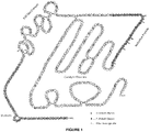

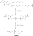
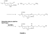
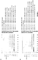

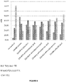
Classifications
-
- A—HUMAN NECESSITIES
- A61—MEDICAL OR VETERINARY SCIENCE; HYGIENE
- A61K—PREPARATIONS FOR MEDICAL, DENTAL OR TOILETRY PURPOSES
- A61K47/00—Medicinal preparations characterised by the non-active ingredients used, e.g. carriers or inert additives; Targeting or modifying agents chemically bound to the active ingredient
- A61K47/50—Medicinal preparations characterised by the non-active ingredients used, e.g. carriers or inert additives; Targeting or modifying agents chemically bound to the active ingredient the non-active ingredient being chemically bound to the active ingredient, e.g. polymer-drug conjugates
- A61K47/51—Medicinal preparations characterised by the non-active ingredients used, e.g. carriers or inert additives; Targeting or modifying agents chemically bound to the active ingredient the non-active ingredient being chemically bound to the active ingredient, e.g. polymer-drug conjugates the non-active ingredient being a modifying agent
- A61K47/56—Medicinal preparations characterised by the non-active ingredients used, e.g. carriers or inert additives; Targeting or modifying agents chemically bound to the active ingredient the non-active ingredient being chemically bound to the active ingredient, e.g. polymer-drug conjugates the non-active ingredient being a modifying agent the modifying agent being an organic macromolecular compound, e.g. an oligomeric, polymeric or dendrimeric molecule
- A61K47/61—Medicinal preparations characterised by the non-active ingredients used, e.g. carriers or inert additives; Targeting or modifying agents chemically bound to the active ingredient the non-active ingredient being chemically bound to the active ingredient, e.g. polymer-drug conjugates the non-active ingredient being a modifying agent the modifying agent being an organic macromolecular compound, e.g. an oligomeric, polymeric or dendrimeric molecule the organic macromolecular compound being a polysaccharide or a derivative thereof
-
- A—HUMAN NECESSITIES
- A61—MEDICAL OR VETERINARY SCIENCE; HYGIENE
- A61K—PREPARATIONS FOR MEDICAL, DENTAL OR TOILETRY PURPOSES
- A61K47/00—Medicinal preparations characterised by the non-active ingredients used, e.g. carriers or inert additives; Targeting or modifying agents chemically bound to the active ingredient
- A61K47/50—Medicinal preparations characterised by the non-active ingredients used, e.g. carriers or inert additives; Targeting or modifying agents chemically bound to the active ingredient the non-active ingredient being chemically bound to the active ingredient, e.g. polymer-drug conjugates
- A61K47/51—Medicinal preparations characterised by the non-active ingredients used, e.g. carriers or inert additives; Targeting or modifying agents chemically bound to the active ingredient the non-active ingredient being chemically bound to the active ingredient, e.g. polymer-drug conjugates the non-active ingredient being a modifying agent
- A61K47/56—Medicinal preparations characterised by the non-active ingredients used, e.g. carriers or inert additives; Targeting or modifying agents chemically bound to the active ingredient the non-active ingredient being chemically bound to the active ingredient, e.g. polymer-drug conjugates the non-active ingredient being a modifying agent the modifying agent being an organic macromolecular compound, e.g. an oligomeric, polymeric or dendrimeric molecule
- A61K47/59—Medicinal preparations characterised by the non-active ingredients used, e.g. carriers or inert additives; Targeting or modifying agents chemically bound to the active ingredient the non-active ingredient being chemically bound to the active ingredient, e.g. polymer-drug conjugates the non-active ingredient being a modifying agent the modifying agent being an organic macromolecular compound, e.g. an oligomeric, polymeric or dendrimeric molecule obtained otherwise than by reactions only involving carbon-to-carbon unsaturated bonds, e.g. polyureas or polyurethanes
- A61K47/60—Medicinal preparations characterised by the non-active ingredients used, e.g. carriers or inert additives; Targeting or modifying agents chemically bound to the active ingredient the non-active ingredient being chemically bound to the active ingredient, e.g. polymer-drug conjugates the non-active ingredient being a modifying agent the modifying agent being an organic macromolecular compound, e.g. an oligomeric, polymeric or dendrimeric molecule obtained otherwise than by reactions only involving carbon-to-carbon unsaturated bonds, e.g. polyureas or polyurethanes the organic macromolecular compound being a polyoxyalkylene oligomer, polymer or dendrimer, e.g. PEG, PPG, PEO or polyglycerol
-
- A—HUMAN NECESSITIES
- A61—MEDICAL OR VETERINARY SCIENCE; HYGIENE
- A61K—PREPARATIONS FOR MEDICAL, DENTAL OR TOILETRY PURPOSES
- A61K47/00—Medicinal preparations characterised by the non-active ingredients used, e.g. carriers or inert additives; Targeting or modifying agents chemically bound to the active ingredient
- A61K47/50—Medicinal preparations characterised by the non-active ingredients used, e.g. carriers or inert additives; Targeting or modifying agents chemically bound to the active ingredient the non-active ingredient being chemically bound to the active ingredient, e.g. polymer-drug conjugates
-
- A—HUMAN NECESSITIES
- A61—MEDICAL OR VETERINARY SCIENCE; HYGIENE
- A61P—SPECIFIC THERAPEUTIC ACTIVITY OF CHEMICAL COMPOUNDS OR MEDICINAL PREPARATIONS
- A61P43/00—Drugs for specific purposes, not provided for in groups A61P1/00-A61P41/00
-
- A—HUMAN NECESSITIES
- A61—MEDICAL OR VETERINARY SCIENCE; HYGIENE
- A61P—SPECIFIC THERAPEUTIC ACTIVITY OF CHEMICAL COMPOUNDS OR MEDICINAL PREPARATIONS
- A61P5/00—Drugs for disorders of the endocrine system
-
- A—HUMAN NECESSITIES
- A61—MEDICAL OR VETERINARY SCIENCE; HYGIENE
- A61P—SPECIFIC THERAPEUTIC ACTIVITY OF CHEMICAL COMPOUNDS OR MEDICINAL PREPARATIONS
- A61P7/00—Drugs for disorders of the blood or the extracellular fluid
- A61P7/04—Antihaemorrhagics; Procoagulants; Haemostatic agents; Antifibrinolytic agents
-
- C—CHEMISTRY; METALLURGY
- C07—ORGANIC CHEMISTRY
- C07K—PEPTIDES
- C07K1/00—General methods for the preparation of peptides, i.e. processes for the organic chemical preparation of peptides or proteins of any length
- C07K1/107—General methods for the preparation of peptides, i.e. processes for the organic chemical preparation of peptides or proteins of any length by chemical modification of precursor peptides
- C07K1/1072—General methods for the preparation of peptides, i.e. processes for the organic chemical preparation of peptides or proteins of any length by chemical modification of precursor peptides by covalent attachment of residues or functional groups
- C07K1/1077—General methods for the preparation of peptides, i.e. processes for the organic chemical preparation of peptides or proteins of any length by chemical modification of precursor peptides by covalent attachment of residues or functional groups by covalent attachment of residues other than amino acids or peptide residues, e.g. sugars, polyols, fatty acids
-
- C—CHEMISTRY; METALLURGY
- C07—ORGANIC CHEMISTRY
- C07K—PEPTIDES
- C07K1/00—General methods for the preparation of peptides, i.e. processes for the organic chemical preparation of peptides or proteins of any length
- C07K1/14—Extraction; Separation; Purification
- C07K1/16—Extraction; Separation; Purification by chromatography
-
- C—CHEMISTRY; METALLURGY
- C07—ORGANIC CHEMISTRY
- C07K—PEPTIDES
- C07K1/00—General methods for the preparation of peptides, i.e. processes for the organic chemical preparation of peptides or proteins of any length
- C07K1/14—Extraction; Separation; Purification
- C07K1/34—Extraction; Separation; Purification by filtration, ultrafiltration or reverse osmosis
-
- C—CHEMISTRY; METALLURGY
- C07—ORGANIC CHEMISTRY
- C07K—PEPTIDES
- C07K14/00—Peptides having more than 20 amino acids; Gastrins; Somatostatins; Melanotropins; Derivatives thereof
- C07K14/435—Peptides having more than 20 amino acids; Gastrins; Somatostatins; Melanotropins; Derivatives thereof from animals; from humans
- C07K14/745—Blood coagulation or fibrinolysis factors
-
- C—CHEMISTRY; METALLURGY
- C07—ORGANIC CHEMISTRY
- C07K—PEPTIDES
- C07K19/00—Hybrid peptides, i.e. peptides covalently bound to nucleic acids, or non-covalently bound protein-protein complexes
Definitions
- the present invention relates to methods for conjugating a water soluble polymer to a protein.
- the preparation of conjugates by forming a covalent linkage between the water soluble polymer and the therapeutic protein can be carried out by a variety of chemical methods.
- PEGylation of polypeptide drugs protects them in circulation and improves their pharmacodynamic and pharmacokinetic profiles ( Harris and Chess, Nat Rev Drug Discov. 2003;2:214-21 ).
- the PEGylation process attaches repeating units of ethylene glycol (polyethylene glycol (PEG)) to a polypeptide drug.
- PEG molecules have a large hydrodynamic volume (5 - 10 times the size of globular proteins), are highly water soluble and hydrated, non-toxic, non-immunogenic and rapidly cleared from the body.
- PEGylation of molecules can lead to increased resistance of drugs to enzymatic degradation, increased half-life in vivo, reduced dosing frequency, decreased immunogenicity, increased physical and thermal stability, increased solubility, increased liquid stability, and reduced aggregation.
- the first PEGylated drugs were approved by the FDA in the early 1990s. Since then, the FDA has approved several PEGylated drugs for oral, injectable, and topical administration.
- PSA Polysialic acid
- CA colominic acid
- PSA acid is biodegradable.
- the polymer can be coupled to the aldehyde groups of the carbohydrate by use of reagents containing, for example, an active hydrazide group ( Wilchek M and Bayer EA, Methods Enzymol 1987;138:429-42 ).
- reagents containing, for example, an active hydrazide group Wilchek M and Bayer EA, Methods Enzymol 1987;138:429-42 .
- a more recent technology is the use of reagents containing aminooxy groups which react with aldehydes to form oxime linkages ( WO 96/40662 , WO2008/025856 ).
- nucelophilic catalysts are also described in Dirksen, A., et al., J Am Chem Soc., 128:15602-3 (2006 ); Dirksen, A., et al., Angew chem. Int Ed., 45:7581-4 (2006 ); Kohler, J.J., ChemBioChem., 10:2147-50 (2009 ); Giuseppone, N., et al., J Am Chem Soc., 127:5528-39 (2005 ); and Thygesen, M.B., et al., J Org Chem., 75:1752-5 (2010 ).
- WO 2012/016131 discloses a method of conjugating a water soluble polymer to an oxidized carbohydrate moiety of a therapeutic protein but does not disclose a step of purifying a water soluble polymer containing an active aminooxy group at a temperature between 2° and 8°C.
- aniline catalysis can accelerate the oxime ligation allowing short reaction times and the use of low concentrations of the aminooxy reagent
- aniline has toxic properties that must be considered when, for example, the conjugated therapeutic protein to form the basis of a pharmaceutical.
- aniline has been shown to induce methemoglobinemia ( Harrison, J.H.., and Jollow, D.J., Molecular Pharmacology, 32(3) 423-431, 1987 ).
- Long-term dietary treatment of rats has been shown to induce tumors in the spleen ( Goodman, DG., et al., J Natl Cancer Inst., 73(1):265-73, 1984 ).
- In vitro studies have also shown that aniline has the potential to induce chromosome mutations and has the potentially genotoxic activity ( Bombhard E.M. et Herbold B, Critical Reviews in Toxicology 35,783-835, 2005 ).
- the present invention provides methods for conjugating polymers to proteins that improves the protein's pharmacodynamic and/or pharmacokinetic properties while minimizing the costs associated with the various reagents and the health risks to the patient recipients when the conjugation reaction is catalyzed by a nucleophilic catalyst.
- alternative catalysts to substitute for aniline are provided. The methods are defined by the enclosed claims.
- the present disclosure also provides an optimized procedure for preparation of various water-soluble polymer-aminooxy linker reagents that can be used to conjugate therapeutic proteins as described herein.
- Recent NMR studies showed that side reactions at the reducing end of PSA can occur if the preparation of the PSA-aminooxy reagent is performed at room temperature.
- the new process is carried out at a temperature between 2-8°C.
- the PSA-aminooxy reagent is prepared at 4°C according to the process described herein.
- the purification of the reagent using a chromatographic purification step is always carried out (e.g., IEX) at a temperature between 2-8°C . Unwanted side reactions at the reducing end of the PSA are substantially reduced when the entire process (chemical reaction and purification of the conjugate by IEX) is performed at a temperature between 2-8°C.
- a method of conjugating a water soluble polymer to an oxidized carbohydrate moiety of a therapeutic protein comprising contacting the oxidized carbohydrate moiety with an activated water soluble polymer under conditions that allow conjugation; said water soluble polymer containing an active aminooxy group and is selected from the group consisting of polysialic acid (PSA ), starch, hydroxyalkyl starch (HAS), hydroxylethyl starch (HES), pullulan, chitosan, hyaluronic acid, chondroitin sulfate, and other polysaccharides dermatan sulfate, .dextran, carboxymethyl-dextran and other polysaccharides containing reducing end groups ; wherein the water soluble polymer containing an active aminooxy group is prepared by a method comprising: a) incubating a solution comprising an oxidized water-soluble polymer with an aminooxy linker comprising an active aminooxy
- the aforementioned method is provided wherein the solution comprising the oxidized water-soluble polymer and the aminooxy linker comprising an active aminooxy group is incubated at 4°C for 1 h in the absence of light with stirring.
- the aforementioned method is provided wherein the water soluble polymer containing an active aminooxy group is purified by anion exchange chromatography at a temperature of 4°C.
- an aforementioned method is provided wherein the oxidized water soluble polymer is PSA and is oxidized by incubation with NaIO 4 .
- an aforementioned method is provided wherein the therapeutic protein selected from the group consisting of Factor IX (FIX), Factor VIII (FVIII), Factor VIIa (FVIIa), Von Willebrand Factor (VWF), Factor FV (FV), Factor X (FX), Factor XI (FXI), Factor XII (FXII), thrombin (FII), protein C, protein S, tPA, PAI-1, tissue factor (TF),ADAMTS 13 protease, IL-1 alpha, IL-1 beta, IL-2, IL-3, IL-4, IL-5, IL-6, IL-11, colony stimulating factor-1 (CSF-1), M-CSF, SCF, GM-CSF, granulocyte colony stimulating factor (G-CSF), EPO, interferon-alpha (IFN-alpha), consensus interferon, IFN-beta, IFN-gamma, IFN-omega,
- the aforementioned method wherein a solution comprising an initial concentration of the therapeutic protein between about 0.3 mg/ml and about 3.0 mg/ml is adjusted to a pH value between about 5.0 and about 8.0 prior to contacting with the activated water soluble polymer.
- the initial concentration of the therapeutic protein is about 1.0 mg/ml and the pH is about 6.0.
- the aforementioned method is provided wherein the therapeutic protein is contacted by a desired excess concentration of activated water soluble polymer, wherein the excess concentration is between about 1-fold molar and about 300-fold molar excess. In one embodiment, the excess concentration is about 50-fold molar excess.
- the aforementioned method is provided wherein the therapeutic protein is incubated with the activated water soluble polymer under conditions comprising a time period between about 0.5 hours and about 24 hours; a temperature between about 2°C and about 37°C; in the presence or absence of light; and with or without stirring.
- the conditions comprise a time period of about 120 minutes, a temperature of about 22 °C, the absence of light; and with stirring.
- an aforementioned method wherein the nucleophilic catalyst is added in an amount to result in a final concentration between about 1.0 mM and about 50 mM nucleophilic catalyst, under conditions comprising a time period between about 0.1 minutes and about 30 minutes; a temperature between about 2°C and about 37°C; in the presence or absence of light; and with or without stirring.
- the final concentration of the nucleophilic catalyst is about 10 mM, and the conditions comprise a time period of up to about 15 minutes, a temperature of about 22 °C, the absence of light; and with stirring.
- the aforementioned method wherein the oxidizing agent is added in an amount to result in a final concentration between about 50 ⁇ M and about 1000 ⁇ M oxidizing agent, under conditions comprising a time period between about 0.1 minutes and 120 minutes; a temperature between about 2°C and about 37°C; in the presence or absence of light; and with or without stirring.
- the final concentration of oxidizing agent is about 400 ⁇ M, and the conditions comprise a time period of about 10 minutes, a temperature of about 22 °C, the absence of light and with stirring.
- the aforementioned method wherein the conjugating the water soluble polymer to the oxidized carbohydrate moiety of the therapeutic protein is stopped by the addition of a quenching agent selected from the group consisting of L-cysteine, methionine, glutathione, glycerol, sodium meta bisulfite (Na 2 S 2 O 5 ), tryptophan, tyrosine, histidine or derivatives thereof, kresol, imidazol, and combinations thereof; wherein the quenching agent is added in an amount to result in a final concentration between about 1 mM and about 100 mM quenching agent, under conditions comprising a time period between about 5 minutes and about 120 minutes; a temperature between about 2°C and about 37°C; in the presence or absence of light; and with or without stirring.
- a quenching agent selected from the group consisting of L-cysteine, methionine, glutathione, glycerol, sodium meta bisulfite (Na 2 S 2 O 5
- the quenching agent is L-cysteine.
- the L-cysteine is added to result in a final concentration of about 10 mM and the conditions comprise a time period of about 60 minutes, a temperature of about 22°C, the absence of light and with stirring.
- an aforementioned method comprising: a) a first step comprising adjusting the pH value of a solution comprising the therapeutic protein to a pH value between about 5.0 and about 8.0, wherein the therapeutic protein concentration is between about 0.3 mg/ml and about 3.0 mg/ml; b) a second step comprising oxidizing one or more carbohydrates on the therapeutic protein, wherein the oxidizing agent is added to the solution in the first step to result in a final concentration between about 50 ⁇ M and about 1000 ⁇ M, under conditions comprising a time period between about 0.1 minutes and about 120 minutes; a temperature between about 2°C and about 37°C; in the presence or absence of light, and with or without stirring; c) a third step comprising contacting the therapeutic protein with a desired excess concentration of activated water soluble polymer, wherein the excess concentration is between about 1-molar excess and about 300-molar excess, under conditions comprising a time period between about 0.5 hours and about 24 hours, a temperature between about 2°C and about 37°
- the initial concentration of the therapeutic protein in the first step is about 1 mg/ml and the pH is about 6.0; wherein the final concentration of oxidizing agent in the second step is about 400 ⁇ M, and the conditions in the fifth step comprise a time period of about 10 minutes, a temperature of about 22 °C, the absence of light and with stirring; wherein the excess concentration in the third step is about 50 molar excess; wherein the conditions in the third step comprise a time period of about 15 minutes, a temperature of about 22 °C, the absence of light and with stirring; wherein the final concentration of the nucleophilic catalyst in the fourth step is about 10 mM, and the conditions in the fourth step comprise a time period of about 15 minutes, a temperature of about 22 °C, the absence of light and with stirring; wherein the conditions of incubating the therapeutic protein with the activated water soluble polymer and nucleophilic catalyst in the fifth step comprise a time period of about 2 hours; a temperature of about 22°C; the absence of light; and with
- the water soluble polymer is PSA. In another embodiment, the water soluble polymer is HES. In yet another embodiment, the water soluble polymer is HAS. In another embodiment the PSA is comprised of about 10 - 300 sialic acid units.
- the therapeutic protein is FIX. In another embodiment, the therapeutic protein is FVIIa. In yet another embodiment, the therapeutic protein is FVIII.
- an aforementioned method is provided wherein the oxidizing agent is sodium periodate (NaIO 4 ).
- the aforementioned method is provided wherein the oxidized carbohydrate moiety of the therapeutic protein is located in the activation peptide of the blood coagulation protein.
- the PSA comprises an activated aminooxy linker selected from the group consisting of: a) a 3-oxa-pentane-1,5-dioxyamine linker of the formula:
- the aminooxy linker is 3-oxa-pentane-1,5-dioxyamine.
- the aforementioned method is provided wherein the nucleophilic catalyst is provided at a concentration between about 1 mM and about 50 mM.
- the nucleophilic catalyst is m-toluidine.
- the m-toluidine is present in the conjugation reaction at a concentration of about 10 mM.
- the aforementioned method is provided further comprising the step of purifying the conjugated therapeutic protein.
- the conjugated therapeutic protein is purified by a method selected from the group consisting of chromatography, filtration and precipitation.
- the chromatography is selected from the group consisting of Hydrophobic Interaction Chromatography (HIC), Ion Exchange chromatography (IEC), Size exclusion chromatography (SEC), Affinity chromatography, and Reversed-phase chromatography.
- HIC Hydrophobic Interaction Chromatography
- IEC Ion Exchange chromatography
- SEC Size exclusion chromatography
- Affinity chromatography Affinity chromatography
- Reversed-phase chromatography Reversed-phase chromatography.
- an anti-chaotropic salt is used in a chromatography loading step and in a chromatography washing step.
- the chromatography takes place in a column.
- the column comprises a chromatography resin selected from the group consisting of Phenyl-Sepharose FF and Butyl-Sepharose FF.
- the resin is present in the column at a bed height of between about 5 cm and about 20 cm.
- the bed height is about 10 cm.
- an aforementioned method is provided wherein comprising one or more washing steps wherein flow direction is set to up-flow and wherein the flow rate is between about 0.2 cm/min and about 6.7 cm/min. In one embodiment, the flow rate is about 2 cm/min. In another embodiment, the aforementioned method is provided comprising one or more elution steps wherein flow direction is set to down-flow and wherein the flow rate is between about 0.1 cm/min and about 6.7 cm/min. In one embodiment, the flow rate is about 1 cm/min.
- the aforementioned method is provided further comprising concentrating the conjugated therapeutic protein by ultra-/diafiltration (UF/DF).
- the aforementioned method is provided wherein the final concentration of therapeutic protein is between about 0.5 and about 3 mg/ml.
- the aforementioned method is provided wherein the therapeutic protein comprises between about 5 and about 11 water soluble polymer moieties.
- the aforementioned method wherein the conjugated therapeutic protein is purified using chromatography; wherein an anti-chaotropic salt is used for a loading step and for a washing step; the method comprising one or more washing steps wherein flow direction is set to up-flow and wherein the flow rate is between about 0.2 cm/min and about 6.7 cm/min and one or more elution steps wherein flow direction is set to down-flow and wherein the flow rate is between about 0.2 cm/min and about 6.7 cm/min; further comprising concentrating the conjugated therapeutic protein by ultra-/diafiltration (UF/DF).
- UF/DF ultra-/diafiltration
- the aforementioned method is provided wherein the chromatography is hydrophobic interaction chromatography (HIC); wherein the one or more washing steps flow rate is about 2 cm/min; and wherein the one or more elution steps flow rate is about 1 cm/min.
- HIC hydrophobic interaction chromatography
- a modified therapeutic protein produced by an aforementioned method is also provided by the present disclosure.
- a method of preparing a water-soluble polymer containing an active aminooxy group comprising: a) incubating a solution comprising an oxidized water-soluble polymer with an aminooxy linker comprising an active aminooxy group under conditions that allow the formation of a stable oxime linkage between the oxidized water-soluble polymer and the activated aminooxy linker, said conditions comprising a time period between about 1 minute and about 24 hours; a temperature between about 2°C and about 8°C; in the presence or absence of light, and with or without stirring; thereby forming a water-soluble polymer containing an active aminooxy group; and b) purifying the water soluble polymer containing an active aminooxy group by a method selected from the group consisting of chromatography, filtration, dialysis, and precipitation, at a temperature between about 2°C and about 8°C; said water soluble polymer is selected from the group consisting of polysialic acid (PSA ), starch, hydroxyal
- a nucleophilic catalyst selected from the group consisting of o-amino benzoic acid, m-amino benzoic acid, p-amino benzoic acid, sulfanilic acid, o-aminobenzamide, o-toluidine, m-toluidine, p-toluidine, o-anisidine, m-anisidine, and p-anisidine.
- the aforementioned method is provided wherein the water soluble polymer is PSA In another embodiment, the aforementioned method is provided wherein the solution comprising the oxidized water-soluble polymer and the aminooxy linker comprising an active aminooxy group is incubated at 4°C for 1 h in the absence of light with stirring. In another embodiment, the aforementioned method is provided wherein the water soluble polymer containing an active aminooxy group is purified by anion exchange chromatography at a temperature of 4°C.
- a method of preparing a PSA-aminooxy reagent aminooxy group comprising: a) incubating a solution comprising an oxidized PSA with an aminooxy linker comprising an active aminooxy group under conditions that allow the formation of a stable oxime linkage between the oxidized PSA and the activated aminooxy linker, said conditions comprising a time period 1 hour; a temperature of 4°C; in the absence of light, and with stirring; thereby forming a PSA containing an active aminooxy group; and b) purifying the PSA containing an active aminooxy group by anion exchange chromatography, a temperature of 4°C; said activated aminooxy linker is 3-oxa-pentane-1,5-dioxyamine linker of the formula: thereby forming an oxime linkage between the PSA and the aminooxy linker.
- a method of conjugating a water soluble polymer to an oxidized carbohydrate moiety of a blood coagulation protein comprising contacting the oxidized carbohydrate moiety with an activated water soluble polymer under conditions that allow conjugation; said blood coagulation protein selected from the group consisting of Factor IX (FIX), Factor VIII (FVIII), Factor VIIa (FVIIa), Von Willebrand Factor (VWF), Factor FV (FV), Factor X (FX), Factor XI (FXI), Factor XII (FXII), thrombin (FII), protein C, protein S, tPA, PAI-1, tissue factor (TF) and ADAMTS 13 protease or a biologically active fragment, derivative or variant thereof; said water soluble polymer containing an active aminooxy group and is selected from the group consisting of polysialic acid (PSA), pullulan, chitosan, hyal
- the pharmacological and immunological properties of therapeutic proteins can be improved by chemical modification and conjugation with polymeric compounds such as polysialic acid (PSA), hydroxyalkyl starch (HAS), hydroxylethyl starch (HES), pullulan, chitosan, hyaluronic acid, chondroitin sulfate, dermatan sulfate, starch, dextran, carboxymethyl- and other polysaccharides containing reducing end groups .
- polymeric compounds such as polysialic acid (PSA), hydroxyalkyl starch (HAS), hydroxylethyl starch (HES), pullulan, chitosan, hyaluronic acid, chondroitin sulfate, dermatan sulfate, starch, dextran, carboxymethyl- and other polysaccharides containing reducing end groups .
- PSA polysialic acid
- HAS hydroxyalkyl starch
- HES hydroxyle
- Synthetic polymers like PEG can be manufactured easily with a narrow size distribution, while PSA can be purified in such a manner that results in a final PSA preparation with a narrow size distribution.
- PEGylation reagents with defined polymer chains and narrow size distribution are on the market and commercially available for a reasonable price.
- a soluble polymer such as through polysialylation
- FIX blood coagulation protein
- other coagulation proteins e.g., VWF, FVIIa (see, e.g., US 2008/0221032A1 , ) and FVIII).
- polypeptides and polynucleotides are exemplified by the following therapeutic proteins: enzymes, antigens, antibodies, receptors, blood coagulation proteins, growth factors, hormones, and ligands.
- the therapeutic protein is a blood coagulation protein such as Factor IX (FIX), Factor VIII (FVIII), Factor VIIa (FVIIa), Von Willebrand Factor (VWF), Factor FV (FV), Factor X (FX), Factor XI (FXI), Factor XII (FXII), thrombin (FII), protein C, protein S, tPA, PAI-1, tissue factor (TF) or ADAMTS 13 protease.
- FIX Factor IX
- FVIII Factor VIIa
- VWF Von Willebrand Factor
- FV Factor FV
- FX Factor X
- FXI Factor XI
- FXIII Factor XII
- thrombin FII
- a therapeutic protein according to the invention is a glycoprotein or, in various embodiments, a protein that is not naturally glycosylated in vivo (i.e., a protein that does not contain a natural glycosylation site or a protein that is not glycosylated in a host cell prior to purification).
- the therapeutic protein is immunoglobulins, cytokines such IL-1 alpha, IL-1 beta, IL-2, IL-3, IL-4, IL-5, IL-6, IL-11, colony stimulating factor-1 (CSF-1), M-CSF, SCF, GM-CSF, granulocyte colony stimulating factor (G-CSF), EPO, interferon-alpha (IFN-alpha), consensus interferon, IFN-beta, IFN-gamma, IFN-omega, IL-7, IL-8, IL-9, IL-10, IL-12, IL-13, IL-14, IL-15, IL-16, IL-17, IL-18, IL-19, IL-20, IL-21, IL-22, IL-23, IL-24, IL-31, IL-32 alpha, IL-33, thrombopoietin (TPO), angiopoietins, for example Ang-1, Ang-2,
- the therapeutic protin is alpha-, beta-, and gamma-interferons, colony stimulating factors including granulocyte colony stimulating factors, fibroblast growth factors, platelet derived growth factors, phospholipase-activating protein (PUP), insulin, plant proteins such as lectins and ricins, tumor necrosis factors and related alleles, soluble forms of tumor necrosis factor receptors, interleukin receptors and soluble forms of interleukin receptors, growth factors such as tissue growth factors, such as TGF ⁇ s or TGF ⁇ s and epidermal growth factors, hormones, somatomedins, pigmentary hormones, hypothalamic releasing factors, antidiuretic hormones, prolactin, chorionic gonadotropin, follicle-stimulating hormone, thyroid-stimulating hormone, tissue plasminogen activator, and immunoglobulins such as IgG, IgE, IgM, IgA, and IgD, a gal
- Table 1 Follicular dendritic cell secreted peptide Angiotensin-converting enzyme Interleukin-1 family member 6 Herstatin Dermokine Antithrombin-III Prostate and testis expressed protein 2 Leucine-rich repeat-containing protein 28 Secreted frizzled-related protein 1 Apolipoprotein B-100 Group XIIA secretory phospholipase A2 LRRN4 C-terminal-like protein Ectodvsplasin-A Apolipoprotein D Collagen alpha-3(V) chain Ly6/PLAUR domain-containing protein 2 Secreted frizzled-related protein 2 Apolipoprotein E Alpha-2-macroglobulin-like protein 1 Transmembrane protein 81 Resistin Beta-1,4-galactosyltransferase 1 Dermatopontin Myelin protein zero-like protein 3 Osteopontin Bone morphogenetic protein 7 Cart
- exon SCR short consensus repeat Peptidoglycan recognition protein
- Dolichol kinase Adiponectin receptor protein 2 PIKR2786 Interferon-induced 17 kDa protein Endothelin-converting enzvme-like 1 Inhibin beta C chain S100 calcium binding protein A7-like 3 Protein Wnt-4 Integral membrane protein 2B Brorin GTWW5826 (LP5085 protein) Allograft inflammatory factor 1 -like Insulin-like qrowth factor-binding protein 5 Semaphorin-3C KTIS8219 (HCG2020043) Armadillo repeat-containing X-linked protein 3 Endothelial cell-selective adhesion molecule Heparan sulfate glucosamine 3-O-sulfotransferase 2 Hyaluronan and proteoglycan link protein 4 Chondroitin sulfate N-acetylgalactosaminyltransferase 1 Signal peptide, C
- therapeutic proteins should not be considered to be exclusive. Rather, as is apparent from the disclosure provided herein, the methods of the invention are applicable to any protein wherein attachment of a water soluble polymer is desired according to the invention.
- therapeutic proteins are described in US 2007/0026485 ,
- the starting material of the present invention is a blood coagulation protein, which can be derived from human plasma, or produced by recombinant engineering techniques, as described in patents US Patent No. 4,757,006 ; US Patent No. 5,733,873 ; US Patent No. 5,198,349 ; US Patent No. 5,250,421 ; US Patent No. 5,919,766 ; and EP 306 968 .
- Therapeutic polypeptides such as blood coagulation proteins including Factor IX (FIX), Factor VIII (FVIII), Factor VIIa (FVIIa), Von Willebrand Factor (VWF), Factor FV (FV), Factor X (FX), Factor XI (FXI), Factor XII (FXII), thrombin (FII), protein C, protein S, tPA, PAI-1, tissue factor (TF) and ADAMTS 13 protease are rapidly degraded by proteolytic enzymes and neutralized by antibodies. This reduces their half-life and circulation time, thereby limiting their therapeutic effectiveness. Relatively high doses and frequent administration are necessary to reach and sustain the desired therapeutic or prophylactic effect of these coagulation proteins. As a consequence, adequate dose regulation is difficult to obtain and the need of frequent intravenous administrations imposes restrictions on the patient's way of living.
- blood coagulation proteins including, but not limited to, Factor IX (FIX), Factor VIII (FVIII), Factor VIIa (FVIIa), Von Willebrand Factor (VWF), Factor V (FV), Factor X (FX), Factor XI, Factor XII (FXII), thrombin (FII), protein C, protein S, tPA, PAI-1, tissue factor (TF) and ADAMTS 13 protease are contemplated by the invention.
- blood coagulation protein refers to any Factor IX (FIX), Factor VIII (FVIII), Factor VIIa (FVIIa), Von Willebrand Factor (VWF), Factor FV (FV), Factor X (FX), Factor XII (FXII), thrombin (FII), protein C, protein S, tPA, PAI-1, tissue factor (TF) and ADAMTS 13 protease which exhibits biological activity that is associated with that particular native blood coagulation protein.
- FIX Factor IX
- FVIII Factor VIIa
- VWF Von Willebrand Factor
- FV Factor FV
- FX Factor X
- FXII Factor XII
- thrombin FII
- the blood coagulation cascade is divided into three distinct segments: the intrinsic, extrinsic, and common pathways ( Schenone et al., Curr Opin Hematol. 2004; 11:272-7 ).
- the cascade involves a series of serine protease enzymes (zymogens) and protein cofactors. When required, an inactive zymogen precursor is converted into the active form, which consequently converts the next enzyme in the cascade.
- zymogens serine protease enzymes
- protein cofactors protein cofactors
- the intrinsic pathway requires the clotting factors VIII, IX, X, XI, and XII. Initiation of the intrinsic pathway occurs when prekallikrein, high-molecular-weight kininogen, factor XI (FXI) and factor XII (FXII) are exposed to a negatively charged surface. Also required are calcium ions and phospholipids secreted from platelets.
- the extrinsic pathway is initiated when the vascular lumen of blood vessels is damaged.
- the membrane glycoprotein tissue factor is exposed and then binds to circulating factor VII (FVII) and to small preexisting amounts of its activated form FVIIa. This binding facilitates full conversion of FVII to FVIIa and subsequently, in the presence of calcium and phospholipids, the conversion of factor IX (FIX) to factor IXa (FIXa) and factor X (FX) to factor Xa (FXa).
- FVIIa The association of FVIIa with tissue factor enhances the proteolytic activity by bringing the binding sites of FVII for the substrate (FIX and FX) into closer proximity and by inducing a conformational change, which enhances the enzymatic activity of FVIIa.
- FX Factor XIIIa
- FVIIa Conversion of FVII to FVIIa is also catalyzed by a number of proteases, including thrombin, FIXa, FXa, factor XIa (FXIa), and factor XIIa (FXIIa).
- tissue factor pathway inhibitor targets FVIIa/tissue factor/FXa product complex.
- FVII also known as stable factor or proconvertin
- FVII is a vitamin K-dependent serine protease glycoprotein with a pivotal role in hemostasis and coagulation ( Eigenbrot, Curr Protein Pept Sci. 2002;3:287-99 ).
- FVII is synthesized in the liver and secreted as a single-chain glycoprotein of 48 kD.
- FVII shares with all vitamin K-dependent serine protease glycoproteins a similar protein domain structure consisting of an amino-terminal gamma-carboxyglutamic acid (Gla) domain with 9-12 residues responsible for the interaction of the protein with lipid membranes, a carboxy-terminal serine protease domain (catalytic domain), and two epidermal growth factor-like domains containing a calcium ion binding site that mediates interaction with tissue factor.
- Gamma-glutamyl carboxylase catalyzes carboxylation of Gla residues in the amino-terminal portion of the molecule.
- the carboxylase is dependent on a reduced form of vitamin K for its action, which is oxidized to the epoxide form.
- Vitamin K epoxide reductase is required to convert the epoxide form of vitamin K back to the reduced form.
- the major proportion of FVII circulates in plasma in zymogen form, and activation of this form results in cleavage of the peptide bond between arginine 152 and isoleucine 153.
- the resulting activated FVIIa consists of a NH2-derived light chain (20 kD) and a COOH terminal-derived heavy chain (30 kD) linked via a single disulfide bond (Cys 135 to Cys 262).
- the light chain contains the membrane-binding Gla domain, while the heavy chain contains the catalytic domain.
- the plasma concentration of FVII determined by genetic and environmental factors is about 0.5 mg/mL ( Pinotti et al., Blood. 2000;95:3423-8 ).
- Different FVII genotypes can result in several-fold differences in mean FVII levels.
- Plasma FVII levels are elevated during pregnancy in healthy females and also increase with age and are higher in females and in persons with hypertriglyceridemia.
- FVII has the shortest half-life of all procoagulant factors (3-6 h).
- the mean plasma concentration of FVIIa is 3.6 ng/mL in healthy individuals and the circulating half-life of FVIIa is relatively long (2.5 h) compared with other coagulation factors.
- Hereditary FVII deficiency is a rare autosomal recessive bleeding disorder with a prevalence estimated to be 1 case per 500,000 persons in the general population ( Acharya et al., J Thromb Haemost. 2004;2248-56 ). Acquired FVII deficiency from inhibitors is also very rare. Cases have also been reported with the deficiency occurring in association with drugs such as cephalosporins, penicillins, and oral anticoagulants. Furthermore, acquired FVII deficiency has been reported to occur spontaneously or with other conditions, such as myeloma, sepsis, aplastic anemia, with interleukin-2 and antithymocyte globulin therapy.
- Reference polynucleotide and polypeptide sequences include, e.g., GenBank Accession Nos. J02933 for the genomic sequence, M13232 for the cDNA ( Hagen et al. PNAS 1986; 83: 2412-6 ), and P08709 for the polypeptide sequence
- GenBank Accession Nos. J02933 for the genomic sequence
- M13232 for the cDNA Hagen et al. PNAS 1986; 83: 2412-6
- P08709 for the polypeptide sequence
- a variety of polymorphisms of FVII have been described, for example see Sabater-Lleal et al. (Hum Genet. 2006; 118:741-51 )
- FIX is a vitamin K-dependent plasma protein that participates in the intrinsic pathway of blood coagulation by converting FX to its active form in the presence of calcium ions, phospholipids and FVIIIa.
- the predominant catalytic capability of FIX is as a serine protease with specificity for a particular arginine-isoleucine bond within FX.
- Activation of FIX occurs by FXIa which causes excision of the activation peptide from FIX to produce an activated FIX molecule comprising two chains held by one or more disulphide bonds. Defects in FIX are the cause of recessive X-linked hemophilia B.
- Hemophilia A and B are inherited diseases characterized by deficiencies in FVIII and FIX polypeptides, respectively.
- the underlying cause of the deficiencies is frequently the result of mutations in FVIII and FIX genes, both of which are located on the X chromosome.
- Traditional therapy for hemophilias often involves intravenous administration of pooled plasma or semi-purified coagulation proteins from normal individuals. These preparations can be contaminated by pathogenic agents or viruses, such as infectious prions, HIV, parvovirus, hepatitis A, and hepatitis C. Hence, there is an urgent need for therapeutic agents that do not require the use of human serum.
- the level of the decrease in FIX activity is directly proportional to the severity of hemophilia B.
- the current treatment of hemophilia B consists of the replacement of the missing protein by plasma-derived or recombinant FIX (so-called FIX substitution or replacement treatment or therapy).
- Polynucleotide and polypeptide sequences of FIX can be found for example in the UniProtKB/Swiss-Prot Accession No. P00740, US Pat. No. 6,531,298 and in Figure 1 (SEQ ID NO: 1).
- Coagulation factor VIII (FVIII) circulates in plasma at a very low concentration and is bound non-covalently to Von Willebrand factor (VWF). During hemostasis, FVIII is separated from VWF and acts as a cofactor for activated factor IX (FIXa)-mediated FX activation by enhancing the rate of activation in the presence of calcium and phospholipids or cellular membranes.
- VWF Von Willebrand factor
- FVIII is synthesized as a single-chain precursor of approximately 270-330 kD with the domain structure A1-A2-B-A3-C1-C2.
- FVIII is composed of a heavy chain (A1-A2-B) and a light chain (A3-C1-C2).
- the molecular mass of the light chain is 80 kD whereas, due to proteolysis within the B domain, the heavy chain is in the range of 90-220 kD.
- FVIII is also synthesized as a recombinant protein for therapeutic use in bleeding disorders.
- Various in vitro assays have been devised to determine the potential efficacy of recombinant FVIII (rFVIII) as a therapeutic medicine. These assays mimic the in vivo effects of endogenous FVIII.
- In vitro thrombin treatment of FVIII results in a rapid increase and subsequent decrease in its procoagulant activity, as measured by in vitro assays. This activation and inactivation coincides with specific limited proteolysis both in the heavy and the light chains, which alter the availability of different binding epitopes in FVIII, e.g. allowing FVIII to dissociate from VWF and bind to a phospholipid surface or altering the binding ability to certain monoclonal antibodies.
- the lack or dysfunction of FVIII is associated with the most frequent bleeding disorder, hemophilia A.
- the treatment of choice for the management of hemophilia A is replacement therapy with plasma derived or rFVIII concentrates. Patients with severe hemophilia A with FVIII levels below 1 %, are generally on prophylactic therapy with the aim of keeping FVIII above 1% between doses. Taking into account the average half-lives of the various FVIII products in the circulation, this result can usually be achieved by giving FVIII two to three times a week.
- Reference polynucleotide and polypeptide sequences include, e.g., UniProtKB/Swiss-Prot P00451 (FA8_HUMAN); Gitschier J et al., Characterization of the human Factor VIII gene, Nature, 312(5992): 326-30 (1984 ); Vehar GH et al., Structure of human Factor VIII, Nature, 312(5992):337-42 (1984 ); Thompson AR. Structure and Function of the Factor VIII gene and protein, Semin Thromb Hemost, 2003:29; 11-29 (2002 ).
- Von Willebrand factor is a glycoprotein circulating in plasma as a series of multimers ranging in size from about 500 to 20,000 kD.
- Multimeric forms of VWF are composed of 250 kD polypeptide subunits linked together by disulfide bonds. VWF mediates initial platelet adhesion to the sub-endothelium of the damaged vessel wall. Only the larger multimers exhibit hemostatic activity. It is assumed that endothelial cells secrete large polymeric forms of VWF and those forms of VWF which have a low molecular weight (low molecular weight VWF) arise from proteolytic cleavage.
- the multimers having large molecular masses are stored in the Weibel-Pallade bodies of endothelial cells and liberated upon stimulation.
- VWF is synthesized by endothelial cells and megakaryocytes as prepro-VWF that consists to a large extent of repeated domains.
- pro-VWF dimerizes through disulfide linkages at its C-terminal region.
- the dimers serve as protomers for multimerization, which is governed by disulfide linkages between the free end termini.
- the assembly to multimers is followed by the proteolytic removal of the propeptide sequence ( Leyte et al., Biochem. J. 274 (1991), 257-261 ).
- the primary translation product predicted from the cloned cDNA of VWF is a 2813-residue precursor polypeptide (prepro-VWF).
- the prepro-VWF consists of a 22 amino acid signal peptide and a 741 amino acid propeptide, with the mature VWF comprising 2050 amino acids ( Ruggeri Z.A., and Ware, J., FASEB J., 308-316 (1993 ).
- VWD type 3 is the most severe form, in which VWF is completely missing, and VWD type 1 relates to a quantitative loss of VWF and its phenotype can be very mild.
- VWD type 2 relates to qualitative defects of VWF and can be as severe as VWD type 3.
- VWD type 2 has many sub forms, some being associated with the loss or the decrease of high molecular weight multimers.
- Von Willebrand disease type 2a (VWD-2A) is characterized by a loss of both intermediate and large multimers.
- VWD-2B is characterized by a loss of highest-molecular-weight multimers.
- Other diseases and disorders related to VWF are known in the art.
- polynucleotide and amino acid sequences of prepro-VWF are available at GenBank Accession Nos. NM_000552 and NP_000543, respectively.
- the starting material of the present invention is a protein or polypeptide.
- therapeutic protein refers to any therapeutic protein molecule which exhibits biological activity that is associated with the therapeutic protein.
- the therapeutic protein molecule is a full-length protein.
- Therapeutic protein molecules contemplated include full-length proteins, precursors of full length proteins, biologically active subunits or fragments of full length proteins, as well as biologically active derivatives and variants of any of these forms of therapeutic proteins.
- therapeutic protein include those that (1) have an amino acid sequence that has greater than about 60%, about 65%, about 70%, about 75%, about 80%, about 85%, about 90%, about 91%, about 92%, about 93%, about 94%, about 95%, about 96%, about 97%, about 98% or about 99% or greater amino acid sequence identity, over a region of at least about 25, about 50, about 100, about 200, about 300, about 400, or more amino acids, to a polypeptide encoded by a referenced nucleic acid or an amino acid sequence described herein; and/or (2) specifically bind to antibodies, e.g., polyclonal or monoclonal antibodies, generated against an immunogen comprising a referenced amino acid sequence as described herein, an immunogenic fragment thereof, and/or a conservatively modified variant thereof
- the term "recombinant therapeutic protein” includes any therapeutic protein obtained via recombinant DNA technology. In certain embodiments, the term encompasses proteins as described herein.
- endogenous therapeutic protein includes a therapeutic protein which originates from the mammal intended to receive treatment.
- the term also includes therapeutic protein transcribed from a transgene or any other foreign DNA present in said mammal.
- exogenous therapeutic protein includes a blood coagulation protein which does not originate from the mammal intended to receive treatment.
- plasma-derived blood coagulation protein or "plasmatic” includes all forms of the protein found in blood obtained from a mammal having the property participating in the coagulation pathway.
- biologically active derivative or “biologically active variant” includes any derivative or variant of a molecule having substantially the same functional and/or biological properties of said molecule, such as binding properties, and/or the same structural basis, such as a peptidic backbone or a basic polymeric unit.
- an “analog,” such as a “variant” or a “derivative,” is a compound substantially similar in structure and having the same biological activity, albeit in certain instances to a differing degree, to a naturally-occurring molecule.
- a polypeptide variant refers to a polypeptide sharing substantially similar structure and having the same biological activity as a reference polypeptide.
- Variants or analogs differ in the composition of their amino acid sequences compared to the naturally-occurring polypeptide from which the analog is derived, based on one or more mutations involving (i) deletion of one or more amino acid residues at one or more termini of the polypeptide and/or one or more internal regions of the naturally-occurring polypeptide sequence (e.g., fragments), (ii) insertion or addition of one or more amino acids at one or more termini (typically an "addition” or “fusion") of the polypeptide and/or one or more internal regions (typically an "insertion") of the naturally-occurring polypeptide sequence or (iii) substitution of one or more amino acids for other amino acids in the naturally-occurring polypeptide sequence.
- a "derivative" is a type of analog and refers to a polypeptide sharing the same or substantially similar structure as a reference polypeptide that has been modified, e.g., chemically.
- a variant polypeptide is a type of analog polypeptide and includes insertion variants, wherein one or more amino acid residues are added to a therapeutic protein amino acid sequence of the invention. Insertions may be located at either or both termini of the protein, and/or may be positioned within internal regions of the therapeutic protein amino acid sequence. Insertion variants, with additional residues at either or both termini, include for example, fusion proteins and proteins including amino acid tags or other amino acid labels.
- the blood coagulation protein molecule optionally contains an N-terminal Met, especially when the molecule is expressed recombinantly in a bacterial cell such as E. coli.
- one or more amino acid residues in a therapeutic protein polypeptide as described herein are removed.
- Deletions can be effected at one or both termini of the therapeutic protein polypeptide, and/or with removal of one or more residues within the therapeutic protein amino acid sequence.
- Deletion variants therefore, include fragments of a therapeutic protein polypeptide sequence.
- substitution variants one or more amino acid residues of a therapeutic protein polypeptide are removed and replaced with alternative residues.
- the substitutions are conservative in nature and conservative substitutions of this type are well known in the art.
- the invention embraces substitutions that are also non-conservative. Exemplary conservative substitutions are described in Lehninger, [Biochemistry, 2nd Edition; Worth Publishers, Inc., New York (1975), pp.71-77 ] and are set out immediately below.
- Nucleic acids encoding a therapeutic protein of the invention include, for example and without limitation, genes, pre-mRNAs, mRNAs, cDNAs, polymorphic variants, alleles, synthetic and naturally-occurring mutants.
- Polynucleotides encoding a therapeutic protein of the invention also include, without limitation, those that (1) specifically hybridize under stringent hybridization conditions to a nucleic acid encoding a referenced amino acid sequence as described herein, and conservatively modified variants thereof; (2) have a nucleic acid sequence that has greater than about 95%, about 96%, about 97%, about 98%, about 99%, or higher nucleotide sequence identity, over a region of at least about 25, about 50, about 100, about 150, about 200, about 250, about 500, about 1000, or more nucleotides (up to the full length sequence of 1218 nucleotides of the mature protein), to a reference nucleic acid sequence as described herein.
- Exemplary "stringent hybridization” conditions include hybridization at 42 °C in 50% formamide, 5X SSC, 20 mM Na•PO4, pH 6.8; and washing in IX SSC at 55 °C for 30 minutes. It is understood that variation in these exemplary conditions can be made based on the length and GC nucleotide content of the sequences to be hybridized. Formulas standard in the art are appropriate for determining appropriate hybridization conditions. See Sambrook et al., Molecular Cloning: A Laboratory Manual (Second ed., Cold Spring Harbor Laboratory Press, 1989) ⁇ 9.47-9.51 .
- a "naturally-occurring" polynucleotide or polypeptide sequence is typically derived from a mammal including, but not limited to, primate, e.g., human; rodent, e.g., rat, mouse, hamster; cow, pig, horse, sheep, or any mammal.
- the nucleic acids and proteins of the invention can be recombinant molecules (e.g., heterologous and encoding the wild type sequence or a variant thereof, or non-naturally occurring).
- Production of a therapeutic protein includes any method known in the art for (i) the production of recombinant DNA by genetic engineering, (ii) introducing recombinant DNA into prokaryotic or eukaryotic cells by, for example and without limitation, transfection, electroporation or microinjection, (iii) cultivating said transformed cells, (iv) expressing therapeutic protein, e.g. constitutively or upon induction, and (v) isolating said blood coagulation protein, e.g. from the culture medium or by harvesting the transformed cells, in order to obtain purified therapeutic protein.
- the therapeutic protein is produced by expression in a suitable prokaryotic or eukaryotic host system characterized by producing a pharmacologically acceptable blood coagulation protein molecule.
- suitable prokaryotic or eukaryotic host system characterized by producing a pharmacologically acceptable blood coagulation protein molecule.
- eukaryotic cells are mammalian cells, such as CHO, COS, HEK 293, BHK, SK-Hep, and HepG2.
- vectors are used for the preparation of the therapeutic protein and are selected from eukaryotic and prokaryotic expression vectors.
- vectors for prokaryotic expression include plasmids such as, and without limitation, pRSET, pET, and pBAD, wherein the promoters used in prokaryotic expression vectors include one or more of, and without limitation, lac, trc, trp, recA, or araBAD.
- vectors for eukaryotic expression include: (i) for expression in yeast, vectors such as, and without limitation, pAO, pPIC, pYES, or pMET, using promoters such as, and without limitation, AOX1, GAP, GAL1, or AUG1; (ii) for expression in insect cells, vectors such as and without limitation, pMT, pAc5, pIB, pMIB, or pBAC, using promoters such as and without limitation PH, p10, MT, Ac5, OpIE2, gp64, or polh, and (iii) for expression in mammalian cells, vectors such as and without limitation pSVL, pCMV, pRc/RSV, pcDNA3, or pBPV, and vectors derived from, in one aspect, viral systems such as and without limitation vaccinia virus, adeno-associated viruses, herpes viruses, or retroviruses, using promoters such as and without limitation C
- a conjugated therapeutic protein of the present invention may be administered by injection, such as intravenous, intramuscular, or intraperitoneal injection.
- compositions comprising a conjugated therapeutic protein of the present invention to human or test animals
- the compositions comprise one or more pharmaceutically acceptable carriers.
- pharmaceutically acceptable carriers include any and all clinically useful solvents, dispersion media, coatings, antibacterial and antifungal agents, isotonic and absorption delaying agents and the like, including those agents disclosed above.
- ⁇ ективное amount includes a dose suitable for treating a disease or disorder or ameliorating a symptom of a disease or disorder. In one embodiment, “effective amount” includes a dose suitable for treating a mammal having a bleeding disorder as described herein.
- compositions may be administered orally, topically, transdermally, parenterally, by inhalation spray, vaginally, rectally, or by intracranial injection.
- parenteral as used herein includes subcutaneous injections, intravenous, intramuscular, intracisternal injection, or infusion techniques. Administration by intravenous, intradermal, intramuscular, intramammary, intraperitoneal, intrathecal, retrobulbar, intrapulmonary injection and or surgical implantation at a particular site is contemplated as well.
- compositions are essentially free of pyrogens, as well as other impurities that could be harmful to the recipient.
- Single or multiple administrations of the compositions can be carried out with the dose levels and pattern being selected by the treating physician.
- the appropriate dosage will depend on the type of disease to be treated, as described above, the severity and course of the disease, whether drug is administered for preventive or therapeutic purposes, previous therapy, the patient's clinical history and response to the drug, and the discretion of the attending physician.
- the present invention also relates to a pharmaceutical composition
- a pharmaceutical composition comprising an effective amount of a conjugated therapeutic protein as defined herein.
- the pharmaceutical composition may further comprise a pharmaceutically acceptable carrier, diluent, salt, buffer, or excipient.
- the pharmaceutical composition can be used for treating the above-defined bleeding disorders.
- the pharmaceutical composition of the invention may be a solution or a lyophilized product. Solutions of the pharmaceutical composition may be subjected to any suitable lyophilization process.
- kits which comprise a composition of the invention packaged in a manner which facilitates its use for administration to subjects.
- a kit includes a compound or composition described herein (e.g., a composition comprising a conjugated therapeutic protein), packaged in a container such as a sealed bottle or vessel, with a label affixed to the container or included in the package that describes use of the compound or composition in practicing the method.
- the kit contains a first container having a composition comprising a conjugated therapeutic protein and a second container having a physiologically acceptable reconstitution solution for the composition in the first container.
- the compound or composition is packaged in a unit dosage form.
- the kit may further include a device suitable for administering the composition according to a specific route of administration.
- the kit contains a label that describes use of the therapeutic protein or peptide composition.
- a therapeutic protein derivative i.e., a conjugated therapeutic protein
- a water-soluble polymer including, polysialic acid (PSA), hydroxyalkyl starch (HAS), hydroxylethyl starch (HES), pullulan, chitosan, hyaluronic acid, chondroitin sulfate, dermatan sulfate, starch, dextran, carboxymethyl-dextran
- the water soluble polymer is consisting of sialic acid molecule having a molecular weight range of 350 to 120,000, 500 to 100,000,1000 to 80,000, 1500 to 60,000, 2,000 to 45,000 Da, 3,000 to 35,000 Da, and 5,000 to 25,000 Da.
- the coupling of the water soluble polymer can be carried out by direct coupling to the protein or via linker molecules.
- a chemical linker is MBPH (4-[4-N-Maleimidophenyl]butyric acid hydrazide) containing a carbohydrate-selective hydrazide and a sulfhydryl-reactive maleimide group ( Chamow et al., J Biol Chem 1992;267:15916-22 ).
- Other exemplary and preferred linkers are described below.
- the derivative retains the full functional activity of native therapeutic protein products, and provides an extended half-life in vivo, as compared to native therapeutic protein products. In another embodiment, the derivative retains at least 20, 21, 22, 23, 24, 25, 26, 27, 28, 29, 30, 31, 32, 34, 35, 36, 37, 38, 39, 40, 41, 42, 43, 44.
- the biological activities of the derivative and native blood coagulation protein are determined by the ratios of chromogenic activity to blood coagulation factor antigen value (blood coagulation factor:Chr: blood coagulation factor:Ag).
- the half-life of the construct is decreased or increased 0.5, 0.6, 0.7, 0.8, 0.9, 1.0, 1.1, 1.2, 1.3, 1.4, 1.5, 2, 3, 4, 5, 6, 7, 8, 9, or 10-fold relative to the in vivo half-life of native therapeutic protein.
- PSAs consist of polymers (generally homopolymers) of N-acetylneuraminic acid.
- the secondary amino group normally bears an acetyl group, but it may instead bear a glycolyl group.
- Possible substituents on the hydroxyl groups include acetyl, lactyl, ethyl, sulfate, and phosphate groups.
- sialic acid N-acetylneuraminic acid
- PSAs and mPSAs generally comprise linear polymers consisting essentially of N-acetylneuraminic acid moieties linked by 2,8- or 2,9- glycosidic linkages or combinations of these (e.g. alternating 2,8- and 2,9- linkages).
- the glycosidic linkages are ⁇ -2,8.
- Such PSAs and mPSAs are conveniently derived from colominic acids, and are referred to herein as "CAs" and "mCAs".
- Typical PSAs and mPSAs comprise at least 2, preferably at least 5, more preferably at least 10 and most preferably at least 20 N-acetylneuraminic acid moieties.
- PSAs and CAs may comprise from 2 to 300 N-acetylneuraminic acid moieties, preferably from 5 to 200 N-acetylneuraminic acid moieties, or most preferably from 10 to 100 N-acetylneuraminic acid moieties.
- PSAs and CAs preferably are essentially free of sugar moieties other than N-acetylneuraminic acid.
- PSAs and CAs preferably comprise at least 90 %, more preferably at least 95 % and most preferably at least 98 % N-acetylneuraminic acid moieties.
- PSAs and CAs comprise moieties other than N-acetylneuraminic acid (as, for example in mPSAS and mCAs) these are preferably located at one or both of the ends of the polymer chain.
- Such "other" moieties may, for example, be moieties derived from terminal N-acetylneuraminic acid moieties by oxidation or reduction.
- WO-A-0187922 describes such mPSAs and mCAs in which the non-reducing terminal N-acetylneuraminic acid unit is converted to an aldehyde group by reaction with sodium periodate.
- WO 2005/016974 describes such mPSAs and mCAs in which the reducing terminal N-acetylneuraminic acid unit is subjected to reduction to reductively open the ring at the reducing terminal N-acetylneuraminic acid unit, whereby a vicinal diol group is formed, followed by oxidation to convert the vicinal diol group to an aldehyde group.
- Sialic acid rich glycoproteins bind selectin in humans and other organisms. They play an important role in human influenza infections. E.g., sialic acid can hide mannose antigens on the surface of host cells or bacteria from mannose-binding lectin. This prevents activation of complement. Sialic acids also hide the penultimate galactose residue thus preventing rapid clearance of the glycoprotein by the galactose receptor on the hepatic parenchymal cells.
- Colominic acids are homopolymers of N-acetylneuraminic acid (NANA) with ⁇ (2 ⁇ 8) ketosidic linkage, and are produced, inter alia, by particular strains of Escherichia coli possessing K1 antigen. Colominic acids have many physiological functions. They are important as a raw material for drugs and cosmetics.
- sialic acid moieties includes sialic acid monomers or polymers ("polysaccharides") which are soluble in an aqueous solution or suspension and have little or no negative impact, such as side effects, to mammals upon administration of the PSA-blood coagulation protein conjugate in a pharmaceutically effective amount.
- the polymers are characterized, in one aspect, as having 1, 2, 3, 4, 5, 10, 20, 30, 40, 50, 60, 70, 80, 90, 100, 200, 300, 400, or 500 sialic acid units. In certain aspects, different sialic acid units are combined in a chain.
- the sialic acid portion of the polysaccharide compound is highly hydrophilic, and in another embodiment the entire compound is highly hydrophilic. Hydrophilicity is conferred primarily by the pendant carboxyl groups of the sialic acid units, as well as the hydroxyl groups.
- the saccharide unit may contain other functional groups, such as, amine, hydroxyl or sulphate groups, or combinations thereof. These groups may be present on naturally-occurring saccharide compounds, or introduced into derivative polysaccharide compounds.
- the naturally occurring polymer PSA is available as a polydisperse preparation showing a broad size distribution (e.g. Sigma C-5762) and high polydispersity (PD). Because the polysaccharides are usually produced in bacteria carrying the inherent risk of copurifying endotoxins, the purification of long sialic acid polymer chains may raise the probability of increased endotoxin content. Short PSA molecules with 1-4 sialic acid units can also be synthetically prepared ( Kang SH et al., Chem Commun. 2000;227-8 ; Ress DK and Linhardt RJ, Current Organic Synthesis. 2004;1:31-46 ), thus minimizing the risk of high endotoxin levels.
- Polysaccharide compounds of particular use for the invention are, in one aspect, those produced by bacteria. Some of these naturally-occurring polysaccharides are known as glycolipids. In one embodiment, the polysaccharide compounds are substantially free of terminal galactose units.
- therapeutic proteins are conjugated to a water soluble polymer by any of a variety of chemical methods ( Roberts JM et al., Advan Drug Delivery Rev 2002;54:459-76 ).
- a therapeutic protein is modified by the conjugation of PEG to free amino groups of the protein using N-hydroxysuccinimide (NHS) esters.
- NHS N-hydroxysuccinimide
- the water soluble polymer for example PEG, is coupled to free SH groups using maleimide chemistry or the coupling of PEG hydrazides or PEG amines to carbohydrate moieties of the therapeutic protein after prior oxidation.
- the conjugation is in one aspect performed by direct coupling (or coupling via linker systems) of the water soluble polymer to a therapeutic protein under formation of stable bonds.
- degradable, releasable or hydrolysable linker systems are used in certain aspects ( Tsubery et al. J Biol Chem 2004;279:38118-24 / Greenwald et al., J Med Chem 1999;42:3657-67 / Zhao et al., Bioconj Chem 2006; 17:341-51 / WO2006/138572A2 / US7259224B2 / US7060259B2 ).
- a therapeutic protein is modified via lysine residues by use of polyethylene glycol derivatives containing an active N-hydroxysuccinimide ester (NHS) such as succinimidyl succinate, succinimidyl glutarate or succinimidyl propionate. These derivatives react with the lysine residues of the therapeutic protein under mild conditions by forming a stable amide bond.
- the chain length of the PEG derivative is 5,000 Da.
- Other PEG derivatives with chain lengths of 500 to 2,000 Da, 2,000 to 5,000 Da, greater than 5,000 up to 10,000 Da or greater than 10,000 up to 20,000 Da, or greater than 20,000 up to 150,000 Da are used in various embodiments, including linear and branched structures.
- PEGylation of amino groups are, without limitation, the chemical conjugation with PEG carbonates by forming urethane bonds, or the reaction with aldehydes or ketones by reductive amination forming secondary amide bonds.
- a therapeutic protein molecule is chemically modified using PEG derivatives that are commercially available. These PEG derivatives in alternative aspects have linear or branched structures. Examples of PEG-derivatives containing NHS groups are listed below.
- PEG derivatives are non-limiting examples of those commercially available from Nektar Therapeutics (Huntsville, Ala.; see www.nektar.com/PEG reagent catalog; Nektar Advanced PEGylation, price list 2005-2006):
- PEG derivatives are commercially available from NOF Corporation (Tokyo, Japan; see www.nof.co.jp/english: Catalogue 2005)
- propane derivatives show a glycerol backbone with a 1,2 substitution pattern.
- Branched PEG derivatives based on glycerol structures with 1,3 substitution or other branched structures described in US2003/0143596A1 are also contemplated.
- PEG derivatives with degradable (for example, hydrolysable) linkers as described by Tsubery et al. (J Biol Chem 2004;279:38118-24 ) and Shechter et al. (WO04089280A3 ) are also contemplated.
- the PEGylated therapeutic protein exhibits functional activity, combined with an extended half-life in vivo.
- the PEGylated rFVIII, FVIIa, FIX, or other blood coagulation factor seems to be more resistant against thrombin inactivation.
- These PEGylated therapeutic proteins do not form part of the present invention.
- HAS Hydroxyalkyl starch
- HES hydroxylethyl starch
- a therapeutic protein molecule is chemically modified using hydroxyalkyl starch (HAS) or hydroxylethyl starch (HES) or derivatives thereof.
- HAS hydroxyalkyl starch
- HES hydroxylethyl starch
- HES is a derivative of naturally occurring amylopectin and is degraded by alpha-amylase in the body.
- HES is a substituted derivative of the carbo- hydrate polymer amylopectin, which is present in corn starch at a concentration of up to 95 % by weight.
- HES exhibits advantageous biological properties and is used as a blood volume replacement agent and in hemodilution therapy in the clinics ( Sommermeyer et al., 1987, Whypharmazie, 8 (8), 271-278 ; and Weidler et al., 1991, Arzneim.-Forschung/Drug Res. g 419 494-498 ).
- Amylopectin consists of glucose moieties, wherein in the main chain alpha-1,4-glycosidic bonds are present and at the branching sites alpha-1, 6-glycosidic bonds are found.
- the physical-chemical properties of this molecule are mainly determined by the type of glycosidic bonds. Due to the nicked alpha-1,4-glycosidic bond, helical structures with about six glucose-monomers per turn are produced.
- the physico- chemical as well as the biochemical properties of the polymer can be modified via substitution.
- the introduction of a hydroxyethyl group can be achieved via alkaline hydroxyethylation.
- HAS refers to a starch derivative which has been substituted by at least one hydroxyalkyl group. Therefore, the term hydroxyalkyl starch is not limited to compounds where the terminal carbohydrate moiety comprises hydroxyalkyl groups R1, R2, and/or R3, but also refers to compounds in which at least one hydroxy group present anywhere, either in the terminal carbohydrate moiety and/or in the remaining part of the starch molecule, HAS', is substituted by a hydroxyalkyl group R1, R2, or R3.
- the alkyl group may be a linear or branched alkyl group which may be suitably substituted.
- the hydroxyalkyl group contains 1 to 10 carbon atoms, more preferably from 1 to 6 carbon atoms, more preferably from 1 to 4 carbon atoms, and even more preferably 2-4 carbon atoms.
- "Hydroxyalkyl starch” therefore preferably comprises hydroxyethyl starch, hydroxypropyl starch and hydroxybutyl starch, wherein hydroxyethyl starch and hydroxypropyl starch are particularly preferred.
- Hydroxyalkyl starch comprising two or more different hydroxyalkyl groups is also comprised in the present invention.
- the at least one hydroxyalkyl group comprised in HAS may contain two or more hydroxy groups.
- the at least one hydroxyalkyl group comprised HAS contains one hydroxy group.
- HAS also includes derivatives wherein the alkyl group is mono-or polysubstituted.
- the alkyl group is substituted with a halogen, especially fluorine, or with an aryl group, provided that the HAS remains soluble in water.
- the terminal hydroxy group of a hydroxyalkyl group may be esterified or etherified.
- HAS derivatives are described in WO/2004/024776 ,
- a therapeutic protein may be covalently linked to the polysaccharide compounds by any of various techniques known to those of skill in the art.
- sialic acid moieties are bound to a therapeutic protein, e.g., FIX, FVIII, FVIIa or VWF, for example by the method described in US Patent No. 4,356,170 ,
- Exemplary techniques include linkage through a peptide bond between a carboxyl group on one of either the blood coagulation protein or polysaccharide and an amine group of the blood coagulation protein or polysaccharide, or an ester linkage between a carboxyl group of the blood coagulation protein or polysaccharide and a hydroxyl group of the therapeutic protein or polysaccharide.
- Another linkage by which the therapeutic protein is covalently bonded to the polysaccharide compound is via a Schiff base, between a free amino group on the blood coagulation protein being reacted with an aldehyde group formed at the non-reducing end of the polysaccharide by periodate oxidation ( Jennings HJ and Lugowski C, J Immunol. 1981;127:1011-8 ; Fernandes AI and Gregoriadis G, Biochim Biophys Acta. 1997;1341;26-34 ).
- the generated Schiff base is in one aspect stabilized by specific reduction with NaCNBH 3 to form a secondary amine.
- An alternative approach is the generation of terminal free amino groups in the PSA by reductive amination with NH 4 Cl after prior oxidation.
- Bifunctional reagents can be used for linking two amino or two hydroxyl groups.
- PSA containing an amino group is coupled to amino groups of the protein with reagents like BS3 (Bis(sulfosuccinimidyl)suberate / Pierce, Rockford, IL).
- reagents like BS3 (Bis(sulfosuccinimidyl)suberate / Pierce, Rockford, IL).
- heterobifunctional cross linking reagents like Sulfo-EMCS (N- ⁇ -Maleimidocaproyloxy) sulfosuccinimide ester / Pierce) is used for instance to link amine and thiol groups.
- a PSA hydrazide is prepared and coupled to the carbohydrate moiety of the protein after prior oxidation and generation of aldehyde functions.
- a free amine group of the therapeutic protein reacts with the 1-carboxyl group of the sialic acid residue to form a peptidyl bond or an ester linkage is formed between the 1-carboxylic acid group and a hydroxyl or other suitable active group on a blood coagulation protein.
- a carboxyl group forms a peptide linkage with deacetylated 5-amino group
- an aldehyde group of a molecule of a therapeutic protein forms a Schiff base with the N-deacetylated 5-amino group of a sialic acid residue.
- the polysaccharide compound is associated in a non-covalent manner with a therapeutic protein.
- the polysaccharide compound and the pharmaceutically active compound are in one aspect linked via hydrophobic interactions.
- Other non-covalent associations include electrostatic interactions, with oppositely charged ions attracting each other.
- the therapeutic protein is linked to or associated with the polysaccharide compound in stoichiometric amounts (e.g., 1:1, 1:2, 1:3, 1:4, 1:5, 1:6, 1:7, 1:7, 1:8, 1:9, or 1:10, etc.).
- 1-6, 7-12 or 13-20 polysaccharides are linked to the blood coagulation protein.
- 1, 2, 3, 4, 5, 6, 7, 8, 9, 10, 11, 12, 13, 14, 15, 16, 17, 18, 19, 20 or more polysaccharides are linked to the blood coagulation protein.
- the therapeutic protein is modified to introduce glycosylation sites (i.e., sites other than the native glycosylation sites). Such modification may be accomplished using standard molecular biological techniques known in the art.
- the therapeutic protein prior to conjugation to a water soluble polymer via one or more carbohydrate moieties, may be glycosylated in vivo or in vitro. These glycosylated sites can serve as targets for conjugation of the proteins with water soluble polymers ( US Patent Application No. 20090028822 , US Patent Application No. 2009/0093399 , US Patent Application No. 2009/0081188 , US Patent Application No. 2007/0254836 , US Patent Application No. 2006/0111279 , and DeFrees S.
- a protein that is not naturally glycoslyated in vivo e.g., a protein that is not a glycoprotein
- a protein that is not a glycoprotein may be modified as described above.
- the reaction of hydroxylamine or hydroxylamine derivatives with aldehydes (e.g., on a carbohydrate moiety following oxidation by sodium periodate) to form an oxime group is applied to the preparation of conjugates of blood coagulation protein.
- a glycoprotein e.g., a therapeutic protein according to the present invention
- a oxidizing agent such as sodium periodate (NaIO 4 ) ( Rothfus JA et Smith EL., J Biol Chem 1963, 238, 1402-10 ; and Van Lenten L and Ashwell G., J Biol Chem 1971, 246, 1889-94 ).
- the periodate oxidation of glycoproteins is based on the classical Malaprade reaction described in 1928, the oxidation of vicinal diols with periodate to form an active aldehyde group ( Malaprade L., Analytical application, Bull Soc Chim France, 1928, 43, 683-96 ).
- oxidizing agent a mild oxidizing compound which is capable of oxidizing vicinal diols in carbohydrates, thereby generating active aldehyde groups under physiological reaction conditions is meant.
- the second step is the coupling of the polymer containing an aminooxy group to the oxidized carbohydrate moiety to form an oxime linkage.
- this step can be carried out in the presence of catalytic amounts of the nucleophilic catalyst aniline or aniline derivatives ( Dirksen A et Dawson PE, Bioconjugate Chem. 2008 ; Zeng Y et al., Nature Methods 2009;6:207-9 ).
- the aniline catalysis dramatically accelerates the oxime ligation allowing the use of very low concentrations of the reagents.
- the oxime linkage is stabilized by reduction with NaCNBH 3 to form an alkoxyamine linkage ( Figure 2 ). Additional catalysts are described below.
- EP 1681303A1 HASylated erythropoietin
- WO 2005/014024 conjugates of a polymer and a protein linked by an oxime linking group
- WO96/40662 aminooxy-containing linker compounds and their application in conjugates
- WO 2008/025856 Modified proteins
- Peri F et al., Tetrahedron 1998, 54, 12269-78 Kubler-Kielb J et.
- a linker to either the reducing or non-reducing end of a water-soluble polymer such as PSA is described herein.
- the coupling site e.g., reducing end versus non-reducing end
- the coupling site is determined by one or more conditions (e.g., time and temperature) of the coupling process as well as the state (e.g., native versus oxidized) of the water-soluble polymer.
- An oxidized water-soluble polymer such as PSA is coupled at it's non-reducing end to an aminooxy linker by performing the coupling reaction at a reduced temperature between 2-8°C).
- the reaction of oxidized PSA with a diaminooxy linker shows two reactions: a "quick reaction” of the aldehyde group at the non-reducing end, and a “slow reaction” at the reducing end.
- native PSA which is not oxidized and does not contain an active aldehyde group
- a derivatized PSA can be observed.
- the PSA-aminooxy linker reagent preparation is performed at a temperature between 2-8°C.
- the derivatization of native PSA at the reducing end is provided.
- native PSA which is not oxidized by NaIO 4 and thus does not contain a free aldehyde group at its non-reducing end
- a derivatization of the PSA at its reducing end can be observed.
- This coupling occurs through ring opening at the reducing end and subsequent oxime formation (the actual side reaction described above and the cause for the presence of by-product in the aminooxy-PSA reagent).
- This reaction can be performed with native PSA yielding in a degree of modification up to approximately 70%.
- the water soluble polymer which is linked according to the aminooxy technology described herein to an oxidized carbohydrate moiety of a therapeutic protein include, but are not limited to polysialic acid (PSA), pullulan, chitosan, hyaluronic acid, chondroitin sulfate, dermatan sulfate, starch, dextran, carboxymethyl-dextran, and other polysaccharides containing reducing end groups
- the conjugation of water soluble polymers to therapeutic proteins can be catalyzed by aniline.
- Aniline strongly catalyzes aqueous reactions of aldehydes and ketones with amines to form stable imines such as hydrazones and oximes.
- the following diagram compares an uncatalyzed versus the aniline-catalyzed oxime ligation reaction ( Kohler JJ, ChemBioChem 2009;10:2147-50 ):
- aniline derivatives as alternative oxime ligation catalysts.
- aniline derivatives include, o-amino benzoic acid, m-amino benzoic acid, p-amino benzoic acid, sulfanilic acid, o-aminobenzamide, o-toluidine, m-toluidine, p-toluidine, o-anisidine, m-anisidine, and p-anisidine.
- m-toluidine (aka meta-toluidine, m-methylaniline, 3-methylaniline, or 3-amino-1-methylbenzene) is used to catalyze the conjugation reactions described herein.
- M-toluidine and aniline have similar physical properties and essentially the same pKa value (m-toluidine: pKa 4.73, aniline: pKa 4.63).
- the nucleophilic catalysts of the invention are useful for oxime ligation (e.g, using aminooxy linkage) or hydrazone formation (e.g., using hydrazide chemistry).
- the nucleophilic catalyst is provided in the conjugation reaction at a concentration of of 0.1, 0.2, 0.3, 0.5, 0.5, 0.6, 0.7, 0.8, 0.9, 1.0, 1.5, 2.0, 2.5, 3.0, 3.5, 4.0, 4.5, 5.0, 5.5, 6.0, 6.5, 7.0, 7.5, 8.0, 8.5, 9.0, 9.5, 10.0, 11, 12, 13, 14, 15, 16, 17, 18, 19, 20, 25, 30, 35, 40, 45, or 50 mM.
- the nucleophilic catalyst is provided between 1 to 10 mM.
- the pH range of conjugation reaction is 4.5, 5.0, 5.5, 6.0, 6.5, 7.0 and 7.5. In one embodiment, the pH is between 5.5 to 6.5.
- purification of a protein that has been incubated with an oxidizing agent and/or a therapeutic protein that has been conjugated with a water soluble polymer according to the present disclosure is desired.
- Numerous purification techniques are known in the art and include, without limitation, chromatographic methods such as ion-exchange chromatography, hydrophobic interaction chromatography, size exclusion chromatography and affinity chromatography or combinations thereof, filtration methods (e.g., UF/DF), and precipitation methods as well as dialysis procedures and any combinations of the aforementioned techniques ( Guide to Protein Purification, Meth. Enzymology Vol 463 (edited by Burgess RR and Guider MP), 2nd edition, Academic Press 2009 ).
- 3-oxa-pentane-1,5 dioxyamine was synthesized according to Botyryn et al (Tetrahedron 1997; 53:5485-92 ) in a two step organic synthesis as outlined in Example 1.
- the crude product was further purified by column chromatography (Silicagel 60; isocratic elution with Dichloromethane/Methanol mixture, 9/1) to yield 11.7 g of the pure final product 3-oxa-pentane -1,5-dioxyamine.
- UF/DF ultrafiltration/diafiltration procedure
- the reaction mixture was diluted with 110 ml Buffer A and loaded onto the DEAE column pre-equilibrated with Buffer A at a flow rate of 1 cm/min. Then the column was washed with 20 CV Buffer B (20 mM Hepes, pH 6.0) to remove free 3-oxa-pentane-1,5-dioxyamine and cyanide at a flow rate of 2 cm/min.
- the aminooxy-PSA reagent was then eluted with a step gradient consisting of 67% Buffer B and 43% Buffer C (20 mM Hepes, 1M NaCl, pH 7.5).
- the eluate was concentrated by UF/DF using a 5 kD membrane made of polyether sulfone (50 cm 2 , Millipore).
- the final diafiltration step was performed against Buffer D (20mM Hepes, 90mM NaCl, pH 7.4).
- the preparation was analytically characterized by measuring total PSA (Resorcinol assay) and total aminooxy groups (TNBS assay) to determine the degree of modification. Furthermore the polydispersity as well as free 3-oxa-pentane-1,5-dioxyamine and cyanide was determined.
- the aminooxy-PSA reagent was the eluted with a step gradient consisting of 67 % Buffer B and 43 % Buffer C (20 mM Hepes, 1 M NaCl, pH 7.5).
- the eluate was concentrated by UF/DF using a 5 kD membrane made of polyether sulfone (50 cm 2 , Millipore).
- the final diafiltration step was performed against Buffer D (20 mM Hepes, 90 mM NaCl, pH 7.4).
- the preparation was analytically characterized by measuring total PSA (Resorcinol assay) and total aminooxy groups (TNBS assay) to determine the degree of modification. Furthermore the polydispersity as well as free 3-oxa-pentane-1,5-dioxyamine was determined.
- the reaction mixture is diluted with 50 ml Buffer A and loaded onto the DEAE column pre-equilibrated with Buffer A at a flow rate of 1 cm/min. Then the column is washed with 20CV Buffer B (20 mM Hepes, pH 6.0) to remove free 3-oxa-pentane-1,5-dioxyamine and cyanide at a flow rate of 2 cm/min.
- the aminooxy-PSA reagent is the eluted with a step gradient consisting of 67 % Buffer B and 43 % Buffer C (20 mM Hepes, 1 M NaCl, pH 7.5).
- the eluate is concentrated by UF/DF using a 5 kD membrane made of polyether sulfone (50 cm 2 , Millipore).
- the final diafiltration step is performed against Buffer D (20 mM Hepes, 90 mM NaCl, pH 7.4).
- the preparation is analytically characterized by measuring total PSA (Resorcinol assay) and total aminooxy groups (TNBS assay) to determine the degree of modification. Furthermore the polydispersity as well as free 3-oxa-pentane-1,5-dioxyamine is determined.
- An Aminooxy - PSA reagent was prepared according to the Examples 4 - 8. After diafiltration, the product was frozen at -80°C and lyophilized. After lyophilization the reagent was dissolved in the appropriate volume of water and used for preparation of PSA-protein conjugates via carbohydrate modification.
- rFIX was incubated with sodium periodate, aminooxy-PSA reagent under standardized conditions (1 mg/ml rFIX in 20 mM L-histidine, 150 mM NaCl, 5 mM CaCl 2 , pH 6.0, 5-fold molar aminooxy-PSA reagent excess, 100 ⁇ M NaIO 4 ) using different nucleophilic catalysts (aniline, m-toluidine, o-anisidine, m-anisidine, o-aminobenzoic acid, m-aminobenzoic acid, p-aminobenzoic acid, p-aminobenzamide, sulfanilic acid / standard concentration: 10 mM) The reaction was carried out for 2 hrs in the dark at room temperature under gentle stirring and quenched for 15 min at room temperature by the addition of aqueous cysteine solution with a final concentration of 1 mM.
- nucleophilic catalysts aniline, m-toluidine,
- the coupling efficiency was determined by SDS-PAGE using an Invitrogen X-cell mini system. Samples were spiked with lithium dodecyl sulfate (LDS) buffer and denatured for 10 min at 70°C. Then the samples were applied on 3-8 % TRIS-acetate gels and ran at 150 V for 60 min. Subsequently the gels were stained with Coomassie.
- LDS lithium dodecyl sulfate
- samples were characterized by use of a SEC-HPLC system using a Agilent 1200 HPLC system equipped with a Shodex KW 803 column under conditions as previously described ( Kolarich et al, Transfusion 2006;46:1959-77 ).
- the retentate (8.8 ml), containing oxidized rFIX was mixed with 2.46 ml of an aqueous m-toluidine solution (50 mM) and incubated for 30 min at room temperature. Then aminooxy-PSA reagent with a MW of 20 kD (described above) was added to give a 5-fold molar reagent excess. This mixture was incubated for 2.5 h at RT in the dark under gentle stirring.
- the free rFIX was removed by means of anion exchange chromatography (AEC).
- the reaction mixture was diluted with 15 ml Buffer A (50 mM Hepes, 5 mM CaCl 2 , pH 7.5) and loaded onto a 20 ml HiPrep QFF 16/10 column (GE Healthcare, Fairfield, CT) pre-equilibrated with Buffer A.
- Buffer B 50 mM Hepes, 1 M NaCl, 5 mM CaCl 2 , pH 7.5.
- Free rFIX elutes at a conductivity between 12-25 mS/cm and the conjugate between 27-45 mS/cm.
- the conductivity of the conjugate containing fractions was subsequently raised to 190 mS/cm with Buffer C (50 mM Hepes, 5M NaCl, 5 mM CaCl 2 , pH 6.9) and loaded onto a 20 ml HiPrep Butyl FF 16/10 column (GE Healthcare, Fairfield, CT) pre-equilibrated with Buffer D (50 mM Hepes, 3 M NaCl, 5 mM CaCl 2 , pH 6.9). Free aminooxy-PSA reagent was washed out within 5 CV Buffer D. Subsequently the conjugate is eluted with 100 % Buffer E (50 mM Hepes, 5 mM CaCl 2 , pH 7.4).
- Buffer C 50 mM Hepes, 5M NaCl, 5 mM CaCl 2 , pH 6.9
- Buffer D 50 mM Hepes, 3 M NaCl, 5 mM CaCl 2 , pH 6.9
- the conjugate containing fractions were concentrated by UF/DF using Vivaspin 15R 10 kD centrifugal filtrator.
- the final diafiltration step was performed against histidine buffer, Ph 7.2 containing 150 mM NaCl and 5 mM CaCl 2 .
- the preparation was analytically characterized by measuring total protein (Bradford) and FIX chromogenic activity.
- the PSA-rFIX conjugate showed a specific activity of > 50% in comparison to native rFIX is determined.
- rFIX 12.3 mg rFIX is dissolved in in L-histidine buffer, pH 6.0 (20 mM L-histidine, 150 mM NaCl, 5 mM CaCl 2 ) to get a final protein concentration of 1 mg rFIX / ml.
- a 5 mM aqueous sodium periodate solution is added to get a final concentration of 100 ⁇ M and the reaction mixture is incubated for 1 hour in the dark at 4°C under gentle stirring at pH 6.0 and quenched for 15 min at room temperature by the addition of an 1 M aqueous L-cysteine solution (or other quenching reagents) to get a final concentration of 10 mM.
- the mixture is subsequently subjected to UF/DF employing Vivaspin 15R 10 kD centrifugal filtrators to remove excess periodate, quencher and the byproducts thereof.
- the free rFIX is removed by means of anion exchange chromatography (AEC).
- AEC anion exchange chromatography
- the reaction mixture is diluted with appropriate amounts of Buffer A (50 mM Hepes, 5 mM CaCl 2 , pH 7.5) to correct the solutions conductivity and pH prior to load onto a 20 ml HiPrep QFF 16/10 column (GE Healthcare, Fairfield, CT) pre-equilibrated with buffer A. Then the column is eluted with Buffer B (50 mM Hepes, 1 M NaCl, 5 mM CaCl 2 , pH 7.5).
- Free rFIX is eluted by a step gradient using 25 % of Buffer B, which results in a conductivity between 12-25 mS/cm in the obtained fraction and the conjugate using a step gradient of 50 % Buffer B, which results in a conductivity between between 27-45 mS/cm in the conjugate fraction.
- the conductivity of the conjugate containing fraction is subsequently raised to 190 mS/cm with Buffer C (50 mM Hepes, 5 M NaCl, 5 mM CaCl 2 , pH 6.9 or by use of anti-chaotropic salts e.g.
- the conjugate containing fractions are concentrated by UF/DF using a 10 kD membrane made of regenerated cellulose (88 cm2, cut-off 10 kD, Millipore).
- the final diafiltration step is performed against L-histidine buffer, pH 7.2 containing 150 mM NaCl and 5 mM CaCl2.
- the preparation is analytically characterized by measuring total protein (Bradford and BCA procedure) and FIX chromogenic- and clotting activity. For the PSA-rFIX conjugate a specific activity of > 50 % in comparison to native rFIX is determined.
- the free rFIX was removed by means of anion exchange chromatography (AEC).
- the reaction mixture was diluted with 20 ml Buffer A (50 mM Hepes, 5 mM CaCl 2 , pH 7.5) and loaded onto a 20 ml HiPrep QFF 16/10 column (GE Healthcare, Fairfield, CT) pre-equilibrated with Buffer A. Then the column was eluted with Buffer B (50 mM Hepes, 1 M NaCl, 5 mM CaCl 2 , pH 7.5). Free rFIX eluted at a conductivity between 12-25 mS/cm and the conjugate between 27-45 mS/cm.
- the conductivity of the conjugate containing fractions was subsequently raised to 190 mS/cm with Buffer C (50 mM Hepes, 5 M NaCl, 5 mM CaCl 2 , pH 6.9) and loaded onto a 20 ml HiPrep Butyl FF 16/10 column (GE Healthcare, Fairfield, CT) pre-equilibrated with Buffer D (50 mM Hepes, 3 M NaCl, 5 mM CaCl 2 , pH 6.9). Free aminooxy-PSA reagent was washed out within 5 CV Buffer D. Subsequently, the conjugate was eluted with 100 % Buffer E (50 mM Hepes, 5 mM CaCl 2 , pH 7.4).
- the conjugate containing fractions were concentrated by UF/DF using a 10 kD membrane made of regenerated cellulose (88 cm2, cut-off 10 kD, Millipore).
- the final diafiltration step was performed against histidine buffer, pH 7.2 containing 150 mM NaCl and 5 mM CaCl 2 .
- the preparation was analytically characterized by measuring total protein (Bradford) and FIX chromogenic activity. For the PSA-rFIX conjugate a specific activity of > 50 % in comparison to native rFIX was determined.
- the conjugate was additionally analytically characterized by Size Exclusion HPLC using a Agilent 1200 HPLC system equipped with a Shodex KW 803 column under conditions as previously described ( Kolarich et al, Transfusion 2006;46:1959-77 ). It was shown that the preparation contains no free FIX.
- the conjugate consisted of 57 % mono-polysialylated and 31 % di-polysialylated and 12 % tri-polysialyated product.
- rFIX 25.4 mg rFIX was dissolved in L-histidine buffer, pH 6.0 (20 mM L-histidine, 150 mM NaCl, 5 mM CaCl 2 ) to get a final protein concentration of 2 mg rFIX / ml. Subsequently an 5 mM aqueous sodium periodate solution was added within 15 minutes to give a final concentration of 100 ⁇ M, followed by addition of an 50 mM aqueous m-toluidine solution to get a final concentration of 10 mM within a time period of 30 minutes. Then the aminooxy-PSA reagent with a MW of 20 kD (described above) was added to give a 5-fold molar reagent excess.
- the free rFIX was removed by means of ion exchange chromatography (IEC).
- the reaction mixture was diluted with appropriate amounts of Buffer A (50 mM Hepes, 5 mM CaCl 2 , pH 7.5) to correct the solutions conductivity and pH value prior to load onto a 20 ml HiPrep QFF 16/10 column (GE Healthcare, Fairfield, CT) pre-equilibrated with Buffer A. Then the column was eluted with Buffer B (50 mM Hepes, 1 M NaCl, 5 mM CaCl 2 , pH 7.5).
- Buffer A 50 mM Hepes, 5 mM CaCl 2 , pH 7.5
- Free rFIX was eluted by a step gradient using 25 % of Buffer B, which results in a conductivity between 12-25 mS/cm in the obtained fraction and the conjugate using a step gradient of 50 % Buffer B, which results in a conductivity between 27-45 mS/cm in the conjugate fraction.
- the conductivity of the conjugate containing fraction was subsequently raised to 190 mS/cm with Buffer C (50 mM Hepes, 5 M NaCl, 5 mM CaCl 2 , pH 6.9; by use of anti-chaotropic salts e.g.
- the final diafiltration step was performed against L-histidine buffer, pH 7.2 containing 150 mM NaCl and 5 mM CaCl 2 .
- the preparation was analytically characterized by measuring total protein (Bradford and BCA procedure) and FIX chromogenic- and clotting activity.
- For the PSA-rFIX conjugate a specific activity of > 50 % in comparison to native rFIX was determined.
- the conjugate was additionally analytically characterized by Size Exclusion HPLC using a Agilent 1200 HPLC system equipped with a Shodex KW 803 column under conditions as previously described ( Kolarich et al, Transfusion 2006;46:1959-77 ). It was shown that the preparation contains no free FIX.
- the conjugate consisted of 57 % mono-polysialylated and 31 % di-polysialylated and 12 % tri-polysialyated product.
- reaction buffer 50 mM Hepes, 350 mM sodium chloride, 5 mM calcium chloride, pH 6.0
- NaIO 4 was added to give a final concentration of 200 ⁇ M.
- the oxidation was carried at RT for 30 min in the dark under gentle shaking.
- cysteine final concentration: 10 mM
- the solution was subjected to an IEX column with a volume of 20 ml (Merck EMD TMAE (M)) which was equilibrated with Buffer A (20 mM Hepes, 5 mM CaCl 2 , pH 7.0).
- the column was equilibrated with 5 CV Buffer A. Then the oxidized rFVIII was eluted with Buffer B (20 mM Hepes, 5 mM CaCl 2 , 1M NaCl, pH 7.0). The rFVIII containing fractions were collected. The protein content was determined (Coomassie, Bradford) and adjusted to 1 mg/ml with reaction buffer and adjusted to pH 6.0 by dropwise addition of 0.5 M HCl. Then a 50-fold molar excess of a aminooxy-PSA reagent with a MW of 20 kD (described above) was added followed by m-toluidine as a nucleophilic catalyst (final concentration: 10 mM).
- the coupling reaction was performed for 2 hours in the dark under gentle shaking at room temperature.
- the excess of aminooxy-PSA reagent was removed by means of HIC.
- the conductivity of the reaction mixture was raised to 130 mS/cm by adding a buffer containing ammonium acetate (50 mM Hepes, 350 mM sodium chloride, 5 mM calcium chloride, 8 M ammonium acetate, pH 6.9) and loaded onto a column filled with 80 ml Phenyl Sepharose FF (GE Healthcare, Fairfield, CT) pre-equilibrated with 50 mM Hepes, 2.5 M ammonium acetate, 350 mM sodium chloride, 5 mM calcium chloride, pH 6.9.
- a buffer containing ammonium acetate 50 mM Hepes, 350 mM sodium chloride, 5 mM calcium chloride, 8 M ammonium acetate, pH 6.9
- Phenyl Sepharose FF GE Healthcare, Fairfield, CT
- the conjugate was eluted with 50 mM Hepes buffer pH 7.5 containing 5 mM CaCl 2 .
- the PSA-rFVIII containing fractions were collected and subjected to UF/DF by use of a 30 kD membrane made of regenerated cellulose (88cm 2 , Millipore).
- the preparation was analytically characterized by measuring total protein (Coomassie, Bradford) and FVIII chromogenic activity.
- the PSA-rFVIII conjugate showed a specific activity of > 70% in comparison to native rFVIII was determined.
- recombinant factor VIII derived from the ADVATE process in Hepes buffer (50 mM HEPES, -350 mM sodium chloride, 5 mM calcium chloride, 0.1 % Polysorbate 80, pH 7.4) is dissolved in reaction buffer (50 mM Hepes, 350 mM sodium chloride, 5 mM calcium chloride, pH 6.0) to get a final protein concentration of 1.0 +/- 0.25 mg/ml. Then the pH of the solution is corrected to 6.0 by drop wise addition of a 0.5 N aqueous HCl solution. Subsequently, a 40 mM aqueous sodium periodate solution is added within 10 minutes to give a concentration of 200 ⁇ M.
- Hepes buffer 50 mM HEPES, -350 mM sodium chloride, 5 mM calcium chloride, 0.1 % Polysorbate 80, pH 7.4
- reaction buffer 50 mM Hepes, 350 mM sodium chloride, 5 mM calcium chloride, pH
- the oxidized rFVIII is further purified by anion exchange chromatography on EMD TMAE (M) (Merck).
- the mixture is diluted with Buffer A (20 mM Hepes, 5 mM CaCl 2 , pH 6.5) to give a conductivity of 5 ms/cm.
- Buffer A (20 mM Hepes, 5 mM CaCl 2 , pH 6.5) to give a conductivity of 5 ms/cm.
- This solution is loaded onto the IEX column (bed height: 5.4 cm) with a column volume of 10 ml using a flow rate of 1.5 cm/min.
- This column is subsequently washed (flow rate: 1.5 cm/min) with 5 CV of a 92:8 mixture (w/w) of Buffer A and Buffer B (20 mM Hepes, 5 mM CaCl 2 , 1.0 M NaCl, pH 7.0).
- the oxidized rFVIII is eluted with a 50:50 (w/w) mixture of Buffer A and Buffer B followed by a postelution step with 5 CV of Buffer B.
- the elution steps are carried out by use of a flow rate of 1.0 cm/min.
- the obtained PSA-rFVIII conjugate is purified by Hydrophobic Interaction Chromatography (HIC) using a Phenyl Sepharose FF low sub resin (GE Healthcare) packed into a column manufactured by GE Healthcare with a bed height (h) of 15 cm and a resulting column volume (CV) of 81 ml.
- HIC Hydrophobic Interaction Chromatography
- the reaction mixture is spiked with ammonium acetate by addition of 50 mM Hepes buffer, containing 350 mM sodium chloride, 8 M ammonium acetate, 5 mM calcium chloride, pH 6.9. Two volumes of the reaction mixture are mixed with 1 volume of the ammonium acetate containing buffer system and the pH value is corrected to pH 6.9 by drop wise addition of a 0.5 N aqueous NaOH solution. This mixture is loaded onto the HIC column at flow rate of 1 cm/min followed by a washing step using > 3 CV equilibration buffer (50 mM Hepes, 350 mM sodium chloride, 2.5 M ammonium acetate, 5 mM calcium chloride, pH 6.9).
- a second washing step is performed with > 5 CV washing buffer 1 (50 mM Hepes, 3 M sodium chloride, 5 mM calcium chloride, pH 6.9) in upflow mode at a flow rate of 2 cm/min. Then elution of purified PSA-rFVIII conjugate is performed in down flow mode using a step gradient of 40 % washing buffer 2 (50 mM Hepes, 1.5 M sodium chloride, 5 mM calcium chloride, pH 6.9) and 60 % elution buffer (20mM Hepes, 5mM calcium chloride, pH 7.5) at a flow rate of 1 cm/min.
- the elution of the PSA-rFVIII conjugate is monitored at UV 280 nm and the eluate containing the conjugate is collected within ⁇ 4 CV.
- the post elution step is performed with > 3 CV elution buffer under the same conditions to separate minor and/or non modified rFVIII from the main product.
- the purified conjugate is concentrated by ultra-/diafiltration (UF/DF) using a membrane made of regenerated cellulose with a molecular weight cut off 30kD (88cm 2 , Millipore).
- the conjugate prepared by use of this procedure are analytically characterized by measuring total protein, FVIII chromogenic activity and determination of the polysialyation degree by measuring the PSA content (resorcinol assay). For the conjugate obtained a specific activity > 50% and a PSA degree > 5.0 is calculated.
- reaction buffer 50 mM Hepes, 350 mM sodium chloride, 5 mM calcium chloride, pH 6.0
- a protein concentration of 1 mg/ml 50 mg/ml.
- a 50-fold molar excess of aminooxy-PSA reagent with a MW of 20 kD (described above) was added followed by m-toluidine as a nucleophilic catalyst (final concentration: 10 mM) and NaIO 4 (final concentration: 400 ⁇ M).
- the coupling reaction was performed for 2 hours in the dark under gentle shaking at room temperature. Subsequently, the reaction was quenched with cysteine for 60 min at RT (final concentration: 10 mM).
- the conductivity of the reaction mixture was raised to 130 mS/cm by adding a buffer containing ammonium acetate (50 mM Hepes, 350 mM sodium chloride, 5 mM calcium chloride, 8 M ammonium acetate, pH 6.9) and loaded onto a column filled with 80 ml Phenyl Sepharose FF (GE Healthcare, Fairfield, CT) pre-equilibrated with 50 mM Hepes, 2.5 M ammonium acetate, 350 mM sodium chloride, 5 mM calcium chloride, 0.01 % Tween 80, pH 6.9. Subsequently, the conjugate was eluted with 50 mM Hepes, 5 mM calcium chloride, pH 7.5.
- a buffer containing ammonium acetate 50 mM Hepes, 350 mM sodium chloride, 5 mM calcium chloride, 8 M ammonium acetate, pH 6.9
- PSA-rFVIII containing fractions were collected and subjected to UF/DF by use of a 30 kD membrane made of regenerated cellulose (88cm 2 , Millipore).
- the preparation was analytically characterized by measuring total protein (Bradford) and FVIII chromogenic activity.
- For the PSA-rFVIII conjugate a specific activity of ⁇ 70% in comparison to native rFVIII was determined.
- aminooxy-polysialic acid (PSA-ONH 2 ) reagent was added in a 50-fold molar excess to this rFVIII solution within a maximum time period (t) of 15 minutes under gentle stirring. Then an aqueous m-toluidine solution (50 mM) was added within 15 minutes to get a final concentration of 10 mM. Finally, a 40 mM aqueous sodium periodate solution was added to give a concentration of 400 ⁇ M.
- the obtained PSA-rFVIII conjugate was purified by Hydrophobic Interaction Chromatography (HIC) using a Phenyl Sepharose FF low sub resin (GE Healthcare) packed into a column manufactured by GE Healthcare with a bed height (h) of 15 cm and a resulting column volume (CV) of 81 ml.
- HIC Hydrophobic Interaction Chromatography
- the reaction mixture was spiked with ammonium acetate by addition of of 50 mM Hepes buffer, containing 350 mM sodium chloride, 8 M ammonium acetate, 5 mM calcium chloride, pH 6.9. Two volumes of the reaction mixture was mixed with 1 volume of the ammonium acetate containing buffer system and the pH value was corrected to pH 6.9 by drop wise addition of an 0.5 N aqueous NaOH solution. This mixture was loaded onto the HIC column using a flow rate of 1 cm/min followed by a washing step using > 3 CV equilibration buffer (50 mM Hepes, 350 mM sodium chloride, 2.5 M ammonium acetate, 5 mM calcium chloride, pH 6.9).
- a second washing step was performed with > 5CV washing buffer 1 (50 mM Hepes, 3 M sodium chloride, 5 mM calcium chloride, pH 6.9) in upflow mode at a flow rate of 2 cm/min. Then elution of purified rFVIII conjugate was performed in down flow mode using a step gradient of 40 % washing buffer 2 (50 mM Hepes, 1.5 M sodium chloride, 5 mM calcium chloride, pH 6.9) and 60 % elution buffer (20 mM Hepes, 5 mM calcium chloride, pH 7.5) at a flow rate of 1 cm/min.
- the elution of the PSA-rFVIII conjugate was monitored at UV 280 nm and the eluate containing the conjugate was collected within ⁇ 4 CV.
- the post elution step was performed with > 3 CV elution buffer under the same conditions to separate minor and/or non modified rFVIII from the main product.
- the purified conjugate was concentrated by ultra-/diafiltration (UF/DF) using a membrane made of regenerated cellulose with a molecular weight cut off 30 kD (88 cm 2 , Millipore).
- the conjugates prepared by use of this procedure were analytically characterized by measuring total protein, FVIII chromogenic activity and determination of the polysialyation degree by measuring the PSA content (resorcinol assay).
- rFVIII is PEGylated by use of a linear 20 kD PEGylation reagent containing an aminooxy group.
- An example of this type of reagent is the Sunbright ® CA series from NOF (NOF Corp., Tokyo, Japan). 14.7 mg rFVIII is dissolved in 7.0 ml histidine buffer, pH 6.0 (20 mM L-histidine, 150 mM NaCl, 5 mM CaCl 2 ).
- the PEG-rFVIII conjugate is purified by ion-exchange chromatography on Q Sepharose FF.
- 1.5 mg protein/ml gel is loaded on the column equilibrated with 50 mM Hepes buffer, pH 7.4 containing 5 mM CaCl 2 .
- the conjugate is eluted with 50 mM Hepes buffer containing 5 mM CaCl 2 and 500 mM sodium chloride, pH 7.4 and is then subjected to UF/DF using a 30 kD membrane (50cm2, Millipore).
- the preparation is analytically characterized by measuring total protein (Coomassie, Bradford) and FVIII chromogenic activity. It is expected that the PEG-rFVIII conjugate will demonstrate a specific activity of > 70% in comparison to native rFVIII was determined.
- rFVIII is PEGylated by use of a linear 20 kD PEGylation reagent containing an aminooxy group.
- An example of this type of reagent is the Sunbright ® CA series from NOF (NOF Corp., Tokyo, Japan).
- a starting weight or concentration of rFVIII is dissolved in or transferred to a reaction buffer (50 mM Hepes, 350 mM sodium chloride, 5 mM calcium chloride, pH 6.0) to get a final protein concentration of 1.0 +/- 0.25 mg/ml. Then the pH of the solution is corrected to 6.0 by drop wise addition of a 0.5 N aqueous HCl solution.
- a 40 mM aqueous sodium periodate solution is added within 10 minutes to give a concentration of 200 ⁇ M.
- T temperature
- the oxidized rFVIII is further purified by anion exchange chromatography on EMD TMAE (M) (Merck).
- the mixture is diluted with Buffer A (20 mM Hepes, 5 mM CaCl 2 , pH 6.5) to give a conductivity of 5 ms/cm.
- Buffer A (20 mM Hepes, 5 mM CaCl 2 , pH 6.5) to give a conductivity of 5 ms/cm.
- This solution is loaded onto the IEX column (bed height: 5.4 cm) with a column volume of 10 ml using a flow rate of 1.5 cm/min.
- This column is subsequently washed (flow rate: 1.5 cm/min) with 5 CV of a 92:8 mixture (w/w) of Buffer A and Buffer B (20 mM Hepes, 5 mM CaCl2, 1.0 M NaCl, pH 7.0).
- the oxidized rFVIII is eluted with a 50:50 (w/w) mixture of Buffer A and Buffer B followed by a postelution step with 5 CV of Buffer B.
- the elution steps are carried out by use of a flow rate of 1.0 cm/min.
- the aminooxy-PEG reagent with a MW of 20 kD reagent is added in a 50-fold molar excess to the eluate containing the purified oxidized rFVIII within a maximum time period (t) of 15 minutes under gentle stirring. Then an aqueous m-toluidine solution (50 mM) is added within 15 minutes to get a final concentration of 10 mM.
- the obtained PEG-rFVIII conjugate is purified by Hydrophobic Interaction Chromatography (HIC) using a Phenyl Sepharose FF low sub resin (GE Healthcare) packed into a column manufactured by GE Healthcare with a bed height (h) of 15 cm and a resulting column volume (CV) of 81 ml.
- HIC Hydrophobic Interaction Chromatography
- the reaction mixture is spiked with ammonium acetate by addition of 50 mM Hepes buffer, containing 350 mM sodium chloride, 8 M ammonium acetate, 5 mM calcium chloride, pH 6.9. Two volumes of the reaction mixture are mixed with 1 volume of the ammonium acetate containing buffer system and the pH value is corrected to pH 6.9 by drop wise addition of a 0.5 N aqueous NaOH solution. This mixture is loaded onto the HIC column using a flow rate of 1 cm/min followed by a washing step using > 3 CV equilibration buffer (50 mM Hepes, 350 mM sodium chloride, 2.5 M ammonium acetate, 5 mM calcium chloride, pH 6.9).
- a second washing step is performed with > 5 CV washing buffer 1 (50 mM Hepes, 3 M sodium chloride, 5 mM calcium chloride, pH 6.9) in upflow mode at a flow rate of 2 cm/min. Then elution of purified rFVIII conjugate is performed in down flow mode using a step gradient of 40 % washing buffer 2 (50 mM Hepes, 1.5 M sodium chloride, 5 mM calcium chloride, pH 6.9) and 60 % elution buffer (20mM Hepes, 5mM calcium chloride, pH 7.5) at a flow rate of 1cm/min.
- the elution of the PEG-rFVIII conjugate is monitored at UV 280 nm and the eluate containing the conjugate is collected within ⁇ 4 CV.
- the post elution step is performed with > 3 CV elution buffer under the same conditions to separate minor and/or non modified rFVIII from the main product.
- the purified conjugate is concentrated by ultra-/diafiltration (UF/DF) using a membrane made of regenerated cellulose with a molecular weight cut off 30kD (Millipore).
- the conjugate prepared by use of this procedure are analytically characterized by measuring total protein and biological activity according to methods known in the art.
- rFVIII is PEGylated by use of a linear 20 kD PEGylation reagent containing an aminooxy group.
- An example of this type of reagent is the Sunbright ® CA series from NOF (NOF Corp., Tokyo, Japan). 7.84 mg rFVIII, dissolved in 6 ml Hepes buffer (50 mM Hepes, 150 mM sodium chloride, 5 mM calcium chloride, pH 6.0) are mixed with 314 ⁇ l of an aqueous sodium periodate solution (10 mM), and 1.57 ml of an aqueous m-toluidine solution (50 mM). Subsequently the aminooxy reagent is added to give a 20-fold molar reagent excess. The mixture is incubated for 2 h in the dark at room temperature under gentle stirring and quenched for 15 min at room temperature by the addition of 8 ⁇ l of aqueous cysteine solution (1 M).
- PEG-rFVIII conjugate is purified by ion-exchange chromatography on Q-Sepharose FF.
- 1.5 mg protein/ml gel is loaded on the column pre equilibrated with 50 mM Hepes buffer, pH 7.4 containing 5 mM CaCl 2 .
- the conjugate is eluted with 50 mM Hepes buffer containing 5 mM CaCl 2 and 500 mM sodium chloride, pH 7.4 and is then subjected to UF/DF using a 30 kD membrane (88cm 2 , Millipore).
- the analytical characterization of the conjugate by FVIII chromogenic assay and determination of total protein shows a specific activity of > 60% compared to the rFVIII starting material.
- rFVIII is PEGylated by use of a linear 20 kD PEGylation reagent containing an aminooxy group.
- a linear 20 kD PEGylation reagent containing an aminooxy group is the Sunbright ® CA series from NOF (NOF Corp., Tokyo, Japan).
- An intital concentration or weight of rFVIII is transferred or dissolved in Hepes buffer (50 mM Hepes, 150 mM sodium chloride, 5 mM calcium chloride, pH 6.0) to get a final protein concentration of 2 mg rFVIII / ml.
- an 5 mM aqueous sodium periodate solution is added within 15 minutes to give a final concentration of 100 ⁇ M, followed by addition of an 50 mM aqueous m-toluidine solution to get a final concentration of 10 mM within a time period of 30 minutes.
- the aminooxy-PEG reagent with a MW of 20 kD (described above) is added to give a 20-fold molar excess.
- the mixture is incubated for 2 h in the dark at room temperature under gentle stirring and quenched for 15 min at room temperature by the addition of a 1 M aqueous L-cysteine solution to give a final concentration of 10 mM.
- the free rFVIII is removed by means of ion exchange chromatography (IEC).
- the reaction mixture was diluted with appropriate amounts of Buffer A (50 mM Hepes, 5 mM CaCl2, pH 7.5) to correct the solutions conductivity and pH value prior to load onto a 20 ml HiPrep QFF 16/10 column (GE Healthcare, Fairfield, CT) pre-equilibrated with Buffer A. Then the column was eluted with Buffer B (50 mM Hepes, 1 M NaCl, 5 mM CaCl 2 , pH 7.5).
- Buffer A 50 mM Hepes, 5 mM CaCl2, pH 7.5
- Free rFVIII was eluted by a step gradient using 25 % of Buffer B, which results in a conductivity between 12-25 mS/cm in the obtained fraction and the conjugate using a step gradient of 50 % Buffer B, which results in a conductivity between 27-45 mS/cm in the conjugate fraction.
- the conductivity of the conjugate containing fraction is subsequently raised with Buffer C (50 mM Hepes, 5 M NaCl, 5 mM CaCl 2 , pH 6.9; by use of anti-chaotropic salts e.g.
- the conjugate containing fractions are concentrated by UF/DF using a 10 kD membrane made of regenerated cellulose (88 cm2, cut-off 10 kD, Millipore).
- the final diafiltration step is performed against Hepes buffer (50 mM Hepes, 5 mM CaCl 2 , pH 7.5).
- the preparation is analytically characterized by measuring total protein (Bradford and BCA procedure) and biological activity according to known methods.
- the oxidized rFVIIa is further purified by anion exchange chromatography on EMD TMAE (M) (Merck).
- the mixture is diluted with Buffer A (20 mM Hepes, 5 mM CaCl 2 , pH 6.5) to give a conductivity of 5 ms/cm.
- Buffer A (20 mM Hepes, 5 mM CaCl 2 , pH 6.5) to give a conductivity of 5 ms/cm.
- This solution is loaded onto the IEX column (bed height: 5.4 cm) with a column volume of 10 ml using a flow rate of 1.5 cm/min.
- This column is subsequently washed (flow rate: 1.5 cm/min) with 5 CV of a 92:8 mixture (w/w) of Buffer A and Buffer B (20 mM Hepes, 5 mM CaCl 2 , 1.0 M NaCl, pH 7.0).
- the oxidized rFVIIa is eluted with a 50:50 (w/w) mixture of Buffer A and Buffer B followed by a postelution step with 5 CV of Buffer B.
- the elution steps are carried out by use of a flow rate of 1.0 cm/min.
- the obtained PSA-rFVIIa conjugate is purified by Hydrophobic Interaction Chromatography (HIC) using a Phenyl Sepharose FF low sub resin (GE Healthcare) packed into a column manufactured by GE Healthcare with a bed height (h) of 15 cm and a resulting column volume (CV) of 81 ml.
- HIC Hydrophobic Interaction Chromatography
- the reaction mixture is spiked with ammonium acetate by addition of 50 mM Hepes buffer, containing 350 mM sodium chloride, 8 M ammonium acetate, 5 mM calcium chloride, pH 6.9. Two volumes of the reaction mixture are mixed with 1 volume of the ammonium acetate containing buffer system and the pH value is corrected to pH 6.9 by drop wise addition of a 0.5 N aqueous NaOH solution. This mixture is loaded onto the HIC column using a flow rate of 1 cm/min followed by a washing step using > 3 CV equilibration buffer (50 mM Hepes, 350 mM sodium chloride, 2.5 M ammonium acetate, 5 mM calcium chloride, pH 6.9).
- a second washing step is performed with > 5CV washing buffer 1 (50 mM Hepes, 3 M sodium chloride, 5 mM calcium chloride, pH 6.9) in upflow mode at a flow rate of 2 cm/min. Then elution of purified rFVIIa conjugate is performed in down flow mode using a step gradient of 40 % washing buffer 2 (50 mM Hepes, 1.5 M sodium chloride, 5 mM calcium chloride, pH 6.9) and 60 % elution buffer (20mM Hepes, 5mM calcium chloride, pH 7.5) at a flow rate of 1 cm/min.
- the elution of the PSA-rFVIIa conjugate is monitored at UV 280 nm and the eluate containing the conjugate is collected within ⁇ 4 CV.
- the post elution step is performed with > 3 CV elution buffer under the same conditions to separate minor and/or non modified rFVIIa from the main product.
- the purified conjugate is concentrated by ultra-/diafiltration (UF/DF) using a membrane made of regenerated cellulose with an appropriate molecular weight cut off (e.g. 10 kD MWCO, 88cm 2 , Millipore).
- UF/DF ultra-/diafiltration
- the conjugate prepared by use of this procedure is analytically characterized by measuring total protein, biological activity, and determination of the polysialyation degree by measuring the PSA content (resorcinol assay).
- a starting weight or concentration of rFVIIa is dissolved in or transferred to a reaction buffer (50 mM Hepes, 350 mM sodium chloride, 5 mM calcium chloride, pH 6.0) to get a final protein concentration of 1.0 +/- 0.25 mg/ml. Then the pH of the solution is corrected to 6.0 by drop wise addition of a 0.5 N aqueous NaOH solution.
- a reaction buffer 50 mM Hepes, 350 mM sodium chloride, 5 mM calcium chloride, pH 6.0
- aminooxy-polysialic acid (PSA-ONH 2 ) reagent is added in a 50-fold molar excess to this rFVIIa solution within a maximum time period (t) of 15 minutes under gentle stirring.
- an aqueous m-toluidine solution 50 mM is added within 15 minutes to get a final concentration of 10 mM.
- a 40 mM aqueous sodium periodate solution is added to give a concentration of 150 ⁇ M.
- the obtained PSA-rFVIIa conjugate is purified by Hydrophobic Interaction Chromatography (HIC) using a Phenyl Sepharose FF low sub resin (GE Healthcare) packed into a column manufactured by GE Healthcare with a bed height (h) of 15 cm and a resulting column volume (CV) of 81 ml.
- HIC Hydrophobic Interaction Chromatography
- the reaction mixture is spiked with ammonium acetate by addition of of 50 mM Hepes buffer, containing 350 mM sodium chloride, 8 M ammonium acetate, 5 mM calcium chloride, pH 6.9.
- Two volumes of the reaction mixture is mixed with 1 volume of the ammonium acetate containing buffer system and the pH value is corrected to pH 6.9 by drop wise addition of an 0.5 N aqueous NaOH solution.
- This mixture is loaded onto the HIC column using a flow rate of 1 cm/min followed by a washing step using > 3CV equilibration buffer (50 mM Hepes, 350 mM sodium chloride, 2.5 M ammonium acetate, 5 mM calcium chloride, pH 6.9).
- a second washing step is performed with > 5CV washing buffer 1 (50 mM Hepes, 3 M sodium chloride, 5 mM calcium chloride, pH 6.9) in upflow mode at a flow rate of 2 cm/min. Then elution of purified rFVIIa conjugate is performed in down flow mode using a step gradient of 40 % washing buffer 2 (50 mM Hepes, 1.5 M sodium chloride, 5 mM calcium chloride, pH 6.9) and 60 % elution buffer (20mM Hepes, 5mM calcium chloride, pH 7.5) at a flow rate of 1 cm/min.
- the elution of the PSA-rFVIIa conjugate is monitored at UV 280 nm and the eluate containing the conjugate was collected within ⁇ 4 CV.
- the post elution step is performed with > 3 CV elution buffer under the same conditions to separate minor and/or non modified rFVIII from the main product.
- the purified conjugate is concentrated by ultra-/diafiltration (UF/DF) using a membrane made of regenerated cellulose (Millipore).
- conjugates prepared by use of this procedure are analytically characterized by measuring total protein, biological activity according to methods known in the art, and determination of the polysialyation degree by measuring the PSA content (resorcinol assay).
- rFIX is PEGylated by use of a linear 20 kD PEGylation reagent containing an aminooxy group.
- An example of this type of reagent is the Sunbright ® CA series from NOF (NOF Corp., Tokyo, Japan).
- a starting weight or concentration of rFIX is dissolved in or transferred to a reaction buffer (50 mM Hepes, 350 mM sodium chloride, 5 mM calcium chloride, pH 6.0) to get a final protein concentration of 1.0 +/- 0.25 mg/ml. Then the pH of the solution is corrected to 6.0 by drop wise addition of a 0.5 N aqueous HCl solution.
- a 40 mM aqueous sodium periodate solution is added within 10 minutes to give a concentration of 200 ⁇ M.
- T temperature
- the oxidized rFVIII is further purified by anion exchange chromatography on EMD TMAE (M) (Merck).
- the mixture is diluted with Buffer A (20 mM Hepes, 5 mM CaCl2, pH 6.5) to give a conductivity of 5 mS/cm.
- Buffer A (20 mM Hepes, 5 mM CaCl2, pH 6.5) to give a conductivity of 5 mS/cm.
- This solution is loaded onto the IEX column (bed height: 5.4 cm) with a column volume of 10 ml using a flow rate of 1.5 cm/min.
- This column is subsequently washed (flow rate: 1.5 cm/min) with 5 CV of a 92:8 mixture (w/w) of Buffer A and Buffer B (20 mM Hepes, 5 mM CaCl2, 1.0 M NaCl, pH 7.0).
- the oxidized rFIX is eluted with a 50:50 (w/w) mixture of Buffer A and Buffer B followed by a postelution step with 5 CV of Buffer B.
- the elution steps are carried out by use of a flow rate of 1.0 cm/min.
- the aminooxy-PEG reagent with a MW of 20 kD reagent is added in a 50-fold molar excess to the eluate containing the purified oxidized rFIX within a maximum time period (t) of 15 minutes under gentle stirring. Then an aqueous m-toluidine solution (50 mM) is added within 15 minutes to get a final concentration of 10 mM.
- the obtained PEG-rFIX conjugate is purified by Hydrophobic Interaction Chromatography (HIC) using a Phenyl Sepharose FF low sub resin (GE Healthcare) packed into a column manufactured by GE Healthcare with a bed height (h) of 15 cm and a resulting column volume (CV) of 81 ml.
- HIC Hydrophobic Interaction Chromatography
- the reaction mixture is spiked with ammonium acetate by addition of 50 mM Hepes buffer, containing 350 mM sodium chloride, 8 M ammonium acetate, 5 mM calcium chloride, pH 6.9. Two volumes of the reaction mixture are mixed with 1 volume of the ammonium acetate containing buffer system and the pH value is corrected to pH 6.9 by drop wise addition of a 0.5 N aqueous NaOH solution. This mixture is loaded onto the HIC column using a flow rate of 1 cm/min followed by a washing step using > 3 CV equilibration buffer (50 mM Hepes, 350 mM sodium chloride, 2.5 M ammonium acetate, 5 mM calcium chloride, pH 6.9).
- a second washing step is performed with > 5CV washing buffer 1 (50 mM Hepes, 3 M sodium chloride, 5 mM calcium chloride, pH 6.9) in upflow mode at a flow rate of 2 cm/min. Then elution of purified rFIX conjugate is performed in down flow mode using a step gradient of 40 % washing buffer 2 (50 mM Hepes, 1.5 M sodium chloride, 5 mM calcium chloride, pH 6.9) and 60 % elution buffer (20 mM Hepes, 5 mM calcium chloride, pH 7.5) at a flow rate of 1 cm/min.
- the elution of the PEG-rFIX conjugate is monitored at UV 280 nm and the eluate containing the conjugate is collected within ⁇ 4 CV.
- the post elution step is performed with > 3 CV elution buffer under the same conditions to separate minor and/or non modified rFIX from the main product.
- the purified conjugate is concentrated by ultra-/diafiltration (UF/DF) using a membrane made of regenerated cellulose with a molecular weight cut off 10kD (88cm2, Millipore).
- the conjugate prepared by use of this procedure are analytically characterized by measuring total protein and biological activity according to methods known in the art.
- rFIX is PEGylated by use of a linear 20 kD PEGylation reagent containing an aminooxy group.
- a linear 20 kD PEGylation reagent containing an aminooxy group is the Sunbright ® CA series from NOF (NOF Corp., Tokyo, Japan).
- An intital concentration or weight of rFIX is transferred or dissolved in Hepes buffer (50 mM Hepes, 150 mM sodium chloride, 5 mM calcium chloride, pH 6.0) to get a final protein concentration of 2 mg rFIX / ml.
- an 5 mM aqueous sodium periodate solution is added within 15 minutes to give a final concentration of 100 ⁇ M, followed by addition of an 50 mM aqueous m-toluidine solution to get a final concentration of 10 mM within a time period of 30 minutes.
- the aminooxy-PEG reagent with a MW of 20 kD (described above) is added to give a 20-fold molar reagent excess.
- the mixture is incubated for 2 h in the dark at room temperature under gentle stirring and quenched for 15 min at room temperature by the addition of a 1 M aqueous L-cysteine solution to give a final concentration of 10 mM.
- the free rFIX is removed by means of ion exchange chromatography (IEC).
- the reaction mixture was diluted with appropriate amounts of Buffer A (50 mM Hepes, 5 mM CaCl2, pH 7.5) to correct the solutions conductivity and pH value prior to load onto a 20 ml HiPrep QFF 16/10 column (GE Healthcare, Fairfield, CT) pre-equilibrated with Buffer A. Then the column was eluted with Buffer B (50 mM Hepes, 1 M NaCl, 5 mM CaCl 2 , pH 7.5).
- Buffer A 50 mM Hepes, 5 mM CaCl2, pH 7.5
- Free rFIX was eluted by a step gradient using 25 % of Buffer B, which results in a conductivity between 12- 25 mS/cm in the obtained fraction and the conjugate using a step gradient of 50 % Buffer B, which results in a conductivity between 27-45 mS/cm in the conjugate fraction.
- the conductivity of the conjugate containing fraction is subsequently raised with Buffer C (50 mM Hepes, 5 M NaCl, 5 mM CaCl 2 , pH 6.9; by use of anti-chaotropic salts e.g.
- the preparation is analytically characterized by measuring total protein (Bradford and BCA procedure) and biological activity according to known methods.
- rFVIIa is PEGylated by use of a linear 20 kD PEGylation reagent containing an aminooxy group.
- An example of this type of reagent is the Sunbright ® CA series from NOF (NOF Corp., Tokyo, Japan).
- a starting weight or concentration of rFVIIa is dissolved in or transferred to a reaction buffer (50 mM Hepes, 350 mM sodium chloride, 5 mM calcium chloride, pH 6.0) to get a final protein concentration of 1.0 +/- 0.25 mg/ml. Then the pH of the solution is corrected to 6.0 by drop wise addition of a 0.5 N aqueous NaOH solution.
- a 40 mM aqueous sodium periodate solution is added within 10 minutes to give a concentration of 50 ⁇ M.
- T temperature
- the oxidized rFVIIa is further purified by anion exchange chromatography on EMD TMAE (M) (Merck).
- the mixture is diluted with Buffer A (20 mM Hepes, 5 mM CaCl2, pH 6.5) to give a conductivity of 5 mS/cm.
- Buffer A (20 mM Hepes, 5 mM CaCl2, pH 6.5) to give a conductivity of 5 mS/cm.
- This solution is loaded onto the IEX column (bed height: 5.4 cm) with a column volume of 10 ml using a flow rate of 1.5 cm/min.
- This column is subsequently washed (flow rate: 1.5 cm/min) with 5 CV of a 92:8 mixture (w/w) of Buffer A and Buffer B (20 mM Hepes, 5 mM CaCl 2 , 1.0 M NaCl, pH 7.0).
- the oxidized rFVIIa is eluted with a 50:50 (w/w) mixture of Buffer A and Buffer B followed by a postelution step with 5 CV of Buffer B.
- the elution steps are carried out by use of a flow rate of 1.0 cm/min.
- the aminooxy-PEG reagent with a MW of 20 kD reagent is added in a 50-fold molar excess to the eluate containing the purified oxidized rFVIIa within a maximum time period (t) of 15 minutes under gentle stirring.
- an aqueous m-toluidine solution 50 mM is added within 15 minutes to get a final concentration of 10 mM.
- the obtained PEG-rFVIIa conjugate is purified by Hydrophobic Interaction Chromatography (HIC) using a Phenyl Sepharose FF low sub resin (GE Healthcare) packed into a column manufactured by GE Healthcare with a bed height (h) of 15 cm and a resulting column volume (CV) of 81 ml.
- HIC Hydrophobic Interaction Chromatography
- the reaction mixture is spiked with ammonium acetate by addition of 50 mM Hepes buffer, containing 350 mM sodium chloride, 8 M ammonium acetate, 5 mM calcium chloride, pH 6.9. Two volumes of the reaction mixture are mixed with 1 volume of the ammonium acetate containing buffer system and the pH value is corrected to pH 6.9 by drop wise addition of a 0.5 N aqueous NaOH solution. This mixture is loaded onto the HIC column using a flow rate of 1 cm/min followed by a washing step using > 3 CV equilibration buffer (50 mM Hepes, 350 mM sodium chloride, 2.5 M ammonium acetate, 5 mM calcium chloride, pH 6.9).
- a second washing step is performed with > 5CV washing buffer 1 (50 mM Hepes, 3 M sodium chloride, 5 mM calcium chloride, pH 6.9) in upflow mode at a flow rate of 2 cm/min. Then elution of purified rFVIIa conjugate is performed in down flow mode using a step gradient of 40 % washing buffer 2 (50 mM Hepes, 1.5 M sodium chloride, 5 mM calcium chloride, pH 6.9) and 60 % elution buffer (20 mM Hepes, 5 mM calcium chloride, pH 7.5) at a flow rate of 1 cm/min.
- the elution of the PEG-rFVIIa conjugate is monitored at UV 280 nm and the eluate containing the conjugate is collected within ⁇ 4 CV.
- the post elution step is performed with > 3 CV elution buffer under the same conditions to separate minor and/or non modified rFVIIa from the main product.
- the purified conjugate is concentrated by ultra-/diafiltration (UF/DF) using a membrane made of regenerated cellulose with a molecular weight cut off 10kD (Millipore).
- the conjugate prepared by use of this procedure are analytically characterized by measuring total protein and biological activity according to methods known in the art.
- rFVIIa is PEGylated by use of a linear 20 kD PEGylation reagent containing an aminooxy group.
- a linear 20 kD PEGylation reagent containing an aminooxy group is the Sunbright ® CA series from NOF (NOF Corp., Tokyo, Japan).
- An intital concentration or weight of rFVIIa is transferred or dissolved in Hepes buffer (50 mM Hepes, 150 mM sodium chloride, 5 mM calcium chloride, pH 6.0) to get a final protein concentration of 2 mg rFVIIa / ml.
- an 5 mM aqueous sodium periodate solution is added within 15 minutes to give a final concentration of 100 ⁇ M, followed by addition of an 50 mM aqueous m-toluidine solution to get a final concentration of 10 mM within a time period of 30 minutes.
- the aminooxy-PEG reagent with a MW of 20 kD (described above) is added to give a 20-fold molar reagent excess.
- the mixture is incubated for 2 h in the dark at room temperature under gentle stirring and quenched for 15 min at room temperature by the addition of a 1 M aqueous L-cysteine solution to give a final concentration of 10 mM.
- the free rFVIIa is removed by means of ion exchange chromatography (IEC).
- the reaction mixture was diluted with appropriate amounts of Buffer A (50 mM Hepes, 5 mM CaCl 2 , pH 7.5) to correct the solutions conductivity and pH value prior to load onto a 20 ml HiPrep QFF 16/10 column (GE Healthcare, Fairfield, CT) pre-equilibrated with Buffer A. Then the column was eluted with Buffer B (50 mM Hepes, 1 M NaCl, 5 mM CaCl2, pH 7.5).
- Buffer A 50 mM Hepes, 5 mM CaCl 2 , pH 7.5
- Free rFVIIa was eluted by a step gradient using 25 % of Buffer B, which results in a conductivity between 12-25 mS/cm in the obtained fraction and the conjugate using a step gradient of 50 % Buffer B, which results in a conductivity between 27-45 mS/cm in the conjugate fraction.
- the conductivity of the conjugate containing fraction is subsequently raised with Buffer C (50 mM Hepes, 5 M NaCl, 5 mM CaCl 2 , pH 6.9; by use of anti-chaotropic salts e.g.
- the preparation is analytically characterized by measuring total protein (Bradford and BCA procedure) and biological activity according to known methods.
- rFIX 8.2 mg rFIX is dissolved in 4.0 ml histidine buffer, pH 6.0 (20 mM L-histidine, 150 mM NaCl, 5 mM CaCl 2 ). Then 82 ⁇ l of an aqueous sodium periodate solution (5 mM) is added and the reaction mixture is incubated for 1 h in the dark at 4°C under gentle stirring and quenched for 15 min at room temperature by the addition of 4 ⁇ l of a 1 M aqueous cysteine solution. The mixture is subsequently subjected to UF/DF employing Vivaspin 6 10 kD centrifugal filtrators to remove excess periodate, quencher and the byproducts thereof.
- a solution of 1 mg rFIX in 0.65 ml sodium phosphate buffer, pH 6.0 containing a 5-fold molar excess of aminooxy-PSA reagent with a MW of 20 kD (described above) was prepared. Then 333 ⁇ l of an aqueous o-amino benzoic acid solution (30 mM) was added as nucleophilic catalyst to give a final concentration of 10 mM. Subsequently 20 ⁇ l of an aqueous solution of NaIO 4 (5 mM) was added yielding in a final concentration of 100 ⁇ M.
- the coupling process was performed for 2 hours in the dark under gentle shaking at room temperature and quenched for 15 min at room temperature by the addition of 1 ⁇ l of aqueous cysteine solution (1 M).
- the further purification of the conjugate is carried out as described herein.
- a starting concentration of erythropoietin (EPO) is transferred into a reaction buffer (e.g. 50 mM Hepes, 350 mM sodium chloride, 5 mM calcium chloride, pH 6.0) and diluted to obtain a protein concentration of 1 mg/ml.
- a reaction buffer e.g. 50 mM Hepes, 350 mM sodium chloride, 5 mM calcium chloride, pH 6.0
- NaIO 4 is added to give a final concentration of 200 ⁇ M.
- the oxidation is carried at RT for 30 min in the dark under gentle shaking.
- the reaction is then quenched with cysteine (final concentration: 10 mM) for 60 min at RT.
- the solution is next subjected to UF/DF employing Vivaspin centrifugal filtrators to remove excess periodate, quencher and the byproducts thereof or, in the alternative, to an IEX column with a volume of 20 ml (Merck EMD TMAE (M)) which is equilibrated with Buffer A (20 mM Hepes, 5 mM CaCl 2 , pH 7.0). The column is equilibrated with 5 CV Buffer A. The oxidized EPO is eluted with Buffer B (20 mM Hepes, 5 mM CaCl 2 , 1M NaCl, pH 7.0). The EPO containing fractions are collected. The protein content is determined (Coomassie, Bradford) and adjusted to 1 mg/ml with reaction buffer and adjusted to pH 6.0 by dropwise addition of 0.5M HCl.
- a 50-fold molar excess of a aminooxy-PSA reagent with a MW of 20 kD (described above) is added followed by m-toluidine as a nucleophilic catalyst (final concentration: 10 mM).
- the coupling reaction is performed for 2 hours in the dark under gentle shaking at room temperature.
- the excess of aminooxy-PSA reagent is removed by means of HIC.
- the conductivity of the reaction mixture is adjusted by adding a buffer containing ammonium acetate (50 mM Hepes, 350 mM sodium chloride, 5 mM calcium chloride, 8 M ammonium acetate, pH 6.9) and loaded onto a column filled with 80 ml Phenyl Sepharose FF (GE Healthcare, Fairfield, CT) pre-equilibrated with 50 mM Hepes, 2.5 M ammonium acetate, 350 mM sodium chloride, 5 mM calcium chloride, pH 6.9. Subsequently, the conjugate is eluted with 50 mM Hepes buffer pH 7.5 containing 5 mM CaCl 2 .
- a buffer containing ammonium acetate 50 mM Hepes, 350 mM sodium chloride, 5 mM calcium chloride, 8 M ammonium acetate, pH 6.9
- PSA-EPO containing fractions are collected and subjected to UF/DF by use of a membrane made of regenerated cellulose (MWCO 10kD, 50cm 2 , Millipore).
- MWCO 10kD regenerated cellulose
- Millipore membrane made of regenerated cellulose
- the preparation is next analytically characterized by measuring total protein (Coomassie, Bradford) and biological activity according to methods known in the art.
- Method 1 is carried out as follows.
- EPO 10 mg EPO is dissolved in 5 ml histidine buffer, pH 6.0 (20 mM L-histidine, 150 mM NaCl). 100 ⁇ l of an aqueous sodium periodate solution (5 mM) is then added and the reaction mixture is incubated for 1 h in the dark at 4°C under gentle stirring and quenched for 15 min at room temperature by the addition of 50 ⁇ l of a 1 M aqueous cysteine solution. The mixture is subsequently subjected to UF/DF employing Vivaspin 15R 10 kD centrifugal filtrators to remove excess periodate, quencher and the byproducts thereof.
- the retentate (approx. 7 ml), containing oxidized EPO, is mixed with 2 ml of an aqueous m-toluidine solution (50 mM) and incubated for 30 min at room temperature. Then aminooxy-PSA reagent with a MW of 20 kD (described above) is added to give a 5-fold molar reagent excess. This mixture is incubated for 2.5 h at RT in the dark under gentle stirring.
- the free EPO is removed by means of anion exchange chromatography (AEC).
- AEC anion exchange chromatography
- the reaction mixture is diluted with 20 ml Buffer A (50 mM Hepes, pH 7.5) and loaded onto a 20 ml HiPrep QFF 16/10 column (GE Healthcare, Fairfield, CT) pre-equilibrated with Buffer A. Then the column is eluted with Buffer B (50 mM Hepes, 1 M NaCl, pH 7.5). Free EPO is eluted by washing the column with 25 % Buffer B and the conjugate at 50 % Buffer B.
- Buffer A 50 mM Hepes, pH 7.5
- Buffer B 50 mM Hepes, 1 M NaCl, pH 7.5
- the conductivity of the conjugate containing fractions is subsequently raised to -190 mS/cm with Buffer C (50 mM Hepes, 5 M NaCl, pH 6.9) and loaded onto a 20 ml HiPrep Butyl FF 16/10 column (GE Healthcare, Fairfield, CT) pre-equilibrated with Buffer D (50 mM Hepes, 3 M NaCl, pH 6.9). Free PSA-reagent is washed out within 5 CV Buffer D. Subsequently, the conjugate is eluted with 100 % Buffer E (50 mM Hepes, pH 7.4).
- the conjugate containing fractions are concentrated by UF/DF using a 10 kD membrane made of regenerated cellulose (88 cm2, cut-off 10 kD / Millipore).
- the final diafiltration step is performed against histidine buffer, pH 7.2 containing 150 mM NaCl.
- the preparation is analytically characterized by measuring total protein (Bradford) and biological activity according to methods known in the art.
- For the PSA-EPO conjugate a specific activity of > 50 % in comparison to native EPO is determined.
- the conjugate is additionally analytically characterized by Size Exclusion HPLC using a Agilent 1200 HPLC system equipped with a Shodex KW 803 column under conditions as previously described ( Kolarich et al, Transfusion 2006;46:1959-77 ). It is shown that the preparation contains no free EPO.
- reaction buffer e.g. 50mM Hepes, 350mM sodium chloride, 5mM calcium chloride, pH 6.0
- the oxidized EPO is further purified by ion exchange chromatography.
- the oxidized EPO containing fractions of the eluate are collected and used for the conjugation reaction.
- the aminooxy-polysialic acid (PSA-ONH 2 ) reagent is added in a 50-fold molar excess to the eluate containing the purified oxidized EPO within a maximum time period (t) of 15 minutes under gentle stirring. Then an aqueous m-toluidine solution (50 mM) is added within 15 minutes to get a final concentration of 10 mM.
- the obtained PSA-EPO conjugate is further purified by ion exchange chromatography.
- the PSA-EPO conjugate containing fractions are collected and concentrated by ultra-/diafiltration (UF/DF) using a membrane made of regenerated cellulose with an appropriate molecular weight cut off (Millipore).
- UF/DF ultra-/diafiltration
- the conjugate prepared by use of this procedure is analytically characterized by measuring total protein, biological activity, and determination of the polysialyation degree by measuring the PSA content (resorcinol assay).
- Erythropoietin is transferred into reaction buffer (50 mM Hepes, 350 mM sodium chloride, 5 mM calcium chloride, pH 6.0) and diluted to obtain a protein 50-fold molar concentration of 1 mg/ml.
- a 50-fold molar excess of a aminooxy-PSA reagent with a MW of 20 kD (described above) is added followed by m-toluidine as a nucleophilic catalyst (10 mM final concentration) and NaIO4 (final concentration: 400 ⁇ M).
- the coupling reaction is performed for 2 hours in the dark under gentle shaking at room temperature.
- the reaction is quenched with cysteine for 60 min at RT (cysteine concentration: 10 mM). Then the conductivity of the reaction mixture is adjusted by adding a buffer containing ammonium acetate (50 mM Hepes, 350 mM sodium chloride, 5 mM calcium chloride, 8 M ammonium acetate, pH 6.9) and loaded onto a column filled with Phenyl Sepharose FF (GE Healthcare, Fairfield, CT) pre-equilibrated with 50 mM Hepes, 2.5 M ammonium acetate, 350 mM sodium chloride, 5 mM calcium chloride, 0.01% Tween 80, pH 6.9.
- a buffer containing ammonium acetate 50 mM Hepes, 350 mM sodium chloride, 5 mM calcium chloride, 8 M ammonium acetate, pH 6.9
- Phenyl Sepharose FF GE Healthcare, Fairfield, CT
- the conjugate is eluted with 50 mM Hepes, 5 mM calcium chloride, pH 7.5.
- the PSA-EPO containing fractions are collected and subjected to UF/DF by use of a membrane made of regenerated cellulose (MWCO 10 kD, 88 cm 2 , Millipore).
- MWCO 10 kD, 88 cm 2 , Millipore The preparation is analytically characterized by measuring total protein (Bradford) and biological activity according to methods known in the art.
- Method 3 is carried out as follows. 10 mg EPO is dissolved in 8 ml histidine buffer, pH 6.0 (20 mM L-histidine, 150 mM NaCl). 200 ⁇ l of an aqueous sodium periodate solution (5 mM) and 2 ml of an aqueous m-toluidine solution (50 mM) are then added. Subsequently, the aminooxy-PSA reagent with a MW of 20 kD (described above) is added to give a 5-fold molar reagent excess. The mixture is incubated for 2 h in the dark at room temperature under gentle stirring and quenched for 15 min at room temperature by the addition of 100 ⁇ l of 1 M aqueous cysteine solution.
- the free EPO is removed by means of anion exchange chromatography (AEC).
- AEC anion exchange chromatography
- the reaction mixture is diluted with 20 ml Buffer A (50 mM Hepes, pH 7.5) and loaded onto a 20 ml HiPrep QFF 16/10 column (GE Healthcare, Fairfield, CT) pre-equilibrated with Buffer A. Then the column is eluted with Buffer B (50 mM Hepes, 1 M NaCl, pH 7.5). Free EPO is eluted by washing the column with 25 % Buffer B and the conjugate at 50 % Buffer B.
- Buffer A 50 mM Hepes, pH 7.5
- Buffer B 50 mM Hepes, 1 M NaCl, pH 7.5
- the conductivity of the conjugate containing fractions is subsequently raised to -190 mS/cm with Buffer C (50 mM Hepes, 5 M NaCl, pH 6.9) and loaded onto a 20 ml HiPrep Butyl FF 16/10 column (GE Healthcare, Fairfield, CT) pre-equilibrated with Buffer D (50 mM Hepes, 3 M NaCl, pH 6.9). Free PSA-reagent is washed out within 5 CV Buffer D. Subsequently, the conjugate is eluted with 100% Buffer E (50 mM Hepes, pH 7.4).
- Buffer C 50 mM Hepes, 5 M NaCl, pH 6.9
- Buffer D 50 mM Hepes, 3 M NaCl, pH 6.9
- the conjugate containing fractions are concentrated by UF/DF using a 10 kD membrane made of regenerated cellulose (88 cm2, cut-off 10 kD, Millipore).
- the final diafiltration step is performed against histidine buffer, pH 7.2 containing 150 mM NaCl.
- the preparation is analytically characterized by measuring total protein (Bradford) and biological activity according to methods known in the art.
- For the PSA-EPO conjugate a specific activity of > 50 % in comparison to native EPO is determined.
- the conjugate is additionally analytically characterized by Size Exclusion HPLC using a Agilent 1200 HPLC system equipped with a Shodex KW 803 column under conditions as previously described ( Kolarich et al, Transfusion 2006;46:1959-77 ). It is shown that the preparation contains no free EPO.
- EPO is dissolved in or transferred to a reaction buffer (e.g. 50mM Hepes, 350mM sodium chloride, 5mM calcium chloride, pH 6.0) to get a final protein concentration of 1.0 +/- 0.25 mg/ml. Then the pH of the solution is corrected to 6.0 by drop wise addition of a 0.5 N aqueous HCl solution.
- a reaction buffer e.g. 50mM Hepes, 350mM sodium chloride, 5mM calcium chloride, pH 6.0
- aminooxy-polysialic acid (PSA-ONH 2 ) reagent is added in a 50-fold molar excess to this EPO solution within a maximum time period (t) of 15 minutes under gentle stirring.
- an aqueous m-toluidine solution 50 mM is added within 15 minutes to get a final concentration of 10 mM.
- a 40 mM aqueous sodium periodate solution is added to give a concentration of 400 ⁇ M.
- the obtained PSA-EPO conjugate is purified by ion-exchange chromatography.
- the PSA-EPO containing fractions of the eluate are collected and concentrated by ultra-/diafiltration (UF/DF) using a membrane made of regenerated cellulose (MWCO 10 kD, 88 cm 2 , Millipore).
- conjugates prepared by use of this procedure are analytically characterized by measuring total protein, biological activity according to methods known in the art, and determination of the polysialyation degree by measuring the PSA content (resorcinol assay).
- a starting concentration of angiopoietin-2 (Ang-2) is transferred into a reaction buffer (e.g. 50 mM Hepes, 350 mM sodium chloride, 5 mM calcium chloride, pH 6.0) and diluted to obtain a protein concentration of 1 mg/ml.
- a reaction buffer e.g. 50 mM Hepes, 350 mM sodium chloride, 5 mM calcium chloride, pH 6.0
- NaIO 4 is added to give a final concentration of 200 ⁇ M.
- the oxidation is carried at RT for 30 min in the dark under gentle shaking.
- the reaction is then quenched with cysteine (final concentration: 10 mM) for 60 min at RT.
- the solution is next subjected to UF/DF employing Vivaspin centrifugal filtrators to remove excess periodate, quencher and the byproducts, or, in the alternative, subjected to an IEX column with a volume of 20 ml (Merck EMD TMAE (M)) which is equilibrated with Buffer A (20 mM Hepes, 5 mM CaCl 2 , pH 7.0).
- Buffer A (20 mM Hepes, 5 mM CaCl 2 , pH 7.0
- the column is equilibrated with 5 CV Buffer A.
- the oxidized Ang-2 is eluted with Buffer B (20 mM Hepes, 5 mM CaCl 2 , 1 M NaCl, pH 7.0).
- the Ang-2 containing fractions are collected.
- the protein content is determined (Coomassie, Bradford) and adjusted to 1 mg/ml with reaction buffer and adjusted to pH 6.0 by dropwise addition of 0.5 M HCl.
- a 50-fold molar excess of aminooxy-PSA reagent with a MW of 20 kD (described above) is added followed by m-toluidine as a nucleophilic catalyst (final concentration: 10 mM).
- the coupling reaction is performed for 2 hours in the dark under gentle shaking at room temperature.
- the excess of aminooxy reagent is removed by means of HIC.
- the conductivity of the reaction mixture is adjusted by adding a buffer containing ammonium acetate (50 mM Hepes, 350 mM sodium chloride, 5 mM calcium chloride, 8 M ammonium acetate, pH 6.9) and loaded onto a column filled with 80 ml Phenyl Sepharose FF (GE Healthcare, Fairfield, CT) pre-equilibrated with 50 mM Hepes, 2.5 M ammonium acetate, 350 mM sodium chloride, 5 mM calcium chloride, pH 6.9. Subsequently, the conjugate is eluted with 50 mM Hepes buffer pH 7.5 containing 5 mM CaCl 2 .
- a buffer containing ammonium acetate 50 mM Hepes, 350 mM sodium chloride, 5 mM calcium chloride, 8 M ammonium acetate, pH 6.9
- the PSA - Ang-2-containing fractions are collected and subjected to UF/DF by use of a membrane made of regenerated cellulose (Millipore).
- the preparation is next analytically characterized by measuring total protein (Coomassie, Bradford) and biological activity according to methods known in the art.
- Method 1 is carried out as follows.
- Angiopoietin-2 (Ang-2) is transferred into a reaction buffer (e.g., 50 mM Hepes, 350 mM sodium chloride, 5 mM calcium chloride, pH 6.0) and diluted to obtain a protein concentration of 1 mg/ml.
- a reaction buffer e.g., 50 mM Hepes, 350 mM sodium chloride, 5 mM calcium chloride, pH 6.0
- NaIO 4 is added to give a final concentration of 200 ⁇ M.
- the oxidation is carried at RT for 30 min in the dark under gentle shaking.
- the reaction is then quenched with cysteine (final concentration: 10 mM) for 60 min at R.T.
- the solution is next subjected to UF/DF employing Vivaspin centrifugal filtrators to remove excess periodate, quencher and the byproducts thereof.
- a 50-fold molar excess of aminooxy-PSA reagent with a MW of 20 kD (described above) is added followed by m-toluidine as a nucleophilic catalyst (final concentration: 10 mM).
- the coupling reaction is performed for 2 hours in the dark under gentle shaking at room temperature.
- the excess of aminooxy reagent is removed by means of ion exchange chromatography.
- the PSA-Ang-2 conjugate-containing fractions of the eluate are collected and subjected to UF/DF by use of a membrane made of regenerated cellulose (Millipore).
- the preparation is next analytically characterized by measuring total protein (Coomassie, Bradford) and biological activity according to methods known in the art.
- reaction buffer e.g. 50 mM Hepes, 350 mM sodium chloride, 5 mM calcium chloride, pH 6.0
- the oxidized Ang-2 is further purified by ion exchange chromatography.
- the oxidized Ang-2 containing fractions of the eluate are collected and used for the conjugation reaction.
- the aminooxy-polysialic acid (PSA-ONH 2 ) reagent is added in a 50-fold molar excess to the eluate containing the purified oxidized Ang-2 within a maximum time period (t) of 15 minutes under gentle stirring. Then an aqueous m-toluidine solution (50 mM) is added within 15 minutes to get a final concentration of 10 mM.
- the obtained PSA- Ang-2 conjugate is further purified by ion-exchange chromatographyn
- the PSA-Ang-2 conjugate containing fractions are collected and concentrated by ultra- / diafiltration (UF/DF) using a membrane made of regenerated cellulose with an appropriate molecular weight cut off (Millipore).
- UF/DF ultra- / diafiltration
- the conjugate prepared by use of this procedure is analytically characterized by measuring total protein, biological activity, and determination of the polysialyation degree by measuring the PSA content (resorcinol assay).
- Angiopoietin-2 (Ang-2) is transferred into reaction buffer (50 mM Hepes, 350 mM sodium chloride, 5 mM calcium chloride, pH 6.0) and diluted to obtain a protein 50-fold molar concentration of 1 mg/ml.
- a 50-fold molar excess of a PSA aminooxy reagent with a MW of 20 kD (described above) is added followed by m-toluidine as a nucleophilic catalyst (10 mM final concentration) and NaIO 4 (final concentration: 400 ⁇ M).
- the coupling reaction is performed for 2 hours in the dark under gentle shaking at room temperature.
- the reaction is quenched with cysteine for 60 min at RT (cysteine concentration: 10 mM). Then the conductivity of the reaction mixture is adjusted by adding a buffer containing ammonium acetate (50 mM Hepes, 350 mM sodium chloride, 5 mM calcium chloride, 8 M ammonium acetate, pH 6.9) and loaded onto a column filled with Phenyl Sepharose FF (GE Healthcare, Fairfield, CT) pre-equilibrated with 50 mM Hepes, 2.5 M ammonium acetate, 350 mM sodium chloride, 5 mM calcium chloride, 0.01% Tween 80, pH 6.9.
- a buffer containing ammonium acetate 50 mM Hepes, 350 mM sodium chloride, 5 mM calcium chloride, 8 M ammonium acetate, pH 6.9
- Phenyl Sepharose FF GE Healthcare, Fairfield, CT
- the conjugate is eluted with 50 mM Hepes, 5 mM calcium chloride, pH 7.5.
- the PSA Ang-2-containing fractions are collected and subjected to UF/DF by use of a membrane made of regenerated cellulose (Millipore).
- the preparation is analytically characterized by measuring total protein (Bradford) and biological activity according to methods known in the art.
- Method 3 is carried out as follows.
- Angiopoietin-2 (Ang-2) is transferred into reaction buffer (e.g. 50 mM Hepes, 350 mM sodium chloride, 5 mM calcium chloride, pH 6.0) and diluted to obtain a protein concentration of 1 mg/ml.
- a 50-fold molar excess of a PSA aminooxy reagent with a MW of 20 kD (described above) is added followed by m-toluidine as a nucleophilic catalyst (10 mM final concentration) and NaIO 4 (final concentration: 400 ⁇ M).
- the coupling reaction is performed for 2 hours in the dark under gentle shaking at room temperature.
- the reaction is quenched with cysteine for 60 min at RT (cysteine concentration: 10 mM) and the conjugate is purified by ion exchange chromatography.
- PSA Ang-2-containing fractions of the eluate are collected and subjected to UF/DF by use of a membrane made of regenerated cellulose (Millipore).
- the preparation is analytically characterized by measuring total protein (Bradford) and biological activity according to methods known in the art.
- Ang-2 is dissolved in or transferred to a reaction buffer (e.g. 50 mM Hepes, 350 mM sodium chloride, 5 mM calcium chloride, pH 6.0) to get a final protein concentration of 1.0 +/- 0.25 mg/ml. Then the pH of the solution is corrected to 6.0 by drop wise addition of a 0.5 N aqueous HCl solution.
- a reaction buffer e.g. 50 mM Hepes, 350 mM sodium chloride, 5 mM calcium chloride, pH 6.0
- aminooxy-polysialic acid (PSA-ONH 2 ) reagent is added in a 50-fold molar excess to this Ang-2 solution within a maximum time period (t) of 15 minutes under gentle stirring.
- an aqueous m-toluidine solution 50 mM is added within 15 minutes to get a final concentration of 10 mM.
- a 40 mM aqueous sodium periodate solution is added to give a concentration of 400 ⁇ M.
- the obtained PSA-Ang-2 conjugate is purified by ion-exchange chromatography.
- the PSA-Ang-2 containing fractions of the eluate are collected and concentrated by ultra-/diafiltration (UF/DF) using a membrane made of regenerated cellulose (Millipore).
- conjugates prepared by use of this procedure are analytically characterized by measuring total protein, biological activity according to methods known in the art, and determination of the polysialyation degree by measuring the PSA content (resorcinol assay).
- vascular endothelial growth factor vascular endothelial growth factor (VEGF) is transferred into a reaction buffer (e.g., 50 mM Hepes, 350 mM sodium chloride, 5 mM calcium chloride, pH 6.0) and diluted to obtain a protein concentration of 1 mg/ml.
- a reaction buffer e.g., 50 mM Hepes, 350 mM sodium chloride, 5 mM calcium chloride, pH 6.0
- NaIO 4 is added to give a final concentration of 200 ⁇ M.
- the oxidation is carried at RT for 30 min in the dark under gentle shaking.
- the reaction is then quenched with cysteine (final concentration: 10 mM) for 60 min at RT.
- the solution is next subjected to UF/DF employing Vivaspin centrifugal filtrators to remove excess periodate, quencher and the byproducts thereof or, in the alternative, to an IEX column with a volume of 20 ml (Merck EMD TMAE (M)) which is equilibrated with Buffer A (20 mM Hepes, 5 mM CaCl 2 , pH 7.0). The column is equilibrated with 5 CV Buffer A. The oxidized VEGF is eluted with Buffer B (20 mM Hepes, 5 mM CaCl 2 , 1 M NaCl, pH 7.0). The VEGF containing fractions are collected. The protein content is determined (Coomassie, Bradford) and adjusted to 1 mg/ml with reaction buffer and adjusted to pH 6.0 by dropwise addition of 0.5M NaOH.
- a 50-fold molar excess of aminooxy-PSA reagent with a MW of 20 kD (described above) is added followed by m-toluidine as a nucleophilic catalyst (final concentration: 10 mM).
- the coupling reaction is performed for 2 hours in the dark under gentle shaking at room temperature.
- the excess of aminooxy reagent is removed by means of HIC.
- the conductivity of the reaction mixture is adjusted by adding a buffer containing ammonium acetate (50 mM Hepes, 350 mM sodium chloride, 5 mM calcium chloride, 8 M ammonium acetate, pH 6.9) and loaded onto a column filled with 80 ml Phenyl Sepharose FF (GE Healthcare, Fairfield, CT) pre-equilibrated with 50 mM Hepes, 2.5 M ammonium acetate, 350 mM sodium chloride, 5 mM calcium chloride, pH 6.9. Subsequently, the conjugate is eluted with 50 mM Hepes buffer pH 7.5 containing 5 mM CaCl 2 .
- a buffer containing ammonium acetate 50 mM Hepes, 350 mM sodium chloride, 5 mM calcium chloride, 8 M ammonium acetate, pH 6.9
- PSA - VEGF - containing fractions are collected and subjected to UF/DF by use of a membrane made of regenerated cellulose (Millipore).
- the preparation is next analytically characterized by measuring total protein (Coomassie, Bradford) and biological activity according to methods known in the art.
- Method 1 is carried out as follows.
- Vascular endothelial growth factor (VEGF) is transferred into a reaction buffer (e.g., 50 mM Hepes, 350 mM sodium chloride, 5 mM calcium chloride, pH 6.0) and diluted to obtain a protein concentration of 1 mg/ml.
- a reaction buffer e.g., 50 mM Hepes, 350 mM sodium chloride, 5 mM calcium chloride, pH 6.0
- NaIO 4 is added to give a final concentration of 200 ⁇ M.
- the oxidation is carried at RT for 30 min in the dark under gentle shaking.
- the reaction is then quenched with cysteine (final concentration: 10 mM) for 60 min at RT.
- the solution is next subjected to UF/DF employing Vivaspin centrifugal filtrators to remove excess periodate, quencher and the byproducts thereof.
- a 50-fold molar excess of aminooxy-PSA reagent with a MW of 20 kD (described above) is added followed by m-toluidine as a nucleophilic catalyst (final concentration: 10 mM).
- the coupling reaction is performed for 2 hours in the dark under gentle shaking at room temperature.
- the excess of aminooxy reagent is removed by means of ion exchange chromatography.
- the PSA - VEGF -containing fractions of the eluate are collected and subjected to UF/DF by use of a a membrane made of regenerated cellulose (Millipore).
- the preparation is next analytically characterized by measuring total protein (Coomassie, Bradford) and biological activity according to methods known in the art.
- reaction buffer e.g. 50 mM Hepes, 350 mM sodium chloride, 5 mM calcium chloride, pH 6.0
- the oxidized VEGF is further purified by ion exchange chromatography.
- the oxidized VEGF containing fractions of the eluate are collected and used for the conjugation reaction.
- the aminooxy-polysialic acid (PSA-ONH 2 ) reagent is added in a 50-fold molar excess to the eluate containing the purified oxidized VEGF within a maximum time period (t) of 15 minutes under gentle stirring. Then an aqueous m-toluidine solution (50 mM) is added within 15 minutes to get a final concentration of 10 mM.
- the obtained PSA-VEGF conjugate is further purified by ion exchange chromatography.
- the PSA-VEGF conjugate containing fractions are collected and concentrated by ultra-/diafiltration (UF/DF) using a membrane made of regenerated cellulose with an appropriate molecular weight cut off (Millipore).
- UF/DF ultra-/diafiltration
- the conjugate prepared by use of this procedure is analytically characterized by measuring total protein, biological activity, and determination of the polysialyation degree by measuring the PSA content (resorcinol assay).
- vascular endothelial growth factor is transferred into reaction buffer (50 mM Hepes, 350 mM sodium chloride, 5 mM calcium chloride, pH 6.0) and diluted to obtain a protein concentration of 1 mg/ml.
- a 50-fold molar excess of a PSA aminooxy reagent with a MW of 20 kD (described above) is added followed by m-toluidine as a nucleophilic catalyst (10 mM final concentration) and NaIO 4 (final concentration: 400 ⁇ M).
- the coupling reaction is performed for 2 hours in the dark under gentle shaking at room temperature. Subsequently, the reaction is quenched with cysteine for 60 min at RT (cysteine concentration: 10 mM).
- the conductivity of the reaction mixture is adjusted by adding a buffer containing ammonium acetate (50 mM Hepes, 350 mM sodium chloride, 5 mM calcium chloride, 8 M ammonium acetate, pH 6.9) and loaded onto a column filled with Phenyl Sepharose FF (GE Healthcare, Fairfield, CT) pre-equilibrated with 50 mM Hepes, 2.5 M ammonium acetate, 350 mM sodium chloride, 5 mM calcium chloride, 0.01% Tween 80, pH 6.9. Subsequently the conjugate is eluted with 50 mM Hepes, 5 mM calcium chloride, pH 7.5.
- a buffer containing ammonium acetate 50 mM Hepes, 350 mM sodium chloride, 5 mM calcium chloride, 8 M ammonium acetate, pH 6.9
- Phenyl Sepharose FF GE Healthcare, Fairfield, CT
- the PSA-VEGF containing fractions are collected and subjected to UF/DF by use of a membrane made of regenerated cellulose (Millipore).
- the preparation is analytically characterized by measuring total protein (Bradford) and biological activity according to methods known in the art.
- Method 3 is carried out as follows.
- Vascular endothelial growth factor (VEGF) is transferred into reaction buffer (e.g. 50 mM Hepes, 350 mM sodium chloride, 5 mM calcium chloride, pH 6.0) and diluted to obtain a protein concentration of 1 mg/ml.
- a 50-fold molar excess of aminooxy-PSA reagent with a MW of 20 kD (described above) is added followed by m-toluidine as a nucleophilic catalyst (10 mM final concentration) and NaIO 4 (final concentration: 400 ⁇ M).
- the coupling reaction is performed for 2 hours in the dark under gentle shaking at room temperature.
- the reaction is quenched with cysteine for 60 min at RT (cysteine concentration: 10 mM) and the conjugate is purified by ion exchange chromatography.
- the PSA-VEGF containing fractions of the eluate are collected and subjected to UF/DF by use of a membrane made of regenerated cellulose (Millipore).
- the preparation is analytically characterized by measuring total protein (Bradford) and biological activity according to methods known in the art.
- VEGF is dissolved in or transferred to a reaction buffer (e.g. 50 mM Hepes, 350 mM sodium chloride, 5 mM calcium chloride, pH 6.0) to get a final protein concentration of 1.0 +/- 0.25 mg/ml. Then the pH of the solution is corrected to 6.0 by drop wise addition of a 0.5 N aqueous HCl solution.
- a reaction buffer e.g. 50 mM Hepes, 350 mM sodium chloride, 5 mM calcium chloride, pH 6.0
- aminooxy-polysialic acid (PSA-ONH 2 ) reagent is added in a 50-fold molar excess to this VEGF solution within a maximum time period (t) of 15 minutes under gentle stirring.
- an aqueous m-toluidine solution 50 mM is added within 15 minutes to get a final concentration of 10 mM.
- a 40 mM aqueous sodium periodate solution is added to give a concentration of 400 ⁇ M.
- the obtained VEGF- conjugate is purified by ion-exchange chromatography.
- the PSA-VEGF containing fractions of the eluate are collected and concentrated by ultra-/diafiltration (UF/DF) using a membrane made of regenerated cellulose (Millipore).
- conjugates prepared by use of this procedure are analytically characterized by measuring total protein, biological activity according to methods known in the art, and determination of the polysialyation degree by measuring the PSA content (resorcinol assay).
- a starting concentration of epidermal growth factor (EGF) is transferred into a reaction buffer (e.g., 50 mM Hepes, 350 mM sodium chloride, 5 mM calcium chloride, pH 6.0) and diluted to obtain a protein concentration of 1 mg/ml.
- a reaction buffer e.g., 50 mM Hepes, 350 mM sodium chloride, 5 mM calcium chloride, pH 6.0
- NaIO 4 is added to give a final concentration of 200 ⁇ M.
- the oxidation is carried at RT for 30 min in the dark under gentle shaking.
- the reaction is then quenched with cysteine (final concentration: 10 mM) for 60 min at R.T.
- the solution is next subjected to UF/DF employing Vivaspin centrifugal filtrators to remove excess periodate, quencher and the byproducts thereof or, in the alternative, to an IEX column with a volume of 20 ml (Merck EMD TMAE (M)) which is equilibrated with Buffer A (20 mM Hepes, 5 mM CaCl 2 , pH 7.0). The column is equilibrated with 5 CV Buffer A. The oxidized EGF is eluted with Buffer B (20 mM Hepes, 5 mM CaCl 2 , 1M NaCl, pH 7.0). The EGF containing fractions are collected. The protein content is determined (Coomassie, Bradford) and adjusted to 1 mg/ml with reaction buffer and adjusted to pH 6.0 by dropwise addition of 0.5M HCl.
- a 50-fold molar excess of aminooxy-PSA reagent with a MW of 20 kD (described above) is added followed by m-toluidine as a nucleophilic catalyst (final concentration: 10 mM).
- the coupling reaction is performed for 2 hours in the dark under gentle shaking at room temperature.
- the excess of aminooxy reagent is removed by means of HIC.
- the conductivity of the reaction mixture is adjusted by adding a buffer containing ammonium acetate (50 mM Hepes, 350 mM sodium chloride, 5 mM calcium chloride, 8 M ammonium acetate, pH 6.9) and loaded onto a column filled with 80 ml Phenyl Sepharose FF (GE Healthcare, Fairfield, CT) pre-equilibrated with 50 mM Hepes, 2.5 M ammonium acetate, 350 mM sodium chloride, 5 mM calcium chloride, pH 6.9. Subsequently, the conjugate is eluted with 50 mM Hepes buffer pH 7.5 containing 5 mM CaCl 2 .
- a buffer containing ammonium acetate 50 mM Hepes, 350 mM sodium chloride, 5 mM calcium chloride, 8 M ammonium acetate, pH 6.9
- the PSA-EGF containing fractions are collected and subjected to UF/DF by use of a a membrane made of regenerated cellulose (Millipore).
- the preparation is next analytically characterized by measuring total protein (Coomassie, Bradford) and biological activity according to methods known in the art.
- Method 1 is carried out as follows.
- Epidermal growth factor (EGF) is transferred into a reaction buffer (e.g., 50 mM Hepes, 350 mM sodium chloride, 5 mM calcium chloride, pH 6.0) and diluted to obtain a protein concentration of 1 mg/ml.
- a reaction buffer e.g., 50 mM Hepes, 350 mM sodium chloride, 5 mM calcium chloride, pH 6.0
- NaIO 4 is added to give a final concentration of 200 ⁇ M.
- the oxidation is carried at RT for 30 min in the dark under gentle shaking.
- the reaction is then quenched with cysteine (final concentration: 10 mM) for 60 min at R.T.
- the solution is next subjected to UF/DF employing Vivaspin centrifugal filtrators to remove excess periodate, quencher and the byproducts thereof.
- a 50-fold molar excess of aminooxy-PSA reagent with a MW of 20 kD (described above) is added followed by m-toluidine as a nucleophilic catalyst (final concentration: 10 mM).
- the coupling reaction is performed for 2 hours in the dark under gentle shaking at room temperature.
- the excess of aminooxy reagent is removed by means of ion exchange chromatography.
- the PSA-EGF containing fractions of the eluate are collected and subjected to UF/DF by use of a membrane made of regenerated cellulose (Millipore).
- the preparation is next analytically characterized by measuring total protein (Coomassie, Bradford) and biological activity according to methods known in the art.
- reaction buffer e.g. 50 mM Hepes, 350 mM sodium chloride, 5 mM calcium chloride, pH 6.0
- the oxidized EGF is further purified by ion exchange chromatography.
- the oxidized EGF containing fractions of the eluate are collected and used for the conjugation reaction.
- the aminooxy-polysialic acid (PSA-ONH 2 ) reagent is added in a 50-fold molar excess to the eluate containing the purified oxidized EGF within a maximum time period (t) of 15 minutes under gentle stirring. Then an aqueous m-toluidine solution (50 mM) is added within 15 minutes to get a final concentration of 10 mM.
- the obtained PSA-EGF conjugate is further purified by ion exchange chromatography.
- the PSA-EGF conjugate containing fractions are collected and concentrated by ultra-/diafiltration (UF/DF) using a membrane made of regenerated cellulose with an appropriate molecular weight cut off (Millipore).
- UF/DF ultra-/diafiltration
- the conjugate prepared by use of this procedure is analytically characterized by measuring total protein, biological activity, and determination of the polysialyation degree by measuring the PSA content (resorcinol assay).
- Epidermal growth factor is transferred into reaction buffer (50 mM Hepes, 350 mM sodium chloride, 5 mM calcium chloride, pH 6.0) and diluted to obtain a protein concentration of 1 mg/ml.
- a 50-fold molar excess of a PSA aminooxy reagent with a MW of 20 kD (described above) is added followed by m-toluidine as a nucleophilic catalyst (10 mM final concentration) and NaIO 4 (final concentration: 400 ⁇ M).
- the coupling reaction is performed for 2 hours in the dark under gentle shaking at room temperature. Subsequently, the reaction is quenched with cysteine for 60 min at RT (cysteine concentration: 10 mM).
- the conductivity of the reaction mixture is adjusted by adding a buffer containing ammonium acetate (50 mM Hepes, 350 mM sodium chloride, 5 mM calcium chloride, 8 M ammonium acetate, pH 6.9) and loaded onto a column filled with Phenyl Sepharose FF (GE Healthcare, Fairfield, CT) pre-equilibrated with 50 mM Hepes, 2.5 M ammonium acetate, 350 mM sodium chloride, 5 mM calcium chloride, 0.01% Tween 80, pH 6.9. Subsequently the conjugate is eluted with 50 mM Hepes, 5 mM calcium chloride, pH 7.5.
- a buffer containing ammonium acetate 50 mM Hepes, 350 mM sodium chloride, 5 mM calcium chloride, 8 M ammonium acetate, pH 6.9
- Phenyl Sepharose FF GE Healthcare, Fairfield, CT
- PSA-EGF containing fractions are collected and subjected to UF/DF by use of a membrane made of regenerated cellulose (Millipore).
- the preparation is analytically characterized by measuring total protein (Bradford) and biological activity according to methods known in the art.
- Method 3 is carried out as follows.
- Epidermal growth factor (EGF) is transferred into reaction buffer (e.g. 50 mM Hepes, 350 mM sodium chloride, 5 mM calcium chloride, pH 6.0) and diluted to obtain a protein concentration of 1 mg/ml.
- a 50-fold molar excess of a PSA aminooxy reagent with a MW of 20 kD (described above) is added followed by m-toluidine as a nucleophilic catalyst (10 mM final concentration) and NaIO 4 (final concentration: 400 ⁇ M).
- the coupling reaction is performed for 2 hours in the dark under gentle shaking at room temperature.
- the reaction is quenched with cysteine for 60 min at RT (cysteine concentration: 10 mM) and the conjugate is purified by ion exchange chromatography.
- the conjugate containing fractions of the eluate are collected and subjected to UF/DF by use of a membrane made of regenerated cellulose (Millipore).
- the preparation is analytically characterized by measuring total protein (Bradford) and biological activity according to methods known in the art.
- EGF is dissolved in or transferred to a reaction buffer (e.g. 50 mM Hepes, 350 mM sodium chloride, 5 mM calcium chloride, pH 6.0) to get a final protein concentration of 1.0 +/- 0.25 mg/ml. Then the pH of the solution is corrected to 6.0 by drop wise addition of a 0.5 N aqueous HCl solution.
- a reaction buffer e.g. 50 mM Hepes, 350 mM sodium chloride, 5 mM calcium chloride, pH 6.0
- aminooxy-polysialic acid (PSA-ONH 2 ) reagent is added in a 50-fold molar excess to this EGF-solution within a maximum time period (t) of 15 minutes under gentle stirring.
- an aqueous m-toluidine solution 50 mM is added within 15 minutes to get a final concentration of 10 mM.
- a 40 mM aqueous sodium periodate solution is added to give a concentration of 400 ⁇ M.
- the obtained EGF-conjugate is purified by ion-exchange chromatography.
- the PSA-EGF containing fractions of the eluate are collected and concentrated by ultra-/diafiltration (UF/DF) using a membrane made of regenerated cellulose (Millipore).
- conjugates prepared by use of this procedure are analytically characterized by measuring total protein, biological activity according to methods known in the art, and determination of the polysialyation degree by measuring the PSA content (resorcinol assay).
- a starting concentration of nerve growth factor (NGF) is transferred into a reaction buffer (e.g., 50 mM Hepes, 350 mM sodium chloride, 5 mM calcium chloride, pH 6.0) and diluted to obtain a protein concentration of 1 mg/ml.
- a reaction buffer e.g., 50 mM Hepes, 350 mM sodium chloride, 5 mM calcium chloride, pH 6.0
- NaIO 4 is added to give a final concentration of 200 ⁇ M.
- the oxidation is carried at RT for 30 min in the dark under gentle shaking.
- the reaction is then quenched with cysteine (final concentration: 10 mM) for 60 min at RT.
- the solution is next subjected to UF/DF employing Vivaspin centrifugal filtrators to remove excess periodate, quencher and the byproducts thereof or, in the alternative, to an IEX column with a volume of 20 ml (Merck EMD TMAE (M)) which is equilibrated with Buffer A (20 mM Hepes, 5 mM CaCl 2 , pH 7.0). The column is equilibrated with 5 CV Buffer A. The oxidized NGF is eluted with Buffer B (20 mM Hepes, 5 mM CaCl 2 , 1M NaCl, pH 7.0). The NGF containing fractions are collected. The protein content is determined (Coomassie, Bradford) and adjusted to 1 mg/ml with reaction buffer and adjusted to pH 6.0 by dropwise addition of 0.5M HCl.
- a 50-fold molar excess of aminooxy-PSA reagent with a MW of 20 kD (described above) is added followed by m-toluidine as a nucleophilic catalyst (final concentration: 10 mM).
- the coupling reaction is performed for 2 hours in the dark under gentle shaking at room temperature.
- the excess of aminooxy reagent is removed by means of HIC.
- the conductivity of the reaction mixture is adjusted by adding a buffer containing ammonium acetate (50 mM Hepes, 350 mM sodium chloride, 5 mM calcium chloride, 8 M ammonium acetate, pH 6.9) and loaded onto a column filled with 80 ml Phenyl Sepharose FF (GE Healthcare, Fairfield, CT) pre-equilibrated with 50 mM Hepes, 2.5 M ammonium acetate, 350 mM sodium chloride, 5 mM calcium chloride, pH 6.9. Subsequently, the conjugate is eluted with 50 mM Hepes buffer pH 7.5 containing 5 mM CaCl 2 .
- a buffer containing ammonium acetate 50 mM Hepes, 350 mM sodium chloride, 5 mM calcium chloride, 8 M ammonium acetate, pH 6.9
- the PSA-NGF containing fractions are collected and subjected to UF/DF by use of a a membrane made of regenerated cellulose (Millipore).
- the preparation is next analytically characterized by measuring total protein (Coomassie, Bradford) and biological activity according to methods known in the art.
- Method 1 is carried out as follows.
- Nerve growth factor (NGF) is transferred into a reaction buffer (e.g., 50 mM Hepes, 350 mM sodium chloride, 5 mM calcium chloride, pH 6.0) and diluted to obtain a protein concentration of 1 mg/ml.
- a reaction buffer e.g., 50 mM Hepes, 350 mM sodium chloride, 5 mM calcium chloride, pH 6.0
- NaIO 4 is added to give a final concentration of 200 ⁇ M.
- the oxidation is carried at RT for 30 min in the dark under gentle shaking.
- the reaction is then quenched with cysteine (final concentration: 10 mM) for 60 min at RT.
- the solution is next subjected to UF/DF employing Vivaspin centrifugal filtrators to remove excess periodate, quencher and the byproducts thereof.
- a 50-fold molar excess of aminooxy-PSA reagent with a MW of 20 kD (described above) is added followed by m-toluidine as a nucleophilic catalyst (final concentration: 10 mM).
- the coupling reaction is performed for 2 hours in the dark under gentle shaking at room temperature.
- the excess of aminooxy reagent is removed by means of ion exchange chromatography.
- the PSA-NGF containing fractions of the eluate are collected and subjected to UF/DF by use of a membrane made of regenerated cellulose (Millipore).
- the preparation is next analytically characterized by measuring total protein (Coomassie, Bradford) and biological activity according to methods known in the art.
- reaction buffer e.g. 50 mM Hepes, 350 mM sodium chloride, 5 mM calcium chloride, pH 6.0
- the oxidized NGF is further purified by ion exchange chromatography.
- the oxidized NGFcontaining fractions of the eluate are collected and used for the conjugation reaction.
- the aminooxy-polysialic acid (PSA-ONH 2 ) reagent is added in a 50-fold molar excess to the eluate containing the purified oxidized NGF within a maximum time period (t) of 15 minutes under gentle stirring. Then an aqueous m-toluidine solution (50 mM) is added within 15 minutes to get a final concentration of 10 mM.
- the obtained PSA-NGF conjugate is further purified by ion exchange chromatography.
- the PSA-NGF conjugate containing fractions are collected and concentrated by ultra-/diafiltration (UF/DF) using a membrane made of regenerated cellulose with an appropriate molecular weight cut off (Millipore).
- UF/DF ultra-/diafiltration
- the conjugate prepared by use of this procedure is analytically characterized by measuring total protein, biological activity, and determination of the polysialyation degree by measuring the PSA content (resorcinol assay).
- Nerve growth factor is transferred into reaction buffer (50 mM Hepes, 350 mM sodium chloride, 5 mM calcium chloride, pH 6.0) and diluted to obtain a protein concentration of 1 mg/ml.
- a 50-fold molar excess of aminooxy-PSA reagent with a MW of 20 kD (described above) is added followed by m-toluidine as a nucleophilic catalyst (10 mM final concentration) and NaIO 4 (final concentration: 400 ⁇ M).
- the coupling reaction is performed for 2 hours in the dark under gentle shaking at room temperature. Subsequently, the reaction is quenched with cysteine for 60 min at RT (cysteine concentration: 10 mM).
- the conductivity of the reaction mixture is adjusted by adding a buffer containing ammonium acetate (50 mM Hepes, 350 mM sodium chloride, 5 mM calcium chloride, 8 M ammonium acetate, pH 6.9) and loaded onto a column filled with Phenyl Sepharose FF (GE Healthcare, Fairfield, CT) pre-equilibrated with 50 mM Hepes, 2.5 M ammonium acetate, 350 mM sodium chloride, 5 mM calcium chloride, 0.01 % Tween 80, pH 6.9. Subsequently the conjugate is eluted with 50 mM Hepes, 5 mM calcium chloride, pH 7.5.
- a buffer containing ammonium acetate 50 mM Hepes, 350 mM sodium chloride, 5 mM calcium chloride, 8 M ammonium acetate, pH 6.9
- Phenyl Sepharose FF GE Healthcare, Fairfield, CT
- the PSA NGF -containing fractions are collected and subjected to UF/DF by use of a membrane made of regenerated cellulose (Millipore).
- the preparation is analytically characterized by measuring total protein (Bradford) and biological activity according to methods known in the art.
- Method 3 is carried out as follows.
- Nerve growth factor (NGF) is transferred into reaction buffer (e.g. 50 mM Hepes, 350 mM sodium chloride, 5 mM calcium chloride, pH 6.0) and diluted to obtain a protein concentration of 1 mg/ml.
- a 50-fold molar excess of aminooxy-PSA reagent with a MW of 20 kD (described above) is added followed by m-toluidine as a nucleophilic catalyst (10 mM final concentration) and NaIO 4 (final concentration: 400 ⁇ M).
- the coupling reaction is performed for 2 hours in the dark under gentle shaking at room temperature.
- the reaction is quenched with cysteine for 60 min at RT (cysteine concentration: 10 mM) and the conjugate is purified by ion exchange chromatography. Then the PSA-NGF containing fractions of the eluate are collected and subjected to UF/DF by use of a membrane made of regenerated cellulose (Millipore).
- the preparation is analytically characterized by measuring total protein (Bradford) and biological activity according to methods known in the art.
- NGF is dissolved in or transferred to a reaction buffer (e.g. 50 mM Hepes, 350 mM sodium chloride, 5 mM calcium chloride, pH 6.0) to get a final protein concentration of 1.0 +/- 0.25 mg/ml. Then the pH of the solution is corrected to 6.0 by drop wise addition of a 0.5 N aqueous HCl solution.
- a reaction buffer e.g. 50 mM Hepes, 350 mM sodium chloride, 5 mM calcium chloride, pH 6.0
- aminooxy-polysialic acid (PSA-ONH 2 ) reagent is added in a 50-fold molar excess to this NGF-solution within a maximum time period (t) of 15 minutes under gentle stirring.
- an aqueous m-toluidine solution 50 mM is added within 15 minutes to get a final concentration of 10 mM.
- a 40 mM aqueous sodium periodate solution is added to give a concentration of 400 ⁇ M.
- the obtained NGF- conjugate is purified by ion-exchange chromatography.
- the PSA-NGF containing fractions of the eluate are collected and concentrated by ultra-/diafiltration (UF/DF) using a membrane made of regenerated cellulose (Millipore).
- conjugates prepared by use of this procedure are analytically characterized by measuring total protein, biological activity according to methods known in the art, and determination of the polysialyation degree by measuring the PSA content (resorcinol assay).
- HGH human growth hormone
- a starting concentration of human growth hormone (HGH) is transferred into a reaction buffer (e.g., 50 mM Hepes, 350 mM sodium chloride, 5 mM calcium chloride, pH 6.0) and diluted to obtain a protein concentration of 1 mg/ml.
- a reaction buffer e.g., 50 mM Hepes, 350 mM sodium chloride, 5 mM calcium chloride, pH 6.0
- NaIO 4 is added to give a final concentration of 200 ⁇ M.
- the oxidation is carried at RT for 30 min in the dark under gentle shaking.
- the reaction is then quenched with cysteine (final concentration: 10 mM) for 60 min at RT.
- the solution is next subjected to UF/DF employing Vivaspin centrifugal filtrators to remove excess periodate, quencher and the byproducts thereof or, in the alternative, to an IEX column with a volume of 20 ml (Merck EMD TMAE (M)) which is equilibrated with Buffer A (20 mM Hepes, 5 mM CaCl 2 , pH 7.0). The column is equilibrated with 5 CV Buffer A. The oxidized HGH is eluted with Buffer B (20 mM Hepes, 5 mM CaCl 2 , 1 M NaCl, pH 7.0). The HGH containing fractions are collected. The protein content is determined (Coomassie, Bradford) and adjusted to 1 mg/ml with reaction buffer and adjusted to pH 6.0 by dropwise addition of 0.5 M HCl.
- a 50-fold molar excess of aminooxy-PSA reagent with a MW of 20 kD (described above) is added followed by m-toluidine as a nucleophilic catalyst (final concentration: 10 mM).
- the coupling reaction is performed for 2 hours in the dark under gentle shaking at room temperature.
- the excess of aminooxy reagent is removed by means of HIC.
- the conductivity of the reaction mixture is adjusted by adding a buffer containing ammonium acetate (50 mM Hepes, 350 mM sodium chloride, 5 mM calcium chloride, 8 M ammonium acetate, pH 6.9) and loaded onto a column filled with 80 ml Phenyl Sepharose FF (GE Healthcare, Fairfield, CT) pre-equilibrated with 50 mM Hepes, 2.5 M ammonium acetate, 350 mM sodium chloride, 5 mM calcium chloride, pH 6.9. Subsequently the conjugate is eluted with 50 mM Hepes buffer pH 7.5 containing 5 mM CaCl 2 .
- a buffer containing ammonium acetate 50 mM Hepes, 350 mM sodium chloride, 5 mM calcium chloride, 8 M ammonium acetate, pH 6.9
- the PSA-HGH containing fractions are collected and subjected to UF/DF by use of a membrane made of regenerated cellulose (Millipore).
- the preparation is next analytically characterized by measuring total protein (Coomassie, Bradford) and biological activity according to methods known in the art.
- Method 1 is carried out as follows.
- HGH human growth hormone
- HGH is glycosylated in vitro according to methods known in the art.
- HGH is transferred into a reaction buffer (e.g., 50 mM Hepes, 350 mM sodium chloride, 5 mM calcium chloride, pH 6.0) and diluted to obtain a protein concentration of 1 mg/ml.
- a reaction buffer e.g., 50 mM Hepes, 350 mM sodium chloride, 5 mM calcium chloride, pH 6.0
- NaIO 4 is added to give a final concentration of 200 ⁇ M.
- the oxidation is carried at RT for 30 min in the dark under gentle shaking.
- the reaction is then quenched with cysteine (final concentration: 10 mM) for 60 min at RT.
- the solution is next subjected to UF/DF employing Vivaspin centrifugal filtrators to remove excess periodate, quencher and the byproducts thereof.
- a 50-fold molar excess of aminooxy-PSA reagent with a MW of 20 kD (described above) is added followed by m-toluidine as a nucleophilic catalyst (final concentration: 10 mM).
- the coupling reaction is performed for 2 hours in the dark under gentle shaking at room temperature.
- the excess of aminooxy reagent is removed by means of ion exchange chromatography.
- the PSA-HGH containing fractions of the eluate are collected and subjected to UF/DF by use of a membrane made of regenerated cellulose (Millipore).
- the preparation is next analytically characterized by measuring total protein (Coomassie, Bradford) and biological activity according to methods known in the art.
- HGH human growth hormone
- reaction buffer e.g. 50 mM Hepes, 350 mM sodium chloride, 5 mM calcium chloride, pH 6.0
- the oxidized HGH is further purified by ion exchange chromatography.
- the oxidized HGH containing fractions of the eluate are collected and used for the conjugation reaction.
- the aminooxy-polysialic acid (PSA-ONH 2 ) reagent is added in a 50-fold molar excess to the eluate containing the purified oxidized HGH within a maximum time period (t) of 15 minutes under gentle stirring. Then an aqueous m-toluidine solution (50 mM) is added within 15 minutes to get a final concentration of 10 mM.
- the obtained PSA-HGH conjugate is further purified by ion exchange chromatography.
- the PSA-HGH conjugate containing fractions are collected and concentrated by ultra-/diafiltration (UF/DF) using a membrane made of regenerated cellulose with an appropriate molecular weight cut off (Millipore).
- the conjugate prepared by use of this procedure is analytically characterized by measuring total protein, biological activity, and determination of the polysialyation degree by measuring the PSA content (resorcinol assay).
- HGH human growth hormone
- HGH Human growth hormone
- reaction buffer 50 mM Hepes, 350 mM sodium chloride, 5 mM calcium chloride, pH 6.0
- m-toluidine as a nucleophilic catalyst (10 mM final concentration)
- NaIO 4 final concentration: 400 ⁇ M
- the coupling reaction is performed for 2 hours in the dark under gentle shaking at room temperature. Subsequently, the reaction is quenched with cysteine for 60 min at RT (cysteine concentration: 10 mM).
- the conductivity of the reaction mixture is adjusted by adding a buffer containing ammonium acetate (50 mM Hepes, 350 mM sodium chloride, 5 mM calcium chloride, 8 M ammonium acetate, pH 6.9) and loaded onto a column filled with Phenyl Sepharose FF (GE Healthcare, Fairfield, CT) pre-equilibrated with 50 mM Hepes, 2.5 M ammonium acetate, 350 mM sodium chloride, 5 mM calcium chloride, 0.01% Tween 80, pH 6.9. Subsequently the conjugate is eluted with 50 mM Hepes, 5 mM calcium chloride, pH 7.5.
- a buffer containing ammonium acetate 50 mM Hepes, 350 mM sodium chloride, 5 mM calcium chloride, 8 M ammonium acetate, pH 6.9
- Phenyl Sepharose FF GE Healthcare, Fairfield, CT
- the PSA HGH -containing fractions are collected and subjected to UF/DF by use of a membrane made of regenerated cellulose (Millipore).
- the preparation is analytically characterized by measuring total protein (Bradford) and biological activity according to methods known in the art.
- Method 3 is carried out as follows.
- HGH human growth hormone
- reaction buffer e.g. 50 mM Hepes, 350 mM sodium chloride, 5 mM calcium chloride, pH 6.0
- reaction buffer e.g. 50 mM Hepes, 350 mM sodium chloride, 5 mM calcium chloride, pH 6.0
- a 50-fold molar excess of aminooxy-PSA reagent with a MW of 20 kD (described above) is added followed by m-toluidine as a nucleophilic catalyst (10 mM final concentration) and NaIO 4 (final concentration: 400 ⁇ M).
- the coupling reaction is performed for 2 hours in the dark under gentle shaking at room temperature. Subsequently, the reaction is quenched with cysteine for 60 min at RT (cysteine concentration: 10 mM) and the conjugate is purified by ion exchange chromatography. Then the PSA -HGH -containing fractions of the eluate are collected and subjected to UF/DF by use of a membrane made of regenerated cellulose (Millipore). The preparation is analytically characterized by measuring total protein (Bradford) and biological activity according to methods known in the art.
- HGH human growth hormone
- HGH is dissolved in or transferred to a reaction buffer (e.g. 50 mM Hepes, 350 mM sodium chloride, 5 mM calcium chloride, pH 6.0) to get a final protein concentration of 1.0 +/- 0.25 mg/ml. Then the pH of the solution is corrected to 6.0 by drop wise addition of a 0.5 N aqueous HCl solution.
- a reaction buffer e.g. 50 mM Hepes, 350 mM sodium chloride, 5 mM calcium chloride, pH 6.0
- aminooxy-polysialic acid (PSA-ONH 2 ) reagent is added in a 50-fold molar excess to this HGH- solution within a maximum time period (t) of 15 minutes under gentle stirring.
- an aqueous m-toluidine solution 50 mM is added within 15 minutes to get a final concentration of 10 mM.
- a 40 mM aqueous sodium periodate solution is added to give a concentration of 400 ⁇ M.
- the obtained HGH- conjugate is purified by ion-exchange chromatography.
- the PSA-HGH containing fractions of the eluate are collected and concentrated by ultra- / diafiltration (UF/DF) using a membrane made of regenerated cellulose (Millipore).
- conjugates prepared by use of this procedure are analytically characterized by measuring total protein, biological activity according to methods known in the art, and determination of the polysialyation degree by measuring the PSA content (resorcinol assay).
- a starting concentration of tumor necrosis factor-alpha (TNF-alpha) is transferred into a reaction buffer (e.g., 50 mM Hepes, 350 mM sodium chloride, 5 mM calcium chloride, pH 6.0) and diluted to obtain a protein concentration of 1 mg/ml.
- a reaction buffer e.g., 50 mM Hepes, 350 mM sodium chloride, 5 mM calcium chloride, pH 6.0
- NaIO 4 is added to give a final concentration of 200 ⁇ M.
- the oxidation is carried at RT for 30 min in the dark under gentle shaking.
- the reaction is then quenched with cysteine (final concentration: 10 mM) for 60 min at RT.
- the solution is next subjected to UF/DF employing Vivaspin centrifugal filtrators to remove excess periodate, quencher and the byproducts thereof or, in the alternative, to an IEX column with a volume of 20 ml (Merck EMD TMAE (M)) which is equilibrated with Buffer A (20 mM Hepes, 5 mM CaCl 2 , pH 7.0). The column is equilibrated with 5 CV Buffer A. The oxidized TNF-alpha is eluted with Buffer B (20 mM Hepes, 5 mM CaCl 2 , 1M NaCl, pH 7.0). The TNF-alpha containing fractions are collected. The protein content is determined (Coomassie, Bradford) and adjusted to 1 mg/ml with reaction buffer and adjusted to pH 6.0 by dropwise addition of 0.5 M HCl.
- a 50-fold molar excess of aminooxy-PSA reagent with a MW of 20 kD (described above) is added followed by m-toluidine as a nucleophilic catalyst (final concentration: 10 mM).
- the coupling reaction is performed for 2 hours in the dark under gentle shaking at room temperature.
- the excess of aminooxy reagent is removed by means of HIC.
- the conductivity of the reaction mixture is adjusted by adding a buffer containing ammonium acetate (50 mM Hepes, 350 mM sodium chloride, 5 mM calcium chloride, 8 M ammonium acetate, pH 6.9) and loaded onto a column filled with 80 ml Phenyl Sepharose FF (GE Healthcare, Fairfield, CT) pre-equilibrated with 50 mM Hepes, 2.5 M ammonium acetate, 350 mM sodium chloride, 5 mM calcium chloride, pH 6.9. Subsequently, the conjugate is eluted with 50 mM Hepes buffer pH 7.5 containing 5 mM CaCl 2 .
- a buffer containing ammonium acetate 50 mM Hepes, 350 mM sodium chloride, 5 mM calcium chloride, 8 M ammonium acetate, pH 6.9
- PSA- TNF-alpha -containing fractions are collected and subjected to UF/DF by use of a a membrane made of regenerated cellulose (Millipore).
- the preparation is next analytically characterized by measuring total protein (Coomassie, Bradford) and biological activity according to methods known in the art.
- Method 1 is carried out as follows. Tumor necrosis factor-alpha (TNF-alpha) is transferred into a reaction buffer (e.g. 50 mM Hepes, 350 mM sodium chloride, 5 mM calcium chloride, pH 6.0) and diluted to obtain a protein concentration of 1 mg/ml. To this solution, NaIO 4 is added to give a final concentration of 200 ⁇ M. The oxidation is carried at RT for 30 min in the dark under gentle shaking. The reaction is then quenched with cysteine (final concentration: 10 mM) for 60 min at RT.
- a reaction buffer e.g. 50 mM Hepes, 350 mM sodium chloride, 5 mM calcium chloride, pH 6.0
- NaIO 4 is added to give a final concentration of 200 ⁇ M.
- the oxidation is carried at RT for 30 min in the dark under gentle shaking.
- the reaction is then quenched with cysteine (final concentration: 10 mM) for 60 min at RT.
- the solution is next subjected to UF/DF employing Vivaspin centrifugal filtrators to remove excess periodate, quencher and the byproducts thereof.
- a 50-fold molar excess of aminooxy-PSA reagent with a MW of 20 kD (described above) is added followed by m-toluidine as a nucleophilic catalyst (final concentration: 10 mM).
- the coupling reaction is performed for 2 hours in the dark under gentle shaking at room temperature.
- the excess of aminooxy reagent is removed by means of ion exchange chromatography.
- the PSA-TNF-alpha containing fractions of the eluate are collected and subjected to UF/DF by use of a a membrane made of regenerated cellulose (Millipore).
- the preparation is next analytically characterized by measuring total protein (Coomassie, Bradford) and biological activity according to methods known in the art.
- reaction buffer e.g. 50 mM Hepes, 350 mM sodium chloride, 5 mM calcium chloride, pH 6.0
- the oxidized TNF-alpha is further purified by ion exchange chromatography.
- the oxidized TNF-alpha containing fractions of the eluate are collected and used for the conjugation reaction.
- the aminooxy-polysialic acid (PSA-ONH 2 ) reagent is added in a 50-fold molar excess to the eluate containing the purified oxidized TNF-alpha within a maximum time period (t) of 15 minutes under gentle stirring. Then an aqueous m-toluidine solution (50 mM) is added within 15 minutes to get a final concentration of 10 mM.
- the obtained PSA-TNF-alpha conjugate is further purified by ion exchange chromatography.
- the PSA-TNF-alpha conjugate containing fractions are collected and concentrated by ultra-/diafiltration (UF/DF) using a membrane made of regenerated cellulose with an appropriate molecular weight cut off (Millipore).
- UF/DF ultra-/diafiltration
- the conjugate prepared by use of this procedure is analytically characterized by measuring total protein, biological activity, and determination of the polysialyation degree by measuring the PSA content (resorcinol assay).
- Tumor necrosis factor-alpha (TNF-alpha) is transferred into reaction buffer (e.g. 50 mM Hepes, 350 mM sodium chloride, 5 mM calcium chloride, pH 6.0) and diluted to obtain a protein concentration of 1 mg/ml.
- reaction buffer e.g. 50 mM Hepes, 350 mM sodium chloride, 5 mM calcium chloride, pH 6.0
- a 50-fold molar excess of aminooxy-PSA reagent with a MW of 20 kD (described above) is added followed by m-toluidine as a nucleophilic catalyst (10 mM final concentration) and NaIO 4 (final concentration: 400 ⁇ M).
- the coupling reaction is performed for 2 hours in the dark under gentle shaking at room temperature.
- the reaction is quenched with cysteine for 60 min at RT (cysteine concentration: 10 mM). Then the conductivity of the reaction mixture is adjusted by adding a buffer containing ammonium acetate (50 mM Hepes, 350 mM sodium chloride, 5 mM calcium chloride, 8 M ammonium acetate, pH 6.9) and loaded onto a column filled with Phenyl Sepharose FF (GE Healthcare, Fairfield, CT) pre-equilibrated with 50 mM Hepes, 2.5 M ammonium acetate, 350 mM sodium chloride, 5 mM calcium chloride, 0.01 % Tween 80, pH 6.9.
- a buffer containing ammonium acetate 50 mM Hepes, 350 mM sodium chloride, 5 mM calcium chloride, 8 M ammonium acetate, pH 6.9
- Phenyl Sepharose FF GE Healthcare, Fairfield, CT
- the conjugate is eluted with 50 mM Hepes, 5 mM calcium chloride, pH 7.5.
- the PSA-TNF-alpha -containing fractions are collected and subjected to UF/DF by use of a membrane made of regenerated cellulose (Millipore).
- the preparation is analytically characterized by measuring total protein (Bradford) and biological activity according to methods known in the art.
- Method 3 is carried out as follows. Tumor necrosis factor-alpha (TNF-alpha) is transferred into reaction buffer (e.g. 50 mM Hepes, 350 mM sodium chloride, 5 mM calcium chloride, pH 6.0) and diluted to obtain a protein concentration of 1 mg/ml. A 50-fold molar excess of aminooxy-PSA reagent with a MW of 20 kD (described above) is added followed by m-toluidine as a nucleophilic catalyst (10 mM final concentration) and NaIO 4 (final concentration: 400 ⁇ M). The coupling reaction is performed for 2 hours in the dark under gentle shaking at room temperature.
- reaction buffer e.g. 50 mM Hepes, 350 mM sodium chloride, 5 mM calcium chloride, pH 6.0
- a 50-fold molar excess of aminooxy-PSA reagent with a MW of 20 kD (described above) is added followed by m-toluidine as a nucle
- the reaction is quenched with cysteine for 60 min at RT (cysteine concentration: 10 mM) .and the conjugate is purified by ion exchange chromatography.
- the PSA-TNF-alpha containing fractions of the eluate are collected and subjected to UF/DF by use of a membrane made of regenerated cellulose (Millipore).
- the preparation is analytically characterized by measuring total protein (Bradford) and biological activity according to methods known in the art.
- TNF-alpha is dissolved in or transferred to a reaction buffer (e.g. 50 mM Hepes, 350 mM sodium chloride, 5 mM calcium chloride, pH 6.0) to get a final protein concentration of 1.0 +/- 0.25 mg/ml. Then the pH of the solution is corrected to 6.0 by drop wise addition of a 0.5 N aqueous HCl solution.
- a reaction buffer e.g. 50 mM Hepes, 350 mM sodium chloride, 5 mM calcium chloride, pH 6.0
- aminooxy-polysialic acid (PSA-ONH 2 ) reagent is added in a 50-fold molar excess to this TNF-alpha-solution within a maximum time period (t) of 15 minutes under gentle stirring.
- an aqueous m-toluidine solution 50 mM is added within 15 minutes to get a final concentration of 10 mM.
- a 40 mM aqueous sodium periodate solution is added to give a concentration of 400 ⁇ M.
- the obtained TNF-alpha conjugate is purified by ion-exchange chromatography.
- the PSA-TNF-alpha containing fractions of the eluate are collected and concentrated by ultra-/diafiltration (UF/DF) using a membrane made of regenerated cellulose (Millipore).
- conjugates prepared by use of this procedure are analytically characterized by measuring total protein, biological activity according to methods known in the art, and determination of the polysialyation degree by measuring the PSA content (resorcinol assay).
- the amino acid sequence of insulin is first modified to incorporate at least one glycosylation site.
- insulin is glycosylated in vitro according to methods known in the art.
- a starting concentration of insulin is transferred into a reaction buffer (e.g., 50 mM Hepes, 350 mM sodium chloride, 5 mM calcium chloride, pH 6.0) and diluted to obtain a protein concentration of 1 mg/ml.
- NaIO 4 is added to give a final concentration of 200 ⁇ M.
- the oxidation is carried at RT for 30 min in the dark under gentle shaking.
- the reaction is then quenched with cysteine (final concentration: 10 mM) for 60 min at RT.
- the solution is next subjected to UF/DF employing Vivaspin centrifugal filtrators to remove excess periodate, quencher and the byproducts thereof or, in the alternative, to an IEX column with a volume of 20 ml (Merck EMD TMAE (M)) which is equilibrated with Buffer A (20 mM Hepes, 5 mM CaCl 2 , pH 7.0). The column is equilibrated with 5 CV Buffer A. The oxidized insulin is eluted with Buffer B (20 mM Hepes, 5 mM CaCl 2 , 1 M NaCl, pH 7.0). The insulin containing fractions are collected. The protein content is determined (Coomassie, Bradford) and adjusted to 1 mg/ml with reaction buffer and adjusted to pH 6.0 by dropwise addition of 0.5 M HCl.
- a 50-fold molar excess of aminooxy-PSA reagent with a MW of 20 kD (described above) is added followed by m-toluidine as a nucleophilic catalyst (final concentration: 10 mM).
- the coupling reaction is performed for 2 hours in the dark under gentle shaking at room temperature.
- the excess of aminooxy reagent is removed by means of HIC.
- the conductivity of the reaction mixture is adjusted by adding a buffer containing ammonium acetate (50 mM Hepes, 350 mM sodium chloride, 5 mM calcium chloride, 8 M ammonium acetate, pH 6.9) and loaded onto a column filled with 80 ml Phenyl Sepharose FF (GE Healthcare, Fairfield, CT) pre-equilibrated with 50 mM Hepes, 2.5 M ammonium acetate, 350 mM sodium chloride, 5 mM calcium chloride, pH 6.9. Subsequently the conjugate is eluted with 50 mM Hepes buffer pH 7.5 containing 5 mM CaCl 2 .
- a buffer containing ammonium acetate 50 mM Hepes, 350 mM sodium chloride, 5 mM calcium chloride, 8 M ammonium acetate, pH 6.9
- PSA-insulin containing fractions are collected and subjected to UF/DF by use of a a membrane made of regenerated cellulose (Millipore).
- the preparation is next analytically characterized by measuring total protein (Coomassie, Bradford) and biological activity according to methods known in the art.
- Method 1 is carried out as follows.
- the amino acid sequence of insulin is first modified to incorporate at least one glycosylation site.
- insulin is glycosylated in vitro according to methods known in the art.
- Insulin is transferred into a reaction buffer (e.g., 50 mM Hepes, 350 mM sodium chloride, 5 mM calcium chloride, pH 6.0) and diluted to obtain a protein concentration of 1 mg/ml.
- a reaction buffer e.g., 50 mM Hepes, 350 mM sodium chloride, 5 mM calcium chloride, pH 6.0
- NaIO 4 is added to give a final concentration of 200 ⁇ M.
- the oxidation is carried at RT for 30 min in the dark under gentle shaking.
- the reaction is then quenched with cysteine (final concentration: 10 mM) for 60 min at RT.
- the solution is next subjected to UF/DF employing Vivaspin centrifugal filtrators to remove excess periodate, quencher and the byproducts thereof.
- a 50-fold molar excess of aminooxy-PSA reagent with a MW of 20 kD (described above) is added followed by m-toluidine as a nucleophilic catalyst (final concentration: 10 mM).
- the coupling reaction is performed for 2 hours in the dark under gentle shaking at room temperature.
- the excess of aminooxy reagent is removed by means of ion exchange chromatography.
- the PSA-insulin containing fractions of the eluate are collected and subjected to UF/DF by use of a membrane made of regenerated cellulose (Millipore).
- the preparation is next analytically characterized by measuring total protein (Coomassie, Bradford) and biological activity according to methods known in the art.
- the amino acid sequence of insulin is first modified to incorporate at least one glycosylation site. Following purification, insulin is glycosylated in vitro according to methods known in the art.
- reaction buffer e.g. 50 mM Hepes, 350mM sodium chloride, 5mM calcium chloride, pH 6.0
- the oxidized insulin is further purified by ion exchange chromatography.
- the oxidized insulin containing fractions of the eluate are collected and used for the conjugation reaction.
- the aminooxy-polysialic acid (PSA-ONH 2 ) reagent is added in a 50-fold molar excess to the eluate containing the purified oxidized insulin within a maximum time period (t) of 15 minutes under gentle stirring. Then an aqueous m-toluidine solution (50 mM) is added within 15 minutes to get a final concentration of 10 mM.
- the obtained PSA-insulin conjugate is further purified by ion exchange chromatography.
- the PSA-insulin conjugate containing fractions are collected and concentrated by ultra-/diafiltration (UF/DF) using a membrane made of regenerated cellulose with an appropriate molecular weight cut off (Millipore).
- UF/DF ultra-/diafiltration
- the conjugate prepared by use of this procedure is analytically characterized by measuring total protein, biological activity, and determination of the polysialyation degree by measuring the PSA content (resorcinol assay).
- the amino acid sequence of insulin is first modified to incorporate at least one glycosylation site. Following purification, insulin is glycosylated in vitro according to methods known in the art.
- Insulin is transferred into reaction buffer (50 mM Hepes, 350 mM sodium chloride, 5 mM calcium chloride, pH 6.0) and diluted to obtain a protein concentration of 1 mg/ml.
- a 50-fold molar excess of aminooxy-PSA reagent with a MW of 20 kD (described above) is added followed by m-toluidine as a nucleophilic catalyst (10 mM final concentration) and NaIO 4 (final concentration: 400 ⁇ M).
- the coupling reaction is performed for 2 hours in the dark under gentle shaking at room temperature. Subsequently, the reaction is quenched with cysteine for 60 min at RT (cysteine concentration: 10 mM).
- the conductivity of the reaction mixture is adjusted by adding a buffer containing ammonium acetate (50 mM Hepes, 350 mM sodium chloride, 5 mM calcium chloride, 8 M ammonium acetate, pH 6.9) and loaded onto a column filled with Phenyl Sepharose FF (GE Healthcare, Fairfield, CT) pre-equilibrated with 50 mM Hepes, 2.5 M ammonium acetate, 350 mM sodium chloride, 5 mM calcium chloride, 0.01 % Tween 80, pH 6.9. Subsequently the conjugate is eluted with 50 mM Hepes, 5 mM calcium chloride, pH 7.5.
- a buffer containing ammonium acetate 50 mM Hepes, 350 mM sodium chloride, 5 mM calcium chloride, 8 M ammonium acetate, pH 6.9
- Phenyl Sepharose FF GE Healthcare, Fairfield, CT
- the PSA-insulin containing fractions are collected and subjected to UF/DF by use of a membrane made of regenerated cellulose (Millipore).
- the preparation is analytically characterized by measuring total protein (Bradford) and biological activity according to methods known in the art.
- Method 3 is carried out as follows. As described herein, the amino acid sequence of insulin is first modified to incorporate at least one glycosylation site. Following purification, insulin is glycosylated in vitro according to methods known in the art.
- Insulin is transferred into reaction buffer (e.g. 50 mM Hepes, 350 mM sodium chloride, 5 mM calcium chloride, pH 6.0) and diluted to obtain a protein concentration of 1 mg/ml.
- reaction buffer e.g. 50 mM Hepes, 350 mM sodium chloride, 5 mM calcium chloride, pH 6.0
- a 50-fold molar excess of aminooxy-PSA reagent with a MW of 20 kD (described above) is added followed by m-toluidine as a nucleophilic catalyst (10 mM final concentration) and NaIO 4 (final concentration: 400 ⁇ M).
- the coupling reaction is performed for 2 hours in the dark under gentle shaking at room temperature.
- the reaction is quenched with cysteine for 60 min at RT (cysteine concentration: 10 mM) and the conjugate is purified by ion exchange chromatography.
- PSA-insulin containing fractions of the eluate are collected and subjected to UF/DF by use of a membrane made of regenerated cellulose (Millipore).
- the preparation is analytically characterized by measuring total protein (Bradford) and biological activity according to methods known in the art.
- the amino acid sequence of insulin is first modified to incorporate at least one glycosylation site. Following purification, insulin is glycosylated in vitro according to methods known in the art.
- Insulin is dissolved in or transferred to a reaction buffer (e.g. 50 mM Hepes, 350 mM sodium chloride, 5 mM calcium chloride, pH 6.0) to get a final protein concentration of 1.0 +/- 0.25 mg/ml. Then the pH of the solution is corrected to 6.0 by drop wise addition of a 0.5 N aqueous HCl solution.
- a reaction buffer e.g. 50 mM Hepes, 350 mM sodium chloride, 5 mM calcium chloride, pH 6.0
- aminooxy-polysialic acid (PSA-ONH2) reagent is added in a 50-fold molar excess to this insulin-solution within a maximum time period (t) of 15 minutes under gentle stirring.
- an aqueous m-toluidine solution 50 mM is added within 15 minutes to get a final concentration of 10 mM.
- a 40 mM aqueous sodium periodate solution is added to give a concentration of 400 ⁇ M.
- the obtained insulin conjugate is purified by ion-exchange chromatography.
- the PSA- insulin containing fractions of the eluate are collected and concentrated by ultra-/diafiltration (UF/DF) using a membrane made of regenerated cellulose (Millipore).
- conjugates prepared by use of this procedure are analytically characterized by measuring total protein, biological activity according to methods known in the art, and determination of the polysialyation degree by measuring the PSA content (resorcinol assay).
- a starting concentration of interferon-alpha is transferred into a reaction buffer (e.g., 50 mM Hepes, 350 mM sodium chloride, 5 mM calcium chloride, pH 6.0) and diluted to obtain a protein concentration of 1 mg/ml.
- a reaction buffer e.g., 50 mM Hepes, 350 mM sodium chloride, 5 mM calcium chloride, pH 6.0
- NaIO 4 is added to give a final concentration of 200 ⁇ M.
- the oxidation is carried at RT for 30 min in the dark under gentle shaking.
- the reaction is then quenched with cysteine (final concentration: 10 mM) for 60 min at RT.
- the solution is next subjected to UF/DF employing Vivaspin centrifugal filtrators to remove excess periodate, quencher and the byproducts thereof or, in the alternative, to an IEX column with a volume of 20 ml (Merck EMD TMAE (M)) which is equilibrated with Buffer A (20 mM Hepes, 5 mM CaCl 2 , pH 7.0). The column is equilibrated with 5 CV Buffer A. The oxidized interferon-alpha is eluted with Buffer B (20 mM Hepes, 5 mM CaCl 2 , 1M NaCl, pH 7.0). The interferon-alpha containing fractions are collected. The protein content is determined (Coomassie, Bradford) and adjusted to 1 mg/ml with reaction buffer and adjusted to pH 6.0 by dropwise addition of 0.5 M HCl.
- a 50-fold molar excess of aminooxy-PSA reagent with a MW of 20 kD (described above) is added followed by m-toluidine as a nucleophilic catalyst (final concentration: 10 mM).
- the coupling reaction is performed for 2 hours in the dark under gentle shaking at room temperature.
- the excess of aminooxy reagent is removed by means of HIC.
- the conductivity of the reaction mixture is adjusted by adding a buffer containing ammonium acetate (50 mM Hepes, 350 mM sodium chloride, 5 mM calcium chloride, 8 M ammonium acetate, pH 6.9) and loaded onto a column filled with 80 ml Phenyl Sepharose FF (GE Healthcare, Fairfield, CT) pre-equilibrated with 50 mM Hepes, 2.5 M ammonium acetate, 350 mM sodium chloride, 5 mM calcium chloride, pH 6.9. Subsequently, the conjugate is eluted with 50 mM Hepes buffer pH 7.5 containing 5 mM CaCl 2 .
- a buffer containing ammonium acetate 50 mM Hepes, 350 mM sodium chloride, 5 mM calcium chloride, 8 M ammonium acetate, pH 6.9
- PSA-interferon-alpha containing fractions are collected and subjected to UF/DF by use of a membrane made of regenerated cellulose (Millipore).
- the preparation is next analytically characterized by measuring total protein (Coomassie, Bradford) and biological activity according to methods known in the art.
- Method 1 is carried out as follows. Interferon-alpha is transferred into a reaction buffer (e.g. 50 mM Hepes, 350 mM sodium chloride, 5 mM calcium chloride, pH 6.0) and diluted to obtain a protein concentration of 1 mg/ml. To this solution, NaIO 4 is added to give a final concentration of 200 ⁇ M. The oxidation is carried at RT for 30 min in the dark under gentle shaking. The reaction is then quenched with cysteine (final concentration: 10 mM) for 60 min at RT.
- a reaction buffer e.g. 50 mM Hepes, 350 mM sodium chloride, 5 mM calcium chloride, pH 6.0
- NaIO 4 is added to give a final concentration of 200 ⁇ M.
- the oxidation is carried at RT for 30 min in the dark under gentle shaking.
- the reaction is then quenched with cysteine (final concentration: 10 mM) for 60 min at RT.
- the solution is next subjected to UF/DF employing Vivaspin centrifugal filtrators to remove excess periodate, quencher and the byproducts thereof.
- a 50-fold molar excess of aminooxy-PSA reagent with a MW of 20 kD (described above) is added followed by m-toluidine as a nucleophilic catalyst (final concentration: 10 mM).
- the coupling reaction is performed for 2 hours in the dark under gentle shaking at room temperature.
- the excess of aminooxy reagent is removed by means of ion-exchange chromatography.
- the PSA-interferon-alpha containing fractions of the eluate are collected and subjected to UF/DF by use of a membrane made of regenerated cellulose (Millipore).
- the preparation is next analytically characterized by measuring total protein (Coomassie, Bradford) and biological activity according to methods known in the art.
- reaction buffer e.g. 50 mM Hepes, 350 mM sodium chloride, 5 mM calcium chloride, pH 6.0
- the oxidized interferon-alpha is further purified by ion exchange chromatography.
- the oxidized interferon-alpha containing fractions of the eluate are collected and used for the conjugation reaction.
- the aminooxy-polysialic acid (PSA-ONH 2 ) reagent is added in a 50-fold molar excess to the eluate containing the purified oxidized interferon-gamma within a maximum time period (t) of 15 minutes under gentle stirring. Then an aqueous m-toluidine solution (50 mM) is added within 15 minutes to get a final concentration of 10 mM.
- the obtained PSA-interferon-alpha conjugate is further purified by ion exchange chromatography.
- the PSA-interferon-alpha conjugate containing fractions are collected and concentrated by ultra-/diafiltration (UF/DF) using a membrane made of regenerated cellulose with an appropriate molecular weight cut off (Millipore).
- UF/DF ultra-/diafiltration
- Interferon-alpha is transferred into reaction buffer (50 mM Hepes, 350 mM sodium chloride, 5 mM calcium chloride, pH 6.0) and diluted to obtain a protein concentration of 1 mg/ml.
- a 50-fold molar excess of a PSA aminooxy reagent with a MW of 20 kD (described above) is added followed by m-toluidine as a nucleophilic catalyst (10 mM final concentration) and NaIO 4 (final concentration: 400 ⁇ M).
- the coupling reaction is performed for 2 hours in the dark under gentle shaking at room temperature. Subsequently, the reaction is quenched with cysteine for 60 min at RT (cysteine concentration: 10 mM).
- the conductivity of the reaction mixture is adjusted by adding a buffer containing ammonium acetate (50 mM Hepes, 350 mM sodium chloride, 5 mM calcium chloride, 8 M ammonium acetate, pH 6.9) and loaded onto a column filled with Phenyl Sepharose FF (GE Healthcare, Fairfield, CT) pre-equilibrated with 50 mM Hepes, 2.5 M ammonium acetate, 350 mM sodium chloride, 5 mM calcium chloride, 0.01 % Tween 80, pH 6.9. Subsequently the conjugate is eluted with 50 mM Hepes, 5 mM calcium chloride, pH 7.5.
- a buffer containing ammonium acetate 50 mM Hepes, 350 mM sodium chloride, 5 mM calcium chloride, 8 M ammonium acetate, pH 6.9
- Phenyl Sepharose FF GE Healthcare, Fairfield, CT
- PSA-interferon-alpha containing fractions are collected and subjected to UF/DF by use of a membrane made of regenerated cellulose (Millipore).
- the preparation is analytically characterized by measuring total protein (Bradford) and biological activity according to methods known in the art.
- Method 3 is carried out as follows. Interferon-alpha is transferred into reaction buffer (e.g. 50 mM Hepes, 350 mM sodium chloride, 5 mM calcium chloride, pH 6.0) and diluted to obtain a protein concentration of 1 mg/ml. A 50-fold molar excess of aminooxy-PSA reagent with a MW of 20 kD (described above) is added followed by m-toluidine as a nucleophilic catalyst (10 mM final concentration) and NaIO 4 (final concentration: 400 ⁇ M). The coupling reaction is performed for 2 hours in the dark under gentle shaking at room temperature.
- reaction buffer e.g. 50 mM Hepes, 350 mM sodium chloride, 5 mM calcium chloride, pH 6.0
- a 50-fold molar excess of aminooxy-PSA reagent with a MW of 20 kD (described above) is added followed by m-toluidine as a nucleophilic catalyst (10 mM final concentration)
- the reaction is quenched with cysteine for 60 min at RT (cysteine concentration: 10 mM) and the conjugate is purified by ion exchange chromatography.
- the PSA-interferon-alpha containing fractions of the eluate are collected and subjected to UF/DF by use of a membrane made of regenerated cellulose (Millipore).
- the preparation is analytically characterized by measuring total protein (Bradford) and biological activity according to methods known in the art.
- Interferon-alpha is dissolved in or transferred to a reaction buffer (e.g. 50 mM Hepes, 350 mM sodium chloride, 5 mM calcium chloride, pH 6.0) to get a final protein concentration of 1.0 +/- 0.25 mg/ml. Then the pH of the solution is corrected to 6.0 by drop wise addition of a 0.5 N aqueous HCl solution.
- a reaction buffer e.g. 50 mM Hepes, 350 mM sodium chloride, 5 mM calcium chloride, pH 6.0
- aminooxy-polysialic acid (PSA-ONH 2 ) reagent is added in a 50-fold molar excess to this interferon-alpha solution within a maximum time period (t) of 15 minutes under gentle stirring.
- an aqueous m-toluidine solution 50 mM is added within 15 minutes to get a final concentration of 10 mM.
- a 40 mM aqueous sodium periodate solution is added to give a concentration of 400 ⁇ M.
- the obtained interferon-alpha conjugate is purified by ion-exchange chromatography.
- the PSA-interferon-alpha containing fractions of the eluate are collected and concentrated by ultra-/diafiltration (UF/DF) using a membrane made of regenerated cellulose (Millipore).
- conjugates prepared by use of this procedure are analytically characterized by measuring total protein, biological activity according to methods known in the art, and determination of the polysialyation degree by measuring the PSA content (resorcinol assay).
- the retentate (approx. 7 ml), containing oxidized interferon-gamma, is mixed with 2 ml of an aqueous m-toluidine solution (50 mM) and incubated for 30 min at room temperature. Then aminooxy-PSA reagent with a MW of 20 kD (described above) is added to give a 5-fold molar reagent excess. This mixture is incubated for 2.5 h at RT in the dark under gentle stirring.
- the free Interferon-gamma is removed by means of cation exchange chromatography (CEC).
- CEC cation exchange chromatography
- the reaction mixture is diluted with 20 ml Buffer A (50 mM Hepes, pH 6.5) and loaded onto a 20 ml HiPrep SPFF 16/10 column (GE Healthcare, Fairfield, CT) pre-equilibrated with Buffer A. Then the column is eluted with Buffer B (50 mM Hepes, 1 M NaCl, pH 6.5). Free interferon-gamma is eluted by washing the column with 25 % Buffer B and the conjugate at 50 % Buffer B.
- Buffer A 50 mM Hepes, pH 6.5
- Buffer B 50 mM Hepes, 1 M NaCl, pH 6.5
- the conductivity of the conjugate containing fractions is subsequently raised to -190 mS/cm with Buffer C (50 mM Hepes, 5 M NaCl, pH 6.9) and loaded onto a 20 ml HiPrep Butyl FF 16/10 column (GE Healthcare, Fairfield, CT) pre-equilibrated with Buffer D (50 mM Hepes, 3 M NaCl, pH 6.9). Free PSA-reagent is washed out within 5 CV Buffer D. Subsequently, the conjugate is eluted with 100 % Buffer E (50 mM Hepes, pH 6.9).
- the conjugate containing fractions are concentrated by UF/DF using a 10 kD membrane made of regenerated cellulose (88 cm2, cut-off 10 kD, Millipore).
- the final diafiltration step is performed against histidine buffer, pH 6.9 containing 150 mM NaCl.
- the preparation is analytically characterized by measuring total protein (Bradford) and biological activity according to methods known in the art. For the PSA-Interferon-gamma conjugate a specific activity of > 50 % in comparison to native Interferon-gamma is determined.
- the conjugate is additionally analytically characterized by Size Exclusion HPLC using a Agilent 1200 HPLC system equipped with a Shodex KW 803 column under conditions as previously described ( Kolarich et al, Transfusion 2006;46:1959-77 ). It is shown that the preparation contains no free Interferon gamma.
- interferon-gamma 10 mg interferon-gamma is dissolved in 8 ml histidine buffer, pH 6.0 (20 mM L-histidine, 150 mM NaCl). 200 ⁇ l of an aqueous sodium periodate solution (5 mM) and 2 ml of an aqueous m-toluidine solution (50 mM) are then added. Subsequently the aminooxy-PSA reagent with a MW of 20 kD (described above) is added to give a 5-fold molar reagent excess. The mixture is incubated for 2 h in the dark at room temperature under gentle stirring and quenched for 15 min at room temperature by the addition of 100 ⁇ l of 1 M aqueous cysteine solution.
- the free interferon gamma is removed by means of cation exchange chromatography (CEC).
- CEC cation exchange chromatography
- the reaction mixture is diluted with 20 ml Buffer A (50 mM Hepes, pH 6.5) and loaded onto a 20 ml HiPrep SPFF 16/10 column (GE Healthcare, Fairfield, CT) pre-equilibrated with Buffer A. Then the column is eluted with Buffer B (50 mM Hepes, 1 M NaCl, pH 6.5). Free interferon-gamma is eluted by washing the column with 25 % Buffer B and the conjugate at 50 % Buffer B.
- Buffer A 50 mM Hepes, pH 6.5
- Buffer B 50 mM Hepes, 1 M NaCl, pH 6.5
- the conductivity of the conjugate containing fractions is subsequently raised to -190 mS/cm with Buffer C (50 mM Hepes, 5 M NaCl, pH 6.9) and loaded onto a 20 ml HiPrep Butyl FF 16/10 column (GE Healthcare, Fairfield, CT) pre-equilibrated with Buffer D (50 mM Hepes, 3 M NaCl, pH 6.9). Free PSA-reagent is washed out within 5 CV Buffer D. Subsequently, the conjugate is eluted with 100 % Buffer E (50 mM Hepes, pH 6.9).
- the conjugate containing fractions are concentrated by UF/DF using a 10 kD membrane made of regenerated cellulose (88 cm2, cut-off 10 kD / Millipore).
- the final diafiltration step is performed against histidine buffer, pH 6.9 containing 150 mM NaCl.
- the preparation is analytically characterized by measuring total protein (Bradford) and biological activity according to methods known in the art. For the PSAinterferon-gamma conjugate a specific activity of > 50 % in comparison to native interferon-gamma is determined.
- the conjugate is additionally analytically characterized by Size Exclusion HPLC using a Agilent 1200 HPLC system equipped with a Shodex KW 803 column under conditions as previously described ( Kolarich et al, Transfusion 2006;46:1959-77 ). It is shown that the preparation contains no free interferon-gamma.
- interferon-gamma 10 mg interferon-gamma is dissolved in 8 ml histidine buffer, pH 6.0 (20 mM L-histidine, 150 mM NaCl). 200 ⁇ l of an aqueous sodium periodate solution (5 mM) and 2 ml of an aqueous m-toluidine solution (50 mM) are then added. Subsequently the aminooxy-PSA reagent with a MW of 20 kD (described above) is added to give a 5-fold molar reagent excess. The mixture is incubated for 2 h in the dark at room temperature under gentle stirring and quenched for 15 min at room temperature by the addition of 100 ⁇ l of 1 M aqueous cysteine solution.
- the free interferon gamma is removed by means of cation exchange chromatography (CEC).
- CEC cation exchange chromatography
- the reaction mixture is diluted with 20 ml Buffer A (50 mM Hepes, pH 6.5) and loaded onto a 20 ml HiPrep SPFF 16/10 column (GE Healthcare, Fairfield, CT) pre-equilibrated with Buffer A. Then the column is eluted with Buffer B (50 mM Hepes, 1 M NaCl, pH 6.5). Free interferon-gamma is eluted by washing the column with 25 % Buffer B and the conjugate at 50 % Buffer B.
- Buffer A 50 mM Hepes, pH 6.5
- Buffer B 50 mM Hepes, 1 M NaCl, pH 6.5
- the conductivity of the conjugate containing fractions is subsequently raised to -190 mS/cm with Buffer C (50 mM Hepes, 5 M NaCl, pH 6.9) and loaded onto a 20 ml HiPrep Butyl FF 16/10 column (GE Healthcare, Fairfield, CT) pre-equilibrated with Buffer D (50 mM Hepes, 3 M NaCl, pH 6.9). Free PSA-reagent is washed out within 5 CV Buffer D. Subsequently the conjugate is eluted with 100% Buffer E (50 mM Hepes, pH 6.9).
- the conjugate containing fractions are concentrated by UF/DF using a 10 kD membrane made of regenerated cellulose (88 cm2, cut-off 10 kD / Millipore).
- the final diafiltration step is performed against histidine buffer, pH 6.9 containing 150 mM NaCl.
- the preparation is analytically characterized by measuring total protein (Bradford) and biological activity according to methods known in the art. For the PSAinterferon-gamma conjugate a specific activity of > 50 % in comparison to native interferon-gamma is determined.
- the conjugate is additionally analytically characterized by Size Exclusion HPLC using a Agilent 1200 HPLC system equipped with a Shodex KW 803 column under conditions as previously described ( Kolarich et al, Transfusion 2006;46:1959-77 ). It is shown that the preparation contains no free interferon-gamma.
- Interferon-gamma is dissolved in or transferred to a reaction buffer (e.g. 50 mM Hepes, 350 mM sodium chloride, 5 mM calcium chloride, pH 6.0) to get a final protein concentration of 1.0 +/- 0.25 mg/ml. Then the pH of the solution is corrected to 6.0 by drop wise addition of a 0.5 N aqueous HCl solution.
- a reaction buffer e.g. 50 mM Hepes, 350 mM sodium chloride, 5 mM calcium chloride, pH 6.0
- aminooxy-polysialic acid (PSA-ONH 2 ) reagent is added in a 50-fold molar excess to this interferon-gamma solution within a maximum time period (t) of 15 minutes under gentle stirring.
- an aqueous m-toluidine solution 50 mM is added within 15 minutes to get a final concentration of 10 mM.
- a 40 mM aqueous sodium periodate solution is added to give a concentration of 400 ⁇ M.
- the obtained interferon-gamma conjugate is purified by ion-exchange chromatography.
- the PSA-interferon-gamma containing fractions of the eluate are collected and concentrated by ultra-/diafiltration (UF/DF) using a membrane made of regenerated cellulose (Millipore).
- conjugates prepared by use of this procedure are analytically characterized by measuring total protein, biological activity according to methods known in the art, and determination of the polysialyation degree by measuring the PSA content (resorcinol assay).
- G-CSF granulocyte-colony stimulating factor
- a reaction buffer e.g., 50 mM Hepes, 350 mM sodium chloride, 5 mM calcium chloride, pH 6.0
- NaIO 4 is added to give a final concentration of 200 ⁇ M.
- the oxidation is carried at RT for 30 min in the dark under gentle shaking.
- the reaction is then quenched with cysteine (final concentration: 10 mM) for 60 min at RT.
- the solution is next subjected to UF/DF employing Vivaspin centrifugal filtrators to remove excess periodate, quencher and the byproducts thereof or, in the alternative, to an IEX column with a volume of 20 ml (Merck EMD TMAE (M)) which is equilibrated with Buffer A (20 mM Hepes, 5 mM CaCl 2 , pH 7.0). The column is equilibrated with 5 CV Buffer A. The oxidized G-CSF is eluted with Buffer B (20 mM Hepes, 5 mM CaCl 2 , 1 M NaCl, pH 7.0). The G-CSF containing fractions are collected. The protein content is determined (Coomassie, Bradford) and adjusted to 1 mg/ml with reaction buffer and adjusted to pH 6.0 by dropwise addition of 0.5 M HCl.
- Buffer A (20 mM Hepes, 5 mM CaCl 2 , pH 7.0
- Buffer B 20 mM He
- a 50-fold molar excess of aminooxy-PSA reagent with a MW of 20 kD (described above) is added followed by m-toluidine as a nucleophilic catalyst (final concentration: 10 mM).
- the coupling reaction is performed for 2 hours in the dark under gentle shaking at room temperature.
- the excess of aminooxy reagent is removed by means of HIC.
- the conductivity of the reaction mixture is adjusted by adding a buffer containing ammonium acetate (50 mM Hepes, 350 mM sodium chloride, 5 mM calcium chloride, 8 M ammonium acetate, pH 6.9) and loaded onto a column filled with 80 ml Phenyl Sepharose FF (GE Healthcare, Fairfield, CT) pre-equilibrated with 50 mM Hepes, 2.5 M ammonium acetate, 350 mM sodium chloride, 5 mM calcium chloride, pH 6.9. Subsequently, the conjugate is eluted with 50 mM Hepes buffer pH 7.5 containing 5 mM CaCl 2 .
- a buffer containing ammonium acetate 50 mM Hepes, 350 mM sodium chloride, 5 mM calcium chloride, 8 M ammonium acetate, pH 6.9
- PSA- G-CSF - containing fractions are collected and subjected to UF/DF by use of a membrane made of regenerated cellulose (Millipore).
- the preparation is next analytically characterized by measuring total protein (Coomassie, Bradford) and biological activity according to methods known in the art.
- Method 1 is carried out as follows.
- Granulocyte-colony stimulating factor (G-CSF) is transferred into a reaction buffer (e.g., 50 mM Hepes, 350 mM sodium chloride, 5 mM calcium chloride, pH 6.0) and diluted to obtain a protein concentration of 1 mg/ml.
- a reaction buffer e.g., 50 mM Hepes, 350 mM sodium chloride, 5 mM calcium chloride, pH 6.0
- NaIO 4 is added to give a final concentration of 200 ⁇ M.
- the oxidation is carried at RT for 30 min in the dark under gentle shaking.
- the reaction is then quenched with cysteine (final concentration: 10 mM) for 60 min at RT.
- the solution is next subjected to UF/DF employing Vivaspin centrifugal filtrators to remove excess periodate, quencher and the byproducts thereof.
- a 50-fold molar excess of aminooxy-PSA reagent with a MW of 20 kD (described above) is added followed by m-toluidine as a nucleophilic catalyst (final concentration: 10 mM).
- the coupling reaction is performed for 2 hours in the dark under gentle shaking at room temperature.
- the excess of aminooxy reagent is removed by means of ion exchange chromatography.
- the PSA-G-CSF containing fractions of the eluate are collected and subjected to UF/DF by use of a membrane made of regenerated cellulose (Millipore).
- the preparation is next analytically characterized by measuring total protein (Coomassie, Bradford) and biological activity according to methods known in the art.
- reaction buffer e.g. 50 mM Hepes, 350 mM sodium chloride, 5 mM calcium chloride, pH 6.0
- the oxidized G-CSF is further purified by ion exchange chromatography.
- the oxidized G-CSF containing fractions of the eluate are collected and used for the conjugation reaction.
- the aminooxy-polysialic acid (PSA-ONH 2 ) reagent is added in a 50-fold molar excess to the eluate containing the purified oxidized G-CSF within a maximum time period (t) of 15 minutes under gentle stirring. Then an aqueous m-toluidine solution (50 mM) is added within 15 minutes to get a final concentration of 10 mM.
- the obtained PSA-G-CSF conjugate is further purified by ion exchange chromatography.
- the PSA-G-CSF conjugate containing fractions are collected and concentrated by ultra-/diafiltration (UF/DF) using a membrane made of regenerated cellulose with an appropriate molecular weight cut off (Millipore).
- UF/DF ultra-/diafiltration
- the conjugate prepared by use of this procedure is analytically characterized by measuring total protein, biological activity, and determination of the polysialyation degree by measuring the PSA content (resorcinol assay).
- G-CSF Granulocyte-colony stimulating factor
- reaction buffer 50 mM Hepes, 350 mM sodium chloride, 5 mM calcium chloride, pH 6.0
- a 50-fold molar excess of aminooxy-PSA reagent with a MW of 20 kD (described above) is added followed by m-toluidine as a nucleophilic catalyst (10 mM final concentration) and NaIO 4 (final concentration: 400 ⁇ M).
- the coupling reaction is performed for 2 hours in the dark under gentle shaking at room temperature. Subsequently, the reaction is quenched with cysteine for 60 min at RT (cysteine concentration: 10 mM).
- the conductivity of the reaction mixture is adjusted by adding a buffer containing ammonium acetate (50 mM Hepes, 350 mM sodium chloride, 5 mM calcium chloride, 8 M ammonium acetate, pH 6.9) and loaded onto a column filled with Phenyl Sepharose FF (GE Healthcare, Fairfield, CT) pre-equilibrated with 50 mM Hepes, 2.5 M ammonium acetate, 350 mM sodium chloride, 5 mM calcium chloride, 0.01 % Tween 80, pH 6.9. Subsequently the conjugate is eluted with 50 mM Hepes, 5 mM calcium chloride, pH 7.5.
- a buffer containing ammonium acetate 50 mM Hepes, 350 mM sodium chloride, 5 mM calcium chloride, 8 M ammonium acetate, pH 6.9
- Phenyl Sepharose FF GE Healthcare, Fairfield, CT
- the PSA-G-CSF -containing fractions are collected and subjected to UF/DF by use of a membrane made of regenerated cellulose (Millipore).
- the preparation is analytically characterized by measuring total protein (Bradford) and biological activity according to methods known in the art.
- Method 3 is carried out as follows.
- Granulocyte-colony stimulating factor (G-CSF) is transferred into reaction buffer (e.g. 50 mM Hepes, 350 mM sodium chloride, 5 mM calcium chloride, pH 6.0) and diluted to obtain a protein concentration of 1 mg/ml.
- a 50-fold molar excess of aminooxy-PSA reagent with a MW of 20 kD (described above) is added followed by m-toluidine as a nucleophilic catalyst (10 mM final concentration) and NaIO 4 (final concentration: 400 ⁇ M).
- the coupling reaction is performed for 2 hours in the dark under gentle shaking at room temperature.
- the reaction is quenched with cysteine for 60 min at RT (cysteine concentration: 10 mM) and the conjugate is purified by ion exchange chromatography.
- the PSA-G-CSF containing fractions of the eluate are collected and subjected to UF/DF by use of a membrane made of regenerated cellulose (Millipore).
- the preparation is analytically characterized by measuring total protein (Bradford) and biological activity according to methods known in the art.
- G-CSF is dissolved in or transferred to a reaction buffer (e.g. 50 mM Hepes, 350 mM sodium chloride, 5 mM calcium chloride, pH 6.0) to get a final protein concentration of 1.0 +/- 0.25 mg/ml. Then the pH of the solution is corrected to 6.0 by drop wise addition of a 0.5 N aqueous HCl solution.
- a reaction buffer e.g. 50 mM Hepes, 350 mM sodium chloride, 5 mM calcium chloride, pH 6.0
- aminooxy-polysialic acid (PSA-ONH 2 ) reagent is added in a 50-fold molar excess to this G-CSF solution within a maximum time period (t) of 15 minutes under gentle stirring.
- an aqueous m-toluidine solution 50 mM is added within 15 minutes to get a final concentration of 10 mM.
- a 40 mM aqueous sodium periodate solution is added to give a concentration of 400 ⁇ M.
- the obtained G-CSF conjugate is purified by ion-exchange chromatography.
- the PSA-G-CSF containing fractions of the eluate are collected and concentrated by ultra-/diafiltration (UF/DF) using a membrane made of regenerated cellulose (Millipore).
- conjugates prepared by use of this procedure are analytically characterized by measuring total protein, biological activity according to methods known in the art, and determination of the polysialyation degree by measuring the PSA content (resorcinol assay).
- a starting concentration of Humira is transferred into a reaction buffer (e.g. 50 mM Hepes, 350 mM sodium chloride, 5 mM calcium chloride, pH 6.0) and diluted to obtain a protein concentration of 1 mg/ml.
- a reaction buffer e.g. 50 mM Hepes, 350 mM sodium chloride, 5 mM calcium chloride, pH 6.0
- NaIO 4 is added to give a final concentration of 200 ⁇ M.
- the oxidation is carried at RT for 30 min in the dark under gentle shaking.
- the reaction is then quenched with cysteine (final concentration: 10 mM) for 60 min at RT.
- the solution is next subjected to UF/DF employing Vivaspin centrifugal filtrators to remove excess periodate, quencher and the byproducts thereof or, in the alternative, to an IEX column with a volume of 20 ml (Merck EMD TMAE (M)) which is equilibrated with Buffer A (20 mM Hepes, 5 mM CaCl 2 , pH 7.0). The column is equilibrated with 5 CV Buffer A. The oxidized Humira is eluted with Buffer B (20 mM Hepes, 5 mM CaCl 2 , 1M NaCl, pH 7.0). The Humira containing fractions are collected. The protein content is determined (Coomassie, Bradford) and adjusted to 1 mg/ml with reaction buffer and adjusted to pH 6.0 by dropwise addition of 0.5M HCl.
- a 50-fold molar excess of aminooxy-PSA reagent with a MW of 20 kD (described above) is added followed by m-toluidine as a nucleophilic catalyst (final concentration: 10 mM).
- the coupling reaction is performed for 2 hours in the dark under gentle shaking at room temperature.
- the excess of aminooxy reagent is removed by means of HIC.
- the conductivity of the reaction mixture is adjusted by adding a buffer containing ammonium acetate (50 mM Hepes, 350 mM sodium chloride, 5 mM calcium chloride, 8 M ammonium acetate, pH 6.9) and loaded onto a column filled with 80 ml Phenyl Sepharose FF (GE Healthcare, Fairfield, CT) pre-equilibrated with 50 mM Hepes, 2.5 M ammonium acetate, 350 mM sodium chloride, 5 mM calcium chloride, pH 6.9. Subsequently the conjugate is eluted with 50 mM Hepes buffer pH 7.5 containing 5 mM CaCl 2 .
- a buffer containing ammonium acetate 50 mM Hepes, 350 mM sodium chloride, 5 mM calcium chloride, 8 M ammonium acetate, pH 6.9
- the PSA-Humira containing fractions are collected and subjected to UF/DF by use of a membrane made of regenerated cellulose (Millipore).
- the preparation is next analytically characterized by measuring total protein (Coomassie, Bradford) and biological activity according to methods known in the art.
- Method 1 is carried out as follows.
- Humira is transferred into a reaction buffer (e.g. 50 mM Hepes, 350 mM sodium chloride, 5 mM calcium chloride, pH 6.0) and diluted to obtain a protein concentration of 1 mg/ml.
- a reaction buffer e.g. 50 mM Hepes, 350 mM sodium chloride, 5 mM calcium chloride, pH 6.0
- NaIO 4 is added to give a final concentration of 200 ⁇ M.
- the oxidation is carried at RT for 30 min in the dark under gentle shaking.
- the reaction is then quenched with cysteine (final concentration: 10 mM) for 60 min at RT.
- the solution is next subjected to UF/DF employing Vivaspin centrifugal filtrators to remove excess periodate, quencher and the byproducts thereof.
- a 50-fold molar excess of aminooxy-PSA reagent with a MW of 20 kD (described above) is added followed by m-toluidine as a nucleophilic catalyst (final concentration: 10 mM).
- the coupling reaction is performed for 2 hours in the dark under gentle shaking at room temperature.
- the excess of aminooxy reagent is removed by means of ion exchange chromatography
- the PSA-Humira containing fractions of the elutae are collected and subjected to UF/DF by use of a a membrane made of regenerated cellulose (Millipore).
- the preparation is next analytically characterized by measuring total protein (Coomassie, Bradford) and biological activity according to methods known in the art.
- reaction buffer e.g. 50 mM Hepes, 350 mM sodium chloride, 5 mM calcium chloride, pH 6.0
- the oxidized Humira is further purified by ion exchange chromatography.
- the oxidized Humira containing fractions of the eluate are collected and used for the conjugation reaction.
- the aminooxy-polysialic acid (PSA-ONH 2 ) reagent is added in a 50-fold molar excess to the eluate containing the purified oxidized Humira within a maximum time period (t) of 15 minutes under gentle stirring. Then an aqueous m-toluidine solution (50 mM) is added within 15 minutes to get a final concentration of 10 mM.
- the obtained PSA-Humira conjugate is further purified by ion exchange chromatography.
- the PSA-Humira conjugate containing fractions are collected and concentrated by ultra-/diafiltration (UF/DF) using a membrane made of regenerated cellulose with an appropriate molecular weight cut off (Millipore).
- UF/DF ultra-/diafiltration
- the conjugate prepared by use of this procedure is analytically characterized by measuring total protein, biological activity, and determination of the polysialyation degree by measuring the PSA content (resorcinol assay).
- Humira is transferred into reaction buffer (50 mM Hepes, 350 mM sodium chloride, 5 mM calcium chloride, pH 6.0) and diluted to obtain a protein concentration of 1 mg/ml.
- a 50-fold molar excess of aminooxy-PSA reagent with a MW of 20 kD (described above) is added followed by m-toluidine as a nucleophilic catalyst (10 mM final concentration) and NaIO 4 (final concentration: 400 ⁇ M).
- the coupling reaction is performed for 2 hours in the dark under gentle shaking at room temperature. Subsequently, the reaction is quenched with cysteine for 60 min at RT (cysteine concentration: 10 mM).
- the conductivity of the reaction mixture is adjusted by adding a buffer containing ammonium acetate (50 mM Hepes, 350 mM sodium chloride, 5 mM calcium chloride, 8 M ammonium acetate, pH 6.9) and loaded onto a column filled with Phenyl Sepharose FF (GE Healthcare, Fairfield, CT) pre-equilibrated with 50 mM Hepes, 2.5 M ammonium acetate, 350 mM sodium chloride, 5 mM calcium chloride, 0.01 % Tween 80, pH 6.9. Subsequently the conjugate is eluted with 50 mM Hepes, 5 mM calcium chloride, pH 7.5.
- a buffer containing ammonium acetate 50 mM Hepes, 350 mM sodium chloride, 5 mM calcium chloride, 8 M ammonium acetate, pH 6.9
- Phenyl Sepharose FF GE Healthcare, Fairfield, CT
- PSA-Humira containing fractions are collected and subjected to UF/DF by use of a membrane made of regenerated cellulose (Millipore).
- the preparation is analytically characterized by measuring total protein (Bradford) and biological activity according to methods known in the art.
- Method 3 is carried out as follows.
- Humira is transferred into reaction buffer (e.g. 50 mM Hepes, 350 mM sodium chloride, 5 mM calcium chloride, pH 6.0) and diluted to obtain a protein concentration of 1 mg/ml.
- a 50-fold molar excess of aminooxy-PSA reagent with a MW of 20 kD (described above) is added followed by m-toluidine as a nucleophilic catalyst (10 mM final concentration) and NaIO 4 (final concentration: 400 ⁇ M).
- the coupling reaction is performed for 2 hours in the dark under gentle shaking at room temperature.
- the reaction is quenched with cysteine for 60 min at RT (cysteine concentration: 10 mM) and the conjugate is purified by ion exchange chromatography.
- the PSA-Humira containing fractions of the eluate are collected and subjected to UF/DF by use of a membrane made of regenerated cellulose (Millipore).
- the preparation is analytically characterized by measuring total protein (Bradford) and biological activity according to methods known in the art.
- Humira is dissolved in or transferred to a reaction buffer (e.g. 50 mM Hepes, 350 mM sodium chloride, 5 mM calcium chloride, pH 6.0) to get a final protein concentration of 1.0 +/- 0.25 mg/ml. Then the pH of the solution is corrected to 6.0 by drop wise addition of a 0.5 N aqueous HCl solution.
- a reaction buffer e.g. 50 mM Hepes, 350 mM sodium chloride, 5 mM calcium chloride, pH 6.0
- aminooxy-polysialic acid (PSA- ONH 2 ) reagent is added in a 50-fold molar excess to this Humira solution within a maximum time period (t) of 15 minutes under gentle stirring.
- an aqueous m-toluidine solution 50 mM is added within 15 minutes to get a final concentration of 10 mM.
- a 40 mM aqueous sodium periodate solution is added to give a concentration of 400 ⁇ M.
- the obtained Humira-conjugate is purified by ion-exchange chromatography.
- the PSA- Humira containing fractions of the eluate are collected and concentrated by ultra-/diafiltration (UF/DF) using a membrane made of regenerated cellulose (Millipore).
- conjugates prepared by use of this procedure are analytically characterized by measuring total protein, biological activity according to methods known in the art, and determination of the polysialyation degree by measuring the PSA content (resorcinol assay).
- a starting concentration of Prolia is transferred into a reaction buffer (e.g. 50 mM Hepes, 350 mM sodium chloride, 5 mM calcium chloride, pH 6.0) and diluted to obtain a protein concentration of 1 mg/ml.
- a reaction buffer e.g. 50 mM Hepes, 350 mM sodium chloride, 5 mM calcium chloride, pH 6.0
- NaIO 4 is added to give a final concentration of 200 ⁇ M.
- the oxidation is carried at RT for 30 min in the dark under gentle shaking.
- the reaction is then quenched with cysteine (final concentration: 10 mM) for 60 min at RT.
- the solution is next subjected to UF/DF employing Vivaspin centrifugal filtrators to remove excess periodate, quencher and the byproducts thereof or, in the alternative, to an IEX column with a volume of 20 ml (Merck EMD TMAE (M)) which is equilibrated with Buffer A (20 mM Hepes, 5 mM CaCl 2 , pH 7.0). The column is equilibrated with 5 CV Buffer A. The oxidized Prolia is eluted with Buffer B (20 mM Hepes, 5 mM CaCl 2 , 1M NaCl, pH 7.0). The Prolia containing fractions are collected. The protein content is determined (Coomassie, Bradford) and adjusted to 1 mg/ml with reaction buffer and adjusted to pH 6.0 by dropwise addition of 0.5 M HCl.
- Buffer A (20 mM Hepes, 5 mM CaCl 2 , pH 7.0
- Buffer B 20 mM He
- a 50-fold molar excess of aminooxy-PSA reagent with a MW of 20 kD (described above) is added followed by m-toluidine as a nucleophilic catalyst (final concentration: 10 mM).
- the coupling reaction is performed for 2 hours in the dark under gentle shaking at room temperature.
- the excess of aminooxy reagent is removed by means of HIC.
- the conductivity of the reaction mixture is adjusted by adding a buffer containing ammonium acetate (50 mM Hepes, 350 mM sodium chloride, 5 mM calcium chloride, 8 M ammonium acetate, pH 6.9) and loaded onto a column filled with 80 ml Phenyl Sepharose FF (GE Healthcare, Fairfield, CT) pre-equilibrated with 50 mM Hepes, 2.5 M ammonium acetate, 350 mM sodium chloride, 5 mM calcium chloride, pH 6.9. Subsequently the conjugate is eluted with 50 mM Hepes buffer pH 7.5 containing 5 mM CaCl 2 .
- a buffer containing ammonium acetate 50 mM Hepes, 350 mM sodium chloride, 5 mM calcium chloride, 8 M ammonium acetate, pH 6.9
- the PSA-Prolia containing fractions are collected and subjected to UF/DF by use of a a membrane made of regenerated cellulose (Millipore).
- the preparation is next analytically characterized by measuring total protein (Coomassie, Bradford) and biological activity according to methods known in the art.
- Method 1 is carried out as follows. 10 mg Prolia is dissolved in 5 ml histidine buffer, pH 6.0 (20 mM L-histidine, 150 mM NaCl). 100 ⁇ l of an aqueous sodium periodate solution (5 mM) is then added and the reaction mixture is incubated for 1 h in the dark at 4°C under gentle stirring and quenched for 15 min at room temperature by the addition of 50 ⁇ l of a 1 M aqueous cysteine solution. The mixture is subsequently subjected to UF/DF employing Vivaspin 15R 10 kD centrifugal filtrators to remove excess periodate, quencher and the byproducts thereof.
- the retentate (approx. 7 ml), containing oxidized Prolia, is mixed with 2 ml of an aqueous m-toluidine solution (50 mM) and incubated for 30 min at room temperature. Then aminooxy-PSA reagent with a MW of 20 kD (described above) is added to give a 5-fold molar reagent excess. This mixture is incubated for 2.5 h at RT in the dark under gentle stirring.
- the free Prolia is removed by means of cation exchange chromatography (CEC).
- CEC cation exchange chromatography
- the reaction mixture is diluted with 20 ml Buffer A (50 mM Hepes, pH 6.5) and loaded onto a 20 ml HiPrep SPFF 16/10 column (GE Healthcare, Fairfield, CT) pre-equilibrated with Buffer A. Then the column is eluted with Buffer B (50 mM Hepes, 1 M NaCl, pH 6.5). Free Prolia is eluted by washing the column with 25 % Buffer B and the conjugate at 50 % Buffer B.
- Buffer A 50 mM Hepes, pH 6.5
- Buffer B 50 mM Hepes, 1 M NaCl, pH 6.5
- the conductivity of the conjugate containing fractions is subsequently raised to -190 mS/cm with Buffer C (50 mM Hepes, 5 M NaCl, pH 6.9) and loaded onto a 20 ml HiPrep Butyl FF 16/10 column (GE Healthcare, Fairfield, CT) pre-equilibrated with Buffer D (50 mM Hepes, 3 M NaCl, pH 6.9). Free PSA-reagent is washed out within 5 CV Buffer D. Subsequently, the conjugate is eluted with 100 % Buffer E (50 mM Hepes, pH 6.9).
- the conjugate containing fractions are concentrated by UF/DF using a 10 kD membrane made of regenerated cellulose (88 cm2, cut-off 10 kD, Millipore).
- the final diafiltration step is performed against histidine buffer, pH 6.9 containing 150 mM NaCl.
- the preparation is analytically characterized by measuring total protein (Bradford) and biological activity according to methods known in the art. For the PSA-Prolia conjugate a specific activity of > 50 % in comparison to native Prolia is determined.
- the conjugate is additionally analytically characterized by Size Exclusion HPLC using a Agilent 1200 HPLC system equipped with a Shodex KW 803 column under conditions as previously described ( Kolarich et al, Transfusion 2006;46:1959-77 ). It is shown that the preparation contains no free Prolia.
- reaction buffer e.g. 50 mM Hepes, 350 mM sodium chloride, 5 mM calcium chloride, pH 6.0
- the oxidized Prolia is further purified by ion exchange chromatography.
- the oxidized Prolia containing fractions of the eluate are collected and used for the conjugation reaction.
- the aminooxy-polysialic acid (PSA- ONH 2 ) reagent is added in a 50-fold molar excess to the eluate containing the purified oxidized Prolia within a maximum time period (t) of 15 minutes under gentle stirring. Then an aqueous m-toluidine solution (50 mM) is added within 15 minutes to get a final concentration of 10 mM.
- the obtained Prolia conjugate is further purified by ion exchange chromatography.
- the Prolia conjugate containing fractions are collected and concentrated by ultra-/diafiltration (UF/DF) using a membrane made of regenerated cellulose with an appropriate molecular weight cut off (Millipore).
- UF/DF ultra-/diafiltration
- the conjugate prepared by use of this procedure is analytically characterized by measuring total protein, biological activity, and determination of the polysialyation degree by measuring the PSA content (resorcinol assay).
- Prolia is transferred into reaction buffer (50 mM Hepes, 350 mM sodium chloride, 5 mM calcium chloride, pH 6.0) and diluted to obtain a protein concentration of 1 mg/ml.
- a 50-fold molar excess of aminooxy-PSA reagent with a MW of 20 kD (described above) is added followed by m-toluidine as a nucleophilic catalyst (10 mM final concentration) and NaIO 4 (final concentration: 400 ⁇ M).
- the coupling reaction is performed for 2 hours in the dark under gentle shaking at room temperature. Subsequently, the reaction is quenched with cysteine for 60 min at RT (cysteine concentration: 10 mM).
- the conductivity of the reaction mixture is adjusted by adding a buffer containing ammonium acetate (50 mM Hepes, 350 mM sodium chloride, 5 mM calcium chloride, 8 M ammonium acetate, pH 6.9) and loaded onto a column filled with Phenyl Sepharose FF (GE Healthcare, Fairfield, CT) pre-equilibrated with 50 mM Hepes, 2.5 M ammonium acetate, 350 mM sodium chloride, 5 mM calcium chloride, 0.01 % Tween 80, pH 6.9. Subsequently the conjugate is eluted with 50 mM Hepes, 5 mM calcium chloride, pH 7.5.
- a buffer containing ammonium acetate 50 mM Hepes, 350 mM sodium chloride, 5 mM calcium chloride, 8 M ammonium acetate, pH 6.9
- Phenyl Sepharose FF GE Healthcare, Fairfield, CT
- PSA Prolia -containing fractions are collected and subjected to UF/DF by use of a membrane made of regenerated cellulose (Millipore).
- the preparation is analytically characterized by measuring total protein (Bradford) and biological activity according to methods known in the art.
- Method 3 is carried out as follows. 10 mg Prolia is dissolved in 8 ml histidine buffer, pH 6.0 (20 mM L-histidine, 150 mM NaCl). 200 ⁇ l of an aqueous sodium periodate solution (5 mM) and 2 ml of an aqueous m-toluidine solution (50 mM) are then added. Subsequently the aminooxy-PSA reagent with a MW of 20 kD (described above) is added to give a 5-fold molar reagent excess. The mixture is incubated for 2 h in the dark at room temperature under gentle stirring and quenched for 15 min at room temperature by the addition of 100 ⁇ l of 1 M aqueous cysteine solution.
- the free Prolia is removed by means of cation exchange chromatography (CEC).
- CEC cation exchange chromatography
- the reaction mixture is diluted with 20 ml Buffer A (50 mM Hepes, pH 6.5) and loaded onto a 20 ml HiPrep SPFF 16/10 column (GE Healthcare, Fairfield, CT) pre-equilibrated with Buffer A. Then the column is eluted with Buffer B (50 mM Hepes, 1 M NaCl, pH 6.5). Free Prolia is eluted by washing the column with 25 % Buffer B and the conjugate at 5 0% Buffer B.
- Buffer A 50 mM Hepes, pH 6.5
- Buffer B 50 mM Hepes, 1 M NaCl, pH 6.5
- the conductivity of the conjugate containing fractions is subsequently raised to -190 mS/cm with Buffer C (50 mM Hepes, 5 M NaCl, pH 6.9) and loaded onto a 20 ml HiPrep Butyl FF 16/10 column (GE Healthcare, Fairfield, CT) pre-equilibrated with Buffer D (50 mM Hepes, 3 M NaCl, pH 6.9). Free PSA-reagent is washed out within 5 CV Buffer D. Subsequently the conjugate is eluted with 100% Buffer E (50 mM Hepes, pH 6.9).
- the conjugate containing fractions are concentrated by UF/DF using a 10 kD membrane made of regenerated cellulose (88 cm 2 , cut-off 10 kD / Millipore).
- the final diafiltration step is performed against histidine buffer, pH 6.9 containing 150 mM NaCl.
- the preparation is analytically characterized by measuring total protein (Bradford) and biological activity according to methods known in the art. For the PSA-Prolia conjugate a specific activity of > 50 % in comparison to native Prolia is determined.
- the conjugate is additionally analytically characterized by Size Exclusion HPLC using a Agilent 1200 HPLC system equipped with a Shodex KW 803 column under conditions as previously described ( Kolarich et al, Transfusion 2006;46:1959-77 ). It is shown that the preparation contains no free Prolia.
- Prolia_ is dissolved in or transferred to a reaction buffer (e.g. 50 mM Hepes, 350 mM sodium chloride, 5 mM calcium chloride, pH 6.0) to get a final protein concentration of 1.0 +/- 0.25 mg/ml. Then the pH of the solution is corrected to 6.0 by drop wise addition of a 0.5 N aqueous HCl solution.
- a reaction buffer e.g. 50 mM Hepes, 350 mM sodium chloride, 5 mM calcium chloride, pH 6.0
- aminooxy-polysialic acid (PSA-ONH 2 ) reagent is added in a 50-fold molar excess to this Prolia - solution within a maximum time period (t) of 15 minutes under gentle stirring.
- an aqueous m-toluidine solution 50 mM is added within 15 minutes to get a final concentration of 10 mM.
- a 40 mM aqueous sodium periodate solution is added to give a concentration of 400 ⁇ M.
- the obtained Prolia conjugate is purified by ion-exchange chromatography.
- the PSA- Prolia containing fractions of the eluate are collected and concentrated by ultra- / diafiltration (UF/DF) using a membrane made of regenerated cellulose (Millipore).
- conjugates prepared by use of this procedure are analytically characterized by measuring total protein, biological activity according to methods known in the art, and determination of the polysialyation degree by measuring the PSA content (resorcinol assay).
- Polysialylation reactions performed in the presence of alternative nucleophilic catalysts like m-toluidine or o-aminobenzoic acid as described herein may be extended to other therapeutic proteins.
- alternative nucleophilic catalysts like m-toluidine or o-aminobenzoic acid as described herein may be extended to other therapeutic proteins.
- the above polysialylation as described herein with PSA aminooxy reagents is repeated with therapeutic proteins such as those proteins described herein.
- Erythropoietin is PEGylated by use of a linear 20 kD PEGylation reagent containing an aminooxy group.
- An example of this type of reagent is the Sunbright ® CA series from NOF (NOF Corp., Tokyo, Japan). EPO is dissolved in 7.0 ml histidine buffer, pH 6.0 (20 mM L-histidine, 150 mM NaCl, 5 mM CaCl 2 ).
- aqueous sodium periodate solution (5 mM) is then added and the reaction mixture is incubated for 1 h in the dark at 4°C under gentle stirring and quenched for 15 min at room temperature by the addition of 7.5 ⁇ l of a 1 M aqueous cysteine solution.
- the mixture is subsequently subjected to UF/DF employing Vivaspin centrifugal filtrators to remove excess periodate, quencher and the byproducts thereof.
- the retentate containing oxidized EPO is next mixed with an aqueous m-toluidine solution (50 mM) and incubated for 30 min at room temperature.
- aminooxy-PEG reagent with a MW of 20 kD is then added to give a 5-fold molar reagent excess. This mixture is incubated for 2.5 h at room temperature in the dark under gentle stirring.
- the PEG-EPO conjugate is purified by ion-exchange chromatography (e.g. on Q Sepharose FF). For example, 1.5 mg protein/ml gel is loaded on the column equilibrated with 50 mM Hepes buffer, pH 7.4 containing 5 mM CaCl 2 . The conjugate is eluted with 50 mM Hepes buffer containing 5 mM CaCl 2 and 500 mM sodium chloride, pH 7.4 and is then subjected to UF/DF using an appropriate MW cutoff membrane. The preparation is next analytically characterized by measuring total protein (Coomassie, Bradford) and biological activity according to methods known in the art.
- Method 1 is carried out as follows. EPO is PEGylated by use of a linear 20 kD PEGylation reagent containing an aminooxy group.
- An example of this type of reagent is the Sunbright ® CA series from NOF (NOF Corp., Tokyo, Japan). 10 mg EPO is dissolved in 5 ml histidine buffer, pH 6.0 (20 mM L-histidine, 150 mM NaCl). 100 ⁇ l of an aqueous sodium periodate solution (5 mM) is then added and the reaction mixture is incubated for 1 h in the dark at 4°C under gentle stirring and quenched for 15 min at room temperature by the addition of 50 ⁇ l of a 1 M aqueous cysteine solution. The mixture is subsequently subjected to UF/DF employing Vivaspin 15R 10 kD centrifugal filtrators to remove excess periodate, quencher and the byproducts thereof.
- the retentate (approx. 7 ml), containing oxidized EPO, is mixed with 2 ml of an aqueous m-toluidine solution (50 mM) and incubated for 30 min at room temperature. Then aminooxy-PEG reagent with a MW of 20 kD (described above) is added to give a 5-fold molar reagent excess. This mixture is incubated for 2.5 h at RT in the dark under gentle stirring.
- the PEG-EPO conjugate is purified by ion-exchange chromatography on Q Sepharose FF.
- the reaction mixture is diluted with 20 ml Buffer A (50 mM Hepes, pH 7.5) and loaded onto a 20 ml HiPrep QFF 16/10 column (GE Healthcare, Fairfield, CT) pre-equilibrated with Buffer A.
- Buffer B 50 mM Hepes, 1 M NaCl, pH 7.5
- Free EPO is eluted by washing the column with 25 % Buffer B and the conjugate at 50 % Buffer B.
- the conjugate containing fractions are concentrated by UF/DF using a 10 kD membrane made of regenerated cellulose (88 cm2, cut-off 10 kD / Millipore).
- the final diafiltration step is performed against histidine buffer, pH 7.2 containing 150 mM NaCl.
- the preparation is analytically characterized by measuring total protein (Bradford) and biological activity biological activity according to methods known in the art.. For the PEG-EPO conjugate a specific activity of > 50 % in comparison to native EPO is determined.
- the conjugate is additionally analytically characterized by Size Exclusion HPLC using a Agilent 1200 HPLC system equipped with a Shodex KW 803 column under conditions as previously described ( Kolarich et al, Transfusion 2006;46:1959-77 ). It is shown that the preparation contains no free EPO.
- EPO is PEGylated by use of a linear 20 kD PEGylation reagent containing an aminooxy group.
- a linear 20 kD PEGylation reagent containing an aminooxy group is the Sunbright ® CA series from NOF (NOF Corp., Tokyo, Japan).
- reaction buffer e.g. 50 mM Hepes, 350 mM sodium chloride, 5 mM calcium chloride, pH 6.0
- the oxidized EPO is further purified by ion exchange chromatography.
- the oxidized EPO containing fractions of the eluate are collected and used for the conjugation reaction.
- the aminooxy-PEG reagent with a MW of 20 kD reagent is added in a 50-fold molar excess to the eluate containing the purified oxidized EPO within a maximum time period (t) of 15 minutes under gentle stirring. Then an aqueous m-toluidine solution (50 mM) is added within 15 minutes to get a final concentration of 10 mM.
- the obtained PEG- EPO conjugate is further purified by ion exchange chromatography.
- the PEG-EPO conjugate containing fractions are collected and concentrated by ultra-/diafiltration (UF/DF) using a membrane made of regenerated cellulose with an appropriate molecular weight cut off (Millipore).
- UF/DF ultra-/diafiltration
- the conjugate prepared by use of this procedure are analytically characterized by measuring total protein and biological activity according to methods known in the art.
- EPO is PEGylated by use of a linear 20 kD PEGylation reagent containing an aminooxy group.
- An example of this type of reagent is the Sunbright ® CA series from NOF (NOF Corp., Tokyo, Japan).
- EPO is dissolved in Hepes buffer (50 mM Hepes, 150 mM sodium chloride, 5 mM calcium chloride, pH 6.0) and mixed with an aqueous sodium periodate solution (10 mM), and an aqueous m-toluidine solution (50 mM).
- the aminooxy reagent is added to give a 20-fold molar reagent excess.
- the mixture is incubated for 2 h in the dark at room temperature under gentle stirring and quenched for 15 min at room temperature by the addition of 8 ⁇ l of aqueous cysteine solution (1 M).
- the PEG-EPO conjugate is purified by ion-exchange chromatography on Q Sepharose FF.
- 1.5 mg protein/ml gel is loaded on the column pre equilibrated with 50 mM Hepes buffer, pH 7.4 containing 5 mM CaCl 2 .
- the conjugate is eluted with 50 mM Hepes buffer containing 5 mM CaCl 2 and 500 mM sodium chloride, pH 7.4 and is then subjected to UF/DF using a membrane.
- the preparation is analytically characterized by measuring total protein (Bradford) and biological activity according to methods known in the art.
- Method 3 is carried out as follows.
- EPO is PEGylated by use of a linear 20 kD PEGylation reagent containing an aminooxy group.
- An example of this type of reagent is the Sunbright ® CA series from NOF (NOF Corp., Tokyo, Japan).
- 10 mg EPO is dissolved in ⁇ 8 ml histidine buffer, pH 6.0 (20 mM L-histidine, 150 mM NaCl). 200 ⁇ l of an aqueous sodium periodate solution (5 mM) and 2 ml of an aqueous m-toluidine solution (50 mM) are then added.
- aminooxy-PEG reagent with a MW of 20 kD (described above) is added to give a 5-fold molar reagent excess.
- the mixture is incubated for 2 h in the dark at room temperature under gentle stirring and quenched for 15 min at room temperature by the addition of 100 ⁇ l of 1 M aqueous cysteine solution.
- the PEG-EPO conjugate is purified by ion-exchange chromatography on Q Sepharose FF.
- the reaction mixture is diluted with 20 ml Buffer A (50 mM Hepes, pH 7.5) and loaded onto a 20 ml HiPrep QFF 16/10 column (GE Healthcare, Fairfield, CT) pre-equilibrated with Buffer A.
- Buffer B 50 mM Hepes, 1 M NaCl, pH 7.5
- Free EPO is eluted by washing the column with 25 % Buffer B and the conjugate at 50 % Buffer B.
- the conjugate containing fractions are concentrated by UF/DF using a 10 kD membrane made of regenerated cellulose (88 cm2, cut-off 10 kD / Millipore).
- the final diafiltration step is performed against histidine buffer, pH 7.2 containing 150 mM NaCl.
- the preparation is analytically characterized by measuring total protein (Bradford) and biological activity according to methods known in the art.
- For the PEG-EPO conjugate a specific activity of > 50 % in comparison to native EPO is determined.
- the conjugate is additionally analytically characterized by Size Exclusion HPLC using a Agilent 1200 HPLC system equipped with a Shodex KW 803 column under conditions as previously described ( Kolarich et al, Transfusion 2006;46:1959-77 ). It is shown that the preparation contains no free EPO.
- EPO is PEGylated by use of a linear 20 kD PEGylation reagent containing an aminooxy group.
- a linear 20 kD PEGylation reagent containing an aminooxy group is the Sunbright ® CA series from NOF (NOF Corp., Tokyo, Japan).
- An intital concentration or weight of EPO is transferred or dissolved in Hepes buffer (50 mM Hepes, 150 mM sodium chloride, 5 mM calcium chloride, pH 6.0) to get a final protein concentration of 2 mg EPO / ml.
- an 5 mM aqueous sodium periodate solution is added within 15 minutes to give a final concentration of 100 ⁇ M, followed by addition of an 50 mM aqueous m-toluidine solution to get a final concentration of 10 mM within a time period of 30 minutes.
- the aminooxy-PEG reagent with a MW of 20 kD (described above) is added to give a 20-fold molar reagent excess.
- the mixture is incubated for 2 h in the dark at room temperature under gentle stirring and quenched for 15 min at room temperature by the addition of a 1 M aqueous L-cysteine solution to give a final concentration of 10 mM.
- the PEG-EPO conjugate is purified by means of ion exchange chromatography (IEC).
- IEC ion exchange chromatography
- the conjugate containing fractions of the eluate are concentrated by UF/DF using a 10 kD membrane made of regenerated cellulose (88 cm 2 , cut-off 10 kD / Millipore).
- the final diafiltration step is performed against Hepes buffer (50 mM Hepes, 5 mM CaCl 2 , pH 7.5).
- the preparation is analytically characterized by measuring total protein (Bradford and BCA procedure) and biological activity according to known methods.
- Ang-2 is PEGylated by use of a linear 20 kD PEGylation reagent containing an aminooxy group.
- An example of this type of reagent is the Sunbright ® CA series from NOF (NOF Corp., Tokyo, Japan).
- Ang-2 is dissolved in 7.0 ml histidine buffer, pH 6.0 (20mM L-histidine, 150 mM NaCl, 5 mM CaCl 2 ).
- An aqueous sodium periodate solution (5 mM) is then added and the reaction mixture is incubated for 1 h in the dark at 4°C under gentle stirring and quenched for 15 min at room temperature by the addition of 7.5 ⁇ l of a 1 M aqueous cysteine solution.
- the mixture is subsequently subjected to UF/DF employing Vivaspin centrifugal filtrators to remove excess periodate, quencher and the byproducts thereof.
- the retentate containing oxidized Ang-2 is next mixed with an aqueous m-toluidine solution (50 mM) and incubated for 30 min at room temperature. Aminooxy-PEG reagent with a MW of 20 kD is then added to give a 5-fold molar reagent excess. This mixture is incubated for 2.5 h at room temperature in the dark under gentle stirring.
- the PEG-Ang-2 conjugate is purified by ion-exchange chromatography (e.g. on Q Sepharose FF). For example, 1.5 mg protein/ml gel is loaded on the column equilibrated with 50 mM Hepes buffer, pH 7.4 containing 5 mM CaCl 2 . The conjugate is eluted with 50 mM Hepes buffer containing 5 mM CaCl 2 and 500 mM sodium chloride, pH 7.4 and is then subjected to UF/DF using an appropriate MW cutoff membrane. The preparation is next analytically characterized by measuring total protein (Coomassie, Bradford) and biological activity according to methods known in the art.
- Method 1 is carried out as follows.
- Ang-2 is PEGylated by use of a linear 20 kD PEGylation reagent containing an aminooxy group.
- An example of this type of reagent is the Sunbright ® CA series from NOF (NOF Corp., Tokyo, Japan).
- Ang-2 is dissolved in 7.0 ml histidine buffer, pH 6.0 (20mM L-histidine, 150 mM NaCl, 5 mM CaCl 2 ).
- aqueous sodium periodate solution (5 mM) is then added and the reaction mixture is incubated for 1 h in the dark at 4°C under gentle stirring and quenched for 15 min at room temperature by the addition of 7.5 ⁇ l of a 1 M aqueous cysteine solution.
- the mixture is subsequently subjected to UF/DF employing Vivaspin centrifugal filtrators to remove excess periodate, quencher and the byproducts thereof.
- the retentate containing oxidized Ang-2 is next mixed with an aqueous m-toluidine solution (50 mM) and incubated for 30 min at room temperature. Aminooxy-PEG reagent with a MW of 20 kD is then added to give a 5-fold molar reagent excess. This mixture is incubated for 2.5 h at room temperature in the dark under gentle stirring.
- the PEG- Ang-2 conjugate is purified by ion-exchange chromatography.
- the conjugate containing fraction of the eluate are collected and then subjected to UF/DF using an appropriate MW cutoff membrane.
- the preparation is next analytically characterized by measuring total protein (Coomassie, Bradford) and biological activity according to methods known in the art.
- Ang-2 is PEGylated by use of a linear 20 kD PEGylation reagent containing an aminooxy group.
- a linear 20 kD PEGylation reagent containing an aminooxy group is the Sunbright ® CA series from NOF (NOF Corp., Tokyo, Japan).
- reaction buffer e.g. 50mM Hepes, 350mM sodium chloride, 5mM calcium chloride, pH 6.0
- the oxidized Ang-2 is further purified by ion exchange chromatography.
- the oxidized Ang-2 containing fractions of the eluate are collected and used for the conjugation reaction.
- the aminooxy-PEG reagent with a MW of 20 kD reagent is added in a 50-fold molar excess to the eluate containing the purified oxidized Ang-2 within a maximum time period (t) of 15 minutes under gentle stirring. Then an aqueous m-toluidine solution (50 mM) is added within 15 minutes to get a final concentration of 10 mM.
- the obtained PEG-Ang-2 conjugate is further purified by ion exchange chromatography.
- the PEG-Ang-2 conjugate containing fractions are collected and concentrated by ultra- / diafiltration (UF/DF) using a membrane made of regenerated cellulose with an appropriate molecular weight cut off (Millipore).
- UF/DF ultra- / diafiltration
- the conjugate prepared by use of this procedure are analytically characterized by measuring total protein and biological activity according to methods known in the art.
- Ang-2 is PEGylated by use of a linear 20 kD PEGylation reagent containing an aminooxy group.
- An example of this type of reagent is the Sunbright ® CA series from NOF (NOF Corp., Tokyo, Japan).
- Ang-2 is dissolved in Hepes buffer (50 mM Hepes, 150 mM sodium chloride, 5 mM calcium chloride, pH 6.0) and mixed with an aqueous sodium periodate solution (10 mM), and an aqueous m-toluidine solution (50 mM). Subsequently the aminooxy reagent is added to give a 20-fold molar reagent excess. The mixture is incubated for 2 h in the dark at room temperature under gentle stirring and quenched for 15 min at room temperature by the addition of 8 ⁇ l of aqueous cysteine solution (1 M).
- the PEG- Ang-2 conjugate is purified by ion-exchange chromatography on Q Sepharose FF.
- 1.5 mg protein/ml gel is loaded on the column pre equilibrated with 50 mM Hepes buffer, pH 7.4 containing 5 mM CaCl 2 .
- the conjugate is eluted with 50 mM Hepes buffer containing 5 mM CaCl 2 and 500 mM sodium chloride, pH 7.4 and is then subjected to UF/DF using a membrane.
- the preparation is analytically characterized by measuring total protein (Bradford) and biological activity according to methods known in the art.
- Method 3 is carried out as follows.
- Ang-2 is PEGylated by use of a linear 20 kD PEGylation reagent containing an aminooxy group.
- An example of this type of reagent is the Sunbright ® CA series from NOF (NOF Corp., Tokyo, Japan).
- Ang-2 is dissolved in Hepes buffer (50 mM Hepes, 150 mM sodium chloride, 5 mM calcium chloride, pH 6.0) and mixed with an aqueous sodium periodate solution (10 mM), and an aqueous m-toluidine solution (50 mM).
- the aminooxy reagent is added to give a 20-fold molar reagent excess.
- the mixture is incubated for 2 h in the dark at room temperature under gentle stirring and quenched for 15 min at room temperature by the addition of 8 ⁇ l of aqueous cysteine solution (1 M).
- PEG-Ang-2 conjugate is purified by ion-exchange chromatography
- the conjugatecontaing freactions of the eluate are collected and then subjected to UF/DF..
- the preparation is analytically characterized by measuring total protein (Bradford) and biological activity according to methods known in the art.
- Ang-2 is PEGylated by use of a linear 20 kD PEGylation reagent containing an aminooxy group.
- a linear 20 kD PEGylation reagent containing an aminooxy group is the Sunbright ® CA series from NOF (NOF Corp., Tokyo, Japan).
- An intital concentration or weight of Ang-2 is transferred or dissolved in Hepes buffer (50 mM Hepes, 150 mM sodium chloride, 5 mM calcium chloride, pH 6.0) to get a final protein concentration of 2 mg Ang-2 / ml.
- an 5 mM aqueous sodium periodate solution is added within 15 minutes to give a final concentration of 100 ⁇ M, followed by addition of an 50 mM aqueous m-toluidine solution to get a final concentration of 10 mM within a time period of 30 minutes.
- the aminooxy-PEG reagent with a MW of 20 kD (described above) is added to give a 20- fold molar reagent excess.
- the mixture is incubated for 2 h in the dark at room temperature under gentle stirring and quenched for 15 min at room temperature by the addition of an 1 M aqueous L-cysteine solution to give a final concentration of 10 mM.
- the PEG-Ang-2 conjugate is purified by means of ion exchange chromatography (IEC).
- IEC ion exchange chromatography
- the conjugate containing fractions of the eluate are concentrated by UF/DF using a 10 kD membrane made of regenerated cellulose (88 cm2, cut-off 10 kD / Millipore).
- the final diafiltration step is performed against Hepes buffer (50 mM Hepes, 5 mM CaCl 2 , pH 7.5).
- the preparation is analytically characterized by measuring total protein (Bradford and BCA procedure) and biological activity according to known methods..
- the free Ang-2 is removed by means of ion exchange chromatography (IEC).
- IEC ion exchange chromatography
- VEGF is PEGylated by use of a linear 20 kD PEGylation reagent containing an aminooxy group.
- An example of this type of reagent is the Sunbright ® CA series from NOF (NOF Corp., Tokyo, Japan).
- VEGF is dissolved in 7.0 ml histidine buffer, pH 6.0 (20mM L-histidine, 150 mM NaCl, 5 mM CaCl 2 ).
- An aqueous sodium periodate solution (5 mM) is then added and the reaction mixture is incubated for 1 h in the dark at 4°C under gentle stirring and quenched for 15 min at room temperature by the addition of 7.5 ⁇ l of a 1 M aqueous cysteine solution.
- the mixture is subsequently subjected to UF/DF employing Vivaspin centrifugal filtrators to remove excess periodate, quencher and the byproducts thereof.
- the retentate containing oxidized VEGF is next mixed with an aqueous m-toluidine solution (50 mM) and incubated for 30 min at room temperature. Aminooxy-PEG reagent with a MW of 20 kD is then added to give a 5-fold molar reagent excess. This mixture is incubated for 2.5 h at room temperature in the dark under gentle stirring.
- the PEG- VEGF conjugate is purified by ion-exchange chromatography (e.g., on Q Sepharose FF). For example, 1.5 mg protein/ml gel is loaded on the column equilibrated with 50 mM Hepes buffer, pH 7.4 containing 5 mM CaCl 2 . The conjugate is eluted with 50 mM Hepes buffer containing 5 mM CaCl 2 and 500 mM sodium chloride, pH 7.4 and is then subjected to UF/DF using an appropriate MW cutoff membrane. The preparation is next analytically characterized by measuring total protein (Coomassie, Bradford) and biological activity according to methods known in the art.
- Method 1 is carried out as follows.
- VEGF is PEGylated by use of a linear 20 kD PEGylation reagent containing an aminooxy group.
- An example of this type of reagent is the Sunbright ® CA series from NOF (NOF Corp., Tokyo, Japan).
- VEGF is dissolved in 7.0 ml histidine buffer, pH 6.0 (20mM L-histidine, 150 mM NaCl, 5 mM CaCl 2 ).
- aqueous sodium periodate solution (5 mM) is then added and the reaction mixture is incubated for 1 h in the dark at 4°C under gentle stirring and quenched for 15 min at room temperature by the addition of 7.5 ⁇ l of a 1 M aqueous cysteine solution.
- the mixture is subsequently subjected to UF/DF employing Vivaspin centrifugal filtrators to remove excess periodate, quencher and the byproducts thereof.
- the retentate containing oxidized VEGF is next mixed with an aqueous m-toluidine solution (50 mM) and incubated for 30 min at room temperature. Aminooxy-PEG reagent with a MW of 20 kD is then added to give a 5-fold molar reagent excess. This mixture is incubated for 2.5 h at room temperature in the dark under gentle stirring.
- the PEG- VEGF conjugate is purified by ion-exchange chromatography
- the conjugate containg fractions of the eluate are collected and then subjected to UF/DF using an appropriate MW cutoff membrane.
- the preparation is next analytically characterized by measuring total protein (Coomassie, Bradford) and biological activity according to methods known in the art.
- VEGF is PEGylated by use of a linear 20 kD PEGylation reagent containing an aminooxy group.
- a linear 20 kD PEGylation reagent containing an aminooxy group is the Sunbright ® CA series from NOF (NOF Corp., Tokyo, Japan).
- VEGF is transferred or dissolved in reaction buffer (e.g. 50 mM Hepes, 350 mM sodium chloride, 5 mM calcium chloride, pH 6.0) to get a final protein concentration of 1.0 +/- 0.25 mg/ml. Then the pH of the solution is corrected to 6.0 by drop wise addition of a 0.5 N aqueous HCl solution.
- a 40 mM aqueous sodium periodate solution is added within 10 minutes to give a concentration of 200 ⁇ M.
- T temperature
- the oxidized VEGF is further purified by ion exchange chromatography.
- the oxidized VEGF containing fractions of the eluate are collected and used for the conjugation reaction.
- the aminooxy-PEG reagent with a MW of 20 kD reagent is added in a 50-fold molar excess to the eluate containing the purified oxidized VEGF within a maximum time period (t) of 15 minutes under gentle stirring. Then an aqueous m-toluidine solution (50 mM) is added within 15 minutes to get a final concentration of 10 mM.
- the obtained PEG-VEGF conjugate is further purified by ion exchange chromatography.
- the PEG-VEGF conjugate containing fractions are collected and concentrated by ultra-/diafiltration (UF/DF) using a membrane made of regenerated cellulose with an appropriate molecular weight cut off (Millipore).
- UF/DF ultra-/diafiltration
- the conjugate prepared by use of this procedure are analytically characterized by measuring total protein and biological activity according to methods known in the art.
- VEGF is PEGylated by use of a linear 20 kD PEGylation reagent containing an aminooxy group.
- An example of this type of reagent is the Sunbright ® CA series from NOF (NOF Corp., Tokyo, Japan).
- VEGF is dissolved in Hepes buffer (50 mM Hepes, 150 mM sodium chloride, 5 mM calcium chloride, pH 6.0) and mixed with an aqueous sodium periodate solution (10 mM), and an aqueous m-toluidine solution (50 mM).
- the aminooxy reagent is added to give a 20-fold molar reagent excess.
- the mixture is incubated for 2 h in the dark at room temperature under gentle stirring and quenched for 15 min at room temperature by the addition of 8 ⁇ l of aqueous cysteine solution (1 M).
- the PEG- VEGF conjugate is purified by ion-exchange chromatography on Q Sepharose FF.
- 1.5 mg protein/ml gel is loaded on the column pre equilibrated with 50 mM Hepes buffer, pH 7.4 containing 5 mM CaCl 2 .
- the conjugate is eluted with 50 mM Hepes buffer containing 5 mM CaCl 2 and 500 mM sodium chloride, pH 7.4 and is then subjected to UF/DF using a membrane.
- the preparation is analytically characterized by measuring total protein (Bradford) and biological activity according to methods known in the art.
- Method 3 is carried out as follows.
- VEGF is PEGylated by use of a linear 20 kD PEGylation reagent containing an aminooxy group.
- An example of this type of reagent is the Sunbright ® CA series from NOF (NOF Corp., Tokyo, Japan).
- VEGF is dissolved in Hepes buffer (50 mM Hepes, 150 mM sodium chloride, 5 mM calcium chloride, pH 6.0) and mixed with an aqueous sodium periodate solution (10 mM), and an aqueous m-toluidine solution (50 mM).
- the aminooxy reagent is added to give a 20-fold molar reagent excess.
- the mixture is incubated for 2 h in the dark at room temperature under gentle stirring and quenched for 15 min at room temperature by the addition of 8 ⁇ l of aqueous cysteine solution (1 M).
- the PEG-VEGF conjugate is purified by ion-exchange chromatography.
- the conjugate conjugate fractions of the eluate are collected and then subjected to UF/DF.
- the preparation is analytically characterized by measuring total protein (Bradford) and biological activity according to methods known in the art.
- VEGF is PEGylated by use of a linear 20 kD PEGylation reagent containing an aminooxy group.
- a linear 20 kD PEGylation reagent containing an aminooxy group is the Sunbright ® CA series from NOF (NOF Corp., Tokyo, Japan).
- An intital concentration or weight of VEGF is transferred or dissolved in Hepes buffer (50 mM Hepes, 150 mM sodium chloride, 5 mM calcium chloride, pH 6.0) to get a final protein concentration of 2 mg VEGF / ml.
- an 5 mM aqueous sodium periodate solution is added within 15 minutes to give a final concentration of 100 ⁇ M, followed by addition of an 50 mM aqueous m-toluidine solution to get a final concentration of 10 mM within a time period of 30 minutes.
- the aminooxy-PEG reagent with a MW of 20 kD (described above) is added to give a 20-fold molar reagent excess.
- the mixture is incubated for 2 h in the dark at room temperature under gentle stirring and quenched for 15 min at room temperature by the addition of an 1 M aqueous L-cysteine solution to give a final concentration of 10 mM.
- the PEG-VEGF conjugate is purified by means of ion exchange chromatography (IEC).
- IEC ion exchange chromatography
- the conjugate containing fractions of the eluate are concentrated by UF/DF using a 10 kD membrane made of regenerated cellulose (88 cm2, cut-off 10 kD / Millipore).
- the final diafiltration step is performed against Hepes buffer (50 mM Hepes, 5 mM CaCl 2 , pH 7.5).
- the preparation is analytically characterized by measuring total protein (Bradford and BCA procedure) and biological activity according to known methods.
- EGF is PEGylated by use of a linear 20 kD PEGylation reagent containing an aminooxy group.
- An example of this type of reagent is the Sunbright ® CA series from NOF (NOF Corp., Tokyo, Japan).
- EGF is dissolved in 7.0 ml histidine buffer, pH 6.0 (20mM L-histidine, 150 mM NaCl, 5 mM CaCl 2 ).
- An aqueous sodium periodate solution (5 mM) is then added and the reaction mixture is incubated for 1 h in the dark at 4°C under gentle stirring and quenched for 15 min at room temperature by the addition of 7.5 ⁇ l of a 1 M aqueous cysteine solution.
- the mixture is subsequently subjected to UF/DF employing Vivaspin centrifugal filtrators to remove excess periodate, quencher and the byproducts thereof.
- the retentate containing oxidized EGF is next mixed with an aqueous m-toluidine solution (50 mM) and incubated for 30 min at room temperature. Aminooxy-PEG reagent with a MW of 20 kD is then added to give a 5-fold molar reagent excess. This mixture is incubated for 2.5 h at room temperature in the dark under gentle stirring.
- the PEG-EGF conjugate is purified by ion-exchange chromatography (e.g., on Q Sepharose FF). For example, 1.5 mg protein/ml gel is loaded on the column equilibrated with 50 mM Hepes buffer, pH 7.4 containing 5 mM CaCl 2 . The conjugate is eluted with 50 mM Hepes buffer containing 5 mM CaCl 2 and 500 mM sodium chloride, pH 7.4 and is then subjected to UF/DF using an appropriate MW cutoff membrane. The preparation is next analytically characterized by measuring total protein (Coomassie, Bradford) and biological activity according to methods known in the art.
- Method 1 is carried out as follows.
- EGF is PEGylated by use of a linear 20 kD PEGylation reagent containing an aminooxy group.
- An example of this type of reagent is the Sunbright ® CA series from NOF (NOF Corp., Tokyo, Japan).
- EGF is dissolved in 7.0 ml histidine buffer, pH 6.0 (20mM L-histidine, 150 mM NaCl, 5 mM CaCl 2 ).
- aqueous sodium periodate solution (5 mM) is then added and the reaction mixture is incubated for 1 h in the dark at 4°C under gentle stirring and quenched for 15 min at room temperature by the addition of 7.5 ⁇ l of a 1 M aqueous cysteine solution.
- the mixture is subsequently subjected to UF/DF employing Vivaspin centrifugal filtrators to remove excess periodate, quencher and the byproducts thereof.
- the retentate containing oxidized EGF is next mixed with an aqueous m-toluidine solution (50 mM) and incubated for 30 min at room temperature. Aminooxy-PEG reagent with a MW of 20 kD is then added to give a 5-fold molar reagent excess. This mixture is incubated for 2.5 h at room temperature in the dark under gentle stirring.
- the PEG-EGF conjugate is purified by ion-exchange chromatography.
- the conjugate containg fractions of the eluate are collected and then subjected to UF/DF using an appropriate MW cutoff membrane.
- the preparation is next analytically characterized by measuring total protein (Coomassie, Bradford) and biological activity according to methods known in the art.
- EGF is PEGylated by use of a linear 20 kD PEGylation reagent containing an aminooxy group.
- An example of this type of reagent is the Sunbright ® CA series from NOF (NOF Corp., Tokyo, Japan).
- EGF is transferred or dissolved in reaction buffer (e.g. 50 mM Hepes, 350 mM sodium chloride, 5 mM calcium chloride, pH 6.0) to get a final protein concentration of 1.0 +/- 0.25 mg/ml. Then the pH of the solution is corrected to 6.0 by drop wise addition of a 0.5 N aqueous HCl solution.
- a 40 mM aqueous sodium periodate solution is added within 10 minutes to give a concentration of 200 ⁇ M.
- T temperature
- the oxidized EGF is further purified by ion exchange chromatography.
- the oxidized EGF containing fractions of the eluate are collected and used for the conjugation reaction.
- the aminooxy-PEG reagent with a MW of 20 kD reagent is added in a 50-fold molar excess to the eluate containing the purified oxidized NGF within a maximum time period (t) of 15 minutes under gentle stirring. Then an aqueous m-toluidine solution (50 mM) is added within 15 minutes to get a final concentration of 10 mM.
- the obtained PEG- EGF conjugate is further purified by ion exchange chromatography.
- the PEG-EGF conjugate containing fractions are collected and concentrated by ultra- / diafiltration (UF/DF) using a membrane made of regenerated cellulose with an appropriate molecular weight cut off (Millipore).
- UF/DF ultra- / diafiltration
- the conjugate prepared by use of this procedure are analytically characterized by measuring total protein and biological activity according to methods known in the art.
- EGF is PEGylated by use of a linear 20 kD PEGylation reagent containing an aminooxy group.
- An example of this type of reagent is the Sunbright ® CA series from NOF (NOF Corp., Tokyo, Japan).
- EGF is dissolved in Hepes buffer (50 mM Hepes, 150 mM sodium chloride, 5 mM calcium chloride, pH 6.0) and mixed with an aqueous sodium periodate solution (10 mM), and an aqueous m-toluidine solution (50 mM).
- the aminooxy reagent is added to give a 20-fold molar reagent excess.
- the mixture is incubated for 2 h in the dark at room temperature under gentle stirring and quenched for 15 min at room temperature by the addition of 8 ⁇ l of aqueous cysteine solution (1 M).
- the PEG-EGF conjugate is purified by ion-exchange chromatography on Q-Sepharose FF.
- 1.5 mg protein/ml gel is loaded on the column pre equilibrated with 50 mM Hepes buffer, pH 7.4 containing 5 mM CaCl 2 .
- the conjugate is eluted with 50 mM Hepes buffer containing 5 mM CaCl 2 and 500 mM sodium chloride, pH 7.4 and is then subjected to UF/DF using a membrane.
- the preparation is analytically characterized by measuring total protein (Bradford) and biological activity according to methods known in the art.
- EGF is PEGylated by use of a linear 20 kD PEGylation reagent containing an aminooxy group.
- An example of this type of reagent is the Sunbright ® CA series from NOF (NOF Corp., Tokyo, Japan).
- EGF is dissolved in Hepes buffer (50 mM Hepes, 150 mM sodium chloride, 5 mM calcium chloride, pH 6.0) and mixed with an aqueous sodium periodate solution (10 mM), and an aqueous m-toluidine solution (50 mM).
- the aminooxy reagent is added to give a 20-fold molar reagent excess.
- the mixture is incubated for 2 h in the dark at room temperature under gentle stirring and quenched for 15 min at room temperature by the addition of 8 ⁇ l of aqueous cysteine solution (1 M).
- the PEG-EGF conjugate is purified by ion-exchange chromatography.
- the conjugate containing fractions of the eluate are collected and then subjected to UF/DF.
- the preparation is analytically characterized by measuring total protein (Bradford) and biological activity according to methods known in the art.
- EGF is PEGylated by use of a linear 20 kD PEGylation reagent containing an aminooxy group.
- a linear 20 kD PEGylation reagent containing an aminooxy group is the Sunbright ® CA series from NOF (NOF Corp., Tokyo, Japan).
- An intital concentration or weight of EGF is transferred or dissolved in Hepes buffer (50 mM Hepes, 150 mM sodium chloride, 5 mM calcium chloride, pH 6.0) to get a final protein concentration of 2 mg EGF / ml.
- an 5 mM aqueous sodium periodate solution is added within 15 minutes to give a final concentration of 100 ⁇ M, followed by addition of an 50 mM aqueous m-toluidine solution to get a final concentration of 10 mM within a time period of 30 minutes.
- the aminooxy-PEG reagent with a MW of 20 kD (described above) is added to give a 20-fold molar reagent excess.
- the mixture is incubated for 2 h in the dark at room temperature under gentle stirring and quenched for 15 min at room temperature by the addition of an 1 M aqueous L-cysteine solution to give a final concentration of 10 mM.
- the PEG-EGF conjugate is purified by means of ion exchange chromatography (IEC).
- IEC ion exchange chromatography
- the conjugate containing fractions of the eluate are concentrated by UF/DF using a 10 kD membrane made of regenerated cellulose (88 cm2, cut-off 10 kD / Millipore).
- the final diafiltration step is performed against Hepes buffer (50 mM Hepes, 5 mM CaCl 2 , pH 7.5).
- the preparation is analytically characterized by measuring total protein (Bradford and BCA procedure) and biological activity according to known methods.
- NGF is PEGylated by use of a linear 20 kD PEGylation reagent containing an aminooxy group.
- An example of this type of reagent is the Sunbright ® CA series from NOF (NOF Corp., Tokyo, Japan).
- NGF is dissolved in 7.0 ml histidine buffer, pH 6.0 (20mM L-histidine, 150 mM NaCl, 5 mM CaCl 2 ).
- An aqueous sodium periodate solution (5 mM) is then added and the reaction mixture is incubated for 1 h in the dark at 4°C under gentle stirring and quenched for 15 min at room temperature by the addition of 7.5 ⁇ l of a 1 M aqueous cysteine solution.
- the mixture is subsequently subjected to UF/DF employing Vivaspin centrifugal filtrators to remove excess periodate, quencher and the byproducts thereof.
- the retentate containing oxidized NGF is next mixed with an aqueous m-toluidine solution (50 mM) and incubated for 30 min at room temperature.
- Aminooxy-PEG reagent with a MW of 20 kD is then added to give a 5-fold molar reagent excess. This mixture is incubated for 2.5 h at room temperature in the dark under gentle stirring.
- the PEG-NGF conjugate is purified by ion-exchange chromatography (e.g., on Q-Sepharose FF). For example, 1.5 mg protein/ml gel is loaded on the column equilibrated with 50 mM Hepes buffer, pH 7.4 containing 5 mM CaCl 2 . The conjugate is eluted with 50 mM Hepes buffer containing 5 mM CaCl 2 and 500 mM sodium chloride, pH 7.4 and is then subjected to UF/DF using an appropriate MW cutoff membrane. The preparation is next analytically characterized by measuring total protein (Coomassie, Bradford) and biological activity according to methods known in the art.
- Method 1 is carried out as follows.
- NGF is PEGylated by use of a linear 20 kD PEGylation reagent containing an aminooxy group.
- An example of this type of reagent is the Sunbright ® CA series from NOF (NOF Corp., Tokyo, Japan).
- NGF is dissolved in 7.0 ml histidine buffer, pH 6.0 (20mM L-histidine, 150 mM NaCl, 5 mM CaCl 2 ).
- aqueous sodium periodate solution (5 mM) is then added and the reaction mixture is incubated for 1 h in the dark at 4°C under gentle stirring and quenched for 15 min at room temperature by the addition of 7.5 ⁇ l of a 1 M aqueous cysteine solution.
- the mixture is subsequently subjected to UF/DF employing Vivaspin centrifugal filtrators to remove excess periodate, quencher and the byproducts thereof.
- the retentate containing oxidized NGF is next mixed with an aqueous m-toluidine solution (50 mM) and incubated for 30 min at room temperature.
- Aminooxy-PEG reagent with a MW of 20 kD is then added to give a 5-fold molar reagent excess. This mixture is incubated for 2.5 h at room temperature in the dark under gentle stirring.
- the PEG-NGF conjugate is purified by ion-exchange chromatography (The conjugate containing fractions of the eluate are collected and then subjected to UF/DF using an appropriate MW cutoff membrane. The preparation is next analytically characterized by measuring total protein (Coomassie, Bradford) and biological activity according to methods known in the art.
- NGF is PEGylated by use of a linear 20 kD PEGylation reagent containing an aminooxy group.
- An example of this type of reagent is the Sunbright ® CA series from NOF (NOF Corp., Tokyo, Japan).
- NGF is transferred or dissolved in reaction buffer (e.g. 50 mM Hepes, 350 mM sodium chloride, 5 mM calcium chloride, pH 6.0) to get a final protein concentration of 1.0 +/- 0.25 mg/ml.
- reaction buffer e.g. 50 mM Hepes, 350 mM sodium chloride, 5 mM calcium chloride, pH 6.0
- the pH of the solution is corrected to 6.0 by drop wise addition of a 0.5N aqueous HCl solution.
- a 40 mM aqueous sodium periodate solution is added within 10 minutes to give a concentration of 200 ⁇ M.
- the oxidized NGF is further purified by ion exchange chromatography.
- the oxidized NGF containing fractions of the eluate are collected and used for the conjugation reaction.
- the aminooxy-PEG reagent with a MW of 20 kD reagent is added in a 50-fold molar excess to the eluate containing the purified oxidized NGF within a maximum time period (t) of 15 minutes under gentle stirring. Then an aqueous m-toluidine solution (50 mM) is added within 15 minutes to get a final concentration of 10 mM.
- the obtained PEG- NGF conjugate is further purified by ion exchange chromatography.
- the PEG-NGF conjugate containing fractions are collected and concentrated by ultra-/diafiltration (UF/DF) using a membrane made of regenerated cellulose with an appropriate molecular weight cut off (Millipore).
- UF/DF ultra-/diafiltration
- the conjugate prepared by use of this procedure are analytically characterized by measuring total protein and biological activity according to methods known in the art.
- NGF is PEGylated by use of a linear 20 kD PEGylation reagent containing an aminooxy group.
- An example of this type of reagent is the Sunbright ® CA series from NOF (NOF Corp., Tokyo, Japan).
- NGF is dissolved in Hepes buffer (50 mM Hepes, 150 mM sodium chloride, 5 mM calcium chloride, pH 6.0) and mixed with an aqueous sodium periodate solution (10 mM), and an aqueous m-toluidine solution (50 mM).
- the aminooxy reagent is added to give a 20-fold molar reagent excess.
- the mixture is incubated for 2 h in the dark at room temperature under gentle stirring and quenched for 15 min at room temperature by the addition of 8 ⁇ l of aqueous cysteine solution (1 M).
- the PEG-NGF conjugate is purified by ion-exchange chromatography on Q Sepharose FF.
- 1.5 mg protein/ml gel is loaded on the column pre equilibrated with 50 mM Hepes buffer, pH 7.4 containing 5 mM CaCl 2 .
- the conjugate is eluted with 50 mM Hepes buffer containing 5 mM CaCl 2 and 500 mM sodium chloride, pH 7.4 and is then subjected to UF/DF using a membrane.
- the preparation is analytically characterized by measuring total protein (Bradford) and biological activity according to methods known in the art.
- NGF is PEGylated by use of a linear 20 kD PEGylation reagent containing an aminooxy group.
- An example of this type of reagent is the Sunbright ® CA series from NOF (NOF Corp., Tokyo, Japan).
- NGF is dissolved in Hepes buffer (50 mM Hepes, 150 mM sodium chloride, 5 mM calcium chloride, pH 6.0) and mixed with an aqueous sodium periodate solution (10 mM), and an aqueous m-toluidine solution (50 mM).
- the aminooxy reagent is added to give a 20-fold molar reagent excess.
- the mixture is incubated for 2 h in the dark at room temperature under gentle stirring and quenched for 15 min at room temperature by the addition of 8 ⁇ l of aqueous cysteine solution (1 M).
- the PEG-NGF conjugate is purified by ion-exchange chromatography.
- the conjugate containg fractions are collected and then subjected to UF/DF.
- the preparation is analytically characterized by measuring total protein (Bradford) and biological activity according to methods known in the art.
- NGF is PEGylated by use of a linear 20 kD PEGylation reagent containing an aminooxy group.
- a linear 20 kD PEGylation reagent containing an aminooxy group is the Sunbright ® CA series from NOF (NOF Corp., Tokyo, Japan).
- An intital concentration or weight of NGF is transferred or dissolved in Hepes buffer (50 mM Hepes, 150 mM sodium chloride, 5 mM calcium chloride, pH 6.0) to get a final protein concentration of 2 mg NGF / ml.
- an 5 mM aqueous sodium periodate solution is added within 15 minutes to give a final concentration of 100 ⁇ M, followed by addition of an 50 mM aqueous m-toluidine solution to get a final concentration of 10 mM within a time period of 30 minutes.
- the aminooxy-PEG reagent with a MW of 20 kD (described above) is added to give a 20- fold molar reagent excess.
- the mixture is incubated for 2 h in the dark at room temperature under gentle stirring and quenched for 15 min at room temperature by the addition of an 1 M aqueous L-cysteine solution to give a final concentration of 10 mM.
- the PEG- NGF conjugate is purified by means of ion exchange chromatography (IEC).
- IEC ion exchange chromatography
- the conjugate containing fractions of the eluate are concentrated by UF/DF using a 10 kD membrane made of regenerated cellulose (88 cm2, cut-off 10 kD / Millipore).
- the final diafiltration step is performed against Hepes buffer (50 mM Hepes, 5 mM CaCl 2 , pH 7.5).
- the preparation is analytically characterized by measuring total protein (Bradford and BCA procedure) and biological activity according to known methods.
- HGH human growth hormone
- HGH is PEGylated by use of a linear 20 kD PEGylation reagent containing an aminooxy group.
- An example of this type of reagent is the Sunbright ® CA series from NOF (NOF Corp., Tokyo, Japan).
- HGH is dissolved in 7.0 ml histidine buffer, pH 6.0 (20mM L-histidine, 150 mM NaCl, 5 mM CaCl 2 ).
- An aqueous sodium periodate solution (5 mM) is then added and the reaction mixture is incubated for 1 h in the dark at 4°C under gentle stirring and quenched for 15 min at room temperature by the addition of 7.5 ⁇ l of a 1 M aqueous cysteine solution.
- the mixture is subsequently subjected to UF/DF employing Vivaspin centrifugal filtrators to remove excess periodate, quencher and the byproducts thereof.
- the retentate containing oxidized HGH is next mixed with an aqueous m-toluidine solution (50 mM) and incubated for 30 min at room temperature.
- Aminooxy-PEG reagent with a MW of 20 kD is then added to give a 5-fold molar reagent excess. This mixture is incubated for 2.5 h at room temperature in the dark under gentle stirring.
- the PEG- HGH conjugate is purified by ion-exchange chromatography (e.g., on Q Sepharose FF). For example, 1.5 mg protein/ml gel is loaded on the column equilibrated with 50 mM Hepes buffer, pH 7.4 containing 5 mM CaCl 2 . The conjugate is eluted with 50 mM Hepes buffer containing 5 mM CaCl 2 and 500 mM sodium chloride, pH 7.4 and is then subjected to UF/DF using an appropriate MW cutoff membrane. The preparation is next analytically characterized by measuring total protein (Coomassie, Bradford) and biological activity according to methods known in the art.
- HGH human growth hormone
- HGH is PEGylated by use of a linear 20 kD PEGylation reagent containing an aminooxy group.
- An example of this type of reagent is the Sunbright ® CA series from NOF (NOF Corp., Tokyo, Japan).
- HGH is dissolved in 7.0 ml histidine buffer, pH 6.0 (20mM L-histidine, 150 mM NaCl, 5 mM CaCl 2 ).
- An aqueous sodium periodate solution (5 mM) is then added and the reaction mixture is incubated for 1 h in the dark at 4°C under gentle stirring and quenched for 15 min at room temperature by the addition of 7.5 ⁇ l of a 1 M aqueous cysteine solution.
- the mixture is subsequently subjected to UF/DF employing Vivaspin centrifugal filtrators to remove excess periodate, quencher and the byproducts thereof.
- the retentate containing oxidized HGH is next mixed with an aqueous m-toluidine solution (50 mM) and incubated for 30 min at room temperature.
- Aminooxy-PEG reagent with a MW of 20 kD is then added to give a 5-fold molar reagent excess. This mixture is incubated for 2.5 h at room temperature in the dark under gentle stirring.
- the PEG-HGH conjugate is purified by ion-exchange chromatography (The conjugate containg fractions of the eluate are collected and then subjected to UF/DF using an appropriate MW cutoff membrane. The preparation is next analytically characterized by measuring total protein (Coomassie, Bradford) and biological activity according to methods known in the art.
- HGH human growth hormone
- HGH is PEGylated by use of a linear 20 kD PEGylation reagent containing an aminooxy group.
- An example of this type of reagent is the Sunbright ® CA series from NOF (NOF Corp., Tokyo, Japan).
- HGH is transferred or dissolved in reaction buffer (e.g. 50mM Hepes, 350mM sodium chloride, 5mM calcium chloride, pH 6.0) to get a final protein concentration of 1.0 +/- 0.25 mg/ml.
- reaction buffer e.g. 50mM Hepes, 350mM sodium chloride, 5mM calcium chloride, pH 6.0
- the pH of the solution is corrected to 6.0 by drop wise addition of a 0.5N aqueous HCl solution.
- a 40 mM aqueous sodium periodate solution is added within 10 minutes to give a concentration of 200 ⁇ M.
- the oxidized HGH is further purified by ion exchange chromatography.
- the oxidized HGH containing fractions of the eluate are collected and used for the conjugation reaction.
- the aminooxy-PEG reagent with a MW of 20 kD reagent is added in a 50-fold molar excess to the eluate containing the purified oxidized HGH within a maximum time period (t) of 15 minutes under gentle stirring. Then an aqueous m-toluidine solution (50 mM) is added within 15 minutes to get a final concentration of 10 mM.
- the obtained PEG- HGH conjugate is further purified by ion exchange chromatography.
- the PEG-NGF conjugate containing fractions are collected and concentrated by ultra- / diafiltration (UF/DF) using a membrane made of regenerated cellulose with an appropriate molecular weight cut off (Millipore).
- the conjugate prepared by use of this procedure are analytically characterized by measuring total protein and biological activity according to methods known in the art.
- HGH human growth hormone
- HGH is PEGylated by use of a linear 20 kD PEGylation reagent containing an aminooxy group.
- An example of this type of reagent is the Sunbright ® CA series from NOF (NOF Corp., Tokyo, Japan).
- HGH is dissolved in Hepes buffer (50 mM Hepes, 150 mM sodium chloride, 5 mM calcium chloride, pH 6.0) and mixed with an aqueous sodium periodate solution (10 mM), and an aqueous m-toluidine solution (50 mM).
- the aminooxy reagent is added to give a 20-fold molar reagent excess.
- the mixture is incubated for 2 h in the dark at room temperature under gentle stirring and quenched for 15 min at room temperature by the addition of 8 ⁇ l of aqueous cysteine solution (1 M).
- the PEG-HGH conjugate is purified by ion-exchange chromatography on Q-Sepharose FF.
- 1.5 mg protein/ml gel is loaded on the column pre equilibrated with 50 mM Hepes buffer, pH 7.4 containing 5 mM CaCl 2 .
- the conjugate is eluted with 50 mM Hepes buffer containing 5 mM CaCl 2 and 500 mM sodium chloride, pH 7.4 and is then subjected to UF/DF using a membrane.
- the preparation is analytically characterized by measuring total protein (Bradford) and biological activity according to methods known in the art.
- Method 3 is carried out as follows.
- HGH human growth hormone
- HGH is first modified to incorporate at least one glycosylation site.
- HGH is glycosylated in vitro according to methods known in the art.
- HGH is PEGylated by use of a linear 20 kD PEGylation reagent containing an aminooxy group.
- An example of this type of reagent is the Sunbright ® CA series from NOF (NOF Corp., Tokyo, Japan).
- HGH is dissolved in Hepes buffer (50 mM Hepes, 150 mM sodium chloride, 5 mM calcium chloride, pH 6.0) and mixed with an aqueous sodium periodate solution (10 mM), and an aqueous m-toluidine solution (50 mM). Subsequently the aminooxy reagent is added to give a 20-fold molar reagent excess. The mixture is incubated for 2 h in the dark at room temperature under gentle stirring and quenched for 15 min at room temperature by the addition of 8 ⁇ l of aqueous cysteine solution (1 M).
- Hepes buffer 50 mM Hepes, 150 mM sodium chloride, 5 mM calcium chloride, pH 6.0
- an aqueous sodium periodate solution 10 mM
- an aqueous m-toluidine solution 50 mM
- the PEG-HGH conjugate is purified by ion-exchange chromatography.
- the conjugate containg fractions are collected and then subjected to UF/DF.
- the preparation is analytically characterized by measuring total protein (Bradford) and biological activity according to methods known in the art.
- HGH human growth hormone
- HGH is PEGylated by use of a linear 20 kD PEGylation reagent containing an aminooxy group.
- a linear 20 kD PEGylation reagent containing an aminooxy group is the Sunbright ® CA series from NOF (NOF Corp., Tokyo, Japan).
- An intital concentration or weight of HGH is transferred or dissolved in Hepes buffer (50 mM Hepes, 150 mM sodium chloride, 5 mM calcium chloride, pH 6.0) to get a final protein concentration of 2 mg HGH / ml.
- an 5 mM aqueous sodium periodate solution is added within 15 minutes to give a final concentration of 100 ⁇ M, followed by addition of an 50 mM aqueous m-toluidine solution to get a final concentration of 10 mM within a time period of 30 minutes.
- the aminooxy-PEG reagent with a MW of 20 kD (described above) is added to give a 20-fold molar reagent excess.
- the mixture is incubated for 2 h in the dark at room temperature under gentle stirring and quenched for 15 min at room temperature by the addition of a 1 M aqueous L-cysteine solution to give a final concentration of 10 mM.
- the PEG - HGH conjugate is purified by means of ion exchange chromatography (IEC).
- IEC ion exchange chromatography
- the conjugate containing fractions of the eluate are concentrated by UF/DF using a 10 kD membrane made of regenerated cellulose (88 cm2, cut-off 10 kD / Millipore).
- the final diafiltration step is performed against Hepes buffer (50 mM Hepes, 5 mM CaCl 2 , pH 7.5).
- the preparation is analytically characterized by measuring total protein (Bradford and BCA procedure) and biological activity according to known methods.
- TNF-alpha is PEGylated by use of a linear 20 kD PEGylation reagent containing an aminooxy group.
- An example of this type of reagent is the Sunbright ® CA series from NOF (NOF Corp., Tokyo, Japan).
- TNF-alpha is dissolved in 7.0 ml histidine buffer, pH 6.0 (20mM L-histidine, 150 mM NaCl, 5 mM CaCl 2 ).
- An aqueous sodium periodate solution (5 mM) is then added and the reaction mixture is incubated for 1 h in the dark at 4°C under gentle stirring and quenched for 15 min at room temperature by the addition of 7.5 ⁇ l of a 1 M aqueous cysteine solution.
- the mixture is subsequently subjected to UF/DF employing Vivaspin centrifugal filtrators to remove excess periodate, quencher and the byproducts thereof.
- the retentate containing oxidized TNF-alpha is next mixed with an aqueous m-toluidine solution (50 mM) and incubated for 30 min at room temperature. Aminooxy-PEG reagent with a MW of 20 kD is then added to give a 5-fold molar reagent excess. This mixture is incubated for 2.5 h at room temperature in the dark under gentle stirring.
- the PEG-TNF-alpha conjugate is purified by ion-exchange chromatography (e.g., on Q-Sepharose FF). For example, 1.5 mg protein/ml gel is loaded on the column equilibrated with 50 mM Hepes buffer, pH 7.4 containing 5 mM CaCl 2 . The conjugate is eluted with 50 mM Hepes buffer containing 5 mM CaCl 2 and 500 mM sodium chloride, pH 7.4 and is then subjected to UF/DF using an appropriate MW cutoff membrane. The preparation is next analytically characterized by measuring total protein (Coomassie, Bradford) and biological activity according to methods known in the art.
- TNF-alpha is PEGylated by use of a linear 20 kD PEGylation reagent containing an aminooxy group.
- a linear 20 kD PEGylation reagent containing an aminooxy group is the Sunbright ® CA series from NOF (NOF Corp., Tokyo, Japan).
- TNF-alpha is dissolved in 7.0 ml histidine buffer, pH 6.0 (20mM L-histidine, 150 mM NaCl, 5 mM CaCl 2 ).
- aqueous sodium periodate solution (5 mM) is then added and the reaction mixture is incubated for 1 h in the dark at 4°C under gentle stirring and quenched for 15 min at room temperature by the addition of 7.5 ⁇ l of a 1 M aqueous cysteine solution.
- the mixture is subsequently subjected to UF/DF employing Vivaspin centrifugal filtrators to remove excess periodate, quencher and the byproducts thereof.
- the retentate containing oxidized TNF-alpha is next mixed with an aqueous m-toluidine solution (50 mM) and incubated for 30 min at room temperature. Aminooxy-PEG reagent with a MW of 20 kD is then added to give a 5-fold molar reagent excess. This mixture is incubated for 2.5 h at room temperature in the dark under gentle stirring.
- the PEG-TNF-alpha conjugate is purified by ion-exchange chromatography.
- the conjugate containg fractions of the eluate are collected and then subjected to UF/DF using an appropriate MW cutoff membrane.
- the preparation is next analytically characterized by measuring total protein (Coomassie, Bradford) and biological activity according to methods known in the art.
- TNF-alpha is PEGylated by use of a linear 20 kD PEGylation reagent containing an aminooxy group.
- An example of this type of reagent is the Sunbright ® CA series from NOF (NOF Corp., Tokyo, Japan).
- TNF-alpha is transferred or dissolved in reaction buffer (e.g. 50mM Hepes, 350mM sodium chloride, 5mM calcium chloride, pH 6.0) to get a final protein concentration of 1.0 +/- 0.25 mg/ml. Then the pH of the solution is corrected to 6.0 by drop wise addition of a 0.5N aqueous HCl solution.
- a 40 mM aqueous sodium periodate solution is added within 10 minutes to give a concentration of 200 ⁇ M.
- T temperature
- the oxidized TNF-alpha is further purified by ion exchange chromatography.
- the oxidized TNF-alpha containing fractions of the eluate are collected and used for the conjugation reaction.
- the aminooxy-PEG reagent with a MW of 20 kD reagent is added in a 50-fold molar excess to the eluate containing the purified oxidized TNF alpha within a maximum time period (t) of 15 minutes under gentle stirring. Then an aqueous m-toluidine solution (50 mM) is added within 15 minutes to get a final concentration of 10 mM.
- the obtained PEG- TNF-alpha conjugate is further purified by ion exchange chromatography.
- the PEG- TNF-alpha conjugate containing fractions are collected and concentrated by ultra-/diafiltration (UF/DF) using a membrane made of regenerated cellulose with an appropriate molecular weight cut off (Millipore).
- UF/DF ultra-/diafiltration
- the conjugate prepared by use of this procedure are analytically characterized by measuring total protein and biological activity according to methods known in the art.
- TNF-alpha is PEGylated by use of a linear 20 kD PEGylation reagent containing an aminooxy group.
- An example of this type of reagent is the Sunbright ® CA series from NOF (NOF Corp., Tokyo, Japan).
- TNF-alpha is dissolved in Hepes buffer (50 mM Hepes, 150 mM sodium chloride, 5 mM calcium chloride, pH 6.0) and mixed with an aqueous sodium periodate solution (10 mM), and an aqueous m-toluidine solution (50 mM).
- the aminooxy reagent is added to give a 20-fold molar reagent excess.
- the mixture is incubated for 2 h in the dark at room temperature under gentle stirring and quenched for 15 min at room temperature by the addition of 8 ⁇ l of aqueous cysteine solution (1 M).
- the PEG-TNF-alpha conjugate is purified by ion-exchange chromatography on Q-Sepharose FF.
- 1.5 mg protein/ml gel is loaded on the column pre equilibrated with 50 mM Hepes buffer, pH 7.4 containing 5 mM CaCl 2 .
- the conjugate is eluted with 50 mM Hepes buffer containing 5 mM CaCl 2 and 500 mM sodium chloride, pH 7.4 and is then subjected to UF/DF using a membrane.
- the preparation is analytically characterized by measuring total protein (Bradford) and biological activity according to methods known in the art.
- TNF-alpha is PEGylated by use of a linear 20 kD PEGylation reagent containing an aminooxy group.
- a linear 20 kD PEGylation reagent containing an aminooxy group is the Sunbright ® CA series from NOF (NOF Corp., Tokyo, Japan).
- TNF-alpha is dissolved in Hepes buffer (50 mM Hepes, 150 mM sodium chloride, 5 mM calcium chloride, pH 6.0) and mixed with an aqueous sodium periodate solution (10 mM), and an aqueous m-toluidine solution (50 mM).
- the aminooxy reagent is added to give a 20-fold molar reagent excess.
- the mixture is incubated for 2 h in the dark at room temperature under gentle stirring and quenched for 15 min at room temperature by the addition of 8 ⁇ l of aqueous cysteine solution (1 M).
- the PEG- TNF-alpha conjugate is purified by ion-exchange chromatography.
- the conjugate containing fractions are collected and then subjected to UF/DF.
- the preparation is analytically characterized by measuring total protein (Bradford) and biological activity according to methods known in the art.
- TNF-alpha is PEGylated by use of a linear 20 kD PEGylation reagent containing an aminooxy group.
- a linear 20 kD PEGylation reagent containing an aminooxy group is the Sunbright ® CA series from NOF (NOF Corp., Tokyo, Japan).
- An intital concentration or weight of TNF-alpha is transferred or dissolved in Hepes buffer (50 mM Hepes, 150 mM sodium chloride, 5 mM calcium chloride, pH 6.0) to get a final protein concentration of 2 mg TNF-alpha / ml.
Landscapes
- Health & Medical Sciences (AREA)
- Chemical & Material Sciences (AREA)
- Life Sciences & Earth Sciences (AREA)
- Organic Chemistry (AREA)
- Medicinal Chemistry (AREA)
- General Health & Medical Sciences (AREA)
- Engineering & Computer Science (AREA)
- Veterinary Medicine (AREA)
- Pharmacology & Pharmacy (AREA)
- Animal Behavior & Ethology (AREA)
- Public Health (AREA)
- Bioinformatics & Cheminformatics (AREA)
- Biochemistry (AREA)
- Molecular Biology (AREA)
- Genetics & Genomics (AREA)
- Biophysics (AREA)
- Proteomics, Peptides & Aminoacids (AREA)
- Epidemiology (AREA)
- General Chemical & Material Sciences (AREA)
- Chemical Kinetics & Catalysis (AREA)
- Analytical Chemistry (AREA)
- Nuclear Medicine, Radiotherapy & Molecular Imaging (AREA)
- Hematology (AREA)
- Diabetes (AREA)
- Water Supply & Treatment (AREA)
- Toxicology (AREA)
- Zoology (AREA)
- Gastroenterology & Hepatology (AREA)
- Endocrinology (AREA)
- Medicines That Contain Protein Lipid Enzymes And Other Medicines (AREA)
- Peptides Or Proteins (AREA)
- Medicinal Preparation (AREA)
- Medicines Containing Antibodies Or Antigens For Use As Internal Diagnostic Agents (AREA)
Priority Applications (1)
| Application Number | Priority Date | Filing Date | Title |
|---|---|---|---|
| EP21214357.2A EP4019049A1 (en) | 2012-05-16 | 2013-05-16 | Methods for preparing therapeutic protein-polymer conjugates |
Applications Claiming Priority (3)
| Application Number | Priority Date | Filing Date | Title |
|---|---|---|---|
| US201261647814P | 2012-05-16 | 2012-05-16 | |
| US13/488,043 US8809501B2 (en) | 2009-07-27 | 2012-06-04 | Nucleophilic catalysts for oxime linkage |
| PCT/US2013/041280 WO2013173543A1 (en) | 2012-05-16 | 2013-05-16 | Nucleophilic catalysts for oxime linkage |
Related Child Applications (2)
| Application Number | Title | Priority Date | Filing Date |
|---|---|---|---|
| EP21214357.2A Division-Into EP4019049A1 (en) | 2012-05-16 | 2013-05-16 | Methods for preparing therapeutic protein-polymer conjugates |
| EP21214357.2A Division EP4019049A1 (en) | 2012-05-16 | 2013-05-16 | Methods for preparing therapeutic protein-polymer conjugates |
Publications (2)
| Publication Number | Publication Date |
|---|---|
| EP2849795A1 EP2849795A1 (en) | 2015-03-25 |
| EP2849795B1 true EP2849795B1 (en) | 2022-04-06 |
Family
ID=48579473
Family Applications (3)
| Application Number | Title | Priority Date | Filing Date |
|---|---|---|---|
| EP13728026.9A Active EP2849795B1 (en) | 2012-05-16 | 2013-05-16 | Methods for preparing therapeutic protein-polymer conjugates |
| EP13724491.9A Withdrawn EP2849794A1 (en) | 2012-05-16 | 2013-05-16 | Nucleophilic catalysts for oxime linkage and use of nmr analyses of the same |
| EP21214357.2A Pending EP4019049A1 (en) | 2012-05-16 | 2013-05-16 | Methods for preparing therapeutic protein-polymer conjugates |
Family Applications After (2)
| Application Number | Title | Priority Date | Filing Date |
|---|---|---|---|
| EP13724491.9A Withdrawn EP2849794A1 (en) | 2012-05-16 | 2013-05-16 | Nucleophilic catalysts for oxime linkage and use of nmr analyses of the same |
| EP21214357.2A Pending EP4019049A1 (en) | 2012-05-16 | 2013-05-16 | Methods for preparing therapeutic protein-polymer conjugates |
Country Status (13)
| Country | Link |
|---|---|
| EP (3) | EP2849795B1 (ko) |
| JP (5) | JP6195611B2 (ko) |
| KR (4) | KR102326360B1 (ko) |
| CN (2) | CN109620963B (ko) |
| AU (1) | AU2013204754C1 (ko) |
| BR (1) | BR112014028636B1 (ko) |
| CA (1) | CA2873756C (ko) |
| EA (2) | EA035506B1 (ko) |
| HK (1) | HK1209024A1 (ko) |
| MX (1) | MX360594B (ko) |
| NZ (4) | NZ724828A (ko) |
| SG (3) | SG10201701780RA (ko) |
| WO (2) | WO2013173543A1 (ko) |
Families Citing this family (8)
| Publication number | Priority date | Publication date | Assignee | Title |
|---|---|---|---|---|
| AU2013204754C1 (en) * | 2012-05-16 | 2018-10-11 | Takeda Pharmaceutical Company Limited | Nucleophilic Catalysts for Oxime Linkage |
| CN109963579A (zh) * | 2016-11-14 | 2019-07-02 | 诺华股份有限公司 | 用于治疗软骨损伤和关节炎的方法和组合物 |
| JP7002036B2 (ja) * | 2016-11-17 | 2022-01-20 | パナソニックIpマネジメント株式会社 | 膜電極接合体および固体酸化物形燃料電池 |
| WO2018112669A1 (en) * | 2016-12-23 | 2018-06-28 | Realist Pharma, Inc. | Ganglioside carbohydrate structures and uses thereof |
| CN108503901B (zh) * | 2018-04-17 | 2020-07-07 | 哈尔滨工业大学 | 一种抗菌普鲁兰多糖/壳聚糖复合食品包装膜的制备方法 |
| US11845989B2 (en) | 2019-01-23 | 2023-12-19 | Regeneron Pharmaceuticals, Inc. | Treatment of ophthalmic conditions with angiopoietin-like 7 (ANGPTL7) inhibitors |
| MX2021008797A (es) | 2019-01-23 | 2022-01-31 | Regeneron Pharma | Tratamiento de afecciones oftalmicas con inhibidores de proteinas relacionadas a angiopoyetina 7 (angptl7). |
| MX2023009987A (es) | 2021-02-26 | 2023-09-06 | Regeneron Pharma | Tratamiento de la inflamacion con glucocorticoides e inhibidores de angiopoyetina tipo 7 (angptl7). |
Family Cites Families (40)
| Publication number | Priority date | Publication date | Assignee | Title |
|---|---|---|---|---|
| JPS54113492A (en) | 1978-02-24 | 1979-09-05 | Sanyo Chem Ind Ltd | Preparation of glucoprotein derivative |
| US4356170A (en) | 1981-05-27 | 1982-10-26 | Canadian Patents & Development Ltd. | Immunogenic polysaccharide-protein conjugates |
| US4757006A (en) | 1983-10-28 | 1988-07-12 | Genetics Institute, Inc. | Human factor VIII:C gene and recombinant methods for production |
| US5250421A (en) | 1986-01-03 | 1993-10-05 | Genetics Institute, Inc. | Method for producing factor VIII:C-type proteins |
| US5198349A (en) | 1986-01-03 | 1993-03-30 | Genetics Institute, Inc. | Method for producing factor VIII:C and analogs |
| JPH0387173A (ja) | 1987-09-10 | 1991-04-11 | Teijin Ltd | ヒト活性化天然型ファクター8cの製造方法及びそれに用いる形質転換体 |
| US5846951A (en) | 1991-06-06 | 1998-12-08 | The School Of Pharmacy, University Of London | Pharmaceutical compositions |
| CA2124690C (en) | 1992-10-02 | 2007-09-11 | Thomas Osterberg | Composition comprising coagulation factor viii formulation, process for its preparation and use of a surfactant as stabilizer |
| NZ250375A (en) * | 1992-12-09 | 1995-07-26 | Ortho Pharma Corp | Peg hydrazone and peg oxime linkage forming reagents and protein derivatives |
| AU7097094A (en) * | 1993-06-01 | 1994-12-20 | Enzon, Inc. | Carbohydrate-modified polymer conjugates with erythropoietic activity |
| US5621039A (en) | 1993-06-08 | 1997-04-15 | Hallahan; Terrence W. | Factor IX- polymeric conjugates |
| WO1996040662A2 (en) | 1995-06-07 | 1996-12-19 | Cellpro, Incorporated | Aminooxy-containing linker compounds and their application in conjugates |
| US6531298B2 (en) | 1997-07-21 | 2003-03-11 | The University Of North Carolina At Chapel Hill | Factor IX antihemophilic factor with increased clotting activity |
| US7074878B1 (en) | 1999-12-10 | 2006-07-11 | Harris J Milton | Hydrolytically degradable polymers and hydrogels made therefrom |
| US6413507B1 (en) | 1999-12-23 | 2002-07-02 | Shearwater Corporation | Hydrolytically degradable carbamate derivatives of poly (ethylene glycol) |
| WO2001087922A2 (en) | 2000-05-16 | 2001-11-22 | Lipoxen Technologies Limited | Derivatisation of proteins in aqueous solution |
| US7265084B2 (en) | 2001-10-10 | 2007-09-04 | Neose Technologies, Inc. | Glycopegylation methods and proteins/peptides produced by the methods |
| US7026440B2 (en) | 2001-11-07 | 2006-04-11 | Nektar Therapeutics Al, Corporation | Branched polymers and their conjugates |
| CN100348618C (zh) | 2002-09-11 | 2007-11-14 | 弗雷泽纽斯卡比德国有限公司 | 生产羟烷基淀粉衍生物的方法 |
| EP1681303B1 (en) | 2002-09-11 | 2013-09-04 | Fresenius Kabi Deutschland GmbH | HASylated polypeptides, especially HASylated erythropoietin |
| WO2004089280A2 (en) | 2003-04-08 | 2004-10-21 | Yeda Research And Development Co. Ltd. | Reversible pegylated drugs |
| SG155777A1 (en) | 2003-04-09 | 2009-10-29 | Neose Technologies Inc | Glycopegylation methods and proteins/peptides produced by the methods |
| BRPI0412671A (pt) * | 2003-08-08 | 2006-10-03 | Fresenius Kabi De Gmbh | conjugados de um polìmero e uma proteìna ligados por um grupo de ligação de oxima |
| WO2005016949A2 (en) | 2003-08-12 | 2005-02-24 | Lipoxen Technologies Limited | Sialic acid derivatives |
| US8633157B2 (en) | 2003-11-24 | 2014-01-21 | Novo Nordisk A/S | Glycopegylated erythropoietin |
| KR101237884B1 (ko) | 2003-12-03 | 2013-02-27 | 바이오제너릭스 에이지 | 글리코 peg화 과립구 콜로니 자극인자 |
| US20060040856A1 (en) | 2003-12-03 | 2006-02-23 | Neose Technologies, Inc. | Glycopegylated factor IX |
| ES2593318T3 (es) | 2004-08-12 | 2016-12-07 | Lipoxen Technologies Limited | Derivados de ácido siálico |
| WO2006031811A2 (en) | 2004-09-10 | 2006-03-23 | Neose Technologies, Inc. | Glycopegylated interferon alpha |
| US7884075B2 (en) | 2004-12-27 | 2011-02-08 | Baxter International Inc. | Polymer-factor VIII-von Willebrand factor-conjugates |
| CN101242858B (zh) | 2005-06-16 | 2012-12-19 | 尼克塔治疗公司 | 具有可降解键的轭合物以及用于制备这种轭合物的聚合物试剂 |
| US7645860B2 (en) | 2006-03-31 | 2010-01-12 | Baxter Healthcare S.A. | Factor VIII polymer conjugates |
| US20100056428A1 (en) * | 2006-09-01 | 2010-03-04 | Novo Nordisk Health Care Ag | Modified proteins |
| JP5876208B2 (ja) | 2006-12-15 | 2016-03-02 | バクスター・インターナショナル・インコーポレイテッドBaxter International Incorp0Rated | 延長されたinvivo半減期を有する第VIIa因子−(ポリ)シアル酸結合体 |
| GB0908393D0 (en) * | 2009-05-15 | 2009-06-24 | Almac Sciences Scotland Ltd | Labelling method |
| RU2533619C2 (ru) * | 2009-07-27 | 2014-11-20 | Лайпоксен Текнолоджиз Лимитед | Гликополисиалирование белков, не являющихся белками свертывания крови |
| KR101759300B1 (ko) * | 2009-07-27 | 2017-07-31 | 리폭센 테크놀로지즈 리미티드 | 비혈액 응고 단백질의 글리코폴리시알화 |
| SI2598172T1 (sl) * | 2010-07-30 | 2019-08-30 | Baxalta GmbH | Nukleofilni katalizatorji za oksimsko vezavo |
| KR101560541B1 (ko) * | 2012-04-13 | 2015-10-19 | 아우토리브 디벨롭먼트 아베 | 에어백 모듈과 스티어링 휠의 조립체 |
| AU2013204754C1 (en) * | 2012-05-16 | 2018-10-11 | Takeda Pharmaceutical Company Limited | Nucleophilic Catalysts for Oxime Linkage |
-
2013
- 2013-04-12 AU AU2013204754A patent/AU2013204754C1/en active Active
- 2013-05-16 SG SG10201701780RA patent/SG10201701780RA/en unknown
- 2013-05-16 NZ NZ724828A patent/NZ724828A/en unknown
- 2013-05-16 KR KR1020217024432A patent/KR102326360B1/ko active IP Right Grant
- 2013-05-16 KR KR1020217009701A patent/KR102287040B1/ko active IP Right Grant
- 2013-05-16 BR BR112014028636-1A patent/BR112014028636B1/pt active IP Right Grant
- 2013-05-16 MX MX2014013919A patent/MX360594B/es active IP Right Grant
- 2013-05-16 SG SG10201911328QA patent/SG10201911328QA/en unknown
- 2013-05-16 NZ NZ738844A patent/NZ738844A/en unknown
- 2013-05-16 WO PCT/US2013/041280 patent/WO2013173543A1/en active Application Filing
- 2013-05-16 JP JP2015512831A patent/JP6195611B2/ja active Active
- 2013-05-16 EP EP13728026.9A patent/EP2849795B1/en active Active
- 2013-05-16 EA EA201790935A patent/EA035506B1/ru not_active IP Right Cessation
- 2013-05-16 SG SG11201407597XA patent/SG11201407597XA/en unknown
- 2013-05-16 KR KR1020147035113A patent/KR102110622B1/ko active IP Right Grant
- 2013-05-16 EP EP13724491.9A patent/EP2849794A1/en not_active Withdrawn
- 2013-05-16 CN CN201811295900.8A patent/CN109620963B/zh active Active
- 2013-05-16 EP EP21214357.2A patent/EP4019049A1/en active Pending
- 2013-05-16 NZ NZ701945A patent/NZ701945A/en unknown
- 2013-05-16 EA EA201492115A patent/EA028186B9/ru not_active IP Right Cessation
- 2013-05-16 WO PCT/US2013/041300 patent/WO2013173557A1/en active Application Filing
- 2013-05-16 NZ NZ738846A patent/NZ738846A/en unknown
- 2013-05-16 CA CA2873756A patent/CA2873756C/en active Active
- 2013-05-16 CN CN201380037823.5A patent/CN104487095B/zh active Active
- 2013-05-16 KR KR1020207013096A patent/KR102237306B1/ko active IP Right Grant
-
2015
- 2015-09-24 HK HK15109408.0A patent/HK1209024A1/xx unknown
-
2017
- 2017-06-01 JP JP2017109323A patent/JP6560712B2/ja active Active
-
2019
- 2019-02-05 JP JP2019018663A patent/JP6711935B2/ja active Active
-
2020
- 2020-04-03 JP JP2020067396A patent/JP7014844B2/ja active Active
-
2021
- 2021-12-29 JP JP2021215385A patent/JP7304402B2/ja active Active
Non-Patent Citations (1)
| Title |
|---|
| ZENG YING ET AL: "High-efficiency labeling of sialylated glycoproteins on living cells", NATURE METHODS, NATURE PUB. GROUP, NEW YORK, vol. 6, no. 3, 1 March 2009 (2009-03-01), pages 207 - 209, XP002605857, ISSN: 1548-7091, [retrieved on 20090222], DOI: 10.1038/NMETH.1305 * |
Also Published As
Similar Documents
| Publication | Publication Date | Title |
|---|---|---|
| US20210163527A1 (en) | Nucleophilic catalysts for oxime linkage | |
| US11564992B2 (en) | Nucleophilic catalysts for oxime linkage | |
| EP2598172B1 (en) | Nucleophilic catalysts for oxime linkage | |
| EP2654794B1 (en) | Materials and methods for conjugating a water soluble fatty acid derivative to a protein | |
| EP2849795B1 (en) | Methods for preparing therapeutic protein-polymer conjugates | |
| US20130310546A1 (en) | Nucleophilic catalysts for oxime linkage and use of nmr analyses of the same | |
| AU2017276255B2 (en) | Nucleophilic Catalysts for Oxime Linkage | |
| EA044273B1 (ru) | Способ конъюгирования водорастворимого полимера с терапевтическим белком |
Legal Events
| Date | Code | Title | Description |
|---|---|---|---|
| PUAI | Public reference made under article 153(3) epc to a published international application that has entered the european phase |
Free format text: ORIGINAL CODE: 0009012 |
|
| 17P | Request for examination filed |
Effective date: 20141216 |
|
| AK | Designated contracting states |
Kind code of ref document: A1 Designated state(s): AL AT BE BG CH CY CZ DE DK EE ES FI FR GB GR HR HU IE IS IT LI LT LU LV MC MK MT NL NO PL PT RO RS SE SI SK SM TR |
|
| AX | Request for extension of the european patent |
Extension state: BA ME |
|
| DAX | Request for extension of the european patent (deleted) | ||
| RAP1 | Party data changed (applicant data changed or rights of an application transferred) |
Owner name: BAXALTA GMBH Owner name: BAXALTA INCORPORATED |
|
| REG | Reference to a national code |
Ref country code: HK Ref legal event code: DE Ref document number: 1209024 Country of ref document: HK |
|
| RAP1 | Party data changed (applicant data changed or rights of an application transferred) |
Owner name: BAXALTA INCORPORATED Owner name: BAXALTA GMBH |
|
| STAA | Information on the status of an ep patent application or granted ep patent |
Free format text: STATUS: EXAMINATION IS IN PROGRESS |
|
| 17Q | First examination report despatched |
Effective date: 20181112 |
|
| STAA | Information on the status of an ep patent application or granted ep patent |
Free format text: STATUS: EXAMINATION IS IN PROGRESS |
|
| RAP1 | Party data changed (applicant data changed or rights of an application transferred) |
Owner name: BAXALTA INCORPORATED Owner name: BAXALTA GMBH |
|
| RAP1 | Party data changed (applicant data changed or rights of an application transferred) |
Owner name: TAKEDA PHARMACEUTICAL COMPANY LIMITED |
|
| REG | Reference to a national code |
Ref country code: DE Ref legal event code: R079 Ref document number: 602013081322 Country of ref document: DE Free format text: PREVIOUS MAIN CLASS: A61K0047480000 Ipc: A61K0047610000 |
|
| GRAP | Despatch of communication of intention to grant a patent |
Free format text: ORIGINAL CODE: EPIDOSNIGR1 |
|
| STAA | Information on the status of an ep patent application or granted ep patent |
Free format text: STATUS: GRANT OF PATENT IS INTENDED |
|
| RIC1 | Information provided on ipc code assigned before grant |
Ipc: A61P 43/00 20060101ALI20211110BHEP Ipc: A61P 7/04 20060101ALI20211110BHEP Ipc: A61P 5/00 20060101ALI20211110BHEP Ipc: A61K 47/60 20170101ALI20211110BHEP Ipc: A61K 47/61 20170101AFI20211110BHEP |
|
| INTG | Intention to grant announced |
Effective date: 20211213 |
|
| GRAS | Grant fee paid |
Free format text: ORIGINAL CODE: EPIDOSNIGR3 |
|
| GRAA | (expected) grant |
Free format text: ORIGINAL CODE: 0009210 |
|
| STAA | Information on the status of an ep patent application or granted ep patent |
Free format text: STATUS: THE PATENT HAS BEEN GRANTED |
|
| AK | Designated contracting states |
Kind code of ref document: B1 Designated state(s): AL AT BE BG CH CY CZ DE DK EE ES FI FR GB GR HR HU IE IS IT LI LT LU LV MC MK MT NL NO PL PT RO RS SE SI SK SM TR |
|
| REG | Reference to a national code |
Ref country code: GB Ref legal event code: FG4D |
|
| REG | Reference to a national code |
Ref country code: CH Ref legal event code: EP |
|
| REG | Reference to a national code |
Ref country code: AT Ref legal event code: REF Ref document number: 1480681 Country of ref document: AT Kind code of ref document: T Effective date: 20220415 |
|
| REG | Reference to a national code |
Ref country code: DE Ref legal event code: R096 Ref document number: 602013081322 Country of ref document: DE |
|
| REG | Reference to a national code |
Ref country code: IE Ref legal event code: FG4D |
|
| REG | Reference to a national code |
Ref country code: LT Ref legal event code: MG9D |
|
| REG | Reference to a national code |
Ref country code: NL Ref legal event code: MP Effective date: 20220406 |
|
| REG | Reference to a national code |
Ref country code: AT Ref legal event code: MK05 Ref document number: 1480681 Country of ref document: AT Kind code of ref document: T Effective date: 20220406 |
|
| PG25 | Lapsed in a contracting state [announced via postgrant information from national office to epo] |
Ref country code: NL Free format text: LAPSE BECAUSE OF FAILURE TO SUBMIT A TRANSLATION OF THE DESCRIPTION OR TO PAY THE FEE WITHIN THE PRESCRIBED TIME-LIMIT Effective date: 20220406 |
|
| PG25 | Lapsed in a contracting state [announced via postgrant information from national office to epo] |
Ref country code: SE Free format text: LAPSE BECAUSE OF FAILURE TO SUBMIT A TRANSLATION OF THE DESCRIPTION OR TO PAY THE FEE WITHIN THE PRESCRIBED TIME-LIMIT Effective date: 20220406 Ref country code: PT Free format text: LAPSE BECAUSE OF FAILURE TO SUBMIT A TRANSLATION OF THE DESCRIPTION OR TO PAY THE FEE WITHIN THE PRESCRIBED TIME-LIMIT Effective date: 20220808 Ref country code: NO Free format text: LAPSE BECAUSE OF FAILURE TO SUBMIT A TRANSLATION OF THE DESCRIPTION OR TO PAY THE FEE WITHIN THE PRESCRIBED TIME-LIMIT Effective date: 20220706 Ref country code: LT Free format text: LAPSE BECAUSE OF FAILURE TO SUBMIT A TRANSLATION OF THE DESCRIPTION OR TO PAY THE FEE WITHIN THE PRESCRIBED TIME-LIMIT Effective date: 20220406 Ref country code: HR Free format text: LAPSE BECAUSE OF FAILURE TO SUBMIT A TRANSLATION OF THE DESCRIPTION OR TO PAY THE FEE WITHIN THE PRESCRIBED TIME-LIMIT Effective date: 20220406 Ref country code: GR Free format text: LAPSE BECAUSE OF FAILURE TO SUBMIT A TRANSLATION OF THE DESCRIPTION OR TO PAY THE FEE WITHIN THE PRESCRIBED TIME-LIMIT Effective date: 20220707 Ref country code: FI Free format text: LAPSE BECAUSE OF FAILURE TO SUBMIT A TRANSLATION OF THE DESCRIPTION OR TO PAY THE FEE WITHIN THE PRESCRIBED TIME-LIMIT Effective date: 20220406 Ref country code: ES Free format text: LAPSE BECAUSE OF FAILURE TO SUBMIT A TRANSLATION OF THE DESCRIPTION OR TO PAY THE FEE WITHIN THE PRESCRIBED TIME-LIMIT Effective date: 20220406 Ref country code: BG Free format text: LAPSE BECAUSE OF FAILURE TO SUBMIT A TRANSLATION OF THE DESCRIPTION OR TO PAY THE FEE WITHIN THE PRESCRIBED TIME-LIMIT Effective date: 20220706 Ref country code: AT Free format text: LAPSE BECAUSE OF FAILURE TO SUBMIT A TRANSLATION OF THE DESCRIPTION OR TO PAY THE FEE WITHIN THE PRESCRIBED TIME-LIMIT Effective date: 20220406 |
|
| PG25 | Lapsed in a contracting state [announced via postgrant information from national office to epo] |
Ref country code: RS Free format text: LAPSE BECAUSE OF FAILURE TO SUBMIT A TRANSLATION OF THE DESCRIPTION OR TO PAY THE FEE WITHIN THE PRESCRIBED TIME-LIMIT Effective date: 20220406 Ref country code: PL Free format text: LAPSE BECAUSE OF FAILURE TO SUBMIT A TRANSLATION OF THE DESCRIPTION OR TO PAY THE FEE WITHIN THE PRESCRIBED TIME-LIMIT Effective date: 20220406 Ref country code: LV Free format text: LAPSE BECAUSE OF FAILURE TO SUBMIT A TRANSLATION OF THE DESCRIPTION OR TO PAY THE FEE WITHIN THE PRESCRIBED TIME-LIMIT Effective date: 20220406 Ref country code: IS Free format text: LAPSE BECAUSE OF FAILURE TO SUBMIT A TRANSLATION OF THE DESCRIPTION OR TO PAY THE FEE WITHIN THE PRESCRIBED TIME-LIMIT Effective date: 20220806 |
|
| REG | Reference to a national code |
Ref country code: CH Ref legal event code: PL |
|
| REG | Reference to a national code |
Ref country code: DE Ref legal event code: R097 Ref document number: 602013081322 Country of ref document: DE |
|
| REG | Reference to a national code |
Ref country code: BE Ref legal event code: MM Effective date: 20220531 |
|
| PG25 | Lapsed in a contracting state [announced via postgrant information from national office to epo] |
Ref country code: SM Free format text: LAPSE BECAUSE OF FAILURE TO SUBMIT A TRANSLATION OF THE DESCRIPTION OR TO PAY THE FEE WITHIN THE PRESCRIBED TIME-LIMIT Effective date: 20220406 Ref country code: SK Free format text: LAPSE BECAUSE OF FAILURE TO SUBMIT A TRANSLATION OF THE DESCRIPTION OR TO PAY THE FEE WITHIN THE PRESCRIBED TIME-LIMIT Effective date: 20220406 Ref country code: RO Free format text: LAPSE BECAUSE OF FAILURE TO SUBMIT A TRANSLATION OF THE DESCRIPTION OR TO PAY THE FEE WITHIN THE PRESCRIBED TIME-LIMIT Effective date: 20220406 Ref country code: MC Free format text: LAPSE BECAUSE OF FAILURE TO SUBMIT A TRANSLATION OF THE DESCRIPTION OR TO PAY THE FEE WITHIN THE PRESCRIBED TIME-LIMIT Effective date: 20220406 Ref country code: LU Free format text: LAPSE BECAUSE OF NON-PAYMENT OF DUE FEES Effective date: 20220516 Ref country code: LI Free format text: LAPSE BECAUSE OF NON-PAYMENT OF DUE FEES Effective date: 20220531 Ref country code: EE Free format text: LAPSE BECAUSE OF FAILURE TO SUBMIT A TRANSLATION OF THE DESCRIPTION OR TO PAY THE FEE WITHIN THE PRESCRIBED TIME-LIMIT Effective date: 20220406 Ref country code: DK Free format text: LAPSE BECAUSE OF FAILURE TO SUBMIT A TRANSLATION OF THE DESCRIPTION OR TO PAY THE FEE WITHIN THE PRESCRIBED TIME-LIMIT Effective date: 20220406 Ref country code: CZ Free format text: LAPSE BECAUSE OF FAILURE TO SUBMIT A TRANSLATION OF THE DESCRIPTION OR TO PAY THE FEE WITHIN THE PRESCRIBED TIME-LIMIT Effective date: 20220406 Ref country code: CH Free format text: LAPSE BECAUSE OF NON-PAYMENT OF DUE FEES Effective date: 20220531 |
|
| PLBE | No opposition filed within time limit |
Free format text: ORIGINAL CODE: 0009261 |
|
| STAA | Information on the status of an ep patent application or granted ep patent |
Free format text: STATUS: NO OPPOSITION FILED WITHIN TIME LIMIT |
|
| 26N | No opposition filed |
Effective date: 20230110 |
|
| PG25 | Lapsed in a contracting state [announced via postgrant information from national office to epo] |
Ref country code: AL Free format text: LAPSE BECAUSE OF FAILURE TO SUBMIT A TRANSLATION OF THE DESCRIPTION OR TO PAY THE FEE WITHIN THE PRESCRIBED TIME-LIMIT Effective date: 20220406 |
|
| PG25 | Lapsed in a contracting state [announced via postgrant information from national office to epo] |
Ref country code: IE Free format text: LAPSE BECAUSE OF NON-PAYMENT OF DUE FEES Effective date: 20220516 |
|
| PG25 | Lapsed in a contracting state [announced via postgrant information from national office to epo] |
Ref country code: SI Free format text: LAPSE BECAUSE OF FAILURE TO SUBMIT A TRANSLATION OF THE DESCRIPTION OR TO PAY THE FEE WITHIN THE PRESCRIBED TIME-LIMIT Effective date: 20220406 Ref country code: BE Free format text: LAPSE BECAUSE OF NON-PAYMENT OF DUE FEES Effective date: 20220531 |
|
| P01 | Opt-out of the competence of the unified patent court (upc) registered |
Effective date: 20230530 |
|
| PGFP | Annual fee paid to national office [announced via postgrant information from national office to epo] |
Ref country code: IT Payment date: 20230420 Year of fee payment: 11 Ref country code: FR Payment date: 20230420 Year of fee payment: 11 Ref country code: DE Payment date: 20230419 Year of fee payment: 11 |
|
| PGFP | Annual fee paid to national office [announced via postgrant information from national office to epo] |
Ref country code: GB Payment date: 20230420 Year of fee payment: 11 |
|
| PG25 | Lapsed in a contracting state [announced via postgrant information from national office to epo] |
Ref country code: HU Free format text: LAPSE BECAUSE OF FAILURE TO SUBMIT A TRANSLATION OF THE DESCRIPTION OR TO PAY THE FEE WITHIN THE PRESCRIBED TIME-LIMIT; INVALID AB INITIO Effective date: 20130516 |
|
| PG25 | Lapsed in a contracting state [announced via postgrant information from national office to epo] |
Ref country code: MK Free format text: LAPSE BECAUSE OF FAILURE TO SUBMIT A TRANSLATION OF THE DESCRIPTION OR TO PAY THE FEE WITHIN THE PRESCRIBED TIME-LIMIT Effective date: 20220406 Ref country code: CY Free format text: LAPSE BECAUSE OF FAILURE TO SUBMIT A TRANSLATION OF THE DESCRIPTION OR TO PAY THE FEE WITHIN THE PRESCRIBED TIME-LIMIT Effective date: 20220406 |