EP2616550B1 - Method for separating target molecules or particles from fibrinogen-containing samples including blood components - Google Patents
Method for separating target molecules or particles from fibrinogen-containing samples including blood components Download PDFInfo
- Publication number
- EP2616550B1 EP2616550B1 EP11788227.4A EP11788227A EP2616550B1 EP 2616550 B1 EP2616550 B1 EP 2616550B1 EP 11788227 A EP11788227 A EP 11788227A EP 2616550 B1 EP2616550 B1 EP 2616550B1
- Authority
- EP
- European Patent Office
- Prior art keywords
- sample
- fibrinogen
- fibrin
- particles
- thrombin
- Prior art date
- Legal status (The legal status is an assumption and is not a legal conclusion. Google has not performed a legal analysis and makes no representation as to the accuracy of the status listed.)
- Active
Links
Images
Classifications
-
- C—CHEMISTRY; METALLURGY
- C12—BIOCHEMISTRY; BEER; SPIRITS; WINE; VINEGAR; MICROBIOLOGY; ENZYMOLOGY; MUTATION OR GENETIC ENGINEERING
- C12Q—MEASURING OR TESTING PROCESSES INVOLVING ENZYMES, NUCLEIC ACIDS OR MICROORGANISMS; COMPOSITIONS OR TEST PAPERS THEREFOR; PROCESSES OF PREPARING SUCH COMPOSITIONS; CONDITION-RESPONSIVE CONTROL IN MICROBIOLOGICAL OR ENZYMOLOGICAL PROCESSES
- C12Q1/00—Measuring or testing processes involving enzymes, nucleic acids or microorganisms; Compositions therefor; Processes of preparing such compositions
- C12Q1/02—Measuring or testing processes involving enzymes, nucleic acids or microorganisms; Compositions therefor; Processes of preparing such compositions involving viable microorganisms
- C12Q1/04—Determining presence or kind of microorganism; Use of selective media for testing antibiotics or bacteriocides; Compositions containing a chemical indicator therefor
- C12Q1/14—Streptococcus; Staphylococcus
-
- C—CHEMISTRY; METALLURGY
- C12—BIOCHEMISTRY; BEER; SPIRITS; WINE; VINEGAR; MICROBIOLOGY; ENZYMOLOGY; MUTATION OR GENETIC ENGINEERING
- C12Q—MEASURING OR TESTING PROCESSES INVOLVING ENZYMES, NUCLEIC ACIDS OR MICROORGANISMS; COMPOSITIONS OR TEST PAPERS THEREFOR; PROCESSES OF PREPARING SUCH COMPOSITIONS; CONDITION-RESPONSIVE CONTROL IN MICROBIOLOGICAL OR ENZYMOLOGICAL PROCESSES
- C12Q1/00—Measuring or testing processes involving enzymes, nucleic acids or microorganisms; Compositions therefor; Processes of preparing such compositions
- C12Q1/56—Measuring or testing processes involving enzymes, nucleic acids or microorganisms; Compositions therefor; Processes of preparing such compositions involving blood clotting factors, e.g. involving thrombin, thromboplastin, fibrinogen
-
- G—PHYSICS
- G01—MEASURING; TESTING
- G01N—INVESTIGATING OR ANALYSING MATERIALS BY DETERMINING THEIR CHEMICAL OR PHYSICAL PROPERTIES
- G01N33/00—Investigating or analysing materials by specific methods not covered by groups G01N1/00 - G01N31/00
- G01N33/48—Biological material, e.g. blood, urine; Haemocytometers
- G01N33/50—Chemical analysis of biological material, e.g. blood, urine; Testing involving biospecific ligand binding methods; Immunological testing
- G01N33/53—Immunoassay; Biospecific binding assay; Materials therefor
- G01N33/569—Immunoassay; Biospecific binding assay; Materials therefor for microorganisms, e.g. protozoa, bacteria, viruses
- G01N33/56911—Bacteria
- G01N33/56938—Staphylococcus
-
- G—PHYSICS
- G01—MEASURING; TESTING
- G01N—INVESTIGATING OR ANALYSING MATERIALS BY DETERMINING THEIR CHEMICAL OR PHYSICAL PROPERTIES
- G01N33/00—Investigating or analysing materials by specific methods not covered by groups G01N1/00 - G01N31/00
- G01N33/48—Biological material, e.g. blood, urine; Haemocytometers
- G01N33/50—Chemical analysis of biological material, e.g. blood, urine; Testing involving biospecific ligand binding methods; Immunological testing
- G01N33/86—Chemical analysis of biological material, e.g. blood, urine; Testing involving biospecific ligand binding methods; Immunological testing involving blood coagulating time or factors, or their receptors
-
- G—PHYSICS
- G01—MEASURING; TESTING
- G01N—INVESTIGATING OR ANALYSING MATERIALS BY DETERMINING THEIR CHEMICAL OR PHYSICAL PROPERTIES
- G01N2333/00—Assays involving biological materials from specific organisms or of a specific nature
- G01N2333/435—Assays involving biological materials from specific organisms or of a specific nature from animals; from humans
- G01N2333/745—Assays involving non-enzymic blood coagulation factors
- G01N2333/75—Fibrin; Fibrinogen
-
- G—PHYSICS
- G01—MEASURING; TESTING
- G01N—INVESTIGATING OR ANALYSING MATERIALS BY DETERMINING THEIR CHEMICAL OR PHYSICAL PROPERTIES
- G01N2333/00—Assays involving biological materials from specific organisms or of a specific nature
- G01N2333/90—Enzymes; Proenzymes
- G01N2333/914—Hydrolases (3)
- G01N2333/948—Hydrolases (3) acting on peptide bonds (3.4)
- G01N2333/974—Thrombin
Definitions
- the invention relates to a method for sample processing for the separation of target molecules or particles from the said sample. More specifically, the invention concerns a sample preparation procedure allowing effective separation and concentration of target molecules or particles from samples containing fibrinogen proteins prior to their detection and analysis.
- sample preparation In bioassays the ability to extract, concentrate and purify target molecule(s), particle(s) or analyte(s) from diverse samples (i.e. sample preparation) represents a critical step and is challenging as a prerequisite step for effective target detection and analysis.
- the sample preparation step is the major rate-limiting step in bioassays in terms of detection limit, reproducibility and interferences with other compounds of said particle(s) or analyte(s).
- Existing sample preparation procedures typically involve lengthy manual or complex robotic pipeting steps including long centrifugation rounds. Not only are such procedures slow, costly and labor consuming they also can represent a health risk to the laboratory staff demanding expensive disposal of hazardous chemicals.
- a typical illustration of the complexity of the sample preparation is the detection of target molecules or particles out of the complex blood medium.
- Particularly complex is the detection of infectious agents (bacteria, fungi) from blood at low detection levels.
- the detection of blood infection i.e. Sepsis
- Sepsis represents indeed the most common cause of death in intensive care units.
- due to the inferior detection of microorganisms from blood the missing or delayed identification of the infectious agent and/or the absence or delayed antibiotic susceptibility testing, many antibiotic treatment modalities are being initiated only empirically without appropriate diagnostic coverage.
- blood cultures either as blood bottle culture or blood agar culture still is the routine method of choice (gold standard) to detect and identify infectious agents in patients with bacteremia and sepsis.
- a major issue in the detection of bacterial cells in the blood is the ability of detecting cell numbers as low as 1 Colony Forming Units (CFU) per milliliter.
- CFU Colony Forming Units
- the volume of blood that must be processed at a detection level in this order of magnitude must therefore consist of several milliliters (5 - 10 ml) of the blood specimen.
- the big challenge in blood infection diagnosis will be based on the availability of efficient and easy-to-implement technologies that allow the extraction and purification of specific infection biomarkers from viable micro - organisms or their nucleic acids genetic content.
- multi-centrifugation or filtration methodologies are used in combination with specific cell wall/membrane lysis steps for enriching target microorganisms out of blood samples and body fluids.
- Another limitation of such centrifugation methodologies is their non-compatibility with routine automated laboratory assay work flows.
- magnetic particles coated with affinity groups directed against target microorganisms have been introduced. Using a magnetic force the particles capture the targets on their surfaces resulting in the easy separation of the targets out of blood.
- affinity groups on magnetic beads to capture viable bacteria.
- microorganisms are not always free-floating in the bloodstream but are rather associated to or sequestered from some blood cells as well as from platelets. In case of Staphylococcus aureus for instance, the interaction platelets and further sequestration of the bacteria by the platelets is an important virulence factor that allows bacteria to escape the host defense system.
- US 6680 195 discloses various methods of separating target particles using adherence to fibrinogen on a solid support such as the surface of a fibrin clot.
- An alternative to the direct enrichment of viable microorganisms out of blood samples consists of the use of molecular biomarkers (specific nucleic acid gene sequences) and immediate subsequent nucleic acid amplification techniques, such as PCR (Polymerase Chain Reaction).
- the method opens new possibilities to deliver faster results.
- the level of detection (sensitivity) is often lower than that of culture-based methods.
- the limited sensitivity of the molecular methods is mainly related to the high background DNA from eukaryotic cells (white blood cells) in the blood sample.
- An increase of the sensitivity of the PCR-based methods can be achieved by drawing out the eukaryotic nucleic acids from the blood sample or by specifically concentrating the microorganism (prokaryotic) DNA.
- EP-A-1,400,589 discloses a method of separating the prokaryotic DNA from blood lysate comprising the step of specific binding of prokaryotic DNA with at least one protein or polypeptide followed by the separation of the so-formed complex.
- EP-A-1,861,495 describes a method for specifically isolating nucleic acids from microbial cells provided in a mixed sample which additionally comprises higher eukaryotic cells.
- nucleases especially DNA-degrading nucleases, for degrading nucleic acids in the presence of one or several chaotropic agents and/or one or several surfactants in whole blood, allowing thereby to draw out eukaryotic nucleic acids from blood lysates.
- Both methods are limited by the complex protocols and the timely processing steps, i.e. half a day before obtaining purified bacterial nucleic acids.
- these methods show a limit of detection of 100 CFU/ml which is still considerably lower than the sensitivity of the blood culture method that by definition is 1 CFU in the considered 10 ml volume
- the invention relates to a method for sample preparation and processing resulting in an effective separation of target molecules or particles from a surrounding complex liquid medium. This method will further allow to recover the said target(s) highly concentrated in a controlled buffer medium, at a volume that is preferably at least 1/10 of the initial sample volume. Furthermore, the advantage of the disclosed method is the capability to reach a concentration rate of 1/100 to 1/1000 of the initial sample volume. The so concentrated target(s) can be thereafter preceded very easily through further purification step(s) and/or directly analyzed using state-of-art methodologies.
- the disclosed sample preparation method is particularly adapted to be used with diverse sample sources and a broad spectrum of volume sizes. Furthermore, the separation according to the invention allows to specifically or un-specifically separate the target particle(s) or molecule(s) from complex sample volumes using size and/or affinity selection.
- this invention discloses a sample preparation method that presents therefore the advantage to be universally used for virtually any type of sample and target.
- a sample collection device is disclosed that can be very easily used manually or integrated with state-of-the-art automated systems which makes this sample preparation method therefore easily to be integrated in routine laboratory work flows.
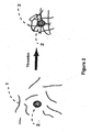
- the technical basis of the disclosed sample preparation method is based on the inventors' observation about the possibility of efficiently separating target micro-organisms, like bacterial or fungi particles, typically from blood samples by converting, in a controlled and standardized way, fibrinogen to fibrin through controlled coagulating the blood sample using the thrombin enzyme to trap said target particles within a fibrin network that will rapidly retract to form a small pellet within the blood container.
- the surrounding blood sample can be decanted leading to separation of the targets trapped within this small pellet.
- the pellet can be lysed to recover the targets from their fibrin trap within a small volume of a controlled buffer.
- the smallest size of the pellet is a key factor that needs to be controlled as it will determine the concentration rate of the disclosed method.
- blood sample one refers to whole blood, platelet-rich plasma, and platelet-poor plasma or serum.
- Blood according to the invention can obviously also refer to blood substitutes or artificially composed sample constituted from blood components, blood additives or any other components that mimic blood functions.
- Typical example of such blood components and that are usually used in blood transfusions include, platelet concentrates, red cell (hemoglobin) concentrates, serum or plasma substitutes (also known as volume expanders).
- clotting factors mainly fibrinogen
- this deficiency can be compensated by adding clotting factors including fibrinogens to the said blood sample as a mandatory component to be able to separate target particles or molecules according to the invention.
- the current invention preferably discloses a method of separating of microorganisms or infectious molecules or particles from a sample of blood
- the sample of blood herein can also refer to a composed sample that includes blood constituents entering into the controlled coagulation process as described previously.
- the invention discloses therefore a method of separating and concentrating target particles or molecules from samples containing fibrinogen proteins by trapping, in a first step, the said target molecules or particles in a fibrin network by converting at least partially the fibrinogen contained in the said sample into fibrin to form the fibrin network. In a second step, the so formed fibrin network to form a fibrin clot that will be separated from the surrounding sample medium.
- the separation according to the invention is obtained by size selection by trapping the said target particle.
- the size of the fibrin network pore is therefore particularly critical.
- the smaller pore size will indeed lead to a more efficient trapping of small infectious microorganisms like E.coli (2 ⁇ m) or Chlamydia (0.3 ⁇ m) or even viruses.
- the control of the trapping fibrin network can be realized by adjusting parameters like sample pH and ionic strength and the concentrations of calcium, fibrinogen, thrombin within the said sample.
- the separation according to the invention is obtained by affinity trapping the said target particle in the fibrin network.
- the inventors' observation is that bacterial particles like Staphylococcus aureus have a strong affinity to fibrinogen/fibrin molecules, which further facilitate (enhance) their separation and concentration according to the invention method.
- the invention discloses to use native as well as induced affinity interactions to separate targets from fibrinogen contained samples.
- the induced affinity separation is realized with fibrinogen recombinant protein(s) composed of fibrinogen fusion protein(s) comprising a capturing moiety domain directed against the said target molecules or particles.
- fibrinogen recombinant protein(s) composed of fibrinogen fusion protein(s) comprising a capturing moiety domain directed against the said target molecules or particles.
- chemical trapping is assured by a fibrin/fibrinogen-binding moiety like a Staphylococcus aureus fibrinogen binding protein and a substance-capturing moiety like an antibody directed against the said target molecules or particles.
- target substances are cells, cell components, cell subpopulations (both eukaryotic and prokaryotic), bacteria, viruses, parasites, antigens, specific antibodies, toxins, proteins, nucleic acid sequences and the like.
- Fibrinogen as referred herein can be therefore a natural fibrinogen obtained from any blood source as for instance human blood or vertebrate blood in general.
- Fibrinogen according to the invention can be also a synthetic composed molecule obtained by combining natural fibrinogen with any other molecule in a way to obtain a new molecule with new affinity functionality for instance.
- the so-combined molecule is obtained by a covalent bonding of a fibrinogen molecule to another molecule.
- the so-combined molecule is a fusion protein produced by the state-of-art recombinant protein synthesis techniques.
- Fibrinogen as referred herein can be also to a synthesized fibrinogen molecule modified that will have the entire fibrinogen crystal structure.
- the synthesized fibrinogen molecule is a modified molecule that will have a different structure, size, composition and affinity activities. More particularly, it is desirable that the fibrinogen according to the invention is a simplified structure molecule (instead of the complex large natural fibrinogen molecule) that still expresses the cleavage (polymerization) activity by thrombin and that can have a defined affinity binding reaction to target particle(s) or molecule(s).
- the invention discloses therefore the use of fibrinogen or fibrinogen modified proteins as a vehicle to trap or capture target particle(s) or molecule(s).
- thrombin cleaves the fibrinogen molecules and their transformation to fibrin.
- the fibrin particles will thereafter self-polymerize to form a small clot in which the targets are trapped resulting therefore to the separation of the target out of the sample liquid volume.
- the proposed method presents a large advantage when compared with the state-of-art techniques as magnetic particles for instance or any other "solid surface” based technologies. As it occurs at the molecular level, the reaction between the targets and the fibrinogen vehicle is very fast and efficient and the non-specific binding issues inherent to surface based assay will be eliminated.
- the present invention provides a method that allows to provide a solution of effectively separating and concentrating of intact microorganisms from an infected blood sample.
- An attainable aim of this is the separation of minute amounts of microorganisms from large volumes of blood allowing thereafter their concentration in a small volume of buffer compatible with further processing steps.
- Another attainable aim of this invention is the separation of intact microorganisms from a blood sample that can be subsequently detected and analyzed by specific techniques recognized in the art. As achieved, instead, this method opens many possibilities in rapid and effective detection and diagnosis of bloodstream infections using fast culture methods as well as rapid and more sensitive molecular based assays.
- fibrinogen according to the invention can be native to the sample (i.e. whole blood samples) or artificially added to the said sample.
- the present invention provides a method for separating target molecule(s) or particle(s) from a composed sample, which comprises the steps:
- a composed sample according to the invention may include, blood, blood derivatives or blood components samples, but also can refer to any fibrinogen free sample as for instance but not limited to, clinical (like urine, sputum and swab), food and environmental samples.
- a sample collection device for separating target molecules or particles from a sample, comprising: (i) an identification code; (ii) a container for containing the said sample; and (iii) a fibrinogen-containing sample in the container, the device being operable to form a fibrin clot that traps in a separable manner the said target molecules or particles upon the exposure of the said sample to thrombin or a thrombin-like enzyme within the said device.
- the device can be a standard reaction tube or reservoir designed to receive a fluid sample that need to be thereafter examined for the existence of target particle(s) or molecule(s) as for instance for pathogenic particle(s) (bacteria, viruses etc.) or target molecule(s) (DNA, RNA or protein etc.).
- the device will further include stable reagent formulations that will lead to the fibrin clot formation and targets separation as previously described herein.
- the device includes a reaction area containing its stored stable reagent formulations that include clotting factors as fibrinogen molecules and coagulation promoting agents as thrombin enzymes.
- Such device will allow the quantitative isolation and detection of targets like infection agents, toxin, nucleic acids and proteins in a test kit, at extremely low copy numbers from any complex biological sample.
- targets like infection agents, toxin, nucleic acids and proteins in a test kit
- the fact that the disclosed device will allow to collect the sample and at the same time to effectively separate and concentrate targets particles or molecules out of the said sample will considerably simplify the necessary sample processing steps and further result in a reduction of potential risks of infection and risks of contamination.
- a main aspect of the invention concerns a method for separating target molecules or particles from a fibrinogen containing sample, attained according to independent claim 1.
- a method for separating target molecules or particles from a blood sample comprises:
- blood sample one refers to whole blood, platelet-rich plasma, and platelet-poor plasma.
- Blood sample can also refer to serum where in this condition fibrinogen must be added to the said sample to allow the separation mechanism working according to the invention.
- Blood according to the invention can obviously also refer to blood substitutes or artificially composed samples constituted from blood components, blood additives or any other components that mimic blood functions.
- Typical example of such blood components and that are usually used in blood transfusions include, platelet concentrates, red cells (hemoglobin) concentrates, serum or plasma substitutes (also known as volume expanders).
- clotting factors mainly fibrinogens
- this deficiency can be compensated by adding clotting factors including fibrinogens to the said blood sample as a mandatory component to be able to separate target particles or molecules according to the invention.
- Blood sample can therefore refer also to an artificially composed blood sample obtained by combining a blood sample with a fibrinogen deficient sample.
- fibrinogen deficient sample can include samples from any sources as for instance biological, clinical, food and environmental samples.
- the term blood sample according to the invention encompasses an artificially composed blood sample by combining clotting factors that at least includes fibrinogens with a fibrinogen deficient sample.
- Fibrin network as generally used herein means the product of a process in which fibrinogen is cleaved upon the exposure to thrombin enzyme and converted into fibrin. Once the fibrinogen is converted into fibrin, a self-polymerization step occurs in which the fibrin monomers come together and form a non-covalently crosslinked three-dimensional polymer network. Further, in the presence of coagulation factor XIII the fibrin network will be cross-linked by factor XIIIa, a transglutaminase activated by thrombin of the factor XIII. Other transglutaminases exist and may also be involved in covalent cross-linking and grafting to the fibrin network.
- slot formation and retraction means the observed pull-in of the fibrin matrix to form a clot after a certain time.
- the size of this clot can be, under certain condition, reduced over time (i.e. clot retraction) by pulling water out of the clot.
- clot retraction is induced by release of multiple coagulation factors from activated platelets trapped in the fibrin mesh of the clot.
- the concentrations of the fibrinogen solution and/or the thrombin solutions have a significant effect on the density of the formed network, clot formation, cross-linking and speed of the final fibrin matrix.
- the reduction of the amount of thrombin and fibrinogen slows down the cross-linking process and contributes to form a fibrin clot with a less dense network.
- controlling the ratio of the amounts of thrombin and fibrinogen leads to a controlled formation of the fibrin network density and size of the final clot.
- the ratio of the amount of thrombin to fibrinogen provides fibrin matrices with a less dense network which is more suitable for target capturing.
- the ratio of the amount of thrombin to fibrinogen provides a retracted fibrin clot with smaller size allowing to attain a high concentration rate of the separated target particles or molecules.
- the mechanism underlying the invention is that converting fibrinogen into fibrin leads to the formation of a fibrin network that will play the role of a network that will capture the target particles or molecules. To obtain the desired effect, the control of the said fibrin network formation is particularly important. With this respect, the concentration of thrombin and fibrinogens as the key coagulation factors are critical in the formation of the fibrin trapping network and consequently the clot creation and separation according to the invention. In fact, a high concentration of thrombin and fibrinogens leads to a very dense fibrin network and a large clot size which is not desirable.
- the concentration of the said fibrinogen in the sample is preferably at least 0.1 ⁇ g/ml.
- the concentration of the fibrinogen in the sample is between 10 mg/ml to 10 ⁇ g/ml.
- Using higher concentration even exceeding the ranges mentioned herein, can also be used but resulting in a higher fibrin matrix density and retracted clot size.
- the downside of using higher concentrations of fibrinogen is the formation of a larger clot size, resulting in a lower concentration rate of the target particle(s). Therefore, in practice and in case of a size selection or size trapping, the concentration of fibrinogen must be optimized in a way to reach maximum capture efficiency and at the same time a lower clot size.
- fibrinogen can be native to the sample (i.e. blood samples) or artificially added to the said sample.
- fibrinogen will be added to a sample even if the said sample has native fibrinogen, as it is the case of whole blood for instance. This will be for instance advantageous to compensate any fibrinogen deficiency or variation in the concentration of the native fibrinogen that may occur in such samples.
- the fibrinogen added to a blood sample (even if the native fibrinogen concentration is at the desired concentration) will be originated from a blood source of a different species than of the sample under consideration.
- fibrinogen/thrombin reaction maybe species specific
- one desired effect by using different fibrinogen source as an additive to a sample with native fibrinogen is to specifically use the added fibrinogen (preferably activated with the related species thrombin) to accomplish the target separation while avoiding (minimizing) the interference of the native fibrinogen in the separation process.
- the added fibrinogen will be a recombinant fibrinogen protein specifically designed to be not cleavable by the endogenous thrombin of the blood sample under use.
- the desired effect of effectively trapping and separating the target particles or molecules from a sample containing fibrinogen can be accordingly achieved by subjecting the said sample to thrombin or thrombin-like enzymes.
- the said thrombin can be an exogenous (artificially added to the said sample) or an endogenous (already part of the said sample) thrombin or thrombin-like enzymes.
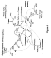
- the thrombin can be in active form or generated through activation of coagulation factors like the factor X as shown in Figure 1 .
- the origin of this thrombin and/or coagulation factors can be from human, animal or insect sources.
- thrombin-like enzymes one refers to the family of serine proteases obtained from outside blood sources and that have the ability to convert fibrinogen to fibrin. Such enzymes are well known in the art and usually obtained from snake venom or produced in recombinant form.
- the thrombin concentration is 0.01 to 10 I.U/ml and preferably within the range of 0.1 to 2 I.U/ml of sample.
- the quantity of the thrombin or thrombin like enzyme must be rather adjusted in correspondence to the fibrinogen concentration within the device to obtain the desired fibrin network structure and clot size.
- the thrombin amount is preferably less than 20I.U thrombin per mg of fibrinogen, preferably in a range between 0.01 to 10I.U thrombin per mg of fibrinogen, more preferably between 0.1 to 1I.U thrombin per mg of fibrinogen.
- the concentration of calcium can also be adjusted. In practice this can be achieved by adding a calcium ion source to the testing sample.
- the calcium ion source is preferably Calcium Chloride (CaCl 2 ), preferably in a concentration range between 1 to 10 mg per ml of sample volume, even more preferably between 4 to 7 mg per ml of sample volume, most preferably between 5 to 6 mg per ml of sample volume.
- CaCl 2 Calcium Chloride
- the adjustment of the calcium concentration can be achieved by further adding calcium chelating agents selected from the groups of GDTA, EDTA and citrate.
- the method may involve the step of adding clotting factor XIII to the said sample.
- Clotting factor XIII is an enzyme capable of catalyzing the fibrin matrix cross-linking formation after it has been activated by thrombin. This will further help to stabilize the fibrin network structure, accelerate the clot retraction and contribute to titer the fibrin porosity.
- Such factor XIII in its inactive or active (XIIIa) formats may be added or adjusted along with the fibrinogen additive in a concentration range between 0.5 to 100I.U per ml of sample volume, more preferably between 1 to 60I.U per ml of sample volume, and most preferably between 1 to 10 I.U per ml of sample volume.
- the major attainable objective of the method according to the invention is to effectively concentrate the target particle(s) or molecule(s) out of the testing sample.
- the concentration factor or rate is practically determined by the clot size. Therefore, in a preferred aspect, the size of the formed clot is at least 1/3 of the initial sample size and in a preferred embodiment the clot size is less than 1/10 of the initial sample volume.
- the clot retracts to further form a small pellet with a size that my reach values that are between 1/50 to 1/1000 of the initial sample volume.
- the concentrations of fibrinogen and thrombin are the predominant factors.
- Other parameters likes the calcium concentration and additives like clotting factor XIII can affect the clot size.
- the clot can be further retracted in the presence of activated platelet cells or activated platelet cell lysates within the said sample.
- the activation of the platelet can be achieved with platelet agonists selected from the group of adenosine diphosphate (ADP) and collagen.
- ADP adenosine diphosphate
- the present invention discloses a method to separate target molecule(s) or particle(s) from a fibrinogen containing sample which includes the step of subjecting the said sample to activated platelet cells or platelet cell lysate.
- the said activated platelet cells or platelet cell lysate can be natively present in the said sample or artificially added to the said sample.
- the platelet activation is preferably achieved by ADP at a concentration of 1 mM to 1 ⁇ M and preferably between 100 ⁇ M to 10 ⁇ M.
- magnetic particles trapped in the fibrin network can be used as a retraction means to compress and therefore reduce the clot size.
- magnetic particles will be used to emulate the role naturally played by the platelet in retracting fibrin clot. This retraction can be achieved by subjecting magnetic particles trapped within a fibrin clot to an external magnetic force. Accordingly, the said magnetic particles are trapped within the fibrin clot due to their larger size one compared with the fibrin network porosity.
- the magnetic particles are trapped within the fibrin clot by affinity interaction that the said particles may have to fibrinogen/fibrin. This can be achieved using magnetic particles coated with a fibrinogen/fibrin binding moiety that can be selected from groups of thrombin, clotting factor XIII, bacterial fibrinogen binding proteins and tissue plasminogen activator (t-PA).
- the invention further discloses the use of affinity trapping to capture the said targets within the fibrin network.
- affinity trapping to capture the said targets within the fibrin network.
- the advantages of using affinity trapping are double: (1) capture small targets that are difficult or cannot be captured by size trapping within the fibrin network; (2) allow high level of concentration (i.e. very small clot or rather a pellet) of the targets as with the affinity trapping fewer fibrinogen concentration is requested to achieve efficient capture yield.
- affinity trapping one can expect to reach a concentration rate that is lower than 1/50 of the initial sample volume and preferably between 1/100 to 1/1000 of the initial sample volume. With large sample volume (3 - 10 ml for instance), the concentration rate can be even less than 1/1000 of the initial sample volume.
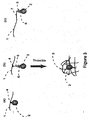
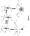
- the affinity trapping can be achieved by the native affinity of the targets (2) having a binding moiety (4) to fibrinogen/fibrin (1).
- a typical example of such affinity trapping is the capturing of the staphylococcus aureus that is known to have an effective affinity to fibrinogen/fibrin through its surface fibrinogen binding protein clumping factor A (ClfA).
- ClfA fibrinogen binding protein clumping factor A
- Staphylococci, Streptococci and Enterococci carry - out proteins called adhesins that can mediate infection by binding to proteins including fibrinogen. In case of blood.
- Another advantage of the method according to invention is the use of native affinity of blood cells to further precipitate leukocytes and thrombocytes cells within the small fibrin pellet while substantially keeping the erythrocytes in suspension. This is particularly important because micro-organisms are not always free-floating in the blood sample but are rather associated or sequestrated in the leukocytes and thrombocytes. In case of Staphylococcus aureus for instance, the interaction and thereafter the sequestration and bacterial survival in platelets contributes to the virulence as it allows bacteria to escape the host defenses.
- Native affinity capture can be also be used to capture small molecules like nucleic acids that strongly bound to fibrin due to electrical charge interaction. As for bacterial particles, the native affinity can be extended to small protein molecules as soon as such molecules have a direct interaction affinity to fibrinogen/fibrin.
- the native affinity separation process can be adapted by using fibrinogens from different species. For instance, using sheep fibrinogens instead of human fibrinogen will lower the capture rate of Staphylococcus aureus. This is due to the fact that sheep fibrinogen shows a low binding affinity to Staphylococcus aureus bacterium.
- the fibrinogen under use within the sample can be a recombinant or a modified fibrinogen designed to enhance or to inhibit the native affinity capture of fibrinogens to a defined targets or target groups.
- FIG. 3(b) A second embodiment of affinity capturing is illustrated in Figure 3(b) .
- the affinity is realized using a substance-capturing (5) directed against the,said target(s) (2) and that have in turn a fibrin/fibrinogen (1) binding moiety (4).
- the use of a substance-capturing as an intermediate means to tag the targets is preferred in case where the target does not have a native affinity to fibrinogen/fibrin.
- a typical example of that is the capture of gram - negative species that in most cases lack native affinity to fibrinogen/fibrin. This can be realized, for instance, by using a gram - negative specific antibody having a fibrinogen binding moiety.
- the indirect affinity capture can be virtually extended to any target particle(s) or molecule(s) that can include but is not limited to target cells, cell components, cell subpopulations (both eukaryotic and prokaryotic), bacteria, viruses, parasites, antigens, specific antibodies, toxins, proteins, nucleic acid sequences and the like.
- the substance-capturing moiety directed against the said target molecules or particles is selected from the group comprising antibodies, nucleic acids and aptamers designed to specifically recognize the said target molecules or particles.
- the said substance-capturing moiety can be coupled or combined with a fibrin/fibrinogen-binding moiety selected from the group comprising thrombin, fibronectin, bacterial fibrinogen binding proteins, tissue-type plasminogen activator, integrines and moieties derived from any member of this group.
- a fibrin/fibrinogen-binding moiety selected from the group comprising thrombin, fibronectin, bacterial fibrinogen binding proteins, tissue-type plasminogen activator, integrines and moieties derived from any member of this group.
- the said fibrin/fibrinogen-binding moiety and said substance-capturing moiety are covalently bound.
- FIG. 3(c) A third embodiment of affinity capturing is illustrated in Figure 3(c) .
- the affinity is realized using fibrinogen fusion (1) protein with a capturing moiety domain (7) directed against the target(s) (1).
- the use of a fusion fibrinogen protein presents the advantage of combining the selectivity of affinity capture directly within the fibrinogen molecules.
- the fibrinogen molecule can be tailor made or specifically designed to specifically capture and thereafter separate one group or specific groups of targets.
- the fibrinogen recombinant or modified protein can be designed to avoid native(s) interaction(s) and/or enhance specific interaction to molecules or particles within a specific sample type.
- the fibrinogen fusion protein further includes a degradation site.
- the degradation site is an enzymatic or hydrolytic degradation site.
- the degradation site is an enzymatic degradation site, which is cleaved by an enzyme selected from the group consisting of plasmin and matrix metalloproteinase.
- the affinity capturing is performed through a substance-capturing (5) directed against the said target(s) (2) and that have a specific fibrin (3) binding moiety (4') without any affinity to fibrinogens like using tissue-type plasminogen activator as a fibrin binding moiety.
- the affinity of the moiety (4') to fibrin will be transformed to an active form (4) only after the exposure step of the sample to Thrombin like in the case of using factor XIII as a fibrin binding moiety.
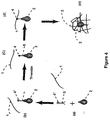
- this invention not only involves a method for separating and concentrating target molecules or particles, but it can also further include the step of detecting the said targets as illustrated in Figure 5 .
- the assay processing includes the step of target capture and separation within a fibrin clot following by the detection of the said targets directly within the clot concentrate. This can be achieved for instance, by exposing the fibrinogen (1) containing sample to a substance-capturing (5) comprising fibrin/fibrinogen binding moiety (4) and a substance - labeling (8) comprising a detection label (9). This leads to the formation of a "target(s)/substance-capturing/substance-labeling".
- the complex "target(s)/substance-capturing/substance-labeling" Upon the exposure to a thrombin the complex "target(s)/substance-capturing/substance-labeling" will be separated within a retracted fibrin clot (3).
- the detection of the target will be performed directly on the clot concentrate by for instance exposing the fibrin clot to a label (8) excitation source (10) resulting in an emission of a detection signal (11).
- the detection methodology will depend on the used label and for that well known detection methodologies as fluorescence, luminescence, SERS (Surface Enhanced Raman Spectroscopy) and Raman spectroscopy can be adopted.
- the fibrin clot or pellet can be suspended in a controlled buffered solution followed by disturbing (i.e. lysis) the clot to recover the separated target(s) from the fibrin clot.
- a controlled buffer is a hypotonic buffer, buffer containing detergents in combination with fibrinolytic like plasmin and/or proteolytic agents like Proteinase K, Pronase and metalloproteinase.
- Such lysis step can be improved by further adding clot lysis enhancers like plasminogen or plasminogen activator.
- the lysis step can further includes the use nucleic acid degradation enzymes.
- a sample collection device for separating target molecules or particles from a sample comprising: (i) an identification code; (ii) a container for containing the said sample that can be in form of a tube that will receive the said sample; and (iii) a fibrinogen-containing sample in the container, the device being operable to form a fibrin clot that traps in a separable manner the said target molecules or particles upon the exposure of the said sample to thrombin or a thrombin-like enzyme within the said device.
- the volume of the said sample container is between 0.1 to 100 ml and preferably between 0.1 to 10 ml.
- the concentration of the said fibrinogen in the sample is preferably at least 0.1 ⁇ g/ml. In a preferred embodiment the concentration of the fibrinogen in the sample is between 0.1 to 100 mg/ml and most preferably between 10 mg/ml to 10 ⁇ g/ml.
- the said device further includes as an additive a thrombin or thrombin enzyme.
- the thrombin concentration is 0.01 to 10 I.U/ml and preferably within the range of 0.1 to 2 I.U/ml of sample.
- the quantity of the thrombin or thrombin like enzyme must be rather adjusted in correspondence to the fibrinogen concentration within the device to obtain the desired fibrin network structure and clot size.
- the thrombin amount is preferably less than 20I.U thrombin per mg of fibrinogen, preferably in a range between 0.01 to 10I.U thrombin per mg of fibrinogen, more preferably between 0.1 to 1I.U thrombin per mg of fibrinogen.
- sample collection device further includes coagulation agents that promote the generation of endogenous thrombin within the sample.
- coagulation agents can be for instance selected from groups comprising powderous or fibrous silicate compounds such as kaolin, Celite, diatomaceous silica and glass fibers, fine powders of calcium compounds such as calcium carbonate and calcium sulfate, thrombin-like substances derived from snake venoms, and polyphenols that can activate blood clotting factors to promote the coagulation.
- these coagulation promoting agents can be, for example, added individually or in combination into .the sample or coated inside the wall of sample container. The amount and the process of which, the said endogenous thrombin promoting agents must be adjusted in a way to control the coagulation process and obtain a small fibrin clot size.
- the device may include additives selected from the group of calcium, chelating agents, activated platelet cells or activated platelet cell lysate and factor XIII.
- the sample collection device may further include as an additive magnetic particles.
- the said magnetic particle within the device are coated with a fibrinogen/fibrin binding moiety selected from the group comprising thrombin, fibronectin, bacterial fibrinogen binding proteins, tissue-type plasminogen activator, integrines and moieties derived from any member of this group.
- the said fibrin/fibrinogen-binding moiety and said magnetic particles are covalently bound.
- the device may include additives comprising molecules having: (I) fibrin/fibrinogen-binding moiety and (II) a substance-capturing moiety directed against the said target molecules or particles.
- the said substance-capturing moiety directed against the said target molecules or particles can be selected from the group comprising antibodies, nucleic acids and aptamers designed to specifically recognize the said target molecules or particles.
- the said substance-capturing moiety can be coupled or combined with a fibrin/fibrinogen-binding moiety selected from the group comprising thrombin, fibronectin, bacterial fibrinogen binding proteins, tissue-type plasminogen activator, integrines and moieties derived from any member of this group.
- the said fibrin/fibrinogen-binding moiety and said substance-capturing moiety may be covalently bound.
- the device can include additives comprising a fibrinogen recombinant or modified protein.
- a fibrinogen recombinant or modified fibrinogen protein can be specifically designed to enhance or inhibit affinity interactions of the said recombinant fibrinogen protein with specific target molecules or particles contained in the sample under use within the device.
- the said recombinant protein in use within the device may be a fibrinogen fusion protein with a capturing moiety domain directed against the said target molecules or particles.
- the fibrinogen fusion protein further includes a degradation site. This will be particular useful for recovering the bound target molecules or particles out of the fibrin network during a lysis step as it will be described later on.
- the degradation site is an enzymatic or hydrolytic degradation site.
- the degradation site is an enzymatic degradation site, which is cleaved by an enzyme selected from the group consisting of plasmin and matrix metalloproteinase.
- the additives can be integrated solubilised in an aqueous buffer solution.
- the said buffer comprises water, calcium chloride, preferably at a concentration of 40 mM, and sodium chloride, preferably at a concentration of 75 mM, and has preferably a pH of 7.3.
- the said additives can be included within the device in a lyophilized format that can be solubilised just prior to the device use or upon the introduction of the sample within the device.
- the so disclosed device for sample collection will in operation lead to the formation of a small fibrin clot in which target particles or molecules are trapped.
- the concentration factor or rate is practically determined by the clot size. Therefore, the device composition arid design so that it will result to the formation of a clot with a size that is at least 1/3 of the initial sample size and preferably the clot size is at least 1/10 of the initial sample volume.
- the clot retracts to further form a small pellet with a size that may reach values that are between 1/50 to 1/1000 of the initial sample volume.
- the sample collection device can be used to separate and concentrate target molecules or particles that can be selected from groups comprising target cells, cell components, cell subpopulations (both eukaryotic and prokaryotic), bacteria, viruses, parasites, antigens, specific antibodies, toxins, proteins, nucleic acid sequences and the like.
- the sample collection device can be used to separate and concentrate target molecules or particles from diverse samples as already defined. In general this includes whole blood, blood derivatives, blood components, composed samples with clotting factors additives. With this respect, the sample herein can refer to any sample type that need to be tested including food, clinical, environmental, and experimental samples.
- the identification code within the device can be for instance a code bar, color, size and shape of the device. Such identification code can be used as a reference or indicator the device intended use and application.
- the devices can be, in fact, differentiated according to their composition, sample type for which the device will be used and or the target(s) that need to be separated.
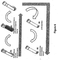
- FIG. 6 shows an example of a sample processing using a device as disclosed.
- the device can be a standard reaction tube with a closing cap and an identification code.
- the device is designed to receive a fluid sample that needs to be thereafter examined for the existence of target particle(s) or molecule(s) as for instance a pathogenic particle(s) (bacteria, viruses etc.) or target molecule(s) (DNA, RNA or protein etc.).
- the device will further include stable reagent formulations that will lead to the fibrin clot formation and targets separation. Upon the sample collection within the device, the fibrinogen molecules will first react with the targets inside the tube.
- the fibrinogen will be transformed to a fibrin, leading to a polymerization and trapping of said targets in the fibrin network.
- the fibrin network in a third step will retract to form a small pellet within the blood container.
- the surrounding sample will be decanted leading to separation of the targets trapped within this small pellet.
- the pellet can be lysed to recover the targets from their fibrin trap within a small volume of a controlled buffer. While the target trapping/pellet formation step will be performed in a closed tube during the sample transportation for instance, the pellet separation and lysis can be easily performed using a state-of-art liquid handling automated system. With this process, the disclosed device will allow to collect the sample and at the same time to effectively separate and concentrate targets particles or molecules out of the said sample, considerably simplifying the necessary sample processing steps and further result in a reduction of potential risks of infection and risks of contamination.
- Example 1 Capture of Staphylococcus aureus (SA) bacterium from a blood component sample: Platelet Rich Plasma (PRP) sample. A sample of 500 ⁇ l of citrated PRP is spiked with 100 CFU of a SA bacterium. By adding 5 ⁇ l of Thrombin at a concentration of 10 U.I./ml and incubation, a small pallet (10 - 20 ⁇ l size) will be formed.
- SA Staphylococcus aureus
- PRP Platelet Rich Plasma
- Example 2 Capture of Staphylococcus aureus (SA) bacterium from a blood component sample: Platelet Poor Plasma (PPP) sample.
- a sample of 500 ⁇ l of citrated PPP is spiked with 100 CFU of a SA bacterium and processed using the same protocol as for Example 1. The same recovery performance will be obtained.
- the size of the fibrin clot is larger when compared with the PRP case. This difference is due to the low retraction of the Fibrin clot in case of the PPP sample. This retraction is instead assured by the platelets cells present within the PRP sample.
- Example 3 Capture of Staphylococcus aureus (SA) bacterium from a blood component sample: Serum sample.
- a sample of 500 ⁇ l of citrated serum is spiked with 100 CFU of a SA bacterium and processed using the same protocol as for Example 1. In this case no clot/pellet will be formed due to lack on fibrinogen within the serum sample.
- By adding 1.25 mg of human Fibrinogen to the serum sample one will be able to form the fibrin clot and thereby separate the SA bacterium out of the serum sample.
- Example 4 Capture of gram-positive bacterium and Fungi out of whole blood. Recovery from 4 ml whole blood spiked with 100 CFU of micro-organisms: Micro-organisms strain/species Yield S.pyogenes M57 85% C .albicans 92% MRSE 83% E.faecalis 70%
- Example 5 Specific capture of bacterium out of a composed sample. 1 ml of a composed PBS samples are spiked with Staphylococcus aureus (SA) (1000 CFU/ml) or Citrobacter Freundi (18000 CFU/ml) at different concentrations of fibrinogen within the sample:
- SA Staphylococcus aureus
- Citrobacter Freundi 18000 CFU/ml
- Example 6 Specific capture of bacterium out a composed sample. The same conditions as for Example 5 using C, Freundi within a 1 ml composed PBS as a sample with the modification that we further added to the sample an antibody directed against gram-negative lipid-A surface protein labeled with a staphylococcal ClfA protein. In this condition and as shown in Figure 3(b) , the antibody will bind C. Freundi allowing its effective binding to fibrinogen and thereafter its efficient separation (nearly 100% yield) within a fibrin pellet.
Landscapes
- Health & Medical Sciences (AREA)
- Life Sciences & Earth Sciences (AREA)
- Chemical & Material Sciences (AREA)
- Engineering & Computer Science (AREA)
- Immunology (AREA)
- Hematology (AREA)
- Molecular Biology (AREA)
- Organic Chemistry (AREA)
- Biomedical Technology (AREA)
- Urology & Nephrology (AREA)
- Microbiology (AREA)
- Biotechnology (AREA)
- General Health & Medical Sciences (AREA)
- Analytical Chemistry (AREA)
- Biochemistry (AREA)
- Physics & Mathematics (AREA)
- Proteomics, Peptides & Aminoacids (AREA)
- Wood Science & Technology (AREA)
- Zoology (AREA)
- Food Science & Technology (AREA)
- Cell Biology (AREA)
- Pathology (AREA)
- General Physics & Mathematics (AREA)
- Medicinal Chemistry (AREA)
- Bioinformatics & Cheminformatics (AREA)
- General Engineering & Computer Science (AREA)
- Biophysics (AREA)
- Genetics & Genomics (AREA)
- Neurosurgery (AREA)
- Virology (AREA)
- Tropical Medicine & Parasitology (AREA)
- Toxicology (AREA)
- Measuring Or Testing Involving Enzymes Or Micro-Organisms (AREA)
- Investigating Or Analysing Biological Materials (AREA)
- Preparation Of Compounds By Using Micro-Organisms (AREA)
- Apparatus Associated With Microorganisms And Enzymes (AREA)
- Sampling And Sample Adjustment (AREA)
Priority Applications (5)
| Application Number | Priority Date | Filing Date | Title |
|---|---|---|---|
| PL16154770T PL3067431T3 (pl) | 2010-09-15 | 2011-09-15 | Urządzenie zawierające składniki krwi do oddzielania z próbek docelowych cząsteczek lub cząstek |
| DK16154770.8T DK3067431T3 (da) | 2010-09-15 | 2011-09-15 | Indretning omfattende blodkomponenter til at adskille målmolekyler eller partikler fra prøver |
| EP16154770.8A EP3067431B1 (en) | 2010-09-15 | 2011-09-15 | Device including blood components for separating target molecules or particles from samples |
| PL11788227T PL2616550T3 (pl) | 2010-09-15 | 2011-09-15 | Sposób oddzielania cząsteczek lub cząstek docelowych z próbki zawierającej fibrynogen, w tym składniki krwi |
| CY20191101074T CY1122166T1 (el) | 2010-09-15 | 2019-10-11 | Διαταξη η οποια περιλαμβανει συστατικα αιματος για διαχωρισμο μοριων η σωματιδιων στοχων απο δειγματα |
Applications Claiming Priority (4)
| Application Number | Priority Date | Filing Date | Title |
|---|---|---|---|
| CH14782010 | 2010-09-15 | ||
| CH20412010 | 2010-12-07 | ||
| CH12072011 | 2011-07-20 | ||
| PCT/IB2011/054035 WO2012035508A2 (en) | 2010-09-15 | 2011-09-15 | Method for separating target molecules or particles from fibrinogen-containing samples including blood components |
Related Child Applications (1)
| Application Number | Title | Priority Date | Filing Date |
|---|---|---|---|
| EP16154770.8A Division EP3067431B1 (en) | 2010-09-15 | 2011-09-15 | Device including blood components for separating target molecules or particles from samples |
Publications (2)
| Publication Number | Publication Date |
|---|---|
| EP2616550A2 EP2616550A2 (en) | 2013-07-24 |
| EP2616550B1 true EP2616550B1 (en) | 2016-02-10 |
Family
ID=45044632
Family Applications (2)
| Application Number | Title | Priority Date | Filing Date |
|---|---|---|---|
| EP11788227.4A Active EP2616550B1 (en) | 2010-09-15 | 2011-09-15 | Method for separating target molecules or particles from fibrinogen-containing samples including blood components |
| EP16154770.8A Active EP3067431B1 (en) | 2010-09-15 | 2011-09-15 | Device including blood components for separating target molecules or particles from samples |
Family Applications After (1)
| Application Number | Title | Priority Date | Filing Date |
|---|---|---|---|
| EP16154770.8A Active EP3067431B1 (en) | 2010-09-15 | 2011-09-15 | Device including blood components for separating target molecules or particles from samples |
Country Status (17)
| Country | Link |
|---|---|
| US (4) | US9689881B2 (enExample) |
| EP (2) | EP2616550B1 (enExample) |
| JP (1) | JP5999776B2 (enExample) |
| KR (1) | KR101948944B1 (enExample) |
| CN (2) | CN107389932B (enExample) |
| AU (2) | AU2011303467C1 (enExample) |
| CA (1) | CA2811493C (enExample) |
| CY (1) | CY1122166T1 (enExample) |
| DK (2) | DK2616550T3 (enExample) |
| ES (2) | ES2751660T3 (enExample) |
| HR (1) | HRP20191785T1 (enExample) |
| HU (2) | HUE028915T2 (enExample) |
| LT (1) | LT3067431T (enExample) |
| PL (2) | PL2616550T3 (enExample) |
| PT (1) | PT3067431T (enExample) |
| SI (1) | SI3067431T1 (enExample) |
| WO (1) | WO2012035508A2 (enExample) |
Families Citing this family (9)
| Publication number | Priority date | Publication date | Assignee | Title |
|---|---|---|---|---|
| US9689881B2 (en) | 2010-09-15 | 2017-06-27 | Spinomix S.A. | Method for separating target molecules or particles from fibrinogen-containing samples including blood components |
| CN103702674B (zh) | 2011-06-27 | 2018-05-29 | 爱默蕾大学 | 血小板裂解物的组合物、用途和制备 |
| EP2979093B1 (en) * | 2013-03-29 | 2019-10-23 | Siemens Healthcare Diagnostics Inc. | Rare cell concentration |
| ES2740838T3 (es) * | 2014-06-01 | 2020-02-06 | Debiopharm Int Sa | Dispositivo de recolección y procesamiento de muestras |
| US12397302B2 (en) | 2015-11-30 | 2025-08-26 | Dh Technologies Development Pte. Ltd. | Electromagnetic assemblies for processing fluids |
| WO2017156375A1 (en) | 2016-03-10 | 2017-09-14 | Arthrex, Inc. | Systems and methods for preparing a thrombin serum |
| WO2017156379A2 (en) | 2016-03-10 | 2017-09-14 | Arthrex, Inc. | Systems and methods for preparing protein enhanced serums |
| KR20210052389A (ko) | 2018-08-27 | 2021-05-10 | 리제너론 파마슈티칼스 인코포레이티드 | 다운스트림 정제에서의 라만 분광법의 사용 |
| JP7298449B2 (ja) * | 2019-11-14 | 2023-06-27 | 株式会社島津製作所 | 血液凝固分析装置、及び分注ノズルの洗浄方法 |
Family Cites Families (29)
| Publication number | Priority date | Publication date | Assignee | Title |
|---|---|---|---|---|
| CA1035260A (en) * | 1974-08-12 | 1978-07-25 | Baxter Travenol Laboratories | Collection and processing tube |
| FR2625321B1 (fr) * | 1987-12-24 | 1992-10-02 | Pasteur Institut | Reactif pour le diagnostic de staphylococcus aureus par agglutination |
| DE58902472D1 (de) * | 1988-01-05 | 1992-11-26 | Linhart Axel | Verfahren und test-kit zur semiquantitativen bestimmung des kalziumgehaltes im blut. |
| DE3803463A1 (de) * | 1988-02-05 | 1989-08-17 | Linhart Axel | Semiquantitativer kalzium-schnelltest |
| US5691160A (en) * | 1992-08-14 | 1997-11-25 | Biogen, Inc. | Effects of actin filaments of fibrin clot structure and lysis |
| EP0587398B1 (en) * | 1992-09-09 | 1998-01-14 | Tokuyama Corporation | Method of assaying fibrinogen, dry reagent therefor, and process for the preparation thereof |
| JP3292760B2 (ja) * | 1993-04-22 | 2002-06-17 | テルモ株式会社 | 採血管およびその製造方法 |
| US5670329A (en) * | 1993-05-28 | 1997-09-23 | Cardiovascular Diagnostics, Inc. | Method and analytical system for performing fibrinogen assays accurately, rapidly and simply using a rotating magnetic field |
| WO1995015397A1 (en) | 1993-12-03 | 1995-06-08 | Dorn Gordon L | Method for improving quantitative recovery of microorganisms fro m specimens containing blood components |
| DE19602673A1 (de) * | 1996-01-25 | 1997-08-07 | Pereg Gmbh | Synthetische Testanschmutzung |
| US6121232A (en) * | 1997-01-31 | 2000-09-19 | Omrix Biopharmaceuticals Sa | Stabilized mixture comprising fibrinogen |
| US6680195B1 (en) * | 1997-11-26 | 2004-01-20 | Inhibitex, Inc. | Extracellular matrix-binding proteins from staphylococcus aureus |
| JP2000262499A (ja) * | 2000-01-01 | 2000-09-26 | Terumo Corp | 採血管 |
| US6686204B2 (en) * | 2001-08-27 | 2004-02-03 | Becton, Dickinson & Company | Collection device |
| WO2003028743A1 (en) * | 2001-10-03 | 2003-04-10 | Woolverton Christopher J | Storage-stable fibrinogen solutions |
| JP4718833B2 (ja) * | 2002-05-01 | 2011-07-06 | シナプス ベスローテン フェンノートシャップ | 複合生物媒体における、一時的なタンパク質分解活性の濃度を決定するための診断試験 |
| EP1400589B1 (de) | 2002-09-18 | 2006-08-09 | SIRS-Lab GmbH | Verfahren zum Nachweis prokaryontischer DNA |
| SI1654387T1 (sl) * | 2003-08-15 | 2009-10-31 | Univ South Florida | Materiali in postopki za ujetje patogenov in odstranitev avrintrikarboksilne kisline iz vzorca |
| US20050065454A1 (en) * | 2003-09-22 | 2005-03-24 | Becton, Dickinson And Company | Non-evacuated blood collection tube |
| US7399609B2 (en) * | 2003-12-30 | 2008-07-15 | 3M Innovative Properties Company | Staphylococcus detection |
| US8419722B2 (en) * | 2004-10-29 | 2013-04-16 | Spinal Restoration, Inc. | Apparatus and method for injection of fibrin sealant in spinal applications |
| DE102005009479A1 (de) | 2005-03-02 | 2006-09-07 | Molzym Gmbh & Co. Kg | Verwendung von Nukleasen zum Abbau von Nukleinsäure in Gegenwart von chaotropen Agenzien und/oder Tensiden |
| US20100009409A1 (en) * | 2005-07-29 | 2010-01-14 | Ecole Polytechnique Federale De Lausanne | Molecular variant fibrinogen fusion proteins |
| US20080299587A1 (en) * | 2007-05-03 | 2008-12-04 | Dennis Durbin | Methods of measuring inhibition of platelet aggregation by thrombin receptor antagonists |
| CA2693565C (en) | 2007-08-02 | 2015-05-26 | Universite Laval | Concentration and enrichment of microbial cells and microbial nucleic acids from bodily fluids |
| JP5029217B2 (ja) * | 2007-08-27 | 2012-09-19 | 東レ株式会社 | シート状物の製造方法 |
| ES2585246T3 (es) * | 2008-07-09 | 2016-10-04 | Profibrix Bv | Fibrinógeno recombinante |
| CA2768485C (en) | 2009-07-16 | 2018-12-04 | Timothy Foster | Treatment of staphylococci infections using recombinant fibrinogen binding protein clumping factor a |
| US9689881B2 (en) | 2010-09-15 | 2017-06-27 | Spinomix S.A. | Method for separating target molecules or particles from fibrinogen-containing samples including blood components |
-
2011
- 2011-09-15 US US13/822,209 patent/US9689881B2/en active Active
- 2011-09-15 CA CA2811493A patent/CA2811493C/en active Active
- 2011-09-15 EP EP11788227.4A patent/EP2616550B1/en active Active
- 2011-09-15 AU AU2011303467A patent/AU2011303467C1/en active Active
- 2011-09-15 DK DK11788227.4T patent/DK2616550T3/en active
- 2011-09-15 CN CN201710338814.XA patent/CN107389932B/zh active Active
- 2011-09-15 JP JP2013528815A patent/JP5999776B2/ja active Active
- 2011-09-15 KR KR1020137009329A patent/KR101948944B1/ko active Active
- 2011-09-15 LT LT16154770T patent/LT3067431T/lt unknown
- 2011-09-15 DK DK16154770.8T patent/DK3067431T3/da active
- 2011-09-15 HU HUE11788227A patent/HUE028915T2/en unknown
- 2011-09-15 CN CN201180044211.XA patent/CN103108959B/zh active Active
- 2011-09-15 ES ES16154770T patent/ES2751660T3/es active Active
- 2011-09-15 PL PL11788227T patent/PL2616550T3/pl unknown
- 2011-09-15 PL PL16154770T patent/PL3067431T3/pl unknown
- 2011-09-15 SI SI201131797T patent/SI3067431T1/sl unknown
- 2011-09-15 EP EP16154770.8A patent/EP3067431B1/en active Active
- 2011-09-15 ES ES11788227T patent/ES2571127T3/es active Active
- 2011-09-15 HU HUE16154770A patent/HUE045771T2/hu unknown
- 2011-09-15 PT PT161547708T patent/PT3067431T/pt unknown
- 2011-09-15 WO PCT/IB2011/054035 patent/WO2012035508A2/en not_active Ceased
-
2016
- 2016-12-02 AU AU2016266066A patent/AU2016266066B2/en active Active
-
2017
- 2017-05-22 US US15/601,677 patent/US20170336424A1/en not_active Abandoned
-
2019
- 2019-10-02 HR HRP20191785TT patent/HRP20191785T1/hr unknown
- 2019-10-11 CY CY20191101074T patent/CY1122166T1/el unknown
-
2021
- 2021-01-28 US US17/160,814 patent/US20210148940A1/en not_active Abandoned
-
2023
- 2023-08-09 US US18/231,880 patent/US20240280593A1/en not_active Abandoned
Also Published As
Similar Documents
| Publication | Publication Date | Title |
|---|---|---|
| US20240280593A1 (en) | Method for separating target molecules or particles from fibrinogen-containing samples including blood components | |
| US20190276868A1 (en) | Methods for Detecting Microorganisms Using Microorganism Detection Protein and Other Applications of Cell Binding Components | |
| US20200316588A1 (en) | Sample collection and processing device | |
| US10281466B2 (en) | Method of detecting an analyte using chromatographic enrichment | |
| US10634672B2 (en) | Method of detecting a microorganism using chromatographic enrichment | |
| HK1247280B (zh) | 用於从含纤维蛋白原的样品中分离目标分子或颗粒的方法 | |
| JP2023523627A (ja) | メチシリン耐性黄色ブドウ球菌を検出するための組成物、方法およびシステム | |
| US20240336950A1 (en) | Use of a Sustainable, Modified and Enhanced Aquaculture Limulus Amebocyte Lysate Protein for Detection and Characterization of Infectious Pathogens in Biologic Samples for Patient Screening, Diagnosis and Therapeutic Management | |
| Choi | Versatile Biological Sample Preparation Platform using Microfluidic Cell Sorting Device |
Legal Events
| Date | Code | Title | Description |
|---|---|---|---|
| PUAI | Public reference made under article 153(3) epc to a published international application that has entered the european phase |
Free format text: ORIGINAL CODE: 0009012 |
|
| 17P | Request for examination filed |
Effective date: 20130412 |
|
| AK | Designated contracting states |
Kind code of ref document: A2 Designated state(s): AL AT BE BG CH CY CZ DE DK EE ES FI FR GB GR HR HU IE IS IT LI LT LU LV MC MK MT NL NO PL PT RO RS SE SI SK SM TR |
|
| DAX | Request for extension of the european patent (deleted) | ||
| GRAP | Despatch of communication of intention to grant a patent |
Free format text: ORIGINAL CODE: EPIDOSNIGR1 |
|
| INTG | Intention to grant announced |
Effective date: 20150521 |
|
| GRAS | Grant fee paid |
Free format text: ORIGINAL CODE: EPIDOSNIGR3 |
|
| GRAP | Despatch of communication of intention to grant a patent |
Free format text: ORIGINAL CODE: EPIDOSNIGR1 |
|
| RAP1 | Party data changed (applicant data changed or rights of an application transferred) |
Owner name: DEBIOPHARM INTERNATIONAL SA |
|
| GRAR | Information related to intention to grant a patent recorded |
Free format text: ORIGINAL CODE: EPIDOSNIGR71 |
|
| INTG | Intention to grant announced |
Effective date: 20151021 |
|
| INTG | Intention to grant announced |
Effective date: 20151116 |
|
| GRAA | (expected) grant |
Free format text: ORIGINAL CODE: 0009210 |
|
| AK | Designated contracting states |
Kind code of ref document: B1 Designated state(s): AL AT BE BG CH CY CZ DE DK EE ES FI FR GB GR HR HU IE IS IT LI LT LU LV MC MK MT NL NO PL PT RO RS SE SI SK SM TR |
|
| REG | Reference to a national code |
Ref country code: GB Ref legal event code: FG4D |
|
| REG | Reference to a national code |
Ref country code: AT Ref legal event code: REF Ref document number: 774682 Country of ref document: AT Kind code of ref document: T Effective date: 20160215 Ref country code: CH Ref legal event code: EP |
|
| REG | Reference to a national code |
Ref country code: IE Ref legal event code: FG4D |
|
| REG | Reference to a national code |
Ref country code: DE Ref legal event code: R096 Ref document number: 602011023273 Country of ref document: DE |
|
| REG | Reference to a national code |
Ref country code: PT Ref legal event code: SC4A Free format text: AVAILABILITY OF NATIONAL TRANSLATION Effective date: 20160505 |
|
| REG | Reference to a national code |
Ref country code: NL Ref legal event code: FP |
|
| REG | Reference to a national code |
Ref country code: DK Ref legal event code: T3 Effective date: 20160517 |
|
| REG | Reference to a national code |
Ref country code: ES Ref legal event code: FG2A Ref document number: 2571127 Country of ref document: ES Kind code of ref document: T3 Effective date: 20160524 |
|
| REG | Reference to a national code |
Ref country code: SE Ref legal event code: TRGR |
|
| REG | Reference to a national code |
Ref country code: LT Ref legal event code: MG4D |
|
| REG | Reference to a national code |
Ref country code: NO Ref legal event code: T2 Effective date: 20160210 |
|
| PG25 | Lapsed in a contracting state [announced via postgrant information from national office to epo] |
Ref country code: HR Free format text: LAPSE BECAUSE OF FAILURE TO SUBMIT A TRANSLATION OF THE DESCRIPTION OR TO PAY THE FEE WITHIN THE PRESCRIBED TIME-LIMIT Effective date: 20160210 |
|
| PG25 | Lapsed in a contracting state [announced via postgrant information from national office to epo] |
Ref country code: RS Free format text: LAPSE BECAUSE OF FAILURE TO SUBMIT A TRANSLATION OF THE DESCRIPTION OR TO PAY THE FEE WITHIN THE PRESCRIBED TIME-LIMIT Effective date: 20160210 Ref country code: LT Free format text: LAPSE BECAUSE OF FAILURE TO SUBMIT A TRANSLATION OF THE DESCRIPTION OR TO PAY THE FEE WITHIN THE PRESCRIBED TIME-LIMIT Effective date: 20160210 Ref country code: LV Free format text: LAPSE BECAUSE OF FAILURE TO SUBMIT A TRANSLATION OF THE DESCRIPTION OR TO PAY THE FEE WITHIN THE PRESCRIBED TIME-LIMIT Effective date: 20160210 |
|
| REG | Reference to a national code |
Ref country code: SK Ref legal event code: T3 Ref document number: E 21042 Country of ref document: SK |
|
| REG | Reference to a national code |
Ref country code: FR Ref legal event code: PLFP Year of fee payment: 6 |
|
| REG | Reference to a national code |
Ref country code: GR Ref legal event code: EP Ref document number: 20160400913 Country of ref document: GR Effective date: 20160628 |
|
| PG25 | Lapsed in a contracting state [announced via postgrant information from national office to epo] |
Ref country code: EE Free format text: LAPSE BECAUSE OF FAILURE TO SUBMIT A TRANSLATION OF THE DESCRIPTION OR TO PAY THE FEE WITHIN THE PRESCRIBED TIME-LIMIT Effective date: 20160210 |
|
| REG | Reference to a national code |
Ref country code: DE Ref legal event code: R097 Ref document number: 602011023273 Country of ref document: DE |
|
| PG25 | Lapsed in a contracting state [announced via postgrant information from national office to epo] |
Ref country code: RO Free format text: LAPSE BECAUSE OF FAILURE TO SUBMIT A TRANSLATION OF THE DESCRIPTION OR TO PAY THE FEE WITHIN THE PRESCRIBED TIME-LIMIT Effective date: 20160210 Ref country code: SM Free format text: LAPSE BECAUSE OF FAILURE TO SUBMIT A TRANSLATION OF THE DESCRIPTION OR TO PAY THE FEE WITHIN THE PRESCRIBED TIME-LIMIT Effective date: 20160210 |
|
| PLBE | No opposition filed within time limit |
Free format text: ORIGINAL CODE: 0009261 |
|
| STAA | Information on the status of an ep patent application or granted ep patent |
Free format text: STATUS: NO OPPOSITION FILED WITHIN TIME LIMIT |
|
| 26N | No opposition filed |
Effective date: 20161111 |
|
| REG | Reference to a national code |
Ref country code: HU Ref legal event code: AG4A Ref document number: E028915 Country of ref document: HU |
|
| PG25 | Lapsed in a contracting state [announced via postgrant information from national office to epo] |
Ref country code: SI Free format text: LAPSE BECAUSE OF FAILURE TO SUBMIT A TRANSLATION OF THE DESCRIPTION OR TO PAY THE FEE WITHIN THE PRESCRIBED TIME-LIMIT Effective date: 20160210 |
|
| PG25 | Lapsed in a contracting state [announced via postgrant information from national office to epo] |
Ref country code: MC Free format text: LAPSE BECAUSE OF FAILURE TO SUBMIT A TRANSLATION OF THE DESCRIPTION OR TO PAY THE FEE WITHIN THE PRESCRIBED TIME-LIMIT Effective date: 20160210 |
|
| REG | Reference to a national code |
Ref country code: FR Ref legal event code: PLFP Year of fee payment: 7 |
|
| PG25 | Lapsed in a contracting state [announced via postgrant information from national office to epo] |
Ref country code: CY Free format text: LAPSE BECAUSE OF FAILURE TO SUBMIT A TRANSLATION OF THE DESCRIPTION OR TO PAY THE FEE WITHIN THE PRESCRIBED TIME-LIMIT Effective date: 20160210 |
|
| PG25 | Lapsed in a contracting state [announced via postgrant information from national office to epo] |
Ref country code: MT Free format text: LAPSE BECAUSE OF NON-PAYMENT OF DUE FEES Effective date: 20160930 Ref country code: MK Free format text: LAPSE BECAUSE OF FAILURE TO SUBMIT A TRANSLATION OF THE DESCRIPTION OR TO PAY THE FEE WITHIN THE PRESCRIBED TIME-LIMIT Effective date: 20160210 |
|
| REG | Reference to a national code |
Ref country code: FR Ref legal event code: PLFP Year of fee payment: 8 |
|
| PG25 | Lapsed in a contracting state [announced via postgrant information from national office to epo] |
Ref country code: AL Free format text: LAPSE BECAUSE OF FAILURE TO SUBMIT A TRANSLATION OF THE DESCRIPTION OR TO PAY THE FEE WITHIN THE PRESCRIBED TIME-LIMIT Effective date: 20160210 |
|
| REG | Reference to a national code |
Ref country code: AT Ref legal event code: UEP Ref document number: 774682 Country of ref document: AT Kind code of ref document: T Effective date: 20160210 |
|
| P01 | Opt-out of the competence of the unified patent court (upc) registered |
Effective date: 20230427 |
|
| PGFP | Annual fee paid to national office [announced via postgrant information from national office to epo] |
Ref country code: FI Payment date: 20240925 Year of fee payment: 14 Ref country code: IE Payment date: 20240927 Year of fee payment: 14 |
|
| PGFP | Annual fee paid to national office [announced via postgrant information from national office to epo] |
Ref country code: GR Payment date: 20240926 Year of fee payment: 14 Ref country code: DK Payment date: 20240925 Year of fee payment: 14 |
|
| PGFP | Annual fee paid to national office [announced via postgrant information from national office to epo] |
Ref country code: BE Payment date: 20240927 Year of fee payment: 14 |
|
| PGFP | Annual fee paid to national office [announced via postgrant information from national office to epo] |
Ref country code: NL Payment date: 20240926 Year of fee payment: 14 |
|
| PGFP | Annual fee paid to national office [announced via postgrant information from national office to epo] |
Ref country code: AT Payment date: 20240821 Year of fee payment: 14 |
|
| PGFP | Annual fee paid to national office [announced via postgrant information from national office to epo] |
Ref country code: NO Payment date: 20240927 Year of fee payment: 14 Ref country code: IT Payment date: 20240919 Year of fee payment: 14 Ref country code: SE Payment date: 20240927 Year of fee payment: 14 |
|
| PGFP | Annual fee paid to national office [announced via postgrant information from national office to epo] |
Ref country code: LU Payment date: 20240927 Year of fee payment: 14 |
|
| PGFP | Annual fee paid to national office [announced via postgrant information from national office to epo] |
Ref country code: ES Payment date: 20241001 Year of fee payment: 14 |
|
| REG | Reference to a national code |
Ref country code: CH Ref legal event code: U11 Free format text: ST27 STATUS EVENT CODE: U-0-0-U10-U11 (AS PROVIDED BY THE NATIONAL OFFICE) Effective date: 20251001 |
|
| PGFP | Annual fee paid to national office [announced via postgrant information from national office to epo] |
Ref country code: PT Payment date: 20250828 Year of fee payment: 15 |
|
| PGFP | Annual fee paid to national office [announced via postgrant information from national office to epo] |
Ref country code: DE Payment date: 20250929 Year of fee payment: 15 |
|
| PGFP | Annual fee paid to national office [announced via postgrant information from national office to epo] |
Ref country code: PL Payment date: 20250818 Year of fee payment: 15 Ref country code: TR Payment date: 20250822 Year of fee payment: 15 |
|
| PGFP | Annual fee paid to national office [announced via postgrant information from national office to epo] |
Ref country code: BG Payment date: 20250917 Year of fee payment: 15 Ref country code: HU Payment date: 20250822 Year of fee payment: 15 Ref country code: GB Payment date: 20250929 Year of fee payment: 15 |
|
| PGFP | Annual fee paid to national office [announced via postgrant information from national office to epo] |
Ref country code: FR Payment date: 20250925 Year of fee payment: 15 |
|
| PGFP | Annual fee paid to national office [announced via postgrant information from national office to epo] |
Ref country code: CZ Payment date: 20250826 Year of fee payment: 15 |
|
| PGFP | Annual fee paid to national office [announced via postgrant information from national office to epo] |
Ref country code: SK Payment date: 20250819 Year of fee payment: 15 |
|
| PGFP | Annual fee paid to national office [announced via postgrant information from national office to epo] |
Ref country code: IS Payment date: 20250910 Year of fee payment: 15 |
|
| PGFP | Annual fee paid to national office [announced via postgrant information from national office to epo] |
Ref country code: CH Payment date: 20251001 Year of fee payment: 15 |