EP2510357B1 - Muteine von humanem lipocalin 2 (lcn2, hngal) mit affinität für ein bestimmtes zielmolekül - Google Patents
Muteine von humanem lipocalin 2 (lcn2, hngal) mit affinität für ein bestimmtes zielmolekül Download PDFInfo
- Publication number
- EP2510357B1 EP2510357B1 EP10784824.4A EP10784824A EP2510357B1 EP 2510357 B1 EP2510357 B1 EP 2510357B1 EP 10784824 A EP10784824 A EP 10784824A EP 2510357 B1 EP2510357 B1 EP 2510357B1
- Authority
- EP
- European Patent Office
- Prior art keywords
- mutein
- sequence
- lcn2
- seq
- muteins
- Prior art date
- Legal status (The legal status is an assumption and is not a legal conclusion. Google has not performed a legal analysis and makes no representation as to the accuracy of the status listed.)
- Active
Links
- 101001023833 Homo sapiens Neutrophil gelatinase-associated lipocalin Proteins 0.000 title claims description 91
- 102000047202 human LCN2 Human genes 0.000 title claims description 91
- 101150055061 LCN2 gene Proteins 0.000 claims description 175
- 108090000623 proteins and genes Proteins 0.000 claims description 110
- 108090000765 processed proteins & peptides Proteins 0.000 claims description 97
- 102000004169 proteins and genes Human genes 0.000 claims description 90
- 238000000034 method Methods 0.000 claims description 74
- 150000007523 nucleic acids Chemical class 0.000 claims description 65
- 108020004707 nucleic acids Proteins 0.000 claims description 60
- 102000039446 nucleic acids Human genes 0.000 claims description 60
- 102000004196 processed proteins & peptides Human genes 0.000 claims description 60
- 229920001184 polypeptide Polymers 0.000 claims description 47
- 150000001875 compounds Chemical class 0.000 claims description 39
- 238000002703 mutagenesis Methods 0.000 claims description 39
- 231100000350 mutagenesis Toxicity 0.000 claims description 38
- 239000002773 nucleotide Substances 0.000 claims description 34
- 125000003729 nucleotide group Chemical group 0.000 claims description 34
- 241000282414 Homo sapiens Species 0.000 claims description 32
- 230000014509 gene expression Effects 0.000 claims description 30
- 108010067306 Fibronectins Proteins 0.000 claims description 29
- 102000016359 Fibronectins Human genes 0.000 claims description 29
- 125000000539 amino acid group Chemical group 0.000 claims description 21
- 238000010494 dissociation reaction Methods 0.000 claims description 18
- 230000005593 dissociations Effects 0.000 claims description 18
- 230000004927 fusion Effects 0.000 claims description 16
- 210000002966 serum Anatomy 0.000 claims description 16
- YBJHBAHKTGYVGT-ZKWXMUAHSA-N (+)-Biotin Chemical class N1C(=O)N[C@@H]2[C@H](CCCCC(=O)O)SC[C@@H]21 YBJHBAHKTGYVGT-ZKWXMUAHSA-N 0.000 claims description 14
- 108010090849 Amyloid beta-Peptides Proteins 0.000 claims description 14
- 102000013455 Amyloid beta-Peptides Human genes 0.000 claims description 14
- SHIBSTMRCDJXLN-UHFFFAOYSA-N Digoxigenin Natural products C1CC(C2C(C3(C)CCC(O)CC3CC2)CC2O)(O)C2(C)C1C1=CC(=O)OC1 SHIBSTMRCDJXLN-UHFFFAOYSA-N 0.000 claims description 13
- QONQRTHLHBTMGP-UHFFFAOYSA-N digitoxigenin Natural products CC12CCC(C3(CCC(O)CC3CC3)C)C3C11OC1CC2C1=CC(=O)OC1 QONQRTHLHBTMGP-UHFFFAOYSA-N 0.000 claims description 13
- SHIBSTMRCDJXLN-KCZCNTNESA-N digoxigenin Chemical class C1([C@@H]2[C@@]3([C@@](CC2)(O)[C@H]2[C@@H]([C@@]4(C)CC[C@H](O)C[C@H]4CC2)C[C@H]3O)C)=CC(=O)OC1 SHIBSTMRCDJXLN-KCZCNTNESA-N 0.000 claims description 13
- 239000008194 pharmaceutical composition Substances 0.000 claims description 12
- 102000004190 Enzymes Human genes 0.000 claims description 8
- 108090000790 Enzymes Proteins 0.000 claims description 8
- 239000011616 biotin Chemical class 0.000 claims description 7
- 229960002685 biotin Drugs 0.000 claims description 7
- 235000020958 biotin Nutrition 0.000 claims description 7
- 239000002184 metal Chemical class 0.000 claims description 7
- 229910052751 metal Inorganic materials 0.000 claims description 7
- 238000002955 isolation Methods 0.000 claims description 6
- 239000003053 toxin Chemical class 0.000 claims description 6
- 231100000765 toxin Toxicity 0.000 claims description 6
- 108700012359 toxins Chemical class 0.000 claims description 6
- 239000000824 cytostatic agent Chemical class 0.000 claims description 3
- PCHJSUWPFVWCPO-UHFFFAOYSA-N gold Chemical compound [Au] PCHJSUWPFVWCPO-UHFFFAOYSA-N 0.000 claims description 3
- 230000004962 physiological condition Effects 0.000 claims description 3
- 108020001580 protein domains Proteins 0.000 claims description 2
- 230000002285 radioactive effect Effects 0.000 claims description 2
- 102220515765 cAMP-dependent protein kinase inhibitor alpha_N10D_mutation Human genes 0.000 claims 1
- 150000002739 metals Chemical class 0.000 claims 1
- 235000001014 amino acid Nutrition 0.000 description 84
- 235000018102 proteins Nutrition 0.000 description 82
- 230000027455 binding Effects 0.000 description 79
- 229940024606 amino acid Drugs 0.000 description 74
- 150000001413 amino acids Chemical class 0.000 description 72
- 108050006654 Lipocalin Proteins 0.000 description 65
- 102000019298 Lipocalin Human genes 0.000 description 64
- 238000009739 binding Methods 0.000 description 62
- FAPWRFPIFSIZLT-UHFFFAOYSA-M Sodium chloride Chemical compound [Na+].[Cl-] FAPWRFPIFSIZLT-UHFFFAOYSA-M 0.000 description 60
- 210000004027 cell Anatomy 0.000 description 56
- 108010064397 amyloid beta-protein (1-40) Proteins 0.000 description 55
- 108020004414 DNA Proteins 0.000 description 52
- 239000012634 fragment Substances 0.000 description 42
- 108020001507 fusion proteins Proteins 0.000 description 42
- 102000037865 fusion proteins Human genes 0.000 description 42
- 239000003446 ligand Substances 0.000 description 39
- 238000002965 ELISA Methods 0.000 description 38
- 241000588724 Escherichia coli Species 0.000 description 36
- 102000009027 Albumins Human genes 0.000 description 34
- 108010088751 Albumins Proteins 0.000 description 34
- 239000011780 sodium chloride Substances 0.000 description 30
- 125000003275 alpha amino acid group Chemical group 0.000 description 29
- 108010028230 Trp-Ser- His-Pro-Gln-Phe-Glu-Lys Proteins 0.000 description 28
- DHMQDGOQFOQNFH-UHFFFAOYSA-N Glycine Chemical compound NCC(O)=O DHMQDGOQFOQNFH-UHFFFAOYSA-N 0.000 description 27
- 239000000203 mixture Substances 0.000 description 25
- 239000011324 bead Substances 0.000 description 24
- 230000002776 aggregation Effects 0.000 description 23
- 238000004220 aggregation Methods 0.000 description 23
- 230000035772 mutation Effects 0.000 description 23
- ROHFNLRQFUQHCH-YFKPBYRVSA-N L-leucine Chemical compound CC(C)C[C@H](N)C(O)=O ROHFNLRQFUQHCH-YFKPBYRVSA-N 0.000 description 22
- 239000013598 vector Substances 0.000 description 21
- DCXYFEDJOCDNAF-REOHCLBHSA-N L-asparagine Chemical compound OC(=O)[C@@H](N)CC(N)=O DCXYFEDJOCDNAF-REOHCLBHSA-N 0.000 description 20
- ZMXDDKWLCZADIW-UHFFFAOYSA-N N,N-Dimethylformamide Chemical compound CN(C)C=O ZMXDDKWLCZADIW-UHFFFAOYSA-N 0.000 description 19
- 239000012528 membrane Substances 0.000 description 19
- 108020004705 Codon Proteins 0.000 description 18
- AYFVYJQAPQTCCC-GBXIJSLDSA-N L-threonine Chemical compound C[C@@H](O)[C@H](N)C(O)=O AYFVYJQAPQTCCC-GBXIJSLDSA-N 0.000 description 18
- KDXKERNSBIXSRK-UHFFFAOYSA-N Lysine Natural products NCCCCC(N)C(O)=O KDXKERNSBIXSRK-UHFFFAOYSA-N 0.000 description 18
- HEMHJVSKTPXQMS-UHFFFAOYSA-M Sodium hydroxide Chemical compound [OH-].[Na+] HEMHJVSKTPXQMS-UHFFFAOYSA-M 0.000 description 18
- 230000015572 biosynthetic process Effects 0.000 description 18
- 239000000243 solution Substances 0.000 description 18
- KDXKERNSBIXSRK-YFKPBYRVSA-N L-lysine Chemical compound NCCCC[C@H](N)C(O)=O KDXKERNSBIXSRK-YFKPBYRVSA-N 0.000 description 17
- 238000002823 phage display Methods 0.000 description 17
- 238000005406 washing Methods 0.000 description 17
- CKLJMWTZIZZHCS-REOHCLBHSA-N L-aspartic acid Chemical compound OC(=O)[C@@H](N)CC(O)=O CKLJMWTZIZZHCS-REOHCLBHSA-N 0.000 description 16
- 108091028043 Nucleic acid sequence Proteins 0.000 description 16
- KZSNJWFQEVHDMF-UHFFFAOYSA-N Valine Natural products CC(C)C(N)C(O)=O KZSNJWFQEVHDMF-UHFFFAOYSA-N 0.000 description 16
- 238000011534 incubation Methods 0.000 description 16
- 238000005259 measurement Methods 0.000 description 16
- 238000003752 polymerase chain reaction Methods 0.000 description 16
- 238000002198 surface plasmon resonance spectroscopy Methods 0.000 description 16
- 108060008226 thioredoxin Proteins 0.000 description 16
- XLYOFNOQVPJJNP-UHFFFAOYSA-N water Substances O XLYOFNOQVPJJNP-UHFFFAOYSA-N 0.000 description 16
- MTCFGRXMJLQNBG-REOHCLBHSA-N (2S)-2-Amino-3-hydroxypropansäure Chemical compound OC[C@H](N)C(O)=O MTCFGRXMJLQNBG-REOHCLBHSA-N 0.000 description 15
- 102000002260 Alkaline Phosphatase Human genes 0.000 description 15
- 108020004774 Alkaline Phosphatase Proteins 0.000 description 15
- 206010028980 Neoplasm Diseases 0.000 description 15
- 230000001580 bacterial effect Effects 0.000 description 15
- 238000006243 chemical reaction Methods 0.000 description 15
- 238000001542 size-exclusion chromatography Methods 0.000 description 15
- JADVWWSKYZXRGX-UHFFFAOYSA-M thioflavine T Chemical compound [Cl-].C1=CC(N(C)C)=CC=C1C1=[N+](C)C2=CC=C(C)C=C2S1 JADVWWSKYZXRGX-UHFFFAOYSA-M 0.000 description 15
- 108091034117 Oligonucleotide Proteins 0.000 description 14
- 229920001213 Polysorbate 20 Polymers 0.000 description 14
- 108010076504 Protein Sorting Signals Proteins 0.000 description 14
- 238000003556 assay Methods 0.000 description 14
- 239000000256 polyoxyethylene sorbitan monolaurate Substances 0.000 description 14
- 235000010486 polyoxyethylene sorbitan monolaurate Nutrition 0.000 description 14
- 208000024827 Alzheimer disease Diseases 0.000 description 13
- 102000014914 Carrier Proteins Human genes 0.000 description 13
- ZDXPYRJPNDTMRX-VKHMYHEASA-N L-glutamine Chemical compound OC(=O)[C@@H](N)CCC(N)=O ZDXPYRJPNDTMRX-VKHMYHEASA-N 0.000 description 13
- 150000001412 amines Chemical class 0.000 description 13
- 238000011282 treatment Methods 0.000 description 13
- 101710175625 Maltose/maltodextrin-binding periplasmic protein Proteins 0.000 description 12
- 239000007983 Tris buffer Substances 0.000 description 12
- 238000010790 dilution Methods 0.000 description 12
- 239000012895 dilution Substances 0.000 description 12
- 238000005516 engineering process Methods 0.000 description 12
- 230000005291 magnetic effect Effects 0.000 description 12
- 238000010223 real-time analysis Methods 0.000 description 12
- 238000012216 screening Methods 0.000 description 12
- 238000002415 sodium dodecyl sulfate polyacrylamide gel electrophoresis Methods 0.000 description 12
- 239000006228 supernatant Substances 0.000 description 12
- LENZDBCJOHFCAS-UHFFFAOYSA-N tris Chemical compound OCC(N)(CO)CO LENZDBCJOHFCAS-UHFFFAOYSA-N 0.000 description 12
- 108091033380 Coding strand Proteins 0.000 description 11
- WHUUTDBJXJRKMK-UHFFFAOYSA-N Glutamic acid Natural products OC(=O)C(N)CCC(O)=O WHUUTDBJXJRKMK-UHFFFAOYSA-N 0.000 description 11
- WHUUTDBJXJRKMK-VKHMYHEASA-N L-glutamic acid Chemical compound OC(=O)[C@@H](N)CCC(O)=O WHUUTDBJXJRKMK-VKHMYHEASA-N 0.000 description 11
- COLNVLDHVKWLRT-QMMMGPOBSA-N L-phenylalanine Chemical compound OC(=O)[C@@H](N)CC1=CC=CC=C1 COLNVLDHVKWLRT-QMMMGPOBSA-N 0.000 description 11
- 102100036407 Thioredoxin Human genes 0.000 description 11
- 238000004458 analytical method Methods 0.000 description 11
- 108091008324 binding proteins Proteins 0.000 description 11
- 239000000872 buffer Substances 0.000 description 11
- 238000005119 centrifugation Methods 0.000 description 11
- 238000010828 elution Methods 0.000 description 11
- 239000002609 medium Substances 0.000 description 11
- 238000006467 substitution reaction Methods 0.000 description 11
- XZKIHKMTEMTJQX-UHFFFAOYSA-N 4-Nitrophenyl Phosphate Chemical compound OP(O)(=O)OC1=CC=C([N+]([O-])=O)C=C1 XZKIHKMTEMTJQX-UHFFFAOYSA-N 0.000 description 10
- KCXVZYZYPLLWCC-UHFFFAOYSA-N EDTA Chemical compound OC(=O)CN(CC(O)=O)CCN(CC(O)=O)CC(O)=O KCXVZYZYPLLWCC-UHFFFAOYSA-N 0.000 description 10
- XUJNEKJLAYXESH-REOHCLBHSA-N L-Cysteine Chemical compound SC[C@H](N)C(O)=O XUJNEKJLAYXESH-REOHCLBHSA-N 0.000 description 10
- TWRXJAOTZQYOKJ-UHFFFAOYSA-L Magnesium chloride Chemical compound [Mg+2].[Cl-].[Cl-] TWRXJAOTZQYOKJ-UHFFFAOYSA-L 0.000 description 10
- 238000013459 approach Methods 0.000 description 10
- 238000004519 manufacturing process Methods 0.000 description 10
- 229940046166 oligodeoxynucleotide Drugs 0.000 description 10
- 239000013612 plasmid Substances 0.000 description 10
- 108060003951 Immunoglobulin Proteins 0.000 description 9
- 239000007836 KH2PO4 Substances 0.000 description 9
- OUYCCCASQSFEME-QMMMGPOBSA-N L-tyrosine Chemical compound OC(=O)[C@@H](N)CC1=CC=C(O)C=C1 OUYCCCASQSFEME-QMMMGPOBSA-N 0.000 description 9
- 239000002202 Polyethylene glycol Substances 0.000 description 9
- 108010090804 Streptavidin Proteins 0.000 description 9
- 238000010521 absorption reaction Methods 0.000 description 9
- 210000004899 c-terminal region Anatomy 0.000 description 9
- 238000010168 coupling process Methods 0.000 description 9
- 238000001514 detection method Methods 0.000 description 9
- 239000003814 drug Substances 0.000 description 9
- -1 haptens Chemical compound 0.000 description 9
- 102000018358 immunoglobulin Human genes 0.000 description 9
- 229910000402 monopotassium phosphate Inorganic materials 0.000 description 9
- 239000002245 particle Substances 0.000 description 9
- 229920001223 polyethylene glycol Polymers 0.000 description 9
- GNSKLFRGEWLPPA-UHFFFAOYSA-M potassium dihydrogen phosphate Chemical compound [K+].OP(O)([O-])=O GNSKLFRGEWLPPA-UHFFFAOYSA-M 0.000 description 9
- 238000000746 purification Methods 0.000 description 9
- 230000008685 targeting Effects 0.000 description 9
- 210000001519 tissue Anatomy 0.000 description 9
- HZAXFHJVJLSVMW-UHFFFAOYSA-N 2-Aminoethan-1-ol Chemical compound NCCO HZAXFHJVJLSVMW-UHFFFAOYSA-N 0.000 description 8
- 239000012505 Superdex™ Substances 0.000 description 8
- XSQUKJJJFZCRTK-UHFFFAOYSA-N Urea Chemical compound NC(N)=O XSQUKJJJFZCRTK-UHFFFAOYSA-N 0.000 description 8
- 108010064539 amyloid beta-protein (1-42) Proteins 0.000 description 8
- 230000008827 biological function Effects 0.000 description 8
- 239000003153 chemical reaction reagent Substances 0.000 description 8
- 230000008878 coupling Effects 0.000 description 8
- 238000005859 coupling reaction Methods 0.000 description 8
- 208000037265 diseases, disorders, signs and symptoms Diseases 0.000 description 8
- BNIILDVGGAEEIG-UHFFFAOYSA-L disodium hydrogen phosphate Chemical compound [Na+].[Na+].OP([O-])([O-])=O BNIILDVGGAEEIG-UHFFFAOYSA-L 0.000 description 8
- 229910000397 disodium phosphate Inorganic materials 0.000 description 8
- 238000000338 in vitro Methods 0.000 description 8
- 238000003780 insertion Methods 0.000 description 8
- 230000037431 insertion Effects 0.000 description 8
- 229930101283 tetracycline Natural products 0.000 description 8
- DGVVWUTYPXICAM-UHFFFAOYSA-N β‐Mercaptoethanol Chemical compound OCCS DGVVWUTYPXICAM-UHFFFAOYSA-N 0.000 description 8
- 108010021625 Immunoglobulin Fragments Proteins 0.000 description 7
- 102000008394 Immunoglobulin Fragments Human genes 0.000 description 7
- JLCPHMBAVCMARE-UHFFFAOYSA-N [3-[[3-[[3-[[3-[[3-[[3-[[3-[[3-[[3-[[3-[[3-[[5-(2-amino-6-oxo-1H-purin-9-yl)-3-[[3-[[3-[[3-[[3-[[3-[[5-(2-amino-6-oxo-1H-purin-9-yl)-3-[[5-(2-amino-6-oxo-1H-purin-9-yl)-3-hydroxyoxolan-2-yl]methoxy-hydroxyphosphoryl]oxyoxolan-2-yl]methoxy-hydroxyphosphoryl]oxy-5-(5-methyl-2,4-dioxopyrimidin-1-yl)oxolan-2-yl]methoxy-hydroxyphosphoryl]oxy-5-(6-aminopurin-9-yl)oxolan-2-yl]methoxy-hydroxyphosphoryl]oxy-5-(6-aminopurin-9-yl)oxolan-2-yl]methoxy-hydroxyphosphoryl]oxy-5-(6-aminopurin-9-yl)oxolan-2-yl]methoxy-hydroxyphosphoryl]oxy-5-(6-aminopurin-9-yl)oxolan-2-yl]methoxy-hydroxyphosphoryl]oxyoxolan-2-yl]methoxy-hydroxyphosphoryl]oxy-5-(5-methyl-2,4-dioxopyrimidin-1-yl)oxolan-2-yl]methoxy-hydroxyphosphoryl]oxy-5-(4-amino-2-oxopyrimidin-1-yl)oxolan-2-yl]methoxy-hydroxyphosphoryl]oxy-5-(5-methyl-2,4-dioxopyrimidin-1-yl)oxolan-2-yl]methoxy-hydroxyphosphoryl]oxy-5-(5-methyl-2,4-dioxopyrimidin-1-yl)oxolan-2-yl]methoxy-hydroxyphosphoryl]oxy-5-(6-aminopurin-9-yl)oxolan-2-yl]methoxy-hydroxyphosphoryl]oxy-5-(6-aminopurin-9-yl)oxolan-2-yl]methoxy-hydroxyphosphoryl]oxy-5-(4-amino-2-oxopyrimidin-1-yl)oxolan-2-yl]methoxy-hydroxyphosphoryl]oxy-5-(4-amino-2-oxopyrimidin-1-yl)oxolan-2-yl]methoxy-hydroxyphosphoryl]oxy-5-(4-amino-2-oxopyrimidin-1-yl)oxolan-2-yl]methoxy-hydroxyphosphoryl]oxy-5-(6-aminopurin-9-yl)oxolan-2-yl]methoxy-hydroxyphosphoryl]oxy-5-(4-amino-2-oxopyrimidin-1-yl)oxolan-2-yl]methyl [5-(6-aminopurin-9-yl)-2-(hydroxymethyl)oxolan-3-yl] hydrogen phosphate Polymers Cc1cn(C2CC(OP(O)(=O)OCC3OC(CC3OP(O)(=O)OCC3OC(CC3O)n3cnc4c3nc(N)[nH]c4=O)n3cnc4c3nc(N)[nH]c4=O)C(COP(O)(=O)OC3CC(OC3COP(O)(=O)OC3CC(OC3COP(O)(=O)OC3CC(OC3COP(O)(=O)OC3CC(OC3COP(O)(=O)OC3CC(OC3COP(O)(=O)OC3CC(OC3COP(O)(=O)OC3CC(OC3COP(O)(=O)OC3CC(OC3COP(O)(=O)OC3CC(OC3COP(O)(=O)OC3CC(OC3COP(O)(=O)OC3CC(OC3COP(O)(=O)OC3CC(OC3COP(O)(=O)OC3CC(OC3COP(O)(=O)OC3CC(OC3COP(O)(=O)OC3CC(OC3COP(O)(=O)OC3CC(OC3COP(O)(=O)OC3CC(OC3CO)n3cnc4c(N)ncnc34)n3ccc(N)nc3=O)n3cnc4c(N)ncnc34)n3ccc(N)nc3=O)n3ccc(N)nc3=O)n3ccc(N)nc3=O)n3cnc4c(N)ncnc34)n3cnc4c(N)ncnc34)n3cc(C)c(=O)[nH]c3=O)n3cc(C)c(=O)[nH]c3=O)n3ccc(N)nc3=O)n3cc(C)c(=O)[nH]c3=O)n3cnc4c3nc(N)[nH]c4=O)n3cnc4c(N)ncnc34)n3cnc4c(N)ncnc34)n3cnc4c(N)ncnc34)n3cnc4c(N)ncnc34)O2)c(=O)[nH]c1=O JLCPHMBAVCMARE-UHFFFAOYSA-N 0.000 description 7
- 230000003321 amplification Effects 0.000 description 7
- 201000011510 cancer Diseases 0.000 description 7
- 238000010367 cloning Methods 0.000 description 7
- 230000021615 conjugation Effects 0.000 description 7
- 229940079593 drug Drugs 0.000 description 7
- 229940088598 enzyme Drugs 0.000 description 7
- 230000005847 immunogenicity Effects 0.000 description 7
- 239000003550 marker Substances 0.000 description 7
- 238000003199 nucleic acid amplification method Methods 0.000 description 7
- 238000004091 panning Methods 0.000 description 7
- 230000001105 regulatory effect Effects 0.000 description 7
- 238000003786 synthesis reaction Methods 0.000 description 7
- OFVLGDICTFRJMM-WESIUVDSSA-N tetracycline Chemical compound C1=CC=C2[C@](O)(C)[C@H]3C[C@H]4[C@H](N(C)C)C(O)=C(C(N)=O)C(=O)[C@@]4(O)C(O)=C3C(=O)C2=C1O OFVLGDICTFRJMM-WESIUVDSSA-N 0.000 description 7
- 230000001225 therapeutic effect Effects 0.000 description 7
- 230000014616 translation Effects 0.000 description 7
- JKMHFZQWWAIEOD-UHFFFAOYSA-N 2-[4-(2-hydroxyethyl)piperazin-1-yl]ethanesulfonic acid Chemical compound OCC[NH+]1CCN(CCS([O-])(=O)=O)CC1 JKMHFZQWWAIEOD-UHFFFAOYSA-N 0.000 description 6
- 241000894006 Bacteria Species 0.000 description 6
- 108090000565 Capsid Proteins Proteins 0.000 description 6
- 101710132601 Capsid protein Proteins 0.000 description 6
- 102100023321 Ceruloplasmin Human genes 0.000 description 6
- 101710094648 Coat protein Proteins 0.000 description 6
- LFQSCWFLJHTTHZ-UHFFFAOYSA-N Ethanol Chemical compound CCO LFQSCWFLJHTTHZ-UHFFFAOYSA-N 0.000 description 6
- PEDCQBHIVMGVHV-UHFFFAOYSA-N Glycerine Chemical compound OCC(O)CO PEDCQBHIVMGVHV-UHFFFAOYSA-N 0.000 description 6
- 102100021181 Golgi phosphoprotein 3 Human genes 0.000 description 6
- 229920001612 Hydroxyethyl starch Polymers 0.000 description 6
- 125000000510 L-tryptophano group Chemical group [H]C1=C([H])C([H])=C2N([H])C([H])=C(C([H])([H])[C@@]([H])(C(O[H])=O)N([H])[*])C2=C1[H] 0.000 description 6
- 101710125418 Major capsid protein Proteins 0.000 description 6
- 101710141454 Nucleoprotein Proteins 0.000 description 6
- 108010079246 OMPA outer membrane proteins Proteins 0.000 description 6
- 101710083689 Probable capsid protein Proteins 0.000 description 6
- 108020005038 Terminator Codon Proteins 0.000 description 6
- 102000004338 Transferrin Human genes 0.000 description 6
- 108090000901 Transferrin Proteins 0.000 description 6
- 239000000443 aerosol Substances 0.000 description 6
- 230000008033 biological extinction Effects 0.000 description 6
- 230000009918 complex formation Effects 0.000 description 6
- OPTASPLRGRRNAP-UHFFFAOYSA-N cytosine Chemical compound NC=1C=CNC(=O)N=1 OPTASPLRGRRNAP-UHFFFAOYSA-N 0.000 description 6
- 238000012217 deletion Methods 0.000 description 6
- 230000037430 deletion Effects 0.000 description 6
- 201000010099 disease Diseases 0.000 description 6
- 229940050526 hydroxyethylstarch Drugs 0.000 description 6
- 238000002360 preparation method Methods 0.000 description 6
- 238000002708 random mutagenesis Methods 0.000 description 6
- 230000002829 reductive effect Effects 0.000 description 6
- 239000000725 suspension Substances 0.000 description 6
- 239000012581 transferrin Substances 0.000 description 6
- 238000013519 translation Methods 0.000 description 6
- IAZDPXIOMUYVGZ-UHFFFAOYSA-N Dimethylsulphoxide Chemical compound CS(C)=O IAZDPXIOMUYVGZ-UHFFFAOYSA-N 0.000 description 5
- 102000013519 Lipocalin-2 Human genes 0.000 description 5
- 108010051335 Lipocalin-2 Proteins 0.000 description 5
- 108010058846 Ovalbumin Proteins 0.000 description 5
- 239000004793 Polystyrene Substances 0.000 description 5
- 102000002933 Thioredoxin Human genes 0.000 description 5
- 239000002585 base Substances 0.000 description 5
- 230000000903 blocking effect Effects 0.000 description 5
- 229940041514 candida albicans extract Drugs 0.000 description 5
- PBAYDYUZOSNJGU-UHFFFAOYSA-N chelidonic acid Natural products OC(=O)C1=CC(=O)C=C(C(O)=O)O1 PBAYDYUZOSNJGU-UHFFFAOYSA-N 0.000 description 5
- 230000000694 effects Effects 0.000 description 5
- 238000002474 experimental method Methods 0.000 description 5
- 238000001597 immobilized metal affinity chromatography Methods 0.000 description 5
- 238000001727 in vivo Methods 0.000 description 5
- 230000002401 inhibitory effect Effects 0.000 description 5
- 229910001629 magnesium chloride Inorganic materials 0.000 description 5
- 239000013642 negative control Substances 0.000 description 5
- 230000003287 optical effect Effects 0.000 description 5
- 229940092253 ovalbumin Drugs 0.000 description 5
- 210000001322 periplasm Anatomy 0.000 description 5
- 229920002223 polystyrene Polymers 0.000 description 5
- 239000000047 product Substances 0.000 description 5
- 239000012146 running buffer Substances 0.000 description 5
- 238000002864 sequence alignment Methods 0.000 description 5
- 239000000126 substance Substances 0.000 description 5
- 229940094937 thioredoxin Drugs 0.000 description 5
- 239000012137 tryptone Substances 0.000 description 5
- 239000012138 yeast extract Substances 0.000 description 5
- 108010011170 Ala-Trp-Arg-His-Pro-Gln-Phe-Gly-Gly Proteins 0.000 description 4
- 108091026890 Coding region Proteins 0.000 description 4
- 230000007023 DNA restriction-modification system Effects 0.000 description 4
- 239000004471 Glycine Substances 0.000 description 4
- 108091006905 Human Serum Albumin Proteins 0.000 description 4
- 102000008100 Human Serum Albumin Human genes 0.000 description 4
- 108700018351 Major Histocompatibility Complex Proteins 0.000 description 4
- 241000124008 Mammalia Species 0.000 description 4
- 210000001744 T-lymphocyte Anatomy 0.000 description 4
- 238000001042 affinity chromatography Methods 0.000 description 4
- 238000001261 affinity purification Methods 0.000 description 4
- 238000013019 agitation Methods 0.000 description 4
- 229960000723 ampicillin Drugs 0.000 description 4
- AVKUERGKIZMTKX-NJBDSQKTSA-N ampicillin Chemical compound C1([C@@H](N)C(=O)N[C@H]2[C@H]3SC([C@@H](N3C2=O)C(O)=O)(C)C)=CC=CC=C1 AVKUERGKIZMTKX-NJBDSQKTSA-N 0.000 description 4
- 239000004202 carbamide Substances 0.000 description 4
- 230000001413 cellular effect Effects 0.000 description 4
- 230000002860 competitive effect Effects 0.000 description 4
- 239000002299 complementary DNA Substances 0.000 description 4
- 230000005284 excitation Effects 0.000 description 4
- 239000013604 expression vector Substances 0.000 description 4
- 238000009472 formulation Methods 0.000 description 4
- 238000010230 functional analysis Methods 0.000 description 4
- UYTPUPDQBNUYGX-UHFFFAOYSA-N guanine Chemical compound O=C1NC(N)=NC2=C1N=CN2 UYTPUPDQBNUYGX-UHFFFAOYSA-N 0.000 description 4
- 208000015181 infectious disease Diseases 0.000 description 4
- 230000005764 inhibitory process Effects 0.000 description 4
- 238000002347 injection Methods 0.000 description 4
- 239000007924 injection Substances 0.000 description 4
- 230000004048 modification Effects 0.000 description 4
- 238000012986 modification Methods 0.000 description 4
- 230000004770 neurodegeneration Effects 0.000 description 4
- 208000015122 neurodegenerative disease Diseases 0.000 description 4
- 239000000546 pharmaceutical excipient Substances 0.000 description 4
- 229920001515 polyalkylene glycol Polymers 0.000 description 4
- 239000000843 powder Substances 0.000 description 4
- 230000002265 prevention Effects 0.000 description 4
- 239000007790 solid phase Substances 0.000 description 4
- 241000894007 species Species 0.000 description 4
- 238000010186 staining Methods 0.000 description 4
- 238000010561 standard procedure Methods 0.000 description 4
- 230000020382 suppression by virus of host antigen processing and presentation of peptide antigen via MHC class I Effects 0.000 description 4
- RWQNBRDOKXIBIV-UHFFFAOYSA-N thymine Chemical compound CC1=CNC(=O)NC1=O RWQNBRDOKXIBIV-UHFFFAOYSA-N 0.000 description 4
- 238000013518 transcription Methods 0.000 description 4
- 230000035897 transcription Effects 0.000 description 4
- 239000003981 vehicle Substances 0.000 description 4
- 108091032973 (ribonucleotides)n+m Proteins 0.000 description 3
- QTBSBXVTEAMEQO-UHFFFAOYSA-N Acetic acid Chemical compound CC(O)=O QTBSBXVTEAMEQO-UHFFFAOYSA-N 0.000 description 3
- 229930024421 Adenine Natural products 0.000 description 3
- GFFGJBXGBJISGV-UHFFFAOYSA-N Adenine Chemical compound NC1=NC=NC2=C1N=CN2 GFFGJBXGBJISGV-UHFFFAOYSA-N 0.000 description 3
- 108091003079 Bovine Serum Albumin Proteins 0.000 description 3
- 102000053602 DNA Human genes 0.000 description 3
- YMWUJEATGCHHMB-UHFFFAOYSA-N Dichloromethane Chemical compound ClCCl YMWUJEATGCHHMB-UHFFFAOYSA-N 0.000 description 3
- 241001646716 Escherichia coli K-12 Species 0.000 description 3
- 241000724791 Filamentous phage Species 0.000 description 3
- 125000000998 L-alanino group Chemical group [H]N([*])[C@](C([H])([H])[H])([H])C(=O)O[H] 0.000 description 3
- AGPKZVBTJJNPAG-WHFBIAKZSA-N L-isoleucine Chemical compound CC[C@H](C)[C@H](N)C(O)=O AGPKZVBTJJNPAG-WHFBIAKZSA-N 0.000 description 3
- OKKJLVBELUTLKV-UHFFFAOYSA-N Methanol Chemical compound OC OKKJLVBELUTLKV-UHFFFAOYSA-N 0.000 description 3
- 102000016943 Muramidase Human genes 0.000 description 3
- 108010014251 Muramidase Proteins 0.000 description 3
- 108010062010 N-Acetylmuramoyl-L-alanine Amidase Proteins 0.000 description 3
- 102000007056 Recombinant Fusion Proteins Human genes 0.000 description 3
- 108010008281 Recombinant Fusion Proteins Proteins 0.000 description 3
- 108700011201 Streptococcus IgG Fc-binding Proteins 0.000 description 3
- 108700005078 Synthetic Genes Proteins 0.000 description 3
- ZMANZCXQSJIPKH-UHFFFAOYSA-N Triethylamine Chemical compound CCN(CC)CC ZMANZCXQSJIPKH-UHFFFAOYSA-N 0.000 description 3
- 239000002253 acid Substances 0.000 description 3
- 239000000654 additive Substances 0.000 description 3
- 229960000643 adenine Drugs 0.000 description 3
- 230000006933 amyloid-beta aggregation Effects 0.000 description 3
- 239000000427 antigen Substances 0.000 description 3
- PXXJHWLDUBFPOL-UHFFFAOYSA-N benzamidine Chemical compound NC(=N)C1=CC=CC=C1 PXXJHWLDUBFPOL-UHFFFAOYSA-N 0.000 description 3
- 210000004900 c-terminal fragment Anatomy 0.000 description 3
- 239000002775 capsule Substances 0.000 description 3
- 238000003776 cleavage reaction Methods 0.000 description 3
- 239000013599 cloning vector Substances 0.000 description 3
- XUJNEKJLAYXESH-UHFFFAOYSA-N cysteine Natural products SCC(N)C(O)=O XUJNEKJLAYXESH-UHFFFAOYSA-N 0.000 description 3
- 125000000151 cysteine group Chemical group N[C@@H](CS)C(=O)* 0.000 description 3
- 210000000805 cytoplasm Anatomy 0.000 description 3
- 229940104302 cytosine Drugs 0.000 description 3
- 238000011157 data evaluation Methods 0.000 description 3
- 238000011161 development Methods 0.000 description 3
- 230000018109 developmental process Effects 0.000 description 3
- 238000003745 diagnosis Methods 0.000 description 3
- 210000003527 eukaryotic cell Anatomy 0.000 description 3
- 239000013613 expression plasmid Substances 0.000 description 3
- 239000000499 gel Substances 0.000 description 3
- RAXXELZNTBOGNW-UHFFFAOYSA-N imidazole Natural products C1=CNC=N1 RAXXELZNTBOGNW-UHFFFAOYSA-N 0.000 description 3
- 230000001900 immune effect Effects 0.000 description 3
- 238000010348 incorporation Methods 0.000 description 3
- 230000006698 induction Effects 0.000 description 3
- 238000001802 infusion Methods 0.000 description 3
- 230000000977 initiatory effect Effects 0.000 description 3
- 239000006166 lysate Substances 0.000 description 3
- 229960000274 lysozyme Drugs 0.000 description 3
- 239000004325 lysozyme Substances 0.000 description 3
- 235000010335 lysozyme Nutrition 0.000 description 3
- 210000004962 mammalian cell Anatomy 0.000 description 3
- 210000000056 organ Anatomy 0.000 description 3
- 230000001590 oxidative effect Effects 0.000 description 3
- 239000008188 pellet Substances 0.000 description 3
- 239000012071 phase Substances 0.000 description 3
- 239000006187 pill Substances 0.000 description 3
- 229920000642 polymer Polymers 0.000 description 3
- 230000003389 potentiating effect Effects 0.000 description 3
- 239000012460 protein solution Substances 0.000 description 3
- 150000003839 salts Chemical class 0.000 description 3
- 230000007017 scission Effects 0.000 description 3
- 238000007423 screening assay Methods 0.000 description 3
- 230000028327 secretion Effects 0.000 description 3
- 239000002904 solvent Substances 0.000 description 3
- 230000009870 specific binding Effects 0.000 description 3
- 239000000758 substrate Substances 0.000 description 3
- 239000000829 suppository Substances 0.000 description 3
- 238000002560 therapeutic procedure Methods 0.000 description 3
- 230000002103 transcriptional effect Effects 0.000 description 3
- 230000009466 transformation Effects 0.000 description 3
- KHNDABJZSPPYLE-FUGFVFQCSA-N (2,5-dioxopyrrolidin-1-yl) 6-[[2-[[(3s,5r,8r,9s,10s,12r,13s,14s,17r)-12,14-dihydroxy-10,13-dimethyl-17-(5-oxo-2h-furan-3-yl)-1,2,3,4,5,6,7,8,9,11,12,15,16,17-tetradecahydrocyclopenta[a]phenanthren-3-yl]oxy]acetyl]amino]hexanoate Chemical compound O([C@@H]1C[C@H]2CC[C@H]3[C@@]4(O)CC[C@@H]([C@]4([C@@H](C[C@@H]3[C@@]2(C)CC1)O)C)C=1COC(=O)C=1)CC(=O)NCCCCCC(=O)ON1C(=O)CCC1=O KHNDABJZSPPYLE-FUGFVFQCSA-N 0.000 description 2
- QRXMUCSWCMTJGU-UHFFFAOYSA-L (5-bromo-4-chloro-1h-indol-3-yl) phosphate Chemical compound C1=C(Br)C(Cl)=C2C(OP([O-])(=O)[O-])=CNC2=C1 QRXMUCSWCMTJGU-UHFFFAOYSA-L 0.000 description 2
- BYEAHWXPCBROCE-UHFFFAOYSA-N 1,1,1,3,3,3-hexafluoropropan-2-ol Chemical compound FC(F)(F)C(O)C(F)(F)F BYEAHWXPCBROCE-UHFFFAOYSA-N 0.000 description 2
- WIIZWVCIJKGZOK-IUCAKERBSA-N 2,2-dichloro-n-[(1s,2s)-1,3-dihydroxy-1-(4-nitrophenyl)propan-2-yl]acetamide Chemical compound ClC(Cl)C(=O)N[C@@H](CO)[C@@H](O)C1=CC=C([N+]([O-])=O)C=C1 WIIZWVCIJKGZOK-IUCAKERBSA-N 0.000 description 2
- QRXMUCSWCMTJGU-UHFFFAOYSA-N 5-bromo-4-chloro-3-indolyl phosphate Chemical compound C1=C(Br)C(Cl)=C2C(OP(O)(=O)O)=CNC2=C1 QRXMUCSWCMTJGU-UHFFFAOYSA-N 0.000 description 2
- 229920001817 Agar Polymers 0.000 description 2
- 208000037259 Amyloid Plaque Diseases 0.000 description 2
- 101710189812 Bilin-binding protein Proteins 0.000 description 2
- 102000008203 CTLA-4 Antigen Human genes 0.000 description 2
- 108010021064 CTLA-4 Antigen Proteins 0.000 description 2
- 229940045513 CTLA4 antagonist Drugs 0.000 description 2
- 108010078791 Carrier Proteins Proteins 0.000 description 2
- 206010009944 Colon cancer Diseases 0.000 description 2
- 108091035707 Consensus sequence Proteins 0.000 description 2
- 206010012289 Dementia Diseases 0.000 description 2
- AOJJSUZBOXZQNB-TZSSRYMLSA-N Doxorubicin Chemical compound O([C@H]1C[C@@](O)(CC=2C(O)=C3C(=O)C=4C=CC=C(C=4C(=O)C3=C(O)C=21)OC)C(=O)CO)[C@H]1C[C@H](N)[C@H](O)[C@H](C)O1 AOJJSUZBOXZQNB-TZSSRYMLSA-N 0.000 description 2
- 241001524679 Escherichia virus M13 Species 0.000 description 2
- 206010016654 Fibrosis Diseases 0.000 description 2
- 108010043121 Green Fluorescent Proteins Proteins 0.000 description 2
- 102000004144 Green Fluorescent Proteins Human genes 0.000 description 2
- 241000238631 Hexapoda Species 0.000 description 2
- 108010001336 Horseradish Peroxidase Proteins 0.000 description 2
- 206010061218 Inflammation Diseases 0.000 description 2
- 125000000393 L-methionino group Chemical group [H]OC(=O)[C@@]([H])(N([H])[*])C([H])([H])C(SC([H])([H])[H])([H])[H] 0.000 description 2
- 239000004472 Lysine Substances 0.000 description 2
- ZDZOTLJHXYCWBA-VCVYQWHSSA-N N-debenzoyl-N-(tert-butoxycarbonyl)-10-deacetyltaxol Chemical compound O([C@H]1[C@H]2[C@@](C([C@H](O)C3=C(C)[C@@H](OC(=O)[C@H](O)[C@@H](NC(=O)OC(C)(C)C)C=4C=CC=CC=4)C[C@]1(O)C3(C)C)=O)(C)[C@@H](O)C[C@H]1OC[C@]12OC(=O)C)C(=O)C1=CC=CC=C1 ZDZOTLJHXYCWBA-VCVYQWHSSA-N 0.000 description 2
- 102000004160 Phosphoric Monoester Hydrolases Human genes 0.000 description 2
- 108090000608 Phosphoric Monoester Hydrolases Proteins 0.000 description 2
- 229920002594 Polyethylene Glycol 8000 Polymers 0.000 description 2
- 108020004511 Recombinant DNA Proteins 0.000 description 2
- 240000004808 Saccharomyces cerevisiae Species 0.000 description 2
- 235000014680 Saccharomyces cerevisiae Nutrition 0.000 description 2
- 238000012300 Sequence Analysis Methods 0.000 description 2
- 108010071390 Serum Albumin Proteins 0.000 description 2
- 102000007562 Serum Albumin Human genes 0.000 description 2
- 206010042566 Superinfection Diseases 0.000 description 2
- 241000723792 Tobacco etch virus Species 0.000 description 2
- 101710120037 Toxin CcdB Proteins 0.000 description 2
- 108020004566 Transfer RNA Proteins 0.000 description 2
- DTQVDTLACAAQTR-UHFFFAOYSA-N Trifluoroacetic acid Chemical compound OC(=O)C(F)(F)F DTQVDTLACAAQTR-UHFFFAOYSA-N 0.000 description 2
- 239000013543 active substance Substances 0.000 description 2
- 230000009824 affinity maturation Effects 0.000 description 2
- 239000008272 agar Substances 0.000 description 2
- 230000004075 alteration Effects 0.000 description 2
- DZHSAHHDTRWUTF-SIQRNXPUSA-N amyloid-beta polypeptide 42 Chemical compound C([C@@H](C(=O)N[C@@H](C)C(=O)N[C@@H](CCC(O)=O)C(=O)N[C@@H](CC(O)=O)C(=O)N[C@H](C(=O)NCC(=O)N[C@@H](CO)C(=O)N[C@@H](CC(N)=O)C(=O)N[C@@H](CCCCN)C(=O)NCC(=O)N[C@@H](C)C(=O)N[C@H](C(=O)N[C@@H]([C@@H](C)CC)C(=O)NCC(=O)N[C@@H](CC(C)C)C(=O)N[C@@H](CCSC)C(=O)N[C@@H](C(C)C)C(=O)NCC(=O)NCC(=O)N[C@@H](C(C)C)C(=O)N[C@@H](C(C)C)C(=O)N[C@@H]([C@@H](C)CC)C(=O)N[C@@H](C)C(O)=O)[C@@H](C)CC)C(C)C)NC(=O)[C@H](CC=1C=CC=CC=1)NC(=O)[C@@H](NC(=O)[C@H](CC(C)C)NC(=O)[C@H](CCCCN)NC(=O)[C@H](CCC(N)=O)NC(=O)[C@H](CC=1N=CNC=1)NC(=O)[C@H](CC=1N=CNC=1)NC(=O)[C@@H](NC(=O)[C@H](CCC(O)=O)NC(=O)[C@H](CC=1C=CC(O)=CC=1)NC(=O)CNC(=O)[C@H](CO)NC(=O)[C@H](CC(O)=O)NC(=O)[C@H](CC=1N=CNC=1)NC(=O)[C@H](CCCNC(N)=N)NC(=O)[C@H](CC=1C=CC=CC=1)NC(=O)[C@H](CCC(O)=O)NC(=O)[C@H](C)NC(=O)[C@@H](N)CC(O)=O)C(C)C)C(C)C)C1=CC=CC=C1 DZHSAHHDTRWUTF-SIQRNXPUSA-N 0.000 description 2
- 230000003698 anagen phase Effects 0.000 description 2
- 230000033115 angiogenesis Effects 0.000 description 2
- 108091007433 antigens Proteins 0.000 description 2
- 102000036639 antigens Human genes 0.000 description 2
- 230000008901 benefit Effects 0.000 description 2
- 230000003115 biocidal effect Effects 0.000 description 2
- 230000008499 blood brain barrier function Effects 0.000 description 2
- 210000001218 blood-brain barrier Anatomy 0.000 description 2
- 229940098773 bovine serum albumin Drugs 0.000 description 2
- 239000007853 buffer solution Substances 0.000 description 2
- 210000000234 capsid Anatomy 0.000 description 2
- 229910052799 carbon Inorganic materials 0.000 description 2
- 239000003795 chemical substances by application Substances 0.000 description 2
- 230000000973 chemotherapeutic effect Effects 0.000 description 2
- 238000004587 chromatography analysis Methods 0.000 description 2
- 230000001268 conjugating effect Effects 0.000 description 2
- NKLPQNGYXWVELD-UHFFFAOYSA-M coomassie brilliant blue Chemical compound [Na+].C1=CC(OCC)=CC=C1NC1=CC=C(C(=C2C=CC(C=C2)=[N+](CC)CC=2C=C(C=CC=2)S([O-])(=O)=O)C=2C=CC(=CC=2)N(CC)CC=2C=C(C=CC=2)S([O-])(=O)=O)C=C1 NKLPQNGYXWVELD-UHFFFAOYSA-M 0.000 description 2
- 239000000287 crude extract Substances 0.000 description 2
- WDVVHMWJSQRNBU-QBNOLPMJSA-N curacin Chemical compound COC1=C(Cl)C(O)=C(Cl)C(C)=C1C(=O)O[C@H]1[C@H](O)C[C@H](O)O[C@@H]1C WDVVHMWJSQRNBU-QBNOLPMJSA-N 0.000 description 2
- 235000018417 cysteine Nutrition 0.000 description 2
- 210000000172 cytosol Anatomy 0.000 description 2
- 230000001419 dependent effect Effects 0.000 description 2
- 239000003599 detergent Substances 0.000 description 2
- 208000035475 disorder Diseases 0.000 description 2
- AMRJKAQTDDKMCE-UHFFFAOYSA-N dolastatin Chemical class CC(C)C(N(C)C)C(=O)NC(C(C)C)C(=O)N(C)C(C(C)C)C(OC)CC(=O)N1CCCC1C(OC)C(C)C(=O)NC(C=1SC=CN=1)CC1=CC=CC=C1 AMRJKAQTDDKMCE-UHFFFAOYSA-N 0.000 description 2
- 239000003937 drug carrier Substances 0.000 description 2
- 230000002255 enzymatic effect Effects 0.000 description 2
- 239000000284 extract Substances 0.000 description 2
- 230000004761 fibrosis Effects 0.000 description 2
- 238000001914 filtration Methods 0.000 description 2
- 230000005714 functional activity Effects 0.000 description 2
- 230000002068 genetic effect Effects 0.000 description 2
- 238000010353 genetic engineering Methods 0.000 description 2
- ZDXPYRJPNDTMRX-UHFFFAOYSA-N glutamine Natural products OC(=O)C(N)CCC(N)=O ZDXPYRJPNDTMRX-UHFFFAOYSA-N 0.000 description 2
- 239000005090 green fluorescent protein Substances 0.000 description 2
- IPCSVZSSVZVIGE-UHFFFAOYSA-N hexadecanoic acid Chemical compound CCCCCCCCCCCCCCCC(O)=O IPCSVZSSVZVIGE-UHFFFAOYSA-N 0.000 description 2
- 230000003053 immunization Effects 0.000 description 2
- 238000002649 immunization Methods 0.000 description 2
- 229940072221 immunoglobulins Drugs 0.000 description 2
- 230000004054 inflammatory process Effects 0.000 description 2
- 239000003978 infusion fluid Substances 0.000 description 2
- 238000009434 installation Methods 0.000 description 2
- 230000003993 interaction Effects 0.000 description 2
- 238000010255 intramuscular injection Methods 0.000 description 2
- 238000010253 intravenous injection Methods 0.000 description 2
- 238000004255 ion exchange chromatography Methods 0.000 description 2
- 238000002372 labelling Methods 0.000 description 2
- 230000014759 maintenance of location Effects 0.000 description 2
- 238000013507 mapping Methods 0.000 description 2
- 238000010369 molecular cloning Methods 0.000 description 2
- 239000000178 monomer Substances 0.000 description 2
- JPXMTWWFLBLUCD-UHFFFAOYSA-N nitro blue tetrazolium(2+) Chemical compound COC1=CC(C=2C=C(OC)C(=CC=2)[N+]=2N(N=C(N=2)C=2C=CC=CC=2)C=2C=CC(=CC=2)[N+]([O-])=O)=CC=C1[N+]1=NC(C=2C=CC=CC=2)=NN1C1=CC=C([N+]([O-])=O)C=C1 JPXMTWWFLBLUCD-UHFFFAOYSA-N 0.000 description 2
- 231100000252 nontoxic Toxicity 0.000 description 2
- 230000003000 nontoxic effect Effects 0.000 description 2
- 238000006384 oligomerization reaction Methods 0.000 description 2
- 238000011275 oncology therapy Methods 0.000 description 2
- 238000005580 one pot reaction Methods 0.000 description 2
- RZXMPPFPUUCRFN-UHFFFAOYSA-N p-toluidine Chemical class CC1=CC=C(N)C=C1 RZXMPPFPUUCRFN-UHFFFAOYSA-N 0.000 description 2
- 230000005298 paramagnetic effect Effects 0.000 description 2
- 229920005862 polyol Polymers 0.000 description 2
- 150000003077 polyols Chemical class 0.000 description 2
- 239000002243 precursor Substances 0.000 description 2
- 238000012545 processing Methods 0.000 description 2
- 230000000644 propagated effect Effects 0.000 description 2
- 229940124272 protein stabilizer Drugs 0.000 description 2
- 108091008146 restriction endonucleases Proteins 0.000 description 2
- 210000003660 reticulum Anatomy 0.000 description 2
- 238000010187 selection method Methods 0.000 description 2
- 238000000926 separation method Methods 0.000 description 2
- 238000002741 site-directed mutagenesis Methods 0.000 description 2
- 235000020183 skimmed milk Nutrition 0.000 description 2
- 239000007921 spray Substances 0.000 description 2
- 238000003756 stirring Methods 0.000 description 2
- 239000011550 stock solution Substances 0.000 description 2
- 238000010254 subcutaneous injection Methods 0.000 description 2
- 238000013268 sustained release Methods 0.000 description 2
- 239000012730 sustained-release form Substances 0.000 description 2
- 239000003826 tablet Substances 0.000 description 2
- 150000003573 thiols Chemical group 0.000 description 2
- 229940113082 thymine Drugs 0.000 description 2
- 238000004448 titration Methods 0.000 description 2
- 230000037317 transdermal delivery Effects 0.000 description 2
- 238000011144 upstream manufacturing Methods 0.000 description 2
- 239000012224 working solution Substances 0.000 description 2
- 108091005957 yellow fluorescent proteins Proteins 0.000 description 2
- WOWDZACBATWTAU-FEFUEGSOSA-N (2s)-2-[[(2s)-2-(dimethylamino)-3-methylbutanoyl]amino]-n-[(3r,4s,5s)-1-[(2s)-2-[(1r,2r)-3-[[(1s,2r)-1-hydroxy-1-phenylpropan-2-yl]amino]-1-methoxy-2-methyl-3-oxopropyl]pyrrolidin-1-yl]-3-methoxy-5-methyl-1-oxoheptan-4-yl]-n,3-dimethylbutanamide Chemical compound CC(C)[C@H](N(C)C)C(=O)N[C@@H](C(C)C)C(=O)N(C)[C@@H]([C@@H](C)CC)[C@H](OC)CC(=O)N1CCC[C@H]1[C@H](OC)[C@@H](C)C(=O)N[C@H](C)[C@@H](O)C1=CC=CC=C1 WOWDZACBATWTAU-FEFUEGSOSA-N 0.000 description 1
- DLKUYSQUHXBYPB-NSSHGSRYSA-N (2s,4r)-4-[[2-[(1r,3r)-1-acetyloxy-4-methyl-3-[3-methylbutanoyloxymethyl-[(2s,3s)-3-methyl-2-[[(2r)-1-methylpiperidine-2-carbonyl]amino]pentanoyl]amino]pentyl]-1,3-thiazole-4-carbonyl]amino]-2-methyl-5-(4-methylphenyl)pentanoic acid Chemical compound N([C@@H]([C@@H](C)CC)C(=O)N(COC(=O)CC(C)C)[C@H](C[C@@H](OC(C)=O)C=1SC=C(N=1)C(=O)N[C@H](C[C@H](C)C(O)=O)CC=1C=CC(C)=CC=1)C(C)C)C(=O)[C@H]1CCCCN1C DLKUYSQUHXBYPB-NSSHGSRYSA-N 0.000 description 1
- HXSHBEIVXXJJIJ-MHEUWTTISA-N (8R,9S,13S,14S)-15-methoxy-13-methyl-6,7,8,9,11,12,14,15,16,17-decahydrocyclopenta[a]phenanthrene-3,17-diol Chemical class OC1=CC=C2[C@H]3CC[C@]4(C)C(O)CC(OC)[C@H]4[C@@H]3CCC2=C1 HXSHBEIVXXJJIJ-MHEUWTTISA-N 0.000 description 1
- FDKXTQMXEQVLRF-ZHACJKMWSA-N (E)-dacarbazine Chemical compound CN(C)\N=N\c1[nH]cnc1C(N)=O FDKXTQMXEQVLRF-ZHACJKMWSA-N 0.000 description 1
- PXFBZOLANLWPMH-UHFFFAOYSA-N 16-Epiaffinine Natural products C1C(C2=CC=CC=C2N2)=C2C(=O)CC2C(=CC)CN(C)C1C2CO PXFBZOLANLWPMH-UHFFFAOYSA-N 0.000 description 1
- QKNYBSVHEMOAJP-UHFFFAOYSA-N 2-amino-2-(hydroxymethyl)propane-1,3-diol;hydron;chloride Chemical compound Cl.OCC(N)(CO)CO QKNYBSVHEMOAJP-UHFFFAOYSA-N 0.000 description 1
- NDMPLJNOPCLANR-UHFFFAOYSA-N 3,4-dihydroxy-15-(4-hydroxy-18-methoxycarbonyl-5,18-seco-ibogamin-18-yl)-16-methoxy-1-methyl-6,7-didehydro-aspidospermidine-3-carboxylic acid methyl ester Natural products C1C(CC)(O)CC(CC2(C(=O)OC)C=3C(=CC4=C(C56C(C(C(O)C7(CC)C=CCN(C67)CC5)(O)C(=O)OC)N4C)C=3)OC)CN1CCC1=C2NC2=CC=CC=C12 NDMPLJNOPCLANR-UHFFFAOYSA-N 0.000 description 1
- FWBHETKCLVMNFS-UHFFFAOYSA-N 4',6-Diamino-2-phenylindol Chemical compound C1=CC(C(=N)N)=CC=C1C1=CC2=CC=C(C(N)=N)C=C2N1 FWBHETKCLVMNFS-UHFFFAOYSA-N 0.000 description 1
- JKNSMBZZZFGYCB-UHFFFAOYSA-N 4,5-dihydrooxadiazole Chemical class C1CN=NO1 JKNSMBZZZFGYCB-UHFFFAOYSA-N 0.000 description 1
- CMUHFUGDYMFHEI-QMMMGPOBSA-N 4-amino-L-phenylalanine Chemical compound OC(=O)[C@@H](N)CC1=CC=C(N)C=C1 CMUHFUGDYMFHEI-QMMMGPOBSA-N 0.000 description 1
- FVFVNNKYKYZTJU-UHFFFAOYSA-N 6-chloro-1,3,5-triazine-2,4-diamine Chemical group NC1=NC(N)=NC(Cl)=N1 FVFVNNKYKYZTJU-UHFFFAOYSA-N 0.000 description 1
- VVIAGPKUTFNRDU-UHFFFAOYSA-N 6S-folinic acid Natural products C1NC=2NC(N)=NC(=O)C=2N(C=O)C1CNC1=CC=C(C(=O)NC(CCC(O)=O)C(O)=O)C=C1 VVIAGPKUTFNRDU-UHFFFAOYSA-N 0.000 description 1
- HCAJQHYUCKICQH-VPENINKCSA-N 8-Oxo-7,8-dihydro-2'-deoxyguanosine Chemical compound C1=2NC(N)=NC(=O)C=2NC(=O)N1[C@H]1C[C@H](O)[C@@H](CO)O1 HCAJQHYUCKICQH-VPENINKCSA-N 0.000 description 1
- WRDABNWSWOHGMS-UHFFFAOYSA-N AEBSF hydrochloride Chemical compound Cl.NCCC1=CC=C(S(F)(=O)=O)C=C1 WRDABNWSWOHGMS-UHFFFAOYSA-N 0.000 description 1
- 108700028369 Alleles Proteins 0.000 description 1
- 102000001049 Amyloid Human genes 0.000 description 1
- 108010094108 Amyloid Proteins 0.000 description 1
- 101710137189 Amyloid-beta A4 protein Proteins 0.000 description 1
- 102100022704 Amyloid-beta precursor protein Human genes 0.000 description 1
- 101710151993 Amyloid-beta precursor protein Proteins 0.000 description 1
- 235000005749 Anthriscus sylvestris Nutrition 0.000 description 1
- 239000004475 Arginine Substances 0.000 description 1
- DCXYFEDJOCDNAF-UHFFFAOYSA-N Asparagine Natural products OC(=O)C(N)CC(N)=O DCXYFEDJOCDNAF-UHFFFAOYSA-N 0.000 description 1
- 108090001008 Avidin Proteins 0.000 description 1
- 244000063299 Bacillus subtilis Species 0.000 description 1
- 235000014469 Bacillus subtilis Nutrition 0.000 description 1
- 102100026189 Beta-galactosidase Human genes 0.000 description 1
- BTBUEUYNUDRHOZ-UHFFFAOYSA-N Borate Chemical compound [O-]B([O-])[O-] BTBUEUYNUDRHOZ-UHFFFAOYSA-N 0.000 description 1
- 125000001433 C-terminal amino-acid group Chemical group 0.000 description 1
- 239000002126 C01EB10 - Adenosine Substances 0.000 description 1
- 108090000994 Catalytic RNA Proteins 0.000 description 1
- 102000053642 Catalytic RNA Human genes 0.000 description 1
- 108010001857 Cell Surface Receptors Proteins 0.000 description 1
- 102000000844 Cell Surface Receptors Human genes 0.000 description 1
- 241000282693 Cercopithecidae Species 0.000 description 1
- CMSMOCZEIVJLDB-UHFFFAOYSA-N Cyclophosphamide Chemical compound ClCCN(CCCl)P1(=O)NCCCO1 CMSMOCZEIVJLDB-UHFFFAOYSA-N 0.000 description 1
- 238000001712 DNA sequencing Methods 0.000 description 1
- 102000016928 DNA-directed DNA polymerase Human genes 0.000 description 1
- 108010014303 DNA-directed DNA polymerase Proteins 0.000 description 1
- 108010016626 Dipeptides Proteins 0.000 description 1
- 108010053187 Diphtheria Toxin Proteins 0.000 description 1
- 102000016607 Diphtheria Toxin Human genes 0.000 description 1
- 241000672609 Escherichia coli BL21 Species 0.000 description 1
- 241001302584 Escherichia coli str. K-12 substr. W3110 Species 0.000 description 1
- 241000206602 Eukaryota Species 0.000 description 1
- 241000192125 Firmicutes Species 0.000 description 1
- GHASVSINZRGABV-UHFFFAOYSA-N Fluorouracil Chemical compound FC1=CNC(=O)NC1=O GHASVSINZRGABV-UHFFFAOYSA-N 0.000 description 1
- 102000013382 Gelatinases Human genes 0.000 description 1
- 108010026132 Gelatinases Proteins 0.000 description 1
- 239000001828 Gelatine Substances 0.000 description 1
- 108700039691 Genetic Promoter Regions Proteins 0.000 description 1
- 229930182566 Gentamicin Natural products 0.000 description 1
- CEAZRRDELHUEMR-URQXQFDESA-N Gentamicin Chemical compound O1[C@H](C(C)NC)CC[C@@H](N)[C@H]1O[C@H]1[C@H](O)[C@@H](O[C@@H]2[C@@H]([C@@H](NC)[C@@](C)(O)CO2)O)[C@H](N)C[C@@H]1N CEAZRRDELHUEMR-URQXQFDESA-N 0.000 description 1
- 102000005720 Glutathione transferase Human genes 0.000 description 1
- 108010070675 Glutathione transferase Proteins 0.000 description 1
- 102000003886 Glycoproteins Human genes 0.000 description 1
- 108090000288 Glycoproteins Proteins 0.000 description 1
- HVLSXIKZNLPZJJ-TXZCQADKSA-N HA peptide Chemical compound C([C@@H](C(=O)N[C@@H](CC(O)=O)C(=O)N[C@@H](C(C)C)C(=O)N1[C@@H](CCC1)C(=O)N[C@@H](CC(O)=O)C(=O)N[C@@H](CC=1C=CC(O)=CC=1)C(=O)N[C@@H](C)C(O)=O)NC(=O)[C@H]1N(CCC1)C(=O)[C@@H](N)CC=1C=CC(O)=CC=1)C1=CC=C(O)C=C1 HVLSXIKZNLPZJJ-TXZCQADKSA-N 0.000 description 1
- 108010093488 His-His-His-His-His-His Proteins 0.000 description 1
- 102000008949 Histocompatibility Antigens Class I Human genes 0.000 description 1
- 241000282412 Homo Species 0.000 description 1
- 101001027128 Homo sapiens Fibronectin Proteins 0.000 description 1
- 101001012157 Homo sapiens Receptor tyrosine-protein kinase erbB-2 Proteins 0.000 description 1
- 101000766306 Homo sapiens Serotransferrin Proteins 0.000 description 1
- 241000701044 Human gammaherpesvirus 4 Species 0.000 description 1
- 102000001706 Immunoglobulin Fab Fragments Human genes 0.000 description 1
- 108010054477 Immunoglobulin Fab Fragments Proteins 0.000 description 1
- UGQMRVRMYYASKQ-KQYNXXCUSA-N Inosine Chemical compound O[C@@H]1[C@H](O)[C@@H](CO)O[C@H]1N1C2=NC=NC(O)=C2N=C1 UGQMRVRMYYASKQ-KQYNXXCUSA-N 0.000 description 1
- 229930010555 Inosine Natural products 0.000 description 1
- 108010050904 Interferons Proteins 0.000 description 1
- 102000014150 Interferons Human genes 0.000 description 1
- 108091029795 Intergenic region Proteins 0.000 description 1
- 241000235058 Komagataella pastoris Species 0.000 description 1
- QNAYBMKLOCPYGJ-REOHCLBHSA-N L-alanine Chemical compound C[C@H](N)C(O)=O QNAYBMKLOCPYGJ-REOHCLBHSA-N 0.000 description 1
- 150000008575 L-amino acids Chemical class 0.000 description 1
- ODKSFYDXXFIFQN-BYPYZUCNSA-N L-arginine Chemical compound OC(=O)[C@@H](N)CCCN=C(N)N ODKSFYDXXFIFQN-BYPYZUCNSA-N 0.000 description 1
- 229930182816 L-glutamine Natural products 0.000 description 1
- FFEARJCKVFRZRR-BYPYZUCNSA-N L-methionine Chemical compound CSCC[C@H](N)C(O)=O FFEARJCKVFRZRR-BYPYZUCNSA-N 0.000 description 1
- FBOZXECLQNJBKD-ZDUSSCGKSA-N L-methotrexate Chemical compound C=1N=C2N=C(N)N=C(N)C2=NC=1CN(C)C1=CC=C(C(=O)N[C@@H](CCC(O)=O)C(O)=O)C=C1 FBOZXECLQNJBKD-ZDUSSCGKSA-N 0.000 description 1
- ZFOMKMMPBOQKMC-KXUCPTDWSA-N L-pyrrolysine Chemical compound C[C@@H]1CC=N[C@H]1C(=O)NCCCC[C@H]([NH3+])C([O-])=O ZFOMKMMPBOQKMC-KXUCPTDWSA-N 0.000 description 1
- QIVBCDIJIAJPQS-VIFPVBQESA-N L-tryptophane Chemical compound C1=CC=C2C(C[C@H](N)C(O)=O)=CNC2=C1 QIVBCDIJIAJPQS-VIFPVBQESA-N 0.000 description 1
- KZSNJWFQEVHDMF-BYPYZUCNSA-N L-valine Chemical compound CC(C)[C@H](N)C(O)=O KZSNJWFQEVHDMF-BYPYZUCNSA-N 0.000 description 1
- GUBGYTABKSRVRQ-QKKXKWKRSA-N Lactose Natural products OC[C@H]1O[C@@H](O[C@H]2[C@H](O)[C@@H](O)C(O)O[C@@H]2CO)[C@H](O)[C@@H](O)[C@H]1O GUBGYTABKSRVRQ-QKKXKWKRSA-N 0.000 description 1
- ROHFNLRQFUQHCH-UHFFFAOYSA-N Leucine Natural products CC(C)CC(N)C(O)=O ROHFNLRQFUQHCH-UHFFFAOYSA-N 0.000 description 1
- 108091054437 MHC class I family Proteins 0.000 description 1
- 102000018697 Membrane Proteins Human genes 0.000 description 1
- 108010052285 Membrane Proteins Proteins 0.000 description 1
- 101100240459 Mus musculus Lcn2 gene Proteins 0.000 description 1
- 101001023834 Mus musculus Neutrophil gelatinase-associated lipocalin Proteins 0.000 description 1
- 125000001429 N-terminal alpha-amino-acid group Chemical group 0.000 description 1
- 238000005481 NMR spectroscopy Methods 0.000 description 1
- 206010029113 Neovascularisation Diseases 0.000 description 1
- 102100035405 Neutrophil gelatinase-associated lipocalin Human genes 0.000 description 1
- 101710147507 Neutrophil gelatinase-associated lipocalin Proteins 0.000 description 1
- 108091005461 Nucleic proteins Proteins 0.000 description 1
- GEYBMYRBIABFTA-VIFPVBQESA-N O-methyl-L-tyrosine Chemical compound COC1=CC=C(C[C@H](N)C(O)=O)C=C1 GEYBMYRBIABFTA-VIFPVBQESA-N 0.000 description 1
- XCCHFTFMUQWCPL-GXQDVZPWSA-N ON1C(CCC1=O)=O.C(CCCC[C@@H]1SC[C@@H]2NC(=O)N[C@H]12)(=O)C(C(=O)O)CCCCN Chemical compound ON1C(CCC1=O)=O.C(CCCC[C@@H]1SC[C@@H]2NC(=O)N[C@H]12)(=O)C(C(=O)O)CCCCN XCCHFTFMUQWCPL-GXQDVZPWSA-N 0.000 description 1
- 108010038807 Oligopeptides Proteins 0.000 description 1
- 102000015636 Oligopeptides Human genes 0.000 description 1
- 101710160107 Outer membrane protein A Proteins 0.000 description 1
- 238000012408 PCR amplification Methods 0.000 description 1
- 239000002033 PVDF binder Substances 0.000 description 1
- 229930012538 Paclitaxel Natural products 0.000 description 1
- 235000021314 Palmitic acid Nutrition 0.000 description 1
- 108091005804 Peptidases Proteins 0.000 description 1
- 102000035195 Peptidases Human genes 0.000 description 1
- 108010067902 Peptide Library Proteins 0.000 description 1
- 108010090127 Periplasmic Proteins Proteins 0.000 description 1
- 108010081690 Pertussis Toxin Proteins 0.000 description 1
- 239000004698 Polyethylene Substances 0.000 description 1
- 239000004365 Protease Substances 0.000 description 1
- 108010067787 Proteoglycans Proteins 0.000 description 1
- 102000016611 Proteoglycans Human genes 0.000 description 1
- 101000762949 Pseudomonas aeruginosa (strain ATCC 15692 / DSM 22644 / CIP 104116 / JCM 14847 / LMG 12228 / 1C / PRS 101 / PAO1) Exotoxin A Proteins 0.000 description 1
- 101000930457 Rattus norvegicus Albumin Proteins 0.000 description 1
- 102100030086 Receptor tyrosine-protein kinase erbB-2 Human genes 0.000 description 1
- 108010039491 Ricin Proteins 0.000 description 1
- 108010084592 Saporins Proteins 0.000 description 1
- 229920002684 Sepharose Polymers 0.000 description 1
- MTCFGRXMJLQNBG-UHFFFAOYSA-N Serine Natural products OCC(N)C(O)=O MTCFGRXMJLQNBG-UHFFFAOYSA-N 0.000 description 1
- 108020004682 Single-Stranded DNA Proteins 0.000 description 1
- 108020004459 Small interfering RNA Proteins 0.000 description 1
- VMHLLURERBWHNL-UHFFFAOYSA-M Sodium acetate Chemical compound [Na+].CC([O-])=O VMHLLURERBWHNL-UHFFFAOYSA-M 0.000 description 1
- 229920002472 Starch Polymers 0.000 description 1
- 235000021355 Stearic acid Nutrition 0.000 description 1
- 229930006000 Sucrose Natural products 0.000 description 1
- CZMRCDWAGMRECN-UGDNZRGBSA-N Sucrose Chemical compound O[C@H]1[C@H](O)[C@@H](CO)O[C@@]1(CO)O[C@@H]1[C@H](O)[C@@H](O)[C@H](O)[C@@H](CO)O1 CZMRCDWAGMRECN-UGDNZRGBSA-N 0.000 description 1
- 108700026226 TATA Box Proteins 0.000 description 1
- 239000004098 Tetracycline Substances 0.000 description 1
- AYFVYJQAPQTCCC-UHFFFAOYSA-N Threonine Natural products CC(O)C(N)C(O)=O AYFVYJQAPQTCCC-UHFFFAOYSA-N 0.000 description 1
- 239000004473 Threonine Substances 0.000 description 1
- QIVBCDIJIAJPQS-UHFFFAOYSA-N Tryptophan Natural products C1=CC=C2C(CC(N)C(O)=O)=CNC2=C1 QIVBCDIJIAJPQS-UHFFFAOYSA-N 0.000 description 1
- 241000726445 Viroids Species 0.000 description 1
- 241000700605 Viruses Species 0.000 description 1
- IEDXPSOJFSVCKU-HOKPPMCLSA-N [4-[[(2S)-5-(carbamoylamino)-2-[[(2S)-2-[6-(2,5-dioxopyrrolidin-1-yl)hexanoylamino]-3-methylbutanoyl]amino]pentanoyl]amino]phenyl]methyl N-[(2S)-1-[[(2S)-1-[[(3R,4S,5S)-1-[(2S)-2-[(1R,2R)-3-[[(1S,2R)-1-hydroxy-1-phenylpropan-2-yl]amino]-1-methoxy-2-methyl-3-oxopropyl]pyrrolidin-1-yl]-3-methoxy-5-methyl-1-oxoheptan-4-yl]-methylamino]-3-methyl-1-oxobutan-2-yl]amino]-3-methyl-1-oxobutan-2-yl]-N-methylcarbamate Chemical compound CC[C@H](C)[C@@H]([C@@H](CC(=O)N1CCC[C@H]1[C@H](OC)[C@@H](C)C(=O)N[C@H](C)[C@@H](O)c1ccccc1)OC)N(C)C(=O)[C@@H](NC(=O)[C@H](C(C)C)N(C)C(=O)OCc1ccc(NC(=O)[C@H](CCCNC(N)=O)NC(=O)[C@@H](NC(=O)CCCCCN2C(=O)CCC2=O)C(C)C)cc1)C(C)C IEDXPSOJFSVCKU-HOKPPMCLSA-N 0.000 description 1
- 230000001594 aberrant effect Effects 0.000 description 1
- 150000007513 acids Chemical class 0.000 description 1
- DZBUGLKDJFMEHC-UHFFFAOYSA-O acridine;hydron Chemical compound C1=CC=CC2=CC3=CC=CC=C3[NH+]=C21 DZBUGLKDJFMEHC-UHFFFAOYSA-O 0.000 description 1
- 230000009056 active transport Effects 0.000 description 1
- 229960005305 adenosine Drugs 0.000 description 1
- 230000002411 adverse Effects 0.000 description 1
- 239000000556 agonist Substances 0.000 description 1
- 235000004279 alanine Nutrition 0.000 description 1
- 150000001298 alcohols Chemical class 0.000 description 1
- 230000003281 allosteric effect Effects 0.000 description 1
- 239000012491 analyte Substances 0.000 description 1
- 238000012436 analytical size exclusion chromatography Methods 0.000 description 1
- 239000005557 antagonist Substances 0.000 description 1
- 229940045799 anthracyclines and related substance Drugs 0.000 description 1
- 230000000692 anti-sense effect Effects 0.000 description 1
- 210000000612 antigen-presenting cell Anatomy 0.000 description 1
- 239000007864 aqueous solution Substances 0.000 description 1
- ODKSFYDXXFIFQN-UHFFFAOYSA-N arginine Natural products OC(=O)C(N)CCCNC(N)=N ODKSFYDXXFIFQN-UHFFFAOYSA-N 0.000 description 1
- 210000004507 artificial chromosome Anatomy 0.000 description 1
- DCXYFEDJOCDNAF-UHFFFAOYSA-M asparaginate Chemical class [O-]C(=O)C(N)CC(N)=O DCXYFEDJOCDNAF-UHFFFAOYSA-M 0.000 description 1
- 229960001230 asparagine Drugs 0.000 description 1
- 235000009582 asparagine Nutrition 0.000 description 1
- CKLJMWTZIZZHCS-REOHCLBHSA-L aspartate group Chemical group N[C@@H](CC(=O)[O-])C(=O)[O-] CKLJMWTZIZZHCS-REOHCLBHSA-L 0.000 description 1
- 235000003704 aspartic acid Nutrition 0.000 description 1
- 239000012298 atmosphere Substances 0.000 description 1
- 125000004429 atom Chemical group 0.000 description 1
- 108010008739 auristatin PHE Proteins 0.000 description 1
- 108010009065 auristatin PYE Proteins 0.000 description 1
- VSRXQHXAPYXROS-UHFFFAOYSA-N azanide;cyclobutane-1,1-dicarboxylic acid;platinum(2+) Chemical compound [NH2-].[NH2-].[Pt+2].OC(=O)C1(C(O)=O)CCC1 VSRXQHXAPYXROS-UHFFFAOYSA-N 0.000 description 1
- 230000004888 barrier function Effects 0.000 description 1
- 125000005605 benzo group Chemical group 0.000 description 1
- 108010005774 beta-Galactosidase Proteins 0.000 description 1
- OQFSQFPPLPISGP-UHFFFAOYSA-N beta-carboxyaspartic acid Natural products OC(=O)C(N)C(C(O)=O)C(O)=O OQFSQFPPLPISGP-UHFFFAOYSA-N 0.000 description 1
- 239000011230 binding agent Substances 0.000 description 1
- 238000002306 biochemical method Methods 0.000 description 1
- 230000033228 biological regulation Effects 0.000 description 1
- 229960000074 biopharmaceutical Drugs 0.000 description 1
- 210000000746 body region Anatomy 0.000 description 1
- 210000004556 brain Anatomy 0.000 description 1
- HXCHCVDVKSCDHU-LULTVBGHSA-N calicheamicin Chemical compound C1[C@H](OC)[C@@H](NCC)CO[C@H]1O[C@H]1[C@H](O[C@@H]2C\3=C(NC(=O)OC)C(=O)C[C@](C/3=C/CSSSC)(O)C#C\C=C/C#C2)O[C@H](C)[C@@H](NO[C@@H]2O[C@H](C)[C@@H](SC(=O)C=3C(=C(OC)C(O[C@H]4[C@@H]([C@H](OC)[C@@H](O)[C@H](C)O4)O)=C(I)C=3C)OC)[C@@H](O)C2)[C@@H]1O HXCHCVDVKSCDHU-LULTVBGHSA-N 0.000 description 1
- 229930195731 calicheamicin Natural products 0.000 description 1
- 150000001720 carbohydrates Chemical class 0.000 description 1
- 235000014633 carbohydrates Nutrition 0.000 description 1
- 125000004432 carbon atom Chemical group C* 0.000 description 1
- 229960004562 carboplatin Drugs 0.000 description 1
- 230000021164 cell adhesion Effects 0.000 description 1
- 230000024245 cell differentiation Effects 0.000 description 1
- 230000012292 cell migration Effects 0.000 description 1
- 239000013553 cell monolayer Substances 0.000 description 1
- 230000004663 cell proliferation Effects 0.000 description 1
- 230000005754 cellular signaling Effects 0.000 description 1
- 229920002301 cellulose acetate Polymers 0.000 description 1
- 230000008859 change Effects 0.000 description 1
- 230000003196 chaotropic effect Effects 0.000 description 1
- 238000003508 chemical denaturation Methods 0.000 description 1
- 239000007795 chemical reaction product Substances 0.000 description 1
- 210000004978 chinese hamster ovary cell Anatomy 0.000 description 1
- DQLATGHUWYMOKM-UHFFFAOYSA-L cisplatin Chemical compound N[Pt](N)(Cl)Cl DQLATGHUWYMOKM-UHFFFAOYSA-L 0.000 description 1
- 229960004316 cisplatin Drugs 0.000 description 1
- 210000001072 colon Anatomy 0.000 description 1
- 208000029742 colonic neoplasm Diseases 0.000 description 1
- 238000002648 combination therapy Methods 0.000 description 1
- 230000000536 complexating effect Effects 0.000 description 1
- 238000010276 construction Methods 0.000 description 1
- 229930194832 curacin Natural products 0.000 description 1
- 229960004397 cyclophosphamide Drugs 0.000 description 1
- 210000001151 cytotoxic T lymphocyte Anatomy 0.000 description 1
- 229940127089 cytotoxic agent Drugs 0.000 description 1
- 239000002254 cytotoxic agent Substances 0.000 description 1
- 231100000599 cytotoxic agent Toxicity 0.000 description 1
- 229960003901 dacarbazine Drugs 0.000 description 1
- 230000003247 decreasing effect Effects 0.000 description 1
- 230000002950 deficient Effects 0.000 description 1
- 238000004925 denaturation Methods 0.000 description 1
- 230000036425 denaturation Effects 0.000 description 1
- 238000010511 deprotection reaction Methods 0.000 description 1
- 238000011033 desalting Methods 0.000 description 1
- 235000014113 dietary fatty acids Nutrition 0.000 description 1
- 238000009792 diffusion process Methods 0.000 description 1
- 238000007865 diluting Methods 0.000 description 1
- ZPWVASYFFYYZEW-UHFFFAOYSA-L dipotassium hydrogen phosphate Chemical compound [K+].[K+].OP([O-])([O-])=O ZPWVASYFFYYZEW-UHFFFAOYSA-L 0.000 description 1
- 229910000396 dipotassium phosphate Inorganic materials 0.000 description 1
- 239000006185 dispersion Substances 0.000 description 1
- 239000012153 distilled water Substances 0.000 description 1
- 229960003668 docetaxel Drugs 0.000 description 1
- 229960004679 doxorubicin Drugs 0.000 description 1
- 238000012377 drug delivery Methods 0.000 description 1
- 238000009510 drug design Methods 0.000 description 1
- 230000009977 dual effect Effects 0.000 description 1
- 238000004520 electroporation Methods 0.000 description 1
- 238000002330 electrospray ionisation mass spectrometry Methods 0.000 description 1
- 239000003995 emulsifying agent Substances 0.000 description 1
- 239000000839 emulsion Substances 0.000 description 1
- 206010014599 encephalitis Diseases 0.000 description 1
- 239000003623 enhancer Substances 0.000 description 1
- 238000006911 enzymatic reaction Methods 0.000 description 1
- 239000003797 essential amino acid Substances 0.000 description 1
- 235000020776 essential amino acid Nutrition 0.000 description 1
- 125000004185 ester group Chemical group 0.000 description 1
- VJJPUSNTGOMMGY-MRVIYFEKSA-N etoposide Chemical compound COC1=C(O)C(OC)=CC([C@@H]2C3=CC=4OCOC=4C=C3[C@@H](O[C@H]3[C@@H]([C@@H](O)[C@@H]4O[C@H](C)OC[C@H]4O3)O)[C@@H]3[C@@H]2C(OC3)=O)=C1 VJJPUSNTGOMMGY-MRVIYFEKSA-N 0.000 description 1
- 229960005420 etoposide Drugs 0.000 description 1
- 238000011156 evaluation Methods 0.000 description 1
- 230000007717 exclusion Effects 0.000 description 1
- 239000003925 fat Substances 0.000 description 1
- 229930195729 fatty acid Natural products 0.000 description 1
- 239000000194 fatty acid Substances 0.000 description 1
- 150000004665 fatty acids Chemical class 0.000 description 1
- 239000012091 fetal bovine serum Substances 0.000 description 1
- 239000000945 filler Substances 0.000 description 1
- 238000011049 filling Methods 0.000 description 1
- 239000007850 fluorescent dye Substances 0.000 description 1
- 229960002949 fluorouracil Drugs 0.000 description 1
- VVIAGPKUTFNRDU-ABLWVSNPSA-N folinic acid Chemical compound C1NC=2NC(N)=NC(=O)C=2N(C=O)C1CNC1=CC=C(C(=O)N[C@@H](CCC(O)=O)C(O)=O)C=C1 VVIAGPKUTFNRDU-ABLWVSNPSA-N 0.000 description 1
- 235000008191 folinic acid Nutrition 0.000 description 1
- 239000011672 folinic acid Substances 0.000 description 1
- 238000005194 fractionation Methods 0.000 description 1
- 239000012737 fresh medium Substances 0.000 description 1
- 229920000159 gelatin Polymers 0.000 description 1
- 235000019322 gelatine Nutrition 0.000 description 1
- 238000002523 gelfiltration Methods 0.000 description 1
- WHUUTDBJXJRKMK-VKHMYHEASA-L glutamate group Chemical group N[C@@H](CCC(=O)[O-])C(=O)[O-] WHUUTDBJXJRKMK-VKHMYHEASA-L 0.000 description 1
- 235000013922 glutamic acid Nutrition 0.000 description 1
- 239000004220 glutamic acid Substances 0.000 description 1
- 150000004676 glycans Chemical class 0.000 description 1
- 230000013595 glycosylation Effects 0.000 description 1
- 238000006206 glycosylation reaction Methods 0.000 description 1
- 125000003630 glycyl group Chemical group [H]N([H])C([H])([H])C(*)=O 0.000 description 1
- 230000012010 growth Effects 0.000 description 1
- 238000004128 high performance liquid chromatography Methods 0.000 description 1
- 210000005260 human cell Anatomy 0.000 description 1
- 210000004408 hybridoma Anatomy 0.000 description 1
- 239000000017 hydrogel Substances 0.000 description 1
- DNZMDASEFMLYBU-RNBXVSKKSA-N hydroxyethyl starch Chemical compound OC[C@H]1O[C@H](O)[C@H](O)[C@@H](O)[C@@H]1O.OCCOC[C@H]1O[C@H](OCCO)[C@H](OCCO)[C@@H](OCCO)[C@@H]1OCCO DNZMDASEFMLYBU-RNBXVSKKSA-N 0.000 description 1
- 238000003384 imaging method Methods 0.000 description 1
- 230000003100 immobilizing effect Effects 0.000 description 1
- 230000036737 immune function Effects 0.000 description 1
- 238000003018 immunoassay Methods 0.000 description 1
- 230000000984 immunochemical effect Effects 0.000 description 1
- 238000010820 immunofluorescence microscopy Methods 0.000 description 1
- 238000012744 immunostaining Methods 0.000 description 1
- 238000009169 immunotherapy Methods 0.000 description 1
- 230000006872 improvement Effects 0.000 description 1
- 238000011503 in vivo imaging Methods 0.000 description 1
- 210000003000 inclusion body Anatomy 0.000 description 1
- 230000001524 infective effect Effects 0.000 description 1
- 239000004615 ingredient Substances 0.000 description 1
- 239000008011 inorganic excipient Substances 0.000 description 1
- 229960003786 inosine Drugs 0.000 description 1
- 239000002198 insoluble material Substances 0.000 description 1
- 229940079322 interferon Drugs 0.000 description 1
- 230000003834 intracellular effect Effects 0.000 description 1
- 238000001990 intravenous administration Methods 0.000 description 1
- PGLTVOMIXTUURA-UHFFFAOYSA-N iodoacetamide Chemical compound NC(=O)CI PGLTVOMIXTUURA-UHFFFAOYSA-N 0.000 description 1
- AGPKZVBTJJNPAG-UHFFFAOYSA-N isoleucine Natural products CCC(C)C(N)C(O)=O AGPKZVBTJJNPAG-UHFFFAOYSA-N 0.000 description 1
- 229960000310 isoleucine Drugs 0.000 description 1
- 229930027917 kanamycin Natural products 0.000 description 1
- SBUJHOSQTJFQJX-NOAMYHISSA-N kanamycin Chemical compound O[C@@H]1[C@@H](O)[C@H](O)[C@@H](CN)O[C@@H]1O[C@H]1[C@H](O)[C@@H](O[C@@H]2[C@@H]([C@@H](N)[C@H](O)[C@@H](CO)O2)O)[C@H](N)C[C@@H]1N SBUJHOSQTJFQJX-NOAMYHISSA-N 0.000 description 1
- 229960000318 kanamycin Drugs 0.000 description 1
- 229930182823 kanamycin A Natural products 0.000 description 1
- 239000008101 lactose Substances 0.000 description 1
- 229960001691 leucovorin Drugs 0.000 description 1
- 230000000670 limiting effect Effects 0.000 description 1
- 239000002502 liposome Substances 0.000 description 1
- 239000007788 liquid Substances 0.000 description 1
- 238000009630 liquid culture Methods 0.000 description 1
- 238000004020 luminiscence type Methods 0.000 description 1
- 239000012139 lysis buffer Substances 0.000 description 1
- 230000002101 lytic effect Effects 0.000 description 1
- 238000002824 mRNA display Methods 0.000 description 1
- 229920002521 macromolecule Polymers 0.000 description 1
- 239000006249 magnetic particle Substances 0.000 description 1
- 230000003211 malignant effect Effects 0.000 description 1
- 239000011159 matrix material Substances 0.000 description 1
- 230000001404 mediated effect Effects 0.000 description 1
- 201000011475 meningoencephalitis Diseases 0.000 description 1
- 229930182817 methionine Natural products 0.000 description 1
- 229960000485 methotrexate Drugs 0.000 description 1
- 108091070501 miRNA Proteins 0.000 description 1
- 239000003094 microcapsule Substances 0.000 description 1
- 238000000386 microscopy Methods 0.000 description 1
- 239000004005 microsphere Substances 0.000 description 1
- 238000013508 migration Methods 0.000 description 1
- 102000035118 modified proteins Human genes 0.000 description 1
- 108091005573 modified proteins Proteins 0.000 description 1
- 238000000302 molecular modelling Methods 0.000 description 1
- 108010093470 monomethyl auristatin E Proteins 0.000 description 1
- 239000003471 mutagenic agent Substances 0.000 description 1
- WQEPLUUGTLDZJY-UHFFFAOYSA-N n-Pentadecanoic acid Natural products CCCCCCCCCCCCCCC(O)=O WQEPLUUGTLDZJY-UHFFFAOYSA-N 0.000 description 1
- 230000001613 neoplastic effect Effects 0.000 description 1
- 231100000189 neurotoxic Toxicity 0.000 description 1
- 230000002887 neurotoxic effect Effects 0.000 description 1
- 238000006386 neutralization reaction Methods 0.000 description 1
- 108010087904 neutravidin Proteins 0.000 description 1
- 239000007764 o/w emulsion Substances 0.000 description 1
- QIQXTHQIDYTFRH-UHFFFAOYSA-N octadecanoic acid Chemical compound CCCCCCCCCCCCCCCCCC(O)=O QIQXTHQIDYTFRH-UHFFFAOYSA-N 0.000 description 1
- OQCDKBAXFALNLD-UHFFFAOYSA-N octadecanoic acid Natural products CCCCCCCC(C)CCCCCCCCC(O)=O OQCDKBAXFALNLD-UHFFFAOYSA-N 0.000 description 1
- 239000003921 oil Substances 0.000 description 1
- 235000019198 oils Nutrition 0.000 description 1
- 239000002674 ointment Substances 0.000 description 1
- 238000005457 optimization Methods 0.000 description 1
- 239000008012 organic excipient Substances 0.000 description 1
- DWAFYCQODLXJNR-BNTLRKBRSA-L oxaliplatin Chemical compound O1C(=O)C(=O)O[Pt]11N[C@@H]2CCCC[C@H]2N1 DWAFYCQODLXJNR-BNTLRKBRSA-L 0.000 description 1
- 229960001756 oxaliplatin Drugs 0.000 description 1
- 150000004880 oxines Chemical class 0.000 description 1
- 229960001592 paclitaxel Drugs 0.000 description 1
- 230000001575 pathological effect Effects 0.000 description 1
- 230000035515 penetration Effects 0.000 description 1
- 238000010647 peptide synthesis reaction Methods 0.000 description 1
- 230000000737 periodic effect Effects 0.000 description 1
- COLNVLDHVKWLRT-UHFFFAOYSA-N phenylalanine Natural products OC(=O)C(N)CC1=CC=CC=C1 COLNVLDHVKWLRT-UHFFFAOYSA-N 0.000 description 1
- 150000008300 phosphoramidites Chemical class 0.000 description 1
- 238000002428 photodynamic therapy Methods 0.000 description 1
- 230000000704 physical effect Effects 0.000 description 1
- 238000002264 polyacrylamide gel electrophoresis Methods 0.000 description 1
- 230000008488 polyadenylation Effects 0.000 description 1
- 229920001281 polyalkylene Polymers 0.000 description 1
- 229920000573 polyethylene Polymers 0.000 description 1
- 108010094020 polyglycine Proteins 0.000 description 1
- 229920001282 polysaccharide Polymers 0.000 description 1
- 239000005017 polysaccharide Substances 0.000 description 1
- 229920002981 polyvinylidene fluoride Polymers 0.000 description 1
- 239000013641 positive control Substances 0.000 description 1
- 238000001556 precipitation Methods 0.000 description 1
- 239000003755 preservative agent Substances 0.000 description 1
- 230000003449 preventive effect Effects 0.000 description 1
- 230000002035 prolonged effect Effects 0.000 description 1
- 238000002331 protein detection Methods 0.000 description 1
- 238000001742 protein purification Methods 0.000 description 1
- 230000017854 proteolysis Effects 0.000 description 1
- XNSAINXGIQZQOO-SRVKXCTJSA-N protirelin Chemical compound NC(=O)[C@@H]1CCCN1C(=O)[C@@H](NC(=O)[C@H]1NC(=O)CC1)CC1=CN=CN1 XNSAINXGIQZQOO-SRVKXCTJSA-N 0.000 description 1
- 239000000941 radioactive substance Substances 0.000 description 1
- 239000000700 radioactive tracer Substances 0.000 description 1
- 238000012483 real time interaction analysis Methods 0.000 description 1
- 230000006798 recombination Effects 0.000 description 1
- 238000005215 recombination Methods 0.000 description 1
- 230000009467 reduction Effects 0.000 description 1
- 238000004153 renaturation Methods 0.000 description 1
- 230000010076 replication Effects 0.000 description 1
- 238000011160 research Methods 0.000 description 1
- 230000004044 response Effects 0.000 description 1
- 238000002702 ribosome display Methods 0.000 description 1
- 108091092562 ribozyme Proteins 0.000 description 1
- 102220127522 rs372855459 Human genes 0.000 description 1
- 102220084139 rs863224800 Human genes 0.000 description 1
- 229920006395 saturated elastomer Polymers 0.000 description 1
- ZKZBPNGNEQAJSX-UHFFFAOYSA-N selenocysteine Chemical compound [SeH]CC(N)C(O)=O ZKZBPNGNEQAJSX-UHFFFAOYSA-N 0.000 description 1
- 230000035945 sensitivity Effects 0.000 description 1
- 238000012807 shake-flask culturing Methods 0.000 description 1
- 238000009097 single-agent therapy Methods 0.000 description 1
- 239000001632 sodium acetate Substances 0.000 description 1
- 235000017281 sodium acetate Nutrition 0.000 description 1
- 239000007787 solid Substances 0.000 description 1
- 239000008247 solid mixture Substances 0.000 description 1
- 239000006104 solid solution Substances 0.000 description 1
- 238000000527 sonication Methods 0.000 description 1
- 239000003381 stabilizer Substances 0.000 description 1
- 239000008107 starch Substances 0.000 description 1
- 235000019698 starch Nutrition 0.000 description 1
- 239000008117 stearic acid Substances 0.000 description 1
- 239000008223 sterile water Substances 0.000 description 1
- 239000003206 sterilizing agent Substances 0.000 description 1
- 238000003860 storage Methods 0.000 description 1
- 238000012916 structural analysis Methods 0.000 description 1
- 238000007920 subcutaneous administration Methods 0.000 description 1
- 239000005720 sucrose Substances 0.000 description 1
- 235000000346 sugar Nutrition 0.000 description 1
- 229940124530 sulfonamide Drugs 0.000 description 1
- 150000003456 sulfonamides Chemical class 0.000 description 1
- 239000004094 surface-active agent Substances 0.000 description 1
- 230000002194 synthesizing effect Effects 0.000 description 1
- 239000000454 talc Substances 0.000 description 1
- 229910052623 talc Inorganic materials 0.000 description 1
- RCINICONZNJXQF-MZXODVADSA-N taxol Chemical compound O([C@@H]1[C@@]2(C[C@@H](C(C)=C(C2(C)C)[C@H](C([C@]2(C)[C@@H](O)C[C@H]3OC[C@]3([C@H]21)OC(C)=O)=O)OC(=O)C)OC(=O)[C@H](O)[C@@H](NC(=O)C=1C=CC=CC=1)C=1C=CC=CC=1)O)C(=O)C1=CC=CC=C1 RCINICONZNJXQF-MZXODVADSA-N 0.000 description 1
- 229940063683 taxotere Drugs 0.000 description 1
- 229960002180 tetracycline Drugs 0.000 description 1
- 235000019364 tetracycline Nutrition 0.000 description 1
- 150000003522 tetracyclines Chemical class 0.000 description 1
- 230000004797 therapeutic response Effects 0.000 description 1
- 125000003396 thiol group Chemical group [H]S* 0.000 description 1
- 230000000699 topical effect Effects 0.000 description 1
- ZGYICYBLPGRURT-UHFFFAOYSA-N tri(propan-2-yl)silicon Chemical compound CC(C)[Si](C(C)C)C(C)C ZGYICYBLPGRURT-UHFFFAOYSA-N 0.000 description 1
- 229930184737 tubulysin Natural products 0.000 description 1
- 210000004881 tumor cell Anatomy 0.000 description 1
- 229960004441 tyrosine Drugs 0.000 description 1
- OUYCCCASQSFEME-UHFFFAOYSA-N tyrosine Natural products OC(=O)C(N)CC1=CC=C(O)C=C1 OUYCCCASQSFEME-UHFFFAOYSA-N 0.000 description 1
- 241000701447 unidentified baculovirus Species 0.000 description 1
- 239000004474 valine Substances 0.000 description 1
- 235000015112 vegetable and seed oil Nutrition 0.000 description 1
- 239000008158 vegetable oil Substances 0.000 description 1
- JXLYSJRDGCGARV-CFWMRBGOSA-N vinblastine Chemical compound C([C@H](C[C@]1(C(=O)OC)C=2C(=CC3=C([C@]45[C@H]([C@@]([C@H](OC(C)=O)[C@]6(CC)C=CCN([C@H]56)CC4)(O)C(=O)OC)N3C)C=2)OC)C[C@@](C2)(O)CC)N2CCC2=C1NC1=CC=CC=C21 JXLYSJRDGCGARV-CFWMRBGOSA-N 0.000 description 1
- 229960003048 vinblastine Drugs 0.000 description 1
- OGWKCGZFUXNPDA-XQKSVPLYSA-N vincristine Chemical compound C([N@]1C[C@@H](C[C@]2(C(=O)OC)C=3C(=CC4=C([C@]56[C@H]([C@@]([C@H](OC(C)=O)[C@]7(CC)C=CCN([C@H]67)CC5)(O)C(=O)OC)N4C=O)C=3)OC)C[C@@](C1)(O)CC)CC1=C2NC2=CC=CC=C12 OGWKCGZFUXNPDA-XQKSVPLYSA-N 0.000 description 1
- 229960004528 vincristine Drugs 0.000 description 1
- OGWKCGZFUXNPDA-UHFFFAOYSA-N vincristine Natural products C1C(CC)(O)CC(CC2(C(=O)OC)C=3C(=CC4=C(C56C(C(C(OC(C)=O)C7(CC)C=CCN(C67)CC5)(O)C(=O)OC)N4C=O)C=3)OC)CN1CCC1=C2NC2=CC=CC=C12 OGWKCGZFUXNPDA-UHFFFAOYSA-N 0.000 description 1
- UGGWPQSBPIFKDZ-KOTLKJBCSA-N vindesine Chemical compound C([C@@H](C[C@]1(C(=O)OC)C=2C(=CC3=C([C@]45[C@H]([C@@]([C@H](O)[C@]6(CC)C=CCN([C@H]56)CC4)(O)C(N)=O)N3C)C=2)OC)C[C@@](C2)(O)CC)N2CCC2=C1N=C1[C]2C=CC=C1 UGGWPQSBPIFKDZ-KOTLKJBCSA-N 0.000 description 1
- 229960004355 vindesine Drugs 0.000 description 1
- GBABOYUKABKIAF-GHYRFKGUSA-N vinorelbine Chemical compound C1N(CC=2C3=CC=CC=C3NC=22)CC(CC)=C[C@H]1C[C@]2(C(=O)OC)C1=CC([C@]23[C@H]([C@]([C@H](OC(C)=O)[C@]4(CC)C=CCN([C@H]34)CC2)(O)C(=O)OC)N2C)=C2C=C1OC GBABOYUKABKIAF-GHYRFKGUSA-N 0.000 description 1
- 229960002066 vinorelbine Drugs 0.000 description 1
- 239000007762 w/o emulsion Substances 0.000 description 1
- 239000001993 wax Substances 0.000 description 1
- 238000001262 western blot Methods 0.000 description 1
- 239000000080 wetting agent Substances 0.000 description 1
- 230000029663 wound healing Effects 0.000 description 1
- KGPGQDLTDHGEGT-JCIKCJKQSA-N zeven Chemical compound C=1C([C@@H]2C(=O)N[C@H](C(N[C@H](C3=CC(O)=C4)C(=O)NCCCN(C)C)=O)[C@H](O)C5=CC=C(C(=C5)Cl)OC=5C=C6C=C(C=5O[C@H]5[C@@H]([C@@H](O)[C@H](O)[C@H](O5)C(O)=O)NC(=O)CCCCCCCCC(C)C)OC5=CC=C(C=C5)C[C@@H]5C(=O)N[C@H](C(N[C@H]6C(=O)N2)=O)C=2C(Cl)=C(O)C=C(C=2)OC=2C(O)=CC=C(C=2)[C@H](C(N5)=O)NC)=CC=C(O)C=1C3=C4O[C@H]1O[C@H](CO)[C@@H](O)[C@H](O)[C@@H]1O KGPGQDLTDHGEGT-JCIKCJKQSA-N 0.000 description 1
- NWONKYPBYAMBJT-UHFFFAOYSA-L zinc sulfate Chemical compound [Zn+2].[O-]S([O-])(=O)=O NWONKYPBYAMBJT-UHFFFAOYSA-L 0.000 description 1
- 229910000368 zinc sulfate Inorganic materials 0.000 description 1
- 239000011686 zinc sulphate Substances 0.000 description 1
Images
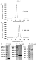
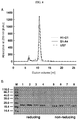


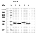
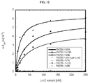
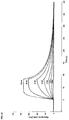


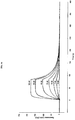
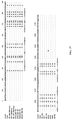

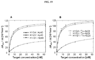

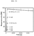

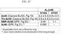

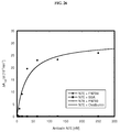
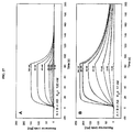
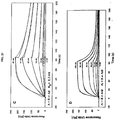

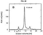
Classifications
-
- C—CHEMISTRY; METALLURGY
- C12—BIOCHEMISTRY; BEER; SPIRITS; WINE; VINEGAR; MICROBIOLOGY; ENZYMOLOGY; MUTATION OR GENETIC ENGINEERING
- C12N—MICROORGANISMS OR ENZYMES; COMPOSITIONS THEREOF; PROPAGATING, PRESERVING, OR MAINTAINING MICROORGANISMS; MUTATION OR GENETIC ENGINEERING; CULTURE MEDIA
- C12N15/00—Mutation or genetic engineering; DNA or RNA concerning genetic engineering, vectors, e.g. plasmids, or their isolation, preparation or purification; Use of hosts therefor
- C12N15/09—Recombinant DNA-technology
- C12N15/63—Introduction of foreign genetic material using vectors; Vectors; Use of hosts therefor; Regulation of expression
-
- A—HUMAN NECESSITIES
- A61—MEDICAL OR VETERINARY SCIENCE; HYGIENE
- A61K—PREPARATIONS FOR MEDICAL, DENTAL OR TOILETRY PURPOSES
- A61K38/00—Medicinal preparations containing peptides
-
- A—HUMAN NECESSITIES
- A61—MEDICAL OR VETERINARY SCIENCE; HYGIENE
- A61K—PREPARATIONS FOR MEDICAL, DENTAL OR TOILETRY PURPOSES
- A61K38/00—Medicinal preparations containing peptides
- A61K38/16—Peptides having more than 20 amino acids; Gastrins; Somatostatins; Melanotropins; Derivatives thereof
- A61K38/17—Peptides having more than 20 amino acids; Gastrins; Somatostatins; Melanotropins; Derivatives thereof from animals; from humans
- A61K38/1703—Peptides having more than 20 amino acids; Gastrins; Somatostatins; Melanotropins; Derivatives thereof from animals; from humans from vertebrates
- A61K38/1709—Peptides having more than 20 amino acids; Gastrins; Somatostatins; Melanotropins; Derivatives thereof from animals; from humans from vertebrates from mammals
-
- A—HUMAN NECESSITIES
- A61—MEDICAL OR VETERINARY SCIENCE; HYGIENE
- A61K—PREPARATIONS FOR MEDICAL, DENTAL OR TOILETRY PURPOSES
- A61K47/00—Medicinal preparations characterised by the non-active ingredients used, e.g. carriers or inert additives; Targeting or modifying agents chemically bound to the active ingredient
- A61K47/50—Medicinal preparations characterised by the non-active ingredients used, e.g. carriers or inert additives; Targeting or modifying agents chemically bound to the active ingredient the non-active ingredient being chemically bound to the active ingredient, e.g. polymer-drug conjugates
- A61K47/51—Medicinal preparations characterised by the non-active ingredients used, e.g. carriers or inert additives; Targeting or modifying agents chemically bound to the active ingredient the non-active ingredient being chemically bound to the active ingredient, e.g. polymer-drug conjugates the non-active ingredient being a modifying agent
- A61K47/56—Medicinal preparations characterised by the non-active ingredients used, e.g. carriers or inert additives; Targeting or modifying agents chemically bound to the active ingredient the non-active ingredient being chemically bound to the active ingredient, e.g. polymer-drug conjugates the non-active ingredient being a modifying agent the modifying agent being an organic macromolecular compound, e.g. an oligomeric, polymeric or dendrimeric molecule
- A61K47/59—Medicinal preparations characterised by the non-active ingredients used, e.g. carriers or inert additives; Targeting or modifying agents chemically bound to the active ingredient the non-active ingredient being chemically bound to the active ingredient, e.g. polymer-drug conjugates the non-active ingredient being a modifying agent the modifying agent being an organic macromolecular compound, e.g. an oligomeric, polymeric or dendrimeric molecule obtained otherwise than by reactions only involving carbon-to-carbon unsaturated bonds, e.g. polyureas or polyurethanes
- A61K47/60—Medicinal preparations characterised by the non-active ingredients used, e.g. carriers or inert additives; Targeting or modifying agents chemically bound to the active ingredient the non-active ingredient being chemically bound to the active ingredient, e.g. polymer-drug conjugates the non-active ingredient being a modifying agent the modifying agent being an organic macromolecular compound, e.g. an oligomeric, polymeric or dendrimeric molecule obtained otherwise than by reactions only involving carbon-to-carbon unsaturated bonds, e.g. polyureas or polyurethanes the organic macromolecular compound being a polyoxyalkylene oligomer, polymer or dendrimer, e.g. PEG, PPG, PEO or polyglycerol
-
- A—HUMAN NECESSITIES
- A61—MEDICAL OR VETERINARY SCIENCE; HYGIENE
- A61P—SPECIFIC THERAPEUTIC ACTIVITY OF CHEMICAL COMPOUNDS OR MEDICINAL PREPARATIONS
- A61P25/00—Drugs for disorders of the nervous system
- A61P25/28—Drugs for disorders of the nervous system for treating neurodegenerative disorders of the central nervous system, e.g. nootropic agents, cognition enhancers, drugs for treating Alzheimer's disease or other forms of dementia
-
- A—HUMAN NECESSITIES
- A61—MEDICAL OR VETERINARY SCIENCE; HYGIENE
- A61P—SPECIFIC THERAPEUTIC ACTIVITY OF CHEMICAL COMPOUNDS OR MEDICINAL PREPARATIONS
- A61P29/00—Non-central analgesic, antipyretic or antiinflammatory agents, e.g. antirheumatic agents; Non-steroidal antiinflammatory drugs [NSAID]
-
- A—HUMAN NECESSITIES
- A61—MEDICAL OR VETERINARY SCIENCE; HYGIENE
- A61P—SPECIFIC THERAPEUTIC ACTIVITY OF CHEMICAL COMPOUNDS OR MEDICINAL PREPARATIONS
- A61P35/00—Antineoplastic agents
-
- C—CHEMISTRY; METALLURGY
- C07—ORGANIC CHEMISTRY
- C07K—PEPTIDES
- C07K14/00—Peptides having more than 20 amino acids; Gastrins; Somatostatins; Melanotropins; Derivatives thereof
- C07K14/435—Peptides having more than 20 amino acids; Gastrins; Somatostatins; Melanotropins; Derivatives thereof from animals; from humans
- C07K14/46—Peptides having more than 20 amino acids; Gastrins; Somatostatins; Melanotropins; Derivatives thereof from animals; from humans from vertebrates
- C07K14/47—Peptides having more than 20 amino acids; Gastrins; Somatostatins; Melanotropins; Derivatives thereof from animals; from humans from vertebrates from mammals
-
- C—CHEMISTRY; METALLURGY
- C07—ORGANIC CHEMISTRY
- C07K—PEPTIDES
- C07K14/00—Peptides having more than 20 amino acids; Gastrins; Somatostatins; Melanotropins; Derivatives thereof
- C07K14/435—Peptides having more than 20 amino acids; Gastrins; Somatostatins; Melanotropins; Derivatives thereof from animals; from humans
- C07K14/775—Apolipopeptides
-
- G—PHYSICS
- G01—MEASURING; TESTING
- G01N—INVESTIGATING OR ANALYSING MATERIALS BY DETERMINING THEIR CHEMICAL OR PHYSICAL PROPERTIES
- G01N33/00—Investigating or analysing materials by specific methods not covered by groups G01N1/00 - G01N31/00
- G01N33/48—Biological material, e.g. blood, urine; Haemocytometers
- G01N33/50—Chemical analysis of biological material, e.g. blood, urine; Testing involving biospecific ligand binding methods; Immunological testing
- G01N33/53—Immunoassay; Biospecific binding assay; Materials therefor
- G01N33/566—Immunoassay; Biospecific binding assay; Materials therefor using specific carrier or receptor proteins as ligand binding reagents where possible specific carrier or receptor proteins are classified with their target compounds
-
- C—CHEMISTRY; METALLURGY
- C07—ORGANIC CHEMISTRY
- C07K—PEPTIDES
- C07K2319/00—Fusion polypeptide
- C07K2319/30—Non-immunoglobulin-derived peptide or protein having an immunoglobulin constant or Fc region, or a fragment thereof, attached thereto
-
- C—CHEMISTRY; METALLURGY
- C12—BIOCHEMISTRY; BEER; SPIRITS; WINE; VINEGAR; MICROBIOLOGY; ENZYMOLOGY; MUTATION OR GENETIC ENGINEERING
- C12N—MICROORGANISMS OR ENZYMES; COMPOSITIONS THEREOF; PROPAGATING, PRESERVING, OR MAINTAINING MICROORGANISMS; MUTATION OR GENETIC ENGINEERING; CULTURE MEDIA
- C12N2795/00—Bacteriophages
- C12N2795/00011—Details
- C12N2795/00041—Use of virus, viral particle or viral elements as a vector
Definitions
- the invention relates to a method for the generation of a mutein of human Lipocalin 2 (Lcn2, hNGAL) which binds a given target with a dissociation constant (K D ) of 200 nM or lower, said method comprising:
- the invention also relates to a nucleic acid library encoding muteins of human Lcn2 protein, wherein the amino acid residue at the sequence position corresponding to the sequence position 96 of the linear polypeptide sequence of human Lcn2 is mutated in the muteins, wherein 11, 12, 13, 14, 15, 16, 17, 18 or 19 amino acid residues at any of the sequence positions corresponding to the sequence positions 36, 40, 41, 49, 52, 68, 70, 72, 73, 77, 79, 81, 100, 103, 106, 125, 127, 132, and 134 of the linear polypeptide sequence of human Lcn2 (SEQ ID NO: 44) are mutated in the muteins.
- the invention also relates to a mutein which has the sequence of SEQ ID NO: 39(S1-A4) or SEQ ID NO: 41 (US7) or SEQ ID NO: 43 (H1-G1) SEQ ID NO: 22 (N7E) or SEQ ID NO: 24 (N9B) or SEQ ID NO: 26 (N10D) or SEQ ID NO: 50 (H1GA) or SEQ ID NO: 52 (H1GV).
- the invention furthermore relates to a nucleic acid molecule comprising a nucleotide sequence encoding a mutein which has the sequence of SEQ ID NO: 39(S1-A4) or SEQ ID NO: 41 (US7) or SEQ ID NO: 43 (H1-G1) SEQ ID NO: 22 (N7E) or SEQ ID NO: 24 (N9B) or SEQ ID NO: 26 (N10D) or SEQ ID NO: 50 (H1GA) or SEQ ID NO: 52 (H1GV).
- the invention furthermore relates to a host cell containing a nucleic acid molecule encoding a mutein which has the sequence of SEQ ID NO: 39(S1-A4) or SEQ ID NO: 41 (US7) or SEQ ID NO: 43 (Hl-Gl) SEQ ID NO: 22 (N7E) or SEQ ID NO: 24 (N9B) or SEQ ID NO: 26 (N10D) or SEQ ID NO: 50 (H1GA) or SEQ ID NO: 52 (H1GV).
- the invention furthermore relates to a pharmaceutical composition
- a pharmaceutical composition comprising at least one mutein which has the sequence of SEQ ID NO: 39(S1-A4) or SEQ ID NO: 41 (US7) or SEQ ID NO: 43 (Hl-Gl) SEQ ID NO: 22 (N7E) or SEQ ID NO: 24 (N9B) or SEQ ID NO: 26 (N10D) or SEQ ID NO: 50 (H1GA) or SEQ ID NO: 52 (H1GV).
- the invention also relates to a diagnostic or analytical kit comprising a mutein which has the sequence of SEQ ID NO: 39(S1-A4) or SEQ ID NO: 41 (US7) or SEQ ID NO: 43 (H1-G1) SEQ ID NO: 22 (N7E) or SEQ ID NO: 24 (N9B) or SEQ ID NO: 26 (N10D) or SEQ ID NO: 50 (H1GA) or SEQ ID NO: 52 (H1GV).
- a diagnostic or analytical kit comprising a mutein which has the sequence of SEQ ID NO: 39(S1-A4) or SEQ ID NO: 41 (US7) or SEQ ID NO: 43 (H1-G1) SEQ ID NO: 22 (N7E) or SEQ ID NO: 24 (N9B) or SEQ ID NO: 26 (N10D) or SEQ ID NO: 50 (H1GA) or SEQ ID NO: 52 (H1GV).
- Lcn2 muteins according to this invention show potential as therapeutic and/or diagnostic reagents in several disease areas. For example, they may serve to bind and deplete pathological forms of natural biomolecules such as the amyloid beta peptide in Alzière's disease. In another example they may be employed for the specific targeting of various labels or toxins to disease-related cell surface markers such as the fibronectin extra-domain B, which is associated with tumor neovasculature. Due to the particular benefits of the Lcn2 muteins that can be generated according to this invention various other applications or examples are possible.
- AD Alzheimer's disease
- a ⁇ amyloid beta peptide
- non-Ig binding reagents such as Anticalins
- the identification of an Affibody molecule with specificity for amyloid beta is one example to illustrate the potential of engineered non-Ig binding proteins ( Grönwall et al (2007) J. Biotechrtol. 128, 162-183 ; Hoyer et al. (2008) Proc. Natl. Acad. Sci. USA 105, 5099-5104 ).
- Fibronectin plays an essential role in cell adhesion, migration, proliferation, and differentiation.
- FN is a large, modular, dimeric glycoprotein comprising multiple domains of type I, II, and III.
- Alternative splice variants of FN such as its extra-domain B (ED-B), which is incorporated between the FN III 7 and FN III 8 domains, are expressed in a tissue-specific and developmental stage-dependent manner ( Zardi et al. (1987) EMBO J 6, 2337-2342 ).
- ED-B is absent from normal adult tissue except during wound healing and neoplastic vascularization. Consequently, ED-B containing fibronectin is abundantly expressed in many different tumor types that attract neovascularization and undergo aberrant angiogenesis. While the actual biological function of ED-B in angiogenesis remains elusive, its incorporation into FN serves as excellent marker for tumorgenesis. Generally, discrimination between malignant tissues and healthy organs is an advantageous therapeutic strategy as the selective targeting of drugs directly to the tumor tissue leads to an increased local drug concentration.
- the L19 scFv fragment is prone to oligomerization.
- Such Lcn2 muteins specifically recognize ED-B containing fibronectin on human cells with high sensitivity and thus show pospects as therapeutic reagents for the diagnosis and treatment of tumor diseases.
- the inventors were also able to identify specific Lcn2 muteins with high affinity and specificity for the A ⁇ peptide. Such Lcn2 muteins can even inhibit A ⁇ aggregation and thus show pospects, possibly after further improvement and modification, as therapeutic reagents to treat AD.
- the present disclosure also describes new random libraries based on the human Lcn2 scaffold which permit the efficient generation of muteins, such as the muteins of the present invention, with high affinity and specificity for a given target in general. Examples for such libraries or sections thereof are shown in Figures 1 and 2 .
- nucleotide triplets coding for cystein are not used for substitution during the mutagenesis. That means that the mutagenesis does not lead to muteins carrying new cysteins which were not already included in the original not mutagenized sequenze.
- the present disclosure is directed to a method of generating a mutein derived from human Lipocalin 2 (Lcn2; also known as human neutrophil gelatinase-associated lipocalin, hNGAL, or as siderocalin).
- the muteins obtained with this method can bind to a non-natural target with detectable affinity.
- the method comprises subjecting a nucleic acid molecule encoding for human Lipocalin 2 (Lcn2, hNGAL) to mutagenesis at a nucleotide triplet coding for at least one of any one of the sequence positions corresponding to the sequence positions 96, 100, and 106, of the linear polypeptide sequence of human Lipocalin 2, resulting in one or more mutein nucleic acid molecule(s).
- a nucleic acid molecule encoding for human Lipocalin 2 (Lcn2, hNGAL)
- human Lipocalin 2 or “human neutrophil gelatinase-associated lipocalin (hNGAL)” includes structural homologues, already identified or yet to be isolated, from other species which have an amino acid sequence homology or sequence identity of more than about 60%.
- these human lipocalins described above comprise 1, 2, 3, 4, 5, 6, 7, 8, 9, 10, 11, 12, 13, 14, 15, 16, or 17 mutated amino acid residues at any of the sequence positions corresponding to the sequence positions 33, 36, 40, 41, 42, 43, 44, 46, 47, 48, 49, 50, 51, 52, 54, 55, 59, 65, 68, 70, 72, 73, 75, 77, 78, 79, 80, 81, 86, 87, 98, 96, 99, 100, 103, 106, 107, 110, 111, 125, 127, 132, 134, 136 and 138 of the linear polypeptide sequence of hNGAL.
- sequence identity means the percentage of pair-wise identical residues - following homology alignment of a sequence of a polypeptide of the present invention with a sequence in question - with respect to the number of residues in the longer of these two sequences.
- the percentage of sequence homology or sequence identity is determined herein using the program BLASTP, version blastp 2.2.5 (November 16, 2002; cf. Altschul, S. F. et al. (1997) Nucl. Acid Res. 25, 3389-3402 ).
- the percentage of homology is based on the alignment of the entire polypeptide sequences (matrix: BLOSUM 62; gap costs: 11.1; cutoff value set to 10 -3 ) including the propeptide sequences, using the human Lipocalin 2 as reference in a pairwise comparison. It is calculated as the percentage of numbers of "positives" (homologous amino acids) indicated as result in the BLASTP program output divided by the total number of amino acids selected by the program for the alignment. It is noted in this connection that this total number of selected amino acids can differ from the length of the human Lipocalin 2.
- a protein other tha human Lipocalin 2 is used in the present invention
- the definition of the mutated sequence positions given for human Lipocalin 2 can be assigned to the other lipocalin with the help of published sequence alignments or alignments methods which are available to the skilled artisan.
- a sequence alignment can, for example, be carried out as explained in WO 99/16873 (cf. Fig. 3 therein), using an published alignment such as the one in Fig. 1 of Redl, B. (2000) Biochim. Bioplys. Acta 1482, 241-248 .
- the three-dimensional structure of the lipocalins is available structural superpositions can also be used for the determination of those sequence positions that are to be subjected to mutagenesis in the present invention.
- Other methods of structural analysis such as multidimensional nuclear magnetic resonance spectroscopy can also be employed for this purpose.
- the homologue of human Lipocalin 2 can also be a mutein protein of human Lipocalin 2 itself, in which amino acid substitutions are introduced at positions other than the positions selected in the present invention.
- a mutein can be a protein in which positions at the solvent exposed surface of the ⁇ -barrel are mutated compared to the wild type sequence of the human Lipocalin 2 in order to increase the solubility or the stability of the protein.
- human Lipocalin 2 includes all proteins that have a sequence homology or sequence identity of more than 60%, 70% 80%, 85%, 90%, or 95% in relation to the human Lipocalin 2. It is preferred that these human lipocalins described above comprise 1, 2, 3, 4, 5, 6, 7, 8, 9, 10, 11, 12, 13, 14, 15, 16, or 17 mutated amino acid residues at any of the sequence positions corresponding to the sequence positions 33, 36, 40, 41, 42, 43, 44, 46, 47, 48, 49, 50, 51, 52, 54, 55, 59, 65, 68, 70, 72, 73, 75, 77, 78, 79, 80, 81, 86, 87, 98, 96, 99, 100, 103, 106, 107, 110, 111, 125, 127, 132, 134, 136 and 138 of the linear polypeptide sequence ofhNGAL.
- the present invention provides a mutein derived from human Lipocalin 2 (i.e., human lipocalin mutein or a mutated human lipocalin, preferably a mutated mature hNGAL, wherein said mature hNGAL has the SWISS-PROT Data Bank Accession Number P80188, more preferably said mature hNGAL has the amino acid sequence shown in SEQ ID NO:44).
- This mutein includes at least one or two mutated amino acid residue at any of the sequence positions corresponding to the sequence positions 96, 100 and 106 of the linear polypeptide sequence of human Lcn2, and wherein the mutein binds a given non-natural target with detectable affinity.
- a "mutein,” a “mutated” entity (whether protein or nucleic acid) or “mutant” refers to the exchange, deletion, or insertion of one or more nucleotides or amino acids, respectively, compared to the naturally occurring (wild-type) nucleic acid or protein "reference” scaffold.
- the number of nucleotides or amino acids, respectively, that is exchanged, deleted or inserted is 1, 2, 3, 4, 5, 6, 7, 8, 9, 10, 11, 12, 13, 14, 15, 16, 17, 18, 19, 20 or more such as 25, 30, 35, 40, 45 or 50.
- human neutrophil gelatinase-associated lipocalin or "hNGAL” or “lipocalin 2" or “Lcn2” as used herein to refer to the mature hNGAL with the SWISS-PROT Data Bank Accession Number P80188 (exemplified herein as SEQ ID NO:44).
- the invention is based on the surprising finding that subjecting human Lipocalin 2, rat ⁇ 2 -microglobulin-related protein (A2m) and mouse 24p3/uterocalin (24p3) to mutagenesis at one or more of these above-mentioned 3 sequence position provides for muteins that have a sufficiently affine binding to pre-defined target which can include, but is not limited to a peptide, a protein, a fragment or a domain of a protein, and a small organic molecule.
- the given target may be any desired non-natural target/ligand.
- non-natural ligand refers to a compound, which does not bind to native mature hNGAL under physiological conditions.
- organic molecule or "small organic molecule” as used herein for the non-natural target denotes an organic molecule comprising at least two carbon atoms, but preferably not more than 7 or 12 rotatable carbon bonds, having a molecular weight in the range between 100 and 2000 Dalton, preferably between 100 and 1000 Dalton, and optionally including one or two metal atoms.
- peptide refers to a dipeptide or an oligopeptide with 2 to 45 amino acids.
- the peptide has 2-40, 2-35, 2-30, 2-25, 2-20, 2-15 or 2-10 amino acid residues.
- the peptide may be a naturally occurring or synthetic peptide and may comprise - besides the 20 naturally occurring L-amino acids - D-amino acids, non-naturally occurring amino acids and amino acid analogs.
- the peptide is an amyloid beta peptide (Abeta or A ⁇ ).
- the amyloid beta peptide is an A ⁇ 40 peptide or an A ⁇ 42 peptide.
- the small organic molecule is a compound showing the features of an immunological hapten.
- the non-natural target which is a protein is fibronection or a domain thereof, such as the EB-domain or a fragment of the EB-domain.
- a human Lipocalin 2 mutein of the disclosure may comprise the wild type (natural) amino acid sequence outside the mutated amino acid sequence positions.
- the lipocalin muteins disclosed herein may also contain amino acid mutations outside the sequence positions subjected to mutagenesis as long as those mutations do not interfere with the binding activity and the folding of the mutein.
- Such mutations can be accomplished very easily on DNA level using established standard methods ( Sambrook, J. et al. (1989) Molecular Cloning: A Laboratory Manual, 2nd Ed., Cold Spring Harbor Laboratory Press, Cold Spring Harbor, NY ).
- Possible alterations of the amino acid sequence are insertions or deletions as well as amino acid substitutions. Such substitutions may be conservative, i.e.
- an amino acid residue is replaced with a chemically similar amino acid residue.
- conservative substitutions are the replacements among the members of the following groups: 1) alanine, serine, and threonine; 2) aspartic acid and glutamic acid; 3) asparagine and glutamine; 4) arginine and lysine; 5) isoleucine, leucine, methionine, and valine; and 6) phenylalanine, tyrosine, and tryptophan.
- modifications of the amino acid sequence include directed mutagenesis of single amino acid positions in order to simplify sub-cloning of the mutated lipocalin gene or its parts by incorporating cleavage sites for certain restriction enzymes.
- these mutations can also be incorporated to further improve the affinity of a lipocalin mutein for a given target.
- mutations can be introduced in order to modulate certain characteristics of the mutein such as to improve folding stability, serum stability, protein resistance or water solubility or to reduce aggregation tendency, if necessary.
- cysteine residues may be mutated to other amino acids to prevent disulphide bridge formation.
- cysteine residues are introduced into the amino acid sequence of a human Lipocalin 2 mutein.
- cysteine residues are introduced at least at one of the sequence positions that correspond to sequence positions 14, 21, 60, 84, 88, 116, 141, 145, 143, 146 or 158 of the wild type sequence of hNGAL.
- the generated thiol moiety at the side of any of the amino acid positions 14, 21, 60, 84, 88, 116, 141, 145, 143, 146 and/or 158 may be used to PEGylate or HESylate the mutein, for example, in order to increase the serum half-life of a respective human Lipocalin 2 mutein.
- the mutein of the present disclosure includes mutated amino acid residues at least at any 2 or all 3 of the sequence positions corresponding to the sequence positions 96, 100 and 106 of the linear polypeptide sequence of human Lipocalin 2.
- the mutein includes at least 1, 2, 3, 4, 5, 6, 7, 8, 9, 10, 11, 12, 13, 14, 15, 16, or 17 mutated amino acid residues at any of the sequence positions corresponding to the sequence positions 33, 36, 40, 41, 42, 43, 44, 46, 47, 48, 49, 50, 51, 52, 54, 55, 59, 65, 68, 70, 72, 73, 75, 77, 78, 79, 80, 81, 86, 87, 98, 96, 99, 100, 103, 106, 107, 110, 111, 125, 127, 132, 134, 136 and 138 of the linear polypeptide sequence of hNGAL.
- the mutein includes at least 2, 3, 4, 5, 6, 7, 8, 9, 10, 11, 12, 13, 14, 15, 16, 17 mutated amino acid residues at any one of the sequence positions 33, 36, 40, 41, 42, 43, 44, 46, 47, 48, 49, 50, 51, 52, 54, 55, 59, 65, 68, 70, 72, 73, 75, 77, 78, 79, 80, 81, 86, 87, 98, 96, 99, 100, 103, 106, 107, 110, 111, 125, 127, 132, 134, 136 and 138 of the linear polypeptide sequence of hNGAL.
- the mutein includes 18, 19 or 20 mutated amino acid residues at any one of the sequence positions 33, 36, 40, 41, 42, 43, 44, 46, 47, 48, 49, 50, 51, 52, 54, 55, 59, 65, 68, 70, 72, 73, 75, 77, 78, 79, 80, 81, 86, 87, 98, 96, 99, 100, 103, 106, 107, 110, 111, 125, 127, 132, 134, 136 and 138 of the linear polypeptide sequence of human Lipocalin 2.
- the mutein includes mutated amino acid residues at at least any 10, 14, 15, 20, 22, 24, 26, 28, 29, 30, 31, 32, 33, 35 or all 45 of the above-listed sequence positions.
- a mutein of the disclosure which binds to an amyloid beta peptide can comprise with respect to the mature human Lipocalin 2 wild type amino acid sequence illustrated in Figure 17 (Lcn2) at least 2, 3, 4, 5, 6, 7, 8, 9, 10, 11, or 12 amino acid replacements which include, but are not limited to Leu36 ⁇ Val or Cys; Ala40 ⁇ Tyr or Lys or Val; Ile41 ⁇ Thr or Ser or Leu; Gln49 ⁇ Leu or Trp; Leu70 ⁇ Gly; Arg72 ⁇ Gly or Asp; Lys73 ⁇ Leu or Thr or Asp; Asp77 ⁇ Asn or His or Leu; Trp79 ⁇ Lys; Asn96 ⁇ Ile or Arg; Tyr100 ⁇ Gln or Arg or Glu; Leu103 ⁇ Met or Arg or Gly; Tyr106 ⁇ Tyr or Ala or Trp; Lys125 ⁇ Thr or Val or Glu;
- the mutein described herein binds to and inhibits aggregation of A ⁇ , preferably A ⁇ 40, more preferably under assay conditions as specified in Example 11 (preferably including the ratio 1:10 mutein : A ⁇ 40).
- the present invention also relates to muteins as described above having a comparable biological function when compared with mutein US7.
- “Comparable biological function” means that these muteins are able to bind to and inhibit A ⁇ , preferably A ⁇ 40, with a deviation of the aggregation inhibiting activity in respect to US7 of not more than about 40%, 30%, 20%, 15%, 10%, 5%, 2,5%, 2% or 1%, for example under conditions which equate to or are identical with those set out in Example 11 (preferably including the ratio 1:10 mutein : A ⁇ 40).
- the present invention also relates to the muteins described herein for use in the treatment or prevention of neurodegenerative diseases such as Alzheimer's disease.
- a mutein of the disclosure which binds to an amyloid beta peptide, such as A ⁇ 40 peptide or an A ⁇ 42 peptide comprises the following amino acid replacements Leu36 ⁇ Val; Ala40 ⁇ Tyr; Ile41 ⁇ Thr; Gln49 ⁇ Leu; Leu70 ⁇ Gly; Lys73 ⁇ Leu; Asp77 ⁇ Asn; Trp79 ⁇ Lys; Asn96 ⁇ Ile; Tyr100 ⁇ Gln; Leu103 ⁇ Met; Lys125 ⁇ Thr; Ser127 ⁇ Gly; Tyr132 ⁇ Met; and Lys134 ⁇ Asn.
- An example of a mutein including such amino acid replacements is S1-A4 illustrated in Figure 17 (SEQ ID NO: 39).
- a mutein of the disclosure which binds to an amyloid beta peptide, such as A ⁇ 40 peptide or an A ⁇ 42 peptide comprises the following amino acid replacements Leu36 ⁇ Val; Ala40 ⁇ Lys; Ile41 ⁇ Ser; Gln49 ⁇ Trp; Leu70 ⁇ Gly; Arg72 ⁇ Gly; Lys73 ⁇ Thr; Asp77 ⁇ His; Trp79 ⁇ Lys; Asn96 ⁇ Arg; Tyr100 ⁇ Arg; Leu103 ⁇ Arg; Tyr106 ⁇ Ala; Lys125 ⁇ Val; Ser127 ⁇ Gln; Tyr132 ⁇ Ser; and Lys134 ⁇ Asn.
- An example of a mutein including such amino acid replacements is US-7 illustrated in Figure 17 (SEQ ID NO: 41).
- the disclosure also envisages a mutated mature hNGAL lipocalin (as deposited in SWISS-PROT Data Bank under Accession Number P80188, preferably having the amino acid sequence shown in SEQ ID NO:40) comprising at a position corresponding to a position 36, 40, 41, 49, 70, 72, 73, 77, 79, 96, 100, 103, 106, 125, 127, 132, 134 of the linear polypeptide sequence of wild type hNGAL one or more mutated amino acids as described herein.
- a mutein of the disclosure which binds to an amyloid beta peptide, such as A ⁇ 40 peptide or an A ⁇ 42 peptide comprises the following amino acid replacements Leu36 ⁇ Cys; Ala40 ⁇ Val; Ile41 ⁇ Leu; Gln49 ⁇ Leu; Leu70 ⁇ Gly; Arg72 ⁇ Asp; Lys73 ⁇ Asp; Asp77 ⁇ Leu; Trp79 ⁇ Lys; Asn96 ⁇ Arg; Tyr100 ⁇ Glu; Leu103 ⁇ Gly; Tyr106 ⁇ Tip; Lys125 ⁇ Glu; Ser127 ⁇ Ala; Tyr132 ⁇ Thr; and Lys134 ⁇ Asn.
- An example of a mutein including such amino acid replacements is H1-G1 illustrated in Figure 17 (SEQ ID NO: 43).
- a mutein of the disclosure which binds to an amyloid beta peptide, such as A ⁇ 40 peptide or an A ⁇ 42 peptide comprises the following amino acid replacements Leu36 ⁇ Ala; Ala40 ⁇ Val; Ile41 ⁇ Leu; Gln49 ⁇ Leu; Leu70 ⁇ Gly; Arg72 ⁇ Asp; Lys73 ⁇ Asp; Asp77 ⁇ Leu; Trp79 ⁇ Lys; Asn96 ⁇ Arg; Tyr100 ⁇ Glu; Leu103 ⁇ Gly; Tyr106 ⁇ Trp; Lys125 ⁇ Glu; Ser127 ⁇ Ala; Tyr132 ⁇ Thr; and Lys134 ⁇ Asn.
- An example of a mutein including such amino acid replacements is H1GA illustrated SEQ ID NO: 50.
- the mutein described herein above binds to and inhibits aggregation of A ⁇ , preferably A ⁇ 40, more preferably under assay conditions as specified in Example 23 (preferably in a ratio of 10:2 A ⁇ 40: H1GA).
- the present disclosure also relates to muteins as described above having a comparable biological function when compared with mutein H1GA.
- “Comparable biological function” means that these muteins are able to bind to and inhibit A ⁇ , preferably A ⁇ 40 with a deviation of the aggregation inhibiting activity in respect to H1GA of not more than about 40%, 30%, 20%, 15%, 10%, 5%, 2,5%, 2% or 1%, for example under conditions which equate to or are identical with those set out in Example 23 (preferably in a ratio of 10:2 A ⁇ 40 : H1GA).
- the present disclosure also relates to the muteins described herein for use in the treatment or prevention of neurodegenerative diseases such as Alzheimer's disease.
- a mutein of the disclosure which binds to an amyloid beta peptide, such as A ⁇ 40 peptide or an A ⁇ 42 peptide comprises the following amino acid replacements Leu36 ⁇ Val; Ala40 ⁇ Val; Ile41 ⁇ Leu; Gln49 ⁇ Leu; Leu70 ⁇ Gly; Arg72 ⁇ Asp; Lys73 ⁇ Asp; Asp77 ⁇ Leu; Trp79 ⁇ Lys; Asn96 ⁇ Arg; Tyr100 ⁇ Glu; Leu103 ⁇ Gly; Tyr106 ⁇ Trp; Lys125 ⁇ Glu; Ser127 ⁇ Ala; Tyr132 ⁇ Thr; and Lys134 ⁇ Asn.
- An example of a mutein including such amino acid replacements is H1GV illustrated SEQ ID NO: 52.
- the mutein described herein above binds to A ⁇ 40 under assay conditions as specified in Example 21.
- the present disclosure also relates to muteins as described above having a comparable biological function when compared with mutein H1GV.
- “Comparable biological function” means that these muteins are able to bind to A ⁇ , preferably A ⁇ 40 with a deviation of the aggregation inhibiting activity in respect to H1GV of not more than about 40%, 30%, 20%, 15%, 10%, 5%, 2,5%, 2% or 1%, for example under conditions which equate to or are identical with those set out in Example 21.
- the present disclosure also relates to the muteins described herein for use in the treatment or prevention of neurodegenerative diseases such as Alzheimer's disease.
- a mutein of the disclosure which bind to extra-domain B or a fragment thereof comprises the following amino acid replacements Leu36 ⁇ Lys; Ala40 ⁇ His; Ile41 ⁇ Asp; Gln49 ⁇ Arg; Tyr52 ⁇ Gln; Ser68 ⁇ Asn; Leu70 ⁇ Arg; Arg72 ⁇ Val; Lys73 ⁇ His; Asp77 ⁇ Asn; Trp79 ⁇ Arg; Tyr100 ⁇ Trp; Tyr106 ⁇ Tip; Lys125 ⁇ Arg; Ser127 ⁇ Tyr; Tyr132 ⁇ Leu; Lys134 ⁇ Glu and Ser146 ⁇ Asn.
- An example of a mutein including such amino acid replacements is N7A illustrated in Figure 17 (SEQ ID NO: 20).
- a mutein of the disclosure which binds to extra-domain B or a fragment thereof comprises the following amino acid replacements Leu36 ⁇ Arg; Ala40 ⁇ Met; Ile41 ⁇ Arg; Gln49 ⁇ Ala; Tyr52 ⁇ Val; Ser68 ⁇ Lys; Leu70 ⁇ Met; Arg72 ⁇ Gln; Lys73 ⁇ Arg; Asp77 ⁇ Lys; Trp79 ⁇ Met; Arg81 ⁇ Asn; Asn96 ⁇ Ala; Tyr100 ⁇ Pro; Leu103 ⁇ Pro; Tyr106 ⁇ Thr; Lys125 ⁇ His; Ser127 ⁇ Phe; and Lys134 ⁇ His.
- An example of a mutein including such amino acid replacements is N9B illustrated in Figure 17 (SEQ ID NO: 24).
- a mutein of the disclosure which binds to extra-domain B or a fragment thereof comprises the following amino acid replacements Leu36 ⁇ Ala; Ala40 ⁇ Thr; Ile41 ⁇ Trp; Gln49 ⁇ Tyr; Tyr52 ⁇ Gln; Ser68 ⁇ Asn; Arg72 ⁇ Met; Lys73 ⁇ Ser; Asp77 ⁇ Arg; Trp79 ⁇ Met; Arg81 ⁇ His; Asn96 ⁇ Ser; Tyr100 ⁇ Trp; Ty106 ⁇ Trp; Lys125 ⁇ Arg; Ser127 ⁇ Tyr; Tyr132 ⁇ Phe; and Lys134 ⁇ Gly.
- An example of a mutein including such amino acid replacements is N10D illustrated in Figure 17 (SEQ ID NO: 26).
- a mutein of the disclosure which bind to extra-domain B or a fragment thereof comprises the following amino acid replacements Leu36 ⁇ Glu; Ala40 ⁇ Ser; Ile41 ⁇ Leu; Gln49 ⁇ Arg; Leu70 ⁇ Arg; Lys73 ⁇ Ser; Asp77 ⁇ His; Trp79 ⁇ Leu; Asn96 ⁇ Leu; Tyr100 ⁇ Lys; Leul03 ⁇ His; Tyr106 ⁇ Phe; Lys125 ⁇ Thr; Ser127 ⁇ Ala; and Lys 134 ⁇ Phe.
- An example of a mutein including such amino acid replacements is N7E illustrated in Figure 17 (SEQ ID NO: 22).
- the above muteins described with reference to Figure 17 can include further amino acid replacements.
- the muteins can further comprise amino acid replacements which can include, but are not limited to Gln28 ⁇ His or Cys87 ⁇ Ser.
- Other possible amino acid replacements include, but are not limited to Tyr52 ⁇ Gln or Val; Ser68 ⁇ Lys or Asn; or Arg81 ⁇ Trp or Asn or His.
- the lipocalin muteins of the disclosure are able to bind the desired non-natural target with detectable affinity, i.e. with a dissociation constant (K D ) of at least 200 nM.
- the mutein binds the given non-natural target with a K D of 1 ⁇ M or less, or 100 ⁇ M or less, or 1 ⁇ M or less, or 500 nM, or 200 nM or less, or 100, nM or less, or 50 nM or less, or 10 nM or less, or 1 nM or less.
- lipocalin muteins bind the desired target with a dissociation constant for a given target of at least 100, 20, 1 nM or even less.
- the binding affinity of a mutein to the desired target can be measured by a multitude of methods such as fluorescence titration, competition ELISA or surface plasmon resonance (Biacore).
- a mutein of the disclosure typically exists as monomeric protein. However, it is also possible that a lipocalin mutein is able to spontaneously dimerise or oligomerise. Although the use of lipocalin muteins that form stable monomers may be preferred for some applications, e.g. because of faster diffusion and better tissue penetration, the use of lipocalin muteins that form stable homodimers or multimers may be advantageous in other instances, since such multimers can provide for a (further) increased affinity and/or avidity to a given target. Furthermore, oligomeric forms of the lipocalin mutein may have slower dissociation rates or prolonged serum half-life.
- the complex formation between the respective mutein and its ligand is influenced by many different factors such as the concentrations of the respective binding partners, the presence of competitors, pH and the ionic strength of the buffer system used, and the experimental method used for determination of the dissociation constant K D (for example fluorescence titration, competition ELISA or surface plasmon resonance, just to name a few) or even the mathematical algorithm which is used for evaluation of the experimental data.
- the K D values (dissociation constant of the complex formed between the respective mutein and its target/ligand) may vary within a certain experimental range, depending on the method and experimental setup that is used for determining the affinity of a particular lipocalin mutein for a given ligand. This means, there may be a slight deviation in the measured K D values or a tolerance range depending, for example, on whether the K D value was determined by surface plasmon resonance (Biacore) or by competition ELISA.
- the muteins disclosed herein can be linked, either N- or C-terminal to an affinity tag such as pentahistidine tag, a hexahistidine tag or a Strep -tag ® .
- an affinity tag such as pentahistidine tag, a hexahistidine tag or a Strep -tag ® .
- fragment as used in the present invention in connection with the feature lipocalin mutein fragment relates to proteins or peptides derived from full-length mature Lcn2 that are N-terminally and/or C-terminally shortened, i.e. lacking at least one of the N-terminal and/or C-terminal amino acids.
- Such fragments comprise preferably at least 10, more preferably 20, most preferably 30 or more consecutive amino acids of the primary sequence of mature Lcn2 and are usually detectable in an immunoassay of mature Lcn2.
- Cytotoxic T-cells recognize peptide antigens on the cell surface of an antigen-presenting cell in association with a class I major histocompatibility complex (MHC) molecule.
- MHC major histocompatibility complex
- the ability of the peptides to bind to MHC molecules is allele specific and correlates with their immunogenicity. In order to reduce immunogenicity of a given protein, the ability to predict which peptides in a protein have the potential to bind to a given MHC molecule is of great value.
- Approaches that employ a computational threading approach to identify potential T-cell epitopes have been previously described to predict the binding of a given peptide sequence to MHC class I molecules ( Altuvia et al. (1995) J. Mol. Biol. 249: 244-250 ).
- Such an approach may also be utilized to identify potential T-cell epitopes in the muteins of the disclosure and to make depending on its intended use a selection of a specific mutein on the basis of its predicted immunogenicity. It may be furthermore possible to subject peptide regions which have been predicted to contain T-cell epitopes to additional mutagenesis to reduce or eliminate these T-cell epitopes and thus minimize immunogenicity.
- the removal of amphipathic epitopes from genetically engineered antibodies has been described ( Mateo et al. (2000) Hybridoma 19(6):463-471 ) and may be adapted to the muteins of the present invention.
- the muteins thus obtained may possess a minimized immunogenicity, which is desirable for their use in therapeutic and diagnostic applications, such as those described below.
- the invention is also directed to lipocalin muteins which are conjugated to a compound which can include, but is not limited to organic molecules, an enzyme label, a colored label, a cytostatic agent, a toxin, a label that can be photoactivated and which is suitable for use in photodynamic therapy, a fluorescent label, a radioactive label, a chromogenic label, a luminescent label, metal complexes, metal, such as colloidal gold, haptens, digoxigenin, biotin, a chemotherapeutic metal, or a chemotherapeutic metal, to name only a few evocative examples.
- the mutein may also be conjugated to an organic drug molecule. The conjugation can be carried out using any conventional coupling method known in the art.
- a Lcn2 mutein described herein with any appropriate chemical substance or enzyme, which directly or indirectly generates a detectable compound or signal in a chemical, physical, optical, or enzymatic reaction.
- An example for a physical reaction and at the same time optical reaction/marker is the emission of fluorescence upon irradiation.
- Alkaline phosphatase, horseradish peroxidase or ⁇ -galactosidase are examples of enzyme labels (and at the same time optical labels) which catalyze the formation of chromogenic reaction products.
- all labels commonly used for antibodies can also be used for conjugation to the muteins of the present disclosure.
- the muteins of the invention may also be conjugated with any suitable therapeutically active agent, e.g., for the targeted delivery of such agents to a given cell, tissue or organ or for the selective targeting of cells, e.g., of tumor cells without affecting the surrounding normal cells.
- suitable therapeutically active agents include radionuclides, toxins, small organic molecules, and therapeutic peptides (such as peptides acting as agonists/antagonists of a cell surface receptor or peptides competing for a protein binding site on a given cellular target).
- Suitable toxins include, but are not limited to pertussis-toxin, diphtheria toxin, ricin, saporin, pseudomonas exotoxin, calicheamicin or a derivative thereof, a taxoid, a maytansinoid, a tubulysin or a dolastatin analogue.
- the dolastatin analogue may be auristatin E, monomethylauristatin E, auristatin PYE and auristatin PHE.
- cytostatic agent examples include, but are not limited to Cisplatin, Carboplatin, Oxaliplatin, 5-Fluorouracil, Taxotere (Docetaxel), Paclitaxel, Anthracycline (Doxorubicin), Methotrexate, Vinblastin, Vincristine, Vindesine, Vinorelbine, dacarbazine, Cyclophosphamide, Etoposide, Adriamycine, Camptotecine, Combretatastin A-4 related compounds, sulfonamides, oxadiazolines, benzo[b]thiophenessynthetic spiroketal pyrans, monotetrahydrofuran compounds, curacin and curacin derivatives, methoxyestradiol derivatives and Leucovorin.
- the lipocalin muteins of the invention may also be conjugated with therapeutically active nucleic acids such as antisense nucleic acid molecules, small interfering RNAs, micro RNAs or ribozymes. Such conjugates can be produced by methods well known in the art.
- the muteins of the disclosure may also be coupled to a targeting moiety that targets a specific body region in order to deliver the inventive muteins to a desired region or area within the body.
- a targeting moiety that targets a specific body region in order to deliver the inventive muteins to a desired region or area within the body.
- One example wherein such modification may be desirable is the crossing of the blood-brain-barrier.
- the muteins may be coupled to moieties that facilitate the active transport across this barrier (see Gaillard PJ, et al. (2005) International Congress Series. 1277,185-198 or Gaillard PJ, et al. (2005) Expert Opin Drug Deliv. 2(2), 299-309 ).
- Such compounds are for example available under the trade name 2B-TransTM (to-BBB technologies BV, Leiden, NL).
- targeting molecules to which the muteins may be coupled include antibodies, antibody fragments or lipocalin muteins with affinity for a desired target molecule.
- the target molecule of the targeting moieties may, for example, be a cell-surface antigen.
- Cell-surface antigens may be specific for a cell or tissue type, such as, for example, cancer cells.
- Illustrative examples of such cell surface proteins are HER-2 or proteoglycans such as NEU-2.
- a mutein may in some embodiments be conjugated to a compound that extends the serum half-life of the mutein (in this regard see also PCT publication WO 2006/56464 where such conjugation strategies are described with references to muteins of human neutrophil gelatinase-associated lipocalin with binding affinity for CTLA-4).
- the compound that extends the serum half-life may be a polyalkylene glycol molecule, such as polyethylene (PEG) or an activated derivative thereof; hydroxyethyl starch, fatty acid molecules, such as palmitic acid ( Vajo & Duckworth (2000) Pharmacol. Rev.
- an Fc part of an immunoglobulin a CH3 domain of an immunoglobulin, a CH4 domain of an immunoglobulin, albumin or a fragment thereof, an albumin binding peptide, or an albumin binding protein, transferrin to name only a few.
- the albumin binding protein may be a bacterial albumin binding protein, an antibody, an antibody fragment including domain antibodies (see US patent 6,696,245 , for example), or a lipocalin mutein with binding activity for albumin. Accordingly, suitable conjugation compounds for extending the half-life of a lipocalin mutein include albumin ( Osborn et al. (2002) J. Pharmacol. Exp. Ther.
- an albumin binding protein for example, a bacterial albumin binding domain, such as the one of streptococcal protein G ( König, T. and Skerra, A. (1998) J. Immunol. Methods 218, 73-83 ).
- albumin binding peptides that can be used as conjugation partner are, for instance, those having a Cys-Xaa 1 -Xaa 2 -Xaa 3 -Xaa 4 -Cys consensus sequence, wherein Xaa 1 is Asp, Asn, Ser, Thr, or Trp; Xaa 2 is Asn, Gln, His, Ile, Leu, or Lys; Xaa 3 is Ala, Asp, Phe, Trp, or Tyr; and Xaa 4 is Asp, Gly, Leu, Phe, Ser, or Thr as described in US patent application 2003/0069395 or Dennis et al. ( Dennis et al. (2002) J. Biol. Chem. 277, 35035-35043 ).
- albumin itself or a biological active fragment of albumin can be used as compound of a lipocalin mutein that extends the serum half-life of the mutein.
- the term "albumin” comprises all mammal albumins such as human serum albumin or bovine serum albumin or rat albumin.
- the albumin or fragment thereof can be recombinantly produced as described in US patent 5,728,553 or European patent applications EP 0 330 451 and EP 0 361 991 .
- Recombinant human albumin (Recombumin®) for use as a protein stabilizer is for example available from Novozymes Delta Ltd. (Nottingham, UK).
- albumin-binding protein is an antibody fragment it may be a domain antibody.
- Domain Antibodies are engineered to allow precise control over biophysical properties and in vivo half-life to create the optimal safety and efficacy product profile. Domain Antibodies are for example commercially available from Domantis Ltd. (Cambridge, UK and MA, USA).
- the muteins can be genetically fused to the N or C terminus, or both, of non-glycosylated transferrin.
- Non-glycosylated transferrin has a half-life of 14-17 days, and a transferrin fusion protein will similarly have an extended half-life.
- the transferrin carrier also provides high bioavailability, biodistribution and circulating stability. This technology is commercially available from BioRexis (BioRexis Pharmaceutical Corporation, PA, USA). Recombinant human transferrin (DeltaFerrinTM) for use as a protein stabilizer is also commercially available from Novozymes Delta Ltd. (Nottingham, UK).
- an Fc part of an immunoglobulin is used for the purpose to prolong the serum half-life of the muteins of the invention
- the SynFusion TM technology commercially available from Syntonix Pharmaceuticals, Inc (MA, USA)
- the use of this Fc-fusion technology allows the creation of longer-acting biopharmaceuticals and may for example consist of two copies of the mutein linked to the Fc region of an antibody to improve pharmacokinetics, solubility, and production efficiency.
- polyalkylene glycol is used as compound that extends the half-life of the mutein, the polyalkylene glycol can be substituted or unsubstituted. It can also be an activated polyalkylene derivative.
- suitable compounds are polyethylene glycol (PEG) molecules as described in WO 99/64016 , in US Patent 6,177,074 or in US Patent 6,403,564 in relation to interferon, or as described for other proteins such as PEG-modified asparaginase, PEG-adenosine deaminase (PEG-ADA) or PEG-superoxide dismutase (see for example, Fuertges et al.
- the molecular weight of such a polymer may range from about 300 to about 70.000 Dalton, including, for example, polyethylene glycol with a molecular weight of about 10.000, of about 20.000, of about 30.000 or of about 40.000 Dalton.
- carbohydrate oligo- and polymers such as starch or hydroxyethyl starch (HES) can be conjugated to a mutein of the invention for the purpose of serum half-life extension.
- artificial amino acids may be introduced by mutagenesis.
- such artificial amino acids are designed to be more reactive and thus to facilitate the conjugation to the desired moiety.
- an artificial amino acid that may be introduced via an artificial tRNA is para-acetyl-phenylalanine.
- the mutein is fused at its N-terminus and/or it's C-terminus to a protein, a protein domain or a peptide such as a signal sequence and/or an affinity tag.
- a mutein of the invention may be fused to a fusion partner that extends the in vivo serum half-life of the mutein (see again PCT publication WO 2006/56464 where suitable fusion partner are described with references to muteins of human neutrophile gelatinase-associated lipocalin with binding affinity for CTLA-4).
- the fusion partner may be an Fc part of an immunoglobulin, a CH3 domain of an immunoglobulin, a CH4 domain of an immunogloubulin, albumin, an albumin binding peptide or an albumin binding protein, to name only a few.
- the albumin binding protein may be a bacterial albumin binding protein or a lipocalin mutein with binding activity for albumin.
- suitable fusion partners for extending the half-life of a lipocalin mutein include albumin ( Osborn, B.L. et al. (2002) supra J. Pharmacol. Exp. Ther. 303, 540-548 ), or an albumin binding protein, for example, a bacterial albumin binding domain, such as the one of streptococcal protein G ( König, T. and Skerra, A. (1998) supra J. Immunol. Methods 218, 73-83 ).
- albumin itself or a biological active fragment of albumin as fusion partner of a lipocalin mutein.
- the term "albumin” comprises all mammal albumins such as human serum albumin or bovine serum albumin or rat serum albumin.
- the recombinant production of albumin or fragments thereof is well known in the art and for example described in US patent 5,728,553 , European patent application EP 0 330 451 or EP 0 361 991 .
- the fusion partner may confer new characteristics to the inventive lipocalin mutein such as enzymatic activity or binding affinity for other molecules.
- suitable fusion proteins are alkaline phosphatase, horseradish peroxidase, gluthation-S-transferase, the albumin-binding domain of protein G, protein A, antibody fragments, oligomerization domains, lipocalin muteins of same or different binding specificity (which results in the formation of "duocalins", cf. Schlehuber, S., and Skerra, A. (2001), Duocalins, engineered ligand-binding proteins with dual specificity derived from the lipocalin fold (Biol. Chem. 382, 1335-1342 ), or toxins.
- a lipocalin mutein with a separate enzyme active site such that both "components" of the resulting fusion protein together act on a given therapeutic target.
- the binding domain of the lipocalin mutein attaches to the disease-causing target, allowing the enzyme domain to abolish the biological function of the target.
- Affinity tags such as the Strep -tag ® or Strep -tag ® II ( Schmidt, T.G.M. et al. (1996) J. Mol. Biol. 255, 753-766 ), the myc -tag, the FLAG-tag, the His 6 -tag or the HA-tag or proteins such as glutathione-S-transferase also allow easy detection and/or purification of recombinant proteins are further examples of preferred fusion partners.
- proteins with chromogenic or fluorescent properties such as the green fluorescent protein (GFP) or the yellow fluorescent protein (YFP) are suitable fusion partners for a lipocalin mutein of the invention as well.
- fusion protein as used herein also comprises lipocalin muteins containing a signal sequence.
- Signal sequences at the N-terminus of a polypeptide direct this polypeptide to a specific cellular compartment, for example the periplasm of E. coli or the endoplasmatic reticulum of eukaryotic cells. A large number of signal sequences is known in the art.
- a preferred signal sequence for secretion a polypeptide into the periplasm of E. coli is the OmpA-signal sequence.
- the present disclosure also relates to nucleic acid molecules (DNA and RNA) comprising nucleotide sequences coding for muteins as described herein. Since the degeneracy of the genetic code permits substitutions of certain codons by other codons specifying the same amino acid, the disclosure is not limited to a specific nucleic acid molecule encoding a mutein but includes all nucleic acid molecules comprising nucleotide sequences encoding a functional mutein.
- the present disclosure also includes a nucleic acid sequence encoding a mutein according to the invention including a mutation at least at one codon of any of the amino acid sequence positions 96, 100 and 106 of the linear polypeptide sequence of Lcn2.
- the present disclosure also includes nucleic acid molecules encoding Lcn2 muteins, which comprise additional mutations outside the indicated sequence positions of experimental mutagenesis. Such mutations are often tolerated or can even prove to be advantageous, for example if they contribute to an improved folding efficiency, serum stability, thermal stability or ligand binding affinity of the mutein.
- a nucleic acid molecule may be "operably linked" to a regulatory sequence (or regulatory sequences) to allow expression of this nucleic acid molecule.
- a nucleic acid molecule such as DNA
- An operable linkage is a linkage in which the regulatory sequence elements and the sequence to be expressed are connected in a way that enables gene expression.
- the precise nature of the regulatory regions necessary for gene expression may vary among species, but in general these regions comprise a promoter which, in prokaryotes, contains both the promoter per se , i.e.
- promoter regions normally include 5' non-coding sequences involved in initiation of transcription and translation, such as the -35/-10 boxes and the Shine-Dalgarno element in prokaryotes or the TATA box, CAAT sequences, and 5'-capping elements in eukaryotes. These regions can also include enhancer or repressor elements as well as translated signal and leader sequences for targeting the native polypeptide to a specific compartment of a host cell.
- the 3' non-coding sequences may contain regulatory elements involved in transcriptional termination, polyadenylation or the like. If, however, these termination sequences are not satisfactory functional in a particular host cell, then they may be substituted with signals functional in that cell.
- a nucleic acid molecule can include a regulatory sequence, preferably a promoter sequence.
- a nucleic acid molecule of the invention comprises a promoter sequence and a transcriptional termination sequence.
- Suitable prokaryotic promoters are, for example, the tet promoter, the lac UV5 promoter or the T7 promoter. Examples of promoters useful for expression in eukaryotic cells are the SV40 promoter or the CMV promoter.
- the nucleic acid molecules can also be part of a vector or any other kind of cloning vehicle, such as a plasmid, a phagemid, a phage, a baculovirus, a cosmid or an artificial chromosome.
- the nucleic acid molecule is comprised in a phasmid.
- a phasmid vector denotes a vector encoding the intergenic region of a temperent phage, such as M13 or f1, or a functional part thereof fused to the cDNA of interest.
- an appropriate helper phage e.g. M13K07, VCS-M13 or R408
- intact phage particles are produced, thereby enabling physical coupling of the encoded heterologous cDNA to its corresponding polypeptide displayed on the phage surface (reviewed, e.g., in Kay, B.K. et al.
- Such cloning vehicles can include, aside from the regulatory sequences described above and a nucleic acid sequence encoding a lipocalin mutein of the invention, replication and control sequences derived from a species compatible with the host cell that is used for expression as well as selection markers conferring a selectable phenotype on transformed or transfected cells. Large numbers of suitable cloning vectors are known in the art, and are commercially available.
- the DNA molecule encoding lipocalin muteins of the invention, and in particular a cloning vector containing the coding sequence of such a lipocalin mutein can be transformed into a host cell capable of expressing the gene. Transformation can be performed using standard techniques (Sambrook, J. et al. (1989), supra ).
- the disclosure is also directed to a host cell containing a nucleic acid molecule as disclosed herein.
- the transformed host cells are cultured under conditions suitable for expression of the nucleotide sequence encoding a fusion protein of the invention.
- Suitable host cells can be prokaryotic, such as Escherichia coli (E. coli) or Bacillus subtilis, or eukaryotic, such as Saccharomyces cerevisiae, Pichia pastoris, SF9 or High5 insect cells, immortalized mammalian cell lines (e.g. HeLa cells or CHO cells) or primary mammalian cells.
- the disclosure also relates to a method for the production of a mutein, wherein the mutein, a fragment of the mutein or a fusion protein of the mutein and another polypeptide is produced starting from the nucleic acid coding for the mutein by means of genetic engineering methods.
- the method can be carried out in vivoi, the mutein can for example be produced in a bacterial or eucaryotic host organism and then isolated from this host organism or its culture. It is also possible to produce a protein in vitro, for example by use of an in vitro translation system.
- a nucleic acid encoding a mutein is introduced into a suitable bacterial or eukaryotic host organism by means of recombinant DNA technology (as already outlined above).
- the host cell is first transformed with a cloning vector comprising a nucleic acid molecule encoding a mutein using established standard methods (Sambrook, J. et al. (1989), supra ).
- the host cell is then cultured under conditions, which allow expression of the heterologous DNA and thus the synthesis of the corresponding polypeptide. Subsequently, the polypeptide is recovered either from the cell or from the cultivation medium.
- the present disclosure relates to a method for the generation of a mutein, comprising:
- the method can further comprise:
- mutagenesis means that the experimental conditions are chosen such that the amino acid naturally occurring at a given sequence position of Lcn2 (hNGAL; Swiss-Prot data bank entry P80188) can be substituted by at least one amino acid that is not present at this specific position in the respective natural polypeptide sequence.
- mutagenesis also includes the (additional) modification of the length of sequence segments by deletion or insertion of one or more amino acids.
- one amino acid at a chosen sequence position is replaced by a stretch of three random mutations, leading to an insertion of two amino acid residues compared to the length of the respective segment of the wild type protein.
- Such an insertion of deletion may be introduced independently from each other in any of the peptide segments that can be subjected to mutagenesis in the invention.
- an insertion of several mutations may be introduced into the loop AB of the chosen lipocalin scaffold (cf. International Patent Application WO 2005/019256 which is incorporated by reference its entirety herein).
- random mutagenesis means that no predetermined single amino acid (mutation) is present at a certain sequence position but that at least two amino acids can be incorporated with a certain probability at a predefined sequence position during mutagenesis.
- the coding sequence of human Lipocalin 2 is used as a starting point for the mutagenesis of the peptide segments selected.
- the person skilled in the art has at his disposal the various established standard methods for site-directed mutagenesis (Sambrook, J. et al. (1989), supra ).
- a commonly used technique is the introduction of mutations by means of PCR (polymerase chain reaction) using mixtures of synthetic oligonucleotides, which bear a degenerate base composition at the desired sequence positions.
- codons for other amino acids such as selenocystein or pyrrolysine can also be incorporated into a nucleic acid of a mutein. It is also possible, as described by Wang, L., et al. (2001) Science 292, 498-500 , or Wang, L., and Schultz, P.G. (2002) Chem. Comm. 1, 1-11 , to use "artificial" codons such as UAG which are usually recognized as stop codons in order to insert other unusual amino acids, for example o-methyl-L-tyrosine or p-aminophenylalanine.
- nucleotide building blocks with reduced base pair specificity as for example inosine, 8-oxo-2'deoxyguanosine or 6(2-deoxy- ⁇ -D-ribofuranosyl)-3,4-dihydro-8H-pyrimindo-1,2-oxazine-7-one ( Zaccolo et al. (1996) J. Mol. Biol. 255, 589-603 ), is another option for the introduction of mutations into a chosen sequence segment.
- triplet-mutagenesis A further possibility is the so-called triplet-mutagenesis.
- This method uses mixtures of different nucleotide triplets, each of which codes for one amino acid, for incorporation into the coding sequence ( Virnekas B, Ge L, Plückthun A, Schneider KC, Wellnhofer G, Moroney SE. 1994 Trinucleotide phosphoramidites: ideal reagents for the synthesis of mixed oligonucleotides for random mutagenesis. Nucleic Acids Res 22, 5600-5607 ).
- One possible strategy for introducing mutations in the selected regions of the respective polypeptides is based on the use of four oligonucleotides, each of which is partially derived from one of the corresponding sequence segments to be mutated.
- oligonucleotides each of which is partially derived from one of the corresponding sequence segments to be mutated.
- a person skilled in the art can employ mixtures of nucleic acid building blocks for the synthesis of those nucleotide triplets which correspond to the amino acid positions to be mutated so that codons encoding all natural amino acids randomly arise, which at last results in the generation of a lipocalin peptide library.
- the first oligonucleotide corresponds in its sequence - apart from the mutated positions - to the coding strand for the peptide segment to be mutated at the most N-terminal position of the lipocalin polypeptide.
- the second oligonucleotide corresponds to the non-coding strand for the second sequence segment following in the polypeptide sequence.
- the third oligonucleotide corresponds in turn to the coding strand for the corresponding third sequence segment.
- the fourth oligonucleotide corresponds to the non-coding strand for the fourth sequence segment.
- a polymerase chain reaction can be performed with the respective first and second oligonucleotide and separately, if necessary, with the respective third and fourth oligonucleotide.
- both of the products can be combined by various known methods into a single nucleic acid comprising the sequence from the first to the fourth sequence segments, in which mutations have been introduced at the selected positions.
- both of the products can for example be subjected to a new polymerase chain reaction using flanking oligonucleotides as well as one or more mediator nucleic acid molecules, which contribute the sequence between the second and the third sequence segment.
- flanking oligonucleotides as well as one or more mediator nucleic acid molecules, which contribute the sequence between the second and the third sequence segment.
- the nucleic acid molecules defined above can be connected by ligation with the missing 5'- and 3'-sequences of a nucleic acid encoding a lipocalin polypeptide and/or the vector, and can be cloned in a known host organism.
- a multitude of established procedures are available for ligation and cloning (Sambrook, J. et al. (1989), supra).
- recognition sequences for restriction endonucleases also present in the sequence of the cloning vector can be engineered into the sequence of the synthetic oligonucleotides.
- Longer sequence segments within the gene coding for the protein selected for mutagenesis can also be subjected to random mutagenesis via known methods, for example by use of the polymerase chain reaction under conditions of increased error rate, by chemical mutagenesis or by using bacterial mutator strains. Such methods can also be used for further optimization of the target affinity or specificity of a lipocalin mutein. Mutations possibly occurring outside the segments of experimental mutagenesis are often tolerated or can even prove to be advantageous, for example if they contribute to an improved folding efficiency or folding stability of the lipocalin mutein.
- the above method includes subjecting the nucleic acid molecule encoding a human Lipocalin 2 protein to mutagenesis at least at 2 or 3 nucleotide triplets coding for any of the above indicated sequence positions of human Lipocalin 2.
- the method further includes subjecting the nucleic acid molecule to mutagenesis at at least one nucleotide triplet coding for any of the sequence positions corresponding to the sequence positions 33, 36, 40, 41, 42, 43, 44, 46, 47, 48, 49, 50, 51, 52, 54, 55, 59, 65, 68, 70, 72, 73, 75, 77, 78, 79, 80, 81, 86, 87, 98, 96, 99, 100, 103, 106, 107, 110, 111, 125, 127, 132, 134, 136 and 138 of the linear polypeptide sequence of hNGAL.
- the method includes subjecting the nucleic acid molecule to mutagenesis at nucleotide triplets coding for at least any 2, 3, 4, 5, 6, 7, 8, 9, 10, 11, 12, 13, 14, 15, 16, 17, 18, 19 or 20 of the sequence positions corresponding to the sequence positions 33, 36, 40, 41, 42, 43, 44, 46, 47, 48, 49, 50, 51, 52, 54, 55, 59, 65, 68, 70, 72, 73, 75, 77, 78, 79, 80, 81, 86, 87, 98, 96, 99, 100, 103, 106, 107, 110, 111, 125, 127, 132, 134, 136 and 138 of the linear polypeptide sequence of human Lipocalin 2.
- a mutein is obtained starting from a nucleic acid encoding hNGAL.
- a nucleic acid is subjected to mutagenesis and introduced into a suitable bacterial or eukaryotic host organism by means of recombinant DNA technology.
- Obtaining a nucleic acid library of human Lipocalin 2 can be carried out using any suitable technique that is known in the art for generating lipocalin muteins with antibody-like properties, i.e. muteins that have affinity towards a given target.
- step (c) further comprises in another embodiment of the above methods:
- the target/ligand is a non-natural target which includes, but is not limited to a peptide, a protein, a fragment or a domain of a protein, or a small organic molecule.
- the small organic molecule is a compound showing the features of an immunological hapten.
- the peptide is an amyloid beta peptide, such as A ⁇ 40 peptide or an A ⁇ 42 peptide.
- the non-natural targe is fibronectin or a domain thereof, such as the EB-domain or a fragment of the EB-domain.
- the selection in step (c) is carried out under competitive conditions.
- Competitive conditions as used herein means that selection of muteins encompasses at least one step in which the muteins and the given non-natural ligand of human Lipocalin 2 (target) are brought in contact in the presence of an additional ligand, which competes with binding of the muteins to the target.
- This additional ligand may be a physiological ligand of the target, an excess of the target itself or any other non-physiological ligand of the target that binds at least an overlapping epitope to the epitope recognized by the muteins of the invention and thus interferes with target binding of the muteins.
- the additional ligand competes with binding of the muteins by complexing an epitope distinct from the binding site of the muteins to the target by allosteric effects.
- phage display technique (reviewed in Kay, B.K. et al. (1996), supra ; Lowman, H. B. (1997) supra or Rodi, D. J., and Makowski, L. (1999), supra ) using temperent M13 phage is given as an example of a selection method that can be employed in the present invention.
- Another embodiment of the phage display technology that can be used for selection of muteins is the hyperphage phage technology as described by Broders et al. ( Broders et al. (2003) "Hyperphage. Improving antibody presentation in phage display.” Methods Mol. Biol. 205:295-302 ).
- M13 phagemids are produced which allow the expression of the mutated lipocalin nucleic acid sequence as a fusion protein with a signal sequence at the N-terminus, preferably the OmpA-signal sequence, and with the capsid protein pIII of the phage M13 or fragments thereof capable of being incorporated into the phage capsid at the C-terminus.
- the C-terminal fragment ⁇ pIII of the phage capsid protein comprising amino acids 217 to 406 of the wild type sequence is preferably used to produce the fusion proteins.
- a C-terminal fragment of pIII in which the cysteine residue at position 201 is missing or is replaced by another amino acid.
- a further embodiment involves operably fusing a nucleic acid coding for the plurality of muteins of human Lipocalin 2 and resulting from mutagenesis at the 3' end with a gene coding for the coat protein pIII of a filamentous bacteriophage of the M13-family or for a fragment of this coat protein, in order to select at least one mutein for the binding of a given ligand.
- the fusion protein may comprise additional components such as an affinity tag, which allows the immobilization, detection and/or purification of the fusion protein or its parts.
- a stop codon can be located between the sequence regions encoding the lipocalin or its muteins and the phage capsid gene or fragments thereof, wherein the stop codon, preferably an amber stop codon, is at least partially translated into an amino acid during translation in a suitable suppressor strain.
- the phasmid vector pTLPC27 now also called pTlc27 that is described here can be used for the preparation of a phagemid library encoding human Lipocalin 2 muteins.
- the inventive nucleic acid molecules coding for the hNGAL muteins are inserted into the vector using the two Bst XI restriction sites. After ligation a suitable host strain such as E. coli XL1-Blue is transformed with the resulting nucleic acid mixture to yield a large number of independent clones.
- a respective vector can be generated for the preparation of a hyperphagemid library, if desired.
- the resulting library is subsequently superinfected in liquid culture with an appropriate M13-helper phage or hyperphage in order to produce functional phagemids.
- the recombinant phagemid displays the lipocalin mutein on its surface as a fusion with the coat protein pIII or a fragment thereof, while the N-terminal signal sequence of the fusion protein is normally cleaved off. On the other hand, it also bears one or more copies of the native capsid protein pIII supplied by the helper phage and is thus capable of infecting a recipient, in general a bacterial strain carrying an F- or F'-plasmid.
- the hyperphagemids display the lipocalin muteins on their surface as a fusion with the infective coat protein pIII but no native capsid protein.
- gene expression of the fusion protein between the lipocalin mutein and the capsid protein pIII can be induced, for example by addition of anhydrotetracycline.
- the induction conditions are chosen such that a substantial fraction of the phagemids obtained displays at least one lipocalin mutein on their surface.
- hyperphage display induction conditions result in a population of hyperphagemids currying between three and five fusion proteins consisting of the lipocalin mutein and the capsid protein pIII.
- Various methods are known for isolating the phagemids, such as precipitation with polyethylene glycol. Isolation typically occurs after an incubation period of 6-8 hours.
- the isolated phasmids can then be subjected to selection by incubation with the desired target, wherein the target is presented in a form allowing at least temporary immobilization of those phagemids which carry muteins with the desired binding activity as fusion proteins in their coat.
- the target can, for example, be conjugated with a carrier protein such as serum albumin and be bound via this carrier protein to a protein binding surface, for example polystyrene.
- a carrier protein such as serum albumin
- Microtiter plates suitable for ELISA techniques or so-called "immuno-sticks" can preferrably be used for such an immobilization of the target.
- conjugates of the target with other binding groups, such as biotin can be used.
- the target can then be immobilized on a surface which selectively binds this group, for example microtiter plates or paramagnetic particles coated with streptavidin, neutravidin or avidin. If the target is fused to an Fc portion of an immunoglobulin, immobilization can also be achieved with surfaces, for example microtiter plates or paramagnetic particles, which are coated with protein A or protein G.
- Non-specific phagemid-binding sites present on the surfaces can be saturated with blocking solutions as they are known for ELISA methods.
- the phagemids are then typically brought into contact with the target immobilized on the surface in the presence of a physiological buffer. Unbound phagemids are removed by multiple washings. The phagemid particles remaining on the surface are then eluted.
- the phagemids can be eluted by addition of proteases or in the presence of acids, bases, detergents or chaotropic salts or under moderately denaturing conditions.
- a preferred method is the elution using buffers of pH 2.2, wherein the eluate is subsequently neutralized.
- a solution of the free target can be added in order to compete with the immobilized target for binding to the phagemids or target-specific phagemids can be eluted by competition with immunoglobulins or natural liganding proteins which specifically bind to the target of interest.
- E. coli cells are infected with the eluted phagemids.
- the nucleic acids can be extracted from the eluted phagemids and used for sequence analysis, amplification or transformation of cells in another manner.
- fresh phagemids or hyperphagemids are again produced by superinfection with M13 helper phages or hyperphage according to the method described above and the phagemids amplified in this way are once again subjected to a selection on the immobilized target. Multiple selection cycles are often necessary in order to obtain the phagemids with the muteins of the invention in sufficiently enriched form.
- the number of selection cycles is preferably chosen such that in the subsequent functional analysis at least 0.1 % of the clones studied produce muteins with detectable affinity for the given target. Depending on the size, i.e. the complexity of the library employed, 2 to 8 cycles are typically required to this end.
- an E. coli strain is infected with the phagemids obtained from the selection cycles and the corresponding double stranded phasmid DNA is isolated.
- the nucleic acid sequences of the selected muteins of the invention can be determined by the methods known in the art and the amino acid sequence can be deduced therefrom.
- the mutated region or the sequence of the entire hNGAL mutein can be subcloned on another expression vector and expressed in a suitable host organism.
- the vector pTLPC26 now also called pTlc26 can be used for expression in E. coli strains such as E.
- the muteins of human Lipocalin 2 thus produced can be purified by various biochemical methods.
- the hNGAL muteins produced, for example with pTlc26, carry the affinity peptide Strep-tag II (Schmidt et al., supra) at their C-termini and can therefore preferably be purified by streptavidin affinity chromatography.
- clones selected or at least enriched by "phage display” can additionally be subjected to "colony screening". This procedure has the advantage that individual clones can directly be isolated with respect to the production of a human Lipocalin 2 mutein with detectable binding affinity for a target.
- E. coli in addition to the use of E. coli as host organism in the "phage display” technique or the “colony screening” method, other bacterial strains, yeast or also insect cells or mammalian cells can be used for this purpose. Further to the selection of a human Lipocalin 2 mutein from a random library as described above, evolutive methods including limited mutagenesis can also be applied in order to optimize a mutein that already possesses some binding activity for the target with respect to affinity or specificity for the target.
- mutein with affinity to a given target it is additionally possible to subject such a mutein to another mutagenesis in order to subsequently select variants of even higher affinity or variants with improved properties such as higher thermostability, improved serum stability, thermodynamic stability, improved solubility, improved monomeric behavior, improved resistance against thermal denaturation, chemical denaturation, proteolysis, or detergents etc.
- This further mutagenesis which in case of aiming at higher affinity can be considered as in vitro "affinity maturation", can be achieved by site specific mutation based on rational design or a random mutation.
- error-prone PCR results in point mutations over a selected range of sequence positions of the lipocalin mutein.
- the error-prone PCR can be carried out in accordance with any known protocol such as the one described by Zaccolo et al. (1996) J. Mol. Biol. 255, 589-603 .
- Other methods of random mutagenesis thatare suitable for such purposes include random insertion/deletion (RID) mutagenesis as described by Murakami et al. (2002) Nat. Biotechrtol. 20, 76-81 or nonhomologous random recombination (NRR) as described by Bittker et al. (2002) Nat. Biotechrtol.
- affinity maturation can also be carried out according to the procedure described in WO 00/75308 or Schlehuber et al. (2000) J. Mol. Biol. 297, 1105-1120 , where muteins of the bilin-binding protein having high affinity to digoxigenin were obtained.
- a further approach for improving the affinity is to carry out positional saturation mutagenesis.
- "small" nucleic acid libraries can be created in which amino acid exchanges/mutations are only introduced at single positions within any of the four loop segments. These libraries are then directly subjected to a selection step (affinity screening) without further rounds of panning. This approach allows the identification of residues that contribute to improved binding of the desired target and allows identification of "hot spots" that are important for the binding.
- the above method for modifying a mutein further includes introducing a Cys residue at at least one of any of the sequence positions that correspond to sequence positions 14, 21, 60, 84, 88, 116, 141, 145, 143, 146 or 158 of the wild type sequence of human Lipocalin 2 and coupling a moiety that is able to modify the serum half time of said mutein via the thiol group of a Cys residue introduced at at least one of any of the sequence positions that correspond to sequence positions 14, 21, 60, 84, 88, 116, 141, 145, 143, 146 or 158 of the wild type sequence of hNGAL.
- the moiety that is able to modify the serum half time of said mutein may be selected from the group consisting of a polyalkylene glycol molecule and hydroxyethylstarch.
- the present disclosure is directed to a mutein of human Lipocalin 2 having detectable binding affinity to a given non-natural ligand of human Lipocalin 2, which is obtainable by or obtained by the above-detailed methods of the invention.
- muteins In some human Lipocalin 2 muteins, the naturally occurring disulfide bond between Cys 76 and Cys 175 is removed. Accordingly, such muteins (or any other human Lipocalin 2 mutein that does not comprise an intramolecular disulfide bond) can be produced in a cell compartment having a reducing redox milieu, for example, in the cytoplasma of Gram-negative bacteria.
- a lipocalin mutein comprises intramolecular disulfide bonds
- an oxidizing environment may be provided by the periplasm of Gram-negative bacteria such as E. coli, in the extracellular milieu of Gram-positive bacteria or in the lumen of the endoplasmatic reticulum of eukaryotic cells and usually favors the formation of structural disulfide bonds.
- the polypeptide can either be directly obtained in a soluble and folded state or recovered in form of inclusion bodies, followed by renaturation in vitro.
- a further option is the use of specific host strains having an oxidizing intracellular milieu, which may thus allow the formation of disulfide bonds in the cytosol ( Venturi et al. (2002) J. Mol. Biol. 315, 1-8 .).
- a mutein may not necessarily be generated or produced only by use of genetic engineering. Rather, a lipocalin mutein can also be obtained by chemical synthesis such as Merrifield solid phase polypeptide synthesis or by in vitro transcription and translation. It is for example possible that promising mutations are identified using molecular modeling and then to synthesize the wanted (designed) polypeptide in vitro and investigate the binding activity for a given target. Methods for the solid phase and/or solution phase synthesis of proteins are well known in the art (reviewed, e.g., in Lloyd-Williams et al. (1997) Chemical Approaches to the Syntheses of Peptides and Proteins.
- the muteins may be produced by in vitro transcription/translation employing well-established methods known to those skilled in the art.
- the disclosure also relates to a pharmaceutical composition
- a pharmaceutical composition comprising at least one inventive mutein referred to in the claims or a fusion protein or conjugates thereof and, optionally, a pharmaceutically acceptable excipient.
- the lipocalin muteins can be administered via any parenteral or non-parenteral (enteral) route that is therapeutically effective for proteinaceous drugs.
- Parenteral application methods comprise, for example, intracutaneous, subcutaneous, intramuscular or intravenous injection and infusion techniques, e.g. in the form of injection solutions, infusion solutions or tinctures, as well as aerosol installation and inhalation, e.g. in the form of aerosol mixtures, sprays or powders.
- Non-parenteral delivery modes are, for instance, orally, e.g. in the form of pills, tablets, capsules, solutions or suspensions, or rectally, e.g. in the form of suppositories.
- the muteins can be administered systemically or topically in formulations containing conventional non-toxic pharmaceutically acceptable excipients or carriers, additives and vehicles as desired.
- the pharmaceutical is administered parenterally to a mammal, and in particular to humans.
- Corresponding administration methods include, but are not limited to, for example, intracutaneous, subcutaneous, intramuscular or intravenous injection and infusion techniques, e.g. in the form of injection solutions, infusion solutions or tinctures as well as aerosol installation and inhalation, e.g. in the form of aerosol mixtures, sprays or powders.
- a combination of intravenous and subcutaneous infusion and /or injection might be most convenient in case of compounds with a relatively short serum half life.
- the pharmaceutical composition may be an aqueous solution, an oil-in water emulsion or a water-in-oil emulsion.
- transdermal delivery technologies e.g. iontophoresis, sonophoresis or microneedle-enhanced delivery, as described in Meidan and Michniak (2004) Am. J. Ther. 11(4), 312-316 , can also be used for transdermal delivery of the muteins described herein.
- Non-parenteral delivery modes are, for instance, oral, e.g. in the form of pills, tablets, capsules, solutions or suspensions, or rectal administration, e.g. in the form of suppositories.
- the muteins of the invention can be administered systemically or topically in formulations containing a variety of conventional non-toxic pharmaceutically acceptable excipients or carriers, additives, and vehicles.
- the dosage of the mutein applied may vary within wide limits to achieve the desired preventive effect or therapeutic response. It will, for instance, depend on the affinity of the compound for a chosen ligand as well as on the half-life of the complex between the mutein and the ligand in vivo. Further, the optimal dosage will depend on the biodistribution of the mutein or its fusion protein or its conjugate, the mode of administration, the severity of the disease/disorder being treated as well as the medical condition of the patient. For example, when used in an ointment for topical applications, a high concentration of the human Lipocalin 2 mutein can be used.
- the mutein may also be given in a sustained release formulation, for example liposomal dispersions or hydrogel-based polymer microspheres, like PolyActive TM or OctoDEX TM (cf. Bos et al., Busirtess Briefing: Pharmatech 2003: 1-6 ).
- a sustained release formulation for example liposomal dispersions or hydrogel-based polymer microspheres, like PolyActive TM or OctoDEX TM (cf. Bos et al., Busirtess Briefing: Pharmatech 2003: 1-6 ).
- the muteins can be formulated into compositions using pharmaceutically acceptable ingredients as well as established methods of preparation ( Gennaro and Gennaro (2000) Remington: The Science and Practice of Pharmacy, 20th Ed., Lippincott Williams & Wilkins, Philadelphia, PA ).
- pharmaceutically inert inorganic or organic excipients can be used.
- pills, powders, gelatine capsules or suppositories for example, lactose, talc, stearic acid and its salts, fats, waxes, solid or liquid polyols, natural and hardened oils can be used.
- Suitable excipients for the production of solutions, suspensions, emulsions, aerosol mixtures or powders for reconstitution into solutions or aerosol mixtures prior to use include water, alcohols, glycerol, polyols, and suitable mixtures thereof as well as vegetable oils.
- the pharmaceutical composition may also contain additives, such as, for example, fillers, binders, wetting agents, glidants, stabilizers, preservatives, emulsifiers, and furthermore solvents or solubilizers or agents for achieving a depot effect.
- additives such as, for example, fillers, binders, wetting agents, glidants, stabilizers, preservatives, emulsifiers, and furthermore solvents or solubilizers or agents for achieving a depot effect.
- additives such as, for example, fillers, binders, wetting agents, glidants, stabilizers, preservatives, emulsifiers, and furthermore solvents or solubilizers or agents for achieving a depot effect.
- fusion proteins may be incorporated into slow or sustained release or targeted delivery systems, such as liposomes and microcapsules.
- the formulations can be sterilized by numerous means, including filtration through a bacteria-retaining filter, or by incorporating sterilizing agents in the form of sterile solid compositions which can be dissolved or dispersed in sterile water or other sterile medium just prior to use.
- a mutein or a fusion protein or a conjugate thereof can be employed in many applications.
- such a mutein can be used in all applications antibodies are used, except those with specifically rely on the glycosylation of the Fc part.
- the muteins of human Lipocalin 2 are used for the binding and/or detection of a given non-natural ligand of human Lipocalin 2.
- Such use may comprise the steps of contacting the mutein with a sample suspected of containing the given ligand under suitable conditions, thereby allowing formation of a complex between the mutein and the given ligand, and detecting the complexed mutein by a suitable signal.
- the detectable signal can be caused by a label, as explained above, or by a change of physical properties due to the binding, i.e. the complex formation, itself.
- a label as explained above
- a change of physical properties due to the binding i.e. the complex formation, itself.
- plasmon surface resonance the value of which is changed during binding of binding partners from which one is immobilized on a surface such as a gold foil.
- the muteins of human Lipocalin 2 disclosed herein may also be used for the separation of a given non-natural ligand of human Lipocalin 2. Such use may comprise the steps of contacting the mutein with a sample supposed to contain said ligand under suitable conditions, thereby allowing formation of a complex between the mutein and the given ligand, and separating the mutein/ligand complex from the sample.
- the mutein and/or the target may be immobilized on a suitable solid phase.
- the human Lipocalin 2 muteins may also be used to target a compound to a pre-selected site.
- a mutein of human Lipocalin 2 is used for the targeting of a pharmaceutically active compound to a pre-selected site in an organism or tissue, comprising of:
- the mutein is contacted with the compound of interest in order to allow complex formation. Then the complex comprising the mutein and the compound of interest are delivered to the pre-selected site. This may, for example, be achieved by coupling the mutein to a targeting moiety, such as an antibody, antibody fragment or lipocalin mutein or lipocalin mutein fragment wit binding affinity for the selected target.
- a targeting moiety such as an antibody, antibody fragment or lipocalin mutein or lipocalin mutein fragment wit binding affinity for the selected target.
- This use is in particular suitable, but not restricted to, for delivering a drug (selectively) to a pre-selected site in an organism, such as an infected body part, tissue or organ which is supposed to be treated with the drug.
- the mutein can also be reacted with the given compound to yield a conjugate of mutein and compound. Similar to the above complex, such a conjugate may be suitable to deliver the compound to the pre-selected target site.
- Such a conjugate of mutein and compound may also include a linker that covalently links mutein and compound to each other.
- a linker is stable in the bloodstream but is cleavable in a cellular environment.
- muteins disclosed herein and its derivatives can thus be used in many fields similar to antibodies or fragments thereof.
- the muteins can be used for labeling with an enzyme, an antibody, a radioactive substance or any other group having biochemical activity or defined binding characteristics.
- their respective targets or conjugates or fusion proteins thereof can be detected or brought in contact with them.
- muteins of the invention can serve to detect chemical structures by means of established analytical methods (e.g. ELISA or Western Blot) or by microscopy or immunosensorics.
- the detection signal can either be generated directly by use of a suitable mutein conjugate or fusion protein or indirectly by immunochemical detection of the bound mutein via an antibody.
- muteins also exist in medicine.
- a mutant polypeptide of the invention which binds, for example, tissue- or tumor-specific cellular surface molecules can be generated.
- Such a mutein may, for example, be employed in conjugated form or as a fusion protein for "tumor imaging" or directly for cancer therapy.
- the present disclosure also involves the use of the human Lipocalin 2 muteins for complex formation with a given non-natural ligand or target.
- the present disclosure also encompasses the use of a mutein for the manufacture of a pharmaceutical composition.
- the pharmaceutical composition thus obtained may be suited for the treatment of neugrodegenerative disorders, such as Alzheimer disease.
- the pharmaceutical composition can also be used for the treatment of cancer, the treatment of fibrosis ro the treatment of an inflammation.
- the pharmaceutical composition may be used as monotherapy or as combination therapy.
- the present disclosure features a diagnostic or analytical kit comprising a mutein.
- Another aspect of the present disclosure relates to a method of treating a subject suffering from a neurodegenerative disease, or cancer, or fibrosis, or an inflammation, including administering a respective mutein of the invention or a pharmaceutical composition comprising a mutein to a subject in need thereof.
- the subject in need of such a treatment may be a mammal, such as a human, a dog, a mouse, a rat, a pig, an ape such as cymologous monkeys to name only a few illustrative examples.
- the present disclosure features a method for in vivo imaging in a subject, including administering to said subject a mutein or a pharmaceutical composition comprising a mutein of the invention.
- the subject may be defined as above.
- Example 1 Construction of a mutant Lcn2 phage display library
- a combinatorial library of Lcn2 variants was generated on the basis of the cloned cDNA ( Breustedt et al. (2006) Biochim. Biophys. Acta 1764, 161-173 ), which carried the amino acid substitutions Cys87Ser, to remove the single unpaired thiol side chain ( Goetz et al. (2000) Biochemistry 39, 1935-1941 ), as well as Gln28His to introduce a second Bst XI restriction site.
- Mutagenesis and polymerase chain reaction (PCR) assembly of this region was essentially performed according to a published strategy ( Beste et al. (1999) Proc. Natl. Acad. Sci. USA 96, 1898-1903 ; Skerra (2001) J.
- Oligodeoxynucleotides were designed such that the primers with SEQ ID NO: 1-4 corresponded to the coding strand and carried degenerate codons at the amino acid positions 36, 40, 41, 49, 52, or 68, 70, 72, 73, 77, 79, 81, or 96, 100, 103, 106, or 125, 127, 132, 134 respectively, while primers with SEQ ID NO: 5-8 corresponded to the non-coding strand and did not carry degenerate codons or anticodons.
- the two flanking primers with SEQ ID NO: 9 and SEQ ID NO: 10 were used in excess and served for the amplification of the assembled randomized gene fragment. All PCR steps were performed using Go- Taq Hot Start DNA polymerase (Promega, Mannheim, Germany) as described ( Schlehuber et al. (2000) J. Mol. Biol. 297, 1105-1120 ).
- Oligodeoxynucleotides that did not carry degenerate codons were purchased in HPLC grade from Metabion (Munich, Germany). NNK-containing oligodeoxynucleotides were purchased desalted from the same vendor and further purified by urea PAGE. The resulting DNA library was cut with Bst XI (Promega, Mannheim, Germany) and cloned on the phagemid vector phNGAL102 (SEQ ID NO:11), which is based on the generic expression vector pASK111 ( Vogt and Skerra (2001) J. Mol. Recognit.
- a cloned synthetic Lcn2 random library which is described in Figure 2 , was obained from Sloning BioTechnology GmbH (Puchheim, Germany).
- the central gene cassette flanked by the two Bst XI restriction sites was amplified via PCR in 20 cycles using appropriate primers (SEQ ID NO: 9 and SEQ ID NO: 10) and subcloned on phNGAL108 (SEQ ID NO: 12) yielding a library with a complexity corresponding to 1,7 x 10 10 independent transformants.
- phNGAL108 which is based on the generic expression vector pASK75 ( Skerra (1994) Gene 151, 131-135 ), codes for a fusion protein composed of the Omp A signal peptide, the modified mature Lcn2, the Strep-tag followed by an amber codon, and the full length gene III coat protein of the filamentous bacteriophage M13 ( Vogt and Skerra (2004) ChemBioChem 5, 191-199 ).
- VCS-M13 helper phage (Agilent Technologies, La Jolla, USA) at a multiplicity of infection of approximately 10
- the culture was shaken for additional 30 min at 37 °C, 100 rpm.
- the incubator temperature was lowered to 26 °C and the shaker speed was increased again to 160 rpm, after 10 min kanamycin (70 ⁇ g/ml) was added, followed by induction of gene expression via addition of anhydrotetracycline (ACROS Organics, Geel, Belgium) at 25 ⁇ g/l (125 ⁇ l of a 200 ⁇ g/ml stock solution in dimethylformamide, DMF per liter of culture).
- ACROS Organics 125 ⁇ l of a 200 ⁇ g/ml stock solution in dimethylformamide, DMF per liter of culture.
- Incubation continued for another 12-15 h at 26 °C, 160 rpm.
- phagemids were thawed and centrifuged (30 min, 34000 g, 4 °C), the supernatant was removed, and the precipitated phagemid particles were dissolved and combined in a total of 400 ⁇ l PBS containing 50 mM benzamidine. After incubation for 30 min on ice the solution was centrifuged (5 min, 18500 g, 4°C) in order to remove residual aggregates and the supernatant was used directly for the phage display selection.
- a ⁇ 40 peptide (SEQ ID NO: 29), corresponding to amino acids 1 to 40 of the mature beta-amyloid sequence ( Dodel et al. (2003) Lancet Neurology 2, 215-220 ), was purchased as synthetic, lyophilized peptide either with a C-terminal biotin group attached via a lysine spacer (Peptide Speciality Laboratory, Heidelberg, Germany) or in a non-labeled form (W. M. Keck Laboratory, New Haven, USA).
- Homogeneously monomeric A ⁇ 40 was obtained by dissolving up to 5 mg of the peptide in 0.5 mL of 1,1,1,3,3,3-hexafluoro-2-propanol (HFIP; Sigma-Aldrich, Steinheim, Germany) at room temperature for at least 0.5 h. Subsequently, HFIP was evaporated in a SpeedVac concentrator, and A ⁇ 40 was dissolved in a suitable volume of distilled H 2 O followed by sonication (Bandelin, Sonorex, RK100, Germany) in cold water for 15 min.
- HFIP 1,1,1,3,3,3-hexafluoro-2-propanol
- beta-amyloid peptide such as the N-terminal peptide A ⁇ 1-11 (SEQ ID NO: 30) and the central peptide A ⁇ 16-27 (SEQ ID NO: 31), were also purchased from the Peptide Speciality Laboratory. These shorter versions were dissolved straight in the buffer of choice without previous HFIP treatment.
- a fusion protein with the maltose binding protein (MBP, pMBP-His, SEQ ID NO: 32) was constructed according to Hortschansky et al. ((2005) Protein Sci. 14, 1753-1759 ) resulting in pASK75-MBP-Abeta40 (SEQ ID NO: 33).
- pASK75-TrxAbeta28H6 (SEQ ID NO: 34), in which amino acids 1 to 28 of the mature amyloid peptide were inserted into the active site loop of thioredoxin (Trx, pASK75-TrxH6, SEQ ID NO: 35), was constructed according to Moretto et al. ((2007) J. Biol. Chem. 282, 11436-11445 ).
- Sequential cloning of human A ⁇ 40 carrying an extended N-terminus in frame with the gene of the maltose binding protein yielded the new plasmid pASK75-MBP-A ⁇ 40 a derivative of the vector pASK75 ( Skerra (1994) Gene 151, 131-135 ).
- This vector encodes a fusion protein of the maltose binding protein followed by a His 6 -tag for facile protein purification, a tobacco etch virus (TEV) recognition site for cleavage of the fusion protein as well as the sequence of human A ⁇ 40.
- the vector is under tight control of the tetracycline promoter/operator system and allows expression of the fusion proteins in high yields in the cytoplasm of E. coli.
- sequence coding for amino acid 1 to 28 of the mature amyloid peptide was inserted into the thioredoxin loop via a unique Cpo I site in the active site loop of thioredoxin (nucleotide position 99-105, corresponding to amino acid residues 34 and 35) yielding the vector pASK75-Trx-Abeta28H6.
- Trx-A ⁇ 28 and MBP-A ⁇ 40 were expressed in E. coli JM83 ( Yanisch-Perron et al. (1985) Gene 33, 103-119 ) at 37 °C.
- Protein expression was induced at an optical density OD 550 of 0.5 by adding 200 ⁇ g/l anhydrotetracycline (aTc; Acros, Geel, Belgium) dissolved in water-free dimethylformamide (DMF; Sigma-Aldrich, Steinheim, Germany) and continuing incubation with agitation for 3 h. Cells were harvested by centrifugation at 4200 g at 4 °C for 20 min.
- aTc anhydrotetracycline
- DMF water-free dimethylformamide
- the cell pellets from a 2 L culture were resuspended in 20 ml lysis buffer (100 mM Tris/HCl pH 8.0, 50 mM NaCl, 1 mM EDTA) and the resulting suspension was homogenized by three passages through a French Pressure Cell. Insoluble material was removed by centrifugation (34500 g, 4 °C, 20 min), the supernatant was filtered with a 0.45 ⁇ m filter (Filtropur S 0.45; Sarstedt, Nuembrecht, Germany) and used for affinity purification via the His 6 -tag of each protein. After immobilized metal affinity chromatography (IMAC) the fusion proteins were further purified via size exclusion chromatography (SEC).
- IMAC immobilized metal affinity chromatography
- Protein concentrations were determined by absorption measurement at 280 nm using calculated extinction coefficients of 66350 M -1 cm -1 for MBP (SEQ ID NO: 32) 69330 M -1 cm -1 for MBP-A ⁇ 40 (SEQ ID NO: 33), 15470 M -1 cm -1 for Trx-A ⁇ 28 (SEQ ID NO: 34), and 13980 M -1 cm -1 for Trx (SEQ ID NO: 35) (Gasteiger et al., supra ).
- Trx-A ⁇ 28 and MBP-A ⁇ 40 were labeled with either biotin or digoxigenin (DIG) at a molar ratio of 2:1 (labelling reagent: target protein).
- DIG digoxigenin
- D-biotinoyl- ⁇ -aminocaproic acid-N-hydroxysuccinimide ester or digoxigenin-3-O-methylcarbonyl- ⁇ -aminocaproic acid-N-hydroxysuccinimide ester (Roche Diagnostics, Mannheim, Germany) dissolved in water-free dimethylformamide (DMF; Sigma-Aldrich, Steinheim, Germany) or dimethylsulfoxide (DMSO; Sigma-Aldrich, Steinheim, Germany) was added in a 2-fold molar ratio to the protein in PBS (4 mM KH 2 PO 4 , 16 mM Na 2 HPO 4 , 115 mM NaCl, pH 7.4).
- DMF water-free dimethylformamide
- DMSO dimethylsulfoxide
- Trx-A ⁇ 28 was first labeled with digoxigenin for 1 h, then - to block any unpaired Cys residues - a 50-fold molar excess of iodoacetamide (Sigma-Aldrich, Steinheim, Germany) was added, followed by incubation for 1h. Subsequently, the modified protein was treated with 1 M Tris/HCl pH 8.0 and purified as above.
- Example 3 Selection of Lcn2 muteins with affinity to the A ⁇ 40 peptide by phage display
- Streptavidin-coated magnetic bead suspension Portions of 50 and of 25 ⁇ L of Streptavidin-coated magnetic bead suspension (Dynabeads M-280 Streptavidin; Dynal Biotech, Invitrogen, Düsseldorf, Germany and Streptavidin Magnetic Particles; Roche Diagnostics, Mannheim, Germany) were separately washed with PBS/T and blocked with 2 % (w/v) BSA in PBS/T for 1 h. The 25 ⁇ L of blocked beads were used for preadsorption of the blocked phagemids to remove phagemids specific for the beads, the 50 ⁇ L were used later for the selection cycle.
- the blocked phagemids were incubated for 30 min with the 25 ⁇ L washed and blocked Streptavidin-coated magnetic beads. The beads were then pulled down with a single tube magnetic stand (Promega, Mannheim, Germany) for 2 min, and the supernatant containing phagemids not bound to the beads was incubated for 1-2 h with 100 nM biotinylated A ⁇ 40 from Example 2 in a total volume of 400 ⁇ L. The mixture of phagemids and peptide target was then incubated for 0.5 h with the 50 ⁇ L blocked beads and subsequently pulled down with a single tube magnetic stand for 2 min. The supernatant containing unbound phagemids was discarded.
- the target/phagemid complexes bound to magnetic beads were washed 10 times with 400 ⁇ L PBS/T, and then bound phagemids were eluted under rotation for 10 min with 350 ⁇ L 0.1 M glycine/HCl, pH 2.2, followed by immediate neutralization with 55 ⁇ L 0.5 M Tris base.
- elution was performed under denaturing conditions with 400 ⁇ L 4 M urea in PBS for 30 min, followed by dilution with 1 mL PBS.
- E. coli XL-1 Blue For amplification of eluted phagemids an exponentially growing culture of E. coli XL-1 Blue was infected for 30 min at 37 °C. Remaining bead-bound phagemids were eluted by addition of E. coli XL-1 Blue in the exponential growth phase directly to the beads.
- the bacterial pellet was resuspended in a suitable volume of 2x YT medium (16 g/L Bacto Tryptone, 10 g/L Bacto Yeast Extract, 5 g/L NaCl, pH 7.5), plated onto LB-Cam plates (10 g/L Bacto Tryptone, 5 g/L Bacto Yeast Extract, 5 g/L NaCl, 15 g/L Bacto Agar, 35 mg/L chloramphenicol, pH 7.5), and incubated for 14-16 h at 32 °C. Cells were then scraped off the plates and employed for rescue and reamplification of the recombinant phagemids.
- 2x YT medium 16 g/L Bacto Tryptone, 10 g/L Bacto Yeast Extract, 5 g/L NaCl, pH 7.5
- LB-Cam plates 10 g/L Bacto Tryptone, 5 g/L Bacto Yeast Extract, 5 g/L NaCl, 15
- Screening of the enriched phagemid pools was performed by a screening ELISA (Example 5) after the fourth panning step.
- Example 4 Selection of Lcn2 muteins with affinity to the Trx-A ⁇ 28 fusion protein by phage display
- anti-DIG-IgG coated magnetic bead suspension (Europa Bioproducts Ltd, Cambridge, UK) were washed with PBS/T, and separately blocked for 1 h with 2 % (w/v) BSA in PBS/T or skim milk.
- the blocked phagemids were incubated for 30 min with the washed and blocked 25 ⁇ L anti-DIG-IgG coated magnetic beads.
- the beads were then pulled down with a single tube magnetic stand (Promega, Mannheim, Germany) for 2 min, and the supernatant containing phagemids not bound to the beads was incubated for 30 min with 15 ⁇ M non-labeled Trx fromExample 2 to remove phagemids specific for Trx.
- Digoxigenated and carboxymethylated Trx-A ⁇ 28 from Example 2 was then added to a final concentration of 100 nM, and the mixture was incubated for 1-2 h in a total volume of 400 ⁇ L.
- the mixture of phagemids, Trx, and Trx-A ⁇ 28 was then incubated with the drained blocked beads from the 50 ⁇ L portion for 0.5 h.
- the beads were then pulled down with a single tube magnetic stand for 2 min.
- the supernatant containing unbound phagemids was discarded.
- the magnetic beads with the bound phagemids were washed 10 times with 500 ⁇ L PBS/T.
- bound phagemids were eluted under rotation for 30 min in 400 ⁇ L 4 M urea in PBS, followed by dilution with 1 mL PBS. In total, 6 selection cycles were performed.
- E. coli XL-1 Blue For amplification of eluted phagemids an exponentially growing culture of E. coli XL-1 Blue was infected for 30 min at 37 °C. Remaining bead-bound phagemids were eluted by addition of E. coli XL-1 Blue in the exponential growth phase directly to the beads.
- the bacterial pellet was resuspended in a suitable volume of 2x YT medium (16 g/L Bacto Tryptone, 10 g/L Bacto Yeast Extract, 5 g/L NaCl, pH 7.5), plated onto LB-Cam plates (10 g/L Bacto Tryptone, 5 g/L Bacto Yeast Extract, 5 g/L NaCl, 15 g/L Bacto Agar, 35 mg/L chloramphenicol, pH 7.5), and incubated for 14-16 h at 32 °C. Cells were then scraped off the plates and employed for rescue and reamplification of the recombinant phagemids.
- 2x YT medium 16 g/L Bacto Tryptone, 10 g/L Bacto Yeast Extract, 5 g/L NaCl, pH 7.5
- LB-Cam plates 10 g/L Bacto Tryptone, 5 g/L Bacto Yeast Extract, 5 g/L NaCl, 15
- the enriched pool of Lcn2 muteins was subcloned on phNGAL98 (SEQ ID NO: 27), used for transformation of the E. coli supE strain TG1-F - (a derivative of E. coli K12 TG1 ( Kim et al. (2009) J. Am. Chem. Soc. 131, 3565-3576 ), and subjected to screening ELISA.
- single colonies from the enriched pool were grown in 96-well plates (Multiple Well Plate 96 round bottom with lid; Sarstedt, Nuembrecht, Germany) in 100 ⁇ L TB-Amp medium (12 g/L Bacto Tryptone, 24 g/L Bacto Yeast Extract, 55 mM glycerol, 17 mM KH 2 PO 4 , 72 mM K 2 HPO 4 , 100 mg/L ampicillin) at 37 °C over night.
- a new plate with 100 ⁇ L TB-Amp medium was inoculated with the overnight cultures, and grown to exponential phase at 22 °C or 37 °C.
- Periplasmic expression of Lcn2 muteins was induced with 20 ⁇ L 0.2 ⁇ g/mL anhydrotetracycline (aTc; Acros, Geel, Belgium) dissolved in water-free dimethylformamide (DMF; Sigma-Aldrich, Steinheim, Germany) for 13-17 h at 20 °C.
- Periplasmic proteins were released with 40 ⁇ L BBS (800 mM Na-borate, 640 mM NaCl, 8 mM EDTA, pH 8.0) including 1 mg/mL lysozyme by incubation for 1 h at 4 °C and agitation at 750 rpm (Thermomixer Comfort, Eppendorf, Hamburg, Germany).
- 0.5 ⁇ M non-biotinylated A ⁇ 40 from Example 2 was directly immobilized onto a 96-well MaxiSorp polystyrene microtitre plate (Nunc, Langenselbold, Germany), followed by blocking. After incubation with the cell extract from above and washing, bound Lcn2 muteins were detected via the Strep -tag II using a 1:1500 dilution of Streptactin/AP conjugate (IBA, Göttingen, Germany). As a control, 0.5 ⁇ M Ova was immobilized onto the ELISA plate.
- Example 6 Identification of Lcn2 muteins specific for Trx-A ⁇ 28 via filter-sandwich colony screening assay
- the mutagenized Lcn2 gene cassette was subcloned via Bst XI (Fermentas, St. Leon-Rot, Germany) on the plasmid phNGAL124 (SEQ ID NO: 42), which encodes a fusion of the OmpA signal peptide, the Lcn2-coding region with the C-terminal Strep-tag II ( Schmidt and Skerra (2007) Nat. Protoc. 2, 1528-1535 ) followed by an amber stop codon as well as a gene for the albumin-binding domain (ABD) from streptococcal protein G ( Schlehuber et al. (2000) J. Mol. Biol. 297, 1105-1120 ).
- Bst XI Fermentas, St. Leon-Rot, Germany
- a filter-sandwich colony screening assay was performed, whereby the Lcn2-ABD fusion proteins were released from the colonies plated on a hydrophilic filter membrane (PVDF type GVWP, 0.22 ⁇ m; Millipore, Schwalbach, Germany) and functionally captured on an underlying second membrane (Immobilon-P membrane, 0.45 ⁇ m; Millipore, Schwalbach, Germany) coated with human serum albumin (HSA, Sigma-Aldrich, Steinheim, Germany) following a procedure described in detail by Schlehuber et al. ((2000) J. Mol. Biol. 297, 1105-1120 ).
- PVDF type GVWP 0.22 ⁇ m; Millipore, Schwalbach, Germany
- Immobilon-P membrane 0.45 ⁇ m; Millipore, Schwalbach, Germany
- HSA human serum albumin
- This membrane was probed with 100 nM of DIG-labeled Trx-A ⁇ 28 from Example 2 in PBS/T for 1 h.
- the bound target conjugate was detected with an anti-DIG Fab/alkaline phosphatase (AP) conjugate (Roche Diagnostics, Mannheim, Germany) followed by a chromogenic reaction with 5-bromo-4-chloro-3-indolyl phosphate, 4-toluidine salt (BCIP; AppliChem, Darmstadt, Germany) and nitro blue tetrazolium (NBT; AppliChem, Darmstadt, Germany). Having identified spots with intense colour signals on this membrane, the corresponding colonies were picked from the first filter and propagated for side by side comparison in a secondary colony screen.
- AP anti-DIG Fab/alkaline phosphatase conjugate
- Lcn2 muteins For this purpose isolated bacteria as well as bacteria expressing control Lcn2 muteins, such as the wild-type lipocalin, were stippled onto a fresh hydrophilic membrane and grown until small colonies became visible. Binding was tested both for the DIG-labeled Trx-A ⁇ 28 as well as for the DIG-labeled control proteins Trx and Ova. Lcn2 muteins that gave rise to specific signals for the Trx-A ⁇ 28 target in this secondary screen were further propagated for plasmid isolation and subsequent sequence analysis.
- Example 7 Soluble production and purification of Lcn2 muteins specific for A ⁇ and ED-B
- the plasmid phNGAL98 (SEQ ID NO: 27) was used, encoding a fusion of the OmpA signal peptide with the mature Lcn2 protein (SEQ ID NO: 28) and the C-terminal Strep-tag II, whereby the plasmid carries the two non-compatible Bst XI restriction sites for unidirectional subcloning of the mutated gene cassette.
- the soluble protein was affinity-purified by means of the Strep-tag II ( Schmidt and Skerra (2007) Nat. Protoc. 2, 1528-1535 ), followed by size exclusion chromatography (SEC) on a Superdex 75 HR 10/30 column (Amersham-Pharmacia, Freiburg, Germany) using PBS buffer. Protein purity was checked by SDS-PAGE ( Fling and Gregerson (1986) Anal. Biochem. 155, 83-88 ).
- Protein concentrations were determined by absorption measurement at 280 nm using calculated extinction coefficients of 31400 M -1 cm -1 for wt Lcn2 (SEQ ID NO: 44) and of 26930 M -1 cm -1 for the A ⁇ specific muteins H1-G1 (SEQ ID NO: 43), 24410 M -1 cm -1 for S1-A4 (SEQ ID NO: 38), and 26930 M -1 cm -1 for US7 (SEQ ID NO: 41).
- the calculated extinction coefficients of the ED-B specific Lcn2 muteins were 37930 M -1 cm -1 for mutein N7A (SEQ ID NO: 20), 22920 M -1 cm -1 for N7E (SEQ ID NO: 22), 21430 M -1 cm -1 for N9B (SEQ ID NO: 24), and 39420 M -1 cm -1 for N10D (SEQ ID NO: 26) (Gasteiger et al. , supra ).
- Example 8 Measurement of binding activity for different A ⁇ targets in ELISA experiments
- 2 ⁇ M unlabeled A ⁇ 40 target from Example 2 was adsorbed onto a 96-well MaxiSorp polystyrene microtiter plate (Nunc, Langenselbold, Germany) and incubated with the purified Lcn2 mutein, which was detected via the Strep -tag II using a 1:1500 dilution of Streptavidin/AP conjugate (GE Healthcare UK Ltd, Buckinghamshire, UK).
- 2 ⁇ M MBP from Example 2 was adsorbed onto the ELISA plate. Absorption at 405 nm was measured in a SpectraMax 250 reader (Molecular Devices, Sunnyvale, USA).
- a "competitive" ELISA was performed in a similar manner as the "capture” ELISA described above. Again, after immobilizing Strep MAB-Immo and washing, 50 ⁇ L of a 1 ⁇ M solution of the purified Lcn2 mutein from Example 7 was applied to all wells for 1 h. After washing, the biotinylated target Trx-A ⁇ 28 from Example 2 was applied at a fixed concentration of 5 nM (for S1-A4) or 40 nM (for US7) in the presence of varying concentrations of the free non-labeled Trx-A ⁇ 28 target as competitor. Starting with a 100-fold excess, the competitor concentration was decreased by the factor 3 in each step.
- Table 1 summarizes the K D values determined in the different ELISA setups as well as in surface plasmon resonance (see Example 9).
- Table 1 K D [nM] S1 - A4 US7 H1-G1 A ⁇ 340 (Capture ELISA, see Fig. 5 ) 2.7 ⁇ 0.1 6.8 ⁇ 0.4 16.2 ⁇ 1.9 Trx-A ⁇ 28 (Capture ELISA, see Fig. 5 ) 1.9 ⁇ 0.1 2.4 ⁇ 0.2 24.3 ⁇ 3.8 MBP-A ⁇ 40 (Capture ELISA, see Fig. 5 ) 4.7 ⁇ 0.1 11.4 ⁇ 0.6 n.d.
- a ⁇ 16-27 Capture ELISA, see Fig.
- Trx-A ⁇ 28 (Direct ELISA, see Fig. 6 ) 16.2 ⁇ 4.4 9.6 ⁇ 1.7 290 ⁇ 62 MBP-A ⁇ 40 (Direct ELISA, see Fig. 6 ) 149 ⁇ 31 49.7 ⁇ 8.1 n.d. Trx-A ⁇ 28 (Comp. ELISA, see Fig. 7 ) 21.7 ⁇ 1.5 76.9 ⁇ 4.5 n.d. MBP-A ⁇ 40 (SPR, see Fig. 8 , Example 9) 1.7 1.3 n.d.
- the purified Lcn2 muteins S1-A4 and US7 from Example 7 were applied in concentrations ranging from 2 nM to 125 nM at a flow rate of 10 ⁇ L/min.
- the sensorgrams were corrected by subtraction of the corresponding signals measured for the control channel, which had been activated and blocked with ethanolamine.
- Kinetic data evaluation was performed by fitting with BIAevaluation software V 3.0 ( Karlsson et al. (1991) J. Immunol. Methods 145, 229-240 ). During this step the k on and k off rates were fitted globally, while the maximum response difference R max was fitted locally for each curve.
- Example 10 Determination of the A ⁇ epitope via epitope mapping on a SPOT membrane
- the SPOT membrane (Amino-PEG500-UC540 sheet, 100 x 150 mm, Intavis, GmbH, Germany) for mapping the A ⁇ epitope of the Lcn2 muteins S1-A4 and US7 was prepared in an automated procedure as described by Frank ((2002) J. Immunol. Methods 262, 13-26 ) using a MultiPep RS instrument (Intavis, GmbH, Germany) and activated amino acids from the same vendor.
- the A ⁇ 40 amino acid sequence (SEQ ID NO: 29) was synthesized on the membrane in successive hexamers, decamers, and pentadecamers, each with a dislocation of 1 amino acid, thus covering the entire sequence.
- the C-termini of these peptides became covalently attached to the membrane whereas their N-termini were acetylated.
- Immobilized Strep-tag II served as a positive control for the detection method.
- the membrane Prior to use, the membrane was washed once with ethanol, 3 times with PBS (4 mM KH 2 PO 4 , 16 mM Na 2 HPO 4 , 115 mM NaCl, pH 7.4), and blocked with 3 % (w/v) BSA in PBS/T (PBS containing 0.1 % (v/v) Tween 20) for 1 h. After washing 3 times with PBS/T, the membrane was incubated with the Lcn2 mutein S1-A4 (50 nM), US7 (100 nM) or wild-type Lcn2 (100 nM) in PBS/T for 1 h and washed 3 times with PBS/T.
- the membrane was incubated with a 1:1500 dilution of Streptavidin/AP conjugate (GE Healthcare, Buckinghamshire, UK) in PBS/T for 1 h for the protein detection via the Strep-tag II. Subsequently, the membrane was washed 3 times with PBS/T and once in AP buffer (100 mM Tris/HCl, pH 8.8, 100 mM NaCl, 5 mM MgCl 2 ), followed by a chromogenic reaction with 5-bromo-4-chloro-3-indolyl phosphate, 4-toluidine salt (BCIP; AppliChem, Darmstadt, Germany) and nitro blue tetrazolium (NBT; AppliChem, Darmstadt, Germany) in AP buffer as described by Schlehuber et al. ((2000) J. Mol. Biol. 297, 1105-1120 ).
- AP buffer 100 mM Tris/HCl, pH 8.8, 100 mM NaCl, 5 m
- Example 11 Functional analysis of Lcn2 muteins with affinity to A ⁇ in a ThT aggregation assay
- Solubilized and homogenously monomeric, non-biotinylated A ⁇ 40 from Example 2 was subjected to a thioflavin T aggregation assay in the presence or absence of various Lcn2 muteins.
- Thioflavin T (ThT; Sigma-Aldrich, Steinheim, Germany) specifically binds to ⁇ -sheet-rich amyloid fibrils and oligomeric precursors but not to the monomeric A ⁇ peptide, which is accompanied by an increase in ThT fluorescence ( Khurana et al. (2005) J. Struct. Biol. 151, 229-238 ).
- ThT was dissolved in distilled H 2 O to a concentration of 1 mM.
- the working solution was prepared by diluting the 1 mM stock solution to a final concentration of 5 ⁇ M with 5 mM glycine/NaOH, pH 8.5.
- samples were vortexed shortly 10 times and 20 ⁇ L of the respective sample were mixed with 180 ⁇ L of the ThT working solution. Fluorescence intensity was measured with an excitation wavelength of 450 nm and an emission wavelength of 482 nm in a luminescence spectrometer (LS 50 B, Perkin Elmer, Waltham, USA).
- the Lcn2 mutein US7 showed potent inhibition of A ⁇ 40 aggregation at a ratio of 1:10.
- Example 12 Preparation of recombinant fibronectin fragments containing the extra domain B (ED-B)
- Coding sequences of FN7B8, FN789, and ED-B were cloned on the vector pASK75 ( Skerra (1994) Gene 151, 131-135 ) or its derivative pASG-IBA-33 (IBA, Göttingen, Germany), yielding pASK75-FN7B8 (SEQ ID NO: 13), pASK75-FN789 (SEQ ID NO: 17), and pASG-IBA-33-EDB (SEQ ID NO: 15), respectively. All constructs provide a His 6 -tag at the carboxy terminus for the purification via immobilized metal affinity chromatography (IMAC) and were produced as soluble proteins in the cytoplasm of E.
- IMAC immobilized metal affinity chromatography
- E. coli TG1/F - [a derivative of E. coli K12 TG1 ( Gibson (1984) Studies on the Epstein-Barr virus genome, Cambridge University, Engl and)] or BL21 (Studier and Moffatt (1986), 189 , 113-130). Therefore, a 2-L culture of E. coli harboring the expression plasmid was grown to mid-log phase at 37 °C in LB-medium containing 100 ⁇ g/ml ampicillin. After addition of 200 ⁇ g/L anhydrotetracycline (Acros, Geel, Belgium) growth was continued for 5 to 7 h at 37°C.
- Cells were concentrated by centrifugation, resuspended in 35 ml ice-cold chromatography buffer (40 mM Hepes/NaOH, 1 M NaCl, pH 7.5) and lysed by sonification (S250D, Branson, Danbury CT, USA).
- the purification protocol given below was the same for all three recombinant fibronectin fragments.
- the cleared lysate was applied to a Zn 2+ /IDA-Sepharose column (GE Healthcare, Munich, Germany) charged with 10 mM ZnSO 4 and equilibrated with 40 mM Hepes/NaOH, 1 M NaCl, pH 7.5.
- Bound Protein was eluted with a shallow gradient between 0 to 300 mM imidazole (pH adjusted with HCl) in chromatography buffer.
- Fractions containing recombinant protein were identified by SDS-PAGE, mixed with EDTA to a final concentration of 1 mM, and dialysed against 20 mM Hepes/NaOH, pH 7.4 overnight. Subsequently, the protein was loaded on an ion-exchange chromatography column (Resource Q, GE Healthcare, Kunststoff, Germany) equilibrated with 20 or 40 mM Hepes/NaOH, pH 7.4. To elute bound fibronectin fragments a gradient between 0 and 300 mM NaCl was used.
- fibronectin fragments were digoxigenin-labeled with an excess (4:1 molar ratio) of digoxigenin-3-O-methylcarbonyl- ⁇ -aminocaproic acid-N-hydroxysuccinimide ester (Roche Diagnostics, Mannheim, Germany) for 1 h at room temperature according to the supplier's manual.
- digoxigenin-3-O-methylcarbonyl- ⁇ -aminocaproic acid-N-hydroxysuccinimide ester (Roche Diagnostics, Mannheim, Germany) for 1 h at room temperature according to the supplier's manual.
- the protein solution was applied to a PD-10 desalting column (GE Healthcare, Munich, Germany) equilibrated with 40 mM Tris/HCl, 115 mM NaCl and 1 mM EDTA, pH 7.5.
- Successful digoxigenation and integrity of the protein was checked by SDS-PAGE, ESI mass spectrometry or dot blot via staining with Anti-Digoxigen
- phagemid display selection cycles were carried out using the Lcn2 random phagemid library based on the synthetic gene collection originally synthesized by Sloning BioTechnology GmbH as described in Example 1.
- TBS/E 40 mM Tris/HCl, 115 mM NaCl and 1 mM EDTA, pH 7.4
- 50 mM benzamidine were blocked for 30 min by adding 100 ⁇ L 8 % (w/v) BSA (Sigma-Aldrich, Kunststoff, Germany) in TBS containing 0.4 % (v/v) Tween 20 [polyoxyethylene sorbitan monolaurate; AppliChem, Darmstadt, Germany]).
- phagemids were first eluted with 400 ⁇ l of 0.1 M triethylamine (pH not adjusted) for 6 min, followed by a second elution with 350 ⁇ l of 0.1 M glycine/HCl, pH 2.2 for 8 min. Eluates were collected and immediately neutralized with an appropriate amount of 2 M acetic acid or 50 ⁇ l 0.5 M Tris base, respectively, combined, and used to infect exponentially growing E. coli XL1-Blue. The phagemids were titered and reamplified prior to the next panning step following published protocols ( Beste et al. (1999) PNAS 96, 1898-903 ; Schlehuber et al. (2000) J Mol Biol 297 1109-20 ).
- Example 14 Selection of Lcn2 variants with affinity to ED-B by screening ELISA
- Soluble expression of individual Lcn2 muteins in 96-well plates was performed as follows: 100 ⁇ l TB medium ( Tartof and Hobbs (1987) Bethesda Research Laboratory Focus 9, 12 ) containing 100 ⁇ g/ml ampicillin was inoculated with a single, randomly picked colony and incubated for 5 h at 37°C with shaking (500 to 800 rpm; Thermomixer comfort; Eppendorf, Hamburg, Germany). For each clone 100 ⁇ l of fresh medium was inoculated with 10 ⁇ l of this culture and incubated for 1 to 2 h at 37°C with shaking followed by lowering the temperature to 22°C.
- Lcn2 muteins were induced in exponentially growing cells for 12-14 h with 0.2 ⁇ g/ml anhydrotetracycline (Acros, Geel, Belgium). Periplasmic release of proteins was effected by the addition of 40 ⁇ l 2xBBS (0.2 M borate/NaOH, 160 mM NaCl, 1 mM EDTA, pH 8.0) containing 1mg/ml lysozyme (Roche Diagnostics, Mannheim, Germany) for 1 h at room temperature with shaking.
- 2xBBS 0.2 M borate/NaOH, 160 mM NaCl, 1 mM EDTA, pH 8.0
- Lysates were blocked with 40 ⁇ l 10 % w/v BSA (Applichem, Darmstadt, Germany) in TBS and 0,5 % Tween-20 for 1 h and cell debris was removed from the crude extract by centrifugation for 10 min.
- Bound Lcn2 muteins were detected with Streptavidin-alkaline phosphatase conjugate (1:1500 in TBS/T; GE Healthcare, Munich, Germany) using the substrate 4-nitrophenyl phosphate (pNpp, 0.5 mg/ml; AppliChem GmbH, Darmstadt, Germany) in 0.1 M Tris/HCl, 0.1 M NaCl, 5 mM MgCl 2 , pH 8.8 (see Example 16).
- Lcn2 muteins were found to also show specific binding activity for FN7B8 in ELISA, SPR-analysis, and in immunofluorescence microscopy of ED-B positive human colon cancer cells. These Lcn2 muteins were designated as N7A (SEQ ID NO: 20), N7E (SEQ ID NO: 22), N9B (SEQ ID NO: 24), and N10D (SEQ ID NO: 26).
- Example 16 Measurement of binding activity for ED-B in an ELISA
- Lcn2 variants N7A, N9B, and N10D were found to specifically bind to FN7B8 with K D values of 14.8 nM (N7A), 40.1 nM (N9B), 30.0 nM (N7E), and 51.2 (N10D) but not to FN789 or BSA.
- Example 17 Measurement of binding activity for recombinant Fibronectin FN7B8 via surface plasmon resonance (SPR)
- the purified Lcn2 mutein was applied at a flow rate of 25 ⁇ l/min at concentrations of 2.5 up to 160 nM.
- the sensorgrams were corrected by subtraction of the corresponding signals measured for the control channel, which had been activated and blocked with ethanolamine.
- Kinetic data evaluation was performed by global fitting with BIAevaluation software V 4.1 ( Karlsson et al. (1991) J. Immunol. Methods 145, 229-240 ). Table 2 .
- Kinetic binding data of selected Lcn2 muteins for FN7B8 determined by surface plasmon resonance.
- Example 19 Generation of the A ⁇ -specific Lcn2 muteins H1GA and H1GV via exchange of a free cysteine residue in H1-G1 at position 36
- Cys36 was replaced with Ala or Val by site-directed mutagenesis.
- a PCR was conducted with the degenerate oligodeoxynucleotide DH-4 (SEQ ID NO: 45) and the second oligodeoxynucleotide J08rev (SEQ ID NO: 48) as well as plasmid DNA encoding the Lcn2 mutein H1-G1 as template (SEQ ID NO: 37).
- the amplified fragments were subcloned via Bst XI on the expression plasmid phNGAL98 and sequenced to identify the individual substitution variants.
- the resulting Lcn2 variants were named H1GA (SEQ ID NO: 49, 50) and H1GV (SEQ ID NO: 51,52).
- Example 20 Soluble production and purification of the new A ⁇ -specific Lcn2 muteins H1GA and H1GV using E. coli cultures with a high optical density
- the recombinant Lcn2 and its muteins were produced by periplasmic secretion in E. coli JM83.
- the plasmid phNGAL98 with the corresponding Bst XI insert encoding either H1GA (SEQ ID NO: 49) or H1GV (SEQ ID NO: 51) was used.
- aTc anhydrotetracycline
- the soluble protein was affinity-purified by means of the Strep-tag II, followed by size exclusion chromatography (SEC) on a Superdex 75 HR 10/30 column using PBS buffer (4 mM KH 2 PO 4 , 16 mM Na 2 HPO 4 115 mM NaCl, pH 7.4). Protein purity was checked by SDS-PAGE. Protein concentrations were determined by absorption measurement at 280 nm using a calculated extinction coefficient of 27055 M-1cm-1 for the A ⁇ specific muteins H1GA (SEQ ID NO: 50) and H1GV (SEQ ID NO: 52).
- Example 21 Measurement of binding activity for MBP-A ⁇ 40 via surface plasmon resonance on a BiacoreX instrument
- the purified Lcn2 muteins H1GA and H1GV from Example 20 were applied in concentrations ranging from 4 nM to 128 nM at a flow rate of 20 ⁇ L/min.
- the data were double-referenced by subtraction of the corresponding signals measured for the control channel, which had been activated and blocked with ethanolamine, as well as subtraction of the measured signals for an average of buffer injections.
- Kinetic data were fitted globally using the BIAevaluation software V 3.0 ( Karlsson et al. (1991) J. Immunol. Methods 145, 229-240 ).
- Example 22 Measurement of binding activity for A ⁇ 40 via surface plasmon resonance on a Biacore T100 instrument
- the purified Lcn2 mutein H1GA from Example 20 was applied in concentrations ranging from 1 nM to 32 nM at a flow rate of 30 ⁇ L/min.
- the dilution series of H1GA was injected with both association and dissociation times of 300 s to obtain k on information. For exact determination of the low k off rate the highest concentration was analysed using a dissociation time of 7200 s.
- the data were double-referenced as in Example 21. k on and k off for the binding reaction were determined from the entire data set using Biacore T100 Evaluation Software V2.0.3 for data processing and kinetic fitting. The data was globally fitted using a 1:1 binding model.
- Example 23 Functional analysis of the Lcn2 mutein H1GA in a ThT aggregation assay
- Thioflavin T (ThT) aggregation assay synthetic A ⁇ peptide (SEQ ID NO: 29) was dissolved in 1,1,1,3,3,3-hexafluoro-2-propanol (HFIP; Sigma-Aldrich, Steinheim, Germany) for 12 h. HFIP was evaporated under vacuum, and A ⁇ was dissolved in a suitable volume of H 2 O dd, sonicated for 15 min at 4 °C, and filtrated (Costar Spin-X centrifuge tube filter cellulose acetate membrane, 0.45 ⁇ m; Corning Inc., Corning, NY). The solubilized monomeric A ⁇ was then immediately used for the aggregation assays.
- HFIP 1,1,1,3,3,3-hexafluoro-2-propanol
- Example 24 Measurement of binding activity for the recombinant fibronectin single domain ED-B via surface plasmon resonance (SPR)
Claims (13)
- Verfahren zur Erzeugung eines Muteins des menschlichen Lipocalin 2 (Lcn2, hNGAL), das ein gegebenes Ziel mit einer Dissoziationskonstante (KD) von 200 nM oder weniger bindet, wobei das Verfahren umfasst:(a) Unterziehen eines Nukleinsäuremoleküls, welches für ein menschliches Lcn2 Protein kodiert, einer Mutagenese an einem Nukleotid-Triplett, das für die Sequenzposition kodiert, welche der Sequenzposition 96 der linearen Polypeptidsequenz von menschlichem Lcn2 (SEQ ID NO: 44) entspricht,(b) Unterziehen des Nukleinsäuremoleküls einer Mutagenese an 11, 12, 13, 14, 15, 16, 17, 18 oder 19 Nukleotid-Tripletts, die für eine der Sequenzpositionen 36, 40, 41, 49, 52, 68, 70, 72, 73, 77, 79, 81, 100, 103, 106, 125, 127, 132 und 134 der linearen Polypeptidsequenz von hLcn2 (SEQ ID NO: 44) kodieren,(c) was in einem oder mehreren Mutein-Nukleinsäuremolekül(en) resultiert,(d) Exprimieren des/der einen oder mehreren Mutein-Nukleinsäuremoleküls/-e, das/die in (c) erhalten wurde(n), in einem geeigneten Expressionssystem, und(e) Anreichern des/der einen oder mehreren Muteins/-e, welche(s) eine Dissoziationskonstante (KD) von 200 nM oder weniger für das gegebenen Ziel hat/haben, durch Selektion und/oder Isolation,wobei das Ziel eine Verbindung ist, die nicht an natives reifes Lcn2 unter physiologischen Bedingungen bindet.
- Verfahren nach Anspruch 1, wobei das Mutein keinen mutierten Aminosäurerest an einer der Sequenzpositionen, die den Sequenzpositionen 137 und 145 der linearen Polypeptidsequenz von menschlichem Lcn2 entsprechen, umfasst.
- Verfahren nach einem der Ansprüche 1 bis 2, wobei das Ziel ein Peptid, Protein oder eine Domäne eines Proteins ist.
- Verfahren nach einem der Ansprüche 1 bis 3, wobei das Ziel Amyloid-beta-Peptid oder Fibronektin oder eine Domäne davon ist.
- Nukleinsäurebibliothek, die für Muteine vom menschlichen Lcn2-Protein kodiert, wobei der Aminosäurerest an der Sequenzposition, die der Sequenzposition 96 der linearen Polypeptidsequenz vom menschlichen Lcn2 entspricht, in den Muteinen mutiert ist, wobei 11, 12, 13, 14 , 15, 16, 17, 18 oder 19 Aminosäurereste an einer der Sequenzpositionen, die den Sequenzpositionen 36, 40, 41, 49, 52, 68, 70, 72, 73, 77, 79, 81, 100, 103, 106, 125, 127, 132 und 134 der linearen Polypeptidsequenz vom menschlichen Lcn2 (SEQ ID NO: 44) entsprechen, in den Muteinen mutiert sind.
- Bibliothek nach Anspruch 5, wobei die Muteine keinen mutierten Aminosäurerest an einer der Sequenzpositionen, die den Sequenzpositionen 137 und 145 der linearen Polypeptidsequenz von menschlichem Lcn2 entsprechen, umfasst.
- Bibliothek nach einem der Ansprüche 5 bis 6, wobei 14, 15, 16, 17, 18 oder 19 Aminosäurereste an einer der Sequenzpositionen, die den Sequenzpositionen 36, 40, 41, 49, 52, 68, 70, 72, 73, 77, 79, 81, 100, 103, 106, 125, 127, 132 und 134 der linearen Polypeptidsequenz von Lcn2 entsprechen, in den Muteinen mutiert sind.
- Mutein, das die Sequenz von SEQ ID NO: 39 (S1-A4) oder SEQ ID NO: 41 (US7) oder SEQ ID NO: 43 (H1-G1) oder SEQ ID NO: 22 (N7E) oder SEQ ID NO: 24 (N9B) oder SEQ ID NO: 26 (N10D) oder SEQ ID NO: 50 (H1GA) oder SEQ ID NO: 52 (H1GV), hat.
- Mutein nach Anspruch 8, wobei das Mutein konjugiert ist mit einer Verbindung, ausgewählt aus der Gruppe bestehend aus organischen Molekülen, Enzymmarkern, radioaktiven Markern, gefärbten Markern, fluoreszierenden Markern, chromogenen Markern, lumineszenten Markern, Haptenen, Digoxigenin, Biotin, zytostatischen Substanzen, Toxinen, Metallkomplexen, Metallen, kolloidalem Gold und einer Verbindung, die die Serumhalbwertszeit des Muteins verlängert, oder wobei das Mutein an seinem N-Terminus und/oder C-Terminus mit einem Fusionspartner fusioniert ist, welcher ein Protein oder eine Proteindomäne oder ein Peptid ist.
- Nukleinsäuremolekül, das ein Mutein nach Anspruch 8 kodiert.
- Wirtszelle, die ein Nukleinsäuremolekül nach Anspruch 10 enthält.
- Pharmazeutische Zusammensetzung, das mindestens ein Mutein nach einem der Ansprüche 8-9 umfasst.
- Diagnostisches oder analytisches Kit, das ein Mutein, wie in einem der Ansprüche 8-9 definiert ist, umfasst.
Priority Applications (3)
| Application Number | Priority Date | Filing Date | Title |
|---|---|---|---|
| EP15186647.2A EP2990798B1 (de) | 2009-12-07 | 2010-12-07 | Muteine von humanem lipocalin 2 (lcn2, hngal) mit affinität für ein bestimmtes zielmolekül |
| EP19198835.1A EP3660510A3 (de) | 2009-12-07 | 2010-12-07 | Muteine von humanem lipocalin 2 (lcn2, hngal) mit affinität für ein bestimmtes zielmolekül |
| DK15186647T DK2990798T3 (da) | 2009-12-07 | 2010-12-07 | Muteiner af human lipocalin 2 (lcn2, hngal) med affinitet for et specifikt mål |
Applications Claiming Priority (2)
| Application Number | Priority Date | Filing Date | Title |
|---|---|---|---|
| US26709809P | 2009-12-07 | 2009-12-07 | |
| PCT/EP2010/069028 WO2011069992A2 (en) | 2009-12-07 | 2010-12-07 | Muteins of human lipocalin 2 (lcn2, hngal) with affinity for a given target |
Related Child Applications (3)
| Application Number | Title | Priority Date | Filing Date |
|---|---|---|---|
| EP19198835.1A Division EP3660510A3 (de) | 2009-12-07 | 2010-12-07 | Muteine von humanem lipocalin 2 (lcn2, hngal) mit affinität für ein bestimmtes zielmolekül |
| EP15186647.2A Division EP2990798B1 (de) | 2009-12-07 | 2010-12-07 | Muteine von humanem lipocalin 2 (lcn2, hngal) mit affinität für ein bestimmtes zielmolekül |
| EP15186647.2A Division-Into EP2990798B1 (de) | 2009-12-07 | 2010-12-07 | Muteine von humanem lipocalin 2 (lcn2, hngal) mit affinität für ein bestimmtes zielmolekül |
Publications (2)
| Publication Number | Publication Date |
|---|---|
| EP2510357A2 EP2510357A2 (de) | 2012-10-17 |
| EP2510357B1 true EP2510357B1 (de) | 2016-03-30 |
Family
ID=43597993
Family Applications (3)
| Application Number | Title | Priority Date | Filing Date |
|---|---|---|---|
| EP19198835.1A Pending EP3660510A3 (de) | 2009-12-07 | 2010-12-07 | Muteine von humanem lipocalin 2 (lcn2, hngal) mit affinität für ein bestimmtes zielmolekül |
| EP15186647.2A Active EP2990798B1 (de) | 2009-12-07 | 2010-12-07 | Muteine von humanem lipocalin 2 (lcn2, hngal) mit affinität für ein bestimmtes zielmolekül |
| EP10784824.4A Active EP2510357B1 (de) | 2009-12-07 | 2010-12-07 | Muteine von humanem lipocalin 2 (lcn2, hngal) mit affinität für ein bestimmtes zielmolekül |
Family Applications Before (2)
| Application Number | Title | Priority Date | Filing Date |
|---|---|---|---|
| EP19198835.1A Pending EP3660510A3 (de) | 2009-12-07 | 2010-12-07 | Muteine von humanem lipocalin 2 (lcn2, hngal) mit affinität für ein bestimmtes zielmolekül |
| EP15186647.2A Active EP2990798B1 (de) | 2009-12-07 | 2010-12-07 | Muteine von humanem lipocalin 2 (lcn2, hngal) mit affinität für ein bestimmtes zielmolekül |
Country Status (12)
| Country | Link |
|---|---|
| US (3) | US9549968B2 (de) |
| EP (3) | EP3660510A3 (de) |
| JP (4) | JP5913120B2 (de) |
| KR (2) | KR101842076B1 (de) |
| CN (1) | CN102770764B (de) |
| AU (1) | AU2010329995B2 (de) |
| BR (1) | BR112012013662B1 (de) |
| CA (1) | CA2779562C (de) |
| DK (2) | DK2990798T3 (de) |
| RU (2) | RU2564125C2 (de) |
| SG (1) | SG10201408073XA (de) |
| WO (1) | WO2011069992A2 (de) |
Cited By (1)
| Publication number | Priority date | Publication date | Assignee | Title |
|---|---|---|---|---|
| EP3715370A1 (de) * | 2019-03-28 | 2020-09-30 | Technische Universität München | Hochaffine anticaline gegen menschliches cd98hc |
Families Citing this family (19)
| Publication number | Priority date | Publication date | Assignee | Title |
|---|---|---|---|---|
| EP3660510A3 (de) * | 2009-12-07 | 2020-07-08 | Pieris Pharmaceuticals GmbH | Muteine von humanem lipocalin 2 (lcn2, hngal) mit affinität für ein bestimmtes zielmolekül |
| WO2012022742A1 (en) * | 2010-08-16 | 2012-02-23 | Pieris Ag | Binding proteins for hepcidin |
| US9758554B2 (en) | 2012-01-31 | 2017-09-12 | Technische Universitaet Muenchen | Muteins of α1m lipocalin and method of production therefor |
| WO2013174783A1 (en) * | 2012-05-23 | 2013-11-28 | Pieris Ag | Lipocalin muteins with binding-affinity for glypican-3 (gpc-3) and use of lipocalin muteins for target-specific delivery to cells expressing gpc-3 |
| EP3441400B1 (de) | 2012-11-19 | 2022-07-20 | Pieris Pharmaceuticals GmbH | Neuartige polypeptide mit spezifischer bindung und verwendungen davon |
| BR112016027024A2 (pt) | 2014-05-22 | 2017-10-31 | Pieris Pharmaceuticals Gmbh | polipeptídeos de ligação específica e seus usos |
| US10526382B2 (en) * | 2015-01-28 | 2020-01-07 | Pieris Pharmaceuticals Gmbh | Human neutrophil gelatinase-associated lipocalin (hNGAL) muteins capable of binding angiopoietin-2 (Ang-2) and methods of use thereof |
| EP3064507A1 (de) | 2015-03-06 | 2016-09-07 | Deutsches Krebsforschungszentrum Stiftung des öffentlichen Rechts | Fusionsproteine mit einem bindenden Protein und einem Interleukin-15-Polypeptid mit reduzierter Affinität für IL15ra und therapeutische Verwendungen davon |
| AU2016258977C1 (en) | 2015-05-04 | 2022-07-14 | Pieris Pharmaceuticals Gmbh | Anti-cancer fusion polypeptide |
| US11261221B2 (en) | 2015-05-04 | 2022-03-01 | Pieris Pharmaceuticals Gmbh | Proteins specific for CD137 |
| CN107787327B (zh) | 2015-05-18 | 2022-02-08 | 皮里斯制药有限公司 | 对磷脂酰肌醇聚糖-3(gpc3)具有亲和力的人脂质运载蛋白2的突变蛋白 |
| CN108112253B (zh) | 2015-05-18 | 2022-09-23 | 皮里斯制药有限公司 | 抗癌融合多肽 |
| EP3115371A1 (de) | 2015-07-07 | 2017-01-11 | Sanofi | Fusionsmoleküle |
| BR112018010887A2 (pt) * | 2015-11-30 | 2018-11-21 | Pieris Australia Pty Ltd | polipeptídeo de fusão |
| TW201725212A (zh) * | 2015-12-10 | 2017-07-16 | 第一三共股份有限公司 | 特異性於降鈣素基因相關胜肽的新穎蛋白 |
| WO2018015380A1 (en) | 2016-07-18 | 2018-01-25 | Philippe Ulsemer | Natural microorganisms which are naturally capable of binding toxins and/or toxin receptors |
| GB201615254D0 (en) | 2016-09-08 | 2016-11-23 | Ge Healthcare Bio Sciences Ab | Novel Ligand and use thereof |
| CN107403074B (zh) * | 2017-06-09 | 2018-05-29 | 天津市湖滨盘古基因科学发展有限公司 | 一种突变蛋白的检测方法及装置 |
| EP3946417A1 (de) | 2019-03-29 | 2022-02-09 | Pieris Pharmaceuticals GmbH | Inhalierte verabreichung von lipocalin-muteinen |
Family Cites Families (43)
| Publication number | Priority date | Publication date | Assignee | Title |
|---|---|---|---|---|
| US4376110A (en) | 1980-08-04 | 1983-03-08 | Hybritech, Incorporated | Immunometric assays using monoclonal antibodies |
| EP0257956B2 (de) | 1986-08-19 | 2000-11-22 | Genentech, Inc. | Verwendung von Polypeptidwuchsstoffen und Zytokinen zur Herstellung eines Geräts und einer Dispersion |
| JPH01215289A (ja) | 1988-02-22 | 1989-08-29 | Toa Nenryo Kogyo Kk | 遺伝子組換えによる正常ヒト血清アルブミンaの製造方法 |
| FR2649991B2 (fr) | 1988-08-05 | 1994-03-04 | Rhone Poulenc Sante | Utilisation de derives stables du plasmide pkd1 pour l'expression et la secretion de proteines heterologues dans les levures du genre kluyveromyces |
| US5728553A (en) | 1992-09-23 | 1998-03-17 | Delta Biotechnology Limited | High purity albumin and method of producing |
| AU696387B2 (en) | 1994-05-18 | 1998-09-10 | Inhale Therapeutic Systems, Inc. | Methods and compositions for the dry powder formulation of interferons |
| DE4417598A1 (de) | 1994-05-19 | 1995-12-14 | Max Planck Gesellschaft | Verwendung des Tetracyclinpromotors zur stringent regulierten Produktion von rekombinanten Proteinen in prokaryontischen Zellen |
| AU5132096A (en) | 1995-01-30 | 1996-08-21 | Terrapin Technologies, Inc. | Glubodies - multiplicities of proteins capable of binding a variety of small molecules |
| US5908621A (en) | 1995-11-02 | 1999-06-01 | Schering Corporation | Polyethylene glycol modified interferon therapy |
| US6620413B1 (en) | 1995-12-27 | 2003-09-16 | Genentech, Inc. | OB protein-polymer chimeras |
| DE19641876B4 (de) | 1996-10-10 | 2011-09-29 | Iba Gmbh | Streptavidinmuteine |
| WO1998016873A1 (fr) | 1996-10-14 | 1998-04-23 | Firm Forsat Ltd. | Procede de preparation de dispersions a base de composants chromogenes |
| US6020163A (en) | 1997-08-06 | 2000-02-01 | Zymogenetics, Inc. | Lipocalin homolog |
| US20140080177A1 (en) | 1997-09-26 | 2014-03-20 | Pieris Ag | Anticalins |
| DE19742706B4 (de) | 1997-09-26 | 2013-07-25 | Pieris Proteolab Ag | Lipocalinmuteine |
| GB9722131D0 (en) | 1997-10-20 | 1997-12-17 | Medical Res Council | Method |
| CA2233725A1 (en) | 1998-03-31 | 1999-09-30 | Hemosol Inc. | Hemoglobin-hydroxyethyl starch complexes |
| TR200003635T2 (tr) | 1998-06-08 | 2001-04-20 | F.Hoffmann-La Roche Ag | Peg-IFN-ALFA ve ribavirinin kronik hepatit C tedavisinde kullanılması. |
| US6403564B1 (en) | 1998-10-16 | 2002-06-11 | Schering Corporation | Ribavirin-interferon alfa combination therapy for eradicating detectable HCV-RNA in patients having chronic hepatitis C infection |
| CA2343917A1 (en) | 1998-10-19 | 2000-04-27 | Ariad Gene Therapeutics, Inc. | Materials and methods involving conditional retention domains |
| ES2290023T3 (es) * | 1999-03-04 | 2008-02-16 | Praecis Pharmaceuticals Incorporated | Moduladores de la agregacion del peptido beta-amiloide que comprenden d-aminoacidos. |
| DE19926068C1 (de) | 1999-06-08 | 2001-01-11 | Arne Skerra | Muteine des Bilin-Bindungsproteins |
| EP1377306A1 (de) | 2001-03-09 | 2004-01-07 | Dyax Corp. | Serumalbumin-bindende anteile |
| EP1430136A1 (de) | 2001-09-27 | 2004-06-23 | Pieris ProteoLab AG | Apolipoprotein d muteine |
| WO2003029462A1 (en) * | 2001-09-27 | 2003-04-10 | Pieris Proteolab Ag | Muteins of human neutrophil gelatinase-associated lipocalin and related proteins |
| WO2005019255A1 (en) | 2003-08-25 | 2005-03-03 | Pieris Proteolab Ag | Muteins of tear lipocalin |
| AU2003264100A1 (en) | 2003-08-25 | 2005-03-10 | Pieris Proteolab Ag | Muteins of a bilin-binding protein with affinity for a given target |
| JP2007284351A (ja) * | 2004-07-27 | 2007-11-01 | Osaka Bioscience Institute | アミロイド蛋白質の凝集を抑制する物質とその作用 |
| EP2899277A1 (de) | 2004-11-26 | 2015-07-29 | Pieris AG | Verbindung mit Affinität für das zytotoxische T-Lymphozyten-assoziierte Antigen (CTLA-4) |
| EP1929073A4 (de) | 2005-09-27 | 2010-03-10 | Amunix Inc | Proteinöse arzneimittel und ihre verwendung |
| CA2659413C (en) * | 2006-08-01 | 2016-06-14 | Pieris Ag | Muteins of tear lipocalin and methods for obtaining the same |
| CN101516907B (zh) * | 2006-08-01 | 2015-08-26 | 皮里斯股份公司 | 泪液脂质运载蛋白的突变蛋白及其获得方法 |
| JP2011501673A (ja) * | 2007-10-19 | 2011-01-13 | アボット・ラボラトリーズ | グリコシル化された哺乳動物ngal及びその使用 |
| AU2009264214B2 (en) | 2008-06-24 | 2013-06-06 | Technische Universitaet Muenchen | Muteins of hNGAL and related proteins with affinity for a given target |
| EP3660510A3 (de) * | 2009-12-07 | 2020-07-08 | Pieris Pharmaceuticals GmbH | Muteine von humanem lipocalin 2 (lcn2, hngal) mit affinität für ein bestimmtes zielmolekül |
| WO2012022742A1 (en) * | 2010-08-16 | 2012-02-23 | Pieris Ag | Binding proteins for hepcidin |
| JP6100694B2 (ja) | 2010-11-15 | 2017-03-22 | ピエリス ファーマシューティカルズ ゲーエムベーハー | グリピカン−3(gpc3)に対して親和性を有するヒトリポカリン2の突然変異タンパク質 |
| EP3441400B1 (de) * | 2012-11-19 | 2022-07-20 | Pieris Pharmaceuticals GmbH | Neuartige polypeptide mit spezifischer bindung und verwendungen davon |
| US10526382B2 (en) * | 2015-01-28 | 2020-01-07 | Pieris Pharmaceuticals Gmbh | Human neutrophil gelatinase-associated lipocalin (hNGAL) muteins capable of binding angiopoietin-2 (Ang-2) and methods of use thereof |
| AU2016221816B2 (en) | 2015-02-18 | 2020-07-02 | Pieris Pharmaceuticals Gmbh | Novel proteins specific for pyoverdine and pyochelin |
| US11261221B2 (en) | 2015-05-04 | 2022-03-01 | Pieris Pharmaceuticals Gmbh | Proteins specific for CD137 |
| CN107787327B (zh) | 2015-05-18 | 2022-02-08 | 皮里斯制药有限公司 | 对磷脂酰肌醇聚糖-3(gpc3)具有亲和力的人脂质运载蛋白2的突变蛋白 |
| TW201725212A (zh) | 2015-12-10 | 2017-07-16 | 第一三共股份有限公司 | 特異性於降鈣素基因相關胜肽的新穎蛋白 |
-
2010
- 2010-12-07 EP EP19198835.1A patent/EP3660510A3/de active Pending
- 2010-12-07 KR KR1020127017730A patent/KR101842076B1/ko active IP Right Grant
- 2010-12-07 WO PCT/EP2010/069028 patent/WO2011069992A2/en active Application Filing
- 2010-12-07 DK DK15186647T patent/DK2990798T3/da active
- 2010-12-07 BR BR112012013662-3A patent/BR112012013662B1/pt active IP Right Grant
- 2010-12-07 EP EP15186647.2A patent/EP2990798B1/de active Active
- 2010-12-07 AU AU2010329995A patent/AU2010329995B2/en active Active
- 2010-12-07 KR KR1020187007781A patent/KR101974044B1/ko active IP Right Grant
- 2010-12-07 SG SG10201408073XA patent/SG10201408073XA/en unknown
- 2010-12-07 RU RU2012128586/10A patent/RU2564125C2/ru active
- 2010-12-07 DK DK10784824.4T patent/DK2510357T3/en active
- 2010-12-07 JP JP2012542505A patent/JP5913120B2/ja active Active
- 2010-12-07 EP EP10784824.4A patent/EP2510357B1/de active Active
- 2010-12-07 CN CN201080055370.5A patent/CN102770764B/zh active Active
- 2010-12-07 US US13/514,133 patent/US9549968B2/en active Active
- 2010-12-07 CA CA2779562A patent/CA2779562C/en active Active
- 2010-12-07 RU RU2015137158A patent/RU2707126C2/ru active
-
2015
- 2015-12-02 JP JP2015236010A patent/JP6346159B2/ja active Active
-
2016
- 2016-12-15 US US15/380,168 patent/US10618941B2/en active Active
-
2018
- 2018-05-24 JP JP2018099240A patent/JP2018148914A/ja active Pending
-
2020
- 2020-03-06 US US16/812,069 patent/US11827681B2/en active Active
- 2020-07-07 JP JP2020117175A patent/JP2021006024A/ja active Pending
Non-Patent Citations (1)
| Title |
|---|
| GEBAUER ET AL: "Combinatorial Design of an Anticalin directed against the Extra-Domain B for the specific Targeting of Oncofetal Fibronectin", J MOL BIOL, vol. 425, 2013, pages 780 - 802 * |
Cited By (2)
| Publication number | Priority date | Publication date | Assignee | Title |
|---|---|---|---|---|
| EP3715370A1 (de) * | 2019-03-28 | 2020-09-30 | Technische Universität München | Hochaffine anticaline gegen menschliches cd98hc |
| WO2020193316A1 (en) * | 2019-03-28 | 2020-10-01 | Technische Universität München | High affinity anticalins directed against human cd98hc |
Also Published As
Similar Documents
| Publication | Publication Date | Title |
|---|---|---|
| US11827681B2 (en) | Muteins of human lipocalin 2 (Lcn2, hNGAL) with affinity for a given target | |
| US10046027B2 (en) | Muteins of tear lipocalin and methods for obtaining the same | |
| US9751920B2 (en) | Muteins with tear lipocalin having affinity to human c-met receptor tyrosine kinase and methods for obtaining the same | |
| JP6100694B2 (ja) | グリピカン−3(gpc3)に対して親和性を有するヒトリポカリン2の突然変異タンパク質 | |
| JP2018519802A (ja) | グリピカン−3(gpc3)に対する親和性を有するヒトリポカリン2のムテイン | |
| AU2011203057B2 (en) | Muteins of tear lipocalin and methods for obtaining the same |
Legal Events
| Date | Code | Title | Description |
|---|---|---|---|
| PUAI | Public reference made under article 153(3) epc to a published international application that has entered the european phase |
Free format text: ORIGINAL CODE: 0009012 |
|
| 17P | Request for examination filed |
Effective date: 20120702 |
|
| AK | Designated contracting states |
Kind code of ref document: A2 Designated state(s): AL AT BE BG CH CY CZ DE DK EE ES FI FR GB GR HR HU IE IS IT LI LT LU LV MC MK MT NL NO PL PT RO RS SE SI SK SM TR |
|
| DAX | Request for extension of the european patent (deleted) | ||
| 17Q | First examination report despatched |
Effective date: 20130429 |
|
| GRAP | Despatch of communication of intention to grant a patent |
Free format text: ORIGINAL CODE: EPIDOSNIGR1 |
|
| INTG | Intention to grant announced |
Effective date: 20150521 |
|
| GRAS | Grant fee paid |
Free format text: ORIGINAL CODE: EPIDOSNIGR3 |
|
| GRAP | Despatch of communication of intention to grant a patent |
Free format text: ORIGINAL CODE: EPIDOSNIGR1 |
|
| INTG | Intention to grant announced |
Effective date: 20150925 |
|
| GRAA | (expected) grant |
Free format text: ORIGINAL CODE: 0009210 |
|
| AK | Designated contracting states |
Kind code of ref document: B1 Designated state(s): AL AT BE BG CH CY CZ DE DK EE ES FI FR GB GR HR HU IE IS IT LI LT LU LV MC MK MT NL NO PL PT RO RS SE SI SK SM TR |
|
| REG | Reference to a national code |
Ref country code: GB Ref legal event code: FG4D |
|
| REG | Reference to a national code |
Ref country code: CH Ref legal event code: EP |
|
| REG | Reference to a national code |
Ref country code: AT Ref legal event code: REF Ref document number: 785936 Country of ref document: AT Kind code of ref document: T Effective date: 20160415 |
|
| REG | Reference to a national code |
Ref country code: IE Ref legal event code: FG4D |
|
| REG | Reference to a national code |
Ref country code: DE Ref legal event code: R096 Ref document number: 602010031826 Country of ref document: DE |
|
| REG | Reference to a national code |
Ref country code: NL Ref legal event code: FP |
|
| REG | Reference to a national code |
Ref country code: DK Ref legal event code: T3 Effective date: 20160713 |
|
| REG | Reference to a national code |
Ref country code: LT Ref legal event code: MG4D |
|
| PG25 | Lapsed in a contracting state [announced via postgrant information from national office to epo] |
Ref country code: NO Free format text: LAPSE BECAUSE OF FAILURE TO SUBMIT A TRANSLATION OF THE DESCRIPTION OR TO PAY THE FEE WITHIN THE PRESCRIBED TIME-LIMIT Effective date: 20160630 Ref country code: GR Free format text: LAPSE BECAUSE OF FAILURE TO SUBMIT A TRANSLATION OF THE DESCRIPTION OR TO PAY THE FEE WITHIN THE PRESCRIBED TIME-LIMIT Effective date: 20160701 Ref country code: HR Free format text: LAPSE BECAUSE OF FAILURE TO SUBMIT A TRANSLATION OF THE DESCRIPTION OR TO PAY THE FEE WITHIN THE PRESCRIBED TIME-LIMIT Effective date: 20160330 Ref country code: FI Free format text: LAPSE BECAUSE OF FAILURE TO SUBMIT A TRANSLATION OF THE DESCRIPTION OR TO PAY THE FEE WITHIN THE PRESCRIBED TIME-LIMIT Effective date: 20160330 |
|
| REG | Reference to a national code |
Ref country code: AT Ref legal event code: MK05 Ref document number: 785936 Country of ref document: AT Kind code of ref document: T Effective date: 20160330 |
|
| PG25 | Lapsed in a contracting state [announced via postgrant information from national office to epo] |
Ref country code: LT Free format text: LAPSE BECAUSE OF FAILURE TO SUBMIT A TRANSLATION OF THE DESCRIPTION OR TO PAY THE FEE WITHIN THE PRESCRIBED TIME-LIMIT Effective date: 20160330 Ref country code: LV Free format text: LAPSE BECAUSE OF FAILURE TO SUBMIT A TRANSLATION OF THE DESCRIPTION OR TO PAY THE FEE WITHIN THE PRESCRIBED TIME-LIMIT Effective date: 20160330 Ref country code: RS Free format text: LAPSE BECAUSE OF FAILURE TO SUBMIT A TRANSLATION OF THE DESCRIPTION OR TO PAY THE FEE WITHIN THE PRESCRIBED TIME-LIMIT Effective date: 20160330 Ref country code: SE Free format text: LAPSE BECAUSE OF FAILURE TO SUBMIT A TRANSLATION OF THE DESCRIPTION OR TO PAY THE FEE WITHIN THE PRESCRIBED TIME-LIMIT Effective date: 20160330 |
|
| PG25 | Lapsed in a contracting state [announced via postgrant information from national office to epo] |
Ref country code: EE Free format text: LAPSE BECAUSE OF FAILURE TO SUBMIT A TRANSLATION OF THE DESCRIPTION OR TO PAY THE FEE WITHIN THE PRESCRIBED TIME-LIMIT Effective date: 20160330 Ref country code: PL Free format text: LAPSE BECAUSE OF FAILURE TO SUBMIT A TRANSLATION OF THE DESCRIPTION OR TO PAY THE FEE WITHIN THE PRESCRIBED TIME-LIMIT Effective date: 20160330 Ref country code: IS Free format text: LAPSE BECAUSE OF FAILURE TO SUBMIT A TRANSLATION OF THE DESCRIPTION OR TO PAY THE FEE WITHIN THE PRESCRIBED TIME-LIMIT Effective date: 20160730 |
|
| PG25 | Lapsed in a contracting state [announced via postgrant information from national office to epo] |
Ref country code: PT Free format text: LAPSE BECAUSE OF FAILURE TO SUBMIT A TRANSLATION OF THE DESCRIPTION OR TO PAY THE FEE WITHIN THE PRESCRIBED TIME-LIMIT Effective date: 20160801 Ref country code: SK Free format text: LAPSE BECAUSE OF FAILURE TO SUBMIT A TRANSLATION OF THE DESCRIPTION OR TO PAY THE FEE WITHIN THE PRESCRIBED TIME-LIMIT Effective date: 20160330 Ref country code: AT Free format text: LAPSE BECAUSE OF FAILURE TO SUBMIT A TRANSLATION OF THE DESCRIPTION OR TO PAY THE FEE WITHIN THE PRESCRIBED TIME-LIMIT Effective date: 20160330 Ref country code: SM Free format text: LAPSE BECAUSE OF FAILURE TO SUBMIT A TRANSLATION OF THE DESCRIPTION OR TO PAY THE FEE WITHIN THE PRESCRIBED TIME-LIMIT Effective date: 20160330 Ref country code: ES Free format text: LAPSE BECAUSE OF FAILURE TO SUBMIT A TRANSLATION OF THE DESCRIPTION OR TO PAY THE FEE WITHIN THE PRESCRIBED TIME-LIMIT Effective date: 20160330 Ref country code: CZ Free format text: LAPSE BECAUSE OF FAILURE TO SUBMIT A TRANSLATION OF THE DESCRIPTION OR TO PAY THE FEE WITHIN THE PRESCRIBED TIME-LIMIT Effective date: 20160330 Ref country code: RO Free format text: LAPSE BECAUSE OF FAILURE TO SUBMIT A TRANSLATION OF THE DESCRIPTION OR TO PAY THE FEE WITHIN THE PRESCRIBED TIME-LIMIT Effective date: 20160330 |
|
| REG | Reference to a national code |
Ref country code: FR Ref legal event code: PLFP Year of fee payment: 7 |
|
| PG25 | Lapsed in a contracting state [announced via postgrant information from national office to epo] |
Ref country code: BE Free format text: LAPSE BECAUSE OF FAILURE TO SUBMIT A TRANSLATION OF THE DESCRIPTION OR TO PAY THE FEE WITHIN THE PRESCRIBED TIME-LIMIT Effective date: 20160330 Ref country code: IT Free format text: LAPSE BECAUSE OF FAILURE TO SUBMIT A TRANSLATION OF THE DESCRIPTION OR TO PAY THE FEE WITHIN THE PRESCRIBED TIME-LIMIT Effective date: 20160330 |
|
| REG | Reference to a national code |
Ref country code: DE Ref legal event code: R097 Ref document number: 602010031826 Country of ref document: DE |
|
| PLBE | No opposition filed within time limit |
Free format text: ORIGINAL CODE: 0009261 |
|
| STAA | Information on the status of an ep patent application or granted ep patent |
Free format text: STATUS: NO OPPOSITION FILED WITHIN TIME LIMIT |
|
| 26N | No opposition filed |
Effective date: 20170103 |
|
| PG25 | Lapsed in a contracting state [announced via postgrant information from national office to epo] |
Ref country code: SI Free format text: LAPSE BECAUSE OF FAILURE TO SUBMIT A TRANSLATION OF THE DESCRIPTION OR TO PAY THE FEE WITHIN THE PRESCRIBED TIME-LIMIT Effective date: 20160330 |
|
| PG25 | Lapsed in a contracting state [announced via postgrant information from national office to epo] |
Ref country code: MC Free format text: LAPSE BECAUSE OF FAILURE TO SUBMIT A TRANSLATION OF THE DESCRIPTION OR TO PAY THE FEE WITHIN THE PRESCRIBED TIME-LIMIT Effective date: 20160330 |
|
| REG | Reference to a national code |
Ref country code: FR Ref legal event code: PLFP Year of fee payment: 8 |
|
| PG25 | Lapsed in a contracting state [announced via postgrant information from national office to epo] |
Ref country code: HU Free format text: LAPSE BECAUSE OF FAILURE TO SUBMIT A TRANSLATION OF THE DESCRIPTION OR TO PAY THE FEE WITHIN THE PRESCRIBED TIME-LIMIT; INVALID AB INITIO Effective date: 20101207 Ref country code: CY Free format text: LAPSE BECAUSE OF FAILURE TO SUBMIT A TRANSLATION OF THE DESCRIPTION OR TO PAY THE FEE WITHIN THE PRESCRIBED TIME-LIMIT Effective date: 20160330 |
|
| PG25 | Lapsed in a contracting state [announced via postgrant information from national office to epo] |
Ref country code: MK Free format text: LAPSE BECAUSE OF FAILURE TO SUBMIT A TRANSLATION OF THE DESCRIPTION OR TO PAY THE FEE WITHIN THE PRESCRIBED TIME-LIMIT Effective date: 20160330 Ref country code: TR Free format text: LAPSE BECAUSE OF FAILURE TO SUBMIT A TRANSLATION OF THE DESCRIPTION OR TO PAY THE FEE WITHIN THE PRESCRIBED TIME-LIMIT Effective date: 20160330 |
|
| PG25 | Lapsed in a contracting state [announced via postgrant information from national office to epo] |
Ref country code: BG Free format text: LAPSE BECAUSE OF FAILURE TO SUBMIT A TRANSLATION OF THE DESCRIPTION OR TO PAY THE FEE WITHIN THE PRESCRIBED TIME-LIMIT Effective date: 20160330 |
|
| PG25 | Lapsed in a contracting state [announced via postgrant information from national office to epo] |
Ref country code: MT Free format text: LAPSE BECAUSE OF NON-PAYMENT OF DUE FEES Effective date: 20161207 |
|
| PG25 | Lapsed in a contracting state [announced via postgrant information from national office to epo] |
Ref country code: AL Free format text: LAPSE BECAUSE OF FAILURE TO SUBMIT A TRANSLATION OF THE DESCRIPTION OR TO PAY THE FEE WITHIN THE PRESCRIBED TIME-LIMIT Effective date: 20160330 |
|
| PGFP | Annual fee paid to national office [announced via postgrant information from national office to epo] |
Ref country code: CH Payment date: 20221213 Year of fee payment: 13 |
|
| P01 | Opt-out of the competence of the unified patent court (upc) registered |
Effective date: 20230508 |
|
| PGFP | Annual fee paid to national office [announced via postgrant information from national office to epo] |
Ref country code: GB Payment date: 20231220 Year of fee payment: 14 |
|
| PGFP | Annual fee paid to national office [announced via postgrant information from national office to epo] |
Ref country code: NL Payment date: 20231220 Year of fee payment: 14 Ref country code: LU Payment date: 20231220 Year of fee payment: 14 Ref country code: IE Payment date: 20231220 Year of fee payment: 14 Ref country code: FR Payment date: 20231221 Year of fee payment: 14 Ref country code: DK Payment date: 20231227 Year of fee payment: 14 Ref country code: DE Payment date: 20231214 Year of fee payment: 14 |
|
| PGFP | Annual fee paid to national office [announced via postgrant information from national office to epo] |
Ref country code: CH Payment date: 20240101 Year of fee payment: 14 |