EP2407175A1 - Agents dotés d'une activité de cicatrisation et angiogène - Google Patents
Agents dotés d'une activité de cicatrisation et angiogène Download PDFInfo
- Publication number
- EP2407175A1 EP2407175A1 EP11164650A EP11164650A EP2407175A1 EP 2407175 A1 EP2407175 A1 EP 2407175A1 EP 11164650 A EP11164650 A EP 11164650A EP 11164650 A EP11164650 A EP 11164650A EP 2407175 A1 EP2407175 A1 EP 2407175A1
- Authority
- EP
- European Patent Office
- Prior art keywords
- crystallin
- protein
- wound
- bovine
- ovine
- Prior art date
- Legal status (The legal status is an assumption and is not a legal conclusion. Google has not performed a legal analysis and makes no representation as to the accuracy of the status listed.)
- Ceased
Links
Images


Classifications
-
- C—CHEMISTRY; METALLURGY
- C07—ORGANIC CHEMISTRY
- C07K—PEPTIDES
- C07K14/00—Peptides having more than 20 amino acids; Gastrins; Somatostatins; Melanotropins; Derivatives thereof
- C07K14/435—Peptides having more than 20 amino acids; Gastrins; Somatostatins; Melanotropins; Derivatives thereof from animals; from humans
- C07K14/46—Peptides having more than 20 amino acids; Gastrins; Somatostatins; Melanotropins; Derivatives thereof from animals; from humans from vertebrates
- C07K14/47—Peptides having more than 20 amino acids; Gastrins; Somatostatins; Melanotropins; Derivatives thereof from animals; from humans from vertebrates from mammals
-
- A—HUMAN NECESSITIES
- A61—MEDICAL OR VETERINARY SCIENCE; HYGIENE
- A61P—SPECIFIC THERAPEUTIC ACTIVITY OF CHEMICAL COMPOUNDS OR MEDICINAL PREPARATIONS
- A61P17/00—Drugs for dermatological disorders
- A61P17/02—Drugs for dermatological disorders for treating wounds, ulcers, burns, scars, keloids, or the like
-
- A—HUMAN NECESSITIES
- A61—MEDICAL OR VETERINARY SCIENCE; HYGIENE
- A61P—SPECIFIC THERAPEUTIC ACTIVITY OF CHEMICAL COMPOUNDS OR MEDICINAL PREPARATIONS
- A61P35/00—Antineoplastic agents
-
- A—HUMAN NECESSITIES
- A61—MEDICAL OR VETERINARY SCIENCE; HYGIENE
- A61P—SPECIFIC THERAPEUTIC ACTIVITY OF CHEMICAL COMPOUNDS OR MEDICINAL PREPARATIONS
- A61P43/00—Drugs for specific purposes, not provided for in groups A61P1/00-A61P41/00
-
- A—HUMAN NECESSITIES
- A61—MEDICAL OR VETERINARY SCIENCE; HYGIENE
- A61P—SPECIFIC THERAPEUTIC ACTIVITY OF CHEMICAL COMPOUNDS OR MEDICINAL PREPARATIONS
- A61P9/00—Drugs for disorders of the cardiovascular system
-
- A—HUMAN NECESSITIES
- A61—MEDICAL OR VETERINARY SCIENCE; HYGIENE
- A61K—PREPARATIONS FOR MEDICAL, DENTAL OR TOILETRY PURPOSES
- A61K38/00—Medicinal preparations containing peptides
Definitions
- the invention relates to the use of angiogenic crystallin proteins for promoting angiogenesis and/or wound healing,
- the invention also relates to compositions comprising angiogenic crystallin proteins and to processes for the purification of crystallin proteins.
- Crystallins are water-soluble proteins that are highly refractive and are related to metabolic enzymes and stress-protective proteins. Crystallins are the dominant structural components of the vertebrate eye lens and can comprise up to 90% of the protein content. The evolutionary relationships of the three classes of crystallins ( ⁇ , ⁇ and ⁇ ) present in mammals have been clearly established.
- ⁇ crystallin genes There are two ⁇ crystallin genes, ⁇ A and ⁇ B (for acidic and basic, respectively), encoding proteins that share approximately 60% sequence identity.
- ⁇ A and ⁇ B crystallins have two domains, a crystallin domain and an alpha-crystallin-HSP domain. Two other domains share homology to the alpha-crystallin-HSP domain, namely the HSP20 domain and IbpA domain.
- Alpha crystallins can be induced by heat shock and are members of the small heat shock protein (sHSP) family. They act as molecular chaperones and hold unfolded or misfolded proteins in large, water soluble low molecular weight aggregates. These heterogeneous aggregates consist of 30-40 subunits of alpha crystallins in which the ⁇ A and ⁇ B subunits are present in a 3:1 ratio.
- sHSP small heat shock protein
- Alpha crystallins are present in all animal kingdoms but not in all organisms. Only ⁇ B crystallin has been found to be stress inducible. The expression of ⁇ A crystallin is essentially limited to the eye lens with only traces found in some other tissues, As such, ⁇ A crystallin is an essentially eye lens specific member of the family. ⁇ B crystallin is more widely expressed and is particularly abundant in brain, heart and muscle (Bloemendal et al., 2004).
- ⁇ crystallins are members of the beta/gamma-crystallin family. There are at least 5 different proteins comprising the ⁇ crystallins.
- the beta/gamma-crystallin family of proteins contains a two-domain ⁇ -structure, folded into four very similar "Greek Key” motifs.
- ⁇ crystallins form homo/heterodimer, or complexes of higher order.
- the structure of ⁇ -crystallin oligomers appears to be stabilized through interactions between their N-terminal arms.
- ⁇ B2 crystallin contains a duplication of the XTALbg domain. At least 5 gamma crystallins have been identified in bovine and rat lens.
- ⁇ , ⁇ and ⁇ crystallins are the major protein components of the vertebrate eye lens with alpha crystallin being both a molecular chaperone as well as a structural protein, whilst beta and gamma crystallins are structural proteins (Bloemendal et al ., 2004). Lenticular proteins, such as the abundant water-soluble crystallins cannot be replaced and thus must last the lifetime of the organism. ⁇ B2 crystallin has been demonstrated as being essential for maintaining the high solubility of crystallins in the eye lens. Its expression does not appear to be induced in other tissues upon changes in physiological condition that occur during wounding.
- Proteins can be considered as evolutionarily related when conspicuous sequence similarities can be detected over longer and contiguous stretches of residues. Such homologous proteins are accordingly grouped in families and superfamilies, with higher or lower than about 50% sequence identity, respectively. Notably, there are no close structural relationships between ⁇ crystallins and the ⁇ / ⁇ crystallins with respect to domain structure or sequence homology.
- crystallins have been classified into ⁇ , ⁇ and ⁇ classes by the size of oligmers formed that correspond with the classes now identifiable through analysis of the respective gene sequences. Whilst alpha crystallin aggregates range from 600 to 180-80 kDa and beta crystallin aggregates range from 200 to 50 kDa, the gamma crystallins are monomeric and their relative molecular mass ranges from 20 to 25 kDa (Ajaz et al., 1997; Hejtmancik et al., 1997).
- Beta crystallins are the most varied in aggregate size, forming several distinct classes of aggregates: ⁇ H (primarily octamers of 160-200 kDa), ⁇ L1 (primarily tetramers of 70-100 kDa and ⁇ L2 (primarily dimers 46-50 kDa) (Hejtmancik et al., 1997).
- the ⁇ A, ⁇ B2 and ⁇ B3 crystallins can be phosphorylated at their penultimate serine residue.
- Many other post-translation modifications are also known to occur in crystallin proteins that accumulate over time as these proteins are maintained throughout the life of the animal.
- the PTM identified include methylation, acetylation, phosphorylation, oxidation of tyrosine and tryptophan residues, glycosylation, glutathione, and S-methyleysteine covalent attachment.
- a review of the types of PTM that occur in lens crystallins is provided by Hoehenwarter et al ., (2006).
- ⁇ crystallins are abundant lens proteins in most, if not all, vertebrate species, and have been reported in chick non-lens tissues, both ocular and extra-ocular, including the expression of ⁇ B2-crystallin in the retina (Head et al., 1991).
- extralenticular ⁇ crystallin expression is found in mammals and ⁇ B2 crystallin has been shown to be expressed in mouse and cat neural and pigmented retinas as well as in cat iris (Dirks et al., 1998). Although present at levels lower than those found in the lens, the appearance and accumulation of ⁇ B2 crystallin in the neural retina coincides with the functional maturation of this tissue (Head et al., 1995).
- ⁇ B2 expression has also been reported in bovine testes and rat brain (Magabo et al., 2000).
- Extralenticular expression of ⁇ B2 indicates that it may play a metabolic role in non-lens tissues in addition to its structural role in the lens. Consistent with a possible metabolic function, ⁇ B2 is phosphorylated by cAMP-dependent kinase (PKA). Surprisingly, two forms of the protein are detected by SDS-PAGE, only one of which is phosphorylated by PKA (Kantorow and Horwitz, 1997). Incubation of recombinant mouse ⁇ B2 and bovine ⁇ B2 under identical conditions without cAMP or PKA also resulted in phosphorylation. This in vitro autophosphorylation is dependent on Mg 2+ and is serine specific. It has been shown that deletion of the ⁇ B2 C-terminal arm does not abolish autophospharylation activity suggesting that autophosphorylation involves a different serine than the penultimate C-terminal serine identified for PKA phosphorylation.
- PKA cAMP-dependent kinase
- WO 2004/096277 relates to the use of a preventative or therapeutic agent for intraocular vascularisation disease.
- the agent is stated to be a crystallin inhibitory substance, and can comprise an antisense RNA oligonucleotide having a nucleotide sequence complementary to a sequence coding for ⁇ B2 crystallin.
- an animal model is described in which ⁇ 2 crystallin is expressed in or around retinal vasculature induced with exposure of the retina to high levels of oxygen. High oxygen conditions are important for propyl hydroxylase activity to form hydroxyproline in collagen, and the observation of the expression of ⁇ B2 crystallin is consistent with a structural role for the protein.
- the invention stems from the surprising observation that certain mammalian crystallin proteins have angiogenic activity and can promote endothelial cell proliferation and/or migration.
- the invention in one or more forms extends though not exclusively, to the treatment of wounds and the promotion of wound healing.
- a method for promoting angiogenesis in a subject in need thereof comprising treating the subject with an effective amount of at least one angiogenic crystallin protein.
- a method for promoting endothelial cell proliferation and/or migration in a subject in need thereof comprising treating the subject with at least one angiogenic crystallin protein.
- any angiogenic crystallin protein can be utilised to promote angiogenesis or endothelial cell proliferation and/or migration in accordance with a method embodied by the invention.
- the angiogenic crystallin protein is an ⁇ A crystallin protein, or an angiogenic fragment of ⁇ A crystallin or ⁇ B2 crystallin.
- Enzymes such as elastase and other proteases are present/released at wound sites and hence, while intact ⁇ B2 crystallin itself has no effect on angiogenesis or wound healing, the intact protein can be administered for cleavage at the wound or treatment site to release truncated active form(s) of the protein.
- intact ⁇ B2 crystallin has application in embodiments of the invention.
- angiogensis and endothelial cell imigration are important components of the wound healing mechanism, and ⁇ B2 crystallin protein with angiogenic activity also has application in wound healing and promoting endothelial cell as described herein.
- a method for treating a wound in a subject comprising administering an effective amount of at least one ⁇ B2 crystallin protein to the subject.
- a method for promoting wound healing in a subject comprising administering an effective amount of at least one ⁇ B2 crystallin protein to the subject.
- compositions for use in a method embodied by the invention comprising at least one ⁇ B2 crystallin protein, together with a pharmaceutically acceptable carrier.
- the crystallin protein can administered directly to target tissue or a wound site.
- the crystallin protein can be administered in a topically acceptable carrier.
- the invention extends to a process for the purification of crystallin proteins. Accordingly, in another aspect of the invention there is provided a process for the purification of at least one crystallin protein, comprising:
- the method will Further comprise removing un-dissolved lens tissue from the solvent prior to recovering the crystallin protein from the solvent.
- the method of purification can also comprise providing the lens tissue by slicing the eye to form an incision in the eye, and applying pressure to the eye to force the lens from the eye through the incision.
- the lens sheath will normally be removed prior to soaking the lens in the solvent.
- the solvent can also be acidified to assist in the dissolution of the crystallin protein from the lens tissue.
- the method of purification can further include lowering the pH of the solvent to pH 5.0 or less after the lens tissue has been in the solvent for an initial predetermined interval.
- the dissolution of the crystallin protein can occur in the absence of the addition of any protease or digestive enzymes to the solvent. That is, the solvent in which the lens tissue is placed can be free of any such enzymes. Any suitable chromatography technique can be used to recover the crystallin protein from the solvent.
- the solvent will typically be water.
- a method for purifying one or more crystallin proteins from the lens of an eye of a carcass of an animal comprising:
- the incision will be made between the sclera and pupil of the eye.
- the eye can be separated from the carcass of the animal prior to the removal of the lens, or it can be removed from the eye while the eye remains in position in the carcass.
- the crystallin protein can purified from the solvent by chromatographic separation (e.g., on a C4 RP-HPLC column) or other suitable separation technique.
- the crystallin protein utilised in accordance with the invention can be of invertebrate or mammalian origin, for example, human, bovine, ovine, porcine or caprine crystallin protein. In at least some embodiments, mammalian crystallin protein is utilised.
- the crystallin protein will typically be in monomeric form. The monomeric form can consist of truncated or protease cleaved form(s) of the crystallin protein.
- crystallin protein in the context of the invention encompasses native crystallin protein, as well as angiogenic variant, truncated (e.g., fragments), and modified forms of the protein, that essentially retain the angiogenic and/or wound healing activity of the protein.
- truncated forms include those comprising the globular domain or one or more (typically all) of "Greek key” domains as can be provided by partial digestion of the protein with a suitable protease such as elastase I or chymotrypsin
- protein is used herein interchangeably with “peptide” unless the context in which it is used states or requires otherwise.
- the subject treated by a method embodied by the invention can be a mammal such as, for example, a member of the bovine, porcine, ovine or equine families, a laboratory test animal such as a mouse, rabbit, guinea pig, a cat or dog, or a primate or human being.
- the angiogenic activity and/or endothelial cell proliferative or migration potential of a crystallin protein can be assessed by assays and methodology described herein or as otherwise deemed appropriate by the skilled addressee.
- the crystallin protein can be any vertebrate or mammalian crystallin, and may be a native crystallin, or a recombinant or other synthetic protein.
- the amino acid sequence for bovine ⁇ A crystallin is for instance provided by GenBank Accession No. NP_776714 crystallin, alpha A [Bos taurus].
- amino acid sequence identity of ⁇ A cystallin for example is highly conserved between species with bovine ⁇ A cystallin having 98% amino acid sequence identity with mouse, hamster and rat ⁇ A cystallin (Hay and Petrash., 1987).
- the amino acid sequence for bovine ⁇ B2 crystallin is for instance provided by (GenBank accession No. M_174807 Bos taurus crystallin, beta B2 (CRYBB2), mRNA.
- the animal from which native crystallin protein is purified can for instance be a member of the bovine, ovine, porcine, equine, canine, feline, primate, rodent or other mammalian family.
- the crystallin protein will be a bovine or ovine crystallin protein.
- the crystallin protein will be purified from eye lens. Ovine and bovine eye lens is particularly suitable.
- a recombinant crystallin protein can have an identical amino acid sequence to the native crystallin or one or more amino acid differences compared to the native protein.
- the amino acid changes can comprise the addition, deletion and/or substitution of one or more amino acids. Inversion of amino acids and other mutational changes that result in modification of the native crystallin protein sequence are also encompassed.
- a recombinant protein can comprise an amino acid or amino acids not encoded by the genetic code. For example, D-amino acids rather than L-amino acids can be utilized to inhibit endopeptidase degradation of the protein in vivo .
- the substitution of an amino acid can be a conservative or non-conservative substitution.
- conservative amino acid substitution is to be taken in the normally accepted sense of replacing an amino acid residue with another amino acid having similar properties which substantially does not adversely affect the angiogenic and/or wound healing activity of the crystallin protein.
- a conservative amino acid substitution can involve substitution of a basic amino acid such as arginine with another basic amino acid such as lysine.
- a cysteine residue can be replaced with serine, or a non-polar amino acid may be substituted with another non-polar amino acid such as alanine.
- Amino acids amenable to substitution or deletion in a crystallin protein amino acid sequence may be determined by comparison of the sequence with closely related crystalline proteins to identify non-conserved amino acids and by routine trial and experimentation well within the skill of the addressee.
- a modified recombinant crystallin protein can be provided by introducing nucleotide change(s) in nucleic acid sequence encoding the native protein such that the desired amino acid changes are achieved upon expression of the nucleic acid in a host cell.
- a recombinant or other synthetic crystallin protein useful in a method embodied by the invention will have amino acid sequence identity with the native crystallin of about 60% or greater, and more usually at least about 70%, 80%, 90%, 95%, 98% or greater, or 100%, and all sequence homologies and ranges thereof within those enumerate above are expressly encompassed.
- Sequence identity between amino acid sequences is determined by comparing amino acids at each position in the sequences when optimally aligned for the purpose of comparison. The sequences are considered identical at a position if the amino acids at that position are the same.
- a gap that is a position in an alignment where an amino acid residue is present in one sequence but not the other is regarded as a position with non-identical residues.
- Alignment of sequences may be performed using any suitablr program or algorithm such as for instance, by the Needleman and Wunsch algorithm (Needleman and Wunsch, 1970).
- Computer assisted sequences alignment can be conveniently performed using standard software programs such as GAP which is part of the Wisconsin Package Version 10.1 (Genetics Computer Group, Madison, Wisconsin, United States) using the default scoring matrix with a gap creation penalty of 50 and a gap extension penalty of 3.
- the crystallin protein can also be chemically synthesized, The provision and use of fusion proteins incorporating a crystallin protein as described herein is also expressly encompassed by the invention.
- Nucleic acid encoding a fusion protein can be provided by joining separate DNA fragments encoding the crystallin protein and, for example, a lipophilic amino acid sequence for enhancing the lipophilic characteristics of the protein by employing blunt-ended termini and oligonucleotide linkers, digestion to provide staggered termini and ligation of cohesive ends as required.
- Techniques for providing recombinant and fusion proteins as described herein are well known to the skilled addressee (eg., see also Ausubel et al.
- Host cells that can be transected for expression of recombinant crystallin proteins and fusion proteins as described herein include bacteria such as E. coli , Bacillus strains (eg., B. subtilis ), Streptomyces and Pseudomonas bacterial strains, yeast such as Sacchromyces and Pichia , insect cells, avian cells and mammalian cells such as Chinese Hamster Ovary cells (CHO), COS, HeLa, HaRas, WI38, SW480, and NIH3T3 cells.
- bacteria such as E. coli , Bacillus strains (eg., B. subtilis ), Streptomyces and Pseudomonas bacterial strains, yeast such as Sacchromyces and Pichia , insect cells, avian cells and mammalian cells such as Chinese Hamster Ovary cells (CHO), COS, HeLa, HaRas, WI38, SW480, and NIH3T3 cells.
- bacteria such as E
- the host cells are cultured in a suitable culture medium under conditions for expression of the introduced nucleic acid (typically in an appropriate expression vector) prior to purification of the expressed product from the host cells, and/or supernatants as required using standard purification techniques known in the art or as described herein.
- a suitable culture medium under conditions for expression of the introduced nucleic acid (typically in an appropriate expression vector) prior to purification of the expressed product from the host cells, and/or supernatants as required using standard purification techniques known in the art or as described herein.
- Crystallin proteins as described herein can also be modified by coupling one or more proteinaceous or non-proteinaceous moieties to the protein to improve solubility, lipophilic characteristics, stability, biological half-life, or for instance to act as a label for subsequent detection or the like. Modifications can also result from post-translational or post-synthesis modification such as by the attachment of carbohydrate moieties, or chemical reaction(s) resulting in structural modification(s) (eg., the alkylation or acetylation of one or more amino acid residues or other changes involving the formation of chemical bonds).
- the crystallin protein can have one or more modifications selected from the group consisting of methylation, phosphorylation, oxidation of tyrosine and/or tryptophan residues, glycosylation, and S-methylcysteine covalent attachment.
- crystallin protein may be pegylated or ornithinylated to render it less resistant to degradation by proteases in vivo or to inhibit their clearance from the circulation via the kidneys.
- Methods such as pegylation of peptides are well known in the art and all such modifications are expressly encompassed.
- the crystallin protein can be of a size with a range of from about 16 Da to about 32 Da.
- an ⁇ A crystallin protein will normally be of a size in a range of from about 16 Da to about 20 Da, and more usually in a range of from about 18 Da to about 20 Da.
- a ⁇ B2 crystallin protein employed in one or more methods embodied by the invention will normally be of a size in a range of from about 15 Da to about 28 Da and more usually, in a range of from about 20 Da to about 25 Da.
- Crystallins are involved in stabilization of quaternary structure and the generation of aggregates of the protein. Thus, crystallins lacking such C-terminal and N-terminal extensions forms aggregates poorly.
- Alpha crystallins form large aggregates, They have a heat shock domain and as with beta crystallins, have extensions at the N terminus involved in aggregate formation.
- Electrostatic interactions between crystallin proteins are also involved in crystallin aggregate formation, and ionization of histidine residues below a pH of 5 can disrupt the aggregates.
- the crystallin protein used in a method embodied by the invention will be in monomeric form.
- Crystallin monomers can be prepared by partial hydrolysis (e.g., by partial protease digestion such as by elastase or chymotrypsin) of extracted crystallin protein such that the native C-terminal and/or N-terminal extensions of the protein are removed leaving in the case of at least ⁇ B2 crystallins, the globular central core or "globular domain or Greek key motifs".
- the globular domain of the crystallin protein exhibits angiogenic and/or endothelial cell proliferative or migration potential.
- Fragments of ⁇ A crystallin employed in methods embodied by the invention will normally comprise the crystallin and heat shock domains of the protein.
- the N-terminal end of ⁇ B2 crystallin can be cleaved by elastase I (i.e., ASDHQTQA / GKPQPLNPKII (SEQ ID No. 2) with the cleavage point being indicated by "/") as can the C-terminal end the protein (RDMQWHQRGA / FHPSS (SEQ . ID No. 3)) resulting in truncated forms of ⁇ B2 crystallin having application in one or more embodiments of the invention.
- elastase I i.e., ASDHQTQA / GKPQPLNPKII (SEQ ID No. 2) with the cleavage point being indicated by "/”
- RDMQWHQRGA / FHPSS SEQ . ID No. 3
- truncated forms of ⁇ B2 crystallin that may be used include the domain comprising amino acids 17 to 191 of Swiss Prot accession number P02522 (UniProtKB/Swiss-Prot P02522 (CRBB2_Bovin); www.uniprot.org/uniprot/P02522), and those consisting or comprising amino acids 2 to 200 and 10 to 200 of Swiss Prot accession number P02522.
- an angiogenic ⁇ B2 cystallin protein can comprise one or more (typically all) of the "Greek key" domains of the protein, such as amino acids 17-56 (Greek key 1), 57-101 (Greek key 2), 107-148 (Greek key 3) and 149-191 (Greek key 4) of Swiss Prot accession number P02522.
- crystallin protein useful in embodiments of the invention may be subjected to post translational modifications not limited to acetyl, methyl, ethyl, phosphorylation, oxidation and glycosylation modifications in the native crystallin protein.
- amino acids of ⁇ B2 crystallin can be modified as follows (AA indicates amino acid position): AA2 N-acetylalanine; AA42 N6-methylated lysine; AA68 N6-methylated lysine; AA76 N6-acetyllysine; AA118 phosphothreonine; AA121 N6-acetyllysine or N6-methylated lysine; AA11 N-(Glc); AA48 N-(Glc); AA68 N-(Glc); AA76 N-(Glc); AA101 N-(Glc); AA108 N-(Glc); AA120 N-(Glc); AA121 N-(Glc); AA140 N-(Glc); AA168 N-(Glc) and AA172 N-(Glc).
- ⁇ A crystallin will be utilised in embodiments of the invention as described herein.
- the ⁇ A crystallin protein will be unmodified or at least non-phosphorylated (as may be achieved by treatment of native protein with alkaline phosphatase).
- Suitable conditions for alkaline phosphatase activity include suitable zinc, magnesium or calcium containing buffers with a pH in a range of from about 8.0 to 9.8.
- Partially hydrolysed forms of cystallin proteins can be purified for use in embodiments of the invention by any suitable purification technique including, but not limited to filtration and chromatography (e.g., RP-HPLC) protocols.
- Tissue sites and wounds that may be treated in accordance with the invention include acute and chronic wounds, burns including burns arising from exposure to ionizing radiation, chemical wounds, surgical wounds, oral wounds, skin and muscle trauma, open skin wounds, diabetic skin sores including diabetic foot ulcers, diabetic naturopathic foot ulcers, ischemic tissue including ischemic naturopathic foot ulcers, venous stasis ulcers, pressure sores, and hypoxic tissue.
- ischemic and hypoxic tissues include ischemic heart tissue and hypoxic tissues associated with stroke, Conditions in which the wound healing process may be promoted by the administration of the crystallin protein include in circumstances of delayed wound healing in which healing is impaired or prevented by for example, tissue hypoxia, repeated trauma, or systemic causes such as diabetes and vascular disease.
- endothelial cell types that may be induced to proliferate and/or migrate by crystallin proteins in accordance with the invention include human umbilical vein endothelial cells, human microvascular endothelial cells, and bovine aorta endothelial cells.
- the crystallin protein can be administered to a subject in need of such treatment alone or be co-administered with one or more other therapeutic agents.
- the crystallin protein can be co-administered in combination with therapeutic agents conventionally used for promoting angiogenesis and/or wound healing.
- co-administered is meant simultaneous administration in the same formulation or in two different formulations by the same or different routes, or sequential administration by the same or different routes, whereby the crystallin protein and other therapeutic agent(s) exhibit overlapping therapeutic windows.
- sequential administration is meant one is administered after the other.
- Such further agents that may be co-administered with the crystallin protein include platelet-derived growth factor (PDGF), transforming growth factor- ⁇ (TGF- ⁇ ), platelet-derived wound healing factor, insulin growth factor (IGF), keratinocyte growth factor (KGF), anti-inflammatory agents and anti-microbial agents.
- PDGF platelet-derived growth factor
- TGF- ⁇ transforming growth factor- ⁇
- IGF insulin growth factor
- KGF keratinocyte growth factor
- anti-inflammatory agents anti-microbial agents.
- N-acylethanolamines ⁇ - ⁇ - ⁇ - ⁇ - ⁇ - ⁇ - ⁇ - ⁇ - ⁇ - ⁇ - ⁇ - ⁇ - ⁇ - ⁇ - ⁇ - ⁇ - ⁇ - ⁇ - ⁇ - ⁇ - ⁇ - ⁇ - ⁇ - ⁇ - ⁇ - ⁇ - ⁇ - ⁇ - ⁇ - ⁇ - ⁇ - ⁇ - ⁇ - ⁇ - ⁇ - ⁇ - ⁇ - ⁇ - ⁇ - ⁇ - ⁇ - ⁇ - ⁇ - ⁇ - ⁇ - ⁇ - ⁇ - ⁇ - ⁇ - ⁇ - ⁇ - ⁇ - ⁇ - ⁇ - ⁇ - ⁇ - ⁇ - ⁇ - ⁇ - ⁇ - ⁇ - ⁇ - ⁇ - ⁇ -
- the crystallin protein will generally be formulated into a pharmaceutical composition comprising the protein and a pharmaceutically acceptable carrier.
- suitable pharmaceutical compositions include topically acceptable formulations such as creams, lotions, ointments and gels for internal or external application. Topically acceptable compositions can be applied directly to the site of treatment including by way of dressings and the like impregnated with the preparation. Gels comprising a crystallin protein precipitate are particularly suitable. Gels and other pharmaceutical compositions as described herein may also include calcium ions and/or salts. The presence of calcium in the composition can enhance wound healing.
- a pharmaceutical composition as described herein can also incorporate one or more preservatives such as parabens, chlorobutanol, and sorbic acid, binders such as corn starch or gelatin, thickening agents, emulsifiers, surfactants, gelling agents, and other components typically used in such compositions.
- Pharmaceutically acceptable carriers include any suitable conventionally known topically and physiologically acceptable solvents, dispersion media, isotonic preparations and solutions. Use of such ingredients and media for pharmaceutically active substances is well known. Except insofar as any conventional media or agent is incompatible with the crystallin protein, use thereof is expressly encompassed.
- compositions embodied by the invention include therapeutic compositions for human or veterinary use.
- the pharmaceutical composition will have a pH suitable for application of the composition directly to a wound.
- the pH will be above 3.8 and usually, about 4 or higher.
- Crystallin protein precipitate gels as described herein can precipitated from a solution containing the protein and a physiologically acceptable buffer system such that the precipitate has the desired pH.
- the pH of the gel can be altered to the desired pH by the addition of any suitable pH modifier(s) to the gel.
- a pharmaceutical composition embodied by the invention will generally contain at least about 0.001% by weight of the crystallin protein up to about 80% w/w of the composition.
- the pharmaceutical composition can contain about 0.05%, 0.01%, 0.1%, 1%, 10%, 20%, 30%, 40%, 50%, 60%, 70%, or 80% by weight of the crystallin protein.
- the amount of the protein in the composition will be such that a suitable effective dosage will be delivered to the subject taking into account the proposed mode of administration.
- the dosage of the crystallin protein administered in accordance with an embodiment of the invention will depend on a number of factors including whether the protein is to be administered for prophylactic or therapeutic use, the disease or condition for which the protein is intended to be administered, the severity of the condition, the sex and age of the subject, and related factors including weight and general health of the subject, and can be determined in accordance with accepted medical principles. For instance, a low dosage can initially be given which is subsequently increased at each administration following evaluation of the subject's response. Similarly, the frequency of administration can be determined in the same way that is, by continuously monitoring the subject 1 s response between each dosage and if desirable, increasing the frequency of administration or alternatively, reducing the frequency of administration.
- the crystallin protein will be administered in accordance with a method embodied by the invention at a dosage up to about 50 mg/kg body weight and preferably, in a range of from about 5 ⁇ g/kg to about 100 ⁇ g/kg body weight.
- the crystallin protein will be administered to a tissue or wound site in a dose range of about 0.1 ⁇ g to 100 ⁇ g/cm -1 .
- the dose administered topically can be about 0.1, 0.5, 1.0, 5.0,10, 20, 30, 40, 50,60, 70, 80, 90 or 100 ug/cm -1 .
- the protein will be administered at a dosage in a range of from 1.0 to 10.0 ⁇ g/cm -1 of the tissue to be treated.
- Routes of administration include but are not limited to topically, respiratorialy, intravenously, orally, intraperitonealy, subcutaneously, intramuscularly, rectally, topically and by implant, With respect to intravenous routes, particularly suitable routes are via injection into blood vessels which supply the target tissue to be treated.
- the crystallin protein can also be delivered into cavities such for example the pleural or peritoneal cavity, or be injected directly into the tissues to be treated.
- the crystallin protein can be encapsulated or otherwise provided in an enteric for passage through the stomach and release in the small intestine. Any suitable such enteric formulation or coating can be utilized.
- a crystallin protein can also be coated onto the surface of a stent or balloon of a catheter such as an angioplasty catheter, or other surgical instrument for application to the interior wall of a blood vessel during angioplasty or other surgical procedure.
- the crystallin can for instance be applied to the wall of the blood vessel in this manner in the form of a gel or any other appropriate formulation to promote wound healing and/or angiogenesis or epithelial cell migration to the site of treatment.
- Suitable pharmaceutically acceptable carriers and formulations useful in compositions embodied by the invention can for instance be found in handbooks and texts well known to the skilled addressee such as Remington's Pharmaceutical Science, 15th ed., Mack Publishing Company, Easton, Pa, United States , the contents of which is incorporated herein in its entirety by reference.
- EXAMPLE 1 Tissue extracts promoting endothelial cell migration and proliferation
- bovine tissues were targeted for proteoglycan isolation. These included hide, articular cartilage, ligament, bone, muscle, nasal septum, kidney basement membrane, eye and aorta.
- the initial procedure used to isolate crystallin proteins involved mechanically breaking down the tissue into small pieces, followed by washing with water to remove water soluble proteins, and extraction with 4 M guanidium hydrochloride (HCl) or 6 M urea.
- the crude proteoglycan extracts were then subjected to proteolysis with elastase I.
- trypsin, chymotrypsin or any other suitable protease that releases peptides of suitable size that demonstrate bioactivity can also be used.
- the digests were tested for their ability to promote endothelial cell proliferation and migration.
- Extracts containing proteins or peptides having activity were fractionated using ion exchange, gel filtration and RP-HPLC, and screened using endothelial cell proliferation and migration assays to identify the active constituent(s).
- Glycopeptides having stimulatory potential were confirmed using an in vivo aortic ring model of angiogenesis provided by Dr Paul Davis of the Wellington School of Medicine, Wellington, New Zealand, based on a modification of the procedure described by Nicosia R. F and Ottinetti A., 1990 and Brown et al., 1996.
- proteoglycan extracts identified in Table 1 were prepared for use in this study.
- Table 1 Proteoglycan extracts Sample Weight (mg) Sample numbers MLA-Spleen-001F 10.6 MLA-008E1 MLA-Eye-001F 14.6 MLA-008E2 MLA-Aorta-001F 11.6 MLA-008E3 MLA-Hide-001F 14.8 MLA-008E4 MLA-Lung-001F 21.4 MLA-008E5 MLA-Nasal seputm-001F 13.3 MLA-008E6 MLA-Ligament-001F 18.3 MLA-008E7 MLA-Collagen-001F 14.5 MLA-008E8 MLA-Bone cartilage-001F 13.3 MLA-008E9
- the extracted proteoglycans were weighed and dissolved in 1 mL of 10 mM Tris HCl pH 8.0 containing 20 mM CaCl 2 , Porcine pancreatic elastase (PPE) Type I E.C 3,4.21.36 (14.3 mg/protein/mL, 6.0 units/mg and 85.8 units/mL where 1 unit is equivalent to 1 ⁇ mol of substrate (N-Succinyl-Ala-Ala-Ala- p NA) converted at 25°C under standard conditions) was used to hydrolyse the proteoglycan extracts.
- One unit (10 ⁇ L) of PPE was added and the proteoglycans extracts were hydrolysed at 37°C for 48 hours. The solid material was removed by centrifugation in microfuge at 13200 rpm for 1 minute at room temperature. The supernatant was filter sterilized and tested for endothelial cell proliferation and migration.
- Sample MLA-Eye-001F was prepared by extracting 17,6 g of minced bovine eye with 200 mL of 4 M guanidinium HCl at room temperature for 2 days. The extract was filtered through Whatman # 541 filter paper under vacuum. The filtrate was dialysed against dH 2 O and freeze dried until use. Prior to use, the freeze dried material was redissolved in 100 mM Tris HCl pH 8.5 buffer containing 10mM calcium chloride and subjected to digestion with elastase I. The MLA-Eye-001F preparation was coded MLA-008E2 as indicated in Table 1. The other proteoglycans identified in Table 1 were extracted the same way.
- Fractionation of digested proteoglycan extracts was performed by RP-HPLC on a Phenomenex Jupiter 4 ⁇ Proteo 90 ⁇ (250 ⁇ 4.06 mm) with a Phenomenex SecurityGuard C-12 guard column (Phenomenex Corporate Headquarters (U.S.A.), Torrance, CA, United States.). The column was equilibrated with 0.1% TFA in milli-Q water, using a flow rate of 1 mL/min.
- a gradient of buffer B (70% acetonitrile 30% milli-Q water containing 0.1% TFA) was applied as follows: 0 min 0% B, 5 min 5% B, 10 min 10% B, 25 min 15% B, 30 min 20% B, 35 min 30% B, 40 min 60% B, 50 min 20% B, 55 min 0% B and 60 min 0% B. Absorbance was monitored at 214 nm.
- Bovine aorta where obtained from a local abattoir and clamped at either end. Arterioles were tied off with string. The aorta was washed free of contaminating blood with 2% sucrose solution under sterile conditions. Trypsin was added in PBS buffer and incubated for 30 minutes. The endothelial cell suspension was mixed 1:1 with cell culture media (DMEM containing 20% foetal bovine serum, 2% glutamine and antibiotics). The cells were collected by centrifugation at 960 ⁇ g for 10 minutes then suspended in media prior to culturing stock cell ampoules. After five cell passages the cells were frozen in 10% DMSO and stored in liquid nitrogen. Cells were revived and grown in the above medium in a 75 cm 2 culture flask until 95% confluent. The cells were then harvested for use.
- DMEM containing 20% foetal bovine serum, 2% glutamine and antibiotics
- a sterile 96 well plate containing 0.8 ⁇ 10 4 bovine aorta endothelial cells per well in Dulbecco's Modified Eagle Medium (DMEM) and 10% foetal bovine serum (FBS) was prepared and incubated for 24,48 or 72 hours. Samples were prepared in triplicate at three different concentrations (neat, 10 -2 and 10 -4 ). The cells were allowed to adhere for 2 hours at 37°C, 5% CO 2 before test extracts were added. After the desired time period the plate was frozen, the cells lysed and cell numbers read using Molecular Probes CyQuant cell proliferation kit (Invitrogen - Molecular Probes, Eugene, OR, United States) as per the manufacturers instructions.
- DMEM Dulbecco's Modified Eagle Medium
- FBS foetal bovine serum
- BD Bioscience angiogenesis cell migration assay 24 well plates (BD Bioscience, Franklin Lakes, NJ, United States) were used as per the manufacturers instructions. The migration plate was kept at -18°C until required. Briefly, bovine aorta endothelial cells were prepared in DMEM and HEPES pH 7.2 at 2 ⁇ 10 5 cells/mL. A 250 ⁇ L volume of the cell suspension (5 ⁇ 10 4 cells) were added to the top chamber. Immediately, 750 ⁇ L was added to each of the bottom wells using the sample port to the bottom wells. Samples were prepared by adding the glycoprotein extract to 1 mL of DMEM containing HEPES. The plate was incubated for 22 ⁇ 1 hour at 37°C, 5% CO 2 atmosphere.
- the cells were labelled post migration with Calcein AM.
- Stock solution of Calcein AM was prepared by dissolving 1 mg/mL in DMSO. Stock solution 50 ⁇ L was added to 12.5 mL of HBSS.
- Rat aorta were removed and cleaned of adhering fatty and connective tissues before being cut into rings of approximately 3mm width. Fibrinogen was applied to the bottoms of wells of multi-well culture plates and allowed to gel by thrombin action. An aortic ring was then layered on the top of each gel and a further layer of fibrin placed on this. The fibrinogen was prepared in MCDB131 medium (Sigma, United States) supplemented with antibiotics. The double layer of fibrin was then overlaid with MCDB131 medium containing the test extracts. Control wells contained HBSS. Fumagillin (20 ⁇ g/mL) was assayed in triplicate as a negative control.
- the gels were incubated at 37°C in an atmosphere of 3% CO 2 /97% air.
- the rings were examined using an inverted microscope and the growth of microvessels from their perimeters is observed. Digital pictures were taken of these every 2 days and the extent of microvessel growth relative to the size of the ring was determined using National Institute of Health (NIH) Image software (National Institutes of Health (NIH), Rockville Pike, Bethesda, Maryland, United States; http://rsb.info.nih.gov/nih-image), and the rate of growth of microvessels was then determined for each well. Each test extract was assayed in triplicate and the mean growth rate was calculated.
- NIH National Institute of Health
- Proteoglycan extracts were prepared and hydrolysed with elastase I. Crude extracts were initially tested for their ability to promote endothelial cell migration or proliferation. The extracts demonstrating activity were fractioned using RP-HPLC. The separated peptides were re-tested to identify the peptide(s) responsible for activity.
- Table 2 Results from in vitro angiogenesis assays performed on tissue extract MLA008E2 Sample Rate of microvessel growth Stimulation/ inhibition Control (HBSS) 12.462 ⁇ 0.87 - Fumagillin (20 ⁇ g/ml) (negative control) 9.462 ⁇ 1.67 24.07% inhibition ML.A008 2 # 31-38 (1.0 ⁇ g/mL) 13.197 ⁇ 2.04 5.90% stimulation MLA008 2 # 33 (2.57 ⁇ M) 14.1 ⁇ 2.69 13.13% stimulation
- HBSS microvessel growth Stimulation/ inhibition Control
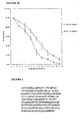
- proteoglycan extract MLA008E2 was tested in triplicate using two different doses of 10 ⁇ L and 50 ⁇ L of extract in a total volume of 750 ⁇ L of serum-free DMEM under sterile conditions. The results are shown in Fig. 3 which indicate cell migration increased in a dose dependent manner suggesting the presence of endothelial cell migration stimulatory agent(s) in the extract.
- the initial fractionation of MLA008E2 involved RP-HPLC separation on an analytical Phenomenex Jupiter 4 ⁇ Proteo 90 ⁇ (250 x 4.06 mm) column with a Phenomenex SecurityGuard C-12 guard column as described in Example 1.4.
- This column is designed to bind peptides smaller than 10 kDa.
- Fractions were pooled and tested for endothelial cell migratory potential (5 x 10 4 cells /well in serum free medium were allowed to migrate for 22 ⁇ 1hour at 37 °C in 5% CO 2 ) and proliferative activity.
- a cell blank provided the background level of proliferation.
- 10% FBS provided the positive cell proliferation control.
- Cell proliferation was tested over a 72-hour period using an initial seed density of 1x10 4 cells per well in DMEM containing 1% FBS. Cell numbers were quantified using Molecular Probes CyQuant kit as per the manufacturers instructions.
- fractions 11-15, 16-20, 21-25, 26-30 and 31-35 appeared to contain agents that stimulated bovine endothelial cells to migrate compared to the background level of migration (cells and media).
- Single fractions were chosen from within the initial groupings that appeared by thin layer chromatography (TLC) analysis to contain a representative peptide profile (fractions 12,13,17, 22, 24, 26, 31 and 33). The individual fractions were re-tested for their endothelial cell migration activity.
- Fractions 12 and 33 appeared to have the ability to enhance endothelial cell migration, (the higher the emission being indicative of the greater the number of cells that have migrated). Fractions 12 and 33 were further fractionated using gel filtration on a TSK2500 colunm.
- fraction 33 There were multiple peptides present in fraction 12 and one main peak present in fraction 33.
- the unknown agents in fraction 33 were analysed. Briefly, the compound was analysed by UV/Vis spectrometry and fluorescent spectrometry, proton and 13 C NMR and IR.
- the result of the analysis of fraction 33 implied that the major constituent was tryptophan. Tryptophan was tested for its ability to promote endothelial cell migration and surprisingly, appeared to be able to promote migration of the cells. Without being limited by theory, the cell migration associated with endothelial cells is believed to be due to the breakdown of tryptophan induced by both tryptophan dioxygenase and indoleamine 2,3- dioxygenase.
- Both of these enzymes use superoxide to catalyse the cleavage of the indole ring producing N-formyl kynurenine, and are thereby anti-inflammatory because they scavange oxygen radicals.
- tryptophan breakdown leads to both a stimulation in angiogenesis and a reduction in inflammatory damage induced by inflammatory cells present in the wound (e.g., neutrophils).
- Fraction 12 was further fractionated on a TSK2500 gel filtration column.
- the column was equilibrated with phosphate buffered saline and 100 ⁇ L of sample was applied to the column at a flow rate of 1 mL/min.
- the elution profile was monitored at 280 nm and 214 nm.
- Fractions of 1 mL were collected for a period of 60 minutes.
- the resulting fractions were retested for their ability to promote endothelial cell migration. The results were inconclusive, It was, therefore, postulated that synergy between these peptides may have caused the initial observation or that higher concentrations of the peptides were needed to produce an effect.
- MLA008E2 RP-HPLC fractions were retested (2 x 10 5 cells in serum free DMEM were allowed to migrate for 22 ⁇ 1 hours at 37 °C in 5% CO 2 ) in the absence of 1% FBS.
- Table 3 Angiogenesis assay results for fractions of extract MLA008E2 Sample Protein Concentration Mean growth rate Image Percentage stimulation or inhibition Control (batch 1) 12.5 ⁇ 0.9 A Fumagillin (negative) 20 ⁇ g/ml 9.5 ⁇ 1.7 B 24.1% inhibition MLA008E2 # 31-38 1 ⁇ g/mL 13.2 ⁇ 2.0 C 5.9% stimulation MLA008E2 # 33 1 ⁇ g/mL 14.1 ⁇ 2.7 D 13.1% stimulation Control (batch 2) 12.5 ⁇ 2.2 E Fumagillin (negative) 40 ⁇ g/ml 5.4 ⁇ 1.6 F 57.7% inhibition VEGF (positive) 40 ⁇ g/ml 13.9 ⁇ 0.2 G 11.1% stimulation MLA008E2 3-15 1 ⁇ g/mL 16.1 ⁇ 0.6 H 28.6% stimulation MLA008E2 16-29 10 ⁇ g/mL 14.1 ⁇ 1.4 I 12.7% stimulation
- the initial batch of samples analysed suggested some stimulation was present in fraction MLA008E2 #33 (corresponding to the fraction containing tryptophan).
- a further batch of samples analysed indicated that a somewhat similar amount of stimulation was present in extract MLA008E2 3-15.
- a similar level of stimulation was observed between MLA008E2 #33 of the initial batch and MLA008E2 16-29 of the further batch.
- Each animal received 4 doses (25 ⁇ L each dose) of the test extract MLA008E2 3-15 which corresponds to MLA-KDJ 007 inphosphate-buffered saline (PBS) (1 ⁇ g/mL) applied topically to one of the wounds on days 1, 3, 5, 7 and 9.
- PBS inphosphate-buffered saline
- the peptide solution was filter sterilised. Some material was retained on the filter.
- 25 ⁇ L of PBS only was applied, so each animal had its own control wound. Photographs of each wound were taken prior to each addition using a Canon EOS 3000N camera (F2.8 Macro lens) and Fuji Professional 400NPH film, Prints of each exposure were recorded digitally and the area of each wound calculated from these images using NIH Image 1.63 software.
- the rate of wound healing was accelerated dramatically between days 7 to 9 for the test wounds compared to the control wounds over the same time period.
- complete wound closure was achieved earlier than the control wounds.
- the faster closure rate suggests that the wound healing process is stimulated at a number points along the wound healing cascade leading to faster wound closure.
- the molecular weights of the peptides / proteins present in the extracts having activity were estimated by gel filtration.
- a TSK2500 gel filtration column was equilibrated with phosphate buffered saline and 100 ⁇ L of sample was applied to the column at a flow rate of 1 mL/min. The elution profile was monitored at 214 nm. Fractions of 1 mL were collected for a period of 60 minutes. Proteins, peptides and amino acids of known relative molecular weight were run under the same conditions to calibrate the column. The results are shown in Table 5.
- the extract was subjected to preparative SDS-PAGE and the 27kDa protein excised for N-terminal sequence analysis.
- the N-terminus was discovered to be blocked.
- a further sample of the 27kDa protein was prepared and digested with trypsin overnight at 37°C. Resulting peptides were loaded onto a C18 Vyadac column (Vyadac Company (Herperia, CA, United States) and the peaks collected from a 1hr gradient of 0-100%B (60% acetonitrile + 0.1% TFA). The N-terminus of these peptides was subsequently sequenced.
- EXAMPLE 3 Purification of ⁇ B2 crystallin, from bovine eye and assessment of wound healing activity
- ⁇ B2 crystallin was partially purified using two different approaches that involved gel filtration chromatography on Sephacryl 300HR and ultrafiltration using several nominal molecular weight cut-off membranes.
- ⁇ B crystallin was purified ion exchange chromatography on DEAE Sepharose fast flow.
- Fresh bovine eyes were obtained from an abattoir and processed immediately.
- a scalpel was used to slice a 2-3 cm incision between the sclera and the pupil.
- the lens was forced from the eye by pressing a thumb on the pupil. Using this approach intact lens were recovered with a minimum of unwanted tissue attached.
- the lens was removed from the eye by making an incision through the centre of the cornea. This incision reached into the lens cutting through the lens sheath, and spanned the diameter of the cornea. This allowed removal of the lens from the eye free of lens sheath and other materials by gently squeezing the eye from either side.
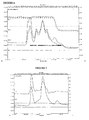
- a typical elution profile for bovine crystallin extract is given in Fig. 6 . Elution was performed at 1 mL/min and protein was monitored at 214 run (triangle) and 280 nm. The extract typically contained around 45 mg/mL of protein following ultracentrifugation to remove insoluble material. Peaks resulting from the separation of the various mammalian crystallin proteins were distinguished in the elution profile by relative molecular weight and immunoblot analysis, and by comparing the elution profile to profiles previously described in the literature for crystallin protein.
- the six fractions obtained from the gel filtration column containing all the crystallin proteins were analyzed by SDS PAGE analysis.
- Crystallin ⁇ B2 was present in all ⁇ fractions ( ⁇ H, ⁇ L1 and ⁇ L2). Recovery of ⁇ B2 crystallin from ⁇ H fraction was attempted through a pH shift. The pH was reduced from 6.8 down to 5.3 to ionize the histidine residues on the surface of ⁇ B2 crystallin which has been suggested to lead to the dissociation of the beta H crystallin aggregate and the formation of a ⁇ B2 crystallin dimers of 46-48 kDa that may be separated by gel filtration. The elution profile is shown in Fig. 7 . Elution was performed at 1 ml/min and protein was at 214 nm (triangles) and 280 nm.
- the ⁇ B2 dimer was eluted with a maximum absorbance in fraction 42.
- the fractions containing this protein were pooled, dialysed and freeze dried.
- the beta H fraction appeared to contain alpha crystallins which were readily separated using gel filtration at pH 5.3 from the dimer ⁇ B2 crystallin. When the pH was adjusted using acetic acid no precipitate formed.
- This fraction was pooled and verified as containing ⁇ B2 crystallin and is referred to below as preparation "Wound Heal 1".
- a bovine cystallin extract containing all of the water soluble crystallins was initially prepared from lens tissue by extraction with distilled water, the extract having a pH of about 6.8, followed by fractionation in distilled water, initially using a 300 kDa nominal molecular weight cut off membrane (NMWCO) in a pressure cell at 70 psi and at 4 °C.
- NMWCO nominal molecular weight cut off membrane
- a gel formed on the membrane which was predominately composed of alpha crystallins and ⁇ crystallins. The gel was collected and a portion dissolved in water and analyzed by gel filtration chromatography, and the remaining gel material was stored at 4 °C in a Petri dish. Gamma crystallins appeared to be absent from this gel.
- the ⁇ crystallin fractions were diafiltered and then freeze dried to produce the second sample tested for wound healing analysis referred to as preparation "Wound Heal 2".
- the majority of the protein present was ⁇ B2 crystallin.
- a dimer of ⁇ B2 crystallin with a small amount of a dimer between ⁇ B2 crystallin and ⁇ A3 crystallin was observed to elute with a peak at fraction 42, which was equivalent to the ⁇ L2 fraction of the bovine crystallin extract eluted using this column.
- Beta crystallins were fractionated using 100 kDa and 50 kDa NMWCO membranes, whereas the gamma crystallins were separated and concentrated using a 30 kDa and 5 kDa NMWCO membrane respectively. All fractionation steps were performed using a pressure cell at 70 psi and at 4 °C as described above. SDS PAGE analysis was performed to observe the protein profiles present in these various fractions.
- the 300 kDa retentate contained predominately alpha A and B crystallins and beta crystallins. High molecular weight non-crystallin proteins that were concentrated using this membrane were also observed the identity of which are unknown,
- the 300 kDa filtrate, 100 kDa retentate and 50 kDa retentate contained mainly ⁇ crystallins.
- the 50 kDa filtrate, 30 kDa retentate, 30 kDa filtrate and 5 kDa retentate contained gamma crystallin proteins of various sizes.
- the gamma crystallins that were present within the fractions have a molecular weight lower than 50 kDa.
- Alpha crystallin proteins were predominately present in the 300 kDa retentate.
- the ⁇ crystallins were located in the fractions between 300 kDa and 50 kDa.
- a crude bovine crystallin extract was fractionated using DEAE Sepharose fast flow under denaturing conditions in order to isolate ⁇ B crystallin.
- the column (3 x 33 cm) (Pharmacia XK-26 column containing GE Healthcare DEAE Sepharose fast flow ion exchange resin.) containing DEAE Sepharose fast flow was equilibrated with 5 mM Tris HCl pH 7.6, 6 M urea, 0.01% DTT.
- a gradient elution was performed using 50 mM Tris HCl pH 7.6, 6 M urea, 0.01% DTT and elution was completed using 35 mM Tris HCl pH 7.6, 6 M urea, 0.01% DTT, 1M NaCl.
- Protein absorbance was monitored using a BIORAD QuadTec detector at 214 nm and 280 nm. Six mL fractions were collected. 50 mL of bovine crystallin 5 mg/mL was loaded at a flow rate of 1 mL/min.
- Fractions under a symmetrical peak off the column were pooled and dialysed using a 12,000 Da nominal molecular weight cutoff membrane against at least three changes of distilled water over 2 days at 4 °C to remove urea.
- the sample was freeze dried and analyzed using SDS PAGE analysis ( Fig. 8 ).
- Fractions 37-47 were shown to contain bovine ⁇ B crystallin by Western blot and MS analysis. The purified ⁇ B crystallin is referred to below as "Wound Heal 3".
- Mass spectrophotometer (MS) analysis of the Wound Heal preparations (1-3) was also undertaken. As discussed above, crystallin proteins can undergo post translational modifications. An extensive list of post translational modifications can be found in a review article by Hoehenwarter et al., 2006.
- ⁇ B2 crystallin (23168 Da) was observed in Wound Heal 1. From the MS result, it appears that another protein was present having a mass around 22664 Da. Two proteins were detected in the MS analysis of the Wound Heal 2 preparation, namely ⁇ B2 crystallin (23167 Da) and ⁇ A3 crystallin (25061Da). With the number of post translational modifications that have been identified for bovine ⁇ B2 crystallin, the mass range for this protein can differ considerably. A peak at 23095 may have resulted from loss of acetyl alanine while a peak at 23335 may be the fully modified form as indicated in Fig. 9 (SEQ ID No. 9).
- Rat treatment protocols Group Treatment Age (weeks) Initial Weight range (g) A Control 21.1 384-428 B Bovine extract ⁇ B2 crystallin (Wound Heal 1) 20.4-21.1 382-442 0.01 mg/wound C Bovine extract ⁇ B2 crystallin (Wound Heal 2) 20.3-21.1 388-444 0.1mg/wound D Bovine extract ⁇ B crystallins (Wound Heal 3) 20.3-21.1 378-420 1mg/wound E Positive control-Aloe vera 21.1 382-436
- the animals were weighed two days before the start of the trial and weights grouped from lightest to heaviest. Animals were then randomly assigned to groups based on body weights, using 5 x 5 Latin squares so that group means were as similar as possible.
- Rats were housed singly post-operatively. Food consumption was not measured. A daily dose of about 4ml of jelly was administered from day 1 to assist in rehydration.
- each animal was given a subcutaneous injection of Temgesic at the base of the neck (0.075 mg/kg body weight). Each animal was anaesthetised using 3% halothane in oxygen. Once the pedal reflexes had been abolished, the animals were shaved from the base of the skull to the hind limb area using electric clippers. The skin on each rat was disinfected using 0.5% chlorhexidine in 70% ethanol. For each animal, the distance from the base of the skull to the top of the hip joint was measured and the wound position marked with a black felt tip pen, 6cm below the base of the skull along the spinal axis,
- the animals were euthanised by CO 2 inhalation and cervical dislocation.
- the entire wound site including adjacent skin, fascia and muscle tissue was excised and fixed in formalin for histological and immunohistochemical examination.
- the wounds were embedded in paraffin wax and three ⁇ m thick sections cut, attached to poly-L-lysine coated glass slides, and dewaxed and stained with haematoxylin, eosin and van Gieson stain for histological examination. Other sections were used for immunohistochemistry.
- Body weights were measured on days 1, 3, 5,7,9,11,13,15,17,19,21 and 23 and deviations from original body weights (on day 1) calculated for each animal.
- photographs were taken using a Canon Digital Eos 20D with a F2.8 Macro lens. Photographs were downloaded and wound areas measured using NIH Image J 1.633u software (National Institutes of Health (NIH), Rockville Pike, Bethesda, Maryland, Unite States; http://rsb.info.nih.gov/nih-image). A laser speckle image was also taken at the same time as each space photograph was taken. Animals were weighed every second day when the photographic record of wound closure rate was also made.
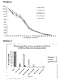
- Table 8 Wound areas of different treatment groups 0.01mg/ wound 0.1mg/ wound 1.0 mg/ wound Wound heal 1 Wound heal 2 Wound heal 3 Aloe vera Day PBS S.E.M ⁇ B2 crystallin S.E.M ⁇ B2 crystallin S.E.M ⁇ B crystallin S.E.M ⁇ B crystallin S.E.M 20mg/ wound S.E.M 1 100 0 100 0 100 0 100 0 100 0 3 85 3 86 3 82 1.7 93 4 84 1.6 5 80 2 75 2 69 1.3 82 2 76 3.6 7 71 3 69 3 67 0.9 76 2 68 1.5 9 52 4 54 3.7 50 3.2 58 4.7 48 4.4 11 27 1 32 5.4 28 1.9 34 1.4 29 2.3 13 18 0,8 18 1.9 20 2.4 23
- the rate of wound healing in Group A was of the order expected.
- the pattern of healing, with the fastest phase being between days 7 and 11 was also as expected.
- the average healing rate for the rats in Group C was higher than that of the Control group (Group A) over the first 9 days. Thereafter the rate for the two groups was almost identical. It is noticeable that the closure rate over the first 11 days was slightly faster for this group than for the group receiving the lower dose (Group B) indicating a potential dose response.
- Wound Heal 1 A low concentration of Wound Heal 1 was used in this assay since at higher protein concentrations, ⁇ B2 crystallin forms dimers and higher order aggregates.
- Wound Heal 2 contained a higher concentration of ⁇ B2 crystallin and so may form dimers with another protein that was also present in this extract, ⁇ A3 crystallin.
- the activity of the ⁇ B2 crystallin was lower than expected with only a slight decrease in wound area at day 5 compared to the other treatments.
- bovine crystallin fractions were tested for ability to inhibit elastase I activity. Briefly, a small amount of elastase inhibition was observed with a crude bovine crystallin extract and alpha A / B crystallin fraction. No activity was detected in the ⁇ crystallin fractions (beta H, L1 and L2 all containing ⁇ B2 crystallin). Gamma S crystallin was shown to have considerable elastase inhibitory activity when compared to the other fractions.
- Bovine crystallin fractions were tested for ability to protect the proteases elastase I, trypsin and chymotrypsin from heat denaturation. Briefly, the proteases were heated in a boiling water bath for 30 seconds in the presence or absence of the crystallin fractions. The results are shown in Fig. 13 .
- the control demonstrated the amount of protease activity before heating and the heat denatured control (HD) demonstrated the amount of protease activity remaining after being heated in the water bath.
- Chaperone activity was observed when elastase I was mixed with Wound Heal 3 but not with Wound Heal 1 or Wound Heal 2 preparations. Effectively no chaperone activity was observed when the samples were treated with trypsin or chymotrypsin.
- ⁇ B crystallin and ⁇ A ctystallin isolated by SP Sepharose fast flow were both able to protect elastase I from heat inactivation.
- Alpha B crystallin appeared to be the most active with respect to protecting elastase I heat denaturation.
- the first study investigated the angiogenic potential of bovine fractions prepared by gel filtration analysis as described in Example 3.2.
- the alpha fraction contained ⁇ A and ⁇ B crystallin
- the ⁇ H fraction contained all ⁇ crystallins
- the ⁇ L1 fractions contained a sub-fraction of beta crystallins and a high content of ⁇ B2 crystallin and beta B3 crystallin
- the beta L2 fraction contained only ⁇ B2 crystallin and ⁇ A3 crystallin.
- ⁇ B3 crystallin was prepared by heating (97-99°C for 3 minutes, supernatant collected by centrifugation) and was essentially homogenous.
- Alpha A crystallin and ⁇ B cystallin samples were prepared by ion exchange chromatography as described above and were also essentially homogenous.
- the Wound Heal 1-3 preparations were also tested in this study.
- the third study investigated the angiogenic potential of ovine crystallin proteins compared to their bovine equivalents. Both bovine and ovine crystallin proteins were purified essentially to homogeneity by RP HPLC using a C4 column, as described below in Example 5. Homogeneity was confirmed by SDS-PAGE.
- the effect of elastase I treatment of ⁇ B2 crystallin was also investigated by comparing the angiogenic potential of intact ⁇ B2 crystallin (i.e. undigested) with elastase I digested pB2 crystallin. Elastase I treatment releases the N-terminal and C-terminal extensions of the crystallin protein leaving a globular protein that is monomeric.
- a single Lewis rat was euthanised and its aorta transferred to a 15 mL Falcon tube containing 10 mL of MCBD-131 medium and stored on ice.
- the outer fatty tissue was removed under an Olympus SZ-60 Dissecting microscope. This was undertaken with fibre optic lamp illumination.
- the aorta was then cut into sequential rings approximately 2mm in size. Connective tissue was removed with micro scissors and forceps from the outside of the individual rings. Clean rings were stored in a separate petri dish in sterile MCDB-131 medium.. Rings were pre-incubated overnight at 37 °C in an atmosphere of 97% air / 3% CO 2 .
- Samples to be tested were diluted to 100 ⁇ g/mL in 5 mL of MCDB-131, and 15 mL of each sample placed in each of three appropriate wells. The plate was then placed in incubator (37°C, 97% air / 3% CO 2 ),
- Culture plate were examined daily for signs of infection (fungal growth or yellowing of media) in the wells using an inverted microscope. The growth of microvessels from their perimeters was also observed. Digital photographs of each well were taken seven days after plating using a Pixel a PVC-100C digital camera mounted on an OLYMPUS CK-12 microscope and stored on a MacIntosh Computer. A low power field (i,e. 20x. A 640x480 pixel PICT) was sufficient. PICT can be converted to grey scale for visualization and measurement in NIH Image. The greyscale image can be opened with the NIH Image program with J 1.32 Image software.
- the area occupied by the microvessel growth around each ring and the area of each aortic ring was determined using the shape tool in NIH image software to draw around the ring. This measurement was repeated, the second time drawing around the capillaries. Typically, 4 measurements were taken- internal and external capillary growth and inner and outer aortic ring area. The ratio of the area of microvessel growth to aortic ring area was then calculated for each well and the mean value and the standard deviation (SD) for each sample of the triplicate determined.
- SD standard deviation
- HIER Heat Induced Epitope Retrieval
- HIER treatment consisted of the following steps. Fixed, embedded tissue sections were adhered to a glass slides using HistoGripTM (Zymed® Cat. No. 00-8050) silane or poly-L-lysine. The immobilized sections were deparaffinzed in xylene and rehydrated in graded alcohols followed by rinsing in deionized water and then in PBS. Endogenous peroxidase was blocked with 0.5% hydrogen peroxide in for 10 minutes, following by further rinsing in PBS. The slides were placed in a slide rack and lowered into a IL glass beaker (Pyrex) containing 500 mL of working solution of citrate buffer.
- the beaker was placed on a hot plate and the solution heated until boiling and allowed to boil for 15 minutes. After heating, the beaker containing slides was removed from the hot plate and allowed to cool for 25 minutes. Slides were then rinsed with PBS and avidin/biotin blocking performed if necessary. A general protein blocking step was also carried out prior to the immune staining.
- the slides were specifically stained for Vimentin and CD-31, with primary antibody used at a 1:20 dilution and 11:10 dilution, respectively. Staining allowed the ratio of endothelial cells to all migrating cells to be calculated.
- the six samples of crystallin aggregates tested contained all the different forms of the crystallin proteins e,g. post translational modifications that include phosphorylation and acetylation and protease degradation product.
- the alpha crystallins were in high molecular weigh clusters (600 to 800 kDa), whereas the beta crystallins were found to be in tetramers, trimers and dimers ranging in size from 200 to 40 kDa. Size was determined by calibration of the Sophareyl 300 HR column used for fractionation, SDS PAGE analysis atid from the known size ranges reported in the literature.).
- the gamma crystallins were monomeric with a size range between 20 to 30 kDa.
- ⁇ B2 crystallin stimulated a 21.83% increase in microvessel growth at a concentration of 200 ⁇ g/mL.
- a 200 ⁇ g/mL solution of ⁇ A crystallin was also found to be 8.26% stimulatory. None of the other test samples exhibited an angiogenic effect on microvessel growth.
- the VEGF at 50 ⁇ g/mL produced a slight stimulation although this was not statistically significant.
- Table 10 Angiogenic activity of ovine and bovine crystallin proteins Sample % of Control %SD t-test Control 100 12.9 1 VEGF(50 ⁇ g/mL) 113.7 2.7 0.15 Ovine ⁇ B2 crystallin (100 ⁇ g/mL) 103.5 9.8 0.73 Ovine ßB2 crystallin elastase I digest (100 ⁇ g/mL) 120.8 14.1 0.17 Bovine ⁇ B2 crystallin (non-elastase treated) (100 ⁇ g/mL) 93.4 8.3 0.50 Ovine ⁇ A crystallin (100 ⁇ g/mL) 130.2 13 0.02 Ovine ⁇ B crystallin (100 ⁇ g/mL) 97.1 12.8 0.8
- Table 11 Angiogenic activity of bovine crystallin aggregates Sample % of Control %SD t-test Control 100 12.9 1 VEGF(50 ⁇ g/mL) 113.7 2.7 0.15 Indoleamine 2,3-dioxygenase IDO (100 ⁇ g/mL) 116.4 11.3 0.31 Tryptophan dioxygenase TDO (100 ⁇ g/mL) 106.2 16.6 0.76 IDO + Ovine ⁇ B2 crystallin (100 ⁇ g/mL) 104.9 9.0 0.7 TDO + Ovine ⁇ B crystallins (100 ⁇ g/mL) 103,82 20.3 0.85
- Ovine eyes were collected and the lens removed as described in Example 3.1 above, The lenses were extracted using distilled water. After mixing at 4 °C for 30 minutes the outer layer of the lens had dissolved. The lens extract was centrifuged and the pH adjusted to 4.5 with glacial acetic acid. No precipitate formed during the pH adjustment. The extract was filtered through a 0.45 micron filter prior to loading onto a preparative C4 HPLC column. A single peak containing ovine ⁇ B2 crystallin was recovered. Modification of the protocol was used to successfully shorten the elution time required to isolate ovine ⁇ B2 crystallin. From two preparative runs 100 mg of purified ovine ⁇ B2 was isolated. The yield of ⁇ B2 crystallin from ovine lens was 8% of the protein loaded onto the preparative HPLC column. This equates to 2.7% of the lenses' wet weight. The ovine lens contained 34% protein.
- Ovine eyes were obtained from a local abattoir and processed immediately (within 1 hour). A scalpel was used to slice a 1-2 cm incision at various positions on the eye. The 6 lenses recovered were washed with 200 mL of distilled water and the remaining lens sheaths were removed. The water was replaced by 200 mL of fresh distilled water and stirred at 4 °C in 20 volume for 30 minutes. The hard lens nucleus remained intact after this period of time and was discarded from processing, The outer layers of the lens dissolved and a cloudy white solution was obtained which was centrifuged at 30 000 rpm for 30 minutes at 10 °C or clarified using 5000 rpm for 30 minutes at 10 °C. The supernatant containing the water soluble lens crystallins was further fractionated using RP-HPLC to determine the potential yield of ⁇ B2 crystallin from ovine eyes.
- a preparative RP-HPLC protocol was adapted from the analytical method. Briefly, ovine ⁇ B2 crystallins were isolated by RP-HPLC using a Phenomenex Jupiter C4 300A 10um 250 x 21,2, Cat. No. 00G-4168-P0, and the SecurityGuard cartridge C4-300A 15 x 21.2 Jupiter (Serial No. 38833-1). The column was equilibrated with 0.1% TFA in dH 2 O and 10% acetonitrile containing 0.1% TFA, flow rate 25 mL/min.
- a gradient of buffer B (100% acetonitrile containing 0.1 % TFA) was applied as follows: 0 min 10% B, 10 min 30% B, 70 min 40% B, 90 min 60% B, 95 min 98% B and 100 min 10%. Absorbance was monitored at 280, 214, 235, 330 and 450 nm. 18 mL fractions were collected every 43 seconds. The load volume was 50 mL (9.57 mg/mL).
- a shorter processing time was obtained under the following modified gradient conditions. 0 min 10% B, 10 min 30% B, 15 min 31% B, 20 min 40%, 25 min 60% B, 30 min 98% B and 32 min 10%. Absorbance was monitored at 280, 214, 235, 330 and 450 nm. Again the flow rate was 25 mL/min and 18 mL fractions were collected. Load volume 80 mL (9.57 mg/mL).
- Table 12 Weight of ovine eye tissue Tissue Total Weight (g) Ovine eye 94.65 Ovine lens 4.69 Ovine vitreous 66.17 Waste 23.79
- Table 13 Determination of ovine ⁇ B2 crystallin yield Protein extract Volume (mL) Protein (mg/mL) Total protein (mg) Yield (%) Water extract 204 9.57 1952 N.D. Load onto HPLC 130 9.57 1244 N.D. Fractions 18 + 19 72 1.4 100 N.D. Freeze dried protein - - 100 8 N.D: Not determined
- Ovine eyes were collected essentially as described in Example 5.1 and processed immediately. Briefly, a scalpel was used to slice a 2-3 cm incision across the cornea. The lens was squeezed out of the eye by pinching either side of the eye whilst the blade was removed, allowing about 12 eyes per minute to be processed. The lenses were stirred over night at 4 °C in 20 volumes (w/v) of distilled water. The lens slowly dissolved during this period. A cloudy white solution was obtained, which was filtered through Celite ( Celite 545 34967-0025 , Acros Fine Chemicals, Belgium,). The pH of the supernatant containing the water soluble lens crystallins was adjusted to 3.5 using acetic acid and ⁇ B2 crystallin was purified using RP-HPLC as described in Example 5.2.
- Protein gel precipitates of ovine ⁇ B2-crystallin were prepared making use of the proteins capacity to spontaneously precipitate in the presence of salt solutions under acidic conditions.
- a transparent ⁇ B2-crystallin acidic gel was prepared as follows. Intact ovine ⁇ B2 crystallin purified by preparative RP-HPLC as described in Example 6.1 was dissolved at 85 mg/mL to 100 mg/mL in distilled H 2 O. The formation of a transparent acidic gel with a pH of about 1.4 -1.7 was evident after the protein had completely dissolved. When an elastase I truncated form of the RP-HPLC purified ovine ⁇ B2 crystallin was dissolved at its maximum solubility (20 mg/mL) it was unable to spontaneously forum a transparent gel.
- the elastase I truncated form of the ovine ⁇ B2 crystallin was prepared by RP-HPLC as described in Example 6.1.
- the truncated form was prepared by digesting purified ⁇ B2 crystallin using 1:100 enzyme to substrate ratio of Elastase I (Worthington Biochemical Corporation, Cat, No. 2280 (5.05 u/mg and 27.2 mg/mL) Batch No. 38A10174).
- Partial hydrolysis of ⁇ B2 crystallin was achieved by mixing purified ⁇ B2 crystallin 10-50 mg/mL with elastase 0.1-0.5 mg/mL in 10 mM Tris HCl pH 8.0 containing 10 mM CaCl 2 for 1 to 8 hours, Upon completion of the reaction the truncated form ⁇ B2 crystallin was subjected to RP-HPLC purification. The retention time of the intact form had shifted from around 15 minutes to 10 minutes for the truncated form indicating protein modification. SDS page analysis also demonstrated a shift in the relative molecular weight of the protein, The protein peak that eluted at 10 minutes was collected and solvent removed under vacuum, The protein solution was then freeze dried.
- the freeze dried truncated protein was mixed with an organogel at 5 mg/g of gel and applied to the wound of a rat at 300 ⁇ L per wound per day.
- the freeze dried protein was dissolved in distilled water at 10-20 mg/mL filter sterilized and used to coat gauze presoaked in a salt solution.
- the protein coated gauze was allowed to air dry. Final sterilization could be achieved using ethylene oxide gas or gamma radiation after packing. This product requires PBS hydration prior to application onto a wound.
- intact ⁇ B2-crystallin was formulated with calcium salts resulting in a protein precipitate gel with a final pH about 4.0-5.0.
- 0.5 g of purified intact ovine ⁇ B2-crystallin prepared as described in Example 6.1 was dissolved in 5 mL of distilled H 2 O. This resulted in a clear gel like mixture with a pH of around 1.5. This was mixed with 10 mM (final) Ca(OH 2 ) and 100 mM (final) CaCl 2 , which resulted in precipitation of proteins, The solution was then centrifuged and the pellet and supernatant separated.
- the pellet (0.11 g) was dissolved in 3 mL of distilled water and was stored at 4°C. As the supernatant still contained 75% of the initial protein content, it was freeze-dried and a fluffy white powder weighing 0.43 g was obtained. This freeze-dried powder was mixed with the pellet solution, resulting in a gel like product having a pH value at pH 4.0. The final concentration of the gel was 100 mg/mL. This gel was used in a delayed wound healing model described below in Example 8.
- a water free organogel (INCI MagnateTM, Johnson & Wilkins Limited) containing isopropyl myristate and magnesium stearate was mixed with the RP-HPLC purified ovine elastase I truncated form of ⁇ B2 crystallin prepared as described in Example 6.2, at a concentration of 5 mg/mL.
- the freeze dried protein was mixed evenly into the organogel to form a smooth paste. This paste was used in the 5 day wound healing study described below in Example 7.
- a wound healing study was carried out using male 30 Spargue-Dawley rats assigned to 5 treatment groups with 6 rats in each group.
- the treatment groups were as follows:
- Bovine IDO was prepared from bovine heart tissue as described previously (Shimizu et al., 1978). A cation exchange SP resin (BioRad) was used under the following conditions. The resin was equilibrated using 10 mM KH 2 PO 4 pH 6.4. Then 1.0 mL of bovine heart extract (prepared by extracting 25 g of frozen heart with 125 mL of 10 mM KH 2 PO 4 pH 6.4) was loaded onto the column. The column was washed with 200 mL of equilibration buffer, followed by a gradient elution from 0-1 M NaCl in equilibration buffer over a 200 mL volume. Fractions of 6 mL were collected and tested for IDO activity.
- a cation exchange SP resin BioRad
- Fractions containing activity were pooled, dialysed (10-12 kDa NMWCO) and then freeze dried.
- the freeze dried protein is referred to as bovine IDO or bovine heart enzyme,
- the freeze dried protein powder was mixed with an organogel at 10 mg/mL.
- Ovine ⁇ A crystallin was prepared by using a combination of gel filtration chromatography on Sephacryl 300HR equilibrated and run using 100 mM phosphate buffer pH 6.2 and RP HPLC using the standard protocol outlined in Example 6.1.
- the load material was the ⁇ A and ⁇ B fraction off the Sephacryl 300HR column.
- Purified ovine ⁇ A crystallin was confirmed by MS analysis and SDS PAGE analysis.
- a positive control (Grapefiuit skin extract) was chosen because of its ability to stimulate blood flow as determined by rat aortic ring assay (a model system for vessel formation), A comparison, was made between compounds with similar modes of activity as determined by the aortic ring model.
- the animal wound model used was essentially as described in Example 3.G. Briefly, approximately 30 minutes before wounding, each animal was given a subcutaneous injection of Temgesic in the fossa region of the groin (0.075 mg/kg body weight), The animals were anaesthetised using 3% halothane in oxygen and once pedal reflexes were abolished, each animal was shaved from the base of the skull to the hind limb area using electric clippers. Skin was disinfected using 0.5% chlorhexidine in 70% ethanol. For each animal, the distance from the base of the skull to the top of the hip joint was measured and the wound position was marked with a black felt tip pen, 6cm below the base of the skull along the spinal axis.
- the biopsy punch was wiped inside with a swab soaked in sterile saline to remove any tissue fluids and/or debris, and the punch was then immersed in 70% ethanol before use on the next animal.
- the condition of each animal was monitored closely each day after wounding. On each day the animals were provided with new toweling as bedding. All animals had a total of three topical applications of control or test compounds on Days 1, 2 and 3. Photographs were taken of each wound each day.
- the animals were euthanized by CO 2 inhalation and cervical dislocation.
- the entire wound site including adj acent skin, fascia and muscle tissue was excised, and each was bisected longitudinally.
- the left piece from animals 1, 2 and 3 of each group was fixed in 4% buffered formalin for 24 hours before being transferred to 70% ethanol.
- the right pieces from animals 1, 2 and 3 were stored frozen for further protein analyses.
- Both left and right section from animals 4,5 and 6 of each group were fixed in 4% buffered formalin. Analysis of the wound area and histological analysis of wounds was undertaken.
- the wounds were photographed daily and the images analyzed using NIH image to calculate the wound area. Descriptive statistics were evaluated. Preliminary t-tests examined whether outliers significantly affected results. Major analysis were performed on all data points collected. Preliminary analyses were undertaken on control wounds only using t-tests. Univariate ANOVAs were used to assess differences between treatment and controls and samples nested within treatments. A separate ANOVA assessed whether there was a dose response using the treatment samples only. Assumptions for normality were tested using q-q plots on studentized residuals and heterogeneity of variances were tested using Levene's tests. SPSS 11 for Mac OSX was used for assumption testing and statistical analysis.
- the entire wound site including adjacent skin, fascia and muscle tissue was excised and fixed in formalin for histological examination. After less than 24 hours in formalin, the wounds were embedded in paraffin wax and three ⁇ m thick sections cut, attached to poly-L-lysine coated glass slides, and dewaxed and stained with haematoxylin and eosin for histology.
- the truncated form of ⁇ B2 crystallin was found to have the greatest healing response over the 5 day wound healing study compared to the organogel vehicle negative control and the grapefruit positive control.
- the wound area for the truncated form of ovine ⁇ B2 crystallin at day 5 was 69.9 ⁇ 4.4 compared to 83.7 ⁇ 3.4 as indicated in Table 14
- Table 14 Percentage size of wounds with time Days after wound admin.
- Ovine meat protein Ovine meat protein, phosphorylated, 2.6mg/gm of organogel.
- Mean % of original wound area (N 6) S.E.M Group D.
- Bovine heart enzyme 10.0mg/gm of organogel.
- Mean % of original wound area (N 6) S.E.M Group E. Positive control, grapefruit ext., 1mg/gm of organogel.
- Histological analysis undertaken at day 5 revealed the inflammatory cell profile (i.e., macrophages, neutrophils, eosinophils, mast cells and plasma cells) at the wound transition zone was similar for all the test substances.
- the number of lymphocytes were statically significantly different (p ⁇ 0.01) between all of the test compounds and the vehicle control suggesting that the level of bacterial burden was higher in the test preparations compared to the vehicle only control. This may have resulted in the higher neutrophil content of the test groups compared to the controls.
- ischemic delayed healing model based on a model previously described by Chen et al., 1999 was used.
- the ischemia was caused by placing two incisional wounds (bipedicle flaps) either side of the excisional wound in the centre of the animals back. More particularly, male age and weight matched Sprague-Dawley rats were anesthetized with intramuscular ketamine hydrochloride (100 mg/kg body weight). The dorsal hair of each was clipped, and a 10 x 4 cm clear rectangular template was used to outline the area of study on the back. Under sterile conditions, a full thickness incision was made using a sterile 12mm biopsy punch, with a gentle rotating motion. Any bleeding was removed using cotton swabs.
- the incisions were closed using three interrupted sutures (4-0 Novafil, monofilament polybutester, nonadsorbable suture, Tyco, Cat, No 4402-33) at equidistant points along the length, Once haemostasis has again been achieved the wounds were photographed. After photographing the wounds, the test samples (100 ⁇ l of intact ovine ⁇ B2 crystallin and 100 ⁇ l of Medifill II TM (see Example 8.2)) were applied, using Gilson Microman CP100 & CP1000 positive displacementpipette. The biopsy punch was wiped inside with a swab soaked in sterile saline to remove any tissue fluids and/or debris.
- interrupted sutures 4-0 Novafil, monofilament polybutester, nonadsorbable suture, Tyco, Cat, No 4402-33
- the punch was immersed in 70% Ethanol before use on the next animal. Each animal's condition was monitored closely after wounding. On the second and third days after wounding, each animal was given a new towel as bedding. Changes from towelling to shavings was made only when wounds have dried and are not adversely affected by shavings - usually, between Days 5 to 7 of the study. The control group consisted of the excisional wound only. The rate of wound closure was measured over a 31 day period.
- the delay in wound closure between the ischemic and normal wounds was approximately 4 days for the early portion of the study up until day 13. At this time point the ischemic wounds were approximately twice the size as the wounds in the control animals. By day 15 the percentage of original wound area in the normal wound animals was approximately 14%. The ischemic wounds did not reach this level of closure until day 27 suggesting that the delay had been extended a further 8 days. This result suggested that tissue ischemia causes a significant delay in wound closure. The grooming of wounds was apparent in all treatment groups. Grooming may also increase the likelihood of wound infection which could delay the healing rate.
- the ischemic delayed wound healing rat model described in Example 8.1 was used to evaluate the wound healing benefit of intact ⁇ B2 crystallin compared to a commercially available Medifill II TM Type I bovine collagen (BioCore Medical Technologies, Inc. Silver Spring, MD, United States), wound healing collagen gel containing Type I bovine collagen in the triple helical molecular form specific to wound healing.
- the enhanced surface area/density of the collagen fibrils increase the level of collagen into contact with the wound surface.
- Intact ⁇ B2 crystallin protein was chosen over the RP-HPLC purified elastase I truncated form described in Example 6.2, because of the ability of the intact form to form a gel containing intact ⁇ B2 crystallin protein in high concentration.
- Human chronic wounds exhibit high levels of proteases (including human neutrophil elastase), that have the ability to hydrolyze the intact ⁇ B2 crystallin leading to the formation of the pro-angiogenic truncated form(s) of the protein.
- the production of the truncated form of ⁇ B2 crystallin from the intact protein may have been restricted in the earlier stages of wound healing in this model as the proteases that hydrolyze ⁇ B2 crystallin are present in inflammatory stages of wound healing which occur in the first 72 hours.
- administration of the intact form of ⁇ B2 crystallin may have greater benefit under those circumstances.
- Cloth material gauze or muslin was coated with RP-HPLC purified intact or elastase I treated ovine ⁇ B2 crystallin as follows.
- the cloth material was placed into a salt solution (10 mM acetic acid pH 3.5 containing 2M ammonium sulfate, although a number of different salts can be used including calcium salts as described above).
- the cloth was removed from the salt solution and blotted dry on filter paper until essentially free of excess water. No additional water could be removed from the material by blotting between two layers of filter paper.
- the cloth was then placed into an acidic solution of purified ⁇ B2 crystallin (10-50 mg/mL).
- ⁇ B2 crystallins truncated form was prepared in 10 mM acetic acid pH 3.5.
- the pH value can range from 2.0 to 5.0.
- the ⁇ B2 cystallin protein concentrations tested were found to effectively coat the cloth.
- ⁇ B2 crystallin coats the surface of the cloth with a white protein precipitate as shown by electron microscopy.
- the intact form of ⁇ B2 crystallin also demonstrated the ability to coat the surface of previously soaked cloth under identical conditions.
- the ability to coat bandage or other materials with ⁇ B2 crystallin may assist in delivering an effective amount of the protein to a wound site.
- Both the intact and the elastase I treated truncated forms of ⁇ B2 crystallin were found to coat cloth material gauze and muslin.
- the level of coating may be controlled by altering the salt concentration, protein concentration and the length of time the cloth, gauze or other suitable substrate material is in contact with the ⁇ B2 crystallin protein solution.
- the coated material may then be dried and sterilized (e.g., gamma irradiated) for subsequent use.
- a sponge material was also coated with ovine ⁇ B2 crystallin protein using the above protocol although the sponge was squeezed to remove excess liquid prior to being placed in the ⁇ B2 crystallin protein solution.
- This type of wound dressing may be suitable for use in wounds that produce high levels of exudate.
Landscapes
- Health & Medical Sciences (AREA)
- Chemical & Material Sciences (AREA)
- Life Sciences & Earth Sciences (AREA)
- Organic Chemistry (AREA)
- General Health & Medical Sciences (AREA)
- Medicinal Chemistry (AREA)
- General Chemical & Material Sciences (AREA)
- Public Health (AREA)
- Chemical Kinetics & Catalysis (AREA)
- Nuclear Medicine, Radiotherapy & Molecular Imaging (AREA)
- Veterinary Medicine (AREA)
- Pharmacology & Pharmacy (AREA)
- Animal Behavior & Ethology (AREA)
- Bioinformatics & Cheminformatics (AREA)
- Engineering & Computer Science (AREA)
- Biophysics (AREA)
- Proteomics, Peptides & Aminoacids (AREA)
- Molecular Biology (AREA)
- Toxicology (AREA)
- Zoology (AREA)
- Gastroenterology & Hepatology (AREA)
- Biochemistry (AREA)
- Genetics & Genomics (AREA)
- Heart & Thoracic Surgery (AREA)
- Dermatology (AREA)
- Cardiology (AREA)
- Medicines That Contain Protein Lipid Enzymes And Other Medicines (AREA)
- Peptides Or Proteins (AREA)
- Medicinal Preparation (AREA)
Applications Claiming Priority (2)
| Application Number | Priority Date | Filing Date | Title |
|---|---|---|---|
| AU2007904857A AU2007904857A0 (en) | 2007-09-07 | Agents with angiogenic and wound healing activity | |
| EP08783056A EP2190455A4 (fr) | 2007-09-07 | 2008-09-05 | Agents présentant une activité angiogénique et de guérison de plaies |
Related Parent Applications (1)
| Application Number | Title | Priority Date | Filing Date |
|---|---|---|---|
| EP08783056.8 Division | 2008-09-05 |
Publications (2)
| Publication Number | Publication Date |
|---|---|
| EP2407175A1 true EP2407175A1 (fr) | 2012-01-18 |
| EP2407175A9 EP2407175A9 (fr) | 2012-02-29 |
Family
ID=40428369
Family Applications (2)
| Application Number | Title | Priority Date | Filing Date |
|---|---|---|---|
| EP08783056A Withdrawn EP2190455A4 (fr) | 2007-09-07 | 2008-09-05 | Agents présentant une activité angiogénique et de guérison de plaies |
| EP11164650A Ceased EP2407175A1 (fr) | 2007-09-07 | 2008-09-05 | Agents dotés d'une activité de cicatrisation et angiogène |
Family Applications Before (1)
| Application Number | Title | Priority Date | Filing Date |
|---|---|---|---|
| EP08783056A Withdrawn EP2190455A4 (fr) | 2007-09-07 | 2008-09-05 | Agents présentant une activité angiogénique et de guérison de plaies |
Country Status (6)
| Country | Link |
|---|---|
| US (1) | US8404644B2 (fr) |
| EP (2) | EP2190455A4 (fr) |
| JP (1) | JP5685773B2 (fr) |
| AU (1) | AU2008295449A1 (fr) |
| NZ (1) | NZ583803A (fr) |
| WO (1) | WO2009029991A1 (fr) |
Families Citing this family (9)
| Publication number | Priority date | Publication date | Assignee | Title |
|---|---|---|---|---|
| EP2190455A4 (fr) * | 2007-09-07 | 2010-11-24 | Australian Meat & Live Stock | Agents présentant une activité angiogénique et de guérison de plaies |
| US8758802B2 (en) | 2009-12-14 | 2014-06-24 | University Of Massachusetts | Methods of inhibiting cataracts and presbyopia |
| JP5993308B2 (ja) | 2011-01-26 | 2016-09-14 | 雪印メグミルク株式会社 | 感覚改善剤 |
| WO2013073968A2 (fr) * | 2011-09-12 | 2013-05-23 | Industrial Research Limited | Agents de modulation de la signalisation cellulaire |
| JP2014005254A (ja) * | 2012-06-26 | 2014-01-16 | Bfsr:Kk | 熱傷由来全身性炎症反応症候群の予防外用薬 |
| PE20151747A1 (es) | 2013-03-14 | 2015-12-18 | Univ Massachusetts | Metodo para inhibir cataratas y presbicia |
| KR20170040648A (ko) * | 2015-10-05 | 2017-04-13 | 차의과학대학교 산학협력단 | 당뇨병성 족부궤양의 예방 또는 치료용 약학 조성물 |
| CA3004585A1 (fr) | 2015-11-13 | 2017-05-18 | The University Of Massachusetts | Molecules bifonctionnelles contenant peg pour utilisation dans l'inhibition des cataractes et de la presbytie |
| SG11202108446TA (en) * | 2019-02-08 | 2021-08-30 | Auckland Uniservices Ltd | Biomaterials and methods related thereto |
Citations (5)
| Publication number | Priority date | Publication date | Assignee | Title |
|---|---|---|---|---|
| JP2004000609A (ja) | 2002-05-15 | 2004-01-08 | Ge Medical Systems Global Technology Co Llc | 多重エネルギ画像による計算機支援診断 |
| US20040014920A1 (en) | 1998-10-15 | 2004-01-22 | Morken Peter Arnold | Polymers, containing a fluorocyclobutyl ring and their preparaton |
| WO2004096277A1 (fr) | 2003-05-01 | 2004-11-11 | Kyoto University | Agent prophylactique ou therapeutique pour affection par vascularisation intraoculaire |
| WO2005004894A2 (fr) | 2003-05-12 | 2005-01-20 | Expressive Constructs, Inc. | Procedes d'augmentation de la viabilite de cellules et de tissus |
| WO2008005021A1 (fr) * | 2006-06-30 | 2008-01-10 | The Johns Hopkins University | Utilisation de cristalline pour moduler l'angiogenèse |
Family Cites Families (13)
| Publication number | Priority date | Publication date | Assignee | Title |
|---|---|---|---|---|
| WO1999051738A1 (fr) | 1998-04-03 | 1999-10-14 | Hunan Medical University | Proteine beta 4 humaine a jonction lacunaire |
| WO1999051634A1 (fr) | 1998-04-03 | 1999-10-14 | Hunan Medical University | Proteine humaine beta 3 a jonction lacunaire |
| WO2004005883A2 (fr) | 2002-07-02 | 2004-01-15 | The Johns Hopkins University | Marqueurs endotheliaux tumoraux secretes et cytoplasmiques |
| JP3783200B2 (ja) * | 2000-08-24 | 2006-06-07 | 株式会社加美乃素本舗 | 血管新生剤 |
| WO2003096981A2 (fr) | 2002-05-15 | 2003-11-27 | Seul, Kyung, Hwan | Procede de modulation de l'angiogenese |
| US7456151B2 (en) * | 2004-07-14 | 2008-11-25 | University Of Utah Research Foundation | Promoting angiogenesis with netrin1 polypeptides |
| JP5964538B2 (ja) | 2005-02-03 | 2016-08-03 | コーダ セラピューティクス, インコーポレイテッド | 抗コネキシン化合物およびそれらの使用 |
| US7897581B2 (en) * | 2005-02-24 | 2011-03-01 | Massachusetts Eye & Ear Infirmary | Methods and compounds for promoting vessel regression |
| EP1978988A4 (fr) | 2006-01-13 | 2009-11-04 | Univ New York State Res Found | Regulation de jonctions communicantes a l aide de peptides |
| KR20100027091A (ko) | 2006-11-15 | 2010-03-10 | 코다 테라퓨틱스, 인크. | 상처 치유를 위한 개선 방법 및 조성물 |
| WO2008073466A2 (fr) | 2006-12-11 | 2008-06-19 | The Board Of Trustees Of The Leland Stanford Junior University | Alpha b-cristalline en tant que traitement de l'inflammation |
| WO2009012224A2 (fr) | 2007-07-13 | 2009-01-22 | The Research Foundation Of State University Of New York | Régulation à base de peptides à jonction lacunaire |
| EP2190455A4 (fr) * | 2007-09-07 | 2010-11-24 | Australian Meat & Live Stock | Agents présentant une activité angiogénique et de guérison de plaies |
-
2008
- 2008-09-05 EP EP08783056A patent/EP2190455A4/fr not_active Withdrawn
- 2008-09-05 WO PCT/AU2008/001319 patent/WO2009029991A1/fr active Application Filing
- 2008-09-05 JP JP2010523239A patent/JP5685773B2/ja not_active Expired - Fee Related
- 2008-09-05 EP EP11164650A patent/EP2407175A1/fr not_active Ceased
- 2008-09-05 US US12/676,295 patent/US8404644B2/en not_active Expired - Fee Related
- 2008-09-05 NZ NZ583803A patent/NZ583803A/xx not_active IP Right Cessation
- 2008-09-05 AU AU2008295449A patent/AU2008295449A1/en not_active Abandoned
Patent Citations (5)
| Publication number | Priority date | Publication date | Assignee | Title |
|---|---|---|---|---|
| US20040014920A1 (en) | 1998-10-15 | 2004-01-22 | Morken Peter Arnold | Polymers, containing a fluorocyclobutyl ring and their preparaton |
| JP2004000609A (ja) | 2002-05-15 | 2004-01-08 | Ge Medical Systems Global Technology Co Llc | 多重エネルギ画像による計算機支援診断 |
| WO2004096277A1 (fr) | 2003-05-01 | 2004-11-11 | Kyoto University | Agent prophylactique ou therapeutique pour affection par vascularisation intraoculaire |
| WO2005004894A2 (fr) | 2003-05-12 | 2005-01-20 | Expressive Constructs, Inc. | Procedes d'augmentation de la viabilite de cellules et de tissus |
| WO2008005021A1 (fr) * | 2006-06-30 | 2008-01-10 | The Johns Hopkins University | Utilisation de cristalline pour moduler l'angiogenèse |
Non-Patent Citations (30)
| Title |
|---|
| "Remington's Pharmaceutical Science", MACK PUBLISHING COMPANY |
| AJAZ, M.S., MA, Z., SMITH, D.L., SMITH, J.B.: "Size of human lens ?-crystallin aggregates are distinguished by N- terminal truncation of ?B1", JOURNAL OF BIOLOGICAL CHEMISTRY, vol. 272, no. 17, 1997, pages 11250 - 11255 |
| AUSUBEL ET AL.: "Current Protocols in Molecular Biology, USA", 1992, JOHN WILEY & SONS |
| BLOEMENDAL, H., DE JONG, W., JAENICKE, R., LUBSEN, N.H., SLINGSBY, C., TARDIEU, A.: "Ageing and vision: structure, stability and function of lens crystallins", PROGRESS IN BIOPHYSICS & MOLECULAR BIOLOGY, vol. 86, 2004, pages 407 - 485 |
| BROWN, KJ, MAYNES, SF, BEZOS, A, MAGUIRE, DJ, FORD, MD, PARRISH, CR: "A novel in vitro assay for human angiogenesis", LAB INVEST., vol. 75, 1996, pages 539 - 555 |
| CHEN, C., SCHULTZ, G.S., BLOCH, M., EDWARDS, P.D., TEBES, S., MAST, B.A.: "Molecular and mechanistic validation of delayed wound healing rat wounds as a model of human chronic wounds", WOUND REPAIR & REGENERATION, vol. 7, 1999, pages 486 - 494, XP000907480, DOI: doi:10.1046/j.1524-475X.1999.00486.x |
| DIRKS, R.P.H., VAN GENESEN, S.T., KRUSE, J.J.C.M., JORISSEN, L., LUBSEN, N.H.: "Extralenticular expression of the rodent ?B2-crystallin gene [2]", EXPERIMENTAL EYE RESEARCH, vol. 66, no. 2, 1998, pages 267 - 269 |
| DOYLE, J.W., ROTH, T.P., SMITH, R.M., LI, Y-Q, DUNN, R.M.: "Effect of calcium alginate on cellular wound healing processes modelled in vitro", JOURNAL OF BIOMEDICAL MATERIALS RESEARCH, vol. 32, 1996, pages 561 - 568 |
| HAY, R.E., PETRASH, J.M.: "Nucleotide sequence of a bovine lens alpha A-crystallin cDNA", BIOCHEM. BIOPHYS, RES. COMMUN., vol. 148, no. 1, 1987, pages 31 - 37, XP024774682, DOI: doi:10.1016/0006-291X(87)91072-2 |
| HEAD, M.W., PETER, A., CLAYTON, R.M.: "Evidence for the exttralenticular expression of members of the B-crystallin gene family in the chick and a comparison with d-crystallin during differentiation and transdifferentiation", DIFFERENTIATION, vol. 48, no. 3, 1991, pages 147 - 156 |
| HEAD, M.W., SEDOWOFIA, K., CLAYTON, R,M.: "?B2-Crystallin in the mammalian retina", EXPERIMENTAL EYE RESEARCH, vol. 61, no. 4, 1995, pages 423 - 428, XP004916920, DOI: doi:10.1016/S0014-4835(05)80137-X |
| HEJTMANCIK, J.F., WINGFIELD, P.T., CHAMBERS, C., RUSSELL, P., CHEN, H.-C., SERGEEV, Y.V., HOPE, J.N.: "Association properties of ?B2- and ?A3-crystallin: Ability to form dimers", PROTEIN ENGINEERING, vol. 10, no. 11, 1997, pages 1347 - 1352 |
| HERBRINK, BLOEMENDAL: "Studies on B crystallin. Isolation and partial characterization of the principal polypeptide chain", BIOCHINICA ET BIOPHYSICA ACTA, vol. 336, 1974, pages 370 - 382, XP025944936, DOI: doi:10.1016/0005-2795(74)90417-6 |
| HOEHENWARTER, W., KLOSE, J., JUNGBLUT, P.R.: "Eye lens proteomics", AMINO ACIDS, vol. 30, no. 4, 2006, pages 369 - 389, XP019430803, DOI: doi:10.1007/s00726-005-0283-9 |
| JOBBY, M. K., SHARMA. Y: "Calcium-binding to lens pB2 and ?A3- crystallins suggests that all p-crystallins are calcium-binding proteins", THE FEBS JOURNAL, vol. 274, 2007, pages 4135 - 4147 |
| KANTOROW, M, HORWITZ, J.: "Extralknticular expression, cAMP-dependent kinase phosphorylation and autophosphoiylation of ?B2-crystallin", INVESTIGATIVE OPHTHALMOLOGY AND VISUAL SCIENCE, vol. 38, no. 4, 1997, pages S205 |
| KASAJ, A., WILLERSHAUSEN, B., BERAKDAR, M., TEKYATAN, H., SCULEAN, A.: "Effect of oily calcium hydroxide suspension on early wound healing after non-surgical periodontal therapy", CLINICAL ORAL INVESTIGATIONS, vol. 10, 2006, pages 72 - 76 |
| KATAI N ET AL: "Nonrefractive role of beta-crystallin in the retinal neovascularization", IOVS, vol. 45, no. Suppl. 1, April 2004 (2004-04-01), & ANNUAL MEETING OF THE ASSOCIATION-FOR-RESEARCH-IN-VISION-AND-OPHTHALM OLOGY; FT LAUDERDALE, FL, USA; APRIL 24 -29, 2004, pages U196, XP002664249, ISSN: 0146-0404 * |
| KLEIMAN, N.J., CHIESA, R., GAWINOWICZ KOLKS, M.A., SPECTOR, A.: "Phosphorylation of ?-crystallin B2 (?Bp) in the bovine lens", JOURNAL OF BIOLOGICAL CHEMISTRY, vol. 263, no. 29, 1988, pages 14978 - 14983 |
| LANSDOWN, A.B.G.: "Calcium: a potential central regulator in wound healing in the skin", WOUND REPAIR AND REGENERATION, vol. 10, no. 5, 2002, pages 271 - 285, XP002558644, DOI: doi:10.1046/j.1524-475X.2002.10502.x |
| MAGABO, K.S., HORWITZ, J., PIATIGORSKY, J., KANTOROW, M.: "Expression of ?B2-crystallin mRNA and protein in retina, brain, and testis", INVESTIGATIVE OPHTHALMOLOGY AND VISUAL SCIENCE, vol. 41, no. 10, 2000, pages 3056 - 3060 |
| MCFALL-NGAI, M., HORWITZ, J., DING, L.-L., LACEY, L.: "Age-dependent changes in the heat-stable crystallin, ?Bp, of the human lens", CURRENT EYE RESEARCH, vol. 5, no. 5, 1986, pages 387 - 394 |
| MELLIN, TN, MENNIE, RJ, CASHEN, DE ET AL.: "Acidic fibroblast growth factor accelerates dermal wound healing", GROWTH FACTORS, vol. 7, 1992, pages 1 - 14 |
| NEEDLEMAN, S.B., WUNSCH, C.D: "A general method applicable to the search for similarities in the amino acid sequence of two proteins", J MOL BIOL, vol. 48, no. 3, 1970, pages 443 - 53, XP024011703, DOI: doi:10.1016/0022-2836(70)90057-4 |
| NICOSIA RF, OTTINETTI, A: "Growth of microvessels in serum-free matrix culture of rat aorta: A quantitative assay of angiogenesis in vitro", LAB INVEST., vol. 63, 1990, pages 115 - 122, XP000604191 |
| SAMBROOK ET AL.: "Molecular cloning: A Laboratory Manual", 1998, COLD SPRING HARBOUR LABORATORY PRESS |
| SHARMA, Y., RAO, M., NARASU, L.M., RAO, C.S., SOMASUNDARAM, T., GAPALAKRISHNA, A., BALASUBRAMANIAN, D.: "Calcium Ion Binding to d- and ?-crystallins", THE JOURNAL OF BIOLOGICAL CHEMISTRY, vol. 264, no. 22, 1989, pages 12794 - 12799 |
| SHIMIZU, T., NOMIYAMA, S., HIRATA, F., HAYAISHI, O.: "Indoleamine 2,3 dioxygenase. Purfication and some properties", JOURNAL OF BIOLOGICAL CHEMISTRY, vol. 253, no. 13, 1978, pages 4700 - 4706 |
| SINHA D ET AL: "A possible function of beta- and gamma-crystallins in vascular remodeling", IOVS, vol. 46, no. Suppl. S, 2005, & ANNUAL MEETING OF THE ASSOCIATION-FOR-RESEARCH-IN-VISION-AND-OPHTHALM OLOGY; FT LAUDERDALE, FL, USA; MAY 01 -05, 2005, pages 3142, XP002441208, ISSN: 0146-0404 * |
| ZHANG CHENG ET AL: "A potential role for beta- and gamma-crystallins in the vascular remodeling of the eye", DEVELOPMENTAL DYNAMICS, WILEY-LISS, INC., NEW YORK, NY, US, vol. 234, no. 1, 1 September 2005 (2005-09-01), pages 36 - 47, XP002441169, ISSN: 1058-8388, DOI: 10.1002/DVDY.20494 * |
Also Published As
| Publication number | Publication date |
|---|---|
| EP2190455A4 (fr) | 2010-11-24 |
| JP2010538020A (ja) | 2010-12-09 |
| EP2190455A1 (fr) | 2010-06-02 |
| AU2008295449A1 (en) | 2009-03-12 |
| JP5685773B2 (ja) | 2015-03-18 |
| US20100210531A1 (en) | 2010-08-19 |
| US8404644B2 (en) | 2013-03-26 |
| WO2009029991A1 (fr) | 2009-03-12 |
| NZ583803A (en) | 2012-12-21 |
| EP2407175A9 (fr) | 2012-02-29 |
Similar Documents
| Publication | Publication Date | Title |
|---|---|---|
| US8404644B2 (en) | Agents with angiogenic and wound healing activity | |
| RU2683649C1 (ru) | Композиция для лечения и профилактики доброкачественной гиперплазии простаты | |
| JP4616642B2 (ja) | コラーゲンおよびコラーゲンを製造する方法 | |
| JP5774999B2 (ja) | 水生動物からのコラーゲン抽出物 | |
| EP0505497B1 (fr) | Traitement des plaies utilisant des peptides biologiquement actifs | |
| EP2737910B1 (fr) | Éponge en collagène composite, et procédé d'élaboration correspondant | |
| JPH07502490A (ja) | 殺菌性/透過性増大タンパク質および脂質担体を含有する組成物、その製造方法、およびそれらの使用 | |
| DK2244724T3 (en) | Use of peptides for promoting wound healing | |
| KR20160136342A (ko) | 모발 성장의 억제를 위한 오스테오폰틴 유도체를 포함하는 조성물 | |
| JP4342731B2 (ja) | プロテアーゼ感受性ii | |
| Calejo et al. | Isolation and biochemical characterisation of a novel collagen from Catostylus tagi | |
| CA2371794C (fr) | Proteines antibiotiques humaines | |
| EP1080111B1 (fr) | INDUCTION DE PROTEINES ET DE PEPTIDES ANTIBIOTIQUES AU MOYEN DE LA PROTEINE sCD14/LAIT (PROTEINE IMMUNOTROPHIQUE ASSOCIEE A LA LACTATION) | |
| WO2007142381A1 (fr) | Peptide-1 hélicoïdal court humain qui présente une activité antimicrobienne, antitumorale et immune stimulante et ses utilisations | |
| JP4406013B2 (ja) | 細胞の接着・伸展を促進するペプチド、その断片及びその誘導体 | |
| JP2023545585A (ja) | 新規の生体活性ペプチドの組み合わせ及びその使用 | |
| WO2013073968A2 (fr) | Agents de modulation de la signalisation cellulaire | |
| CN100334114C (zh) | 一种新型融合蛋白及其制备 | |
| JP4048565B2 (ja) | 「抗ピロリ菌剤」 | |
| US20210238241A1 (en) | Overexpression of foxc1 to treat corneal vascularization | |
| WO2001089450A2 (fr) | Traitement de pathologies musculosquelettiques par le polypeptide lp85 et des analogues de celui-ci | |
| Guo et al. | Sema3A ameliorates the pathological progression of osteoarthritis by modulating mitochondrial damage in chondrocytes | |
| Harrington | Age and cataract-related degradation of crystallins in human lenses | |
| KR20230041318A (ko) | 세포 투과 펩티드 및 약제를 포함하는 창상 치료용 조성물 | |
| KR20150128336A (ko) | 텔로머라제 펩티드를 유효성분으로 포함하는 뇌혈관 질환 예방 또는 치료용 조성물 |
Legal Events
| Date | Code | Title | Description |
|---|---|---|---|
| AC | Divisional application: reference to earlier application |
Ref document number: 2190455 Country of ref document: EP Kind code of ref document: P |
|
| AK | Designated contracting states |
Kind code of ref document: A1 Designated state(s): AT BE BG CH CY CZ DE DK EE ES FI FR GB GR HR HU IE IS IT LI LT LU LV MC MT NL NO PL PT RO SE SI SK TR |
|
| PUAI | Public reference made under article 153(3) epc to a published international application that has entered the european phase |
Free format text: ORIGINAL CODE: 0009012 |
|
| RIC1 | Information provided on ipc code assigned before grant |
Ipc: A61P 17/02 20060101ALI20120229BHEP Ipc: C07K 1/14 20060101ALI20120229BHEP Ipc: C07K 1/16 20060101ALI20120229BHEP Ipc: A61K 38/17 20060101AFI20120229BHEP Ipc: C07K 14/435 20060101ALI20120229BHEP Ipc: C07K 14/47 20060101ALN20120229BHEP Ipc: A61P 43/00 20060101ALI20120229BHEP |
|
| 17P | Request for examination filed |
Effective date: 20120716 |
|
| 17Q | First examination report despatched |
Effective date: 20130214 |
|
| STAA | Information on the status of an ep patent application or granted ep patent |
Free format text: STATUS: THE APPLICATION HAS BEEN REFUSED |
|
| 18R | Application refused |
Effective date: 20150212 |