EP2287192B1 - Procédés de prévention et de traitement des métastases de cancer et perte osseuse associée aux métastases de cancer - Google Patents
Procédés de prévention et de traitement des métastases de cancer et perte osseuse associée aux métastases de cancer Download PDFInfo
- Publication number
- EP2287192B1 EP2287192B1 EP10178225.8A EP10178225A EP2287192B1 EP 2287192 B1 EP2287192 B1 EP 2287192B1 EP 10178225 A EP10178225 A EP 10178225A EP 2287192 B1 EP2287192 B1 EP 2287192B1
- Authority
- EP
- European Patent Office
- Prior art keywords
- csf
- antibody
- cancer
- antagonist
- csfr
- Prior art date
- Legal status (The legal status is an assumption and is not a legal conclusion. Google has not performed a legal analysis and makes no representation as to the accuracy of the status listed.)
- Revoked
Links
- 206010028980 Neoplasm Diseases 0.000 title claims description 156
- 201000011510 cancer Diseases 0.000 title claims description 112
- 206010027476 Metastases Diseases 0.000 title claims description 64
- 230000009401 metastasis Effects 0.000 title claims description 61
- 206010065687 Bone loss Diseases 0.000 title claims description 41
- 238000000034 method Methods 0.000 title abstract description 188
- 102000007651 Macrophage Colony-Stimulating Factor Human genes 0.000 claims abstract description 409
- 108010046938 Macrophage Colony-Stimulating Factor Proteins 0.000 claims abstract description 409
- 239000005557 antagonist Substances 0.000 claims abstract description 164
- 239000003814 drug Substances 0.000 claims abstract description 97
- 208000037819 metastatic cancer Diseases 0.000 claims abstract description 59
- 208000011575 metastatic malignant neoplasm Diseases 0.000 claims abstract description 59
- 206010027452 Metastases to bone Diseases 0.000 claims abstract description 21
- 108090000765 processed proteins & peptides Proteins 0.000 claims description 222
- 102000004196 processed proteins & peptides Human genes 0.000 claims description 189
- 229920001184 polypeptide Polymers 0.000 claims description 152
- 241000282414 Homo sapiens Species 0.000 claims description 75
- 210000000988 bone and bone Anatomy 0.000 claims description 46
- 229940124597 therapeutic agent Drugs 0.000 claims description 44
- 210000002997 osteoclast Anatomy 0.000 claims description 37
- 230000003993 interaction Effects 0.000 claims description 31
- 210000004881 tumor cell Anatomy 0.000 claims description 30
- 239000003053 toxin Substances 0.000 claims description 20
- 206010009944 Colon cancer Diseases 0.000 claims description 15
- 206010035226 Plasma cell myeloma Diseases 0.000 claims description 14
- 231100000765 toxin Toxicity 0.000 claims description 14
- 241000124008 Mammalia Species 0.000 claims description 13
- 230000036210 malignancy Effects 0.000 claims description 11
- 230000004069 differentiation Effects 0.000 claims description 10
- 206010025323 Lymphomas Diseases 0.000 claims description 9
- 210000000481 breast Anatomy 0.000 claims description 9
- 230000035755 proliferation Effects 0.000 claims description 9
- 208000034578 Multiple myelomas Diseases 0.000 claims description 8
- 206010033128 Ovarian cancer Diseases 0.000 claims description 8
- 208000029742 colonic neoplasm Diseases 0.000 claims description 8
- 201000007270 liver cancer Diseases 0.000 claims description 8
- 208000014018 liver neoplasm Diseases 0.000 claims description 8
- 206010061902 Pancreatic neoplasm Diseases 0.000 claims description 7
- 208000032839 leukemia Diseases 0.000 claims description 7
- 210000004072 lung Anatomy 0.000 claims description 7
- 208000015486 malignant pancreatic neoplasm Diseases 0.000 claims description 7
- 201000002528 pancreatic cancer Diseases 0.000 claims description 7
- 208000008443 pancreatic carcinoma Diseases 0.000 claims description 7
- 210000002307 prostate Anatomy 0.000 claims description 7
- 206010005003 Bladder cancer Diseases 0.000 claims description 6
- 206010008342 Cervix carcinoma Diseases 0.000 claims description 6
- 208000001333 Colorectal Neoplasms Diseases 0.000 claims description 6
- 206010014759 Endometrial neoplasm Diseases 0.000 claims description 6
- 206010017993 Gastrointestinal neoplasms Diseases 0.000 claims description 6
- 206010039491 Sarcoma Diseases 0.000 claims description 6
- 208000007097 Urinary Bladder Neoplasms Diseases 0.000 claims description 6
- 208000006105 Uterine Cervical Neoplasms Diseases 0.000 claims description 6
- 201000010881 cervical cancer Diseases 0.000 claims description 6
- 201000010536 head and neck cancer Diseases 0.000 claims description 6
- 208000014829 head and neck neoplasm Diseases 0.000 claims description 6
- 201000001441 melanoma Diseases 0.000 claims description 6
- 201000008968 osteosarcoma Diseases 0.000 claims description 6
- 206010041823 squamous cell carcinoma Diseases 0.000 claims description 6
- 208000017572 squamous cell neoplasm Diseases 0.000 claims description 6
- 201000005112 urinary bladder cancer Diseases 0.000 claims description 6
- 208000003174 Brain Neoplasms Diseases 0.000 claims description 5
- 206010029260 Neuroblastoma Diseases 0.000 claims description 5
- 208000000453 Skin Neoplasms Diseases 0.000 claims description 5
- 208000005718 Stomach Neoplasms Diseases 0.000 claims description 5
- 208000009956 adenocarcinoma Diseases 0.000 claims description 5
- 210000000601 blood cell Anatomy 0.000 claims description 5
- 208000035250 cutaneous malignant susceptibility to 1 melanoma Diseases 0.000 claims description 5
- 210000005002 female reproductive tract Anatomy 0.000 claims description 5
- 206010017758 gastric cancer Diseases 0.000 claims description 5
- 201000000849 skin cancer Diseases 0.000 claims description 5
- 201000011549 stomach cancer Diseases 0.000 claims description 5
- 210000001685 thyroid gland Anatomy 0.000 claims description 5
- 108010067060 Immunoglobulin Variable Region Proteins 0.000 claims description 3
- 102000017727 Immunoglobulin Variable Region Human genes 0.000 claims description 3
- 238000010494 dissociation reaction Methods 0.000 claims description 2
- 230000005593 dissociations Effects 0.000 claims description 2
- 238000012216 screening Methods 0.000 abstract description 23
- 230000004614 tumor growth Effects 0.000 abstract description 18
- 239000008194 pharmaceutical composition Substances 0.000 abstract description 16
- 210000004027 cell Anatomy 0.000 description 153
- 230000027455 binding Effects 0.000 description 136
- 108090000623 proteins and genes Proteins 0.000 description 134
- 102000004169 proteins and genes Human genes 0.000 description 100
- 150000001875 compounds Chemical class 0.000 description 93
- 235000018102 proteins Nutrition 0.000 description 80
- 235000001014 amino acid Nutrition 0.000 description 61
- 229940024606 amino acid Drugs 0.000 description 57
- 150000001413 amino acids Chemical class 0.000 description 55
- 238000011282 treatment Methods 0.000 description 51
- 239000000427 antigen Substances 0.000 description 48
- 108091007433 antigens Proteins 0.000 description 48
- 102000036639 antigens Human genes 0.000 description 48
- 102000005962 receptors Human genes 0.000 description 48
- 108020003175 receptors Proteins 0.000 description 47
- 238000003556 assay Methods 0.000 description 46
- 230000000694 effects Effects 0.000 description 46
- 239000012634 fragment Substances 0.000 description 45
- 239000003446 ligand Substances 0.000 description 41
- 238000006467 substitution reaction Methods 0.000 description 39
- 238000004519 manufacturing process Methods 0.000 description 38
- 239000000203 mixture Substances 0.000 description 37
- -1 tilundronate Chemical compound 0.000 description 36
- 241001465754 Metazoa Species 0.000 description 30
- 125000003275 alpha amino acid group Chemical group 0.000 description 30
- 239000003795 chemical substances by application Substances 0.000 description 29
- 102000039446 nucleic acids Human genes 0.000 description 27
- 108020004707 nucleic acids Proteins 0.000 description 27
- 150000007523 nucleic acids Chemical class 0.000 description 27
- 230000014509 gene expression Effects 0.000 description 26
- 108010021625 Immunoglobulin Fragments Proteins 0.000 description 25
- 102000008394 Immunoglobulin Fragments Human genes 0.000 description 25
- 241000699666 Mus <mouse, genus> Species 0.000 description 25
- 239000000243 solution Substances 0.000 description 25
- 239000012528 membrane Substances 0.000 description 24
- 210000004379 membrane Anatomy 0.000 description 24
- 230000004048 modification Effects 0.000 description 24
- 238000012986 modification Methods 0.000 description 24
- 238000012360 testing method Methods 0.000 description 24
- 230000015572 biosynthetic process Effects 0.000 description 23
- 108091034117 Oligonucleotide Proteins 0.000 description 22
- 238000002560 therapeutic procedure Methods 0.000 description 22
- 230000000692 anti-sense effect Effects 0.000 description 21
- 102100024568 Tumor necrosis factor ligand superfamily member 11 Human genes 0.000 description 20
- 241001529936 Murinae Species 0.000 description 19
- 108010025832 RANK Ligand Proteins 0.000 description 19
- 238000001727 in vivo Methods 0.000 description 19
- 230000006870 function Effects 0.000 description 18
- 239000000126 substance Substances 0.000 description 18
- 108020004414 DNA Proteins 0.000 description 17
- 241000699670 Mus sp. Species 0.000 description 17
- 230000004071 biological effect Effects 0.000 description 17
- 150000003384 small molecules Chemical class 0.000 description 17
- 108091032973 (ribonucleotides)n+m Proteins 0.000 description 16
- 108090000994 Catalytic RNA Proteins 0.000 description 16
- 102000053642 Catalytic RNA Human genes 0.000 description 16
- 108020004999 messenger RNA Proteins 0.000 description 16
- 230000003472 neutralizing effect Effects 0.000 description 16
- 239000000047 product Substances 0.000 description 16
- 108091092562 ribozyme Proteins 0.000 description 16
- 239000003636 conditioned culture medium Substances 0.000 description 15
- 238000002347 injection Methods 0.000 description 15
- 239000007924 injection Substances 0.000 description 15
- 239000002953 phosphate buffered saline Substances 0.000 description 15
- 230000001225 therapeutic effect Effects 0.000 description 15
- JLCPHMBAVCMARE-UHFFFAOYSA-N [3-[[3-[[3-[[3-[[3-[[3-[[3-[[3-[[3-[[3-[[3-[[5-(2-amino-6-oxo-1H-purin-9-yl)-3-[[3-[[3-[[3-[[3-[[3-[[5-(2-amino-6-oxo-1H-purin-9-yl)-3-[[5-(2-amino-6-oxo-1H-purin-9-yl)-3-hydroxyoxolan-2-yl]methoxy-hydroxyphosphoryl]oxyoxolan-2-yl]methoxy-hydroxyphosphoryl]oxy-5-(5-methyl-2,4-dioxopyrimidin-1-yl)oxolan-2-yl]methoxy-hydroxyphosphoryl]oxy-5-(6-aminopurin-9-yl)oxolan-2-yl]methoxy-hydroxyphosphoryl]oxy-5-(6-aminopurin-9-yl)oxolan-2-yl]methoxy-hydroxyphosphoryl]oxy-5-(6-aminopurin-9-yl)oxolan-2-yl]methoxy-hydroxyphosphoryl]oxy-5-(6-aminopurin-9-yl)oxolan-2-yl]methoxy-hydroxyphosphoryl]oxyoxolan-2-yl]methoxy-hydroxyphosphoryl]oxy-5-(5-methyl-2,4-dioxopyrimidin-1-yl)oxolan-2-yl]methoxy-hydroxyphosphoryl]oxy-5-(4-amino-2-oxopyrimidin-1-yl)oxolan-2-yl]methoxy-hydroxyphosphoryl]oxy-5-(5-methyl-2,4-dioxopyrimidin-1-yl)oxolan-2-yl]methoxy-hydroxyphosphoryl]oxy-5-(5-methyl-2,4-dioxopyrimidin-1-yl)oxolan-2-yl]methoxy-hydroxyphosphoryl]oxy-5-(6-aminopurin-9-yl)oxolan-2-yl]methoxy-hydroxyphosphoryl]oxy-5-(6-aminopurin-9-yl)oxolan-2-yl]methoxy-hydroxyphosphoryl]oxy-5-(4-amino-2-oxopyrimidin-1-yl)oxolan-2-yl]methoxy-hydroxyphosphoryl]oxy-5-(4-amino-2-oxopyrimidin-1-yl)oxolan-2-yl]methoxy-hydroxyphosphoryl]oxy-5-(4-amino-2-oxopyrimidin-1-yl)oxolan-2-yl]methoxy-hydroxyphosphoryl]oxy-5-(6-aminopurin-9-yl)oxolan-2-yl]methoxy-hydroxyphosphoryl]oxy-5-(4-amino-2-oxopyrimidin-1-yl)oxolan-2-yl]methyl [5-(6-aminopurin-9-yl)-2-(hydroxymethyl)oxolan-3-yl] hydrogen phosphate Polymers Cc1cn(C2CC(OP(O)(=O)OCC3OC(CC3OP(O)(=O)OCC3OC(CC3O)n3cnc4c3nc(N)[nH]c4=O)n3cnc4c3nc(N)[nH]c4=O)C(COP(O)(=O)OC3CC(OC3COP(O)(=O)OC3CC(OC3COP(O)(=O)OC3CC(OC3COP(O)(=O)OC3CC(OC3COP(O)(=O)OC3CC(OC3COP(O)(=O)OC3CC(OC3COP(O)(=O)OC3CC(OC3COP(O)(=O)OC3CC(OC3COP(O)(=O)OC3CC(OC3COP(O)(=O)OC3CC(OC3COP(O)(=O)OC3CC(OC3COP(O)(=O)OC3CC(OC3COP(O)(=O)OC3CC(OC3COP(O)(=O)OC3CC(OC3COP(O)(=O)OC3CC(OC3COP(O)(=O)OC3CC(OC3COP(O)(=O)OC3CC(OC3CO)n3cnc4c(N)ncnc34)n3ccc(N)nc3=O)n3cnc4c(N)ncnc34)n3ccc(N)nc3=O)n3ccc(N)nc3=O)n3ccc(N)nc3=O)n3cnc4c(N)ncnc34)n3cnc4c(N)ncnc34)n3cc(C)c(=O)[nH]c3=O)n3cc(C)c(=O)[nH]c3=O)n3ccc(N)nc3=O)n3cc(C)c(=O)[nH]c3=O)n3cnc4c3nc(N)[nH]c4=O)n3cnc4c(N)ncnc34)n3cnc4c(N)ncnc34)n3cnc4c(N)ncnc34)n3cnc4c(N)ncnc34)O2)c(=O)[nH]c1=O JLCPHMBAVCMARE-UHFFFAOYSA-N 0.000 description 14
- 239000002246 antineoplastic agent Substances 0.000 description 14
- 210000004408 hybridoma Anatomy 0.000 description 14
- 238000000338 in vitro Methods 0.000 description 14
- 108091033319 polynucleotide Proteins 0.000 description 14
- 102000040430 polynucleotide Human genes 0.000 description 14
- 239000002157 polynucleotide Substances 0.000 description 14
- 108060003951 Immunoglobulin Proteins 0.000 description 13
- 101710127797 Macrophage colony-stimulating factor 1 Proteins 0.000 description 13
- 102100028123 Macrophage colony-stimulating factor 1 Human genes 0.000 description 13
- 230000004913 activation Effects 0.000 description 13
- 238000006243 chemical reaction Methods 0.000 description 13
- 230000000295 complement effect Effects 0.000 description 13
- 208000037265 diseases, disorders, signs and symptoms Diseases 0.000 description 13
- 108020001507 fusion proteins Proteins 0.000 description 13
- 102000037865 fusion proteins Human genes 0.000 description 13
- 102000018358 immunoglobulin Human genes 0.000 description 13
- 229920005989 resin Polymers 0.000 description 13
- 239000011347 resin Substances 0.000 description 13
- 108700012359 toxins Proteins 0.000 description 13
- XLYOFNOQVPJJNP-UHFFFAOYSA-N water Substances O XLYOFNOQVPJJNP-UHFFFAOYSA-N 0.000 description 13
- 229920002307 Dextran Polymers 0.000 description 12
- 150000001720 carbohydrates Chemical group 0.000 description 12
- 235000018417 cysteine Nutrition 0.000 description 12
- 229940079593 drug Drugs 0.000 description 12
- 230000004927 fusion Effects 0.000 description 12
- 239000003112 inhibitor Substances 0.000 description 12
- 206010006187 Breast cancer Diseases 0.000 description 11
- 208000026310 Breast neoplasm Diseases 0.000 description 11
- 108010047041 Complementarity Determining Regions Proteins 0.000 description 11
- LFQSCWFLJHTTHZ-UHFFFAOYSA-N Ethanol Chemical compound CCO LFQSCWFLJHTTHZ-UHFFFAOYSA-N 0.000 description 11
- ZDXPYRJPNDTMRX-VKHMYHEASA-N L-glutamine Chemical compound OC(=O)[C@@H](N)CCC(N)=O ZDXPYRJPNDTMRX-VKHMYHEASA-N 0.000 description 11
- KDXKERNSBIXSRK-YFKPBYRVSA-N L-lysine Chemical compound NCCCC[C@H](N)C(O)=O KDXKERNSBIXSRK-YFKPBYRVSA-N 0.000 description 11
- KDXKERNSBIXSRK-UHFFFAOYSA-N Lysine Natural products NCCCCC(N)C(O)=O KDXKERNSBIXSRK-UHFFFAOYSA-N 0.000 description 11
- 208000003076 Osteolysis Diseases 0.000 description 11
- 125000000539 amino acid group Chemical class 0.000 description 11
- 125000003178 carboxy group Chemical group [H]OC(*)=O 0.000 description 11
- 125000000151 cysteine group Chemical class N[C@@H](CS)C(=O)* 0.000 description 11
- 229960002086 dextran Drugs 0.000 description 11
- 230000002401 inhibitory effect Effects 0.000 description 11
- 208000029791 lytic metastatic bone lesion Diseases 0.000 description 11
- 238000002360 preparation method Methods 0.000 description 11
- 230000008685 targeting Effects 0.000 description 11
- YBJHBAHKTGYVGT-ZKWXMUAHSA-N (+)-Biotin Chemical compound N1C(=O)N[C@@H]2[C@H](CCCCC(=O)O)SC[C@@H]21 YBJHBAHKTGYVGT-ZKWXMUAHSA-N 0.000 description 10
- 101710117290 Aldo-keto reductase family 1 member C4 Proteins 0.000 description 10
- 229940122361 Bisphosphonate Drugs 0.000 description 10
- 239000002585 base Substances 0.000 description 10
- 150000004663 bisphosphonates Chemical group 0.000 description 10
- 239000003153 chemical reaction reagent Substances 0.000 description 10
- 230000007423 decrease Effects 0.000 description 10
- 201000010099 disease Diseases 0.000 description 10
- 238000013537 high throughput screening Methods 0.000 description 10
- 229940127121 immunoconjugate Drugs 0.000 description 10
- 230000005855 radiation Effects 0.000 description 10
- 239000002904 solvent Substances 0.000 description 10
- 239000000758 substrate Substances 0.000 description 10
- 238000003786 synthesis reaction Methods 0.000 description 10
- 108091028043 Nucleic acid sequence Proteins 0.000 description 9
- 238000004458 analytical method Methods 0.000 description 9
- 239000002738 chelating agent Substances 0.000 description 9
- 238000003776 cleavage reaction Methods 0.000 description 9
- 239000000539 dimer Substances 0.000 description 9
- 229920000642 polymer Polymers 0.000 description 9
- 239000000523 sample Substances 0.000 description 9
- 230000007017 scission Effects 0.000 description 9
- 239000000725 suspension Substances 0.000 description 9
- 238000013518 transcription Methods 0.000 description 9
- 230000035897 transcription Effects 0.000 description 9
- AOJJSUZBOXZQNB-TZSSRYMLSA-N Doxorubicin Chemical compound O([C@H]1C[C@@](O)(CC=2C(O)=C3C(=O)C=4C=CC=C(C=4C(=O)C3=C(O)C=21)OC)C(=O)CO)[C@H]1C[C@H](N)[C@H](O)[C@H](C)O1 AOJJSUZBOXZQNB-TZSSRYMLSA-N 0.000 description 8
- 239000000579 Gonadotropin-Releasing Hormone Substances 0.000 description 8
- FAPWRFPIFSIZLT-UHFFFAOYSA-M Sodium chloride Chemical compound [Na+].[Cl-] FAPWRFPIFSIZLT-UHFFFAOYSA-M 0.000 description 8
- NKANXQFJJICGDU-QPLCGJKRSA-N Tamoxifen Chemical compound C=1C=CC=CC=1C(/CC)=C(C=1C=CC(OCCN(C)C)=CC=1)/C1=CC=CC=C1 NKANXQFJJICGDU-QPLCGJKRSA-N 0.000 description 8
- 150000001412 amines Chemical class 0.000 description 8
- 238000013459 approach Methods 0.000 description 8
- 230000021615 conjugation Effects 0.000 description 8
- 230000006378 damage Effects 0.000 description 8
- 238000009472 formulation Methods 0.000 description 8
- 238000001415 gene therapy Methods 0.000 description 8
- 238000009396 hybridization Methods 0.000 description 8
- 238000003780 insertion Methods 0.000 description 8
- 230000037431 insertion Effects 0.000 description 8
- FZWBNHMXJMCXLU-BLAUPYHCSA-N isomaltotriose Chemical compound O[C@@H]1[C@@H](O)[C@H](O)[C@@H](CO)O[C@@H]1OC[C@@H]1[C@@H](O)[C@H](O)[C@@H](O)[C@@H](OC[C@@H](O)[C@@H](O)[C@H](O)[C@@H](O)C=O)O1 FZWBNHMXJMCXLU-BLAUPYHCSA-N 0.000 description 8
- 239000002502 liposome Substances 0.000 description 8
- 239000000178 monomer Substances 0.000 description 8
- 238000002703 mutagenesis Methods 0.000 description 8
- 231100000350 mutagenesis Toxicity 0.000 description 8
- 238000000159 protein binding assay Methods 0.000 description 8
- 238000012552 review Methods 0.000 description 8
- 235000002639 sodium chloride Nutrition 0.000 description 8
- 210000001519 tissue Anatomy 0.000 description 8
- CURLTUGMZLYLDI-UHFFFAOYSA-N Carbon dioxide Chemical compound O=C=O CURLTUGMZLYLDI-UHFFFAOYSA-N 0.000 description 7
- XUJNEKJLAYXESH-REOHCLBHSA-N L-Cysteine Chemical compound SC[C@H](N)C(O)=O XUJNEKJLAYXESH-REOHCLBHSA-N 0.000 description 7
- 108010058398 Macrophage Colony-Stimulating Factor Receptor Proteins 0.000 description 7
- 125000003277 amino group Chemical group 0.000 description 7
- 230000000259 anti-tumor effect Effects 0.000 description 7
- 238000009175 antibody therapy Methods 0.000 description 7
- 238000011161 development Methods 0.000 description 7
- 230000018109 developmental process Effects 0.000 description 7
- 239000012636 effector Substances 0.000 description 7
- 230000005670 electromagnetic radiation Effects 0.000 description 7
- 230000001976 improved effect Effects 0.000 description 7
- 230000003834 intracellular effect Effects 0.000 description 7
- 239000000463 material Substances 0.000 description 7
- 230000007246 mechanism Effects 0.000 description 7
- 239000003921 oil Substances 0.000 description 7
- 235000019198 oils Nutrition 0.000 description 7
- 230000000010 osteolytic effect Effects 0.000 description 7
- 238000002823 phage display Methods 0.000 description 7
- 229920001223 polyethylene glycol Polymers 0.000 description 7
- 230000002829 reductive effect Effects 0.000 description 7
- 230000014616 translation Effects 0.000 description 7
- MTCFGRXMJLQNBG-REOHCLBHSA-N (2S)-2-Amino-3-hydroxypropansäure Chemical compound OC[C@H](N)C(O)=O MTCFGRXMJLQNBG-REOHCLBHSA-N 0.000 description 6
- 102000004127 Cytokines Human genes 0.000 description 6
- 108090000695 Cytokines Proteins 0.000 description 6
- 102000018233 Fibroblast Growth Factor Human genes 0.000 description 6
- 108050007372 Fibroblast Growth Factor Proteins 0.000 description 6
- PEDCQBHIVMGVHV-UHFFFAOYSA-N Glycerine Chemical compound OCC(O)CO PEDCQBHIVMGVHV-UHFFFAOYSA-N 0.000 description 6
- CKLJMWTZIZZHCS-REOHCLBHSA-N L-aspartic acid Chemical compound OC(=O)[C@@H](N)CC(O)=O CKLJMWTZIZZHCS-REOHCLBHSA-N 0.000 description 6
- WHUUTDBJXJRKMK-VKHMYHEASA-N L-glutamic acid Chemical compound OC(=O)[C@@H](N)CCC(O)=O WHUUTDBJXJRKMK-VKHMYHEASA-N 0.000 description 6
- AYFVYJQAPQTCCC-GBXIJSLDSA-N L-threonine Chemical compound C[C@@H](O)[C@H](N)C(O)=O AYFVYJQAPQTCCC-GBXIJSLDSA-N 0.000 description 6
- 239000002202 Polyethylene glycol Substances 0.000 description 6
- 108020004511 Recombinant DNA Proteins 0.000 description 6
- 108700008625 Reporter Genes Proteins 0.000 description 6
- 102000007591 Tartrate-Resistant Acid Phosphatase Human genes 0.000 description 6
- 108010032050 Tartrate-Resistant Acid Phosphatase Proteins 0.000 description 6
- DTQVDTLACAAQTR-UHFFFAOYSA-N Trifluoroacetic acid Chemical compound OC(=O)C(F)(F)F DTQVDTLACAAQTR-UHFFFAOYSA-N 0.000 description 6
- 239000002253 acid Substances 0.000 description 6
- 229940100198 alkylating agent Drugs 0.000 description 6
- 239000002168 alkylating agent Substances 0.000 description 6
- 230000000340 anti-metabolite Effects 0.000 description 6
- 230000000118 anti-neoplastic effect Effects 0.000 description 6
- 230000000890 antigenic effect Effects 0.000 description 6
- 229940100197 antimetabolite Drugs 0.000 description 6
- 239000002256 antimetabolite Substances 0.000 description 6
- 229940034982 antineoplastic agent Drugs 0.000 description 6
- 230000000903 blocking effect Effects 0.000 description 6
- 210000001185 bone marrow Anatomy 0.000 description 6
- 229910002092 carbon dioxide Inorganic materials 0.000 description 6
- 238000002512 chemotherapy Methods 0.000 description 6
- 229960002433 cysteine Drugs 0.000 description 6
- XUJNEKJLAYXESH-UHFFFAOYSA-N cysteine Natural products SCC(N)C(O)=O XUJNEKJLAYXESH-UHFFFAOYSA-N 0.000 description 6
- 238000012217 deletion Methods 0.000 description 6
- 230000037430 deletion Effects 0.000 description 6
- 238000005516 engineering process Methods 0.000 description 6
- 230000002255 enzymatic effect Effects 0.000 description 6
- 229940126864 fibroblast growth factor Drugs 0.000 description 6
- 230000013595 glycosylation Effects 0.000 description 6
- 238000006206 glycosylation reaction Methods 0.000 description 6
- 230000028993 immune response Effects 0.000 description 6
- 230000002163 immunogen Effects 0.000 description 6
- 238000000099 in vitro assay Methods 0.000 description 6
- NOESYZHRGYRDHS-UHFFFAOYSA-N insulin Chemical compound N1C(=O)C(NC(=O)C(CCC(N)=O)NC(=O)C(CCC(O)=O)NC(=O)C(C(C)C)NC(=O)C(NC(=O)CN)C(C)CC)CSSCC(C(NC(CO)C(=O)NC(CC(C)C)C(=O)NC(CC=2C=CC(O)=CC=2)C(=O)NC(CCC(N)=O)C(=O)NC(CC(C)C)C(=O)NC(CCC(O)=O)C(=O)NC(CC(N)=O)C(=O)NC(CC=2C=CC(O)=CC=2)C(=O)NC(CSSCC(NC(=O)C(C(C)C)NC(=O)C(CC(C)C)NC(=O)C(CC=2C=CC(O)=CC=2)NC(=O)C(CC(C)C)NC(=O)C(C)NC(=O)C(CCC(O)=O)NC(=O)C(C(C)C)NC(=O)C(CC(C)C)NC(=O)C(CC=2NC=NC=2)NC(=O)C(CO)NC(=O)CNC2=O)C(=O)NCC(=O)NC(CCC(O)=O)C(=O)NC(CCCNC(N)=N)C(=O)NCC(=O)NC(CC=3C=CC=CC=3)C(=O)NC(CC=3C=CC=CC=3)C(=O)NC(CC=3C=CC(O)=CC=3)C(=O)NC(C(C)O)C(=O)N3C(CCC3)C(=O)NC(CCCCN)C(=O)NC(C)C(O)=O)C(=O)NC(CC(N)=O)C(O)=O)=O)NC(=O)C(C(C)CC)NC(=O)C(CO)NC(=O)C(C(C)O)NC(=O)C1CSSCC2NC(=O)C(CC(C)C)NC(=O)C(NC(=O)C(CCC(N)=O)NC(=O)C(CC(N)=O)NC(=O)C(NC(=O)C(N)CC=1C=CC=CC=1)C(C)C)CC1=CN=CN1 NOESYZHRGYRDHS-UHFFFAOYSA-N 0.000 description 6
- 150000002632 lipids Chemical class 0.000 description 6
- 125000003588 lysine group Chemical group [H]N([H])C([H])([H])C([H])([H])C([H])([H])C([H])([H])C([H])(N([H])[H])C(*)=O 0.000 description 6
- 206010061289 metastatic neoplasm Diseases 0.000 description 6
- 229940126619 mouse monoclonal antibody Drugs 0.000 description 6
- 230000035772 mutation Effects 0.000 description 6
- 201000000050 myeloid neoplasm Diseases 0.000 description 6
- 229930014626 natural product Natural products 0.000 description 6
- 239000002773 nucleotide Substances 0.000 description 6
- 125000003729 nucleotide group Chemical group 0.000 description 6
- 238000000746 purification Methods 0.000 description 6
- 210000002966 serum Anatomy 0.000 description 6
- 238000001356 surgical procedure Methods 0.000 description 6
- 238000012546 transfer Methods 0.000 description 6
- 238000013519 translation Methods 0.000 description 6
- 239000013598 vector Substances 0.000 description 6
- ZOXJGFHDIHLPTG-UHFFFAOYSA-N Boron Chemical compound [B] ZOXJGFHDIHLPTG-UHFFFAOYSA-N 0.000 description 5
- QOSSAOTZNIDXMA-UHFFFAOYSA-N Dicylcohexylcarbodiimide Chemical compound C1CCCCC1N=C=NC1CCCCC1 QOSSAOTZNIDXMA-UHFFFAOYSA-N 0.000 description 5
- 102000004190 Enzymes Human genes 0.000 description 5
- 108090000790 Enzymes Proteins 0.000 description 5
- WHUUTDBJXJRKMK-UHFFFAOYSA-N Glutamic acid Natural products OC(=O)C(N)CCC(O)=O WHUUTDBJXJRKMK-UHFFFAOYSA-N 0.000 description 5
- 102100032236 Tumor necrosis factor receptor superfamily member 11B Human genes 0.000 description 5
- 150000007513 acids Chemical class 0.000 description 5
- 229960002685 biotin Drugs 0.000 description 5
- 235000020958 biotin Nutrition 0.000 description 5
- 239000011616 biotin Substances 0.000 description 5
- 229910052796 boron Inorganic materials 0.000 description 5
- 210000004899 c-terminal region Anatomy 0.000 description 5
- 239000000969 carrier Substances 0.000 description 5
- 238000004587 chromatography analysis Methods 0.000 description 5
- 238000003501 co-culture Methods 0.000 description 5
- 239000003184 complementary RNA Substances 0.000 description 5
- 229940127089 cytotoxic agent Drugs 0.000 description 5
- 150000004985 diamines Chemical class 0.000 description 5
- 235000014113 dietary fatty acids Nutrition 0.000 description 5
- 239000003085 diluting agent Substances 0.000 description 5
- MWRBNPKJOOWZPW-CLFAGFIQSA-N dioleoyl phosphatidylethanolamine Chemical compound CCCCCCCC\C=C/CCCCCCCC(=O)OCC(COP(O)(=O)OCCN)OC(=O)CCCCCCC\C=C/CCCCCCCC MWRBNPKJOOWZPW-CLFAGFIQSA-N 0.000 description 5
- 238000007876 drug discovery Methods 0.000 description 5
- 229940088598 enzyme Drugs 0.000 description 5
- 229930195729 fatty acid Natural products 0.000 description 5
- 239000000194 fatty acid Substances 0.000 description 5
- 239000001963 growth medium Substances 0.000 description 5
- 239000002955 immunomodulating agent Substances 0.000 description 5
- 229940121354 immunomodulator Drugs 0.000 description 5
- 238000011534 incubation Methods 0.000 description 5
- 238000011081 inoculation Methods 0.000 description 5
- 239000003550 marker Substances 0.000 description 5
- 230000001404 mediated effect Effects 0.000 description 5
- 125000001360 methionine group Chemical group N[C@@H](CCSC)C(=O)* 0.000 description 5
- 210000000963 osteoblast Anatomy 0.000 description 5
- 230000003647 oxidation Effects 0.000 description 5
- 238000007254 oxidation reaction Methods 0.000 description 5
- 238000010647 peptide synthesis reaction Methods 0.000 description 5
- 239000000843 powder Substances 0.000 description 5
- 239000002243 precursor Substances 0.000 description 5
- 125000002924 primary amino group Chemical group [H]N([H])* 0.000 description 5
- 230000008569 process Effects 0.000 description 5
- 238000001959 radiotherapy Methods 0.000 description 5
- 238000011160 research Methods 0.000 description 5
- 239000011780 sodium chloride Substances 0.000 description 5
- 239000007787 solid Substances 0.000 description 5
- 239000007790 solid phase Substances 0.000 description 5
- 241000894007 species Species 0.000 description 5
- 238000001890 transfection Methods 0.000 description 5
- 230000009261 transgenic effect Effects 0.000 description 5
- 238000010396 two-hybrid screening Methods 0.000 description 5
- WOVKYSAHUYNSMH-RRKCRQDMSA-N 5-bromodeoxyuridine Chemical compound C1[C@H](O)[C@@H](CO)O[C@H]1N1C(=O)NC(=O)C(Br)=C1 WOVKYSAHUYNSMH-RRKCRQDMSA-N 0.000 description 4
- KDCGOANMDULRCW-UHFFFAOYSA-N 7H-purine Chemical compound N1=CNC2=NC=NC2=C1 KDCGOANMDULRCW-UHFFFAOYSA-N 0.000 description 4
- 108020005544 Antisense RNA Proteins 0.000 description 4
- VGGGPCQERPFHOB-MCIONIFRSA-N Bestatin Chemical compound CC(C)C[C@H](C(O)=O)NC(=O)[C@@H](O)[C@H](N)CC1=CC=CC=C1 VGGGPCQERPFHOB-MCIONIFRSA-N 0.000 description 4
- 102100023995 Beta-nerve growth factor Human genes 0.000 description 4
- 108010006654 Bleomycin Proteins 0.000 description 4
- DLGOEMSEDOSKAD-UHFFFAOYSA-N Carmustine Chemical compound ClCCNC(=O)N(N=O)CCCl DLGOEMSEDOSKAD-UHFFFAOYSA-N 0.000 description 4
- 230000004568 DNA-binding Effects 0.000 description 4
- KCXVZYZYPLLWCC-UHFFFAOYSA-N EDTA Chemical class OC(=O)CN(CC(O)=O)CCN(CC(O)=O)CC(O)=O KCXVZYZYPLLWCC-UHFFFAOYSA-N 0.000 description 4
- 241000196324 Embryophyta Species 0.000 description 4
- LYCAIKOWRPUZTN-UHFFFAOYSA-N Ethylene glycol Chemical compound OCCO LYCAIKOWRPUZTN-UHFFFAOYSA-N 0.000 description 4
- GHASVSINZRGABV-UHFFFAOYSA-N Fluorouracil Chemical compound FC1=CNC(=O)NC1=O GHASVSINZRGABV-UHFFFAOYSA-N 0.000 description 4
- 108700012941 GNRH1 Proteins 0.000 description 4
- 102000004269 Granulocyte Colony-Stimulating Factor Human genes 0.000 description 4
- 108010017080 Granulocyte Colony-Stimulating Factor Proteins 0.000 description 4
- 239000000095 Growth Hormone-Releasing Hormone Substances 0.000 description 4
- 102000038461 Growth Hormone-Releasing Hormone Human genes 0.000 description 4
- 102000003745 Hepatocyte Growth Factor Human genes 0.000 description 4
- 108090000100 Hepatocyte Growth Factor Proteins 0.000 description 4
- 241000282412 Homo Species 0.000 description 4
- 108010091358 Hypoxanthine Phosphoribosyltransferase Proteins 0.000 description 4
- 102000014150 Interferons Human genes 0.000 description 4
- 108010050904 Interferons Proteins 0.000 description 4
- 108010002350 Interleukin-2 Proteins 0.000 description 4
- 102000000588 Interleukin-2 Human genes 0.000 description 4
- LRQKBLKVPFOOQJ-YFKPBYRVSA-N L-norleucine Chemical compound CCCC[C@H]([NH3+])C([O-])=O LRQKBLKVPFOOQJ-YFKPBYRVSA-N 0.000 description 4
- 125000002707 L-tryptophyl group Chemical group [H]C1=C([H])C([H])=C2C(C([C@](N([H])[H])(C(=O)[*])[H])([H])[H])=C([H])N([H])C2=C1[H] 0.000 description 4
- 108010000817 Leuprolide Proteins 0.000 description 4
- 241000699660 Mus musculus Species 0.000 description 4
- NWIBSHFKIJFRCO-WUDYKRTCSA-N Mytomycin Chemical compound C1N2C(C(C(C)=C(N)C3=O)=O)=C3[C@@H](COC(N)=O)[C@@]2(OC)[C@@H]2[C@H]1N2 NWIBSHFKIJFRCO-WUDYKRTCSA-N 0.000 description 4
- 108010025020 Nerve Growth Factor Proteins 0.000 description 4
- 241000906034 Orthops Species 0.000 description 4
- 206010061535 Ovarian neoplasm Diseases 0.000 description 4
- 108700020797 Parathyroid Hormone-Related Proteins 0.000 description 4
- 102000043299 Parathyroid hormone-related Human genes 0.000 description 4
- 108010067902 Peptide Library Proteins 0.000 description 4
- 208000000236 Prostatic Neoplasms Diseases 0.000 description 4
- 150000004753 Schiff bases Chemical class 0.000 description 4
- 101710142969 Somatoliberin Proteins 0.000 description 4
- 101000857870 Squalus acanthias Gonadoliberin Proteins 0.000 description 4
- 102000019197 Superoxide Dismutase Human genes 0.000 description 4
- 108010012715 Superoxide dismutase Proteins 0.000 description 4
- 208000024770 Thyroid neoplasm Diseases 0.000 description 4
- 230000002378 acidificating effect Effects 0.000 description 4
- RJURFGZVJUQBHK-UHFFFAOYSA-N actinomycin D Natural products CC1OC(=O)C(C(C)C)N(C)C(=O)CN(C)C(=O)C2CCCN2C(=O)C(C(C)C)NC(=O)C1NC(=O)C1=C(N)C(=O)C(C)=C2OC(C(C)=CC=C3C(=O)NC4C(=O)NC(C(N5CCCC5C(=O)N(C)CC(=O)N(C)C(C(C)C)C(=O)OC4C)=O)C(C)C)=C3N=C21 RJURFGZVJUQBHK-UHFFFAOYSA-N 0.000 description 4
- 239000012190 activator Substances 0.000 description 4
- 239000002671 adjuvant Substances 0.000 description 4
- 238000001042 affinity chromatography Methods 0.000 description 4
- SHGAZHPCJJPHSC-YCNIQYBTSA-N all-trans-retinoic acid Chemical compound OC(=O)\C=C(/C)\C=C\C=C(/C)\C=C\C1=C(C)CCCC1(C)C SHGAZHPCJJPHSC-YCNIQYBTSA-N 0.000 description 4
- 238000007112 amidation reaction Methods 0.000 description 4
- VSRXQHXAPYXROS-UHFFFAOYSA-N azanide;cyclobutane-1,1-dicarboxylic acid;platinum(2+) Chemical compound [NH2-].[NH2-].[Pt+2].OC(=O)C1(C(O)=O)CCC1 VSRXQHXAPYXROS-UHFFFAOYSA-N 0.000 description 4
- 229960001561 bleomycin Drugs 0.000 description 4
- OYVAGSVQBOHSSS-UAPAGMARSA-O bleomycin A2 Chemical compound N([C@H](C(=O)N[C@H](C)[C@@H](O)[C@H](C)C(=O)N[C@@H]([C@H](O)C)C(=O)NCCC=1SC=C(N=1)C=1SC=C(N=1)C(=O)NCCC[S+](C)C)[C@@H](O[C@H]1[C@H]([C@@H](O)[C@H](O)[C@H](CO)O1)O[C@@H]1[C@H]([C@@H](OC(N)=O)[C@H](O)[C@@H](CO)O1)O)C=1N=CNC=1)C(=O)C1=NC([C@H](CC(N)=O)NC[C@H](N)C(N)=O)=NC(N)=C1C OYVAGSVQBOHSSS-UAPAGMARSA-O 0.000 description 4
- 239000000872 buffer Substances 0.000 description 4
- 229960004562 carboplatin Drugs 0.000 description 4
- 238000007385 chemical modification Methods 0.000 description 4
- ACSIXWWBWUQEHA-UHFFFAOYSA-N clodronic acid Chemical compound OP(O)(=O)C(Cl)(Cl)P(O)(O)=O ACSIXWWBWUQEHA-UHFFFAOYSA-N 0.000 description 4
- 229960002286 clodronic acid Drugs 0.000 description 4
- 239000002299 complementary DNA Substances 0.000 description 4
- 238000010276 construction Methods 0.000 description 4
- 238000007796 conventional method Methods 0.000 description 4
- 238000013461 design Methods 0.000 description 4
- 239000002552 dosage form Substances 0.000 description 4
- 239000003623 enhancer Substances 0.000 description 4
- 229960002949 fluorouracil Drugs 0.000 description 4
- 150000004676 glycans Chemical class 0.000 description 4
- XLXSAKCOAKORKW-AQJXLSMYSA-N gonadorelin Chemical compound C([C@@H](C(=O)NCC(=O)N[C@@H](CC(C)C)C(=O)N[C@@H](CCCNC(N)=N)C(=O)N1[C@@H](CCC1)C(=O)NCC(N)=O)NC(=O)[C@H](CO)NC(=O)[C@H](CC=1C2=CC=CC=C2NC=1)NC(=O)[C@H](CC=1N=CNC=1)NC(=O)[C@H]1NC(=O)CC1)C1=CC=C(O)C=C1 XLXSAKCOAKORKW-AQJXLSMYSA-N 0.000 description 4
- 229940035638 gonadotropin-releasing hormone Drugs 0.000 description 4
- 239000003102 growth factor Substances 0.000 description 4
- 238000004128 high performance liquid chromatography Methods 0.000 description 4
- 125000000487 histidyl group Chemical class [H]N([H])C(C(=O)O*)C([H])([H])C1=C([H])N([H])C([H])=N1 0.000 description 4
- 230000002209 hydrophobic effect Effects 0.000 description 4
- 230000003053 immunization Effects 0.000 description 4
- 238000002649 immunization Methods 0.000 description 4
- 230000002584 immunomodulator Effects 0.000 description 4
- 230000005764 inhibitory process Effects 0.000 description 4
- 229940047124 interferons Drugs 0.000 description 4
- 230000009545 invasion Effects 0.000 description 4
- 229940043355 kinase inhibitor Drugs 0.000 description 4
- GFIJNRVAKGFPGQ-LIJARHBVSA-N leuprolide Chemical compound CCNC(=O)[C@@H]1CCCN1C(=O)[C@H](CCCNC(N)=N)NC(=O)[C@H](CC(C)C)NC(=O)[C@@H](CC(C)C)NC(=O)[C@@H](NC(=O)[C@H](CO)NC(=O)[C@H](CC=1C2=CC=CC=C2NC=1)NC(=O)[C@H](CC=1N=CNC=1)NC(=O)[C@H]1NC(=O)CC1)CC1=CC=C(O)C=C1 GFIJNRVAKGFPGQ-LIJARHBVSA-N 0.000 description 4
- 229960004338 leuprorelin Drugs 0.000 description 4
- 229960004961 mechlorethamine Drugs 0.000 description 4
- HAWPXGHAZFHHAD-UHFFFAOYSA-N mechlorethamine Chemical class ClCCN(C)CCCl HAWPXGHAZFHHAD-UHFFFAOYSA-N 0.000 description 4
- 239000002609 medium Substances 0.000 description 4
- 230000001394 metastastic effect Effects 0.000 description 4
- 229940053128 nerve growth factor Drugs 0.000 description 4
- WRUUGTRCQOWXEG-UHFFFAOYSA-N pamidronate Chemical compound NCCC(O)(P(O)(O)=O)P(O)(O)=O WRUUGTRCQOWXEG-UHFFFAOYSA-N 0.000 description 4
- 230000007170 pathology Effects 0.000 description 4
- 239000000816 peptidomimetic Substances 0.000 description 4
- 239000000546 pharmaceutical excipient Substances 0.000 description 4
- 239000012071 phase Substances 0.000 description 4
- 239000003757 phosphotransferase inhibitor Substances 0.000 description 4
- 229920001282 polysaccharide Polymers 0.000 description 4
- 239000005017 polysaccharide Substances 0.000 description 4
- 230000000069 prophylactic effect Effects 0.000 description 4
- 125000006239 protecting group Chemical group 0.000 description 4
- 150000003212 purines Chemical class 0.000 description 4
- 150000003230 pyrimidines Chemical class 0.000 description 4
- 238000003127 radioimmunoassay Methods 0.000 description 4
- 235000004400 serine Nutrition 0.000 description 4
- 235000000346 sugar Nutrition 0.000 description 4
- 239000000375 suspending agent Substances 0.000 description 4
- 239000003826 tablet Substances 0.000 description 4
- 229960001603 tamoxifen Drugs 0.000 description 4
- 125000003396 thiol group Chemical group [H]S* 0.000 description 4
- 229950009811 ubenimex Drugs 0.000 description 4
- HBOMLICNUCNMMY-XLPZGREQSA-N zidovudine Chemical compound O=C1NC(=O)C(C)=CN1[C@@H]1O[C@H](CO)[C@@H](N=[N+]=[N-])C1 HBOMLICNUCNMMY-XLPZGREQSA-N 0.000 description 4
- 229960002555 zidovudine Drugs 0.000 description 4
- FWMNVWWHGCHHJJ-SKKKGAJSSA-N 4-amino-1-[(2r)-6-amino-2-[[(2r)-2-[[(2r)-2-[[(2r)-2-amino-3-phenylpropanoyl]amino]-3-phenylpropanoyl]amino]-4-methylpentanoyl]amino]hexanoyl]piperidine-4-carboxylic acid Chemical compound C([C@H](C(=O)N[C@H](CC(C)C)C(=O)N[C@H](CCCCN)C(=O)N1CCC(N)(CC1)C(O)=O)NC(=O)[C@H](N)CC=1C=CC=CC=1)C1=CC=CC=C1 FWMNVWWHGCHHJJ-SKKKGAJSSA-N 0.000 description 3
- WFDIJRYMOXRFFG-UHFFFAOYSA-N Acetic anhydride Chemical compound CC(=O)OC(C)=O WFDIJRYMOXRFFG-UHFFFAOYSA-N 0.000 description 3
- 108090001008 Avidin Proteins 0.000 description 3
- WOVKYSAHUYNSMH-UHFFFAOYSA-N BROMODEOXYURIDINE Natural products C1C(O)C(CO)OC1N1C(=O)NC(=O)C(Br)=C1 WOVKYSAHUYNSMH-UHFFFAOYSA-N 0.000 description 3
- LZZYPRNAOMGNLH-UHFFFAOYSA-M Cetrimonium bromide Chemical compound [Br-].CCCCCCCCCCCCCCCC[N+](C)(C)C LZZYPRNAOMGNLH-UHFFFAOYSA-M 0.000 description 3
- 108091026890 Coding region Proteins 0.000 description 3
- 239000006144 Dulbecco’s modified Eagle's medium Substances 0.000 description 3
- 241000588724 Escherichia coli Species 0.000 description 3
- 241000724791 Filamentous phage Species 0.000 description 3
- WSFSSNUMVMOOMR-UHFFFAOYSA-N Formaldehyde Chemical compound O=C WSFSSNUMVMOOMR-UHFFFAOYSA-N 0.000 description 3
- DHMQDGOQFOQNFH-UHFFFAOYSA-N Glycine Chemical compound NCC(O)=O DHMQDGOQFOQNFH-UHFFFAOYSA-N 0.000 description 3
- 102000003886 Glycoproteins Human genes 0.000 description 3
- 108090000288 Glycoproteins Proteins 0.000 description 3
- 102100029098 Hypoxanthine-guanine phosphoribosyltransferase Human genes 0.000 description 3
- 108010054477 Immunoglobulin Fab Fragments Proteins 0.000 description 3
- 102000001706 Immunoglobulin Fab Fragments Human genes 0.000 description 3
- 102000016844 Immunoglobulin-like domains Human genes 0.000 description 3
- 108050006430 Immunoglobulin-like domains Proteins 0.000 description 3
- 102000004877 Insulin Human genes 0.000 description 3
- 108090001061 Insulin Proteins 0.000 description 3
- 102000004890 Interleukin-8 Human genes 0.000 description 3
- 108090001007 Interleukin-8 Proteins 0.000 description 3
- KFZMGEQAYNKOFK-UHFFFAOYSA-N Isopropanol Chemical compound CC(C)O KFZMGEQAYNKOFK-UHFFFAOYSA-N 0.000 description 3
- QNAYBMKLOCPYGJ-REOHCLBHSA-N L-alanine Chemical compound C[C@H](N)C(O)=O QNAYBMKLOCPYGJ-REOHCLBHSA-N 0.000 description 3
- OUYCCCASQSFEME-QMMMGPOBSA-N L-tyrosine Chemical compound OC(=O)[C@@H](N)CC1=CC=C(O)C=C1 OUYCCCASQSFEME-QMMMGPOBSA-N 0.000 description 3
- 239000004472 Lysine Substances 0.000 description 3
- 102000018697 Membrane Proteins Human genes 0.000 description 3
- 108010052285 Membrane Proteins Proteins 0.000 description 3
- QPCDCPDFJACHGM-UHFFFAOYSA-N N,N-bis{2-[bis(carboxymethyl)amino]ethyl}glycine Chemical compound OC(=O)CN(CC(O)=O)CCN(CC(=O)O)CCN(CC(O)=O)CC(O)=O QPCDCPDFJACHGM-UHFFFAOYSA-N 0.000 description 3
- 229920002873 Polyethylenimine Polymers 0.000 description 3
- DNIAPMSPPWPWGF-UHFFFAOYSA-N Propylene glycol Chemical compound CC(O)CO DNIAPMSPPWPWGF-UHFFFAOYSA-N 0.000 description 3
- 108010067787 Proteoglycans Proteins 0.000 description 3
- 102000016611 Proteoglycans Human genes 0.000 description 3
- 239000002262 Schiff base Substances 0.000 description 3
- MTCFGRXMJLQNBG-UHFFFAOYSA-N Serine Natural products OCC(N)C(O)=O MTCFGRXMJLQNBG-UHFFFAOYSA-N 0.000 description 3
- 102000004887 Transforming Growth Factor beta Human genes 0.000 description 3
- 108090001012 Transforming Growth Factor beta Proteins 0.000 description 3
- 206010044696 Tropical spastic paresis Diseases 0.000 description 3
- 108060008682 Tumor Necrosis Factor Proteins 0.000 description 3
- 102000000852 Tumor Necrosis Factor-alpha Human genes 0.000 description 3
- 241000021375 Xenogenes Species 0.000 description 3
- 230000001594 aberrant effect Effects 0.000 description 3
- 239000000654 additive Substances 0.000 description 3
- 235000004279 alanine Nutrition 0.000 description 3
- 230000004075 alteration Effects 0.000 description 3
- 230000009435 amidation Effects 0.000 description 3
- 239000002870 angiogenesis inducing agent Substances 0.000 description 3
- 238000010171 animal model Methods 0.000 description 3
- 230000010056 antibody-dependent cellular cytotoxicity Effects 0.000 description 3
- 125000003118 aryl group Chemical group 0.000 description 3
- 125000000613 asparagine group Chemical group N[C@@H](CC(N)=O)C(=O)* 0.000 description 3
- 210000004369 blood Anatomy 0.000 description 3
- 239000008280 blood Substances 0.000 description 3
- 230000037396 body weight Effects 0.000 description 3
- 229950004398 broxuridine Drugs 0.000 description 3
- 239000012830 cancer therapeutic Substances 0.000 description 3
- 235000014633 carbohydrates Nutrition 0.000 description 3
- 230000004663 cell proliferation Effects 0.000 description 3
- 230000001413 cellular effect Effects 0.000 description 3
- 230000008859 change Effects 0.000 description 3
- HVYWMOMLDIMFJA-DPAQBDIFSA-N cholesterol Chemical compound C1C=C2C[C@@H](O)CC[C@]2(C)[C@@H]2[C@@H]1[C@@H]1CC[C@H]([C@H](C)CCCC(C)C)[C@@]1(C)CC2 HVYWMOMLDIMFJA-DPAQBDIFSA-N 0.000 description 3
- 229960004316 cisplatin Drugs 0.000 description 3
- DQLATGHUWYMOKM-UHFFFAOYSA-L cisplatin Chemical compound N[Pt](N)(Cl)Cl DQLATGHUWYMOKM-UHFFFAOYSA-L 0.000 description 3
- 238000002648 combination therapy Methods 0.000 description 3
- 238000009833 condensation Methods 0.000 description 3
- 230000005494 condensation Effects 0.000 description 3
- 230000001268 conjugating effect Effects 0.000 description 3
- 238000010168 coupling process Methods 0.000 description 3
- 238000004132 cross linking Methods 0.000 description 3
- 231100000433 cytotoxic Toxicity 0.000 description 3
- 230000001472 cytotoxic effect Effects 0.000 description 3
- 230000003247 decreasing effect Effects 0.000 description 3
- 238000010790 dilution Methods 0.000 description 3
- 239000012895 dilution Substances 0.000 description 3
- 208000035475 disorder Diseases 0.000 description 3
- 239000003937 drug carrier Substances 0.000 description 3
- 239000000839 emulsion Substances 0.000 description 3
- 238000013401 experimental design Methods 0.000 description 3
- 239000000284 extract Substances 0.000 description 3
- 150000004665 fatty acids Chemical class 0.000 description 3
- 238000001943 fluorescence-activated cell sorting Methods 0.000 description 3
- 125000000291 glutamic acid group Chemical group N[C@@H](CCC(O)=O)C(=O)* 0.000 description 3
- 235000011187 glycerol Nutrition 0.000 description 3
- 239000008187 granular material Substances 0.000 description 3
- 230000012010 growth Effects 0.000 description 3
- ZRALSGWEFCBTJO-UHFFFAOYSA-N guanidine group Chemical group NC(=N)N ZRALSGWEFCBTJO-UHFFFAOYSA-N 0.000 description 3
- 229940088597 hormone Drugs 0.000 description 3
- 239000005556 hormone Substances 0.000 description 3
- 229940072221 immunoglobulins Drugs 0.000 description 3
- 238000009169 immunotherapy Methods 0.000 description 3
- 230000006698 induction Effects 0.000 description 3
- 229910052500 inorganic mineral Inorganic materials 0.000 description 3
- 229940125396 insulin Drugs 0.000 description 3
- 238000002955 isolation Methods 0.000 description 3
- 230000000670 limiting effect Effects 0.000 description 3
- 238000011068 loading method Methods 0.000 description 3
- 208000020816 lung neoplasm Diseases 0.000 description 3
- 210000004698 lymphocyte Anatomy 0.000 description 3
- 210000004962 mammalian cell Anatomy 0.000 description 3
- 238000005259 measurement Methods 0.000 description 3
- TWXDDNPPQUTEOV-FVGYRXGTSA-N methamphetamine hydrochloride Chemical compound Cl.CN[C@@H](C)CC1=CC=CC=C1 TWXDDNPPQUTEOV-FVGYRXGTSA-N 0.000 description 3
- 230000003278 mimic effect Effects 0.000 description 3
- 235000010755 mineral Nutrition 0.000 description 3
- 239000011707 mineral Substances 0.000 description 3
- 229960004857 mitomycin Drugs 0.000 description 3
- 239000003607 modifier Substances 0.000 description 3
- 210000005088 multinucleated cell Anatomy 0.000 description 3
- UNAFTICPPXVTTN-UHFFFAOYSA-N n-dodecyldodecan-1-amine;hydrobromide Chemical compound [Br-].CCCCCCCCCCCC[NH2+]CCCCCCCCCCCC UNAFTICPPXVTTN-UHFFFAOYSA-N 0.000 description 3
- 238000011580 nude mouse model Methods 0.000 description 3
- 239000003002 pH adjusting agent Substances 0.000 description 3
- 229960003330 pentetic acid Drugs 0.000 description 3
- 230000026731 phosphorylation Effects 0.000 description 3
- 238000006366 phosphorylation reaction Methods 0.000 description 3
- 230000004962 physiological condition Effects 0.000 description 3
- 229920000768 polyamine Polymers 0.000 description 3
- 229920000656 polylysine Polymers 0.000 description 3
- 230000002265 prevention Effects 0.000 description 3
- 230000009467 reduction Effects 0.000 description 3
- 230000004044 response Effects 0.000 description 3
- 230000000717 retained effect Effects 0.000 description 3
- 150000003839 salts Chemical class 0.000 description 3
- 230000035945 sensitivity Effects 0.000 description 3
- 238000002741 site-directed mutagenesis Methods 0.000 description 3
- 210000004872 soft tissue Anatomy 0.000 description 3
- 239000007921 spray Substances 0.000 description 3
- 239000003381 stabilizer Substances 0.000 description 3
- 238000010561 standard procedure Methods 0.000 description 3
- 210000002536 stromal cell Anatomy 0.000 description 3
- 239000000829 suppository Substances 0.000 description 3
- 239000004094 surface-active agent Substances 0.000 description 3
- 230000004083 survival effect Effects 0.000 description 3
- ZRKFYGHZFMAOKI-QMGMOQQFSA-N tgfbeta Chemical compound C([C@H](NC(=O)[C@H](C(C)C)NC(=O)CNC(=O)[C@H](CCC(O)=O)NC(=O)[C@H](CCCNC(N)=N)NC(=O)[C@H](CC(N)=O)NC(=O)[C@H](CC(C)C)NC(=O)[C@H]([C@@H](C)O)NC(=O)[C@H](CCC(O)=O)NC(=O)[C@H]([C@@H](C)O)NC(=O)[C@H](CC(C)C)NC(=O)CNC(=O)[C@H](C)NC(=O)[C@H](CO)NC(=O)[C@H](CCC(N)=O)NC(=O)[C@@H](NC(=O)[C@H](C)NC(=O)[C@H](C)NC(=O)[C@@H](NC(=O)[C@H](CC(C)C)NC(=O)[C@@H](N)CCSC)C(C)C)[C@@H](C)CC)C(=O)N[C@@H]([C@@H](C)O)C(=O)N[C@@H](C(C)C)C(=O)N[C@@H](CC=1C=CC=CC=1)C(=O)N[C@@H](C)C(=O)N1[C@@H](CCC1)C(=O)N[C@@H]([C@@H](C)O)C(=O)N[C@@H](CC(N)=O)C(=O)N[C@@H](CCC(O)=O)C(=O)N[C@@H](C)C(=O)N[C@@H](CC=1C=CC=CC=1)C(=O)N[C@@H](CCCNC(N)=N)C(=O)N[C@@H](C)C(=O)N[C@@H](CC(C)C)C(=O)N1[C@@H](CCC1)C(=O)N1[C@@H](CCC1)C(=O)N[C@@H](CCCNC(N)=N)C(=O)N[C@@H](CCC(O)=O)C(=O)N[C@@H](CCCNC(N)=N)C(=O)N[C@@H](CO)C(=O)N[C@@H](CCCNC(N)=N)C(=O)N[C@@H](CC(C)C)C(=O)N[C@@H](CC(C)C)C(O)=O)C1=CC=C(O)C=C1 ZRKFYGHZFMAOKI-QMGMOQQFSA-N 0.000 description 3
- 230000001988 toxicity Effects 0.000 description 3
- 231100000419 toxicity Toxicity 0.000 description 3
- 238000011830 transgenic mouse model Methods 0.000 description 3
- 125000001493 tyrosinyl group Chemical group [H]OC1=C([H])C([H])=C(C([H])=C1[H])C([H])([H])C([H])(N([H])[H])C(*)=O 0.000 description 3
- 241001430294 unidentified retrovirus Species 0.000 description 3
- 239000003981 vehicle Substances 0.000 description 3
- WDQLRUYAYXDIFW-RWKIJVEZSA-N (2r,3r,4s,5r,6r)-4-[(2s,3r,4s,5r,6r)-3,5-dihydroxy-4-[(2r,3r,4s,5s,6r)-3,4,5-trihydroxy-6-(hydroxymethyl)oxan-2-yl]oxy-6-[[(2r,3r,4s,5s,6r)-3,4,5-trihydroxy-6-(hydroxymethyl)oxan-2-yl]oxymethyl]oxan-2-yl]oxy-6-(hydroxymethyl)oxane-2,3,5-triol Chemical compound O[C@@H]1[C@@H](CO)O[C@@H](O)[C@H](O)[C@H]1O[C@H]1[C@H](O)[C@@H](O[C@H]2[C@@H]([C@@H](O)[C@H](O)[C@@H](CO)O2)O)[C@H](O)[C@@H](CO[C@H]2[C@@H]([C@@H](O)[C@H](O)[C@@H](CO)O2)O)O1 WDQLRUYAYXDIFW-RWKIJVEZSA-N 0.000 description 2
- OPCHFPHZPIURNA-MFERNQICSA-N (2s)-2,5-bis(3-aminopropylamino)-n-[2-(dioctadecylamino)acetyl]pentanamide Chemical compound CCCCCCCCCCCCCCCCCCN(CC(=O)NC(=O)[C@H](CCCNCCCN)NCCCN)CCCCCCCCCCCCCCCCCC OPCHFPHZPIURNA-MFERNQICSA-N 0.000 description 2
- OMJKFYKNWZZKTK-POHAHGRESA-N (5z)-5-(dimethylaminohydrazinylidene)imidazole-4-carboxamide Chemical compound CN(C)N\N=C1/N=CN=C1C(N)=O OMJKFYKNWZZKTK-POHAHGRESA-N 0.000 description 2
- 125000003088 (fluoren-9-ylmethoxy)carbonyl group Chemical group 0.000 description 2
- GZCWLCBFPRFLKL-UHFFFAOYSA-N 1-prop-2-ynoxypropan-2-ol Chemical compound CC(O)COCC#C GZCWLCBFPRFLKL-UHFFFAOYSA-N 0.000 description 2
- KWVJHCQQUFDPLU-YEUCEMRASA-N 2,3-bis[[(z)-octadec-9-enoyl]oxy]propyl-trimethylazanium Chemical compound CCCCCCCC\C=C/CCCCCCCC(=O)OCC(C[N+](C)(C)C)OC(=O)CCCCCCC\C=C/CCCCCCCC KWVJHCQQUFDPLU-YEUCEMRASA-N 0.000 description 2
- NDMPLJNOPCLANR-UHFFFAOYSA-N 3,4-dihydroxy-15-(4-hydroxy-18-methoxycarbonyl-5,18-seco-ibogamin-18-yl)-16-methoxy-1-methyl-6,7-didehydro-aspidospermidine-3-carboxylic acid methyl ester Natural products C1C(CC)(O)CC(CC2(C(=O)OC)C=3C(=CC4=C(C56C(C(C(O)C7(CC)C=CCN(C67)CC5)(O)C(=O)OC)N4C)C=3)OC)CN1CCC1=C2NC2=CC=CC=C12 NDMPLJNOPCLANR-UHFFFAOYSA-N 0.000 description 2
- DHMYGZIEILLVNR-UHFFFAOYSA-N 5-fluoro-1-(oxolan-2-yl)pyrimidine-2,4-dione;1h-pyrimidine-2,4-dione Chemical compound O=C1C=CNC(=O)N1.O=C1NC(=O)C(F)=CN1C1OCCC1 DHMYGZIEILLVNR-UHFFFAOYSA-N 0.000 description 2
- GOZMBJCYMQQACI-UHFFFAOYSA-N 6,7-dimethyl-3-[[methyl-[2-[methyl-[[1-[3-(trifluoromethyl)phenyl]indol-3-yl]methyl]amino]ethyl]amino]methyl]chromen-4-one;dihydrochloride Chemical compound Cl.Cl.C=1OC2=CC(C)=C(C)C=C2C(=O)C=1CN(C)CCN(C)CC(C1=CC=CC=C11)=CN1C1=CC=CC(C(F)(F)F)=C1 GOZMBJCYMQQACI-UHFFFAOYSA-N 0.000 description 2
- NKGPJODWTZCHGF-KQYNXXCUSA-N 6-thioinosinic acid Chemical compound O[C@@H]1[C@H](O)[C@@H](CO)O[C@H]1N1C2=NC=NC(S)=C2N=C1 NKGPJODWTZCHGF-KQYNXXCUSA-N 0.000 description 2
- STQGQHZAVUOBTE-UHFFFAOYSA-N 7-Cyan-hept-2t-en-4,6-diinsaeure Natural products C1=2C(O)=C3C(=O)C=4C(OC)=CC=CC=4C(=O)C3=C(O)C=2CC(O)(C(C)=O)CC1OC1CC(N)C(O)C(C)O1 STQGQHZAVUOBTE-UHFFFAOYSA-N 0.000 description 2
- 102000013563 Acid Phosphatase Human genes 0.000 description 2
- 108010051457 Acid Phosphatase Proteins 0.000 description 2
- 108010088751 Albumins Proteins 0.000 description 2
- 102000009027 Albumins Human genes 0.000 description 2
- OGSPWJRAVKPPFI-UHFFFAOYSA-N Alendronic Acid Chemical compound NCCCC(O)(P(O)(O)=O)P(O)(O)=O OGSPWJRAVKPPFI-UHFFFAOYSA-N 0.000 description 2
- 108020004491 Antisense DNA Proteins 0.000 description 2
- 239000004475 Arginine Substances 0.000 description 2
- 206010003445 Ascites Diseases 0.000 description 2
- CIWBSHSKHKDKBQ-JLAZNSOCSA-N Ascorbic acid Chemical compound OC[C@H](O)[C@H]1OC(=O)C(O)=C1O CIWBSHSKHKDKBQ-JLAZNSOCSA-N 0.000 description 2
- IJGRMHOSHXDMSA-UHFFFAOYSA-N Atomic nitrogen Chemical compound N#N IJGRMHOSHXDMSA-UHFFFAOYSA-N 0.000 description 2
- 241000894006 Bacteria Species 0.000 description 2
- VGGGPCQERPFHOB-UHFFFAOYSA-N Bestatin Natural products CC(C)CC(C(O)=O)NC(=O)C(O)C(N)CC1=CC=CC=C1 VGGGPCQERPFHOB-UHFFFAOYSA-N 0.000 description 2
- 208000010392 Bone Fractures Diseases 0.000 description 2
- 102000007350 Bone Morphogenetic Proteins Human genes 0.000 description 2
- 108010007726 Bone Morphogenetic Proteins Proteins 0.000 description 2
- COVZYZSDYWQREU-UHFFFAOYSA-N Busulfan Chemical compound CS(=O)(=O)OCCCCOS(C)(=O)=O COVZYZSDYWQREU-UHFFFAOYSA-N 0.000 description 2
- 108010001789 Calcitonin Receptors Proteins 0.000 description 2
- 102100038520 Calcitonin receptor Human genes 0.000 description 2
- UXVMQQNJUSDDNG-UHFFFAOYSA-L Calcium chloride Chemical compound [Cl-].[Cl-].[Ca+2] UXVMQQNJUSDDNG-UHFFFAOYSA-L 0.000 description 2
- SHHKQEUPHAENFK-UHFFFAOYSA-N Carboquone Chemical compound O=C1C(C)=C(N2CC2)C(=O)C(C(COC(N)=O)OC)=C1N1CC1 SHHKQEUPHAENFK-UHFFFAOYSA-N 0.000 description 2
- 201000009030 Carcinoma Diseases 0.000 description 2
- AOCCBINRVIKJHY-UHFFFAOYSA-N Carmofur Chemical compound CCCCCCNC(=O)N1C=C(F)C(=O)NC1=O AOCCBINRVIKJHY-UHFFFAOYSA-N 0.000 description 2
- RURLVUZRUFHCJO-UHFFFAOYSA-N Chromomycin A3 Natural products COC(C1Cc2cc3cc(OC4CC(OC(=O)C)C(OC5CC(O)C(OC)C(C)O5)C(C)O4)c(C)c(O)c3c(O)c2C(=O)C1OC6CC(OC7CC(C)(O)C(OC(=O)C)C(C)O7)C(O)C(C)O6)C(=O)C(O)C(C)O RURLVUZRUFHCJO-UHFFFAOYSA-N 0.000 description 2
- CMSMOCZEIVJLDB-UHFFFAOYSA-N Cyclophosphamide Chemical compound ClCCN(CCCl)P1(=O)NCCCO1 CMSMOCZEIVJLDB-UHFFFAOYSA-N 0.000 description 2
- UHDGCWIWMRVCDJ-CCXZUQQUSA-N Cytarabine Chemical compound O=C1N=C(N)C=CN1[C@H]1[C@@H](O)[C@H](O)[C@@H](CO)O1 UHDGCWIWMRVCDJ-CCXZUQQUSA-N 0.000 description 2
- SRBFZHDQGSBBOR-IOVATXLUSA-N D-xylopyranose Chemical compound O[C@@H]1COC(O)[C@H](O)[C@H]1O SRBFZHDQGSBBOR-IOVATXLUSA-N 0.000 description 2
- 108010092160 Dactinomycin Proteins 0.000 description 2
- 108010008532 Deoxyribonuclease I Proteins 0.000 description 2
- 102000007260 Deoxyribonuclease I Human genes 0.000 description 2
- 241000702421 Dependoparvovirus Species 0.000 description 2
- QSJXEFYPDANLFS-UHFFFAOYSA-N Diacetyl Chemical compound CC(=O)C(C)=O QSJXEFYPDANLFS-UHFFFAOYSA-N 0.000 description 2
- BWGNESOTFCXPMA-UHFFFAOYSA-N Dihydrogen disulfide Chemical compound SS BWGNESOTFCXPMA-UHFFFAOYSA-N 0.000 description 2
- SAMRUMKYXPVKPA-VFKOLLTISA-N Enocitabine Chemical compound O=C1N=C(NC(=O)CCCCCCCCCCCCCCCCCCCCC)C=CN1[C@H]1[C@@H](O)[C@H](O)[C@@H](CO)O1 SAMRUMKYXPVKPA-VFKOLLTISA-N 0.000 description 2
- OBMLHUPNRURLOK-XGRAFVIBSA-N Epitiostanol Chemical compound C1[C@@H]2S[C@@H]2C[C@]2(C)[C@H]3CC[C@](C)([C@H](CC4)O)[C@@H]4[C@@H]3CC[C@H]21 OBMLHUPNRURLOK-XGRAFVIBSA-N 0.000 description 2
- DBVJJBKOTRCVKF-UHFFFAOYSA-N Etidronic acid Chemical compound OP(=O)(O)C(O)(C)P(O)(O)=O DBVJJBKOTRCVKF-UHFFFAOYSA-N 0.000 description 2
- ZHNUHDYFZUAESO-UHFFFAOYSA-N Formamide Chemical compound NC=O ZHNUHDYFZUAESO-UHFFFAOYSA-N 0.000 description 2
- 108010010803 Gelatin Proteins 0.000 description 2
- 108700004714 Gelonium multiflorum GEL Proteins 0.000 description 2
- SXRSQZLOMIGNAQ-UHFFFAOYSA-N Glutaraldehyde Chemical compound O=CCCCC=O SXRSQZLOMIGNAQ-UHFFFAOYSA-N 0.000 description 2
- JZNWSCPGTDBMEW-UHFFFAOYSA-N Glycerophosphorylethanolamin Natural products NCCOP(O)(=O)OCC(O)CO JZNWSCPGTDBMEW-UHFFFAOYSA-N 0.000 description 2
- 108010069236 Goserelin Proteins 0.000 description 2
- BLCLNMBMMGCOAS-URPVMXJPSA-N Goserelin Chemical compound C([C@@H](C(=O)N[C@H](COC(C)(C)C)C(=O)N[C@@H](CC(C)C)C(=O)N[C@@H](CCCN=C(N)N)C(=O)N1[C@@H](CCC1)C(=O)NNC(N)=O)NC(=O)[C@H](CO)NC(=O)[C@H](CC=1C2=CC=CC=C2NC=1)NC(=O)[C@H](CC=1NC=NC=1)NC(=O)[C@H]1NC(=O)CC1)C1=CC=C(O)C=C1 BLCLNMBMMGCOAS-URPVMXJPSA-N 0.000 description 2
- 108010009202 Growth Factor Receptors Proteins 0.000 description 2
- 102000009465 Growth Factor Receptors Human genes 0.000 description 2
- NYHBQMYGNKIUIF-UUOKFMHZSA-N Guanosine Chemical compound C1=NC=2C(=O)NC(N)=NC=2N1[C@@H]1O[C@H](CO)[C@@H](O)[C@H]1O NYHBQMYGNKIUIF-UUOKFMHZSA-N 0.000 description 2
- 102100031573 Hematopoietic progenitor cell antigen CD34 Human genes 0.000 description 2
- 241000238631 Hexapoda Species 0.000 description 2
- 101000777663 Homo sapiens Hematopoietic progenitor cell antigen CD34 Proteins 0.000 description 2
- VSNHCAURESNICA-UHFFFAOYSA-N Hydroxyurea Chemical compound NC(=O)NO VSNHCAURESNICA-UHFFFAOYSA-N 0.000 description 2
- 208000037147 Hypercalcaemia Diseases 0.000 description 2
- MPBVHIBUJCELCL-UHFFFAOYSA-N Ibandronate Chemical compound CCCCCN(C)CCC(O)(P(O)(O)=O)P(O)(O)=O MPBVHIBUJCELCL-UHFFFAOYSA-N 0.000 description 2
- 102100022337 Integrin alpha-V Human genes 0.000 description 2
- 102000006992 Interferon-alpha Human genes 0.000 description 2
- 108010047761 Interferon-alpha Proteins 0.000 description 2
- 102000003996 Interferon-beta Human genes 0.000 description 2
- 108090000467 Interferon-beta Proteins 0.000 description 2
- 102000015696 Interleukins Human genes 0.000 description 2
- 108010063738 Interleukins Proteins 0.000 description 2
- 208000008839 Kidney Neoplasms Diseases 0.000 description 2
- FADYJNXDPBKVCA-UHFFFAOYSA-N L-Phenylalanyl-L-lysin Natural products NCCCCC(C(O)=O)NC(=O)C(N)CC1=CC=CC=C1 FADYJNXDPBKVCA-UHFFFAOYSA-N 0.000 description 2
- ODKSFYDXXFIFQN-BYPYZUCNSA-P L-argininium(2+) Chemical compound NC(=[NH2+])NCCC[C@H]([NH3+])C(O)=O ODKSFYDXXFIFQN-BYPYZUCNSA-P 0.000 description 2
- FBOZXECLQNJBKD-ZDUSSCGKSA-N L-methotrexate Chemical compound C=1N=C2N=C(N)N=C(N)C2=NC=1CN(C)C1=CC=C(C(=O)N[C@@H](CCC(O)=O)C(O)=O)C=C1 FBOZXECLQNJBKD-ZDUSSCGKSA-N 0.000 description 2
- COLNVLDHVKWLRT-QMMMGPOBSA-N L-phenylalanine Chemical compound OC(=O)[C@@H](N)CC1=CC=CC=C1 COLNVLDHVKWLRT-QMMMGPOBSA-N 0.000 description 2
- 108010085895 Laminin Proteins 0.000 description 2
- 102000007547 Laminin Human genes 0.000 description 2
- 229920001491 Lentinan Polymers 0.000 description 2
- KFKWRHQBZQICHA-STQMWFEESA-N Leu-Phe Chemical compound CC(C)C[C@H](N)C(=O)N[C@H](C(O)=O)CC1=CC=CC=C1 KFKWRHQBZQICHA-STQMWFEESA-N 0.000 description 2
- 108060001084 Luciferase Proteins 0.000 description 2
- 239000005089 Luciferase Substances 0.000 description 2
- 229940124761 MMP inhibitor Drugs 0.000 description 2
- XADCESSVHJOZHK-UHFFFAOYSA-N Meperidine Chemical compound C=1C=CC=CC=1C1(C(=O)OCC)CCN(C)CC1 XADCESSVHJOZHK-UHFFFAOYSA-N 0.000 description 2
- IVDYZAAPOLNZKG-KWHRADDSSA-N Mepitiostane Chemical compound O([C@@H]1[C@]2(CC[C@@H]3[C@@]4(C)C[C@H]5S[C@H]5C[C@@H]4CC[C@H]3[C@@H]2CC1)C)C1(OC)CCCC1 IVDYZAAPOLNZKG-KWHRADDSSA-N 0.000 description 2
- SGDBTWWWUNNDEQ-UHFFFAOYSA-N Merphalan Chemical compound OC(=O)C(N)CC1=CC=C(N(CCCl)CCCl)C=C1 SGDBTWWWUNNDEQ-UHFFFAOYSA-N 0.000 description 2
- VFKZTMPDYBFSTM-KVTDHHQDSA-N Mitobronitol Chemical compound BrC[C@@H](O)[C@@H](O)[C@H](O)[C@H](O)CBr VFKZTMPDYBFSTM-KVTDHHQDSA-N 0.000 description 2
- 229930192392 Mitomycin Natural products 0.000 description 2
- ZDZOTLJHXYCWBA-VCVYQWHSSA-N N-debenzoyl-N-(tert-butoxycarbonyl)-10-deacetyltaxol Chemical compound O([C@H]1[C@H]2[C@@](C([C@H](O)C3=C(C)[C@@H](OC(=O)[C@H](O)[C@@H](NC(=O)OC(C)(C)C)C=4C=CC=CC=4)C[C@]1(O)C3(C)C)=O)(C)[C@@H](O)C[C@H]1OC[C@]12OC(=O)C)C(=O)C1=CC=CC=C1 ZDZOTLJHXYCWBA-VCVYQWHSSA-N 0.000 description 2
- 108091007491 NSP3 Papain-like protease domains Proteins 0.000 description 2
- 101710204212 Neocarzinostatin Proteins 0.000 description 2
- PXHVJJICTQNCMI-UHFFFAOYSA-N Nickel Chemical compound [Ni] PXHVJJICTQNCMI-UHFFFAOYSA-N 0.000 description 2
- 230000004989 O-glycosylation Effects 0.000 description 2
- 108010035042 Osteoprotegerin Proteins 0.000 description 2
- 229930012538 Paclitaxel Natural products 0.000 description 2
- 108010057150 Peplomycin Proteins 0.000 description 2
- 101710178358 Peptidoglycan-associated lipoprotein Proteins 0.000 description 2
- FADYJNXDPBKVCA-STQMWFEESA-N Phe-Lys Chemical compound NCCCC[C@@H](C(O)=O)NC(=O)[C@@H](N)CC1=CC=CC=C1 FADYJNXDPBKVCA-STQMWFEESA-N 0.000 description 2
- 108091000080 Phosphotransferase Proteins 0.000 description 2
- 108010039918 Polylysine Proteins 0.000 description 2
- WCUXLLCKKVVCTQ-UHFFFAOYSA-M Potassium chloride Chemical compound [Cl-].[K+] WCUXLLCKKVVCTQ-UHFFFAOYSA-M 0.000 description 2
- 241000288906 Primates Species 0.000 description 2
- ONIBWKKTOPOVIA-UHFFFAOYSA-N Proline Natural products OC(=O)C1CCCN1 ONIBWKKTOPOVIA-UHFFFAOYSA-N 0.000 description 2
- 206010060862 Prostate cancer Diseases 0.000 description 2
- 108090000412 Protein-Tyrosine Kinases Proteins 0.000 description 2
- 102000004022 Protein-Tyrosine Kinases Human genes 0.000 description 2
- JUJWROOIHBZHMG-UHFFFAOYSA-N Pyridine Chemical compound C1=CC=NC=C1 JUJWROOIHBZHMG-UHFFFAOYSA-N 0.000 description 2
- CZPWVGJYEJSRLH-UHFFFAOYSA-N Pyrimidine Chemical compound C1=CN=CN=C1 CZPWVGJYEJSRLH-UHFFFAOYSA-N 0.000 description 2
- 206010038389 Renal cancer Diseases 0.000 description 2
- 102000006382 Ribonucleases Human genes 0.000 description 2
- 108010083644 Ribonucleases Proteins 0.000 description 2
- 108010039491 Ricin Proteins 0.000 description 2
- 240000004808 Saccharomyces cerevisiae Species 0.000 description 2
- 229920002305 Schizophyllan Polymers 0.000 description 2
- 102000005157 Somatostatin Human genes 0.000 description 2
- 108010056088 Somatostatin Proteins 0.000 description 2
- 241000251131 Sphyrna Species 0.000 description 2
- 229920002472 Starch Polymers 0.000 description 2
- FOCVUCIESVLUNU-UHFFFAOYSA-N Thiotepa Chemical compound C1CN1P(N1CC1)(=S)N1CC1 FOCVUCIESVLUNU-UHFFFAOYSA-N 0.000 description 2
- 239000004473 Threonine Substances 0.000 description 2
- 108010078233 Thymalfasin Proteins 0.000 description 2
- IQFYYKKMVGJFEH-XLPZGREQSA-N Thymidine Chemical compound O=C1NC(=O)C(C)=CN1[C@@H]1O[C@H](CO)[C@@H](O)C1 IQFYYKKMVGJFEH-XLPZGREQSA-N 0.000 description 2
- 102400000800 Thymosin alpha-1 Human genes 0.000 description 2
- 241000222355 Trametes versicolor Species 0.000 description 2
- 102000005789 Vascular Endothelial Growth Factors Human genes 0.000 description 2
- 108010019530 Vascular Endothelial Growth Factors Proteins 0.000 description 2
- JXLYSJRDGCGARV-WWYNWVTFSA-N Vinblastine Natural products O=C(O[C@H]1[C@](O)(C(=O)OC)[C@@H]2N(C)c3c(cc(c(OC)c3)[C@]3(C(=O)OC)c4[nH]c5c(c4CCN4C[C@](O)(CC)C[C@H](C3)C4)cccc5)[C@@]32[C@H]2[C@@]1(CC)C=CCN2CC3)C JXLYSJRDGCGARV-WWYNWVTFSA-N 0.000 description 2
- 229940122803 Vinca alkaloid Drugs 0.000 description 2
- 108010048673 Vitronectin Receptors Proteins 0.000 description 2
- HIHOWBSBBDRPDW-PTHRTHQKSA-N [(3s,8s,9s,10r,13r,14s,17r)-10,13-dimethyl-17-[(2r)-6-methylheptan-2-yl]-2,3,4,7,8,9,11,12,14,15,16,17-dodecahydro-1h-cyclopenta[a]phenanthren-3-yl] n-[2-(dimethylamino)ethyl]carbamate Chemical compound C1C=C2C[C@@H](OC(=O)NCCN(C)C)CC[C@]2(C)[C@@H]2[C@@H]1[C@@H]1CC[C@H]([C@H](C)CCCC(C)C)[C@@]1(C)CC2 HIHOWBSBBDRPDW-PTHRTHQKSA-N 0.000 description 2
- 230000002159 abnormal effect Effects 0.000 description 2
- 229950002684 aceglatone Drugs 0.000 description 2
- ZOZKYEHVNDEUCO-XUTVFYLZSA-N aceglatone Chemical compound O1C(=O)[C@H](OC(C)=O)[C@@H]2OC(=O)[C@@H](OC(=O)C)[C@@H]21 ZOZKYEHVNDEUCO-XUTVFYLZSA-N 0.000 description 2
- YRKCREAYFQTBPV-UHFFFAOYSA-N acetylacetone Chemical compound CC(=O)CC(C)=O YRKCREAYFQTBPV-UHFFFAOYSA-N 0.000 description 2
- 230000021736 acetylation Effects 0.000 description 2
- 238000006640 acetylation reaction Methods 0.000 description 2
- USZYSDMBJDPRIF-SVEJIMAYSA-N aclacinomycin A Chemical compound O([C@H]1[C@@H](O)C[C@@H](O[C@H]1C)O[C@H]1[C@H](C[C@@H](O[C@H]1C)O[C@H]1C[C@]([C@@H](C2=CC=3C(=O)C4=CC=CC(O)=C4C(=O)C=3C(O)=C21)C(=O)OC)(O)CC)N(C)C)[C@H]1CCC(=O)[C@H](C)O1 USZYSDMBJDPRIF-SVEJIMAYSA-N 0.000 description 2
- 229960004176 aclarubicin Drugs 0.000 description 2
- RJURFGZVJUQBHK-IIXSONLDSA-N actinomycin D Chemical compound C[C@H]1OC(=O)[C@H](C(C)C)N(C)C(=O)CN(C)C(=O)[C@@H]2CCCN2C(=O)[C@@H](C(C)C)NC(=O)[C@H]1NC(=O)C1=C(N)C(=O)C(C)=C2OC(C(C)=CC=C3C(=O)N[C@@H]4C(=O)N[C@@H](C(N5CCC[C@H]5C(=O)N(C)CC(=O)N(C)[C@@H](C(C)C)C(=O)O[C@@H]4C)=O)C(C)C)=C3N=C21 RJURFGZVJUQBHK-IIXSONLDSA-N 0.000 description 2
- 230000009471 action Effects 0.000 description 2
- 230000000996 additive effect Effects 0.000 description 2
- 229940009456 adriamycin Drugs 0.000 description 2
- 239000000443 aerosol Substances 0.000 description 2
- 230000009824 affinity maturation Effects 0.000 description 2
- 238000001261 affinity purification Methods 0.000 description 2
- 239000000556 agonist Substances 0.000 description 2
- 238000012867 alanine scanning Methods 0.000 description 2
- 125000003172 aldehyde group Chemical group 0.000 description 2
- 150000001299 aldehydes Chemical group 0.000 description 2
- 108700025316 aldesleukin Proteins 0.000 description 2
- 229960005310 aldesleukin Drugs 0.000 description 2
- 229940062527 alendronate Drugs 0.000 description 2
- 125000001931 aliphatic group Chemical group 0.000 description 2
- 239000012637 allosteric effector Substances 0.000 description 2
- WQZGKKKJIJFFOK-PHYPRBDBSA-N alpha-D-galactose Chemical group OC[C@H]1O[C@H](O)[C@H](O)[C@@H](O)[C@H]1O WQZGKKKJIJFFOK-PHYPRBDBSA-N 0.000 description 2
- 150000001408 amides Chemical class 0.000 description 2
- BBDAGFIXKZCXAH-CCXZUQQUSA-N ancitabine Chemical compound N=C1C=CN2[C@@H]3O[C@H](CO)[C@@H](O)[C@@H]3OC2=N1 BBDAGFIXKZCXAH-CCXZUQQUSA-N 0.000 description 2
- 229950000242 ancitabine Drugs 0.000 description 2
- 239000003242 anti bacterial agent Substances 0.000 description 2
- 230000002280 anti-androgenic effect Effects 0.000 description 2
- 230000003527 anti-angiogenesis Effects 0.000 description 2
- 230000001093 anti-cancer Effects 0.000 description 2
- 239000000051 antiandrogen Substances 0.000 description 2
- 229940088710 antibiotic agent Drugs 0.000 description 2
- 210000000628 antibody-producing cell Anatomy 0.000 description 2
- 239000004599 antimicrobial Substances 0.000 description 2
- 239000003101 antineoplastic hormone agonist and antagonist Substances 0.000 description 2
- 239000003963 antioxidant agent Substances 0.000 description 2
- 235000006708 antioxidants Nutrition 0.000 description 2
- 239000003816 antisense DNA Substances 0.000 description 2
- 239000000074 antisense oligonucleotide Substances 0.000 description 2
- 238000012230 antisense oligonucleotides Methods 0.000 description 2
- 239000008365 aqueous carrier Substances 0.000 description 2
- ODKSFYDXXFIFQN-UHFFFAOYSA-N arginine Natural products OC(=O)C(N)CCCNC(N)=N ODKSFYDXXFIFQN-UHFFFAOYSA-N 0.000 description 2
- 125000000637 arginyl group Chemical group N[C@@H](CCCNC(N)=N)C(=O)* 0.000 description 2
- 239000011324 bead Substances 0.000 description 2
- 230000008901 benefit Effects 0.000 description 2
- MMIMIFULGMZVPO-UHFFFAOYSA-N benzyl 3-bromo-2,6-dinitro-5-phenylmethoxybenzoate Chemical compound [O-][N+](=O)C1=C(C(=O)OCC=2C=CC=CC=2)C([N+](=O)[O-])=C(Br)C=C1OCC1=CC=CC=C1 MMIMIFULGMZVPO-UHFFFAOYSA-N 0.000 description 2
- MDKCFLQDBWCQCV-UHFFFAOYSA-N benzyl isothiocyanate Chemical compound S=C=NCC1=CC=CC=C1 MDKCFLQDBWCQCV-UHFFFAOYSA-N 0.000 description 2
- JGSARLDLIJGVTE-MBNYWOFBSA-M benzylpenicillin(1-) Chemical compound N([C@H]1[C@H]2SC([C@@H](N2C1=O)C([O-])=O)(C)C)C(=O)CC1=CC=CC=C1 JGSARLDLIJGVTE-MBNYWOFBSA-M 0.000 description 2
- 239000011230 binding agent Substances 0.000 description 2
- 238000004166 bioassay Methods 0.000 description 2
- 230000003115 biocidal effect Effects 0.000 description 2
- 229940112869 bone morphogenetic protein Drugs 0.000 description 2
- 229960002092 busulfan Drugs 0.000 description 2
- 239000001110 calcium chloride Substances 0.000 description 2
- 229910001628 calcium chloride Inorganic materials 0.000 description 2
- 235000011148 calcium chloride Nutrition 0.000 description 2
- 239000001506 calcium phosphate Substances 0.000 description 2
- 229910000389 calcium phosphate Inorganic materials 0.000 description 2
- 235000011010 calcium phosphates Nutrition 0.000 description 2
- 239000002775 capsule Substances 0.000 description 2
- 150000001718 carbodiimides Chemical class 0.000 description 2
- 229910052799 carbon Inorganic materials 0.000 description 2
- 229960002115 carboquone Drugs 0.000 description 2
- 229960003261 carmofur Drugs 0.000 description 2
- 229960005243 carmustine Drugs 0.000 description 2
- 230000003197 catalytic effect Effects 0.000 description 2
- 230000010261 cell growth Effects 0.000 description 2
- 230000006037 cell lysis Effects 0.000 description 2
- 238000012512 characterization method Methods 0.000 description 2
- 239000003638 chemical reducing agent Substances 0.000 description 2
- 230000000973 chemotherapeutic effect Effects 0.000 description 2
- ZYVSOIYQKUDENJ-WKSBCEQHSA-N chromomycin A3 Chemical compound O([C@@H]1C[C@@H](O[C@H](C)[C@@H]1OC(C)=O)OC=1C=C2C=C3C[C@H]([C@@H](C(=O)C3=C(O)C2=C(O)C=1C)O[C@@H]1O[C@H](C)[C@@H](O)[C@H](O[C@@H]2O[C@H](C)[C@@H](O)[C@H](O[C@@H]3O[C@@H](C)[C@H](OC(C)=O)[C@@](C)(O)C3)C2)C1)[C@H](OC)C(=O)[C@@H](O)[C@@H](C)O)[C@@H]1C[C@@H](O)[C@@H](OC)[C@@H](C)O1 ZYVSOIYQKUDENJ-WKSBCEQHSA-N 0.000 description 2
- 238000010367 cloning Methods 0.000 description 2
- 238000000975 co-precipitation Methods 0.000 description 2
- 230000002860 competitive effect Effects 0.000 description 2
- 230000009918 complex formation Effects 0.000 description 2
- 238000004590 computer program Methods 0.000 description 2
- 238000005094 computer simulation Methods 0.000 description 2
- 230000008878 coupling Effects 0.000 description 2
- 238000005859 coupling reaction Methods 0.000 description 2
- 239000013078 crystal Substances 0.000 description 2
- 210000004748 cultured cell Anatomy 0.000 description 2
- 229960004397 cyclophosphamide Drugs 0.000 description 2
- 229960000684 cytarabine Drugs 0.000 description 2
- OPTASPLRGRRNAP-UHFFFAOYSA-N cytosine Chemical compound NC=1C=CNC(=O)N=1 OPTASPLRGRRNAP-UHFFFAOYSA-N 0.000 description 2
- 230000001086 cytosolic effect Effects 0.000 description 2
- 229960003901 dacarbazine Drugs 0.000 description 2
- 229960000640 dactinomycin Drugs 0.000 description 2
- STQGQHZAVUOBTE-VGBVRHCVSA-N daunorubicin Chemical compound O([C@H]1C[C@@](O)(CC=2C(O)=C3C(=O)C=4C=CC=C(C=4C(=O)C3=C(O)C=21)OC)C(C)=O)[C@H]1C[C@H](N)[C@H](O)[C@H](C)O1 STQGQHZAVUOBTE-VGBVRHCVSA-N 0.000 description 2
- 229960000975 daunorubicin Drugs 0.000 description 2
- 238000001514 detection method Methods 0.000 description 2
- 238000010586 diagram Methods 0.000 description 2
- 238000000502 dialysis Methods 0.000 description 2
- NLORYLAYLIXTID-ISLYRVAYSA-N diethylstilbestrol diphosphate Chemical compound C=1C=C(OP(O)(O)=O)C=CC=1C(/CC)=C(\CC)C1=CC=C(OP(O)(O)=O)C=C1 NLORYLAYLIXTID-ISLYRVAYSA-N 0.000 description 2
- 230000006334 disulfide bridging Effects 0.000 description 2
- 229960003668 docetaxel Drugs 0.000 description 2
- ZWAOHEXOSAUJHY-ZIYNGMLESA-N doxifluridine Chemical compound O[C@@H]1[C@H](O)[C@@H](C)O[C@H]1N1C(=O)NC(=O)C(F)=C1 ZWAOHEXOSAUJHY-ZIYNGMLESA-N 0.000 description 2
- 229950005454 doxifluridine Drugs 0.000 description 2
- 229960004679 doxorubicin Drugs 0.000 description 2
- 238000007877 drug screening Methods 0.000 description 2
- 238000002651 drug therapy Methods 0.000 description 2
- 229950011487 enocitabine Drugs 0.000 description 2
- 229950002973 epitiostanol Drugs 0.000 description 2
- 150000002148 esters Chemical class 0.000 description 2
- 229960001842 estramustine Drugs 0.000 description 2
- FRPJXPJMRWBBIH-RBRWEJTLSA-N estramustine Chemical compound ClCCN(CCCl)C(=O)OC1=CC=C2[C@H]3CC[C@](C)([C@H](CC4)O)[C@@H]4[C@@H]3CCC2=C1 FRPJXPJMRWBBIH-RBRWEJTLSA-N 0.000 description 2
- 229940011871 estrogen Drugs 0.000 description 2
- 239000000262 estrogen Substances 0.000 description 2
- 229940009626 etidronate Drugs 0.000 description 2
- VJJPUSNTGOMMGY-MRVIYFEKSA-N etoposide Chemical compound COC1=C(O)C(OC)=CC([C@@H]2C3=CC=4OCOC=4C=C3[C@@H](O[C@H]3[C@@H]([C@@H](O)[C@@H]4O[C@H](C)OC[C@H]4O3)O)[C@@H]3[C@@H]2C(OC3)=O)=C1 VJJPUSNTGOMMGY-MRVIYFEKSA-N 0.000 description 2
- 238000011156 evaluation Methods 0.000 description 2
- 238000002474 experimental method Methods 0.000 description 2
- 239000013604 expression vector Substances 0.000 description 2
- 238000000605 extraction Methods 0.000 description 2
- ODKNJVUHOIMIIZ-RRKCRQDMSA-N floxuridine Chemical compound C1[C@H](O)[C@@H](CO)O[C@H]1N1C(=O)NC(=O)C(F)=C1 ODKNJVUHOIMIIZ-RRKCRQDMSA-N 0.000 description 2
- 239000012530 fluid Substances 0.000 description 2
- MHMNJMPURVTYEJ-UHFFFAOYSA-N fluorescein-5-isothiocyanate Chemical compound O1C(=O)C2=CC(N=C=S)=CC=C2C21C1=CC=C(O)C=C1OC1=CC(O)=CC=C21 MHMNJMPURVTYEJ-UHFFFAOYSA-N 0.000 description 2
- 108700024553 fms Genes Proteins 0.000 description 2
- 108010074605 gamma-Globulins Proteins 0.000 description 2
- IRSCQMHQWWYFCW-UHFFFAOYSA-N ganciclovir Chemical compound O=C1NC(N)=NC2=C1N=CN2COC(CO)CO IRSCQMHQWWYFCW-UHFFFAOYSA-N 0.000 description 2
- 229960002963 ganciclovir Drugs 0.000 description 2
- 229920000159 gelatin Polymers 0.000 description 2
- 235000019322 gelatine Nutrition 0.000 description 2
- 235000011852 gelatine desserts Nutrition 0.000 description 2
- 229960005277 gemcitabine Drugs 0.000 description 2
- SDUQYLNIPVEERB-QPPQHZFASA-N gemcitabine Chemical compound O=C1N=C(N)C=CN1[C@H]1C(F)(F)[C@H](O)[C@@H](CO)O1 SDUQYLNIPVEERB-QPPQHZFASA-N 0.000 description 2
- 229930195712 glutamate Natural products 0.000 description 2
- 125000000404 glutamine group Chemical group N[C@@H](CCC(N)=O)C(=O)* 0.000 description 2
- 230000002414 glycolytic effect Effects 0.000 description 2
- 229960002913 goserelin Drugs 0.000 description 2
- 230000036541 health Effects 0.000 description 2
- BXWNKGSJHAJOGX-UHFFFAOYSA-N hexadecan-1-ol Chemical compound CCCCCCCCCCCCCCCCO BXWNKGSJHAJOGX-UHFFFAOYSA-N 0.000 description 2
- 235000014304 histidine Nutrition 0.000 description 2
- 238000002744 homologous recombination Methods 0.000 description 2
- 230000006801 homologous recombination Effects 0.000 description 2
- 238000001794 hormone therapy Methods 0.000 description 2
- 229930195733 hydrocarbon Natural products 0.000 description 2
- 150000002430 hydrocarbons Chemical class 0.000 description 2
- 125000002887 hydroxy group Chemical group [H]O* 0.000 description 2
- WGCNASOHLSPBMP-UHFFFAOYSA-N hydroxyacetaldehyde Natural products OCC=O WGCNASOHLSPBMP-UHFFFAOYSA-N 0.000 description 2
- 229960001330 hydroxycarbamide Drugs 0.000 description 2
- 230000000148 hypercalcaemia Effects 0.000 description 2
- 208000030915 hypercalcemia disease Diseases 0.000 description 2
- FDGQSTZJBFJUBT-UHFFFAOYSA-N hypoxanthine Chemical compound O=C1NC=NC2=C1NC=N2 FDGQSTZJBFJUBT-UHFFFAOYSA-N 0.000 description 2
- 229940015872 ibandronate Drugs 0.000 description 2
- HOMGKSMUEGBAAB-UHFFFAOYSA-N ifosfamide Chemical compound ClCCNP1(=O)OCCCN1CCCl HOMGKSMUEGBAAB-UHFFFAOYSA-N 0.000 description 2
- 229960001101 ifosfamide Drugs 0.000 description 2
- 230000016784 immunoglobulin production Effects 0.000 description 2
- 239000007943 implant Substances 0.000 description 2
- 238000011065 in-situ storage Methods 0.000 description 2
- 230000002779 inactivation Effects 0.000 description 2
- 230000001939 inductive effect Effects 0.000 description 2
- 229950000038 interferon alfa Drugs 0.000 description 2
- 229960001388 interferon-beta Drugs 0.000 description 2
- 229940047122 interleukins Drugs 0.000 description 2
- 201000010982 kidney cancer Diseases 0.000 description 2
- 238000002372 labelling Methods 0.000 description 2
- 150000002611 lead compounds Chemical class 0.000 description 2
- 229940115286 lentinan Drugs 0.000 description 2
- 210000000265 leukocyte Anatomy 0.000 description 2
- 125000005647 linker group Chemical group 0.000 description 2
- HQKMJHAJHXVSDF-UHFFFAOYSA-L magnesium stearate Chemical compound [Mg+2].CCCCCCCCCCCCCCCCCC([O-])=O.CCCCCCCCCCCCCCCCCC([O-])=O HQKMJHAJHXVSDF-UHFFFAOYSA-L 0.000 description 2
- 238000004949 mass spectrometry Methods 0.000 description 2
- 239000011159 matrix material Substances 0.000 description 2
- 229950009246 mepitiostane Drugs 0.000 description 2
- GLVAUDGFNGKCSF-UHFFFAOYSA-N mercaptopurine Chemical compound S=C1NC=NC2=C1NC=N2 GLVAUDGFNGKCSF-UHFFFAOYSA-N 0.000 description 2
- 229960001428 mercaptopurine Drugs 0.000 description 2
- 229910052751 metal Inorganic materials 0.000 description 2
- 239000002184 metal Substances 0.000 description 2
- 229960000485 methotrexate Drugs 0.000 description 2
- 239000000693 micelle Substances 0.000 description 2
- 244000005700 microbiome Species 0.000 description 2
- 229960005485 mitobronitol Drugs 0.000 description 2
- 238000002156 mixing Methods 0.000 description 2
- 238000010369 molecular cloning Methods 0.000 description 2
- 210000001616 monocyte Anatomy 0.000 description 2
- 210000001721 multinucleated osteoclast Anatomy 0.000 description 2
- SHDMMLFAFLZUEV-UHFFFAOYSA-N n-methyl-1,1-diphenylmethanamine Chemical compound C=1C=CC=CC=1C(NC)C1=CC=CC=C1 SHDMMLFAFLZUEV-UHFFFAOYSA-N 0.000 description 2
- QZGIWPZCWHMVQL-UIYAJPBUSA-N neocarzinostatin chromophore Chemical compound O1[C@H](C)[C@H](O)[C@H](O)[C@@H](NC)[C@H]1O[C@@H]1C/2=C/C#C[C@H]3O[C@@]3([C@@H]3OC(=O)OC3)C#CC\2=C[C@H]1OC(=O)C1=C(O)C=CC2=C(C)C=C(OC)C=C12 QZGIWPZCWHMVQL-UIYAJPBUSA-N 0.000 description 2
- 230000007935 neutral effect Effects 0.000 description 2
- VFEDRRNHLBGPNN-UHFFFAOYSA-N nimustine Chemical compound CC1=NC=C(CNC(=O)N(CCCl)N=O)C(N)=N1 VFEDRRNHLBGPNN-UHFFFAOYSA-N 0.000 description 2
- 229960001420 nimustine Drugs 0.000 description 2
- OSTGTTZJOCZWJG-UHFFFAOYSA-N nitrosourea Chemical compound NC(=O)N=NO OSTGTTZJOCZWJG-UHFFFAOYSA-N 0.000 description 2
- 210000000056 organ Anatomy 0.000 description 2
- 150000002894 organic compounds Chemical class 0.000 description 2
- 230000001582 osteoblastic effect Effects 0.000 description 2
- 230000001599 osteoclastic effect Effects 0.000 description 2
- 230000001590 oxidative effect Effects 0.000 description 2
- 229960001592 paclitaxel Drugs 0.000 description 2
- 229940046231 pamidronate Drugs 0.000 description 2
- 239000002245 particle Substances 0.000 description 2
- 239000001814 pectin Substances 0.000 description 2
- 229920001277 pectin Polymers 0.000 description 2
- 235000010987 pectin Nutrition 0.000 description 2
- QIMGFXOHTOXMQP-GFAGFCTOSA-N peplomycin Chemical compound N([C@H](C(=O)N[C@H](C)[C@@H](O)[C@H](C)C(=O)N[C@@H]([C@H](O)C)C(=O)NCCC=1SC=C(N=1)C=1SC=C(N=1)C(=O)NCCCN[C@@H](C)C=1C=CC=CC=1)[C@@H](O[C@H]1[C@H]([C@@H](O)[C@H](O)[C@H](CO)O1)O[C@@H]1[C@H]([C@@H](OC(N)=O)[C@H](O)[C@@H](CO)O1)O)C=1NC=NC=1)C(=O)C1=NC([C@H](CC(N)=O)NC[C@H](N)C(N)=O)=NC(N)=C1C QIMGFXOHTOXMQP-GFAGFCTOSA-N 0.000 description 2
- 229950003180 peplomycin Drugs 0.000 description 2
- 150000002978 peroxides Chemical class 0.000 description 2
- 229960000482 pethidine Drugs 0.000 description 2
- 230000000144 pharmacologic effect Effects 0.000 description 2
- OJUGVDODNPJEEC-UHFFFAOYSA-N phenylglyoxal Chemical compound O=CC(=O)C1=CC=CC=C1 OJUGVDODNPJEEC-UHFFFAOYSA-N 0.000 description 2
- 102000020233 phosphotransferase Human genes 0.000 description 2
- 239000006187 pill Substances 0.000 description 2
- 108700028325 pokeweed antiviral Proteins 0.000 description 2
- 239000002244 precipitate Substances 0.000 description 2
- 239000003755 preservative agent Substances 0.000 description 2
- 229960001586 procarbazine hydrochloride Drugs 0.000 description 2
- 229940002612 prodrug Drugs 0.000 description 2
- 239000000651 prodrug Substances 0.000 description 2
- 230000002035 prolonged effect Effects 0.000 description 2
- 238000009163 protein therapy Methods 0.000 description 2
- NGVDGCNFYWLIFO-UHFFFAOYSA-N pyridoxal 5'-phosphate Chemical compound CC1=NC=C(COP(O)(O)=O)C(C=O)=C1O NGVDGCNFYWLIFO-UHFFFAOYSA-N 0.000 description 2
- 239000002534 radiation-sensitizing agent Substances 0.000 description 2
- 230000002285 radioactive effect Effects 0.000 description 2
- 210000000664 rectum Anatomy 0.000 description 2
- 230000001105 regulatory effect Effects 0.000 description 2
- 238000002821 scintillation proximity assay Methods 0.000 description 2
- 150000003335 secondary amines Chemical class 0.000 description 2
- 238000002864 sequence alignment Methods 0.000 description 2
- 238000004513 sizing Methods 0.000 description 2
- 229940126586 small molecule drug Drugs 0.000 description 2
- JQWHASGSAFIOCM-UHFFFAOYSA-M sodium periodate Chemical compound [Na+].[O-]I(=O)(=O)=O JQWHASGSAFIOCM-UHFFFAOYSA-M 0.000 description 2
- NHXLMOGPVYXJNR-ATOGVRKGSA-N somatostatin Chemical compound C([C@H]1C(=O)N[C@H](C(N[C@@H](CO)C(=O)N[C@@H](CSSC[C@@H](C(=O)N[C@@H](CCCCN)C(=O)N[C@@H](CC(N)=O)C(=O)N[C@@H](CC=2C=CC=CC=2)C(=O)N[C@@H](CC=2C=CC=CC=2)C(=O)N[C@@H](CC=2C3=CC=CC=C3NC=2)C(=O)N[C@@H](CCCCN)C(=O)N[C@H](C(=O)N1)[C@@H](C)O)NC(=O)CNC(=O)[C@H](C)N)C(O)=O)=O)[C@H](O)C)C1=CC=CC=C1 NHXLMOGPVYXJNR-ATOGVRKGSA-N 0.000 description 2
- 229960000553 somatostatin Drugs 0.000 description 2
- ATHGHQPFGPMSJY-UHFFFAOYSA-N spermidine Chemical compound NCCCCNCCCN ATHGHQPFGPMSJY-UHFFFAOYSA-N 0.000 description 2
- PFNFFQXMRSDOHW-UHFFFAOYSA-N spermine Chemical compound NCCCNCCCCNCCCN PFNFFQXMRSDOHW-UHFFFAOYSA-N 0.000 description 2
- 210000004989 spleen cell Anatomy 0.000 description 2
- 102000009076 src-Family Kinases Human genes 0.000 description 2
- 108010087686 src-Family Kinases Proteins 0.000 description 2
- 235000019698 starch Nutrition 0.000 description 2
- 239000008107 starch Substances 0.000 description 2
- 210000000130 stem cell Anatomy 0.000 description 2
- 230000000638 stimulation Effects 0.000 description 2
- 238000007920 subcutaneous administration Methods 0.000 description 2
- 238000010254 subcutaneous injection Methods 0.000 description 2
- 239000007929 subcutaneous injection Substances 0.000 description 2
- 239000013589 supplement Substances 0.000 description 2
- 230000002194 synthesizing effect Effects 0.000 description 2
- 238000010189 synthetic method Methods 0.000 description 2
- 239000006188 syrup Substances 0.000 description 2
- 235000020357 syrup Nutrition 0.000 description 2
- RCINICONZNJXQF-MZXODVADSA-N taxol Chemical compound O([C@@H]1[C@@]2(C[C@@H](C(C)=C(C2(C)C)[C@H](C([C@]2(C)[C@@H](O)C[C@H]3OC[C@]3([C@H]21)OC(C)=O)=O)OC(=O)C)OC(=O)[C@H](O)[C@@H](NC(=O)C=1C=CC=CC=1)C=1C=CC=CC=1)O)C(=O)C1=CC=CC=C1 RCINICONZNJXQF-MZXODVADSA-N 0.000 description 2
- 229960001674 tegafur Drugs 0.000 description 2
- WFWLQNSHRPWKFK-ZCFIWIBFSA-N tegafur Chemical compound O=C1NC(=O)C(F)=CN1[C@@H]1OCCC1 WFWLQNSHRPWKFK-ZCFIWIBFSA-N 0.000 description 2
- 229940061532 tegafur / uracil Drugs 0.000 description 2
- 229960001196 thiotepa Drugs 0.000 description 2
- 125000000341 threoninyl group Chemical group [H]OC([H])(C([H])([H])[H])C([H])(N([H])[H])C(*)=O 0.000 description 2
- NZVYCXVTEHPMHE-ZSUJOUNUSA-N thymalfasin Chemical compound CC(=O)N[C@@H](CO)C(=O)N[C@@H](CC(O)=O)C(=O)N[C@@H](C)C(=O)N[C@@H](C)C(=O)N[C@@H](C(C)C)C(=O)N[C@@H](CC(O)=O)C(=O)N[C@@H]([C@@H](C)O)C(=O)N[C@@H](CO)C(=O)N[C@@H](CO)C(=O)N[C@@H](CCC(O)=O)C(=O)N[C@@H]([C@@H](C)CC)C(=O)N[C@@H]([C@@H](C)O)C(=O)N[C@@H]([C@@H](C)O)C(=O)N[C@@H](CCCCN)C(=O)N[C@@H](CC(O)=O)C(=O)N[C@@H](CC(C)C)C(=O)N[C@@H](CCCCN)C(=O)N[C@@H](CCC(O)=O)C(=O)N[C@@H](CCCCN)C(=O)N[C@@H](CCCCN)C(=O)N[C@@H](CCC(O)=O)C(=O)N[C@@H](C(C)C)C(=O)N[C@@H](C(C)C)C(=O)N[C@@H](CCC(O)=O)C(=O)N[C@@H](CCC(O)=O)C(=O)N[C@@H](C)C(=O)N[C@@H](CCC(O)=O)C(=O)N[C@@H](CC(N)=O)C(O)=O NZVYCXVTEHPMHE-ZSUJOUNUSA-N 0.000 description 2
- 229960004231 thymalfasin Drugs 0.000 description 2
- 201000002510 thyroid cancer Diseases 0.000 description 2
- 210000002303 tibia Anatomy 0.000 description 2
- 229960001727 tretinoin Drugs 0.000 description 2
- QORWJWZARLRLPR-UHFFFAOYSA-H tricalcium bis(phosphate) Chemical compound [Ca+2].[Ca+2].[Ca+2].[O-]P([O-])([O-])=O.[O-]P([O-])([O-])=O QORWJWZARLRLPR-UHFFFAOYSA-H 0.000 description 2
- 230000001960 triggered effect Effects 0.000 description 2
- 241000701161 unidentified adenovirus Species 0.000 description 2
- VBEQCZHXXJYVRD-GACYYNSASA-N uroanthelone Chemical compound C([C@@H](C(=O)N[C@H](C(=O)N[C@@H](CS)C(=O)N[C@@H](CC(N)=O)C(=O)N[C@@H](CS)C(=O)N[C@H](C(=O)N[C@@H]([C@@H](C)CC)C(=O)NCC(=O)N[C@@H](CC=1C=CC(O)=CC=1)C(=O)N[C@@H](CO)C(=O)NCC(=O)N[C@@H](CC(O)=O)C(=O)N[C@@H](CCCNC(N)=N)C(=O)N[C@@H](CS)C(=O)N[C@@H](CCC(N)=O)C(=O)N[C@@H]([C@@H](C)O)C(=O)N[C@@H](CCCNC(N)=N)C(=O)N[C@@H](CC(O)=O)C(=O)N[C@@H](CC(C)C)C(=O)N[C@@H](CCCNC(N)=N)C(=O)N[C@@H](CC=1C2=CC=CC=C2NC=1)C(=O)N[C@@H](CC=1C2=CC=CC=C2NC=1)C(=O)N[C@@H](CCC(O)=O)C(=O)N[C@@H](CC(C)C)C(=O)N[C@@H](CCCNC(N)=N)C(O)=O)C(C)C)[C@@H](C)O)NC(=O)[C@H](CO)NC(=O)[C@H](CC(O)=O)NC(=O)[C@H](CC(C)C)NC(=O)[C@H](CO)NC(=O)[C@H](CCC(O)=O)NC(=O)[C@@H](NC(=O)[C@H](CC=1NC=NC=1)NC(=O)[C@H](CCSC)NC(=O)[C@H](CS)NC(=O)[C@@H](NC(=O)CNC(=O)CNC(=O)[C@H](CC(N)=O)NC(=O)[C@H](CC(C)C)NC(=O)[C@H](CS)NC(=O)[C@H](CC=1C=CC(O)=CC=1)NC(=O)CNC(=O)[C@H](CC(O)=O)NC(=O)[C@H](CC=1C=CC(O)=CC=1)NC(=O)[C@H](CO)NC(=O)[C@H](CO)NC(=O)[C@H]1N(CCC1)C(=O)[C@H](CS)NC(=O)CNC(=O)[C@H]1N(CCC1)C(=O)[C@H](CC=1C=CC(O)=CC=1)NC(=O)[C@H](CO)NC(=O)[C@@H](N)CC(N)=O)C(C)C)[C@@H](C)CC)C1=CC=C(O)C=C1 VBEQCZHXXJYVRD-GACYYNSASA-N 0.000 description 2
- 229960003048 vinblastine Drugs 0.000 description 2
- JXLYSJRDGCGARV-XQKSVPLYSA-N vincaleukoblastine Chemical compound C([C@@H](C[C@]1(C(=O)OC)C=2C(=CC3=C([C@]45[C@H]([C@@]([C@H](OC(C)=O)[C@]6(CC)C=CCN([C@H]56)CC4)(O)C(=O)OC)N3C)C=2)OC)C[C@@](C2)(O)CC)N2CCC2=C1NC1=CC=CC=C21 JXLYSJRDGCGARV-XQKSVPLYSA-N 0.000 description 2
- OGWKCGZFUXNPDA-XQKSVPLYSA-N vincristine Chemical compound C([N@]1C[C@@H](C[C@]2(C(=O)OC)C=3C(=CC4=C([C@]56[C@H]([C@@]([C@H](OC(C)=O)[C@]7(CC)C=CCN([C@H]67)CC5)(O)C(=O)OC)N4C=O)C=3)OC)C[C@@](C1)(O)CC)CC1=C2NC2=CC=CC=C12 OGWKCGZFUXNPDA-XQKSVPLYSA-N 0.000 description 2
- 229960004528 vincristine Drugs 0.000 description 2
- OGWKCGZFUXNPDA-UHFFFAOYSA-N vincristine Natural products C1C(CC)(O)CC(CC2(C(=O)OC)C=3C(=CC4=C(C56C(C(C(OC(C)=O)C7(CC)C=CCN(C67)CC5)(O)C(=O)OC)N4C=O)C=3)OC)CN1CCC1=C2NC2=CC=CC=C12 OGWKCGZFUXNPDA-UHFFFAOYSA-N 0.000 description 2
- UGGWPQSBPIFKDZ-KOTLKJBCSA-N vindesine Chemical compound C([C@@H](C[C@]1(C(=O)OC)C=2C(=CC3=C([C@]45[C@H]([C@@]([C@H](O)[C@]6(CC)C=CCN([C@H]56)CC4)(O)C(N)=O)N3C)C=2)OC)C[C@@](C2)(O)CC)N2CCC2=C1N=C1[C]2C=CC=C1 UGGWPQSBPIFKDZ-KOTLKJBCSA-N 0.000 description 2
- 229960004355 vindesine Drugs 0.000 description 2
- 239000013603 viral vector Substances 0.000 description 2
- 238000005406 washing Methods 0.000 description 2
- 238000001086 yeast two-hybrid system Methods 0.000 description 2
- 229950009268 zinostatin Drugs 0.000 description 2
- XRASPMIURGNCCH-UHFFFAOYSA-N zoledronic acid Chemical compound OP(=O)(O)C(P(O)(O)=O)(O)CN1C=CN=C1 XRASPMIURGNCCH-UHFFFAOYSA-N 0.000 description 2
- 229940002005 zometa Drugs 0.000 description 2
- KYBXNPIASYUWLN-WUCPZUCCSA-N (2s)-5-hydroxypyrrolidine-2-carboxylic acid Chemical compound OC1CC[C@@H](C(O)=O)N1 KYBXNPIASYUWLN-WUCPZUCCSA-N 0.000 description 1
- 102000040650 (ribonucleotides)n+m Human genes 0.000 description 1
- UYBWIEGTWASWSR-UHFFFAOYSA-N 1,3-diaminopropan-2-ol Chemical compound NCC(O)CN UYBWIEGTWASWSR-UHFFFAOYSA-N 0.000 description 1
- WVWOOAYQYLJEFD-UHFFFAOYSA-N 1-(2-nitroimidazol-1-yl)-3-piperidin-1-ylpropan-2-ol Chemical compound C1=CN=C([N+]([O-])=O)N1CC(O)CN1CCCCC1 WVWOOAYQYLJEFD-UHFFFAOYSA-N 0.000 description 1
- RUAUPNFNQOGIFF-UHFFFAOYSA-N 1-(4-tert-butyl-2,5-dimethoxyphenyl)propan-2-amine Chemical compound COC1=CC(C(C)(C)C)=C(OC)C=C1CC(C)N RUAUPNFNQOGIFF-UHFFFAOYSA-N 0.000 description 1
- OEWYWFJWBZNJJG-UHFFFAOYSA-N 1-(aziridin-1-yl)-3-(2-nitroimidazol-1-yl)propan-2-ol Chemical compound C1=CN=C([N+]([O-])=O)N1CC(O)CN1CC1 OEWYWFJWBZNJJG-UHFFFAOYSA-N 0.000 description 1
- YAMUFBLWGFFICM-PTGWMXDISA-N 1-O-oleoyl-sn-glycero-3-phosphocholine Chemical compound CCCCCCCC\C=C/CCCCCCCC(=O)OC[C@@H](O)COP([O-])(=O)OCC[N+](C)(C)C YAMUFBLWGFFICM-PTGWMXDISA-N 0.000 description 1
- RYCNUMLMNKHWPZ-SNVBAGLBSA-N 1-acetyl-sn-glycero-3-phosphocholine Chemical compound CC(=O)OC[C@@H](O)COP([O-])(=O)OCC[N+](C)(C)C RYCNUMLMNKHWPZ-SNVBAGLBSA-N 0.000 description 1
- VSNHCAURESNICA-NJFSPNSNSA-N 1-oxidanylurea Chemical compound N[14C](=O)NO VSNHCAURESNICA-NJFSPNSNSA-N 0.000 description 1
- OWEGMIWEEQEYGQ-UHFFFAOYSA-N 100676-05-9 Natural products OC1C(O)C(O)C(CO)OC1OCC1C(O)C(O)C(O)C(OC2C(OC(O)C(O)C2O)CO)O1 OWEGMIWEEQEYGQ-UHFFFAOYSA-N 0.000 description 1
- PXFBZOLANLWPMH-UHFFFAOYSA-N 16-Epiaffinine Natural products C1C(C2=CC=CC=C2N2)=C2C(=O)CC2C(=CC)CN(C)C1C2CO PXFBZOLANLWPMH-UHFFFAOYSA-N 0.000 description 1
- 125000003287 1H-imidazol-4-ylmethyl group Chemical group [H]N1C([H])=NC(C([H])([H])[*])=C1[H] 0.000 description 1
- BAXOFTOLAUCFNW-UHFFFAOYSA-N 1H-indazole Chemical compound C1=CC=C2C=NNC2=C1 BAXOFTOLAUCFNW-UHFFFAOYSA-N 0.000 description 1
- LDGWQMRUWMSZIU-LQDDAWAPSA-M 2,3-bis[(z)-octadec-9-enoxy]propyl-trimethylazanium;chloride Chemical compound [Cl-].CCCCCCCC\C=C/CCCCCCCCOCC(C[N+](C)(C)C)OCCCCCCCC\C=C/CCCCCCCC LDGWQMRUWMSZIU-LQDDAWAPSA-M 0.000 description 1
- NHJVRSWLHSJWIN-UHFFFAOYSA-N 2,4,6-trinitrobenzenesulfonic acid Chemical compound OS(=O)(=O)C1=C([N+]([O-])=O)C=C([N+]([O-])=O)C=C1[N+]([O-])=O NHJVRSWLHSJWIN-UHFFFAOYSA-N 0.000 description 1
- FALRKNHUBBKYCC-UHFFFAOYSA-N 2-(chloromethyl)pyridine-3-carbonitrile Chemical compound ClCC1=NC=CC=C1C#N FALRKNHUBBKYCC-UHFFFAOYSA-N 0.000 description 1
- GSCHIGXDTVYEEM-UHFFFAOYSA-N 2-[2-[[3-[6-[[4,5-dihydroxy-3-[3,4,5-trihydroxy-6-(hydroxymethyl)oxan-2-yl]oxyoxan-2-yl]oxymethyl]-3,4-dihydroxy-5-[3,4,5-trihydroxy-6-[(3,4,5-trihydroxyoxan-2-yl)oxymethyl]oxan-2-yl]oxyoxan-2-yl]oxy-4,5-dihydroxy-6-[4,5,6-trihydroxy-2-(hydroxymethyl)oxan Chemical compound OC1C(O)C(O)C(CO)OC1OC1C(OCC2C(C(O)C(O)C(OC3C(OC(O)C(O)C3O)CO)O2)OC2C(C(O)C(OC3C(C(O)C(O)C(COC4C(C(O)C(O)CO4)O)O3)O)C(COC3C(C(O)C(O)CO3)OC3C(C(O)C(O)C(CO)O3)O)O2)O)OCC(O)C1O GSCHIGXDTVYEEM-UHFFFAOYSA-N 0.000 description 1
- 125000000979 2-amino-2-oxoethyl group Chemical group [H]C([*])([H])C(=O)N([H])[H] 0.000 description 1
- XPBJPGMCFKYBBV-UHFFFAOYSA-N 2-bromoethyl-[2-hydroxy-3-(2-nitroimidazol-1-yl)propyl]azanium;bromide Chemical compound Br.BrCCNCC(O)CN1C=CN=C1[N+]([O-])=O XPBJPGMCFKYBBV-UHFFFAOYSA-N 0.000 description 1
- HBAHZZVIEFRTEY-UHFFFAOYSA-N 2-heptylcyclohex-2-en-1-one Chemical compound CCCCCCCC1=CCCCC1=O HBAHZZVIEFRTEY-UHFFFAOYSA-N 0.000 description 1
- JQPFYXFVUKHERX-UHFFFAOYSA-N 2-hydroxy-2-cyclohexen-1-one Natural products OC1=CCCCC1=O JQPFYXFVUKHERX-UHFFFAOYSA-N 0.000 description 1
- MHIITNFQDPFSES-UHFFFAOYSA-N 25,26,27,28-tetrazahexacyclo[16.6.1.13,6.18,11.113,16.019,24]octacosa-1(25),2,4,6,8(27),9,11,13,15,17,19,21,23-tridecaene Chemical class N1C(C=C2C3=CC=CC=C3C(C=C3NC(=C4)C=C3)=N2)=CC=C1C=C1C=CC4=N1 MHIITNFQDPFSES-UHFFFAOYSA-N 0.000 description 1
- NFJMWGFHZFBGJQ-LQDDAWAPSA-M 3,3-bis[[(z)-octadec-9-enoyl]oxy]propyl-trimethylazanium;bromide Chemical compound [Br-].CCCCCCCC\C=C/CCCCCCCC(=O)OC(CC[N+](C)(C)C)OC(=O)CCCCCCC\C=C/CCCCCCCC NFJMWGFHZFBGJQ-LQDDAWAPSA-M 0.000 description 1
- VJINKBZUJYGZGP-UHFFFAOYSA-N 3-(1-aminopropylideneamino)propyl-trimethylazanium Chemical compound CCC(N)=NCCC[N+](C)(C)C VJINKBZUJYGZGP-UHFFFAOYSA-N 0.000 description 1
- NUGLIYXAARVRPQ-UHFFFAOYSA-N 3-(2-nitroimidazol-1-yl)propane-1,2-diol Chemical compound OCC(O)CN1C=CN=C1[N+]([O-])=O NUGLIYXAARVRPQ-UHFFFAOYSA-N 0.000 description 1
- CYDQOEWLBCCFJZ-UHFFFAOYSA-N 4-(4-fluorophenyl)oxane-4-carboxylic acid Chemical compound C=1C=C(F)C=CC=1C1(C(=O)O)CCOCC1 CYDQOEWLBCCFJZ-UHFFFAOYSA-N 0.000 description 1
- QFVHZQCOUORWEI-UHFFFAOYSA-N 4-[(4-anilino-5-sulfonaphthalen-1-yl)diazenyl]-5-hydroxynaphthalene-2,7-disulfonic acid Chemical compound C=12C(O)=CC(S(O)(=O)=O)=CC2=CC(S(O)(=O)=O)=CC=1N=NC(C1=CC=CC(=C11)S(O)(=O)=O)=CC=C1NC1=CC=CC=C1 QFVHZQCOUORWEI-UHFFFAOYSA-N 0.000 description 1
- KISUPFXQEHWGAR-RRKCRQDMSA-N 4-amino-5-bromo-1-[(2r,4s,5r)-4-hydroxy-5-(hydroxymethyl)oxolan-2-yl]pyrimidin-2-one Chemical compound C1=C(Br)C(N)=NC(=O)N1[C@@H]1O[C@H](CO)[C@@H](O)C1 KISUPFXQEHWGAR-RRKCRQDMSA-N 0.000 description 1
- TVZGACDUOSZQKY-LBPRGKRZSA-N 4-aminofolic acid Chemical compound C1=NC2=NC(N)=NC(N)=C2N=C1CNC1=CC=C(C(=O)N[C@@H](CCC(O)=O)C(O)=O)C=C1 TVZGACDUOSZQKY-LBPRGKRZSA-N 0.000 description 1
- HIQIXEFWDLTDED-UHFFFAOYSA-N 4-hydroxy-1-piperidin-4-ylpyrrolidin-2-one Chemical compound O=C1CC(O)CN1C1CCNCC1 HIQIXEFWDLTDED-UHFFFAOYSA-N 0.000 description 1
- FJKROLUGYXJWQN-UHFFFAOYSA-N 4-hydroxybenzoic acid Chemical compound OC(=O)C1=CC=C(O)C=C1 FJKROLUGYXJWQN-UHFFFAOYSA-N 0.000 description 1
- SQDAZGGFXASXDW-UHFFFAOYSA-N 5-bromo-2-(trifluoromethoxy)pyridine Chemical compound FC(F)(F)OC1=CC=C(Br)C=N1 SQDAZGGFXASXDW-UHFFFAOYSA-N 0.000 description 1
- 229940117976 5-hydroxylysine Drugs 0.000 description 1
- 108010066676 Abrin Proteins 0.000 description 1
- 244000215068 Acacia senegal Species 0.000 description 1
- 229920001817 Agar Polymers 0.000 description 1
- XZWXFWBHYRFLEF-FSPLSTOPSA-N Ala-His Chemical compound C[C@H](N)C(=O)N[C@H](C(O)=O)CC1=CN=CN1 XZWXFWBHYRFLEF-FSPLSTOPSA-N 0.000 description 1
- WPWUFUBLGADILS-WDSKDSINSA-N Ala-Pro Chemical compound C[C@H](N)C(=O)N1CCC[C@H]1C(O)=O WPWUFUBLGADILS-WDSKDSINSA-N 0.000 description 1
- IPWKGIFRRBGCJO-IMJSIDKUSA-N Ala-Ser Chemical compound C[C@H]([NH3+])C(=O)N[C@@H](CO)C([O-])=O IPWKGIFRRBGCJO-IMJSIDKUSA-N 0.000 description 1
- YYAVDNKUWLAFCV-ACZMJKKPSA-N Ala-Ser-Gln Chemical compound [H]N[C@@H](C)C(=O)N[C@@H](CO)C(=O)N[C@@H](CCC(N)=O)C(O)=O YYAVDNKUWLAFCV-ACZMJKKPSA-N 0.000 description 1
- 102000002260 Alkaline Phosphatase Human genes 0.000 description 1
- 108020004774 Alkaline Phosphatase Proteins 0.000 description 1
- GUBGYTABKSRVRQ-XLOQQCSPSA-N Alpha-Lactose Chemical compound O[C@@H]1[C@@H](O)[C@@H](O)[C@@H](CO)O[C@H]1O[C@@H]1[C@@H](CO)O[C@H](O)[C@H](O)[C@H]1O GUBGYTABKSRVRQ-XLOQQCSPSA-N 0.000 description 1
- 206010002198 Anaphylactic reaction Diseases 0.000 description 1
- MXPOCMVWFLDDLZ-NSCUHMNNSA-N Apaziquone Chemical compound CN1C(\C=C\CO)=C(CO)C(C2=O)=C1C(=O)C=C2N1CC1 MXPOCMVWFLDDLZ-NSCUHMNNSA-N 0.000 description 1
- QYLJIYOGHRGUIH-CIUDSAMLSA-N Arg-Ile Chemical compound CC[C@H](C)[C@@H](C(O)=O)NC(=O)[C@@H](N)CCCNC(N)=N QYLJIYOGHRGUIH-CIUDSAMLSA-N 0.000 description 1
- 102220618023 Arginine-glutamic acid dipeptide repeats protein_V78K_mutation Human genes 0.000 description 1
- PTNFNTOBUDWHNZ-GUBZILKMSA-N Asn-Arg-Met Chemical compound [H]N[C@@H](CC(N)=O)C(=O)N[C@@H](CCCNC(N)=N)C(=O)N[C@@H](CCSC)C(O)=O PTNFNTOBUDWHNZ-GUBZILKMSA-N 0.000 description 1
- RJUHZPRQRQLCFL-IMJSIDKUSA-N Asn-Asn Chemical compound NC(=O)C[C@H](N)C(=O)N[C@@H](CC(N)=O)C(O)=O RJUHZPRQRQLCFL-IMJSIDKUSA-N 0.000 description 1
- IIFDPDVJAHQFSR-WHFBIAKZSA-N Asn-Glu Chemical compound NC(=O)C[C@H](N)C(=O)N[C@H](C(O)=O)CCC(O)=O IIFDPDVJAHQFSR-WHFBIAKZSA-N 0.000 description 1
- MECFLTFREHAZLH-ACZMJKKPSA-N Asn-Glu-Cys Chemical compound C(CC(=O)O)[C@@H](C(=O)N[C@@H](CS)C(=O)O)NC(=O)[C@H](CC(=O)N)N MECFLTFREHAZLH-ACZMJKKPSA-N 0.000 description 1
- KHCNTVRVAYCPQE-CIUDSAMLSA-N Asn-Lys-Asn Chemical compound [H]N[C@@H](CC(N)=O)C(=O)N[C@@H](CCCCN)C(=O)N[C@@H](CC(N)=O)C(O)=O KHCNTVRVAYCPQE-CIUDSAMLSA-N 0.000 description 1
- IQTUDDBANZYMAR-WDSKDSINSA-N Asn-Met Chemical compound CSCC[C@@H](C(O)=O)NC(=O)[C@@H](N)CC(N)=O IQTUDDBANZYMAR-WDSKDSINSA-N 0.000 description 1
- HSPSXROIMXIJQW-BQBZGAKWSA-N Asp-His Chemical compound OC(=O)C[C@H](N)C(=O)N[C@H](C(O)=O)CC1=CNC=N1 HSPSXROIMXIJQW-BQBZGAKWSA-N 0.000 description 1
- 108010070075 Bacteriochlorophyll A Proteins 0.000 description 1
- DWRXFEITVBNRMK-UHFFFAOYSA-N Beta-D-1-Arabinofuranosylthymine Natural products O=C1NC(=O)C(C)=CN1C1C(O)C(O)C(CO)O1 DWRXFEITVBNRMK-UHFFFAOYSA-N 0.000 description 1
- 208000006386 Bone Resorption Diseases 0.000 description 1
- 206010061728 Bone lesion Diseases 0.000 description 1
- 108091003079 Bovine Serum Albumin Proteins 0.000 description 1
- 206010055113 Breast cancer metastatic Diseases 0.000 description 1
- UJKPHYRXOLRVJJ-MLSVHJFASA-N CC(O)C1=C(C)/C2=C/C3=N/C(=C\C4=C(CCC(O)=O)C(C)=C(N4)/C=C4\N=C(\C=C\1/N\2)C(C)=C4C(C)O)/C(CCC(O)=O)=C3C Chemical class CC(O)C1=C(C)/C2=C/C3=N/C(=C\C4=C(CCC(O)=O)C(C)=C(N4)/C=C4\N=C(\C=C\1/N\2)C(C)=C4C(C)O)/C(CCC(O)=O)=C3C UJKPHYRXOLRVJJ-MLSVHJFASA-N 0.000 description 1
- 102100032937 CD40 ligand Human genes 0.000 description 1
- 241000282472 Canis lupus familiaris Species 0.000 description 1
- 108090000565 Capsid Proteins Proteins 0.000 description 1
- 101710132601 Capsid protein Proteins 0.000 description 1
- OKTJSMMVPCPJKN-UHFFFAOYSA-N Carbon Chemical group [C] OKTJSMMVPCPJKN-UHFFFAOYSA-N 0.000 description 1
- 239000004215 Carbon black (E152) Substances 0.000 description 1
- 101710167917 Carbonic anhydrase 2 Proteins 0.000 description 1
- 102100024633 Carbonic anhydrase 2 Human genes 0.000 description 1
- 229920002134 Carboxymethyl cellulose Polymers 0.000 description 1
- 102000014914 Carrier Proteins Human genes 0.000 description 1
- 102000011632 Caseins Human genes 0.000 description 1
- 108010076119 Caseins Proteins 0.000 description 1
- 101710169849 Catalase isozyme A Proteins 0.000 description 1
- 102100023321 Ceruloplasmin Human genes 0.000 description 1
- 229920002101 Chitin Polymers 0.000 description 1
- 229920001661 Chitosan Polymers 0.000 description 1
- 229920001287 Chondroitin sulfate Polymers 0.000 description 1
- 101710094648 Coat protein Proteins 0.000 description 1
- 108020004705 Codon Proteins 0.000 description 1
- 108010035532 Collagen Proteins 0.000 description 1
- 102000008186 Collagen Human genes 0.000 description 1
- 102000007644 Colony-Stimulating Factors Human genes 0.000 description 1
- 108010071942 Colony-Stimulating Factors Proteins 0.000 description 1
- 102000000989 Complement System Proteins Human genes 0.000 description 1
- 108010069112 Complement System Proteins Proteins 0.000 description 1
- 108020004635 Complementary DNA Proteins 0.000 description 1
- 108020004394 Complementary RNA Proteins 0.000 description 1
- 241000699800 Cricetinae Species 0.000 description 1
- 241000699802 Cricetulus griseus Species 0.000 description 1
- 239000004971 Cross linker Substances 0.000 description 1
- MIKUYHXYGGJMLM-GIMIYPNGSA-N Crotonoside Natural products C1=NC2=C(N)NC(=O)N=C2N1[C@H]1O[C@@H](CO)[C@H](O)[C@@H]1O MIKUYHXYGGJMLM-GIMIYPNGSA-N 0.000 description 1
- FBPFZTCFMRRESA-KVTDHHQDSA-N D-Mannitol Chemical compound OC[C@@H](O)[C@@H](O)[C@H](O)[C@H](O)CO FBPFZTCFMRRESA-KVTDHHQDSA-N 0.000 description 1
- 150000008574 D-amino acids Chemical class 0.000 description 1
- NYHBQMYGNKIUIF-UHFFFAOYSA-N D-guanosine Natural products C1=2NC(N)=NC(=O)C=2N=CN1C1OC(CO)C(O)C1O NYHBQMYGNKIUIF-UHFFFAOYSA-N 0.000 description 1
- 102000052510 DNA-Binding Proteins Human genes 0.000 description 1
- 108700020911 DNA-Binding Proteins Proteins 0.000 description 1
- 102220569345 Deoxynucleotidyltransferase terminal-interacting protein 1_Q20A_mutation Human genes 0.000 description 1
- RPNUMPOLZDHAAY-UHFFFAOYSA-N Diethylenetriamine Chemical compound NCCNCCN RPNUMPOLZDHAAY-UHFFFAOYSA-N 0.000 description 1
- 102000016607 Diphtheria Toxin Human genes 0.000 description 1
- 108010053187 Diphtheria Toxin Proteins 0.000 description 1
- 238000012286 ELISA Assay Methods 0.000 description 1
- LVGKNOAMLMIIKO-UHFFFAOYSA-N Elaidinsaeure-aethylester Natural products CCCCCCCCC=CCCCCCCCC(=O)OCC LVGKNOAMLMIIKO-UHFFFAOYSA-N 0.000 description 1
- 206010014733 Endometrial cancer Diseases 0.000 description 1
- 102000002494 Endoribonucleases Human genes 0.000 description 1
- 108010093099 Endoribonucleases Proteins 0.000 description 1
- 241000792859 Enema Species 0.000 description 1
- 101000759376 Escherichia phage Mu Tail sheath protein Proteins 0.000 description 1
- PIICEJLVQHRZGT-UHFFFAOYSA-N Ethylenediamine Chemical compound NCCN PIICEJLVQHRZGT-UHFFFAOYSA-N 0.000 description 1
- 101710082714 Exotoxin A Proteins 0.000 description 1
- 108010087819 Fc receptors Proteins 0.000 description 1
- 102000009109 Fc receptors Human genes 0.000 description 1
- 206010017076 Fracture Diseases 0.000 description 1
- 108010015133 Galactose oxidase Proteins 0.000 description 1
- YNNXQZDEOCYJJL-CIUDSAMLSA-N Gln-Arg-Asp Chemical compound C(C[C@@H](C(=O)N[C@@H](CC(=O)O)C(=O)O)NC(=O)[C@H](CCC(=O)N)N)CN=C(N)N YNNXQZDEOCYJJL-CIUDSAMLSA-N 0.000 description 1
- WQWMZOIPXWSZNE-WDSKDSINSA-N Gln-Asp-Gly Chemical compound [H]N[C@@H](CCC(N)=O)C(=O)N[C@@H](CC(O)=O)C(=O)NCC(O)=O WQWMZOIPXWSZNE-WDSKDSINSA-N 0.000 description 1
- 102000006395 Globulins Human genes 0.000 description 1
- 108010044091 Globulins Proteins 0.000 description 1
- YYOBUPFZLKQUAX-FXQIFTODSA-N Glu-Asn-Glu Chemical compound [H]N[C@@H](CCC(O)=O)C(=O)N[C@@H](CC(N)=O)C(=O)N[C@@H](CCC(O)=O)C(O)=O YYOBUPFZLKQUAX-FXQIFTODSA-N 0.000 description 1
- PABVKUJVLNMOJP-WHFBIAKZSA-N Glu-Cys Chemical compound OC(=O)CC[C@H](N)C(=O)N[C@@H](CS)C(O)=O PABVKUJVLNMOJP-WHFBIAKZSA-N 0.000 description 1
- WQZGKKKJIJFFOK-GASJEMHNSA-N Glucose Natural products OC[C@H]1OC(O)[C@H](O)[C@@H](O)[C@@H]1O WQZGKKKJIJFFOK-GASJEMHNSA-N 0.000 description 1
- BCCRXDTUTZHDEU-VKHMYHEASA-N Gly-Ser Chemical compound NCC(=O)N[C@@H](CO)C(O)=O BCCRXDTUTZHDEU-VKHMYHEASA-N 0.000 description 1
- 239000004471 Glycine Substances 0.000 description 1
- 244000068988 Glycine max Species 0.000 description 1
- 235000010469 Glycine max Nutrition 0.000 description 1
- 102100021181 Golgi phosphoprotein 3 Human genes 0.000 description 1
- 108010017213 Granulocyte-Macrophage Colony-Stimulating Factor Proteins 0.000 description 1
- 102100039620 Granulocyte-macrophage colony-stimulating factor Human genes 0.000 description 1
- 229920000084 Gum arabic Polymers 0.000 description 1
- 208000009889 Herpes Simplex Diseases 0.000 description 1
- 229920000209 Hexadimethrine bromide Polymers 0.000 description 1
- 101000690301 Homo sapiens Aldo-keto reductase family 1 member C4 Proteins 0.000 description 1
- 101100005713 Homo sapiens CD4 gene Proteins 0.000 description 1
- 101000868215 Homo sapiens CD40 ligand Proteins 0.000 description 1
- 101001046686 Homo sapiens Integrin alpha-M Proteins 0.000 description 1
- 101000916628 Homo sapiens Macrophage colony-stimulating factor 1 Proteins 0.000 description 1
- 101001116548 Homo sapiens Protein CBFA2T1 Proteins 0.000 description 1
- 101000766306 Homo sapiens Serotransferrin Proteins 0.000 description 1
- 108010001336 Horseradish Peroxidase Proteins 0.000 description 1
- 229920002153 Hydroxypropyl cellulose Polymers 0.000 description 1
- UGQMRVRMYYASKQ-UHFFFAOYSA-N Hypoxanthine nucleoside Natural products OC1C(O)C(CO)OC1N1C(NC=NC2=O)=C2N=C1 UGQMRVRMYYASKQ-UHFFFAOYSA-N 0.000 description 1
- XQFRJNBWHJMXHO-RRKCRQDMSA-N IDUR Chemical compound C1[C@H](O)[C@@H](CO)O[C@H]1N1C(=O)NC(=O)C(I)=C1 XQFRJNBWHJMXHO-RRKCRQDMSA-N 0.000 description 1
- IOVUXUSIGXCREV-DKIMLUQUSA-N Ile-Leu-Phe Chemical compound CC[C@H](C)[C@H](N)C(=O)N[C@@H](CC(C)C)C(=O)N[C@H](C(O)=O)CC1=CC=CC=C1 IOVUXUSIGXCREV-DKIMLUQUSA-N 0.000 description 1
- TUYOFUHICRWDGA-CIUDSAMLSA-N Ile-Met Chemical compound CC[C@H](C)[C@H](N)C(=O)N[C@H](C(O)=O)CCSC TUYOFUHICRWDGA-CIUDSAMLSA-N 0.000 description 1
- WMDZARSFSMZOQO-DRZSPHRISA-N Ile-Phe Chemical compound CC[C@H](C)[C@H](N)C(=O)N[C@H](C(O)=O)CC1=CC=CC=C1 WMDZARSFSMZOQO-DRZSPHRISA-N 0.000 description 1
- IPFKIGNDTUOFAF-CYDGBPFRSA-N Ile-Val-Arg Chemical compound CC[C@H](C)[C@H](N)C(=O)N[C@@H](C(C)C)C(=O)N[C@H](C(O)=O)CCCN=C(N)N IPFKIGNDTUOFAF-CYDGBPFRSA-N 0.000 description 1
- 102100022338 Integrin alpha-M Human genes 0.000 description 1
- 102000000646 Interleukin-3 Human genes 0.000 description 1
- 108010002386 Interleukin-3 Proteins 0.000 description 1
- 208000004404 Intractable Pain Diseases 0.000 description 1
- PIWKPBJCKXDKJR-UHFFFAOYSA-N Isoflurane Chemical compound FC(F)OC(Cl)C(F)(F)F PIWKPBJCKXDKJR-UHFFFAOYSA-N 0.000 description 1
- 108010092694 L-Selectin Proteins 0.000 description 1
- 125000000570 L-alpha-aspartyl group Chemical group [H]OC(=O)C([H])([H])[C@]([H])(N([H])[H])C(*)=O 0.000 description 1
- 125000000010 L-asparaginyl group Chemical group O=C([*])[C@](N([H])[H])([H])C([H])([H])C(=O)N([H])[H] 0.000 description 1
- 125000000415 L-cysteinyl group Chemical group O=C([*])[C@@](N([H])[H])([H])C([H])([H])S[H] 0.000 description 1
- 125000002061 L-isoleucyl group Chemical group [H]N([H])[C@]([H])(C(=O)[*])[C@](C([H])([H])[H])([H])C(C([H])([H])[H])([H])[H] 0.000 description 1
- 125000001176 L-lysyl group Chemical group [H]N([H])[C@]([H])(C(=O)[*])C([H])([H])C([H])([H])C([H])([H])C(N([H])[H])([H])[H] 0.000 description 1
- 125000002435 L-phenylalanyl group Chemical group O=C([*])[C@](N([H])[H])([H])C([H])([H])C1=C([H])C([H])=C([H])C([H])=C1[H] 0.000 description 1
- 102000016551 L-selectin Human genes 0.000 description 1
- 125000002842 L-seryl group Chemical group O=C([*])[C@](N([H])[H])([H])C([H])([H])O[H] 0.000 description 1
- 125000003580 L-valyl group Chemical group [H]N([H])[C@]([H])(C(=O)[*])C(C([H])([H])[H])(C([H])([H])[H])[H] 0.000 description 1
- 108010054278 Lac Repressors Proteins 0.000 description 1
- GUBGYTABKSRVRQ-QKKXKWKRSA-N Lactose Natural products OC[C@H]1O[C@@H](O[C@H]2[C@H](O)[C@@H](O)C(O)O[C@@H]2CO)[C@H](O)[C@@H](O)[C@H]1O GUBGYTABKSRVRQ-QKKXKWKRSA-N 0.000 description 1
- 108090001090 Lectins Proteins 0.000 description 1
- 102000004856 Lectins Human genes 0.000 description 1
- 241000880493 Leptailurus serval Species 0.000 description 1
- HSQGMTRYSIHDAC-BQBZGAKWSA-N Leu-Ala Chemical compound CC(C)C[C@H](N)C(=O)N[C@@H](C)C(O)=O HSQGMTRYSIHDAC-BQBZGAKWSA-N 0.000 description 1
- 108090001030 Lipoproteins Proteins 0.000 description 1
- 102000004895 Lipoproteins Human genes 0.000 description 1
- 206010058467 Lung neoplasm malignant Diseases 0.000 description 1
- 241000282553 Macaca Species 0.000 description 1
- 102100028198 Macrophage colony-stimulating factor 1 receptor Human genes 0.000 description 1
- 101710150918 Macrophage colony-stimulating factor 1 receptor Proteins 0.000 description 1
- 101710125418 Major capsid protein Proteins 0.000 description 1
- GUBGYTABKSRVRQ-PICCSMPSSA-N Maltose Natural products O[C@@H]1[C@@H](O)[C@H](O)[C@@H](CO)O[C@@H]1O[C@@H]1[C@@H](CO)OC(O)[C@H](O)[C@H]1O GUBGYTABKSRVRQ-PICCSMPSSA-N 0.000 description 1
- 229930195725 Mannitol Natural products 0.000 description 1
- 229920001367 Merrifield resin Polymers 0.000 description 1
- 208000029725 Metabolic bone disease Diseases 0.000 description 1
- 206010027458 Metastases to lung Diseases 0.000 description 1
- NQTADLQHYWFPDB-UHFFFAOYSA-N N-Hydroxysuccinimide Chemical compound ON1C(=O)CCC1=O NQTADLQHYWFPDB-UHFFFAOYSA-N 0.000 description 1
- 230000004988 N-glycosylation Effects 0.000 description 1
- CHJJGSNFBQVOTG-UHFFFAOYSA-N N-methyl-guanidine Natural products CNC(N)=N CHJJGSNFBQVOTG-UHFFFAOYSA-N 0.000 description 1
- QAADZYUXQLUXFX-UHFFFAOYSA-N N-phenylmethylthioformamide Natural products S=CNCC1=CC=CC=C1 QAADZYUXQLUXFX-UHFFFAOYSA-N 0.000 description 1
- 125000000729 N-terminal amino-acid group Chemical group 0.000 description 1
- 108010057466 NF-kappa B Proteins 0.000 description 1
- 102000003945 NF-kappa B Human genes 0.000 description 1
- 229910020889 NaBH3 Inorganic materials 0.000 description 1
- 241000244206 Nematoda Species 0.000 description 1
- 108700019961 Neoplasm Genes Proteins 0.000 description 1
- 102000048850 Neoplasm Genes Human genes 0.000 description 1
- 206010029174 Nerve compression Diseases 0.000 description 1
- 102000005348 Neuraminidase Human genes 0.000 description 1
- 108010006232 Neuraminidase Proteins 0.000 description 1
- 101710141454 Nucleoprotein Proteins 0.000 description 1
- CTQNGGLPUBDAKN-UHFFFAOYSA-N O-Xylene Chemical compound CC1=CC=CC=C1C CTQNGGLPUBDAKN-UHFFFAOYSA-N 0.000 description 1
- REYJJPSVUYRZGE-UHFFFAOYSA-N Octadecylamine Chemical compound CCCCCCCCCCCCCCCCCCN REYJJPSVUYRZGE-UHFFFAOYSA-N 0.000 description 1
- 108020005187 Oligonucleotide Probes Proteins 0.000 description 1
- 102000015636 Oligopeptides Human genes 0.000 description 1
- 108010038807 Oligopeptides Proteins 0.000 description 1
- 108700026244 Open Reading Frames Proteins 0.000 description 1
- 241000283973 Oryctolagus cuniculus Species 0.000 description 1
- 208000010191 Osteitis Deformans Diseases 0.000 description 1
- 208000001132 Osteoporosis Diseases 0.000 description 1
- 208000027868 Paget disease Diseases 0.000 description 1
- 208000002193 Pain Diseases 0.000 description 1
- 108090000526 Papain Proteins 0.000 description 1
- 235000019483 Peanut oil Nutrition 0.000 description 1
- 102000057297 Pepsin A Human genes 0.000 description 1
- 108090000284 Pepsin A Proteins 0.000 description 1
- 239000004264 Petrolatum Substances 0.000 description 1
- KIQUCMUULDXTAZ-HJOGWXRNSA-N Phe-Tyr-Tyr Chemical compound N[C@@H](Cc1ccccc1)C(=O)N[C@@H](Cc1ccc(O)cc1)C(=O)N[C@@H](Cc1ccc(O)cc1)C(O)=O KIQUCMUULDXTAZ-HJOGWXRNSA-N 0.000 description 1
- 241000364051 Pima Species 0.000 description 1
- 229920003171 Poly (ethylene oxide) Polymers 0.000 description 1
- 229920000805 Polyaspartic acid Polymers 0.000 description 1
- 239000004698 Polyethylene Substances 0.000 description 1
- 108010020346 Polyglutamic Acid Proteins 0.000 description 1
- 101710083689 Probable capsid protein Proteins 0.000 description 1
- 239000004365 Protease Substances 0.000 description 1
- 241000589516 Pseudomonas Species 0.000 description 1
- 101000762949 Pseudomonas aeruginosa (strain ATCC 15692 / DSM 22644 / CIP 104116 / JCM 14847 / LMG 12228 / 1C / PRS 101 / PAO1) Exotoxin A Proteins 0.000 description 1
- 108091030071 RNAI Proteins 0.000 description 1
- 239000012980 RPMI-1640 medium Substances 0.000 description 1
- 208000001647 Renal Insufficiency Diseases 0.000 description 1
- 238000011579 SCID mouse model Methods 0.000 description 1
- 206010061934 Salivary gland cancer Diseases 0.000 description 1
- 239000012506 Sephacryl® Substances 0.000 description 1
- 229920002684 Sepharose Polymers 0.000 description 1
- UJTZHGHXJKIAOS-WHFBIAKZSA-N Ser-Gln Chemical compound OC[C@H](N)C(=O)N[C@H](C(O)=O)CCC(N)=O UJTZHGHXJKIAOS-WHFBIAKZSA-N 0.000 description 1
- QMCDMHWAKMUGJE-IHRRRGAJSA-N Ser-Phe-Val Chemical compound [H]N[C@@H](CO)C(=O)N[C@@H](CC1=CC=CC=C1)C(=O)N[C@@H](C(C)C)C(O)=O QMCDMHWAKMUGJE-IHRRRGAJSA-N 0.000 description 1
- DKGRNFUXVTYRAS-UBHSHLNASA-N Ser-Ser-Trp Chemical compound [H]N[C@@H](CO)C(=O)N[C@@H](CO)C(=O)N[C@@H](CC1=CNC2=C1C=CC=C2)C(O)=O DKGRNFUXVTYRAS-UBHSHLNASA-N 0.000 description 1
- LZLREEUGSYITMX-JQWIXIFHSA-N Ser-Trp Chemical compound C1=CC=C2C(C[C@H](NC(=O)[C@H](CO)N)C(O)=O)=CNC2=C1 LZLREEUGSYITMX-JQWIXIFHSA-N 0.000 description 1
- ILVGMCVCQBJPSH-WDSKDSINSA-N Ser-Val Chemical compound CC(C)[C@@H](C(O)=O)NC(=O)[C@@H](N)CO ILVGMCVCQBJPSH-WDSKDSINSA-N 0.000 description 1
- BQCADISMDOOEFD-UHFFFAOYSA-N Silver Chemical compound [Ag] BQCADISMDOOEFD-UHFFFAOYSA-N 0.000 description 1
- 206010041067 Small cell lung cancer Diseases 0.000 description 1
- VMHLLURERBWHNL-UHFFFAOYSA-M Sodium acetate Chemical compound [Na+].CC([O-])=O VMHLLURERBWHNL-UHFFFAOYSA-M 0.000 description 1
- 229920002125 Sokalan® Polymers 0.000 description 1
- 108010090804 Streptavidin Proteins 0.000 description 1
- 229930006000 Sucrose Natural products 0.000 description 1
- CZMRCDWAGMRECN-UGDNZRGBSA-N Sucrose Chemical compound O[C@H]1[C@H](O)[C@@H](CO)O[C@@]1(CO)O[C@@H]1[C@H](O)[C@@H](O)[C@H](O)[C@@H](CO)O1 CZMRCDWAGMRECN-UGDNZRGBSA-N 0.000 description 1
- NINIDFKCEFEMDL-UHFFFAOYSA-N Sulfur Chemical compound [S] NINIDFKCEFEMDL-UHFFFAOYSA-N 0.000 description 1
- 108700012920 TNF Proteins 0.000 description 1
- 108010055044 Tetanus Toxin Proteins 0.000 description 1
- 241000248384 Tetrahymena thermophila Species 0.000 description 1
- NYTOUQBROMCLBJ-UHFFFAOYSA-N Tetranitromethane Chemical compound [O-][N+](=O)C([N+]([O-])=O)([N+]([O-])=O)[N+]([O-])=O NYTOUQBROMCLBJ-UHFFFAOYSA-N 0.000 description 1
- DSGIVWSDDRDJIO-ZXXMMSQZSA-N Thr-Thr Chemical compound C[C@@H](O)[C@H](N)C(=O)N[C@@H]([C@@H](C)O)C(O)=O DSGIVWSDDRDJIO-ZXXMMSQZSA-N 0.000 description 1
- AYFVYJQAPQTCCC-UHFFFAOYSA-N Threonine Natural products CC(O)C(N)C(O)=O AYFVYJQAPQTCCC-UHFFFAOYSA-N 0.000 description 1
- 108010034949 Thyroglobulin Proteins 0.000 description 1
- 102000009843 Thyroglobulin Human genes 0.000 description 1
- ATJFFYVFTNAWJD-UHFFFAOYSA-N Tin Chemical compound [Sn] ATJFFYVFTNAWJD-UHFFFAOYSA-N 0.000 description 1
- 229920001615 Tragacanth Polymers 0.000 description 1
- 101710162629 Trypsin inhibitor Proteins 0.000 description 1
- 229940122618 Trypsin inhibitor Drugs 0.000 description 1
- 101710097161 Tumor necrosis factor ligand superfamily member 11 Proteins 0.000 description 1
- VNYDHJARLHNEGA-RYUDHWBXSA-N Tyr-Pro Chemical compound C([C@H](N)C(=O)N1[C@@H](CCC1)C(O)=O)C1=CC=C(O)C=C1 VNYDHJARLHNEGA-RYUDHWBXSA-N 0.000 description 1
- JAQGKXUEKGKTKX-HOTGVXAUSA-N Tyr-Tyr Chemical compound C([C@H](N)C(=O)N[C@@H](CC=1C=CC(O)=CC=1)C(O)=O)C1=CC=C(O)C=C1 JAQGKXUEKGKTKX-HOTGVXAUSA-N 0.000 description 1
- 208000025865 Ulcer Diseases 0.000 description 1
- 108091023045 Untranslated Region Proteins 0.000 description 1
- 241000700605 Viruses Species 0.000 description 1
- 206010047741 Vulval cancer Diseases 0.000 description 1
- HCHKCACWOHOZIP-UHFFFAOYSA-N Zinc Chemical compound [Zn] HCHKCACWOHOZIP-UHFFFAOYSA-N 0.000 description 1
- CWRILEGKIAOYKP-SSDOTTSWSA-M [(2r)-3-acetyloxy-2-hydroxypropyl] 2-aminoethyl phosphate Chemical compound CC(=O)OC[C@@H](O)COP([O-])(=O)OCCN CWRILEGKIAOYKP-SSDOTTSWSA-M 0.000 description 1
- DFPAKSUCGFBDDF-ZQBYOMGUSA-N [14c]-nicotinamide Chemical compound N[14C](=O)C1=CC=CN=C1 DFPAKSUCGFBDDF-ZQBYOMGUSA-N 0.000 description 1
- HMNZFMSWFCAGGW-XPWSMXQVSA-N [3-[hydroxy(2-hydroxyethoxy)phosphoryl]oxy-2-[(e)-octadec-9-enoyl]oxypropyl] (e)-octadec-9-enoate Chemical compound CCCCCCCC\C=C\CCCCCCCC(=O)OCC(COP(O)(=O)OCCO)OC(=O)CCCCCCC\C=C\CCCCCCCC HMNZFMSWFCAGGW-XPWSMXQVSA-N 0.000 description 1
- 235000010489 acacia gum Nutrition 0.000 description 1
- 239000000205 acacia gum Substances 0.000 description 1
- 230000003213 activating effect Effects 0.000 description 1
- 230000010933 acylation Effects 0.000 description 1
- 238000005917 acylation reaction Methods 0.000 description 1
- 239000012082 adaptor molecule Substances 0.000 description 1
- 238000011360 adjunctive therapy Methods 0.000 description 1
- 230000002411 adverse Effects 0.000 description 1
- 239000007801 affinity label Substances 0.000 description 1
- 239000008272 agar Substances 0.000 description 1
- 229940023476 agar Drugs 0.000 description 1
- 235000010419 agar Nutrition 0.000 description 1
- 230000004931 aggregating effect Effects 0.000 description 1
- 239000003570 air Substances 0.000 description 1
- 108010070944 alanylhistidine Proteins 0.000 description 1
- 108010087924 alanylproline Proteins 0.000 description 1
- 150000001298 alcohols Chemical class 0.000 description 1
- 235000010443 alginic acid Nutrition 0.000 description 1
- 229920000615 alginic acid Polymers 0.000 description 1
- 125000000217 alkyl group Chemical group 0.000 description 1
- 125000000266 alpha-aminoacyl group Chemical group 0.000 description 1
- 229940037003 alum Drugs 0.000 description 1
- 210000001132 alveolar macrophage Anatomy 0.000 description 1
- 150000001409 amidines Chemical class 0.000 description 1
- 238000003277 amino acid sequence analysis Methods 0.000 description 1
- 229960003896 aminopterin Drugs 0.000 description 1
- APKFDSVGJQXUKY-INPOYWNPSA-N amphotericin B Chemical compound O[C@H]1[C@@H](N)[C@H](O)[C@@H](C)O[C@H]1O[C@H]1/C=C/C=C/C=C/C=C/C=C/C=C/C=C/[C@H](C)[C@@H](O)[C@@H](C)[C@H](C)OC(=O)C[C@H](O)C[C@H](O)CC[C@@H](O)[C@H](O)C[C@H](O)C[C@](O)(C[C@H](O)[C@H]2C(O)=O)O[C@H]2C1 APKFDSVGJQXUKY-INPOYWNPSA-N 0.000 description 1
- 230000036783 anaphylactic response Effects 0.000 description 1
- 208000003455 anaphylaxis Diseases 0.000 description 1
- 230000005875 antibody response Effects 0.000 description 1
- 229940041181 antineoplastic drug Drugs 0.000 description 1
- 210000000702 aorta abdominal Anatomy 0.000 description 1
- 239000007864 aqueous solution Substances 0.000 description 1
- 239000007900 aqueous suspension Substances 0.000 description 1
- PYMYPHUHKUWMLA-UHFFFAOYSA-N arabinose Natural products OCC(O)C(O)C(O)C=O PYMYPHUHKUWMLA-UHFFFAOYSA-N 0.000 description 1
- 235000010323 ascorbic acid Nutrition 0.000 description 1
- 229960005070 ascorbic acid Drugs 0.000 description 1
- 239000011668 ascorbic acid Substances 0.000 description 1
- 229940009098 aspartate Drugs 0.000 description 1
- CKLJMWTZIZZHCS-REOHCLBHSA-L aspartate group Chemical group N[C@@H](CC(=O)[O-])C(=O)[O-] CKLJMWTZIZZHCS-REOHCLBHSA-L 0.000 description 1
- 239000000305 astragalus gummifer gum Substances 0.000 description 1
- 125000004429 atom Chemical group 0.000 description 1
- 230000035578 autophosphorylation Effects 0.000 description 1
- 238000011888 autopsy Methods 0.000 description 1
- 210000003719 b-lymphocyte Anatomy 0.000 description 1
- ZSERVQBSOBTXFV-DHHJBRQQSA-M bacteriochlorophyll a Chemical compound C1([C@H](C(=O)OC)C(=O)C2=C3C)=C2N2C3=CC([C@@H](CC)[C@@H]3C)=[N+]4C3=CC3=C(C(C)=O)C(C)=C5N3[Mg]42[N+]2=C1[C@@H](CCC(=O)OC\C=C(/C)CCC[C@H](C)CCC[C@H](C)CCCC(C)C)[C@H](C)C2=C5 ZSERVQBSOBTXFV-DHHJBRQQSA-M 0.000 description 1
- 230000003385 bacteriostatic effect Effects 0.000 description 1
- 210000002469 basement membrane Anatomy 0.000 description 1
- 230000009286 beneficial effect Effects 0.000 description 1
- 125000001797 benzyl group Chemical group [H]C1=C([H])C([H])=C(C([H])=C1[H])C([H])([H])* 0.000 description 1
- SRBFZHDQGSBBOR-UHFFFAOYSA-N beta-D-Pyranose-Lyxose Natural products OC1COC(O)C(O)C1O SRBFZHDQGSBBOR-UHFFFAOYSA-N 0.000 description 1
- IQFYYKKMVGJFEH-UHFFFAOYSA-N beta-L-thymidine Natural products O=C1NC(=O)C(C)=CN1C1OC(CO)C(O)C1 IQFYYKKMVGJFEH-UHFFFAOYSA-N 0.000 description 1
- 125000002619 bicyclic group Chemical group 0.000 description 1
- 230000001588 bifunctional effect Effects 0.000 description 1
- 108091008324 binding proteins Proteins 0.000 description 1
- 230000000975 bioactive effect Effects 0.000 description 1
- 229920002988 biodegradable polymer Polymers 0.000 description 1
- 239000004621 biodegradable polymer Substances 0.000 description 1
- 230000008827 biological function Effects 0.000 description 1
- 239000012620 biological material Substances 0.000 description 1
- 229920001222 biopolymer Polymers 0.000 description 1
- 201000000053 blastoma Diseases 0.000 description 1
- 230000008499 blood brain barrier function Effects 0.000 description 1
- 230000036770 blood supply Effects 0.000 description 1
- 210000001218 blood-brain barrier Anatomy 0.000 description 1
- 238000009835 boiling Methods 0.000 description 1
- 238000006664 bond formation reaction Methods 0.000 description 1
- 210000002798 bone marrow cell Anatomy 0.000 description 1
- 210000004271 bone marrow stromal cell Anatomy 0.000 description 1
- 210000002805 bone matrix Anatomy 0.000 description 1
- 230000024279 bone resorption Effects 0.000 description 1
- 229940098773 bovine serum albumin Drugs 0.000 description 1
- 235000010633 broth Nutrition 0.000 description 1
- 239000006172 buffering agent Substances 0.000 description 1
- RMRJXGBAOAMLHD-IHFGGWKQSA-N buprenorphine Chemical compound C([C@]12[C@H]3OC=4C(O)=CC=C(C2=4)C[C@@H]2[C@]11CC[C@]3([C@H](C1)[C@](C)(O)C(C)(C)C)OC)CN2CC1CC1 RMRJXGBAOAMLHD-IHFGGWKQSA-N 0.000 description 1
- 229960001736 buprenorphine Drugs 0.000 description 1
- 239000007894 caplet Substances 0.000 description 1
- 239000001569 carbon dioxide Substances 0.000 description 1
- 125000002915 carbonyl group Chemical group [*:2]C([*:1])=O 0.000 description 1
- 239000001768 carboxy methyl cellulose Substances 0.000 description 1
- 235000010948 carboxy methyl cellulose Nutrition 0.000 description 1
- 150000007942 carboxylates Chemical class 0.000 description 1
- 150000001244 carboxylic acid anhydrides Chemical class 0.000 description 1
- 239000008112 carboxymethyl-cellulose Substances 0.000 description 1
- 239000005018 casein Substances 0.000 description 1
- BECPQYXYKAMYBN-UHFFFAOYSA-N casein, tech. Chemical compound NCCCCC(C(O)=O)N=C(O)C(CC(O)=O)N=C(O)C(CCC(O)=N)N=C(O)C(CC(C)C)N=C(O)C(CCC(O)=O)N=C(O)C(CC(O)=O)N=C(O)C(CCC(O)=O)N=C(O)C(C(C)O)N=C(O)C(CCC(O)=N)N=C(O)C(CCC(O)=N)N=C(O)C(CCC(O)=N)N=C(O)C(CCC(O)=O)N=C(O)C(CCC(O)=O)N=C(O)C(COP(O)(O)=O)N=C(O)C(CCC(O)=N)N=C(O)C(N)CC1=CC=CC=C1 BECPQYXYKAMYBN-UHFFFAOYSA-N 0.000 description 1
- 235000021240 caseins Nutrition 0.000 description 1
- 238000012219 cassette mutagenesis Methods 0.000 description 1
- 238000006555 catalytic reaction Methods 0.000 description 1
- 125000002091 cationic group Chemical group 0.000 description 1
- 238000000423 cell based assay Methods 0.000 description 1
- 238000004113 cell culture Methods 0.000 description 1
- 230000024245 cell differentiation Effects 0.000 description 1
- 230000032823 cell division Effects 0.000 description 1
- 230000007910 cell fusion Effects 0.000 description 1
- 210000000170 cell membrane Anatomy 0.000 description 1
- 230000008614 cellular interaction Effects 0.000 description 1
- 230000030570 cellular localization Effects 0.000 description 1
- 230000036755 cellular response Effects 0.000 description 1
- 239000001913 cellulose Substances 0.000 description 1
- 229920002678 cellulose Polymers 0.000 description 1
- 238000005119 centrifugation Methods 0.000 description 1
- 230000009920 chelation Effects 0.000 description 1
- 210000000038 chest Anatomy 0.000 description 1
- 235000012000 cholesterol Nutrition 0.000 description 1
- 229940059329 chondroitin sulfate Drugs 0.000 description 1
- 239000011248 coating agent Substances 0.000 description 1
- 238000000576 coating method Methods 0.000 description 1
- 229940110456 cocoa butter Drugs 0.000 description 1
- 235000019868 cocoa butter Nutrition 0.000 description 1
- 229920001436 collagen Polymers 0.000 description 1
- 238000011284 combination treatment Methods 0.000 description 1
- 238000012875 competitive assay Methods 0.000 description 1
- 230000009137 competitive binding Effects 0.000 description 1
- 230000001010 compromised effect Effects 0.000 description 1
- 238000012790 confirmation Methods 0.000 description 1
- 239000000356 contaminant Substances 0.000 description 1
- 239000005289 controlled pore glass Substances 0.000 description 1
- 238000013270 controlled release Methods 0.000 description 1
- 229920001577 copolymer Polymers 0.000 description 1
- 235000005687 corn oil Nutrition 0.000 description 1
- 239000002285 corn oil Substances 0.000 description 1
- 235000012343 cottonseed oil Nutrition 0.000 description 1
- 239000002385 cottonseed oil Substances 0.000 description 1
- 239000006071 cream Substances 0.000 description 1
- 239000003431 cross linking reagent Substances 0.000 description 1
- 238000002425 crystallisation Methods 0.000 description 1
- 230000008025 crystallization Effects 0.000 description 1
- ATDGTVJJHBUTRL-UHFFFAOYSA-N cyanogen bromide Chemical compound BrC#N ATDGTVJJHBUTRL-UHFFFAOYSA-N 0.000 description 1
- 230000001351 cycling effect Effects 0.000 description 1
- OILAIQUEIWYQPH-UHFFFAOYSA-N cyclohexane-1,2-dione Chemical compound O=C1CCCCC1=O OILAIQUEIWYQPH-UHFFFAOYSA-N 0.000 description 1
- 229940104302 cytosine Drugs 0.000 description 1
- 230000003013 cytotoxicity Effects 0.000 description 1
- 231100000135 cytotoxicity Toxicity 0.000 description 1
- GVJHHUAWPYXKBD-UHFFFAOYSA-N d-alpha-tocopherol Natural products OC1=C(C)C(C)=C2OC(CCCC(C)CCCC(C)CCCC(C)C)(C)CCC2=C1C GVJHHUAWPYXKBD-UHFFFAOYSA-N 0.000 description 1
- 230000006240 deamidation Effects 0.000 description 1
- 230000002950 deficient Effects 0.000 description 1
- 230000018044 dehydration Effects 0.000 description 1
- 238000006297 dehydration reaction Methods 0.000 description 1
- 238000002716 delivery method Methods 0.000 description 1
- YSMODUONRAFBET-UHFFFAOYSA-N delta-DL-hydroxylysine Natural products NCC(O)CCC(N)C(O)=O YSMODUONRAFBET-UHFFFAOYSA-N 0.000 description 1
- 210000004268 dentin Anatomy 0.000 description 1
- 239000005547 deoxyribonucleotide Substances 0.000 description 1
- 125000002637 deoxyribonucleotide group Chemical group 0.000 description 1
- 238000010511 deprotection reaction Methods 0.000 description 1
- 238000001212 derivatisation Methods 0.000 description 1
- 229960000633 dextran sulfate Drugs 0.000 description 1
- 239000008121 dextrose Substances 0.000 description 1
- 150000005690 diesters Chemical class 0.000 description 1
- 235000005911 diet Nutrition 0.000 description 1
- 230000037213 diet Effects 0.000 description 1
- FFYPMLJYZAEMQB-UHFFFAOYSA-N diethyl pyrocarbonate Chemical compound CCOC(=O)OC(=O)OCC FFYPMLJYZAEMQB-UHFFFAOYSA-N 0.000 description 1
- 230000029087 digestion Effects 0.000 description 1
- 238000006471 dimerization reaction Methods 0.000 description 1
- OGQYPPBGSLZBEG-UHFFFAOYSA-N dimethyl(dioctadecyl)azanium Chemical compound CCCCCCCCCCCCCCCCCC[N+](C)(C)CCCCCCCCCCCCCCCCCC OGQYPPBGSLZBEG-UHFFFAOYSA-N 0.000 description 1
- SWSQBOPZIKWTGO-UHFFFAOYSA-N dimethylaminoamidine Natural products CN(C)C(N)=N SWSQBOPZIKWTGO-UHFFFAOYSA-N 0.000 description 1
- 230000003292 diminished effect Effects 0.000 description 1
- MGHPNCMVUAKAIE-UHFFFAOYSA-N diphenylmethanamine Chemical compound C=1C=CC=CC=1C(N)C1=CC=CC=C1 MGHPNCMVUAKAIE-UHFFFAOYSA-N 0.000 description 1
- LOKCTEFSRHRXRJ-UHFFFAOYSA-I dipotassium trisodium dihydrogen phosphate hydrogen phosphate dichloride Chemical compound P(=O)(O)(O)[O-].[K+].P(=O)(O)([O-])[O-].[Na+].[Na+].[Cl-].[K+].[Cl-].[Na+] LOKCTEFSRHRXRJ-UHFFFAOYSA-I 0.000 description 1
- 239000002270 dispersing agent Substances 0.000 description 1
- 239000006185 dispersion Substances 0.000 description 1
- 125000002228 disulfide group Chemical group 0.000 description 1
- 231100000673 dose–response relationship Toxicity 0.000 description 1
- 238000009510 drug design Methods 0.000 description 1
- 238000004520 electroporation Methods 0.000 description 1
- 201000008184 embryoma Diseases 0.000 description 1
- 238000004836 empirical method Methods 0.000 description 1
- 239000003995 emulsifying agent Substances 0.000 description 1
- 230000012202 endocytosis Effects 0.000 description 1
- 239000006274 endogenous ligand Substances 0.000 description 1
- 201000003914 endometrial carcinoma Diseases 0.000 description 1
- 230000002616 endonucleolytic effect Effects 0.000 description 1
- 239000002158 endotoxin Substances 0.000 description 1
- 239000007920 enema Substances 0.000 description 1
- 229940095399 enema Drugs 0.000 description 1
- 230000007613 environmental effect Effects 0.000 description 1
- 238000001952 enzyme assay Methods 0.000 description 1
- YSMODUONRAFBET-UHNVWZDZSA-N erythro-5-hydroxy-L-lysine Chemical compound NC[C@H](O)CC[C@H](N)C(O)=O YSMODUONRAFBET-UHNVWZDZSA-N 0.000 description 1
- WCDWBPCFGJXFJZ-UHFFFAOYSA-N etanidazole Chemical compound OCCNC(=O)CN1C=CN=C1[N+]([O-])=O WCDWBPCFGJXFJZ-UHFFFAOYSA-N 0.000 description 1
- 229950006566 etanidazole Drugs 0.000 description 1
- BEFDCLMNVWHSGT-UHFFFAOYSA-N ethenylcyclopentane Chemical compound C=CC1CCCC1 BEFDCLMNVWHSGT-UHFFFAOYSA-N 0.000 description 1
- 150000002170 ethers Chemical class 0.000 description 1
- 125000001301 ethoxy group Chemical group [H]C([H])([H])C([H])([H])O* 0.000 description 1
- 125000001495 ethyl group Chemical group [H]C([H])([H])C([H])([H])* 0.000 description 1
- LVGKNOAMLMIIKO-QXMHVHEDSA-N ethyl oleate Chemical compound CCCCCCCC\C=C/CCCCCCCC(=O)OCC LVGKNOAMLMIIKO-QXMHVHEDSA-N 0.000 description 1
- 229940093471 ethyl oleate Drugs 0.000 description 1
- 230000029142 excretion Effects 0.000 description 1
- 238000001400 expression cloning Methods 0.000 description 1
- 238000013265 extended release Methods 0.000 description 1
- 210000003722 extracellular fluid Anatomy 0.000 description 1
- 210000003414 extremity Anatomy 0.000 description 1
- 238000000855 fermentation Methods 0.000 description 1
- 230000004151 fermentation Effects 0.000 description 1
- 210000002950 fibroblast Anatomy 0.000 description 1
- 238000001914 filtration Methods 0.000 description 1
- 239000000796 flavoring agent Substances 0.000 description 1
- 238000002875 fluorescence polarization Methods 0.000 description 1
- 239000007850 fluorescent dye Substances 0.000 description 1
- 239000006260 foam Substances 0.000 description 1
- 235000013355 food flavoring agent Nutrition 0.000 description 1
- 235000003599 food sweetener Nutrition 0.000 description 1
- 238000005194 fractionation Methods 0.000 description 1
- 230000005714 functional activity Effects 0.000 description 1
- 125000000524 functional group Chemical group 0.000 description 1
- 229930182830 galactose Natural products 0.000 description 1
- 238000001502 gel electrophoresis Methods 0.000 description 1
- 239000008273 gelatin Substances 0.000 description 1
- 239000007897 gelcap Substances 0.000 description 1
- 238000002523 gelfiltration Methods 0.000 description 1
- 230000009368 gene silencing by RNA Effects 0.000 description 1
- 108091006104 gene-regulatory proteins Proteins 0.000 description 1
- 102000034356 gene-regulatory proteins Human genes 0.000 description 1
- 238000007429 general method Methods 0.000 description 1
- 230000002068 genetic effect Effects 0.000 description 1
- 239000011521 glass Substances 0.000 description 1
- 208000005017 glioblastoma Diseases 0.000 description 1
- 235000013922 glutamic acid Nutrition 0.000 description 1
- 239000004220 glutamic acid Substances 0.000 description 1
- ZDXPYRJPNDTMRX-UHFFFAOYSA-N glutamine Natural products OC(=O)C(N)CCC(N)=O ZDXPYRJPNDTMRX-UHFFFAOYSA-N 0.000 description 1
- 125000005456 glyceride group Chemical group 0.000 description 1
- 150000002314 glycerols Chemical class 0.000 description 1
- 125000003630 glycyl group Chemical group [H]N([H])C([H])([H])C(*)=O 0.000 description 1
- HHLFWLYXYJOTON-UHFFFAOYSA-N glyoxylic acid Chemical compound OC(=O)C=O HHLFWLYXYJOTON-UHFFFAOYSA-N 0.000 description 1
- 229940029575 guanosine Drugs 0.000 description 1
- 210000004524 haematopoietic cell Anatomy 0.000 description 1
- 230000003394 haemopoietic effect Effects 0.000 description 1
- 230000002440 hepatic effect Effects 0.000 description 1
- 206010073071 hepatocellular carcinoma Diseases 0.000 description 1
- 125000005842 heteroatom Chemical group 0.000 description 1
- HNDVDQJCIGZPNO-UHFFFAOYSA-N histidine Natural products OC(=O)C(N)CC1=CN=CN1 HNDVDQJCIGZPNO-UHFFFAOYSA-N 0.000 description 1
- 230000003054 hormonal effect Effects 0.000 description 1
- 102000053925 human CSF1 Human genes 0.000 description 1
- 102000054751 human RUNX1T1 Human genes 0.000 description 1
- 238000006703 hydration reaction Methods 0.000 description 1
- 239000001257 hydrogen Substances 0.000 description 1
- 229910052739 hydrogen Inorganic materials 0.000 description 1
- 239000000852 hydrogen donor Substances 0.000 description 1
- XLYOFNOQVPJJNP-UHFFFAOYSA-M hydroxide Chemical compound [OH-] XLYOFNOQVPJJNP-UHFFFAOYSA-M 0.000 description 1
- 125000002349 hydroxyamino group Chemical group [H]ON([H])[*] 0.000 description 1
- 238000012872 hydroxylapatite chromatography Methods 0.000 description 1
- 230000033444 hydroxylation Effects 0.000 description 1
- 238000005805 hydroxylation reaction Methods 0.000 description 1
- 239000001863 hydroxypropyl cellulose Substances 0.000 description 1
- 235000010977 hydroxypropyl cellulose Nutrition 0.000 description 1
- 150000002463 imidates Chemical class 0.000 description 1
- 150000002466 imines Chemical class 0.000 description 1
- 210000002865 immune cell Anatomy 0.000 description 1
- 239000012642 immune effector Substances 0.000 description 1
- 210000000987 immune system Anatomy 0.000 description 1
- 230000005847 immunogenicity Effects 0.000 description 1
- 238000011532 immunohistochemical staining Methods 0.000 description 1
- 238000001114 immunoprecipitation Methods 0.000 description 1
- 230000000415 inactivating effect Effects 0.000 description 1
- 238000010348 incorporation Methods 0.000 description 1
- 230000006882 induction of apoptosis Effects 0.000 description 1
- 230000028709 inflammatory response Effects 0.000 description 1
- 239000004615 ingredient Substances 0.000 description 1
- 230000000977 initiatory effect Effects 0.000 description 1
- 239000013546 insoluble monolayer Substances 0.000 description 1
- 230000010354 integration Effects 0.000 description 1
- 102000006495 integrins Human genes 0.000 description 1
- 108010044426 integrins Proteins 0.000 description 1
- 239000000138 intercalating agent Substances 0.000 description 1
- 229940076264 interleukin-3 Drugs 0.000 description 1
- 210000000936 intestine Anatomy 0.000 description 1
- 230000004068 intracellular signaling Effects 0.000 description 1
- 239000007927 intramuscular injection Substances 0.000 description 1
- 238000010255 intramuscular injection Methods 0.000 description 1
- 238000007912 intraperitoneal administration Methods 0.000 description 1
- 239000007928 intraperitoneal injection Substances 0.000 description 1
- 238000010253 intravenous injection Methods 0.000 description 1
- 238000004255 ion exchange chromatography Methods 0.000 description 1
- 150000002500 ions Chemical class 0.000 description 1
- 238000001038 ionspray mass spectrometry Methods 0.000 description 1
- 229960002725 isoflurane Drugs 0.000 description 1
- 150000002576 ketones Chemical class 0.000 description 1
- 108010045069 keyhole-limpet hemocyanin Proteins 0.000 description 1
- 201000006370 kidney failure Diseases 0.000 description 1
- 230000002147 killing effect Effects 0.000 description 1
- 239000008101 lactose Substances 0.000 description 1
- 239000010410 layer Substances 0.000 description 1
- 239000002523 lectin Substances 0.000 description 1
- 210000005240 left ventricle Anatomy 0.000 description 1
- 210000002414 leg Anatomy 0.000 description 1
- 108010071185 leucyl-alanine Proteins 0.000 description 1
- 108010044056 leucyl-phenylalanine Proteins 0.000 description 1
- 239000007788 liquid Substances 0.000 description 1
- 239000008297 liquid dosage form Substances 0.000 description 1
- 239000007791 liquid phase Substances 0.000 description 1
- 239000006193 liquid solution Substances 0.000 description 1
- 239000006194 liquid suspension Substances 0.000 description 1
- 210000003141 lower extremity Anatomy 0.000 description 1
- 239000000314 lubricant Substances 0.000 description 1
- 201000005202 lung cancer Diseases 0.000 description 1
- 210000002751 lymph Anatomy 0.000 description 1
- 229920002521 macromolecule Polymers 0.000 description 1
- 210000002540 macrophage Anatomy 0.000 description 1
- 108091005446 macrophage receptors Proteins 0.000 description 1
- 235000019359 magnesium stearate Nutrition 0.000 description 1
- 230000005291 magnetic effect Effects 0.000 description 1
- 238000012423 maintenance Methods 0.000 description 1
- 230000003211 malignant effect Effects 0.000 description 1
- 239000000845 maltitol Substances 0.000 description 1
- 235000010449 maltitol Nutrition 0.000 description 1
- VQHSOMBJVWLPSR-WUJBLJFYSA-N maltitol Chemical compound OC[C@H](O)[C@@H](O)[C@@H]([C@H](O)CO)O[C@H]1O[C@H](CO)[C@@H](O)[C@H](O)[C@H]1O VQHSOMBJVWLPSR-WUJBLJFYSA-N 0.000 description 1
- 229940035436 maltitol Drugs 0.000 description 1
- 208000027202 mammary Paget disease Diseases 0.000 description 1
- 239000000594 mannitol Substances 0.000 description 1
- 235000010355 mannitol Nutrition 0.000 description 1
- 229960001855 mannitol Drugs 0.000 description 1
- 230000037323 metabolic rate Effects 0.000 description 1
- 150000002739 metals Chemical class 0.000 description 1
- RMAHPRNLQIRHIJ-UHFFFAOYSA-N methyl carbamimidate Chemical compound COC(N)=N RMAHPRNLQIRHIJ-UHFFFAOYSA-N 0.000 description 1
- 229920000609 methyl cellulose Polymers 0.000 description 1
- NEGQCMNHXHSFGU-UHFFFAOYSA-N methyl pyridine-2-carboximidate Chemical compound COC(=N)C1=CC=CC=N1 NEGQCMNHXHSFGU-UHFFFAOYSA-N 0.000 description 1
- 230000011987 methylation Effects 0.000 description 1
- 238000007069 methylation reaction Methods 0.000 description 1
- 239000001923 methylcellulose Substances 0.000 description 1
- 235000010981 methylcellulose Nutrition 0.000 description 1
- YACKEPLHDIMKIO-UHFFFAOYSA-N methylphosphonic acid Chemical compound CP(O)(O)=O YACKEPLHDIMKIO-UHFFFAOYSA-N 0.000 description 1
- VAOCPAMSLUNLGC-UHFFFAOYSA-N metronidazole Chemical compound CC1=NC=C([N+]([O-])=O)N1CCO VAOCPAMSLUNLGC-UHFFFAOYSA-N 0.000 description 1
- 229960000282 metronidazole Drugs 0.000 description 1
- 238000002493 microarray Methods 0.000 description 1
- 238000000520 microinjection Methods 0.000 description 1
- 239000011859 microparticle Substances 0.000 description 1
- 238000000386 microscopy Methods 0.000 description 1
- 230000005012 migration Effects 0.000 description 1
- 238000013508 migration Methods 0.000 description 1
- 208000027361 mineral metabolism disease Diseases 0.000 description 1
- 239000002480 mineral oil Substances 0.000 description 1
- 235000010446 mineral oil Nutrition 0.000 description 1
- 231100000324 minimal toxicity Toxicity 0.000 description 1
- OBBCSXFCDPPXOL-UHFFFAOYSA-N misonidazole Chemical compound COCC(O)CN1C=CN=C1[N+]([O-])=O OBBCSXFCDPPXOL-UHFFFAOYSA-N 0.000 description 1
- 229950010514 misonidazole Drugs 0.000 description 1
- 210000003470 mitochondria Anatomy 0.000 description 1
- 230000011278 mitosis Effects 0.000 description 1
- 238000000302 molecular modelling Methods 0.000 description 1
- 108700021654 myb Genes Proteins 0.000 description 1
- 208000025113 myeloid leukemia Diseases 0.000 description 1
- HOGDNTQCSIKEEV-UHFFFAOYSA-N n'-hydroxybutanediamide Chemical class NC(=O)CCC(=O)NO HOGDNTQCSIKEEV-UHFFFAOYSA-N 0.000 description 1
- ZFRKLDBGVWUUEE-UHFFFAOYSA-N n,n-dimethyl-1-phenylmethanamine;hydrate Chemical compound [OH-].C[NH+](C)CC1=CC=CC=C1 ZFRKLDBGVWUUEE-UHFFFAOYSA-N 0.000 description 1
- 230000017095 negative regulation of cell growth Effects 0.000 description 1
- 230000009826 neoplastic cell growth Effects 0.000 description 1
- 230000001613 neoplastic effect Effects 0.000 description 1
- 238000006386 neutralization reaction Methods 0.000 description 1
- 210000000440 neutrophil Anatomy 0.000 description 1
- 239000002547 new drug Substances 0.000 description 1
- 229910052759 nickel Inorganic materials 0.000 description 1
- MDJFHRLTPRPZLY-UHFFFAOYSA-N nimorazole Chemical compound [O-][N+](=O)C1=CN=CN1CCN1CCOCC1 MDJFHRLTPRPZLY-UHFFFAOYSA-N 0.000 description 1
- 229960004918 nimorazole Drugs 0.000 description 1
- FEMOMIGRRWSMCU-UHFFFAOYSA-N ninhydrin Chemical compound C1=CC=C2C(=O)C(O)(O)C(=O)C2=C1 FEMOMIGRRWSMCU-UHFFFAOYSA-N 0.000 description 1
- 229910052757 nitrogen Inorganic materials 0.000 description 1
- 208000002154 non-small cell lung carcinoma Diseases 0.000 description 1
- 230000036963 noncompetitive effect Effects 0.000 description 1
- 230000000683 nonmetastatic effect Effects 0.000 description 1
- 238000010606 normalization Methods 0.000 description 1
- 238000003199 nucleic acid amplification method Methods 0.000 description 1
- XXUPLYBCNPLTIW-UHFFFAOYSA-N octadec-7-ynoic acid Chemical compound CCCCCCCCCCC#CCCCCCC(O)=O XXUPLYBCNPLTIW-UHFFFAOYSA-N 0.000 description 1
- 239000012053 oil suspension Substances 0.000 description 1
- 239000002674 ointment Substances 0.000 description 1
- 229940124276 oligodeoxyribonucleotide Drugs 0.000 description 1
- 239000002751 oligonucleotide probe Substances 0.000 description 1
- 239000004006 olive oil Substances 0.000 description 1
- 235000008390 olive oil Nutrition 0.000 description 1
- 239000006186 oral dosage form Substances 0.000 description 1
- 229940043515 other immunoglobulins in atc Drugs 0.000 description 1
- 210000001672 ovary Anatomy 0.000 description 1
- 239000007800 oxidant agent Substances 0.000 description 1
- 238000004091 panning Methods 0.000 description 1
- 229940055729 papain Drugs 0.000 description 1
- 235000019834 papain Nutrition 0.000 description 1
- 230000005298 paramagnetic effect Effects 0.000 description 1
- 238000007911 parenteral administration Methods 0.000 description 1
- 238000004810 partition chromatography Methods 0.000 description 1
- 239000000312 peanut oil Substances 0.000 description 1
- 239000008188 pellet Substances 0.000 description 1
- 229940111202 pepsin Drugs 0.000 description 1
- 239000002304 perfume Substances 0.000 description 1
- 210000003819 peripheral blood mononuclear cell Anatomy 0.000 description 1
- 229940066842 petrolatum Drugs 0.000 description 1
- 235000019271 petrolatum Nutrition 0.000 description 1
- 239000003208 petroleum Substances 0.000 description 1
- 125000002467 phosphate group Chemical group [H]OP(=O)(O[H])O[*] 0.000 description 1
- WTJKGGKOPKCXLL-RRHRGVEJSA-N phosphatidylcholine Chemical compound CCCCCCCCCCCCCCCC(=O)OC[C@H](COP([O-])(=O)OCC[N+](C)(C)C)OC(=O)CCCCCCCC=CCCCCCCCC WTJKGGKOPKCXLL-RRHRGVEJSA-N 0.000 description 1
- 150000008104 phosphatidylethanolamines Chemical class 0.000 description 1
- 150000003904 phospholipids Chemical class 0.000 description 1
- 150000008300 phosphoramidites Chemical class 0.000 description 1
- 238000002428 photodynamic therapy Methods 0.000 description 1
- 229940109328 photofrin Drugs 0.000 description 1
- 238000000206 photolithography Methods 0.000 description 1
- IEQIEDJGQAUEQZ-UHFFFAOYSA-N phthalocyanine Chemical compound N1C(N=C2C3=CC=CC=C3C(N=C3C4=CC=CC=C4C(=N4)N3)=N2)=C(C=CC=C2)C2=C1N=C1C2=CC=CC=C2C4=N1 IEQIEDJGQAUEQZ-UHFFFAOYSA-N 0.000 description 1
- 230000000704 physical effect Effects 0.000 description 1
- 238000000053 physical method Methods 0.000 description 1
- 230000001766 physiological effect Effects 0.000 description 1
- 229950010456 pimonidazole Drugs 0.000 description 1
- 108010054442 polyalanine Proteins 0.000 description 1
- 108010064470 polyaspartate Proteins 0.000 description 1
- 125000003367 polycyclic group Chemical group 0.000 description 1
- 229920000573 polyethylene Polymers 0.000 description 1
- 229920002643 polyglutamic acid Polymers 0.000 description 1
- 229930001119 polyketide Natural products 0.000 description 1
- 125000000830 polyketide group Chemical group 0.000 description 1
- 229930001118 polyketide hybrid Natural products 0.000 description 1
- 125000003308 polyketide hybrid group Chemical group 0.000 description 1
- 229920001451 polypropylene glycol Polymers 0.000 description 1
- 229920001296 polysiloxane Polymers 0.000 description 1
- 229920005990 polystyrene resin Polymers 0.000 description 1
- 238000010837 poor prognosis Methods 0.000 description 1
- 238000009258 post-therapy Methods 0.000 description 1
- 230000004481 post-translational protein modification Effects 0.000 description 1
- 230000001323 posttranslational effect Effects 0.000 description 1
- 239000001103 potassium chloride Substances 0.000 description 1
- 235000011164 potassium chloride Nutrition 0.000 description 1
- 230000003389 potentiating effect Effects 0.000 description 1
- 238000001556 precipitation Methods 0.000 description 1
- 238000012545 processing Methods 0.000 description 1
- 239000003380 propellant Substances 0.000 description 1
- AOHJOMMDDJHIJH-UHFFFAOYSA-N propylenediamine Chemical compound CC(N)CN AOHJOMMDDJHIJH-UHFFFAOYSA-N 0.000 description 1
- 230000012846 protein folding Effects 0.000 description 1
- 230000006916 protein interaction Effects 0.000 description 1
- 238000000734 protein sequencing Methods 0.000 description 1
- 230000012743 protein tagging Effects 0.000 description 1
- 230000004850 protein–protein interaction Effects 0.000 description 1
- 230000017854 proteolysis Effects 0.000 description 1
- 230000002797 proteolythic effect Effects 0.000 description 1
- 230000006337 proteolytic cleavage Effects 0.000 description 1
- XNSAINXGIQZQOO-SRVKXCTJSA-N protirelin Chemical compound NC(=O)[C@@H]1CCCN1C(=O)[C@@H](NC(=O)[C@H]1NC(=O)CC1)CC1=CN=CN1 XNSAINXGIQZQOO-SRVKXCTJSA-N 0.000 description 1
- 125000000561 purinyl group Chemical group N1=C(N=C2N=CNC2=C1)* 0.000 description 1
- UMJSCPRVCHMLSP-UHFFFAOYSA-N pyridine Natural products COC1=CC=CN=C1 UMJSCPRVCHMLSP-UHFFFAOYSA-N 0.000 description 1
- 235000007682 pyridoxal 5'-phosphate Nutrition 0.000 description 1
- 239000011589 pyridoxal 5'-phosphate Substances 0.000 description 1
- 229960001327 pyridoxal phosphate Drugs 0.000 description 1
- 125000001453 quaternary ammonium group Chemical group 0.000 description 1
- 150000003242 quaternary ammonium salts Chemical class 0.000 description 1
- 230000006340 racemization Effects 0.000 description 1
- 238000000163 radioactive labelling Methods 0.000 description 1
- 238000002708 random mutagenesis Methods 0.000 description 1
- 108091006084 receptor activators Proteins 0.000 description 1
- 108700015048 receptor decoy activity proteins Proteins 0.000 description 1
- 230000010837 receptor-mediated endocytosis Effects 0.000 description 1
- 238000003259 recombinant expression Methods 0.000 description 1
- 230000007115 recruitment Effects 0.000 description 1
- 239000006215 rectal suppository Substances 0.000 description 1
- 238000006722 reduction reaction Methods 0.000 description 1
- 230000037425 regulation of transcription Effects 0.000 description 1
- PYWVYCXTNDRMGF-UHFFFAOYSA-N rhodamine B Chemical compound [Cl-].C=12C=CC(=[N+](CC)CC)C=C2OC2=CC(N(CC)CC)=CC=C2C=1C1=CC=CC=C1C(O)=O PYWVYCXTNDRMGF-UHFFFAOYSA-N 0.000 description 1
- XMVJITFPVVRMHC-UHFFFAOYSA-N roxarsone Chemical group OC1=CC=C([As](O)(O)=O)C=C1[N+]([O-])=O XMVJITFPVVRMHC-UHFFFAOYSA-N 0.000 description 1
- 230000022932 ruffle assembly Effects 0.000 description 1
- 201000003804 salivary gland carcinoma Diseases 0.000 description 1
- 238000013341 scale-up Methods 0.000 description 1
- 238000013391 scatchard analysis Methods 0.000 description 1
- 238000007423 screening assay Methods 0.000 description 1
- 238000010845 search algorithm Methods 0.000 description 1
- 208000011581 secondary neoplasm Diseases 0.000 description 1
- 230000003248 secreting effect Effects 0.000 description 1
- 230000028327 secretion Effects 0.000 description 1
- MCAHWIHFGHIESP-UHFFFAOYSA-N selenous acid Chemical compound O[Se](O)=O MCAHWIHFGHIESP-UHFFFAOYSA-N 0.000 description 1
- 230000001235 sensitizing effect Effects 0.000 description 1
- 238000000926 separation method Methods 0.000 description 1
- 150000003355 serines Chemical class 0.000 description 1
- 125000003607 serino group Chemical group [H]N([H])[C@]([H])(C(=O)[*])C(O[H])([H])[H] 0.000 description 1
- 239000008159 sesame oil Substances 0.000 description 1
- 235000011803 sesame oil Nutrition 0.000 description 1
- 230000019491 signal transduction Effects 0.000 description 1
- 230000011664 signaling Effects 0.000 description 1
- 229910052709 silver Inorganic materials 0.000 description 1
- 239000004332 silver Substances 0.000 description 1
- 208000000587 small cell lung carcinoma Diseases 0.000 description 1
- 239000001632 sodium acetate Substances 0.000 description 1
- 235000017281 sodium acetate Nutrition 0.000 description 1
- 239000012279 sodium borohydride Substances 0.000 description 1
- 229910000033 sodium borohydride Inorganic materials 0.000 description 1
- 238000002415 sodium dodecyl sulfate polyacrylamide gel electrophoresis Methods 0.000 description 1
- 239000001540 sodium lactate Substances 0.000 description 1
- 229940005581 sodium lactate Drugs 0.000 description 1
- 235000011088 sodium lactate Nutrition 0.000 description 1
- 239000002689 soil Substances 0.000 description 1
- 239000007909 solid dosage form Substances 0.000 description 1
- 230000003381 solubilizing effect Effects 0.000 description 1
- 239000004334 sorbic acid Substances 0.000 description 1
- 235000010199 sorbic acid Nutrition 0.000 description 1
- 229940075582 sorbic acid Drugs 0.000 description 1
- 125000006850 spacer group Chemical group 0.000 description 1
- 230000003595 spectral effect Effects 0.000 description 1
- 229940063673 spermidine Drugs 0.000 description 1
- 229940063675 spermine Drugs 0.000 description 1
- 238000009987 spinning Methods 0.000 description 1
- 230000006641 stabilisation Effects 0.000 description 1
- 238000011105 stabilization Methods 0.000 description 1
- 239000012086 standard solution Substances 0.000 description 1
- 229940032147 starch Drugs 0.000 description 1
- 239000008174 sterile solution Substances 0.000 description 1
- 230000001954 sterilising effect Effects 0.000 description 1
- 238000004659 sterilization and disinfection Methods 0.000 description 1
- 238000003860 storage Methods 0.000 description 1
- 238000012916 structural analysis Methods 0.000 description 1
- 229940014800 succinic anhydride Drugs 0.000 description 1
- 239000005720 sucrose Substances 0.000 description 1
- 150000008163 sugars Chemical class 0.000 description 1
- 239000011593 sulfur Substances 0.000 description 1
- 229910052717 sulfur Inorganic materials 0.000 description 1
- 239000006228 supernatant Substances 0.000 description 1
- 230000001629 suppression Effects 0.000 description 1
- 230000002459 sustained effect Effects 0.000 description 1
- 239000003765 sweetening agent Substances 0.000 description 1
- 208000024891 symptom Diseases 0.000 description 1
- 230000002195 synergetic effect Effects 0.000 description 1
- 238000001308 synthesis method Methods 0.000 description 1
- 229920001059 synthetic polymer Polymers 0.000 description 1
- 229940118376 tetanus toxin Drugs 0.000 description 1
- 229960000814 tetanus toxoid Drugs 0.000 description 1
- 239000002562 thickening agent Substances 0.000 description 1
- RYYWUUFWQRZTIU-UHFFFAOYSA-K thiophosphate Chemical compound [O-]P([O-])([O-])=S RYYWUUFWQRZTIU-UHFFFAOYSA-K 0.000 description 1
- 229940104230 thymidine Drugs 0.000 description 1
- 229960002175 thyroglobulin Drugs 0.000 description 1
- QVMPZNRFXAKISM-UHFFFAOYSA-N tirapazamine Chemical compound C1=CC=C2[N+]([O-])=NC(=N)N(O)C2=C1 QVMPZNRFXAKISM-UHFFFAOYSA-N 0.000 description 1
- 229960001295 tocopherol Drugs 0.000 description 1
- 235000010384 tocopherol Nutrition 0.000 description 1
- 229930003799 tocopherol Natural products 0.000 description 1
- 239000011732 tocopherol Substances 0.000 description 1
- 231100000331 toxic Toxicity 0.000 description 1
- 230000002588 toxic effect Effects 0.000 description 1
- 230000001052 transient effect Effects 0.000 description 1
- 102000027257 transmembrane receptors Human genes 0.000 description 1
- 108091008578 transmembrane receptors Proteins 0.000 description 1
- 230000032258 transport Effects 0.000 description 1
- 238000011269 treatment regimen Methods 0.000 description 1
- 150000003626 triacylglycerols Chemical class 0.000 description 1
- GETQZCLCWQTVFV-UHFFFAOYSA-N trimethylamine Chemical compound CN(C)C GETQZCLCWQTVFV-UHFFFAOYSA-N 0.000 description 1
- 239000002753 trypsin inhibitor Substances 0.000 description 1
- 210000005239 tubule Anatomy 0.000 description 1
- 230000005747 tumor angiogenesis Effects 0.000 description 1
- 230000005740 tumor formation Effects 0.000 description 1
- 208000025421 tumor of uterus Diseases 0.000 description 1
- 208000029729 tumor suppressor gene on chromosome 11 Diseases 0.000 description 1
- 108010020532 tyrosyl-proline Proteins 0.000 description 1
- 108010003137 tyrosyltyrosine Proteins 0.000 description 1
- 230000036269 ulceration Effects 0.000 description 1
- 238000000108 ultra-filtration Methods 0.000 description 1
- 230000036967 uncompetitive effect Effects 0.000 description 1
- 241000701447 unidentified baculovirus Species 0.000 description 1
- 238000011144 upstream manufacturing Methods 0.000 description 1
- 206010046766 uterine cancer Diseases 0.000 description 1
- 210000003462 vein Anatomy 0.000 description 1
- 201000005102 vulva cancer Diseases 0.000 description 1
- 229920003169 water-soluble polymer Polymers 0.000 description 1
- 238000004018 waxing Methods 0.000 description 1
- 230000003442 weekly effect Effects 0.000 description 1
- 238000001262 western blot Methods 0.000 description 1
- 239000000080 wetting agent Substances 0.000 description 1
- 239000008096 xylene Substances 0.000 description 1
- 238000003158 yeast two-hybrid assay Methods 0.000 description 1
- 229910052725 zinc Inorganic materials 0.000 description 1
- 239000011701 zinc Substances 0.000 description 1
- GVJHHUAWPYXKBD-IEOSBIPESA-N α-tocopherol Chemical compound OC1=C(C)C(C)=C2O[C@@](CCC[C@H](C)CCC[C@H](C)CCCC(C)C)(C)CCC2=C1C GVJHHUAWPYXKBD-IEOSBIPESA-N 0.000 description 1
Images


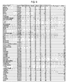
Classifications
-
- C—CHEMISTRY; METALLURGY
- C07—ORGANIC CHEMISTRY
- C07K—PEPTIDES
- C07K16/00—Immunoglobulins [IGs], e.g. monoclonal or polyclonal antibodies
- C07K16/18—Immunoglobulins [IGs], e.g. monoclonal or polyclonal antibodies against material from animals or humans
- C07K16/24—Immunoglobulins [IGs], e.g. monoclonal or polyclonal antibodies against material from animals or humans against cytokines, lymphokines or interferons
- C07K16/243—Colony Stimulating Factors
-
- A—HUMAN NECESSITIES
- A61—MEDICAL OR VETERINARY SCIENCE; HYGIENE
- A61K—PREPARATIONS FOR MEDICAL, DENTAL OR TOILETRY PURPOSES
- A61K38/00—Medicinal preparations containing peptides
- A61K38/16—Peptides having more than 20 amino acids; Gastrins; Somatostatins; Melanotropins; Derivatives thereof
- A61K38/17—Peptides having more than 20 amino acids; Gastrins; Somatostatins; Melanotropins; Derivatives thereof from animals; from humans
- A61K38/19—Cytokines; Lymphokines; Interferons
- A61K38/193—Colony stimulating factors [CSF]
-
- A—HUMAN NECESSITIES
- A61—MEDICAL OR VETERINARY SCIENCE; HYGIENE
- A61P—SPECIFIC THERAPEUTIC ACTIVITY OF CHEMICAL COMPOUNDS OR MEDICINAL PREPARATIONS
- A61P19/00—Drugs for skeletal disorders
- A61P19/08—Drugs for skeletal disorders for bone diseases, e.g. rachitism, Paget's disease
-
- A—HUMAN NECESSITIES
- A61—MEDICAL OR VETERINARY SCIENCE; HYGIENE
- A61P—SPECIFIC THERAPEUTIC ACTIVITY OF CHEMICAL COMPOUNDS OR MEDICINAL PREPARATIONS
- A61P35/00—Antineoplastic agents
-
- A—HUMAN NECESSITIES
- A61—MEDICAL OR VETERINARY SCIENCE; HYGIENE
- A61P—SPECIFIC THERAPEUTIC ACTIVITY OF CHEMICAL COMPOUNDS OR MEDICINAL PREPARATIONS
- A61P35/00—Antineoplastic agents
- A61P35/04—Antineoplastic agents specific for metastasis
-
- A—HUMAN NECESSITIES
- A61—MEDICAL OR VETERINARY SCIENCE; HYGIENE
- A61P—SPECIFIC THERAPEUTIC ACTIVITY OF CHEMICAL COMPOUNDS OR MEDICINAL PREPARATIONS
- A61P43/00—Drugs for specific purposes, not provided for in groups A61P1/00-A61P41/00
-
- A—HUMAN NECESSITIES
- A61—MEDICAL OR VETERINARY SCIENCE; HYGIENE
- A61K—PREPARATIONS FOR MEDICAL, DENTAL OR TOILETRY PURPOSES
- A61K39/00—Medicinal preparations containing antigens or antibodies
- A61K2039/505—Medicinal preparations containing antigens or antibodies comprising antibodies
-
- A—HUMAN NECESSITIES
- A61—MEDICAL OR VETERINARY SCIENCE; HYGIENE
- A61K—PREPARATIONS FOR MEDICAL, DENTAL OR TOILETRY PURPOSES
- A61K39/00—Medicinal preparations containing antigens or antibodies
- A61K2039/505—Medicinal preparations containing antigens or antibodies comprising antibodies
- A61K2039/507—Comprising a combination of two or more separate antibodies
-
- C—CHEMISTRY; METALLURGY
- C07—ORGANIC CHEMISTRY
- C07K—PEPTIDES
- C07K2317/00—Immunoglobulins specific features
- C07K2317/70—Immunoglobulins specific features characterized by effect upon binding to a cell or to an antigen
- C07K2317/73—Inducing cell death, e.g. apoptosis, necrosis or inhibition of cell proliferation
-
- C—CHEMISTRY; METALLURGY
- C07—ORGANIC CHEMISTRY
- C07K—PEPTIDES
- C07K2317/00—Immunoglobulins specific features
- C07K2317/70—Immunoglobulins specific features characterized by effect upon binding to a cell or to an antigen
- C07K2317/76—Antagonist effect on antigen, e.g. neutralization or inhibition of binding
Definitions
- This invention relates to methods for preventing and treating cancer metastasis and bone loss associated with cancer metastasis by administering M-CSF antagonist to a subject.
- Cancer metastasis is the primary cause of post-operation or post-therapy recurrence in cancer patients. Despite intensive efforts to develop treatments, cancer metastasis remains substantially refractory to therapy. Bone is one of the most common sites of metastasis of various types of human cancers (e.g., breast, lung, prostate and thyroid cancers). The occurrence of osteolytic bone metastases causes serious morbidity due to intractable pain, high susceptibility to fracture, nerve compression and hypercalcemia. Despite the importance of these clinical problems, there are few available treatments for bone loss associated with cancer metastasis.
- Osteoclasts mediate bone readsorption. Osteoclasts are multinucleated cells differentiating from haemopoietic cells. It is generally accepted that osteoclasts are formed by the fusion of mononuclear precursors derived from haemopoietic stem cells in the bone marrow, rather than incomplete cell divisions ( Chambers, Bone and Mineral Research, 6: 1-25, 1989 ; Göthling et al., Clin Orthop Relat R.
- osteoclast precursors into mature multinucleated osteoclasts require different factors including hormonal and local stimuli ( Athanasou et al., Bone Miner 3: 317-333, 1988 ; Feldman et al., Endocrinology 107: 1137-1143, 1980 ; Walker, Science 190: 784-785, 1975 ; Zheng et al., Histochem J 23: 180-188, 1991 ) and living bone and bone cells have been shown to play a critical role in osteoclast development ( Hagenaars et al., Bone Miner 6: 179-189, 1989 ). Osteoblastic or bone marrow stromal cells are also required for osteoclast differentiation.
- M-CSF macrophage-colony stimulating factor
- Receptor activator for NF- ⁇ B ligand is another signal ( Suda et al., Endocr Rev 13: 66-80, 1992 ) through which osteoblastic/stromal cells stimulate osteoclast formation and resorption via a receptor, RANK (TRANCER), located on osteoclasts and osteoclast precursors ( Lacey et al., Cell 93: 165-176, 1998 ; Tsuda et al., Biochem Biophys Res Co 234: 137-142, 1997 ; Wong et al., J Exp Med 186: 2075-2080, 1997 ; Wong et al., J Biol.
- Osteoblasts also secrete a protein that strongly inhibits osteoclast formation called osteoprotegerin (OPG, also known as OCIF), which acts as a decoy receptor for the RANKL thus inhibiting the positive signal between osteoclasts and osteoblasts via RANK and RANKL.
- OPG osteoprotegerin
- Osteoclasts are responsible for dissolving both the mineral and organic bone matrix ( Blair et al., J Cell Biol 102: 1164-1172, 1986 ). Osteoclasts represent terminally differentiated cells expressing a unique polarized morphology with specialized membrane areas and several membrane and cytoplasmic markers, such as tartrate resistant acid phosphatase (TRAP) (Anderson et al.
- TRIP tartrate resistant acid phosphatase
- Multinucleated osteoclasts usually contain less than 10 nuclei, but they may contain up to 100 nuclei being between 10 and 100 ⁇ m in diameter ( Göthling et al., Clin Orthop Relat R 120: 201-228, 1976 ). This makes them relatively easy to identify by light microscopy.
- osteoclasts play a major role in osteolytic bone metastases, there is a need in the art for new agents and methods for preventing osteoclast stimulation.
- the invention provides an M-CSF antagonist, for use in:
- compositions and methods disclosed herein fulfill the aforementioned and other related needs in the art.
- a method for preventing bone metastases comprising administering to a subject afflicted with metastatic cancer a therapeutically effective amount of M-CSF antagonist, thereby preventing bone loss associated with the metastatic cancer.
- a method of treating a subject afflicted with a metastatic cancer to bone comprising administering to said subject a therapeutically effective amount of M-CSF antagonist, thereby reducing the severity of bone loss associated with the metastatic cancer.
- the aforementioned methods are provided wherein the subject is a mammal or wherein the mammal is a human.
- the methods disclosed herein achieve their therapeutic potential by inhibiting the interaction between M-CSF and its receptor (M-CSFR). It is further contemplated that the inhibition of M-CSF/M-CSFR interaction inhibits osteoclast proliferation and/or differentiation induced by tumor cells.
- the M-CSF antagonist of the aforementioned methods is selected from the group consisting of: a polypeptide comprising an anti-M-CSF antibody; a polypeptide comprising an anti-M-CSFR antibody thereof; a soluble polypeptide comprising an M-CSF mutein or derivative thereof; or a soluble polypeptide comprising an M-CSFR mutein or derivative thereof.
- the M-CSF antagonist is an anti-M-CSF antibody.
- the M-CSF antagonist is a polypeptide comprising an anti-M-CSFR antibody.
- the M-CSF antagonist is a soluble polypeptide comprising an M-CSF mutein or derivative thereof.
- the M-CSF antagonist is a soluble polypeptide comprising an M-CSFR mutein or derivative thereof.
- the M-CSF antagonist is an antibody selected from the group consisting of: a polyclonal antibody; a monoclonal antibody; a humanized antibody; a human antibody; a chimeric antibody; Fab, F(ab') 2 or F v antibody fragment; and a mutein of any one of the aforementioned antibodies.
- the antibody binds to the same epitope as monoclonal antibody 5H4 (ATCC Accession No. HB10027).
- metastatic cancers are contemplated to be amenable to the methods disclosed herein.
- the metastatic cancer is breast, lung, renal, multiple myeloma, thyroid, prostate, adenocarcinoma, blood cell malignancies, including leukemia and lymphoma; head and neck cancers; gastrointestinal cancers, including stomach cancer, colon cancer, colorectal cancer, pancreatic cancer, liver cancer; malignancies of the female genital tract, including ovarian carcinoma, uterine endometrial cancers and cervical cancer; bladder cancer; brain cancer, including neuroblastoma; sarcoma, osteosarcoma; and skin cancer, including malignant melanoma or squamous cell cancer.
- the M-CSF antagonist is an antibody administered at a dose between about 0.01 mg/kg and about 100 mg/kg.
- a non-murine antibody that binds to M-CSF for treating a subject afflicted with a metastatic cancer, wherein said antibody effectively reduces the severity of bone loss associated with the metastatic cancer.
- a non-murine monoclonal antibody that specifically binds to the same epitope of M-CSF as 5H4.
- a non-murine monoclonal antibody that competes with 5H4 for binding to M-CSF is provided.
- the non-murine monoclonal antibody competes with 5H4 for binding to M-CSF by more than 10%, more preferably by more than 25%, still more preferably by more than 50%, even more preferably by more than 75%, and most preferably more than 90%.
- any of the aforementioned antibodies can be used to administer to a subject in need thereof. Accordingly, one embodiment discloses a pharmaceutical composition comprising any one of the aforementioned antibodies and a pharmaceutically acceptable carrier, excipient or diluent.
- a non-murine antibody for treating a subject afflicted with a metastatic cancer is disclosed in one embodiment, wherein the antibody effectively reduces the severity of bone loss associated with the metastatic cancer.
- the aforementioned antibody is selected from the group consisting of: a polyclonal antibody; a monoclonal antibody; a humanized antibody; a human antibody; a chimeric antibody; Fab, F(ab') 2 or F v antibody fragment; and a mutein of any one of the aforementioned antibodies.
- the antibody is specific to M-CSF.
- the antibody is specific to M-CSFR.
- the antibody is a fully human antibody or a humanized antibody.
- a hybridoma is disclosed that secretes an aforementioned antibody.
- metastatic cancers are contemplated to be amenable to the antibodies disclosed herein.
- the metastatic cancer is breast, lung, renal, multiple myeloma, thyroid, prostate, adenocarcinoma, blood cell malignancies, including leukemia and lymphoma; head and neck cancers; gastrointestinal cancers, including stomach cancer, colon cancer, colorectal cancer, pancreatic cancer, liver cancer; malignancies of the female genital tract, including ovarian carcinoma, uterine endometrial cancers and cervical cancer; bladder cancer; brain cancer, including neuroblastoma; sarcoma, osteosarcoma; and skin cancer, including malignant melanoma or squamous cell cancer.
- a pharmaceutical composition comprising an aforementioned antibody and a pharmaceutically suitable carrier, excipient or diluent is provided.
- M-CSF antagonists potentially useful in preventing or treating bone loss associated with cancer metastasis may be screened using various assays.
- a method of screening for an M-CSF antagonist comprising the steps of contacting metastatic tumor cell medium, osteoclasts and a candidate antagonist; detecting osteoclast formation, proliferation and/or differentiation; and identifying said candidate antagonist as an M-CSF antagonist if a decrease in osteoclast formation, proliferation and/or differentiation is detected.
- a method of screening is contemplated wherein the metastatic tumor cell medium includes tumor cells.
- a method wherein the contacting step occurs in vivo, the detecting step comprises detecting size and/or number of bone metastases, and the candidate antagonist is identified as an M-CSF antagonist if a decrease in size and/or number of bone metastases is detected.
- the method further comprises the step of determining if said candidate antagonist binds to M-CSF.
- the method further comprises the step of determining if said candidate antagonist inhibits interaction between M-CSF and its receptor M-CSFR.
- a method wherein the candidate antagonist is selected from the group consisting of: a polypeptide comprising an anti-M-CSF antibody; a polypeptide comprising an anti-M-CSFR antibody thereof; a soluble polypeptide comprising an M-CSF mutein or derivative thereof; a soluble polypeptide comprising an M-CSFR mutein or derivative thereof; a peptide; or a small molecule.
- a method is disclosed wherein said candidate antagonist is an M-CSF mutein.
- a method is provided wherein said candidate antagonist is an anti-M-CSF antibody.
- the candidate antagonist is an M-CSF mutein.
- the candidate antagonist is an anti-M-CSF antibody.
- the candidate antagonist is an anti-M-CSFR antibody.
- Another embodiment discloses a method of identifying an M-CSF antagonist that can prevent or treat metastatic cancer to bone, comprising the steps of: detecting binding of a candidate antagonist to M-CSF; and assaying the ability of said candidate antagonist to prevent or treat metastatic cancer to bone in vitro or in vivo.
- a method of identifying an M-CSF antagonist that can prevent or treat metastatic cancer to bone comprising the steps of: detecting binding of a candidate antagonist to M-CSFR; and assaying the ability of said candidate antagonist to prevent or treat metastatic cancer to bone in vitro or in vivo.
- a method of identifying an M-CSF antagonist that can prevent or treat metastatic cancer to bone comprising the steps of: identifying a candidate antagonist that inhibits the interaction between M-CSF and M-CSFR; and assaying the ability of said candidate antagonist to prevent or treat metastatic cancer to bone in vitro or in vivo.
- one embodiment discloses a method of preventing bone metastases and tumor growth comprising administering to a subject afflicted with metastatic cancer therapeutically effective amounts of M-CSF antagonist and a therapeutic agent, thereby preventing bone loss associated with the metastatic cancer and preventing tumor growth.
- another embodiment discloses a method of treating a subject afflicted with a metastatic cancer comprising administering to the subject therapeutically effective amounts of M-CSF antagonist and a therapeutic agent, thereby reducing the severity of bone loss associated with the metastatic cancer and inhibiting tumor growth.
- the subject of the aforementioned method is a mammal.
- the subject is a human.
- the aforementioned methods are disclosed wherein the antagonist inhibits the interaction between M-CSF and its receptor M-CSFR.
- the antagonist inhibits osteoclast proliferation and/or differentiation induced by tumor cells.
- the M-CSF antagonist is selected from the group consisting of: a polypeptide comprising an anti-M-CSF antibody; a polypeptide comprising an anti-M-CSFR antibody thereof; a soluble polypeptide comprising an M-CSF mutein or derivative thereof; and a soluble polypeptide comprising an M-CSFR mutein or derivative thereof.
- M-CSF antagonist antibodies are contemplated.
- the aforementioned methods are disclosed wherein said antibody is selected from the group consisting of: a polyclonal antibody; a monoclonal antibody; a humanized antibody; a human antibody; a chimeric antibody; Fab, F(ab') 2 or F v antibody fragment; and a mutein of any one of the aforementioned antibodies.
- the aforementioned methods are disclosed wherein the therapeutic agent is a bisphosphonate.
- the bisphophonate is zeledronate, pamidronate, clodronate, etidronate, tilundronate, alendronate, or ibandronate.
- the aforementioned methods are disclosed wherein the therapeutic agent is a chemotherapeutic agent. It is contemplated that some subjects may be precluded from receiving bisphosphonate treatment.
- a subject may be precluded from receiving bisphosphonate treatment if the subject does not adequately respond to bisphosphonate, if the subject does not tolerate bisphosphonate treatment, or if bisphosphonate is contraindicated for the subject's particular symptoms (e.g., renal failure).
- Cancer chemotherapeutic agents include, without limitation, alkylating agents, such as carboplatin and cisplatin; nitrogen mustard alkylating agents; nitrosourea alkylating agents, such as carmustine (BCNU); antimetabolites, such as methotrexate; purine analog antimetabolites, mercaptopurine; pyrimidine analog antimetabolites, such as fluorouracil (5-FU) and gemcitabine; hormonal antineoplastics, such as goserelin, leuprolide, and tamoxifen; natural antineoplastics, such as aldesleukin, interleukin-2, docetaxel, etoposide (VP-16), interferon alfa, paclitaxel, and tretinoin (ATRA); antibiotic natural antineoplastics, such as bleomycin, dactinomycin, daunorubicin, doxorubicin, and mitomycin; and vinca alkaloid natural antineoplastics, such as vin
- additional agents used as adjunctive therapy for cancer patients include EPO, G-CSF, ganciclovir; antibiotics, leuprolide; meperidine; zidovudine (AZT); interleukins 1 through 18, including mutants and analogues; interferons or cytokines, such as interferons ⁇ , ⁇ , and ⁇ .; hormones, such as luteinizing hormone releasing hormone (LHRH) and analogues and, gonadotropin releasing hormone (GnRH); growth factors, such as transforming growth factor- ⁇ (TGF- ⁇ ), fibroblast growth factor (FGF), nerve growth factor (NGF), growth hormone releasing factor (GHRF), epidermal growth factor (EGF), fibroblast growth factor homologous factor (FGFHF), hepatocyte growth factor (HGF), and insulin growth factor (IGF); tumor necrosis factor- ⁇ .
- TGF- ⁇ transforming growth factor- ⁇
- FGF fibroblast growth factor
- NGF nerve growth factor
- GHRF
- TNF- ⁇ & ⁇ invasion inhibiting factor-2
- BMP 1-7 bone morphogenetic proteins 1-7
- SOD superoxide dismutase
- complement factors anti-angiogenesis factors; antigenic materials; pro-drugs; growth factor receptor kinase inhibitors; anti-Her2 anitbody; and VEGF neutralizing antibody.
- the M-CSF antagonist is effective to reduce the dosage of therapeutic agent required to achieve a therapeutic effect.
- an M-CSF antagonist may improve efficacy of the second therapeutic agent, or reduce side effects associated with administration of the second therapeutic agent, or improve the safety of the second therapeutic agent.
- An M-CSF antagonist may also improve efficacy, reduce side effects of, or improve safety of a second therapeutic modality such as surgery or radiation therapy.
- the aforementioned methods further comprise the step of administering a non-M-CSF colony stimulating factor, for example G-CSF.
- a pharmaceutical composition is disclosed comprising a M-CSF antagonist and a cancer therapeutic agent.
- a package, vial or container comprising a medicament comprising an M-CSF antagonist and instructions that the medicament should be used in combination with surgery or radiation therapy.
- a method of preventing or treating metastatic cancer to bone comprising the steps of administering an M-CSF antagonist to a subject and treating said subject with surgery or radiation therapy.
- M-CSF antagonists may be useful as immunotherapeutic agents. Accordingly, one embodiment discloses a method of targeting a tumor cell expressing membrane-bound M-CSF on its surface comprising the step of administering an antibody that specifically binds to the extracellular portion of membrane-bound M-CSF [SEQ ID NO: 2].
- the antibody is conjugated to a radionuclide or other toxin.
- the antibody is selected from the group consisting of: a polyclonal antibody; a monoclonal antibody; a humanized antibody; a human antibody; a chimeric antibody; Fab, F(ab') 2 or F v antibody fragment; and a mutein of any one of the aforementioned antibodies.
- the disclosure contemplates immunotherapy-based methods of treating cancer by using anti-M-CSF antibodies to target tumor cells expressing membrane-bound M-CSF on their surface.
- the examples herein show that a variety of cancer cells express the membrane-bound form of M-CSF, including but not limited to breast cancer, colon cancer, kidney cancer, liver cancer, lung cancer, lymphoma, melanoma, ovarian cancer, pancreatic cancer, prostate cancer, osteosarcoma and thyroid cancer.
- the disclosure contemplates methods of killing or inhibiting growth of tumor cells expressing membrane-bound M-CSF comprising the step of administering an effective amount of an antibody that binds to M-CSF, alone or conjugated to a cytotoxic moiety. While antibodies specific for membrane-bound M-CSF are preferred, any antibody that binds to the extracellular portion of membrane-bound M-CSF is useful according to these methods.
- any one of the M-CSF antagonists of the disclosure may be useful for treating cancer, including non-metastatic cancer.
- the disclosure contemplates use of any one of the antagonists of the invention for the treatment of hypercalcemia, Paget's disease or osteoporosis.
- a method of treating a subject suffering from a cancer comprising the cells comprising said cancer do not secrete M-CSF, comprising the step of administering an M-CSF antagonist.
- a method for preventing bone metastases comprising administering to a subject afflicted with metastatic cancer an amount of M-CSF antagonist effective to neutralize M-CSF produced by the subject's cells, the amount being larger than the amount effective to neutralize M-CSF produced by the cancer cells.
- a method of treating a subject afflicted with a metastatic cancer to bone comprising administering to the subject an amount of M-CSF antagonist effective to neutralize M-CSF produced by the subject's cells, the amount being larger than the amount effective to neutralize M-CSF produced by the cancer cells.
- one embodiment discloses any of the aforementioned antibodies for use in medicine.
- a use of a M-CSF antagonist in the manufacture of a medicament is disclosed for preventing bone metastases in a subject afflicted with metastatic cancer.
- a use of a M-CSF antagonist in the manufacture of a medicament for preventing, in a subject afflicted with metastatic cancer, bone loss associated with the cancer is disclosed.
- a use of a M-CSF antagonist in the manufacture of a medicament is disclosed for treating a subject afflicted with a metastatic cancer to bone.
- a use of a M-CSF antagonist in the manufacture of a medicament for reducing, in a subject afflicted with a metastatic cancer to bone, the severity of bone loss associated with the cancer is disclosed.
- the subject is a mammal. In another embodiment, the mammal is human.
- the aforementioned uses are isclosed wherein the antagonist inhibits the interaction between M-CSF and its receptor (M-CSFR).
- the antagonist inhibits osteoclast proliferation and/or differentiation induced by tumor cells.
- the M-CSF antagonist is selected from the group consisting of: a polypeptide comprising an anti-M-CSF antibody; a polypeptide comprising an anti-M-CSFR antibody thereof; a soluble polypeptide comprising an M-CSF mutein or derivative thereof; or a soluble polypeptide comprising an M-CSFR mutein or derivative thereof.
- the antibody is selected from the group consisting of: a polyclonal antibody; a monoclonal antibody; a humanized antibody; a human antibody; a chimeric antibody; Fab, F(ab') 2 or F v antibody fragment; and a mutein of any one of the aforementioned antibodies or fragments.
- the aforementioned uses are disclosed wherein the antibody is specific to M-CSF.
- the antibody is antibody 5H4.
- the antibody is specific to M-CSFR.
- the metastatic cancer is breast, lung, renal, multiple myeloma, thyroid, prostate, adenocarcinoma, blood cell malignancies, including leukemia and lymphoma; head and neck cancers; gastrointestinal cancers, including stomach cancer, colon cancer, colorectal cancer, pancreatic cancer, liver cancer; malignancies of the female genital tract, including ovarian carcinoma, uterine endometrial cancers and cervical cancer; bladder cancer; brain cancer, including neuroblastoma; sarcoma, osteosarcoma; and skin cancer, including malignant melanoma or squamous cell cancer.
- the M-CSF antagonist of the aforementioned uses is an antibody administered at a dose between about 0.01 mg/kg and about 100 mg/kg.
- a medicament for treating a subject afflicted with metastatic cancer is disclosed.
- the use of the antibody in the manufacture of a medicament for reducing, in a subject afflicted with a metastatic cancer, the severity of bone loss associated with the cancer is provided.
- a use of a M-CSF antagonist and a therapeutic agent in the manufacture of a medicament is disclosed for preventing, in a subject afflicted with metastatic cancer, bone metastases and tumor growth.
- a use of a M-CSF antagonist and a therapeutic agent in the manufacture of a medicament is disclosed for preventing, in a subject afflicted with metastatic cancer, bone loss associated with the cancer.
- a use of a M-CSF antagonist and a therapeutic agent in the manufacture of a medicament for treating a metastatic cancer is disclosed.
- a use of a M-CSF antagonist and a therapeutic agent in the manufacture of a medicament is disclosed for reducing the severity of bone loss associated with the cancer and inhibiting tumor growth in a subject afflicted with metastatic cancer.
- a product comprising an M-CSF antagonist and a therapeutic agent as a combined preparation for simultaneous, separate or sequential use in treating cancer is disclosed.
- a use of an M-CSF antagonist in preparation of a medicament for preventing or treating metastatic cancer to bone, wherein the medicament is simultaneously separately or sequentially administered with a cancer therapeutic agent is disclosed.
- a use of a cancer therapeutic agent in preparation of a medicament is disclosed for preventing or treating metastatic cancer to bone, wherein the medicament is simultaneously separately or sequentially administered with an M-CSF antagonist.
- a package, vial or container is provided comprising a medicament comprising an M-CSF antagonist and instructions that the medicament should be used in combination with surgery or radiation therapy.
- the aforementioned uses are disclosed wherein the subject is a mammal. Even further, the aforementioned uses are disclosed wherein the mammal is human.
- the antagonist of the aforementioned uses inhibits the interaction between M-CSF and its receptor M-CSFR.
- the antagonist inhibits osteoclast proliferation and/or differentiation induced by tumor cells.
- the aforementioned uses are disclosed wherein said M-CSF antagonist is selected from the group consisting of: a polypeptide comprising an anti-M-CSF antibody; a polypeptide comprising an anti-M-CSFR antibody thereof; a soluble polypeptide comprising an M-CSF mutein or derivative thereof; or a soluble polypeptide comprising an M-CSFR mutein or derivative thereof.
- the antibody is selected from the group consisting of: a polyclonal antibody; a monoclonal antibody; a humanized antibody; a human antibody; a chimeric antibody; Fab, F(ab') 2 or F v antibody fragment; and a mutein of any one of the aforementioned anybodies.
- the aforementioned uses are disclosed wherein the therapeutic agent is a bisphosphonate.
- the bisphosphonate is zeledronate, pamidronate, clodronate, etidronate, tilundronate, alendronate, or ibandronate.
- the therapeutic agent is a chemotherapeutic agent.
- the subject is precluded from receiving bisphophonate treatment.
- a use of a M-CSF antagonist in the manufacture of a medicament for reducing the dose of a therapeutic agent administered to a subject to treat or prevent bone metastases and tumor growth is disclosed.
- a use of a M-CSF antagonist, a therapeutic agent, and a non-M-CSF colony stimulating factor in the manufacture of a medicament for preventing, in a subject afflicted with metastatic cancer, bone metastases and tumor growth is disclosed.
- a use of a M-CSF antagonist, a therapeutic agent, and a non-M-CSF colony stimulating factor in the manufacture of a medicament is disclosed for preventing, in a subject afflicted with metastatic cancer, bone loss associated with the cancer.
- a use of a M-CSF antagonist, a therapeutic agent, and a non-M-CSF colony stimulating factor in the manufacture of a medicament for treating a metastatic cancer is disclosed.
- a use of a M-CSF antagonist, a therapeutic agent, and a non-M-CSF colony stimulating factor in the manufacture of a medicament is disclosed for reducing the severity of bone loss associated with the cancer and inhibiting tumor growth in a subject afflicted with metastatic cancer.
- the aforementioned uses are provided wherein the non-M-CSF colony stimulating factor is G-CSF.
- Another embodiment discloses a use of an antibody that specifically binds to the extracellular portion of membrane-bound M-CSF in the manufacture of a medicament for targeting a tumor cell that expresses membrane-bound M-CSF on its surface.
- a use of an antibody that (a) specifically binds to the extracellular portion of membrane-bound M-CSF, and (b) is conjugated to a radionuclide or other toxin in the manufacture of a medicament for treating cancer is disclosed.
- the aforementioned use is disclosed wherein said antibody is selected from the group consisting of: a polyclonal antibody; a monoclonal antibody; a humanized antibody; a human antibody; a chimeric antibody; Fab, F(ab') 2 or F v antibody fragment; and a mutein of any one of the aforementioned antibodies.
- a use of a non-murine anti-M-CSF antibody in the manufacture of a medicament for treating cancer is disclosed.
- the cells comprising the cancer do not secrete M-CSF.
- a use of a M-CSF antagonist, in an amount that is larger that the amount effective to neutralize M-CSF produced by cancer cells, in the manufacture of a medicament for preventing bone metastases is disclosed.
- a use of a M-CSF antagonist in an amount that is larger that the amount effective to neutralize M-CSF produced by cancer cells, in the manufacture of a medicament for neutralizing M-CSF produced by a subject's cells is disclosed.
- a use of a M-CSF antagonist in an amount that is larger that the amount effective to neutralize M-CSF produced by cancer cells, in the manufacture of a medicament for treating a subject afflicted with a metastatic cancer to bone.
- a use of a M-CSF antagonist in an amount that is larger than the amount effective to neutralize M-CSF produced by cancer cells, in the manufacture of a medicament for treating cancer is disclosed.
- Kits are also contemplated.
- a typical kit can comprise a package, container or vial comprising an M-CSF antagonist of the invention and instructions, such as a product insert or label, directing the user to utilize the pharmaceutical formulation according to any of the methods of the present disclosure.
- Metastasis refers to the spread of cancer cells to other parts of the body or to the condition produced by this spread. Metastasis is a complex multi-step process that includes changes in the genetic material of a cell, uncontrolled proliferation of the altered cell to form a primary tumor, development of a new blood supply for the primary tumor, invasion of the circulatory system by cells from the primary tumor, dispersal of small clumps of primary tumor cells to other parts of the body, and the growth of secondary tumors in those sites.
- Bone is one of the most common sites of metastasis in human breast, lung, prostate and thyroid cancers, as well as other cancers, and in autopsies as many as 60% of cancer patients are found to have bone metastasis.
- Osteolytic bone metastasis shows a unique step of osteoclastic bone resorption that is not seen in metastasis to other organs.
- Bone loss associated with cancer metastasis is mediated by osteoclasts (multinucleated giant cells with the capacity to resorb mineralized tissues), which seem to be activated by tumor products.
- Colony stimulating factor also known as macrophage colony stimulating factor (M-CSF)
- M-CSF macrophage colony stimulating factor
- M-CSF has been found crucial for osteoclast formation.
- M-CSF has been shown to modulate the osteoclastic functions of mature osteoclasts, their migration and their survival in cooperation with other soluble factors and cell to cell interactions provided by osteoblasts and fibroblasts ( Fixe and Praloran, Cytokine 10: 3-7, 1998 ; Martin et al., Critical Rev. in Eukaryotic Gene Expression 8: 107-23 (1998 )).
- M-CSF The full-length human M-CSF mRNA encodes a precursor protein of 554 amino acids (SEQ ID NO: 4).
- SEQ ID NO: 4 The full-length human M-CSF mRNA encodes a precursor protein of 554 amino acids (SEQ ID NO: 4).
- M-CSF can either be secreted into the circulation as a glycoprotein or chondroitin sulfate containing proteoglycan or be expressed as a membrane spanning glycoprotein on the surface of M-CSF producing cells.
- the three-dimensional structure of the bacterially expressed amino terminal 150 amino acids of human M-CSF indicates that this protein is a disulfide linked dimer with each monomer consisting of four alpha helical bundles and an anti-parallel beta sheet ( Pandit et al., Science 258: 1358-62 (1992 )).
- Three distinct M-CSF species are produced through alternative mRNA splicing.
- the three polypeptide precursors are M-CFS ⁇ of 256 amino acids (DNA and amino acid sequence set forth in SEQ ID NOS: 1 and 2), M-CSF ⁇ of 554 amino acids (DNA and amino acid sequence set forth in SEQ ID NOS: 3 and 4), and M-CSF ⁇ of 438 amino acids (DNA and amino acid sequence set forth in SEQ ID NOS: 5 and 6).
- M-CSF ⁇ is a secreted protein that does not occur in a membrane-bound form.
- M-CSF ⁇ is expressed as an integral membrane protein that is slowly released by proteolytic cleavage.
- M-CSF ⁇ is cleaved at amino acids 191-197 of SEQ ID NO: 2.
- the membrane-bound form of M-CSF can interact with receptors on nearby cells and therefore mediates specific cell-to-cell contacts.
- M-CSFR DNA and amino acid sequence set forth in SEQ ID NOS: 7 and 8) is a membrane spanning molecule with five extracellular immunoglobulin-like domains, a transmembrane domain and an intracellular interrupted Src related tyrosine kinase domain.
- M-CSFR is encoded by the c - fms proto-oncogene. Binding of M-CSF to the extracellular domain of M-CSFR leads to dimerization of the receptor, which activates the cytoplasmic kinase domain, leading to autophosphorylation and phosphorylation of other cellular proteins ( Hamilton J. A., J Leukoc Biol.,62(2):145-55 (1997 ); Hamilton J, A., Immuno Today., 18(7): 313-7(1997 ).
- Phosphorylated cellular proteins induce a cascade of biochemical events leading to cellular responses: mitosis, secretion of cytokines, membrane ruffling, and regulation of transcription of its own receptor ( Fixe and Praloran, Cytokine 10: 32-37 (1998 )).
- M-CSF is expressed in stromal cells, osteoblasts, and other cells. It is also expressed in breast, uterine, and ovarian tumor cells. The extent of expression in these tumors correlates with high grade and poor prognosis ( Kacinski Ann. Med. 27: 79-85 (1995 ); Smith et al., Clin. Cancer Res. 1: 313-25 (1995 )). In breast carcinomas, M-CSF expression is prevalent in invasive tumor cells as opposed to the intraductal (pre-invasive) cancer ( Scholl et al., J. Natl. Cancer Inst. 86: 120-6 (1994 )). In addition, M-CSF is shown to promote progression of mammary tumors to malignancy ( Lin et al., J. Exp. Med. 93: 727-39 (2001 )). For breast and ovarian cancer, the production of M-CSF seems to be responsible for the recruitment of macrophages to the tumor.
- M-CSF antagonists neutralize osteoclast induction by metastatic cancer cells.
- the present invention provides compositions and methods for treating or preventing cancer metastasis and bone loss associated with cancer metastasis.
- Tumor refers to all neoplastic cell growth and proliferation, whether malignant or benign, and all pre-cancerous and cancerous cells and tissues.
- cancer and “cancerous” refer to or describe the physiological condition in mammals that is typically characterized by unregulated cell growth.
- cancer include but are not limited to, carcinoma,; lymphoma, blastoma, sarcoma, and leukemia. More particular examples of such cancers include breast cancer, prostate cancer, colon cancer, squamous cell cancer, small-cell lung cancer, non-small cell lung cancer, gastrointestinal cancer, pancreatic cancer, glioblastoma, cervical cancer, ovarian cancer, liver cancer, bladder cancer, hepatoma, colorectal cancer, endometrial carcinoma, salivary gland carcinoma, kidney cancer, liver cancer, vulval cancer, thyroid cancer, hepatic carcinoma and various types of head and neck cancer.
- Treatment is an intervention performed with the intention of preventing the development or altering the pathology of a disorder. Accordingly, “treatment” refers to both therapeutic treatment and prophylactic or preventative measures. Those in need of treatment include those already with the disorder as well as those in which the disorder is to be prevented.
- a therapeutic agent may directly decrease the pathology of tumor cells, or render the tumor cells more susceptible to treatment by other therapeutic agents, e.g., radiation and/or chemotherapy.
- the "pathology” of cancer includes all phenomena that compromise the well being of the patient. This includes, without limitation, abnormal or uncontrollable cell growth, metastasis, interference with the normal functioning of neighboring cells, release of cytokines or other secretory products at abnormal levels, suppression or aggravation of inflammatory or immunological response, etc.
- metastatic cancer is defined as cancers that have potential to spread to other areas of the body, particularly bone.
- a variety of cancers can metastasize to the bone, but the most common metastasizing cancers are breast, lung, renal, multiple myeloma, thyroid and prostate.
- cancers that have the potential to metastasize to bone include but are not limited to adenocarcinoma, blood cell malignancies, including leukemia and lymphoma; head and neck cancers; gastrointestinal cancers, including stomach cancer, colon cancer, colorectal cancer, pancreatic cancer, liver cancer; malignancies of the female genital tract, including ovarian carcinoma, uterine endometrial cancers and cervical cancer; bladder cancer; brain cancer, including neuroblastoma; sarcoma, osteosarcoma; and skin cancer, including malignant melanoma and squamous cell cancer.
- the present invention especially contemplates prevention and treatment of tumor-induced osteolytic lesions in bone.
- the term "antagonist” generally refers to the property of a molecule, compound or other agent to, for example, interfere with the binding of one molecule with another molecule or the stimulation of one cell by another cell either through steric hindrance, conformational alterations or other biochemical mechanisms.
- the term antagonist relates to the property of an agent to prevent the binding of a receptor to its ligand, e.g., the binding of M-CSF with M-CSFR, thereby inhibiting the signal transduction pathway triggered by M-CSF.
- the term antagonist is not limited by any specific action mechanism, but, rather, refers generally to the functional property presently defined.
- Antagonists of the present invention include, but are not limited to: M-CSF antibodies and fragments and muteins and modifications thereof, soluble M-CSF and fragments and muteins and modifications thereof, M-CSFR antibodies and fragments and muteins and modifications thereof, soluble M-CSFR and fragments and muteins and modifications thereof, and peptides as well as other chemical compounds and molecules that bind to M-CSF or M-CSFR and antisense compounds that inhibit expression of M-CSF and M-CSFR.
- Antagonists of the present invention optionally exclude antisense molecules that target M-CSF. Any of the antagonists of the present invention can be administered in any manner known in the art. For example, M-CSF muteins, M-CSFR muteins or antibody fragments that bind to M-CSF or M-CSFR can be administered via gene therapy.
- M-CSF antagonists of the present invention include, where applicable, functional equivalents.
- molecules may differ in length, structure, components, etc., but may still retain one or more of the defined functions.
- functional equivalents of the antibodies, antibody fragments or peptides of the present invention may include mimetic compounds, i.e., constructs designed to mimic the proper configuration and/or orientation for antigen binding.
- Preferred M-CSF antagonists may optionally be modified by addition of side groups, etc., e.g., by amino terminal acylation, carboxy terminal amidation or by coupling of additional groups to amino acid side chains.
- Antagonists may also comprise one or more conservative amino acid substitutions.
- conservative amino acid substitutions is meant those changes in amino acid sequence that preserve the general charge, hydrophobicity/hydrophilicity and/or steric bulk of the amino acid substituted. For example, substitutions between the following groups are conservative: Gly/Ala, Val/Ile/Leu, Asp/Glu, Lys/Arg, Asn/Gln, Ser/Cys/Thr, and Phe/Trp/Tyr. Such modifications will not substantially diminish the efficacy of the M-CSF antagonists and may impart such desired properties as, for example, increased in vivo half life or decreased toxicity.
- the invention is also intended to include polypeptides bearing modifications other than the insertion, deletion, or substitution of amino acid residues.
- the modifications may be covalent in nature, and include for example, chemical bonding with polymers, lipids, other organic, and inorganic moieties.
- Such derivatives may be prepared to increase circulating half-life of a polypeptide, or may be designed to improve targeting capacity for the polypeptide to desired cells, tissues, or organs.
- the invention further embraces M-CSF or M-CSFR polypeptides that have been covalently modified to include one or more water soluble polymer attachments such as polyethylene glycol, polyoxyethylene glycol, or polypropylene glycol.
- therapeutically effective amount refers to an amount of therapeutic or prophylactic M-CSF antagonist that would be appropriate for an embodiment of the present invention, that will elicit the desired therapeutic or prophylactic effect or response when administered in accordance with the desired treatment regimen.
- M-CSF refers to a human polypeptide having substantially the same amino acid sequence as the mature human M-CSF ⁇ , M-CSF ⁇ , or M-CSF ⁇ polypeptides described in Kawasaki et al., Science 230:291 (1985 ), Cerretti et al., Molecular Immunology, 25:761 (1988 ), or Ladner et al., EMBO Journal 6:2693 (198 7 ) Such terminology reflects the understanding that the three mature M-CSFs have different amino acid sequences, as described above, and that the active form of M-CSF is a disulfide bonded dimer; thus, when the term "M-CSF" refers to the biologically active form, the dimeric form is intended.
- M-CSF dimer refers to two M-CSF polypeptide monomers that have dimerized and includes both homodimers (consisting of two of the same type of M-CSF monomer) and heterodimers (consisting of two different monomers). M-CSF monomers may be converted to M-CSF dimers in vitro as described in U.S. Pat. No. 4,929,700 .
- antibody is used in the broadest sense and covers fully assembled antibodies, antibody fragments that can bind antigen (e.g., Fab', F'(ab)2, Fv, single chain antibodies, diabodies), and recombinant peptides comprising the forgoing.
- the term "monoclonal antibody” as used herein refers to an antibody obtained from a population of substantially homogeneous antibodies, i.e., the individual antibodies comprising the population are identical except; for possible naturally occurring mutations that may be present in minor amounts. Monoclonal antibodies are highly specific, being directed against a single antigenic site. Furthermore, in contrast to conventional (polyclonal) antibody preparations that are typically include different antibodies directed against different determinants (epitopes), each monoclonal antibody is directed against a single determinant on the antigen. In addition to their specificity, the monoclonal antibodies are advantageous in that they are synthesized by the homogeneous culture, uncontaminated by other immunoglobulins with different specificities and characteristics.
- the modifier "monoclonal” indicates the character of the antibody as being obtained from a substantially homogeneous population of antibodies, and is not to be construed as requiring production of the antibody by any particular method.
- the monoclonal antibodies to be used in accordance with the present invention may be made by the hybridoma method first described by Kohler et al., Nature, 256:495 [19751 , or may be made by recombinant DNA methods (see, e.g., U.S. Patent No. 4,816,567 ).
- the "monoclonal antibodies” may also be isolated from phage antibody libraries using the techniques described in Clackson et al.,Nature,352:624628[1991 ] end Marks et al., J. Mol. Biol., 222:581-597 (1991 ), for example.
- Antibody fragments comprise a portion of an intact antibody, preferably the antigen binding or variable region of the intact antibody.
- antibody fragments include Fab, Fab', F(ab')2, and Fv fragments; diabodies; linear antibodies ( Zapata et al., Protein Eng.,8(10):1057-1062 (1995 )); single-chain antibody molecules; and multispecific antibodies formed from antibody fragments.
- Papain digestion of antibodies produces two identical antigen-binding fragments, called “Fab” fragments, each with a single antigen-binding site, and a residual "Fc” fragment, whose name reflects its ability to crystallize 35 readily.
- Fv polypeptide further comprises a polypeptide linker between the VH and VL domains that enables the Fv to form the desired structure for antigen binding.
- diabodies refers to small antibody fragments with two antigen-binding sites, which fragments comprise a heavy-chain variable domain (VH) connected to a light-chain variable domain (VL) in the same polypeptide chain (VH VL).
- VH heavy-chain variable domain
- VL light-chain variable domain
- VH VL polypeptide chain
- an “isolated” antibody is one that has been identified and separated and for recovered from a component of its natural environment. Contaminant components of its natural environment are materials that would interfere with diagnostic or therapeutic uses for the antibody, and may include enzymes, hormones, and other proteinaceous or nonproteinaceous solutes.
- the antibody will be purified (1) to greater than 95% by weight of antibody as determined by the Lowry method, and most preferably more than 99% by weight, (2) to a degree sufficient too obtain at least 15 residues of N-terminal or internal amino acid sequence by use of a spinning cup sequenator, or (3) to homogeneity by SDS-PAGE under reducing or nonreducing conditions using Coomassie blue or, preferably, silver stain.
- Isolated antibody includes the antibody in situ within recombinant cells since at least one component of the antibody's natural environment will not be present. Ordinarily, however, isolated antibody will be prepared by at least one purification step.
- Fv is the minimum antibody fragment that contains a complete antigen recognition and binding site. This region consists of a dimer of one heavy- and one light-chain variable domain in tight, non-covalent association. It is in this configuration that the three CDRs of each variable domain interact to define an antigen binding site on the surface of the VH VI dimer. Collectively, the six CDRs confer antigen-binding specificity to the antibody. However, even a single variable domain (or half of an Fv comprising only three CDRs specific for an antigen) has the ability to recognize and bind antigen, although at a lower affinity than the entire binding site.
- the Fab fragment also contains the constant domain of the light chain and the first constant domain (CH1) of the heavy chain.
- Fab fragments differ from Fab' fragments by the addition of a few residues at the carboxy terminus of the heavy chain CH 1 domain including one or more cysteines from the antibody hinge region.
- Fab'-SH is the designation herein for Fab' in which the cysteine residue(s) of the constant domains bear a free thiol group.
- F(ab')2 antibody fragments originally were produced as pairs of Fab' fragments which have hinge cysteines between them.
- neutralizing antibody an antibody molecule that is able to eliminate or significantly reduce an effecter function of a target antigen to which is binds. Accordingly, a “neutralizing" anti-target antibody is capable of eliminating or significantly reducing an effecter function, such as enzyme activity, ligand binding, or intracellular signaling.
- compositions for and methods of treating cancer metastasis and/or bone loss associated with cancer metastasis may utilize one or more antibody used singularly or in combination with other therapeutics to achieve the desired effects.
- Antibodies according to the present invention may be isolated from an animal producing the antibody as a result of either direct contact with an environmental antigen or immunization with the antigen.
- antibodies may be produced by recombinant DNA methodology using one of the antibody expression systems well known in the art (See, e.g., Harlow and Lane, Antibodies: A Laboratory Manual, Cold Spring Harbor Laboratory (1988 )).
- Such antibodies may include recombinant IgGs, chimeric fusion proteins having immunoglobulin derived sequences or "humanized” antibodies that may all be used for the treatment of cancer metastasis and/or bone loss associated with cancer metastasis according to the present invention.
- the term "antibody” also refers to fragments thereof (such as, e.g., scFv, Fv, Fd, Fab, Fab' and F(ab)'2 fragments) or multimers or aggregates of intact molecules and/or fragments that bind to M-CSF (or M-CSFR). These antibody fragments bind antigen and may be derivatized to exhibit structural features that facilitate clearance and uptake, e.g., by incorporation of galactose residues.
- M-CSF antagonists are monoclonal antibodies prepared essentially as described in Halenbeck et al. U.S. Pat. No. 5,491,065 (1997 ).
- Exemplary M-CSF antagonists include monoclonal antibodies that bind to an apparent conformational epitope associated with recombinant or native dimeric M-CSF with concomitant neutralization of biological activity. These antibodies are substantially unreactive with biologically inactive forms of M-CSF including monomeric and chemically derivatized dimeric M-CSF.
- humanized anti-M-CSF monoclonal antibodies are provided.
- the phrase "humanized antibody” refers to an antibody derived from a non-human antibody, typically a mouse monoclonal antibody.
- a humanized antibody may be derived from a chimeric antibody that retains or substantially retains the antigen binding properties of the parental, non-human, antibody but which exhibits diminished immunogenicity as compared to the parental antibody when administered to humans.
- complementarity determining region refers to amino acid sequences which together define the binding affinity and specificity of the natural Fv region of a native immunoglobulin binding site (See, e.g., Chothia et al., J. Mol. Biol. 196:901 917 (1987 ); Kabat et al., U.S. Dept. of Health and Human Services NIH Publication No. 91 3242 (1991)).
- constant region refers to the portion of the antibody molecule that confers effector functions.
- mouse constant regions are preferably substituted by human constant regions.
- the constant regions of the subject humanized antibodies are derived from human immunoglobulins.
- the heavy chain constant region can be selected from any of the five isotypes: alpha, delta, epsilon, gamma or mu.
- the antibodies of the present invention are said to be immunospecific or specifically binding if they bind to antigen with a K a of greater than or equal to about 104M -1 , preferably of greater than or equal to about 10 5 M -1 , more preferably of greater than or equal to about 10 6 M -1 and still more preferably of greater than or equal to about 10 7 M -1 , and most preferably of greater than or equal to about 10 8 M -1 ,10 9 M -1 , or 10 10 M -1 .
- anti-M-CSF/M-CSFR specific antibodies suitable bind to their antigen with an affinity of at least 10 4 M -1 , preferably of greater than or equal to about 10 5 M -1 , more preferably of greater than or equal to about 10 6 M -1 and still more preferably of greater than or equal to about 10 7 M -1 , and most preferably of greater than or equal to about 10 8 M -1 ,10 9 M -1 , or 10 10 M -1 .
- the anti-M-CSF/M-CSFR antibodies bind to different naturally occurring forms of M-CSF/M-CSFR, including those expressed by the host's/subject's tissues as well as that expressed by the tumor.
- the monoclonal antibodies disclosed herein have affinity for M-CSF and M-CSFR and are characterized by a dissociation equilibrium constant (Kd) of at least 10 -4 M, preferably at least about 10-7 M to about 10-8 M, more preferably at least about 10 -8 M to about 10 -12 M.
- Kd dissociation equilibrium constant
- Monoclonal antibodies and antigen-binding fragments thereof that are suitable for use in the methods of the invention are capable of specifically binding to M-CSF/M-CSFR.
- Such affinities may be readily determined using conventional techniques, such as by equilibrium dialysis; by using the BIAcore 2000 instrument, using general procedures outlined by the manufacturer; by radioimmunoassay using 125 I labeled M-CSF; or by another method known to the skilled artisan.
- the affinity data may be analyzed, for example, by the method of Scatchard et al., Ann N.Y. Acad. Sci., 51:660 (1949 ).
- preferred M-CSF antagonists will exhibit a high degree of specificity for M-CSF and will bind with substantially lower affinity to other molecules.
- the antigen to be used for production of antibodies may be, e.g., intact M-CSF or a fragment of M-CSF that retains the desired epitope, optionally fused to another polypeptide that allows the epitope to be displayed in its native conformation.
- Polyclonal antibodies are preferably raised in animals by multiple subcutaneous (sc) or intraperitoneal (ip) injections of the relevant antigen and an adjuvant.
- An improved antibody response may be obtained by conjugating the relevant antigen to a protein that is immunogenic in the species to be immunized, e.g., keyhole limpet hemocyanin, serum albumin, bovine thyroglobulin, or soybean trypsin inhibitor using a bifunctional or derivatizing agent, for example, maleimidobenzoyl sulfosuccinimide ester (conjugation through cysteine residues), N-hydroxysuccinimide (through lysine residues), glutaraldehyde, succinic anhydride or other agents known in the art.
- a protein that is immunogenic in the species to be immunized e.g., keyhole limpet hemocyanin, serum albumin, bovine thyroglobulin, or soybean trypsin inhibitor using a
- Animals are immunized against the antigen, immunogenic conjugates, or derivatives by combining, e.g., 100 ⁇ g or 5 ⁇ g of the protein or conjugate (for rabbits or mice, respectively) with 3 volumes of Freund's complete adjuvant and injecting the solution intradermally at multiple sites.
- the animals are boosted with 1/5 to ⁇ fraction (1/10) ⁇ the original amount of peptide or conjugate in Freund's complete adjuvant by subcutaneous injection at multiple sites.
- the animals are bled and the serum is assayed for antibody titer. Animals are boosted until the titer plateaus.
- the animal is boosted with the conjugate of the same antigen, but conjugated to a different protein and/or through a different cross-linking reagent.
- Conjugates also can be made in recombinant cell culture as protein fusions.
- aggregating agents such as alum are suitably used to enhance the immune response.
- Monoclonal antibodies may be made using the hybridoma method first described by Kohler et al., Nature, 256:495 (1975 ), or may be made by recombinant DNA methods.
- a mouse or other appropriate host animal such as a hamster or macaque monkey
- lymphocytes that produce or are capable of producing antibodies that will specifically bind to the protein used for immunization.
- lymphocytes may be immunized in vitro. Lymphocytes then are fused with myeloma cells using a suitable fusing agent, such as polyethylene glycol, to form a hybridoma cell ( Goding, Monoclonal Antibodies: Principles and Practice, pp.59-103 (Academic Press, 1986 )).
- the hybridoma cells thus prepared are seeded and grown in a suitable culture medium that preferably contains one or more substances that inhibit the growth or survival of the unfused, parental myeloma cells.
- a suitable culture medium that preferably contains one or more substances that inhibit the growth or survival of the unfused, parental myeloma cells.
- the culture medium for the hybridomas typically will include hypoxanthine, aminopterin, and thymidine (HAT medium), which substances prevent the growth of HGPRT-deficient cells.
- Preferred myeloma cells are those that fuse efficiently, support stable high-level production of antibody by the selected antibody-producing cells, and are sensitive to a medium.
- Human myeloma and mouse-human heteromyeloma cell lines also have been described for the production of human monoclonal antibodies ( Brodeur et al., Monoclonal Antibody Production Techniques and Applications, pp. 51-63 (Marcel Dekker, Inc., New York, 1987 )).
- Culture medium in which hybridoma cells are growing is assayed for production of monoclonal antibodies directed against the antigen.
- the binding specificity of monoclonal antibodies produced by hybridoma cells is determined by immunoprecipitation or by an in vitro binding assay, such as radioimmunoassay (RIA) or enzyme-linked immunoabsorbent assay (ELISA).
- RIA radioimmunoassay
- ELISA enzyme-linked immunoabsorbent assay
- the binding affinity of the monoclonal antibody can, for example, be determined by Scatchard analysis ( Munson et al., Anal. Biochem., 107:220 (1980 )).
- the clones may be subcloned by limiting dilution procedures and grown by standard methods ( Goding, Monoclonal Antibodies: Principles and Practice, pp.59-103 (Academic Press, 1986 )). Suitable culture media for this purpose include, for example, D-MEM or RPMI-1640 medium.
- the hybridoma cells may be grown in vivo as ascites tumors in an animal.
- the monoclonal antibodies secreted by the subclones are suitably separated from the culture medium, ascites fluid, or serum by conventional immunoglobulin purification procedures such as, for example, protein A-Sepharose, hydroxylapatite chromatography, gel electrophoresis, dialysis, or affinity chromatography.
- DNA encoding the monoclonal antibodies may be isolated and sequenced from the hybridoma cells using conventional procedures (e.g., by using oligonucleotide probes that are capable of binding specifically to genes encoding the heavy and light chains of the monoclonal antibodies). Once isolated, the DNA may be placed into expression vectors, which are then transfected into host cells such as E. coli cells, simian COS cells, Chinese hamster ovary (CHO) cells, or myeloma cells that do not otherwise produce immunoglobulin protein, to obtain the synthesis of monoclonal antibodies in the recombinant host cells. Recombinant production of antibodies is well known in the art.
- Amino acid sequence variants of the desired antibody may be prepared by introducing appropriate nucleotide changes into the encoding DNA, or by peptide synthesis. Such variants include, for example, deletions from, and/or insertions into and/or substitutions of, residues within the amino acid sequences of the antibodies. Any combination of deletion, insertion, and substitution is made to arrive at the final construct, provided that the final construct possesses the desired characteristics.
- the amino acid changes also may alter post-translational processes of the humanized or variant antibody, such as changing the number or position of glycosylation sites.
- Nucleic acid molecules encoding amino acid sequence variants of the antibody are prepared by a variety of methods known in the art. These methods include, but are not limited to, isolation from a natural source (in the case of naturally occurring amino acid sequence variants) or preparation by oligonucleotide-mediated (or site-directed) mutagenesis, PCR mutagenesis, and cassette mutagenesis of an earlier prepared variant or a non-variant version of the antibody.
- chimeric or humanized antibodies are less immunogenic in humans than the parental mouse monoclonal antibodies, they can be used for the treatment of humans with far less risk of anaphylaxis. Thus, these antibodies may be preferred in therapeutic applications that involve in vivo administration to a human.
- Chimeric monoclonal antibodies in which the variable Ig domains of a mouse monoclonal antibody are fused to human constant Ig domains, can be generated using standard procedures known in the art (See Morrison, S. L., et al. (1984) Chimeric Human Antibody Molecules; Mouse Antigen Binding Domains with Human Constant Region Domains, Proc. Natl. Acad. Sci. USA 81, 6841-6855 ; and, Boulianne, G. L., et al, Nature 312, 643-646 . (1984 )). Although some chimeric monoclonal antibodies have proved less immunogenic in humans, the mouse variable Ig domains can still lead to a significant human anti-mouse response.
- Humanized antibodies may be achieved by a variety of methods including, for example: (1) grafting the non-human complementarity determining regions (CDRs) onto a human framework and constant region (a process referred to in the art as humanizing through “CDR grafting"), or, alternatively, (2) transplanting the entire non-human variable domains, but “cloaking” them with a human-like surface by replacement of surface residues (a process referred to in the art as “veneering”).
- CDRs complementarity determining regions
- CDR grafting involves introducing one or more of the six CDRs from the mouse heavy and light chain variable Ig domains into the appropriate four framework regions of human variable Ig domains is also called CDR grafting.
- This technique Riechmann, L., et al., Nature 332, 323 (1988 )
- FR1-FR4 conserved framework regions
- a disadvantage of CDR grafting is that it can result in a humanized antibody that has a substantially lower binding affinity than the original mouse antibody, because amino acids of the framework regions can contribute to antigen binding, and because amino acids of the CDR loops can influence the association of the two variable Ig domains.
- the CDR grafting technique can be improved by choosing human framework regions that most closely resemble the framework regions of the original mouse antibody, and by site-directed mutagenesis of single amino acids within the framework or CDRs aided by computer modeling of the antigen binding site (e.g., Co, M. S., et al. (1994), J. Immunol. 152, 2968-2976 ).
- a useful method for identification of certain residues or regions of the antibody that are preferred locations for mutagenesis is called "alanine scanning mutagenesis," as described by Cunningham and Wells Science, 244:1081-1085 (1989 ).
- a residue or group of target residues are identified (e.g., charged residues such as arg, asp, his, lys, and glu) and replaced by a neutral or negatively charged amino acid (most preferably alanine or polyalanine) to affect the interaction of the amino acids with antigen.
- Those amino acid locations demonstrating functional sensitivity to the substitutions then are refined by introducing further or other variants at, or for, the sites of substitution.
- the site for introducing an amino acid sequence variation is predetermined, the nature of the mutation per se need not be predetermined. For example, to analyze the performance of a mutation at a given site, ala scanning or random mutagenesis is conducted at the target codon or region and the expressed antibody variants are screened for the desired activity.
- Amino acid sequence insertions include amino- and/or carboxyl-terminal fusions ranging in length from one residue to polypeptides containing a hundred or more residues, as well as intra-sequence insertions of single or multiple amino acid residues.
- terminal insertions include an antibody with an N-terminal methionyl residue or the antibody fused to an epitope tag.
- Other insertional variants of the antibody molecule include the fusion to a polypeptide which increases the serum half-life of the antibody.
- variants are an amino acid substitution variant. These variants have at least one amino acid residue in the antibody molecule removed and a different residue inserted in its place. Substitutional mutagenesis within any of the hypervariable or CDR regions or framework regions is contemplated. Conservative substitutions are shown in Table 1 under the heading of "preferred substitutions". If such substitutions result in a change in biological activity, then more substantial changes, denominated "exemplary substitutions" in Table 1, or as further described below in reference to amino acid classes, may be introduced and the products screened.
- Substantial modifications in the biological properties of the antibody are accomplished by selecting substitutions that differ significantly in their effect on maintaining (a) the structure of the polypeptide backbone in the area of the substitution, for example, as a sheet or helical conformation, (b) the charge or hydrophobicity of the molecule at the target site, or (c) the bulk of the side chain.
- Naturally occurring residues are divided into groups based on common side-chain properties:
- Non-conservative substitutions involve replacing a member of one of these classes with a member of another class.
- cysteine residue not involved in maintaining the proper conformation of the humanized or variant antibody also may be substituted, generally with serine, to improve the oxidative stability of the molecule and prevent aberrant crosslinking.
- cysteine bond(s) may be added to the antibody to improve its stability (particularly where the antibody is an antibody fragment such as an Fv fragment).
- Affinity maturation involves preparing and screening antibody variants that have substitutions within the CDRs of a parent antibody and selecting variants that have improved biological properties such as binding affinity relative to the parent antibody.
- a convenient way for generating such substitutional variants is affinity maturation using phage display. Briefly, several hypervariable region sites (e.g. 6-7 sites) are mutated to generate all possible amino substitutions at each site.
- the antibody variants thus generated are displayed in a monovalent fashion from filamentous phage particles as fusions to the gene III product of M13 packaged within each particle. The phage-displayed variants are then screened for their biological activity (e.g. binding affinity).
- Alanine scanning mutagenesis can be performed to identify hypervariable region residues that contribute significantly to antigen binding. Alternatively, or in addition, it may be beneficial to analyze a crystal structure of the antigen-antibody complex to identify contact points between the antibody and antigen. Such contact residues and neighboring residues are candidates for substitution according to the techniques elaborated herein. Once such variants are generated, the panel of variants is subjected to screening as described herein and antibodies with superior properties in one or more relevant assays may be selected for further development.
- Antibody variants can also be produced that have a modified glycosylation pattern relative to the parent antibody, for example, deleting one or more carbohydrate moieties found in the antibody, and/or adding one or more glycosylation sites that are not present in the antibody.
- N-linked refers to the attachment of the carbohydrate moiety to the side chain of an asparagine residue.
- the tripeptide sequences asparagine-X-serine and asparagine-X-threonine, where X is any amino acid except proline, are the recognition sequences for enzymatic attachment of the carbohydrate moiety to the asparagine side chain.
- the presence of either of these tripeptide sequences in a polypeptide creates a potential glycosylation site.
- N-linked glycosylation sites may be added to an antibody by altering the amino acid sequence such that it contains one or more of these tripeptide sequences.
- O-linked glycosylation refers to the attachment of one of the sugars N-aceylgalactosamine, galactose, or xylose to a hydroxyamino acid, most commonly serine or threonine, although 5-hydroxyproline or 5-hydroxylysine may also be used.
- O-linked glycosylation sites may be added to an antibody by inserting or substituting one or more serine or threonine residues to the sequence of the original antibody.
- Humanized or human antibodies to M-CSF can also be produced using transgenic animals that have no endogenous immunoglobulin production and are engineered to contain human immunoglobulin loci.
- WO 98/24893 discloses transgenic animals having a human Ig locus wherein the animals do not produce functional endogenous immunoglobulins due to the inactivation of endogenous heavy and light chain loci.
- WO 91/741 also discloses transgenic non-primate mammalian hosts capable of mounting an immune response to an immunogen, wherein the antibodies have primate constant and/or variable regions, and wherein the endogenous immunoglobulin encoding loci are substituted or inactivated.
- WO 96/30498 discloses the use of the Cre/Lox system to modify the immunoglobulin locus in a mammal, such as to replace all or a portion of the constant or variable region to form a modified antibody molecule.
- WO 94/02602 discloses non-human mammalian hosts having inactivated endogenous Ig loci and functional human Ig loci.
- U.S. Patent No. 5,939,598 discloses methods of making transgenic mice in which the mice lack endogenous heavy chains, and express an exogenous immunoglobulin locus comprising one or more xenogeneic constant regions.
- an immune response can be produced to a selected antigenic molecule, and antibody producing cells can be removed from the animal and used to produce hybridomas that secrete human monoclonal antibodies.
- Immunization protocols, adjuvants, and the like are known in the art, and are used in immunization of, for example, a transgenic mouse as described in WO 96/33735 .
- This publication discloses monoclonal antibodies against a variety of antigenic molecules including IL 6, IL 8, TNFa, human CD4, L selectin, gp39, and tetanus toxin.
- the monoclonal antibodies can be tested for the ability to inhibit or neutralize the biological activity or physiological effect of the corresponding protein.
- WO 96/33735 discloses that monoclonal antibodies against IL-8, derived from immune cells of transgenic mice immunized with IL-8, blocked IL-8 induced functions of neutrophils. Human monoclonal antibodies with specificity for the antigen used to immunize transgenic animals are also disclosed in WO 96/34096 and U.S. patent application no. 20030194404 ; and U.S. patent application no. 20030031667 )
- the antibodies produced by phage technology are produced as antigen binding fragments-usually Fv or Fab fragments-in bacteria and thus lack effector functions.
- Effector functions can be introduced by one of two strategies: The fragments can be engineered either into complete antibodies for expression in mammalian cells, or into bispecific antibody fragments with a second binding site capable of triggering an effector function.
- the Fd fragment (V H -C H 1) and light chain (V L -C L ) of antibodies are separately cloned by PCR and recombined randomly in combinatorial phage display libraries, which can then be selected for binding to a particular antigen.
- the Fab fragments are expressed on the phage surface, i.e., physically linked to the genes that encode them.
- selection of Fab by antigen binding co-selects for the Fab encoding sequences which can be amplified subsequently.
- a procedure termed panning Fab specific for the antigen are enriched and finally isolated.
- fragment as used herein means a portion of the intact native molecule; for example, a fragment polypeptide is a fragment of the native polypeptide in which one or more amino acids from either the N-terminal or C-terminal have been deleted.
- “Mutein” as used herein with respect to polypeptides means a variant of the intact native molecule or a variant of a fragment of the native molecule, in which one or more amino acids have been substituted, inserted or deleted. Such substitutions, insertions or deletions can be at the N-terminus, C-terminus or internal to the molecule.
- muteins includes within its scope fragments of the native molecule. Insertional muteins include fusions at the N- or C-terminus, e.g. fusion to the Fc portion of an immunoglobulin to increase half-life
- Modification as used herein means any modification of the native polypeptide, fragment or mutein, such as glycosylation, phosphorylation, polymer conjugation (such as with polyethylene glycol), or other addition of foreign moieties, so long as the desired activity (agonist or antagonist) is retained.
- FIG. 1 is a topology diagram showing the disulfide bonds in truncated dimeric M-CSF
- FIG. 2 is a stereodiagram of the C-alpha backbone with every tenth residue labelled and with the non-crystallographic symmetry axis indicated by a dotted line.
- the overall topology of this form of M-CSF as shown in FIG. 1 is that of an antiparallel four alpha-helical bundle, in which the helices run up-up-down-down, unlike the more commonly observed up-down-up-down connectivity of most four helical bundles.
- a long crossover connection links helix A to helix B and a similar connection is found between helices C and D.
- the bundles are linked end-to-end, forming an extremely flat, elongated structure (approximate dimensions 85 x 35 x 25 ⁇ ).
- One interchain disulfide bond (Cys31--Cys31) is located at the dimer interface with the noncrystallographic two-fold symmetry axis passing through it as shown in FIG. 2 . Mutation experiments indicate that all of the cysteine residues in this form of M-CSF may be necessary for full biological activity.
- U.S. Patent No. 6,025,146 provides the three-dimensional structure of the truncated recombinant M-CSF ⁇ dimer as identified by the alpha-carbon positions of the amino acid residues in the sequence.
- M-CSF ⁇ has intrachain disulfide bonds involving cysteines 157 and/or 159
- the C-terminal region of M-CSF likely extends from the "rear" of the structure, providing a variable-length "tether" for membrane-bound forms of M-CSF.
- the "front" or receptor-binding region of M-CSF is on the opposite side of the molecules, consisting of solvent-accessible residues in or near helices A, C, and D, including residues from about 6 to 26, 71 to 90, and 110 to 130, respectively, of native M-CSF.
- Altering solvent accessible residues in these regions by site directed mutagenesis to increase or decrease side-chain interactions with the receptor may generate M-CSF agonists or antagonists.
- Residues having a solvent accessible surface area of greater than about 0.25 and preferably greater than about 0.4 are preferred based on normalization of the surface area of the amino acid accessible when in the trypeptide gly-x-gly ( Kabsch, W. et al., Biopolymers 22:2577 (1983 )).
- residues are chosen which do not interact with other parts of the protein such as the dimer interface in order to maintain the relative orientation of monomers and to avoid disturbing the process of protein folding.
- Candidate amino acids are preferably selected for substitution with non-conservative amino acids, so as to disrupt hydrogen bonding and/or hydrophobic interactions with MCSF-R residues. For example, changing one or more histidines to non-hydrogen-donor amino acids of similar size may create an M-CSF with altered receptor binding ability.
- Preferred amino acids for substitution include but are not limited to: H15; Q79; R86; E115; E41; K93; D99; L55; S18; Q20; I75; V78; L85; D69; N70; H9; N63; and T34.
- M-CSF residues important in receptor signaling are believed to be composed of discontinuous regions of M-CSF.
- the disclosure contemplates M-CSF muteins in which residues of helices A and/or C and/or D involved in receptor-binding (for example, amino acids 6 to 26, 71 to 90 and/or 110 to 130) have been mutated non-conservatively.
- Such muteins preferably retain at least 65%, 70%, 75%, 80%, 85% or 90% similarity (i.e. amino acids that are identical or have similar properties) to the native sequence within helices A, C or D, but have higher similarity to the native sequence in the remainder of the polypeptide, e.g. , at least 95%, 98% or 99% similarity.
- residues that support the three-dimensional confirmation of the receptor-binding site may be mutated non-conservatively.
- the M-CSF mutein is a monomeric form of M-CSF.
- the dimeric form of M-CSF is the biologically active form, and monomeric forms of M-CSF are generally not active. Disulfide bonding of the monomers appears to occur through the Cys31-Cys31 interchain linkage.
- monomeric forms of M-CSF may be suitable for use as antagonists.
- Such forms include muteins comprising cysteine deletions and/or cysteine replacements (e.g., cysteine to alanine substitutions) of Cys31 and/or other cysteines, or muteins in which the cysteine(s), particularly Cys31, have been chemically modified so that they are not available for disulfide bonding.
- the M-CSF mutein comprises one or more of helices A, C or D, or portions thereof involved in receptor-binding, alone or fused to other polypeptides that allow display of the fragments in proper three-dimensional conformation.
- Muteins containing any desired conservative and/or non-conservative muteins are readily prepared using techniques well known in the art, including recombinant production or chemical synthesis.
- amino acids can be classified according to physical properties and contribution to secondary and tertiary protein structure.
- a conservative substitution is recognized in the art as a substitution of one amino acid for another amino acid that has similar properties.
- Exemplary conservative substitutions are set out in Table 2 (from WO 97/09433 , page 10, published March 13, 1997 ( PCT/GB96/02197, filed 9/6/96 ), immediately below.
- M-CSF Modifications to the primary sequence of M-CSF can be made by deletion, addition, or alteration of the amino acids encoded by the DNA sequence without destroying the desired structure (e.g., the receptor binding ability of M-CSF) in accordance with well-known recombinant DNA techniques. Further, a skilled artisan will appreciate that individual amino acids may be substituted or modified by oxidation, reduction or other modification, and the polypeptide may be cleaved to obtain fragments that retain the active binding site and structural information.
- polypeptides having an amino acid sequence which falls within the definition of polypeptide having substantially the same amino acid sequence as the mature M-CSF ⁇ (SEQ ID NO: 2), M-CSF ⁇ (SEQ ID NO: 4), and M-CSF ⁇ (SEQ ID NO: 6)polypeptides.”
- Polypeptides may be synthesized using standard solution phase or solid phase peptide synthesis techniques known in the art.
- solution phase synthesis a wide variety of coupling methods and protecting groups may be used (see Gross and Meienhofer, eds., "The Peptides: Analysis, Synthesis, Biology," Vol. 1-4 (Academic Press, 1979 ); Bodansky and Bodansky, “The Practice of Peptide Synthesis,” 2d ed. (Springer Verlag, 1994 )).
- intermediate purification and linear scale up are possible.
- solution synthesis requires consideration of main chain and side chain protecting groups and activation method as well as segment selection to minimize racemization.
- Solid-phase peptide synthesis may generally be performed according to the method of Merrifield et al., J. Am. Chem. Soc. 85:2149, 1963 , which involves assembling a linear peptide chain on a resin support using protected amino acids.
- Solid phase peptide synthesis typically utilizes either the Boc or Fmoc strategy.
- the Boc strategy uses a 1% cross-linked polystyrene resin.
- the standard protecting group for ⁇ -amino functions is the tert-butyloxycarbonyl (Boc) group. This group can be removed with dilute solutions of strong acids such as 25% trifluoroacetic acid (TFA).
- Boc-amino acid is typically coupled to the amino acyl resin using dicyclohexylcarbodiimide (DCC).
- DCC dicyclohexylcarbodiimide
- the peptide-resin is treated with anhydrous HF to cleave the benzyl ester link and liberate the free peptide.
- Side-chain functional groups are usually blocked during synthesis by benzyl-derived blocking groups, which are also cleaved by HF.
- the free peptide is then extracted from the resin with a suitable solvent, purified and characterized.
- Newly synthesized peptides can be purified, for example, by gel filtration, HPLC, partition chromatography and/or ion-exchange chromatography, and may be characterized by, for example, mass spectrometry or amino acid sequence analysis.
- Boc strategy C-terminal amidated peptides can be obtained using benzhydrylamine or methylbenzhydrylamine resins, which yield peptide amides directly upon cleavage with HF.
- An alternative approach using 9-fluorenylmethyloxycarbonyl (Fmoc) uses different reagents which allow the side-chain protecting groups and the peptide-resin link to be completely stable to the secondary amines used for cleaving the N- ⁇ -Fmoc group.
- the side-chain protection and the peptide-resin link are cleaved by mild acidolysis.
- the repeated contact with base makes the Merrifield resin unsuitable for Fmoc chemistry, and p-alkoxybenzyl esters linked to the resin are generally used.
- Deprotection and cleavage are generally accomplished using TFA.
- Acetylation of the N-terminal can be accomplished by reacting the final peptide with acetic anhydride before cleavage from the resin.
- C-amidation is accomplished using an appropriate resin such as methylbenzhydrylamine resin using the Boc technology.
- modifications of the genes encoding the M-CSF polypeptide are readily accomplished by a variety of well-known techniques, such as site-directed mutagenesis (see, Gillman and Smith, Gene 8:81-97 (1979 ) and Roberts, S. et al., Nature 328:731-734 (1987 ) and U.S. Pat. No. 5,032,676 ). Most modifications are evaluated by screening in a suitable assay for the desired characteristic. For instance, a change in the M-CSF receptor-binding character of the polypeptide can be detected by competitive assays with an appropriate reference polypeptides or by the bioassays described in U.S. Pat. No. 4,847,201 .
- Insertional variants of the present invention are those in which one or more amino acid residues are introduced into a predetermined site in the M-CSF.
- insertional variants can be fusions of heterologous proteins or polypeptides to the amino or carboxyl terminus of the subunits.
- substitutional variants are those in which at least one residue has been removed and a different residue inserted in its place.
- Non-natural amino acids i.e., amino acids not normally found in native proteins
- isosteric analogs amino acid or otherwise
- suitable substitutions are well known in the art, such as the Glu->Asp, Ser->Cys, and Cys->Ser, His->alanine for example.
- Another class of variants are deletional variants, which are characterized by the removal of one or more amino acid residues from the M-CSF.
- variants of the present invention may be produced by chemically modifying amino acids of the native protein (e.g., diethylpyrocarbonate treatment which modifies histidine residues). Preferred or chemical modifications which are specific for certain amino acid side chains. Specificity may also be achieved by blocking other side chains with antibodies directed to the side chains to be protected. Chemical modification includes such reactions as oxidation, reduction, amidation, deamidation, or substitution of bulky groups such as polysaccharides or polyethylene glycol (see e.g., U.S. Pat. No. 4,179,337 and WO91/21029 ).
- Exemplary modifications include the modification of lysinyl and amino terminal residues by reaction with succinic or other carboxylic acid anhydrides. Modification with these agents has the effect of reversing the charge of the lysinyl residues.
- Other suitable reagents for modifying amino-containing residues include imidoesters such as methyl picolinimidate; pyridoxal phosphate; pyridoxal chloroborohydride; trinitrobenzenesulfonic acid; O-methylisourea, 2,4-pentanedione; and transaminase-catalyzed reaction with glyoxylate, and N-hydroxysuccinamide esters of polyethylenene glycol or other bulky substitutions.
- Arginyl residues may be modified by reaction with a number of reagents, including phenylglyoxal, 2,3-butanedione, 1,2-cyclohexanedione, and ninhydrin. Modification of arginine residues requires that the reaction be performed in alkaline conditions because of the high pK a of the guanidine functional group. Furthermore, these reagents may react with the groups of lysine as well as the arginine epsilon-amino group.
- Tyrosyl residues may also be modified with particular interest in introducing spectral labels into tyrosyl residues by reaction with aromatic diazonium compounds or tetranitromethane are used to form O-acetyl tyrosyl species and 3-nitro derivatives, respectively. Tyrosyl residues may also be iodinated using 125 I or 131 I to prepare labeled proteins for use in radioimmunoassay.
- Carboxyl side groups may be selectively modified by reaction with carbodiimides (R--N.dbd.C.dbd.N--R.sup.1), where R and R 1 are different alkyl groups, such as 1-cyclohexyl-3-(2-morpholinyl-4-ethyl)carbodiimide or 1-ethyl-3-(4-azonia-4,4-dimethylpentyl)carbodiimide.
- R and R 1 are different alkyl groups, such as 1-cyclohexyl-3-(2-morpholinyl-4-ethyl)carbodiimide or 1-ethyl-3-(4-azonia-4,4-dimethylpentyl)carbodiimide.
- aspartyl and glutamyl residues are converted to asparaginyl and glutaminyl residues by reaction with ammonium ions.
- glutaminyl and asparaginyl residues may be deamidated to the corresponding glutamyl and aspartyl residues, respectively, under mildly acidic conditions. Either form of these residues falls within the scope of this invention.
- a number of methods can be used to determine the similarity of M-CSF muteins to the native M-CSF protein. For example, percent homology is calculated as the percentage of amino acid residues in the smaller of two sequences which align with identical amino acid residue in the sequence being compared, when four gaps in a length of 100 amino acids may be introduced to maximize alignment ( Dayhoff, in Atlas of Protein Sequence and Structure, Vol. 5, p. 124, National Biochemical Research Foundation, Washington, D.C. (1972 )). Sequence alignment of polypeptides for purposes of sequence comparison also can be done using a variety of multiple alignment servers, most of which are presently available on the Internet, e.g., Clustal W, MAP, PIMA, Block Maker, MSA, MEME, and Match-Box.
- Clustal W Higgins et al., Gene (1988) 73:237-244 ; Higgins et al., Meth. Enzymol.(1996) 266:383-402
- BLASTP compares an amino acid query sequence against a protein database
- TBLASTN compares a protein query sequence against a nucleotide sequence database dynamically translated in all six reading frames (both strands), and can be employed in the invention.
- preferred methods to determine identity and/or similarity are designed to give the largest match between the sequences tested.
- Methods to determine identity and similarity are codified in publicly available computer programs (e.g., such as those previously described).
- Preferred computer program methods to determine identity and similarity between two sequences include, but are not limited to, the GCG program package, including GAP ( Devereux et al., Nucleic Acids Research (1984) 12(1):387 ; Genetics Computer Group, University of Wisconsin, Madison, WI), BLASTP, BLASTN, and FASTA ( Altschul et al., J. Molec. Biol. (1990) 215:403-410 ).
- the BLAST X program is publicly available from the National Center for Biotechnology Information (NCBI) and other sources ( Altschul et al., BLAST Manual, NCB NLM NIH Bethesda, MD 20894 ; Altschul et al., J. Mol. Biol. (1990) 215:403-410 ).
- NCBI National Center for Biotechnology Information
- the well known Smith Waterman algorithm may also be used to determine identity.
- the relatedness of proteins can also be characterized through the relatedness of their encoding nucleic acids. Methods to determine identity and/or similarity of polynucleotide sequences are described above. In addition, methods to determine similarity of polynucleotide sequences through testing their ability to hybridize under moderately or highly stringent conditions may be determined as follows. Exemplary moderately stringent hybridization conditions are as follows: hybridization at 42°C in a hybridization solution comprising 50% formamide, 1% SDS, 1 M NaCl, 10% Dextran sulfate, and washing twice for 30 minutes at 60°C in a wash solution comprising 0.1x SSC and 1% SDS.
- Highly stringent conditions include washes at 68°C in a wash solution comprising 0.1x SSC and 1% SDS. It is understood in the art that conditions of equivalent stringency can be achieved through variation of temperature and buffer, or salt concentration as described in the art ( Ausubel, et al. (Eds.), Protocols in Molecular Biology, John Wiley & Sons (1994), pp. 6.0.3 to 6.4.10 ). Modifications in hybridization conditions can be empirically determined or precisely calculated based on the length and the percentage of guanosine/cytosine (GC) base pairing of the probe. The hybridization conditions can be calculated as described in Sambrook et al., (Eds.), Molecular Cloning: A Laboratory Manual, Cold Spring Harbor Laboratory Press: Cold Spring Harbor, New York (1989), pp. 9.47 to 9.51 .
- Exemplary M-CSFR fragments according to the invention may comprise one or more, or two or more, of domains involved in M-CSF/receptor binding (believed to be domains 1, 2 and 3).
- Preferred M-CSFR fragments comprises all three of domains 1, 2 and 3 of M-CSFR. Additional mutations and/or modifications to such fragments or to the entire extracellular domain of M-CSFR are contemplated and may be produced as described above in the section on M-CSF muteins.
- the invention also provides antibodies to M-CSFR that may be used as MCSF antagonists according to the methods of the invention.
- M-CSFR (SEQ ID NO: 8) is a membrane spanning molecule with five extracellular immunoglobulin-like domains (of which domains 1-3 are believed to be involved in ligand-receptor binding), a transmembrane domain and an intracellular interrupted Src related tyrosine kinase domain.
- the aforementioned domains are located as follows: Ig domain 1: amino acids 27-102; Ig domain 2: amino acids 112-196; Ig domain 3: amino acids 215-285; Ig domain 4: amino acids 308-399; Ig domain 5: amino acids 410-492; transmembrane domain: amino acids 515-537; and kinase domain: amino acids 582-910.
- a "typical" immunoglobulin-like domain contains a loop structure usually anchored by a disulfide bond between two cysteines at the extremity of each loop.
- these cysteines forming the Ig-like loops are at the following amino acid positions: Domain 1: 42, 84; Domain 2: 127, 177; Domain 3: 224, 278; Domain 4: no cysteins involved; Domain 5: 419, 485.
- the intact extracellular portion of M-CSFR or any fragment thereof that retains antigenicity, for example, one or more of the Ig-like loops, may be used to raise antibodies that would bind to the native receptor.
- Polyclonal, monoclonal, chimeric, CDR grafted, humanized, fully human antibodies and antigen-binding fragments thereof may be prepared as described above for antibodies to M-CSF.
- the antibody products may be screened for activity as an MCSF antagonist and for suitability in the treatment methods of the invention using assays as described in the section entitled "Screening Methods" herein or using any suitable assays known in the art.
- M-CSF The biological function of M-CSF in vivo takes place through binding and activation of the M-CSF receptor, also referred to as the c - fms gene product.
- M-CSF receptor also referred to as the c - fms gene product.
- Recombinant human soluble M-CSF receptor (rhsM-CSFR), representing amino acids 20 to 511 of SEQ ID NO: 8 (Coussens, L et al., Nature, 320:277 (1986)) was used as an in vitro assay reagent to test the receptor-binding ability of M-CSF proteins.
- rhsM-CSFR Recombinant human soluble M-CSF receptor
- To generate a soluble form of the transmembrane receptor only the extracellular domain of the human M-CSF receptor was expressed in a baculovirus/insect cell recombinant expression system.
- non-denaturing chromatographic methods were chosen, as described below. Other choices exist for the purification of the recombinant receptor. Affinity chromatography may be employed when either a suitable antibody to or ligand for the receptor are available. Alternatively, "tags" may be added to the C-terminus of the recombinant receptor, i.e., KT3 antibody recognition sequence, and purified by an anti-tag antibody, i.e., KT3, column, for use in affinity chromatography. In expression systems in which the rhsM-CSFR is glycosylated, lectin chromatography can be used to enrich for specific glycoproteins.
- One or more of the aformentioend Ig-like loops within the extracellular domain of the receptor may be sufficient to inhibit interaction between M-CSF and M-CSFR.
- fragments of the extracellular domain of M-CSFR and muteins thereof may be easily prepared using recombinant or chemical synthetic means well known in the art.
- the products may be screened for activity as an MCSF antagonist and for suitability in the treatment methods of the invention using assays as described in the section entitled "Screening Methods" herein or using any suitable assays known in the art.
- Delivery of a therapeutic protein to appropriate cells can be effected via gene therapy ex vivo, in situ, or in vivo by use of any suitable approach known in the art, including by use of physical DNA transfer methods (e.g., liposomes or chemical treatments) or by use of viral vectors (e.g., adenovirus, adeno-associated virus, or a retrovirus).
- a nucleic acid encoding the desired protein either alone or in conjunction with a vector, liposome, or precipitate may be injected directly into the subject, and in some embodiments, may be injected at the site where the expression of the protein compound is desired.
- the subject's cells are removed, the nucleic acid is introduced into these cells, and the modified cells are returned to the subject either directly or, for example, encapsulated within porous membranes which are implanted into the patient. See, e.g. U.S. Pat. Nos. 4,892,538 and 5,283,187 .
- nucleic acids are introduced into viable cells. The techniques vary depending upon whether the nucleic acid is transferred into cultured cells in vitro, or in vivo in the cells of the intended host.
- a commonly used vector for ex vivo delivery of a nucleic acid is a retrovirus.
- nucleic acid transfer techniques include transfection with viral vectors (such as adenovirus, Herpes simplex I virus, or adeno-associated virus) and lipid-based systems.
- viral vectors such as adenovirus, Herpes simplex I virus, or adeno-associated virus
- the nucleic acid and transfection agent are optionally associated with a microparticle.
- Exemplary transfection agents include calcium phosphate or calcium chloride co-precipitation, DEAE-dextran-mediated transfection, quaternary ammonium amphiphile DOTMA ((dioleoyloxypropyl) trimethylammonium bromide, commercialized as Lipofectin by GIBCO-BRL))( Felgner et al, (1987) Proc. Natl. Acad. Sci.
- CTAB cetyltrimethylammonium bromide
- DOPE dihydroxy-3-methylbenzyltrimethylammonium bromide
- DEBDA didodecylammonium bromide
- DDAB didodecylammonium bromide
- stearylamine in admixture with phosphatidylethanolamine
- Rose et al., (1991) Biotechnique 10, 520-525 DDAB/DOPE
- TransfectACE GIBCO BRL
- oligogalactose bearing lipids oligogalactose bearing lipids.
- nucleic acid with an agent that directs the nucleic acid-containing vector to target cells.
- targeting molecules include antibodies specific for a cell-surface membrane protein on the target cell, or a ligand for a receptor on the target cell.
- proteins which bind to a cell-surface membrane protein associated with endocytosis may be used for targeting and/or to facilitate uptake. Examples of such proteins include capsid proteins and fragments thereof tropic for a particular cell type, antibodies for proteins which undergo internalization in cycling, and proteins that target intracellular localization and enhance intracellular half-life.
- receptor-mediated endocytosis can be used.
- Antisense compounds and methods of using them are also provided by the present invention.
- the level of M-CSF or M-CSFR activity may be reduced by using well-known antisense, gene "knock-out,” ribozyme and/or triple helix methods to decrease the level gene expression. Techniques for the production and use of such molecules are well known to those of skill in the art.
- Antisense compounds may block the translation of mRNA by hybridizing to targeted mRNA and preventing protein translation and include oligonucleotides complementary to the target gene mRNA.
- Targeting may be to the coding region or more preferably to transcribed, untranslated region(s). Absolute complementarity is not required, only sufficient complementarity to be able to hybridize with the endogenous RNA or DNA and forming a stable duplex or triplex. The ability to hybridize depends on both the degree of complementarity and the length of the antisense nucleic acid.
- Oligonucleotides complementary to non-coding regions of the gene of interest can also be used in an antisense approach to inhibit translation of endogenous mRNA.
- Antisense nucleic acids are preferably oligonucleotides ranging from 6 to about 50 nucleotides in length. In specific aspects, the oligonucleotide is at least 10 nucleotides, at least 17 nucleotides, or at least 20 nucleotides.
- In vitro studies are first performed to quantitate the ability of the candidate antisense oligonucleotide to inhibit target gene expression. It is preferred that these studies utilize controls that distinguish between antisense gene inhibition and nonspecific biological effects of oligonucleotides. It is also preferred that these studies compare levels of the target RNA or protein with that of an internal control RNA or protein.
- the oligonucleotides can be DNA or RNA or chimeric mixtures or derivatives or modified versions thereof, and may be single-stranded or double-stranded (e.g., RNAi).
- the oligonucleotide can be modified at the base moiety, sugar moiety, or phosphate backbone, for example, to improve stability of the molecule or hybridization.. See, for example, WO 03/088921 ; Dean, Curr Opin Biotechnol. 12(6):622-5, 2001 ; Geary et al, Curr Opin Investig Drugs. 2(4):562-573, 2001 .
- the oligonucleotide may be conjugated to other moieties, including peptides (e.g., for targeting host cells in vivo), or agents facilitating transport across the cell membrane (see, e.g., Letsinger, et al., 1989, Proc. Natl. Acad. Sci. U.S.A. 86:6553-6556 ; Lemaitre, et al., 1987, Proc. Natl. Acad. Sci. U.S.A. 84:648-652 ; PCT Publication No. WO88/09810, published Dec. 15, 1988 ) or the blood-brain barrier (see, e.g., PCT Publication No. WO89/10134, published Apr.
- moieties including peptides (e.g., for targeting host cells in vivo), or agents facilitating transport across the cell membrane (see, e.g., Letsinger, et al., 1989, Proc. Natl. Acad. Sci
- the antisense compound may also be an alpha-anomeric oligonucleotide which forms specific double-stranded hybrids with complementary RNA that run parallel to each other ( Gautier, et al., 1987, Nucl. Acids Res. 15:6625-6641 ).
- the oligonucleotide may be a 2'-( )-methylribonucleotide ( Inoue, et al., 1987, Nucl. Acids Res. 15:6131-6148 ), or a chimeric RNA-DNA analogue ( Inoue, et al., 1987, FEBS Lett. 215:327-330 ).
- Antisense molecules should be delivered to cells that express the target gene in vivo.
- a number of methods have been developed for delivering antisense DNA or RNA to cells; e.g., antisense molecules can be injected directly into the tissue site, or modified antisense molecules, designed to target the desired cells (e.g., antisense linked to peptides or antibodies that specifically bind receptors or antigens expressed on the target cell surface) can be administered systemically.
- intracellular concentrations of the antisense sufficient to suppress translation of endogenous mRNAs may be accomplished with a recombinant DNA construct in which the antisense oligonucleotide is placed under the control of a strong promoter.
- a vector can be introduced e.g., such that it is taken up by a cell and directs the transcription of an antisense RNA.
- Such a vector can remain episomal or become chromosomally integrated, as long as it can be transcribed to produce the desired antisense RNA.
- Ribozyme molecules designed to catalytically cleave target gene mRNA transcripts can also be used to prevent translation of target gene mRNA and, therefore, expression of target gene product.
- Ribozymes are enzymatic RNA molecules capable of catalyzing the specific cleavage of RNA. (For a review, see Rossi, 1994, Current Biology 4:469-471 ). The mechanism of ribozyme action involves sequence specific hybridization of the ribozyme molecule to complementary target RNA, followed by an endonucleolytic cleavage event.
- composition of ribozyme molecules must include one or more sequences complementary to the target gene mRNA, preferably at a site closer to the 5' end, and must include the well known catalytic sequence responsible for mRNA cleavage. For this sequence, see, e.g., U.S. Pat. No. 5,093,246 .
- Hammerhead ribozymes which cleave mRNAs at locations dictated by specific flanking regions that form complementary base pairs with the target mRNA, may also be used.
- the construction and production of hammerhead ribozymes is known in the art and is described more fully in Myers, 1995, Molecular Biology and Biotechnology: A Comprehensive Desk Reference, VCH Publishers, New York, (see especially FIG.
- RNA endoribonucleases also known as "Cech-type ribozymes"
- Cech-type ribozymes Such ribozymes such as the one that occurs naturally in Tetrahymena thermophila (known as the IVS, or L-19 IVS RNA) have been described in Zaug, et al., 1984, Science, 224:574-578 ; Zaug and Cech, 1986, Science, 231:470-475 ; Zaug, et al., 1986, Nature, 324:429-433 ; published International patent application No.
- WO 88/04300 and Been and Cech, 1986, Cell, 47:207-216 .
- the Cech-type ribozymes have an eight base pair active site which hybridizes to a target RNA sequence. Ribozymes can also be composed of modified oligonucleotides (e.g., for improved stability, targeting, etc.) and should be delivered to cells that express the target gene in vivo. Because ribozymes unlike antisense molecules, are catalytic, a lower intracellular concentration is required for efficiency.
- Endogenous target gene expression can also be reduced by inactivating or "knocking out" the target gene or its promoter using targeted homologous recombination (e.g., see Smithies, et al., 1985, Nature 317:230-234 ; Thomas and Capecchi, 1987, Cell 51:503-512 ; Thompson, et al., 1989, Cell 5:313-321 ).
- targeted homologous recombination e.g., see Smithies, et al., 1985, Nature 317:230-234 ; Thomas and Capecchi, 1987, Cell 51:503-512 ; Thompson, et al., 1989, Cell 5:313-321 .
- a mutant, non-functional target gene flanked by DNA homologous to the endogenous target gene (either the coding regions or regulatory regions of the target gene) can be used, with or without a selectable marker and/or a negative selectable marker, to transfect cells that express the target gene in vivo. Insertion of
- endogenous target gene expression can be reduced by targeting deoxyribonucleotide sequences complementary to the regulatory region of the target gene (i.e., the target gene promoter and/or enhancers) to form triple helical structures that prevent transcription of the target gene in target cells in the body.
- deoxyribonucleotide sequences complementary to the regulatory region of the target gene i.e., the target gene promoter and/or enhancers
- triple helical structures that prevent transcription of the target gene in target cells in the body.
- Nucleic acid molecules to be used in triplex helix formation for the inhibition of transcription should be single stranded and composed of deoxynucleotides.
- the base composition of these oligonucleotides must be designed to promote triple helix formation via Hoogsteen base pairing rules, which generally require sizeable stretches of either purines or pyrimidines to be present on one strand of a duplex.
- Nucleotide sequences may be pyrimidine-based, which will result in TAT and CGC.sup.+triplets across the three associated strands of the resulting triple helix.
- the pyrimidine-rich molecules provide base complementarity to a purine-rich region of a single strand of the duplex in a parallel orientation to that strand.
- nucleic acid molecules may be chosen that are purine-rich, for example, contain a stretch of G residues. These molecules will form a triple helix with a DNA duplex that is rich in GC pairs, in which the majority of the purine residues are located on a single strand of the targeted duplex, resulting in GGC triplets across the three strands in the triplex.
- the potential sequences that can be targeted for triple helix formation may be increased by creating a so called "switchback" nucleic acid molecule.
- Switchback molecules are synthesized in an alternating 5'-3', 3'-5' manner, such that they base pair with first one strand of a duplex and then the other, eliminating the necessity for a sizeable stretch of either purines or pyrimidines to be present on one strand of a duplex.
- antisense, ribozyme or triple helix therapy reduces target gene expression to levels that are undesirably low
- replacement protein may be administered.
- gene therapy to increase gene expression may be carried out using modified nucleic acids that do not contain sequences susceptible to whatever antisense, ribozyme, or triple helix treatments are being utilized.
- Anti-sense RNA and DNA, ribozyme and triple helix molecules of the invention may be prepared by any method known in the art for the synthesis of DNA and RNA molecules. These include techniques for chemically synthesizing oligodeoxyribonucleotides and oligoribonucleotides well known in the art such as for example solid phase phosphoramidite chemical synthesis. Alternatively, RNA molecules may be generated by in vitro and in vivo transcription of DNA sequences encoding the therapeutic RNA molecule.
- Chemical synthetic methods include use of a commercially available automated DNA synthesizer; phosphorothioate oligonucleotides may be synthesized by the method of Stein, et al. (1988, Nucl. Acids Res. 16:3209 ), and methylphosphonate oligonucleotides can be prepared by use of controlled pore glass polymer supports ( Sarin, et al., 1988, Proc. Natl. Acad. Sci. U.S.A. 85:7448-7451 ).
- protein includes proteins, oligopeptides, polypeptides, peptides and the like. Additionally, the term protein may also refer to fragments, multimers or aggregates of intact molecules and/or fragments. Proteins may be naturally occurring or may be produced via recombinant DNA means or by chemical and/or enzymatic synthesis. See, e.g., Sambrook et al., Molecular Cloning: A Laboratory Manual, Cold Spring Harbor Laboratories (3rd ed. 2001 ).
- peptides is used to refer to relatively short chains of amino acids, preferably between 6 and 100 amino acids in length.
- Peptides can be screened for desired binding activity (e.g., binding to M-CSF or M-CSFR) or biological activity using phage display techniques (See, for example, Scott et al. Science 249: 386 (1990 ); Devlin et al., Science 249: 404 (1990 ); U.S. Patent No. 5,223,409, issued June 29, 1993 ; U.S. Patent No. 5,733,731, issued March 31, 1998 ; U.S. Patent No. 5,498,530, issued March 12, 1996 ; U.S. Patent No. 5,432,018, issued July 11, 1995 ; U.S. Patent No. 5,338,665, issued August 16, 1994 ; U.S. Patent No.
- peptide sequences are displayed by fusion with coat proteins of filamentous phage.
- the displayed peptides are affinity-eluted against an antibody-immobilized extracellular domain of a receptor.
- the retained phages may be enriched by successive rounds of affinity purification and re-propagation.
- the best binding peptides may be sequenced to identify key residues within one or more structurally related families of peptides.
- Peptides can also be screened using other methods, such as generating libraries of peptides fused to the carboxyl terminus of the lac repressor and expressed in E. coli, or displaying peptides on the outer membrane of E. coli by fusion with a peptidoglycan-associated lipoprotein (PAL).
- PAL peptidoglycan-associated lipoprotein
- translation of random RNA is halted prior to ribosome release, resulting in a library of polypeptides with their associated RNA still attached.
- Other methods employ chemical linkage of peptides to RNA (See, for example, Roberts and Szostak, Proc Natl Acad Sci USA, 94: 12297-303 (1997 )).
- Chemically derived peptide libraries have been developed in which peptides are immobilized on stable, non-biological materials, such as polyethylene rods or solvent-permeable resins. Other chemically derived peptide libraries uses photolithography to scan peptides immobilized on glass slides. Chemical-peptide screening may be advantageous in that it allows use of D-amino acids and other unnatural analogues, as well as non-peptide elements. Biological and chemical methods are reviewed in Wells and Lowman, Curr Opin Biotechnol 3: 355-62 (1992 ).
- Peptides may be further modified by mutagenesis at the DNA level. Mutagenesis libraries may be created and screened to further optimize the sequence of the best binders ( Lowman, Ann Rev Biophys Biomol Struct 26: 401-24 (1997 )). Structural analysis of protein-protein interaction may also be used to suggest peptides that mimic the binding activity of large protein ligands. In such an analysis, the crystal structure may suggest the identity and relative orientation of critical residues of the large protein ligand, from which a peptide may be designed (See, e.g., Takasaki et al., Nature Biotech 15: 1266-70 (1997 )).
- Random peptides or peptides derived from antigen-binding CDRs may be fused in a linear fashion or as part of a three-dimensional scaffold, e.g. an antibody or portion thereof such as a constant region, to create new macromolecules that bind the desired antigen. Multiple copies of the same peptide or different peptides may be included.
- peptidomimetic is a non-peptide compound that comprises an assembly of amino acid side chains, or pharmacophores, or suitable derivatives thereof, that are supported on a scaffold such that the spatial orientation of the pharmacophores substantially mimic the bioactive conformation of a natural peptide.
- a peptidomimetic may lack amino acids or peptide bonds but retain the particular three-dimensional arrangement of peptide chain groups from the parent peptide that is required for binding activity.
- the scaffold may comprise a bicyclic, tricyclic or higher polycyclic carbon or heteroatom skeleton, or may be based on one or more ring structures (e.g., pyridine, indazole, etc.) or amide bonds.
- This scaffold may be linked by spacers to an acidic group (e.g. a carboxylic acid functional group) at one end and a basic group (e.g. an N-containing moiety such as amidine or guanidine) at the other end of the core.
- an acidic group e.g. a carboxylic acid functional group
- a basic group e.g. an N-containing moiety such as amidine or guanidine
- Exemplary techniques for synthesizing peptidomimetics are described in U.S. patent application no. 20030199531 published October 23, 2003 , U.S. Patent Application No. 20030139348 published July 24, 2003 .
- this invention also contemplates alternative M-CSF antagonists including, but not limited to, small molecules that are also effective in treating cancer metastasis and/or bone loss associated with cancer metastasis.
- small molecules may be identified by assaying their capacity to bind to M-CSF and/or to inhibit the interaction between M-CSF and M-CSFR.
- Methods for measuring the binding of M-CSF with small molecules are readily available in the art and include, for example, competition assays whereby the small molecule interferes with the interaction between M-CSF and its receptor (M-CSFR) or an anti M-CSF antibody.
- competition assays whereby the small molecule interferes with the interaction between M-CSF and its receptor (M-CSFR) or an anti M-CSF antibody.
- direct binding assays may be utilized to measure the interaction of a small molecule with M-CSF.
- an ELISA assay may be employed whereby M-CSF is adsorbed onto an insoluble matrix such as a tissue culture plate or bead. A labeled M-CSFR or anti-M-CSF antibody is blocked from binding to M-CSF by inclusion of the small molecule of interest.
- the binding of a small molecule to M-CSF may be determined by a fluorescence activated cell sorting (FACS) assay.
- FACS fluorescence activated cell sorting
- cells expressing M-CSF are incubated with a fluorescent tagged anti-M-CSF antibody or an anti-M-CSF antibody in the presence of a fluorescent tagged secondary antibody.
- Binding of a small molecule to M-CSF may be assessed by a dose dependent decrease in fluorescence bound to the M-CSF expressing cells.
- direct binding of a small molecule may be assessed by labeling, e.g. radio labeling or fluorescent tagging, the small molecule, incubating with immobilized M-CSF or M-CSF expressing cells and assaying for the radioactivity or fluorescence of the bound small molecule.
- a candidate antagonist may first be characterized in a cultured cell system to determine its ability to neutralize M-CSF in inducing osteoclastogenesis.
- a system may include the co-culture of mouse calvarial osteoblasts and spleen cells ( Suda et al., Modulation of osteoclast differentiation. Endocr. Rev. 13: 66 80, 1992 ; Martin and Udagawa, Trends Endocrinol. Metab.
- mice stromal cell lines e.g., MC3T3-G2/PA6 and ST2
- mouse spleen cells Udagawa et al., Endocrinology 125: 1805 13, 1989
- ST2 cells and bone marrow cells peripheral blood mononuclear cells or alveolar macrophages
- multinucleated cells formed in such co-cultures satisfy the major criteria of osteoclasts such as tartrate resistant acid phosphatase (TRAP, a marker enzyme of osteoclasts) activity, calcitonin receptors, p60C-STC, vitronectin receptors, and the ability to form resorption pits on bone and dentine slices.
- TRIP tartrate resistant acid phosphatase
- calcitonin receptors calcitonin receptors
- p60C-STC a marker enzyme of osteoclasts
- vitronectin receptors a marker enzyme of osteoclasts
- the presence of an effective M-CSF antagonist inhibits the formation of such multinucleated cells.
- the ability of a candidate M-CSF antagonist in inhibiting osteoclastogenesis may be assayed in a stromal cell-free or osteoblast-free system.
- the M-CSF required for osteoclastogenesis may be provided by co-cultured metastatic cancer cells (e.g., MDA 231) or conditioned medium from these cancer cells ( Mancino et al., J. Surg. Res. 0: 18-24, 2001 ) or by addition of purified M-CSF.
- Efficacy of a given M-CSF antagonist in preventing or treating bone loss associated with cancer metastasis may also be tested in any of the animal bone metastasis model systems familiar to those skilled in the art.
- Such model systems include those involving direct injection of tumor cells into the medullary cavity of bones ( Ingall, Proc. Soc. Exp. Biol. Med., 117: 819-22, 1964 ; Falasko, Clin. Orthop. 169: 20 7, 1982 ), into the rat abdominal aorta ( Powles et al., Br. J. Cancer 28: 316 21, 1973 ), into the mouse lateral tail vein or into the mouse left ventricle ( Auguello et al., Cancer Res. 48: 6876 81, 1988 ).
- osteolytic bone metastases formed from injected tumor cells may be determined by radiographs (areas of osteolytic bone lesions) or histochemistry (bone and soft tissues). Sasaki et al., Cancer Res. 55: 3551 7, 1995 ; Yoneda et al., J. Clin. Invest. 99: 2509 17, 1997 . Clohisy and Ramnaraine, Orthop Res. 16: 660 6, 1998 . Yin et al., J. Clin. Invest. 103: 197 206, 1999 . In the presence of an effective M-CSF antigonist, osteolytic bone metastases may be prevented, or inhibited to result in fewer and/or smaller metastases.
- the M-CSF antagonists of the present invention may also be useful in preventing or treating cancer metastasis.
- the effectiveness of a candidate M-CSF antagonist in preventing or treating cancer metastasis may be screened using a human amnionic basement membrane invasion model as described in Filderman et al., Cancer Res 52: 36616, 1992 .
- any of the animal model systems for metastasis of various types of cancers may also be used.
- Such model systems include, but are not limited to, those described in Wenger et al., Clin. Exp. Metastasis 19: 169 73, 2002 ; Yi et al., Cancer Res.
- cancer metastases may be prevented, or inhibited to result in fewer and/or smaller metastases.
- Identification of additional M-CSF antagonists may be achieved by using any of a number of known methods for identifying and obtaining proteins that specifically interact with other proteins or polypeptides, for example, a yeast two hybrid screening system such as that described in U.S. Patent No. 5,283,173 or the equivalent may be utilized.
- a cDNA encoding M-CSF, or a fragment thereof may be cloned into a two hybrid bait vector and used to screen a complementary target library for a protein having M-CSF binding activity.
- the anti-tumor activity of a particular M-CSF antagonist, or combination of M-CSF antagonists may be evaluated in vivo using a suitable animal model.
- a suitable animal model For example, xenogenic lymphoma cancer models wherein human lymphoma cells are introduced into immune compromised animals, such as nude or SCID mice. Efficacy may be predicted using assays which measure inhibition of tumor formation, tumor regression or metastasis, and the like.
- the amino acid sequence information provided by the instant invention also makes possible the identification of binding partner compounds with which M-CSF or M-CSFR polypeptides or polynucleotides will interact.
- Methods to identify binding partner compounds include solution assays, in vitro assays wherein M-CSF or M-CSFR polypeptides are immobilized, and cell based assays. Identification of binding partner compounds of M-CSF or M-CSFR polypeptides provides candidates for therapeutic or prophylactic intervention in pathologies associated with normal or aberrant biological activity of M-CSF or M-CSFR.
- the disclosre includes several assay systems for identifying binding partners for M-CSF or M-CSFR polypeptides.
- methods of the invention comprise the steps of (a) contacting M-CSF or M-CSFR polypeptides with one or more candidate binding partner compounds and (b) identifying the compounds that bind to the M-CSF or M-CSFR polypeptide(s). Identification of the compounds that bind to M-CSF or M-CSFR polypeptides can be achieved by isolating the M-CSF or M-CSFR polypeptide/binding partner complex, and separating the M-CSF or M-CSFR polypeptide from the binding partner compound.
- M-CSF or M-CSFR polypeptide/binding partner complex is isolated using an antibody immunospecific for either the M-CSF or M-CSFR polypeptide or the candidate binding partner compound.
- either the M-CSF or M-CSFR polypeptide or the candidate binding partner compound comprises a label or tag that facilitates its isolation
- methods of the disclosure to identify binding partner compounds include a step of isolating the M-CSF or M-CSFR polypeptide/binding partner complex through interaction with the label or tag.
- An exemplary tag of this type is a poly-histidine sequence, generally around six histidine residues, that permits isolation of a compound so labeled using nickel chelation.
- Other labels and tags such as the FLAG® tag (Eastman Kodak, Rochester, NY), well known and routinely used in the art, are embraced by the disclosure.
- an in vitro assay disclosed herein is a method comprising the steps of (a) contacting an immobilized M-CSF or M-CSFR polypeptide with a candidate binding partner compound and (b) detecting binding of the candidate compound to the M-CSF or M-CSFR polypeptide.
- the candidate binding partner compound is immobilized and binding of M-CSF or M-CSFR polypeptide is detected.
- Immobilization is accomplished using any of the methods well known in the art, including covalent bonding to a support, a bead, or a chromatographic resin, as well as non-covalent, high affinity interaction such as antibody binding, or use of streptavidin/biotin binding wherein the immobilized compound includes a biotin moiety.
- Detection of binding can be accomplished (i) using a radioactive label on the compound that is not immobilized, (ii) using a fluorescent label on the non-immobilized compound, (iii) using an antibody immunospecific for the non-immobilized compound, (iv) using a label on the non-immobilized compound that excites a fluorescent support to which the immobilized compound is attached, as well as other techniques well known and routinely practiced in the art.
- Agents e.g., antibodies or organic/inorganic chemical compounds that modulate (i.e., increase, decrease, or block) the activity or expression of M-CSF or M-CSFR polypeptides may be identified by incubating a putative modulator with a cell expressing a M-CSF or M-CSFR polypeptide or polynucleotide and determining the effect of the putative modulator on the activity or expression of the M-CSF or M-CSFR polypeptide or polynucleotide.
- the selectivity of a compound that modulates the activity of a M-CSF or M-CSFR polypeptide or polynucleotide can be evaluated by comparing its effects on the M-CSF or M-CSFR polypeptide or polynucleotide to its effect on other related compounds.
- Selective modulators may include, for example, antibodies and other proteins, peptides, or organic molecules which specifically bind to M-CSF or M-CSFR polypeptides or to a nucleic acid encoding a M-CSF or M-CSFR polypeptide. Modulators of M-CSF or M-CSFR polypeptide activity will be therapeutically useful in treatment of diseases and physiological conditions in which normal or aberrant activity of M-CSF or M-CSFR polypeptide is involved.
- Methods of the disclosure to identify modulators include variations on any of the methods described above to identify binding partner compounds, the variations including techniques wherein a binding partner compound has been identified and the binding assay is carried out in the presence and absence of a candidate modulator.
- a modulator is identified in those instances where binding between M-CSF or M-CSFR polypeptides and the binding partner compound changes in the presence of the candidate modulator compared to binding in the absence of the candidate modulator compound.
- a modulator that increases binding between a M-CSF or M-CSFR polypeptide and the binding partner compound is described as an enhancer or activator, and a modulator that decreases binding between a M-CSF or M-CSFR polypeptide and the binding partner compound is described as an inhibitor.
- the disclosure also comprehends high throughput screening (HTS) assays to identify compounds that interact with or inhibit biological activity (i.e., inhibit enzymatic activity, binding activity, etc.) of a M-CSF or M-CSFR polypeptide.
- HTS assays permit screening of large numbers of compounds in an efficient manner.
- Cell-based HTS systems are contemplated to investigate the interaction between M-CSF or M-CSFR polypeptides and their binding partners.
- HTS assays are designed to identify "hits” or “lead compounds” having the desired property, from which modifications can be designed to improve the desired property. Chemical modification of the "hit” or “lead compound” is often based on an identifiable structure/activity relationship between the "hit” and M-CSF or M-CSFR polypeptides.
- Another aspect of the present disclosure is directed to methods of identifying compounds that bind to either a M-CSF or M-CSFR polypeptide or nucleic acid molecules encoding a M-CSF or M-CSFR polypeptide, comprising contacting a M-CSF or M-CSFR polypeptide, or a nucleic acid molecule encoding the same, with a compound, and determining whether the compound binds the M-CSF or M-CSFR polypeptide or a nucleic acid molecule encoding the same.
- Binding can be determined by binding assays which are well known to the skilled artisan, including, but not limited to, gel-shift assays, Western blots, radiolabeled competition assay, phage-based expression cloning, co-fractionation by chromatography, co-precipitation, cross linking, interaction trap/two-hybrid analysis, southwestern analysis, ELISA, and the like, which are described in, for example, Current Protocols in Molecular Biology (1999) John Wiley & Sons, NY .
- the compounds to be screened include, but are not limited to, extracellular, intracellular, biologic or chemical origin.
- the methods of the disclosure also embrace ligands including substrates, adaptor or receptor molecules that are attached to a label, such as a radiolabel (e.g., 125 I, 35 S, 32 P, 33 P, 3 H), a fluorescence label, a chemiluminescent label, an enzymic label or an immunogenic label.
- Modulators include, but are not limited to, non-peptide molecules such as non-peptide mimetics, non-peptide allosteric effectors, and peptides.
- the M-CSF or M-CSFR polypeptide or polynucleotide employed in such a test may either be free in solution, attached to a solid support, borne on a cell surface or located intracellularly or associated with a portion of a cell.
- One skilled in the art can, for example, measure the formation of complexes between a M-CSF or M-CSFR polypeptide and the compound being tested.
- one skilled in the art can examine the diminution in complex formation between a M-CSF or M-CSFR polypeptide and its substrate caused by the compound being tested.
- Another aspect of the present disclosure is directed to methods of identifying compounds which modulate (i.e., decrease) activity of a M-CSF or M-CSFR polypeptide comprising contacting a M-CSF or M-CSFR polypeptide with a compound, and determining whether the compound modifies activity of the M-CSF or M-CSFR polypeptide.
- the activity in the presence of the test compared is measured to the activity in the absence of the test compound. Where the activity of the sample containing the test compound is higher than the activity in the sample lacking the test compound, the compound will have increased activity. Similarly, where the activity of the sample containing the test compound is lower than the activity in the sample lacking the test compound, the compound will have inhibited activity.
- the present disclosure is particularly useful for screening compounds by using the M-CSF or M-CSFR polypeptides in any of a variety of drug screening techniques.
- the compounds to be screened (which may include compounds which are suspected to bind a M-CSF or M-CSFR polypeptide, or a nucleic acid molecule encoding the same) include, but are not limited to, extracellular, intracellular, biologic or chemical origin.
- the M-CSF or M-CSFR polypeptide or polynucleotide employed in such a test may either be free in solution, attached to a solid support, borne on a cell surface or located intracellularly or associated with a portion of a cell.
- One skilled in the art can, for example, measure the formation of complexes between a M-CSF or M-CSFR polypeptide and the compound being tested. Alternatively, one skilled in the art can examine the diminution in complex formation between a M-CSF or M-CSFR polypeptide and its substrate caused by the compound being tested.
- the activity of M-CSF or M-CSFR polypeptides can be determined by, for example, examining their ability to bind chemically synthesized or naturally occurring peptide ligands. Alternatively, the activity of the M-CSF or M-CSFR polypeptides can be assayed by examining their ability to bind to adaptor molecules, receptor molecules, substrates or other ligands. The activity of the M-CSF or M-CSFR polypeptides can also be determined by examining the activity of effector molecules including, but not limited to other downstream enzymes that are activated by M-CSF or M-CSFR.
- modulators of M-CSF or M-CSFR polypeptide activity may alter a M-CSF or M-CSFR function, such as a binding property of a M-CSF or M-CSFR polypeptide.
- the assay may take the form of binding assays to natural binding partners, as well as other binding or function-based assays of M-CSF or M-CSFR activity that are generally known in the art.
- Biological activities of M-CSF or M-CSFR include, but are not limited to, the binding of a natural or an unnatural ligand, as well as any one of the functional activities of M-CSF or M-CSFR known in the art.
- Non-limiting examples of M-CSF or M-CSFR activities include proteolysis of substrates and binding to substrates, ligands, adaptor or receptor molecules.
- the modulators of the disclosure exhibit a variety of chemical structures, which can be generally grouped into non-peptide mimetics of natural M-CSF or M-CSFR ligands, peptide and non-peptide allosteric effectors of M-CSF or M-CSFR, and peptides that may function as activators or inhibitors (competitive, uncompetitive and non-competitive) (e.g., antibody products) of M-CSF or M-CSFR.
- the disclosure does not restrict the sources for suitable modulators, which may be obtained from natural sources such as plant, animal or mineral extracts, or non-natural sources such as small molecule libraries, including the products of combinatorial chemical approaches to library construction, and peptide libraries.
- enzyme Assays can be used to examine enzymatic activity including, but not limited to, photometric, radiometric, HPLC, electrochemical, and the like, which are described in, for example, Enzyme Assays: A Practical Approach, eds. R. Eisenthal and M. J. Danson (1992) Oxford University Press .
- cDNAs encoding M-CSF or M-CSFR polypeptides can be used in drug discovery programs; assays capable of testing thousands of unknown compounds per day in high-throughput screens (HTSs) are thoroughly documented.
- HTSs high-throughput screens
- the literature is replete with examples of the use of radiolabeled ligands in HTS binding assays for drug discovery (see Williams, Medicinal Research Reviews (1991) 11, 147-184 .; Sweetnam, et al., J. Natural Products (1993) 56, 441-455 for review).
- Immobilized M-CSF or M-CSFR are preferred for binding assay HTS because they allow for better specificity (higher relative purity), provide the ability to generate large amounts of M-CSF or M-CSFR material, and can be used in a broad variety of formats (see Hodgson, Bio/Technology (1992) 10:973-980 )).
- heterologous systems are available for functional expression of recombinant polypeptides that are well known to those skilled in the art.
- Such systems include bacteria ( Strosberg, et al., Trends in Pharmacological Sciences (1992) 13:95-98 ), yeast ( Pausch, Trends in Biotechnology (1997) 15:487-494 ), several kinds of insect cells ( Vanden Broeck, Int. Rev. Cytology (1996) 164:189-268 ), amphibian cells ( Jayawickreme et al., Current Opinion in Biotechnology (1997) 8: 629-634 ) and several mammalian cell lines (CHO, HEK293, COS, etc.; see Gerhardt, et al., Eur. J. Pharmacology (1997) 334:1-23 ). These examples do not preclude the use of other possible cell expression systems, including cell lines obtained from nematodes ( PCT application WO 98/37177 ).
- methods of screening for compounds which modulate the activity of M-CSF or M-CSFR polypeptides comprise contacting test compounds with a M-CSF or M-CSFR polypeptide and assaying for the presence of a complex between the compound and the M-CSF or M-CSFR polypeptide.
- the ligand is typically labeled. After suitable incubation, free ligand is separated from that present in bound form, and the amount of free or uncomplexed label is a measure of the ability of the particular compound to bind to the M-CSF or M-CSFR polypeptide
- high throughput screening for compounds having suitable binding affinity to a M-CSF or M-CSFR polypeptide is employed. Briefly, large numbers of different small peptide test compounds are synthesized on a solid substrate. The peptide test compounds are contacted with a M-CSF or M-CSFR polypeptide and washed. Bound M-CSF or M-CSFR polypeptides are then detected by methods well known in the art. Purified polypeptides of the disclosure can also be coated directly onto plates for use in the aforementioned drug screening techniques. In addition, non-neutralizing antibodies can be used to capture the protein and immobilize it on the solid support.
- an expressed M-CSF or M-CSFR polypeptide can be used for HTS binding assays in conjunction with a substrate, ligand, adaptor or receptor molecule that is labeled with a suitable radioisotope, including, but not limited to, 125 I, 3 H, 35 S or 32 P, by methods that are well known to those skilled in the art.
- a suitable radioisotope including, but not limited to, 125 I, 3 H, 35 S or 32 P
- the substrate, ligand, adaptor or receptor molecule may be labeled by well-known methods with a suitable fluorescent derivative ( Baindur, et al., Drug Dev. Res., (1994) 33:373-398 ; Rogers, Drug Discovery Today (1997) 2:156-160 ).
- Radioactive ligand specifically bound to immobilized M-CSF or M-CSFR can be detected in HTS assays in one of several standard ways, including filtration of the M-CSF or M-CSFR -ligand complex to separate bound ligand from unbound ligand ( Williams, Med. Res. Rev. (1991) 11, 147-184 ; Sweetnam, et al., J. Natural Products (1993) 56, 441-455 ).
- Alternative methods include a scintillation proximity assay (SPA) or a FlashPlate format in which such separation is unnecessary ( Nakayama, Cur. Opinion Drug Disc. Dev. (1998) 1:85-91 Bossé, et al., J. Biomolecular Screening (1998) 3: 285-292 .).
- Binding of fluorescent ligands can be detected in various ways, including fluorescence energy transfer (FRET), direct spectrophotofluorometric analysis of bound ligand, or fluorescence polarization ( Rogers, Drug Discovery Today (1997) 2, 156-160 ; Hill, Cur. Opinion Drug Disc. Dev. (1998) 1, 92-97 ).
- FRET fluorescence energy transfer
- direct spectrophotofluorometric analysis of bound ligand or fluorescence polarization
- the disclosure contemplates a multitude of assays to screen and identify inhibitors of substrate, ligand, adaptor or receptor binding to M-CSF or M-CSFR.
- M-CSF or M-CSFR is immobilized and interaction with a binding partner is assessed in the presence and absence of a candidate modulator such as an inhibitor compound.
- interaction between M-CSF or M-CSFR and its binding partner is assessed in a solution assay, both in the presence and absence of a candidate inhibitor compound.
- an inhibitor is identified as a compound that decreases binding between the M-CSF or M-CSFR and its binding partner.
- Another contemplated assay involves a variation of the di-hybrid assay wherein an inhibitor of protein/protein interactions is identified by detection of a positive signal in a transformed or transfected host cell, as described in PCT publication number WO 95/20652, published August 3, 1995 .
- Candidate modulators contemplated by the disclosure include compounds selected from libraries of either potential activators or potential inhibitors. There are a number of different libraries used for the identification of small molecule modulators, including: (1) chemical libraries, (2) natural product libraries, and (3) combinatorial libraries comprised of random peptides, oligonucleotides or organic molecules. Chemical libraries consist of random chemical structures, some of which are analogs of known compounds or analogs of compounds that have been identified as "hits" or "leads" in other drug discovery screens, some of which are derived from natural products, and some of which arise from non-directed synthetic organic chemistry.
- Natural product libraries are collections of microorganisms, animals, plants, or marine organisms that are used to create mixtures for screening by: (1) fermentation and extraction of broths from soil, plant or marine microorganisms or (2) extraction of plants or marine organisms. Natural product libraries include polyketides, non-ribosomal peptides, and variants (non-naturally occurring) thereof. For a review, see Science 282:63-68 (1998 ). Combinatorial libraries are composed of large numbers of peptides, oligonucleotides, or organic compounds as a mixture. These libraries are relatively easy to prepare by traditional automated synthesis methods, PCR, cloning, or proprietary synthetic methods. Of particular interest are non-peptide combinatorial libraries.
- Still other libraries of interest include peptide, protein, peptidomimetic, multiparallel synthetic collection, recombinatorial, and polypeptide libraries.
- combinatorial chemistry and libraries created therefrom see Myers, Curr. Opin. Biotechnol. 8:701-707 (1997 ). Identification of modulators through use of the various libraries described herein permits modification of the candidate "hit” (or “lead") to optimize the capacity of the "hit" to modulate activity.
- binding partners can be designed and include soluble forms of binding partners, as well as such binding partners as chimeric, or fusion, proteins.
- assays may be used to identify specific ligands of M-CSF or M-CSFR, including assays that identify ligands of the target protein through measuring direct binding of test ligands to the target protein, as well as assays that identify ligands of target proteins through affinity ultrafiltration with ion spray mass spectroscopy/HPLC methods or other physical and analytical methods.
- binding interactions are evaluated indirectly using the yeast two-hybrid system described in Fields et al., Nature, 340:245-246 (1989 ), and Fields et al., Trends in Genetics, 10:286-292 (1994 ).
- the two-hybrid system is a genetic assay for detecting interactions between two proteins or polypeptides.
- the two-hybrid system exploits the ability of a pair of interacting proteins to bring a transcription activation domain into close proximity with a DNA binding domain that binds to an upstream activation sequence (UAS) of a reporter gene, and is generally performed in yeast.
- UAS upstream activation sequence
- the assay requires the construction of two hybrid genes encoding (1) a DNA-binding domain that is fused to a first protein and (2) an activation domain fused to a second protein.
- the DNA-binding domain targets the first hybrid protein to the UAS of the reporter gene; however, because most proteins lack an activation domain, this DNA-binding hybrid protein does not activate transcription of the reporter gene.
- the second hybrid protein which contains the activation domain, cannot by itself activate expression of the reporter gene because it does not bind the UAS.
- the noncovalent interaction of the first and second proteins tethers the activation domain to the UAS, activating transcription of the reporter gene.
- the first protein is M-CSF or M-CSFR, or subunit or fragment thereof, that is known to interact with another protein or nucleic acid
- this assay can be used to detect agents that interfere with the binding interaction. Expression of the reporter gene is monitored as different test agents are added to the system. The presence of an inhibitory agent results in lack of a reporter signal.
- the yeast two-hybrid assay can also be used to identify proteins that bind to M-CSF or M-CSFR.
- a fusion polynucleotide encoding both a M-CSF or M-CSFR (or subunit or fragment) and a UAS binding domain i.e., a first protein
- a large number of hybrid genes each encoding a different second protein fused to an activation domain are produced and screened in the assay.
- the second protein is encoded by one or more members of a total cDNA or genomic DNA fusion library, with each second protein-coding region being fused to the activation domain.
- This system is applicable to a wide variety of proteins, and it is not even necessary to know the identity or function of the second binding protein.
- the system is highly sensitive and can detect interactions not revealed by other methods; even transient interactions may trigger transcription to produce a stable mRNA that can be repeatedly translated to yield the reporter protein.
- Binding of the ligand to the target protein can be determined by any method which distinguishes between the folded and unfolded states of the target protein.
- the function of the target protein need not be known in order for this assay to be performed. Virtually any agent can be assessed by this method as a test ligand, including, but not limited to, metals, polypeptides, proteins, lipids, polysaccharides, polynucleotides and small organic molecules.
- Other embodiments comprise using competitive screening assays in which neutralizing antibodies capable of binding a polypeptide of the invention specifically compete with a test compound for binding to the polypeptide.
- the antibodies can be used to detect the presence of any peptide that shares one or more antigenic determinants with M-CSF or M-CSFR.
- Radiolabeled competitive binding studies are described in A.H. Lin et al. Antimicrobial Agents and Chemotherapy (1997) vol. 41, no. 10. pp. 2127-2131 .
- the polypeptides of the disclosure are employed as a research tool for identification, characterization and purification of interacting, regulatory proteins.
- Appropriate labels are incorporated into the polypeptides of the invention by various methods known in the art and the polypeptides are used to capture interacting molecules. For example, molecules are incubated with the labeled polypeptides, washed to removed unbound polypeptides, and the polypeptide complex is quantified. Data obtained using different concentrations of polypeptide are used to calculate values for the number, affinity, and association of polypeptide with the protein complex.
- Labeled polypeptides are also useful as reagents for the purification of molecules with which the polypeptide interacts including, but not limited to, inhibitors.
- affinity purification a polypeptide is covalently coupled to a chromatography column. Cells and their membranes are extracted, and various cellular subcomponents are passed over the column. Molecules bind to the column by virtue of their affinity to the polypeptide. The polypeptide-complex is recovered from the column, dissociated and the recovered molecule is subjected to protein sequencing. This amino acid sequence is then used to identify the captured molecule or to design degenerate oligonucleotides for cloning the corresponding gene from an appropriate cDNA library.
- compounds may be identified which exhibit similar properties to the ligand for M-CSF or M-CSFR, but which are smaller and exhibit a longer half time than the endogenous ligand in a human or animal body.
- a molecule according to the invention is used as a "lead” compound.
- the design of mimetics to known pharmaceutically active compounds is a well-known approach in the development of pharmaceuticals based on such "lead” compounds.
- Mimetic design, synthesis and testing are generally used to avoid randomly screening a large number of molecules for a target property.
- structural data deriving from the analysis of the deduced amino acid sequences encoded by the polynucleotides of the present invention are useful to design new drugs, more specific and therefore with a higher pharmacological potency.
- Computer modeling can be used to develop a putative tertiary structure of the proteins of the invention based on the available information on M-CSF or M-CSFR.
- novel ligands based on the predicted structure of M-CSF or M-CSFR can be designed.
- Another aspect of the present disclosure is the use of the M-CSF or M-CSFR nucleotide sequences disclosed herein for identifying homologs in other animals.
- Any of the nucleotide sequences disclosed herein, or any portion thereof, can be used, for example, as probes to screen databases or nucleic acid libraries, such as, for example, genomic or cDNA libraries, to identify homologs, using screening procedures well known to those skilled in the art. Accordingly, homologs having at least 50%, more preferably at least 60%, more preferably at least 70%, more preferably at least 80%, more preferably at least 90%, more preferably at least 95%, and most preferably at least 100% homology with M-CSF or M-CSFR sequences can be identified.
- compositions comprising one or more M-CSF antagonist may be administered to persons or mammals suffering from, or predisposed to suffer from, cancer metastasis and/or bone loss associated with cancer metastasis.
- antibody therapy may be particularly appropriate in advanced or metastatic cancers.
- Combining the antibody therapy method a chemotherapeutic or radiation regimen may be preferred in patients that have not received chemotherapeutic treatment, whereas treatment with the antibody therapy may be indicated for patients who have received one or more chemotherapies.
- antibody therapy can also enable the use of reduced dosages of concomitant chemotherapy, particularly in patients that do not tolerate the toxicity of the chemotherapeutic agent very well.
- the method of the disclosure contemplates the administration of single anti-M-CSF and anti-M-CSFR antibodies, as well as combinations, or "cocktails", of different antibodies.
- antibody cocktails may have certain advantages inasmuch as they contain antibodies which exploit different effector mechanisms or combine directly cytotoxic antibodies with antibodies that rely on immune effector functionality. Such antibodies in combination may exhibit synergistic therapeutic effects.
- anti-M-CSF and anti-M-CSFR antibodies may be combined with other therapeutic agents and/or procedures, including but not limited to various chemotherapeutic agents, androgen-blockers, and immune modulators (e.g., IL-2, GM-CSF), Bisphosphonate(s) (e.g., Aredia; Zometa; Clodronate), surgery, radiation, chemotherapy, hormone therapy (e.g., Tamoxifen; anti-Androgen therapy), antibody therapy (e.g., RANKL/RANK neutralizing antibodies; PTHrP neutralizing antibody, anti-Her2 antibody, VEGF neutralizing antibody), therapeutic protein therapy (e.g., soluble RANKL receptor; OPG, and PDGF and MMP inhibitors), small molecule drug therapy (e.g., Src-kinase inhibitor), kinase inhibitors of growth factor receptors; oligonucleotides therapy (e.g., RANKL or RANK or PTHrP Anti
- Cancer chemotherapeutic agents include, without limitation, alkylating agents, such as carboplatin and cisplatin; nitrogen mustard alkylating agents; nitrosourea alkylating agents, such as carmustine (BCNU); antimetabolites, such as methotrexate; purine analog antimetabolites, mercaptopurine; pyrimidine analog antimetabolites, such as fluorouracil (5-FU) and gemcitabine; hormonal antineoplastics, such as goserelin, leuprolide, and tamoxifen; natural antineoplastics, such as aldesleukin, interleukin-2, docetaxel, etoposide (VP-16), interferon alfa, paclitaxel, and tretinoin (ATRA); antibiotic natural antineoplastics, such as bleomycin, dactinomycin, daunorubicin, doxorubicin, and mitomycin; and vinca alkaloid natural antineoplastics, such as vin
- additional agents used as therapy for cancer patients include EPO, G-CSF, ganciclovir; antibiotics, leuprolide; meperidine; zidovudine (AZT); interleukins 1 through 18, including mutants and analogues; interferons or cytokines, such as interferons ⁇ , ⁇ , and ⁇ hormones, such as luteinizing hormone releasing hormone (LHRH) and analogues and, gonadotropin releasing hormone (GnRH); growth factors, such as transforming growth factor- ⁇ (TGF- ⁇ ), fibroblast growth factor (FGF), nerve growth factor (NGF), growth hormone releasing factor (GHRF), epidermal growth factor (EGF), fibroblast growth factor homologous factor (FGFHF), hepatocyte growth factor (HGF), and insulin growth factor (IGF); tumor necrosis factor- ⁇ & ⁇ (TNF- ⁇ & ⁇ ); invasion inhibiting factor-2 (IIF-2); bone morphogenetic proteins 1-7 (BMP 1
- the present disclosure provides compounds, pharmaceutical formulations including the compounds, methods of preparing the pharmaceutical formulations, and methods of treating patients with the pharmaceutical formulations and compounds.
- compositions can be in the form of, for example, granules, powders, tablets, capsules, syrup, suppositories, injections, emulsions, elixirs, suspensions or solutions.
- the instant compositions can be formulated for various routes of administration, for example, by oral administration, by nasal administration, by rectal administration, subcutaneous injection, intravenous injection, intramuscular injections, or intraperitoneal injection.
- the following dosage forms are given by way of example and should not be construed as limiting the instant invention.
- powders, suspensions, granules, tablets, pills, capsules, gelcaps, and caplets are acceptable as solid dosage forms. These can be prepared, for example, by mixing one or more compounds of the instant invention, or pharmaceutically acceptable salts or tautomers thereof, with at least one additive such as a starch or other additive.
- Suitable additives are sucrose, lactose, cellulose sugar, mannitol, maltitol, dextran, starch, agar, alginates, chitins, chitosans, pectins, tragacanth gum, gum arabic, gelatins, collagens, casein, albumin, synthetic or semi-synthetic polymers or glycerides.
- oral dosage forms can contain other ingredients to aid in administration, such as an inactive diluent, or lubricants such as magnesium stearate, or preservatives such as paraben or sorbic acid, or anti-oxidants such as ascorbic acid, tocopherol or cysteine, a disintegrating agent, binders, thickeners, buffers, sweeteners, flavoring agents or perfuming agents. Tablets and pills may be further treated with suitable coating materials known in the art.
- suitable coating materials known in the art.
- Liquid dosage forms for oral administration may be in the form of pharmaceutically acceptable emulsions, syrups, elixirs, suspensions, and solutions, which may contain an inactive diluent, such as water.
- Pharmaceutical formulations and medicaments may be prepared as liquid suspensions or solutions using a sterile liquid, such as, but not limited to, an oil, water, an alcohol, and combinations of these.
- Pharmaceutically suitable surfactants, suspending agents, emulsifying agents may be added for oral or parenteral administration.
- suspensions may include oils.
- oils include, but are not limited to, peanut oil, sesame oil, cottonseed oil, corn oil and olive oil.
- Suspension preparation may also contain esters of fatty acids such as ethyl oleate, isopropyl myristate, fatty acid glycerides and acetylated fatty acid glycerides.
- Suspension formulations may include alcohols, such as, but not limited to, ethanol, isopropyl alcohol, hexadecyl alcohol, glycerol and propylene glycol.
- Ethers such as but not limited to, poly(ethyleneglycol), petroleum hydrocarbons such as mineral oil and petrolatum; and water may also be used in suspension formulations.
- the pharmaceutical formulations and medicaments may be a spray or aerosol containing an appropriate solvent(s) and optionally other compounds such as, but not limited to, stabilizers, antimicrobial agents, antioxidants, pH modifiers, surfactants, bioavailability modifiers and combinations of these.
- a propellant for an aerosol formulation may include compressed air, nitrogen, carbon dioxide, or a hydrocarbon based low boiling solvent.
- Injectable dosage forms generally include aqueous suspensions or oil suspensions which may be prepared using a suitable dispersant or wetting agent and a suspending agent. Injectable forms may be in solution phase or in the form of a suspension, which is prepared with a solvent or diluent. Acceptable solvents or vehicles include sterilized water, Ringer's solution, or an isotonic aqueous saline solution. Alternatively, sterile oils may be employed as solvents or suspending agents.
- the oil or fatty acid is non-volatile, including natural or synthetic oils, fatty acids, mono-, di- or tri-glycerides.
- the pharmaceutical formulation and/or medicament may be a powder suitable for reconstitution with an appropriate solution as described above.
- these include, but are not limited to, freeze dried, rotary dried or spray dried powders, amorphous powders, granules, precipitates, or particulates.
- the formulations may optionally contain stabilizers, pH modifiers, surfactants, bioavailability modifiers and combinations of these.
- the pharmaceutical formulations and medicaments may be in the form of a suppository, an ointment, an enema, a tablet or a cream for release of compound in the intestines, sigmoid flexure and/or rectum.
- Rectal suppositories are prepared by mixing one or more compounds of the instant invention, or pharmaceutically acceptable salts or tautomers of the compound, with acceptable vehicles, for example, cocoa butter or polyethylene glycol, which is present in a solid phase at normal storing temperatures, and present in a liquid phase at those temperatures suitable to release a drug inside the body, such as in the rectum. Oils may also be employed in the preparation of formulations of the soft gelatin type and suppositories.
- suspension formulations which may also contain suspending agents such as pectins, carbomers, methyl cellulose, hydroxypropyl cellulose or carboxymethyl cellulose, as well as buffers and preservatives.
- suspending agents such as pectins, carbomers, methyl cellulose, hydroxypropyl cellulose or carboxymethyl cellulose, as well as buffers and preservatives.
- pharmaceutically acceptable excipients and carries are generally known to those skilled in the art and are thus included in the instant invention. Such excipients and carriers are described, for example, in " Remingtons Pharmaceutical Sciences” Mack Pub. Co., New Jersey (1991 ).
- the formulations may be designed to be short-acting, fast-releasing, long-acting, and sustained-releasing as described below.
- the pharmaceutical formulations may also be formulated for controlled release or for slow release.
- compositions may also comprise, for example, micelles or liposomes, or some other encapsulated form, or may be administered in an extended release form to provide a prolonged storage and/or delivery effect. Therefore, the pharmaceutical formulations and medicaments may be compressed into pellets or cylinders and implanted intramuscularly or subcutaneously as depot injections or as implants such as stents. Such implants may employ known inert materials such as silicones and biodegradable polymers.
- Specific dosages may be adjusted depending on conditions of disease, the age, body weight, general health conditions, sex, and diet of the subject, dose intervals, administration routes, excretion rate, and combinations of drugs. Any of the above dosage forms containing effective amounts are well within the bounds of routine experimentation and therefore, well within the scope of the instant invention.
- compositions comprising M-CSF antagonists may be administered parenterally, topically, orally or locally for therapeutic treatment.
- the compositions are administered orally or parenterally, i.e., intravenously, intraperitoneally, intradermally or intramuscularly.
- a pharmaceutically acceptable carrier preferably an aqueous carrier.
- aqueous carriers may be used, e.g., water, buffered water, 0.4% saline, 0.3% glycine and the like, and may include other proteins for enhanced stability, such as albumin, lipoprotein, globulin, etc., subjected to mild chemical modifications or the like.
- M-CSF antagonists useful as therapeutics for cancer metastasis or bone loss associated with cancer metastasis will often be prepared substantially free of other naturally occurring immunoglobulins or other biological molecules.
- Preferred M-CSF antagonists will also exhibit minimal toxicity when administered to a mammal afflicted with, or predisposed to suffer from, cancer metastasis and/or bone loss associated with cancer metastasis.
- compositions disclosed herein may be sterilized by conventional, well known sterilization techniques.
- the resulting solutions may be packaged for use or filtered under aseptic conditions and lyophilized, the lyophilized preparation being combined with a sterile solution prior to administration.
- the compositions may contain pharmaceutically acceptable auxiliary substances as required to approximate physiological conditions, such as pH adjusting and buffering agents, tonicity adjusting agents and the like, for example, sodium acetate, sodium lactate, sodium chloride, potassium chloride, calcium chloride and stabilizers (e.g., 1 20% maltose, etc.).
- the M-CSF antagonists of the present invention may also be administered via liposomes.
- Liposomes which include emulsions, foams, micelles, insoluble monolayers, phospholipid dispersions, lamellar layers and the like, can serve as vehicles to target the M-CSF antagonists to a particular tissue as well as to increase the half life of the composition.
- a variety of methods are available for preparing liposomes, as described in, e.g., U.S. Patent Nos. 4,837,028 and 5,019,369 .
- the concentration of the M-CSF antagonist in these compositions can vary widely, i.e., from less than about 10%, usually at least about 25% to as much as 75% or 90% by weight and will be selected primarily by fluid volumes, viscosities, etc., in accordance with the particular mode of administration selected.
- Actual methods for preparing orally, topically and parenterally administrable compositions will be known or apparent to those skilled in the art and are described in detail in, for example, Remington's Pharmaceutical Science, 19th ed., Mack Publishing Co., Easton, PA (1995 ).
- Determination of an effective amount of a composition of the invention to treat cancer metastasis and/or bone loss associated with cancer metastasis in a patient can be accomplished through standard empirical methods which are well known in the art.
- the in vivo neutralizing activity of sera from a subject treated with a given dosage of M-CSF antagonist may be evaluated using an assay that determines the ability of the sera to block M-CSF induced proliferation and survival of murine monocytes (CD11b+ cell, a subset of CD11 cells, which expresses high levels of receptor to M-CSF) in vitro as described in Cenci et al., J Clin. Invest. 1055: 1279-87, 2000 .
- murine monocytes CD11b+ cell, a subset of CD11 cells, which expresses high levels of receptor to M-CSF
- compositions of the disclosure are administered to a mammal already suffering from, or predisposed to, cancer metastasis and/or bone loss associated with cancer metastasis in an amount sufficient to prevent or at least partially arrest the development of cancer metastasis and/or bone loss associated with cancer metastasis.
- An amount adequate to accomplish this is defined as a "therapeutically effective dose.”
- Effective amounts of a M-CSF antagonist will vary and depend on the severity of the disease and the weight and general state of the patient being treated, but generally range from about 1.0 ⁇ g/kg to about 100 mg/kg body weight, with dosages of from about 10 ⁇ g/kg to about 10 mg/kg per application being more commonly used.
- Administration is daily, weekly or less frequently, as necessary depending on the response to the disease and the patient's tolerance of the therapy. Maintenance dosages over a prolonged period of time may be needed, and dosages may be adjusted as necessary.
- compositions can be carried out with the dose levels and pattern being selected by the treating physician.
- the formulations should provide a quantity of M-CSF antagonist sufficient to effectively prevent or minimize the severity of cancer metastasis and/or bone loss associated with cancer metastasis.
- the compositions may be administered alone or as an adjunct therapy in conjunction with other therapeutics known in the art for the treatment of cancer metastasis and/or bone loss associated with cancer metastasis.
- Anti-M-CSF and anti-M-CSFR antibodies useful in treating cancers include those which are capable of initiating a potent immune response against the tumor and those which are capable of direct cytotoxicity.
- anti-M-CSF and anti-M-CSFR antibodies may elicit tumor cell lysis by either complement-mediated or antibody-dependent cell cytotoxicity (ADCC) mechanisms, both of which require an intact Fc portion of the immunoglobulin molecule for interaction with effector cell Fc receptor sites or complement proteins.
- ADCC antibody-dependent cell cytotoxicity
- anti-M-CSF and anti-M-CSFR antibodies that exert a direct biological effect on tumor growth are useful in the practice of the invention.
- cytotoxic antibodies may act include inhibition of cell growth, modulation of cellular differentiation, modulation of tumor angiogenesis factor profiles, and the induction of apoptosis.
- the mechanism by which a particular anti-M-CSF and anti-M-CSFR antibody exerts an anti-tumor effect may be evaluated using any number of in vitro assays designed to determine ADCC, ADMMC, complement-mediated cell lysis, and so forth, as is generally known in the art.
- immunotherapy is carried out using antibodies that have a higher affinity for the membrane-bound form of M-CSF (M-CSF ⁇ ) than for the secreted forms of M-CSF.
- M-CSF ⁇ membrane-bound form of M-CSF
- antibodies may be prepared that specifically bind at or around the cleavage site of M-CSF ⁇ or to the portion of M-CSF ⁇ adjacent to the membrane. Such antibodies may also beneficially inhibit cleavage and release of the soluble active portion of M-CSF ⁇ .
- the anti-M-CSF and anti-M-CSFR antibodies may be administered in their "naked” or unconjugated form, or may have therapeutic agents conjugated to them.
- anti-M-CSF and anti-M-CSFR antibodies are used as a radiosenitizer.
- the anti-M-CSF and anti-M-CSFR antibodies are conjugated to a radiosensitizing agent.
- radiosensitizer is defined as a molecule, preferably a low molecular weight molecule, administered to animals in therapeutically effective amounts to increase the sensitivity of the cells to be radiosensitized to electromagnetic radiation and/or to promote the treatment of diseases that are treatable with electromagnetic radiation.
- Diseases that are treatable with electromagnetic radiation include neoplastic diseases, benign and malignant tumors, and cancerous cells.
- electromagnetic radiation and “radiation” as used herein include, but are not limited to, radiation having the wavelength of 10 -20 to 100 meters.
- Preferred embodiments of the present invention employ the electromagnetic radiation of: gamma-radiation (10 -20 to 10 -13 m), X-ray radiation (10 -12 to 10 -9 m), ultraviolet light (10 nm to 400 nm), visible light (400 nm to 700 nm), infrared radiation (700 nm to 1.0 mm), and microwave radiation (1 mm to 30 cm).
- Radiosensitizers are known to increase the sensitivity of cancerous cells to the toxic effects of electromagnetic radiation.
- Many cancer treatment protocols currently employ radiosensitizers activated by the electromagnetic radiation of X-rays.
- X-ray activated radiosensitizers include, but are not limited to, the following: metronidazole, misonidazole, desmethylmisonidazole, pimonidazole, etanidazole, nimorazole, mitomycin C, RSU 1069, SR 4233, EO9, RB 6145, nicotinamide, 5-bromodeoxyuridine (BUdR), 5-iododeoxyuridine (IUdR), bromodeoxycytidine, fluorodeoxyuridine (FUdR), hydroxyurea, cisplatin, and therapeutically effective analogs and derivatives of the same.
- metronidazole misonidazole
- desmethylmisonidazole pimonidazole
- Photodynamic therapy (PDT) of cancers employs visible light as the radiation activator of the sensitizing agent.
- photodynamic radiosensitizers include the following, but are not limited to: hematoporphyrin derivatives, Photofrin(r), benzoporphyrin derivatives, NPe6, tin etioporphyrin (SnET2), pheoborbide-a, bacteriochlorophyll-a, naphthalocyanines, phthalocyanines, zinc phthalocyanine, and therapeutically effective analogs and derivatives of the same.
- the anti-M-CSF and anti-M-CSFR antibodies used in the practice of a method of the invention may be formulated into pharmaceutical compositions comprising a carrier suitable for the desired delivery method.
- Suitable carriers include any material which, when combined with the anti-M-CSF and anti-M-CSFR antibodies, retains the anti-tumor function of the antibody and is nonreactive with the subject's immune systems. Examples include, but are not limited to, any of a number of standard pharmaceutical carriers such as sterile phosphate buffered saline solutions, bacteriostatic water, and the like.
- the present invention further provides the above-described antibodies in detectably labeled form.
- Antibodies can be detectably labeled through the use of radioisotopes, affinity labels (such as biotin, avidin, etc.), enzymatic labels (such as horseradish peroxidase, alkaline phosphatase, etc.) fluorescent labels (such as FITC or rhodamine, etc.), paramagnetic atoms, and the like. Procedures for accomplishing such labeling are well known in the art; for example, see ( Sternberger, L.A. et al., J. Histochem. Cytochem. 18:315 (1970 ); Bayer, E.A. et al., Meth. Enzym. 62:308 (1979 ); Engval, E. et al., Immunol. 109:129 (1972 ); Goding, J.W. J. Immunol. Meth. 13:215 (1976 )).
- the present invention contemplates the use of "naked" anti-M-CSF and anti-M-CSFR antibodies, as well as the use of immunoconjugates.
- Production of immunconjugates is described in U.S. Patent No. 6,306,393 .
- Immnunoconjugates can be prepared by indirectly conjugating a therapeutic agent to an antibody component. General techniques are described in Shih et al., Int. J. Cancer 41:832-839 (1988 ); Shih et al., Int. J. Cancer 46:1101-1106 (1990 ); and Shih et al., U.S. Pat. No. 5,057,313 .
- the general method involves reacting an antibody component having an oxidized carbohydrate portion with a carrier polymer that has at least one free amine function and that is loaded with a plurality of drug, toxin, chelator, boron addends, or other therapeutic agent. This reaction results in an initial Schiff base (imine) linkage, which can be stabilized by reduction to a secondary amine to form the final conjugate.
- the carrier polymer is preferably an aminodextran or polypeptide of at least 50 amino acid residues, although other substantially equivalent polymer carriers can also be used.
- the final immunoconjugate is soluble in an aqueous solution, such as mammalian serum, for ease of administration and effective targeting for use in therapy.
- solubilizing functions on the carrier polymer will enhance the serum solubility of the final immunoconjugate.
- an aminodextran will be preferred.
- the process for preparing an inmmunoconjugate with an aminodextran carrier typically begins with a dextran polymer, advantageously a dextran of average molecular weight of about 10,000-100,000.
- the dextran is reacted with an oxidizing agent to affect a controlled oxidation of a portion of its carbohydrate rings to generate aldehyde groups.
- the oxidation is conveniently effected with glycolytic chemical reagents such as NaIO 4 , according to conventional procedures.
- the oxidized dextran is then reacted with a polyamine, preferably a diamine, and more preferably, a mono- or polyhydroxy diamine.
- Suitable amines include ethylene diamine, propylene diamine, or other like polymethylene diamines, diethylene triamine or like polyamines, 1,3-diamino-2-hydroxypropane, or other like hydroxylated diamines or polyamines, and the like.
- An excess of the amine relative to the aldehyde groups of the dextran is used to ensure substantially complete conversion of the aldehyde functions to Schiff base groups.
- a reducing agent such as NaBH 4 , NaBH 3 CN or the like, is used to effect reductive stabilization of the resultant Schiff base intermediate.
- the resultant adduct can be purified by passage through a conventional sizing column to remove cross-linked dextrans.
- amninodextran is then reacted with a derivative of the particular drug, toxin, chelator, immunomodulator, boron addend, or other therapeutic agent to be loaded, in an activated form, preferably, a carboxyl-activated derivative, prepared by conventional means, e.g., using dicyclohexylcarbodiimide (DCC) or a water soluble variant thereof, to form an intermediate adduct.
- DCC dicyclohexylcarbodiimide
- polypeptide toxins such as pokeweed antiviral protein or ricin A-chain, and the like, can be coupled to aminodextran by glutaraldehyde condensation or by reaction of activated carboxyl groups on the protein with amines on the aminodextran.
- Chelators for radiometals or magnetic resonance enhancers are well-known in the art. Typical are derivatives of ethylenediaminetetraacetic acid (EDTA) and diethylenetriaminepentaacetic acid (DTPA). These chelators typically have groups on the side chain by which the chelator can be attached to a carrier. Such groups include, e.g., benzylisothiocyanate, by which the DTPA or EDTA can be coupled to the amine group of a carrier. Alternatively, carboxyl groups or amine groups on a chelator can be coupled to a carrier by activation or prior derivatization and then coupling, all by well-known means.
- Carrier addends such as carboranes
- carboranes can be prepared with carboxyl functions on pendant side chains, as is well known in the art. Attachment of such carboranes to a carrier, e.g., aminodextran, can be achieved by activation of the carboxyl groups of the carboranes and condensation with amines on the carrier to produce an intermediate conjugate. Such intermediate conjugates are then attached to antibody components to produce therapeutically useful immunoconjugates, as described below.
- a polypeptide carrier can be used instead of aminodextran, but the polypeptide carrier should have at least 50 amino acid residues in the chain, preferably 100-5000 amino acid residues. At least some of the amino acids should be lysine residues or glutamate or aspartate residues. The pendant amines of lysine residues and pendant carboxylates of glutamine and aspartate are convenient for attaching a drug, toxin, immunomodulator, chelator, boron addend or other therapeutic agent.
- suitable polypeptide carriers include polylysine, polyglutamic acid, polyaspartic acid, co-polymers thereof, and mixed polymers of these amino acids and others, e.g., serines, to confer desirable solubility properties on the resultant loaded carrier and immunoconjugate.
- Conjugation of the intermediate conjugate with the antibody component is effected by oxidizing the carbohydrate portion of the antibody component and reacting the resulting aldehyde (and ketone) carbonyls with amine groups remaining on the carrier after loading with a drug, toxin, chelator, immunomodulator, boron addend, or other therapeutic agent.
- an intermediate conjugate can be attached to an oxidized antibody component via amine groups that have been introduced in the intermediate conjugate after loading with the therapeutic agent.
- Oxidation is conveniently effected either chemically, e.g., with NaIO 4 or other glycolytic reagent, or enzymatically, e.g., with neuraminidase and galactose oxidase.
- aminodextran carrier not all of the amines of the aminodextran are typically used for loading a therapeutic agent.
- the remaining amines of aminodextran condense with the oxidized antibody component to form Schiff base adducts, which are then reductively stabilized, normally with a borohydride reducing agent.
- Loaded polypeptide carriers preferably have free lysine residues remaining for condensation with the oxidized carbohydrate portion of an antibody component.
- Carboxyls on the polypeptide carrier can, if necessary, be converted to amines by, e.g., activation with DCC and reaction with an excess of a diamme.
- the final immunoconjugate is purified using conventional techniques, such as sizing chromatography on Sephacryl S-300 or affinity chromatography using one or more CD84Hy epitopes.
- immunoconjugates can be prepared by directly conjugating an antibody component with a therapeutic agent.
- the general procedure is analogous to the indirect method of conjugation except that a therapeutic agent is directly attached to an oxidized antibody component.
- a therapeutic agent can be attached at the hinge region of a reduced antibody component via disulfide bond formation.
- the tetanus toxoid peptides can be constructed with a single cysteine residue that is used to attach the peptide to an antibody component.
- such peptides can be attached to the antibody component using a heterobifunctional cross-linker, such as N-succinyl 3-(2-pyridyldithio)proprionate (SPDP). Yu et al., Int. J. Cancer56:244 (1994 ). General techniques for such conjugation are well-known in the art.
- carbohydrate moieties in the Fc region of an antibody can be used to conjugate a therapeutic agent.
- the Fc region may be absent if an antibody fragment is used as the antibody component of the immunoconjugate.
- the engineered carbohydrate moiety is then used to attach a therapeutic agent.
- the carbohydrate moiety can be used to attach polyethyleneglycol in order to extend the half-life of an intact antibody, or antigen-binding fragment thereof, in blood, lymph, or other extracellular fluids.
- a "divalent immunoconjugate" by attaching therapeutic agents to a carbohydrate moiety and to a free sulfhydryl group. Such a free sulfhydryl group may be located in the hinge region of the antibody component.
- the present invention contemplates the use of fusion proteins comprising one or more anti-M-CSF and anti-M-CSFR antibody moieties and an immunomodulator or toxin moiety.
- Methods of making antibody fusion proteins are well known in the art. See, e.g., U.S. Patent No. 6,306,393 .
- Antibody fusion proteins comprising an interleukin-2 moiety are described by Boleti et al., Ann. Oncol. 6:945 (1995 ), Nicolet et al., Cancer Gene Ther. 2:161 (1995 ), Becker et al., Proc. Nat'l Acad. Sci. USA 93:7826 (1996 ), Hank et al., Clin. Cancer Res.
- antibody-toxin fusion proteins in which a recombinant molecule comprises one or more antibody components and a toxin or chemotherapeutic agent also are known to those of skill in the art.
- antibody- Pseudomonas exotoxin A fusion proteins have been described by Chaudhary et al., Nature 339:394 (1989 ), Brinkmann et al., Proc. Nat'l Acad. Sci. USA 88:8616 (1991 ), Batra et al., Proc. Nat'l Acad. Sci. USA 89:5867 (1992 ), Friedman et al., J. Immunol.
- toxins which are suitably employed in the preparation of such conjugates are ricin, abrin, ribonuclease, DNase I, Staphylococcal enterotoxin-A, pokeweed antiviral protein, gelonin, diphtherin toxin, Pseudomonas exotoxin, and Pseudomonas endotoxin. See, for example, Pastan et al., Cell 47:641 (1986 ), and Goldenberg, CA--A Cancer Journal for Clinicians 44:43 (1994 ). Other suitable toxins are known to those of skill in the art.
- M-CSF gene expression by the highly metastatic cell line, MDA 231 was compared with that of the cell lines MCF7 and ZR751. There was a 6.9 fold increase when the M-CSF expression level in MDA 231 was compared with that in MCF7, and a 5.2 fold increase when the M-CSF expression level in MDA 231 was compared with that in ZR751.
- CM conditioned media
- CM conditioned media
- Osteoclast assay Bone marrow CD34 + cells were plated at a density of 15,000 cells/ 96 well in 100 ⁇ l of Alpha MEM containing 10% FCS, 1 X Pen/Strep and 1 x fungizone. The next day, 50 ⁇ l of media was removed from each well and replaced with 25 ⁇ l of Alpha MEM media and 75 ⁇ l of CM or 50% DMEM/ 50% HAMs F12 containing 1 x ITS. RANKL was added to each well at a final concentration of 100 ng/ml and 30 ng/ml M-CSF was added to the appropriate wells. The cells were incubated at 37°C in 5% CO 2 for 11 days. During that time fresh RANKL was added again after 6 days. After 11 days the cells were fixed and stained for tartrate resistant acid phosphatase using the Leukocyte acid phosphatase kit from Sigma.
- purified M-CSF can be replaced by conditioned media (CM) from the metastatic cell line MDA 231 but not from the cell line MCF7 in in vitro assays of osteoclast formation.
- CM conditioned media
- This example shows that osteoclast induction by MDA 231 CM is neutralized by antibodies to M-CSF ( Figure 4 ).
- Bone marrow CD34 + cells were plated as described in Example 2. The next day 50 ⁇ l of media was removed from each well. 25 ⁇ l of 6x antibody 5H4 or Alpha MEM media was added to each well followed by 75 ⁇ l of CM or 50% DMEM/50% HAMs F12 containing 1 x ITS or alpha MEM media. 100 ng/ml RANKL was added to all wells, and 30 ng/ml M-CSF was added to half of the wells. The cells were incubated at 37°C in 5% CO 2 for 11 days. During that time fresh RANKL was added again after 6 days. After 11 days, the cells were fixed and stained for tartrate resistant acid phosphatase using the Leukocyte acid phosphatase kit from Sigma.
- osteoclast induction by MDA 231 CM is neutralized by antibodies to M-CSF.
- This example shows neutralizing activity of a monoclonal antibody 5H4 (American Type Culture Collection Accession No. HB10027) and other antibodies against human M-CSF ( Figure 5 ).
- the mixture was added to the M NFS 60 culture in 96 well microtiter plates.
- the total assay volume per well was 100 ⁇ l, with 10 ng/ml rhM-CSF, the indicated antibody concentration according to Figure 5 , and cell density at 5,000 cells/well.
- cell proliferation was assayed by CeIITiter Glo Kit (Promega). All antibodies were raised against human M-CSF.
- Anogen refers to Anogen product catalog #MO C40048.A clone 116; Antigenix refers to Antigenix America product catalog #MC600520 clone M16; and R&D refers to R&D Systems product catalog #Mab216.
- mice were at the age of 4-7 weeks old, and had an average weight of ⁇ 20g.
- the mice were chipped for identification and underwent an acclimation period of at least 7 days prior to the start of the 8-week study.
- the volume injected was increased or decreased by 5 ⁇ l per gram of weight difference. For example, a 23 gram mouse received 115 ⁇ l, while an 18 gram mouse received 90 ⁇ l.
- each mouse received a baseline Faxitron image taken the day following injection of tumor cells.
- a Faxitron image was taken at the end of the study (8 weeks). Tumor growth was simultaneously measured using the Xenogen system since the tumor cells stably expressed luciferase.
- results As shown in Figure 6 , the number of animals with a mean osteolysis score of ⁇ 2.5 was lowest in the group that received the combination of 5A1 + 5H4 antibodies. Osteolytic bone damage was evaluated on a scale from 0-4, with 0 equal to no bone damage; 1-2 equal to some bone damage, such that scores of 2.25 or greater was indicative of severe bone damage. The data indicated that the M-CSF produced by the mouse (i.e. host) has a greater impact on osteolysis than the M-CSF produced by the tumor cells (compare 5H4 to 5A1 individually, and in combination).
- Example 5 The study described in Example 5 above was repeated essentially as described with one exception in the experimental design.
- antibody treatment did not commence until 2 weeks following tumor inoculation. Radiograms of the hind legs were taken one day after tumor inoculation for getting baseline image and checking for bone fracture caused by injection. Further, on day 14 after tumor inoculation, mice were imaged by Xenogen IVIS system to quantitate the photon emission of fluorescent light from tumor cells.
- mice Sixty mice were chosen with similar amount of tibial photon signal. The mice with chest signals (artificial lung metastases) were excluded from this study. Selected mice were randomly grouped into the 6 treatment groups described and administered antibody treatments as described in Example 5. Photon emission was continuously monitored once per week throughout the study. From the 5 th week after tumor inoculation, radiograms were also be taken once per week to assess the tumor growth in the bone.
- the number of animals with a mean osteolysis score of ⁇ 2.5 was lowest in the group that received the combination of 5A1 + 5H4 antibodies Osteolytic bone damage was evaluated on a scale from 0-4, with 0 equal to no bone damage; 1-2 equal to some bone damage, such that scores of 2.25 or greater was indicative of severe bone damage.
- This example sets out a procedure for the evaluation of the anti-cancer activity of anti-M-CSF monoclonal antibody in a subcutaneous SW620 model.
- Examples 5 and 6 above showed that anti-M-CSF monoclonal antibody treatment significantly inhibited the tumor growth in bone marrow.
- the purpose of this study is to evaluate whether the antibody can also inhibit the tumor growth in soft tissue.
- mice Female nu/nu mice at the age of 10 weeks old, average weight ⁇ 20g will be used for this study. Mice will undergo an acclimation period of at least 7 days prior to study start. On day 0, the right flank of nude mice will be injected with SW620 human colon cancer cells subcutaneously at 5x10 6 cells per mouse per 100ul. When tumor volume reaches 100-200 mm 3 (usually 1 week after tumor inoculation), mice will be randomized into 5 groups at 10 mice per group as follows:
- mice will be treated intraperitoneally with the designated antibodies at 10mpk once a week for 4 weeks. When tumor volume reaches 2000 mm 3 , the study will be terminated. Alternatively, animals will also be euthanized when any of the following situations are met: tumor surface ulceration is bigger than 30% of total tumor surface area, significant body weight loss (>20%), dehydration, and moribund.
- Whole blood will be collected from all of the mice and monocyte population will be analyzed as a potential surrogate marker. Tumor growth/size will be measured by 2-D analysis. Measurements of tumor width and length will be used to calculate tumor volume. It is expected that tumor growth in soft tissue will be inhibted as a result of the foregoing experiment.
- the following example sets out a procedure for the evaluation of combination therapy for the treatment and prevention severe osteolytic disease associated with cancer metastasis.
- the treatment groups are as follows. The above additional treatments are indicated below as "plus therapy X":
- each antibody is used for administration to each animal. Preferred dosing is 10 mg/kg.
- the administration route can be IV, IP, SC. The preferred route is IP. Treatment will begin the day following injection of tumor cells, as described in Example 5, above.
- each mouse receives a baseline Faxitron image taken the day following injection of tumor cells. A Faxitron image is also taken at the end of the study (8 weeks). Tumor growth is simultaneously measured using the Xenogen system since the tumor cells stably express luciferase. It is expected that combination therapy for the treatment and prevention severe osteolytic disease associated with cancer metastasis will be improved with relative to antibody therapy alone.
- the following example provides a protocol for evaluating the ability of M-CSF-specific antibodies to bind to, for example, breast cancer cells (cell line MDA231) or multiple myeloma cancer cells (cell line ARH77) using a fluorescence-activated cell sorter.
- the cells were first washed twice with PBS (no Ca 2+ , Mg 2+ ). For each 10-cm plate, 2ml of 3 mM EDTA was added, and the plates were incubated at 37 °C for 2-3 minutes, until the cells were rounded and began to detach from the dish. Next, 10 ml of buffer A (PBS + 5% FBS) was added and mixed. At that time, the cells were pelleted and resuspended at about 5x10 6 cells/ml in PBS+5% FBS, and the cells were placed into tubules at 100 ⁇ l/sample.
- PBS no Ca 2+ , Mg 2+
- 0.1-10 ug/ml of the primary antibody (used at indicated concentration of M-CSF antibody or control antibody) was added. Dilution, if necessary, was made in 5% FBS/PBS. The mixture was then incubated for 30 min at 4 °C. Following the incubation period, the cells were washed 3 times by centrifugation at 400 g for 5 min., and the cells were resuspended in PBS.
- the FITC or PE-labeld anti-IgG antibody (0.25 ug/sample) was diluted in 1% BSA/PBS at the optimal dilution, and the cells were resuspended in this solution and incubated for 30 min at 4 °C. Next, the cells were washed 3 times as described above. Following th cell washes, the cells were resuspended with 0.5 ml/sample PI-PBS (if necessary to distinguish dead cells from live ones). The cells can also be fixed for later analysis (the cells can last about 3 days if they are fixed with 0.1% formaldehyde). The cells were next analyzed in a fluorescence-active FACS using standard procedures.
- M-CSF is prevalent on a number of cancer cell surfaces.
- Immunohistochemical staining of M-CSF using a M-CSF-specific antibody was carried out as follows.
- antigen retrieval was prepared using 1 x Biogenex Citra Plus.
- the solution was initially microwaved at full power to boil. Once the solution boiled, the microwave was quickly set for another 13 min at power-level 2, and allowed to cool before proceeding.
- the peroxide blocking step was performed as follows. The slides were immersed slides in 3% H2O2 (25ml 30% to 250ml dI H2O) and placed at room temperature for 10 minutes. The slides were next rinsed 2x with dI H2O, and washed with 1 X PBS 2 x 2 minutes.
- the avidin/biotin blocking procedure was performed as follows. Slides were placed flat on a metal rack. A Blue PAP pen was used (hydrophobic slide marker) around tissue. Next, 2 drops Zymed Avidin (Reagent A)---enough to cover tissue--was added and the slides were incuabated at room temperature for 10 min. Following the incubation, , the slides were washed as follows:
- the protein blocking procedure was performed as follows. First, 10% serum [to 2% final concentration] of secondary antibody species was added. The BioGenex Power Block was next diluted to 1 X with dI H2O. The rack of slides was immersed in Power Block for 8 min at room temperature, and the slides were rinsed 2x in 1X PBS.
- the slides were placed flat on a metal rack. Antibody was added to cover each section ( ⁇ 350 ⁇ l), and the antibody was spread with pipet tip (if necessary) without scraping tissue. The slides were then incubated for 1 hour at room temperature. Following the incubation, the slides were washed 3 x with 1 x PBS 3-5 minutes each time. At this point, BioGenex Multi-Link was applied to sections & incubated for 10-11 minutes at room temperature. The sections were then washed 3 minutes each time.
- M-CSF is prevalent on a number of cancer cell surfaces. Sections for the indicated cancer cell types were scored as follows:
Claims (15)
- Antagoniste de M-CSF, pour utilisation dans :a) la prévention des métastases osseuses chez un sujet souffrant d'un cancer métastatique ;b) la prévention des pertes osseuses associées au cancer chez un sujet souffrant d'un cancer métastatique ;c) le traitement d'un sujet souffrant d'un cancer métastatique de l'os,d) la diminution, chez un sujet souffrant d'un cancer métastatique de l'os, de la sévérité des pertes osseuses associées au cancer,e) le traitement d'un sujet souffrant de métastases osseuses ostéolytiques, ouf) le traitement d'un sujet souffrant de pertes osseuses associées à une métastase cancéreuse, où ledit antagoniste inhibe l'interaction entre M-CSF et son récepteur (M-CSFR) et est choisi dans le groupe constitué par les suivants :i) un polypeptide comprenant un anticorps anti-M-CSF ; etii) un polypeptide comprenant un anticorps anti-M-CSFR.
- Antagoniste de M-CSF pour utilisation selon la revendication 1, où ledit antagoniste de M-CSF est un anticorps anti-M-CSF.
- Antagoniste de M-CSF pour utilisation selon la revendication 1, où ledit antagoniste de M-CSF est un polypeptide comprenant un anticorps anti-M-CSFR.
- Antagoniste de M-CSF pour utilisation selon l'une quelconque des revendications précédentes, où ledit antagoniste inhibe la prolifération et/ou la différenciation des ostéoclastes induites par les cellules tumorales.
- Antagoniste de M-CSF pour utilisation selon l'une quelconque des revendications précédentes, où ledit anticorps est choisi dans le groupe constitué par les suivants : a) un anticorps polyclonal ; b) un anticorps monoclonal ; c) un anticorps humanisé ; d) un anticorps humain ; e) un anticorps chimérique ; et f) un fragment d'anticorps Fab, F(ab') 2 ou Fv.
- Anticorps monoclonal pour utilisation selon la revendication 5, où ledit anticorps est caractérisé par une constante d'équilibre de dissociation (Kd) comprise entre 10-8M et 10-12M.
- Antagoniste de M-CSF pour utilisation selon la revendication 2, où ledit antagoniste est un anticorps qui se lie au même épitope que l'anticorps monoclonal 5H4 présentant le n° d'accès ATCC HB 10027.
- Antagoniste de M-CSF pour utilisation selon l'une quelconque des revendications 1 à 7, où le cancer métastatique est un cancer du sein, du poumon, du rein, un myélome multiple, un cancer de la thyroïde, de la prostate, un adénocarcinome, une tumeur maligne des cellules sanguines, y compris la leucémie et les lymphomes ; un cancer de la tête et du cou ; un cancer gastro-intestinal, y compris un cancer de l'estomac, un cancer du côlon, un cancer colorectal, un cancer du pancréas, un cancer du foie ; une tumeur maligne de l'appareil génital féminin, y compris un carcinome ovarien, un cancer de l'endométrium utérin et un cancer du col de l'utérus ; un cancer de la vessie ; un cancer du cerveau, y compris un neuroblastome ; un sarcome, un ostéosarcome ; et un cancer de la peau, y compris un mélanome malin ou un cancer à cellules squameuses.
- Antagoniste de M-CSF pour utilisation selon l'une quelconque des revendications précédentes, où l'antagoniste de M-CSF est administré à une dose comprise entre 0,01 mg/kg et 100 mg/kg.
- Antagoniste de M-CSF pour utilisation selon l'une quelconque des revendications 1 à 4, où ledit antagoniste est un anticorps entièrement humain.
- Antagoniste de M-CSF pour utilisation selon l'une quelconque des revendications 1 à 4, où ledit antagoniste est un anticorps humanisé.
- Antagoniste de M-CSF pour utilisation selon l'une quelconque des revendications précédentes, où ledit anticorps est conjugué à un radionucléide ou à une autre toxine.
- Antagoniste de M-CSF pour utilisation selon l'une quelconque des revendications précédentes, où ledit sujet est un mammifère.
- Antagoniste de M-CSF pour utilisation selon la revendication 13, où ledit sujet est un humain.
- Antagoniste de M-CSF pour utilisation selon une quelconque des revendications précédentes, où l'antagoniste doit être utilisé en combinaison avec un autre agent thérapeutique.
Applications Claiming Priority (3)
| Application Number | Priority Date | Filing Date | Title |
|---|---|---|---|
| US42678102P | 2002-11-15 | 2002-11-15 | |
| EP10004581A EP2246363A1 (fr) | 2002-11-15 | 2003-11-17 | Procédés de prévention et de traitement des métastases de cancer et perte osseuse associée aux métastases de cancer |
| EP03783587A EP1572106B1 (fr) | 2002-11-15 | 2003-11-17 | Procedes de prevention et de traitement de metastase cancereuse et de perte osseuse liee a la metastase cancereuse |
Related Parent Applications (4)
| Application Number | Title | Priority Date | Filing Date |
|---|---|---|---|
| EP03783587A Division EP1572106B1 (fr) | 2002-11-15 | 2003-11-17 | Procedes de prevention et de traitement de metastase cancereuse et de perte osseuse liee a la metastase cancereuse |
| EP10004581A Division EP2246363A1 (fr) | 2002-11-15 | 2003-11-17 | Procédés de prévention et de traitement des métastases de cancer et perte osseuse associée aux métastases de cancer |
| EP03783587.3 Division | 2003-11-17 | ||
| EP10004581.4 Division | 2010-04-30 |
Publications (2)
| Publication Number | Publication Date |
|---|---|
| EP2287192A1 EP2287192A1 (fr) | 2011-02-23 |
| EP2287192B1 true EP2287192B1 (fr) | 2015-08-26 |
Family
ID=32326424
Family Applications (3)
| Application Number | Title | Priority Date | Filing Date |
|---|---|---|---|
| EP03783587A Expired - Lifetime EP1572106B1 (fr) | 2002-11-15 | 2003-11-17 | Procedes de prevention et de traitement de metastase cancereuse et de perte osseuse liee a la metastase cancereuse |
| EP10178225.8A Revoked EP2287192B1 (fr) | 2002-11-15 | 2003-11-17 | Procédés de prévention et de traitement des métastases de cancer et perte osseuse associée aux métastases de cancer |
| EP10004581A Withdrawn EP2246363A1 (fr) | 2002-11-15 | 2003-11-17 | Procédés de prévention et de traitement des métastases de cancer et perte osseuse associée aux métastases de cancer |
Family Applications Before (1)
| Application Number | Title | Priority Date | Filing Date |
|---|---|---|---|
| EP03783587A Expired - Lifetime EP1572106B1 (fr) | 2002-11-15 | 2003-11-17 | Procedes de prevention et de traitement de metastase cancereuse et de perte osseuse liee a la metastase cancereuse |
Family Applications After (1)
| Application Number | Title | Priority Date | Filing Date |
|---|---|---|---|
| EP10004581A Withdrawn EP2246363A1 (fr) | 2002-11-15 | 2003-11-17 | Procédés de prévention et de traitement des métastases de cancer et perte osseuse associée aux métastases de cancer |
Country Status (14)
| Country | Link |
|---|---|
| EP (3) | EP1572106B1 (fr) |
| JP (2) | JP4727992B2 (fr) |
| CN (1) | CN1787837A (fr) |
| AT (1) | ATE466885T1 (fr) |
| AU (1) | AU2003291002A1 (fr) |
| CA (1) | CA2505994A1 (fr) |
| CY (1) | CY1110305T1 (fr) |
| DE (1) | DE60332483D1 (fr) |
| DK (1) | DK1572106T3 (fr) |
| ES (1) | ES2345885T3 (fr) |
| IL (3) | IL168531A (fr) |
| PT (1) | PT1572106E (fr) |
| SI (1) | SI1572106T1 (fr) |
| WO (1) | WO2004045532A2 (fr) |
Families Citing this family (73)
| Publication number | Priority date | Publication date | Assignee | Title |
|---|---|---|---|---|
| US7108852B2 (en) | 2000-03-20 | 2006-09-19 | Warner-Lambert Company Llc | Methods of treating inflammation using antibodies to M-CSF |
| ES2345885T3 (es) | 2002-11-15 | 2010-10-05 | Novartis Vaccines And Diagnostics, Inc. | Metodos para prevenir y tratar metastasis de cancer y perdida de hueso asociada con la metastasis de cancer. |
| AR045563A1 (es) | 2003-09-10 | 2005-11-02 | Warner Lambert Co | Anticuerpos dirigidos a m-csf |
| EP3476861A1 (fr) * | 2004-01-07 | 2019-05-01 | Novartis Vaccines and Diagnostics, Inc. | Anticorps monoclonal spécifique du m-csf et ses utilisations |
| WO2005070447A2 (fr) * | 2004-01-21 | 2005-08-04 | Chiron Corporation | Muteines de facteur de stimulation de colonies de macrophages (m-csf) et leurs utilisations |
| US20060024677A1 (en) * | 2004-07-20 | 2006-02-02 | Morris David W | Novel therapeutic targets in cancer |
| WO2006096488A2 (fr) * | 2005-03-08 | 2006-09-14 | Pharmacia & Upjohn Company Llc | Compositions d'anticorps en plateau |
| TW200714290A (en) * | 2005-03-08 | 2007-04-16 | Pharmacia & Upjohn Co Llc | Anti-M-CSF antibody compositions having reduced levels of endotoxin |
| WO2007016285A2 (fr) * | 2005-07-28 | 2007-02-08 | Novartis Ag | Anticorps monoclonal specifique de m-csf et ses utilisations |
| EP2277916A3 (fr) * | 2005-07-28 | 2011-04-27 | Novartis AG | Utilisation d'un anticorps de M-CSF |
| WO2007120252A2 (fr) * | 2005-12-22 | 2007-10-25 | Novartis Ag | Récepteur soluble humain du facteur m-csf et utilisations de celui-ci |
| EP2705854A1 (fr) * | 2006-01-05 | 2014-03-12 | Novartis AG | Procédés de prévention et de traitement des métastases de cancer et perte osseuse associée aux métastases de cancer |
| SG10201500328QA (en) | 2007-08-21 | 2015-03-30 | Amgen Inc | Human c-fms antigen binding proteins |
| US8183207B2 (en) | 2008-11-26 | 2012-05-22 | Five Prime Therapeutics, Inc. | Treatment of osteolytic disorders and cancer using CSF1R extracellular domain fusion molecules |
| US8080246B2 (en) | 2008-11-26 | 2011-12-20 | Five Prime Therapeutics, Inc. | Colony stimulating factor 1 receptor (CSF1R) extracellular domain fusion molecules |
| DK2949670T3 (da) | 2009-12-10 | 2019-05-13 | Hoffmann La Roche | Antistoffer, der fortrinsvis binder humant ekstracellulært CSF1R-domæne 4 og anvendelse deraf |
| MX336682B (es) | 2010-03-05 | 2016-01-27 | Hoffmann La Roche | Anticuerpos contra csf-1r humanos y usos de los mismos. |
| EP2542587A1 (fr) | 2010-03-05 | 2013-01-09 | F. Hoffmann-La Roche AG | Anticorps dirigés contre le csf-1r humain et utilisations associées |
| AR080698A1 (es) | 2010-04-01 | 2012-05-02 | Imclone Llc | Anticuerpo o fragmento del mismo que especificamente enlaza la variante de csf -1r humano, composicion farmaceutica que lo comprende, su uso para la manufactura de un medicamento util para el tratamiento de cancer y metodo para determinar si un sujeto es candidato para tratamiento de cancer basado e |
| NZ603193A (en) | 2010-05-04 | 2014-07-25 | Five Prime Therapeutics Inc | Antibodies that bind csf1r |
| CA2841013C (fr) | 2011-07-18 | 2020-04-21 | Morphosys Ag | Utilisation d'antagonistes de c-fms |
| US20140288279A1 (en) | 2011-10-21 | 2014-09-25 | Institut National De La Sante Et De La Recherche Medicale (Inserm) | M-DC8+ Monocyte Depleting Agent for the Prevention or the Treatment of a Condition Associated with a Chronic Hyperactivation of the Immune System |
| JP6242804B2 (ja) | 2011-12-15 | 2017-12-06 | エフ・ホフマン−ラ・ロシュ・アクチェンゲゼルシャフト | ヒトcsf−1rに対する抗体及びその使用 |
| EP3345624A1 (fr) | 2012-02-22 | 2018-07-11 | The Regents Of The University Of Colorado | Dérivés de bouvardin et leurs utilisations thérapeutiques |
| US9452215B2 (en) | 2012-02-22 | 2016-09-27 | The Regents Of The University Of Colorado | Bourvadin derivatives and therapeutic uses thereof |
| AR090263A1 (es) | 2012-03-08 | 2014-10-29 | Hoffmann La Roche | Terapia combinada de anticuerpos contra el csf-1r humano y las utilizaciones de la misma |
| US20130302322A1 (en) | 2012-05-11 | 2013-11-14 | Five Prime Therapeutics, Inc. | Methods of treating conditions with antibodies that bind colony stimulating factor 1 receptor (csf1r) |
| AU2013308635A1 (en) | 2012-08-31 | 2015-03-12 | Five Prime Therapeutics, Inc. | Methods of treating conditions with antibodies that bind colony stimulating factor 1 receptor (CSF1R) |
| JP5799048B2 (ja) * | 2013-03-26 | 2015-10-21 | 株式会社三共 | 遊技機 |
| BR112015025622A2 (pt) | 2013-04-12 | 2017-07-18 | Morphosys Ag | anticorpo ou fragmento de anticorpo isolado, composição farmacêutica, ácido nucleico, vetor, e célula hospedeira isolada |
| AR095882A1 (es) | 2013-04-22 | 2015-11-18 | Hoffmann La Roche | Terapia de combinación de anticuerpos contra csf-1r humano con un agonista de tlr9 |
| AR097584A1 (es) | 2013-09-12 | 2016-03-23 | Hoffmann La Roche | Terapia de combinación de anticuerpos contra el csf-1r humano y anticuerpos contra el pd-l1 humano |
| JOP20200094A1 (ar) | 2014-01-24 | 2017-06-16 | Dana Farber Cancer Inst Inc | جزيئات جسم مضاد لـ pd-1 واستخداماتها |
| JOP20200096A1 (ar) | 2014-01-31 | 2017-06-16 | Children’S Medical Center Corp | جزيئات جسم مضاد لـ tim-3 واستخداماتها |
| KR102442436B1 (ko) | 2014-03-14 | 2022-09-15 | 노파르티스 아게 | Lag-3에 대한 항체 분자 및 그의 용도 |
| US10111935B2 (en) * | 2014-04-30 | 2018-10-30 | Fundaciön Pedro Barrié De La Maza, Condo De Fenosa | Agent for capturing tumor cells and methods of use thereof |
| CN106795222A (zh) | 2014-06-23 | 2017-05-31 | 戊瑞治疗有限公司 | 用结合集落刺激因子1受体(csf1r)的抗体治疗病状的方法 |
| MA41044A (fr) | 2014-10-08 | 2017-08-15 | Novartis Ag | Compositions et procédés d'utilisation pour une réponse immunitaire accrue et traitement contre le cancer |
| AU2015333687B2 (en) | 2014-10-14 | 2021-03-18 | Dana-Farber Cancer Institute, Inc. | Antibody molecules to PD-L1 and uses thereof |
| EP3212670B1 (fr) | 2014-10-29 | 2020-12-23 | Five Prime Therapeutics, Inc. | Polythérapie contre le cancer |
| EP3233918A1 (fr) | 2014-12-19 | 2017-10-25 | Novartis AG | Polythérapies |
| EP3237447B1 (fr) | 2014-12-22 | 2020-12-02 | Five Prime Therapeutics, Inc. | Anticorps anti-csf1r pour le traitement d'une svnp |
| SG11201706756VA (en) | 2015-03-10 | 2017-09-28 | Aduro Biotech Inc | Compositions and methods for activating "stimulator of interferon gene" -dependent signalling |
| PL3283527T3 (pl) | 2015-04-13 | 2021-06-14 | Five Prime Therapeutics, Inc. | Leczenie skojarzone nowotworów |
| EP3108897A1 (fr) | 2015-06-24 | 2016-12-28 | F. Hoffmann-La Roche AG | Anticorps contre le csf-1r humain permettant d'induire une lymphocytose dans les lymphomes ou les leucémies |
| PT3317301T (pt) | 2015-07-29 | 2021-07-09 | Novartis Ag | Terapias de associação compreendendo moléculas de anticorpo contra lag-3 |
| WO2017019897A1 (fr) | 2015-07-29 | 2017-02-02 | Novartis Ag | Polythérapies comprenant des molécules d'anticorps contre tim -3 |
| CN108697791B (zh) | 2015-11-03 | 2022-08-23 | 詹森生物科技公司 | 特异性结合pd-1的抗体及其用途 |
| CN108495651A (zh) | 2015-12-17 | 2018-09-04 | 诺华股份有限公司 | 抗pd-1的抗体分子及其用途 |
| US11098077B2 (en) | 2016-07-05 | 2021-08-24 | Chinook Therapeutics, Inc. | Locked nucleic acid cyclic dinucleotide compounds and uses thereof |
| JP7138094B2 (ja) | 2016-08-25 | 2022-09-15 | エフ・ホフマン-ラ・ロシュ・アクチェンゲゼルシャフト | マクロファージ活性化剤と組み合わせた抗csf-1r抗体の間欠投与 |
| WO2018115051A1 (fr) | 2016-12-22 | 2018-06-28 | F. Hoffmann-La Roche Ag | Traitement de tumeurs par un anticorps anti-csf1r en association avec un anticorps anti-pd-l1 après l'échec d'un traitement anti-pd-l1/pd1 |
| UY37695A (es) | 2017-04-28 | 2018-11-30 | Novartis Ag | Compuesto dinucleótido cíclico bis 2’-5’-rr-(3’f-a)(3’f-a) y usos del mismo |
| EP3642240A1 (fr) | 2017-06-22 | 2020-04-29 | Novartis AG | Molécules d'anticorps dirigées contre cd73 et utilisations correspondantes |
| WO2018237157A1 (fr) | 2017-06-22 | 2018-12-27 | Novartis Ag | Molécules d'anticorps se liant à cd73 et leurs utilisations |
| JP7311514B2 (ja) | 2017-08-30 | 2023-07-19 | ベイジン シュエンイー ファーマサイエンシズ カンパニー, リミテッド | インターフェロン遺伝子刺激因子調節薬としての環状ジヌクレオチド |
| SG11202001606XA (en) | 2017-09-13 | 2020-03-30 | Five Prime Therapeutics Inc | Combination anti-csf1r and anti-pd-1 antibody combination therapy for pancreatic cancer |
| AU2018368731A1 (en) | 2017-11-16 | 2020-05-14 | Novartis Ag | Combination therapies |
| UY38247A (es) | 2018-05-30 | 2019-12-31 | Novartis Ag | Anticuerpos frente a entpd2, terapias de combinación y métodos de uso de los anticuerpos y las terapias de combinación |
| US20210214459A1 (en) | 2018-05-31 | 2021-07-15 | Novartis Ag | Antibody molecules to cd73 and uses thereof |
| AR116109A1 (es) | 2018-07-10 | 2021-03-31 | Novartis Ag | Derivados de 3-(5-amino-1-oxoisoindolin-2-il)piperidina-2,6-diona y usos de los mismos |
| BR112021011874A2 (pt) | 2018-12-20 | 2021-09-08 | Novartis Ag | Regime de dosagem e combinação farmacêutica compreendendo derivados de 3-(1-oxoisoindolin-2-il)piperidina-2,6-diona |
| MX2021009763A (es) | 2019-02-15 | 2021-09-08 | Novartis Ag | Derivados de 3-(1-oxo-5-(piperidin-4-il)isoindolin-2-il)piperidina -2,6-diona y usos de los mismos. |
| KR20210129672A (ko) | 2019-02-15 | 2021-10-28 | 노파르티스 아게 | 치환된 3-(1-옥소이소인돌린-2-일)피페리딘-2,6-디온 유도체 및 이의 용도 |
| CN109970852B (zh) * | 2019-04-01 | 2020-10-13 | 浙江大学 | 分泌抗狂犬病毒m蛋白单克隆抗体的杂交瘤细胞株及应用 |
| JP2022548881A (ja) | 2019-09-18 | 2022-11-22 | ノバルティス アーゲー | Entpd2抗体、組合せ療法並びに抗体及び組合せ療法を使用する方法 |
| CA3165399A1 (fr) | 2019-12-20 | 2021-06-24 | Novartis Ag | Utilisations d'anticorps anti-tgf-betas et inhibiteurs de point de controle pour le traitement des maladies proliferatives |
| CN111053903A (zh) * | 2019-12-26 | 2020-04-24 | 深圳市纳诺艾医疗科技有限公司 | 一种携带共振原子的核酸类似物的化合物的应用 |
| BR112022026202A2 (pt) | 2020-06-23 | 2023-01-17 | Novartis Ag | Regime de dosagem compreendendo derivados de 3-(1-oxoisoindolin-2-il)piperidina-2,6-diona |
| CN116134027A (zh) | 2020-08-03 | 2023-05-16 | 诺华股份有限公司 | 杂芳基取代的3-(1-氧代异吲哚啉-2-基)哌啶-2,6-二酮衍生物及其用途 |
| CA3195020A1 (fr) * | 2020-10-07 | 2022-04-14 | Samuel GENIER | Agents biotherapeutiques vivants secretant des bacteriophages synthetiques dans le traitement du cancer |
| TW202304979A (zh) | 2021-04-07 | 2023-02-01 | 瑞士商諾華公司 | 抗TGFβ抗體及其他治療劑用於治療增殖性疾病之用途 |
| AR125874A1 (es) | 2021-05-18 | 2023-08-23 | Novartis Ag | Terapias de combinación |
Citations (4)
| Publication number | Priority date | Publication date | Assignee | Title |
|---|---|---|---|---|
| WO1990009400A1 (fr) | 1989-02-10 | 1990-08-23 | Cetus Corporation | Anticorps monoclonaux de facteur de stimulation de colonies de macrophages (m-csf) qui reconnaissent un epitope de conformation neutralisante |
| WO1999029345A1 (fr) | 1997-12-05 | 1999-06-17 | La Jolla Institute For Experimental Medicine | Inhibition de la croissance tumorale due a l'intervention de macrophages |
| WO2001030381A2 (fr) | 1999-10-28 | 2001-05-03 | Hofbauer, Reinhold | Utilisation d'inhibiteurs csf-1 |
| EP1572106B1 (fr) | 2002-11-15 | 2010-05-05 | Novartis Vaccines and Diagnostics, Inc. | Procedes de prevention et de traitement de metastase cancereuse et de perte osseuse liee a la metastase cancereuse |
Family Cites Families (63)
| Publication number | Priority date | Publication date | Assignee | Title |
|---|---|---|---|---|
| US4179337A (en) | 1973-07-20 | 1979-12-18 | Davis Frank F | Non-immunogenic polypeptides |
| US4816567A (en) | 1983-04-08 | 1989-03-28 | Genentech, Inc. | Recombinant immunoglobin preparations |
| US5019369A (en) | 1984-10-22 | 1991-05-28 | Vestar, Inc. | Method of targeting tumors in humans |
| US6146851A (en) * | 1985-02-05 | 2000-11-14 | Chiron Corporation | DNA encoding NV2 (long form) and carboxy truncated fragments thereof |
| US4847201A (en) | 1985-02-05 | 1989-07-11 | Cetus Corporation | DNA encoding for CSF-1 and accompanying recombinant systems |
| US5057313A (en) | 1986-02-25 | 1991-10-15 | The Center For Molecular Medicine And Immunology | Diagnostic and therapeutic antibody conjugates |
| US4902505A (en) | 1986-07-30 | 1990-02-20 | Alkermes | Chimeric peptides for neuropeptide delivery through the blood-brain barrier |
| US5567610A (en) | 1986-09-04 | 1996-10-22 | Bioinvent International Ab | Method of producing human monoclonal antibodies and kit therefor |
| US5032676A (en) | 1986-10-14 | 1991-07-16 | Immunex Corporation | Nonglycosylated analogs of human colony stimulating factors |
| US4987071A (en) | 1986-12-03 | 1991-01-22 | University Patents, Inc. | RNA ribozyme polymerases, dephosphorylases, restriction endoribonucleases and methods |
| US5116742A (en) | 1986-12-03 | 1992-05-26 | University Patents, Inc. | RNA ribozyme restriction endoribonucleases and methods |
| US4837028A (en) | 1986-12-24 | 1989-06-06 | Liposome Technology, Inc. | Liposomes with enhanced circulation time |
| US4929700A (en) | 1987-04-16 | 1990-05-29 | Cetus Corporation | Production of purified, biologically active, bacterially produced recombinant human CSF-1 |
| US4904582A (en) | 1987-06-11 | 1990-02-27 | Synthetic Genetics | Novel amphiphilic nucleic acid conjugates |
| US5283187A (en) | 1987-11-17 | 1994-02-01 | Brown University Research Foundation | Cell culture-containing tubular capsule produced by co-extrusion |
| US4892538A (en) | 1987-11-17 | 1990-01-09 | Brown University Research Foundation | In vivo delivery of neurotransmitters by implanted, encapsulated cells |
| US5223409A (en) | 1988-09-02 | 1993-06-29 | Protein Engineering Corp. | Directed evolution of novel binding proteins |
| GB8823869D0 (en) | 1988-10-12 | 1988-11-16 | Medical Res Council | Production of antibodies |
| US5175384A (en) | 1988-12-05 | 1992-12-29 | Genpharm International | Transgenic mice depleted in mature t-cells and methods for making transgenic mice |
| US5530101A (en) | 1988-12-28 | 1996-06-25 | Protein Design Labs, Inc. | Humanized immunoglobulins |
| DE3920358A1 (de) | 1989-06-22 | 1991-01-17 | Behringwerke Ag | Bispezifische und oligospezifische, mono- und oligovalente antikoerperkonstrukte, ihre herstellung und verwendung |
| GB8915572D0 (en) | 1989-07-07 | 1989-08-23 | Natural Environment Res | Use of bluetongue virus proteins as vaccine components |
| DE69133566T2 (de) | 1990-01-12 | 2007-12-06 | Amgen Fremont Inc. | Bildung von xenogenen Antikörpern |
| US5283173A (en) | 1990-01-24 | 1994-02-01 | The Research Foundation Of State University Of New York | System to detect protein-protein interactions |
| US5229275A (en) | 1990-04-26 | 1993-07-20 | Akzo N.V. | In-vitro method for producing antigen-specific human monoclonal antibodies |
| US5723286A (en) | 1990-06-20 | 1998-03-03 | Affymax Technologies N.V. | Peptide library and screening systems |
| GB9014932D0 (en) | 1990-07-05 | 1990-08-22 | Celltech Ltd | Recombinant dna product and method |
| GB9015198D0 (en) | 1990-07-10 | 1990-08-29 | Brien Caroline J O | Binding substance |
| US5650554A (en) | 1991-02-22 | 1997-07-22 | Sembiosys Genetics Inc. | Oil-body proteins as carriers of high-value peptides in plants |
| JP3417558B2 (ja) | 1991-05-10 | 2003-06-16 | ジェネンテク,インコーポレイテッド | 配位子作用薬および拮抗薬の選択 |
| US5565332A (en) | 1991-09-23 | 1996-10-15 | Medical Research Council | Production of chimeric antibodies - a combinatorial approach |
| US5270170A (en) | 1991-10-16 | 1993-12-14 | Affymax Technologies N.V. | Peptide library and screening method |
| US5733731A (en) | 1991-10-16 | 1998-03-31 | Affymax Technologies N.V. | Peptide library and screening method |
| EP0617706B1 (fr) | 1991-11-25 | 2001-10-17 | Enzon, Inc. | Proteines multivalentes de fixation aux antigenes |
| DE69333082T2 (de) | 1992-02-11 | 2004-05-06 | Cell Genesys, Inc., Foster City | Erzielen von homozygotem durch zielgerichtete genetische ereignisse |
| US5573905A (en) | 1992-03-30 | 1996-11-12 | The Scripps Research Institute | Encoded combinatorial chemical libraries |
| EP0646178A1 (fr) | 1992-06-04 | 1995-04-05 | The Regents Of The University Of California | Cassette d'expression avec des regious regulatrices fonctionnelles chez le mammifere hote |
| ES2301158T3 (es) | 1992-07-24 | 2008-06-16 | Amgen Fremont Inc. | Produccion de anticuerpos xenogenicos. |
| JP3628713B2 (ja) | 1993-06-07 | 2005-03-16 | 帝國製薬株式会社 | 生理学的に活性なペプチドを含有する膣投与製剤 |
| US5585277A (en) | 1993-06-21 | 1996-12-17 | Scriptgen Pharmaceuticals, Inc. | Screening method for identifying ligands for target proteins |
| US5922545A (en) | 1993-10-29 | 1999-07-13 | Affymax Technologies N.V. | In vitro peptide and antibody display libraries |
| US5443953A (en) | 1993-12-08 | 1995-08-22 | Immunomedics, Inc. | Preparation and use of immunoconjugates |
| WO1995020652A1 (fr) | 1994-01-28 | 1995-08-03 | Medigene Gmbh | Procede pour determiner l'activite d'un facteur regulateur et application de ce procede |
| US6054287A (en) | 1994-05-27 | 2000-04-25 | Methodist Hospital Of Indiana, Inc. | Cell-type-specific methods and devices for the low temperature preservation of the cells of an animal species |
| US20020111461A1 (en) | 1999-05-21 | 2002-08-15 | Todd C. Somers | Low molecular weight peptidomimetic growth hormone secretagogues |
| US6130364A (en) | 1995-03-29 | 2000-10-10 | Abgenix, Inc. | Production of antibodies using Cre-mediated site-specific recombination |
| EP0822830B1 (fr) | 1995-04-27 | 2008-04-02 | Amgen Fremont Inc. | Anticorps anti-IL-8 dérivés de xenosouris immunisées |
| AU2466895A (en) | 1995-04-28 | 1996-11-18 | Abgenix, Inc. | Human antibodies derived from immunized xenomice |
| US6025146A (en) | 1995-06-05 | 2000-02-15 | Chiron Corporation | Identification of M-CSF agonists and antagonists |
| GB9518220D0 (en) | 1995-09-06 | 1995-11-08 | Medical Res Council | Checkpoint gene |
| GB9712818D0 (en) | 1996-07-08 | 1997-08-20 | Cambridge Antibody Tech | Labelling and selection of specific binding molecules |
| JP2001512560A (ja) | 1996-10-08 | 2001-08-21 | ユー―ビスイス ベスローテン フェンノートシャップ | 標的に対し特異的な親和性を有するペプチドおよびタンパク質の選択のための方法および手段 |
| CA2722378C (fr) | 1996-12-03 | 2015-02-03 | Amgen Fremont Inc. | Anticorps humains qui se lient au tnf.alpha. |
| US6017757A (en) | 1997-02-20 | 2000-01-25 | Mississippi State University | Isolated viable nematode intestinal cells |
| US6306393B1 (en) | 1997-03-24 | 2001-10-23 | Immunomedics, Inc. | Immunotherapy of B-cell malignancies using anti-CD22 antibodies |
| US20020032315A1 (en) | 1997-08-06 | 2002-03-14 | Manuel Baca | Anti-vegf antibodies |
| US20020029391A1 (en) | 1998-04-15 | 2002-03-07 | Claude Geoffrey Davis | Epitope-driven human antibody production and gene expression profiling |
| WO2000032634A1 (fr) | 1998-12-01 | 2000-06-08 | Protein Design Labs, Inc. | Anticorps anti-interferon gamma humanises |
| US7560534B2 (en) | 2000-05-08 | 2009-07-14 | Celldex Research Corporation | Molecular conjugates comprising human monoclonal antibodies to dendritic cells |
| ES2295228T3 (es) | 2000-11-30 | 2008-04-16 | Medarex, Inc. | Roedores transcromosomicos transgenicos para la preparacion de anticuerpos humanos. |
| US20030199531A1 (en) | 2001-07-19 | 2003-10-23 | Todd Wipke | YIGSR peptidomimetics and methods for using the same |
| SI1527100T1 (sl) | 2002-03-29 | 2009-12-31 | Schering Corp | Humana monoklonska protitelesa proti interlevkinu-5 in postopki in sestavki, ki jih obsegajo |
| US20030198965A1 (en) | 2002-04-19 | 2003-10-23 | Isis Pharmaceuticals Inc. | Antisense modulation of hydroxysteroid 11-beta dehydrogenase 1 expression |
-
2003
- 2003-11-17 ES ES03783587T patent/ES2345885T3/es not_active Expired - Lifetime
- 2003-11-17 EP EP03783587A patent/EP1572106B1/fr not_active Expired - Lifetime
- 2003-11-17 WO PCT/US2003/036679 patent/WO2004045532A2/fr active Application Filing
- 2003-11-17 EP EP10178225.8A patent/EP2287192B1/fr not_active Revoked
- 2003-11-17 DK DK03783587.3T patent/DK1572106T3/da active
- 2003-11-17 SI SI200331835T patent/SI1572106T1/sl unknown
- 2003-11-17 AU AU2003291002A patent/AU2003291002A1/en not_active Abandoned
- 2003-11-17 CA CA002505994A patent/CA2505994A1/fr not_active Abandoned
- 2003-11-17 DE DE60332483T patent/DE60332483D1/de not_active Expired - Lifetime
- 2003-11-17 PT PT03783587T patent/PT1572106E/pt unknown
- 2003-11-17 EP EP10004581A patent/EP2246363A1/fr not_active Withdrawn
- 2003-11-17 AT AT03783587T patent/ATE466885T1/de active
- 2003-11-17 CN CNA2003801088289A patent/CN1787837A/zh active Pending
- 2003-11-17 JP JP2004553807A patent/JP4727992B2/ja not_active Expired - Lifetime
-
2005
- 2005-05-11 IL IL168531A patent/IL168531A/en not_active IP Right Cessation
-
2009
- 2009-05-21 IL IL198884A patent/IL198884A/en not_active IP Right Cessation
-
2010
- 2010-04-11 IL IL204987A patent/IL204987A/en not_active IP Right Cessation
- 2010-07-20 CY CY20101100680T patent/CY1110305T1/el unknown
- 2010-10-12 JP JP2010230108A patent/JP2011006488A/ja not_active Withdrawn
Patent Citations (6)
| Publication number | Priority date | Publication date | Assignee | Title |
|---|---|---|---|---|
| WO1990009400A1 (fr) | 1989-02-10 | 1990-08-23 | Cetus Corporation | Anticorps monoclonaux de facteur de stimulation de colonies de macrophages (m-csf) qui reconnaissent un epitope de conformation neutralisante |
| EP0457804A1 (fr) * | 1989-02-10 | 1991-11-27 | Cetus Corp | Anticorps monoclonaux de facteur de stimulation de colonies de macrophages (m-csf) qui reconnaissent un epitope de conformation neutralisante. |
| US5491065A (en) | 1989-02-10 | 1996-02-13 | Cetus Oncology Corporation | Monoclonal antibodies that bind to and neutralize dimeric but not monomeric human M-CSF and methods of use thereof for detection of dimeric M-CSF |
| WO1999029345A1 (fr) | 1997-12-05 | 1999-06-17 | La Jolla Institute For Experimental Medicine | Inhibition de la croissance tumorale due a l'intervention de macrophages |
| WO2001030381A2 (fr) | 1999-10-28 | 2001-05-03 | Hofbauer, Reinhold | Utilisation d'inhibiteurs csf-1 |
| EP1572106B1 (fr) | 2002-11-15 | 2010-05-05 | Novartis Vaccines and Diagnostics, Inc. | Procedes de prevention et de traitement de metastase cancereuse et de perte osseuse liee a la metastase cancereuse |
Non-Patent Citations (47)
| Title |
|---|
| ABRAMS ET AL.: "Preclinical evaluation of the tyrosine kinase inhibitor SU11248 as a single agent and in combination with "standard of care" therapeutic agents for the treatment of breast cancer.", MOLECULAR CANCER THERAPEUTICS, vol. 2, no. 10, 2003, pages 1011 - 2021, XP009070150 |
| ABRAMS ET AL.: "Preclinical evaluation of the tyrosine kinase inhibitor SU11248 as a single agent and in combination with standard of care therapeutic agents for the treatment of breast cancer", MOLECULAR CANCER THERAPEUTICS, vol. 2, no. 10, 2003, pages 1011 - 1021, XP009070150 |
| AHARINEJAD ET AL.: "Colony-stimulating factor-1 antisense treatment suppresses growth of human tumor xenografts in mice.", CANCER RESEARCH, vol. 62, no. 18, 2002, pages 5317 - 5324, XP002247522 |
| AHARINEJAD ET AL.: "Colony-stimulating factor-1 blockade by antisense oligonucleotides and small interfering RNAs suppresses growth of human mammary tumor xenografts in mice", CANCER RESEARCH, vol. 62, 2002, pages 5317 - 24, XP002610330 |
| CECCHINI MG. ET AL: "Role of CSF-1 in bone and bone marrow development.", MOL REPROD DEV., vol. 46, no. 1, January 1997 (1997-01-01), pages 83 - 84, XP055291308 |
| CENCI ET AL.: "M-CSF neutralization and Egr-1 deficiency prevent ovariectomy-induced bone loss", JOURNAL OF CLINICAL INVESTIGATION, vol. 105, no. 9, 2000, pages 1279 - 1287, XP055291195 |
| CLOHISY DR ET AL: "Osteoclasts are required for bone tumors to grow and destroy bone.", J. ORTHOPAEDIC RESEARCH, vol. 16, no. 6, November 1998 (1998-11-01), pages 660 - 666, XP055291310 |
| CLOHISY ET AL.: "Review of cellular mechanisms of tumor osteolysis.", CLINICAL ORTHOPAEDICS AND RELATED RESEARCH, vol. 373, 2000, pages 104 - 114, XP055291190 |
| CLOHISY ET AL.: "Tumor osteolysis in osteopetrotic mice.", J ORTHOP. RES., vol. 13, no. 6, 1995, pages 892 - 897, XP055291305 |
| CLOHISY, DENIS R. ET AL: "Review of Cellular Mechanisms of Tumor Osteolysis.", CLINICAL ORTHOPAEDICS & RELATED RESEARCH, vol. 373, 1 April 2000 (2000-04-01), pages 104 - 114, XP055291190 |
| FELIX ET AL.: "MACROPHAGE COLONY STIMULATING FACTOR RESTORES IN VIVO BONE RESORPTION IN THE OP/OP OSTEOPETROTIC MOUSE", ENDOCRINOLOGY, vol. 127, no. 5, 1990, pages 2592 - 2594, XP055291296 |
| JIMI ET AL.: "Osteoclast differentiation factor acts as a multifunctional regulator in murine osteoclast differentiation and function.", J IMMUNOL., vol. 163, no. 1, 1999, pages 434 - 442, XP055291321 |
| KACINSKI: "CSF-1 and its receptor in breast carcinomas and neoplasms of the female reproductive tract", MOLECULAR REPRODUCTION AND DEVELOPMENT, vol. 46, no. 1, 1997, pages 71 - 74, XP055291210 |
| KACINSKI: "CSF-1 and its receptor in breast carcinomas and neoplasms of the female reproductive tract.", MOLECULAR REPRODUCTION AND DEVELOPMENT, vol. 46, no. 1, 1997, pages 71 - 74, XP055291210 |
| KAWAMOTO ET AL.: "CT of Epithelial Ovarian Tumors", RADIOGRAPHICS, vol. 19, 1999, pages S85 - S102, XP055291218 |
| KAWAMOTO ET AL.: "CT of Epithelial Ovarian Tumors", RADIOGRAPHICS, vol. 19, no. Suppl. 1., 1999, pages S85 - S102, XP055291218 |
| LANETA DORFLINGER, SARAH S. BROWN, AND LESLIE Z. BENET, MOLLA S. DONALDSON: "Clinical Applications of Mifepristone (RU 486) and Other Antiprogestins", 1993, ISBN: 0309049490, article KATHRYN B. HORWITZ: "B9 Antiprogestins and the Treatment of Breast Cancer", pages: 210 - 228, XP055291235 |
| LEES ET AL.: "Macrophage Colony Stimulating Factor Increases Bone Resorption in Dispersed Osteoclast Cultures by Increasing Osteoclast Size", JOURNAL OF BONE AND MINERAL RESEARCH, vol. 14, no. 6, 1999, pages 937 - 945, XP055291192 |
| LIN ET AL.: "Colony-Stimulating Factor 1 Promotes Progression of Mammary Tumors to Malignancy", JOURNAL OF EXPERIMENTAL MEDICINE, vol. 193, no. 6, 19 March 2001 (2001-03-19), pages 727 - 740, XP055059258 |
| LIN ET AL.: "Colony-Stimulating Factor 1 Promotes Progression of Mammary Tumors to Malignancy", JOURNAL OF EXPERIMENTAL MEDICINE, vol. 193, no. 6, 2001, pages 727 - 740, XP055059258 |
| LIN ET AL.: "Colony-stimulating factor 1 promotes progression of mammary tumors to malignancy.", JOURNAL OF EXPERIMENTAL MEDICINE, vol. 193, no. 6, 2001, pages 727 - 739, XP055059258 |
| LIN ET AL.: "The Macrophage Growth Factor CSF-1 in Mammary Gland Development and Tumor Progression", JOURNAL OF MAMMARY GLAND BIOLOGY AND NEOPLASIA, vol. 7, no. 2, 2002, pages 147 - 162, XP055291051 |
| LIN ET AL.: "The macrophage growth factor CSF-1 in mammary gland development and tumor progression.", JOURNAL OF MAMMARY GLAND BIOLOGY AND NEOPLASIA, vol. 7, no. 2, 2002, pages 147 - 162, XP055291051 |
| LOKESHWAR B L; LIN HSIU-SAN: "Development and characterization of monoclonal antibodies to murine macrophage colony-stimulating factor", THE JOURNAL OF IMMUNOLOGY, vol. 141, no. 2, 15 July 1988 (1988-07-15), pages 483 - 488, XP002430404 |
| MANCINO ET AL.: "Breast cancer increases osteoclastogenesis by secreting M-CSF and upregulating RANKL in stromal cells", JOURNAL OF SURGICAL RESEARCH, vol. 100, no. 1, 2001, pages 18 - 24, XP003013598 |
| MANCINO ET AL.: "Breast Cancer Increases Osteoclastogenesis by Secreting M-CSF and Upregulating RANKL in Stromal Cells", JOURNAL OF SURGICAL RESEARCH, vol. 100, no. 1, 2001, pages 18 - 24, XP055291193 |
| MANCINO ET AL.: "Breast cancer increases osteoclastogenesis by secreting M-CSF and upregulating RANKL in stromal cells.", JOURNAL OF SURGICAL RESEARCH, vol. 100, 2001, pages 18 - 24, XP055291193 |
| MOLLA S. DONALDSON, LANETA DORFLINGER, SARAH S. BROWN, AND LESLIE Z. BENET,: "Clinical Applications of Mifepristone ( RU 486) and Other Antiprogestins", 1993, Washington , D.C., ISBN: 978-0-309-04949-8, article HORWITZ ET AL.: "B9 Antiprogestins and the Treatment of Breast Cancer", pages: 210 - 228, XP055291235 |
| MURRAY ET AL.: "SU11248 inhibits tumor growth and CSF-1R-dependent osteolysis in an experimental breast cancer bone metastasis model", CLINICAL & EXPERIMENTAL METASTASIS, vol. 20, 2003, pages 757 - 766, XP019235730 |
| MURRAY ET AL.: "SU11248 inhibits tumor growth and CSF-1R-dependent osteolysis in an experimental breast cancer bone metastasis model", CLINICAL & EXPERIMENTAL METASTASIS, vol. 20, no. 8, 2003, pages 757 - 766, XP019235730 |
| NEALE ET AL.: "Macrophage colony-stimulating factor and interleukin-6 release by periprosthetic cells stimulates osteoclast formation and bone resorption", JOURNAL OF ORTHOPAEDIC RESEARCH, vol. 17, no. 5, 1999, pages 686 - 694, XP001013119 |
| ROCERETO ET AL.: "Phase II study of Mifepristone (RU486) in refractory ovarian cancer", GYNECOLOGIE ONCOLOGY, vol. 77, no. 3, 2000, pages 429 - 432, XP002378774 |
| S TANAKA ET AL: "Macrophage colony-stimulating factor is indispensable for both proliferation and differentiation of osteoclast progenitors.", JOURNAL OF CLINICAL INVESTIGATION, vol. 91, no. 1, 1993, pages 257 - 263, XP055291049 |
| SAPI ET AL.: "The Role of CSF-1 in Normal and Neoplastic Breast Physiology", PROC SOC EXP BIOL MED., vol. 220, no. 1, 2000, pages 1 - 8, XP055291323 |
| SAPI ET AL.: "Transcriptional regulation of the c-fms (CSF-1R) proto-oncogene in human breast carcinoma cells by glucocorticoids.", ONCOGENE, vol. 10, no. 3, 1995, pages 529 - 542, XP055291224 |
| SHERR C J; ET AL: "Inhibition of colony-stimulating factor-1 activity by monoclonal antibodies to the human CSF-1 receptor", BLOOD, vol. 73, no. 7, 1989, pages 1786 - 1796, XP002570336 |
| SHERR ET AL.: "Inhibition of colony-stimulating factor-1 activity by monoclonal antibodies to the human CSF-1 receptor", BLOOD, vol. 73, no. 7, 1989, pages 1786 - 1793, XP002570336 |
| SHERR ET AL.: "Inhibition of Colony-Stimulating Factor-i Activity by Monoclonal Antibodies to the Human CSF-1 Receptor", BLOOD, vol. 73, no. 7, 1989, pages 1786 - 1793, XP002570336 |
| SHIOI ET AL.: "Enrichment of Generated Murine Osteoclasts", CALCIF. TISSUE INT., vol. 55, no. 5, November 1994 (1994-11-01), pages 387 - 394, XP008075572 |
| SUDA ET AL.: "Modulation of osteoclast differentiation and function by the new members of the tumor necrosis factor receptor and ligand families.", ENDOCR REV., vol. 20, no. 3, 1999, pages 345 - 357, XP055291315 |
| TANAKA ET AL.: "Macrophage colony-stimulating factor is indispensable for both proliferation and differentiation of osteoclast progenitors.", JOURNAL OF CLINICAL INVESTIGATION, vol. 91, no. 1, 1993, pages 257 - 263, XP055291049 |
| YANG T T; A; SABOKBAR; GIBBONS C L M H; ATHANASOU N A: "Human mesenchymal tumour-associated macrophages differentiate into osteoclastic bone-resorbing cells", THE JOURNAL OF BONE AND JOINT SURGERY,, vol. 84-B, 1 April 2002 (2002-04-01), pages 452 - 456, XP055466410 |
| YONEDA ET AL.: "Actions of bisphosphonate on bone metastasis in animal models of breast carcinoma", CANCER SUPPLEMENT, vol. 88, no. 12, 2000, pages 2979 - 2988, XP002323809 |
| YONEDA ET AL.: "Actions of bisphosphonate on bone metastasis in animal models of breast carcinoma.", CANCER SUPPLEMENT, vol. 88, no. 12, 2000, pages 2979 - 2988, XP002323809 |
| YONEDA ET AL.: "Osteolytic Bone Metastasis in Breast Cancer", BREAST CANCER RESEARCH AND TREATMENT, vol. 32, no. 1, 1994, pages 73 - 84, XP055291302 |
| ZETTER, B.: "ANGIOGENESIS AND TUMOR METASTASIS", ANNUAL REVIEW OF MEDICINE, vol. 49, no. 1, 1998, pages 407 - 424, XP055291238 |
| ZETTER: "ANGIOGENESIS AND TUMOR METASTASIS", ANNUAL REVIEW OF MEDICINE, vol. 49, no. 1, 1998, pages 407 - 424, XP055291238 |
Also Published As
| Publication number | Publication date |
|---|---|
| CN1787837A (zh) | 2006-06-14 |
| IL198884A (en) | 2013-10-31 |
| EP2287192A1 (fr) | 2011-02-23 |
| AU2003291002A8 (en) | 2004-06-15 |
| JP4727992B2 (ja) | 2011-07-20 |
| IL204987A (en) | 2015-02-26 |
| ES2345885T3 (es) | 2010-10-05 |
| EP1572106B1 (fr) | 2010-05-05 |
| EP1572106A2 (fr) | 2005-09-14 |
| CY1110305T1 (el) | 2015-01-14 |
| SI1572106T1 (sl) | 2010-08-31 |
| IL198884A0 (en) | 2011-08-01 |
| JP2006519163A (ja) | 2006-08-24 |
| WO2004045532A2 (fr) | 2004-06-03 |
| DK1572106T3 (da) | 2010-08-23 |
| ATE466885T1 (de) | 2010-05-15 |
| JP2011006488A (ja) | 2011-01-13 |
| CA2505994A1 (fr) | 2004-06-03 |
| IL168531A (en) | 2012-08-30 |
| PT1572106E (pt) | 2010-06-17 |
| IL204987A0 (en) | 2011-08-01 |
| EP1572106A4 (fr) | 2007-06-13 |
| EP2246363A1 (fr) | 2010-11-03 |
| DE60332483D1 (de) | 2010-06-17 |
| AU2003291002A1 (en) | 2004-06-15 |
| WO2004045532A3 (fr) | 2006-01-26 |
Similar Documents
| Publication | Publication Date | Title |
|---|---|---|
| EP2287192B1 (fr) | Procédés de prévention et de traitement des métastases de cancer et perte osseuse associée aux métastases de cancer | |
| JP2006519163A5 (fr) | ||
| US11034759B2 (en) | Humanized M-CSF specific monoclonal antibodies, kits, and uses thereof | |
| US20230026100A1 (en) | Anti-muc16 antibodies and uses thereof | |
| JP6305964B2 (ja) | Il−22に結合する抗体およびil−22rに結合する抗体を含む、サイトカインシグナリングに関連する疾患および障害の処置のための組成物および方法 | |
| EP1913027B1 (fr) | Anticorps monoclonal specifique de m-csf et ses utilisations | |
| EP3901175A1 (fr) | Anticorps monoclonal anti-cd73 et son utilisation | |
| EP3551658A1 (fr) | Inhibiteurs spécifiques d'une isoforme, permissifs au contexte de tgf 1 et leur utilisation | |
| JP2019513144A (ja) | 抗体、医薬組成物及び方法 | |
| JP2017507118A (ja) | がんの処置および検出のための組成物および方法 | |
| KR20180096794A (ko) | 필요로 하는 대상체에서 섬유증의 억제 방법 | |
| KR20190000925A (ko) | Ror1 암을 치료하고 전이를 저해하는데 이용을 위한 항체와 백신 | |
| WO2006089141A2 (fr) | Anticorps contre cxcr4 et leurs procédés d'utilisation | |
| CN115335120A (zh) | 用于治疗和/或预防冠状病毒诱导的急性呼吸窘迫综合征的抑制masp-2的方法 | |
| AU2005299802A1 (en) | Duffy antigen receptor for chemokines and use thereof | |
| KR20220131223A (ko) | Dkk-1 억제제를 사용하여 암을 치료하는 방법 |
Legal Events
| Date | Code | Title | Description |
|---|---|---|---|
| PUAI | Public reference made under article 153(3) epc to a published international application that has entered the european phase |
Free format text: ORIGINAL CODE: 0009012 |
|
| AC | Divisional application: reference to earlier application |
Ref document number: 1572106 Country of ref document: EP Kind code of ref document: P Ref document number: 2246363 Country of ref document: EP Kind code of ref document: P |
|
| AK | Designated contracting states |
Kind code of ref document: A1 Designated state(s): AT BE BG CH CY CZ DE DK EE ES FI FR GB GR HU IE IT LI LU MC NL PT RO SE SI SK TR |
|
| 17P | Request for examination filed |
Effective date: 20110819 |
|
| REG | Reference to a national code |
Ref country code: HK Ref legal event code: DE Ref document number: 1153756 Country of ref document: HK |
|
| 17Q | First examination report despatched |
Effective date: 20130718 |
|
| GRAP | Despatch of communication of intention to grant a patent |
Free format text: ORIGINAL CODE: EPIDOSNIGR1 |
|
| INTG | Intention to grant announced |
Effective date: 20150206 |
|
| GRAS | Grant fee paid |
Free format text: ORIGINAL CODE: EPIDOSNIGR3 |
|
| GRAL | Information related to payment of fee for publishing/printing deleted |
Free format text: ORIGINAL CODE: EPIDOSDIGR3 |
|
| GRAS | Grant fee paid |
Free format text: ORIGINAL CODE: EPIDOSNIGR3 |
|
| GRAA | (expected) grant |
Free format text: ORIGINAL CODE: 0009210 |
|
| AC | Divisional application: reference to earlier application |
Ref document number: 2246363 Country of ref document: EP Kind code of ref document: P Ref document number: 1572106 Country of ref document: EP Kind code of ref document: P |
|
| AK | Designated contracting states |
Kind code of ref document: B1 Designated state(s): AT BE BG CH CY CZ DE DK EE ES FI FR GB GR HU IE IT LI LU MC NL PT RO SE SI SK TR |
|
| REG | Reference to a national code |
Ref country code: GB Ref legal event code: FG4D |
|
| REG | Reference to a national code |
Ref country code: CH Ref legal event code: EP |
|
| REG | Reference to a national code |
Ref country code: AT Ref legal event code: REF Ref document number: 745159 Country of ref document: AT Kind code of ref document: T Effective date: 20150915 |
|
| REG | Reference to a national code |
Ref country code: IE Ref legal event code: FG4D |
|
| REG | Reference to a national code |
Ref country code: DE Ref legal event code: R096 Ref document number: 60347973 Country of ref document: DE Ref country code: FR Ref legal event code: PLFP Year of fee payment: 13 |
|
| REG | Reference to a national code |
Ref country code: AT Ref legal event code: MK05 Ref document number: 745159 Country of ref document: AT Kind code of ref document: T Effective date: 20150826 |
|
| PG25 | Lapsed in a contracting state [announced via postgrant information from national office to epo] |
Ref country code: FI Free format text: LAPSE BECAUSE OF FAILURE TO SUBMIT A TRANSLATION OF THE DESCRIPTION OR TO PAY THE FEE WITHIN THE PRESCRIBED TIME-LIMIT Effective date: 20150826 Ref country code: GR Free format text: LAPSE BECAUSE OF FAILURE TO SUBMIT A TRANSLATION OF THE DESCRIPTION OR TO PAY THE FEE WITHIN THE PRESCRIBED TIME-LIMIT Effective date: 20151127 |
|
| REG | Reference to a national code |
Ref country code: NL Ref legal event code: MP Effective date: 20150826 |
|
| PG25 | Lapsed in a contracting state [announced via postgrant information from national office to epo] |
Ref country code: PT Free format text: LAPSE BECAUSE OF FAILURE TO SUBMIT A TRANSLATION OF THE DESCRIPTION OR TO PAY THE FEE WITHIN THE PRESCRIBED TIME-LIMIT Effective date: 20151228 Ref country code: ES Free format text: LAPSE BECAUSE OF FAILURE TO SUBMIT A TRANSLATION OF THE DESCRIPTION OR TO PAY THE FEE WITHIN THE PRESCRIBED TIME-LIMIT Effective date: 20150826 Ref country code: SE Free format text: LAPSE BECAUSE OF FAILURE TO SUBMIT A TRANSLATION OF THE DESCRIPTION OR TO PAY THE FEE WITHIN THE PRESCRIBED TIME-LIMIT Effective date: 20150826 Ref country code: AT Free format text: LAPSE BECAUSE OF FAILURE TO SUBMIT A TRANSLATION OF THE DESCRIPTION OR TO PAY THE FEE WITHIN THE PRESCRIBED TIME-LIMIT Effective date: 20150826 |
|
| PG25 | Lapsed in a contracting state [announced via postgrant information from national office to epo] |
Ref country code: NL Free format text: LAPSE BECAUSE OF FAILURE TO SUBMIT A TRANSLATION OF THE DESCRIPTION OR TO PAY THE FEE WITHIN THE PRESCRIBED TIME-LIMIT Effective date: 20150826 |
|
| PG25 | Lapsed in a contracting state [announced via postgrant information from national office to epo] |
Ref country code: SK Free format text: LAPSE BECAUSE OF FAILURE TO SUBMIT A TRANSLATION OF THE DESCRIPTION OR TO PAY THE FEE WITHIN THE PRESCRIBED TIME-LIMIT Effective date: 20150826 Ref country code: CZ Free format text: LAPSE BECAUSE OF FAILURE TO SUBMIT A TRANSLATION OF THE DESCRIPTION OR TO PAY THE FEE WITHIN THE PRESCRIBED TIME-LIMIT Effective date: 20150826 Ref country code: EE Free format text: LAPSE BECAUSE OF FAILURE TO SUBMIT A TRANSLATION OF THE DESCRIPTION OR TO PAY THE FEE WITHIN THE PRESCRIBED TIME-LIMIT Effective date: 20150826 Ref country code: IT Free format text: LAPSE BECAUSE OF FAILURE TO SUBMIT A TRANSLATION OF THE DESCRIPTION OR TO PAY THE FEE WITHIN THE PRESCRIBED TIME-LIMIT Effective date: 20150826 Ref country code: DK Free format text: LAPSE BECAUSE OF FAILURE TO SUBMIT A TRANSLATION OF THE DESCRIPTION OR TO PAY THE FEE WITHIN THE PRESCRIBED TIME-LIMIT Effective date: 20150826 |
|
| REG | Reference to a national code |
Ref country code: DE Ref legal event code: R026 Ref document number: 60347973 Country of ref document: DE |
|
| PLBI | Opposition filed |
Free format text: ORIGINAL CODE: 0009260 |
|
| PG25 | Lapsed in a contracting state [announced via postgrant information from national office to epo] |
Ref country code: RO Free format text: LAPSE BECAUSE OF FAILURE TO SUBMIT A TRANSLATION OF THE DESCRIPTION OR TO PAY THE FEE WITHIN THE PRESCRIBED TIME-LIMIT Effective date: 20150826 |
|
| 26 | Opposition filed |
Opponent name: F.HOFFMANN-LA ROCHE AG Effective date: 20160525 Opponent name: BRISTOL-MYERS SQUIBB COMPANY Effective date: 20160525 Opponent name: FIVE PRIME THERAPEUTICS, INC. Effective date: 20160525 |
|
| PG25 | Lapsed in a contracting state [announced via postgrant information from national office to epo] |
Ref country code: MC Free format text: LAPSE BECAUSE OF FAILURE TO SUBMIT A TRANSLATION OF THE DESCRIPTION OR TO PAY THE FEE WITHIN THE PRESCRIBED TIME-LIMIT Effective date: 20150826 Ref country code: LU Free format text: LAPSE BECAUSE OF FAILURE TO SUBMIT A TRANSLATION OF THE DESCRIPTION OR TO PAY THE FEE WITHIN THE PRESCRIBED TIME-LIMIT Effective date: 20151117 |
|
| REG | Reference to a national code |
Ref country code: CH Ref legal event code: PL |
|
| PLAF | Information modified related to communication of a notice of opposition and request to file observations + time limit |
Free format text: ORIGINAL CODE: EPIDOSCOBS2 |
|
| PLAX | Notice of opposition and request to file observation + time limit sent |
Free format text: ORIGINAL CODE: EPIDOSNOBS2 |
|
| PG25 | Lapsed in a contracting state [announced via postgrant information from national office to epo] |
Ref country code: LI Free format text: LAPSE BECAUSE OF NON-PAYMENT OF DUE FEES Effective date: 20151130 Ref country code: CH Free format text: LAPSE BECAUSE OF NON-PAYMENT OF DUE FEES Effective date: 20151130 |
|
| REG | Reference to a national code |
Ref country code: IE Ref legal event code: MM4A |
|
| PG25 | Lapsed in a contracting state [announced via postgrant information from national office to epo] |
Ref country code: SI Free format text: LAPSE BECAUSE OF FAILURE TO SUBMIT A TRANSLATION OF THE DESCRIPTION OR TO PAY THE FEE WITHIN THE PRESCRIBED TIME-LIMIT Effective date: 20150826 |
|
| REG | Reference to a national code |
Ref country code: FR Ref legal event code: PLFP Year of fee payment: 14 |
|
| PG25 | Lapsed in a contracting state [announced via postgrant information from national office to epo] |
Ref country code: IE Free format text: LAPSE BECAUSE OF NON-PAYMENT OF DUE FEES Effective date: 20151117 |
|
| PG25 | Lapsed in a contracting state [announced via postgrant information from national office to epo] |
Ref country code: BE Free format text: LAPSE BECAUSE OF FAILURE TO SUBMIT A TRANSLATION OF THE DESCRIPTION OR TO PAY THE FEE WITHIN THE PRESCRIBED TIME-LIMIT Effective date: 20150826 |
|
| PLBB | Reply of patent proprietor to notice(s) of opposition received |
Free format text: ORIGINAL CODE: EPIDOSNOBS3 |
|
| PG25 | Lapsed in a contracting state [announced via postgrant information from national office to epo] |
Ref country code: BG Free format text: LAPSE BECAUSE OF FAILURE TO SUBMIT A TRANSLATION OF THE DESCRIPTION OR TO PAY THE FEE WITHIN THE PRESCRIBED TIME-LIMIT Effective date: 20150826 Ref country code: HU Free format text: LAPSE BECAUSE OF FAILURE TO SUBMIT A TRANSLATION OF THE DESCRIPTION OR TO PAY THE FEE WITHIN THE PRESCRIBED TIME-LIMIT; INVALID AB INITIO Effective date: 20031117 |
|
| PG25 | Lapsed in a contracting state [announced via postgrant information from national office to epo] |
Ref country code: CY Free format text: LAPSE BECAUSE OF FAILURE TO SUBMIT A TRANSLATION OF THE DESCRIPTION OR TO PAY THE FEE WITHIN THE PRESCRIBED TIME-LIMIT Effective date: 20150826 |
|
| PLAB | Opposition data, opponent's data or that of the opponent's representative modified |
Free format text: ORIGINAL CODE: 0009299OPPO |
|
| PG25 | Lapsed in a contracting state [announced via postgrant information from national office to epo] |
Ref country code: TR Free format text: LAPSE BECAUSE OF FAILURE TO SUBMIT A TRANSLATION OF THE DESCRIPTION OR TO PAY THE FEE WITHIN THE PRESCRIBED TIME-LIMIT Effective date: 20150826 |
|
| R26 | Opposition filed (corrected) |
Opponent name: FIVE PRIME THERAPEUTICS, INC. Effective date: 20160525 |
|
| REG | Reference to a national code |
Ref country code: FR Ref legal event code: PLFP Year of fee payment: 15 |
|
| PGFP | Annual fee paid to national office [announced via postgrant information from national office to epo] |
Ref country code: DE Payment date: 20171114 Year of fee payment: 15 |
|
| REG | Reference to a national code |
Ref country code: HK Ref legal event code: WD Ref document number: 1153756 Country of ref document: HK |
|
| RDAF | Communication despatched that patent is revoked |
Free format text: ORIGINAL CODE: EPIDOSNREV1 |
|
| STAA | Information on the status of an ep patent application or granted ep patent |
Free format text: STATUS: THE PATENT HAS BEEN GRANTED |
|
| APBM | Appeal reference recorded |
Free format text: ORIGINAL CODE: EPIDOSNREFNO |
|
| APBP | Date of receipt of notice of appeal recorded |
Free format text: ORIGINAL CODE: EPIDOSNNOA2O |
|
| APAH | Appeal reference modified |
Free format text: ORIGINAL CODE: EPIDOSCREFNO |
|
| REG | Reference to a national code |
Ref country code: FR Ref legal event code: PLFP Year of fee payment: 16 |
|
| REG | Reference to a national code |
Ref country code: DE Ref legal event code: R064 Ref document number: 60347973 Country of ref document: DE Ref country code: DE Ref legal event code: R103 Ref document number: 60347973 Country of ref document: DE |
|
| APBU | Appeal procedure closed |
Free format text: ORIGINAL CODE: EPIDOSNNOA9O |
|
| RDAG | Patent revoked |
Free format text: ORIGINAL CODE: 0009271 |
|
| STAA | Information on the status of an ep patent application or granted ep patent |
Free format text: STATUS: PATENT REVOKED |
|
| 27W | Patent revoked |
Effective date: 20181108 |
|
| GBPR | Gb: patent revoked under art. 102 of the ep convention designating the uk as contracting state |
Effective date: 20181108 |
|
| PGFP | Annual fee paid to national office [announced via postgrant information from national office to epo] |
Ref country code: GB Payment date: 20181114 Year of fee payment: 16 Ref country code: FR Payment date: 20181026 Year of fee payment: 16 |