EP2250279B1 - Anti-ifnar1 antibodies with reduced fc ligand affinity - Google Patents
Anti-ifnar1 antibodies with reduced fc ligand affinity Download PDFInfo
- Publication number
- EP2250279B1 EP2250279B1 EP09707396.9A EP09707396A EP2250279B1 EP 2250279 B1 EP2250279 B1 EP 2250279B1 EP 09707396 A EP09707396 A EP 09707396A EP 2250279 B1 EP2250279 B1 EP 2250279B1
- Authority
- EP
- European Patent Office
- Prior art keywords
- antibody
- antibodies
- seq
- ifnar1
- human
- Prior art date
- Legal status (The legal status is an assumption and is not a legal conclusion. Google has not performed a legal analysis and makes no representation as to the accuracy of the status listed.)
- Active
Links
- 230000002829 reductive effect Effects 0.000 title claims description 70
- 239000003446 ligand Substances 0.000 title claims description 31
- 125000003275 alpha amino acid group Chemical group 0.000 claims description 81
- 208000037265 diseases, disorders, signs and symptoms Diseases 0.000 claims description 66
- 238000006467 substitution reaction Methods 0.000 claims description 57
- 201000010099 disease Diseases 0.000 claims description 43
- 230000001363 autoimmune Effects 0.000 claims description 28
- 108091033319 polynucleotide Proteins 0.000 claims description 26
- 102000040430 polynucleotide Human genes 0.000 claims description 26
- 239000002157 polynucleotide Substances 0.000 claims description 26
- 102000039446 nucleic acids Human genes 0.000 claims description 24
- 108020004707 nucleic acids Proteins 0.000 claims description 24
- 150000007523 nucleic acids Chemical class 0.000 claims description 24
- 208000035475 disorder Diseases 0.000 claims description 23
- 201000001981 dermatomyositis Diseases 0.000 claims description 21
- 206010067584 Type 1 diabetes mellitus Diseases 0.000 claims description 14
- 101000852870 Homo sapiens Interferon alpha/beta receptor 1 Proteins 0.000 claims description 11
- 239000008194 pharmaceutical composition Substances 0.000 claims description 10
- NFGXHKASABOEEW-UHFFFAOYSA-N 1-methylethyl 11-methoxy-3,7,11-trimethyl-2,4-dodecadienoate Chemical compound COC(C)(C)CCCC(C)CC=CC(C)=CC(=O)OC(C)C NFGXHKASABOEEW-UHFFFAOYSA-N 0.000 claims description 9
- 206010049567 Miller Fisher syndrome Diseases 0.000 claims description 9
- 208000021386 Sjogren Syndrome Diseases 0.000 claims description 9
- 208000030836 Hashimoto thyroiditis Diseases 0.000 claims description 8
- 201000011152 Pemphigus Diseases 0.000 claims description 8
- 208000031981 Thrombocytopenic Idiopathic Purpura Diseases 0.000 claims description 8
- 208000011231 Crohn disease Diseases 0.000 claims description 7
- 201000004681 Psoriasis Diseases 0.000 claims description 7
- 206010039710 Scleroderma Diseases 0.000 claims description 7
- 208000015943 Coeliac disease Diseases 0.000 claims description 6
- 102100036714 Interferon alpha/beta receptor 1 Human genes 0.000 claims description 6
- 208000011580 syndromic disease Diseases 0.000 claims description 6
- 206010002556 Ankylosing Spondylitis Diseases 0.000 claims description 5
- 208000024869 Goodpasture syndrome Diseases 0.000 claims description 5
- 208000035895 Guillain-Barré syndrome Diseases 0.000 claims description 5
- 208000010159 IgA glomerulonephritis Diseases 0.000 claims description 5
- 206010021263 IgA nephropathy Diseases 0.000 claims description 5
- 206010028417 myasthenia gravis Diseases 0.000 claims description 5
- 208000026872 Addison Disease Diseases 0.000 claims description 4
- 208000003343 Antiphospholipid Syndrome Diseases 0.000 claims description 4
- 206010003827 Autoimmune hepatitis Diseases 0.000 claims description 4
- 208000000659 Autoimmune lymphoproliferative syndrome Diseases 0.000 claims description 4
- 206010064539 Autoimmune myocarditis Diseases 0.000 claims description 4
- 206010050245 Autoimmune thrombocytopenia Diseases 0.000 claims description 4
- 208000008439 Biliary Liver Cirrhosis Diseases 0.000 claims description 4
- 208000033222 Biliary cirrhosis primary Diseases 0.000 claims description 4
- 206010012468 Dermatitis herpetiformis Diseases 0.000 claims description 4
- 201000009273 Endometriosis Diseases 0.000 claims description 4
- 208000003807 Graves Disease Diseases 0.000 claims description 4
- 208000015023 Graves' disease Diseases 0.000 claims description 4
- 208000035186 Hemolytic Autoimmune Anemia Diseases 0.000 claims description 4
- 206010019939 Herpes gestationis Diseases 0.000 claims description 4
- 208000028622 Immune thrombocytopenia Diseases 0.000 claims description 4
- 208000005615 Interstitial Cystitis Diseases 0.000 claims description 4
- 208000003250 Mixed connective tissue disease Diseases 0.000 claims description 4
- 206010028570 Myelopathy Diseases 0.000 claims description 4
- 206010034277 Pemphigoid Diseases 0.000 claims description 4
- 208000008223 Pemphigoid Gestationis Diseases 0.000 claims description 4
- 241000721454 Pemphigus Species 0.000 claims description 4
- 208000027086 Pemphigus foliaceus Diseases 0.000 claims description 4
- 208000031845 Pernicious anaemia Diseases 0.000 claims description 4
- 206010065159 Polychondritis Diseases 0.000 claims description 4
- 208000012654 Primary biliary cholangitis Diseases 0.000 claims description 4
- 201000001263 Psoriatic Arthritis Diseases 0.000 claims description 4
- 208000036824 Psoriatic arthropathy Diseases 0.000 claims description 4
- 206010042742 Sympathetic ophthalmia Diseases 0.000 claims description 4
- 206010047642 Vitiligo Diseases 0.000 claims description 4
- 208000004631 alopecia areata Diseases 0.000 claims description 4
- 201000000448 autoimmune hemolytic anemia Diseases 0.000 claims description 4
- 208000006424 autoimmune oophoritis Diseases 0.000 claims description 4
- 208000029407 autoimmune urticaria Diseases 0.000 claims description 4
- 201000004982 autoimmune uveitis Diseases 0.000 claims description 4
- 201000004988 autoimmune vasculitis Diseases 0.000 claims description 4
- 208000000594 bullous pemphigoid Diseases 0.000 claims description 4
- 208000025302 chronic primary adrenal insufficiency Diseases 0.000 claims description 4
- 208000024376 chronic urticaria Diseases 0.000 claims description 4
- 230000007812 deficiency Effects 0.000 claims description 4
- 201000005737 orchitis Diseases 0.000 claims description 4
- 201000001976 pemphigus vulgaris Diseases 0.000 claims description 4
- 125000002924 primary amino group Chemical group [H]N([H])* 0.000 claims description 4
- 208000009169 relapsing polychondritis Diseases 0.000 claims description 4
- 208000031220 Hemophilia Diseases 0.000 claims description 3
- 208000009292 Hemophilia A Diseases 0.000 claims description 3
- 239000000546 pharmaceutical excipient Substances 0.000 claims description 3
- 102100026383 Vasopressin-neurophysin 2-copeptin Human genes 0.000 claims description 2
- 201000004983 autoimmune atherosclerosis Diseases 0.000 claims description 2
- 210000004027 cell Anatomy 0.000 description 216
- 238000000034 method Methods 0.000 description 157
- 230000027455 binding Effects 0.000 description 155
- 108090000623 proteins and genes Proteins 0.000 description 99
- 241000699670 Mus sp. Species 0.000 description 93
- 102000014150 Interferons Human genes 0.000 description 82
- 108010050904 Interferons Proteins 0.000 description 82
- 229940047124 interferons Drugs 0.000 description 72
- 235000001014 amino acid Nutrition 0.000 description 58
- 239000012636 effector Substances 0.000 description 56
- 201000000596 systemic lupus erythematosus Diseases 0.000 description 55
- 108010047041 Complementarity Determining Regions Proteins 0.000 description 53
- 230000006870 function Effects 0.000 description 52
- 239000000203 mixture Substances 0.000 description 51
- 102000005962 receptors Human genes 0.000 description 51
- 108020003175 receptors Proteins 0.000 description 51
- 238000011282 treatment Methods 0.000 description 51
- LOKCTEFSRHRXRJ-UHFFFAOYSA-I dipotassium trisodium dihydrogen phosphate hydrogen phosphate dichloride Chemical compound P(=O)(O)(O)[O-].[K+].P(=O)(O)([O-])[O-].[Na+].[Na+].[Cl-].[K+].[Cl-].[Na+] LOKCTEFSRHRXRJ-UHFFFAOYSA-I 0.000 description 50
- 239000012634 fragment Substances 0.000 description 50
- 239000002953 phosphate buffered saline Substances 0.000 description 50
- 108010014726 Interferon Type I Proteins 0.000 description 48
- 102100029193 Low affinity immunoglobulin gamma Fc region receptor III-A Human genes 0.000 description 48
- 101710099301 Low affinity immunoglobulin gamma Fc region receptor III-A Proteins 0.000 description 48
- 230000000694 effects Effects 0.000 description 48
- 102000004169 proteins and genes Human genes 0.000 description 48
- 102000002227 Interferon Type I Human genes 0.000 description 47
- 230000010056 antibody-dependent cellular cytotoxicity Effects 0.000 description 46
- 235000018102 proteins Nutrition 0.000 description 43
- 230000014509 gene expression Effects 0.000 description 41
- 108010087819 Fc receptors Proteins 0.000 description 40
- 102000009109 Fc receptors Human genes 0.000 description 40
- 230000004540 complement-dependent cytotoxicity Effects 0.000 description 40
- 238000002474 experimental method Methods 0.000 description 40
- 108020004414 DNA Proteins 0.000 description 37
- 238000003556 assay Methods 0.000 description 37
- 230000001225 therapeutic effect Effects 0.000 description 37
- 239000013598 vector Substances 0.000 description 37
- 239000000427 antigen Substances 0.000 description 34
- 108091007433 antigens Proteins 0.000 description 34
- 102000036639 antigens Human genes 0.000 description 34
- 201000001474 proteinuria Diseases 0.000 description 34
- 210000002966 serum Anatomy 0.000 description 31
- 210000001519 tissue Anatomy 0.000 description 31
- 230000001404 mediated effect Effects 0.000 description 30
- 108090000765 processed proteins & peptides Proteins 0.000 description 30
- 108020001507 fusion proteins Proteins 0.000 description 29
- 102000037865 fusion proteins Human genes 0.000 description 29
- 238000010186 staining Methods 0.000 description 29
- 210000003819 peripheral blood mononuclear cell Anatomy 0.000 description 28
- 108010047761 Interferon-alpha Proteins 0.000 description 26
- 102000006992 Interferon-alpha Human genes 0.000 description 25
- 125000000539 amino acid group Chemical group 0.000 description 25
- 239000013078 crystal Substances 0.000 description 25
- 102000004196 processed proteins & peptides Human genes 0.000 description 24
- 238000004519 manufacturing process Methods 0.000 description 23
- 210000001616 monocyte Anatomy 0.000 description 23
- 229920001184 polypeptide Polymers 0.000 description 23
- 230000001988 toxicity Effects 0.000 description 23
- 231100000419 toxicity Toxicity 0.000 description 23
- 229940024606 amino acid Drugs 0.000 description 22
- 150000001413 amino acids Chemical class 0.000 description 22
- 238000010172 mouse model Methods 0.000 description 22
- 239000000243 solution Substances 0.000 description 22
- 108060003951 Immunoglobulin Proteins 0.000 description 21
- 241000699666 Mus <mouse, genus> Species 0.000 description 21
- 238000007792 addition Methods 0.000 description 21
- 230000003247 decreasing effect Effects 0.000 description 21
- 102000018358 immunoglobulin Human genes 0.000 description 21
- 241000701161 unidentified adenovirus Species 0.000 description 21
- 108010029485 Protein Isoforms Proteins 0.000 description 20
- 102000001708 Protein Isoforms Human genes 0.000 description 20
- 238000004458 analytical method Methods 0.000 description 20
- 239000003795 chemical substances by application Substances 0.000 description 20
- 230000006698 induction Effects 0.000 description 20
- 208000024891 symptom Diseases 0.000 description 20
- 108010021472 Fc gamma receptor IIB Proteins 0.000 description 19
- 230000009089 cytolysis Effects 0.000 description 19
- 230000011664 signaling Effects 0.000 description 19
- -1 IgM Chemical compound 0.000 description 18
- 238000009825 accumulation Methods 0.000 description 18
- 238000012217 deletion Methods 0.000 description 18
- 230000037430 deletion Effects 0.000 description 18
- 206010025135 lupus erythematosus Diseases 0.000 description 18
- 230000004048 modification Effects 0.000 description 18
- 238000012986 modification Methods 0.000 description 18
- 230000004044 response Effects 0.000 description 18
- 239000000523 sample Substances 0.000 description 18
- 239000002299 complementary DNA Substances 0.000 description 17
- YBJHBAHKTGYVGT-ZKWXMUAHSA-N (+)-Biotin Chemical compound N1C(=O)N[C@@H]2[C@H](CCCCC(=O)O)SC[C@@H]21 YBJHBAHKTGYVGT-ZKWXMUAHSA-N 0.000 description 16
- 230000004071 biological effect Effects 0.000 description 16
- 210000004544 dc2 Anatomy 0.000 description 16
- LFQSCWFLJHTTHZ-UHFFFAOYSA-N Ethanol Chemical compound CCO LFQSCWFLJHTTHZ-UHFFFAOYSA-N 0.000 description 15
- 239000000872 buffer Substances 0.000 description 15
- 229940079593 drug Drugs 0.000 description 15
- 239000003814 drug Substances 0.000 description 15
- 210000004408 hybridoma Anatomy 0.000 description 15
- 230000035772 mutation Effects 0.000 description 15
- XLYOFNOQVPJJNP-UHFFFAOYSA-N water Chemical compound O XLYOFNOQVPJJNP-UHFFFAOYSA-N 0.000 description 15
- 230000037396 body weight Effects 0.000 description 14
- 239000013604 expression vector Substances 0.000 description 14
- 238000011534 incubation Methods 0.000 description 14
- 210000002381 plasma Anatomy 0.000 description 14
- 101000777636 Homo sapiens ADP-ribosyl cyclase/cyclic ADP-ribose hydrolase 1 Proteins 0.000 description 13
- 230000000295 complement effect Effects 0.000 description 13
- 238000001727 in vivo Methods 0.000 description 13
- 230000001965 increasing effect Effects 0.000 description 13
- 210000000265 leukocyte Anatomy 0.000 description 13
- 102100031585 ADP-ribosyl cyclase/cyclic ADP-ribose hydrolase 1 Human genes 0.000 description 12
- 102100025248 C-X-C motif chemokine 10 Human genes 0.000 description 12
- PEDCQBHIVMGVHV-UHFFFAOYSA-N Glycerine Chemical compound OCC(O)CO PEDCQBHIVMGVHV-UHFFFAOYSA-N 0.000 description 12
- 102100029205 Low affinity immunoglobulin gamma Fc region receptor II-b Human genes 0.000 description 12
- FAPWRFPIFSIZLT-UHFFFAOYSA-M Sodium chloride Chemical compound [Na+].[Cl-] FAPWRFPIFSIZLT-UHFFFAOYSA-M 0.000 description 12
- 230000000903 blocking effect Effects 0.000 description 12
- 210000004369 blood Anatomy 0.000 description 12
- 239000008280 blood Substances 0.000 description 12
- 150000001875 compounds Chemical class 0.000 description 12
- 230000007423 decrease Effects 0.000 description 12
- 210000004443 dendritic cell Anatomy 0.000 description 12
- 238000000338 in vitro Methods 0.000 description 12
- 210000003205 muscle Anatomy 0.000 description 12
- 239000002773 nucleotide Substances 0.000 description 12
- 125000003729 nucleotide group Chemical group 0.000 description 12
- 108091032973 (ribonucleotides)n+m Proteins 0.000 description 11
- 102000004127 Cytokines Human genes 0.000 description 11
- 108090000695 Cytokines Proteins 0.000 description 11
- 241001465754 Metazoa Species 0.000 description 11
- 230000002401 inhibitory effect Effects 0.000 description 11
- 238000006386 neutralization reaction Methods 0.000 description 11
- 241001523209 Antissa Species 0.000 description 10
- 101000998120 Homo sapiens Interleukin-3 receptor subunit alpha Proteins 0.000 description 10
- 239000002202 Polyethylene glycol Substances 0.000 description 10
- PXIPVTKHYLBLMZ-UHFFFAOYSA-N Sodium azide Chemical compound [Na+].[N-]=[N+]=[N-] PXIPVTKHYLBLMZ-UHFFFAOYSA-N 0.000 description 10
- 241000700605 Viruses Species 0.000 description 10
- 238000013459 approach Methods 0.000 description 10
- 230000001472 cytotoxic effect Effects 0.000 description 10
- 238000005516 engineering process Methods 0.000 description 10
- 210000003734 kidney Anatomy 0.000 description 10
- 230000026731 phosphorylation Effects 0.000 description 10
- 238000006366 phosphorylation reaction Methods 0.000 description 10
- 229920001223 polyethylene glycol Polymers 0.000 description 10
- 239000013641 positive control Substances 0.000 description 10
- CSCPPACGZOOCGX-UHFFFAOYSA-N Acetone Chemical compound CC(C)=O CSCPPACGZOOCGX-UHFFFAOYSA-N 0.000 description 9
- 208000009329 Graft vs Host Disease Diseases 0.000 description 9
- 201000002481 Myositis Diseases 0.000 description 9
- 239000012980 RPMI-1640 medium Substances 0.000 description 9
- 238000010790 dilution Methods 0.000 description 9
- 239000012895 dilution Substances 0.000 description 9
- 238000010494 dissociation reaction Methods 0.000 description 9
- 230000005593 dissociations Effects 0.000 description 9
- 239000012091 fetal bovine serum Substances 0.000 description 9
- 208000024908 graft versus host disease Diseases 0.000 description 9
- 201000008319 inclusion body myositis Diseases 0.000 description 9
- 230000005764 inhibitory process Effects 0.000 description 9
- 230000003993 interaction Effects 0.000 description 9
- 229940079322 interferon Drugs 0.000 description 9
- 239000012139 lysis buffer Substances 0.000 description 9
- 239000003550 marker Substances 0.000 description 9
- 238000000746 purification Methods 0.000 description 9
- 238000010963 scalable process Methods 0.000 description 9
- 238000002864 sequence alignment Methods 0.000 description 9
- 101710098275 C-X-C motif chemokine 10 Proteins 0.000 description 8
- 241000283707 Capra Species 0.000 description 8
- 229910052693 Europium Inorganic materials 0.000 description 8
- 101000617830 Homo sapiens Sterol O-acyltransferase 1 Proteins 0.000 description 8
- 108010073807 IgG Receptors Proteins 0.000 description 8
- 102000003810 Interleukin-18 Human genes 0.000 description 8
- 108090000171 Interleukin-18 Proteins 0.000 description 8
- 102100021993 Sterol O-acyltransferase 1 Human genes 0.000 description 8
- 101000697584 Streptomyces lavendulae Streptothricin acetyltransferase Proteins 0.000 description 8
- 230000004913 activation Effects 0.000 description 8
- 230000003460 anti-nuclear Effects 0.000 description 8
- 239000011324 bead Substances 0.000 description 8
- 230000015572 biosynthetic process Effects 0.000 description 8
- 229960002685 biotin Drugs 0.000 description 8
- 235000020958 biotin Nutrition 0.000 description 8
- 239000011616 biotin Substances 0.000 description 8
- 230000000875 corresponding effect Effects 0.000 description 8
- 231100000433 cytotoxic Toxicity 0.000 description 8
- OGPBJKLSAFTDLK-UHFFFAOYSA-N europium atom Chemical compound [Eu] OGPBJKLSAFTDLK-UHFFFAOYSA-N 0.000 description 8
- 238000002347 injection Methods 0.000 description 8
- 239000007924 injection Substances 0.000 description 8
- 238000003468 luciferase reporter gene assay Methods 0.000 description 8
- 210000002540 macrophage Anatomy 0.000 description 8
- 239000000463 material Substances 0.000 description 8
- 210000000056 organ Anatomy 0.000 description 8
- 208000005987 polymyositis Diseases 0.000 description 8
- 238000002360 preparation method Methods 0.000 description 8
- 239000000047 product Substances 0.000 description 8
- 150000003839 salts Chemical class 0.000 description 8
- 239000006228 supernatant Substances 0.000 description 8
- 238000005406 washing Methods 0.000 description 8
- 208000023275 Autoimmune disease Diseases 0.000 description 7
- 102100029185 Low affinity immunoglobulin gamma Fc region receptor III-B Human genes 0.000 description 7
- 206010028980 Neoplasm Diseases 0.000 description 7
- 229920001213 Polysorbate 20 Polymers 0.000 description 7
- 239000004480 active ingredient Substances 0.000 description 7
- 239000003153 chemical reaction reagent Substances 0.000 description 7
- 230000001684 chronic effect Effects 0.000 description 7
- 238000001514 detection method Methods 0.000 description 7
- 238000012377 drug delivery Methods 0.000 description 7
- 239000002158 endotoxin Substances 0.000 description 7
- 239000013612 plasmid Substances 0.000 description 7
- 239000000256 polyoxyethylene sorbitan monolaurate Substances 0.000 description 7
- 235000010486 polyoxyethylene sorbitan monolaurate Nutrition 0.000 description 7
- 230000036515 potency Effects 0.000 description 7
- 230000002265 prevention Effects 0.000 description 7
- 206010039073 rheumatoid arthritis Diseases 0.000 description 7
- 201000000306 sarcoidosis Diseases 0.000 description 7
- 241000894007 species Species 0.000 description 7
- 230000002269 spontaneous effect Effects 0.000 description 7
- 230000004083 survival effect Effects 0.000 description 7
- 238000012360 testing method Methods 0.000 description 7
- KCXVZYZYPLLWCC-UHFFFAOYSA-N EDTA Chemical compound OC(=O)CN(CC(O)=O)CCN(CC(O)=O)CC(O)=O KCXVZYZYPLLWCC-UHFFFAOYSA-N 0.000 description 6
- 108010021468 Fc gamma receptor IIA Proteins 0.000 description 6
- WSFSSNUMVMOOMR-UHFFFAOYSA-N Formaldehyde Chemical compound O=C WSFSSNUMVMOOMR-UHFFFAOYSA-N 0.000 description 6
- 208000031886 HIV Infections Diseases 0.000 description 6
- 208000037357 HIV infectious disease Diseases 0.000 description 6
- 101000913074 Homo sapiens High affinity immunoglobulin gamma Fc receptor I Proteins 0.000 description 6
- 208000022559 Inflammatory bowel disease Diseases 0.000 description 6
- OUYCCCASQSFEME-QMMMGPOBSA-N L-tyrosine Chemical compound OC(=O)[C@@H](N)CC1=CC=C(O)C=C1 OUYCCCASQSFEME-QMMMGPOBSA-N 0.000 description 6
- 102100029204 Low affinity immunoglobulin gamma Fc region receptor II-a Human genes 0.000 description 6
- 208000010428 Muscle Weakness Diseases 0.000 description 6
- 206010028372 Muscular weakness Diseases 0.000 description 6
- 108091034117 Oligonucleotide Proteins 0.000 description 6
- 108010058846 Ovalbumin Proteins 0.000 description 6
- 206010057249 Phagocytosis Diseases 0.000 description 6
- DNIAPMSPPWPWGF-UHFFFAOYSA-N Propylene glycol Chemical compound CC(O)CO DNIAPMSPPWPWGF-UHFFFAOYSA-N 0.000 description 6
- 230000002411 adverse Effects 0.000 description 6
- 230000003321 amplification Effects 0.000 description 6
- 230000000840 anti-viral effect Effects 0.000 description 6
- 201000011510 cancer Diseases 0.000 description 6
- 150000001720 carbohydrates Chemical group 0.000 description 6
- 210000004720 cerebrum Anatomy 0.000 description 6
- 238000010367 cloning Methods 0.000 description 6
- RWSXRVCMGQZWBV-WDSKDSINSA-N glutathione Chemical compound OC(=O)[C@@H](N)CCC(=O)N[C@@H](CS)C(=O)NCC(O)=O RWSXRVCMGQZWBV-WDSKDSINSA-N 0.000 description 6
- 208000033519 human immunodeficiency virus infectious disease Diseases 0.000 description 6
- 238000001802 infusion Methods 0.000 description 6
- 210000004072 lung Anatomy 0.000 description 6
- 239000006166 lysate Substances 0.000 description 6
- 238000002844 melting Methods 0.000 description 6
- 230000008018 melting Effects 0.000 description 6
- 238000003199 nucleic acid amplification method Methods 0.000 description 6
- 229940092253 ovalbumin Drugs 0.000 description 6
- 230000008782 phagocytosis Effects 0.000 description 6
- 238000000159 protein binding assay Methods 0.000 description 6
- 238000011160 research Methods 0.000 description 6
- 238000013207 serial dilution Methods 0.000 description 6
- 239000011780 sodium chloride Substances 0.000 description 6
- 239000000758 substrate Substances 0.000 description 6
- 102100036170 C-X-C motif chemokine 9 Human genes 0.000 description 5
- 101100069853 Caenorhabditis elegans hil-3 gene Proteins 0.000 description 5
- 108091026890 Coding region Proteins 0.000 description 5
- 102000004190 Enzymes Human genes 0.000 description 5
- 108090000790 Enzymes Proteins 0.000 description 5
- 241000588724 Escherichia coli Species 0.000 description 5
- 208000010201 Exanthema Diseases 0.000 description 5
- 102100026122 High affinity immunoglobulin gamma Fc receptor I Human genes 0.000 description 5
- 101000947172 Homo sapiens C-X-C motif chemokine 9 Proteins 0.000 description 5
- 101001082065 Homo sapiens Interferon-induced protein with tetratricopeptide repeats 1 Proteins 0.000 description 5
- 108010021625 Immunoglobulin Fragments Proteins 0.000 description 5
- 102000008394 Immunoglobulin Fragments Human genes 0.000 description 5
- 108091028043 Nucleic acid sequence Proteins 0.000 description 5
- 108020004511 Recombinant DNA Proteins 0.000 description 5
- 210000001744 T-lymphocyte Anatomy 0.000 description 5
- 206010052779 Transplant rejections Diseases 0.000 description 5
- 239000002253 acid Substances 0.000 description 5
- 239000003963 antioxidant agent Substances 0.000 description 5
- 235000006708 antioxidants Nutrition 0.000 description 5
- 210000003719 b-lymphocyte Anatomy 0.000 description 5
- 230000011712 cell development Effects 0.000 description 5
- 230000001413 cellular effect Effects 0.000 description 5
- 239000006185 dispersion Substances 0.000 description 5
- 229940088598 enzyme Drugs 0.000 description 5
- 201000005884 exanthem Diseases 0.000 description 5
- 238000009472 formulation Methods 0.000 description 5
- 239000000499 gel Substances 0.000 description 5
- 102000054261 human IFNAR1 Human genes 0.000 description 5
- 238000009396 hybridization Methods 0.000 description 5
- 229940072221 immunoglobulins Drugs 0.000 description 5
- 238000003364 immunohistochemistry Methods 0.000 description 5
- 230000002757 inflammatory effect Effects 0.000 description 5
- 238000003780 insertion Methods 0.000 description 5
- 230000037431 insertion Effects 0.000 description 5
- 210000004698 lymphocyte Anatomy 0.000 description 5
- 210000004962 mammalian cell Anatomy 0.000 description 5
- 238000005259 measurement Methods 0.000 description 5
- 239000012528 membrane Substances 0.000 description 5
- 244000005700 microbiome Species 0.000 description 5
- 201000006417 multiple sclerosis Diseases 0.000 description 5
- 239000002245 particle Substances 0.000 description 5
- 238000002823 phage display Methods 0.000 description 5
- 210000005134 plasmacytoid dendritic cell Anatomy 0.000 description 5
- 230000008569 process Effects 0.000 description 5
- 238000012545 processing Methods 0.000 description 5
- 238000011321 prophylaxis Methods 0.000 description 5
- 206010037844 rash Diseases 0.000 description 5
- 238000003259 recombinant expression Methods 0.000 description 5
- 125000003607 serino group Chemical group [H]N([H])[C@]([H])(C(=O)[*])C(O[H])([H])[H] 0.000 description 5
- 210000000952 spleen Anatomy 0.000 description 5
- 239000000126 substance Substances 0.000 description 5
- 208000035408 type 1 diabetes mellitus 1 Diseases 0.000 description 5
- GTVAUHXUMYENSK-RWSKJCERSA-N 2-[3-[(1r)-3-(3,4-dimethoxyphenyl)-1-[(2s)-1-[(2s)-2-(3,4,5-trimethoxyphenyl)pent-4-enoyl]piperidine-2-carbonyl]oxypropyl]phenoxy]acetic acid Chemical compound C1=C(OC)C(OC)=CC=C1CC[C@H](C=1C=C(OCC(O)=O)C=CC=1)OC(=O)[C@H]1N(C(=O)[C@@H](CC=C)C=2C=C(OC)C(OC)=C(OC)C=2)CCCC1 GTVAUHXUMYENSK-RWSKJCERSA-N 0.000 description 4
- UAIUNKRWKOVEES-UHFFFAOYSA-N 3,3',5,5'-tetramethylbenzidine Chemical compound CC1=C(N)C(C)=CC(C=2C=C(C)C(N)=C(C)C=2)=C1 UAIUNKRWKOVEES-UHFFFAOYSA-N 0.000 description 4
- 208000030507 AIDS Diseases 0.000 description 4
- 108090001008 Avidin Proteins 0.000 description 4
- 208000027496 Behcet disease Diseases 0.000 description 4
- 208000004434 Calcinosis Diseases 0.000 description 4
- 102000000989 Complement System Proteins Human genes 0.000 description 4
- 108010069112 Complement System Proteins Proteins 0.000 description 4
- 108700039887 Essential Genes Proteins 0.000 description 4
- 108010008165 Etanercept Proteins 0.000 description 4
- 108010021470 Fc gamma receptor IIC Proteins 0.000 description 4
- 101000858088 Homo sapiens C-X-C motif chemokine 10 Proteins 0.000 description 4
- 102100026120 IgG receptor FcRn large subunit p51 Human genes 0.000 description 4
- 101710177940 IgG receptor FcRn large subunit p51 Proteins 0.000 description 4
- 108010086140 Interferon alpha-beta Receptor Proteins 0.000 description 4
- 102000007438 Interferon alpha-beta Receptor Human genes 0.000 description 4
- 102100027355 Interferon-induced protein with tetratricopeptide repeats 1 Human genes 0.000 description 4
- 108060001084 Luciferase Proteins 0.000 description 4
- 102000003992 Peroxidases Human genes 0.000 description 4
- 206010037660 Pyrexia Diseases 0.000 description 4
- MTCFGRXMJLQNBG-UHFFFAOYSA-N Serine Natural products OCC(N)C(O)=O MTCFGRXMJLQNBG-UHFFFAOYSA-N 0.000 description 4
- PTFCDOFLOPIGGS-UHFFFAOYSA-N Zinc dication Chemical compound [Zn+2] PTFCDOFLOPIGGS-UHFFFAOYSA-N 0.000 description 4
- JLCPHMBAVCMARE-UHFFFAOYSA-N [3-[[3-[[3-[[3-[[3-[[3-[[3-[[3-[[3-[[3-[[3-[[5-(2-amino-6-oxo-1H-purin-9-yl)-3-[[3-[[3-[[3-[[3-[[3-[[5-(2-amino-6-oxo-1H-purin-9-yl)-3-[[5-(2-amino-6-oxo-1H-purin-9-yl)-3-hydroxyoxolan-2-yl]methoxy-hydroxyphosphoryl]oxyoxolan-2-yl]methoxy-hydroxyphosphoryl]oxy-5-(5-methyl-2,4-dioxopyrimidin-1-yl)oxolan-2-yl]methoxy-hydroxyphosphoryl]oxy-5-(6-aminopurin-9-yl)oxolan-2-yl]methoxy-hydroxyphosphoryl]oxy-5-(6-aminopurin-9-yl)oxolan-2-yl]methoxy-hydroxyphosphoryl]oxy-5-(6-aminopurin-9-yl)oxolan-2-yl]methoxy-hydroxyphosphoryl]oxy-5-(6-aminopurin-9-yl)oxolan-2-yl]methoxy-hydroxyphosphoryl]oxyoxolan-2-yl]methoxy-hydroxyphosphoryl]oxy-5-(5-methyl-2,4-dioxopyrimidin-1-yl)oxolan-2-yl]methoxy-hydroxyphosphoryl]oxy-5-(4-amino-2-oxopyrimidin-1-yl)oxolan-2-yl]methoxy-hydroxyphosphoryl]oxy-5-(5-methyl-2,4-dioxopyrimidin-1-yl)oxolan-2-yl]methoxy-hydroxyphosphoryl]oxy-5-(5-methyl-2,4-dioxopyrimidin-1-yl)oxolan-2-yl]methoxy-hydroxyphosphoryl]oxy-5-(6-aminopurin-9-yl)oxolan-2-yl]methoxy-hydroxyphosphoryl]oxy-5-(6-aminopurin-9-yl)oxolan-2-yl]methoxy-hydroxyphosphoryl]oxy-5-(4-amino-2-oxopyrimidin-1-yl)oxolan-2-yl]methoxy-hydroxyphosphoryl]oxy-5-(4-amino-2-oxopyrimidin-1-yl)oxolan-2-yl]methoxy-hydroxyphosphoryl]oxy-5-(4-amino-2-oxopyrimidin-1-yl)oxolan-2-yl]methoxy-hydroxyphosphoryl]oxy-5-(6-aminopurin-9-yl)oxolan-2-yl]methoxy-hydroxyphosphoryl]oxy-5-(4-amino-2-oxopyrimidin-1-yl)oxolan-2-yl]methyl [5-(6-aminopurin-9-yl)-2-(hydroxymethyl)oxolan-3-yl] hydrogen phosphate Polymers Cc1cn(C2CC(OP(O)(=O)OCC3OC(CC3OP(O)(=O)OCC3OC(CC3O)n3cnc4c3nc(N)[nH]c4=O)n3cnc4c3nc(N)[nH]c4=O)C(COP(O)(=O)OC3CC(OC3COP(O)(=O)OC3CC(OC3COP(O)(=O)OC3CC(OC3COP(O)(=O)OC3CC(OC3COP(O)(=O)OC3CC(OC3COP(O)(=O)OC3CC(OC3COP(O)(=O)OC3CC(OC3COP(O)(=O)OC3CC(OC3COP(O)(=O)OC3CC(OC3COP(O)(=O)OC3CC(OC3COP(O)(=O)OC3CC(OC3COP(O)(=O)OC3CC(OC3COP(O)(=O)OC3CC(OC3COP(O)(=O)OC3CC(OC3COP(O)(=O)OC3CC(OC3COP(O)(=O)OC3CC(OC3COP(O)(=O)OC3CC(OC3CO)n3cnc4c(N)ncnc34)n3ccc(N)nc3=O)n3cnc4c(N)ncnc34)n3ccc(N)nc3=O)n3ccc(N)nc3=O)n3ccc(N)nc3=O)n3cnc4c(N)ncnc34)n3cnc4c(N)ncnc34)n3cc(C)c(=O)[nH]c3=O)n3cc(C)c(=O)[nH]c3=O)n3ccc(N)nc3=O)n3cc(C)c(=O)[nH]c3=O)n3cnc4c3nc(N)[nH]c4=O)n3cnc4c(N)ncnc34)n3cnc4c(N)ncnc34)n3cnc4c(N)ncnc34)n3cnc4c(N)ncnc34)O2)c(=O)[nH]c1=O JLCPHMBAVCMARE-UHFFFAOYSA-N 0.000 description 4
- 238000010521 absorption reaction Methods 0.000 description 4
- 230000004075 alteration Effects 0.000 description 4
- 206010003246 arthritis Diseases 0.000 description 4
- 229960002170 azathioprine Drugs 0.000 description 4
- LMEKQMALGUDUQG-UHFFFAOYSA-N azathioprine Chemical compound CN1C=NC([N+]([O-])=O)=C1SC1=NC=NC2=C1NC=N2 LMEKQMALGUDUQG-UHFFFAOYSA-N 0.000 description 4
- 238000006243 chemical reaction Methods 0.000 description 4
- 210000004978 chinese hamster ovary cell Anatomy 0.000 description 4
- 230000024203 complement activation Effects 0.000 description 4
- 230000002596 correlated effect Effects 0.000 description 4
- 230000001086 cytosolic effect Effects 0.000 description 4
- 230000003013 cytotoxicity Effects 0.000 description 4
- 231100000135 cytotoxicity Toxicity 0.000 description 4
- 230000012202 endocytosis Effects 0.000 description 4
- 230000004927 fusion Effects 0.000 description 4
- 230000013595 glycosylation Effects 0.000 description 4
- 238000006206 glycosylation reaction Methods 0.000 description 4
- 239000001963 growth medium Substances 0.000 description 4
- JYGXADMDTFJGBT-VWUMJDOOSA-N hydrocortisone Chemical compound O=C1CC[C@]2(C)[C@H]3[C@@H](O)C[C@](C)([C@@](CC4)(O)C(=O)CO)[C@@H]4[C@@H]3CCC2=C1 JYGXADMDTFJGBT-VWUMJDOOSA-N 0.000 description 4
- 239000003018 immunosuppressive agent Substances 0.000 description 4
- 208000015181 infectious disease Diseases 0.000 description 4
- 239000000543 intermediate Substances 0.000 description 4
- 238000001990 intravenous administration Methods 0.000 description 4
- 239000002502 liposome Substances 0.000 description 4
- 210000001165 lymph node Anatomy 0.000 description 4
- 230000002934 lysing effect Effects 0.000 description 4
- 210000000822 natural killer cell Anatomy 0.000 description 4
- 230000007935 neutral effect Effects 0.000 description 4
- 238000011275 oncology therapy Methods 0.000 description 4
- 230000000069 prophylactic effect Effects 0.000 description 4
- 230000002285 radioactive effect Effects 0.000 description 4
- 238000003753 real-time PCR Methods 0.000 description 4
- 230000002441 reversible effect Effects 0.000 description 4
- 238000000926 separation method Methods 0.000 description 4
- 210000003491 skin Anatomy 0.000 description 4
- 239000002904 solvent Substances 0.000 description 4
- 230000001629 suppression Effects 0.000 description 4
- 238000002560 therapeutic procedure Methods 0.000 description 4
- 238000013518 transcription Methods 0.000 description 4
- 230000035897 transcription Effects 0.000 description 4
- OUYCCCASQSFEME-UHFFFAOYSA-N tyrosine Natural products OC(=O)C(N)CC1=CC=C(O)C=C1 OUYCCCASQSFEME-UHFFFAOYSA-N 0.000 description 4
- 230000003612 virological effect Effects 0.000 description 4
- IIZPXYDJLKNOIY-JXPKJXOSSA-N 1-palmitoyl-2-arachidonoyl-sn-glycero-3-phosphocholine Chemical compound CCCCCCCCCCCCCCCC(=O)OC[C@H](COP([O-])(=O)OCC[N+](C)(C)C)OC(=O)CCC\C=C/C\C=C/C\C=C/C\C=C/CCCCC IIZPXYDJLKNOIY-JXPKJXOSSA-N 0.000 description 3
- JKMHFZQWWAIEOD-UHFFFAOYSA-N 2-[4-(2-hydroxyethyl)piperazin-1-yl]ethanesulfonic acid Chemical compound OCC[NH+]1CCN(CCS([O-])(=O)=O)CC1 JKMHFZQWWAIEOD-UHFFFAOYSA-N 0.000 description 3
- QTBSBXVTEAMEQO-UHFFFAOYSA-N Acetic acid Chemical compound CC(O)=O QTBSBXVTEAMEQO-UHFFFAOYSA-N 0.000 description 3
- 102000007469 Actins Human genes 0.000 description 3
- 108010085238 Actins Proteins 0.000 description 3
- 241000894006 Bacteria Species 0.000 description 3
- 108091003079 Bovine Serum Albumin Proteins 0.000 description 3
- PMATZTZNYRCHOR-CGLBZJNRSA-N Cyclosporin A Chemical compound CC[C@@H]1NC(=O)[C@H]([C@H](O)[C@H](C)C\C=C\C)N(C)C(=O)[C@H](C(C)C)N(C)C(=O)[C@H](CC(C)C)N(C)C(=O)[C@H](CC(C)C)N(C)C(=O)[C@@H](C)NC(=O)[C@H](C)NC(=O)[C@H](CC(C)C)N(C)C(=O)[C@H](C(C)C)NC(=O)[C@H](CC(C)C)N(C)C(=O)CN(C)C1=O PMATZTZNYRCHOR-CGLBZJNRSA-N 0.000 description 3
- 108010036949 Cyclosporine Proteins 0.000 description 3
- 241000283073 Equus caballus Species 0.000 description 3
- 241000282326 Felis catus Species 0.000 description 3
- 241000287828 Gallus gallus Species 0.000 description 3
- 206010018364 Glomerulonephritis Diseases 0.000 description 3
- 108010024636 Glutathione Proteins 0.000 description 3
- 239000007995 HEPES buffer Substances 0.000 description 3
- 241000282412 Homo Species 0.000 description 3
- 101000599852 Homo sapiens Intercellular adhesion molecule 1 Proteins 0.000 description 3
- 101000959820 Homo sapiens Interferon alpha-1/13 Proteins 0.000 description 3
- 101000622304 Homo sapiens Vascular cell adhesion protein 1 Proteins 0.000 description 3
- 108010054477 Immunoglobulin Fab Fragments Proteins 0.000 description 3
- 102000001706 Immunoglobulin Fab Fragments Human genes 0.000 description 3
- 102100037877 Intercellular adhesion molecule 1 Human genes 0.000 description 3
- 102100040019 Interferon alpha-1/13 Human genes 0.000 description 3
- FBOZXECLQNJBKD-ZDUSSCGKSA-N L-methotrexate Chemical compound C=1N=C2N=C(N)N=C(N)C2=NC=1CN(C)C1=CC=C(C(=O)N[C@@H](CCC(O)=O)C(O)=O)C=C1 FBOZXECLQNJBKD-ZDUSSCGKSA-N 0.000 description 3
- COLNVLDHVKWLRT-QMMMGPOBSA-N L-phenylalanine Chemical compound OC(=O)[C@@H](N)CC1=CC=CC=C1 COLNVLDHVKWLRT-QMMMGPOBSA-N 0.000 description 3
- 102100029206 Low affinity immunoglobulin gamma Fc region receptor II-c Human genes 0.000 description 3
- 239000005089 Luciferase Substances 0.000 description 3
- 241000283973 Oryctolagus cuniculus Species 0.000 description 3
- KDLHZDBZIXYQEI-UHFFFAOYSA-N Palladium Chemical compound [Pd] KDLHZDBZIXYQEI-UHFFFAOYSA-N 0.000 description 3
- 206010035226 Plasma cell myeloma Diseases 0.000 description 3
- 241000700159 Rattus Species 0.000 description 3
- 240000004808 Saccharomyces cerevisiae Species 0.000 description 3
- 102100023543 Vascular cell adhesion protein 1 Human genes 0.000 description 3
- 230000033228 biological regulation Effects 0.000 description 3
- 230000008499 blood brain barrier function Effects 0.000 description 3
- 210000001218 blood-brain barrier Anatomy 0.000 description 3
- 210000001185 bone marrow Anatomy 0.000 description 3
- 238000004422 calculation algorithm Methods 0.000 description 3
- 235000014633 carbohydrates Nutrition 0.000 description 3
- 229910052799 carbon Inorganic materials 0.000 description 3
- 239000000969 carrier Substances 0.000 description 3
- 229960001265 ciclosporin Drugs 0.000 description 3
- KRKNYBCHXYNGOX-UHFFFAOYSA-N citric acid Chemical compound OC(=O)CC(O)(C(O)=O)CC(O)=O KRKNYBCHXYNGOX-UHFFFAOYSA-N 0.000 description 3
- 238000003776 cleavage reaction Methods 0.000 description 3
- 239000011248 coating agent Substances 0.000 description 3
- 238000000576 coating method Methods 0.000 description 3
- 230000000052 comparative effect Effects 0.000 description 3
- 230000004154 complement system Effects 0.000 description 3
- 239000013068 control sample Substances 0.000 description 3
- 238000007796 conventional method Methods 0.000 description 3
- 239000003246 corticosteroid Substances 0.000 description 3
- 229960001334 corticosteroids Drugs 0.000 description 3
- 230000008878 coupling Effects 0.000 description 3
- 238000010168 coupling process Methods 0.000 description 3
- 238000005859 coupling reaction Methods 0.000 description 3
- 238000002425 crystallisation Methods 0.000 description 3
- 230000008025 crystallization Effects 0.000 description 3
- 238000002447 crystallographic data Methods 0.000 description 3
- 229930182912 cyclosporin Natural products 0.000 description 3
- 238000013461 design Methods 0.000 description 3
- 238000000502 dialysis Methods 0.000 description 3
- 239000003937 drug carrier Substances 0.000 description 3
- 150000002148 esters Chemical class 0.000 description 3
- GNBHRKFJIUUOQI-UHFFFAOYSA-N fluorescein Chemical compound O1C(=O)C2=CC=CC=C2C21C1=CC=C(O)C=C1OC1=CC(O)=CC=C21 GNBHRKFJIUUOQI-UHFFFAOYSA-N 0.000 description 3
- 229960003180 glutathione Drugs 0.000 description 3
- 210000002865 immune cell Anatomy 0.000 description 3
- 230000028993 immune response Effects 0.000 description 3
- 229940127121 immunoconjugate Drugs 0.000 description 3
- 229960003444 immunosuppressant agent Drugs 0.000 description 3
- 239000007943 implant Substances 0.000 description 3
- 208000027866 inflammatory disease Diseases 0.000 description 3
- 230000004054 inflammatory process Effects 0.000 description 3
- 239000004615 ingredient Substances 0.000 description 3
- 238000007912 intraperitoneal administration Methods 0.000 description 3
- 239000000787 lecithin Substances 0.000 description 3
- 229940067606 lecithin Drugs 0.000 description 3
- 235000010445 lecithin Nutrition 0.000 description 3
- 230000033001 locomotion Effects 0.000 description 3
- 239000011159 matrix material Substances 0.000 description 3
- 230000035800 maturation Effects 0.000 description 3
- 239000002609 medium Substances 0.000 description 3
- KBOPZPXVLCULAV-UHFFFAOYSA-N mesalamine Chemical compound NC1=CC=C(O)C(C(O)=O)=C1 KBOPZPXVLCULAV-UHFFFAOYSA-N 0.000 description 3
- 229960004963 mesalazine Drugs 0.000 description 3
- 229960000485 methotrexate Drugs 0.000 description 3
- 235000013336 milk Nutrition 0.000 description 3
- 239000008267 milk Substances 0.000 description 3
- 210000004080 milk Anatomy 0.000 description 3
- 201000000050 myeloid neoplasm Diseases 0.000 description 3
- 239000013642 negative control Substances 0.000 description 3
- 210000000440 neutrophil Anatomy 0.000 description 3
- 229940021182 non-steroidal anti-inflammatory drug Drugs 0.000 description 3
- 231100000252 nontoxic Toxicity 0.000 description 3
- 230000003000 nontoxic effect Effects 0.000 description 3
- 230000007170 pathology Effects 0.000 description 3
- 239000008188 pellet Substances 0.000 description 3
- 210000005259 peripheral blood Anatomy 0.000 description 3
- 239000011886 peripheral blood Substances 0.000 description 3
- 108040007629 peroxidase activity proteins Proteins 0.000 description 3
- 239000000843 powder Substances 0.000 description 3
- 230000000750 progressive effect Effects 0.000 description 3
- 230000002035 prolonged effect Effects 0.000 description 3
- 230000006798 recombination Effects 0.000 description 3
- 230000009467 reduction Effects 0.000 description 3
- 230000029058 respiratory gaseous exchange Effects 0.000 description 3
- 230000007017 scission Effects 0.000 description 3
- 238000012216 screening Methods 0.000 description 3
- 230000007781 signaling event Effects 0.000 description 3
- 238000002741 site-directed mutagenesis Methods 0.000 description 3
- 210000002027 skeletal muscle Anatomy 0.000 description 3
- 210000004988 splenocyte Anatomy 0.000 description 3
- 230000000638 stimulation Effects 0.000 description 3
- 235000000346 sugar Nutrition 0.000 description 3
- 230000009747 swallowing Effects 0.000 description 3
- 238000003786 synthesis reaction Methods 0.000 description 3
- 230000008685 targeting Effects 0.000 description 3
- 238000013519 translation Methods 0.000 description 3
- 230000003827 upregulation Effects 0.000 description 3
- 210000002700 urine Anatomy 0.000 description 3
- ZCYVEMRRCGMTRW-UHFFFAOYSA-N 7553-56-2 Chemical compound [I] ZCYVEMRRCGMTRW-UHFFFAOYSA-N 0.000 description 2
- 108010001779 Ancrod Proteins 0.000 description 2
- CIWBSHSKHKDKBQ-JLAZNSOCSA-N Ascorbic acid Chemical compound OC[C@H](O)[C@H]1OC(=O)C(O)=C1O CIWBSHSKHKDKBQ-JLAZNSOCSA-N 0.000 description 2
- 102100022005 B-lymphocyte antigen CD20 Human genes 0.000 description 2
- 239000004322 Butylated hydroxytoluene Substances 0.000 description 2
- NLZUEZXRPGMBCV-UHFFFAOYSA-N Butylhydroxytoluene Chemical compound CC1=CC(C(C)(C)C)=C(O)C(C(C)(C)C)=C1 NLZUEZXRPGMBCV-UHFFFAOYSA-N 0.000 description 2
- 102100025279 C-X-C motif chemokine 11 Human genes 0.000 description 2
- 102000000844 Cell Surface Receptors Human genes 0.000 description 2
- 108010001857 Cell Surface Receptors Proteins 0.000 description 2
- 206010057248 Cell death Diseases 0.000 description 2
- 241000579895 Chlorostilbon Species 0.000 description 2
- 206010009900 Colitis ulcerative Diseases 0.000 description 2
- 108091035707 Consensus sequence Proteins 0.000 description 2
- CMSMOCZEIVJLDB-UHFFFAOYSA-N Cyclophosphamide Chemical compound ClCCN(CCCl)P1(=O)NCCCO1 CMSMOCZEIVJLDB-UHFFFAOYSA-N 0.000 description 2
- FBPFZTCFMRRESA-FSIIMWSLSA-N D-Glucitol Natural products OC[C@H](O)[C@H](O)[C@@H](O)[C@H](O)CO FBPFZTCFMRRESA-FSIIMWSLSA-N 0.000 description 2
- FBPFZTCFMRRESA-JGWLITMVSA-N D-glucitol Chemical compound OC[C@H](O)[C@@H](O)[C@H](O)[C@H](O)CO FBPFZTCFMRRESA-JGWLITMVSA-N 0.000 description 2
- 239000006144 Dulbecco’s modified Eagle's medium Substances 0.000 description 2
- 241000196324 Embryophyta Species 0.000 description 2
- 108010010803 Gelatin Proteins 0.000 description 2
- 108090000288 Glycoproteins Proteins 0.000 description 2
- 102000003886 Glycoproteins Human genes 0.000 description 2
- 206010018691 Granuloma Diseases 0.000 description 2
- WZUVPPKBWHMQCE-UHFFFAOYSA-N Haematoxylin Chemical compound C12=CC(O)=C(O)C=C2CC2(O)C1C1=CC=C(O)C(O)=C1OC2 WZUVPPKBWHMQCE-UHFFFAOYSA-N 0.000 description 2
- 101710154606 Hemagglutinin Proteins 0.000 description 2
- 241000238631 Hexapoda Species 0.000 description 2
- 108010093488 His-His-His-His-His-His Proteins 0.000 description 2
- 101000897405 Homo sapiens B-lymphocyte antigen CD20 Proteins 0.000 description 2
- 101000858060 Homo sapiens C-X-C motif chemokine 11 Proteins 0.000 description 2
- 101000852865 Homo sapiens Interferon alpha/beta receptor 2 Proteins 0.000 description 2
- 101000840293 Homo sapiens Interferon-induced protein 44 Proteins 0.000 description 2
- 238000009015 Human TaqMan MicroRNA Assay kit Methods 0.000 description 2
- 206010020751 Hypersensitivity Diseases 0.000 description 2
- 102000009490 IgG Receptors Human genes 0.000 description 2
- 108010067060 Immunoglobulin Variable Region Proteins 0.000 description 2
- 102000017727 Immunoglobulin Variable Region Human genes 0.000 description 2
- 206010061218 Inflammation Diseases 0.000 description 2
- 102100034343 Integrase Human genes 0.000 description 2
- 102100036718 Interferon alpha/beta receptor 2 Human genes 0.000 description 2
- 102100029607 Interferon-induced protein 44 Human genes 0.000 description 2
- 208000012528 Juvenile dermatomyositis Diseases 0.000 description 2
- ZDXPYRJPNDTMRX-VKHMYHEASA-N L-glutamine Chemical compound OC(=O)[C@@H](N)CCC(N)=O ZDXPYRJPNDTMRX-VKHMYHEASA-N 0.000 description 2
- TWRXJAOTZQYOKJ-UHFFFAOYSA-L Magnesium chloride Chemical compound [Mg+2].[Cl-].[Cl-] TWRXJAOTZQYOKJ-UHFFFAOYSA-L 0.000 description 2
- 241001529936 Murinae Species 0.000 description 2
- 238000011887 Necropsy Methods 0.000 description 2
- 229930193140 Neomycin Natural products 0.000 description 2
- 102100028762 Neuropilin-1 Human genes 0.000 description 2
- 101710093908 Outer capsid protein VP4 Proteins 0.000 description 2
- 101710135467 Outer capsid protein sigma-1 Proteins 0.000 description 2
- 101710160107 Outer membrane protein A Proteins 0.000 description 2
- ISWSIDIOOBJBQZ-UHFFFAOYSA-N Phenol Chemical compound OC1=CC=CC=C1 ISWSIDIOOBJBQZ-UHFFFAOYSA-N 0.000 description 2
- NBIIXXVUZAFLBC-UHFFFAOYSA-N Phosphoric acid Chemical compound OP(O)(O)=O NBIIXXVUZAFLBC-UHFFFAOYSA-N 0.000 description 2
- 108010004729 Phycoerythrin Proteins 0.000 description 2
- ZTHYODDOHIVTJV-UHFFFAOYSA-N Propyl gallate Chemical compound CCCOC(=O)C1=CC(O)=C(O)C(O)=C1 ZTHYODDOHIVTJV-UHFFFAOYSA-N 0.000 description 2
- 239000004365 Protease Substances 0.000 description 2
- 101710176177 Protein A56 Proteins 0.000 description 2
- 108091008109 Pseudogenes Proteins 0.000 description 2
- 102000057361 Pseudogenes Human genes 0.000 description 2
- 108010092799 RNA-directed DNA polymerase Proteins 0.000 description 2
- 108091027981 Response element Proteins 0.000 description 2
- 241000219061 Rheum Species 0.000 description 2
- 208000025747 Rheumatic disease Diseases 0.000 description 2
- 239000006146 Roswell Park Memorial Institute medium Substances 0.000 description 2
- 206010040047 Sepsis Diseases 0.000 description 2
- CDBYLPFSWZWCQE-UHFFFAOYSA-L Sodium Carbonate Chemical compound [Na+].[Na+].[O-]C([O-])=O CDBYLPFSWZWCQE-UHFFFAOYSA-L 0.000 description 2
- 108091081024 Start codon Proteins 0.000 description 2
- 241000723873 Tobacco mosaic virus Species 0.000 description 2
- 206010053613 Type IV hypersensitivity reaction Diseases 0.000 description 2
- 208000025865 Ulcer Diseases 0.000 description 2
- 201000006704 Ulcerative Colitis Diseases 0.000 description 2
- 208000024780 Urticaria Diseases 0.000 description 2
- 206010047115 Vasculitis Diseases 0.000 description 2
- 208000036142 Viral infection Diseases 0.000 description 2
- 150000007513 acids Chemical class 0.000 description 2
- 230000001154 acute effect Effects 0.000 description 2
- 229960002964 adalimumab Drugs 0.000 description 2
- 201000010411 adult dermatomyositis Diseases 0.000 description 2
- 150000001412 amines Chemical class 0.000 description 2
- 229940035676 analgesics Drugs 0.000 description 2
- 238000010171 animal model Methods 0.000 description 2
- 238000000137 annealing Methods 0.000 description 2
- 239000000730 antalgic agent Substances 0.000 description 2
- 239000003242 anti bacterial agent Substances 0.000 description 2
- 230000003078 antioxidant effect Effects 0.000 description 2
- 230000001580 bacterial effect Effects 0.000 description 2
- 230000009286 beneficial effect Effects 0.000 description 2
- 230000008901 benefit Effects 0.000 description 2
- 102000005936 beta-Galactosidase Human genes 0.000 description 2
- 108010005774 beta-Galactosidase Proteins 0.000 description 2
- 239000011230 binding agent Substances 0.000 description 2
- 238000001815 biotherapy Methods 0.000 description 2
- 210000001772 blood platelet Anatomy 0.000 description 2
- 210000004204 blood vessel Anatomy 0.000 description 2
- 230000036760 body temperature Effects 0.000 description 2
- 235000010354 butylated hydroxytoluene Nutrition 0.000 description 2
- 229940095259 butylated hydroxytoluene Drugs 0.000 description 2
- 244000309466 calf Species 0.000 description 2
- 239000011545 carbonate/bicarbonate buffer Substances 0.000 description 2
- 239000012876 carrier material Substances 0.000 description 2
- 238000004113 cell culture Methods 0.000 description 2
- 230000003915 cell function Effects 0.000 description 2
- 239000006285 cell suspension Substances 0.000 description 2
- 238000005119 centrifugation Methods 0.000 description 2
- 201000010415 childhood type dermatomyositis Diseases 0.000 description 2
- OSASVXMJTNOKOY-UHFFFAOYSA-N chlorobutanol Chemical compound CC(C)(O)C(Cl)(Cl)Cl OSASVXMJTNOKOY-UHFFFAOYSA-N 0.000 description 2
- 238000004587 chromatography analysis Methods 0.000 description 2
- 238000002648 combination therapy Methods 0.000 description 2
- 230000001268 conjugating effect Effects 0.000 description 2
- 238000002247 constant time method Methods 0.000 description 2
- 238000013270 controlled release Methods 0.000 description 2
- 230000009260 cross reactivity Effects 0.000 description 2
- 229960004397 cyclophosphamide Drugs 0.000 description 2
- 231100000599 cytotoxic agent Toxicity 0.000 description 2
- 230000006378 damage Effects 0.000 description 2
- 238000013480 data collection Methods 0.000 description 2
- 230000001934 delay Effects 0.000 description 2
- 230000001419 dependent effect Effects 0.000 description 2
- 238000011161 development Methods 0.000 description 2
- 230000018109 developmental process Effects 0.000 description 2
- 238000003745 diagnosis Methods 0.000 description 2
- 238000000113 differential scanning calorimetry Methods 0.000 description 2
- 230000004069 differentiation Effects 0.000 description 2
- 230000003292 diminished effect Effects 0.000 description 2
- 239000002612 dispersion medium Substances 0.000 description 2
- 239000002552 dosage form Substances 0.000 description 2
- 231100000673 dose–response relationship Toxicity 0.000 description 2
- 239000012893 effector ligand Substances 0.000 description 2
- 238000002337 electrophoretic mobility shift assay Methods 0.000 description 2
- 229910052876 emerald Inorganic materials 0.000 description 2
- 239000010976 emerald Substances 0.000 description 2
- 229940073621 enbrel Drugs 0.000 description 2
- 239000003623 enhancer Substances 0.000 description 2
- 229960000403 etanercept Drugs 0.000 description 2
- 238000011156 evaluation Methods 0.000 description 2
- 239000012530 fluid Substances 0.000 description 2
- 238000002866 fluorescence resonance energy transfer Methods 0.000 description 2
- 239000012595 freezing medium Substances 0.000 description 2
- 230000005714 functional activity Effects 0.000 description 2
- 229920000159 gelatin Polymers 0.000 description 2
- 239000008273 gelatin Substances 0.000 description 2
- 235000019322 gelatine Nutrition 0.000 description 2
- 235000011852 gelatine desserts Nutrition 0.000 description 2
- 238000003633 gene expression assay Methods 0.000 description 2
- 230000002068 genetic effect Effects 0.000 description 2
- 229930004094 glycosylphosphatidylinositol Natural products 0.000 description 2
- 210000003630 histaminocyte Anatomy 0.000 description 2
- 229940048921 humira Drugs 0.000 description 2
- 229960000890 hydrocortisone Drugs 0.000 description 2
- 208000003532 hypothyroidism Diseases 0.000 description 2
- 208000013643 idiopathic inflammatory myopathy Diseases 0.000 description 2
- 238000003384 imaging method Methods 0.000 description 2
- 230000006303 immediate early viral mRNA transcription Effects 0.000 description 2
- 210000000987 immune system Anatomy 0.000 description 2
- 208000026278 immune system disease Diseases 0.000 description 2
- 230000016784 immunoglobulin production Effects 0.000 description 2
- 238000011532 immunohistochemical staining Methods 0.000 description 2
- 238000013388 immunohistochemistry analysis Methods 0.000 description 2
- 230000004957 immunoregulator effect Effects 0.000 description 2
- 238000000099 in vitro assay Methods 0.000 description 2
- 230000001939 inductive effect Effects 0.000 description 2
- 229960000598 infliximab Drugs 0.000 description 2
- 206010022000 influenza Diseases 0.000 description 2
- 239000003112 inhibitor Substances 0.000 description 2
- NOESYZHRGYRDHS-UHFFFAOYSA-N insulin Chemical compound N1C(=O)C(NC(=O)C(CCC(N)=O)NC(=O)C(CCC(O)=O)NC(=O)C(C(C)C)NC(=O)C(NC(=O)CN)C(C)CC)CSSCC(C(NC(CO)C(=O)NC(CC(C)C)C(=O)NC(CC=2C=CC(O)=CC=2)C(=O)NC(CCC(N)=O)C(=O)NC(CC(C)C)C(=O)NC(CCC(O)=O)C(=O)NC(CC(N)=O)C(=O)NC(CC=2C=CC(O)=CC=2)C(=O)NC(CSSCC(NC(=O)C(C(C)C)NC(=O)C(CC(C)C)NC(=O)C(CC=2C=CC(O)=CC=2)NC(=O)C(CC(C)C)NC(=O)C(C)NC(=O)C(CCC(O)=O)NC(=O)C(C(C)C)NC(=O)C(CC(C)C)NC(=O)C(CC=2NC=NC=2)NC(=O)C(CO)NC(=O)CNC2=O)C(=O)NCC(=O)NC(CCC(O)=O)C(=O)NC(CCCNC(N)=N)C(=O)NCC(=O)NC(CC=3C=CC=CC=3)C(=O)NC(CC=3C=CC=CC=3)C(=O)NC(CC=3C=CC(O)=CC=3)C(=O)NC(C(C)O)C(=O)N3C(CCC3)C(=O)NC(CCCCN)C(=O)NC(C)C(O)=O)C(=O)NC(CC(N)=O)C(O)=O)=O)NC(=O)C(C(C)CC)NC(=O)C(CO)NC(=O)C(C(C)O)NC(=O)C1CSSCC2NC(=O)C(CC(C)C)NC(=O)C(NC(=O)C(CCC(N)=O)NC(=O)C(CC(N)=O)NC(=O)C(NC(=O)C(N)CC=1C=CC=CC=1)C(C)C)CC1=CN=CN1 NOESYZHRGYRDHS-UHFFFAOYSA-N 0.000 description 2
- 230000003834 intracellular effect Effects 0.000 description 2
- 238000007918 intramuscular administration Methods 0.000 description 2
- 239000011630 iodine Substances 0.000 description 2
- 229910052740 iodine Inorganic materials 0.000 description 2
- 238000002093 isoelectric focusing polyacrylamide gel electrophoresis Methods 0.000 description 2
- 238000002955 isolation Methods 0.000 description 2
- 239000007951 isotonicity adjuster Substances 0.000 description 2
- 238000012004 kinetic exclusion assay Methods 0.000 description 2
- 238000002843 lactate dehydrogenase assay Methods 0.000 description 2
- 210000004185 liver Anatomy 0.000 description 2
- 238000003670 luciferase enzyme activity assay Methods 0.000 description 2
- 210000003563 lymphoid tissue Anatomy 0.000 description 2
- 238000012423 maintenance Methods 0.000 description 2
- 238000013507 mapping Methods 0.000 description 2
- 230000007246 mechanism Effects 0.000 description 2
- 210000003071 memory t lymphocyte Anatomy 0.000 description 2
- 210000004914 menses Anatomy 0.000 description 2
- GLVAUDGFNGKCSF-UHFFFAOYSA-N mercaptopurine Chemical compound S=C1NC=NC2=C1NC=N2 GLVAUDGFNGKCSF-UHFFFAOYSA-N 0.000 description 2
- 229910052751 metal Inorganic materials 0.000 description 2
- 239000002184 metal Substances 0.000 description 2
- 238000012544 monitoring process Methods 0.000 description 2
- 231100000219 mutagenic Toxicity 0.000 description 2
- 230000003505 mutagenic effect Effects 0.000 description 2
- 229960004866 mycophenolate mofetil Drugs 0.000 description 2
- RTGDFNSFWBGLEC-SYZQJQIISA-N mycophenolate mofetil Chemical compound COC1=C(C)C=2COC(=O)C=2C(O)=C1C\C=C(/C)CCC(=O)OCCN1CCOCC1 RTGDFNSFWBGLEC-SYZQJQIISA-N 0.000 description 2
- 210000001087 myotubule Anatomy 0.000 description 2
- OHDXDNUPVVYWOV-UHFFFAOYSA-N n-methyl-1-(2-naphthalen-1-ylsulfanylphenyl)methanamine Chemical compound CNCC1=CC=CC=C1SC1=CC=CC2=CC=CC=C12 OHDXDNUPVVYWOV-UHFFFAOYSA-N 0.000 description 2
- 229960004927 neomycin Drugs 0.000 description 2
- 230000009871 nonspecific binding Effects 0.000 description 2
- 230000003204 osmotic effect Effects 0.000 description 2
- 230000037361 pathway Effects 0.000 description 2
- 230000006320 pegylation Effects 0.000 description 2
- 239000000137 peptide hydrolase inhibitor Substances 0.000 description 2
- 229920000729 poly(L-lysine) polymer Polymers 0.000 description 2
- 229920005862 polyol Polymers 0.000 description 2
- 150000003077 polyols Chemical class 0.000 description 2
- 238000011533 pre-incubation Methods 0.000 description 2
- 229960004618 prednisone Drugs 0.000 description 2
- XOFYZVNMUHMLCC-ZPOLXVRWSA-N prednisone Chemical compound O=C1C=C[C@]2(C)[C@H]3C(=O)C[C@](C)([C@@](CC4)(O)C(=O)CO)[C@@H]4[C@@H]3CCC2=C1 XOFYZVNMUHMLCC-ZPOLXVRWSA-N 0.000 description 2
- 239000008213 purified water Substances 0.000 description 2
- 239000002510 pyrogen Substances 0.000 description 2
- 230000001698 pyrogenic effect Effects 0.000 description 2
- 238000011002 quantification Methods 0.000 description 2
- 238000003127 radioimmunoassay Methods 0.000 description 2
- 239000011541 reaction mixture Substances 0.000 description 2
- 238000005215 recombination Methods 0.000 description 2
- 229940116176 remicade Drugs 0.000 description 2
- 238000012552 review Methods 0.000 description 2
- 231100000046 skin rash Toxicity 0.000 description 2
- 238000002415 sodium dodecyl sulfate polyacrylamide gel electrophoresis Methods 0.000 description 2
- GEHJYWRUCIMESM-UHFFFAOYSA-L sodium sulfite Chemical compound [Na+].[Na+].[O-]S([O-])=O GEHJYWRUCIMESM-UHFFFAOYSA-L 0.000 description 2
- 239000007787 solid Substances 0.000 description 2
- 239000000600 sorbitol Substances 0.000 description 2
- 230000009870 specific binding Effects 0.000 description 2
- 238000010561 standard procedure Methods 0.000 description 2
- 239000007858 starting material Substances 0.000 description 2
- 239000012089 stop solution Substances 0.000 description 2
- UCSJYZPVAKXKNQ-HZYVHMACSA-N streptomycin Chemical compound CN[C@H]1[C@H](O)[C@@H](O)[C@H](CO)O[C@H]1O[C@@H]1[C@](C=O)(O)[C@H](C)O[C@H]1O[C@@H]1[C@@H](NC(N)=N)[C@H](O)[C@@H](NC(N)=N)[C@H](O)[C@H]1O UCSJYZPVAKXKNQ-HZYVHMACSA-N 0.000 description 2
- 238000007920 subcutaneous administration Methods 0.000 description 2
- 150000008163 sugars Chemical class 0.000 description 2
- 239000004094 surface-active agent Substances 0.000 description 2
- 238000013268 sustained release Methods 0.000 description 2
- 230000008961 swelling Effects 0.000 description 2
- 210000002435 tendon Anatomy 0.000 description 2
- 230000004797 therapeutic response Effects 0.000 description 2
- 210000001541 thymus gland Anatomy 0.000 description 2
- 231100000331 toxic Toxicity 0.000 description 2
- 230000002588 toxic effect Effects 0.000 description 2
- 239000003053 toxin Substances 0.000 description 2
- 231100000765 toxin Toxicity 0.000 description 2
- 108700012359 toxins Proteins 0.000 description 2
- 230000009261 transgenic effect Effects 0.000 description 2
- 238000011269 treatment regimen Methods 0.000 description 2
- 239000003981 vehicle Substances 0.000 description 2
- 230000009385 viral infection Effects 0.000 description 2
- 238000012800 visualization Methods 0.000 description 2
- 239000011534 wash buffer Substances 0.000 description 2
- 210000000707 wrist Anatomy 0.000 description 2
- DGVVWUTYPXICAM-UHFFFAOYSA-N β‐Mercaptoethanol Chemical compound OCCS DGVVWUTYPXICAM-UHFFFAOYSA-N 0.000 description 2
- QIJRTFXNRTXDIP-UHFFFAOYSA-N (1-carboxy-2-sulfanylethyl)azanium;chloride;hydrate Chemical compound O.Cl.SCC(N)C(O)=O QIJRTFXNRTXDIP-UHFFFAOYSA-N 0.000 description 1
- DIGQNXIGRZPYDK-WKSCXVIASA-N (2R)-6-amino-2-[[2-[[(2S)-2-[[2-[[(2R)-2-[[(2S)-2-[[(2R,3S)-2-[[2-[[(2S)-2-[[2-[[(2S)-2-[[(2S)-2-[[(2R)-2-[[(2S,3S)-2-[[(2R)-2-[[(2S)-2-[[(2S)-2-[[(2S)-2-[[2-[[(2S)-2-[[(2R)-2-[[2-[[2-[[2-[(2-amino-1-hydroxyethylidene)amino]-3-carboxy-1-hydroxypropylidene]amino]-1-hydroxy-3-sulfanylpropylidene]amino]-1-hydroxyethylidene]amino]-1-hydroxy-3-sulfanylpropylidene]amino]-1,3-dihydroxypropylidene]amino]-1-hydroxyethylidene]amino]-1-hydroxypropylidene]amino]-1,3-dihydroxypropylidene]amino]-1,3-dihydroxypropylidene]amino]-1-hydroxy-3-sulfanylpropylidene]amino]-1,3-dihydroxybutylidene]amino]-1-hydroxy-3-sulfanylpropylidene]amino]-1-hydroxypropylidene]amino]-1,3-dihydroxypropylidene]amino]-1-hydroxyethylidene]amino]-1,5-dihydroxy-5-iminopentylidene]amino]-1-hydroxy-3-sulfanylpropylidene]amino]-1,3-dihydroxybutylidene]amino]-1-hydroxy-3-sulfanylpropylidene]amino]-1,3-dihydroxypropylidene]amino]-1-hydroxyethylidene]amino]-1-hydroxy-3-sulfanylpropylidene]amino]-1-hydroxyethylidene]amino]hexanoic acid Chemical compound C[C@@H]([C@@H](C(=N[C@@H](CS)C(=N[C@@H](C)C(=N[C@@H](CO)C(=NCC(=N[C@@H](CCC(=N)O)C(=NC(CS)C(=N[C@H]([C@H](C)O)C(=N[C@H](CS)C(=N[C@H](CO)C(=NCC(=N[C@H](CS)C(=NCC(=N[C@H](CCCCN)C(=O)O)O)O)O)O)O)O)O)O)O)O)O)O)O)N=C([C@H](CS)N=C([C@H](CO)N=C([C@H](CO)N=C([C@H](C)N=C(CN=C([C@H](CO)N=C([C@H](CS)N=C(CN=C(C(CS)N=C(C(CC(=O)O)N=C(CN)O)O)O)O)O)O)O)O)O)O)O)O DIGQNXIGRZPYDK-WKSCXVIASA-N 0.000 description 1
- QZNNVYOVQUKYSC-JEDNCBNOSA-N (2s)-2-amino-3-(1h-imidazol-5-yl)propanoic acid;hydron;chloride Chemical compound Cl.OC(=O)[C@@H](N)CC1=CN=CN1 QZNNVYOVQUKYSC-JEDNCBNOSA-N 0.000 description 1
- HBZBAMXERPYTFS-SECBINFHSA-N (4S)-2-(6,7-dihydro-5H-pyrrolo[3,2-f][1,3]benzothiazol-2-yl)-4,5-dihydro-1,3-thiazole-4-carboxylic acid Chemical compound OC(=O)[C@H]1CSC(=N1)c1nc2cc3CCNc3cc2s1 HBZBAMXERPYTFS-SECBINFHSA-N 0.000 description 1
- GVJHHUAWPYXKBD-IEOSBIPESA-N (R)-alpha-Tocopherol Natural products OC1=C(C)C(C)=C2O[C@@](CCC[C@H](C)CCC[C@H](C)CCCC(C)C)(C)CCC2=C1C GVJHHUAWPYXKBD-IEOSBIPESA-N 0.000 description 1
- AALIIUIRMKPDPI-UHFFFAOYSA-N 2-hydroxy-3-(1H-imidazol-2-yl)butanedioic acid Chemical compound OC(=O)C(O)C(C(O)=O)C1=NC=CN1 AALIIUIRMKPDPI-UHFFFAOYSA-N 0.000 description 1
- XMTQQYYKAHVGBJ-UHFFFAOYSA-N 3-(3,4-DICHLOROPHENYL)-1,1-DIMETHYLUREA Chemical compound CN(C)C(=O)NC1=CC=C(Cl)C(Cl)=C1 XMTQQYYKAHVGBJ-UHFFFAOYSA-N 0.000 description 1
- FJKROLUGYXJWQN-UHFFFAOYSA-N 4-hydroxybenzoic acid Chemical compound OC(=O)C1=CC=C(O)C=C1 FJKROLUGYXJWQN-UHFFFAOYSA-N 0.000 description 1
- SUBDBMMJDZJVOS-UHFFFAOYSA-N 5-methoxy-2-{[(4-methoxy-3,5-dimethylpyridin-2-yl)methyl]sulfinyl}-1H-benzimidazole Chemical compound N=1C2=CC(OC)=CC=C2NC=1S(=O)CC1=NC=C(C)C(OC)=C1C SUBDBMMJDZJVOS-UHFFFAOYSA-N 0.000 description 1
- WYWHKKSPHMUBEB-UHFFFAOYSA-N 6-Mercaptoguanine Natural products N1C(N)=NC(=S)C2=C1N=CN2 WYWHKKSPHMUBEB-UHFFFAOYSA-N 0.000 description 1
- CJIJXIFQYOPWTF-UHFFFAOYSA-N 7-hydroxycoumarin Natural products O1C(=O)C=CC2=CC(O)=CC=C21 CJIJXIFQYOPWTF-UHFFFAOYSA-N 0.000 description 1
- 208000004998 Abdominal Pain Diseases 0.000 description 1
- 206010000228 Abortion infected Diseases 0.000 description 1
- 102000012440 Acetylcholinesterase Human genes 0.000 description 1
- 108010022752 Acetylcholinesterase Proteins 0.000 description 1
- 206010001052 Acute respiratory distress syndrome Diseases 0.000 description 1
- 102100029457 Adenine phosphoribosyltransferase Human genes 0.000 description 1
- 108010024223 Adenine phosphoribosyltransferase Proteins 0.000 description 1
- 108010000239 Aequorin Proteins 0.000 description 1
- 229920000936 Agarose Polymers 0.000 description 1
- 102000002260 Alkaline Phosphatase Human genes 0.000 description 1
- 108020004774 Alkaline Phosphatase Proteins 0.000 description 1
- 108700028369 Alleles Proteins 0.000 description 1
- 208000035285 Allergic Seasonal Rhinitis Diseases 0.000 description 1
- 206010001889 Alveolitis Diseases 0.000 description 1
- 208000024827 Alzheimer disease Diseases 0.000 description 1
- 208000004881 Amebiasis Diseases 0.000 description 1
- 206010001980 Amoebiasis Diseases 0.000 description 1
- 206010002199 Anaphylactic shock Diseases 0.000 description 1
- 206010003011 Appendicitis Diseases 0.000 description 1
- 101001084702 Arabidopsis thaliana Histone H2B.10 Proteins 0.000 description 1
- 208000006820 Arthralgia Diseases 0.000 description 1
- 206010003445 Ascites Diseases 0.000 description 1
- 201000001320 Atherosclerosis Diseases 0.000 description 1
- 206010003694 Atrophy Diseases 0.000 description 1
- 210000002237 B-cell of pancreatic islet Anatomy 0.000 description 1
- 230000003844 B-cell-activation Effects 0.000 description 1
- 235000014469 Bacillus subtilis Nutrition 0.000 description 1
- 208000031729 Bacteremia Diseases 0.000 description 1
- 102100024775 Beta-1,4-mannosyl-glycoprotein 4-beta-N-acetylglucosaminyltransferase Human genes 0.000 description 1
- 101000999356 Bos taurus Interferon alpha-F Proteins 0.000 description 1
- 101000999355 Bos taurus Interferon alpha-G Proteins 0.000 description 1
- 101000999354 Bos taurus Interferon alpha-H Proteins 0.000 description 1
- 206010006448 Bronchiolitis Diseases 0.000 description 1
- 206010006895 Cachexia Diseases 0.000 description 1
- OYPRJOBELJOOCE-UHFFFAOYSA-N Calcium Chemical compound [Ca] OYPRJOBELJOOCE-UHFFFAOYSA-N 0.000 description 1
- 241000222122 Candida albicans Species 0.000 description 1
- 206010007134 Candida infections Diseases 0.000 description 1
- OKTJSMMVPCPJKN-UHFFFAOYSA-N Carbon Chemical compound [C] OKTJSMMVPCPJKN-UHFFFAOYSA-N 0.000 description 1
- 206010007559 Cardiac failure congestive Diseases 0.000 description 1
- 241000701489 Cauliflower mosaic virus Species 0.000 description 1
- 241000282693 Cercopithecidae Species 0.000 description 1
- 206010008088 Cerebral artery embolism Diseases 0.000 description 1
- 208000006545 Chronic Obstructive Pulmonary Disease Diseases 0.000 description 1
- 108020004705 Codon Proteins 0.000 description 1
- 206010009895 Colitis ischaemic Diseases 0.000 description 1
- 102000008186 Collagen Human genes 0.000 description 1
- 108010035532 Collagen Proteins 0.000 description 1
- 208000029147 Collagen-vascular disease Diseases 0.000 description 1
- 102000004420 Creatine Kinase Human genes 0.000 description 1
- 108010042126 Creatine kinase Proteins 0.000 description 1
- 201000003883 Cystic fibrosis Diseases 0.000 description 1
- IGXWBGJHJZYPQS-SSDOTTSWSA-N D-Luciferin Chemical compound OC(=O)[C@H]1CSC(C=2SC3=CC=C(O)C=C3N=2)=N1 IGXWBGJHJZYPQS-SSDOTTSWSA-N 0.000 description 1
- FBPFZTCFMRRESA-KVTDHHQDSA-N D-Mannitol Chemical compound OC[C@@H](O)[C@@H](O)[C@H](O)[C@H](O)CO FBPFZTCFMRRESA-KVTDHHQDSA-N 0.000 description 1
- 101150074155 DHFR gene Proteins 0.000 description 1
- 102000053602 DNA Human genes 0.000 description 1
- 230000004544 DNA amplification Effects 0.000 description 1
- XPDXVDYUQZHFPV-UHFFFAOYSA-N Dansyl Chloride Chemical compound C1=CC=C2C(N(C)C)=CC=CC2=C1S(Cl)(=O)=O XPDXVDYUQZHFPV-UHFFFAOYSA-N 0.000 description 1
- CYCGRDQQIOGCKX-UHFFFAOYSA-N Dehydro-luciferin Natural products OC(=O)C1=CSC(C=2SC3=CC(O)=CC=C3N=2)=N1 CYCGRDQQIOGCKX-UHFFFAOYSA-N 0.000 description 1
- 208000001490 Dengue Diseases 0.000 description 1
- 206010012310 Dengue fever Diseases 0.000 description 1
- 201000004624 Dermatitis Diseases 0.000 description 1
- FEWJPZIEWOKRBE-JCYAYHJZSA-N Dextrotartaric acid Chemical compound OC(=O)[C@H](O)[C@@H](O)C(O)=O FEWJPZIEWOKRBE-JCYAYHJZSA-N 0.000 description 1
- BWGNESOTFCXPMA-UHFFFAOYSA-N Dihydrogen disulfide Chemical compound SS BWGNESOTFCXPMA-UHFFFAOYSA-N 0.000 description 1
- 208000003556 Dry Eye Syndromes Diseases 0.000 description 1
- 206010013774 Dry eye Diseases 0.000 description 1
- 208000000059 Dyspnea Diseases 0.000 description 1
- 206010013975 Dyspnoeas Diseases 0.000 description 1
- 208000009366 Echinococcosis Diseases 0.000 description 1
- LVGKNOAMLMIIKO-UHFFFAOYSA-N Elaidinsaeure-aethylester Natural products CCCCCCCCC=CCCCCCCCC(=O)OCC LVGKNOAMLMIIKO-UHFFFAOYSA-N 0.000 description 1
- 206010014561 Emphysema Diseases 0.000 description 1
- 206010014824 Endotoxic shock Diseases 0.000 description 1
- 208000004232 Enteritis Diseases 0.000 description 1
- YQYJSBFKSSDGFO-UHFFFAOYSA-N Epihygromycin Natural products OC1C(O)C(C(=O)C)OC1OC(C(=C1)O)=CC=C1C=C(C)C(=O)NC1C(O)C(O)C2OCOC2C1O YQYJSBFKSSDGFO-UHFFFAOYSA-N 0.000 description 1
- 208000000289 Esophageal Achalasia Diseases 0.000 description 1
- PIICEJLVQHRZGT-UHFFFAOYSA-N Ethylenediamine Chemical compound NCCN PIICEJLVQHRZGT-UHFFFAOYSA-N 0.000 description 1
- 108010074860 Factor Xa Proteins 0.000 description 1
- 206010016228 Fasciitis Diseases 0.000 description 1
- 108090000270 Ficain Proteins 0.000 description 1
- 241000724791 Filamentous phage Species 0.000 description 1
- 201000006353 Filariasis Diseases 0.000 description 1
- BJGNCJDXODQBOB-UHFFFAOYSA-N Fivefly Luciferin Natural products OC(=O)C1CSC(C=2SC3=CC(O)=CC=C3N=2)=N1 BJGNCJDXODQBOB-UHFFFAOYSA-N 0.000 description 1
- PXGOKWXKJXAPGV-UHFFFAOYSA-N Fluorine Chemical compound FF PXGOKWXKJXAPGV-UHFFFAOYSA-N 0.000 description 1
- 102100020715 Fms-related tyrosine kinase 3 ligand protein Human genes 0.000 description 1
- 101710162577 Fms-related tyrosine kinase 3 ligand protein Proteins 0.000 description 1
- GYHNNYVSQQEPJS-UHFFFAOYSA-N Gallium Chemical compound [Ga] GYHNNYVSQQEPJS-UHFFFAOYSA-N 0.000 description 1
- 108700028146 Genetic Enhancer Elements Proteins 0.000 description 1
- 239000004366 Glucose oxidase Substances 0.000 description 1
- 108010015776 Glucose oxidase Proteins 0.000 description 1
- 201000005569 Gout Diseases 0.000 description 1
- 206010019280 Heart failures Diseases 0.000 description 1
- 206010019973 Herpes virus infection Diseases 0.000 description 1
- 208000017604 Hodgkin disease Diseases 0.000 description 1
- 208000010747 Hodgkins lymphoma Diseases 0.000 description 1
- 101000959794 Homo sapiens Interferon alpha-2 Proteins 0.000 description 1
- 101000917826 Homo sapiens Low affinity immunoglobulin gamma Fc region receptor II-a Proteins 0.000 description 1
- 101000917839 Homo sapiens Low affinity immunoglobulin gamma Fc region receptor III-B Proteins 0.000 description 1
- 108010001336 Horseradish Peroxidase Proteins 0.000 description 1
- 241000701024 Human betaherpesvirus 5 Species 0.000 description 1
- 101100321817 Human parvovirus B19 (strain HV) 7.5K gene Proteins 0.000 description 1
- 206010020741 Hyperpyrexia Diseases 0.000 description 1
- 208000001953 Hypotension Diseases 0.000 description 1
- 108010091358 Hypoxanthine Phosphoribosyltransferase Proteins 0.000 description 1
- 102100029098 Hypoxanthine-guanine phosphoribosyltransferase Human genes 0.000 description 1
- DGAQECJNVWCQMB-PUAWFVPOSA-M Ilexoside XXIX Chemical compound C[C@@H]1CC[C@@]2(CC[C@@]3(C(=CC[C@H]4[C@]3(CC[C@@H]5[C@@]4(CC[C@@H](C5(C)C)OS(=O)(=O)[O-])C)C)[C@@H]2[C@]1(C)O)C)C(=O)O[C@H]6[C@@H]([C@H]([C@@H]([C@H](O6)CO)O)O)O.[Na+] DGAQECJNVWCQMB-PUAWFVPOSA-M 0.000 description 1
- 208000024781 Immune Complex disease Diseases 0.000 description 1
- 108010009817 Immunoglobulin Constant Regions Proteins 0.000 description 1
- 102000009786 Immunoglobulin Constant Regions Human genes 0.000 description 1
- 108010091135 Immunoglobulin Fc Fragments Proteins 0.000 description 1
- 102000018071 Immunoglobulin Fc Fragments Human genes 0.000 description 1
- 102000012745 Immunoglobulin Subunits Human genes 0.000 description 1
- 108010079585 Immunoglobulin Subunits Proteins 0.000 description 1
- 108020005350 Initiator Codon Proteins 0.000 description 1
- 102000004877 Insulin Human genes 0.000 description 1
- 108090001061 Insulin Proteins 0.000 description 1
- 108010054267 Interferon Receptors Proteins 0.000 description 1
- 102000001617 Interferon Receptors Human genes 0.000 description 1
- 102100040018 Interferon alpha-2 Human genes 0.000 description 1
- 108010079944 Interferon-alpha2b Proteins 0.000 description 1
- 102000003996 Interferon-beta Human genes 0.000 description 1
- 108090000467 Interferon-beta Proteins 0.000 description 1
- 102000008070 Interferon-gamma Human genes 0.000 description 1
- 108010074328 Interferon-gamma Proteins 0.000 description 1
- UETNIIAIRMUTSM-UHFFFAOYSA-N Jacareubin Natural products CC1(C)OC2=CC3Oc4c(O)c(O)ccc4C(=O)C3C(=C2C=C1)O UETNIIAIRMUTSM-UHFFFAOYSA-N 0.000 description 1
- QNAYBMKLOCPYGJ-REOHCLBHSA-N L-alanine Chemical compound C[C@H](N)C(O)=O QNAYBMKLOCPYGJ-REOHCLBHSA-N 0.000 description 1
- 239000011786 L-ascorbyl-6-palmitate Substances 0.000 description 1
- QAQJMLQRFWZOBN-LAUBAEHRSA-N L-ascorbyl-6-palmitate Chemical compound CCCCCCCCCCCCCCCC(=O)OC[C@H](O)[C@H]1OC(=O)C(O)=C1O QAQJMLQRFWZOBN-LAUBAEHRSA-N 0.000 description 1
- 229930182816 L-glutamine Natural products 0.000 description 1
- AGPKZVBTJJNPAG-WHFBIAKZSA-N L-isoleucine Chemical compound CC[C@H](C)[C@H](N)C(O)=O AGPKZVBTJJNPAG-WHFBIAKZSA-N 0.000 description 1
- KDXKERNSBIXSRK-YFKPBYRVSA-N L-lysine Chemical compound NCCCC[C@H](N)C(O)=O KDXKERNSBIXSRK-YFKPBYRVSA-N 0.000 description 1
- 206010069698 Langerhans' cell histiocytosis Diseases 0.000 description 1
- DDWFXDSYGUXRAY-UHFFFAOYSA-N Luciferin Natural products CCc1c(C)c(CC2NC(=O)C(=C2C=C)C)[nH]c1Cc3[nH]c4C(=C5/NC(CC(=O)O)C(C)C5CC(=O)O)CC(=O)c4c3C DDWFXDSYGUXRAY-UHFFFAOYSA-N 0.000 description 1
- 206010025323 Lymphomas Diseases 0.000 description 1
- KDXKERNSBIXSRK-UHFFFAOYSA-N Lysine Natural products NCCCCC(N)C(O)=O KDXKERNSBIXSRK-UHFFFAOYSA-N 0.000 description 1
- 241000282567 Macaca fascicularis Species 0.000 description 1
- FYYHWMGAXLPEAU-UHFFFAOYSA-N Magnesium Chemical compound [Mg] FYYHWMGAXLPEAU-UHFFFAOYSA-N 0.000 description 1
- 229930195725 Mannitol Natural products 0.000 description 1
- 102000018697 Membrane Proteins Human genes 0.000 description 1
- 108010052285 Membrane Proteins Proteins 0.000 description 1
- 201000009906 Meningitis Diseases 0.000 description 1
- 102000003792 Metallothionein Human genes 0.000 description 1
- 108090000157 Metallothionein Proteins 0.000 description 1
- FQISKWAFAHGMGT-SGJOWKDISA-M Methylprednisolone sodium succinate Chemical compound [Na+].C([C@@]12C)=CC(=O)C=C1[C@@H](C)C[C@@H]1[C@@H]2[C@@H](O)C[C@]2(C)[C@@](O)(C(=O)COC(=O)CCC([O-])=O)CC[C@H]21 FQISKWAFAHGMGT-SGJOWKDISA-M 0.000 description 1
- ZOKXTWBITQBERF-AKLPVKDBSA-N Molybdenum Mo-99 Chemical compound [99Mo] ZOKXTWBITQBERF-AKLPVKDBSA-N 0.000 description 1
- 206010028289 Muscle atrophy Diseases 0.000 description 1
- 208000000112 Myalgia Diseases 0.000 description 1
- 208000009525 Myocarditis Diseases 0.000 description 1
- MBBZMMPHUWSWHV-BDVNFPICSA-N N-methylglucamine Chemical compound CNC[C@H](O)[C@@H](O)[C@H](O)[C@H](O)CO MBBZMMPHUWSWHV-BDVNFPICSA-N 0.000 description 1
- 206010028740 Nasal dryness Diseases 0.000 description 1
- 206010028851 Necrosis Diseases 0.000 description 1
- 208000012902 Nervous system disease Diseases 0.000 description 1
- 206010029240 Neuritis Diseases 0.000 description 1
- 208000025966 Neurological disease Diseases 0.000 description 1
- 108090000772 Neuropilin-1 Proteins 0.000 description 1
- 239000000020 Nitrocellulose Substances 0.000 description 1
- 229940122313 Nucleoside reverse transcriptase inhibitor Drugs 0.000 description 1
- 206010030113 Oedema Diseases 0.000 description 1
- 206010030136 Oesophageal achalasia Diseases 0.000 description 1
- 108020005187 Oligonucleotide Probes Proteins 0.000 description 1
- 208000010191 Osteitis Deformans Diseases 0.000 description 1
- 206010031252 Osteomyelitis Diseases 0.000 description 1
- 238000012408 PCR amplification Methods 0.000 description 1
- 208000027868 Paget disease Diseases 0.000 description 1
- 208000002193 Pain Diseases 0.000 description 1
- 206010033645 Pancreatitis Diseases 0.000 description 1
- 108090000526 Papain Proteins 0.000 description 1
- 229930040373 Paraformaldehyde Natural products 0.000 description 1
- 206010033799 Paralysis Diseases 0.000 description 1
- 229930182555 Penicillin Natural products 0.000 description 1
- JGSARLDLIJGVTE-MBNYWOFBSA-N Penicillin G Chemical compound N([C@H]1[C@H]2SC([C@@H](N2C1=O)C(O)=O)(C)C)C(=O)CC1=CC=CC=C1 JGSARLDLIJGVTE-MBNYWOFBSA-N 0.000 description 1
- 102000057297 Pepsin A Human genes 0.000 description 1
- 108090000284 Pepsin A Proteins 0.000 description 1
- 208000008469 Peptic Ulcer Diseases 0.000 description 1
- 108700020962 Peroxidase Proteins 0.000 description 1
- 201000007100 Pharyngitis Diseases 0.000 description 1
- 229940122907 Phosphatase inhibitor Drugs 0.000 description 1
- 108091000080 Phosphotransferase Proteins 0.000 description 1
- 241000235648 Pichia Species 0.000 description 1
- 241000158500 Platanus racemosa Species 0.000 description 1
- 206010035664 Pneumonia Diseases 0.000 description 1
- 206010035742 Pneumonitis Diseases 0.000 description 1
- 241000276498 Pollachius virens Species 0.000 description 1
- 229920002732 Polyanhydride Polymers 0.000 description 1
- 239000004698 Polyethylene Substances 0.000 description 1
- 229920002562 Polyethylene Glycol 3350 Polymers 0.000 description 1
- 229920000954 Polyglycolide Polymers 0.000 description 1
- 229920001710 Polyorthoester Polymers 0.000 description 1
- ZLMJMSJWJFRBEC-UHFFFAOYSA-N Potassium Chemical compound [K] ZLMJMSJWJFRBEC-UHFFFAOYSA-N 0.000 description 1
- 206010036697 Primary hypothyroidism Diseases 0.000 description 1
- 229940124158 Protease/peptidase inhibitor Drugs 0.000 description 1
- 238000002123 RNA extraction Methods 0.000 description 1
- 102000007056 Recombinant Fusion Proteins Human genes 0.000 description 1
- 108010008281 Recombinant Fusion Proteins Proteins 0.000 description 1
- 206010063837 Reperfusion injury Diseases 0.000 description 1
- 208000013616 Respiratory Distress Syndrome Diseases 0.000 description 1
- 206010061603 Respiratory syncytial virus infection Diseases 0.000 description 1
- 239000011542 SDS running buffer Substances 0.000 description 1
- 108010044012 STAT1 Transcription Factor Proteins 0.000 description 1
- 241000235070 Saccharomyces Species 0.000 description 1
- 208000002359 Septic Abortion Diseases 0.000 description 1
- 206010040070 Septic Shock Diseases 0.000 description 1
- 102000012479 Serine Proteases Human genes 0.000 description 1
- 108010022999 Serine Proteases Proteins 0.000 description 1
- 102100029904 Signal transducer and activator of transcription 1-alpha/beta Human genes 0.000 description 1
- 241000700584 Simplexvirus Species 0.000 description 1
- 102220497176 Small vasohibin-binding protein_T47D_mutation Human genes 0.000 description 1
- VMHLLURERBWHNL-UHFFFAOYSA-M Sodium acetate Chemical compound [Na+].CC([O-])=O VMHLLURERBWHNL-UHFFFAOYSA-M 0.000 description 1
- UIIMBOGNXHQVGW-DEQYMQKBSA-M Sodium bicarbonate-14C Chemical compound [Na+].O[14C]([O-])=O UIIMBOGNXHQVGW-DEQYMQKBSA-M 0.000 description 1
- 239000004268 Sodium erythorbin Substances 0.000 description 1
- 208000007107 Stomach Ulcer Diseases 0.000 description 1
- 108010090804 Streptavidin Proteins 0.000 description 1
- NINIDFKCEFEMDL-UHFFFAOYSA-N Sulfur Chemical compound [S] NINIDFKCEFEMDL-UHFFFAOYSA-N 0.000 description 1
- QAOWNCQODCNURD-UHFFFAOYSA-N Sulfuric acid Chemical compound OS(O)(=O)=O QAOWNCQODCNURD-UHFFFAOYSA-N 0.000 description 1
- 206010042496 Sunburn Diseases 0.000 description 1
- QJJXYPPXXYFBGM-LFZNUXCKSA-N Tacrolimus Chemical compound C1C[C@@H](O)[C@H](OC)C[C@@H]1\C=C(/C)[C@@H]1[C@H](C)[C@@H](O)CC(=O)[C@H](CC=C)/C=C(C)/C[C@H](C)C[C@H](OC)[C@H]([C@H](C[C@H]2C)OC)O[C@@]2(O)C(=O)C(=O)N2CCCC[C@H]2C(=O)O1 QJJXYPPXXYFBGM-LFZNUXCKSA-N 0.000 description 1
- FEWJPZIEWOKRBE-UHFFFAOYSA-N Tartaric acid Natural products [H+].[H+].[O-]C(=O)C(O)C(O)C([O-])=O FEWJPZIEWOKRBE-UHFFFAOYSA-N 0.000 description 1
- 108090000190 Thrombin Proteins 0.000 description 1
- 102000006601 Thymidine Kinase Human genes 0.000 description 1
- 108020004440 Thymidine kinase Proteins 0.000 description 1
- ATJFFYVFTNAWJD-UHFFFAOYSA-N Tin Chemical compound [Sn] ATJFFYVFTNAWJD-UHFFFAOYSA-N 0.000 description 1
- 108700019146 Transgenes Proteins 0.000 description 1
- 239000007983 Tris buffer Substances 0.000 description 1
- YZCKVEUIGOORGS-NJFSPNSNSA-N Tritium Chemical compound [3H] YZCKVEUIGOORGS-NJFSPNSNSA-N 0.000 description 1
- 108090000631 Trypsin Proteins 0.000 description 1
- 102000004142 Trypsin Human genes 0.000 description 1
- 206010046851 Uveitis Diseases 0.000 description 1
- 241000700618 Vaccinia virus Species 0.000 description 1
- 206010046914 Vaginal infection Diseases 0.000 description 1
- 201000008100 Vaginitis Diseases 0.000 description 1
- 206010047791 Vulvovaginal dryness Diseases 0.000 description 1
- 208000000260 Warts Diseases 0.000 description 1
- 208000027207 Whipple disease Diseases 0.000 description 1
- 238000002441 X-ray diffraction Methods 0.000 description 1
- FHNFHKCVQCLJFQ-NJFSPNSNSA-N Xenon-133 Chemical compound [133Xe] FHNFHKCVQCLJFQ-NJFSPNSNSA-N 0.000 description 1
- 230000001594 aberrant effect Effects 0.000 description 1
- 230000002159 abnormal effect Effects 0.000 description 1
- 238000002835 absorbance Methods 0.000 description 1
- ZOIORXHNWRGPMV-UHFFFAOYSA-N acetic acid;zinc Chemical compound [Zn].CC(O)=O.CC(O)=O ZOIORXHNWRGPMV-UHFFFAOYSA-N 0.000 description 1
- 229940022698 acetylcholinesterase Drugs 0.000 description 1
- 201000000621 achalasia Diseases 0.000 description 1
- 229960004150 aciclovir Drugs 0.000 description 1
- MKUXAQIIEYXACX-UHFFFAOYSA-N aciclovir Chemical compound N1C(N)=NC(=O)C2=C1N(COCCO)C=N2 MKUXAQIIEYXACX-UHFFFAOYSA-N 0.000 description 1
- 230000002378 acidificating effect Effects 0.000 description 1
- 208000033017 acquired idiopathic inflammatory myopathy Diseases 0.000 description 1
- 239000013543 active substance Substances 0.000 description 1
- 238000005917 acylation reaction Methods 0.000 description 1
- 230000033289 adaptive immune response Effects 0.000 description 1
- 230000001464 adherent effect Effects 0.000 description 1
- 239000002671 adjuvant Substances 0.000 description 1
- 208000011341 adult acute respiratory distress syndrome Diseases 0.000 description 1
- 201000000028 adult respiratory distress syndrome Diseases 0.000 description 1
- 238000013019 agitation Methods 0.000 description 1
- 235000004279 alanine Nutrition 0.000 description 1
- 150000001299 aldehydes Chemical class 0.000 description 1
- 229960002459 alefacept Drugs 0.000 description 1
- 125000001931 aliphatic group Chemical group 0.000 description 1
- 229910052784 alkaline earth metal Inorganic materials 0.000 description 1
- 150000001342 alkaline earth metals Chemical class 0.000 description 1
- 238000005804 alkylation reaction Methods 0.000 description 1
- SHGAZHPCJJPHSC-YCNIQYBTSA-N all-trans-retinoic acid Chemical compound OC(=O)\C=C(/C)\C=C\C=C(/C)\C=C\C1=C(C)CCCC1(C)C SHGAZHPCJJPHSC-YCNIQYBTSA-N 0.000 description 1
- 208000026935 allergic disease Diseases 0.000 description 1
- 208000030961 allergic reaction Diseases 0.000 description 1
- 230000007815 allergy Effects 0.000 description 1
- 229940087168 alpha tocopherol Drugs 0.000 description 1
- 229910000147 aluminium phosphate Inorganic materials 0.000 description 1
- 150000001408 amides Chemical class 0.000 description 1
- 230000006229 amino acid addition Effects 0.000 description 1
- 229940126575 aminoglycoside Drugs 0.000 description 1
- 208000003455 anaphylaxis Diseases 0.000 description 1
- 239000005557 antagonist Substances 0.000 description 1
- 239000002259 anti human immunodeficiency virus agent Substances 0.000 description 1
- 230000000844 anti-bacterial effect Effects 0.000 description 1
- 229940124411 anti-hiv antiviral agent Drugs 0.000 description 1
- 230000000340 anti-metabolite Effects 0.000 description 1
- 230000000259 anti-tumor effect Effects 0.000 description 1
- 238000002832 anti-viral assay Methods 0.000 description 1
- 229940088710 antibiotic agent Drugs 0.000 description 1
- 229940124691 antibody therapeutics Drugs 0.000 description 1
- 239000003472 antidiabetic agent Substances 0.000 description 1
- 229940125708 antidiabetic agent Drugs 0.000 description 1
- 229940121375 antifungal agent Drugs 0.000 description 1
- 239000003429 antifungal agent Substances 0.000 description 1
- 229960005475 antiinfective agent Drugs 0.000 description 1
- 239000003430 antimalarial agent Substances 0.000 description 1
- 229940033495 antimalarials Drugs 0.000 description 1
- 229940100197 antimetabolite Drugs 0.000 description 1
- 239000002256 antimetabolite Substances 0.000 description 1
- 239000004599 antimicrobial Substances 0.000 description 1
- 239000003200 antithyroid agent Substances 0.000 description 1
- 229940043671 antithyroid preparations Drugs 0.000 description 1
- 239000007864 aqueous solution Substances 0.000 description 1
- 159000000032 aromatic acids Chemical class 0.000 description 1
- 230000006793 arrhythmia Effects 0.000 description 1
- 206010003119 arrhythmia Diseases 0.000 description 1
- 206010003230 arteritis Diseases 0.000 description 1
- 125000003118 aryl group Chemical group 0.000 description 1
- 235000010323 ascorbic acid Nutrition 0.000 description 1
- 229960005070 ascorbic acid Drugs 0.000 description 1
- 239000011668 ascorbic acid Substances 0.000 description 1
- 235000010385 ascorbyl palmitate Nutrition 0.000 description 1
- 238000002820 assay format Methods 0.000 description 1
- 208000006673 asthma Diseases 0.000 description 1
- QVGXLLKOCUKJST-UHFFFAOYSA-N atomic oxygen Chemical compound [O] QVGXLLKOCUKJST-UHFFFAOYSA-N 0.000 description 1
- 230000037444 atrophy Effects 0.000 description 1
- 208000010928 autoimmune thyroid disease Diseases 0.000 description 1
- 244000052616 bacterial pathogen Species 0.000 description 1
- 239000003855 balanced salt solution Substances 0.000 description 1
- IPOKCKJONYRRHP-FMQUCBEESA-N balsalazide Chemical compound C1=CC(C(=O)NCCC(=O)O)=CC=C1\N=N\C1=CC=C(O)C(C(O)=O)=C1 IPOKCKJONYRRHP-FMQUCBEESA-N 0.000 description 1
- 229960004168 balsalazide Drugs 0.000 description 1
- 230000004888 barrier function Effects 0.000 description 1
- 210000003651 basophil Anatomy 0.000 description 1
- 229960003270 belimumab Drugs 0.000 description 1
- JUHORIMYRDESRB-UHFFFAOYSA-N benzathine Chemical compound C=1C=CC=CC=1CNCCNCC1=CC=CC=C1 JUHORIMYRDESRB-UHFFFAOYSA-N 0.000 description 1
- 108010087667 beta-1,4-mannosyl-glycoprotein beta-1,4-N-acetylglucosaminyltransferase Proteins 0.000 description 1
- 238000010256 biochemical assay Methods 0.000 description 1
- 229920000249 biocompatible polymer Polymers 0.000 description 1
- 229940125385 biologic drug Drugs 0.000 description 1
- OWMVSZAMULFTJU-UHFFFAOYSA-N bis-tris Chemical compound OCCN(CCO)C(CO)(CO)CO OWMVSZAMULFTJU-UHFFFAOYSA-N 0.000 description 1
- 230000036765 blood level Effects 0.000 description 1
- 210000000988 bone and bone Anatomy 0.000 description 1
- 210000004556 brain Anatomy 0.000 description 1
- 210000000481 breast Anatomy 0.000 description 1
- 206010006451 bronchitis Diseases 0.000 description 1
- MAEIEVLCKWDQJH-UHFFFAOYSA-N bumetanide Chemical compound CCCCNC1=CC(C(O)=O)=CC(S(N)(=O)=O)=C1OC1=CC=CC=C1 MAEIEVLCKWDQJH-UHFFFAOYSA-N 0.000 description 1
- 229960004064 bumetanide Drugs 0.000 description 1
- DQXBYHZEEUGOBF-UHFFFAOYSA-N but-3-enoic acid;ethene Chemical compound C=C.OC(=O)CC=C DQXBYHZEEUGOBF-UHFFFAOYSA-N 0.000 description 1
- 235000019282 butylated hydroxyanisole Nutrition 0.000 description 1
- 229910052791 calcium Inorganic materials 0.000 description 1
- 239000011575 calcium Substances 0.000 description 1
- 238000004364 calculation method Methods 0.000 description 1
- 201000003984 candidiasis Diseases 0.000 description 1
- 229940077731 carbohydrate nutrients Drugs 0.000 description 1
- 239000005018 casein Substances 0.000 description 1
- BECPQYXYKAMYBN-UHFFFAOYSA-N casein, tech. Chemical compound NCCCCC(C(O)=O)N=C(O)C(CC(O)=O)N=C(O)C(CCC(O)=N)N=C(O)C(CC(C)C)N=C(O)C(CCC(O)=O)N=C(O)C(CC(O)=O)N=C(O)C(CCC(O)=O)N=C(O)C(C(C)O)N=C(O)C(CCC(O)=N)N=C(O)C(CCC(O)=N)N=C(O)C(CCC(O)=N)N=C(O)C(CCC(O)=O)N=C(O)C(CCC(O)=O)N=C(O)C(COP(O)(O)=O)N=C(O)C(CCC(O)=N)N=C(O)C(N)CC1=CC=CC=C1 BECPQYXYKAMYBN-UHFFFAOYSA-N 0.000 description 1
- 235000021240 caseins Nutrition 0.000 description 1
- 230000015556 catabolic process Effects 0.000 description 1
- 230000024245 cell differentiation Effects 0.000 description 1
- 239000002458 cell surface marker Substances 0.000 description 1
- 230000036755 cellular response Effects 0.000 description 1
- 230000005754 cellular signaling Effects 0.000 description 1
- 206010008118 cerebral infarction Diseases 0.000 description 1
- 208000026106 cerebrovascular disease Diseases 0.000 description 1
- 230000008859 change Effects 0.000 description 1
- 238000012512 characterization method Methods 0.000 description 1
- 239000002738 chelating agent Substances 0.000 description 1
- 210000000038 chest Anatomy 0.000 description 1
- 229960004926 chlorobutanol Drugs 0.000 description 1
- VDANGULDQQJODZ-UHFFFAOYSA-N chloroprocaine Chemical compound CCN(CC)CCOC(=O)C1=CC=C(N)C=C1Cl VDANGULDQQJODZ-UHFFFAOYSA-N 0.000 description 1
- 229960002023 chloroprocaine Drugs 0.000 description 1
- 208000003167 cholangitis Diseases 0.000 description 1
- 201000001352 cholecystitis Diseases 0.000 description 1
- OEYIOHPDSNJKLS-UHFFFAOYSA-N choline Chemical compound C[N+](C)(C)CCO OEYIOHPDSNJKLS-UHFFFAOYSA-N 0.000 description 1
- 229960001231 choline Drugs 0.000 description 1
- 210000000349 chromosome Anatomy 0.000 description 1
- 208000037516 chromosome inversion disease Diseases 0.000 description 1
- 229960001380 cimetidine Drugs 0.000 description 1
- CCGSUNCLSOWKJO-UHFFFAOYSA-N cimetidine Chemical compound N#CNC(=N/C)\NCCSCC1=NC=N[C]1C CCGSUNCLSOWKJO-UHFFFAOYSA-N 0.000 description 1
- 230000009194 climbing Effects 0.000 description 1
- 238000012411 cloning technique Methods 0.000 description 1
- VNFPBHJOKIVQEB-UHFFFAOYSA-N clotrimazole Chemical compound ClC1=CC=CC=C1C(N1C=NC=C1)(C=1C=CC=CC=1)C1=CC=CC=C1 VNFPBHJOKIVQEB-UHFFFAOYSA-N 0.000 description 1
- 229960004022 clotrimazole Drugs 0.000 description 1
- 230000004186 co-expression Effects 0.000 description 1
- 229920001436 collagen Polymers 0.000 description 1
- 238000004440 column chromatography Methods 0.000 description 1
- 238000004891 communication Methods 0.000 description 1
- 230000006957 competitive inhibition Effects 0.000 description 1
- 108010062119 complement 1q receptor Proteins 0.000 description 1
- 230000009918 complex formation Effects 0.000 description 1
- 238000013329 compounding Methods 0.000 description 1
- 230000021615 conjugation Effects 0.000 description 1
- 210000002808 connective tissue Anatomy 0.000 description 1
- 238000010276 construction Methods 0.000 description 1
- 208000006111 contracture Diseases 0.000 description 1
- 239000000599 controlled substance Substances 0.000 description 1
- 238000001816 cooling Methods 0.000 description 1
- 239000011548 crystallization buffer Substances 0.000 description 1
- 238000012258 culturing Methods 0.000 description 1
- 238000013211 curve analysis Methods 0.000 description 1
- 125000000151 cysteine group Chemical group N[C@@H](CS)C(=O)* 0.000 description 1
- 229960001305 cysteine hydrochloride Drugs 0.000 description 1
- 210000000805 cytoplasm Anatomy 0.000 description 1
- 210000005220 cytoplasmic tail Anatomy 0.000 description 1
- 229940127089 cytotoxic agent Drugs 0.000 description 1
- 239000002254 cytotoxic agent Substances 0.000 description 1
- 238000002784 cytotoxicity assay Methods 0.000 description 1
- 231100000263 cytotoxicity test Toxicity 0.000 description 1
- 239000002619 cytotoxin Substances 0.000 description 1
- 238000013499 data model Methods 0.000 description 1
- 230000002498 deadly effect Effects 0.000 description 1
- 230000007123 defense Effects 0.000 description 1
- 230000006735 deficit Effects 0.000 description 1
- 238000006731 degradation reaction Methods 0.000 description 1
- 208000025729 dengue disease Diseases 0.000 description 1
- 230000006866 deterioration Effects 0.000 description 1
- 206010012601 diabetes mellitus Diseases 0.000 description 1
- 150000001991 dicarboxylic acids Chemical class 0.000 description 1
- ZBCBWPMODOFKDW-UHFFFAOYSA-N diethanolamine Chemical compound OCCNCCO ZBCBWPMODOFKDW-UHFFFAOYSA-N 0.000 description 1
- 229940043237 diethanolamine Drugs 0.000 description 1
- 238000002050 diffraction method Methods 0.000 description 1
- 230000029087 digestion Effects 0.000 description 1
- UGMCXQCYOVCMTB-UHFFFAOYSA-K dihydroxy(stearato)aluminium Chemical compound CCCCCCCCCCCCCCCCCC(=O)O[Al](O)O UGMCXQCYOVCMTB-UHFFFAOYSA-K 0.000 description 1
- 229940042399 direct acting antivirals protease inhibitors Drugs 0.000 description 1
- 238000002845 discoloration Methods 0.000 description 1
- 239000002270 dispersing agent Substances 0.000 description 1
- 238000006073 displacement reaction Methods 0.000 description 1
- 238000009826 distribution Methods 0.000 description 1
- 239000002934 diuretic Substances 0.000 description 1
- 229940030606 diuretics Drugs 0.000 description 1
- 208000007784 diverticulitis Diseases 0.000 description 1
- 230000007783 downstream signaling Effects 0.000 description 1
- 238000001647 drug administration Methods 0.000 description 1
- 206010013781 dry mouth Diseases 0.000 description 1
- 208000000718 duodenal ulcer Diseases 0.000 description 1
- 239000000975 dye Substances 0.000 description 1
- 230000005014 ectopic expression Effects 0.000 description 1
- 229960000284 efalizumab Drugs 0.000 description 1
- 238000001962 electrophoresis Methods 0.000 description 1
- 238000010828 elution Methods 0.000 description 1
- 239000003995 emulsifying agent Substances 0.000 description 1
- 206010014599 encephalitis Diseases 0.000 description 1
- 206010014665 endocarditis Diseases 0.000 description 1
- RDYMFSUJUZBWLH-UHFFFAOYSA-N endosulfan Chemical compound C12COS(=O)OCC2C2(Cl)C(Cl)=C(Cl)C1(Cl)C2(Cl)Cl RDYMFSUJUZBWLH-UHFFFAOYSA-N 0.000 description 1
- 238000012407 engineering method Methods 0.000 description 1
- 230000002255 enzymatic effect Effects 0.000 description 1
- 210000003979 eosinophil Anatomy 0.000 description 1
- 208000003401 eosinophilic granuloma Diseases 0.000 description 1
- 201000010063 epididymitis Diseases 0.000 description 1
- 208000001606 epiglottitis Diseases 0.000 description 1
- 238000011067 equilibration Methods 0.000 description 1
- BEFDCLMNVWHSGT-UHFFFAOYSA-N ethenylcyclopentane Chemical compound C=CC1CCCC1 BEFDCLMNVWHSGT-UHFFFAOYSA-N 0.000 description 1
- 125000001495 ethyl group Chemical group [H]C([H])([H])C([H])([H])* 0.000 description 1
- LVGKNOAMLMIIKO-QXMHVHEDSA-N ethyl oleate Chemical compound CCCCCCCC\C=C/CCCCCCCC(=O)OCC LVGKNOAMLMIIKO-QXMHVHEDSA-N 0.000 description 1
- 229940093471 ethyl oleate Drugs 0.000 description 1
- 239000005038 ethylene vinyl acetate Substances 0.000 description 1
- 229940012017 ethylenediamine Drugs 0.000 description 1
- 210000003527 eukaryotic cell Anatomy 0.000 description 1
- 230000029142 excretion Effects 0.000 description 1
- 210000003499 exocrine gland Anatomy 0.000 description 1
- 238000010195 expression analysis Methods 0.000 description 1
- 201000001155 extrinsic allergic alveolitis Diseases 0.000 description 1
- 210000000744 eyelid Anatomy 0.000 description 1
- 206010016256 fatigue Diseases 0.000 description 1
- 210000001752 female genitalia Anatomy 0.000 description 1
- 239000012894 fetal calf serum Substances 0.000 description 1
- 235000019836 ficin Nutrition 0.000 description 1
- POTUGHMKJGOKRI-UHFFFAOYSA-N ficin Chemical compound FI=CI=N POTUGHMKJGOKRI-UHFFFAOYSA-N 0.000 description 1
- 238000000684 flow cytometry Methods 0.000 description 1
- 238000002073 fluorescence micrograph Methods 0.000 description 1
- 238000001943 fluorescence-activated cell sorting Methods 0.000 description 1
- 239000007850 fluorescent dye Substances 0.000 description 1
- 239000011737 fluorine Substances 0.000 description 1
- 229910052731 fluorine Inorganic materials 0.000 description 1
- 229940014144 folate Drugs 0.000 description 1
- OVBPIULPVIDEAO-LBPRGKRZSA-N folic acid Chemical compound C=1N=C2NC(N)=NC(=O)C2=NC=1CNC1=CC=C(C(=O)N[C@@H](CCC(O)=O)C(O)=O)C=C1 OVBPIULPVIDEAO-LBPRGKRZSA-N 0.000 description 1
- 235000019152 folic acid Nutrition 0.000 description 1
- 239000011724 folic acid Substances 0.000 description 1
- 210000000245 forearm Anatomy 0.000 description 1
- 238000004108 freeze drying Methods 0.000 description 1
- 238000002825 functional assay Methods 0.000 description 1
- ZZUFCTLCJUWOSV-UHFFFAOYSA-N furosemide Chemical compound C1=C(Cl)C(S(=O)(=O)N)=CC(C(O)=O)=C1NCC1=CC=CO1 ZZUFCTLCJUWOSV-UHFFFAOYSA-N 0.000 description 1
- 229960003883 furosemide Drugs 0.000 description 1
- 229910052733 gallium Inorganic materials 0.000 description 1
- 229960002963 ganciclovir Drugs 0.000 description 1
- IRSCQMHQWWYFCW-UHFFFAOYSA-N ganciclovir Chemical compound O=C1NC(N)=NC2=C1N=CN2COC(CO)CO IRSCQMHQWWYFCW-UHFFFAOYSA-N 0.000 description 1
- 230000002496 gastric effect Effects 0.000 description 1
- 238000001502 gel electrophoresis Methods 0.000 description 1
- 238000002523 gelfiltration Methods 0.000 description 1
- 235000019420 glucose oxidase Nutrition 0.000 description 1
- 229940116332 glucose oxidase Drugs 0.000 description 1
- 230000012010 growth Effects 0.000 description 1
- 229940093915 gynecological organic acid Drugs 0.000 description 1
- 230000009931 harmful effect Effects 0.000 description 1
- 230000036541 health Effects 0.000 description 1
- 210000005003 heart tissue Anatomy 0.000 description 1
- 239000000185 hemagglutinin Substances 0.000 description 1
- 208000006454 hepatitis Diseases 0.000 description 1
- 231100000283 hepatitis Toxicity 0.000 description 1
- 208000002672 hepatitis B Diseases 0.000 description 1
- 208000010710 hepatitis C virus infection Diseases 0.000 description 1
- 206010073071 hepatocellular carcinoma Diseases 0.000 description 1
- 231100000171 higher toxicity Toxicity 0.000 description 1
- HNDVDQJCIGZPNO-UHFFFAOYSA-N histidine Natural products OC(=O)C(N)CC1=CN=CN1 HNDVDQJCIGZPNO-UHFFFAOYSA-N 0.000 description 1
- 125000000487 histidyl group Chemical group [H]N([H])C(C(=O)O*)C([H])([H])C1=C([H])N([H])C([H])=N1 0.000 description 1
- 102000052645 human CD38 Human genes 0.000 description 1
- 102000052088 human IL3RA Human genes 0.000 description 1
- BHEPBYXIRTUNPN-UHFFFAOYSA-N hydridophosphorus(.) (triplet) Chemical compound [PH] BHEPBYXIRTUNPN-UHFFFAOYSA-N 0.000 description 1
- 229910052739 hydrogen Inorganic materials 0.000 description 1
- 150000002433 hydrophilic molecules Chemical class 0.000 description 1
- 125000002887 hydroxy group Chemical group [H]O* 0.000 description 1
- XXSMGPRMXLTPCZ-UHFFFAOYSA-N hydroxychloroquine Chemical compound ClC1=CC=C2C(NC(C)CCCN(CCO)CC)=CC=NC2=C1 XXSMGPRMXLTPCZ-UHFFFAOYSA-N 0.000 description 1
- 229960004171 hydroxychloroquine Drugs 0.000 description 1
- 208000022098 hypersensitivity pneumonitis Diseases 0.000 description 1
- 208000024326 hypersensitivity reaction type III disease Diseases 0.000 description 1
- 230000036543 hypotension Effects 0.000 description 1
- 230000002989 hypothyroidism Effects 0.000 description 1
- 230000002519 immonomodulatory effect Effects 0.000 description 1
- 230000001900 immune effect Effects 0.000 description 1
- 239000012642 immune effector Substances 0.000 description 1
- 230000003053 immunization Effects 0.000 description 1
- 238000003018 immunoassay Methods 0.000 description 1
- 238000012309 immunohistochemistry technique Methods 0.000 description 1
- 229940121354 immunomodulator Drugs 0.000 description 1
- 238000013394 immunophenotyping Methods 0.000 description 1
- 229940125721 immunosuppressive agent Drugs 0.000 description 1
- 230000001771 impaired effect Effects 0.000 description 1
- 230000008676 import Effects 0.000 description 1
- 230000001976 improved effect Effects 0.000 description 1
- 238000011065 in-situ storage Methods 0.000 description 1
- 229910052738 indium Inorganic materials 0.000 description 1
- APFVFJFRJDLVQX-UHFFFAOYSA-N indium atom Chemical compound [In] APFVFJFRJDLVQX-UHFFFAOYSA-N 0.000 description 1
- 239000012678 infectious agent Substances 0.000 description 1
- 239000005550 inflammation mediator Substances 0.000 description 1
- 230000003960 inflammatory cascade Effects 0.000 description 1
- 230000004968 inflammatory condition Effects 0.000 description 1
- 230000000977 initiatory effect Effects 0.000 description 1
- 239000007972 injectable composition Substances 0.000 description 1
- 230000015788 innate immune response Effects 0.000 description 1
- 229940125396 insulin Drugs 0.000 description 1
- 229960003130 interferon gamma Drugs 0.000 description 1
- 108010045648 interferon omega 1 Proteins 0.000 description 1
- 229960001388 interferon-beta Drugs 0.000 description 1
- 238000001361 intraarterial administration Methods 0.000 description 1
- 201000010849 intracranial embolism Diseases 0.000 description 1
- 238000007913 intrathecal administration Methods 0.000 description 1
- 238000010253 intravenous injection Methods 0.000 description 1
- 238000005342 ion exchange Methods 0.000 description 1
- 230000001788 irregular Effects 0.000 description 1
- 208000028867 ischemia Diseases 0.000 description 1
- 201000008222 ischemic colitis Diseases 0.000 description 1
- 210000004153 islets of langerhan Anatomy 0.000 description 1
- 229960000310 isoleucine Drugs 0.000 description 1
- AGPKZVBTJJNPAG-UHFFFAOYSA-N isoleucine Natural products CCC(C)C(N)C(O)=O AGPKZVBTJJNPAG-UHFFFAOYSA-N 0.000 description 1
- 230000000155 isotopic effect Effects 0.000 description 1
- 210000001503 joint Anatomy 0.000 description 1
- 230000000366 juvenile effect Effects 0.000 description 1
- 230000002147 killing effect Effects 0.000 description 1
- 238000009533 lab test Methods 0.000 description 1
- 238000002372 labelling Methods 0.000 description 1
- 210000002664 langerhans' cell Anatomy 0.000 description 1
- 229960003174 lansoprazole Drugs 0.000 description 1
- MJIHNNLFOKEZEW-UHFFFAOYSA-N lansoprazole Chemical compound CC1=C(OCC(F)(F)F)C=CN=C1CS(=O)C1=NC2=CC=CC=C2N1 MJIHNNLFOKEZEW-UHFFFAOYSA-N 0.000 description 1
- 230000000670 limiting effect Effects 0.000 description 1
- 238000012417 linear regression Methods 0.000 description 1
- 239000007788 liquid Substances 0.000 description 1
- 239000012669 liquid formulation Substances 0.000 description 1
- 238000011068 loading method Methods 0.000 description 1
- 230000007774 longterm Effects 0.000 description 1
- 208000030208 low-grade fever Diseases 0.000 description 1
- HWYHZTIRURJOHG-UHFFFAOYSA-N luminol Chemical compound O=C1NNC(=O)C2=C1C(N)=CC=C2 HWYHZTIRURJOHG-UHFFFAOYSA-N 0.000 description 1
- 229910052749 magnesium Inorganic materials 0.000 description 1
- 239000011777 magnesium Substances 0.000 description 1
- 229910001629 magnesium chloride Inorganic materials 0.000 description 1
- 239000004337 magnesium citrate Substances 0.000 description 1
- 201000004792 malaria Diseases 0.000 description 1
- 208000027202 mammary Paget disease Diseases 0.000 description 1
- 239000000594 mannitol Substances 0.000 description 1
- 235000010355 mannitol Nutrition 0.000 description 1
- 150000008146 mannosides Chemical class 0.000 description 1
- 238000002483 medication Methods 0.000 description 1
- 239000000155 melt Substances 0.000 description 1
- 229960001428 mercaptopurine Drugs 0.000 description 1
- 229910021645 metal ion Inorganic materials 0.000 description 1
- 150000002739 metals Chemical class 0.000 description 1
- 229960004584 methylprednisolone Drugs 0.000 description 1
- AQCHWTWZEMGIFD-UHFFFAOYSA-N metolazone Chemical compound CC1NC2=CC(Cl)=C(S(N)(=O)=O)C=C2C(=O)N1C1=CC=CC=C1C AQCHWTWZEMGIFD-UHFFFAOYSA-N 0.000 description 1
- 229960002817 metolazone Drugs 0.000 description 1
- HPNSFSBZBAHARI-UHFFFAOYSA-N micophenolic acid Natural products OC1=C(CC=C(C)CCC(O)=O)C(OC)=C(C)C2=C1C(=O)OC2 HPNSFSBZBAHARI-UHFFFAOYSA-N 0.000 description 1
- 239000004530 micro-emulsion Substances 0.000 description 1
- 238000001471 micro-filtration Methods 0.000 description 1
- 239000011325 microbead Substances 0.000 description 1
- 238000000386 microscopy Methods 0.000 description 1
- 230000005012 migration Effects 0.000 description 1
- 238000013508 migration Methods 0.000 description 1
- 150000007522 mineralic acids Chemical class 0.000 description 1
- 230000000394 mitotic effect Effects 0.000 description 1
- 238000010369 molecular cloning Methods 0.000 description 1
- 208000013465 muscle pain Diseases 0.000 description 1
- 201000000585 muscular atrophy Diseases 0.000 description 1
- 238000002703 mutagenesis Methods 0.000 description 1
- 231100000350 mutagenesis Toxicity 0.000 description 1
- 229960000951 mycophenolic acid Drugs 0.000 description 1
- HPNSFSBZBAHARI-RUDMXATFSA-N mycophenolic acid Chemical compound OC1=C(C\C=C(/C)CCC(O)=O)C(OC)=C(C)C2=C1C(=O)OC2 HPNSFSBZBAHARI-RUDMXATFSA-N 0.000 description 1
- 210000000066 myeloid cell Anatomy 0.000 description 1
- 208000031225 myocardial ischemia Diseases 0.000 description 1
- ZTLGJPIZUOVDMT-UHFFFAOYSA-N n,n-dichlorotriazin-4-amine Chemical compound ClN(Cl)C1=CC=NN=N1 ZTLGJPIZUOVDMT-UHFFFAOYSA-N 0.000 description 1
- 239000004311 natamycin Substances 0.000 description 1
- 230000017074 necrotic cell death Effects 0.000 description 1
- 208000004296 neuralgia Diseases 0.000 description 1
- 229920001220 nitrocellulos Polymers 0.000 description 1
- 229940042402 non-nucleoside reverse transcriptase inhibitor Drugs 0.000 description 1
- 239000002726 nonnucleoside reverse transcriptase inhibitor Substances 0.000 description 1
- 229960000988 nystatin Drugs 0.000 description 1
- VQOXZBDYSJBXMA-NQTDYLQESA-N nystatin A1 Chemical compound O[C@H]1[C@@H](N)[C@H](O)[C@@H](C)O[C@H]1O[C@H]1/C=C/C=C/C=C/C=C/CC/C=C/C=C/[C@H](C)[C@@H](O)[C@@H](C)[C@H](C)OC(=O)C[C@H](O)C[C@H](O)C[C@H](O)CC[C@@H](O)[C@H](O)C[C@](O)(C[C@H](O)[C@H]2C(O)=O)O[C@H]2C1 VQOXZBDYSJBXMA-NQTDYLQESA-N 0.000 description 1
- 239000002751 oligonucleotide probe Substances 0.000 description 1
- 235000008390 olive oil Nutrition 0.000 description 1
- 239000004006 olive oil Substances 0.000 description 1
- QQBDLJCYGRGAKP-FOCLMDBBSA-N olsalazine Chemical compound C1=C(O)C(C(=O)O)=CC(\N=N\C=2C=C(C(O)=CC=2)C(O)=O)=C1 QQBDLJCYGRGAKP-FOCLMDBBSA-N 0.000 description 1
- 229960004110 olsalazine Drugs 0.000 description 1
- 229960000381 omeprazole Drugs 0.000 description 1
- 238000005457 optimization Methods 0.000 description 1
- 210000003463 organelle Anatomy 0.000 description 1
- 150000007524 organic acids Chemical class 0.000 description 1
- 235000005985 organic acids Nutrition 0.000 description 1
- 150000002895 organic esters Chemical class 0.000 description 1
- 230000010355 oscillation Effects 0.000 description 1
- 229910052760 oxygen Inorganic materials 0.000 description 1
- 239000001301 oxygen Substances 0.000 description 1
- 238000012856 packing Methods 0.000 description 1
- 230000000242 pagocytic effect Effects 0.000 description 1
- 229910052763 palladium Inorganic materials 0.000 description 1
- 210000000496 pancreas Anatomy 0.000 description 1
- 229940055729 papain Drugs 0.000 description 1
- 235000019834 papain Nutrition 0.000 description 1
- 229920002866 paraformaldehyde Polymers 0.000 description 1
- 230000005298 paramagnetic effect Effects 0.000 description 1
- 238000007911 parenteral administration Methods 0.000 description 1
- 230000036961 partial effect Effects 0.000 description 1
- 230000001575 pathological effect Effects 0.000 description 1
- 229940049954 penicillin Drugs 0.000 description 1
- 229940111202 pepsin Drugs 0.000 description 1
- 230000001175 peptic effect Effects 0.000 description 1
- 208000028169 periodontal disease Diseases 0.000 description 1
- 108091005706 peripheral membrane proteins Proteins 0.000 description 1
- 206010034674 peritonitis Diseases 0.000 description 1
- 230000003285 pharmacodynamic effect Effects 0.000 description 1
- COLNVLDHVKWLRT-UHFFFAOYSA-N phenylalanine Natural products OC(=O)C(N)CC1=CC=CC=C1 COLNVLDHVKWLRT-UHFFFAOYSA-N 0.000 description 1
- 102000005309 phosducin Human genes 0.000 description 1
- 108010031256 phosducin Proteins 0.000 description 1
- 102000020233 phosphotransferase Human genes 0.000 description 1
- 238000001126 phototherapy Methods 0.000 description 1
- 238000002616 plasmapheresis Methods 0.000 description 1
- 208000008423 pleurisy Diseases 0.000 description 1
- 229920001200 poly(ethylene-vinyl acetate) Polymers 0.000 description 1
- 229920000747 poly(lactic acid) Polymers 0.000 description 1
- 229920002401 polyacrylamide Polymers 0.000 description 1
- 230000008488 polyadenylation Effects 0.000 description 1
- 229920000573 polyethylene Polymers 0.000 description 1
- 239000004633 polyglycolic acid Substances 0.000 description 1
- 239000004626 polylactic acid Substances 0.000 description 1
- 238000002600 positron emission tomography Methods 0.000 description 1
- 231100000683 possible toxicity Toxicity 0.000 description 1
- 230000001323 posttranslational effect Effects 0.000 description 1
- 239000011591 potassium Substances 0.000 description 1
- 229910052700 potassium Inorganic materials 0.000 description 1
- 230000003389 potentiating effect Effects 0.000 description 1
- 229960005205 prednisolone Drugs 0.000 description 1
- OIGNJSKKLXVSLS-VWUMJDOOSA-N prednisolone Chemical compound O=C1C=C[C@]2(C)[C@H]3[C@@H](O)C[C@](C)([C@@](CC4)(O)C(=O)CO)[C@@H]4[C@@H]3CCC2=C1 OIGNJSKKLXVSLS-VWUMJDOOSA-N 0.000 description 1
- 239000003755 preservative agent Substances 0.000 description 1
- MFDFERRIHVXMIY-UHFFFAOYSA-N procaine Chemical compound CCN(CC)CCOC(=O)C1=CC=C(N)C=C1 MFDFERRIHVXMIY-UHFFFAOYSA-N 0.000 description 1
- 229960004919 procaine Drugs 0.000 description 1
- XJMOSONTPMZWPB-UHFFFAOYSA-M propidium iodide Chemical compound [I-].[I-].C12=CC(N)=CC=C2C2=CC=C(N)C=C2[N+](CCC[N+](C)(CC)CC)=C1C1=CC=CC=C1 XJMOSONTPMZWPB-UHFFFAOYSA-M 0.000 description 1
- 239000000473 propyl gallate Substances 0.000 description 1
- 235000010388 propyl gallate Nutrition 0.000 description 1
- 229940075579 propyl gallate Drugs 0.000 description 1
- 201000007094 prostatitis Diseases 0.000 description 1
- 230000001681 protective effect Effects 0.000 description 1
- 238000000751 protein extraction Methods 0.000 description 1
- 230000009145 protein modification Effects 0.000 description 1
- 239000012460 protein solution Substances 0.000 description 1
- 230000006337 proteolytic cleavage Effects 0.000 description 1
- 238000010791 quenching Methods 0.000 description 1
- 239000012857 radioactive material Substances 0.000 description 1
- 238000002708 random mutagenesis Methods 0.000 description 1
- VMXUWOKSQNHOCA-LCYFTJDESA-N ranitidine Chemical compound [O-][N+](=O)/C=C(/NC)NCCSCC1=CC=C(CN(C)C)O1 VMXUWOKSQNHOCA-LCYFTJDESA-N 0.000 description 1
- 229960000620 ranitidine Drugs 0.000 description 1
- ZAHRKKWIAAJSAO-UHFFFAOYSA-N rapamycin Natural products COCC(O)C(=C/C(C)C(=O)CC(OC(=O)C1CCCCN1C(=O)C(=O)C2(O)OC(CC(OC)C(=CC=CC=CC(C)CC(C)C(=O)C)C)CCC2C)C(C)CC3CCC(O)C(C3)OC)C ZAHRKKWIAAJSAO-UHFFFAOYSA-N 0.000 description 1
- 230000009257 reactivity Effects 0.000 description 1
- 230000008707 rearrangement Effects 0.000 description 1
- 238000010188 recombinant method Methods 0.000 description 1
- 230000001105 regulatory effect Effects 0.000 description 1
- 210000005084 renal tissue Anatomy 0.000 description 1
- 238000009877 rendering Methods 0.000 description 1
- 230000010076 replication Effects 0.000 description 1
- 208000030925 respiratory syncytial virus infectious disease Diseases 0.000 description 1
- 208000037803 restenosis Diseases 0.000 description 1
- 229930002330 retinoic acid Natural products 0.000 description 1
- 201000003068 rheumatic fever Diseases 0.000 description 1
- 206010039083 rhinitis Diseases 0.000 description 1
- PYWVYCXTNDRMGF-UHFFFAOYSA-N rhodamine B Chemical compound [Cl-].C=12C=CC(=[N+](CC)CC)C=C2OC2=CC(N(CC)CC)=CC=C2C=1C1=CC=CC=C1C(O)=O PYWVYCXTNDRMGF-UHFFFAOYSA-N 0.000 description 1
- 230000000630 rising effect Effects 0.000 description 1
- 229960004641 rituximab Drugs 0.000 description 1
- 239000003419 rna directed dna polymerase inhibitor Substances 0.000 description 1
- 210000003296 saliva Anatomy 0.000 description 1
- 239000012723 sample buffer Substances 0.000 description 1
- 238000007790 scraping Methods 0.000 description 1
- 230000003248 secreting effect Effects 0.000 description 1
- 230000028327 secretion Effects 0.000 description 1
- 239000006152 selective media Substances 0.000 description 1
- 230000035945 sensitivity Effects 0.000 description 1
- 208000013223 septicemia Diseases 0.000 description 1
- 230000035939 shock Effects 0.000 description 1
- 238000004904 shortening Methods 0.000 description 1
- 208000013220 shortness of breath Diseases 0.000 description 1
- 230000019491 signal transduction Effects 0.000 description 1
- 238000004088 simulation Methods 0.000 description 1
- 201000009890 sinusitis Diseases 0.000 description 1
- 229960002930 sirolimus Drugs 0.000 description 1
- QFJCIRLUMZQUOT-HPLJOQBZSA-N sirolimus Chemical compound C1C[C@@H](O)[C@H](OC)C[C@@H]1C[C@@H](C)[C@H]1OC(=O)[C@@H]2CCCCN2C(=O)C(=O)[C@](O)(O2)[C@H](C)CC[C@H]2C[C@H](OC)/C(C)=C/C=C/C=C/[C@@H](C)C[C@@H](C)C(=O)[C@H](OC)[C@H](O)/C(C)=C/[C@@H](C)C(=O)C1 QFJCIRLUMZQUOT-HPLJOQBZSA-N 0.000 description 1
- 238000004513 sizing Methods 0.000 description 1
- 201000010153 skin papilloma Diseases 0.000 description 1
- 239000002002 slurry Substances 0.000 description 1
- 150000003384 small molecules Chemical class 0.000 description 1
- 239000011734 sodium Substances 0.000 description 1
- 229910052708 sodium Inorganic materials 0.000 description 1
- WBHQBSYUUJJSRZ-UHFFFAOYSA-M sodium bisulfate Chemical compound [Na+].OS([O-])(=O)=O WBHQBSYUUJJSRZ-UHFFFAOYSA-M 0.000 description 1
- 229910000342 sodium bisulfate Inorganic materials 0.000 description 1
- 229940100996 sodium bisulfate Drugs 0.000 description 1
- 229910000029 sodium carbonate Inorganic materials 0.000 description 1
- 239000001509 sodium citrate Substances 0.000 description 1
- NLJMYIDDQXHKNR-UHFFFAOYSA-K sodium citrate Chemical compound O.O.[Na+].[Na+].[Na+].[O-]C(=O)CC(O)(CC([O-])=O)C([O-])=O NLJMYIDDQXHKNR-UHFFFAOYSA-K 0.000 description 1
- HRZFUMHJMZEROT-UHFFFAOYSA-L sodium disulfite Chemical compound [Na+].[Na+].[O-]S(=O)S([O-])(=O)=O HRZFUMHJMZEROT-UHFFFAOYSA-L 0.000 description 1
- 229940001584 sodium metabisulfite Drugs 0.000 description 1
- 235000010262 sodium metabisulphite Nutrition 0.000 description 1
- 229940001482 sodium sulfite Drugs 0.000 description 1
- 235000010265 sodium sulphite Nutrition 0.000 description 1
- AIDBEARHLBRLMO-UHFFFAOYSA-M sodium;dodecyl sulfate;2-morpholin-4-ylethanesulfonic acid Chemical compound [Na+].OS(=O)(=O)CCN1CCOCC1.CCCCCCCCCCCCOS([O-])(=O)=O AIDBEARHLBRLMO-UHFFFAOYSA-M 0.000 description 1
- 235000010199 sorbic acid Nutrition 0.000 description 1
- 239000004334 sorbic acid Substances 0.000 description 1
- 229940075582 sorbic acid Drugs 0.000 description 1
- 238000001179 sorption measurement Methods 0.000 description 1
- 125000006850 spacer group Chemical group 0.000 description 1
- 238000001228 spectrum Methods 0.000 description 1
- 208000020431 spinal cord injury Diseases 0.000 description 1
- 230000010473 stable expression Effects 0.000 description 1
- 210000000130 stem cell Anatomy 0.000 description 1
- 230000001954 sterilising effect Effects 0.000 description 1
- 238000004659 sterilization and disinfection Methods 0.000 description 1
- 238000012414 sterilization procedure Methods 0.000 description 1
- 238000003860 storage Methods 0.000 description 1
- 229960005322 streptomycin Drugs 0.000 description 1
- MNQYNQBOVCBZIQ-JQOFMKNESA-A sucralfate Chemical compound O[Al](O)OS(=O)(=O)O[C@@H]1[C@@H](OS(=O)(=O)O[Al](O)O)[C@H](OS(=O)(=O)O[Al](O)O)[C@@H](COS(=O)(=O)O[Al](O)O)O[C@H]1O[C@@]1(COS(=O)(=O)O[Al](O)O)[C@@H](OS(=O)(=O)O[Al](O)O)[C@H](OS(=O)(=O)O[Al](O)O)[C@@H](OS(=O)(=O)O[Al](O)O)O1 MNQYNQBOVCBZIQ-JQOFMKNESA-A 0.000 description 1
- 229960004291 sucralfate Drugs 0.000 description 1
- 150000005846 sugar alcohols Polymers 0.000 description 1
- NCEXYHBECQHGNR-QZQOTICOSA-N sulfasalazine Chemical compound C1=C(O)C(C(=O)O)=CC(\N=N\C=2C=CC(=CC=2)S(=O)(=O)NC=2N=CC=CC=2)=C1 NCEXYHBECQHGNR-QZQOTICOSA-N 0.000 description 1
- 229960001940 sulfasalazine Drugs 0.000 description 1
- NCEXYHBECQHGNR-UHFFFAOYSA-N sulfasalazine Natural products C1=C(O)C(C(=O)O)=CC(N=NC=2C=CC(=CC=2)S(=O)(=O)NC=2N=CC=CC=2)=C1 NCEXYHBECQHGNR-UHFFFAOYSA-N 0.000 description 1
- 229910052717 sulfur Inorganic materials 0.000 description 1
- 239000011593 sulfur Substances 0.000 description 1
- 238000002198 surface plasmon resonance spectroscopy Methods 0.000 description 1
- 108010033090 surfactant protein A receptor Proteins 0.000 description 1
- 230000002459 sustained effect Effects 0.000 description 1
- 239000012730 sustained-release form Substances 0.000 description 1
- 201000004595 synovitis Diseases 0.000 description 1
- 229960001967 tacrolimus Drugs 0.000 description 1
- QJJXYPPXXYFBGM-SHYZHZOCSA-N tacrolimus Natural products CO[C@H]1C[C@H](CC[C@@H]1O)C=C(C)[C@H]2OC(=O)[C@H]3CCCCN3C(=O)C(=O)[C@@]4(O)O[C@@H]([C@H](C[C@H]4C)OC)[C@@H](C[C@H](C)CC(=C[C@@H](CC=C)C(=O)C[C@H](O)[C@H]2C)C)OC QJJXYPPXXYFBGM-SHYZHZOCSA-N 0.000 description 1
- 239000011975 tartaric acid Substances 0.000 description 1
- 235000002906 tartaric acid Nutrition 0.000 description 1
- 229910052713 technetium Inorganic materials 0.000 description 1
- GKLVYJBZJHMRIY-UHFFFAOYSA-N technetium atom Chemical compound [Tc] GKLVYJBZJHMRIY-UHFFFAOYSA-N 0.000 description 1
- 238000012956 testing procedure Methods 0.000 description 1
- 229910052716 thallium Inorganic materials 0.000 description 1
- BKVIYDNLLOSFOA-UHFFFAOYSA-N thallium Chemical compound [Tl] BKVIYDNLLOSFOA-UHFFFAOYSA-N 0.000 description 1
- 238000001757 thermogravimetry curve Methods 0.000 description 1
- 229960004072 thrombin Drugs 0.000 description 1
- 201000005060 thrombophlebitis Diseases 0.000 description 1
- 210000001685 thyroid gland Anatomy 0.000 description 1
- 206010043778 thyroiditis Diseases 0.000 description 1
- 229960003087 tioguanine Drugs 0.000 description 1
- MNRILEROXIRVNJ-UHFFFAOYSA-N tioguanine Chemical compound N1C(N)=NC(=S)C2=NC=N[C]21 MNRILEROXIRVNJ-UHFFFAOYSA-N 0.000 description 1
- 238000000954 titration curve Methods 0.000 description 1
- AOBORMOPSGHCAX-DGHZZKTQSA-N tocofersolan Chemical compound OCCOC(=O)CCC(=O)OC1=C(C)C(C)=C2O[C@](CCC[C@H](C)CCC[C@H](C)CCCC(C)C)(C)CCC2=C1C AOBORMOPSGHCAX-DGHZZKTQSA-N 0.000 description 1
- 229960000984 tocofersolan Drugs 0.000 description 1
- 210000003371 toe Anatomy 0.000 description 1
- 238000011200 topical administration Methods 0.000 description 1
- 230000000699 topical effect Effects 0.000 description 1
- 231100000167 toxic agent Toxicity 0.000 description 1
- 239000003440 toxic substance Substances 0.000 description 1
- 231100000041 toxicology testing Toxicity 0.000 description 1
- 230000002103 transcriptional effect Effects 0.000 description 1
- 238000012546 transfer Methods 0.000 description 1
- 238000011830 transgenic mouse model Methods 0.000 description 1
- 230000001052 transient effect Effects 0.000 description 1
- 102000035160 transmembrane proteins Human genes 0.000 description 1
- 108091005703 transmembrane proteins Proteins 0.000 description 1
- 102000027257 transmembrane receptors Human genes 0.000 description 1
- 108091008578 transmembrane receptors Proteins 0.000 description 1
- 229960001727 tretinoin Drugs 0.000 description 1
- 230000001960 triggered effect Effects 0.000 description 1
- LENZDBCJOHFCAS-UHFFFAOYSA-N tris Chemical compound OCC(N)(CO)CO LENZDBCJOHFCAS-UHFFFAOYSA-N 0.000 description 1
- 229910052722 tritium Inorganic materials 0.000 description 1
- 239000012588 trypsin Substances 0.000 description 1
- 201000008827 tuberculosis Diseases 0.000 description 1
- 229940046728 tumor necrosis factor alpha inhibitor Drugs 0.000 description 1
- 239000002451 tumor necrosis factor inhibitor Substances 0.000 description 1
- 208000025883 type III hypersensitivity disease Diseases 0.000 description 1
- 231100000397 ulcer Toxicity 0.000 description 1
- ORHBXUUXSCNDEV-UHFFFAOYSA-N umbelliferone Chemical compound C1=CC(=O)OC2=CC(O)=CC=C21 ORHBXUUXSCNDEV-UHFFFAOYSA-N 0.000 description 1
- HFTAFOQKODTIJY-UHFFFAOYSA-N umbelliferone Natural products Cc1cc2C=CC(=O)Oc2cc1OCC=CC(C)(C)O HFTAFOQKODTIJY-UHFFFAOYSA-N 0.000 description 1
- 241000701447 unidentified baculovirus Species 0.000 description 1
- 241001515965 unidentified phage Species 0.000 description 1
- 208000000143 urethritis Diseases 0.000 description 1
- 210000003934 vacuole Anatomy 0.000 description 1
- 238000001291 vacuum drying Methods 0.000 description 1
- 238000009777 vacuum freeze-drying Methods 0.000 description 1
- 235000015112 vegetable and seed oil Nutrition 0.000 description 1
- 239000008158 vegetable oil Substances 0.000 description 1
- 229920003169 water-soluble polymer Polymers 0.000 description 1
- 230000003442 weekly effect Effects 0.000 description 1
- 208000016261 weight loss Diseases 0.000 description 1
- 230000004580 weight loss Effects 0.000 description 1
- 238000001262 western blot Methods 0.000 description 1
- 239000000080 wetting agent Substances 0.000 description 1
- 239000011701 zinc Substances 0.000 description 1
- 239000004246 zinc acetate Substances 0.000 description 1
- 239000002076 α-tocopherol Substances 0.000 description 1
- 235000004835 α-tocopherol Nutrition 0.000 description 1
Images

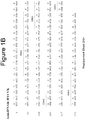
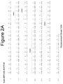


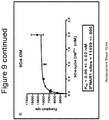


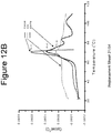
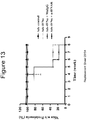
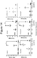
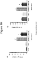


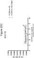
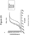
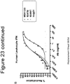


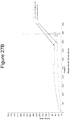

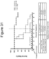

Classifications
-
- C—CHEMISTRY; METALLURGY
- C07—ORGANIC CHEMISTRY
- C07K—PEPTIDES
- C07K16/00—Immunoglobulins [IGs], e.g. monoclonal or polyclonal antibodies
- C07K16/18—Immunoglobulins [IGs], e.g. monoclonal or polyclonal antibodies against material from animals or humans
- C07K16/28—Immunoglobulins [IGs], e.g. monoclonal or polyclonal antibodies against material from animals or humans against receptors, cell surface antigens or cell surface determinants
- C07K16/2866—Immunoglobulins [IGs], e.g. monoclonal or polyclonal antibodies against material from animals or humans against receptors, cell surface antigens or cell surface determinants against receptors for cytokines, lymphokines, interferons
-
- A—HUMAN NECESSITIES
- A01—AGRICULTURE; FORESTRY; ANIMAL HUSBANDRY; HUNTING; TRAPPING; FISHING
- A01K—ANIMAL HUSBANDRY; AVICULTURE; APICULTURE; PISCICULTURE; FISHING; REARING OR BREEDING ANIMALS, NOT OTHERWISE PROVIDED FOR; NEW BREEDS OF ANIMALS
- A01K67/00—Rearing or breeding animals, not otherwise provided for; New or modified breeds of animals
- A01K67/027—New or modified breeds of vertebrates
-
- A—HUMAN NECESSITIES
- A61—MEDICAL OR VETERINARY SCIENCE; HYGIENE
- A61K—PREPARATIONS FOR MEDICAL, DENTAL OR TOILETRY PURPOSES
- A61K39/00—Medicinal preparations containing antigens or antibodies
- A61K39/395—Antibodies; Immunoglobulins; Immune serum, e.g. antilymphocytic serum
-
- A—HUMAN NECESSITIES
- A61—MEDICAL OR VETERINARY SCIENCE; HYGIENE
- A61P—SPECIFIC THERAPEUTIC ACTIVITY OF CHEMICAL COMPOUNDS OR MEDICINAL PREPARATIONS
- A61P1/00—Drugs for disorders of the alimentary tract or the digestive system
-
- A—HUMAN NECESSITIES
- A61—MEDICAL OR VETERINARY SCIENCE; HYGIENE
- A61P—SPECIFIC THERAPEUTIC ACTIVITY OF CHEMICAL COMPOUNDS OR MEDICINAL PREPARATIONS
- A61P1/00—Drugs for disorders of the alimentary tract or the digestive system
- A61P1/16—Drugs for disorders of the alimentary tract or the digestive system for liver or gallbladder disorders, e.g. hepatoprotective agents, cholagogues, litholytics
-
- A—HUMAN NECESSITIES
- A61—MEDICAL OR VETERINARY SCIENCE; HYGIENE
- A61P—SPECIFIC THERAPEUTIC ACTIVITY OF CHEMICAL COMPOUNDS OR MEDICINAL PREPARATIONS
- A61P13/00—Drugs for disorders of the urinary system
- A61P13/10—Drugs for disorders of the urinary system of the bladder
-
- A—HUMAN NECESSITIES
- A61—MEDICAL OR VETERINARY SCIENCE; HYGIENE
- A61P—SPECIFIC THERAPEUTIC ACTIVITY OF CHEMICAL COMPOUNDS OR MEDICINAL PREPARATIONS
- A61P13/00—Drugs for disorders of the urinary system
- A61P13/12—Drugs for disorders of the urinary system of the kidneys
-
- A—HUMAN NECESSITIES
- A61—MEDICAL OR VETERINARY SCIENCE; HYGIENE
- A61P—SPECIFIC THERAPEUTIC ACTIVITY OF CHEMICAL COMPOUNDS OR MEDICINAL PREPARATIONS
- A61P17/00—Drugs for dermatological disorders
-
- A—HUMAN NECESSITIES
- A61—MEDICAL OR VETERINARY SCIENCE; HYGIENE
- A61P—SPECIFIC THERAPEUTIC ACTIVITY OF CHEMICAL COMPOUNDS OR MEDICINAL PREPARATIONS
- A61P17/00—Drugs for dermatological disorders
- A61P17/04—Antipruritics
-
- A—HUMAN NECESSITIES
- A61—MEDICAL OR VETERINARY SCIENCE; HYGIENE
- A61P—SPECIFIC THERAPEUTIC ACTIVITY OF CHEMICAL COMPOUNDS OR MEDICINAL PREPARATIONS
- A61P17/00—Drugs for dermatological disorders
- A61P17/06—Antipsoriatics
-
- A—HUMAN NECESSITIES
- A61—MEDICAL OR VETERINARY SCIENCE; HYGIENE
- A61P—SPECIFIC THERAPEUTIC ACTIVITY OF CHEMICAL COMPOUNDS OR MEDICINAL PREPARATIONS
- A61P17/00—Drugs for dermatological disorders
- A61P17/14—Drugs for dermatological disorders for baldness or alopecia
-
- A—HUMAN NECESSITIES
- A61—MEDICAL OR VETERINARY SCIENCE; HYGIENE
- A61P—SPECIFIC THERAPEUTIC ACTIVITY OF CHEMICAL COMPOUNDS OR MEDICINAL PREPARATIONS
- A61P21/00—Drugs for disorders of the muscular or neuromuscular system
-
- A—HUMAN NECESSITIES
- A61—MEDICAL OR VETERINARY SCIENCE; HYGIENE
- A61P—SPECIFIC THERAPEUTIC ACTIVITY OF CHEMICAL COMPOUNDS OR MEDICINAL PREPARATIONS
- A61P21/00—Drugs for disorders of the muscular or neuromuscular system
- A61P21/04—Drugs for disorders of the muscular or neuromuscular system for myasthenia gravis
-
- A—HUMAN NECESSITIES
- A61—MEDICAL OR VETERINARY SCIENCE; HYGIENE
- A61P—SPECIFIC THERAPEUTIC ACTIVITY OF CHEMICAL COMPOUNDS OR MEDICINAL PREPARATIONS
- A61P25/00—Drugs for disorders of the nervous system
-
- A—HUMAN NECESSITIES
- A61—MEDICAL OR VETERINARY SCIENCE; HYGIENE
- A61P—SPECIFIC THERAPEUTIC ACTIVITY OF CHEMICAL COMPOUNDS OR MEDICINAL PREPARATIONS
- A61P27/00—Drugs for disorders of the senses
- A61P27/02—Ophthalmic agents
-
- A—HUMAN NECESSITIES
- A61—MEDICAL OR VETERINARY SCIENCE; HYGIENE
- A61P—SPECIFIC THERAPEUTIC ACTIVITY OF CHEMICAL COMPOUNDS OR MEDICINAL PREPARATIONS
- A61P29/00—Non-central analgesic, antipyretic or antiinflammatory agents, e.g. antirheumatic agents; Non-steroidal antiinflammatory drugs [NSAID]
-
- A—HUMAN NECESSITIES
- A61—MEDICAL OR VETERINARY SCIENCE; HYGIENE
- A61P—SPECIFIC THERAPEUTIC ACTIVITY OF CHEMICAL COMPOUNDS OR MEDICINAL PREPARATIONS
- A61P3/00—Drugs for disorders of the metabolism
- A61P3/08—Drugs for disorders of the metabolism for glucose homeostasis
- A61P3/10—Drugs for disorders of the metabolism for glucose homeostasis for hyperglycaemia, e.g. antidiabetics
-
- A—HUMAN NECESSITIES
- A61—MEDICAL OR VETERINARY SCIENCE; HYGIENE
- A61P—SPECIFIC THERAPEUTIC ACTIVITY OF CHEMICAL COMPOUNDS OR MEDICINAL PREPARATIONS
- A61P31/00—Antiinfectives, i.e. antibiotics, antiseptics, chemotherapeutics
- A61P31/12—Antivirals
- A61P31/14—Antivirals for RNA viruses
- A61P31/18—Antivirals for RNA viruses for HIV
-
- A—HUMAN NECESSITIES
- A61—MEDICAL OR VETERINARY SCIENCE; HYGIENE
- A61P—SPECIFIC THERAPEUTIC ACTIVITY OF CHEMICAL COMPOUNDS OR MEDICINAL PREPARATIONS
- A61P37/00—Drugs for immunological or allergic disorders
-
- A—HUMAN NECESSITIES
- A61—MEDICAL OR VETERINARY SCIENCE; HYGIENE
- A61P—SPECIFIC THERAPEUTIC ACTIVITY OF CHEMICAL COMPOUNDS OR MEDICINAL PREPARATIONS
- A61P37/00—Drugs for immunological or allergic disorders
- A61P37/02—Immunomodulators
-
- A—HUMAN NECESSITIES
- A61—MEDICAL OR VETERINARY SCIENCE; HYGIENE
- A61P—SPECIFIC THERAPEUTIC ACTIVITY OF CHEMICAL COMPOUNDS OR MEDICINAL PREPARATIONS
- A61P37/00—Drugs for immunological or allergic disorders
- A61P37/02—Immunomodulators
- A61P37/06—Immunosuppressants, e.g. drugs for graft rejection
-
- A—HUMAN NECESSITIES
- A61—MEDICAL OR VETERINARY SCIENCE; HYGIENE
- A61P—SPECIFIC THERAPEUTIC ACTIVITY OF CHEMICAL COMPOUNDS OR MEDICINAL PREPARATIONS
- A61P43/00—Drugs for specific purposes, not provided for in groups A61P1/00-A61P41/00
-
- A—HUMAN NECESSITIES
- A61—MEDICAL OR VETERINARY SCIENCE; HYGIENE
- A61P—SPECIFIC THERAPEUTIC ACTIVITY OF CHEMICAL COMPOUNDS OR MEDICINAL PREPARATIONS
- A61P5/00—Drugs for disorders of the endocrine system
-
- A—HUMAN NECESSITIES
- A61—MEDICAL OR VETERINARY SCIENCE; HYGIENE
- A61P—SPECIFIC THERAPEUTIC ACTIVITY OF CHEMICAL COMPOUNDS OR MEDICINAL PREPARATIONS
- A61P5/00—Drugs for disorders of the endocrine system
- A61P5/14—Drugs for disorders of the endocrine system of the thyroid hormones, e.g. T3, T4
-
- A—HUMAN NECESSITIES
- A61—MEDICAL OR VETERINARY SCIENCE; HYGIENE
- A61P—SPECIFIC THERAPEUTIC ACTIVITY OF CHEMICAL COMPOUNDS OR MEDICINAL PREPARATIONS
- A61P7/00—Drugs for disorders of the blood or the extracellular fluid
- A61P7/04—Antihaemorrhagics; Procoagulants; Haemostatic agents; Antifibrinolytic agents
-
- A—HUMAN NECESSITIES
- A61—MEDICAL OR VETERINARY SCIENCE; HYGIENE
- A61P—SPECIFIC THERAPEUTIC ACTIVITY OF CHEMICAL COMPOUNDS OR MEDICINAL PREPARATIONS
- A61P7/00—Drugs for disorders of the blood or the extracellular fluid
- A61P7/06—Antianaemics
-
- A—HUMAN NECESSITIES
- A61—MEDICAL OR VETERINARY SCIENCE; HYGIENE
- A61P—SPECIFIC THERAPEUTIC ACTIVITY OF CHEMICAL COMPOUNDS OR MEDICINAL PREPARATIONS
- A61P9/00—Drugs for disorders of the cardiovascular system
-
- A—HUMAN NECESSITIES
- A61—MEDICAL OR VETERINARY SCIENCE; HYGIENE
- A61P—SPECIFIC THERAPEUTIC ACTIVITY OF CHEMICAL COMPOUNDS OR MEDICINAL PREPARATIONS
- A61P9/00—Drugs for disorders of the cardiovascular system
- A61P9/10—Drugs for disorders of the cardiovascular system for treating ischaemic or atherosclerotic diseases, e.g. antianginal drugs, coronary vasodilators, drugs for myocardial infarction, retinopathy, cerebrovascula insufficiency, renal arteriosclerosis
-
- A—HUMAN NECESSITIES
- A01—AGRICULTURE; FORESTRY; ANIMAL HUSBANDRY; HUNTING; TRAPPING; FISHING
- A01K—ANIMAL HUSBANDRY; AVICULTURE; APICULTURE; PISCICULTURE; FISHING; REARING OR BREEDING ANIMALS, NOT OTHERWISE PROVIDED FOR; NEW BREEDS OF ANIMALS
- A01K2227/00—Animals characterised by species
- A01K2227/10—Mammal
- A01K2227/105—Murine
-
- A—HUMAN NECESSITIES
- A01—AGRICULTURE; FORESTRY; ANIMAL HUSBANDRY; HUNTING; TRAPPING; FISHING
- A01K—ANIMAL HUSBANDRY; AVICULTURE; APICULTURE; PISCICULTURE; FISHING; REARING OR BREEDING ANIMALS, NOT OTHERWISE PROVIDED FOR; NEW BREEDS OF ANIMALS
- A01K2267/00—Animals characterised by purpose
- A01K2267/01—Animal expressing industrially exogenous proteins
-
- A—HUMAN NECESSITIES
- A61—MEDICAL OR VETERINARY SCIENCE; HYGIENE
- A61K—PREPARATIONS FOR MEDICAL, DENTAL OR TOILETRY PURPOSES
- A61K39/00—Medicinal preparations containing antigens or antibodies
- A61K2039/505—Medicinal preparations containing antigens or antibodies comprising antibodies
-
- C—CHEMISTRY; METALLURGY
- C07—ORGANIC CHEMISTRY
- C07K—PEPTIDES
- C07K2299/00—Coordinates from 3D structures of peptides, e.g. proteins or enzymes
-
- C—CHEMISTRY; METALLURGY
- C07—ORGANIC CHEMISTRY
- C07K—PEPTIDES
- C07K2317/00—Immunoglobulins specific features
- C07K2317/20—Immunoglobulins specific features characterized by taxonomic origin
- C07K2317/24—Immunoglobulins specific features characterized by taxonomic origin containing regions, domains or residues from different species, e.g. chimeric, humanized or veneered
-
- C—CHEMISTRY; METALLURGY
- C07—ORGANIC CHEMISTRY
- C07K—PEPTIDES
- C07K2317/00—Immunoglobulins specific features
- C07K2317/70—Immunoglobulins specific features characterized by effect upon binding to a cell or to an antigen
- C07K2317/71—Decreased effector function due to an Fc-modification
-
- C—CHEMISTRY; METALLURGY
- C07—ORGANIC CHEMISTRY
- C07K—PEPTIDES
- C07K2317/00—Immunoglobulins specific features
- C07K2317/70—Immunoglobulins specific features characterized by effect upon binding to a cell or to an antigen
- C07K2317/73—Inducing cell death, e.g. apoptosis, necrosis or inhibition of cell proliferation
- C07K2317/732—Antibody-dependent cellular cytotoxicity [ADCC]
-
- C—CHEMISTRY; METALLURGY
- C07—ORGANIC CHEMISTRY
- C07K—PEPTIDES
- C07K2317/00—Immunoglobulins specific features
- C07K2317/70—Immunoglobulins specific features characterized by effect upon binding to a cell or to an antigen
- C07K2317/73—Inducing cell death, e.g. apoptosis, necrosis or inhibition of cell proliferation
- C07K2317/734—Complement-dependent cytotoxicity [CDC]
-
- C—CHEMISTRY; METALLURGY
- C07—ORGANIC CHEMISTRY
- C07K—PEPTIDES
- C07K2317/00—Immunoglobulins specific features
- C07K2317/70—Immunoglobulins specific features characterized by effect upon binding to a cell or to an antigen
- C07K2317/76—Antagonist effect on antigen, e.g. neutralization or inhibition of binding
Definitions
- the present invention relates to isolated antibodies and compositions specific for the interferon alpha receptor 1 (IFNAR1) with reduced affinity for Fc ligands.
- the invention also comprises nucleic acids encoding such antibodies, complementary nucleic acids, vectors, host cells, and methods of making and using thereof, including therapeutic compositions, formulations, administrations and devices.
- Type I interferons (IFN ⁇ , IFN ⁇ , IFN ⁇ , IFN ⁇ ) are a family of structurally related cytokines having antiviral, antitumor and immunomodulatory effects ( Hardy et al. (2001) Blood 97:473 ; Cutrone and Langer (2001) J. Biol. Chem. 276:17140 ).
- the human IFN ⁇ locus includes two subfamilies. The first subfamily consists of 14 non-allelic genes and 4 pseudogenes having at least 80% homology. The second subfamily, ⁇ II or omega ( ⁇ ), contains 5 pseudogenes and 1 functional gene which exhibits 70% homology with the IFN ⁇ genes ( Weissmann and Weber (1986) Prog. Nucl.
- IFN ⁇ Interferon alpha subtypes have been identified with the following nomenclature: IFN ⁇ 1, 2a, 2b, 4, 4b, 5, 6, 7, 8, 10, 14, 16, 17, and 21.
- the interferon ⁇ (IFN ⁇ ) is encoded by a single gene, which has approximately 50% homology with the IFN ⁇ genes.
- Interferon ⁇ which is produced by activated lymphocytes, does not possess any homology with the alpha/beta interferons and it does not react with their receptor.
- IFN alpha receptor IFN alpha receptor
- IFNAR1 cell surface receptor 1
- IFNAR2 IFN alpha receptor 2
- IFNAR1 is essential for high affinity binding and differential specificity of the IFNAR complex ( Cutrone et al. (2001) J. Bio Chem 276(20):17140-8 ) While functional differences for each of the type I IFN subtypes have not been identified, it is thought that each may exhibit different interactions with the IFNAR receptor components leading to potentially diverse signaling outcomes ( Cook et al. (1996) J. Biol. Chem.
- type I IFNs have been shown to facilitate differentiation of naive T cells along the Th1 pathway ( Brinkmann et al. (1993) J. Exp. Med. 178:1655 ), to enhance antibody production ( Finkelman et al. (1991) J. Exp. Med. 174:1179 ) and to support the functional activity and survival of memory T cells ( Santini et al. (2000) J. Exp. Med. 191:1777 ; Tough et al. (1996) Science 272:1947 ).
- IDM insulin-dependent diabetes mellitus
- SLE systemic lupus erythematosus
- RA rheumatoid arthritis
- interferon ⁇ has been reported to exacerbate underlying disease in patients with psoriasis and multiple sclerosis and to induce an SLE-like syndrome in patients without a previous history of autoimmune disease.
- Interferon ⁇ has also been shown to induce glomerulonephritis in normal mice and to accelerate the onset of the spontaneous autoimmune disease of NZB/W mice.
- IFN ⁇ therapy has been shown in some cases to lead to undesired side effects, including fever and neurological disorders. Hence there are pathological situations in which inhibition of Type I IFN activity may be beneficial to the patient and a need exists for agents effective in inhibiting Type I IFN activity.
- the Fc region of an antibody interacts with a number of ligands (also referred herein as "Fc ligands" which include but are not limited to agents that specifically bind to the Fc region of antibodies, such as Fc receptors and C1q) including Fc receptors and C1q, imparting an array of important functional capabilities referred to as effector functions.
- Fc ligands also include but are not limited to agents that specifically bind to the Fc region of antibodies, such as Fc receptors and C1q
- effector functions imparting an array of important functional capabilities referred to as effector functions.
- the Fc receptors mediate communication between antibodies and the cellular arm of the immune system ( Raghavan et al., 1996, Annu Rev Cell Dev Biol 12:181-220 ; Ravetch et al., 2001, Annu Rev Immunol 19:275-290 ).
- this protein family includes Fc ⁇ RI (CD64), including isoforms Fc ⁇ RIA, Fc ⁇ RIB, and Fc ⁇ RIC; Fc ⁇ RII (CD32), including isoforms Fc ⁇ RIIA, Fc ⁇ RIIB, and Fc ⁇ RIIC; and Fc ⁇ RIII (CD16), including isoforms Fc ⁇ RIIIA and Fc ⁇ RIIB ( Jefferis et al., 2002, Immunol Lett 82:57-65 ). These receptors typically have an extracellular domain that mediates binding to Fc, a membrane spanning region, and an intracellular domain that may mediate some signaling event within the cell.
- Fc/Fc ⁇ R complex recruits these effector cells to sites of bound antigen, typically resulting in signaling events within the cells and important subsequent immune responses such as release of inflammation mediators, B cell activation, endocytosis, phagocytosis, and cytotoxic attack.
- the ability to mediate cytotoxic and phagocytic effector functions is a potential mechanism by which antibodies destroy targeted cells.
- ADCC antibody dependent cell-mediated cytotoxicity
- ADCP antibody dependent cell-mediated phagocytosis
- CDC complement dependent cytotoxicity
- Fc ⁇ RI Human Fc ⁇ Rs are divided into three distinct classes: Fc ⁇ RI (CD64), Fc ⁇ RII (CD32) and Fc ⁇ RIII (CD16).
- Fc ⁇ RI is a high affinity receptor (K a : 10 - 8-10 -9 M -1 ) and binds both immune complexes and monomeric IgG molecules while the Fc receptors Fc ⁇ RII and Fc ⁇ RIII exhibit lower affinities ( ⁇ 10 -7 M -1 and 2-3x10 7 respectively) ( Gessner J.E. et al., 1998, Ann. Hematology 76:231-48 ).
- Fc ⁇ Rs Signaling through the Fc ⁇ Rs is either through an immunoreceptor tyrosine-based activation motif (ITAM) or immunoreceptor tyrosine-based inhibitory motif (ITIM) for all the transmembrane receptors ( Presta, 2006, Adv. Drug Deliv. Rev 58:640-656 ).
- ITAM immunoreceptor tyrosine-based activation motif
- ITIM immunoreceptor tyrosine-based inhibitory motif
- the 72 kDa extracellular glycoprotein Fc ⁇ RI is mainly expressed on myeloid cells such as monocytes, macrophages CD4+ progenitor cells and may elicit the ADCC, endocytosis, and phagocytosis responses ( Siberil et al., 2006, J. Immunol. Lett. 106:111-118 ).
- the 40 kDa Fc ⁇ RII group of receptors exhibit extracellular domains but do not contain active signal transduction domains. These receptors propagate signals through phosphorylation of a cytoplasmic tail domain ( Amigorena S. et al., 1992 Science. 256:1808-12 ).
- the Fc ⁇ RIIA is mainly expressed on monocytes, macrophages, neutrophils, and platelets whereas the Fc ⁇ RIIC receptor has only been identified on NK cells. These two receptors have been shown to initiate ADCC, endocytosis, phagocytosis and inflammatory mediator release ( Cassel et al., 1993. Mol Immunol 30:451-60 ).
- Fc ⁇ RIIB B1 and B2 types
- B1 and B2 types Fc ⁇ RIIB receptors
- B1 and B2 types Fc ⁇ RIIB receptors are expressed on B cells, Mast cells, basophils, monocytes, macrophages and dendritic cells and have been shown to downregulate the immune response triggered by the A and C isoforms.
- the 50 kDa Fc ⁇ RIIIA is expressed on NK cells, monocytes, macrophages and a subset of T lymphocytes where it activates ADCC, phagocytosis, endocytosis and cytokine release (Gessner et al .).
- the Fc ⁇ RIIIB isoform is a glycosyl-phosphatidylinositol (GPI) anchored peripheral membrane protein involved in the degranulation and the production of reactive oxygen intermediates ( Salmon J.E. et al., 1995, J. Clin. Inves. 95:2877-85 ).
- IgG molecules also exhibit differential isotype specificity for Fc ⁇ Rs.
- IgG3 molecules bind strongly to all Fc ⁇ R isoforms.
- IgG1 the most prevalent isoform in the blood binds to all Fc ⁇ Rs albeit with a lower affinity for the Fc ⁇ RIIA/B isoforms.
- IgG4 is an intermediate binder to Fc ⁇ RI and a weak binder to Fc ⁇ RIIB.
- IgG2 binds only weakly to one allelic form of Fc ⁇ RIIA (Fc ⁇ RIIA-H131) ( Siberil et al., 2006, J. Immunol. Lett. 106:111-118 ).
- the complement inflammatory cascade is a part of the innate immune response and is crucial to the ability for an individual to ward off infection.
- Another important Fc ligand is the complement protein C1q. Fc binding to C1q mediates a process called complement dependent cytotoxicity (CDC) (reviewed in Ward et al., 1995, Ther Immunol 2:77-94 ).
- C1q is capable of binding six antibodies, although binding to two IgGs is sufficient to activate the complement cascade.
- C1q forms a complex with the C1r and C1s serine proteases to form the C1 complex of the complement pathway.
- the binding and stimulation of effector functions mediated by the Fc region of immunoglobulins is highly beneficial, however, in certain instances it may be more advantageous to decrease or eliminate effector function.
- a drug e.g., toxins and isotopes
- the Fc/Fc ⁇ R mediated effector functions bring healthy immune cells into the proximity of the deadly payload, resulting in depletion of normal lymphoid tissue along with the target cells ( Hutchins et al., 1995, PNAS USA 92:11980-11984 ; White et al., 2001, Annu. Rev. Med. 52:125-145 ).
- US 2006/0029601 A1 describes isolated human monoclonal antibodies that bind IFNAR-1 and inhibit the biological activity of type I interferon; modification of the Fc region at specified residues to alter the effector function of the antibody is also described.
- WO 2006/002177 A2 describes human monoclonal antibodies that bind to IFNAR-1 and are capable of inhibiting the biological activity of Type I interferons
- WO 94/029351 A2 describes modifications of Fc that alter effector function.
- WO 2006/036291 A2 describes modifications that impair effector function of antibodies.
- EP 1 707 627 A1 describes Fc modifications that decrease ADCC.
- interferon alpha IFN ⁇ proteins encoded by a functional gene of the interferon alpha gene locus with 75% or greater sequence identity to IFN alpha 1 (GenBank accession number NP_076918 or protein encoded by GenBank accession number NM_024013).
- IFN alpha subtypes include IFN alpha 1, alpha 2a, alpha 2b, alpha 4, alpha 4b alpha 5, alpha 6, alpha 7, alpha 8, alpha 10, alpha 13, alpha 14, alpha 16, alpha 17 and alpha 21.
- IFN alpha interferon alpha
- IFN ⁇ interferon alpha
- IFN alpha immunoglobulin alpha
- naturally occurring preparations that comprise IFN alpha proteins such as leukocyte IFN and lymphoblastoid IFN.
- Interferon alpha receptor-1 is used interchangeably, and include variants, isoforms, species homologs of human IFNAR-1, and analogs having at least one common epitope with IFNAR-1.
- human antibodies described herein may cross-react with IFNAR-1 from species other than human, or other proteins which are structurally related to human IFNAR-1 (e.g., human IFNAR-1 homologs).
- the antibodies may be completely specific for human IFNAR-1 and not exhibit species or other types of cross-reactivity.
- the complete cDNA sequence of human IFNAR-1 has the Genbank accession number NM_000629.
- conservative sequence modifications is intended to include amino acid modifications that do not affect or alter the binding characteristics of the antibody containing the amino acid sequence. Such conservative modifications include amino acid substitutions, additions and deletions. Modifications can be introduced into an antibody described herein by standard techniques known in the art, such as site-directed mutagenesis and PCR-mediated mutagenesis. Conservative amino acid substitutions are ones in which the amino acid residue is replaced with an amino acid residue having a similar side chain. For example, one or more amino acids of a similar polarity act as functional equivalents and result in a silent alteration within the amino acid sequence of the peptide.
- Substitutions that are charge neutral and which replace a residue with a smaller residue may also be considered "conservative substitutions" even if the residues are in different groups (e.g., replacement of phenylalanine with the smaller isoleucine).
- Families of amino acid residues having similar side chains have been defined in the art. Several non-limiting examples of families of conservative amino acid substitutions are shown in Table 1.
- Table 1 Families of Conservative Amino Acid Substitutions Family Amino Acids non-polar Trp, Phe, Met, Leu, Ile, Val, Ala, Pro Uncharged polar Gly, Ser, Thr, Asn, Gln, Tyr, Cys acidic/negatively charged Asp, Glu basic/positively charged Arg, Lys, His Beta-branched Thr, Val, Ile residues that influence chain orientation Gly, Pro Aromatic Tip, Tyr, Phe, His, His
- anti-IFNAR1 antibodies with reduced or ablated effector function are desired for the treatment of chronic autoimmune and/or inflammatory diseases.
- antibodies directed against IFNAR1 were developed with the understanding that effector function would play a role in mediating treatment or at least moderation of a chronic autoimmune and/or inflammatory disease state (see, for example U.S. Publication No. 20060029601 or PCT publication No. WO06002177 ).
- the present invention describes anti-IFNAR1 antibodies with reduced or ablated effector function (such as ADCC and/or CDC).
- effector function such as ADCC and/or CDC.
- tissue cross-reactivitiy studies it was surprisingly found that anti-IFNAR1 antibodies with strong or enhanced effector function displayed a propensity for unwanted toxicity due to the prevalence of staining of anti-IFNAR1 on non-target tissues. This toxicity would result from the non-specific activation of ADCC and/or CDC at inappropriate sites.
- the invetors recognized the need to reduce effector function of polypeptides comprising an Fc region.
- the invention provides a modified IgG class monoclonal antibody specific for IFNAR1, wherein said antibody comprises in the Fc region an amino acid substitution of L234F, as numbered by the EU index as set forth in Kabat and wherein said antibody exhibits reduced affinity for at least one Fc ligand compared to an unmodified antibody.
- said antibody is an IgG1 or IgG4 subclass antibody.
- An antibody described herein may further comprise an amino acid substitution of L235E and / or P331S.
- An antibody described herein may comprise: a. a human heavy chain variable region CDR1 comprising Seq ID NO: 1, b. a human heavy chain variable region CDR2 comprising Seq ID NO: 2; c. a human heavy chain variable region CDR3 comprising Seq ID NO: 3; d. a human light chain variable region CDR1 comprising Seq ID NO: 4; e. a human light chain variable region CDR2 comprising Seq ID NO: 5; and f. a human light chain variable region CDR3 comprising Seq ID NO: 6.
- An antibody described herein may comprise: a. a human heavy chain variable region CDR1 comprising Seq ID NO: 21; b. a human heavy chain variable region CDR2 comprising Seq ID NO: 22; c. a human heavy chain variable region CDR3 comprising Seq ID NO: 23; d. a human light chain variable region CDR1 comprising Seq ID NO: 24; e. a human light chain variable region CDR2 comprising Seq ID NO: 25; and f. a human light chain variable region CDR3 comprising Seq ID NO: 26.
- An antibody described herein may comprise: a. a human heavy chain variable region comprising the amino acid sequence of Seq ID No: 38; and b. a human light chain variable region comprising the amino acid sequence of Seq No: 40.
- An antibody described herein may comprise: a. a human heavy chain variable region comprising amino sequence of Seq ID No: 18; and b. a human light chain variable region comprising the amino acid sequence of Seq ID No: 20.
- Am antibody described herein may comprise: a. a human heavy chain variable region comprising the amino acid sequence of Seq ID No: 28; and b. a human light chain variable region comprising the amino acid sequence of Seq ID No: 30.
- An antibody described herein may comprise the light chain constant region sequence of Seq ID No: 41.
- An antibody described herein may comprise the heavy chain constant region of Seq ID No: 42.
- An antibody described herein may comprise the light chain constant region having the amino acid sequence of Seq ID No: 41 and the heavy chain constant region having the amino acid sequence of Seq ID No: 42.
- An antibody described herein may comprise a heavy chain amino acid sequence comprising allelic variation, wherein said allelic variation is at least one or more positions selected from the group consisting of 214, 221, 356 and 358 as defined by the EU index numbering system.
- the invention further provides an isolated nucleic acid comprising a polynucleotide sequence encoding the antibody of any of the preceding claims.
- the invention yet further provides a pharmaceutical composition
- a pharmaceutical composition comprising the antibody described herein, and a pharmaceutically acceptable excipient.
- the invention also provides a pharmaceutical composition described herein for use in treating a disease or disorder chosen from Grave's disease, Hashimoto's thyroiditis, Crohn's disease, psoriasis, psoriatic arthritis, sympathetic ophthalmitis, autoimmune oophoritis, autoimmune orchitis, autoimmune lymphoproliferative syndrome, antiphospholipid syndrome, Sjogren's syndrome, scleroderma, Addison's disease, polyendocrine deficiency syndrome, Guillain-Barré syndrome, immune thrombocytopenic purpura, pernicious anemia, myasthenia gravis, primary biliary cirrhosis, mixed connective tissue disease, vitiligo, autoimmune uveitis, autoimmune hemolytic anemia, autoimmune thrombocytopenia, celiac disease, dermatitis herpetiformis, autoimmune hepatitis, pemphigus, pemphigus vulgaris, pemphigus foliace
- modified antibodies or other polypeptides comprising the Fc region of an antibody, comprising the addition, substitution, or deletion of at least one amino acid residue to the Fc region resulting in reduced or ablated affinity for at least one Fc ligand (referred to herein as "modified antibodies").
- the Fc region interacts with a number of ligands including, but not limited to, Fc Receptors (e.g., FcRn, Fc ⁇ RIIIa, Fc ⁇ RIIb), the complement protein C1q, and other molecules, such as proteins A and G.
- Fc Receptors e.g., FcRn, Fc ⁇ RIIIa, Fc ⁇ RIIb
- the complement protein C1q e.g., C1q
- other molecules such as proteins A and G.
- Antibodies described herein may have reduced or ablated affinity for an Fc ligand responsible for facilitating effector function compared to an antibody having the same amino acid sequence but not comprising the addition, substitution, or deletion of at least one amino acid residue to the Fc region (also referred to herein as an "unmodified antibody").
- Antibodies described herein may comprise at least one or more of the following properties: reduced or ablated effector (ADCC and/or CDC) function, reduced or ablated binding to Fc receptors, or reduced or ablated toxicities. More specifically, we describe anti-IFNAR1 antibodies with reduced affinity for Fc receptors (e.g., FcRn, Fc ⁇ RIIIa, Fc ⁇ RIIb) and/or the complement protein C1q.
- Antibodies described herein may comprise an Fc region comprising at least one addition, substitution, or deletion of an amino acid residue selected from the positions consisting of: 234, 235, and 331, wherein the numbering system of the constant region is that of the EU index as set forth in Kabat et al. (1991, NIH Publication 91-3242, National Technical Information Service, Springfield, VA ).
- Antibodies described herein may comprise an Fc region comprising at least one amino acid substitution selected from the group consisting of: L234F, L235E, and P331S, wherein the first letter and number represent the unmodified amino acid and its position and the second letter represents the substituted amino acid at said position.
- Antibodies described herein may further comprise an Fc region comprising at least one addition, substitution, or deletion of an amino acid residue that is correlated with increased stability of the antibody.
- the addition, substitution, or deletion of an amino acid residue may be at position 228 of the Fc region, wherein the numbering system of the constant region is that of the EU index as set forth in Kabat et al .
- Antibodies described herein may comprise an Fc region comprising an amino acid substitution at position 228, wherein the substitution is a serine residue.
- Antibodies of the IgG4 subtype may comprise an amino acid substitution of serine at position 228 of the Fc region.
- Antibodies described herein may already comprise a serine residue at position 228 of the Fc region; in such antibodies, no modification is required. Alternatively, antibodies described herein may not require modification of residue 228 of the Fc region or may already comprise serine at said position.
- Antibodies described herein may be any of any class (for example, but not limited to IgG, IgM, and IgE). Antibodies described herein are members of the IgG class of antibodies. In a specific embodiment, antibodies described herein are of the IgG1 subclass. In another specific embodiment, antibodies described herein are of the IgG1 subclass and comprise the following amino acid substitutions: 234F, 235E and 331S of the Fc region. In alternate embodiments, antibodies described herein are of the IgG4 subclass. Antibodies described herein of the IgG4 subclass may comprise the following amino acid substitutions: S228P and L235E of the Fc region.
- Modified antibodies may be produced by combining a variable domain, or fragment thereof, with an Fc domain comprising one or more of the amino acid substitutions disclosed herein. Modified antibodies may be produced by modifying an Fc domain-containing antibody by introducing one or more of the amino acid substitutions residues into the Fc domain.
- antibodies described herein may have altered (relative to an unmodified antibody) Fc ⁇ R and/or C1q binding properties (examples of binding properties include but are not limited to, binding specificity, equilibrium dissociation constant (K D ), dissociation and association rates (K off and K on respectively), binding affinity and/or avidity) and that certain alterations are more or less desirable.
- binding properties include but are not limited to, binding specificity, equilibrium dissociation constant (K D ), dissociation and association rates (K off and K on respectively), binding affinity and/or avidity) and that certain alterations are more or less desirable.
- the equilibrium dissociation constant (K D ) is defined as k off / k on .
- One skilled in the art can determine which kinetic parameter is most important for a given antibody application.
- a modification that reduces binding to one or more positive regulator (e.g., Fc ⁇ RIIIA) and/or enhanced binding to an inhibitory Fc receptor (e.g., Fc ⁇ RIIB) would be suitable for reducing ADCC activity.
- the ratio of binding affinities e.g., equilibrium dissociation constants (K D )
- K D equilibrium dissociation constants
- the affinities and binding properties of an Fc region for its ligand may be determined by a variety of in vitro assay methods (biochemical or immunological based assays) known in the art for determining Fc-Fc ⁇ R interactions, i . e ., specific binding of an Fc region to an Fc ⁇ R including but not limited to, equilibrium methods (e.g., enzyme-linked immunoabsorbent assay (ELISA) or radioimmunoassay (RIA)), or kinetics (e.g., BIACORE ® analysis), and other methods such as indirect binding assays, competitive inhibition assays, fluorescence resonance energy transfer (FRET), gel electrophoresis and chromatography (e.g., gel filtration).
- in vitro assay methods biochemical or immunological based assays
- ELISA enzyme-linked immunoabsorbent assay
- RIA radioimmunoassay
- kinetics e.g., BIACORE ® analysis
- These and other methods may utilize a label on one or more of the components being examined and/or employ a variety of detection methods including but not limited to chromogenic, fluorescent, luminescent, or isotopic labels.
- detection methods including but not limited to chromogenic, fluorescent, luminescent, or isotopic labels.
- Antibodies described herein may exhibit reduced binding affinity for one or more Fc receptors including, but not limited to Fc ⁇ RI (CD64) including isoforms Fc ⁇ RIA, Fc ⁇ RIB, and Fc ⁇ RIC; Fc ⁇ RII (CD32 including isoforms Fc ⁇ RIIA, Fc ⁇ RIIB, and Fc ⁇ RIIC); and Fc ⁇ RIII (CD16, including isoforms Fc ⁇ RIIIA and Fc ⁇ RIIB) as compared to an unmodified antibody.
- Antibodies described herein may not comprise a concomitant increase in binding the Fc ⁇ RIIB receptor as compared to an unmodified (for example, containing a wild type Fc region) antibody.
- Antibodies described herein may exhibit decreased affinities to Fc ⁇ RI relative to an unmodified antibody.
- Antibodies described herein may exhibit affinities for Fc ⁇ RI receptor that are at least 2 fold, or at least 3 fold, or at least 5 fold, or at least 7 fold, or a least 10 fold, or at least 20 fold, or at least 30 fold, or at least 40 fold, or at least 50 fold, or at least 60 fold, or at least 70 fold, or at least 80 fold, or at least 90 fold, or at least 100 fold, or at least 200 fold less than an unmodified antibody.
- Antibodies described herein may exhibit an affinity for Fc ⁇ RI receptor that is at least 90%, at least 80%, at least 70%, at least 60%, at least 50%, at least 40%, at least 30%, at least 20%, at least 10%, or at least 5% less than an unmodified antibody.
- Antibodies described herein may exhibit decreased affinity for the Fc ⁇ RIIIA receptor relative to an unmodified antibody.
- Antibodies described herein may exhibit affinities for Fc ⁇ RIIIA receptor that are at least 2 fold, or at least 3 fold, or at least 5 fold, or at least 7 fold, or a least 10 fold, or at least 20 fold, or at least 30 fold, or at least 40 fold, or at least 50 fold, or at least 60 fold, or at least 70 fold, or at least 80 fold, or at least 90 fold, or at least 100 fold, or at least 200 fold less than an unmodified antibody.
- Antibodies described herein may exhibit affinities for Fc ⁇ RIIIA receptor that are at least 90%, at least 80%, at least 70%, at least 60%, at least 50%, at least 40%, at least 30%, at least 20%, at least 10%, or at least 5% less than an unmodified antibody.
- the F158V allelic variant of the Fc ⁇ RIIIA receptor has altered binding characteristics to antibodies.
- Antibodies described herein may bind with decreased affinities to Fc ⁇ RIIIA (F158V) relative to an unmodified antibody.
- Antibodies described herein may exhibit affinities for Fc ⁇ RIIIA (F158V) receptor that are at least 2 fold, or at least 3 fold, or at least 5 fold, or at least 7 fold, or a least 10 fold, or at least 20 fold, or at least 30 fold, or at least 40 fold, or at least 50 fold, or at least 60 fold, or at least 70 fold, or at least 80 fold, or at least 90 fold, or at least 100 fold, or at least 200 fold less than that of an unmodified antibody.
- Antibodies described herein may exhibit affinities for the Fc ⁇ RIIIA(F158V) receptor that are at least 90%, at least 80%, at least 70%, at least 60%, at least 50%, at least 40%, at least 30%, at least 20%, at least 10%, or at least 5% less than an unmodified antibody.
- Antibodies described herein may exhibit increased affinities for the Fc ⁇ RIIB receptor as compared to unmodified antibody.
- Antibodies described herein may exhibit affinities for the Fc ⁇ RIIB receptor that are unchanged or increased by at least at least 2 fold, or at least 3 fold, or at least 5 fold, or at least 7 fold, or a least 10 fold, or at least 20 fold, or at least 30 fold, or at least 40 fold, or at least 50 fold, or at least 60 fold, or at least 70 fold, or at least 80 fold, or at least 90 fold, or at least 100 fold, or at least 200 fold than that of an unmodified antibody.
- Antibodies described herein may exhibit affinities for the Fc ⁇ RIIB receptor that are increased by at least 5%, at least 10%, at least 20%, at least 30%, at least 40%, at least 50%, at least 60%, at least 70%, at least 80%, at least 90%, or at least 95% than an unmodified antibody.
- Antibodies described herein may exhibit affinities for the Fc ⁇ RI, Fc ⁇ RIIIA, or Fc ⁇ RIIIA (F158V) receptors that are between about 100 nM to about 100 ⁇ M, or about 100 nM to about 10 ⁇ M, or about 100 nM to about 1 ⁇ M, or about 1 nM to about 100 ⁇ M, or about 10 nM to about 100 ⁇ M, or about 1 ⁇ M to about 100 ⁇ M, or about 10 ⁇ M to about 100 ⁇ M.
- Antibodies described herein may exhibit affinities for the Fc ⁇ RI, Fc ⁇ RIIIA, or Fc ⁇ RIIIA (F158V) receptors that are greater than 1 ⁇ M, greater than 5 ⁇ M, greater than 10 ⁇ M, greater than 25 ⁇ M, greater than 50 ⁇ M, or greater than 100 ⁇ M.
- Antibodies described herein may exhibit affinities for the Fc ⁇ RIIB receptor that are between about 100 nM to about 100 ⁇ M, or about 100 nM to about 10 ⁇ M, or about 100 nM to about 1 ⁇ M, or about 1 nM to about 100 ⁇ M, or about 10 nM to about 100 ⁇ M, or about 1 ⁇ M to about 100 ⁇ M, or about 10 ⁇ M to about 100 ⁇ M.
- Antibodies described herein may exhibit affinities for the Fc ⁇ RI, Fc ⁇ RIIIA, or Fc ⁇ RIIIA (F158V) receptors that are less than 100 ⁇ M, less than 50 ⁇ M, less than 10 ⁇ M, less than 5 ⁇ M, less than 2.5 ⁇ M, less than 1 ⁇ M, or less than 100 nM, or less than 10 nM.
- ADCC antibody-dependent cell-mediated cytotoxicity
- FcRs Fc receptors
- cytotoxic cells e.g., Natural Killer (NK) cells, neutrophils, and macrophages
- NK Natural Killer
- IgG antibodies directed to the surface of target cells "arm" the cytotoxic cells and are required for such killing.
- CDC complement dependent cytotoxicity
- ADCC antibody-mediated lysis
- an antibody of interest is added to target cells in combination with immune effector cells, which may be activated by the antigen antibody complexes resulting in cytolysis of the target cell. Cytolysis is generally detected by the release of label (e.g., radioactive substrates, fluorescent dyes or natural intracellular proteins) from the lysed cells.
- label e.g., radioactive substrates, fluorescent dyes or natural intracellular proteins
- useful effector cells for such assays include peripheral blood mononuclear cells (PBMC) and Natural Killer (NK) cells.
- PBMC peripheral blood mononuclear cells
- NK Natural Killer
- ADCC assays are described in Wisecarver et al ., 1985 79:277-282; Bruggemann et al., 1987, J Exp Med 166:1351-1361 ; Wilkinson et al., 2001, J Immunol Methods 258:183-191 ; Patel et al., 1995 J Immunol Methods 184:29-38 .
- ADCC activity of the antibody of interest may be assessed in vivo, e.g., in an animal model such as that disclosed in Clynes et al., 1998, PNAS USA 95:652-656 .
- antibodies described herein are characterized by in vitro functional assays for determining one or more Fc ⁇ R mediated effector cell functions.
- Antibodies described herein may have similar binding properties and effector cell functions in in vivo models (such as those described and disclosed herein) as those in in vitro based assays. However, antibodies described herein that do not exhibit the desired phenotype in in vitro based assays may exhibit the desired phenotype in vivo.
- Antibodies described herein may exhibit decreased ADCC activities as compared to an unmodified antibody.
- Antibodies described herein may exhibit ADCC activities that are at least 2 fold, or at least 3 fold, or at least 5 fold, or at least 10 fold, or at least 50 fold, or at least 100 fold less than that of an unmodified antibody.
- antibodies described herein may exhibit ADCC activities that are reduced by at least 10%, or at least 20%, or by at least 30%, or by at least 40%, or by at least 50%, or by at least 60%, or by at least 70%, or by at least 80%, or by at least 90%, or by at least 100%, or by at least 200%, or by at least 300%, or by at least 400%, or by at least 500% relative to an unmodified antibody.
- Antibodies described herein may have no detectable ADCC activity.
- the reduction and/or ablatement of ADCC activity may be attributed to the reduced affinity that antibodies described herein may exhibit for Fc ligands and/or receptors.
- the complement activation pathway is initiated by the binding of the first component of the complement system (C1q) to a molecule, an antibody for example, complexed with a cognate antigen.
- a CDC assay e.g., as described in Gazzano-Santoro et al., 1996, J. Immunol. Methods, 202:163 , may be performed.
- Antibodies described herein may exhibit decreased affinities to C1q relative to an unmodified antibody.
- Antibodies described herein may exhibit affinities for C1q receptor that are at least 2 fold, or at least 3 fold, or at least 5 fold, or at least 7 fold, or a least 10 fold, or at least 20 fold, or at least 30 fold, or at least 40 fold, or at least 50 fold, or at least 60 fold, or at least 70 fold, or at least 80 fold, or at least 90 fold, or at least 100 fold, or at least 200 fold less than an unmodified antibody.
- Antibodies described herein may exhibit affinities for C1q that are at least 90%, at least 80%, at least 70%, at least 60%, at least 50%, at least 40%, at least 30%, at least 20%, at least 10%, or at least 5% less than an unmodified antibody.
- Antibodies described herein may exhibit affinities for C1q that are between about 100 nM to about 100 ⁇ M, or about 100 nM to about 10 ⁇ M, or about 100 nM to about 1 ⁇ M, or about 1 nM to about 100 ⁇ M, or about 10 nM to about 100 ⁇ M, or about 1 ⁇ M to about 100 ⁇ M, or about 10 ⁇ M to about 100 ⁇ M.
- Antibodies described herein may exhibit affinities for C1q that are greater than 1 ⁇ M, greater than 5 ⁇ M, greater than 10 ⁇ M, greater than 25 ⁇ M, greater than 50 ⁇ M, or greater than 100 ⁇ M.
- Antibodies described herein may exhibit decreased CDC activities as compared to an unmodified antibody.
- Antibodies described herein may exhibit CDC activities that are at least 2 fold, or at least 3 fold, or at least 5 fold, or at least 10 fold, or at least 50 fold, or at least 100 fold less than that of an unmodified antibody.
- antibodies described herein may exhibit CDC activities that are reduced by at least 10%, or at least 20%, or by at least 30%, or by at least 40%, or by at least 50%, or by at least 60%, or by at least 70%, or by at least 80%, or by at least 90%, or by at least 100%, or by at least 200%, or by at least 300%, or by at least 400%, or by at least 500% relative to an unmodified antibody.
- Antibodies described herein may exhibit no detectable CDC activities.
- the reduction and/or ablatement of CDC activity may be attributed to the reduced affinity antibodies described herein may exhibit for Fc ligands and/or receptors.
- biological therapies may have adverse toxicity issues associated with the complex nature of directing the immune system to recognize and attack unwanted cells and/or targets.
- adverse toxicity may occur.
- antibody staining of non-targeted tissues may be indicative of potential toxicity issues.
- Antibodies described herein may exhibit reduced staining of non-targeted tissues as compared to an unmodified antibody.
- Antibodies described herein may exhibit reduced staining of non-targeted tissues that are at least 2 fold, or at least 3 fold, or at least 5 fold, or at least 7 fold, or at least 10 fold, or at least 20 fold, or at least 30 fold, or at least 40 fold, or at least 50 fold, or at least 60 fold, or at least 70 fold, or at least 80 fold, or at least 90 fold, or at least 100 fold, or at least 200 fold less than that of an unmodified antibody.
- Antibodies described herein may exhibit reduced staining of non-targeted tissues that are reduced by at least 10%, or by at least 20%, or by at least 30%, or by at least 40%, or by at least 50%, or by at least 60%, or by at least 70%, or by at least 80%, or by at least 90%, or by at least 100%, or by at least 200%, or by at least 300%, or by at least 400%, or by at least 500% relative to an unmodified antibody.
- Antibodies described herein may exhibit a reduced antibody-related toxicity as compared to an unmodified antibody.
- Antibodies described herein may exhibit toxicities that are at least 2 fold, or at least 3 fold, or at least 5 fold, or at least 7 fold, or a least 10 fold, or at least 20 fold, or at least 30 fold, or at least 40 fold, or at least 50 fold, or at least 60 fold, or at least 70 fold, or at least 80 fold, or at least 90 fold, or at least 100 fold, or at least 200 fold less than that of an unmodified antibody.
- Antibodies described herein may exhibit toxicities that are reduced by at least 10%, or by at least 20%, or by at least 30%, or by at least 40%, or by at least 50%, or by at least 60%, or by at least 70%, or by at least 80%, or by at least 90%, or by at least 100%, or by at least 200%, or by at least 300%, or by at least 400%, or by at least 500% relative to an unmodified antibody.
- Antibodies described herein may bind to cell-surface antigens that may internalize, further carrying the antibodies into the cell. Once inside the cell, the antibodies may be released into the cytoplasm, targeted to a specific compartment, or recycled to the cell surface. Antibodies described herein may bind to a cell-surface antigen that internalizes. Antibodies described herein may be targeted to specific organelles or compartments of the cell. Antibodies described herein may be recycled to the cell surface or periphery after internalization. In a specific embodiment, the antibody described herein is specific for IFNAR1.
- the extent of internalization can be represented as a percentage of total antibody bound to cells.
- the extent of antibody internalization can be represented as a comparison to a non-specific control antibody.
- the extent of antibody internalization can be represented as a comparison to an antibody that binds a cell-surface antigen that does not internalize.
- the extent of antibody internalization can be correlated with the degradation of the antibody.
- the extent of antibody internalization can be represented as a ratio of cytoplasmic versus cell surface staining.
- the antibodies described herein once bound may internalize into cells wherein internalization is at least about 10%, at least about 20%, at least about 30%, at least about 40%, at least about 50%, at least about 60%, at least about 70%, at least about 80%, or at least about 90%, at least about 100%, at least about 110%, at least about 130%, at least about 140%, at least about 150%, at least about 160%, or at least about 170% more than a non-specific control antibody.
- the antibodies described herein once bound may internalize into cells wherein internalization is 1-10%, 10-20%, 20-30%, 30-40%,40-50%, 50-60%, 60-70%, 70-80%, 80-90%, 90-100%, 100-110%, 110-120%, 120-130%, 130-140%, 140-150%, 150-160%, 160-170% more than a non-specific control antibody.
- the antibodies described herein once bound may internalize into cells wherein internalization is 1-10%, 10-20%, 20-30%, 30-40%,40-50%, 50-60%, 60-70%, 70-80%, 80-90%, 90-100%, 100-110%, 110-120%, 120-130%, 130-140%, 140-150%, 150-160%, 160-170% more than control antibodies as determined by the internalization assay using a secondary antibody.
- crystalline forms of a human IgG Fc region wherein the human Fc region, designated as Fc-TM, comprises amino acid substitutions of L234F, L235E and P331S as numbered by the EU index as set forth in Kabat and exhibits reduced or ablated effector (ADCC and/or CDC) function, reduced or ablated binding to Fc receptors, and/or reduced or ablated toxicities.
- the crystals are of diffraction quality to permit the determination of the three-dimensional X-ray diffraction structure of the crystalline polypeptide(s) to high resolution, preferably to a resolution of greater than about 3 ⁇ , typically in the range of about 2 ⁇ to about 3 ⁇ .
- Fc-TM The overall three-dimensional structure of Fc-TM was very similar to previously reported structures of unliganded human Fc regions ( Deisenhofer, (1981). Biochemistry, 20, 2361-2370 ; Krapp et al., (2003). J. Mol. Biol. 325, 979-989 ; Matsumiya et al., (2007). J. Mol. Biol. 368, 767-779 ; Oganesyan et al., (2007) Molecular Immunology, December 11, 2007 , in press).
- Fc-TM C H 2 and C H 3 domains showed great structural conservation and rigidity when compared with other unliganded, unmutated human Fc structures.
- the structure information can be used in a variety of computation or computer-based methods to screen, design or identify anti-IFNAR antibodies that have altered biological properties.
- the crystals and structure coordinates obtained therefrom can be used to screen, design or identify amino acid additions, substitutions or deletions in Fc region that result in reduced or ablated binding to Fc receptors, reduced or ablated effector (ADCC and/or CDC) function, or reduced or ablated toxicities.
- ADCC and/or CDC reduced or ablated effector
- anti-IFNAR1 antibodies that are designed or selected by the use of the structure information of Fc-TM and that exhibit the desired biological activities.
- Such antibodies may comprise an Fc region with the mutations of L234F, L235E, and P331S.
- Such antibodies may comprise an Fc region with one or more addition, substitution, or deletion of an amino acid residue other than amino acid residues 234, 235, and 331.
- Antibodies described herein are specific for ( i.e., specifically bind) IFNAR1. Such antibodies may also be referred to herein as "anti-IFNAR1 antibodies described herein.” Antibodies described herein are specific for human IFNAR1. The anti-IFNAR1 antibodies described herein may cross-react with IFNAR1 from species other than human, or other proteins which are structurally related to human IFNAR1 (for example, human IFNAR1 homologs). Anti-IFNAR1 antibodies described herein may be specific for human IFNAR1 only and not exhibit species or other types of cross-reactivity.
- Anti-IFNAR1 antibodies described herein may exhibit reduced binding affinities for Fc ligands and have at least one of the following properties: reduced or ablated effector (ADCC and/or CDC) function, reduced or ablated binding to Fc ligands, or reduced or ablated toxicities as compared to an unmodified antibody.
- ADCC and/or CDC reduced or ablated effector
- Anti-IFNAR1 antibodies described herein may comprise the addition, substitution or deletion of at least one amino acid residue selected from the group consisting of: L234F, L235E, and P331S.
- Anti-IFNAR1 antibodies described herein may comprise the amino acid substitutions: L234F, L235E, and P331S of the Fc region.
- An anti-IFNAR1 antibody described herein may be an IgG isotype antibody.
- Anti-IFNAR1 antibodies described herein may be of the IgG4 subclass.
- Anti-IFNAR1 IgG4 antibodies described herein may comprise the amino acid substitution L235E of the Fc region.
- Anti-IFNAR1 IgG4 antibodies described herein may also comprise an amino acid change that is correlated with increased stability.
- Anti-IFNAR1 IgG4 antibodies described herein may further comprise the amino acid substitution S228P of the Fc region.
- Anti-IFNAR1 antibodies described herein may exhibit reduced or ablated binding affinities for Fc receptors (for example, but not limited to Fc ⁇ RI (CD64), including isoforms Fc ⁇ RIA, Fc ⁇ RIB, and Fc ⁇ RIC; Fc ⁇ RII (CD32), including isoforms Fc ⁇ RIIA, Fc ⁇ RIIB, and Fc ⁇ RIIC; and Fc ⁇ RIII (CD16), including isoforms Fc ⁇ RIIIA and Fc ⁇ RIIB) as compared to an unmodified antibody.
- Anti-IFNAR 1 antibodies described herein may exhibit decreased affinities to Fc ⁇ RI relative to an unmodified antibody.
- Anti-IFNAR1 antibodies described herein exhibit may decreased affinities for the Fc ⁇ RIIIA receptor relative to an unmodified antibody.
- Anti-IFNAR1 antibodies described herein may bind with decreased affinities to the F158V allele of Fc ⁇ RIIIA relative to an unmodified antibody.
- Anti-IFNAR1 antibodies described herein may exhibit reduced or ablated binding affinities for C1q as compared to an unmodified antibody.
- Anti-IFNAR 1 antibodies described herein may exhibit decreased affinities to Fc ⁇ RI relative to an unmodified antibody.
- Anti-IFNAR1 antibodies described herein may exhibit reduced or ablated effector function. Anti-IFNAR1 antibodies described herein may exhibit reduced or ablated ADCC and/or CDC activity. Anti-IFNAR1 antibodies described herein may exhibit reduced or ablated toxicity.
- Amino acid sequences of the heavy chain variable regions and/or light chain variable regions of anti-IFNAR1 antibodies described herein are provided as Figures 1A , 2A , 3A , 4A and Figures 1B , 2B , 3B , 4B , respectively.
- the polynucleotide sequence encoding the heavy chain variable and light chain variable regions of the anti-IFNAR1 antibodies described herein are provided as Figures 1A , 2A , 3A , 4A and Figures 1B , 2B , 3B , 4B , respectively.
- Antibodies described herein are disclosed in U.S. Patent Provisional Applications Serial Nos. 60/842,925, filed September 8, 2006 , 60/866,917 ; filed November 22, 2006; 60/911,397, filed April 12, 2007 ; 60/915,309, filed May 22, 2007 ; US Patent Application Serial No. 11/852,106, filed September 7, 2007 ; and PCT Application Serial No. US2007/07791, filed September 7, 2007 .
- Anti-IFNAR1 antibodies described herein also include antibodies that comprise an amino acid sequence of a variable heavy chain and/or variable light chain that is at least 45%, at least 50%, at least 55%, at least 60%, at least 65%, at least 70%, at least 75%, at least 80%, at least 85%, at least 90%, at least 95%, or at least 99% identical to the amino acid sequence of the variable heavy chain and/or light chain of the 3F11, 11E2, 4G5, and 9D4 antibodies (see Figures 1-4 for sequences).
- CDRs residue numbers referred to herein are those of Kabat et al ., (1991, NIH Publication 91-3242, National Technical Information Service, Springfield, VA). Specifically, residues 24-34 (CDR1), 50-56 (CDR2) and 89-97 (CDR3) in the light chain variable domain and 31-35 (CDR1), 50-65 (CDR2) and 95-102 (CDR3) in the heavy chain variable domain. Note that CDRs vary considerably from antibody to antibody (and by definition will not exhibit homology with the Kabat consensus sequences). Maximal alignment of framework residues frequently requires the insertion of "spacer" residues in the numbering system, to be used for the Fv region.
- CDRs referred to herein are those of Kabat et al . supra .
- identity of certain individual residues at any given Kabat site number may vary from antibody chain to antibody chain due to interspecies or allelic divergence.
- Anti-IFNAR1 antibodies described herein may comprise at least one VH CDR having an amino acid sequence of any one of the VH CDRs listed in Table 2.
- Anti-IFNAR1 antibodies described herein may comprise at least one VL CDR having an amino acid sequence of any one of the VL CDRs listed in Table 2.
- Anti-IFNAR1 antibodies described herein may comprise one or more of the VH CDRs and one or more of the VL CDRs listed in Table 2.
- Anti-IFNAR1 antibodies described herein may comprise any combination of the VH CDRs and VL CDRs listed in Table 2.
- Anti-IFNAR1 antibodies described herein may comprise at least 1, or at least 2, or at least 3, or at least 4, or at least 5, or at least 6 CDRs selected from Table 2.
- Anti-IFNAR1 antibodies described herein may comprise a VH domain and/or a VL domain each comprising 1, 2 or 3 CDRs.
- the anti-IFNAR1 antibodies described herein may comprise a VH further comprising 1, 2, or 3 heavy chain CDRs (CDRH#) listed in Table 2.
- the anti-IFNAR1 antibodies described herein may comprise a VL further comprising 1, 2, or 3 light chain CDRs (CDRL#) listed in Table 2.
- Anti-IFNAR1 antibodies described herein may comprise the CDRs of antibody 3F11 (see for example Table 2).
- Anti-IFNAR1 antibodies described herein may comprise the CDRs of antibody 4G5 (see for example Table 2).
- Anti-IFNAR1 antibodies described herein may comprise the CDRs of antibody 11E2 (see for example Table 2).
- Anti-IFNAR1 antibodies described herein comprise the CDRs of antibody 9D4 (see for example Table 2). Table 2.
- Anti-IFNAR1 antibody CDR sequences Antibody CDR Sequence Seq ID No: 3F11 CDRL1 RASQGIYSVLA 1 3F11 CDRL2 DASRLES 2 3F11 CDRL3 QQFNSYIT 3 3F11 CDRH1 GYFWS 4 3F11 CDRH2 EIDHSGKTNYNPSLKS 5 3F11 CDRH3 ESKYYFGLDV 6 4G5 CDRL1 RATQDISIALV 11 4G5 CDRL2 DASGLGS 12 4G5 CDRL3 QQFNSYPYT 13 4G5 CDRH1 NYYWS 14 4G5 CDRH2 EIILSGSTNYNPSLKS 15 4G5 CDRH3 ESKWGYYFDS 16 11E2 CDRL1 RASQSVSSSFFA 21 11E2 CDRL2 GASSRAT 22 11E2 CDRL3 QQYYDSSAIT 23 11E2 CDRH1 NYWIA 24 11E2 CDRH2 IIYPGDSDIRYSPSFQG 25
- Anti-IFNAR1 antibodies described herein may comprise an amino acid sequence of a variable heavy chain and/or variable light chain that comprises at least 1, at least 2, at least 3, at least 4, at least 5, at least 10, at least 15, or at least 20 amino acid substitutions, additions, or deletions as compared to the variable heavy chains and/or light chains represented in Figures 1 , 2 , 3 , or 4 .
- Anti-IFNAR1 antibodies described herein may comprise one or more CDRs with at least 1, at least 2, at least 3, at least 4, at least 5, or at least 10 amino acid substitutions, deletions, or additions of one or more CDRs listed in Table 2.
- Anti-IFNAR1 antibodies described herein may comprise antibodies encoded by a polynucleotide sequence that hybridizes to the nucleotide sequence represented in Figures 1 , 2 , 3 , or 4 under stringent conditions.
- Anti-IFNAR1 antibodies described herein may comprise one or more CDRs encoded by a nucleotide sequence that hybridizes under stringent conditions to the nucleotide sequence of one or more CDRs listed in Figures 1 , 2 , 3 , or 4 .
- Stringent hybridization conditions include, but are not limited to, hybridization to filter-bound DNA in 6X sodium chloride/sodium citrate (SSC) at about 45°C followed by one or more washes in 0.2X SSC/0.1% SDS at about 50-65°C, highly stringent conditions such as hybridization to filter-bound DNA in 6X SSC at about 45°C followed by one or more washes in 0.1X SSC/0.2% SDS at about 60°C, or any other stringent hybridization conditions known to those skilled in the art (see, for example, Ausubel, F.M. et al., eds. 1989 Current Protocols in Molecular Biology, vol. 1, Green Publishing Associates, Inc.
- Anti-IFNAR1 antibodies described herein include, but are not limited to, antibodies encoded by a polynucleotide sequence that is at least 70%, at least 80%, at least 85%, at least 90%, at least 95%, at least 96%, at least 97%, at least 98%, or at least 99% identical to a polynucleotide sequence encoding antibodies 3F11, 11E2, 4G5, or 9D4 (see Figures 1-4 ).
- Anti-IFNAR1 antibodies described herein may exhibit a high binding affinity for IFNAR1.
- Anti-IFNAR1 antibodies described herein may exhibit association rate (k on ) of at least 10 5 M -1 s -1 , at least 5 ⁇ 10 5 M - s -1 , at least 10 6 M -1 s -1 , at least 5 ⁇ 10 6 M -1 s -1 , at least 10 7 M -1 s -1 , at least 5 ⁇ 10 7 M -1 s -1 , or at least 10 8 M -1 s -1 .
- Anti-IFNAR1 antibodies described herein may exhibit a k on of at least 2 ⁇ 10 5 M -1 s -1 , at least 5 ⁇ 10 5 M -1 s -1 , at least 10 6 M -1 s -1 , at least 5 ⁇ 10 6 M -1 s -1 , at least 10 7 M -1 s -1 , at least 5 ⁇ 10 7 M -1 s -1 , or at least 10 8 M -1 s -1 .
- Anti-IFNAR1 antibodies described herein may exhibit a dissociation rate (k off ) of less than 10 -1 s -1 , less than 5 ⁇ 10 -1 s -1 , less than 10 -2 s -1 , less than 5 ⁇ 10 -2 s -1 , less than 10 -3 s -1 , less than 5 ⁇ 10 -3 s -1 , less than 10 -4 s -1 , less than 5 ⁇ 10 -4 s -1 , less than 10 -5 s -1 , less than 5 ⁇ 10 -5 s -1 , less than 10 -6 s -1 , less than 5 ⁇ 10 -6 s -1 , less than 10 -7 s -1 , less than 5 ⁇ 10 -7 s -1 , less than 10 -8 s -1 , less than 5 ⁇ 10 -8 s -1 , less than 10 -9 s -1 , less than 5 ⁇ 10 -9 s -1 , or less than 10 -10-1 s -1 .
- Anti-IFNAR1 antibodies described herein may exhibit a k off , of less than 5 ⁇ 10 -4 s -1 , less than 10 -5 s -1 , less than 5 ⁇ 10 -5 s -1 , less than 10 -6 s -1 , less than 5 ⁇ 10 -6 s -1 , less than 10 -7 s -1 , less than 5 ⁇ 10 -7 s -1 , less than 10 -8 s -1 , less than 5 ⁇ 10 -8 s -1 , less than 10 -9 s -1 , less than 5 ⁇ 10 -9 s -1 , or less than 10 -10 s -1 .
- Anti-IFNAR1 antibodies described herein may exhibit an affinity constant or K a (k on /k off ) of at least 10 2 M -1 , at least 5 ⁇ 10 2 M -1 , at least 10 3 M -1 , at least 5 ⁇ 10 3 M -1 , at least 10 4 M -1 , at least 5 ⁇ 10 4 M -1 , at least 10 5 M -1 , at least 5 ⁇ 10 5 M -1 , at least 10 6 M -1 , at least 5 ⁇ 10 6 M -1 , at least 10 7 M -1 , at least 5 ⁇ 10 7 M -1 , at least 10 8 M -1 , at least 5 ⁇ 10 8 M -1 , at least 10 9 M -1 , at least 5 ⁇ 10 9 M -1 , at least 10 10 M -1 , at least 5 ⁇ 10 1 M -1 , at least 10 11 M -1 , at least 5 ⁇ 10 11 M -1 , at least 10 12 M -1 , at least 5 ⁇ 10 12 M, at least 10 13 M -1
- Anti-IFNAR1 antibodies described herein may exhibit a dissociation constant or K d (k off /k on ) of less than 10 -2 M, less than 5 ⁇ 10 -2 M, less than 10 -3 M, less than 5 ⁇ 10 -3 M, less than 10 -4 M, less than 5 ⁇ 10 -4 M, less than 10 -5 M, less than 5 ⁇ 10 -5 M, less than 10 -6 M, less than 5 ⁇ 10 -6 M, less than 10 -7 M, less than 5 ⁇ 10 -7 M, less than 10 -8 M, less than 5 ⁇ 10 -8 M, less than 10 -9 M, less than 5 ⁇ 10 -9 M, less than 10 -10 M, less than 5 ⁇ 10 -10 M, less than 10 -11 M, less than 5 ⁇ 10 -11 M, less than 10 -12 M, less than 5 ⁇ 10 -12 M, less than 10 -13 M, less than 5 ⁇ 10 -13 M, less than 10 -14 M, less than 5 ⁇ 10 -14 M, less than 10 -15 M, or less than 5 ⁇ 10
- Anti-IFNAR1 antibodies described herein may exhibit the ability to block binding to IFNAR1 and/or neutralize the biological activity of one or more Type I interferon (IFN) including, but not limited to, IFN ⁇ , IFN ⁇ , and IFN ⁇ . Binding of IFN ⁇ subtypes can be determined by routine competition assays such as that described in "Antibodies: A Laboratory Manual", CSHL. The anti-IFNAR1 antibodies described herein may exhibit the ability to block binding to IFNAR1 and/or neutralize the biological activity of IFN ⁇ , IFN ⁇ , and IFN ⁇ .
- IFN Type I interferon
- the anti-IFNAR1 antibodies described herein may exhibit the ability to block binding to IFNAR1 and/or neutralize the biological activity of one or more subtypes of IFN ⁇ including, but not limited to, IFN ⁇ subtypes 1, 2a, 2b, 4, 4b, 5, 6, 7, 8, 10, 14, 16, 17, and 21.
- the anti-IFNAR1 antibodies described herein may exhibit the ability to block binding to IFNAR1 and/or neutralize the biological activity of all subtypes of IFN ⁇ .
- anti-IFNAR1 antibodies described herein may exhibit the ability to block the binding of and/or neutralize the biological activity of IFN ⁇ subtypes IFN ⁇ 1, 2a, 2b, 4, 4b, 5, 6, 7, 8, 10, 14, 16, 17, and 21.
- Anti-IFNAR1 antibodies described herein may not exhibit the ability to block binding to IFNAR1 and/or neutralize the biological activity of one or more subtypes of IFN ⁇ including, but not limited to, IFN ⁇ subtypes 1, 2a, 2b, 4, 4b, 5, 6, 7, 8, 10, 14, 16, 17, and 21.
- anti-IFNAR1 antibodies described herein may exhibit the ability to block binding to IFNAR1 and/or neutralize the biological activity all IFN ⁇ subtypes except IFN ⁇ 21.
- the anti-IFNAR1 antibodies described herein may exhibit the ability to block binding to IFNAR1 and/or neutralize the biological activity of at least 1, at least 2, at least 3, at least 4, at least 5, at least 6, at least 7, at least 8, at least 9, at least 10, at least 11, at least 12, or at least 13 of the following IFN ⁇ subtypes: 1, 2a, 2b, 4, 4b, 5, 6, 7, 8, 10, 14, 16, 17, and 21.
- the anti-IFNAR1 antibodies described herein may not exhibit the ability to block binding to IFNAR1 and/or neutralize the biological activity of at least 1, at least 2, at least 3, at least 4, at least 5, at least 6, at least 7, at least 8, at least 9, at least 10, at least 11, at least 12, or at least 13 of the following IFN ⁇ subtypes: 1, 2a, 2b, 4, 4b, 5, 6, 7, 8, 10, 14, 16, 17, and 21.
- Anti-IFNAR1 antibodies described herein may exhibit the ability to block binding to IFNAR1 and/or neutralize the biological activity of non-naturally-occurring type I-like interferons.
- non-naturally occurring type I-like interferons, or hybrid type I-like interferons represent molecules that have been altered from their naturallyoccurung structures by recombinant or synthetic techniques.
- Hybrid interferons, as described in U.S. Patent No. 7,232,563 represent a molecular replacement of various segments of a naturally occurring interferon structure to create a molecule that has increased potency and/or reduced toxicity.
- Anti-IFNAR1 antibodies described herein may exhibit the ability to block binding to IFNAR1 and/or neutralize the biological activity of mutated type I interferons. Mutated type I interferons are described in U.S. Patent Nos. 6,299,870 and 6,300,474 .
- Anti-IFNAR1 antibodies described herein may exhibit the ability to block binding to IFNAR1 and/or neutralize the biological activity of type I-like interferons derived from other animal species.
- type I-like interferons are isolated from chicken, cat, mouse, rat, rabbit, goat, horse or other animal species.
- human type I interferons are isolated from cells derived from chicken, cat, mouse, rat, rabbit, goat, horse or other animal species.
- human type I interferons entail different glycosylation patterns when derived from chicken, cat, mouse, rat, rabbit, goat, horse or other animal species. Further discussion of interferons from other animal species can be found in WIPO publication No. WO06099451A3 .
- the ability of anti-IFNAR1 antibodies described herein to neutralize the activity of IFN ⁇ can be monitored, for example, in a Kinase Receptor Activation (KIRA) Assay as described in WO 95/14930, published Jun. 1, 1995 , by measuring the ability of a candidate antibody to reduce tyrosine phosphorylation (resulting from ligand binding) of the IFNAR1/R2 receptor complex.
- KIRA Kinase Receptor Activation
- the ability of anti-IFNAR1 antibodies described herein to neutralize the elicitation of a cellular response by IFN ⁇ may be tested by monitoring the neutralization of the antiviral activity of IFN ⁇ , as described by Kawade, J. Interferon Res. 1:61 70 (1980 ), or Kawade and Watanabe, J. Interferon Res. 4:571 584 (1984 ), or Yousefi, et al., Am. J. Clin. Pathol.
- ISGF3 interferon-stimulated factor 3
- ISRE interferon-stimulated response element
- Anti-IFNAR1 antibodies described herein may exhibit the ability to inhibit at least one IFN ⁇ mediated function of the IFNAR1 receptor.
- Anti-IFNAR1 antibodies described herein may inhibit the activity of the IFNAR1 receptor in response to IFN ⁇ or subtypes thereof by at least about 60%, at least about 70%, at least about 75%, at least about 80%, at least about 85%, at least about 90%, at least about 95%, or at least about 99% .
- the anti-IFNAR1 antibodies described herein may inhibit the activity of the IFNAR1 receptor in response to IFN ⁇ or subtypes thereof as measured by the KIRA assay described above by at least about 60%, at least about 70%, at least about 75%, at least about 80%, at least about 85%, at least about 90%, at least about 95%, or at least about 99%.
- the anti-IFNAR1 antibodies described herein may inhibit the activity of the IFNAR1 receptor in response to IFN ⁇ or subtypes thereof as measured by the binding of the signaling molecule, interferon-stimulated factor 3 (ISGF3), to an oligonucleotide derived from the interferon-stimulated response element (ISRE), in an electrophoretic mobility shift assay, as described by Kurabayashi et al., Mol. Cell Biol., 15: 6386 (1995 ) by at least about 60%, at least about 70%, at least about 75%, at least about 80%, at least about 85%, at least about 90%, at least about 95%, or at least about 99% .
- ISGF3 interferon-stimulated factor 3
- ISRE interferon-stimulated response element
- the anti-IFNAR1 antibodies described herein may inhibit the activity of the IFNAR1 receptor in response to IFN ⁇ or subtypes thereof as measured by an assay known in the art by at least about 60%, at least about 70%, at least about 75%, at least about 80%, at least about 85%, at least about 90%, at least about 95%, or at least about 99%.
- Anti-IFNAR1 antibodies described herein may exhibit the ability to neutralize the anti-viral properties of IFN ⁇ or subtypes thereof.
- Anti-IFNAR1 antibodies described herein may neutralize at least about 60%, at least about 70%, at least about 75%, at least about 80%, at least about 85%, at least about 90%, at least about 95%, or at least about 99% of the anti-viral activity of IFN ⁇ or subtypes thereof, as determined by the anti-viral assay of Kawade (1980), or Yousefi (1985).
- anti-IFNAR1 antibodies described herein may not neutralize the anti-viral properties of IFN ⁇ or subtypes thereof.
- anti-IFNAR1 antibodies described herein may exhibit the ability to block or inhibit binding of the following IFN ⁇ subtypes: 1, 2, 4, 5, 8, 10, and 21 to IFNAR1.
- the anti-IFNAR1 antibodies described herein may exhibit the ability to block or inhibit binding of: at least 1, at least 2, at least 3, at least 4, at least 5, at least 6, or at least 7 of the following IFN ⁇ subtypes: 1, 2, 4, 5, 8, 10, and 21 to IFNAR1.
- Antibodies described herein may act on IFNAR to regulate IFN-I responsive genes.
- IFN-I responsive genes have been identified in US Patent Applications entitled “IFN alpha-induced Pharmacodynamic Markers" with the following serial numbers; 60/873,008, filed December 6, 2006 ; 60/907,762, filed April 16, 2007 ; 60/924, 584, filed May 21, 2007 and 60/960,187, filed September 19, 2007 .
- Antibodies described herein may include monoclonal antibodies, multispecific antibodies, human antibodies, humanized antibodies, camelized antibodies, chimeric antibodies, single-chain Fvs (scFv), disulfide-linked Fvs (sdFv), and anti-idiotypic (anti-Id) antibodies, and epitope-binding fragments of any of the above.
- antibodies include immunoglobulin molecules and immunologically active fragments of immunoglobulin molecules, i.e., molecules that contain an antigen-binding site, these fragments may or may not be fused to another immunoglobulin domain including but not limited to, an Fc region or fragment thereof.
- the terms "antibody” and "antibodies” specifically include the modified antibodies described herein.
- Immunoglobulin molecules can be of any type (e.g., IgG, IgE, IgM, IgD, IgA and IgY), class (e.g., IgG1, IgG2, IgG3, IgG4, IgA1 and IgA2) or subclass.
- Antibodies described herein can be of any isotype.
- Antibodies described herein can be of the IgG1, IgG2, IgG3 or IgG4 isotype.
- Antibodies described herein can be full-length antibodies comprising variable and constant regions, or they can be antigen-binding fragments thereof, such as a single chain antibody.
- antibody fragment refers to one or more fragments of an antibody that retain the ability to specifically bind to an antigen (e.g ., IFNAR1). It has been shown that the antigen-binding function of an antibody can be performed by fragments of a full-length antibody.
- binding fragments encompassed within the term "antigen-binding fragment" of an antibody include (i) a Fab fragment, a monovalent fragment consisting of the V L , V H , C L and C H1 domains; (ii) a F(ab') 2 fragment, a bivalent fragment comprising two Fab fragments linked by a disulfide bridge at the hinge region; (iii) a Fd fragment consisting of the V H and C H1 domains; (iv) a Fv fragment consisting of the V L and V H domains of a single arm of an antibody, (v) a dAb fragment ( Ward et al., (1989) Nature 341:544-546 ), which consists of a V H domain; and (vi) an isolated complementarity determining region (CDR).
- a Fab fragment a monovalent fragment consisting of the V L , V H , C L and C H1 domains
- F(ab') 2 fragment a bivalent fragment comprising two
- the two domains of the Fv fragment, V L and V H are coded for by separate genes, they can be joined, using recombinant methods, by a synthetic linker that enables them to be made as a single protein chain in which the V L and V H regions pair to form monovalent molecules (known as single chain Fv (scFv); see e.g., Bird et al. (1988) Science 242:423-426 ; and Huston et al. (1988) Proc. Natl. Acad. Sci. USA 85:5879-5883 ).
- single chain Fv single chain Fv
- Such single chain antibodies are also intended to be encompassed within the term "antigen-binding fragment" of an antibody.
- fusion proteins comprising a modified Fc region with reduced or ablated affinity for an Fc ligand responsible for facilitating effector function compared to an Fc region having the same amino acid sequence as the fusion protein described herein but not comprising the addition, substitution, or deletion of at least one amino acid residue of the Fc region.
- Fusion proteins described herein may comprise a peptide, polypeptide, protein scaffold, scFv, dsFv, diabody, Tandab, or an antibody mimetic fused to a modified Fc region. Fusion proteins described herein may comprise a linker region connecting the peptide, polypeptide, protein scaffold, scFv, dsFv, diabody, Tandab, or an antibody mimetic to the modified Fc region.
- the use of naturally occurring, as well as artificial, peptide linkers to connect polypeptides into novel, linked fusion polypeptides is well known in the literature ( Hallewell et al., (1989), J. Biol. Chem.
- Fusion proteins described herein may comprise an Fc region comprising at least one addition, substitution, or deletion of an amino acid residue selected from the group consisting of: 234, 235, and 331, wherein the numbering system of the constant region is that of the EU index as set forth in Kabat et al. (1991, NIH Publication 91-3242, National Technical Information Service, Springfield, VA ). Fusion proteins described herein may comprise an Fc region comprising at least one amino acid residue selected from the group consisting of: L234F, L235E, and P331S.
- Fusion proteins described herein may further comprise an Fc region comprising at least one addition, substitution, or deletion of an amino acid residue that is correlated with increased stability of the fusion protein.
- the addition, substitution, or deletion of an amino acid residue may be at position 228 of the Fc region, wherein the numbering system of the constant region is that of the EU index as set forth in Kabat et al. (supra).
- Fusion proteins described herein may comprise an Fc region comprising an amino acid substitution at position 228, wherein the substitution is a serine residue.
- Antibodies or fusion proteins may comprise one or more engineered glycoforms, i.e., a carbohydrate composition that is covalently attached to a molecule comprising an Fc region.
- Engineered glycoforms may be useful for a variety of purposes, including, but not limited to, reducing effector function.
- Engineered glycoforms may be generated by any method known to one skilled in the art, for example by using engineered or variant expression strains, by co-expression with one or more enzymes, for example DI N-acetylglucosaminyltransferase III (GnTI11), by expressing a molecule comprising an Fc region in various organisms or cell lines from various organisms, or by modifying carbohydrate(s) after the molecule comprising Fc region has been expressed.
- Methods for generating engineered glycoforms are known in the art, and include, but are not limited to, those described in Umana et al., 1999, Nat.
- antibodies or fragments thereof recombinantly fused or chemically conjugated (including both covalent and non-covalent conjugations) to a heterologous protein or polypeptide (or fragment thereof, to a polypeptide of at least 10, at least 20, at least 30, at least 40, at least 50, at least 60, at least 70, at least 80, at least 90 or at least 100 amino acids) to generate fusion proteins.
- the fusion does not necessarily need to be direct, but may occur through linker sequences.
- antibodies may be used to target heterologous polypeptides to particular cell types, either in vitro or in vivo, by fusing or conjugating the antibodies to antibodies specific for particular cell surface receptors.
- Antibodies fused or conjugated to heterologous polypeptides may also be used in in vitro immunoassays and purification methods using methods known in the art. See e.g., International publication No. WO 93/21232 ; European Patent No. EP 439,095 ; Naramura et al., 1994, Immunol. Lett. 39:91-99 ; U.S. Pat. No. 5,474,981 ; Gillies et al., 1992, PNAS 89:1428-1432 ; and Fell et al., 1991, J. Immunol. 146:2446-2452 .
- DNA shuffling may be employed to alter the activities of antibodies described herein or fragments thereof (e.g ., antibodies or fragments thereof with higher affinities and lower dissociation rates). See, generally, U.S. Pat. Nos. 5,605,793 ; 5,811,238 ; 5,830,721 ; 5,834,252 ; and 5,837,458 , and Patten et al., 1997, Curr. Opinion Biotechnol.
- Antibodies or fragments thereof, or the encoded antibodies or fragments thereof, may be modified by being subjected to random mutagenesis by error-prone PCR, random nucleotide insertion or other methods prior to recombination.
- One or more portions of a polynucleotide encoding an antibody or antibody fragment, which portions specifically bind to IFNAR1 may be recombined with one or more components, motifs, sections, parts, domains, fragments, etc., of one or more heterologous molecules.
- the antibodies or fragments thereof can be fused to marker sequences, such as a peptide to facilitate purification.
- the marker amino acid sequence may be a hexa-histidine peptide, such as the tag provided in a pQE vector (QIAGEN, Inc., 9259 Eton Avenue, Chatsworth, Calif., 91311), among others, many of which are commercially available.
- hexa-histidine provides for convenient purification of the fusion protein.
- peptide tags useful for purification include, but are not limited to, the hemagglutinin "HA” tag, which corresponds to an epitope derived from the influenza hemagglutinin protein ( Wilson et al., 1984, Cell 37:767 ) and the "Flag" tag.
- Antibodies or fragments, analogs or derivatives thereof may be conjugated to a diagnostic or detectable agent. Such antibodies can be useful for monitoring or prognosing the development or progression of an inflammatory disorder as part of a clinical testing procedure, such as determining the efficacy of a particular therapy.
- Such diagnosis and detection can be accomplished by coupling the antibody to detectable substances including, but not limited to various enzymes, such as, but not limited to, horseradish peroxidase, alkaline phosphatase, beta-galactosidase, or acetylcholinesterase; prosthetic groups, such as, but not limited to, streptavidin/biotin and avidin/biotin; fluorescent materials, such as, but not limited to, umbelliferone, fluorescein, fluorescein isothiocynate, rhodamine, dichlorotriazinylamine fluorescein, dansyl chloride or phycoerythrin; luminescent materials, such as, but not limited to, luminol; bioluminescent materials, such as, but not limited to, luciferase, luciferin, and aequorin; radioactive materials, such as, but not limited to, iodine ( 131 I, 125 I, 123 I,
- an antibody can be conjugated to a second antibody to form an antibody heteroconjugate as described by Segal in U.S. Pat. No. 4,676,980 .
- the therapeutic moiety or drug conjugated to an antibody or fragment thereof that specifically binds to IFNAR1 should be chosen to achieve the desired prophylactic or therapeutic effect(s) for a particular disorder in a subject.
- a clinician or other medical personnel should consider the following when deciding on which therapeutic moiety or drug to conjugate to an antibody or fragment thereof that specifically binds to IFNAR1: the nature of the disease, the severity of the disease, and the condition of the subject.
- the antibodies or fragments thereof can be produced by any method known in the art for the synthesis of antibodies, in particular, by chemical synthesis or by recombinant expression techniques.
- Monoclonal antibodies can be prepared using a wide variety of techniques known in the art including the use of hybridoma, recombinant, and phage display technologies, or a combination thereof.
- monoclonal antibodies can be produced using hybridoma techniques including those known in the art and taught, for example, in Harlow et al., "Antibodies: A Laboratory Manual", (Cold Spring Harbor Laboratory Press, 2nd ed. 1988 ); Hammerling, et al., in: Monoclonal Antibodies and T-Cell Hybridomas 563-681 (Elsevier, N.Y., 1981 ).
- the term “monoclonal antibody” as used herein is not limited to antibodies produced through hybridoma technology.
- the term “monoclonal antibody” refers to an antibody that is derived from a single clone, including any eukaryotic, prokaryotic, or phage clone, and not the method by which it is produced.
- mice can be immunized with IFNAR1 and once an immune response is detected, e.g ., antibodies specific for IFNAR1 are detected in the mouse serum, the mouse spleen is harvested and splenocytes isolated. The splenocytes are then fused by well known techniques to any suitable myeloma cells, for example, cells from cell line SP20 available from the ATCC. Hybridomas are selected and cloned by limited dilution. The hybridoma clones are then assayed by methods known in the art for cells that secrete antibodies capable of binding a polypeptide described herein. Ascites fluid, which generally contains high levels of antibodies, can be generated by immunizing mice with positive hybridoma clones.
- monoclonal antibodies can be generated by culturing a hybridoma cell secreting an antibody described herein, wherein the hybridoma is generated by fusing splenocytes isolated from a mouse immunized with IFNAR1 with myeloma cells and then screening the hybridomas resulting from the fusion for hybridoma clones that secrete an antibody able to bind IFNAR1.
- Antibody fragments which recognize specific IFNAR1 epitopes may be generated by any technique known to those of skill in the art.
- Fab and F(ab') 2 fragments described herein may be produced by proteolytic cleavage of immunoglobulin molecules, using enzymes such as papain (to produce Fab fragments) or pepsin (to produce F(ab') 2 fragments).
- F(ab') 2 fragments contain the variable region, the light chain constant region and the CH1 domain of the heavy chain.
- antibodies can also be generated using various phage display methods known in the art.
- phage display methods functional antibody domains are displayed on the surface of phage particles which carry the polynucleotide sequences encoding them.
- DNA sequences encoding VH and VL domains are amplified from animal cDNA libraries (e.g., human or murine cDNA libraries of lymphoid tissues).
- the DNA encoding the VH and VL domains are recombined together with an scFv linker by PCR and cloned into a phagemid vector (e.g., p CANTAB 6 or pComb 3 HSS).
- the vector is electroporated in E . coli and the E. coli is infected with helper phage.
- Phage used in these methods are typically filamentous phage including fd and M13 and the VH and VL domains are usually recombinantly fused to either the phage gene III or gene VIII.
- Phage expressing an antigen binding domain that binds to the IFNAR1 epitope of interest can be selected or identified with antigen, e.g ., using labeled antigen or antigen bound or captured to a solid surface or bead.
- Examples of phage display methods that can be used to make the antibodies of the present invention include those disclosed in Brinkman et al., 1995, J. Immunol. Methods 182:41-50 ; Ames et al., 1995, J. Immunol.
- the antibody coding regions from the phage can be isolated and used to generate whole antibodies, including human antibodies, or any other desired antigen binding fragment, and expressed in any desired host, including mammalian cells, insect cells, plant cells, yeast, and bacteria, e.g., as described below.
- Techniques to recombinantly produce Fab, Fab' and F(ab')2 fragments can also be employed using methods known in the art such as those disclosed in International Publication No.
- PCR primers including VH or VL nucleotide sequences, a restriction site, and a flanking sequence to protect the restriction site can be used to amplify the VH or VL sequences in scFv clones.
- VH constant region e.g. the human gamma 4 constant region
- VL constant region e.g. human kappa or lambda constant regions.
- the vectors for expressing the VH or VL domains comprise an EF-lalpha promoter, a secretion signal, a cloning site for the variable domain, constant domains, and a selection marker such as neomycin.
- the VH and VL domains may also be cloned into one vector expressing the necessary constant regions.
- the heavy chain conversion vectors and light chain conversion vectors are then co-transfected into cell lines to generate stable or transient cell lines that express full-length antibodies, e.g ., IgG, using techniques known to those of skill in the art.
- Human antibodies can be made by a variety of methods known in the art including phage display methods described above using antibody libraries derived from human immunoglobulin sequences. See also U.S. Pat. Nos. 4,444,887 and 4,716,111 ; and International Publication Nos. WO 98/46645 , WO 98/50433 , WO 98/24893 , WO98/16654 , WO 96/34096 , WO 96/33735 , and WO 91/10741 .
- a chimeric antibody is a molecule in which different portions of the antibody are derived from different immunoglobulin molecules.
- Methods for producing chimeric antibodies are known in the art. See, e.g., Morrison, 1985, Science 229:1202 ; Oi et al., 1986, BioTechniques 4:214 ; Gillies et al., 1989, J. Immunol. Methods 125:191-202 ; and U.S. Pat. Nos. 5,807,715 , 4,816,567 , 4,816,397 , and 6,311,415 .
- a humanized antibody is an antibody or fragment thereof which is capable of binding to a predetermined antigen and which comprises a framework region having substantially the amino acid sequence of a human immunoglobulin and a CDR having substantially the amino acid sequence of a non-human immunoglobulin.
- a humanized antibody comprises substantially all of at least one, and typically two, variable domains (Fab, Fab', F(ab') 2 , Fabc, Fv) in which all or substantially all of the CDR regions correspond to those of a non-human immunoglobulin (i.e., donor antibody) and all or substantially all of the framework regions are those of a human immunoglobulin consensus sequence.
- a humanized antibody also comprises at least a portion of an immunoglobulin constant region (Fc), typically that of a human immunoglobulin.
- the antibody will contain both the light chain as well as at least the variable domain of a heavy chain.
- the antibody also may include the CH1, hinge, CH2, CH3, and CH4 regions of the heavy chain.
- the humanized antibody can be selected from any class of immunoglobulins, including IgM, IgG, IgD; IgA and IgE, and any isotype, including IgG1, IgG2, IgG3 and lgG4.
- the constant domain is a complement fixing constant domain where it is desired that the humanized antibody exhibit: cytotoxic activity and the class is typically IgG 1 .
- the constant domain may be of the IgG 2 class.
- the humanized antibody may comprise sequences from more than one class or isotype, and selecting particular constant domains to optimize desired effector functions is within the ordinary skill in the art.
- the framework and CDR regions of a humanized antibody need not correspond precisely to the parental sequences, e.g., the donor CDR or the consensus framework may be mutagenized by substitution, insertion or deletion of at least one residue so that the CDR or framework residue at that site does not correspond to either the consensus or the import antibody. Such mutations, however, will not be extensive. Usually, at least 75% of the humanized antibody residues will correspond to those of the parental framework region (FR) and CDR sequences, more often 90%, and possibly greater than 95%.
- FR parental framework region
- Humanized antibody can be produced using variety of techniques known in the art, including but not limited to, CDR-grafting (European Patent No. EP 239,400 ; International Publication No. WO 91/09967 ; and U.S. Pat. Nos. 5,225,539 , 5,530,101 , and 5,585,089 ), veneering or resurfacing (European Patent Nos. EP 592,106 and EP 519,596 ; Padlan, 1991, Molecular Immunology 28(4/5):489-498 ; Studnicka et al., 1994, Protein Engineering 7(6):805-814 ; and Roguska et al., 1994, PNAS 91:969-973 ), chain shuffling ( U.S. Pat. No.
- framework residues in the framework regions will be substituted with the corresponding residue from the CDR donor antibody to alter or improve antigen binding.
- framework substitutions are identified by methods known in the art, e.g., by modeling of the interactions of the CDR and framework residues to identify framework residues important for antigen binding and sequence comparison to identify unusual framework residues at particular positions. (See, e.g., Queen et al., U.S. Pat. No. 5,585,089 ; and Riechmann et al., 1988, Nature 332:323 .
- the polynucleotides may be obtained, and the nucleotide sequence of the polynucleotides determined, by any method known in the art. Since the amino acid sequences of the antibodies are known, nucleotide sequences encoding these antibodies can be determined using methods known in the art, i.e., nucleotide codons known to encode particular amino acids are assembled in such a way to generate a nucleic acid that encodes the antibody or fragment thereof described herein.
- Such a polynucleotide encoding the antibody maybe assembled from chemically synthesized oligonucleotides (e.g ., as described in Kutmejer et al., 1994, BioTechniques 17:242 ), which, briefly, involves the synthesis of overlapping oligonucleotides containing portions of the sequence encoding the antibody, annealing and ligating of those oligonucleotides, and then amplification of the ligated oligonucleotides by PCR.
- a polynucleotide encoding an antibody may be generated from nucleic acid from a suitable source. If a clone containing a nucleic acid encoding a particular antibody is not available, but the sequence of the antibody molecule is known, a nucleic acid encoding the immunoglobulin may be chemically synthesized or obtained from a suitable source (e.g., an antibody cDNA library, or a cDNA library generated from, or nucleic acid, usually poly A+RNA, isolated from, any tissue or cells expressing the antibody, such as hybridoma cells selected to express an antibody described herein) by PCR amplification using synthetic primers hybridizable to the 3' and 5' ends of the sequence or by cloning using an oligonucleotide probe specific for the particular gene sequence to identify, e.g., a cDNA clone from a cDNA library that encodes the antibody. Amplified nucleic acids generated by PCR may then be clolified
- the nucleotide sequence of the antibody may be manipulated using methods known in the art for the manipulation of nucleotide sequences, e.g ., recombinant DNA techniques, site-directed mutagenesis, PCR, etc. (see, for example, the techniques described in Sambrook et al., 1990, "Molecular Cloning, A Laboratory Manual", 2d Ed., Cold Spring Harbor Laboratory, Cold Spring Harbor, N.Y . and Ausubel et al., eds., 1998, Current Protocols in Molecular Biology, John Wiley & Sons, NY , to generate antibodies having a different amino acid sequence, for example to create amino acid substitutions, deletions, and/or insertions.
- one or more of the CDRs is inserted within framework regions using routine recombinant DNA techniques.
- the framework regions may be naturally-occurring or consensus framework regions, and in certain instances, human framework regions (see, e.g., Chothia et al., 1998, J. Mol. Biol. 278: 457-479 for a listing of human framework regions).
- the polynucleotide generated by the combination of the framework regions and CDRs encodes an antibody that specifically binds to IFNAR1.
- one or more amino acid substitutions may be made within the framework regions, and, in certain instances, the amino acid substitutions improve binding of the antibody to its antigen.
- Such methods may be used to make amino acid substitutions or deletions of one or more variable region cysteine residues participating in an intrachain disulfide bond to generate antibody molecules lacking one or more intrachain disulfide bonds.
- Other alterations to the polynucleotide are encompassed by the present invention and within the skill of the art.
- antibodies described herein are encoded by polynucleotide sequences exemplified in Figures 1-4 .
- polynucleotides described herein encode antibodies comprising light chain and heavy chain constant regions corresponding to SEQ ID Nos: 41 and 42 respectively.
- polynucleotides described herein encode antibodies comprising heavy chain constant regions corresponding to SEQ ID No: 42 with an allowance for allelic variation wherein the variation is at least one or more residue selected from the group consisting of positions 214, 221, 356, and 358 as defined by the EU index numbering system.
- an antibody described herein, derivative, analog or fragment thereof e.g., a heavy or light chain of an antibody described herein or a portion thereof or a single chain antibody described herein
- an expression vector containing a polynucleotide that encodes the antibody Once a polynucleotide encoding an antibody molecule or a heavy or light chain of an antibody, or portion thereof (but not necessarily containing the heavy or light chain variable domain), described herein has been obtained, the vector for the production of the antibody molecule may be produced by recombinant DNA technology using techniques known in the art.
- Such vectors may include the nucleotide sequence encoding the constant region of the antibody molecule (see, e.g ., International Publication No. WO 86/05807 ; International Publication No. WO 89/01036 ; and U.S. Pat. No. 5,122,464 ) and the variable domain of the antibody maybe cloned into such a vector for expression of the entire heavy, the entire light chain, or both the entire heavy and light chains.
- the expression vector is transferred to a host cell by conventional techniques and the transfected cells are then cultured by conventional techniques to produce an antibody described herein.
- host cells containing a polynucleotide encoding an antibody described herein or fragments thereof, or a heavy or light chain thereof, or portion thereof, or a single chain antibody described herein, operably linked to a heterologous promoter.
- vectors encoding both the heavy and light chains may be co-expressed in the host cell for expression of the entire immunoglobulin molecule, as detailed below.
- host-expression vector systems may be utilized to express the antibody molecules described herein (see, e.g., U.S. Pat. No. 5,807,715 ).
- host-expression systems represent vehicles by which the coding sequences of interest may be produced and subsequently purified, but also represent cells which may, when transformed or transfected with the appropriate nucleotide coding sequences, express an antibody molecule described herein in situ.
- microorganisms such as bacteria (e.g., E. coli and B.
- subtilis transformed with recombinant bacteriophage DNA, plasmid DNA or cosmid DNA expression vectors containing antibody coding sequences; yeast (e.g., Saccharomyces and Pichia ) transformed with recombinant yeast expression vectors containing antibody coding sequences; insect cell systems infected with recombinant virus expression vectors (e.g., baculovirus) containing antibody coding sequences; plant cell systems infected with recombinant virus expression vectors (e . g ., cauliflower mosaic virus, CaMV; tobacco mosaic virus, TMV) or transformed with recombinant plasmid expression vectors (e.g.
- Ti plasmid containing antibody coding sequences; or mammalian cell systems (e.g., COS, CHO, BHK, 293, NS0, and 3T3 cells) harboring recombinant expression constructs containing promoters derived from the genome of mammalian cells (e.g., metallothionein promoter) or from mammalian viruses (e.g., the adenovirus late promoter; the vaccinia virus 7.5K promoter).
- Bacterial cells such as Escherichia coli, and in otheralternatives, eukaryotic cells, especially for the expression of whole recombinant antibody molecule, may be used for the expression of a recombinant antibody molecule.
- mammalian cells such as Chinese hamster ovary cells (CHO), in conjunction with a vector such as the major intermediate early gene promoter element from human cytomegalovirus is an effective expression system for antibodies ( Foecking et al., 1986, Gene 45:101 ; and Cockett et al., 1990, Bio/Technology 8:2 ).
- the expression of nucleotide sequences encoding antibodies or fragments thereof which specifically bind to IFNAR1 is regulated by a constitutive promoter, inducible promoter or tissue specific promoter.
- a number of expression vectors may be advantageously selected depending upon the use intended for the antibody molecule being expressed.
- vectors which direct the expression of high levels of fusion protein products that are readily purified may be desirable.
- Such vectors include, but are not limited to, the E. coli expression vector pUR278 ( Ruther et al., 1983, EMBO 12:1791 ), in which the antibody coding sequence may be ligated individually into the vector in frame with the lac Z coding region so that a fusion protein is produced; pIN vectors ( Inouye & Inouye, 1985, Nucleic Acids Res.
- pGEX vectors may also be used to express foreign polypeptides as fusion proteins with glutathione 5-transferase (GST).
- GST glutathione 5-transferase
- fusion proteins are soluble and can easily be purified from lysed cells by adsorption and binding to matrix glutathione agarose beads followed by elution in the presence of free glutathione.
- the pGEX vectors are designed to include thrombin or factor Xa protease cleavage sites so that the cloned target gene product can be released from the GST moiety.
- a number of viral-based expression systems may be utilized.
- the antibody coding sequence of interest may be ligated to an adenovirus transcription/translation control complex, e.g., the late promoter and tripartite leader sequence.
- This chimeric gene may then be inserted in the adenovirus genome by in vitro or in vivo recombination. Insertion in a non-essential region of the viral genome (e.g ., region E1 or E3) will result in a recombinant virus that is viable and capable of expressing the antibody molecule in infected hosts (e.g., see Logan & Shenk, 1984, Proc.
- Specific initiation signals may also be required for efficient translation of inserted antibody coding sequences. These signals include the ATG initiation codon and adjacent sequences. Furthermore, the initiation codon must be in phase with the reading frame of the desired coding sequence to ensure translation of the entire insert. These exogenous translational control signals and initiation codons can be of a variety of origins, both natural and synthetic. The efficiency of expression may be enhanced by the inclusion of appropriate transcription enhancer elements, transcription terminators, etc. (see, e.g., Bittner et al., 1987, Methods in Enzymol. 153:516-544 ).
- a host cell strain may be chosen which modulates the expression of the inserted sequences, or modifies and processes the gene product in the specific fashion desired. Such modifications (e.g., glycosylation) and processing (e.g., cleavage) of protein products may be important for the function of the protein.
- Different host cells have characteristic and specific mechanisms for the post-translational processing and modification of proteins and gene products. Appropriate cell lines or host systems can be chosen to ensure the correct modification and processing of the foreign protein expressed.
- eukaryotic host cells which possess the cellular machinery for proper processing of the primary transcript, glycosylation, and phosphorylation of the gene product may be used.
- Such mammalian host cells include but are not limited to CHO, VERY, BHK, HeLa, COS, MDCK, 293, 3T3, W138, BT483, Hs578T, HTB2, BT2O and T47D, NS0 (a murine myeloma cell line that does not endogenously produce any immunoglobulin chains), CRL7O3O and HsS78Bst cells.
- cell lines which stably express the antibody molecule may be engineered.
- host cells can be transformed with DNA controlled by appropriate expression control elements (e.g ., promoter, enhancer, sequences, transcription terminators, polyadenylation sites, etc. ), and a selectable marker.
- appropriate expression control elements e.g ., promoter, enhancer, sequences, transcription terminators, polyadenylation sites, etc.
- engineered cells may be allowed to grow for 1-2 days in an enriched media, and then are switched to a selective media.
- the selectable marker in the recombinant plasmid confers resistance to the selection and allows cells to stably integrate the plasmid into their chromosomes and grow to form foci which in turn can be cloned and expanded into cell lines.
- This method may advantageously be used to engineer cell lines which express the antibody molecule.
- Such engineered cell lines may be particularly useful in screening and evaluation of compositions that interact directly or indirectly with the antibody molecule.
- a number of selection systems may be used, including but not limited to, the herpes simplex virus thymidine kinase ( Wigler et al., 1977, Cell 11:223 ), hypoxanthineguanine phosphoribosyltransferase ( Szybalska & Szybalski, 1992, Proc.Natl. Acad. Sci. USA 48:202 ), and adenine phosphoribosyltransferase ( Lowy et al., 1980, Cell 22:8-17 ) genes can be employed in tk-, hgprt- or aprt- cells, respectively.
- antimetabolite resistance can be used as the basis of selection for the following genes: dhfr, which confers resistance to methotrexate ( Wigler et al., 1980, Natl. Acad. Sci. USA 77:357 ; O'Hare et al., 1981, Proc. Natl. Acad. Sci. USA 78:1527 ); gpt, which confers resistance to mycophenolic acid ( Mulligan & Berg, 1981, Proc. Natl. Acad. Sci. USA 78:2072 ); neo, which confers resistance to the aminoglycoside G-418 ( Wu and Wu, 1991, Biotherapy 3:87-95 ; Tolstoshev, 1993, Ann. Rev. Pharmacol.
- the expression levels of an antibody molecule can be increased by vector amplification (for a review, see Bebbington and Hentschel, The use of vectors based on gene amplification for the expression of cloned genes in mammalian cells in DNA cloning, Vol.3. (Academic Press, New York, 1987 )).
- vector amplification for a review, see Bebbington and Hentschel, The use of vectors based on gene amplification for the expression of cloned genes in mammalian cells in DNA cloning, Vol.3. (Academic Press, New York, 1987 )).
- a marker in the vector system expressing antibody is amplifiable
- increase in the level of inhibitor present in culture of host cell will increase the number of copies of the marker gene. Since the amplified region is associated with the antibody gene, production of the antibody will also increase ( Crouse et al., 1983, Mol. Cell. Biol. 3:257 ).
- the host cell may be co-transfected with two expression vectors described herein, the first vector encoding a heavy chain derived polypeptide and the second vector encoding a light chain derived polypeptide.
- the two vectors may contain identical selectable markers which enable equal expression of heavy and light chain polypeptides.
- a single vector may be used which encodes, and is capable of expressing, both heavy and light chain polypeptides. In such situations, the light chain should be placed before the heavy chain to avoid an excess of toxic free heavy chain ( Proudfoot, 1986, Nature 322:52 ; and Kohler, 1980, Proc. Natl. Acad. Sci. USA 77:2 197 ).
- the coding sequences for the heavy and light chains may comprise cDNA or genomic DNA.
- an antibody molecule described herein may be purified by any method known in the art for purification of an immunoglobulin molecule, for example, by chromatography (e.g ., ion exchange, affinity, particularly by affinity for the specific antigen after Protein A, and sizing column chromatography), centrifugation, differential solubility, or by any other standard technique for the purification of proteins.
- chromatography e.g ., ion exchange, affinity, particularly by affinity for the specific antigen after Protein A, and sizing column chromatography
- centrifugation e.g., ion exchange, affinity, particularly by affinity for the specific antigen after Protein A, and sizing column chromatography
- differential solubility e.g., differential solubility
- antibodies described herein may be produced by a scalable process (hereinafter referred to as "scalable process described herein").
- scalable process described herein antibodies may be produced by a scalable process described herein in the research laboratory that may be scaled up to produce the antibodies described herein in analytical scale bioreactors (for example, but not limited to 5L, 10L, 15L, 30L, or 50L bioreactors).
- the antibodies may be produced by a scalable process described herein in the research laboratory that may be scaled up to produce the antibodies described herein in production scale bioreactors (for example, but not limited to 75L, 100L, 150L, 300L, or 500L).
- the scalable process described herein results in little or no reduction in production efficiency as compared to the production process performed in the research laboratory.
- the scalable process described herein produces antibodies at production efficiency of about 10 mg/L, about 20 m/L, about 30 mg/L, about 50 mg/L, about 75 mg/L, about 100 mg/ L, about 125 mg/L, about 150 mg/L, about 175 mg/L, about 200 mg/L, about 250 mg/L, about 300 mg/L or higher.
- fusion proteins may be produced by scalable processes described herein.
- the scalable process described herein produces antibodies at production efficiency of at least about 10 mg/L, at least about 20 m/L, at least about 30 mg/L, at least about 50 mg/L, at least about 75 mg/L, at least about 100 mg/L, at least about 125 mg/L, at least about 150 mg/L, at least about 175 mg/L, at least about 200 mg/L, at least about 250 mg/L, at least about 300 mg/L or higher.
- the scalable process described herein produces antibodies at production efficiency from about 10 mg/L to about 300 mg/L, from about 10 mg/L to about 250 mg/L, from about 10 mg/L to about 200 mg/L, from about 10 mg/L to about 175 mg/L, from about 10 mg/L to about 150 mg/L, from about 10 mg/L to about 100 mg/L, from about 20 mg/L to about 300 mg/L, from about 20 mg/L to about 250 mg/L, from about 20 mg/L to about 200 mg/L, from 20 mg/L to about 175 mg/L, from about 20 mg/L to about 150 mg/L, from about 20 mg/L to about 125 mg/L, from about 20 mg/L to about 100 mg/L, from about 30 mg/L to about 300 mg/L, from about 30 mg/L to about 250 mg/L, from about 30 mg/L to about 200 mg/L, from about 30 mg/L to about 175 mg/L, from about 30 mg/L to about 150 mg/L, from about 10 mg/
- An Fc hinge region of an antibody described herein may be mutated to decrease the biological half life of the antibody. More specifically, one or more amino acid mutations may be introduced into the CH2-CH3 domain interface region of the Fc-hinge fragment such that the antibody has impaired Staphylococcyl protein A (SpA) binding relative to native Fc-hinge domain SpA binding.
- SpA Staphylococcyl protein A
- an antibody may be modified to increase its biological half life.
- Various approaches are possible.
- one or more of the following mutations can be introduced: T252L, T254S, T256F, as described in U.S. Patent No. 6,277,375 .
- one or more of the following mutations can be introduced: M252Y, S254T, T256E, as described in U.S. Patent No. 7,083,784 .
- the antibody can be modified within the CH1 or CL region to contain a salvage receptor binding epitope taken from two loops of a CH2 domain of an Fc region of an IgG, as described in U.S. Patent Nos. 5,869,046 and 6,121,022 by Presta et al.
- An Fc region may be modified by replacing at least one amino acid residue with a different amino acid residue to reduce the effector function(s) of the antibody.
- one or more amino acids selected from amino acid residues 234, 235, 236, 237, 297, 318, 320 and 322 can be replaced with a different amino acid residue such that the antibody has reduced affinity for an effector ligand but retains the antigen-binding ability of the parent antibody.
- the effector ligand to which affinity is reduced can be, for example, an Fc receptor or the C1 component of complement. This approach is described in further detail in U.S. Patent Nos. 5,624,821 and 5,648,260 , both by Winter et al.
- one or more amino acids selected from amino acid residues 329, 331 and 322 can be replaced with a different amino acid residue such that the antibody has reduced C1q binding and/or reduced or abolished complement dependent cytotoxicity (CDC).
- CDC complement dependent cytotoxicity
- one or more amino acid residues within amino acid positions 231 and 239 are modified to thereby reduce the ability of the antibody to fix complement. This approach is described further in PCT Publication WO 94/29351 by Bodmer et al.
- An Fc region of an antibody described herein may be further modified to decrease the ability of the antibody to mediate antibody dependent cellular cytotoxicity (ADCC) and/or to decrease the affinity of the antibody for an Fc ⁇ receptor by modifying one or more amino acids at the following positions: 238, 239, 248, 249, 252, 254, 255, 256, 258, 265, 267, 268, 269, 270, 272, 276, 278, 280, 283, 285, 286, 289, 290, 292, 293, 294, 295, 296, 298, 301, 303, 305, 307, 309, 312, 315, 320, 322, 324, 326, 327, 329, 330, 333, 334, 335, 337, 338, 340, 360, 373, 376, 378, 382, 388, 389, 398, 414, 416, 419, 430, 434, 435, 437, 438 or 439.
- ADCC antibody dependent cellular cytotoxicity
- an antibody can be pegylated to, for example, increase the biological (e.g., serum) half life of the antibody.
- the antibody, or fragment thereof typically is reacted with polyethylene glycol (PEG), such as a reactive ester or aldehyde derivative of PEG, under conditions in which one or more PEG groups become attached to the antibody or antibody fragment.
- PEG polyethylene glycol
- the pegylation is carried out via an acylation reaction or an alkylation reaction with a reactive PEG molecule (or an analogous reactive water-soluble polymer).
- polyethylene glycol is intended to encompass any of the forms of PEG that have been used to derivatize other proteins, such as mono (C1-C10) alkoxy- or aryloxy-polyethylene glycol or polyethylene glycol-maleimide.
- the antibody to be pegylated is an aglycosylated antibody. Methods for pegylating proteins are known in the art and can be applied to antibodies described herein. See for example, EP 0 154 316 by Nishimura et al. and EP 0 401 384 by Ishikawa et al.
- anti-IFNAR1 antibodies for example, but not limited to 3F11, 4G5, 11E2, and 9D4, are used to create structurally related anti-IFNAR1 antibodies that retain at least one functional property of antibodies described herein, such as binding to IFNAR1.
- one or more CDR regions of 3F11, 4G5, 11E2, or 9D4, or mutations thereof can be combined recombinantly with known framework regions and/or other CDRs to create additional, recombinantly-engineered, anti-IFNAR1 antibodies described herein, as discussed above.
- Other types of modifications include those described in the previous section.
- the starting material for the engineering method is one or more of the V H and/or V L sequences provided herein, or one or more CDR regions thereof.
- To create the engineered antibody it is not necessary to actually prepare ( i.e., express as a protein) an antibody having one or more of the V H and/or V L sequences provided herein, or one or more CDR regions thereof. Rather, the information contained in the sequence(s) is used as the starting material to create a "second generation" sequence(s) derived from the original sequence(s) and then the "second generation" sequence(s) is prepared and expressed as a protein.
- compositions containing one or a combination of monoclonal antibodies, or fusion proteins comprising an Fc region thereof, as described herein, formulated together with a carrier may include one or a combination of (e.g., two or more different) antibodies, fusion proteins, immunoconjugates or bispecific molecules described herein.
- such compositions are physiologically tolerable and as such are suitable for administration to a subject (also referred to as a "pharmaceutical composition described herein.”
- pharmaceutical compositions described herein may comprise a combination of antibodies (or immunoconjugates or bispecifics) that bind to different epitopes on the target antigen or that have complementary activities.
- compositions described herein may include one or more pharmaceutically acceptable salts.
- salts include acid addition salts and base addition salts.
- Acid addition salts include those derived from nontoxic inorganic acids, such as hydrochloric, nitric, phosphoric, sulfuric, hydrobromic, hydroiodic, phosphorous and the like, as well as from nontoxic organic acids such as aliphatic mono- and dicarboxylic acids, phenyl-substituted alkanoic acids, hydroxy alkanoic acids, aromatic acids, aliphatic and aromatic sulfonic acids and the like.
- Base addition salts include those derived from alkaline earth metals, such as sodium, potassium, magnesium, calcium and the like, as well as from nontoxic organic amines, such as N,N'-dibenzylethylenediamine, N-methylglucamine, chloroprocaine, choline, diethanolamine, ethylenediamine, procaine and the like.
- compositions described herein also may include a pharmaceutically acceptable anti-oxidant.
- pharmaceutically acceptable antioxidants include: (1) water soluble antioxidants, such as ascorbic acid, cysteine hydrochloride, sodium bisulfate, sodium metabisulfite, sodium sulfite and the like; (2) oil-soluble antioxidants, such as ascorbyl palmitate, butylated hydroxyanisole (BHA), butylated hydroxytoluene (BHT), lecithin, propyl gallate, alpha-tocopherol, and the like; and (3) metal chelating agents, such as citric acid, ethylenediamine tetraacetic acid (EDTA), sorbitol, tartaric acid, phosphoric acid, and the like.
- water soluble antioxidants such as ascorbic acid, cysteine hydrochloride, sodium bisulfate, sodium metabisulfite, sodium sulfite and the like
- oil-soluble antioxidants such as ascorbyl palmitate, butylated hydroxy
- aqueous and nonaqueous carriers examples include water, ethanol, polyols (such as glycerol, propylene glycol, polyethylene glycol, and the like), and suitable mixtures thereof, vegetable oils, such as olive oil, and injectable organic esters, such as ethyl oleate.
- polyols such as glycerol, propylene glycol, polyethylene glycol, and the like
- vegetable oils such as olive oil
- injectable organic esters such as ethyl oleate.
- Proper fluidity can be maintained, for example, by the use of coating materials, such as lecithin, by the maintenance of the required particle size in the case of dispersions, and by the use of surfactants.
- compositions described herein may also contain adjuvants such as preservatives, wetting agents, emulsifying agents and dispersing agents. Prevention of presence of microorganisms may be ensured both by sterilization procedures and by the inclusion of various antibacterial and antifungal agents, for example, paraben, chlorobutanol, phenol sorbic acid, and the like. It may also be desirable to include isotonic agents, such as sugars, sodium chloride, and the like into the compositions. In addition, prolonged absorption of the injectable pharmaceutical form may be brought about by the inclusion of agents which delay absorption such as aluminum monostearate and gelatin.
- Pharmaceutically acceptable carriers include sterile aqueous solutions or dispersions and sterile powders for the extemporaneous preparation of sterile injectable solutions or dispersion.
- the use of such media and agents for pharmaceutically active substances is known in the art. Except insofar as any conventional media or agent is incompatible with the active compound, use thereof in the pharmaceutical compositions described herein is contemplated. Supplementary active compounds can also be incorporated into the compositions.
- Therapeutic compositions typically must be sterile and stable under the conditions of manufacture and storage.
- the composition can be formulated as a solution, microemulsion, liposome, or other ordered structure suitable to high drug concentration.
- the carrier can be a solvent or dispersion medium containing, for example, water, ethanol, polyol (for example, glycerol, propylene glycol, and liquid polyethylene glycol, and the like), and suitable mixtures thereof.
- the proper fluidity can be maintained, for example, by the use of a coating such as lecithin, by the maintenance of the required particle size in the case of dispersion and by the use of surfactants.
- Prolonged absorption of the injectable compositions can be brought about by including in the composition an agent that delays absorption, for example, monostearate salts and gelatin.
- Sterile injectable solutions can be prepared by incorporating the active compound in the required amount in an appropriate solvent with one or a combination of ingredients enumerated above, as required, followed by sterilization microfiltration.
- dispersions are prepared by incorporating the active compound into a sterile vehicle that contains a basic dispersion medium and the required other ingredients from those enumerated above.
- the preferred methods of preparation are vacuum drying and freeze-drying (lyophilization) that yield a powder of the active ingredient plus any additional desired ingredient from a previously sterile-filtered solution thereof.
- compositions e.g ., liquid formulations
- pyrogen-free formulations which are substantially free of endotoxins and/or related pyrogenic substances.
- Endotoxins include toxins that are confined inside a microorganism and are released when the microorganisms are broken down or die.
- Pyrogenic substances also include fever-inducing, thermostable substances (glycoproteins) from the outer membrane of bacteria and other microorganisms. Both of these substances can cause fever, hypotension and shock if administered to humans. Due to the potential harmful effects, it is advantageous to remove even low amounts of endotoxins from intravenously administered pharmaceutical drug solutions.
- FDA Food & Drug Administration
- EU endotoxin units
- Endotoxin and pyrogen levels in the composition are preferably less than 10 EU/mg, or less than 5 EU/mg, or less than 1 EU/mg, or less than 0.1 EU/mg, or less than 0.01 EU/mg, or less than 0.001 EU/mg.
- Endotoxin and pyrogen levels in the composition may be less than about 10 EU/mg, or less than about 5 EU/mg, or less than about 1 EU/mg, or less than about 0.1 EU/mg, or less than about 0.01 EU/mg, or less than about 0.001 EU/mg.
- the amount of active ingredient which can be combined with a carrier material to produce a single dosage form will vary depending upon the subject being treated, and the particular mode of administration.
- the amount of active ingredient which can be combined with a carrier material to produce a single dosage form will generally be that amount of the composition which produces a therapeutic effect. Generally, out of one hundred per cent, this amount will range from about 0.01 per cent to about ninety-nine percent of active ingredient, also from about 0.1 per cent to about 70 per cent, also from about 1 per cent to about 30 per cent of active ingredient in combination with a pharmaceutically acceptable carrier.
- Dosage regimens are adjusted to provide the optimum desired response (e.g., a therapeutic response). For example, a single bolus may be administered, several divided doses may be administered over time or the dose may be proportionally reduced or increased as indicated by the exigencies of the therapeutic situation. It is especially advantageous to formulate parenteral compositions in dosage unit form for ease of administration and uniformity of dosage.
- Dosage unit form as used herein refers to physically discrete units suited as unitary dosages for the subjects to be treated; each unit contains a predetermined quantity of active compound calculated to produce the desired therapeutic effect in association with the required pharmaceutical carrier.
- the specification for the dosage unit forms described herein are dictated by and directly dependent on (a) the unique characteristics of the active compound and the particular therapeutic effect to be achieved, and (b) the limitations inherent in the art of compounding such an active compound for the treatment of sensitivity in individuals.
- the dosage ranges from about 0.0001 to 100 mg/kg, and more usually 0.01 to 5 mg/kg, of the host body weight.
- dosages can be 0.3 mg/kg body weight, 1 mg/kg body weight, 3 mg/kg body weight, 5 mg/kg body weight or 10 mg/kg body weight or within the range of 1-10 mg/kg.
- a treatment regime may entail administration once per week, once every two weeks, once every three weeks, once every four weeks, once a month, once every 3 months or once every three to 6 months.
- Dosage regimens for an anti-IFNAR1 antibody described herein include 1 mg/kg body weight or 3 mg/kg body weight via intravenous administration, with the antibody being given using one of the following dosing schedules: (i) every four weeks for six dosages, then every three months; (ii) every three weeks; (iii) 3 mg/kg body weight once followed by 1 mg/kg body weight every three weeks.
- an antibody or fusion protein may be administered as a sustained release formulation, in which case less frequent administration is required. Dosage and frequency vary depending on the half-life of the antibody in the patient. In general, human antibodies show the longest half life, followed by humanized antibodies, chimeric antibodies, and non-human antibodies. The dosage and frequency of administration can vary depending on whether the treatment is prophylactic or therapeutic. In prophylactic applications, a relatively low dosage is administered at relatively infrequent intervals over a long period of time. Some patients continue to receive treatment for the rest of their lives. In therapeutic applications, a relatively high dosage at relatively short intervals is sometimes required until progression of the disease is reduced or terminated, and usually until the patient shows partial or complete amelioration of symptoms of disease. Thereafter, the patient can be administered a prophylactic regime.
- Actual dosage levels of the active ingredients in the pharmaceutical compositions described herein may be varied so as to obtain an amount of the active ingredient which is effective to achieve the desired therapeutic response for a particular patient, composition, and mode of administration, without being toxic to the patient.
- the selected dosage level will depend upon a variety of pharmacokinetic factors including the activity of the particular compositions employed, or the ester, salt or amide thereof, the route of administration, the time of administration, the rate of excretion of the particular compound being employed, the duration of the treatment, other drugs, compounds and/or materials used in combination with the particular compositions employed, the age, sex, weight, condition, general health and prior medical history of the patient being treated, and like factors well known in the medical arts.
- a therapeutically effective dosage of an anti-IFNAR1 antibody described herein results in a decrease in severity of disease symptoms, an increase in frequency and duration of disease symptom-free periods, or a prevention of impairment or disability due to the disease affliction.
- a therapeutically effective dose may prevent further deterioration of physical symptoms associated with SLE, such as, for example, pain, fatigue or weakness.
- a therapeutically effective dose may also prevent or delays onset of SLE, such as may be desired when early or preliminary signs of the disease are present. Likewise it includes delaying chronic progression associated with SLE.
- Laboratory tests utilized in the diagnosis of SLE include chemistries, hematology, serology and radiology.
- any clinical or biochemical assay that monitors any of the foregoing may be used to determine whether a particular treatment is a therapeutically effective dose for treating SLE.
- One of ordinary skill in the art would be able to determine such amounts based on such factors as the subject's size, the severity of the subject's symptoms, and the particular composition or route of administration selected.
- a composition described herein can be administered via one or more routes of administration using one or more of a variety of methods known in the art. As will be appreciated by the skilled artisan, the route and/or mode of administration will vary depending upon the desired results. Selected routes of administration for antibodies described herein include intravenous, intramuscular, intradermal, intraperitoneal, subcutaneous, spinal or other parenteral routes of administration, for example by injection or infusion.
- Parenteral administration may represent modes of administration other than enteral and topical administration, usually by injection, and includes, without limitation, intravenous, intramuscular, intraarterial, intrathecal, intracapsular, intraorbital, intracardiac, intradermal, intraperitoneal, transtracheal, subcutaneous, subcuticular, intraarticular, subcapsular, subarachnoid, intraspinal, epidural and intrasternal injection and infusion.
- an antibody described herein can be administered via a non-parenteral route, such as a topical, epidermal or mucosal route of administration, for example, intranasally, orally, vaginally, rectally, sublingually or topically.
- a non-parenteral route such as a topical, epidermal or mucosal route of administration, for example, intranasally, orally, vaginally, rectally, sublingually or topically.
- the active compounds can be prepared with carriers that will protect the compound against rapid release, such as a controlled release formulation, including implants, transdermal patches, and microencapsulated delivery systems.
- a controlled release formulation including implants, transdermal patches, and microencapsulated delivery systems.
- Biodegradable, biocompatible polymers can be used, such as ethylene vinyl acetate, polyanhydrides, polyglycolic acid, collagen, polyorthoesters, and polylactic acid. Many methods for the preparation of such formulations are patented or generally known to those skilled in the art. See, e.g., Sustained and Controlled Release Drug Delivery Systems, J.R. Robinson, ed., Marcel Dekker, Inc., New York, 1978 .
- Therapeutic compositions can be administered with medical devices known in the art.
- a therapeutic composition described herein can be administered with a needleless hypodermic injection device, such as the devices disclosed in U.S. Patent Nos. 5,399,163 ; 5,383,851 ; 5,312,335 ; 5,064,413 ; 4,941,880 ; 4,790,824 ; or 4,596,556 .
- Examples of well-known implants and modules useful in the present invention include: U.S. Patent No. 4,487,603 , which discloses an implantable micro-infusion pump for dispensing medication at a controlled rate; U.S. Patent No. 4,486,194 , which discloses a therapeutic device for administering medicants through the skin; U.S.
- Patent No. 4,447,233 which discloses a medication infusion pump for delivering medication at a precise infusion rate
- U.S. Patent No. 4,447,224 which discloses a variable flow implantable infusion apparatus for continuous drug delivery
- U.S. Patent No. 4,439,196 which discloses an osmotic drug delivery system having multi-chamber compartments
- U.S. Patent No. 4,475,196 which discloses an osmotic drug delivery system.
- Many other such implants, delivery systems, and modules are known to those skilled in the art.
- Antibodies described herein can be formulated to ensure proper distribution in vivo.
- the blood-brain barrier (BBB) excludes many highly hydrophilic compounds.
- the therapeutic compounds described herein cross the BBB (if desired)
- they can be formulated, for example, in liposomes.
- liposomes For methods of manufacturing liposomes, see, e.g., U.S. Patents 4,522,811 ; 5,374,548 ; and 5,399,331 .
- the liposomes may comprise one or more moieties which are selectively transported into specific cells or organs, thus enhance targeted drug delivery ( see, e.g., V.V. Ranade (1989) J. Clin. Pharmacol. 29:685 ).
- Exemplary targeting moieties include folate or biotin (see, e.g., U.S. Patent 5,416,016 to Low et al .); mannosides ( Umezawa et al., (1988) Biochem. Biophys. Res. Commun. 153:1038 ); antibodies ( P.G. Bloeman et al. (1995) FEBS Lett. 357:140 ; M. Owais et al. (1995) Antimicrob. Agents Chemother. 39:180 ); surfactant protein A receptor ( Briscoe et al. (1995) Am. J. Physiol. 1233:134 ); p120 ( Schreier et al. (1994) J. Biol. Chem.
- antibodies described herein have in vitro and in vivo diagnostic and therapeutic utilities.
- these molecules can be administered to cells in culture, e.g. in vitro or ex vivo, or in a subject, e.g., in vivo, to treat, prevent or diagnose a variety of disorders.
- Antibodies described herein can be used to detect levels of IFNAR1, or levels of cells that express IFNAR1. This can be achieved, for example, by contacting a sample (such as an in vitro sample) and a control sample with the anti-IFNAR1 antibody under conditions that allow for the formation of a complex between the antibody and IFNAR1. Any complexes formed between the antibody and IFNAR1 are detected and compared in the sample and the control. For example, standard detection methods, well-known in the art, such as ELISA and flow cytometic assays, can be performed using the compositions described herein.
- IFNAR1 e.g., human IFNAR1 antigen
- methods for detecting the presence of IFNAR1 comprising contacting the sample, and a control sample, with antibodies described herein, or an antigen binding portion thereof, which specifically binds to IFNAR1, under conditions that allow for formation of a complex between the antibody or portion thereof and IFNAR1.
- the formation of a complex is then detected, wherein a difference in complex formation between the sample compared to the control sample is indicative of the presence of IFNAR1 in the sample.
- Type IFNAR1 is part of the cellular receptor for Type I interferons, and Type I interferons are known to be immunoregulatory cytokines that are involved in T cell differentiation, antibody production and activity and survival of memory T cells. Moreover, increased expression of Type I interferons has been described in numerous autoimmune diseases, in HIV infection, in transplant rejection and in graft versus host disease (GVHD). Accordingly, the anti-IFNAR1 antibodies described herein or fragments thereof, which inhibit the functional activity of Type I interferons, can be used in a variety of clinical indications involving aberrant or undesired Type I interferon activity. We describe methods of preventing, treating, maintaining, ameliorating, or inhibiting a Type I interferon-mediated disease or disorder, wherein the methods comprise administering antibodies, or antigen-binding portions thereof, described herein.
- autoimmune conditions include, but are not limited to, the following: systemic lupus erythematosus (SLE), insulin dependent diabetes mellitus (IDDM), inflammatory bowel disease (IBD) (including Crohn's Disease, Ulcerative Colitis and Celiac's Disease), multiple sclerosis (MS), psoriasis, autoimmune thyroiditis, rheumatoid arthritis (RA) and glomerulonephritis.
- SLE systemic lupus erythematosus
- IBD inflammatory bowel disease
- MS multiple sclerosis
- psoriasis autoimmune thyroiditis
- RA rheumatoid arthritis
- glomerulonephritis glomerulonephritis.
- the antibody compositions described herein can be used for inhibiting or preventing transplant rejection or in the treatment of graft versus host disease (GVHD) or in the treatment of HIV infection/AIDS.
- GVHD graft versus host disease
- IFN ⁇ also has been implicated in the pathology of Type I diabetes.
- the presence of immunoreactive IFN ⁇ in pancreatic beta cells of Type I diabetes patients has been reported ( Foulis et al. (1987) Lancet 2:1423-1427 ).
- Prolonged use of IFN ⁇ in anti-viral therapy also has been shown to induce Type I diabetes ( Waguri et al. (1994) Diabetes Res. Clin. Pract. 23:33-36 ).
- the anti-IFNAR1 antibodies or fragments thereof described herein can be used in the treatment of Type I diabetes by administering the antibody to a subject in need of treatment.
- the antibody can be used alone or in combination with other anti-diabetic agents, such as insulin.
- Antibodies to IFNAR1 have been shown to be effective in an animal model of inflammatory bowel disease (see US Patent Application 60/465,155 ).
- the anti-IFNAR1 antibodies or fragments thereof described herein can be used in the treatment of inflammatory bowel disease (IBD), including ulcerative colitis and Crohn's disease, by administering the antibody to a subject in need of treatment.
- IBD inflammatory bowel disease
- anti-IFNAR1 antibodies described herein can be used in the treatment of autoimmune thyroid disease, including autoimmune primary hypothyroidism, Graves Disease, Hashimoto's thyroiditis and destructive thyroiditis with hypothyroidism, by administering an antibody described herein to a subject in need of treatment.
- Antibodies described herein can be used alone or in combination with other agents or treatments, such as anti-thyroid drugs, radioactive iodine and subtotal thyroidectomy.
- anti-IFNAR1 antibodies described herein may be used in the treatment of HIV infection or AIDS by administering the antibody described herein to a subject in need of treatment.
- Antibodies described herein can be used alone or in combination with other anti-HIV agents, such as nucleoside reverse transcriptase inhibitors, non-nucleoside reverse transcriptase inhibitors, protease inhibitors and fusion inhibitors.
- Antibodies to IFNAR1 have been demonstrated to be effective in inhibiting allograft rejection and prolonging allograft survival (see e.g., Tovey et al. (1996) J. Leukoc. Biol. 59:512-517 ; Benizri et al. (1998) J. Interferon Cytokine Res. 18:273-284 ). Accordingly, the anti-IFNAR1 antibodies described herein also can be used in transplant recipients to inhibit allograft rejection and/or prolong allograft survival. We describe a method of inhibiting transplant rejection by administering anti-IFNAR1 antibodies described herein to a transplant recipient in need of treatment.
- tissue transplants examples include, but are not limited to, liver, lung, kidney, heat, small bowel, and pancreatic islet cells, as well as the treatment of graft versus host disease (GVHD).
- Antibodies described herein can be used alone or in combination with other agents for inhibiting transplant rejection, such as immunosuppressive agents (e.g ., cyclosporine, azathioprine, methylprednisolone, prednisolone, prednisone, mycophenolate mofetil, sirilimus, rapamycin, tacrolimus), anti-infective agents (e.g ., acyclovir, clotrimazole, ganciclovir, nystatin, trimethoprimsulfarnethoxazole), diuretics (e.g ., bumetanide, furosemide, metolazone) and ulcer medications (e.g ., cimetidine, farnotidine, lanso
- compositions and antibodies described herein to treat and prevent a wide range of inflammatory conditions including both chronic and acute conditions, such as, but not limited to, appendicitis, peptic, gastric and duodenal ulcers, peritonitis, pancreatitis, ulcerative, pseudomembranous, acute and ischemic colitis, diverticulitis, epiglottitis, achalasia, cholangitis, cholecystitis, hepatitis, Crohn's disease, enteritis, Whipple's disease, asthma, allergy, anaphylactic shock, immune complex disease, organ ischemia, reperfusion injury, organ necrosis, hay fever, sepsis, septicemia, endotoxic shock, cachexia, hyperpyrexia, eosinophilic granuloma, granulomatosis, sarcoidosis, septic abortion, epididymitis, vaginitis, prostatitis, urethriti
- methods of administration and compositions of antibodies described herein may be useful in the prevention, treatment, amelioration of symptoms associated with the following conditions or disease states: Graves's disease, Hashimoto's thyroiditis, Crohn's disease, psoriasis, psoriatic arthritis, sympathetic ophthalmitis, autoimmune oophoritis, autoimmune orchitis, autoimmune lymphoproliferative syndrome, antiphospholipid syndrome.
- Sjögren's syndrome is an autoimmune disorder in which immune cells attack and destroy the exocrine glands that produce tears and saliva. It is named after Swedish ophthalmologist Henrik Sjögren (1899-1986), who first described it. Sjögren's syndrome is also associated with rheumatic disorders such as rheumatoid arthritis, and it is rheumatoid factor positive in 90 percent of cases. The hallmark symptoms of the disorder are dry mouth and dry eyes.
- Sjögren's syndrome may cause skin, nose, and vaginal dryness, and may affect other organs of the body, including the kidneys, blood vessels, lungs, liver, pancreas, and brain.
- Sjögren's patients are women and the average age of onset is late 40s, although Sjögren's occurs in all age groups in both women and men. It is estimated to strike as many as 4 million people in the United States alone making it the second most common autoimmune rheumatic disease.
- Myositis is general condition characterized by inflammation of skeletal muscle or voluntary muscle. Muscle inflammation may be caused by an allergic reaction, exposure to a toxic substance or medicine, another disease such as cancer or rheumatoid conditions, or a virus or other infectious agent.
- the chronic inflammatory myopathies are idiopathic, meaning they have no known cause. They are understood to be autoimmune disorders, in which the body's white blood cells (that normally fight disease) attack blood vessels, normal muscle fibers, and connective tissue in organs, bones, and joints.
- Polymyositis affects skeletal muscles (involved with making movement) on both sides of the body. It is rarely seen in persons under age 18; most cases are in patients between the ages of 31 and 60. In addition to symptoms listed above, progressive muscle weakness leads to difficulty swallowing, speaking, rising from a sitting position, climbing stairs, lifting objects, or reaching overhead. Patients with polymyositis may also experience arthritis, shortness of breath, and heart arrhythmias.
- Dermatomyositis is characterized by a skin rash that precedes or accompanies progressive muscle weakness.
- the rash looks patchy, with bluish-purple or red discolorations, and characteristically develops on the eyelids and on muscles used to extend or straighten joints, including knuckles, elbows, heels, and toes. Red rashes may also occur on the face, neck, shoulders, upper chest, back, and other locations, and there may be swelling in the affected areas.
- the rash sometimes occurs without obvious muscle involvement.
- Adults with dermatomyositis may experience weight loss or a low-grade fever, have inflamed lungs, and be sensitive to light.
- IBM Inclusion body myositis
- IBM is similar to polymyositis but has its own distinctive features. The onset of muscle weakness is generally gradual (over months or years) and affects both proximal and distal muscles. Muscle weakness may affect only one side of the body. Small holes called vacuoles are seen in the cells of affected muscle fibers. Falling and tripping are usually the first noticeable symptoms of IBM. For some patients the disorder begins with weakness in the wrists and fingers that causes difficulty with pinching, buttoning, and gripping objects. There may be weakness of the wrist and finger muscles and atrophy (thinning or loss of muscle bulk) of the forearm muscles and quadricep muscles in the legs.
- Difficulty swallowing occurs in approximately half of IBM cases. Symptoms of the disease usually begin after the age of 50, although the disease can occur earlier. Unlike polymyositis and dermatomyositis, IBM occurs more frequently in men than in women.
- Juvenile myositis has some similarities to adult dermatomyositis and polymyositis. It typically affects children ages 2 to 15 years, with symptoms that include proximal muscle weakness and inflammation, edema (an abnormal collection of fluids within body tissues that causes swelling), muscle pain, fatigue, skin rashes, abdominal pain, fever, and contractures (chronic shortening of muscles or tendons around joints, caused by inflammation in the muscle tendons, which prevents the joints from moving freely). Children with juvenile myositis may also have difficulty swallowing and breathing, and the heart may be affected. Approximately 20 to 30 percent of children with juvenile dermatomyositis develop calcinosis. Juvenile patients may not show higher than normal levels of the muscle enzyme creatine kinase in their blood but have higher than normal levels of other muscle enzymes.
- Antibodies described herein may be useful in the prevention, treatment, or amelioration of myositis, inflammatory myositis, idiopathic myositis, polymyositis, dermatomyositis, inclusion body myositis (IBM), juvenile myositis or symptoms associated with these conditions.
- myositis inflammatory myositis, idiopathic myositis, polymyositis, dermatomyositis, inclusion body myositis (IBM), juvenile myositis or symptoms associated with these conditions.
- IBM inclusion body myositis
- Antibodies described herein may be useful in the prevention, treatment, or amelioration of symptoms associated with vasculitis.
- Antibodies described herein may be useful for the treatment of scleroderma. Methods of treating Scleroderma are described in a U.S. patent application entitled “Methods Of Treating Scleroderma” with an application serial number of 60/996,175, filed on November 5, 2007 and PCT Application No. PCT/US2008/82481 ( WO 2009/061818 ).
- Antibodies described herein may be useful in the prevention, treatment, or amelioration of symptoms associated with sarcoidosis.
- Sarcoidosis also called sarcoid or Besnier-Boeck disease
- granulomas small inflammatory nodules. Virtually any organ can be affected; however, granulomas most often appear in the lungs or the lymph nodes. Symptoms can occasionally appear suddenly but usually appear gradually.
- sarcoidosis can have the appearance of tuberculosis or lymphoma.
- kits comprising the compositions (e.g ., anti-IFNAR1 antibodies) described herein and instructions for use.
- the kit can further contain a least one additional reagent, or one or more additional antibodies described herein (e.g., an antibody having a complementary activity which binds to an epitope on the target antigen distinct from the first antibody).
- Kits typically include a label indicating the intended use of the contents of the kit.
- the term label includes any writing, or recorded material supplied on or with the kit, or which otherwise accompanies the kit.
- compositions described herein also can be administered in combination therapy, such as, combined with other agents.
- the combination therapy can include an anti-IFNAR1 antibody described herein combined with at least one other immunosuppressent.
- two or more monoclonal antibodies with different binding specificities are administered simultaneously, in which case the dosage of each antibody administered falls within the ranges indicated.
- the antibody is usually administered on multiple occasions. Intervals between single dosages can be, for example, weekly, monthly, every three months or yearly. Intervals can also be irregular as indicated by measuring blood levels of antibody to the target antigen in the patient.
- dosage is adjusted to achieve a plasma antibody concentration of about 1-1000 ⁇ g /ml and in some methods about 25-300 ⁇ g /ml.
- an anti-IFNAR1 antibody described herein can be used in combination with one or more of the following agents: drugs containing mesalamine (including sulfasalazine and other agents containing 5-aminosalicylic acid (5-ASA), such as olsalazine and balsalazide), non-steroidal antiinflammatory drugs (NSAIDs), analgesics, corticosteroids (e.g ., predinisone, hydrocortisone), TNF-inhibitors (including adalimumab (HUMIRA®), etanercept (ENBREL®) and infliximab (REMICADE®)), immunosuppressants (such as 6-mercaptopurine, azathioprine and cyclosporine, A), and antibiotics anti-IFN ⁇ antibody, anti-IFN ⁇ receptor antibody, and soluble IFN ⁇ receptor.
- drugs containing mesalamine including sulfasalazine and other agents containing 5-aminosalicylic
- compositions described herein may also include agents useful in the treatment of SLE.
- agents useful in the treatment of SLE include analgesics, corticosteroids (e.g ., predinisone, hydrocortisone), immunosuppressants (such as cyclophosphamide, azathioprine, and methotrexate), antimalarials (such as hydroxychloroquine) and biologic drugs that inhibit the production of dsDNA antibodies (e . g ., LJP 394).
- corticosteroids e.g ., predinisone, hydrocortisone
- immunosuppressants such as cyclophosphamide, azathioprine, and methotrexate
- antimalarials such as hydroxychloroquine
- biologic drugs that inhibit the production of dsDNA antibodies (e . g ., LJP 394).
- Immunohistochemistry techniques to study antibody binding characteristics are readily known in the art and for example, could be performed by isolating the desired cells or tissues and preparing them for microscopy by standard fixation and mounting techniques.
- Mouse Macrophages A cell suspension was spun down to form a loose pellet. The pellet was frozen in OCT freezing medium to form a block. Slide sections were cut to 5 microns thickness, soaked in acetone for 10 minutes and allowed to dry with dessicant overnight. Prior to use, the slides were dipped into 10% neutral buffered formalin for 10 sec and washed 3X in buffer (1X TBS with 0.01% Tween20).
- Human Monocytes A cell suspension was smeared/spotted directly onto slides. The slides were allowed to dry overnight and then soaked in Acetone for 10 min and allowed to air dry. Prior to use, slides were dipped into 10% neutral buffered formalin for 10 secs and washed 3X in buffer (1X TBS with 0.01% Tween20).
- Human Cerebrum and Cardiac Tissue Tissue samples from donors were frozen in OCT freezing medium to form a block. Slide sections were cut to 5 micron thickness, soaked in acetone for 10 minutes and allowed to dry with dessicant overnight. Prior to use, the slides were dipped into 10% neutral buffered formalin for 10 sec and washed 3X in buffer (1XTBS with 0.01% Tween 20).
- Antibodies were conjugated to biotin by the following protocol. Approximately 500 ⁇ g of antibody was mixed with a 20 fold excess of biotin and incubated for 2 hours in the dark at 4°C. After the 2 hour incubation, the antibody/biotin mix was applied to a pre-equilibrated PD10 column with 1X PBS. Subsequently, the biotin conjugated antibodies were concentrated to a desired concentration using an YM-30 Centricon concentration tube.
- Slide staining After washing in buffer, slides were treated to quench endogenous peroxidases by treatment with a solution of Glucose Oxidase (1 U/ml, Sigma G0543), B-D(+) Glucose (10 mM, Sigma G5250), Sodium Azide (1 mM, Sigma, S8032) for 1 hour at room temperature. Slides were then rinsed in wash buffer (1X TBS with 0.01% Tween 20). Slides were placed in a protein block solution (1x PBS pH7.2, 0.5% casein(N-Z amine, Sigma C0626), 1% BSA (Sigma A7906), 1.5% Normal Goat serum (Jackson Labs #005-000-001) for 30 min at room temperature.
- Biotinylated antibody (see above) was applied to the slides by dilution into the protein block solution. Incubation of the slides with the biotinylated antibody was performed at room temperature for 2 hours. Slides were rinsed 3X in wash buffer (1X TBS, 0.01% Tween 20). Antibody detection was performed using a Vectastain Kit (Vector Laboratories). Slides were washed and counterstained with hematoxylin. Slides were dehydrated and mounted with coverslips prior to viewing.
- Fig 6A Results: Presented in Fig 6A are the results of an IHC analysis of Human cerebrum tissue stained with various anti-IFNAR1 and control antibodies.
- Antibody 9D4 (50 ⁇ g/ml) did not stain the human cerebrum tissue sample as well as MDX-1333 and 4G5 as demonstrated by the reduced brown/dark staining throughout the sample.
- An IgG1 isotype control was included to demonstrate that binding specificity of the individual antibodies.
- Fig 6B are the results of an IHC analysis of monocytes stained with various anti-IFNAR1 and control antibodies.
- the antibodies MDX-1333 (50 and 20 ⁇ g/ml), 4G5 (50 ⁇ g/ml) and 9D4 (50 and 20 ⁇ g/ml) all exhibited prominent staining on human monocytes as demonstrated by the brown/dark staining of the samples.
- the isotype control antibody R3-47 did not exhibit prominent staining on human monocytes.
- MDX-1333(50 ⁇ g/ml) did not stain purified mouse macrophages.
- the modified anti-IFNAR1 antibody designated "9D4-TM" was generated through the following procedure;
- Human ⁇ 1 Fc was cloned and engineered from human PBLs by first isolating total RNA, transcribing cDNA, and PCR amplifying the constant regions with gene-specific primers containing restriction sites Apa I and EcoRI for cloning into the mammalian vector PEE6.
- the triple mutant (TM) includes three amino acid changes in human IgG to decrease ADCC effector function (L234F, L235E, and P331S).
- TM was engineered using human IgG1 (KOL) as a template, and utilizing site-directed mutagenesis (QuickChange XL, Stratagene) to encode three residue changes in the Fc. Sequence of the mutagenic primers used to encode the L234F/L235E/P331S changes were as follows:
- Clones encoding the 9D4-TM antibody were sequenced to confirm the triple mutations, and resolved on the ABI3100 genetic analyzer.
- the modified anti-IFNAR1 antibody designated "9D4-DM" was generated through the following procedure;
- Human ⁇ 4 Fc was cloned and engineered from human PBLs by first isolating total RNA, transcribing cDNA, and PCR amplifying the constant regions with gene-specific primers containing restriction sites Apa I and EcoRI for cloning into the mammalian vector PEE6.
- the double mutant (DM) consists of two mutations in human IgG4 Fc: S228P and L235E. Mutagenic primers to encode DM include:
- Clones encoding the 9D4-DM antibody were sequenced to confirm the encoded changes, and resolved on the ABI3100 genetic analyzer.
- Peripheral blood mononuclear cells were purified from healthy human donors using LSM media (MP Biomedical, Solon OH). PBMCs were quantified and seeded at 10 6 cell per condition per well. Antibodies were added at 10 ⁇ g/mL to appropriate well and incubated at 37°C, 5% CO 2 for 10 minutes. After pre-incubation with antibodies, recombinant human IFN ⁇ 2a (PBL Biomedical, Piscataway NJ) or human plasmacytoid dendritic cell-derived IFN (see below for generation of PDCs derived type-I IFN supernatants) was added to appropriate wells at 100 or 500IU/mL for 20 minutes.
- LSM media MP Biomedical, Solon OH
- Cells were spun at 1200rpm for 5 minutes and washed with sterile 1x PBS. After one additional spin, PBS was removed and cells were lysed using mammalian protein extraction reagent (Pierce, Rockford IL) supplemented with 300 ⁇ L of 1x phosphatase inhibitor cocktails 1 and 2 (Sigma, St. Louis MO) and 1x protease inhibitor (Roche Biomedical, Nutley NJ). Lysates were incubated for 10 minutes on an orbital shaker to ensure complete lysis, transferred to microfuge tubes and spun at 14000 rpm to remove cellular debris. NuPAGE sample buffer (Invitrogen, Carlsbad CA) and dTT (Sigma, St.
- Blocking media was subsequently removed and 0.2 ⁇ g/mL anti-STAT1, anti-STAT1 pY701, or 1:1000 dilution of ⁇ -Actin antibodies (Cell Signaling Technology, Danvers MA) were added to appropriate blots and incubated overnight at 4°C. Blots were washed 3x in 1x TBS with 0.05% Tween20 (Sigma, St. Louis MO). 1:2500 diluted, HRP conjugated anti-rabbit secondary antibody was added to blots and incubated for 1hr at room temperature. Blots were washed as described before and 3mL of a 1:1 mixture of Pico Supersignal West reagent (Pierce, Rockford IL) was added to each blot for 1 minute. Blots were drained, excess reagent was removed and bands were visualized using a Kodak X-omat 1000A Processor.
- Type I IFN Using purified Type I IFN from pDC cells, a reporter assay was used to test the ability of anti-IFNAR1 antibodies to block Type I IFN signaling.
- Plasmacytoid dendritic cells were isolated from whole blood of healthy donors using a lymphocyte separation media (MP Biomedical, Solon OH) followed by positive selection using CD304 (BDCA-4/Neuropilin-1) MicroBead Kit (Milteny Biotec, Auburn CA). Purified PDCs were then cultured at 1x10 6 cells/mL in RPMI 1640 supplemented with 10% FBS (Gibco BRL) and 6 ⁇ g/mL CpGA (InVivogen, San Diego CA).
- HEK293 ATCC, Manassas VA cells were stably transfected with pHTS-ISRE reported plasmid (Biomyx Technology, San Diego CA) and were maintained in DMEM supplemented with 10% FBS, 1x NEAA, and 700 ⁇ g/mL G418 (Invitrogen, Carlsbad CA). Cells were seeded at a concentration of 80,000 cells per well in Optilix white/clear 96 well plates (VWR, West Chester PA).
- Type I IFN supernatants were harvested from pDC cells derived from three individual donors.
- incubation of anti-IPNAR1 antibodies inhibited the signaling ability of various concentrations of Type I IFN supernatant ( Figure 8 ).
- 200,000 HEK 293F cells were seeded in a round bottom, 96-well plate using 50 ⁇ L RPMI 1640 media supplemented with 10% FBS.
- Europium-labeled 9D4-TM was prepared under contract by PerkinElmer Life and Analytical Sciences.
- 25 ⁇ L of 100-fold excess unlabeled, serially diluted anti-IFNARI antibodies were added to appropriate wells of the 96 well for 5-10 minutes prior to the addition of labeled 9D4-TM.
- 25 ⁇ L of europium conjugated, serially diluted antibody was then added to appropriate wells and cells and antibodies were agitated gently at room temperature for 1-2 hours.
- srIFNAR1 ligand was coated onto UltraLink® Biosupport beads (PIERCE, Rockford, IL) at concentrations of 5 ⁇ g/mL and 50 ⁇ g/mL in coating buffer (50mM sodium carbonate buffer, pH9) for a period of 1-2 days at 4°C. Coated beads were then separated (gentle pulse spin) from unreacted ligand solution, and gently rocked in block buffer (1mL 1M Tris, pH8, containing BSA at 10mg/mL) for about 15 min at room temperature (RT). After this, the bead slurry was again spun to remove the blocking solution, and then the block step was repeated for about 2 hrs at RT using a fresh aliquot of block buffer.
- coating buffer 50mM sodium carbonate buffer, pH9
- the coated beads were stored at 4°C until used. Prior to use, the srIPNAR1-coated beads were transferred to a bead vial, resuspended in 27mLs of instrument run buffer (PBS, pH7.4 - 0.02% NaN3), then attached to the KinExA 3000 instrument.
- instrument run buffer PBS, pH7.4 - 0.02% NaN3
- K D equilibrium binding constants
- Fig 10A Depicted in Fig 10A are the binding curves for three concentrations of 9D4-TM (1 pM, 10 pM, and 50 pM) with sIFN ⁇ RI. Data obtained from at least three independent experiments were fitted to a software derived binding curve to establish a relative K D for 9D4-TM.
- the K D of 9D4-TM in this binding assay was determined to be 1.1 pM with a 95% confidence interval of 0.603 pM - 1.8 pM.
- the percentage error of the K D determination of 1.1 pM was 1.96%.
- the Kon and Koff for 9D4-TM was also determined to be 7 x 10 6 +/- 1.3 x 10 6 S -1 and 7.7 x 10 -6 +/- 1.57 x 10 -6 1/Ms respectively (data not shown).
- Peripheral blood mononuclear cells were purified from healthy human donors using LSM media (MP Biomedical, Solon OH). Cells were counted and 200,000 cells were seeded in a round bottom, 96-well plate using 50 ⁇ L RPMI 1640 media supplemented with 10% FBS.
- Europium-labeled 9D4-TM was prepared under contract by PerkinElmer Life and Analytical Sciences. To measure non-specific europium signal, 25 ⁇ L of 100-fold excess unlabeled, serially diluted 9D4-TM was added to appropriate wells of the 96 well for 5-10 minutes prior to the addition of labeled 9D4-TM.
- Example 9 The modified anti-IFNAR1 antibodies exhibit similar potency with the parental unmodified antibody.
- modified anti-IFNAR2 antibodies i.e . anti-IFNAR1 antibodies with reduced Fc ligand affinity
- the Luciferase Reporter assay system used in this example has been previously described above (See Example 3).
- Antibodies to IFNAR1 used in this example include 9D4, 9D4-DM, 9D4-TM, MDX-1333. Included is a control antibody R3-47.
- IC50 values were generated for the various anti-IFNAR1 antibodies described above (See Figure 11A ).
- the anti-IFNAR1 antibody 9D4 (0.01 nM) and the modified antibodies, such as 9D4-DM (0.01 nM) and 9D4-TM (0.02 nM) each elicit a similar IC50 value in the reporter assay demonstrating that they exhibit a similar potency.
- Another anti-IFNAR1 antibody, MDX1333 (0.04 nM) also exhibits a similar potency to the unmodified 9D4 antibody.
- the isotype control does not inhibit Type I IFN mediated signaling in this Luciferase reporter assay.
- Modified anti-IFNAR1 antibodies share similar potencies to the unmodified versions as demonstrated by IC50 values generated in a Luciferase Reporter assay system designed to quantify IFN signaling events.
- Example 10 9D4-TM inhibits the activity of multiple Type I interferon alpha isoforms
- IC50 values for the 9D4-TM mediated inhibition of Type I interferon activity are presented in Table 4.
- Table 4 IC50 values for 9D4-TM mediated inhibition of Type I interferon activity
- Type I Interferon 9D4-TM IC50 (nM) IFN- ⁇ 2b 0.07 +/- 0.01 IFN- ⁇ 2a 0.3 +/- 0.2 IFN- ⁇ 6 0.04 +/- 0.01 IFN- ⁇ 16 0.02 +/- 0.03 IFN- ⁇ 8 0.03 +/- 0.04 IFN- ⁇ 10 0.01 +/- 0.01 Leukocyte Interferon 0.01 +/- 0.01 IFN- ⁇ 17 0.04 +/- 0.03 IFN- ⁇ 14 0.02 +/- 0.01 IFN- ⁇ 1 0.004 +/- 0.01 IFN- ⁇ 21 0.01 +/- 0.002 IFN- ⁇ 7 0.04 +/- 0.01 IFN- ⁇ 4b 0.02 +/- 0.01 IFN- ⁇ 1 6.8 +/- 9.4 IFN- ⁇ 0.1 +/- 0
- 9D4-TM exhibits IC50 values in the sub-nanomolar range for multiple interferon alpha isoforms, leukocyte interferon, and interferon omega.
- the modified anti-IFNAR1 antibody 9D4-TM demonstrates the ability to inhibit the signaling attributed to multiple specific interferon alpha subtypes as well as leukocyte interferon alpha in a reporter assay
- FIG. 12A Depicted in Figure 12A is the isoelectric point (pI) determination for antibodies 9D4WT, 9D4DM, and 9D4TM.
- Samples of the antibodies were run according to the methods above and exhibited the following characteristics.
- the 9D4 WT antibody exhibited prominent protein bands corresponding to 8.2, 8.35 and 8.51.
- the 9D4 DM antibody exhibited a single prominent protein band corresponding to 7.13.
- the 9D4 TM antibody exhibited prominent protein bands corresponding to 8.09 and 8.18.
- the modified antibodies 9D4-DM and 9D4-TM exhibit very similar biophysical characteristics (pI) to the parental unmodified antibody 9D4.
- T m thermal melting temperatures
- VP-DSC MicroCal, LLC
- a filter period of 8 seconds was used along with a 15 minute pre-scan.
- Samples were prepared by dialysis into 10 mM Histidine-HCl, pH 6 using Pierce dialysis cassettes (3.5 kD).
- Mab concentrations were 0.14 mg/ml, 0.79 mg/ml, and 0.64 mg/ml as determined by A 280 .
- Melting temperatures were determined following manufacturer procedures using Origin software supplied with the system. Briefly, multiple baselines were run with buffer in both the sample and reference cell to establish thermal equilibrium. After the baseline was subtracted from the sample thermogram, the data were concentration normalized.
- the modified antibodies 9D4-DM and 9D4-TM exhibit very similar biophysical characteristics (T m ) to the parental unmodified antibody 9D4.
- mice Female NZB/W F1 mice were purchased from Jackson Labs and housed in pathogen-free barrier facility.
- the recombinant adenovirus vector containing the mouse IFN ⁇ subtype 5 cDNA under the control of the CMV promoter/enhancer (Adv-mIFN ⁇ 5) was used to induce early lupus in these mice.
- Mice (8 mice/group) were treated at 8-11 wk of age with a single i.v. injection of 0.3x10 10 Adv-mIFN ⁇ 5 viral particles (vp). Controls received the same amount of control Adv particles.
- mice were injected with gradual doses of Adv-mIFN ⁇ 5 ranging from 0.01 x10 10 to 1.0x10 10 vp/mouse.
- mice were treated with successive 5 daily i.p. dosing of antibody at 10 mg/kg starting at the time of Adv delivery.
- proteinuria urine was tested using a dipstick (Chemstrip 2 GP; Roche Diagnostics). Proteinuria scored as 1 for levels of 30 mg/dl, 2 for 100 mg/dl, and 3 for levels ⁇ 500 mg/dl. Mice were considered to have proteinuria if two consecutive urine samples scored 2 or higher.
- mice infected with Adv-mIFN ⁇ 5 exhibit proteinuria with an onset of about 3 weeks.
- Infected mice treated with control mouse IgG antibody are not protected from the onset of proteinuria over the course of the experiment as demonstrated by an onset of proteinuria of about 4 weeks.
- Mice treated with anti-IFNAR antibodies do not show evidence of proteinuria throughout the 8 week time course.
- Mice treated with an adenovirus control show no proteinuria over the experimental time course.
- RNA was prepared from tissues using RLT lysis buffer (Qiagen). For solid tissues (kidney, spleen, skin), no more than 50 mg of tissue was used for RNA processing each time. Samples were placed in lysis buffer and lysing matrix A (Qbiogene), and processed for 30 sec at 4.5m/s using Fastprep24 homogenizer instrument (Thermo Electron Corporation, Waltham, MA). For PBMC, whole blood samples were centrifuged and the pellet was lysed in RLT buffer. Upon lysis, samples were snap frozen at -80 °C until further processed.
- RLT lysis buffer Qiagen
- cDNA was generated from 3 ⁇ g of RNA using SuperScript III reverse transcriptase and oligo d(T) as described in the manufacturer's protocol (Invitrogen, Corp. Carlsbad, CA). Samples of cDNA were diluted in nuclease-free water and stored at -80 °C.
- Expression levels of selected genes were measured by real-time PCR TaqMan® analysis using the ABI 7900HT Fast Real-time PCR system (Applied Biosystems, Foster City, CA). Housekeeping gene ⁇ -actin was used for endogenous control. Reaction mixtures had a final volume of 20 ⁇ l consisting of 1 ⁇ l of cDNA, 2 ⁇ l of 20x primers and probes (TaqMan® Gene Expression Assays, Applied Biosystems) and 18 ⁇ l of diluted TaqMan® Fast Universal PCR Master Mix. Amplification conditions were: 20 seconds at 95 °C, 50 cycles of 1 second at 95 °C and 20 seconds at 60 °C. CT values range from 0 to 50, with the latter number assumed to represent no product formation. Quantification of gene expression was performed using the comparative CT method (Sequence Detector User Bulletin 2; Applied Biosystems) and reported as the fold difference relative to the housekeeping gene.
- Type I interferon ectopically expressed in mice leads to induction of a number of genes.
- Figure 14 are the fold changes of six Type I interferon responsive genes in the different populations of mice used in this experiment. Specifically, genes IFIT1, IFI44, IFI202b, CXCL9, CXCL10, and CXCL11 are all induced in the mice ectopically expressing IFN ⁇ and treated with nonspecific Mouse IgG. Mice ectopically expressing IFN ⁇ and treated with anti-IFNAR antibodies do not show any induction of the six Type I interferon responsive genes.
- mice treated with PBS, or control adenovirus do not show any induction of these 6 genes.
- Anti-IFNAR antibodies can block the regulation of Type I responsive genes in mouse model of SLE.
- mice were prepared and treated as described in Example 13. Serum anti-dsDNA autoantibody levels were assessed by ELISA. Briefly, ELISA plates pretreated with poly (L-lysine) (100 ⁇ g/ml) were coated with calf thymus activated DNA (5 ⁇ g/ml in carbonate-bicarbonate buffer) (SIGMA). After overnight incubation at 4°C, plates were blocked with PBS/ 10% FCS. Sera (1/200 dilution) were incubated for 30 minutes at room temperature. Bound IgG was detected with peroxidase-conjugated goat anti-mouse IgG (1/4000) (KPL) added to the plates for 30 min.
- SIGMA carbonate-bicarbonate buffer
- Binding was measured by adding TMB substrate (KPL) and stop solution (KPL), and the OD was read at 450 nm.
- KPL TMB substrate
- KPL stop solution
- a mouse anti-ds DNA IgG standard in serum was run in serial dilution (from 625 ng/ml) (Alpha Diagnostic) on each plate to allow standardization.
- Serum anti-SSA/Ro autoantibody levels were measured by ELISA (Alpha Diagnostic) following the manufacturer's instructions.
- mice Type I interferon ectopically expressed in mice (See Example 13) leads to accumulation of anti-dsDNA and anti-SSA/Ro antibodies.
- Fig 15 are the relative quantities of anti-dsDNA (A) and anti-SSA/Ro (B) antibodies in the different populations of mice (control adenovirus, Adv-IFN ⁇ + PBS, Adv-IFN ⁇ +MuIgG, and Adv-IFN ⁇ + Anti-IFNAR) as measured by ELISA. Control adenovirus infected mice show little accumulation of anti-dsDNA or anti-SSA/Ro antibodies in this experiment.
- mice infected with adenovirus encoding IFN ⁇ and treated with PBS accumulate anti-dsDNA and anti-SSA/Ro antibodies.
- Adv-IFN ⁇ infected mice treated with anti-IFNAR antibodies acquire less anti-dsDNA and anti-SSA/Ro antibodies than Adv-IFN ⁇ infected mice treated with non-specific IgG.
- mice from the experimental procedures described in Example 13 also provided samples for analysis in this example. Serum levels of cytokines were measured by ELISA (R&D systems) following the manufacturer's instructions.
- Type I interferon ectopically expressed in mice leads to accumulation of IP-10 and IL-18 cytokines.
- Presented in Figure 16 are the relative quantities of IP-10 (A) and IL-18 (B) in the different populations of mice (PBS, control adenovirus, Adv-IFN ⁇ +MuIgG, and Adv-IFN ⁇ + Anti-IFNAR) as measured by ELISA.
- Type I interferon ectopically expressed in mice leads to accumulation of the cytokines, IP-10 and IL-18. Control adenovirus infected mice show little accumulation of IP-10 (A) or IL-18 (B) cytokines in this experiment.
- Adv-IFN ⁇ infected mice treated with anti-IFNAR antibodies accumulate less IP-10 and IL-18 cytokines than Adv-IFN ⁇ infected mice treated with non-specific IgG.
- Anti-IFNAR antibodies are able to block the accumulation of IFN ⁇ induced cytokines in a mouse model of SLE.
- mice from the experimental procedures described in Example 13 also provided samples for analysis in this example.
- Antinuclear antibody (ANA) levels were measured by ANA test kit (Antibodies Incorporated) with Hep-2 stabilized substrate and mitotic figures following the manufacturer's instruction. Serum was serially diluted and incubated with the Hep-2 cells on slides and the bound antinuclear antibody was detected by Hi-FITC labeled goat anti-mouse IgG (H+L) (Antibodies Incorporated).
- the titer of ANA is defined as the serum dilution factor where the ANA is no longer detectable.
- mice Type I interferon ectopically expressed in mice (See Example 13) leads to accumulation of anti-ANA antibodies.
- Figure 17 is the serum titer of anti-ANA antibodies in the different populations of mice (no virus, control adenovirus, Adv-IFN ⁇ + PBS, Adv-IFN ⁇ +MuIgG, and Adv-IFN ⁇ + Anti-IFNAR) as measured by serial dilution staining on HEP2 cells.
- Control adenovirus infected mice show little accumulation of anti-ANA antibodies in this experiment.
- Mice infected with adenovirus encoding IFN ⁇ and treated with PBS accumulate anti-ANA antibodies.
- Adv-IFN ⁇ infected mice treated with anti-IFNAR antibodies acquire less anti-ANA antibodies than Adv-IFN ⁇ infected mice treated with non-specific IgG.
- Anti-IFNAR antibodies are able to block the accumulation of anti-nuclear antibodies induced by IFN ⁇ in a mouse model of SLE.
- SLE plasma induces dendritic cell development from normal human monocytes.
- the purified monoclonal antibody 9D4-TM was tested for the inhibition of dendritic cell development, as assessed by the ability of the antibodies to inhibit the induction of the cell surface markers CD38, and CD 123 by SLE plasma.
- Monocytes were isolated by incubating PBMCs (2.0x10 7 cells/5 ml/25 cm 2 flask) for 1.5 hours at 37°C in culture media and then washing away non-adherent cells twice.
- PBMCs 2.0x10 7 cells/5 ml/25 cm 2 flask
- IgG1 isotype control
- Each culture was resuspended in staining medium (Hanks's Balanced Salt Solution with 0.2% sodium bicarbonate, 0.01% sodium azide, 0.1 mM EDTA, 20 mM HEPES, and 2% fetal calf serum) and separated equally into six wells of a V bottom 96 well plate.
- the cells were pulse-spun at 2100 rpm on a Sorvall RTH-750 rotor, and resuspended in 25 ⁇ l of staining media.
- One microgram of specific phycoerythrin conjugated antibody was added to each well and incubated on ice for 45 minutes.
- the cells were washed three times, resuspended in 200 ⁇ l of 2% paraformaldehyde in PBS, and analyzed by flow cytometry with the Becton Dickinson FACScalibur. Gates were drawn on the forward ⁇ side scatter graph to remove contaminating cells from the analysis.
- the anti-IFNAR1 antibody 9D4-TM was able to block the ability of IFN ⁇ derived form SLE patients to induce pDC maturation as measured by cell surface marker expression.
- the antibodies 9D4, 9D4-DM and 9D4 TM were tested for the inhibition of dendritic cell development, as assessed by the ability of the antibodies to inhibit the induction of the cell surface markers CD38, and CD 123 by Leukocyte IFN.
- Monocytes were isolated from whole blood of healthy donors using a lymphocyte separation media (MP Biomedical, Solon OH) followed by positive selection using Monocyte Isolation kit II (Milteny Biotec, Auburn CA). Purified monocytes were then cultured at 1x10 6 cells/mL in RPMI 1640 supplemented with 10% FBS (Gibco BRL). Serially diluted antibodies were prepared at final concentrations of 3 ⁇ g/mL - 20pg/mL in media and were added to appropriate wells of cells.
- the anti-IFNAR antibodies elicited similar suppression curves which were utilized to generate IC50 values.
- Example 20 Modified anti-IFNAR1 antibodies exhibit decreased binding to the Fc receptor Fc ⁇ RI.
- the plate was washed with PBS (pH7.4) containing 0.05 % of Tween 20 and 25 ⁇ l of HRP conjugated Avidin was added to each well. After an hour incubation at 37°C, the plates were washed again and 50 ⁇ l/well of substrate - SureBlue TMB peroxidase (KPL cat. 52-00-03) was added. The reaction was stopped with 50 ⁇ l of 0.2M H 2 SO 4 after 5-10 minutes development. The ELISA signal was read at 450nM.
- Example 21 The Fc receptor Fc ⁇ RIIIA exhibits reduced binding to the modified anti-IFNAR1 antibodies.
- Fig 21 (A, B, C) The results from an ELISA based binding assay between anti-IFNAR1 antibodies (9D4WT, 9D4DM, and 9D4TM) and the high and low affinity Fc receptor Fc ⁇ RIIIA are presented in Fig 21 (A, B, C).
- 9D4WT antibodies coated on the ELISA plate efficiently bind the high affinity Fc ⁇ RIIIA receptor at concentrations greater than 3 ng/ml while there is limited binding of the low affinity Fc ⁇ RIIIA receptor at all concentrations tested.
- Fig 21(B) Modified 9D4DM antibodies coated on the ELISA plate do not efficiently bind the high or low affinity Fc ⁇ RIIIA receptors at any concentrations tested.
- Fig 21(C) Modified 9D4TM antibodies coated on the ELISA plate do not efficiently bind the high or low affinity Fc ⁇ RIIIA receptors at any concentrations tested.
- Example 22 The modified antibodies 9D4DM and 9D4TM exhibit reduced binding for the Fc receptor Fc ⁇ RIIIA.
- Fc ⁇ RIIIA variants Fc ⁇ RIIIA-10 158F and Fc ⁇ RIIIA-10 158V
- Immunlon IV microtiter plate After washing and blocking with 4% milk 1hr at room temperature biotinylated 9D4, 9D4TM, and 9D4DM antibodies were added to the wells of the blocked plate at 100 ⁇ g/mL. The plate was washed one hour later and incubated with HRP conjugated Avidin. The unbound materials were removed by washing one hr after incubation. The binding signal was detected with the substrate TMB.
- the modified anti-IFNAR1 antibody 9D4DM does not efficiently bind the immobilized high or low affinity Fc ⁇ RIIIA receptors at any concentrations tested compared to the unmodified 9D4WT anti-IFNAR1 antibody.
- the modified anti-IFNAR1 antibody 9D4TM does not efficiently bind the immobilized high or low affinity Fc ⁇ RIIIA receptors at any concentrations tested compared to the unmodified 9D4WT anti-IFNAR1 antibody.
- IFN ⁇ neutralization is measured by a HiL3 based reporter assay.
- An example of how a IFN ⁇ neutralization assay using HiL3 cells as a reporter is as follows: A human hepatoma cell line HiL3 was transfected with a plasmid containing an IFN ⁇ stimulated response element-luciferase (ISRE-Luc), and a neomycin resistance gene. These cells were kindly provided by Dr Michael Tovey (CNRS, Paris, France).
- results The neutralization capacity for anti-IFNAR1 antibodies for various IFN species in a HiL3 reporter assay is presented in Figure 23 (A-E).
- the anti-IFNAR1 antibodies MDX-1333, 9D4WT and 9D4TM inhibit multiple Type I interferon subtypes with similar potency.
- the anti-IFNAR1 antibodies MDX-1333, 9D4WT and 9D4TM neutralize IFN ⁇ 10 (A) with IC50 values of 0.09880 ⁇ g/ml, 0.008345 ⁇ g/ml, and 0.004287 ⁇ g/ml respectively.
- the anti-IFNAR1 antibodies MDX-1333, 9D4WT and 9D4TM neutralize Human Leukocyte IFN (B) with IC50 values of 1.121 ⁇ g/ml, 0.02104 ⁇ g/ml, and 0.02120 ⁇ g/ml respectively.
- the anti-IFNAR1 antibodies MDX-1333, 9D4WT and 9D4TM neutralize IFN ⁇ 2b (C) with IC50 values of 0.0006462 ⁇ g/ml, 0.002789 ⁇ g/ml, and 0.0008279 ⁇ g/ml respectively.
- the anti-IFNAR1 antibodies MDX-1333, 9D4WT and 9D4TM neutralize IFN ⁇ (D) with IC50 values of 5.323 ⁇ g/ml, 0.01015 ⁇ g/ml, and 0.01423 ⁇ g/ml respectively.
- the anti-IFNAR1 antibodies MDX-1333, 9D4WT and 9D4TM neutralize IFN ⁇ (E) with IC50 values of 18.97 ⁇ g/ml, 0.7403 ⁇ g/ml, and 0.2611 ⁇ g/ml respectively.
- Stably transfected PIL-5 ISRE cells were maintained in RPMI 1640 + 1X Pen-strep-glutamine + 10% FBS and seeded at 100,000 cells per well in Optilix white/clear 96 well plates (VWR, West Chester PA). Antibodies were titrated added to appropriate wells for a final concentration ranging from 90 ⁇ g/mL - 60pg/mL. Type-I interferon positive human SLE patient serum samples were added to each well for a final serum percentage of 50% per well. Cells, IFN, and antibodies were incubated overnight at 37°C, 5% CO 2 .
- 9D4-TM neutralizes Type I interferons in SLE patient plasma.
- the results from a neutralization assay of Type I interferons in SLE patient plasma is presented in Figure 24 .
- Neutralization of Type I interferon contained in the SLE patient plasma sample is specifically neutralized with 9D4-TM versus an Isotype control at increasing antibody concentrations.
- 9D4-TM exhibits an IC50 of 0.04 nM for neutralization of Type I interferons in this plasma sample taken from an SLE patient.
- mice from the experimental procedures described in Example 13 also provided samples for analysis in this example.
- PBMCs were isolated from Spleen, Lymph Nodes, Bone Marrow and Peripheral Blood using standard isolation techniques and stained for the B220 and Ly6C surface markers. Isolated PBMCs were analyzed by FACS and double positive (B220 and Ly6C) cells were scored as pDC cells and the relative populations are represented in Fig 25 .
- ectopic expression of IFN ⁇ triggers an increase in pDC cells within the PBMCs isolated from spleen (A), lymph nodes (B), blood (C), and bone marrow (D) in the presence of PBS or mouse non-specific IgG.
- Mice treated with anti-IFNAR antibodies do not accumulate pDC cells in response to IFN-alpha.
- Mice treated with control Adenovirus do not accumulate pDCs in the PBMC population.
- Example 26 Modified anti-IFNAR1 antibodies exhibit lower binding affinities to Fc receptors.
- Fc ⁇ RI was prepared at 20nM in HBS-EP buffer (BIAcore, Inc., consisting of the following: 10mM HEPES buffer, pH7.4, 150mM NaCl, 3mM EDTA, and 0.005% P20. Between Fc ⁇ RI injections, the IgG surface was regenerated with a 1 min. injection of 5mM HCl. Sensorgram overlays were generated using the BIAevaluation 4.1 software (BIAcore, Inc, Uppsala, Sweden).
- the anti-IFNAR1 antibody 9D4 and modified anti-IFNAR1 antibodies 9D4-TM and 9D4-DM were tested for binding affinity to immobilized Fc ⁇ RI protein in a BIAcore assay format. As depicted in Figure 26 , the anti-IFNAR1 antibody 9D4 exhibits a high affinity for the immobilized Fc ⁇ RI. The binding of the anti-IFNAR1 antibody 9D4 to Fc ⁇ RI is specific as the similar assay run with ovalbumin exhibits very little affinity for the immobilized receptor.
- the modified anti-IFNAR1 antibodies 9D4-TM and 9D4-DM exhibit a lower affinity of the immobilized receptor Fc ⁇ RI compared to the unmodified 9D4 anti-IFNAR1 antibody.
- HBS-EP buffer (BIAcore, Inc., consisting of the following: 10mM HEPES buffer, pH7.4, 150mM NaCl, 3mM EDTA, and 0.005% P20. Between antibody injections, the Fc ⁇ RI surface was regenerated with a 1 min. injection of 3M MgCl2. Sensorgram overlays were generated using the BIAevaluation 4.1 software (BIAcore, Inc, Uppsala, Sweden).
- the anti-IFNAR1 antibodies 9D4, 9D4-TM and 9D4-DM were immobilized and incubated with soluble Fc ⁇ RI. Binding affinity of the soluble Fc ⁇ RI receptor to each of the anti-IFNAR1 antibodies were measured in a BIAcore assay and the resultant tracings are represented in Figure 27A , B , C .
- the Fc ⁇ RI bound the immobilized anti-IFNAR1 antibody 9D4 with a high affinity as represented in Figure 27A . This interaction was highly specific as soluble ovalbumin did not show any binding to the immobilized anti-IFNAR1 antibody 9D4.
- the modified antibodies 9D4-TM and 9D4-DM do not bind the Fc ⁇ RI as strongly as the wild type unmodified 9D4 antibody.
- the modified anti-IFNAR1 antibody 9D4-DM was immobilized and incubated with either soluble Fc ⁇ RI or ovalbumin.
- the Fc ⁇ RI exhibited a low binding affinity for the immobilized 9D4-DM antibody. This binding affinity is similar to the non-specific interaction seen with soluble ovalbumin.
- the modified anti-IFNAR1 antibody 9D4-TM was immobilized and incubated with either soluble Fc ⁇ RI or ovalbumin.
- the Fc ⁇ RI exhibited a low binding affinity for the immobilized 9D4-TM antibody. This binding affinity is similar to the non-specific interaction seen with soluble ovalbumin.
- mice from the experimental procedures described in Example 13 also provided samples for analysis in this example. After 8 weeks into the experiment the mice were sacrificed and kidney tissue was removed. No more than 50 mg of tissue was used for RNA extraction using RLT lysis buffer (Qiagen). Samples were placed in lysis buffer and lysing matrix A (Qbiogene), and processed for 30 sec at 4.5m/s using Fastprep24 homogenizer instrument (Thermo Electron Corporation, Waltham, MA). To isolate RNA, thawed tissue lysates were first processed using Qiashredder spin columns, then equal volumes of 70% ethanol were added to the tissue lysates and RNA was purified using Rneasy mini spin column kits according to the manufacturer's instruction.
- RLT lysis buffer Qiagen
- Samples were placed in lysis buffer and lysing matrix A (Qbiogene), and processed for 30 sec at 4.5m/s using Fastprep24 homogenizer instrument (Thermo Electron Corporation, Waltham, MA).
- To isolate RNA
- cDNA was generated from 3 ⁇ g of RNA using SuperScript III reverse transcriptase and oligo d(T) as described in the manufacturer's protocol (Invitrogen, Corp. Carlsbad, CA). Samples of cDNA were diluted in nuclease-free water and stored at -80 °C.
- Expression levels of selected genes were measured by real-time PCR TaqMan® analysis using the ABI 7900HT Fast Real-time PCR system (Applied Biosystems, Foster City, CA). Housekeeping gene ⁇ -actin was used for endogenous control. Reaction mixtures had a final volume of 20 ⁇ l consisting of 1 ⁇ l of cDNA, 2 ⁇ l of 20x primers and probes
- Results Presented in Figure 28 are the results from a comparative expression analysis in the kidney of 6 genes induced by interferon alpha after 8 weeks in an accelerated lupus mouse model. Mice ectopically expressing interferon alpha were treated with mouse IgG or anti-IFNAR antibodies. After 8 weeks, the mice treated with control IgG demonstrated a high induction of IFN ⁇ responsive genes namely ICAM1, VCAM1, CXCL9, CXCL10, and IFIT1. Mice treated with anti-IFNAR antibodies did not show induction of IFN ⁇ responsive genes after 8 weeks.
- mice from the experimental procedures described in Example 13 also provided samples for analysis in this example.
- Whole blood samples were taken at 1 week intervals from week 2-7 of the regimen.
- Serum anti-dsDNA autoantibody levels were assessed by ELISA. Briefly, ELISA plates pretreated with poly (L-lysine) (100 ⁇ g/ml) were coated with calf thymus activated DNA (5 ⁇ g/ml in carbonate-bicarbonate buffer) (SIGMA). After overnight incubation at 4°C, plates were blocked with PBS/ 10% FCS. Sera (1/200 dilution) were incubated for 30 minutes at room temperature.
- Bound IgG was detected with peroxidase-conjugated goat anti-mouse IgG (1/4000) (KPL) added to the plates for 30 min. Binding was measured by adding TMB substrate (KPL) and stop solution (KPL), and the OD was read at 450 nm. A mouse anti-ds DNA IgG standard in serum was run in serial dilution (from 625 ng/ml) (Alpha Diagnostic) on each plate to allow standardization.
- Mice ectopically expressing IFN ⁇ were treated with anti-IFNAR antibodies or mouse IgG control antibodies during an 7 week regimen.
- the mice treated with anti-IFNAR antibodies did not accumulate anti-dsDNA antibodies at the same rate or to the same extent of mice treated with control IgG antibodies.
- Mice infected with control adenovirus did not develop anti-ds DNA antibodies over the time course.
- mice from the experimental procedures described in Example 13 also provided samples for analysis in this example. However, in a therapeutic approach, mice were allowed to develop proteinuria as a symptom of Lupus before application of the antibodies. Specifically, mice were allowed to develop a proteinuria score of 2.0 - 2.5 as described previously. Once the threshold level of proteinuria was passed, a treatment regimen of semi-weekly doses of PBS, control IgG or anti-IFNAR antibodies was conducted for 5 additional weeks. At semi-weekly intervals urine samples were tested and given a proteinuria score.
- mice from the experimental procedures described in Example 30 also provided samples for analysis in this example.
- mice were allowed to develop proteinuria as a symptom of Lupus before application of the antibodies. Specifically, mice were allowed to develop a proteinuria score of 2.0 - 2.5 as described previously. Once the threshold level of proteinuria was passed, a treatment regimen of semi-weekly doses of PBS, control IgG or anti-IFNAR antibodies was conducted for 5 additional weeks. Overall mortality was tracked for an additional 4 weeks.
- mice were allowed to develop proteinuria at which time, the cohort was either given PBS, control IgG or anti-IFNAR antibodies as treatment.
- mice treated with anti-IFNAR antibodies exhibited no mortality at week 5, whereas mice treated with PBS or control IgG exhibited mortality rates of 87.5% and 62.5% respectively.
- anti-IFNAR treated animals exhibited a high survival rate compared to PBS or control IgG treated animals.
- 293F target cells were labeled with DiO cell label (Invitrogen, experiments I & II) and combined with unlabeled effectors PBMCs (for 4h at 37°C, in the absence or presence 10 ⁇ g/ml of 9D4-TM, human IgG1 isotype negative control R3-47, 9D4-WT or anti-EphA2 antibody used as a positive control. Lysis of target cells was evaluated by measuring DiO + /PI + (propidium iodide) double positive staining.
- DiO + /PI + propidium iodide
- Effector-target ratio 50-1, percent of lysis was calculated according to the formula : [(percent of double positive staining in the presence of antibodies - percent of double positive staining in media alone) / (percent of double positive staining in the presence of lysis buffer - percent of double positive staining in media alone)].
- One hundred percent of lysis was achieved by adding lysis buffer (Promega).
- 293F target cells were incubated with cells from a transgenic NK cell line stably expressing Fc ⁇ RIIIA (experiment III) for 4h at 37°C, in the absence or presence 10 ⁇ g/ml of 9D4-TM, human IgG1 isotype negative control R3-47, 9D4-wt or anti-EphA2 antibody used as a positive control.
- Effector-target ratio 4-1 and percent of lysis was calculated according to the formula : 100 x (Experimental - Effector Spontaneous - Target Spontaneous) /(Target Maximum-Target Spontaneous).
- Fc-TM The human Fc/TM fragment was obtained from the enzymatic cleavage of 9D4-TM. Digestion was carried out using immobilized ficin according to the manufacturer's instructions (Pierce). Purification was first performed on HiTrap Protein A columns according to the manufacturer's instructions (GE Healthcare, Piscataway, NJ). After dialysis in 50 mM NaOAc/pH 5.2, the protein solution was applied to a HiTrap SP HP column (GE Healthcare) and collected in the flow through. The flow through was loaded onto a HiTrap Q column (GE Healthcare) and eluted in a NaCl gradient to yield a homogenous Fc/TM preparation, as judged by reducing and non-reducing SDS-PAGE.
- Fc-TM SDS-PAGE profile showed the presence of only one band around 25 kDa or 50 kDa under reducing or non reducing conditions, respectively. This observation clearly demonstrated the presence of at least one interchain disulfide bond at positions C226 and/or C229. Consequently, mutated 'downstream' residues F234 and E235 were present in the polypeptide chain comprising the crystal.
- Crystallization of Fc-TM Purified Fc-TM was concentrated to about 5 mg/ml using a Centricon concentrator (Millipore, Billerica MA,30 KDa cutoff). Crystallization conditions were identified using the commercial screens from Hampton Research (Hampton Research, Aliso Viejo, CA), Emerald BioSystems (Emerald BioSystems, Inc., Bainbridge Island, WA) and Molecular Dimensions (Molecular Dimensions Inc., Apopka, FL). Each screen yielded several potentially usable crystallization conditions.
- diffraction-quality crystals were obtained from 0.2 M Zinc acetate, 0.1 M Imidazole-Malate, pH 8.0, 5% PEG 3350, 5% glycerol at protein concentration of 2.0 mg/ml. Under these conditions, well-shaped crystals with three dimensions ranging from 0.1 to 0.2 mm grew in 2-3 days.
- Diffraction data were collected from a single crystal at the Center for Advanced Research in Biotechnology (CARB, University of Maryland Biotechnology Institute, Rockville, MD) using a Rigaku MicroMaxTM 007 rotating anode generator with an R-AXIS IV++ imaging plate (Rigaku/MSC, The Woodlands, TX). Prior to cooling, the crystal was kept for a few minutes in its growth solution supplemented with 20% glycerol. The crystal was then cooled to 105 kelvins with an X-stream 2000 Cryogenic cooler (Rigaku/MSC). Diffraction of up to 2.3 ⁇ was achieved after one round of annealing as described (Oganesyan et al., 2007).
- Diffraction data comprising 234 images were collected using an oscillation range of 0.5°, a crystal/detector distance of 200 mm and an exposure time of 600 s. Data were integrated and scaled using the HKL 2000 software (Otwinowski & Minor, 1997).
- the C H 2 and C H 3 domains were considered separately to minimize any bias in terms of the domains relative conformation.
- Data up to 3.0 ⁇ were used for the molecular replacement problem using Phaser ( McCoy et al., (2005) Acta Cryst. D61, 458-464 ).
- the final LL-gain and the Z-score were 1192 and 31, respectively.
- Weighted electron density calculated with FWT/PHWT at 3.0 ⁇ showed a good match with the model with the exception of some loops in the C H 2 and C H 3 domains. Strong positive difference electron density calculated with DELFWT/PHDELWT was visible in the expected place of N-linked carbohydrate residues attached to N297.
- the carbohydrate moiety was modeled according to its electron density and the final model contained nine sugar residues, essentially as described by us in the context of another human Fc structure ( Oganesyan et al., (2007) Molecular Immunology, December 11, 2007, in press ). The final model contained 75 solvent molecules. Crystallographic data and refinement statistics are given in Table 6. Table 6. X-Ray data collection and model refinement statistics.
- Fc-TM The atomic coordinates and experimental structure factors of Fc-TM have been deposited with the Protein Data Bank under accession number 3C2S.
- Fc-TM The overall three-dimensional structure of Fc-TM was very similar to previously reported structures of unliganded human Fc regions ( Deisenhofer, (1981). Biochemistry, 20: 2361-2370 ; Krapp et al., (2003). J. Mol. Biol. 325, 979-989 ; Matsumiya et al., (2007). J. Mol. Biol. 368:767-779 ; Oganesyan et al., (2007) Molecular Immunology, December 11, 2007, in press ). More precisely, the human Fc structures corresponding to PDB ID numbers 1H3W ( Krapp et al., (2003). J. Mol. Biol.
- Table 7 following below, provides the atomic structure coordinates of Fc-TM. The following abbreviations are used in Table 7
- Atom Type refers to the element whose coordinates are provided. The first letter in the column defines the element.
- A.A refers to amino acid.
- X, Y and Z provide the Cartesian coordinates of the element.
- B is a thermal factor that measures movement of the atom around its atomic center.
- OCC refers to occupancy, and represents the percentage of time the atom type occupies the particular coordinate. OCC values range from 0 to 1, with 1 being 100%.
- THP-1 cells were cultured in RPMI-1640 media containing 0.05 mM 2-mercaptoethanol and 10% fetal bovine serum at 37°C in 5% CO 2 incubator. THP-1 cells were seeded at 2 x 10 5 cells/ml in fresh growth media one day prior to experiments. At the day of the experiment, cells were washed, counted and resuspended in PBS at 3 x 10 6 cells/ml. The cells were stained with 1 ⁇ M CFSE in 37°C CO 2 incubator for 10 min.
- the fluorescence images of cells were analyzed using an algorithm.
- the algorithm used CFSE cytosolic dye to identify the boundary of a cell and a membrane region.
- the algorithm quantified the 9D4-TM associated fluorescence inside cells as well as on membrane. Rate of fluorescence accumulated inside the cells was calculated by model fitting of the data using SAAMII software.
- HI heat-inactivated
- Daudi B cells were used as target cells as they express CD20 (target for positive control antibody) and IFNAR1.
- Target cells were washed and resuspended in either phenol-free RPMI media with 10% non-heat inactivated serum or in phenol-free RPMI media with 10% heat inactivated serum at a final concentration of 0.4 x10 6 cells/mL.
- Antibody solutions were prepared as a 3x dilution series with the concentrations ranging from 50ug/mL-1.3x10 -6 ⁇ g/mL. Replicate preparations of antibody dilutions were made in either media with heat-inactivated or non-heat-inactivated human serum.
- the CDC assay was prepared by adding 50 ⁇ L of NHI or HI media to appropriate wells of a 96 well, round bottom plate. 50 ⁇ L of antibody dilution series were added to the appropriate wells. Subsequently, 50 ⁇ L of the target cell preparation was added to the wells, including extra wells with target cells alone for controls. The plates were incubated for 37°C for 4 hours in 5% CO 2 . After 3.5 hour incubation, 20uL lysis buffer was added to appropriate control wells designated for determination of maximum lysis signal. The QuantitateTM LDH release assay was performed using protocols defined in Promega non-radioactive cytotoxicity assay, #G1780. Absorbance was measured at 490nM and Kd values were generated using GraphPad Prism 4 analysis software.
- Results Presented in Figure 34 are the results from the CDC performed as described above.
- the modified anti-IFNAR1 antibody, 9D4-TM exhibited no detectable CDC activity on target Daudi B cells over that observed with the R347 antibody.
- the positive control antibody which binds CD20 expressed on Daudi B cells, caused a dose-dependent increase in cytotoxicity over background levels.
- Example 36 The modified anti-IFNAR1 antibody, 9D4-TM does not display any adverse toxicity
Landscapes
- Health & Medical Sciences (AREA)
- Chemical & Material Sciences (AREA)
- Life Sciences & Earth Sciences (AREA)
- Organic Chemistry (AREA)
- Medicinal Chemistry (AREA)
- General Health & Medical Sciences (AREA)
- Animal Behavior & Ethology (AREA)
- Engineering & Computer Science (AREA)
- Veterinary Medicine (AREA)
- Public Health (AREA)
- Bioinformatics & Cheminformatics (AREA)
- Pharmacology & Pharmacy (AREA)
- Nuclear Medicine, Radiotherapy & Molecular Imaging (AREA)
- General Chemical & Material Sciences (AREA)
- Chemical Kinetics & Catalysis (AREA)
- Immunology (AREA)
- Diabetes (AREA)
- Molecular Biology (AREA)
- Dermatology (AREA)
- Biophysics (AREA)
- Biochemistry (AREA)
- Proteomics, Peptides & Aminoacids (AREA)
- Genetics & Genomics (AREA)
- Neurology (AREA)
- Endocrinology (AREA)
- Hematology (AREA)
- Urology & Nephrology (AREA)
- Environmental Sciences (AREA)
- Virology (AREA)
- Heart & Thoracic Surgery (AREA)
- Cardiology (AREA)
- Physical Education & Sports Medicine (AREA)
- Orthopedic Medicine & Surgery (AREA)
- Animal Husbandry (AREA)
- Epidemiology (AREA)
- Mycology (AREA)
- Microbiology (AREA)
- Biodiversity & Conservation Biology (AREA)
- Zoology (AREA)
- Oncology (AREA)
Priority Applications (4)
| Application Number | Priority Date | Filing Date | Title |
|---|---|---|---|
| SI200931472A SI2250279T1 (sl) | 2008-02-08 | 2009-02-06 | Protitelesa anti-IFNAR1 z zmanjšano afiniteto do FC liganda |
| HRP20160855TT HRP20160855T1 (hr) | 2008-02-08 | 2016-07-12 | Anti-ifnar1 protutijela sa smanjenim afinitetom fc liganda |
| HUS2200023C HUS2200023I1 (hu) | 2008-02-08 | 2022-05-16 | Csökkentett Fc-ligandum-affinitású anti-IFNAR1 ellenanyagok |
| HUS2200022C HUS2200022I1 (hu) | 2008-02-08 | 2022-05-16 | Csökkentett Fc-ligandum-affinitású anti-IFNAR1 ellenanyagok |
Applications Claiming Priority (4)
| Application Number | Priority Date | Filing Date | Title |
|---|---|---|---|
| US696208P | 2008-02-08 | 2008-02-08 | |
| US3461808P | 2008-03-07 | 2008-03-07 | |
| US4997008P | 2008-05-02 | 2008-05-02 | |
| PCT/US2009/033358 WO2009100309A2 (en) | 2008-02-08 | 2009-02-06 | Anti-ifnar1 antibodies with reduced fc ligand affinity |
Publications (3)
| Publication Number | Publication Date |
|---|---|
| EP2250279A2 EP2250279A2 (en) | 2010-11-17 |
| EP2250279A4 EP2250279A4 (en) | 2011-03-30 |
| EP2250279B1 true EP2250279B1 (en) | 2016-04-13 |
Family
ID=40952709
Family Applications (1)
| Application Number | Title | Priority Date | Filing Date |
|---|---|---|---|
| EP09707396.9A Active EP2250279B1 (en) | 2008-02-08 | 2009-02-06 | Anti-ifnar1 antibodies with reduced fc ligand affinity |
Country Status (24)
| Country | Link |
|---|---|
| US (5) | US20110059078A1 (ko) |
| EP (1) | EP2250279B1 (ko) |
| JP (1) | JP5608100B2 (ko) |
| KR (2) | KR101666229B1 (ko) |
| CN (1) | CN101952454B (ko) |
| AU (1) | AU2009212273B2 (ko) |
| BR (2) | BR122020023189B1 (ko) |
| CA (1) | CA2713981C (ko) |
| CY (1) | CY1117827T1 (ko) |
| DK (1) | DK2250279T3 (ko) |
| ES (1) | ES2581917T3 (ko) |
| FR (1) | FR22C1019I2 (ko) |
| HK (1) | HK1149297A1 (ko) |
| HR (1) | HRP20160855T1 (ko) |
| HU (3) | HUE028958T2 (ko) |
| LT (1) | LTPA2022514I1 (ko) |
| MX (1) | MX2010008578A (ko) |
| NL (1) | NL301175I2 (ko) |
| NO (1) | NO2022016I1 (ko) |
| PL (1) | PL2250279T3 (ko) |
| PT (1) | PT2250279E (ko) |
| SG (1) | SG188129A1 (ko) |
| SI (1) | SI2250279T1 (ko) |
| WO (1) | WO2009100309A2 (ko) |
Families Citing this family (80)
| Publication number | Priority date | Publication date | Assignee | Title |
|---|---|---|---|---|
| WO2006106905A1 (ja) | 2005-03-31 | 2006-10-12 | Chugai Seiyaku Kabushiki Kaisha | 会合制御によるポリペプチド製造方法 |
| US11046784B2 (en) | 2006-03-31 | 2021-06-29 | Chugai Seiyaku Kabushiki Kaisha | Methods for controlling blood pharmacokinetics of antibodies |
| DK2009101T3 (en) | 2006-03-31 | 2018-01-15 | Chugai Pharmaceutical Co Ltd | Antibody modification method for purification of a bispecific antibody |
| SI2202245T1 (sl) | 2007-09-26 | 2016-10-28 | Chugai Seiyaku Kabushiki Kaisha | Postopek modificiranja izoelektrične točke protitelesa preko aminokislinske substitucije v CDR |
| MY163473A (en) | 2007-09-26 | 2017-09-15 | Chugai Pharmaceutical Co Ltd | Modified antibody constant region |
| DK2236604T3 (en) | 2007-12-05 | 2016-10-03 | Chugai Pharmaceutical Co Ltd | The anti-NR10 antibody and use thereof |
| PL2250279T3 (pl) * | 2008-02-08 | 2016-11-30 | Przeciwciała anty-ifnar1 o zmniejszonym powinowactwie do liganda fc | |
| TWI440469B (zh) | 2008-09-26 | 2014-06-11 | Chugai Pharmaceutical Co Ltd | Improved antibody molecules |
| WO2010107110A1 (ja) | 2009-03-19 | 2010-09-23 | 中外製薬株式会社 | 抗体定常領域改変体 |
| JP5787446B2 (ja) | 2009-03-19 | 2015-09-30 | 中外製薬株式会社 | 抗体定常領域改変体 |
| CN102459344A (zh) | 2009-05-15 | 2012-05-16 | 中外制药株式会社 | 抗axl抗体 |
| US10150808B2 (en) | 2009-09-24 | 2018-12-11 | Chugai Seiyaku Kabushiki Kaisha | Modified antibody constant regions |
| PT2506871T (pt) | 2009-11-30 | 2016-11-07 | Janssen Biotech Inc | Mutantes de fc de anticorpos com funções efetoras inutilizadas |
| US10053513B2 (en) | 2009-11-30 | 2018-08-21 | Janssen Biotech, Inc. | Antibody Fc mutants with ablated effector functions |
| EP2543730B1 (en) | 2010-03-04 | 2018-10-31 | Chugai Seiyaku Kabushiki Kaisha | Antibody constant region variant |
| AU2011349049B2 (en) * | 2010-12-22 | 2016-08-11 | Teva Pharmaceuticals Australia Pty Ltd | Modified antibody with improved half-life |
| SG193554A1 (en) | 2011-03-29 | 2013-11-29 | Roche Glycart Ag | Antibody fc variants |
| EP2725101B1 (en) * | 2011-06-21 | 2017-09-27 | Kyowa Hakko Kirin Co., Ltd. | Protein comprising truncated form of extracellular domain protein of frizzled 2, and pharmaceutical composition for treating bone diseases which comprises said protein |
| RU2014120375A (ru) * | 2011-11-01 | 2015-12-10 | Рош Гликарт Аг | Модель антителозависимой клеточной цитотоксичности в живом организме |
| CA2876636C (en) * | 2012-06-13 | 2022-09-13 | Medimmune, Llc | Fixed dosage regimens for anti-type i interferon receptor (ifnar) antibodies |
| TW201402611A (zh) | 2012-06-21 | 2014-01-16 | Univ Indiana Res & Tech Corp | 具有改變之fc效應子功能之腸促胰島素受體配體多肽fc區融合多肽及結合物 |
| BR112015000167B1 (pt) | 2012-07-06 | 2021-11-23 | Genmab B.V. | Proteína dimérica, proteína, composição, kit de partes e seus usos, bem como método para aumentar a oligomerização em solução de uma proteína dimérica compreendendo um primeiro e segundo polipeptídeo, cada um compreendendo pelo menos as regiões ch2 e ch3 de uma cadeia pesada de igg1 humana e proteína dimérica variante |
| CN104428315B (zh) | 2012-07-13 | 2017-09-29 | 罗氏格黎卡特股份公司 | 双特异性抗‑vegf/抗‑ang‑2抗体及其在治疗眼血管疾病中的应用 |
| US9657105B2 (en) | 2013-03-15 | 2017-05-23 | City Of Hope | CD123-specific chimeric antigen receptor redirected T cells and methods of their use |
| PL3702373T3 (pl) * | 2013-09-13 | 2022-12-05 | Beigene Switzerland Gmbh | Przeciwciała anty-PD1 i ich zastosowanie jako środki terapeutyczne i diagnostyczne |
| BR112016006197B1 (pt) | 2013-09-27 | 2023-04-11 | Chugai Seiyaku Kabushiki Kaisha | Método para produzir um anticorpo biespecífico de polipeptídeos |
| CN113307880A (zh) * | 2014-01-13 | 2021-08-27 | 希望之城公司 | 在Fc间隔物区中具有突变的嵌合抗原受体(CAR)及其使用方法 |
| CA2939201A1 (en) * | 2014-03-05 | 2015-09-11 | Ucb Biopharma Sprl | Multimeric fc proteins |
| ES2755395T3 (es) | 2014-06-06 | 2020-04-22 | Bristol Myers Squibb Co | Anticuerpos contra el receptor del factor de necrosis tumoral inducido por glucocorticoides (GITR) y usos de los mismos |
| KR102130600B1 (ko) | 2014-07-03 | 2020-07-08 | 베이진 엘티디 | Pd-l1 항체와 이를 이용한 치료 및 진단 |
| TW201619200A (zh) | 2014-10-10 | 2016-06-01 | 麥迪紐有限責任公司 | 人類化抗-ox40抗體及其用途 |
| US10100129B2 (en) | 2014-11-21 | 2018-10-16 | Bristol-Myers Squibb Company | Antibodies against CD73 and uses thereof |
| PL3221346T3 (pl) | 2014-11-21 | 2021-03-08 | Bristol-Myers Squibb Company | Przeciwciała ze zmodyfikowanym regionem stałym łańcucha ciężkiego |
| WO2016131893A1 (en) | 2015-02-18 | 2016-08-25 | Medimmune Limited | Incretin fusion polypeptides |
| KR20180095740A (ko) | 2015-02-27 | 2018-08-27 | 추가이 세이야쿠 가부시키가이샤 | Il-6 관련 질환 치료용 조성물 |
| WO2016159213A1 (ja) | 2015-04-01 | 2016-10-06 | 中外製薬株式会社 | ポリペプチド異種多量体の製造方法 |
| CA2988115A1 (en) | 2015-06-03 | 2016-12-08 | Bristol-Myers Squibb Company | Anti-gitr antibodies for cancer diagnostics |
| CA2995222C (en) * | 2015-08-19 | 2021-07-13 | Medimmune, Llc | Stable anti-ifnar1 formulation |
| PE20181300A1 (es) | 2015-11-02 | 2018-08-09 | Five Prime Therapeutics Inc | Polipeptidos del dominio extracelular de cd80 y su uso en el tratamiento del cancer |
| SG11201803817PA (en) | 2015-11-19 | 2018-06-28 | Bristol Myers Squibb Co | Antibodies against glucocorticoid-induced tumor necrosis factor receptor (gitr) and uses thereof |
| EP3398965A4 (en) | 2015-12-28 | 2019-09-18 | Chugai Seiyaku Kabushiki Kaisha | METHOD FOR PROMOTING THE EFFICACY OF PURIFYING A POLYPEPTIDE CONTAINING AN FC REGION |
| WO2017157948A1 (en) * | 2016-03-14 | 2017-09-21 | Innate Pharma | Anti-cd39 antibodies |
| CN106243226B (zh) * | 2016-08-05 | 2019-02-12 | 北京智仁美博生物科技有限公司 | 抗人ifnar1的抗体及其用途 |
| CN110087680B (zh) | 2016-08-19 | 2024-03-19 | 百济神州有限公司 | 使用包含btk抑制剂的组合产品治疗癌症 |
| SG10201607778XA (en) | 2016-09-16 | 2018-04-27 | Chugai Pharmaceutical Co Ltd | Anti-Dengue Virus Antibodies, Polypeptides Containing Variant Fc Regions, And Methods Of Use |
| BR112019010061A2 (pt) | 2016-11-16 | 2019-08-13 | Ablynx Nv | polipeptídeos de recrutamento de células t capazes de se ligarem ao cd123 e tcr alfa/beta |
| WO2018172957A1 (en) * | 2017-03-21 | 2018-09-27 | Austrianni Gmbh | Type 1 interferon receptor antagonists for use in methods of treating tuberculosis and other infectious diseases |
| AU2018258661A1 (en) | 2017-04-28 | 2019-10-17 | Five Prime Therapeutics, Inc. | Methods of treatment with CD80 extracellular domain polypeptides |
| JP7185884B2 (ja) | 2017-05-02 | 2022-12-08 | 国立研究開発法人国立精神・神経医療研究センター | Il-6及び好中球の関連する疾患の治療効果の予測及び判定方法 |
| KR20200006115A (ko) | 2017-05-16 | 2020-01-17 | 브리스톨-마이어스 스큅 컴퍼니 | 항-gitr 효능제 항체에 의한 암의 치료 |
| JP2020525411A (ja) | 2017-06-26 | 2020-08-27 | ベイジーン リミテッド | 肝細胞癌のための免疫療法 |
| WO2019046338A1 (en) | 2017-08-28 | 2019-03-07 | Angiex, Inc. | ANTI-TM4SF1 ANTIBODIES AND METHODS OF USE |
| KR20200064096A (ko) | 2017-10-14 | 2020-06-05 | 싸이톰스 테라퓨틱스, 인크. | 항체, 활성화 가능한 항체, 이중특이적 항체 및 이중특이적 활성화 가능한 항체 및 이들의 사용 방법 |
| WO2019108795A1 (en) | 2017-11-29 | 2019-06-06 | Beigene Switzerland Gmbh | Treatment of indolent or aggressive b-cell lymphomas using a combination comprising btk inhibitors |
| MX2020008479A (es) | 2018-02-14 | 2020-11-24 | Viela Bio Inc | Anticuerpos frente al ligando del receptor de tirosina cinasa 3 similar al sarcoma de mcdonough felino (fms) (flt3l) y tratamiento de enfermedades autoinmunes e inflamatorias. |
| WO2019177543A1 (en) | 2018-03-15 | 2019-09-19 | Chugai Seiyaku Kabushiki Kaisha | Anti-dengue virus antibodies having cross-reactivity to zika virus and methods of use |
| MX2021003146A (es) * | 2018-09-18 | 2021-06-23 | I Mab Biopharma Hangzhou Co Ltd | Anticuerpos anti-ifnar1 para tratar enfermedades autoinmunitarias. |
| CA3128785A1 (en) | 2019-02-15 | 2020-08-20 | Astrazeneca Ab | Type i interferon-mediated disorders |
| WO2020172625A1 (en) | 2019-02-21 | 2020-08-27 | Lilly Kathline | Workout station |
| CA3131033A1 (en) | 2019-02-22 | 2020-08-27 | Wuhan Yzy Biopharma Co., Ltd. | Modified fc fragment, antibodies containing the same and use thereof |
| JP2022550434A (ja) | 2019-10-04 | 2022-12-01 | ティーエーイー ライフ サイエンシーズ | Fc変異および部位特異的コンジュゲーション特性を含む抗体組成物 |
| AU2020382850A1 (en) | 2019-11-11 | 2022-06-23 | Astrazeneca Ab | Type I interferon inhibition in systemic lupus erythematosus |
| JP2023527054A (ja) | 2020-05-29 | 2023-06-26 | アストラゼネカ・アクチエボラーグ | I型インターフェロンシグナル伝達の阻害剤による心血管代謝疾患の治療 |
| TW202237647A (zh) | 2020-10-08 | 2022-10-01 | 瑞典商阿斯特捷利康公司 | 狼瘡發作之治療 |
| EP4240765A2 (en) | 2020-11-06 | 2023-09-13 | Novartis AG | Antibody fc variants |
| CN116997359A (zh) | 2020-11-18 | 2023-11-03 | 阿斯利康(瑞典)有限公司 | 类固醇节制 |
| KR20240001704A (ko) | 2021-04-23 | 2024-01-03 | 아스트라제네카 아베 | 항-i형 inf 수용체 항체 아니프롤루맙을 이용한 루푸스 신염의 치료 |
| JP2024519196A (ja) | 2021-04-23 | 2024-05-09 | アストラゼネカ・アクチエボラーグ | 皮膚エリテマトーデスの処置 |
| LT4114465T (lt) | 2021-04-23 | 2023-11-10 | Astrazeneca Ab | Anti-ifnar1 dozavimo režimas, skirtas poodinei injekcijai |
| IL308128A (en) | 2021-05-12 | 2023-12-01 | Astrazeneca Ab | Inhibitor of type 1 interferon receptor spares steroids in patients with systemic lupus erythematosus |
| CN113278071B (zh) | 2021-05-27 | 2021-12-21 | 江苏荃信生物医药股份有限公司 | 抗人干扰素α受体1单克隆抗体及其应用 |
| CA3226744A1 (en) | 2021-07-27 | 2023-02-02 | Astrazeneca Ab | Treatment of lupus |
| WO2023056069A1 (en) | 2021-09-30 | 2023-04-06 | Angiex, Inc. | Degrader-antibody conjugates and methods of using same |
| WO2023057369A2 (en) | 2021-10-04 | 2023-04-13 | Astrazeneca Ab | Treatment of lupus |
| WO2023073599A1 (en) | 2021-10-28 | 2023-05-04 | Novartis Ag | Engineered fc variants |
| TW202400658A (zh) | 2022-04-26 | 2024-01-01 | 瑞士商諾華公司 | 靶向il—13和il—18的多特異性抗體 |
| WO2023239803A1 (en) | 2022-06-08 | 2023-12-14 | Angiex, Inc. | Anti-tm4sf1 antibody-drug conjugates comprising cleavable linkers and methods of using same |
| EP4299731A1 (en) | 2022-06-29 | 2024-01-03 | ASOCIACIÓN CENTRO DE INVESTIGACIÓN COOPERATIVA EN NANOCIENCIAS "CIC nanoGUNE" | Synthetic primase-polymerase and uses thereof |
| WO2024027793A1 (en) * | 2022-08-05 | 2024-02-08 | I-Mab Biopharma (Hangzhou) Co., Ltd. | Bispecific antibodies targeting ifnar1 and blys |
| WO2024079241A1 (en) | 2022-10-13 | 2024-04-18 | Astrazeneca Ab | Treatment of lupus |
Family Cites Families (109)
| Publication number | Priority date | Publication date | Assignee | Title |
|---|---|---|---|---|
| US4444887A (en) | 1979-12-10 | 1984-04-24 | Sloan-Kettering Institute | Process for making human antibody producing B-lymphocytes |
| US4475196A (en) | 1981-03-06 | 1984-10-02 | Zor Clair G | Instrument for locating faults in aircraft passenger reading light and attendant call control system |
| US4447233A (en) | 1981-04-10 | 1984-05-08 | Parker-Hannifin Corporation | Medication infusion pump |
| US4439196A (en) | 1982-03-18 | 1984-03-27 | Merck & Co., Inc. | Osmotic drug delivery system |
| US4522811A (en) | 1982-07-08 | 1985-06-11 | Syntex (U.S.A.) Inc. | Serial injection of muramyldipeptides and liposomes enhances the anti-infective activity of muramyldipeptides |
| US4716111A (en) | 1982-08-11 | 1987-12-29 | Trustees Of Boston University | Process for producing human antibodies |
| US4447224A (en) | 1982-09-20 | 1984-05-08 | Infusaid Corporation | Variable flow implantable infusion apparatus |
| US4487603A (en) | 1982-11-26 | 1984-12-11 | Cordis Corporation | Implantable microinfusion pump system |
| GB8308235D0 (en) | 1983-03-25 | 1983-05-05 | Celltech Ltd | Polypeptides |
| US4486194A (en) | 1983-06-08 | 1984-12-04 | James Ferrara | Therapeutic device for administering medicaments through the skin |
| EP0154316B1 (en) | 1984-03-06 | 1989-09-13 | Takeda Chemical Industries, Ltd. | Chemically modified lymphokine and production thereof |
| US5807715A (en) | 1984-08-27 | 1998-09-15 | The Board Of Trustees Of The Leland Stanford Junior University | Methods and transformed mammalian lymphocyte cells for producing functional antigen-binding protein including chimeric immunoglobulin |
| US4596556A (en) | 1985-03-25 | 1986-06-24 | Bioject, Inc. | Hypodermic injection apparatus |
| DE3668186D1 (de) | 1985-04-01 | 1990-02-15 | Celltech Ltd | Transformierte myeloma-zell-linie und dieselbe verwendendes verfahren zur expression eines gens, das ein eukaryontisches polypeptid kodiert. |
| US5374548A (en) | 1986-05-02 | 1994-12-20 | Genentech, Inc. | Methods and compositions for the attachment of proteins to liposomes using a glycophospholipid anchor |
| MX9203291A (es) | 1985-06-26 | 1992-08-01 | Liposome Co Inc | Metodo para acoplamiento de liposomas. |
| US4676980A (en) | 1985-09-23 | 1987-06-30 | The United States Of America As Represented By The Secretary Of The Department Of Health And Human Services | Target specific cross-linked heteroantibodies |
| GB8601597D0 (en) | 1986-01-23 | 1986-02-26 | Wilson R H | Nucleotide sequences |
| GB8607679D0 (en) | 1986-03-27 | 1986-04-30 | Winter G P | Recombinant dna product |
| US5225539A (en) | 1986-03-27 | 1993-07-06 | Medical Research Council | Recombinant altered antibodies and methods of making altered antibodies |
| JP3101690B2 (ja) * | 1987-03-18 | 2000-10-23 | エス・ビィ・2・インコーポレイテッド | 変性抗体の、または変性抗体に関する改良 |
| US4790824A (en) | 1987-06-19 | 1988-12-13 | Bioject, Inc. | Non-invasive hypodermic injection device |
| US4941880A (en) | 1987-06-19 | 1990-07-17 | Bioject, Inc. | Pre-filled ampule and non-invasive hypodermic injection device assembly |
| GB8717430D0 (en) | 1987-07-23 | 1987-08-26 | Celltech Ltd | Recombinant dna product |
| JP3771253B2 (ja) | 1988-09-02 | 2006-04-26 | ダイアックス コープ. | 新規な結合タンパク質の生成と選択 |
| US5223409A (en) | 1988-09-02 | 1993-06-29 | Protein Engineering Corp. | Directed evolution of novel binding proteins |
| DE68925966T2 (de) | 1988-12-22 | 1996-08-29 | Kirin Amgen Inc | Chemisch modifizierte granulocytenkolonie erregender faktor |
| US5530101A (en) | 1988-12-28 | 1996-06-25 | Protein Design Labs, Inc. | Humanized immunoglobulins |
| US5108921A (en) | 1989-04-03 | 1992-04-28 | Purdue Research Foundation | Method for enhanced transmembrane transport of exogenous molecules |
| US5312335A (en) | 1989-11-09 | 1994-05-17 | Bioject Inc. | Needleless hypodermic injection device |
| US5064413A (en) | 1989-11-09 | 1991-11-12 | Bioject, Inc. | Needleless hypodermic injection device |
| GB8928874D0 (en) | 1989-12-21 | 1990-02-28 | Celltech Ltd | Humanised antibodies |
| AU7247191A (en) | 1990-01-11 | 1991-08-05 | Molecular Affinities Corporation | Production of antibodies using gene libraries |
| US5780225A (en) | 1990-01-12 | 1998-07-14 | Stratagene | Method for generating libaries of antibody genes comprising amplification of diverse antibody DNAs and methods for using these libraries for the production of diverse antigen combining molecules |
| DK0710719T3 (da) | 1990-01-12 | 2007-07-09 | Amgen Fremont Inc | Frembringelse af xenogene antistoffer |
| US5314995A (en) | 1990-01-22 | 1994-05-24 | Oncogen | Therapeutic interleukin-2-antibody based fusion proteins |
| US5427908A (en) | 1990-05-01 | 1995-06-27 | Affymax Technologies N.V. | Recombinant library screening methods |
| GB9015198D0 (en) | 1990-07-10 | 1990-08-29 | Brien Caroline J O | Binding substance |
| US5698426A (en) | 1990-09-28 | 1997-12-16 | Ixsys, Incorporated | Surface expression libraries of heteromeric receptors |
| DK0564531T3 (da) | 1990-12-03 | 1998-09-28 | Genentech Inc | Berigelsesfremgangsmåde for variantproteiner med ændrede bindingsegenskaber |
| PT100379B (pt) | 1991-04-10 | 1999-01-29 | Scripps Research Inst | Bibliotecas de receptores heterodimericos usando fagomideos |
| EP0519596B1 (en) | 1991-05-17 | 2005-02-23 | Merck & Co. Inc. | A method for reducing the immunogenicity of antibody variable domains |
| WO1992022324A1 (en) | 1991-06-14 | 1992-12-23 | Xoma Corporation | Microbially-produced antibody fragments and their conjugates |
| CA2103059C (en) | 1991-06-14 | 2005-03-22 | Paul J. Carter | Method for making humanized antibodies |
| US5565332A (en) | 1991-09-23 | 1996-10-15 | Medical Research Council | Production of chimeric antibodies - a combinatorial approach |
| PT1024191E (pt) | 1991-12-02 | 2008-12-22 | Medical Res Council | Produção de auto-anticorpos a partir de reportórios de segmentos de anticorpo e exibidos em fagos |
| EP1291360A1 (en) | 1991-12-13 | 2003-03-12 | Xoma Corporation | Methods and materials for preparation of modified antibody variable domains and therapeutic uses thereof |
| GB9203459D0 (en) | 1992-02-19 | 1992-04-08 | Scotgen Ltd | Antibodies with germ-line variable regions |
| US5733743A (en) | 1992-03-24 | 1998-03-31 | Cambridge Antibody Technology Limited | Methods for producing members of specific binding pairs |
| EP0563487A1 (en) | 1992-03-31 | 1993-10-06 | Laboratoire Europeen De Biotechnologie S.A. | Monoclonal antibodies against the interferon receptor, with neutralizing activity against type I interferon |
| ZA932522B (en) | 1992-04-10 | 1993-12-20 | Res Dev Foundation | Immunotoxins directed against c-erbB-2(HER/neu) related surface antigens |
| EP0640094A1 (en) | 1992-04-24 | 1995-03-01 | The Board Of Regents, The University Of Texas System | Recombinant production of immunoglobulin-like domains in prokaryotic cells |
| US5383851A (en) | 1992-07-24 | 1995-01-24 | Bioject Inc. | Needleless hypodermic injection device |
| CA2141602A1 (en) | 1992-08-26 | 1994-03-03 | Philip Leder | Use of the cytokine ip-10 as an anti-tumor agent |
| US5540926A (en) * | 1992-09-04 | 1996-07-30 | Bristol-Myers Squibb Company | Soluble and its use in B cell stimulation |
| US5639641A (en) | 1992-09-09 | 1997-06-17 | Immunogen Inc. | Resurfacing of rodent antibodies |
| AU5670194A (en) | 1992-11-20 | 1994-06-22 | Enzon, Inc. | Linker for linked fusion polypeptides |
| US5885573A (en) | 1993-06-01 | 1999-03-23 | Arch Development Corporation | Methods and materials for modulation of the immunosuppressive activity and toxicity of monoclonal antibodies |
| WO1994029344A1 (en) | 1993-06-11 | 1994-12-22 | Pestka Biomedical Laboratories, Inc. | Super proteins including interferons and interleukins |
| US6299870B1 (en) | 1993-06-11 | 2001-10-09 | Pbl Biomedical Laboratories | Mutant human interferons |
| WO1994029351A2 (en) * | 1993-06-16 | 1994-12-22 | Celltech Limited | Antibodies |
| CA2175892C (en) | 1993-11-23 | 2000-03-07 | Paul J. Godowski | Kinase receptor activation assay |
| JPH09506262A (ja) | 1993-12-08 | 1997-06-24 | ジェンザイム・コーポレイション | 特異的抗体の製造方法 |
| AU1736495A (en) | 1994-01-31 | 1995-08-15 | Trustees Of Boston University | Polyclonal antibody libraries |
| US5837458A (en) | 1994-02-17 | 1998-11-17 | Maxygen, Inc. | Methods and compositions for cellular and metabolic engineering |
| US5834252A (en) | 1995-04-18 | 1998-11-10 | Glaxo Group Limited | End-complementary polymerase reaction |
| US5605793A (en) | 1994-02-17 | 1997-02-25 | Affymax Technologies N.V. | Methods for in vitro recombination |
| US5516637A (en) | 1994-06-10 | 1996-05-14 | Dade International Inc. | Method involving display of protein binding pairs on the surface of bacterial pili and bacteriophage |
| US6121022A (en) | 1995-04-14 | 2000-09-19 | Genentech, Inc. | Altered polypeptides with increased half-life |
| US5869046A (en) | 1995-04-14 | 1999-02-09 | Genentech, Inc. | Altered polypeptides with increased half-life |
| ATE390933T1 (de) | 1995-04-27 | 2008-04-15 | Amgen Fremont Inc | Aus immunisierten xenomäusen stammende menschliche antikörper gegen il-8 |
| EP0823941A4 (en) | 1995-04-28 | 2001-09-19 | Abgenix Inc | HUMAN ANTIBODIES DERIVED FROM IMMUNIZED XENO MOUSES |
| GB9601081D0 (en) | 1995-10-06 | 1996-03-20 | Cambridge Antibody Tech | Specific binding members for human transforming growth factor beta;materials and methods |
| JP2978435B2 (ja) | 1996-01-24 | 1999-11-15 | チッソ株式会社 | アクリロキシプロピルシランの製造方法 |
| US5916771A (en) | 1996-10-11 | 1999-06-29 | Abgenix, Inc. | Production of a multimeric protein by cell fusion method |
| EP0942968B1 (en) | 1996-12-03 | 2008-02-27 | Amgen Fremont Inc. | Fully human antibodies that bind EGFR |
| US6277375B1 (en) | 1997-03-03 | 2001-08-21 | Board Of Regents, The University Of Texas System | Immunoglobulin-like domains with increased half-lives |
| PL193780B1 (pl) | 1997-04-14 | 2007-03-30 | Micromet Ag | Sposób wytwarzania receptora przeciw ludzkim antygenom, ludzkie przeciwciało i środek farmaceutyczny |
| US6235883B1 (en) | 1997-05-05 | 2001-05-22 | Abgenix, Inc. | Human monoclonal antibodies to epidermal growth factor receptor |
| US6194551B1 (en) * | 1998-04-02 | 2001-02-27 | Genentech, Inc. | Polypeptide variants |
| DK2180007T4 (da) | 1998-04-20 | 2017-11-27 | Roche Glycart Ag | Glycosyleringsteknik for antistoffer til forbedring af antistofafhængig cellecytotoxicitet |
| GB9809951D0 (en) | 1998-05-08 | 1998-07-08 | Univ Cambridge Tech | Binding molecules |
| US6311415B1 (en) | 1998-09-14 | 2001-11-06 | Lind Shoe Company | Bowling shoe with replaceable tip |
| MXPA01007170A (es) | 1999-01-15 | 2002-07-30 | Genentech Inc | Variantes de polipeptidos con funcion efectora alterada. |
| EP2275540B1 (en) | 1999-04-09 | 2016-03-23 | Kyowa Hakko Kirin Co., Ltd. | Method for controlling the activity of immunologically functional molecule |
| JP4668498B2 (ja) | 1999-10-19 | 2011-04-13 | 協和発酵キリン株式会社 | ポリペプチドの製造方法 |
| AU1574401A (en) | 1999-10-20 | 2001-04-30 | Zymogenetics Inc. | Granulocyte peptide homolog zgpa1 |
| CA2424977C (en) | 2000-10-06 | 2008-03-18 | Kyowa Hakko Kogyo Co., Ltd. | Process for purifying antibody |
| US6946292B2 (en) | 2000-10-06 | 2005-09-20 | Kyowa Hakko Kogyo Co., Ltd. | Cells producing antibody compositions with increased antibody dependent cytotoxic activity |
| CA2462073A1 (en) | 2000-10-13 | 2002-04-18 | Monash University | Hematopoietic stem cell gene therapy |
| EP2357187A1 (en) | 2000-12-12 | 2011-08-17 | MedImmune, LLC | Molecules with extended half-lives, compositions and uses thereof |
| DE60239522D1 (de) | 2001-01-09 | 2011-05-05 | Baylor Res Inst Dallas | Verfahren zur behandlung von autoimmunerkrankungen bei einem probanden und diagnostische in-vitro-assays |
| US7232563B2 (en) | 2001-08-12 | 2007-06-19 | Pepgen Corporation | Hybrid interferon/interferon tau proteins, compositions and methods of use |
| JP4498746B2 (ja) * | 2002-02-14 | 2010-07-07 | イミューノメディクス、インコーポレイテッド | 抗cd20抗体およびその融合タンパク質ならびに使用法 |
| US20040132101A1 (en) * | 2002-09-27 | 2004-07-08 | Xencor | Optimized Fc variants and methods for their generation |
| WO2004029207A2 (en) | 2002-09-27 | 2004-04-08 | Xencor Inc. | Optimized fc variants and methods for their generation |
| US20060134105A1 (en) * | 2004-10-21 | 2006-06-22 | Xencor, Inc. | IgG immunoglobulin variants with optimized effector function |
| PT1707627E (pt) * | 2003-12-25 | 2013-01-24 | Kyowa Hakko Kirin Co Ltd | Mutante antagonista de anticorpo anti-cd40 |
| EP2374817B1 (en) * | 2004-04-13 | 2017-09-06 | F. Hoffmann-La Roche AG | Anti-P-selectin antibodies |
| RU2600884C2 (ru) * | 2004-06-21 | 2016-10-27 | Е. Р. Сквибб энд Сонз, Эл,Эл.Си | Антитела рецептора 1 интерферона альфа и их применение |
| BRPI0513959A (pt) | 2004-07-30 | 2008-05-20 | Rinat Neuroscience Corp | anticorpos dirigidos contra o peptìdeo beta-amilóide, suas composições farmacêuticas, kit e métodos de fabricação dos mesmos |
| WO2006047350A2 (en) | 2004-10-21 | 2006-05-04 | Xencor, Inc. | IgG IMMUNOGLOBULIN VARIANTS WITH OPTIMIZED EFFECTOR FUNCTION |
| CA2587766A1 (en) | 2004-11-10 | 2007-03-01 | Macrogenics, Inc. | Engineering fc antibody regions to confer effector function |
| EP2845865A1 (en) | 2004-11-12 | 2015-03-11 | Xencor Inc. | Fc variants with altered binding to FcRn |
| WO2006076594A2 (en) | 2005-01-12 | 2006-07-20 | Xencor, Inc. | Antibodies and fc fusion proteins with altered immunogenicity |
| WO2006099451A2 (en) | 2005-03-14 | 2006-09-21 | University Of Medicine & Dentistry Of New Jersey | Novel human, feline, chicken and other animal interferons and uses thereof |
| AU2008324800B2 (en) | 2007-11-05 | 2014-03-27 | Astrazeneca Ab | Methods of treating scleroderma |
| PL2250279T3 (pl) * | 2008-02-08 | 2016-11-30 | Przeciwciała anty-ifnar1 o zmniejszonym powinowactwie do liganda fc | |
| US11392902B2 (en) | 2017-06-06 | 2022-07-19 | United Parcel Service Of America, Inc. | Systems, methods, apparatuses and computer program products for providing notification of items for pickup and delivery |
-
2009
- 2009-02-06 PL PL09707396.9T patent/PL2250279T3/pl unknown
- 2009-02-06 US US12/866,579 patent/US20110059078A1/en not_active Abandoned
- 2009-02-06 PT PT09707396T patent/PT2250279E/pt unknown
- 2009-02-06 KR KR1020107019995A patent/KR101666229B1/ko active IP Right Grant
- 2009-02-06 WO PCT/US2009/033358 patent/WO2009100309A2/en active Application Filing
- 2009-02-06 CN CN200980104557.7A patent/CN101952454B/zh active Active
- 2009-02-06 JP JP2010546038A patent/JP5608100B2/ja active Active
- 2009-02-06 DK DK09707396.9T patent/DK2250279T3/en active
- 2009-02-06 CA CA2713981A patent/CA2713981C/en active Active
- 2009-02-06 KR KR1020167014946A patent/KR20160070165A/ko not_active Application Discontinuation
- 2009-02-06 HU HUE09707396A patent/HUE028958T2/en unknown
- 2009-02-06 MX MX2010008578A patent/MX2010008578A/es active IP Right Grant
- 2009-02-06 ES ES09707396.9T patent/ES2581917T3/es active Active
- 2009-02-06 BR BR122020023189-0A patent/BR122020023189B1/pt active IP Right Grant
- 2009-02-06 AU AU2009212273A patent/AU2009212273B2/en active Active
- 2009-02-06 SI SI200931472A patent/SI2250279T1/sl unknown
- 2009-02-06 SG SG2013009865A patent/SG188129A1/en unknown
- 2009-02-06 EP EP09707396.9A patent/EP2250279B1/en active Active
- 2009-02-06 BR BRPI0907735-9A patent/BRPI0907735B1/pt active IP Right Grant
-
2011
- 2011-04-07 HK HK11103558.5A patent/HK1149297A1/zh unknown
-
2016
- 2016-07-12 CY CY20161100661T patent/CY1117827T1/el unknown
- 2016-07-12 HR HRP20160855TT patent/HRP20160855T1/hr unknown
-
2017
- 2017-09-21 US US15/711,197 patent/US9988459B2/en active Active
-
2018
- 2018-05-04 US US15/971,127 patent/US10301390B2/en active Active
-
2019
- 2019-04-08 US US16/377,769 patent/US20200172625A1/en not_active Abandoned
- 2019-07-15 US US16/511,600 patent/US20200055944A1/en not_active Abandoned
-
2022
- 2022-05-16 HU HUS2200022C patent/HUS2200022I1/hu unknown
- 2022-05-16 HU HUS2200023C patent/HUS2200023I1/hu unknown
- 2022-05-17 NL NL301175C patent/NL301175I2/nl unknown
- 2022-05-18 FR FR22C1019C patent/FR22C1019I2/fr active Active
- 2022-05-25 NO NO2022016C patent/NO2022016I1/no unknown
- 2022-06-27 LT LTPA2022514C patent/LTPA2022514I1/lt unknown
Also Published As
Similar Documents
| Publication | Publication Date | Title |
|---|---|---|
| US10301390B2 (en) | Anti-IFNAR1 antibodies with reduced Fc ligand affinity | |
| US11459385B2 (en) | Anti-IL-2 antibodies and compositions and uses thereof | |
| US11474112B2 (en) | Tumor necrosis factor-like ligand 1A specific antibodies and compositions and uses thereof | |
| AU2014348676A1 (en) | Tumor necrosis factor-like ligand 1A specific antibodies and compositions and uses thereof | |
| AU2014256356A1 (en) | Anti-ifnar1 antibodies with reduced fc ligand affinity |
Legal Events
| Date | Code | Title | Description |
|---|---|---|---|
| PUAI | Public reference made under article 153(3) epc to a published international application that has entered the european phase |
Free format text: ORIGINAL CODE: 0009012 |
|
| 17P | Request for examination filed |
Effective date: 20100908 |
|
| AK | Designated contracting states |
Kind code of ref document: A2 Designated state(s): AT BE BG CH CY CZ DE DK EE ES FI FR GB GR HR HU IE IS IT LI LT LU LV MC MK MT NL NO PL PT RO SE SI SK TR |
|
| AX | Request for extension of the european patent |
Extension state: AL BA RS |
|
| A4 | Supplementary search report drawn up and despatched |
Effective date: 20110224 |
|
| DAX | Request for extension of the european patent (deleted) | ||
| REG | Reference to a national code |
Ref country code: HK Ref legal event code: DE Ref document number: 1149297 Country of ref document: HK |
|
| 17Q | First examination report despatched |
Effective date: 20120601 |
|
| REG | Reference to a national code |
Ref country code: DE Ref legal event code: R079 Ref document number: 602009037722 Country of ref document: DE Free format text: PREVIOUS MAIN CLASS: C12P0021080000 Ipc: C07K0016280000 |
|
| RIC1 | Information provided on ipc code assigned before grant |
Ipc: A61K 39/00 20060101ALI20150810BHEP Ipc: C07K 16/28 20060101AFI20150810BHEP |
|
| GRAP | Despatch of communication of intention to grant a patent |
Free format text: ORIGINAL CODE: EPIDOSNIGR1 |
|
| INTG | Intention to grant announced |
Effective date: 20150930 |
|
| GRAS | Grant fee paid |
Free format text: ORIGINAL CODE: EPIDOSNIGR3 |
|
| GRAA | (expected) grant |
Free format text: ORIGINAL CODE: 0009210 |
|
| AK | Designated contracting states |
Kind code of ref document: B1 Designated state(s): AT BE BG CH CY CZ DE DK EE ES FI FR GB GR HR HU IE IS IT LI LT LU LV MC MK MT NL NO PL PT RO SE SI SK TR |
|
| REG | Reference to a national code |
Ref country code: GB Ref legal event code: FG4D |
|
| REG | Reference to a national code |
Ref country code: AT Ref legal event code: REF Ref document number: 790051 Country of ref document: AT Kind code of ref document: T Effective date: 20160415 Ref country code: CH Ref legal event code: EP |
|
| REG | Reference to a national code |
Ref country code: IE Ref legal event code: FG4D |
|
| REG | Reference to a national code |
Ref country code: DE Ref legal event code: R096 Ref document number: 602009037722 Country of ref document: DE |
|
| REG | Reference to a national code |
Ref country code: PT Ref legal event code: SC4A Free format text: AVAILABILITY OF NATIONAL TRANSLATION Effective date: 20160530 |
|
| REG | Reference to a national code |
Ref country code: RO Ref legal event code: EPE |
|
| REG | Reference to a national code |
Ref country code: HR Ref legal event code: TUEP Ref document number: P20160855 Country of ref document: HR |
|
| REG | Reference to a national code |
Ref country code: NL Ref legal event code: FP |
|
| REG | Reference to a national code |
Ref country code: DK Ref legal event code: T3 Effective date: 20160726 |
|
| REG | Reference to a national code |
Ref country code: SE Ref legal event code: TRGR |
|
| REG | Reference to a national code |
Ref country code: EE Ref legal event code: FG4A Ref document number: E012100 Country of ref document: EE Effective date: 20160711 |
|
| REG | Reference to a national code |
Ref country code: NO Ref legal event code: T2 Effective date: 20160413 |
|
| REG | Reference to a national code |
Ref country code: ES Ref legal event code: FG2A Ref document number: 2581917 Country of ref document: ES Kind code of ref document: T3 Effective date: 20160908 |
|
| REG | Reference to a national code |
Ref country code: HR Ref legal event code: T1PR Ref document number: P20160855 Country of ref document: HR |
|
| REG | Reference to a national code |
Ref country code: SK Ref legal event code: T3 Ref document number: E 21535 Country of ref document: SK |
|
| REG | Reference to a national code |
Ref country code: HK Ref legal event code: GR Ref document number: 1149297 Country of ref document: HK |
|
| REG | Reference to a national code |
Ref country code: FR Ref legal event code: PLFP Year of fee payment: 9 |
|
| REG | Reference to a national code |
Ref country code: DE Ref legal event code: R097 Ref document number: 602009037722 Country of ref document: DE |
|
| REG | Reference to a national code |
Ref country code: HU Ref legal event code: AG4A Ref document number: E028958 Country of ref document: HU |
|
| REG | Reference to a national code |
Ref country code: GR Ref legal event code: EP Ref document number: 20160401587 Country of ref document: GR Effective date: 20161020 |
|
| PLBE | No opposition filed within time limit |
Free format text: ORIGINAL CODE: 0009261 |
|
| STAA | Information on the status of an ep patent application or granted ep patent |
Free format text: STATUS: NO OPPOSITION FILED WITHIN TIME LIMIT |
|
| 26N | No opposition filed |
Effective date: 20170116 |
|
| REG | Reference to a national code |
Ref country code: FR Ref legal event code: PLFP Year of fee payment: 10 |
|
| REG | Reference to a national code |
Ref country code: AT Ref legal event code: UEP Ref document number: 790051 Country of ref document: AT Kind code of ref document: T Effective date: 20160413 |
|
| REG | Reference to a national code |
Ref country code: NO Ref legal event code: CHAD Owner name: ASTRAZENECA AB, SE |
|
| REG | Reference to a national code |
Ref country code: LU Ref legal event code: PD Owner name: ASTRAZENECA AB; SE Free format text: FORMER OWNER: MEDIMMUNE LLC Effective date: 20180914 |
|
| REG | Reference to a national code |
Ref country code: EE Ref legal event code: GB1A Ref document number: E012100 Country of ref document: EE |
|
| REG | Reference to a national code |
Ref country code: HR Ref legal event code: ODRP Ref document number: P20160855 Country of ref document: HR Payment date: 20190107 Year of fee payment: 11 |
|
| REG | Reference to a national code |
Ref country code: DE Ref legal event code: R081 Ref document number: 602009037722 Country of ref document: DE Owner name: ASTRAZENECA AB, SE Free format text: FORMER OWNER: MEDIMMUNE, LLC, GAITHERSBURG, MD., US |
|
| REG | Reference to a national code |
Ref country code: ES Ref legal event code: PC2A Owner name: ASTRAZENECA AB Effective date: 20190321 |
|
| REG | Reference to a national code |
Ref country code: CH Ref legal event code: PUE Owner name: ASTRAZENECA AB, SE Free format text: FORMER OWNER: MEDIMMUNE, LLC, US Ref country code: NL Ref legal event code: PD Owner name: ASTRAZENECA AB; SE Free format text: DETAILS ASSIGNMENT: CHANGE OF OWNER(S), ASSIGNMENT; FORMER OWNER NAME: MEDIMMUNE, LLC Effective date: 20190319 |
|
| REG | Reference to a national code |
Ref country code: HR Ref legal event code: PPPP Ref document number: P20160855 Country of ref document: HR Owner name: ASTRAZENECA AB, SE |
|
| REG | Reference to a national code |
Ref country code: SI Ref legal event code: SP73 Owner name: ASTRAZENECA AB; SE Effective date: 20190424 |
|
| REG | Reference to a national code |
Ref country code: BE Ref legal event code: FP Effective date: 20160712 Ref country code: BE Ref legal event code: PD Owner name: ASTRAZENECA AB; SE Free format text: DETAILS ASSIGNMENT: CHANGE OF OWNER(S), CESSION; FORMER OWNER NAME: MEDIMMUNE, LLC Effective date: 20190405 |
|
| REG | Reference to a national code |
Ref country code: AT Ref legal event code: PC Ref document number: 790051 Country of ref document: AT Kind code of ref document: T Owner name: ASTRAZENECA AB, SE Effective date: 20190509 |
|
| REG | Reference to a national code |
Ref country code: SK Ref legal event code: PC4A Ref document number: E 21535 Country of ref document: SK Owner name: ASTRAZENECA AB, SOEDERTAELJE, SE Free format text: FORMER OWNER: MEDIMMUNE, LLC, GAITHERSBURG, MD, US Effective date: 20190618 |
|
| REG | Reference to a national code |
Ref country code: GB Ref legal event code: 732E Free format text: REGISTERED BETWEEN 20190627 AND 20190703 |
|
| REG | Reference to a national code |
Ref country code: HU Ref legal event code: FH1C Free format text: FORMER REPRESENTATIVE(S): DANUBIA SZABADALMI ES JOGI IRODA KFT., HU Representative=s name: DANUBIA SZABADALMI ES JOGI IRODA KFT., HU Ref country code: HU Ref legal event code: GB9C Owner name: ASTRAZENECA AB, SE Free format text: FORMER OWNER(S): MEDIMMUNE, LLC, US |
|
| REG | Reference to a national code |
Ref country code: HR Ref legal event code: ODRP Ref document number: P20160855 Country of ref document: HR Payment date: 20200102 Year of fee payment: 12 |
|
| REG | Reference to a national code |
Ref country code: HR Ref legal event code: ODRP Ref document number: P20160855 Country of ref document: HR Payment date: 20210201 Year of fee payment: 13 |
|
| REG | Reference to a national code |
Ref country code: HR Ref legal event code: ODRP Ref document number: P20160855 Country of ref document: HR Payment date: 20220204 Year of fee payment: 14 |
|
| REG | Reference to a national code |
Ref country code: FR Ref legal event code: CP Free format text: PRODUCT NAME: ANIFROLUMAB; REGISTRATION NO/DATE: EU/1/21/1623 20220215 Spc suppl protection certif: 22C1019 Filing date: 20220518 Ref country code: NL Ref legal event code: SPCF Free format text: PRODUCT NAME: ANIFROLUMAB; REGISTRATION NO/DATE: EU/1/21/1623 20220215 Spc suppl protection certif: 301175 Filing date: 20220517 Expiry date: 20290205 Extension date: 20340205 |
|
| REG | Reference to a national code |
Ref country code: LU Ref legal event code: SPCF Free format text: PRODUCT NAME: ANIFROLUMAB; AUTHORISATION NUMBER AND DATE: EU/1/21/1623 20220215 Spc suppl protection certif: LUC00263 Filing date: 20220518 Expiry date: 20290206 Extension date: 20340206 |
|
| REG | Reference to a national code |
Ref country code: NO Ref legal event code: SPCF Free format text: PRODUCT NAME: ANIFROLUMAB; REG. NO/DATE: EU/1/21/1623 20220221 Spc suppl protection certif: 2022016 Filing date: 20220525 |
|
| REG | Reference to a national code |
Ref country code: SE Ref legal event code: SPCF Free format text: PRODUCT NAME: ANIFROLUMAB; REG. NO/DATE: EU/1/21/1623 20220215 Spc suppl protection certif: 2290020-3 |
|
| REG | Reference to a national code |
Ref country code: EE Ref legal event code: AA1Y Ref document number: E012100 Country of ref document: EE Free format text: PRODUCT NAME: ANIFROLUMAB;REG NO/DATE: EU/1/21/1623 15.02.2022 Spc suppl protection certif: C20220016 Filing date: 20220517 |
|
| REG | Reference to a national code |
Ref country code: IE Ref legal event code: SPCF Free format text: PRODUCT NAME: ANIFROLUMAB; REGISTRATION NO/DATE: EU/1/21/1623 20220215 Spc suppl protection certif: 2022/022 Filing date: 20220520 |
|
| REG | Reference to a national code |
Ref country code: HU Ref legal event code: AA1S Ref document number: E028958 Country of ref document: HU Free format text: PRODUCT NAME: ANTI-IFNAR1 ANTIBODIES WITH REDUCED FC LIGAND AFFINITY; NAT. REGISTRATION NO/DATE: EU/1/21/1623 20220215; FIRST REGISTRATION: EU/1/21/1623 20220215 Spc suppl protection certif: S2200023 Filing date: 20220516 Ref country code: HU Ref legal event code: AA1S Ref document number: E028958 Country of ref document: HU Free format text: PRODUCT NAME: ANTI-IFNAR1 ANTIBODIES WITH REDUCED FC LIGAND AFFINITY; NAT. REGISTRATION NO/DATE: EU/1/21/1623 20220215; FIRST REGISTRATION: EU/1/21/1623 20220215 Spc suppl protection certif: S2200022 Filing date: 20220516 |
|
| REG | Reference to a national code |
Ref country code: FI Ref legal event code: SPCF Spc suppl protection certif: C20220024 |
|
| REG | Reference to a national code |
Ref country code: BE Ref legal event code: SPCF Free format text: PRODUCT NAME: ANIFROLUMAB; AUTHORISATION NUMBER AND DATE: EU/1/21/1623 20220215 Spc suppl protection certif: 2022C/526 Filing date: 20220616 Expiry date: 20290206 Extension date: 20340206 |
|
| REG | Reference to a national code |
Ref country code: AT Ref legal event code: SPCF Ref document number: 790051 Country of ref document: AT Kind code of ref document: T Free format text: PRODUCT NAME: ANIFROLUMAB; REGISTRATION NO/DATE: EU/1/21/1623 (MITTEILUNG) 20220215 Spc suppl protection certif: 16/2022 Filing date: 20220518 Effective date: 20220715 |
|
| REG | Reference to a national code |
Ref country code: DK Ref legal event code: CTFF Free format text: PRODUCT NAME: ANIFROLUMAB; REG. NO/DATE: EU/1/21/1623 20220215 Spc suppl protection certif: CA 2022 00019 Filing date: 20220519 Expiry date: 20290206 Extension date: 20340206 |
|
| REG | Reference to a national code |
Ref country code: LT Ref legal event code: SPCF Free format text: PRODUCT NAME: ANIFROLUMABAS; REGISTRATION NO/DATE: 1/21/1623 20220215 Spc suppl protection certif: PA2022514 Filing date: 20220627 Expiry date: 20290206 |
|
| REG | Reference to a national code |
Ref country code: ES Ref legal event code: SPCF Free format text: PRODUCT NAME: ANIFROLUMAB; NATIONAL AUTHORISATION NUMBER: EU/1/21/1623; DATE OF AUTHORISATION: 20220214; NUMBER OF FIRST AUTHORISATION IN EUROPEAN ECONOMIC AREA (EEA): EU/1/21/1623; DATE OF FIRST AUTHORISATION IN EEA: 20220214 Spc suppl protection certif: C202230020 Effective date: 20220525 |
|
| REG | Reference to a national code |
Ref country code: DE Ref legal event code: R065 Ref document number: 602009037722 Country of ref document: DE Free format text: PRODUCT NAME: ANIFROLUMAB; REGISTRATION NO/DATE: EU/1/21/1623 20220214 Spc suppl protection certif: 122022000039 Filing date: 20220630 Expiry date: 20290207 Ref country code: GB Ref legal event code: CTFF Free format text: PRODUCT NAME: SAPHNELO; REGISTERED: UK EU/1/21/1623(FOR NI) 20220214; UK FURTHER MA ON IPSUM 20220214 Spc suppl protection certif: SPC/GB22/040 Filing date: 20220803 |
|
| REG | Reference to a national code |
Ref country code: AT Ref legal event code: SPCG Ref document number: 790051 Country of ref document: AT Kind code of ref document: T Free format text: PRODUCT NAME: ANIFROLUMAB; REGISTRATION NO/DATE: EU/1/21/1623 (MITTEILUNG) 20220215 Spc suppl protection certif: 16/2022 Filing date: 20220518 Expiry date: 20290206 Extension date: 20340206 Effective date: 20221015 |
|
| REG | Reference to a national code |
Ref country code: EE Ref legal event code: HC1A Ref document number: E012100 Country of ref document: EE |
|
| REG | Reference to a national code |
Ref country code: CH Ref legal event code: SPCF Free format text: PRODUCT NAME: ANIFROLUMABUM; REGISTRATION NO/DATE: SWISSMEDIC-ZULASSUNG 68512 31.08.2022 Filing date: 20221221 |
|
| REG | Reference to a national code |
Ref country code: SE Ref legal event code: SPCG Free format text: PRODUCT NAME: ANIFROLUMAB; REG. NO/DATE: EU/1/21/1623 20220215 Spc suppl protection certif: 2290020-3 Expiry date: 20290207 Extension date: 20340206 |
|
| REG | Reference to a national code |
Ref country code: CH Ref legal event code: SPCG Free format text: PRODUCT NAME: ANIFROLUMABUM; REGISTRATION NO/DATE: SWISSMEDIC-ZULASSUNG 68512 31.08.2022 Spc suppl protection certif: C02250279/01 Filing date: 20221221 Extension date: 20340205 |
|
| REG | Reference to a national code |
Ref country code: HR Ref legal event code: ODRP Ref document number: P20160855 Country of ref document: HR Payment date: 20230117 Year of fee payment: 15 |
|
| PGFP | Annual fee paid to national office [announced via postgrant information from national office to epo] |
Ref country code: MK Payment date: 20221226 Year of fee payment: 15 |
|
| PGFP | Annual fee paid to national office [announced via postgrant information from national office to epo] |
Ref country code: NO Payment date: 20230208 Year of fee payment: 15 Ref country code: DK Payment date: 20230213 Year of fee payment: 15 |
|
| REG | Reference to a national code |
Ref country code: HU Ref legal event code: FG4S Ref document number: E028958 Country of ref document: HU Free format text: PRODUCT NAME: ANIFROLUMAB; NAT. REGISTRATION NO/DATE: EU/1/21/1623 20220215; FIRST REGISTRATION: EU/1/21/1623 20220215 Spc suppl protection certif: S2200022 Filing date: 20220516 |
|
| PGFP | Annual fee paid to national office [announced via postgrant information from national office to epo] |
Ref country code: TR Payment date: 20230206 Year of fee payment: 15 Ref country code: IT Payment date: 20230110 Year of fee payment: 15 Ref country code: BE Payment date: 20230117 Year of fee payment: 15 |
|
| P01 | Opt-out of the competence of the unified patent court (upc) registered |
Effective date: 20230418 |
|
| PGFP | Annual fee paid to national office [announced via postgrant information from national office to epo] |
Ref country code: MT Payment date: 20230224 Year of fee payment: 15 |
|
| PGFP | Annual fee paid to national office [announced via postgrant information from national office to epo] |
Ref country code: CY Payment date: 20230206 Year of fee payment: 15 |
|
| REG | Reference to a national code |
Ref country code: NO Ref legal event code: SPCG Free format text: PRODUCT NAME: ANIFROLUMAB; REG. NO/DATE: EU/1/21/1623 20220221 Spc suppl protection certif: 2022016 Filing date: 20220525 Extension date: 20340206 |
|
| REG | Reference to a national code |
Ref country code: DK Ref legal event code: CTFG Free format text: PRODUCT NAME: ANIFROLUMAB; REG. NO/DATE: EU/1/21/1623 20220215 Spc suppl protection certif: CR 2022 00019 Filing date: 20220519 Expiry date: 20290206 Extension date: 20340206 |
|
| REG | Reference to a national code |
Ref country code: ES Ref legal event code: SPCG Free format text: PRODUCT NAME: ANIFROLUMAB; NATIONAL AUTHORISATION NUMBER: EU/1/21/1623; DATE OF AUTHORISATION: 20220214; NUMBER OF FIRST AUTHORISATION IN EUROPEAN ECONOMIC AREA (EEA): EU/1/21/1623; DATE OF FIRST AUTHORISATION IN EEA: 20220214 Spc suppl protection certif: C202230020 Extension date: 20340206 Effective date: 20231103 |
|
| PGFP | Annual fee paid to national office [announced via postgrant information from national office to epo] |
Ref country code: SK Payment date: 20231227 Year of fee payment: 16 |
|
| PGFP | Annual fee paid to national office [announced via postgrant information from national office to epo] |
Ref country code: GB Payment date: 20231214 Year of fee payment: 16 |
|
| PGFP | Annual fee paid to national office [announced via postgrant information from national office to epo] |
Ref country code: SE Payment date: 20231213 Year of fee payment: 16 Ref country code: NL Payment date: 20231215 Year of fee payment: 16 Ref country code: LV Payment date: 20231219 Year of fee payment: 16 Ref country code: LT Payment date: 20231222 Year of fee payment: 16 Ref country code: IE Payment date: 20231211 Year of fee payment: 16 Ref country code: HR Payment date: 20231228 Year of fee payment: 16 Ref country code: FR Payment date: 20231212 Year of fee payment: 16 Ref country code: FI Payment date: 20231218 Year of fee payment: 16 Ref country code: EE Payment date: 20231219 Year of fee payment: 16 Ref country code: BG Payment date: 20231219 Year of fee payment: 16 |
|
| REG | Reference to a national code |
Ref country code: HR Ref legal event code: ODRP Ref document number: P20160855 Country of ref document: HR Payment date: 20231228 Year of fee payment: 16 |
|
| PGFP | Annual fee paid to national office [announced via postgrant information from national office to epo] |
Ref country code: PL Payment date: 20231215 Year of fee payment: 16 |
|
| PGFP | Annual fee paid to national office [announced via postgrant information from national office to epo] |
Ref country code: LU Payment date: 20240129 Year of fee payment: 16 |
|
| REG | Reference to a national code |
Ref country code: FR Ref legal event code: CT Free format text: PRODUCT NAME: ANIFROLUMAB; REGISTRATION NO/DATE: EU/1/21/1623 20220215 Spc suppl protection certif: 22C1019 Filing date: 20220518 Extension date: 20340205 |
|
| PGFP | Annual fee paid to national office [announced via postgrant information from national office to epo] |
Ref country code: GR Payment date: 20240110 Year of fee payment: 16 |
|
| PGFP | Annual fee paid to national office [announced via postgrant information from national office to epo] |
Ref country code: IS Payment date: 20240109 Year of fee payment: 16 |
|
| REG | Reference to a national code |
Ref country code: EE Ref legal event code: SPCM Free format text: CHANGE OF NAMES; (74) RIHO PIKKOR PATENDIBUEROO TURVAJA OUE LIIVALAIA 22, 10118 TALLINN, EE (74) URMAS KAULER PATENDIBUEROO TURVAJA OUE LIIVALAIA 22, 10118 TALLINN, EE Spc suppl protection certif: C20220016 |
|
| PGFP | Annual fee paid to national office [announced via postgrant information from national office to epo] |
Ref country code: ES Payment date: 20240305 Year of fee payment: 16 |
|
| PGFP | Annual fee paid to national office [announced via postgrant information from national office to epo] |
Ref country code: AT Payment date: 20240125 Year of fee payment: 16 |
|
| PGFP | Annual fee paid to national office [announced via postgrant information from national office to epo] |
Ref country code: MC Payment date: 20240126 Year of fee payment: 16 |
|
| PGFP | Annual fee paid to national office [announced via postgrant information from national office to epo] |
Ref country code: RO Payment date: 20240112 Year of fee payment: 16 Ref country code: HU Payment date: 20240111 Year of fee payment: 16 Ref country code: DE Payment date: 20231212 Year of fee payment: 16 Ref country code: CZ Payment date: 20240118 Year of fee payment: 16 Ref country code: PT Payment date: 20240206 Year of fee payment: 16 Ref country code: CH Payment date: 20240301 Year of fee payment: 16 |
|
| REG | Reference to a national code |
Ref country code: NL Ref legal event code: SPCG Free format text: PRODUCT NAME: ANIFROLUMAB; REGISTRATION NO/DATE: EU/1/21/1623 20220215 Spc suppl protection certif: 301175 Filing date: 20220517 Expiry date: 20290205 Extension date: 20340205 Effective date: 20240425 |
|
| PGFP | Annual fee paid to national office [announced via postgrant information from national office to epo] |
Ref country code: SI Payment date: 20231228 Year of fee payment: 16 |