EP2223086B2 - Fluoreszenzverbindungen - markierungsfarbstoffe - Google Patents
Fluoreszenzverbindungen - markierungsfarbstoffe Download PDFInfo
- Publication number
- EP2223086B2 EP2223086B2 EP08861025.8A EP08861025A EP2223086B2 EP 2223086 B2 EP2223086 B2 EP 2223086B2 EP 08861025 A EP08861025 A EP 08861025A EP 2223086 B2 EP2223086 B2 EP 2223086B2
- Authority
- EP
- European Patent Office
- Prior art keywords
- group
- compound
- fluorescent
- reactive
- labeled
- Prior art date
- Legal status (The legal status is an assumption and is not a legal conclusion. Google has not performed a legal analysis and makes no representation as to the accuracy of the status listed.)
- Active
Links
- 0 *c(c(*)c1*)c(*)c(C(*#C)=C2*)c1OC2=O Chemical compound *c(c(*)c1*)c(*)c(C(*#C)=C2*)c1OC2=O 0.000 description 27
- OVQVITPVPRKGQY-UHFFFAOYSA-N CC(C1C(O2)=CC(N)=CC1)=C(CC(N(C)CCCCCC(OC)=O)=O)C2=O Chemical compound CC(C1C(O2)=CC(N)=CC1)=C(CC(N(C)CCCCCC(OC)=O)=O)C2=O OVQVITPVPRKGQY-UHFFFAOYSA-N 0.000 description 1
- HOKHVCKVUTZIQQ-OBOAASLTSA-N CCC/C=C\C(NCCCC/C=N\C(C(C=C1)=O)C1=O)=O Chemical compound CCC/C=C\C(NCCCC/C=N\C(C(C=C1)=O)C1=O)=O HOKHVCKVUTZIQQ-OBOAASLTSA-N 0.000 description 1
- YXFVVABEGXRONW-UHFFFAOYSA-N Cc1ccccc1 Chemical compound Cc1ccccc1 YXFVVABEGXRONW-UHFFFAOYSA-N 0.000 description 1
- ZYGHJZDHTFUPRJ-UHFFFAOYSA-N O=C1Oc(cccc2)c2C=C1 Chemical compound O=C1Oc(cccc2)c2C=C1 ZYGHJZDHTFUPRJ-UHFFFAOYSA-N 0.000 description 1
Images


Classifications
-
- G—PHYSICS
- G01—MEASURING; TESTING
- G01N—INVESTIGATING OR ANALYSING MATERIALS BY DETERMINING THEIR CHEMICAL OR PHYSICAL PROPERTIES
- G01N33/00—Investigating or analysing materials by specific methods not covered by groups G01N1/00 - G01N31/00
- G01N33/48—Biological material, e.g. blood, urine; Haemocytometers
- G01N33/50—Chemical analysis of biological material, e.g. blood, urine; Testing involving biospecific ligand binding methods; Immunological testing
- G01N33/58—Chemical analysis of biological material, e.g. blood, urine; Testing involving biospecific ligand binding methods; Immunological testing involving labelled substances
- G01N33/582—Chemical analysis of biological material, e.g. blood, urine; Testing involving biospecific ligand binding methods; Immunological testing involving labelled substances with fluorescent label
-
- C—CHEMISTRY; METALLURGY
- C09—DYES; PAINTS; POLISHES; NATURAL RESINS; ADHESIVES; COMPOSITIONS NOT OTHERWISE PROVIDED FOR; APPLICATIONS OF MATERIALS NOT OTHERWISE PROVIDED FOR
- C09B—ORGANIC DYES OR CLOSELY-RELATED COMPOUNDS FOR PRODUCING DYES, e.g. PIGMENTS; MORDANTS; LAKES
- C09B11/00—Diaryl- or thriarylmethane dyes
- C09B11/04—Diaryl- or thriarylmethane dyes derived from triarylmethanes, i.e. central C-atom is substituted by amino, cyano, alkyl
- C09B11/06—Hydroxy derivatives of triarylmethanes in which at least one OH group is bound to an aryl nucleus and their ethers or esters
- C09B11/08—Phthaleins; Phenolphthaleins; Fluorescein
-
- C—CHEMISTRY; METALLURGY
- C09—DYES; PAINTS; POLISHES; NATURAL RESINS; ADHESIVES; COMPOSITIONS NOT OTHERWISE PROVIDED FOR; APPLICATIONS OF MATERIALS NOT OTHERWISE PROVIDED FOR
- C09B—ORGANIC DYES OR CLOSELY-RELATED COMPOUNDS FOR PRODUCING DYES, e.g. PIGMENTS; MORDANTS; LAKES
- C09B11/00—Diaryl- or thriarylmethane dyes
- C09B11/04—Diaryl- or thriarylmethane dyes derived from triarylmethanes, i.e. central C-atom is substituted by amino, cyano, alkyl
- C09B11/10—Amino derivatives of triarylmethanes
- C09B11/24—Phthaleins containing amino groups ; Phthalanes; Fluoranes; Phthalides; Rhodamine dyes; Phthaleins having heterocyclic aryl rings; Lactone or lactame forms of triarylmethane dyes
-
- C—CHEMISTRY; METALLURGY
- C09—DYES; PAINTS; POLISHES; NATURAL RESINS; ADHESIVES; COMPOSITIONS NOT OTHERWISE PROVIDED FOR; APPLICATIONS OF MATERIALS NOT OTHERWISE PROVIDED FOR
- C09B—ORGANIC DYES OR CLOSELY-RELATED COMPOUNDS FOR PRODUCING DYES, e.g. PIGMENTS; MORDANTS; LAKES
- C09B23/00—Methine or polymethine dyes, e.g. cyanine dyes
- C09B23/0066—Methine or polymethine dyes, e.g. cyanine dyes the polymethine chain being part of a carbocyclic ring,(e.g. benzene, naphtalene, cyclohexene, cyclobutenene-quadratic acid)
-
- C—CHEMISTRY; METALLURGY
- C09—DYES; PAINTS; POLISHES; NATURAL RESINS; ADHESIVES; COMPOSITIONS NOT OTHERWISE PROVIDED FOR; APPLICATIONS OF MATERIALS NOT OTHERWISE PROVIDED FOR
- C09B—ORGANIC DYES OR CLOSELY-RELATED COMPOUNDS FOR PRODUCING DYES, e.g. PIGMENTS; MORDANTS; LAKES
- C09B23/00—Methine or polymethine dyes, e.g. cyanine dyes
- C09B23/02—Methine or polymethine dyes, e.g. cyanine dyes the polymethine chain containing an odd number of >CH- or >C[alkyl]- groups
- C09B23/06—Methine or polymethine dyes, e.g. cyanine dyes the polymethine chain containing an odd number of >CH- or >C[alkyl]- groups three >CH- groups, e.g. carbocyanines
-
- C—CHEMISTRY; METALLURGY
- C09—DYES; PAINTS; POLISHES; NATURAL RESINS; ADHESIVES; COMPOSITIONS NOT OTHERWISE PROVIDED FOR; APPLICATIONS OF MATERIALS NOT OTHERWISE PROVIDED FOR
- C09B—ORGANIC DYES OR CLOSELY-RELATED COMPOUNDS FOR PRODUCING DYES, e.g. PIGMENTS; MORDANTS; LAKES
- C09B23/00—Methine or polymethine dyes, e.g. cyanine dyes
- C09B23/02—Methine or polymethine dyes, e.g. cyanine dyes the polymethine chain containing an odd number of >CH- or >C[alkyl]- groups
- C09B23/08—Methine or polymethine dyes, e.g. cyanine dyes the polymethine chain containing an odd number of >CH- or >C[alkyl]- groups more than three >CH- groups, e.g. polycarbocyanines
- C09B23/083—Methine or polymethine dyes, e.g. cyanine dyes the polymethine chain containing an odd number of >CH- or >C[alkyl]- groups more than three >CH- groups, e.g. polycarbocyanines five >CH- groups
-
- C—CHEMISTRY; METALLURGY
- C09—DYES; PAINTS; POLISHES; NATURAL RESINS; ADHESIVES; COMPOSITIONS NOT OTHERWISE PROVIDED FOR; APPLICATIONS OF MATERIALS NOT OTHERWISE PROVIDED FOR
- C09B—ORGANIC DYES OR CLOSELY-RELATED COMPOUNDS FOR PRODUCING DYES, e.g. PIGMENTS; MORDANTS; LAKES
- C09B23/00—Methine or polymethine dyes, e.g. cyanine dyes
- C09B23/02—Methine or polymethine dyes, e.g. cyanine dyes the polymethine chain containing an odd number of >CH- or >C[alkyl]- groups
- C09B23/08—Methine or polymethine dyes, e.g. cyanine dyes the polymethine chain containing an odd number of >CH- or >C[alkyl]- groups more than three >CH- groups, e.g. polycarbocyanines
- C09B23/086—Methine or polymethine dyes, e.g. cyanine dyes the polymethine chain containing an odd number of >CH- or >C[alkyl]- groups more than three >CH- groups, e.g. polycarbocyanines more than five >CH- groups
-
- C—CHEMISTRY; METALLURGY
- C09—DYES; PAINTS; POLISHES; NATURAL RESINS; ADHESIVES; COMPOSITIONS NOT OTHERWISE PROVIDED FOR; APPLICATIONS OF MATERIALS NOT OTHERWISE PROVIDED FOR
- C09B—ORGANIC DYES OR CLOSELY-RELATED COMPOUNDS FOR PRODUCING DYES, e.g. PIGMENTS; MORDANTS; LAKES
- C09B57/00—Other synthetic dyes of known constitution
- C09B57/001—Pyrene dyes
-
- C—CHEMISTRY; METALLURGY
- C09—DYES; PAINTS; POLISHES; NATURAL RESINS; ADHESIVES; COMPOSITIONS NOT OTHERWISE PROVIDED FOR; APPLICATIONS OF MATERIALS NOT OTHERWISE PROVIDED FOR
- C09B—ORGANIC DYES OR CLOSELY-RELATED COMPOUNDS FOR PRODUCING DYES, e.g. PIGMENTS; MORDANTS; LAKES
- C09B57/00—Other synthetic dyes of known constitution
- C09B57/02—Coumarine dyes
-
- C—CHEMISTRY; METALLURGY
- C09—DYES; PAINTS; POLISHES; NATURAL RESINS; ADHESIVES; COMPOSITIONS NOT OTHERWISE PROVIDED FOR; APPLICATIONS OF MATERIALS NOT OTHERWISE PROVIDED FOR
- C09B—ORGANIC DYES OR CLOSELY-RELATED COMPOUNDS FOR PRODUCING DYES, e.g. PIGMENTS; MORDANTS; LAKES
- C09B69/00—Dyes not provided for by a single group of this subclass
-
- G—PHYSICS
- G01—MEASURING; TESTING
- G01N—INVESTIGATING OR ANALYSING MATERIALS BY DETERMINING THEIR CHEMICAL OR PHYSICAL PROPERTIES
- G01N21/00—Investigating or analysing materials by the use of optical means, i.e. using sub-millimetre waves, infrared, visible or ultraviolet light
- G01N21/62—Systems in which the material investigated is excited whereby it emits light or causes a change in wavelength of the incident light
- G01N21/63—Systems in which the material investigated is excited whereby it emits light or causes a change in wavelength of the incident light optically excited
- G01N21/64—Fluorescence; Phosphorescence
- G01N21/6428—Measuring fluorescence of fluorescent products of reactions or of fluorochrome labelled reactive substances, e.g. measuring quenching effects, using measuring "optrodes"
- G01N2021/6439—Measuring fluorescence of fluorescent products of reactions or of fluorochrome labelled reactive substances, e.g. measuring quenching effects, using measuring "optrodes" with indicators, stains, dyes, tags, labels, marks
Definitions
- Fluorescent dyes are widely used in biological research and medical diagnostics. Fluorescent dyes are superior to conventional radioactive materials because fluorescent dyes are typically sufficiently sensitive to be detected, less expensive and less toxic. In particular, a diversity of fluorophores with a distinguishable color range has made it more practical to perform multiplexed assays capable of detecting multiple biological targets in parallel. The ability to visualize multiple targets in parallel is often required for delineating the spatial and temporal relationships amongst different biological targets in vitro and in vivo. In addition, the generation of a wide range of fluorescent dyes has opened a new avenue for conducting high-throughput and automated assays, thus dramatically reducing the unit cost per assay. Moreover, the low toxicity of fluorescent dyes provides ease of handling in vitro, and also renders it safer for imaging biological activities in vivo.
- conventional dyes have a number of profound imitations.
- conventional fluorescent dyes are typically prone to inter-dye quenching, a phenomenon known to diminish the effective brightness of the dyes.
- the fluorescence intensity of the labeled targets is often not directly proportional to the number of attached dye molecules, but rather less than the predicted intensify due to, e.g., quenching amongst the multiple dyes attached to the target.
- Such quenching effect can be attributed to, in part, the physical interaction amongst the attached dye molecules, which may lead to formation of nonfluorescent dye dimers. Dimerformation may be driven by hydrophobic interaction. Because many traditional fluorescent dyes, such as various rhodamine dyes and cyanine dyes, are highly hydrophobic aromatic compounds, these commonly used dyes are particularly prone to forming dimers on labeled biomolecules. Adding sulfonate groups to a dye has been shown to reduce dimer formation. See, e.g., US Patent Nos. 5,268,486 and 6,977,305 , 6,130,101 and Panchuk-Voloshina, et al. J. Histochem. Cytochem.
- Another limiting factor for conventional fluorescent dyes is the low fluorescence brightness intrinsic to individual fluorescent, groups. Such property is generally determined by the fluorescence quantum yield of the fluorescent group. A low fluorescence quantum yield is usually due to energy transfer from the excited electronic state to the vibrational and rotational states of the molecule, a process in which the electronic energy is converted to heat, instead of light.
- One approach to improve the fluorescence quantum field of a fluorescent group is to rigidity the dye structure so that the dye has limited vibrational and rotational modes. See, e.g., US Patent Nos 5,981,747 and 5,986,093 , which describe mon-omethine cyanine dyes that are rigidified by a two-carbon chain that links the two benzazolium nitrogen atoms.
- trimethine cyanine dyes are rigidified by incorporating the bridge moiety into a three fused ring system.
- the rigidified cyanine dyes all have significantly improved quantum yields compared to the nonrigidified counterpart dyes.
- the improvements in quantum yield are obtained at the expense of other desirable properties.
- these rigidified dyes typically take several more steps to synthesize than regular cyanine dyes, often with low yields.
- Highly rigidified dyes may also show a higher tendency to aggregate on proteins.
- a rigidified cyanine dye has been shown to form dimers even when used at a much lower degree of labeling on proteins than a nonrigidified cyanine ( Cooper, et al. Journal of Fluorescence 14, 145(2004 )).
- rigidified trimethine cyanine dyes have shown significantly reduced photostability, compared to regular non-rigidified trimethine cyanine dyes (see, e.g., US Patent No. 6,133,445 ).
- US 2004/0152084 (Slattum et al. ) relates to alkylating compounds useful for covalently attaching a label to a nucleic acid, and one-pot methods for carrying out the attachment of a label to a nucleic acid.
- EP 1 619 501 discloses that certain cyanine dye conjugates, which have an angiogenesis-specific binding portion, are useful for diagnosing of a number of diseases including rheumatoid arthritis.
- the present invention provides fluorescent compounds which may have any or all of the following characteristics.
- labeled biomolecules prepared using fluorescent compounds of the invention show significantly reduced dimer formation.
- compounds and labeled biomolecules of the invention show other desirable properties such as higher water solubility, improved fluorescence quantum yield, improved photostability, relatively simple synthesis, improved specificity of the labeled conjugates, and/or improved in vivo half-life.
- the fluorophore may be a xanthene dye, a coumarin dye, a pyrene dye or a cyanine dye.
- the water soluble group may, for example, be a polyalkylene oxide such as a polyethylene oxide. Alternatively, the water soluble polymer group may be a carbohydrate or a polypeptide. The water soluble polymer group may have a molecular weight of greater than about 300 Da, or alternatively greater than about 800 Da, or a molecular weight ranging from about 800 Da to about 3000 Da.
- the reactive group may form a covalent bond with an amino, a sulfhydryl or a hydroxy nucleophile.
- the reactive group may be an isothiocyanate, an isocyanate, a monochlorotriazine, a dichlorotriazine, a halogen-substituted pyridine, a halogen-substituted diazine, a phosphoramidite, a maleimide, an aziridine, a sulfonyl halide, an acid halide, a hydroxysuccinimidyl ester, a hydroxysulfosuccinimidyl ester, a tetrafluorophenol ester, an imido ester, a hydrazine, an azidonitrophenyl, an azide, an alkyne, a 3-(2-pyridyl dithio)-propionamide, a glyoxal or an aldehyde.
- a fluorescence excitation wavelength of the compound may range from about 350 to about 1200 nm, while a fluorescence emission wavelength of the compound may range from about 360 to 1250 nm.
- the compound of Formula I may comprise a fluorophore which is a coumarin of the formula: wherein one moiety of R a , R b , R c , R d , R e and R f is a bond connecting said fluorophore to said moiety- (L) m or said moiety also each remaining moiety of R a , R b , R c , R d , R e and R f has the formula (R) p -(L)q-, wherein each R of R a , R b , R c , R d , R e and R f is independently i) a reactive group capable of forming a covalent bond upon reacting with a reaction partner; ii) a radical of a water-soluble polymer; iii) an alkyl group, a trifluoroalkyl group, a halogen group, a sulfonyl group, a sulfon
- the compound of Formula I may comprise a fluorophore which is a rhodamine of the formula: wherein connects said fluorophore to said moiety -(L) m - or said moiety
- R 4 , R 5 , and R 6 are each independently (R) p -(L) q -; each R of R 4 , R 5 , R 6 is independently i) a reactive group capable of forming a covalent bond upon reacting with a reaction partner; ii) a water soluble polymer group; iii) an alkyl group, a trifluoroalkyl group, a halogen group, a sulfonate group or a sulfonamide group; or iv) -H; each L of R 4 , R 5 and R 6 is independently a linking moiety formed of one or more chemical bonds and containing about 1-100 atoms; each p of R 4 , R 5 , and R 6 is independently an integer ranging
- R 1 may comprise a water soluble polymer group and R 2 may comprise a reactive group.
- R 1 may comprise a polyethylene glycol
- R 2 may comprise an N-hydroxysuccinimide group.
- the fluorophore may be substituted by one or more sulfonate groups.
- the compounds of Formula II have an absorption maximum of at least 600 nm.
- Y is: or wherein when C is present, it is a five-or six-membered cyclic group;
- R 7 is (R) p -(L) q -; each R of (R) p -(L) q - is independently i) a reactive group capable of forming a covalent bond upon reacting with a reaction substrate; ii) a water soluble polymer group which is a polyalkylene oxide with a molecular weight ranging from 800 to 3000; iii) an alkyl group, an aryl group, an alkylamino group, a dialkylamino group, an alkoxy group, a trifluoroalkyl group, a halogen group, a sulfonyl group, a sulfonate group or a sulfonamido group; or iv
- the compounds of Formula II have at least one R group whick is a sulfonate group.
- at least one R of R 1 and R 2 is a charged moiety.
- at least one R of R 1 and R 2 comprises a sulfonate group or a phosphonate group.
- each R of R 1 and R 2 comprises a sulfonate group or a phosphonate group.
- the compound is or
- a substituted cyanine dye comprising a reactive group and one or more water soluble polymer groups, wherein the cyanine dye has a maximal fluorescence excitation wavelength of equal to or greater than about 660 nm.
- the dye may be substituted by a non-spiro moiety.
- the invention provide a substituted cyanine dye comprising a reactive group and one or more water soluble polymer groups according to the claims.
- the cyanine dye may be an absorption maximal wavelength of equal to or greater than about 660 nm.
- the substituted cyanine dye of claim, the dye is substituted by a non-spiro moiety.
- binding agent labeled with a substituted cyanine dye of the invention, wherein the binding agent binds selectively to a target polypeptide to form a complex, and wherein formation of the complex yields a signal to noise ratio of fluorescence that is at least 100.
- the binding agent may comprise a substituted cyanine dye which is substituted with a non-spiro substituent.
- the binding agent may be a polypeptide, which may be an antibody.
- At least one of R 6 , R 7 , R 8 or R 9 may be combinable with a neighboring R 1 or R 2 and any intervening atoms in a ring to which the neighboring R 1 or R 2 is attached, to form a 5- or 6-membered ring which is unsubstituted or substituted by one or more (R) p- (L) q -
- the at least one of R 6 , R 7 , R 8 or R 9 and a neighboring R 1 or R 2 are combinable to form a 5- or 6-membered ring
- the ring so formed is unsaturated.
- the at least one of R 6 , R 7 , R 8 or R 9 and a neighboring R 1 or R 2 are combinable to form a 5- or 6-membered ring, the ring so formed is saturated.
- X 2 may be -OH, X 2 may be -NH2.
- a maximal fluorescence excitation wavelength of the compound of Formula III may range from about 450 to 750 nm.
- a maximal fluorescence emission wavelength of the compound may range from about 470 to 800 nm.
- the invention provides a kit comprising: i) a compound according to the claims ii) a buffer; iii) materials or devices for purifying conjugation products; and iv) instructions instructing the use of the compound.
- the invention provides a biomolecule comprising a label inline with the claims
- a biomolecule comprising a label having a structure of Formula I, II, III, IV, V, a substituted cyanine dye comprising a reactive group and one or more water soluble polymer groups, wherein the cyanine dye has a maximal fluorescence excitation wavelength of equal to or greater than about 660 nm or a substituted cyanine dye comprising a reactive group and one or more water soluble polymer groups, wherein the cyanine dye has an absorption maximal wavelength of equal to or greater than about 660 nm, wherein the at least one reactive moiety of the Formula has undergone a reaction which attaches the label to the biomolecule.
- the biomolecule comprises a polynucleotide. In some embodiments, the biomolecule comprises a polypeptide. In some embodiments, the polypeptide further comprises an antigen binding site. In some embodiments, the polypeptide is a whole immunoglobulin. In some embodiments, the polypeptide is a Fab fragment.
- the invention provides an immunoglobulin comprising a label inline with the claims.
- the disclosure also describes an immunoglobin comprising a label having a structure of Formula I, II, III, IV, V, a substituted cyanine dye comprising a reactive group and one or more water soluble polymer groups, wherein the cyanine dye has a maximal fluorescence excitation wavelength of equal to or greater than about 660 nm or a substituted cyanine dye comprising a reactive group and one or more water soluble polymer groups, wherein the cyanine dye has an absorption maximal wavelength of equal to or greater than about 660 nm, wherein the at least one reactive moiety of the Formula has undergone a reaction which attaches the label to the immunoglobin, wherein the immunoglobin is an antibody that binds specifically to an antigen on a cancer cell. In some embodiments, the antibody binds to erb2.
- the invention provides a method of preparing a labeled biomolecule according to the claims.
- the disclosure describes a method of preparing a labeled comprising reacting a compound having a structure of Formula I, II, III, IV, V, a substituted cyanine dye comprising a reactive group and one or more water soluble polymer groups, wherein the cyanine dye has a maximal fluorescence excitation wavelength of equal to or greater than about 660 nm or a substituted cyanine dye comprising a reactive group and one or more water soluble polymer groups, wherein the cyanine dye has an absorption maximal wavelength of equal to or greater than about 660 nm, and a substrate biomolecule under conditions sufficient to effect crosslinking between the compound and the substrate biomolecule.
- the substrate biomolecule is a polypeptide, a polynucleotide, a carbohydrate, a lipid or a combination thereof. In other embodiments, the substrate biomolecule is a polynucleotide.
- the invention provides a method for labeling a cell within a population of cells whereby the cell is differentially labeled relative to neighboring cells within the population, the method comprising contacting the cell with a biomolecule of labeled according to the methods of the invention, wherein the biomolecule comprises a targeting moiety that binds to a binding partner that is indicative of the cell, and thereby differentially labeling the cell relative to neighboring cells within the population.
- the method further comprises the step of imaging the cell, the imaging step comprising: i) directing exciting wavelength to the cell; and ii) detecting emitted fluorescence from the cell.
- the labeling takes place in vitro. In other embodiments, the labeling takes place in vivo.
- an immunoglobulin may be labeled with a fluorescent compound comprising a polyalkylene oxide and a fluorophores that has an absorption maximal wavelength equal to or greater than 685 nm.
- the immunoglobulin may retain binding specificity to a target upon conjugation to the fluorescent compound.
- the immunoglobin may be an antibody that binds specifically to an antigen on a cancer cell.
- the antibody may bind to erb2.
- the fluorescent compound may be a compound of Formula I, II, III, a substituted cyanine dye comprising a reactive group and one or more water soluble polymer groups, wherein the cyanine dye has a maximal fluorescence excitation wave-length of equal to or greater than about 660 nm or a substituted cyanine dye comprising a reactive group and one or more water soluble polymer groups, wherein the cyanine dye has an absorption maximal wavelength of equal to or greater than abou 660 nm.
- an immunoglobulin labeled with a fluorescent compound comprising a polyalkylene oxide and a fluorophore that has an absorption maximal wavelength at or greater than 750 nm.
- the immunoglobulin may retain binding specificity to a target upon conjugation to the fluorescent compound.
- the immunoglobin may be an antibody that binds specifically to an antigen on a cancer cell.
- the antibody may bind to erb2.
- the fluorescent compound may be a compound having a structure of Formula I, II, III, V, a substituted cyanine dye comprising a reactive group and one or more water soluble polymer groups, wherein the cyanine dye has a maximal fluorescence excitation wavelength of equal to or greater than about 660 nm or a substituted cyanine dye comprising a reactive group and one or more water soluble polymer groups, wherein the cyanine dye has an absorption maximal wavelength of equal to or greater than about 660 nm.
- the invention provides a polypeptide labeled with a fluorescent compound in line with the claims.
- the disclosure describes a polypeptide labelled with a fluorescent compound, the polypeptide exhibiting a serum half-life no shorter than that of a corresponding polypeptide that lacks the fluorescent compound, wherein the fluorescent compound is a compound having a structure of Formula I; II, III, V, a substituted cyanine dye comprising a reactive group and one or more water soluble polymer groups, wherein the cyanine dye has a maximal fluorescence excitation wavelength of equal to or greater than about 660 nm or a substituted cyanine dye comprising a reactive group and one or more water soluble polymer groups, wherein the cyanine dye has an absorption maximal wavelength of equal to or greater than about 660 nm.
- a method of labeling a polypeptide comprising: forming a complex that comprises the polypeptide and a binding agent, wherein the binding agent comprises a fluorescent label having a structure of Formula I, II, III, V, a substituted cyanine dye comprising a reactive group and one or more water soluble polymer groups, wherein the cyanine dye has a maximal fluorescence excitation wavelength of equal to or greater than about 660 nm or a substituted cyanine dye comprising a reactive group and one or more water soluble polymer groups, wherein the cyanine dye has an absorption maximal wavelength of equal to or greater than about 660 nm, wherein the at least one reactive moiety of the Formula has undergone a reaction which attaches the label to the binding agent.
- the binding agent comprises a fluorescent label having a structure of Formula I, II, III, V, a substituted cyanine dye comprising a reactive group and one or more water soluble polymer groups, wherein the cyanine dye has a maximal fluorescence ex
- the binding agent may be an antibody.
- the complex may comprise (a) a primary antibody that binds to the polypeptide, and (b) the binding agent which functions as a secondary antibody exhibiting binding capability to the primary antibody.
- the labeling may occur on a solid substrate. In some instances, that labels a polypeptide intracellularly.
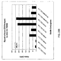
- the complex may yield a signal to noise ratio greater than about 100, wherein the signal to noise ratio is calculated by the formula:(fluorescent signal from a complex comprising the polypeptide bound by a primary antibody which in turn is bound to the binding agent)/ (fluorescent signal from a mixture of the polypeptide, an isotype control primary antibody and the binding agent).
- the complex may a signal to noise ratio greater than about 250, wherein the signal to noise ratio is calculated by the formula: (fluorescent signal from a complex comprising the polypeptide bound by a primary antibody which in turn is bound to the binding agent)/ (fluorescent signal from a mixture of the polypeptide, an isotype control primary antibody and the binding agent).
- the complex may yield a signal to noise ratio greater than about 270, wherein the signal to noise ratio is calculated by the formula: (fluorescent signal from a complex comprising the polypeptide bound by a primary antibody which in turn is bound to the binding agent)/ (fluorescent signal from a mixture of the polypeptide, an isotype control primary antibody and the binding agent).
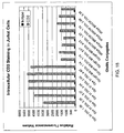
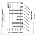
- the complex may yield a total fluorescence signal at least 5% greater than that generated by a complex formed with the same primary antibody and with the same secondary antibody that has a comparable degree of labeling with a DyLight 680TM dye.
- the complex may yield a total fluorescence signal at least 5% greater than that generated by a complex formed with the same primary antibody and with the same secondary antibody that has a comparable degree of labeling with a Cy5.5 ®dye.
- the complex may yield a total fluorescence signal at least 5% greater than that generated by a complex formed with the same primary antibody and with the same secondary antibody that has a comparable degree of labeling with an Alexa Fluor 680®dye.
- the label having a structure of a Formula of Formula II may be the compound having the structure:
- each complex is excited at 635 nm or 633 nm. In some embodiments, each complex is present at identical protein concentrations.
- the present invention discloses fluorescent compounds comprising at least one reactive group and at least one water soluble polymer inline with the claims.
- Such compounds may have desirable properties such as restricted intramolecular mobility, increased fluorescence quantum yield, decreased aggregation, increased solubility, decreased quenching and increased in vivo and in vitro stability.
- the compounds may be used for labeling molecules and biomolecules such as polypeptides and polynucleotides and are suitable for use in a wide range of applications, including diagnostic and imaging systems.
- Fluorescent compounds and labeled molecules of the invention may exhibit reduced aggregation. Dye aggregation is often seen as a major contributing factor to fluorescence quenching. Prevention of aggregation in the present invention may be achieved without the use of an excessive number of negatively charged sulfonate groups. This in turn may aid in the labeling of biomolecules such as proteins because the labeled protein may have an isoelectric point comparable to that of the substrate protein, and may thereby better maintain its biological specificity.
- the use of water soluble polymers of the invention may also aid in camouflaging or shielding the fluorophore to which it is linked. For example, such a water soluble polymer may be a relatively large group such as a polyethylene glycol moiety.
- labeled proteins such as labeled antibodies, of the invention may have a longer half-life in the circulation when applied to an animal's body, such as a mammal's body, and therefore may be suitable for in vivo imaging.
- Labeled biomolecules may also exhibit a longer half-life in in vitro systems, for example in assays employing serum or other biological extracts.
- a water soluble polymer may restrict or reduce the intramolecular mobility, such as the vibration and rotation, of the fluorophore to which it is attached. This may increase the fluorescence quantum yield of the fluorescent group.
- the fluorescence enhancement effect may be particularly effective for fluorescent groups that have a relatively flexible core structure.
- labeled molecules of the invention may be less immunogenic and less antigenic in vivo than the corresponding substrate biomolecules.
- the compounds and labeled molecules of the invention may exhibit higher photostability and resistance to bleaching of the fluorescent group.
- the compounds of the present invention may have asymmetric centers, chiral axes, and chiral planes (as described in: E. L. Eliel and S. H. Wilen, Stereo-chemistry of Carbon Compounds, John Wiley & Sons, New York, 1994, pages 1119 1190 ), and occur as racemates, racemic mixtures, and as individual diastereomers, with all possible isomers and mixtures thereof, including optical isomers, being included in the present invention.
- the compounds disclosed herein may exist as tautomers and both tautomeric forms are intended to be encompassed by the scope of the invention, even though only one tautomeric structure is depicted.
- any variable e.g. R, L, (R 1 ) a' (L) q
- its definition on each occurrence is independent at every other occurrence.
- Combinations of substituents and variables are permissible only if such combinations result in stable compounds.
- Lines drawn into the ring systems from substituents indicate that the indicated bond may be attached to any of the substitutable ring carbon atoms. If the ring system is polycyclic, it is intended that the bond be attached to any of the suitable carbon atoms on the proximal ring only. Substitution of a ring by a substitutent generally allows the substituent to be a cyclic structure fused to the ring.
- substituents and substitution patterns on the compounds of the instant invention can be selected by one of ordinary skill in the art to provide compounds that are chemically stable and that can be readily synthesized by techniques known in the art, as well as those methods set forth below, from readily available starting materials. If a substituent is itself substituted with more than one group, it is understood that these multiple groups may be on the same carbon or on different carbons, so long as a stable structure results.
- the phrase "optionally substituted with one or more substituents” should be taken to be equivalent to the phrase “optionally substituted with at least one substituent” and in such cases the preferred embodiment will have from zero to three substituents.
- alkyl is intended to include both branched, straight-chain, and cyclic saturated aliphatic hydrocarbon groups.
- Alkyl groups specifically include methyl, ethyl, propyl, butyl, pentyl, hexyl, heptyl, octyl, nonyl, decyl, and so on, as well as cycloalkyls such as cyclopropyl, cyclobutyl, cyclopentyl, cyclohexyl, tetrahydronaphthalene, meth-ylenecylohexyl, and so on.
- Alkoxy represents an alkyl group attached through an oxygen bridge.
- alkenyl refers to a non-aromatic hydrocarbon group, straight, branched or cyclic, containing at least one carbon to carbon double bond.
- Alkenyl groups include, but are not limited to, ethenyl, propenyl, butenyl and cyclohexenyl.
- the straight, branched or cyclic portion of the alkenyl group may contain double bonds and may be substituted if a substituted alkenyl group is indicated.
- alkynyl refers to a hydrocarbon group, straight, branched or cyclic, containing at least one carbon to carbon triple bond.
- Alkynyl groups include, but are not limited to, ethynyl, propynyl and butynyl.
- the straight, branched or cyclic portion of the akynyl group may contain triple bonds and may be substituted if a substituted alkynyl group is indicated.
- aryl is intended to mean any stable monocyclic or polycyclic carbon ring of up to 7 atoms in each ring, wherein at least one ring is aromatic.
- aryl elements include phenyl, naphthyl, tetrahydronaphthyl, indanyl, biphenyl, phenanthryl, anthryl or acenaphthyl.
- the aryl substituent is bicyclic and one ring is non-aromatic, it is understood that attachment is via the aromatic ring.
- heteroaryl represents a stable monocyclic or bicyclic ring of up to 7 atoms in each ring, wherein at least one ring is aromatic and contains from 1 to 4 heteroatoms selected from the group consisting of O, N and S.
- Heteroaryl groups within the scope of this definition include but are not limited to acridinyl, carbazolyl, cinnolinyl, quinoxalinyl, pyrrazolyl, indolyl, benzotriazolyl, furanyl, thienyl, benzothienyl, benzofuranyl, quinolinyl, isoquinolinyl, oxazolyl, isoxazolyl, pyrazinyl, pyridazinyl, pyridinyl, pyrimidinyl, pyrrolyl, tetrahydroquinoline, xanthenyl, and coumarinyl.
- the heteroaryl substituent is bicyclic and one ring is non-aromatic or contains no heteroatoms, it is understood that attachment is via the aromatic ring or via the heteroatom containing ring, respectively.
- heterocycle or “heterocyclyl” as used herein is intended to mean a 5- to 10-membered aromatic or nonaromatic heterocycle containing at least one heteroatom which is O, N or S. This definition includes bicyclic groups. "Heterocyclyl” therefore includes the above mentioned heteroaryls, as well as dihydro and tetrahydro analogs thereof.
- heterocyclyl include, but are not limited to the following: benzoimidazolyl, benzofuranyl, benzofurazanyl, benzopyrazolyl, benzotriazolyl, benzothiophenyl, benzoxamlyl, carbazolyl, carbolinyl, cinnolinyl, furanyl, imidazolyl, indolinyl, indolyl, indolazinyl, indazolyl, isobenzofuranyl, isoindolyl, isoquinolyl, isothiazolyl, isoxazolyl, naphthpyridinyl, oxadiazolyl, oxazolyl, oxazoline, isoxazoline, oxetanyl, pyranyl, pyrazinyl, pyrazolyl, pyridazinyl, pyridopyridinyl, pyridazinyl, pyridazinyl
- alkyl, alkenyl, alkynyl, cycloalkyl, aryl, heteroaryl and heterocyclyl substituents may be unsubstituted or unsubstituted, unless specifically defined otherwise.
- an alkyl group may be substituted with one or more substituents selected from OH, oxo, halo, alkoxy, dialkylamino, or heterocyclyl, such as morpholinyl or piperidinyl.
- halo or halogen are intended to include chloro, fluoro, bromo and iodo groups.
- aromatic is used in its usual sense, including unsaturation that is essentially delocalized across multiple bonds, such as around a ring.
- substituted refers to an atom, radical or chemical group which replaces a hydrogen in a substituted chemical group, radical, molecule, moiety or compound.
- Spiro refers to a cylic moiety which is attached to another group such that one of the ring atoms of the cyclic moiety is also an atom of said other group.
- a non-spiro substituent is a moiety cyclic or noncylic which is directly attached to said other group via bond connection between atoms of the non-spiro moiety and said other group.
- An example of a spiro moiety is, for instance, a subsitutuent Ring B on cyclohexanone Ring A.
- radical as applied to any molecule or compound, is used to refer to a part, fragment or group of the molecule or compound rather than to a “free radical”.
- a radical may be linked to another moiety through a covalent bond.
- polynucleotides refer to a polymeric form of nucleotides of any length, either deoxyribonucleotides or ribonucleotides, or analogs thereof. Polynucleotides may have any three-dimensional structure, and may perform any function, known or unknown.
- polynucleotides coding or non-coding regions of a gene or gene fragment, loci (locus) defined from linkage analysis, exons, introns, messenger RNA (mRNA), transfer RNA, ribosomal RNA, ribozymes, cDNA, recombinant polynucleotides, branched polynucleotides, plasmids, vectors, isolated DNA of any sequence, isolated RNA of any sequence, nucleic acid probes, and primers.
- a polynucleotide may comprise modified nucleotides, such as methylated nucleotides and nucleotide analogs.
- modifications to the nucleotide structure may be imparted before or after assembly of the polymer.
- the sequence of nucleotides may be interrupted by nonnucleotide components.
- a polynucleotide may be further modified after polymerization, such as by conjugation with a labeling component "Polynucleotide” may also be used to refer to peptide nucleic acids (PNA), locked nucleic acids (LNA), threofuranosyl nucleic acids (TNA) and other unnatural nucleic acids or nucleic acid mimics.
- PNA peptide nucleic acids
- LNA locked nucleic acids
- TAA threofuranosyl nucleic acids
- Other base and backbone modifications known in the art are encompassed in this definition. See, e.g. De Mesmaeker et al (1997) Pure & Appl. Chem., 69, 3, pp 437-440 .
- polypeptide polypeptide
- peptide protein
- polymer may be linear, cyclic, or branched, it may comprise modified amino acids, and it may be interrupted by non-amino acids.
- amino acid polymers that have been modified, for example, via sulfonation, glycosylation, lipidation, acetylation, phosphorylation, iodination, methylation, oxidation, proteolytic processing, phosphorylation, prenylation, racemization, selenoylation, transfer-RNA mediated addition of amino acids to proteins such as arginylation, ubiquitination, or any other manipulation, such as conjugation with a labeling component.
- amino acid refers to either natural and/or unnatural or synthetic amino acids, including glycine and both the D or L optical isomers, and amino acid analogs and peptidomimetics.
- antibody refers to immunoglobulin molecules and immunologically active portions of immunoglobulin molecules, i.e., molecules that contain an antigen-binding site which specifically binds ("immunoreacts with") an antigen.
- the simplest naturally occurring antibody e.g., IgG
- the immunoglobulins represent a large family of molecules that include several types of molecules, such as IgD, IgG, IgA, IgM and IgE.
- immunoglobulin molecule includes, for example, hybrid antibodies, or altered antibodies, and fragments thereof.
- Antigen binding units can be broadly divided into “single-chain” (“Sc”) and “non-single-chain” (“Nsc”) types based on their molecular structures.
- antibodies immunoglobulin molecules of a variety of species origins including invertebrates and vertebrates.
- human as applies to an antibody or an antigen binding unit refers to an immunoglobulin molecule expressed by a human gene or fragment thereof.
- humanized as applies to a non-human (e.g. rodent or primate) antibodies are hybrid immunoglobulins, immunoglobulin chains or fragments thereof which contain minimal sequence derived from non-human immunoglobulin.
- humanized antibodies are human immunoglobulins (recipient antibody) in which residues from a complementary determining region (CDR) of the recipient are replaced by residues from a CDR of a non-human species (donor antibody) such as mouse, rat, rabbit or primate having the desired specificity, affinity and capacity.
- CDR complementary determining region
- donor antibody such as mouse, rat, rabbit or primate having the desired specificity, affinity and capacity.
- Fv framework region (FR) residues of the human immunoglobulin are replaced by corresponding non-human residues.
- the humanized antibody may comprise residues which are found neither in the recipient antibody nor in the imported CDR or framework sequences.
- the humanized antibody will comprise substantially all of at least one, and typically two, variable domains, in which all or substantially all of the CDR regions correspond to those of a non-human immunoglobulin and all or substantially all of the FR regions are those of a human immunoglobulin sequence.
- the humanized antibody may also comprise at least a portion of an immunoglobulin constant region (Fc), typically that of a human immunoglobulin.
- stable refers to compositions and compounds which have sufficient chemical stability to survive isolation from a reaction mixture to a useful degree of purity for use in a desired application.
- fluorescent group refers interchangeably to molecules, groups or radicals which are fluorescent.
- fluorescent as applied to a molecule of compound is used to refer to the property of the compound of absorbing energy (such as UV, visible or IR radiation) and re-emitting at least a fraction of that energy as light over time.
- Fluorescent groups, compounds or fluorophores include, but are not limited to discrete compounds, molecules, proteins and macromolecular complexes. Fluorophores also include compounds that exhibit long-lived fluorescence decay such as lanthanide ions and lanthanide complexes with organic ligand sensitizers.
- a “subject” as used herein refers to a biological entity containing expressed genetic materials.
- the biological entity is in various embodiments, a vertebrate. In some embodiment, the biological entity is a mammal. In other embodiments, the subject is a biological entity which comprises a human.
- a “control” is an alternative subject or sample used in an experiment for comparison purposes.
- a control can be "positive” or “negative”.
- the purpose of the experiment is to detect a differentially expressed transcript or polypeptide in cell or tissue affected by a disease of concern, it is generally preferable to use a positive control (a subject or a sample from a subject, exhibiting such differential expression and syndromes characteristic of that disease), and a negative control (a subject or a sample from a subject lacking the differential expression and clinical syndrome of that disease.
- FRET refers to Foerster resonance energy transfer.
- FRET refers to energy transfer processes occurring between at least two fluorescent compounds, between a fluorescent compound and a non-fluorescent component or between a fluorescent component and a non-fluorescent component.
- a “binding agent” is a molecule that exhibits binding selectivity towards a binding partner or a target molecule to which it binds.
- a binding agent may be a biomolecule such as a polypeptide such as an antibody or protein, polypeptide-based toxin, amino acid, nucleotide, polynucleotides including DNA and RNA, lipids, and carbohydrates, or a combination thereof.
- a binding agent may also be a hapten, drug, ion-complexing agent such as metal chelators, microparticles, synthetic or natural polymers, cells, viruses, or other fluorescent molecules including the dye molecule according to the invention.
- a “targeting moiety” is the portion of the binding agent that binds to a binding partner.
- a targeting moiety may be, without limitation, a nucleotide sequence within a polynucleotide that selectively binds to another polynucleotide or polypeptide.
- Another nonlimiting example of a targeting moiety may be a polypeptide sequence within a larger polypeptide sequence which binds specifically to a polynuclotide sequence or a second polypeptide sequence.
- a targeting moiety may be a small molecule or structural motif which will bind to a protein receptor, another small molecule motif, or complexing agent, without limitation.
- the selective binding may be a specific binding event.
- a "binding partner” is a molecule or particle which is bound by the targeting moiety. It can be a cell, virus, fragment of a cell, antibody, fragment of an antibody, peptide, protein, polynucleotide, antigen, small molecule, or a combination thereof. It may be bound selectively or specifically by the binding agent.
- signal to noise ratio of fluorescence as referred to herein in the context of a polypeptide-antibody complex, is the ratio of (fluorescent signal from a complex comprising a polypeptide bound by a primary antibody which in turn is bound to a binding agent labeled with a compound of the invention)/(fluorescent signal from a mixture of the polypeptide, an isotype control primary antibody, and the labeled binding agent).
- Degree of labeling refers to the number of dye molecules which are attached per target molecule (including but not limited to polypeptide and polynucleotide).
- a single dye molecule per a polypeptide such as an antibody represents a 1.0 degree of labeling (DOL). If more than one dye molecule, on average, reacts with and is crosslinked to a polypeptide such as an antibody, the degree of labeling is greater than 1 and may further be a number other than a whole integer. The higher the number of DOL, the greater extent of labeling.
- Intracellular refers to the presence of a given molecule in a cell.
- An intracellular molecule can be present within the cytoplasm, attached to the cell membrane, on the surface of an organelle, or within an organelle of a cell.
- Substrate or “solid substrate” when used in the context of a reaction surface refers to the material that certain interaction is assayed.
- a substrate in this context can be a surface of an array or a surface of microwell. It may also be a solid such as a polymer which does not form a specific shape but has attachment points on its surface.
- wavelength of maximum excitation and “maximal fluorescence excitation wavelength” are used herein interchangeably. These terms refer to the maximum wavelength at which a fluorescent compound absorbs light energy which excites the dye to emit maximal fluorescence.
- absorption maximal wavelength as applied to a dye refers the wavelength of light energy at which the dye most effectively absorbs excitation energy to fluoresce.
- a fluorescent dye has a "maximal fluorescence emission wavelength” which is the wavelength at which the dye most intensely fluoresces.
- an absorption wavelength refers to the wavelength at which the compound has maximal absorption
- an emission wavelength refers to the wavelength at which the dye most intensely fluoresces.
- F represents a fluorophore or fluorescent group.
- suitable fluorophores are derived from fluorescent compounds which have substitution sites that allow the attachment to the -(L) m - or T groups.
- the core structures of a number of fluorescent groups including those of their sub-categories may be suitable as fluorophores.
- a fluorophore generally may comprise a structure comprising a minimal number of atoms necessary to form a fluorescent group belonging to a class of fluorescent groups.
- a coumarin fluorescent group comprises the core structure of formula A as set forth below:
- a fluorescein fluorescent group comprises the core structure of formula B as set forth below:
- rhodamine fluorescent groups have the core structure of formula C as set forth below:
- indocarbocyanine fluorescent groups may have the core structure of formula D as set forth below:
- Core structures for other classes of fluorescent fluorescent groups can be readily determined by one of ordinary skill in the art using the above principle.
- One of skill can appreciate that the determination of a fluorescent group core structure can be somewhat arbitrary because the classification of fluorescent groups may be arbitrary by itself.
- a class of fluorescent groups for example, may be sub-classified into different subclasses, wherein each subclass of fluorescent groups comprises one or more substituents unique to the particular subclass of fluorescent groups.
- 7-aminocoumarin shown below as formula E, is the core structure for all 7-aminocoumarin derivatives, which are themselves a subclass of fluorescent groups belonging to the more general coumarin fluorescent groups that have the core structure of formula A.
- the following table shows typical excitation and emission wavelengths for a number of common classes of fluorophores and core structures: Fluorescent Group Typical Excitation Wavelengths Typical Emission Wavelengths Coumarin 300-500 nm 350-550 nm Fluorescein 470-520 nm 500-540 nm Rhodamine 480-640 nm 510-660 nm Cyanine 350-1200 nm 360-1250 nm Pyrene 350-490 nm 400-510 nm
- F may be a derived from cyanine fluorescent group, Cy fluorescent group, xanthene fluorescent group, Alexa Fluor fluorescent group, coumarin fluorescent group, pyrene fluorescent group, Bodipy fluorescent group, ATTO fluorescent group or DY fluorescent group.
- cyanine fluorescent groups may comprise various sub-categories of cyanine fluorescent groups including, but not limited to, indocarbocyanine fluorescent groups, oxacarbocyanine fluorescent groups, thiacarbocyanine fluorescent groups, azacarbocyanine fluorescent groups (azacyanine fluorescent groups), styrylcyanine group and merocyanine fluorescent groups, merely by way of example.
- xanthene fluorescent groups may include, but are not limited to, fluorescein and its derivatives and various rhodamine fluorescent groups, for example.
- fluorescent compounds for use as fluorophores include Acridine orange, Acridine yellow, Alexa Fluorfluorescentgroups, ATTOfluorescentgroups, Bodipyfluorescentgroups, AuramineO, Benzanthrone, 9,10-Bis(phenylethynyl)anthracene, 5,12-Bis(phenylethynyl)naphthacene, Carboxyfluorescein diacetate, Calcein, Carboxyfluorescein, 1-Chloro-9,10-bis(phenylethynyl)anthracene, 2-Chloro-9,10-bis(phenylethynyl)anthracene, Coumarin, Cyanine, Cy2, Cy3, Cy3.5, Cy5, Cy5.5, Cy7, DyLight Fluor fluorescent groups, Fluorescein, 2',7'-dichlorodihydrofluorescein, Hilyte Fluor fluorescent groups, , LDS 751, Oregon Green, Per
- fluorescent groups include but are not limited to 4-acetamido-4'-isothiocyanatostilbene-2,2'disulfonic acid, acridine and derivatives such as acridine and acridine isothiocyanate, 5-(2'-aminoethyl)aminonaphthalene-1-sulfonic acid (EDANS), 4-amino-N-[3-vinylsulfonyl)phenyl]naphthalimide-3,5 disulfonate (Lucifer Yellow VS), N-(4-anilino-1-naphthyl)maleimide, anthranilamide, BODIPYTM and its derivatives and analogs, Brilliant Yellow, cyanine fluorescent groups such as Cy3 and Cy5 and other derivatives, coumarin and derivatives such as coumarin, 7-amino-4-methylcoumarin (AMC, Coumarin 120), 7-amino-4-trifluoromethylcouluarin (Co
- Fluorescent groups may also include fluorescent proteins.
- fluorescent proteins known in the art include GFP and its various derivatives, described e.g. in US Pat. Nos. 5,625,048 ; 5,777,079 ; 6,066,475 ; 6,319,669 ; 6,046,925 ; 6,124,128 and 6,077,707 .
- Additional fluorescent proteins are Y66F, Y66H, EBFP, GFPuv, ECFP, AmCyan1, Y66W, S65A, S65C, S65L, S65T, EGFP, ZsGreen1, EYFP, ZsYellow1, DsRed, DsRed2, AsRed2, mRFP1and HcRed1.
- fluorescent groups are commercially available and may be used as described herein. Commercial sources of reactive fluorescent groups include Invitrogen (Molecular Probes), AnaSpec, Amersham (AP Biotech), Atto-Tec, Dyomics, Clontech and Sigma-Aldrich.
- linking moieties may be any group connecting two moieties, such as fluorophores, water soluble polymers and reactive groups to each other or to any other group as described herein. Synthetic accessibility and convenience may generally dictate the nature of each linking moiety.
- a linking moiety may be a group containing about 1-100 atoms and formed of one or more chemical bonds selected such that the group is a stable moiety.
- a linking moiety may be formed of one or more carbon-hydrogen, carbon-nitrogen, carbon-oxygen, carbon-sulfur, carbon-phosphorus, nitrogenhydrogen, sulfur-hydrogen, phosphorus-hydrogen, sulfur-oxygen, sulfur-nitrogen, sulfur-phosphorus, phosphorus-oxygen, phosphorus-nitrogen and oxygen-nitrogen bonds, wherein such bonds may be single, double, triple, aromatic and heteroaromatic bonds selected such that the linking moiety is stable.
- a linking moiety can be, for example, a divalent alkyl radical.
- a linking moiety may be an alkyl group comprising additional ether, amine, amide, ester, sulfonyl, thioether, carboxamide, sulfonamide, hydrazide or morpholino, aryl and heteroaryl groups.
- Linking moieties are generally formed of about 1-100 atoms. Linking moieties may be formed of 1-50 non-hydrogen atoms as well as additional hydrogen atoms. Such atoms may be, for example, C, N, O, P or S. A linker moiety connecting two groups may comprise 1 to 50 consecutive bonds between the groups. Some linker moieties may have 1 to 40, 1 to 30, 1 to 20, 1 to 10, 1 to 5, 5 to 25, or 5 to 20 such consecutive bonds.
- Non-limiting linking moieties are illustrated below:
- n represents a number of repeating methylene units which can be varied such as to provide a desired length of the linker. Typically, n ranges from 1 to about 50. Some linkers will have an n of 1 to 40, 1 to 30, 1 to 20, 1 to 10, 1 to 5, 5 to 30, 5 to 20, or 5 to 15.
- joining moieties may be any group connecting three or more distinct moieties such as fluorophores, water soluble polymers and reactive groups to each other or to any other group, such as a linker moiety, included in the compound.
- a joining moiety may be a group containing about 1-100 atoms and formed ofone or more chemical bonds. The bonds may be selected such that the group is a stable moiety.
- a joining moiety may be formed of one or more carbon-hydrogen, carbon-nitrogen, carbon-oxygen, carbon-sulfur, carbon-phosphorus, nitrogen-hydrogen, sulfur-hydrogen, oxygen-hydrogen, phosphorus-hydrogen, sulfur-oxygen, sulfur-nitrogen, sulfur-phosphorus, phosphorus-oxygen, phosphorus-nitrogen and oxygen-nitrogen bonds, wherein such bonds may be single, double, triple, aromatic and heteroaromatic bonds selected such that the group is a stable moiety.
- T may contain between I and 50 atoms, or alternatively between 1 and 40, 1 and 30, 1 and 20, or 1 and 10 atoms. T may be a single atom such as N or C.
- T may also be a small cyclic group such as a carbocycle, a heterocycle, or an aromatic group.
- Nonlimiting examples include substituted phenyl, naphthyl, teaahydronaphthyl, indanyl, biphenyl, phenanthryl, anthryl, acenaphthyl, benzoimidazolyl, benzofuranyl, benzofurazanyl, benzopyrazolyl, benzotriazolyl, benzothiophenyl, benzoxazolyl, carbazolyl, carbolinyl, cinnolinyl, furanyl, imidazolyl, indolinyl, indolyl, indolazinyl, indazolyl, isobenzofuranyl, isoindolyl, isoquinolyl, isothiazolyl, isoxazolyl, naphthpyridinyl, oxadiazolyl, oxazolyl,
- m and n of Formula I indicate the number of linker moieties present and are independently integers ranging from 0 to 20.
- m the linker moiety is understood to be absent and any two moieties shown as attached to such a linker moiety are understood to be connected through a bond.
- n any substituent qualified by "n" will be understood to be absent.
- Groups denoted as R 1 , R 2 , and R 3 of Formula I are groups of the formula (R) p -(L) q -.
- each R, L, p or q is independent of any other R, L, p or q group present in the same compound.
- Each p is generally an integer ranging from 1 to 20.
- p may be 1.
- p may range from 1 to 2, 3, 4, 5, 10 or 15.
- Each q is generally an integer ranging from 0 to 20.
- L is understood to be absent and any R group shown as attached to L is understood to be connected directly through a bond.
- p may be 1.
- q may range from 1 to 2, 3, 4, 5, 10 or 15.
- R may be any group that confers a desirable functional property to the compound. More specific embodiments of R groups will be discussed below.
- Compounds described herein comprise at least one R which is a reactive group.
- a reactive group is a chemical moiety capable of reacting with a reaction partner on a substrate or substrate molecule to form a covalent bond.
- a compound described herein can be used to label a wide variety of molecules or substrates that contain a suitable reaction partner or are derivatized to contain a suitable reaction partner.
- "Reactive group” and “reaction partner” may refer to groups on a compound as described herein or to groups on a molecule to be labeled.
- a bond-forming group on a compound will generally be referred to as a reactive group and a bond-forming group on the substrate molecule will generally be referred to as a reaction partner.
- reaction substrate “substrate” and “reaction partner” are used interchangeably throughout this document.
- the reactive group and its reaction partner may be an electrophile and a nucleophile, respectively, that can form a covalent bond with or without a coupling agent or catalyst.
- the reactive group is a photoactivatable group capable of reacting with a hydrocarbon molecule upon ultraviolet photoactivation or photolysis.
- the reactive group is a dienophile capable of reacting with a conjugated diene via a Diels-Alder reaction.
- the reactive group is a 1,3-diene capable of reacting with a dienophile.
- the reactive group is an alkyne capable of reacting with an azido functional group to form a 1,2,3-triazole linkage.
- the reactive group is a 2-(diphenylphosphino)benzoic acid methyl ester capable of reacting with an azido functional group to form an amide linkage via so-called Staudinger reaction.
- examples of useful reactive groups, functional groups, and corresponding linkages are listed below in Table 1.
- Table 1 Examples of Reactive Groups, Functional Groups, and Covalent Linkages Reactive Group Reaction Partner/Substrate Resulting Covalent Linkage activated esters * amines/anilines Carboxamides acrylamides Thiols Thioethers acyl azides** amines/anilines Carboxamides acyl halides amines/anilines Carboxamides acyl halides Alcohols/phenols Esters acyl nitriles Alcohols/phenols Esters acyl nitriles amines/anilines Carboxamides aldehydes amines/anilines Imines aldehydes or ketones Hydrazines Hydrazones aldehydes or ketones Hydroxylamines Oximes alkyl halides amines/anilines alkyl amines alkyl halides Thiols Thioethers alkyl halides alcohols/phenols Esters alkyl sulfonates Thiols Thioethers alkyl sul
- the reactive group may be one that will react with an amine, a thiol, a hydroxyl or an aldehyde.
- the reactive group may be an amine-reactive group, such as a succinimidyl ester (SE), for example, or a thiol-reactive group, such as a maleimide, a haloacetamide, or a methanethiosulfonate (MTS), for example, or an aldehyde-reactive group, such as an amine, an aminooxy, or a hydrazide, for example.
- SE succinimidyl ester
- MTS methanethiosulfonate
- the compounds described herein also comprise at least one R which is a water-soluble polymer group.
- water soluble polymer groups may significantly reduce the intramolecular mobility of the fluorescent group core structure and may thus improve the fluorescent group's fluorescence quantum yield.
- Such groups may also confer other properties to the compounds to which they are attached, such as improvements of the photostability of the fluorescent group, reduced fluorescent group aggregation for biomolecule labeling, increased staining specificity of fluorescently labeled biomolecules (such as antibodies); and reduced immunogenicity and antigenicity of labeled biomolecules (such as antibodies) in vivo.
- Each water soluble polymer group is generally a substantially unreactive and water-soluble moiety sufficiently large to improve the fluorescence properties of a compound.
- the term "polymer” used in this context does not require the presence of strictly repeating units. A molecule of sufficient molecular size and solubility but without repeating units is considered a "water soluble polymer group" for the purposes of the invention.
- Water soluble polymer groups include, but are not limited to, organic polymers and biomolecules such as polypeptides and carbohydrates.
- Water soluble polymers may comprise ether groups, hydroxyl groups, tertiary amine groups, quaternized amine groups, and/or guanidine groups. Each water soluble polymer may be linear, branched, cyclic or a combination thereof.
- Water soluble polymers may comprise a single chain or alternatively one, two, three, four or more chains. Watersoluble polymers with one, two, three, fouror more branches may be used.
- the compounds described herein may comprise any number of water soluble polymer groups. Generally, compounds of the invention comprise at least 1 water soluble polymer groups up to about 8 water soluble polymer groups.
- a compound may comprise at least 2 water soluble polymer groups to about 8 water soluble polymer groups.
- a compound may comprise at least 3 water soluble polymer groups up to about 8 water soluble groups. Suitable molecular weights of each water soluble polymer group or, alternatively, of all water soluble polymer groups in one compound may be about 100, 200, 300, 400, 500, 600, 700, 800, 900, 1000, 1500, 2000, 2500, 3000, 3500, 4000, 4500, 5000, 10000, 15000, 20000 Da or greater.
- a water soluble polymer group may have a molecular weight between 450 and 5000 Da.
- a water soluble polymer group of the invention has a molecular weight between about 800 and about 3000 Da.
- the combined molecular weight of all water soluble polymer groups within a compound is from about 450 to about 5,000 Da. In still another embodiment, the combined molecular weight of all water soluble polymer groups within a compound is from about 1,000 to about 3,000 Da.
- the water soluble polymer is a polyalkylene oxide.
- Suitable polyalkylene oxides include polyethylene glycol (PEG), polypropylene glycol (PPG), polyethylene glycol-polypropylene glycol (PEG-PPG) copolymers, and N-substituted methacrylamide-containing polymers and copolymers.
- Polyalkylene oxides may be additionally substituted as necessary to confer other desired properties to the polymer. Such modifications may comprise, for example, chemical linkages that increase or decrease the chemical stability of the polymer, which would allow tuning of the chemical or biological stability of the half-life of the polymer.
- polyalkylene oxide molecules are terminated or "capped” with various groups. Examples of such groups are hydroxy, alkyl ether (e.g. methyl, ethyl, propyl ethers), carboxymethyl ether, carboxyethyl ether, benzyl ether, dibenzylmethylene ether or dimethylamine.
- a polyalkylene oxide may have one of many possible terminals, including but not limited to hydroxyl, methyl ether, ethyl ether, carboxymethyl ether, and carboxymethyl ether.
- a polyalkylene oxide is a polyethylene glycol polymer terminated with a methyl ether.
- Such a group may be referred to as an mPEG.
- An mPEG generally has the formula of -(CH 2 CH 2 O) n CH 3 , wherein n is the number of ethylene glycol units and is determined by the size of said mPEG.
- suitable polymers include derivatives and conjugates of poly(2-hydroxyethyl methacrylate), polyhydroxypropyl methacrylamide, poly(styrene sulfonic acid), poly(vinyl alcohol), or poly(2-vinyl N-methyl pyridinium iodide).
- a water soluble polymer may be carbohydrate.
- Such carbohydrates include monosaccharides or polysaccharides and may be, for example, soluble starch, glycogen, dextran, pectin, mannan, galactan, hydroxymethylcellulose, hydroxyethylcellulose and other derivatized celluloses.
- the water soluble polymer is a carbohydrate, at least 30% of the hydroxyl groups present in the carbohydrate may be masked as methyl ethers, sulfonatoalkyl ethers, and/or acetate esters.
- a water soluble polymer may be a polypeptide.
- Suitable polypeptides may comprise, for example, serine, arginine, polylysine with modified epsilon amino groups, or cysteinic acid.
- Other examples of such polypeptides are disclosed, for example, in WO 2006/081249 . It is contemplated that such polypeptides may be used as the water soluble polymer
- Water soluble polymers also comprise combinations of the different classes described above.
- such a water soluble polymer would be a polypeptide linked to a polyalkylene oxide moiety.
- Water soluble polymers do not generally comprise any group or groups that are incompatible with the chemistry of the reactive group or groups included in the compound described herein.
- a water soluble polymer should not comprise strong nucleophiles if a reactive group is an electrophile.
- a water soluble polymer should not comprise primary or secondary amines if a reactive group is an N-hydroxysuccinimidyl ester.
- a water soluble polymer should not comprise a thiol when a reactive group is a maleimide.
- a water soluble polymer should generally not comprise a strong electrophile if a reactive group is a nucleophile.
- a watersoluble may comprise a minimal number of weak nucleophilesor a minimal numberofweak electrophiles such that the chemistry of the reactive group is not significantly affected, or the stability of the compound described herein is not affected during storage and handling.
- weak nucleophiles are hydroxyl groups, which are commonly present in carbohydrate molecules.
- a water soluble polymer is a carbohydrate molecule
- at least 30% of the hydroxyl groups are preferably masked as ethers, such as methyl ether, and/or as esters, such as acetate esters.
- all of the hydroxyl groups may be masked as ethers and/or esters.
- additional substituents ("R") of the compounds described herein may in some cases be groups such as sulfonate (-SO 3 - ), phosphonate (-PO 3 2- ), and ammonium groups.
- ammonium means NH 4 + , a trialkylammonium, or a tetraalkylammonium.
- an ionic group requires a counter ion to balance its charge. For example, each negatively charged -SO 3 - or -PO 3 2- may necessitate one or two cations to balance the negative charge. Likewise, a positively charged ammonium may require an anion to maintain neutrality.
- the nature of the counter ion is not critical as long as the counter ion does not lower the solubility of said fluorescent group.
- the counter ion when a substituent is -SO 3 - or -PO 3 2- , the counter ion is H + , Na + , K + or an ammonium. In other embodiments, when the substituent is ammonium, the counter ion is preferably chloride, fluoride, bromide, sulfate, phosphate, acetate or the like.
- Some fluorescent groups may intrinsically possess a positive charge or negative charge. In such a case, the intrinsic charge may act as a counter ion. Alternatively, the intrinsic charge may require a counter ion for maintaining neutrality.
- a counter ion for any intrinsic charge is as previously described. At least one sulfonate group may be present (-SO 3 - ) and any necessary counter ion is selected from H + , Na + , K + and an ammonium. For reason of simplicity, any dissociable counter ion or counter ions for most of the fluorescent group structures depicted herein may not be shown.
- Such substituents may increase a compound's water solubility and/or its fluorescent quantum yield.
- a relatively high number of charged groups is generally not desirable because it would result in a highly charged fluorophore, which on conjugation to a protein, for example, may significantly change the isoelectric point of the protein, thus possibly affecting the biological properties of the labeled protein.
- an antibody labeled with a highly charged fluorescent molecule may show high background in staining.
- the number of such charged water-soluble R groups may be 0-4, or 0-3.
- the fluorescent group has at least one water soluble polymer group, which is also capable of increasing the water solubility and/or the quantum yield of the fluorescent group, the number of charged R groups, such as sulfonate groups, can be kept to a minimum, thereby minimizing the loss of biological specificity of labeled proteins.
- Each substituent R may be the same or different and may be selected from halogens, -OH, -NH 2 , -SO 2 NH 2 , and any carbon-containing substituents comprising 1 to about 15 carbon atoms and optionally at least one hetero atom.
- the at least one hetero atom is preferably selected from the group consisting of halogens, N, O, S, P and Si.
- R may be a dialkylamine substituent such as, for example, diethylamine or dimethylamine.
- R When R is a carbon-containing substituent, it may assume any structure or conformation, including, for instance, alkyl, cycloalkyl, alkenyl or alkynyl.
- the compound of formula I may comprise a fluorophore which is a xanthene fluorescent group, a coumarin fluorescent group, a pyrene fluorescent group or a cyanine fluorescent group.
- the fluorophore may be a coumarin fluorescent group.
- Such a fluorophore may have the formula where one moiety of R a , R b , R c , R d , R e and R f is a bond connecting the fluorophore to a moiety-(L) m or a moiety as indicated in Formula I.
- R a , R b , R c , R d , R e and R f have the formula (R) p -(L) q -, where R, L, p and q are as previously defined.
- R a , R b , R c , R d , R e and R f may join to form a 5- or 6-membered, saturated or unsaturated ring that may optionally comprise additional heteroatoms in the ring as well as additional R substituents.
- R be substituents may be SO 3 - , sulfonamido, halo, hydroxy, amino or alkyl groups.
- the fluorophore is a compound of the formula: wherein connects said fluorophore to said moiety -(L) m - or said moiety
- R 4 , R 5 , and R 6 are each independently (R)p-(L)q-;
- each R of R 4 , R 5 , R 6 is independently i) a reactive group capable of forming a covalent bond upon reacting with a reaction partner; ii) a water soluble polymer group; iii) an alkyl group, a trifluoroalkyl group, a halogen group, a sulfonate group or a sulfonamido group; or iv) -H;
- each L of R 4 , R 5 and R 6 is independently a linking moiety formed of one or more chemical bonds and containing about 1-100 atoms;
- each p of R 4 , R 5 , and R 6 is independently an integer ranging from 1 to 20;
- T may be a trivalent moiety comprising one carbon atom such as
- T may be a trivalent nitrogen atom.
- T may be a trivalent nitrogen atom.
- in Formula I may have the formula
- R 1 is a water soluble polymer
- said water-soluble polymer may have a molecular weight of greater than about 300 Da.
- the molecular weight may be greater than 800 Da, or it may range from about 800 Da to about 3000 Da.
- R 1 may, for example, comprise a water soluble group such as a polyethylene glycol group.
- the molecular weight of the polyethylene glycol group may be between 450 and 5000 Da.
- R 2 When R 2 is a reactive group, the reactive group may form a covalent bond, for example, with amino, sulfhydryl or hydroxy nucleophiles.
- the reactive group may be an isothiocyanate, an isocyanate, a monochlorotriazine, a dichlorotriazine, a halogen-substituted pyridine, a halogen-substituted diazine, a phosphoramidite, a maleimide, an aziridine, a sulfonyl halide, an acid halide, a hydroxysuccinimide ester, a hydroxysulfosuccinimide ester, an imido ester, a hydrazine, an azidonitrophenyl, an azide, an alkyne, a 3-(2-pyridyl dithio)-propionamide, a glyoxal an aldehyde, or an N
- ⁇ is a moiety having one of the following structures:
- X 1 and X 4 are independently substituted. X 1 and X 4 may or may not be additionally
- X 2 and X 3 are independently
- the elements a and b are independently 0, 1, 2, or 3.
- the element b' is 0, 1 or 2.
- R 5 and R 6 when ⁇ is Formula 1, and the maximal fluorescence excitation wavelength of the compound is less than 660 nm, then R 5 and R 6 are independently (R) p -(L) q -, wherein R 5 and R 6 are not combinable to form a substituted ring; In other embodiments, when ⁇ is Formula 1, and the maximal fluorescence excitation wavelength of the compound is equal to or greater than 660 nm, or ⁇ is other than Formula 1, then R 5 and R 6 are independently (R) p -(L) q , or R 5 and R 6 are combinable to form a cyclic moiety which is unsubstituted or substituted by one or more (R) p -(L) q - The cyclic moiety so formed is a 5, 6, or 7 membered ring with is carbocyclic or heterocyclic, and in some embodiments, substituted by one or more reactive groups and/or one or more water soluble polymers.
- R 5 and R 6 when ⁇ is Formula 1 and the maximal fluorescence excitation wavelength of the compound is less than 655 nm, then R 5 and R 6 are independently (R) p -(L) q -, wherein R 5 and R 6 are not combinable to form a substituted ring; In other embodiments, when ⁇ is Formula 1, and the maximal fluorescence excitation wavelength of the compound is equal to or greater than 655nm, or ⁇ is other than Formula 1, then R 5 and R 6 are independently (R) p -(L) q - ,or R 5 and R 6 are combinable to form a cyclic moiety which is unsubstituted or substituted by one or more (R) p -(L) q -.
- Y is: or wherein when C is present, it is a five- or six-membered cyclic group.
- R 1 , R 2 , R 3 , R 4 , R 5 , R 6 and R 7 are each independently (R) p -(L) q -.
- Each R of each (R) p -(L) q - of the compound is independently i) a reactive group capable of forming a covalent bond upon reacting with a reaction substrate; ii) a water soluble polymer group; iii) an alkyl group, an aryl group, an alkylamino group, a dialkylamino group, an alkoxy group, a trifluoroalkyl group, a halogen group, a phosphonate group, a sulfonyl group, a sulfonate group or a sulfonamido group; or iv) -H.
- Each L of each (R) p -(L) q - of the compound is independently a linking moiety formed of one or more chemical bonds and containing about 1-100 atoms.
- Each p of each (R) p -(L) q - is independently an integer of about 1 to about 20.
- Each q of each (R) p -(L) q - of R 1 or R 2 is independently an integer of 0 to about 20.
- Each q of each (R) p -(L) q - of R 3 , R 4 , R 5 , R 6 , or R 7 is independently an integer of 1 to about 20.
- the element c is 0 or 1.
- the element d is 0 or 1.
- At least one R of the (R) p -(L) q - of the compound is a reactive moiety; and at least one R of the (R) p -(L) q - of the compound is a water-soluble polymer.
- the two adjacent R 1 and/or the two adjacent R 2 are combinable to form a 6-membered ring which is unsubstituted or substituted by one or more (R) p -(L) q -.
- the ring so formed is aromatic.
- two adjacent (R 2 ) b and the atoms in ring B to which it is attached are combined to form a carbocyclic ring, optionally additionally substituted with groups that may further increase the solubility of the fluorescent group.
- two adjacent (R 2 ) b and the atoms in ring B to which it is attached are combined to form a carbocyclic ring which is aromatic and optionally additionally substituted with groups that may further increase the solubility of the fluorescent group.
- two adjacent (R 1 ) b and the atoms in ring A to which it is attached are combined to form a carbocyclic ring, and optionally additionally substituted with groups that may further increase the solubility of the fluorescent group.
- two adjacent (R 1 ) b and the atoms in ring A to which it is attached are combined to form a carbocyclic ring which is aromatic, which is optionally additionally substituted with groups that may further increase the solubility of the fluorescent group.
- the element c may be 0 or 1.
- c When c is 1, the nitrogen atom to which R 3 is attached is positively charged.
- d may be 0 or 1.
- At least one R of R 1 and R 2 is a charged moiety. In some embodiments, at least one R of R 1 and R 2 comprises a sulfonate group or a phosphonate group. In yet other embodiments, each R of R 1 and R 2 comprises a sulfonate group or a phosphonate group.
- X 2 and X 3 are independently In related embodiments, X 2 and X 3 are alkyl groups such as methyl or ethyl. In some embodiments, X 2 and X 3 are In some embodiments, X 2 and X 3 are: wherein R 5 comprises a reactive group or a water-soluble polymer.
- the compound of formula II contains at least one reactive group and a water-soluble polymer in line with the claims. These groups may be attached at any position shown in formula II.
- a reactive group or a water-soluble polymer may be attached to ring A or ring B.
- the reactive group or the water-soluble polymer may substitute the nitrogen atoms indicated in formula II, e.g. R 3 could be a reactive group and R 4 could be a water-soluble polymer, optionally attached through L linker moieties.
- reactive groups and/or water-soluble polymers may also be attached to the X 2 and/or X 3 positions in formula II, or may be part of the linker structure Y.
- R substituent which is a charged moiety on ring A or one ring of B, D, E, F, and G or both ring A and one ring of B, D, E, F and G such as to increase the solubility of the compound of formula II.
- a moiety may be a sulfonate group or any other group as previously described. Similar substitution can be made as part of the groups R 3 , R 4 , X 2 and X 3 , for example by attaching a linker moiety which is substituted with a sulfonate, phosphonate or other charged group.
- At least one water-soluble polymer is a polyalkylene oxide according to the claims.
- a further water-soluble polymer is a polyethylene oxide.
- a further water-soluble polymer is a carbohydrate.
- a further water-soluble polymer is a polypeptide.
- the at least one polyalkylene oxide water-soluble polymer has a molecular weight ranging from about 800 to about 3000.
- a further water-soluble polymer has a molecular weight of greater than 300.
- a further water-soluble polymer has a molecular weight of greater than 800.
- the compound of Formula II comprises at least one reactive group and at least two water-soluble polymers.
- the compound has the formula:
- the compound of Formula II has the formula: A wherein c is 1; d is 1; at least one R of R 3 and R 4 is a reactive group capable of forming a covalent bond upon reacting with a reaction substrate; and at least one R of R 3 and R 4 is a water soluble polymer group, which is a polyalkylene oxide with a molecular weight ranging from 800 to 3000
- the compound has the formula: wherein c is 1; and d is 1; at least one R of R 3 , R 4 and R 5 is a reactive group capable of forming a covalent bond upon reacting with a reaction substrate; and at least one R of R 3 , R 4 and R 5 is a radical of a water-soluble polymer*
- c is 1; d is 1; one R of R 3 , R 4 and R 5 is a reactive group; and at least two R of R 3 , R 4 and R 5 are a radical of a water-soluble polymer.
- Y is selected such that the absorption maximal wavelength is about 550 nm, about 650 nm, or about 750 nm.
- the compound has the formula:
- the compound has the formula:
- the compound has the formula:
- the compound has the structure: wherein, c is 1; d is 1; one R of R 3 , R 4 and R 5 is a reactive group; and at least two R of R 3 , R 4 and R 5 each comprise a radical of a water-soluble polymer which is a polyakylene oxide with a molecular weight ranging from 800 to 3000.
- Y is selected such that the absorption maximal wavelength is about 580 nm, about 680 nm , or about 790 nm.
- the compound has the structure: wherein, c is 1; d is 1; one R of R 3 , R 4 and R 5 is a reactive group; and at least two R of R 3 , R 4 and R 5 are each a radical of water-soluble polymers which is a polyalkylene oxide with a molecular weight ranging from 800 to 3000.
- Y is selected such that the absorption maximal wavelength is around 560 nm, around 660 nm, or around 770 nm.
- the bridging unit Y in combination with a specific embodiment of a moiety of Formula F, in combination with a specific embodiment of moiety Y, and in combination with specific substituents such that the maximal fluorescence excitation wavelength of the compound ranges from about 350 to about 1200 nm.
- Standard analysis well known in the art is used to obtain the absorption and emission spectra of the dyes in order to ascertain the absorption maximal wavelength and the maximal fluorescence emission wavelength.
- any combinations of the above described groups may be used to form a compound with a maximal fluorescence excitation wavelength of about 550 nm, about 560 nm, about 570 nm, about 580 nm, about 590 nm, about 600 nm, about 610 nm, about 620 nm, about 615 nm, about 620 nm, about 625 nm, about 630 nm, about 635 nm, about 640 nm, about 650 nm, about 655 nm, about 660 nm, about 665 nm, about 670 nm, about 675 nm, about 680 nm, about 685 nm, about 690 nm, about 695 nm, about 700 nm, about 705 nm, about 710 nm, about 715 nm, about 720 nm, about 725 nm, about 730 nm, about 735 nm, about 740 nm
- the moieties described above may be used in any combination.
- the maximal fluorescence excitation wavelength of the dye is typically the wavelength at which the dye has a maximal absorption or maximal optical density, which excites the dye to fluoresce.
- the compound of Formula II has an absorption maximal wavelength at >670 nm.
- the absorption maximal wavelength is at >700 nm.
- the absorption maximal wavelength is at >800 nm.
- a person of skill can also combine select the bridging unit Y, in combination with a specific embodiment of a moiety of Formula F, in combination with a specific embodiment of moiety Y, and in combination with specific substituents such that one obtains a compound of Formula II having a maximal fluorescence emission wavelength of about 550 nm, about 560 nm, about 570 nm, about 580 nm, about 590 nm, about 600 nm, about 6.10 nm, about 620 nm, about 615 nm, about 620 nm, about 625 nm, about 630 nm, about 635 nm, about 640 nm, about 650 nm, about 655 nm, about 660 nm, about 665 nm, about 670 nm, about 675 nm, about 680 nm, about 685 nm, about 690 nm, about 695 nm, about 700 nm, about 705 nm,
- the wavelength of the absorption maximum of a compound of formula II is at least greater than 655 nm.
- the wavelength of the absorption maximum of a compound of formula 11 is at least greater than 670 nm. In still another embodiment, the wavelength of the absorption maximum of a compound of formula II is at least greater than 670 nm. In yet another embodiment, the wavelength of the absorption maximum of a compound of formula II is at least greater than 700 nm.
- a substituted cyanine dye which comprises one or more water soluble polymer groups, wherein the cyanine dye has a maximal fluorescence excitation wavelength of equal to or greater than about 660 nm.
- the substituted cyanine dye may comprise at least one reactive group.
- the substituted cyanine dye may be substituted by a non-spiro moiety.
- a substituted cyanine dye which comprises one or more water soluble polymer groups, wherein the cyanine dye has an absorption maximal wavelength of equal to or greater than about 660 nm.
- the substituted cyanine dye may comprise at least one reactive group.
- the substituted cyanine dye may be substituted by a non-spiro moiety.
- Dye compounds of formula II have excellent solubility and superior fluorescence brightness and photostability.
- compounds of formula II having an absorption maximal wavelength greater than about 655 nm typically referred to as near-IR dyes, showed major advantages over prior art dyes of similar wavelengths.
- the water-soluble polymer substituents drastically improved the performance of the near-IRdyes of the invention, resulting in unprecedented fluorescence brightness (See Figures).
- the water-soluble polymer substituents dramatically improved the stability of the near-IR dyes (See Figures).
- the stability of near-IR dyes has been a problem, which demands extreme care in storage and handling and therefore limits the use of the dyes.
- Z may be -H, alkyl or a substituent such as -CF 3 , or -CN.
- Z is:
- Compounds of formula III generally belong to the class of xanthene fluorescent groups.
- R 1 , R 2 , R 3 , R 4 , R 5 , R 6 , R 7 , R 8 and R 9 are each independently (R) p -(L) q -, where L, p and q are as generally defined elsewhere in this document.
- R is as previously defined, with the addition that R also includes moieties which have both a water-soluble polymer and a reactive group, for example joined by a joining moiety.
- the variables c, d and e indicate the numbers of R 3 , R 4 and R 5 substituents on the phenyl ring in group Z and may be 0, 1, 2 or 3, such that the sum of c, d and e is less or equal to 5.
- Each compound of formula III comprises a reactive moiety as well as a water-soluble polymer. Therefore, at least one R of R 1 , R 2 , R 3 , R 4 , R 5 , R 6 and R 7 is a reactive moiety while at least one R of R 1 , R 2 , R 3 , R 4 , R 5 , R 6 and R 7 is a water-soluble polymer group.
- Z may comprise at least one R which is a water soluble polymer and at least one R which is a reactive group.
- X 2 may be an amino group which is optionally substituted with one or two groups such as alkyl. In such cases, X 2 may be, for instance, a dimethylamino, diethylamino, methylamino or ethylamino substituent. If neither R 1 or X 2 comprises a water-soluble polymer group or reactive group, group Z may be substituted with at least one reactive group and at least one water soluble polymer.
- R 1 and X 2 taken together form a carbocyclic or heterocyclic ring fused to ring A in formula III.
- R 2 and X 1 taken together may form a carbocyclic or heterocyclic ring fused to ring B in formula III.
- Such fused rings may additionally be substituted with additional R groups, for example SO 3 - , alkyl, or even water soluble polymer groups or reactive groups.
- the rings so formed by combining one of R 6 , R 7 , R 8 and R 9 with a neighboring R 1 and/or R2 are unsaturated or saturated and may be unsubstituted or substituted by one or more (R) p -(L) q -.
- one R of R 1 , X 1 , R 2 or X 2 comprises a water soluble polymer group while another R of R 1 , X 1 , R 2 or X 2 comprises a reactive group.
- Water soluble groups may comprise a polyalkylene oxide attached to the core structure of formula III via a linker moiety or joining moiety. It is possible for either R 3 , R 4 or R 5 to be a joining moiety to which both a water soluble polymer and a reactive group are connected via independent linker moieties.
- Exemplary compounds of formula III are illustrated in Table 4.
- R 1 , R 2 , R 3 , and R 4 are each independently (R) q -(L) q -, provided that at least one R of R 1 , R 2 , R 3 and R 4 is a reactive moiety; and at least one R of R 1, R 2 , R 3 and R 4 is a water-soluble polymer group.
- the variables a, b, c and d are independently 0, 1,2 or 3. Generally, the sum of a, b, c and d is 2, 3 or 4. In one embodiment, the sum of a, b, c and d is 4.
- R 1 , R 2 and R 3 may each comprise water soluble polymer groups, such that each (R) p -(L) q - group may independently have the formula - SO 2 NH(CH 2 CH 2 O) o CH 3 , where n is from about 3 to about 30, or from about 7 to about 24.
- R 4 comprises an R which is a reactive group such as an activated ester of a carboxylic acid.
- L may comprise a C1-C8 N-alkyl sulfonamide or a C2-C10 N,N-dialkyl sulfonamide group linking either a reactive group or a water soluble polymer group to the pyrene moiety in formula V, such that each sulfonamide is covalently linked to the pyrene carbon via S-C bond and where each alkyl portion optionally comprises at least one O.
- R 1 , R 2 and R 3 may comprise substituents such as sulfonate groups and R 4 is a joining moiety to which both a water soluble polymer and a reactive group are connected via independent linker moieties.
- Exemplary compounds of the Formula V are listed in Table 6. Table 6. Exemplary Pyrene compounds* Dye No. Structure ⁇ abs / ⁇ em (nm) (H 2 O) 44 45 46 400/424 *For simplicity, any counter ions for the structures are not shown.
- n may be an integer selected from about 3 to about 30; h is an integer selected from 1 to 5; and R p is a latent or a protected reactive group; 2) converting the latent or protected reactive group R P in the resulting compound to a reactive group.
- the above amino compound shown above can be readily prepared, for example, by reacting a suitable mPEG alkylating compound, such as a mPEG tosylate or mPEG chloride, with a suitable aminoalkyl compound comprising a suitable R P group.
- suitable R P groups include a carboxylic acid group, a methyl ester of a carboxylic acid group, a t-butyl ester group of a carboxylic acid group, a t-BOC-protected amine group and a benzyloxycarbonyl-protected group, merely by way of example.
- R P group is a latent reactive group, such as a free carboxylic acid group, it may be directly converted to a reactive group using methods well known to one of skills. If R P is a protected group, it is deprotected and then converted to a reactive group using a known method.
- the subject compounds find use in a variety of different applications.
- One application of interest is the use of the subject compounds as labeling agentswhich are capable of imparting a fluorescent property to a particular composition of matter.
- the compounds of the present invention can be used to react with any of a broad range of molecules, including but not limited to, biomolecules such as polypeptides, polypeptide-based toxins, amino acids, nucleotides, polynucleotides including DNA and RNA, lipids, and carbohydrates, and any combinations thereof. Additionally, the compounds of the invention can be used to react with haptens, drugs, ion-complexing agents such as metal chelators, microparticles, synthetic or natural polymers, cells, viruses, other fluorescent molecules including the dye molecule according to the invention, or surfaces.
- the substrate molecules typically comprise one or more functional groups, which react with the reactive group of the subject compounds to form covalent or non-covalent linkage.
- the reactive group of a compound of the invention is an activated ester (such as a succinimidyl ester, or SE), a maleimide, a hydrazide or an aminooxy group.
- functional group from a substrate molecule (or reaction substrate) is an amine, a thiol, an aldehyde or ketone.
- the resulting fluorescently labeled substrate molecules may be referred to as conjugates or labeled substrate molecules. Any methods practiced in the art (e.g., Brinkley, Bioconjugate Chem. 3, 2 (1992)) for preparing fluorescent group-substrate conjugates are applicable for practicing the subject invention.
- Conjugates of biomolecules and compounds of the invention usually have high fluorescence yield while typically retaining the critical parameters of unlabeled biomolecules, such as solubility, selective binding to a receptor or nucleic acid, activation or inhibition of a particular enzyme or the ability to incorporate into a biological membrane. Nevertheless, conjugates with the highest degree of labeling may still precipitate or bind nonspecifically. As necessary, a less-than-maximal degree of labeling may be acceptable in orderto preserve function or binding specificity. Preparing the conjugates of the invention may involve experimentation to optimize properties.
- unconjugated labeling reagent may be removed by techniques known in the art such as by gel filtration, dialysis, conjugate precipitation and resolubi-lization, HPLC or a combination of these techniques.
- gel filtration dialysis
- conjugate precipitation and resolubi-lization
- HPLC HPLC
- free dye particularly if it remains chemically reactive, may complicate subsequent experiments with the bioconjugate.
- the subject compounds can be used to conjugate with a nucleoside, a nucleotide, or a polynucleotide, wherein any of such molecules may be natural or synthetic, modified or unmodified.
- the compound of the invention used for labeling may comprise a reactive group which is a phosphoramidite, an activated ester (such as a succinimidyl ester), an alkylating group ora reactive platinum complex.
- Such molecules may contain or are derivatized to contain one or more reaction partners for the reactive groups on the compounds of the invention.
- a reactive group of a compound of the invention may react with a suitable reaction partner on said molecule to form a covalent linkage.
- a phosphoramidite group may react with a hydroxyl group to form a phosphate linkage after deprotection; a succinimidyl ester or the like may react with an amine group to form an amide linkage; and a reactive platinum complex may react with a guanosine base to form a platinum complex linkage.
- a reactive compound of the invention comprising an activated ester is reacted with a nucleotide triphosphate comprising a base comprising an aminoalkynyl group, an aminoallyl group or an aminoalkyl group to form a fluorescently labeled nucleotide triphosphate.
- a labeled nucleotide triphosphate is often used to prepare a fluorescently labeled nucleic acid polymer via enzymatic incorporation.
- the fluorescent compound of the invention is reacted with a group or linker attached to the C-5 position of a uridine or cytidine residue. This position is not involved in Watson-Crick base-pairing and interferes little with hybridization to complementary sequences.
- An aminoalkynyl linker may be introduced between a fluorescent moiety and the nucleotide in order to reduce fluorophore interaction with enzymes or target binding sites.
- seven- to 10-atom spacers may be introduced that further separate the fluorophore from the base. The use of longer spacers may result in brighter conjugates and increased hapten accessibility for secondary detection reagents.
- deoxycytidine triphosphates may be prepared which are modified at the N-4 position of cytosine using a 2-aminoethoxyethyl (OBEA) linker.
- OBEA 2-aminoethoxyethyl
- Fluorescently labeled DNA may be prepared from a fluorescently labeled nucleotide triphosphate by PCR reaction, terminal transferase-catalyzed addition or nick translation.
- Various polymerases may be used in such reactions.
- Such polymerases include Taq polymerase (useful e.g. in polymerase chain reaction (PCR) assays), DNA polymerase I (useful e.g. in nick-translation and primer-extension assays), Klenow polymerase (useful e.g. in random-primer labeling), Terminal deoxynucleotidyl transferase (TdT) (useful e.g. for 3'-end labeling), Reverse transcriptase (e.g. for synthesizing DNA from RNA templates) or other polymerases such as SP6 RNA polymerase, T3 RNA polymerase and T7 RNA polymerase for in vitro transcription.
- SP6 RNA polymerase T3
- a fluorescently labeled nucleic acid polymer may be prepared by first enzymatically incorporating an amine-labeled nucleotide into a nucleic acid polymer to result in an amine-labeled nucleic acid polymer, followed by the labeling of said amine-labeled polymer with a compound of the invention. More information on the preparation and use of fluorescently labeled nucleotide triphosphates can be found in US patent Nos 4,711,955 and 5,047,519 .
- a nucleic acid polymer such as a DNA
- a compound of the invention comprising a reactive platinum complex as the reactive group, wherein the platinum complex form a coordinative bond with a nitrogen atom of a guanosine base such as described in US patent No. 5,714,327 .
- the subject compounds can be used to conjugate with an amino acid, amino acid analog or a polypeptide.
- Labeled aminoacids, amino acid analogs and polypeptides may be labeled by reacting the compounds of the invention with amino acids, amino acid analogs and polypeptides comprising reaction partners for the reactive groups on said compounds.
- reaction partners may be natural or unnatural groups present in said polypeptides.
- reaction partners may be the natural residues such as amino groups, which are part of natural lysine residues, or thiol groups, which are part of natural cysteine groups.
- a protein may be labeled with as many molecules of the same fluorescent group as possible, to the degree that the biological activity of the protein is minimally affected by the labeling.
- Dye-dye interactions may be physical, such as dye aggregation, or may be a spectral, such as FRET-based energy transfer, or a combination of both. Either type of interaction may lead to fluorescence quenching, which can be characterized by a slow rise and then a rapid drop of the total fluorescence of the labeled protein as the degree of labeling increases.
- Figures 1-9 show that a fluorescent group of the invention that is less likely or substantially less likely to quench its fluorescence on an antibody or streptavidin than a similar fluorescent group of prior art without a water soluble polymer group.
- a primary reason for fluorescence quenching of a labeling fluorescent group on protein is believed to be due to formation of dye aggregates such as dye dimer. When dye dimer formation occurs, the absorption spectrum of the fluorescent group-protein conjugate typically show a doublet peak As shown in Figure 1 , a fluorescent group of the invention is much less likelyto have dimer formation than a similar fluorescent group of prior art. Consequently, the fluorescent group of the invention is much brighter on proteins ( Figure 2 ).
- the relatively low tendency to aggregate on proteins for fluorescent groups of the invention permits a protein to be labeled multiple times (i.e., a higher degree of labeling (DOL)) for a greater fluorescence intensity ( Figures 3-8 ).
- DOL degree of labeling
- Figures 3-8 antibodies labeled with a fluorescent group of the invention are more sensitive in detecting their targets than antibodies labeled with a fluorescent group of prior art ( Figure 9 ).
- Another advantage for antibodies labeled with a fluorescent group of the invention is their improved staining specificity relative to fluorescently labeled antibodies ( Figure 10 ), e.g., antibodies labeled with a fluorescent group of the invention retain higher binding specificity with their antigen than the same antibody labeled with the same degree of labeling with a conventional fluorescent dye.
- a further advantage of the fluorescent groups of the invention is a consequence of decreased aggregation and increased binding specificity; a labeled biomolecule of the invention, upon binding with its binding partner, will provide a fluorescent signal with a higher signal-to-noise ratio than the complex formed with the same binding partner and the same biomolecule which has been labeled with a conventional fluorescent group that is not a compound of the invention.
- the complexes of the methods of the invention have a signal-to-noise ratio that is equal or greater than about 70, about 80, about 90, about 100, about 110, about 120, about 130, about 140, about 150, about 160, about 170, about 180, about 190, about 200, about 210, about 220, about 230, about 240, about 250, about 255, about 260, about 265, about 270, about 275, about 280, about 285, about 290, about 295, about 300, about 305, about 310, about 315, about 320, about 330, about 340, about 350, about 360, about 370, about 380, about 390, or about 400.
- the signal-to-noise ratio is no less than about 100, about 110, about 120, about 130, about 140, about 150, about 160, about 170, about 180, about 190, about 200, about 210, about 220, about 230, about 240, about 250, about 255, about 260, about 265, about 270, about 275, about 280, about 285, about 290, about 295, about 300, about 305, about 310, about 315, about 320, about 330, about 340, about 350, about 360, about 370, about 380, about 390, or about 400.
- a complex of a labeled biomolecule comprising a label of the invention upon binding with its binding partner, will provide a fluorescent signal which is greater than that of a complex formed with the same binding partner and the same biomolecule which has been labeled to the same degree of labeling with a conventional fluorescent group by about 3%, about 4%, about 5%, about 6%, about 7%, about 8%, about 9%, about 10%, about 15%, about 20%, about 25%, about 30%, about 35%, about 40%, about 45%, about 50%, about 55%, about 60%, about 65%, about 70%, about 80%, about 90%, about 100%, about 125%, about 150%, about 175%, about 200%, about 300%, about 400%, about 500%, about 600%, about 700%, about 800%, about 900%, or about 1000%.
- a polypeptide labeled with a fluorescent compound of the invention may have a serum half life no shorter than than of the corresponding polypeptide that has no fluorescent label.
- the subject compounds provide an effective tool for labeling biomolecules for a wide variety of applications. Labeling allows one to discern interactions involving biomolecules such as proteins, glycoproteins, nucleic acids, and lipids, as well as inorganic chemicals, or any combinations thereof. The interactions may be between nucleic acid molecules, between nucleic acid and protein, and between protein and small molecules.
- the interactions may be discerned in a cell-free biological system, in a cellular system (including intracellular and extracellular systems), or in vivo, which encompasses which encompasses activities within a cell that is within a tissue or organ or a subject Delineating the various interactions is often a significant step in scientific research and development, drug design, screening and optimization, phylogenetic classification, genotyping individuals, parental and forensic identification, environmental studies, diagnosis, prognosis, and/or treatment of disease conditions.
- Biomolecules labeled according to the methods described herein may be used as binding agents to detect their binding partners, the targets of their biological interaction, as described above.
- a protein can be labeled with a dye of the invention and used to bind to a cell surface receptor.
- a binding agent may be labeled with a substituted cyanine dye having maximal fluorescence excitation wavelength of equal or greater than 660nm, water soluble polymer group, and a reactive group under conditions effective to crosslink the dye and the binding agent.
- the substituted cyanine dye may be substituted by a non-spiro substituent.
- a binding agent so labeled is contacted with its binding partner, and the fluorescent label is detected.
- a binding agent may be reacted with a compound of structure of Formula I, II, III, IV or V under conditions effective to crosslink the compound with the binding agent.
- Labeled molecules of the invention may be used as part of FRET pairs in a variety of biological assays and methods, whether as donor or acceptor molecules.
- a person skilled in the art will know to select a suitable FRET partner based on the specific application.
- Such applications include, but are not limited to, assays involving molecular beacons, FRET protease assays, flow cytometry, nucleic acid hybridization and any other applications where the relative spatial localization of two or more moieties must be probed.
- FRET is generally useful on scales of 10 to 100 ⁇ . In one case, both the donor and the acceptor of a FRET pair are labeled molecules of the invention.
- one member of a FRET pair is a labeled oligonucleotide of the invention which is capable of annealing to a complementary oligonucleotide labeled with a second member of the FRET pair, such that annealing leads to an increase in the efficiency of energy transfer.
- the second member of the FRET pair may be a fluorophore of the invention or may be a different fluorophore.
- a molecular beacon may be labeled with a compound of the invention as well as with a suitable quencher. In the closed conformation of the beacon, the fluorophore is quenched. When the beacon opens as a result of a recognition or binding event, the fluorescence of the fluorophore increases significantly.
- an energy transfer fluorescent group comprising a first donor fluorescent group and second acceptor fluorescent group wherein: the donor fluorescent group and acceptor fluorescent group are covalently linked to form a FRET pair; at least one of the donor fluorescent group and acceptor fluorescent group is a fluorescent group of the invention; and the energy transfer fluorescent group optionally comprises a reactive group.
- a fluorescentgroup of the invention is used to label a fluorescent protein to form a so-called tandem dye, wherein the fluorescent group of the invention and the fluorophore of the fluorescent protein form an energy transfer pair (i.e., FRET pair).
- FRET pair the fluorescent group of the invention is either the donor fluorescent group or the acceptor fluorescent group and, likewise, the fluorophore of the protein is either the acceptor fluorescent group or the donor fluorescent group, such that the FRET pair can be excited at or near the absorption maxima of the donor fluorescent group and the fluorescence collected at the emission maxima of the acceptor fluorescent group, resulting in a large Stokes shift.
- Suitable fluorescent proteins for preparing tandem dyes include, but are not limited to, various phycobiliproteins such as Allophycocyanin B, Allophycocyanin (APC), C-Phycocyanin, R-Phycocyanin, Phycoerythrocyanin, C-Phycoerythrin, b-Phycoerythrin, B-Phycoerythrin, R-Phycoerythrin (R-PE), and the likes.
- Phycobiliproteins are proteins comprising bilin as prosthetic groups, which are also the fluorophores of the proteins.
- the phycobiliproteins are R-PE or APC.
- tandem dyes of the invention may be useful for multi-color detections where only a limited number of excitation light sources may be available.
- tandem dyes of the invention may be useful for fluorescence-activated cell sorting (FACS) or flow cytometry studies.
- FACS fluorescence-activated cell sorting
- Commercial flow cytometers are typically equipped with 1 to 3 excitation light sources, more commonly 1 to 2 excitation light sources.
- Tandem dyes of the invention can fill this need as different tandem dyes having the same excitation maxima but different emission maxima can be readily prepared.
- R-PE may be labeled with Dye No. 10 of Table 3 and compound No. 18 of Table 3, respectively, to result in two tandem dyes where the first tandem dye is excitable at 488 nm with emission at 665 nm and the second tandem dye is also excitable at 488 nm but with emission at 775 nm.
- a compound of the invention is applied to a biological sample comprising a plurality of polypeptides and optionally other biological molecules under a condition facilitating the covalent labeling of said polypeptides.
- the reactive group of the compound is an activated ester, a maleimide, an iodoacetamide, a bromoacetamide, a hydrazide, an amine or an aminooxy group.
- the biological sample may be a cell lysate or a tissue lysate.
- the resulting labeled polypeptides or cellular components may be analyzed and/or purified by any of a variety of known tools or techniques, including, but not limited to, protein microarrays, chromatography and gel electrophoresis.
- kits comprising compounds of the invention and/or fluorescent group-substrate conjugates of the invention for various assays as selectively described above.
- a kit of the invention may comprise one or more compounds of the invention and instructions instructing the use of said compound.
- a kit may comprise one or more compounds of the invention for labeling a substrate, one or more buffers for the labeling reaction and product purification, a chromatography column for purifying the resulting fluorescent group-substrate conjugate, a protocol for carrying out the procedure, optionally any additional reagents and optionally any reference standard.
- a kit comprises one or more fluorescent group-substrate conjugates of the invention, one or more buffers, a protocol for the use of said conjugate(s), optionally any other reagents for an assay, and optionally any calibration standard(s).
- the kit may further contain other materials or devices of use in purifying the conjugation products.
- the signals produced by the fluorescent groups of the invention may be detected in a variety of ways. Generally, a change of signal intensity can be detected by any methods known in the art and is generally dependent on the choice of fluorescent group used. It can be performed with the aid of an optical system. Such system typically comprises at least two elements, namely an excitation source and a photon detector. Numerous examples of these elements are available in the art.
- An exemplary excitation source is a laser, such as a polarized laser. The choice of laser light will depend on the fluorescent group attached to the probe. For most of the fluorescent groups, the required excitation light is within the range of about 300 nm to about 1200 nm, or more commonly from about 350 nm to about 900 nm.
- compounds of the invention may be excited using an excitation wavelength of about 300 to about 350 nm, 350 to 400 nm, 400 to 450 nm, 450 to 500 nm, 500 to 550 nun, 550 to 600 nm, 600 to 650 nm, 650 to 700 nm, 750 nm to 800 nm, or from 800 nm to 850 nm, merely by way of example.
- an excitation wavelength of about 300 to about 350 nm, 350 to 400 nm, 400 to 450 nm, 450 to 500 nm, 500 to 550 nun, 550 to 600 nm, 600 to 650 nm, 650 to 700 nm, 750 nm to 800 nm, or from 800 nm to 850 nm, merely by way of example.
- excitation wavelength of about 300 to about 350 nm, 350 to 400 nm, 400 to 450 nm, 450 to 500 nm, 500 to 550 nun, 550 to 600
- optical systems may comprise elements such as optical reader, high-efficiency photon detection system, photo multiplier tube, gate sensitive FET's, nano-tube FET's, photodiode (e.g. avalanche photo diodes (APD)), camera, charge couple device (CCD), electron-multiplying charge-coupled device (EMCCD), intensified charge coupled device (ICCD), and confocal microscope.
- optical systems may also comprise optical transmission elements such as optic fibers, optical switches, mirrors, lenses (including microlens and nanolens), collimators.
- optical transmission element can be planar waveguides in optical communication with the arrayed optical confinements. See, e.g., US Patent Nos. 7,292,742 , 7,181,122 , 7,013,054 , 6,917,726 , 7,267,673 , and 7,170,050 . These and other optical components known in the art can be combined and assembled in a variety of ways to effect detection of distinguishable signals.
- Fluorescently labeled polynucleotides of the invention find use in a variety of applications. Such applications can involve interactions between nucleic acids, e.g., interactions between DNA and DNA, DNA and RNA, and RNA and RNA, or any other non-naturally occurring nucleic acids PNA, LNA, and/or TNA. Various applications can also involve interactions between nucleic acids and proteins, lipids or combinations thereof.
- Non-limiting examples of specific nucleic acid assays include nucleic acid amplification, both quantitative or end-point amplification, hybridization in solution or on a substrate (e.g., array hybridization), gel shifts, and nucleic acid sequencing.
- the fluorescently labeled polynucleotides can be used in solution phase or immobilized on a substrate.
- the labeled polynucleotides are used as hybridization probes.
- hybridization probes are fluorescent in situ hybridization (FISH).
- FISH fluorescent in situ hybridization
- a labeled polynucleotide complementary to a sequence of interest is annealed to fixed chromosomes preparations, and the presence of the sequence of interest as well as the chromosomal localization is detected by microscopy.
- FISH can be performed by immobilizing the nucleic acids of interest on a substrate including without limitation glass, silicon, or fiber.
- FISH may also be used quantitatively (Q-FISH) to detect the presence and length of repetitive sequences such as telomeres. This may be done by quantitating the intensity of emitted fluorescence as measured by microscopy.
- FISH assays utilizing the subject fluorescent compounds can be performed for detecting a specific segment of a DNA molecule or a chromosome.
- labeled polynucleotides can be used as primers in amplification reactions such as PCR.
- a compound of the invention may be used to label a polynucleotide which is subsequently used as a probe may be a hybridization probe or a real-time PCR probe.
- Such a probe may be labeled with a second fluorescent group to form a FRET pair with the first fluorescent group of the invention.
- a method for detecting or quantifying a target nucleic acid comprising the steps of: a) providing a labeled polynucleotide ("probe") of the present invention; b) contacting said labeled polynucleotide with the nucleic acid target so as to allow for hybridization of the probe with the nucleic acid target; and c) detecting or quantifying said nucleic acid target by measuring a change in the fluorescence of the probe upon the hybridization of the nucleic acid probe with the nucleic acid target.
- probe labeled polynucleotide
- hybridization occurs when the probe form a complex with the target nucleic acid.
- the complex is stabilized, at least in part, via hydrogen bonding between the bases of the nucleotide residues.
- the hydrogen bonding may occur by Watson-Crick base pairing, Hoogstein binding, or in anyothersequence-specific manner.
- Hybridization may constitute a step in a more extensive process, such as the initiation of PCR reaction, or the enzymatic cleavage of a polynucleotide by a ribozyme.
- a change in the intensity of the fluorescence of the probe may be measured. Such change before and after hybridization can yield a positive gain or negative reduction in the detected signal intensity.
- more than one event after hybridization may contribute to the generation of a change in signal intensity.
- an increase in reporter signal may result by way of spatial extension or separation of the reporter fluorescent group from the quencher group while both are still attached to the probe.
- either the reporter or the quencher of the probe can be separated by way of cleavage via an enzyme (e.g., a polymerase having a 5' to 3' exonuclease), thereby generating a reporter signal that is detected.
- both the reporter and the quencher are defined in functional terms, such that these groups can be identical though serving, relative to each other, a different function when used in a hybridization reaction.
- a group attached to a probe is a quencher because it reduces the emission of an optical signal when the probe is not hybridized with the target nucleic acid (typically when the probe assumes a random state).
- the same group can become a reporter fluorescent group upon being cleaved by an enzyme after hybridization with the target nucleic acid as the signal of the fluorescent group is now detected during the assay.
- the signal detection methods described previously can be applied to nucleic acid amplification in which the target nucleic acid is increased in copy number. Such increase may occur in a linear or in an exponential manner. Amplification may be carried out by natural or recombinant DNA polymerases such as Taq polymerase, Pfu polymerase, T7 DNA polymerase, Klenow fragment of E.
- DNA polymerases such as Taq polymerase, Pfu polymerase, T7 DNA polymerase, Klenow fragment of E.
- coli DNA polymerase Tma DNA polymerase, exo-Tli DNA polymerase, exo-KOD DNA polymerase, exo-JDF-3 DNA polymerase, exo-PGB-D DNA polymerase, UITma (N-truncated) Thermatoga martima DNA polymerase, Sequenase, and/or RNA polymerases such as reverse transcriptase.
- a preferred amplification method is polymerase chain reaction (PCR).
- PCR polymerase chain reaction
- General procedures for PCR are taught in U.S. Patent Nos. 4,683195 (Mullis ) and 4,683,202 (Mullis et al. ). Briefly, amplification of nucleic acids by PCR involves repeated cycles of heat-denaturing the DNA, annealing two primers to sequences that flank the target nucleic acid segment to be amplified, and extending the annealed primers with a polymerase. The primers hybridize to opposite strands of the target nucleic acid and are oriented so that the synthesis by the polymerase proceeds across the segment between the primers, effectively doubling the amount of the target segment.
- each successive cycle essentially doubles the amount of target nucleic acids synthesized in the previous cycle. This results in exponential accumulation of the specific target nucleic acids at approximately a rate of 2 n , where n is the number of cycles.
- Atypical conventional PCR thermal cycling protocol comprises 30 cycles of (a) denaturation at a range of 90°C to 95°C for 0.5 to 1 minute, (b) annealing at a temperature ranging from 50°C to 65°C for 1 to 2 minutes, and (c) extension at 68°C to 75°C for at least 1 minute.
- Other protocols including but not limited to universal protocol as well as fast cycling protocol can be performed the subject probes as well.
- Hot Start PCR A variant of the conventional PCR is a reaction termed "Hot Start PCR”.
- Hot Start PCR techniques focus on the inhibition of polymerase activity during reaction preparation. By limiting polymerase activity prior to PCR cycling, non-specific amplification is reduced and the target yield is increased.
- Common methods for Hot Start PCR include chemical modifications to the polymerase (see, e.g., U.S. Patent No. 5,773,258 ), inhibition of the polymerase by a polymerase-specific antibody (see, e.g., U.S. Patent No. 5,338,671 ), and introduction of physical barriers in the reaction site to sequester the polymerase before the thermal cycling takes place (e.g., wax-barrier methods).
- the reagents necessary for performing Hot Start PCR are conveniently packaged in kits that are commercially available (see, e.g., Sigma's JumpStart Kit).
- nested PCR using nested primers.
- the method is preferred when the amount of target nucleic acid in a sample is extremely limited for example, where archival, forensic samples are used.
- the nucleic acid is first amplified with an outer set of primers capable of hybridizing to the sequences flanking a larger segment of the target nucleic acid. This amplification reaction is followed by a second round of amplification cycles using an inner set of primers that hybridizes to target sequences within the large segment.
- the subject probes can be employed in reverse transcription PCR reaction (RT-PCR), in which a reverse transcriptase first coverts RNA molecules to double stranded cDNA molecules, which are then employed as the template for subsequent amplification in the polymerase chain reaction.
- RT-PCR reverse transcription PCR reaction
- the reverse transcriptase is generally added to the reaction sample after the target nucleic acids are heat denatured.
- the reaction is then maintained at a suitable temperature (e.g., 30 °C-45°C) for a sufficient amount of time (e.g., 5-60 minutes) to generate the cDNA template before the scheduled cycles of amplification take place.
- a suitable temperature e.g., 30 °C-45°C
- time e.g., 5-60 minutes
- Such reaction is particularly useful for detecting the biological entity whose genetic information is stored in RNA molecules.
- Non-limiting examples of this category of biological entities include RNA viruses such as HIV and hepatitis-causing viruses.
- the subject probes can also be employed to perform ligase chain polymerase chain reaction (LCR-PCR).
- LCR-PCR ligase chain polymerase chain reaction
- the method involves ligating the target nucleic acids to a set of primer pairs, each having a target-specific portion and a short anchor sequence unrelated to the target sequences. A second set of primers containing the anchor sequence is then used to amplify the target sequences linked with the first set of primers.
- Procedures for conducting LCR-PCR are well known to artisans in the field, and hence are not detailed herein (see, e.g., U.S. Patent No. 5,494,810 ).
- the subject probes are particularly suited for use in a homogeneous assay.
- a target nucleic acid is detected and/or quantified without the requirement of post-assay processing to record the result of the assay.
- a homogeneous PCR reaction can be carried out in a closed sample holder (e.g., a tube, a sample capillary or thermalchip), and no further addition or removal of reagents is necessary to record the result once the assay is started.
- Homogeneous assays allow recordation of the result of the assay in real time.
- the result of the assay can be continuously recorded as the assay progresses in time or recorded intermittently at one or more point during the assay or upon completion of the assay.
- homogeneous assays can be multiplexed, i.e., more than one target nucleic acid can be detected in one assay.
- a multiplex assay two or more specific nucleic acid probes, which differ in the nature of their covalently attached fluorescent groups, are added to the mixture to be assayed.
- the fluorescent groups are chosen to produce distinguishable fluorescent signals from each specific nucleic acid probe.
- the signals of the different fluorescent group combinations of the nucleic acid probes can be recorded simultaneously to detect and/or quantify the corresponding target nucleic acids. Multiplexing greatly reduces the cost of analysis and can tremendously increase throughput in high volume settings.
- the subject probes can be used to detect single mutations. Accordingly, methods are provided to use the probes of the invention to detect as few as a single mismatch between the probe sequence and a target sequence.
- Such high specificity in nucleic acid detection by PCR is highly valuable in clinical diagnosis and genetic research. For example, many diseases are associated with single mutations at different sites in the human genome.
- this type of genetic variations also called single nucleotide polymorphism or SNP, may be detected by sequencing, such sequencing method is not expected to be practical on a large scale due to high cost and low efficiency. Detection of SNP by an amplification reaction is feasible with the use of the subject probes.
- the subject probes are also particularly suited for monitoring nucleic acid amplification reactions.
- a method of monitoring the increase in a target nucleic acid during amplification of said target typically involves a) providing an amplification reaction mixture that comprises said target nucleic acid, at least one primer that hybridizes to the target nucleic acid, a labeled oligonucleotide probe of the present invention that provides a detectable signal, the intensity of which is proportional to the increase in the target nucleic acid in the amplification; (b) treating said mixture under conditions for amplifying said target nucleic acid; and (c) measuring the amount of said signal produced by said mixture during said treating step (c).
- the amount of signal is determined continuously throughout the amplification reaction or determined intermittently during the amplification reaction.
- the amplification can be exponentially with the use of a primer pair or linearly with the use of one primer of the pair.
- the increase in signal intensity during the amplification reaction may due to the step of hybridization of the probe to the target nucleic acid and also the step of cleavage via the action of the polymerase utilized in the amplification reaction.
- the subject methods may exploit the 5' to 3' nuclease activity of a polymerase when used in conjunction with PCR.
- a polymerase When the subject probe is added concomitantly with the primer at the start of PCR, and the signal generated from hydrolysis of the labeled nucleotide(s) of the probe provides a means for detection of the target sequence during its amplification.
- Numerous polymerases are suited to catalyze primer and template-dependent nucleic acid synthesis and possess the 5' to 3' nuclease activity.
- Non-limiting examples include DNA polymerases such as E.
- thermophilus (Tth) DNA polymerase Thermus thermophilus (Tth) DNA polymerase, Bacillus stearothermophilus DNA polymerase, Thermococcus littoralis DNA polymerase, and Thermus aquaticus (Taq) DNA polymerase.
- temperature stable polymerases can be employed in a nucleic acid amplification reaction. See, e.g., U.S. Pat. No. 4,889,818 that discloses a representative thermostable enzyme isolated from Thermus aquaticus.
- Additional representative temperature stable polymerases include without limitation, e.g., polymerases extracted from the thermostable bacteria Thermus flavus, Thermus ruber, Thermus thermophilus, Bacillus stearothermophilus (which has a somewhat lower temperature optimum than the others listed), Thermus lacteus, Thermus rubens, Thermotoga maritima, Thermococcus littoralis, and Methanothermusfervidus.
- Nucleic acid amplification can be performed with polymerases that exhibit strand-displacement activity (also known as rolling circle polymerization). Strand displacement can result in the synthesis of tandem copies of a circular DNA template, and is particularly useful in isothermal PCR reaction.
- rolling circle polymerases suitable for the present invention include but are not limited to T5 DNA polymerase ( Chatterjee et al., Gene 97:13-19 (1991 )), and T4 DNA polymerase holoenzyme ( Kaboord and Benkovic, Curr. Biol.
- a preferred class of rolling circle polymerases utilizes protein priming as a way of initiating replication.
- Exemplary polymerases of this class are modified and unmodified DNA polymerase, chosen orderived from the phages ⁇ 29, PRD1, Cp-1, Cp-5, Cp-7, ⁇ 15, ⁇ 1, ⁇ 21, ⁇ 25.
- the wildtype bacteriophage ⁇ 29 genome consists of a linear double-stranded DNA (dsDNA) of 19,285 base pairs, having a terminal protein (TP) covalently linked to each 5'end.
- dsDNA linear double-stranded DNA
- TP terminal protein
- a histone-like viral protein forms a nucleoprotein complex with the origins of replication that likely contributes to the unwinding of the double helix at both DNA ends ( Serrano et al., The EMBO Journal 16(9): 2519-2527 (1997 )).
- the DNA polymerase catalyses the addition of the first dAMP to the hydroxyl group provided by the TP.
- This protein-primed event occurs opposite to the second 3' nucleotide of the template, and the initiation product (TP-dAMP) slides back one position in the DNA to recover the terminal nucleotide After initiation, the same DNA polymerase replicates one of the DNA strands while displacing the other.
- TP-dAMP initiation product
- the high processivity and strand displacement ability of ⁇ 29 DNA polymerase makes it possible to complete replication of the ⁇ 29 TP-containing genome (TP-DNA) in the absence of any helicase or accessory processivity factors (reviewed by Serrano et al., The EMBO Journal 16(9): 2519-2527 (1997 )).
- Strand displacement can be enhanced through the use of a variety of accessory proteins. They include but are not limited to helicases ( Siegel et al., J. BioL Chem. 267:13629-13635 (1992 )), herpes simplex viral protein ICP8 (Skaliter and Lehman, Proc. Natl, Acad. Sci. USA 91(22):10665-10669 (1994 )), single-stranded DNA binding proteins ( Rigler and Romano, J. Biol. Chem. 270:8910-8919 (1995 )), adenovirus DNA-binding protein ( Zijderveld and van der Vliet, J. Virology 68(2):1158-1164 (1994 )), and BMRF1 polymerase accessory subunit ( Tsurumi et al., J. Virology 67(12): 7648-7653 (1993 )).
- helicases Siegel et al., J. BioL Chem. 267:13629-13635
- the subject probes can be utilized in an isothermal amplification reaction.
- amplification reaction does not rely solely upon thermal cycling.
- the procedure can be applied at a wide range of ambient temperatures.
- denaturation of the double-stranded template sequence is not accomplished solely through an increase in temperature above the melting temperature of the double stranded sequence. Rather, the denaturation process involves physical or mechanical force that separates the strand to allow primer annealing and extension.
- Various mechanisms for conducting isothermal amplification reaction including isothermal PCR are described in US. Patent Publication No 20060019274 and US Patent Nos. 5,824,477 and 6,033,850 .
- Nucleic acid amplification is generally performed with the use of amplification reagents.
- Amplification reagents typically include enzymes, aqueous buffers, salts, primers, target nucleic acid, and nucleoside triphosphates.
- amplification reagents can be either a complete or incomplete amplification reaction mixture.
- primers for use in nucleic acid amplification will depend on the target nucleic acid sequence.
- Primers used in the present invention are generally oligonucleotides, e.g., 10 to 100 or 10 to 25 bases in length, that can be extended in a template-specific manner via the action of a polymerase. In general, the following factors are considered in primer design: a) each individual primer of a pair preferably does not self-hybridize in an amplification reaction; b) the individual pairs preferably do not cross-hybridize in an amplification reaction; and c) the selected pair must have the appropriate length and sequence homology in order to anneal to two distinct regions flanking the nucleic acid segment to be amplified.
- the primer sequence need not reflect the exact sequence of the target nucleic acid.
- a non-complementary nucleotide fragment may be attached to the 5' end of the primer with the remainder of the primer sequence being complementary to the target.
- non-complementary bases can be interspersed into the primer, provided that the primer sequence has sufficient complementarily with the target for annealing to occur and allow synthesis of a complementary nucleic acid strand.
- a nucleic acid amplification reaction typically comprises a target nucleic acid in a buffer compatible with the enzymes used to amplify the target.
- the buffer typically contains nucleotides or nucleotide analogs (ATP, TTP, CTP, GTP, or analogs thereof including without limitation pentaphosphates having the respective base unit) that are capable of being incorporated into a replica strand of the template sequence.
- amplification reaction is carried out as an automated process.
- Numerous theratocyclers are available in the art that are capable of holding 48, 96 or more samples.
- a suitable optical system moves the excitation light from the source to the reaction sites and measures the emission light from each sample.
- multiple fiber optic leads simultaneously read all PCR tubes undergoing thermocycling.
- only a single fluorometer may be needed to read fluorescence from the reaction sites.
- An analogous detection scheme is suitable in a 96-well microtiter format. This type of format is frequently desirable in clinical laboratories for large scale sample screening, for example, for genetic analysis such as screening for AIDS virus in blood bank screening procedures.
- the apparatus comprises a thermal unit (e.g., a thermocycler) capable of holding an amplification reaction mixture comprising the subject probes and effecting an amplification of the target sequence, and a detector that detects the signal generated from the subject probes.
- a thermal unit e.g., a thermocycler
- the subject probes may be employed in assays that are conducted on nucleic acid microarrays to detect or quantify nucleic acid targets.
- assays a fluorescent signal is generated on a nucleic acid microarray upon the presence of a complementary target nucleic acid.
- Nucleic acid microarrays including gene chips comprise ordered arrays of nucleic acids that are covalently attached to a solid surface, see e.g., US Patent Nos. 5, 871,928 , 6,040193 , 6,262,776 , 6,403,320 , and 6,576,424 .
- the fluorescent signal that is generated in the assay can be monitored and quantified with optical detectors including but not limited to fluorescence imagers, e.g. commercial instruments supplied by Hitachi Corp., San Bruno, Calif. or confocal laser microscopes (confocal fluorescence scanners), e.g. commercial instruments from General Scanning, Inc., Watertown, Mass.
- the target nucleic acids may be provided as a mixture of nucleic acid sequences derived from any suitable biological sources. They can be derived from body fluid, solid tissue samples, tissue cultures or cells derived therefrom and the progeny thereof, and sections or smears prepared from any of these sources, or any other samples that contain nucleic acids.
- the mRNA sequences are first typically amplified by reverse transcription PCR with universal primers prior to their use as the target sequences in the assay.
- all nucleic acid sequences present in the test sample are simultaneously applied to the microarray for analysis, thus allowing the interaction of all target nucleic acid sequences with all nucleic acids that are present on the array.
- the target nucleic acids applied to the array may be pre-selected to yield a subset for refined hybridization analysis utilizing a microarray.
- a limited number of target sequences can contain more than one stretch of specific nucleotide sequence to be analyzed, e.g. more than one single nucleotide polymorphism.
- the nucleic acid sequences of this setting may be amplified by PCR with the aid of specific primers prior to their analysis on the microarray.
- target polynucleotides are allowed to form stable complexes with probes on the aforementioned arrays in a hybridization reaction.
- the sequence immobilized on the array are chosen to be complementary to sequences of the antisense nucleic acids.
- the sequence immobilized on the array are selected to be complementary to sequences of the sense nucleic acids.
- the probes may be of either sense and/or antisense as the target nucleic acids include both sense and antisense strands.
- Labeled probes may be utilized to perform a competitive hybridization on a microarray.
- a target nucleic acid from a test sample competes with a probe of the present invention for binding of a known sequence immobilized on the microarray.
- the amount of labeled probes that will bind to the immobilized known sequences is inversely proportional to the concentration of corresponding target nucleic acids in the test sample.
- a variant hybridization assay involves the use of polymerases on a microarray to enhance the signals of the probes by performing cleavage of the reporters. For example, a mixture of target sequences are first allowed to hybridize with known sequences immobilized on the array. Unhybridized sequences are then washed away. Thereafter, probes corresponding to the target sequences are allowed to hybridize to different regions on the targets. Upon washing of the excessive unbound probes, the reporter fluorescent groups on the hybridized probes are cleaved via the action of polymerases, thereby generating a detectable signal that is indicative of the presence and/or quantity of a target sequence initially present in the test sample.
- Suitable hybridization conditions for use of the labeled probes of the invention are such that the recognition interaction between the sequence on the array and target is both sufficiently specific and sufficiently stable.
- hybridization reactions can be performed under conditions of different "stringency". Relevant conditions include temperature, ionic strength, time of incubation, the presence of additional solutes in the reaction mixture such as formamide, and the washing procedure. Higher stringency conditions are those conditions, such as higher temperature and lower sodium ion concentration, which require higher minimum complementarity between hybridizing elements for a stable hybridization complex to form. Conditions that increase the stringency of a hybridization reaction are widely known and published in the art. See, for example, (Sambrook, et al., (1989), supra ).
- hybridization specificity stringency
- signal intensity there is a tradeoff between hybridization specificity (stringency) and signal intensity.
- washing the hybridized array prior to detecting the target-probe complexes is performed to enhance the signal to noise ratio.
- the hybridized array is washed at successively higher stringency solutions and signals are read between each wash. Analysis of the data sets thus produced will reveal a wash stringency above which the hybridization pattern is not appreciably altered and which provides adequate signal for the particular polynucleotide probes of interest. Parameters governing the wash stringency are generally the same as those of hybridization stringency.
- Other measures such as inclusion of blocking reagents (e.g. sperm DNA, detergent or other organic or inorganic substances) during hybridization can also reduce non-specific binding.
- Imaging specific hybridization event on a microarray is typically performed with the aid of an optical system.
- suitable systems include camera, charge couple device (CCD), electron-multiplying charge-coupled device (EMCCD), intensified charge coupled device (ICCD), and confocal microscope.
- the microarray provides a positional localization of the sequence where hybridization has taken place.
- the position of the hybridized region correlates to the specific sequence, and hence the identity of the target expressed in the test sample.
- the detection methods also yield quantitative measurement of the level of hybridization intensity at each hybridized region, and thus a direct measurement of the level of expression of a given gene transcript.
- a collection of the data indicating the regions of hybridization present on an array and their respective intensities constitutes a hybridization pattern that is representative of a multiplicity of expressed gene transcripts of a subject. Any discrepancies detected in the hybridization patterns generated by hybridizing target polynucleotides derived from different subjects are indicative of differential expression of a multiplicity of gene transcripts of these subjects.
- the hybridization patterns to be compared can be generated on the same array. In such case, different patterns are distinguished by the distinct types of detectable labels. In a separate aspect, the hybridization patterns employed for the comparison are generated on different arrays, where discrepancies are indicative of a differential expression of a particular gene in the subjects being compared.
- the test nucleic acids for a comparative hybridization analysis can be derived from (a) cells from different organisms of the same species (e.g. cells derived from different humans); (b) cells derived from the same organism but from different tissue types including normal or disease tissues, embryonic or adult tissues; (c) cells at different points in the cell-cycle; (d) cells treated with or without external or internal stimuli.
- the comparative hybridization analysis using the arrays described herein can be employed to monitor gene expression in a wide variety of contexts. Such analysis may be extended to detecting differential expression of genes between diseased and normal tissues, among different types of tissues and cells, amongst cells at different cell-cycle points or at different developmental stages, and amongst cells that are subjected to various environmental stimuli or lead drugs.
- the expression detecting methods described herein may be used in a wide variety of circumstances including detection of disease, identification and quantification of differential gene expression between at least two samples, linking the differentially expressed genes to a specific chromosomal location, and/or screening for compositions that upregulate or downregulate the expression or alter the pattern of expression of particular genes.
- test sample can be biological and/or environmental samples.
- Biological samples may be derived from human or other animals, body fluid, solid tissue samples, tissue cultures or cells derived therefrom and the progeny thereof, sections or smears prepared from any of these sources, or any other samples that contain nucleic acids.
- Preferred biological samples are body fluids including but not limited to urine, blood, cerebrospinal fluid, spinal fluid, sinovial fluid, semen, ammoniac fluid, cerebrospinal fluid (CSF), and saliva.
- biological sample may include food products and ingredients such as dairy items, vegetables, meat and meat by-products, and waste.
- Environmental samples are derived from environmental material including but not limited to soil, water, sewage, cosmetic, agricultural and industrial samples, as well as samples obtained from food and dairy processing instruments, apparatus, equipment, disposable, and non-disposable items.
- Polynucleotides labeled according to the invention may also be used in gel shift assays.
- Such an assay also known as electrophoretic mobility shift assay (EMSA), gel mobility shift assay, band shift assay, or gel retardation assay, is a common technique used to study protein-DNA or protein-RNA interactions. This procedure can determine if a protein or mixture of proteins is capable of binding to a given DNA or RNA sequence, and can sometimes indicate if more than one protein molecule is involved in the binding complex.
- Labeled oligonucleotides may be used in gel shift assays by peforming electrophoresis and subsequently determining the extent of migration of the labeled oligonucleotides in the gel by visualizing the emission of the fluorescent label.
- Gel shift assays may be performed in vitro concurrently with DNase footprinting, primer extension, and promoter-probe experiments when studying transcription initiation, DNA replication, DNA repair or RNA processing and maturation. Methods of performing gel shift assays are known. See, e.g. Garner, M.M. and Revzin, A. (1981) "A gel electrophoresis method for quantifying the binding of proteins to specific DNA regions: application to components of the Escherichia coli lactose operon regulatory system.” Nucleic Acids Res. 9: 3047-3060 or Fried, M. and Crothers, D.M. (1981) "Equilibria and kinetics of lac repressor-operator interactions by polyacrylamide gel electrophoresis.” Nucleic Acids Res., 9:6505-6525 .
- Fluorescently labeled polypeptides of the invention are useful in a wide variety of assays. Such assays can be performed to discern specific protein-protein interactions, protein-nucleic acid interaction, interactions between a protein of interest and candidate inhibitors or activators.
- Candidate inhibitors or activators include but are not limited to antisense oligonucleotides, double stranded RNAs, ribozymes, a ribozyme derivatives, antibodies, liposomes, small molecules, inorganic or organic compounds.
- the subject assays can also be performed to study enzymatic kinetics, for e.g., drug design, screen and/or optimization and can be performed using the fluorescently labeled polypeptides in solution or immobilized on a solid substrate.
- Cell surface receptors are molecules anchored on or inserted into the cell plasma membrane. They constitute a large family of proteins, glycoproteins, polysaccharides and lipids, which serve not only as structural constituents of the plasma membrane, but also as regulatory elements governing a variety of biological functions.
- the specific protein-protein interaction involves a cell surface receptor and an immunoliposome or an immunotoxin.
- the specific protein-protein interaction may involve a cytosolic protein, a nuclear protein, a chaperon protein, or proteins anchored on other intracellular membranous structures.
- the specific protein-protein interaction is between a target protein (e.g., an antigen) and an antibody specific for that antigen.
- a specific interaction between a labeled polypeptide and an interacting entity is assayed by mixing the two entities under conditions such interaction is suspected to occur. Typically, the interaction is visualized with the aid of an optical device.
- these entities can be placed within an optical confinement (see, e.g., US Patent Nos. 7,267,673 , and 7,170,050 ).
- each optical confinement contains only one target that is being investigated. This can be achieved by diluting a minute amount of target in a large volume of solution, such that deposition over an array of confinements results in a primary distribution, or a majority of confinements will have a single target molecule disposed there.
- the labeled polypeptide and the interacting entity can be immobilized onto the inner surface of the optical confinement by any of the methods available in the art. Such methods encompass the uses of covalent and noncovalent attachments effected by a variety of binding moieties. The choice of the binding moieties will depend on the nature of the labeled polypeptide and/or the interacting entity.
- One way to immobilize the labeled polypeptide or the proteinaceous probe involves the use of the streptavidin or avidin/biotin binding pair.
- the polypeptide to be reacted with a compound of the invention comprises 3 to about 80 amino acids.
- polypeptides include, but are not limited to, neuropeptides, cytokines, toxins and peptidase or protease substrates. Fluorescently labeled-neuropeptides, -cytokines and -toxins may be used to map or visualize the distribution of the receptors specific to the respective peptides.
- phalloidin which is a toxin with a cyclic peptide structure, can be used to stain F-actin filaments in cells.
- ⁇ -bungarotoxin a peptide-based snake toxin
- Peptidase or protease substrates labeled with a fluorescent group of the invention may be used to assay the activities of the peptidases or proteases, and used in screening drugs designed as inhibitors of the peptidases or proteases.
- a peptide comprising a peptide sequence cleavable by a peptidase may be labeled at one end of the peptide sequence with a first fluorescent group, a fluorescence donor fluorescent group, selected from a fluorescent group of the invention and at the other end of the peptide sequence with a second fluorescent group, a fluorescence acceptor fluorescent group (such as another fluorescent group from the invention or a quencher), where the first dye and second dye form a fluorescence resonance energy transfer (FRET) pair.
- FRET fluorescence resonance energy transfer
- polypeptide conjugates that can be prepared according to the invention include those of antibodies, lectins, enzymes, lipoproteins, albumins, avidin, streptavidin, annexins, protein A, protein G, transferrin, apotransferrin, phycobiliproteins and other fluorescent proteins, toxins, growth factors, tubulins, hormones, various receptors and ion channels.
- compounds of the invention may be reacted with antibodies.
- Such antibodies may be primary or secondary depending on the desired application. If the antigen to be detected is present in very small amounts, a secondary antibody may be used in order to provide signal amplification. Various secondary antibody isotypes may be labeled.
- Non-limiting examples of secondary antibody isotypes are Anti-mouse IgG, Anti-mouse IgM, Anti-rabbit IgG, Anti-rat IgG, Anti-rat IgM, Anti-guinea pig IgG, Anti-chicken IgG, Anti-hamster IgG, Anti-human IgG, Anti-human IgM, Anti-goat IgG, Anti-mouse IgG, Anti-rabbit IgG, Anti-rat IgG, Anti-sheep IgG, Anti-goat IgG, Anti-mouse IgG, Anti-human IgG, Anti-rat IgG, Anti-mouse IgG, Anti-human IgG, Anti-rat IgG, Anti-goat IgG, and Anti-rabbit IgG.
- Fab fragments may be labeled with the compounds of the invention. Such fragments may be superior to whole antibody conjugates because they lack the Fc region, which would reduce nonspecific interactions with Fc receptor-bearing cell membranes and would allow better penetration into tissues.
- Labeled secondary antibodies of the invention may be used in signal amplification kits such as those commercialized by Molecular Probes, Inc. Such kits could each provide two labeled antibodies specific to a primary antibodies, such as a mouse antibody.
- a rabbit anti-mouse IgG antibody conjugate of the invention is first used to bind to the mouse-derived primary antibody. The fluorescence is then dramatically enhanced by the addition of a second conjugate of a goat anti-rabbit IgG antibody.
- the compounds of the invention may be used to label protein A and/or protein G.
- Protein A and protein G are bacterial proteins that bind with high affinity to the Fc portion of various classes and subclasses of immunoglobulins from a variety of species, such as Bovine, Cat, Chicken, Dog, Goat, Guinea pig, Horse, Human IgG1, IgG2, IgG3, IgG4, Human IgM, IgA, IgE, Human IgD, Mouse IgG1 or others, Pig, Rabbit, Rat or Sheep, which may be used in the detection of immunoglobulins.
- immunoglobins can be labeled with a compound having a structure of Formula I, II,.
- labeled immunoglobins can be used for in-vitro or in -vivo detection of the target antigen.
- the labeled immunoglobins comprise a fluorophore that has an asborption maximal wavelength equal to or greater than 750nm.
- labeled immunoglobins comprise a fluorophore that has an absorption maximal wavelength equal to or greater than 685nm.
- such labeled immunoglobins bind to an antigen on a cancer cell.
- the labeled immunoglobin binds to erb2.
- Labeled antibodies prepared according to the invention may be primary antibodies for various applications. While secondary detection methods can provide significant signal amplification, a directly labeled primary antibody often produces lower background fluorescence and less nonspecific binding. Using primary antibodies also allows multiple primary antibodies of the same isotype or derived from the same species to be used in the same experiment when they are directly labeled.
- Examples of such primary antibodies include polyclonal antibodies specific for reporter gene products. These include Anti-Green-Fluorescent Protein Antibodies, Anti-Glutathione S-Transferase Antibody, Anti-beta-Glucuronidase Antibody, Anti-beta-Galactosidase Antibody, Monoclonal Antibodies Specific for Epitope Tags, Penta His Antibody, Anti-HA Antibody and Anti-c-myc Antibody.
- Organelle-specific labeled antibodies may also be prepared to label various subcellular organelles and components such as the endoplasmic reticulum, peroxisomes, mitochondria, or cytochrome c. Labeled antibodies may also be specific for proteins in the oxidative phosphorylation system, such as antibodies againstcytochrome oxidase (Complex IV) or antibodies against Complexes I, II, III and V, or other mitochondrial proteins such as anti-mitochondrial porin antibodies or anti-pyruvate dehydrogenase antibodies.
- oxidative phosphorylation system such as antibodies againstcytochrome oxidase (Complex IV) or antibodies against Complexes I, II, III and V, or other mitochondrial proteins such as anti-mitochondrial porin antibodies or anti-pyruvate dehydrogenase antibodies.
- labeled antibodies specific for proliferation markers and cell-cycle control proteins may be prepared.
- Such antibodies include Anti-Bromodeoxyuridine Antibody (Anti-BrdU Antibody), which may for example be used in TUNEL assays, Anti-Human mRNA-Binding Protein HuR Antibody (Anti-HuR Antibody), Anti-Human Neuronal Protein HuC/HuD Antibody (Anti-Hu Antibody), Anti-cdc6 Peptide Antibody, Anti-CD Antibodies, Antibodies against D Cyclins/Cyclin-Dependent Kinase Inhibitors, and Anti-Phosphoinositide Antibodies.
- Some labeled antibodies may be specific for structural cellular proteins. Examples of such antibodies are Anti-alpha-Tubulin Monoclonal Antibody, Anti-Glial Fibrillary Acidic Protein (GFAP) Antibody, Anti-Desmin Antibody, or Anti-Fibronectin Antibody. Additional antibodies suitable for use in the invention include antibodies specific for neuronal proteins such as Anti-Synapsin I Antibody or Anti-NMDA Receptor Antibodies. Other Polyclonal and Monoclonal Antibodies that may be labeled according to the invention include Anti-Human Golgin-97 Antibody, Anti-Human Transferrin Receptor Antibody, Antibodies against Matrix Metalloproteinases and Anti-Bovine Serum Albumin Antibody.
- the immunoassays can permit single-molecule detection or ensemble detection.
- the subject immunoassays can be performed to characterize biological entities, screen for antibody therapeutics, and determine the structural conformations of a target antigen. For instance, immunoassays involving antibodies that are specific for the biological entity or specific for a byproduct produced by the biological entity have been routinely used to identify the entity by forming an antibody-entity complex. Immunoassays are also employed to screen for antibodies capable of activating or down-regulating the biological activity of a target antigen of therapeutic potential. Immunoassays are also useful for determining structural conformations by using anti-idotypic antibodies capable of differentiating target proteins folded in different conformations.
- biomolecules labeled with a fluorescent group of the invention are suitable for in vivo imaging, including without limitation imaging a biomolecule present inside a cell, a cell, tissue, organ or a whole subject.
- the labeled biomolecules can be used to perform "In Cell Western" in which given molecules (e.g., a specific cellular protein) present inside a cell are stained and imaged.
- the fluorescent groups of the invention and/or the labeled biomolecules of the present invention can be administered to a subject in a variety of forms adapted to the chosen route of administration, i.e., orally, or parenterally.
- Parenteral administration in this respect includes, but is not limited to, administration by the following routes: intravenous, intramuscular, subcutaneous, parenteral, intraocular, intrasynovial, transepithelially including transdermal, opthalmic, sublingual, and buccal; topically including opthalmic, dermal, ocular, rectal and nasal inhalation via insufflation and aerosol and rectal systemic.
- proteins labeled with a fluorescent group of the invention comprising an mPEG as awatersoluble polymer group may be advantageous.
- In vivo imaging may provide means for early detection, screening, diagnosis, image-guided surgical intervention, and treatment of various diseases.
- Near IR fluorescent group-labeled toxin Veiseh, et al. Cancer Res. 67(14), 6882(2007 )
- antibody Kulbersh, et al. Arch Otolaryngol Head Neck Surg. 133(5), 511(2007 ) have been used to detect and guide the surgical removal of tumors.
- a fluorescent probe such as an antibody labeled with a fluorescent group
- an animal such as a mammal
- the animal is then imaged by applying an excitation light with a wavelength appropriate for the absorption of the fluorescent group and collecting the fluorescence signal at another wavelength appropriate for the emission of the fluorescent group.
- the absorption and emission wavelengths of the fluorescent group may be greater than 470 nm, greater than 550 nm, greater than 600 nm, or greater than 640 nm.
- Absorption and emission wavelengths may be less than 1,200 nm.
- Fluorescent groups with wavelengths in the 640 nm-1,200 nm range may be referred to as near infrared dyes, or near IR dyes, which are preferred for tissue or in vivo imaging.
- An important challenge for in vivo imaging using antibodies has been the relatively short half-life of the fluorescently labeled antibodies. It has been reported that antibodies labeled with more than 3 fluorescent group molecules were rapidly cleared from the body by translocating into the liver, where they became metabolized ( BioProbes 52, 10-11, March 2007, by Molecular Probes, Inc ).
- PEG is a known biocompatible material often used in functionalizing the surface of implantable medical devices ( Balakrishanan, et al. Biomaterials 26(17), 3495(2005 )) and in modifying drugs ( Mehvar, et al. Pharm.
- proteins such as antibodies
- proteins may be labeled with single or multiple, such as more than 3, 4, 5, 6 or more fluorescent dye molecules of the invention and the antibodies labeled in such a manner can have a relatively long half-life in the body.
- the PEG group(s) in the fluorescent group can mask the fluorescent group such that an antibody labeled with multiple molecules of the fluorescent group is less immunogenic as compare to the same antibody labeled with a conventional fluorescent dye (such as Cy5.5, Cy7 or Alexa Fluor 750).
- PEG group(s) on the fluorescent group can mask or protect the antibody itself, making the antibody more resistant to hydrolysis by proteases.
- a method of in-vivo imaging of a subject comprising the steps of administering to a subject in need thereof a biomolecule comprising a label having a structure of Formula I, II, I II, IV or V wherein the at least one reactive moiety of label has undergone a reaction which attached the label to the biomolecule and wherein the biomolecule further comprises a targeting moiety that binds to a binding partner on a cell of the subject which is indicative of the cell; binding the binding partner on the cell with the targeting moiety of the biomolecule thereby differentially labeling the cell relative to neighboring cells; directing exciting wavelength to the cell; and detecting emitted fluorescence from the cell of the subject thereby detecting the differentially labeled cell of the subject.
- the biomolecule may be an antibody, fragment of an antibody, protein, peptide, lipid or carbohydrate.
- the compounds of the invention may also be used to produce labeled biomolecules for use in immunohistochemistry and immunocytochemistry experiments.
- immunohistochemistry IHC
- the presence and location of proteins is determined within a tissue section by exploiting the principle of an antibody binding specifically to an antigens present in a biological tissue.
- Such experiments may, for example, be used in the diagnosis and treatment of cancer.
- Specific molecular markers are characteristic of particular cancer types and are known to persons skilled in the art.
- IHC can also be used in basic research to determine the distribution and localization of biomarkers in different parts of a tissue. Visualization of antibody-antigen interactions can be accomplished by reacting an antibody with a reactive fluorescent compound of the invention and using the labeled antibody to stain tissue sections.
- the labeled antibody is used to stain populations of cultured cells.
- These techniques can be combined with confocal laser scanning microscopy, which is highly sensitive and can also be used to visualise interactions between multiple proteins. Subcellular localization of proteins may also be possible using confocal microscopy.
- Fluorescence immunocytochemistry combined with fluorescence microscopy provides visualization of biomolecules such as proteins and nucleic acids within a cell.
- One method uses primary antibodies hybridized to the desired target. Then, secondary antibodies conjugated with the subject fluorescent dyes and targeted to the primary antibodies are used to tag the complex. The complex is visualized by exciting the dyes with a wavelength of light matched to the dye's excitation spectrum.
- Immunocytochemistry can also be employed to discern subcellular localization of a given protein or nucleic acid. For instance, colocalization of biomolecules in a cell is performed using different sets of antibodies for each cellular target. For example, one cellular component can be targeted with a mouse monoclonal antibody and another component with a rabbit polyclonal antibody. These are designated as the primary antibody. Subsequently, secondary antibodies to the mouse antibody or the rabbit antibody, conjugated to different fluorescent dyes of the present invention having different emission wavelengths, are used to visualize the cellular target.
- the present invention provides a method of individually labeling a cell within a population of cells whereby the cell is differentially labeled relative to neighboring cells within the population.
- the method typically comprises contacting the cell with a labeled biomolecule of the present invention, wherein said biomolecule comprises a targeting moiety that binds to a binding partner that is indicative of said cell, and thereby differentially labeling the cell relative to neighboring cells within the population.
- the targeting moiety can be any biomolecules that recognize a binding partner on the cell to be detected. The choice of the targeting moiety will vary depending on the cell that is to be labeled.
- a targeting moiety for detecting a cancer cell, is selected such that its binding partner is differentially expressed on a cancer cell.
- cancer markers include without limitation cell surface receptors such as erb2, PDGF receptor, VEGF receptors, a host of intracellular proteins such as phosphatidylinositol 3-kinases, c-abl, raf, ras, as well as a hostof nuclear proteins including transcription factors and other nucleic acid binding molecules.
- the cancer marker is Immunoglobulin epsilon Fc receptor II, Alk-1,CD20, EGF receptor, FGF receptor, NGF receptor, EpCam, CD3, CD4, CD1 1a, CD19, CD22, CD30, CD33, CD38, CD40, CD51, CD55, CD80, CD95, CCR2, CCR3, CCR4, CCRS, CTLA-4, Mucin 1, Mucin 16, Endoglin, Mesothelin receptor, Nogo receptor, folate receptor, CXCR4, insulin-like growth factor receptor, Ganglioside GD3, and alpha or beta Integrins.
- targeting moieties recognizing a cell-specific binding partner can be used.
- the targeting moieties can be antibodies, receptors, cytokines, growth factors, and any other moieties or combinations thereof that are recognized by a binding partner on the cell to be labeled.
- the cell which is labeled may be labeled intracellularly.
- the differentially labeled cells can be imaged by directing exciting wavelength to the cell and detecting emitted fluorescence from the cell, in a number of in-vitro formats, either in solution or immobilized on a substrate.
- the labeled cells and/or the intensity of the fluorescence may be detected or quantified by performing flow cytometry.
- Cells or particles labeled with the compounds of the invention or stained with labeled biomolecules of the invention may also be separated and isolated based on the specific properties of the label using fluorescence activated cell sorting (FACS).
- FACS fluorescence activated cell sorting
- the machine can be used either as an analytical tool, counting the number of labeled cells in a population or to separate the cells for subsequent growth of the selected population. Further sophistication can be built into the system by using a second laser system at right angles to the first to look at a second fluorescent label or to gauge cell size on the basis of light scatter.
- Fluorescent compounds of the invention may also be used for fluorescence lifetime imaging (FLIM).
- FLIM is a useful technique for producing images based on the variation in the fluorescence decay characteristics of a fluorescent sample. It can be used as an imaging technique in confocal microscopy and other microscope systems. The lifetime of the fluorophore signal, rather than its intensity, is used to create the image in FLIM, which has the advantage of minimizing the effect of photon scattering in thick layers of sample.
- FLIM may be useful for biomedical tissue imaging, allowing to probe greater tissue depths than conventional fluorescence microscopy.
- the compounds of the invention may be used in single molecule applications. Removal of ensemble averaging by observing individual molecules of fluorescent group may allow the determination of the mechanism of biological and chemical processes. Such processes may include the translocation of protein motors such as kinesin or myosin, formation, dissolution and translocation of cellular protein complexes and the mechanism of action of DNA or RNA polymerases.
- the present compounds may be used, for example, to label biomolecules which are attached to a surface such as a microscopy slide or flow chamber. Individual fluorophores may subsequently be observed using total internal reflection fluorescence microscopy.
- the present compounds may also be used for the labeling of lipids.
- Lipids are involved in many biological processes, and the labeling of lipids and lipid rafts may is often a valuable method for studying their properties.
- Various lipid monolayers and bilayers may be labeled in live cells or artificial systems such as liposomes and micelles.
- a live cell population may be labeled with a fluorescent conjugate prepared by reacting a compound of the invention and cholera toxin subunit B, which specifically interacts with lipid rafts.
- Such lipid rafts may then be crosslinked into distinct membrane patches by the use of an anti-cholera toxin antibody, which may be labeled with one of the present compounds.
- the labeled polypeptides of the present invention find use as biosensors in prokaryotic and eukaryotic cells, e.g. as calcium ion indicators, as pH indicators, as phorphorylation indicators, as indicators of other ions including without limiting to magnesium, sodium, potassium, chloride and halides.
- calcium ion indicators e.g. as calcium ion indicators
- pH indicators e.g. as potassium ion indicators
- phorphorylation indicators as indicators of other ions including without limiting to magnesium, sodium, potassium, chloride and halides.
- proteins containing an EF-hand motif are known to translocate from the cytosol to membranes upon binding to calcium ion. These proteins contain a myristoyl group that is buried within the molecule by hydrophobic interactions with other regions of the protein. Binding of calcium ion induces a conformational change exposing the myristoyl group which then is available for the insertion into the lipid bilayer.
- EF-hand proteins suitable for use in this system include, but are not limited to: recoverin (1-3), calcineurin B, troponin C, visinin, neurocalcin, calmodulin, parvalbumin, and the like.
- Hisactophilins are myristoylated histidine-rich proteins known to exist in Dictyostelium. Their binding to actin and acidic lipids is sharply pH-dependent within the range of cytoplasmic pH variations. In living cells membrane binding seems to override the interaction of hisactophilins with actin filaments. At pH of approximately 6.5 they typically locate to the plasma membrane and nucleus. In contrast, at pH 7.5 they evenly distribute throughout the cytoplasmic space. This change of distribution is reversible and is attributed to histidine clusters exposed in loops on the surface of the molecule.
- the reversion of intracellular distribution in the range of cytoplasmic pH variations is in accord with a pK of 6.5 of histidine residues.
- the cellular distribution is independent of myristoylation of the protein.
- conjugating the subject fluorescent dye to hisactophilin the intracellular distribution of the labeled hisactophilin can be followed by laser scanning, confocal microscopy or standard fluorescence microscopy.
- Quantitative fluorescence analysis can be done by performing line scans through cells (laser scanning confocal microscopy) or other electronic data analysis (e.g., using metamorph software (Universal Imaging Corp) and averaging of data collected in a population of cells.
- the subject fluorescent proteins also find use in applications involving the automated screening of arrays of cells by using microscopic imaging and electronic analysis. Screening can be used for drug discovery and in the field of functional genomics: e.g., where the subject proteins are used as markers of whole cells to detect changes in multicellular reorganization and migration, e.g., formation of multicellulartubules (blood vessel formation) by endothelial cells, migration of cells through Fluoroblok Insert System (Becton Dickinson Co.), wound healing, neurite outgrowth; where the proteins are used as markers fused to peptides (e.g., targeting sequences) and proteins that allow the detection of change of intracellular location as indicator for cellular activity, for example: signal transduction, such as kinase and transcription factor translocation upon stimuli, such as protein kinase C, protein kinase A, transcription factor NFkB, and NFAT; cell cycle proteins, such as cyclin A, cyclin B1 and cyclinE; proteas
- the subject fluorescent proteins also find use in high through-put screening assays.
- the subject fluorescent proteins are typically more stable than proteins lacking the subject fluorescent dyes.
- the fluorescent proteins can exhibit a serum half-life of more than 1 hour, 2 hours, 5 hours, or 24 hours or more.
- the subject fluorescent proteins can be used as second messenger detectors, e.g., by conjugating the subject fluorescent dyes to specific signaling domains, e.g., calcium binding SH2-, SH3-, PH-, PDZ-domain and etc.
- specific signaling domains e.g., calcium binding SH2-, SH3-, PH-, PDZ-domain and etc.
- Compound 8 (60 mg) was synthesized from compound No. 2 (35 mg) and compound No. 3b (50 mg) according to the preparation of compound No. 6.
- Compound No. 10 (75 mg) was prepared from 1-(5-carboxypentyl)-2,3,3-trimethylindoleninium-5-sulfonate, potassium and mPEG-NH 2 (Mwt ⁇ 2,000) (Laysan Bio. Arab, AL) according to the synthesis of compound No. 3.
- Compound No. 16 (80 mg) was prepared from compound No. 15 (50 mg) and compound No. 3a (120 mg) according to the synthesis of compound No. 6.
- Compound No. 37 (11 mg) was prepared from compound No. 36a (25 mg) and compound No. 32 (40 mg) according to the synthesis of compound No. 6.
- Compound No. 42 (0.19 mg) was synthesized from 1-(5-carboxypentyl)-2,3,3-trimethylindoleninium bromide (0.1 g) and m-dPEG 24 amine (0.3 g) according to the preparation of compound No. 3b.
- Compound No. 43 (0.5 g) was synthesized from 1-(5-carboxypenthyl)-2,3,3-trimethylindoleninium bromide (1 g) and malonaldehyde dianil hydrochloride (0.85 g) according to the preparation of compound No. 2.
- Compound No. 50 (270 mg) was prepared from compound No. 49 (460 mg) and N,N'-diphenylformamidine (340 mg) according to the synthesis of compound No. 1.
- Compound No. 53 (18 g) was prepared from 2,3,3-trimethylindoleninium-5-sulfonate, sodium salt (11 g) (Bio-conjugate Chem. 4, 105(1993)) according to the synthesis of compound No. 49.
- Compound No. 59 (4 g) was prepared from 1-(5-carboxypentyl)-2,3,3-trimethylindoleninium-5-sulfonate inner salt (12 g) and glutoconaldehyde dianil hydrochloride (18 g) according to the synthesis of compound No. 2.
- Compound No. 65 (17 mg) was prepared from compound No. 64 (60 mg) and compound No. 3a (100 mg) according to the synthesis of compound No. 6.
- succinimidyl ester (20 mg, prepared according to US patent 4956480 ) in DMF (500 ⁇ L) was added Et 3 N (50 ⁇ L) and compound No. 74 (34 mg). The mixture was stirred at room temperature for 2 hours and then concentrated to dryness under vacuum. The residue was purified by LH-20 column (30 mg).
- Compound No. 87 (5 mg) was prepared from compound No. 7 (10 mg) and mono t-BOC-cadaverine (2 equivalents) according to the synthesis of compound No. 85.
- succinimidyl ester 17.0 mg was added Et 3 N (50 ⁇ L) and compound No. 89 (60 mg) in DMF (0.5 mL). The mixture was stirred at room for 30 minutes and then concentrated to dryness under vacuum. The residue was purified by silica gel column (46 mg).
- Fluorescent conjugates of goat anti-mouse IgG (GAM), goat anti-rabbit IgG (GAR), and streptavidin were prepared from the respective proteins and a reactive dye, following published procedures ( US patent No. 6,974873 ; Haugland et al., Meth. Mol. Biol. 45, 205(1995 ); Haugland et al., Meth. Mol. Biol. 45, 223(1995 ); Haugland et al., Meth. Mol. Biol. 45, 235(1995 ); Haugland et al., Current Protocols in Cell Biology, 16.5.1-16.5.22(2000 )).
- an antibody or streptavidin at 1 mg/mL in 0.1 mM pH 8.5 sodium bicarbonate buffer was mixed with one of the reactive dye at various ratio of dye molecules/protein molecule. After incubating for about an hour at room temperature, the reaction mixture was separated by gel filtration using Sephadex G-25 equilibrated with PBS (pH 7.4).
- the various dye molecules/protein ratios used in the labeling reactions produced protein conjugates with different degree of dye labeling (DOL) as listed in Table 7 below for each dye/protein pair.
- Table 7 List of selected antibody and streptavidin conjugates prepared according to the disclosure Protein Dye Degree of Labeling (DOL) Streptavidin Compound No.
- the fluorescence of the conjugates was measured using a JACSO fluorescence spectrophotometer and was then plotted against the DOL to give figures 4-7 .
- Example 99 Preparation and use of a fluorescent ⁇ -bungarotoxin dye-conjugate
- Heat killed Escherichia coli are suspended in pH 8-9 buffer (10 mg/mL) and then incubated with 0.5-1.0 mg/mL of an amine-reactive dye such as compound No. 7. After 30-60 minutes the labeled bacteria are centrifuged and washed several times with buffer to remove any free dye. The labeled bacteria is analyzed by flow cytometry.
- fluorescent dye-conjugates of deoxyuridine 5'-triphosphate are prepared from 5-(3-amino-1-propynyl)-2'-deoxyuridine 5'-triphosphate or by treating a thiolated nucleotide or a thiophosphate nucleotide with a thiol reactive dye of the invention such as compound No. 85.
- Example 104 Flow cytometry analysis of cells extracellularly stained with dye-antibody conjugates
- Example 105 Flow cytometry analysis of cells intracellularly stained with dye-antibody conjugates
- a solution of ⁇ -galactosidase, a protein rich in free thiol groups, is prepared in PBS buffer (1 mg in 200 ⁇ L) and then treated with a solution of thiol reactive compound No. 85 (5 mg) in DMF (100 ⁇ L). Unreactive dye is removed by centrifugation using Nanosep centrifugal device. The degree of substitution by the dye is estimated using the method cited in Example 96.
- Example 107 Photostability comparison among compound No. 96, Alexa Fluor 488 and fluorescein
- Actin filaments were stained with phalloidin labeled with compound No. 96, Alexa Fluor 488 or fluorescein (the phalloidin conjugates were prepared from aminophalloidin and the succinimidyl esterform of the dyes using the procedure described in Example 98). After washing, each stained sample was continuously illuminated and the fluorescence intensity was monitored by taking measurement every 5 seconds. The relative fluorescence vs. time for each sample was plotted ( FIG. 11 ). For a long time, fluorescein had been the dye of choice for green fluorescence color because the dye's absorption peak well matches with the 488 nm argon laser line. However, fluorescein undergoes photobleaching very quickly, limiting its use for microscopy studies. Alexa Fluor 488 from Molecular Probes, Inc. was developed as a superior alternative because of its exceptional photostability. Figure 11 demonstrates that compound No. 96 is more photostable than Alexa Fluor 488.
- the in vivo serum half-life of a labeled biomolecule of the invention may be measured, for example, after injection of a labeled biomolecule into catheterized rats, for example as described by [ Pepinsky, R. B., et al. (200 1) J Pharmacol Exp Ther, 297: 1059-66 ].
- the plasma concentration of the biomolecule is then measured in extracted blood samples. Such samples may be withdrawn at various time points depending on the time course studied.
- the concentration of the labeled biomolecule may be measured using a variety of methods, including fluorescence measurements and/or biochemical techniques such as ELISA or Western Blots.
- the stabilizing effect of a fluorescent group of the invention may be measured by comparing the stability of the labeled biomolecule of the invention relative to a corresponding biomolecule lacking said dye.
- a set of goat anti-mouse IgG conjugates were prepared by labeling a portion of goat anti-mouse IgG with one of three near IR dyes. Additionally for each dye, separate portions were labeled at one of several different degree of labeling (DOL), to observe the absorption spectra for aggregation behavior as the concentration of dye molecules on the antibody is increased with increased degree of labeling.
- the dyes utilized were Cy7® dye, Alexa Fluor 750® (AF750®) dye, and a dye of the invention, Dye No. 29 (Table 3).
- FIG, 12A shows the absorption spectra of the conjugate formed from labeling goat anti-mouse IgG with Cy7® dye at 4 different DOL (1.2 to 3.4 dye molecules per antibody).
- FIG. 12B shows the absorption spectra of the conjugate formed from labeling goat anti-mouse IgG with Alexa Fluor 750® (AF750®) dye atfourdifferent DOL (2.2 to 5.9 dye molecules per antibody).
- FIG. 12C shows the absorption spectra of the conjugate formed from labeling goat anti-mouse IgG with Dye No. 29 (Table 3), at six different DOL (1.1 to 7.4 dye molecules per antibody). All spectra were taken at room temperature in PBS 7.4 buffer.
- the spectra of both Cy7® dye - and AF750® dye-labeled conjugates (A and B) display a double peak characteristic of dye aggregation while the spectra of Compound No. 29-labeled conjugates (C) show substantially a single peak, indicating a substantial lack of dye aggregation.
- a set of goat anti-mouse IgG conjugates were prepared by labeling a portion of goat anti-mouse IgG with one of three near IR dyes. Additionally for each dye, separate portions were labeled at one of several different degree of labeling (DOL), to observe the fluorescent output as the concentration of dye molecules on the antibody is increased with increased degree of labeling.
- the dyes utilized were: a dye of the invention, Dye No. 29 (Table 3) Cy7® dye, and Alexa Fluor 750® (AF750®) dye.
- the Cy7® dye and Alexa Fluor 750® (AF750®) dye do not have a water soluble polymer group.
- FIG. 13 shows total fluorescence vs.
- DOL degree of labeling
- FIG. 14 shows the relative fluorescence levels of the Jurkat cells stained with goat anti-mouse IgG labeled with Dye No. 29 (Table 3), Alexa Fluor 750® dye and Cy7®dye, respectively, as measured by flow cytometry. The results demonstrate that cells stained with antibody conjugate ofthis invention are significantly brighter and have excellentsignal-to-noise ratio.
- Example 112. Determination and comparison of photostability for three near IR dyes.
- FIG. 15 compares the photostability of three near-IR dyes: Compound No. 29 of Table 3, Alexa Fluor 750®(AF750®)dye and Cy7® dye. Solutions of the three dyes at 5 ⁇ M dye concentration were exposed to sun light for 1/2 hour. Absorption spectra of the solutions were recorded before and after the photolysis. The results show the near-IR dye of the invention is significantly more stable than both AF750® dye and Cy7®dye.
- FIGS. 16A and B show the relative fluorescence levels of Jurkat cells stained intracellularly with an antibody labeled with APC-Alexa Fluor 750® tandem dye or Dye 29 (Table 3).
- the cells were first labeled with mouse anti-human CD3 antibody and then stained with one of the two labeled secondary antibodies.
- FIG. 16B shows the signal-to-noise ratios of the stainings from FIG. 16A .
- Flow cytometry experiments were performed on a BD LSR II equipped with a 633 nm laser and 780/60 nm PMT detector. The results demonstrate that Dye No. 29 gives excellent fluorescent signal with very little background while the APC-AF750® tandem dye showed significant nonspecific staining. Dye No. 29 has very little absorption at 633 nm whereas the tandem dye has nearly maximal absorption at the laser wavelength due to the donor dye APC.
- tandem dyes such as APC-AF750® dyes are much more difficult and thus expensive to manufacture than a simple dye such as Dye No. 29, near-IR dyes of the invention are particularly advantageous for flow cytometry analysis using a 633 nm or longer wavelength excitation source.
- FIG. 17 shows the total fluorescence vs. degree of labeling (DOL) for goat anti-mouse IgG conjugates of a dye of the invention, Dye No. 31 (Table 3), Dylight TM800 Dyefrom Thermo Fisher,and IRDye 800 ®CW dye from Li-Cor Biosciences, all of which have similar wavelengths.
- DOL total fluorescence vs. degree of labeling
- FIG. 18 shows the relative fluorescence levels of Jurkat cells stained with goat anti-mouse IgG antibodies labeled with Dye No. 31 (Table 3), DylightTM 800 dye from Thermo Fisher and IRDye 800®CW dye from Li-Cor Biosciences, respectively, as measured by flow cytometry.
- Dye No. 31 Table 3
- DylightTM 800 dye from Thermo Fisher
- IRDye 800®CW dye from Li-Cor Biosciences
- Dye No. 31 is significantly brighter than both DaylightTM 800 dye and IRDye 800®CW dye over a wide range of DOL. Also importantly, Dye No. 31 produced much better signal-to-noise ratio than the other two dyes.
- Example 116 Measurement and comparison of fluorescence quantum yield for four spectrally similar near IR dyes.
- FIG. 19 is a plot of total fluorescence vs. degree of labeling (DOL) for goat anti-mouse IgG conjugates of a near-IR dye of the invention, Dye No. 32 (Table 3), and three spectrally similar near-IR fluorescent groups, Cy5.5® dye from GE Healthcare, Alexa Fluor 680® (AF680) dye from Invitrogen and DylightTM 680 from Thermo Fisher, respectively. Fluorescence measurements were made in pH 7.4 PBS buffer using 660 nm excitation. The data shows that, compared to Cy5.5®, Alexa Fluor 680® and DylightTM 680, the fluorescent group of the invention has higher fluorescence quantum yield over a wide degree of labeling.
- DOL total fluorescence vs. degree of labeling
- FIGS. 20A and B show data related to the relative fluorescence level of Jurkat cells stained with goat anti-mouse IgG antibodies labeled individually with four different near IR dyes.
- FIG. 20A shows the relative fluorescence levels of Jurkat cells stained with goat anti-mouse IgG antibodies labeled with Dye No. 32 (Table 3), Cy5.5® dye from GE Healthcare, DylightTM 680 dye from Thermo Fisher or Alexa Fluor 680® (AF680®) dye from Invitrogen, as measured by flow cytometry.
- each portion of goat anti-mouse IgG antibody was labeled with one dye at one of two different degree of labeling (DOL) as indicated to form a set of eight antibodies.
- the cells were first labeled with mouse anti-human CD3 antibody and then stained with one of the labeled secondary antibodies. To measure the background fluorescence from each labeled secondary antibody, the cells were also stained directly with each of the fluorescent secondary antibodies without the primary antibody (isotype, dark columns).
- FIB. 20B is a plot ofsignal-to-noise ratio (S/N) for the staining results in FIG. 20A .
- S/N signal-to-noise ratio
- Dye 29 was prepared in 4 steps from common intermediates used in previous examples herein.
- Compound No. 19 (84 mg), compound No. 59, and sodium acetate (4 equivalents) were combined in a mixture of acetic acid (2 mL) and acetic anhydride (I mL). The resulting mixture is heated at 120 °C for 20 min to form the intermediate compound No. 29a.
- Dye 31 was prepared in 4 steps from common intermediates used in previous examples herein.
- Compound No. 19 (12 g) was first treated with glutoconaldehyde dianil hydrochloride (18 g) and Et 3 N (0.5 mL) in AcOH (40 mL) at 120°C for 3 hours. After cooling down to room temperature, the mixture was concentrated to dryness under vacuum and the residue was purified by column chromatography on silica gel to give compound No. 31a as a red brown gummy solid (3 g).
- Dye 32 was prepared using sulfonated benzindolium intermediates and procedures similar to those used for preparing compound No. 23 (Example 23). Briefly, N-(5-carboxypentyl)-1,3,3-trimethylbenzindolenium-6,8-dilsulfonate (Example 110) (5 g) was reacted with malonaldehyde dianil hydrochloride (5 g), Et 3 N (0.5 mL) in AcOH (50 mL) was heated at 120°C for 3 hours. After cooling down to room temperature, the mixture was concentrated to dryness under vacuum and the residue was purified by column chromatography on silica gel to compound No. 32a as an orange red gummy solid (2.5 g).
Landscapes
- Chemical & Material Sciences (AREA)
- Organic Chemistry (AREA)
- Health & Medical Sciences (AREA)
- Life Sciences & Earth Sciences (AREA)
- Immunology (AREA)
- Physics & Mathematics (AREA)
- Engineering & Computer Science (AREA)
- Urology & Nephrology (AREA)
- Hematology (AREA)
- Analytical Chemistry (AREA)
- Biochemistry (AREA)
- General Health & Medical Sciences (AREA)
- General Physics & Mathematics (AREA)
- Pathology (AREA)
- Molecular Biology (AREA)
- Biomedical Technology (AREA)
- Biotechnology (AREA)
- Cell Biology (AREA)
- Nuclear Medicine, Radiotherapy & Molecular Imaging (AREA)
- Microbiology (AREA)
- Optics & Photonics (AREA)
- Chemical Kinetics & Catalysis (AREA)
- Food Science & Technology (AREA)
- Medicinal Chemistry (AREA)
- Investigating, Analyzing Materials By Fluorescence Or Luminescence (AREA)
- Investigating Or Analysing Materials By The Use Of Chemical Reactions (AREA)
- Polyethers (AREA)
- Investigating Or Analysing Biological Materials (AREA)
Claims (13)
- Verbindung mit einer Struktur der Formel II:
F- Y=Ψ Formel II
worin:(i) die Verbindung die folgende Formel aufweist: zumindest ein R von R3 und R4 eine reaktive Gruppe ist, die bei Reaktion mit einem Reaktionssubstrat zur Bildung einer kovalenten Bindung in der Lage ist; undzumindest ein R von R3 und R4 eine wasserlösliche Polymergruppe ist, die ein Polyalkylenoxid mit einem Molekulargewicht im Bereich von 800 bis 3.000 ist; oder(ii) die Verbindung die folgende Formel aufweist:
 zumindest ein R von R3, R4 und R5 eine reaktive Gruppe ist, die bei Reaktion mit einem Reaktionssubstrat zur Bildung einer kovalenten Bindung in der Lage ist; undzumindest ein R von R3, R4 und R5 ein Rest eines wasserlöslichen Polymers ist, das ein Polyalkylenoxid mit einem Molekulargewicht im Bereich von 800 bis 3.000 ist; oder(iii) die Verbindung die folgende Formel aufweist:
 zumindest ein R von R3, R4 und R5 eine reaktive Gruppe ist, die bei Reaktion mit einem Reaktionssubstrat zur Bildung einer kovalenten Bindung in der Lage ist; undzumindest ein R von R3, R4 und R5 ein Rest eines wasserlöslichen Polymers ist, das ein Polyalkylenoxid mit einem Molekulargewicht im Bereich von 800 bis 3.000 ist; oder(iv) die Verbindung die folgende Formel aufweist:
 zumindest ein R von R3, R4 und R5 eine reaktive Gruppe ist, die bei Reaktion mit einem Reaktionssubstrat zur Bildung einer kovalenten Bindung in der Lage ist; undzumindest ein R von R3, R4 und R5 ein Rest eines wasserlöslichen Polymers ist, das ein Polyalkylenoxid mit einem Molekulargewicht im Bereich von 800 bis 3.000 ist;und worin in jedem der obigen (i), (ii), (iii) oder (iv):Z ein Gegenion ist;Y eine Brückeneinheit ist, die Elektronendelokalisierung zwischen F und Ψ er-möglicht;R3, R4 und R5 jeweils unabhängig voneinander (R)p-(L)q- sind,die R aller (R)p-(L)q- der Verbindung jeweils unabhängig voneinander i) eine reaktive Gruppe, die bei Reaktion mit einem Reaktionssubstrat zur Bildung einer ko-valenten Bindung in der Lage ist; ii) eine wasserlösliche Polymergruppe, die ein Poly-alkylenoxid mit einem Molekulargewicht im Bereich von 800 bis 3.000 ist; iii) eine Alkylgruppe, eine Arylgruppe, eine Alkylaminogruppe, eine Dialkylaminogruppe, eine Alkoxygruppe, eine Trifluoralkylgruppe, eine Halogengruppe, eine Phosphonatgruppe, eine Sulfonylgruppe, eine Sulfonatgruppe oder eine Sulfonamidogruppe; oder iv) -H sind;die L aller (R)p-(L)q- der Verbindung jeweils unabhängig voneinander eine Linkergruppierung sind, die aus einer oder mehreren chemischen Bindungen gebildet ist und 1 bis 100 Atome enthält;die p aller (R)p-(L)q- jeweils unabhängig voneinander eine ganze Zahl von 1 bis 20 sind;c = 1 ist; undd = 1 ist.
 - Verbindung nach Anspruch 1, wobei die Verbindung ein Absorptionsmaximum aufweist, das gleich oder größer als etwa 660 nm ist.
- Verbindung nach Anspruch 1 oder 2, worin zumindest eine Gruppe R eine Sulfonatgruppe ist.
- Verbindung nach einem der Ansprüche 1 bis 3, worin Y eine Polymethin-Einheit (Tri-, Penta- oder Heptamethin) ist.
- Verbindung nach einem der Ansprüche 1 bis 4, worin zumindest zwei wasserlösliche Polymergruppen, vorzugsweise zwei Polyethylenglykolgruppen, vorhanden sind.
- Verbindung nach einem der Ansprüche 1 bis 5, worin Y


 wenn C vorhanden ist, es sich um eine fünf- oder sechsgliedrige zyklische Gruppe handelt;R7 (R)p-(L)q- ist;die R in (R)p-(L)q- jeweils unabhängig voneinander i) eine reaktive Gruppe, die bei Reaktion mit einem Reaktionssubstrat zur Bildung einer kovalenten Bindung in der Lage ist; ii) eine wasserlösliche Polymergruppe, die ein Polyalkylenoxid mit einem Molekulargewicht im Bereich von 800 bis 3.000 ist; iii) eine Alkylgruppe, eine Aryl-gruppe, eine Alkylaminogruppe, eine Dialkylaminogruppe, eine Alkoxygruppe, eine Trifluoralkylgruppe, eine Halogengruppe, eine Sulfonylgruppe, eine Sulfonatgruppe oder eine Sulfonamidogruppe; oder iv) -H sind;die L in (R)p-(L)q- jeweils unabhängig voneinander eine Linkergruppierung sind, die aus einer oder mehreren chemischen Bindungen gebildet ist und 1 bis 100 Atome enthält;p eine ganze Zahl von 1 bis 20 ist; undq eine ganze Zahl von 1 bis 20 ist.
 - Verbindung nach Anspruch 1, wobei die Verbindung eine derfolgenden ist:


- Set, das Folgendes umfasst:i) eine Verbindung nach einem der Ansprüche 1 bis 7;ii) einen Puffer;iii) Materialien oder Vorrichtungen zur Reinigung von Konjugationsprodukten; undiv) Anleitungen zur Verwendung der Verbindung.
- Biomolekül, vorzugsweise ein Polynucleotid oder Polypeptid, das eine Markierung umfasst, die eine Verbindung nach einem der Ansprüche 1 bis 7 ist, wobei die zumindest eine reaktive Gruppierung der Markierung eine die Markierung an das Biomolekül bindende Reaktion eingegangen ist.
- Immunglobulin, das eine Markierung umfasst, die eine Verbindung nach einem der Ansprüche 1 bis 7 ist, wobei die zumindest eine reaktive Gruppierung der Markierung eine die Markierung an das Biomolekül bindende Reaktion eingegangen ist, wobei das Immunglobulin ein Antikörper ist, der spezifisch an ein Antigen auf einer Krebszelle bindet, vorzugsweise erb2.
- Verfahren zur Herstellung eines markierten Biomoleküls, umfassend das Umsetzen einer Verbindung nach einem der Ansprüche 1 bis 7 mit einem Substrat-Biomolekül, vorzugsweise einem Polypeptid, einem Polynucleotid, einem Kohlenhydrat, einem Lipid oder einer Kombination davon, unter Bedingungen, die ausreichen, um eine Vernetzung zwischen der Verbindung oder einem substituierten Cyaninfarbstoff und dem Substrat-Biomolekül zu bewirken.
- Verfahren zur Markierung einer Zelle innerhalb einer Population von Zellen, wobei die Zelle in Bezug auf benachbarte Zellen innerhalb der Population unter-schiedlich markiert wird, wobei das Verfahren das Kontaktieren der Zelle mit einem Biomolekül nach Anspruch 9 umfasst, wobei das Biomolekül eine Targeting-Gruppie-rung umfasst, die an einen Bindepartner bindet, der ein Indikator für die Zelle ist, wodurch eine unterschiedliche Markierung der Zelle in Bezug auf benachbarte Zellen innerhalb der Population erreicht wird;und das gegebenenfalls weiters den Schritt des Abbildens der Zelle umfasst, wobei der Abbildungsschritt Folgendes umfasst:i) das Richten einer Anregungswellenlänge auf die Zelle; undii) das Detektieren von Fluoreszenz, die von der Zelle emittiert wird.
- Polypeptid, das mit einer fluoreszierenden Verbindung markiert ist, wobei das Polypeptid eine Serumhalbwertszeit aufweist, die nicht kürzer ist als die eines entsprechenden Polypeptids, dem die fluoreszierende Verbindung fehlt, wobei die fluo-reszierende Verbindung eine Verbindung nach einem der Ansprüche 1 bis 7 ist.
Applications Claiming Priority (2)
| Application Number | Priority Date | Filing Date | Title |
|---|---|---|---|
| US1395607P | 2007-12-14 | 2007-12-14 | |
| PCT/US2008/013698 WO2009078970A1 (en) | 2007-12-14 | 2008-12-12 | Fluorescent compounds |
Publications (4)
| Publication Number | Publication Date |
|---|---|
| EP2223086A1 EP2223086A1 (de) | 2010-09-01 |
| EP2223086A4 EP2223086A4 (de) | 2011-05-04 |
| EP2223086B1 EP2223086B1 (de) | 2013-03-20 |
| EP2223086B2 true EP2223086B2 (de) | 2016-06-01 |
Family
ID=40795823
Family Applications (1)
| Application Number | Title | Priority Date | Filing Date |
|---|---|---|---|
| EP08861025.8A Active EP2223086B2 (de) | 2007-12-14 | 2008-12-12 | Fluoreszenzverbindungen - markierungsfarbstoffe |
Country Status (5)
| Country | Link |
|---|---|
| US (3) | US9097667B2 (de) |
| EP (1) | EP2223086B2 (de) |
| JP (2) | JP2011506673A (de) |
| CN (1) | CN101946171B (de) |
| WO (1) | WO2009078970A1 (de) |
Families Citing this family (84)
| Publication number | Priority date | Publication date | Assignee | Title |
|---|---|---|---|---|
| CA2423806C (en) | 2000-09-29 | 2009-12-22 | Molecular Probes, Inc. | Modified carbocyanine dyes and their conjugates |
| JP2011506673A (ja) | 2007-12-14 | 2011-03-03 | バイオティウム, インコーポレイテッド | 蛍光性化合物 |
| WO2010091126A1 (en) | 2009-02-03 | 2010-08-12 | Biotium, Inc. | Xanthene dyes comprising a sulfonamide group |
| JP5515529B2 (ja) * | 2009-09-04 | 2014-06-11 | 国立大学法人三重大学 | 近赤外光発光化合物、その合成法及びその発光法 |
| US8658434B2 (en) * | 2009-10-28 | 2014-02-25 | Biotium, Inc. | Fluorescent pyrene compounds |
| WO2011068569A1 (en) | 2009-12-04 | 2011-06-09 | Biotium, Inc. | Heterocycle-substituted xanthene dyes |
| DE102010022110A1 (de) * | 2010-05-31 | 2011-12-01 | LMU Universität München Department Chemie | Cyaninfarbstoffe als Kontrastmittel zur Unterstützung der Augenchirurgie |
| EP2609426B1 (de) * | 2010-08-27 | 2018-12-19 | The Texas A&M University System | Fluoreszenzmarkierungsreagenzien und deren verwendungen |
| WO2012054749A1 (en) | 2010-10-20 | 2012-04-26 | Li-Cor, Inc. | Cyanine dyes and their conjugates |
| FR2967675B1 (fr) * | 2010-11-24 | 2015-02-27 | Pf Medicament | Derives fluorescents de cyanines polyamines en tant que sonde de diagnostic |
| US10053447B2 (en) | 2010-12-21 | 2018-08-21 | Pierce Biotechnology, Inc | Fluorescent compounds |
| FR2973030B1 (fr) | 2011-03-25 | 2013-10-18 | Centre Nat Rech Scient | Composes a groupements chromophore et photoreactif |
| DE102011101207B3 (de) | 2011-05-11 | 2012-05-10 | Sartorius Stedim Biotech Gmbh | Fluoreszenzfarbstoff für pH-Sensor |
| JP5742494B2 (ja) * | 2011-06-10 | 2015-07-01 | 住友化学株式会社 | 高分子化合物及びそれを用いた電子素子 |
| US8889884B1 (en) | 2011-07-14 | 2014-11-18 | Pierce Biotechnology, Inc. | Phosphine derivatives of fluorescent compounds |
| US9249307B2 (en) | 2011-08-16 | 2016-02-02 | Pierce Biotechnology, Inc. | Benzocyanine compounds |
| EP2768906B1 (de) | 2011-10-18 | 2019-02-27 | Max-Planck-Gesellschaft zur Förderung der Wissenschaften e.V. | Fluoreszenzfarbstoffe mit phosphorylierten hydroxymethylgruppen und ihre verwendung in der lichtmikroskopie und bei bildgebungsverfahren |
| CN102558909B (zh) * | 2011-12-15 | 2013-07-31 | 合肥工业大学 | 一种端羟基聚醚型高分子荧光染料及其合成方法 |
| US8835000B2 (en) | 2011-12-23 | 2014-09-16 | General Electric Company | High-density fluorescent dye clusters |
| CN102706842B (zh) * | 2012-05-24 | 2015-04-08 | 中国科学院化学研究所 | 用于诊断白血病的试剂盒和系统 |
| CN102608052B (zh) * | 2012-02-21 | 2014-11-05 | 中国科学院化学研究所 | 用于诊断白血病的系统 |
| CN102608051B (zh) * | 2012-02-21 | 2015-04-08 | 中国科学院化学研究所 | 用于诊断白血病的试剂盒 |
| US9751868B2 (en) | 2012-02-28 | 2017-09-05 | Pierce Biotechnology, Inc. | Benzocyanine compounds |
| EP2804860B1 (de) * | 2012-03-02 | 2016-06-15 | Pierce Biotechnology, Inc. | Indol-derivate als markierungsfarbstoff für biomoleküle |
| CN103374234B (zh) * | 2012-04-11 | 2016-03-30 | 中国科学院理化技术研究所 | 一类基于香豆素的近红外的荧光染料及其制备方法与应用 |
| US9592307B2 (en) | 2012-07-20 | 2017-03-14 | Canon Kabushiki Kaisha | Compound and photoacoustic imaging contrast medium containing the compound |
| WO2014035712A1 (en) | 2012-08-28 | 2014-03-06 | Pierce Biotechnology, Inc. | Benzopyrylium compounds |
| US10107799B2 (en) | 2012-09-18 | 2018-10-23 | Biotium, Inc. | Methods, compositions and kits for labeling of proteins |
| US9012643B2 (en) | 2012-10-24 | 2015-04-21 | Becton, Dickinson And Company | Hydroxamate substituted azaindoline-cyanine dyes and bioconjugates of the same |
| US20150182518A1 (en) * | 2012-10-26 | 2015-07-02 | Canon Kabushiki Kaisha | Cancer cell inhibitory drug and cancer stem-cell detection probe |
| US20140255312A1 (en) * | 2013-03-07 | 2014-09-11 | Greg Hermanson | Cyanine compounds |
| CN108299275A (zh) * | 2013-03-08 | 2018-07-20 | 伊鲁米纳剑桥有限公司 | 多次甲基化合物和其作为荧光标记物的用途 |
| CN105339436B (zh) | 2013-03-15 | 2018-05-25 | 文森医学公司 | 4,4-二取代环己基桥连七甲川花菁染料及其应用 |
| JP6456592B2 (ja) * | 2013-07-10 | 2019-01-23 | 国立大学法人 東京大学 | 蛍光寿命イメージングプローブ |
| WO2015081126A1 (en) | 2013-11-27 | 2015-06-04 | University Of Washington Through Its Center For Commercialization | Encoded chromophoric polymer particles |
| CN103642261A (zh) * | 2013-12-11 | 2014-03-19 | 南京晓庄学院 | 一种新型Alexa Fluor荧光菁染料及其制备方法和应用 |
| US10487089B2 (en) | 2014-03-21 | 2019-11-26 | The Trustees Of The University Of Pennsylvania | Imaging agents and methods of identifying same |
| US10245329B2 (en) * | 2014-09-08 | 2019-04-02 | Canon Kabushiki Kaisha | Composition having dye and conjugate of polyethyleneglycol and additive and contrast agent for photoacoustic imaging having the same |
| JP6700750B2 (ja) * | 2014-12-09 | 2020-05-27 | キヤノン株式会社 | 化合物、および、前記化合物を有する光学イメージング用造影剤 |
| CN104710978A (zh) * | 2015-02-11 | 2015-06-17 | 深圳市前海安测信息技术有限公司 | 有机电致发光材料及其制备方法 |
| US9670318B2 (en) | 2015-05-28 | 2017-06-06 | Miltenyi Biotec Gmbh | Bright fluorochromes based on multimerization of fluorescent dyes |
| EP3098269B1 (de) | 2015-05-28 | 2022-04-06 | Miltenyi Biotec B.V. & Co. KG | Auf der multimerisierung von fluoreszenzfarbstoffen auf verzweigten polyethergerüsten basierende helle fluorchrome |
| JP2017002000A (ja) * | 2015-06-12 | 2017-01-05 | キヤノン株式会社 | 化合物又はその薬学的に受容可能な塩、及び、前記化合物又はその薬学的に受容可能な塩を有する光音響イメージング用造影剤。 |
| GB201516987D0 (en) | 2015-09-25 | 2015-11-11 | Illumina Cambridge Ltd | Polymethine compounds and their use as fluorescent labels |
| CN105254655B (zh) * | 2015-11-20 | 2017-03-22 | 江汉大学 | 一种基于bodipy的荧光氨基酸及其合成方法与应用 |
| JPWO2017110387A1 (ja) * | 2015-12-25 | 2018-09-13 | 富士フイルム株式会社 | 着色組成物、インクジェット用インク、捺染方法、及び化合物 |
| US9862682B2 (en) | 2016-01-08 | 2018-01-09 | BroadPharm | Functionalized pegylated cyanine compounds, pharmaceutical compositions, and methods of use thereof |
| US9850383B2 (en) * | 2016-01-27 | 2017-12-26 | Gene Tools, Llc | Uncharged pyrenyloxy sulfonamide dyes for conjugation with biomolecules |
| CN105688230B (zh) * | 2016-02-02 | 2018-12-11 | 史春梦 | 七甲川吲哚花菁染料-聚乙二醇-叶酸复合物及制备方法和应用 |
| US20230213525A1 (en) * | 2016-03-30 | 2023-07-06 | Qualizyme Diagnostics Gmbh & Co Kg | Detecting microbial infection in wounds |
| WO2017187717A1 (ja) * | 2016-04-28 | 2017-11-02 | 国立大学法人名古屋大学 | 蛍光プローブ、蛍光検出方法及び蛍光プローブの使用方法 |
| ES2979129T3 (es) | 2016-09-01 | 2024-09-24 | Life Technologies Corp | Composiciones y métodos para la fluorescencia mejorada |
| CA3052513A1 (en) * | 2017-03-23 | 2018-09-27 | Abbott Laboratories | Methods for aiding in the diagnosis and determination of the extent of traumatic brain injury in a human subject using the early biomarker ubiquitin carboxy-terminal hydrolase l1 |
| ES3055047T3 (en) * | 2017-04-15 | 2026-02-09 | Abbott Lab | Methods for aiding in the hyperacute diagnosis and determination of traumatic brain injury in a human subject using early biomarkers |
| PL3845902T3 (pl) | 2017-06-23 | 2023-01-16 | Nanotemper Technologies Gmbh | Sposoby pomiaru oddziaływań między- i/lub wewnątrzcząsteczkowych |
| US11549017B2 (en) * | 2017-09-29 | 2023-01-10 | Perkinelmer Health Sciences, Inc. | NIR to SWIR fluorescent compounds for imaging and detection |
| CN108458964A (zh) * | 2017-10-31 | 2018-08-28 | 天津协和华美医学诊断技术有限公司 | 一种抗体组合物及其在淋巴浆细胞性淋巴瘤微小残留检测中的应用 |
| CN108017928A (zh) * | 2017-12-15 | 2018-05-11 | 淮海工学院 | 一种用于生物分子标记的聚乙二醇化吲哚菁染料及其制备方法 |
| CN109054013A (zh) * | 2018-08-22 | 2018-12-21 | 东北大学 | 一种经修饰的吲哚菁绿及其制备方法 |
| DE102018124199B4 (de) | 2018-10-01 | 2023-09-07 | Abberior GmbH | Neue Fluoreszenzfarbstoffe, deren Synthese und neue Analyseverfahren für die Glycan-Analytik |
| CN112147113B (zh) * | 2019-06-26 | 2023-04-07 | 西南交通大学 | 香豆素衍生物在二氧化硫定量检测中的应用 |
| CN112442020B (zh) * | 2019-09-03 | 2022-06-14 | 南京大学 | 一种可超分辨示踪线粒体-溶酶体相互作用的荧光探针 |
| WO2021125295A1 (ja) * | 2019-12-19 | 2021-06-24 | 富士フイルム株式会社 | 化合物及びこれを用いた蛍光標識生体物質 |
| JP7479667B2 (ja) * | 2020-02-12 | 2024-05-09 | 国立大学法人山形大学 | スルホニルアニリン骨格を有する化合物若しくはその塩、又はそれらを有する有機蛍光材料 |
| CN111073884A (zh) * | 2020-02-14 | 2020-04-28 | 昆明理工大学 | 提高非编码区域距离<50bp的SNP中具有功能效应的SNP位点检测准确率的方法 |
| CN111393872B (zh) * | 2020-03-18 | 2022-05-17 | 宝天生物科技(上海)有限公司 | 紫外光激发荧光染料及其应用 |
| CN115397923A (zh) * | 2020-04-24 | 2022-11-25 | 富士胶片株式会社 | 化合物及使用了该化合物的标记生物物质 |
| KR20230054379A (ko) * | 2020-07-23 | 2023-04-24 | 라이프 테크놀로지스 코포레이션 | 생물학적 분석을 위한 시스템 및 방법 |
| EP4190795A4 (de) | 2020-07-30 | 2024-08-28 | FUJIFILM Corporation | Verbindung und markierte biologische substanz damit |
| JP2022053282A (ja) * | 2020-09-24 | 2022-04-05 | 大塚製薬株式会社 | シアニン系色素、標識試薬および検出方法 |
| JP7660821B2 (ja) * | 2021-04-16 | 2025-04-14 | 株式会社ニコン | 化合物、ミトコンドリア染色剤およびミトコンドリアの蛍光染色方法 |
| EP4357437A4 (de) | 2021-06-18 | 2025-02-19 | FUJIFILM Corporation | Verbindung und markiertes biomaterial damit |
| CN117642469A (zh) * | 2021-07-13 | 2024-03-01 | 太阳控股株式会社 | 氧杂蒽化合物和其制造方法 |
| CN117529529A (zh) * | 2021-07-21 | 2024-02-06 | 生命技术公司 | 氮杂吲哚花菁染料、用途和制备方法 |
| EP4389755A4 (de) * | 2021-08-18 | 2025-04-23 | FUJIFILM Corporation | Fluoreszierende verbindung und damit erhaltene fluoreszenzmarkierte biosubstanz |
| CN114062676B (zh) * | 2021-10-19 | 2024-04-16 | 杭州迪英加科技有限公司 | 一种用于防伪的抗体试剂缓冲液及其应用 |
| CN114214061B (zh) * | 2022-02-22 | 2022-05-17 | 北京大学 | 一种聚乙二醇修饰的两性离子化荧光探针、制剂及其应用 |
| CN114621248B (zh) * | 2022-03-11 | 2023-11-17 | 华南理工大学 | 识别rna和具有光动力的荧光探针及其制备方法 |
| CN115372341B (zh) * | 2022-05-14 | 2025-04-25 | 西北工业大学深圳研究院 | D-赖氨酸衍生物构筑的细菌细胞壁新生肽聚糖的生物发光检测体系及应用方法 |
| WO2024240186A1 (zh) * | 2023-05-22 | 2024-11-28 | 深圳博芮健制药有限公司 | 含硫稠环化合物、含其的药物组合物、其制备方法及其应用 |
| CN117890582B (zh) * | 2023-12-21 | 2025-01-21 | 南京迪安医学检验所有限公司 | 一种单纯疱疹病毒检测试剂盒及其检测方法 |
| WO2025197851A1 (ja) * | 2024-03-21 | 2025-09-25 | 富士フイルム株式会社 | 蛍光性化合物 |
| US20250341516A1 (en) * | 2024-05-01 | 2025-11-06 | BioLegend, Inc. | Compositions for use with multiple fluorophores and methods of using |
| CN119119042B (zh) * | 2024-09-14 | 2025-11-25 | 大连理工大学 | 近红外响应型不对称多甲川花菁染料、其制备方法及应用 |
Citations (1)
| Publication number | Priority date | Publication date | Assignee | Title |
|---|---|---|---|---|
| WO2002026890A1 (de) † | 2000-09-19 | 2002-04-04 | Institut für Chemo- und Biosensorik Münster E.V. | 3-spiro-cyanin fluorochrome und deren verwendung in bioassays |
Family Cites Families (76)
| Publication number | Priority date | Publication date | Assignee | Title |
|---|---|---|---|---|
| US4711955A (en) | 1981-04-17 | 1987-12-08 | Yale University | Modified nucleotides and methods of preparing and using same |
| US4520110A (en) | 1981-10-06 | 1985-05-28 | The Board Of Trustees Of The Leland Stanford Junior University | Fluorescent immunoassay employing a phycobiliprotein labeled ligand or receptor |
| US5268486A (en) * | 1986-04-18 | 1993-12-07 | Carnegie-Mellon Unversity | Method for labeling and detecting materials employing arylsulfonate cyanine dyes |
| US5047519A (en) | 1986-07-02 | 1991-09-10 | E. I. Du Pont De Nemours And Company | Alkynylamino-nucleotides |
| US4813973A (en) * | 1988-01-11 | 1989-03-21 | The University Of Toronto Innovations Foundation | Modified, water-soluble polymers containing amide groups reacted with xanthydrol and other dyes |
| US4877411A (en) * | 1988-08-29 | 1989-10-31 | Milliken Research Corporation | Method for color mounting the level of finish applied to textile materials and for color coding textile materials: amine-reducible fugitive tints |
| DE3912046B4 (de) * | 1988-09-02 | 2004-03-25 | Carnegie Mellon University | Verfahren zum Markieren einer Komponente einer wäßrigen Flüssigkeit und lumineszenzphotostabiles Reaktionsprodukt |
| US5082934A (en) * | 1989-04-05 | 1992-01-21 | Naxcor | Coumarin derivatives for use as nucleotide crosslinking reagents |
| US5122614A (en) | 1989-04-19 | 1992-06-16 | Enzon, Inc. | Active carbonates of polyalkylene oxides for modification of polypeptides |
| US5324844A (en) | 1989-04-19 | 1994-06-28 | Enzon, Inc. | Active carbonates of polyalkylene oxides for modification of polypeptides |
| US5234903A (en) | 1989-11-22 | 1993-08-10 | Enzon, Inc. | Chemically modified hemoglobin as an effective, stable non-immunogenic red blood cell substitute |
| US5650388A (en) | 1989-11-22 | 1997-07-22 | Enzon, Inc. | Fractionated polyalkylene oxide-conjugated hemoglobin solutions |
| US5312808A (en) | 1989-11-22 | 1994-05-17 | Enzon, Inc. | Fractionation of polyalkylene oxide-conjugated hemoglobin solutions |
| US5880287A (en) * | 1990-05-15 | 1999-03-09 | Hyperion, Inc. | Polyoxyhydrocarbyl related products and methods for fluorescence assays |
| US5298643A (en) | 1992-12-22 | 1994-03-29 | Enzon, Inc. | Aryl imidate activated polyalkylene oxides |
| US5349001A (en) | 1993-01-19 | 1994-09-20 | Enzon, Inc. | Cyclic imide thione activated polyalkylene oxides |
| US5808044A (en) * | 1993-01-22 | 1998-09-15 | Pharmacia Biotech Inc. | Indocarbocyanine and benzindocarbocyanine phosphoramidites |
| US5321095A (en) | 1993-02-02 | 1994-06-14 | Enzon, Inc. | Azlactone activated polyalkylene oxides |
| US5436134A (en) * | 1993-04-13 | 1995-07-25 | Molecular Probes, Inc. | Cyclic-substituted unsymmetrical cyanine dyes |
| US5585235A (en) | 1993-04-13 | 1996-12-17 | Diagnescent Technologies, Inc. | Fluorescent assay and method that corrects for spectral interference |
| US5880131A (en) | 1993-10-20 | 1999-03-09 | Enzon, Inc. | High molecular weight polymer-based prodrugs |
| US5605976A (en) | 1995-05-15 | 1997-02-25 | Enzon, Inc. | Method of preparing polyalkylene oxide carboxylic acids |
| US5686110A (en) | 1994-06-02 | 1997-11-11 | Enzon, Inc. | Water soluble complex of an alkyl or olefinic end capped polyalkylene oxide and a water insoluble substance |
| US5625048A (en) | 1994-11-10 | 1997-04-29 | The Regents Of The University Of California | Modified green fluorescent proteins |
| US5777079A (en) | 1994-11-10 | 1998-07-07 | The Regents Of The University Of California | Modified green fluorescent proteins |
| DE4445065A1 (de) | 1994-12-07 | 1996-06-13 | Diagnostikforschung Inst | Verfahren zur In-vivo-Diagnostik mittels NIR-Strahlung |
| ATE264694T1 (de) | 1995-01-30 | 2004-05-15 | Daiichi Pure Chemicals Co Ltd | Diagnostisches markierungsmittel |
| JP3669752B2 (ja) * | 1995-01-30 | 2005-07-13 | 第一化学薬品株式会社 | 診断用マーカー |
| US5756593A (en) | 1995-05-15 | 1998-05-26 | Enzon, Inc. | Method of preparing polyalkyene oxide carboxylic acids |
| US6008373A (en) | 1995-06-07 | 1999-12-28 | Carnegie Mellon University | Fluorescent labeling complexes with large stokes shift formed by coupling together cyanine and other fluorochromes capable of resonance energy transfer |
| US5986093A (en) | 1995-06-07 | 1999-11-16 | Carnegie Mellon University | Monomethine cyanines rigidized by a two-carbon chain |
| ATE483733T1 (de) | 1995-06-14 | 2010-10-15 | Univ California | Hochaffine humane antikörper gegen tumorantigene |
| US5723343A (en) | 1995-08-28 | 1998-03-03 | University Of Florida | Autoantibodies in patients with acquired hypoparathyroidism and assay method therefor |
| US6766183B2 (en) | 1995-11-22 | 2004-07-20 | Medtronic Minimed, Inc. | Long wave fluorophore sensor compounds and other fluorescent sensor compounds in polymers |
| US5714386A (en) | 1996-01-11 | 1998-02-03 | Board Of Trustees Of The Leland Stanford Junior University | Cy7-allophycocyanin conjugates for use in multiplex fluorescence detection assays |
| US6124128A (en) | 1996-08-16 | 2000-09-26 | The Regents Of The University Of California | Long wavelength engineered fluorescent proteins |
| JP3601972B2 (ja) * | 1998-05-14 | 2004-12-15 | 松下電器産業株式会社 | 色素標識タンパク質複合体およびその製造方法 |
| US5696157A (en) | 1996-11-15 | 1997-12-09 | Molecular Probes, Inc. | Sulfonated derivatives of 7-aminocoumarin |
| US6037137A (en) * | 1997-02-20 | 2000-03-14 | Oncoimmunin, Inc. | Fluorogenic peptides for the detection of protease activity |
| US6046925A (en) | 1997-04-14 | 2000-04-04 | The Regents Of The University Of California | Photochromic fluorescent proteins and optical memory storage devices based on fluorescent proteins |
| JP2002505574A (ja) | 1997-04-30 | 2002-02-19 | エンゾン,インコーポレイテッド | ポリアルキレンオキシド修飾された単鎖ポリペプチド |
| US20040152084A1 (en) | 2003-01-31 | 2004-08-05 | Slattum Paul M. | Compounds and processes for single-pot attachment of a label to nucleic acid |
| GB9712524D0 (en) | 1997-06-16 | 1997-08-20 | Nycomed Imaging As | Method |
| US5866366A (en) | 1997-07-01 | 1999-02-02 | Smithkline Beecham Corporation | gidB |
| US6130101A (en) | 1997-09-23 | 2000-10-10 | Molecular Probes, Inc. | Sulfonated xanthene derivatives |
| US6133445A (en) | 1997-12-17 | 2000-10-17 | Carnegie Mellon University | Rigidized trimethine cyanine dyes |
| US5931758A (en) * | 1998-04-08 | 1999-08-03 | General Dynamics Land Systems, Inc. | Simplified multi-range hydromechanical transmission for vehicles |
| US6251382B1 (en) | 1998-04-17 | 2001-06-26 | Enzon, Inc. | Biodegradable high molecular weight polymeric linkers and their conjugates |
| US6485703B1 (en) * | 1998-07-31 | 2002-11-26 | The Texas A&M University System | Compositions and methods for analyte detection |
| GB2341189B (en) | 1998-08-31 | 2001-06-13 | Amersham Pharm Biotech Inc | Energy transfer dyes |
| AU751168B2 (en) * | 1999-04-23 | 2002-08-08 | Molecular Probes, Inc. | Xanthene dyes and their application as luminescence quenching compounds |
| WO2001016375A2 (en) | 1999-08-30 | 2001-03-08 | The Government Of The United States Of America, As Represented By The Secretary, Department Of Health And Human Services | High speed parallel molecular nucleic acid sequencing |
| CA2417816A1 (en) * | 2000-08-04 | 2002-02-14 | Molecular Probes, Inc. | Derivatives of 1,2-dihydro-7-hydroxyquinolines containing fused rings |
| CA2423806C (en) | 2000-09-29 | 2009-12-22 | Molecular Probes, Inc. | Modified carbocyanine dyes and their conjugates |
| US6387672B1 (en) * | 2000-12-04 | 2002-05-14 | Beckman Coulter, Inc. | Photo-induced electron transfer fluorescent sensor molecules |
| US20030044353A1 (en) * | 2001-01-05 | 2003-03-06 | Ralph Weissleder | Activatable imaging probes |
| US6632944B2 (en) * | 2001-06-22 | 2003-10-14 | Cytec Technology Corp. | Process for isolation of monophenolic-bisaryl triazines |
| ATE259801T1 (de) | 2001-07-03 | 2004-03-15 | Dyomics Gmbh | Verbindungen auf der basis von 2-bzw. 4- chromenyliden-merocyaninen und ihre verwendung |
| ATE458067T1 (de) | 2001-08-29 | 2010-03-15 | Ge Healthcare Bio Sciences | Markierte nukleosidpolyphosphate |
| US20050069962A1 (en) | 2001-10-12 | 2005-03-31 | Archer Robert M | Antibody complexes and methods for immunolabeling |
| DE10160524A1 (de) | 2001-12-05 | 2003-06-26 | Dyomics Gmbh | Neue Fluoreszenzmarker mit großem Stokes-Shift |
| US20030134790A1 (en) * | 2002-01-11 | 2003-07-17 | University Of Medicine And Dentistry Of New Jersey | Bone Morphogenetic Protein-2 And Bone Morphogenetic Protein-4 In The Treatment And Diagnosis Of Cancer |
| WO2004027388A2 (en) * | 2002-09-23 | 2004-04-01 | Chromagen, Inc. | Novel green and orange fluorescent labels and their uses |
| US7563907B2 (en) | 2002-12-05 | 2009-07-21 | Dyomics Gmbh | Compounds based on polymethines |
| DE10356130A1 (de) * | 2003-11-28 | 2005-06-23 | Dyomics Gmbh | Neue Polymethinfarbstoffe auf Cumarin-Basis mit einstellbarem Stokes-Shift |
| US7172907B2 (en) | 2003-03-21 | 2007-02-06 | Ge Healthcare Bio-Sciences Corp. | Cyanine dye labelling reagents with meso-substitution |
| ATE374367T1 (de) * | 2004-07-22 | 2007-10-15 | Bayer Schering Pharma Ag | Verwendung von cyanin-farbstoffen zur diagnose von krankheiten, welche mit angiogenese assoziert sind |
| US7456277B2 (en) * | 2004-08-09 | 2008-11-25 | Silverbrook Research Pty Ltd | Method of minimizing absorption of visible light in ink compositions comprising IR-absorbing metal-cyanine dyes |
| AU2005325801A1 (en) | 2005-01-31 | 2006-08-03 | Ablynx N.V. | Method for generating variable domain sequences of heavy chain antibodies |
| BRPI0611474A2 (pt) * | 2005-05-26 | 2010-09-14 | Ensemble Discovery Corp | biodetecção por quìmica modelada com ácido nucléico |
| US7561888B2 (en) | 2005-07-15 | 2009-07-14 | Cisco Technology, Inc. | Efficiently bounding the location of a mobile communications device |
| DK1792949T3 (da) * | 2005-12-05 | 2010-12-06 | Dyomics Gmbh | Hydrofil markør på basis af diastereoisomere cyaniner |
| DE102006029454A1 (de) | 2005-12-05 | 2007-06-06 | Dyomics Gmbh | Hydrophile Marker auf Basis von diasteromeren |
| US8586743B2 (en) * | 2007-01-30 | 2013-11-19 | Life Technologies Corporation | Labeling reagents and methods of their use |
| WO2008093647A1 (ja) | 2007-01-31 | 2008-08-07 | Tohoku University | マイクロアレイとその製造方法並びに有機分子と作用物質との相互作用の検出方法 |
| JP2011506673A (ja) | 2007-12-14 | 2011-03-03 | バイオティウム, インコーポレイテッド | 蛍光性化合物 |
-
2008
- 2008-12-12 JP JP2010537989A patent/JP2011506673A/ja not_active Withdrawn
- 2008-12-12 EP EP08861025.8A patent/EP2223086B2/de active Active
- 2008-12-12 US US12/334,387 patent/US9097667B2/en active Active
- 2008-12-12 CN CN200880126876.3A patent/CN101946171B/zh active Active
- 2008-12-12 WO PCT/US2008/013698 patent/WO2009078970A1/en not_active Ceased
-
2015
- 2015-04-24 JP JP2015088997A patent/JP2015172199A/ja active Pending
- 2015-06-08 US US14/733,764 patent/US9791450B2/en active Active
-
2017
- 2017-07-26 US US15/659,811 patent/US20180156810A1/en not_active Abandoned
Patent Citations (1)
| Publication number | Priority date | Publication date | Assignee | Title |
|---|---|---|---|---|
| WO2002026890A1 (de) † | 2000-09-19 | 2002-04-04 | Institut für Chemo- und Biosensorik Münster E.V. | 3-spiro-cyanin fluorochrome und deren verwendung in bioassays |
Also Published As
| Publication number | Publication date |
|---|---|
| US20180156810A1 (en) | 2018-06-07 |
| EP2223086A4 (de) | 2011-05-04 |
| JP2011506673A (ja) | 2011-03-03 |
| JP2015172199A (ja) | 2015-10-01 |
| CN101946171A (zh) | 2011-01-12 |
| CN101946171B (zh) | 2014-03-12 |
| US9097667B2 (en) | 2015-08-04 |
| US20090305410A1 (en) | 2009-12-10 |
| EP2223086B1 (de) | 2013-03-20 |
| EP2223086A1 (de) | 2010-09-01 |
| US9791450B2 (en) | 2017-10-17 |
| WO2009078970A1 (en) | 2009-06-25 |
| US20160033520A1 (en) | 2016-02-04 |
Similar Documents
| Publication | Publication Date | Title |
|---|---|---|
| EP2223086B2 (de) | Fluoreszenzverbindungen - markierungsfarbstoffe | |
| US10053484B2 (en) | Heterocycle-substituted xanthene dyes | |
| EP2393805B1 (de) | Xanthenfarbstoffe mit einer sulfonamidgruppe | |
| DE69801682T2 (de) | Versteifte trimethin-cyaninfarbstoffe | |
| CN104704366B (zh) | 异羟肟酸取代的氮杂吲哚啉-花菁染料和它的生物共轭物 | |
| KR102846425B1 (ko) | 시아닌계 화합물, 이를 포함하는 생체분자 표지용 염료, 키트 및 조영제 조성물 | |
| US8658434B2 (en) | Fluorescent pyrene compounds | |
| AU2003234038B2 (en) | Differential analysis of cell surface proteins on closed membrane structures by labelling with dyes in the presence of an internal standard | |
| US20230257587A1 (en) | Fluorenyl cyanine dyes | |
| US20200408739A1 (en) | Carbopyronone Compounds Useful as Diagnostic Adjuvants | |
| KR20180136436A (ko) | 폴리플루오레노[4,5-cde]옥세핀 접합체 및 피분석물 검출 방법에서의 이의 용도 |
Legal Events
| Date | Code | Title | Description |
|---|---|---|---|
| PUAI | Public reference made under article 153(3) epc to a published international application that has entered the european phase |
Free format text: ORIGINAL CODE: 0009012 |
|
| 17P | Request for examination filed |
Effective date: 20100702 |
|
| AK | Designated contracting states |
Kind code of ref document: A1 Designated state(s): AT BE BG CH CY CZ DE DK EE ES FI FR GB GR HR HU IE IS IT LI LT LU LV MC MT NL NO PL PT RO SE SI SK TR |
|
| AX | Request for extension of the european patent |
Extension state: AL BA MK RS |
|
| DAX | Request for extension of the european patent (deleted) | ||
| A4 | Supplementary search report drawn up and despatched |
Effective date: 20110405 |
|
| RIC1 | Information provided on ipc code assigned before grant |
Ipc: C09B 69/00 20060101ALI20110325BHEP Ipc: G01N 21/64 20060101AFI20090710BHEP |
|
| 17Q | First examination report despatched |
Effective date: 20120206 |
|
| RIC1 | Information provided on ipc code assigned before grant |
Ipc: C09B 23/08 20060101ALI20120718BHEP Ipc: C09B 57/02 20060101ALI20120718BHEP Ipc: C09B 69/00 20060101ALI20120718BHEP Ipc: G01N 21/64 20060101AFI20120718BHEP |
|
| GRAP | Despatch of communication of intention to grant a patent |
Free format text: ORIGINAL CODE: EPIDOSNIGR1 |
|
| GRAS | Grant fee paid |
Free format text: ORIGINAL CODE: EPIDOSNIGR3 |
|
| GRAA | (expected) grant |
Free format text: ORIGINAL CODE: 0009210 |
|
| AK | Designated contracting states |
Kind code of ref document: B1 Designated state(s): AT BE BG CH CY CZ DE DK EE ES FI FR GB GR HR HU IE IS IT LI LT LU LV MC MT NL NO PL PT RO SE SI SK TR |
|
| REG | Reference to a national code |
Ref country code: GB Ref legal event code: FG4D |
|
| REG | Reference to a national code |
Ref country code: CH Ref legal event code: EP |
|
| REG | Reference to a national code |
Ref country code: IE Ref legal event code: FG4D |
|
| REG | Reference to a national code |
Ref country code: AT Ref legal event code: REF Ref document number: 602353 Country of ref document: AT Kind code of ref document: T Effective date: 20130415 |
|
| REG | Reference to a national code |
Ref country code: DE Ref legal event code: R096 Ref document number: 602008023122 Country of ref document: DE Effective date: 20130516 |
|
| REG | Reference to a national code |
Ref country code: SE Ref legal event code: TRGR |
|
| PG25 | Lapsed in a contracting state [announced via postgrant information from national office to epo] |
Ref country code: LT Free format text: LAPSE BECAUSE OF FAILURE TO SUBMIT A TRANSLATION OF THE DESCRIPTION OR TO PAY THE FEE WITHIN THE PRESCRIBED TIME-LIMIT Effective date: 20130320 Ref country code: BG Free format text: LAPSE BECAUSE OF FAILURE TO SUBMIT A TRANSLATION OF THE DESCRIPTION OR TO PAY THE FEE WITHIN THE PRESCRIBED TIME-LIMIT Effective date: 20130620 Ref country code: NO Free format text: LAPSE BECAUSE OF FAILURE TO SUBMIT A TRANSLATION OF THE DESCRIPTION OR TO PAY THE FEE WITHIN THE PRESCRIBED TIME-LIMIT Effective date: 20130620 Ref country code: ES Free format text: LAPSE BECAUSE OF FAILURE TO SUBMIT A TRANSLATION OF THE DESCRIPTION OR TO PAY THE FEE WITHIN THE PRESCRIBED TIME-LIMIT Effective date: 20130701 |
|
| REG | Reference to a national code |
Ref country code: NL Ref legal event code: T3 |
|
| REG | Reference to a national code |
Ref country code: AT Ref legal event code: MK05 Ref document number: 602353 Country of ref document: AT Kind code of ref document: T Effective date: 20130320 |
|
| REG | Reference to a national code |
Ref country code: LT Ref legal event code: MG4D |
|
| PG25 | Lapsed in a contracting state [announced via postgrant information from national office to epo] |
Ref country code: FI Free format text: LAPSE BECAUSE OF FAILURE TO SUBMIT A TRANSLATION OF THE DESCRIPTION OR TO PAY THE FEE WITHIN THE PRESCRIBED TIME-LIMIT Effective date: 20130320 Ref country code: LV Free format text: LAPSE BECAUSE OF FAILURE TO SUBMIT A TRANSLATION OF THE DESCRIPTION OR TO PAY THE FEE WITHIN THE PRESCRIBED TIME-LIMIT Effective date: 20130320 Ref country code: SI Free format text: LAPSE BECAUSE OF FAILURE TO SUBMIT A TRANSLATION OF THE DESCRIPTION OR TO PAY THE FEE WITHIN THE PRESCRIBED TIME-LIMIT Effective date: 20130320 Ref country code: GR Free format text: LAPSE BECAUSE OF FAILURE TO SUBMIT A TRANSLATION OF THE DESCRIPTION OR TO PAY THE FEE WITHIN THE PRESCRIBED TIME-LIMIT Effective date: 20130621 |
|
| PG25 | Lapsed in a contracting state [announced via postgrant information from national office to epo] |
Ref country code: HR Free format text: LAPSE BECAUSE OF FAILURE TO SUBMIT A TRANSLATION OF THE DESCRIPTION OR TO PAY THE FEE WITHIN THE PRESCRIBED TIME-LIMIT Effective date: 20130320 Ref country code: BE Free format text: LAPSE BECAUSE OF FAILURE TO SUBMIT A TRANSLATION OF THE DESCRIPTION OR TO PAY THE FEE WITHIN THE PRESCRIBED TIME-LIMIT Effective date: 20130320 |
|
| PG25 | Lapsed in a contracting state [announced via postgrant information from national office to epo] |
Ref country code: PT Free format text: LAPSE BECAUSE OF FAILURE TO SUBMIT A TRANSLATION OF THE DESCRIPTION OR TO PAY THE FEE WITHIN THE PRESCRIBED TIME-LIMIT Effective date: 20130722 Ref country code: SK Free format text: LAPSE BECAUSE OF FAILURE TO SUBMIT A TRANSLATION OF THE DESCRIPTION OR TO PAY THE FEE WITHIN THE PRESCRIBED TIME-LIMIT Effective date: 20130320 Ref country code: IS Free format text: LAPSE BECAUSE OF FAILURE TO SUBMIT A TRANSLATION OF THE DESCRIPTION OR TO PAY THE FEE WITHIN THE PRESCRIBED TIME-LIMIT Effective date: 20130720 Ref country code: RO Free format text: LAPSE BECAUSE OF FAILURE TO SUBMIT A TRANSLATION OF THE DESCRIPTION OR TO PAY THE FEE WITHIN THE PRESCRIBED TIME-LIMIT Effective date: 20130320 Ref country code: AT Free format text: LAPSE BECAUSE OF FAILURE TO SUBMIT A TRANSLATION OF THE DESCRIPTION OR TO PAY THE FEE WITHIN THE PRESCRIBED TIME-LIMIT Effective date: 20130320 Ref country code: EE Free format text: LAPSE BECAUSE OF FAILURE TO SUBMIT A TRANSLATION OF THE DESCRIPTION OR TO PAY THE FEE WITHIN THE PRESCRIBED TIME-LIMIT Effective date: 20130320 Ref country code: CZ Free format text: LAPSE BECAUSE OF FAILURE TO SUBMIT A TRANSLATION OF THE DESCRIPTION OR TO PAY THE FEE WITHIN THE PRESCRIBED TIME-LIMIT Effective date: 20130320 |
|
| PG25 | Lapsed in a contracting state [announced via postgrant information from national office to epo] |
Ref country code: CY Free format text: LAPSE BECAUSE OF FAILURE TO SUBMIT A TRANSLATION OF THE DESCRIPTION OR TO PAY THE FEE WITHIN THE PRESCRIBED TIME-LIMIT Effective date: 20130320 Ref country code: PL Free format text: LAPSE BECAUSE OF FAILURE TO SUBMIT A TRANSLATION OF THE DESCRIPTION OR TO PAY THE FEE WITHIN THE PRESCRIBED TIME-LIMIT Effective date: 20130320 |
|
| PLBI | Opposition filed |
Free format text: ORIGINAL CODE: 0009260 |
|
| PLAX | Notice of opposition and request to file observation + time limit sent |
Free format text: ORIGINAL CODE: EPIDOSNOBS2 |
|
| PG25 | Lapsed in a contracting state [announced via postgrant information from national office to epo] |
Ref country code: DK Free format text: LAPSE BECAUSE OF FAILURE TO SUBMIT A TRANSLATION OF THE DESCRIPTION OR TO PAY THE FEE WITHIN THE PRESCRIBED TIME-LIMIT Effective date: 20130320 |
|
| 26 | Opposition filed |
Opponent name: MILTENYI BIOTEC GMBH Effective date: 20131219 |
|
| PG25 | Lapsed in a contracting state [announced via postgrant information from national office to epo] |
Ref country code: IT Free format text: LAPSE BECAUSE OF FAILURE TO SUBMIT A TRANSLATION OF THE DESCRIPTION OR TO PAY THE FEE WITHIN THE PRESCRIBED TIME-LIMIT Effective date: 20130320 |
|
| REG | Reference to a national code |
Ref country code: DE Ref legal event code: R026 Ref document number: 602008023122 Country of ref document: DE Effective date: 20131219 |
|
| PLAF | Information modified related to communication of a notice of opposition and request to file observations + time limit |
Free format text: ORIGINAL CODE: EPIDOSCOBS2 |
|
| REG | Reference to a national code |
Ref country code: NL Ref legal event code: V1 Effective date: 20140701 |
|
| REG | Reference to a national code |
Ref country code: SE Ref legal event code: EUG |
|
| REG | Reference to a national code |
Ref country code: CH Ref legal event code: PL |
|
| PLBB | Reply of patent proprietor to notice(s) of opposition received |
Free format text: ORIGINAL CODE: EPIDOSNOBS3 |
|
| PG25 | Lapsed in a contracting state [announced via postgrant information from national office to epo] |
Ref country code: LU Free format text: LAPSE BECAUSE OF FAILURE TO SUBMIT A TRANSLATION OF THE DESCRIPTION OR TO PAY THE FEE WITHIN THE PRESCRIBED TIME-LIMIT Effective date: 20131212 Ref country code: MC Free format text: LAPSE BECAUSE OF FAILURE TO SUBMIT A TRANSLATION OF THE DESCRIPTION OR TO PAY THE FEE WITHIN THE PRESCRIBED TIME-LIMIT Effective date: 20130320 Ref country code: SE Free format text: LAPSE BECAUSE OF NON-PAYMENT OF DUE FEES Effective date: 20131213 |
|
| REG | Reference to a national code |
Ref country code: IE Ref legal event code: MM4A |
|
| PG25 | Lapsed in a contracting state [announced via postgrant information from national office to epo] |
Ref country code: CH Free format text: LAPSE BECAUSE OF NON-PAYMENT OF DUE FEES Effective date: 20131231 Ref country code: NL Free format text: LAPSE BECAUSE OF NON-PAYMENT OF DUE FEES Effective date: 20140701 Ref country code: IE Free format text: LAPSE BECAUSE OF NON-PAYMENT OF DUE FEES Effective date: 20131212 Ref country code: LI Free format text: LAPSE BECAUSE OF NON-PAYMENT OF DUE FEES Effective date: 20131231 |
|
| PG25 | Lapsed in a contracting state [announced via postgrant information from national office to epo] |
Ref country code: TR Free format text: LAPSE BECAUSE OF FAILURE TO SUBMIT A TRANSLATION OF THE DESCRIPTION OR TO PAY THE FEE WITHIN THE PRESCRIBED TIME-LIMIT Effective date: 20130320 |
|
| PG25 | Lapsed in a contracting state [announced via postgrant information from national office to epo] |
Ref country code: HU Free format text: LAPSE BECAUSE OF FAILURE TO SUBMIT A TRANSLATION OF THE DESCRIPTION OR TO PAY THE FEE WITHIN THE PRESCRIBED TIME-LIMIT; INVALID AB INITIO Effective date: 20081212 |
|
| PG25 | Lapsed in a contracting state [announced via postgrant information from national office to epo] |
Ref country code: MT Free format text: LAPSE BECAUSE OF FAILURE TO SUBMIT A TRANSLATION OF THE DESCRIPTION OR TO PAY THE FEE WITHIN THE PRESCRIBED TIME-LIMIT Effective date: 20130320 |
|
| REG | Reference to a national code |
Ref country code: FR Ref legal event code: PLFP Year of fee payment: 8 |
|
| PUAH | Patent maintained in amended form |
Free format text: ORIGINAL CODE: 0009272 |
|
| STAA | Information on the status of an ep patent application or granted ep patent |
Free format text: STATUS: PATENT MAINTAINED AS AMENDED |
|
| 27A | Patent maintained in amended form |
Effective date: 20160601 |
|
| AK | Designated contracting states |
Kind code of ref document: B2 Designated state(s): AT BE BG CH CY CZ DE DK EE ES FI FR GB GR HR HU IE IS IT LI LT LU LV MC MT NL NO PL PT RO SE SI SK TR |
|
| REG | Reference to a national code |
Ref country code: DE Ref legal event code: R102 Ref document number: 602008023122 Country of ref document: DE |
|
| REG | Reference to a national code |
Ref country code: FR Ref legal event code: PLFP Year of fee payment: 9 |
|
| REG | Reference to a national code |
Ref country code: FR Ref legal event code: PLFP Year of fee payment: 10 |
|
| P01 | Opt-out of the competence of the unified patent court (upc) registered |
Effective date: 20230515 |
|
| PGFP | Annual fee paid to national office [announced via postgrant information from national office to epo] |
Ref country code: DE Payment date: 20241227 Year of fee payment: 17 |
|
| PGFP | Annual fee paid to national office [announced via postgrant information from national office to epo] |
Ref country code: GB Payment date: 20251229 Year of fee payment: 18 |
|
| PGFP | Annual fee paid to national office [announced via postgrant information from national office to epo] |
Ref country code: FR Payment date: 20251226 Year of fee payment: 18 |