EP2209423B1 - Systems for controlled deployment of needles in tissue - Google Patents
Systems for controlled deployment of needles in tissue Download PDFInfo
- Publication number
- EP2209423B1 EP2209423B1 EP08837389.9A EP08837389A EP2209423B1 EP 2209423 B1 EP2209423 B1 EP 2209423B1 EP 08837389 A EP08837389 A EP 08837389A EP 2209423 B1 EP2209423 B1 EP 2209423B1
- Authority
- EP
- European Patent Office
- Prior art keywords
- needle
- projected
- treatment
- tissue
- probe
- Prior art date
- Legal status (The legal status is an assumption and is not a legal conclusion. Google has not performed a legal analysis and makes no representation as to the accuracy of the status listed.)
- Active
Links
Images


Classifications
-
- A—HUMAN NECESSITIES
- A61—MEDICAL OR VETERINARY SCIENCE; HYGIENE
- A61B—DIAGNOSIS; SURGERY; IDENTIFICATION
- A61B90/00—Instruments, implements or accessories specially adapted for surgery or diagnosis and not covered by any of the groups A61B1/00 - A61B50/00, e.g. for luxation treatment or for protecting wound edges
- A61B90/36—Image-producing devices or illumination devices not otherwise provided for
-
- A—HUMAN NECESSITIES
- A61—MEDICAL OR VETERINARY SCIENCE; HYGIENE
- A61B—DIAGNOSIS; SURGERY; IDENTIFICATION
- A61B10/00—Instruments for taking body samples for diagnostic purposes; Other methods or instruments for diagnosis, e.g. for vaccination diagnosis, sex determination or ovulation-period determination; Throat striking implements
- A61B10/0045—Devices for taking samples of body liquids
-
- A—HUMAN NECESSITIES
- A61—MEDICAL OR VETERINARY SCIENCE; HYGIENE
- A61B—DIAGNOSIS; SURGERY; IDENTIFICATION
- A61B18/00—Surgical instruments, devices or methods for transferring non-mechanical forms of energy to or from the body
- A61B18/04—Surgical instruments, devices or methods for transferring non-mechanical forms of energy to or from the body by heating
- A61B18/12—Surgical instruments, devices or methods for transferring non-mechanical forms of energy to or from the body by heating by passing a current through the tissue to be heated, e.g. high-frequency current
- A61B18/14—Probes or electrodes therefor
- A61B18/1477—Needle-like probes
-
- A—HUMAN NECESSITIES
- A61—MEDICAL OR VETERINARY SCIENCE; HYGIENE
- A61B—DIAGNOSIS; SURGERY; IDENTIFICATION
- A61B34/00—Computer-aided surgery; Manipulators or robots specially adapted for use in surgery
- A61B34/10—Computer-aided planning, simulation or modelling of surgical operations
-
- A—HUMAN NECESSITIES
- A61—MEDICAL OR VETERINARY SCIENCE; HYGIENE
- A61B—DIAGNOSIS; SURGERY; IDENTIFICATION
- A61B8/00—Diagnosis using ultrasonic, sonic or infrasonic waves
- A61B8/08—Clinical applications
- A61B8/0833—Clinical applications involving detecting or locating foreign bodies or organic structures
- A61B8/0841—Clinical applications involving detecting or locating foreign bodies or organic structures for locating instruments
-
- A—HUMAN NECESSITIES
- A61—MEDICAL OR VETERINARY SCIENCE; HYGIENE
- A61B—DIAGNOSIS; SURGERY; IDENTIFICATION
- A61B8/00—Diagnosis using ultrasonic, sonic or infrasonic waves
- A61B8/12—Diagnosis using ultrasonic, sonic or infrasonic waves in body cavities or body tracts, e.g. by using catheters
-
- A—HUMAN NECESSITIES
- A61—MEDICAL OR VETERINARY SCIENCE; HYGIENE
- A61M—DEVICES FOR INTRODUCING MEDIA INTO, OR ONTO, THE BODY; DEVICES FOR TRANSDUCING BODY MEDIA OR FOR TAKING MEDIA FROM THE BODY; DEVICES FOR PRODUCING OR ENDING SLEEP OR STUPOR
- A61M5/00—Devices for bringing media into the body in a subcutaneous, intra-vascular or intramuscular way; Accessories therefor, e.g. filling or cleaning devices, arm-rests
- A61M5/46—Devices for bringing media into the body in a subcutaneous, intra-vascular or intramuscular way; Accessories therefor, e.g. filling or cleaning devices, arm-rests having means for controlling depth of insertion
-
- A—HUMAN NECESSITIES
- A61—MEDICAL OR VETERINARY SCIENCE; HYGIENE
- A61B—DIAGNOSIS; SURGERY; IDENTIFICATION
- A61B10/00—Instruments for taking body samples for diagnostic purposes; Other methods or instruments for diagnosis, e.g. for vaccination diagnosis, sex determination or ovulation-period determination; Throat striking implements
- A61B10/02—Instruments for taking cell samples or for biopsy
- A61B10/04—Endoscopic instruments, e.g. catheter-type instruments
- A61B2010/045—Needles
-
- A—HUMAN NECESSITIES
- A61—MEDICAL OR VETERINARY SCIENCE; HYGIENE
- A61B—DIAGNOSIS; SURGERY; IDENTIFICATION
- A61B18/00—Surgical instruments, devices or methods for transferring non-mechanical forms of energy to or from the body
- A61B18/04—Surgical instruments, devices or methods for transferring non-mechanical forms of energy to or from the body by heating
- A61B18/12—Surgical instruments, devices or methods for transferring non-mechanical forms of energy to or from the body by heating by passing a current through the tissue to be heated, e.g. high-frequency current
- A61B18/14—Probes or electrodes therefor
- A61B2018/1405—Electrodes having a specific shape
- A61B2018/1425—Needle
-
- A—HUMAN NECESSITIES
- A61—MEDICAL OR VETERINARY SCIENCE; HYGIENE
- A61B—DIAGNOSIS; SURGERY; IDENTIFICATION
- A61B34/00—Computer-aided surgery; Manipulators or robots specially adapted for use in surgery
- A61B34/10—Computer-aided planning, simulation or modelling of surgical operations
- A61B2034/107—Visualisation of planned trajectories or target regions
-
- A—HUMAN NECESSITIES
- A61—MEDICAL OR VETERINARY SCIENCE; HYGIENE
- A61B—DIAGNOSIS; SURGERY; IDENTIFICATION
- A61B90/00—Instruments, implements or accessories specially adapted for surgery or diagnosis and not covered by any of the groups A61B1/00 - A61B50/00, e.g. for luxation treatment or for protecting wound edges
- A61B90/36—Image-producing devices or illumination devices not otherwise provided for
- A61B90/37—Surgical systems with images on a monitor during operation
- A61B2090/378—Surgical systems with images on a monitor during operation using ultrasound
-
- A—HUMAN NECESSITIES
- A61—MEDICAL OR VETERINARY SCIENCE; HYGIENE
- A61B—DIAGNOSIS; SURGERY; IDENTIFICATION
- A61B90/00—Instruments, implements or accessories specially adapted for surgery or diagnosis and not covered by any of the groups A61B1/00 - A61B50/00, e.g. for luxation treatment or for protecting wound edges
- A61B90/10—Instruments, implements or accessories specially adapted for surgery or diagnosis and not covered by any of the groups A61B1/00 - A61B50/00, e.g. for luxation treatment or for protecting wound edges for stereotaxic surgery, e.g. frame-based stereotaxis
- A61B90/11—Instruments, implements or accessories specially adapted for surgery or diagnosis and not covered by any of the groups A61B1/00 - A61B50/00, e.g. for luxation treatment or for protecting wound edges for stereotaxic surgery, e.g. frame-based stereotaxis with guides for needles or instruments, e.g. arcuate slides or ball joints
Definitions
- the present invention relates generally to systems for controlling the deployment of needles using visual feedback from an ultrasonic or other image.
- the methods use ultrasound imaging to observe and identify a treatment target and the position of the needle relative to the treatment target.
- a treatment for uterine fibroids has recently been proposed which relies on the transvaginal positioning of a treatment device in the patient's uterus.
- a radiofrequency or other energy or therapeutic delivery needle is deployed from the device into the fibroid, and energy and/or therapeutic substances are delivered in order to ablate or treat the fibroid.
- the device includes an on-board ultrasonic imaging array with a field of view in a generally lateral direction from an axial shaft.
- a curved needle is advanced from the shaft and into the field of view so that the needle can be visualized and directed into the tissue and the targeted fibroid.
- the geometry of the needle deployment is advantageous since it permits the location and treatment of fibroids which are laterally adjacent to the shaft.
- a second challenge comes after the needle has been deployed. While the position of the needle can be observed on the ultrasonic or other visual image, the treatment volume resulting from energy or other therapeutic delivery can be difficult to predict. As with initial positioning, experience will help but the need to exercise judgment and conjecture is best reduced.
- a third challenge is in assuring that nearby sensitive tissue structures, such as the serosa surrounding the myometrium, are not unintentionally damaged. As with judging the treatment volume, predicting the safety margin of the treatment can be difficult.
- the biopsy targeting system consists of a redirecting biopsy needle guide which works in conjunction with a side-view or end-fire transrectal ultrasound probe, a cooperating software program which can be loaded and operated by a computer control ultrasound system, and a bendable needle set.
- the transrectal ultrasound probe is placed in the cradle of a stabiliser.
- the redirecting needle guide positioning assembly is also affixed to the cradle.
- the physician advances and adjusts the cradle to allow the transrectal probe to be inserted into the rectum of the patient.
- the physician generates an ultrasound image while positioning the probe to ensure that the patients prostate is viewable within the viewing area of the probe. Once the probe is correctly positioned, the physician then locks the probe in place with the stabilizer.
- US2006/0089636 describes a device and method for transurethral needle ablation of prostate tissue to alleviate benign prostatic hypertrophy or hyperplasia (BPH), which provides ultrasound visualization and/or measurement of the urethra, the prostate, ablation legions and/or other pertinent structures.
- BPH benign prostatic hypertrophy or hyperplasia
- An ultrasound transducer is positioned at a distal tip of the transurethral needle ablation catheter. The ultrasound transducer provides measurements of the target prostate tissue in each imaging plane before deployment of the ablation needles.
- the device may also display the image tissue for visualisation by a physician.
- the present invention is set out in the appended claims. Disclosed herein are methods and systems for deploying one or more needles in tissue. The methods and some of the systems disclosed herein do not belong to the present claimed invention.
- the needles are usually intended to deliver a therapy to the tissue, most typically being adapted to deliver radiofrequency, plasma, heat, or other energy to ablate or otherwise modify the tissue or a targeted anatomy within the tissue. In other embodiments, however, particularly those relating to initial needle deployment, the needles could also be intended for biopsy or have other diagnostic purposes.
- One or more needles are deployed in tissue where the tissue is being imaged so that at least a portion of the needle (once deployed) and at least one anatomical feature within the tissue will be visible, preferably on a display screen in real time before, after, and/or during needle deployment.
- the image is overlaid with projected needle treatment information.
- projected it is meant that the needle treatment information is predicted or calculated based on known or determined system information.
- the shape of the needle and mechanics of the needle deployment system may be used to predict the path that the needle may take into tissue, as described in greater detail below.
- the treatment volume and safety boundaries or margins may be calculated or predicted based on the energy delivery characteristics of the system together with the anticipated tissue characteristics.
- the information overlaid on the image will allow a user, typically a treating physician, to evaluate the predicted and/or actual needle positions relative to both treatment efficacy and safety.
- At least one needle will be deployed from a probe where the probe may be introduced to the uterus or other body cavity or lumen.
- exemplary anatomical features that may be imaged and subsequently treated or biopsied include fibroids, tumors, encapsulated tissue masses, pseudoencapsulated tissue masses, and the like.
- the probe may be positioned in the uterus and the needle deployed to a location proximate or into a fibroid located in the myometrium surrounding the uterus. In such cases, it will usually be desirable to also image the serosa which surrounds the myometrium and/or other sensitive anatomical features that could be damaged by the energy-mediated or other therapeutic treatment.
- the projected needle information will include at least a projected safety boundary which provides a visual image of the treatment volume that can be provided through the needle.
- evaluating can comprise confirming that the serosa or other sensitive tissue or anatomical structure is outside of the projected safety boundary (where tissue within the projected safety boundary is at risk of tissue damage).
- the projected safety boundary will usually provide a minimum distance between the needle and the serosa or other sensitive anatomical feature which is at least 0.5 cm, often being at least 0.7 cm, and preferably being at least 1 cm.
- the projected needle treatment information will comprise a projected needle deployment path.
- the projected needle deployment path will typically find use prior to needle deployment where the treating physician can manipulate the probe which carries the needle so that the projected needle treatment path visible on the display screen is aligned so that the needle will enter or at least be reasonably close to the targeted anatomy to be treated.
- the projected needle treatment information will be based on the known mechanical characteristics of the needle and may vary for different needles. In some instances, it will be desirable to actually test individual needles which are being used so that their individual characteristics are known, but this will usually not be necessary.
- the disclosed methods and systems will allow for inputting the actual treatment position so that the safety and treatment boundaries can be predicted based on the actual needle position, not the predicted needle position.
- the physician may locate a known point or artifact on the needle which appears in the visual image. By then “clicking on” that point or otherwise feeding that positional information back into the imaging and control system, the system can recalculate the actual needle position and, based on the actual position, calculate the safety and treatment boundaries.
- the projected needle treatment information comprises a projected therapy region.
- the projected therapy region will be a boundary or volume which is shown on the visual display to allow the treating physician to assess whether the target region to be treated will likely be effectively treated based on the needle position.
- the projected needle treatment information is preferably based on the actual needle position but could also be based on the projected needle position.
- the treating physician may rely on a projected therapy region (as well as a projected safety boundary) while the projected needle position is being manipulated relative to the targeted anatomy to be treated.
- the system can recalculate both the projected therapy region and the projected safety boundary to allow the treating physician to confirm both that the treatment will likely be effective and that the serosa and/or other sensitive tissue structures will not be damaged.
- the treatment system will provide for an interlock or enablement step before treatment can be delivered to the tissue.
- the system may require the treating physician to acknowledge that either or both of the safety boundary and treatment volumes have been observed and evaluated to determine that the treatment will be safe and/or effective. Without such acknowledgement, the system could preclude energy delivery until such time as the treating physician acknowledges evaluation of the safety and/or effectiveness.
- the system could be modified to assess the projected boundaries relative to the targeted treatment anatomies and the sensitive tissue anatomy, although such fully automated systems are not presently preferred.
- the disclosed methods will preferably employ the uterine fibroid treatment probes, such as those described in the commonly owned.
- These treatment probes comprise a shaft having both an imaging transducer and a deployable needle near the distal end.
- the needle is configured so that it may be selectively advanced in a generally lateral direction within the field of image of the transducer, typically an ultrasonic imaging array.
- therapy may be administered through the needle, such as radiofrequency tissue treatment or other energy or non-energy mediated treatments.
- Exemplary energy treatment modalities include radiofrequency, microwave, high intensity focused ultrasound (HIFU), liquid infusion, plasma infusion, vapor, cryotherapy, and the like.
- a needle is deployed in tissue by first positioning a probe having a deployable needle proximate a surface of the tissue. An image of the tissue is provided in real time, and a projected needle path is overlaid on the image. Prior to actually deploying the needle, the probe is repositioned to align the projected needle path on the real time image with anatomical feature. After the probe has been repositioned to optimize the position of the projected needle path within the anatomical feature, the needle may be deployed from the probe. After the needle has been actually deployed, the actual needle position may be fed back into the imaging system by marking a location on an image of the needle. Based on the actual needle position provided by the marked location, the projected safety boundary may be calculated by the system and overlaid on the image.
- the physician may visually confirm that sensitive anatomic structures are safe.
- the tissue image will also be overlaid with a projected treatment boundary based on the marked location.
- the physician may then also visually confirm that at least a portion of the anatomical feature to be treated is within the projected treatment boundary.
- the system may also be programmed so that the treatment device will be enabled only if the sensitive anatomic structures are outside of the safety boundary, typically by requiring the treating physician to acknowledge that the anatomical structures are safe.
- Systems disclosed herein for deploying needles in tissue comprise a probe and a system controller.
- the probe includes one or more deployable needles and an imaging transducer, where the needle(s) is (are) configured to be advanced into an image field produced by the imaging transducer.
- the system controller includes a screen for displaying the image produced by the transducer, where the system controller provides for an overlay on the screen with projected needle treatment information.
- the projected needle treatment information may comprise a projected needle path, where the physician can manipulate the probe to align the projected needle path with a target anatomy in the image field visible on the screen.
- the needle information may further comprise a projected treatment boundary and/or projected safety boundary. In such instances, the system may require the physician to confirm that the projected or actual needle position is safe and/or effective prior to enabling a therapy.
- the system controller further includes a generator for producing a therapy to be delivered through the needle, such as a radiofrequency, microwave, high intensity focused ultrasound (HIFU), vapor, liquid infusion, and cryotherapy.
- a generator for producing a therapy to be delivered through the needle such as a radiofrequency, microwave, high intensity focused ultrasound (HIFU), vapor, liquid infusion, and cryotherapy.
- Systems may employ needle arrays comprising multiple needles.
- Methods for treating fibroids and other anatomical features further comprise deploying at least one needle in the uterus proximate, usually within, the anatomical feature.
- the methods may deploy multiple needles in needle arrays. Radiofrequency energy is delivered into the feature through an exposed portion or portions of the needle, where no exposed needle portion is closer than 0.5 cm to the serosa, usually being no closer than 0.7 cm, and preferably being no closer than 1cm.
- such methods can achieve effecting treatment of many or most fibroids or other features without damaging the serosa.
- a system 10 includes both a system controller 12 and treatment probe 14.
- the system controller 12 will include a processing and power unit 16 and a display screen 18.
- the controller 12 will further include means for the treating physician to input information, such as a keyboard, touch screen, control panel, or the like.
- the processing and power unit 16 will usually include a radiofrequency, microwave, vapor, treatment plasma, or other circuitry or mechanisms for delivering the treatment energy or other treatment agents to the treatment probe 14.
- the system controller 12 could comprise a conventional desktop or laptop computer to provide both the screen and logic and be connected to a separate radiofrequency, microwave, HIFU, liquid infusion, plasma infusion, vapor, cryotherapy or other source to provide the desired treatment.
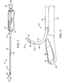
- the treatment probe 14 typically includes a shaft 20 having a handle 22 at its proximal end.
- a needle 24 and imaging array 26 are provided at the distal end of the shaft 20, as described in more detail with reference to Figs. 2 through 4 .
- the treatment probe 14 shown in Figs. 2 through 4 is described in more detail in copending provisional application no. 60/938,140 (Attorney Docket No. 025676-001700US), filed on May 15, 2007.
- the probe 14 generally includes a rigid or other delivery shaft 20, an ultrasound imaging transducer 26, and an echogenic curved needle 24 with an artifact/feature 100 at a distal end 51 ( Fig. 3 ) thereof.
- the artifact is a corner cut type retroreflector.
- the handle 22 is attached to a proximal end 21 of the shaft 20.
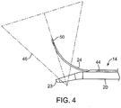
- a distal end 23 of the shaft 20 has a bent or deflectable distal tip, as best seen in Fig. 4 .
- the ultrasound imaging transducer 26 comprises a linear ultrasound array disposed in a flat viewing window 36 ( Fig. 3 ) which images in a field of view 46 ( Fig. 4 ).
- the probe may carry multiple needles in arrays and/or the needles may be straight or have any other configuration.
- the needle 24 is a solid tip electrically conductive needle intended for radiofrequency tissue ablation. As discussed elsewhere, it could also be intended for delivery of other forms of energy or be a hollow core needle intended for substance delivery or injection.
- the exemplary needle 24 generally comprises a two-piece construction including an elongate hollow body 48 (as best seen in Fig. 3 ) and a solid distal tip 50 at a distal end thereof.
- the distal tip 50 may be laser welded to the hollow tubular body 48.
- the solid tip 50 may also be attached via alternative means, for example adhesives or mechanical features or fits.
- the hollow tube 48 will generally have a length in a range from about 20 cm to about 45 cm.
- the hollow tube will have an oval cross section having a thickness generally in a range from about 0.5 mm to about 2 mm and a wideness generally in a range from about 1 mm to about 3 mm.
- This flattened oval cross sectional shape when present, is intended to inhibit lateral deflection during deployment or penetration of the needle 24.
- Fig. 3 also illustrates a representative laser cut hole 60 within the distal end of the tubular body 48 for the infusion of agents (e.g., electrolytes, drugs, etc.) so as to enhance the therapeutic effect of the needle 14 prior to or during ablation treatment.
- agents e.g., electrolytes, drugs, etc.
- the infusion hole 60 may be aligned on one side of the tubular body 48 and generally has length in a range from about 0.5 mm to about 2 mm and a width in a range from about 0.5 mm to about 2 mm. It should be noted that hole 60 may comprise one or a plurality of holes, and each may be used for a different purpose.
- the handle 24 further includes a longitudinally movable slider 72 for enabling the advancement and retraction of the needle 14 to and from within a needle guide 44.
- the ultrasound imaging transducer 26 may optionally be present on an imaging insert replaceably disposed within the axial passage of the shaft 20.
- a sealing element 30 may be provided between the ultrasound imaging transducer 26 and the shaft handle 24 to ensure sufficient sealing around the insert at a proximal end. It will be appreciated that the above depictions are for illustrative purposes only and do not necessarily reflect the actual shape, size, or dimensions of the system 10.
- the ultrasound array may be parallel to an axis of the shaft 20 or may be slightly inclined as illustrated in Fig. 4 . This applies to all depictions hereinafter.
- the array is typically a linear array with from 16 to 128 elements, usually having 64 elements.
- the length (azimuth) of array 12 usually ranges from about 5 mm to about 20 mm, normally being about 14 mm.
- the array may have a depth (elevation) ranging from about 1 mm to about 8 mm, normally being about 2 mm.
- the ultrasound array transmits ultrasound waves at a center frequency ranging from about 2 MHz to about 15 MHz, typically from about 5 MHz to about 12 MHz, normally about 6.5 MHz.
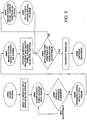
- Fig. 5 an exemplary protocol for performing the needle positioning methods disclosed herein for treating uterine fibroids will be described.
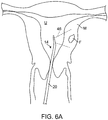
- the treating physician scans the myometrium M in order to locate fibroids F, as shown in Fig. 6A .
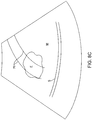
- Shaft 20 is manipulated so that the field of view 46 of the transducer array 26 provides a visual image, such as that shown in Fig. 8A , on the screen 18 of the system 12.
- the physician can scan the image for other anatomical features such as the treatment-sensitive serosa S, as also shown in Fig. 8A .
- the image being produced is "real time," and that the image will change as the physician moves the shaft 20 within the uterus U so that the field of view 46 scans over different portions of the myometrium.
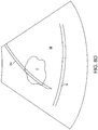
- the next step in the protocol of Fig. 5 relies on aligning a needle guide overlay with the fibroid.
- the needle guide may be a simple pair of parallel lines 70, as shown in Fig. 8B .
- the parallel lines 70 will typically represent the limits of the most likely lateral needle advancement path.
- aligning the lines 70 generally across the target fibroid F as shown in Fig. 8C , the likelihood that the needle will be directed into the middle of the fibroid is increased.
- the treating physician continues to visually assess the position of the needle guidelines 70 relative to the fibroid F until they are acceptably aligned, as shown in Fig. 8C .
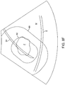
- the physician advances the actual needle into the tissue as shown in Fig. 6B , where the image of the actual needle is shown in Fig. 8D .
- the physician marks a preselected position on the needle, either by moving a curser on the image and clicking, touching the screen, or the like.
- Such "marking" of the actual position allows the system to calculate or recalculate a projected safety boundary and a projected therapy region.
- the system may be marked near the tip of the needle, as shown at location 80 on Fig. 8E .
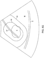
- a treatment needle 24 has an uninsulated treatment portion 96 having a length l in the range from 1 cm to 3 cm, typically being 2 cm.
- the safety boundary will be an oval line which is generally a distance s from the exposed exterior of the treating electrode portion 96. The distance s is usually in the range from 1 cm to 3 cm, typically being about 1.5 cm.
- a distance t between the exposed needle portion 96 and the treatment region boundary 92 will typically be about half that of the safety distance s, typically being in the range from 0.5 cm to 1.5 cm, usually being about 0.75 cm.
- the distance tt from the distal tip of the needle 24 and the safety boundary and the treatment region perimeter will be somewhat less because of the reduced energy density at the tip.
- the distance tt between the tip and the treatment region perimeter may be from 0.1 cm to 0.5 cm, usually being about 0.25 cm while the distance ts between the tip and the safety boundary will be in the range from 0.5 cm to 1.5 cm, typically being about 1 cm.
- the system projects treatment and safety overlays on the actual image of the needle 24, as shown in Fig. 8F .
- the physician can then visually assess whether sensitive tissue structures, such as the serosa S remain outside of the projected safety boundary 90.
- the serosa S is inside of the safety boundary 90, so it will be necessary to reposition or redeploy the needle 24 to move the serosa S beyond the safety boundary.
- the position of the treatment perimeter 92 about the fibroid F is probably sufficient for treatment, but the needle needs to be deployed based on safety concerns.
- the physician will enable the system for treatment. Usually, the system will require the physician to acknowledge that the needle has been properly positioned before allowing the system to power the needle. Once that is done, the physician can initiate treatment, as described generally in the prior applications which have been referenced herein.
Landscapes
- Health & Medical Sciences (AREA)
- Life Sciences & Earth Sciences (AREA)
- Surgery (AREA)
- Engineering & Computer Science (AREA)
- Veterinary Medicine (AREA)
- Biomedical Technology (AREA)
- Heart & Thoracic Surgery (AREA)
- Animal Behavior & Ethology (AREA)
- General Health & Medical Sciences (AREA)
- Public Health (AREA)
- Medical Informatics (AREA)
- Molecular Biology (AREA)
- Nuclear Medicine, Radiotherapy & Molecular Imaging (AREA)
- Pathology (AREA)
- Physics & Mathematics (AREA)
- Hematology (AREA)
- Plasma & Fusion (AREA)
- Otolaryngology (AREA)
- Oral & Maxillofacial Surgery (AREA)
- Biophysics (AREA)
- Radiology & Medical Imaging (AREA)
- Anesthesiology (AREA)
- Vascular Medicine (AREA)
- Robotics (AREA)
- Surgical Instruments (AREA)
- Ultra Sonic Daignosis Equipment (AREA)
- Endoscopes (AREA)
- Gynecology & Obstetrics (AREA)
- Pregnancy & Childbirth (AREA)
- Reproductive Health (AREA)
Priority Applications (2)
| Application Number | Priority Date | Filing Date | Title |
|---|---|---|---|
| EP18173696.8A EP3420916B1 (en) | 2007-10-12 | 2008-10-09 | Systems for controlled deployment of needles in tissue |
| EP20163750.1A EP3733106B1 (en) | 2007-10-12 | 2008-10-09 | Systems of controlled depolyment of needles in tissue |
Applications Claiming Priority (2)
| Application Number | Priority Date | Filing Date | Title |
|---|---|---|---|
| US97961307P | 2007-10-12 | 2007-10-12 | |
| PCT/US2008/079400 WO2009049082A1 (en) | 2007-10-12 | 2008-10-09 | Methods and systems for controlled deployment of needles in tissue |
Related Child Applications (3)
| Application Number | Title | Priority Date | Filing Date |
|---|---|---|---|
| EP20163750.1A Division EP3733106B1 (en) | 2007-10-12 | 2008-10-09 | Systems of controlled depolyment of needles in tissue |
| EP18173696.8A Division EP3420916B1 (en) | 2007-10-12 | 2008-10-09 | Systems for controlled deployment of needles in tissue |
| EP18173696.8A Division-Into EP3420916B1 (en) | 2007-10-12 | 2008-10-09 | Systems for controlled deployment of needles in tissue |
Publications (3)
| Publication Number | Publication Date |
|---|---|
| EP2209423A1 EP2209423A1 (en) | 2010-07-28 |
| EP2209423A4 EP2209423A4 (en) | 2016-05-04 |
| EP2209423B1 true EP2209423B1 (en) | 2018-07-04 |
Family
ID=40534934
Family Applications (3)
| Application Number | Title | Priority Date | Filing Date |
|---|---|---|---|
| EP18173696.8A Active EP3420916B1 (en) | 2007-10-12 | 2008-10-09 | Systems for controlled deployment of needles in tissue |
| EP20163750.1A Active EP3733106B1 (en) | 2007-10-12 | 2008-10-09 | Systems of controlled depolyment of needles in tissue |
| EP08837389.9A Active EP2209423B1 (en) | 2007-10-12 | 2008-10-09 | Systems for controlled deployment of needles in tissue |
Family Applications Before (2)
| Application Number | Title | Priority Date | Filing Date |
|---|---|---|---|
| EP18173696.8A Active EP3420916B1 (en) | 2007-10-12 | 2008-10-09 | Systems for controlled deployment of needles in tissue |
| EP20163750.1A Active EP3733106B1 (en) | 2007-10-12 | 2008-10-09 | Systems of controlled depolyment of needles in tissue |
Country Status (7)
| Country | Link |
|---|---|
| US (11) | US8088072B2 (enExample) |
| EP (3) | EP3420916B1 (enExample) |
| JP (7) | JP5964010B2 (enExample) |
| AU (1) | AU2008310843B2 (enExample) |
| CA (1) | CA2702353C (enExample) |
| ES (2) | ES2812341T3 (enExample) |
| WO (1) | WO2009049082A1 (enExample) |
Families Citing this family (80)
| Publication number | Priority date | Publication date | Assignee | Title |
|---|---|---|---|---|
| US7918795B2 (en) | 2005-02-02 | 2011-04-05 | Gynesonics, Inc. | Method and device for uterine fibroid treatment |
| US8512330B2 (en) * | 2005-07-01 | 2013-08-20 | Halt Medical Inc. | Ablation method |
| US8080009B2 (en) | 2005-07-01 | 2011-12-20 | Halt Medical Inc. | Radio frequency ablation device for the destruction of tissue masses |
| US8784336B2 (en) | 2005-08-24 | 2014-07-22 | C. R. Bard, Inc. | Stylet apparatuses and methods of manufacture |
| US10058342B2 (en) | 2006-01-12 | 2018-08-28 | Gynesonics, Inc. | Devices and methods for treatment of tissue |
| US7874986B2 (en) * | 2006-04-20 | 2011-01-25 | Gynesonics, Inc. | Methods and devices for visualization and ablation of tissue |
| US11259825B2 (en) | 2006-01-12 | 2022-03-01 | Gynesonics, Inc. | Devices and methods for treatment of tissue |
| US7794407B2 (en) | 2006-10-23 | 2010-09-14 | Bard Access Systems, Inc. | Method of locating the tip of a central venous catheter |
| US8388546B2 (en) | 2006-10-23 | 2013-03-05 | Bard Access Systems, Inc. | Method of locating the tip of a central venous catheter |
| US8088072B2 (en) | 2007-10-12 | 2012-01-03 | Gynesonics, Inc. | Methods and systems for controlled deployment of needles in tissue |
| US8781555B2 (en) | 2007-11-26 | 2014-07-15 | C. R. Bard, Inc. | System for placement of a catheter including a signal-generating stylet |
| US8849382B2 (en) | 2007-11-26 | 2014-09-30 | C. R. Bard, Inc. | Apparatus and display methods relating to intravascular placement of a catheter |
| US10751509B2 (en) | 2007-11-26 | 2020-08-25 | C. R. Bard, Inc. | Iconic representations for guidance of an indwelling medical device |
| US10449330B2 (en) | 2007-11-26 | 2019-10-22 | C. R. Bard, Inc. | Magnetic element-equipped needle assemblies |
| US10524691B2 (en) | 2007-11-26 | 2020-01-07 | C. R. Bard, Inc. | Needle assembly including an aligned magnetic element |
| JP5452500B2 (ja) | 2007-11-26 | 2014-03-26 | シー・アール・バード・インコーポレーテッド | カテーテルの血管内留置のための統合システム |
| US9521961B2 (en) | 2007-11-26 | 2016-12-20 | C. R. Bard, Inc. | Systems and methods for guiding a medical instrument |
| US9649048B2 (en) | 2007-11-26 | 2017-05-16 | C. R. Bard, Inc. | Systems and methods for breaching a sterile field for intravascular placement of a catheter |
| EP2252228B1 (en) * | 2008-03-12 | 2016-11-02 | AFreeze GmbH | Handle for an ablation device |
| EP2313143B1 (en) | 2008-08-22 | 2014-09-24 | C.R. Bard, Inc. | Catheter assembly including ecg sensor and magnetic assemblies |
| US20100094270A1 (en) | 2008-10-06 | 2010-04-15 | Sharma Virender K | Method and Apparatus for Tissue Ablation |
| US10064697B2 (en) * | 2008-10-06 | 2018-09-04 | Santa Anna Tech Llc | Vapor based ablation system for treating various indications |
| US8437833B2 (en) | 2008-10-07 | 2013-05-07 | Bard Access Systems, Inc. | Percutaneous magnetic gastrostomy |
| US8632534B2 (en) | 2009-04-03 | 2014-01-21 | Angiodynamics, Inc. | Irreversible electroporation (IRE) for congestive obstructive pulmonary disease (COPD) |
| US8556815B2 (en) | 2009-05-20 | 2013-10-15 | Laurent Pelissier | Freehand ultrasound imaging systems and methods for guiding fine elongate instruments |
| US10039527B2 (en) | 2009-05-20 | 2018-08-07 | Analogic Canada Corporation | Ultrasound systems incorporating spatial position sensors and associated methods |
| US8903488B2 (en) | 2009-05-28 | 2014-12-02 | Angiodynamics, Inc. | System and method for synchronizing energy delivery to the cardiac rhythm |
| US9532724B2 (en) | 2009-06-12 | 2017-01-03 | Bard Access Systems, Inc. | Apparatus and method for catheter navigation using endovascular energy mapping |
| RU2549998C2 (ru) | 2009-06-12 | 2015-05-10 | Бард Аксесс Системс, Инк. | Способ позиционирования конца катетера |
| US9895189B2 (en) | 2009-06-19 | 2018-02-20 | Angiodynamics, Inc. | Methods of sterilization and treating infection using irreversible electroporation |
| EP2464407A4 (en) | 2009-08-10 | 2014-04-02 | Bard Access Systems Inc | DEVICES AND METHODS FOR ENDOVASCULAR ELECTROGRAPHY |
| EP2517622A3 (en) | 2009-09-29 | 2013-04-24 | C. R. Bard, Inc. | Stylets for use with apparatus for intravascular placement of a catheter |
| EP3556308B1 (en) | 2009-11-05 | 2023-12-20 | Stratus Medical, LLC | Systems for spinal radio frequency neurotomy |
| US9486162B2 (en) | 2010-01-08 | 2016-11-08 | Ultrasonix Medical Corporation | Spatial needle guidance system and associated methods |
| ES2811107T3 (es) | 2010-02-02 | 2021-03-10 | Bard Inc C R | Aparato y método para conducción de catéter y localización de punta |
| MX2012013280A (es) | 2010-05-21 | 2013-03-05 | Nimbus Concepts Llc | Sistema y metodos para ablacion de tejido. |
| EP2912999B1 (en) | 2010-05-28 | 2022-06-29 | C. R. Bard, Inc. | Apparatus for use with needle insertion guidance system |
| CA2800810C (en) | 2010-05-28 | 2019-11-05 | C.R. Bard, Inc. | Insertion guidance system for needles and medical components |
| CN103442632A (zh) | 2010-08-20 | 2013-12-11 | C·R·巴德股份有限公司 | Ecg辅助导管末端放置的再确认 |
| EP2627274B1 (en) | 2010-10-13 | 2022-12-14 | AngioDynamics, Inc. | System for electrically ablating tissue of a patient |
| WO2012058461A1 (en) | 2010-10-29 | 2012-05-03 | C.R.Bard, Inc. | Bioimpedance-assisted placement of a medical device |
| JP6000569B2 (ja) | 2011-04-01 | 2016-09-28 | 東芝メディカルシステムズ株式会社 | 超音波診断装置及び制御プログラム |
| US8545409B2 (en) * | 2011-04-14 | 2013-10-01 | St. Jude Medical, Inc. | Arrangement and interface for RF ablation system with acoustic feedback |
| US20140051989A1 (en) * | 2011-04-19 | 2014-02-20 | Drexel University | Devices adapted for ultrasound location in patients and method of use |
| RU2609203C2 (ru) | 2011-07-06 | 2017-01-30 | Си.Ар. Бард, Инк. | Определение и калибровка длины иглы для системы наведения иглы |
| US9078665B2 (en) | 2011-09-28 | 2015-07-14 | Angiodynamics, Inc. | Multiple treatment zone ablation probe |
| KR101828453B1 (ko) * | 2011-12-09 | 2018-02-13 | 삼성전자주식회사 | 의료용 로봇 시스템 및 그 제어 방법 |
| US9295449B2 (en) | 2012-01-23 | 2016-03-29 | Ultrasonix Medical Corporation | Landmarks for ultrasound imaging |
| US9414881B2 (en) | 2012-02-08 | 2016-08-16 | Angiodynamics, Inc. | System and method for increasing a target zone for electrical ablation |
| US9113825B2 (en) * | 2012-07-10 | 2015-08-25 | Fujifilm Sonosite, Inc. | Ultrasonic probe and aligned needle guide system |
| US9861336B2 (en) * | 2012-09-07 | 2018-01-09 | Gynesonics, Inc. | Methods and systems for controlled deployment of needle structures in tissue |
| JP6596436B2 (ja) | 2013-10-18 | 2019-10-23 | ジーバ メディカル, インコーポレイテッド | 多嚢胞性卵巣症候群の治療のための方法およびシステム |
| JP6591415B2 (ja) | 2013-11-20 | 2019-10-16 | アドバンスト アクセス ソリューションズ, インク.Advanced Access Solutions, Inc. | 血管内超音波ニードルガイド |
| CN105979868B (zh) | 2014-02-06 | 2020-03-10 | C·R·巴德股份有限公司 | 用于血管内装置的导向和放置的系统和方法 |
| CA2953133A1 (en) * | 2014-07-02 | 2016-01-07 | Covidien Lp | System and method of providing distance and orientation feedback while navigating in 3d |
| US12114911B2 (en) | 2014-08-28 | 2024-10-15 | Angiodynamics, Inc. | System and method for ablating a tissue site by electroporation with real-time pulse monitoring |
| US10973584B2 (en) | 2015-01-19 | 2021-04-13 | Bard Access Systems, Inc. | Device and method for vascular access |
| AU2016242867B2 (en) | 2015-03-31 | 2020-11-05 | May Health Us Inc. | Methods and systems for the manipulation of ovarian tissues |
| WO2016184746A1 (en) * | 2015-05-18 | 2016-11-24 | Koninklijke Philips N.V. | Intra-procedural accuracy feedback for image-guided biopsy |
| WO2016210325A1 (en) | 2015-06-26 | 2016-12-29 | C.R. Bard, Inc. | Connector interface for ecg-based catheter positioning system |
| US11000207B2 (en) | 2016-01-29 | 2021-05-11 | C. R. Bard, Inc. | Multiple coil system for tracking a medical device |
| US12364537B2 (en) | 2016-05-02 | 2025-07-22 | Santa Anna Tech Llc | Catheter with a double balloon structure to generate and apply a heated ablative zone to tissue |
| CA3043314A1 (en) * | 2016-11-11 | 2018-05-17 | Gynesonics, Inc. | Controlled treatment of tissue and dynamic interaction with, and comparison of, tissue and/or treatment data |
| JP7148511B2 (ja) * | 2016-11-14 | 2022-10-05 | ガイネソニックス, インコーポレイテッド | 組織内でのアブレーション針展開のリアルタイム計画および監視の方法およびシステム |
| US10905492B2 (en) | 2016-11-17 | 2021-02-02 | Angiodynamics, Inc. | Techniques for irreversible electroporation using a single-pole tine-style internal device communicating with an external surface electrode |
| JP2020518385A (ja) * | 2017-05-04 | 2020-06-25 | ガイネソニックス, インコーポレイテッド | ドップラー超音波を用いたアブレーション進行過程の監視のための方法 |
| US11129680B2 (en) * | 2017-12-21 | 2021-09-28 | Cilag Gmbh International | Surgical instrument comprising a projector |
| JP7346422B2 (ja) * | 2017-12-21 | 2023-09-19 | エシコン エルエルシー | 画像層を備えるディスプレイを有する外科用器具 |
| US10912542B2 (en) * | 2018-03-14 | 2021-02-09 | Spiration, Inc. | Catheter assembly with offset device for tissue sampling |
| CN112040875A (zh) * | 2018-04-06 | 2020-12-04 | 美敦力公司 | 基于图像的导航系统和使用相同的导航系统的方法 |
| AU2019272506A1 (en) | 2018-05-21 | 2020-12-17 | Gynesonics, Inc. | Methods and systems for in situ exchange |
| CN113015494A (zh) | 2018-06-01 | 2021-06-22 | 圣安娜技术有限公司 | 多级蒸汽消融治疗方法以及蒸汽产生和输送系统 |
| EP3852622B1 (en) | 2018-10-16 | 2025-04-02 | Bard Access Systems, Inc. | Safety-equipped connection systems and methods thereof for establishing electrical connections |
| KR102747176B1 (ko) * | 2018-12-11 | 2024-12-27 | 삼성메디슨 주식회사 | 초음파 영상 장치, 그 제어 방법, 및 컴퓨터 프로그램 제품 |
| WO2020152631A2 (en) | 2019-01-25 | 2020-07-30 | AblaCare SAS | Systems and methods for applying energy to ovarian tissue |
| CA3166892A1 (en) | 2020-01-07 | 2021-07-15 | Covidien Lp | Devices, systems, and methods for trans-vaginal, ultrasound-guided hysteroscopic surgical procedures |
| CN115334984A (zh) * | 2020-03-30 | 2022-11-11 | 安普列斯医疗公司 | 预测手术设备的弯曲穿透路径 |
| NL2026208B1 (en) | 2020-08-04 | 2022-04-08 | Stichting Het Nederlands Kanker Inst Antoni Van Leeuwenhoek Ziekenhuis | Surgical forceps and stapler |
| WO2024014906A1 (ko) * | 2022-07-13 | 2024-01-18 | 주식회사 로엔서지컬 | 결석 제거를 위한 가이던스 제공 시스템 및 방법 |
| WO2025235783A1 (en) | 2024-05-10 | 2025-11-13 | Gynesonics, Inc. | Tissue ablation systems and methods to prevent or minimize risk of skin burns by dispersive electrodes |
Family Cites Families (260)
| Publication number | Priority date | Publication date | Assignee | Title |
|---|---|---|---|---|
| US4289132A (en) | 1979-06-25 | 1981-09-15 | Rieman Robert D | Surgical instrument and method of using the same |
| US5370675A (en) * | 1992-08-12 | 1994-12-06 | Vidamed, Inc. | Medical probe device and method |
| US4671292A (en) * | 1985-04-30 | 1987-06-09 | Dymax Corporation | Concentric biopsy probe |
| US4802487A (en) * | 1987-03-26 | 1989-02-07 | Washington Research Foundation | Endoscopically deliverable ultrasound imaging system |
| US5269301A (en) * | 1987-10-06 | 1993-12-14 | Leonard Bloom | Multimode system for monitoring and treating a malfunctioning heart |
| US5372587A (en) | 1989-01-09 | 1994-12-13 | Pilot Cariovascular Systems, Inc. | Steerable medical device |
| US4936281A (en) * | 1989-04-13 | 1990-06-26 | Everest Medical Corporation | Ultrasonically enhanced RF ablation catheter |
| US5316000A (en) | 1991-03-05 | 1994-05-31 | Technomed International (Societe Anonyme) | Use of at least one composite piezoelectric transducer in the manufacture of an ultrasonic therapy apparatus for applying therapy, in a body zone, in particular to concretions, to tissue, or to bones, of a living being and method of ultrasonic therapy |
| US6485413B1 (en) | 1991-04-29 | 2002-11-26 | The General Hospital Corporation | Methods and apparatus for forward-directed optical scanning instruments |
| US7549424B2 (en) * | 1991-10-18 | 2009-06-23 | Pro Surg, Inc. | Method and apparatus for tissue treatment with laser and electromagnetic radiation |
| US6730081B1 (en) * | 1991-10-18 | 2004-05-04 | Ashvin H. Desai | Endoscopic surgical instrument |
| US6461296B1 (en) | 1998-06-26 | 2002-10-08 | 2000 Injectx, Inc. | Method and apparatus for delivery of genes, enzymes and biological agents to tissue cells |
| US6024733A (en) | 1995-06-07 | 2000-02-15 | Arthrocare Corporation | System and method for epidermal tissue ablation |
| US6770071B2 (en) * | 1995-06-07 | 2004-08-03 | Arthrocare Corporation | Bladed electrosurgical probe |
| US5470308A (en) * | 1992-08-12 | 1995-11-28 | Vidamed, Inc. | Medical probe with biopsy stylet |
| US5364408A (en) | 1992-09-04 | 1994-11-15 | Laurus Medical Corporation | Endoscopic suture system |
| US6832996B2 (en) | 1995-06-07 | 2004-12-21 | Arthrocare Corporation | Electrosurgical systems and methods for treating tissue |
| US5860974A (en) * | 1993-07-01 | 1999-01-19 | Boston Scientific Corporation | Heart ablation catheter with expandable electrode and method of coupling energy to an electrode on a catheter shaft |
| US5456689A (en) * | 1993-10-13 | 1995-10-10 | Arnold J. Kresch | Method and device for tissue resection |
| US6569159B1 (en) * | 1993-11-08 | 2003-05-27 | Rita Medical Systems, Inc. | Cell necrosis apparatus |
| US5599346A (en) | 1993-11-08 | 1997-02-04 | Zomed International, Inc. | RF treatment system |
| US6241725B1 (en) * | 1993-12-15 | 2001-06-05 | Sherwood Services Ag | High frequency thermal ablation of cancerous tumors and functional targets with image data assistance |
| US5471988A (en) | 1993-12-24 | 1995-12-05 | Olympus Optical Co., Ltd. | Ultrasonic diagnosis and therapy system in which focusing point of therapeutic ultrasonic wave is locked at predetermined position within observation ultrasonic scanning range |
| DE4443947B4 (de) | 1994-01-14 | 2005-09-22 | Siemens Ag | Endoskop |
| US5873828A (en) * | 1994-02-18 | 1999-02-23 | Olympus Optical Co., Ltd. | Ultrasonic diagnosis and treatment system |
| US5517989A (en) * | 1994-04-01 | 1996-05-21 | Cardiometrics, Inc. | Guidewire assembly |
| US5492126A (en) * | 1994-05-02 | 1996-02-20 | Focal Surgery | Probe for medical imaging and therapy using ultrasound |
| JP3419884B2 (ja) * | 1994-05-11 | 2003-06-23 | オリンパス光学工業株式会社 | 制御システム |
| US6002968A (en) | 1994-06-24 | 1999-12-14 | Vidacare, Inc. | Uterine treatment apparatus |
| US6405732B1 (en) * | 1994-06-24 | 2002-06-18 | Curon Medical, Inc. | Method to treat gastric reflux via the detection and ablation of gastro-esophageal nerves and receptors |
| US6032673A (en) * | 1994-10-13 | 2000-03-07 | Femrx, Inc. | Methods and devices for tissue removal |
| US6575969B1 (en) * | 1995-05-04 | 2003-06-10 | Sherwood Services Ag | Cool-tip radiofrequency thermosurgery electrode system for tumor ablation |
| US5964740A (en) | 1996-07-09 | 1999-10-12 | Asahi Kogaku Kogyo Kabushiki Kaisha | Treatment accessory for an endoscope |
| US6837888B2 (en) * | 1995-06-07 | 2005-01-04 | Arthrocare Corporation | Electrosurgical probe with movable return electrode and methods related thereto |
| US6837887B2 (en) | 1995-06-07 | 2005-01-04 | Arthrocare Corporation | Articulated electrosurgical probe and methods |
| US5979452A (en) | 1995-06-07 | 1999-11-09 | General Surgical Innovations, Inc. | Endoscopic linton procedure using balloon dissectors and retractors |
| US6632193B1 (en) | 1995-06-07 | 2003-10-14 | Arthrocare Corporation | Systems and methods for electrosurgical tissue treatment |
| US5964709A (en) | 1995-06-29 | 1999-10-12 | Teratech Corporation | Portable ultrasound imaging system |
| US5590658A (en) | 1995-06-29 | 1997-01-07 | Teratech Corporation | Portable ultrasound imaging system |
| JPH11508790A (ja) * | 1995-06-30 | 1999-08-03 | ボストン・サイエンティフィック・コーポレイション | 切断エレメントを備えた超音波映写カテーテル |
| BR9609484A (pt) * | 1995-07-16 | 1999-12-14 | Yoav Paltieli | Processo e aparelho para direcionamento à mão livre de uma agulha so sentido de um alvo localizado em um volume corpóreo e aparelho de agulha |
| US6256529B1 (en) * | 1995-07-26 | 2001-07-03 | Burdette Medical Systems, Inc. | Virtual reality 3D visualization for surgical procedures |
| US6080150A (en) | 1995-08-15 | 2000-06-27 | Rita Medical Systems, Inc. | Cell necrosis apparatus |
| US6375615B1 (en) | 1995-10-13 | 2002-04-23 | Transvascular, Inc. | Tissue penetrating catheters having integral imaging transducers and their methods of use |
| US5979453A (en) | 1995-11-09 | 1999-11-09 | Femrx, Inc. | Needle myolysis system for uterine fibriods |
| US5780435A (en) * | 1995-12-15 | 1998-07-14 | Praecis Pharmaceuticals Incorporated | Methods for treating prostate cancer with LHRH-R antagonists |
| US5863294A (en) * | 1996-01-26 | 1999-01-26 | Femrx, Inc. | Folded-end surgical tubular cutter and method for fabrication |
| US6203524B1 (en) | 1997-02-10 | 2001-03-20 | Emx, Inc. | Surgical and pharmaceutical site access guide and methods |
| US6813520B2 (en) | 1996-04-12 | 2004-11-02 | Novacept | Method for ablating and/or coagulating tissue using moisture transport |
| US5769880A (en) * | 1996-04-12 | 1998-06-23 | Novacept | Moisture transport system for contact electrocoagulation |
| US6419673B1 (en) | 1996-05-06 | 2002-07-16 | Stuart Edwards | Ablation of rectal and other internal body structures |
| US6077257A (en) | 1996-05-06 | 2000-06-20 | Vidacare, Inc. | Ablation of rectal and other internal body structures |
| US5649911A (en) * | 1996-05-17 | 1997-07-22 | Indiana University Foundation | Intravenous catheter and delivery system |
| US5957941A (en) | 1996-09-27 | 1999-09-28 | Boston Scientific Corporation | Catheter system and drive assembly thereof |
| WO1998014169A1 (en) | 1996-09-30 | 1998-04-09 | Brigham & Women's Hospital | Methods and compounds for treatment of abnormal uterine bleeding |
| US6805128B1 (en) | 1996-10-22 | 2004-10-19 | Epicor Medical, Inc. | Apparatus and method for ablating tissue |
| US6719755B2 (en) * | 1996-10-22 | 2004-04-13 | Epicor Medical, Inc. | Methods and devices for ablation |
| US5730752A (en) * | 1996-10-29 | 1998-03-24 | Femrx, Inc. | Tubular surgical cutters having aspiration flow control ports |
| US5741287A (en) * | 1996-11-01 | 1998-04-21 | Femrx, Inc. | Surgical tubular cutter having a tapering cutting chamber |
| US5910104A (en) * | 1996-12-26 | 1999-06-08 | Cryogen, Inc. | Cryosurgical probe with disposable sheath |
| US6270494B1 (en) | 1996-12-26 | 2001-08-07 | Cryogen, Inc. | Stretchable cryoprobe sheath |
| US5931787A (en) | 1997-02-11 | 1999-08-03 | Tetrad Corporation | Sheath and methods of ultrasonic guidance for biopsy and catheter insertion |
| US6314310B1 (en) | 1997-02-14 | 2001-11-06 | Biosense, Inc. | X-ray guided surgical location system with extended mapping volume |
| US5906615A (en) * | 1997-03-31 | 1999-05-25 | Femrx, Inc. | Serpentine ablation/coagulation electrode |
| US5984942A (en) | 1997-04-02 | 1999-11-16 | Femrx, Inc. | Methods and systems for reducing tissue adhesion |
| US5873877A (en) * | 1997-04-11 | 1999-02-23 | Vidamed, Inc. | Medical probe device with transparent distal extremity |
| US5876340A (en) * | 1997-04-17 | 1999-03-02 | Irvine Biomedical, Inc. | Ablation apparatus with ultrasonic imaging capabilities |
| US5891137A (en) * | 1997-05-21 | 1999-04-06 | Irvine Biomedical, Inc. | Catheter system having a tip with fixation means |
| US5876399A (en) * | 1997-05-28 | 1999-03-02 | Irvine Biomedical, Inc. | Catheter system and methods thereof |
| US5921926A (en) | 1997-07-28 | 1999-07-13 | University Of Central Florida | Three dimensional optical imaging colposcopy |
| US6039748A (en) * | 1997-08-05 | 2000-03-21 | Femrx, Inc. | Disposable laparoscopic morcellator |
| US5916198A (en) * | 1997-08-05 | 1999-06-29 | Femrx, Inc. | Non-binding surgical valve |
| CA2300845A1 (en) | 1997-08-19 | 1999-02-25 | John D. Mendlein | Multi-site ultrasound methods and devices, particularly for measurement of fluid regulation |
| US6055449A (en) * | 1997-09-22 | 2000-04-25 | Siemens Corporate Research, Inc. | Method for localization of a biopsy needle or similar surgical tool in a radiographic image |
| JP2001517475A (ja) | 1997-09-22 | 2001-10-09 | エシコン・インコーポレイテッド | 凍結手術システム及び方法 |
| US6238389B1 (en) | 1997-09-30 | 2001-05-29 | Boston Scientific Corporation | Deflectable interstitial ablation device |
| US6171249B1 (en) * | 1997-10-14 | 2001-01-09 | Circon Corporation | Ultrasound guided therapeutic and diagnostic device |
| US6325758B1 (en) * | 1997-10-27 | 2001-12-04 | Nomos Corporation | Method and apparatus for target position verification |
| US6007499A (en) | 1997-10-31 | 1999-12-28 | University Of Washington | Method and apparatus for medical procedures using high-intensity focused ultrasound |
| IL122336A0 (en) * | 1997-11-27 | 1998-04-05 | Ultra Guide Ltd | System and method for guiding the movements of a device to a target particularly for medical applications |
| US6280441B1 (en) | 1997-12-15 | 2001-08-28 | Sherwood Services Ag | Apparatus and method for RF lesioning |
| IL122839A0 (en) * | 1997-12-31 | 1998-08-16 | Ultra Guide Ltd | Calibration method and apparatus for calibrating position sensors on scanning transducers |
| US6146380A (en) | 1998-01-09 | 2000-11-14 | Radionics, Inc. | Bent tip electrical surgical probe |
| WO1999035988A1 (en) * | 1998-01-14 | 1999-07-22 | Conway-Stuart Medical, Inc. | Electrosurgical device for sphincter treatment |
| CN1058905C (zh) * | 1998-01-25 | 2000-11-29 | 重庆海扶(Hifu)技术有限公司 | 高强度聚焦超声肿瘤扫描治疗系统 |
| US6059766A (en) | 1998-02-27 | 2000-05-09 | Micro Therapeutics, Inc. | Gynecologic embolotherapy methods |
| JP4125814B2 (ja) * | 1998-03-04 | 2008-07-30 | Hoya株式会社 | 超音波内視鏡 |
| JPH11276422A (ja) * | 1998-03-31 | 1999-10-12 | Fuji Photo Optical Co Ltd | 超音波内視鏡 |
| US6311084B1 (en) * | 1998-05-04 | 2001-10-30 | Robert A. Cormack | Radiation seed implant method and apparatus |
| US6635055B1 (en) | 1998-05-06 | 2003-10-21 | Microsulis Plc | Microwave applicator for endometrial ablation |
| US6508815B1 (en) * | 1998-05-08 | 2003-01-21 | Novacept | Radio-frequency generator for powering an ablation device |
| US20050255039A1 (en) | 1998-06-26 | 2005-11-17 | Pro Surg, Inc., A California Corporation | Gel injection treatment of breast, fibroids & endometrial ablation |
| AU4842999A (en) | 1998-06-30 | 2000-01-17 | Arthrocare Corporation | Systems and methods for electrosurgical ablation of viable body structures |
| US6296639B1 (en) | 1999-02-12 | 2001-10-02 | Novacept | Apparatuses and methods for interstitial tissue removal |
| US7435247B2 (en) | 1998-08-11 | 2008-10-14 | Arthrocare Corporation | Systems and methods for electrosurgical tissue treatment |
| WO2003024305A2 (en) | 2001-09-14 | 2003-03-27 | Arthrocare Corporation | Electrosurgical apparatus and methods for tissue treatment & removal |
| US7276063B2 (en) * | 1998-08-11 | 2007-10-02 | Arthrocare Corporation | Instrument for electrosurgical tissue treatment |
| US6425867B1 (en) * | 1998-09-18 | 2002-07-30 | University Of Washington | Noise-free real time ultrasonic imaging of a treatment site undergoing high intensity focused ultrasound therapy |
| US7686763B2 (en) * | 1998-09-18 | 2010-03-30 | University Of Washington | Use of contrast agents to increase the effectiveness of high intensity focused ultrasound therapy |
| US6190383B1 (en) * | 1998-10-21 | 2001-02-20 | Sherwood Services Ag | Rotatable electrode device |
| DE69924750T2 (de) * | 1998-11-16 | 2006-03-02 | United States Surgical Corp., Norwalk | Gerät zur thermischen behandlung von gewebe |
| US6507747B1 (en) * | 1998-12-02 | 2003-01-14 | Board Of Regents, The University Of Texas System | Method and apparatus for concomitant structural and biochemical characterization of tissue |
| US6254601B1 (en) | 1998-12-08 | 2001-07-03 | Hysterx, Inc. | Methods for occlusion of the uterine arteries |
| DE69920178T2 (de) * | 1998-12-09 | 2005-09-22 | Cook Inc., Bloomington | Super-elastische gebogene hohlnadel zur medizinischen verwendung |
| US6146378A (en) | 1999-03-19 | 2000-11-14 | Endocare, Inc. | Placement guide for ablation devices |
| AU3985400A (en) * | 1999-04-15 | 2000-11-02 | Ultra-Guide Ltd. | Apparatus and method for detecting the bending of medical invasive tools in medical interventions |
| US6463331B1 (en) | 1999-04-19 | 2002-10-08 | Novasys Medical, Inc. | Application of energy and substances in the treatment of uro-genital disorders |
| US6692490B1 (en) * | 1999-05-18 | 2004-02-17 | Novasys Medical, Inc. | Treatment of urinary incontinence and other disorders by application of energy and drugs |
| US6139544A (en) * | 1999-05-26 | 2000-10-31 | Endocare, Inc. | Computer guided cryosurgery |
| EP1207788A4 (en) | 1999-07-19 | 2009-12-09 | St Jude Medical Atrial Fibrill | DEVICE AND METHOD FOR THE ABLATION OF TISSUE |
| ITMI991608A1 (it) | 1999-07-21 | 2001-01-21 | Thermo Med 2000 Kft | Sonda elettrochirurgica per il trattamento di tumori mediante radiofrequenza |
| US7520856B2 (en) * | 1999-09-17 | 2009-04-21 | University Of Washington | Image guided high intensity focused ultrasound device for therapy in obstetrics and gynecology |
| US20030032896A1 (en) * | 2000-09-25 | 2003-02-13 | Vance Products, Inc., D/B/A/ Cook Urological, Inc. | Microvolume embryo transfer system |
| US20020077550A1 (en) | 1999-10-05 | 2002-06-20 | Rabiner Robert A. | Apparatus and method for treating gynecological diseases using an ultrasonic medical device operating in a transverse mode |
| EP1229829B1 (en) * | 1999-11-10 | 2008-01-09 | Cytyc Surgical Products | System for detecting perforations in a body cavity |
| US6626855B1 (en) | 1999-11-26 | 2003-09-30 | Therus Corpoation | Controlled high efficiency lesion formation using high intensity ultrasound |
| US6506156B1 (en) * | 2000-01-19 | 2003-01-14 | Vascular Control Systems, Inc | Echogenic coating |
| US6158250A (en) | 2000-02-14 | 2000-12-12 | Novacept | Flat-bed knitting machine and method of knitting |
| US6579298B1 (en) * | 2000-02-29 | 2003-06-17 | Scimed Life Systems, Inc. | Method and apparatus for treating vein graft lesions |
| US6379348B1 (en) * | 2000-03-15 | 2002-04-30 | Gary M. Onik | Combined electrosurgical-cryosurgical instrument |
| JP3875841B2 (ja) * | 2000-03-28 | 2007-01-31 | アロカ株式会社 | 医療システム |
| US6419648B1 (en) * | 2000-04-21 | 2002-07-16 | Insightec-Txsonics Ltd. | Systems and methods for reducing secondary hot spots in a phased array focused ultrasound system |
| US6543272B1 (en) * | 2000-04-21 | 2003-04-08 | Insightec-Txsonics Ltd. | Systems and methods for testing and calibrating a focused ultrasound transducer array |
| US6613004B1 (en) | 2000-04-21 | 2003-09-02 | Insightec-Txsonics, Ltd. | Systems and methods for creating longer necrosed volumes using a phased array focused ultrasound system |
| US6550482B1 (en) * | 2000-04-21 | 2003-04-22 | Vascular Control Systems, Inc. | Methods for non-permanent occlusion of a uterine artery |
| US6599288B2 (en) | 2000-05-16 | 2003-07-29 | Atrionix, Inc. | Apparatus and method incorporating an ultrasound transducer onto a delivery member |
| GB2379793B (en) | 2000-06-20 | 2004-03-17 | Seagate Technology Llc | Disc drive with layered write coil |
| US6506171B1 (en) * | 2000-07-27 | 2003-01-14 | Insightec-Txsonics, Ltd | System and methods for controlling distribution of acoustic energy around a focal point using a focused ultrasound system |
| US6905492B2 (en) * | 2000-07-31 | 2005-06-14 | Galil Medical Ltd. | Planning and facilitation systems and methods for cryosurgery |
| US6840935B2 (en) | 2000-08-09 | 2005-01-11 | Bekl Corporation | Gynecological ablation procedure and system using an ablation needle |
| US7678106B2 (en) | 2000-08-09 | 2010-03-16 | Halt Medical, Inc. | Gynecological ablation procedure and system |
| US6612988B2 (en) | 2000-08-29 | 2003-09-02 | Brigham And Women's Hospital, Inc. | Ultrasound therapy |
| US7387628B1 (en) | 2000-09-15 | 2008-06-17 | Boston Scientific Scimed, Inc. | Methods and systems for focused bipolar tissue ablation |
| US20020165947A1 (en) * | 2000-09-25 | 2002-11-07 | Crossbeam Systems, Inc. | Network application apparatus |
| US6638275B1 (en) | 2000-10-05 | 2003-10-28 | Medironic, Inc. | Bipolar ablation apparatus and method |
| US20070088247A1 (en) * | 2000-10-24 | 2007-04-19 | Galil Medical Ltd. | Apparatus and method for thermal ablation of uterine fibroids |
| US6679855B2 (en) * | 2000-11-07 | 2004-01-20 | Gerald Horn | Method and apparatus for the correction of presbyopia using high intensity focused ultrasound |
| US6635065B2 (en) | 2000-11-16 | 2003-10-21 | Vascular Control Systems, Inc. | Doppler directed suture ligation device and method |
| US6638286B1 (en) | 2000-11-16 | 2003-10-28 | Vascular Control Systems, Inc. | Doppler directed suture ligation device and method |
| US6540677B1 (en) * | 2000-11-17 | 2003-04-01 | Bjorn A. J. Angelsen | Ultrasound transceiver system for remote operation through a minimal number of connecting wires |
| US6666833B1 (en) | 2000-11-28 | 2003-12-23 | Insightec-Txsonics Ltd | Systems and methods for focussing an acoustic energy beam transmitted through non-uniform tissue medium |
| US6613005B1 (en) | 2000-11-28 | 2003-09-02 | Insightec-Txsonics, Ltd. | Systems and methods for steering a focused ultrasound array |
| US6506154B1 (en) * | 2000-11-28 | 2003-01-14 | Insightec-Txsonics, Ltd. | Systems and methods for controlling a phased array focused ultrasound system |
| US6645162B2 (en) | 2000-12-27 | 2003-11-11 | Insightec - Txsonics Ltd. | Systems and methods for ultrasound assisted lipolysis |
| US6626854B2 (en) | 2000-12-27 | 2003-09-30 | Insightec - Txsonics Ltd. | Systems and methods for ultrasound assisted lipolysis |
| EP1357842B1 (en) * | 2001-01-16 | 2010-11-03 | Cytyc Surgical Products | Apparatus and method for treating venous reflux |
| US6572613B1 (en) * | 2001-01-16 | 2003-06-03 | Alan G. Ellman | RF tissue penetrating probe |
| US6695786B2 (en) * | 2001-03-16 | 2004-02-24 | U-Systems, Inc. | Guide and position monitor for invasive medical instrument |
| WO2002089686A1 (en) * | 2001-05-10 | 2002-11-14 | Rita Medical Systems, Inc. | Rf tissue ablation apparatus and method |
| US6559644B2 (en) * | 2001-05-30 | 2003-05-06 | Insightec - Txsonics Ltd. | MRI-based temperature mapping with error compensation |
| US6921398B2 (en) | 2001-06-04 | 2005-07-26 | Electrosurgery Associates, Llc | Vibrating electrosurgical ablator |
| US6735461B2 (en) * | 2001-06-19 | 2004-05-11 | Insightec-Txsonics Ltd | Focused ultrasound system with MRI synchronization |
| US6728571B1 (en) * | 2001-07-16 | 2004-04-27 | Scimed Life Systems, Inc. | Electronically scanned optical coherence tomography with frequency modulated signals |
| US6994706B2 (en) * | 2001-08-13 | 2006-02-07 | Minnesota Medical Physics, Llc | Apparatus and method for treatment of benign prostatic hyperplasia |
| US6733458B1 (en) * | 2001-09-25 | 2004-05-11 | Acuson Corporation | Diagnostic medical ultrasound systems and methods using image based freehand needle guidance |
| US6969354B1 (en) | 2001-09-25 | 2005-11-29 | Acuson Corporation | Adaptable intraoperative or endocavity ultrasound probe |
| JP2003126093A (ja) * | 2001-10-23 | 2003-05-07 | Olympus Optical Co Ltd | 超音波診断装置 |
| ES2246999T3 (es) | 2001-11-23 | 2006-03-01 | Nucletron B.V. | Dispositivo autocontrolado guiado por imagenes para insertar una aguja en el cuerpo de un animal para realizar radioterapia en dicho cuerpo. |
| US6790180B2 (en) | 2001-12-03 | 2004-09-14 | Insightec-Txsonics Ltd. | Apparatus, systems, and methods for measuring power output of an ultrasound transducer |
| US6522142B1 (en) * | 2001-12-14 | 2003-02-18 | Insightec-Txsonics Ltd. | MRI-guided temperature mapping of tissue undergoing thermal treatment |
| WO2003068055A2 (en) | 2002-02-11 | 2003-08-21 | Arthrocare Corporation | Electrosurgical apparatus and methods for laparoscopy |
| US20050177209A1 (en) | 2002-03-05 | 2005-08-11 | Baylis Medical Company Inc. | Bipolar tissue treatment system |
| US20030199472A1 (en) | 2002-03-19 | 2003-10-23 | Board Of Regents, The University Of Texas System | Adenovirus-mediated therapy for uterine fibroids |
| US7462366B2 (en) | 2002-03-29 | 2008-12-09 | Boston Scientific Scimed, Inc. | Drug delivery particle |
| EP1501411B1 (en) | 2002-04-22 | 2014-03-12 | Johns Hopkins University | Apparatus for insertion of a medical device during a medical imaging process |
| US8221321B2 (en) | 2002-06-07 | 2012-07-17 | Verathon Inc. | Systems and methods for quantification and classification of fluids in human cavities in ultrasound images |
| US6923807B2 (en) * | 2002-06-27 | 2005-08-02 | Ethicon, Inc. | Helical device and method for aiding the ablation and assessment of tissue |
| US20040006336A1 (en) * | 2002-07-02 | 2004-01-08 | Scimed Life Systems, Inc. | Apparatus and method for RF ablation into conductive fluid-infused tissue |
| US6705994B2 (en) * | 2002-07-08 | 2004-03-16 | Insightec - Image Guided Treatment Ltd | Tissue inhomogeneity correction in ultrasound imaging |
| US20040049121A1 (en) * | 2002-09-06 | 2004-03-11 | Uri Yaron | Positioning system for neurological procedures in the brain |
| US6944490B1 (en) | 2002-09-25 | 2005-09-13 | Advanced Cardiovascular Systems, Inc. | Apparatus and method for positioning and delivering a therapeutic tool to the inside of a heart |
| US7101367B2 (en) | 2002-09-30 | 2006-09-05 | Ethicon, Inc. | Deployable cryosurgical catheter |
| EP1558326A4 (en) | 2002-10-17 | 2007-08-15 | Pro Surg Inc | GELINJECTION DEVICE AND TREATMENT OF THE CHEST, FIBROID AND ANDOMETRIUM ABLATION |
| US7306593B2 (en) * | 2002-10-21 | 2007-12-11 | Biosense, Inc. | Prediction and assessment of ablation of cardiac tissue |
| AU2003298638A1 (en) | 2002-11-12 | 2004-06-03 | Prourocare, Inc. | Intelligent medical device barrier |
| WO2004045378A2 (en) | 2002-11-15 | 2004-06-03 | The Government Of The United States Of America As Represented By The Secretary Of Health And Human Services | Method and device for catheter-based repair of cardiac valves |
| US6953458B2 (en) * | 2002-12-20 | 2005-10-11 | Trimedyne, Inc. | Device and method for delivery of long wavelength laser energy to a tissue site |
| US6936048B2 (en) | 2003-01-16 | 2005-08-30 | Charlotte-Mecklenburg Hospital Authority | Echogenic needle for transvaginal ultrasound directed reduction of uterine fibroids and an associated method |
| US7635332B2 (en) * | 2003-02-14 | 2009-12-22 | Siemens Medical Solutions Usa, Inc. | System and method of operating microfabricated ultrasonic transducers for harmonic imaging |
| US8071550B2 (en) | 2003-03-03 | 2011-12-06 | Allergan, Inc. | Methods for treating uterine disorders |
| US20040176760A1 (en) | 2003-03-05 | 2004-09-09 | Qiu Xue Hua | Electrosurgical apparatus with cooling device |
| US7333844B2 (en) | 2003-03-28 | 2008-02-19 | Vascular Control Systems, Inc. | Uterine tissue monitoring device and method |
| US20040254572A1 (en) | 2003-04-25 | 2004-12-16 | Mcintyre Jon T. | Self anchoring radio frequency ablation array |
| WO2004100811A1 (ja) | 2003-05-19 | 2004-11-25 | Hitachi, Ltd. | 超音波治療装置 |
| US8102392B2 (en) * | 2003-06-27 | 2012-01-24 | Kabushiki Kaisha Toshiba | Image processing/displaying apparatus having free moving control unit and limited moving control unit and method of controlling the same |
| US8055323B2 (en) | 2003-08-05 | 2011-11-08 | Imquant, Inc. | Stereotactic system and method for defining a tumor treatment region |
| US20050159676A1 (en) | 2003-08-13 | 2005-07-21 | Taylor James D. | Targeted biopsy delivery system |
| JP4828802B2 (ja) * | 2004-05-12 | 2011-11-30 | 株式会社東芝 | 穿刺治療のための超音波診断装置 |
| JP4467927B2 (ja) * | 2003-08-19 | 2010-05-26 | 株式会社東芝 | 超音波診断装置 |
| US7066887B2 (en) | 2003-10-21 | 2006-06-27 | Vermon | Bi-plane ultrasonic probe |
| US20050085718A1 (en) * | 2003-10-21 | 2005-04-21 | Ramin Shahidi | Systems and methods for intraoperative targetting |
| EP1684655A2 (en) * | 2003-11-18 | 2006-08-02 | SciMed Life Systems, Inc. | System and method for tissue ablation |
| US7247141B2 (en) | 2004-03-08 | 2007-07-24 | Ethicon Endo-Surgery, Inc. | Intra-cavitary ultrasound medical system and method |
| US7854733B2 (en) | 2004-03-24 | 2010-12-21 | Biosense Webster, Inc. | Phased-array for tissue treatment |
| JP4847442B2 (ja) | 2004-04-02 | 2011-12-28 | コーニンクレッカ フィリップス エレクトロニクス エヌ ヴィ | 3d撮像のための超音波腔内プローブ |
| US20050256405A1 (en) | 2004-05-17 | 2005-11-17 | Makin Inder Raj S | Ultrasound-based procedure for uterine medical treatment |
| US20060020204A1 (en) * | 2004-07-01 | 2006-01-26 | Bracco Imaging, S.P.A. | System and method for three-dimensional space management and visualization of ultrasound data ("SonoDEX") |
| US7699780B2 (en) * | 2004-08-11 | 2010-04-20 | Insightec—Image-Guided Treatment Ltd. | Focused ultrasound system with adaptive anatomical aperture shaping |
| US20060058680A1 (en) * | 2004-08-25 | 2006-03-16 | Stephen Solomon | Needle guide for laparoscopic ultrasonography |
| US20080015664A1 (en) * | 2004-10-06 | 2008-01-17 | Podhajsky Ronald J | Systems and methods for thermally profiling radiofrequency electrodes |
| EP2151206B1 (en) * | 2004-10-06 | 2012-05-23 | Covidien AG | Methods for thermally profiling radiofrequency electrodes |
| US20060089636A1 (en) | 2004-10-27 | 2006-04-27 | Christopherson Mark A | Ultrasound visualization for transurethral needle ablation |
| NL1027678C2 (nl) | 2004-12-07 | 2006-06-12 | Benedictus Christiaan Schoot | Inrichting en werkwijze voor onderzoek van een lichaamsholte. |
| JP4619803B2 (ja) | 2005-01-26 | 2011-01-26 | 富士フイルム株式会社 | 蛍光断層画像取得装置 |
| US7918795B2 (en) | 2005-02-02 | 2011-04-05 | Gynesonics, Inc. | Method and device for uterine fibroid treatment |
| US7517346B2 (en) | 2005-02-08 | 2009-04-14 | Boston Scientific Scimed, Inc. | Radio frequency ablation system with integrated ultrasound imaging |
| GB0503380D0 (en) * | 2005-02-17 | 2005-03-23 | Rhytec Ltd | Tissue treatment system |
| EP1861133B1 (en) | 2005-02-28 | 2012-11-21 | Spirus Medical Inc. | Rotate-to-advance catheterization system |
| US8788019B2 (en) * | 2005-02-28 | 2014-07-22 | Robarts Research Institute | System and method for performing a biopsy of a target volume and a computing device for planning the same |
| US8801701B2 (en) * | 2005-03-09 | 2014-08-12 | Sunnybrook Health Sciences Centre | Method and apparatus for obtaining quantitative temperature measurements in prostate and other tissue undergoing thermal therapy treatment |
| US7963941B2 (en) | 2005-04-12 | 2011-06-21 | Wilk Peter J | Intra-abdominal medical method and associated device |
| US20090043295A1 (en) | 2005-06-06 | 2009-02-12 | Ams Research Corporation | Fibroid Treatment Methods and Devices |
| US8512330B2 (en) | 2005-07-01 | 2013-08-20 | Halt Medical Inc. | Ablation method |
| US8512333B2 (en) * | 2005-07-01 | 2013-08-20 | Halt Medical Inc. | Anchored RF ablation device for the destruction of tissue masses |
| US8080009B2 (en) | 2005-07-01 | 2011-12-20 | Halt Medical Inc. | Radio frequency ablation device for the destruction of tissue masses |
| US9561044B2 (en) * | 2005-10-12 | 2017-02-07 | Atricure, Inc. | Diaphragm entry for posterior surgical access |
| US20070129626A1 (en) * | 2005-11-23 | 2007-06-07 | Prakash Mahesh | Methods and systems for facilitating surgical procedures |
| KR20070058785A (ko) * | 2005-12-05 | 2007-06-11 | 주식회사 메디슨 | 중재적 시술을 위한 초음파 시스템 |
| US9357977B2 (en) | 2006-01-12 | 2016-06-07 | Gynesonics, Inc. | Interventional deployment and imaging system |
| US7874986B2 (en) | 2006-04-20 | 2011-01-25 | Gynesonics, Inc. | Methods and devices for visualization and ablation of tissue |
| US20070161905A1 (en) | 2006-01-12 | 2007-07-12 | Gynesonics, Inc. | Intrauterine ultrasound and method for use |
| US7815571B2 (en) | 2006-04-20 | 2010-10-19 | Gynesonics, Inc. | Rigid delivery systems having inclined ultrasound and needle |
| JP4812458B2 (ja) * | 2006-02-15 | 2011-11-09 | 株式会社東芝 | 超音波診断装置及び治療支援装置 |
| JP4768494B2 (ja) | 2006-03-31 | 2011-09-07 | テルモ株式会社 | 画像診断装置およびその処理方法 |
| JP5209603B2 (ja) * | 2006-04-13 | 2013-06-12 | ミラビリス メディカ インコーポレイテッド | 高密度焦点式超音波エネルギーの使用による機能性子宮出血、子宮内膜の病状、および子宮頸部の新形成の治療のための方法および装置 |
| US8206300B2 (en) | 2008-08-26 | 2012-06-26 | Gynesonics, Inc. | Ablation device with articulated imaging transducer |
| US10376314B2 (en) | 2006-07-14 | 2019-08-13 | Neuwave Medical, Inc. | Energy delivery systems and uses thereof |
| US8298145B2 (en) * | 2006-08-01 | 2012-10-30 | Gynesonics, Inc. | Peri-capsular fibroid treatment |
| CN101801275B (zh) | 2007-08-15 | 2013-04-24 | 皇家飞利浦电子股份有限公司 | 用于产生组织部分附近的加热尖头的图像的方法和设备 |
| US8088072B2 (en) | 2007-10-12 | 2012-01-03 | Gynesonics, Inc. | Methods and systems for controlled deployment of needles in tissue |
| WO2009055720A1 (en) | 2007-10-26 | 2009-04-30 | University Of Virginia Patent Foundation | System for treatment and imaging using ultrasonic energy and microbubbles and related method thereof |
| US9622813B2 (en) * | 2007-11-01 | 2017-04-18 | Covidien Lp | Method for volume determination and geometric reconstruction |
| JP5304986B2 (ja) | 2008-03-31 | 2013-10-02 | 株式会社日立メディコ | 超音波診断装置 |
| CN104382650B (zh) | 2008-05-28 | 2017-04-12 | 泰克尼恩研究和发展基金有限公司 | 用于柔性针操纵的超声引导机器人 |
| US8864652B2 (en) | 2008-06-27 | 2014-10-21 | Intuitive Surgical Operations, Inc. | Medical robotic system providing computer generated auxiliary views of a camera instrument for controlling the positioning and orienting of its tip |
| US9089287B2 (en) | 2008-12-30 | 2015-07-28 | St. Jude Medical, Atrial Fibrillation Division, Inc. | Image-guided ablation system and method for monitoring an ablation procedure |
| US8287485B2 (en) | 2009-01-28 | 2012-10-16 | Olympus Medical Systems Corp. | Treatment system for surgery and control method of treatment system for surgery |
| US11284931B2 (en) | 2009-02-03 | 2022-03-29 | Tsunami Medtech, Llc | Medical systems and methods for ablating and absorbing tissue |
| US8690776B2 (en) | 2009-02-17 | 2014-04-08 | Inneroptic Technology, Inc. | Systems, methods, apparatuses, and computer-readable media for image guided surgery |
| US20100305439A1 (en) * | 2009-05-27 | 2010-12-02 | Eyal Shai | Device and Method for Three-Dimensional Guidance and Three-Dimensional Monitoring of Cryoablation |
| US20120209115A1 (en) | 2009-10-30 | 2012-08-16 | Hitachi Medical Corporation | Ultrasonic diagnostic device, method for generating image for evaluating disorder of part to be diagnosed of object, and program for generating image for evaluating disorder of part to be diagnosed of object |
| US20120071794A1 (en) | 2010-09-20 | 2012-03-22 | Alma Lasers Ltd. | Robotic System for Delivering Energy for Treatment of Skin of a Subject |
| WO2012105256A1 (ja) | 2011-02-01 | 2012-08-09 | パナソニック株式会社 | 体腔内超音波探触子 |
| EP2696790B1 (en) | 2011-04-12 | 2021-03-24 | Thermedical, Inc. | Devices for controlling ablation therapy |
| US9237925B2 (en) | 2011-04-22 | 2016-01-19 | Ablative Solutions, Inc. | Expandable catheter system for peri-ostial injection and muscle and nerve fiber ablation |
| US8814796B2 (en) | 2012-01-10 | 2014-08-26 | Hologic, Inc. | System and method for tissue ablation in a body cavity |
| US9439627B2 (en) | 2012-05-22 | 2016-09-13 | Covidien Lp | Planning system and navigation system for an ablation procedure |
| WO2014013491A1 (en) | 2012-07-18 | 2014-01-23 | Mor Research Applications Ltd. | Intrauterine device |
| US9861336B2 (en) | 2012-09-07 | 2018-01-09 | Gynesonics, Inc. | Methods and systems for controlled deployment of needle structures in tissue |
| US9615878B2 (en) | 2012-12-21 | 2017-04-11 | Volcano Corporation | Device, system, and method for imaging and tissue characterization of ablated tissue |
| WO2014120923A1 (en) | 2013-01-31 | 2014-08-07 | Muffin Incorporated | 3d catheter-based ultrasound assembly with gimbal-mount transducer and single-coil drive |
| US10555719B2 (en) | 2013-03-12 | 2020-02-11 | St. Jude Medical Puerto Rico Llc | Ultrasound assisted needle puncture mechanism |
| US9198707B2 (en) | 2013-03-15 | 2015-12-01 | Warsaw Orthopedic, Inc. | Nerve and soft tissue ablation device and method |
| US9198719B2 (en) | 2013-09-30 | 2015-12-01 | Gyrus Acmi, Inc. | Electrosurgical fibroid ablation system and method |
| CA2930598C (en) | 2013-11-14 | 2023-10-24 | Clph, Llc | Apparatus, systems, and methods for epicardial imaging and injection |
| EP3116408B1 (en) | 2014-03-12 | 2018-12-19 | Cibiem, Inc. | Ultrasound ablation catheter |
| KR102356719B1 (ko) | 2014-12-01 | 2022-01-27 | 삼성메디슨 주식회사 | 초음파 영상 장치 및 그 동작 방법 |
| US20160278740A1 (en) | 2015-03-23 | 2016-09-29 | Hyland Software, Inc. | Ultrasound imaging system and method |
| US10362965B2 (en) | 2015-04-22 | 2019-07-30 | Acclarent, Inc. | System and method to map structures of nasal cavity |
| CN111315294B (zh) | 2017-11-09 | 2024-03-19 | 艾赛萨健康公司 | 用于控制消融治疗和可视化的系统 |
-
2008
- 2008-10-03 US US12/245,567 patent/US8088072B2/en active Active
- 2008-10-09 EP EP18173696.8A patent/EP3420916B1/en active Active
- 2008-10-09 ES ES18173696T patent/ES2812341T3/es active Active
- 2008-10-09 EP EP20163750.1A patent/EP3733106B1/en active Active
- 2008-10-09 WO PCT/US2008/079400 patent/WO2009049082A1/en not_active Ceased
- 2008-10-09 JP JP2010529053A patent/JP5964010B2/ja active Active
- 2008-10-09 ES ES08837389.9T patent/ES2685448T3/es active Active
- 2008-10-09 EP EP08837389.9A patent/EP2209423B1/en active Active
- 2008-10-09 CA CA2702353A patent/CA2702353C/en active Active
- 2008-10-09 AU AU2008310843A patent/AU2008310843B2/en not_active Ceased
-
2011
- 2011-11-30 US US13/307,304 patent/US8262577B2/en active Active
-
2012
- 2012-08-20 US US13/589,975 patent/US20120316440A1/en not_active Abandoned
-
2013
- 2013-11-12 JP JP2013233728A patent/JP2014050748A/ja not_active Withdrawn
-
2015
- 2015-09-24 JP JP2015186299A patent/JP6473404B2/ja active Active
-
2017
- 2017-05-15 US US15/595,659 patent/US11826207B2/en active Active
- 2017-05-17 US US15/597,511 patent/US20170245891A1/en not_active Abandoned
- 2017-11-27 JP JP2017227040A patent/JP6674432B2/ja active Active
-
2019
- 2019-01-11 JP JP2019003560A patent/JP2019072535A/ja not_active Withdrawn
-
2020
- 2020-04-06 US US16/841,201 patent/US20200229892A1/en not_active Abandoned
- 2020-09-22 US US17/028,593 patent/US11096760B2/en active Active
- 2020-09-22 US US17/028,596 patent/US11096761B2/en active Active
-
2021
- 2021-07-14 US US17/376,039 patent/US11925512B2/en active Active
- 2021-12-16 JP JP2021204109A patent/JP2022033982A/ja not_active Withdrawn
-
2022
- 2022-05-11 US US17/742,211 patent/US20220265381A1/en not_active Abandoned
-
2024
- 2024-01-05 JP JP2024000619A patent/JP2024024069A/ja active Pending
- 2024-11-06 US US18/939,342 patent/US20250248784A1/en active Pending
Non-Patent Citations (1)
| Title |
|---|
| None * |
Also Published As
Similar Documents
| Publication | Publication Date | Title |
|---|---|---|
| US20250248784A1 (en) | Methods and systems for controlled deployment of needles in tissue | |
| EP4275636B1 (en) | Systems for real-time planning and monitoring of ablation needle deployment in tissue | |
| AU2018222955A1 (en) | Methods and systems for controlled deployment of needle structures in tissue | |
| JP2023021242A (ja) | ドップラー超音波を用いたアブレーション進行過程の監視のための方法 | |
| EP4547135A1 (en) | Systems and methods for uterine fibroid ablation |
Legal Events
| Date | Code | Title | Description |
|---|---|---|---|
| PUAI | Public reference made under article 153(3) epc to a published international application that has entered the european phase |
Free format text: ORIGINAL CODE: 0009012 |
|
| 17P | Request for examination filed |
Effective date: 20100430 |
|
| AK | Designated contracting states |
Kind code of ref document: A1 Designated state(s): AT BE BG CH CY CZ DE DK EE ES FI FR GB GR HR HU IE IS IT LI LT LU LV MC MT NL NO PL PT RO SE SI SK TR |
|
| AX | Request for extension of the european patent |
Extension state: AL BA MK RS |
|
| DAX | Request for extension of the european patent (deleted) | ||
| RIC1 | Information provided on ipc code assigned before grant |
Ipc: A61B 19/00 00000000AFI20151204BHEP Ipc: A61B 18/14 20060101ALN20151204BHEP |
|
| RA4 | Supplementary search report drawn up and despatched (corrected) |
Effective date: 20160405 |
|
| RIC1 | Information provided on ipc code assigned before grant |
Ipc: A61B 90/00 20160101AFI20160413BHEP Ipc: A61B 18/14 20060101ALN20160413BHEP |
|
| STAA | Information on the status of an ep patent application or granted ep patent |
Free format text: STATUS: EXAMINATION IS IN PROGRESS |
|
| 17Q | First examination report despatched |
Effective date: 20170317 |
|
| REG | Reference to a national code |
Ref country code: DE Ref legal event code: R079 Ref document number: 602008055875 Country of ref document: DE Free format text: PREVIOUS MAIN CLASS: A61B0008140000 Ipc: A61B0090000000 |
|
| RIC1 | Information provided on ipc code assigned before grant |
Ipc: A61B 18/14 20060101ALN20171212BHEP Ipc: A61B 34/10 20160101ALI20171212BHEP Ipc: A61B 10/00 20060101ALI20171212BHEP Ipc: A61B 90/00 20160101AFI20171212BHEP Ipc: A61B 10/04 20060101ALI20171212BHEP |
|
| GRAP | Despatch of communication of intention to grant a patent |
Free format text: ORIGINAL CODE: EPIDOSNIGR1 |
|
| STAA | Information on the status of an ep patent application or granted ep patent |
Free format text: STATUS: GRANT OF PATENT IS INTENDED |
|
| RIC1 | Information provided on ipc code assigned before grant |
Ipc: A61B 90/00 20160101AFI20171219BHEP Ipc: A61B 10/00 20060101ALI20171219BHEP Ipc: A61B 10/04 20060101ALI20171219BHEP Ipc: A61B 34/10 20160101ALI20171219BHEP Ipc: A61B 18/14 20060101ALN20171219BHEP |
|
| INTG | Intention to grant announced |
Effective date: 20180122 |
|
| RIN1 | Information on inventor provided before grant (corrected) |
Inventor name: MUNRO, MALCOLM, G. Inventor name: MUNROW, MICHAEL Inventor name: BAJOR, JORDAN |
|
| GRAS | Grant fee paid |
Free format text: ORIGINAL CODE: EPIDOSNIGR3 |
|
| GRAA | (expected) grant |
Free format text: ORIGINAL CODE: 0009210 |
|
| STAA | Information on the status of an ep patent application or granted ep patent |
Free format text: STATUS: THE PATENT HAS BEEN GRANTED |
|
| AK | Designated contracting states |
Kind code of ref document: B1 Designated state(s): AT BE BG CH CY CZ DE DK EE ES FI FR GB GR HR HU IE IS IT LI LT LU LV MC MT NL NO PL PT RO SE SI SK TR |
|
| REG | Reference to a national code |
Ref country code: GB Ref legal event code: FG4D |
|
| REG | Reference to a national code |
Ref country code: CH Ref legal event code: EP |
|
| REG | Reference to a national code |
Ref country code: AT Ref legal event code: REF Ref document number: 1013686 Country of ref document: AT Kind code of ref document: T Effective date: 20180715 |
|
| REG | Reference to a national code |
Ref country code: IE Ref legal event code: FG4D |
|
| REG | Reference to a national code |
Ref country code: NL Ref legal event code: FP |
|
| REG | Reference to a national code |
Ref country code: DE Ref legal event code: R096 Ref document number: 602008055875 Country of ref document: DE |
|
| RAP2 | Party data changed (patent owner data changed or rights of a patent transferred) |
Owner name: GYNESONICS, INC. |
|
| REG | Reference to a national code |
Ref country code: CH Ref legal event code: NV Representative=s name: SERVOPATENT GMBH, CH |
|
| REG | Reference to a national code |
Ref country code: ES Ref legal event code: FG2A Ref document number: 2685448 Country of ref document: ES Kind code of ref document: T3 Effective date: 20181009 |
|
| REG | Reference to a national code |
Ref country code: FR Ref legal event code: PLFP Year of fee payment: 11 |
|
| REG | Reference to a national code |
Ref country code: LT Ref legal event code: MG4D |
|
| REG | Reference to a national code |
Ref country code: AT Ref legal event code: MK05 Ref document number: 1013686 Country of ref document: AT Kind code of ref document: T Effective date: 20180704 |
|
| PG25 | Lapsed in a contracting state [announced via postgrant information from national office to epo] |
Ref country code: PL Free format text: LAPSE BECAUSE OF FAILURE TO SUBMIT A TRANSLATION OF THE DESCRIPTION OR TO PAY THE FEE WITHIN THE PRESCRIBED TIME-LIMIT Effective date: 20180704 Ref country code: LT Free format text: LAPSE BECAUSE OF FAILURE TO SUBMIT A TRANSLATION OF THE DESCRIPTION OR TO PAY THE FEE WITHIN THE PRESCRIBED TIME-LIMIT Effective date: 20180704 Ref country code: IS Free format text: LAPSE BECAUSE OF FAILURE TO SUBMIT A TRANSLATION OF THE DESCRIPTION OR TO PAY THE FEE WITHIN THE PRESCRIBED TIME-LIMIT Effective date: 20181104 Ref country code: AT Free format text: LAPSE BECAUSE OF FAILURE TO SUBMIT A TRANSLATION OF THE DESCRIPTION OR TO PAY THE FEE WITHIN THE PRESCRIBED TIME-LIMIT Effective date: 20180704 Ref country code: NO Free format text: LAPSE BECAUSE OF FAILURE TO SUBMIT A TRANSLATION OF THE DESCRIPTION OR TO PAY THE FEE WITHIN THE PRESCRIBED TIME-LIMIT Effective date: 20181004 Ref country code: SE Free format text: LAPSE BECAUSE OF FAILURE TO SUBMIT A TRANSLATION OF THE DESCRIPTION OR TO PAY THE FEE WITHIN THE PRESCRIBED TIME-LIMIT Effective date: 20180704 Ref country code: GR Free format text: LAPSE BECAUSE OF FAILURE TO SUBMIT A TRANSLATION OF THE DESCRIPTION OR TO PAY THE FEE WITHIN THE PRESCRIBED TIME-LIMIT Effective date: 20181005 Ref country code: FI Free format text: LAPSE BECAUSE OF FAILURE TO SUBMIT A TRANSLATION OF THE DESCRIPTION OR TO PAY THE FEE WITHIN THE PRESCRIBED TIME-LIMIT Effective date: 20180704 Ref country code: BG Free format text: LAPSE BECAUSE OF FAILURE TO SUBMIT A TRANSLATION OF THE DESCRIPTION OR TO PAY THE FEE WITHIN THE PRESCRIBED TIME-LIMIT Effective date: 20181004 Ref country code: CZ Free format text: LAPSE BECAUSE OF FAILURE TO SUBMIT A TRANSLATION OF THE DESCRIPTION OR TO PAY THE FEE WITHIN THE PRESCRIBED TIME-LIMIT Effective date: 20180704 |
|
| REG | Reference to a national code |
Ref country code: CH Ref legal event code: PK Free format text: BERICHTIGUNGEN |
|
| RIC2 | Information provided on ipc code assigned after grant |
Ipc: A61B 10/04 20060101ALI20171219BHEP Ipc: A61B 34/10 20160101ALI20171219BHEP Ipc: A61B 10/00 20060101ALI20171219BHEP Ipc: A61B 18/14 20060101ALN20171219BHEP Ipc: A61B 90/00 20160101AFI20171219BHEP |
|
| PG25 | Lapsed in a contracting state [announced via postgrant information from national office to epo] |
Ref country code: HR Free format text: LAPSE BECAUSE OF FAILURE TO SUBMIT A TRANSLATION OF THE DESCRIPTION OR TO PAY THE FEE WITHIN THE PRESCRIBED TIME-LIMIT Effective date: 20180704 Ref country code: LV Free format text: LAPSE BECAUSE OF FAILURE TO SUBMIT A TRANSLATION OF THE DESCRIPTION OR TO PAY THE FEE WITHIN THE PRESCRIBED TIME-LIMIT Effective date: 20180704 |
|
| REG | Reference to a national code |
Ref country code: DE Ref legal event code: R097 Ref document number: 602008055875 Country of ref document: DE |
|
| PG25 | Lapsed in a contracting state [announced via postgrant information from national office to epo] |
Ref country code: RO Free format text: LAPSE BECAUSE OF FAILURE TO SUBMIT A TRANSLATION OF THE DESCRIPTION OR TO PAY THE FEE WITHIN THE PRESCRIBED TIME-LIMIT Effective date: 20180704 Ref country code: EE Free format text: LAPSE BECAUSE OF FAILURE TO SUBMIT A TRANSLATION OF THE DESCRIPTION OR TO PAY THE FEE WITHIN THE PRESCRIBED TIME-LIMIT Effective date: 20180704 |
|
| PLBE | No opposition filed within time limit |
Free format text: ORIGINAL CODE: 0009261 |
|
| STAA | Information on the status of an ep patent application or granted ep patent |
Free format text: STATUS: NO OPPOSITION FILED WITHIN TIME LIMIT |
|
| PG25 | Lapsed in a contracting state [announced via postgrant information from national office to epo] |
Ref country code: DK Free format text: LAPSE BECAUSE OF FAILURE TO SUBMIT A TRANSLATION OF THE DESCRIPTION OR TO PAY THE FEE WITHIN THE PRESCRIBED TIME-LIMIT Effective date: 20180704 Ref country code: SK Free format text: LAPSE BECAUSE OF FAILURE TO SUBMIT A TRANSLATION OF THE DESCRIPTION OR TO PAY THE FEE WITHIN THE PRESCRIBED TIME-LIMIT Effective date: 20180704 |
|
| 26N | No opposition filed |
Effective date: 20190405 |
|
| REG | Reference to a national code |
Ref country code: BE Ref legal event code: MM Effective date: 20181031 |
|
| PG25 | Lapsed in a contracting state [announced via postgrant information from national office to epo] |
Ref country code: LU Free format text: LAPSE BECAUSE OF NON-PAYMENT OF DUE FEES Effective date: 20181009 Ref country code: MC Free format text: LAPSE BECAUSE OF FAILURE TO SUBMIT A TRANSLATION OF THE DESCRIPTION OR TO PAY THE FEE WITHIN THE PRESCRIBED TIME-LIMIT Effective date: 20180704 |
|
| REG | Reference to a national code |
Ref country code: IE Ref legal event code: MM4A |
|
| PG25 | Lapsed in a contracting state [announced via postgrant information from national office to epo] |
Ref country code: SI Free format text: LAPSE BECAUSE OF FAILURE TO SUBMIT A TRANSLATION OF THE DESCRIPTION OR TO PAY THE FEE WITHIN THE PRESCRIBED TIME-LIMIT Effective date: 20180704 Ref country code: BE Free format text: LAPSE BECAUSE OF NON-PAYMENT OF DUE FEES Effective date: 20181031 |
|
| PG25 | Lapsed in a contracting state [announced via postgrant information from national office to epo] |
Ref country code: IE Free format text: LAPSE BECAUSE OF NON-PAYMENT OF DUE FEES Effective date: 20181009 |
|
| PG25 | Lapsed in a contracting state [announced via postgrant information from national office to epo] |
Ref country code: MT Free format text: LAPSE BECAUSE OF NON-PAYMENT OF DUE FEES Effective date: 20181009 |
|
| PG25 | Lapsed in a contracting state [announced via postgrant information from national office to epo] |
Ref country code: TR Free format text: LAPSE BECAUSE OF FAILURE TO SUBMIT A TRANSLATION OF THE DESCRIPTION OR TO PAY THE FEE WITHIN THE PRESCRIBED TIME-LIMIT Effective date: 20180704 |
|
| PG25 | Lapsed in a contracting state [announced via postgrant information from national office to epo] |
Ref country code: PT Free format text: LAPSE BECAUSE OF FAILURE TO SUBMIT A TRANSLATION OF THE DESCRIPTION OR TO PAY THE FEE WITHIN THE PRESCRIBED TIME-LIMIT Effective date: 20180704 |
|
| REG | Reference to a national code |
Ref country code: CH Ref legal event code: PCAR Free format text: NEW ADDRESS: WANNERSTRASSE 9/1, 8045 ZUERICH (CH) |
|
| PG25 | Lapsed in a contracting state [announced via postgrant information from national office to epo] |
Ref country code: HU Free format text: LAPSE BECAUSE OF FAILURE TO SUBMIT A TRANSLATION OF THE DESCRIPTION OR TO PAY THE FEE WITHIN THE PRESCRIBED TIME-LIMIT; INVALID AB INITIO Effective date: 20081009 Ref country code: CY Free format text: LAPSE BECAUSE OF FAILURE TO SUBMIT A TRANSLATION OF THE DESCRIPTION OR TO PAY THE FEE WITHIN THE PRESCRIBED TIME-LIMIT Effective date: 20180704 |
|
| REG | Reference to a national code |
Ref document number: 602008055875 Country of ref document: DE Representative=s name: ZACCO LEGAL RECHTSANWALTSGESELLSCHAFT MBH, DE Ref country code: DE Ref legal event code: R082 |
|
| PGFP | Annual fee paid to national office [announced via postgrant information from national office to epo] |
Ref country code: NL Payment date: 20221026 Year of fee payment: 15 |
|
| PGFP | Annual fee paid to national office [announced via postgrant information from national office to epo] |
Ref country code: IT Payment date: 20221020 Year of fee payment: 15 Ref country code: ES Payment date: 20221102 Year of fee payment: 15 |
|
| PGFP | Annual fee paid to national office [announced via postgrant information from national office to epo] |
Ref country code: CH Payment date: 20221116 Year of fee payment: 15 |
|
| REG | Reference to a national code |
Ref country code: CH Ref legal event code: PL |
|
| REG | Reference to a national code |
Ref country code: NL Ref legal event code: MM Effective date: 20231101 |
|
| PG25 | Lapsed in a contracting state [announced via postgrant information from national office to epo] |
Ref country code: CH Free format text: LAPSE BECAUSE OF NON-PAYMENT OF DUE FEES Effective date: 20231031 Ref country code: NL Free format text: LAPSE BECAUSE OF NON-PAYMENT OF DUE FEES Effective date: 20231101 |
|
| PG25 | Lapsed in a contracting state [announced via postgrant information from national office to epo] |
Ref country code: NL Free format text: LAPSE BECAUSE OF NON-PAYMENT OF DUE FEES Effective date: 20231101 Ref country code: CH Free format text: LAPSE BECAUSE OF NON-PAYMENT OF DUE FEES Effective date: 20231031 |
|
| REG | Reference to a national code |
Ref country code: ES Ref legal event code: FD2A Effective date: 20241128 |
|
| PG25 | Lapsed in a contracting state [announced via postgrant information from national office to epo] |
Ref country code: IT Free format text: LAPSE BECAUSE OF NON-PAYMENT OF DUE FEES Effective date: 20231009 |
|
| PG25 | Lapsed in a contracting state [announced via postgrant information from national office to epo] |
Ref country code: IT Free format text: LAPSE BECAUSE OF NON-PAYMENT OF DUE FEES Effective date: 20231009 |
|
| PGFP | Annual fee paid to national office [announced via postgrant information from national office to epo] |
Ref country code: DE Payment date: 20241017 Year of fee payment: 17 |
|
| PGFP | Annual fee paid to national office [announced via postgrant information from national office to epo] |
Ref country code: GB Payment date: 20241016 Year of fee payment: 17 |
|
| PGFP | Annual fee paid to national office [announced via postgrant information from national office to epo] |
Ref country code: FR Payment date: 20241016 Year of fee payment: 17 |
|
| PG25 | Lapsed in a contracting state [announced via postgrant information from national office to epo] |
Ref country code: ES Free format text: LAPSE BECAUSE OF NON-PAYMENT OF DUE FEES Effective date: 20231010 |
|
| PG25 | Lapsed in a contracting state [announced via postgrant information from national office to epo] |
Ref country code: ES Free format text: LAPSE BECAUSE OF NON-PAYMENT OF DUE FEES Effective date: 20231010 |
|
| REG | Reference to a national code |
Ref country code: DE Ref legal event code: R082 Ref document number: 602008055875 Country of ref document: DE |