US9895189B2 - Methods of sterilization and treating infection using irreversible electroporation - Google Patents
Methods of sterilization and treating infection using irreversible electroporation Download PDFInfo
- Publication number
- US9895189B2 US9895189B2 US14/302,678 US201414302678A US9895189B2 US 9895189 B2 US9895189 B2 US 9895189B2 US 201414302678 A US201414302678 A US 201414302678A US 9895189 B2 US9895189 B2 US 9895189B2
- Authority
- US
- United States
- Prior art keywords
- treatment
- medical device
- probe
- probes
- implanted medical
- Prior art date
- Legal status (The legal status is an assumption and is not a legal conclusion. Google has not performed a legal analysis and makes no representation as to the accuracy of the status listed.)
- Active, expires
Links
Images


Classifications
-
- A—HUMAN NECESSITIES
- A61—MEDICAL OR VETERINARY SCIENCE; HYGIENE
- A61B—DIAGNOSIS; SURGERY; IDENTIFICATION
- A61B18/00—Surgical instruments, devices or methods for transferring non-mechanical forms of energy to or from the body
- A61B18/04—Surgical instruments, devices or methods for transferring non-mechanical forms of energy to or from the body by heating
- A61B18/12—Surgical instruments, devices or methods for transferring non-mechanical forms of energy to or from the body by heating by passing a current through the tissue to be heated, e.g. high-frequency current
- A61B18/14—Probes or electrodes therefor
- A61B18/1477—Needle-like probes
-
- A—HUMAN NECESSITIES
- A61—MEDICAL OR VETERINARY SCIENCE; HYGIENE
- A61B—DIAGNOSIS; SURGERY; IDENTIFICATION
- A61B18/00—Surgical instruments, devices or methods for transferring non-mechanical forms of energy to or from the body
- A61B2018/00571—Surgical instruments, devices or methods for transferring non-mechanical forms of energy to or from the body for achieving a particular surgical effect
- A61B2018/00577—Ablation
-
- A—HUMAN NECESSITIES
- A61—MEDICAL OR VETERINARY SCIENCE; HYGIENE
- A61B—DIAGNOSIS; SURGERY; IDENTIFICATION
- A61B18/00—Surgical instruments, devices or methods for transferring non-mechanical forms of energy to or from the body
- A61B2018/00571—Surgical instruments, devices or methods for transferring non-mechanical forms of energy to or from the body for achieving a particular surgical effect
- A61B2018/00613—Irreversible electroporation
-
- A—HUMAN NECESSITIES
- A61—MEDICAL OR VETERINARY SCIENCE; HYGIENE
- A61B—DIAGNOSIS; SURGERY; IDENTIFICATION
- A61B18/00—Surgical instruments, devices or methods for transferring non-mechanical forms of energy to or from the body
- A61B18/04—Surgical instruments, devices or methods for transferring non-mechanical forms of energy to or from the body by heating
- A61B18/12—Surgical instruments, devices or methods for transferring non-mechanical forms of energy to or from the body by heating by passing a current through the tissue to be heated, e.g. high-frequency current
- A61B18/1206—Generators therefor
- A61B2018/124—Generators therefor switching the output to different electrodes, e.g. sequentially
-
- A—HUMAN NECESSITIES
- A61—MEDICAL OR VETERINARY SCIENCE; HYGIENE
- A61B—DIAGNOSIS; SURGERY; IDENTIFICATION
- A61B18/00—Surgical instruments, devices or methods for transferring non-mechanical forms of energy to or from the body
- A61B18/04—Surgical instruments, devices or methods for transferring non-mechanical forms of energy to or from the body by heating
- A61B18/12—Surgical instruments, devices or methods for transferring non-mechanical forms of energy to or from the body by heating by passing a current through the tissue to be heated, e.g. high-frequency current
- A61B18/14—Probes or electrodes therefor
- A61B2018/1467—Probes or electrodes therefor using more than two electrodes on a single probe
-
- A—HUMAN NECESSITIES
- A61—MEDICAL OR VETERINARY SCIENCE; HYGIENE
- A61B—DIAGNOSIS; SURGERY; IDENTIFICATION
- A61B34/00—Computer-aided surgery; Manipulators or robots specially adapted for use in surgery
- A61B34/25—User interfaces for surgical systems
- A61B2034/252—User interfaces for surgical systems indicating steps of a surgical procedure
-
- A—HUMAN NECESSITIES
- A61—MEDICAL OR VETERINARY SCIENCE; HYGIENE
- A61B—DIAGNOSIS; SURGERY; IDENTIFICATION
- A61B34/00—Computer-aided surgery; Manipulators or robots specially adapted for use in surgery
- A61B34/25—User interfaces for surgical systems
- A61B2034/254—User interfaces for surgical systems being adapted depending on the stage of the surgical procedure
Definitions
- FIG. 16 is an example of a spreadsheet of the E-field values that are determined for x, y coordinates on the grid, as will be further described below in reference to Example 2.
- the methods involve using a medical device to deliver electrical pulses to the treatment zone that comprises an implanted medical device and potentially infectious cells within a non-thermal irreversible electroporation range.
- a probe comprising at least one electrode is adapted to receive from a voltage generator a plurality of electrical pulses in an amount sufficient to cause destruction of cells comprising the infection.
- the number of pulses, pulse length, pulse amplitude can be used to irreversibly electroporate a target tissue.
- Also presented herein is a method for sterilizing an implanted medical device using irreversible electroporation.
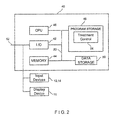
- FIGS. 1 through 35 The components that can be used with the present invention are illustrated in FIG. 1 .
- One or more probes 22 can deliver therapeutic energy and are powered by a voltage pulse generator 10 that generates high voltage pulses as therapeutic energy such as pulses capable of irreversibly electroporating the tissue cells.
- the voltage pulse generator 10 can include six separate receptacles for receiving up to six individual probes 22 which are adapted to be plugged into the respective receptacle.
- the receptacles are each labeled with a number in consecutive order.
- the voltage pulse generator 10 can have any number of receptacles for receiving more or less than six probes.
- the determined voltage was placed into the Cassini oval electronic worksheet for the same electrode geometry and the “gain denominator” was adjusted until the shape from the cassini oval matched that from the numerical solution.
- the system charges to the full therapeutic treatment voltage (as shown in window 430 ) and waits for instructions from the user to begin treatment.
- a user is required to press both foot pedals of a double foot pedal device (not shown) in order to activate treatment (the first pedal is used to arm the generator 10 , the second pedal is used to fire or start the treatment).
- This provides a type of safety check and prevents accidental activation of the treatment.
- the screen shown in FIG. 27 uses two buttons 422 , 423 instead of a double foot pedal device. Accordingly, the user will click on the “Arm” button 422 with the pointing device 14 to arm the probes. Then, the user will click on the “Pulse” button 423 with the pointing device 14 to initiate the treatment.
Abstract
Description
Voltage=xd, (1)
-
- where x=the electric field density setting (Volts/cm) shown in
column 225, which is based on the value frombox 211, and - where d=the distance (cm) between the given pair of electrodes shown in
column 226.
Therefore, when “Linear” is selected, the Voltage that is applied between a given pair of electrodes is directly proportional to the Distance between the given electrode pair in a linear relationship.
- where x=the electric field density setting (Volts/cm) shown in
dist(q 1 ,p)×dist(q 2 ,p)=b 2 (2)
((x−a)2 +y 2)((x+a)2 +y 2)=b 4 (3)
The equivalent polar equation is:
r 4−2a 2 r 2 cos 2θ=b 4 −a 4 (4)
r 2 =a 2 cos(2*theta)+/−sqrt(b 4 −a 4 sin2(2*theta)) (5)
| TABLE 1 | ||||
| Theta | r = | r = | r = | r = |
| (degrees) | sqrt(M + L) | −sqrt(M + L) | sqrt(M − L) | −sqrt(M − L) |
| 0 | 1.366154 | −1.36615 | 0 | 0 |
| 1 | 1.366006 | −1.36601 | 0 | 0 |
| 2 | 1.365562 | −1.36556 | 0 | 0 |
| 3 | 1.364822 | −1.36482 | 0 | 0 |
| 4 | 1.363788 | −1.36379 | 0 | 0 |
| 5 | 1.362461 | −1.36246 | 0 | 0 |
| 6 | 1.360843 | −1.36084 | 0 | 0 |
| 7 | 1.358936 | −1.35894 | 0 | 0 |
| 8 | 1.356743 | −1.35674 | 0 | 0 |
| 9 | 1.354267 | −1.35427 | 0 | 0 |
| 10 | 1.351512 | −1.35151 | 0 | 0 |
| 11 | 1.348481 | −1.34848 | 0 | 0 |
| 12 | 1.34518 | −1.34518 | 0 | 0 |
| 13 | 1.341611 | −1.34161 | 0 | 0 |
| 14 | 1.337782 | −1.33778 | 0 | 0 |
| 15 | 1.333697 | −1.3337 | 0 | 0 |
Gain Denominator=595.28·ln(a)+2339; R 2=0.993 (7)
a=7*10−9 *E 3−2*10−5 *E 2+0.015*E+6.1619; R 2=0.9806 (8)
GD=1.0121*E+1920; R 2=0.9928 (9)
| TABLE 3 | |||||
| 1 | 5 | 7.5 | 10 | ||
| 11 | 15 | 17.5 | 20 | ||
| 22.5 | |||||
| 21 | 25 | 27.5 | 30 | ||
x i=εj *a*cos(θi+φ) (14)
y i=εj *b*sin(θi+φ), (15)
where,
-
- a=the major axis of the elliptical shape (cm) that is selected at
FIG. 3 ; - b=the minor axis of the elliptical shape (cm) that is selected at
FIG. 3 ; and; - φ=the rotational angle (degrees) of the ellipse as shown on treatment screen (see
input box 251 atFIG. 11 )
- a=the major axis of the elliptical shape (cm) that is selected at
| TABLE 2.1 | |
| Angular Offsets (θi) for each probe, | |
| Total number | referenced from zero degrees (positive |
| of probes in the device | x-axis) of the |
| 2 |
0° and 180° |
| 3 |
90°, 210°, and 330° |
| 4 probes | 45°, 135°, 225°, and 315° |
| 5 |
90°, 162°, 234°, 306°, and 18° |
| 6 |
90°, 162°, 234°, 306°, and 18° PLUS the |
| 6th probe at the center of the grid (0, 0) | |
| TABLE 2.2 | |||
| Total number of probes in the device | Probe placement ratios (εj) | ||
| 2 probes | ε2 = 0.70 | ||
| 3 probes | ε3 = 0.70 | ||
| 4 probes | ε4 = 0.65 | ||
| 5 probes | ε5 = 0.65 | ||
| 6 probes | ε6 = 0.65 | ||
The above algorithm is based on the following assumptions:
-
- Treatment zone center is at (0,0) or will be translated to (0,0) for calculations.
- Treatment zone area may or may not be adequately covered depending on size and number of probes to be deployed.
- A fixed angular array of probe placements is used, with the exception of 6 probes in which the last probe is placed in the center of the target tissue at (0,0). (see Table 2.1)
- A predetermined firing sequence is used according to the total number of probes. (see Table 2.3 below)
- An array of εj for j=2, 3, . . . 6 is used to determine the ratio of the probe placement radius from the edges of the target tissue. (see Table 2.2) The εj numbers are determined empirically for best-fit. Alternatively, these values can be represented as functions rather than fixed numerical values for each number of probes.
- A default electric field density between probes is 1500 volts/cm which can be changed by the user. The actual voltage value between probes is adjusted based on the default electric field density. For example, if the default is set at 1500 volts/cm, the actual treatment voltage for a pair of probes that are 1.5 cm apart is 2250V.
| TABLE 2.3 | |
| Firing Sequence of Probe Treatment | |
| Total | Pairs, identified by polarity and |
| number of probes in the device | specific probe number |
| 2 probes | (1 treatment pair) |
| (+) 1, (−) 2 | |
| 3 probes | (3 treatment pairs) |
| (+) 1, (−) 2 | |
| (+) 2, (−) 3 | |
| (+) 3, (−) 1 | |
| 4 probes | (5 treatment pairs) |
| (+) 1, (−) 2 | |
| (+) 2, (−) 3 | |
| (+) 3, (−) 4 | |
| (+) 4, (−) 1 | |
| (+) 2, (−) 4 | |
| 5 probes | (8 treatment pairs) |
| (+) 1, (−) 2 | |
| (+) 2, (−) 3 | |
| (+) 3, (−) 4 | |
| (+) 4, (−) 5 | |
| (+) 5, (−) 1 | |
| (+) 2, (−) 5 | |
| (+) 1, (−) 3 | |
| (+) 4, (−) 1 | |
| 6 probes | (10 treatment pairs) |
| (+) 1, (−) 2 | |
| (+) 2, (−) 3 | |
| (+) 3, (−) 4 | |
| (+) 4, (−) 5 | |
| (+) 5, (−) 1 | |
| (+) 1, (−) 6 | |
| (+) 6, (−) 2 | |
| (+) 3, (−) 6 | |
| (+) 6, (−) 4 | |
| (+) 5, (−) 6 | |
x 1=εj *a*cos(θi+φ)=0.70*2.0 cm*cos(90 degrees)=0
y 1=εj *b*sin(θi+φ)=0.70*1.0 cm*sin(90 degrees)=0.70 cm
x 2=εj *a*cos(θi+φ)=0.70*2.0 cm*cos(210 degrees)=−1.21 cm
y 2=εj *b*sin(θi+φ)=0.70*1.0 cm*sin(210 degrees)=−0.35 cm
x 3=εj *a*cos(θi+φ)=0.70*2.0 cm*cos(330 degrees)=1.21 cm
y 3=εj *b*sin(θi+φ)=0.70*1.0 cm*sin(330 degrees)=−0.35 cm
-
- (+)
Probe # 1, (−)Probe # 2 - (+)
Probe # 2, (−)Probe # 3 - (+)
Probe # 3, (−)Probe # 1
- (+)
Claims (6)
Priority Applications (3)
| Application Number | Priority Date | Filing Date | Title |
|---|---|---|---|
| US14/302,678 US9895189B2 (en) | 2009-06-19 | 2014-06-12 | Methods of sterilization and treating infection using irreversible electroporation |
| US15/864,421 US20180116710A1 (en) | 2013-06-13 | 2018-01-08 | Methods of Sterilization and Treating Infection Using Irreversible Electroporation |
| US17/073,039 US11957405B2 (en) | 2013-06-13 | 2020-10-16 | Methods of sterilization and treating infection using irreversible electroporation |
Applications Claiming Priority (3)
| Application Number | Priority Date | Filing Date | Title |
|---|---|---|---|
| US12/488,070 US9173704B2 (en) | 2008-06-20 | 2009-06-19 | Device and method for the ablation of fibrin sheath formation on a venous catheter |
| US201361834471P | 2013-06-13 | 2013-06-13 | |
| US14/302,678 US9895189B2 (en) | 2009-06-19 | 2014-06-12 | Methods of sterilization and treating infection using irreversible electroporation |
Related Child Applications (1)
| Application Number | Title | Priority Date | Filing Date |
|---|---|---|---|
| US15/864,421 Continuation US20180116710A1 (en) | 2013-06-13 | 2018-01-08 | Methods of Sterilization and Treating Infection Using Irreversible Electroporation |
Publications (2)
| Publication Number | Publication Date |
|---|---|
| US20140378964A1 US20140378964A1 (en) | 2014-12-25 |
| US9895189B2 true US9895189B2 (en) | 2018-02-20 |
Family
ID=52111502
Family Applications (3)
| Application Number | Title | Priority Date | Filing Date |
|---|---|---|---|
| US14/302,678 Active 2035-09-13 US9895189B2 (en) | 2009-06-19 | 2014-06-12 | Methods of sterilization and treating infection using irreversible electroporation |
| US15/864,421 Abandoned US20180116710A1 (en) | 2013-06-13 | 2018-01-08 | Methods of Sterilization and Treating Infection Using Irreversible Electroporation |
| US17/073,039 Active 2035-03-15 US11957405B2 (en) | 2013-06-13 | 2020-10-16 | Methods of sterilization and treating infection using irreversible electroporation |
Family Applications After (2)
| Application Number | Title | Priority Date | Filing Date |
|---|---|---|---|
| US15/864,421 Abandoned US20180116710A1 (en) | 2013-06-13 | 2018-01-08 | Methods of Sterilization and Treating Infection Using Irreversible Electroporation |
| US17/073,039 Active 2035-03-15 US11957405B2 (en) | 2013-06-13 | 2020-10-16 | Methods of sterilization and treating infection using irreversible electroporation |
Country Status (1)
| Country | Link |
|---|---|
| US (3) | US9895189B2 (en) |
Cited By (1)
| Publication number | Priority date | Publication date | Assignee | Title |
|---|---|---|---|---|
| US10702337B2 (en) | 2016-06-27 | 2020-07-07 | Galary, Inc. | Methods, apparatuses, and systems for the treatment of pulmonary disorders |
Families Citing this family (40)
| Publication number | Priority date | Publication date | Assignee | Title |
|---|---|---|---|---|
| US9283051B2 (en) | 2008-04-29 | 2016-03-15 | Virginia Tech Intellectual Properties, Inc. | System and method for estimating a treatment volume for administering electrical-energy based therapies |
| US8992517B2 (en) | 2008-04-29 | 2015-03-31 | Virginia Tech Intellectual Properties Inc. | Irreversible electroporation to treat aberrant cell masses |
| US10702326B2 (en) | 2011-07-15 | 2020-07-07 | Virginia Tech Intellectual Properties, Inc. | Device and method for electroporation based treatment of stenosis of a tubular body part |
| US10272178B2 (en) | 2008-04-29 | 2019-04-30 | Virginia Tech Intellectual Properties Inc. | Methods for blood-brain barrier disruption using electrical energy |
| US9198733B2 (en) | 2008-04-29 | 2015-12-01 | Virginia Tech Intellectual Properties, Inc. | Treatment planning for electroporation-based therapies |
| US10117707B2 (en) | 2008-04-29 | 2018-11-06 | Virginia Tech Intellectual Properties, Inc. | System and method for estimating tissue heating of a target ablation zone for electrical-energy based therapies |
| US10238447B2 (en) | 2008-04-29 | 2019-03-26 | Virginia Tech Intellectual Properties, Inc. | System and method for ablating a tissue site by electroporation with real-time monitoring of treatment progress |
| WO2009134876A1 (en) | 2008-04-29 | 2009-11-05 | Virginia Tech Intellectual Properties, Inc. | Irreversible electroporation to create tissue scaffolds |
| US11254926B2 (en) | 2008-04-29 | 2022-02-22 | Virginia Tech Intellectual Properties, Inc. | Devices and methods for high frequency electroporation |
| US10245098B2 (en) | 2008-04-29 | 2019-04-02 | Virginia Tech Intellectual Properties, Inc. | Acute blood-brain barrier disruption using electrical energy based therapy |
| US9867652B2 (en) | 2008-04-29 | 2018-01-16 | Virginia Tech Intellectual Properties, Inc. | Irreversible electroporation using tissue vasculature to treat aberrant cell masses or create tissue scaffolds |
| US11272979B2 (en) | 2008-04-29 | 2022-03-15 | Virginia Tech Intellectual Properties, Inc. | System and method for estimating tissue heating of a target ablation zone for electrical-energy based therapies |
| US11382681B2 (en) | 2009-04-09 | 2022-07-12 | Virginia Tech Intellectual Properties, Inc. | Device and methods for delivery of high frequency electrical pulses for non-thermal ablation |
| US11638603B2 (en) | 2009-04-09 | 2023-05-02 | Virginia Tech Intellectual Properties, Inc. | Selective modulation of intracellular effects of cells using pulsed electric fields |
| EP2429444B1 (en) | 2009-05-11 | 2024-02-28 | TriAgenics, Inc. | Therapeutic tooth bud ablation |
| US10022202B2 (en) | 2013-03-15 | 2018-07-17 | Triagenics, Llc | Therapeutic tooth bud ablation |
| WO2014143014A1 (en) | 2013-03-15 | 2014-09-18 | Triagenics, Llc | Therapeutic tooth bud ablation |
| US8903488B2 (en) | 2009-05-28 | 2014-12-02 | Angiodynamics, Inc. | System and method for synchronizing energy delivery to the cardiac rhythm |
| US9895189B2 (en) | 2009-06-19 | 2018-02-20 | Angiodynamics, Inc. | Methods of sterilization and treating infection using irreversible electroporation |
| WO2012051433A2 (en) | 2010-10-13 | 2012-04-19 | Angiodynamics, Inc. | System and method for electrically ablating tissue of a patient |
| WO2012088149A2 (en) | 2010-12-20 | 2012-06-28 | Virginia Tech Intellectual Properties, Inc. | High-frequency electroporation for cancer therapy |
| US9078665B2 (en) | 2011-09-28 | 2015-07-14 | Angiodynamics, Inc. | Multiple treatment zone ablation probe |
| EP2889003B1 (en) * | 2012-08-22 | 2023-11-08 | Samsung Medison Co., Ltd. | Ultrasound diagnosis device and method of operation |
| US9438264B1 (en) | 2015-09-10 | 2016-09-06 | Realtek Semiconductor Corp. | High-speed capacitive digital-to-analog converter and method thereof |
| JP6751351B2 (en) * | 2014-05-02 | 2020-09-02 | コーニンクレッカ フィリップス エヌ ヴェKoninklijke Philips N.V. | Device to inactivate bacteria |
| CN112807074A (en) | 2014-05-12 | 2021-05-18 | 弗吉尼亚暨州立大学知识产权公司 | Electroporation system |
| US10643371B2 (en) * | 2014-08-11 | 2020-05-05 | Covidien Lp | Treatment procedure planning system and method |
| US10694972B2 (en) | 2014-12-15 | 2020-06-30 | Virginia Tech Intellectual Properties, Inc. | Devices, systems, and methods for real-time monitoring of electrophysical effects during tissue treatment |
| US10905492B2 (en) | 2016-11-17 | 2021-02-02 | Angiodynamics, Inc. | Techniques for irreversible electroporation using a single-pole tine-style internal device communicating with an external surface electrode |
| US11607537B2 (en) | 2017-12-05 | 2023-03-21 | Virginia Tech Intellectual Properties, Inc. | Method for treating neurological disorders, including tumors, with electroporation |
| US11925405B2 (en) | 2018-03-13 | 2024-03-12 | Virginia Tech Intellectual Properties, Inc. | Treatment planning system for immunotherapy enhancement via non-thermal ablation |
| US11311329B2 (en) | 2018-03-13 | 2022-04-26 | Virginia Tech Intellectual Properties, Inc. | Treatment planning for immunotherapy based treatments using non-thermal ablation techniques |
| US20190290903A1 (en) * | 2018-03-23 | 2019-09-26 | The Board Of Regents Of The University Of Oklahoma | System and Method of Electric-Induced Acoustic Tomography for Electrotherapy Monitoring |
| CN109157280A (en) * | 2018-08-10 | 2019-01-08 | 重庆大学 | Irreversible electroporated tissue ablation effect dynamic realtime assessment equipment |
| US11756681B2 (en) | 2019-05-07 | 2023-09-12 | Medtronic, Inc. | Evaluation of post implantation patient status and medical device performance |
| EP3979938A4 (en) | 2019-06-06 | 2023-06-28 | TriAgenics, Inc. | Ablation probe systems |
| US11950835B2 (en) | 2019-06-28 | 2024-04-09 | Virginia Tech Intellectual Properties, Inc. | Cycled pulsing to mitigate thermal damage for multi-electrode irreversible electroporation therapy |
| US20210344880A1 (en) * | 2020-04-30 | 2021-11-04 | Medtronic, Inc. | Post operative implantation site monitoring |
| US11937926B2 (en) * | 2020-07-02 | 2024-03-26 | Garwood Medical Devices, Llc | Method for optimizing treatment of infected metallic implants by measuring charge transfer |
| AU2022339915A1 (en) * | 2021-08-30 | 2024-04-04 | Nanovis, LLC | Devices and methods for treating infected tissue |
Citations (816)
| Publication number | Priority date | Publication date | Assignee | Title |
|---|---|---|---|---|
| US1653819A (en) | 1926-08-07 | 1927-12-27 | Northcott Ephraim | Electrotherapeutical apparatus |
| US3730238A (en) | 1971-09-21 | 1973-05-01 | R Butler | Friction type screwdriver |
| US3746004A (en) | 1971-06-14 | 1973-07-17 | B Jankelson | Disposable electrodes for electrical stimulation of muscles and nerves of the head |
| US3871359A (en) | 1973-06-25 | 1975-03-18 | Interscience Technology Corp | Impedance measuring system |
| US4016886A (en) | 1974-11-26 | 1977-04-12 | The United States Of America As Represented By The United States Energy Research And Development Administration | Method for localizing heating in tumor tissue |
| US4037341A (en) | 1973-08-13 | 1977-07-26 | Johns-Manville Corporation | Luminaire for lighting a sign and method |
| US4216860A (en) | 1978-12-11 | 1980-08-12 | Electro-Catheter Corporation | Medical device container and method of manufacture |
| US4226246A (en) | 1977-05-27 | 1980-10-07 | Carba Societe Anonyme | Apparatus for maintaining the negative potential of human, animal, and plant cells |
| US4262672A (en) | 1978-01-02 | 1981-04-21 | Horst Kief | Acupuncture instrument |
| US4267047A (en) | 1977-02-11 | 1981-05-12 | Akzo N.V. Of Arnhem/Nederland | Dialyzing membrane with adsorbent layer |
| US4278092A (en) | 1979-07-05 | 1981-07-14 | American Hospital Supply Corporation | Peritoneal catheter |
| US4299217A (en) | 1977-06-03 | 1981-11-10 | Terumo Corporation | Intravascular catheter |
| US4311148A (en) | 1980-05-19 | 1982-01-19 | Mitchell V. Kaminski, Jr. | Micro-jejunostomy feeding tube |
| US4336881A (en) | 1979-06-14 | 1982-06-29 | Diachem, Inc. | Aqueous acid concentrate for hemodialysis dialysate |
| US4344436A (en) | 1980-01-16 | 1982-08-17 | Yukio Kubota | Device for determining location of the tip of catheter |
| US4392855A (en) | 1980-05-08 | 1983-07-12 | Oreopoulos Dimitrios G | Catheter |
| US4406827A (en) | 1979-09-04 | 1983-09-27 | Minnesota Mining And Manufacturing Company | Cohesive nonsticky electrically conductive gel composition |
| US4407943A (en) | 1976-12-16 | 1983-10-04 | Millipore Corporation | Immobilized antibody or antigen for immunoassay |
| US4416276A (en) | 1981-10-26 | 1983-11-22 | Valleylab, Inc. | Adaptive, return electrode monitoring system |
| US4447235A (en) | 1981-05-07 | 1984-05-08 | John M. Clarke | Thoracentesis device |
| US4469098A (en) | 1978-12-18 | 1984-09-04 | Davi Samantha K | Apparatus for and method of utilizing energy to excise pathological tissue |
| US4489535A (en) | 1980-10-02 | 1984-12-25 | Veltman Preston Leonard | Materials and method for preparing dialysis solutions containing bicarbonate ions |
| US4512765A (en) | 1983-06-09 | 1985-04-23 | Rudolph Muto | Selective tracheal bronchial catheter |
| US4580572A (en) | 1983-06-01 | 1986-04-08 | Bio-Stimu Trend Corp. | Garment apparatus for delivering or receiving electric impulses |
| US4636199A (en) | 1984-07-09 | 1987-01-13 | Victor Lyle D | Device for inserting a catheter within the intercostal space |
| EP0218275A1 (en) | 1985-08-30 | 1987-04-15 | Fijneman, Martinus Jacobus Antonius Johannes | Multi-purpose catheter |
| US4672969A (en) | 1983-10-06 | 1987-06-16 | Sonomo Corporation | Laser healing method |
| US4676258A (en) | 1983-01-24 | 1987-06-30 | Kureha Kagaku Kogyo Kabushiki Kaisha | Device for hyperthermia |
| US4676782A (en) | 1984-09-21 | 1987-06-30 | Vitaphore Corporation | Positionable tissue interfacing device for the management of percutaneous conduits |
| US4687471A (en) | 1985-05-01 | 1987-08-18 | Curators Of The University Of Missouri | Peritoneal dialysis catheter |
| US4716896A (en) | 1986-08-01 | 1988-01-05 | Ackrad Laboratories | Bronchial catheter |
| US4723549A (en) | 1986-09-18 | 1988-02-09 | Wholey Mark H | Method and apparatus for dilating blood vessels |
| USD294519S (en) | 1985-07-29 | 1988-03-01 | Peter LaHaye | Instrument for tattooing |
| US4756838A (en) | 1980-02-21 | 1988-07-12 | Veltman Preston Leonard | Preparation of dry dialysate products |
| US4772269A (en) | 1985-05-01 | 1988-09-20 | Curators Of The University Of Missouri | Peritoneal dialysis catheter |
| US4798585A (en) | 1986-06-06 | 1989-01-17 | Asahi Kogaku Kogyo Kabushiki Kaisha | Support for biomedical implant device |
| US4810963A (en) | 1984-04-03 | 1989-03-07 | Public Health Laboratory Service Board | Method for investigating the condition of a bacterial suspension through frequency profile of electrical admittance |
| US4813929A (en) | 1987-02-19 | 1989-03-21 | Neal Semrad | Chest tube device and method of inserting device |
| US4819637A (en) | 1987-09-01 | 1989-04-11 | Interventional Therapeutics Corporation | System for artificial vessel embolization and devices for use therewith |
| US4822470A (en) | 1987-10-09 | 1989-04-18 | Baylor College Of Medicine | Method of and apparatus for cell poration and cell fusion using radiofrequency electrical pulses |
| US4836204A (en) | 1987-07-06 | 1989-06-06 | Landymore Roderick W | Method for effecting closure of a perforation in the septum of the heart |
| US4840172A (en) | 1986-09-04 | 1989-06-20 | Augustine Scott D | Device for positioning an endotracheal tube |
| US4863426A (en) | 1987-08-18 | 1989-09-05 | Ferragamo Michael C | Percutaneous venous catheter |
| EP0339501A2 (en) | 1988-04-26 | 1989-11-02 | Arturo Dr. Muti | Bronchial examination catheter |
| US4885003A (en) | 1988-07-25 | 1989-12-05 | Cordis Corporation | Double mesh balloon catheter device |
| US4886502A (en) | 1986-12-09 | 1989-12-12 | Thermedics, Inc. | Peritoneal access catheter |
| US4886496A (en) | 1988-02-04 | 1989-12-12 | Conoscenti Craig S | Bronchoscopic balloon tipped catheter and method of making the same |
| US4889634A (en) | 1988-10-04 | 1989-12-26 | Gynex, Inc. | Dialysate solution containing hydroxypropyl-beta-cyclodextrin and method of using same |
| US4907601A (en) | 1988-06-15 | 1990-03-13 | Etama Ag | Electrotherapy arrangement |
| US4919148A (en) | 1988-06-13 | 1990-04-24 | Muccio Philip E | Apparatus and method for transcutaneous electrical stimulation |
| US4920978A (en) | 1988-08-31 | 1990-05-01 | Triangle Research And Development Corporation | Method and apparatus for the endoscopic treatment of deep tumors using RF hyperthermia |
| US4921484A (en) | 1988-07-25 | 1990-05-01 | Cordis Corporation | Mesh balloon catheter device |
| EP0378132A2 (en) | 1989-01-09 | 1990-07-18 | S.L. Cit Ionofor | A device for the administration of medication by iontopheresis for local - regional treatment. |
| US4946793A (en) | 1986-05-09 | 1990-08-07 | Electropore, Inc. | Impedance matching for instrumentation which electrically alters vesicle membranes |
| US4976709A (en) | 1988-12-15 | 1990-12-11 | Sand Bruce J | Method for collagen treatment |
| US4981477A (en) | 1988-04-16 | 1991-01-01 | Rudolf Schon | Catheter for introduction into the trachea and the bronchial system |
| US4986810A (en) | 1989-09-01 | 1991-01-22 | Neal Semrad | Toggle catheter |
| US4987895A (en) | 1986-10-06 | 1991-01-29 | Heimlich Henry J | Tracheal tube |
| WO1991004014A1 (en) | 1989-09-21 | 1991-04-04 | Synergen, Inc. | Method for transporting compositions across the blood brain barrier |
| US5019034A (en) | 1988-01-21 | 1991-05-28 | Massachusetts Institute Of Technology | Control of transport of molecules across tissue using electroporation |
| US5031775A (en) | 1990-02-14 | 1991-07-16 | Angeion Corporation | Medical instrument holder |
| DE4000893A1 (en) | 1990-01-15 | 1991-07-18 | Bosch Gmbh Robert | Multichannel appts. for electro-simulation - provides several current circuits for patient with electrodes applying pulse signals |
| US5053013A (en) | 1990-03-01 | 1991-10-01 | The Regents Of The University Of Michigan | Implantable infusion device |
| US5052391A (en) | 1990-10-22 | 1991-10-01 | R.F.P., Inc. | High frequency high intensity transcutaneous electrical nerve stimulator and method of treatment |
| US5058605A (en) | 1989-02-22 | 1991-10-22 | Ceske Vysoke Uceni Technicke | Method and device for the controlled local, non-invasive application of dc pulses to human and animal tissues |
| US5071558A (en) | 1989-08-11 | 1991-12-10 | Nikkiso Co., Ltd. | Sodium bicarbonate dialysate |
| US5098843A (en) | 1987-06-04 | 1992-03-24 | Calvin Noel M | Apparatus for the high efficiency transformation of living cells |
| US5122137A (en) | 1990-04-27 | 1992-06-16 | Boston Scientific Corporation | Temperature controlled rf coagulation |
| US5134070A (en) | 1990-06-04 | 1992-07-28 | Casnig Dael R | Method and device for cell cultivation on electrodes |
| US5137517A (en) | 1989-11-28 | 1992-08-11 | Scimed Life Systems, Inc. | Device and method for gripping medical shaft |
| US5141499A (en) | 1991-10-09 | 1992-08-25 | Zappacosta Anthony R | Peritoneal dialysis catheter |
| USD329496S (en) | 1990-02-20 | 1992-09-15 | Celia Clarke | Needle depth gauge |
| US5156597A (en) | 1989-12-30 | 1992-10-20 | B. Braun Melsungen Ag | Transcutaneous implantation catheter |
| US5173158A (en) | 1991-07-22 | 1992-12-22 | Schmukler Robert E | Apparatus and methods for electroporation and electrofusion |
| US5186715A (en) | 1990-12-06 | 1993-02-16 | E-Z-Em, Inc. | Biliary drainage method |
| US5186800A (en) | 1988-04-18 | 1993-02-16 | Bio-Rad Laboratories, Inc. | Electroporation of prokaryotic cells |
| US5188592A (en) | 1991-06-24 | 1993-02-23 | Hakki Sam I | Dynamic pressurized catheter with simultaneous oxygen delivery and suction |
| US5190541A (en) | 1990-10-17 | 1993-03-02 | Boston Scientific Corporation | Surgical instrument and method |
| EP0528891A1 (en) | 1990-04-23 | 1993-03-03 | Alkermes, Inc. | Method for increasing blood-brain barrier permeability |
| US5192312A (en) | 1991-03-05 | 1993-03-09 | Colorado State University Research Foundation | Treated tissue for implantation and methods of treatment and use |
| US5193537A (en) | 1990-06-12 | 1993-03-16 | Zmd Corporation | Method and apparatus for transcutaneous electrical cardiac pacing |
| EP0533511A1 (en) | 1991-07-22 | 1993-03-24 | Thomas Schmitz-Rode | Device for maintaining the patency of a bodily duct, and especially of a blood vessel, and uses thereof |
| US5209723A (en) | 1990-01-08 | 1993-05-11 | The Curators Of The University Of Missouri | Multiple lumen catheter for hemodialysis |
| US5215530A (en) | 1991-07-11 | 1993-06-01 | City Of Hope | Sleeved extension and anchoring system for percutaneous catheters |
| US5222997A (en) | 1992-07-24 | 1993-06-29 | Montgomery David S | Parallel holder device |
| US5224933A (en) | 1992-03-23 | 1993-07-06 | C. R. Bard, Inc. | Catheter purge device |
| US5227730A (en) | 1992-09-14 | 1993-07-13 | Kdc Technology Corp. | Microwave needle dielectric sensors |
| US5242415A (en) | 1992-08-14 | 1993-09-07 | L-Vad Technology, Inc. | Percutaneous access device |
| US5273525A (en) | 1992-08-13 | 1993-12-28 | Btx Inc. | Injection and electroporation apparatus for drug and gene delivery |
| US5277201A (en) | 1992-05-01 | 1994-01-11 | Vesta Medical, Inc. | Endometrial ablation apparatus and method |
| US5279564A (en) | 1992-09-11 | 1994-01-18 | Edward Weck Incorporated | Cannula retention device |
| USD343687S (en) | 1992-01-06 | 1994-01-25 | Becton, Dickinson And Company | Biopsy procedure tray |
| US5281213A (en) | 1992-04-16 | 1994-01-25 | Implemed, Inc. | Catheter for ice mapping and ablation |
| US5290263A (en) | 1989-02-02 | 1994-03-01 | Regents Of The University Of Minnesota | Bidirectional check valve catheter |
| US5308338A (en) | 1993-04-22 | 1994-05-03 | Helfrich G Baird | Catheter or the like with medication injector to prevent infection |
| US5308325A (en) | 1991-01-28 | 1994-05-03 | Corpak, Inc. | Retention balloon for percutaneous catheter |
| US5318543A (en) | 1992-10-08 | 1994-06-07 | Abbott Laboratories | Laparoscopic jejunostomy instrumentation kit |
| US5318563A (en) | 1992-06-04 | 1994-06-07 | Valley Forge Scientific Corporation | Bipolar RF generator |
| US5328451A (en) | 1991-08-15 | 1994-07-12 | Board Of Regents, The University Of Texas System | Iontophoretic device and method for killing bacteria and other microbes |
| US5334167A (en) | 1993-11-19 | 1994-08-02 | Cocanower David A | Modified nasogastric tube for use in enteral feeding |
| USD351661S (en) | 1993-02-16 | 1994-10-18 | Ultradent Products, Inc. | Combined organizer and tray for an endodontic dental kit |
| US5383917A (en) | 1991-07-05 | 1995-01-24 | Jawahar M. Desai | Device and method for multi-phase radio-frequency ablation |
| US5389069A (en) | 1988-01-21 | 1995-02-14 | Massachusetts Institute Of Technology | Method and apparatus for in vivo electroporation of remote cells and tissue |
| US5391158A (en) | 1994-02-24 | 1995-02-21 | Peters; Michael J. | Nasogastric tube |
| US5403311A (en) | 1993-03-29 | 1995-04-04 | Boston Scientific Corporation | Electro-coagulation and ablation and other electrotherapeutic treatments of body tissue |
| US5405320A (en) | 1990-01-08 | 1995-04-11 | The Curators Of The University Of Missouri | Multiple lumen catheter for hemodialysis |
| US5417687A (en) | 1993-04-30 | 1995-05-23 | Medical Scientific, Inc. | Bipolar electrosurgical trocar |
| US5425752A (en) | 1991-11-25 | 1995-06-20 | Vu'nguyen; Dung D. | Method of direct electrical myostimulation using acupuncture needles |
| US5439440A (en) | 1993-04-01 | 1995-08-08 | Genetronics, Inc. | Electroporation system with voltage control feedback for clinical applications |
| US5439444A (en) | 1991-01-28 | 1995-08-08 | Corpak, Inc. | Pre-formed member for percutaneous catheter |
| US5458597A (en) | 1993-11-08 | 1995-10-17 | Zomed International | Device for treating cancer and non-malignant tumors and methods |
| US5458625A (en) | 1994-05-04 | 1995-10-17 | Kendall; Donald E. | Transcutaneous nerve stimulation device and method for using same |
| US5462644A (en) * | 1991-12-31 | 1995-10-31 | Minnesota Mining And Manufacturing Company | Biofilm reduction method |
| US5462521A (en) | 1993-12-21 | 1995-10-31 | Angeion Corporation | Fluid cooled and perfused tip for a catheter |
| US5484401A (en) | 1992-11-04 | 1996-01-16 | Denver Biomaterials, Inc. | Treatment method for pleural effusion |
| US5484400A (en) | 1992-08-12 | 1996-01-16 | Vidamed, Inc. | Dual channel RF delivery system |
| US5533999A (en) | 1993-08-23 | 1996-07-09 | Refractec, Inc. | Method and apparatus for modifications of visual acuity by thermal means |
| US5536240A (en) | 1992-08-12 | 1996-07-16 | Vidamed, Inc. | Medical probe device and method |
| US5536267A (en) | 1993-11-08 | 1996-07-16 | Zomed International | Multiple electrode ablation apparatus |
| US5540737A (en) | 1991-06-26 | 1996-07-30 | Massachusetts Institute Of Technology | Minimally invasive monopole phased array hyperthermia applicators and method for treating breast carcinomas |
| US5546940A (en) | 1994-01-28 | 1996-08-20 | Ep Technologies, Inc. | System and method for matching electrical characteristics and propagation velocities in cardiac tissue to locate potential ablation sites |
| US5562720A (en) | 1992-05-01 | 1996-10-08 | Vesta Medical, Inc. | Bipolar/monopolar endometrial ablation device and method |
| WO1996034571A1 (en) | 1995-05-04 | 1996-11-07 | Cosman Eric R | Cool-tip electrode thermosurgery system |
| US5575811A (en) | 1993-07-08 | 1996-11-19 | Urologix, Inc. | Benign prostatic hyperplasia treatment catheter with urethral cooling |
| US5582588A (en) | 1993-04-19 | 1996-12-10 | Olympus Optical Co., Ltd. | Ultrasonic therapeutic apparatus |
| WO1996039531A1 (en) | 1995-06-06 | 1996-12-12 | Massachusetts Institute Of Technology | Delivery of nucleotides into organisms by electroporation |
| USD376652S (en) | 1995-07-07 | 1996-12-17 | Hunt Ilyssa A | Medical instrument tray |
| US5586982A (en) | 1992-04-10 | 1996-12-24 | Abela; George S. | Cell transfection apparatus and method |
| US5588960A (en) | 1994-12-01 | 1996-12-31 | Vidamed, Inc. | Transurethral needle delivery device with cystoscope and method for treatment of urinary incontinence |
| US5588424A (en) | 1995-06-28 | 1996-12-31 | The Cleveland Clinic Foundation | Bronchial blocker endotracheal apparatus |
| US5599311A (en) | 1994-07-25 | 1997-02-04 | Raulerson; J. Daniel | Subcutaneous catheter stabilizing devices |
| US5616126A (en) | 1995-03-03 | 1997-04-01 | Malekmehr; Farshad | Low residual bladder catheter |
| US5620479A (en) | 1992-11-13 | 1997-04-15 | The Regents Of The University Of California | Method and apparatus for thermal therapy of tumors |
| US5626146A (en) | 1992-12-18 | 1997-05-06 | British Technology Group Limited | Electrical impedance tomography |
| US5630426A (en) | 1995-03-03 | 1997-05-20 | Neovision Corporation | Apparatus and method for characterization and treatment of tumors |
| US5634899A (en) | 1993-08-20 | 1997-06-03 | Cortrak Medical, Inc. | Simultaneous cardiac pacing and local drug delivery method |
| USD380272S (en) | 1995-07-07 | 1997-06-24 | Becton, Dickinson And Company | Skin preparation tray |
| US5645855A (en) | 1996-03-13 | 1997-07-08 | Ridge Scientific Enterprises, Inc. | Adhesive compositions including polyvinylpyrrolidone acrylic acid polymers, and polyamines |
| US5672174A (en) | 1995-08-15 | 1997-09-30 | Rita Medical Systems, Inc. | Multiple antenna ablation apparatus and method |
| US5672173A (en) | 1995-08-15 | 1997-09-30 | Rita Medical Systems, Inc. | Multiple antenna ablation apparatus and method |
| US5674267A (en) | 1993-03-30 | 1997-10-07 | Centre National De La Recherche Scientifique | Electric pulse applicator using pairs of needle electrodes for the treatment of biological tissue |
| US5683384A (en) | 1993-11-08 | 1997-11-04 | Zomed | Multiple antenna ablation apparatus |
| US5687723A (en) | 1993-12-03 | 1997-11-18 | Avitall; Boaz | Mapping and ablation catheter system |
| US5690620A (en) | 1996-05-14 | 1997-11-25 | Knott; Michael Mcfarland | Anatomically conforming nasogastric tube with normally-curved tip and method for using same |
| US5697905A (en) | 1995-06-19 | 1997-12-16 | Leo T. d'Ambrosio | Triple-lumen intra-aortic catheter |
| US5700252A (en) | 1995-11-01 | 1997-12-23 | Klingenstein; Ralph James | Lumen-seeking nasogastric tube and method |
| US5702359A (en) | 1995-06-06 | 1997-12-30 | Genetronics, Inc. | Needle electrodes for mediated delivery of drugs and genes |
| US5707332A (en) | 1994-01-21 | 1998-01-13 | The Trustees Of Columbia University In The City Of New York | Apparatus and method to reduce restenosis after arterial intervention |
| US5718246A (en) | 1996-01-03 | 1998-02-17 | Preferential, Inc. | Preferential induction of electrically mediated cell death from applied pulses |
| US5720921A (en) | 1995-03-10 | 1998-02-24 | Entremed, Inc. | Flow electroporation chamber and method |
| US5728143A (en) | 1995-08-15 | 1998-03-17 | Rita Medical Systems, Inc. | Multiple antenna ablation apparatus and method |
| WO1998010745A1 (en) | 1996-09-11 | 1998-03-19 | Aksys, Ltd. | Batch quantity dialysate chemical formulations |
| US5735847A (en) | 1995-08-15 | 1998-04-07 | Zomed International, Inc. | Multiple antenna ablation apparatus and method with cooling element |
| WO1998014238A1 (en) | 1996-07-18 | 1998-04-09 | Bertil Persson | A method and an apparatus for treating tumoral diseases (cancer) |
| US5752939A (en) | 1992-12-24 | 1998-05-19 | Kabushiki Kaisha Hayashidera Medinooru | Catheter for continuous ambulatory peritoneal dialysis |
| US5778894A (en) | 1996-04-18 | 1998-07-14 | Elizabeth Arden Co. | Method for reducing human body cellulite by treatment with pulsed electromagnetic energy |
| US5782827A (en) | 1995-08-15 | 1998-07-21 | Rita Medical Systems, Inc. | Multiple antenna ablation apparatus and method with multiple sensor feedback |
| US5782882A (en) | 1995-11-30 | 1998-07-21 | Hewlett-Packard Company | System and method for administering transcutaneous cardiac pacing with transcutaneous electrical nerve stimulation |
| US5800484A (en) | 1995-08-15 | 1998-09-01 | Rita Medical Systems, Inc. | Multiple antenna ablation apparatus with expanded electrodes |
| US5807395A (en) | 1993-08-27 | 1998-09-15 | Medtronic, Inc. | Method and apparatus for RF ablation and hyperthermia |
| US5807272A (en) | 1995-10-31 | 1998-09-15 | Worcester Polytechnic Institute | Impedance spectroscopy system for ischemia monitoring and detection |
| US5807306A (en) | 1992-11-09 | 1998-09-15 | Cortrak Medical, Inc. | Polymer matrix drug delivery apparatus |
| US5810762A (en) | 1995-04-10 | 1998-09-22 | Genetronics, Inc. | Electroporation system with voltage control feedback for clinical applications |
| US5810742A (en) | 1994-10-24 | 1998-09-22 | Transcan Research & Development Co., Ltd. | Tissue characterization based on impedance images and on impedance measurements |
| US5810804A (en) | 1995-08-15 | 1998-09-22 | Rita Medical Systems | Multiple antenna ablation apparatus and method with cooling element |
| US5830184A (en) | 1996-03-06 | 1998-11-03 | Medical Components, Inc. | Composite catheter stabilizing devices, methods of making the same and catheter extracting device |
| US5836905A (en) | 1994-06-20 | 1998-11-17 | Lemelson; Jerome H. | Apparatus and methods for gene therapy |
| US5836897A (en) | 1990-02-02 | 1998-11-17 | Olympus Optical Co., Ltd. | Ultrasonic treatment apparatus |
| US5843026A (en) | 1992-08-12 | 1998-12-01 | Vidamed, Inc. | BPH ablation method and apparatus |
| US5843182A (en) | 1994-03-14 | 1998-12-01 | Cryolife, Inc. | Treated tissue for implantation and methods of preparation |
| WO1999001076A1 (en) | 1997-07-02 | 1999-01-14 | Broncus Technologies, Inc. | Bleb reducer |
| US5863290A (en) | 1995-08-15 | 1999-01-26 | Rita Medical Systems | Multiple antenna ablation apparatus and method |
| US5866756A (en) | 1995-10-02 | 1999-02-02 | Duke University | Dopamine transporter knockout mice |
| WO1999004710A1 (en) | 1997-07-25 | 1999-02-04 | Cosman Eric R | Cluster ablation electrode system |
| US5868708A (en) | 1997-05-07 | 1999-02-09 | Applied Medical Resources Corporation | Balloon catheter apparatus and method |
| US5873849A (en) | 1997-04-24 | 1999-02-23 | Ichor Medical Systems, Inc. | Electrodes and electrode arrays for generating electroporation inducing electrical fields |
| US5904648A (en) | 1996-06-18 | 1999-05-18 | Cook Incorporated | Guided endobronchial blocker catheter |
| US5913855A (en) | 1995-08-15 | 1999-06-22 | Rita Medical Systems, Inc. | Multiple antenna ablation apparatus and method |
| US5919142A (en) | 1995-06-22 | 1999-07-06 | Btg International Limited | Electrical impedance tomography method and apparatus |
| US5919191A (en) | 1995-01-30 | 1999-07-06 | Boston Scientific Corporation | Electro-surgical tissue removal |
| US5921982A (en) | 1993-07-30 | 1999-07-13 | Lesh; Michael D. | Systems and methods for ablating body tissue |
| US5944710A (en) | 1996-06-24 | 1999-08-31 | Genetronics, Inc. | Electroporation-mediated intravascular delivery |
| US5947284A (en) | 1998-02-13 | 1999-09-07 | United States Surgical Corporation | Package with guide for flexible medical instruments |
| US5947889A (en) | 1995-01-17 | 1999-09-07 | Hehrlein; Christoph | Balloon catheter used to prevent re-stenosis after angioplasty and process for producing a balloon catheter |
| US5954745A (en) | 1997-05-16 | 1999-09-21 | Gertler; Jonathan | Catheter-filter set having a compliant seal |
| US5968006A (en) | 1997-11-04 | 1999-10-19 | Genetronics, Inc. | Method and apparatus for a combination of electroporation and iontophoresis for the delivery of drugs and genes |
| US5983131A (en) | 1995-08-11 | 1999-11-09 | Massachusetts Institute Of Technology | Apparatus and method for electroporation of tissue |
| US5984896A (en) | 1997-10-28 | 1999-11-16 | Ojp #73, Inc. | Fixated catheter |
| US5991697A (en) | 1996-12-31 | 1999-11-23 | The Regents Of The University Of California | Method and apparatus for optical Doppler tomographic imaging of fluid flow velocity in highly scattering media |
| US5999847A (en) | 1997-10-21 | 1999-12-07 | Elstrom; John A. | Apparatus and method for delivery of surgical and therapeutic agents |
| US6004339A (en) | 1996-11-13 | 1999-12-21 | Angiodynamics Incorporated | Balloon catheter with multiple distensibilities |
| US6009347A (en) | 1998-01-27 | 1999-12-28 | Genetronics, Inc. | Electroporation apparatus with connective electrode template |
| US6009877A (en) | 1994-06-24 | 2000-01-04 | Edwards; Stuart D. | Method for treating a sphincter |
| US6010613A (en) | 1995-12-08 | 2000-01-04 | Cyto Pulse Sciences, Inc. | Method of treating materials with pulsed electrical fields |
| US6012885A (en) | 1998-10-09 | 2000-01-11 | Dzn, Incorporated | Cargo chock |
| US6016452A (en) | 1996-03-19 | 2000-01-18 | Kasevich; Raymond S. | Dynamic heating method and radio frequency thermal treatment |
| US6029090A (en) | 1997-01-27 | 2000-02-22 | Herbst; Ewa | Multi-functional electrical stimulation system |
| US6041252A (en) | 1995-06-07 | 2000-03-21 | Ichor Medical Systems Inc. | Drug delivery system and method |
| US6043066A (en) | 1997-09-04 | 2000-03-28 | Mangano; Joseph A. | Cell separation using electric fields |
| WO2000020554A1 (en) | 1998-10-08 | 2000-04-13 | Astrazeneca Ab | Microfabricated cell injector |
| US6050994A (en) | 1998-05-05 | 2000-04-18 | Cardiac Pacemakers, Inc. | RF ablation apparatus and method using controllable duty cycle with alternate phasing |
| US6055453A (en) | 1997-08-01 | 2000-04-25 | Genetronics, Inc. | Apparatus for addressing needle array electrodes for electroporation therapy |
| US6059780A (en) | 1995-08-15 | 2000-05-09 | Rita Medical Systems, Inc. | Multiple antenna ablation apparatus and method with cooling element |
| US6066134A (en) | 1992-01-07 | 2000-05-23 | Arthrocare Corporation | Method for electrosurgical cutting and ablation |
| US6068121A (en) | 1998-03-11 | 2000-05-30 | Schneider (Usa) Inc. | Universal catheter tray |
| US6071281A (en) | 1998-05-05 | 2000-06-06 | Ep Technologies, Inc. | Surgical method and apparatus for positioning a diagnostic or therapeutic element within the body and remote power control unit for use with same |
| US6074389A (en) | 1995-03-10 | 2000-06-13 | Seedling Enterprises, Llc | Electrosurgery with cooled electrodes |
| US6074374A (en) | 1998-07-31 | 2000-06-13 | Angiodynamics, Inc. | Catheter with lumen occluding means |
| US6085115A (en) | 1997-05-22 | 2000-07-04 | Massachusetts Institite Of Technology | Biopotential measurement including electroporation of tissue surface |
| US6090105A (en) | 1995-08-15 | 2000-07-18 | Rita Medical Systems, Inc. | Multiple electrode ablation apparatus and method |
| US6090016A (en) | 1998-11-18 | 2000-07-18 | Kuo; Hai Pin | Collapsible treader with enhanced stability |
| US6090106A (en) | 1996-01-09 | 2000-07-18 | Gyrus Medical Limited | Electrosurgical instrument |
| US6102885A (en) | 1996-08-08 | 2000-08-15 | Bass; Lawrence S. | Device for suction-assisted lipectomy and method of using same |
| US6106524A (en) | 1995-03-03 | 2000-08-22 | Neothermia Corporation | Methods and apparatus for therapeutic cauterization of predetermined volumes of biological tissue |
| US6106521A (en) | 1996-08-16 | 2000-08-22 | United States Surgical Corporation | Apparatus for thermal treatment of tissue |
| US6109270A (en) | 1997-02-04 | 2000-08-29 | The United States Of America As Represented By The Administrator Of The National Aeronautics And Space Administration | Multimodality instrument for tissue characterization |
| US6110192A (en) | 1996-09-23 | 2000-08-29 | Boston Scientific Corporation | Catheter balloon having raised radial segments |
| USD430015S (en) | 1998-06-11 | 2000-08-29 | Pharmacia & Upjohn Ab | Blister pack for a syringe |
| US6113593A (en) | 1999-02-01 | 2000-09-05 | Tu; Lily Chen | Ablation apparatus having temperature and force sensing capabilities |
| US6116330A (en) | 1999-06-23 | 2000-09-12 | The University Of Dayton | Heat storage system utilizing phase change materials government rights |
| US6122599A (en) | 1998-02-13 | 2000-09-19 | Mehta; Shailesh | Apparatus and method for analyzing particles |
| US6123701A (en) | 1997-10-09 | 2000-09-26 | Perfect Surgical Techniques, Inc. | Methods and systems for organ resection |
| US6132397A (en) | 1997-05-01 | 2000-10-17 | Chase Medical Inc. | Integral aortic arch infusion clamp catheter |
| US6132419A (en) | 1992-05-22 | 2000-10-17 | Genetronics, Inc. | Electroporetic gene and drug therapy |
| US6134460A (en) | 1988-11-02 | 2000-10-17 | Non-Invasive Technology, Inc. | Spectrophotometers with catheters for measuring internal tissue |
| US6139545A (en) | 1998-09-09 | 2000-10-31 | Vidaderm | Systems and methods for ablating discrete motor nerve regions |
| US6150148A (en) | 1998-10-21 | 2000-11-21 | Genetronics, Inc. | Electroporation apparatus for control of temperature during the process |
| US6159163A (en) | 1998-05-07 | 2000-12-12 | Cedars-Sinai Medical Center | System for attenuating pain during bone marrow aspiration and method |
| EP1061983A1 (en) | 1998-03-11 | 2000-12-27 | Oldfield Family Holdings PTY Limited | Endotracheal tube for selective bronchial occlusion |
| CA2378110A1 (en) | 1999-07-21 | 2001-02-01 | The Regents Of The University Of California | Cell/tissue analysis via controlled electroporation |
| WO2001007585A1 (en) | 1999-07-21 | 2001-02-01 | The Regents Of The University Of California | Electrical impedance tomography to control electroporation |
| WO2001007584A1 (en) | 1999-07-21 | 2001-02-01 | The Regents Of The University Of California | Controlled electroporation and mass transfer across cell membranes |
| WO2001010319A1 (en) | 1999-08-04 | 2001-02-15 | Eastern Virginia Medical School Of The Medical College Of Hampton Roads | Method and apparatus for intracellular electro-manipulation |
| USD437941S1 (en) | 1999-10-27 | 2001-02-20 | J F Medical L.L.C. | Equipment storage tray |
| US6193715B1 (en) | 1999-03-19 | 2001-02-27 | Medical Scientific, Inc. | Device for converting a mechanical cutting device to an electrosurgical cutting device |
| US6198970B1 (en) | 1995-10-27 | 2001-03-06 | Esd Limited Liability Company | Method and apparatus for treating oropharyngeal respiratory and oral motor neuromuscular disorders with electrical stimulation |
| US6200314B1 (en) | 1998-05-05 | 2001-03-13 | Cardiac Pacemakers, Inc. | RF ablation apparatus and method using unipolar and bipolar techniques |
| US6208893B1 (en) | 1998-01-27 | 2001-03-27 | Genetronics, Inc. | Electroporation apparatus with connective electrode template |
| US6212433B1 (en) | 1998-07-28 | 2001-04-03 | Radiotherapeutics Corporation | Method for treating tumors near the surface of an organ |
| US6210402B1 (en) | 1995-11-22 | 2001-04-03 | Arthrocare Corporation | Methods for electrosurgical dermatological treatment |
| US6216034B1 (en) | 1997-08-01 | 2001-04-10 | Genetronics, Inc. | Method of programming an array of needle electrodes for electroporation therapy of tissue |
| US6219577B1 (en) | 1998-04-14 | 2001-04-17 | Global Vascular Concepts, Inc. | Iontophoresis, electroporation and combination catheters for local drug delivery to arteries and other body tissues |
| AU7656800A (en) | 1999-09-15 | 2001-04-17 | Delaval Holding Ab | Milking arrangement |
| US6233490B1 (en) | 1999-02-09 | 2001-05-15 | Kai Technologies, Inc. | Microwave antennas for medical hyperthermia, thermotherapy and diagnosis |
| USD442697S1 (en) | 1997-10-27 | 2001-05-22 | Mohammed Ali Hajianpour | Accessory tray for hip replacement surgery |
| US6235023B1 (en) | 1995-08-15 | 2001-05-22 | Rita Medical Systems, Inc. | Cell necrosis apparatus |
| US6241702B1 (en) | 1992-08-12 | 2001-06-05 | Vidamed, Inc. | Radio frequency ablation device for treatment of the prostate |
| USD443360S1 (en) | 2000-03-22 | 2001-06-05 | Dexterity Surgical Inc. | Distal end of obturator for a trocar |
| US6241725B1 (en) | 1993-12-15 | 2001-06-05 | Sherwood Services Ag | High frequency thermal ablation of cancerous tumors and functional targets with image data assistance |
| WO2001048153A1 (en) | 1999-12-29 | 2001-07-05 | Children's Medical Center Corporation | Reconstructing organs from decellularized biomaterial scaffold |
| US6258249B1 (en) * | 1999-11-10 | 2001-07-10 | Sulzer Carbomedics Inc. | Sterilization of surgical sites |
| US6258100B1 (en) | 1999-08-24 | 2001-07-10 | Spiration, Inc. | Method of reducing lung size |
| USD445198S1 (en) | 1999-10-27 | 2001-07-17 | J F Medical L.L.C. | Equipment storage tray |
| US6261831B1 (en) | 1999-03-26 | 2001-07-17 | The United States Of America As Represented By The Secretary Of The Air Force | Ultra-wide band RF-enhanced chemotherapy for cancer treatmeat |
| US6277114B1 (en) | 1998-04-03 | 2001-08-21 | Gyrus Medical Limited | Electrode assembly for an electrosurical instrument |
| US6280441B1 (en) | 1997-12-15 | 2001-08-28 | Sherwood Services Ag | Apparatus and method for RF lesioning |
| US6283988B1 (en) | 1997-04-07 | 2001-09-04 | Broncus Technologies, Inc. | Bronchial stenter having expandable electrodes |
| US6283989B1 (en) | 1997-04-07 | 2001-09-04 | Broncus Technolgies, Inc. | Method of treating a bronchial tube with a bronchial stenter having diametrically adjustable electrodes |
| US6284140B1 (en) | 1992-12-18 | 2001-09-04 | Fresenius Ag | Dialysis solution for peritoneal dialysis |
| US6287304B1 (en) | 1999-10-15 | 2001-09-11 | Neothermia Corporation | Interstitial cauterization of tissue volumes with electrosurgically deployed electrodes |
| US6287293B1 (en) | 1999-09-28 | 2001-09-11 | C. R. Bard, Inc. | Method and apparatus for locating the injection point of an implanted medical device |
| WO2001070114A1 (en) | 2000-03-17 | 2001-09-27 | Rita Medical Systems Inc. | Lung treatment apparatus |
| US6296636B1 (en) | 1994-05-10 | 2001-10-02 | Arthrocare Corporation | Power supply and methods for limiting power in electrosurgery |
| US6298726B1 (en) | 1998-06-25 | 2001-10-09 | Olympus Optical Co., Ltd. | Acoustic impedance measuring apparatus using ultrasonic waves |
| US6299633B1 (en) | 1997-04-07 | 2001-10-09 | Broncus Technologies, Inc. | Bronchial stenter |
| WO2001081533A1 (en) | 2000-04-21 | 2001-11-01 | Igea S.R.L. | Electroporation device and method, where amplitude of the electric pulse or pulses is automatically set according to pre-pulse measurement of electric properties of the sample |
| US20010039393A1 (en) | 1997-11-05 | 2001-11-08 | Kenji Mori | Apparatus and method for in vivo delivery of therapeutic agents |
| USD450391S1 (en) | 1995-07-07 | 2001-11-13 | Arrow International, Inc. | Medical instrument tray |
| US20010044596A1 (en) | 2000-05-10 | 2001-11-22 | Ali Jaafar | Apparatus and method for treatment of vascular restenosis by electroporation |
| US20010047167A1 (en) | 2000-02-03 | 2001-11-29 | Heggeness Michael H. | Methods and devices for intraosseous nerve ablation |
| US6327505B1 (en) | 1998-05-07 | 2001-12-04 | Medtronic, Inc. | Method and apparatus for rf intraluminal reduction and occlusion |
| US6330478B1 (en) | 1995-08-15 | 2001-12-11 | Rita Medical Systems, Inc. | Cell necrosis apparatus |
| US6328689B1 (en) | 2000-03-23 | 2001-12-11 | Spiration, Inc., | Lung constriction apparatus and method |
| US6328735B1 (en) | 1998-10-30 | 2001-12-11 | E.P., Limited | Thermal ablation system |
| US20020002393A1 (en) | 1998-11-16 | 2002-01-03 | James Mitchell | Apparatus for thermal treatment of tissue |
| WO2002000554A1 (en) | 2000-06-26 | 2002-01-03 | Sanyo Electric Co., Ltd. | Water treating method, water treating apparatus, and hydroponic system using the same |
| US20020010491A1 (en) | 1999-08-04 | 2002-01-24 | Schoenbach Karl H. | Method and apparatus for intracellular electro-manipulation |
| US6347247B1 (en) | 1998-05-08 | 2002-02-12 | Genetronics Inc. | Electrically induced vessel vasodilation |
| US6349233B1 (en) | 1993-02-22 | 2002-02-19 | Angeion Corporation | Neuro-stimulation to control pain during cardioversion defibrillation |
| US20020022864A1 (en) | 2000-06-07 | 2002-02-21 | Mahvi David M. | Multipolar electrode system for radiofrequency ablation |
| US6351674B2 (en) | 1998-11-23 | 2002-02-26 | Synaptic Corporation | Method for inducing electroanesthesia using high frequency, high intensity transcutaneous electrical nerve stimulation |
| US20020040204A1 (en) | 1996-06-24 | 2002-04-04 | Dev Nagendu B. | Electroporation-enhanced inhibition of vascular neointimal hyperplasia |
| US20020049370A1 (en) | 1999-08-05 | 2002-04-25 | Laufer Michael D. | Devices for creating collateral channels in the lungs |
| US20020052601A1 (en) | 1997-05-30 | 2002-05-02 | Goldberg S. Nahum | System and method for performing plate type radiofrequency ablation |
| US20020055731A1 (en) | 1997-10-24 | 2002-05-09 | Anthony Atala | Methods for promoting cell transfection in vivo |
| US20020065541A1 (en) | 2000-09-07 | 2002-05-30 | Raymond Fredricks | Apparatus and method for treatment of an intervertebral disc |
| US20020072742A1 (en) | 2000-07-06 | 2002-06-13 | Schaefer Dean A. | Tumor ablation needle with independently activated and independently traversing tines |
| US6405732B1 (en) | 1994-06-24 | 2002-06-18 | Curon Medical, Inc. | Method to treat gastric reflux via the detection and ablation of gastro-esophageal nerves and receptors |
| US20020077676A1 (en) | 1999-04-09 | 2002-06-20 | Schroeppel Edward A. | Implantable device and method for the electrical treatment of cancer |
| US20020077314A1 (en) | 1991-07-03 | 2002-06-20 | Rudolf E. Falk | Use of hyaluronic acid and forms to prevent arterial restenosis |
| US20020077627A1 (en) | 2000-07-25 | 2002-06-20 | Johnson Theodore C. | Method for detecting and treating tumors using localized impedance measurement |
| US6411852B1 (en) | 1997-04-07 | 2002-06-25 | Broncus Technologies, Inc. | Modification of airways by application of energy |
| US20020082543A1 (en) | 2000-12-14 | 2002-06-27 | Jung-Hwan Park | Microneedle devices and production thereof |
| US6419674B1 (en) | 1996-11-27 | 2002-07-16 | Cook Vascular Incorporated | Radio frequency dilator sheath |
| US20020095197A1 (en) | 2000-07-11 | 2002-07-18 | Lardo Albert C. | Application of photochemotherapy for the treatment of cardiac arrhythmias |
| US20020099323A1 (en) | 1998-07-13 | 2002-07-25 | Nagendu B. Dev | Skin and muscle-targeted gene therapy by pulsed electrical field |
| US20020111615A1 (en) | 1993-12-15 | 2002-08-15 | Eric R. Cosman | Cluster ablation electrode system |
| US20020112729A1 (en) | 2001-02-21 | 2002-08-22 | Spiration, Inc. | Intra-bronchial obstructing device that controls biological interaction with the patient |
| US20020115208A1 (en) | 2000-08-16 | 2002-08-22 | Shannon Mitchell | Decellularized tissue engineered constructs and tissues |
| US20020119437A1 (en) | 2000-09-20 | 2002-08-29 | Grooms Jamie M. | Method of preparing and processing transplant tissue |
| US6443952B1 (en) | 1997-07-29 | 2002-09-03 | Medtronic, Inc. | Tissue sealing electrosurgery device and methods of sealing tissue |
| US20020133324A1 (en) | 2000-11-03 | 2002-09-19 | Weaver James C. | Functional simulation method |
| US20020138117A1 (en) | 2000-06-21 | 2002-09-26 | Son Young Tae | Apparatus and method for selectively removing a body fat mass in human body |
| US20020137121A1 (en) | 1999-07-21 | 2002-09-26 | Boris Rubinsky | Cell viability detection using electrical measurements |
| US6463331B1 (en) | 1999-04-19 | 2002-10-08 | Novasys Medical, Inc. | Application of energy and substances in the treatment of uro-genital disorders |
| WO2002078527A2 (en) | 2001-03-30 | 2002-10-10 | Ethicon Endo-Surgery, Inc. | Endoscopic ablation system with sealed sheath |
| US20020147462A1 (en) | 2000-09-11 | 2002-10-10 | Closure Medical Corporation | Bronchial occlusion method and apparatus |
| US6470211B1 (en) | 1997-06-03 | 2002-10-22 | Uab Research Foundation | Method and apparatus for treating cardiac arrhythmia |
| US20020156472A1 (en) | 2000-06-07 | 2002-10-24 | Lee Fred T. | Radio-frequency ablation system using multiple electrodes |
| US20020161361A1 (en) | 1998-05-05 | 2002-10-31 | Sherman Marshall L. | RF ablation system and method having automatic temperature control |
| US6478793B1 (en) | 1999-06-11 | 2002-11-12 | Sherwood Services Ag | Ablation treatment of bone metastases |
| WO2002089686A1 (en) | 2001-05-10 | 2002-11-14 | Rita Medical Systems, Inc. | Rf tissue ablation apparatus and method |
| US6482221B1 (en) | 2000-08-21 | 2002-11-19 | Counter Clockwise, Inc. | Manipulatable delivery catheter for occlusive devices (II) |
| US6485487B1 (en) | 1998-05-05 | 2002-11-26 | Cardiac Pacemakers, Inc. | RF ablation apparatus having high output impedance drivers |
| US6488673B1 (en) | 1997-04-07 | 2002-12-03 | Broncus Technologies, Inc. | Method of increasing gas exchange of a lung |
| US6488680B1 (en) | 2000-04-27 | 2002-12-03 | Medtronic, Inc. | Variable length electrodes for delivery of irrigated ablation |
| US20020183735A1 (en) | 2000-04-25 | 2002-12-05 | Edwards Stuart D. | Ablation of rectal and other internal body structures |
| US6493592B1 (en) | 1999-12-01 | 2002-12-10 | Vertis Neuroscience, Inc. | Percutaneous electrical therapy system with electrode position maintenance |
| US6491706B1 (en) | 2001-07-10 | 2002-12-10 | Spiration, Inc. | Constriction device including fixation structure |
| US6493589B1 (en) | 1998-05-07 | 2002-12-10 | Medtronic, Inc. | Methods and apparatus for treatment of pulmonary conditions |
| US20020188242A1 (en) | 2001-06-12 | 2002-12-12 | Allan Wu | Method and invention for the treatment of diseases and disorders of the cervix |
| US20020193831A1 (en) | 2001-04-26 | 2002-12-19 | Smith Edward Dewey | Method and apparatus for the treatment of cosmetic skin conditions |
| WO2002100459A2 (en) | 2001-06-11 | 2002-12-19 | Endobionics, Inc. | Electroporation microneedle and methods for its use |
| US20020193784A1 (en) | 2001-03-07 | 2002-12-19 | Mchale Anthony Patrick | Ultrasound therapy for selective cell ablation |
| US6497704B2 (en) | 2001-04-04 | 2002-12-24 | Moshe Ein-Gal | Electrosurgical apparatus |
| US6500173B2 (en) | 1992-01-07 | 2002-12-31 | Ronald A. Underwood | Methods for electrosurgical spine surgery |
| US6503248B1 (en) | 2000-10-30 | 2003-01-07 | Seedling Enterprises, Llc | Cooled, non-sticking electrosurgical devices |
| US20030009110A1 (en) | 2001-07-06 | 2003-01-09 | Hosheng Tu | Device for tumor diagnosis and methods thereof |
| US20030014047A1 (en) | 1995-06-07 | 2003-01-16 | Jean Woloszko | Apparatus and methods for treating cervical inter-vertebral discs |
| US6514248B1 (en) | 1999-10-15 | 2003-02-04 | Neothermia Corporation | Accurate cutting about and into tissue volumes with electrosurgically deployed electrodes |
| US6520183B2 (en) | 2001-06-11 | 2003-02-18 | Memorial Sloan-Kettering Cancer Center | Double endobronchial catheter for one lung isolation anesthesia and surgery |
| USD471641S1 (en) | 2002-02-28 | 2003-03-11 | Kimberly-Clark Worldwide, Inc. | Surgical kit tray |
| USD471640S1 (en) | 2002-02-28 | 2003-03-11 | Kimberly-Clark Worldwide, Inc. | Surgical kit for percutaneous endoscopic gastrostomy procedures |
| US6533784B2 (en) | 2001-02-24 | 2003-03-18 | Csaba Truckai | Electrosurgical working end for transecting and sealing tissue |
| US20030055420A1 (en) | 2001-09-18 | 2003-03-20 | Kadhiresan Veerichetty A | System and method for assessing electrode-tissue contact and lesion quality during RF ablation by measurement of conduction time |
| US20030055220A1 (en) | 2001-01-12 | 2003-03-20 | Pierre Legrain | Protein-protein interactions between Shigella flexneri polypeptides and mammalian polypeptides |
| US6537976B1 (en) | 1997-08-07 | 2003-03-25 | Ajay Gupta | Dialysis solutions containing water soluble vitamins and nutrients |
| US20030060856A1 (en) | 2001-08-13 | 2003-03-27 | Victor Chornenky | Apparatus and method for treatment of benign prostatic hyperplasia |
| US20030059945A1 (en) | 2001-02-21 | 2003-03-27 | Dzekunov Sergey M. | Apparatus and method for flow electroporation of biological samples |
| US20030078490A1 (en) | 1999-05-26 | 2003-04-24 | Damasco Sanford D. | System for providing computer guided ablation of tissue |
| US20030088189A1 (en) | 2001-11-05 | 2003-05-08 | Hosheng Tu | Apparatus and methods for monitoring tissue impedance |
| US20030088199A1 (en) | 1999-10-01 | 2003-05-08 | Toshikuni Kawaji | Analgesic and anti-inflammatory patches for external use containing 4-biphenylylylacetic acid |
| US20030096407A1 (en) | 2001-11-16 | 2003-05-22 | Anthony Atala | Creation of tissue engineered female reproductive organs |
| US20030105454A1 (en) | 1990-12-14 | 2003-06-05 | Cucin Robert L. | Power-assisted liposuction instrument with cauterizing cannula assembly |
| US6575967B1 (en) | 1995-03-24 | 2003-06-10 | The Board Of Regents Of The University Of Nebraska | Method and systems for volumetric tissue ablation |
| US6575969B1 (en) | 1995-05-04 | 2003-06-10 | Sherwood Services Ag | Cool-tip radiofrequency thermosurgery electrode system for tumor ablation |
| WO2003047684A2 (en) | 2001-12-04 | 2003-06-12 | University Of Southern California | Method for intracellular modifications within living cells using pulsed electric fields |
| US6589174B1 (en) | 2000-10-20 | 2003-07-08 | Sunnybrook & Women's College Health Sciences Centre | Technique and apparatus for ultrasound therapy |
| US6589161B2 (en) | 2001-10-18 | 2003-07-08 | Spiration, Inc. | Constriction device including tear resistant structures |
| US20030130711A1 (en) | 2001-09-28 | 2003-07-10 | Pearson Robert M. | Impedance controlled tissue ablation apparatus and method |
| US20030127090A1 (en) | 2001-11-14 | 2003-07-10 | Emphasys Medical, Inc. | Active pump bronchial implant devices and methods of use thereof |
| US6592594B2 (en) | 2001-10-25 | 2003-07-15 | Spiration, Inc. | Bronchial obstruction device deployment system and method |
| US20030135242A1 (en) | 2000-07-27 | 2003-07-17 | Mongeon Luc R. | Forced deceleration algorithm for synchronization of atrial cardioversion shock and technique for the implementation |
| US20030149451A1 (en) | 2001-08-17 | 2003-08-07 | Chomenky Victor I. | Apparatus and method for reducing subcutaneous fat deposits by electroporation with improved comfort of patients |
| US6607529B1 (en) | 1995-06-19 | 2003-08-19 | Medtronic Vidamed, Inc. | Electrosurgical device |
| US20030154988A1 (en) | 2002-02-21 | 2003-08-21 | Spiration, Inc. | Intra-bronchial device that provides a medicant intra-bronchially to the patient |
| US6611706B2 (en) | 1998-11-09 | 2003-08-26 | Transpharma Ltd. | Monopolar and bipolar current application for transdermal drug delivery and analyte extraction |
| US6613211B1 (en) | 1999-08-27 | 2003-09-02 | Aclara Biosciences, Inc. | Capillary electrokinesis based cellular assays |
| US20030164168A1 (en) | 2000-05-18 | 2003-09-04 | Shaw David Peter | Bronchiopulmonary occulsion devices and lung volume reduction methods |
| US6616657B2 (en) | 1998-05-05 | 2003-09-09 | Cardiac Pacemakers, Inc. | RF ablation catheter tip electrode with multiple thermal sensors |
| US6627421B1 (en) | 1999-04-13 | 2003-09-30 | Imarx Therapeutics, Inc. | Methods and systems for applying multi-mode energy to biological samples |
| USD480816S1 (en) | 2002-02-28 | 2003-10-14 | Kimberly-Clark Worldwide, Inc. | Surgical kit for percutaneous endoscopic gastrostomy procedures |
| US20030195385A1 (en) | 2002-04-16 | 2003-10-16 | Spiration, Inc. | Removable anchored lung volume reduction devices and methods |
| US20030195406A1 (en) | 1999-11-22 | 2003-10-16 | Jenkins Thomas R. | Loop structures for supporting diagnostic and therapeutic elements in contact with body tissue |
| US6634363B1 (en) | 1997-04-07 | 2003-10-21 | Broncus Technologies, Inc. | Methods of treating lungs having reversible obstructive pulmonary disease |
| US6638253B2 (en) | 2001-07-17 | 2003-10-28 | Eugene Michael Breznock | Method and apparatus for chest drainage |
| US20030208200A1 (en) | 2002-05-03 | 2003-11-06 | Palanker Daniel V. | Method and apparatus for plasma-mediated thermo-electrical ablation |
| US20030212412A1 (en) | 2002-05-09 | 2003-11-13 | Spiration, Inc. | Intra-bronchial obstructing device that permits mucus transport |
| US6653091B1 (en) | 1998-09-30 | 2003-11-25 | Cyngnus, Inc. | Method and device for predicting physiological values |
| WO2003099382A1 (en) | 2002-05-23 | 2003-12-04 | Gendel Limited | Ablation device |
| US20030225360A1 (en) | 2002-03-11 | 2003-12-04 | Jonathan Eppstein | Transdermal drug delivery patch system, method of making same and method of using same |
| US20030228344A1 (en) | 2002-03-08 | 2003-12-11 | Fields Antony J. | Methods and devices for inducing collapse in lung regions fed by collateral pathways |
| US6666858B2 (en) | 2001-04-12 | 2003-12-23 | Scimed Life Systems, Inc. | Cryo balloon for atrial ablation |
| US6669691B1 (en) | 2000-07-18 | 2003-12-30 | Scimed Life Systems, Inc. | Epicardial myocardial revascularization and denervation methods and apparatus |
| US6673070B2 (en) | 1994-06-24 | 2004-01-06 | Curon Medical, Inc. | Sphincter treatment apparatus |
| US6678558B1 (en) | 1999-03-25 | 2004-01-13 | Genetronics, Inc. | Method and apparatus for reducing electroporation-mediated muscle reaction and pain response |
| US20040009459A1 (en) | 2002-05-06 | 2004-01-15 | Anderson James H. | Simulation system for medical procedures |
| US6682501B1 (en) | 1996-02-23 | 2004-01-27 | Gyrus Ent, L.L.C. | Submucosal tonsillectomy apparatus and method |
| US20040019371A1 (en) | 2001-02-08 | 2004-01-29 | Ali Jaafar | Apparatus and method for reducing subcutaneous fat deposits, virtual face lift and body sculpturing by electroporation |
| US6689127B1 (en) | 1995-08-15 | 2004-02-10 | Rita Medical Systems | Multiple antenna ablation apparatus and method with multiple sensor feedback |
| US6689096B1 (en) | 1997-10-31 | 2004-02-10 | Soprane S.A. | Multipurpose catheter |
| US6692493B2 (en) | 1998-02-11 | 2004-02-17 | Cosman Company, Inc. | Method for performing intraurethral radio-frequency urethral enlargement |
| US6694984B2 (en) | 2001-03-27 | 2004-02-24 | Imperial College Innovations Limited | Liver surgery |
| US6695861B1 (en) | 2002-09-20 | 2004-02-24 | Interrad Medical, Inc. | Sutureless retention device |
| US6694979B2 (en) | 2000-03-04 | 2004-02-24 | Emphasys Medical, Inc. | Methods and devices for use in performing pulmonary procedures |
| US6702808B1 (en) | 2000-09-28 | 2004-03-09 | Syneron Medical Ltd. | Device and method for treating skin |
| US20040059328A1 (en) | 2001-01-11 | 2004-03-25 | Rita Medical Systems, Inc. | Bone-treatment instrument and method |
| US20040059389A1 (en) | 2002-08-13 | 2004-03-25 | Chornenky Victor I. | Apparatus and method for the treatment of benign prostatic hyperplasia |
| US20040055606A1 (en) | 2001-03-02 | 2004-03-25 | Emphasys Medical, Inc. | Bronchial flow control devices with membrane seal |
| US6712811B2 (en) | 1998-02-20 | 2004-03-30 | Arthrocare Corporation | Methods for electrosurgical spine surgery |
| US20040068228A1 (en) | 2002-10-04 | 2004-04-08 | Jon Cunningham | Device and method for stabilizing catheters |
| EP1406685A1 (en) | 2001-06-01 | 2004-04-14 | Baxter International Inc. | Hemodialyzer having improved dialysate perfusion |
| WO2004037341A2 (en) | 2002-05-07 | 2004-05-06 | Schroeppel Edward A | Method and device for treating concer with electrical therapy in conjunction with chemotherapeutic agents and radiation therapy |
| USD489973S1 (en) | 2003-06-02 | 2004-05-18 | Vascular Solutions, Inc. | Medical device package |
| EP1424970A2 (en) | 2001-09-11 | 2004-06-09 | Spiration, Inc. | Removable lung reduction devices, systems, and methods |
| US20040116965A1 (en) | 2002-12-11 | 2004-06-17 | Eric Falkenberg | Atrial fibrillation therapy with pulmonary vein support |
| US6753171B2 (en) | 1998-03-12 | 2004-06-22 | Center For Advanced Science And Technology Incubation, Ltd. | Site-specific cell perforation technique |
| US20040133194A1 (en) | 2003-01-04 | 2004-07-08 | Eum Jay J. | Open system heat exchange catheters and methods of use |
| US20040138715A1 (en) | 2003-01-13 | 2004-07-15 | Van Groeningen Christianus J.J.E. | Synchronized atrial anti-tachy pacing system and method |
| JP2004203224A (en) | 2002-12-25 | 2004-07-22 | Asmo Co Ltd | Actuator |
| US20040146877A1 (en) | 2001-04-12 | 2004-07-29 | Diss James K.J. | Diagnosis and treatment of cancer:I |
| EP1442765A1 (en) | 2001-10-16 | 2004-08-04 | Daiken Iki Kabushiki Kaisha | IMPLEMENT FOR ASSISTING INFLATION OF MEDICAL IMPLEMENT WITH CUFF, AND BRONCHUS CLOSING IMPLEMENT WITH THE IMPLEMENT |
| US20040153057A1 (en) | 1998-11-20 | 2004-08-05 | Arthrocare Corporation | Electrosurgical apparatus and methods for ablating tissue |
| US20040167458A1 (en) | 2002-03-07 | 2004-08-26 | Ruxandra Draghia-Akli | Electrode assembly for constant-current electroporation and use |
| US20040172136A1 (en) | 2001-10-01 | 2004-09-02 | Ralph James D. | Intervertebral spacer device utilizing a belleville washer having radially extending grooves |
| USD495807S1 (en) | 2003-06-23 | 2004-09-07 | Codman & Shurtleff, Inc. | Tray |
| US20040176855A1 (en) | 2003-03-07 | 2004-09-09 | Acell, Inc. | Decellularized liver for repair of tissue and treatment of organ deficiency |
| US6795728B2 (en) | 2001-08-17 | 2004-09-21 | Minnesota Medical Physics, Llc | Apparatus and method for reducing subcutaneous fat deposits by electroporation |
| WO2004080347A2 (en) | 2003-03-12 | 2004-09-23 | Spiration Inc. | Apparatus, method and assembly for delivery of intra-bronchial devices |
| US20040193097A1 (en) | 1999-05-10 | 2004-09-30 | Hofmann Gunter A. | Devices for needle-free injection and electroporation |
| US6801804B2 (en) | 2002-05-03 | 2004-10-05 | Aciont, Inc. | Device and method for monitoring and controlling electrical resistance at a tissue site undergoing iontophoresis |
| US20040199159A1 (en) | 2001-09-12 | 2004-10-07 | Manoa Medical, Inc., A Delaware Corporation | Devices and methods for tissue severing and removal |
| US20040200484A1 (en) | 2003-04-08 | 2004-10-14 | Springmeyer Steven C. | Bronchoscopic lung volume reduction method |
| JP2004303590A (en) | 2003-03-31 | 2004-10-28 | Sanyo Electric Co Ltd | Laminated battery, and manufacturing method of the same |
| US6812204B1 (en) | 1999-07-23 | 2004-11-02 | Gendel Limited | Delivery of an agent |
| US20040236376A1 (en) | 2001-06-04 | 2004-11-25 | Damijan Miklavcic | Electroporation device which reduces muscle contraction and pain sensation |
| US20040243107A1 (en) | 2001-10-01 | 2004-12-02 | Macoviak John A | Methods and devices for treating atrial fibrilation |
| US20040267340A1 (en) | 2002-12-12 | 2004-12-30 | Wit Ip Corporation | Modular thermal treatment systems with single-use disposable catheter assemblies and related methods |
| US20040267189A1 (en) | 2001-10-24 | 2004-12-30 | Daniela Mavor | Device and method for controlled delivery of active substance into the skin |
| US6837886B2 (en) | 2000-05-03 | 2005-01-04 | C.R. Bard, Inc. | Apparatus and methods for mapping and ablation in electrophysiology procedures |
| US20050010209A1 (en) | 2000-06-07 | 2005-01-13 | Lee Fred T. | Radiofrequency ablation system using multiple prong probes |
| US20050010259A1 (en) | 2002-09-06 | 2005-01-13 | Medtronic, Inc. | Method, system and device for treating disorders of the pelvic floor by delivering drugs to various nerves or tissues |
| US20050013870A1 (en) | 2003-07-17 | 2005-01-20 | Toby Freyman | Decellularized extracellular matrix of conditioned body tissues and uses thereof |
| US6847848B2 (en) | 2003-01-07 | 2005-01-25 | Mmtc, Inc | Inflatable balloon catheter structural designs and methods for treating diseased tissue of a patient |
| US20050020965A1 (en) | 2003-03-20 | 2005-01-27 | Scimed Life Systems, Inc. | Devices and methods for delivering agents to tissue region while preventing leakage |
| US20050033276A1 (en) | 2003-07-07 | 2005-02-10 | Olympus Corporation | Blood vessel detection device |
| US20050043726A1 (en) | 2001-03-07 | 2005-02-24 | Mchale Anthony Patrick | Device II |
| US6860847B2 (en) | 2001-07-10 | 2005-03-01 | Spiration, Inc. | Constriction device viewable under X ray fluoroscopy |
| US20050049541A1 (en) | 2001-10-12 | 2005-03-03 | Francine Behar | Device for medicine delivery by intraocular iontophoresis or electroporation |
| US20050054978A1 (en) | 2001-11-29 | 2005-03-10 | Jerome Segal | Mechanical apparatus and method for dilating and delivering a therapeutic agent to a site of treatment |
| US6869430B2 (en) | 2000-03-31 | 2005-03-22 | Rita Medical Systems, Inc. | Tissue biopsy and treatment apparatus and method |
| US20050061322A1 (en) | 2003-01-20 | 2005-03-24 | Pulmonx | Method and arrangement for reducing the volume of a lung |
| US20050066974A1 (en) | 2002-05-28 | 2005-03-31 | Antony Fields | Modification of lung region flow dynamics using flow control devices implanted in bronchial wall channels |
| US6881213B2 (en) | 2002-06-28 | 2005-04-19 | Ethicon, Inc. | Device and method to expand treatment array |
| US20050096709A1 (en) | 2003-10-31 | 2005-05-05 | Medtronic, Inc. | Stimulating the prostate gland |
| US20050096537A1 (en) | 1999-04-22 | 2005-05-05 | Jean-Marie Parel | Intraoperative monitoring of temperature-induced tissue changes with a high-resolution digital X-ray system during thermotherapy |
| US20050107781A1 (en) | 2003-11-18 | 2005-05-19 | Isaac Ostrovsky | System and method for tissue ablation |
| US6905480B2 (en) | 2001-02-28 | 2005-06-14 | Rex Medical, L.P. | Apparatus for delivering ablation fluid to treat lesions |
| US6912417B1 (en) | 2002-04-05 | 2005-06-28 | Ichor Medical Systmes, Inc. | Method and apparatus for delivery of therapeutic agents |
| US20050143817A1 (en) | 2003-11-10 | 2005-06-30 | Angiotech International Ag | Medical implants and anti-scarring agents |
| WO2005065284A2 (en) | 2003-12-24 | 2005-07-21 | The Regents Of The University Of California | Tissue ablation with irreversible electroporation |
| US20050165393A1 (en) | 1996-12-31 | 2005-07-28 | Eppstein Jonathan A. | Microporation of tissue for delivery of bioactive agents |
| US20050171522A1 (en) | 2004-01-30 | 2005-08-04 | Christopherson Mark A. | Transurethral needle ablation system with needle position indicator |
| US6926713B2 (en) | 2002-12-11 | 2005-08-09 | Boston Scientific Scimed, Inc. | Angle indexer for medical devices |
| US20050197619A1 (en) | 2003-04-22 | 2005-09-08 | Rule Peter R. | Ultrasound enhanced central venous catheter |
| US6941950B2 (en) | 2001-10-11 | 2005-09-13 | Emphasys Medical, Inc. | Bronchial flow control devices and methods of use |
| US6942681B2 (en) | 2001-02-16 | 2005-09-13 | Cordis Corporation | Method of balloon catheter stent delivery system with ridges |
| US20050216047A1 (en) | 2004-03-26 | 2005-09-29 | Terumo Kabushiki Kaisha | Catheter with expandable body and method of dilating a blood vessel with such catheter |
| US20050228460A1 (en) | 2002-04-08 | 2005-10-13 | Levin Howard R | Renal nerve stimulation method and apparatus for treatment of patients |
| US6958062B1 (en) | 1993-11-08 | 2005-10-25 | Rita Medical Systems, Inc. | Multiple antenna ablation apparatus and method |
| US6960189B2 (en) | 2002-03-29 | 2005-11-01 | Gore Enterprise Holdings | Proximal catheter assembly allowing for natural and suction-assisted aspiration |
| US20050261672A1 (en) | 2004-05-18 | 2005-11-24 | Mark Deem | Systems and methods for selective denervation of heart dysrhythmias |
| US20050261707A1 (en) | 2002-06-27 | 2005-11-24 | Shaike Schatzberger | Method and apparatus for positioning a surgical instrument |
| US20050267407A1 (en) | 2002-02-01 | 2005-12-01 | Vascular Designs, Inc. | Multi-function catheter and use thereof |
| US6972013B1 (en) | 1998-07-13 | 2005-12-06 | Genetronics, Inc. | Enhanced delivery of naked DNA to skin by non-invasive in vivo electroporation |
| US20050282284A1 (en) | 1999-07-21 | 2005-12-22 | The Regents Of The University Of California | Controlled electroporation and mass transfer across cell membranes in tissue |
| US20050288684A1 (en) | 2004-06-16 | 2005-12-29 | Aronson Nathan A | Method of reducing collateral flow in a portion of a lung |
| US20050288730A1 (en) | 2002-04-08 | 2005-12-29 | Mark Deem | Methods and apparatus for renal neuromodulation |
| US20060004356A1 (en) | 2002-11-26 | 2006-01-05 | Bilski W J | Cooling Element for electrosurgery |
| US20060015147A1 (en) | 1998-03-31 | 2006-01-19 | Aditus Medical Ab. | Apparatus for controlling the generation of electric fields |
| US6989010B2 (en) | 2001-04-26 | 2006-01-24 | Medtronic, Inc. | Ablation system and method of use |
| US20060020347A1 (en) | 2004-03-08 | 2006-01-26 | Michael Barrett | Implanted bronchial isolation devices and methods |
| US20060024359A1 (en) | 1995-06-07 | 2006-02-02 | Walker Jeffrey P | Drug delivery system and method |
| US20060025760A1 (en) | 2002-05-06 | 2006-02-02 | Podhajsky Ronald J | Blood detector for controlling anesu and method therefor |
| US20060025821A1 (en) | 2002-04-08 | 2006-02-02 | Mark Gelfand | Methods and devices for renal nerve blocking |
| US6994689B1 (en) | 1995-06-05 | 2006-02-07 | Medtronic Vascular, Inc. | Occlusion of a vessel |
| US20060030810A1 (en) | 2002-09-18 | 2006-02-09 | Cardiac Pacemakers, Inc. | Devices and methods to stimulate therapeutic angiogenesis for ischemia and heart failure |
| US7008421B2 (en) | 2002-08-21 | 2006-03-07 | Resect Medical, Inc. | Apparatus and method for tissue resection |
| US7011094B2 (en) | 2001-03-02 | 2006-03-14 | Emphasys Medical, Inc. | Bronchial flow control devices and methods of use |
| US7012061B1 (en) | 1998-10-19 | 2006-03-14 | New York University | Method for increasing the permeability of the blood brain barrier |
| US20060074413A1 (en) | 2004-06-28 | 2006-04-06 | Kamran Behzadian | Method and apparatus for substantial and uniform ablation about a linear bipolar array of electrodes |
| US7027869B2 (en) | 1998-01-07 | 2006-04-11 | Asthmatx, Inc. | Method for treating an asthma attack |
| US20060079845A1 (en) | 2004-10-08 | 2006-04-13 | Eben Howard And Pamela A. Howard | Movable inflatable anchor for medical devices |
| US20060079883A1 (en) | 2004-10-13 | 2006-04-13 | Ahmed Elmouelhi | Transurethral needle ablation system |
| US20060079838A1 (en) | 2004-10-08 | 2006-04-13 | Walker Steven C | Movable Balloon anchor for medical devices |
| US20060089635A1 (en) | 2004-10-22 | 2006-04-27 | Scimed Life Systems, Inc. | Methods and apparatus for focused bipolar tissue ablation using an insulated shaft |
| US7036510B2 (en) | 2003-04-28 | 2006-05-02 | Cook Critical Care Incorporated | Percutaneous tracheostomy balloon apparatus |
| US20060106379A1 (en) | 2000-03-06 | 2006-05-18 | Tissuelink Medical, Inc. | Fluid-assisted medical devices, systems and methods |
| US20060127703A1 (en) | 2004-12-13 | 2006-06-15 | Hitachi Global Storage Technologies Netherlands B.V. | Perpendicular magnetic recording medium for high density recording and manufacturing of the same |
| US7063698B2 (en) | 2002-06-14 | 2006-06-20 | Ncontact Surgical, Inc. | Vacuum coagulation probes |
| US20060142801A1 (en) | 2002-04-08 | 2006-06-29 | Ardian, Inc. | Methods and apparatus for intravascularly-induced neuromodulation |
| US20060149123A1 (en) | 2000-03-21 | 2006-07-06 | Myocor, Inc. | Splint assembly for improving cardiac function in hearts, and method for implanting the splint assembly |
| US20060173490A1 (en) | 2005-02-01 | 2006-08-03 | Boston Scientific Scimed, Inc. | Filter system and method |
| US7087040B2 (en) | 2001-02-28 | 2006-08-08 | Rex Medical, L.P. | Apparatus for delivering ablation fluid to treat lesions |
| US20060182684A1 (en) | 2003-01-03 | 2006-08-17 | Richard Beliveau | Method for transporting a compound across the blood-brain barrier |
| US20060184163A1 (en) | 2005-02-16 | 2006-08-17 | Case Western Reserve University | System and methods for image-guided thermal treatment of tissue |
| US7097612B2 (en) | 2003-07-29 | 2006-08-29 | Endoscopic Technologies, Inc. | Tissue positioner |
| US20060195146A1 (en) | 2004-02-11 | 2006-08-31 | Tracey Michael R | System and method for selectively stimulating different body parts |
| US20060206150A1 (en) | 2002-04-08 | 2006-09-14 | Ardian, Inc. | Methods and apparatus for treating acute myocardial infarction |
| US20060212032A1 (en) | 2005-01-18 | 2006-09-21 | Daniel Steven A | Device and method for thermal ablation of biological tissue using spherical ablation patterns |
| US7113821B1 (en) | 1999-08-25 | 2006-09-26 | Johnson & Johnson Consumer Companies, Inc. | Tissue electroperforation for enhanced drug delivery |
| US20060217704A1 (en) | 2005-02-04 | 2006-09-28 | Instrumedical Ltd. | Electro-surgical needle apparatus |
| US20060224188A1 (en) | 2005-04-05 | 2006-10-05 | Cardiac Pacemakers, Inc. | Method and apparatus for synchronizing neural stimulation to cardiac cycles |
| US20060235474A1 (en) | 2002-04-08 | 2006-10-19 | Ardian, Inc. | Methods and apparatus for multi-vessel renal neuromodulation |
| US20060247619A1 (en) | 2004-11-05 | 2006-11-02 | Asthmatx, Inc. | Medical device with procedure improvement features |
| US20060264752A1 (en) | 2005-04-27 | 2006-11-23 | The Regents Of The University Of California | Electroporation controlled with real time imaging |
| US20060269531A1 (en) | 2003-07-18 | 2006-11-30 | Eastern Virginia Medical School | Apparatus for generating electrical pulses and methods of using the same |
| WO2006130194A2 (en) | 2005-05-27 | 2006-12-07 | Boston Scientific Limited | Fiber mesh controlled expansion balloon catheter |
| US20060276710A1 (en) | 2005-03-03 | 2006-12-07 | Krishnan Subramaniam C | Method and apparatus for locating the fossa ovalis, creating a virtual fossa ovalis and performing transseptal puncture |
| US20060293734A1 (en) | 2005-04-27 | 2006-12-28 | Scott David J | Apparatus and method for providing enhanced heat transfer from a body |
| US20060293731A1 (en) | 2005-06-24 | 2006-12-28 | Boris Rubinsky | Methods and systems for treating tumors using electroporation |
| US20060293725A1 (en) | 2005-06-24 | 2006-12-28 | Boris Rubinsky | Methods and systems for treating fatty tissue sites using electroporation |
| US20060293713A1 (en) | 2005-06-24 | 2006-12-28 | Boris Rubinsky | Methods and systems for treating BPH using electroporation |
| US20060293730A1 (en) | 2005-06-24 | 2006-12-28 | Boris Rubinsky | Methods and systems for treating restenosis sites using electroporation |
| US20070010805A1 (en) | 2005-07-08 | 2007-01-11 | Fedewa Russell J | Method and apparatus for the treatment of tissue |
| US20070016185A1 (en) | 2005-04-29 | 2007-01-18 | Tullis Philip J | Medical Bipolar Electrode Assembly With A Cannula Having A Bipolar Active Tip And A Separate Supply Electrode And Medical Monopolar Electrode Assembly With A Cannula Having A Monopolar Active Tip And A Separate Temperature-Transducer Post |
| US20070016183A1 (en) | 2005-07-01 | 2007-01-18 | Bruce Lee | Radio frequency ablation device for the destruction of tissue masses |
| US20070021803A1 (en) | 2005-07-22 | 2007-01-25 | The Foundry Inc. | Systems and methods for neuromodulation for treatment of pain and other disorders associated with nerve conduction |
| US7169107B2 (en) | 2002-01-25 | 2007-01-30 | Karen Jersey-Willuhn | Conductivity reconstruction based on inverse finite element measurements in a tissue monitoring system |
| US20070025919A1 (en) | 2005-07-22 | 2007-02-01 | The Foundry Inc. | Systems and methods for delivery of a therapeutic agent |
| US20070055142A1 (en) | 2003-03-14 | 2007-03-08 | Webler William E | Method and apparatus for image guided position tracking during percutaneous procedures |
| US20070060989A1 (en) | 2005-09-07 | 2007-03-15 | Deem Mark E | Apparatus and method for disrupting subcutaneous structures |
| US20070066957A1 (en) | 2004-11-02 | 2007-03-22 | Ardian, Inc. | Methods and apparatus for inducing controlled renal neuromodulation |
| US20070066971A1 (en) | 2005-09-21 | 2007-03-22 | Podhajsky Ronald J | Method and system for treating pain during an electrosurgical procedure |
| US20070078453A1 (en) | 2005-10-04 | 2007-04-05 | Johnson Kristin D | System and method for performing cardiac ablation |
| US20070078391A1 (en) | 2005-09-30 | 2007-04-05 | Angiodynamics Inc. | Implantable medical device |
| US20070083239A1 (en) | 2005-09-23 | 2007-04-12 | Denise Demarais | Methods and apparatus for inducing, monitoring and controlling renal neuromodulation |
| US20070093789A1 (en) | 2005-09-30 | 2007-04-26 | Transcutaneous Technologies Inc. | Iontophoresis apparatus and method for delivery of angiogenic factors to enhance healing of injured tissue |
| US7211083B2 (en) | 2003-03-17 | 2007-05-01 | Minnesota Medical Physics, Llc | Apparatus and method for hair removal by electroporation |
| US20070096048A1 (en) | 2005-10-14 | 2007-05-03 | Claude Clerc | Bronchoscopic lung volume reduction valve |
| US20070129711A1 (en) | 1999-01-08 | 2007-06-07 | Altshuler Gregory B | Cooling system for a photocosmetic device |
| AU2006321574A1 (en) | 2005-12-06 | 2007-06-14 | St. Jude Medical, Atrial Fibrillation Division, Inc. | Assessment of electrode coupling for tissue ablation |
| WO2007067940A2 (en) | 2005-12-06 | 2007-06-14 | St. Jude Medical, Atrial Fibrillation Division, Inc. | Assessment of electrode coupling for tissue ablation |
| US7232437B2 (en) | 2003-10-30 | 2007-06-19 | Medical Cv, Inc. | Assessment of lesion transmurality |
| US20070137567A1 (en) | 2001-08-31 | 2007-06-21 | Kabushiki Kaisha Toshiba | Apparatus for manufacturing a semiconductor device |
| WO2007070361A2 (en) | 2005-12-06 | 2007-06-21 | St. Jude Medical, Atrial Fibrillation Division, Inc. | Assessment of electrode coupling for tissue ablation |
| US20070156135A1 (en) | 2006-01-03 | 2007-07-05 | Boris Rubinsky | System and methods for treating atrial fibrillation using electroporation |
| US20070156136A1 (en) | 2002-03-05 | 2007-07-05 | Neil Godara | Methods of treating the sacroiliac region of a patient's body |
| US20070156129A1 (en) | 2006-01-03 | 2007-07-05 | Alcon, Inc. | System For Dissociation and Removal of Proteinaceous Tissue |
| US7250048B2 (en) | 2001-04-26 | 2007-07-31 | Medtronic, Inc. | Ablation system and method of use |
| US20070179380A1 (en) | 2006-01-12 | 2007-08-02 | Gynesonics, Inc. | Interventional deployment and imaging system |
| US20070191889A1 (en) | 2006-02-15 | 2007-08-16 | Biotronik Crm Patent Ag | Atrial defibrillator |
| USD549332S1 (en) | 2004-04-09 | 2007-08-21 | Olympus Corporation | Endoscope |
| US20070197895A1 (en) | 2006-02-17 | 2007-08-23 | Sdgi Holdings, Inc. | Surgical instrument to assess tissue characteristics |
| US20070203486A1 (en) | 2004-10-14 | 2007-08-30 | Boston Scientific Scimed, Inc. | Ablation probe with distal inverted electrode array |
| US20070203549A1 (en) | 2005-12-29 | 2007-08-30 | Ardian, Inc. | Methods and apparatus for pulsed electric field neuromodulation via an intra-to-extravascular approach |
| US20070230757A1 (en) | 2006-04-04 | 2007-10-04 | John Trachtenberg | System and method of guided treatment within malignant prostate tissue |
| US20070239099A1 (en) | 2006-03-03 | 2007-10-11 | Paul Goldfarb | Method and device for treating microscopic tumors remaining in tissues following surgical resection |
| US20070244521A1 (en) | 2002-02-14 | 2007-10-18 | Pacesetter, Inc. | Systems and methods for preventing, detecting, and terminating pacemaker mediated tachycardia in biventricular implantable cardiac stimulation systems |
| US20070249939A1 (en) | 2006-04-20 | 2007-10-25 | Gynesonics, Inc. | Rigid delivery systems having inclined ultrasound and curved needle |
| WO2007123690A2 (en) | 2006-03-31 | 2007-11-01 | Spiration, Inc. | Articulable anchor |
| US7291146B2 (en) | 2003-09-12 | 2007-11-06 | Minnow Medical, Inc. | Selectable eccentric remodeling and/or ablation of atherosclerotic material |
| WO2007137303A2 (en) | 2006-05-24 | 2007-11-29 | Myelin Repair Foundation, Inc. | Permeability of blood-brain barrier |
| US20070287950A1 (en) | 2006-02-11 | 2007-12-13 | Rune Kjeken | Device and method for single-needle in vivo electroporation |
| US20070295337A1 (en) | 2006-06-22 | 2007-12-27 | Nelson Donald S | Endotracheal cuff and technique for using the same |
| US20070295336A1 (en) | 2006-06-22 | 2007-12-27 | Nelson Donald S | Endotracheal cuff and technique for using the same |
| US20080015628A1 (en) | 1998-04-27 | 2008-01-17 | Artemis Medical, Inc. | Electroporation Vascular Device and Method |
| US20080027314A1 (en) | 2003-10-23 | 2008-01-31 | Osamu Miyazaki | Image Processor for Medical Treatment Support |
| US20080033417A1 (en) | 2006-08-04 | 2008-02-07 | Nields Morgan W | Apparatus for planning and performing thermal ablation |
| US20080033340A1 (en) | 2000-02-22 | 2008-02-07 | University Of South Florida | Electroporation and Electrophoresis System and Method for Achieving Molecular Penetration into Cells In Vivo |
| US7331949B2 (en) | 2003-02-27 | 2008-02-19 | Margaret Grahn Marisi | Urinary catheter with check valve |
| US7331940B2 (en) | 2003-05-30 | 2008-02-19 | Codman & Shurtleff, Inc. | Percutaneous access device |
| US20080045880A1 (en) | 2006-02-11 | 2008-02-21 | Rune Kjeken | Device and method for single-needle in vivo electroporation |
| US20080052786A1 (en) | 2006-08-24 | 2008-02-28 | Pei-Cheng Lin | Animal Model of Prostate Cancer and Use Thereof |
| US7341558B2 (en) | 2003-09-19 | 2008-03-11 | Medcanica, Llc | Pericardial retractor |
| US20080071264A1 (en) | 2006-09-14 | 2008-03-20 | Larry Azure | Ablation probe with deployable electrodes |
| USD565743S1 (en) | 2007-04-12 | 2008-04-01 | Vector Surgical, Inc. | Surgical container with applicators |
| US20080086115A1 (en) | 2006-09-07 | 2008-04-10 | Warsaw Orthopedic, Inc. | Intercostal spacer device and method for use in correcting a spinal deformity |
| US20080097422A1 (en) | 1998-02-19 | 2008-04-24 | Curon Medical Inc. | Graphical user interface for association with an electrode structure deployed in contact with a tissue region |
| US20080097139A1 (en) | 2006-07-14 | 2008-04-24 | Boston Scientific Scimed, Inc. | Systems and methods for treating lung tissue |
| US20080103529A1 (en) | 2006-10-26 | 2008-05-01 | Old Dominion University | Apparatus and methods for performing cellular electro-manipulations |
| US20080125772A1 (en) | 2004-09-10 | 2008-05-29 | Minnow Medical, Inc | Tuned RF energy and electrical tissue characterization for selective treatment of target tissues |
| US20080121375A1 (en) | 2006-11-27 | 2008-05-29 | Honeywell International Inc. | Systems and methods for passive thermal management using phase change material |
| WO2008063195A1 (en) | 2006-10-12 | 2008-05-29 | St. Jude Medical, Atrial Fibrillation Division, Inc. | Assessment of electrode coupling for tissue ablation |
| US20080132885A1 (en) | 2006-12-01 | 2008-06-05 | Boris Rubinsky | Methods for treating tissue sites using electroporation |
| US20080132884A1 (en) | 2006-12-01 | 2008-06-05 | Boris Rubinsky | Systems for treating tissue sites using electroporation |
| US20080132826A1 (en) | 2003-01-18 | 2008-06-05 | Shadduck John H | Medical instruments and techniques for treating pulmonary disorders |
| US20080140064A1 (en) | 2006-12-07 | 2008-06-12 | Cierra, Inc. | Energy delivery apparatus with tissue piercing thermocouple |
| USD571478S1 (en) | 2006-10-25 | 2008-06-17 | American Sterilizer Company | Container for reprocessing medical devices |
| US20080146934A1 (en) | 2006-12-08 | 2008-06-19 | Gerald Czygan | Implantable medical system with acoustic sensor to measure mitral blood flow |
| US20080167649A1 (en) | 1994-08-12 | 2008-07-10 | Angiodynamics, Inc. | Ablation apparatus and method |
| US7399747B1 (en) | 1998-11-30 | 2008-07-15 | Synt:Em | Peptides carrying substances across the blood brain barrier |
| US20080171985A1 (en) | 2006-10-16 | 2008-07-17 | Y.K.K. Saglik Hizmetleri Limited Sirketi | Flexible and Rigid Catheter Resector Balloon |
| US20080190434A1 (en) | 2004-05-13 | 2008-08-14 | Anaesthetiq B.V. | Bronchus Blocker and Artificial Respiration System |
| USD575402S1 (en) | 2005-05-23 | 2008-08-19 | Sandor Marilyn C | Disposable prophylaxis |
| WO2008101091A2 (en) | 2007-02-15 | 2008-08-21 | Ethicon Endo-Surgery, Inc | Electrical ablation apparatus, system, and method |
| US20080200912A1 (en) | 2007-02-15 | 2008-08-21 | Long Gary L | Electroporation ablation apparatus, system, and method |
| US20080208052A1 (en) | 2004-04-16 | 2008-08-28 | Lepivert Patrick | Systems and methods for improving image-guided tissue ablation |
| US20080210243A1 (en) | 2007-03-02 | 2008-09-04 | Jessica Clayton | Endotracheal cuff and technique for using the same |
| US20080214986A1 (en) | 2006-10-16 | 2008-09-04 | The Regents Of The University Of California | Gels with predetermined conductivity used in electroporation of tissue |
| US20080213331A1 (en) | 2002-04-08 | 2008-09-04 | Ardian, Inc. | Methods and devices for renal nerve blocking |
| US20080236593A1 (en) | 2006-06-22 | 2008-10-02 | Nellcor Puritan Bennett Llc | Endotracheal cuff and technique for using the same |
| US7434578B2 (en) | 2002-05-17 | 2008-10-14 | Spiration, Inc. | Methods of achieving lung volume reduction with removable anchored devices |
| US20080255553A1 (en) | 2007-04-13 | 2008-10-16 | Boston Scientific Scimed, Inc. | Radiofrequency ablation device |
| US20080269838A1 (en) | 2000-02-23 | 2008-10-30 | The Trustees Of The University Of Pennsylvania | Regulation of genes via application of specific and selective electrical and electromagnetic signals |
| US7449019B2 (en) | 1999-01-25 | 2008-11-11 | Smith & Nephew, Inc. | Intervertebral decompression |
| US20080279995A1 (en) | 2004-03-20 | 2008-11-13 | Forschungszentrum Karlsruhe Gmbh | Process for the More Effective and Gentle Release of Quality-Enhancing Constituents From Grapes, the Young Wine Obtained in the Process and the Wine Produced Therefrom, as Well as a Device for Carrying Out the Electroporation |
| US7451765B2 (en) | 2004-11-18 | 2008-11-18 | Mark Adler | Intra-bronchial apparatus for aspiration and insufflation of lung regions distal to placement or cross communication and deployment and placement system therefor |
| US20080283065A1 (en) | 2007-05-15 | 2008-11-20 | Portaero, Inc. | Methods and devices to maintain patency of a lumen in parenchymal tissue of the lung |
| US7455675B2 (en) | 2002-11-06 | 2008-11-25 | Angiodynamics, Inc. | Device and method for withdrawing a tubular body part |
| US20080294155A1 (en) | 2006-01-03 | 2008-11-27 | Microsulis House | Radiation Applicator and Method of Radiating Tissue |
| US20080294358A1 (en) | 2007-05-24 | 2008-11-27 | Richardson John G | Impedance-estimation methods, modeling methods, articles of manufacture, impedance-modeling devices, and estimated-impedance monitoring systems |
| US20080306427A1 (en) | 2007-06-05 | 2008-12-11 | Cook Incorporated | Chronic Hemodialysis Catheter with Balloon |
| US20080312599A1 (en) | 2007-06-15 | 2008-12-18 | Interrad Medical, Inc. | Anchor instrumentation and methods |
| US7476203B2 (en) | 2002-09-24 | 2009-01-13 | Spiration, Inc. | Device and method for measuring the diameter of an air passageway |
| US20090018206A1 (en) | 2005-09-06 | 2009-01-15 | Meditor Pharmaceuticals Ltd. | Prevention of hypotension and stabilization of blood pressure in hemodialysis patients |
| US20090018567A1 (en) | 2006-06-30 | 2009-01-15 | Artheromed, Inc. | Atherectomy devices, systems, and methods |
| US20090018565A1 (en) | 2006-06-30 | 2009-01-15 | Artheromed, Inc. | Atherectomy devices, systems, and methods |
| US20090018566A1 (en) | 2006-06-30 | 2009-01-15 | Artheromed, Inc. | Atherectomy devices, systems, and methods |
| US20090024085A1 (en) | 2006-06-30 | 2009-01-22 | Artheromed, Inc | Atherectomy devices, systems, and methods |
| US20090030336A1 (en) | 2006-04-14 | 2009-01-29 | University-Industry Cooperation Group Of Kyunghee University | Apparatus and detecting anomaly and method thereof |
| US20090029407A1 (en) | 2005-12-19 | 2009-01-29 | Yissum Research Development Company Of The Hebrew University Of Jerusalem | Systems And Methods For Analyzing And Manipulating Biological Samples |
| US20090036773A1 (en) | 2007-07-31 | 2009-02-05 | Mirabilis Medica Inc. | Methods and apparatus for engagement and coupling of an intracavitory imaging and high intensity focused ultrasound probe |
| US20090038752A1 (en) | 2005-02-09 | 2009-02-12 | Adel Weng | Reinforced balloon for a catheter |
| US20090062792A1 (en) | 2007-08-31 | 2009-03-05 | Ethicon Endo-Surgery, Inc. | Electrical ablation surgical instruments |
| US20090062788A1 (en) | 2007-08-31 | 2009-03-05 | Long Gary L | Electrical ablation surgical instruments |
| US20090076496A1 (en) | 2007-09-14 | 2009-03-19 | Lazure Technologies Llc. | Prostate cancer ablation |
| US20090076500A1 (en) | 2007-09-14 | 2009-03-19 | Lazure Technologies, Llc | Multi-tine probe and treatment by activation of opposing tines |
| US20090076499A1 (en) | 2007-09-14 | 2009-03-19 | Lazure Technologies, Llc. | Multi-layer electrode ablation probe and related methods |
| US20090081272A1 (en) | 2007-09-24 | 2009-03-26 | John Clarke | Medical devices having a metal particulate composition for controlled diffusion |
| US20090088636A1 (en) | 2006-01-13 | 2009-04-02 | Mirabilis Medica, Inc. | Apparatus for delivering high intensity focused ultrasound energy to a treatment site internal to a patient's body |
| WO2009046176A1 (en) | 2007-10-02 | 2009-04-09 | C. R. Bard, Inc. | Drainage catheter with one-way valve |
| US20090099544A1 (en) | 2007-10-12 | 2009-04-16 | Gynesonics, Inc. | Methods and systems for controlled deployment of needles in tissue |
| US20090105703A1 (en) | 2000-12-09 | 2009-04-23 | Shadduck John H | Method for treating tissue |
| US20090118729A1 (en) | 2007-11-07 | 2009-05-07 | Mirabilis Medica Inc. | Hemostatic spark erosion tissue tunnel generator with integral treatment providing variable volumetric necrotization of tissue |
| US20090118725A1 (en) | 2007-11-07 | 2009-05-07 | Mirabilis Medica, Inc. | Hemostatic tissue tunnel generator for inserting treatment apparatus into tissue of a patient |
| US7533671B2 (en) | 2003-08-08 | 2009-05-19 | Spiration, Inc. | Bronchoscopic repair of air leaks in a lung |
| US20090138014A1 (en) | 1998-02-06 | 2009-05-28 | Bonutti Peter M | Apparatus and method for securing bone |
| US7544301B2 (en) | 2004-08-19 | 2009-06-09 | Hhd Llc | Citrate-based dialysate chemical formulations |
| US20090157166A1 (en) | 2007-12-12 | 2009-06-18 | Boston Scientific Scimed, Inc. | Medical Devices Having Porous Component For Controlled Diffusion |
| US20090163904A1 (en) | 2005-12-06 | 2009-06-25 | St. Jude Medical, Atrial Fibrillation Division, Inc. | System and Method for Assessing Coupling Between an Electrode and Tissue |
| US7553309B2 (en) | 2004-10-08 | 2009-06-30 | Covidien Ag | Electrosurgical system employing multiple electrodes and method thereof |
| USD595422S1 (en) | 2008-06-18 | 2009-06-30 | Wire • Caddy, LLC | Support device for interventional instruments |
| US20090171280A1 (en) | 2006-05-18 | 2009-07-02 | Peter Samuel | Catheter |
| US20090177111A1 (en) | 2006-12-06 | 2009-07-09 | Miller Stephan P | System and method for displaying contact between a catheter and tissue |
| US7565208B2 (en) | 2004-03-25 | 2009-07-21 | Boston Scientific Scimed, Inc. | Catheter with sensor tips, tool and device and methods of use of same |
| US20090186850A1 (en) | 2002-11-21 | 2009-07-23 | Kowa Co., Ltd. | Peritoneal dialysis method |
| US20090198231A1 (en) | 2007-12-06 | 2009-08-06 | Massachusetts Institute Of Technology | Methods to treat unwanted tissue with electric pulses |
| US20090198227A1 (en) | 2008-01-31 | 2009-08-06 | Vivant Medical, Inc. | Articulating Ablation Device and Method |
| US7571729B2 (en) | 2004-03-09 | 2009-08-11 | Usgi Medical, Inc. | Apparatus and methods for performing mucosectomy |
| US20090204112A1 (en) | 2008-02-11 | 2009-08-13 | Expandoheat, Llc | Appraratus and method for vessel sealing and tissue coagulation |
| US20090204005A1 (en) | 2008-02-07 | 2009-08-13 | Broncus Technologies, Inc. | Puncture resistant catheter for sensing vessels and for creating passages in tissue |
| US20090209955A1 (en) | 2006-06-20 | 2009-08-20 | Forster David C | Prosthetic valve implant site preparation techniques |
| US20090216543A1 (en) | 2005-06-30 | 2009-08-27 | Lg Electronics, Inc. | Method and apparatus for encoding and decoding an audio signal |
| US20090221939A1 (en) | 2002-04-08 | 2009-09-03 | Ardian, Inc. | Methods and apparatus for thermally-induced renal neuromodulation |
| US20090228001A1 (en) | 2005-03-10 | 2009-09-10 | Emcision Limited | Device and method for the treatment of diseased tissue such as tumors |
| JP2009532077A (en) | 2006-02-24 | 2009-09-10 | イースタン バージニア メディカル スクール | Nanosecond pulsed electric field causing self-destruction of melanoma |
| US20090240247A1 (en) | 2003-11-14 | 2009-09-24 | Boston Scientific Scimed, Inc. | Systems and methods for performing simultaneous ablation |
| US20090248012A1 (en) | 2008-03-27 | 2009-10-01 | The Regents Of The University Of California | Irreversible electroporation device and method for attenuating neointimal |
| US20090269317A1 (en) | 2008-04-29 | 2009-10-29 | Davalos Rafael V | Irreversible electroporation to create tissue scaffolds |
| US20090270756A1 (en) | 2008-04-23 | 2009-10-29 | Gamache Ronald W | Determining physiological characteristics of animal |
| WO2009135070A1 (en) | 2008-05-01 | 2009-11-05 | Spiration, Inc. | Direct lung sensor systems, methods, and apparatuses |
| US20090275827A1 (en) | 2005-12-06 | 2009-11-05 | Aiken Robert D | System and method for assessing the proximity of an electrode to tissue in a body |
| WO2009137800A2 (en) | 2008-05-09 | 2009-11-12 | Angiodynamics, Inc. | Electroporation device and method |
| US20090281540A1 (en) | 2008-05-06 | 2009-11-12 | Blomgren Richard D | Apparatus, Systems and Methods for Treating a Human Tissue Condition |
| US20090287081A1 (en) | 2008-04-29 | 2009-11-19 | Gynesonics , Inc | Submucosal fibroid ablation for the treatment of menorrhagia |
| US20090306545A1 (en) | 2008-06-09 | 2009-12-10 | Mamdouh Elsakka | Bronchoalveolar lavage catheter assembly |
| US20090306544A1 (en) | 2008-06-09 | 2009-12-10 | Ho-Kin Ng | Instillation/aspiration device |
| US20090301480A1 (en) | 2008-06-09 | 2009-12-10 | Mamdouh Elsakka | Diagnostic sample collection system and method of use |
| US7632291B2 (en) | 2003-06-13 | 2009-12-15 | Trivascular2, Inc. | Inflatable implant |
| US20090318849A1 (en) * | 2008-06-20 | 2009-12-24 | Angiodynamics, Inc. | Device and Method for the Ablation of Fibrin Sheath Formation on a Venous Catheter |
| US20090318905A1 (en) | 2008-06-23 | 2009-12-24 | Angiodynamics, Inc. | Treatment Devices and Methods |
| US20090326570A1 (en) | 2008-06-27 | 2009-12-31 | Kenneth Brown | Treated needle holding tube for use in tattooing |
| US20090326436A1 (en) | 2003-12-24 | 2009-12-31 | The Regents Of The University Of California | Electroporation to deliver chemotherapeutics and enhance tumor regression |
| US20090326561A1 (en) | 2008-06-27 | 2009-12-31 | Ethicon Endo-Surgery, Inc. | Surgical suture arrangement |
| US20090326366A1 (en) | 2008-06-25 | 2009-12-31 | Robert Krieg | Method for visually monitoring an irreversible electroporation treatment, and magnetic resonance imaging apparatus with integrated electroporation treatment device |
| US20100004623A1 (en) * | 2008-03-27 | 2010-01-07 | Angiodynamics, Inc. | Method for Treatment of Complications Associated with Arteriovenous Grafts and Fistulas Using Electroporation |
| US20100016783A1 (en) | 2008-04-04 | 2010-01-21 | Duke University | Non-invasive systems and methods for in-situ photobiomodulation |
| US20100023004A1 (en) | 2008-07-28 | 2010-01-28 | David Francischelli | Systems and methods for cardiac tissue electroporation ablation |
| US20100030211A1 (en) | 2008-04-29 | 2010-02-04 | Rafael Davalos | Irreversible electroporation to treat aberrant cell masses |
| US20100036291A1 (en) | 2008-08-06 | 2010-02-11 | Mirabilis Medica Inc. | Optimization and feedback control of hifu power deposition through the frequency analysis of backscattered hifu signals |
| US20100049190A1 (en) | 2008-08-25 | 2010-02-25 | Ethicon Endo-Surgery, Inc. | Electrical ablation devices |
| US20100056926A1 (en) | 2008-08-26 | 2010-03-04 | Gynesonics, Inc. | Ablation device with articulated imaging transducer |
| US20100057076A1 (en) | 2008-09-03 | 2010-03-04 | Vivant Medical, Inc. | Shielding for an Isolation Apparatus Used in a Microwave Generator |
| US20100057074A1 (en) | 2008-09-02 | 2010-03-04 | Roman Ricardo D | Irrigated Ablation Catheter System and Methods |
| US20100069921A1 (en) | 2006-12-06 | 2010-03-18 | Miller Stephan P | System and method for assessing lesions in tissue |
| US20100082084A1 (en) | 2008-09-30 | 2010-04-01 | Brannan Joseph D | Microwave system calibration apparatus and method of use |
| US20100082022A1 (en) | 2008-09-30 | 2010-04-01 | Haley Kaylen J | Delivered energy generator for microwave ablation |
| US20100082025A1 (en) | 2008-09-30 | 2010-04-01 | Brannan Joseph D | Microwave ablation generator control system |
| US20100079215A1 (en) | 2008-09-30 | 2010-04-01 | Brannan Joseph D | System, apparatus and method for dissipating standing wave in a microwave delivery system |
| US20100082024A1 (en) | 2008-09-30 | 2010-04-01 | Brannan Joseph D | Intermittent microwave energy delivery system |
| US20100082083A1 (en) | 2008-09-30 | 2010-04-01 | Brannan Joseph D | Microwave system tuner |
| US20100082023A1 (en) | 2008-09-30 | 2010-04-01 | Brannan Joseph D | Microwave system calibration apparatus, system and method of use |
| USD613418S1 (en) | 2008-07-23 | 2010-04-06 | Smiths Medical Asd, Inc. | Tray assembly |
| JP2010511467A (en) | 2006-12-08 | 2010-04-15 | コーニンクレッカ フィリップス エレクトロニクス エヌ ヴィ | System, method, computer readable recording medium and use for planning combined therapy |
| US20100090696A1 (en) | 2008-09-30 | 2010-04-15 | Michael Deimling | Method, processor, and magnetic resonance apparatus for selective presentation of lung movement |
| US20100100093A1 (en) | 2008-09-16 | 2010-04-22 | Lazure Technologies, Llc. | System and method for controlled tissue heating for destruction of cancerous cells |
| US20100106025A1 (en) | 2007-02-01 | 2010-04-29 | Ls Biopath, Inc., A Delaware Corporation | Methods for optical identification and characterization of abnormal tissue and cells |
| US20100106047A1 (en) | 2007-02-01 | 2010-04-29 | Ls Biopath, Inc. | Electrical methods for detection and characterization of abnormal tissue and cells |
| WO2010064154A1 (en) | 2008-12-03 | 2010-06-10 | Koninklijke Philips Electronics, N.V. | Feedback system for integrating interventional planning and navigation |
| US20100152725A1 (en) | 2008-12-12 | 2010-06-17 | Angiodynamics, Inc. | Method and system for tissue treatment utilizing irreversible electroporation and thermal track coagulation |
| US7742795B2 (en) | 2005-03-28 | 2010-06-22 | Minnow Medical, Inc. | Tuned RF energy for selective treatment of atheroma and other target tissues and/or structures |
| US20100168735A1 (en) | 2005-12-06 | 2010-07-01 | Don Curtis Deno | System and method for assessing coupling between an electrode and tissue |
| US20100174282A1 (en) | 2002-04-08 | 2010-07-08 | Ardian, Inc. | Apparatus for thermal modulation of nerves contributing to renal function |
| US20100179530A1 (en) | 2009-01-12 | 2010-07-15 | Ethicon Endo-Surgery, Inc. | Electrical ablation devices |
| US7763018B2 (en) | 2006-07-28 | 2010-07-27 | Covidien Ag | Cool-tip thermocouple including two-piece hub |
| WO2010085765A2 (en) | 2009-01-23 | 2010-07-29 | Moshe Meir H | Therapeutic energy delivery device with rotational mechanism |
| US7771401B2 (en) | 2006-06-08 | 2010-08-10 | Angiodynamics, Inc. | Selective renal cannulation and infusion systems and methods |
| US20100204560A1 (en) | 2008-11-11 | 2010-08-12 | Amr Salahieh | Low profile electrode assembly |
| US20100204638A1 (en) | 2009-02-10 | 2010-08-12 | Angiodynamics, Inc. | Irreversible electroporation and tissue regeneration |
| US7776035B2 (en) | 2004-10-08 | 2010-08-17 | Covidien Ag | Cool-tip combined electrode introducer |
| US20100211061A1 (en) | 2009-02-13 | 2010-08-19 | Greg Leyh | Optimizing RF power spatial distribution using frequency control |
| US20100222677A1 (en) | 2009-02-27 | 2010-09-02 | Gynesonics, Inc. | Needle and tine deployment mechanism |
| US20100249771A1 (en) | 2009-03-31 | 2010-09-30 | Pearson Robert M | System and method for interactively planning and controlling a treatment of a patient with a medical treatment device |
| US20100255795A1 (en) | 2007-06-18 | 2010-10-07 | The Regents Of The University Of California | Cellular Phone Enabled Medical Imaging System |
| US20100256628A1 (en) | 2009-04-03 | 2010-10-07 | Angiodynamics, Inc. | Irreversible Electroporation (IRE) for Congestive Obstructive Pulmonary Disease (COPD) |
| US20100256624A1 (en) | 2009-04-01 | 2010-10-07 | Vivant Medical, Inc. | Microwave Ablation System with User-Controlled Ablation Size and Method of Use |
| US20100256630A1 (en) | 2009-04-07 | 2010-10-07 | Angiodynamics, Inc. | Irreversible electroporation (ire) for esophageal disease |
| WO2010118387A1 (en) | 2009-04-09 | 2010-10-14 | Virginia Tech Intellectual Properties, Inc. | Integration of very short electric pulses for minimally to noninvasive electroporation |
| US7815662B2 (en) | 2007-03-08 | 2010-10-19 | Ethicon Endo-Surgery, Inc. | Surgical suture anchors and deployment device |
| US20100268223A1 (en) | 2009-04-15 | 2010-10-21 | Tyco Health Group Lp | Methods for Image Analysis and Visualization of Medical Image Data Suitable for Use in Assessing Tissue Ablation and Systems and Methods for Controlling Tissue Ablation Using Same |
| US20100268225A1 (en) | 2009-04-15 | 2010-10-21 | Tyco Healthcare Group Lp | Methods for Image Analysis and Visualization of Medical Image Data Suitable for Use in Assessing Tissue Ablation and Systems and Methods for Controlling Tissue Ablation Using Same |
| WO2010128373A1 (en) | 2009-05-04 | 2010-11-11 | Igea S.P.A. | Reversible electroporation device for inducing cell apoptosis |
| US20100298822A1 (en) | 2009-05-19 | 2010-11-25 | Tyco Healthcare Group Lp | Tissue Impedance Measurement Using a Secondary Frequency |
| US20100298825A1 (en) | 2009-05-08 | 2010-11-25 | Cellutions, Inc. | Treatment System With A Pulse Forming Network For Achieving Plasma In Tissue |
| US7846108B2 (en) | 2002-04-16 | 2010-12-07 | Vivant Medical, Inc. | Localization element with energized tip |
| USRE42016E1 (en) | 2001-08-13 | 2010-12-28 | Angiodynamics, Inc. | Apparatus and method for the treatment of benign prostatic hyperplasia |
| USD630321S1 (en) | 2009-05-08 | 2011-01-04 | Angio Dynamics, Inc. | Probe handle |
| US20110009860A1 (en) | 2000-08-17 | 2011-01-13 | Chornenky Victor I | Apparatus and Method for Reducing Subcutaneous Fat Deposits, Virtual Face Lift and Body Sculpturing by Electroporation |
| US7874986B2 (en) | 2006-04-20 | 2011-01-25 | Gynesonics, Inc. | Methods and devices for visualization and ablation of tissue |
| US20110017207A1 (en) | 2001-03-02 | 2011-01-27 | Pulmonx Corporation | Bronchial flow control devices with membrane seal |
| US20110021970A1 (en) | 2007-11-06 | 2011-01-27 | Duke University | Non-invasive energy upconversion methods and systems for in-situ photobiomodulation |
| US7879031B2 (en) | 2005-09-27 | 2011-02-01 | Covidien Ag | Cooled RF ablation needle |
| US20110034209A1 (en) | 2007-06-18 | 2011-02-10 | Boris Rubinsky | Wireless technology as a data conduit in three-dimensional ultrasonogray |
| US20110054458A1 (en) | 2009-08-25 | 2011-03-03 | Vivan Medical, Inc. | Microwave Ablation with Tissue Temperature Monitoring |
| WO2011028937A1 (en) | 2009-09-04 | 2011-03-10 | The Regents Of The University Of California | Extracellular matrix material created using non-thermal irreversible electroporation |
| US20110064671A1 (en) | 2008-03-10 | 2011-03-17 | Cornell University | Modulation of blood brain barrier permeability |
| US20110082414A1 (en) | 2009-10-06 | 2011-04-07 | Wallace Michael P | Ultrasound-enhanced stenosis therapy |
| US20110082362A1 (en) | 2009-10-05 | 2011-04-07 | Sebastian Schmidt | Image-monitoring method for electroporation treatment and as associated image-monitoring appliance |
| WO2011047387A2 (en) | 2009-10-16 | 2011-04-21 | Virginia Tech Intellectual Properties, Inc. | Treatment planning for electroporation-based therapies |
| US20110098695A1 (en) | 2009-10-27 | 2011-04-28 | Vivant Medical,Inc. | System and Method for Monitoring Ablation Size |
| US20110105823A1 (en) | 2009-10-30 | 2011-05-05 | Angiodynamics, Inc. | Medical Template Device and Method for Use in Positioning Therapeutic Probes at a Target Tissue |
| US20110106221A1 (en) | 2008-04-29 | 2011-05-05 | Neal Ii Robert E | Treatment planning for electroporation-based therapies |
| US20110112434A1 (en) | 2009-11-06 | 2011-05-12 | Ethicon Endo-Surgery, Inc. | Kits and procedures for natural orifice translumenal endoscopic surgery |
| US20110112531A1 (en) | 2005-02-25 | 2011-05-12 | Boston Scientific Scimed, Inc. | Dual mode lesion formation apparatus, systems and methods |
| US20110118734A1 (en) | 2009-11-16 | 2011-05-19 | Alcon Research, Ltd. | Capsularhexis device using pulsed electric fields |
| US20110118727A1 (en) | 2005-12-06 | 2011-05-19 | Fish Jeffrey M | System and method for assessing the formation of a lesion in tissue |
| US20110118729A1 (en) | 2009-11-13 | 2011-05-19 | Alcon Research, Ltd | High-intensity pulsed electric field vitrectomy apparatus with load detection |
| US20110118721A1 (en) | 2009-11-17 | 2011-05-19 | Vivant Medical, Inc. | Electromagnetic Energy Delivery Devices Including an Energy Applicator Array and Electrosurgical Systems Including Same |
| US20110118732A1 (en) | 2009-11-19 | 2011-05-19 | The Regents Of The University Of California | Controlled irreversible electroporation |
| US20110135626A1 (en) | 2009-12-08 | 2011-06-09 | Alcon Research, Ltd. | Localized Chemical Lysis of Ocular Tissue |
| WO2011072221A1 (en) | 2009-12-11 | 2011-06-16 | St. Jude Medical, Atrial Fibrillation Division, Inc. | Systems and methods for determining the likelihood of endocardial barotrauma in tissue during ablation |
| US20110144641A1 (en) | 2009-12-15 | 2011-06-16 | Alcon Research, Ltd. | High-Intensity Pulsed Electric Field Vitrectomy Apparatus |
| US20110144562A1 (en) | 2009-12-14 | 2011-06-16 | Alcon Research, Ltd. | Localized Pharmacological Treatment of Ocular Tissue Using High-Intensity Pulsed Electrical Fields |
| US20110144524A1 (en) | 2005-12-06 | 2011-06-16 | Fish Jeffrey M | Graphical user interface for real-time rf lesion depth display |
| US20110144635A1 (en) | 2009-12-16 | 2011-06-16 | Tyco Healthcare Group Lp | System and Method for Tissue Sealing |
| US20110144638A1 (en) | 2009-12-14 | 2011-06-16 | Alcon Research, Ltd. | Localized Shockwave-Induced Tissue Disruption |
| US20110152678A1 (en) | 2005-01-20 | 2011-06-23 | Pulmonx Corporation | Methods and devices for passive residual lung volume reduction and functional lung volume expansion |
| US20110160514A1 (en) * | 2009-12-31 | 2011-06-30 | Ethicon Endo-Surgery, Inc. | Electrical ablation devices |
| US20110172659A1 (en) | 2010-01-13 | 2011-07-14 | Vivant Medical, Inc. | Ablation Device With User Interface at Device Handle, System Including Same, and Method of Ablating Tissue Using Same |
| US20110202053A1 (en) | 2010-02-16 | 2011-08-18 | Angiodynamics, Inc. | Ablation Device with Guide Sleeves |
| US20110202052A1 (en) | 2010-02-12 | 2011-08-18 | Daniel Gelbart | System for treating benign prostatic hyperplasia |
| US20110208096A1 (en) | 2002-04-08 | 2011-08-25 | Ardian, Inc. | Methods and apparatus for thermally-induced renal neuromodulation |
| US20110207758A1 (en) | 2003-04-08 | 2011-08-25 | Medtronic Vascular, Inc. | Methods for Therapeutic Renal Denervation |
| US20110208180A1 (en) | 2010-02-25 | 2011-08-25 | Vivant Medical, Inc. | System and Method for Monitoring Ablation Size |
| US20110230874A1 (en) | 2005-07-01 | 2011-09-22 | Halt Medical Inc. | Ablation method |
| US20110245756A1 (en) | 2009-12-03 | 2011-10-06 | Rishi Arora | Devices for material delivery, electroporation, sonoporation, and/or monitoring electrophysiological activity |
| US8037591B2 (en) | 2009-02-02 | 2011-10-18 | Ethicon Endo-Surgery, Inc. | Surgical scissors |
| USD647628S1 (en) | 2008-02-12 | 2011-10-25 | Stryker Leibinger Gmbh & Co. Kg | Storage module for surgical devices, in particular for boneplate templates |
| WO2011135294A1 (en) | 2010-04-26 | 2011-11-03 | Giantcode Corporation Pte Ltd | Method, device and system for targetted cell lysis |
| US20110282354A1 (en) | 2010-05-11 | 2011-11-17 | Schulte John B | Fistula Cleaning and Repair Device and Method |
| US20110288545A1 (en) | 2010-04-22 | 2011-11-24 | Old Dominion University Research Foundation | Method and Device for Ablation of Cancer and Resistance to New Cancer Growth |
| US8070759B2 (en) | 2008-05-30 | 2011-12-06 | Ethicon Endo-Surgery, Inc. | Surgical fastening device |
| US8075572B2 (en) | 2007-04-26 | 2011-12-13 | Ethicon Endo-Surgery, Inc. | Surgical suturing apparatus |
| WO2012006533A1 (en) | 2010-07-08 | 2012-01-12 | Misbah Huzaira Khan | Method and apparatus for minimally invasive, treatment of human adipose tissue using controlled cooling and radiofrequency current |
| US8100922B2 (en) | 2007-04-27 | 2012-01-24 | Ethicon Endo-Surgery, Inc. | Curved needle suturing tool |
| US20120034131A1 (en) | 2009-03-30 | 2012-02-09 | Boris Rubinsky | apparatus, system and method for preventing biological contamination to materials during storage using pulsed electrical energy |
| US8114072B2 (en) | 2008-05-30 | 2012-02-14 | Ethicon Endo-Surgery, Inc. | Electrical ablation device |
| US8114119B2 (en) | 2008-09-09 | 2012-02-14 | Ethicon Endo-Surgery, Inc. | Surgical grasping device |
| US20120046658A1 (en) | 2010-08-18 | 2012-02-23 | Invasix Ltd. | Method and device for soft tissue ablation |
| US8131371B2 (en) | 2002-04-08 | 2012-03-06 | Ardian, Inc. | Methods and apparatus for monopolar renal neuromodulation |
| US20120059255A1 (en) | 2010-09-02 | 2012-03-08 | Saurav Paul | Catheter systems |
| US8145317B2 (en) | 2002-04-08 | 2012-03-27 | Ardian, Inc. | Methods for renal neuromodulation |
| US8145316B2 (en) | 2002-04-08 | 2012-03-27 | Ardian, Inc. | Methods and apparatus for renal neuromodulation |
| US8150519B2 (en) | 2002-04-08 | 2012-04-03 | Ardian, Inc. | Methods and apparatus for bilateral renal neuromodulation |
| US20120089009A1 (en) | 2010-10-11 | 2012-04-12 | Omary Reed A | Methods and apparatus to deliver nanoparticles to tissue using electronanotherapy |
| US20120085649A1 (en) | 2010-03-09 | 2012-04-12 | Virginia Tech Intellectual Properties, Inc. | Dielectrophoresis devices and methods therefor |
| US8157834B2 (en) | 2008-11-25 | 2012-04-17 | Ethicon Endo-Surgery, Inc. | Rotational coupling device for surgical instrument with flexible actuators |
| US20120095459A1 (en) | 2010-10-13 | 2012-04-19 | Peter Callas | System and Method for Electrically Ablating Tissue of a Patient |
| US20120090646A1 (en) | 2009-09-03 | 2012-04-19 | Arakawa Chemical Industries, Ltd. | Cleaning agent for removal of, removal method for, and cleaning method for water-soluble, lead-free solder flux |
| WO2012054573A2 (en) | 2010-10-21 | 2012-04-26 | Highland Instruments | Systems for detecting a condition |
| US20120109122A1 (en) | 2009-04-09 | 2012-05-03 | Arena Christopher B | High-frequency electroporation for cancer therapy |
| US8172772B2 (en) | 2008-12-11 | 2012-05-08 | Ethicon Endo-Surgery, Inc. | Specimen retrieval device |
| WO2012063266A2 (en) | 2010-11-10 | 2012-05-18 | Perfint Healthcare Pvt. Ltd | Systems and methods for planning image-guided interventional procedures |
| US8181995B2 (en) | 2007-09-07 | 2012-05-22 | Tyco Healthcare Group Lp | Cool tip junction |
| US8182477B2 (en) | 2004-10-08 | 2012-05-22 | Covidien Ag | Electrosurgical system employing multiple electrodes and method thereof |
| WO2012071526A2 (en) | 2010-11-23 | 2012-05-31 | Virginia Tech Intellectual Properties, Inc. | Irreversible electroporation using tissue vasculature to treat aberrant cell masses or create tissue scaffolds |
| US20120150172A1 (en) | 2009-08-20 | 2012-06-14 | Mark Ortiz | Multi-Electrode Energy Delivery Device and Method of Using the Same |
| US8206300B2 (en) | 2008-08-26 | 2012-06-26 | Gynesonics, Inc. | Ablation device with articulated imaging transducer |
| US8211099B2 (en) | 2007-01-31 | 2012-07-03 | Tyco Healthcare Group Lp | Thermal feedback systems and methods of using the same |
| US8211125B2 (en) | 2008-08-15 | 2012-07-03 | Ethicon Endo-Surgery, Inc. | Sterile appliance delivery device for endoscopic procedures |
| US8240468B2 (en) | 2005-04-22 | 2012-08-14 | Becton, Dickinson And Company | Prepackaged medical device and packaging tray |
| US8241204B2 (en) | 2008-08-29 | 2012-08-14 | Ethicon Endo-Surgery, Inc. | Articulating end cap |
| US8252057B2 (en) | 2009-01-30 | 2012-08-28 | Ethicon Endo-Surgery, Inc. | Surgical access device |
| US20120220999A1 (en) | 2011-02-28 | 2012-08-30 | Ethicon Endo-Surgery, Inc. | Electrical ablation devices and methods |
| US20120226271A1 (en) | 2005-03-25 | 2012-09-06 | Peter Callas | Vacuum Ablation Apparatus and Method |
| US8262655B2 (en) | 2007-11-21 | 2012-09-11 | Ethicon Endo-Surgery, Inc. | Bipolar forceps |
| US8262563B2 (en) | 2008-07-14 | 2012-09-11 | Ethicon Endo-Surgery, Inc. | Endoscopic translumenal articulatable steerable overtube |
| US8262680B2 (en) | 2008-03-10 | 2012-09-11 | Ethicon Endo-Surgery, Inc. | Anastomotic device |
| US8267936B2 (en) | 2007-09-28 | 2012-09-18 | Tyco Healthcare Group Lp | Insulating mechanically-interfaced adhesive for electrosurgical forceps |
| US8267927B2 (en) | 2007-01-24 | 2012-09-18 | Koninklijke Philips Electronics N.V. | Advanced ablation planning |
| US8277379B2 (en) | 2006-01-13 | 2012-10-02 | Mirabilis Medica Inc. | Methods and apparatus for the treatment of menometrorrhagia, endometrial pathology, and cervical neoplasia using high intensity focused ultrasound energy |
| US20120265186A1 (en) | 2009-05-20 | 2012-10-18 | Keith Burger | Steerable curvable ablation catheter for vertebroplasty |
| US8292880B2 (en) | 2007-11-27 | 2012-10-23 | Vivant Medical, Inc. | Targeted cooling of deployable microwave antenna |
| US8303516B2 (en) | 2007-09-06 | 2012-11-06 | Baxano, Inc. | Method, system and apparatus for neural localization |
| US8317806B2 (en) | 2008-05-30 | 2012-11-27 | Ethicon Endo-Surgery, Inc. | Endoscopic suturing tension controlling and indication devices |
| US20120310237A1 (en) | 2011-05-31 | 2012-12-06 | Estech, Inc. (Endoscopic Technologies, Inc.) | High-voltage pulse ablation systems and methods |
| US8337394B2 (en) | 2008-10-01 | 2012-12-25 | Ethicon Endo-Surgery, Inc. | Overtube with expandable tip |
| US8347891B2 (en) | 2002-04-08 | 2013-01-08 | Medtronic Ardian Luxembourg S.A.R.L. | Methods and apparatus for performing a non-continuous circumferential treatment of a body lumen |
| US8353487B2 (en) | 2009-12-17 | 2013-01-15 | Ethicon Endo-Surgery, Inc. | User interface support devices for endoscopic surgical instruments |
| US8366712B2 (en) | 2005-10-15 | 2013-02-05 | Baxano, Inc. | Multiple pathways for spinal nerve root decompression from a single access point |
| US20130035921A1 (en) | 2010-01-28 | 2013-02-07 | Maria Inmaculada Rodriguez-Ponce | Method and apparatus for estimating temperature in a body |
| US20130041436A1 (en) | 2011-08-08 | 2013-02-14 | Richard B. Ruse | Method and apparatus for treating cancer |
| USD677798S1 (en) | 2012-06-25 | 2013-03-12 | Angiodynamics, Inc. | Medical device package tray |
| US8394102B2 (en) | 2009-06-25 | 2013-03-12 | Baxano, Inc. | Surgical tools for treatment of spinal stenosis |
| US8398641B2 (en) | 2008-07-01 | 2013-03-19 | Baxano, Inc. | Tissue modification devices and methods |
| US20130072858A1 (en) | 2007-04-23 | 2013-03-21 | Cold Plasma Medical Technologies, Inc. | Cold Plasma Treatment Devices and Associated Methods |
| US8403926B2 (en) | 2008-06-05 | 2013-03-26 | Ethicon Endo-Surgery, Inc. | Manually articulating devices |
| US8409206B2 (en) | 2008-07-01 | 2013-04-02 | Baxano, Inc. | Tissue modification devices and methods |
| US8409200B2 (en) | 2008-09-03 | 2013-04-02 | Ethicon Endo-Surgery, Inc. | Surgical grasping device |
| WO2013052138A2 (en) | 2011-10-03 | 2013-04-11 | Fabrico. Inc. | System and method for electrophoretic decellularization |
| US20130090646A1 (en) | 2011-09-28 | 2013-04-11 | Angiodynamics, Inc. | Multiple Treatment Zone Ablation Probe |
| US8425455B2 (en) | 2010-03-30 | 2013-04-23 | Angiodynamics, Inc. | Bronchial catheter and method of use |
| US20130108667A1 (en) | 2011-10-27 | 2013-05-02 | Soiwisa Soikum | Method, apparatus and system for electroporation |
| US20130110106A1 (en) | 2011-10-28 | 2013-05-02 | Boston Scientific Scimed, Inc. | Expandable structure for off-wall ablation electrode |
| EP2593179A1 (en) | 2010-07-13 | 2013-05-22 | Zoll Medical Corporation | Deposit ablation within and external to circulatory systems |
Family Cites Families (276)
| Publication number | Priority date | Publication date | Assignee | Title |
|---|---|---|---|---|
| US1351661A (en) | 1918-09-04 | 1920-08-31 | Kaufman Jean Francois | Color-indicating chart |
| US1329496A (en) | 1918-09-26 | 1920-02-03 | Railway & Mine Supply Company | Mine-car wheel |
| US1376652A (en) | 1920-01-31 | 1921-05-03 | Verplex Art Company Inc | Process for the reproduction of oil-paintings |
| US1437941A (en) | 1920-02-25 | 1922-12-05 | Francis M Hoover | Toy bed |
| US1443360A (en) | 1920-03-20 | 1923-01-30 | Worthington Pump & Mach Corp | Condenser |
| US1380272A (en) | 1920-12-17 | 1921-05-31 | Epifanio V N Tomasulo | Automatic hair-cutting machine |
| US1450391A (en) | 1921-06-04 | 1923-04-03 | Ira D Shaw | Fastener |
| US1445198A (en) | 1921-06-10 | 1923-02-13 | Ansco Company | Finder for folding cameras |
| US1430015A (en) | 1921-10-22 | 1922-09-26 | Icher Joseph | Screen door |
| US1442697A (en) | 1922-03-14 | 1923-01-16 | Orthmann Fred | Cornice scaffold |
| DE863111C (en) | 1951-07-03 | 1953-01-15 | Walter Hallegger | Instrument for transcutaneous and subcutaneous heating and iontophoresis and method of its use |
| US3437941A (en) | 1966-04-07 | 1969-04-08 | Us Navy | Wide band frequency discriminator |
| US3639545A (en) | 1968-03-26 | 1972-02-01 | Dow Chemical Co | Process for making cyclic alkylene phosphate alkylene esters |
| US3634460A (en) | 1969-07-28 | 1972-01-11 | Upjohn Co | Ring b-secosteroid transformation products and process |
| US4016866A (en) | 1975-12-18 | 1977-04-12 | General Electric Company | Implantable electrochemical sensor |
| US4224949A (en) | 1977-11-17 | 1980-09-30 | Cornell Research Foundation, Inc. | Method and electrical resistance probe for detection of estrus in bovine |
| US4304239A (en) | 1980-03-07 | 1981-12-08 | The Kendall Company | Esophageal probe with balloon electrode |
| US4903707A (en) | 1988-04-22 | 1990-02-27 | Camino Laboratories | Ventricular catheter assembly |
| US5749914A (en) | 1989-01-06 | 1998-05-12 | Advanced Coronary Intervention | Catheter for obstructed stent |
| US5424752A (en) | 1990-12-10 | 1995-06-13 | Semiconductor Energy Laboratory Co., Ltd. | Method of driving an electro-optical device |
| US5856081A (en) | 1991-07-08 | 1999-01-05 | The American National Red Cross | Computer controlled cryoprotectant perfusion apparatus |
| US5681282A (en) | 1992-01-07 | 1997-10-28 | Arthrocare Corporation | Methods and apparatus for ablation of luminal tissues |
| WO1994000178A1 (en) | 1992-06-26 | 1994-01-06 | Schneider (Usa) Inc. | Catheter with expandable wire mesh tip |
| US5542916A (en) | 1992-08-12 | 1996-08-06 | Vidamed, Inc. | Dual-channel RF power delivery system |
| US5348554A (en) | 1992-12-01 | 1994-09-20 | Cardiac Pathways Corporation | Catheter for RF ablation with cooled electrode |
| NO310064B1 (en) | 1994-11-04 | 2001-05-14 | Norske Stats Oljeselskap | Loading / unloading terminal, especially for use in loading or unloading petroleum products |
| EP2070486A1 (en) | 1994-12-13 | 2009-06-17 | Torben Lorentzen | An electrosurgical instrument for tissue ablation, an apparatus, and a method for providing a lesion in damaged and diseased tissue from a mammal |
| US6312428B1 (en) | 1995-03-03 | 2001-11-06 | Neothermia Corporation | Methods and apparatus for therapeutic cauterization of predetermined volumes of biological tissue |
| US6023638A (en) | 1995-07-28 | 2000-02-08 | Scimed Life Systems, Inc. | System and method for conducting electrophysiological testing using high-voltage energy pulses to stun tissue |
| AUPN487495A0 (en) | 1995-08-18 | 1995-09-14 | Cardiac Crc Nominees Pty Limited | A multipolar transmural probe |
| US6017942A (en) | 1996-10-31 | 2000-01-25 | Baxter International Inc. | Method and compositions for the treatment of renal failure |
| US5993466A (en) | 1997-06-17 | 1999-11-30 | Yoon; Inbae | Suturing instrument with multiple rotatably mounted spreadable needle holders |
| JPH10243947A (en) | 1997-03-04 | 1998-09-14 | Olympus Optical Co Ltd | High-frequency device |
| US5873877A (en) | 1997-04-11 | 1999-02-23 | Vidamed, Inc. | Medical probe device with transparent distal extremity |
| US5861017A (en) | 1997-06-06 | 1999-01-19 | Shriners Hospitals For Children | Portable functional electrical stimulation (FES) system for upper or lower extremity applications |
| AU732188B2 (en) | 1997-08-13 | 2001-04-12 | Surx, Inc. | Noninvasive devices, methods, and systems for shrinking of tissues |
| US7456012B2 (en) | 1997-11-06 | 2008-11-25 | Cellectricon Ab | Method and apparatus for spatially confined electroporation |
| US6375634B1 (en) | 1997-11-19 | 2002-04-23 | Oncology Innovations, Inc. | Apparatus and method to encapsulate, kill and remove malignancies, including selectively increasing absorption of x-rays and increasing free-radical damage to residual tumors targeted by ionizing and non-ionizing radiation therapy |
| US20020091362A1 (en) | 1998-01-06 | 2002-07-11 | Maginot Thomas J. | Medical procedure using catheter system having removability feature |
| WO1999035987A1 (en) | 1998-01-14 | 1999-07-22 | Conway-Stuart Medical, Inc. | Gerd treatment apparatus and method |
| US6051019A (en) | 1998-01-23 | 2000-04-18 | Del Mar Medical Technologies, Inc. | Selective organ hypothermia method and apparatus |
| US6120493A (en) | 1998-01-27 | 2000-09-19 | Genetronics, Inc. | Method for the introduction of therapeutic agents utilizing an electroporation apparatus |
| US6403347B1 (en) | 1998-02-03 | 2002-06-11 | Merck & Co., Inc. | HIV integrase inhibitors |
| US6540695B1 (en) | 1998-04-08 | 2003-04-01 | Senorx, Inc. | Biopsy anchor device with cutter |
| US6010452A (en) | 1998-08-24 | 2000-01-04 | Fitnessage, Inc. | Article, system and method for determining a fitness age for an individual |
| US6398779B1 (en) | 1998-10-23 | 2002-06-04 | Sherwood Services Ag | Vessel sealing system |
| US6178354B1 (en) | 1998-12-02 | 2001-01-23 | C. R. Bard, Inc. | Internal mechanism for displacing a slidable electrode |
| US6152923A (en) | 1999-04-28 | 2000-11-28 | Sherwood Services Ag | Multi-contact forceps and method of sealing, coagulating, cauterizing and/or cutting vessels and tissue |
| US6694170B1 (en) | 1999-05-26 | 2004-02-17 | Endocare, Inc. | Computer guided surgery for prostatic nerve sparing |
| US6139544A (en) | 1999-05-26 | 2000-10-31 | Endocare, Inc. | Computer guided cryosurgery |
| CA2376903A1 (en) | 1999-06-25 | 2001-01-04 | Emory University | Devices and methods for vagus nerve stimulation |
| US20050112141A1 (en) | 2000-08-30 | 2005-05-26 | Terman David S. | Compositions and methods for treatment of neoplastic disease |
| US6437551B1 (en) | 1999-11-02 | 2002-08-20 | The Regents Of The University Of California | Microfabricated AC impedance sensor |
| US6428802B1 (en) | 1999-12-29 | 2002-08-06 | Children's Medical Center Corp. | Preparing artificial organs by forming polylayers of different cell populations on a substrate |
| US8024048B2 (en) | 2000-03-13 | 2011-09-20 | Ionix Medical Inc. | Method and device for treating cancer with electrical therapy in conjunction with chemotherapeutic agents and radiation therapy |
| US6569162B2 (en) | 2001-03-29 | 2003-05-27 | Ding Sheng He | Passively self-cooled electrode design for ablation catheters |
| US6638275B1 (en) | 2000-10-05 | 2003-10-28 | Medironic, Inc. | Bipolar ablation apparatus and method |
| US6493569B2 (en) | 2000-12-30 | 2002-12-10 | Ge Medical Systems Global Technology Company, Llc | Method and apparatus using post contrast-enhanced steady-state free precession in MR imaging |
| US6733516B2 (en) | 2001-01-17 | 2004-05-11 | Scimed Life Systems, Inc. | Method and apparatus for limiting revascularization to viable tissue |
| US20020104318A1 (en) | 2001-02-08 | 2002-08-08 | Ali Jaafar | Miniature thermoelectric cooler |
| US7422586B2 (en) | 2001-02-28 | 2008-09-09 | Angiodynamics, Inc. | Tissue surface treatment apparatus and method |
| US20080125775A1 (en) | 2001-02-28 | 2008-05-29 | Morris David L | Hemostasis and/or coagulation of tissue |
| WO2002087679A2 (en) | 2001-05-01 | 2002-11-07 | C.R. Bard, Inc. | Method and apparatus for altering conduction properties along pathways in the heart and in vessels in conductive communication with the heart |
| JP2002360712A (en) | 2001-06-06 | 2002-12-17 | Komatsu Powertron Kk | Induction heating method for living body and apparatus therefor |
| US6628234B2 (en) | 2001-07-18 | 2003-09-30 | Fast Location.Net, Llc | Method and system for processing positioning signals in a stand-alone mode |
| US6694964B2 (en) | 2001-08-16 | 2004-02-24 | Tsann Kuen Usa Inc. | Barbecue grill assembly with a foldable side rack |
| GB2379832B (en) | 2001-09-15 | 2004-01-07 | Motorola Inc | A cellular communication system,billing processor and method of billing therefor |
| US6962597B2 (en) | 2001-10-24 | 2005-11-08 | Scimed Life Systems, Inc. | Inner member support block |
| US6895267B2 (en) | 2001-10-24 | 2005-05-17 | Scimed Life Systems, Inc. | Systems and methods for guiding and locating functional elements on medical devices positioned in a body |
| US8518036B2 (en) | 2002-03-05 | 2013-08-27 | Kimberly-Clark Inc. | Electrosurgical tissue treatment method |
| US8043287B2 (en) | 2002-03-05 | 2011-10-25 | Kimberly-Clark Inc. | Method of treating biological tissue |
| US8774922B2 (en) | 2002-04-08 | 2014-07-08 | Medtronic Ardian Luxembourg S.A.R.L. | Catheter apparatuses having expandable balloons for renal neuromodulation and associated systems and methods |
| US9308043B2 (en) | 2002-04-08 | 2016-04-12 | Medtronic Ardian Luxembourg S.A.R.L. | Methods for monopolar renal neuromodulation |
| US9308044B2 (en) | 2002-04-08 | 2016-04-12 | Medtronic Ardian Luxembourg S.A.R.L. | Methods for therapeutic renal neuromodulation |
| US20140018880A1 (en) | 2002-04-08 | 2014-01-16 | Medtronic Ardian Luxembourg S.A.R.L. | Methods for monopolar renal neuromodulation |
| US8774913B2 (en) | 2002-04-08 | 2014-07-08 | Medtronic Ardian Luxembourg S.A.R.L. | Methods and apparatus for intravasculary-induced neuromodulation |
| US7110815B2 (en) | 2002-05-06 | 2006-09-19 | Cardiac Pacemakers, Inc. | System and method for providing temporary stimulation therapy to optimize chronic electrical performance for electrodes used in conjunction with a cardiac rhythm management system |
| WO2004008153A2 (en) | 2002-07-16 | 2004-01-22 | Bayer Healthcare Ag | Diagnostics and therapeutics for diseases associated with homo sapiens putative neurotransmitter receptor (pnr) |
| WO2004039273A2 (en) | 2002-10-31 | 2004-05-13 | C.R. Bard, Inc. | Electrophysiology catheter with biased tip |
| US7257450B2 (en) | 2003-02-13 | 2007-08-14 | Coaptus Medical Corporation | Systems and methods for securing cardiovascular tissue |
| EP1597349A4 (en) | 2003-02-21 | 2007-02-14 | Queens Medical Ct | Methods of screening for trpm5 modulators |
| US6918907B2 (en) | 2003-03-13 | 2005-07-19 | Boston Scientific Scimed, Inc. | Surface electrode multiple mode operation |
| US7270634B2 (en) | 2003-03-27 | 2007-09-18 | Koninklijke Philips Electronics N.V. | Guidance of invasive medical devices by high resolution three dimensional ultrasonic imaging |
| US20040226556A1 (en) | 2003-05-13 | 2004-11-18 | Deem Mark E. | Apparatus for treating asthma using neurotoxin |
| US20040267256A1 (en) | 2003-06-24 | 2004-12-30 | Garabedian Robert J. | Compound lesion alignment device |
| US6953549B2 (en) | 2003-07-15 | 2005-10-11 | Steris Inc. | System and method for determining concentration of sterilant |
| DE10339342B3 (en) | 2003-08-25 | 2005-01-20 | Fresenius Medical Care Deutschland Gmbh | Blood treatment system comprises an analysis unit which provides a control unit with information allowing blood treatment to be carried out with process parameters maintained within set value ranges |
| JPWO2005040252A1 (en) | 2003-10-22 | 2007-03-15 | 株式会社カネカ | Method for producing slush molding powder |
| US20050135393A1 (en) | 2003-12-19 | 2005-06-23 | Benco David S. | Peripheral hub for mobile phones |
| US20050283149A1 (en) | 2004-06-08 | 2005-12-22 | Thorne Jonathan O | Electrosurgical cutting instrument |
| US20060085054A1 (en) | 2004-09-09 | 2006-04-20 | Zikorus Arthur W | Methods and apparatus for treatment of hollow anatomical structures |
| US8396548B2 (en) | 2008-11-14 | 2013-03-12 | Vessix Vascular, Inc. | Selective drug delivery in a lumen |
| US20080015664A1 (en) | 2004-10-06 | 2008-01-17 | Podhajsky Ronald J | Systems and methods for thermally profiling radiofrequency electrodes |
| US8613745B2 (en) | 2004-10-15 | 2013-12-24 | Baxano Surgical, Inc. | Methods, systems and devices for carpal tunnel release |
| US8221397B2 (en) | 2004-10-15 | 2012-07-17 | Baxano, Inc. | Devices and methods for tissue modification |
| US9247952B2 (en) | 2004-10-15 | 2016-02-02 | Amendia, Inc. | Devices and methods for tissue access |
| US8048080B2 (en) | 2004-10-15 | 2011-11-01 | Baxano, Inc. | Flexible tissue rasp |
| WO2006044727A2 (en) | 2004-10-15 | 2006-04-27 | Baxano, Inc. | Devices and methods for tissue removal |
| US9101386B2 (en) | 2004-10-15 | 2015-08-11 | Amendia, Inc. | Devices and methods for treating tissue |
| US20060278241A1 (en) | 2004-12-14 | 2006-12-14 | Gualberto Ruano | Physiogenomic method for predicting clinical outcomes of treatments in patients |
| US7670337B2 (en) | 2005-03-25 | 2010-03-02 | Boston Scientific Scimed, Inc. | Ablation probe having a plurality of arrays of electrodes |
| US20070055225A1 (en) | 2005-09-07 | 2007-03-08 | Dodd Gerald D Iii | Method and apparatus for electromagnetic ablation of biological tissue |
| US8267884B1 (en) * | 2005-10-07 | 2012-09-18 | Surfx Technologies Llc | Wound treatment apparatus and method |
| US7744596B2 (en) | 2005-10-13 | 2010-06-29 | Boston Scientific Scimed, Inc. | Magnetically augmented radio frequency ablation |
| US20080086034A1 (en) | 2006-08-29 | 2008-04-10 | Baxano, Inc. | Tissue Access Guidewire System and Method |
| US8062298B2 (en) | 2005-10-15 | 2011-11-22 | Baxano, Inc. | Flexible tissue removal devices and methods |
| US7575720B2 (en) | 2006-01-05 | 2009-08-18 | Applied Nanotech Holdings, Inc. | Capacitance based biosensor |
| US9995668B2 (en) | 2006-02-01 | 2018-06-12 | Ecole polytechnique fédérale de Lausanne (EPFL) | Apparatus for manipulating, modifying and characterizing particles in a micro channel |
| US20070260240A1 (en) | 2006-05-05 | 2007-11-08 | Sherwood Services Ag | Soft tissue RF transection and resection device |
| WO2007134258A2 (en) | 2006-05-12 | 2007-11-22 | Vytronus, Inc. | Device for ablating body tissue |
| US9119633B2 (en) | 2006-06-28 | 2015-09-01 | Kardium Inc. | Apparatus and method for intra-cardiac mapping and ablation |
| US8920411B2 (en) | 2006-06-28 | 2014-12-30 | Kardium Inc. | Apparatus and method for intra-cardiac mapping and ablation |
| US10376314B2 (en) | 2006-07-14 | 2019-08-13 | Neuwave Medical, Inc. | Energy delivery systems and uses thereof |
| US8048069B2 (en) | 2006-09-29 | 2011-11-01 | Medtronic, Inc. | User interface for ablation therapy |
| DK2066399T3 (en) | 2006-10-17 | 2019-01-21 | Inovio Pharmaceuticals Inc | Electroporation devices and methods for using them for the electroporation of cells in mammals |
| AU2007310991B2 (en) | 2006-10-18 | 2013-06-20 | Boston Scientific Scimed, Inc. | System for inducing desirable temperature effects on body tissue |
| CN101199430B (en) | 2006-12-15 | 2011-12-28 | 深圳迈瑞生物医疗电子股份有限公司 | Spatial compound imaging method and equipment and ultrasonic imaging system thereof |
| US7706865B1 (en) | 2007-01-24 | 2010-04-27 | Pacesetter, Inc. | Apparatus and method for cardiac rhythm detection |
| KR100853093B1 (en) | 2007-03-14 | 2008-08-19 | 동부일렉트로닉스 주식회사 | Image sensor and method for manufacturing thereof |
| US8142461B2 (en) | 2007-03-22 | 2012-03-27 | Ethicon Endo-Surgery, Inc. | Surgical instruments |
| EP3391844A1 (en) | 2007-04-19 | 2018-10-24 | Miramar Labs, Inc. | Apparatus for reducing sweat production |
| US8496653B2 (en) | 2007-04-23 | 2013-07-30 | Boston Scientific Scimed, Inc. | Thrombus removal |
| US20100136076A1 (en) | 2007-06-29 | 2010-06-03 | Mikkel Vestergaard Frandsen | Insecticidal thread |
| US20090062795A1 (en) | 2007-08-31 | 2009-03-05 | Ethicon Endo-Surgery, Inc. | Electrical ablation surgical instruments |
| US8579897B2 (en) | 2007-11-21 | 2013-11-12 | Ethicon Endo-Surgery, Inc. | Bipolar forceps |
| US20090112059A1 (en) | 2007-10-31 | 2009-04-30 | Nobis Rudolph H | Apparatus and methods for closing a gastrotomy |
| US8480657B2 (en) | 2007-10-31 | 2013-07-09 | Ethicon Endo-Surgery, Inc. | Detachable distal overtube section and methods for forming a sealable opening in the wall of an organ |
| US8906011B2 (en) | 2007-11-16 | 2014-12-09 | Kardium Inc. | Medical device for use in bodily lumens, for example an atrium |
| US8192436B2 (en) | 2007-12-07 | 2012-06-05 | Baxano, Inc. | Tissue modification devices |
| US8483831B1 (en) | 2008-02-15 | 2013-07-09 | Holaira, Inc. | System and method for bronchial dilation |
| EP2265196B9 (en) | 2008-03-31 | 2013-10-02 | Applied Medical Resources Corporation | Electrosurgical system with means for measuring permittivity and conductivity of tissue |
| US10238447B2 (en) | 2008-04-29 | 2019-03-26 | Virginia Tech Intellectual Properties, Inc. | System and method for ablating a tissue site by electroporation with real-time monitoring of treatment progress |
| US20220151688A1 (en) | 2008-04-29 | 2022-05-19 | Virginia Tech Intellectual Properties Inc. | System and method for estimating tissue heating of a target ablation zone for electrical-energy based therapies |
| US10272178B2 (en) | 2008-04-29 | 2019-04-30 | Virginia Tech Intellectual Properties Inc. | Methods for blood-brain barrier disruption using electrical energy |
| US11254926B2 (en) | 2008-04-29 | 2022-02-22 | Virginia Tech Intellectual Properties, Inc. | Devices and methods for high frequency electroporation |
| US9283051B2 (en) | 2008-04-29 | 2016-03-15 | Virginia Tech Intellectual Properties, Inc. | System and method for estimating a treatment volume for administering electrical-energy based therapies |
| US10117707B2 (en) | 2008-04-29 | 2018-11-06 | Virginia Tech Intellectual Properties, Inc. | System and method for estimating tissue heating of a target ablation zone for electrical-energy based therapies |
| US9867652B2 (en) | 2008-04-29 | 2018-01-16 | Virginia Tech Intellectual Properties, Inc. | Irreversible electroporation using tissue vasculature to treat aberrant cell masses or create tissue scaffolds |
| US10245098B2 (en) | 2008-04-29 | 2019-04-02 | Virginia Tech Intellectual Properties, Inc. | Acute blood-brain barrier disruption using electrical energy based therapy |
| US10702326B2 (en) | 2011-07-15 | 2020-07-07 | Virginia Tech Intellectual Properties, Inc. | Device and method for electroporation based treatment of stenosis of a tubular body part |
| US11272979B2 (en) | 2008-04-29 | 2022-03-15 | Virginia Tech Intellectual Properties, Inc. | System and method for estimating tissue heating of a target ablation zone for electrical-energy based therapies |
| US20190083169A1 (en) | 2008-05-09 | 2019-03-21 | Angiodynamics, Inc. | Techniques for Controlling an Irreversible Electroporation System |
| CN102014779B (en) | 2008-05-09 | 2014-10-22 | 赫莱拉公司 | Systems, assemblies, and methods for treating a bronchial tree |
| US8771260B2 (en) | 2008-05-30 | 2014-07-08 | Ethicon Endo-Surgery, Inc. | Actuating and articulating surgical device |
| US8652150B2 (en) | 2008-05-30 | 2014-02-18 | Ethicon Endo-Surgery, Inc. | Multifunction surgical device |
| US8679003B2 (en) | 2008-05-30 | 2014-03-25 | Ethicon Endo-Surgery, Inc. | Surgical device and endoscope including same |
| US8906035B2 (en) | 2008-06-04 | 2014-12-09 | Ethicon Endo-Surgery, Inc. | Endoscopic drop off bag |
| WO2010009093A2 (en) | 2008-07-14 | 2010-01-21 | Baxano, Inc | Tissue modification devices |
| US8888792B2 (en) | 2008-07-14 | 2014-11-18 | Ethicon Endo-Surgery, Inc. | Tissue apposition clip application devices and methods |
| US8608739B2 (en) | 2008-07-22 | 2013-12-17 | Covidien Lp | Electrosurgical devices, systems and methods of using the same |
| EA201100297A1 (en) | 2008-08-04 | 2011-10-31 | Новартис Аг | BIOTESTS ON THE PROTEIN POLYQ |
| US9248318B2 (en) | 2008-08-06 | 2016-02-02 | Mirabilis Medica Inc. | Optimization and feedback control of HIFU power deposition through the analysis of detected signal characteristics |
| US8211098B2 (en) | 2008-08-25 | 2012-07-03 | Vivant Medical, Inc. | Microwave antenna assembly having a dielectric body portion with radial partitions of dielectric material |
| US8480689B2 (en) | 2008-09-02 | 2013-07-09 | Ethicon Endo-Surgery, Inc. | Suturing device |
| US9050449B2 (en) | 2008-10-03 | 2015-06-09 | Mirabilis Medica, Inc. | System for treating a volume of tissue with high intensity focused ultrasound |
| EP2331207B1 (en) | 2008-10-03 | 2013-12-11 | Mirabilis Medica Inc. | Apparatus for treating tissues with hifu |
| US8256233B2 (en) | 2008-10-31 | 2012-09-04 | The Invention Science Fund I, Llc | Systems, devices, and methods for making or administering frozen particles |
| US11376061B2 (en) | 2008-11-11 | 2022-07-05 | Covidien Lp | Energy delivery device and methods of use |
| US8725249B2 (en) | 2008-12-09 | 2014-05-13 | Nephera Ltd. | Stimulation of the urinary system |
| US8923970B2 (en) | 2008-12-09 | 2014-12-30 | Nephera Ltd. | Stimulation of the urinary system |
| MX2011006476A (en) | 2008-12-17 | 2011-07-13 | Tennant Co | Method and apparatus for applying electrical charge through a liquid to enhance sanitizing properties. |
| US8828031B2 (en) | 2009-01-12 | 2014-09-09 | Ethicon Endo-Surgery, Inc. | Apparatus for forming an anastomosis |
| US9226772B2 (en) | 2009-01-30 | 2016-01-05 | Ethicon Endo-Surgery, Inc. | Surgical device |
| US8167871B2 (en) | 2009-02-25 | 2012-05-01 | The Invention Science Fund I, Llc | Device for actively removing a target cell from blood or lymph of a vertebrate subject |
| WO2010104856A2 (en) | 2009-03-09 | 2010-09-16 | Virginia Tech Intellectual Properties, Inc. | Devices and methods for contactless dielectrophoresis for cell or particle manipulation |
| CA2749673A1 (en) | 2009-03-13 | 2010-09-16 | Baxano, Inc. | Flexible neural localization devices and methods |
| US11382681B2 (en) | 2009-04-09 | 2022-07-12 | Virginia Tech Intellectual Properties, Inc. | Device and methods for delivery of high frequency electrical pulses for non-thermal ablation |
| US11638603B2 (en) | 2009-04-09 | 2023-05-02 | Virginia Tech Intellectual Properties, Inc. | Selective modulation of intracellular effects of cells using pulsed electric fields |
| US8728139B2 (en) | 2009-04-16 | 2014-05-20 | Lazure Technologies, Llc | System and method for energy delivery to a tissue using an electrode array |
| US8663210B2 (en) | 2009-05-13 | 2014-03-04 | Novian Health, Inc. | Methods and apparatus for performing interstitial laser therapy and interstitial brachytherapy |
| US8903488B2 (en) | 2009-05-28 | 2014-12-02 | Angiodynamics, Inc. | System and method for synchronizing energy delivery to the cardiac rhythm |
| AU2010259071B2 (en) | 2009-06-09 | 2014-07-24 | Biosensors, Inc. | Non-invasive monitoring of blood metabolite levels |
| US9895189B2 (en) | 2009-06-19 | 2018-02-20 | Angiodynamics, Inc. | Methods of sterilization and treating infection using irreversible electroporation |
| CN112089394A (en) | 2009-10-27 | 2020-12-18 | 努瓦拉公司 | Delivery device with coolable energy emitting assembly |
| US8608652B2 (en) | 2009-11-05 | 2013-12-17 | Ethicon Endo-Surgery, Inc. | Vaginal entry surgical devices, kit, system, and method |
| US8911439B2 (en) | 2009-11-11 | 2014-12-16 | Holaira, Inc. | Non-invasive and minimally invasive denervation methods and systems for performing the same |
| WO2011060200A1 (en) | 2009-11-11 | 2011-05-19 | Innovative Pulmonary Solutions, Inc. | Systems, apparatuses, and methods for treating tissue and controlling stenosis |
| US8496574B2 (en) | 2009-12-17 | 2013-07-30 | Ethicon Endo-Surgery, Inc. | Selectively positionable camera for surgical guide tube assembly |
| US8506564B2 (en) | 2009-12-18 | 2013-08-13 | Ethicon Endo-Surgery, Inc. | Surgical instrument comprising an electrode |
| US9028483B2 (en) | 2009-12-18 | 2015-05-12 | Ethicon Endo-Surgery, Inc. | Surgical instrument comprising an electrode |
| US8882759B2 (en) | 2009-12-18 | 2014-11-11 | Covidien Lp | Microwave ablation system with dielectric temperature probe |
| DE102010004307B4 (en) | 2010-01-11 | 2013-01-31 | Technische Universität München | Magnetic stimulation with freely selectable pulse shape |
| US8421890B2 (en) | 2010-01-15 | 2013-04-16 | Picofield Technologies, Inc. | Electronic imager using an impedance sensor grid array and method of making |
| US9005198B2 (en) | 2010-01-29 | 2015-04-14 | Ethicon Endo-Surgery, Inc. | Surgical instrument comprising an electrode |
| US20110238057A1 (en) | 2010-02-16 | 2011-09-29 | Angiodynamics, Inc. | Dual Bracketed Energy Delivery Probe and Method of Use |
| US8568404B2 (en) | 2010-02-19 | 2013-10-29 | Covidien Lp | Bipolar electrode probe for ablation monitoring |
| WO2011127216A2 (en) | 2010-04-06 | 2011-10-13 | Innovative Pulmonary Solutions, Inc. | System and method for pulmonary treatment |
| CA2795229A1 (en) | 2010-04-09 | 2011-10-13 | Vessix Vascular, Inc. | Power generating and control apparatus for the treatment of tissue |
| US9192790B2 (en) | 2010-04-14 | 2015-11-24 | Boston Scientific Scimed, Inc. | Focused ultrasonic renal denervation |
| US8469716B2 (en) | 2010-04-19 | 2013-06-25 | Covidien Lp | Laparoscopic surgery simulator |
| EP2568905A4 (en) | 2010-05-12 | 2017-07-26 | Shifamed Holdings, LLC | Low profile electrode assembly |
| CA2801028A1 (en) | 2010-06-03 | 2011-12-08 | The Regents Of The University Of California | Electroporation electrode configuration and methods |
| US8473067B2 (en) | 2010-06-11 | 2013-06-25 | Boston Scientific Scimed, Inc. | Renal denervation and stimulation employing wireless vascular energy transfer arrangement |
| ES2413557T3 (en) | 2010-06-22 | 2013-07-16 | Lonza Cologne Gmbh | Procedure and arrangement of electrodes to treat adherent cells |
| US9084609B2 (en) | 2010-07-30 | 2015-07-21 | Boston Scientific Scime, Inc. | Spiral balloon catheter for renal nerve ablation |
| US9155589B2 (en) | 2010-07-30 | 2015-10-13 | Boston Scientific Scimed, Inc. | Sequential activation RF electrode set for renal nerve ablation |
| US8465464B2 (en) | 2010-08-06 | 2013-06-18 | WalkMed Infusion LLC | Infusion pump and slide clamp apparatus and method |
| US8546979B2 (en) | 2010-08-11 | 2013-10-01 | Alcon Research, Ltd. | Self-matching pulse generator with adjustable pulse width and pulse frequency |
| EP2621389B1 (en) | 2010-10-01 | 2015-03-18 | Applied Medical Resources Corporation | Electrosurgical instrument with jaws and with an electrode |
| US8974451B2 (en) | 2010-10-25 | 2015-03-10 | Boston Scientific Scimed, Inc. | Renal nerve ablation using conductive fluid jet and RF energy |
| JP6046041B2 (en) | 2010-10-25 | 2016-12-14 | メドトロニック アーディアン ルクセンブルク ソシエテ ア レスポンサビリテ リミテ | Devices, systems, and methods for neuromodulation therapy evaluation and feedback |
| US9220558B2 (en) | 2010-10-27 | 2015-12-29 | Boston Scientific Scimed, Inc. | RF renal denervation catheter with multiple independent electrodes |
| US9028485B2 (en) | 2010-11-15 | 2015-05-12 | Boston Scientific Scimed, Inc. | Self-expanding cooling electrode for renal nerve ablation |
| ES2910440T3 (en) | 2010-11-16 | 2022-05-12 | Tva Medical Inc | Devices to form a fistula |
| US9089350B2 (en) | 2010-11-16 | 2015-07-28 | Boston Scientific Scimed, Inc. | Renal denervation catheter with RF electrode and integral contrast dye injection arrangement |
| US9326751B2 (en) | 2010-11-17 | 2016-05-03 | Boston Scientific Scimed, Inc. | Catheter guidance of external energy for renal denervation |
| US9060761B2 (en) | 2010-11-18 | 2015-06-23 | Boston Scientific Scime, Inc. | Catheter-focused magnetic field induced renal nerve ablation |
| US9192435B2 (en) | 2010-11-22 | 2015-11-24 | Boston Scientific Scimed, Inc. | Renal denervation catheter with cooled RF electrode |
| US9023034B2 (en) | 2010-11-22 | 2015-05-05 | Boston Scientific Scimed, Inc. | Renal ablation electrode with force-activatable conduction apparatus |
| EP2651505A4 (en) | 2010-12-15 | 2014-06-11 | Old Dominion Univ Res Found | Electroporation-induced electrosensitization |
| WO2012100095A1 (en) | 2011-01-19 | 2012-07-26 | Boston Scientific Scimed, Inc. | Guide-compatible large-electrode catheter for renal nerve ablation with reduced arterial injury |
| US9265557B2 (en) | 2011-01-31 | 2016-02-23 | Medtronic Ablation Frontiers Llc | Multi frequency and multi polarity complex impedance measurements to assess ablation lesions |
| US9314620B2 (en) | 2011-02-28 | 2016-04-19 | Ethicon Endo-Surgery, Inc. | Electrical ablation devices and methods |
| US9254169B2 (en) | 2011-02-28 | 2016-02-09 | Ethicon Endo-Surgery, Inc. | Electrical ablation devices and methods |
| WO2012125785A1 (en) | 2011-03-17 | 2012-09-20 | Ethicon Endo-Surgery, Inc. | Hand held surgical device for manipulating an internal magnet assembly within a patient |
| WO2012135786A2 (en) | 2011-04-01 | 2012-10-04 | The Regents Of The University Of California | Cryoelectric systems and methods for treatment of biological matter |
| EP3785653A1 (en) | 2011-04-12 | 2021-03-03 | Thermedical, Inc. | Devices for remote temperature monitoring in fluid enhanced ablation therapy |
| AU2012241648B2 (en) | 2011-04-12 | 2016-11-17 | Centre National De La Recherche Scientifique (Cnrs) | Therapeutic peptides and use thereof against Huntington's disease |
| AU2012255070B2 (en) | 2011-05-19 | 2016-02-04 | Cimpax Aps | An electrosurgical pencil, a kit of parts comprising an electrosurgical pencil, and a method of providing an electrosurgical pencil |
| US20120310230A1 (en) | 2011-06-01 | 2012-12-06 | Angiodynamics, Inc. | Coaxial dual function probe and method of use |
| WO2013016203A1 (en) | 2011-07-22 | 2013-01-31 | Boston Scientific Scimed, Inc. | Nerve modulation system with a nerve modulation element positionable in a helical guide |
| US20130030430A1 (en) | 2011-07-29 | 2013-01-31 | Stewart Mark T | Intracardiac tools and methods for delivery of electroporation therapies |
| US9486625B2 (en) | 2011-08-08 | 2016-11-08 | Medamp Electronics, Llc | Method for treating benign prostate hyperplasia |
| ES2562990T3 (en) | 2011-09-05 | 2016-03-09 | Venus Concept Ltd | Improved aesthetic device to beautify the skin |
| US9351790B2 (en) | 2011-09-17 | 2016-05-31 | M.O.E. Medical Devices Llc | Electrode geometries and method for applying electric field treatment to parts of the body |
| US20130197425A1 (en) | 2011-12-16 | 2013-08-01 | The Regents Of The University Of California | Current cage for reduction of a non-target tissue exposure to electric fields in electroporation based treatment |
| WO2013091657A1 (en) | 2011-12-22 | 2013-06-27 | Herlev Hospital | Therapeutic applications of calcium electroporation to effectively induce tumor necrosis |
| JP6317927B2 (en) | 2012-01-09 | 2018-04-25 | ムー・メディカル・デバイスズ・エルエルシーMoe Medical Devices Llc | Plasma assisted skin treatment |
| US20130345779A1 (en) | 2012-01-12 | 2013-12-26 | The Regents Of The University Of California | Two dimensional and one dimensional field electroporation |
| WO2013119511A1 (en) | 2012-02-06 | 2013-08-15 | Elmedtech, LLC | Composition, method, and devices for reduction of cells in a volume of matter |
| US9414881B2 (en) | 2012-02-08 | 2016-08-16 | Angiodynamics, Inc. | System and method for increasing a target zone for electrical ablation |
| CN103315808A (en) | 2012-03-23 | 2013-09-25 | 心诺普医疗技术(北京)有限公司 | Ablation electrode and injection type electrode conduit adopting same |
| US9095350B2 (en) | 2012-05-01 | 2015-08-04 | Medtronic Ablation Frontiers Llc | Impedance detection of venous placement of multi-electrode catheters |
| CN104520441A (en) | 2012-05-01 | 2015-04-15 | 得克萨斯大学体系董事会 | Method for determining complete response to anticancer therapy |
| US9693832B2 (en) | 2012-05-21 | 2017-07-04 | Kardium Inc. | Systems and methods for selecting, activating, or selecting and activating transducers |
| US9554847B2 (en) | 2012-07-02 | 2017-01-31 | Biosense Webster (Israel) Ltd. | Real time assessment of ablation from electrocardiogram signals |
| CA2782942C (en) | 2012-07-12 | 2019-08-27 | Canadian Blood Services | Method for inducing immune tolerance using polymer-modified antigenic leukocytes |
| US9801681B2 (en) | 2012-08-17 | 2017-10-31 | Medtronic Ablation Frontiers Llc | Catheters and methods for intracardiac electrical mapping |
| JP6072474B2 (en) | 2012-08-29 | 2017-02-01 | 京セラ株式会社 | Power management apparatus, power control system, and power management method |
| US9113911B2 (en) | 2012-09-06 | 2015-08-25 | Medtronic Ablation Frontiers Llc | Ablation device and method for electroporating tissue cells |
| US9370392B2 (en) | 2012-10-02 | 2016-06-21 | Covidien Lp | Heat-sensitive optical probes |
| US9662165B2 (en) | 2012-10-02 | 2017-05-30 | Covidien Lp | Device and method for heat-sensitive agent application |
| GB2507283B (en) | 2012-10-24 | 2015-09-23 | Aber Instr Ltd | Probe |
| WO2014066655A2 (en) | 2012-10-25 | 2014-05-01 | Oncosec Medical Incorporation | Electroporation device |
| US9888956B2 (en) | 2013-01-22 | 2018-02-13 | Angiodynamics, Inc. | Integrated pump and generator device and method of use |
| DK3003470T3 (en) | 2013-06-03 | 2017-10-09 | Pulse Biosciences Inc | Methods and Devices to Stimulate an Immune Response Using Nanosecond Pulsed Electric Fields |
| JP6723153B2 (en) | 2013-12-05 | 2020-07-15 | アールエフイーエムビー ホールディングス リミテッド ライアビリティ カンパニー | System for removing unwanted soft tissue in vivo |
| US20150289923A1 (en) | 2014-04-14 | 2015-10-15 | Virginia Tech Intellectual Properties, Inc. | Treatment planning for electrical-energy based therapies based on cell characteristics |
| US9717552B2 (en) | 2014-05-06 | 2017-08-01 | Cosman Intruments, Llc | Electrosurgical generator |
| WO2015171921A2 (en) | 2014-05-07 | 2015-11-12 | Mickelson Steven R | Methods and apparatus for selective tissue ablation |
| GB201408119D0 (en) | 2014-05-08 | 2014-06-25 | Univ Cork | Method |
| CN112807074A (en) | 2014-05-12 | 2021-05-18 | 弗吉尼亚暨州立大学知识产权公司 | Electroporation system |
| WO2015192027A1 (en) | 2014-06-12 | 2015-12-17 | Iowa Approach Inc. | Method and apparatus for rapid and selective transurethral tissue ablation |
| NL2013661B1 (en) | 2014-10-21 | 2016-10-05 | Ablynx Nv | KV1.3 Binding immunoglobulins. |
| US20180198218A1 (en) | 2014-11-10 | 2018-07-12 | Rhythmlink International, Llc | Electrode cable system for neurological monitoring and imaging |
| US10694972B2 (en) | 2014-12-15 | 2020-06-30 | Virginia Tech Intellectual Properties, Inc. | Devices, systems, and methods for real-time monitoring of electrophysical effects during tissue treatment |
| US20210137410A1 (en) | 2014-12-15 | 2021-05-13 | Virginia Tech Intellectual Properties, Inc. | Intelligent surgical probe for real-time monitoring of electroporation-based therapies |
| AU2016246146B2 (en) | 2015-04-10 | 2021-03-11 | Angiodynamics, Inc. | System and method for irreversible electroporation with thermally controlled electrodes |
| WO2017024123A1 (en) | 2015-08-06 | 2017-02-09 | Medtronic, Inc. | Cardiac pulsed field ablation |
| WO2017117418A1 (en) | 2015-12-30 | 2017-07-06 | Anthrogenesis Corporation | T lymphocyte production methods and t lymphocytes produced thereby |
| US20170319851A1 (en) | 2016-05-06 | 2017-11-09 | Pulse Biosciences, Inc. | Low-voltage impedance check pulse generator |
| US10589092B2 (en) | 2016-06-07 | 2020-03-17 | The Board Of Trustees Of The Leland Stanford Junior University | Methods for enhancing and modulating reversible and irreversible electroporation lesions by manipulating pulse waveforms |
| WO2018067999A1 (en) | 2016-10-06 | 2018-04-12 | Virginia Tech Intellectual Properties Inc. | Induced cell morphology electroporation |
| US10905492B2 (en) | 2016-11-17 | 2021-02-02 | Angiodynamics, Inc. | Techniques for irreversible electroporation using a single-pole tine-style internal device communicating with an external surface electrode |
| US11607537B2 (en) | 2017-12-05 | 2023-03-21 | Virginia Tech Intellectual Properties, Inc. | Method for treating neurological disorders, including tumors, with electroporation |
| US11925405B2 (en) | 2018-03-13 | 2024-03-12 | Virginia Tech Intellectual Properties, Inc. | Treatment planning system for immunotherapy enhancement via non-thermal ablation |
| US11311329B2 (en) | 2018-03-13 | 2022-04-26 | Virginia Tech Intellectual Properties, Inc. | Treatment planning for immunotherapy based treatments using non-thermal ablation techniques |
| WO2020061192A1 (en) | 2018-09-18 | 2020-03-26 | Virginia Tech Intellectual Properties, Inc. | Treatment planning system for immunotherapy enhancement via non-thermal ablation |
| WO2020190688A1 (en) | 2019-03-15 | 2020-09-24 | Boston Scientific Scimed, Inc. | Spatially multiplexed waveform for selective cell ablation |
| US11950835B2 (en) | 2019-06-28 | 2024-04-09 | Virginia Tech Intellectual Properties, Inc. | Cycled pulsing to mitigate thermal damage for multi-electrode irreversible electroporation therapy |
| US20210023362A1 (en) | 2019-07-24 | 2021-01-28 | Virginia Tech Intellectual Properties, Inc. | Fourier analysis spectroscopy for monitoring tissue impedance changes and treatment outcome during electroporation-based-therapies |
| US20210052882A1 (en) | 2019-08-21 | 2021-02-25 | Virginia Tech Intellectual Properties, Inc. | Enhanced treatment volume and selective ablation using electroporation with adjuvant calcium |
| US20220290183A1 (en) | 2019-08-30 | 2022-09-15 | Virginia Tech Intellectual Properties, Inc. | Method for intracellular delivery of compounds using cell force and shape with electric fields |
| JP2022553886A (en) | 2019-10-21 | 2022-12-26 | エンドジェネクス, インコーポレイテッド | Devices, systems, and methods for pulsed electric field therapy of the duodenum |
| WO2022066768A1 (en) | 2020-09-22 | 2022-03-31 | Virginia Tech Intellectual Properties, Inc. | Electroporation-based platform for generation of tumor-activated t cells |
| US20220161027A1 (en) | 2020-11-25 | 2022-05-26 | Virginia Tech Intellectual Properties Inc. | Methods for modulating temporal infrastructure of pulsed electric fields |
-
2014
- 2014-06-12 US US14/302,678 patent/US9895189B2/en active Active
-
2018
- 2018-01-08 US US15/864,421 patent/US20180116710A1/en not_active Abandoned
-
2020
- 2020-10-16 US US17/073,039 patent/US11957405B2/en active Active
Patent Citations (1100)
| Publication number | Priority date | Publication date | Assignee | Title |
|---|---|---|---|---|
| US1653819A (en) | 1926-08-07 | 1927-12-27 | Northcott Ephraim | Electrotherapeutical apparatus |
| US3746004A (en) | 1971-06-14 | 1973-07-17 | B Jankelson | Disposable electrodes for electrical stimulation of muscles and nerves of the head |
| US3730238A (en) | 1971-09-21 | 1973-05-01 | R Butler | Friction type screwdriver |
| US3871359A (en) | 1973-06-25 | 1975-03-18 | Interscience Technology Corp | Impedance measuring system |
| US4037341A (en) | 1973-08-13 | 1977-07-26 | Johns-Manville Corporation | Luminaire for lighting a sign and method |
| US4016886A (en) | 1974-11-26 | 1977-04-12 | The United States Of America As Represented By The United States Energy Research And Development Administration | Method for localizing heating in tumor tissue |
| US4407943A (en) | 1976-12-16 | 1983-10-04 | Millipore Corporation | Immobilized antibody or antigen for immunoassay |
| US4267047A (en) | 1977-02-11 | 1981-05-12 | Akzo N.V. Of Arnhem/Nederland | Dialyzing membrane with adsorbent layer |
| US4226246A (en) | 1977-05-27 | 1980-10-07 | Carba Societe Anonyme | Apparatus for maintaining the negative potential of human, animal, and plant cells |
| US4299217A (en) | 1977-06-03 | 1981-11-10 | Terumo Corporation | Intravascular catheter |
| US4262672A (en) | 1978-01-02 | 1981-04-21 | Horst Kief | Acupuncture instrument |
| US4216860A (en) | 1978-12-11 | 1980-08-12 | Electro-Catheter Corporation | Medical device container and method of manufacture |
| US4469098A (en) | 1978-12-18 | 1984-09-04 | Davi Samantha K | Apparatus for and method of utilizing energy to excise pathological tissue |
| US4336881A (en) | 1979-06-14 | 1982-06-29 | Diachem, Inc. | Aqueous acid concentrate for hemodialysis dialysate |
| US4278092A (en) | 1979-07-05 | 1981-07-14 | American Hospital Supply Corporation | Peritoneal catheter |
| US4406827A (en) | 1979-09-04 | 1983-09-27 | Minnesota Mining And Manufacturing Company | Cohesive nonsticky electrically conductive gel composition |
| US4344436A (en) | 1980-01-16 | 1982-08-17 | Yukio Kubota | Device for determining location of the tip of catheter |
| US4756838A (en) | 1980-02-21 | 1988-07-12 | Veltman Preston Leonard | Preparation of dry dialysate products |
| US4392855A (en) | 1980-05-08 | 1983-07-12 | Oreopoulos Dimitrios G | Catheter |
| US4311148A (en) | 1980-05-19 | 1982-01-19 | Mitchell V. Kaminski, Jr. | Micro-jejunostomy feeding tube |
| US4489535A (en) | 1980-10-02 | 1984-12-25 | Veltman Preston Leonard | Materials and method for preparing dialysis solutions containing bicarbonate ions |
| US4447235A (en) | 1981-05-07 | 1984-05-08 | John M. Clarke | Thoracentesis device |
| US4416276A (en) | 1981-10-26 | 1983-11-22 | Valleylab, Inc. | Adaptive, return electrode monitoring system |
| US4676258A (en) | 1983-01-24 | 1987-06-30 | Kureha Kagaku Kogyo Kabushiki Kaisha | Device for hyperthermia |
| US4580572A (en) | 1983-06-01 | 1986-04-08 | Bio-Stimu Trend Corp. | Garment apparatus for delivering or receiving electric impulses |
| US4512765A (en) | 1983-06-09 | 1985-04-23 | Rudolph Muto | Selective tracheal bronchial catheter |
| US4672969A (en) | 1983-10-06 | 1987-06-16 | Sonomo Corporation | Laser healing method |
| US4810963A (en) | 1984-04-03 | 1989-03-07 | Public Health Laboratory Service Board | Method for investigating the condition of a bacterial suspension through frequency profile of electrical admittance |
| US4636199A (en) | 1984-07-09 | 1987-01-13 | Victor Lyle D | Device for inserting a catheter within the intercostal space |
| US4676782A (en) | 1984-09-21 | 1987-06-30 | Vitaphore Corporation | Positionable tissue interfacing device for the management of percutaneous conduits |
| US4772269A (en) | 1985-05-01 | 1988-09-20 | Curators Of The University Of Missouri | Peritoneal dialysis catheter |
| US4687471A (en) | 1985-05-01 | 1987-08-18 | Curators Of The University Of Missouri | Peritoneal dialysis catheter |
| US4772269B1 (en) | 1985-05-01 | 1992-05-19 | Univ Missouri | |
| USD294519S (en) | 1985-07-29 | 1988-03-01 | Peter LaHaye | Instrument for tattooing |
| EP0218275A1 (en) | 1985-08-30 | 1987-04-15 | Fijneman, Martinus Jacobus Antonius Johannes | Multi-purpose catheter |
| US4946793A (en) | 1986-05-09 | 1990-08-07 | Electropore, Inc. | Impedance matching for instrumentation which electrically alters vesicle membranes |
| US4798585A (en) | 1986-06-06 | 1989-01-17 | Asahi Kogaku Kogyo Kabushiki Kaisha | Support for biomedical implant device |
| US4716896A (en) | 1986-08-01 | 1988-01-05 | Ackrad Laboratories | Bronchial catheter |
| US4840172A (en) | 1986-09-04 | 1989-06-20 | Augustine Scott D | Device for positioning an endotracheal tube |
| US4723549A (en) | 1986-09-18 | 1988-02-09 | Wholey Mark H | Method and apparatus for dilating blood vessels |
| US4987895A (en) | 1986-10-06 | 1991-01-29 | Heimlich Henry J | Tracheal tube |
| US4886502A (en) | 1986-12-09 | 1989-12-12 | Thermedics, Inc. | Peritoneal access catheter |
| US4813929A (en) | 1987-02-19 | 1989-03-21 | Neal Semrad | Chest tube device and method of inserting device |
| US5098843A (en) | 1987-06-04 | 1992-03-24 | Calvin Noel M | Apparatus for the high efficiency transformation of living cells |
| US4836204A (en) | 1987-07-06 | 1989-06-06 | Landymore Roderick W | Method for effecting closure of a perforation in the septum of the heart |
| US4863426A (en) | 1987-08-18 | 1989-09-05 | Ferragamo Michael C | Percutaneous venous catheter |
| US4819637A (en) | 1987-09-01 | 1989-04-11 | Interventional Therapeutics Corporation | System for artificial vessel embolization and devices for use therewith |
| US4822470A (en) | 1987-10-09 | 1989-04-18 | Baylor College Of Medicine | Method of and apparatus for cell poration and cell fusion using radiofrequency electrical pulses |
| US5019034B1 (en) | 1988-01-21 | 1995-08-15 | Massachusetts Inst Technology | Control of transport of molecules across tissue using electroporation |
| US5389069A (en) | 1988-01-21 | 1995-02-14 | Massachusetts Institute Of Technology | Method and apparatus for in vivo electroporation of remote cells and tissue |
| US5019034A (en) | 1988-01-21 | 1991-05-28 | Massachusetts Institute Of Technology | Control of transport of molecules across tissue using electroporation |
| US4886496A (en) | 1988-02-04 | 1989-12-12 | Conoscenti Craig S | Bronchoscopic balloon tipped catheter and method of making the same |
| US4981477A (en) | 1988-04-16 | 1991-01-01 | Rudolf Schon | Catheter for introduction into the trachea and the bronchial system |
| US5186800A (en) | 1988-04-18 | 1993-02-16 | Bio-Rad Laboratories, Inc. | Electroporation of prokaryotic cells |
| EP0339501A2 (en) | 1988-04-26 | 1989-11-02 | Arturo Dr. Muti | Bronchial examination catheter |
| US4919148A (en) | 1988-06-13 | 1990-04-24 | Muccio Philip E | Apparatus and method for transcutaneous electrical stimulation |
| US4907601A (en) | 1988-06-15 | 1990-03-13 | Etama Ag | Electrotherapy arrangement |
| US4921484A (en) | 1988-07-25 | 1990-05-01 | Cordis Corporation | Mesh balloon catheter device |
| US4885003A (en) | 1988-07-25 | 1989-12-05 | Cordis Corporation | Double mesh balloon catheter device |
| US4920978A (en) | 1988-08-31 | 1990-05-01 | Triangle Research And Development Corporation | Method and apparatus for the endoscopic treatment of deep tumors using RF hyperthermia |
| US4889634A (en) | 1988-10-04 | 1989-12-26 | Gynex, Inc. | Dialysate solution containing hydroxypropyl-beta-cyclodextrin and method of using same |
| US6134460A (en) | 1988-11-02 | 2000-10-17 | Non-Invasive Technology, Inc. | Spectrophotometers with catheters for measuring internal tissue |
| US4976709A (en) | 1988-12-15 | 1990-12-11 | Sand Bruce J | Method for collagen treatment |
| EP0378132A2 (en) | 1989-01-09 | 1990-07-18 | S.L. Cit Ionofor | A device for the administration of medication by iontopheresis for local - regional treatment. |
| US5290263A (en) | 1989-02-02 | 1994-03-01 | Regents Of The University Of Minnesota | Bidirectional check valve catheter |
| US5058605A (en) | 1989-02-22 | 1991-10-22 | Ceske Vysoke Uceni Technicke | Method and device for the controlled local, non-invasive application of dc pulses to human and animal tissues |
| US5071558A (en) | 1989-08-11 | 1991-12-10 | Nikkiso Co., Ltd. | Sodium bicarbonate dialysate |
| US4986810A (en) | 1989-09-01 | 1991-01-22 | Neal Semrad | Toggle catheter |
| WO1991004014A1 (en) | 1989-09-21 | 1991-04-04 | Synergen, Inc. | Method for transporting compositions across the blood brain barrier |
| US5137517A (en) | 1989-11-28 | 1992-08-11 | Scimed Life Systems, Inc. | Device and method for gripping medical shaft |
| US5156597A (en) | 1989-12-30 | 1992-10-20 | B. Braun Melsungen Ag | Transcutaneous implantation catheter |
| US5209723A (en) | 1990-01-08 | 1993-05-11 | The Curators Of The University Of Missouri | Multiple lumen catheter for hemodialysis |
| US5405320A (en) | 1990-01-08 | 1995-04-11 | The Curators Of The University Of Missouri | Multiple lumen catheter for hemodialysis |
| DE4000893A1 (en) | 1990-01-15 | 1991-07-18 | Bosch Gmbh Robert | Multichannel appts. for electro-simulation - provides several current circuits for patient with electrodes applying pulse signals |
| US5836897A (en) | 1990-02-02 | 1998-11-17 | Olympus Optical Co., Ltd. | Ultrasonic treatment apparatus |
| US5031775A (en) | 1990-02-14 | 1991-07-16 | Angeion Corporation | Medical instrument holder |
| USD329496S (en) | 1990-02-20 | 1992-09-15 | Celia Clarke | Needle depth gauge |
| US5053013A (en) | 1990-03-01 | 1991-10-01 | The Regents Of The University Of Michigan | Implantable infusion device |
| EP0528891A1 (en) | 1990-04-23 | 1993-03-03 | Alkermes, Inc. | Method for increasing blood-brain barrier permeability |
| US5122137A (en) | 1990-04-27 | 1992-06-16 | Boston Scientific Corporation | Temperature controlled rf coagulation |
| US5134070A (en) | 1990-06-04 | 1992-07-28 | Casnig Dael R | Method and device for cell cultivation on electrodes |
| US5193537A (en) | 1990-06-12 | 1993-03-16 | Zmd Corporation | Method and apparatus for transcutaneous electrical cardiac pacing |
| US5190541A (en) | 1990-10-17 | 1993-03-02 | Boston Scientific Corporation | Surgical instrument and method |
| US5052391A (en) | 1990-10-22 | 1991-10-01 | R.F.P., Inc. | High frequency high intensity transcutaneous electrical nerve stimulator and method of treatment |
| US5186715A (en) | 1990-12-06 | 1993-02-16 | E-Z-Em, Inc. | Biliary drainage method |
| US20030105454A1 (en) | 1990-12-14 | 2003-06-05 | Cucin Robert L. | Power-assisted liposuction instrument with cauterizing cannula assembly |
| US5308325A (en) | 1991-01-28 | 1994-05-03 | Corpak, Inc. | Retention balloon for percutaneous catheter |
| US5439444A (en) | 1991-01-28 | 1995-08-08 | Corpak, Inc. | Pre-formed member for percutaneous catheter |
| US5192312A (en) | 1991-03-05 | 1993-03-09 | Colorado State University Research Foundation | Treated tissue for implantation and methods of treatment and use |
| US5188592A (en) | 1991-06-24 | 1993-02-23 | Hakki Sam I | Dynamic pressurized catheter with simultaneous oxygen delivery and suction |
| US5540737A (en) | 1991-06-26 | 1996-07-30 | Massachusetts Institute Of Technology | Minimally invasive monopole phased array hyperthermia applicators and method for treating breast carcinomas |
| US20020077314A1 (en) | 1991-07-03 | 2002-06-20 | Rudolf E. Falk | Use of hyaluronic acid and forms to prevent arterial restenosis |
| US5383917A (en) | 1991-07-05 | 1995-01-24 | Jawahar M. Desai | Device and method for multi-phase radio-frequency ablation |
| US5215530A (en) | 1991-07-11 | 1993-06-01 | City Of Hope | Sleeved extension and anchoring system for percutaneous catheters |
| US5283194A (en) | 1991-07-22 | 1994-02-01 | Schmukler Robert E | Apparatus and methods for electroporation and electrofusion |
| EP0533511A1 (en) | 1991-07-22 | 1993-03-24 | Thomas Schmitz-Rode | Device for maintaining the patency of a bodily duct, and especially of a blood vessel, and uses thereof |
| US5173158A (en) | 1991-07-22 | 1992-12-22 | Schmukler Robert E | Apparatus and methods for electroporation and electrofusion |
| US5328451A (en) | 1991-08-15 | 1994-07-12 | Board Of Regents, The University Of Texas System | Iontophoretic device and method for killing bacteria and other microbes |
| US5141499A (en) | 1991-10-09 | 1992-08-25 | Zappacosta Anthony R | Peritoneal dialysis catheter |
| US5425752A (en) | 1991-11-25 | 1995-06-20 | Vu'nguyen; Dung D. | Method of direct electrical myostimulation using acupuncture needles |
| US5462644A (en) * | 1991-12-31 | 1995-10-31 | Minnesota Mining And Manufacturing Company | Biofilm reduction method |
| USD343687S (en) | 1992-01-06 | 1994-01-25 | Becton, Dickinson And Company | Biopsy procedure tray |
| US6066134A (en) | 1992-01-07 | 2000-05-23 | Arthrocare Corporation | Method for electrosurgical cutting and ablation |
| US6500173B2 (en) | 1992-01-07 | 2002-12-31 | Ronald A. Underwood | Methods for electrosurgical spine surgery |
| US5224933A (en) | 1992-03-23 | 1993-07-06 | C. R. Bard, Inc. | Catheter purge device |
| US5586982A (en) | 1992-04-10 | 1996-12-24 | Abela; George S. | Cell transfection apparatus and method |
| US5281213A (en) | 1992-04-16 | 1994-01-25 | Implemed, Inc. | Catheter for ice mapping and ablation |
| US5277201A (en) | 1992-05-01 | 1994-01-11 | Vesta Medical, Inc. | Endometrial ablation apparatus and method |
| US5562720A (en) | 1992-05-01 | 1996-10-08 | Vesta Medical, Inc. | Bipolar/monopolar endometrial ablation device and method |
| US6132419A (en) | 1992-05-22 | 2000-10-17 | Genetronics, Inc. | Electroporetic gene and drug therapy |
| US5318563A (en) | 1992-06-04 | 1994-06-07 | Valley Forge Scientific Corporation | Bipolar RF generator |
| US5222997A (en) | 1992-07-24 | 1993-06-29 | Montgomery David S | Parallel holder device |
| US6241702B1 (en) | 1992-08-12 | 2001-06-05 | Vidamed, Inc. | Radio frequency ablation device for treatment of the prostate |
| US6610054B1 (en) | 1992-08-12 | 2003-08-26 | Vidamed, Inc. | Medical probe device and method |
| US5800378A (en) | 1992-08-12 | 1998-09-01 | Vidamed, Inc. | Medical probe device and method |
| US7387626B2 (en) | 1992-08-12 | 2008-06-17 | Medtronic Vidamed, Inc. | Medical probe device and method |
| US20020183740A1 (en) | 1992-08-12 | 2002-12-05 | Vidamed, Inc. | Medical probe device and method relationship to copending application |
| US5484400A (en) | 1992-08-12 | 1996-01-16 | Vidamed, Inc. | Dual channel RF delivery system |
| US5843026A (en) | 1992-08-12 | 1998-12-01 | Vidamed, Inc. | BPH ablation method and apparatus |
| US5536240A (en) | 1992-08-12 | 1996-07-16 | Vidamed, Inc. | Medical probe device and method |
| US5599294A (en) | 1992-08-12 | 1997-02-04 | Vidamed, Inc. | Microwave probe device and method |
| US5273525A (en) | 1992-08-13 | 1993-12-28 | Btx Inc. | Injection and electroporation apparatus for drug and gene delivery |
| US5242415A (en) | 1992-08-14 | 1993-09-07 | L-Vad Technology, Inc. | Percutaneous access device |
| US5279564A (en) | 1992-09-11 | 1994-01-18 | Edward Weck Incorporated | Cannula retention device |
| US5227730A (en) | 1992-09-14 | 1993-07-13 | Kdc Technology Corp. | Microwave needle dielectric sensors |
| US5865787A (en) | 1992-10-06 | 1999-02-02 | Cortrak Medical, Inc. | Simultaneous cardiac pacing and local drug delivery |
| US5318543A (en) | 1992-10-08 | 1994-06-07 | Abbott Laboratories | Laparoscopic jejunostomy instrumentation kit |
| US5484401A (en) | 1992-11-04 | 1996-01-16 | Denver Biomaterials, Inc. | Treatment method for pleural effusion |
| US5807306A (en) | 1992-11-09 | 1998-09-15 | Cortrak Medical, Inc. | Polymer matrix drug delivery apparatus |
| US5620479A (en) | 1992-11-13 | 1997-04-15 | The Regents Of The University Of California | Method and apparatus for thermal therapy of tumors |
| US5626146A (en) | 1992-12-18 | 1997-05-06 | British Technology Group Limited | Electrical impedance tomography |
| US6284140B1 (en) | 1992-12-18 | 2001-09-04 | Fresenius Ag | Dialysis solution for peritoneal dialysis |
| US5752939A (en) | 1992-12-24 | 1998-05-19 | Kabushiki Kaisha Hayashidera Medinooru | Catheter for continuous ambulatory peritoneal dialysis |
| USD351661S (en) | 1993-02-16 | 1994-10-18 | Ultradent Products, Inc. | Combined organizer and tray for an endodontic dental kit |
| US6349233B1 (en) | 1993-02-22 | 2002-02-19 | Angeion Corporation | Neuro-stimulation to control pain during cardioversion defibrillation |
| US5403311A (en) | 1993-03-29 | 1995-04-04 | Boston Scientific Corporation | Electro-coagulation and ablation and other electrotherapeutic treatments of body tissue |
| US5674267A (en) | 1993-03-30 | 1997-10-07 | Centre National De La Recherche Scientifique | Electric pulse applicator using pairs of needle electrodes for the treatment of biological tissue |
| US5439440A (en) | 1993-04-01 | 1995-08-08 | Genetronics, Inc. | Electroporation system with voltage control feedback for clinical applications |
| US5582588A (en) | 1993-04-19 | 1996-12-10 | Olympus Optical Co., Ltd. | Ultrasonic therapeutic apparatus |
| US5308338A (en) | 1993-04-22 | 1994-05-03 | Helfrich G Baird | Catheter or the like with medication injector to prevent infection |
| US5417687A (en) | 1993-04-30 | 1995-05-23 | Medical Scientific, Inc. | Bipolar electrosurgical trocar |
| US5575811A (en) | 1993-07-08 | 1996-11-19 | Urologix, Inc. | Benign prostatic hyperplasia treatment catheter with urethral cooling |
| US5921982A (en) | 1993-07-30 | 1999-07-13 | Lesh; Michael D. | Systems and methods for ablating body tissue |
| US5634899A (en) | 1993-08-20 | 1997-06-03 | Cortrak Medical, Inc. | Simultaneous cardiac pacing and local drug delivery method |
| US5533999A (en) | 1993-08-23 | 1996-07-09 | Refractec, Inc. | Method and apparatus for modifications of visual acuity by thermal means |
| US5807395A (en) | 1993-08-27 | 1998-09-15 | Medtronic, Inc. | Method and apparatus for RF ablation and hyperthermia |
| US6958062B1 (en) | 1993-11-08 | 2005-10-25 | Rita Medical Systems, Inc. | Multiple antenna ablation apparatus and method |
| US5536267A (en) | 1993-11-08 | 1996-07-16 | Zomed International | Multiple electrode ablation apparatus |
| US5683384A (en) | 1993-11-08 | 1997-11-04 | Zomed | Multiple antenna ablation apparatus |
| US5458597A (en) | 1993-11-08 | 1995-10-17 | Zomed International | Device for treating cancer and non-malignant tumors and methods |
| US5334167A (en) | 1993-11-19 | 1994-08-02 | Cocanower David A | Modified nasogastric tube for use in enteral feeding |
| US5687723A (en) | 1993-12-03 | 1997-11-18 | Avitall; Boaz | Mapping and ablation catheter system |
| US6241725B1 (en) | 1993-12-15 | 2001-06-05 | Sherwood Services Ag | High frequency thermal ablation of cancerous tumors and functional targets with image data assistance |
| US20020111615A1 (en) | 1993-12-15 | 2002-08-15 | Eric R. Cosman | Cluster ablation electrode system |
| US6530922B2 (en) | 1993-12-15 | 2003-03-11 | Sherwood Services Ag | Cluster ablation electrode system |
| US5462521A (en) | 1993-12-21 | 1995-10-31 | Angeion Corporation | Fluid cooled and perfused tip for a catheter |
| US5643197A (en) | 1993-12-21 | 1997-07-01 | Angeion Corporation | Fluid cooled and perfused tip for a catheter |
| US5707332A (en) | 1994-01-21 | 1998-01-13 | The Trustees Of Columbia University In The City Of New York | Apparatus and method to reduce restenosis after arterial intervention |
| US5546940A (en) | 1994-01-28 | 1996-08-20 | Ep Technologies, Inc. | System and method for matching electrical characteristics and propagation velocities in cardiac tissue to locate potential ablation sites |
| US5391158A (en) | 1994-02-24 | 1995-02-21 | Peters; Michael J. | Nasogastric tube |
| US5843182A (en) | 1994-03-14 | 1998-12-01 | Cryolife, Inc. | Treated tissue for implantation and methods of preparation |
| US5458625A (en) | 1994-05-04 | 1995-10-17 | Kendall; Donald E. | Transcutaneous nerve stimulation device and method for using same |
| US6296636B1 (en) | 1994-05-10 | 2001-10-02 | Arthrocare Corporation | Power supply and methods for limiting power in electrosurgery |
| US5836905A (en) | 1994-06-20 | 1998-11-17 | Lemelson; Jerome H. | Apparatus and methods for gene therapy |
| US6405732B1 (en) | 1994-06-24 | 2002-06-18 | Curon Medical, Inc. | Method to treat gastric reflux via the detection and ablation of gastro-esophageal nerves and receptors |
| US6009877A (en) | 1994-06-24 | 2000-01-04 | Edwards; Stuart D. | Method for treating a sphincter |
| US6673070B2 (en) | 1994-06-24 | 2004-01-06 | Curon Medical, Inc. | Sphincter treatment apparatus |
| US5599311A (en) | 1994-07-25 | 1997-02-04 | Raulerson; J. Daniel | Subcutaneous catheter stabilizing devices |
| US20080167649A1 (en) | 1994-08-12 | 2008-07-10 | Angiodynamics, Inc. | Ablation apparatus and method |
| US5810742A (en) | 1994-10-24 | 1998-09-22 | Transcan Research & Development Co., Ltd. | Tissue characterization based on impedance images and on impedance measurements |
| US5588960A (en) | 1994-12-01 | 1996-12-31 | Vidamed, Inc. | Transurethral needle delivery device with cystoscope and method for treatment of urinary incontinence |
| US5947889A (en) | 1995-01-17 | 1999-09-07 | Hehrlein; Christoph | Balloon catheter used to prevent re-stenosis after angioplasty and process for producing a balloon catheter |
| US5919191A (en) | 1995-01-30 | 1999-07-06 | Boston Scientific Corporation | Electro-surgical tissue removal |
| US5630426A (en) | 1995-03-03 | 1997-05-20 | Neovision Corporation | Apparatus and method for characterization and treatment of tumors |
| US6106524A (en) | 1995-03-03 | 2000-08-22 | Neothermia Corporation | Methods and apparatus for therapeutic cauterization of predetermined volumes of biological tissue |
| US5616126A (en) | 1995-03-03 | 1997-04-01 | Malekmehr; Farshad | Low residual bladder catheter |
| US5720921A (en) | 1995-03-10 | 1998-02-24 | Entremed, Inc. | Flow electroporation chamber and method |
| US6074389A (en) | 1995-03-10 | 2000-06-13 | Seedling Enterprises, Llc | Electrosurgery with cooled electrodes |
| US6575967B1 (en) | 1995-03-24 | 2003-06-10 | The Board Of Regents Of The University Of Nebraska | Method and systems for volumetric tissue ablation |
| US5810762A (en) | 1995-04-10 | 1998-09-22 | Genetronics, Inc. | Electroporation system with voltage control feedback for clinical applications |
| US6575969B1 (en) | 1995-05-04 | 2003-06-10 | Sherwood Services Ag | Cool-tip radiofrequency thermosurgery electrode system for tumor ablation |
| WO1996034571A1 (en) | 1995-05-04 | 1996-11-07 | Cosman Eric R | Cool-tip electrode thermosurgery system |
| EP1011495A1 (en) | 1995-05-04 | 2000-06-28 | Cosman, Eric Richard | Cool-tip electrode thermosurgery system |
| EP1462065A2 (en) | 1995-05-04 | 2004-09-29 | Sherwood Services AG | Cool-tip electrode thermosurgery system |
| US6506189B1 (en) | 1995-05-04 | 2003-01-14 | Sherwood Services Ag | Cool-tip electrode thermosurgery system |
| US6994689B1 (en) | 1995-06-05 | 2006-02-07 | Medtronic Vascular, Inc. | Occlusion of a vessel |
| US5702359A (en) | 1995-06-06 | 1997-12-30 | Genetronics, Inc. | Needle electrodes for mediated delivery of drugs and genes |
| WO1996039531A1 (en) | 1995-06-06 | 1996-12-12 | Massachusetts Institute Of Technology | Delivery of nucleotides into organisms by electroporation |
| US20060024359A1 (en) | 1995-06-07 | 2006-02-02 | Walker Jeffrey P | Drug delivery system and method |
| US20030014047A1 (en) | 1995-06-07 | 2003-01-16 | Jean Woloszko | Apparatus and methods for treating cervical inter-vertebral discs |
| US6041252A (en) | 1995-06-07 | 2000-03-21 | Ichor Medical Systems Inc. | Drug delivery system and method |
| US6607529B1 (en) | 1995-06-19 | 2003-08-19 | Medtronic Vidamed, Inc. | Electrosurgical device |
| US5697905A (en) | 1995-06-19 | 1997-12-16 | Leo T. d'Ambrosio | Triple-lumen intra-aortic catheter |
| US5919142A (en) | 1995-06-22 | 1999-07-06 | Btg International Limited | Electrical impedance tomography method and apparatus |
| US5588424A (en) | 1995-06-28 | 1996-12-31 | The Cleveland Clinic Foundation | Bronchial blocker endotracheal apparatus |
| USD376652S (en) | 1995-07-07 | 1996-12-17 | Hunt Ilyssa A | Medical instrument tray |
| USD450391S1 (en) | 1995-07-07 | 2001-11-13 | Arrow International, Inc. | Medical instrument tray |
| USD380272S (en) | 1995-07-07 | 1997-06-24 | Becton, Dickinson And Company | Skin preparation tray |
| US5983131A (en) | 1995-08-11 | 1999-11-09 | Massachusetts Institute Of Technology | Apparatus and method for electroporation of tissue |
| US5810804A (en) | 1995-08-15 | 1998-09-22 | Rita Medical Systems | Multiple antenna ablation apparatus and method with cooling element |
| US5800484A (en) | 1995-08-15 | 1998-09-01 | Rita Medical Systems, Inc. | Multiple antenna ablation apparatus with expanded electrodes |
| US6235023B1 (en) | 1995-08-15 | 2001-05-22 | Rita Medical Systems, Inc. | Cell necrosis apparatus |
| US5782827A (en) | 1995-08-15 | 1998-07-21 | Rita Medical Systems, Inc. | Multiple antenna ablation apparatus and method with multiple sensor feedback |
| US5735847A (en) | 1995-08-15 | 1998-04-07 | Zomed International, Inc. | Multiple antenna ablation apparatus and method with cooling element |
| US5672174A (en) | 1995-08-15 | 1997-09-30 | Rita Medical Systems, Inc. | Multiple antenna ablation apparatus and method |
| US20080154259A1 (en) | 1995-08-15 | 2008-06-26 | Angiodynamics, Inc. | Ablation apparatus and method |
| US6689127B1 (en) | 1995-08-15 | 2004-02-10 | Rita Medical Systems | Multiple antenna ablation apparatus and method with multiple sensor feedback |
| US5863290A (en) | 1995-08-15 | 1999-01-26 | Rita Medical Systems | Multiple antenna ablation apparatus and method |
| US5672173A (en) | 1995-08-15 | 1997-09-30 | Rita Medical Systems, Inc. | Multiple antenna ablation apparatus and method |
| US5728143A (en) | 1995-08-15 | 1998-03-17 | Rita Medical Systems, Inc. | Multiple antenna ablation apparatus and method |
| US6330478B1 (en) | 1995-08-15 | 2001-12-11 | Rita Medical Systems, Inc. | Cell necrosis apparatus |
| US5913855A (en) | 1995-08-15 | 1999-06-22 | Rita Medical Systems, Inc. | Multiple antenna ablation apparatus and method |
| US6059780A (en) | 1995-08-15 | 2000-05-09 | Rita Medical Systems, Inc. | Multiple antenna ablation apparatus and method with cooling element |
| US6090105A (en) | 1995-08-15 | 2000-07-18 | Rita Medical Systems, Inc. | Multiple electrode ablation apparatus and method |
| US5866756A (en) | 1995-10-02 | 1999-02-02 | Duke University | Dopamine transporter knockout mice |
| US6198970B1 (en) | 1995-10-27 | 2001-03-06 | Esd Limited Liability Company | Method and apparatus for treating oropharyngeal respiratory and oral motor neuromuscular disorders with electrical stimulation |
| US5807272A (en) | 1995-10-31 | 1998-09-15 | Worcester Polytechnic Institute | Impedance spectroscopy system for ischemia monitoring and detection |
| US5700252A (en) | 1995-11-01 | 1997-12-23 | Klingenstein; Ralph James | Lumen-seeking nasogastric tube and method |
| US6210402B1 (en) | 1995-11-22 | 2001-04-03 | Arthrocare Corporation | Methods for electrosurgical dermatological treatment |
| US5782882A (en) | 1995-11-30 | 1998-07-21 | Hewlett-Packard Company | System and method for administering transcutaneous cardiac pacing with transcutaneous electrical nerve stimulation |
| US6010613A (en) | 1995-12-08 | 2000-01-04 | Cyto Pulse Sciences, Inc. | Method of treating materials with pulsed electrical fields |
| US5718246A (en) | 1996-01-03 | 1998-02-17 | Preferential, Inc. | Preferential induction of electrically mediated cell death from applied pulses |
| US6090106A (en) | 1996-01-09 | 2000-07-18 | Gyrus Medical Limited | Electrosurgical instrument |
| US6682501B1 (en) | 1996-02-23 | 2004-01-27 | Gyrus Ent, L.L.C. | Submucosal tonsillectomy apparatus and method |
| US5830184A (en) | 1996-03-06 | 1998-11-03 | Medical Components, Inc. | Composite catheter stabilizing devices, methods of making the same and catheter extracting device |
| US5645855A (en) | 1996-03-13 | 1997-07-08 | Ridge Scientific Enterprises, Inc. | Adhesive compositions including polyvinylpyrrolidone acrylic acid polymers, and polyamines |
| US6016452A (en) | 1996-03-19 | 2000-01-18 | Kasevich; Raymond S. | Dynamic heating method and radio frequency thermal treatment |
| US5778894A (en) | 1996-04-18 | 1998-07-14 | Elizabeth Arden Co. | Method for reducing human body cellulite by treatment with pulsed electromagnetic energy |
| US5690620A (en) | 1996-05-14 | 1997-11-25 | Knott; Michael Mcfarland | Anatomically conforming nasogastric tube with normally-curved tip and method for using same |
| US5904648A (en) | 1996-06-18 | 1999-05-18 | Cook Incorporated | Guided endobronchial blocker catheter |
| US20020183684A1 (en) | 1996-06-24 | 2002-12-05 | Genetronics | Electroporation-enhanced inhibition of vascular neointimal hyperplasia |
| US5944710A (en) | 1996-06-24 | 1999-08-31 | Genetronics, Inc. | Electroporation-mediated intravascular delivery |
| US20020040204A1 (en) | 1996-06-24 | 2002-04-04 | Dev Nagendu B. | Electroporation-enhanced inhibition of vascular neointimal hyperplasia |
| WO1998014238A1 (en) | 1996-07-18 | 1998-04-09 | Bertil Persson | A method and an apparatus for treating tumoral diseases (cancer) |
| EP0935482A1 (en) | 1996-07-18 | 1999-08-18 | Bertil Persson | A method and an apparatus for treating tumoral diseases (cancer) |
| US6102885A (en) | 1996-08-08 | 2000-08-15 | Bass; Lawrence S. | Device for suction-assisted lipectomy and method of using same |
| US6106521A (en) | 1996-08-16 | 2000-08-22 | United States Surgical Corporation | Apparatus for thermal treatment of tissue |
| WO1998010745A1 (en) | 1996-09-11 | 1998-03-19 | Aksys, Ltd. | Batch quantity dialysate chemical formulations |
| US6110192A (en) | 1996-09-23 | 2000-08-29 | Boston Scientific Corporation | Catheter balloon having raised radial segments |
| US6004339A (en) | 1996-11-13 | 1999-12-21 | Angiodynamics Incorporated | Balloon catheter with multiple distensibilities |
| US6419674B1 (en) | 1996-11-27 | 2002-07-16 | Cook Vascular Incorporated | Radio frequency dilator sheath |
| US5991697A (en) | 1996-12-31 | 1999-11-23 | The Regents Of The University Of California | Method and apparatus for optical Doppler tomographic imaging of fluid flow velocity in highly scattering media |
| US20050165393A1 (en) | 1996-12-31 | 2005-07-28 | Eppstein Jonathan A. | Microporation of tissue for delivery of bioactive agents |
| US6029090A (en) | 1997-01-27 | 2000-02-22 | Herbst; Ewa | Multi-functional electrical stimulation system |
| US20020143365A1 (en) | 1997-01-27 | 2002-10-03 | Ewa Herbst | Multi-functional electrical stimulation system |
| US6109270A (en) | 1997-02-04 | 2000-08-29 | The United States Of America As Represented By The Administrator Of The National Aeronautics And Space Administration | Multimodality instrument for tissue characterization |
| US6411852B1 (en) | 1997-04-07 | 2002-06-25 | Broncus Technologies, Inc. | Modification of airways by application of energy |
| US6299633B1 (en) | 1997-04-07 | 2001-10-09 | Broncus Technologies, Inc. | Bronchial stenter |
| US6634363B1 (en) | 1997-04-07 | 2003-10-21 | Broncus Technologies, Inc. | Methods of treating lungs having reversible obstructive pulmonary disease |
| US6488673B1 (en) | 1997-04-07 | 2002-12-03 | Broncus Technologies, Inc. | Method of increasing gas exchange of a lung |
| US20030159700A1 (en) | 1997-04-07 | 2003-08-28 | Laufer Michael D. | Method of increasing gas exchange of a lung |
| US20090143705A1 (en) | 1997-04-07 | 2009-06-04 | Asthmatx, Inc. | Modification of airways by application of ultrasound energy |
| US20090192508A1 (en) | 1997-04-07 | 2009-07-30 | Asthmatx, Inc. | Modification of airways by application of mechanical energy |
| US6283989B1 (en) | 1997-04-07 | 2001-09-04 | Broncus Technolgies, Inc. | Method of treating a bronchial tube with a bronchial stenter having diametrically adjustable electrodes |
| US6283988B1 (en) | 1997-04-07 | 2001-09-04 | Broncus Technologies, Inc. | Bronchial stenter having expandable electrodes |
| US5873849A (en) | 1997-04-24 | 1999-02-23 | Ichor Medical Systems, Inc. | Electrodes and electrode arrays for generating electroporation inducing electrical fields |
| US6278895B1 (en) | 1997-04-24 | 2001-08-21 | Ichor Medical Systems, Inc. | Electrodes and electrode arrays for generating electroporation inducing electrical fields |
| US6132397A (en) | 1997-05-01 | 2000-10-17 | Chase Medical Inc. | Integral aortic arch infusion clamp catheter |
| US5868708A (en) | 1997-05-07 | 1999-02-09 | Applied Medical Resources Corporation | Balloon catheter apparatus and method |
| US5954745A (en) | 1997-05-16 | 1999-09-21 | Gertler; Jonathan | Catheter-filter set having a compliant seal |
| US6085115A (en) | 1997-05-22 | 2000-07-04 | Massachusetts Institite Of Technology | Biopotential measurement including electroporation of tissue surface |
| US20020052601A1 (en) | 1997-05-30 | 2002-05-02 | Goldberg S. Nahum | System and method for performing plate type radiofrequency ablation |
| US6470211B1 (en) | 1997-06-03 | 2002-10-22 | Uab Research Foundation | Method and apparatus for treating cardiac arrhythmia |
| US5957919A (en) | 1997-07-02 | 1999-09-28 | Laufer; Michael D. | Bleb reducer |
| WO1999001076A1 (en) | 1997-07-02 | 1999-01-14 | Broncus Technologies, Inc. | Bleb reducer |
| WO1999004710A1 (en) | 1997-07-25 | 1999-02-04 | Cosman Eric R | Cluster ablation electrode system |
| EP1493397A1 (en) | 1997-07-25 | 2005-01-05 | Sherwood Services AG | Cluster ablation electrode system |
| CA2297846A1 (en) | 1997-07-25 | 1999-02-04 | Eric R. Cosman | Cluster ablation electrode system |
| JP2001510702A (en) | 1997-07-25 | 2001-08-07 | エリック アール コスマン | Collective electrode system |
| EP0998235A1 (en) | 1997-07-25 | 2000-05-10 | Eric Richard Cosman | Cluster ablation electrode system |
| US6443952B1 (en) | 1997-07-29 | 2002-09-03 | Medtronic, Inc. | Tissue sealing electrosurgery device and methods of sealing tissue |
| US6068650A (en) | 1997-08-01 | 2000-05-30 | Gentronics Inc. | Method of Selectively applying needle array configurations |
| US6055453A (en) | 1997-08-01 | 2000-04-25 | Genetronics, Inc. | Apparatus for addressing needle array electrodes for electroporation therapy |
| US6216034B1 (en) | 1997-08-01 | 2001-04-10 | Genetronics, Inc. | Method of programming an array of needle electrodes for electroporation therapy of tissue |
| US6537976B1 (en) | 1997-08-07 | 2003-03-25 | Ajay Gupta | Dialysis solutions containing water soluble vitamins and nutrients |
| US6043066A (en) | 1997-09-04 | 2000-03-28 | Mangano; Joseph A. | Cell separation using electric fields |
| US20030199050A1 (en) | 1997-09-04 | 2003-10-23 | Joseph Mangano | Cell separation using electric fields |
| US6123701A (en) | 1997-10-09 | 2000-09-26 | Perfect Surgical Techniques, Inc. | Methods and systems for organ resection |
| US5999847A (en) | 1997-10-21 | 1999-12-07 | Elstrom; John A. | Apparatus and method for delivery of surgical and therapeutic agents |
| US20020055731A1 (en) | 1997-10-24 | 2002-05-09 | Anthony Atala | Methods for promoting cell transfection in vivo |
| USD442697S1 (en) | 1997-10-27 | 2001-05-22 | Mohammed Ali Hajianpour | Accessory tray for hip replacement surgery |
| US5984896A (en) | 1997-10-28 | 1999-11-16 | Ojp #73, Inc. | Fixated catheter |
| US6689096B1 (en) | 1997-10-31 | 2004-02-10 | Soprane S.A. | Multipurpose catheter |
| US5968006A (en) | 1997-11-04 | 1999-10-19 | Genetronics, Inc. | Method and apparatus for a combination of electroporation and iontophoresis for the delivery of drugs and genes |
| US20010039393A1 (en) | 1997-11-05 | 2001-11-08 | Kenji Mori | Apparatus and method for in vivo delivery of therapeutic agents |
| US6280441B1 (en) | 1997-12-15 | 2001-08-28 | Sherwood Services Ag | Apparatus and method for RF lesioning |
| US7027869B2 (en) | 1998-01-07 | 2006-04-11 | Asthmatx, Inc. | Method for treating an asthma attack |
| US6009347A (en) | 1998-01-27 | 1999-12-28 | Genetronics, Inc. | Electroporation apparatus with connective electrode template |
| US6208893B1 (en) | 1998-01-27 | 2001-03-27 | Genetronics, Inc. | Electroporation apparatus with connective electrode template |
| US20090138014A1 (en) | 1998-02-06 | 2009-05-28 | Bonutti Peter M | Apparatus and method for securing bone |
| US6692493B2 (en) | 1998-02-11 | 2004-02-17 | Cosman Company, Inc. | Method for performing intraurethral radio-frequency urethral enlargement |
| US6122599A (en) | 1998-02-13 | 2000-09-19 | Mehta; Shailesh | Apparatus and method for analyzing particles |
| US5947284A (en) | 1998-02-13 | 1999-09-07 | United States Surgical Corporation | Package with guide for flexible medical instruments |
| US20080097422A1 (en) | 1998-02-19 | 2008-04-24 | Curon Medical Inc. | Graphical user interface for association with an electrode structure deployed in contact with a tissue region |
| US20020138075A1 (en) | 1998-02-19 | 2002-09-26 | Curon Medical, Inc. | Method to treat gastric reflux via the detection and ablation of gastro-esophageal nerves and receptors |
| US6712811B2 (en) | 1998-02-20 | 2004-03-30 | Arthrocare Corporation | Methods for electrosurgical spine surgery |
| EP1061983A1 (en) | 1998-03-11 | 2000-12-27 | Oldfield Family Holdings PTY Limited | Endotracheal tube for selective bronchial occlusion |
| US6068121A (en) | 1998-03-11 | 2000-05-30 | Schneider (Usa) Inc. | Universal catheter tray |
| US6753171B2 (en) | 1998-03-12 | 2004-06-22 | Center For Advanced Science And Technology Incubation, Ltd. | Site-specific cell perforation technique |
| US8187269B2 (en) | 1998-03-27 | 2012-05-29 | Tsunami Medtech, Llc | Medical instruments and techniques for treating pulmonary disorders |
| US20060015147A1 (en) | 1998-03-31 | 2006-01-19 | Aditus Medical Ab. | Apparatus for controlling the generation of electric fields |
| US20070118069A1 (en) | 1998-03-31 | 2007-05-24 | Aditus Medical Ab | Apparatus for controlling the generation of electric fields |
| US6277114B1 (en) | 1998-04-03 | 2001-08-21 | Gyrus Medical Limited | Electrode assembly for an electrosurical instrument |
| US6219577B1 (en) | 1998-04-14 | 2001-04-17 | Global Vascular Concepts, Inc. | Iontophoresis, electroporation and combination catheters for local drug delivery to arteries and other body tissues |
| US20080015628A1 (en) | 1998-04-27 | 2008-01-17 | Artemis Medical, Inc. | Electroporation Vascular Device and Method |
| US6488678B2 (en) | 1998-05-05 | 2002-12-03 | Cardiac Pacemakers, Inc. | RF ablation apparatus and method using unipolar and bipolar techniques |
| US6485487B1 (en) | 1998-05-05 | 2002-11-26 | Cardiac Pacemakers, Inc. | RF ablation apparatus having high output impedance drivers |
| US6616657B2 (en) | 1998-05-05 | 2003-09-09 | Cardiac Pacemakers, Inc. | RF ablation catheter tip electrode with multiple thermal sensors |
| US20020161361A1 (en) | 1998-05-05 | 2002-10-31 | Sherman Marshall L. | RF ablation system and method having automatic temperature control |
| US6050994A (en) | 1998-05-05 | 2000-04-18 | Cardiac Pacemakers, Inc. | RF ablation apparatus and method using controllable duty cycle with alternate phasing |
| US6200314B1 (en) | 1998-05-05 | 2001-03-13 | Cardiac Pacemakers, Inc. | RF ablation apparatus and method using unipolar and bipolar techniques |
| US6558378B2 (en) | 1998-05-05 | 2003-05-06 | Cardiac Pacemakers, Inc. | RF ablation system and method having automatic temperature control |
| US6071281A (en) | 1998-05-05 | 2000-06-06 | Ep Technologies, Inc. | Surgical method and apparatus for positioning a diagnostic or therapeutic element within the body and remote power control unit for use with same |
| US6327505B1 (en) | 1998-05-07 | 2001-12-04 | Medtronic, Inc. | Method and apparatus for rf intraluminal reduction and occlusion |
| US6493589B1 (en) | 1998-05-07 | 2002-12-10 | Medtronic, Inc. | Methods and apparatus for treatment of pulmonary conditions |
| US6159163A (en) | 1998-05-07 | 2000-12-12 | Cedars-Sinai Medical Center | System for attenuating pain during bone marrow aspiration and method |
| US6347247B1 (en) | 1998-05-08 | 2002-02-12 | Genetronics Inc. | Electrically induced vessel vasodilation |
| US6865416B2 (en) | 1998-05-08 | 2005-03-08 | Genetronics, Inc. | Electrically induced vessel vasodilation |
| US7264002B2 (en) | 1998-06-10 | 2007-09-04 | Asthmatx, Inc. | Methods of treating reversible obstructive pulmonary disease |
| US7273055B2 (en) | 1998-06-10 | 2007-09-25 | Asthmatx, Inc. | Methods of treating asthma |
| USD430015S (en) | 1998-06-11 | 2000-08-29 | Pharmacia & Upjohn Ab | Blister pack for a syringe |
| US6298726B1 (en) | 1998-06-25 | 2001-10-09 | Olympus Optical Co., Ltd. | Acoustic impedance measuring apparatus using ultrasonic waves |
| US20020099323A1 (en) | 1998-07-13 | 2002-07-25 | Nagendu B. Dev | Skin and muscle-targeted gene therapy by pulsed electrical field |
| US6697669B2 (en) | 1998-07-13 | 2004-02-24 | Genetronics, Inc. | Skin and muscle-targeted gene therapy by pulsed electrical field |
| US6972013B1 (en) | 1998-07-13 | 2005-12-06 | Genetronics, Inc. | Enhanced delivery of naked DNA to skin by non-invasive in vivo electroporation |
| US6212433B1 (en) | 1998-07-28 | 2001-04-03 | Radiotherapeutics Corporation | Method for treating tumors near the surface of an organ |
| US6074374A (en) | 1998-07-31 | 2000-06-13 | Angiodynamics, Inc. | Catheter with lumen occluding means |
| US6139545A (en) | 1998-09-09 | 2000-10-31 | Vidaderm | Systems and methods for ablating discrete motor nerve regions |
| US6653091B1 (en) | 1998-09-30 | 2003-11-25 | Cyngnus, Inc. | Method and device for predicting physiological values |
| WO2000020554A1 (en) | 1998-10-08 | 2000-04-13 | Astrazeneca Ab | Microfabricated cell injector |
| US6012885A (en) | 1998-10-09 | 2000-01-11 | Dzn, Incorporated | Cargo chock |
| US7012061B1 (en) | 1998-10-19 | 2006-03-14 | New York University | Method for increasing the permeability of the blood brain barrier |
| US6150148A (en) | 1998-10-21 | 2000-11-21 | Genetronics, Inc. | Electroporation apparatus for control of temperature during the process |
| US6328735B1 (en) | 1998-10-30 | 2001-12-11 | E.P., Limited | Thermal ablation system |
| US6611706B2 (en) | 1998-11-09 | 2003-08-26 | Transpharma Ltd. | Monopolar and bipolar current application for transdermal drug delivery and analyte extraction |
| US6526320B2 (en) | 1998-11-16 | 2003-02-25 | United States Surgical Corporation | Apparatus for thermal treatment of tissue |
| US20020002393A1 (en) | 1998-11-16 | 2002-01-03 | James Mitchell | Apparatus for thermal treatment of tissue |
| US6090016A (en) | 1998-11-18 | 2000-07-18 | Kuo; Hai Pin | Collapsible treader with enhanced stability |
| US20040153057A1 (en) | 1998-11-20 | 2004-08-05 | Arthrocare Corporation | Electrosurgical apparatus and methods for ablating tissue |
| US6351674B2 (en) | 1998-11-23 | 2002-02-26 | Synaptic Corporation | Method for inducing electroanesthesia using high frequency, high intensity transcutaneous electrical nerve stimulation |
| US7399747B1 (en) | 1998-11-30 | 2008-07-15 | Synt:Em | Peptides carrying substances across the blood brain barrier |
| US20070129711A1 (en) | 1999-01-08 | 2007-06-07 | Altshuler Gregory B | Cooling system for a photocosmetic device |
| US7449019B2 (en) | 1999-01-25 | 2008-11-11 | Smith & Nephew, Inc. | Intervertebral decompression |
| US6113593A (en) | 1999-02-01 | 2000-09-05 | Tu; Lily Chen | Ablation apparatus having temperature and force sensing capabilities |
| US6233490B1 (en) | 1999-02-09 | 2001-05-15 | Kai Technologies, Inc. | Microwave antennas for medical hyperthermia, thermotherapy and diagnosis |
| US6193715B1 (en) | 1999-03-19 | 2001-02-27 | Medical Scientific, Inc. | Device for converting a mechanical cutting device to an electrosurgical cutting device |
| US6678558B1 (en) | 1999-03-25 | 2004-01-13 | Genetronics, Inc. | Method and apparatus for reducing electroporation-mediated muscle reaction and pain response |
| US7054685B2 (en) | 1999-03-25 | 2006-05-30 | Genetronics, Inc. | Method and apparatus for reducing electroporation-mediated muscle reaction and pain response |
| US20060224192A1 (en) | 1999-03-25 | 2006-10-05 | Genetronics, Inc. | Electroporation devices |
| US6261831B1 (en) | 1999-03-26 | 2001-07-17 | The United States Of America As Represented By The Secretary Of The Air Force | Ultra-wide band RF-enhanced chemotherapy for cancer treatmeat |
| US20020077676A1 (en) | 1999-04-09 | 2002-06-20 | Schroeppel Edward A. | Implantable device and method for the electrical treatment of cancer |
| US20090024075A1 (en) | 1999-04-09 | 2009-01-22 | Schroeppel Edward A | Method and Device for Treating Abnormal Tissue Growth With Electrical Therapy |
| US6627421B1 (en) | 1999-04-13 | 2003-09-30 | Imarx Therapeutics, Inc. | Methods and systems for applying multi-mode energy to biological samples |
| US6463331B1 (en) | 1999-04-19 | 2002-10-08 | Novasys Medical, Inc. | Application of energy and substances in the treatment of uro-genital disorders |
| US20050096537A1 (en) | 1999-04-22 | 2005-05-05 | Jean-Marie Parel | Intraoperative monitoring of temperature-induced tissue changes with a high-resolution digital X-ray system during thermotherapy |
| US20040193097A1 (en) | 1999-05-10 | 2004-09-30 | Hofmann Gunter A. | Devices for needle-free injection and electroporation |
| US20030078490A1 (en) | 1999-05-26 | 2003-04-24 | Damasco Sanford D. | System for providing computer guided ablation of tissue |
| US6478793B1 (en) | 1999-06-11 | 2002-11-12 | Sherwood Services Ag | Ablation treatment of bone metastases |
| US6116330A (en) | 1999-06-23 | 2000-09-12 | The University Of Dayton | Heat storage system utilizing phase change materials government rights |
| US20010046706A1 (en) | 1999-07-21 | 2001-11-29 | Boris Rubinsky | Controlled electroporation and mass transfer across cell membranes |
| US20100196984A1 (en) | 1999-07-21 | 2010-08-05 | The Regents Of The University Of California | Controlled electroporation and mass transfer across cell membranes |
| US6482619B1 (en) | 1999-07-21 | 2002-11-19 | The Regents Of The University Of California | Cell/tissue analysis via controlled electroporation |
| US20030166181A1 (en) | 1999-07-21 | 2003-09-04 | The Regents Of The University Of California | Controlled electroporation and mass transfer across cell membranes |
| JP2003505072A (en) | 1999-07-21 | 2003-02-12 | ザ・レジェンツ・オブ・ザ・ユニバーシティー・オブ・カリフォルニア | Cell / tissue analysis by controlled electroporation |
| US20050282284A1 (en) | 1999-07-21 | 2005-12-22 | The Regents Of The University Of California | Controlled electroporation and mass transfer across cell membranes in tissue |
| US20010051366A1 (en) | 1999-07-21 | 2001-12-13 | Boris Rubinsky | Controlled electroporation and mass transfer across cell membranes |
| US7718409B2 (en) | 1999-07-21 | 2010-05-18 | The Regents Of The University Of California | Controlled electroporation and mass transfer across cell membranes |
| US20020137121A1 (en) | 1999-07-21 | 2002-09-26 | Boris Rubinsky | Cell viability detection using electrical measurements |
| WO2001007583A1 (en) | 1999-07-21 | 2001-02-01 | The Regents Of The University Of California | Cell/tissue analysis via controlled electroporation |
| EP1196550A2 (en) | 1999-07-21 | 2002-04-17 | The Regents Of The University Of California | Cell/tissue analysis via controlled electroporation |
| US7053063B2 (en) | 1999-07-21 | 2006-05-30 | The Regents Of The University Of California | Controlled electroporation and mass transfer across cell membranes in tissue |
| US6387671B1 (en) | 1999-07-21 | 2002-05-14 | The Regents Of The University Of California | Electrical impedance tomography to control electroporation |
| US6403348B1 (en) | 1999-07-21 | 2002-06-11 | The Regents Of The University Of California | Controlled electroporation and mass transfer across cell membranes |
| US20030194808A1 (en) | 1999-07-21 | 2003-10-16 | Boris Rubinsky | Controlled electroporation and mass transfer across cell membranes |
| US7955827B2 (en) | 1999-07-21 | 2011-06-07 | The Regents Of The University Of California | Controlled electroporation and mass transfer across cell membranes |
| US6927049B2 (en) | 1999-07-21 | 2005-08-09 | The Regents Of The University Of California | Cell viability detection using electrical measurements |
| WO2001007584A1 (en) | 1999-07-21 | 2001-02-01 | The Regents Of The University Of California | Controlled electroporation and mass transfer across cell membranes |
| WO2001007585A1 (en) | 1999-07-21 | 2001-02-01 | The Regents Of The University Of California | Electrical impedance tomography to control electroporation |
| US20060121610A1 (en) | 1999-07-21 | 2006-06-08 | The Regents Of The University Of California | Controlled electroporation and mass transfer across cell membranes |
| US6300108B1 (en) | 1999-07-21 | 2001-10-09 | The Regents Of The University Of California | Controlled electroporation and mass transfer across cell membranes |
| CA2378110A1 (en) | 1999-07-21 | 2001-02-01 | The Regents Of The University Of California | Cell/tissue analysis via controlled electroporation |
| US6562604B2 (en) | 1999-07-21 | 2003-05-13 | The Regents Of The University Of California | Controlled electroporation and mass transfer across cell membranes |
| US6812204B1 (en) | 1999-07-23 | 2004-11-02 | Gendel Limited | Delivery of an agent |
| EP1207797A1 (en) | 1999-08-04 | 2002-05-29 | Eastern Virginia Medical School of the Medical College of Hampton Roads | Method and apparatus for intracellular electro-manipulation |
| JP2003506064A (en) | 1999-08-04 | 2003-02-18 | イースタン バージニア メディカル スクール オブ ザ メディカル カレッジ オブ ハンプトン ローズ | Intracellular electromanipulation method |
| WO2001010319A1 (en) | 1999-08-04 | 2001-02-15 | Eastern Virginia Medical School Of The Medical College Of Hampton Roads | Method and apparatus for intracellular electro-manipulation |
| DE60038026T2 (en) | 1999-08-04 | 2009-02-05 | Eastern Virginia Medical School Of The Medical College Of Hampton Roads | METHOD OF INTRA-CELLULAR ELECTRO-MANIPULATION |
| US20020010491A1 (en) | 1999-08-04 | 2002-01-24 | Schoenbach Karl H. | Method and apparatus for intracellular electro-manipulation |
| US6326177B1 (en) | 1999-08-04 | 2001-12-04 | Eastern Virginia Medical School Of The Medical College Of Hampton Roads | Method and apparatus for intracellular electro-manipulation |
| ES2300272T3 (en) | 1999-08-04 | 2008-06-16 | Eastern Virginia Medical School Of The Medical College Of Hampton Roads | METHOD AND APPARATUS FOR INTRACELLULAR ELECTROMANIPULATION. |
| US20020049370A1 (en) | 1999-08-05 | 2002-04-25 | Laufer Michael D. | Devices for creating collateral channels in the lungs |
| US6258100B1 (en) | 1999-08-24 | 2001-07-10 | Spiration, Inc. | Method of reducing lung size |
| US7113821B1 (en) | 1999-08-25 | 2006-09-26 | Johnson & Johnson Consumer Companies, Inc. | Tissue electroperforation for enhanced drug delivery |
| US6613211B1 (en) | 1999-08-27 | 2003-09-02 | Aclara Biosciences, Inc. | Capillary electrokinesis based cellular assays |
| AU7656800A (en) | 1999-09-15 | 2001-04-17 | Delaval Holding Ab | Milking arrangement |
| US6287293B1 (en) | 1999-09-28 | 2001-09-11 | C. R. Bard, Inc. | Method and apparatus for locating the injection point of an implanted medical device |
| US20030088199A1 (en) | 1999-10-01 | 2003-05-08 | Toshikuni Kawaji | Analgesic and anti-inflammatory patches for external use containing 4-biphenylylylacetic acid |
| US6514248B1 (en) | 1999-10-15 | 2003-02-04 | Neothermia Corporation | Accurate cutting about and into tissue volumes with electrosurgically deployed electrodes |
| US6287304B1 (en) | 1999-10-15 | 2001-09-11 | Neothermia Corporation | Interstitial cauterization of tissue volumes with electrosurgically deployed electrodes |
| USD445198S1 (en) | 1999-10-27 | 2001-07-17 | J F Medical L.L.C. | Equipment storage tray |
| USD437941S1 (en) | 1999-10-27 | 2001-02-20 | J F Medical L.L.C. | Equipment storage tray |
| US6258249B1 (en) * | 1999-11-10 | 2001-07-10 | Sulzer Carbomedics Inc. | Sterilization of surgical sites |
| US20030195406A1 (en) | 1999-11-22 | 2003-10-16 | Jenkins Thomas R. | Loop structures for supporting diagnostic and therapeutic elements in contact with body tissue |
| US6493592B1 (en) | 1999-12-01 | 2002-12-10 | Vertis Neuroscience, Inc. | Percutaneous electrical therapy system with electrode position maintenance |
| WO2001048153A1 (en) | 1999-12-29 | 2001-07-05 | Children's Medical Center Corporation | Reconstructing organs from decellularized biomaterial scaffold |
| US20010047167A1 (en) | 2000-02-03 | 2001-11-29 | Heggeness Michael H. | Methods and devices for intraosseous nerve ablation |
| US20080033340A1 (en) | 2000-02-22 | 2008-02-07 | University Of South Florida | Electroporation and Electrophoresis System and Method for Achieving Molecular Penetration into Cells In Vivo |
| US20080269838A1 (en) | 2000-02-23 | 2008-10-30 | The Trustees Of The University Of Pennsylvania | Regulation of genes via application of specific and selective electrical and electromagnetic signals |
| US6694979B2 (en) | 2000-03-04 | 2004-02-24 | Emphasys Medical, Inc. | Methods and devices for use in performing pulmonary procedures |
| US20090114226A1 (en) | 2000-03-04 | 2009-05-07 | Deem Mark E | Methods and devices for use in performing pulmonary procedures |
| US20060106379A1 (en) | 2000-03-06 | 2006-05-18 | Tissuelink Medical, Inc. | Fluid-assisted medical devices, systems and methods |
| US6770070B1 (en) | 2000-03-17 | 2004-08-03 | Rita Medical Systems, Inc. | Lung treatment apparatus and method |
| WO2001070114A1 (en) | 2000-03-17 | 2001-09-27 | Rita Medical Systems Inc. | Lung treatment apparatus |
| US20060149123A1 (en) | 2000-03-21 | 2006-07-06 | Myocor, Inc. | Splint assembly for improving cardiac function in hearts, and method for implanting the splint assembly |
| USD443360S1 (en) | 2000-03-22 | 2001-06-05 | Dexterity Surgical Inc. | Distal end of obturator for a trocar |
| US6328689B1 (en) | 2000-03-23 | 2001-12-11 | Spiration, Inc., | Lung constriction apparatus and method |
| US6869430B2 (en) | 2000-03-31 | 2005-03-22 | Rita Medical Systems, Inc. | Tissue biopsy and treatment apparatus and method |
| WO2001081533A1 (en) | 2000-04-21 | 2001-11-01 | Igea S.R.L. | Electroporation device and method, where amplitude of the electric pulse or pulses is automatically set according to pre-pulse measurement of electric properties of the sample |
| US20020183735A1 (en) | 2000-04-25 | 2002-12-05 | Edwards Stuart D. | Ablation of rectal and other internal body structures |
| US6488680B1 (en) | 2000-04-27 | 2002-12-03 | Medtronic, Inc. | Variable length electrodes for delivery of irrigated ablation |
| US6837886B2 (en) | 2000-05-03 | 2005-01-04 | C.R. Bard, Inc. | Apparatus and methods for mapping and ablation in electrophysiology procedures |
| US20010044596A1 (en) | 2000-05-10 | 2001-11-22 | Ali Jaafar | Apparatus and method for treatment of vascular restenosis by electroporation |
| US20030164168A1 (en) | 2000-05-18 | 2003-09-04 | Shaw David Peter | Bronchiopulmonary occulsion devices and lung volume reduction methods |
| US20040230187A1 (en) | 2000-06-07 | 2004-11-18 | Lee Fred T. | Multipolar electrode system for volumetric radiofrequency ablation |
| US20020156472A1 (en) | 2000-06-07 | 2002-10-24 | Lee Fred T. | Radio-frequency ablation system using multiple electrodes |
| US20050010209A1 (en) | 2000-06-07 | 2005-01-13 | Lee Fred T. | Radiofrequency ablation system using multiple prong probes |
| US7520877B2 (en) | 2000-06-07 | 2009-04-21 | Wisconsin Alumni Research Foundation | Radiofrequency ablation system using multiple prong probes |
| US20020022864A1 (en) | 2000-06-07 | 2002-02-21 | Mahvi David M. | Multipolar electrode system for radiofrequency ablation |
| US20020138117A1 (en) | 2000-06-21 | 2002-09-26 | Son Young Tae | Apparatus and method for selectively removing a body fat mass in human body |
| WO2002000554A1 (en) | 2000-06-26 | 2002-01-03 | Sanyo Electric Co., Ltd. | Water treating method, water treating apparatus, and hydroponic system using the same |
| US20020072742A1 (en) | 2000-07-06 | 2002-06-13 | Schaefer Dean A. | Tumor ablation needle with independently activated and independently traversing tines |
| US20020095197A1 (en) | 2000-07-11 | 2002-07-18 | Lardo Albert C. | Application of photochemotherapy for the treatment of cardiac arrhythmias |
| US6669691B1 (en) | 2000-07-18 | 2003-12-30 | Scimed Life Systems, Inc. | Epicardial myocardial revascularization and denervation methods and apparatus |
| US20020077627A1 (en) | 2000-07-25 | 2002-06-20 | Johnson Theodore C. | Method for detecting and treating tumors using localized impedance measurement |
| US20030109871A1 (en) | 2000-07-25 | 2003-06-12 | Johnson Theodore C. | Apparatus for detecting and treating tumors using locaIized impedance measurement |
| US6962587B2 (en) | 2000-07-25 | 2005-11-08 | Rita Medical Systems, Inc. | Method for detecting and treating tumors using localized impedance measurement |
| US7419487B2 (en) | 2000-07-25 | 2008-09-02 | Angiodynamics, Inc. | Apparatus for detecting and treating tumors using localized impedance measurement |
| US20030135242A1 (en) | 2000-07-27 | 2003-07-17 | Mongeon Luc R. | Forced deceleration algorithm for synchronization of atrial cardioversion shock and technique for the implementation |
| US20020115208A1 (en) | 2000-08-16 | 2002-08-22 | Shannon Mitchell | Decellularized tissue engineered constructs and tissues |
| US20110009860A1 (en) | 2000-08-17 | 2011-01-13 | Chornenky Victor I | Apparatus and Method for Reducing Subcutaneous Fat Deposits, Virtual Face Lift and Body Sculpturing by Electroporation |
| US20050182462A1 (en) | 2000-08-17 | 2005-08-18 | Chornenky Victor I. | Apparatus and method for reducing subcutaneous fat deposits, virtual face lift and body sculpturing by electroporation |
| USRE43009E1 (en) | 2000-08-17 | 2011-12-06 | Angiodynamics, Inc. | Apparatus and method for reducing subcutaneous fat deposits by electroporation |
| US8251986B2 (en) | 2000-08-17 | 2012-08-28 | Angiodynamics, Inc. | Method of destroying tissue cells by eletroporation |
| US20120303020A1 (en) | 2000-08-17 | 2012-11-29 | Chornenky Victor I | Method of Destroying Tissue Cells by Electroporation |
| USRE42835E1 (en) | 2000-08-17 | 2011-10-11 | Angiodynamics, Inc. | Apparatus and method for reducing subcutaneous fat deposits by electroporation with improved comfort of patients |
| US7938824B2 (en) | 2000-08-17 | 2011-05-10 | Angiodynamics, Inc. | Apparatus and method for reducing subcutaneous fat deposits, virtual face lift and body sculpturing by electroporation |
| USRE42277E1 (en) | 2000-08-17 | 2011-04-05 | Angiodynamics, Inc. | Apparatus and method for reducing subcutaneous fat deposits, virtual face lift and body sculpturing by electroporation |
| US6482221B1 (en) | 2000-08-21 | 2002-11-19 | Counter Clockwise, Inc. | Manipulatable delivery catheter for occlusive devices (II) |
| US20020065541A1 (en) | 2000-09-07 | 2002-05-30 | Raymond Fredricks | Apparatus and method for treatment of an intervertebral disc |
| US20020147462A1 (en) | 2000-09-11 | 2002-10-10 | Closure Medical Corporation | Bronchial occlusion method and apparatus |
| US20020119437A1 (en) | 2000-09-20 | 2002-08-29 | Grooms Jamie M. | Method of preparing and processing transplant tissue |
| US6702808B1 (en) | 2000-09-28 | 2004-03-09 | Syneron Medical Ltd. | Device and method for treating skin |
| US6589174B1 (en) | 2000-10-20 | 2003-07-08 | Sunnybrook & Women's College Health Sciences Centre | Technique and apparatus for ultrasound therapy |
| US6503248B1 (en) | 2000-10-30 | 2003-01-07 | Seedling Enterprises, Llc | Cooled, non-sticking electrosurgical devices |
| US20020133324A1 (en) | 2000-11-03 | 2002-09-19 | Weaver James C. | Functional simulation method |
| US20090105703A1 (en) | 2000-12-09 | 2009-04-23 | Shadduck John H | Method for treating tissue |
| US20020082543A1 (en) | 2000-12-14 | 2002-06-27 | Jung-Hwan Park | Microneedle devices and production thereof |
| US20040059328A1 (en) | 2001-01-11 | 2004-03-25 | Rita Medical Systems, Inc. | Bone-treatment instrument and method |
| US20030055220A1 (en) | 2001-01-12 | 2003-03-20 | Pierre Legrain | Protein-protein interactions between Shigella flexneri polypeptides and mammalian polypeptides |
| US6892099B2 (en) | 2001-02-08 | 2005-05-10 | Minnesota Medical Physics, Llc | Apparatus and method for reducing subcutaneous fat deposits, virtual face lift and body sculpturing by electroporation |
| US20040019371A1 (en) | 2001-02-08 | 2004-01-29 | Ali Jaafar | Apparatus and method for reducing subcutaneous fat deposits, virtual face lift and body sculpturing by electroporation |
| US6942681B2 (en) | 2001-02-16 | 2005-09-13 | Cordis Corporation | Method of balloon catheter stent delivery system with ridges |
| US20020112729A1 (en) | 2001-02-21 | 2002-08-22 | Spiration, Inc. | Intra-bronchial obstructing device that controls biological interaction with the patient |
| US20030059945A1 (en) | 2001-02-21 | 2003-03-27 | Dzekunov Sergey M. | Apparatus and method for flow electroporation of biological samples |
| US6533784B2 (en) | 2001-02-24 | 2003-03-18 | Csaba Truckai | Electrosurgical working end for transecting and sealing tissue |
| US7087040B2 (en) | 2001-02-28 | 2006-08-08 | Rex Medical, L.P. | Apparatus for delivering ablation fluid to treat lesions |
| US6905480B2 (en) | 2001-02-28 | 2005-06-14 | Rex Medical, L.P. | Apparatus for delivering ablation fluid to treat lesions |
| US7011094B2 (en) | 2001-03-02 | 2006-03-14 | Emphasys Medical, Inc. | Bronchial flow control devices and methods of use |
| US20110017207A1 (en) | 2001-03-02 | 2011-01-27 | Pulmonx Corporation | Bronchial flow control devices with membrane seal |
| US20040055606A1 (en) | 2001-03-02 | 2004-03-25 | Emphasys Medical, Inc. | Bronchial flow control devices with membrane seal |
| US20020193784A1 (en) | 2001-03-07 | 2002-12-19 | Mchale Anthony Patrick | Ultrasound therapy for selective cell ablation |
| US20050043726A1 (en) | 2001-03-07 | 2005-02-24 | Mchale Anthony Patrick | Device II |
| US6694984B2 (en) | 2001-03-27 | 2004-02-24 | Imperial College Innovations Limited | Liver surgery |
| WO2002078527A2 (en) | 2001-03-30 | 2002-10-10 | Ethicon Endo-Surgery, Inc. | Endoscopic ablation system with sealed sheath |
| US6497704B2 (en) | 2001-04-04 | 2002-12-24 | Moshe Ein-Gal | Electrosurgical apparatus |
| US6666858B2 (en) | 2001-04-12 | 2003-12-23 | Scimed Life Systems, Inc. | Cryo balloon for atrial ablation |
| US20040146877A1 (en) | 2001-04-12 | 2004-07-29 | Diss James K.J. | Diagnosis and treatment of cancer:I |
| US20020193831A1 (en) | 2001-04-26 | 2002-12-19 | Smith Edward Dewey | Method and apparatus for the treatment of cosmetic skin conditions |
| US7250048B2 (en) | 2001-04-26 | 2007-07-31 | Medtronic, Inc. | Ablation system and method of use |
| US6989010B2 (en) | 2001-04-26 | 2006-01-24 | Medtronic, Inc. | Ablation system and method of use |
| JP2004525726A (en) | 2001-05-10 | 2004-08-26 | リタ メディカル システムズ インコーポレイテッド | RF tissue resection device and method |
| CN1525839A (en) | 2001-05-10 | 2004-09-01 | ҽ������ϵͳ����˾ | RF tissue ablation apparatus and method |
| WO2002089686A1 (en) | 2001-05-10 | 2002-11-14 | Rita Medical Systems, Inc. | Rf tissue ablation apparatus and method |
| JP4252316B2 (en) | 2001-05-10 | 2009-04-08 | リタ メディカル システムズ インコーポレイテッド | RF tissue excision apparatus and method |
| CA2445392A1 (en) | 2001-05-10 | 2002-11-14 | Rita Medical Systems, Inc. | Rf tissue ablation apparatus and method |
| US20030212394A1 (en) | 2001-05-10 | 2003-11-13 | Rob Pearson | Tissue ablation apparatus and method |
| EP1406685A1 (en) | 2001-06-01 | 2004-04-14 | Baxter International Inc. | Hemodialyzer having improved dialysate perfusion |
| US20040236376A1 (en) | 2001-06-04 | 2004-11-25 | Damijan Miklavcic | Electroporation device which reduces muscle contraction and pain sensation |
| US6520183B2 (en) | 2001-06-11 | 2003-02-18 | Memorial Sloan-Kettering Cancer Center | Double endobronchial catheter for one lung isolation anesthesia and surgery |
| WO2002100459A2 (en) | 2001-06-11 | 2002-12-19 | Endobionics, Inc. | Electroporation microneedle and methods for its use |
| AU2002315095A1 (en) | 2001-06-11 | 2002-12-23 | Endobionics, Inc. | Electroporation microneedle and methods for its use |
| US20020188242A1 (en) | 2001-06-12 | 2002-12-12 | Allan Wu | Method and invention for the treatment of diseases and disorders of the cervix |
| US20030009110A1 (en) | 2001-07-06 | 2003-01-09 | Hosheng Tu | Device for tumor diagnosis and methods thereof |
| US6860847B2 (en) | 2001-07-10 | 2005-03-01 | Spiration, Inc. | Constriction device viewable under X ray fluoroscopy |
| US6491706B1 (en) | 2001-07-10 | 2002-12-10 | Spiration, Inc. | Constriction device including fixation structure |
| US6638253B2 (en) | 2001-07-17 | 2003-10-28 | Eugene Michael Breznock | Method and apparatus for chest drainage |
| US20100262067A1 (en) | 2001-08-13 | 2010-10-14 | Chornenky Victor I | Method for Treatment of Neoplastic Cells in the Prostate of a Patient |
| US20030060856A1 (en) | 2001-08-13 | 2003-03-27 | Victor Chornenky | Apparatus and method for treatment of benign prostatic hyperplasia |
| USRE42016E1 (en) | 2001-08-13 | 2010-12-28 | Angiodynamics, Inc. | Apparatus and method for the treatment of benign prostatic hyperplasia |
| US7765010B2 (en) | 2001-08-13 | 2010-07-27 | Angiodynamics, Inc. | Apparatus and method for treatment of benign prostatic hyperplasia |
| US6994706B2 (en) | 2001-08-13 | 2006-02-07 | Minnesota Medical Physics, Llc | Apparatus and method for treatment of benign prostatic hyperplasia |
| US20060217703A1 (en) | 2001-08-13 | 2006-09-28 | Chornenky Victor I | Apparatus and method for treatment of benign prostatic hyperplasia |
| US20030149451A1 (en) | 2001-08-17 | 2003-08-07 | Chomenky Victor I. | Apparatus and method for reducing subcutaneous fat deposits by electroporation with improved comfort of patients |
| US6795728B2 (en) | 2001-08-17 | 2004-09-21 | Minnesota Medical Physics, Llc | Apparatus and method for reducing subcutaneous fat deposits by electroporation |
| US6697670B2 (en) | 2001-08-17 | 2004-02-24 | Minnesota Medical Physics, Llc | Apparatus and method for reducing subcutaneous fat deposits by electroporation with improved comfort of patients |
| EP1439792A1 (en) | 2001-08-28 | 2004-07-28 | Wisconsin Alumni Research Foundation | Radio-frequency ablation system using multiple electrodes |
| KR101034682B1 (en) | 2001-08-28 | 2011-05-16 | 위스콘신 얼럼나이 리서어치 화운데이션 | Radio-frequency ablation system using multiple electrodes |
| WO2003020144A1 (en) | 2001-08-28 | 2003-03-13 | Wisconsin Alumni Research Foundation | Radio-frequency ablation system using multiple electrodes |
| JP2005501596A (en) | 2001-08-28 | 2005-01-20 | ウイスコンシン アラムナイ リサーチ フオンデーシヨン | Radio frequency ablation system using multiple electrodes |
| US20070137567A1 (en) | 2001-08-31 | 2007-06-21 | Kabushiki Kaisha Toshiba | Apparatus for manufacturing a semiconductor device |
| US20040206349A1 (en) | 2001-09-11 | 2004-10-21 | Alferness Clifton A. | Removable lung reduction devices, systems, and methods |
| EP1424970A2 (en) | 2001-09-11 | 2004-06-09 | Spiration, Inc. | Removable lung reduction devices, systems, and methods |
| US20040199159A1 (en) | 2001-09-12 | 2004-10-07 | Manoa Medical, Inc., A Delaware Corporation | Devices and methods for tissue severing and removal |
| US6761716B2 (en) | 2001-09-18 | 2004-07-13 | Cardiac Pacemakers, Inc. | System and method for assessing electrode-tissue contact and lesion quality during RF ablation by measurement of conduction time |
| US20030055420A1 (en) | 2001-09-18 | 2003-03-20 | Kadhiresan Veerichetty A | System and method for assessing electrode-tissue contact and lesion quality during RF ablation by measurement of conduction time |
| US20030130711A1 (en) | 2001-09-28 | 2003-07-10 | Pearson Robert M. | Impedance controlled tissue ablation apparatus and method |
| US7344533B2 (en) | 2001-09-28 | 2008-03-18 | Angiodynamics, Inc. | Impedance controlled tissue ablation apparatus and method |
| US20040172136A1 (en) | 2001-10-01 | 2004-09-02 | Ralph James D. | Intervertebral spacer device utilizing a belleville washer having radially extending grooves |
| US20040243107A1 (en) | 2001-10-01 | 2004-12-02 | Macoviak John A | Methods and devices for treating atrial fibrilation |
| US20110130834A1 (en) | 2001-10-11 | 2011-06-02 | Pulmonx Corporation | Bronchial flow control devices and methods of use |
| US6941950B2 (en) | 2001-10-11 | 2005-09-13 | Emphasys Medical, Inc. | Bronchial flow control devices and methods of use |
| US20050049541A1 (en) | 2001-10-12 | 2005-03-03 | Francine Behar | Device for medicine delivery by intraocular iontophoresis or electroporation |
| EP1442765A1 (en) | 2001-10-16 | 2004-08-04 | Daiken Iki Kabushiki Kaisha | IMPLEMENT FOR ASSISTING INFLATION OF MEDICAL IMPLEMENT WITH CUFF, AND BRONCHUS CLOSING IMPLEMENT WITH THE IMPLEMENT |
| US6589161B2 (en) | 2001-10-18 | 2003-07-08 | Spiration, Inc. | Constriction device including tear resistant structures |
| US20040267189A1 (en) | 2001-10-24 | 2004-12-30 | Daniela Mavor | Device and method for controlled delivery of active substance into the skin |
| US6592594B2 (en) | 2001-10-25 | 2003-07-15 | Spiration, Inc. | Bronchial obstruction device deployment system and method |
| US20030088189A1 (en) | 2001-11-05 | 2003-05-08 | Hosheng Tu | Apparatus and methods for monitoring tissue impedance |
| US20030127090A1 (en) | 2001-11-14 | 2003-07-10 | Emphasys Medical, Inc. | Active pump bronchial implant devices and methods of use thereof |
| US20030096407A1 (en) | 2001-11-16 | 2003-05-22 | Anthony Atala | Creation of tissue engineered female reproductive organs |
| US20050054978A1 (en) | 2001-11-29 | 2005-03-10 | Jerome Segal | Mechanical apparatus and method for dilating and delivering a therapeutic agent to a site of treatment |
| WO2003047684A2 (en) | 2001-12-04 | 2003-06-12 | University Of Southern California | Method for intracellular modifications within living cells using pulsed electric fields |
| US20030170898A1 (en) | 2001-12-04 | 2003-09-11 | Gundersen Martin A. | Method for intracellular modifications within living cells using pulsed electric fields |
| US7169107B2 (en) | 2002-01-25 | 2007-01-30 | Karen Jersey-Willuhn | Conductivity reconstruction based on inverse finite element measurements in a tissue monitoring system |
| US20050267407A1 (en) | 2002-02-01 | 2005-12-01 | Vascular Designs, Inc. | Multi-function catheter and use thereof |
| US20070244521A1 (en) | 2002-02-14 | 2007-10-18 | Pacesetter, Inc. | Systems and methods for preventing, detecting, and terminating pacemaker mediated tachycardia in biventricular implantable cardiac stimulation systems |
| US20030154988A1 (en) | 2002-02-21 | 2003-08-21 | Spiration, Inc. | Intra-bronchial device that provides a medicant intra-bronchially to the patient |
| USD480816S1 (en) | 2002-02-28 | 2003-10-14 | Kimberly-Clark Worldwide, Inc. | Surgical kit for percutaneous endoscopic gastrostomy procedures |
| USD471641S1 (en) | 2002-02-28 | 2003-03-11 | Kimberly-Clark Worldwide, Inc. | Surgical kit tray |
| USD471640S1 (en) | 2002-02-28 | 2003-03-11 | Kimberly-Clark Worldwide, Inc. | Surgical kit for percutaneous endoscopic gastrostomy procedures |
| US20070156136A1 (en) | 2002-03-05 | 2007-07-05 | Neil Godara | Methods of treating the sacroiliac region of a patient's body |
| US20060264807A1 (en) | 2002-03-07 | 2006-11-23 | Advisys, Inc. | Electrode assembly for constant-current electroporation and use |
| US20040167458A1 (en) | 2002-03-07 | 2004-08-26 | Ruxandra Draghia-Akli | Electrode assembly for constant-current electroporation and use |
| US20060283462A1 (en) | 2002-03-08 | 2006-12-21 | Fields Antony J | Methods and devices for inducing collapse in lung regions fed by collateral pathways |
| US20080249503A1 (en) | 2002-03-08 | 2008-10-09 | Fields Antony J | Methods and devices for lung treatment |
| US20080027343A1 (en) | 2002-03-08 | 2008-01-31 | Emphasys Medical, Inc. | Methods and Devices for Lung Treatment |
| US20030228344A1 (en) | 2002-03-08 | 2003-12-11 | Fields Antony J. | Methods and devices for inducing collapse in lung regions fed by collateral pathways |
| US7412977B2 (en) | 2002-03-08 | 2008-08-19 | Emphasys Medical, Inc. | Methods and devices for inducing collapse in lung regions fed by collateral pathways |
| US20030225360A1 (en) | 2002-03-11 | 2003-12-04 | Jonathan Eppstein | Transdermal drug delivery patch system, method of making same and method of using same |
| US6960189B2 (en) | 2002-03-29 | 2005-11-01 | Gore Enterprise Holdings | Proximal catheter assembly allowing for natural and suction-assisted aspiration |
| US6912417B1 (en) | 2002-04-05 | 2005-06-28 | Ichor Medical Systmes, Inc. | Method and apparatus for delivery of therapeutic agents |
| US20060025821A1 (en) | 2002-04-08 | 2006-02-02 | Mark Gelfand | Methods and devices for renal nerve blocking |
| US20080213331A1 (en) | 2002-04-08 | 2008-09-04 | Ardian, Inc. | Methods and devices for renal nerve blocking |
| US20090221939A1 (en) | 2002-04-08 | 2009-09-03 | Ardian, Inc. | Methods and apparatus for thermally-induced renal neuromodulation |
| US7617005B2 (en) | 2002-04-08 | 2009-11-10 | Ardian, Inc. | Methods and apparatus for thermally-induced renal neuromodulation |
| US7647115B2 (en) | 2002-04-08 | 2010-01-12 | Ardian, Inc. | Renal nerve stimulation method and apparatus for treatment of patients |
| US7653438B2 (en) | 2002-04-08 | 2010-01-26 | Ardian, Inc. | Methods and apparatus for renal neuromodulation |
| US7717948B2 (en) | 2002-04-08 | 2010-05-18 | Ardian, Inc. | Methods and apparatus for thermally-induced renal neuromodulation |
| US20100174282A1 (en) | 2002-04-08 | 2010-07-08 | Ardian, Inc. | Apparatus for thermal modulation of nerves contributing to renal function |
| US20100191112A1 (en) | 2002-04-08 | 2010-07-29 | Ardian, Inc. | Ultrasound apparatuses for thermally-induced renal neuromodulation |
| US7853333B2 (en) | 2002-04-08 | 2010-12-14 | Ardian, Inc. | Methods and apparatus for multi-vessel renal neuromodulation |
| US20050228460A1 (en) | 2002-04-08 | 2005-10-13 | Levin Howard R | Renal nerve stimulation method and apparatus for treatment of patients |
| US20050228459A1 (en) | 2002-04-08 | 2005-10-13 | Levin Howard R | Renal nerve stimulation method and apparatus for treatment of patients |
| US20050234523A1 (en) | 2002-04-08 | 2005-10-20 | Levin Howard R | Renal nerve stimulation method and apparatus for treatment of patients |
| US20110208096A1 (en) | 2002-04-08 | 2011-08-25 | Ardian, Inc. | Methods and apparatus for thermally-induced renal neuromodulation |
| US8131371B2 (en) | 2002-04-08 | 2012-03-06 | Ardian, Inc. | Methods and apparatus for monopolar renal neuromodulation |
| US20060142801A1 (en) | 2002-04-08 | 2006-06-29 | Ardian, Inc. | Methods and apparatus for intravascularly-induced neuromodulation |
| US8131372B2 (en) | 2002-04-08 | 2012-03-06 | Ardian, Inc. | Renal nerve stimulation method for treatment of patients |
| US8145317B2 (en) | 2002-04-08 | 2012-03-27 | Ardian, Inc. | Methods for renal neuromodulation |
| US8145316B2 (en) | 2002-04-08 | 2012-03-27 | Ardian, Inc. | Methods and apparatus for renal neuromodulation |
| US8150519B2 (en) | 2002-04-08 | 2012-04-03 | Ardian, Inc. | Methods and apparatus for bilateral renal neuromodulation |
| US8150520B2 (en) | 2002-04-08 | 2012-04-03 | Ardian, Inc. | Methods for catheter-based renal denervation |
| US8150518B2 (en) | 2002-04-08 | 2012-04-03 | Ardian, Inc. | Renal nerve stimulation method and apparatus for treatment of patients |
| US8175711B2 (en) | 2002-04-08 | 2012-05-08 | Ardian, Inc. | Methods for treating a condition or disease associated with cardio-renal function |
| US20120130289A1 (en) | 2002-04-08 | 2012-05-24 | Ardian, Inc. | Methods for renal neuromodulation |
| US20060206150A1 (en) | 2002-04-08 | 2006-09-14 | Ardian, Inc. | Methods and apparatus for treating acute myocardial infarction |
| US20060212078A1 (en) | 2002-04-08 | 2006-09-21 | Ardian, Inc. | Methods and apparatus for treating congestive heart failure |
| US20060212076A1 (en) | 2002-04-08 | 2006-09-21 | Ardian, Inc. | Methods and apparatus for treating end-stage renal disease |
| US20050288730A1 (en) | 2002-04-08 | 2005-12-29 | Mark Deem | Methods and apparatus for renal neuromodulation |
| US20070173899A1 (en) | 2002-04-08 | 2007-07-26 | Ardian, Inc. | Renal nerve stimulation method for treatment of patients |
| US8347891B2 (en) | 2002-04-08 | 2013-01-08 | Medtronic Ardian Luxembourg S.A.R.L. | Methods and apparatus for performing a non-continuous circumferential treatment of a body lumen |
| US8444640B2 (en) | 2002-04-08 | 2013-05-21 | Medtronic Ardian Luxembourg S.A.R.L. | Methods and apparatus for performing a non-continuous circumferential treatment of a body lumen |
| US8454594B2 (en) | 2002-04-08 | 2013-06-04 | Medtronic Ardian Luxembourg S.A.R.L. | Apparatus for performing a non-continuous circumferential treatment of a body lumen |
| US7162303B2 (en) | 2002-04-08 | 2007-01-09 | Ardian, Inc. | Renal nerve stimulation method and apparatus for treatment of patients |
| US20060235474A1 (en) | 2002-04-08 | 2006-10-19 | Ardian, Inc. | Methods and apparatus for multi-vessel renal neuromodulation |
| US20060271111A1 (en) | 2002-04-08 | 2006-11-30 | Ardian, Inc. | Methods and apparatus for treating contrast nephropathy |
| US7846108B2 (en) | 2002-04-16 | 2010-12-07 | Vivant Medical, Inc. | Localization element with energized tip |
| US20030195385A1 (en) | 2002-04-16 | 2003-10-16 | Spiration, Inc. | Removable anchored lung volume reduction devices and methods |
| US20030208200A1 (en) | 2002-05-03 | 2003-11-06 | Palanker Daniel V. | Method and apparatus for plasma-mediated thermo-electrical ablation |
| US6801804B2 (en) | 2002-05-03 | 2004-10-05 | Aciont, Inc. | Device and method for monitoring and controlling electrical resistance at a tissue site undergoing iontophoresis |
| US20040009459A1 (en) | 2002-05-06 | 2004-01-15 | Anderson James H. | Simulation system for medical procedures |
| US20060025760A1 (en) | 2002-05-06 | 2006-02-02 | Podhajsky Ronald J | Blood detector for controlling anesu and method therefor |
| WO2004037341A2 (en) | 2002-05-07 | 2004-05-06 | Schroeppel Edward A | Method and device for treating concer with electrical therapy in conjunction with chemotherapeutic agents and radiation therapy |
| US20030212412A1 (en) | 2002-05-09 | 2003-11-13 | Spiration, Inc. | Intra-bronchial obstructing device that permits mucus transport |
| US7434578B2 (en) | 2002-05-17 | 2008-10-14 | Spiration, Inc. | Methods of achieving lung volume reduction with removable anchored devices |
| JP2005526579A (en) | 2002-05-23 | 2005-09-08 | ジェンデル・リミテッド | Shochu device |
| EP1506039A1 (en) | 2002-05-23 | 2005-02-16 | Gendel Limited | Ablation device |
| WO2003099382A1 (en) | 2002-05-23 | 2003-12-04 | Gendel Limited | Ablation device |
| CA2487284A1 (en) | 2002-05-23 | 2003-12-04 | Gendel Limited | Ablation device |
| ES2315493T3 (en) | 2002-05-23 | 2009-04-01 | Gendel Limited | ABLATION DEVICE. |
| AU2003227960A1 (en) | 2002-05-23 | 2003-12-12 | Gendel Limited | Ablation device |
| US20050066974A1 (en) | 2002-05-28 | 2005-03-31 | Antony Fields | Modification of lung region flow dynamics using flow control devices implanted in bronchial wall channels |
| US7063698B2 (en) | 2002-06-14 | 2006-06-20 | Ncontact Surgical, Inc. | Vacuum coagulation probes |
| US7670333B2 (en) | 2002-06-27 | 2010-03-02 | Uc-Care Ltd. | Method and apparatus for positioning a surgical instrument |
| US20050261707A1 (en) | 2002-06-27 | 2005-11-24 | Shaike Schatzberger | Method and apparatus for positioning a surgical instrument |
| US6881213B2 (en) | 2002-06-28 | 2005-04-19 | Ethicon, Inc. | Device and method to expand treatment array |
| US20040059389A1 (en) | 2002-08-13 | 2004-03-25 | Chornenky Victor I. | Apparatus and method for the treatment of benign prostatic hyperplasia |
| US7130697B2 (en) | 2002-08-13 | 2006-10-31 | Minnesota Medical Physics Llc | Apparatus and method for the treatment of benign prostatic hyperplasia |
| US7008421B2 (en) | 2002-08-21 | 2006-03-07 | Resect Medical, Inc. | Apparatus and method for tissue resection |
| US20050010259A1 (en) | 2002-09-06 | 2005-01-13 | Medtronic, Inc. | Method, system and device for treating disorders of the pelvic floor by delivering drugs to various nerves or tissues |
| US20060030810A1 (en) | 2002-09-18 | 2006-02-09 | Cardiac Pacemakers, Inc. | Devices and methods to stimulate therapeutic angiogenesis for ischemia and heart failure |
| US6695861B1 (en) | 2002-09-20 | 2004-02-24 | Interrad Medical, Inc. | Sutureless retention device |
| US7476203B2 (en) | 2002-09-24 | 2009-01-13 | Spiration, Inc. | Device and method for measuring the diameter of an air passageway |
| US20040068228A1 (en) | 2002-10-04 | 2004-04-08 | Jon Cunningham | Device and method for stabilizing catheters |
| US7455675B2 (en) | 2002-11-06 | 2008-11-25 | Angiodynamics, Inc. | Device and method for withdrawing a tubular body part |
| US20090186850A1 (en) | 2002-11-21 | 2009-07-23 | Kowa Co., Ltd. | Peritoneal dialysis method |
| US20060004356A1 (en) | 2002-11-26 | 2006-01-05 | Bilski W J | Cooling Element for electrosurgery |
| US20040116965A1 (en) | 2002-12-11 | 2004-06-17 | Eric Falkenberg | Atrial fibrillation therapy with pulmonary vein support |
| US6926713B2 (en) | 2002-12-11 | 2005-08-09 | Boston Scientific Scimed, Inc. | Angle indexer for medical devices |
| US20040267340A1 (en) | 2002-12-12 | 2004-12-30 | Wit Ip Corporation | Modular thermal treatment systems with single-use disposable catheter assemblies and related methods |
| JP2004203224A (en) | 2002-12-25 | 2004-07-22 | Asmo Co Ltd | Actuator |
| US20060182684A1 (en) | 2003-01-03 | 2006-08-17 | Richard Beliveau | Method for transporting a compound across the blood-brain barrier |
| US6972014B2 (en) | 2003-01-04 | 2005-12-06 | Endocare, Inc. | Open system heat exchange catheters and methods of use |
| US20040133194A1 (en) | 2003-01-04 | 2004-07-08 | Eum Jay J. | Open system heat exchange catheters and methods of use |
| US6847848B2 (en) | 2003-01-07 | 2005-01-25 | Mmtc, Inc | Inflatable balloon catheter structural designs and methods for treating diseased tissue of a patient |
| US20040138715A1 (en) | 2003-01-13 | 2004-07-15 | Van Groeningen Christianus J.J.E. | Synchronized atrial anti-tachy pacing system and method |
| US20080132826A1 (en) | 2003-01-18 | 2008-06-05 | Shadduck John H | Medical instruments and techniques for treating pulmonary disorders |
| US20050061322A1 (en) | 2003-01-20 | 2005-03-24 | Pulmonx | Method and arrangement for reducing the volume of a lung |
| US7331949B2 (en) | 2003-02-27 | 2008-02-19 | Margaret Grahn Marisi | Urinary catheter with check valve |
| US20040176855A1 (en) | 2003-03-07 | 2004-09-09 | Acell, Inc. | Decellularized liver for repair of tissue and treatment of organ deficiency |
| US20040210248A1 (en) | 2003-03-12 | 2004-10-21 | Spiration, Inc. | Apparatus, method and assembly for delivery of intra-bronchial devices |
| WO2004080347A2 (en) | 2003-03-12 | 2004-09-23 | Spiration Inc. | Apparatus, method and assembly for delivery of intra-bronchial devices |
| US20070055142A1 (en) | 2003-03-14 | 2007-03-08 | Webler William E | Method and apparatus for image guided position tracking during percutaneous procedures |
| US7267676B2 (en) | 2003-03-17 | 2007-09-11 | Minnesota Medical Physics Llc | Method for hair removal by electroporation |
| US7211083B2 (en) | 2003-03-17 | 2007-05-01 | Minnesota Medical Physics, Llc | Apparatus and method for hair removal by electroporation |
| US20050020965A1 (en) | 2003-03-20 | 2005-01-27 | Scimed Life Systems, Inc. | Devices and methods for delivering agents to tissue region while preventing leakage |
| JP2004303590A (en) | 2003-03-31 | 2004-10-28 | Sanyo Electric Co Ltd | Laminated battery, and manufacturing method of the same |
| US20110207758A1 (en) | 2003-04-08 | 2011-08-25 | Medtronic Vascular, Inc. | Methods for Therapeutic Renal Denervation |
| US7100616B2 (en) | 2003-04-08 | 2006-09-05 | Spiration, Inc. | Bronchoscopic lung volume reduction method |
| US20040200484A1 (en) | 2003-04-08 | 2004-10-14 | Springmeyer Steven C. | Bronchoscopic lung volume reduction method |
| US20050197619A1 (en) | 2003-04-22 | 2005-09-08 | Rule Peter R. | Ultrasound enhanced central venous catheter |
| US7036510B2 (en) | 2003-04-28 | 2006-05-02 | Cook Critical Care Incorporated | Percutaneous tracheostomy balloon apparatus |
| US7331940B2 (en) | 2003-05-30 | 2008-02-19 | Codman & Shurtleff, Inc. | Percutaneous access device |
| USD489973S1 (en) | 2003-06-02 | 2004-05-18 | Vascular Solutions, Inc. | Medical device package |
| US7632291B2 (en) | 2003-06-13 | 2009-12-15 | Trivascular2, Inc. | Inflatable implant |
| USD495807S1 (en) | 2003-06-23 | 2004-09-07 | Codman & Shurtleff, Inc. | Tray |
| US20050033276A1 (en) | 2003-07-07 | 2005-02-10 | Olympus Corporation | Blood vessel detection device |
| US7488292B2 (en) | 2003-07-07 | 2009-02-10 | Olympus Corporation | Blood vessel detection device |
| US20050013870A1 (en) | 2003-07-17 | 2005-01-20 | Toby Freyman | Decellularized extracellular matrix of conditioned body tissues and uses thereof |
| US20060269531A1 (en) | 2003-07-18 | 2006-11-30 | Eastern Virginia Medical School | Apparatus for generating electrical pulses and methods of using the same |
| US7097612B2 (en) | 2003-07-29 | 2006-08-29 | Endoscopic Technologies, Inc. | Tissue positioner |
| US7533671B2 (en) | 2003-08-08 | 2009-05-19 | Spiration, Inc. | Bronchoscopic repair of air leaks in a lung |
| US7291146B2 (en) | 2003-09-12 | 2007-11-06 | Minnow Medical, Inc. | Selectable eccentric remodeling and/or ablation of atherosclerotic material |
| US7341558B2 (en) | 2003-09-19 | 2008-03-11 | Medcanica, Llc | Pericardial retractor |
| US20080027314A1 (en) | 2003-10-23 | 2008-01-31 | Osamu Miyazaki | Image Processor for Medical Treatment Support |
| US7232437B2 (en) | 2003-10-30 | 2007-06-19 | Medical Cv, Inc. | Assessment of lesion transmurality |
| US20050096709A1 (en) | 2003-10-31 | 2005-05-05 | Medtronic, Inc. | Stimulating the prostate gland |
| US7437194B2 (en) | 2003-10-31 | 2008-10-14 | Medtronic, Inc. | Stimulating the prostate gland |
| US20050143817A1 (en) | 2003-11-10 | 2005-06-30 | Angiotech International Ag | Medical implants and anti-scarring agents |
| US20090240247A1 (en) | 2003-11-14 | 2009-09-24 | Boston Scientific Scimed, Inc. | Systems and methods for performing simultaneous ablation |
| US20050107781A1 (en) | 2003-11-18 | 2005-05-19 | Isaac Ostrovsky | System and method for tissue ablation |
| US20050171574A1 (en) | 2003-12-24 | 2005-08-04 | The Regents Of The University Of California | Electroporation to interrupt blood flow |
| US20050171523A1 (en) | 2003-12-24 | 2005-08-04 | The Regents Of The University Of California | Irreversible electroporation to control bleeding |
| US8298222B2 (en) | 2003-12-24 | 2012-10-30 | The Regents Of The University Of California | Electroporation to deliver chemotherapeutics and enhance tumor regression |
| US20080269586A1 (en) | 2003-12-24 | 2008-10-30 | The Regents Of The University Of California | Electroporation to interrupt blood flow |
| US8282631B2 (en) | 2003-12-24 | 2012-10-09 | The Regents Of The University Of California | Tissue ablation with irreversible electroporation |
| US20070043345A1 (en) | 2003-12-24 | 2007-02-22 | Rafael Davalos | Tissue ablation with irreversible electroporation |
| US20090326436A1 (en) | 2003-12-24 | 2009-12-31 | The Regents Of The University Of California | Electroporation to deliver chemotherapeutics and enhance tumor regression |
| US20120277741A1 (en) | 2003-12-24 | 2012-11-01 | The Regents Of The University Of California | Tissue ablation with irreversible electroporation |
| US20120071874A1 (en) | 2003-12-24 | 2012-03-22 | The Regents Of The University Of California | Tissue ablation with irreversible electroporation |
| US8048067B2 (en) | 2003-12-24 | 2011-11-01 | The Regents Of The University Of California | Tissue ablation with irreversible electroporation |
| WO2005065284A2 (en) | 2003-12-24 | 2005-07-21 | The Regents Of The University Of California | Tissue ablation with irreversible electroporation |
| US20050171522A1 (en) | 2004-01-30 | 2005-08-04 | Christopherson Mark A. | Transurethral needle ablation system with needle position indicator |
| US20060195146A1 (en) | 2004-02-11 | 2006-08-31 | Tracey Michael R | System and method for selectively stimulating different body parts |
| US20060020347A1 (en) | 2004-03-08 | 2006-01-26 | Michael Barrett | Implanted bronchial isolation devices and methods |
| US7571729B2 (en) | 2004-03-09 | 2009-08-11 | Usgi Medical, Inc. | Apparatus and methods for performing mucosectomy |
| US20080279995A1 (en) | 2004-03-20 | 2008-11-13 | Forschungszentrum Karlsruhe Gmbh | Process for the More Effective and Gentle Release of Quality-Enhancing Constituents From Grapes, the Young Wine Obtained in the Process and the Wine Produced Therefrom, as Well as a Device for Carrying Out the Electroporation |
| US7565208B2 (en) | 2004-03-25 | 2009-07-21 | Boston Scientific Scimed, Inc. | Catheter with sensor tips, tool and device and methods of use of same |
| US20050216047A1 (en) | 2004-03-26 | 2005-09-29 | Terumo Kabushiki Kaisha | Catheter with expandable body and method of dilating a blood vessel with such catheter |
| USD549332S1 (en) | 2004-04-09 | 2007-08-21 | Olympus Corporation | Endoscope |
| USD575399S1 (en) | 2004-04-09 | 2008-08-19 | Olympus Corporation | Endoscope |
| US20080208052A1 (en) | 2004-04-16 | 2008-08-28 | Lepivert Patrick | Systems and methods for improving image-guided tissue ablation |
| US20080190434A1 (en) | 2004-05-13 | 2008-08-14 | Anaesthetiq B.V. | Bronchus Blocker and Artificial Respiration System |
| US20050261672A1 (en) | 2004-05-18 | 2005-11-24 | Mark Deem | Systems and methods for selective denervation of heart dysrhythmias |
| US20060009748A1 (en) | 2004-06-16 | 2006-01-12 | Mathis Mark L | Method of compressing a portion of a lung |
| US20060004400A1 (en) | 2004-06-16 | 2006-01-05 | Mcgurk Erin | Method of treating a lung |
| US20050288702A1 (en) | 2004-06-16 | 2005-12-29 | Mcgurk Erin | Intra-bronchial lung volume reduction system |
| US20050288684A1 (en) | 2004-06-16 | 2005-12-29 | Aronson Nathan A | Method of reducing collateral flow in a portion of a lung |
| US7549984B2 (en) | 2004-06-16 | 2009-06-23 | Pneumrx, Inc. | Method of compressing a portion of a lung |
| US20060074413A1 (en) | 2004-06-28 | 2006-04-06 | Kamran Behzadian | Method and apparatus for substantial and uniform ablation about a linear bipolar array of electrodes |
| CA2575792A1 (en) | 2004-08-05 | 2006-02-16 | Wisconsin Alumni Research Foundation | Radiofrequency ablation system using multiple-prong probes |
| JP2008508946A (en) | 2004-08-05 | 2008-03-27 | ウイスコンシン アラムナイ リサーチ フオンデーシヨン | High-frequency ablation system using a multi-prong probe |
| WO2006017666A2 (en) | 2004-08-05 | 2006-02-16 | Wisconsin Alumni Research Foundation | Radiofrequency ablation system using multiple-prong probes |
| AU2005271471A2 (en) | 2004-08-05 | 2006-02-16 | Wisconsin Alumni Research Foundation | Radiofrequency ablation system using multiple-prong probes |
| US7544301B2 (en) | 2004-08-19 | 2009-06-09 | Hhd Llc | Citrate-based dialysate chemical formulations |
| US20080125772A1 (en) | 2004-09-10 | 2008-05-29 | Minnow Medical, Inc | Tuned RF energy and electrical tissue characterization for selective treatment of target tissues |
| US20110178570A1 (en) | 2004-10-05 | 2011-07-21 | Ardian, Inc. | Methods and apparatus for multi-vessel renal neuromodulation |
| US8433423B2 (en) | 2004-10-05 | 2013-04-30 | Ardian, Inc. | Methods for multi-vessel renal neuromodulation |
| US7776035B2 (en) | 2004-10-08 | 2010-08-17 | Covidien Ag | Cool-tip combined electrode introducer |
| US8398626B2 (en) | 2004-10-08 | 2013-03-19 | Covidien Ag | Electrosurgical system employing multiple electrodes |
| US7699842B2 (en) | 2004-10-08 | 2010-04-20 | Covidien Ag | Electrosurgical system employing multiple electrodes and method thereof |
| US8182477B2 (en) | 2004-10-08 | 2012-05-22 | Covidien Ag | Electrosurgical system employing multiple electrodes and method thereof |
| US8062290B2 (en) | 2004-10-08 | 2011-11-22 | Covidien Ag | Electrosurgical system employing multiple electrodes |
| US7553309B2 (en) | 2004-10-08 | 2009-06-30 | Covidien Ag | Electrosurgical system employing multiple electrodes and method thereof |
| US8377057B2 (en) | 2004-10-08 | 2013-02-19 | Covidien Ag | Cool-tip combined electrode introducer |
| US20060079845A1 (en) | 2004-10-08 | 2006-04-13 | Eben Howard And Pamela A. Howard | Movable inflatable anchor for medical devices |
| US20100292686A1 (en) | 2004-10-08 | 2010-11-18 | Rick Kyle R | Cool-Tip Combined Electrode Introducer |
| US20060079838A1 (en) | 2004-10-08 | 2006-04-13 | Walker Steven C | Movable Balloon anchor for medical devices |
| US20060079883A1 (en) | 2004-10-13 | 2006-04-13 | Ahmed Elmouelhi | Transurethral needle ablation system |
| US20070203486A1 (en) | 2004-10-14 | 2007-08-30 | Boston Scientific Scimed, Inc. | Ablation probe with distal inverted electrode array |
| US20060089635A1 (en) | 2004-10-22 | 2006-04-27 | Scimed Life Systems, Inc. | Methods and apparatus for focused bipolar tissue ablation using an insulated shaft |
| US20070066957A1 (en) | 2004-11-02 | 2007-03-22 | Ardian, Inc. | Methods and apparatus for inducing controlled renal neuromodulation |
| US7937143B2 (en) | 2004-11-02 | 2011-05-03 | Ardian, Inc. | Methods and apparatus for inducing controlled renal neuromodulation |
| US20060247619A1 (en) | 2004-11-05 | 2006-11-02 | Asthmatx, Inc. | Medical device with procedure improvement features |
| US7451765B2 (en) | 2004-11-18 | 2008-11-18 | Mark Adler | Intra-bronchial apparatus for aspiration and insufflation of lung regions distal to placement or cross communication and deployment and placement system therefor |
| US20060127703A1 (en) | 2004-12-13 | 2006-06-15 | Hitachi Global Storage Technologies Netherlands B.V. | Perpendicular magnetic recording medium for high density recording and manufacturing of the same |
| US20060212032A1 (en) | 2005-01-18 | 2006-09-21 | Daniel Steven A | Device and method for thermal ablation of biological tissue using spherical ablation patterns |
| US20110152678A1 (en) | 2005-01-20 | 2011-06-23 | Pulmonx Corporation | Methods and devices for passive residual lung volume reduction and functional lung volume expansion |
| US20060173490A1 (en) | 2005-02-01 | 2006-08-03 | Boston Scientific Scimed, Inc. | Filter system and method |
| US7875025B2 (en) | 2005-02-04 | 2011-01-25 | Instrumedical Ltd. | Electro-surgical needle apparatus |
| US20060217704A1 (en) | 2005-02-04 | 2006-09-28 | Instrumedical Ltd. | Electro-surgical needle apparatus |
| US20090038752A1 (en) | 2005-02-09 | 2009-02-12 | Adel Weng | Reinforced balloon for a catheter |
| US20060184163A1 (en) | 2005-02-16 | 2006-08-17 | Case Western Reserve University | System and methods for image-guided thermal treatment of tissue |
| US20110112531A1 (en) | 2005-02-25 | 2011-05-12 | Boston Scientific Scimed, Inc. | Dual mode lesion formation apparatus, systems and methods |
| US20060276710A1 (en) | 2005-03-03 | 2006-12-07 | Krishnan Subramaniam C | Method and apparatus for locating the fossa ovalis, creating a virtual fossa ovalis and performing transseptal puncture |
| US20090228001A1 (en) | 2005-03-10 | 2009-09-10 | Emcision Limited | Device and method for the treatment of diseased tissue such as tumors |
| US20120226271A1 (en) | 2005-03-25 | 2012-09-06 | Peter Callas | Vacuum Ablation Apparatus and Method |
| US7742795B2 (en) | 2005-03-28 | 2010-06-22 | Minnow Medical, Inc. | Tuned RF energy for selective treatment of atheroma and other target tissues and/or structures |
| US20060224188A1 (en) | 2005-04-05 | 2006-10-05 | Cardiac Pacemakers, Inc. | Method and apparatus for synchronizing neural stimulation to cardiac cycles |
| US8240468B2 (en) | 2005-04-22 | 2012-08-14 | Becton, Dickinson And Company | Prepackaged medical device and packaging tray |
| US20060264752A1 (en) | 2005-04-27 | 2006-11-23 | The Regents Of The University Of California | Electroporation controlled with real time imaging |
| US20060293734A1 (en) | 2005-04-27 | 2006-12-28 | Scott David J | Apparatus and method for providing enhanced heat transfer from a body |
| US7918852B2 (en) | 2005-04-29 | 2011-04-05 | Stryker Corporation | Bipolar cannula for use with an electrode assembly having a separate supply electrode |
| US20070016185A1 (en) | 2005-04-29 | 2007-01-18 | Tullis Philip J | Medical Bipolar Electrode Assembly With A Cannula Having A Bipolar Active Tip And A Separate Supply Electrode And Medical Monopolar Electrode Assembly With A Cannula Having A Monopolar Active Tip And A Separate Temperature-Transducer Post |
| USD575402S1 (en) | 2005-05-23 | 2008-08-19 | Sandor Marilyn C | Disposable prophylaxis |
| WO2006130194A2 (en) | 2005-05-27 | 2006-12-07 | Boston Scientific Limited | Fiber mesh controlled expansion balloon catheter |
| US20060293725A1 (en) | 2005-06-24 | 2006-12-28 | Boris Rubinsky | Methods and systems for treating fatty tissue sites using electroporation |
| US20080015571A1 (en) | 2005-06-24 | 2008-01-17 | Boris Rubinsky | Methods and systems for treating tumors using electroporation |
| US20090292342A1 (en) | 2005-06-24 | 2009-11-26 | Boris Rubinsky | Methods and Systems for Treating BPH Using Electroporation |
| US20080021371A1 (en) | 2005-06-24 | 2008-01-24 | Boris Rubinsky | Methods and systems for treating restenosis using electroporation |
| US20060293730A1 (en) | 2005-06-24 | 2006-12-28 | Boris Rubinsky | Methods and systems for treating restenosis sites using electroporation |
| US20060293713A1 (en) | 2005-06-24 | 2006-12-28 | Boris Rubinsky | Methods and systems for treating BPH using electroporation |
| US20060293731A1 (en) | 2005-06-24 | 2006-12-28 | Boris Rubinsky | Methods and systems for treating tumors using electroporation |
| US8114070B2 (en) | 2005-06-24 | 2012-02-14 | Angiodynamics, Inc. | Methods and systems for treating BPH using electroporation |
| US20090216543A1 (en) | 2005-06-30 | 2009-08-27 | Lg Electronics, Inc. | Method and apparatus for encoding and decoding an audio signal |
| US20070016183A1 (en) | 2005-07-01 | 2007-01-18 | Bruce Lee | Radio frequency ablation device for the destruction of tissue masses |
| US20110230874A1 (en) | 2005-07-01 | 2011-09-22 | Halt Medical Inc. | Ablation method |
| US20120165813A1 (en) | 2005-07-01 | 2012-06-28 | Halt Medical Inc. | Radio frequency ablation device for the destruction of tissue masses |
| US20070010805A1 (en) | 2005-07-08 | 2007-01-11 | Fedewa Russell J | Method and apparatus for the treatment of tissue |
| US20070025919A1 (en) | 2005-07-22 | 2007-02-01 | The Foundry Inc. | Systems and methods for delivery of a therapeutic agent |
| US20070021803A1 (en) | 2005-07-22 | 2007-01-25 | The Foundry Inc. | Systems and methods for neuromodulation for treatment of pain and other disorders associated with nerve conduction |
| US20090018206A1 (en) | 2005-09-06 | 2009-01-15 | Meditor Pharmaceuticals Ltd. | Prevention of hypotension and stabilization of blood pressure in hemodialysis patients |
| US20070060989A1 (en) | 2005-09-07 | 2007-03-15 | Deem Mark E | Apparatus and method for disrupting subcutaneous structures |
| US20070066971A1 (en) | 2005-09-21 | 2007-03-22 | Podhajsky Ronald J | Method and system for treating pain during an electrosurgical procedure |
| US20070083239A1 (en) | 2005-09-23 | 2007-04-12 | Denise Demarais | Methods and apparatus for inducing, monitoring and controlling renal neuromodulation |
| US7879031B2 (en) | 2005-09-27 | 2011-02-01 | Covidien Ag | Cooled RF ablation needle |
| US20070093789A1 (en) | 2005-09-30 | 2007-04-26 | Transcutaneous Technologies Inc. | Iontophoresis apparatus and method for delivery of angiogenic factors to enhance healing of injured tissue |
| US20070078391A1 (en) | 2005-09-30 | 2007-04-05 | Angiodynamics Inc. | Implantable medical device |
| US20070078453A1 (en) | 2005-10-04 | 2007-04-05 | Johnson Kristin D | System and method for performing cardiac ablation |
| US20070096048A1 (en) | 2005-10-14 | 2007-05-03 | Claude Clerc | Bronchoscopic lung volume reduction valve |
| US8366712B2 (en) | 2005-10-15 | 2013-02-05 | Baxano, Inc. | Multiple pathways for spinal nerve root decompression from a single access point |
| WO2007067939A2 (en) | 2005-12-06 | 2007-06-14 | St. Jude Medical, Arial Fibrillation Division, Inc. | Assessment of electrode coupling for tissue ablation |
| US20100286690A1 (en) | 2005-12-06 | 2010-11-11 | Saurav Paul | Assessment of electrode coupling for tissue ablation |
| AU2006321574A1 (en) | 2005-12-06 | 2007-06-14 | St. Jude Medical, Atrial Fibrillation Division, Inc. | Assessment of electrode coupling for tissue ablation |
| US20080300589A1 (en) | 2005-12-06 | 2008-12-04 | Saurav Paul | Assessment of Electrode Coupling for Tissue Ablation |
| JP2009518151A (en) | 2005-12-06 | 2009-05-07 | セント・ジュード・メディカル・エイトリアル・フィブリレーション・ディヴィジョン・インコーポレーテッド | Evaluation of electrode coupling for tissue ablation |
| JP2009518130A (en) | 2005-12-06 | 2009-05-07 | セント・ジュード・メディカル・エイトリアル・フィブリレーション・ディヴィジョン・インコーポレーテッド | Evaluation of electrode coupling for tissue ablation |
| CA2631946A1 (en) | 2005-12-06 | 2007-06-14 | St. Jude Medical, Atrial Fibrillation Division, Inc. | Assessment of electrode coupling for tissue ablation |
| US20110118727A1 (en) | 2005-12-06 | 2011-05-19 | Fish Jeffrey M | System and method for assessing the formation of a lesion in tissue |
| US20090163904A1 (en) | 2005-12-06 | 2009-06-25 | St. Jude Medical, Atrial Fibrillation Division, Inc. | System and Method for Assessing Coupling Between an Electrode and Tissue |
| WO2007067937A2 (en) | 2005-12-06 | 2007-06-14 | St. Jude Medical, Atrial Figriliation Division, Inc. | Design of handle set for ablation catheter with indicators of catheter and tissue parameters |
| US20100168735A1 (en) | 2005-12-06 | 2010-07-01 | Don Curtis Deno | System and method for assessing coupling between an electrode and tissue |
| US20080275465A1 (en) | 2005-12-06 | 2008-11-06 | Saurav Paul | Design of Handle Set for Ablation Catheter with Indicators of Catheter and Tissue Parameters |
| US20080288038A1 (en) | 2005-12-06 | 2008-11-20 | Saurav Paul | Method for Displaying Catheter Electrode-Tissue Contact in Electro-Anatomic Mapping and Navigation System |
| US20100241117A1 (en) | 2005-12-06 | 2010-09-23 | Saurav Paul | Assessment of Electrode Coupling for Tissue Ablation |
| US20100298823A1 (en) | 2005-12-06 | 2010-11-25 | Hong Cao | Assessment of electrode coupling for tissue ablation |
| US20080281319A1 (en) | 2005-12-06 | 2008-11-13 | Saurav Paul | Assessment of Electrode Coupling For Tissue Ablation |
| WO2007067940A2 (en) | 2005-12-06 | 2007-06-14 | St. Jude Medical, Atrial Fibrillation Division, Inc. | Assessment of electrode coupling for tissue ablation |
| AU2006321570A1 (en) | 2005-12-06 | 2007-06-14 | St. Jude Medical Atrial Fibrillation Division, Inc. | Method for displaying catheter electrode-tissue contact in electro-anatomic mapping and navigation system |
| WO2007067628A1 (en) | 2005-12-06 | 2007-06-14 | St. Jude Medical, Atrial Fibrillation Division, Inc. | Assessment of electrode coupling for tissue ablation |
| CA2631940A1 (en) | 2005-12-06 | 2007-06-14 | St. Jude Medical, Atrial Fibrillation Division, Inc. | Assessment of electrode coupling for tissue ablation |
| US20090275827A1 (en) | 2005-12-06 | 2009-11-05 | Aiken Robert D | System and method for assessing the proximity of an electrode to tissue in a body |
| WO2007070361A2 (en) | 2005-12-06 | 2007-06-21 | St. Jude Medical, Atrial Fibrillation Division, Inc. | Assessment of electrode coupling for tissue ablation |
| WO2007067943A2 (en) | 2005-12-06 | 2007-06-14 | St. Jude Medical, Atrial Fibrillation Division, Inc. | Assessment of electrode coupling for tissue ablation |
| JP2009518150A (en) | 2005-12-06 | 2009-05-07 | セント・ジュード・メディカル・エイトリアル・フィブリレーション・ディヴィジョン・インコーポレーテッド | Method for displaying catheter electrode-tissue contact in an electroanatomical mapping and navigation system |
| AU2006321918A1 (en) | 2005-12-06 | 2007-06-14 | St. Jude Medical, Atrial Fibrillation Division Inc. | Assessment of electrode coupling for tissue ablation |
| US20110144524A1 (en) | 2005-12-06 | 2011-06-16 | Fish Jeffrey M | Graphical user interface for real-time rf lesion depth display |
| CN101534736A (en) | 2005-12-06 | 2009-09-16 | 圣朱德医疗有限公司房颤分公司 | Assessment of electrode coupling for tissue ablation |
| WO2007067941A2 (en) | 2005-12-06 | 2007-06-14 | St. Jude Medical, Atrial Fibrillation Division, Inc. | Assessment of electrode coupling for tissue ablation |
| CA2632604A1 (en) | 2005-12-06 | 2007-06-14 | St. Jude Medical, Atrial Fibrillation Division, Inc. | Method for displaying catheter electrode-tissue contact in electro-anatomic mapping and navigation system |
| WO2007067938A2 (en) | 2005-12-06 | 2007-06-14 | St. Jude Medical Atrial Fibrillation Division, Inc. | Method for displaying catheter electrode-tissue contact in electro-anatomic mapping and navigation system |
| US20100228247A1 (en) | 2005-12-06 | 2010-09-09 | Saurav Paul | Assessment of electrode coupling of tissue ablation |
| US20090029407A1 (en) | 2005-12-19 | 2009-01-29 | Yissum Research Development Company Of The Hebrew University Of Jerusalem | Systems And Methods For Analyzing And Manipulating Biological Samples |
| US20110217730A1 (en) | 2005-12-19 | 2011-09-08 | Yissum Research Development Company Of The Hebrew University Of Jerusalem | Systems and methods for analyzing and manipulating biological samples |
| US7951582B2 (en) | 2005-12-19 | 2011-05-31 | Yissum Research Development Company Of The Hebrew University Of Jerusalem | Systems and methods for analyzing and manipulating biological samples |
| US20070203549A1 (en) | 2005-12-29 | 2007-08-30 | Ardian, Inc. | Methods and apparatus for pulsed electric field neuromodulation via an intra-to-extravascular approach |
| US7620451B2 (en) | 2005-12-29 | 2009-11-17 | Ardian, Inc. | Methods and apparatus for pulsed electric field neuromodulation via an intra-to-extravascular approach |
| US20070156129A1 (en) | 2006-01-03 | 2007-07-05 | Alcon, Inc. | System For Dissociation and Removal of Proteinaceous Tissue |
| US7824870B2 (en) | 2006-01-03 | 2010-11-02 | Alcon, Inc. | System for dissociation and removal of proteinaceous tissue |
| US20100331911A1 (en) | 2006-01-03 | 2010-12-30 | Kovalcheck Steven W | System for Dissociation and Removal of Proteinaceous Tissue |
| US20070156135A1 (en) | 2006-01-03 | 2007-07-05 | Boris Rubinsky | System and methods for treating atrial fibrillation using electroporation |
| US20080294155A1 (en) | 2006-01-03 | 2008-11-27 | Microsulis House | Radiation Applicator and Method of Radiating Tissue |
| US20070179380A1 (en) | 2006-01-12 | 2007-08-02 | Gynesonics, Inc. | Interventional deployment and imaging system |
| US8277379B2 (en) | 2006-01-13 | 2012-10-02 | Mirabilis Medica Inc. | Methods and apparatus for the treatment of menometrorrhagia, endometrial pathology, and cervical neoplasia using high intensity focused ultrasound energy |
| US20090088636A1 (en) | 2006-01-13 | 2009-04-02 | Mirabilis Medica, Inc. | Apparatus for delivering high intensity focused ultrasound energy to a treatment site internal to a patient's body |
| US8057391B2 (en) | 2006-01-13 | 2011-11-15 | Mirabilis Medica, Inc. | Apparatus for delivering high intensity focused ultrasound energy to a treatment site internal to a patient's body |
| US20070287950A1 (en) | 2006-02-11 | 2007-12-13 | Rune Kjeken | Device and method for single-needle in vivo electroporation |
| US20080045880A1 (en) | 2006-02-11 | 2008-02-21 | Rune Kjeken | Device and method for single-needle in vivo electroporation |
| US20070191889A1 (en) | 2006-02-15 | 2007-08-16 | Biotronik Crm Patent Ag | Atrial defibrillator |
| US20070197895A1 (en) | 2006-02-17 | 2007-08-23 | Sdgi Holdings, Inc. | Surgical instrument to assess tissue characteristics |
| JP2009532077A (en) | 2006-02-24 | 2009-09-10 | イースタン バージニア メディカル スクール | Nanosecond pulsed electric field causing self-destruction of melanoma |
| US20070239099A1 (en) | 2006-03-03 | 2007-10-11 | Paul Goldfarb | Method and device for treating microscopic tumors remaining in tissues following surgical resection |
| WO2007123690A2 (en) | 2006-03-31 | 2007-11-01 | Spiration, Inc. | Articulable anchor |
| US20070230757A1 (en) | 2006-04-04 | 2007-10-04 | John Trachtenberg | System and method of guided treatment within malignant prostate tissue |
| US20090030336A1 (en) | 2006-04-14 | 2009-01-29 | University-Industry Cooperation Group Of Kyunghee University | Apparatus and detecting anomaly and method thereof |
| US7815571B2 (en) | 2006-04-20 | 2010-10-19 | Gynesonics, Inc. | Rigid delivery systems having inclined ultrasound and needle |
| US20070249939A1 (en) | 2006-04-20 | 2007-10-25 | Gynesonics, Inc. | Rigid delivery systems having inclined ultrasound and curved needle |
| US7874986B2 (en) | 2006-04-20 | 2011-01-25 | Gynesonics, Inc. | Methods and devices for visualization and ablation of tissue |
| US20090171280A1 (en) | 2006-05-18 | 2009-07-02 | Peter Samuel | Catheter |
| WO2007137303A2 (en) | 2006-05-24 | 2007-11-29 | Myelin Repair Foundation, Inc. | Permeability of blood-brain barrier |
| US7771401B2 (en) | 2006-06-08 | 2010-08-10 | Angiodynamics, Inc. | Selective renal cannulation and infusion systems and methods |
| US20090209955A1 (en) | 2006-06-20 | 2009-08-20 | Forster David C | Prosthetic valve implant site preparation techniques |
| US20080236593A1 (en) | 2006-06-22 | 2008-10-02 | Nellcor Puritan Bennett Llc | Endotracheal cuff and technique for using the same |
| US20070295336A1 (en) | 2006-06-22 | 2007-12-27 | Nelson Donald S | Endotracheal cuff and technique for using the same |
| US20070295337A1 (en) | 2006-06-22 | 2007-12-27 | Nelson Donald S | Endotracheal cuff and technique for using the same |
| US20090024085A1 (en) | 2006-06-30 | 2009-01-22 | Artheromed, Inc | Atherectomy devices, systems, and methods |
| US20110152907A1 (en) | 2006-06-30 | 2011-06-23 | Atheromed, Inc. | Devices, systems, and methods for performing atherectomy including delivery of a bioactive material |
| US20090018566A1 (en) | 2006-06-30 | 2009-01-15 | Artheromed, Inc. | Atherectomy devices, systems, and methods |
| US20110152906A1 (en) | 2006-06-30 | 2011-06-23 | Atheromed, Inc. | Devices, systems, and methods for debulking restenosis of a blood vessel |
| US20090018565A1 (en) | 2006-06-30 | 2009-01-15 | Artheromed, Inc. | Atherectomy devices, systems, and methods |
| US20090018567A1 (en) | 2006-06-30 | 2009-01-15 | Artheromed, Inc. | Atherectomy devices, systems, and methods |
| US20080097139A1 (en) | 2006-07-14 | 2008-04-24 | Boston Scientific Scimed, Inc. | Systems and methods for treating lung tissue |
| US7763018B2 (en) | 2006-07-28 | 2010-07-27 | Covidien Ag | Cool-tip thermocouple including two-piece hub |
| US20080033417A1 (en) | 2006-08-04 | 2008-02-07 | Nields Morgan W | Apparatus for planning and performing thermal ablation |
| US20080052786A1 (en) | 2006-08-24 | 2008-02-28 | Pei-Cheng Lin | Animal Model of Prostate Cancer and Use Thereof |
| US20080086115A1 (en) | 2006-09-07 | 2008-04-10 | Warsaw Orthopedic, Inc. | Intercostal spacer device and method for use in correcting a spinal deformity |
| US7680543B2 (en) | 2006-09-14 | 2010-03-16 | Lazure Technologies, Llc | Tissue ablation and removal |
| US20080071264A1 (en) | 2006-09-14 | 2008-03-20 | Larry Azure | Ablation probe with deployable electrodes |
| US20110015630A1 (en) | 2006-09-14 | 2011-01-20 | Lazure Technologies, Llc | Device and method for destruction of cancer cells |
| US20080071265A1 (en) | 2006-09-14 | 2008-03-20 | Larry Azure | Device and method for destruction of cancer cells |
| US20080071262A1 (en) | 2006-09-14 | 2008-03-20 | Larry Azure | Tissue ablation and removal |
| US7722606B2 (en) | 2006-09-14 | 2010-05-25 | LaZúre Technologies, LLC | Device and method for destruction of cancer cells |
| US8109926B2 (en) | 2006-09-14 | 2012-02-07 | Lazure Scientific, Inc. | Ablation probe with deployable electrodes |
| WO2008063195A1 (en) | 2006-10-12 | 2008-05-29 | St. Jude Medical, Atrial Fibrillation Division, Inc. | Assessment of electrode coupling for tissue ablation |
| US7674249B2 (en) | 2006-10-16 | 2010-03-09 | The Regents Of The University Of California | Gels with predetermined conductivity used in electroporation of tissue |
| US20120179091A1 (en) | 2006-10-16 | 2012-07-12 | Antoni Ivorra | Gels with predetermined conductivity used in electroporation of tissue |
| US8162918B2 (en) | 2006-10-16 | 2012-04-24 | The Regents Of The University Of California | Gels with predetermined conductivity used in electroporation of tissue |
| US20080214986A1 (en) | 2006-10-16 | 2008-09-04 | The Regents Of The University Of California | Gels with predetermined conductivity used in electroporation of tissue |
| US20080171985A1 (en) | 2006-10-16 | 2008-07-17 | Y.K.K. Saglik Hizmetleri Limited Sirketi | Flexible and Rigid Catheter Resector Balloon |
| US20100160850A1 (en) | 2006-10-16 | 2010-06-24 | The Regents Of The University Of California | Gels with predetermined conductivity used in electroporation of tissue |
| US8348921B2 (en) | 2006-10-16 | 2013-01-08 | The Regents Of The University Of California | Gels with predetermined conductivity used in electroporation of tissue |
| USD571478S1 (en) | 2006-10-25 | 2008-06-17 | American Sterilizer Company | Container for reprocessing medical devices |
| US20080103529A1 (en) | 2006-10-26 | 2008-05-01 | Old Dominion University | Apparatus and methods for performing cellular electro-manipulations |
| US20080121375A1 (en) | 2006-11-27 | 2008-05-29 | Honeywell International Inc. | Systems and methods for passive thermal management using phase change material |
| US20120071872A1 (en) | 2006-12-01 | 2012-03-22 | Boris Rubinsky | Systems for Treating Tissue Sites Using Electroporation |
| US20080132885A1 (en) | 2006-12-01 | 2008-06-05 | Boris Rubinsky | Methods for treating tissue sites using electroporation |
| US20080132884A1 (en) | 2006-12-01 | 2008-06-05 | Boris Rubinsky | Systems for treating tissue sites using electroporation |
| US20090177111A1 (en) | 2006-12-06 | 2009-07-09 | Miller Stephan P | System and method for displaying contact between a catheter and tissue |
| US20100069921A1 (en) | 2006-12-06 | 2010-03-18 | Miller Stephan P | System and method for assessing lesions in tissue |
| US20080140064A1 (en) | 2006-12-07 | 2008-06-12 | Cierra, Inc. | Energy delivery apparatus with tissue piercing thermocouple |
| JP2010511467A (en) | 2006-12-08 | 2010-04-15 | コーニンクレッカ フィリップス エレクトロニクス エヌ ヴィ | System, method, computer readable recording medium and use for planning combined therapy |
| US20080146934A1 (en) | 2006-12-08 | 2008-06-19 | Gerald Czygan | Implantable medical system with acoustic sensor to measure mitral blood flow |
| US8267927B2 (en) | 2007-01-24 | 2012-09-18 | Koninklijke Philips Electronics N.V. | Advanced ablation planning |
| US8211099B2 (en) | 2007-01-31 | 2012-07-03 | Tyco Healthcare Group Lp | Thermal feedback systems and methods of using the same |
| US20100179436A1 (en) | 2007-02-01 | 2010-07-15 | Moshe Sarfaty | Optical system for detection and characterization of abnormal tissue and cells |
| US20100106025A1 (en) | 2007-02-01 | 2010-04-29 | Ls Biopath, Inc., A Delaware Corporation | Methods for optical identification and characterization of abnormal tissue and cells |
| US20100106047A1 (en) | 2007-02-01 | 2010-04-29 | Ls Biopath, Inc. | Electrical methods for detection and characterization of abnormal tissue and cells |
| US20100121173A1 (en) | 2007-02-01 | 2010-05-13 | Moshe Sarfaty | Electrical systems for detection and characterization of abnormal tissue and cells |
| US8417328B2 (en) | 2007-02-01 | 2013-04-09 | Ls Biopath, Inc. | Electrical systems for detection and characterization of abnormal tissue and cells |
| US8437845B2 (en) | 2007-02-01 | 2013-05-07 | Ls Biopath, Inc. | Electrical methods for detection and characterization of abnormal tissue and cells |
| US7655004B2 (en) | 2007-02-15 | 2010-02-02 | Ethicon Endo-Surgery, Inc. | Electroporation ablation apparatus, system, and method |
| WO2008101086A2 (en) | 2007-02-15 | 2008-08-21 | Ethicon Endo-Surgery, Inc | Electroporation ablation apparatus, system, and method |
| US20110306971A1 (en) | 2007-02-15 | 2011-12-15 | Ethicon Endo-Surgery, Inc. | Electroporation ablation apparatus, system, and method |
| US8449538B2 (en) | 2007-02-15 | 2013-05-28 | Ethicon Endo-Surgery, Inc. | Electroporation ablation apparatus, system, and method |
| US8029504B2 (en) | 2007-02-15 | 2011-10-04 | Ethicon Endo-Surgery, Inc. | Electroporation ablation apparatus, system, and method |
| US20100130975A1 (en) | 2007-02-15 | 2010-05-27 | Ethicon Endo-Surgery, Inc. | Electroporation ablation apparatus, system, and method |
| US8425505B2 (en) | 2007-02-15 | 2013-04-23 | Ethicon Endo-Surgery, Inc. | Electroporation ablation apparatus, system, and method |
| US20080200912A1 (en) | 2007-02-15 | 2008-08-21 | Long Gary L | Electroporation ablation apparatus, system, and method |
| US20100087813A1 (en) | 2007-02-15 | 2010-04-08 | Ethicon Endo-Surgery, Inc. | Electroporation ablation apparatus, system, and method |
| US20080200911A1 (en) | 2007-02-15 | 2008-08-21 | Long Gary L | Electrical ablation apparatus, system, and method |
| WO2008101091A2 (en) | 2007-02-15 | 2008-08-21 | Ethicon Endo-Surgery, Inc | Electrical ablation apparatus, system, and method |
| US20080210243A1 (en) | 2007-03-02 | 2008-09-04 | Jessica Clayton | Endotracheal cuff and technique for using the same |
| US7815662B2 (en) | 2007-03-08 | 2010-10-19 | Ethicon Endo-Surgery, Inc. | Surgical suture anchors and deployment device |
| USD565743S1 (en) | 2007-04-12 | 2008-04-01 | Vector Surgical, Inc. | Surgical container with applicators |
| US20080255553A1 (en) | 2007-04-13 | 2008-10-16 | Boston Scientific Scimed, Inc. | Radiofrequency ablation device |
| US20130072858A1 (en) | 2007-04-23 | 2013-03-21 | Cold Plasma Medical Technologies, Inc. | Cold Plasma Treatment Devices and Associated Methods |
| US8075572B2 (en) | 2007-04-26 | 2011-12-13 | Ethicon Endo-Surgery, Inc. | Surgical suturing apparatus |
| US8100922B2 (en) | 2007-04-27 | 2012-01-24 | Ethicon Endo-Surgery, Inc. | Curved needle suturing tool |
| US20080283065A1 (en) | 2007-05-15 | 2008-11-20 | Portaero, Inc. | Methods and devices to maintain patency of a lumen in parenchymal tissue of the lung |
| US20080294358A1 (en) | 2007-05-24 | 2008-11-27 | Richardson John G | Impedance-estimation methods, modeling methods, articles of manufacture, impedance-modeling devices, and estimated-impedance monitoring systems |
| US7620507B2 (en) | 2007-05-24 | 2009-11-17 | Battelle Energy Alliance, Llc | Impedance-estimation methods, modeling methods, articles of manufacture, impedance-modeling devices, and estimated-impedance monitoring systems |
| US20080306427A1 (en) | 2007-06-05 | 2008-12-11 | Cook Incorporated | Chronic Hemodialysis Catheter with Balloon |
| US20080312599A1 (en) | 2007-06-15 | 2008-12-18 | Interrad Medical, Inc. | Anchor instrumentation and methods |
| US20110034209A1 (en) | 2007-06-18 | 2011-02-10 | Boris Rubinsky | Wireless technology as a data conduit in three-dimensional ultrasonogray |
| US20100255795A1 (en) | 2007-06-18 | 2010-10-07 | The Regents Of The University Of California | Cellular Phone Enabled Medical Imaging System |
| US8052604B2 (en) | 2007-07-31 | 2011-11-08 | Mirabilis Medica Inc. | Methods and apparatus for engagement and coupling of an intracavitory imaging and high intensity focused ultrasound probe |
| US20090036773A1 (en) | 2007-07-31 | 2009-02-05 | Mirabilis Medica Inc. | Methods and apparatus for engagement and coupling of an intracavitory imaging and high intensity focused ultrasound probe |
| US20090062792A1 (en) | 2007-08-31 | 2009-03-05 | Ethicon Endo-Surgery, Inc. | Electrical ablation surgical instruments |
| US20090062788A1 (en) | 2007-08-31 | 2009-03-05 | Long Gary L | Electrical ablation surgical instruments |
| US8303516B2 (en) | 2007-09-06 | 2012-11-06 | Baxano, Inc. | Method, system and apparatus for neural localization |
| US8181995B2 (en) | 2007-09-07 | 2012-05-22 | Tyco Healthcare Group Lp | Cool tip junction |
| US20090076496A1 (en) | 2007-09-14 | 2009-03-19 | Lazure Technologies Llc. | Prostate cancer ablation |
| US20090076502A1 (en) | 2007-09-14 | 2009-03-19 | Lazure Technologies, Llc. | Prostate cancer ablation |
| WO2009036468A1 (en) | 2007-09-14 | 2009-03-19 | Lazure Technologies, Llc | Transurethral systems and methods for ablation treatment of prostate tissue |
| US20090076500A1 (en) | 2007-09-14 | 2009-03-19 | Lazure Technologies, Llc | Multi-tine probe and treatment by activation of opposing tines |
| US20090076499A1 (en) | 2007-09-14 | 2009-03-19 | Lazure Technologies, Llc. | Multi-layer electrode ablation probe and related methods |
| US20090081272A1 (en) | 2007-09-24 | 2009-03-26 | John Clarke | Medical devices having a metal particulate composition for controlled diffusion |
| US8267936B2 (en) | 2007-09-28 | 2012-09-18 | Tyco Healthcare Group Lp | Insulating mechanically-interfaced adhesive for electrosurgical forceps |
| WO2009046176A1 (en) | 2007-10-02 | 2009-04-09 | C. R. Bard, Inc. | Drainage catheter with one-way valve |
| US8262577B2 (en) | 2007-10-12 | 2012-09-11 | Gynesonics, Inc. | Methods and systems for controlled deployment of needles in tissue |
| US8088072B2 (en) | 2007-10-12 | 2012-01-03 | Gynesonics, Inc. | Methods and systems for controlled deployment of needles in tissue |
| US20090099544A1 (en) | 2007-10-12 | 2009-04-16 | Gynesonics, Inc. | Methods and systems for controlled deployment of needles in tissue |
| US20110021970A1 (en) | 2007-11-06 | 2011-01-27 | Duke University | Non-invasive energy upconversion methods and systems for in-situ photobiomodulation |
| US8439907B2 (en) | 2007-11-07 | 2013-05-14 | Mirabilis Medica Inc. | Hemostatic tissue tunnel generator for inserting treatment apparatus into tissue of a patient |
| US20090118725A1 (en) | 2007-11-07 | 2009-05-07 | Mirabilis Medica, Inc. | Hemostatic tissue tunnel generator for inserting treatment apparatus into tissue of a patient |
| US8187270B2 (en) | 2007-11-07 | 2012-05-29 | Mirabilis Medica Inc. | Hemostatic spark erosion tissue tunnel generator with integral treatment providing variable volumetric necrotization of tissue |
| US20090118729A1 (en) | 2007-11-07 | 2009-05-07 | Mirabilis Medica Inc. | Hemostatic spark erosion tissue tunnel generator with integral treatment providing variable volumetric necrotization of tissue |
| US8262655B2 (en) | 2007-11-21 | 2012-09-11 | Ethicon Endo-Surgery, Inc. | Bipolar forceps |
| US8292880B2 (en) | 2007-11-27 | 2012-10-23 | Vivant Medical, Inc. | Targeted cooling of deployable microwave antenna |
| US20090198231A1 (en) | 2007-12-06 | 2009-08-06 | Massachusetts Institute Of Technology | Methods to treat unwanted tissue with electric pulses |
| US20090157166A1 (en) | 2007-12-12 | 2009-06-18 | Boston Scientific Scimed, Inc. | Medical Devices Having Porous Component For Controlled Diffusion |
| US20090198227A1 (en) | 2008-01-31 | 2009-08-06 | Vivant Medical, Inc. | Articulating Ablation Device and Method |
| US8353902B2 (en) | 2008-01-31 | 2013-01-15 | Vivant Medical, Inc. | Articulating ablation device and method |
| US20090204005A1 (en) | 2008-02-07 | 2009-08-13 | Broncus Technologies, Inc. | Puncture resistant catheter for sensing vessels and for creating passages in tissue |
| US8343144B2 (en) | 2008-02-11 | 2013-01-01 | Expandoheat, Llc | Apparatus and method for vessel sealing and tissue coagulation |
| US20090204112A1 (en) | 2008-02-11 | 2009-08-13 | Expandoheat, Llc | Appraratus and method for vessel sealing and tissue coagulation |
| USD647628S1 (en) | 2008-02-12 | 2011-10-25 | Stryker Leibinger Gmbh & Co. Kg | Storage module for surgical devices, in particular for boneplate templates |
| US8262680B2 (en) | 2008-03-10 | 2012-09-11 | Ethicon Endo-Surgery, Inc. | Anastomotic device |
| US20110064671A1 (en) | 2008-03-10 | 2011-03-17 | Cornell University | Modulation of blood brain barrier permeability |
| US20090248012A1 (en) | 2008-03-27 | 2009-10-01 | The Regents Of The University Of California | Irreversible electroporation device and method for attenuating neointimal |
| US20090247933A1 (en) | 2008-03-27 | 2009-10-01 | The Regents Of The University Of California; Angiodynamics, Inc. | Balloon catheter method for reducing restenosis via irreversible electroporation |
| US20100004623A1 (en) * | 2008-03-27 | 2010-01-07 | Angiodynamics, Inc. | Method for Treatment of Complications Associated with Arteriovenous Grafts and Fistulas Using Electroporation |
| US20100016783A1 (en) | 2008-04-04 | 2010-01-21 | Duke University | Non-invasive systems and methods for in-situ photobiomodulation |
| US20090270756A1 (en) | 2008-04-23 | 2009-10-29 | Gamache Ronald W | Determining physiological characteristics of animal |
| US20110106221A1 (en) | 2008-04-29 | 2011-05-05 | Neal Ii Robert E | Treatment planning for electroporation-based therapies |
| US20090269317A1 (en) | 2008-04-29 | 2009-10-29 | Davalos Rafael V | Irreversible electroporation to create tissue scaffolds |
| US20090287081A1 (en) | 2008-04-29 | 2009-11-19 | Gynesonics , Inc | Submucosal fibroid ablation for the treatment of menorrhagia |
| US20100030211A1 (en) | 2008-04-29 | 2010-02-04 | Rafael Davalos | Irreversible electroporation to treat aberrant cell masses |
| US20100331758A1 (en) | 2008-04-29 | 2010-12-30 | Davalos Rafael V | Irreversible electroporation using nanoparticles |
| WO2009134876A1 (en) | 2008-04-29 | 2009-11-05 | Virginia Tech Intellectual Properties, Inc. | Irreversible electroporation to create tissue scaffolds |
| WO2009135070A1 (en) | 2008-05-01 | 2009-11-05 | Spiration, Inc. | Direct lung sensor systems, methods, and apparatuses |
| US8348938B2 (en) | 2008-05-06 | 2013-01-08 | Old Dominian University Research Foundation | Apparatus, systems and methods for treating a human tissue condition |
| US20090281540A1 (en) | 2008-05-06 | 2009-11-12 | Blomgren Richard D | Apparatus, Systems and Methods for Treating a Human Tissue Condition |
| US20090281477A1 (en) | 2008-05-09 | 2009-11-12 | Angiodynamics, Inc. | Electroporation device and method |
| WO2009137800A2 (en) | 2008-05-09 | 2009-11-12 | Angiodynamics, Inc. | Electroporation device and method |
| USD631154S1 (en) | 2008-05-09 | 2011-01-18 | Angiodynamics, Inc. | Probe handle tip |
| US8317806B2 (en) | 2008-05-30 | 2012-11-27 | Ethicon Endo-Surgery, Inc. | Endoscopic suturing tension controlling and indication devices |
| US8070759B2 (en) | 2008-05-30 | 2011-12-06 | Ethicon Endo-Surgery, Inc. | Surgical fastening device |
| US8114072B2 (en) | 2008-05-30 | 2012-02-14 | Ethicon Endo-Surgery, Inc. | Electrical ablation device |
| US8403926B2 (en) | 2008-06-05 | 2013-03-26 | Ethicon Endo-Surgery, Inc. | Manually articulating devices |
| US20090306545A1 (en) | 2008-06-09 | 2009-12-10 | Mamdouh Elsakka | Bronchoalveolar lavage catheter assembly |
| US20090306544A1 (en) | 2008-06-09 | 2009-12-10 | Ho-Kin Ng | Instillation/aspiration device |
| US20090301480A1 (en) | 2008-06-09 | 2009-12-10 | Mamdouh Elsakka | Diagnostic sample collection system and method of use |
| USD595422S1 (en) | 2008-06-18 | 2009-06-30 | Wire • Caddy, LLC | Support device for interventional instruments |
| US20090318849A1 (en) * | 2008-06-20 | 2009-12-24 | Angiodynamics, Inc. | Device and Method for the Ablation of Fibrin Sheath Formation on a Venous Catheter |
| US20090318905A1 (en) | 2008-06-23 | 2009-12-24 | Angiodynamics, Inc. | Treatment Devices and Methods |
| US8380283B2 (en) | 2008-06-25 | 2013-02-19 | Siemens Aktiengesellschaft | Method for visually monitoring an irreversible electroporation treatment, and magnetic resonance imaging apparatus with integrated electroporation treatment device |
| US20090326366A1 (en) | 2008-06-25 | 2009-12-31 | Robert Krieg | Method for visually monitoring an irreversible electroporation treatment, and magnetic resonance imaging apparatus with integrated electroporation treatment device |
| US8361112B2 (en) | 2008-06-27 | 2013-01-29 | Ethicon Endo-Surgery, Inc. | Surgical suture arrangement |
| US20090326561A1 (en) | 2008-06-27 | 2009-12-31 | Ethicon Endo-Surgery, Inc. | Surgical suture arrangement |
| US20090326570A1 (en) | 2008-06-27 | 2009-12-31 | Kenneth Brown | Treated needle holding tube for use in tattooing |
| US8409206B2 (en) | 2008-07-01 | 2013-04-02 | Baxano, Inc. | Tissue modification devices and methods |
| US8398641B2 (en) | 2008-07-01 | 2013-03-19 | Baxano, Inc. | Tissue modification devices and methods |
| US8262563B2 (en) | 2008-07-14 | 2012-09-11 | Ethicon Endo-Surgery, Inc. | Endoscopic translumenal articulatable steerable overtube |
| USD613418S1 (en) | 2008-07-23 | 2010-04-06 | Smiths Medical Asd, Inc. | Tray assembly |
| US20100023004A1 (en) | 2008-07-28 | 2010-01-28 | David Francischelli | Systems and methods for cardiac tissue electroporation ablation |
| US8221411B2 (en) | 2008-07-28 | 2012-07-17 | Medtronic, Inc. | Systems and methods for cardiac tissue electroporation ablation |
| US20100036291A1 (en) | 2008-08-06 | 2010-02-11 | Mirabilis Medica Inc. | Optimization and feedback control of hifu power deposition through the frequency analysis of backscattered hifu signals |
| US8216161B2 (en) | 2008-08-06 | 2012-07-10 | Mirabilis Medica Inc. | Optimization and feedback control of HIFU power deposition through the frequency analysis of backscattered HIFU signals |
| US8211125B2 (en) | 2008-08-15 | 2012-07-03 | Ethicon Endo-Surgery, Inc. | Sterile appliance delivery device for endoscopic procedures |
| US20100049190A1 (en) | 2008-08-25 | 2010-02-25 | Ethicon Endo-Surgery, Inc. | Electrical ablation devices |
| US20100056926A1 (en) | 2008-08-26 | 2010-03-04 | Gynesonics, Inc. | Ablation device with articulated imaging transducer |
| US8206300B2 (en) | 2008-08-26 | 2012-06-26 | Gynesonics, Inc. | Ablation device with articulated imaging transducer |
| US8241204B2 (en) | 2008-08-29 | 2012-08-14 | Ethicon Endo-Surgery, Inc. | Articulating end cap |
| US20100057074A1 (en) | 2008-09-02 | 2010-03-04 | Roman Ricardo D | Irrigated Ablation Catheter System and Methods |
| US8409200B2 (en) | 2008-09-03 | 2013-04-02 | Ethicon Endo-Surgery, Inc. | Surgical grasping device |
| US8403924B2 (en) | 2008-09-03 | 2013-03-26 | Vivant Medical, Inc. | Shielding for an isolation apparatus used in a microwave generator |
| US20100057076A1 (en) | 2008-09-03 | 2010-03-04 | Vivant Medical, Inc. | Shielding for an Isolation Apparatus Used in a Microwave Generator |
| US8114119B2 (en) | 2008-09-09 | 2012-02-14 | Ethicon Endo-Surgery, Inc. | Surgical grasping device |
| US20100100093A1 (en) | 2008-09-16 | 2010-04-22 | Lazure Technologies, Llc. | System and method for controlled tissue heating for destruction of cancerous cells |
| US20100082025A1 (en) | 2008-09-30 | 2010-04-01 | Brannan Joseph D | Microwave ablation generator control system |
| US8287527B2 (en) | 2008-09-30 | 2012-10-16 | Vivant Medical, Inc. | Microwave system calibration apparatus and method of use |
| US8248075B2 (en) | 2008-09-30 | 2012-08-21 | Vivant Medical, Inc. | System, apparatus and method for dissipating standing wave in a microwave delivery system |
| US20100082084A1 (en) | 2008-09-30 | 2010-04-01 | Brannan Joseph D | Microwave system calibration apparatus and method of use |
| US8242782B2 (en) | 2008-09-30 | 2012-08-14 | Vivant Medical, Inc. | Microwave ablation generator control system |
| US20100082022A1 (en) | 2008-09-30 | 2010-04-01 | Haley Kaylen J | Delivered energy generator for microwave ablation |
| US20100079215A1 (en) | 2008-09-30 | 2010-04-01 | Brannan Joseph D | System, apparatus and method for dissipating standing wave in a microwave delivery system |
| US8180433B2 (en) | 2008-09-30 | 2012-05-15 | Vivant Medical, Inc. | Microwave system calibration apparatus, system and method of use |
| US8174267B2 (en) | 2008-09-30 | 2012-05-08 | Vivant Medical, Inc. | Intermittent microwave energy delivery system |
| US20100090696A1 (en) | 2008-09-30 | 2010-04-15 | Michael Deimling | Method, processor, and magnetic resonance apparatus for selective presentation of lung movement |
| US20100082024A1 (en) | 2008-09-30 | 2010-04-01 | Brannan Joseph D | Intermittent microwave energy delivery system |
| US20100082083A1 (en) | 2008-09-30 | 2010-04-01 | Brannan Joseph D | Microwave system tuner |
| US20100082023A1 (en) | 2008-09-30 | 2010-04-01 | Brannan Joseph D | Microwave system calibration apparatus, system and method of use |
| US8346370B2 (en) | 2008-09-30 | 2013-01-01 | Vivant Medical, Inc. | Delivered energy generator for microwave ablation |
| US8154288B2 (en) | 2008-09-30 | 2012-04-10 | Siemens Aktiengesellschaft | Method, processor, and magnetic resonance apparatus for selective presentation of lung movement |
| US8337394B2 (en) | 2008-10-01 | 2012-12-25 | Ethicon Endo-Surgery, Inc. | Overtube with expandable tip |
| US20100204560A1 (en) | 2008-11-11 | 2010-08-12 | Amr Salahieh | Low profile electrode assembly |
| US8157834B2 (en) | 2008-11-25 | 2012-04-17 | Ethicon Endo-Surgery, Inc. | Rotational coupling device for surgical instrument with flexible actuators |
| US20110251607A1 (en) | 2008-12-03 | 2011-10-13 | Koninklijke Philips Electronics N.V. | Feedback system for integrating interventional planning and navigation |
| JP2012510332A (en) | 2008-12-03 | 2012-05-10 | コーニンクレッカ フィリップス エレクトロニクス エヌ ヴィ | A feedback system that integrates interventional planning and navigation |
| WO2010064154A1 (en) | 2008-12-03 | 2010-06-10 | Koninklijke Philips Electronics, N.V. | Feedback system for integrating interventional planning and navigation |
| US8172772B2 (en) | 2008-12-11 | 2012-05-08 | Ethicon Endo-Surgery, Inc. | Specimen retrieval device |
| US20100152725A1 (en) | 2008-12-12 | 2010-06-17 | Angiodynamics, Inc. | Method and system for tissue treatment utilizing irreversible electroporation and thermal track coagulation |
| US8361066B2 (en) | 2009-01-12 | 2013-01-29 | Ethicon Endo-Surgery, Inc. | Electrical ablation devices |
| US20100179530A1 (en) | 2009-01-12 | 2010-07-15 | Ethicon Endo-Surgery, Inc. | Electrical ablation devices |
| WO2010085765A2 (en) | 2009-01-23 | 2010-07-29 | Moshe Meir H | Therapeutic energy delivery device with rotational mechanism |
| US20100191235A1 (en) | 2009-01-23 | 2010-07-29 | Angiodynamics, Inc. | Therapeutic energy delivery device with rotational mechanism |
| US8252057B2 (en) | 2009-01-30 | 2012-08-28 | Ethicon Endo-Surgery, Inc. | Surgical access device |
| US8037591B2 (en) | 2009-02-02 | 2011-10-18 | Ethicon Endo-Surgery, Inc. | Surgical scissors |
| US8231603B2 (en) | 2009-02-10 | 2012-07-31 | Angiodynamics, Inc. | Irreversible electroporation and tissue regeneration |
| US20100204638A1 (en) | 2009-02-10 | 2010-08-12 | Angiodynamics, Inc. | Irreversible electroporation and tissue regeneration |
| US8211097B2 (en) | 2009-02-13 | 2012-07-03 | Cutera, Inc. | Optimizing RF power spatial distribution using frequency control |
| US20100211061A1 (en) | 2009-02-13 | 2010-08-19 | Greg Leyh | Optimizing RF power spatial distribution using frequency control |
| US20120303012A1 (en) | 2009-02-13 | 2012-11-29 | Cutera, Inc. | Treatment apparatus with frequency controlled treatment depth |
| US20120310236A1 (en) | 2009-02-27 | 2012-12-06 | Gynesonics, Inc. | Needle and tine deployment mechanism |
| US20100222677A1 (en) | 2009-02-27 | 2010-09-02 | Gynesonics, Inc. | Needle and tine deployment mechanism |
| US20120034131A1 (en) | 2009-03-30 | 2012-02-09 | Boris Rubinsky | apparatus, system and method for preventing biological contamination to materials during storage using pulsed electrical energy |
| US20100249771A1 (en) | 2009-03-31 | 2010-09-30 | Pearson Robert M | System and method for interactively planning and controlling a treatment of a patient with a medical treatment device |
| JP2012521863A (en) | 2009-03-31 | 2012-09-20 | アンジオダイナミツクス・インコーポレイテツド | System and method for treatment area estimation and interactive patient treatment planning of treatment devices |
| CN102421386A (en) | 2009-03-31 | 2012-04-18 | 安吉戴尼克公司 | System and method for estimating a treatment region for a medical treatment device and for interactively planning a treatment of a patient |
| US20100250209A1 (en) | 2009-03-31 | 2010-09-30 | Pearson Robert M | System and method for estimating a treatment region for a medical treatment device |
| EP2413833A1 (en) | 2009-03-31 | 2012-02-08 | AngioDynamics, Inc. | System and method for estimating a treatment region for a medical treatment device and for interactively planning a treatment of a patient |
| WO2010117806A1 (en) | 2009-03-31 | 2010-10-14 | Angiodynamics, Inc. | System and method for estimating a treatment region for a medical treatment device and for interactively planning a treatment of a patient |
| US20100256624A1 (en) | 2009-04-01 | 2010-10-07 | Vivant Medical, Inc. | Microwave Ablation System with User-Controlled Ablation Size and Method of Use |
| US20100256628A1 (en) | 2009-04-03 | 2010-10-07 | Angiodynamics, Inc. | Irreversible Electroporation (IRE) for Congestive Obstructive Pulmonary Disease (COPD) |
| US20100256630A1 (en) | 2009-04-07 | 2010-10-07 | Angiodynamics, Inc. | Irreversible electroporation (ire) for esophageal disease |
| US20100261994A1 (en) | 2009-04-09 | 2010-10-14 | Rafael Davalos | Integration of very short electric pulses for minimally to noninvasive electroporation |
| US20120109122A1 (en) | 2009-04-09 | 2012-05-03 | Arena Christopher B | High-frequency electroporation for cancer therapy |
| WO2010118387A1 (en) | 2009-04-09 | 2010-10-14 | Virginia Tech Intellectual Properties, Inc. | Integration of very short electric pulses for minimally to noninvasive electroporation |
| US20100268223A1 (en) | 2009-04-15 | 2010-10-21 | Tyco Health Group Lp | Methods for Image Analysis and Visualization of Medical Image Data Suitable for Use in Assessing Tissue Ablation and Systems and Methods for Controlling Tissue Ablation Using Same |
| US20100268225A1 (en) | 2009-04-15 | 2010-10-21 | Tyco Healthcare Group Lp | Methods for Image Analysis and Visualization of Medical Image Data Suitable for Use in Assessing Tissue Ablation and Systems and Methods for Controlling Tissue Ablation Using Same |
| WO2010128373A1 (en) | 2009-05-04 | 2010-11-11 | Igea S.P.A. | Reversible electroporation device for inducing cell apoptosis |
| EP2429435A1 (en) | 2009-05-04 | 2012-03-21 | IGEA S.p.A. | Reversible electroporation device for inducing cell apoptosis |
| US20100298825A1 (en) | 2009-05-08 | 2010-11-25 | Cellutions, Inc. | Treatment System With A Pulse Forming Network For Achieving Plasma In Tissue |
| USD630321S1 (en) | 2009-05-08 | 2011-01-04 | Angio Dynamics, Inc. | Probe handle |
| WO2010132472A1 (en) | 2009-05-13 | 2010-11-18 | St. Jude Medical, Atrial Fibrillation Division, Inc. | System and method for assessing the proximity of an electrode to tissue in a body |
| CA2751462A1 (en) | 2009-05-13 | 2010-11-18 | Robert D. Aiken | System and method for assessing the proximity of an electrode to tissue in a body |
| EP2381829A1 (en) | 2009-05-13 | 2011-11-02 | St. Jude Medical Atrial Fibrillation Division, Inc. | System and method for assessing the proximity of an electrode to tissue in a body |
| US8246615B2 (en) | 2009-05-19 | 2012-08-21 | Vivant Medical, Inc. | Tissue impedance measurement using a secondary frequency |
| US20100298822A1 (en) | 2009-05-19 | 2010-11-25 | Tyco Healthcare Group Lp | Tissue Impedance Measurement Using a Secondary Frequency |
| US20120265186A1 (en) | 2009-05-20 | 2012-10-18 | Keith Burger | Steerable curvable ablation catheter for vertebroplasty |
| WO2010151277A1 (en) | 2009-06-24 | 2010-12-29 | Virginia Tech Intellectual Properties, Inc. | Irreversible electroporation using nanoparticles |
| US8394102B2 (en) | 2009-06-25 | 2013-03-12 | Baxano, Inc. | Surgical tools for treatment of spinal stenosis |
| US20120150172A1 (en) | 2009-08-20 | 2012-06-14 | Mark Ortiz | Multi-Electrode Energy Delivery Device and Method of Using the Same |
| US20110054458A1 (en) | 2009-08-25 | 2011-03-03 | Vivan Medical, Inc. | Microwave Ablation with Tissue Temperature Monitoring |
| US20120090646A1 (en) | 2009-09-03 | 2012-04-19 | Arakawa Chemical Industries, Ltd. | Cleaning agent for removal of, removal method for, and cleaning method for water-soluble, lead-free solder flux |
| US20120226218A1 (en) | 2009-09-04 | 2012-09-06 | Mary Phillips | Extracellular matrix material created using non-thermal irreversible electroporation |
| WO2011028937A1 (en) | 2009-09-04 | 2011-03-10 | The Regents Of The University Of California | Extracellular matrix material created using non-thermal irreversible electroporation |
| US20110082362A1 (en) | 2009-10-05 | 2011-04-07 | Sebastian Schmidt | Image-monitoring method for electroporation treatment and as associated image-monitoring appliance |
| US20110082414A1 (en) | 2009-10-06 | 2011-04-07 | Wallace Michael P | Ultrasound-enhanced stenosis therapy |
| EP2488251A2 (en) | 2009-10-16 | 2012-08-22 | Virginia Tech Intellectual Properties, Inc. | Treatment planning for electroporation-based therapies |
| WO2011047387A2 (en) | 2009-10-16 | 2011-04-21 | Virginia Tech Intellectual Properties, Inc. | Treatment planning for electroporation-based therapies |
| US20110098695A1 (en) | 2009-10-27 | 2011-04-28 | Vivant Medical,Inc. | System and Method for Monitoring Ablation Size |
| US20110105823A1 (en) | 2009-10-30 | 2011-05-05 | Angiodynamics, Inc. | Medical Template Device and Method for Use in Positioning Therapeutic Probes at a Target Tissue |
| US20110112434A1 (en) | 2009-11-06 | 2011-05-12 | Ethicon Endo-Surgery, Inc. | Kits and procedures for natural orifice translumenal endoscopic surgery |
| US20110118729A1 (en) | 2009-11-13 | 2011-05-19 | Alcon Research, Ltd | High-intensity pulsed electric field vitrectomy apparatus with load detection |
| US20110118734A1 (en) | 2009-11-16 | 2011-05-19 | Alcon Research, Ltd. | Capsularhexis device using pulsed electric fields |
| US20110118721A1 (en) | 2009-11-17 | 2011-05-19 | Vivant Medical, Inc. | Electromagnetic Energy Delivery Devices Including an Energy Applicator Array and Electrosurgical Systems Including Same |
| US8394092B2 (en) | 2009-11-17 | 2013-03-12 | Vivant Medical, Inc. | Electromagnetic energy delivery devices including an energy applicator array and electrosurgical systems including same |
| US20110118732A1 (en) | 2009-11-19 | 2011-05-19 | The Regents Of The University Of California | Controlled irreversible electroporation |
| WO2011062653A1 (en) | 2009-11-20 | 2011-05-26 | St. Jude Medical, Atrial Fibrillation Division, Inc. | System and method for assessing lesions in tissue |
| US20110245756A1 (en) | 2009-12-03 | 2011-10-06 | Rishi Arora | Devices for material delivery, electroporation, sonoporation, and/or monitoring electrophysiological activity |
| US20110135626A1 (en) | 2009-12-08 | 2011-06-09 | Alcon Research, Ltd. | Localized Chemical Lysis of Ocular Tissue |
| WO2011072221A1 (en) | 2009-12-11 | 2011-06-16 | St. Jude Medical, Atrial Fibrillation Division, Inc. | Systems and methods for determining the likelihood of endocardial barotrauma in tissue during ablation |
| US20110144657A1 (en) | 2009-12-11 | 2011-06-16 | Fish Jeffrey M | Systems and methods for determining the likelihood of endocardial barotrauma in tissue during ablation |
| US20110144562A1 (en) | 2009-12-14 | 2011-06-16 | Alcon Research, Ltd. | Localized Pharmacological Treatment of Ocular Tissue Using High-Intensity Pulsed Electrical Fields |
| US20110144638A1 (en) | 2009-12-14 | 2011-06-16 | Alcon Research, Ltd. | Localized Shockwave-Induced Tissue Disruption |
| US20110144641A1 (en) | 2009-12-15 | 2011-06-16 | Alcon Research, Ltd. | High-Intensity Pulsed Electric Field Vitrectomy Apparatus |
| US20110144635A1 (en) | 2009-12-16 | 2011-06-16 | Tyco Healthcare Group Lp | System and Method for Tissue Sealing |
| US8353487B2 (en) | 2009-12-17 | 2013-01-15 | Ethicon Endo-Surgery, Inc. | User interface support devices for endoscopic surgical instruments |
| US20110160514A1 (en) * | 2009-12-31 | 2011-06-30 | Ethicon Endo-Surgery, Inc. | Electrical ablation devices |
| US20110172659A1 (en) | 2010-01-13 | 2011-07-14 | Vivant Medical, Inc. | Ablation Device With User Interface at Device Handle, System Including Same, and Method of Ablating Tissue Using Same |
| US20130035921A1 (en) | 2010-01-28 | 2013-02-07 | Maria Inmaculada Rodriguez-Ponce | Method and apparatus for estimating temperature in a body |
| US20110202052A1 (en) | 2010-02-12 | 2011-08-18 | Daniel Gelbart | System for treating benign prostatic hyperplasia |
| US20110202053A1 (en) | 2010-02-16 | 2011-08-18 | Angiodynamics, Inc. | Ablation Device with Guide Sleeves |
| US20110208180A1 (en) | 2010-02-25 | 2011-08-25 | Vivant Medical, Inc. | System and Method for Monitoring Ablation Size |
| US20120085649A1 (en) | 2010-03-09 | 2012-04-12 | Virginia Tech Intellectual Properties, Inc. | Dielectrophoresis devices and methods therefor |
| US8425455B2 (en) | 2010-03-30 | 2013-04-23 | Angiodynamics, Inc. | Bronchial catheter and method of use |
| US20110288545A1 (en) | 2010-04-22 | 2011-11-24 | Old Dominion University Research Foundation | Method and Device for Ablation of Cancer and Resistance to New Cancer Growth |
| WO2011135294A1 (en) | 2010-04-26 | 2011-11-03 | Giantcode Corporation Pte Ltd | Method, device and system for targetted cell lysis |
| US20110282354A1 (en) | 2010-05-11 | 2011-11-17 | Schulte John B | Fistula Cleaning and Repair Device and Method |
| WO2012006533A1 (en) | 2010-07-08 | 2012-01-12 | Misbah Huzaira Khan | Method and apparatus for minimally invasive, treatment of human adipose tissue using controlled cooling and radiofrequency current |
| EP2593179A1 (en) | 2010-07-13 | 2013-05-22 | Zoll Medical Corporation | Deposit ablation within and external to circulatory systems |
| US20120046658A1 (en) | 2010-08-18 | 2012-02-23 | Invasix Ltd. | Method and device for soft tissue ablation |
| US20120059255A1 (en) | 2010-09-02 | 2012-03-08 | Saurav Paul | Catheter systems |
| US20120089009A1 (en) | 2010-10-11 | 2012-04-12 | Omary Reed A | Methods and apparatus to deliver nanoparticles to tissue using electronanotherapy |
| WO2012051433A2 (en) | 2010-10-13 | 2012-04-19 | Angiodynamics, Inc. | System and method for electrically ablating tissue of a patient |
| US20120095459A1 (en) | 2010-10-13 | 2012-04-19 | Peter Callas | System and Method for Electrically Ablating Tissue of a Patient |
| WO2012054560A1 (en) | 2010-10-21 | 2012-04-26 | Highland Instruments, Inc. | Methods for detecting a condition |
| WO2012054573A2 (en) | 2010-10-21 | 2012-04-26 | Highland Instruments | Systems for detecting a condition |
| WO2012063266A2 (en) | 2010-11-10 | 2012-05-18 | Perfint Healthcare Pvt. Ltd | Systems and methods for planning image-guided interventional procedures |
| WO2012071526A2 (en) | 2010-11-23 | 2012-05-31 | Virginia Tech Intellectual Properties, Inc. | Irreversible electroporation using tissue vasculature to treat aberrant cell masses or create tissue scaffolds |
| WO2012088149A2 (en) | 2010-12-20 | 2012-06-28 | Virginia Tech Intellectual Properties, Inc. | High-frequency electroporation for cancer therapy |
| US20120220999A1 (en) | 2011-02-28 | 2012-08-30 | Ethicon Endo-Surgery, Inc. | Electrical ablation devices and methods |
| US20120310237A1 (en) | 2011-05-31 | 2012-12-06 | Estech, Inc. (Endoscopic Technologies, Inc.) | High-voltage pulse ablation systems and methods |
| US20130041436A1 (en) | 2011-08-08 | 2013-02-14 | Richard B. Ruse | Method and apparatus for treating cancer |
| US20130090646A1 (en) | 2011-09-28 | 2013-04-11 | Angiodynamics, Inc. | Multiple Treatment Zone Ablation Probe |
| WO2013052138A2 (en) | 2011-10-03 | 2013-04-11 | Fabrico. Inc. | System and method for electrophoretic decellularization |
| US20130108667A1 (en) | 2011-10-27 | 2013-05-02 | Soiwisa Soikum | Method, apparatus and system for electroporation |
| US20130110106A1 (en) | 2011-10-28 | 2013-05-02 | Boston Scientific Scimed, Inc. | Expandable structure for off-wall ablation electrode |
| USD677798S1 (en) | 2012-06-25 | 2013-03-12 | Angiodynamics, Inc. | Medical device package tray |
Non-Patent Citations (339)
| Title |
|---|
| Adeyanju, et al, The improvement of irreversible electroporation therapy using saline-irrigated electrodes: A theoretical study, Technology in Cancer Research and Treatment, Aug. 2011, vol. 10, No. 4, pp. 347-360. |
| Agerholm-Larsen, et al, Preclinical validation of electrochemotherapy as an effective treatment for brain tumors, Cancer Res, Jun. 1, 2011, 71, 11, pp. 3753-3762. |
| Alberts, et al, Molecular biology of the Cell, Biocchemical education, 1994, 22(3), pp. 164. |
| Albright, et al, Performance and complicatioins associated with the Synchromed 10-ml infusion pump for intrathecal baclofen administration in children, J Neurosurg (Pediatrics 2), Aug. 2004, vol. 101, pp. 64-68. |
| Al-Sakere, et al, A study of the immunological response to tumor ablation with irreversible electroporation, Technology in Cancer Research and Treatment, Aug. 2007, vol. 6, No. 4, pp. 301-305. |
| Al-Sakere, et al, Tumor ablation with irreversible electroporation, PLOS One, Nov. 7, 2007, Iss. 11, e1135, pp. 1-8. |
| Amasha, et al, Quantitative assessment of impedance tomography for temperature measurements in microwave hyperthermia, Clin. Phys. Physiol. Meas., 1988, vol. 9, Suppl. A, pp. 49-53. |
| Andreason, Electroporation as a technique for the ransfer of macromolecules into mamalian cell lines, J. Tiss. Cult. Meth., 1993, 15, pp. 56-62. |
| Appelbaum, et al, US findings after irreversible electroporation ablation: Radiologic-pathologic correlation, Radiology, Jan. 2012, vol. 262, No. 1, pp. 117-125. |
| Arena, et al, A three-dimensional in vitro tumor platform for modeling therapeutic irreversible electroporation, Biophysical Journal, Nov. 2012, vol. 103, pp. 2033-2042. |
| Arena, et al, High-frequency irreversible electroporation (H-FIRE) for non-thermal ablation without muscle aontraction, BioMedical Engineering Online, Nov. 21, 2011, 10: 102, pp. 1-20. |
| Arena, et al, Phase change electrodes for reducing joule heating during irreversible electroporation, Proceedings of the ASME 2012 Summer Bioengineering Conference, Jun. 20, 2012, pp. 1-2. |
| Arena, et al, Theoretical considerations of tissue electropration with high frequency biopolar pulses, IEEEE, pp. 1-7. |
| Arena, et al, Towards the development of latent heat storage electrodes for electroporation-based therapies, Applied Physics Letters, 2012, 101, 083902, pp. 1-4. |
| Asmai, et al, Dielectric properties of mouse lymphocytes and erythrocytes, Biochimica et Biophysica Acta, 1989, 1010, pp. 49-55. |
| Bagla, Papadouris, Percutaneous irreversible electroporation of surgically unresectable pancreatic cancer: A case report, J Vasc Interv Radiol, 2012, 23, pp. 142-145. |
| Baker, Knight, Calcium-dependent exocytosis in bovine adrenal medullary cells with leaky plasma membranes, Mature, Dec. 1978, vol. 276, pp. 620-622. |
| Ball, et al, Irreversible electroporation: A new challenge in "out of the operating theater" anesthesia, Anesth Analg, May 2010, 110, pp. 1305-1309. |
| Bancroft, et al, Design of a flow perfusion bioreactor system for bone tissue-engineering applications, Tissue Engineering, 2003, vol. 9, No. 3, pp. 549-554. |
| Barber, Electrical impedance tomography applied potential tomography, Advances in Biomedical Engineering, 1993, IOS Press, pp. 165-173. |
| Bastista, et al, The use of whole organ decellularization for the generation of a vascularized liver organoid, Hepatology, 2011, vol. 53, No. 2, pp. 604-617. |
| Beebe, et al, Nanosecond pulsed electric field (nsPEF) effects on cells and tissues: Apoptosis induction and tumor growth inhibition, IEEE, 2002, pp. 211-215. |
| Ben-David, et al, Characterization of irreversible electroporaiton ablation in in vivo porcine liver, AJR, Jan. 2012, 198, pp. W62-W68. |
| Bersa, et al, Tumor blood flow modifying effect of electrochemotherapy with Bleomycin, Anticancer Research, 1999, 19, pp. 4017-4022. |
| Bertacchini, et al, Design of an irreversible electroporation system for clinical use, Technology in Cancer Research and Treatment, Aug. 2007, vol. 6, No. 4, pp. 313-320. |
| Blad, Baldetorp, Impedance spectra of tumour tissue in comparison with normal tissue; a possible clinical application for electrical impedance tomography, Physiol. Meas., 1996, 17, pp. A105-A115. |
| Bolland, et al, Development and characterisation of a full-thickness acellular porcine bladder matrix for tissue engineering, Biomaterials, Nov. 7, 2006, 28, pp. 1061-1070. |
| Boone, et al, Review imaging with electricity: Report of the European concerted action on impedance tomography, Journal of Medical Engineering & Technology, Nov. 1997, vol. 21, No. 6, pp. 201-232. |
| Bower, et al, Irreversible electroporation of the pancreas: Definitive local therapy without systemic effects, Journal of Surgical Oncology, Feb. 28, 2011, 104, pp. 22-28. |
| Brown, et al, Blood flow imaging using electrical impedance tomography, Clin. Phys. Physiol. Meas., 1992, vol. 13, Suppl. A, pp. 175-179. |
| Brown, Phototherapy of tumors, World J. Surg., 1983, 7, 700-709. |
| Cannon, et al, Safety and early efficacy of irreversible electroporation for hepatic tumors in proximity to vital structures, Journal of Surgical Oncology, 2012, pp. 1-6. |
| Carmi, Georgiades, Combination percutaneous and intraarterial therapy for the treatment of hepatocellular carcinoma: A review, Seminars in Interventional Radiology, 2010, vol. 27, No. 3, pp. 296-301. |
| Carpenter, et al, CellProfiler: image analysis software for identifying and quantifying cell phenotypes, Genome Biology, Oct. 31, 2006, vol. 7, Iss. 10, R100, pp. 1-11. |
| Carson, et al, Improving patient satisfaction, BPH management strategies, Supplement to Urology Times, May 2001, Vo. 29, Suppl. 1, pp. 1-22. |
| Cemazar, et al, Electroporation of human microvascular endothelial cells: evidence for an anti-vascular mechanism of electrochemotherapy, British Journal of Cancer, 2001, 84, 4, pp. 565-570. |
| Chandrasekar, et al, Transurethral needle ablation of the prostate (TUNA)-A prospective study, six year follow up, pp. 1210. |
| Chandrasekar, et al, Transurethral needle ablation of the prostate (TUNA)—A prospective study, six year follow up, pp. 1210. |
| Charpentier, et al, Irreversible electroporation of the liver an dliver hilum in swine, HBP, 2011, 13, pp. 168-173. |
| Charpentier, et al, Irreversible electroporation of the pancreas in swine: A pilot study, HPB, 2010, 12, pp. 348-351. |
| Chen, et al, Classification of cell types using a microfluidic device for mechanical and electrical measurements on single cells, Lab Chip, 2011, 11 , pp. 3174-3181. |
| Choi, et al, Preclinical analysis of irreversible electroporation on rat liver tissues using a microfabricated electroporator, Tissue Engineering Part C, 2010, vol. 16, No. 6, pp. 1245-1253. |
| Cook, et al, ACT3: a high-speed, high-precision electrical impedance tomograph, IEEE Transactions on Biomedical Engineering, 1994, vol. 41, No. 8, pp. 713-722. |
| Corovic, et al, Analytical and numerical quantification and comparison of the local electric field in the tissue for Different electrode configurations, BioMedical Engineering Online, Oct. 15, 2007, 6, 37, pp. 1-14. |
| Cowley, Lifestyle Good news for boomers, Newsweek, Dec. 30, 1996. |
| Craiu, Scadden, Chapter 22 flow electroporation with pulsed electric fields for purging tumor cells, Electroporation Protocols: Preclinical and Clinical Gene Medicine, Methods in Molecular Biology, vol. 423, pp. 301-310. |
| Crowley, Electrical breakdown of bimolecular lipid membranes as an electromechanical instability, Biophysical Journal, 1973, vol. 13, 711-724. |
| Cukjati, et al, Real time electroporation control for accurate and safe in vivo non-viral gene therapy, Bioelectrochemistry, Nov. 10, 2006, 70, pp. 501-507. |
| Dahl, et al, Nuclear shape, mechanics and mechanotransduction, Circulation Research, Jun. 6, 2008, 102, pp. 1307-1318. |
| Daniels, Rubinsky, Electrical field and temperature model of nonthermal irreversible electroporation in heterogeneous tissues, Journal of Biomedical Engineering, Jul. 2009, vol. 131, 071006, pp. 1-12. |
| Daniels, Rubinsky, Temperature modulation of electric fields in biological matter, PLOS One, vol. 6, Iss. 6, e20877, pp. 1-9. |
| Daud, et al, Phase I trial of Interleukin-12 plasmid electroporation in patients with metastatic melanoma, Journal of clinical Oncology, Dec. 20, 2008, vol. 26, No. 36, pp. 5896-5903. |
| Davalos, et al, A feasibility study for electrical impedance tomography as a means to montior tissue electroporatioin or molecular medicien, IEEE Transactions on Biomedical Engineering, Apr. 2002, vol. 49, No. 4, pp. 400-403. |
| Davalos, et al, Electrical impedance tomography for imaging tissue electroporation, IEEE Transactions on Biomedical Engineering, May 2004, vol. 51, No. 5, pp. 761-767. |
| Davalos, et al, Theoretical analysis of the thermal effects during in vivo tissue electroporation, Bioelectrochemistry, 2003, 61, pp. 99-107. |
| Davalos, et al, Tissue ablation with irreversible electroporation, Annals of Biomedical Engineering, Feb. 2005, vol. 33, No. 2, pp. 223-231. |
| Davalos, Real-time imaging for molecular medicine through electrical impedance tomography of electroporation, Dissertation, Univeristy of California, Berkeley. |
| Davalos, Rubinsky, Temperature considerations during irreversible electroporation, International Journal of Heat and Mass Transfer, Jun. 14, 2008, 51, pp. 5617-5622. |
| Dean, Nonviral gene transfer to skeletal, smooth, and cardiac muscle in living animals, Am J Physiol cell Physiol, Aug. 2005, 289, pp. C233-C245. |
| Demirbas, Thermal energy storage and phase change materials: An overview, Energy Sources, Part B, 2006, 1, pp. 35-95. |
| Deodhar, et al, Irreversible electroporation near the heart: Ventricular arrhythmias can be prevented with ECG synchronization, AJR, Mar. 2011, 196, pp. W330-W335. |
| Deodhar, et al, Renal tissue ablation with irreversible electroporation: Preliminary results in a porcine model, Technology and Engineering, Urology, 2010, 1-7. |
| Dev, et al, Electric field of a six-needle array electrode used in drug and DNA delivery in vivo: Analytical versus iumerical solution, IEEE Transactions on Biomedical Engineering, Nov. 2003, vol. 50, No. 11, pp. 1296-1300. |
| Dev, et al, Medical applications of electroporation, IEEE Transactions on Plasma Science, Feb. 2000, vol. 28, No. 1, pp. 206-222. |
| Dev, et al, Sustained local delivery of heparin to the rabbit arterial wall with an electroporation catheter, Catheterization and Cardiovascular Diagnosis, 1998, 45, pp. 337-345. |
| Diederich, et al, Catheter-based ultrasound applicators for selective thermal ablation: progress towards MRI-guided applications in prostate, Int. J. Hyperthermia, Nov. 2004, vol. 20, No. 7, pp. 739-756. |
| Du Pre, et al, Minimal coronary artery damage by myocardial electroporation ablation, European Society of Cardiology, Europace, May 31, 2012, pp. 1-6. |
| Dunki-Jacobs, et al, Evaluation of resistance as a measure of successful tumor ablation during irreversible electroporation of the pancreas, American College of Surgeons, Feb. 2014, vol. 218, No. 2, pp. 179-187. |
| Dupuy, et al, Irreversible electroporation in a swine lung model, Cardiovasc Intervent Radiol, Dec. 30, 2010, 34, pp. 391-395. |
| Dupuy, Shulman, Current status of thermal ablation treatments for lung malignancies, Seminars in Interventional Radiology, 2010, vol. 27, No. 3, pp. 268-275. |
| Duraiswami et al, Solution of electrical impedance tomography equations using boundary element methods, Boundary Element Technology XII, Apr. 1997, pp. 227-237. |
| Duraiswami, et al, Boundary element techniques for efficient 2-D and 3-D electrical impedance tomography, Chemical Engineering Science, 1997, vol. 52, No. 13, pp. 2185-2196. |
| Duraiswami, et al, Efficient 2D and3D electrical impedance tomography using dual reciprocity boundary element echniques, Engineering Analysis with Boundary Elements, 1998, 22, pp. 13-31. |
| Edd, Davalos, Mathematical modeling of irreversible electroporation for treatment planning, Technology in Cancer Research and Treatment, Aug. 2007, vol. 6, No. 4, pp. 275-286. |
| Edd, et al, In vivo results of a new focal tissue ablation technique: Irreversible electroporation, IEEE Transactions on Biomedical Engineering, Jun. 2006, vol. 53, No. 5, pp. 1409-1415. |
| Ellis, et al, Nonthermal irreversible electroporation for intracranial surgical applications, J Neurosurg, Mar. 2011, 114, pp. 681-688. |
| Erez, Shitzer, Controlled destruction and temperature distributions in biological tissues subjected to monactive electrocoagulation, Transactions of theASME, Feb. 1980, vol. 102, pp. 42-49. |
| Ermolina, et al, Study of normal and malignant white blood cells by time domain dielectric spectroscopy, IEEE Transactions on Dielectrics and Electrical Insulation, Apr. 2001, vol. 8, No. 2, pp. 253-261. |
| Esser, et al, Towards solid tumor treatment by irreversible electroporation: Intrinsic redistribution of fields and aurrents in tissue, Technology in Cancer Research and Treatment, Aug. 2007, vol. 6, No. 4, pp. 261-273. |
| Esser, et al, Towards solid tumor treatment by irreversible electroporation: Intrinsic redistribution of fields and currents in tissue, Technology in Cancer Research and Treatment, Aug. 2007, vol. 6, No. 4, pp. 261-273. |
| Faroja, et al, Irreversible electroporation ablation: Is all the damage non-thermal?, Radiology, Feb. 2013, vol. 266, No. 2, pp. 462-470. |
| Fischbach, et al, Engineering tumors with 3D scaffolds, Nature Methods, Sep. 2, 2007, vol. 4, No. 10, pp. 855-860. |
| Flanagan, et al, Unique dielectric properties distinguish stem cells and their differentiated progency, Stem Cells, 2008, 26, pp. 656-665. |
| Fong, et al, Modeling ewing sarcoma tumors in vitro with 3D scaffolds, PNAS, Apr. 16, 2013, vol. 110, No. 15, pp. 6500-6505. |
| Foster, et al, High-intensity focused ultrsound in the treatment of prostatic disease, Eur Urol, 1993, 23(suppll), pp. 29-33. |
| Foster, et al, Production of prostatic lesions in canines usign transrectally administered high-intensity focused ultrasound, Eur Urol, 1993, pp. 330-336. |
| Fwbinsky, et al, Irreversible electroporation: A new ablation modality-Clinical implications, Technology in Cancer Research and Treatment, Feb. 2007, vol. 6, No. 1, pp. 1-12. |
| Fwbinsky, et al, Irreversible electroporation: A new ablation modality—Clinical implications, Technology in Cancer Research and Treatment, Feb. 2007, vol. 6, No. 1, pp. 1-12. |
| Garcia, et al, 7.0-T magnetic resonance imaging characterization of acute blood-brain-barrier disruption achieved with intracranial irreversible electroporation, PLOS One, vol. 7, 11, pp. 1-8. |
| Garcia, et al, A parametric study delineating irreversible electroporation from thermal damage based on a minimally invasive intracranial procedure, Biomedical Engineering Online, 2011, 10: 34, pp. 1-21. |
| Garcia, et al, Intracranial nonthermal irreversible electroporation: In vivo analysis, J Membrane Biol, Jul. 29, 2010, 236, pp. 127-136. |
| Garcia, et al, Irreversible electroporation (IRE) to treat brain tumors, Proceedings of the ASME 2008 Summer Bioengineering Conference (SBC2008), Jun. 25, 2008, pp. 6-7. |
| Garcia, et al, Non-thermal irreversible electroporation (N-TIRE) and adjuvant fractionated radiotherapeutic multimodal therapy for intracranial malignant glioma in a canine patient, Feb. 2011, vol. 10, No. 1, pp. 73-83. |
| Garcia, et al, Non-thermal irreversible electroporation for deep intracranial disorders, 32nd Annual International Conferenece of the IEEE EMBS, IEEE, Aug. 2010, pp. 2747463-. |
| Garcia, et al, Pilot study of irreversible electroporation for intracranial surgery, 31st Annual International Conference of the IEEE EMBS, IEEE, Sep. 2, 2009, pp. 6513-6516. |
| Garcia, et al, Position paper concerning the use of Angiodynamics' nanoknife system for treatment of brain gliomas, Virgina Tech-Wake Forest University, May 22, 2013, pp. 1-46. |
| Garcia, et al, Position paper concerning the use of Angiodynamics' nanoknife system for treatment of brain gliomas, Virgina Tech—Wake Forest University, May 22, 2013, pp. 1-46. |
| Garcia, et al, Towards a predictive model of electroporation-based therapies using pre-pulse electrical measurements, 34th Annual International Conference of the IEEE EMBS, Aug. 28, 2012, pp. 2575-2578. |
| Gascoyne, et al, Membrane changes accompanying the induced differentiation of Friend murine erythroleukemia cells studied by dielectrophoresis, Biochimca et Biophysica Acta, 1993, 1149, pp. 119-126. |
| Gauger, Bentrup, A study of dielectric membrane breakdown in the Fucus egg, J. Membrane Biol., 1979, 48, pp. 249-264. |
| Gehl, et al, In vivo electroporation of skeletal muscle: threshold, efficacy and relation to electric field distribution, Biochimica et Biophysica Acta, 1999, 1428, pp. 233-240. |
| Gencer, et al, Electrical impedance tomography: Induced-currentimaging achieved with a multiple coil system, IEEE Transactions on Biomedical Engineering, Feb. 1996, vol. 43, No. 2, pp. 139-149. |
| Gilbert, et al, Decellularization of tissues and organs, Biomaterials, Mar. 7, 2006, 27, pp. 3675-3683. |
| Gilbert, et al, Rapid report novel electrode designs for electrochemotherapy, Biochimica et Biophysica Acta, Feb. 11, 1997, 1134, pp. 9-14. |
| Gilbert, et al, The use of ultrsound imaging for monitoring cryosurgery, IEEE Frontiers of Engineering and aomputing in Health Care, 1984, pp. 107-111. |
| Gimsa, et al, Dielectric spectroscopy of single human erythrocytes at physiological ionic strength: Dispersion of the cytoplasm, Biophysical Journal, Jul. 1996, vol. 71, pp. 495-506. |
| Glidewell, NG, The use of magnetic resonance imaging data and the inclusion of anisotropic regions in electrical Impedance tomography, ISA, 1993, pp. 251-257. |
| Goldberg, Rubinsky, A statistical model for multidimensional irreversible electroporation cell death in tissue, Biomedical Engineering Online, 2010, 9:13, pp. 1-13. |
| Gothelf, et al, Electrochemotherapy: results of cancer treatment using enhanced delivery of bleomycin by electroporation, Cancer Treatment Reviews, 2003, 39, pp. 371-387. |
| Griffiths, et al, Measurement of pharyngeal transit time by electrical impedance tomography, Clin. Phys. Physiol. Meas., 1993, vol. 13, Suppl. A, pp. 197-200. |
| Griffiths, The importance of phase measurement in e lectrical impedance tomography, Phys. Med. Biol., Nov. 1987, vol. 32, No. 11, pp. 1435-1444. |
| Griffiths, Tissue spectroscopy with electrical impedance tomography: Computer simulations, IEEE Transactions on Biomedical Engineering, Saep 1995, vol. 42, No. 9, pp. 948-954. |
| Griffiths, Zhang, A dual-frequency electrical impedance tomography system, Phys. Med. Biol., 1989, vol. 34, No. 10, pp. 1465-1476. |
| Gumerov, et al, The dipole approximation method and its coupling with the regular boundar yelement method for efficient electrical impedance tomography, BETECH 99. |
| Guo, et al, Irreversible electroporation in the liver: Contrast-enhanced inversion-recovery MR imaging approaches to differentiate reversibly electroporated penumbra from irreversibly electroporated ablation zones, Radiology, Feb. 2011, vol. 258, No. 2, pp. 461-468. |
| Hall, et al, Nanosecond pulsed electric fields have differential effects on cells in the S-phase, DNA and Cell Biology, 2007, vol. 26, No. 3, pp. 160-171. |
| Hall, et al, Nanosecond pulsed electric fields induce apoptosis in p53-wildtype and p53-null HCT116 colon carcinoma cells, Apoptosis, May 23, 2007, 12, pp. 1721-1731. |
| Hapala, Breaking the barrier: methods for reversible permeabilization of cellular membranes, Critical Reviews in Biotechnology, 1997, 17(2), pp. 105-122. |
| He, et al, Nonlinear current response of micro electroporation and resealing dynamics for human cancer cells, Bioelectrochemistry, Jan. 29, 2008, 72, pp. 161-168. |
| Heczynska, et al, Hypoxia promotes a dedifferentiated phenotype in ductal breast carcinoma in situ, Cancer Research, Apr. 1, 2003, 63, pp. 1441-1444. |
| Heller, et al, Clinical applications of electrochemotherapy, Advanced Drug Delivery Reviews, 1999, 35, 119-129. |
| Hjouj, et al, Electroporationo-induced BBB disruption and tissue damage depicted by MRI, Neuro-Oncology, Abstracts from the 16th Annual Scientific Meeting, Nov. 17, 2011, vol. 13, Supp 3, ET-32, p. iii114. |
| Hjouj, et al, MRI study on reversible and irreversible electroporation induced blood brain barrier disruption, Aug. 10, 2012, PLOS One, vol. 7, 8, e42817, pp. 1-9. |
| Ho, Mittal, Electroporation of cell membranes: a review, Critical Reviews in Biotechnology, 1996, 16(4), pp. 349-362. |
| Holder, et al, Low-Frequency System, Assessment and calibration of a low-frequency impedance tomography (EIT), optimized for use in imaging brain function in ambulant human subjects, Annals New York Academy Sciences, pp. 512-519. |
| Hong, et al, Cardiac ablation via electroporation, 31st Annual International Conference of the IEEE EMBS, IEEE, Sep 2, 2010, pp. 3381-3384. |
| Hong, et al, Cardiac ablation via electroporation, 31st Annual International Conference of the IEEE EMBS, IEEE, Sep. 2, 2009, pp. 3381-3384. |
| Hong, et al, Cardiac ablation via electroporation, 31st Annual International Conference of the IEEE EMBS, IEEE, Sep. 2, 2010, pp. 3381-3384. |
| Huang, Rubinsky, Micro-electroporation: improving the efficiency and understanding of electrical permeabilization of cells, Biomedical Microdevices, 1999, 2:2, pp. 145-150. |
| Hughes, et al, An analysis of studies comparing electrical impedance tomography with x-ray videofluoroscopy in the assessment of swallowing, Physiol. Meas. 1994, 15, pp. A199-A209. |
| Ibey, et al, Selective cytotoxicity of intense nanosecond-duration electric pulses in mammalian cells, Biochim Biophys Acta, Nov. 2010, 1800, 11, pp. 1210-1219. |
| International Search Report 12002108.4 ESO dated Jun. 12, 2013. |
| International Search Report for 06751655 SESR dated Oct. 9, 2016. |
| International Search Report for 06751655.9 ESO dated Oct. 29, 2009. |
| International Search Report for 07716249 SESR dated Jan. 19, 2009. |
| International Search Report for 10824248.8 ESO dated Jan. 20, 2014. |
| International Search Report for 11833421 SESR dated Mar. 18, 2014. |
| International Search Report for 12002108 EPS dated May 30, 2012. |
| International Search Report for EP 09739678 SESR dated May 3, 2012. |
| International Search Report for PCT/US06/16045 ISR dated Sep. 25, 2007. |
| International Search Report for PCT/US10/29243 ISR dated Jul. 30, 2010. |
| International Search Report for PCT/US2006/016045 IPRP dated Oct. 30, 2007. |
| International Search Report for PCT/US2007/000084 IPRP dated Jul. 8, 2008. |
| International Search Report for PCT/US2009/038661 IPRP dated Sep. 28, 2010. |
| International Search Report for PCT/US2009/042100 IPRP IPRP dated Nov. 2, 2010. |
| International Search Report for PCT/US2009/042100 ISR dated Jul. 9, 2009. |
| International Search Report for PCT/US2009/042100 WOSA dated Jul. 9, 2009. |
| International Search Report for PCT/US2009/047969 IPRP dated Dec. 21, 2010. |
| International Search Report for PCT/US2009/047969 ISR dated Jan. 21, 2010. |
| International Search Report for PCT/US2009/047969 WOSA dated Jan. 21, 2010. |
| International Search Report for PCT/US2009/048270 IPRP dated Jan. 5, 2011. |
| International Search Report for PCT/US2009/048270 ISR dated Feb. 11, 2010. |
| International Search Report for PCT/US2009/048270 WOSA dated Feb. 11, 2010. |
| International Search Report for PCT/US2009/062806 IPRP dated Jan. 4, 2012. |
| International Search Report for PCT/US2009/062806 ISR dated Jan. 19, 2010. |
| International Search Report for PCT/US2009/062806 WOSA dated Jan. 19, 2010. |
| International Search Report for PCT/US2010/022011 IPRP dated Jul. 26, 2011. |
| International Search Report for PCT/US2010/022011 ISR dated Aug. 30, 2010. |
| International Search Report for PCT/US2010/022011 WOSA dated Aug. 30, 2010. |
| International Search Report for PCT/US2010/029243 IPRP dated Oct. 4, 2011. |
| International Search Report for PCT/US2010/029243 WOSA dated Jul. 30, 2010. |
| International Search Report for PCT/US2010/036734 IPRP dated Nov. 29, 2011. |
| International Search Report for PCT/US2010/036734 ISR dated Dec. 23, 2010. |
| International Search Report for PCT/US2010/036734 WOSA dated Dec. 23, 2010. |
| International Search Report for PCT/US2010/053077 ISR IPRP dated Apr. 17, 2012. |
| International Search Report for PCT/US2011/024909 IPRP dated Aug. 21, 2012. |
| International Search Report for PCT/US2011/024909 ISR dated Oct. 18, 2011. |
| International Search Report for PCT/US2011/024909 WOSA dated Oct. 18, 2011. |
| International Search Report for PCT/US2011/025003 IPRP dated Aug. 21, 2012. |
| International Search Report for PCT/US2011/025003 ISR dated Oct. 24, 2011. |
| International Search Report for PCT/US2011/025003 WOSA dated Oct. 24, 2011. |
| International Search Report for PCT/US2011/056177 IPRP dated Apr. 16, 2013. |
| International Search Report for PCT/US2011/056177 ISR dated May 30, 2012. |
| International Search Report for PCT/US2011/056177 WOSA dated May 30, 2012. |
| International Search Report for PCT/US2011/062067 IPRP dated May 28, 2013. |
| International Search Report for PCT/US2011/062067 ISR dated Jul. 25, 2012. |
| International Search Report for PCT/US2011/062067 WOSA dated Jul. 25, 2012. |
| International Search Report for PCT-US-10-053077 ISR dated Aug. 2, 2011. |
| International Search Report for PCT-US-10-053077 WOSA dated Aug. 2, 2011. |
| International Search Report PCT/US07/00084 WOSA dated Dec. 14, 2007. |
| International Search Report PCT/US2009/038661 ISR dated Jun. 12, 2009. |
| International Search Report PCT/US2009042100 ESO dated May 11, 2012. |
| International Search Reprot for PCT/US2011056177 ESO dated Mar. 28, 2014. |
| Issa, et al, Recent Reports: The TUNA procedure for BPH: Review of the technology, Infections in Urology, Jul. 1998. |
| Issa, et al, Specialty Surgery: The TUNA procedure for BPH: Basic procedure and clinical results, Infections in Urology, Sep. 1998. |
| Ivanusa, et al, MRI macromolecular contrast agents as indicators of changed tumor blood flow, Radiol Oncol, 2001, 35, 2, pp. 139-147. |
| Ivorra, Bioimpedance monitoring for physicians: an overview, Biomedical Applications Group, Centre Nacional de Microelectronica, Jul. 2003, pp. 1-35. |
| Ivorra, et al, In vivo electrical conductivity measurements during and after tumor electroporation: conductivity changes reflect the treatment,Phys. Med. Biol., Sep. 17, 2009, 54, pp. 5949-5963. |
| Ivorra, Rubinsky, Impedance analyzer for in vivo electroporation studies, Proceedings of the 28th IEEE EMBS Annual International Conference, IEEE, Aug. 30, 2006, pp. 5056-5059. |
| Ivorra, Rubinsky, In vivo electrical impedance measurements during and after electroporation of rat liver, Bioelectrochemistry, Oct. 21, 2007, 70, pp. 287-295. |
| Jarm, et al, Antivascular effects of electrochemotherapy: implicatoins in treatment of bleeding metastases, Expert Rev. Anticancer Ther., 2010, 10, 5, pp. 729-746. |
| Jaroszeski, et al, In vivo gene delivery by electroporation, Advanced Drug Delivery Reviews, 1999, 35, pp. 131-137. |
| Jaroszeski, et al, In vivo gene delivery by electroporationi, Advanced Drug Delivery Reviews, 1999, 35, pp. 131-137. |
| Jensen, et al, Tumor volume in subcutaneous mouse xenografts measured by microCT is more accurate and reproducible than determined by 18F-FDG-microPET or external caliper, BMC Medical Imaging, Oct. 16, 2008, 8, 16,m pp. 1-9. |
| Jossinet, et al, Electrical impedance end-tomography: Imaging tissue from inside, IEEE Transactions on Medical Imaging, Jun. 2002, vol. 21, No. 6, pp. 560-565. |
| Kanduser, et al, Cell membrane fluidity related to electroporation and resealing, Eur Biophys J, Oct. 8, 2006, 35, pp. 196-204. |
| Kingham, et al, Ablation of perivascular hepatic malignant tumors with irreversible electroporation, J Am Coll Surg, 2012, 215, pp. 379-387. |
| Kinosita, Jr. et al, Electroporation of cell membrane visualized under a pulsed-laser fluorescence microscope, Biophys. J., Jun. 1988, vol. 53, pp. 1015-1019. |
| Kinosita, Jr., Tsong, Hemodialysis of human erythrocytes by a transient electric field, Biochemistry, 1977, vol. 74, No. 5, pp. 1923-1927. |
| Kinosita, Jr., Tsong, Voltage-induced pore formation and hemolysis of human erythrocytes, Biochimica et Biophysica Acta, 1977, pp. 227-242. |
| Kinsoita, Jr., Tsong, Formation and resealing of pores of controlled sizes in human erythrocyte membrane, Aug. 1977, vol. 268, pp. 438-441. |
| Kirson, et al, Alternating electric fields arrest cell proliferation in animal tumor models and human brain tumor, PNAS, Jun. 12, 2007, vol. 104, No. 24, pp. 10152-10157. |
| Knight, et al, Direct imaging of transvenous radiofrequency cardiac ablation using a steerable fiberoptic infrared endoscope, Heart Rhythm Society, Oct. 2005, vol. 2, No. 10, pp. 1116-1121. |
| Kotnik, et al, Cell membrane electropermeabilization by symmetrical biopolar rectangular pulses, Part I. Increased efficiency of permeabilization, Bioelectrochemistry, 2001, 54, pp. 83-90. |
| Kotnik, et al, Cell membrane electropermeabilization by symmetrical biopolar rectangular pulses, Part II. Reduced electrolytic contamination, Bioelectrochemistry, 2001, 54, pp. 91-95. |
| Kotnik, Miklavcic, Theoretical evaluation of voltage inducement on internal membranes of biological cells exposed to electric fields, Biophysical Journal, Jan. 2006, vol. 90, pp. 480-491. |
| Kroeger, et al, Curvature-driven pore growth in charged membranes during charge-pulse and voltage-clamp experiments, Biophysical Journal, Feb. 2009, 96, 3, pp. 907-916. |
| Kurup, Callstrom, Image-guided percutaneous ablation of bone and soft tissue tumors, Seminars in Interventional Radiology, 2010, vol. 27, No. 3, pp. 276-284. |
| Kuthi, Gundersen, Nanosecond uplse generator with scalable pulse amplitude, IEEE, 2008, pp. 65-68. |
| Labeed, et al, Differences in the biophysical properties of membrane and cytoplasm of apoptotic cells revealed using dielectrophoresis, Biochimica et Biophysica Acta, Feb. 23, 2006, 1760, pp. 922-929. |
| Laufer, et al, Electrical impedance characterization of normal and cancerous human hepatic tissue, Physiol Meas, 2010, 31, pp. 995-1009. |
| Lavee, et al, A novel nonthermal energy source for surgical epicardial atrial ablation: Irreversible electroporation, The Heart Forum, Mar. 2007, 10, 2, pp. 96-101. |
| Lebar, et al, Inter-pulse interval between rectangular voltage pulses affects electroporation threshold of artifical lipid bilayers, IEEE Transactions on Nanobioscience, Sep. 2002, vol. 1, No. 3, pp. 116-120. |
| Lebar, Miklavcic, Cell electropermeabilization to small molecules in vitro: control by pulse parameters, Radiol Oncol, 2001, 35, 3, pp. 193-202. |
| Lee, et al, Advanced hepatic ablation technique for creating complete cell death: Irreversible electroporation, 2010, Radiology, vol. 255, No. 2, pp. 426-433. |
| Lee, et al, Imaging guided percutaneous irreversible electroporation: Ultrasound and immunohistological correlation, Technology in Cancer Research and Treatment, Aug. 2007, vol. 6, No. 4, pp. 287-293. |
| Lee, et al, Irreversible electroporation: A novel image-guided cancer therapy, Gut and Liver, Sep. 2010, vol. 4, Supp. 1, pp. S99-S104. |
| Li, et al, The effects of irreversible electroporation (IRE) on nerves, PLOS One, Apr. 14, 2011, vol. 6, Iss. 4, e18831, pp. 1-7. |
| Lin, Lee, An optically induced cell lysis device using dielectrophoresis, Applied Physics Letters, Jan. 20, 2009, 94, 033901, pp. 1-3. |
| Lion, et al, Poly(I:C) enhances the susceptibility of leukemic cells to NK cell cytotoxicity and phagocytosis by DC, PLOS One, vol. 6, Iss. 6, e20952, pp. 1-10. |
| Lu, et al, Irreversible electroporation: Ready for prime time?, Techniques in Vascular and Interventional Radiology, 2013, 16, pp. 277-286. |
| Lundqvist, et al, Altering the biochemical state of individual cultured cells and organelles with ultramicroelectrodes, Proc. Natl. Acad. Sci. USA, Sep. 1998, Vo. 95, pp. 10356-10360. |
| Lurquin, Review: Gene transfer by electroporation, Molecular Biotechnology, 1997, vol. 7, pp. 5-31. |
| Machado-Aranda, et al, Gene transfer of the Na+, K+K-ATPase B1 subunit using electroporation increases lung liquid clearance, American Journal of Respiratory and Critical Care Medicine, 2004, vol. 171, pp. 204-211. |
| Machado-Aranda, et al, Gene transfer of the Na+, K+K—ATPase B1 subunit using electroporation increases lung liquid clearance, American Journal of Respiratory and Critical Care Medicine, 2004, vol. 171, pp. 204-211. |
| Mahmood, et al, Diffusion-weighted MRI for verification of electroporation-based treatments, J Membrane Biol, Mar. 3, 2011, 240, pp. 131-138. |
| Mahmood, Gehl, Optimizing clinical performance and geometrical robustness of a new electrode device for ntracranial tumor electroporation, Bioelectrochemistry, Jan. 6, 2011, 81, pp. 10-16. |
| Mahnic-Kalamiza, et al, Educational application for visualization and analysis of electric field strength in multiple electrode electroporation, BMC Medical Education, 2012, 12, 102, pp. 1-13. |
| Mali, et al, The effect of electroporation pulses on functioning of the heart, Med Biol Eng Comput, 2008. |
| Malpica, et al, Grading ovarian serous carcinoma using a two-tier system, Am J Surg Pathol, Apr. 2004, vol. 28, No. 1, pp. 496-504. |
| Maor, et al, Intravascular irreversible electroporation: Theoretical and experimental feasibility study, 30th Annual International IEEE EMBS Conference, IEEE, Aug. 20, 2008, pp. 2051-2054. |
| Maor, et al, Irreversible electroporation attenuates neointimal formation after angioplasty, IEEE Transactions on Biomedical Engineering, Sep. 2008, vol. 55, No. 9, pp. 2268-2274. |
| Maor, et al, Non thermal irreversible electroporation: Novel technology for vascular smooth muscle cells abation, PLOS One, Mar. 9, 2009, vol. 4757-, Iss. 3, e4757, pp. 1-9. |
| Maor, et al, The effect of irreversible electroporation on blood vessels, Technology in Cancer Research and Treatment, Aug. 2007, vol. 6, No. 4, pp. 307-312. |
| Maor, Rubinsky, Endovascular nonthermal irreversible electroporation: A finite element analysis, Journal of Biomedical Engineering, Feb. 7, 2010, vol. 132, 031008, pp. 1-7. |
| Marsazalek, et al, Schwan equation and transmembrane potential induced by alternating electric field, Biophysical Journal, Oct. 1990, vol. 58, pp. 1053-1058. |
| Martin, et al, Irreversible electroporation therapy in the management of locally advanced pancreatic adenocarcinoma, American College of Surgeons, Sep. 2012, vol. 215, No. 3, pp. 361-369. |
| Marty, et al, Electrochemotherapy-An easy, highly effective and safe treatment of cutaneous and subcutaneous metastases: Results of ESOPE (European Standard Operating Procedures of Electrochemotherapy) study, EJC Supplements, 2006, 4, pp. 3-13. |
| Marty, et al, Electrochemotherapy—An easy, highly effective and safe treatment of cutaneous and subcutaneous metastases: Results of ESOPE (European Standard Operating Procedures of Electrochemotherapy) study, EJC Supplements, 2006, 4, pp. 3-13. |
| Maybody, An overview of image-guided percutaneous ablation of renal tumors, Seminars in Interventional Radiology, 2010, vol. 27, No. 3, pp. 261-267. |
| Mazurek, et al, Effect of Short HV Pulses in Bacteria and Fungi, 1995, vol. 2, No. 3, pp. 418-425. |
| McCall, Nanoknife, liposomal doxorubicin show efficacy against liver cancer, European Congress of Radiology, Mar. 1, 2011, pp. 1-2. |
| McCarley, Soulen, Percutaneous ablation of hepatic tumors, Seminars in Interventional Radiology, 2010, vol. 27, No. 3, pp. 255-260. |
| McWilliams, et al, Image-guided tumor ablation: Emerging technologies and future directions, Seminars in Interventional Radiology, 2010, vol. 27, No. 3, pp. 302-313. |
| Miklavcic, et al, The importance of electric field distribution for effective in vivo electroporation of tissues, Biophysical Journal, May 1998, vol. 74, pp. 2152-2158. |
| Miller, et al, Cancer cells ablation with irreversible electroporation, Technology in Cancer Research and Treatment, Dec. 2005, vol. 4, No. 6, pp. 699-705. |
| Mir, Chapter 1 application of electroporation gene therapy: Past, current and future, Electroporation Protocols: Preclinical and Clinical Gene Medicine, Methods in Molecular Biology, vol. 423, pp. 3-17. |
| Mir, et al, Effective treatment of cutaneous and subcutaneous malignant tumors by electrochemotherapy, 1998, British Journal of Cancer, 77 (12), pp. 2336-2342. |
| Mir, et al, Electric pulse-mediated gene delviery to various animal tissues, Advances in Genetics, 2005, vol. 54, pp. 34-114. |
| Mir, et al, Electrochemotherapy potentiation of antitumour effect of bleomycin by local electric pulses, Eur. J. Cancer, 1991, vol. 27, No. 1, pp. 68-72. |
| Mir, et al, Electrochemotherapy, a novel antitumor treatment: first clinical trial, Cancerology, 1991, 313, pp. 613-618. |
| Mir, Orlowski, Introduction: Electropermeabilization as a new drug delivery approach, Methods in Molecular Medicine, 2000, vol. 37, pp. 99-117. |
| Mir, Orlowski, Mechanisms of electrochemotherapy, Advanced Drug Delivery Reviews, 1999, 35, pp. 107-118. |
| Mulhall, et al, Cancer, pre-cancer and normal oral cells distinguished by dielectrophoresis, Anal Bioanal chem, Aug. 30, 2011, 401, pp. 2455-2463. |
| N/A, Highlights from worldwide clinical studies, Transurethral needle ablation (TUNA), Vidamed's Office TUNA System, VidaMed, Inc. , pp. 1-4. |
| Narayan, Dahiya, Establishment and characterization of a human primay prostatic adenocarcinoma cell line (ND-1-, The Journal of Urology, Nov. 1992, vol. 148, pp. 1600-1604. |
| Narayan, Dahiya, Establishment and characterization of a human primay prostatic adenocarcinoma cell line (ND-1—, The Journal of Urology, Nov. 1992, vol. 148, pp. 1600-1604. |
| Naslund, Cost-effectiveness of minimally invasive treatments and transurethral resection (TURP) in benign prostatic hyperplasia (BPH), Unveristy of Maryland School of Medicine, 2001, pp. 1213. |
| Naslund, Transurethral needle ablation of the prostate, Urology, Aug. 1997, vol. 50, No. 2, pp. 167-172. |
| Neal, et al, A study using irreversible electroporation to treat large, irregular tumors in a canine patient, 32nd Annual International Conference of the IEEE EMBS, IEEE, Aug. 2010, pp. 2747-2750. |
| Neal, et al, Experimental characterization and numerical modeling of tissue electrical conductivity during pulsed electric fields for irreversible electroporation treatment planning, IEEE Transactions on Biomedical Engineering, Apr. 2012, vol. 59, No. 4, pp. 1076-1085. |
| Nesin, et al, Manipulation of cell volume and membrane pore comparision following single cell permeabilization with 60- and 600-ns electric pulses, Biochim Biophys Acta, Mar. 2011, 1808(3), pp. 792-801. |
| Neu, Neu, Mechanism of irreversible electroporation in cells: Insight from the models, Irreversible Electroporation: BIOMED, pp. 85-122. |
| Neumann, Rosenheck, Permeability changes induced by electric impulses in vesicular membranes, J. Membrane Biol., 1972, 10, pp. 279-290. |
| Nieal, et al, Successful treatment of a large soft tissue sarcoma with irreversible electroporaiton, Journal of Clinical Oncology, May 1, 2011, vol. 29, No. 13, pp. e372-e377. |
| Nieal, et al, Treatment of breast cancer through the application of irreversible electroporation using a novel minimally Invasive single needle electrode, Breat Cancer Res Treat, Aug. 27, 2010, 123, 1, pp. 295-301. |
| Nieumann, et al, Gene transfer into mouse lyoma cells by electroporation in high electric fields, The EMBO Journal, 1982, vol. 1, No. 7, pp. 841-845. |
| Nikolski, Efimov, Electroporation of the heart, Europace, 2005, 7, pp. S146-S154. |
| O'Brien, et al, Investigation of the Alamar Blue (resarzurin) fluorescent dye for the assessment of mammalian cell cytotoxicity, Eur J Biochem, 2000, 267, pp. 5421-5426. |
| Okino, Mohri, Effects of high-voltage electrical impulse and an anticancer drug on in vivo growing tumors, Jpn. J. Cancer Res., Dec. 1987, 78, pp. 1319-1321. |
| Onik, et al, Irreversible electroporation: Implications for prostate ablation, Technology in Cancer Research and Treatment, Aug. 2007, vol. 6, No. 4, pp. 295-300. |
| Onik, et al, Sonographic monitoring of hepatic cryosurgery in an experimental animal model, AJR, May 1985, 144, pp. 1043-1047. |
| Onik, et al, Ultraonic characteristics of frozen liver, Cryobiology, 1984, 21, pp. 321-328. |
| Onik, Rubinsky, Irreversible electroporation: First patient experience focal therapy of prostate cancer, Irreversible Electroporation, BIOMED, pp. 235-247. |
| Organ, Electrophysiologic principles of radiofrequency lesion making, Appl. Neurophysiol., 1976, 39, pp. 69-76. |
| Ott, et al, Perfusion-decellarized matrix: using nature's platform to engineer a bioartificial heart, Nature Medicine, Jan. 13, 2008, vol. 14, No. 2, pp. 213-221. |
| Paszek, et al, Tensional homeostasis and the malignant phenotype, Cancer Cell, Sep 2005, vol. 8, pp. 241-254. |
| Pavselj, et al, The course of tissue permeabilization studied on a mathematical model of a subcutaenous tumor in small animals, IEEE Transactions on Biomedical Engineering, Aug. 2005, vol. 52, No. 8, pp. 1373-1381. |
| Pech, et al, Irreversible electroporation of renal cell carcinoma: A first-in-man phase I clinical study, Cardiovasc Intervent Radiol, Aug. 15, 2010. |
| Phillips, et al, Nonthermal irreversible electroporation for tissue decellularization, Journal of Biomedical Engineering, Aug. 16, 2010, vol. 132, 091003, pp. 1-8. |
| Pinero, et al, Apoptotic and necrotic cell death are both induced by electroporation in HL60 human promyeloid leukaemia cells, Apoptosis, 1997, 2, pp. 330-336. |
| Polak, et al, On the electroporation thresholds of lipid bilayers: Molecular dynamics simulation investigations, J Membrane Biol, Jun. 13, 2013, 246, pp. 843-850. |
| Pucihar, et al, Numerical determination of transmembrane voltage indcued on irregularly shaped cells, Annals of Biomedical Engineering, Mar. 18, 2006, vol. 34, No. 4, pp. 642-652. |
| Radeva, et al, Induction of apoptosis and necrosis in cancer cells by electric fields, electromagnetic fields, and photodynamically active quinoids, Electromagnetic Biology and Medicine, 2003, 23, pp. 185-200. |
| Rajagopal, Rockson, Coronary restenosis: A review of mechanisms and management, The American Journal of Medicine, Nov. 2003, vol. 115, pp. 547-553. |
| Rebersek, Miklavcic, Advantages and disadvantages of different concepts of electroporation pulse generation, Automatika, 2011, 52, 1, pp. 12-19. |
| Rols, et al, Highly efficient transfection of mammalian cells by electric field pulses application to large volumes of cell aulture by using a flow system, Eur. J. Biochem., 1992, 205, pp. 115-121. |
| Ron, et al, Cell-based screening for membranal and cytoplasmatic markers using dielectric spectroscopy, Biophysical chemistry, Mar. 29, 2008, 135, pp. 59-68. |
| Rossmeisl, Jr., et al, Pathology of non-thermal irreversible electroporation (N-TIRE)-induced ablation of the canine brain, Journal of Veterinary Science, 2013, 14, 4, pp. 433-440. |
| Rossmeisl, Jr., et al, Successful treatment of a large soft tissue sarcoma with irreversible electroporation, Journal of Clinical Oncology, May 1, 2011, vol. 29, No. 13, pp. e372-e377. |
| Rowland, et al, Transvenous ablation of atrioventricular conduction with a low energy power source, Br Heart J, 1989, 62, pp. 361-366. |
| Rubinsky, et al, Optimal parameters for the destruction of prostate cancer using irreversible electroporation, The Journal of Urology, Dec. 2008, vol. 180, pp. 2668-2674. |
| Rubinsky, Irreversible electroporation in medicine, Technology in Cancer Research and Treatment, Aug. 2007, vol. 6, No. 4, pp. 255-259. |
| Sabuncu, et al, Dielectrophoretic separation of mouse melanoma clones, Biomicrofluidics, Jun. 16, 2010, 4, 021101, pp. 1-7. |
| Saldanha, et al, Current tumor ablation technologies: Basic science and device review, Seminars in Interventional Radiology, 2010, vol. 27, No. 3, pp. 247-254. |
| Salford, et al, A new brain tumour therapy combining bleomycin with in vivo electropermeabilization, Biochemical and Biohysical Research Communications, Jul. 30, 1993, vol. 194, No. pp. 938-943. |
| Salmanzadeh, et al, Dielectrophoretic differentiation of mouse ovarian surface epithelial cells, macrophages, and Fibroblasts using contactless dielectrophoresis, Biomicrofluidics, Apr. 3, 2012, 6, 024104, pp. 1-13. |
| Salmanzadeh, et al, Investigating dielectric properties of different stages of syngeneic murine ovarian cancer cells, Biomicrofluidics, Jan. 23, 2013, 7, 011809, pp. 1-12. |
| Salmanzadeh, et al, Sphingolipid metabolites modulate dielectric characteristics of cells in a mouse ovarian cancer progression model, Integr Biol, Jun. 2013, 5, 6, pp. 843-852. |
| Sankaranarayanan, et al, Effect of irreversible electroporation on cell proliferation in fibroblasts, Proc. ESA Annual Meeting on Electrostatics, 2011, pp. 1-8. |
| Sano, et al, Contactless dielectrophoretic spectroscopy: Examination of the dielectric properties of cells found in blood, Electrophoresis, 2011, 32, pp. 3164-3171. |
| Sano, et al, Modeling and development fo a low frequency contactless dielectrophoresis (cDEP) platform to sort cancer cells from dilute whole blood samples, Biosensors and Bioelectronics, 2011, pp. 1-8. |
| Sano, et al, Towards the creation of decellularized organ constructs using irreversible electroporation and active mechanical perfusion, Biomedical Engineering Online, 2010, 9, 83, pp. 1-16. |
| Saur, et al, CXCR4 expression increases liver and lung metastasis in a mouse model of pancreatic cancer, Basic-Liver, pancreas, and biliary tract, Gastroenterology, Oct. 2004, 129, pp. 1237-1250. |
| Saur, et al, CXCR4 expression increases liver and lung metastasis in a mouse model of pancreatic cancer, Basic—Liver, pancreas, and biliary tract, Gastroenterology, Oct. 2004, 129, pp. 1237-1250. |
| Schoenbach, et al, Bioelectric effects of intense nanosecond pulses, IEEE Transactions on Dielectric and Electrical Insulation, 2007, vol. 14, Iss. 5, pp. 1088-1109. |
| Schoenbach, et al, Intracellular effect of ultrashort electrical pulses, Bioelectromagnetics, 2001, 22, pp. 440-448. |
| Seibert, et al, Clonal variation of MCF-7 breast cancer cells in vitro and in athymic nude mice, Cancer Research, May 1983, 43, pp. 2223-2239. |
| Seidler, et al, A Cre-loxP-based mouse model for conditional somatic gene expression and knockdown in vivo by using avian retroviral vectors, PNAS, Jul. 22, 2008, vol. 105, No. 29, pp. 10137-10142. |
| Sel, et al, Feasibility of employing model-based optimization of pulse amplitude and electrode distance for effective tumor electropereabilization, IEEE Transactions on Biomedical Engineering, May 2007, vol. 54, No. 5, pp. 773-781. |
| Sel, et al, Sequential finite element model of tissue electropermeabilization, IEEE Transactions on Biomedical Engineering, May 2005, vol. 52, No. 5, pp. 816-827. |
| Sersa, et al, Reduced blood flow and oxygenation in SA-I tumors after electrochemotherapy with cisplatin, 2003, 87, pp. 1047-1054. |
| Sersa, et al, Tumor blood flow modifying effects of electrochemotherapy: a potential vascular targeted mechanism, Radiol Oncol, 2003, 37, 1, pp. 43-48. |
| Shafiee, et al, A preliminary study to delineate irreversible electroporation from thermal damage using the Arrhenius equation, Journal of Biomedical Engineering, Jul. 2009, vol. 131, 074509, pp. 1-5. |
| Sharma, et al, Poloxamer 188 decrease susceptibility of artificial lipid membranes to electroporation, Biophysical Journal, 1996, vol. 71, pp. 3229-3241. |
| Shiina, et al, Percutaneous ethanol injection therapy for hepatocellular carcinoma: Results in 146 patients, AJR, May 1993, 160, pp. 1023-1028. |
| Soden, et al, Successful application of targeted electrochemotherapy using novel flexible electrodes and low dose bleomycin to solid tumors, Cancer Letters, 2006, 232 pp. 300-310. |
| Szot, et al, 3D in vitro bioengineered tumors based on collagen I hydrogels, Biomaterials, Nov. 2011, 32(31), pp. 1905-7912. |
| Talele, Gaynor, Non-linear time domain model of electropermeabilizationi: Response of a single cell to an arbitary applied electric field, Journal of Electrostatics, Jul. 16, 2007, 65, pp. 775-784. |
| Tekle, et al, Electroporation by using bipolar oscillating electric field: An improved method for DNA transfection of NIH 3T3 cells, Biochemistry, May 1991, vol. 88, pp. 4230-4234. |
| Thompson, et al, To determine whether the temperature of 2% lignocaine gel affects the initial discomfort which may be associated with its instillation into the male urethra, BJU International, 1999, 84, pp. 1035-1037. |
| Thomson, et al, Investigation of the safety of irreversible electroporation in humans, J Vasc Interv Radiol, May 2011, 22, pp. 611-621. |
| Thomson, Human experience with irreversible electroporation, Irreversible Electroporation, BIOMED, 2010, pp. 249-354. |
| Tijink, et al, How we do it: Chemo-electroporation in the head and neck for otherwise untreatable patients, Correspondence, Clinical Otolaryngology, 2006, 31, pp. 447-451. |
| Tracy, et al, Irreversible electroporation (IRE): A novel method for renal tissue ablation, BJU International, 107, pp. 1982-1987. |
| Troszak, Rubinsky, Self-powered electroporation using a singularity-induced nano-electroporation configuration, Biochemical and Biophysical Research Communications, Sep. 28, 2011, 414, pp. 419-424. |
| Verbridge, et al, Oxygen-controlled three-dimensional cultures to analyze tumor angiogenesis, Tissue Engineering, Part A, Apr. 9, 2010, vol. 16, No. 7, pp. 2133-2141. |
| Weaver, Chizmadzhev, Review Theory of electroporation: a review, Biolectrochemistry and Bioenergetics, 1996, 41, pp. 135-160. |
| Weaver, Electroporation of biological membranes from multicellular to nano scales, IEEE Transactions on Dielectrics and Electrical Insulation, Oct. 2003, vol. 10, No. 5, pp. 754-768. |
| Weaver, Electroporation: A general phenomenon for manipulating cells and tissues, Journal of Cellular Biochemistry, 1993, 51, pp. 426-435. |
| Weaver, et al, A brief overview of electroporation pulse strength-duration space: A region where additional intracellular effects are expected, Bioelectrochemistry, Oct. 2012, 87, pp. 236-243. |
| Wittkampf, et al, Myocradial lesion depth with circular electroporation ablation, Circ Arrhythm Electrophysiol, 2012, 5, pp. 581-586. |
| Wright, On a relationship betweene the arrhenius parameters from thermal damage studies, Technical Brief, Journalof Biomechanical Engineering, Transactions of the ASME, Apr. 2003, vol. 125, pp. 300-304. |
| Yang, et al, Dielectric properties of human luekocyte subpopulations determined by electrorotation as a cell separation criterion, Jun. 1999, vol. 76, pp. 3307-3014. |
| Yao, et al, Study of transmembrane potentials of inner and outer membranes induced by pulsed-electric-field model and simulation, IEEE Transactions on Plasma Science, Oct. 2007, vol. 35, No. 5, pp. 1541-1549. |
| Zhang, et al, MR imaging to assess immediate response to irreversible electroporation for targeted ablation of liver tissues: Preclinical feasibility studies in a rodent model, Radiology, Aug. 2010, vol. 256, No. 2, pp. 424-432. |
| Zhou, et al, Electroporation-mediated transfer of plasmids to the lung results in reduced TLR9 signaling and Inflammation, Gene Therapy, Mar. 8, 2007, 14, pp. 775-780. |
| Zimmermann, et al, Dielectric breakdown of cell membranes, Biophysical Journal, 1974, vol. 14, pp. 881-899. |
| Zlotta, et al, Possible mechanisms fo action of transsurethral needle ablation of the prostate on benign prostatic hyperplasia systems: A neurohistochemical study, Journal of Urology, Mar. 1997, vol. 157, No. 3, pp. 894-899. |
Cited By (3)
| Publication number | Priority date | Publication date | Assignee | Title |
|---|---|---|---|---|
| US10702337B2 (en) | 2016-06-27 | 2020-07-07 | Galary, Inc. | Methods, apparatuses, and systems for the treatment of pulmonary disorders |
| US10939958B2 (en) | 2016-06-27 | 2021-03-09 | Galary, Inc. | Methods, apparatuses, and systems for the treatment of pulmonary disorders |
| US11369433B2 (en) | 2016-06-27 | 2022-06-28 | Galvanize Therapeutics, Inc. | Methods, apparatuses, and systems for the treatment of pulmonary disorders |
Also Published As
| Publication number | Publication date |
|---|---|
| US20210030463A1 (en) | 2021-02-04 |
| US20180116710A1 (en) | 2018-05-03 |
| US11957405B2 (en) | 2024-04-16 |
| US20140378964A1 (en) | 2014-12-25 |
Similar Documents
| Publication | Publication Date | Title |
|---|---|---|
| US11957405B2 (en) | Methods of sterilization and treating infection using irreversible electroporation | |
| US11952568B2 (en) | Device and methods for delivery of biphasic electrical pulses for non-thermal ablation | |
| US11723709B2 (en) | System, method and computer-accessible medium for in-vivo tissue ablation and/or damage | |
| Stewart et al. | Safety and chronic lesion characterization of pulsed field ablation in a Porcine model | |
| Pavliha et al. | Patient-specific treatment planning of electrochemotherapy: Procedure design and possible pitfalls | |
| US10342600B2 (en) | System and method for increasing a target zone for electrical ablation | |
| US20160354142A1 (en) | System and Method for Estimating A Treatment Region for a Medical Treatment Device | |
| US9931161B2 (en) | Coaxial dual function probe and method of use | |
| Thomson et al. | Introduction to irreversible electroporation—principles and techniques | |
| EP2488251A2 (en) | Treatment planning for electroporation-based therapies | |
| Sano et al. | Optimization of a single insertion electrode array for the creation of clinically relevant ablations using high-frequency irreversible electroporation | |
| WO2016126811A1 (en) | Radio-frequency electrical membrane breakdown for the treatment of adipose tissue and removal of unwanted body fat | |
| US20240138903A1 (en) | Methods of sterilization and treating infection using irreversible electroporation | |
| AU2022252150A1 (en) | System and methods for minimally invasive ablation with injectable wire structure devices | |
| Cindrič et al. | Electrodes and electric field distribution in clinical practice | |
| Neal et al. | A study using irreversible electroporation to treat large, irregular tumors in a canine patient | |
| RU2231991C2 (en) | Method for treating unresectable primary and metastatic hepatic tumors | |
| US20230338081A1 (en) | System and method for minimally invasive treatment with injectable electrodes | |
| WO2022060425A1 (en) | System and method for minimally invasive treatment with injectable electrodes | |
| Montroni et al. | Minimally invasive therapies for fecal incontinence | |
| Zhang et al. | Electrodes Deployment for IRE Tumor Ablation based on the Nelder-Mead Simplex Algorithm |
Legal Events
| Date | Code | Title | Description |
|---|---|---|---|
| AS | Assignment |
Owner name: JPMORGAN CHASE BANK, N.A., AS ADMINISTRATIVE AGENT, ILLINOIS Free format text: SECURITY INTEREST;ASSIGNOR:ANGIODYNAMICS, INC.;REEL/FRAME:040613/0049 Effective date: 20161107 Owner name: JPMORGAN CHASE BANK, N.A., AS ADMINISTRATIVE AGENT Free format text: SECURITY INTEREST;ASSIGNOR:ANGIODYNAMICS, INC.;REEL/FRAME:040613/0049 Effective date: 20161107 |
|
| STCF | Information on status: patent grant |
Free format text: PATENTED CASE |
|
| AS | Assignment |
Owner name: JPMORGAN CHASE BANK, N.A., AS ADMINISTRATIVE AGENT Free format text: CONFIRMATORY GRANT OF SECURITY INTEREST IN UNITED STATES PATENTS;ASSIGNOR:ANGIODYNAMICS, INC.;REEL/FRAME:049371/0657 Effective date: 20190603 Owner name: JPMORGAN CHASE BANK, N.A., AS ADMINISTRATIVE AGENT, ILLINOIS Free format text: CONFIRMATORY GRANT OF SECURITY INTEREST IN UNITED STATES PATENTS;ASSIGNOR:ANGIODYNAMICS, INC.;REEL/FRAME:049371/0657 Effective date: 20190603 |
|
| MAFP | Maintenance fee payment |
Free format text: PAYMENT OF MAINTENANCE FEE, 4TH YEAR, LARGE ENTITY (ORIGINAL EVENT CODE: M1551); ENTITY STATUS OF PATENT OWNER: LARGE ENTITY Year of fee payment: 4 |
|
| AS | Assignment |
Owner name: ANGIODYNAMICS, INC., NEW YORK Free format text: ASSIGNMENT OF ASSIGNORS INTEREST;ASSIGNOR:PEARSON, ROBERT M;REEL/FRAME:060934/0932 Effective date: 20120409 |
|
| AS | Assignment |
Owner name: JPMORGAN CHASE BANK, N.A., AS ADMINISTRATIVE AGENT, ILLINOIS Free format text: SECURITY INTEREST;ASSIGNOR:ANGIODYNAMICS, INC.;REEL/FRAME:061360/0668 Effective date: 20220830 Owner name: ANGIODYNAMICS, INC., NEW YORK Free format text: RELEASE BY SECURED PARTY;ASSIGNOR:JPMORGAN CHASE BANK, N.A., AS ADMINISTRATIVE AGENT;REEL/FRAME:061363/0446 Effective date: 20220830 |
|
| AS | Assignment |
Owner name: ANGIODYNAMICS, INC., NEW YORK Free format text: RELEASE BY SECURED PARTY;ASSIGNOR:JPMORGAN CHASE BANK, N.A., AS ADMINISTRATIVE AGENT;REEL/FRAME:063940/0362 Effective date: 20230608 |