EP2021045B1 - Collagen for use in prevention of peridural fibrosis formation after spinal surgery - Google Patents
Collagen for use in prevention of peridural fibrosis formation after spinal surgery Download PDFInfo
- Publication number
- EP2021045B1 EP2021045B1 EP07725680.8A EP07725680A EP2021045B1 EP 2021045 B1 EP2021045 B1 EP 2021045B1 EP 07725680 A EP07725680 A EP 07725680A EP 2021045 B1 EP2021045 B1 EP 2021045B1
- Authority
- EP
- European Patent Office
- Prior art keywords
- collagen
- foil biomatrix
- biomatrix
- collagen foil
- tissue
- Prior art date
- Legal status (The legal status is an assumption and is not a legal conclusion. Google has not performed a legal analysis and makes no representation as to the accuracy of the status listed.)
- Active
Links
- 102000008186 Collagen Human genes 0.000 title claims description 353
- 108010035532 Collagen Proteins 0.000 title claims description 353
- 229920001436 collagen Polymers 0.000 title claims description 353
- 238000001356 surgical procedure Methods 0.000 title claims description 36
- 230000015572 biosynthetic process Effects 0.000 title claims description 26
- 206010016654 Fibrosis Diseases 0.000 title claims description 20
- 230000004761 fibrosis Effects 0.000 title claims description 20
- 230000002265 prevention Effects 0.000 title claims description 11
- 239000011888 foil Substances 0.000 claims description 234
- 210000001519 tissue Anatomy 0.000 claims description 96
- 210000001951 dura mater Anatomy 0.000 claims description 31
- 241000283073 Equus caballus Species 0.000 claims description 18
- 230000010261 cell growth Effects 0.000 claims description 15
- 238000000926 separation method Methods 0.000 claims description 12
- 241000124008 Mammalia Species 0.000 claims description 11
- 239000000203 mixture Substances 0.000 claims description 9
- 210000001032 spinal nerve Anatomy 0.000 claims description 9
- 241000283690 Bos taurus Species 0.000 claims description 6
- 210000004027 cell Anatomy 0.000 description 60
- 230000002980 postoperative effect Effects 0.000 description 24
- 230000007547 defect Effects 0.000 description 23
- 238000005755 formation reaction Methods 0.000 description 23
- 238000000034 method Methods 0.000 description 22
- 239000000463 material Substances 0.000 description 18
- 241000283973 Oryctolagus cuniculus Species 0.000 description 17
- 230000036573 scar formation Effects 0.000 description 17
- 208000027418 Wounds and injury Diseases 0.000 description 16
- 230000017423 tissue regeneration Effects 0.000 description 16
- 210000004369 blood Anatomy 0.000 description 15
- 239000008280 blood Substances 0.000 description 15
- 238000002513 implantation Methods 0.000 description 14
- 239000000047 product Substances 0.000 description 14
- 206010052428 Wound Diseases 0.000 description 13
- 238000002684 laminectomy Methods 0.000 description 13
- 230000008439 repair process Effects 0.000 description 12
- 241001465754 Metazoa Species 0.000 description 11
- 238000004132 cross linking Methods 0.000 description 11
- 239000012528 membrane Substances 0.000 description 11
- 108010080379 Fibrin Tissue Adhesive Proteins 0.000 description 10
- 210000003484 anatomy Anatomy 0.000 description 10
- 230000008929 regeneration Effects 0.000 description 10
- 238000011069 regeneration method Methods 0.000 description 10
- 230000004888 barrier function Effects 0.000 description 9
- 239000000499 gel Substances 0.000 description 9
- 238000007490 hematoxylin and eosin (H&E) staining Methods 0.000 description 9
- 230000008595 infiltration Effects 0.000 description 9
- 238000001764 infiltration Methods 0.000 description 9
- 239000002504 physiological saline solution Substances 0.000 description 9
- 239000000126 substance Substances 0.000 description 9
- 230000008569 process Effects 0.000 description 8
- 241001269524 Dura Species 0.000 description 7
- 108010073385 Fibrin Proteins 0.000 description 7
- 102000009123 Fibrin Human genes 0.000 description 7
- BWGVNKXGVNDBDI-UHFFFAOYSA-N Fibrin monomer Chemical compound CNC(=O)CNC(=O)CN BWGVNKXGVNDBDI-UHFFFAOYSA-N 0.000 description 7
- 230000000975 bioactive effect Effects 0.000 description 7
- 238000002389 environmental scanning electron microscopy Methods 0.000 description 7
- 229950003499 fibrin Drugs 0.000 description 7
- 239000007943 implant Substances 0.000 description 7
- 238000004519 manufacturing process Methods 0.000 description 7
- HEMHJVSKTPXQMS-UHFFFAOYSA-M Sodium hydroxide Chemical compound [OH-].[Na+] HEMHJVSKTPXQMS-UHFFFAOYSA-M 0.000 description 6
- 210000001789 adipocyte Anatomy 0.000 description 6
- 210000000988 bone and bone Anatomy 0.000 description 6
- 208000037265 diseases, disorders, signs and symptoms Diseases 0.000 description 6
- 238000009826 distribution Methods 0.000 description 6
- 231100000241 scar Toxicity 0.000 description 6
- 239000003106 tissue adhesive Substances 0.000 description 6
- 230000001413 cellular effect Effects 0.000 description 5
- 238000011156 evaluation Methods 0.000 description 5
- 230000006870 function Effects 0.000 description 5
- 230000036571 hydration Effects 0.000 description 5
- 238000006703 hydration reaction Methods 0.000 description 5
- 208000014674 injury Diseases 0.000 description 5
- 239000011159 matrix material Substances 0.000 description 5
- 239000011148 porous material Substances 0.000 description 5
- 230000005855 radiation Effects 0.000 description 5
- 238000007634 remodeling Methods 0.000 description 5
- 230000009772 tissue formation Effects 0.000 description 5
- IAYPIBMASNFSPL-UHFFFAOYSA-N Ethylene oxide Chemical compound C1CO1 IAYPIBMASNFSPL-UHFFFAOYSA-N 0.000 description 4
- 108010049003 Fibrinogen Proteins 0.000 description 4
- 102000008946 Fibrinogen Human genes 0.000 description 4
- VEXZGXHMUGYJMC-UHFFFAOYSA-N Hydrochloric acid Chemical compound Cl VEXZGXHMUGYJMC-UHFFFAOYSA-N 0.000 description 4
- 239000002262 Schiff base Substances 0.000 description 4
- 208000002847 Surgical Wound Diseases 0.000 description 4
- 239000012620 biological material Substances 0.000 description 4
- 210000001175 cerebrospinal fluid Anatomy 0.000 description 4
- 210000002808 connective tissue Anatomy 0.000 description 4
- 230000000694 effects Effects 0.000 description 4
- 229940012952 fibrinogen Drugs 0.000 description 4
- 230000010354 integration Effects 0.000 description 4
- 230000003993 interaction Effects 0.000 description 4
- 230000001338 necrotic effect Effects 0.000 description 4
- 102000004169 proteins and genes Human genes 0.000 description 4
- 108090000623 proteins and genes Proteins 0.000 description 4
- 239000000243 solution Substances 0.000 description 4
- 210000005166 vasculature Anatomy 0.000 description 4
- QTBSBXVTEAMEQO-UHFFFAOYSA-N Acetic acid Chemical compound CC(O)=O QTBSBXVTEAMEQO-UHFFFAOYSA-N 0.000 description 3
- 102000012422 Collagen Type I Human genes 0.000 description 3
- 108010022452 Collagen Type I Proteins 0.000 description 3
- 206010061218 Inflammation Diseases 0.000 description 3
- 150000004753 Schiff bases Chemical class 0.000 description 3
- IUJDSEJGGMCXSG-UHFFFAOYSA-N Thiopental Chemical compound CCCC(C)C1(CC)C(=O)NC(=S)NC1=O IUJDSEJGGMCXSG-UHFFFAOYSA-N 0.000 description 3
- 210000001361 achilles tendon Anatomy 0.000 description 3
- 239000000515 collagen sponge Substances 0.000 description 3
- 230000006378 damage Effects 0.000 description 3
- 201000010099 disease Diseases 0.000 description 3
- 208000035475 disorder Diseases 0.000 description 3
- 239000000835 fiber Substances 0.000 description 3
- 210000002950 fibroblast Anatomy 0.000 description 3
- 239000012530 fluid Substances 0.000 description 3
- 239000003292 glue Substances 0.000 description 3
- 210000003714 granulocyte Anatomy 0.000 description 3
- 230000004054 inflammatory process Effects 0.000 description 3
- 210000004698 lymphocyte Anatomy 0.000 description 3
- 239000005445 natural material Substances 0.000 description 3
- 229940124583 pain medication Drugs 0.000 description 3
- 230000009467 reduction Effects 0.000 description 3
- 230000001172 regenerating effect Effects 0.000 description 3
- 239000000565 sealant Substances 0.000 description 3
- 239000007858 starting material Substances 0.000 description 3
- 229960003279 thiopental Drugs 0.000 description 3
- 206010002091 Anaesthesia Diseases 0.000 description 2
- 206010015548 Euthanasia Diseases 0.000 description 2
- WSFSSNUMVMOOMR-UHFFFAOYSA-N Formaldehyde Chemical compound O=C WSFSSNUMVMOOMR-UHFFFAOYSA-N 0.000 description 2
- SXRSQZLOMIGNAQ-UHFFFAOYSA-N Glutaraldehyde Chemical compound O=CCCCC=O SXRSQZLOMIGNAQ-UHFFFAOYSA-N 0.000 description 2
- 241000282412 Homo Species 0.000 description 2
- 208000002193 Pain Diseases 0.000 description 2
- 206010060932 Postoperative adhesion Diseases 0.000 description 2
- 102000029797 Prion Human genes 0.000 description 2
- 108091000054 Prion Proteins 0.000 description 2
- 208000024777 Prion disease Diseases 0.000 description 2
- 206010059604 Radicular pain Diseases 0.000 description 2
- FAPWRFPIFSIZLT-UHFFFAOYSA-M Sodium chloride Chemical compound [Na+].[Cl-] FAPWRFPIFSIZLT-UHFFFAOYSA-M 0.000 description 2
- 208000018756 Variant Creutzfeldt-Jakob disease Diseases 0.000 description 2
- 241000700605 Viruses Species 0.000 description 2
- 239000002253 acid Substances 0.000 description 2
- 238000001949 anaesthesia Methods 0.000 description 2
- 230000037005 anaesthesia Effects 0.000 description 2
- 238000010171 animal model Methods 0.000 description 2
- 229940125717 barbiturate Drugs 0.000 description 2
- HNYOPLTXPVRDBG-UHFFFAOYSA-N barbituric acid Chemical compound O=C1CC(=O)NC(=O)N1 HNYOPLTXPVRDBG-UHFFFAOYSA-N 0.000 description 2
- 230000033228 biological regulation Effects 0.000 description 2
- 208000005881 bovine spongiform encephalopathy Diseases 0.000 description 2
- RMRJXGBAOAMLHD-IHFGGWKQSA-N buprenorphine Chemical compound C([C@]12[C@H]3OC=4C(O)=CC=C(C2=4)C[C@@H]2[C@]11CC[C@]3([C@H](C1)[C@](C)(O)C(C)(C)C)OC)CN2CC1CC1 RMRJXGBAOAMLHD-IHFGGWKQSA-N 0.000 description 2
- 238000006243 chemical reaction Methods 0.000 description 2
- 239000003153 chemical reaction reagent Substances 0.000 description 2
- 230000001276 controlling effect Effects 0.000 description 2
- 239000003814 drug Substances 0.000 description 2
- 238000005538 encapsulation Methods 0.000 description 2
- 206010015037 epilepsy Diseases 0.000 description 2
- 210000003743 erythrocyte Anatomy 0.000 description 2
- 238000002474 experimental method Methods 0.000 description 2
- 238000002695 general anesthesia Methods 0.000 description 2
- 230000012010 growth Effects 0.000 description 2
- 239000002874 hemostatic agent Substances 0.000 description 2
- 208000010544 human prion disease Diseases 0.000 description 2
- 230000002757 inflammatory effect Effects 0.000 description 2
- VCMGMSHEPQENPE-UHFFFAOYSA-N ketamine hydrochloride Chemical compound [Cl-].C=1C=CC=C(Cl)C=1C1([NH2+]C)CCCCC1=O VCMGMSHEPQENPE-UHFFFAOYSA-N 0.000 description 2
- 210000004705 lumbosacral region Anatomy 0.000 description 2
- 238000005399 mechanical ventilation Methods 0.000 description 2
- 230000008520 organization Effects 0.000 description 2
- 230000036407 pain Effects 0.000 description 2
- 244000052769 pathogen Species 0.000 description 2
- 230000008506 pathogenesis Effects 0.000 description 2
- 230000035515 penetration Effects 0.000 description 2
- 239000000546 pharmaceutical excipient Substances 0.000 description 2
- 229920001223 polyethylene glycol Polymers 0.000 description 2
- 238000001556 precipitation Methods 0.000 description 2
- 230000000306 recurrent effect Effects 0.000 description 2
- 229940069575 rompun Drugs 0.000 description 2
- 210000000273 spinal nerve root Anatomy 0.000 description 2
- 238000010186 staining Methods 0.000 description 2
- 210000000701 subdural space Anatomy 0.000 description 2
- 230000008961 swelling Effects 0.000 description 2
- 238000012360 testing method Methods 0.000 description 2
- 231100000419 toxicity Toxicity 0.000 description 2
- 230000001988 toxicity Effects 0.000 description 2
- 238000002627 tracheal intubation Methods 0.000 description 2
- 210000003462 vein Anatomy 0.000 description 2
- 230000029663 wound healing Effects 0.000 description 2
- QYEFBJRXKKSABU-UHFFFAOYSA-N xylazine hydrochloride Chemical compound Cl.CC1=CC=CC(C)=C1NC1=NCCCS1 QYEFBJRXKKSABU-UHFFFAOYSA-N 0.000 description 2
- HRPVXLWXLXDGHG-UHFFFAOYSA-N Acrylamide Chemical compound NC(=O)C=C HRPVXLWXLXDGHG-UHFFFAOYSA-N 0.000 description 1
- 238000003691 Amadori rearrangement reaction Methods 0.000 description 1
- 241000894006 Bacteria Species 0.000 description 1
- 241000282472 Canis lupus familiaris Species 0.000 description 1
- 241000283707 Capra Species 0.000 description 1
- 229920002134 Carboxymethyl cellulose Polymers 0.000 description 1
- XZMCDFZZKTWFGF-UHFFFAOYSA-N Cyanamide Chemical compound NC#N XZMCDFZZKTWFGF-UHFFFAOYSA-N 0.000 description 1
- 102000004190 Enzymes Human genes 0.000 description 1
- 108090000790 Enzymes Proteins 0.000 description 1
- 206010071309 Epidural fibrosis Diseases 0.000 description 1
- 241000283086 Equidae Species 0.000 description 1
- 102000010834 Extracellular Matrix Proteins Human genes 0.000 description 1
- 108010037362 Extracellular Matrix Proteins Proteins 0.000 description 1
- 241000282326 Felis catus Species 0.000 description 1
- 102000016359 Fibronectins Human genes 0.000 description 1
- 108010067306 Fibronectins Proteins 0.000 description 1
- 206010018852 Haematoma Diseases 0.000 description 1
- 208000032843 Hemorrhage Diseases 0.000 description 1
- 208000032984 Intraoperative Complications Diseases 0.000 description 1
- GFXYTQPNNXGICT-YFKPBYRVSA-N L-allysine Chemical compound OC(=O)[C@@H](N)CCCC=O GFXYTQPNNXGICT-YFKPBYRVSA-N 0.000 description 1
- 206010050219 Lumbar radiculopathy Diseases 0.000 description 1
- KDXKERNSBIXSRK-UHFFFAOYSA-N Lysine Natural products NCCCCC(N)C(O)=O KDXKERNSBIXSRK-UHFFFAOYSA-N 0.000 description 1
- 239000004472 Lysine Substances 0.000 description 1
- 241000699670 Mus sp. Species 0.000 description 1
- 206010033425 Pain in extremity Diseases 0.000 description 1
- 241001494479 Pecora Species 0.000 description 1
- 229920003171 Poly (ethylene oxide) Polymers 0.000 description 1
- 208000004550 Postoperative Pain Diseases 0.000 description 1
- 206010059599 Pseudomeningocele Diseases 0.000 description 1
- 241000700159 Rattus Species 0.000 description 1
- 241000282849 Ruminantia Species 0.000 description 1
- 150000007513 acids Chemical class 0.000 description 1
- 230000009471 action Effects 0.000 description 1
- 239000000654 additive Substances 0.000 description 1
- 230000000996 additive effect Effects 0.000 description 1
- 125000003172 aldehyde group Chemical group 0.000 description 1
- 150000001299 aldehydes Chemical class 0.000 description 1
- 150000004705 aldimines Chemical class 0.000 description 1
- 238000005882 aldol condensation reaction Methods 0.000 description 1
- VREFGVBLTWBCJP-UHFFFAOYSA-N alprazolam Chemical compound C12=CC(Cl)=CC=C2N2C(C)=NN=C2CN=C1C1=CC=CC=C1 VREFGVBLTWBCJP-UHFFFAOYSA-N 0.000 description 1
- 150000001413 amino acids Chemical group 0.000 description 1
- 125000003277 amino group Chemical group 0.000 description 1
- 230000000181 anti-adherent effect Effects 0.000 description 1
- 229940030225 antihemorrhagics Drugs 0.000 description 1
- 239000012237 artificial material Substances 0.000 description 1
- 239000002585 base Substances 0.000 description 1
- 230000009286 beneficial effect Effects 0.000 description 1
- 230000008901 benefit Effects 0.000 description 1
- 230000001588 bifunctional effect Effects 0.000 description 1
- 230000002146 bilateral effect Effects 0.000 description 1
- 230000005540 biological transmission Effects 0.000 description 1
- 210000000601 blood cell Anatomy 0.000 description 1
- 150000001718 carbodiimides Chemical class 0.000 description 1
- 235000010948 carboxy methyl cellulose Nutrition 0.000 description 1
- 239000001768 carboxy methyl cellulose Substances 0.000 description 1
- 239000008112 carboxymethyl-cellulose Substances 0.000 description 1
- 230000008614 cellular interaction Effects 0.000 description 1
- 208000004711 cerebrospinal fluid leak Diseases 0.000 description 1
- 230000008859 change Effects 0.000 description 1
- 238000010382 chemical cross-linking Methods 0.000 description 1
- 239000007795 chemical reaction product Substances 0.000 description 1
- 230000001684 chronic effect Effects 0.000 description 1
- 210000001608 connective tissue cell Anatomy 0.000 description 1
- 238000010276 construction Methods 0.000 description 1
- 239000003431 cross linking reagent Substances 0.000 description 1
- 230000006735 deficit Effects 0.000 description 1
- 230000001419 dependent effect Effects 0.000 description 1
- 230000008021 deposition Effects 0.000 description 1
- 238000013461 design Methods 0.000 description 1
- 238000011161 development Methods 0.000 description 1
- 230000018109 developmental process Effects 0.000 description 1
- 238000009792 diffusion process Methods 0.000 description 1
- 238000007598 dipping method Methods 0.000 description 1
- 229940079593 drug Drugs 0.000 description 1
- 238000001035 drying Methods 0.000 description 1
- 206010014599 encephalitis Diseases 0.000 description 1
- 230000002255 enzymatic effect Effects 0.000 description 1
- YSMODUONRAFBET-UHNVWZDZSA-N erythro-5-hydroxy-L-lysine Chemical group NC[C@H](O)CC[C@H](N)C(O)=O YSMODUONRAFBET-UHNVWZDZSA-N 0.000 description 1
- 210000002744 extracellular matrix Anatomy 0.000 description 1
- 210000003195 fascia Anatomy 0.000 description 1
- 210000000109 fascia lata Anatomy 0.000 description 1
- 230000003176 fibrotic effect Effects 0.000 description 1
- 239000012467 final product Substances 0.000 description 1
- 230000004927 fusion Effects 0.000 description 1
- 230000002068 genetic effect Effects 0.000 description 1
- 239000003102 growth factor Substances 0.000 description 1
- 230000035876 healing Effects 0.000 description 1
- 238000002685 hemilaminectomy Methods 0.000 description 1
- 230000023597 hemostasis Effects 0.000 description 1
- 230000002439 hemostatic effect Effects 0.000 description 1
- DAAGYSXTLYIDHZ-UHFFFAOYSA-N hydroxyallysine Chemical compound OC(=O)C(N)CCC(O)C=O DAAGYSXTLYIDHZ-UHFFFAOYSA-N 0.000 description 1
- 230000028993 immune response Effects 0.000 description 1
- 238000001727 in vivo Methods 0.000 description 1
- 238000010348 incorporation Methods 0.000 description 1
- 230000006698 induction Effects 0.000 description 1
- 208000015181 infectious disease Diseases 0.000 description 1
- 230000002458 infectious effect Effects 0.000 description 1
- 210000004969 inflammatory cell Anatomy 0.000 description 1
- 230000028709 inflammatory response Effects 0.000 description 1
- 238000001802 infusion Methods 0.000 description 1
- 230000005764 inhibitory process Effects 0.000 description 1
- 238000011835 investigation Methods 0.000 description 1
- 230000003902 lesion Effects 0.000 description 1
- 210000004749 ligamentum flavum Anatomy 0.000 description 1
- 230000033001 locomotion Effects 0.000 description 1
- 230000007774 longterm Effects 0.000 description 1
- 210000003141 lower extremity Anatomy 0.000 description 1
- 230000005541 medical transmission Effects 0.000 description 1
- WSFSSNUMVMOOMR-NJFSPNSNSA-N methanone Chemical compound O=[14CH2] WSFSSNUMVMOOMR-NJFSPNSNSA-N 0.000 description 1
- 230000005012 migration Effects 0.000 description 1
- 238000013508 migration Methods 0.000 description 1
- 210000003205 muscle Anatomy 0.000 description 1
- 230000001537 neural effect Effects 0.000 description 1
- 238000011587 new zealand white rabbit Methods 0.000 description 1
- 238000005457 optimization Methods 0.000 description 1
- 239000012188 paraffin wax Substances 0.000 description 1
- 238000009931 pascalization Methods 0.000 description 1
- 230000001717 pathogenic effect Effects 0.000 description 1
- 230000001575 pathological effect Effects 0.000 description 1
- 239000002244 precipitate Substances 0.000 description 1
- 239000003755 preservative agent Substances 0.000 description 1
- 230000002335 preservative effect Effects 0.000 description 1
- 238000012545 processing Methods 0.000 description 1
- 230000001737 promoting effect Effects 0.000 description 1
- 150000003254 radicals Chemical group 0.000 description 1
- 230000001105 regulatory effect Effects 0.000 description 1
- 238000002271 resection Methods 0.000 description 1
- 230000002441 reversible effect Effects 0.000 description 1
- 238000012552 review Methods 0.000 description 1
- 238000004626 scanning electron microscopy Methods 0.000 description 1
- 230000037390 scarring Effects 0.000 description 1
- 208000008864 scrapie Diseases 0.000 description 1
- 239000011780 sodium chloride Substances 0.000 description 1
- 210000000278 spinal cord Anatomy 0.000 description 1
- 230000002269 spontaneous effect Effects 0.000 description 1
- 230000007480 spreading Effects 0.000 description 1
- 238000003892 spreading Methods 0.000 description 1
- 230000000087 stabilizing effect Effects 0.000 description 1
- 230000001954 sterilising effect Effects 0.000 description 1
- 238000004659 sterilization and disinfection Methods 0.000 description 1
- 208000011580 syndromic disease Diseases 0.000 description 1
- 210000002435 tendon Anatomy 0.000 description 1
- 230000001225 therapeutic effect Effects 0.000 description 1
- 230000008733 trauma Effects 0.000 description 1
- 230000008736 traumatic injury Effects 0.000 description 1
- 238000005303 weighing Methods 0.000 description 1
- 210000002517 zygapophyseal joint Anatomy 0.000 description 1
Images


Classifications
-
- A—HUMAN NECESSITIES
- A61—MEDICAL OR VETERINARY SCIENCE; HYGIENE
- A61L—METHODS OR APPARATUS FOR STERILISING MATERIALS OR OBJECTS IN GENERAL; DISINFECTION, STERILISATION OR DEODORISATION OF AIR; CHEMICAL ASPECTS OF BANDAGES, DRESSINGS, ABSORBENT PADS OR SURGICAL ARTICLES; MATERIALS FOR BANDAGES, DRESSINGS, ABSORBENT PADS OR SURGICAL ARTICLES
- A61L31/00—Materials for other surgical articles, e.g. stents, stent-grafts, shunts, surgical drapes, guide wires, materials for adhesion prevention, occluding devices, surgical gloves, tissue fixation devices
- A61L31/04—Macromolecular materials
- A61L31/043—Proteins; Polypeptides; Degradation products thereof
- A61L31/044—Collagen
-
- A—HUMAN NECESSITIES
- A61—MEDICAL OR VETERINARY SCIENCE; HYGIENE
- A61F—FILTERS IMPLANTABLE INTO BLOOD VESSELS; PROSTHESES; DEVICES PROVIDING PATENCY TO, OR PREVENTING COLLAPSING OF, TUBULAR STRUCTURES OF THE BODY, e.g. STENTS; ORTHOPAEDIC, NURSING OR CONTRACEPTIVE DEVICES; FOMENTATION; TREATMENT OR PROTECTION OF EYES OR EARS; BANDAGES, DRESSINGS OR ABSORBENT PADS; FIRST-AID KITS
- A61F2/00—Filters implantable into blood vessels; Prostheses, i.e. artificial substitutes or replacements for parts of the body; Appliances for connecting them with the body; Devices providing patency to, or preventing collapsing of, tubular structures of the body, e.g. stents
- A61F2/0077—Special surfaces of prostheses, e.g. for improving ingrowth
-
- A—HUMAN NECESSITIES
- A61—MEDICAL OR VETERINARY SCIENCE; HYGIENE
- A61P—SPECIFIC THERAPEUTIC ACTIVITY OF CHEMICAL COMPOUNDS OR MEDICINAL PREPARATIONS
- A61P41/00—Drugs used in surgical methods, e.g. surgery adjuvants for preventing adhesion or for vitreum substitution
-
- A—HUMAN NECESSITIES
- A61—MEDICAL OR VETERINARY SCIENCE; HYGIENE
- A61F—FILTERS IMPLANTABLE INTO BLOOD VESSELS; PROSTHESES; DEVICES PROVIDING PATENCY TO, OR PREVENTING COLLAPSING OF, TUBULAR STRUCTURES OF THE BODY, e.g. STENTS; ORTHOPAEDIC, NURSING OR CONTRACEPTIVE DEVICES; FOMENTATION; TREATMENT OR PROTECTION OF EYES OR EARS; BANDAGES, DRESSINGS OR ABSORBENT PADS; FIRST-AID KITS
- A61F2/00—Filters implantable into blood vessels; Prostheses, i.e. artificial substitutes or replacements for parts of the body; Appliances for connecting them with the body; Devices providing patency to, or preventing collapsing of, tubular structures of the body, e.g. stents
- A61F2/02—Prostheses implantable into the body
- A61F2/30—Joints
- A61F2/30767—Special external or bone-contacting surface, e.g. coating for improving bone ingrowth
-
- A—HUMAN NECESSITIES
- A61—MEDICAL OR VETERINARY SCIENCE; HYGIENE
- A61F—FILTERS IMPLANTABLE INTO BLOOD VESSELS; PROSTHESES; DEVICES PROVIDING PATENCY TO, OR PREVENTING COLLAPSING OF, TUBULAR STRUCTURES OF THE BODY, e.g. STENTS; ORTHOPAEDIC, NURSING OR CONTRACEPTIVE DEVICES; FOMENTATION; TREATMENT OR PROTECTION OF EYES OR EARS; BANDAGES, DRESSINGS OR ABSORBENT PADS; FIRST-AID KITS
- A61F2/00—Filters implantable into blood vessels; Prostheses, i.e. artificial substitutes or replacements for parts of the body; Appliances for connecting them with the body; Devices providing patency to, or preventing collapsing of, tubular structures of the body, e.g. stents
- A61F2/02—Prostheses implantable into the body
- A61F2/30—Joints
- A61F2/30756—Cartilage endoprostheses
- A61F2002/30757—Cartilage endoprostheses made of a sheet covering the natural articular surface, e.g. cap
-
- A—HUMAN NECESSITIES
- A61—MEDICAL OR VETERINARY SCIENCE; HYGIENE
- A61F—FILTERS IMPLANTABLE INTO BLOOD VESSELS; PROSTHESES; DEVICES PROVIDING PATENCY TO, OR PREVENTING COLLAPSING OF, TUBULAR STRUCTURES OF THE BODY, e.g. STENTS; ORTHOPAEDIC, NURSING OR CONTRACEPTIVE DEVICES; FOMENTATION; TREATMENT OR PROTECTION OF EYES OR EARS; BANDAGES, DRESSINGS OR ABSORBENT PADS; FIRST-AID KITS
- A61F2/00—Filters implantable into blood vessels; Prostheses, i.e. artificial substitutes or replacements for parts of the body; Appliances for connecting them with the body; Devices providing patency to, or preventing collapsing of, tubular structures of the body, e.g. stents
- A61F2/02—Prostheses implantable into the body
- A61F2/30—Joints
- A61F2/44—Joints for the spine, e.g. vertebrae, spinal discs
- A61F2/442—Intervertebral or spinal discs, e.g. resilient
- A61F2002/4445—Means for culturing intervertebral disc tissue
-
- A—HUMAN NECESSITIES
- A61—MEDICAL OR VETERINARY SCIENCE; HYGIENE
- A61F—FILTERS IMPLANTABLE INTO BLOOD VESSELS; PROSTHESES; DEVICES PROVIDING PATENCY TO, OR PREVENTING COLLAPSING OF, TUBULAR STRUCTURES OF THE BODY, e.g. STENTS; ORTHOPAEDIC, NURSING OR CONTRACEPTIVE DEVICES; FOMENTATION; TREATMENT OR PROTECTION OF EYES OR EARS; BANDAGES, DRESSINGS OR ABSORBENT PADS; FIRST-AID KITS
- A61F2310/00—Prostheses classified in A61F2/28 or A61F2/30 - A61F2/44 being constructed from or coated with a particular material
- A61F2310/00005—The prosthesis being constructed from a particular material
- A61F2310/00365—Proteins; Polypeptides; Degradation products thereof
Definitions
- the present invention relates to preventing post-surgical or posttraumatic cellular adhesion on the surface of a tissue selected from spinal column tissue, dura mater and spinal nerves comprising the step of covering and separating the tissue with a multilayered bioactive and biofunctional collagen biomatrix foil, and to directing cell growth and tissue repair and for treating a disorder in a mammal comprising the step of covering and separating said tissue with a multilayered collagen foil biomatrix.
- the present invention prevents peridural and perineural adhesion and scar tissue formation by providing a biofunctional matrix for directed ingrowth of cells and controlled tissue regeneration.
- anti-adhesion gels have limited success as a barrier and have undefined layer thickness. These anti-adhesion gels have low hemostatic properties, if any at all, and provide no wound healing support functions and do not direct cell growth and tissue regeneration. There are even reports of increased rates of CSF (cerebro spinal fluid) leaks accruing in conjunction with the use of ADCON-L ( Hieb, L. D. & Stevens, D. L. (2001). Spontaneous postoperative cerebrospinal fluid leaks following application of anti- adhesion barrier gel: case report and review of the literature. Spine, 26(7), 748-751 .; Kuhn, J., Hofmann, B., Knitelius, H. O., Coenen, H.
- DURAGEN PLUS One commercially available anti-adhesion product is DURAGEN PLUS.
- the DURAGEN PLUS barrier which is of bovine origin, is not very shape stable, which means that its shape and position is difficult to correct after application. Further, the DURAGEN PLUS barrier does not have a high tensile strength and elasticity. And due to the fact that the DURAGEN PLUS barrier is porous it is consequently not fluid-tight (impermeable to fluids) and therefore has limited success as a barrier function. Also, DURAGEN PLUS absorbs blood which can results in fibrin bands that play a key role in the pathogenesis of adhesion formations and its porous structure promotes non-directed cell in-growth which may also contribute to uncontrolled fibrotic tissue formation and to adhesions.
- EP 1 283 063 A1 discloses collagen matrix for tissue regeneration containing therapeutic genetic material
- EP 1 484 070 A1 discloses composition for repairing and regenerating human dura mater
- Collins et al. J. Biomed. Mat. Res. 25(1991), 267-276 ) discloses the use of collagen film as a dural substitute.
- the present invention is defined by the subject matter of claims 1 to 9, namely to collagen for use for the prevention of peridural fibrosis formation after spinal surgery in a mammal, wherein collagen is present as microscopically multilayered non-porous fluid-tight collagen foil biomatrix and wherein the biomatrix directs the growth of cells in the interstices between the collagen layers wherein the collagen is selected from one of the group consisting of bovine, porcine, equine, or human collagen and mixtures thereof.
- the present invention is directed to collagen for use in preventing post-surgical or post-traumatic peridural or perineural adhesion and fibrosis formation, directing cell growth and cell in-growth and controlling tissue regeneration after surgery or trauma by using a multilayer collagen foil biomatrix to cover and separate tissues such as spinal column tissues.
- Spinal column tissues include those such as spinal canal tissues, dura mater and spinal nerves.
- the collagen for use according to the present invention may be used, for example, during spinal surgery, in a mammal, e.g., a human being, comprising the step of covering and separating the tissue with a microscopically multilayered collagen foil biomatrix.
- the multilayered collagen foil biomatrix attracts cells selected from the group consisting of repair cells and regeneration cells.
- the multilayered structure of the biofunctional collagen foil biomatrix directs the cell growth on to the surface, and the in-growth of cells such as repair cells and regeneration cells and is remodeled to natural tissue after said in-growth and is resorbed.
- the present invention relates to collagen for use in treating a disorder in a mammal characterized by a defect of the spinal column tissue, comprising the step of covering and separating said tissue and/or a surrounding tissue with a multilayered collagen foil biomatrix in order to inhibit uncontrolled tissue formation.
- collagen based compositions are usually perceived as foreign by the host and often encapsulated. Therefore, recellularisation and remodeling to the respective anatomical tissue does not occur or is impossible, there is no directed cell in-growth and no control of the regeneration process and are merely tolerated as a "biocompatible" implant.
- the collagen for use in the present invention acts as a membrane (e.g., spinal membrane) functioning as a bioactive temporary separation layer directing cell growth within the multilayered collagen foil biomatrix and on the surface of the collagen foil biomatrix. Rather than acting solely as a barrier against cell growth, as most anti-adhesion compositions do, the collagen for use in the present invention is extremely bioactive and supports the remodeling of the tissues.
- the collagen for use in the present invention is already well integrated into the restored anatomical structure of peridural tissues.
- the nonporous, fluid-tight (e.g., blood) multilayered structure of the collagen membrane is capable of preventing uncontrolled distribution of blood (e.g., fibrinogen /fibrin) and necrotic material from the peridural wound areas, which are responsible for supporting conditions of adhesion formation in the initial time period after surgery (in contrast to porous compositions).
- the collagen for use in the present invention also prevents direct contact between the dura mater and the peridural wound area, the primary area of the scar formation and fibrosis. This contributes also to the controlled remodeling of anatomical structures with prevention and minimization of uncontrolled adhesion and scar formation and peridural fibrosis.
- the present invention relates to collagen for use for preventing postsurgical or post-traumatic peridural or perineural adhesion and fibrosis formation on the surface of spinal column tissue, including tissues selected from the group consisting of spinal canal tissues, dura mater, and spinal nerves in a mammal, comprising the step of covering the tissue and separating the tissue from other surrounding tissues with a microscopically multilayered collagen foil biomatrix.
- This multilayered collagen foil biomatrix is a collagenous native cross-linked microscopically multilayered biomatrix consisting of multiple layers of a substantially non-porous foil comprised of collagen fibrils in a non- naturally occurring biomatrix, e.g., as described in the international patent application WO 04/108179 .
- the collagen for use according to the present invention is biofunctional, bioactive, mechanically stable, elastic, non-porous and fluid-tight, especially blood and cell tight, temporary barrier against uncontrolled distribution of blood, fibrinogen, necrotic material and damaged tissues.
- a defined bioactive separation layer between the spinal column tissues thus initially shields the spinal column tissue and surrounding tissues, one or both of which may be abraded or otherwise damaged.
- the multilayered collagen foil biomatrix acts as hemostatic agent and inhibits uncontrolled fibrin bands formation and distribution as well as hematomas, which are one of the main causes for fibrosis and adhesion formation, in anatomical areas which are located beside or close to the dura mater or spinal nerves.
- the cells whose adhesion to the spinal nerves and/or the dura mater is prevented by the collagen for use of the present invention are selected from connective tissue cells.
- the mammal may be any mammal, such as humans, mice, rats, cats, dogs, etc.
- the step of covering and separating the tissue with a multilayered biofunctional collagen foil biomatrix may be carried out during the treatment of any injuries or defects of the spinal dura mater or the spinal column.
- the step of covering and separating the tissue with a multilayered collagen foil biomatrix may be carried out during spinal surgery.
- the multilayered collagen foil biomatrix attracts cells such as repair cells and regeneration cells and directs their ingrowth through and on the foil biomatrix.
- the multilayered collagen foil biomatrix is reabsorbed and remodeled to natural tissue by the in-growth of cells.
- the collagen foil biomatrix acts as a bioactive and biofunctional scaffold for cellular in-growth in vivo and is replaced by mammalian tissue during regeneration and restoration.
- the collagen foil biomatrix is resorbable by the mammal in which it is implanted. This property may be enhanced by the biofunctionality of the native cross-linked collagen fibers and the multilayered structure of the collagen foil biomatrix, as shown in Figures 5 - 6 .
- the process utilized to produce the collagen foil biomatrix used in the invention forms stacked layers of collagen fibrils. Between each layer are interstices into which cells and vasculature of the patient can migrate and form new collagen structures and native-conformation tissue. It is a beneficial property of the present invention that the biofunctional native collagen fibers and the non-porous, layered structure of the collagen foil biomatrix promotes the in-growth of cells, vasculature, and the formation of new collagen structures across the collagen foil biomatrix and in the interstices that exist between its multiple layers.
- the directed in-growth and regeneration according to the invention prevents the formation of adhesions and fibrosis, maintaining the separation of the tissues in the spinal column anatomical structure.
- pain and complications associated with peridural or perineural adhesions and fibrosis are avoided.
- the phrase "covering the tissue with a multilayered collagen foil biomatrix” means, in general, bringing the tissue into physical contact with a multilayered collagen foil biomatrix.
- the contacting of the tissue with a multilayered collagen foil biomatrix results in an implantation of said foil. Examples of the positioning of the multilayered collagen foil biomatrix are illustrated in Figures 7 - 8 .
- multilayered collagen foil biomatrix or "collagen biomatrix” or “collagen foil” as used herein means a biomatrix (i.e. a matrix of biocompatible and biofunctional material) of native collagen fibrils treated to remove non-collagenous components and to form a sheet of collagen fibrils with a multilayered laminar structure on a microscopic level.
- the multilayered collagen foil may be from any source, such as bovine, ovine, porcine, equine, or human origin treated to remove non-collagenous components and to form a sheet of collagen fibrils, with the same physical characteristics.
- This collagen foil biomatrix is substantially non- porous, as determinable by scanning electron microscopy.
- biofunctional as used herein in the context of a biofunctional multilayered foil biomatrix means that the biomatrix consists of native collagen fibrils that are recognized and utilized by the cells of an animal in a manner similar to the native collagen fibrils in the animal.
- functions may include migration of repair and regeneration cells along the biofunctional collagen fibrils and the multi-layered structure, and the deposition of new extracellular matrix by the cells including, or replacing, the biofunctional collagen fibrils.
- non-naturally occurring biomatrix means a manufactured matrix or framework comprising native collagen fibrils formed from (i) a material existing in nature (i.e. natural material) that has been treated or processed in a manner in which the collagen fibrils contained in the natural material have been moved or repositioned from their naturally-occurring arrangement within the collagen structure of the natural material; or (ii) a material not existing in nature (i.e. a non-natural, artificial material, such as a recombinant material) treated or processed to manipulate the arrangement of the collagen fibrils.
- a non-naturally occurring biomatrix may be formed from starting material comprising collagen that has been mechanically or chemically processed (e.g. grounded, chopped, etc.).
- a collagen biomatrix that is formed from the treatment or processing of starting material in a manner that preserves the structure of the naturally occurring collagen framework is not a non-naturally occurring biomatrix (e.g. epidermal tissue treated to remove cellular components while preserving the naturally occurring collagen structure).
- the collagen foil biomatrix is comprised of connective tissue proteins consisting of collagen fibrils.
- the collagen foil biomatrix may be comprised of connective tissue proteins consisting of Type I collagen fibrils.
- the collagen foil biomatrix can further comprise an excipient, a preservative, a growth factor, or an additive that aids in the flexibility and elasticity of the final product.
- Each layer of collagen fibrils is substantially non-porous.
- substantially non-porous means that any pores that are present in a collagen foil biomatrix as a result of precipitation of collagen fibrils to form a collagen sheet are primarily isolated from one another. The pores are not interconnected in a manner that traverses the thickness of the collagen foil. Mechanical perforations that create holes in the collagen foil biomatrix are not pores. In one example of the present invention the material appears to be substantially free of pores that would be visible using a scanning electron microscope at 1500x magnification. Scanning electron microscope pictures illustrate the non-porous nature of the collagen foil biomatrix as in Figures 1 - 4 .
- the collagen foil biomatrix utilized in the present invention is a non-naturally occurring multi-layered collagen membrane consisting of layers of numerous multi-directional intertwined collagen fibrils.
- the collagen fibrils are arranged in a multi-directional fashion in a plane, and these planes form sheets, which create a multi-layered structure.
- An illustration of a dry collagen foil biomatrix may be seen in a photomicrograph (SEM)1 which illustrates the surface of the collagen foil biomatrix in which collagen fibrils are embedded ( Figure 1 ).
- the collagen fibrils are visible on the surface on photographs of the upper surface of the collagen foil biomatrix under ESEM (Environmental Scanning Electron Microscopy) conditions, in which a slightly humid atmosphere provides near natural conditions.
- the surface appears smooth and substantially non-porous ( Figure 2 ).
- Photographs (ESEM) of the lower surface of collagen foil biomatrix illustrate the substantial non-porosity of the collagen foil biomatrix in Figure 3 .
- the unique orientation of the collagen fibers in two-dimensional directions in the multiple layers is primarily responsible for a liquid-tightness, even under high hydrostatic pressure, and provides great strength with high elasticity. Due to the numerous parallel- oriented thin collagen fibril layers of the collagen foil biomatrix, this material is suitable for temporarily replacing the body's own tissue in closing and separating the defect after covering and provides a biofunctional biomatrix scaffold for cell in-growth for forming a new tissue. This multiple layer structure enhances the liquid-tight characteristic of the collagen foil biomatrix.
- the collagen foil biomatrix is substantially non-porous, interstices exist between the layers of collagen fibrils.
- the collagen foil biomatrix is analogous to a stack of pages wherein each page is substantially smooth and non-porous, with a space between each page. When in its dry form the interstices are more pronounced. The interstices become reduced when the collagen foil biomatrix is observed under near natural conditions in a slightly humid atmosphere. The reduction of the interstices of the collagen foil biomatrix is illustrated in pictures of cross sections of collagen foil biomatrix in a humid atmosphere in Figure 5 .
- the numerous parallel-oriented thin collagen fibril layers of the collagen foil biomatrix simultaneously serve as a bioequivalent biofunctional scaffold for cell in-growth for de novo construction of the body's own tissue.
- the change in volume of the collagen foil biomatrix used according to the present invention is small or negligible when hydrated.
- the collagen foil biomatrix substantially retains its size and shape upon being hydrated, having excellent shape stability even after hydration, and causing no problems of swelling or shrinking following the contact with the tissue.
- collagen foil biomatrix does not significantly expand or contract in area or thickness to the extent that it would tear surgical sutures or break apart fibrin or other biocompatible glue seals that hold the collagen foil biomatrix to the patient's tissue.
- the shrinking or swelling of the area of the dry collagen foil biomatrix may vary from about -5% to about 20% when completely hydrated. In another example, the area of the dry collagen foil biomatrix may vary between about -5% to about 10% when completely hydrated. In a further example, the area of the dry collagen foil biomatrix varies between about -5% to about 5% when completely hydrated. For example, the area of the dry collagen foil biomatrix increases no more than about 4 percent when completely hydrated.
- the collagen foil biomatrix increases up to about 6 times its dry thickness when it is completely hydrated. In another example, the collagen foil biomatrix increases up to about 3 times its dry thickness when it is completely hydrated. In another example, the collagen foil biomatrix increases to about twice its dry thickness when it is completely hydrated.
- the thickness of the collagen foil biomatrix for use in the present invention may vary as required by a particular application. Varying the amount of starting material utilized to produce a particular size of collagen foil biomatrix can control the thickness of the collagen foil biomatrix.
- the collagen foil biomatrix used according to the present invention when in its dry form, has a thickness between about 0.01 mm to about 3.0 mm.
- the collagen foil biomatrix has a thickness between about 0.02 mm to about 2.0 mm.
- the collagen foil biomatrix has a thickness between about 0.03 mm to about 1.5 mm.
- the collagen foil biomatrix has a thickness between about 0.05 mm to about 1 mm.
- the collagen foil biomatrix has a thickness of about 1.0 mm or less.
- the dry weight of the collagen foil biomatrix is dependent on its desired thickness. In one example, the dry weight of the collagen foil biomatrix is between about 1 mg/cm 2 to about 50 mg/cm 2 . In another example, the dry weight of the collagen foil biomatrix is between about 1.5 mg/cm 2 to about 30 mg/cm 2 . In still another example, the dry weight of the collagen foil biomatrix is between about 2 mg/cm 2 to about 20 mg/cm 2 . In a further example, the dry weight of the collagen foil biomatrix is between about 2.5 mg/cm 2 to about 15 mg/cm 2 . For example, the dry weight of the collagen foil biomatrix is between about 3 mg/cm 2 to about 10 mg/cm 2 .
- the weight of the collagen foil biomatrix increases up to about 15 times its dry weight upon hydration. In another example, the weight of the collagen foil biomatrix increases up to about 10 times its dry weight upon hydration. In another example, the weight of the collagen foil biomatrix increases up to about 7 times its dry weight upon hydration. In still another example, the weight of the collagen foil biomatrix increases up to about 5 times upon hydration from its dry state.
- the collagen foil biomatrix used according to the present invention beneficially has high tensile strength, which improves and supports the handling of the collagen foil biomatrix e.g. during its surgical application and provides an increased mechanical stability, e.g., after its implantation. Additionally, increasing the thickness of the collagen foil biomatrix can significantly increase the tensile strength.
- the propensity of collagen foil biomatrix material to tear under exerted pressure may be measured as its "ultimate tensile load” or “ultimate tensile force,” hereinafter referred to as “ultimate tensile force.”
- the ultimate tensile force of a collagen foil biomatrix may be determined by subjecting pressure to a strip of collagen foil biomatrix having a specified width and determining the amount of pressure applied that results in failure (e.g., tearing or rupturing) of the collagen foil biomatrix.
- the collagen foil biomatrix has an ultimate tensile force between about 1 and about 30 Newtons/cm-strip, for example between about 1.5 and about 15 Newtons/cm-strip, for example between about 2 and about 10 Newtons/cm-strip, for example between about 3 and about 6 Newtons/cm-strip.
- the collagen foil biomatrix used according to the present invention has a high tensile strength, it remains elastic and flexible when hydrated. This feature permits the collagen foil biomatrix to optimally adapt to the anatomic conditions (e.g. curves) present at the contact site.
- the collagen foil biomatrix When in its hydrated state, the collagen foil biomatrix can be easily moved around e.g. in the surgical site and optimally modeled and adapted to the shape and position of the defect e.g. where it is being implanted. Once implanted, the collagen foil biomatrix graft remains smooth and may be repositioned if necessary. Over time, cells and vasculature migrate directed throughout the multiple layers of the multilayered collagen foil biomatrix, eventually replacing the multilayered collagen foil biomatrix with a new tissue. As cells migrate and vasculature forms within the layers of the collagen foil biomatrix, the tissue takes on the form of the collagen foil biomatrix in a directed way. After cellular organization of the collagen foil biomatrix with the newly formed connective tissue, adhesion formation to the spinal dura or spinal nerve tissues is minimized.
- Collagen for use in manufacturing the collagen foil biomatrix may be obtained from any suitable source.
- collagen may be of bovine, ovine, porcine, equine, or human origin.
- the collagen may be harvested from a naturally occurring tissue, such as tendon, corium, or other collagen rich tissue or may be produced by recombinant genetic means.
- one exemplary embodiment of the invention utilizes equine collagen derived from Achilles tendon.
- the collagen fibrils become naturally cross-linked as the fibrils precipitate out of solution to form a collagen foil.
- chemicals or radiation e.g. ionizing or ultraviolet radiation
- allowing natural cross-linking of the collagen fibrils ensures their biofunctionality, promotes accelerated regeneration, and reduced resorption times once the collagen foil biomatrix is brought into contact with the tissue.
- Cross-linking collagen fibrils with chemicals or radiation can result in increased resorption times, or even non-resorption, encapsulation, and scar formation.
- the natural cross-linking of the fibrils in the collagen foil biomatrix utilized in the invention occurs by natural, physiological-like means.
- this natural cross-linking is through non-covalent interactions (e.g. van der Waals or dipole-dipole interactions) or by the formation of readily dissociable Schiff-base bonds between the amino acid side chains of the collagen molecule.
- Intermolecular cross-linking of collagen is responsible for physical and chemical stability.
- the fibrils of the product of the present invention may be dissociated by treatment with, for example, a weak acid.
- Cross-linking arising from the use of chemical cross-linking agents can be detected from the presence of stable covalently cross-linked cross-linking moieties. Commonly, this is accomplished by using a Schiff-base reagent (e.g. glutaraldehyde) to form Schiff-base reaction products, and then stabilizing the bonds through either an Amadori-rearrangement or reducing conditions.
- collagen can be cross-linked by various bifunctional carbodiimide reagents.
- Cross- linking arising from the use of radiation can be detected by the presence of stable covalent bonds between the collagen fibrils, caused by the reaction of free radical moieties generated during irradiation.
- the fibrils in the product of the present invention are substantially uncross-linked with any stable covalent bonds, and have not been treated in a chemical or irradiative manner.
- any associations between the fibrils in the product of the invention are substantially non-covalent or readily reversible, and are not stably cross-linked.
- Chemicals such as cyanamide, glutaraldehyde, formaldehyde, acrylamide, carbodiimidediones, diimidates, bisacrylamides, and the like have been utilized in the past to chemically cross-link collagen fibrils.
- Use of such chemicals may result in toxicity risks associated with inadvertently contacting neural tissue with residual chemicals in the collagen foil biomatrix.
- the precipitation process thereby avoids the toxicity risks of cross-linking chemicals and longer resorption times associated with cross-linking the collagen fibrils with chemicals or radiation.
- the resulting dried, precipitated, collagen composition forms an collagen foil biomatrix comprised of a high-molecular weight multi-layered collagen membrane consisting of numerous layers of two-dimensionally multi-directional naturally intertwined collagen fibrils.
- the collagen foil biomatrix primarily contains interstitial Type I collagen.
- the collagen foil biomatrix has substantially no pores and is primarily liquid-tight. Immune diffusion tests may be conducted on the product to guarantee the absence of foreign protein.
- the collagen foil biomatrix may be gas-sterilized with ethylene oxide (ETO) or similar sterilization gas or by irradiation.
- ETO ethylene oxide
- a significant benefit of using an equine collagen foil biomatrix as collagen for use in the present invention is the substantially low risk of transmitting a disease to a patient being contacted with said foil.
- the manufacturing process in which the collagen fibrils are treated with acids (e.g. hydrochloric acid, acetic acid, and the like) and bases, such as sodium hydroxide, to produce the equine collagen foil beneficially acts to inactivate or reduce the infectious levels of bacteria, viruses, and prions that may be present.
- Treatment of biomaterial with hydrochloric acid, sodium hydroxide, ethylene oxide (ETO), and the like have been recognized in the prior art as approved methods within drug and biomaterial regulations to inactivate prions and viruses. Such treatment may, under some regulations, reduce the regulatory requirements for testing the equine collagen foil on a batch-by-batch basis.
- the treatment of the collagen fibrils during the manufacturing process enhances the product safety and reduces the risk of disease transmission to a patient.
- Equine material that has been subjected to the manufacturing process described above is not known to transmit any pathogens to patients.
- utilization of equine-based collagen further avoids the risks of transmitting spongiform encephalitis that have been previously associated with human cadaveric materials.
- the use of collagen derived from an equine origin, such as collagen derived from equine Achilles tendons avoids the risks of transmitting transmissible spongiform encephalopathy (TSE), which is also known as bovine spongiform encephalopathy (BSE) or scrapie. Transmission of this disease has been associated with the use of biological material obtained from ruminant sources (e.g. biological material from cattle, goats, sheep, and the like).
- the collagen foil biomatrix used according to the present invention wherein the collagen is derived from an equine origin and treated (e.g. with enzymes) additionally reduces the risk of eliciting an immune response.
- Equine-derived collagen foil biomatrix also results in a reduced inflammatory response.
- the number of inflammatory cells resulting from the contact with the equine collagen foil biomatrix is significantly lower.
- the dry collagen foil biomatrix may be hydrated, e.g. in physiological saline.
- the physiological saline comprises a 0.9% sodium chloride solution.
- the collagen foil biomatrix is hydrated in excipients or drug- containing solutions. The length of time necessary to hydrate the collagen foil biomatrix is related to the thickness of the foil. The collagen foil biomatrix is hydrated until it is consistent in thickness across its entire area. In one example the collagen foil biomatrix is hydrated between about 0.5 seconds and about 1 hour in physiological saline. In another example the collagen foil biomatrix is hydrated between about 0.5 seconds and about 30 minutes in physiological saline.
- the collagen foil biomatrix is hydrated between about 0.5 seconds and about 20 minutes in physiological saline. In another example the collagen foil biomatrix is hydrated between about 0.5 seconds and about 10 minutes in physiological saline. In still another example the collagen foil biomatrix is hydrated between about 0.5 seconds and about 2 minutes in physiological saline. In another example the collagen foil biomatrix is hydrated about 0.5 seconds to ten seconds in physiological saline, e.g., by dipping the collagen foil biomatrix into physiological saline. In another example the collagen foil biomatrix is not hydrated prior to contacting the tissue.
- the collagen foil biomatrix may be attached to the patient's tissue by established surgical techniques, e.g. by fibrin sealant, tissue glue, surgical sutures, or by pressure fitting surgical techniques.
- the natural attraction between the collagen foil biomatrix and the tissue, or the blood on the surface of the tissue can be used to attach the collagen foil biomatrix to the tissue without the use of any sealant, glue, sutures, or pressure fitting techniques.
- the collagen foil biomatrix can be cut slightly larger than e.g. the surgical opening in the patient's tissue.
- the collagen foil biomatrix thereby slightly overlaps the patient's tissue to which it is attached.
- the hydrated collagen foil biomatrix is sized to have an approximately 0.5 cm to about 1 cm overlap with the tissue. The amount of overlap can vary depending on the preferences and skill of the surgeon.
- the collagen foil biomatrix can be fixed in place with fibrin sealant.
- fibrin sealant approved for surgical use include TISSUCOL and TISSEEL fibrin sealants (Baxter AG, Vienna, Austria).
- a tissue glue that does not induce an extensive inflammatory reaction, and is approved for use in spinal surgery may also be utilized.
- the fibrin sealant or tissue glue may be applied in a continuous line around the portion of the collagen foil biomatrix that overlaps the tissue in order to form a liquid-tight seal.
- a liquid-tight seal fixation is advantageous as it avoids complications associated with contact of the adjacent tissues with hemorrhages, e.g., induction of adhesion formation by fibrin.
- the collagen foil biomatrix produces a liquid-tight seal when attached to the tissue with a continuous line of fibrin sealant or tissue glue.
- the collagen foil biomatrix that overlaps the tissue can be dotted with fibrin sealant or tissue glue to attach it to the tissue.
- the collagen foil biomatrix is attached by surgically suturing it to the tissue once it has been positioned to the desired contact site. If the collagen foil biomatrix is to be sutured, tensionless suturing techniques must be used to prevent tearing the foil.
- the collagen foil biomatrix is positioned and implanted according to pressure fitting techniques known in the art. In this technique, the collagen foil biomatrix is positioned in the desired implantation site and held in place by the surrounding tissues. Thus, the graft remains in place without the use of surgical sutures, fibrin sealant, or tissue glue. In another example, the collagen foil biomatrix is positioned and implanted without the use of any sealant, glue, sutures, or pressure fitting techniques. In this technique, the collagen foil biomatrix is positioned in the desired implantation site and held in place by the natural attraction or adhesion that occurs between the collagen foil biomatrix and the mammalian tissue.
- the collagen foil biomatrix may be applied to a tissue and affixed by any of the methods above, and then another collagen foil biomatrix may be applied to an adjacent tissue, and applied by any of the methods above, thus resulting in adjacent sheets of the collagen foil biomatrix.
- the collagen foil biomatrix may be utilized in conjunction with other products.
- an anti-adhesion product may be applied to the upper or lower surface of the collagen foil biomatrix, or to adjacent tissues.
- a PEG based product such as CoSeal(R) (available from Baxter Healthcare corporation) may be applied to the upper or lower surface of the collagen foil biomatrix, or to adjacent tissues.
- collagen foil biomatrix prevents adhesion by separating tissues and by directing tissue regeneration, rather than by creating a "slippery” surface, its action may be complemented by utilizing products that temporarily create a "slippery” surface to which cells will not adhere.
- a ready-to-use collagen foil biomatrix which is already coated with a PEG-based product on one or both surfaces may be used.
- the present invention is related to the collagen for use according to claims 1 to 9 in the manufacture of a medicament, i.e. a medically applicable material, for treating a disorder such as e.g. injuries, surgeries, or pathogen-based diseases, in a mammal characterized by a disconnection of a tissue selected from the group consisting of spinal column and dura mater, and the surrounding tissue.
- a medicament i.e. a medically applicable material
- a disorder such as e.g. injuries, surgeries, or pathogen-based diseases
- a mammal characterized by a disconnection of a tissue selected from the group consisting of spinal column and dura mater, and the surrounding tissue.
- the surgical goal is the optimization of the surgical wound situation during and in the early postoperative period. It is known that wound conditions in this period are mainly responsible for the formation of any postoperative adhesion and fibrosis formation.
- the aim of the following Examples is to evaluate the biofunctionality as a temporary separation layer and as biomatrix for cell growth and tissue regeneration of a biological collagen foil biomatrix in the postoperative period and the value for the prevention and minimization of clinically relevant adhesion and uncontrolled scar tissue formation.
- Example I Materials and Methods: Animal Experiments showing reduced adhesions in spinal surgery using the biofunctional collagen foil biomatrix
- Group 2 Laminectomy, Covering with collagen foil biomatrix.
- the animals were positioned in a prone position and secured with the surgical area (lumbar spine) shaved. Under sterile conditions, after median incision of the skin, the Para vertebral musculature was detached from the spinous processes and the laminae of the lumbar vertebral spinal column exposed. Hemilaminectomy of the 3 ⁇ rd> and 4th lumbar vertebra was conducted. Exposure of the two corresponding spinal nerve roots and of the root channels. Continuation of surgery in accordance with the different groups is also described.
- Example II Collagen foil biomatrix one week after implantation in human patient
- the human patient (age: 18, gender: female) was operated in the interval of 7 days in the frame of a routine surgical treatment (epilepsy).
- the collagen foil biomatrix was implanted epidural during the first surgery (prevention of CSF leaks).
- the collagen foil biomatrix implant was removed routinely before the reopening of the dura.
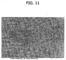
- the collagen foil biomatrix is still mechanically stable and removable as shown in Fig.10 .
- Fig. 11 is a sectional view of the collagen foil biomatrix of Figure 10 one week after implantation. As can be seen, fibroblasts are growing into the collagen foil biomatrix, directed by the multi-layer structure. The penetration in longitudinal direction is about 220 to 320 [mu]m.
- the speed of the directed in-growth of repair cells along the multi-layered structure is about 10 to 15 times higher in longitudinal direction compared to the transversal direction (" Fig. 10 "). Minimal inflammatory infiltration is shown, expressing the ongoing regenerative process.
- Example III Cell growth, tissue regeneration and prevention of peridural adhesion and fibrosis after implantation of a collagen foil biomatrix in spinal surgery
- Native equine collagen fibrils (mainly type I collagen) produced from purified minced Achilles tendon from horses and precipitated to fibrils.
- the flexible formstable and elastic biomatrix is specially engineered and has a nonporous fluid tight multilayer structure.
- the thickness of the dry equine multilayered collagen foil membrane was about 0.1 mm. In the wet condition the membrane thickness growth up to 0.3 mm.
- General anaesthesia Ketavet 60 mg/kg Rompun 16 mg/kg s.c. Thiopental i.v. through ear vein, based on effect Narkosis, intubation, mechanical ventilation Pain medication: 2 x day. Temgesic 0,05mg/kg s.c. for 3-4 days post operative. Euthanasia: In general anesthesia barbiturate overdose i.v.
- the present study was performed on New Zealand White (“NZW”) rabbits weighing on average 3 kg at the time of surgery and their average age was 4 months. All rabbits were female and approval for the animal studies were performed after formal approval by the authorities of the city of Vienna, Austria.
- the design of surgery in this experimental animal model in rabbits was similar to the most common operative intervention in humans.
- a laminectomy and resection was performed of the facet joint at the lumbar spine 4 to 5 (L4/5).
- PLIF Posterior Lumbar Interbody Fusion
- the ligamentum flavum was resected.
- the dura above the spinal cord remained intact.
- the laminectomy area was covered by a biological collagen foil biomatrix of the present invention.
- the paravertebral muscle was moved back in place and the fascia was closed by absorbable sutures.
- the skin was closed using syntofil sutures.
- the rabbits received postoperative pain medication and fluid infusion during the first three days and received mixed food.
- the rabbits were euthanized by overdose of thiopental and the operated areas of the spine were removed and isolated for histological evaluation.
- the vertebral column was excised en bloc and immersed in 10% formalin solution for fixation. After decalcification each lumbar vertebra was cut into slices, dehydrated and embedded in paraffin. Seven sections 3[mu]m thick were taken from each sample.
- the sections were stained with Haematoxilin-Eosin for general histology staining all cells and bone components
- the slides were examined by evaluating anatomical structures like relation between dura mater and the peridural dorsal surgical wound area, cell in-growth into the collagen foil biomatrix and the absence/presence and extent of adhesion formation and peridural fibrous scar formation.
- the sections were stained with Haematoxilin-Eosin for general histology staining all cells and bone components
- the slides were examined by evaluating anatomical structures like relation between dura mater and the peridural dorsal surgical wound area.
- Cell growth into and on the collagen foil biomatrix and the quantity and quality of the inflammatory reaction in the biomatrix and surrounding tissue were analyzed.
- the integration/incorporation process and the absence/presence and extent of adhesion formation and peridural connective tissue organization were also analyzed.
- the collagen foil biomatrix is placed between the dura mater and the dorsal defect area and forms a biological separation layer between the dura mater and the dorsal wound area. It is not fixed to the tissue of the dorsal defect. Blood (erythrocytes) adhere in a thin layer to the surface of the collagen foil biomatrix and proves its function as a hemostat.
- Blood erythrocytes
- the parallel microscopically multilayer structure of the collagen foil biomatrix which is nonporous and fluid-tight to blood, is clearly visible.
- the thickness of the collagen foil biomatrix is about 0.3 mm. ( Fig. 12 )
- the membrane is mostly integrated in the surrounding tissues. There is infiltration of blood cells, especially lymphocytes ( Fig 13 A) .
- the subdural space is free from cell infiltration or adhesion tissue.
- On the dorsal surface cells are directed along the surface and have hardly penetrated the surface of the collagen foil biomatrix. At the edges of the defect the collagen biomatrix was applied on the bone.
- the contact area (a) is a preferred area of bioactivity (biomatrix / cell interaction) and the beginning of directed cell infiltration along the multilayer structure.
- the area between the multilayered collagen foil biomatrix and the dura mater consists of loose tissue containing fat cells (b), comparable to the tissue of the epidural space in areas where the anatomy was not affected by the surgery (c) as shown in Fig. 13A .
- Fig. 13A shows the contact area between the multilayered collagen foil biomatrix and the bone at the edge of the defect (a).
- the remodelling of the multilayered collagen foil biomatrix starts with the directed infiltration of cells (fibroblasts, granulocytes) into the parallel multilayer structure of the multilayered collagen foil biomatrix.
- Fig. 13B shows the contact area between the multilayered collagen foil biomatrix and the bone at the edge of the defect (a).
- Fig. 13C shows the multilayered collagen foil biomatrix in the center of the laminectomy defect.
- the collagen biomatrix is integrated and is separating the ventral epidural space from the dorsal scar formation. Cells are directed along the dorsal surface and have not penetrated the surface of the collagen biomatrix.
- the area between the collagen biomatrix and the dura mater consists of loose tissue containing fat cells (a).
- FIG. 13D shows a NZW rabbit 1 week postoperative.
- the multilayered collagen foil biomatrix is closing the laminectomy defect and separating the dura from the beginning cell rich dorsal scar formation. Cells have not penetrated the surface of the collagen biomatrix. At the edge of the collagen biomatrix the beginning of directed infiltration of repair cells into the multilayer structure(a) is seen.
- Fig. 13E shows a NZW rabbit 1 week postoperative.
- a collagen sponge (DURAGEN) was used to cover the laminectomy defect.
- the collagen sponge is soaked with blood.
- the integration of the membrane into surrounding tissue is improved by in-growth of capillaries structures.
- the amount of the lymphocyte, segmented granulocyte is increased.
- the structure of the collagen foil biomatrix can be distinguished form the surrounding tissue.
- the subdural space is free from cell infiltration or adhesion tissue.
- Figs. 14 A - C show a NZW rabbit two weeks postoperative.
- the multilayered collagen foil biomatrix is fully integrated.
- Tissue repair cells have infiltrated the collagen biomatrix and are directed along the multilayer structure.
- the dura mater is separated by loose tissue with fat cells from the scar formation and the remodelled collagen biomatrix.
- the collagen for use in the present invention proved to have an immediate and excellent separation and protection function between the dura mater and the dorsal surgical defect, avoiding the uncontrolled distribution of blood, fibrin and necrotic materials into the peridural area and directing and separating the cell growth of the beginning scar above the dorsal surface.
- the biofunctional collagen foil also directs the cell in-growth into its multilayer structure. The speed of ingrowth along the multilayered structure is higher than the in-growth of cells into the biomatrix from the dorsal surface. There is no in-growth of cells in the first week into the ventral surface of the collagen foil biomatrix. The different speed of cell ingrowth along or across the multilayered structure is in line with a clinical observation after the peridural implantation of the multilayered collagen foil biomatrix one week postoperative (Ex. II).
- the multilayered biomatrix is infiltrated by repair cells.
- repair cells There is a full integration of the biomatrix into the physiological scar formation of the dorsal surgical wound area.
- the dura mater is separated by a loose fat tissue which looks similar to the fat tissue, which is physiologically separating the dura mater from the spinal canal.
- the biomatrix is remodeled and integrated into the normal anatomical structure.
- the collagen for use according to the present invention proved to be an effective, biocompatible and biofunctional immediate protection and separation layer between the dura mater and the dorsal defect area which is directing the cell ingrowth within the multilayer structure and the cell growth above its dorsal surface.
- the collagen foil biomatrix effectively controls the remodeling and tissue regeneration process and provides optimal conditions for the prevention and minimization of clinically relevant adhesions.
Applications Claiming Priority (2)
| Application Number | Priority Date | Filing Date | Title |
|---|---|---|---|
| US80959106P | 2006-05-31 | 2006-05-31 | |
| PCT/EP2007/004791 WO2007137839A2 (en) | 2006-05-31 | 2007-05-30 | Method for directed cell in-growth and controlled tissue regeneration in spinal surgery |
Publications (2)
| Publication Number | Publication Date |
|---|---|
| EP2021045A2 EP2021045A2 (en) | 2009-02-11 |
| EP2021045B1 true EP2021045B1 (en) | 2016-03-16 |
Family
ID=38457907
Family Applications (1)
| Application Number | Title | Priority Date | Filing Date |
|---|---|---|---|
| EP07725680.8A Active EP2021045B1 (en) | 2006-05-31 | 2007-05-30 | Collagen for use in prevention of peridural fibrosis formation after spinal surgery |
Country Status (14)
| Country | Link |
|---|---|
| US (1) | US8703122B2 (es) |
| EP (1) | EP2021045B1 (es) |
| JP (1) | JP5231401B2 (es) |
| KR (1) | KR20090017654A (es) |
| CN (1) | CN101454030B (es) |
| AU (1) | AU2007267338B2 (es) |
| BR (1) | BRPI0712088B8 (es) |
| CA (1) | CA2651941C (es) |
| CO (1) | CO6140041A2 (es) |
| ES (1) | ES2575933T3 (es) |
| HK (1) | HK1128638A1 (es) |
| MX (1) | MX2008014847A (es) |
| NO (1) | NO20085419L (es) |
| WO (1) | WO2007137839A2 (es) |
Families Citing this family (35)
| Publication number | Priority date | Publication date | Assignee | Title |
|---|---|---|---|---|
| US7435425B2 (en) | 2001-07-17 | 2008-10-14 | Baxter International, Inc. | Dry hemostatic compositions and methods for their preparation |
| US8603511B2 (en) | 1996-08-27 | 2013-12-10 | Baxter International, Inc. | Fragmented polymeric compositions and methods for their use |
| US6066325A (en) | 1996-08-27 | 2000-05-23 | Fusion Medical Technologies, Inc. | Fragmented polymeric compositions and methods for their use |
| US8303981B2 (en) | 1996-08-27 | 2012-11-06 | Baxter International Inc. | Fragmented polymeric compositions and methods for their use |
| US8834864B2 (en) * | 2003-06-05 | 2014-09-16 | Baxter International Inc. | Methods for repairing and regenerating human dura mater |
| US7927626B2 (en) | 2003-08-07 | 2011-04-19 | Ethicon, Inc. | Process of making flowable hemostatic compositions and devices containing such compositions |
| WO2007137839A2 (en) | 2006-05-31 | 2007-12-06 | Baxter International Inc. | Method for directed cell in-growth and controlled tissue regeneration in spinal surgery |
| TWI436793B (zh) * | 2006-08-02 | 2014-05-11 | Baxter Int | 快速作用之乾密封膠及其使用和製造方法 |
| US20080260794A1 (en) * | 2007-02-12 | 2008-10-23 | Lauritzen Nels J | Collagen products and methods for producing collagen products |
| US9056151B2 (en) * | 2007-02-12 | 2015-06-16 | Warsaw Orthopedic, Inc. | Methods for collagen processing and products using processed collagen |
| US8092541B2 (en) * | 2007-08-03 | 2012-01-10 | Warsaw Orthopedic, Inc. | Method of using an anti-growth matrix as a barrier for cell attachment and osteo-inductive factors |
| EP2214734B1 (en) | 2007-10-30 | 2017-12-13 | Baxter International Inc. | Use of a regenerative biofunctional collagen biomatrix for treating visceral or parietal defects |
| JP5569398B2 (ja) | 2008-02-29 | 2014-08-13 | フェッローサン メディカル ディバイス エー/エス | 止血および/または創傷治癒を促進するための装置 |
| US9039783B2 (en) | 2009-05-18 | 2015-05-26 | Baxter International, Inc. | Method for the improvement of mesh implant biocompatibility |
| KR101699992B1 (ko) * | 2009-06-16 | 2017-01-26 | 백스터 인터내셔널 인코포레이티드 | 지혈용 스펀지 |
| AU2010339045B2 (en) | 2009-12-16 | 2014-06-05 | Baxter Healthcare S.A. | Hemostatic sponge |
| SA111320355B1 (ar) | 2010-04-07 | 2015-01-08 | Baxter Heathcare S A | إسفنجة لايقاف النزف |
| US8790699B2 (en) | 2010-04-23 | 2014-07-29 | Warsaw Orthpedic, Inc. | Foam-formed collagen strand |
| US8460691B2 (en) | 2010-04-23 | 2013-06-11 | Warsaw Orthopedic, Inc. | Fenestrated wound repair scaffold |
| US8940335B2 (en) | 2010-06-01 | 2015-01-27 | Baxter International Inc. | Process for making dry and stable hemostatic compositions |
| JP6289096B2 (ja) | 2010-06-01 | 2018-03-07 | バクスター・インターナショナル・インコーポレイテッドBaxter International Incorp0Rated | 乾燥した安定な止血組成物を作製するためのプロセス |
| CN103037847B (zh) | 2010-06-01 | 2016-01-20 | 巴克斯特国际公司 | 用于制备干燥、稳定的止血组合物的方法 |
| CN103957948B (zh) | 2011-10-11 | 2016-10-26 | 巴克斯特国际公司 | 止血组合物 |
| CN103957949B (zh) | 2011-10-11 | 2017-07-18 | 巴克斯特国际公司 | 止血组合物 |
| KR101975624B1 (ko) | 2011-10-27 | 2019-05-07 | 백스터 인터내셔널 인코포레이티드 | 지혈 조성물 |
| EP2822474B1 (en) | 2012-03-06 | 2018-05-02 | Ferrosan Medical Devices A/S | Pressurized container containing haemostatic paste |
| AU2013275758B2 (en) | 2012-06-12 | 2015-03-12 | Ferrosan Medical Devices A/S | Dry haemostatic composition |
| US9114190B2 (en) | 2013-02-08 | 2015-08-25 | Laser Spine Institute, Llc | Regeneration of spinal discs |
| AU2014283170B2 (en) | 2013-06-21 | 2017-11-02 | Ferrosan Medical Devices A/S | Vacuum expanded dry composition and syringe for retaining same |
| JP6489485B2 (ja) | 2013-12-11 | 2019-03-27 | フェロサン メディカル デバイシーズ エイ/エス | 押し出し増強因子を含んでいる乾燥組成物 |
| BR112017007466B1 (pt) | 2014-10-13 | 2021-03-02 | Ferrosan Medical Devices A/S | método para preparar uma composição seca, método para reconstituir a composição seca, pasta, composição seca, recipiente, kit homeostático, e, uso de uma composição seca |
| US10653837B2 (en) | 2014-12-24 | 2020-05-19 | Ferrosan Medical Devices A/S | Syringe for retaining and mixing first and second substances |
| US10918796B2 (en) | 2015-07-03 | 2021-02-16 | Ferrosan Medical Devices A/S | Syringe for mixing two components and for retaining a vacuum in a storage condition |
| RU2644278C1 (ru) * | 2016-12-21 | 2018-02-08 | Федеральное государственное автономное учреждение "Национальный медицинский исследовательский центр нейрохирургии имени академика Н.Н.Бурденко" Министерства здравоохранения Российской Федерации (ФГАУ "НМИЦ нейрохирургии им. акад. Н.Н.Бурденко" Минздрава России | Способ микрохирургической реконструкции спинного мозга на животной модели с использованием биодеградируемого гидрогеля на основе поливинилового спирта |
| AU2019266529A1 (en) | 2018-05-09 | 2020-12-03 | Ethicon Inc. | Method for preparing a haemostatic composition |
Family Cites Families (133)
| Publication number | Priority date | Publication date | Assignee | Title |
|---|---|---|---|---|
| US2507244A (en) * | 1947-04-14 | 1950-05-09 | Upjohn Co | Surgical gelatin dusting powder and process for preparing same |
| CH264752A (de) * | 1947-06-03 | 1949-10-31 | Hoffmann La Roche | Verfahren zur Herstellung von Trägern für Arzneimittel. |
| SE420565B (sv) | 1974-06-06 | 1981-10-19 | Pharmacia Ab | Hjelpmedel for intravaskuler administraring for anvendning i samband med intravaskuler administrering av en losning eller en suspension av ett diagnostiseringsmedel |
| US4013078A (en) * | 1974-11-25 | 1977-03-22 | Feild James Rodney | Intervertebral protector means |
| US4164559A (en) * | 1977-09-21 | 1979-08-14 | Cornell Research Foundation, Inc. | Collagen drug delivery device |
| DE2843963A1 (de) * | 1978-10-09 | 1980-04-24 | Merck Patent Gmbh | Im koerper resorbierbare geformte masse auf basis von kollagen und ihre verwendung in der medizin |
| US4265233A (en) * | 1978-04-12 | 1981-05-05 | Unitika Ltd. | Material for wound healing |
| US4179400A (en) | 1978-05-09 | 1979-12-18 | W. R. Grace & Co. | Process for preparing catalytic solutions of sulfonium salts |
| AT359653B (de) * | 1979-02-15 | 1980-11-25 | Immuno Ag | Verfahren zur herstellung eines gewebekleb- stoffes |
| AT359652B (de) | 1979-02-15 | 1980-11-25 | Immuno Ag | Verfahren zur herstellung eines gewebekleb- stoffes |
| DE3036033A1 (de) | 1980-09-24 | 1982-05-06 | Max-Planck-Gesellschaft zur Förderung der Wissenschaften e.V., 3400 Göttingen | Wundbehandlungsmittel in pulverform und verfahren zu seiner herstellung |
| US4300494A (en) | 1979-09-26 | 1981-11-17 | Shell Oil Company | Thermal insulated intake ports |
| US4292972A (en) | 1980-07-09 | 1981-10-06 | E. R. Squibb & Sons, Inc. | Lyophilized hydrocolloio foam |
| DE3105624A1 (de) * | 1981-02-16 | 1982-09-02 | Hormon-Chemie München GmbH, 8000 München | Material zum abdichten und heilen von wunden |
| US4424208A (en) * | 1982-01-11 | 1984-01-03 | Collagen Corporation | Collagen implant material and method for augmenting soft tissue |
| DE3360633D1 (en) * | 1982-02-12 | 1985-10-03 | Unitika Ltd | Anti-cancer device |
| US4482386A (en) | 1982-03-26 | 1984-11-13 | Warner-Lambert Company | Method of conditioning a water swellable hydrocolloid |
| US4543332A (en) * | 1982-03-29 | 1985-09-24 | Miles Laboratories, Inc. | Method for the preparation of spherical microorganism cell aggregates |
| US4540410A (en) * | 1982-11-16 | 1985-09-10 | Serono Pharmaceutical Partners | Lyophilized compositions, preparation and use thereof |
| DE3466702D1 (en) | 1983-07-14 | 1987-11-12 | Hitachi Chemical Co Ltd | Gelatin spherical gels and production thereof |
| JPS60100516A (ja) | 1983-11-04 | 1985-06-04 | Takeda Chem Ind Ltd | 徐放型マイクロカプセルの製造法 |
| US4515637A (en) * | 1983-11-16 | 1985-05-07 | Seton Company | Collagen-thrombin compositions |
| AT389815B (de) * | 1984-03-09 | 1990-02-12 | Immuno Ag | Verfahren zur inaktivierung von vermehrungsfaehigen filtrierbaren krankheitserregern in blutprodukten |
| US4600574A (en) * | 1984-03-21 | 1986-07-15 | Immuno Aktiengesellschaft Fur Chemisch-Medizinische Produkte | Method of producing a tissue adhesive |
| US4837285A (en) * | 1984-03-27 | 1989-06-06 | Medimatrix | Collagen matrix beads for soft tissue repair |
| JPS6144825A (ja) * | 1984-08-09 | 1986-03-04 | Unitika Ltd | 止血剤 |
| GB8422950D0 (en) * | 1984-09-11 | 1984-10-17 | Warne K J | Hydrogel |
| JPS61122222A (ja) * | 1984-11-19 | 1986-06-10 | Koken:Kk | コラ−ゲン又はゼラチンとプロタミンとよりなる止血剤 |
| US5178883A (en) * | 1984-11-29 | 1993-01-12 | Regents Of The University Of Minnesota | Method for promoting hair growth |
| US5165938A (en) | 1984-11-29 | 1992-11-24 | Regents Of The University Of Minnesota | Wound healing agents derived from platelets |
| US4600533A (en) | 1984-12-24 | 1986-07-15 | Collagen Corporation | Collagen membranes for medical use |
| US5007916A (en) * | 1985-08-22 | 1991-04-16 | Johnson & Johnson Medical, Inc. | Method and material for prevention of surgical adhesions |
| IE59361B1 (en) * | 1986-01-24 | 1994-02-09 | Akzo Nv | Pharmaceutical preparation for obtaining a highly viscous hydrogel or suspension |
| IL78826A (en) * | 1986-05-19 | 1991-05-12 | Yissum Res Dev Co | Precursor composition for the preparation of a biodegradable implant for the sustained release of an active material and such implants prepared therefrom |
| US5300494A (en) * | 1986-06-06 | 1994-04-05 | Union Carbide Chemicals & Plastics Technology Corporation | Delivery systems for quaternary and related compounds |
| US4946870A (en) * | 1986-06-06 | 1990-08-07 | Union Carbide Chemicals And Plastics Company Inc. | Delivery systems for pharmaceutical or therapeutic actives |
| US4832686A (en) * | 1986-06-24 | 1989-05-23 | Anderson Mark E | Method for administering interleukin-2 |
| US4803075A (en) * | 1986-06-25 | 1989-02-07 | Collagen Corporation | Injectable implant composition having improved intrudability |
| US5080893A (en) * | 1988-05-31 | 1992-01-14 | University Of Florida | Method for preventing surgical adhesions using a dilute solution of polymer |
| US5017229A (en) * | 1990-06-25 | 1991-05-21 | Genzyme Corporation | Water insoluble derivatives of hyaluronic acid |
| US5140016A (en) * | 1988-05-31 | 1992-08-18 | University Of Florida | Method and composition for preventing surgical adhesions using a dilute solution of polymer |
| US5350573A (en) * | 1988-05-31 | 1994-09-27 | University Of Florida Research Foundation, Inc. | Method and composition for preventing surgical adhesions |
| US5447966A (en) * | 1988-07-19 | 1995-09-05 | United States Surgical Corporation | Treating bioabsorbable surgical articles by coating with glycerine, polalkyleneoxide block copolymer and gelatin |
| US5041292A (en) * | 1988-08-31 | 1991-08-20 | Theratech, Inc. | Biodegradable hydrogel matrices for the controlled release of pharmacologically active agents |
| US4925677A (en) * | 1988-08-31 | 1990-05-15 | Theratech, Inc. | Biodegradable hydrogel matrices for the controlled release of pharmacologically active agents |
| US5126141A (en) * | 1988-11-16 | 1992-06-30 | Mediventures Incorporated | Composition and method for post-surgical adhesion reduction with thermo-irreversible gels of polyoxyalkylene polymers and ionic polysaccharides |
| US5135751A (en) * | 1988-11-16 | 1992-08-04 | Mediventures Incorporated | Composition for reducing postsurgical adhesions |
| US5162430A (en) | 1988-11-21 | 1992-11-10 | Collagen Corporation | Collagen-polymer conjugates |
| US5614587A (en) * | 1988-11-21 | 1997-03-25 | Collagen Corporation | Collagen-based bioadhesive compositions |
| US5510418A (en) * | 1988-11-21 | 1996-04-23 | Collagen Corporation | Glycosaminoglycan-synthetic polymer conjugates |
| US4891359A (en) * | 1988-12-08 | 1990-01-02 | Johnson & Johnson Patient Care, Inc. | Hemostatic collagen paste composition |
| DE3903672C1 (es) * | 1989-02-08 | 1990-02-01 | Lohmann Gmbh & Co Kg | |
| WO1991001711A1 (en) | 1989-08-10 | 1991-02-21 | W.L. Gore & Associates, Inc. | A medical dispensing system for tissue adhesive components |
| US5196185A (en) * | 1989-09-11 | 1993-03-23 | Micro-Collagen Pharmaceutics, Ltd. | Collagen-based wound dressing and method for applying same |
| US5061274A (en) | 1989-12-04 | 1991-10-29 | Kensey Nash Corporation | Plug device for sealing openings and method of use |
| US5219328A (en) * | 1990-01-03 | 1993-06-15 | Cryolife, Inc. | Fibrin sealant delivery method |
| US5134229A (en) * | 1990-01-12 | 1992-07-28 | Johnson & Johnson Medical, Inc. | Process for preparing a neutralized oxidized cellulose product and its method of use |
| JPH0813750B2 (ja) * | 1990-03-01 | 1996-02-14 | 持田製薬株式会社 | 経口用トロンビン製剤 |
| US5306501A (en) * | 1990-05-01 | 1994-04-26 | Mediventures, Inc. | Drug delivery by injection with thermoreversible gels containing polyoxyalkylene copolymers |
| US5595735A (en) * | 1990-05-23 | 1997-01-21 | Johnson & Johnson Medical, Inc. | Hemostatic thrombin paste composition |
| US5292362A (en) * | 1990-07-27 | 1994-03-08 | The Trustees Of Columbia University In The City Of New York | Tissue bonding and sealing composition and method of using the same |
| US5209776A (en) * | 1990-07-27 | 1993-05-11 | The Trustees Of Columbia University In The City Of New York | Tissue bonding and sealing composition and method of using the same |
| US5108421A (en) * | 1990-10-01 | 1992-04-28 | Quinton Instrument Company | Insertion assembly and method of inserting a vessel plug into the body of a patient |
| US5192300A (en) * | 1990-10-01 | 1993-03-09 | Quinton Instrument Company | Insertion assembly and method of inserting a vessel plug into the body of a patient |
| NO302481B1 (no) * | 1990-10-16 | 1998-03-09 | Takeda Chemical Industries Ltd | Polymer for et preparat med forlenget frigjöring, samt preparat med forlenget frigjöring |
| US5129882A (en) * | 1990-12-27 | 1992-07-14 | Novoste Corporation | Wound clotting device and method of using same |
| US5690675A (en) | 1991-02-13 | 1997-11-25 | Fusion Medical Technologies, Inc. | Methods for sealing of staples and other fasteners in tissue |
| JPH06500802A (ja) * | 1991-06-14 | 1994-01-27 | アムジエン・インコーポレーテツド | コラーゲンフィルムによるタンパクのドラッグ・デリバリー |
| AT398079B (de) * | 1991-11-04 | 1994-09-26 | Immuno Ag | Präparation mit thrombinaktivität sowie verfahren zu ihrer herstellung |
| ATE193037T1 (de) * | 1992-02-28 | 2000-06-15 | Collagen Corp | Hochkonzentrierte homogenisierte kollagenzusammensetzungen |
| ATE208217T1 (de) | 1992-02-28 | 2001-11-15 | Cohesion Tech Inc | Injektierbare, keramische verbindungen sowie verfahren zur deren herstellung und anwendung |
| US5204382A (en) * | 1992-02-28 | 1993-04-20 | Collagen Corporation | Injectable ceramic compositions and methods for their preparation and use |
| US5384333A (en) * | 1992-03-17 | 1995-01-24 | University Of Miami | Biodegradable injectable drug delivery polymer |
| WO1993021844A1 (en) * | 1992-04-23 | 1993-11-11 | Scimed Life Systems, Inc. | Apparatus and method for sealing vascular punctures |
| IL105529A0 (en) * | 1992-05-01 | 1993-08-18 | Amgen Inc | Collagen-containing sponges as drug delivery for proteins |
| WO1993024476A1 (en) * | 1992-06-04 | 1993-12-09 | Clover Consolidated, Limited | Water-soluble polymeric carriers for drug delivery |
| US5385606A (en) * | 1992-07-06 | 1995-01-31 | Kowanko; Nicholas | Adhesive composition and method |
| US5413571A (en) * | 1992-07-16 | 1995-05-09 | Sherwood Medical Company | Device for sealing hemostatic incisions |
| US5428022A (en) * | 1992-07-29 | 1995-06-27 | Collagen Corporation | Composition of low type III content human placental collagen |
| US5514379A (en) * | 1992-08-07 | 1996-05-07 | The General Hospital Corporation | Hydrogel compositions and methods of use |
| DE4227681C2 (de) * | 1992-08-21 | 1995-05-18 | Becker & Co Naturinwerk | Wundabdeckungsmaterial auf der Basis von Kollagenfasern und Verfahren zu seiner Herstellung |
| ATE206602T1 (de) * | 1992-11-12 | 2001-10-15 | Neville Alleyne | Einrichtung zum schutz des herzens |
| US5667839A (en) * | 1993-01-28 | 1997-09-16 | Collagen Corporation | Human recombinant collagen in the milk of transgenic animals |
| JP3639593B2 (ja) | 1993-05-31 | 2005-04-20 | 科研製薬株式会社 | 塩基性線維芽細胞増殖因子含有架橋ゼラチンゲル製剤 |
| FR2715309B1 (fr) * | 1994-01-24 | 1996-08-02 | Imedex | Composition adhésive, à usage chirurgical, à base de collagène modifié par coupure oxydative et non réticulé. |
| US5674275A (en) | 1994-04-06 | 1997-10-07 | Graphic Controls Corporation | Polyacrylate and polymethacrylate ester based hydrogel adhesives |
| US5531759A (en) * | 1994-04-29 | 1996-07-02 | Kensey Nash Corporation | System for closing a percutaneous puncture formed by a trocar to prevent tissue at the puncture from herniating |
| GB9415739D0 (en) | 1994-07-30 | 1994-09-21 | Scimat Ltd | Gel wound dressing |
| US5516532A (en) * | 1994-08-05 | 1996-05-14 | Children's Medical Center Corporation | Injectable non-immunogenic cartilage and bone preparation |
| US5931165A (en) * | 1994-09-06 | 1999-08-03 | Fusion Medical Technologies, Inc. | Films having improved characteristics and methods for their preparation and use |
| US20030039695A1 (en) | 2001-08-10 | 2003-02-27 | Ed. Geistlich Soehne Ag Fuer Chemische Industrie | Collagen carrier of therapeutic genetic material, and method |
| US5698213A (en) | 1995-03-06 | 1997-12-16 | Ethicon, Inc. | Hydrogels of absorbable polyoxaesters |
| US5580923A (en) | 1995-03-14 | 1996-12-03 | Collagen Corporation | Anti-adhesion films and compositions for medical use |
| US6129761A (en) | 1995-06-07 | 2000-10-10 | Reprogenesis, Inc. | Injectable hydrogel compositions |
| US6458889B1 (en) | 1995-12-18 | 2002-10-01 | Cohesion Technologies, Inc. | Compositions and systems for forming crosslinked biomaterials and associated methods of preparation and use |
| JP4193917B2 (ja) | 1995-12-18 | 2008-12-10 | アンジオデバイス インターナショナル ゲーエムベーハー | 架橋ポリマー組成物およびその使用方法 |
| JP3476631B2 (ja) | 1995-12-21 | 2003-12-10 | 株式会社アムニオテック | ヒト由来の天然コラーゲン膜からなる医用材料 |
| US5748318A (en) * | 1996-01-23 | 1998-05-05 | Brown University Research Foundation | Optical stress generator and detector |
| SK284693B6 (sk) | 1996-04-04 | 2005-09-08 | Baxter Aktiengesellschaft | Hemostatická huba, pokrytie rany, kit na prípravu pokrytia rany a spôsob prípravy huby |
| US5902832A (en) * | 1996-08-20 | 1999-05-11 | Menlo Care, Inc. | Method of synthesizing swollen hydrogel for sphincter augmentation |
| US6066325A (en) * | 1996-08-27 | 2000-05-23 | Fusion Medical Technologies, Inc. | Fragmented polymeric compositions and methods for their use |
| US7320962B2 (en) * | 1996-08-27 | 2008-01-22 | Baxter International Inc. | Hemoactive compositions and methods for their manufacture and use |
| US6706690B2 (en) * | 1999-06-10 | 2004-03-16 | Baxter Healthcare Corporation | Hemoactive compositions and methods for their manufacture and use |
| US6063061A (en) | 1996-08-27 | 2000-05-16 | Fusion Medical Technologies, Inc. | Fragmented polymeric compositions and methods for their use |
| US7435425B2 (en) * | 2001-07-17 | 2008-10-14 | Baxter International, Inc. | Dry hemostatic compositions and methods for their preparation |
| US7871637B2 (en) * | 1996-08-27 | 2011-01-18 | Baxter International Inc. | Dry hemostatic compositions and methods for their preparation |
| TW501934B (en) * | 1996-11-20 | 2002-09-11 | Tapic Int Co Ltd | Collagen material and process for making the same |
| US6458386B1 (en) | 1997-06-03 | 2002-10-01 | Innogenetics N.V. | Medicaments based on polymers composed of methacrylamide-modified gelatin |
| US5908054A (en) * | 1997-06-16 | 1999-06-01 | Fusion Medical Technologies, Inc. | Fluid dispersion and delivery assembly and method |
| US5997895A (en) | 1997-09-16 | 1999-12-07 | Integra Lifesciences Corporation | Dural/meningeal repair product using collagen matrix |
| ATE306935T1 (de) | 1997-09-16 | 2005-11-15 | Integra Lifesciences Corp | Zusammensetzung zur förderung des wachstums von duralem oder meningealem gewebe enthaltend kollagen |
| US6179872B1 (en) * | 1998-03-17 | 2001-01-30 | Tissue Engineering | Biopolymer matt for use in tissue repair and reconstruction |
| US6227394B1 (en) * | 1998-06-09 | 2001-05-08 | Asahi Glass Company Ltd. | Glass bulb for a cathode ray tube and a method for producing a cathode ray tube |
| US6110484A (en) * | 1998-11-24 | 2000-08-29 | Cohesion Technologies, Inc. | Collagen-polymer matrices with differential biodegradability |
| US6328229B1 (en) | 1998-12-18 | 2001-12-11 | Cohesion Technologies, Inc. | Low volume mixing spray head for mixing and dispensing of two reactive fluid components |
| US6312725B1 (en) | 1999-04-16 | 2001-11-06 | Cohesion Technologies, Inc. | Rapid gelling biocompatible polymer composition |
| US6221109B1 (en) | 1999-09-15 | 2001-04-24 | Ed. Geistlich Söhne AG fur Chemische Industrie | Method of protecting spinal area |
| US6312474B1 (en) | 1999-09-15 | 2001-11-06 | Bio-Vascular, Inc. | Resorbable implant materials |
| CA2422852C (en) | 2000-09-18 | 2012-06-26 | Organogenesis Inc. | Methods for treating a patient using a bioengineered flat sheet graft prostheses |
| NZ527166A (en) | 2001-01-25 | 2005-03-24 | Nycomed Pharma As | A method of preparing a collagen sponge, a device for extracting a part of a collagen foam, and an elongated collagen sponge |
| JP2003160506A (ja) * | 2001-08-10 | 2003-06-03 | Ed Geistlich Soehne Ag Fuer Chemische Industrie | 治療用遺伝物質のコラーゲン担体およびその方法 |
| US8834864B2 (en) * | 2003-06-05 | 2014-09-16 | Baxter International Inc. | Methods for repairing and regenerating human dura mater |
| EP1484070B1 (en) | 2003-06-05 | 2006-01-11 | Baxter International Inc. | Compositions for repairing and regenerating human dura mater |
| EP1682196A2 (en) * | 2003-11-10 | 2006-07-26 | Angiotech International Ag | Medical implants and anti-scarring agents |
| CA2536188A1 (en) * | 2003-11-20 | 2005-06-09 | Angiotech International Ag | Electrical devices and anti-scarring agents |
| US20080091277A1 (en) * | 2004-08-13 | 2008-04-17 | Kai Deusch | Surgical prosthesis having biodegradable and nonbiodegradable regions |
| ES2367583T3 (es) | 2005-05-04 | 2011-11-10 | Suprapolix B.V. | Hidrogeles con enlaces de hidrógeno. |
| WO2007001926A2 (en) | 2005-06-24 | 2007-01-04 | Hyperbranch Medical Technology, Inc. | Low-swelling hydrogel sealants for wound repair |
| WO2007137839A2 (en) | 2006-05-31 | 2007-12-06 | Baxter International Inc. | Method for directed cell in-growth and controlled tissue regeneration in spinal surgery |
| TWI436793B (zh) | 2006-08-02 | 2014-05-11 | Baxter Int | 快速作用之乾密封膠及其使用和製造方法 |
| EP2214734B1 (en) * | 2007-10-30 | 2017-12-13 | Baxter International Inc. | Use of a regenerative biofunctional collagen biomatrix for treating visceral or parietal defects |
| US9039783B2 (en) | 2009-05-18 | 2015-05-26 | Baxter International, Inc. | Method for the improvement of mesh implant biocompatibility |
| KR101699992B1 (ko) | 2009-06-16 | 2017-01-26 | 백스터 인터내셔널 인코포레이티드 | 지혈용 스펀지 |
-
2007
- 2007-05-30 WO PCT/EP2007/004791 patent/WO2007137839A2/en active Application Filing
- 2007-05-30 CA CA2651941A patent/CA2651941C/en active Active
- 2007-05-30 EP EP07725680.8A patent/EP2021045B1/en active Active
- 2007-05-30 MX MX2008014847A patent/MX2008014847A/es active IP Right Grant
- 2007-05-30 BR BRPI0712088A patent/BRPI0712088B8/pt not_active IP Right Cessation
- 2007-05-30 ES ES07725680.8T patent/ES2575933T3/es active Active
- 2007-05-30 CN CN200780019553XA patent/CN101454030B/zh active Active
- 2007-05-30 JP JP2009512489A patent/JP5231401B2/ja active Active
- 2007-05-30 US US12/302,716 patent/US8703122B2/en active Active
- 2007-05-30 AU AU2007267338A patent/AU2007267338B2/en not_active Ceased
- 2007-05-30 KR KR1020087031934A patent/KR20090017654A/ko not_active Application Discontinuation
-
2008
- 2008-12-29 CO CO08137571A patent/CO6140041A2/es unknown
- 2008-12-30 NO NO20085419A patent/NO20085419L/no not_active Application Discontinuation
-
2009
- 2009-07-28 HK HK09106947.2A patent/HK1128638A1/zh not_active IP Right Cessation
Also Published As
| Publication number | Publication date |
|---|---|
| JP2009538649A (ja) | 2009-11-12 |
| JP5231401B2 (ja) | 2013-07-10 |
| US8703122B2 (en) | 2014-04-22 |
| BRPI0712088B1 (pt) | 2018-05-29 |
| WO2007137839A3 (en) | 2008-11-27 |
| CN101454030B (zh) | 2013-06-26 |
| MX2008014847A (es) | 2009-04-30 |
| AU2007267338A1 (en) | 2007-12-06 |
| ES2575933T3 (es) | 2016-07-04 |
| KR20090017654A (ko) | 2009-02-18 |
| BRPI0712088A2 (pt) | 2012-03-06 |
| US20100028309A1 (en) | 2010-02-04 |
| WO2007137839A8 (en) | 2008-02-28 |
| AU2007267338B2 (en) | 2013-04-04 |
| CO6140041A2 (es) | 2010-03-19 |
| CN101454030A (zh) | 2009-06-10 |
| HK1128638A1 (zh) | 2009-11-06 |
| NO20085419L (no) | 2008-12-30 |
| CA2651941A1 (en) | 2007-12-06 |
| WO2007137839A2 (en) | 2007-12-06 |
| EP2021045A2 (en) | 2009-02-11 |
| CA2651941C (en) | 2015-02-17 |
| BRPI0712088B8 (pt) | 2021-06-22 |
Similar Documents
| Publication | Publication Date | Title |
|---|---|---|
| EP2021045B1 (en) | Collagen for use in prevention of peridural fibrosis formation after spinal surgery | |
| AU2008317874B2 (en) | Use of a regenerative biofunctional collagen biomatrix for treating visceral or parietal defects | |
| US8834864B2 (en) | Methods for repairing and regenerating human dura mater | |
| AU2004245086B2 (en) | Compositions for repairing and regenerating human dura mater | |
| WO2000016822A1 (en) | Compositions and methods for tissue repair | |
| AU2003260086A1 (en) | Composite material for wound repair | |
| Meddings | Evaluation of collagen vicryl mesh as a dural graft substance |
Legal Events
| Date | Code | Title | Description |
|---|---|---|---|
| PUAI | Public reference made under article 153(3) epc to a published international application that has entered the european phase |
Free format text: ORIGINAL CODE: 0009012 |
|
| 17P | Request for examination filed |
Effective date: 20081127 |
|
| AK | Designated contracting states |
Kind code of ref document: A2 Designated state(s): AT BE BG CH CY CZ DE DK EE ES FI FR GB GR HU IE IS IT LI LT LU LV MC MT NL PL PT RO SE SI SK TR |
|
| AX | Request for extension of the european patent |
Extension state: AL BA HR MK RS |
|
| REG | Reference to a national code |
Ref country code: HK Ref legal event code: DE Ref document number: 1128638 Country of ref document: HK |
|
| 17Q | First examination report despatched |
Effective date: 20091211 |
|
| RAX | Requested extension states of the european patent have changed |
Extension state: HR Payment date: 20081127 |
|
| GRAP | Despatch of communication of intention to grant a patent |
Free format text: ORIGINAL CODE: EPIDOSNIGR1 |
|
| INTG | Intention to grant announced |
Effective date: 20151007 |
|
| GRAS | Grant fee paid |
Free format text: ORIGINAL CODE: EPIDOSNIGR3 |
|
| GRAA | (expected) grant |
Free format text: ORIGINAL CODE: 0009210 |
|
| AK | Designated contracting states |
Kind code of ref document: B1 Designated state(s): AT BE BG CH CY CZ DE DK EE ES FI FR GB GR HU IE IS IT LI LT LU LV MC MT NL PL PT RO SE SI SK TR |
|
| AX | Request for extension of the european patent |
Extension state: HR |
|
| REG | Reference to a national code |
Ref country code: GB Ref legal event code: FG4D |
|
| REG | Reference to a national code |
Ref country code: CH Ref legal event code: EP |
|
| REG | Reference to a national code |
Ref country code: IE Ref legal event code: FG4D |
|
| REG | Reference to a national code |
Ref country code: AT Ref legal event code: REF Ref document number: 780634 Country of ref document: AT Kind code of ref document: T Effective date: 20160415 |
|
| REG | Reference to a national code |
Ref country code: DE Ref legal event code: R096 Ref document number: 602007045290 Country of ref document: DE |
|
| REG | Reference to a national code |
Ref country code: FR Ref legal event code: PLFP Year of fee payment: 10 |
|
| REG | Reference to a national code |
Ref country code: ES Ref legal event code: FG2A Ref document number: 2575933 Country of ref document: ES Kind code of ref document: T3 Effective date: 20160704 |
|
| REG | Reference to a national code |
Ref country code: NL Ref legal event code: MP Effective date: 20160316 |
|
| REG | Reference to a national code |
Ref country code: LT Ref legal event code: MG4D |
|
| PG25 | Lapsed in a contracting state [announced via postgrant information from national office to epo] |
Ref country code: FI Free format text: LAPSE BECAUSE OF FAILURE TO SUBMIT A TRANSLATION OF THE DESCRIPTION OR TO PAY THE FEE WITHIN THE PRESCRIBED TIME-LIMIT Effective date: 20160316 Ref country code: GR Free format text: LAPSE BECAUSE OF FAILURE TO SUBMIT A TRANSLATION OF THE DESCRIPTION OR TO PAY THE FEE WITHIN THE PRESCRIBED TIME-LIMIT Effective date: 20160617 |
|
| REG | Reference to a national code |
Ref country code: AT Ref legal event code: MK05 Ref document number: 780634 Country of ref document: AT Kind code of ref document: T Effective date: 20160316 |
|
| PG25 | Lapsed in a contracting state [announced via postgrant information from national office to epo] |
Ref country code: LV Free format text: LAPSE BECAUSE OF FAILURE TO SUBMIT A TRANSLATION OF THE DESCRIPTION OR TO PAY THE FEE WITHIN THE PRESCRIBED TIME-LIMIT Effective date: 20160316 Ref country code: BE Free format text: LAPSE BECAUSE OF NON-PAYMENT OF DUE FEES Effective date: 20160531 Ref country code: LT Free format text: LAPSE BECAUSE OF FAILURE TO SUBMIT A TRANSLATION OF THE DESCRIPTION OR TO PAY THE FEE WITHIN THE PRESCRIBED TIME-LIMIT Effective date: 20160316 Ref country code: NL Free format text: LAPSE BECAUSE OF FAILURE TO SUBMIT A TRANSLATION OF THE DESCRIPTION OR TO PAY THE FEE WITHIN THE PRESCRIBED TIME-LIMIT Effective date: 20160316 Ref country code: SE Free format text: LAPSE BECAUSE OF FAILURE TO SUBMIT A TRANSLATION OF THE DESCRIPTION OR TO PAY THE FEE WITHIN THE PRESCRIBED TIME-LIMIT Effective date: 20160316 |
|
| REG | Reference to a national code |
Ref country code: HK Ref legal event code: GR Ref document number: 1128638 Country of ref document: HK |
|
| PG25 | Lapsed in a contracting state [announced via postgrant information from national office to epo] |
Ref country code: IS Free format text: LAPSE BECAUSE OF FAILURE TO SUBMIT A TRANSLATION OF THE DESCRIPTION OR TO PAY THE FEE WITHIN THE PRESCRIBED TIME-LIMIT Effective date: 20160716 Ref country code: EE Free format text: LAPSE BECAUSE OF FAILURE TO SUBMIT A TRANSLATION OF THE DESCRIPTION OR TO PAY THE FEE WITHIN THE PRESCRIBED TIME-LIMIT Effective date: 20160316 Ref country code: PL Free format text: LAPSE BECAUSE OF FAILURE TO SUBMIT A TRANSLATION OF THE DESCRIPTION OR TO PAY THE FEE WITHIN THE PRESCRIBED TIME-LIMIT Effective date: 20160316 |
|
| REG | Reference to a national code |
Ref country code: AT Ref legal event code: EK Ref document number: 780634 Country of ref document: AT Kind code of ref document: T Effective date: 20160921 |
|
| PG25 | Lapsed in a contracting state [announced via postgrant information from national office to epo] |
Ref country code: PT Free format text: LAPSE BECAUSE OF FAILURE TO SUBMIT A TRANSLATION OF THE DESCRIPTION OR TO PAY THE FEE WITHIN THE PRESCRIBED TIME-LIMIT Effective date: 20160718 Ref country code: SK Free format text: LAPSE BECAUSE OF FAILURE TO SUBMIT A TRANSLATION OF THE DESCRIPTION OR TO PAY THE FEE WITHIN THE PRESCRIBED TIME-LIMIT Effective date: 20160316 Ref country code: CZ Free format text: LAPSE BECAUSE OF FAILURE TO SUBMIT A TRANSLATION OF THE DESCRIPTION OR TO PAY THE FEE WITHIN THE PRESCRIBED TIME-LIMIT Effective date: 20160316 Ref country code: RO Free format text: LAPSE BECAUSE OF FAILURE TO SUBMIT A TRANSLATION OF THE DESCRIPTION OR TO PAY THE FEE WITHIN THE PRESCRIBED TIME-LIMIT Effective date: 20160316 |
|
| REG | Reference to a national code |
Ref country code: DE Ref legal event code: R097 Ref document number: 602007045290 Country of ref document: DE |
|
| PG25 | Lapsed in a contracting state [announced via postgrant information from national office to epo] |
Ref country code: BE Free format text: LAPSE BECAUSE OF FAILURE TO SUBMIT A TRANSLATION OF THE DESCRIPTION OR TO PAY THE FEE WITHIN THE PRESCRIBED TIME-LIMIT Effective date: 20160316 Ref country code: LU Free format text: LAPSE BECAUSE OF FAILURE TO SUBMIT A TRANSLATION OF THE DESCRIPTION OR TO PAY THE FEE WITHIN THE PRESCRIBED TIME-LIMIT Effective date: 20160530 |
|
| REG | Reference to a national code |
Ref country code: CH Ref legal event code: PL |
|
| PLBE | No opposition filed within time limit |
Free format text: ORIGINAL CODE: 0009261 |
|
| STAA | Information on the status of an ep patent application or granted ep patent |
Free format text: STATUS: NO OPPOSITION FILED WITHIN TIME LIMIT |
|
| PG25 | Lapsed in a contracting state [announced via postgrant information from national office to epo] |
Ref country code: LI Free format text: LAPSE BECAUSE OF NON-PAYMENT OF DUE FEES Effective date: 20160531 Ref country code: CH Free format text: LAPSE BECAUSE OF NON-PAYMENT OF DUE FEES Effective date: 20160531 Ref country code: DK Free format text: LAPSE BECAUSE OF FAILURE TO SUBMIT A TRANSLATION OF THE DESCRIPTION OR TO PAY THE FEE WITHIN THE PRESCRIBED TIME-LIMIT Effective date: 20160316 |
|
| 26N | No opposition filed |
Effective date: 20161219 |
|
| REG | Reference to a national code |
Ref country code: IE Ref legal event code: MM4A |
|
| PG25 | Lapsed in a contracting state [announced via postgrant information from national office to epo] |
Ref country code: BG Free format text: LAPSE BECAUSE OF FAILURE TO SUBMIT A TRANSLATION OF THE DESCRIPTION OR TO PAY THE FEE WITHIN THE PRESCRIBED TIME-LIMIT Effective date: 20160616 |
|
| REG | Reference to a national code |
Ref country code: FR Ref legal event code: PLFP Year of fee payment: 11 |
|
| PG25 | Lapsed in a contracting state [announced via postgrant information from national office to epo] |
Ref country code: SI Free format text: LAPSE BECAUSE OF FAILURE TO SUBMIT A TRANSLATION OF THE DESCRIPTION OR TO PAY THE FEE WITHIN THE PRESCRIBED TIME-LIMIT Effective date: 20160316 Ref country code: IE Free format text: LAPSE BECAUSE OF NON-PAYMENT OF DUE FEES Effective date: 20160530 |
|
| REG | Reference to a national code |
Ref country code: FR Ref legal event code: PLFP Year of fee payment: 12 |
|
| PG25 | Lapsed in a contracting state [announced via postgrant information from national office to epo] |
Ref country code: HU Free format text: LAPSE BECAUSE OF FAILURE TO SUBMIT A TRANSLATION OF THE DESCRIPTION OR TO PAY THE FEE WITHIN THE PRESCRIBED TIME-LIMIT; INVALID AB INITIO Effective date: 20070530 Ref country code: CY Free format text: LAPSE BECAUSE OF FAILURE TO SUBMIT A TRANSLATION OF THE DESCRIPTION OR TO PAY THE FEE WITHIN THE PRESCRIBED TIME-LIMIT Effective date: 20160316 |
|
| PG25 | Lapsed in a contracting state [announced via postgrant information from national office to epo] |
Ref country code: MT Free format text: LAPSE BECAUSE OF NON-PAYMENT OF DUE FEES Effective date: 20160531 Ref country code: MC Free format text: LAPSE BECAUSE OF FAILURE TO SUBMIT A TRANSLATION OF THE DESCRIPTION OR TO PAY THE FEE WITHIN THE PRESCRIBED TIME-LIMIT Effective date: 20160316 Ref country code: TR Free format text: LAPSE BECAUSE OF FAILURE TO SUBMIT A TRANSLATION OF THE DESCRIPTION OR TO PAY THE FEE WITHIN THE PRESCRIBED TIME-LIMIT Effective date: 20160316 |
|
| REG | Reference to a national code |
Ref country code: AT Ref legal event code: UEP Ref document number: 780634 Country of ref document: AT Kind code of ref document: T Effective date: 20160316 |
|
| PGFP | Annual fee paid to national office [announced via postgrant information from national office to epo] |
Ref country code: IT Payment date: 20220315 Year of fee payment: 16 |
|
| PGFP | Annual fee paid to national office [announced via postgrant information from national office to epo] |
Ref country code: ES Payment date: 20220609 Year of fee payment: 16 |
|
| PGFP | Annual fee paid to national office [announced via postgrant information from national office to epo] |
Ref country code: AT Payment date: 20220406 Year of fee payment: 16 |
|
| PGFP | Annual fee paid to national office [announced via postgrant information from national office to epo] |
Ref country code: FR Payment date: 20230405 Year of fee payment: 17 Ref country code: DE Payment date: 20230414 Year of fee payment: 17 |
|
| PGFP | Annual fee paid to national office [announced via postgrant information from national office to epo] |
Ref country code: GB Payment date: 20230405 Year of fee payment: 17 |
|
| REG | Reference to a national code |
Ref country code: AT Ref legal event code: MM01 Ref document number: 780634 Country of ref document: AT Kind code of ref document: T Effective date: 20230530 |
|
| PG25 | Lapsed in a contracting state [announced via postgrant information from national office to epo] |
Ref country code: AT Free format text: LAPSE BECAUSE OF NON-PAYMENT OF DUE FEES Effective date: 20230530 |