EP1960429B1 - Immunoglobulins directed against nogo - Google Patents
Immunoglobulins directed against nogo Download PDFInfo
- Publication number
- EP1960429B1 EP1960429B1 EP06830633A EP06830633A EP1960429B1 EP 1960429 B1 EP1960429 B1 EP 1960429B1 EP 06830633 A EP06830633 A EP 06830633A EP 06830633 A EP06830633 A EP 06830633A EP 1960429 B1 EP1960429 B1 EP 1960429B1
- Authority
- EP
- European Patent Office
- Prior art keywords
- seq
- antibody
- nogo
- antibodies
- sequences
- Prior art date
- Legal status (The legal status is an assumption and is not a legal conclusion. Google has not performed a legal analysis and makes no representation as to the accuracy of the status listed.)
- Active
Links
Images


Classifications
-
- A—HUMAN NECESSITIES
- A61—MEDICAL OR VETERINARY SCIENCE; HYGIENE
- A61P—SPECIFIC THERAPEUTIC ACTIVITY OF CHEMICAL COMPOUNDS OR MEDICINAL PREPARATIONS
- A61P9/00—Drugs for disorders of the cardiovascular system
-
- C—CHEMISTRY; METALLURGY
- C07—ORGANIC CHEMISTRY
- C07K—PEPTIDES
- C07K16/00—Immunoglobulins [IGs], e.g. monoclonal or polyclonal antibodies
- C07K16/18—Immunoglobulins [IGs], e.g. monoclonal or polyclonal antibodies against material from animals or humans
-
- A—HUMAN NECESSITIES
- A61—MEDICAL OR VETERINARY SCIENCE; HYGIENE
- A61K—PREPARATIONS FOR MEDICAL, DENTAL OR TOILETRY PURPOSES
- A61K39/00—Medicinal preparations containing antigens or antibodies
- A61K39/395—Antibodies; Immunoglobulins; Immune serum, e.g. antilymphocytic serum
-
- A—HUMAN NECESSITIES
- A61—MEDICAL OR VETERINARY SCIENCE; HYGIENE
- A61P—SPECIFIC THERAPEUTIC ACTIVITY OF CHEMICAL COMPOUNDS OR MEDICINAL PREPARATIONS
- A61P25/00—Drugs for disorders of the nervous system
-
- A—HUMAN NECESSITIES
- A61—MEDICAL OR VETERINARY SCIENCE; HYGIENE
- A61P—SPECIFIC THERAPEUTIC ACTIVITY OF CHEMICAL COMPOUNDS OR MEDICINAL PREPARATIONS
- A61P25/00—Drugs for disorders of the nervous system
- A61P25/02—Drugs for disorders of the nervous system for peripheral neuropathies
-
- A—HUMAN NECESSITIES
- A61—MEDICAL OR VETERINARY SCIENCE; HYGIENE
- A61P—SPECIFIC THERAPEUTIC ACTIVITY OF CHEMICAL COMPOUNDS OR MEDICINAL PREPARATIONS
- A61P25/00—Drugs for disorders of the nervous system
- A61P25/28—Drugs for disorders of the nervous system for treating neurodegenerative disorders of the central nervous system, e.g. nootropic agents, cognition enhancers, drugs for treating Alzheimer's disease or other forms of dementia
-
- A—HUMAN NECESSITIES
- A61—MEDICAL OR VETERINARY SCIENCE; HYGIENE
- A61P—SPECIFIC THERAPEUTIC ACTIVITY OF CHEMICAL COMPOUNDS OR MEDICINAL PREPARATIONS
- A61P9/00—Drugs for disorders of the cardiovascular system
- A61P9/10—Drugs for disorders of the cardiovascular system for treating ischaemic or atherosclerotic diseases, e.g. antianginal drugs, coronary vasodilators, drugs for myocardial infarction, retinopathy, cerebrovascula insufficiency, renal arteriosclerosis
-
- C—CHEMISTRY; METALLURGY
- C07—ORGANIC CHEMISTRY
- C07K—PEPTIDES
- C07K16/00—Immunoglobulins [IGs], e.g. monoclonal or polyclonal antibodies
- C07K16/18—Immunoglobulins [IGs], e.g. monoclonal or polyclonal antibodies against material from animals or humans
- C07K16/22—Immunoglobulins [IGs], e.g. monoclonal or polyclonal antibodies against material from animals or humans against growth factors ; against growth regulators
-
- C—CHEMISTRY; METALLURGY
- C07—ORGANIC CHEMISTRY
- C07K—PEPTIDES
- C07K16/00—Immunoglobulins [IGs], e.g. monoclonal or polyclonal antibodies
- C07K16/46—Hybrid immunoglobulins
-
- C—CHEMISTRY; METALLURGY
- C07—ORGANIC CHEMISTRY
- C07K—PEPTIDES
- C07K16/00—Immunoglobulins [IGs], e.g. monoclonal or polyclonal antibodies
- C07K16/46—Hybrid immunoglobulins
- C07K16/461—Igs containing Ig-regions, -domains or -residues form different species
- C07K16/464—Igs containing CDR-residues from one specie grafted between FR-residues from another
- C07K16/465—Igs containing CDR-residues from one specie grafted between FR-residues from another with additional modified FR-residues
-
- A—HUMAN NECESSITIES
- A61—MEDICAL OR VETERINARY SCIENCE; HYGIENE
- A61K—PREPARATIONS FOR MEDICAL, DENTAL OR TOILETRY PURPOSES
- A61K39/00—Medicinal preparations containing antigens or antibodies
- A61K2039/505—Medicinal preparations containing antigens or antibodies comprising antibodies
-
- C—CHEMISTRY; METALLURGY
- C07—ORGANIC CHEMISTRY
- C07K—PEPTIDES
- C07K2317/00—Immunoglobulins specific features
- C07K2317/20—Immunoglobulins specific features characterized by taxonomic origin
- C07K2317/24—Immunoglobulins specific features characterized by taxonomic origin containing regions, domains or residues from different species, e.g. chimeric, humanized or veneered
-
- C—CHEMISTRY; METALLURGY
- C07—ORGANIC CHEMISTRY
- C07K—PEPTIDES
- C07K2317/00—Immunoglobulins specific features
- C07K2317/50—Immunoglobulins specific features characterized by immunoglobulin fragments
- C07K2317/56—Immunoglobulins specific features characterized by immunoglobulin fragments variable (Fv) region, i.e. VH and/or VL
- C07K2317/565—Complementarity determining region [CDR]
Definitions
- the present invention relates to immunoglobulins, particularly antibodies that bind to NOGO and neutralise the activity thereof, polynucleotides encoding such antibodies, pharmaceutical formulations containing said antibodies and to the use of such antibodies in the treatment and/or prophylaxis of neurological diseases.
- Stroke is a major cause of death and disability in the Western World.
- t-PA tissue plasminogen
- CT computer tomography
- most therapeutic agents directed towards the treatment of acute stroke i.e. neuroprotection
- neuroprotection have predominantly involved targeting glutamate receptors and their down stream signalling pathways known to be involved in acute cell death.
- these strategies have proved unsuccessful in clinical trials and are often associated with dose-limiting side effects ( Hill & Hachinski, The Lancet, 352 : (suppl III) 10-14 (1998 )). Therefore there is a need for novel approaches directed towards the amelioration of cell death following the cessation of blood flow.
- Neuroprotection is the ability of a treatment to prevent or ameliorate neuronal cell loss in response to an insult or disease process. This may be achieved by targeting the neurons directly or indirectly by preventing glial (including oligodendrocyte) cell loss.
- Agents that have the potential to enhance this recovery may therefore allow intervention to be made much later (potentially days) following the onset of cerebral ischaemia. Agents which are able to offer both acute neuroprotection and enhance functional recovery may provide significant advantages over current potential neuroprotective strategies.
- AD Alzheimer's disease
- a ⁇ 40 and A ⁇ 42 aggregated beta-amyloid peptide
- tau hyperphosphorylated tau
- beta- and gamma-secretase Cleavage of the type I transmembrane amyloid precursor protein (APP) by two distinct proteases designated beta- and gamma-secretase is necessary for the formation of beta-amyloid peptide.
- the molecular identity of beta-secretase as the aspartyl-protease Asp2/BACE1 has been confirmed ( Hussain et al Mol.Cell.Neurosci. 16, 609-619 (2000 ); Vassar et al, Science (1999), Oct.22; 286 (5440):735-741 ).
- gamma-secretase The nature of gamma-secretase remains the source of some debate and is likely to consist of a high molecular weight complex consisting of at least the following proteins: presenilins, Aph1, Pen2 and nicastrin (reviewed in Medina & Dotti Cell Signalling 2003 15(9):829-41 ).
- APP processing of APP within the CNS is likely to occur within a number of cell-types including neurons, oligodendrocytes, astrocytes and microglia. While the overall rate of APP processing in these cells will be influenced by the relative level of expression of APP, BACE1/Asp2, presenilin-1 and -2, Aph1, Pen2 and nicastrin.
- Cholesterol rich microdomains or rafts are also an important cellular site of beta-amyloid production and APP, BACE1 and components of the gamma-secretase complex have all been shown to transiently reside within rafts.
- Antibody cross-linking of APP and BACE1 towards cholesterol rich rafts was able to elevate beta-amyloid production ( Ehehalt et al 2003 J Cell. Biol 160 (1) 113-123 ).
- Expression of GPI-anchored BACE1 which is exclusively targeted to lipid rafts, is similarly able to elevate APP cleavage and beta-amyloid production ( Cordy et al 2003 PNAS 100(20) 11735-11740 ).
- axonal sprouting requires a viable neuron.
- enhancement of functional recovery offered by a given agent post stroke may therefore be through mechanisms other than axonal sprouting such as differentiation of endogenous stem cells, activation of redundant pathways, changes in receptor distribution or excitability of neurons or glia ( Fawcett & Asher, 1999, Brain Res. Bulletin, 49: 377-391 , Homer & Gage, 2000, Nature 407 963-970 ).
- CNS central nervous system
- NOGO-A having 1192 amino acid residues (GenBank accession no. AJ251383); NOGO-B, a splice variant which lacks residues 186 to 1004 in the putative extracellular domain (GenBank accession no. AJ251384) and a shorter splice variant, NOGO-C, which also lacks residues 186 to 1004 and also has smaller, alternative amino terminal domain (GenBank accession no. AJ251385) (Prinjha et al (2000) supra).
- Inhibition of the CNS inhibitory proteins such as NOGO may provide a therapeutic means to ameliorate neuronal damage and promote neuronal repair and growth thereby potentially assisting recovery from neuronal injury such as that sustained in stroke.
- Examples of such NOGO inhibitors may include small molecules, peptides and antibodies.
- NOGO-A is the antigen for IN-1 ( Chen et al (2000) Nature 403 434-439 ).
- An Escherichia coli-derived IN-1 Fab fragment was described by Bandtlow et al (European Journal of Biochemistry (1996) 241 468-475 ), and was able to neutralize mylin-associated inhibition of neurite growth.
- Administration of IN-1 Fab fragment or humanised IN-1 to rats that have undergone spinal cord transection also enhanced recovery ( Fiedler, M et al (2002) Protein Eng 15 931-941 ; Brösamle, C et al (2000) J. Neuroscience 20 8061-8068 ).
- WO 04/052932 discloses a murine antibody 11C7 which binds to certain forms of human NOGO with high affinity.
- Patent application WO05/061544 also discloses high affinity monoclonal antibodies, including a murine monoclonal antibody 2A10, and generally discloses humanised variants thereof, for example H1L11 (the sequences for the H1 and L11 are provided in SEQ ID NOs. 33 and 34 respectively (VH or VL sequences only)).
- the antibodies disclosed bind to human NOGO-A with high affinity.
- the murine 2A10 antibody (and CDR-grafted humanised variants thereof) are characterised by the following complementarity determining region (CDR) sequences (as determined using the Kabat methodology ( Kabat et al.
- Table 1 Antibody 2A10 light chain CDRs CDR Sequence L1 RSSKSLLYKDGKTYLN (SEQ ID NO:4) L2 LMSTRAS (SEQ ID NO:5) L3 QQLVEYPLT (SEQ ID NO:6)
- Table 2 Antibody 2A10 heavy chain CDRs CDR Sequence H1 SYWMH (SEQ ID NO:1) H2 NINPSNGGTNYNEKFKS (SEQ ID NO:2) H3 GQGY (SEQ ID NO:3)
- WO05/061544 further discloses "analogues" of the antibodies that comprise the CDRs of Tables 1 and 2 above, such “analogues” the have same antigen binding specificity and/or neutralizing ability as the donor antibody from which they were derived.
- the process of neurodegeneration underlies many neurological diseases/disorders including, but not limited to, acute diseases such as stroke (ischemic or haemorrhagic), traumatic brain injury and spinal cord injury as well as chronic diseases including Alzheimer's disease, fronto-temporal dementias (tauopathies), peripheral neuropathy, Parkinson's disease, Creutzfeldt-Jakob disease (CJD), Schizophrenia, amyotrophic lateral sclerosis (ALS), multiple sclerosis, Huntington's disease, multiple sclerosis and inclusion body myositis. Consequently the anti-NOGO monoclonal antibodies, and the like, of the present invention may be useful in the treatment of these diseases/disorders.
- Antibodies for the treatment of the above mentioned disease/disorders are provided by the present invention and described in detail below.
- the invention provides an isolated antibody, or fragment thereof, capable of binding to human NOGO-A, comprising a heavy chain variable region having the amino acid sequence set forth in SEQ ID NO:47, and a light chain variable region having the amino acid sequence set forth in SEQ ID NO:14..
- the antibody or fragment thereof can be formatted in the conventional immunoglobulin manner (for example, human IgG, IgA, IgM etc.) or in any other fragment or "antibody-like" format that binds to human NOGO-A (for a summary of alternative "antibody” formats see Holliger and Hudson, Nature Biotechnology, 2005, Vol 23, No. 9, 1126-1136 )).
- the antibodies of the present invention retain the human NOGO binding activity of antibodies that comprise the CDR H3: GQGY, in terms of their activity as measured in ELISA and Biacore experiments, and in some cases the activity in these experiments is increased.
- the antibodies or fragments thereof of the present invention comprise the CDRs defined in Table 3 (as defined by Kabat): Table 3: CDR Sequence H1 SYWMH (SEQ ID NO:1) H2 NINPSNGGTNYNEKFKS (SEQ ID NO:2) H3 MQGY (SEQ ID NO:45)
- the antibodies described herein comprise a heavy chain variable region having the amino acid sequence of SEQ ID NO. 66 (H98 variable region) further comprising substitutions at positions 48, 67, 68 and 79; wherein each substituted amino acid residue is replaced with the amino acid residue at the equivalent position in SEQ ID NO 7 (the heavy chain variable region of the donor antibody 2A10).
- substitutions that are described are equivalent in concept to "back-mutations" where the human framework amino acid residues in specific positions within the H98 sequence are back-mutated to the amino acid residues in the equivalent position within the 2A10 donor antibody sequence.
- VH heavy chain variable
- VH heavy chain variable regions
- a human or humanised heavy chain variable region which comprises CDRs defined in Table 5: Table 5: CDR According to Kabat H1 SYWMH (SEQ ID NO:1) H2 NINPSNGGTNYNEKFKS (SEQ ID NO:2) H3 GQSY (SEQ ID NO:62)
- the CDRs of Table 5 may be incorporated within a human heavy chain variable region sequence.
- the humanised heavy chain variable region comprises the CDRs listed in Table 5 within an acceptor antibody framework having greater than 40% identity in the framework regions, or greater than 50%, or greater than 60%, or greater than 65% identity to the murine 2A10 donor antibody heavy chain variable region (SEQ ID NO.7).
- the CDRs of Table 5 are inserted into a human heavy chain variable region to give the following sequence (H99):
- H99 VH is the equivalent of H1 VH (SEQ ID NO.33) differing only in that the CDR H3 is GQSY in H99 instead of GQGY as found in H1.
- the back mutations are located in the positions indicated in Table 6 below where the H99 residue at the relevant position is substituted with the 2A10 residue at that position (in the table, "-" means that there is no substitution in that position, and so the residue remains as in the sequence of H1):
- Antibodies or fragments that comprise the human or humanised heavy chain variable regions and light chain variable regions are provided.
- the VH constructs described herein may be paired with a light chain to form a human NOGO-A binding unit (Fv) in any format, including a conventional IgG antibody format having full length (FL) variable and constant domain heavy chain sequences.
- Fv human NOGO-A binding unit
- full length (FL) IgG1 heavy chain sequences comprising the VH constructs of the present invention and inactivating mutations in positions 235 and 237 (EU Index numbering) to render the antibody non-lytic are SEQ ID NOs 53, 54 and 55.
- the light chain variable region sequence that forms an Fv with the heavy chain variable region sequences described above may be any sequence that allows the Fv to bind to Human NOGO-A.
- the light chain variable region may be the 2A10 light chain (see WO 05/061544 ), the light chain variable region of which is provided herein as SEQ ID NO. 8, or may be a humanised variant thereof.
- Humanised variants of the 2A10 light chain preferably contain all of the light chain variable region CDRs that are described in Table 1 grafted onto a human light chain variable region acceptor framework.
- Humanised light chain variable regions include L11 (SEQ ID NO.34), L13 (SEQ ID NO.13) or L16 (SEQ ID NO.14).
- the full length (FL) light chain sequences are L11FL (SEQ ID NO.36), L13 FL (SEQ ID NO.17) or L16 FL (SEQ ID NO.18).
- a further aspect of the invention provides a pharmaceutical composition
- a pharmaceutical composition comprising an anti-NOGO antibody of the present invention or functional fragment or equivalent thereof together with a pharmaceutically acceptable diluent or carrier.
- the present invention provides an anti-NOGO antibody or fragment thereof of the present invention for treatment or prophylaxis of stroke (particularly ischemic stroke) and other neurological diseases, in particular Alzheimer's disease, and treatment of a patient suffering from a mechanical trauma to the CNS (such as spinal cord injury).
- the invention provides the use of an anti-NOGO antibody of the invention or a functional fragment thereof in the preparation of a medicament for treatment or prophylaxis of stroke (particularly ischemic stroke) and other neurological diseases, in particular Alzheimer's disease and treatment of a patient suffering from a mechanical trauma to the CNS (such as spinal cord injury).
- the antibody or fragment thereof may therefore comprise the VH regions of the invention formatted into a full length antibody, a (Fab') 2 fragment, a Fab fragment, or equivalent thereof (such as scFV, bi- tr- or tetra-bodies, Tandabs, etc.), when paired with an appropriate light chain.
- the antibody may be an IgG1, IgG2, IgG3, or IgG4; or IgM; IgA, IgE or IgD or a modified variant thereof.
- the constant domain of the antibody heavy chain may be selected accordingly.
- the light chain constant domain may be a kappa or lambda constant domain.
- the antibody may comprise modifications of all classes eg IgG dimers, Fc mutants that no longer bind Fc receptors or mediate Clq binding.
- the antibody may also be a chimeric antibody of the type described in WO86/01533 which comprises an antigen binding region and a non-immunoglobulin region.
- the constant region is selected according to the functionality required. Normally an IgG1 will demonstrate lytic ability through binding to complement and/or will mediate ADCC (antibody dependent cell cytotoxicity). An IgG4 will be preferred if a non-cytotoxic blocking antibody is required. However, IgG4 antibodies can demonstrate instability in production and therefore it may be more preferable to modify the generally more stable IgG1. Suggested modifications are described in EP0307434 preferred modifications include at positions 235 and database, e.g., the KABAT® database, Los Alamos database, and Swiss Protein database, by homology to the nucleotide and amino acid sequences of the donor antibody (in this case the murine donor antibody 2A10).
- a human antibody characterized by a homology to the framework regions of the donor antibody may be suitable to provide a heavy chain constant region and/or a heavy chain variable framework region for insertion of the donor CDRs (see Table 1 for the 2A10 CDRs for insertion into the acceptor framework).
- a suitable acceptor antibody capable of donating light chain constant or variable framework regions may be selected in a similar manner. It should be noted that the acceptor antibody heavy and light chains are not required to originate from the same acceptor antibody.
- the prior art describes several ways of producing such humanised antibodies - see for example EP-A-0239400 and EP-A-054951 .
- donor antibody refers to a non-human antibody which contributes the amino acid sequences of its variable regions, CDRs, or other functional fragments or analogs thereof to the humanised antibody, and thereby provide the humanised antibody with the antigenic specificity and neutralizing activity characteristic of the donor antibody.
- acceptor antibody refers to an antibody heterologous to the donor antibody, which provides the the amino acid sequences of its heavy and/or light chain framework regions and/or its heavy and/or light chain constant regions to the humanised antibody.
- the acceptor antibody may be derived from any mammal provided that it is non-immunogenic in humans.
- the acceptor antibody is a human antibody.
- humanisation maybe achieved by a process of "veneering".
- a statistical analysis of unique human and murine immunoglobulin heavy and light chain variable regions revealed that the precise patterns of exposed residues are different in human and murine antibodies, and most individual surface positions have a strong preference for a small number of different residues (see Padlan E.A. et al; (1991) Mol. Immunol.28, 489-498 and Pedersen J.T. et al (1994) J.Mol.Biol. 235; 959-973 ). Therefore it is possible to reduce the immunogenicity of a non-human Fv by replacing exposed residues in its framework regions that differ from those usually found in human antibodies.
- CDRs are defined as the complementarity determining region amino acid sequences of an antibody which are the hypervariable regions of immunoglobulin heavy and light chains. See, e.g., Kabat et al., Sequences of Proteins of Immunological Interest, 4th Ed., U.S. Department of Health and Human Services, National Institutes of Health (1987 ). There are three heavy chain and three light chain CDRs (or CDR regions) in the variable portion of an immunoglobulin. Thus, “CDRs” as used herein refers to all three heavy chain CDRs, or all three light chain CDRs (or both all heavy and all light chain CDRs, if appropriate).
- the structure and protein folding of the antibody may mean that other residues are considered part of the antigen binding region and would be understood to be so by a skilled person. See for example Chothia et al., (1989) Conformations of immunoglobulin hypervariable regions; Nature 342, p877-883 .
- a bispecific antibody is an antibody having binding specificities for at least two different epitopes. Methods of making such antibodies are known in the art. Traditionally, the recombinant production of bispecific antibodies is based on the coexpression of two immunoglobulin H chain-L chain pairs, where the two H chains have different binding specificities see Millstein et al, Nature 305 537-539 (1983 ), WO93/08829 and Traunecker et al EMBO, 10, 1991, 3655-3659 . Because of the random assortment of H and L chains, a potential mixture of ten different antibody structures are produced of which only one has the desired binding specificity.
- variable domains with the desired binding specificities to heavy chain constant region comprising at least part of the hinge region, CH2 and CH3 regions. It is preferred to have the CH1 region containing the site necessary for light chain binding present in at least one of the fusions. DNA encoding these fusions, and if desired the L chain are inserted into separate expression vectors and are then cotransfected into a suitable host organism. It is possible though to insert the coding sequences for two or all three chains into one expression vector.
- the bispecific antibody is composed of a H chain with a first binding specificity in one arm and a H-L chain pair, providing a second binding specificity in the other arm, see WO94/0469 See also Suresh et al Methods in Enzymology 121, 210, 1986 .
- the bispecific antibody may comprise the heavy chain variable region CDR H3 sequence MQGY (SEQ ID NO. 45).
- the bispecific antibody may comprise the following pairs of heavy and light chain variable regions: H27L16 (SEQ ID NO.48 + SEQ ID NO.14), H28L13 (SEQ ID NO.49 + SEQ ID NO.13) or H28L16 (SEQ ID NO.49 + SEQ ID NO.14).
- the antibodies of the present invention may be produced by transfection of a host cell with an expression vector comprising the coding sequence for the antibodies of the invention.
- An expression vector or recombinant plasmid is produced by placing these coding sequences for the antibody in operative association with conventional regulatory control sequences capable of controlling the replication and expression in, and/or secretion from, a host cell.
- Regulatory sequences include promoter sequences, e.g., CMV promoter, and signal sequences, which can be derived from other known antibodies.
- a second expression vector can be produced having a DNA sequence which encodes a complementary antibody light or heavy chain.
- this second expression vector is identical to the first except insofar as the coding sequences and selectable markers are concerned, so to ensure as far as possible that each polypeptide chain is functionally expressed.
- the heavy and light chain coding sequences for the altered antibody may reside on a single vector.
- a selected host cell is co-transfected by conventional techniques with both the first and second vectors (or simply transfected by a single vector) to create the transfected host cell of the invention comprising both the recombinant or synthetic light and heavy chains.
- the transfected cell is then cultured by conventional techniques to produce the engineered antibody of the invention.
- the antibody which includes the association of both the recombinant heavy chain and/or light chain is screened from culture by appropriate assay, such as ELISA or RIA. Similar conventional techniques may be employed to construct other altered antibodies and molecules.
- One useful expression system is a glutamate synthetase system (such as sold by Lonza Biologics), particularly where the host cell is CHO or NS0 (see below).
- Polynucleotide encoding the antibody is readily isolated and sequenced using conventional procedures (e.g. oligonucleotide probes).
- Vectors that may be used include plasmid, virus, phage, transposons, minichromsomes of which plasmids are a typical embodiment. Generally such vectors further include a signal sequence, origin of replication, one or more marker genes, an enhancer element, a promoter and transcription termination sequences operably linked to the light and/or heavy chain polynucleotide so as to facilitate expression.
- Polynucleotide encoding the light and heavy chains may be inserted into separate vectors and introduced (e.g. by electroporation) into the same host cell or, if desired both the heavy chain and light chain can be inserted into the same vector for transfection into the host cell.
- a process of constructing a vector encoding the light and/or heavy chains of a therapeutic antibody or antigen binding fragment thereof of the invention which method comprises inserting into a vector, a polynucleotide encoding either a light chain and/or heavy chain of a therapeutic antibody of the invention.
- polynucletotide encoding a humanised heavy chain variable region having the sequence set forth as SEQ.I.D.NO: 47, 48 or 49
- polynucleotide encoding a humanised heavy chain having the sequence set forth as SEQ.I.D.NO: 53, 54 or 55.
- Suitable vectors for the cloning and subcloning steps employed in the methods and construction of the compositions of this invention may be selected by one of skill in the art.
- the conventional pUC series of cloning vectors may be used.
- One vector, pUC19. is commercially available from supply houses, such as Amersham (Buckinghamshire, United Kingdom) or Pharmacia (Uppsala, Sweden).
- any vector which is capable of replicating readily has an abundance of cloning sites and selectable genes (e.g., antibiotic resistance), and is easily manipulated may be used for cloning.
- the selection of the cloning vector is not a limiting factor in this invention.
- telomere sequences include a poly A signal sequence, such as from bovine growth hormone (BGH) and the betaglobin promoter sequence (betaglopro).
- BGH bovine growth hormone
- betaglobin promoter sequence betaglobin promoter sequence
- the expression vectors useful herein may be synthesized by techniques well known to those skilled in this art.
- Typical selection genes encode proteins that (a) confer resistance to antibiotics or other toxins e.g. ampicillin, neomycin, methotrexate or tetracycline or (b) complement auxiotrophic deficiencies or supply nutrients not available in the complex media.
- the selection scheme may involve arresting growth of the host cell. Cells, which have been successfully transformed with the genes encoding the therapeutic antibody of the present invention, survive due to e.g. drug resistance conferred by the selection marker.
- DHFR selection marker wherein transformants are cultured in the presence of methotrexate.
- CHO cells are a particularly useful cell line for the DHFR selection.
- Methods of selecting transformed host cells and amplifying the cell copy number of the transgene include using the DHFR system see Kaufman R.J. et al J.Mol.Biol. (1982) 159, 601-621 , for review, see Wemer RG, Noe W, Kopp K,Schluter M,” Appropriate mammalian expression systems for biopharmaceuticals", Arzneistoff-Forschung. 48(8):870-80, 1998 Aug .
- a further example is the glutamate synthetase expression system (Lonza Biologics).
- a suitable selection gene for use in yeast is the trp1 gene; see Stinchcomb et al Nature 282, 38, 1979 .
- replicons e.g. replicons, selection genes, enhancers, promoters, signal sequences and the like
- selection genes e.g. replicons, selection genes, enhancers, promoters, signal sequences and the like
- Other appropriate expression vectors of which numerous types are known in the art for mammalian, bacterial, insect, yeast, and fungal expression may also be selected for this purpose.
- the present invention also encompasses a cell line transfected with a recombinant plasmid containing the coding sequences of the antibodies or equivalents of the present invention.
- Host cells useful for the cloning and other manipulations of these cloning vectors are also conventional. However, most desirably, cells from various strains of E. coli are used for replication of the cloning vectors and other steps in the construction of altered antibodies of this invention.
- Suitable host cells or cell lines for the expression of the antibody of the invention are preferably mammalian cells such as NS0, Sp2/0, CHO (e.g. DG44), COS, a fibroblast cell (e.g., 3T3), and myeloma cells, and more preferably a CHO or a myeloma cell.
- Human cells may be used, thus enabling the molecule to be modified with human glycosylation patterns.
- other eukaryotic cell lines may be employed.
- the selection of suitable mammalian host cells and methods for transformation, culture, amplification, screening and product production and purification are known in the art. See, e.g., Sambrook et al., cited above.
- Bacterial cells may prove useful as host cells suitable for the expression of the antibodies or fragments thereof (such as recombinant Fabs or ScFvs) of the present invention (see, e.g., Plückthun, A., Immunol. Rev., 130:151-188 (1992 )).
- any recombinant fragment produced in a bacterial cell would have to be screened for retention of antigen binding ability. If the molecule expressed by the bacterial cell was produced in a properly folded form, that bacterial cell would be a desirable host.
- various strains of E. coli used for expression are well-known as host cells in the field of biotechnology.
- Various strains of B. subtilis, Streptomyces, other bacilli and the like may also be employed in this method.
- strains of yeast cells known to those skilled in the art are also available as host cells, as well as insect cells, e.g. Drosophila and Lepidoptera and viral expression systems. See, e.g. Miller et al., Genetic Engineering, 8:277-298, Plenum Press (1986 ) and references cited therein.
- the transfection methods required to produce the host cells of the invention, and culture methods necessary to produce the antibody of the invention from such host cell are all conventional techniques.
- the culture method of the present invention is a serum-free culture method, usually by culturing cells serum-free in suspension.
- the antibodies of the invention may be purified from the cell culture contents according to standard procedures of the art, including ammonium sulfate precipitation, affinity columns, column chromatography, gel electrophoresis and the like. Such techniques are within the skill of the art and do not limit this invention. For example, preparation of antibodies are described in WO 99/58679 and WO 96/16990 .
- Yet another method of expression of the antibodies may utilize expression in a transgenic animal, such as described in U. S. Patent No. 4,873,316 .
- This relates to an expression system using the animal's casein promoter which when transgenically incorporated into a mammal permits the female to produce the desired recombinant protein in its milk.
- a method of producing an antibody of the invention comprises the step of culturing a host cell transformed or transfected with a vector encoding the light and/or heavy chain of the antibody of the invention and recovering the antibody thereby produced.
- Suitable host cells for cloning or expressing vectors encoding antibodies of the invention are prokaroytic, yeast or higher eukaryotic cells.
- Suitable prokaryotic cells include eubacteria e.g. enterobacteriaceae such as Escherichia e.g. E.Coli (for example ATCC 31,446; 31,537; 27,325), Enterobacter, Erwinia, Klebsiella Proteus, Salmonella e.g. Salmonella typhimurium, Serratia e.g.
- Serratia marcescans and Shigella as well as Bacilli such as B.subtilis and B.licheniformis (see DD 266 710 ), Pseudomonas such as P.aeruginosa and Streptomyces.
- yeast host cells Saccharomyces cerevisiae, schizosaccharomyces pombe, Kluyveromyces (e.g. ATCC 16,045; 12,424; 24178; 56,500), yarrowia ( EP402, 226 ), Pichia Pastoris ( EP183, 070 , see also Peng et al J.Biotechnol. 108 (2004) 185-192 ), Candida, Trichoderma reesia ( EP244, 234 ), Penicillin, Tolypocladium and Aspergillus hosts such as A.nidulans and A.niger are also contemplated.
- host cells of the present invention are vertebrate cells.
- Suitable vertebrate host cells include mammalian cells such as COS-1 (ATCC No.CRL 1650) COS-7 (ATCC CRL 1651), human embryonic kidney line 293, baby hamster kidney cells (BHK) (ATCC CRL.1632), BHK570 (ATCC NO: CRL 10314), 293 (ATCC NO.CRL 1573), Chinese hamster ovary cells CHO (e.g.
- CHO-K1 ATCC NO: CCL 61, DHFR-CHO cell line such as DG44 (see Urlaub et al, (1986) Somatic Cell Mol.Genet.12, 555-556)), particularly those CHO cell lines adapted for suspension culture, mouse sertoli cells, monkey kidney cells, African green monkey kidney cells (ATCC CRL-1587), HELA cells, canine kidney cells (ATCC CCL 34), human lung cells (ATCC CCL 75), Hep G2 and myeloma or lymphoma cells e.g. NS0 (see US 5,807,715 ), Sp2/0, Y0.
- DG44 see Urlaub et al, (1986) Somatic Cell Mol.Genet.12, 555-556
- DG44 see Urlaub et al, (1986) Somatic Cell Mol.Genet.12, 555-556
- DG44 see Urlaub et al, (1986) Somatic Cell Mol.Genet.12, 555-556
- a stably transformed host cell comprising a vector encoding a heavy chain and/or light chain of the therapeutic antibody or antigen binding fragment thereof as described herein.
- host cells comprise a first vector encoding the light chain and a second vector encoding said heavy chain.
- Host cells transformed with vectors encoding the therapeutic antibodies of the invention or antigen binding fragments thereof may be cultured by any method known to those skilled in the art.
- Host cells may be cultured in spinner flasks, roller bottles or hollow fibre systems but it is preferred for large scale production that stirred tank reactors are used particularly for suspension cultures.
- stirred tank reactors are adapted for aeration using e.g. spargers, baffles or low shear impellers.
- For bubble columns and airlift reactors direct aeration with air or oxygen bubbles maybe used.
- the host cells are cultured in a serum free culture media it is preferred that the media is supplemented with a cell protective agent such as pluronic F-68 to help prevent cell damage as a result of the aeration process.
- a cell protective agent such as pluronic F-68 to help prevent cell damage as a result of the aeration process.
- either microcarriers maybe used as growth substrates for anchorage dependent cell lines or the cells maybe adapted to suspension culture (which is typical).
- the culturing of host cells, particularly vertebrate host cells may utilise a variety of operational modes such as fed-batch, repeated batch processing (see Drapeau et al (1994) cytotechnology 15: 103-109 ), extended batch process or perfusion culture.
- recombinantly transformed mammalian host cells may be cultured in serum-containing media such media comprising fetal calf serum (FCS), it is preferred that such host cells are cultured in synthetic serum -free media such as disclosed in Keen et al (1995) Cytotechnology 17:153-163 , or commercially available media such as ProCHO-CDM or UltraCHOTM (Cambrex NJ, USA), supplemented where necessary with an energy source such as glucose and synthetic growth factors such as recombinant insulin.
- FCS fetal calf serum
- synthetic serum -free media such as disclosed in Keen et al (1995) Cytotechnology 17:153-163
- commercially available media such as ProCHO-CDM or UltraCHOTM (Cambrex NJ, USA)
- an energy source such as glucose and synthetic growth factors such as recombinant insulin.
- the serum-free culturing of host cells may require that those cells are adapted to grow in serum free conditions.
- One adaptation approach is to culture such host cells in serum containing media and repeatedly exchange 80% of the culture medium for the serum-free media so that the host cells learn to adapt in serum free conditions (see e.g. Scharfenberg K et al (1995) in Animal Cell technology: Developments towards the 21st century (Beuvery E.C. et al eds), pp619-623, Kluwer Academic publishers ).
- Antibodies of the invention secreted into the media may be recovered and purified from the media using a variety of techniques to provide a degree of purification suitable for the intended use.
- the use of therapeutic antibodies of the invention for the treatment of human patients typically mandates at least 95% purity, more typically 98% or 99% purity compared to the culture media comprising the therapeutic antibodies.
- cell debris from the culture media is typically removed using centrifugation followed by a clarification step of the supernatant using e.g. microfiltration, ultrafiltration and/or depth filtration.
- HA hydroxyapatite
- affinity chromatography optionally involving an affinity tagging system such as polyhistidine
- hydrophobic interaction chromatography see US 5, 429,746
- the antibodies of the invention following various clarification steps, are captured using Protein A or G affinity chromatography followed by further chromatography steps such as ion exchange and/or HA chromatography, anion or cation exchange, size exclusion chromatography and ammonium sulphate precipitation.
- various virus removal steps are also employed (e.g. nanofiltration using e.g. a DV-20 filter).
- a purified (typically monoclonal) preparation comprising at least 75mg/ml or greater e.g. 100mg/ml or greater of the antibody of the invention or antigen binding fragment thereof is provided and therefore forms an embodiment of the invention.
- a purified preparation comprising at least 75mg/ml or greater e.g. 100mg/ml or greater of the antibody of the invention or antigen binding fragment thereof is provided and therefore forms an embodiment of the invention.
- Such preparations are substantially free of aggregated forms of antibodies of the invention.
- an anti-NOGO antibody of the present invention which specifically binds to and neutralises the activity of human NOGO-A which method comprises the steps of;
- the antibody is then examined for in vitro activity by use of an appropriate assay.
- an appropriate assay Presently conventional ELISA assay formats are employed to assess qualitative and quantitative binding of the antibody to NOGO. Additionally, other in vitro assays may also be used to verify neutralizing efficacy prior to subsequent human clinical studies performed to evaluate the persistence of the antibody in the body despite the usual clearance mechanisms.
- glycosylation variants of the antibodies of the invention include glycosylation variants of the antibodies of the invention. Glycosylation of antibodies at conserved positions in their constant regions is known to have a profound effect on antibody function, particularly effector functioning such as those described above, see for example, Boyd et al (1996), Mol.lmmunol. 32, 1311-1318 . Glycosylation variants of the therapeutic antibodies or antigen binding fragments thereof of the present invention wherein one or more carbonhydrate moiety is added, substituted, deleted or modified are contemplated. Introduction of an asparagine-X-serine or asparagine-X-threonine motif creates a potential site for enzymatic attachment of carbonhydrate moieties and may therefore be used to manipulate the glycosylation of an antibody.
- the invention concerns a plurality of therapeutic (typical monoclonal) antibodies (which maybe of the IgG isotype, e.g. IgG1) as described herein comprising a defined number (e.g. 7 or less, for example 5 or less such as two or a single) glycoform(s) of said antibodies or antigen binding fragments thereof.
- the therapeutic agents of this invention may be administered as a prophylactic or following the stroke event/on-set of clinical symptoms, or as otherwise needed.
- the dose and duration of treatment relates to the relative duration of the molecules of the present invention in the human circulation, and can be adjusted by one of skill in the art depending upon the condition being treated and the general health of the patient. It is envisaged that repeated dosing (e.g. once a week or once every two weeks) over an extended time period (e.g. four to six months) maybe required to achieve maximal therapeutic efficacy.
- the mode of administration of the therapeutic agent of the invention may be any suitable route which delivers the agent to the host.
- the antibodies, and pharmaceutical compositions of the invention are particularly useful for parenteral administration, i.e., subcutaneously (s.c.), intrathecally, intraperitoneally (i.p.), intramuscularly (i.m.), intravenously (i.v.), or intranasally (i.n.).
- Therapeutic agents of the invention may be prepared as pharmaceutical compositions containing an effective amount of the antibody of the invention as an active ingredient in a pharmaceutically acceptable carrier.
- a pharmaceutically acceptable carrier preferably an aqueous carrier.
- a variety of aqueous carriers may be employed, e.g., 0.9% saline, 0.3% glycine, and the like. These solutions are sterile and generally free of particulate matter.
- compositions may contain pharmaceutically acceptable auxiliary substances as required to approximate physiological conditions such as pH adjusting and buffering agents, etc.
- concentration of the antibody of the invention in such pharmaceutical formulation can vary widely, i.e., from less than about 0.5%, usually at or at least about 1% to as much as 15 or 20% by weight and will be selected primarily based on fluid volumes, viscosities, etc., according to the particular mode of administration selected.
- a pharmaceutical composition of the invention for intramuscular injection could be prepared to contain 1 mL sterile buffered water, and between about 1 ng to about 100 mg, e.g. about 50 ng to about 30 mg or more preferably, about 5 mg to about 25 mg, of an antibody of the invention.
- a pharmaceutical composition of the invention for intravenous infusion could be made up to contain about 250 ml of sterile Ringer's solution, and about 1 to about 30 and preferably 5 mg to about 25 mg of an engineered antibody of the invention per ml of Ringer's solution.
- parenterally administrable compositions are well known or will be apparent to those skilled in the art and are described in more detail in, for example, Remington's Pharmaceutical Science, 15th ed., Mack Publishing Company, Easton, Pennsylvania .
- intravenously administrable antibody formulations of the invention see Lasmar U and Parkins D "The formulation of Biopharmaceutical products", Pharma. Sci.Tech.today, page 129-137, Vol.3 (3rd April 2000 ), Wang, W "Instability, stabilisation and formulation of liquid protein pharmaceuticals", Int. J.
- the therapeutic agent of the invention when in a pharmaceutical preparation, be present in unit dose forms.
- the appropriate therapeutically effective dose will be determined readily by those of skill in the art.
- one dose within the range of 700 to 3500 mg per 70 kg body weight of an antibody of this invention is envisaged to be administered parenterally, preferably s.c., i.v. or i.m. (intramuscularly).
- Such dose may, if necessary, be repeated at appropriate time intervals selected as appropriate by a physician.
- the antibodies described herein can be lyophilized for storage and reconstituted in a suitable carrier prior to use. This technique has been shown to be effective with conventional immunoglobulins and art-known lyophilization and reconstitution techniques can be employed.
- Antibodies of the invention may be used in methods of treatment to slow or halt the progression and/or onset of Alzheimer's disease in addition to (or as an alternative to) treating established disease in a human patient.
- Neurological diseases or disorders as used hereinabove includes, but is not limited to traumatic brain injury, spinal cord injury, fronto-temporal dementias (tauopathies), peripheral neuropathy, Parkinson's disease, Huntington's disease, and in particular Alzheimers disease, multiple sclerosis or amyotrophic lateral sclerosis (ALS).
- tauopathies fronto-temporal dementias
- Parkinson's disease Huntington's disease
- ALS amyotrophic lateral sclerosis
- Antibodies of the invention can also be used in a method of promoting axonal sprouting comprising the step of contacting a human axon with an anti-NOGO antibody of the present invention.
- This method may be performed in-vitro or in-vivo, preferably the method is performed in-vivo.
- Antibodies of the invention can also be used in a method of modulating the production of an amyloidogenic peptide comprising contacting a cell which is expressing the precursor from which the amyloidogenic peptide is derived and a NOGO polypeptide (e.g. human NOGO-A) with an anti-NOGO antibody of the present invention.
- a NOGO polypeptide e.g. human NOGO-A
- the precursor is APP.
- the amyloidogenic peptide is A ⁇ , most preferably A ⁇ 40, A ⁇ 42 or a combination of both.
- the term "functional recovery” refers to a motor and/or sensory and/or behavioural improvement in a subject following e.g. an ischemic event or injury or on-set of clinical symptoms. Functional recovery in humans may be evaluated by instruments designed to measure elemental neurological functions such as motor strength, sensation and coordination, cognitive functions such as memory, language and the ability to follow directions, and functional capacities such as basic activities of daily living or instrumental activities.
- NIHSS NIH Stroke Scale
- ADCS/ADL Alzheimer's Disease Clinical Studies/Activities of Daily Living
- Bristol Activities of Daily Living Scale all tests and scales known in the art.
- Humanised V H and V L constructs were prepared de novo by build up of overlapping oligonucleotides including restriction sites for cloning into Rid and Rin mammalian expression vectors (or any other suitable expression vector for expression of proteins in mammalian cells) as well as a human signal sequence.
- Hind III and Spe I restriction sites were introduced to frame the V H domain containing the CAMPATH-1H signal sequence for cloning into Rld containing the human ⁇ 1 mutated constant region to prevent ADCC and CDC activity (L235A and G237A - EU index numbering system).
- Hind III and BsiWI restriction sites were introduced to frame the V L domain containing the CAMPATH-1H signal sequence for cloning into Rln containing the human kappa constant region.
- Plasmids encoding the heavy chains were then co-transfected into CHO cells (for details see example 2) with the one of the following full length light chain sequences: L11 FL (SEQ ID NO. 36), L13 FL (SEQ ID NO. 17), or L16 FL (SEQ ID NO. 18).
- HcLc which is the chimera of 2A10 (SEQ ID NO.9 and 10-the full length chains comprising the 2A10 murine VH (SEQ ID NO. 7) and VL (SEQ ID NO.8) and human IgG constant regions) was produced.
- Rld and Rln plasmids (or other vectors suitable for use in mammalian cells) encoding the heavy and light chains respectively were transiently co-transfected into CHO cells and expressed at small scale or large scale to produce antibody.
- the same plasmids were co-transfected into DHFR- CHO cells by electroporation and a stable polyclonal population of cells expressing the appropriate antibody were selected using a nucleoside-free media (Rid contains the DHFR gene, Rin contains a neomycin selection marker).
- a nucleoside-free media Rosulfected from the tissue culture supernatant.
- recombinant antibody was recovered and purified by affinity chromatography on Protein A sepharose.
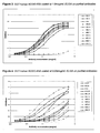
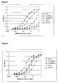
- Figures 1-4 illustrate the dose-dependent binding of humanised antibodies in comparison with the chimera (termed HcLc which is the chimera of 2A10 (comprising the 2A10 murine VH (SEQ ID NO. 7) and VL (SEQ ID NO.8) and human IgG constant regions)) to GST-human NOGO-A56 (see Example 5 for details) in an ELISA assay.
- the Y-axis shows the measured optical density (OD) at 490nm, a quantitative measure of antibody captured in the wells.
- the X-axis shows the concentration of antibody used (mcg/ml) per well at each data point.
- the antibody material used in Figures 1-4 is purified antibody generated by either the polyclonal expression system or large scale transient transfections. In these cases, IgG levels were quantified by ELISA and optical density.
- Nunc Immunosorp plates were coated with a goat anti-human IgG chain capture antibody (Sigma #I3382) at 2 ⁇ g/ml in Bicarbonate buffer (Sigma #C3041) and incubated overnight at 4°C.
- the plates were washed twice with TBS containing 0.05% Tween20 (TBST) and blocked with 200 ⁇ l TBST containing 2% (or from 1-3%) BSA (block buffer) for 1 hr at room temperature.
- the plates were washed twice with TBST.
- Tissue culture supernatants containing antibody were titrated across the plate in 2-fold dilution steps into block buffer and incubated at room temperature for 1 hr.
- the plates were washed three times with TBST.
- HRP conjugated antibody H23 (goat anti-human kappa chain, Sigma #A7164) was diluted 1:2000 in TBST and 100 ⁇ l added to each well. The plates were incubated at room temperature for 1 hr. The plates were washed three times with TBST and developed with 100 ⁇ l of Fast-OPD substrate (Sigma #P9187). Colour was allowed to develop for 5-10mins after which time the ELISA was stopped with 25 ⁇ l 3M H 2 SO 4 . The absorbance at 490nM was read plate and antibody concentration determined by reference to a standard curve.
- a cDNA sequence encoding a polypeptide comprising amino acids 586-785 and a GST tag(SEQ.I.D.NO:32) of human NOGO-A was created by cloning a cDNA encoding amino acids 586-785 of human NOGO-A into the BamHI-Xhol sites of pGEX-6P1 to generate a GST-tagged fusion protein designated GST-human-NOGO-A56.
- Plasmid was expressed in BL21 cells in 2XTY medium with 100 ⁇ g/ml ampicillin following induction with IPTG to 0.5mM at 37C for 3hours. Cell pellets were lysed by sonication and the fusion protein purified using Glutathione-sepharose (Amersham Pharmacia) following manufacturers instructions. Purified protein was eluted using reduced glutathione and extensively dialysed against PBS, quantitated using BSA standards and a BioRad coomassie based protein assay and then stored in aliquots at -80C.
- the binding kinetics of the anti-NOGO monoclonal antibody (mAb) to recombinantly expressed GST-human NOGO-A was analysed using the Biacore3000 biosensor or BIAcore T100.
- the hNOGO-A chip was prepared as follows:
- GST-human NOGO-A56 was immobilised to a CM5 chip by primary amine coupling using the Biacore Wizard program designed for targeted immobilisation levels.
- the CM5 sensor surface was activated by passing a solution of 50mM N-hydroxy-succinimide (NHS) and 200mM N-ethyl-N'-dimethylaminopropyl carbonide (EDC). Then GST-human NOGO-A56in sodium acetate buffer, pH5.0 or pH 4.5, was passed over the chip and immobilised. After immobilisation was complete any still activated esters were blocked by an injection of 1M ethanolamine hydrochloride, pH8.5.
- NHS N-hydroxy-succinimide
- EDC N-ethyl-N'-dimethylaminopropyl carbonide
- the anti-NOGO mAbs were diluted down in HBS-EP (10mM HEPES, pH 7.4, 150mM NaCl, 3mM EDTA, and 0.005% P-20 surfactant) for the BIAcore 3000 or HBS-EP+ (10mM HEPES, pH 7.4, 150mM NaCl, 3mM EDTA, and 0.05% P-20 surfactant) in the case of the T100 and binding studies were Carried out at range of defined antibody concentrations. All runs were referenced against a blanked sensor surface (one that had been activated and blocked as described earlier but had no addition of ligand).
- the GST-human NOGO-A56 chip was prepared as for kinetic analysis.
- Cell supernatants where taken directly from transient transfections of CHO-K1 cells. These were passed directly over the sensor surface and the interaction measured.
- a mock transfected cell supernatant was used for double referencing to remove any artefacts due to the tissue culture media. All runs were referenced against a blanked sensor surface (one that had been activated and blocked as described earlier but had no addition of ligand). Analysis of binding was carried out using the BIAevaluation kinetic analysis software version 4.1.
- the peptides are 16 amino acids in length with a twelve amino acid overlap with the adjacent peptide (each peptide further comprising a biotin-SGSG sequence at the N-terminus) with the exception of the first peptide which has a GSG-biocytin tag at the C-terminus.
- the peptides were used to epitope map the binding site of 2A10 and H28L16.
- Streptavidin at 5 ⁇ g/ml in sterile water was coated onto Nunc immunosorp plates (100 ⁇ l per well) at 37°C overnight. The plates were rinsed 3 times with PBS containing 0.05% Tween (PBST) then blocked with 3% BSA in PBST at 4°C overnight. The plates were washed 3 times with PBST. Peptides were then added to the wells at a concentration of approximately 10 ⁇ g/ml (diluted in 3% BSA in PBST) and incubated at room temperature for 1 hour. The plates were washed 3 times with PBST then incubated for 1 hour with anti-NOGO antibodies diluted to 5 ⁇ g/ml in 3% BSA in PBST.
- PBST PBS containing 0.05% Tween
- the plates were washed 3 times with PBST then incubated with anti-human or anti-mouse kappa peroxidase conjugate (1:1000, diluted in 3% BSA in PBST) for 1 hour.
- the plates were washed 3 times with PBST and then incubated with 100 ⁇ l OPD peroxidase substrate (Sigma) per well for 10 minutes.
- the colour reaction was stopped by the addition of 50 ⁇ l 3 molar H 2 SO 4 .
- Absorbance at 490 nm was measured using a plate reader.
- HcLc A modified variant of HcLc was constructed from existing expression plasmids by introducing a single point mutation, G95M (Kabat numbering), using the Quikchange kit (Stratagene).
- G95M variable heavy domain Hc
- Hc(G95M)Lc was expressed in CHO cells as described previously. The antibody was quantified as described in Example 4.
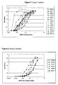
- Figures 5 and Figure 6 show a comparison of the binding activity of Hc(G95M)Lc and HcLc as determined using a human NOGO-A binding ELISA when NOGO was coated onto Nunc immunosorp plates at 0.05 ( Figure 5 ) and 1 ⁇ g/ml ( Figure 6 ).
- Table 12 shows a comparison of the binding affinities of Hc(G95M)Lc and HcLc.
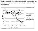
- a panel of 90 heavy chain variable regions was created by single point mutations in the residues contained in the CDR H3, or the preceding Leucine. Specifically, vectors encoding a heavy chain (based on H6FL, SEQ ID NO. 15) were made encoding heavy chain variable regions where each amino acid residue in CDR H3 and the preceding Leucine was substituted (using the Quikchange kit (Stratagene)) with all other naturally occurring amino acids, excluding cysteine, and expressed in conjunction with a light chain (L13FL, SEQ ID NO. 17) to give 90 different antibodies. These antibodies were assayed for binding to NOGO in ELISA and Biacore experiments.
- Figures 7 and 8 show a comparison of the binding activity of the variants of H6FL in comparison to H6FL L13FL.
- Tables 13 and 14 show a comparison of the off-rate kinetics as measured by Biacore - only the results for those antibodies that had a measurable off rate in the Biacore assay and had comparable binding activity to H6L13 in ELISA are shown.
- the antibodies which retain the binding properties of the murine 2A10, and the GQGY containing antibody H6L13 are those containing following CDR H3: RQGY, IQGY, MQGY, GDGY, GIGY, GSGY, GQNY, GQYY, GQSY, GQLY, GQFY, GQGW, WQGY, GAGY, GLGY, GVGY, GQWY.
- Table 14 The antibodies listed in Table 14 were manufactured as described above. Table 14 - humanised 2A10 anti-Nogo-A antibodies giving the total number of back-mutations for the whole antibody (2x heavy chain + 2x light chain). Antibody Total number of back-mutations per whole antibody/tetramer H20L16 22 H28L16 22 H28L13 16 H27L16 32
- the ability of the antibodies to bind recombinant human Nogo-A was investigated by various related ELISA assays (performed in a related, but slightly different, protocol as that described in Example 3).
- the recombinant Nogo-A is directly coated to the plate at various different antigen concentrations.
- the results of the direct binding ELISA when the antigen is loaded at 1mcg/ml or 0.05mcg/ml are shown in Figure 9A and Figure 9B respectively.
- the data confirms that all the antibodies show comparable binding activity to recombinant human Nogo-A when compared with the chimeric form of the parental antibody (HcLc).
- the format of the assay was reversed.
- the antibody is captured on to the plate and the binding of the recombinant human Nogo-A (GST-human Nogo-A-56) detected using the GST tag.
- the results of the reverse format ELISA are shown in Figure 10 .
- the data confirms that all the antibodies show comparable binding activity to recombinant human Nogo-A when compared with the chimeric form of the parental antibody (HcLc). This format of the binding ELISA did not distinguish between the antibodies.
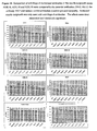
- the ability of the antibodies to compete directly with the parental antibody for the same epitope on human Nogo-A was assessed using a competition ELISA.
- the recombinant human Nogo-A (GST-human Nogo-A 56) was coated onto the plates.
- the parental antibody 2A10 and the humanised antibodies were pre-mixed prior to adding to the plates.
- the binding of 2A10 was quantified using an anti-mouse IgG-HRP conjugate (Dakocytomation, #P0260).
- the results shown in Figure 11 confirm that all four antibodies can compete with 2A10. This suggests that the humanised antibodies and parental antibody recognise an overlapping epitope on human Nogo-A.
- the activity of the humanised antibodies is comparable or better than the chimera HcLc.
- the results indicate that H27L16, H28L16 and H28L13 are more potent than H20L16.
- Biacore was used to determine affinities and rank antibodies using two different methodologies.
- the recombinant Nogo-A was coupled to the surface of the chip and anti-Nogo-A antibodies passed over this surface.
- Protein A was used to capture the antibody onto the surface of the chip over which the recombinant GST-human Nogo-A56 was passed.
- Table 15 were obtained by coupling the antigen to the surface and confirm that all four antibodies show comparable/better affinity than the parental antibody (HcLc). Based on the average of six independent runs, the antibodies rank in the following order in terms of overall affinity: H27L16>H28L16>H28L13>H20L16, consistent with the rank order of the direct binding ELISA ( Figure 9B ).
- the humanised antibodies demonstrate 2-3x higher affinity that the parental antibody (HcLc).
- Table 15 Binding kinetics of the anti-Nogo-A humanised antibodies to recombinant human Nogo-A (GST-human Nogo-A 56) as determined using the Biacore T100.
- the antigen was bound to the CM5 chip by primary amine coupling.
- the antibodies were flowed over a various concentrations (0.125-8nM).
- the values show the mean and standard deviation (in brackets) of six independent runs carried out in duplicate. Each completed data set was analysed independently prior to the calculation of mean and standard deviation. ** Only 11 sets of data analysed for H20L16 as one set could not be analysed.
- Protein A was immobilised to approximately 4000RUs by primary amine and used to capture 200-300RUs of the sample antibodies. Recombinant human Nogo-A was passed over at various concentrations (0.125-8nM). The values show the mean and standard deviation (in brackets) of three independent runs in duplicate. Each data set was independently analysed prior to the calculation of the mean and standard deviation.
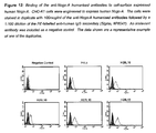
- the ability of the humanised antibodies to bind native Nogo-A was assessed using a human neuroblastoma cell line - IMR32. This cell line is characterised by high intracellular/low cell surface levels of Nogo-A protein.
- the assay was set-up to detect intracellular Nogo-A (ER-resident). IMR32 cells were permeabilised and fixed prior to staining with the anti-Nogo-A humanised antibodies. Binding of the antibodies to Nogo-A was detected using an anti-human IgG-PE labelled secondary (Sigma, #P8047). The results, shown in Figure 14 below, confirm that all the antibodies bind to intracellular Nogo-A at levels comparable or higher than the parental antibody HcLc.
- Humanised anti-Nogo-A antibodies were tested for their ability to neutralise neurite-outgrowth (NO) inhibitory activity of Nogo-A in an assay that is based on quantifying NO as described previously. Antibodies tested in the assay were selected on the basis of their binding kinetics for Nogo-A. High affinity humanised antibodies namely, H28L16, H27L16, H20L16 and for reference their parental antibodies 2A10 (mouse monoclonal) and HcLc (human mouse chimera) were tested for Nogo-A neutralisation. For comparison, antibody 11 C7 (see Example 13) was also tested in the assay.
- the ability of the antibodies to bind full-length extracellular domain recombinant human Nogo-A was investigated by a direct binding ELISA assay.
- the ECD was a splice variant falling within the region of approximately position 186-1004 of human NOGO A (the portion beginning DETFAL (SEQ ID NO.95) and ending with ELSKTS (SEQ ID NO.96)).
- the recombinant GST-human Nogo-A-ECD was directly coated to the plate at 1 ⁇ g/ml.
- the data shown in Figure 15 confirms that H28L16 can recognise GST-human Nogo-A-ECD as levels comparable or better than the parental (HcLc) or H20L16.
- FIG. 17 A-D shows the results of a direct binding ELISA to recombinant NOGO (GST-human Nogo-A 56) from rat (SEQ ID NO.94), cynomolgus (SEQ ID NO. 92), marmoset (SEQ ID NO. 93) and squirrel monkey respectively (SEQ ID NO. 91).
- H28L16 shows activity comparable or better than the chimeric antibody (HcLc).
- the calculated EC50 values are very similar to those calculated for binding to human recombinant Nogo-A.
- Table 17 Binding kinetics of H28L 16, 11 C7 and HcLc to the recombinant orthologues of human Nogo-A as determined using the Biacore T100. Approximately 140-180RUs of the various Nogo-A orthologues were captured to the CM5 chip by primary amine coupling. The antibodies were flowed over a various concentrations (0.125-8nM). The values show the mean and standard deviation (in brackets) of 1-2 independent runs carried out in duplicate with each data set independently analysed prior to calculation of the mean and standard deviation. * One set of curves was discarded due to uninterpretable curves for antibody 11C7.
- Protein A was immobilised on the surface at about 4000RUs and anti-Nogo-A antibodies were captured at approximately 300-400RUs.
- the recombinant proteins (GST- NOGO-A56) were flowed over a various concentrations (0.125-64nM) dependent on the construct. All the runs were done in duplicates. The values show the mean and standard deviation (in brackets) of 1-3 independent runs with each run done in duplicate and each data set analysed independently prior to calculation of the mean and standard deviation.
- SEC-HPLC The physicochemical properties of H28L16 and H20L16 were assessed by SEC-HPLC and SDS-PAGE.
- SEC-HPLC was carried out at 1.Oml/minute using 100mM sodium phosphate, 400mM sodium chloride pH 6.8 and a TSK G3000 SW xl 30cm x 7.8mm stainless steel column with detection at 214nm and 280nm.
- SDS-PAGE was carried out on a 4-20% Novex Tris-HCL gel loading 10 ⁇ g product and staining with Sypro Ruby.
- C-IEF was carried out on a Beckman MDQ using pH 3.5 -10 ampholines.
- the SEC-HPLC data suggests that H20L16 is more susceptible to aggregation than H28L16 (H28L16). If the data reported here were to be repeated at large scale, this could impact the ability of the manufacturing process to produce material of acceptable quality for clinical use (>95% monomer).
- the SDS-PAGE data shows both candidates are acceptable with both showing a typical profile.
- a murine anti-Nogo-A antibody designated 11C7 is described in WO2004052932 , which was raised to a peptide epitope.
- a chimeric 11 C7 was made based on the sequence information provided in W02004052932 .
- a competition ELISA was established to investigate if 11 C7 and 2A10 recognise an overlapping epitope on Nogo-A.
- H28L16 was able to compete with 2A10 for binding to human recombinant Nogo-A whereas 11 C7 showed no competition with 2A10, even at concentrations of up to 100mcg/ml.
- Example 14 Competition ELISA to demonstrate the ability of peptides to compete directly with human NOGO-5+6 for binding to NOGO H28L16
- the ability of peptides to compete directly with NOGO-A (GST-human Nogo-A56) for binding to NOGO H28L16 was assessed using a competition ELISA.
- Rabbit anti-human IgG (Sigma, #I-9764) at 5g/ml in bicarbonate buffer was coated onto Nunc immunosorp plates (100 ul per well) at 4°C overnight. The plates were rinsed 3 times with TBS containing 0.05% Tween (TBST) then blocked with 1% BSA in TBST at room temperature for 1 hour. H28L16 was then captured onto the plate (1 ug/ml, diluted in 1 % BSA in TBST, 50ul per well) at room temperature for 1 hour. The plates were washed 3 times with TBST.
- Peptides from 0 to 100g/ml and GST-human NOGO-A56 at a concentration of 1 ug/ml (diluted in 1 % BSA in TBST) were pre-mixed prior to addition into the wells and incubated at room temperature for 1 hour.
- the plates were washed 3 times with TBST then incubated for 1 hour with rabbit anti-GST peroxidase conjugate (Sigma, #A7340, 1:2000, diluted in 1% BSA in TBST) for 1 hour.
- the plates were washed 3 times with TBST and then incubated with 50 I OPD peroxidase substrate (Sigma) per well for 10 minutes.
- the colour reaction was stopped by the addition of 25 I concentrated H 2 SO 4 . Absorbance at 490 nm was measured using a plate reader.
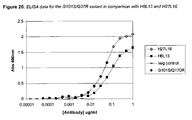
- Example 15 ELISA analysis of a humanised Anti NOGO Monoclonal Antibody based on the NOGO antibody variant G101S / Q37R
- G101S also known as H100 (SEQ ID NO.63)
- a modified variant of the heavy chain variable region of H6 (SEQ ID NO.11) was generated by introducing a single substitution, G101 S (Kabat numbering) into CDR H3 as described above.
- Q37R a modified variant of the light chain variable region of L13 (SEQ ID NO. 13) were generated by introducing a single substitution (Kabat numbering Q37R) into the framework region (to form L100).
- the protein sequence of the variable light domain Q37R is given in SEQ ID NO. 67.
- H100 A modified variant of the heavy chain variable region of H6 (SEQ ID NO.11) was generated by introducing a single substitution, G101 S (Kabat numbering) into CDR H3.
- the protein sequence of the variable heavy domain H100 protein is given in SEQ ID NO.63.
- L100 and L101 modified variants of the light chain variable region of L13 (SEQ ID NO. 13) were generated by introducing a single substitution (Kabat numbering Q37R and Q45R respectively) into the framework region.
- the protein sequences of the variable light domains L100 and L101 proteins are given in SEQ ID NO.67 and SEQ ID NO.68 respectively.
- H100L100 and H100L101 were expressed in CHO cells as described previously.
- Table 21 shows a comparison of the binding affinities of H6L13 with H100L100 and H100L101 and indicates that H100L100 and H100L101 have an improved binding affinity when compared with H6L13.
- the method was performed essentially as described in Example 6 where the CM5 chip was activated by passing the NHS and EDC solutions over the chip at 5 ⁇ l/ml for 7 minutes and the NOGO was suspended in 10nM sodium acetate buffer (pH 4.5) before passing over the chip.
- Table 22 - Biacore measurements for the G101S variants of the H6 variable heavy chain in combination with variants of the L13 variable light chain in comparison with H6L13.
Landscapes
- Health & Medical Sciences (AREA)
- Chemical & Material Sciences (AREA)
- Organic Chemistry (AREA)
- Life Sciences & Earth Sciences (AREA)
- Medicinal Chemistry (AREA)
- General Health & Medical Sciences (AREA)
- Immunology (AREA)
- Engineering & Computer Science (AREA)
- Bioinformatics & Cheminformatics (AREA)
- Proteomics, Peptides & Aminoacids (AREA)
- Biochemistry (AREA)
- Biophysics (AREA)
- Genetics & Genomics (AREA)
- Molecular Biology (AREA)
- Veterinary Medicine (AREA)
- Pharmacology & Pharmacy (AREA)
- Public Health (AREA)
- Animal Behavior & Ethology (AREA)
- General Chemical & Material Sciences (AREA)
- Nuclear Medicine, Radiotherapy & Molecular Imaging (AREA)
- Chemical Kinetics & Catalysis (AREA)
- Biomedical Technology (AREA)
- Neurology (AREA)
- Neurosurgery (AREA)
- Cardiology (AREA)
- Heart & Thoracic Surgery (AREA)
- Mycology (AREA)
- Hospice & Palliative Care (AREA)
- Vascular Medicine (AREA)
- Urology & Nephrology (AREA)
- Microbiology (AREA)
- Psychiatry (AREA)
- Epidemiology (AREA)
- Peptides Or Proteins (AREA)
- Medicines Containing Antibodies Or Antigens For Use As Internal Diagnostic Agents (AREA)
- Medicines That Contain Protein Lipid Enzymes And Other Medicines (AREA)
- Medicinal Preparation (AREA)
- Pharmaceuticals Containing Other Organic And Inorganic Compounds (AREA)
- Micro-Organisms Or Cultivation Processes Thereof (AREA)
Priority Applications (4)
| Application Number | Priority Date | Filing Date | Title |
|---|---|---|---|
| PL06830633T PL1960429T3 (pl) | 2005-12-16 | 2006-12-14 | Immunoglobuliny skierowane przeciwko nogo |
| SI200631393T SI1960429T1 (sl) | 2005-12-16 | 2006-12-14 | Imunoglobulini, usmerjeni proti NOGO |
| EP20100162457 EP2228391A3 (en) | 2005-12-16 | 2006-12-14 | Immunoglobulins directed against Nogo |
| CY20121100801T CY1113094T1 (el) | 2005-12-16 | 2012-09-05 | Ανοσοσφαιρινες κατευθυνομενες εναντι nogo |
Applications Claiming Priority (2)
| Application Number | Priority Date | Filing Date | Title |
|---|---|---|---|
| GBGB0525662.3A GB0525662D0 (en) | 2005-12-16 | 2005-12-16 | Immunoglobulins |
| PCT/EP2006/069737 WO2007068750A2 (en) | 2005-12-16 | 2006-12-14 | Immunoglobulins directed against nogo |
Related Child Applications (1)
| Application Number | Title | Priority Date | Filing Date |
|---|---|---|---|
| EP10162457.5 Division-Into | 2010-05-10 |
Publications (2)
| Publication Number | Publication Date |
|---|---|
| EP1960429A2 EP1960429A2 (en) | 2008-08-27 |
| EP1960429B1 true EP1960429B1 (en) | 2012-06-27 |
Family
ID=35736284
Family Applications (2)
| Application Number | Title | Priority Date | Filing Date |
|---|---|---|---|
| EP06830633A Active EP1960429B1 (en) | 2005-12-16 | 2006-12-14 | Immunoglobulins directed against nogo |
| EP20100162457 Withdrawn EP2228391A3 (en) | 2005-12-16 | 2006-12-14 | Immunoglobulins directed against Nogo |
Family Applications After (1)
| Application Number | Title | Priority Date | Filing Date |
|---|---|---|---|
| EP20100162457 Withdrawn EP2228391A3 (en) | 2005-12-16 | 2006-12-14 | Immunoglobulins directed against Nogo |
Country Status (31)
| Country | Link |
|---|---|
| US (2) | US8362208B2 (es) |
| EP (2) | EP1960429B1 (es) |
| JP (1) | JP5015949B2 (es) |
| KR (1) | KR101355118B1 (es) |
| CN (1) | CN101374863B (es) |
| AR (1) | AR057239A1 (es) |
| AU (1) | AU2006325228B2 (es) |
| BR (1) | BRPI0619855A2 (es) |
| CA (1) | CA2633501C (es) |
| CR (1) | CR10099A (es) |
| CY (1) | CY1113094T1 (es) |
| DK (1) | DK1960429T3 (es) |
| EA (1) | EA015536B1 (es) |
| ES (1) | ES2389380T3 (es) |
| GB (1) | GB0525662D0 (es) |
| HK (1) | HK1119720A1 (es) |
| HR (1) | HRP20120684T1 (es) |
| IL (1) | IL192086A0 (es) |
| JO (1) | JO2795B1 (es) |
| MA (1) | MA30041B1 (es) |
| MY (1) | MY149492A (es) |
| NO (1) | NO20082699L (es) |
| NZ (1) | NZ569143A (es) |
| PE (1) | PE20071099A1 (es) |
| PL (1) | PL1960429T3 (es) |
| PT (1) | PT1960429E (es) |
| SI (1) | SI1960429T1 (es) |
| TW (1) | TWI378940B (es) |
| UA (1) | UA96279C2 (es) |
| WO (1) | WO2007068750A2 (es) |
| ZA (2) | ZA200805111B (es) |
Families Citing this family (15)
| Publication number | Priority date | Publication date | Assignee | Title |
|---|---|---|---|---|
| JP5698534B2 (ja) * | 2007-11-02 | 2015-04-08 | ノバルティス アーゲー | 改良されたnogo−a結合分子およびその医薬的使用 |
| US8669349B2 (en) | 2008-04-02 | 2014-03-11 | Macrogenics, Inc. | BCR-complex-specific antibodies and methods of using same |
| PE20091965A1 (es) | 2008-05-06 | 2010-01-09 | Glaxo Group Ltd | Encapsulacion de agentes biologicamente activos |
| JP2011527317A (ja) * | 2008-07-11 | 2011-10-27 | グラクソ グループ リミテッド | Nogo−aアンタゴニストによる筋萎縮性側索硬化症の治療 |
| WO2012004773A1 (en) | 2010-07-09 | 2012-01-12 | Universite De Geneve | New uses of nogo-a inhibitors and related methods |
| EP2870177A1 (en) | 2012-07-05 | 2015-05-13 | Glaxo Group Limited | Optimum dose regime of an anti-nogo-a antibody in the treatment of amyotrophic lateral sclerosis |
| US20180362660A1 (en) * | 2016-05-09 | 2018-12-20 | Schickwann Tsai | Anti-penumbra monoclonal antibodies for detection and therapy of normal and abnormal B lymphocytes |
| WO2018232144A1 (en) * | 2017-06-14 | 2018-12-20 | Monojul, Llc | High-affinity anti-human folate receptor beta antibodies and methods of use |
| EP4048692A2 (en) | 2019-10-24 | 2022-08-31 | NovaGo Therapeutics AG | Novel anti-nogo-a antibodies |
| GB202012331D0 (en) | 2020-08-07 | 2020-09-23 | Petmedix Ltd | Therapeutic antibodies |
| CA3224517A1 (en) | 2021-06-17 | 2022-12-22 | Petmedix Ltd | Anti canine cd20 antibodies |
| CA3236019A1 (en) | 2021-10-21 | 2023-04-27 | Petmedix Ltd | Proteins comprising the extracellular domain of p75ntr |
| CN116251181B (zh) * | 2021-12-02 | 2023-09-22 | 北京东方百泰生物科技股份有限公司 | 一种抗tslp单克隆抗体的注射制剂 |
| WO2023152486A1 (en) | 2022-02-09 | 2023-08-17 | Petmedix Ltd | Therapeutic antibodies |
| WO2024041450A1 (zh) * | 2022-08-22 | 2024-02-29 | 舒泰神(北京)生物制药股份有限公司 | 特异性识别Nogo-A的抗体及其应用 |
Family Cites Families (31)
| Publication number | Priority date | Publication date | Assignee | Title |
|---|---|---|---|---|
| JPS57106673A (en) | 1980-12-24 | 1982-07-02 | Chugai Pharmaceut Co Ltd | Dibenzo(b,f)(1,4)oxazepin derivative |
| DD266710A3 (de) | 1983-06-06 | 1989-04-12 | Ve Forschungszentrum Biotechnologie | Verfahren zur biotechnischen Herstellung van alkalischer Phosphatase |
| US5807715A (en) | 1984-08-27 | 1998-09-15 | The Board Of Trustees Of The Leland Stanford Junior University | Methods and transformed mammalian lymphocyte cells for producing functional antigen-binding protein including chimeric immunoglobulin |
| GB8422238D0 (en) | 1984-09-03 | 1984-10-10 | Neuberger M S | Chimeric proteins |
| US4879231A (en) | 1984-10-30 | 1989-11-07 | Phillips Petroleum Company | Transformation of yeasts of the genus pichia |
| GB8607679D0 (en) | 1986-03-27 | 1986-04-30 | Winter G P | Recombinant dna product |
| GB8610600D0 (en) | 1986-04-30 | 1986-06-04 | Novo Industri As | Transformation of trichoderma |
| JP3101690B2 (ja) | 1987-03-18 | 2000-10-23 | エス・ビィ・2・インコーポレイテッド | 変性抗体の、または変性抗体に関する改良 |
| US4873316A (en) | 1987-06-23 | 1989-10-10 | Biogen, Inc. | Isolation of exogenous recombinant proteins from the milk of transgenic mammals |
| US5250414A (en) | 1988-11-04 | 1993-10-05 | Erziehungsdirektion Of The Canton Zurich | Diagnostic methods using neurite growth regulatory factors |
| EP0402226A1 (en) | 1989-06-06 | 1990-12-12 | Institut National De La Recherche Agronomique | Transformation vectors for yeast yarrowia |
| WO1993008829A1 (en) | 1991-11-04 | 1993-05-13 | The Regents Of The University Of California | Compositions that mediate killing of hiv-infected cells |
| AU668423B2 (en) | 1992-08-17 | 1996-05-02 | Genentech Inc. | Bispecific immunoadhesins |
| GB9403250D0 (en) | 1994-02-21 | 1994-04-13 | Univ Mcgill | Therapeutic use of myelin-associated glycoprotein (mag) |
| US5429746A (en) | 1994-02-22 | 1995-07-04 | Smith Kline Beecham Corporation | Antibody purification |
| PT940391E (pt) | 1994-05-27 | 2004-12-31 | Glaxosmithkline Spa | Derivados de quinolina como antagonistas do receptor de taquicinina nk3 |
| GB9424449D0 (en) | 1994-12-02 | 1995-01-18 | Wellcome Found | Antibodies |
| CA2225691A1 (en) | 1995-06-27 | 1997-01-16 | Research Foundation Of Cuny, Hunter College | Compositions and methods using myelin-associated glycoprotein (mag) and inhibitors thereof |
| US6420338B1 (en) * | 1997-06-13 | 2002-07-16 | New York University Medical Center | Inhibition of the Src kinase family pathway as a method of treating HBV infection and hepatocellular carcinoma |
| GB9809839D0 (en) | 1998-05-09 | 1998-07-08 | Glaxo Group Ltd | Antibody |
| JP2002522016A (ja) | 1998-07-22 | 2002-07-23 | スミスクライン・ビーチャム・パブリック・リミテッド・カンパニー | 神経内分泌特異的タンパク質に類似したタンパク質、およびそれをコードするcDNA |
| CA2350395C (en) | 1998-11-06 | 2014-07-08 | Martin E. Schwab | Nucleotide and protein sequences of nogo genes and methods based thereon |
| FR2792199B3 (fr) | 1999-04-13 | 2001-05-18 | Sanofi Sa | Utilisation de l'osanetant pour la preparation de medicaments utiles dans le traitement des troubles de l'humeur |
| EP1539233B1 (en) | 2001-07-12 | 2011-04-27 | FOOTE, Jefferson | Super humanized antibodies |
| GB0228832D0 (en) | 2002-12-10 | 2003-01-15 | Novartis Ag | Organic compound |
| GB0318727D0 (en) | 2003-08-08 | 2003-09-10 | Smithkline Beecham Corp | Novel compounds |
| DE10338136A1 (de) | 2003-08-15 | 2005-03-24 | European Molecular Biology Laboratory | Zentrifuge und ein Träger zur Verwendung in einer Zentrifuge |
| GB0321997D0 (en) | 2003-09-19 | 2003-10-22 | Novartis Ag | Organic compound |
| DK1711530T3 (da) * | 2003-12-22 | 2009-12-14 | Glaxo Group Ltd | Nogo-A-neutraliserende immunoglobuliner til behandling af neurologiske sygdomme |
| US7981843B2 (en) * | 2004-01-20 | 2011-07-19 | Kalobios Pharmaceuticals, Inc. | Antibody specificity transfer using minimal essential binding determinants |
| EP1786463A4 (en) * | 2004-03-26 | 2009-05-20 | Human Genome Sciences Inc | ANTIBODY AGAINST NOGO RECEPTOR |
-
2005
- 2005-12-16 GB GBGB0525662.3A patent/GB0525662D0/en not_active Ceased
-
2006
- 2006-12-14 WO PCT/EP2006/069737 patent/WO2007068750A2/en active Application Filing
- 2006-12-14 PT PT06830633T patent/PT1960429E/pt unknown
- 2006-12-14 KR KR1020087017348A patent/KR101355118B1/ko not_active IP Right Cessation
- 2006-12-14 ES ES06830633T patent/ES2389380T3/es active Active
- 2006-12-14 US US12/097,279 patent/US8362208B2/en not_active Expired - Fee Related
- 2006-12-14 NZ NZ569143A patent/NZ569143A/en not_active IP Right Cessation
- 2006-12-14 BR BRPI0619855-4A patent/BRPI0619855A2/pt not_active IP Right Cessation
- 2006-12-14 PE PE2006001607A patent/PE20071099A1/es not_active Application Discontinuation
- 2006-12-14 TW TW095146890A patent/TWI378940B/zh not_active IP Right Cessation
- 2006-12-14 CN CN2006800529521A patent/CN101374863B/zh not_active Expired - Fee Related
- 2006-12-14 AU AU2006325228A patent/AU2006325228B2/en not_active Ceased
- 2006-12-14 MY MYPI20082130A patent/MY149492A/en unknown
- 2006-12-14 PL PL06830633T patent/PL1960429T3/pl unknown
- 2006-12-14 UA UAA200807663A patent/UA96279C2/ru unknown
- 2006-12-14 AR ARP060105524A patent/AR057239A1/es active IP Right Grant
- 2006-12-14 CA CA2633501A patent/CA2633501C/en not_active Expired - Fee Related
- 2006-12-14 JP JP2008545009A patent/JP5015949B2/ja not_active Expired - Fee Related
- 2006-12-14 DK DK06830633.1T patent/DK1960429T3/da active
- 2006-12-14 EP EP06830633A patent/EP1960429B1/en active Active
- 2006-12-14 SI SI200631393T patent/SI1960429T1/sl unknown
- 2006-12-14 EP EP20100162457 patent/EP2228391A3/en not_active Withdrawn
- 2006-12-14 EA EA200801343A patent/EA015536B1/ru not_active IP Right Cessation
- 2006-12-24 JO JO2006457A patent/JO2795B1/en active
-
2008
- 2008-06-11 MA MA31018A patent/MA30041B1/fr unknown
- 2008-06-11 ZA ZA200805111A patent/ZA200805111B/xx unknown
- 2008-06-11 NO NO20082699A patent/NO20082699L/no not_active Application Discontinuation
- 2008-06-12 IL IL192086A patent/IL192086A0/en not_active IP Right Cessation
- 2008-06-20 CR CR10099A patent/CR10099A/es unknown
- 2008-12-08 HK HK08113307.3A patent/HK1119720A1/xx not_active IP Right Cessation
-
2009
- 2009-09-04 ZA ZA200906162A patent/ZA200906162B/en unknown
-
2012
- 2012-08-29 HR HRP20120684TT patent/HRP20120684T1/hr unknown
- 2012-09-05 CY CY20121100801T patent/CY1113094T1/el unknown
-
2013
- 2013-01-28 US US13/751,588 patent/US20130287781A1/en not_active Abandoned
Also Published As
Similar Documents
| Publication | Publication Date | Title |
|---|---|---|
| EP1960429B1 (en) | Immunoglobulins directed against nogo | |
| US7780964B2 (en) | Immunoglobulins which bind NOGO | |
| US20110105724A1 (en) | Novel compounds | |
| EP1899377B1 (en) | Humanised antibodies specific for nogo-a and phrarmaceutical uses thereof | |
| US10221234B2 (en) | Antigen binding proteins | |
| CN101258165B (zh) | Nogo-a对人抗体有特效和它的药学应用 |
Legal Events
| Date | Code | Title | Description |
|---|---|---|---|
| PUAI | Public reference made under article 153(3) epc to a published international application that has entered the european phase |
Free format text: ORIGINAL CODE: 0009012 |
|
| 17P | Request for examination filed |
Effective date: 20080610 |
|
| AK | Designated contracting states |
Kind code of ref document: A2 Designated state(s): AT BE BG CH CY CZ DE DK EE ES FI FR GB GR HU IE IS IT LI LT LU LV MC NL PL PT RO SE SI SK TR |
|
| AX | Request for extension of the european patent |
Extension state: HR |
|
| 17Q | First examination report despatched |
Effective date: 20081021 |
|
| REG | Reference to a national code |
Ref country code: HK Ref legal event code: DE Ref document number: 1119720 Country of ref document: HK |
|
| GRAP | Despatch of communication of intention to grant a patent |
Free format text: ORIGINAL CODE: EPIDOSNIGR1 |
|
| RAX | Requested extension states of the european patent have changed |
Extension state: HR Payment date: 20080610 |
|
| GRAJ | Information related to disapproval of communication of intention to grant by the applicant or resumption of examination proceedings by the epo deleted |
Free format text: ORIGINAL CODE: EPIDOSDIGR1 |
|
| GRAP | Despatch of communication of intention to grant a patent |
Free format text: ORIGINAL CODE: EPIDOSNIGR1 |
|
| GRAS | Grant fee paid |
Free format text: ORIGINAL CODE: EPIDOSNIGR3 |
|
| GRAJ | Information related to disapproval of communication of intention to grant by the applicant or resumption of examination proceedings by the epo deleted |
Free format text: ORIGINAL CODE: EPIDOSDIGR1 |
|
| GRAL | Information related to payment of fee for publishing/printing deleted |
Free format text: ORIGINAL CODE: EPIDOSDIGR3 |
|
| GRAP | Despatch of communication of intention to grant a patent |
Free format text: ORIGINAL CODE: EPIDOSNIGR1 |
|
| GRAS | Grant fee paid |
Free format text: ORIGINAL CODE: EPIDOSNIGR3 |
|
| GRAA | (expected) grant |
Free format text: ORIGINAL CODE: 0009210 |
|
| AK | Designated contracting states |
Kind code of ref document: B1 Designated state(s): AT BE BG CH CY CZ DE DK EE ES FI FR GB GR HU IE IS IT LI LT LU LV MC NL PL PT RO SE SI SK TR |
|
| AX | Request for extension of the european patent |
Extension state: HR |
|
| REG | Reference to a national code |
Ref country code: GB Ref legal event code: FG4D |
|
| REG | Reference to a national code |
Ref country code: CH Ref legal event code: EP |
|
| REG | Reference to a national code |
Ref country code: AT Ref legal event code: REF Ref document number: 564192 Country of ref document: AT Kind code of ref document: T Effective date: 20120715 |
|
| REG | Reference to a national code |
Ref country code: IE Ref legal event code: FG4D |
|
| REG | Reference to a national code |
Ref country code: HR Ref legal event code: TUEP Ref document number: P20120684 Country of ref document: HR |
|
| REG | Reference to a national code |
Ref country code: DE Ref legal event code: R096 Ref document number: 602006030476 Country of ref document: DE Effective date: 20120830 |
|
| REG | Reference to a national code |
Ref country code: CH Ref legal event code: NV Representative=s name: ISLER & PEDRAZZINI AG |
|
| REG | Reference to a national code |
Ref country code: NL Ref legal event code: T3 |
|
| REG | Reference to a national code |
Ref country code: RO Ref legal event code: EPE |
|
| REG | Reference to a national code |
Ref country code: DK Ref legal event code: T3 |
|
| REG | Reference to a national code |
Ref country code: PT Ref legal event code: SC4A Free format text: AVAILABILITY OF NATIONAL TRANSLATION Effective date: 20120918 |
|
| REG | Reference to a national code |
Ref country code: HR Ref legal event code: T1PR Ref document number: P20120684 Country of ref document: HR |
|
| REG | Reference to a national code |
Ref country code: SE Ref legal event code: TRGR |
|
| REG | Reference to a national code |
Ref country code: HK Ref legal event code: GR Ref document number: 1119720 Country of ref document: HK |
|
| REG | Reference to a national code |
Ref country code: ES Ref legal event code: FG2A Ref document number: 2389380 Country of ref document: ES Kind code of ref document: T3 Effective date: 20121025 |
|
| REG | Reference to a national code |
Ref country code: GR Ref legal event code: EP Ref document number: 20120401956 Country of ref document: GR Effective date: 20120915 |
|
| REG | Reference to a national code |
Ref country code: PL Ref legal event code: T3 |
|
| REG | Reference to a national code |
Ref country code: SK Ref legal event code: T3 Ref document number: E 12548 Country of ref document: SK |
|
| REG | Reference to a national code |
Ref country code: EE Ref legal event code: FG4A Ref document number: E007205 Country of ref document: EE Effective date: 20120912 |
|
| REG | Reference to a national code |
Ref country code: HU Ref legal event code: AG4A Ref document number: E014935 Country of ref document: HU |
|
| REG | Reference to a national code |
Ref country code: CH Ref legal event code: PCOW Free format text: NEW ADDRESS: 980 GREAT WEST ROAD, BRENTFORD, MIDDLESEX TW8 9GS (GB) |
|
| PLBE | No opposition filed within time limit |
Free format text: ORIGINAL CODE: 0009261 |
|
| STAA | Information on the status of an ep patent application or granted ep patent |
Free format text: STATUS: NO OPPOSITION FILED WITHIN TIME LIMIT |
|
| 26N | No opposition filed |
Effective date: 20130328 |
|
| REG | Reference to a national code |
Ref country code: DE Ref legal event code: R097 Ref document number: 602006030476 Country of ref document: DE Effective date: 20130328 |
|
| REG | Reference to a national code |
Ref country code: HU Ref legal event code: HC9C Owner name: GLAXO GROUP LIMITED, GB Free format text: FORMER OWNER(S): GLAXO GROUP LIMITED, GB |
|
| REG | Reference to a national code |
Ref country code: HR Ref legal event code: PNAN Ref document number: P20120684 Country of ref document: HR Owner name: GLAXO GROUP LIMITED, GB |
|
| REG | Reference to a national code |
Ref country code: EE Ref legal event code: HE1A Ref document number: E007205 Country of ref document: EE |
|
| REG | Reference to a national code |
Ref country code: SK Ref legal event code: TE4A Ref document number: E 12548 Country of ref document: SK Owner name: GLAXO GROUP LIMITED, BRENTFORD, MIDDLESEX, GB Effective date: 20130815 |
|
| REG | Reference to a national code |
Ref country code: FR Ref legal event code: CA Effective date: 20131004 |
|
| REG | Reference to a national code |
Ref country code: SI Ref legal event code: SP73 Owner name: GLAXO GROUP LIMITED; GB Effective date: 20140217 |
|
| PGFP | Annual fee paid to national office [announced via postgrant information from national office to epo] |
Ref country code: PL Payment date: 20140929 Year of fee payment: 9 |
|
| REG | Reference to a national code |
Ref country code: HR Ref legal event code: ODRP Ref document number: P20120684 Country of ref document: HR Payment date: 20141204 Year of fee payment: 9 |
|
| PGFP | Annual fee paid to national office [announced via postgrant information from national office to epo] |
Ref country code: DK Payment date: 20141124 Year of fee payment: 9 |
|
| PGFP | Annual fee paid to national office [announced via postgrant information from national office to epo] |
Ref country code: RO Payment date: 20141119 Year of fee payment: 9 Ref country code: GR Payment date: 20141125 Year of fee payment: 9 Ref country code: LU Payment date: 20141219 Year of fee payment: 9 Ref country code: FI Payment date: 20141202 Year of fee payment: 9 Ref country code: LT Payment date: 20141201 Year of fee payment: 9 Ref country code: SE Payment date: 20141205 Year of fee payment: 9 Ref country code: EE Payment date: 20141127 Year of fee payment: 9 Ref country code: SK Payment date: 20141127 Year of fee payment: 9 Ref country code: CH Payment date: 20141027 Year of fee payment: 9 Ref country code: MC Payment date: 20141126 Year of fee payment: 9 Ref country code: CZ Payment date: 20141201 Year of fee payment: 9 Ref country code: BG Payment date: 20141010 Year of fee payment: 9 |
|
| PGFP | Annual fee paid to national office [announced via postgrant information from national office to epo] |
Ref country code: HU Payment date: 20141125 Year of fee payment: 9 Ref country code: AT Payment date: 20141124 Year of fee payment: 9 Ref country code: PT Payment date: 20141125 Year of fee payment: 9 Ref country code: LV Payment date: 20141128 Year of fee payment: 9 Ref country code: SI Payment date: 20141125 Year of fee payment: 9 Ref country code: IS Payment date: 20141113 Year of fee payment: 9 |
|
| PGFP | Annual fee paid to national office [announced via postgrant information from national office to epo] |
Ref country code: CY Payment date: 20141118 Year of fee payment: 9 |
|
| REG | Reference to a national code |
Ref country code: FR Ref legal event code: PLFP Year of fee payment: 10 |
|
| REG | Reference to a national code |
Ref country code: PT Ref legal event code: MM4A Free format text: LAPSE DUE TO NON-PAYMENT OF FEES Effective date: 20160614 |
|
| REG | Reference to a national code |
Ref country code: HR Ref legal event code: PBON Ref document number: P20120684 Country of ref document: HR Effective date: 20151214 |
|
| REG | Reference to a national code |
Ref country code: LT Ref legal event code: MM4D Effective date: 20151214 |
|
| REG | Reference to a national code |
Ref country code: DK Ref legal event code: EBP Effective date: 20151231 |
|
| PG25 | Lapsed in a contracting state [announced via postgrant information from national office to epo] |
Ref country code: CY Free format text: LAPSE BECAUSE OF NON-PAYMENT OF DUE FEES Effective date: 20151214 Ref country code: LU Free format text: LAPSE BECAUSE OF NON-PAYMENT OF DUE FEES Effective date: 20151214 Ref country code: CZ Free format text: LAPSE BECAUSE OF NON-PAYMENT OF DUE FEES Effective date: 20151214 Ref country code: MC Free format text: LAPSE BECAUSE OF NON-PAYMENT OF DUE FEES Effective date: 20151231 |
|
| REG | Reference to a national code |
Ref country code: CH Ref legal event code: PL |
|
| REG | Reference to a national code |
Ref country code: SE Ref legal event code: EUG |
|
| REG | Reference to a national code |
Ref country code: EE Ref legal event code: MM4A Ref document number: E007205 Country of ref document: EE Effective date: 20151231 Ref country code: AT Ref legal event code: MM01 Ref document number: 564192 Country of ref document: AT Kind code of ref document: T Effective date: 20151214 |
|
| PG25 | Lapsed in a contracting state [announced via postgrant information from national office to epo] |
Ref country code: LT Free format text: LAPSE BECAUSE OF NON-PAYMENT OF DUE FEES Effective date: 20151214 Ref country code: PT Free format text: LAPSE BECAUSE OF NON-PAYMENT OF DUE FEES Effective date: 20160614 Ref country code: LV Free format text: LAPSE BECAUSE OF NON-PAYMENT OF DUE FEES Effective date: 20151214 Ref country code: RO Free format text: LAPSE BECAUSE OF NON-PAYMENT OF DUE FEES Effective date: 20151214 Ref country code: SE Free format text: LAPSE BECAUSE OF NON-PAYMENT OF DUE FEES Effective date: 20151215 |
|
| REG | Reference to a national code |
Ref country code: SK Ref legal event code: MM4A Ref document number: E 12548 Country of ref document: SK Effective date: 20151214 |
|
| REG | Reference to a national code |
Ref country code: SI Ref legal event code: KO00 Effective date: 20160829 |
|
| REG | Reference to a national code |
Ref country code: GR Ref legal event code: ML Ref document number: 20120401956 Country of ref document: GR Effective date: 20160707 |
|
| PG25 | Lapsed in a contracting state [announced via postgrant information from national office to epo] |
Ref country code: HU Free format text: LAPSE BECAUSE OF NON-PAYMENT OF DUE FEES Effective date: 20151215 Ref country code: CH Free format text: LAPSE BECAUSE OF NON-PAYMENT OF DUE FEES Effective date: 20151231 Ref country code: EE Free format text: LAPSE BECAUSE OF NON-PAYMENT OF DUE FEES Effective date: 20151231 Ref country code: LI Free format text: LAPSE BECAUSE OF NON-PAYMENT OF DUE FEES Effective date: 20151231 Ref country code: BG Free format text: LAPSE BECAUSE OF NON-PAYMENT OF DUE FEES Effective date: 20160930 |
|
| REG | Reference to a national code |
Ref country code: FR Ref legal event code: PLFP Year of fee payment: 11 |
|
| PG25 | Lapsed in a contracting state [announced via postgrant information from national office to epo] |
Ref country code: GR Free format text: LAPSE BECAUSE OF NON-PAYMENT OF DUE FEES Effective date: 20160707 Ref country code: AT Free format text: LAPSE BECAUSE OF NON-PAYMENT OF DUE FEES Effective date: 20151214 Ref country code: SI Free format text: LAPSE BECAUSE OF NON-PAYMENT OF DUE FEES Effective date: 20151215 Ref country code: SK Free format text: LAPSE BECAUSE OF NON-PAYMENT OF DUE FEES Effective date: 20151214 |
|
| PG25 | Lapsed in a contracting state [announced via postgrant information from national office to epo] |
Ref country code: DK Free format text: LAPSE BECAUSE OF NON-PAYMENT OF DUE FEES Effective date: 20151231 |
|
| PGFP | Annual fee paid to national office [announced via postgrant information from national office to epo] |
Ref country code: GB Payment date: 20161125 Year of fee payment: 11 Ref country code: FR Payment date: 20161117 Year of fee payment: 11 Ref country code: IE Payment date: 20161125 Year of fee payment: 11 Ref country code: NL Payment date: 20161207 Year of fee payment: 11 |
|
| PGFP | Annual fee paid to national office [announced via postgrant information from national office to epo] |
Ref country code: ES Payment date: 20161215 Year of fee payment: 11 Ref country code: IT Payment date: 20161221 Year of fee payment: 11 Ref country code: BE Payment date: 20161215 Year of fee payment: 11 |
|
| PGFP | Annual fee paid to national office [announced via postgrant information from national office to epo] |
Ref country code: TR Payment date: 20161122 Year of fee payment: 11 Ref country code: DE Payment date: 20161220 Year of fee payment: 11 |
|
| PG25 | Lapsed in a contracting state [announced via postgrant information from national office to epo] |
Ref country code: PL Free format text: LAPSE BECAUSE OF NON-PAYMENT OF DUE FEES Effective date: 20151214 |
|
| PG25 | Lapsed in a contracting state [announced via postgrant information from national office to epo] |
Ref country code: IS Free format text: LAPSE BECAUSE OF NON-PAYMENT OF DUE FEES Effective date: 20160630 Ref country code: FI Free format text: LAPSE BECAUSE OF NON-PAYMENT OF DUE FEES Effective date: 20151214 |
|
| REG | Reference to a national code |
Ref country code: DE Ref legal event code: R119 Ref document number: 602006030476 Country of ref document: DE |
|
| REG | Reference to a national code |
Ref country code: NL Ref legal event code: MM Effective date: 20180101 |
|
| GBPC | Gb: european patent ceased through non-payment of renewal fee |
Effective date: 20171214 |
|
| REG | Reference to a national code |
Ref country code: IE Ref legal event code: MM4A |
|
| PG25 | Lapsed in a contracting state [announced via postgrant information from national office to epo] |
Ref country code: NL Free format text: LAPSE BECAUSE OF NON-PAYMENT OF DUE FEES Effective date: 20180101 |
|
| REG | Reference to a national code |
Ref country code: FR Ref legal event code: ST Effective date: 20180831 |
|
| REG | Reference to a national code |
Ref country code: BE Ref legal event code: MM Effective date: 20171231 |
|
| PG25 | Lapsed in a contracting state [announced via postgrant information from national office to epo] |
Ref country code: IE Free format text: LAPSE BECAUSE OF NON-PAYMENT OF DUE FEES Effective date: 20171214 Ref country code: IT Free format text: LAPSE BECAUSE OF NON-PAYMENT OF DUE FEES Effective date: 20171214 Ref country code: DE Free format text: LAPSE BECAUSE OF NON-PAYMENT OF DUE FEES Effective date: 20180703 Ref country code: FR Free format text: LAPSE BECAUSE OF NON-PAYMENT OF DUE FEES Effective date: 20180102 |
|
| PG25 | Lapsed in a contracting state [announced via postgrant information from national office to epo] |
Ref country code: BE Free format text: LAPSE BECAUSE OF NON-PAYMENT OF DUE FEES Effective date: 20171231 Ref country code: GB Free format text: LAPSE BECAUSE OF NON-PAYMENT OF DUE FEES Effective date: 20171214 |
|
| REG | Reference to a national code |
Ref country code: ES Ref legal event code: FD2A Effective date: 20190703 |
|
| PG25 | Lapsed in a contracting state [announced via postgrant information from national office to epo] |
Ref country code: ES Free format text: LAPSE BECAUSE OF NON-PAYMENT OF DUE FEES Effective date: 20171215 |
|
| PG25 | Lapsed in a contracting state [announced via postgrant information from national office to epo] |
Ref country code: TR Free format text: LAPSE BECAUSE OF NON-PAYMENT OF DUE FEES Effective date: 20171214 |