EP1614692B1 - ME-5, ME-2, and EPP2: human protein antigens reactive with autoantibodies present in the serum of women suffering from endometriosis - Google Patents
ME-5, ME-2, and EPP2: human protein antigens reactive with autoantibodies present in the serum of women suffering from endometriosis Download PDFInfo
- Publication number
- EP1614692B1 EP1614692B1 EP05254121A EP05254121A EP1614692B1 EP 1614692 B1 EP1614692 B1 EP 1614692B1 EP 05254121 A EP05254121 A EP 05254121A EP 05254121 A EP05254121 A EP 05254121A EP 1614692 B1 EP1614692 B1 EP 1614692B1
- Authority
- EP
- European Patent Office
- Prior art keywords
- endometriosis
- epp2
- protein
- recombinant
- seq
- Prior art date
- Legal status (The legal status is an assumption and is not a legal conclusion. Google has not performed a legal analysis and makes no representation as to the accuracy of the status listed.)
- Expired - Lifetime
Links
Images


Classifications
-
- G—PHYSICS
- G01—MEASURING; TESTING
- G01N—INVESTIGATING OR ANALYSING MATERIALS BY DETERMINING THEIR CHEMICAL OR PHYSICAL PROPERTIES
- G01N33/00—Investigating or analysing materials by specific methods not covered by groups G01N1/00 - G01N31/00
- G01N33/48—Biological material, e.g. blood, urine; Haemocytometers
- G01N33/50—Chemical analysis of biological material, e.g. blood, urine; Testing involving biospecific ligand binding methods; Immunological testing
- G01N33/68—Chemical analysis of biological material, e.g. blood, urine; Testing involving biospecific ligand binding methods; Immunological testing involving proteins, peptides or amino acids
- G01N33/689—Chemical analysis of biological material, e.g. blood, urine; Testing involving biospecific ligand binding methods; Immunological testing involving proteins, peptides or amino acids related to pregnancy or the gonads
-
- A—HUMAN NECESSITIES
- A61—MEDICAL OR VETERINARY SCIENCE; HYGIENE
- A61P—SPECIFIC THERAPEUTIC ACTIVITY OF CHEMICAL COMPOUNDS OR MEDICINAL PREPARATIONS
- A61P15/00—Drugs for genital or sexual disorders; Contraceptives
-
- C—CHEMISTRY; METALLURGY
- C07—ORGANIC CHEMISTRY
- C07K—PEPTIDES
- C07K14/00—Peptides having more than 20 amino acids; Gastrins; Somatostatins; Melanotropins; Derivatives thereof
- C07K14/435—Peptides having more than 20 amino acids; Gastrins; Somatostatins; Melanotropins; Derivatives thereof from animals; from humans
- C07K14/46—Peptides having more than 20 amino acids; Gastrins; Somatostatins; Melanotropins; Derivatives thereof from animals; from humans from vertebrates
- C07K14/47—Peptides having more than 20 amino acids; Gastrins; Somatostatins; Melanotropins; Derivatives thereof from animals; from humans from vertebrates from mammals
- C07K14/4701—Peptides having more than 20 amino acids; Gastrins; Somatostatins; Melanotropins; Derivatives thereof from animals; from humans from vertebrates from mammals not used
- C07K14/4713—Autoimmune diseases, e.g. Insulin-dependent diabetes mellitus, multiple sclerosis, rheumathoid arthritis, systemic lupus erythematosus; Autoantigens
-
- G—PHYSICS
- G01—MEASURING; TESTING
- G01N—INVESTIGATING OR ANALYSING MATERIALS BY DETERMINING THEIR CHEMICAL OR PHYSICAL PROPERTIES
- G01N2800/00—Detection or diagnosis of diseases
- G01N2800/36—Gynecology or obstetrics
- G01N2800/364—Endometriosis, i.e. non-malignant disorder in which functioning endometrial tissue is present outside the uterine cavity
-
- Y—GENERAL TAGGING OF NEW TECHNOLOGICAL DEVELOPMENTS; GENERAL TAGGING OF CROSS-SECTIONAL TECHNOLOGIES SPANNING OVER SEVERAL SECTIONS OF THE IPC; TECHNICAL SUBJECTS COVERED BY FORMER USPC CROSS-REFERENCE ART COLLECTIONS [XRACs] AND DIGESTS
- Y10—TECHNICAL SUBJECTS COVERED BY FORMER USPC
- Y10S—TECHNICAL SUBJECTS COVERED BY FORMER USPC CROSS-REFERENCE ART COLLECTIONS [XRACs] AND DIGESTS
- Y10S424/00—Drug, bio-affecting and body treating compositions
- Y10S424/81—Drug, bio-affecting and body treating compositions involving autoimmunity, allergy, immediate hypersensitivity, delayed hypersensitivity, immunosuppression, immunotolerance, or anergy
-
- Y—GENERAL TAGGING OF NEW TECHNOLOGICAL DEVELOPMENTS; GENERAL TAGGING OF CROSS-SECTIONAL TECHNOLOGIES SPANNING OVER SEVERAL SECTIONS OF THE IPC; TECHNICAL SUBJECTS COVERED BY FORMER USPC CROSS-REFERENCE ART COLLECTIONS [XRACs] AND DIGESTS
- Y10—TECHNICAL SUBJECTS COVERED BY FORMER USPC
- Y10S—TECHNICAL SUBJECTS COVERED BY FORMER USPC CROSS-REFERENCE ART COLLECTIONS [XRACs] AND DIGESTS
- Y10S424/00—Drug, bio-affecting and body treating compositions
- Y10S424/811—Drug, bio-affecting and body treating compositions involving sex selection or contraception
-
- Y—GENERAL TAGGING OF NEW TECHNOLOGICAL DEVELOPMENTS; GENERAL TAGGING OF CROSS-SECTIONAL TECHNOLOGIES SPANNING OVER SEVERAL SECTIONS OF THE IPC; TECHNICAL SUBJECTS COVERED BY FORMER USPC CROSS-REFERENCE ART COLLECTIONS [XRACs] AND DIGESTS
- Y10—TECHNICAL SUBJECTS COVERED BY FORMER USPC
- Y10S—TECHNICAL SUBJECTS COVERED BY FORMER USPC CROSS-REFERENCE ART COLLECTIONS [XRACs] AND DIGESTS
- Y10S530/00—Chemistry: natural resins or derivatives; peptides or proteins; lignins or reaction products thereof
- Y10S530/827—Proteins from mammals or birds
- Y10S530/85—Reproductive organs or embryos
- Y10S530/852—Sperm
- Y10S530/853—Ovary; eggs; embryos
-
- Y—GENERAL TAGGING OF NEW TECHNOLOGICAL DEVELOPMENTS; GENERAL TAGGING OF CROSS-SECTIONAL TECHNOLOGIES SPANNING OVER SEVERAL SECTIONS OF THE IPC; TECHNICAL SUBJECTS COVERED BY FORMER USPC CROSS-REFERENCE ART COLLECTIONS [XRACs] AND DIGESTS
- Y10—TECHNICAL SUBJECTS COVERED BY FORMER USPC
- Y10S—TECHNICAL SUBJECTS COVERED BY FORMER USPC CROSS-REFERENCE ART COLLECTIONS [XRACs] AND DIGESTS
- Y10S530/00—Chemistry: natural resins or derivatives; peptides or proteins; lignins or reaction products thereof
- Y10S530/868—Chemistry: natural resins or derivatives; peptides or proteins; lignins or reaction products thereof involving autoimmunity, allergy, immediate hypersensitivity, delayed hypersensitivity, immunosuppression, or immunotolerance
Definitions
- Endometriosis is a female reproductive disorder characterized by the presence of endometrial tissue outside of the normal uterine location. Most frequently the endometriosis tissue is present in the peritoneal cavity, attaching to various tissues and organs in this location. Endometriosis is a benign disease affecting approximately 5 million women in the United States annually with a prevalence of 10 to 15 percent in women of childbearing age. The incidence increases to 60 to 80 percent of women who are infertile or present with pelvic pain ( D. Gosselin et al. [1999] Curr. Opin. Onco. Endo. & Metabol. Invest. Drugs 1:31 ). The conditions that predispose an individual to endometriosis are still unknown.
- the marker may be of use for patients who are likely to have the disease for faster orientation toward laparoscopy, since CA-125 levels do correlate somewhat with the degree of disease and response to treatment ( T.P. Canavan and L. Radosh [2000] Postgrad. Med. 107:213 ).
- Patent application 2002/0192647 proposes a process for diagnosing angiogenic diseases by measuring a single nucleotide polymorphism in the VEGFR-1 gene. Endometriosis is categorized as one of this group of angiogenic diseases, but it was not the subject of any of the claims. Patent applications 2001/046713 and 2001/044158 describe a method for diagnosis of endometriosis by detecting anti-Tomsen-Frienenreich antibodies in specimens. An issued U.S. Patent 6,376,201 illustrates the use of major histocompatibility complex-class I antigens in diagnosing endometriosis and forming the basis of the Metrio Test as described above.
- European Patent No. 1191107 describes a method for diagnosis of endometriosis by measuring a reduction in the levels of one of a group of 15 different human genes.
- An immunoassay process is described in European Patent No. 0387027 which establishes endometriosis in a patient by evaluating a specimen with an anti-endometriosis monoclonal antibody.
- a method is described in W.O. 0063675 for diagnosis of endometriosis by measuring increased levels of endometriosis factor in biological fluids of a patient.
- W.O. 9963116 provides for a method of diagnosing endometriosis by measuring increases in the amount of prothymosin in endometriotic tissue.
- U.S. Patent 6,531,277 discloses an endometriosis-specific secretory protein.
- the document characterized and disclosed human ENDO-1 that is produced by stromal cells of endometriotic tissue.
- the ENDO-1 protein is 40 to 50 kilodaltons in molecular weight and has an isoelectric point of 4.0 to 5.5.
- the claims of the document are concerned primarily with a molecular diagnostic assay measuring differences in expression of ENDO-1 mRNA in endometrosis tissue samples.
- U.S. 2002/0009718 the invention is extended for measurement of the ENDO-1 glycoprotein in patient samples using immunoassay to establish the presence of endometriosis.
- ENDO-1 the characteristics of ENDO-1 presented in these documents suggest that it is considerably different from the markers described in the present invention.
- the ME-5, ME-2, and EPP2 proteins are about 38, 49, and 9 kilodaltons in size, respectively.
- ME-2 marker is within the range specified for ENDO-1, but ME-2 has an isoelectric point of 8.8 so it is not a related protein.
- the isoelectric points of the ME-5 and EPP2 antigens are calculated at 5.7 and 12.5, respectively, which are also well above the range of values specified for the ENDO-1 protein.
- the ENDO-1 marker is a member of the haptogloblin family of proteins, but nucleic acid and amino acid sequence comparisons show that the ME-5, ME-2, and EPP2 markers are not related to this family of proteins.
- U. S. Patent 5,843,673 specifies a method of screening for endometriosis in women by measuring a reduction in the amounts of a 28 to 32 kilodalton molecular weight glycoprotein in peritoneal fluid or serum samples.
- the protein possesses an isoelectric point of 7.0 to 9.0 and is secreted specifically by stromal cells of endometriotic origin.
- the glycoprotein disclosed in the document is related to tissue inhibitor of metaloproteinases-1 (TIMP-1) by virtue of amino acid sequence identity measured in the amino terminal region of protein.
- TIMP-1 tissue inhibitor of metaloproteinases-1
- the ME-5, ME-2, and EPP2 proteins of this invention are not related to TIMP-1 and they have no measurable protein or nucleic acid homology to this family of proteins.
- the biochemical properties of the ME-5, ME-2, and EPP2 proteins differ from those of TIMP-1 and each is considerably larger or smaller (at 38, 49, or 9 kilodaltons, respectively) than the range given for TIMP-1. While the isoelectric point of ME-2 is at the upper range of that of TIMP-1, the isoelectric point of ME-5 is 5.7 and EPP2 is 12.5 which are much different.
- the individual nucleic acid sequences identified and implicated as somehow being involved in endometriosis are: cathepsin D, AEBP-1, stromelysin-3, cystatin B, protease inhibitor 1, sFRP4, gelsolin, IGFBP-3, dual specificity phosphatase 1, PAEP, immunoglobulin ⁇ chain, ferritin, complement component 3, pro-alpha-1 type III collagen, proline 4-hydroxylase, alpha-2 type I collagen, claudin-4, melanoma adhesion protein, procollagen C-endopeptidase enhancer, nascent-polypeptide-associated complex alpha polypeptide, elongation factor 1 alpha (EF-1 ⁇ ), vitamin D3 25 hydroxylase, CSRP-1, steroidogenic acute regulatory protein, apolipoprotein E, transcobalamin II, prosaposin, early growth response 1 (EGR1), ribosomal protein S6, adenosine deaminase RNA-specific protein
- the overexpressed genes were NADH dehydrogenase, hUCC1, Paralemmin, citrate transport protein. HIF1-alpha, ARNT, Glut-1, MnSOD, GPx, ATP synthase, c-jun, Cx43, HSP 70, and cox2. In addition, 19 genes were reported in this document to be underexpressed in endometriosis patients relative to disease-free females.
- the genes underexpressed in diseased endometrial tissues were Cap43, RNA helicase, CO3, FKHR, AK3, catalase, GST, eNOS, 12S rRNA, TI227H, CO2, aconitase, ANT-1, Bcl-2, COUP-TF, IL-1 beta, HSP 90, GPx4, and GRP78.
- Yet another gene expression strategy was described by H. Hess-Stumpp et al. In US Patent Application 2003/0077589 resulting in the discovery of 15 genes that are overexpressed in endometriosis.
- the overexpressed genes were fibronectin, IGFBP-2, transmembrane receptor PTK7, platelet-derived growth factor alpha, collagen type XVIII alpha 1, subtilisin-like protein (PACE4), laminin M chain (merosin), elastin, collagen type IV alpha 2, p27interferon alpha-inducible gene, reticulocalbin, aldehyde dehydrogenase 6, gravin, nidogen, and phospholipase C epsilon.
- the ME-5, ME-2, and EPP2 protein and nucleic acid sequences are not related to any of the genes described in the latter two patents.
- the document WO 94/28021 describes endometrial proteins, antigenic compounds, and methods of detecting endometriosis.
- the disclosure encompasses endometriosis-specific proteins defined by molecular weight and isoelectric point. Many of the claims presented are based only on size, but others specify a molecular weight and isoelectric point.
- the principal endometriosis antigen of the document and which is described in the initial claim has a molecular weight of 64 kilodaltons and an isoelectric point of 3.5.
- the antigen is used to measure antibodies in specimens obtained from endometriosis patients and also can itself be measured directly for its presence in patient samples.
- the antigens described above do not compare in any reported properties to those of the three endometrosis antigens presented here. Initially, none of the unambiguous residues of amino terminal protein sequence are present in the corresponding regions of ME-5, ME-2, and EPP2. In addition, the ME-5, ME-2, and EPP2 proteins are 38, 49, and 9 kilodaltons in size, which are considerably smaller than the antigens described in the document outlined above. Moreover the isoelectric points of ME-5, ME-2, and EPP2 are 5.7, 8.8, and 12.5 which are considerably greater than described for the other proteins. It must be concluded that the endometrial ME-5, ME-2, and EPP2 antigens of this invention have little in common with the proteins described in WO 94/28021 .
- NZ 232801 also application EP-A-0 387 027 .
- Various antigens are described in the document ranging in molecular weight from 50 to 173 kilodaltons but no additional characterization of the proteins was performed. These proteins were isolated as a mixture from the culture medium and cytoplasm of 2774 ovarian carcinoma cells, and can be obtained from other cultured cell lines as well.
- an anti-endometrial antibody which is a human JgM monoclonal originally isolated because it reacted with ovarian cancer-associated antigens.
- endometrial antigens reactive with anti-endometrial antibodies is described in WO 92/18535 and these are also characterized by molecular weight on SDS PAGE analysis.
- the described protein antigen fragments were isolated from the cytoplasm of epithelial adenocarcinoma cells and are described as useful for detection of endometrial antibodies which are indicative of endometriosis.
- the antigens are cytoplasmic proteins with sizes of 63 to 67, 33 to 37, 40 to 44, 31 to 35, and 57 to 64 kilodaltons. The designations likely refer to a single protein species, but the size ranges were presented in the document to reflect the inherent inaccuracy ( ⁇ 10%) for the SDS PAGE assay method used.
- the preferred proteins for use are the 33 to 37, 40 to 44, and the 57 to 59 kilodalton proteins.
- the 33 to 37 and 40 to 44 proteins seemed to be present in most of the cell lines that were studied in the document for use as sources of antigen, while the 57 to 59 protein fragments originates from the T47D breast carcinoma cell line.
- the document describes the use of these proteins individually (or mixed) immobilized on solid support to measure endometrial antibodies.
- similar applications are envisioned for the ME-5, ME-2, and EPP2 antigens, however with the exception of possibly the 33 to 37 kilodalton fragments there is tittle else presented in this document that compares to disclosures in WO 92/18535 .
- the Invention relates to the method of claim 1 and composition of claim 5. Further, the following subject matter is described herein:
- a purified polypeptide comprising an epitope of at least 5 amino acids of ME-5 (SEQ ID NO:3), wherein the epitope specifically binds to antibodies from subjects diagnosed with endometriosis.
- composition consisting essentially of an antibody that specifically binds to an epitope of ME-5 polypeptide (SEQ ID NO:3).
- a method for detecting a ME-5 polypeptide (SEQ ID NO:3) in a sample comprising the steps of:
- a method for diagnosing endometriosis in a human subject comprising the steps of:
- a recombinant polynucleotide comprising an isolated nucleotide sequence from SEQ ID NO:5 encoding a polypeptide epitope of at least 5 amino acids of ME-2 (SEQ ID NO:6), wherein the epitope specifically binds to antibodies from subjects diagnosed with endometriosis.
- a purified polypeptide comprising an epitope of at least 5 amino acids of ME-2 (SEQ ID NO:6), wherein the epitope specifically binds to antibodies from subjects diagnosed with endometriosis.
- composition consisting essentially of an antibody that specifically binds to an epitope of ME-2 polypeptide (SEQ ID NO:6).
- a method for detecting a ME-2 polypeptide (SEQ ID NO:6) in a sample comprising the steps of:
- a method for diagnosing endometriosis in a human subject comprising the steps of:
- a recombinant polynucleotide comprising an isolated nucleotide sequence from SEQ ID NO:8 encoding a polypeptide epitope of at least 5 amino acids of EPP2 (SEQ ID NO:9), wherein the epitope specifically binds to antibodies from subjects diagnosed with endometriosis.
- a purified polypeptide comprising an epitope of at least 5 amino acids of EPP2 (SEQ ID NO:9), wherein the epitope specifically binds to antibodies from subjects diagnosed with endometriosis.
- composition consisting essentially of an antibody that specifically binds to an epitope of EPP2 polypeptide (SEQ ID NO:9).
- a method for detecting a EPP2 polypeptide (SEQ ID NO:9) in a sample comprising the steps of:
- a method for diagnosing endometriosis in a human subject comprising the steps of:
- a method for diagnosing endometriosis in a human subject comprising the steps of:
- the ME-5 endometriosis marker is specified by a mRNA of about 1.4 kb, of which 1,302 nucleotides is disclosed in this invention.
- the protein predicted from this sequence is 303 amino acids in size and has a calculated molecular weight of about 35,000 daltons.
- the natural protein product has a molecular weight of about 38 kD as measured by Western blot with a specific monoclonal antibody.
- the protein was particularly abundant in ovary tissue which, taken with the isolation from endometrial tissue is strongly supportive of its presence in reproductive tissues and as a marker of reproductive disease.
- immunoblotting experiments with immobilized recombinant ME-5 antigen a number of endometrosis patients were evaluated and the signals generated were considerably stronger than that obtained with a number of control patients.
- the ME-2 endometriosis marker is specified by a mRNA of about 2.0 kb of which 1,353 nucleotides is disclosed in this invention.
- the protein predicted from this sequence is 393 amino acids in size and has a calculated molecular weight of about 45,000 Daltons.
- the signal generated was considerably stronger than that obtained with a number of control patients.
- the EPP2 endometriosis marker is specified by a mRNA of about 1.0 kb of which 891 nucleotides is disclosed in this invention.
- the protein predicted from this sequence is 99 amino acids in size and has a calculated molecular weight of about 9,300 Daltons.
- the signal generated was considerably stronger than that obtained with a number of control patients.
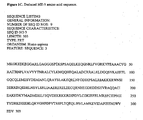
- FIGURES 1A , 1B , and 1C show the nucleotide sequence (SEQ ID NO:1) for the isolated ME-5 cDNA, the nucleotide sequence of the coding region (SEQ ID NO:2) of this ME-5 cDNA, and the deduced amino acid sequence (SEQ ID NO:3) of the protein encoded by the nucleotide sequence of the ME-5 cDNA.
- SEQ ID NO:1 for the isolated ME-5 cDNA
- SEQ ID NO:2 the nucleotide sequence of the coding region
- SEQ ID NO:3 the deduced amino acid sequence of the protein encoded by the nucleotide sequence of the ME-5 cDNA.
- FIGURES 2A , 2B , and 2C show the nucleotide sequence (SEQ ID NO:4) for the isolated ME-2 cDNA, the nucleotide sequence of the coding region (SEQ ID NO:5) of this ME-2 cDNA, and the deduced amino acid sequence (SEQ ID NO:6) of the protein encoded by the nucleotide sequence of the ME-2 cDNA.
- SEQ ID NO:4 there is a 54 base pair 5' untranslated sequence upstream of the predicted ATG start codon.
- a 95 base pair 3' untranslated region downstream of the TAG stop codon is a stretch of dT corresponding to the poly A tail of the mRNA.
- the start codon (ATG) and the translation stop codon (TAG) are presented in bold type in the cDNA sequence of Figures 2A and B .
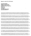
- FIGURES 3A , 3B , and 3C show the nucleotide sequence (SEQ ID NO:7) for the isolated EPP2 cDNA, the nucleotide sequence of the coding region (SEQ ID NO:8) of this EPP2 cDNA, and the deduced amino acid sequence (SEQ ID NO:9) of the protein encoded by the nucleotide sequence of the EPP2 cDNA.
- SEQ ID NO:7 the nucleotide sequence of the coding region
- SEQ ID NO:9 the deduced amino acid sequence of the protein encoded by the nucleotide sequence of the EPP2 cDNA.
- FIG. 3A there is a 45 base pair 5' untranslated sequence upstream of the predicted ATG start codon.
- a 522 base pair 3' untranslated region downstream of the TAA stop codon is a stretch of dT corresponding to the poly A tail of the mRNA.
- the start codon (ATG) and the translation stop codon (TAA)
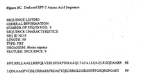
- FIGURE 4 demonstrates the pattern of ME-5 mRNA expression in various human tissues.
- a commercial Northern blot (BD Biosciences; San Diego, CA) was hybridized with the complete 32 P-labeled ME-5 coding sequence of figure 1B . Conditions of hybridization and washing were as described by the manufacturer. Hybridizing bands were observed corresponding to a mRNA of about 1,400 nucleotides (migrates just slower than the 1,350 nucleotide marker) as well as another larger but perhaps less abundant message of 1,800 to 2,000 nucleotides (migrating just ahead of the 2,400 nucleotide marker).
- the ME-5 sequence seems to be expressed most abundantly in prostate, testis and uterus tissues, but lower amounts were detected in the other tissues evaluated (spleen, thymus, small intestine, colon and peripheral blood leukocyte).
- FIGURE 5 demonstrates the pattern of ME-2 mRNA expression in various human tissues.
- a commercial Northern blot (BD Biosciences; San Diego, CA) was hybridized with the complete 32 P-labeled ME-2 coding sequence of figure 2B . Conditions of hybridization and washing were as described by the manufacturer. Hybridizing bands were observed corresponding to a mRNA of about 2,000 nucleotides (migrates about mid way between the 2,400 nucleotide and the 1,350 nucleotide markers). No other strongly hybridizing bands were detected upon the blot.
- the ME-2 sequence seems to be expressed most abundantly in prostate and testis tissues. Moderate levels are detectable in spleen, uterus, small intestine, colon, and peripheral blood leukocyte tissues. In this experiment lower amounts of hybridization were observed in thymus tissue.
- FIGURE 6 demonstrates the pattern of EPP2 mRNA expression in various human tissues.
- a commercial Northern blot (BD Biosciences; San Diego, CA) was hybridized with the complete 32 P-labeled EPP2 coding sequence of figure 3B . Conditions of hybridization and washing were as described by the manufacturer. Hybridizing bands were observed corresponding to a mRNA of about 1,000 nucleotides (migrates just faster than the 1,350 nucleotide marker).
- the EPP2 sequence seems to be expressed most abundantly in prostate, testis, colon and peripheral blood leukocyte. Lesser amounts of signal were visualized in spleen, thymus, and small intestine tissues, but little or no signal was detected in uterus tissue.
- FIGURE 7 shows the pattern of expression of recombinant ME-5 in an insect cell host.
- the ME-5 cDNA was cloned for expression as a 6X histidine-tagged recombinant protein in insect cells.
- a culture of Sf9 insect cells expressing recombinant ME-5 was prepared and lysed. The culture medium, PBS wash, and the soluble and insoluble fractions of the cell lysate were analyzed by SDS PAGE and staining (left panel) of the gel with GelCode blue (Pierce Chemicals; Rockford, IL).
- the expression samples were also evaluated by Western blotting (right panel) with an anti-HisG mouse monoclonal antibody (Invitrogen; Carlsbad, CA) followed by an 125 I-labeled rabbit anti-mouse IgG secondary antibody.
- the recombinant protein was obscured by the multiplicity of protein bands in the stained gel at left, but a band of about 38 kD was clearly detected by the Western blot. This confirmed the presence of a 6X His-tagged protein with the approximate molecular weight expected for the recombinant ME-5 antigen.
- No recombinant ME-5 protein was detectable in the cell culture medium, but some was present in the PBS used to wash the insect cells prior to lysis. Most of the recombinant ME-5 protein seemed to be present in the soluble fraction of the insect cell lysate, but some was associated with the insoluble material.
- FIGURE 8 shows the pattern of expression of recombinant ME-2 in an insect cell host.
- the ME-2 cDNA was cloned for expression as a 6X histidine-tagged recombinant protein in insect cells.
- a culture of Sf9 insect cells expressing recombinant ME-2 was prepared and lysed. The culture medium, PBS wash, and the soluble and insoluble fractions of the cell lysate were analyzed by SDS PAGE and staining (left panel) of the gel with GelCode blue (Pierce Chemicals; Rockford, IL).
- the expression samples were also evaluated by Western blotting (right panel) with an anti-HisG mouse monoclonal antibody (Invitrogen; Carlsbad, CA) followed by an 125 I-labeled rabbit anti-mouse IgG secondary antibody.
- the recombinant protein was obscured by the multiplicity of protein bands in the stained get at left, but a band of about 49 kD was clearly detected by the Western blot. This confirmed the presence of a 6X His-tagged protein with the approximate molecular weight expected for the recombinant ME-2 protein. No recombinant ME-2 protein was detectable in the cell culture medium, but some was present in the PBS used to wash the insect cells prior to lysis. Approximately equal amounts of the recombinant ME-2 protein seemed to be distributed between the soluble and the insoluble fractions of the insect cell lysate.
- FIGURE 9 shows the pattern of expression of recombinant EPP2 in an insect cell host.
- the EPP2 cDNA was cloned for expression as a 6X histidine-tagged recombinant protein in insect cells.
- a culture of Sf9 insect cells expressing recombinant EPP2 was prepared and lysed.
- the culture medium, PBS wash, and the soluble and insoluble fractions of the cell lysate were analyzed by SDS PAGE and staining (left panel) of the gel with GelCode blue (Pierce Chemicals; Rockford, IL).
- the expression samples were also evaluated by Western blotting (right panel) with an anti-HisG mouse monoclonal antibody (Invitrogen; Carlsbad, CA) followed by an 125 I-labeled rabbit anti-mouse IgG secondary antibody.
- the recombinant protein was obscured by the multiplicity of protein bands in the stained gel at left, but a band of about 9 kD was clearly detected by the Western blot. This confirmed the presence of a 6X His-tagged protein with the approximate molecular weight expected for the recombinant EPP2 protein..
- No recombinant EPP2 protein was detectable in the cell culture medium, nor was any measurable amount present in the PBS used to wash the insect cells prior to lysis. Approximately equal amounts of the recombinant EPP2 protein seemed to be distributed between the soluble and the insoluble fractions of the insect cell lysate.
- FIGURE 10 shows the isolation of the recombinant 6X-tagged ME-5 protein using immoblized metal affinity chromatography (IMAC).
- Recombinant ME-5 protein was expressed in Sf9 insect cells and the cells were lysed in IMAC column binding buffer.
- the soluble fraction of the insect cells (Lysate) was loaded onto a column of Chelating Sepharose Fast Flow (Amersham Biosciences; Piscataway, NJ) that had been charged with nickel ions.
- the lysate was captured after passing through the column resin (breakthrough) and the column was washed extensively with IMAC wash buffer.
- the recombinant ME-5 bound to the resin was eluted from the column with buffer containing imidazole.
- FIGURE 11 shows the isolation of the recombinant 6X-tagged ME-2 protein using immoblized metal affinity chromatography (IMAC).
- Recombinant ME-2 protein was expressed in Sf9 insect cells and the cells were lysed in IMAC column binding buffer.
- the soluble fraction of the insect cells (Lysate) was loaded onto a column of Chelating Sepharose Fast Flow (Amersham Biosciences; Piscataway, NJ) that had been charged with nickel ions.
- the lysate was captured after passing through the column resin (break-through) and the column was washed extensively with IMAC wash buffers A10, A15, and A20.
- the recombinant ME-2 bound to the resin was eluted from the column with buffer containing imidazole.
- FIGURE 12 shows the isolation of the recombinant 6X-tagged EPP2 protein using immoblized metal affinity chromatography (IMAC).
- Recombinant EPP2 protein was expressed in Sf9 insect cells and the cells were lysed in denaturing IMAC column binding buffer.
- the insect cell lysate was loaded onto a column of Chelating Sepharose Fast Flow (Amersham Biosciences; Piscataway, NJ) that had been charged with nickel ions.
- the lysate was captured after passing through the column resin (break-through) and the column was washed extensively with A10, A15, A20, A25, and A30 IMAC wash buffers.
- the recombinant EPP2 bound to the resin was eluted from the column with buffer containing imidazole.
- Samples of the lysate, break-through, washes, and elution were analyzed by SDS PAGE and Western blot as described above.
- the stained gel showed the complexity of the insect cell lysate, which resulted in a smear of protein.
- the break-through and the A10 Column Wash samples contained a substantial amount of material that did not bind to the column matrix. Very little protein contaminants were washed away with the A15, A20, A25, and A30 Column Wash buffers as visualized from the stained gel.
- FIGURE 13 shows Western blot analysis of isolated recombinant ME-5 protein, as well as the native ME-5 antigen present in RL95-2 endometrial carcinoma cells.
- Cultured RL95-2 cells were lysed and a sample of the soluble fraction electrophoresed in a 4% to 20% Tris Glycine SDS PAGE gel (Invitrogen; Carlsbad, CA).
- a sample of recombinant ME-5 isolated by IMAC from Sf9 insect cells was included on the gel as a positive control for the anti-ME-5 antibody.
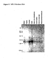
- FIGURE 14 is a Western blot showing ME-5 native antigen expression in various human tissues.
- Tissue protein extracts in SDS PAGE sample buffer protein medleys: BD Biosciences; San Diego, CA
- SDS PAGE gels Western blotting done as described in Figure 13 .
- the native ME-5 antigen seems to be ubiquitously present in all tissues examined, but it appears to be slightly more abundant in heart, liver, ovary and kidney extracts.
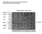
- FIGURES 15A and 15B show representative line immunoblots illustrating the ability of recombinant ME-5 to react with antibodies present in serum obtained from endometriosis patients, but not in normal control sera.
- Each strip contains immobilized antigens that were slotted onto the membrane at different concentrations.
- the protein concentrations for ME-5 are 0.018, 0.036, 0.072, and 0.144 milligrams per milliliter (mg/ml).
- the optimal concentration for discrimination between patients and controls was 0.036 mg/ml as designated by the arrow at the right of the line blot strips.
- One advantage of the line immunoblot assay is that many different proteins can be interrogated on a single strip, and additional unrelated proteins are present on the strips that act as internal controls.
- a reagent control (mouse anti-human IgG monoclonal) is included on each strip to act as a positive control.
- Each strip was incubated with serum from a normal person (control) or from a patient with confirmed endometriosis.
- Line blot patterns for a total of 11 controls (A6, A7, A8, A9, A10, A14, A15, A16, A17, A18, A21) are shown in Figure 15A .
- ME-5 at a concentration of 0.036 mg/ml detected 18 endometriosis patients as positive (DS01, DS03, DS05, DS06, DS10, DS11, DS12, DS27, DS28, DS29, DS30, DS31, DS32, DS33, DS34, DS36, DS38, and DS39).
- 5 endometriosis patients (DS02, DS04, DS07, DS08, and DS13) yielded patterns of reactivity that were a bit lower.
- ME-5 clearly did not react with nine of them (A6, A7, A8, A10, A15, A16, A17, A18, A21). There may have been detectable signals seen for two of the normal controls (A9, A14), but these were very light relative to the patterns seen with sera from the endometriosis patients and are interpreted as negative.
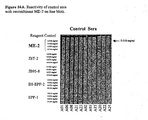
- FIGURES 16A and 16B show representative line immunoblots illustrating the ability of recombinant ME-2 to react with antibodies present in serum obtained from endometriosis patients, but not in normal control sera.
- Each strip contains immobilized antigens that were slotted onto the membrane at different concentrations.
- the protein concentrations of ME-2 applied to the strips are 0.009 (for endometriosis sera, only), 0.018, 0.036, 0.072, and 0.144 (for control sera, only) milligrams per milliliter (mg/ml).
- the optimal concentration for discrimination between patients and controls was set at 0.018 mg/ml as designated by the arrow at the right of the line blot strips.
- Line immunoblot assay One advantage of the line immunoblot assay is that many different proteins can be interrogated on a single strip for reactivity with antibodies, and additional unrelated proteins are present on the strips that act as internal controls.
- a reagent control mouse anti-human IgG monoclonal
- Each strip was incubated with serum from a normal person (control) or from a patient with confirmed endometriosis.
- Line blot patterns for a total of 11 controls (A01, A02, A03, A06, A08, A15, A20, A21, A22, A23, and A24) are shown in Figure 16A .
- 21 endometriosis patients (DS10, DS11, DS12, DS13, DS14, DS17, DS19, DS20, DS21, DS22, DS24, DS25, DS26, DS27, DS28, DS29, DS30, DS31, DS32, DS33, and DS35) are shown in Figure 16B .
- the intensity of staining of each band is indicative of the reactivity of the tested serum with ME-2.
- ME-2 at a concentration of 0.018 mg/ml detected 15 endometriosis patients as positive (DS012, DS17, DS19, DS20, DS21, DS22, DS24, DS25, DS26, DS27, DS28, DS30, DS31, DS33, and DS35).
- 6 endometriosis patients (DS10, DS11, DS13, DS14, DS29, and DS32) yielded patterns of reactivity that were a bit lower.
- ME-2 did not react with any of them at the 0.018 mg/ml cutoff applied to endometriosis patients.
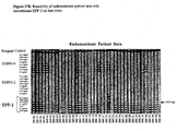
- FIGURES 17A and 17B show representative line immunoblots illustrating the ability of recombinant EPP2 to react with antibodies present in serum obtained from endometriosis patients, but not in normal control sera.
- Each strip contains immobilized antigens that were slotted onto the membrane at different concentrations.
- the protein concentrations for EPP2 are 0.01, 0.025, 0.05, 0.1, 0.15, 0.2, and 025 milligrams per milliliter.
- the optimal concentration for discrimination between patients and controls was 0.05 mg/ml as designated by the arrow at the right of the line blot strips.
- One advantage of the line immunoblot assay is that many different proteins can be interrogated on a single strip, and additional unrelated proteins are present on the strips that act as internal controls.
- a reagent control (mouse anti-human IgG monoclonal) is included on each strip to capture human IgG in the sample and act as a positive control.
- Each strip was incubated with serum from a normal person (control) or from a patient with confirmed endometriosis.
- Line blot patterns for a total of 11 controls (A01, A02, A03, A04, A05, A09, A13, A14, A16, A20, and A24) are shown in Figure 17A .
- EPP2 at a concentration of 0.05 mg/ml detected 33 endometriosis patients as positive (DS06, DS12, DS24, DS05, BBI02, BBI03, BBI04, BBI06, BBI07, BBI08, BBI09,BBI10, BBI11, BBI12, BBI13, BBI15, BBI16, BBI20, BBI22, BBI23, BBI25, BBI26, BBI27, BBI28, BBI30, BBI31, BBI32, BBI34, BBI35, BBI37, BBI38, BBI39, and BBI40).
- Polypeptide refers to a polymer composed of amino acid residues, related naturally occurring structural variants, and synthetic non-naturally occurring analogs thereof linked via peptide bonds, related naturally occurring analogs thereof. Synthetic polypeptides can be synthesized, for example,using an automated polypeptide synthesizer.
- the term “protein” typically refers to large polypeptides.
- the term “peptide” typically refers to short polypeptides.
- polypeptide sequences the lefthand end of a polypeptide sequence is the amino-terminus; the right-hand end of a polypeptide sequence is the carboxyl-terminus.
- Constant substitution refers to the substitution in a polypeptide of an amino acid with a functionally similar amino acid. It is to be understood that the claims encompass conservative substitution. The following six groups each contain amino acids that are conservative substitutions for one another:
- Allelic Variant refers to any of two or more polymorphic forms of a gene occupying the same genetic locus. Allelic variations arise naturally through mutation, and may result in phenotypic polymorphism within populations. Gene mutations can be silent (no change in the encoded polypeptide) or may encode polypeptides having altered amino acid sequences. "Allelic variants” also refer to cDNAs derived from mRNA transcripts of genetic allelic variants, as well as the proteins encoded by them.
- This invention provides methods for diagnosing endometriosis in a subject by detecting in a sample from the subject a diagnostic amount of an antibody that specifically binds to ME-2, ME-5 or EPP2 polypeptide.
- Suitable patient samples include, without limitation, saliva, blood or a blood product (e.g., serum), peritoneal fluid, urine, menstrual fluid, vaginal secretion.
- the antibodies can be detected by any of the methods for detecting proteins described herein. However, sandwich type assays are particularly useful.
- all antibodies are captured onto a solid phase, for example using protein A, and antibodies specific for ME-2, ME-5 or EPP2 are detected using a directly or indirectly labeled ME-2, ME-5 or EPP2 or polypeptide fragment of it having an epitope of ME-2, ME-5 or EPP2.
- ME-2, ME-5 or EPP2 or an antigenic fragment of it can be used as the capture molecule and captured antibodies can be detected.
- ME-2, ME-5 or EPP2 that is shed into the peritoneal fluid of women with endometriosis is useful in methods of diagnosing endometriosis. These methods include detecting ME-2, ME-5 or EPP2 in a biological sample of a subject. Suitable samples include, without limitation, saliva, blood or a blood product (e.g., serum), urine, menstrual fluid, vaginal secretion and, in particular, peritoneal fluid.
- ME-2, ME-5 or EPP2 can be detected by any of the methods described herein. Any detection of ME-2, ME-5 or EPP2 above a normal range is a positive sign in the diagnosis of endometriosis.
- substantially identical in the context of two nucleic acids or polypeptides, refers to two or more sequences or sub-sequences that have at least 60%, 80%, 90%, 95% or 98% nucleotide or amino acid residue identity, when compared and aligned for maximum correspondence, as measured using one of the following sequence comparison algorithms or by visual inspection.
- the substantial identity exists over a region of the sequences that is at least about 50 residues in length, more preferably over a region of at least about 100 residues, and most preferably the sequences are substantially identical over at least about 150 residues.
- the sequences are substantially identical over the entire length of the coding regions
- the endometriosis tissue cDNA-library was generated using poly A + RNA isolated from a deep embedded endometriosis tissue specimen donated by Professor Philip Koninckx at the Catholic University of Leuven. Total RNA was isolated from the tissue using Trizol reagent (Biorad Laboratories; Hercules, CA), and poly A+ RNA was prepared by hybridization to oligo poly T coupled magnetic particles using a commercial kit (PolyATract; Promega; Madison, WI). Library construction was carried out using the Lambda ZAP ® II vector system following instructions obtained from the supplier (Stratagene; San Diego, CA).
- the initial ME-5 and ME-2 cDNA clones were identified by immunoscreening using, as primary antibody, a single endometriosis patient serum specimen obtained from a woman diagnosed with mild disease. This serum was adsorbed of nonspecific anti- E . co li /lambda phage antibodies by diluting the sera 1:50 in a commercial E . coli phage lysate (Stratagene; San Diego, CA) according to the protocol provided by the supplier.
- EPP2 cDNA clone was identified in similar immunoscreening protocol except that, as primary antibody, a pool of ten endometriosis patient serum specimens was used. The sera in this pool were from women with various stages of endometrial disease.
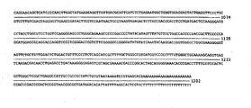
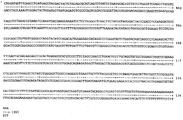
- the ME-5 cDNA sequence is presented in figure 1A (SEQ ID NO:1) and it is 1,279 base pairs in size excluding the poly dA track.
- a 5' noncoding sequence of 112 base pairs was identified just upstream of the suspected ATG start codon.
- the ME-5 coding sequence is shown in Figure 1B (SEQ ID NO:2) as predicted from the entire isolated cDNA sequence ( Figure 1A ).
- the coding region is 912 base pairs in size, including the start and stop codons.
- the cDNA codes for a predicted protein of 303 amino acids shown in Figure 1C (SEQ ID NO:3) and the calculated molecular weight was about 35,000 Daltons.
- the translation product is slightly acidic with a calculated isoelectric value of 5.7.
- NCBI National Center for Biotechnology Information
- BLAST Basic Local Alignment Search Tool
- the 1-NY-CO-7 protein was reported to be 356 amino acids on size which is considerably larger than the predicted ME-5 protein.
- the nucleotide changes were at nucleotide 807 (C -> G [occurs in 3 rd position of codon with no amino acid change -> proline]), 814 (C -> G [arginine -> glycine]), and 838 (C -> T [leucine -> phenylalanine]) relative to the ME-5 coding domain.
- the 1-NY-CO-7 GenBank sequences were compared to that of ME-5 they were identical except for the three nucleotide mismatches described above.
- CHIP Hsp70-interacting protein
- a low stringency hybridization yielded 12 clones that did not hybridize at higher stringency (55°C). Characterization of the clones revealed 8 of them corresponded to human CyP-40, and 4 clones encoded CHIP that was a sequence with no homology to known genes. Characterization of CHIP revealed that it interacts with both Hsc70 and Hsp70 by binding to the carboxy terminus of these proteins through sequences within the amino terminus of CHIP. Interestingly recombinant CHIP inhibited the Hsp40-stimulated ATPase activity of Hsc70 and Hsp70 suggesting that it regulated the forward reaction of the substrate-binding cycle.
- the ME-2 cDNA sequence is presented in Figure 2A (SEQ ID NO:4) and it is 1,332 base pairs in size excluding the poly dA track.
- a 5' noncoding sequence of 54 base pairs was identified just upstream of the suspected ATG start codon.
- the ME-2 coding sequence is shown in Figure 2B (SEQ ID NO:5) as predicted from the entire isolated cDNA sequence ( Figure 2A ).
- the coding region is 1182 base pairs in size, including the start and stop codons.
- the cDNA codes for a predicted protein of 393 amino acids shown in Figure 2C (SEQ ID NO:6) and the calculated molecular weight was about 45,000 Daltons.
- the translation product is slightly acidic with a calculated isoelectric value of 8.8.
- the EPP2 cDNA sequence is presented in Figure 3A (SEQ ID NO:7) and it is 868 base pairs in size excluding the poly dA track. A 5' noncoding sequence of 45 base pairs was identified just upstream of the suspected ATG start codon. There is a 3' non coding sequence of 522 base pairs down stream of the TAA stop codon and this is followed by a stretch of dA residues that would correspond to the poly A tail at the 3' end of the mRNA. Both the start and stop codon are highlighted in bold type in figures 3A and 3B .
- the EPP2 coding sequence is shown in Figure 3B (SEQ ID NO:8) as predicted from the entire isolated cDNA sequence ( Figure 3A ).
- the coding region is 300 base pairs in size, including the start and stop codons.
- the cDNA codes for a predicted protein of 99 amino acids shown in Figure 3C (SEQ ID NO:9) and the calculated molecular weight was approximately 9300 Daltons. Interestingly 18 of the amino acids are arginine residues therefore the translation product is very basic with a calculated isoelectric value of 12.5.
- the gene expression profile of ME-5 from normal human tissues was done by performing Northern blot analysis with a commercial Multiple Tissue Northern Blot (BD Biosciences; San Diego, CA), the results of which are presented in Figure 4 .
- the commercial Northern blot contained RNA from the following tissues: spleen, thymus, prostate, testis, uterus, small intestine, colon (no mucosa), and peripheral blood leukocyte.
- the entire 912 base pair coding sequence was isolated by electrophoresis in a low melting agarose gel, and labeled with 32 P by random priming.
- the 32 P-labeled ME-5 probe was used for hybridization to the Northern blot using the procedure supplied by the manufacturer. After washing the blot was exposed to X-ray film. Upon development of the film a band at about 1.4 kb on the Northern blot corresponds to the ME-5 transcript of the expected size ( Figure 4 ).
- the transcript can be seen in all tissues and is particularly abundant in prostate, testis and
- Gene expression profile of ME-2 from normal human tissues was done by performing Northern blot analysis with a commercial Multiple Tissue Northern Blot (BD Biosciences; San Diego, CA), the results of which are presented in Figure 5 .
- the commercial Northern blot contained RNA from the following tissues: spleen, thymus, prostate, testis, uterus, small intestine, colon (no mucosa), and peripheral blood leukocyte.
- the entire 1182 base pair coding sequence was isolated by electrophoresis in a low melting agarose gel, and labeled with 32 P by random priming.
- the 32 P-labeled ME-2 probe was used for hybridization to the Northern blot using the procedure supplied by the manufacturer. After washing the blot was exposed to X-ray film.
- RNAs expressed in spleen, uterus, small intestine, colon, and peripheral blood lymphocyte tissues Interestingly, despite the pattern observed with the peripheral blood lymphocytes, relatively little signal could be detected in thymus tissue.
- EPP2 Gene expression profile of EPP2 from normal human tissues was done by performing Northern blot analysis with a commercial Multiple Tissue Northern Blot (BD Biosciences; San Diego, CA), the results of which are presented in Figure 6 .
- the commercial Northern blot contained RNA from the following tissues: spleen, thymus, prostate, testis, uterus, small intestine, colon (no mucosa), and peripheral blood leukocyte.
- the entire 300 base pair EPP2 coding sequence was isolated by electrophoresis in a low melting agarose gel, and labeled with 32 P by random priming.
- the 32 P-labeled EPP2 probe was used for hybridization to the Northern blot using the procedure supplied by the manufacturer. After washing the blot was exposed to X-ray film.
- a band at about 1.0 kb on the Northern blot corresponds to the EPP2 transcript hybridizing to the labeled probe ( Figure 6 ).
- the transcript can be seen in all tissues and is most abundant in prostate, testis, colon, and peripheral blood lymphocyte tissues. In addition, the transcript is present but the relative levels of hybridization are lower among the RNAs expressed in spleen, thymus, uterus, and.small intestine tissues.
- the ME-5 antigen was cloned for expression as a 6 X histidine-tagged fusion protein in insect cells.
- the sequence of the ME-5 cDNA insert was generated by PCR amplification using specific primers that flanked the 912 bp coding region. Unique sites for the Bam HI and Eco RI restriction enzymes were incorporated into the primers to maintain the ME-5 reading frame with the vector sequences.
- the PCR amplicons were digested with the Bam HI and Eco RI restriction enzymes (Stratagene; San Diego, CA) and purified by agarose gel electrophoresis.
- the insect cell transfer vector Blue Bac His2a (Stratagene; San Diego, CA) was also digested with the restriction enzymes Bam HI and Eco RI and treated with calf intestine alkaline phosphatase.
- the ME-5 cDNA insert was ligated with the vector and competent bacteria transformed. Individual isolated clones were grown, plasmid DNA isolated, and digested with the restriction enzymes Bam HI and Eco RI. Clones producing a band of about 900 bp in addition to the linear vector were chosen. Several candidates were further characterized by DNA sequence analysis to verify that no changes occurred during the process of PCR amplification and cloning.
- Recombinant baculoviruses were generated by cotransfection of Sf9 insect cells with baculovirus DNA and the ME-5 transfer vector. The baculoviruses were isolated by plaque purification and used to evaluate expression patterns in pilot cultures. The recombinant baculovirus virus and pilot scale cultures were evaluated for expression patterns.
- One recombinant baculovirus clone was identified which expressed an antigen of approximately 38 kD, which was detected in both the soluble and the insoluble fraction of the insect cell lysates. The clone was expanded into large-scale virus stocks for expression of recombinant ME-5 protein.
- the ME-2 antigen was cloned for expression as a 6 X histidine-tagged fusion protein in insect cells as described above for the ME-5 activity.
- the sequence of the ME-2 cDNA insert was generated by PCR amplification using specific primers that flanked the 1182 bp coding region. Unique sites for the Bam HI and Eco RI restriction enzymes were incorporated into the primers to maintain the ME-2 reading frame with the vector sequences.
- the PCR amplicons were digested with the Bam HI and Eco RI restriction enzymes (Stratagene; San Diego, CA) and purified by agarose gel electrophoresis.
- the insect cell transfer vector Blue Bac His2a (Stratagene; San Diego, CA) was also digested with the restriction enzymes Bam HI and Eco RI and treated with calf intestine alkaline phosphatase.
- the ME-2 cDNA insert was ligated with the vector and competent bacteria transformed. Individual isolated clones were grown, plasmid DNA isolated, and digested with the restriction enzymes Bam HI and Eco RI. Clones producing a band of about 1100 bp in addition to the linear vector were chosen. Several candidates were further characterized by DNA sequence analysis to verify that no changes occurred during the process of PCR amplification and cloning.
- Recombinant baculoviruses were generated by cotransfection of Sf9 insect cells with baculovirus DNA and the ME-2 transfer vector. The baculoviruses were isolated by plaque purification and used to evaluate expression patterns in pilot cultures. The recombinant baculovirus virus and pilot scale cultures were evaluated for expression patterns.
- One recombinant baculovirus clone was identified which expressed an antigen of approximately 49 kD, which was detected in both the soluble and the insoluble fraction of the insect cell lysates. The clone was expanded into large-scale virus stocks for expression of recombinant ME-2 protein.
- the EPP2 antigen was cloned for expression as a 6 X histidine-tagged fusion protein in insect cells as described above.
- the sequence of the EPP2 cDNA insert was generated by PCR amplification using specific primers that flanked the 300 bp coding region. Unique sites for the Bam HI and Eco RI restriction enzymes were incorporated into the primers to maintain the EPP2 reading frame with the vector sequences.
- the PCR amplicons were digested with the Bam HI and Eco RI restriction enzymes (Stratagene; San Diego, CA) and purified by agarose gel electrophoresis.
- the insect cell transfer vector Blue Bac His2a (Stratagene; San Diego, CA) was also digested with the restriction enzymes Bam HI and Eco RI and treated with calf intestine alkaline phosphatase.
- the EPP2 cDNA insert was ligated with the vector and competent bacteria transformed. Individual isolated clones were grown, plasmid DNA isolated, and digested with the restriction enzymes Bam HI and Eco RI. Clones producing a band of about 300 bp in addition to the linear vector were chosen. Several candidates were further characterized by DNA sequence analysis to verify that no changes occurred during the process of PCR amplification and cloning.
- Recombinant baculoviruses were generated by cotransfection of Sf9 insect cells with baculovirus DNA and the EPP2 transfer vector. The baculoviruses were isolated by plaque purification and used to evaluate expression patterns in pilot cultures. The recombinant baculovirus virus and pilot scale cultures were evaluated for expression patterns.
- One recombinant baculovirus clone was identified which expressed an antigen of approximately 9 kD, which was detected in both the soluble and the insoluble fraction of the insect cell lysates.
- the clone was expanded into large-scale virus stocks for expression of recombinant EPP2 protein. This was used to infect a large-scale culture of Sf9 insect cells. The pattern of expression is illustrated in Figure 9 , and is best visualized by the Western blot analysis ( Figure 9 ).
- the presence of recombinant EPP2 was confirmed with a commercial anti-HisG monoclonal antibody (Invitrogen; Carlsbad, CA) followed by an 125 I-labeled rabbit anti-mouse IgG secondary antibody. This confirms the presence of a 6X histidine-tagged protein of approximately 9 kD which is the molecular weight expected for EPP2.
- the recombinant was detected in both the soluble and the insoluble fraction of the insect cell lysates, but slightly more antigen seems to be localized in the soluble fraction. In contrast to the patterns seen with the ME-5 and ME-2 protein expression, no EPP2 antigen was present in the PBS used to wash the infected cells prior to the lysis.
- ME-5, ME-2, and EPP2 antigens require substantial amounts of isolated protein. Specifically, these are needed for evaluating the reactivity of the ME-5, ME-2, and EPP2 proteins with endometriosis patient serum specimens to establish clinical relevance.
- the recombinant ME-5, ME-2, and EPP2 antigens were isolated from the soluble fraction or the whole cell lysate by immobilized metal affinity chromatography (IMAC).
- ME-5 antigen was isolated from the soluble fraction of the insect cell lysate. Briefly, ME-5 recombinant baculovirus-infected insect cells were harvested after three days of infection by centrifugation. The cells were washed twice with PBS and the cell pellet frozen for one hour at -70°C. After thawing the cell pellet was suspended in binding buffer (500 mM NaCl, 20 mM Tris-HCl, pH 8.0) supplemented with protease inhibitor cocktail for mammalian tissues (Sigma; St. Louis, MO). The lysate was sonicated, and centrifuged at 18,000 rpm, 4°C for 20 minutes to separate the soluble and insoluble fractions.
- binding buffer 500 mM NaCl, 20 mM Tris-HCl, pH 8.0
- the soluble fraction was dialyzed against binding buffer, and centrifuged at 18,000 rpm, 4°C to remove impurities that might affect the performance of the column.
- Nickel-charged chelating Sepharose resin (Amersham Biosciences; Piscataway, NJ) was equilibrated twice with 2 X column volume of binding buffer. The resin was incubated with the ME-5 insect cell lysate for 20 minutes on a rocker at room temperature. The resin/lysate mixture is loaded on a column and washed with 40 column volumes of A20 Column Wash buffer (20 mM imidazole; 500 mM NaCl; 20 mM Tris-HCl, pH 8.0).
- Bound ME-5 protein was eluted from the column with elution buffer (500 mM imidazole; 500 mM NaCl; 20 mM Tris-HCl, pH 7.5). Protease inhibitor cocktail was added to the pooled elution fractions and protein concentration measured by BCA assay (Pierce; Rockford, IL) using a BSA standard curve. The eluted protein samples are analyzed upon SDS PAGE followed by staining with Coomassie blue or transfer to nitrocellulose and Western blotting as shown in Figure 10 . Such isolated ME-5 protein preparations are divided into aliquots and stored at -20°C with 30 % glycerol.
- ME-2 antigen was also isolated from the soluble fraction of the insect cell lysate. Briefly, ME-2 recombinant baculovirus-infected insect cells were harvested after three days of infection by centrifugation. The cells were washed with PBS and the cells lysed as described above. After dialysis the soluble fraction was allowed to bind to nickel-charged chelating Sepharose resin (Amersham Biosciences; Piscataway, NJ) for 20 minutes at room temperature.
- the resin/lysate mixture is loaded on a column and washed sequentially with denaturing binding buffer A10 (10 mM imidazole, 1 M NaCl, 20 mM Tris-HCl, pH 8.0, 10 % glycerol, 6 M Urea), A15 (buffer A10 containing 15 mM imidazole), and A20 (buffer A10 with 20 mM imidazole).
- denaturing binding buffer A10 (10 mM imidazole, 1 M NaCl, 20 mM Tris-HCl, pH 8.0, 10 % glycerol, 6 M Urea
- A15 buffer A10 containing 15 mM imidazole
- A20 buffer A10 with 20 mM imidazole
- Bound ME-2 protein was eluted from the column with elution buffer (500 mM imidazole; 500 mM NaCl; 20 mM Tris-HCl, pH 7.5).
- Protease inhibitor cocktail was added to the pooled elution fractions and protein concentration measured by BCA assay (Pierce; Rockford, IL) using a BSA standard curve.
- BCA assay Pieris; Rockford, IL
- the eluted protein samples are analyzed upon SDS PAGE followed by staining with Coomassie blue or transfer to nitrocellulose and Western blotting as shown in Figure 11 .
- Such isolated ME-2 protein preparations are divided into aliquots and stored at -20°C with 30 % glycerol.
- EPP2 antigen was isolated from the whole insect cell lysate as follows.
- the EPP2 recombinant baculovirus-infected insect cells were harvested after three days of infection by centrifugation. The cells were washed twice with PBS and the cell pellet frozen for one hour at -70°C. After thawing the cell pellet was suspended in denaturing binding buffer (750 mM NaCl; 20 mM Tris-HCl, pH 8.0; 10% glycerol; 6 M guanidine HCl) supplemented with protease inhibitor cocktail for mammalian tissues (Sigma; St. Louis, MO). The lysate was sonicated, and centrifuged to separate the soluble and insoluble fractions.
- denaturing binding buffer 750 mM NaCl; 20 mM Tris-HCl, pH 8.0; 10% glycerol; 6 M guanidine HCl
- the soluble fraction was allowed to bind to nickel-charged chelating Sepharose resin (Amersham Biosciences; Piscataway, NJ) for 20 minutes at room temperature.
- the resin is loaded on a column and washed sequentially with denaturing binding buffer A10 (10 mM imidazole, 1 M NaCl, 20 mM Tris-HCl, pH 8.0, 10 % glycerol, 6 M Urea), A15 (buffer A10 with 15 mM imidazole), A20 (buffer A10 with 20 mM imidazole), A25 (buffer A10 with 25 mM imidazole), and A30 (same as buffer A10 but with 30 mM imidazole).
- denaturing binding buffer A10 (10 mM imidazole, 1 M NaCl, 20 mM Tris-HCl, pH 8.0, 10 % glycerol, 6 M Urea
- A15 buffer A10 with 15 mM imidazole
- A20
- the isolated EPP2 protein is eluted with denaturing elution buffer (250 mM imidazole, 1 M NaCl, 20 mM Tris-HCl, pH 7.5, 10 % glycerol, 6 M Urea).
- Protease inhibitor cocktail was added to the pooled elution fractions and EPP2 protein is dialyzed against 0.2 M bicarbonate buffer with 0.5 M NaCl and cysteine/cystine to remove the urea. After dialysis the samples are concentrated if needed on Aquacide and the protein concentration measured by BCA assay (Pierce; Rockford, IL) using a BSA standard curve.
- the isolated EPP2 protein samples are analyzed upon SDS PAGE followed by Coomassie and Western blotting as shown in figure 12 . Protein samples are stored at -20°C with 30% glycerol. Such isolated EPP2 protein preparations are divided into aliquots and stored at -20°C with 30 % glycerol.
- Monoclonal antibodies to the ME-5 protein were produced using standard methods ( G. Galfre et al. [1977] Nature 266:550 ) with modifications ( V.T. Oi and L.A. Herzenberg [1980] In B.B. Mishell and S.M. Shiigi [eds.] Selected Method in Cellular Immunology [San Francisco: W.H. Freeman ]).
- Such monoclonal antibody reagents are valuable for additional studies of the ME-5 protein character, and to assist in development of immunoassays for determining the clinical significance of the protein in endometriosis patients.
- mice (BALB/c) were immunized with isolated recombinant ME-5 antigen and the antibody response to the antigen monitored in these animals by ELISA and Western blot techniques with the animal's serum. When the antibody response was significant the animals were boosted with another immunization with the ME-5 antigen. Three days later the spleen was removed from an animal and the immune cells isolated from the organ. The isolated spleen cells were fused with the immunoglobulin non-producing Sp2/0 mouse myeloma cell line ( M. Shulman et al. [1978] Nature 276:269 ). The resulting hybridoma cells were selected in culture medium containing HAT reagents.
- Candidate hybridoma cells were cloned a minimum of two times by limiting dilution and the clones screened by ELISA using isolated ME-5 antigen.
- One hybridoma cell line designated 2D1 was found to react particularly well with the isolated ME-5 antigen and this was selected for additional experiments.
- tissue extracts obtained from various human organs were studied including human spleen, brain, lung, heart, liver, ovary, placenta, testis, skeletal muscle, and kidney.
- Samples of commercially available human tissue protein extracts (Protein Medleys: BD Biosciences; San Diego, CA) were separated by electrophoresis on SDS PAGE gels using instructions provided by the manufacturer.
- the protein extracts were evaluated by Western blot using the anti-ME-5 2D1 monoclonal as primary antibody and the immune complexes were detected with a 125 I-labeled anti-mouse antibody.
- the clinical significance of the ME-5, ME-2 and EPP2 proteins were evaluated using line immunoblotting studies to measure reactivity with antibodies present in the serum of endometriosis patients. These line blotting experiments were designed to identify IgG antibodies in human serum reactive with the recombinant proteins. Briefly, the line blot utilizes the ME-5, ME-2, and EPP2 recombinant protein antigens which are immobilized on a nitrocellulose membrane in a discrete location and in the form of a line spanning the surface. In addition, a reagent control line is included to verify that the specific assay conditions have been followed.
- FIG. 15A Some representative lineblot strips containing ME-5 antigen treated with normal control patient serum are shown in Figure 15A .
- Figure 15B the pattern of reactivity of representative endometriosis patients with similar strips.
- Sera from endometriosis patients consistently react much more strongly with the ME-5 protein when compared to control sera (compare patterns of 15A and 15B).
- this concentration was 0.036 milligrams of ME-5 per milliliter. At this value, few if any control patients react, but reactivity of the endometriosis patients was substantial.
- the marker ME-5 reacted with at least 57% of the endometriosis patient sera evaluated in this experiment, and overall the pattern of reactivity of sera from endometriosis patients was considerably stronger with the ME-5 protein when compared to the patterns observed with control sera.
- FIG. 16A Some representative lineblot strips containing the ME-2 antigen and treated with normal control patient serum are shown in Figure 16A .
- Figure 16B the pattern of reactivity of representative endometriosis patients with similar strips.
- Sera from endometriosis patients consistently react much more strongly with the ME-2 protein when compared to control sera (compare patterns of 16A and 16B).
- a particular concentration of the ME-2 recombinant is signaled out which offers the best discrimination of reactivity for antibodies in endometriosis patients relative to controls. In these experiments, and others to be summarized later, this concentration was 0.018 milligrams of ME-2 per milliliter. At this value, few if any control patients react, but reactivity of the endometriosis patients was substantial.
- the patient serum specimen showing reactivity with EPP2 antigen is considered positive if the intensity of the signal is stronger than that obtained with the protein on the control patient strips.
- the strips treated with control sera were compared to the strips incubated with endometriosis patient sera to facilitate analysis of the intensity of staining of each band.
- Figure 17A Some representative lineblot strips showing the reactivity of recombinant EPP2 with normal control patient serum are shown in Figure 17A .
- Figure 17B the pattern of reactivity of representative endometriosis patients with similar strips.
Landscapes
- Health & Medical Sciences (AREA)
- Life Sciences & Earth Sciences (AREA)
- Chemical & Material Sciences (AREA)
- Engineering & Computer Science (AREA)
- Hematology (AREA)
- Molecular Biology (AREA)
- Immunology (AREA)
- Organic Chemistry (AREA)
- General Health & Medical Sciences (AREA)
- Medicinal Chemistry (AREA)
- Biochemistry (AREA)
- Urology & Nephrology (AREA)
- Biomedical Technology (AREA)
- Proteomics, Peptides & Aminoacids (AREA)
- Reproductive Health (AREA)
- Pathology (AREA)
- Toxicology (AREA)
- Gynecology & Obstetrics (AREA)
- Pregnancy & Childbirth (AREA)
- Genetics & Genomics (AREA)
- Biophysics (AREA)
- Biotechnology (AREA)
- Cell Biology (AREA)
- Gastroenterology & Hepatology (AREA)
- Zoology (AREA)
- Microbiology (AREA)
- Rheumatology (AREA)
- Rehabilitation Therapy (AREA)
- Food Science & Technology (AREA)
- Physics & Mathematics (AREA)
- Analytical Chemistry (AREA)
- Diabetes (AREA)
- General Physics & Mathematics (AREA)
- Pharmacology & Pharmacy (AREA)
- Bioinformatics & Cheminformatics (AREA)
- Chemical Kinetics & Catalysis (AREA)
- General Chemical & Material Sciences (AREA)
- Veterinary Medicine (AREA)
- Nuclear Medicine, Radiotherapy & Molecular Imaging (AREA)
- Public Health (AREA)
Applications Claiming Priority (1)
| Application Number | Priority Date | Filing Date | Title |
|---|---|---|---|
| US10/887,540 US20060008876A1 (en) | 2004-07-07 | 2004-07-07 | ME-5, ME-2, and EPP2: human protein antigens reactive with autoantibodies present in the serum of women suffering from endometriosis |
Publications (3)
| Publication Number | Publication Date |
|---|---|
| EP1614692A2 EP1614692A2 (en) | 2006-01-11 |
| EP1614692A3 EP1614692A3 (en) | 2006-04-05 |
| EP1614692B1 true EP1614692B1 (en) | 2009-06-10 |
Family
ID=35124497
Family Applications (1)
| Application Number | Title | Priority Date | Filing Date |
|---|---|---|---|
| EP05254121A Expired - Lifetime EP1614692B1 (en) | 2004-07-07 | 2005-06-30 | ME-5, ME-2, and EPP2: human protein antigens reactive with autoantibodies present in the serum of women suffering from endometriosis |
Country Status (9)
| Country | Link |
|---|---|
| US (7) | US20060008876A1 (enExample) |
| EP (1) | EP1614692B1 (enExample) |
| JP (1) | JP2006051019A (enExample) |
| AT (1) | ATE433462T1 (enExample) |
| AU (1) | AU2005202925A1 (enExample) |
| CA (1) | CA2509632A1 (enExample) |
| DE (1) | DE602005014828D1 (enExample) |
| ES (1) | ES2326324T3 (enExample) |
| NZ (1) | NZ541063A (enExample) |
Families Citing this family (5)
| Publication number | Priority date | Publication date | Assignee | Title |
|---|---|---|---|---|
| US8329399B2 (en) * | 2006-10-27 | 2012-12-11 | Siu K W Michael | Endometrial biomarkers |
| US8279789B2 (en) * | 2008-06-10 | 2012-10-02 | Qualcomm Incorporated | Intelligent setting of hysteresis activation timer to enter hysteresis sooner and save battery |
| WO2011085213A2 (en) * | 2010-01-07 | 2011-07-14 | Sanford-Burnham Medical Research Institute | Pathologically-activated therapeutics |
| DE102012002929A1 (de) | 2012-02-14 | 2013-08-14 | Jürgen Lewald | Minimalinvasives Verfahren für die Diagnose und die Therapieverlaufskontrolle der Endometriose |
| GB202310300D0 (en) | 2023-07-05 | 2023-08-16 | Univ Edinburgh | Endometrosis markers and methods of diagnosing nedometriosis |
Family Cites Families (37)
| Publication number | Priority date | Publication date | Assignee | Title |
|---|---|---|---|---|
| US652187A (en) * | 1899-11-02 | 1900-06-19 | Sigvald Krohn | System of electrical distribution. |
| US4489166A (en) * | 1982-05-27 | 1984-12-18 | Research Corporation | Monitoring of human endometrial function by radioimmunoassay of PEP |
| NZ232801A (en) | 1989-03-07 | 1992-01-29 | Adeza Biomedical Corp | Diagnostic test for endometriosis |
| ATE141278T1 (de) | 1991-03-15 | 1996-08-15 | Hoffmann La Roche | Diphosphonsaurederivate als zwischenprodukte zur herstellung von diphosphinliganden |
| CA2062675A1 (en) | 1991-04-09 | 1992-10-10 | Sandra S. Fenton | Endometrial antigen, composition, test kit and method for endometrial antibody determination |
| AU6960694A (en) * | 1993-05-28 | 1994-12-20 | Medical University Of South Carolina | Endometrial proteins, antigenic compositions and methods for detecting endometriosis |
| US5877284A (en) * | 1993-08-12 | 1999-03-02 | The Trustees Of The University Of Pennsylvania | Isolated chemotactic factor from patients with endometriosis |
| US6531277B2 (en) * | 1994-10-25 | 2003-03-11 | The Curators Of The University Of Missouri | Endometriosis-specific secretory protein |
| US5843673A (en) * | 1994-10-25 | 1998-12-01 | Curators Of The University Of Missouri | Method of screening for endometriosis |
| US7122322B2 (en) * | 1994-10-25 | 2006-10-17 | The Curators Of The University Of Missouri | Endometriosis-specific secretory protein |
| US6376201B2 (en) * | 1994-12-28 | 2002-04-23 | Procrea Biosciences Inc. | Use of ligands specific to major histocompatibility complex-class I antigens for diagnosing endometriosis |
| US5618680A (en) * | 1994-12-28 | 1997-04-08 | Institut De Medecine De La Reproduction De Montreal | Use of ligands specific to major histocompatibility complex-class I antigens for diagnosing endometriosis |
| WO1997035016A1 (en) | 1996-03-18 | 1997-09-25 | Novo Nordisk Biotech Inc | Polypeptides having phytase activity and nucleic acids encoding same |
| AU2553097A (en) | 1996-03-21 | 1997-10-10 | New York University | Growth factor inducible serine/threonine phosphatase fin13 |
| US6294662B1 (en) * | 1996-08-27 | 2001-09-25 | University Of South Florida | Nucleic acids encoding an endometrial bleeding associated factor (ebaf) |
| EP0996857B1 (en) | 1997-07-17 | 2009-09-09 | Ludwig Institute For Cancer Research | Cancer associated nucleic acids and polypeptides |
| US6165767A (en) * | 1998-03-20 | 2000-12-26 | Incyte Pharmaceuticals, Inc. | Protein phosphatase-related molecules |
| AU765750B2 (en) * | 1998-06-04 | 2003-09-25 | Reprogen, Inc. | Use of cathepsin s in the diagnosis and treatment of endometriosis |
| JP2002517209A (ja) | 1998-06-04 | 2002-06-18 | リプロジェン, インコーポレイテッド | 子宮内膜症の診断および処置におけるプロサイモシンの使用 |
| US6525187B1 (en) * | 1998-07-31 | 2003-02-25 | Diagnostic Products Corporation | Polynucleotide encoding autoantigens associated with endometriosis |
| ATE374370T1 (de) | 1999-01-25 | 2007-10-15 | Siemens Medical Solutions Diag | Verfahren zur diagnose von endometriose |
| AU4056000A (en) * | 1999-04-02 | 2000-10-23 | Center For Molecular Medicine And Immunology | Method of detecting endometriosis |
| AU4233500A (en) | 1999-04-16 | 2000-11-02 | Bioincept, Inc. | Identification of a serum marker for endometriosis |
| WO2000075321A2 (en) * | 1999-06-03 | 2000-12-14 | Curagen Corporation | Polynucleotides and membrane-bound polypeptide encoded thereby |
| WO2001032920A2 (en) | 1999-11-03 | 2001-05-10 | Metris Therapeutics Limited | Agents implicated in endometriosis |
| GB0004232D0 (en) | 2000-02-24 | 2000-04-12 | Zeneca Ltd | Diagnostic method |
| EP1290218B8 (en) * | 2000-02-25 | 2007-10-03 | Siemens Medical Solutions Diagnostics | Endometriosis-related markers and uses thereof |
| WO2001079846A2 (en) * | 2000-04-19 | 2001-10-25 | Research Corporation Technologies, Inc. | Diagnostic assay for endometriosis |
| WO2001081922A2 (en) * | 2000-04-19 | 2001-11-01 | Research Corporation Technologies, Inc. | Diagnostic assay for endometriosis |
| DE10048633A1 (de) | 2000-09-25 | 2002-04-18 | Schering Ag | Methode zur in vitro Diagnostik von Endometriose |
| US6780594B2 (en) * | 2000-09-25 | 2004-08-24 | Schering Aktiengesellschaft | Method for in vitro diagnosis of endometriosis |
| AU2002255478A1 (en) | 2001-01-10 | 2002-09-12 | Pe Corporation (Ny) | Kits, such as nucleic acid arrays, comprising a majority of human exons or transcripts, for detecting expression and other uses thereof |
| US7049069B2 (en) * | 2001-07-17 | 2006-05-23 | University Of Florida | Detecting and treating reproductive tract disorders |
| AU2003295328A1 (en) * | 2002-10-02 | 2004-04-23 | Genentech, Inc. | Compositions and methods for the diagnosis and treatment of tumor |
| WO2004087874A2 (en) | 2003-03-28 | 2004-10-14 | Nuvelo, Inc. | Novel nucleic acids and polypeptides |
| WO2005019258A2 (en) * | 2003-08-11 | 2005-03-03 | Genentech, Inc. | Compositions and methods for the treatment of immune related diseases |
| US6843673B1 (en) * | 2004-04-30 | 2005-01-18 | Speed Tech Corp. | Coaxial connector structure |
-
2004
- 2004-07-07 US US10/887,540 patent/US20060008876A1/en not_active Abandoned
-
2005
- 2005-06-30 DE DE602005014828T patent/DE602005014828D1/de not_active Expired - Lifetime
- 2005-06-30 ES ES05254121T patent/ES2326324T3/es not_active Expired - Lifetime
- 2005-06-30 NZ NZ541063A patent/NZ541063A/en not_active IP Right Cessation
- 2005-06-30 AT AT05254121T patent/ATE433462T1/de not_active IP Right Cessation
- 2005-06-30 EP EP05254121A patent/EP1614692B1/en not_active Expired - Lifetime
- 2005-07-04 AU AU2005202925A patent/AU2005202925A1/en not_active Abandoned
- 2005-07-05 CA CA002509632A patent/CA2509632A1/en not_active Abandoned
- 2005-07-06 JP JP2005197432A patent/JP2006051019A/ja active Pending
-
2006
- 2006-11-06 US US11/593,975 patent/US20070178528A1/en not_active Abandoned
- 2006-11-06 US US11/593,874 patent/US20080153106A1/en not_active Abandoned
- 2006-11-06 US US11/593,693 patent/US20070141600A1/en not_active Abandoned
-
2009
- 2009-07-15 US US12/460,255 patent/US7879562B2/en not_active Expired - Fee Related
- 2009-07-22 US US12/460,724 patent/US7981626B2/en not_active Expired - Fee Related
-
2011
- 2011-03-03 US US13/040,130 patent/US8030007B2/en not_active Expired - Fee Related
Also Published As
| Publication number | Publication date |
|---|---|
| ES2326324T3 (es) | 2009-10-07 |
| US20070141600A1 (en) | 2007-06-21 |
| US20070178528A1 (en) | 2007-08-02 |
| US7981626B2 (en) | 2011-07-19 |
| EP1614692A2 (en) | 2006-01-11 |
| US20080153106A1 (en) | 2008-06-26 |
| US20060008876A1 (en) | 2006-01-12 |
| AU2005202925A1 (en) | 2006-02-02 |
| US7879562B2 (en) | 2011-02-01 |
| JP2006051019A (ja) | 2006-02-23 |
| NZ541063A (en) | 2008-07-31 |
| US20100330696A1 (en) | 2010-12-30 |
| DE602005014828D1 (de) | 2009-07-23 |
| ATE433462T1 (de) | 2009-06-15 |
| US20100062453A1 (en) | 2010-03-11 |
| EP1614692A3 (en) | 2006-04-05 |
| US8030007B2 (en) | 2011-10-04 |
| US20110201129A1 (en) | 2011-08-18 |
| CA2509632A1 (en) | 2006-01-07 |
Similar Documents
| Publication | Publication Date | Title |
|---|---|---|
| US20100331206A1 (en) | Compositions and methods for early pregnancy diagnosis | |
| US8030007B2 (en) | Method for the detection of endometriosis using an ME-2 antigen | |
| JPH11514859A (ja) | 肝腫瘍性疾患に関連する遺伝子 | |
| WO1994028021A1 (en) | Endometrial proteins, antigenic compositions and methods for detecting endometriosis | |
| JP2959837B2 (ja) | 癌関連ハプトグロビン | |
| JP3220451B2 (ja) | ブタクサからのアレルゲン蛋白質およびそれらの使用 | |
| Fliger et al. | Identification of a target antigen in human anti-tubular basement membrane nephritis | |
| JP2001501300A (ja) | ヒト子宮内膜の生化学的マーカー | |
| JP3989837B2 (ja) | 抗ラミニン−1抗体の測定法およびその応用 | |
| US6531277B2 (en) | Endometriosis-specific secretory protein | |
| JP2002516658A (ja) | 胎盤タンパク質13 | |
| WO1992004472A1 (en) | Antigens associated with polymyositis and with dermatomyositis | |
| KR101341732B1 (ko) | 난소암 진단을 위한 콜라겐 유형 ⅵ 알파 1의 신규한 용도 | |
| US7511123B2 (en) | Immuno-interactive fragments of the alpha-C subunit of inhibin | |
| JP2648952B2 (ja) | 種々の病理的状態に於いてps2遺伝子から特異的に発現される蛋白質及びその断片、該蛋白質及び/又はその断片から得られる抗体、並びに病理的状態に対する検出、診断及び治療への該蛋白質、その断片及び抗体の適用 | |
| Bell et al. | Development and Validation of a radioimmunoassay for human secretory ‘pregnancy-associated endometrial α2-globulin’(α2-PEG) and detection in serum during pregnancy | |
| JP4530539B2 (ja) | 膵機能障害認識のための診断方法 | |
| EP1301793B1 (en) | Biochemical markers of the human endometrium | |
| JP5311117B2 (ja) | 免疫細胞刺激活性を有する機能ペプチド | |
| Szabó | Pregnancy-Related Protein Concentrations During Normal Pregnancy | |
| US20030044795A1 (en) | Biochemical markers of the human endometrium | |
| WO2004095033A2 (en) | Compositions and methods for accurate early pregnancy diagnosis in ungulates |
Legal Events
| Date | Code | Title | Description |
|---|---|---|---|
| PUAI | Public reference made under article 153(3) epc to a published international application that has entered the european phase |
Free format text: ORIGINAL CODE: 0009012 |
|
| AK | Designated contracting states |
Kind code of ref document: A2 Designated state(s): AT BE BG CH CY CZ DE DK EE ES FI FR GB GR HU IE IS IT LI LT LU MC NL PL PT RO SE SI SK TR |
|
| AX | Request for extension of the european patent |
Extension state: AL BA HR LV MK YU |
|
| PUAL | Search report despatched |
Free format text: ORIGINAL CODE: 0009013 |
|
| AK | Designated contracting states |
Kind code of ref document: A3 Designated state(s): AT BE BG CH CY CZ DE DK EE ES FI FR GB GR HU IE IS IT LI LT LU MC NL PL PT RO SE SI SK TR |
|
| AX | Request for extension of the european patent |
Extension state: AL BA HR LV MK YU |
|
| 17P | Request for examination filed |
Effective date: 20060707 |
|
| 17Q | First examination report despatched |
Effective date: 20060822 |
|
| AKX | Designation fees paid |
Designated state(s): AT BE BG CH CY CZ DE DK EE ES FI FR GB GR HU IE IS IT LI LT LU MC NL PL PT RO SE SI SK TR |
|
| RAP1 | Party data changed (applicant data changed or rights of an application transferred) |
Owner name: SIEMENS MEDICAL SOLUTIONS DIAGNOSTICS |
|
| RAP1 | Party data changed (applicant data changed or rights of an application transferred) |
Owner name: SIEMENS HEALTHCARE DIAGNOSTICS INC. |
|
| GRAP | Despatch of communication of intention to grant a patent |
Free format text: ORIGINAL CODE: EPIDOSNIGR1 |
|
| GRAS | Grant fee paid |
Free format text: ORIGINAL CODE: EPIDOSNIGR3 |
|
| GRAA | (expected) grant |
Free format text: ORIGINAL CODE: 0009210 |
|
| AK | Designated contracting states |
Kind code of ref document: B1 Designated state(s): AT BE BG CH CY CZ DE DK EE ES FI FR GB GR HU IE IS IT LI LT LU MC NL PL PT RO SE SI SK TR |
|
| REG | Reference to a national code |
Ref country code: GB Ref legal event code: FG4D |
|
| REG | Reference to a national code |
Ref country code: CH Ref legal event code: EP |
|
| REG | Reference to a national code |
Ref country code: IE Ref legal event code: FG4D |
|
| REF | Corresponds to: |
Ref document number: 602005014828 Country of ref document: DE Date of ref document: 20090723 Kind code of ref document: P |
|
| REG | Reference to a national code |
Ref country code: ES Ref legal event code: FG2A Ref document number: 2326324 Country of ref document: ES Kind code of ref document: T3 |
|
| PG25 | Lapsed in a contracting state [announced via postgrant information from national office to epo] |
Ref country code: FI Free format text: LAPSE BECAUSE OF FAILURE TO SUBMIT A TRANSLATION OF THE DESCRIPTION OR TO PAY THE FEE WITHIN THE PRESCRIBED TIME-LIMIT Effective date: 20090610 Ref country code: AT Free format text: LAPSE BECAUSE OF FAILURE TO SUBMIT A TRANSLATION OF THE DESCRIPTION OR TO PAY THE FEE WITHIN THE PRESCRIBED TIME-LIMIT Effective date: 20090610 Ref country code: LT Free format text: LAPSE BECAUSE OF FAILURE TO SUBMIT A TRANSLATION OF THE DESCRIPTION OR TO PAY THE FEE WITHIN THE PRESCRIBED TIME-LIMIT Effective date: 20090610 |
|
| NLV1 | Nl: lapsed or annulled due to failure to fulfill the requirements of art. 29p and 29m of the patents act | ||
| PG25 | Lapsed in a contracting state [announced via postgrant information from national office to epo] |
Ref country code: SE Free format text: LAPSE BECAUSE OF FAILURE TO SUBMIT A TRANSLATION OF THE DESCRIPTION OR TO PAY THE FEE WITHIN THE PRESCRIBED TIME-LIMIT Effective date: 20090910 Ref country code: PL Free format text: LAPSE BECAUSE OF FAILURE TO SUBMIT A TRANSLATION OF THE DESCRIPTION OR TO PAY THE FEE WITHIN THE PRESCRIBED TIME-LIMIT Effective date: 20090610 Ref country code: NL Free format text: LAPSE BECAUSE OF FAILURE TO SUBMIT A TRANSLATION OF THE DESCRIPTION OR TO PAY THE FEE WITHIN THE PRESCRIBED TIME-LIMIT Effective date: 20090610 Ref country code: SI Free format text: LAPSE BECAUSE OF FAILURE TO SUBMIT A TRANSLATION OF THE DESCRIPTION OR TO PAY THE FEE WITHIN THE PRESCRIBED TIME-LIMIT Effective date: 20090610 |
|
| PG25 | Lapsed in a contracting state [announced via postgrant information from national office to epo] |
Ref country code: RO Free format text: LAPSE BECAUSE OF FAILURE TO SUBMIT A TRANSLATION OF THE DESCRIPTION OR TO PAY THE FEE WITHIN THE PRESCRIBED TIME-LIMIT Effective date: 20090610 Ref country code: CZ Free format text: LAPSE BECAUSE OF FAILURE TO SUBMIT A TRANSLATION OF THE DESCRIPTION OR TO PAY THE FEE WITHIN THE PRESCRIBED TIME-LIMIT Effective date: 20090610 Ref country code: IS Free format text: LAPSE BECAUSE OF FAILURE TO SUBMIT A TRANSLATION OF THE DESCRIPTION OR TO PAY THE FEE WITHIN THE PRESCRIBED TIME-LIMIT Effective date: 20091010 Ref country code: MC Free format text: LAPSE BECAUSE OF NON-PAYMENT OF DUE FEES Effective date: 20090630 Ref country code: EE Free format text: LAPSE BECAUSE OF FAILURE TO SUBMIT A TRANSLATION OF THE DESCRIPTION OR TO PAY THE FEE WITHIN THE PRESCRIBED TIME-LIMIT Effective date: 20090610 |
|
| REG | Reference to a national code |
Ref country code: CH Ref legal event code: PL |
|
| PG25 | Lapsed in a contracting state [announced via postgrant information from national office to epo] |
Ref country code: BE Free format text: LAPSE BECAUSE OF FAILURE TO SUBMIT A TRANSLATION OF THE DESCRIPTION OR TO PAY THE FEE WITHIN THE PRESCRIBED TIME-LIMIT Effective date: 20090610 Ref country code: SK Free format text: LAPSE BECAUSE OF FAILURE TO SUBMIT A TRANSLATION OF THE DESCRIPTION OR TO PAY THE FEE WITHIN THE PRESCRIBED TIME-LIMIT Effective date: 20090610 |
|
| PG25 | Lapsed in a contracting state [announced via postgrant information from national office to epo] |
Ref country code: BG Free format text: LAPSE BECAUSE OF FAILURE TO SUBMIT A TRANSLATION OF THE DESCRIPTION OR TO PAY THE FEE WITHIN THE PRESCRIBED TIME-LIMIT Effective date: 20090910 Ref country code: PT Free format text: LAPSE BECAUSE OF FAILURE TO SUBMIT A TRANSLATION OF THE DESCRIPTION OR TO PAY THE FEE WITHIN THE PRESCRIBED TIME-LIMIT Effective date: 20091010 |
|
| PLBE | No opposition filed within time limit |
Free format text: ORIGINAL CODE: 0009261 |
|
| STAA | Information on the status of an ep patent application or granted ep patent |
Free format text: STATUS: NO OPPOSITION FILED WITHIN TIME LIMIT |
|
| PG25 | Lapsed in a contracting state [announced via postgrant information from national office to epo] |
Ref country code: LI Free format text: LAPSE BECAUSE OF NON-PAYMENT OF DUE FEES Effective date: 20090630 Ref country code: DK Free format text: LAPSE BECAUSE OF FAILURE TO SUBMIT A TRANSLATION OF THE DESCRIPTION OR TO PAY THE FEE WITHIN THE PRESCRIBED TIME-LIMIT Effective date: 20090610 Ref country code: CH Free format text: LAPSE BECAUSE OF NON-PAYMENT OF DUE FEES Effective date: 20090630 Ref country code: IE Free format text: LAPSE BECAUSE OF NON-PAYMENT OF DUE FEES Effective date: 20090630 |
|
| 26N | No opposition filed |
Effective date: 20100311 |
|
| PG25 | Lapsed in a contracting state [announced via postgrant information from national office to epo] |
Ref country code: GR Free format text: LAPSE BECAUSE OF FAILURE TO SUBMIT A TRANSLATION OF THE DESCRIPTION OR TO PAY THE FEE WITHIN THE PRESCRIBED TIME-LIMIT Effective date: 20090911 |
|
| PG25 | Lapsed in a contracting state [announced via postgrant information from national office to epo] |
Ref country code: LU Free format text: LAPSE BECAUSE OF NON-PAYMENT OF DUE FEES Effective date: 20090630 |
|
| PG25 | Lapsed in a contracting state [announced via postgrant information from national office to epo] |
Ref country code: HU Free format text: LAPSE BECAUSE OF FAILURE TO SUBMIT A TRANSLATION OF THE DESCRIPTION OR TO PAY THE FEE WITHIN THE PRESCRIBED TIME-LIMIT Effective date: 20091211 |
|
| PG25 | Lapsed in a contracting state [announced via postgrant information from national office to epo] |
Ref country code: TR Free format text: LAPSE BECAUSE OF FAILURE TO SUBMIT A TRANSLATION OF THE DESCRIPTION OR TO PAY THE FEE WITHIN THE PRESCRIBED TIME-LIMIT Effective date: 20090610 |
|
| PG25 | Lapsed in a contracting state [announced via postgrant information from national office to epo] |
Ref country code: CY Free format text: LAPSE BECAUSE OF FAILURE TO SUBMIT A TRANSLATION OF THE DESCRIPTION OR TO PAY THE FEE WITHIN THE PRESCRIBED TIME-LIMIT Effective date: 20090610 |
|
| PGFP | Annual fee paid to national office [announced via postgrant information from national office to epo] |
Ref country code: IT Payment date: 20120626 Year of fee payment: 8 |
|
| PG25 | Lapsed in a contracting state [announced via postgrant information from national office to epo] |
Ref country code: IT Free format text: LAPSE BECAUSE OF NON-PAYMENT OF DUE FEES Effective date: 20130630 |
|
| PGFP | Annual fee paid to national office [announced via postgrant information from national office to epo] |
Ref country code: GB Payment date: 20140609 Year of fee payment: 10 |
|
| PGFP | Annual fee paid to national office [announced via postgrant information from national office to epo] |
Ref country code: DE Payment date: 20140819 Year of fee payment: 10 |
|
| PGFP | Annual fee paid to national office [announced via postgrant information from national office to epo] |
Ref country code: ES Payment date: 20140708 Year of fee payment: 10 Ref country code: FR Payment date: 20140617 Year of fee payment: 10 |
|
| REG | Reference to a national code |
Ref country code: DE Ref legal event code: R119 Ref document number: 602005014828 Country of ref document: DE |
|
| GBPC | Gb: european patent ceased through non-payment of renewal fee |
Effective date: 20150630 |
|
| REG | Reference to a national code |
Ref country code: FR Ref legal event code: ST Effective date: 20160229 |
|
| PG25 | Lapsed in a contracting state [announced via postgrant information from national office to epo] |
Ref country code: GB Free format text: LAPSE BECAUSE OF NON-PAYMENT OF DUE FEES Effective date: 20150630 Ref country code: DE Free format text: LAPSE BECAUSE OF NON-PAYMENT OF DUE FEES Effective date: 20160101 |
|
| PG25 | Lapsed in a contracting state [announced via postgrant information from national office to epo] |
Ref country code: FR Free format text: LAPSE BECAUSE OF NON-PAYMENT OF DUE FEES Effective date: 20150630 |
|
| REG | Reference to a national code |
Ref country code: ES Ref legal event code: FD2A Effective date: 20160801 |
|
| PG25 | Lapsed in a contracting state [announced via postgrant information from national office to epo] |
Ref country code: ES Free format text: LAPSE BECAUSE OF NON-PAYMENT OF DUE FEES Effective date: 20150701 |