EP1573654B1 - Biological growth plate scanner with automated image processing profile selection - Google Patents
Biological growth plate scanner with automated image processing profile selection Download PDFInfo
- Publication number
- EP1573654B1 EP1573654B1 EP03787013A EP03787013A EP1573654B1 EP 1573654 B1 EP1573654 B1 EP 1573654B1 EP 03787013 A EP03787013 A EP 03787013A EP 03787013 A EP03787013 A EP 03787013A EP 1573654 B1 EP1573654 B1 EP 1573654B1
- Authority
- EP
- European Patent Office
- Prior art keywords
- plate
- biological
- image processing
- image
- biological growth
- Prior art date
- Legal status (The legal status is an assumption and is not a legal conclusion. Google has not performed a legal analysis and makes no representation as to the accuracy of the status listed.)
- Revoked
Links
Images


Classifications
-
- G—PHYSICS
- G06—COMPUTING OR CALCULATING; COUNTING
- G06V—IMAGE OR VIDEO RECOGNITION OR UNDERSTANDING
- G06V20/00—Scenes; Scene-specific elements
- G06V20/60—Type of objects
- G06V20/69—Microscopic objects, e.g. biological cells or cellular parts
-
- C—CHEMISTRY; METALLURGY
- C12—BIOCHEMISTRY; BEER; SPIRITS; WINE; VINEGAR; MICROBIOLOGY; ENZYMOLOGY; MUTATION OR GENETIC ENGINEERING
- C12M—APPARATUS FOR ENZYMOLOGY OR MICROBIOLOGY; APPARATUS FOR CULTURING MICROORGANISMS FOR PRODUCING BIOMASS, FOR GROWING CELLS OR FOR OBTAINING FERMENTATION OR METABOLIC PRODUCTS, i.e. BIOREACTORS OR FERMENTERS
- C12M41/00—Means for regulation, monitoring, measurement or control, e.g. flow regulation
- C12M41/30—Means for regulation, monitoring, measurement or control, e.g. flow regulation of concentration
- C12M41/36—Means for regulation, monitoring, measurement or control, e.g. flow regulation of concentration of biomass, e.g. colony counters or by turbidity measurements
-
- G—PHYSICS
- G06—COMPUTING OR CALCULATING; COUNTING
- G06T—IMAGE DATA PROCESSING OR GENERATION, IN GENERAL
- G06T7/00—Image analysis
- G06T7/0002—Inspection of images, e.g. flaw detection
- G06T7/0012—Biomedical image inspection
-
- G—PHYSICS
- G06—COMPUTING OR CALCULATING; COUNTING
- G06T—IMAGE DATA PROCESSING OR GENERATION, IN GENERAL
- G06T2207/00—Indexing scheme for image analysis or image enhancement
- G06T2207/30—Subject of image; Context of image processing
- G06T2207/30004—Biomedical image processing
- G06T2207/30024—Cell structures in vitro; Tissue sections in vitro
Definitions
- the invention relates to techniques for analysis of biological growth media to analyze bacteria or other biological agents in food samples, laboratory samples, and the like.
- Biological safety is a paramount concern in modem society. Testing for biological contamination in foods or other materials has become an important, and sometimes mandatory requirement for developers and distributors of food products. Biological testing is also used to identify bacteria or other agents in laboratory samples such as blood samples taken from medical patients, laboratory samples developed for experimental purposes, and other types of biological samples. Various techniques and devices can be utilized to improve biological testing and to streamline and standardize the biological testing process.
- biological growth media in the form of growth plates have been developed by 3M Company (hereafter "3M") of St. Paul, Minnesota.
- Biological growth plates are sold by 3M under the trade name PETRIFILM plates.
- Biological growth plates can be utilized to facilitate the rapid growth and detection or enumeration of bacteria or other biological agents commonly associated with food contamination, including, for example, aerobic bacteria, E. coli, coliform, enterobacteriaceae, yeast, mold, Staphylococcus aureus, Listeria, Campylobacter, and the like.
- the use of PETRIFILM plates, or other growth media can simplify bacterial testing of food samples.
- Biological growth media can be used to identify the presence of bacteria so that corrective measures can be performed (in the case of food testing) or proper diagnosis can be made (in the case of medical use).
- biological growth media may be used to rapidly grow bacteria or other biological agents in laboratory samples, e.g., for experimental purposes.
- Biological scanners refer to devices used to read or count bacterial colonies, or the amount of a particular biological agent on a biological growth medium. For example, a food sample or laboratory sample can be placed on a biological growth medium, and then the medium can be inserted into an incubation chamber. After incubation, the biological growth medium can be placed into the biological scanner for automated detection and enumeration of bacterial growth.
- WO-A-01/04828 teaches the automated detection of fixed, stained, non-growing cells (e.g., cancer cells). This reference further discloses that the specimen are mounted on microscope slides and subjected to histochemical staining procedures which involve reagents and staining procedures that, rather than promoting biological, can be extremely toxic to living cells.
- WO-A-91/06911 discloses an automated cytological specimen classification system and method.
- US 6243486 discloses a method for counting colonies of microorganisms in a culture media (patri dish). Different types of microorganisms can be counted, the proper procedure (spreader filter, vicinity filter, shape filter) is selected and applied.
- the present invention provides a device according to claim 1, a method of using the device according to claim 9, a computer-readable medium according to claim 10 and a system, according to claim 11.
- the dependent claims relate to individual embodiments of the invention.
- the invention is directed to a biological scanner that automates selection of image processing profiles to scan and analyze different types of biological growth plates.
- the scanner automatically identifies the type of plate to be scanned, and then selects one of the image processing profiles appropriate for the identified plate type.
- the scanner may identify the plate type by reference to a variety of machine-readable indicators, such as optically or magnetically readable marks, carried on the plate. Accordingly, the invention also is directed to biological growth plates carrying particular indicators that permit plate type identification for selection of image processing profiles.
- the plates may be scanned in order to read or count different types of bacterial colonies, or the amount of a particular biological agent on the biological growth plate.
- the scanner identifies the plate type, e.g., upon presentation of the biological growth plate to the scanner.
- the scanner then processes the image according to an image processing profile associated with the identified plate type.
- the image processing profile may specify particular image capture conditions, such as illumination intensities, durations, and colors, for capturing images of particular plate types.
- the image capture conditions also may include camera gain, resolution, aperture, and exposure time.
- the image processing profile may specify particular image analysis criteria, such as color, shape, size and proximity criteria, for detecting or enumerating different types of bacterial colonies within a captured image.
- the scanner may apply different image capture conditions, different image analysis criteria, or both in processing an image of the biological growth plate.
- the biological scanner may select a corresponding image processing profile.
- the biological scanner may illuminate the biological growth plate using image capture conditions specified by the image processing profile and capture one or more images of the plate.
- the biological scanner then may perform an analysis of the captured image using image analysis criteria specified by the image processing profile. In this manner, the biological scanner automates the scanning and analysis of different types of biological growth plates.
- the invention provides a device comprising a memory that stores a set of image processing profiles, and an image processing device that selects one of the image processing profiles based on a plate type associated with a biological growth plate.
- the invention provides a method comprising detecting a plate type associated with a biological growth plate, selecting one of a plurality of image processing profiles based on the detected plate type, and processing an image of the biological growth plate according to the selected image processing profile.
- the invention provides a computer-readable medium comprising instructions for causing a processor to select one of a plurality of image processing profiles based on a detected plate type for a biological growth plate, and control an image processing device to process an image of the biological growth plate according to the selected image processing profile.
- the invention provides a biological growth plate comprising a plate surface to support growth of a biological agent, and a machine-readable plate type indicator that identifies a type of the biological growth plate.
- the invention provides a system comprising a biological growth plate including a machine-readable plate type indicator that identifies a plate type of the biological growth plate, and an imaging device to capture an image of the biological growth plate and process the image according to one of a plurality of image processing profiles selected based on the plate type indicator.
- automated image processing profile selection can provide a convenient and accurate technique for selecting the appropriate image processing profile.
- Automated image processing profile selection can promote the accuracy of bacterial colony counts and other analytical procedures, enhancing quality assurance.
- appropriate image capture conditions and image analysis criteria can be automatically selected and applied for each plate type.
- Automatic image processing profile selection can avoid the need for a technician to visually identify and manually enter the plate type, and thereby eliminate plate identification errors sometimes associated with human intervention.
- Analytical accuracy can be a critical health concern, particularly when testing food samples.
- automated image processing profile selection can promote efficiency and convenience, and improve workflow for laboratory technicians.
- a biological growth plate carrying a machine-readable plate type indicator that permits automated plate type identification by a biological scanner can contribute to the foregoing advantages.
- FIG. 1 is a perspective view of an exemplary biological scanner.
- FIG. 2 is another perspective view of an exemplary biological scanner.
- FIGS. 3 and 4 are top views of an exemplary growth plate bearing an indicator pattern for image processing profile selection.
- FIGS. 5A-5D are diagrams illustrating exemplary plate type indicator patterns carried by a biological growth plate for image processing profile selection.
- FIG. 6 is a block diagram illustrating a biological scanner configured for automated image processing profile selection.
- FIG. 7 is a block diagram illustrating another biological scanner configured for automated image processing profile selection.
- FIG. 8 is a block diagram illustrating the biological scanner of FIG. 6 in greater detail and depicting plate illumination hardware.
- FIG. 9 illustrates sample display content produced on a display by a biological scanner upon plate type detection.
- FIG. 10 illustrates sample display content produced on a display by a biological scanner 10 upon rejection of an automated plate type detection by a user.
- FIG. 11 illustrates sample display content produced on a display by a biological scanner upon determination of a colony count.
- FIG. 12 illustrates sample display content produced on a display by a biological scanner upon determination of a colony count and including an image of a scanned plate.
- FIG. 13 is a flow diagram illustrating a process for image processing profile selection in biological scanner.
- FIG. 14 is a flow diagram illustrating a process for image processing profile selection in a biological scanner involving detection of a plate type indicator.
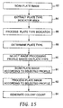
- FIG. 15 is a flow diagram illustrating a process for image processing profile selection in a biological scanner involving extraction of a plate type indicator from a scanned plate image.
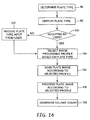
- FIG. 16 is a flow diagram illustrating a process that permits a user to override an automatic plate type identification by a biological scanner.
- the invention is directed to a biological scanner for biological growth plates.
- a biological growth plate can be presented to the biological scanner, which then generates an image of the plate and performs an analysis of the image to detect biological growth.
- the scanner may count or otherwise quantify an amount of biological agents that appear in the image, such as a number of bacteria colonies.
- the biological scanner automates the analysis of biological growth plates, thereby improving such analysis and reducing the possibility of human error.
- a biological scanner in accordance with the invention, also automates selection of image processing profiles to scan different types of biological growth plates and analyze plate images.
- the scanner automatically identifies the type of plate to be scanned by the scanner, and then selects one of the image processing profiles appropriate for the identified plate type.
- the image processing profiles may specify image capture conditions, image analysis criteria or a combination of both for different types of biological growth plates.
- the image processing profile may specify illumination intensities, durations, and colors for illumination of particular plate types for image capture.
- the image capture conditions also may include camera gain, resolution, aperture, and exposure time.
- the image processing profiles may specify different color, shape, size and proximity criteria in counting different types of bacterial colonies within a captured image to promote accuracy in the analytical result, e.g., a count.
- the image processing profiles may pertain both to image capture and analysis. Accuracy is critical in both food and laboratory sample test environments. For food safety, in particular, accurate results permit verification of sanitation at critical control points throughout the food processing operation, including line production, equipment and environmental testing.
- the scanner may identify the plate type by reference to a variety of machine-readable plate type indicators, such as optically or magnetically readable marks, carried on the plate. Accordingly, the invention also contemplates biological growth plates carrying a particular indicator that permits plate type identification. In addition, the invention may eliminate or reduce reliance on human judgment in making plate type identifications, thereby reducing the potential for human error and resulting inaccuracy in colony counts or other analyses.
- the invention may be useful with a variety of biological growth plates.
- the invention may be useful with different plate-like devices for growing biological agents to enable detection or enumeration of the agents, such as thin-film culture plate devices, Petri dish culture plate devices, and the like.
- the term "biological growth plate” will be used broadly herein to refer to a medium suitable for growth of biological agents to permit detection and enumeration of the agents by a scanner.
- the biological growth plate can be housed in a cassette that supports multiple plates, e.g., as described in U.S. Patent No. 5,573,950 to Graessle et al.
- FIG. 1 is a perspective view of an exemplary biological scanner 10.
- biological scanner 10 includes a scanner unit 12 having a drawer 14 that receives a biological growth plate (not shown in FIG. 1 ).
- Drawer 14 moves the biological growth plate into biological scanner 10 for scanning and analysis.
- Scanner 10 may incorporate features that permit automated plate type identification, and automated selection of image processing profiles based on plate type, in accordance with the invention.
- Biological scanner 10 also may include a display screen 16 to display the progress or results of analysis of the biological growth plate to a user. Alternatively or additionally, display screen may present to a user an image of the growth plate scanned by biological scanner 10. The displayed image may be optically magnified or digitally scaled upward.
- a mounting platform 18 defines an ejection slot 20 through which the growth plate can be ejected following image capture by biological scanner 10. Accordingly, biological scanner 10 may have a two-part design in which scanner unit 12 is mounted on mounting platform 18. The two-part design is depicted in FIG. 1 for purposes of example, and is not intended to be required by or limiting of the inventions described herein.
- Scanner unit 12 houses an imaging device for scanning the biological growth plate and generating an image.
- the imaging device may take the form of a line scanner or an area scanner, which ordinarily will be provided in combination with an illumination system to provide front and/or back illumination of the biological growth plate.
- scanner unit 12 may house processing hardware that performs analysis of the scanned image, e.g., in order to determine the number or amount of biological agents in the growth plate. For example, upon presentation of the biological growth plate via drawer 14, the plate may be positioned adjacent an optical platen for scanning.
- the growth plate When the drawer is subsequently opened, the growth plate may drop downward into the mounting platform 18 for ejection via ejection slot 20.
- mounting platform 18 may house a conveyor that ejects the growth plate from biological scanner 10 via ejection slot 20.
- a biological growth plate After a biological growth plate is inserted into drawer 14, moved into scanner unit 12, and scanned, the biological growth plate drops downward into mounting platform 18, where a horizontal conveyor, such as a moving belt, ejects the medium via slot 20.
- FIG. 2 is another perspective view of biological scanner 10.
- drawer 14 extends outward from biological scanner 10 to receive a biological growth plate 22.
- a biological growth plate 22 may be placed on a platform 24 provided within drawer 14.
- platform 24 may include positioning actuators such as cam levers to elevate the platform for precise positioning of growth plate 22 within biological scanner 10.
- drawer 14 retracts into scanner unit 12 to place the biological growth plate in a scanning position, i.e., a position at which the biological growth medium is optically scanned.
- FIGS. 3 and 4 are top views of an exemplary biological growth plate 22.
- a suitable growth plate 22 may comprise biological growth plates sold by 3M under the trade name PETRIFILM plates.
- biological growth plate 22 may comprise other biological growth media for growing particular bacteria or other biological agents.
- biological growth plate 22 carries plate type indicator 28 to facilitate automated identification of the type of biological media associated with the growth plate.
- Plate type indicator 28 presents an encoded pattern that is machine-readable.
- plate type indicator 28 takes the form of an optically readable pattern.
- FIGS. 3 and 4 depict a four-square pattern of light and dark quadrants formed in a corner margin of biological growth plate 22.
- plate type indicator 28 defines a two-dimensional grid of cells modulated between black and white to form an encoded pattern.
- optical patterns such as characters, bar codes, two-dimensional bar codes, optical gratings, holograms, phosphorous inks and the like are conceivable.
- plate type indicator 28 may take the form of patterns that are readable by magnetic or radio frequency techniques.
- plate type indicator 28 may take the form of apertures, slots, surface contours, or the like that are readable by optical or mechanical techniques. In each case, plate type indicator 28 carries information sufficient to enable automated identification of the type of biological growth plate 22 by biological scanner 10. Plate type indicator 28 will be described in greater detail below.
- Biological growth plates may facilitate the rapid growth and detection and enumeration of bacteria or other biological agents including, for example, aerobic bacteria, E. coli, coliform, enterobacteriaceae, yeast, mold, Staphylococcus aureus, listeria, and campylobacter, and the like.
- the use of PETRIFILM plates, or other growth media can simplify bacterial testing of food samples.
- biological scanner 10 can further simplify such testing by providing automated plate type detection, and automated selection of image processing profiles based on the detected plate type to illuminate and/or analyze biological growth plate 22, e.g., by counting bacterial colonies on an image of the plate.
- biological growth plate 22 defines a growth area 26.
- a determination of whether a given sample being tested in plate 22 is acceptable, in terms of bacterial colony counts, may depend on the number of bacterial colonies per unit area. Accordingly, scanner 10 may quantify the amount of bacterial colonies per unit area on plate 22, and may compare the amount, or "count," to a threshold.
- the surface of biological growth plate 22 may contain one or more growth enhancing agents designed to facilitate the rapid growth of one or more types of bacteria or other biological agents.
- plate 22 After placing a sample of the material being tested, typically in liquid form, on the surface of biological growth plate 22 within growth area 26, plate 22 can be inserted into an incubation chamber (not shown). In the incubation chamber, bacterial colonies or other biological agents being grown by growth plate 22 manifest themselves, as shown in biological growth plate 22 of FIG. 4 .
- the colonies, represented by various dots 30 on biological growth plate 22 in FIG. 4 may appear in different colors on plate 22, facilitating automated detection and enumeration of bacterial colonies by scanner 10.
- FIGS. 5A-5D are diagrams illustrating exemplary plate type indicator 28 carried by a biological growth plate 22 for image processing profile selection.
- plate type indicator 28 may take the form of patterns, marks, apertures, surface contours and the like, which permit optical or mechanical readability. For example, different optical patterns can be read by optical decoders, bar code scanners, optical character recognition (OCR) processors or the like. In the case of apertures or contours, mechanical styli may interact with the apertures or contours to detect different patterns and produce an electrical signal.
- plate type indicator 28 may be magnetically encoded stripes or markers or carry radio frequency identifications to permit magnetic or radio frequency readability.
- Optically readable patterns may be formed by printing or deposition of ink on the surface of biological growth plate 22, e.g., outside of growth area 26. Apertures or surface contour patterns can be formed in biological growth plate 22 by punches, stamps, embossers, die cutters and the like. A magnetic stripe or radio frequency identification may be affixed to the surface of biological growth plate 22, e.g., by adhesive or lamination techniques. In addition, a magnetic or radio frequency indicator need not be carried on the surface of biological growth plate 22, but may be interposed between layers of the growth plate in the event the growth plate has a multi-layer structure. In each case, the various plate type indicators 28 may be formed at the factory to identify the type of biological growth plate 22.
- plate type indicator 28 may further include information that identifies a particular manufacturer, lot number, expiration date, security authorization, and the like. Such additional items of information may be important in verifying quality and suitability of biological growth plate 22 for used in biological scanner 10. For example, one or more manufacturers may be specifically validated, e.g., on the basis of plate production quality and plate performance criteria, to provide biological growth plates 22 for use in biological scanner 10. In this case, biological scanner 10 may be configured to reject biological growth plates 22 that, according to plate type indicator 28, are not associated with validated manufacturers.
- plate type indicator 28 may carry security information, such as serial number codes or the like, that serve to authenticate biological growth plate 22 and prevent fraudulent introduction of unauthorized growth plates, e.g., to thwart the food inspection or laboratory analysis process.
- security information such as serial number codes or the like
- biological growth plate 22 may carry one or more indicator patterns, in addition to plate type indicator 28, which serve different security and quality assurance purposes.
- plate type indicator 28 may benefit from a variety of security mechanisms.
- a printed plated type indicator 28 may be printed with a particular phosphorous ink so that it can be conspicuously identified according to the wavelength of light emitted by the indicator when it is scanned.
- plate type indicators 28 may take the form of more complex patterns that carry encryption keys to unlock biological scanner 10 for operation. In this case, processor 34 in biological scanner 10 ( FIG. 6 ) would perform decryption of the pattern in order to proceed with image processing.
- plate type indicator 28 takes the form of a four-square pattern having four quadrants 29, 31, 33, 35 that can be either dark or light, permitting ready optical processing.
- plate type indicator 28 has four light quadrants, and may identify a first type of biological growth plate 22.
- plate type indicator 28 include one black quadrant, two black quadrants, and four black quadrants, respectively. Selection of the number and position of the black quadrants permits up to sixteen (2 4 ) different encoded patterns to be formed and, accordingly, up to sixteen different plate types to be identified by machine-readable plate type indicator 28.
- different encoded patterns could represent Aerobic Count, Coliform, E. Coli, Staphylococcus aureus, Yeast and Mold, and other plate type designations.
- plate type indicator 28 may be subject to wide variation, the 4-square pattern shown in FIGS. 5A-5D provides one type of pattern that is relatively simple and easy to identify using optical pattern recognition techniques, i.e., machine vision.
- plate type indicator 28 may be scanned by a dedicated optical code reader, such as a bar code reader or custom reader. In this case, plate type indicator 28 can be scanned prior to or in parallel with scanning of biological growth plate 22, but before processing of the growth plate image.
- plate type indicator 28 may be captured in a scanned image of biological growth plate 22, and then extracted for image processing to identify the plate type. In this case, the plate type can be identified before further image processing of the scanned growth plate image.
- FIG. 6 is a block diagram illustrating internal operation of biological scanner 10.
- a biological growth plate 22 is positioned within biological scanner 10 on a platform (not shown in FIG. 6 ).
- the platform places biological growth plate 22 at a desired focal plane of an imaging device 32.
- Imaging device 32 may include illumination hardware for top and back illumination of growth pate 22, as well as a line or area scanner that captures an image of the surface of growth plate 22.
- Imaging device 32 may apply standard image capture conditions, or a user may specify image capture conditions.
- scanner 10 may automatically control image capture conditions based on an image processing profile that corresponds to a plate type.
- imaging device 32 may take the form of a two-dimensional camera, although line scanners can be used in configurations in which either the camera or biological growth plate 22 is translated relative to the other.
- image device 32 captures an image of biological growth plate 22, or at least a growth region within the biological growth plate.
- a processor 34 controls the operation of imaging device 32. In operation, processor 34 controls imaging device 32 to capture an image of biological growth plate 22.
- Processor 34 receives image data representing the scanned image from imaging device 32, and extracts or segregates a portion of the image to isolate plate type indicator 28.
- processor 34 analyzes plate type indicator 28 to identify a plate type associated with biological growth plate 22. Processor 34 then retrieves an image processing profile from image processing profile memory 36. The image processing profile corresponds to the detected plate type.
- Processor 34 may take the form of a microprocessor, digital signal processor, application specific integrated circuitry (ASIC), field programmable gate array (FPGA) or other integrated or discrete logic circuitry programmed or otherwise configured to provide functionality as described herein.
- processor 34 Using the image processing profile, processor 34 loads appropriate image analysis parameters and proceeds to process the scanned image of biological growth plate 22. In this manner, processor 34 forms an image processing device in the sense that it processes the image data obtained from biological growth plate 22.
- the image analysis parameters may vary with the image processing profile and detected plate type, and may specify particular parameters such as colony color, size, shape and proximity criteria for analysis of the scanned image.
- the color of surrounding nutrient media may be an indicator of high colony counts.
- color may be an indicator of the type of organism.
- Adjacent objects, such as gas bubbles also may be an indicator of the type of organism. Accordingly, a variety of image processing criteria and associated parameters may be specified for various plate types. The criteria may differ according to the type of plate 22 to be analyzed, and may significantly affect colony count or other analytical results produced by biological scanner 10.
- processor 34 Upon selection of the appropriate image processing parameters, processor 34 processes the scanned image and produces an analytical result, such as a colony count, which is presented to a user via display 16. Processor 34 also may store the analytical result in memory, such as count data memory 38, for later retrieval from scanner 10. The data stored in count data memory 38 may be retrieved, for example, by a host computer that communicates with biological scanner 10 via a communication port 40, e.g., a universal serial bus (USB) port. The host computer may compile analytical results for a series of biological growth plates 22 presented to biological scanner 10 for analysis.
- a communication port 40 e.g., a universal serial bus (USB) port.
- USB universal serial bus
- Automated selection of image processing profiles within biological scanner 10 can provide a convenient and accurate technique for selecting the appropriate image processing profile.
- Automated selection of image processing profiles can promote the accuracy of bacterial colony counts and other analytical procedures.
- automatic image processing profile selection can avoid the need for a technician to visually identify and manually enter the plate type. In this manner, plate identification errors sometimes associated with human intervention can be avoided. Consequently, the combination of a scanner 10 and a biological growth plate 22 that carries plate type indicator 28 can promote efficiency and workflow of laboratory technicians while enhancing analytical accuracy and, in the end, food safety and human health.
- FIG. 7 is a block diagram illustrating another biological scanner 10' configured for automated image processing profile selection.
- Biological scanner 10' conforms substantially to biological scanner 10 of FIG. 6 , but further includes a code reader 42.
- code reader 42 serves as a dedicated reader to obtain plate type information.
- code reader 42 may take the form of a dedicated optical reader, bar code reader, magnetic reader, radio frequency or mechanical reader.
- code reader 42 serves to identify the plate type from plate type indicator 28 and communicates the plate type to processor 34.
- Processor 34 selects an image processing profile from memory 36 based on the identified plate type. Imaging device 32 scans biological growth plate 22 and provides the image date to processor 34.
- Processor 34 then applies the image processing parameters specified by the retrieved image processing profile to process the image and produce an analytical result such as colony count.
- processor 34 applies the appropriate image processing profile on an automated basis in view of the automatically identified plate type, offering enhanced accuracy, efficiency and convenience to the user.
- the invention eliminates the need for the user to enter the plate type identification manually, and reduces the likelihood of analytical error due to erroneous human input.
- FIG. 8 is a block diagram illustrating biological scanner 10 of FIG. 6 in greater detail and depicting plate illumination hardware.
- biological scanner 10 may include a front illumination system 44 and a back illumination system 46.
- Front illumination system 44 illuminates a front side of biological growth plate 22, and back illumination system 46 illuminates a back side of the biological growth plate.
- Front and back illumination systems 44, 46 may produce different illumination intensities, colors and durations on a selective basis.
- processor 34 controls front and back illumination systems 44, 46 to expose biological growth plate 22 to different illumination colors.
- Front and back illumination systems 44, 46 may incorporate LEDs as illumination sources. The LEDs can be readily controlled by processor 34 and appropriate driver circuitry to achieve desired illumination intensities and durations.
- processor 34 may control camera 43 to capture images of biological growth plate 22 during illumination with the different colors.
- processor 34 may provide coordinated control of illumination systems 44, 46 and camera 43 to capture one or more images of biological growth plate 22.
- Camera 43 captures one or more images of biological growth plate 22 during illumination by front illumination system 44, back illumination system 46 or both, and may store the images in an image memory 47.
- processor 34 may control camera gain, resolution, aperture, exposure time or the like in response to the image capture conditions specified by the image processing profile.
- processor 34 uses the stored images to perform image analysis according to the image analysis criteria specified by the image processing profile. In particular, processor 34 then may analyze the individual images or combine the multiple images to form a composite image. In some embodiments, for example, processor 34 may control illumination systems 44, 46 to capture red, green and blue images of biological growth plate 22 and analyze the images individually or as a composite multi-color image.
- Some types of biological growth plates 22 may require illumination with a particular color, intensity and duration.
- some biological growth plates 22 may require only front or back illumination, but not both.
- an aerobic count plate may require only front illumination as well as illumination by only a single color such as red.
- an E. coli/Coliform plate may require only back illumination and a combination of red and blue illumination.
- particular intensity levels and durations may be appropriate, as well as different camera gain, resolution, aperture, and exposure time.
- processor 34 may control illumination and camera conditions in response to image capture conditions specified by an image processing profile.
- scanner 10 can be configured to select not only image analysis criteria based on plate type, but also image capture conditions to be applied to capture the image.
- scanner 10 may apply techniques similar to those described with respect to FIGS. 6 and 7 .
- a dedicated code reader may be provided to identify plate type in advance of illumination for image capture.
- processor 34 selects a corresponding image processing profile from image processing profile memory 36 and controls illumination according to image capture conditions specified in the image processing profile.
- scanner 10 may be configured to apply machine vision techniques to identify the plate type from a capture image, as discussed with respect to FIG. 6 .
- scanner 10 may apply a set of default illumination conditions to capture an initial image of biological growth plate 22, or a portion thereof, for purposes of analyzing plate type indicator 28 for plate type identification.
- processor 34 may select a corresponding image processing profile and apply the specified image capture conditions to capture an image for analysis of biological growth.
- processor 34 may access an image capture profile specifying image capture conditions such as illumination colors, intensities and durations. Then, for analysis of a captured image, processor 34 may access a separate image analysis profile specifying image analysis criteria such as color, shape, size and proximity.
- processor 34 may present a preliminary plate type identification to a user via display 16.
- processor 34 may permit the user to either confirm or reject the automatically identified plate type before proceeding with image capture or analysis using a corresponding image processing profiles
- the user may confirm or reject a preliminary plate type identification, for example, by actuating a pointing device or depressing regions of a touch screen. If the user believes that the automatically detected plate type identification is in error, processor 34 may permit the user to change the plate type identification.
- FIG. 9 is a sample display content produced on display 16 by biological scanner 10 upon plate type detection.
- display 16 presents a preliminary plate type identification, i.e., a plate type identification automatically made by processor 34.
- display 16 presents two touch screen regions 52, 54 that accept user input to indicate whether the preliminary plate type identification is confirmed or rejected, respectively, by the user.
- FIG. 10 is sample content produced by display 16 of biological scanner 10 upon rejection of the automated plate type detection by the user.
- processor 34 may drive display 16 to present an "ENTER PLATE TYPE" dialog by which the user may choose the correct plate type identification, as shown in FIG. 10 .
- Display 16 may present a vertical scroll bar menu 56 that permits the user to choose an alternative plate type identification, e.g., by depressing an appropriate touch screen region.
- processor 34 may select an alternative image processing profile containing, for example, image capture conditions, image analysis criteria, or both.
- FIG. 11 is sample content produced by display 16 of biological scanner 10 upon determination of a colony count.
- FIG. 12 is sample content produced by display 16 of biological scanner 10 upon determination of a colony count and including an image of a scanned plate.
- display 16 presents information similar to that shown in FIG. 11 , but further includes a representation 60 of the actual image scanned by biological scanner 10 from the surface of biological growth plate 22.
- image representation 60 may present a sufficient amount of detail to permit the user to verify the automatically determined count.
- image representation 60 may be a lower resolution representation.
- FIG. 13 is a flow diagram illustrating a process for image processing profile selection in biological scanner 10.
- the process may involve identifying a plate type (62) for a biological growth plate 22 presented to scanner 10.
- the process may further involve selection of an image processing profile based on the plate type (64), either before or after scanning a plate image (66). If the image processing profile specifies image capture conditions, the image processing profile should be selected prior to scanning the plate image so that illumination conditions, camera properties or both may be controlled.
- the process further involves processing the plate image to produce an analytical result (68). In particular, the process may generate a bacterial colony count (70).
- the plate type is identified before the plate image is scanned.
- FIG. 14 is a flow diagram illustrating a process for image processing profile selection in a biological scanner involving detection of a plate type indicator.
- the process involves reading the plate type indicator carried by a biological growth plate (72), e.g., with a dedicate plate type indicator reader such as an optical reader, bar code reader, magnetic reader, radio frequency reader, mechanical reader or the like.
- a dedicate plate type indicator reader such as an optical reader, bar code reader, magnetic reader, radio frequency reader, mechanical reader or the like.
- the process involves selecting an image processing profile based on the detected plate type (76).
- the process involves scanning an image of the biological growth plate (78), and processing the plate image according to parameters specified by the selected image processing profile (80). The process then produces an analytical result such as a colony count (81).
- FIG. 15 is a flow diagram illustrating a process for image processing profile selection in a biological scanner involving extraction of plate type indicator from a scanned plate image.
- the process involves scanning an image of a biological growth plate (82), and extracting a plate type indicator area from the scanned image (84). The process further involves processing the extracted plate type indicator area (86) to determine the plate type (88).
- the process involves selecting an image processing profile based on the detected plate type (90).
- the process then may involve scanning the plate again (91), e.g., using image capture conditions specified by the selected image processing profile, and processing the plate image according to image analysis criteria specified by the selected image processing profile (92).
- the process then produces an analytical result such as a colony count (94).
- FIG. 16 is a flow diagram illustrating a process that permits a user to override an automatic plate type identification by a biological scanner.
- the process involves automatically determining a plate type associated with a biological growth plate 22 (96), and presenting the determined plate type to a user via display 16 (98).
- the process further involves accepting user input to accept or reject the automatically determined plate type (100), e.g., via touch screen input. If the plate type is not accepted by the user, the process involves accepting user input to receive a plate type from the user (102).
- the process involves selection of an image processing profile based on the plate type (104). Using the selected image processing profile, the process scans a plate image (105), processes the plate image (106) and generates a colony count (108) or some other desired analytical result.
- processor 34 executes instructions that may be stored on a computer-readable medium to carry out the processes described herein.
- the computer-readable medium may comprise random access memory (RAM) such as synchronous dynamic random access memory (SDRAM), read-only memory (ROM), non-volatile random access memory (NVRAM), electrically erasable programmable read-only memory (EEPROM), FLASH memory, magnetic or optical data storage media, and the like.
- RAM random access memory
- SDRAM synchronous dynamic random access memory
- ROM read-only memory
- NVRAM non-volatile random access memory
- EEPROM electrically erasable programmable read-only memory
- FLASH memory magnetic or optical data storage media, and the like.
Landscapes
- Engineering & Computer Science (AREA)
- Health & Medical Sciences (AREA)
- Chemical & Material Sciences (AREA)
- General Health & Medical Sciences (AREA)
- Life Sciences & Earth Sciences (AREA)
- Organic Chemistry (AREA)
- Bioinformatics & Cheminformatics (AREA)
- Zoology (AREA)
- Physics & Mathematics (AREA)
- General Physics & Mathematics (AREA)
- Theoretical Computer Science (AREA)
- Wood Science & Technology (AREA)
- Biomedical Technology (AREA)
- Nuclear Medicine, Radiotherapy & Molecular Imaging (AREA)
- Medical Informatics (AREA)
- Analytical Chemistry (AREA)
- Biotechnology (AREA)
- Microbiology (AREA)
- Sustainable Development (AREA)
- Radiology & Medical Imaging (AREA)
- Computer Vision & Pattern Recognition (AREA)
- Biochemistry (AREA)
- General Engineering & Computer Science (AREA)
- Genetics & Genomics (AREA)
- Quality & Reliability (AREA)
- Molecular Biology (AREA)
- Multimedia (AREA)
- Apparatus Associated With Microorganisms And Enzymes (AREA)
- Investigating Or Analysing Biological Materials (AREA)
- Measuring Or Testing Involving Enzymes Or Micro-Organisms (AREA)
- Image Processing (AREA)
- Micro-Organisms Or Cultivation Processes Thereof (AREA)
- Image Input (AREA)
- Measuring Pulse, Heart Rate, Blood Pressure Or Blood Flow (AREA)
- Investigating Or Analysing Materials By Optical Means (AREA)
Priority Applications (1)
| Application Number | Priority Date | Filing Date | Title |
|---|---|---|---|
| EP10183904A EP2325778A1 (en) | 2002-11-27 | 2003-11-21 | Biological growth plate scanner with automated image processing profile selection |
Applications Claiming Priority (3)
| Application Number | Priority Date | Filing Date | Title |
|---|---|---|---|
| US10/306,579 US7298885B2 (en) | 2002-11-27 | 2002-11-27 | Biological growth plate scanner with automated image processing profile selection |
| US306579 | 2002-11-27 | ||
| PCT/US2003/037386 WO2004051554A1 (en) | 2002-11-27 | 2003-11-21 | Biological growth plate scanner with automated image processing profile selection |
Related Child Applications (1)
| Application Number | Title | Priority Date | Filing Date |
|---|---|---|---|
| EP10183904.1 Division-Into | 2010-09-30 |
Publications (3)
| Publication Number | Publication Date |
|---|---|
| EP1573654A1 EP1573654A1 (en) | 2005-09-14 |
| EP1573654A4 EP1573654A4 (en) | 2007-12-12 |
| EP1573654B1 true EP1573654B1 (en) | 2011-10-05 |
Family
ID=32325729
Family Applications (2)
| Application Number | Title | Priority Date | Filing Date |
|---|---|---|---|
| EP03787013A Revoked EP1573654B1 (en) | 2002-11-27 | 2003-11-21 | Biological growth plate scanner with automated image processing profile selection |
| EP10183904A Withdrawn EP2325778A1 (en) | 2002-11-27 | 2003-11-21 | Biological growth plate scanner with automated image processing profile selection |
Family Applications After (1)
| Application Number | Title | Priority Date | Filing Date |
|---|---|---|---|
| EP10183904A Withdrawn EP2325778A1 (en) | 2002-11-27 | 2003-11-21 | Biological growth plate scanner with automated image processing profile selection |
Country Status (11)
| Country | Link |
|---|---|
| US (3) | US7298885B2 (enExample) |
| EP (2) | EP1573654B1 (enExample) |
| JP (2) | JP2006507837A (enExample) |
| KR (1) | KR101120829B1 (enExample) |
| CN (2) | CN100489887C (enExample) |
| AT (1) | ATE527618T1 (enExample) |
| AU (1) | AU2003295804B2 (enExample) |
| BR (1) | BR0316406A (enExample) |
| CA (1) | CA2504616A1 (enExample) |
| MX (1) | MXPA05005378A (enExample) |
| WO (1) | WO2004051554A1 (enExample) |
Families Citing this family (64)
| Publication number | Priority date | Publication date | Assignee | Title |
|---|---|---|---|---|
| US20040101954A1 (en) * | 2002-11-27 | 2004-05-27 | Graessle Josef A. | Back side plate illumination for biological growth plate scanner |
| US20040102903A1 (en) * | 2002-11-27 | 2004-05-27 | Graessle Josef A. | Biological growth plate scanner |
| US7298885B2 (en) * | 2002-11-27 | 2007-11-20 | 3M Innovative Properties Company | Biological growth plate scanner with automated image processing profile selection |
| US7319031B2 (en) * | 2002-11-27 | 2008-01-15 | 3M Innovative Properties Company | Mounting platform for biological growth plate scanner |
| US7351574B2 (en) * | 2002-11-27 | 2008-04-01 | 3M Innovative Properties Company | Loading and ejection systems for biological growth plate scanner |
| US7496225B2 (en) * | 2003-09-04 | 2009-02-24 | 3M Innovative Properties Company | Biological growth plate scanner with automated intake |
| US7298886B2 (en) | 2003-09-05 | 2007-11-20 | 3M Innovative Properties Company | Counting biological agents on biological growth plates |
| US20080238627A1 (en) * | 2005-03-22 | 2008-10-02 | Applera Corporation | Sample carrier device incorporating radio frequency identification, and method |
| US7187286B2 (en) | 2004-03-19 | 2007-03-06 | Applera Corporation | Methods and systems for using RFID in biological field |
| JP4713261B2 (ja) * | 2005-07-20 | 2011-06-29 | 株式会社カネカ | 細胞培養装置 |
| JP4821279B2 (ja) * | 2005-11-11 | 2011-11-24 | 株式会社ニコン | 培養装置 |
| EP1882948A2 (de) * | 2006-07-28 | 2008-01-30 | Qiagen GmbH | Vorrichtung zur Probenverarbeitung |
| KR101596649B1 (ko) | 2007-07-09 | 2016-02-22 | 쓰리엠 이노베이티브 프로퍼티즈 컴파니 | 미생물을 검출하기 위한 모듈형 시스템 및 방법 |
| US8828653B2 (en) | 2007-11-20 | 2014-09-09 | 3M Innovative Properties Company | Environmental sampling articles and methods |
| CN101952457B (zh) | 2007-12-21 | 2013-08-21 | 3M创新有限公司 | 流体样品分析的微生物系统和方法 |
| WO2009111301A1 (en) * | 2008-03-04 | 2009-09-11 | 3M Innovative Properties Company | Information management in automated processing of biological growth media |
| EP2265733A4 (en) * | 2008-03-04 | 2017-12-13 | 3M Innovative Properties Company | Processing of biological growth media based on measured manufacturing characteristics |
| US8005775B2 (en) * | 2008-03-18 | 2011-08-23 | Yahoo! Inc. | System and method for detecting human judgment drift and variation control |
| KR20110005813A (ko) * | 2008-03-26 | 2011-01-19 | 쓰리엠 이노베이티브 프로퍼티즈 컴파니 | 생물학적 성장 배지의 분광 분석 |
| US8969029B2 (en) * | 2008-10-17 | 2015-03-03 | 3M Innovative Properties Company | Biological sterilization indicator, system, and methods of using same |
| JP5480559B2 (ja) * | 2008-10-30 | 2014-04-23 | シスメックス株式会社 | 細菌分析装置、細菌分析方法及びコンピュータプログラム |
| EP2199956A1 (en) * | 2008-12-18 | 2010-06-23 | Siemens Aktiengesellschaft | Method and system for managing results of an analysis process on objects handled along a technical process line |
| CN105671124A (zh) | 2008-12-31 | 2016-06-15 | 3M创新有限公司 | 大肠菌检测方法以及其中使用的试剂盒 |
| WO2011055791A1 (ja) * | 2009-11-05 | 2011-05-12 | 株式会社日立ハイテクノロジーズ | 細菌コロニー釣菌装置及びその方法 |
| JP6172940B2 (ja) | 2009-12-30 | 2017-08-09 | スリーエム イノベイティブ プロパティズ カンパニー | グルコースオキシダーゼを産生するカビの迅速検出 |
| EP2519821B1 (en) | 2009-12-30 | 2016-11-30 | 3M Innovative Properties Company | Microbial detection article |
| KR101783836B1 (ko) | 2010-06-23 | 2017-10-10 | 가부시키가이샤 엔테크 | 미생물 검출방법, 미생물 검출장치 및 프로그램 |
| FR2964215B1 (fr) * | 2010-08-25 | 2013-06-14 | Intelligence Artificielle Applic | Utilisation d'un code datamatrix et procede d'impresssion |
| JP5780732B2 (ja) * | 2010-10-04 | 2015-09-16 | マイクロバイオ株式会社 | コロニー検出方法、コロニー検出システムおよびコロニー検出プログラム |
| CN102024259B (zh) * | 2010-12-24 | 2012-06-20 | 刘安安 | 一种菌落自动检测方法 |
| JP5849976B2 (ja) * | 2012-03-30 | 2016-02-03 | カシオ計算機株式会社 | ソーシャル・ネットワーク・サービスシステム及び画像表示方法 |
| JP5998744B2 (ja) * | 2012-08-23 | 2016-09-28 | 大日本印刷株式会社 | コロニー検出装置、培地情報登録システム、衛生管理システム、及びプログラム |
| EP2731051A1 (en) * | 2012-11-07 | 2014-05-14 | bioMérieux | Bio-imaging method |
| EP2936387A1 (en) * | 2012-12-20 | 2015-10-28 | 3M Innovative Properties Company | Method of detecting gas-producing microbial colonies |
| EP3336195B1 (en) | 2012-12-20 | 2020-02-26 | 3M Innovative Properties Company | Method of differentiating microbial colonies in an image |
| JP6070172B2 (ja) * | 2012-12-25 | 2017-02-01 | 大日本印刷株式会社 | 培地情報登録システム、プログラム及び衛生管理システム |
| JP2014135949A (ja) * | 2013-01-18 | 2014-07-28 | Dainippon Printing Co Ltd | 培地情報登録システム、培地画像解析装置、プログラム及び衛生管理システム |
| JP2014135948A (ja) * | 2013-01-18 | 2014-07-28 | Dainippon Printing Co Ltd | 培地情報登録システム、コロニー検出装置、プログラム及び衛生管理システム |
| JP2014140335A (ja) * | 2013-01-24 | 2014-08-07 | Dainippon Printing Co Ltd | 培地画像解析装置、培地情報登録システム、プログラム及び衛生管理システム |
| US8921067B2 (en) | 2013-02-04 | 2014-12-30 | 3M Innovative Properties Company | Method and culture device for detecting yeasts and molds |
| JP6281231B2 (ja) * | 2013-10-08 | 2018-02-21 | 大日本印刷株式会社 | 培地情報登録システムおよびコロニー検出システム |
| WO2015134686A1 (en) | 2014-03-07 | 2015-09-11 | 3M Innovative Properties Company | Article and method for detecting aerobic bacteria |
| US10407654B1 (en) | 2014-03-21 | 2019-09-10 | Charm Sciences, Inc. | Growth plate devices, kits and assemblies |
| JP5904295B2 (ja) * | 2014-03-31 | 2016-04-13 | 大日本印刷株式会社 | コロニー検出システム、コロニー検出方法、及び、プログラム |
| JP2016158577A (ja) * | 2015-03-03 | 2016-09-05 | 富士フイルム株式会社 | 細胞コロニー検出装置および方法並びにプログラム |
| US10563164B1 (en) * | 2015-10-08 | 2020-02-18 | Charm Sciences, Inc. | Plate reader |
| US10988720B1 (en) | 2015-11-09 | 2021-04-27 | Charm Sciences, Inc. | Peel plate assembly |
| EP4067503A1 (en) | 2015-12-28 | 2022-10-05 | Nihon Rikagaku Kaihatsu LLC. | Device for determining live/dead state of microorganisms and method for determining live/dead state of microorganisms using the device |
| US10495563B1 (en) | 2016-04-28 | 2019-12-03 | Charm Sciences, Inc. | Plate reader observation methods and operation |
| KR102493051B1 (ko) * | 2016-09-20 | 2023-01-31 | 가부시키가이샤 닛스이 | 이물질이 제거된 어란 페이스트의 제조방법 및 이물질이 제거된 어란 페이스트의 제조장치 |
| WO2018061131A1 (ja) * | 2016-09-28 | 2018-04-05 | オリンパス株式会社 | 細胞状態計測装置 |
| EP3535382A4 (en) * | 2016-11-04 | 2020-06-10 | Becton, Dickinson and Company | SYSTEM AND METHOD FOR SELECTING COLONIES |
| EP3979208B1 (en) | 2016-11-10 | 2023-07-19 | Becton, Dickinson and Company | Timeline system for monitoring a culture media protocol |
| CN107644210B (zh) * | 2017-09-22 | 2020-05-12 | 哈尔滨工业大学(威海) | 基于图像处理的微生物数量估算方法 |
| CN108251270B (zh) * | 2018-01-16 | 2022-11-15 | 上海睿度光电科技有限公司 | 一种定量溶液覆盖细胞的设备 |
| US11112416B2 (en) | 2018-01-30 | 2021-09-07 | Life Technologies Corporation | Instruments, devices and consumables for use in a workflow of a smart molecular analysis system |
| FR3080211B1 (fr) | 2018-04-16 | 2020-05-08 | Pinqkerton | Systeme et procede d'analyse de dispositifs de test |
| KR102043670B1 (ko) * | 2018-04-27 | 2019-11-12 | 이상인 | 미생물 자동관리 인큐베이터 시스템 |
| CN109975290A (zh) * | 2018-11-30 | 2019-07-05 | 军事科学院军事医学研究院环境医学与作业医学研究所 | 一种细菌检测快速分析仪 |
| JP6832970B2 (ja) * | 2019-02-27 | 2021-02-24 | 富士フイルム株式会社 | 細胞コロニー検出装置および方法並びにプログラム |
| JP7259732B2 (ja) * | 2019-12-23 | 2023-04-18 | 横河電機株式会社 | 配信サーバ、方法およびプログラム |
| JP7286558B2 (ja) * | 2020-01-07 | 2023-06-05 | 株式会社エビデント | 検査方法、コンピュータ読取可能記録媒体、及び、標準板 |
| DE102020106819A1 (de) * | 2020-03-12 | 2021-09-16 | Funke - Dr. N. Gerber Labortechnik Gesellschaft mit beschränkter Haftung | Verfahren zum zählen mikrobiologischer kolonien |
| US12228511B1 (en) * | 2023-09-25 | 2025-02-18 | Craig Ellins | Organic plant material microbial test kit devices and processing method |
Family Cites Families (113)
| Publication number | Priority date | Publication date | Assignee | Title |
|---|---|---|---|---|
| US3493772A (en) | 1967-05-29 | 1970-02-03 | Palo Alto Medical Research Fou | Bacterial counting machine and method |
| US3745090A (en) * | 1970-08-04 | 1973-07-10 | Nasa | Method of detecting and counting bacteria in body fluids |
| US3811036A (en) | 1972-09-19 | 1974-05-14 | Artek Syst Corp | Micro-biological colony counter |
| US3962040A (en) | 1974-03-14 | 1976-06-08 | The United States Of America As Represented By The Department Of Health, Education And Welfare | Method and apparatus for plating and counting aerobic bacteria |
| US4118280A (en) | 1976-05-03 | 1978-10-03 | Mcdonnell Douglas Corporation | Automated microbial analyzer |
| US4146775A (en) * | 1976-09-16 | 1979-03-27 | Armstrong Machine Works | Automatic control system for an electrode-type air humidifier |
| US4160601A (en) | 1978-02-24 | 1979-07-10 | Nasa | Biocontamination and particulate detection system |
| DK366778A (da) | 1978-08-18 | 1980-02-19 | Foss Electric As | Fremgangsmaade til taelling af bakterier |
| US4563096A (en) * | 1980-05-09 | 1986-01-07 | Internationale Octrooi Maatschappij "Octropa" B.V. | Apparatus for assisting the visual assessment of test objects having multivariate visible characteristics, and its use |
| US4353988A (en) | 1980-11-12 | 1982-10-12 | Couse Nancy L | Grid for use in counting colonies of bacteria present in discrete areas of a spiral deposition pattern |
| US4424191A (en) | 1982-03-04 | 1984-01-03 | Eastman Kodak Company | Analyzer featuring loading and unloading means for a storage chamber, and common drive means |
| US4591567A (en) | 1982-04-21 | 1986-05-27 | California Institute Of Technology | Recombinant DNA screening system including fixed array replicator and support |
| JPS6083597A (ja) | 1983-10-10 | 1985-05-11 | Hitachi Electronics Eng Co Ltd | コロニ−検査方法 |
| US4637053A (en) | 1984-05-03 | 1987-01-13 | Spiral System Instruments, Inc. | Computer assisted biological assay system |
| US4724215A (en) | 1985-02-27 | 1988-02-09 | Sherwood Medical Company | Automated microbiological testing apparatus and method |
| US4720463A (en) | 1985-03-01 | 1988-01-19 | Sherwood Medical Company | Automated microbiological testing apparatus |
| DE3686067T2 (de) | 1985-02-27 | 1993-03-18 | Sherwood Medical Co | Verfahren und vorrichtung zur automatischen mikrobiologischen analyse. |
| JPS62215383A (ja) | 1986-03-17 | 1987-09-22 | Datsuku Eng Kk | 微小生物体検査装置 |
| JPS6435347A (en) | 1987-07-31 | 1989-02-06 | Sumitomo Electric Industries | Detection of intrusion of various bacteria |
| US5270173A (en) * | 1987-10-06 | 1993-12-14 | Sumitomo Electric Industries, Ltd. | Method of monitoring cell culture |
| US5202010A (en) * | 1987-11-25 | 1993-04-13 | Princeton Biochemicals, Inc. | Automated capillary electrophoresis apparatus |
| JPH01296974A (ja) | 1988-05-23 | 1989-11-30 | Toyo Jozo Co Ltd | コロニー計数装置 |
| CA2014647C (en) | 1989-05-09 | 1994-09-20 | James David Shaw | Analyzer featuring a circular track of cartridges centered on an incubator, and method of use |
| US5266486A (en) * | 1989-05-12 | 1993-11-30 | Nvl Photronics Corporation | Method and apparatus for detecting biological activities in a specimen |
| WO1991006911A1 (en) * | 1989-10-23 | 1991-05-16 | Neuromedical Systems, Inc. | Automated cytological specimen classification system and method |
| DE3938565A1 (de) | 1989-11-21 | 1991-05-23 | Behringwerke Ag | Inkubationseinrichtung fuer mikrotitrationsplatten |
| GB2249829A (en) | 1990-11-13 | 1992-05-20 | Powergen Public Limited Compan | Measurement of carbon in ash |
| GB9100623D0 (en) | 1991-01-11 | 1991-02-27 | Medical Res Council | Transfer of biological samples |
| US5290701A (en) | 1991-08-28 | 1994-03-01 | Wilkins Judd R | Microbial detection system and process |
| US5428690A (en) | 1991-09-23 | 1995-06-27 | Becton Dickinson And Company | Method and apparatus for automated assay of biological specimens |
| US5481620A (en) | 1991-09-27 | 1996-01-02 | E. I. Du Pont De Nemours And Company | Adaptive vision system |
| US5448652A (en) | 1991-09-27 | 1995-09-05 | E. I. Du Pont De Nemours And Company | Adaptive display system |
| US6058209A (en) * | 1991-09-27 | 2000-05-02 | E. I. Du Pont De Nemours And Company | Method for resolving redundant identifications of an object |
| US5329686A (en) | 1991-12-19 | 1994-07-19 | Eastman Kodak Company | Slide frame and manufacturing process |
| JPH0698220A (ja) | 1992-05-29 | 1994-04-08 | Hooya Shiyotsuto Kk | ビデオカメラ |
| US5366873A (en) | 1992-06-03 | 1994-11-22 | Gideon Eden | Device and method for use in detecting microorganisms in a sample |
| DE69314959T2 (de) | 1992-07-13 | 1998-06-18 | Minnesota Mining & Mfg | Verfahren zum zählen von objekten in einem rasterabgetasteten teilbild. |
| JPH0651129A (ja) | 1992-07-27 | 1994-02-25 | Inoue Denki Kk | 照明装置 |
| JP2777509B2 (ja) | 1992-09-30 | 1998-07-16 | 出光石油化学株式会社 | 色調検査方法 |
| US5591974A (en) * | 1992-09-30 | 1997-01-07 | Westinghouse Electric Corporation | Automated collection and processing of environmental samples |
| DE4238550A1 (de) | 1992-11-14 | 1994-05-19 | Daimler Benz Ag | Abgasturbolader für eine Brennkraftmaschine |
| EP0698084B1 (en) | 1993-05-14 | 1998-06-24 | Minnesota Mining And Manufacturing Company | Method for rapid quantification of microorganism growth |
| US5364766A (en) | 1993-05-14 | 1994-11-15 | Minnesota Mining And Manufacturing Company | Culture medium for rapid count of coliform bacteria |
| US5723308A (en) | 1993-05-14 | 1998-03-03 | Minnesota Mining And Manufacturing Company | Culture medium for rapid count of coliform bacteria |
| EP0767361B1 (en) | 1993-07-22 | 2000-02-23 | Applied Spectral Imaging Ltd. | Method and apparatus for spectral imaging |
| US5995645A (en) | 1993-08-18 | 1999-11-30 | Applied Spectral Imaging Ltd. | Method of cancer cell detection |
| EP0734435B1 (en) | 1993-12-17 | 1999-03-31 | Minnesota Mining And Manufacturing Company | Automated incubating and imaging apparatus for disposable microorganism culturing media |
| JPH07275200A (ja) | 1994-04-15 | 1995-10-24 | Asahi Optical Co Ltd | 内視鏡の照明装置 |
| US5573950A (en) | 1994-05-11 | 1996-11-12 | Minnesota Mining And Manufacturing Company | Cassette for disposable microorganism culturing media and automated scanning system |
| JPH08116408A (ja) | 1994-08-24 | 1996-05-07 | Nikon Corp | 面状光源及び画像読取装置 |
| AU3585495A (en) * | 1994-09-20 | 1996-04-09 | Neopath, Inc. | Biological analysis system self-calibration apparatus |
| US5694478A (en) | 1994-12-15 | 1997-12-02 | Minnesota Mining And Manufacturing Company | Method and apparatus for detecting and identifying microbial colonies |
| US6319668B1 (en) * | 1995-04-25 | 2001-11-20 | Discovery Partners International | Method for tagging and screening molecules |
| US6151405A (en) * | 1996-11-27 | 2000-11-21 | Chromavision Medical Systems, Inc. | System and method for cellular specimen grading |
| US5721435A (en) | 1996-04-09 | 1998-02-24 | Hewlett Packard Company | Methods and apparatus for measuring optical properties of biological and chemical substances |
| JP3777661B2 (ja) * | 1996-07-10 | 2006-05-24 | 株式会社明電舎 | ろ過障害微生物監視装置 |
| DE19629141A1 (de) | 1996-07-19 | 1998-04-16 | Bayer Ag | Verfahren und Vorrichtung zum Screening von Molekülen bezüglich ihres individuellen Bindungsverhaltens zu mindestens einem vorgegebenen Ligand |
| US6381353B1 (en) | 1996-08-30 | 2002-04-30 | Marvin Weiss | System for counting colonies of micro-organisms in petri dishes and other culture media |
| DE29618623U1 (de) | 1996-10-25 | 1997-02-06 | Jencons (Scientific) Ltd., Leighton Buzzard, Bedfordshire | Vorrichtung zum Kultivieren von Mikroorganismen |
| US5817475A (en) | 1996-11-15 | 1998-10-06 | Giles Scientific, Inc. | Automatic microbiological testing apparatus and method |
| GB9624927D0 (en) * | 1996-11-29 | 1997-01-15 | Oxford Glycosciences Uk Ltd | Gels and their use |
| BR9816343B1 (pt) | 1997-05-23 | 2012-08-21 | método de operação de um aparelho de teste microbiológico diagnóstico e método de execução de teste microbiológico diagnóstico. | |
| US6002789A (en) | 1997-06-24 | 1999-12-14 | Pilot Industries, Inc. | Bacteria colony counter and classifier |
| US5911000A (en) * | 1997-08-01 | 1999-06-08 | Ortho Diagnostic Systems, Inc. | Detecting abnormal reactions in a red blood cell agglutination |
| KR100269125B1 (ko) * | 1997-10-25 | 2000-10-16 | 윤덕용 | 양자화효과감소를위한영상데이터후처리방법및장치 |
| ES2137879B1 (es) | 1997-12-02 | 2000-08-16 | Francisco Soria Melguizo S A | Sistema analizador de imagenes producidas por reacciones bacterianas. |
| US6178205B1 (en) * | 1997-12-12 | 2001-01-23 | Vtel Corporation | Video postfiltering with motion-compensated temporal filtering and/or spatial-adaptive filtering |
| DE19819144C1 (de) | 1998-04-29 | 2000-06-15 | Tga Tech Geraete Und Apparateb | Verfahren zur mikroskopischen Untersuchung der Gewebeintegration von Festkörpern, die dauerhaft oder vorübergehend in lebende Organismen implantiert werden und Vorrichtung zur Durchführung des Verfahrens |
| GB9811656D0 (en) * | 1998-05-29 | 1998-07-29 | Oxford Glycosciences Uk Ltd | Gels, methods and apparatus for identification and characterization of biomolecules |
| US6271042B1 (en) | 1998-08-26 | 2001-08-07 | Alpha Innotech Corporation | Biochip detection system |
| FR2786498B1 (fr) | 1998-11-27 | 2002-02-08 | Intelligence Artificielle Appl | Appareil de lecture automatique d'un antibiogramme |
| US6107054A (en) | 1998-12-31 | 2000-08-22 | Gibbs; David | Microbiological testing apparatus and method |
| FR2789693B1 (fr) | 1999-02-16 | 2001-05-25 | Intelligence Artificielle Appl | Appareil de lecture automatique pour une boite de petri munie d'un couvercle |
| FR2789694B1 (fr) | 1999-02-16 | 2003-05-16 | Intelligence Artificielle Appl | Perfectionnement pour un appareil de lecture automatique d'un antibiogramme |
| US6215894B1 (en) | 1999-02-26 | 2001-04-10 | General Scanning, Incorporated | Automatic imaging and analysis of microarray biochips |
| US6251624B1 (en) * | 1999-03-12 | 2001-06-26 | Akzo Nobel N.V. | Apparatus and method for detecting, quantifying and characterizing microorganisms |
| US6271022B1 (en) | 1999-03-12 | 2001-08-07 | Biolog, Inc. | Device for incubating and monitoring multiwell assays |
| JP2000270840A (ja) * | 1999-03-29 | 2000-10-03 | Elmex Ltd | コロニー計数装置およびその使用方法 |
| EP1171636A2 (en) | 1999-04-22 | 2002-01-16 | Albert Einstein College Of Medicine Of Yeshiva University | Assay of gene expression patterns by multi-fluor fish |
| JP2002543434A (ja) * | 1999-04-29 | 2002-12-17 | デイド マイクロスキャン インコーポレーテッド | 迅速な抗菌物質感受性アッセイと微生物同定を組み合わせたシステム |
| US6731818B1 (en) * | 1999-06-30 | 2004-05-04 | Realnetworks, Inc. | System and method for generating video frames |
| JP2003504627A (ja) | 1999-07-13 | 2003-02-04 | クロマビジョン メディカル システムズ インコーポレイテッド | 生物試料中の物体の自動検出 |
| US6488890B1 (en) * | 1999-08-05 | 2002-12-03 | 3M Innovative Properties Company | Machine readable sterilization indicator for monitoring articles to be sterilized |
| US6485979B1 (en) * | 1999-08-05 | 2002-11-26 | 3M Innovative Properties Company | Electronic system for tracking and monitoring articles to be sterilized and associated method |
| US6737266B1 (en) * | 1999-10-01 | 2004-05-18 | 3M Innovative Properties Company | Devices and methods for microorganism detection |
| WO2001033190A2 (en) | 1999-11-04 | 2001-05-10 | Arcturus Engineering, Inc. | Automated laser capture microdissection |
| US6649406B1 (en) * | 1999-11-23 | 2003-11-18 | 3M Innovative Properties Company | Device for propagation and storage of microorganisms |
| JP2001242082A (ja) | 2000-02-29 | 2001-09-07 | Nippon Laser & Electronics Lab | 生体試料光学的走査装置 |
| US6795636B1 (en) * | 2000-03-05 | 2004-09-21 | 3M Innovative Properties Company | Radiation-transmissive films on glass articles |
| WO2001083673A2 (en) | 2000-04-28 | 2001-11-08 | Spiral Biotech, Inc. | Method and apparatus for viewing a culture medium having visible bacterial colonies |
| US6492133B1 (en) | 2000-05-01 | 2002-12-10 | 3M Innovative Properties Company | Reflective disc assay devices, systems and methods |
| US6711283B1 (en) | 2000-05-03 | 2004-03-23 | Aperio Technologies, Inc. | Fully automatic rapid microscope slide scanner |
| US6965704B2 (en) * | 2000-08-22 | 2005-11-15 | Affymetrix, Inc. | System, method, and computer software product for grid alignment of multiple scanned images |
| US6756225B2 (en) * | 2000-12-08 | 2004-06-29 | 3M Innovative Properties Company | Automated imaging and harvesting of colonies on thin film culture devices |
| EP1379855A1 (en) * | 2001-02-20 | 2004-01-14 | Cytokinetics | Method and apparatus for automated cellular bioinformatics |
| US6706314B2 (en) * | 2001-03-15 | 2004-03-16 | Amesbury Trust | Method of labelling an object |
| US7450641B2 (en) * | 2001-09-14 | 2008-11-11 | Sharp Laboratories Of America, Inc. | Adaptive filtering based upon boundary strength |
| WO2002090966A1 (en) | 2001-05-10 | 2002-11-14 | Large Scale Proteomics Corporation | Automated apparatus for separating a biological sample from a two dimensional electrophoresis gel |
| US6673315B2 (en) | 2001-06-29 | 2004-01-06 | Biomachines, Inc. | Method and apparatus for accessing a site on a biological substrate |
| US6418180B1 (en) | 2001-07-19 | 2002-07-09 | Marvin Weiss | Method and apparatus for counting objects having substantially uniform size |
| WO2003014400A1 (en) | 2001-08-08 | 2003-02-20 | Applied Precision, Llc | Time-delay integration imaging of biological specimens |
| KR20030037314A (ko) | 2001-11-01 | 2003-05-14 | (주)다이아칩 | 바이오 칩 분석을 위한 형광 영상 분석장치 |
| US7057721B2 (en) * | 2002-01-10 | 2006-06-06 | Chemimage Corporation | Wide field method for detecting pathogenic microorganisms |
| US7227901B2 (en) * | 2002-11-21 | 2007-06-05 | Ub Video Inc. | Low-complexity deblocking filter |
| US20040101954A1 (en) | 2002-11-27 | 2004-05-27 | Graessle Josef A. | Back side plate illumination for biological growth plate scanner |
| US7351574B2 (en) | 2002-11-27 | 2008-04-01 | 3M Innovative Properties Company | Loading and ejection systems for biological growth plate scanner |
| US7298885B2 (en) | 2002-11-27 | 2007-11-20 | 3M Innovative Properties Company | Biological growth plate scanner with automated image processing profile selection |
| US20040102903A1 (en) | 2002-11-27 | 2004-05-27 | Graessle Josef A. | Biological growth plate scanner |
| US7319031B2 (en) | 2002-11-27 | 2008-01-15 | 3M Innovative Properties Company | Mounting platform for biological growth plate scanner |
| US7496225B2 (en) * | 2003-09-04 | 2009-02-24 | 3M Innovative Properties Company | Biological growth plate scanner with automated intake |
| US7298886B2 (en) * | 2003-09-05 | 2007-11-20 | 3M Innovative Properties Company | Counting biological agents on biological growth plates |
| US20050222778A1 (en) * | 2003-12-30 | 2005-10-06 | Compliance Software Solutions Corp. | System, method, and computer-readable medium for collection of environmental data and generation of user report for compliance with FDA requirements |
| US20060263258A1 (en) * | 2005-02-10 | 2006-11-23 | Matthew Harris | Automated biological indicator incubator |
-
2002
- 2002-11-27 US US10/306,579 patent/US7298885B2/en not_active Expired - Lifetime
-
2003
- 2003-11-21 EP EP03787013A patent/EP1573654B1/en not_active Revoked
- 2003-11-21 BR BR0316406-3A patent/BR0316406A/pt not_active Application Discontinuation
- 2003-11-21 CN CNB2006101398505A patent/CN100489887C/zh not_active Expired - Fee Related
- 2003-11-21 AT AT03787013T patent/ATE527618T1/de not_active IP Right Cessation
- 2003-11-21 MX MXPA05005378A patent/MXPA05005378A/es active IP Right Grant
- 2003-11-21 AU AU2003295804A patent/AU2003295804B2/en not_active Ceased
- 2003-11-21 JP JP2004557258A patent/JP2006507837A/ja active Pending
- 2003-11-21 CN CNB2003801042726A patent/CN100342389C/zh not_active Expired - Fee Related
- 2003-11-21 WO PCT/US2003/037386 patent/WO2004051554A1/en not_active Ceased
- 2003-11-21 KR KR1020057009535A patent/KR101120829B1/ko not_active Expired - Fee Related
- 2003-11-21 EP EP10183904A patent/EP2325778A1/en not_active Withdrawn
- 2003-11-21 CA CA002504616A patent/CA2504616A1/en not_active Abandoned
-
2007
- 2007-11-02 US US11/934,175 patent/US20080064089A1/en not_active Abandoned
-
2010
- 2010-07-09 JP JP2010156333A patent/JP5194068B2/ja not_active Expired - Fee Related
- 2010-09-08 US US12/877,254 patent/US7901933B2/en not_active Expired - Fee Related
Also Published As
| Publication number | Publication date |
|---|---|
| MXPA05005378A (es) | 2005-08-26 |
| CN1717693A (zh) | 2006-01-04 |
| US20080064089A1 (en) | 2008-03-13 |
| BR0316406A (pt) | 2005-10-11 |
| ATE527618T1 (de) | 2011-10-15 |
| CA2504616A1 (en) | 2004-06-17 |
| CN100489887C (zh) | 2009-05-20 |
| EP1573654A1 (en) | 2005-09-14 |
| KR20050086878A (ko) | 2005-08-30 |
| JP2010227118A (ja) | 2010-10-14 |
| JP5194068B2 (ja) | 2013-05-08 |
| JP2006507837A (ja) | 2006-03-09 |
| EP2325778A1 (en) | 2011-05-25 |
| US20100330610A1 (en) | 2010-12-30 |
| KR101120829B1 (ko) | 2012-03-15 |
| CN100342389C (zh) | 2007-10-10 |
| US7298885B2 (en) | 2007-11-20 |
| CN1940962A (zh) | 2007-04-04 |
| EP1573654A4 (en) | 2007-12-12 |
| US7901933B2 (en) | 2011-03-08 |
| AU2003295804B2 (en) | 2009-10-01 |
| AU2003295804A1 (en) | 2004-06-23 |
| US20040101189A1 (en) | 2004-05-27 |
| WO2004051554A1 (en) | 2004-06-17 |
Similar Documents
| Publication | Publication Date | Title |
|---|---|---|
| EP1573654B1 (en) | Biological growth plate scanner with automated image processing profile selection | |
| US8698881B2 (en) | Apparatus, method and article to perform assays using assay strips | |
| US9576181B2 (en) | Bio-imaging method | |
| EP2323070B1 (en) | Counting biological agents on biological growth plates | |
| CN100424166C (zh) | 组织样品自动染色方法和装置 | |
| EP2005137B1 (en) | Device and method for cross-referencing | |
| CN101981430B (zh) | 生物生长培养基的光谱分析 | |
| US9933446B2 (en) | Processing of biological growth media based on measured manufacturing characteristics | |
| EP0571053A2 (en) | Analysis method and apparatus for biological specimens | |
| EP1016707B1 (en) | Microbiological testing apparatus and method | |
| EP2536820A2 (en) | Culture systems, apparatus, and related methods and articles | |
| CN206450598U (zh) | 胶体金荧光定量分析一体机 | |
| CN106855515A (zh) | 胶体金荧光定量分析一体机及其控制方法 | |
| WO2019150755A1 (ja) | 撮像システム | |
| WO2003076914A1 (en) | Method and apparatus to determine the presence of specific substances in a sample |
Legal Events
| Date | Code | Title | Description |
|---|---|---|---|
| PUAI | Public reference made under article 153(3) epc to a published international application that has entered the european phase |
Free format text: ORIGINAL CODE: 0009012 |
|
| 17P | Request for examination filed |
Effective date: 20050524 |
|
| AK | Designated contracting states |
Kind code of ref document: A1 Designated state(s): AT BE BG CH CY CZ DE DK EE ES FI FR GB GR HU IE IT LI LU MC NL PT RO SE SI SK TR |
|
| AX | Request for extension of the european patent |
Extension state: AL LT LV MK |
|
| DAX | Request for extension of the european patent (deleted) | ||
| A4 | Supplementary search report drawn up and despatched |
Effective date: 20071109 |
|
| 17Q | First examination report despatched |
Effective date: 20080212 |
|
| GRAP | Despatch of communication of intention to grant a patent |
Free format text: ORIGINAL CODE: EPIDOSNIGR1 |
|
| GRAS | Grant fee paid |
Free format text: ORIGINAL CODE: EPIDOSNIGR3 |
|
| GRAA | (expected) grant |
Free format text: ORIGINAL CODE: 0009210 |
|
| AK | Designated contracting states |
Kind code of ref document: B1 Designated state(s): AT BE BG CH CY CZ DE DK EE ES FI FR GB GR HU IE IT LI LU MC NL PT RO SE SI SK TR |
|
| REG | Reference to a national code |
Ref country code: GB Ref legal event code: FG4D |
|
| REG | Reference to a national code |
Ref country code: CH Ref legal event code: EP |
|
| REG | Reference to a national code |
Ref country code: IE Ref legal event code: FG4D |
|
| REG | Reference to a national code |
Ref country code: DE Ref legal event code: R096 Ref document number: 60338674 Country of ref document: DE Effective date: 20111201 |
|
| REG | Reference to a national code |
Ref country code: NL Ref legal event code: VDEP Effective date: 20111005 |
|
| PG25 | Lapsed in a contracting state [announced via postgrant information from national office to epo] |
Ref country code: SI Free format text: LAPSE BECAUSE OF FAILURE TO SUBMIT A TRANSLATION OF THE DESCRIPTION OR TO PAY THE FEE WITHIN THE PRESCRIBED TIME-LIMIT Effective date: 20111005 |
|
| REG | Reference to a national code |
Ref country code: AT Ref legal event code: MK05 Ref document number: 527618 Country of ref document: AT Kind code of ref document: T Effective date: 20111005 |
|
| PG25 | Lapsed in a contracting state [announced via postgrant information from national office to epo] |
Ref country code: BE Free format text: LAPSE BECAUSE OF FAILURE TO SUBMIT A TRANSLATION OF THE DESCRIPTION OR TO PAY THE FEE WITHIN THE PRESCRIBED TIME-LIMIT Effective date: 20111005 |
|
| PG25 | Lapsed in a contracting state [announced via postgrant information from national office to epo] |
Ref country code: GR Free format text: LAPSE BECAUSE OF FAILURE TO SUBMIT A TRANSLATION OF THE DESCRIPTION OR TO PAY THE FEE WITHIN THE PRESCRIBED TIME-LIMIT Effective date: 20120106 Ref country code: NL Free format text: LAPSE BECAUSE OF FAILURE TO SUBMIT A TRANSLATION OF THE DESCRIPTION OR TO PAY THE FEE WITHIN THE PRESCRIBED TIME-LIMIT Effective date: 20111005 Ref country code: SE Free format text: LAPSE BECAUSE OF FAILURE TO SUBMIT A TRANSLATION OF THE DESCRIPTION OR TO PAY THE FEE WITHIN THE PRESCRIBED TIME-LIMIT Effective date: 20111005 Ref country code: PT Free format text: LAPSE BECAUSE OF FAILURE TO SUBMIT A TRANSLATION OF THE DESCRIPTION OR TO PAY THE FEE WITHIN THE PRESCRIBED TIME-LIMIT Effective date: 20120206 |
|
| PG25 | Lapsed in a contracting state [announced via postgrant information from national office to epo] |
Ref country code: MC Free format text: LAPSE BECAUSE OF NON-PAYMENT OF DUE FEES Effective date: 20111130 Ref country code: CY Free format text: LAPSE BECAUSE OF FAILURE TO SUBMIT A TRANSLATION OF THE DESCRIPTION OR TO PAY THE FEE WITHIN THE PRESCRIBED TIME-LIMIT Effective date: 20111005 |
|
| REG | Reference to a national code |
Ref country code: CH Ref legal event code: PL |
|
| PLBI | Opposition filed |
Free format text: ORIGINAL CODE: 0009260 |
|
| PG25 | Lapsed in a contracting state [announced via postgrant information from national office to epo] |
Ref country code: EE Free format text: LAPSE BECAUSE OF FAILURE TO SUBMIT A TRANSLATION OF THE DESCRIPTION OR TO PAY THE FEE WITHIN THE PRESCRIBED TIME-LIMIT Effective date: 20111005 Ref country code: CZ Free format text: LAPSE BECAUSE OF FAILURE TO SUBMIT A TRANSLATION OF THE DESCRIPTION OR TO PAY THE FEE WITHIN THE PRESCRIBED TIME-LIMIT Effective date: 20111005 Ref country code: CH Free format text: LAPSE BECAUSE OF NON-PAYMENT OF DUE FEES Effective date: 20111130 Ref country code: DK Free format text: LAPSE BECAUSE OF FAILURE TO SUBMIT A TRANSLATION OF THE DESCRIPTION OR TO PAY THE FEE WITHIN THE PRESCRIBED TIME-LIMIT Effective date: 20111005 Ref country code: LI Free format text: LAPSE BECAUSE OF NON-PAYMENT OF DUE FEES Effective date: 20111130 Ref country code: SK Free format text: LAPSE BECAUSE OF FAILURE TO SUBMIT A TRANSLATION OF THE DESCRIPTION OR TO PAY THE FEE WITHIN THE PRESCRIBED TIME-LIMIT Effective date: 20111005 Ref country code: BG Free format text: LAPSE BECAUSE OF FAILURE TO SUBMIT A TRANSLATION OF THE DESCRIPTION OR TO PAY THE FEE WITHIN THE PRESCRIBED TIME-LIMIT Effective date: 20120105 |
|
| 26 | Opposition filed |
Opponent name: OPENSHAW, PAUL MALCOLM Effective date: 20120705 |
|
| PLAX | Notice of opposition and request to file observation + time limit sent |
Free format text: ORIGINAL CODE: EPIDOSNOBS2 |
|
| REG | Reference to a national code |
Ref country code: FR Ref legal event code: ST Effective date: 20120731 |
|
| REG | Reference to a national code |
Ref country code: IE Ref legal event code: MM4A |
|
| PG25 | Lapsed in a contracting state [announced via postgrant information from national office to epo] |
Ref country code: IT Free format text: LAPSE BECAUSE OF FAILURE TO SUBMIT A TRANSLATION OF THE DESCRIPTION OR TO PAY THE FEE WITHIN THE PRESCRIBED TIME-LIMIT Effective date: 20111005 Ref country code: RO Free format text: LAPSE BECAUSE OF FAILURE TO SUBMIT A TRANSLATION OF THE DESCRIPTION OR TO PAY THE FEE WITHIN THE PRESCRIBED TIME-LIMIT Effective date: 20111005 |
|
| REG | Reference to a national code |
Ref country code: DE Ref legal event code: R026 Ref document number: 60338674 Country of ref document: DE Effective date: 20120705 |
|
| PG25 | Lapsed in a contracting state [announced via postgrant information from national office to epo] |
Ref country code: IE Free format text: LAPSE BECAUSE OF NON-PAYMENT OF DUE FEES Effective date: 20111121 |
|
| PLAF | Information modified related to communication of a notice of opposition and request to file observations + time limit |
Free format text: ORIGINAL CODE: EPIDOSCOBS2 |
|
| PG25 | Lapsed in a contracting state [announced via postgrant information from national office to epo] |
Ref country code: FR Free format text: LAPSE BECAUSE OF NON-PAYMENT OF DUE FEES Effective date: 20111205 |
|
| PG25 | Lapsed in a contracting state [announced via postgrant information from national office to epo] |
Ref country code: AT Free format text: LAPSE BECAUSE OF FAILURE TO SUBMIT A TRANSLATION OF THE DESCRIPTION OR TO PAY THE FEE WITHIN THE PRESCRIBED TIME-LIMIT Effective date: 20111005 |
|
| PLBB | Reply of patent proprietor to notice(s) of opposition received |
Free format text: ORIGINAL CODE: EPIDOSNOBS3 |
|
| PG25 | Lapsed in a contracting state [announced via postgrant information from national office to epo] |
Ref country code: ES Free format text: LAPSE BECAUSE OF FAILURE TO SUBMIT A TRANSLATION OF THE DESCRIPTION OR TO PAY THE FEE WITHIN THE PRESCRIBED TIME-LIMIT Effective date: 20120116 |
|
| PG25 | Lapsed in a contracting state [announced via postgrant information from national office to epo] |
Ref country code: LU Free format text: LAPSE BECAUSE OF NON-PAYMENT OF DUE FEES Effective date: 20111121 |
|
| PG25 | Lapsed in a contracting state [announced via postgrant information from national office to epo] |
Ref country code: FI Free format text: LAPSE BECAUSE OF FAILURE TO SUBMIT A TRANSLATION OF THE DESCRIPTION OR TO PAY THE FEE WITHIN THE PRESCRIBED TIME-LIMIT Effective date: 20111005 |
|
| PG25 | Lapsed in a contracting state [announced via postgrant information from national office to epo] |
Ref country code: TR Free format text: LAPSE BECAUSE OF FAILURE TO SUBMIT A TRANSLATION OF THE DESCRIPTION OR TO PAY THE FEE WITHIN THE PRESCRIBED TIME-LIMIT Effective date: 20111005 |
|
| PG25 | Lapsed in a contracting state [announced via postgrant information from national office to epo] |
Ref country code: HU Free format text: LAPSE BECAUSE OF FAILURE TO SUBMIT A TRANSLATION OF THE DESCRIPTION OR TO PAY THE FEE WITHIN THE PRESCRIBED TIME-LIMIT Effective date: 20111005 |
|
| PGFP | Annual fee paid to national office [announced via postgrant information from national office to epo] |
Ref country code: DE Payment date: 20141118 Year of fee payment: 12 Ref country code: GB Payment date: 20141119 Year of fee payment: 12 |
|
| PLAB | Opposition data, opponent's data or that of the opponent's representative modified |
Free format text: ORIGINAL CODE: 0009299OPPO |
|
| R26 | Opposition filed (corrected) |
Opponent name: OPENSHAW, PAUL MALCOLM Effective date: 20120705 |
|
| RDAF | Communication despatched that patent is revoked |
Free format text: ORIGINAL CODE: EPIDOSNREV1 |
|
| REG | Reference to a national code |
Ref country code: DE Ref legal event code: R103 Ref document number: 60338674 Country of ref document: DE Ref country code: DE Ref legal event code: R064 Ref document number: 60338674 Country of ref document: DE |
|
| RDAG | Patent revoked |
Free format text: ORIGINAL CODE: 0009271 |
|
| STAA | Information on the status of an ep patent application or granted ep patent |
Free format text: STATUS: PATENT REVOKED |
|
| 27W | Patent revoked |
Effective date: 20150802 |
|
| GBPR | Gb: patent revoked under art. 102 of the ep convention designating the uk as contracting state |
Effective date: 20150802 |