EP1570267B1 - Verfahren zum nachweis antikörper-herstellender zellen - Google Patents
Verfahren zum nachweis antikörper-herstellender zellen Download PDFInfo
- Publication number
- EP1570267B1 EP1570267B1 EP03778577A EP03778577A EP1570267B1 EP 1570267 B1 EP1570267 B1 EP 1570267B1 EP 03778577 A EP03778577 A EP 03778577A EP 03778577 A EP03778577 A EP 03778577A EP 1570267 B1 EP1570267 B1 EP 1570267B1
- Authority
- EP
- European Patent Office
- Prior art keywords
- antibody
- cells
- antigen
- cell
- assay
- Prior art date
- Legal status (The legal status is an assumption and is not a legal conclusion. Google has not performed a legal analysis and makes no representation as to the accuracy of the status listed.)
- Expired - Lifetime
Links
Images


Classifications
-
- C—CHEMISTRY; METALLURGY
- C07—ORGANIC CHEMISTRY
- C07K—PEPTIDES
- C07K16/00—Immunoglobulins [IG], e.g. monoclonal or polyclonal antibodies
- C07K16/18—Immunoglobulins [IG], e.g. monoclonal or polyclonal antibodies against material from animals or humans
- C07K16/24—Immunoglobulins [IG], e.g. monoclonal or polyclonal antibodies against material from animals or humans against cytokines, lymphokines or interferons
- C07K16/243—Colony Stimulating Factors
-
- C—CHEMISTRY; METALLURGY
- C07—ORGANIC CHEMISTRY
- C07K—PEPTIDES
- C07K16/00—Immunoglobulins [IG], e.g. monoclonal or polyclonal antibodies
- C07K16/18—Immunoglobulins [IG], e.g. monoclonal or polyclonal antibodies against material from animals or humans
- C07K16/24—Immunoglobulins [IG], e.g. monoclonal or polyclonal antibodies against material from animals or humans against cytokines, lymphokines or interferons
- C07K16/244—Interleukins [IL]
- C07K16/245—IL-1
-
- G—PHYSICS
- G01—MEASURING; TESTING
- G01N—INVESTIGATING OR ANALYSING MATERIALS BY DETERMINING THEIR CHEMICAL OR PHYSICAL PROPERTIES
- G01N33/00—Investigating or analysing materials by specific methods not covered by groups G01N1/00 - G01N31/00
- G01N33/48—Biological material, e.g. blood, urine; Haemocytometers
- G01N33/50—Chemical analysis of biological material, e.g. blood, urine; Testing involving biospecific ligand binding methods; Immunological testing
- G01N33/53—Immunoassay; Biospecific binding assay; Materials therefor
- G01N33/569—Immunoassay; Biospecific binding assay; Materials therefor for microorganisms, e.g. protozoa, bacteria, viruses
- G01N33/56966—Animal cells
- G01N33/56972—White blood cells
-
- G—PHYSICS
- G01—MEASURING; TESTING
- G01N—INVESTIGATING OR ANALYSING MATERIALS BY DETERMINING THEIR CHEMICAL OR PHYSICAL PROPERTIES
- G01N33/00—Investigating or analysing materials by specific methods not covered by groups G01N1/00 - G01N31/00
- G01N33/48—Biological material, e.g. blood, urine; Haemocytometers
- G01N33/50—Chemical analysis of biological material, e.g. blood, urine; Testing involving biospecific ligand binding methods; Immunological testing
- G01N33/58—Chemical analysis of biological material, e.g. blood, urine; Testing involving biospecific ligand binding methods; Immunological testing involving labelled substances
- G01N33/582—Chemical analysis of biological material, e.g. blood, urine; Testing involving biospecific ligand binding methods; Immunological testing involving labelled substances with fluorescent label
-
- G—PHYSICS
- G01—MEASURING; TESTING
- G01N—INVESTIGATING OR ANALYSING MATERIALS BY DETERMINING THEIR CHEMICAL OR PHYSICAL PROPERTIES
- G01N33/00—Investigating or analysing materials by specific methods not covered by groups G01N1/00 - G01N31/00
- G01N33/48—Biological material, e.g. blood, urine; Haemocytometers
- G01N33/50—Chemical analysis of biological material, e.g. blood, urine; Testing involving biospecific ligand binding methods; Immunological testing
- G01N33/68—Chemical analysis of biological material, e.g. blood, urine; Testing involving biospecific ligand binding methods; Immunological testing involving proteins, peptides or amino acids
- G01N33/6854—Immunoglobulins
Definitions
- the present invention relates generally to improved methods for producing antibodies and more specifically provides a homogeneous assay for obtaining antibodies.
- the selected lymphocyte antibody method (SLAM) for generating monoclonal antibodies overcomes the limitations of both hybridoma technology and bacterially expressed antibody libraries by enabling high affinity antibodies generated during in vivo immune responses to be isolated from any species ( Babcook et al., 1996, Proc.Natl.Acad.Sci, 93, 7843-7848 ).
- SLAM enables a single lymphocyte that is producing an antibody with a desired specificity or function to be identified within a large population of lymphoid cells and the genetic information that encodes the specificity of the antibody to be rescued from that lymphocyte.
- Antibody producing cells which produce antibodies which bind to selected antigens are detected using an adapted hemolytic plaque assay method ( Jerne and Nordin, 1963, Science, 140, 405 ).
- erythrocytes are coated with the selected antigen and incubated with the population of antibody producing cells and a source of complement.
- Single antibody producing cells are identified by the formation of hemolytic plaques. Plaques of lysed erythrocytes are identified using an inverted microscope and the single antibody producing cell of interest at the centre of the plaque is removed using micromanipulation techniques and the antibody genes from the cell are cloned by reverse transcription PCR.
- Other methods for detecting single antibody-producing cells of a desired function have already been described in International Patent Specification, WO 92/02551 .
- EP0286405 describes a homogeneous phase method of selecting cells that produce an anti-idiotypic antibody which comprises contacting a population of antibody-producing cells with a fluorescently-labelled antibody produced by immunisation of a host with an antigen from a selected pathogen and selecting those antibody-producing cells that bind the labelled antiserum.
- red blood cells are typically coated with antigen via a biotin/streptavidin coupling system that requires the antigen to be biotinylated.
- This method is therefore restricted to antigens that are available in a pure form and to those that can be biotinylated without affecting epitope presentation.
- This method clearly precludes the isolation of antibodies against a wide range of antigens.
- many proteins are difficult to purify, particularly cell surface expressed proteins, such as type III proteins.
- Many proteins alter their conformation and presentation of desirable eptiopes upon biotinylation, for example proteins that contain lysine groups in their active site.
- tumor cells may also be desirable to produce antibodies against unknown antigens, such as proteins expressed on the surface of cells, such as tumor cells.
- unknown antigens such as proteins expressed on the surface of cells, such as tumor cells.
- the direct use of tumor cells in the plaque assay instead of antigen coated erythrocytes is difficult to achieve given the requirement for cell lysis to occur in order for plaques containing antibody producing cells to be identified.
- Cell lysis is dependent on cell type, antigen and antibody concentration. Red blood cells coated with the desired antigen will bind large amounts of available antibody and will lyse readily in the presence of complement. Other cell types such as tumor cells will not lyse so readily, especially when the availability of antigen on the surface may be very low and hence antibody binding will be low.
- Manz et al discloses a method for identification of antibody secreting cells where the secreted product is retained on the cell surface of the screening cells.
- Required steps include labelling of the surface of the antibody producing cell with capture antibodies or antigen, artificially elevating the viscosity of the medium to stop diffusion and cross-feeding of secreted product between cells, wash steps, identification of cells by fluorescent activated cell sorting procedures.
- This improved assay has many advantages over the methods described above, allowing the identification of antibodies that bind to any antigen, including unknown antigens, cell surface antigens and antigens which cannot be biotinylated without altering the presentation of desirable epitopes. As a result, antibodies with binding specificities that were previously unidentifiable by conventional plaque assays can now be produced. In addition the assay is more facile than the hemolytic plaque assay and antibody producing cells can be identified more quickly.
- an in vitro homogeneous assay for identifying an antibody producing cell producing an antibody which binds to a selected antigen comprising:
- the term 'antibody' as used herein includes any recombinant or naturally occurring immunoglobulin molecule such as a member of the IgG class e.g. IgG1 and also any antigen binding immunoglobulin fragment, such as Fv, Fab' and F(ab') 2 fragments, and any derivatives thereof, such as single chain Fv fragments.
- antibody producing cell' as used herein means any cell capable of secreting an antibody, such as a B-lymphocyte, a plasma cell, a plasmablast, an activated B cell or a memory B cell.
- Antibody-producing cells for use in the invention may be obtained from an animal which has either been immunized with an antigen, or which has developed an immune response to an antigen as a result of disease.
- Other antibody producing cells for use in the present invention may include any transformed cell in particular, any mammalian cells which express immunoglobulin genes or parts thereof In one example the populations of antibody producing cells for use in the present invention produce a range of antibodies with different binding specificities.
- the assay of the present invention may also be used to identify high yielding antibody producing cells from a population of antibody producing cells which all produce the same antibody.
- the term 'high yielding' as used herein refers to antibody producing cells that produce antibodies of a known specificity but for which it would be desirable to identify those cells producing the antibody most efficiently. Identification of the high yielding cell will allow the cell to be isolated and clonally reproduced.
- the high yielding antibody producing cell is a hybridoma cell.
- the high yielding antibody producing cell is a transformed cell in particular, a mammalian cell which expresses immunoglobulin genes or parts thereof. Examples of such mammalian cells include but are not limited to NS0, CHO, COS and 293 cells.
- the term 'antigen' as used herein refers to any known or unknown substance that can be recognised by an antibody, including proteins, glycoproteins and carbohydrates.
- these antigens include biologically active proteins, such as hormones, cytokines, and their cell surface receptors, bacterial or parasitic cell membranes or purified components thereof, and viral antigens.
- the antigen is available in a pure form obtained either by direct purification from the native source or by recombinant expression and purification of said antigen.
- the purified antigen is coupled to erythrocytes or any other particle such as a bead for incorporation into the assay,.
- the antigen is one which is difficult to purify
- such antigens include but are not limited to cell surface expressed proteins such as receptors, particularly type III proteins.
- the presentation of desirable epitopes on the antigen is altered upon biotinylation, this includes but is not limited to proteins which contain lysines in their active site regions.
- the antigen may be expressed on the surface of a cell, either naturally or recombinantly.
- Such cells may include but are not limited to mammalian cells, immunomodulatory cells, lymphocytes, monocytes, polymorphs, T cells, tumor cells, yeast cells, bacterial cell, infectious agents, parasites, plant cells, transfected cells such as NS0, CHO, COS, 293 cells.
- the antigens expressed on the surface of said cells are antigens which are difficult to purify or antigens which lose desired epitopes upon biotinylation such as those antigens described above.
- the antigen is a cell or a population of cells for which it would be desirable to isolate antibodies to, such as mammalian cells, immunomodulatory cells, lymphocytes, monocytes, polymorphs, T cells, tumor cells, yeast cells, bacterial cell, infectious agents, parasites, and plant cells.
- the cell is a tumor cell.
- the term 'homogeneous assay' as used herein refers to an assay whereby all components of the assay are combined together to identify antibody producing cells without the need to remove unbound labeled anti-antibody antibodies.
- the term 'labeled anti-antibody antibody' refers to labeled antibodies which bind to any region of the antibodies produced by the antibody producing cells, regardless of the binding specificity of those antibodies.
- said anti-antibody antibodies are from one species while the antibody producing cells are from another.
- these antibodies bind to the Fc portion of the antibody produced by the antibody producing cell.
- the labeled anti-antibody antibodies are capable of distinguishing cells producing antibodies that bind to the selected antigen from those cells that do not.
- Appropriate labels are well known in the art and can include but are not limited to chemiluminescence, enzyme and fluorescent labels.
- the label is a fluorescent label.
- the fluorescent label conjugated to the anti-antibody antibodies can be any fluorescent label including but not limited to Aqua, Texas-Red, FITC, rhodamine, rhodamine derivative, fluorescein, fluorescein derivative, cascade blue, Cy5 and phycoerythrin.
- the fluorescent conjugate is FITC.
- the antibody producing cells are from rabbits and the labeled anti-antibody antibodies are fluorescent labeled goat anti-rabbit anti-Fc antibodies.
- the antibody producing cells producing antibodies which bind to the selected antigen are distinguished from those that do not by detecting the increased concentration of labeled anti-antibody antibodies surrounding said cells. This is achieved by visualising the labeled anti-antibody antibodies and hence the antibody producing cell surrounded by said antibodies. This is achieved using a microscope.
- the label is detected using an inverted microscope with a mercury vapour UV lamp and a filter set appropriate for the conjugate used.
- the filter set is a fluorescein filter set.
- the label is a fluorescent label
- the antibody producing cells producing antibodies which bind to the selected antigen are identified by a localised increase in fluorescence surrounding said cells.
- the present invention also provides a in vitro method for producing an antibody which binds to a selected antigen comprising:
- steps (d) and (e) can be repeated more than once to isolate more than one antibody producing cell and to synthesize more than one antibody.
- Antibody producing cells identified using the homogeneous assay described herein are isolated directly from the assay using micromanipulation techniques well known in the art.
- Antibodies can be synthesized from the isolated antibody producing cell either directly or indirectly. Direct synthesis can be achieved by culturing the isolated antibody producing cell in an appropriate medium. Indirect synthesis can be achieved by isolating the genes encoding the antibodies or parts thereof and expressing them in a host cell using methods well known in the art. A vector containing the antibody gene(s) is transfected into a host cell and the host cell cultured in an appropriate medium such that the antibody or antibody fragment with the desired specificity is produced in the host cell.
- Antibody-producing cells for use in the present invention may be obtained from any appropriate source, including an animal which has either been immunized with a selected antigen, or which has developed an immune response to an antigen as a result of disease.
- Animals may be immunized with a selected antigen using any of the techniques well known in the art suitable for generating an immune response (see Handbook of Experimental Immunology, D. M. Weir (ed.), Vol 4, Blackwell Scientific Publishers, Oxford, England, 1986 ).
- Many warm-blooded animals such as humans, rabbits, mice, rats, sheep, cows or pigs may be immunized in order to obtain antibody-producing cells.
- mice, rabbits and rats are generally preferred.
- antibody producing cells can be found in the spleen and lymph node of the immunised animal and once an immune response has been generated and the animal has been sacrificed, the spleen and lymph nodes are removed.
- a single cell suspension of antibody producing cells is prepared using techniques well known in the art.
- Antibody producing cells can also be obtained from an animal that has generated the cells during the course of a disease. For instance, antibody producing cells from a human with a disease of unknown cause, such as cancer, may be obtained and used to assist in the identification of antibodies which have an effect on the disease process or which may lead to identification of an agent or body component that is involved in the cause of the disease. Similarly, antibody-producing cells may be obtained from subjects with disease of known cause such as malaria or AIDS. These antibody producing cells may be derived from the blood or lymph nodes, as well as from other diseased or normal tissues.
- Antibody producing cells may also be obtained by culture techniques such as in vitro immunization. Examples of such methods are described by C. R. Reading in Methods in Enzymology 121:18-33 (J. J. Langone, H. H. van Vunakis (eds,), Academic Press Inc., N. Y .). Antibody producing cells may also be obtained from very early monoclonal or oligoclonal fusion cultures produced by conventional hybridoma technology.
- the population of antibody producing cells may be enriched for use in the assay by methods based upon the size or density of the antibody producing cells relative to other cells.
- An example of the use of Percoll to separate cells according to density is described by van Mourik and W. P. Zeizlmaker in Methods in Enzymology 121;174-182 (J. J. Langone, H. H. van Vunakis (eds.), Academic Press Inc., N.Y .). Gradients of varying density of solutions of bovine serum albumin can also be used to separate cells according to density. (See N. Moav and T. N. Harris, J. Immunol. 105, 1512, 1970 ; see also Raid, D. J. in Selected Methods in Cellular Immunology, B.
- fraction that is most enriched for desired antibody-producing cells can be determined in a preliminary procedure using ELISA based assays to select populations that may contain antibodies with the desired binding specificity. Alternatively or in addition, the fraction most enriched for the desired antibody can be determined by a functional assay.
- an appropriate medium for the assay will be one that provides at least the minimum requirements for short-term maintenance of cellular integrity and cellular structures, such as an isotonic buffer.
- this medium is immune cell medium comprising Roswell Park Memorial Institute medium (RPMI) + 10% foetal bovine serum; 50 ⁇ M 2- ⁇ -mercaptoethanol; 2mM glutamine; 20mM Hepes; and 1x Penicillin and Streptomycin.
- the antibody producing cells produce and secrete antibodies.
- Antibody producing cells are diluted within the medium to a density which allows selection of an individual or small number of antibody producing cells. If it is unclear which cell is responsible for the activity indicated by the assay, or in order to confirm the activity, the selected cell(s) may be retested for their ability to produce antibodies with the desired binding specificity.
- the antigen for use in the assay may be, as described above, any substance to which an antibody can be produced including proteins, glycoproteins, carbohydrates and whole cells, such as tumor cells or transfected cells expressing the antigen on the surface.
- the antigen is known and available in a pure form and is coated on the surface of erythrocytes or other particles such as beads for incorporation into the assay.
- a number of methods for coating particles with antigens are known to those skilled in the art. These include chromic chloride or water soluble carbodiimide.
- a biotin/streptavidin coupling system is used to couple antigen to erythrocytes, the methods for which are described in detail in WO92/02551 .
- the antigen is coupled to commercially available beads (for example as can be obtained from New England Biolabs).
- Antigen can be conjugated to beads using a number of different methods, preferably via direct conjugation to activated beads or via biotin to streptavidin-coupled beads. Preferably these beads are magnetic for ease of handling.
- the antigen is coupled to the surface of a particle via a polyclonal antibody that binds the antigen.
- a polyclonal antibody that binds the antigen.
- the polyclonal antibody is first conjugated to the surface of a particle, such as a bead using any suitable method, such as via biotin to streptavidin-coupled beads.
- the polyclonal antibody-particle conjugate is then incubated with an excess of antigen to allow binding of the polyclonal antibody to the antigen.
- the antigen-polyclonal antibody-particle conjugate is then separated from unbound antigen, for example by centrifugation, and incorporated into the assay.
- the polyclonal antibody for use in the conjugate may be produced using any suitable method known in the art, using the desired antigen as immunogen, in any suitable species.
- the polyclonal antibody may be a whole IgG or a fragment thereof such as a Fab', F(ab') 2 or Fab fragment. Fragments may be produced using any method known in the art, for example by pepsin or papain digestion. It is important that the polyclonal antibody used in the conjugate is not recognized by the labeled anti-antibody antibody used in the assay to detect the antibodies produced by the antibody producing cells.
- the labeled anti-antibody antibody may be an anti-rabbit anti-Fc antibody and the polyclonal antibody used in the conjugate should be an antibody from a species other than rabbit, for example goat, or if the antibody is from rabbit it should be a fragment lacking the Fc region, for example a Fab', F(ab') 2 or Fab fragment.
- the antigen is expressed on the surface of a cell.
- Such cells may be those that naturally express the antigen on their surface or a transfected cell expressing the antigen on its surface.
- Such cells may include but are not limited to mammalian cells, immunomodulatory cells, lymphocytes, monocytes, polymorphs, T cells, tumor cells, yeast cells, bacterial cell, infectious agents, parasites, plant cells, transfected cells such as NS0, CHO, COS, 293 cells.
- Transfection of cells such as NS0, CHO, COS and 293 cells can be achieved by any method known in the art including, electroporation and nucleofection.
- the antigen source is any cell that it would be desirable to isolate antibodies to.
- Such cells may include but are not limited to mammalian cells, immunomodulatory cells, lymphocytes, monocytes, polymorphs, T cells, tumor cells, yeast cells, bacterial cell, infectious agents, parasites and plant cells.
- the antibody producing cells and the antigen are incorporated into the assay at an appropriate concentration which can be determined empirically for example as described in the examples hereinafter.
- the antibody producing cells are at sufficiently low density that they are well separated allowing identification and isolation of the antibody producing cell producing antibodies of the desired specificity.
- the antigen will be present in excess and preferably the antigen is in a 10-1,000 fold excess over the antibody producing cells.
- labeled anti-antibody antibodies are incorporated into the assay. Said antibodies will bind to all antibodies produced by the antibody producing cells, regardless of their binding specificity. Such antibodies are easily produced by one skilled in the art or are readily available commercially.
- the anti-antibody antibodies are anti-Fc antibodies.
- the antibody producing cells are from rabbits and the labeled anti-Fc antibodies are goat anti-rabbit anti-Fc antibodies.
- the label conjugated to the anti-antibody antibodies is any label that can be detected in the assay by any suitable method known in the art. Many different conjugates are available for labeling the antibodies for example, chemiluminescent, enzyme and fluorescent labels. Such antibodies are easily produced by one skilled in the art or are readily available commercially.
- the label is one that can be detected by microscopy. In general in the various aspects of the invention described herein the label used is preferably a fluorescent label. Particular fluorescent labels are those which can be visualised by microscopy and can include but are not limited to Aqua, Texas-Red, FITC, rhodamine, rhodamine derivatives, fluorescein, fluorescein derivatives, cascade blue, Cy5 and phycoertythrin.
- said label is the fluorescent conjugate, fluorescein isothiocyanate (FITC).
- the labeled anti-antibody antibody is used in the assay at a concentration at which it is possible to distinguish cells producing antibodies that bind to the selected antigen from those cells that do not.
- the optimal concentration can be determined empirically by one skilled in the art by varying the concentration of labeled anti-antibody antibody.
- the labeled antibody is a fluorescent labeled antibody and is used at a concentration that is not so low that no fluorescence can be detected and not so high that there is high background fluorescence.
- the fluorescent labeled anti-antibody antibody is in excess such that it binds all antibodies produced by the antibody producing cell without causing excessive background fluorescence.
- the antigen and labeled anti-antibody antibody is incubated in the medium described above to allow binding to take place.
- Optimal incubation times and temperatures can be determined empirically by one skilled in the art. Incubation will take place in any suitable container such as a microscope slide at any suitable temperature for example between 4°C or about and 37°C or about, for any suitable length of time for example between 5 minutes or about and 5 hours or about. Preferably the incubation of the assay mixture takes place on a microscope slide at 37°C for up to 1 hour.
- the labeled anti-antibody antibody is detected using any appropriate method known in the art.
- the labeled anti-antibody antibody is detected using a microscope.
- the anti-antibody antibody is conjugated to a fluorescent label and the fluorescence is visualised using an inverted microscope equipped with a mercury vapour UV lamp with an appropriate filter set.
- the filter set is a fluorescein filter set.
- Antibody producing cells which produce an antibody which binds to the selected antigen are identified by the increased concentration of labeled anti-antibody antibodies surrounding the cell. Those antibody producing cells producing antibodies which do not bind to the antigen will not be surrounded by an increased concentration of labeled anti-antibody antibodies. High yielding antibody producing cells are identified as those where the localised increase in anti-antibody antibody concentration appears most quickly.
- the antibody producing cell may then be isolated directly from the assay using standard micromanipulation techniques such as a fine glass pipette and a micromanipulator.
- Antibodies can be synthesized directly or indirectly from the isolated antibody producing cell. Direct synthesis can be achieved by culturing the isolated antibody producing cell in an appropriate medium. If the assay is used to identify a high yielding antibody producing cell the cell will be cultured under appropriate conditions to clonally reproduce this high yielding cell.
- Indirect synthesis can be achieved by isolating the genes encoding the antibodies or parts thereof and expressing them in a host cell.
- the entire genes may be cloned or the variable regions or portions thereof which confer the desired specificity of the antibody may be cloned and used to produce recombinant antibodies.
- Recombinant antibodies can take several different forms and include intact immunoglobulins, chimeric antibodies, humanised antibodies and antigen binding fragments such as Fv, Fab, Fab' and F(ab') 2 fragments, and any derivatives thereof, such as single chain Fv fragments.
- the types of expression systems available to produce these antibody molecules include bacterial, yeast, insect and mammalian expression systems, the methods for which are well known in the art ( Verma et al., 1998, Journal of Immunological Methods, 216, 165-181 ).
- Antibodies obtained according to the present disclosure may be used without further modification, or if desired following modification including conjugation to one or more reporter or effector molecules, for any suitable diagnostic or therapeutic purpose.
- SRBC Sheep Red Blood Cells
- the coating of the SRBC was carried out by streptavidin linking the biotinylated antigen to the surface of biotin coated SRBC.
- the antigen coated SRBC were prepared on the day of use and stored 5% (v/v) in immune cell medium.
- the assay mix was set up in ICM and contained 10 ⁇ l of rabbit B cells containing 10-1,000 B cells from an ELISA positive population, 10 ⁇ l antigen coated SRBC (5% v/v) and 20 ⁇ l of Goat anti-Rabbit IgG Fc specific FITC conjugate (Jackson ImmunoResearch) at variable concentrations for each experiment (1:100, 1:200, 1:400 and 1:800).
- the experiments were set up to determine the optimal concentration of Goat anti-Rabbit IgG Fc specific FITC conjugate required for the identification of antibody producing cells without excessive background fluorescence.
- the B cells present within the fluorescent foci were then harvested into Eppendorf tubes using standard micro-manipulation apparatus, (Eppendorf Transferman and CellTram Vario) and the heavy and light chain variable regions of the antibody subsequently isolated by PCR.
- ICM immune cell medium
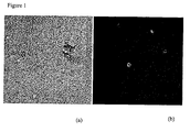
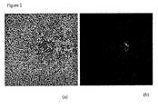
- B cells (plasma cells), secreting antigen specific IgG antibody were identified by a focal increase in fluorescence around the B cell. See Figure 2 .
- 1 ⁇ g of biotinylated antigen per 50 ⁇ l of bead stock was determined as optimal for signal generation for this particular antigen.
- Other B cells in the mixture which did not secrete antigen specific antibodies, did not show surrounding fluorescence. In controls, no B-cell localised fluorescence was observed when the beads were coated with another irrelevant antigen.
- the B cells present within the fluorescent foci were then harvested into Eppendorf tubes using standard micro-manipulation apparatus, (Eppendorf Transferman and CellTram Vario) and the heavy and light chain variable regions of the antibody from one of the cells subsequently isolated by PCR.
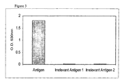
- a recombinant chimeric IgG (human constant regions) was produced by transient expression in CHO cells. Transfections of CHO cells were performed using the lipofectamine procedure according to manufacturer's instructions (InVitrogen, catalogue no. 18324). Specific binding of th IgG to antigen was confirmed by ELISA ( Figure 3 ).
- COS-1 cells transiently expressing the selected antigen were suspended in Immune cell media. Cell density was altered to 2x10 7 cells per ml.
- the assay mix was set up in ICM and contained 40 ⁇ l ELISA positive B cells, 40 ⁇ l of Goat anti-Rabbit IgG Fc specific FITC conjugate (Jackson ImmunoResearch) at 1:400 dilution and 40 ⁇ l of COS-1 cell suspension.
- This assay mix was then spotted (2-3ul per spot), onto Sigmacote treated 'chamber' slides and flooded with light paraffin oil. Slides were incubated for 40mins at 37°C and examined using an inverted microscope equipped with a mercury vapour UV lamp and a fluorescein filter set.
- B cells (plasma cells), secreting antigen specific IgG antibody were identified by a focal increase in fluorescence surrounding the B cells. See Figure 4 . Other B cells in the mixture, which did not secrete antigen specific antibodies, did not show surrounding fluorescence. In COS-1 cell controls where no antigen was present on the surface, no B-cell localised fluorescence was observed.
- the B cells present within the fluorescent foci were then harvested into Eppendorf tubes using standard micro-manipulation apparatus, (Eppendorf Transferman and CellTram Vario) and the heavy and light chain variable regions of the antibody subsequently isolated by PCR.
- CHO cells transiently expressing the selected antigen were suspended in Immune cell media. Cell density was altered to 2x10 7 cells per ml.
- the assay mix was set up in ICM and contained 40 ⁇ l ELISA positive B cells, 40 ⁇ l of Goat anti-Rabbit IgG Fc specific FITC conjugate (Jackson ImmunoResearch) at 1:400 dilution and 40 ⁇ l of CHO cell suspension.
- This assay mix was then spotted (2-3 ⁇ l per spot), onto Sigmacote treated 'chamber' slides and flooded with light paraffin oil. Slides were incubated for 40mins at 37°C and examined using an inverted microscope equipped with a mercury vapour UV lamp and a fluorescein filter set.
- B cells (plasma cells), secreting antigen specific IgG antibody were identified by a focal increase in fluorescence surrounding the B cells. See Figure 5 .
- Other B cells in the mixture which did not secrete antigen specific antibodies, did not show surrounding fluorescence.
- CHO cell controls where no antigen was present on the surface, no B-cell localised fluorescence was observed.
- the B cells present within the fluorescent foci were then harvested into Eppendorf tubes using standard micro-manipulation apparatus, (Eppendorf Transferman and CellTram Vario) and the heavy and light chain variable regions of the antibody subsequently isolated by PCR.
Landscapes
- Health & Medical Sciences (AREA)
- Life Sciences & Earth Sciences (AREA)
- Chemical & Material Sciences (AREA)
- Immunology (AREA)
- Engineering & Computer Science (AREA)
- Molecular Biology (AREA)
- Hematology (AREA)
- Urology & Nephrology (AREA)
- Biomedical Technology (AREA)
- General Health & Medical Sciences (AREA)
- Medicinal Chemistry (AREA)
- Cell Biology (AREA)
- Biochemistry (AREA)
- Organic Chemistry (AREA)
- Analytical Chemistry (AREA)
- Pathology (AREA)
- Proteomics, Peptides & Aminoacids (AREA)
- Microbiology (AREA)
- Biotechnology (AREA)
- Food Science & Technology (AREA)
- Physics & Mathematics (AREA)
- General Physics & Mathematics (AREA)
- Biophysics (AREA)
- Genetics & Genomics (AREA)
- Zoology (AREA)
- Tropical Medicine & Parasitology (AREA)
- Virology (AREA)
- Peptides Or Proteins (AREA)
- Preparation Of Compounds By Using Micro-Organisms (AREA)
- Micro-Organisms Or Cultivation Processes Thereof (AREA)
- Measuring Or Testing Involving Enzymes Or Micro-Organisms (AREA)
Claims (15)
- Homogenes In-vitro-Testverfahren zur Identifizierung einer antikörperproduzierenden Zelle, die einen Antikörper produziert, der an ein ausgewähltes Antigen bindet, wobei man:a) eine Population antikörperproduzierender Zellen bereitstellt,b) auf einem Objektträger die Population antikörperproduzierender Zellen mit einem an ein Kügelchen konjugierten oder an einen Erythrozyten gekoppelten ausgewählten Antigen oder auf der Oberfläche einer Zelle exprimierten Antigen und
einem markierten Anti-Antikörper-Antikörper, der einen an das ausgewählte Antigen bindenden Antikörper produzierende Zellen von diesen Antikörper nicht produzierenden Zellen unterscheiden kann und mit einem Fluoreszenzkonjugat markiert ist, inkubiert und,c) ohne, dass nicht gebundene Anti-Antikörper-Antikörper abgetrennt werden müssen, eine antikörperproduzierende Zelle, die zur Produktion eines an das ausgewählte Antigen bindenden Antikörpers fähig ist, identifiziert, indem man die erhöhte Konzentration an die Zelle umgebenden markierten Anti-Antikörper-Antikörpern unter Verwendung eines Mikroskops nachweist. - Testverfahren nach Anspruch 1, bei dem das Antigen an einen Erythrozyten gekoppelt ist.
- Testverfahren nach Anspruch 1, bei dem das Antigen an ein Kügelchen gekoppelt ist.
- Testverfahren nach Anspruch 3, bei dem das Antigen über einen polyklonalen Antikörper an ein Kügelchen gekoppelt ist.
- Testverfahren nach Anspruch 4, bei dem es sich bei dem polyklonalen Antikörper um ein Antikörperfragment handelt.
- Testverfahren nach Anspruch 5, bei dem es sich bei dem polyklonalen Antikörper um ein Fab-, Fab'- oder F(ab')2-Antikörperfragment handelt.
- Testverfahren nach Anspruch 1, bei dem es sich bei der antigenexprimierenden Zelle um eine transfizierte Zelle handelt.
- Testverfahren nach Anspruch 1, bei dem es sich bei der antigenexprimierenden Zelle um eine Tumorzelle handelt.
- Testverfahren nach Anspruch 1 bis 8, bei dem es sich bei dem Antigen um ein infektiöses Agens handelt.
- Testverfahren nach Anspruch 1 bis 9, bei dem es sich bei dem markierten Anti-Antikörper-Antikörper um einen Anti-Fc-Antikörper handelt.
- Testverfahren nach Anspruch 10, bei dem der fluoreszenzmarkierte Anti-Antikörper-Antikörper mit FITC markiert ist.
- Testverfahren nach Anspruch 11, bei dem es sich bei dem FITC-markierten Anti-Antikörper-Antikörper um einen Anti-Fc-Antikörper handelt.
- Testverfahren nach Anspruch 1 bis 12, bei dem es sich bei den antikörperproduzierenden Zellen um B-Zellen, Plasmazellen, Plasmablasten, aktivierte B-Zellen oder Gedächtnis-B-Zellen handelt.
- Testverfahren nach Anspruch 1 bis 13, bei dem es sich bei der antikörperproduzierenden Zelle um eine Hybridomzelle oder um eine zur Expression von Antikörpern manipulierte Säugerzelle handelt.
- In-vitro-Verfahren zur Herstellung eines Antikörpers, der an ein ausgewähltes Antigen bindet, wobei man:a) eine Population antikörperproduzierender Zellen bereitstellt,b) auf einem Objektträger die Population antikörperproduzierender Zellen mit einem an ein Kügelchen konjugierten oder an einen Erythrozyten gekoppelten ausgewählten Antigen oder auf der Oberfläche einer Zelle exprimierten Antigen und
einem markierten Anti-Antikörper-Antikörper, der einen an das ausgewählte Antigen bindenden Antikörper produzierende Zellen von diesen Antikörper nicht produzierenden Zellen unterscheiden kann und mit einem Fluoreszenzkonjugat markiert ist, inkubiert,c) ohne, dass Waschschritte notwendig sind, eine antikörperproduzierende Zelle, die einen an das ausgewählte Antigen bindenden Antikörper produziert, identifiziert, indem man die erhöhte Konzentration an die Zelle umgebenden markierten Anti-Antikörper-Antikörpern unter Verwendung eines Mikroskops nachweist,d) die identifizierte antikörperproduzierende Zelle isoliert unde) davon einen Antikörper oder ein Antikörperfragment synthetisiert.
Applications Claiming Priority (5)
| Application Number | Priority Date | Filing Date | Title |
|---|---|---|---|
| GB0228188A GB0228188D0 (en) | 2002-12-03 | 2002-12-03 | Biological products |
| GB0228188 | 2002-12-03 | ||
| GB0319587A GB0319587D0 (en) | 2003-08-20 | 2003-08-20 | Biological products |
| GB0319587 | 2003-08-20 | ||
| PCT/GB2003/005254 WO2004051268A1 (en) | 2002-12-03 | 2003-12-01 | Assay for identifying antibody producing cells |
Publications (2)
| Publication Number | Publication Date |
|---|---|
| EP1570267A1 EP1570267A1 (de) | 2005-09-07 |
| EP1570267B1 true EP1570267B1 (de) | 2011-10-12 |
Family
ID=32472152
Family Applications (1)
| Application Number | Title | Priority Date | Filing Date |
|---|---|---|---|
| EP03778577A Expired - Lifetime EP1570267B1 (de) | 2002-12-03 | 2003-12-01 | Verfahren zum nachweis antikörper-herstellender zellen |
Country Status (9)
| Country | Link |
|---|---|
| US (1) | US7993864B2 (de) |
| EP (1) | EP1570267B1 (de) |
| JP (1) | JP4603894B2 (de) |
| AT (1) | ATE528648T1 (de) |
| AU (1) | AU2003285578B2 (de) |
| CA (1) | CA2507004A1 (de) |
| ES (1) | ES2374068T3 (de) |
| PT (1) | PT1570267E (de) |
| WO (1) | WO2004051268A1 (de) |
Cited By (11)
| Publication number | Priority date | Publication date | Assignee | Title |
|---|---|---|---|---|
| WO2017009473A1 (en) | 2015-07-16 | 2017-01-19 | Ucb Biopharma Sprl | Antibody molecules which bind cd45 |
| US10358493B2 (en) | 2014-05-29 | 2019-07-23 | Ucb Biopharma Sprl | Bispecific format suitable for use in high-through-put screening |
| US10618979B2 (en) | 2015-12-03 | 2020-04-14 | Ucb Biopharma Sprl | Multispecific antibodies |
| WO2020148554A1 (en) | 2019-01-18 | 2020-07-23 | UCB Biopharma SRL | Antibodies to ebola virus glycoprotein |
| US10774157B2 (en) | 2015-12-03 | 2020-09-15 | UCB Biopharma SRL | Multispecific antibodies |
| US10829566B2 (en) | 2015-12-03 | 2020-11-10 | UCB Biopharma SRL | Method employing bispecific antibodies |
| US10954312B2 (en) | 2015-12-03 | 2021-03-23 | UCB Biopharma SRL | Method employing bispecific protein complex |
| WO2021105669A1 (en) | 2019-11-29 | 2021-06-03 | Oxford University Innovation Limited | Antibodies |
| US11286312B2 (en) | 2015-12-03 | 2022-03-29 | UCB Biopharma SRL | Multispecific antibodies |
| EP3988936A1 (de) | 2015-06-18 | 2022-04-27 | UCB Biopharma SRL | Antikörperepitop |
| WO2025120171A1 (en) | 2023-12-08 | 2025-06-12 | UCB Biopharma SRL | Antibodies |
Families Citing this family (154)
| Publication number | Priority date | Publication date | Assignee | Title |
|---|---|---|---|---|
| ES2385829T3 (es) | 2003-08-20 | 2012-08-01 | Ucb Pharma, S.A. | Métodos para obtener anticuerpos |
| US7771969B2 (en) | 2003-08-20 | 2010-08-10 | Celltech R&D Limited | Methods for obtaining antibodies |
| AU2005287406B2 (en) | 2004-07-26 | 2011-08-18 | Biogen Ma Inc. | Anti-CD154 antibodies |
| GB0506912D0 (en) | 2005-04-05 | 2005-05-11 | Celltech R&D Ltd | Biological products |
| EP1910513B1 (de) * | 2005-07-01 | 2016-08-10 | John Schrader | Verfahren zur Isolierung Antikörper-sezernierender Zellen aus dem Blut immunisierter Tiere und Menschen und Erzeugung monoklonaler Antikörper aus diesen Antikörper-sezernierenden Zellen. |
| ES2382879T3 (es) | 2005-09-14 | 2012-06-14 | Ucb Pharma, S.A. | Conjugado de anticuerpo - polímero de peine. |
| SI1960430T1 (sl) | 2005-12-09 | 2015-01-30 | Ucb Pharma, S.A. | Molekule protiteles s specifiäśnostjo za äśloveĺ ki il-6 |
| WO2007122823A1 (ja) * | 2006-04-20 | 2007-11-01 | Jichi Medical University | ベクター産生型腫瘍標的細胞 |
| EP2021463B1 (de) | 2006-05-19 | 2016-11-23 | Alder Biopharmaceuticals, Inc. | Anbauverfahren zum erhalten einer klonpopulation von antigenspezifischen b-zellen |
| GB0614780D0 (en) | 2006-07-25 | 2006-09-06 | Ucb Sa | Biological products |
| GB0619291D0 (en) | 2006-09-29 | 2006-11-08 | Ucb Sa | Altered antibodies |
| GB0620729D0 (en) | 2006-10-18 | 2006-11-29 | Ucb Sa | Biological products |
| CA2673331A1 (en) | 2006-12-19 | 2008-06-26 | Ablynx N.V. | Amino acid sequences directed against gpcrs and polypeptides comprising the same for the treatment of gpcr-related diseases and disorders |
| EP2102244A2 (de) | 2006-12-19 | 2009-09-23 | Ablynx N.V. | Gegen metalloproteinase aus der adam-familie gerichtete aminosäuresequenzen und diese enthaltende polypeptide zur behandlung von mit adam in zusammenhang stehenden krankheiten und störungen |
| US9512236B2 (en) | 2006-12-19 | 2016-12-06 | Ablynx N.V. | Amino acid sequences directed against GPCRS and polypeptides comprising the same for the treatment of GPCR-related diseases and disorders |
| EP2457928B1 (de) * | 2007-03-13 | 2017-05-10 | Universität Zürich | Monoklonale humane tumorspezifische Antikörper |
| ME00832B (me) | 2007-03-22 | 2012-03-20 | Ucb Biopharma Sprl | Vezivni proteini uključujući antitjela, derivate antitjela i fragmente antitjela, koji specifično vezuju cd154 i njihova upotreba |
| GB0717337D0 (en) | 2007-09-06 | 2007-10-17 | Ucb Pharma Sa | Method of treatment |
| EP2535349A1 (de) | 2007-09-26 | 2012-12-19 | UCB Pharma S.A. | Antikörperfusionen mit Doppelspezifität |
| GB0721752D0 (en) * | 2007-11-06 | 2007-12-19 | Univ Southampton | Configurable electronic device and method |
| EP2650311A3 (de) | 2007-11-27 | 2014-06-04 | Ablynx N.V. | Gegen heterodimere Cytokine und/oder deren Rezeptoren gerichtete Aminosäuresequenzen und Polypeptide damit |
| GB0800277D0 (en) | 2008-01-08 | 2008-02-13 | Imagination Tech Ltd | Video motion compensation |
| JP2011516520A (ja) | 2008-04-07 | 2011-05-26 | アブリンクス エン.ヴェー. | Notch経路に指向性を有するアミノ酸配列及びその使用 |
| GB0807413D0 (en) | 2008-04-23 | 2008-05-28 | Ucb Pharma Sa | Biological products |
| EP2285833B1 (de) | 2008-05-16 | 2014-12-17 | Ablynx N.V. | Gegen cxcr4 und andere gpcr gerichtete aminosäuresequenzen sowie verbindungen damit |
| LT2285408T (lt) | 2008-06-05 | 2019-01-25 | Ablynx N.V. | Aminorūgščių sekos, nukreiptos prieš viruso apvalkalo baltymus, ir tokias sekas turintys polipeptidai, skirti virusinių ligų gydymui |
| CN102257003B (zh) | 2008-12-19 | 2017-04-05 | 埃博灵克斯股份有限公司 | 用于产生针对细胞相关抗原如p2x7、cxcr7或cxcr4的免疫球蛋白的基因免疫 |
| GB0900425D0 (en) | 2009-01-12 | 2009-02-11 | Ucb Pharma Sa | Biological products |
| NZ594315A (en) | 2009-02-17 | 2013-05-31 | Ucb Pharma Sa | Antibody molecules having specificity for human ox40 |
| AU2010217120B2 (en) * | 2009-02-24 | 2014-11-20 | Novartis Ag | Methods for identifying immunobinders of cell-surface antigens |
| ES2700442T3 (es) | 2009-02-25 | 2019-02-15 | Ucb Biopharma Sprl | Método para producir anticuerpos |
| GB0903207D0 (en) | 2009-02-25 | 2009-04-08 | Ucb Pharma Sa | Method for expressing multimeric proteins |
| GB0904214D0 (en) | 2009-03-11 | 2009-04-22 | Ucb Pharma Sa | Biological products |
| US9109216B2 (en) | 2009-09-24 | 2015-08-18 | Ucb Pharma, S.A. | Bacterial host strain |
| GB201005063D0 (en) | 2010-03-25 | 2010-05-12 | Ucb Pharma Sa | Biological products |
| WO2011043077A1 (ja) * | 2009-10-09 | 2011-04-14 | 川崎重工業株式会社 | 未分化多能性幹細胞の識別方法及び装置並びに自動培養方法及び装置 |
| GB0922435D0 (en) | 2009-12-22 | 2010-02-03 | Ucb Pharma Sa | Method |
| GB0922434D0 (en) | 2009-12-22 | 2010-02-03 | Ucb Pharma Sa | antibodies and fragments thereof |
| US9234037B2 (en) | 2009-10-27 | 2016-01-12 | Ucb Biopharma Sprl | Method to generate antibodies to ion channels |
| CN102781963B (zh) | 2009-10-27 | 2018-02-16 | Ucb医药有限公司 | 功能修饰性NAv1.7抗体 |
| US20120321640A1 (en) | 2009-12-01 | 2012-12-20 | Ablynx N.V. | Von willebrand factor specific binding agents and uses thereof |
| EP3309176B1 (de) | 2009-12-14 | 2025-10-01 | Ablynx N.V. | Variable einzeldomänen-antikörper gegen ox40l, konstrukte und ihre therapeutische verwendung |
| GB201000590D0 (en) | 2010-01-14 | 2010-03-03 | Ucb Pharma Sa | Bacterial host strain |
| GB201000587D0 (en) | 2010-01-14 | 2010-03-03 | Ucb Pharma Sa | Bacterial hoist strain |
| GB201000591D0 (en) | 2010-01-14 | 2010-03-03 | Ucb Pharma Sa | Bacterial hoist strain |
| US9120855B2 (en) | 2010-02-10 | 2015-09-01 | Novartis Ag | Biologic compounds directed against death receptor 5 |
| EP2545078A1 (de) | 2010-03-11 | 2013-01-16 | UCB Pharma, S.A. | Pd-1-antikörper |
| TW201134488A (en) | 2010-03-11 | 2011-10-16 | Ucb Pharma Sa | PD-1 antibodies |
| ES2717883T3 (es) | 2010-03-25 | 2019-06-26 | Ucb Biopharma Sprl | Moléculas de DVD-LG estabilizadas con disulfuro |
| GB201005064D0 (en) | 2010-03-25 | 2010-05-12 | Ucb Pharma Sa | Biological products |
| KR20130079362A (ko) * | 2010-03-25 | 2013-07-10 | 내셔날 유니버시티 코포레이션 유니버시티 오브 토야마 | 형질 세포 동정 및 단리용 형광 프로브 및 이 프로브를 이용한 형질 세포의 동정 또는 단리방법 |
| WO2011117423A1 (en) | 2010-03-26 | 2011-09-29 | Ablynx N.V. | Immunoglobulin single variable domains directed against cxcr7 |
| EP2571901B1 (de) | 2010-05-20 | 2019-01-02 | Ablynx N.V. | Biologische stoffe im zusammenhang mit her3 |
| GB201012599D0 (en) | 2010-07-27 | 2010-09-08 | Ucb Pharma Sa | Process for purifying proteins |
| GB201014033D0 (en) | 2010-08-20 | 2010-10-06 | Ucb Pharma Sa | Biological products |
| AU2011328246B2 (en) | 2010-11-08 | 2016-06-30 | Ablynx N.V. | CXCR2 binding polypeptides |
| ME02734B (de) | 2011-01-14 | 2017-10-20 | Ucb Biopharma Sprl | Antikörpermodule zur bindung an il-17a und il-17f |
| CN103917560B (zh) | 2011-03-28 | 2017-05-24 | 埃博灵克斯股份有限公司 | 双特异性抗‑cxcr7免疫球蛋白单可变结构域 |
| JP5963746B2 (ja) * | 2011-03-30 | 2016-08-03 | 国立大学法人富山大学 | 形質細胞または形質芽細胞の選択方法、目的抗原特異的な抗体の製造方法、新規モノクローナル抗体 |
| UA117218C2 (uk) | 2011-05-05 | 2018-07-10 | Мерк Патент Гмбх | Поліпептид, спрямований проти il-17a, il-17f та/або il17-a/f |
| RS55775B2 (sr) | 2011-06-23 | 2022-10-31 | Ablynx Nv | Tehnike za predviđanje, otkrivanje i smanjenje nespecifične proteinske interferencije u testovima koji uključuju pojedinačne varijabilne domene imunoglobulina |
| SG10201805064SA (en) | 2011-06-23 | 2018-07-30 | Ablynx Nv | Techniques for predicting, detecting and reducing aspecific protein interference in assays involving immunoglobulin single variable domains |
| EP4350345A3 (de) | 2011-06-23 | 2024-07-24 | Ablynx N.V. | Verfahren zur vorhersage, erkennung und reduzierung von aspezifischer proteininterferenz in tests mit variablen immunglobulin-einzeldomänen |
| HRP20180226T1 (hr) | 2011-07-13 | 2018-03-09 | Ucb Biopharma Sprl | Bakterijski soj domaćina koji eksprimira rekombinantnu dsbc |
| US20140234903A1 (en) | 2011-09-05 | 2014-08-21 | Eth Zurich | Biosynthetic gene cluster for the production of peptide/protein analogues |
| PL2758432T3 (pl) | 2011-09-16 | 2019-08-30 | Ucb Biopharma Sprl | Przeciwciała neutralizujące przeciw głównym egzotoksynom tcda i tcdb z clostridium difficile |
| WO2013068571A1 (en) | 2011-11-11 | 2013-05-16 | Ucb Pharma S.A. | Albumin binding antibodies and binding fragments thereof |
| US8962315B2 (en) | 2011-12-22 | 2015-02-24 | Elwha Llc | Compositions and methods including recombinant B lymphocyte cell line including at least one endogenous gene expressing at least one endogenous membrane immunoglobulin reactive to a first antigen and including at least one exogenously incorporated nucleic acid expressing at least one exogenous secreted immunoglobulin reactive to a second antigen |
| US9175072B2 (en) | 2011-12-22 | 2015-11-03 | Elwha Llc | Compositions and methods including recombinant B lymphocyte cell line including an exogenously incorporated nucleic acid expressing an exogenous membrane immunoglobulin reactive to a first antigen and including an endogenous gene expressing an endogenous secreted immunoglobulin reactive to a second antigen |
| US10745468B2 (en) | 2011-12-22 | 2020-08-18 | Kota Biotherapeutics, Llc | Compositions and methods for modified B cells expressing reassigned biological agents |
| US10233424B2 (en) | 2011-12-22 | 2019-03-19 | Elwha Llc | Compositions and methods including cytotoxic B lymphocyte cell line expressing exogenous membrane immunoglobulin different from secreted immunoglobulin |
| GB201201332D0 (en) | 2012-01-26 | 2012-03-14 | Imp Innovations Ltd | Method |
| GB201203051D0 (en) | 2012-02-22 | 2012-04-04 | Ucb Pharma Sa | Biological products |
| GB201203071D0 (en) | 2012-02-22 | 2012-04-04 | Ucb Pharma Sa | Biological products |
| FR2987627B1 (fr) | 2012-03-05 | 2016-03-18 | Splicos | Utilisation de rbm39 comme biomarqueur |
| US9328174B2 (en) | 2012-05-09 | 2016-05-03 | Novartis Ag | Chemokine receptor binding polypeptides |
| GB201208370D0 (en) | 2012-05-14 | 2012-06-27 | Ucb Pharma Sa | Antibodies |
| GB201208367D0 (en) | 2012-05-14 | 2012-06-27 | Ucb Pharma Sa | Biological product |
| EP2867674B1 (de) | 2012-06-28 | 2018-10-10 | UCB Biopharma SPRL | Verfahren zur identifikation von verbindungen von therapeutischem interesse |
| US8921055B2 (en) | 2012-10-30 | 2014-12-30 | Berkeley Lights, Inc. | Detecting cells secreting a protein of interest |
| GB201223276D0 (en) | 2012-12-21 | 2013-02-06 | Ucb Pharma Sa | Antibodies and methods of producing same |
| GB201315487D0 (en) | 2013-08-30 | 2013-10-16 | Ucb Pharma Sa | Antibodies |
| HUE035875T2 (hu) | 2013-10-25 | 2018-06-28 | Psioxus Therapeutics Ltd | Heterológ géneket tartalmazó onkolitikus adenovírusok |
| GB201320066D0 (en) | 2013-11-13 | 2013-12-25 | Ucb Pharma Sa | Biological products |
| US8986694B1 (en) | 2014-07-15 | 2015-03-24 | Kymab Limited | Targeting human nav1.7 variants for treatment of pain |
| US9067998B1 (en) | 2014-07-15 | 2015-06-30 | Kymab Limited | Targeting PD-1 variants for treatment of cancer |
| US8992927B1 (en) | 2014-07-15 | 2015-03-31 | Kymab Limited | Targeting human NAV1.7 variants for treatment of pain |
| US9914769B2 (en) | 2014-07-15 | 2018-03-13 | Kymab Limited | Precision medicine for cholesterol treatment |
| US9045545B1 (en) | 2014-07-15 | 2015-06-02 | Kymab Limited | Precision medicine by targeting PD-L1 variants for treatment of cancer |
| GB201403775D0 (en) | 2014-03-04 | 2014-04-16 | Kymab Ltd | Antibodies, uses & methods |
| GB201406608D0 (en) | 2014-04-12 | 2014-05-28 | Psioxus Therapeutics Ltd | Virus |
| GB201411320D0 (en) | 2014-06-25 | 2014-08-06 | Ucb Biopharma Sprl | Antibody construct |
| US9139648B1 (en) | 2014-07-15 | 2015-09-22 | Kymab Limited | Precision medicine by targeting human NAV1.9 variants for treatment of pain |
| GB201412659D0 (en) | 2014-07-16 | 2014-08-27 | Ucb Biopharma Sprl | Molecules |
| GB201412658D0 (en) | 2014-07-16 | 2014-08-27 | Ucb Biopharma Sprl | Molecules |
| GB201506870D0 (en) | 2015-04-22 | 2015-06-03 | Ucb Biopharma Sprl | Method |
| GB201506869D0 (en) | 2015-04-22 | 2015-06-03 | Ucb Biopharma Sprl | Method |
| TW201702271A (zh) | 2015-04-30 | 2017-01-16 | 哈佛大學校長及研究員協會 | 治療代謝病症之抗-ap2抗體及抗原結合劑 |
| GB201508180D0 (en) | 2015-05-13 | 2015-06-24 | Ucb Biopharma Sprl | Antibodies |
| JP6851322B2 (ja) | 2015-05-27 | 2021-03-31 | ユーシービー バイオファルマ エスアールエル | 神経疾患を治療する方法 |
| GB201601077D0 (en) | 2016-01-20 | 2016-03-02 | Ucb Biopharma Sprl | Antibody molecule |
| GB201601075D0 (en) | 2016-01-20 | 2016-03-02 | Ucb Biopharma Sprl | Antibodies molecules |
| CN116059350A (zh) | 2015-10-27 | 2023-05-05 | Ucb生物制药有限责任公司 | 使用抗-il-17a/f抗体的治疗方法 |
| WO2017086419A1 (ja) | 2015-11-18 | 2017-05-26 | 中外製薬株式会社 | 液性免疫応答の増強方法 |
| GB201522394D0 (en) | 2015-12-18 | 2016-02-03 | Ucb Biopharma Sprl | Antibodies |
| GB201602413D0 (en) | 2016-02-10 | 2016-03-23 | Nascient Ltd | Method |
| US9442113B1 (en) | 2016-02-29 | 2016-09-13 | Rarecyte, Inc. | Method to identify antigen-specific immune cells for therapeutic development |
| US10222373B2 (en) | 2016-02-29 | 2019-03-05 | Rarecyte, Inc. | Method to identify antigen-specific immune cells |
| US9395367B1 (en) | 2016-02-29 | 2016-07-19 | Rarecyte, Inc. | Method to identify antigen-specific B cells for antibody development |
| GB201610198D0 (en) | 2016-06-10 | 2016-07-27 | Ucb Biopharma Sprl | Anti-ige antibodies |
| CN109563482A (zh) | 2016-06-10 | 2019-04-02 | 埃尔瓦有限公司 | 含表达异于分泌免疫球蛋白和细胞溶解功能的膜免疫球蛋白的b淋巴细胞系的组合物和方法 |
| RU2769282C2 (ru) | 2016-06-20 | 2022-03-30 | Кимаб Лимитед | Анти-PD-L1 и IL-2 цитокины |
| WO2018038684A1 (en) | 2016-08-26 | 2018-03-01 | Agency For Science, Technology And Research | Macrophage stimulating protein receptor (or ron - recepteur d' origine nantais) antibodies and uses thereof |
| GB201616596D0 (en) | 2016-09-29 | 2016-11-16 | Nascient Limited | Epitope and antibodies |
| EP3534947A1 (de) | 2016-11-03 | 2019-09-11 | Kymab Limited | Antikörper, kombinationen mit antikörpern, biomarker, verwendungen und verfahren |
| GB201621635D0 (en) | 2016-12-19 | 2017-02-01 | Ucb Biopharma Sprl | Crystal structure |
| WO2019084512A1 (en) * | 2017-10-26 | 2019-05-02 | Essenlix Corporation | DETECTION OF BACTERIA CAUSING SEXUALLY TRANSMITTED DISEASES AND IMMUNE T CELLS |
| GB201802486D0 (en) | 2018-02-15 | 2018-04-04 | Ucb Biopharma Sprl | Methods |
| MX2020013808A (es) | 2018-06-18 | 2021-05-27 | UCB Biopharma SRL | Antagonista de gremlina-1 para la prevencion y el tratamiento del cancer. |
| CN113646051A (zh) | 2018-10-16 | 2021-11-12 | Ucb生物制药有限责任公司 | 重症肌无力的治疗方法 |
| GB201817309D0 (en) | 2018-10-24 | 2018-12-05 | Ucb Biopharma Sprl | Antibodies |
| GB201817311D0 (en) | 2018-10-24 | 2018-12-05 | Ucb Biopharma Sprl | Antibodies |
| JP7410143B2 (ja) | 2018-11-01 | 2024-01-09 | 山▲東▼新▲時▼代▲薬▼▲業▼有限公司 | 二重特異性抗体及びその用途 |
| US11357787B2 (en) | 2019-02-18 | 2022-06-14 | Nb Health Laboratory Co., Ltd. | Method for selecting cells, method for producing nucleic acid, method for producing recombinant cells, method for producing target substance, method for producing pharmaceutical composition, and reagent |
| JP7607573B2 (ja) | 2019-09-30 | 2024-12-27 | 東京応化工業株式会社 | 分泌物産生細胞のスクリーニング方法、及び分泌物産生細胞のスクリーニングキット |
| GB201919058D0 (en) | 2019-12-20 | 2020-02-05 | Ucb Biopharma Sprl | Multi-specific antibodies |
| GB201919061D0 (en) | 2019-12-20 | 2020-02-05 | Ucb Biopharma Sprl | Multi-specific antibody |
| GB201919062D0 (en) | 2019-12-20 | 2020-02-05 | Ucb Biopharma Sprl | Antibody |
| GB202001447D0 (en) | 2020-02-03 | 2020-03-18 | Ucb Biopharma Sprl | Antibodies |
| WO2021160267A1 (en) | 2020-02-13 | 2021-08-19 | UCB Biopharma SRL | Bispecific antibodies against cd9 and cd7 |
| US20230151108A1 (en) | 2020-02-13 | 2023-05-18 | UCB Biopharma SRL | Bispecific antibodies against cd9 and cd137 |
| WO2021160269A1 (en) | 2020-02-13 | 2021-08-19 | UCB Biopharma SRL | Anti cd44-ctla4 bispecific antibodies |
| EP4103612A1 (de) | 2020-02-13 | 2022-12-21 | UCB Biopharma SRL | Bispezifische antikörper gegen cd9 |
| ES2975410T3 (es) | 2020-02-13 | 2024-07-05 | UCB Biopharma SRL | Anticuerpos biespecíficos que se unen a HVEM y CD9 |
| JP2023546071A (ja) | 2020-10-13 | 2023-11-01 | アルミラル・ソシエダッド・アノニマ | 二重特異性分子およびそれを用いた処置方法 |
| JP2023547795A (ja) | 2020-10-15 | 2023-11-14 | ユーシービー バイオファルマ エスアールエル | Cd45を多量体化する結合分子 |
| JP2023550596A (ja) | 2020-11-02 | 2023-12-04 | ユーシービー バイオファルマ エスアールエル | 運動ニューロン神経変性障害の治療のための抗trem1中和抗体の使用 |
| PE20231953A1 (es) | 2020-12-07 | 2023-12-06 | UCB Biopharma SRL | Anticuerpos multiespecificos y combinaciones de anticuerpos |
| AR124250A1 (es) | 2020-12-07 | 2023-03-01 | UCB Biopharma SRL | Anticuerpos |
| CN117321076A (zh) | 2021-02-19 | 2023-12-29 | 美国卫生及公众服务部代表 | 中和SARS-CoV-2的单结构域抗体 |
| EP4067381A1 (de) | 2021-04-01 | 2022-10-05 | Julius-Maximilians-Universität Würzburg | Neue tnfr2-bindende moleküle |
| MX2023012489A (es) | 2021-04-22 | 2023-11-22 | Guangdong Fapon Biopharma Inc | Polipeptido de fusion biespecifico multifuncional. |
| IL308100A (en) | 2021-05-03 | 2023-12-01 | UCB Biopharma SRL | Antibodies |
| WO2023285878A1 (en) | 2021-07-13 | 2023-01-19 | Aviation-Ophthalmology | Methods for detecting, treating, and preventing gpr68-mediated ocular diseases, disorders, and conditions |
| GB202111905D0 (en) | 2021-08-19 | 2021-10-06 | UCB Biopharma SRL | Antibodies |
| GB202205200D0 (en) | 2022-04-08 | 2022-05-25 | Ucb Biopharma Sprl | Combination with chemotherapy |
| GB202205203D0 (en) | 2022-04-08 | 2022-05-25 | UCB Biopharma SRL | Combination with inhibitor |
| WO2024050354A1 (en) | 2022-08-31 | 2024-03-07 | Washington University | Alphavirus antigen binding antibodies and uses thereof |
| EP4605077A1 (de) | 2022-10-18 | 2025-08-27 | Confo Therapeutics N.V. | Gegen den melanocortin-4-rezeptor gerichtete aminosäuresequenzen und polypeptide damit zur behandlung von mc4r-bedingten erkrankungen und störungen |
| AU2023400451A1 (en) | 2022-11-28 | 2025-05-22 | UCB Biopharma SRL | Treatment of fibromyalgia |
| TW202540204A (zh) | 2024-01-26 | 2025-10-16 | 西班牙商阿爾米雷爾有限公司 | 雙特異性分子及使用其的治療方法 |
| WO2025238133A1 (en) | 2024-05-17 | 2025-11-20 | UCB Biopharma SRL | Multispecific antibody with binding specificity for il-11 and il-17 |
| WO2025238135A2 (en) | 2024-05-17 | 2025-11-20 | UCB Biopharma SRL | Antibody with binding specificity for il-11 |
| WO2026027660A1 (en) | 2024-08-02 | 2026-02-05 | UCB Biopharma SRL | Formulations of anti-gremlin-1 antibodies |
Citations (1)
| Publication number | Priority date | Publication date | Assignee | Title |
|---|---|---|---|---|
| WO2003012449A2 (en) * | 2001-07-27 | 2003-02-13 | Lonza Group Ag | Method for selecting antibody expressing cells |
Family Cites Families (15)
| Publication number | Priority date | Publication date | Assignee | Title |
|---|---|---|---|---|
| US3865689A (en) * | 1972-11-09 | 1975-02-11 | Hoffmann La Roche | Method of producing carcinoembryonic antigens |
| GB8308235D0 (en) * | 1983-03-25 | 1983-05-05 | Celltech Ltd | Polypeptides |
| EP0286405A3 (de) * | 1987-04-08 | 1989-08-09 | Synbiotics Corporation | Verfahren zur frühen Auswahl von inneren Bildern der Anti-idiotyp-Antikörper |
| US5264341A (en) * | 1989-08-30 | 1993-11-23 | Eli Lilly And Company | Selective cloning for high monoclonal antibody secreting hybridomas |
| CA2090126C (en) | 1990-08-02 | 2002-10-22 | John W. Schrader | Methods for the production of proteins with a desired function |
| US5256542A (en) * | 1992-03-09 | 1993-10-26 | Tanox Biosystems, Inc. | Selecting low frequency antigen-specific single B lymphocytes with correction for background noise |
| US5397703A (en) * | 1992-07-09 | 1995-03-14 | Cetus Oncology Corporation | Method for generation of antibodies to cell surface molecules |
| JP3731891B2 (ja) | 1992-10-21 | 2006-01-05 | ミルテンイ,ステファン | 分泌生成物による細胞の直接選択 |
| ATE282717T1 (de) | 1998-04-30 | 2004-12-15 | Max Planck Gesellschaft | Neuartiges verfahren zur identifizierung von klonen mit einer gewünschten biologischen eigenschaft, ausgehend von einer expressionsgenbank |
| AU765085B2 (en) | 1998-05-11 | 2003-09-11 | Miltenyi Biotec Gmbh | Method of direct selection of antigen-specific T cells |
| JP2000304750A (ja) * | 1999-04-16 | 2000-11-02 | Fuji Photo Film Co Ltd | 発光標識イムノアッセイ及びその分析要素 |
| WO2001055727A1 (en) * | 2000-01-31 | 2001-08-02 | Emory University | Immunological assay system and method |
| AUPR177400A0 (en) | 2000-11-29 | 2000-12-21 | Cancerprobe Pty Ltd | Probes for identifying cancer-specific antigens |
| US20040240687A1 (en) * | 2003-05-30 | 2004-12-02 | Graetz Michael L. | Flat panel speaker |
| GB0412973D0 (en) * | 2004-06-10 | 2004-07-14 | Celltech R&D Ltd | Identification of antibody producing cells |
-
2003
- 2003-12-01 ES ES03778577T patent/ES2374068T3/es not_active Expired - Lifetime
- 2003-12-01 WO PCT/GB2003/005254 patent/WO2004051268A1/en not_active Ceased
- 2003-12-01 AT AT03778577T patent/ATE528648T1/de not_active IP Right Cessation
- 2003-12-01 PT PT03778577T patent/PT1570267E/pt unknown
- 2003-12-01 US US10/537,309 patent/US7993864B2/en not_active Expired - Lifetime
- 2003-12-01 CA CA002507004A patent/CA2507004A1/en not_active Abandoned
- 2003-12-01 AU AU2003285578A patent/AU2003285578B2/en not_active Expired
- 2003-12-01 EP EP03778577A patent/EP1570267B1/de not_active Expired - Lifetime
- 2003-12-01 JP JP2004570704A patent/JP4603894B2/ja not_active Expired - Lifetime
Patent Citations (1)
| Publication number | Priority date | Publication date | Assignee | Title |
|---|---|---|---|---|
| WO2003012449A2 (en) * | 2001-07-27 | 2003-02-13 | Lonza Group Ag | Method for selecting antibody expressing cells |
Non-Patent Citations (1)
| Title |
|---|
| MANZ R ET AL: "Analysis and sorting of live cells according to secreted molecules, relocated to a cell-surface affinity matrix.", PROCEEDINGS OF THE NATIONAL ACADEMY OF SCIENCES OF THE UNITED STATES OF AMERICA 14 MAR 1995 LNKD- PUBMED:7892200, vol. 92, no. 6, 14 March 1995 (1995-03-14), pages 1921 - 1925, XP002007404, ISSN: 0027-8424, DOI: doi:10.1073/pnas.92.6.1921 * |
Cited By (13)
| Publication number | Priority date | Publication date | Assignee | Title |
|---|---|---|---|---|
| EP3750915A1 (de) | 2014-05-29 | 2020-12-16 | UCB Biopharma SRL | Neues bispezifisches format zur verwendung bei hochdurchsatzscreening |
| US10358493B2 (en) | 2014-05-29 | 2019-07-23 | Ucb Biopharma Sprl | Bispecific format suitable for use in high-through-put screening |
| EP3995831A1 (de) | 2015-06-18 | 2022-05-11 | UCB Biopharma SRL | Antikörper |
| EP3988936A1 (de) | 2015-06-18 | 2022-04-27 | UCB Biopharma SRL | Antikörperepitop |
| WO2017009473A1 (en) | 2015-07-16 | 2017-01-19 | Ucb Biopharma Sprl | Antibody molecules which bind cd45 |
| US11286312B2 (en) | 2015-12-03 | 2022-03-29 | UCB Biopharma SRL | Multispecific antibodies |
| US10829566B2 (en) | 2015-12-03 | 2020-11-10 | UCB Biopharma SRL | Method employing bispecific antibodies |
| US10954312B2 (en) | 2015-12-03 | 2021-03-23 | UCB Biopharma SRL | Method employing bispecific protein complex |
| US10774157B2 (en) | 2015-12-03 | 2020-09-15 | UCB Biopharma SRL | Multispecific antibodies |
| US10618979B2 (en) | 2015-12-03 | 2020-04-14 | Ucb Biopharma Sprl | Multispecific antibodies |
| WO2020148554A1 (en) | 2019-01-18 | 2020-07-23 | UCB Biopharma SRL | Antibodies to ebola virus glycoprotein |
| WO2021105669A1 (en) | 2019-11-29 | 2021-06-03 | Oxford University Innovation Limited | Antibodies |
| WO2025120171A1 (en) | 2023-12-08 | 2025-06-12 | UCB Biopharma SRL | Antibodies |
Also Published As
| Publication number | Publication date |
|---|---|
| JP2006509217A (ja) | 2006-03-16 |
| EP1570267A1 (de) | 2005-09-07 |
| ATE528648T1 (de) | 2011-10-15 |
| CA2507004A1 (en) | 2004-06-17 |
| AU2003285578B2 (en) | 2010-07-15 |
| WO2004051268A1 (en) | 2004-06-17 |
| PT1570267E (pt) | 2012-01-03 |
| US7993864B2 (en) | 2011-08-09 |
| US20060148012A1 (en) | 2006-07-06 |
| ES2374068T3 (es) | 2012-02-13 |
| JP4603894B2 (ja) | 2010-12-22 |
| AU2003285578A1 (en) | 2004-06-23 |
Similar Documents
| Publication | Publication Date | Title |
|---|---|---|
| EP1570267B1 (de) | Verfahren zum nachweis antikörper-herstellender zellen | |
| US20070243564A1 (en) | Identification of Antibody-Producing Cells | |
| JP4202251B2 (ja) | 抗体発現細胞を選択する方法 | |
| JP3731891B2 (ja) | 分泌生成物による細胞の直接選択 | |
| US4420558A (en) | Bright field light microscopic method of enumerating and characterizing subtypes of white blood cells and their precursors | |
| US7166423B1 (en) | Direct selection of cells by secretion product | |
| US20140199319A1 (en) | Methods for increasing the efficiency of hybridoma generation | |
| JPH029366A (ja) | モノクローナル二特異的免疫グロブリンを製造するためのファストトラック法 | |
| WO2019238137A1 (zh) | 抗sFcεRIα单克隆抗体及其应用 | |
| US20060073095A1 (en) | Methods for screening antibody-producing cells on heterogeneous antigen substrates | |
| US20030044849A1 (en) | Methods for screening monoclonal antibodies on heterogeneous antigen substrates | |
| FR2599513A1 (fr) | Anticorps monoclonaux inhibant la liaison de ige soluble aux lymphocytes humains, hybridomes monoclonaux produisant de tels anticorps, leur utilisation | |
| US7067314B2 (en) | Monoclonal antibody, its immunoreactive fragment and hybridoma | |
| Brams et al. | In vitro priming of human lymphocytes. II. Induction of antigen-specific IgG responses by repeated antigen stimulation | |
| JP2002209579A (ja) | 抗ヒトヘモグロビンモノクローナル抗体、およびそれを産生する細胞株またはそれを含むヒトヘモグロビン検出用キット | |
| WO2003028625A2 (en) | Methods for screening monoclonal antibodies on heterogeneous antigen substrates | |
| CN119876016A (zh) | 一种兔单b细胞富集方法及微流控筛选方法 | |
| Assenmacher | Combined intracellular and surface staining | |
| CA1329545C (en) | Immunological complex, its preparation and its use | |
| CN120647759A (zh) | 醛固酮抗原结合分子及其应用 | |
| JPH11504208A (ja) | レパートリークローニング法、それから得られる産物及びその産物の使用 | |
| Van Duijn et al. | Production and application of polyclonal and monoclonal antibodies | |
| JPH0257178A (ja) | モノクローナル抗体産生細胞ラインおよびモノクローナル抗体 | |
| JPH02131574A (ja) | ミニクローン | |
| JPWO2004078964A1 (ja) | ハイブリドーマの製造方法およびその利用 |
Legal Events
| Date | Code | Title | Description |
|---|---|---|---|
| PUAI | Public reference made under article 153(3) epc to a published international application that has entered the european phase |
Free format text: ORIGINAL CODE: 0009012 |
|
| 17P | Request for examination filed |
Effective date: 20050704 |
|
| AK | Designated contracting states |
Kind code of ref document: A1 Designated state(s): AT BE BG CH CY CZ DE DK EE ES FI FR GB GR HU IE IT LI LU MC NL PT RO SE SI SK TR |
|
| AX | Request for extension of the european patent |
Extension state: AL LT LV MK |
|
| RAP1 | Party data changed (applicant data changed or rights of an application transferred) |
Owner name: UCB PHARMA, S.A. |
|
| 17Q | First examination report despatched |
Effective date: 20070830 |
|
| GRAJ | Information related to disapproval of communication of intention to grant by the applicant or resumption of examination proceedings by the epo deleted |
Free format text: ORIGINAL CODE: EPIDOSDIGR1 |
|
| GRAP | Despatch of communication of intention to grant a patent |
Free format text: ORIGINAL CODE: EPIDOSNIGR1 |
|
| GRAS | Grant fee paid |
Free format text: ORIGINAL CODE: EPIDOSNIGR3 |
|
| GRAA | (expected) grant |
Free format text: ORIGINAL CODE: 0009210 |
|
| AK | Designated contracting states |
Kind code of ref document: B1 Designated state(s): AT BE BG CH CY CZ DE DK EE ES FI FR GB GR HU IE IT LI LU MC NL PT RO SE SI SK TR |
|
| AX | Request for extension of the european patent |
Extension state: AL LT LV MK |
|
| REG | Reference to a national code |
Ref country code: GB Ref legal event code: FG4D |
|
| REG | Reference to a national code |
Ref country code: CH Ref legal event code: EP |
|
| REG | Reference to a national code |
Ref country code: IE Ref legal event code: FG4D |
|
| REG | Reference to a national code |
Ref country code: DE Ref legal event code: R096 Ref document number: 60338758 Country of ref document: DE Effective date: 20111201 |
|
| REG | Reference to a national code |
Ref country code: PT Ref legal event code: SC4A Free format text: AVAILABILITY OF NATIONAL TRANSLATION Effective date: 20111214 |
|
| REG | Reference to a national code |
Ref country code: NL Ref legal event code: VDEP Effective date: 20111012 |
|
| REG | Reference to a national code |
Ref country code: ES Ref legal event code: FG2A Ref document number: 2374068 Country of ref document: ES Kind code of ref document: T3 Effective date: 20120213 |
|
| LTIE | Lt: invalidation of european patent or patent extension |
Effective date: 20111012 |
|
| REG | Reference to a national code |
Ref country code: AT Ref legal event code: MK05 Ref document number: 528648 Country of ref document: AT Kind code of ref document: T Effective date: 20111012 |
|
| PG25 | Lapsed in a contracting state [announced via postgrant information from national office to epo] |
Ref country code: SI Free format text: LAPSE BECAUSE OF FAILURE TO SUBMIT A TRANSLATION OF THE DESCRIPTION OR TO PAY THE FEE WITHIN THE PRESCRIBED TIME-LIMIT Effective date: 20111012 Ref country code: NL Free format text: LAPSE BECAUSE OF FAILURE TO SUBMIT A TRANSLATION OF THE DESCRIPTION OR TO PAY THE FEE WITHIN THE PRESCRIBED TIME-LIMIT Effective date: 20111012 Ref country code: SE Free format text: LAPSE BECAUSE OF FAILURE TO SUBMIT A TRANSLATION OF THE DESCRIPTION OR TO PAY THE FEE WITHIN THE PRESCRIBED TIME-LIMIT Effective date: 20111012 Ref country code: GR Free format text: LAPSE BECAUSE OF FAILURE TO SUBMIT A TRANSLATION OF THE DESCRIPTION OR TO PAY THE FEE WITHIN THE PRESCRIBED TIME-LIMIT Effective date: 20120113 |
|
| PG25 | Lapsed in a contracting state [announced via postgrant information from national office to epo] |
Ref country code: CY Free format text: LAPSE BECAUSE OF FAILURE TO SUBMIT A TRANSLATION OF THE DESCRIPTION OR TO PAY THE FEE WITHIN THE PRESCRIBED TIME-LIMIT Effective date: 20111012 |
|
| PG25 | Lapsed in a contracting state [announced via postgrant information from national office to epo] |
Ref country code: EE Free format text: LAPSE BECAUSE OF FAILURE TO SUBMIT A TRANSLATION OF THE DESCRIPTION OR TO PAY THE FEE WITHIN THE PRESCRIBED TIME-LIMIT Effective date: 20111012 Ref country code: BG Free format text: LAPSE BECAUSE OF FAILURE TO SUBMIT A TRANSLATION OF THE DESCRIPTION OR TO PAY THE FEE WITHIN THE PRESCRIBED TIME-LIMIT Effective date: 20120112 Ref country code: SK Free format text: LAPSE BECAUSE OF FAILURE TO SUBMIT A TRANSLATION OF THE DESCRIPTION OR TO PAY THE FEE WITHIN THE PRESCRIBED TIME-LIMIT Effective date: 20111012 Ref country code: CZ Free format text: LAPSE BECAUSE OF FAILURE TO SUBMIT A TRANSLATION OF THE DESCRIPTION OR TO PAY THE FEE WITHIN THE PRESCRIBED TIME-LIMIT Effective date: 20111012 Ref country code: MC Free format text: LAPSE BECAUSE OF NON-PAYMENT OF DUE FEES Effective date: 20111231 Ref country code: DK Free format text: LAPSE BECAUSE OF FAILURE TO SUBMIT A TRANSLATION OF THE DESCRIPTION OR TO PAY THE FEE WITHIN THE PRESCRIBED TIME-LIMIT Effective date: 20111012 |
|
| PLBE | No opposition filed within time limit |
Free format text: ORIGINAL CODE: 0009261 |
|
| STAA | Information on the status of an ep patent application or granted ep patent |
Free format text: STATUS: NO OPPOSITION FILED WITHIN TIME LIMIT |
|
| PG25 | Lapsed in a contracting state [announced via postgrant information from national office to epo] |
Ref country code: RO Free format text: LAPSE BECAUSE OF FAILURE TO SUBMIT A TRANSLATION OF THE DESCRIPTION OR TO PAY THE FEE WITHIN THE PRESCRIBED TIME-LIMIT Effective date: 20111012 |
|
| 26N | No opposition filed |
Effective date: 20120713 |
|
| REG | Reference to a national code |
Ref country code: IE Ref legal event code: MM4A |
|
| PG25 | Lapsed in a contracting state [announced via postgrant information from national office to epo] |
Ref country code: IE Free format text: LAPSE BECAUSE OF NON-PAYMENT OF DUE FEES Effective date: 20111201 |
|
| REG | Reference to a national code |
Ref country code: DE Ref legal event code: R097 Ref document number: 60338758 Country of ref document: DE Effective date: 20120713 |
|
| PG25 | Lapsed in a contracting state [announced via postgrant information from national office to epo] |
Ref country code: AT Free format text: LAPSE BECAUSE OF FAILURE TO SUBMIT A TRANSLATION OF THE DESCRIPTION OR TO PAY THE FEE WITHIN THE PRESCRIBED TIME-LIMIT Effective date: 20111012 |
|
| PG25 | Lapsed in a contracting state [announced via postgrant information from national office to epo] |
Ref country code: LU Free format text: LAPSE BECAUSE OF NON-PAYMENT OF DUE FEES Effective date: 20111201 |
|
| PG25 | Lapsed in a contracting state [announced via postgrant information from national office to epo] |
Ref country code: FI Free format text: LAPSE BECAUSE OF FAILURE TO SUBMIT A TRANSLATION OF THE DESCRIPTION OR TO PAY THE FEE WITHIN THE PRESCRIBED TIME-LIMIT Effective date: 20111012 |
|
| PG25 | Lapsed in a contracting state [announced via postgrant information from national office to epo] |
Ref country code: TR Free format text: LAPSE BECAUSE OF FAILURE TO SUBMIT A TRANSLATION OF THE DESCRIPTION OR TO PAY THE FEE WITHIN THE PRESCRIBED TIME-LIMIT Effective date: 20111012 |
|
| PG25 | Lapsed in a contracting state [announced via postgrant information from national office to epo] |
Ref country code: HU Free format text: LAPSE BECAUSE OF FAILURE TO SUBMIT A TRANSLATION OF THE DESCRIPTION OR TO PAY THE FEE WITHIN THE PRESCRIBED TIME-LIMIT Effective date: 20111012 |
|
| REG | Reference to a national code |
Ref country code: FR Ref legal event code: PLFP Year of fee payment: 13 |
|
| REG | Reference to a national code |
Ref country code: FR Ref legal event code: PLFP Year of fee payment: 14 |
|
| REG | Reference to a national code |
Ref country code: FR Ref legal event code: PLFP Year of fee payment: 15 |
|
| PGFP | Annual fee paid to national office [announced via postgrant information from national office to epo] |
Ref country code: PT Payment date: 20221202 Year of fee payment: 20 Ref country code: IT Payment date: 20221111 Year of fee payment: 20 Ref country code: GB Payment date: 20221103 Year of fee payment: 20 Ref country code: FR Payment date: 20221110 Year of fee payment: 20 Ref country code: DE Payment date: 20221102 Year of fee payment: 20 |
|
| PGFP | Annual fee paid to national office [announced via postgrant information from national office to epo] |
Ref country code: BE Payment date: 20221118 Year of fee payment: 20 |
|
| PGFP | Annual fee paid to national office [announced via postgrant information from national office to epo] |
Ref country code: ES Payment date: 20230113 Year of fee payment: 20 Ref country code: CH Payment date: 20230101 Year of fee payment: 20 |
|
| REG | Reference to a national code |
Ref country code: DE Ref legal event code: R071 Ref document number: 60338758 Country of ref document: DE |
|
| REG | Reference to a national code |
Ref country code: CH Ref legal event code: PL |
|
| REG | Reference to a national code |
Ref country code: GB Ref legal event code: PE20 Expiry date: 20231130 |
|
| REG | Reference to a national code |
Ref country code: ES Ref legal event code: FD2A Effective date: 20231227 |
|
| REG | Reference to a national code |
Ref country code: BE Ref legal event code: MK Effective date: 20231201 |
|
| PG25 | Lapsed in a contracting state [announced via postgrant information from national office to epo] |
Ref country code: GB Free format text: LAPSE BECAUSE OF EXPIRATION OF PROTECTION Effective date: 20231130 |
|
| PG25 | Lapsed in a contracting state [announced via postgrant information from national office to epo] |
Ref country code: ES Free format text: LAPSE BECAUSE OF EXPIRATION OF PROTECTION Effective date: 20231202 |
|
| PG25 | Lapsed in a contracting state [announced via postgrant information from national office to epo] |
Ref country code: PT Free format text: LAPSE BECAUSE OF EXPIRATION OF PROTECTION Effective date: 20231214 Ref country code: GB Free format text: LAPSE BECAUSE OF EXPIRATION OF PROTECTION Effective date: 20231130 Ref country code: ES Free format text: LAPSE BECAUSE OF EXPIRATION OF PROTECTION Effective date: 20231202 |