EP1527340B1 - Verfahren zur identifizierung von substanzen - Google Patents
Verfahren zur identifizierung von substanzen Download PDFInfo
- Publication number
- EP1527340B1 EP1527340B1 EP03792196A EP03792196A EP1527340B1 EP 1527340 B1 EP1527340 B1 EP 1527340B1 EP 03792196 A EP03792196 A EP 03792196A EP 03792196 A EP03792196 A EP 03792196A EP 1527340 B1 EP1527340 B1 EP 1527340B1
- Authority
- EP
- European Patent Office
- Prior art keywords
- cell
- activity
- target molecule
- reporter gene
- yeast
- Prior art date
- Legal status (The legal status is an assumption and is not a legal conclusion. Google has not performed a legal analysis and makes no representation as to the accuracy of the status listed.)
- Expired - Lifetime
Links
Images






Classifications
-
- C—CHEMISTRY; METALLURGY
- C12—BIOCHEMISTRY; BEER; SPIRITS; WINE; VINEGAR; MICROBIOLOGY; ENZYMOLOGY; MUTATION OR GENETIC ENGINEERING
- C12Q—MEASURING OR TESTING PROCESSES INVOLVING ENZYMES, NUCLEIC ACIDS OR MICROORGANISMS; COMPOSITIONS OR TEST PAPERS THEREFOR; PROCESSES OF PREPARING SUCH COMPOSITIONS; CONDITION-RESPONSIVE CONTROL IN MICROBIOLOGICAL OR ENZYMOLOGICAL PROCESSES
- C12Q1/00—Measuring or testing processes involving enzymes, nucleic acids or microorganisms; Compositions therefor; Processes of preparing such compositions
- C12Q1/02—Measuring or testing processes involving enzymes, nucleic acids or microorganisms; Compositions therefor; Processes of preparing such compositions involving viable microorganisms
- C12Q1/025—Measuring or testing processes involving enzymes, nucleic acids or microorganisms; Compositions therefor; Processes of preparing such compositions involving viable microorganisms for testing or evaluating the effect of chemical or biological compounds, e.g. drugs, cosmetics
-
- G—PHYSICS
- G01—MEASURING; TESTING
- G01N—INVESTIGATING OR ANALYSING MATERIALS BY DETERMINING THEIR CHEMICAL OR PHYSICAL PROPERTIES
- G01N2333/00—Assays involving biological materials from specific organisms or of a specific nature
- G01N2333/37—Assays involving biological materials from specific organisms or of a specific nature from fungi
- G01N2333/39—Assays involving biological materials from specific organisms or of a specific nature from fungi from yeasts
- G01N2333/395—Assays involving biological materials from specific organisms or of a specific nature from fungi from yeasts from Saccharomyces
-
- G—PHYSICS
- G01—MEASURING; TESTING
- G01N—INVESTIGATING OR ANALYSING MATERIALS BY DETERMINING THEIR CHEMICAL OR PHYSICAL PROPERTIES
- G01N2500/00—Screening for compounds of potential therapeutic value
- G01N2500/10—Screening for compounds of potential therapeutic value involving cells
Definitions
- the detection of cell proliferation and reporter gene activity need not necessarily take place in the above order.
- the reporter gene can be integrated into the genome of the cell or transfected transiently or transiently into the cell.
- the term reporter gene product encompasses both the mRNA and the protein.
- Suitable reporter genes and their products are well known to those skilled in the art, especially enzymes such as ⁇ -galactosidase, ⁇ -glucuronidase, luciferase, alkaline phosphatase, acid phosphatase or fluorescent proteins such as GFP, BFP, aequorin and the like.
- Suitable promoters for the reporter genes depend on the type and objective of the specific test system and the type of cell used.
- the reporter genes are preferably under the control of promoters that are regulated by the signal transduction pathway to which the target molecule couples, directly or indirectly. Reporter genes whose products are enzymes whose activity can be detected by the reaction of an externally added substrate are preferred.
- the target molecules within the scope of the invention can have a direct or indirect effect on the cell proliferation of the cell used.
- the target molecule can exert its own influence on the cell proliferation of the cell used for the process (ie activate or inhibit it directly in the active state).
- the target molecule may for example be constitutively active and be inhibited by the substance to be tested or in an inactive state and be activated by the substance to be tested. According to a preferred embodiment, however, the target molecule exerts the influence on the cell proliferation of the cell used for the method in the active state only by interposing a directly influencing the proliferation molecule (eg FUS1-HIS3 in yeast cells, see below).
- extracellular, membrane-bound or intracellular biological molecules as target molecules in question, particularly preferred are human biomolecules, especially proteins or nucleic acids, including in particular members of signal transduction cascades of cell division, in particular GPCRs, protein kinases, protein phosphatases, etc.
- Influence of the target molecule on the substance to be analyzed may have an effect on or inhibit its activity, e.g. by interacting with the target molecule itself or influencing molecules, which in turn affect the activity or expression of the target molecule.
- the detection of cell proliferation and the reporter gene product activity can be purely qualitative but also quantitative, various types of detection are the skilled person well known (for example, the measurement of cell density directly or indirectly by determining the turbidity of liquid cultures in suspension cells, colorimetric or luminometric determinations of Reporter gene product activity, etc.).
- the activity of the target molecule affects the activity, preferably the expression of the reporter gene product.
- the target molecule can be directly (the target molecule itself influences the activity / expression of the reporter gene product) or indirectly (the target molecule influences the reporter gene activity or expression via an activated by the target molecule cellular metabolic or signaling cascade) on the activity or expression of the reporter gene product impact.
- the target molecule is a heterologous molecule (i.e., not naturally present or expressed in the cells used for the method of the invention), more preferably an oligonucleotide, polynucleotide, nucleic acid, polypeptide, protein or protein fragment.
- the heterologous target molecule can be integrated into the genome of the cell or transfected transiently or transiently into the cell; the expression of the target molecule may be constitutive or inducible.
- the heterologous target molecule exerts its effect on the cell proliferation of the cell used for the method by interaction with a chimeric molecule.
- a method in which the heterologous target molecule is a human molecule which stably integrated into the genome of a non-human cell, in particular a yeast cell, and via a chimeric molecule which is able to interact with the heterologous molecule and to integrate into the yeast cell's own signal transduction cascades or metabolic cascades on the cell proliferation influence.
- the chimeric molecule comprises a recombinant protein, polypeptide or protein fragment whose amino acid sequence has human and yeast components.
- a human GPCR as a heterologous target molecule with a chimeric G-protein subunit ("transplant”, see below) is suitable, it being possible in principle for any of the subunits to be present in chimeric form.
- the substrate is added in a time-delayed manner after addition of the substance to be analyzed.
- a time interval with the duration of at least one cell cycle run of the cell used for the method, particularly preferably an interval of 2 to 24 cell cycle runs.
- the time interval is preferably about 4 to 48, preferably 20 to 30 and especially 24 hours.
- the detection of the activity of the reporter gene is preferably carried out with digestion of the cell, more preferably by adding a cell wall permeabilizing or destructive substance (suitably a detergent or a combination of two or more detergents, particularly digitonin, Triton X-100, Nonidet P-40, Tween 20, CHAPS or SDS).
- a cell wall permeabilizing or destructive substance suitable a detergent or a combination of two or more detergents, particularly digitonin, Triton X-100, Nonidet P-40, Tween 20, CHAPS or SDS.
- Particularly preferred are digitonin in the concentration range of 10 to 600, preferably 20 to 400 and more preferably from 40 to 60 ug / ml and / or Triton X-100 in the concentration range of 0.005 to 0.4, preferably 0.01 to 0.2 vol %, in each case based on the final concentration.
- the detergents are preferably added to the batch in buffer solutions, particularly suitable buffer conditions are well known to the person skilled in the art (
- prokaryotic and eukaryotic, plant or animal cells are suitable.
- eukaryotic cells particularly preferably mammalian cells or yeast cells, in particular S. cerevisiae strains.
- different cells are used in an approach or process passage and simultaneously screened, wherein the cells differ from each other at least by the nature of the target molecule (so-called "multiplex method").
- the invention relates to a broadly applicable method of identifying substances that act as ligands for cloned G-protein coupled receptors.
- the inventive method is so sensitive and robust that multiple GPCRs can be tested simultaneously in a high-throughput assay in multiplex format.
- G-protein coupled receptors One of the major classes of target molecules for the drug industry is G-protein coupled receptors. Numerous members of this protein family have been gradually cloned and pharmacologically characterized in the past. Now that the whole human genome has been sequenced, a large number of sequence-level GPCRs have recently been identified. A major goal of the pharmaceutical industry is now to provide ligands for these receptors Screening of comprehensive banks of substances to identify.
- EP 0 708 922 B1 suggests that it is possible to simultaneously screen several GPCRs in cell culture.
- the mammalian cells which overexpress GPCRs described therein react with increased growth when contacted with a receptor-activating substance. Since the cells expressing receptors not activated by this substance continue to grow, albeit more slowly, the sensitivity of the test system is not very high. The method is also time consuming since the incubation times are very long and expensive, since it is a mammalian cell system.
- One way to cost-effectively screen GPCR is to use a yeast-based test system. Since time is also an extremely important factor in pharmaceutical research, the aim of the present invention was to develop a yeast system that makes it possible to simultaneously screen numerous GPCRs. In order to allow use in high-throughput screening, the method should be as easy as possible to handle and should have the largest possible measuring window.
- GPCRs G-protein coupled receptors
- G-protein coupled receptors work on a common basic principle: the binding of an extracellular ligand leads to a conformational change of the receptor protein, so that this contact with a guanine nucleotide-binding protein (G-protein) can accommodate.
- G proteins located on the cytoplasmic side of the plasma membrane are the mediators of the extracellular signal into the cell interior. They trigger different signal transduction pathways depending on the specificity of the receptor, all of which result in the formation of messenger molecules (second messengers), such as cAMP, cGMP, Ca 2+, or others, which via activation or deactivation of intracellular protein responses in the Trigger cell.
- the heterotrimeric G proteins consist of three subunits, ⁇ , ⁇ , and ⁇ .
- the G ⁇ subunit has bound GDP in the G protein heterotrimer.
- the interaction with a receptor activated by a ligand leads to the exchange of GDP for GTP.
- the resulting conformational changes cause the G protein heterotrimer to dissociate into an ⁇ subunit and a ⁇ complex. Both the activated ⁇ subunit and the ⁇ complex can affect intracellular effector proteins.
- the ⁇ subunits can be divided into four different classes: G ⁇ s, G ⁇ i, G ⁇ q and G ⁇ 12.
- GPCRs are classified according to the G protein involved in signal transduction. That is, GPs of the Gs family mediate the stimulation of adenylate cyclase via activation of G ⁇ s and thus increase the intracellular cAMP concentration. Gi-group GPCRs mediate inhibition of adenylate cyclase via activation of G ⁇ i and thus decrease the concentration of intracellular cAMPs. Gq family GPCRs mediate stimulation of various PLC ⁇ isoforms through activation of G ⁇ q, leading to hydrolysis of membrane-bound phosphatidylinositol 4,5-biphosphate to diacylglycerol and inositol triphosphate (IP 3 ). IP 3 releases Ca 2+ from intracellular stores. G ⁇ 12 interacts with rho-specific guanine nucleotide exchange factors.
- the signal persists until the G ⁇ subunit, which has GTPase activity, hydrolyzes the bound GTP.
- GTPase activity members of the RGS protein family regulate the duration of the signal by acting as activators on the G ⁇ subunit GTPase activity. This G protein-driven signal transduction system seems to be common to all eukaryotic systems.
- a well-characterized example of such a signaling system is the so-called "pheromone response pathway" of the baker's yeast Saccharomyces cerevisiae.
- Yeast cells with the MATa mating type express a receptor that is encoded by the STE2 gene. This receptor is activated by the binding of ⁇ -factor, a peptide pheromone released by yeast cells of the other mating type (MAT ⁇ ).
- the yeast heterotrimeric G protein is composed of the products of the genes GPA1 (G ⁇ ), STE4 (G ⁇ ) and STE18 (G ⁇ ). The G ⁇ complex is released upon activation of the Ste2p receptor and transmits the signal to a mitogen-activated protein kinase cascade.
- GPCRs can be coupled to the yeast G protein signaling system.
- Some receptors including the rat somatostatin 2 receptor ( Price et al., Mol Cell Biol 15, 6188-6195 (1995 )) and of the rat adenosine A 2a receptor ( Price et al., Molecular Pharmacology 50, 829-837 (1996 )) can interact directly with the yeast G ⁇ protein Gpa1p, whereas other receptors, including the growth hormone releasing hormone receptor (GHRHR) ( Kajkowski et al., J Recept Signal Trans. Res. 17, 293-303 (1997 )), are incompatible with Gpa1p.
- GHRHR growth hormone releasing hormone receptor
- the yeast G ⁇ subunit can be deleted and the heterologous Receptor is expressed together with the full-length mammalian G ⁇ subunit.
- hybrid G ⁇ subunits were used in which the C-terminal domain (approximately one-third of the peptide sequence) of Gpa1p was replaced with the corresponding region of the mammalian G ⁇ subunit. Please refer WO95 / 21925 for both approaches.
- Hybrids or other modified or heterologous G ⁇ subunits must meet some criteria in order to couple to the yeast signal transduction system.
- yeast G ⁇ so as to prevent a signal in the absence of an activated GPCR
- they can effectively bind to the agonist-activated receptor and then relay the signal.
- Conklin et al. Nature 363, 274-276 (1993 ) described for the first time a hybrid in which the C-terminal 5 amino acids of G ⁇ q has been replaced by the corresponding G ⁇ i sequence (G ⁇ qiS), which allowed a down-coupling of normally G ⁇ i-coupled receptors to the Gaq signal transduction pathway.
- G ⁇ qiS G ⁇ i sequence
- hybrids allow the coupling of numerous mammalian GPCRs and the mating pathway of the yeast.
- the yeast strains used here carry deletions in the SST2, FAR1 and, depending on the mating type of the cell, STE2 or STE3 genes.
- SST2 a member of the family of RGS proteins, is deleted to prevent down-regulation of the signal.
- the deletion of FAR1 allows cell growth to continue under conditions where the pheromone response pathway is turned on.
- STE2 or STE3 is turned off to prevent unwanted competition for the heterotrimeric G protein.
- the GPA1 gene was replaced in the yeast genome by the transplants described above. Expression of the transplants under the control of the GPA1 promoter at the natural gene locus ensures that the stoichiometry of the heterotrimeric G protein is maintained.
- Modification of the effect of at least one of a GPCR-dependent signal transduction pathway of a biological organism may inhibit or done stimulating.
- An inhibitory effect of a chemical compound is when the measurable signal dependent on the signal transduction pathway is weaker in the presence of a chemical compound than if it is left out. Compounds that cause such an effect are also called antagonists.
- there is a stimulating effect of a chemical compound when the measurable signal dependent on the signal transduction pathway is stronger than when the chemical compound is omitted. Such compounds are also called agonists.
- reporter genes are normally endogenous growth markers, such as HIS3 or other auxotrophic marker genes (eg URA3, LEU2, ADE2, LYS1 or TRP1) which, in the stimulated signal transduction pathway, allow the cells to grow in a correspondingly depleted medium, or genes that are resistant or sensitive to certain substances lend (eg CYH2 or G418 R ).
- reporter genes which code for intracellular enzymes, such as ⁇ -galactosidase (LacZ) or "Green Fluorescent Protein” (GFP) or for secreted enzymes, such as phosphatases (PHO5).
- the reporter used is CAN1, the cells grow in canavanine-containing medium. In the presence of activators (agonists) of a heterologously expressed GPCR, the CAN1 gene is expressed so that the cells no longer grow in canavanine-containing medium. If an inhibitor (antagonist) is added, the cultures grow in this selection medium.
- the yeast GPCR assays described in the literature usually only use one reporter gene, usually HIS3 or LacZ ( Price et al., Mol Cell Biol 15, 6188-6195 (1995 ); Price et al., Molecular Pharmacology 50, 829-837 (1996 ); Campbell et al., Bioorg.Med.Chem.Lett. 9, 2413-2418 (1999 ); Pausch, Trends Biotechnol 15, 487-494 (1997 )) under the control of the FUS1 promoter (FUS1-HIS3 or FUSI-lacZ).
- FUS1-HIS3 activation of the signal transduction pathway as turbidity of the yeast culture in a liquid medium without histidine is measured.
- Brown et al., Yeast 16, 11-22 (2000 ) describes a similar assay.
- FUS1-HIS3 and FUS1-lacZ are used simultaneously in a ⁇ -galactosidase liquid assay using CPRG as a substrate.
- CPRG is added here throughout the period of stimulation of the receptor with ligand.
- CPRG is added along with detergent in buffered solution only after stimulation of the receptor with ligand, resulting in a marked improvement in the ⁇ -galactosidase reading.
- CPRG is present during ligand-induced growth, it is easily inhibited and, secondly, CPRG is poorly able to pass through the plasma membrane into the interior of the cell. Both problems are circumvented when CPRG is added only after growth, along with a detergent that can disrupt the plasma membrane.
- the method in a preferred embodiment, employs a double reporter gene assay wherein one reporter gene is a growth marker and the other reporter gene is an enzyme or GFP. Only this combination of growth, a logarithmic event, and the more or less linearly induced expression of a measurable enzyme or fluorescent protein results in the described amplification of the signal, i. a large measuring window.
- EP 0 708 922 B1 (Acadia Pharmaceuticals) also describes a method that relies on growth in response to receptor stimulation. This will grow through Ligand stimulated receptor-expressing cells but only faster than the non-stimulated cells (see. Fig. 2 and FIG. 10 in FIG EP 0 708 922 B1 ). However, in the invention described herein, such non-stimulated yeast cells do not grow at all (See cited examples (eg, FIG. 3B, left graph). EP 0 708 922 B1 also uses the activity of the heterologously expressed enzyme ⁇ -galactosidase as a measurable signal.
- LacZ is expressed constitutively, ie, the enzyme activity measured is only a measure of the number of cells that have grown in response to stimulation of the signal transduction pathway by ligand, but not a measure of the magnitude of stimulation of the signal transduction pathway.
- expression of LacZ is under the control of a pheromone response pathway-induced promoter (eg, FUS1 or YNL279w).
- a pheromone response pathway-induced promoter eg, FUS1 or YNL279w.
- EP 0 708 922 B1 "understood the growth of receptor-transfected cells, particularly as compared to the growth of cells not transfected with the receptor.” That is, both receptor-transfected and non-transfected cells can grow, except that the transfected cells grow faster after stimulation with ligand. This is illustrated in the illustrations on p. 33 and p. In the described cell lines there is no reporter construct whose expression enables growth. The only modification that allows growth is the ligand-stimulated overexpressed receptor.
- the method described can be used both in the single receptor format and in the multiple receptor format (multiplex format). Especially in the multiple receptor format, the advantages of this assay come into play. Fig. 1c should clarify this. Theoretically, a yeast cell expressing a particular receptor should suffice that when that cell comes into contact with the appropriate ligand (chemical compound or natural ligand), in response to the stimulation, it will, so to speak, emerge from the background of unresponsive other yeast cells ". This provides benefits for high-throughput screening because it allows this method to screen multiple GPCRs simultaneously. Especially for orphan GPCRs, whose importance for the pharmaceutical industry is not clear from the outset, the method described here minimizes the time and financial commitment of the company. Since orphan GPCRs also do not know to which G ⁇ subunit they couple, this method also allows one or more orphan GPCRs to be tested in multiple transplant strains simultaneously.
- EDG1 receptor Genbank NM_001400
- EDG5 receptor Genbank NM_004230
- plasmakinin B2 receptor Genbank NM_000623
- M1 muscarinic receptor Genbank NM_000738
- Somatostatin SSTR2 receptor Genbank NM_001050
- M3 muscarinic receptor Genbank NM_000740
- yeast strains are based on the Saccharomyces cerevisiae wild-type strain W303-1a, described under ATCC number 208352.
- Genotype MATa, ade2-1, ura3-1, his3-11, trp1-1, leu2-3, leu2-112, can1-100
- Two different sets of yeast strains were used. One set is derived from YLJ21 and uses the promoter of the FUS1 gene to express the reporter genes, the other set is derived from YSG13 and uses the promoter of the YNL279w gene.
- the yeast strain YLJ21 was provided by Ekkehard Leberer.
- the activation of the pheromone response pathway can be measured by the two reporter genes FUS1 :: HIS3 and FUS1-lacZ, which were integrated at the HIS3 and MFA2 gene loci, respectively.
- the FAR1 gene has been replaced by a hisG repeat, so that the cells can continue to grow, even if the pheromone response pathway is activated.
- the SST2 gene was replaced by the URA3 gene to prevent downregulation of the G protein signal by the GTPase function of Sst2p.
- the ura3 markers were each recovered by selection on 5-fluoroorotic acid-containing medium again.
- the gene STE2 encoding the ⁇ -factor receptor has been replaced by a KanR gene.
- Yeast strain YSG13 was prepared as follows:
- yeast SST2 gene was replaced with a cassette which allows the expression of the HIS3 gene under the control of the YNL279w promoter.
- the plasmid sst2 :: 279LHIS3 / pCR Bluntll was cut with BamHI and Notl and transformed. Selection was on SC / Gluc-His + ⁇ -factor medium. far1 :: pYNL279w-N136FUS1 lacZ :: ADE2
- the FAR1 gene was replaced by a cassette which allowed expression of the N-terminal 136 amino acids of Fus1p fused to ⁇ -galactosidase under the control of the YNL279w promoter.
- the plasmid pBSfar1 :: YNL279w-N136FUS1-lacZ :: ADE2 w / o was cut with Sacll and Xhol and transformed. Selection was on SC / Gluc-Ade medium.
- the last 5 amino acids of the yeast G protein ⁇ subunit Gpa1 in the yeast genome were replaced by the last 5 amino acids of the human G protein ⁇ subunits.
- the plasmid GPA1-C5-Galpha q integration with Sacl was cut and transformed into yeast strain YLJ21. Selection was on SC / Gluc-Trp medium. The integration of the other transplants was done in the same way. Table 1 lists the various transplants and the yeast strains derived therefrom.
- G-protein transplants GPA1 / G ⁇ X transplant stands for human G protein ⁇ subunit C-terminal 5 amino acids FUS1 promoter YNL279w promoter GPA1 - KIGII YLJ21 YSG13 i1 t, i1, i2 DCGLF YEW11 YEW25 i3 i3 ECGLY YEW7 YEW21 O o1, o2 GCGLY YEW8 YEW22 z z YIGLC YEW12 YEW26 q q, 11 EYNLV YEW3 YEW17 14 14 ENFLV YEW6 YEW20 16 15, 16 EINLL YEW2 YEW16 12 12 DIMLQ YEW13 YEW27 13 13 QLMLQ YEW14 YEW28 s s1, s2 QYELL YEW1 YEW15
- the human GPCRs cloned into vector p426GPD are transformed into the respective yeast strain and incubated on SC selection plates without uracil and with 2% glucose as carbon source (SC / Gluc-Ura) for 3 days at 30 ° C. The way individual colonies obtained are then used to inoculate 2 ml overnight cultures in SC / Gluc-Ura. The next day, the cells are diluted 1: 100 in SC / Gluc-Ura-His pH6.8. In the case of yeast strains using the FUS1 promoter for reporter gene expression, 2-10 mM 3-aminotriazole (3-AT, Sigma) is also added to this medium.
- Each 90 ⁇ l of the diluted cell suspension is pipetted into a well of a 96-well microtiter plate in which there are already 10 ⁇ l of the ligand to be examined.
- the plates are incubated with or without shaking for 5-24h at 30 ° C.
- 50 ⁇ l Assay Mix is added to each well.
- the assay mix consists of 150 ⁇ g / ml digitonin (Sigma), 300 ⁇ g / ml chlorophenolred- ⁇ -D-galactopyranoside (CPRG, Roche), 300mM sodium phosphate buffer pH6.7.
- the ⁇ -galactosidase activity is measured as absorbance at 574 nm in a spectrophotometer (Spectramax Plus, Molecular Devices). The evaluation of the data and the drawing of the dose-response curves is done with the Graphpad Prism 3.0 computer program. All measurement points are averages of triplicate determinations.
- the yeast strain YLJ21 was transformed with either the empty vector p426GPD or the human GPCRs EDG1 and EDG5, which were cloned into p426GPD.
- the transformed yeasts were then grown in 2 ml SC / Gluc-Ura overnight at 30 ° C. The next day, cultures were 1: 100 in SC / Gluc-Ura-His pH6.8 medium without ( Fig. 3A ) or with 2mM 3-AT ( Fig. 3B ) diluted.
- each of the diluted cell suspension are pipetted into a well of a 96-well microtiter plate in which there are already 10 ⁇ l of a serial dilution of sphingosine 1-phosphate (biomol) or pure water as control.
- the plates are incubated for 23h at 30 ° C with shaking (700 rpm).
- the turbidity resulting from the growth of the yeast cells was measured at 630 nm in the photometer ( FIGS. 3A and B each left graph). Thereafter, 50 ⁇ l of assay mix per well was added. After 2 h incubation at 30 ° C with shaking, the ⁇ -galactosidase activity was measured as absorbance at 574 nm in the photometer.
- the left graph of Fig. 3A shows that when using FUS1-HIS as a reporter construct without the addition of 3-AT, no dose-response curve can be recognized. Even without stimulation of the signal transduction pathway, the FUS1 promoter leads to a rather high background signal on medium without histidine, ie the promoter is not strictly regulated. In contrast, when 2mM 3-AT, a competitive inhibitor of His3p, is added, the background signal is suppressed and the level of the measurement signal is dependent on the amount of sphingosine 1-phosphate ( Ancellin et al., J Biol Chem 274, 18997-19002 (1999 ) in the medium when EDG1 or EDG5 are expressed ( Fig. 3B , left graph).
- the yeast strains YLJ21 (Gpa1), YEW1 (Gpa1 / G ⁇ s), YEW2 (Gpa1 / G ⁇ 16) and YEW3 (Gpa1 / G ⁇ q) were transformed with either the empty vector p426GPD or the human bradykinin B2 receptor cloned into p426GPD.
- the assay was performed under standard conditions as described above in the presence of 2mM 3-AT.
- the ligand was the natural agonist bradykinin (Sigma); Incubation was for 20h.
- Out Fig. 4 it can be seen that the bradykinin B2 receptor is almost impossible to couple to the pheromone response pathway when only Gpa1p is available.
- Gpa1 / G ⁇ q allows the most effective coupling, which was expected since the human bradykinin B2 receptor is in its natural state Cell environment coupled to G ⁇ q (Hall, Pharmacol. Th. 56, 131-190 (1992 )).
- the yeast strain YEW3 (Gpa1 / G ⁇ q) was transformed with either the empty vector p426GPD or the human bradykinin B2 receptor cloned into p426GPD.
- the assay was carried out analogously to Example 2. The only difference was that 1 nM of the agonist bradykinin was present in each well of the test plate, including dilutions of the antagonist HOE140 ( Sigma; Hall, Gen. Pharmacol. 28, 1-6 (1997 )) were added.
- Fig. 5 shows that increasing amounts of HOE140 depress the bradykinin-evoked signal to the background level.
- the double reporter gene assay is also suitable for antagonist assays.
- the yeast strains YEW3 (Gpa1 / G ⁇ q, FUS1 promoter) and YEW17 (Gpa1 / G ⁇ q, YNL279w promoter) were transformed with either the empty vector p426GPD or the human bradykinin B2 receptor cloned into p426GPD.
- the assay was performed under standard conditions as described above in the presence of 2mM 3-AT in the case of YEW3 and without 3-AT in the case of YEW17. After addition of assay mix, the measurement of ⁇ -galactosidase activity was measured after 2 h and again after 29 h.
- Fig. 6A and even more impressive Fig. 6B In the case of the FUS1 promoter, the background signal of the receptor-transformed strain or the strain transformed with the empty control vector increases significantly over time even though 3-AT was present in the medium. In the case of the YNL279w promoter, the background signal does not change significantly over time. The addition of 3-AT is not necessary. It can be deduced that the regulation of YNL279w is very strict. This proves to be particularly advantageous in high-throughput assays, since the time of measurement does not have to be precisely adhered to, allowing more flexible working.
- YEW3 (Gpa1 / G ⁇ q) and was transformed with the human M1 muscarinic receptor and YLJ21 (Gpa1) was transformed with the somatostatin receptor 2 and EDG5.
- the receptors were cloned into p426GPD. The procedure was carried out according to the standard method described above. The receptors were tested either individually or in admixture. In the case of the mixture, the same overnight cultures were used as in the individual tests. They were first mixed at the 1: 100 dilution in SC / Gluc-Ura-His pH6.8, ie the mixture contains a total of three times as many cells as the individual tests. Fig.
- a yeast CPRG assay is performed such that CPRG is present in the medium throughout the time that the receptor-transformed yeast cell is in contact with ligand ( Brown et al., Yeast 16, 11-22 (2000 ) and WO99 / 14344 ).
- Fig. 8 shows a comparison between this method and the method set forth in this invention.
- YEW3 (Gpa1 / G ⁇ q) and was transformed with the human M1 and M3 muscarinic receptors cloned into p426GPD. Overnight cultures were grown as described and then diluted 1: 100 to OD 600 0.02 in two different media.
- Figure 8A the cells were diluted in SC / Gluc-Ura-His, 2mM 3-AT, 0.1 mg / ml CPRG, 0.1 M sodium phosphate buffer pH7.
- the assay was performed as described above in the presence of the 2mM 3-AT ( Figure 8B).
- Carbachol was used as the ligand for the muscarinic receptors. The assay was performed for the data in Figure 8A for 28h before the assay plate in the photometer was measured. In the case of Fig. 8B, incubation was carried out for 26 hours and then assay mix was added. The measurement took place 2 hours later.
- Fig. 8 As can be seen, the addition of CPRG and a detergent, in this case digitonin, only after the successful incubation with ligand significantly improves the performance of the assay. Another advantage of the method described here is the fact that a possible interaction of CPRG during the long incubation with chemical compounds, especially during a screen, is excluded.
Landscapes
- Chemical & Material Sciences (AREA)
- Engineering & Computer Science (AREA)
- Organic Chemistry (AREA)
- Life Sciences & Earth Sciences (AREA)
- Health & Medical Sciences (AREA)
- Proteomics, Peptides & Aminoacids (AREA)
- Bioinformatics & Cheminformatics (AREA)
- Zoology (AREA)
- Wood Science & Technology (AREA)
- Immunology (AREA)
- Toxicology (AREA)
- Biotechnology (AREA)
- Analytical Chemistry (AREA)
- Microbiology (AREA)
- Molecular Biology (AREA)
- Physics & Mathematics (AREA)
- Biophysics (AREA)
- Medicinal Chemistry (AREA)
- Biochemistry (AREA)
- General Engineering & Computer Science (AREA)
- General Health & Medical Sciences (AREA)
- Genetics & Genomics (AREA)
- Measuring Or Testing Involving Enzymes Or Micro-Organisms (AREA)
- Micro-Organisms Or Cultivation Processes Thereof (AREA)
- Other Investigation Or Analysis Of Materials By Electrical Means (AREA)
Description
- Die vorliegende Erfindung betrifft ein zelluläres Verfahren zur Identifizierung von Substanzen, die fähig sind, die Aktivität eines Zielmoleküls zu beeinflussen. Bei der Entwicklung neuer Medikamente, die auf zelluläre Zielmoleküle einwirken, werden herkömmlicherweise biochemische oder zelluläre funktionelle Assays eingesetzt, durch die eine Vielzahl putativer Wirksubstanzen daraufhin untersucht werden kann, ob sie eine Wirkung auf das zu untersuchende Zielmolekül ausüben. Zelluläre Assays arbeiten herkömmlicherweise auf der Grundlage wachstumsbasierender Testsysteme, bei denen sich die Aktivität des zu untersuchenden Zielmoleküls auf die Zellvermehrung auswirkt. Überdies sind Testsysteme bekannt, bei denen die Aktivität des Zielmoleküls anhand der Aktivität bzw. Expression sogenannter Reportergenprodukte detektierbar und quantifizierbar ist. Beide Arten von Assays erlauben es zwar, auch im Hochdurchsatz (High throughput, HTS) Screen neue Wirksubstanzen zu identifizieren, die die Aktivität des Zielmoleküls modulieren; der Nachteil dieser bekannten Testsysteme liegt jedoch darin, dass ihr Signal-zu-Hintergrund Verhältnis in der Regel relativ klein ist, so dass ihre Spezifität, insbesondere bei Einsatz im HTS Format sehr gering ist. Angesichts der genannten Nachteile des Standes der Technik besteht die Aufgabe der Erfindung in der Bereitstellung eines Testsystems, welches ein hoch effizientes Screening im HTS-Format erlaubt. Diese Aufgabe wird erfindungsgemäß gelöst durch ein zelluläres Verfahren zur Identifizierung von Substanzen, die fähig sind, die Aktivität eines Zielmoleküls zu beeinflussen, wobei die zu analysierenden Zellen zwei Reportergene tragen, und die Aktivität des Zielmoleküls sich auf die Zellvermehrung auswirkt, mit den Schritten
- a. Kontaktierung mindestens einer Zelle mit der zu testenden Substanz,
- b. Detektion der Zellvermehrung,
- c. Detektion der Aktivität der Reportgenprodukte, dadurch gekennzeichnet, dass ein Reportergen ein Wachstumsmarker und das andere Reportergen ein Enzym, ist, dessen Aktivität anhand der Umsetzung eines Substrates detektierbar ist, wobei das Substrat zeitlich versetzt nach Zugabe der zu analysierenden Substanz zugegeben wird.
- Die Detektion von Zellvermehrung und Reportergenaktivität müssen dabei nicht notwendigerweise in obiger Reihenfolge stattfinden.
- Das Reportergen kann dabei in das Genom der Zelle integriert oder stabil bzw. transient in die Zelle transfiziert sein. Der Begriff Reportergenprodukt umfasst dabei sowohl die mRNA als auch das Protein. Geeignete Reportergene und deren Produkte sind dem zuständigen Fachmann hinlänglich bekannt, besonders eignen sich hier Enzyme wie β-Galaktosidase, β-Glucuronidase, Luciferase, alkalische Phosphatase, saure Phosphatase oder fluoreszierende Proteine wie GFP, BFP, Aequorin und dergleichen. Geeignete Promotoren für die Reportergene hängen dabei von der Art und Zielsetzung des speziellen Testsystems sowie der verwendeten Zellart ab. Die Reportergene stehen vorzugsweise unter der Kontrolle von Promotoren, die durch den Signaltransduktionsweg reguliert werden, an den das Zielmolekül direkt oder indirekt koppelt. Bevorzugt sind Reportergene, deren Produkte Enzyme sind, deren Aktivität anhand der Umsetzung eines extern zugefügten Substrates detektierbar ist.
- Die Zielmoleküle im Rahmen der Erfindung können sich direkt oder indirekt auf die Zellvermehrung der verwendeten Zelle auswirken. Das Zielmolekül kann dabei selbst Einfluss auf die Zellvermehrung der für das Verfahren verwendeten Zelle ausüben (diese also im aktiven Zustand direkt aktivieren oder inhibieren). Das Zielmolekül kann beispielsweise konstitutiv aktiv sein und durch die zu testende Substanz inhibiert werden oder in inaktivem Zustand vorliegen und durch die zu testende Substanz aktiviert werden. Gemäß einer bevorzugten Ausführungsform übt das Zielmolekül den Einfluss auf die Zellvermehrung der für das Verfahren verwendeten Zelle im aktiven Zustand jedoch nur durch Zwischenschaltung eines direkt die Vermehrung beeinflussenden Moleküls aus (z.B. FUS1-HIS3 in Hefezellen, s.u.). Grundsätzlich kommen alle Arten extrazellulärer, membranständiger oder intrazellulärer biologischer Moleküle als Zielmoleküle in Frage, besonders bevorzugt sind humane Biomoleküle, insbesondere Proteine oder Nukleinsäuren, hierunter insbesondere Mitglieder von Signaltransduktionskaskaden der Zellteilung, insbesondere GPCRs, Proteinkinasen, Proteinphosphatasen, etc.
- Die Beeinflussung des Zielmoleküls durch die zu analysierende Substanz kann sich dabei auf dessen Aktivität fördernd oder inhibierend auswirken, z.B. durch Interaktion mit dem Zielmolekül selbst oder Beeinflussung von Molekülen, die sich ihrerseits auf die Aktivität bzw. Expression des Zielmoleküls auswirken.
- Die Detektion der Zellvermehrung und der Reportergenproduktaktivität kann dabei rein qualitativ aber auch quantitativ erfolgen, verschiedene Arten der Detektion sind dem zuständigen Fachmann hinlänglich bekannt (beispielsweise die Messung der Zelldichte direkt oder indirekt durch die Bestimmung der Trübung von Flüssigkulturen bei Suspensionszellen, colorimetrische oder luminometrische Bestimmungen der Reportergenproduktaktivität etc.).
- Gemäß einer bevorzugten Ausführungsform wirkt sich die Aktivität des Zielmoleküls auf die Aktivität, vorzugsweise die Expression des Reportergenproduktes aus. Das Zielmolekül kann sich dabei direkt (das Zielmolekül selbst beeinflusst die Aktivität / Expression des Reportergenproduktes) oder indirekt (das Zielmolekül beeinflusst die Reportergenaktivität bzw. -Expression über eine durch das Zielmolekül aktivierte zelluläre Stoffwechsel- oder Signalkaskade) auf die Aktivität bzw. Expression des Reportergenproduktes auswirken.
- Vorzugsweise ist das Zielmolekül ein heterologes Molekül (d. h. nicht natürlich in den für das erfindungsgemäße Verfahren verwendeten Zellen vorhandenes oder exprimiertes Molekül), besonders bevorzugt ein Oligonukleotid, Polynukleotid, eine Nukleinsäure, ein Polypeptid, Protein oder Proteinfragment. Das heterologe Zielmolekül kann dabei in das Genom der Zelle integriert oder stabil bzw. transient in die Zelle transfiziert sein; die Expression des Zielmoleküls kann konstitutiv oder induzierbar sein.
- Gemäß einer bevorzugten Ausführungsform übt das heterologe Zielmolekül seine Wirkung auf die Zellvermehrung der für das Verfahren verwendeten Zelle durch Wechselwirkung mit einem chimären Molekül aus. Besonders bevorzugt ist hier ein Verfahren, bei dem das heterologe Zielmolekül ein humanes Molekül ist, welches stabil in das Genom einer nicht humanen Zelle, insbesondere einer Hefezelle, integriert ist, und über ein chimäres Molekül, welches fähig ist, mit dem heterologen Molekül zu interagieren und in die der Hefezelle eigenen Signaltransduktionskaskaden oder Stoffwechselkaskaden zu integrieren, auf die Zellvermehrung Einfluss nimmt. Hierbei ist es besonders bevorzugt, wenn das chimäre Molekül ein rekombinantes Protein, Polypeptid oder Proteinfragment, dessen Aminosäuresequenz humane und Hefeanteile aufweist. Insbesondere eignet sich im Rahmen der Erfindung die Kombination eines humanen GPCRs als heterologes Zielmolekül mit einer chimären G-Protein Untereinheit ("Transplant", s.u.), wobei grundsätzlich jede der Untereinheiten in chimärer Form vorliegen kann.
- Zweckmäßig ist es, wenn bei Verwendung der Reportergenprodukte, dessen Aktivität anhand der Umsetzung eines Substrates bestimmt wird, das Substrat zeitlich versetzt nach Zugabe der zu analysierenden Substanz zugegeben wird. Zwischen der Zugabe der zu analysierenden Substanz und der des Substrates liegt dabei vorzugsweise ein Zeitintervall mit der Dauer mindestens eines Zellzyklusdurchlaufs der für das Verfahren verwendeten Zelle, besonders bevorzugt eine Intervall von 2 bis 24 Zellzyklusdurchläufen. Bei Verwendung von Hefezellen beträgt das Zeitintervall vorzugsweise ca. 4 bis 48, vorzugsweise 20 bis 30 und insbesondere 24 Stunden.
- Die Detektion der Aktivität des Reportergens erfolgt vorzugsweise unter Aufschluss der Zelle, besonders bevorzugt durch Zugabe einer die Zellwand permeabel machenden oder zerstörenden Substanz (zweckmäßigerweise ein Detergens oder eine Kombination zweier oder mehrer Detergentien; hier eignen sich besonders Digitonin, Triton X-1 00, Nonidet P-40, Tween 20, CHAPS oder SDS). Besonders bevorzugt sind Digitonin im Konzentrationsbereich von 10 bis 600, vorzugsweise 20 bis 400 und besonders bevorzugt von 40 bis 60 µg/ml und/oder Triton X-100 im Konzentrationsbereich von 0,005 bis 0,4, vorzugsweise 0,01 bis 0,2 Vol%, jeweils bezogen auf die Endkonzentration. Die Detergentien werden dem Ansatz dabei vorzugsweise in Pufferlösungen zugesetzt, besonders geeignete Pufferbedingungen sind dem zuständigen Fachmann hinlänglich bekannt (physiologische Puffer, neutraler pH, isotonische Salzkonzentration, etc.).
- Für das erfindungsgemäße Verfahren können verschiedenste Arten von Zellen zum Einsatz kommen: So eignen sich grundsätzlich sowohl prokaryontische als auch eukaryontische, pflanzliche oder tierische Zellen. Bevorzugt sind jedoch eukaryontische Zellen, besonders bevorzugt Säugerzellen oder Hefezellen, insbesondere S. cerevisiae Stämme.
- Gemäß einer weiteren bevorzugten Ausführungsform der Erfindung werden in einem Ansatz bzw. Verfahrensdurchgang verschiedene Zellen eingesetzt und gleichzeitig gescreent, wobei sich die Zellen mindestens durch die Art des Zielmoleküls voneinander unterscheiden (sogenanntes "Multiplex Verfahren").
- Gemäß einer besonders bevorzugten Ausführungsform bezieht sich die Erfindung auf ein breit einsetzbares Verfahren zur Identifizierung von Substanzen, die als Liganden für klonierte G-Protein gekoppelte Rezeptoren wirken. Das erfindungsgemäße Verfahren ist dabei so empfindlich und robust, dass mehrere GPCRs gleichzeitig in einem High-Throughput-Assay im Multiplex-Format getestet werden können.
- Die Erfindung wird im folgenden anhand von Ausführungsbeispielen und Figuren näher erläutert.
- Eine der wichtigsten Klassen von Zielmolekülen für die Arzneimittelindustrie stellen G-Protein gekoppelte Rezeptoren dar. Zahlreiche Vertreter dieser Proteinfamilie wurden in der Vergangenheit nach und nach kloniert und pharmakologisch charakterisiert. Da nun das ganze humane Genom sequenziert ist, wurden in letzter Zeit eine große Anzahl von GPCRs auf Sequenzebene identifiziert. Ein Hauptziel der pharmazeutischen Industrie besteht nun darin, Liganden für diese Rezeptoren durch Screening von umfassenden Banken von Substanzen zu identifizieren.
- Ungünstigerweise besteht bei den derzeit verfügbaren Methoden und Techniken ein wesentliches Hindernis beim Auffinden von Substanzen in dem hohen Zeit- und Kostenaufwand, der zum Absuchen dieser Banken in Hinblick auf so zahlreiche Zielmoleküle erforderlich ist.
EP 0 708 922 B1 legt dar, dass es möglich ist, mehrere GPCRs gleichzeitig in Zellkultur zu screenen. Die dort beschrieben Säugetierzellen, welche GPCRs überexprimieren, reagieren mit einem verstärkten Wachstum, wenn sie mit einer den Rezeptor aktivierenden Substanz in Kontakt gebracht werden. Da die Zellen, die nicht durch diese Substanz aktivierte Rezeptoren exprimieren, trotzdem, wenn auch langsamer, weiterwachsen, ist die Sensitivität des Testsystems nicht sehr hoch. Das Verfahren ist außerdem zeitaufwendig, da die Inkubationszeiten sehr lang sind, und teuer, da es sich um ein Säugetierzellsystem handelt;. - Eine Möglichkeit, kostensparend GPCR zu screenen, bietet der Einsatz eines auf Hefe basierenden Testsystems. Da auch Zeit ein extrem wichtiger Faktor in der pharmazeutischen Forschung ist, bestand das Ziel der vorliegenden Erfindung darin, ein Hefesystem auszuarbeiten, das es ermöglicht, zahlreiche GPCRs gleichzeitig zu screenen. Um den Einsatz im High-Throughput-Screening zu erlauben, sollte das Verfahren möglichst einfach zu handhaben sein und ein möglichst großes Messfenster aufweisen.
- G-Protein gekoppelte Rezeptoren (GPCRs) spielen eine wichtige Rolle bei einer Vielzahl physiologischer Prozesse. Sie stellen eine der wichtigsten bisher bekannten Proteinfamilien dar und man nimmt an, dass im menschlichen Genom etwa 1000 Gene für diese Rezeptorklasse kodieren. GPCRs haben eine charakteristische Struktur: Es handelt sich um integrale Membranproteine, die sich siebenfach in Form von □-Helices durch die Phospholipid-Doppelschicht der Zellmembran winden, wobei sie sich kreisförmig anordnen. Man schätzt, dass etwa 50% der gegenwärtig verfügbaren verschreibungspflichtigen Medikamente an GPCRs binden. Dies unterstreicht die bedeutende Rolle dieser Rezeptorklasse für die Pharmaindustrie. Aufgrund der Größe und Bedeutung der Proteinfamilie und angesichts der Tatsache, dass für viele GPCRs noch keine chemischen Bindungspartner bekannt sind (orphan GPCRs), ist davon auszugehen, dass diese Rezeptorklasse in Zukunft eines der wichtigsten Reservoirs für geeignete Zielproteine bei der Suche nach neuen Arzneistoffen sein wird.
- Alle G-Protein gekoppelten Rezeptoren funktionieren nach einem gemeinsamen Grundprinzip: Die Bindung eines extrazellulären Liganden führt zur Konformationsänderung des Rezeptorproteins, so dass dieses Kontakt mit einem Guanin-Nukleotid-bindenden Protein (G-Protein) aufnehmen kann. Die an der zytoplasmatischen Seite der Plasmamembran gelegenen G-Proteine sind die Vermittler des extrazellulären Signals in das Zellinnere. Sie lösen in Abhängigkeit von der Spezifität des Rezeptors verschiedene Signaltransduktionswege aus, die alle die Bildung von Botenmolekülen (second messengers), wie beispielsweise cAMP, cGMP, Ca2+ oder andere, zur Folge haben, welche über Aktivierung oder Deaktivierung intrazellulärer Proteine Reaktionen in der Zelle auslösen.
- Die heterotrimeren G-Proteine bestehen aus drei Untereinheiten, α, β, und γ. Die G□-Untereinheit hat im G-Protein-Heterotrimer GDP gebunden. Die Interaktion mit einem durch einen Liganden aktivierten Rezeptor führt zum Austausch von GDP gegen GTP. Die daraus resultierenden Konformationänderungen führen dazu, dass das G-Protein-Heterotrimer in eine α-Untereinheit und einen βγ-Komplex dissoziiert. Sowohl die aktivierte α-Untereinheit alsauch der βγ-Komplex können intrazelluläre Effektorproteine beeinflussen. Die □-Untereinheiten lassen sich in vier verschiedene Klassen einteilen: Gαs, Gαi, Gαq und Gα12.
- GPCRs werden entsprechend des in die Signaltransduktion eingebundenen G-Proteins klassifiziert. D.h. GPCRs der Gs-Familie vermitteln über Aktivierung von Gαs die Stimulation der Adenylatcyclase und erhöhen somit die intrazelluläre cAMP-Konzentration. GPCRs der Gi-Familie vermitteln über Aktivierung von Gαi eine Hemmung der Adenylatcyclase und erniedrigen so die Konzentration intrazellulären cAMPs. GPCRs der Gq-Familie vermitteln über Aktivierung von Gαq eine Stimulation verschiedener PLCβ -Isoformen und führen zu einer Hydrolyse von membranständigen Phosphatidylinosit-4,5-biphosphat zu Diacylglycerol und Inositoltriphosphat (IP3). IP3 setzt aus intrazellulären Speichern Ca2+ frei. Gα12 interagiert mit rho-spezifischen Guanin-Nukleotid-Austauschfaktoren.
- Das Signal hält so lange an bis die Gα-Untereinheit, welche ein GTPasen-Aktivität besitzt, das gebundene GTP hydrolysiert. Mitglieder der Familie der RGS-Proteine (regulator of G protein signalling) steuern die zeitliche Dauer des Signals, indem sie als Aktivatoren auf die GTPasen-Aktivität der Gα-Untereinheit wirken. Dieses von G-Proteinen gesteuerte Signaltransduktionssystem scheint allen eukaryontischen Systemen gemeinsam zu sein.
- Ein sehr gut charakterisiertes Beispiel für ein solches Signalsystem stellt der sogenannte "Pheromone Response Pathway" der Bäckerhefe Saccharomyces cerevisiae dar. Hefezellen mit dem MATa Mating Typ exprimieren einen Rezeptor, welcher vom STE2 Gen kodiert wird. Dieser Rezeptor wird aktiviert durch die Bindung von α-Faktor, einem Peptidpheromon, welches von Hefezellen des anderen Mating Typs (MATα) freigesetzt wird. Das heterotrimere G-Protein der Hefe setzt sich aus den Produkten der Gene GPA1 (Gα), STE4 (Gβ) und STE18 (Gγ) zusammen. Der Gβγ-Komplex wird nach der Aktivierung des Ste2p Rezeptors freigesetzt und überträgt das Signal auf eine Mitogen-aktivierte Proteinkinasekaskade. Dies führt zur Aktivierung des Cyclin-abhängigen Kinaseinhibitors Far1p, was einen Arrest in Zellzyklus und die transkriptionelle Induktion einer Reihe von Genen, die in den Mating-Prozess involviert sind (z.B. FUS1), zur Folge hat. Dieser Pathway wird von Sst2p, einem Mitglied der RGS-Familie, desensibilisiert. Hefezellen des anderen Mating Typs (MATα) exprimieren einen anderen Rezeptor (Ste3p) und antworten daher auf das andere Pheromon (a-Faktor), das von MATa-Zellen freigesetzt wird. Abgesehen davon ist der Signalapparat, den die zwei Mating Typen verwenden, der gleiche.
- Es wurde mehrfach gezeigt, dass Säugetier-GPCRs an das G-Protein-Signalsystem der Hefe gekoppelt werden können. Einige Rezeptoren, einschließlich des Ratten Somatostatin 2 Rezeptors (Price et al., Mol Cell Biol 15, 6188-6195 (1995)) und des Ratten Adenosin A2a Rezeptors (Price et al., Molecular Pharmacology 50, 829-837 (1996)), können direkt mit dem Hefe-Gα-Protein Gpa1p interagieren, wogegen andere Rezeptoren, einschließlich des Growth-Hormone-Releasing-Hormone Rezeptors (GHRHR) (Kajkowski et al., J Recept Signal Transduct Res 17, 293-303 (1997)), inkompatibel zu Gpa1p sind. Um trotzdem ein Koppeln dieser Rezeptoren zu ermöglichen, kann die Hefe-Gα-Untereinheit deletiert werden und der heterologe Rezeptor wird dafür zusammen mit der Volllänge-Säugetier-Gα-Untereinheit exprimiert. Alternativ dazu wurden Hybrid-Gα-Untereinheiten verwendet, bei denen die C-terminale Domäne (ungefähr ein Drittel der Peptidsequenz) von Gpa1p mit durch die entsprechende Region der Säugetier-Gα-Untereinheit ersetzt wurde. Siehe
WO95/21925 WO99/14344 - Die hier eingesetzten Hefestämme tragen Deletionen in den SST2, FAR1 und, je nach Mating Typ der Zelle, STE2 oder STE3 Genen. SST2, ein Mitglied der Familie der RGS-Proteine, ist deletiert, um die Runterregulierung des Signals zu verhindern. Die Deletion von FAR1 ermöglicht, dass das Zellwachstum auch unter Bedingungen weitergeht, unter denen der Pheromone Response Pathway angeschaltet ist. STE2 oder STE3 wird ausgeschaltet, um eine unerwünschte Kompetition um das heterotrimere G-Protein zu verhindern. Das GPA1 Gen wurde im Hefegenom durch die oben beschriebenen Transplants ersetzt. Durch die Expression der Transplants unter der Kontrolle des GPA1-Promotors am natürlichen Genlocus wird sichergestellt, dass die Stöchiometrie des heterotrimeren G-Proteins erhalten bleibt.
- Die Modifizierung der Wirkung wenigstens eines von einem GPCR abhängigen Signaltransduktionswegs eines biologischen Organismus kann inhibierend oder stimulierend erfolgen. Eine inhibierende Wirkung einer chemischen Verbindung liegt vor, wenn das vom Signaltransduktionsweg abhängige messbare Signal in Anwesenheit einer chemischen Verbindung schwächer ausfällt, als wenn dieses wegegelassen wird. Verbindungen, die eine solche Wirkung hervorrufen werden auch Antagonisten genannt. Andererseits liegt eine stimulierende Wirkung einer chemischen Verbindung vor, wenn das vom Signaltransduktionsweg abhängige messbare Signal stärker ausfällt, als wenn die chemische Verbindung weggelassen wird. Solche Verbindungen werden auch Agonisten genannt.
- Für funktionelle Assays in Saccharomyces cerevisiae ist die Verwendung der Promotoren der Gene FUS1, FUS2 (Cismowski et al., Nat Biotechnol 17, 878-883 (1999); Frederickson, Nat Biotechnol 17, 852-853 (1999)) und YNL279w (
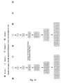
WO 02/40660 - Die in der Literatur beschriebenen Hefe-GPCR-Assays benutzen meist nur ein Reportergen, meist HIS3 oder LacZ (Price et al., Mol Cell Biol 15, 6188-6195 (1995); Price et al., Molecular Pharmacology 50, 829-837 (1996); Campbell et al., Bioorg.Med.Chem.Lett. 9, 2413-2418 (1999); Pausch, Trends Biotechnol 15, 487-494 (1997)) unter der Kontrolle des FUS1-Promotors (FUS1-HIS3 oder FUSI-lacZ). Wird FUS1-HIS3 verwendet, so wird eine Aktivierung des Signaltransduktionsweg als Trübung der Hefekultur in einem Flüssigmedium ohne Histidin gemessen. Experimente der Erfinder zeigten, dass der alleinige Wachstums-Read-out ein Signal-zu-Hintergrund-Verhältnis von ca. 30-50:1 in Flüssigkultur liefert (s.
Figuren 1a und3 ). β-Galaktosidase-Flüssigassays unter Verwendung von Chlorphenolred β-D-Galaktopyranosid (CPRG) als Enzymsubstrat zeigten nach Stimulierung ein ca. 2-3-fach über Hintergrund erhöhtes Signal (s. a.Figur 1b ). Im Bemühen das Messfenster weiter zu vergrößern, wurden beide Reportergene gleichzeitig in einer Hefezelle eingesetzt, da dies zu einer Multiplikation der beiden Messsignale führen sollte. InFig. 1c ist dieses Prinzip zur Verdeutlichung dargestellt. Dieser Doppel Reportergen-Assay führte dann auch zu einer Verbesserung des Signal-zu-Hintergrund-Verhältnisses auf ca. 100-150:1. Brown et al., Yeast 16, 11-22 (2000), beschreibt einen ähnlichen Assay. Auch hier werden FUS1-HIS3 und FUS1-IacZ gleichzeitig in einem β-Galaktosidase-Flüssigassay unter Verwendung von CPRG als Substrat benutzt. CPRG wird hier während der gesamten Zeitdauer der Stimulierung des Rezeptors mit Ligand zugesetzt. Im Unterschied dazu wird in diesem Verfahren CPRG zusammen mit einem Detergenz in gepufferter Lösung erst nach der Stimulierung des Rezeptors mit Ligand zugegeben, was zu einer deutlichen Verbessung des β-Galaktosidase-Messwertes führt. Ist nämlich zum einen CPRG während des vom Liganden induzierten Wachstums anwesend, wird dieses leicht inhibiert, zum anderen kann CPRG nur schlecht durch die Plasmamembran ins Innere der Zelle gelangen. Beide Probleme werden umgangen, wenn CPRG erst nach erfolgtem Wachstum zusammen mit einem Detergenz, welches die Plasmamembran aufbrechen kann, zugegeben wird. - Das Verfahren bedient sich in einer bevorzugten Ausführungsweise eines Doppel-Reportergen-Assays, wobei das eine Reportergen ein Wachstumsmarker und das andere Reportergen ein Enzym oder GFP ist. Nur diese Kombination von Wachstum, einem logarithmischen Ereignis, und der mehr oder wenigen linear induzierten Expression eines messbaren Enzyms oder fluoreszierenden Proteins, führt zu der beschriebenen Amplifikation des Signals, d.h. einem großen Messfenster.
-
EP 0 708 922 B1 (Acadia Pharmaceuticals) beschreibt auch ein Verfahren, dass auf Wachstum als Antwort auf Rezeptorstimulation beruht. Hierbei wachsen die durch Ligand stimulierten Rezeptor-exprimierenden Zellen aber nur schneller als die nicht stimulierten Zellen (vgl.Fig. 2 und Fig. 10 inEP 0 708 922 B1 ). In der hier beschriebenen Erfindung, wachsen solche nicht stimulierten Hefezellen aber gar nicht (Siehe angeführte Beispiele (z.B. Fig. 3B, linker Graph).EP 0 708 922 B1 verwendet als messbares Signal auch die Aktivität des heterolog exprimierten Enzyms β-Galaktosidase. LacZ wird hier aber konstitutiv exprimiert, d.h. die gemessene Enzymaktivität ist nur ein Maß für die Anzahl der Zellen, die als Antwort auf Stimulierung des Signaltransduktionsweges durch Ligand gewachsen sind, nicht aber ein Maß für die Stärke der Stimulierung des Signaltransduktionsweges. In der hier beschriebenen Erfindung steht die Expression von LacZ in Gegensatz dazu unter der Kontrolle eines durch den Pheromone Response Pathway induzierten Promotors (z.B. FUS1 oder YNL279w). In Fig. 3A ist dargestellt, dass auch bei gleicher Anzahl von Hefezellen (linker Graph) die messbare Aktivität der β-Galaktosidase von der zugesetzten Menge an Ligand, d.h. von der Stärke der Stimulierung des Signaltransduktionsweges, abhängt (rechter Graph). - Unter "Amplifikation" von Zellen wird laut
EP 0 708 922 B1 (vgl. S. 10) "das Wachstum von mit Rezeptor transfizierten Zellen, insbesondere im Vergleich zum Wachstum von nicht mit dem Rezeptor transfizierten Zellen" verstanden. D.h. sowohl mit Rezeptor transfizierte als auch nicht transfizierte Zellen können wachsen, nur dass die transfizierten Zellen nach Stimulierung mit Ligand schneller wachsen. Dies wird in den Abbildungen auf S. 33 und S.44 verdeutlicht. In den beschriebenen Zelllinien ist kein Reporterkonstrukt vorhanden, dessen Expression erst das Wachstum ermöglicht. Die einzige Modifikation, die das Wachstum ermöglicht, ist der durch Ligand stimulierte überexprimierte Rezeptor. - Gemäß der bevorzugten Ausführungsform wird aber eine doppelte Selektion betrieben:
- Dem Nährmedium fehlen Uracil und Histidin, Substanzen, die die hier verwendeten Hefestämme zum Leben braucht. Da wir das URA3-Gen als selektiven Marker auf unseren Rezeptorplasmiden verwenden, können Zellen, denen das Rezeptorplasmid fehlt, überhaupt nicht wachsen.
- Auch die mit Rezeptor-DNA transfizierte Zellen können grundsätzlich so lange auf diesem Nährmedium nicht wachsen, solange sie nicht durch die Gegenwart von Ligand stimuliert werden. Erst wenn der Ligand bindet, wird das Reportergen HIS3 exprimiert und die Zellen sind in der Lage auf dem Nährmedium zu wachsen. Grundsätzlich reagiert ein Hefestamm, der mit einem Rezeptor transfiziert ist, aber kein Wachstumsmarker wie HIS3 als Reportergen trägt, nicht mit Wachstum.
- Das beschriebene Verfahren kann sowohl im Einzelrezeptor-Format als auch im Mehrfachrezeptor-Format (Multiplex-Format) eingesetzt werden. Besonders im Mehrfachrezeptor-Format kommen die Vorteile dieses Assays zum Tragen.
Fig. 1c soll dies verdeutlichen. Theoretisch sollte eine Hefezelle, die einen bestimmten Rezeptor exprimiert, ausreichen, dass wenn diese Zelle mit dem passenden Liganden (einer chemischen Verbindung oder dem natürlichen Liganden) in Kontakt kommt, als Antwort auf die Stimulierung sozusagen aus dem Hintergrund der nicht reagierenden anderen Hefezellen "herauswächst". Dies bringt Vorteile für das High-Throughput-Screening, da es diese Methode erlaubt mehrere GPCRs gleichzeitig zu screenen. Gerade für orphan GPCRs, deren Bedeutung für die pharmazeutische Industrie von vorne herein nicht klar ist, minimiert die hier beschriebene Methode den zeitlichen und finanziellen Einsatz des Unternehmens. Da von orphan GPCRs auch nicht bekannt ist, an welche Gα-Untereinheit sie koppeln, besteht mit dieser Methode auch die Möglichkeit einen oder mehrere orphan GPCRs in mehreren Transplant-Stämmen gleichzeitig zu testen. -
-
Fig. 1 verdeutlicht das Prinzip des Doppel-Reportergen-Assays im Mehrfachrezeptor-Format:Figur 1a zeigt einen Agonisten-induzierten Wachstums Read Out.Figur 1b stellt einen Agonisten-induzieren β-Galaktosidase vermittelten Farb Read Out dar.Figur 1c . schließlich zeigt den doppelten Agonisten-induzierten Wachstums und Farb Read Out. -
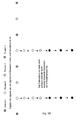
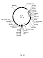
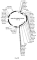
Fig. 2A-D zeigen Plasmide, die zur Konstruktion der Stämme verwendet wurden, die auf dem YNL279w-Promotor basieren. - In
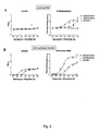
Fig. 3 ist dargestellt, wie der Doppel-Reportergen-Assay die Leistungsfähigkeit des Hefe-Flüssigassays verbessert, verglichen zur Nutzung nur eines Reportergens. -
Fig. 4 zeigt die Anbindung des humanen Bradykinin B2 Rezeptors an den Hefe-Signaltransduktionsweg in Abhängigkeit vom eingesetzten Gα-Transplant. Als Kontrollen wurde immer der leere Vektor p426GPD verwendet. -
Fig. 5 zeigt, dass der Doppel-Reportergen-Assay auch zum Screening von Antagonisten eingesetzt werden kann. Als Beispiel sind der humanen Bradykinin B2 Rezeptors bzw. die leere Vektorkontrolle dargestellt. - In
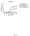
Fig. 6A undB wird gezeigt, dass die Verwendung des YNL279w-Promotors auch nach 29h Inkubation mit dem Enzymsubstrat deutlich weniger Hintergrundsignal verursacht als die Verwendung des FUS1-Promotors. -
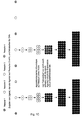
Fig. 7 erklärt die Durchführung eines Assays im Mehrfachrezeptor-Format. InFig. 7A wird deutlich gemacht, dass die verschiedenen GPCRs jeweils in einem eigenen Hefestamm exprimiert werden, nicht alle zusammen in einem.Fig. 7B zeigt die Leistungsfähigkeit eines Assays im Mehrfachrezeptor-Format im Vergleich zum Einfachrezeptor-Format in einer Mikrotiterplatte. -
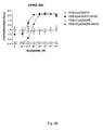
Fig. 8 zeigt, dass die Leistungsfähigkeit des Assays steigt, wenn das Enzymsubstrat CPRG zusammen mit Detergenz erst nach der Inkubation mit Liganden zugesetzt wird. - Alle molekularbiologischen und genetischen Manipulationen wurden nach Standardmethoden durchgeführt (Ausubel et al., Current Protocols in Moleculat Biology, Wiley & Sons, New York; Guthrie and Fink, Guide to Yeast Genetics and Molecular Biology, Methods in Enzymology, Academic Press, San Diego).
- Alle Expressionskonstrukte basieren auf dem episomalen 2□-Hefe-E. coli-Shuttle-Vektor p426GPD (Mumberg et al., Gene 156, 119-122 (1995)). cDNA-Sequenzen, welche für die humanen G-Protein gekoppelten Rezeptoren kodieren, werden in diesem Vektor zwischen dem GPD-Promotor und dem CYC-Terminator kloniert, um eine starke konstitutive Expression in Hefezellen zu erreichen. Die folgenden humanen GPCRs wurden in diesen Vektor kloniert:
EDG1 Rezeptor (Genbank NM_001400), EDG5 Rezeptor (Genbank NM_004230), Bradykinin B2 Rezeptor (Genbank NM_000623), M1 Muskarinrezeptor (Genbank NM_000738), Somatostatin SSTR2 Rezeptor (Genbank NM_001050). M3 Muskarinrezeptor (Genbank NM_000740) - Alle Hefestämme beruhen auf dem Saccharomyces cerevisiae Wildtypstamm W303-1a, beschrieben unter der ATCC-Nummer 208352.
- Genotyp: MATa, ade2-1, ura3-1, his3-11, trp1-1, leu2-3, leu2-112, can1-100
- Es wurden zwei verschiedene Sets von Hefestämmen eingesetzt. Ein Set stammt von YLJ21 ab und benutzt den Promotor des FUS1 Gens zur Expression der Reportergene, das andere Set stammt von YSG13 ab und nutzt den Promotor des YNL279w Gens.
- Der Hefestamm YLJ21 wurde von Ekkehard Leberer zur Verfügung gestellt.
- Genotyp: MATa, ste2::KanR sst2::ura3FOA far1::hisG FUS1::HIS3 mfa2::FUS1-lacZ::ura3FOA ade2-1, ura3-1, his3-11, trp1-1, leu2-3, leu2-112, can1-100
- Die Aktivierung des Pheromone Response Pathways kann mit Hilfe der zwei Reportergene FUS1::HIS3 und FUS1-IacZ gemessen werden, welche am HIS3- bzw. MFA2-Genlocus integriert wurden. Das FAR1 Gen wurde durch ein hisG-Repeat ersetzt, sodass die Zellen weiterwachsen können, auch wenn der Pheromone Response Pathway aktiviert ist. Das SST2 Gen wurde durch das URA3 Gen ersetzt, um die Runterregulierung des G-Protein-Signals durch die GTPasen-Funktion von Sst2p zu verhindern. Die ura3-Marker wurde jeweils durch Selektion auf 5-Fluoroorotic-acid-haltigem Medium wieder zurückgewonnen. Das Gen STE2, das für den α-Faktor Rezeptor kodiert, wurde durch ein KanR Gen ersetzt.
-
- Genotyp:
- MATa, ste2::KanR sst2::pYNL279w-H1S3 far1::pYNL279w-N136FUS1-IacZ::ADE2 ade2-1, ura3-1, his3-11, trpl-1, leu2-3, leu2-112, canl-100
- Stammkonstruktion:
- ste2::KanR
Um das Hefe STE2-Gen durch ein Kanamycin-Resistenzgen zu ersetzen wurde das Plasmid pLJ51 mit BamH1 und EcoR1 geschnitten und in den Wildtyp-Hefestamm W303-1 a transformiert. Selektion erfolgte auf YPD+G418-Medium. - sst2::pYNL279w-H1S3
- ste2::KanR
- In einem weiteren Schritt wurde das Hefe SST2 Gen durch eine Kassette ersetzt, welche die Expression des HIS3 Gens unter der Kontrolle des YNL279w-Promotors erlaubt. Dazu wurde das Plasmid sst2::279LHIS3/pCR-Bluntll mit BamHI und Notl geschnitten und transformiert. Selektion erfolgte auf SC/Gluc-His+α-Faktor-Medium. far1::pYNL279w-N136FUS1-IacZ::ADE2
- Anschließend wurde das FAR1 Gen durch eine Kassette ersetzt, welche die Expression der N-terminalen 136 Aminosäuren von Fus1p fusioniert an β-Galaktosidase unter der Kontrolle des YNL279w-Promotors erlaubt. Dazu wurde das Plasmid pBSfar1::YNL279w-N136FUS1-IacZ::ADE2 w/o mit Sacll und Xhol geschnitten und transformiert. Selektion erfolgte auf SC/Gluc-Ade-Medium.
- Die korrekte Integration aller Fragmente ins Genom wurde immer mittels PCR überprüft.
- Ausgehend von den Stämmen YLJ21 und YSG13 wurden schließlich die letzten 5 Aminosäuren der Hefe G-Protein-α-Untereinheit Gpa1 im Hefegenom durch die letzten 5 Aminosäuren der humanen G-Protein-α-Untereinheiten ersetzt. Dazu wurde zum Beispiel zur Konstruktion des Hefestammes YEW3 das Plasmid GPA1-C5-Galpha q integration mit Sacl geschnitten und in Hefestamm YLJ21 transformiert. Selektion erfolgte auf SC/Gluc-Trp-Medium. Die Integration der anderen Transplants erfolgte auf die gleiche Weise. In Tabelle 1 sind die verschiedenen Transplants und die davon abgeleiteten Hefestämme aufgeführt.
Tabelle 1: G-Protein Transplants GPA1/GαX Transplant steht für humane G-Protein α-Untereinheit C-terminale 5 Aminosäuren FUS1-Promotor YNL279w-Promotor GPA1 - KIGII YLJ21 YSG13 i1 t, i1, i2 DCGLF YEW11 YEW25 i3 i3 ECGLY YEW7 YEW21 o o1, o2 GCGLY YEW8 YEW22 z z YIGLC YEW12 YEW26 q q, 11 EYNLV YEW3 YEW17 14 14 ENFLV YEW6 YEW20 16 15, 16 EINLL YEW2 YEW16 12 12 DIMLQ YEW13 YEW27 13 13 QLMLQ YEW14 YEW28 s s1, s2 QYELL YEW1 YEW15 - Die in den Vektor p426GPD klonierten humanen GPCRs werden in den jeweiligen Hefestamm transformiert und auf SC-Selektionsplatten ohne Uracil und mit 2% Glukose als Kohlenstoffquelle (SC/Gluc-Ura) für 3 Tage bei 30°C inkubiert. Die so erhaltenen Einzelkolonien werden dann benutzt, um 2ml-Übernachtkulturen in SC/Gluc-Ura anzuimpfen. Am nächsten Tag werden die Zellen 1:100 in SC/Gluc-Ura-His pH6.8 verdünnt. Im Fall von Hefestämmen, die den FUS1-Promotor zur Reportergenexpression verwenden, wird diesem Medium noch 2-10mM 3-Aminotriazol (3-AT, Sigma) zugesetzt. Je 90µl der verdünnten Zellsuspension werden in eine Vertiefung einer Mikrotiterplatte mit 96 Vertiefungen pipettiert, in der sich bereits 10µl des zu untersuchenden Liganden befinden. Die Platten werden mit oder ohne Schütteln für 5-24h bei 30°C inkubiert. Dann wird je Vertiefung 50µl Assay Mix zugegeben. Der Assay Mix besteht aus 150µg/ml Digitonin (Sigma), 300µg/ml Chlorphenolred-β-D-Galaktopyranosid (CPRG, Roche), 300mM Natriumphosphat-Buffer pH6.7. Nach Inkubation für 2h bei 30°C mit oder ohne Schütteln wird die β-Galaktosidase-Aktivität als Absorption bei 574nm in einen Spektrophotometer (Spectramax Plus, Molecular Devices) gemessen. Die Auswertung der Daten und das Zeichnen der Dosis-Wirkungskurven erfolgt mit dem Graphpad Prism 3.0 Computerprogramm. Alle Messpunkte sind Mittelwerte aus Dreifachbestimmungen.
- Der Hefestamm YLJ21 wurde entweder mit dem leeren Vektor p426GPD oder den humanen GPCRs EDG1 und EDG5, welche in p426GPD kloniert waren, transformiert. Die transformierten Hefen wurden dann in 2ml SC/Gluc-Ura über Nacht bei 30°C angezüchtet. Am nächsten Tag wurden die Kulturen 1:100 in SC/Gluc-Ura-His pH6.8-Medium ohne (
Fig. 3A ) oder mit 2mM 3-AT (Fig. 3B ) verdünnt. Je 90µl der verdünnten Zellsuspension werden in eine Vertiefung einer Mikrotiterplatte mit 96 Vertiefungen pipettiert, in der sich bereits je 10µl einer Serienverdünnung von Sphingosin-1-phosphat (Biomol) oder als Kontrolle reines Wasser befinden. Die Platten werden für 23h bei 30°C unter Schütteln (700 U/min) inkubiert. Die Trübung, welche aus dem Wachstum der Hefezellen resultierte, wurde bei 630nm im Photometer gemessen (Fig. 3A und B jeweils linker Graph). Danach wurde 50µl Assay Mix je Vertiefung zugegeben. Nach 2h Inkubation bei 30°C unter Schütteln wurde die β-Galaktosidase-Aktivität als Absorption bei 574nm im Photometer gemessen. - Der linke Graph von
Fig. 3A zeigt, dass bei Verwendung von FUS1-HIS als Reporterkonstrukt ohne die Zugabe von 3-AT keine Dosis-Wirkungskurve zu erkennen ist. Der FUS1-Promotor führt auch ohne Stimulierung des Signaltransduktionsweges zu einem recht hohen Hintergrundsignal auf Medium ohne Histidin, d.h. der Promotor ist nicht strikt reguliert. Wird dagegen 2mM 3-AT, ein kompetitiver Inhibitor von His3p, zugesetzt, so wird das Hintergrundsignal unterdrückt und die Höhe des Messsignals ist abhängig von der Menge an Sphingosin-1-phosphat (Ancellin et al., J Biol Chem 274, 18997-19002 (1999)) im Medium, wenn EDG1 oder EDG5 exprimiert werden (Fig. 3B , linker Graph). AusFig. 3A rechter Graph ist ersichtlich, dass LacZ als Reportergen auch zu einer akzeptablen Dosis-Wirkungkurve führt, das Messfenster ist jedoch sehr klein. WieFig. 3B rechter Graph zeigt, führt der Einsatz von HIS3 und LacZ gleichzeitig zu einem vielfach besserem Signal-zu-Hintergrund-Verhältnis. Insgesamt zeigt dieses Experiment, dass die humanen GPCRs EDG1 und EDG5 durch die Hefeeigene Gα-Untereinheit Gpa1p an den Pheromone Response Pathway koppeln können. - Die Hefestämme YLJ21 (Gpa1), YEW1 (Gpa1/Gαs), YEW2 (Gpa1/Gα16) und YEW3 (Gpa1/Gαq) wurden entweder mit dem leeren Vektor p426GPD oder dem humanen Bradykinin B2 Rezeptor, der in p426GPD kloniert war, transformiert. Der Assay wurde unter Standardbedingungen wie oben beschrieben in der Anwesenheit von 2mM 3-AT durchgeführt. Als Ligand diente der natürliche Agonist Bradykinin (Sigma); Inkubation erfolgte für 20h. Aus
Fig. 4 wird ersichtlich, dass der Bradykinin B2 Rezeptor fast gar nicht an den Pheromone Response Pathway koppeln kann, wenn nur Gpa1p zur Verfügung steht. Wenn dagegen die Hefe die Gα-Transplants Gpa1/Gα16 oder Gpa1/Gαq exprimiert, ist eine Anbindung des Rezeptors an diesen Signaltransduktionsweg erfolgreich. Gpa1/Gαq erlaubt die effektivste Kopplung, was zu erwarten war, da der humane Bradykinin B2 Rezeptor in seiner natürlichen Zellumgebung an Gαq koppelt (Hall, Pharmacol.Ther. 56, 131-190 (1992)). - Der Hefestamm YEW3 (Gpa1/Gαq) wurde entweder mit dem leeren Vektor p426GPD oder dem humanen Bradykinin B2 Rezeptor, der in p426GPD kloniert war, transformiert. Der Assay wurde analog zu Beispiel 2 durchgeführt. Der einzige Unterschied war, dass in jeder Vertiefung des Testplatte 1 nM des Agonisten Bradykinin vorhanden war, wozu Verdünnungen des Antagonisten HOE140 (Sigma; Hall, Gen.Pharmacol. 28, 1-6 (1997)) zugegeben wurden.
Fig. 5 zeigt, dass steigende Mengen HOE140 das von Bradykinin hervorgerufene Signal auf das Hintergrundniveau herunterdrücken. Der Doppel-Reportergen-Assay eignet sich also auch für Antagonisten-Assays. - Die Hefestämme YEW3 (Gpa1/Gαq, FUS1-Promotor) und YEW17 (Gpa1/Gαq, YNL279w-Promotor) wurden entweder mit dem leeren Vektor p426GPD oder dem humanen Bradykinin B2 Rezeptor, der in p426GPD kloniert war, transformiert. Der Assay wurde unter Standardbedingungen wie oben beschrieben in der Anwesenheit von 2mM 3-AT im Fall von YEW3 und ohne 3-AT im Fall von YEW17 durchgeführt. Nach Zugabe von Assay Mix wurde die Messung der β-Galaktosidase-Aktivität nach 2h und nochmals nach 29h gemessen.
- Wie
Fig. 6A und noch eindrücklicherFig. 6B zeigen erhöht sich im Fall des FUS1-Promotors das Hintergrundsignal des mit Rezeptor transformierten Stammes oder auch des mit leerem Kontrollvektor transformierten Stammes über die Zeit erheblich, obwohl sogar 3-AT im Medium vorhanden war. Im Fall des YNL279w-Promotors ändert sich das Hintergrundsignal nicht wesentlich über die Zeit. Die Zugabe von 3-AT ist nicht nötig. Daraus ist abzuleiten, dass die Regulation von YNL279w sehr strikt ist. Dies erweist sich besonders bei High-Throughput-Assays als vorteilhaft, da der Zeitpunkt der Messung nicht präzise eingehalten werden muss, was ein flexibleres Arbeiten ermöglicht. - YEW3 (Gpa1/Gαq) und wurde mit dem humanen M1 Muskarinrezeptor und YLJ21 (Gpa1) wurde mit dem Somatostatin Rezeptor 2 und EDG5 transformiert. Die Rezeptoren waren in p426GPD kloniert. Die Durchführung erfolgte nach der oben beschriebenen Standardmethode. Die Rezeptoren wurden entweder einzeln oder im Gemisch getestet. Im Fall des Gemisches wurden die gleichen Übernachtkulturen wie bei den Einzeltests verwendet. Sie wurden erst bei der 1:100 Verdünnung in SC/Gluc-Ura-His pH6.8 gemischt, d.h. das Gemisch enthält somit insgesamt dreimal so viele Zellen wie die Einzeltests.
Fig. 7A soll verdeutlichen, dass die Rezeptoren alle individuell, d.h. nicht gemeinsam in einer Zelle exprimiert wurden. Die Inkubation mit den Liganden Carbachol (Sigma; Verdünnung 10-8M bis10-2M), Somatostatin-14 (Bachem; 10-10M bis10-4M) und Sphingosin-1-phosphat (Biomol, 10-10M bis10-4M) erfolgte für 24h. AusFig. 7B ist ersichtlich, dass eine Detektion der Agonisten auch im Gemisch möglich ist. - Herkömmlicherweise wird ein Hefe-CPRG-Assay so durchgeführt, dass CPRG während der ganzen Zeit, in der die mit Rezeptor transformierte Hefezelle mit Ligand in Kontakt ist, im Medium vorhanden ist (Brown et al., Yeast 16, 11-22 (2000) und
WO99/14344 Fig. 8 zeigt einen Vergleich zwischen dieser Methode und der in dieser Erfindung dargelegten Methode. - YEW3 (Gpa1/Gαq) und wurde mit den humanen M1 und M3 Muskarinrezeptoren, die in p426GPD kloniert waren, transformiert. Übernachtkulturen wurden wie beschrieben angezüchtet und dann in zwei verschiedene Medien 1:100 auf OD6000.02 verdünnt. In einem Fall (Fig. 8A) wurden die Zellen in SC/ Gluc-Ura-His, 2mM 3-AT, 0,1mg/ml CPRG, 0,1 M Natriumphosphat-Buffer pH7, verdünnt. Im anderen Fall wurde der Assay wie oben beschrieben in der Anwesenheit vom 2mM 3-AT durchgeführt (Fig. 8B). Als Ligand für die Muskarinrezeptoren wurde Carbachol verwendet. Der Assay wurde für die Daten in Fig. 8A für 28h durchgeführt, bevor die Assayplatte im Photometer gemessen wurde. Im Fall Fig. 8B wurde 26h inkubiert und dann Assay Mix zugegeben. Die Messung erfolgte 2h später.
- Wie aus
Fig. 8 ersichtlich ist, verbessert die Zugabe von CPRG und einem Detergenz, in diesem Fall Digitonin, erst nach der erfolgten Inkubation mit Ligand, die Leistungsfähigkeit des Assays erheblich. Ein weiterer Vorteil des hier beschriebenen Verfahrens ist die Tatsache, dass eine mögliche Wechselwirkung von CPRG während der langen Inkubation mit chemischen Verbindungen, v.a. während eines Screens, ausgeschlossen ist.
Claims (10)
- Doppel-Reportergen-Assay zur Identifizierung von Substanzen, die fähig sind, die Aktivität eines Zielmoleküls zu beeinflussen, wobei die zu analysierenden Hefezellen zwei Reportergene tragen, und die Aktivität des Zielmoleküls sich auf die Zellvermehrung auswirkt, mit den Schrittena) Kontaktierung mindestens einer Zelle mit der zu testenden Substanz,b) Detektion der Zellvermehrung,c) Detektion der Aktivität desr Reportergenproduktes,dadurch gekennzeichnet, dass ein Reportergen ein Wachstumsmarker und das andere Reportergen ein Enzym ist, dessen Aktivität anhand der Umsetzung eines Substrates detektierbar ist, wobei das Substrat zeitlich versetzt nach Zugabe der zu analysierenden Substanz zugegeben wird.
- Verfahren nach Anspruch 1, dadurch gekennzeichnet, dass sich die Aktivität des Zielmoleküls auf die Aktivität, vorzugsweise die Expression des Reportergenproduktes auswirkt.
- Verfahren nach Anspruch 1, dadurch gekennzeichnet, dass das Zielmolekül ein heterologes Molekül, vorzugsweise ein heterologes Oligonukleotid, Polynukleotid, eine Nukleinsäure, ein Polypeptid, Protein oder Proteinfragment ist.
- Verfahren nach Anspruch 1, dadurch gekennzeichnet, dass das zwischen Zugabe der zu analysierenden Substanz und der des Substrats liegende Zeitintervall der Dauer mindestens eines Zellzyklusdurchlaufs der verwendeten Zelle, vorzugsweise von 2 bis 24 Zellzyklusdurchläufen entspricht.
- Verfahren nach Anspruch 1, bei dem die Detektion der Aktivität des Reportergenproduktes unter Aufschluss der Zelle erfolgt, vorzugsweise durch Zugabe des Substrates zusammen mit einer die Zellwand permeabel machenden oder zerstörenden Substanz.
- Verfahren nach einem der vorstehenden Ansprüche, dadurch gekennzeichnet, dass das Zielmolekül selbst Einfluss auf die Zellvermehrung ausübt.
- Verfahren nach einem der vorstehenden Ansprüche, dadurch gekennzeichnet, dass das Zielmolekül den Einfluss auf die Zellvermehrung durch Zwischenschaltung eines direkt die Vermehrung beeinflussenden Moleküls ausübt.
- Verfahren nach Anspruch 7, dadurch gekennzeichnet, dass das Zielmolekül seine Wirkung auf die Zellvermehrung durch Wechselwirkung mit einem chimären Molekül ausübt.
- Verfahren nach einem der vorstehenden Ansprüche, dadurch gekennzeichnet, dass die Hefezellen vom Stamm S. cerevisiae sind.
- Verfahren nach einem der Ansprüche 2 bis 9, dadurch gekennzeichnet, dass in einem Ansatz Zellen mit unterschiedlichen Zielmolekülen gleichzeitig gescreent werden.
Applications Claiming Priority (3)
| Application Number | Priority Date | Filing Date | Title |
|---|---|---|---|
| DE10233516 | 2002-07-23 | ||
| DE10233516A DE10233516A1 (de) | 2002-07-23 | 2002-07-23 | Verfahren zur Identifizierung von Substanzen |
| PCT/EP2003/007569 WO2004019036A2 (de) | 2002-07-23 | 2003-07-14 | Verfahren zur identifizierung von substanzen |
Publications (2)
| Publication Number | Publication Date |
|---|---|
| EP1527340A2 EP1527340A2 (de) | 2005-05-04 |
| EP1527340B1 true EP1527340B1 (de) | 2009-10-28 |
Family
ID=30128304
Family Applications (1)
| Application Number | Title | Priority Date | Filing Date |
|---|---|---|---|
| EP03792196A Expired - Lifetime EP1527340B1 (de) | 2002-07-23 | 2003-07-14 | Verfahren zur identifizierung von substanzen |
Country Status (16)
| Country | Link |
|---|---|
| US (1) | US8642259B2 (de) |
| EP (1) | EP1527340B1 (de) |
| JP (1) | JP4546827B2 (de) |
| KR (1) | KR101089048B1 (de) |
| CN (1) | CN100397078C (de) |
| AT (1) | ATE447176T1 (de) |
| AU (1) | AU2003260308B2 (de) |
| BR (1) | BR0312804A (de) |
| CA (1) | CA2493323C (de) |
| DE (2) | DE10233516A1 (de) |
| IL (1) | IL166345A (de) |
| MX (1) | MXPA05000837A (de) |
| NO (1) | NO20050919L (de) |
| RU (1) | RU2325652C2 (de) |
| WO (1) | WO2004019036A2 (de) |
| ZA (1) | ZA200500326B (de) |
Families Citing this family (2)
| Publication number | Priority date | Publication date | Assignee | Title |
|---|---|---|---|---|
| US20060246417A1 (en) * | 2005-04-30 | 2006-11-02 | Ok-Kyu Song | System for functional analysis of polypeptides |
| FR3069644B1 (fr) * | 2017-07-28 | 2024-07-12 | Cisbio Bioassays | Methode pour mesurer la modulation de l'activation d'un recepteur couple a une proteine g |
Family Cites Families (13)
| Publication number | Priority date | Publication date | Assignee | Title |
|---|---|---|---|---|
| DE3429823A1 (de) * | 1984-08-14 | 1986-02-27 | Merck Patent Gmbh, 6100 Darmstadt | Mittel und verfahren zum nachweis antimikrobiell wirksamer substanzen |
| US5885769A (en) * | 1991-02-14 | 1999-03-23 | Schering Corporation | Screening systems |
| US5707798A (en) | 1993-07-13 | 1998-01-13 | Novo Nordisk A/S | Identification of ligands by selective amplification of cells transfected with receptors |
| US5691188A (en) | 1994-02-14 | 1997-11-25 | American Cyanamid Company | Transformed yeast cells expressing heterologous G-protein coupled receptor |
| US5728534A (en) * | 1996-07-19 | 1998-03-17 | New England Medical Center Hospitals, Inc. | Methods for identifying cardiovascular therapeutic agents |
| CN1107870C (zh) * | 1996-09-20 | 2003-05-07 | 奥索·麦克尼尔药品公司 | 用于体外测定红细胞生成素生物活性的方法及其试剂盒 |
| WO1998016557A1 (en) * | 1996-10-11 | 1998-04-23 | The General Hospital Corporation | Assays for g-protein-linked receptors |
| GB9719496D0 (en) * | 1997-09-13 | 1997-11-19 | Glaxo Group Ltd | G protien chimeras |
| EP0974649A1 (de) * | 1998-06-22 | 2000-01-26 | Universität Zürich | Screening-System |
| CA2343046A1 (en) * | 1998-09-01 | 2000-03-09 | Basf Aktiengesellschaft | Enhanced functional expression of g protein-coupled receptors |
| US6063578A (en) * | 1998-10-22 | 2000-05-16 | Signal Pharmaceuticals, Inc. | Dual reporter system and methods of use therefor |
| DE10056899A1 (de) * | 2000-11-16 | 2002-05-29 | Aventis Pharma Gmbh | Promotor zur funktionellen Charakterisierung von G-Protein gekoppelten Rezeptoren in der Hefe Saccharomyces cerevisiae |
| US20050118690A1 (en) * | 2001-10-09 | 2005-06-02 | Shannon Roberts | Love variant regulator molecules |
-
2002
- 2002-07-23 DE DE10233516A patent/DE10233516A1/de not_active Ceased
-
2003
- 2003-07-14 JP JP2004530019A patent/JP4546827B2/ja not_active Expired - Fee Related
- 2003-07-14 AU AU2003260308A patent/AU2003260308B2/en not_active Ceased
- 2003-07-14 WO PCT/EP2003/007569 patent/WO2004019036A2/de not_active Ceased
- 2003-07-14 DE DE50312072T patent/DE50312072D1/de not_active Expired - Lifetime
- 2003-07-14 CN CNB038174936A patent/CN100397078C/zh not_active Expired - Fee Related
- 2003-07-14 KR KR1020057001209A patent/KR101089048B1/ko not_active Expired - Fee Related
- 2003-07-14 BR BR0312804-0A patent/BR0312804A/pt not_active IP Right Cessation
- 2003-07-14 AT AT03792196T patent/ATE447176T1/de not_active IP Right Cessation
- 2003-07-14 RU RU2005104955/15A patent/RU2325652C2/ru not_active IP Right Cessation
- 2003-07-14 CA CA2493323A patent/CA2493323C/en not_active Expired - Fee Related
- 2003-07-14 MX MXPA05000837A patent/MXPA05000837A/es active IP Right Grant
- 2003-07-14 EP EP03792196A patent/EP1527340B1/de not_active Expired - Lifetime
- 2003-07-23 US US10/625,085 patent/US8642259B2/en not_active Expired - Fee Related
-
2005
- 2005-01-13 ZA ZA200500326A patent/ZA200500326B/xx unknown
- 2005-01-17 IL IL166345A patent/IL166345A/en not_active IP Right Cessation
- 2005-02-21 NO NO20050919A patent/NO20050919L/no not_active Application Discontinuation
Also Published As
| Publication number | Publication date |
|---|---|
| CN100397078C (zh) | 2008-06-25 |
| ATE447176T1 (de) | 2009-11-15 |
| AU2003260308B2 (en) | 2009-03-26 |
| WO2004019036A2 (de) | 2004-03-04 |
| US8642259B2 (en) | 2014-02-04 |
| BR0312804A (pt) | 2005-04-19 |
| HK1083534A1 (zh) | 2006-07-07 |
| IL166345A0 (en) | 2006-01-16 |
| WO2004019036A3 (de) | 2004-05-13 |
| MXPA05000837A (es) | 2005-04-19 |
| KR20050025648A (ko) | 2005-03-14 |
| KR101089048B1 (ko) | 2011-12-02 |
| CA2493323A1 (en) | 2004-03-04 |
| CN1672046A (zh) | 2005-09-21 |
| EP1527340A2 (de) | 2005-05-04 |
| JP4546827B2 (ja) | 2010-09-22 |
| JP2005533523A (ja) | 2005-11-10 |
| US20040092474A1 (en) | 2004-05-13 |
| DE50312072D1 (de) | 2009-12-10 |
| ZA200500326B (en) | 2006-07-26 |
| DE10233516A1 (de) | 2004-02-12 |
| IL166345A (en) | 2011-11-30 |
| RU2325652C2 (ru) | 2008-05-27 |
| RU2005104955A (ru) | 2005-10-10 |
| AU2003260308A1 (en) | 2004-03-11 |
| NO20050919L (no) | 2005-04-14 |
| CA2493323C (en) | 2012-04-17 |
Similar Documents
| Publication | Publication Date | Title |
|---|---|---|
| DE69831083T2 (de) | Reprimiertes trans-aktivatorsystem zur charakterisierung von protein-protein interaktionen | |
| DE69731960T2 (de) | Verfahren und zusammensetzungen für die identifizierung von receptor effectoren | |
| DE69428689T2 (de) | Hefe zellen so konstruiert, dass sie proteinsurrogate des pheromensystems produzieren und anwendungen dafuer | |
| DE69527951T2 (de) | Ein in-vivo genexpressions-system | |
| US7319009B2 (en) | Methods and compositions for identifying receptor effectors | |
| DE69918123T2 (de) | Verfahren und reagentiensatz zur erkennung von protein-protein wechselwirkungen | |
| DE69829485T2 (de) | Chimäre des g-proteins | |
| DE69935313T2 (de) | Nachweisverfahren für peptide | |
| EP1137810B1 (de) | Peptid screening test zum nachweis von gamma-sekretase aktivität | |
| JP2001519157A (ja) | 改変g蛋白質を発現する酵母細胞及びそれらの利用方法 | |
| US7361498B2 (en) | Yeast cells expressing modified G proteins and methods of use therefor | |
| WO2004019036A2 (de) | Verfahren zur identifizierung von substanzen | |
| US7022513B2 (en) | Cell based signal generation | |
| US20030009022A1 (en) | Methods and compositions for identifying receptor effectors | |
| EP1341922B1 (de) | Hefestamm zur prüfung der geno- und zytotoxizität komplexer umweltkontaminationen | |
| DE102008064553B4 (de) | Verfahren zum Nachweis und/oder zur Identifizierung hormonell wirksamer Substanzen | |
| DE10222714B4 (de) | Kofaktoren, die spezifisch mit dem Glukokortikoidrezeptor interagieren sowie Verfahren zu deren Identifizierung | |
| DE10393660B4 (de) | Verfahren zur Identifizierung von Protein-CAMs (konstitutiv aktive Mutanten) | |
| EP1084231A1 (de) | Transformierte zell-linien, die heterologe g-protein-gekoppelte rezeptoren exprimieren | |
| DE60226056T2 (de) | Aequorin als reportergen in hefe | |
| HK1083534B (en) | Method for identifying substances | |
| Klee | The molecular function and regulation of formins in the yeast Saccharomyces cerevisiae | |
| EP0677588A2 (de) | Testverfahren zur Identifizierung von Substanzen, die auf Endothelinrezeptoren wirken | |
| Lee | Activation of gene expression in yeast |
Legal Events
| Date | Code | Title | Description |
|---|---|---|---|
| PUAI | Public reference made under article 153(3) epc to a published international application that has entered the european phase |
Free format text: ORIGINAL CODE: 0009012 |
|
| 17P | Request for examination filed |
Effective date: 20050223 |
|
| AK | Designated contracting states |
Kind code of ref document: A2 Designated state(s): AT BE BG CH CY CZ DE DK EE ES FI FR GB GR HU IE IT LI LU MC NL PT RO SE SI SK TR |
|
| AX | Request for extension of the european patent |
Extension state: AL LT LV MK |
|
| RAP1 | Party data changed (applicant data changed or rights of an application transferred) |
Owner name: SANOFI-AVENTIS DEUTSCHLAND GMBH |
|
| DAX | Request for extension of the european patent (deleted) | ||
| 17Q | First examination report despatched |
Effective date: 20080812 |
|
| GRAP | Despatch of communication of intention to grant a patent |
Free format text: ORIGINAL CODE: EPIDOSNIGR1 |
|
| GRAS | Grant fee paid |
Free format text: ORIGINAL CODE: EPIDOSNIGR3 |
|
| GRAA | (expected) grant |
Free format text: ORIGINAL CODE: 0009210 |
|
| AK | Designated contracting states |
Kind code of ref document: B1 Designated state(s): AT BE BG CH CY CZ DE DK EE ES FI FR GB GR HU IE IT LI LU MC NL PT RO SE SI SK TR |
|
| REG | Reference to a national code |
Ref country code: GB Ref legal event code: FG4D Free format text: NOT ENGLISH |
|
| REG | Reference to a national code |
Ref country code: CH Ref legal event code: EP |
|
| REG | Reference to a national code |
Ref country code: IE Ref legal event code: FG4D |
|
| REF | Corresponds to: |
Ref document number: 50312072 Country of ref document: DE Date of ref document: 20091210 Kind code of ref document: P |
|
| NLV1 | Nl: lapsed or annulled due to failure to fulfill the requirements of art. 29p and 29m of the patents act | ||
| PG25 | Lapsed in a contracting state [announced via postgrant information from national office to epo] |
Ref country code: ES Free format text: LAPSE BECAUSE OF FAILURE TO SUBMIT A TRANSLATION OF THE DESCRIPTION OR TO PAY THE FEE WITHIN THE PRESCRIBED TIME-LIMIT Effective date: 20100208 Ref country code: FI Free format text: LAPSE BECAUSE OF FAILURE TO SUBMIT A TRANSLATION OF THE DESCRIPTION OR TO PAY THE FEE WITHIN THE PRESCRIBED TIME-LIMIT Effective date: 20091028 Ref country code: SE Free format text: LAPSE BECAUSE OF FAILURE TO SUBMIT A TRANSLATION OF THE DESCRIPTION OR TO PAY THE FEE WITHIN THE PRESCRIBED TIME-LIMIT Effective date: 20091028 Ref country code: PT Free format text: LAPSE BECAUSE OF FAILURE TO SUBMIT A TRANSLATION OF THE DESCRIPTION OR TO PAY THE FEE WITHIN THE PRESCRIBED TIME-LIMIT Effective date: 20100301 |
|
| REG | Reference to a national code |
Ref country code: IE Ref legal event code: FD4D |
|
| PG25 | Lapsed in a contracting state [announced via postgrant information from national office to epo] |
Ref country code: CY Free format text: LAPSE BECAUSE OF FAILURE TO SUBMIT A TRANSLATION OF THE DESCRIPTION OR TO PAY THE FEE WITHIN THE PRESCRIBED TIME-LIMIT Effective date: 20091028 Ref country code: SI Free format text: LAPSE BECAUSE OF FAILURE TO SUBMIT A TRANSLATION OF THE DESCRIPTION OR TO PAY THE FEE WITHIN THE PRESCRIBED TIME-LIMIT Effective date: 20091028 |
|
| PG25 | Lapsed in a contracting state [announced via postgrant information from national office to epo] |
Ref country code: DK Free format text: LAPSE BECAUSE OF FAILURE TO SUBMIT A TRANSLATION OF THE DESCRIPTION OR TO PAY THE FEE WITHIN THE PRESCRIBED TIME-LIMIT Effective date: 20091028 Ref country code: BG Free format text: LAPSE BECAUSE OF FAILURE TO SUBMIT A TRANSLATION OF THE DESCRIPTION OR TO PAY THE FEE WITHIN THE PRESCRIBED TIME-LIMIT Effective date: 20100128 Ref country code: EE Free format text: LAPSE BECAUSE OF FAILURE TO SUBMIT A TRANSLATION OF THE DESCRIPTION OR TO PAY THE FEE WITHIN THE PRESCRIBED TIME-LIMIT Effective date: 20091028 Ref country code: IE Free format text: LAPSE BECAUSE OF FAILURE TO SUBMIT A TRANSLATION OF THE DESCRIPTION OR TO PAY THE FEE WITHIN THE PRESCRIBED TIME-LIMIT Effective date: 20091028 Ref country code: RO Free format text: LAPSE BECAUSE OF FAILURE TO SUBMIT A TRANSLATION OF THE DESCRIPTION OR TO PAY THE FEE WITHIN THE PRESCRIBED TIME-LIMIT Effective date: 20091028 |
|
| PG25 | Lapsed in a contracting state [announced via postgrant information from national office to epo] |
Ref country code: CZ Free format text: LAPSE BECAUSE OF FAILURE TO SUBMIT A TRANSLATION OF THE DESCRIPTION OR TO PAY THE FEE WITHIN THE PRESCRIBED TIME-LIMIT Effective date: 20091028 Ref country code: SK Free format text: LAPSE BECAUSE OF FAILURE TO SUBMIT A TRANSLATION OF THE DESCRIPTION OR TO PAY THE FEE WITHIN THE PRESCRIBED TIME-LIMIT Effective date: 20091028 |
|
| PLBE | No opposition filed within time limit |
Free format text: ORIGINAL CODE: 0009261 |
|
| STAA | Information on the status of an ep patent application or granted ep patent |
Free format text: STATUS: NO OPPOSITION FILED WITHIN TIME LIMIT |
|
| 26N | No opposition filed |
Effective date: 20100729 |
|
| PG25 | Lapsed in a contracting state [announced via postgrant information from national office to epo] |
Ref country code: GR Free format text: LAPSE BECAUSE OF FAILURE TO SUBMIT A TRANSLATION OF THE DESCRIPTION OR TO PAY THE FEE WITHIN THE PRESCRIBED TIME-LIMIT Effective date: 20100129 |
|
| BERE | Be: lapsed |
Owner name: SANOFI-AVENTIS DEUTSCHLAND G.M.B.H. Effective date: 20100731 |
|
| PG25 | Lapsed in a contracting state [announced via postgrant information from national office to epo] |
Ref country code: MC Free format text: LAPSE BECAUSE OF NON-PAYMENT OF DUE FEES Effective date: 20100731 |
|
| REG | Reference to a national code |
Ref country code: CH Ref legal event code: PL |
|
| PG25 | Lapsed in a contracting state [announced via postgrant information from national office to epo] |
Ref country code: IT Free format text: LAPSE BECAUSE OF FAILURE TO SUBMIT A TRANSLATION OF THE DESCRIPTION OR TO PAY THE FEE WITHIN THE PRESCRIBED TIME-LIMIT Effective date: 20091028 |
|
| PG25 | Lapsed in a contracting state [announced via postgrant information from national office to epo] |
Ref country code: LI Free format text: LAPSE BECAUSE OF NON-PAYMENT OF DUE FEES Effective date: 20100731 Ref country code: CH Free format text: LAPSE BECAUSE OF NON-PAYMENT OF DUE FEES Effective date: 20100731 |
|
| PG25 | Lapsed in a contracting state [announced via postgrant information from national office to epo] |
Ref country code: BE Free format text: LAPSE BECAUSE OF NON-PAYMENT OF DUE FEES Effective date: 20100731 |
|
| PG25 | Lapsed in a contracting state [announced via postgrant information from national office to epo] |
Ref country code: AT Free format text: LAPSE BECAUSE OF NON-PAYMENT OF DUE FEES Effective date: 20100714 |
|
| PG25 | Lapsed in a contracting state [announced via postgrant information from national office to epo] |
Ref country code: HU Free format text: LAPSE BECAUSE OF FAILURE TO SUBMIT A TRANSLATION OF THE DESCRIPTION OR TO PAY THE FEE WITHIN THE PRESCRIBED TIME-LIMIT Effective date: 20100429 Ref country code: LU Free format text: LAPSE BECAUSE OF NON-PAYMENT OF DUE FEES Effective date: 20100714 Ref country code: NL Free format text: LAPSE BECAUSE OF FAILURE TO SUBMIT A TRANSLATION OF THE DESCRIPTION OR TO PAY THE FEE WITHIN THE PRESCRIBED TIME-LIMIT Effective date: 20091028 |
|
| PG25 | Lapsed in a contracting state [announced via postgrant information from national office to epo] |
Ref country code: TR Free format text: LAPSE BECAUSE OF FAILURE TO SUBMIT A TRANSLATION OF THE DESCRIPTION OR TO PAY THE FEE WITHIN THE PRESCRIBED TIME-LIMIT Effective date: 20091028 |
|
| PGFP | Annual fee paid to national office [announced via postgrant information from national office to epo] |
Ref country code: DE Payment date: 20130711 Year of fee payment: 11 |
|
| PGFP | Annual fee paid to national office [announced via postgrant information from national office to epo] |
Ref country code: FR Payment date: 20130724 Year of fee payment: 11 Ref country code: GB Payment date: 20130710 Year of fee payment: 11 |
|
| REG | Reference to a national code |
Ref country code: DE Ref legal event code: R119 Ref document number: 50312072 Country of ref document: DE |
|
| GBPC | Gb: european patent ceased through non-payment of renewal fee |
Effective date: 20140714 |
|
| REG | Reference to a national code |
Ref country code: FR Ref legal event code: ST Effective date: 20150331 |
|
| PG25 | Lapsed in a contracting state [announced via postgrant information from national office to epo] |
Ref country code: DE Free format text: LAPSE BECAUSE OF NON-PAYMENT OF DUE FEES Effective date: 20150203 |
|
| REG | Reference to a national code |
Ref country code: DE Ref legal event code: R119 Ref document number: 50312072 Country of ref document: DE Effective date: 20150203 |
|
| PG25 | Lapsed in a contracting state [announced via postgrant information from national office to epo] |
Ref country code: GB Free format text: LAPSE BECAUSE OF NON-PAYMENT OF DUE FEES Effective date: 20140714 Ref country code: FR Free format text: LAPSE BECAUSE OF NON-PAYMENT OF DUE FEES Effective date: 20140731 |