EP1378750A1 - Blood analyzers and methods for analyzing blood - Google Patents
Blood analyzers and methods for analyzing blood Download PDFInfo
- Publication number
- EP1378750A1 EP1378750A1 EP03014409A EP03014409A EP1378750A1 EP 1378750 A1 EP1378750 A1 EP 1378750A1 EP 03014409 A EP03014409 A EP 03014409A EP 03014409 A EP03014409 A EP 03014409A EP 1378750 A1 EP1378750 A1 EP 1378750A1
- Authority
- EP
- European Patent Office
- Prior art keywords
- fluid
- temperature
- detector
- analyzer
- flow path
- Prior art date
- Legal status (The legal status is an assumption and is not a legal conclusion. Google has not performed a legal analysis and makes no representation as to the accuracy of the status listed.)
- Withdrawn
Links
- 238000000034 method Methods 0.000 title claims abstract description 26
- 210000004369 blood Anatomy 0.000 title description 35
- 239000008280 blood Substances 0.000 title description 35
- 239000012530 fluid Substances 0.000 claims abstract description 218
- 238000010438 heat treatment Methods 0.000 claims abstract description 25
- 239000012491 analyte Substances 0.000 claims abstract description 17
- 230000007613 environmental effect Effects 0.000 claims description 25
- 239000003153 chemical reaction reagent Substances 0.000 description 90
- 210000003958 hematopoietic stem cell Anatomy 0.000 description 55
- 230000007246 mechanism Effects 0.000 description 34
- 238000001514 detection method Methods 0.000 description 24
- 210000000265 leukocyte Anatomy 0.000 description 14
- 210000004027 cell Anatomy 0.000 description 13
- 238000005070 sampling Methods 0.000 description 11
- 210000003743 erythrocyte Anatomy 0.000 description 9
- 210000005259 peripheral blood Anatomy 0.000 description 9
- 239000011886 peripheral blood Substances 0.000 description 9
- 239000000306 component Substances 0.000 description 7
- 239000002699 waste material Substances 0.000 description 7
- 239000004973 liquid crystal related substance Substances 0.000 description 6
- 238000005259 measurement Methods 0.000 description 6
- 239000000243 solution Substances 0.000 description 6
- 230000003287 optical effect Effects 0.000 description 5
- 239000012503 blood component Substances 0.000 description 4
- 239000000463 material Substances 0.000 description 4
- 210000000130 stem cell Anatomy 0.000 description 4
- 230000001276 controlling effect Effects 0.000 description 3
- 238000010586 diagram Methods 0.000 description 3
- 239000002245 particle Substances 0.000 description 3
- 238000005192 partition Methods 0.000 description 3
- 230000000717 retained effect Effects 0.000 description 3
- 238000010186 staining Methods 0.000 description 3
- 210000002700 urine Anatomy 0.000 description 3
- 210000000601 blood cell Anatomy 0.000 description 2
- 210000001772 blood platelet Anatomy 0.000 description 2
- 210000001185 bone marrow Anatomy 0.000 description 2
- 210000000805 cytoplasm Anatomy 0.000 description 2
- 239000003219 hemolytic agent Substances 0.000 description 2
- 210000003494 hepatocyte Anatomy 0.000 description 2
- 230000003834 intracellular effect Effects 0.000 description 2
- 206010018910 Haemolysis Diseases 0.000 description 1
- 210000003969 blast cell Anatomy 0.000 description 1
- 230000001413 cellular effect Effects 0.000 description 1
- 239000013043 chemical agent Substances 0.000 description 1
- 238000004140 cleaning Methods 0.000 description 1
- 239000000470 constituent Substances 0.000 description 1
- 230000004069 differentiation Effects 0.000 description 1
- 238000007865 diluting Methods 0.000 description 1
- 239000003085 diluting agent Substances 0.000 description 1
- 238000010790 dilution Methods 0.000 description 1
- 239000012895 dilution Substances 0.000 description 1
- 238000007599 discharging Methods 0.000 description 1
- 230000003394 haemopoietic effect Effects 0.000 description 1
- 230000008588 hemolysis Effects 0.000 description 1
- 230000001900 immune effect Effects 0.000 description 1
- 238000009434 installation Methods 0.000 description 1
- 208000032839 leukemia Diseases 0.000 description 1
- 239000007788 liquid Substances 0.000 description 1
- 239000002184 metal Substances 0.000 description 1
- 230000002572 peristaltic effect Effects 0.000 description 1
- 239000011148 porous material Substances 0.000 description 1
- 238000002360 preparation method Methods 0.000 description 1
- 238000011127 radiochemotherapy Methods 0.000 description 1
- 230000001105 regulatory effect Effects 0.000 description 1
- 239000011347 resin Substances 0.000 description 1
- 229920005989 resin Polymers 0.000 description 1
- 230000000087 stabilizing effect Effects 0.000 description 1
- 238000011476 stem cell transplantation Methods 0.000 description 1
- 238000002560 therapeutic procedure Methods 0.000 description 1
- 238000002054 transplantation Methods 0.000 description 1
- 238000011282 treatment Methods 0.000 description 1
- XLYOFNOQVPJJNP-UHFFFAOYSA-N water Substances O XLYOFNOQVPJJNP-UHFFFAOYSA-N 0.000 description 1
Images
Classifications
-
- G—PHYSICS
- G01—MEASURING; TESTING
- G01N—INVESTIGATING OR ANALYSING MATERIALS BY DETERMINING THEIR CHEMICAL OR PHYSICAL PROPERTIES
- G01N35/00—Automatic analysis not limited to methods or materials provided for in any single one of groups G01N1/00 - G01N33/00; Handling materials therefor
- G01N35/10—Devices for transferring samples or any liquids to, in, or from, the analysis apparatus, e.g. suction devices, injection devices
- G01N35/1095—Devices for transferring samples or any liquids to, in, or from, the analysis apparatus, e.g. suction devices, injection devices for supplying the samples to flow-through analysers
- G01N35/1097—Devices for transferring samples or any liquids to, in, or from, the analysis apparatus, e.g. suction devices, injection devices for supplying the samples to flow-through analysers characterised by the valves
-
- G—PHYSICS
- G01—MEASURING; TESTING
- G01N—INVESTIGATING OR ANALYSING MATERIALS BY DETERMINING THEIR CHEMICAL OR PHYSICAL PROPERTIES
- G01N15/00—Investigating characteristics of particles; Investigating permeability, pore-volume or surface-area of porous materials
- G01N15/10—Investigating individual particles
- G01N15/14—Optical investigation techniques, e.g. flow cytometry
- G01N15/1456—Optical investigation techniques, e.g. flow cytometry without spatial resolution of the texture or inner structure of the particle, e.g. processing of pulse signals
-
- G—PHYSICS
- G01—MEASURING; TESTING
- G01N—INVESTIGATING OR ANALYSING MATERIALS BY DETERMINING THEIR CHEMICAL OR PHYSICAL PROPERTIES
- G01N15/00—Investigating characteristics of particles; Investigating permeability, pore-volume or surface-area of porous materials
- G01N15/01—Investigating characteristics of particles; Investigating permeability, pore-volume or surface-area of porous materials specially adapted for biological cells, e.g. blood cells
- G01N2015/012—Red blood cells
-
- G—PHYSICS
- G01—MEASURING; TESTING
- G01N—INVESTIGATING OR ANALYSING MATERIALS BY DETERMINING THEIR CHEMICAL OR PHYSICAL PROPERTIES
- G01N15/00—Investigating characteristics of particles; Investigating permeability, pore-volume or surface-area of porous materials
- G01N15/01—Investigating characteristics of particles; Investigating permeability, pore-volume or surface-area of porous materials specially adapted for biological cells, e.g. blood cells
- G01N2015/016—White blood cells
-
- G—PHYSICS
- G01—MEASURING; TESTING
- G01N—INVESTIGATING OR ANALYSING MATERIALS BY DETERMINING THEIR CHEMICAL OR PHYSICAL PROPERTIES
- G01N15/00—Investigating characteristics of particles; Investigating permeability, pore-volume or surface-area of porous materials
- G01N15/01—Investigating characteristics of particles; Investigating permeability, pore-volume or surface-area of porous materials specially adapted for biological cells, e.g. blood cells
- G01N2015/018—Platelets
-
- G—PHYSICS
- G01—MEASURING; TESTING
- G01N—INVESTIGATING OR ANALYSING MATERIALS BY DETERMINING THEIR CHEMICAL OR PHYSICAL PROPERTIES
- G01N15/00—Investigating characteristics of particles; Investigating permeability, pore-volume or surface-area of porous materials
- G01N15/10—Investigating individual particles
- G01N15/14—Optical investigation techniques, e.g. flow cytometry
- G01N2015/1486—Counting the particles
-
- G—PHYSICS
- G01—MEASURING; TESTING
- G01N—INVESTIGATING OR ANALYSING MATERIALS BY DETERMINING THEIR CHEMICAL OR PHYSICAL PROPERTIES
- G01N15/00—Investigating characteristics of particles; Investigating permeability, pore-volume or surface-area of porous materials
- G01N15/10—Investigating individual particles
- G01N15/14—Optical investigation techniques, e.g. flow cytometry
- G01N2015/1497—Particle shape
-
- G—PHYSICS
- G01—MEASURING; TESTING
- G01N—INVESTIGATING OR ANALYSING MATERIALS BY DETERMINING THEIR CHEMICAL OR PHYSICAL PROPERTIES
- G01N35/00—Automatic analysis not limited to methods or materials provided for in any single one of groups G01N1/00 - G01N33/00; Handling materials therefor
- G01N2035/00346—Heating or cooling arrangements
Definitions
- the present invention relates to sample analyzers and analyzing methods. More specifically, the present invention relates to samples analyzers and analyzing methods whereby analysis results are responsively influenced by the reaction temperature with the reagent solution. The present invention further relates to sample analyzers and analyzing methods capable of providing an optimum temperature for analyzing a sample.
- Samples of blood and urine contain constituent components that are difficult to analyze directly in minute quantities.
- a sample is generally diluted with a reagent solution, and analysis is performed after the sample and reagent have reacted.
- the progress of the reaction between the sample and the reagent may be greatly affected by conditions such as temperature, sunlight, humidity and the like, such that there may be wide variation in the results obtained from the same sample depending on conditions.
- the analysis results obtained may vary greatly if there is a slight change in temperature.
- PBSC peripheral blood stem cells
- PBSCT peripheral blood stem cell transplantation
- PBSCT is typically performed as follows. First, the patient is administered a normal dose of chemical agent, which reduces the number of leukocytes in the peripheral blood. The leukocytes begin to increase 5 to 7 days later. It is during this period that the number of PBSC increases in the peripheral blood.
- PBSC When the number of PBSC in the peripheral blood has sufficiently increased (5 to 20 days), PBSC are collected by a blood component separator, and the PBSC are frozen and stored. When collecting the PBSC, it is important to accurately know the number of PBSC in the peripheral blood. In order to collect an adequate quantity of PBSC for transplantation, PBSC must be collected when the number of PBSC has sufficiently increased.
- the patient is subjected to a chemoradiation therapy of proper dosage to destroy the bone marrow. Thereafter, the previously collected PBSC are transplanted into the patient so as to rapidly restore hematopoietic function.
- HPC hematopoietic progenitor cells
- a first analyzer embodying features of the present invention includes a heater for heating a fluid; a detector for detecting a signal from an analyte; a flow path connecting the heater and the detector; a fluid supplier for supplying the fluid heated by the heater through the flow path to the detector; a first thermometer for measuring a fluid temperature of the fluid in at least one of the detector and the flow path; and a controller for controlling the heater, the detector, the fluid supplier, and the first thermometer, and for outputting results of an analysis of the signal detected.
- the controller controls the fluid supplier based on a temperature measured by the first thermometer.
- a second analyzer embodying features of the present invention includes a detector for detecting a signal from an analyte; a heater for heating a fluid supplied to the detector; a fluid supplier for supplying the fluid heated by the heater to the detector; and a controller for controlling the detector, the heater, and the fluid supplier, and for outputting an analysis result from the signal detected by the detector.
- the controller controls the fluid supplier such that heated fluid is supplied to the detector until a temperature of the fluid in the detector attains a predetermined temperature.
- a method for analyzing an analyte embodying features of the present invention includes (a) heating a fluid; (b) supplying the fluid to a detector; (c) measuring a temperature of the fluid supplied to the detector; (d) supplying the fluid to the detector until the temperature of the fluid attains a predetermined temperature; (e) supplying the analyte to the detector; (f) detecting a signal from the analyte supplied to the detector; and (g) outputting a result of an analysis of the signal detected.
- the present invention eliminates the previously described problems by controlling a fluid supply mechanism based on the fluid temperature and environmental temperature.
- the sample analyzer of the present invention is provided with a reagent fluid supply part 6, heating means 8, analysis part 4, flow path 5, sample supply part 15 (including, for example, a sampling valve 7, pipette 9, syringe 10, and motor 11), environmental temperature measuring means 1, fluid temperature measuring means 3, and operation controller 2.
- the analysis part 4 and heating means 8 are connected by a flow path 5, such that heated reagent fluid is supplied to the analysis part 4 through the flow path 5.
- the sample supply part 15 may be connected in the flow path 5 at a suitable location, or may be constructed so as to supply sample directly to the analysis part 4.
- the environmental temperature measuring means 1 is used to measure the temperature in the vicinity of the apparatus, and may be disposed, not only outside the device, but also within the device insofar as it is installed at a location which is not affected by the heat generated by the heater and the like.
- the fluid temperature measuring means 3 is used to measure the temperature of the reagent fluid, and may, for example, be disposed within the analysis part 4 or within the flow path 5 near the entrance to the analysis part 4.
- the environmental temperature measuring means 1 and the fluid temperature measuring means 3 are respectively connected to the operation controller 2 by circuits.
- the temperatures measured by the environmental temperature measuring means 1 and the fluid temperature measuring means 3 are input to the operation controller 2.
- the reagent fluid supply part 6 is connected to the operation controller 2 by a circuit.
- the operation controller issues a command to supply reagent fluid based on the environmental temperature and fluid temperature data
- the reagent fluid supply part 6 sends reagent fluid to the analysis part 4 through the flow path 5.
- the heating means 8 is connected to the operation controller 2 by a circuit.
- the operation controller 2 controls the heating means 8, such that the temperature of the reagent fluid attains a predetermined temperature.
- the sample supply part 15 includes, for example, a sampling valve 7, pipette 9, syringe 10, and motor 11.
- the motor 11 operates to collect a fixed amount of sample from a sample vessel 12 by the pipette 9 through the syringe 10, and the sample is supplied to the sample valve 7. This operation is controlled by the operation controller 2 connected by a circuit.
- hemocytometer is used as a representative and non-limiting example of a sample analyzer embodying features of the present invention.
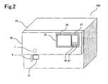
- FIG. 2 shows a perspective view of a hemocytometer embodying features of the present invention.
- the hemocytometer 100 is an example of the sample analyzer shown in FIG. 1. Accordingly, parts in common with FIG. 1 are labeled by the same reference numbers shown in FIG. 1.
- the hemocytometer 100 includes a body 29 and a front cover 30. This embodiment of the hemocytometer 100 detects HPC.
- the body 29 is provided with a pipette 9 for suctioning blood, start switch 31 used for starting analysis and the like, keyboard 27 for receiving input information from a user, and a liquid crystal display 28 for displaying information.
- the keyboard 27 is provided with a normal mode key 40 for selecting a normal mode and an HPC mode key 41 for selecting the HPC mode.
- the normal mode is a mode for calculating the number of leukocytes, erythrocytes and the like
- the HPC mode is a mode for calculating the number of HPC by adding the number of leukocytes and the number of erythrocytes and the like.
- a user selects the normal mode from the normal mode key 40, and selects the HPC mode from the HPC mode key 41.
- the front cover 30 has a window 32, which opens so that the liquid crystal display 28 is visible and to allow operation of the keyboard 27.
- a thermistor 1 On the reverse side of the front cover 30 is mounted a thermistor 1 for measuring the environmental temperature.
- the environmental temperature is the temperature in the vicinity of the analyzer, and may be the temperature within the analyzer or the temperature outside the analyzer insofar as the location is unaffected by heat generated by the heaters and the like within the analyzer. Accordingly, the thermistor 1 may be mounted within the body 29, or may be mounted on the outside of the body 29 or the front cover 30. Furthermore, the thermistor 1 also may be mounted on a table or wall near the installation location of the hemocytometer 100.
- FIG. 3 shows the structure of the hemocytometer 100.
- the hemocytometer 100 includes valves V1, V2, V3, V4, V5, V6, V7, and V8 for opening and closing the flow paths, start button 31, reagent chamber 50 for accommodating reagent, diaphragm pump 51 for suctioning and discharging predetermined amounts of fluids, fluid heater 8 for heating fluids to predetermined temperatures, pipette 9 for suctioning blood from the sample vessel 12, syringe 10, motor 11, sampling valve 7 for providing a predetermined amount of blood, detection mechanism 4 for detecting HPC and covering the sensor 53, syringe 54 for suctioning predetermined amounts of sample and reagent fluid from the sensor 53, motor 55, waste fluid chamber 13 for accommodating discard fluid, flow paths connecting various parts, positive pressure source 57 for supplying a positive pressure to the flow paths, and negative pressure sources 58, 59 and the like for supplying a negative pressure to the flow paths.
- the sensor 53 is covered by a cover to eliminate the influence of electrical noise, and forms part of the detection mechanism 4.
- a thermistor 3 is provided in the flow path 5 within the detection mechanism 4 to measure the temperature of the fluids passing within the flow path 5.
- the thermistor 3 is connected to the operation controller 2.
- the operation controller 2 is connected to the thermistor 1 for measuring the temperature of the air in the vicinity of the analyzer, and includes the keyboard 27 and liquid crystal display 28.
- the reagent chamber 50, valves V1 and V2, diaphragm pump 51, positive pressure source 57, negative pressure source 58, and the connecting flow paths form the reagent fluid supply mechanism 6.
- the pipette 9, syringe 10, motor 11, sampling valve 7, and the connecting flow paths form the sample supply mechanism 15.
- the hemocytometer 100 is described in detail below with reference to FIG. 4.
- FIG. 4 shows the structure of the hemocytometer 100.
- the hemocytometer 100 is provided with a reagent fluid supply mechanism 6, fluid heater 8, sample supply mechanism 15, detection mechanism 4, flow path 5, thermistor 1, start switch 31, controller 62, keyboard 27, liquid crystal display 28, valves V5 through V8, waste fluid chamber 13, syringe 54, motor 55, and negative pressure source 59.
- the reagent fluid supply mechanism 6 is provided with a reagent chamber 50, valves V1 through V4, diaphragm pump 51, positive pressure source 57, negative pressure source 58, and the respective connecting tubes.
- the valve V2 is connected to the fluid heater 8 through a tube.
- the reagent fluid supply mechanism 6 receives instructions from the controller 2 and sends out reagent fluid to the detection mechanism 4 through the fluid heater 8 and the flow path 5 and the like.
- the reagent chamber 50 internally accommodates reagent fluid such as dilution fluid, stain fluid, hemolytic agent and the like.
- reagent fluid such as dilution fluid, stain fluid, hemolytic agent and the like.
- immature leukocyte information (IMI) reagent (see, fore example, U.S. Patent No. 5,413,938) is used as a reagent fluid.
- the valves V1 through V4 open and close the flow paths.
- the positive pressure source 57 supplies a positive pressure to the diagram pump 51.
- the negative pressure source 58 supplies a negative pressure to the diaphragm pump 51.
- the diaphragm pump 51 suctions a predetermined amount of reagent from the reagent chamber 50 and discharges this reagent to the flow path 5 by means of the pressure forces from the positive pressure source 57 and the negative pressure source 58.
- a syringe and motor may also be used instead of the diaphragm pump 51, positive pressure source 57 and negative pressure source 58.
- the fluid heater 8 heats the reagent fluid to a predetermined temperature via the control of the controller 2.
- the apparatus for regulating liquid temperature disclosed in U.S. Patent No. 5,387,334 may be used as the fluid heater 8.
- the reagent fluid is heated to a predetermined temperature by the fluid heater 8.
- This temperature is set to a suitable reaction temperature in accordance with the type of reagent fluid used.
- the temperature is set between 32.5 and 40 °C.
- the sample supply mechanism 15 is provided with a pipette 9, syringe 10, motor 11, sampling valve 7, and the respective connecting tubes.
- the motor 11 When the motor 11 is operated, the syringe 10 operates continuously.
- the pipette 9 suctions a fixed amount of blood from the sample vessel 12, and supplies the blood to the sampling valve 7.
- the sampling valve 7 measures a fixed amount of blood.
- the sampling valve 7 is formed by stationary valves 7c and 7d, and a movable valve 7e disposed medially to the stationary valves 7c and 7d.
- the movable valve 7e is provided with blood metered-quantity flow paths 7a and 7b.
- the sampling valve 7 is inserted in the path of the flow path 5.
- a peristaltic pump may be used instead of the sampling valve 7.
- the sample supply mechanism 15 need not be inserted in the flow path 5.
- the sample supply mechanism 15 may be constructed such that the pipette 9 is moved to the detection mechanism 4 by a motor so as to supply blood from the pipette 9 to the detection mechanism 4 through the operation of a pump.
- the detection mechanism 4 has the function of obtaining an electrical signal from the sample, processing the electrical signal, and transmitting the processed electrical signal to the controller 2 so as to detect leukocytes, HPC or the like.
- the detection mechanism 4 is provided with part of the flow path 5, part of the tube 36a, part of the tube 36b, sensor 53, current supply circuit 61, electrodes 38a and 38b, thermistor 3, and cover 35.
- the part of the flow path 5, part of the tube 36a, part of the tube 36b, sensor 53, current supply circuit 61, electrodes 38a and 38b, and thermistor 3 are covered by the cover 35.
- the cover 35 is provided to eliminate the influence of electrical noise.
- the sensor 53 is formed by a chamber 53a, chamber 53b, and a partition 53b disposed between the chambers. Micropore 60 is provided in the partition 53b. The fluid within the chamber 53a is allowed to move to the chamber 53c by passing through the pores of the partition 53b.
- Tubes 36a and 36b are connected at the bottom of the chambers 53a and 53c, respectively.
- the tubes 36a and 36b are flow paths for fluids discharged from the chambers 53a and 53c, respectively.
- the electrodes 38a and 38b are mounted within the interiors of the chambers 53a and 53c, respectively.
- the electrodes 38a and 38b are connected to a current supply circuit 61.
- the current supply circuit 61 supplies an electrical current which flows to the electrodes 38a and 38b.
- the current supply circuit 61 measures the voltage and capacitance between the electrodes 38a and 38b, processes the values thus obtained, and transmits these processed values to the controller 2.
- the thermistor 3 is provided to measure the temperature of fluids flowing in the flow path 5.
- the thermistor 3 is connected to the controller 2.
- a detection mechanism capable of analyzing a plurality of cellular information may be provided in the particle analyzer as the detection mechanism 4.
- Well known flow cytometers used as optical detection mechanisms, and detection mechanisms employing an RF/DF detection method used as electrical resistance type detection mechanism, may be used as the above-mentioned detection mechanism.
- a detection mechanism employing an RF/DF detection method for example, a detection method such as model XE-2100 (Sysmex K.K.).
- Information on the size of cells, information on cell morphology, and intracellular information are included in cell information.
- Information such as DC signal and low angle scattered light intensity information, and the like, may provide information on cell size and cell morphology.
- a DC signal is a signal based on the difference in electrical resistance of a cell that is generated when a cell passes through a micropore to which flows a direct electrical current.
- Low angle scattered light is the scattered light found at 1 to 6° relative to an optical axis.
- Intracellular information may be information such as an RF signal, intensity of anterior high-angle scattered light, intensity of lateral scattered light, intensity of posterior scattered light, deflected resolution and the like.
- the RF signal is a signal based on the permittivity of a cell that is generated when a cell passes through a micropore to which a high frequency current flows.
- Anterior high-angle scattered light is scattered light at 8 to 20° relative to an optical axis.
- Lateral scattered light is scattered light at 70 to 110° relative to an optical axis.
- Posterior scattered light is scattered light at 120 to 180° relative to an optical axis.
- the flow path 5 is a flow path from the fluid heater 8 to the sensor 53. Within the flow path 5 flows a fluid heated by the fluid heater 8.
- the flow path 5 is formed by tubes 5a and 5b, T section 5c, and nozzle 5d. A reagent fluid is normally accommodated in the flow path 5.
- the tube 5a connects the fluid heater 8 and the stationary valve 7d.
- the tube 5b is connected to the stationary valve 7c.
- the T section 5c is connected to the other end of the tube 5b.
- the T section 5c is positioned inside the cover 35.
- the nozzle 5d is connected to the T section 5c.
- the nozzle 5d is disposed at the top of the chamber 53a so as to inject reagent fluid into the interior of the chamber 53a.
- the reagent fluid sent from the fluid heater 8 is injected to the sensor 53 through the tube 5a, stationary valve 7d, blood metered-quantity flow path 7a, stationary valve 7c, tube 5b, T section 5c, and nozzle 5d.
- tubes 5a and 5b are formed of resin material, they may also be formed of metal. A flow path having a channel may also be used instead of the tubes 5a and 5b. The lengths of the tubes 5a and 5b are not limited.
- the thermistor 3 is mounted at the T section 5c.
- the thermistor 3 is mounted in a position so as to come into contact with the reagent fluid passing within the T section 5c.
- the thermistor 3 may also be attached within the sensor 53, to the tube 5a, tube 5b, or nozzle 5d.
- the thermistor 3 is a thermometer for measuring the temperature of the reagent fluid heated by the fluid heater 8.
- the thermistor 3 is desirably mounted at a position within the detection mechanism 4 or near the detection mechanism 4.
- thermocouple also may be used as the thermometer instead of the thermistor 3.
- the blood suctioned by the pipette 9 is mixed within the sensor 53 with the reagent fluid transported by the reagent fluid supply mechanism 6.
- the hemocytometer 100 may also be constructed so as to mix blood and reagent within the flow path 5.
- the hemocytometer 100 may also be provided with another mixing vessel and constructed so as to mix the blood and reagent within this mixing vessel.
- the blood and reagent When the blood and reagent are mixed, components contained in the blood react with the reagent.
- erythrocytes in the blood react with the previously mentioned IMI reagent fluid, and hemolysis results.
- leukocytes other than immature leukocytes have cytoplasm removed and reduced.
- valves V5 through V8 open and close the flow paths.
- the valves V5 through V8 close the flow paths in the initial state.
- the syringe 54 suctions a predetermined amount of blood and reagent from within the chamber 53a through the micropore 60, chamber 53c and tube 36b. Furthermore, when the valve V6 is closed, the syringe 54 discharges discard fluid to the waste fluid chamber 13.
- the waste fluid chamber 13 accommodates discard fluid discharged from the chambers 53a and 53c.
- the negative pressure source 59 supplies a negative pressure to the waste fluid chamber 13.
- the valve V8 is open when discard fluid accommodated in the waste fluid chamber 13 is discharged outside the apparatus.
- the thermistor 1 is connected to the controller 62. As previously mentioned, the thermistor 1 measures the environmental temperature.
- the start switch 31 is connected to the controller 62.
- the controller 62 includes a CPU at its core, ROM, RAM used as a work area, and a hard disk for storing data and applications.
- the controller 62 is provided with a timer 42.
- the controller 62 receives the temperatures measured by the thermistors 1 and 3.
- the controller 62 issues operation instructions to the reagent fluid supply mechanism 6 based on the temperatures received from the thermistors 1 and 3.
- the controller 62 is connected to the keyboard 27 and the liquid crystal display 28.
- the controller 62, keyboard 27, and liquid crystal display 28 form the operation controller 2 (FIG. 3).
- a commercial personal computer with Windows 2000 (Windows is a registered trademark of Microsoft Corporation) installed as an operating system may also be used as the operation controller 2.
- the operation of the hemocytometer 100 is described below.
- FIG. 5 is a flow chart showing an overview of the processing sequence in controller 2.
- processing is executed to determine whether or not there is input from the normal mode key 40. If there is input from the normal mode key 40, the routine continues to S102. When there is no input from the normal mode key 40, the routine advances to S104.
- processing is executed to determine whether or not there is input from the start button 31. If there is input from the start button 31, the routine continues to S103. When there is no input from the start button 31, the routine continues to S101.
- processing is executed to control normal analysis operation. Normal analysis operation includes suctioning blood, processing the blood, and calculating the analysis result (the number of HPC is not included in the analysis result).
- S104 processing is executed to determine whether or not there is input from the HPC mode key 41. If there is input from the HPC mode key 41, the routine continues to S105. When there is no input from the HPC mode key 41, the routine continues to S101.
- S105 controls are executed to control the HPC pre-analysis operation. Details of S105 are shown in FIG. 6.
- processing is executed to start measurements by the timer 42.
- processing is executed to determine whether or not 3 minutes have elapsed since the start of the measurement by the timer 42. If 3 minutes have elapsed, the routine advances to S111. When 3 minutes have not elapsed, the routine continues to S108.
- processing is executed to determine whether or not there is input from the start button 31. If there is input from the start button 31, the routine continues to S109. When there is no input from the start button 31, the routine continues to S107.
- S110 processing is executed to control the HPC analysis operation. Details of S110 are shown in FIG. 7.
- a sleep control process is executed.
- the hemocytometer 100 enters a power saving mode to reduce power consumption, and enters a state wherein each mechanism is stopped until there is input from the start button 31.
- processing is executed to determine whether or not there is input from the start button 31. If there is input from the start button 31, the routine continues to S105. When there is no input from the start button 31, the routine continues to S111.
- FIG. 6 is a flow chart illustrating the process executed in S105.
- the environmental temperature is obtained by the thermistor 1 (S1)
- the hemocytometer 100 can be operated under a mode which performs analysis of HPC and under a mode which does not perform analysis of HPC; the mode which performs analysis of HPC is called the HPC analysis mode.
- the environmental temperature obtained by the thermistor 1 is 28 °C or higher
- the hemocytometer 100 enters a standby state (hereinafter, the state wherein HPC analysis is possible is referred to as the "standby" state), and enters a state wherein sample blood can be suctioned (S2, S3).
- a heating sequence is executed (S2, S4).
- the heating sequence is described below.
- valves are closed.
- Reagent fluid (the previously mentioned IMI reagent in the present embodiment) is retained in the fluid heater 8 beforehand, and is heated to a predetermined temperature.
- valves V1 and V4 When the valves V1 and V4 are opened, a predetermined amount of reagent fluid is suctioned into the diaphragm pump 51 by the negative pressure source 58.
- the predetermined amount of reagent fluid in the diaphragm pump 51 is injected in a direction toward the fluid heater 8 by the positive pressure source 57.
- the reagent fluid retained beforehand in the fluid heater 8 is suctioned therefrom, and the reagent fluid passes through the flow path to the sensor 53.
- the temperature of the reagent fluid is measured in real time by the thermistor 3.
- valves V2 and V3 When the valves V2 and V3 are closed, the reagent fluid within the sensor 53 is discharged to the waste chamber 13 by opening the valve V7. Thereafter, the valve 7 is closed and the heating sequence ends. By executing the heating sequence, the flow path 5 and the sensor 53 are heated, and the fluid temperature is measured by the thermistor 3.
- the hemocytometer 100 enters standby, a state wherein sample blood can be suctioned (S5, S6).
- the temperature of the reagent fluid is measured by again executing the heating sequence (S5 and S7).
- the reagent fluid temperature is measured by again executing the heating sequence (S8, S7).
- the heating sequence is executed once (S12), and the hemocytometer 100 enters standby (S13).
- FIG. 7 is a flow chart showing the process executed in S110. For convenience, only key operations are described, and a description of the cleaning operation is omitted.
- a predetermined amount of blood is suctioned from the sample vessel 12 through the pipette 9 by the suction operation of the syringe 10 and the operation of the motor 11. In this way, blood fills the blood metered-quantity flow path 7a of the sample valve 7 (S50).
- the blood is measured via the rotation of the movable valve of the sampling valve 7 (S51).
- valves V1 and V4 are opened, and a predetermined amount of reagent fluid is suctioned into the diaphragm pump 51 (S52).
- valves V1 and V4 are closed, the valves V2 and V3 are opened, and the reagent fluid within the diaphragm pump 51 is discharged in the direction of the fluid heater 8.
- reagent fluid retained within the flow path beforehand and a metered quantity of blood measured in S51 are discharged into the sensor 53 (S52).
- the temperature of the reagent fluid flowing within the flow path 5 is measured by the thermistor 3.
- valves V6 and V7 are closed and the valve V5 is opened, to induce a suctioning operation of a predetermined quantity by the syringe 54.
- blood passes through the micropore 60 of the sensor 53, and the change in voltage is detected at this time (S53).
- RF/DC detection method a two-dimensional distribution (scattergram) is obtained (RF/DC detection method).
- the number of HPC is calculated from this scattergram (S54). The method for calculating the number of HPC is described in detail in U.S. Patent No. 5,830,701, which is hereby incorporated by reference in its entirety except that in the event of any inconsistent disclosure or definition from the present application, the disclosure or definition herein shall be deemed to prevail..
- S55 processing is executed to correct the number of HPC calculated in S54 based on the temperature obtained in S52.
- the process executed in S55 is described below.
- H2 H1 t1-t0 * a- H1*t0 t1-t0
- H2 represents the number of HPC after correction
- a represents the optimum reaction temperature
- H1 represents the number of HPC before correction
- t0 represents the X-axis intersection
- t1 represents the fluid temperature obtained by the fluid temperature measuring means during the reaction of the IMI reagent.
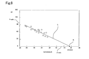
- t0 is determined in the following manner. The numbers of HPC before correction are set on the Y-axis, and the fluid temperatures obtained by the fluid temperature measuring means are set on the X-axis.
- FIG. 8 is a graph showing the relationship between the fluid temperature and the number of HPC before correction
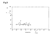
- FIG. 9 is a graph showing the relationship between the fluid temperature and the number of HPC after correction.
- t0 34.8 °C. It can be understood from the graph in FIG. 9 that the correction using equation (1) produces a suitable number of HPC.
- the value of t0 can be determined in the following manner.
- a plurality of blood samples having essentially the same number of HPC are analyzed using IMI reagent fluid of various temperatures.
- the obtained number of HPC H1 are set on the Y-axis, and the temperatures measured by the thermistor 3 are set on the X-axis, and when the analysis results are graphically plotted, the analysis results align along a certain straight line.
- the temperature indicated at the intersection of this straight line and the X-axis is designated t0.
- the Y-axis represents H1
- the X-axis represents the temperature measured by the thermistor 3.
- the analysis results align along a straight line L.
- the operations of S1 through S13 shown in FIG. 6 are executed again.
- the routine may start from the operation of S50 since the temperatures in the flow path 5 and sensor 11 will not have changed greatly in such a short time.
- a reagent fluid is used as the fluid supplied to the analysis part, water and the like may also be used since the fluid supply part of the present invention has the purpose of stabilizing the flow path temperature.
- a diluent for diluting a sample, a stain for staining a component contained in the sample, hemolytic agent to hemolyze blood components such as erythrocytes, and the like may be used as a reagent fluid.
- the reagent fluid reacts with a component contained in the sample depending on the sample and type of reagent.
- the sample is blood
- the reagent is an IMI reagent
- the erythrocytes in the blood are hemolyzed by the reaction, and leukocytes other than pre-blast cells have cytoplasm removed and reduced.
- the first predetermined value is a temperature selected so as to not reduce the temperature of the fluid passing through the flow path, which arises when the environmental temperature is less than the temperature of the fluid flowing through the flow path.
- the first predetermined value can be determined by considering the length and material of the flow path, and the desired reaction temperature of the sample and the reagent. Specifically, when heated fluid is actually supplied and the environmental temperature is at a certain degree Centigrade, a check is made to determine whether the heated fluid is supplied to the analysis part without a decrease in temperature. Such an environmental temperature is standardized as the first predetermined value.
- the operation control part compares the temperature of the fluid obtained by the fluid temperature measuring means and a second predetermined value, and when the fluid temperature is less than the second predetermined value, fluid is supplied from the fluid supply part to the analysis part.
- the temperature of the fluid is measured again by the fluid temperature measuring means, and the supply of fluid from the fluid supply part to the analysis part is stopped when the re-measured fluid temperature exceeds the second predetermined value. Since the flow path and analysis part are adequately warmed if the temperature of the fluid is higher than the second predetermined value, it is unnecessary to supply more fluid.
- the temperatures of the flow path and analysis part are lower than the temperature of the fluid when the fluid temperature is less than the second predetermined value, it is necessary to supply heated fluid from the fluid supply part to the analysis part to warm the flow path and analysis part. In this way, only a necessary amount of fluid is supplied to the analysis part to attain an optimum temperature of the flow path and the analysis part before a sample is analyzed.
- the temperature used as the second predetermined value may be selected so as to be an optimum fluid temperature for sample analysis when the heated fluid arrives at the analysis part. Specifically, a check is made to determine whether or not a desired analysis result is obtained when heated fluid is actually supplied and the fluid temperature obtained by the fluid temperature measuring means is at a certain degree Centigrade. This fluid temperature is standardized as the second predetermined temperature.
- analysis by the analysis part is enabled when the operation control part compares the environmental temperature obtained from the environmental temperature measuring means and the first predetermined value and the environmental temperature is higher than the first predetermined value. Likewise, it is desirable that analysis by the analysis part is enabled when the operation control part compares the fluid temperature obtained from the fluid temperature measuring means and the second predetermined value and the fluid temperature is higher than the second predetermined value.
- analysis of a sample is started in this state, analysis can be performed at a desired temperature, and temperature-induced errors can be minimized.
- the analysis part can be used to analyze materials including but not limited to blood components such as HPC (hematopoietic progenitor cells), PBSC, leukocytes, erythrocytes, and platelets, urine components such as leukocytes, erythrocytes, and microbes, and industrial particles requiring a staining process for measurement.
- HPC hematopoietic progenitor cells
- PBSC hematopoietic progenitor cells
- leukocytes erythrocytes
- platelets urine components
- urine components such as leukocytes, erythrocytes, and microbes
- industrial particles requiring a staining process for measurement a staining process for measurement.
- the sample analyzer of the present invention is particularly useful for analyzing HPC.
- the reaction optimum temperature range for HPC and a reagent (for example, IMI reagent) desirable for analysis of HPC is extremely narrow because maintaining the IMI reagent at an optimum fluid temperature during
- the present invention can be applied to sample analyzers for analyzing materials including but not limited to blood components such as leukocytes, erythrocytes, and platelets, urine components such as leukocytes, erythrocytes, and microbes, and industrial particles requiring a staining process for measurement.
- blood components such as leukocytes, erythrocytes, and platelets
- urine components such as leukocytes, erythrocytes, and microbes
- industrial particles requiring a staining process for measurement.
- the sample analyzer of the present invention obtains accurate analysis results by analyzing samples under optimum conditions.
- the temperature of the reagent fluid used in analysis is stabilized, and errors in analysis results due to temperature fluctuation are minimized. More accurate analysis results can be obtained by performing temperature correction on the obtained analysis results.
Landscapes
- Chemical & Material Sciences (AREA)
- General Health & Medical Sciences (AREA)
- Immunology (AREA)
- Health & Medical Sciences (AREA)
- Analytical Chemistry (AREA)
- Biochemistry (AREA)
- Physics & Mathematics (AREA)
- General Physics & Mathematics (AREA)
- Life Sciences & Earth Sciences (AREA)
- Pathology (AREA)
- Dispersion Chemistry (AREA)
- Investigating Or Analysing Biological Materials (AREA)
- Automatic Analysis And Handling Materials Therefor (AREA)
- Apparatus Associated With Microorganisms And Enzymes (AREA)
- Sampling And Sample Adjustment (AREA)
Applications Claiming Priority (2)
| Application Number | Priority Date | Filing Date | Title |
|---|---|---|---|
| JP2002192293A JP4118618B2 (ja) | 2002-07-01 | 2002-07-01 | 試料分析装置 |
| JP2002192293 | 2002-07-01 |
Publications (1)
| Publication Number | Publication Date |
|---|---|
| EP1378750A1 true EP1378750A1 (en) | 2004-01-07 |
Family
ID=29720234
Family Applications (1)
| Application Number | Title | Priority Date | Filing Date |
|---|---|---|---|
| EP03014409A Withdrawn EP1378750A1 (en) | 2002-07-01 | 2003-06-27 | Blood analyzers and methods for analyzing blood |
Country Status (3)
| Country | Link |
|---|---|
| US (1) | US7906073B2 (zh) |
| EP (1) | EP1378750A1 (zh) |
| JP (1) | JP4118618B2 (zh) |
Cited By (2)
| Publication number | Priority date | Publication date | Assignee | Title |
|---|---|---|---|---|
| EP1703270A1 (en) * | 2005-03-17 | 2006-09-20 | Sysmex Corporation | Sample analyser and sample analysing method |
| CN101750504B (zh) * | 2008-12-05 | 2013-11-27 | 深圳迈瑞生物医疗电子股份有限公司 | 生化分析仪的液体温度控制系统及方法 |
Families Citing this family (19)
| Publication number | Priority date | Publication date | Assignee | Title |
|---|---|---|---|---|
| JP4509607B2 (ja) * | 2004-03-17 | 2010-07-21 | シスメックス株式会社 | 細胞分析装置および方法 |
| JP5025371B2 (ja) * | 2007-07-31 | 2012-09-12 | シスメックス株式会社 | 血液分析装置 |
| US8021039B2 (en) * | 2007-11-07 | 2011-09-20 | Frank Amato | Quality control material monitor |
| JP2010066108A (ja) * | 2008-09-10 | 2010-03-25 | Olympus Corp | 自動分析装置および自動分析装置の駆動制御方法 |
| JP5276470B2 (ja) * | 2009-02-25 | 2013-08-28 | ベックマン コールター, インコーポレイテッド | 分析装置および分析方法 |
| JP5478101B2 (ja) * | 2009-03-31 | 2014-04-23 | シスメックス株式会社 | 試薬調製装置および検体処理システム |
| JP5496581B2 (ja) * | 2009-08-31 | 2014-05-21 | シスメックス株式会社 | 検体処理装置 |
| US9689942B2 (en) * | 2011-06-30 | 2017-06-27 | Liposcience, Inc. | Quantitative NMR clinical analyzers with automatic NMR temperature sensitivity compensation that accommodate large ambient operational temperature ranges |
| JP6027742B2 (ja) | 2011-12-28 | 2016-11-16 | シスメックス株式会社 | 血球分析装置、血球分析方法、及びコンピュータプログラム |
| JP5878377B2 (ja) | 2012-01-16 | 2016-03-08 | シスメックス株式会社 | 分析装置 |
| JP5921336B2 (ja) * | 2012-05-28 | 2016-05-24 | 株式会社日立ハイテクノロジーズ | 自動分析装置及び前処理装置 |
| JP2013255447A (ja) * | 2012-06-12 | 2013-12-26 | Nippon Koden Corp | 細胞単離装置 |
| US9835612B2 (en) | 2012-12-26 | 2017-12-05 | Hitachi High-Technologies Corporation | Automatic analyzer |
| KR102127220B1 (ko) * | 2013-12-04 | 2020-06-26 | 삼성전자주식회사 | 생체물질 검사장치 및 그 제어방법 |
| US10222383B2 (en) | 2016-01-29 | 2019-03-05 | Advanced Animal Diagnostics, Inc. | Methods and compositions for detecting mycoplasma exposure |
| EP3457112B1 (en) * | 2017-09-15 | 2022-08-03 | Horiba, Ltd. | Particle analyzing apparatus |
| EP3779459A4 (en) * | 2018-04-05 | 2021-05-05 | Konica Minolta, Inc. | TEMPERATURE ADAPTATION SYSTEM AND TEMPERATURE ADJUSTMENT PROCEDURE |
| JP7157658B2 (ja) * | 2018-12-27 | 2022-10-20 | 株式会社日立ハイテク | 自動分析装置 |
| CN112444619B (zh) * | 2019-08-30 | 2023-12-19 | 深圳迈瑞动物医疗科技股份有限公司 | 阻抗法计数装置及血液细胞分析仪 |
Citations (3)
| Publication number | Priority date | Publication date | Assignee | Title |
|---|---|---|---|---|
| US5387334A (en) * | 1991-02-15 | 1995-02-07 | Toa Medical Electronics Co., Ltd. | Apparatus for regulating liquid temperature |
| US5939326A (en) * | 1994-08-01 | 1999-08-17 | Abbott Laboratories | Method and apparatus for performing automated analysis |
| EP1189059A1 (en) * | 2000-09-18 | 2002-03-20 | Sysmex Corporation | Blood cell detector, blood analyzer and blood analyzing method using the detector |
Family Cites Families (11)
| Publication number | Priority date | Publication date | Assignee | Title |
|---|---|---|---|---|
| US4070155A (en) * | 1977-01-19 | 1978-01-24 | Thermo Electron Corporation | Apparatus for chromatographically analyzing a liquid sample |
| JPS54161389A (en) | 1978-06-10 | 1979-12-20 | Toshiba Corp | Automatic chemical analyzer |
| JPH01305361A (ja) | 1988-06-03 | 1989-12-08 | Toshiba Corp | 自動化学分析装置 |
| JPH06266448A (ja) | 1993-03-11 | 1994-09-22 | Mitsubishi Paper Mills Ltd | ジャケット付きタンクの温度調節方法 |
| JP3301646B2 (ja) | 1993-03-19 | 2002-07-15 | シスメックス株式会社 | 幼若細胞測定用試薬 |
| US5665314A (en) * | 1994-10-11 | 1997-09-09 | Hewlett-Packard Company | Temperature control in a portable analytical instrument |
| DE19538031C2 (de) * | 1995-01-17 | 2002-11-28 | Agilent Technologies Inc | Chromatograph mit gesteuertem Ofen |
| US5830701A (en) | 1997-03-28 | 1998-11-03 | Tao Medical Electronics Co., Ltd. | Method of detecting hematopoietic progenitor cells |
| US6534136B2 (en) * | 1997-09-19 | 2003-03-18 | Southpac Trust Int'l. Inc. | Packaging material |
| US6846458B1 (en) * | 1999-10-29 | 2005-01-25 | Rosemount Analytical Inc. | Process analytic system with improved sample handling system |
| US6530260B1 (en) * | 2002-02-04 | 2003-03-11 | Rvm Scientific, Inc. | Gas chromatography analysis system |
-
2002
- 2002-07-01 JP JP2002192293A patent/JP4118618B2/ja not_active Expired - Lifetime
-
2003
- 2003-06-27 EP EP03014409A patent/EP1378750A1/en not_active Withdrawn
- 2003-06-30 US US10/611,236 patent/US7906073B2/en not_active Expired - Fee Related
Patent Citations (3)
| Publication number | Priority date | Publication date | Assignee | Title |
|---|---|---|---|---|
| US5387334A (en) * | 1991-02-15 | 1995-02-07 | Toa Medical Electronics Co., Ltd. | Apparatus for regulating liquid temperature |
| US5939326A (en) * | 1994-08-01 | 1999-08-17 | Abbott Laboratories | Method and apparatus for performing automated analysis |
| EP1189059A1 (en) * | 2000-09-18 | 2002-03-20 | Sysmex Corporation | Blood cell detector, blood analyzer and blood analyzing method using the detector |
Non-Patent Citations (1)
| Title |
|---|
| PENG, L., ET. AL.: "Determination of peripheral blood stem cells by the Sysmex SE-9500", CLIN. LAB. HAEM., vol. 23, 2001, pages 231 - 236, XP002254557 * |
Cited By (4)
| Publication number | Priority date | Publication date | Assignee | Title |
|---|---|---|---|---|
| EP1703270A1 (en) * | 2005-03-17 | 2006-09-20 | Sysmex Corporation | Sample analyser and sample analysing method |
| CN102636635B (zh) * | 2005-03-17 | 2014-04-02 | 希森美康株式会社 | 血液分析装置 |
| US9243993B2 (en) | 2005-03-17 | 2016-01-26 | Sysmex Corporation | Sample analyzer and sample analyzing method |
| CN101750504B (zh) * | 2008-12-05 | 2013-11-27 | 深圳迈瑞生物医疗电子股份有限公司 | 生化分析仪的液体温度控制系统及方法 |
Also Published As
| Publication number | Publication date |
|---|---|
| JP2004037161A (ja) | 2004-02-05 |
| US20040023404A1 (en) | 2004-02-05 |
| US7906073B2 (en) | 2011-03-15 |
| JP4118618B2 (ja) | 2008-07-16 |
Similar Documents
| Publication | Publication Date | Title |
|---|---|---|
| US7906073B2 (en) | Analyzers and methods for analyzing analytes | |
| CN101358960B (zh) | 试样分析仪及试样分析方法 | |
| US8821791B2 (en) | Monitoring method, monitoring apparatus and liquid sample analyzer | |
| EP1516170B1 (en) | Lysing reagent, cartridge and automatic electronic cell counter for simultaneous enumeration of different types of white blood cells | |
| EP1036335B1 (en) | Method of verifying aspirated volume in automatic diagnostic system | |
| US9217750B2 (en) | Sample processing apparatus and cleaning method | |
| US8511888B2 (en) | Reagent preparing apparatus, sample processing apparatus and reagent preparing method | |
| US7005107B2 (en) | Apparatus for determining the speed of sedimentation of blood and other parameters correlated thereto | |
| JPS61200458A (ja) | 自動分析装置 | |
| US5288374A (en) | Method and apparatus for electrochemical analysis and an aqueous solution for use therein | |
| EP4411380A1 (en) | Automatic analyzer | |
| EP3574302B1 (en) | Inflammatory marker measurement method, inflammatory marker measurement apparatus, inflammatory marker measurement program, and recording medium storing the program | |
| JP3206999B2 (ja) | サンプル希釈誤差の検出方法およびそれを用いるサンプル希釈誤差の検出装置 | |
| KR101762877B1 (ko) | 혈액과 시약의 혼합장치 | |
| JP4082815B2 (ja) | 試料処理装置 | |
| US20220268725A1 (en) | Deaerator and Electrolyte Measurement System | |
| JP7492540B2 (ja) | 電解質分析装置 | |
| Siggaard-Andersen et al. | Semiautomatic pipetting of ultramicro volumes of sample and reagent | |
| JPS63151860A (ja) | 自動分析装置 | |
| JPS6138564A (ja) | フロ−セル型多項目自動分析装置 | |
| JPH02189443A (ja) | 粒子数あるいは溶存物質濃度計測方法およびその装置 | |
| JPS5855749A (ja) | 血液分析装置 | |
| CN116068159A (zh) | 一种样本分析仪和清洗方法 | |
| JPS6151556A (ja) | 電解質分析装置 | |
| JPH04177168A (ja) | 生体液中の金属自動分析装置 |
Legal Events
| Date | Code | Title | Description |
|---|---|---|---|
| PUAI | Public reference made under article 153(3) epc to a published international application that has entered the european phase |
Free format text: ORIGINAL CODE: 0009012 |
|
| AK | Designated contracting states |
Kind code of ref document: A1 Designated state(s): AT BE BG CH CY CZ DE DK EE ES FI FR GB GR HU IE IT LI LU MC NL PT RO SE SI SK TR |
|
| AX | Request for extension of the european patent |
Extension state: AL LT LV MK |
|
| 17P | Request for examination filed |
Effective date: 20040407 |
|
| AKX | Designation fees paid |
Designated state(s): DE FR GB IT |
|
| 17Q | First examination report despatched |
Effective date: 20101206 |
|
| GRAP | Despatch of communication of intention to grant a patent |
Free format text: ORIGINAL CODE: EPIDOSNIGR1 |
|
| INTG | Intention to grant announced |
Effective date: 20140522 |
|
| STAA | Information on the status of an ep patent application or granted ep patent |
Free format text: STATUS: THE APPLICATION IS DEEMED TO BE WITHDRAWN |
|
| 18D | Application deemed to be withdrawn |
Effective date: 20141002 |


