EP1372474B1 - Method and system for detecting colorimetric abnormalities - Google Patents
Method and system for detecting colorimetric abnormalities Download PDFInfo
- Publication number
- EP1372474B1 EP1372474B1 EP02707063A EP02707063A EP1372474B1 EP 1372474 B1 EP1372474 B1 EP 1372474B1 EP 02707063 A EP02707063 A EP 02707063A EP 02707063 A EP02707063 A EP 02707063A EP 1372474 B1 EP1372474 B1 EP 1372474B1
- Authority
- EP
- European Patent Office
- Prior art keywords
- image
- reference value
- abnormality
- blood
- gastrointestinal tract
- Prior art date
- Legal status (The legal status is an assumption and is not a legal conclusion. Google has not performed a legal analysis and makes no representation as to the accuracy of the status listed.)
- Expired - Lifetime
Links
Images

Classifications
-
- A—HUMAN NECESSITIES
- A61—MEDICAL OR VETERINARY SCIENCE; HYGIENE
- A61B—DIAGNOSIS; SURGERY; IDENTIFICATION
- A61B1/00—Instruments for performing medical examinations of the interior of cavities or tubes of the body by visual or photographical inspection, e.g. endoscopes; Illuminating arrangements therefor
- A61B1/04—Instruments for performing medical examinations of the interior of cavities or tubes of the body by visual or photographical inspection, e.g. endoscopes; Illuminating arrangements therefor combined with photographic or television appliances
- A61B1/041—Capsule endoscopes for imaging
-
- A—HUMAN NECESSITIES
- A61—MEDICAL OR VETERINARY SCIENCE; HYGIENE
- A61B—DIAGNOSIS; SURGERY; IDENTIFICATION
- A61B1/00—Instruments for performing medical examinations of the interior of cavities or tubes of the body by visual or photographical inspection, e.g. endoscopes; Illuminating arrangements therefor
- A61B1/00002—Operational features of endoscopes
- A61B1/00004—Operational features of endoscopes characterised by electronic signal processing
- A61B1/00009—Operational features of endoscopes characterised by electronic signal processing of image signals during a use of endoscope
- A61B1/000094—Operational features of endoscopes characterised by electronic signal processing of image signals during a use of endoscope extracting biological structures
-
- A—HUMAN NECESSITIES
- A61—MEDICAL OR VETERINARY SCIENCE; HYGIENE
- A61B—DIAGNOSIS; SURGERY; IDENTIFICATION
- A61B1/00—Instruments for performing medical examinations of the interior of cavities or tubes of the body by visual or photographical inspection, e.g. endoscopes; Illuminating arrangements therefor
- A61B1/00002—Operational features of endoscopes
- A61B1/00043—Operational features of endoscopes provided with output arrangements
- A61B1/00045—Display arrangement
-
- A—HUMAN NECESSITIES
- A61—MEDICAL OR VETERINARY SCIENCE; HYGIENE
- A61B—DIAGNOSIS; SURGERY; IDENTIFICATION
- A61B5/00—Measuring for diagnostic purposes; Identification of persons
- A61B5/0002—Remote monitoring of patients using telemetry, e.g. transmission of vital signals via a communication network
- A61B5/0031—Implanted circuitry
-
- A—HUMAN NECESSITIES
- A61—MEDICAL OR VETERINARY SCIENCE; HYGIENE
- A61B—DIAGNOSIS; SURGERY; IDENTIFICATION
- A61B5/00—Measuring for diagnostic purposes; Identification of persons
- A61B5/0059—Measuring for diagnostic purposes; Identification of persons using light, e.g. diagnosis by transillumination, diascopy, fluorescence
- A61B5/0075—Measuring for diagnostic purposes; Identification of persons using light, e.g. diagnosis by transillumination, diascopy, fluorescence by spectroscopy, i.e. measuring spectra, e.g. Raman spectroscopy, infrared absorption spectroscopy
-
- A—HUMAN NECESSITIES
- A61—MEDICAL OR VETERINARY SCIENCE; HYGIENE
- A61B—DIAGNOSIS; SURGERY; IDENTIFICATION
- A61B5/00—Measuring for diagnostic purposes; Identification of persons
- A61B5/0059—Measuring for diagnostic purposes; Identification of persons using light, e.g. diagnosis by transillumination, diascopy, fluorescence
- A61B5/0082—Measuring for diagnostic purposes; Identification of persons using light, e.g. diagnosis by transillumination, diascopy, fluorescence adapted for particular medical purposes
- A61B5/0084—Measuring for diagnostic purposes; Identification of persons using light, e.g. diagnosis by transillumination, diascopy, fluorescence adapted for particular medical purposes for introduction into the body, e.g. by catheters
-
- A—HUMAN NECESSITIES
- A61—MEDICAL OR VETERINARY SCIENCE; HYGIENE
- A61B—DIAGNOSIS; SURGERY; IDENTIFICATION
- A61B5/00—Measuring for diagnostic purposes; Identification of persons
- A61B5/02—Detecting, measuring or recording pulse, heart rate, blood pressure or blood flow; Combined pulse/heart-rate/blood pressure determination; Evaluating a cardiovascular condition not otherwise provided for, e.g. using combinations of techniques provided for in this group with electrocardiography or electroauscultation; Heart catheters for measuring blood pressure
- A61B5/02042—Determining blood loss or bleeding, e.g. during a surgical procedure
-
- A—HUMAN NECESSITIES
- A61—MEDICAL OR VETERINARY SCIENCE; HYGIENE
- A61B—DIAGNOSIS; SURGERY; IDENTIFICATION
- A61B5/00—Measuring for diagnostic purposes; Identification of persons
- A61B5/07—Endoradiosondes
- A61B5/073—Intestinal transmitters
-
- A—HUMAN NECESSITIES
- A61—MEDICAL OR VETERINARY SCIENCE; HYGIENE
- A61B—DIAGNOSIS; SURGERY; IDENTIFICATION
- A61B5/00—Measuring for diagnostic purposes; Identification of persons
- A61B5/74—Details of notification to user or communication with user or patient ; user input means
- A61B5/742—Details of notification to user or communication with user or patient ; user input means using visual displays
- A61B5/743—Displaying an image simultaneously with additional graphical information, e.g. symbols, charts, function plots
-
- G—PHYSICS
- G06—COMPUTING; CALCULATING OR COUNTING
- G06T—IMAGE DATA PROCESSING OR GENERATION, IN GENERAL
- G06T7/00—Image analysis
- G06T7/0002—Inspection of images, e.g. flaw detection
- G06T7/0012—Biomedical image inspection
-
- G—PHYSICS
- G06—COMPUTING; CALCULATING OR COUNTING
- G06T—IMAGE DATA PROCESSING OR GENERATION, IN GENERAL
- G06T7/00—Image analysis
- G06T7/90—Determination of colour characteristics
-
- G—PHYSICS
- G06—COMPUTING; CALCULATING OR COUNTING
- G06T—IMAGE DATA PROCESSING OR GENERATION, IN GENERAL
- G06T2207/00—Indexing scheme for image analysis or image enhancement
- G06T2207/10—Image acquisition modality
- G06T2207/10024—Color image
-
- G—PHYSICS
- G06—COMPUTING; CALCULATING OR COUNTING
- G06T—IMAGE DATA PROCESSING OR GENERATION, IN GENERAL
- G06T2207/00—Indexing scheme for image analysis or image enhancement
- G06T2207/10—Image acquisition modality
- G06T2207/10068—Endoscopic image
-
- G—PHYSICS
- G06—COMPUTING; CALCULATING OR COUNTING
- G06T—IMAGE DATA PROCESSING OR GENERATION, IN GENERAL
- G06T2207/00—Indexing scheme for image analysis or image enhancement
- G06T2207/30—Subject of image; Context of image processing
- G06T2207/30004—Biomedical image processing
- G06T2207/30028—Colon; Small intestine
Definitions
- the present invention relates to a method and system for detection of colorimetric abnormalities in vivo, and specifically within the gastrointestinal (GI) tract.
- GI gastrointestinal
- Pathologies of the gastrointestinal (GI) tract may exist for a variety of reasons. Some examples of pathologies include bleeding, lesions, angiodisplasia, Crohn's disease, polyps, celiac disorders, and others. The majority of pathologies result in changes of color and/or texture of the inner surface of the GI tract.
- color changes may be due to bleeding.
- Blood may be present within the digestive tract for a variety of pathological reasons, including ulcers, cancer, or other disease conditions. It is often difficult to detect the presence of blood within the GI tract, since bleeding can occur in difficult to reach locations. In addition, it is difficult to "see” inside the tract, especially in sections which are hard to reach such as the small intestines.
- a second, more invasive technique has been the use of an endoscope or enteroscope. This approach enables direct visualization of parts of the GI tract. However, most portions of the small intestine are inaccessible by this method.
- pathologies which may be detected based on the red part of the spectrum include active bleeding, blood clots, polyps, lesions, ulcerations, angiodisplasia and telangectasia.
- Pathologies which may be characterized by blue/violet color include arterio-venous malformation (AVM) and submucosal bleeding. AVM may also appear in red.
- AVM arterio-venous malformation
- submucosal bleeding may also appear in red.
- some types of ulcers are characterized by white color.
- WO 00/22975 describes a system in which a capsule moves through the gastrointestinal tract in a first pass to generate a map and to identify a location of interest. In a second pass, the capsule is controlled to perform a job at the identified location. Repeated localizations generate a map of the route taken, and images displayed on a monitor are compared with the generated map to identify the location of a pathology.
- the independent claims relate to a method and system for detecting abnormalities in-vivo in a gastrointestinal tract. Advantageous embodiments are described in the dependent claims.
- the method for detecting abnormalities may include the step of calculating a probability indication of a presence of an abnormal color within the body lumen based on comparison of spectral characteristics to at least one reference value.
- the method may include analyzing the image for a spectral characteristic in relation to a reference value.
- a method for calculation of a reference value for tissue may include the steps of receiving at least a first image and a second image from within a body lumen, selecting blocks of pixels within the images based on colorimetric parameters, averaging the colorimetric parameters of the selected blocks of pixels of the first and second images, and filtering the colorimetric parameters, thereby obtaining a reference value for tissue.
- the system for detecting abnormalities may comprise a swallowable capsule for detecting colorimetric abnormalities in a gastrointestinal tract.
- the capsule may include an image-receiver for receiving images from the gastrointestinal tract, and a processor for generating a probability indication for presence of colorimetric abnormalities by comparing color content of the images to at least one reference value.
- the system for detecting abnormalities may comprise an apparatus for determining colorimetric abnormalities within a body lumen.
- the apparatus may include an image-receiver for receiving images from a body lumen, a spectral analyzer for determining color content of the images, and a processor for generating a probability indication for presence of an abnormal condition by comparing the color content to at least one reference value.
- the system for detecting abnormalities may comprise a swallowable capsule having an in-vivo imager for obtaining images from within the body lumen, a transmitter for transmitting the images to a receiver, and a processor for generating a probability indication of presence of blood based on comparison of color content of the received images and at least one reference value.
- the present invention relates to a method and system of detection of pathologies by spectral analysis of images captured by a moving in-vivo video camera system. This analysis is based on detection of colorimetric abnormalities, or deviations from an expected spectrum.
- the in-vivo video camera system may be included on an endoscope, a swallowable capsule, or any other device which is introduced into the body to view the interior.
- US Patent No. 5,604,531 assigned to the common assignee of the present application, teaches an in vivo camera system, which is carried by a swallowable capsule.
- the in vivo video camera system captures and transmits images of the GI tract while the capsule passes through the GI lumen.
- the capsule contains an optical system for imaging an area of interest onto the camera system and a transmitter for transmitting the video output of the camera.
- the capsule can pass through the entire digestive tract and operate as an autonomous video endoscope. It images even the difficult to reach areas of the small intestine.
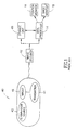
- Fig. 1 shows a schematic diagram of the system, described in US Patent 5,604,531 .
- the system comprises a capsule 40 having an imager 46, an illumination source 42, and a transmitter 41. Outside the patient's body are an image receiver 12 (usually an antenna array), a storage unit 19, a data processor 14, an image monitor 18, and a position monitor 16. While Fig.1 shows separate monitor, both an image and its position can be presented on a single monitor.
- Imager 46 in capsule 40 is connected to transmitter 41 also located in capsule 40.
- Transmitter 41 transmits images to image receiver 12, which sends the data to data processor 14 and to storage unit 19.
- Data processor 14 analyzes the data and is in communication with storage unit 19, transferring frame data to and from storage unit 19.
- Data processor 14 also provides the analyzed data to image monitor 18 and position monitor 16 where the physician views the data.
- the image monitor presents an image of the GI lumen and the position monitor presents the position in the GI tract at which the image was taken.
- Data processor 14 can be configured for real time processing or for post processing to be viewed at a later date. In addition to revealing pathological conditions of the GI tract, the system can provide information about the location of these pathologies.
- received images are analyzed for color content. Based on this analysis, as described hereinbelow, determination as to the presence or absence of a colorimetric abnormality may be made.
- a colorimetric abnormality may indicate a pathological condition, such as bleeding.
- Other examples of pathologies which may be detected based on the red part of the spectrum include active bleeding, blood clots, polyps, lesions, ulcerations, angiodisplasia and telangectasia.
- Pathologies which may be characterized by blue/violet color include arterio-venous malformation (AVM) and submucosal bleeding.
- AVM may also appear in red.
- some types of ulcers are characterized by white color. It will be apparent that the method and system described hereinbelow may be useful in detecting any colorimetric deviation from the normal color content of a body lumen, whether or not a pathological condition is present.
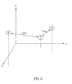
- Fig. 2 is a schematic illustration of the classification of samples according to their spectral components.
- Each test sample T is located within a coordinate system represented by the following variables: hue H, saturation S and value V.
- Hue H represents a number related to the dominant wavelength of the color stimulus, and varies from 0 to 1 as the color changes from red to yellow to green to cyan to blue to magenta and back to red again.
- Saturation S corresponds to color purity, and in the case of a pure color is equal to 100%.
- Value V is a measure of relative intensity of color, representing brightness of red, blue and green (RBG).
- a distance vector r(B,T) between test sample T and an ideal pathology sample B is calculated.
- Another distance vector r(R,T) between test sample T and a reference sample of healthy tissue R is calculated.
- the relationship of distance vector r(B,T) and distance vector r(R,T) is calculated.
- Each test sample T is classified based on the relationship between distance vector r(B,T) and distance vector r(R,T). Briefly, if distance vector r(B,T) is small relative to distance vector r(R,T), there is a positive indication of pathological color.
- the analysis is set up to include a higher possibility of false positives than false negatives, so as to minimize the likelihood of missing a positive diagnosis.
- other embodiments of analysis are possible as well.
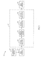
- System 15 comprises illumination source 42', image receiver 12', data processor 14', and image monitor 18'.
- Data processor 14' comprises a spectral analyzer 22, an adaptive reference builder 24, a distance calculator 26, and a decision calculator 28.
- data processor 14' is a standard computer accelerator board, high performance computer, multiprocessor or any other serial or parallel high performance processing machine.
- Image monitor 18' may be a video display, or a graph, table or any other indicator.
- Steps of Fig. 4 may be accomplished using system 15 of Fig. 3 .
- images are captured and processed within a capsule.
- images are captured by an in-vivo system, and are transmitted to a remote location where they are processed.
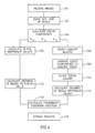
- Image receiver 12' receives (step 101) images captured by the in-vivo camera system of Fig. 1 or any other in-vivo imager.
- Data processor 14' divides (step 102) the color images into a grid of pixels. As in other imaging applications, the number of pixels determines the resolution of the image. For purposes of this discussion, the images are divided into blocks (i,j) of 8 x 8 pixels.
- the original image is a 256 x 256 pixel image
- the result of dividing into 8 pixels, and determining the color components is a 32 x 32 x 3 matrix of color component value blocks.
- Spectral analyzer 22 calculates (step 104) the color components of each block: hue H i,j ; saturation S i,j ; and brightness value V i,j for each image.
- Spectral analyzer also calculates (steps 105 and 106-110) the color components of blocks of pathology sample B and of healthy reference tissue R.
- Spectral analyzer 22 calculates (step 105) the color components of blocks of pathology sample B from known images containing blood.
- Adaptive reference builder 24 calculates (steps 106-110) tissue reference color components in order to build a reference sample of healthy tissue. The adaptive approach is based on averaging healthy tissue appearing in subsequent images. Averages are used since the parameters of healthy tissue along the GI tract may change. Adaptive reference builder 24 selects (step 107) blocks based on value V (brightness) and hue H. In one embodiment, the conditions are: 0.1 ⁇ V i,j ⁇ 0.9 and 0 ⁇ H i,j ⁇ 0.09. These conditions indicate that healthy tissue is present. As shown in Fig.
- images P i , P i-1 , and P i-2 with regions R i , R i-1 , and R i-2 of healthy tissue are obtained.
- Adaptive reference builder 24 averages (step 108) color components of healthy regions R i , R i-1 , and R i-2 (i.e. the selected blocks) of images P i , P i-1 , and P i-2 obtained along the GI tract.
- adaptive reference builder 24 filters (step 110) the average tissue colors of the present image P i and the previous image P i-1 .
- IIR Infinite Impulse Response
- distance calculator 26 calculates (step 112) the Euclidian distance between each block in the matrix and blood reference value B.
- Blood reference value B is obtained from known images containing blood, analyzed by spectral analyzer 22 as described above.
- a different colorimetric reference value may be used for indication of other unusual colors.
- the result of this calculation is a matrix of 32 x 32 elements ⁇ i,j .
- ⁇ i ,j H i , j - H t 2 + S i , j - S t 2 + V i , j - V t 2 H t 2 + S t 2 + V t 2 * H i , j 2 + S i , j 2 + V i , j 2
- H t , S t and V t are the reference values for hue saturation and brightness, respectively, of healthy tissue.
- image monitor 18' displays (step 118) the results, either as a color video showing the presence of blood, or as a graph or table indicating the levels and/or threshold values.
- Display of results may include incorporation of a position indicator, so that the end user can determine where the presence of color change is within the GI tract, or other body lumen. Thus, the physician will be able to deal with the problem area.
Landscapes
- Health & Medical Sciences (AREA)
- Life Sciences & Earth Sciences (AREA)
- Engineering & Computer Science (AREA)
- Surgery (AREA)
- Physics & Mathematics (AREA)
- General Health & Medical Sciences (AREA)
- Medical Informatics (AREA)
- Pathology (AREA)
- Veterinary Medicine (AREA)
- Biomedical Technology (AREA)
- Heart & Thoracic Surgery (AREA)
- Biophysics (AREA)
- Molecular Biology (AREA)
- Animal Behavior & Ethology (AREA)
- Public Health (AREA)
- Nuclear Medicine, Radiotherapy & Molecular Imaging (AREA)
- Radiology & Medical Imaging (AREA)
- Optics & Photonics (AREA)
- General Physics & Mathematics (AREA)
- Computer Vision & Pattern Recognition (AREA)
- Theoretical Computer Science (AREA)
- Signal Processing (AREA)
- Spectroscopy & Molecular Physics (AREA)
- Computer Networks & Wireless Communication (AREA)
- Quality & Reliability (AREA)
- Physiology (AREA)
- Cardiology (AREA)
- Endoscopes (AREA)
- Measuring And Recording Apparatus For Diagnosis (AREA)
- Spectrometry And Color Measurement (AREA)
- Ultra Sonic Daignosis Equipment (AREA)
- Measurement Of The Respiration, Hearing Ability, Form, And Blood Characteristics Of Living Organisms (AREA)
- Closed-Circuit Television Systems (AREA)
Applications Claiming Priority (3)
| Application Number | Priority Date | Filing Date | Title |
|---|---|---|---|
| US27548601P | 2001-03-14 | 2001-03-14 | |
| US275486P | 2001-03-14 | ||
| PCT/IL2002/000210 WO2002073507A2 (en) | 2001-03-14 | 2002-03-14 | Method and system for detecting colorimetric abnormalities |
Publications (3)
| Publication Number | Publication Date |
|---|---|
| EP1372474A2 EP1372474A2 (en) | 2004-01-02 |
| EP1372474A4 EP1372474A4 (en) | 2007-02-28 |
| EP1372474B1 true EP1372474B1 (en) | 2011-05-11 |
Family
ID=23052498
Family Applications (1)
| Application Number | Title | Priority Date | Filing Date |
|---|---|---|---|
| EP02707063A Expired - Lifetime EP1372474B1 (en) | 2001-03-14 | 2002-03-14 | Method and system for detecting colorimetric abnormalities |
Country Status (9)
| Country | Link |
|---|---|
| US (4) | US20020177779A1 (es) |
| EP (1) | EP1372474B1 (es) |
| JP (2) | JP4067407B2 (es) |
| CN (2) | CN101305906B (es) |
| AT (1) | ATE509328T1 (es) |
| AU (1) | AU2002241215A1 (es) |
| ES (1) | ES2365696T3 (es) |
| IL (1) | IL157892A0 (es) |
| WO (1) | WO2002073507A2 (es) |
Families Citing this family (191)
| Publication number | Priority date | Publication date | Assignee | Title |
|---|---|---|---|---|
| IL126727A (en) | 1998-10-22 | 2006-12-31 | Given Imaging Ltd | A method of bringing a device to the goal |
| US8636648B2 (en) | 1999-03-01 | 2014-01-28 | West View Research, Llc | Endoscopic smart probe |
| US10973397B2 (en) | 1999-03-01 | 2021-04-13 | West View Research, Llc | Computerized information collection and processing apparatus |
| WO2001053792A2 (en) * | 2000-01-19 | 2001-07-26 | Given Imaging Ltd. | A system for detecting substances |
| CN101305906B (zh) * | 2001-03-14 | 2012-02-08 | 吉温成象有限公司 | 检测活体内色度异常的系统 |
| WO2002102224A2 (en) | 2001-06-18 | 2002-12-27 | Given Imaging Ltd. | In vivo sensing device with a circuit board having rigid sections and flexible sections |
| US7160258B2 (en) * | 2001-06-26 | 2007-01-09 | Entrack, Inc. | Capsule and method for treating or diagnosing the intestinal tract |
| US9113846B2 (en) * | 2001-07-26 | 2015-08-25 | Given Imaging Ltd. | In-vivo imaging device providing data compression |
| US20030043263A1 (en) * | 2001-07-26 | 2003-03-06 | Arkady Glukhovsky | Diagnostic device using data compression |
| US20050187433A1 (en) * | 2001-07-26 | 2005-08-25 | Given Imaging Ltd. | In-vivo imaging device providing constant bit rate transmission |
| US6951536B2 (en) * | 2001-07-30 | 2005-10-04 | Olympus Corporation | Capsule-type medical device and medical system |
| US8428685B2 (en) * | 2001-09-05 | 2013-04-23 | Given Imaging Ltd. | System and method for magnetically maneuvering an in vivo device |
| EP1428178A4 (en) * | 2001-09-05 | 2009-01-14 | Given Imaging Ltd | SYSTEM AND METHOD FOR THE THREE-DIMENSIONAL DISPLAY OF BODIES |
| JP3756797B2 (ja) | 2001-10-16 | 2006-03-15 | オリンパス株式会社 | カプセル型医療機器 |
| US7474327B2 (en) * | 2002-02-12 | 2009-01-06 | Given Imaging Ltd. | System and method for displaying an image stream |
| JP4234605B2 (ja) * | 2002-02-12 | 2009-03-04 | ギブン イメージング リミテッド | 画像ストリームを表示するためのシステムおよび方法 |
| US8022980B2 (en) * | 2002-02-12 | 2011-09-20 | Given Imaging Ltd. | System and method for displaying an image stream |
| EP1501415A1 (en) * | 2002-05-09 | 2005-02-02 | Given Imaging Ltd. | System and method for in vivo sensing |
| US7662094B2 (en) | 2002-05-14 | 2010-02-16 | Given Imaging Ltd. | Optical head assembly with dome, and device for use thereof |
| WO2004014227A1 (en) | 2002-08-13 | 2004-02-19 | Given Imaging Ltd. | System for in vivo sampling and analysis |
| AU2003269438A1 (en) | 2002-09-30 | 2004-04-19 | Given Imaging Ltd. | In-vivo sensing system |
| WO2004028336A2 (en) | 2002-09-30 | 2004-04-08 | Given Imaging Ltd. | Reduced size imaging device |
| WO2004036803A2 (en) | 2002-10-15 | 2004-04-29 | Given Imaging Ltd. | Device, system and method for transfer of signals to a moving device |
| US7195588B2 (en) * | 2004-03-01 | 2007-03-27 | Olympus Corporation | Endoscope image pick-up apparatus |
| AU2003282373A1 (en) * | 2002-11-29 | 2004-06-23 | Given Imaging Ltd. | Methods device and system for in vivo diagnosis |
| AU2003288517A1 (en) | 2002-12-26 | 2004-07-22 | Given Imaging Ltd. | In vivo imaging device and method of manufacture thereof |
| US7833151B2 (en) | 2002-12-26 | 2010-11-16 | Given Imaging Ltd. | In vivo imaging device with two imagers |
| ATE547976T1 (de) | 2002-12-26 | 2012-03-15 | Given Imaging Ltd | Immobilisierbare in-vivo messvorrichtung |
| IL155175A (en) * | 2003-03-31 | 2012-01-31 | Given Imaging Ltd | A diagnostic device that uses information thinning |
| JP4493386B2 (ja) * | 2003-04-25 | 2010-06-30 | オリンパス株式会社 | 画像表示装置、画像表示方法および画像表示プログラム |
| JP4554647B2 (ja) * | 2003-04-25 | 2010-09-29 | オリンパス株式会社 | 画像表示装置、画像表示方法および画像表示プログラム |
| CN100431475C (zh) * | 2003-04-25 | 2008-11-12 | 奥林巴斯株式会社 | 图像显示装置、图像显示方法以及图像显示程序 |
| JP4547401B2 (ja) * | 2003-04-25 | 2010-09-22 | オリンパス株式会社 | 画像表示装置、画像表示方法および画像表示プログラム |
| JP4547402B2 (ja) * | 2003-04-25 | 2010-09-22 | オリンパス株式会社 | 画像表示装置、画像表示方法および画像表示プログラム |
| JP3810381B2 (ja) * | 2003-04-25 | 2006-08-16 | オリンパス株式会社 | 画像表示装置、画像表示方法および画像表示プログラム |
| WO2004096008A2 (en) * | 2003-05-01 | 2004-11-11 | Given Imaging Ltd. | Panoramic field of view imaging device |
| IL162740A (en) | 2003-06-26 | 2010-06-16 | Given Imaging Ltd | Device, method and system for reduced transmission imaging |
| JP4656825B2 (ja) * | 2003-09-08 | 2011-03-23 | オリンパス株式会社 | 被検体内導入装置および無線型被検体内情報取得システム |
| US7604589B2 (en) * | 2003-10-01 | 2009-10-20 | Given Imaging, Ltd. | Device, system and method for determining orientation of in-vivo devices |
| US20050075537A1 (en) * | 2003-10-06 | 2005-04-07 | Eastman Kodak Company | Method and system for real-time automatic abnormality detection for in vivo images |
| EP1681009A4 (en) * | 2003-10-27 | 2010-08-11 | Olympus Corp | IMAGE PROCESSING DEVICE, IMAGE PROCESSING AND IMAGE PROGRAM |
| JP4574983B2 (ja) * | 2003-11-04 | 2010-11-04 | オリンパス株式会社 | 画像表示装置、画像表示方法、及び画像表示プログラム |
| WO2005053518A1 (ja) | 2003-12-05 | 2005-06-16 | Olympus Corporation | 表示処理装置 |
| US20050137468A1 (en) * | 2003-12-18 | 2005-06-23 | Jerome Avron | Device, system, and method for in-vivo sensing of a substance |
| US7647090B1 (en) | 2003-12-30 | 2010-01-12 | Given Imaging, Ltd. | In-vivo sensing device and method for producing same |
| EP1699530B1 (en) * | 2003-12-31 | 2012-07-04 | Given Imaging Ltd. | System and method for displaying an image stream |
| WO2005062717A2 (en) | 2003-12-31 | 2005-07-14 | Given Imaging Ltd. | In-vivo sensing device with detachable part |
| JP4652694B2 (ja) * | 2004-01-08 | 2011-03-16 | オリンパス株式会社 | 画像処理方法 |
| US8082024B2 (en) * | 2004-01-16 | 2011-12-20 | Alfano Robert R | Micro-scale compact device for in vivo medical diagnosis combining optical imaging and point fluorescence spectroscopy |
| US20050196023A1 (en) * | 2004-03-01 | 2005-09-08 | Eastman Kodak Company | Method for real-time remote diagnosis of in vivo images |
| US7623690B2 (en) * | 2004-03-30 | 2009-11-24 | Carestream Health, Inc. | System and method for classifying in vivo images according to anatomical structure |
| US7605852B2 (en) | 2004-05-17 | 2009-10-20 | Micron Technology, Inc. | Real-time exposure control for automatic light control |
| US7938775B2 (en) * | 2004-06-28 | 2011-05-10 | Given Imaging, Ltd. | Device, system, and method for in-vivo analysis |
| WO2006005075A2 (en) * | 2004-06-30 | 2006-01-12 | Amir Belson | Apparatus and methods for capsule endoscopy of the esophagus |
| US7596403B2 (en) | 2004-06-30 | 2009-09-29 | Given Imaging Ltd. | System and method for determining path lengths through a body lumen |
| US7336833B2 (en) * | 2004-06-30 | 2008-02-26 | Given Imaging, Ltd. | Device, system, and method for reducing image data captured in-vivo |
| US7643865B2 (en) | 2004-06-30 | 2010-01-05 | Given Imaging Ltd. | Autonomous in-vivo device |
| US8500630B2 (en) | 2004-06-30 | 2013-08-06 | Given Imaging Ltd. | In vivo device with flexible circuit board and method for assembly thereof |
| JP4885432B2 (ja) * | 2004-08-18 | 2012-02-29 | オリンパス株式会社 | 画像表示装置、画像表示方法および画像表示プログラム |
| US7986337B2 (en) | 2004-09-27 | 2011-07-26 | Given Imaging Ltd. | System and method for editing an image stream captured in vivo |
| US20060095093A1 (en) * | 2004-11-04 | 2006-05-04 | Ido Bettesh | Apparatus and method for receiving device selection and combining |
| US7486981B2 (en) * | 2004-11-15 | 2009-02-03 | Given Imaging Ltd. | System and method for displaying an image stream |
| WO2006062163A1 (ja) | 2004-12-10 | 2006-06-15 | Olympus Corporation | 医用画像処理方法 |
| US8738106B2 (en) * | 2005-01-31 | 2014-05-27 | Given Imaging, Ltd | Device, system and method for in vivo analysis |
| EP1849402B1 (en) | 2005-02-15 | 2018-05-16 | Olympus Corporation | Medical image processing device, lumen image processing device, lumen image processing method, and programs for them |
| US20060217593A1 (en) * | 2005-03-24 | 2006-09-28 | Zvika Gilad | Device, system and method of panoramic multiple field of view imaging |
| WO2006103684A2 (en) * | 2005-04-01 | 2006-10-05 | Given Imaging Ltd. | Device, system and method for in vivo magnetic immunoassay analysis |
| IL174531A0 (en) * | 2005-04-06 | 2006-08-20 | Given Imaging Ltd | System and method for performing capsule endoscopy diagnosis in remote sites |
| EP1870020B1 (en) | 2005-04-13 | 2015-08-05 | Olympus Medical Systems Corp. | Image processing apparatus and image processing method |
| EP1875855B1 (en) * | 2005-04-27 | 2012-05-02 | Olympus Medical Systems Corp. | Image processing apparatus, image processing method, and image processing program |
| JP4418400B2 (ja) * | 2005-05-20 | 2010-02-17 | オリンパスメディカルシステムズ株式会社 | 画像表示装置 |
| JP4767591B2 (ja) | 2005-06-01 | 2011-09-07 | オリンパスメディカルシステムズ株式会社 | 内視鏡診断支援方法、内視鏡診断支援装置および内視鏡診断支援プログラム |
| EP1942800B1 (en) * | 2005-09-09 | 2011-09-07 | Given Imaging Ltd. | Concurrent transfer and processing and real time viewing of in-vivo images |
| WO2007031946A2 (en) * | 2005-09-12 | 2007-03-22 | Dvp Technologies Ltd. | Medical image processing |
| US20070066875A1 (en) * | 2005-09-18 | 2007-03-22 | Eli Horn | System and method for identification of images in an image database |
| US8423123B2 (en) * | 2005-09-30 | 2013-04-16 | Given Imaging Ltd. | System and method for in-vivo feature detection |
| US7482593B2 (en) * | 2005-10-20 | 2009-01-27 | The Research Foundation Of State University Of New York | Method to determine the depth-of-interaction function for PET detectors |
| CN101947113B (zh) | 2005-12-28 | 2011-12-07 | 奥林巴斯医疗株式会社 | 图像处理装置和该图像处理装置中的图像处理方法 |
| US9320417B2 (en) | 2005-12-29 | 2016-04-26 | Given Imaging Ltd. | In-vivo optical imaging device with backscatter blocking |
| US20070167834A1 (en) * | 2005-12-29 | 2007-07-19 | Amit Pascal | In-vivo imaging optical device and method |
| US20070156051A1 (en) * | 2005-12-29 | 2007-07-05 | Amit Pascal | Device and method for in-vivo illumination |
| ES2405879T3 (es) * | 2006-03-13 | 2013-06-04 | Given Imaging Ltd. | Dispositivo, sistema y método para la detección automática de actividad contráctil en una trama de imagen |
| US8441530B2 (en) * | 2006-03-13 | 2013-05-14 | Given Imaging Ltd. | Cascade analysis for intestinal contraction detection |
| IL182332A (en) * | 2006-03-31 | 2013-04-30 | Given Imaging Ltd | A system and method for assessing a patient's condition |
| US8663093B2 (en) * | 2006-04-03 | 2014-03-04 | Given Imaging Ltd. | Device, system and method for in-vivo analysis |
| DE202006006268U1 (de) * | 2006-04-12 | 2006-06-14 | Branofilter Gmbh | Vorrichtung zum lösbaren Befestigen eines Staubfilterbeutels in einem staubsaugenden Gerät |
| JP2007319442A (ja) * | 2006-06-01 | 2007-12-13 | Fujifilm Corp | カプセル内視鏡システム、および画像処理装置 |
| JP2009539544A (ja) * | 2006-06-12 | 2009-11-19 | ギブン イメージング リミテッド | 収縮活動の測定及び分析のための装置、システム、及び方法 |
| WO2008007172A1 (en) * | 2006-07-11 | 2008-01-17 | Stanley Kim | Test preparation device |
| JP4912787B2 (ja) | 2006-08-08 | 2012-04-11 | オリンパスメディカルシステムズ株式会社 | 医療用画像処理装置及び医療用画像処理装置の作動方法 |
| US8615284B2 (en) | 2006-09-06 | 2013-12-24 | Innurvation, Inc. | Method for acoustic information exchange involving an ingestible low power capsule |
| WO2008030480A2 (en) | 2006-09-06 | 2008-03-13 | Innurvation, Inc. | Ingestible low power sensor device and system for communicating with same |
| JP5121204B2 (ja) | 2006-10-11 | 2013-01-16 | オリンパス株式会社 | 画像処理装置、画像処理方法、および画像処理プログラム |
| KR101071466B1 (ko) * | 2007-02-22 | 2011-10-10 | 올림푸스 가부시키가이샤 | 피검체 내 도입 시스템 |
| WO2008139812A1 (ja) | 2007-05-08 | 2008-11-20 | Olympus Corporation | 画像処理装置および画像処理プログラム |
| JP5327641B2 (ja) * | 2007-05-17 | 2013-10-30 | オリンパスメディカルシステムズ株式会社 | 画像情報の表示処理装置 |
| JP2008295490A (ja) | 2007-05-29 | 2008-12-11 | Olympus Medical Systems Corp | カプセル内視鏡画像表示装置 |
| JP5106928B2 (ja) | 2007-06-14 | 2012-12-26 | オリンパス株式会社 | 画像処理装置および画像処理プログラム |
| JP5403880B2 (ja) * | 2007-06-27 | 2014-01-29 | オリンパスメディカルシステムズ株式会社 | 画像情報の表示処理装置 |
| US9197470B2 (en) | 2007-10-05 | 2015-11-24 | Innurvation, Inc. | Data transmission via multi-path channels using orthogonal multi-frequency signals with differential phase shift keying modulation |
| US20090105532A1 (en) * | 2007-10-22 | 2009-04-23 | Zvika Gilad | In vivo imaging device and method of manufacturing thereof |
| US9017248B2 (en) | 2007-11-08 | 2015-04-28 | Olympus Medical Systems Corp. | Capsule blood detection system and method |
| US9131847B2 (en) | 2007-11-08 | 2015-09-15 | Olympus Corporation | Method and apparatus for detecting abnormal living tissue |
| US20100329520A2 (en) * | 2007-11-08 | 2010-12-30 | Olympus Medical Systems Corp. | Method and System for Correlating Image and Tissue Characteristic Data |
| WO2009060460A2 (en) * | 2007-11-09 | 2009-05-14 | Given Imaging Ltd. | Apparatus and methods for capsule endoscopy of the esophagus |
| ES2620816T3 (es) * | 2007-11-12 | 2017-06-29 | Novineon Healthcare Technology Partners Gmbh | Dispositivo para la detección de hemorragias |
| US8529441B2 (en) | 2008-02-12 | 2013-09-10 | Innurvation, Inc. | Ingestible endoscopic optical scanning device |
| US20100016662A1 (en) * | 2008-02-21 | 2010-01-21 | Innurvation, Inc. | Radial Scanner Imaging System |
| JP5336749B2 (ja) * | 2008-03-24 | 2013-11-06 | オリンパス株式会社 | カプセル型医療装置とその作動方法 |
| US8515507B2 (en) | 2008-06-16 | 2013-08-20 | Given Imaging Ltd. | Device and method for detecting in-vivo pathology |
| US8888680B2 (en) * | 2008-07-07 | 2014-11-18 | Olympus Medical Systems Corp. | Method and apparatus for foreign matter detection for blood content sensors |
| US8617058B2 (en) | 2008-07-09 | 2013-12-31 | Innurvation, Inc. | Displaying image data from a scanner capsule |
| JP5117353B2 (ja) * | 2008-11-07 | 2013-01-16 | オリンパス株式会社 | 画像処理装置、画像処理プログラムおよび画像処理方法 |
| US8516691B2 (en) | 2009-06-24 | 2013-08-27 | Given Imaging Ltd. | Method of assembly of an in vivo imaging device with a flexible circuit board |
| US20110097690A1 (en) * | 2009-10-22 | 2011-04-28 | Perry Franklin Samuel-Cutts | Training system for surfing and method of use |
| US9192353B2 (en) * | 2009-10-27 | 2015-11-24 | Innurvation, Inc. | Data transmission via wide band acoustic channels |
| EP2515759A4 (en) | 2009-12-23 | 2015-01-21 | Given Imaging Inc | METHOD OF ASSESSING CONSTIPATION USING INGREDIENT CAPSULE |
| US8446465B2 (en) * | 2010-01-05 | 2013-05-21 | Given Imaging Ltd. | System and method for displaying an image stream captured in-vivo |
| WO2012123939A1 (en) | 2011-03-17 | 2012-09-20 | Photopill Medical Ltd. | Capsule phototherapy |
| US8682142B1 (en) | 2010-03-18 | 2014-03-25 | Given Imaging Ltd. | System and method for editing an image stream captured in-vivo |
| US8647259B2 (en) | 2010-03-26 | 2014-02-11 | Innurvation, Inc. | Ultrasound scanning capsule endoscope (USCE) |
| KR101708726B1 (ko) | 2010-04-28 | 2017-02-21 | 기븐 이미징 리미티드 | 생체내 영상들 일부 표시 시스템 및 방법 |
| JP5622461B2 (ja) * | 2010-07-07 | 2014-11-12 | オリンパス株式会社 | 画像処理装置、画像処理方法、および画像処理プログラム |
| CN102843950B (zh) * | 2010-11-08 | 2015-02-04 | 奥林巴斯医疗株式会社 | 图像显示装置以及胶囊型内窥镜系统 |
| US8913807B1 (en) | 2010-12-30 | 2014-12-16 | Given Imaging Ltd. | System and method for detecting anomalies in a tissue imaged in-vivo |
| US8873816B1 (en) | 2011-04-06 | 2014-10-28 | Given Imaging Ltd. | Method and system for identification of red colored pathologies in vivo |
| US20140055400A1 (en) | 2011-05-23 | 2014-02-27 | Haworth, Inc. | Digital workspace ergonomics apparatuses, methods and systems |
| US9430140B2 (en) | 2011-05-23 | 2016-08-30 | Haworth, Inc. | Digital whiteboard collaboration apparatuses, methods and systems |
| US8929629B1 (en) * | 2011-06-29 | 2015-01-06 | Given Imaging Ltd. | Method and system for image-based ulcer detection |
| US8983167B2 (en) | 2012-05-14 | 2015-03-17 | Gauss Surgical | System and method for estimating a quantity of a blood component in a fluid canister |
| US9652655B2 (en) | 2011-07-09 | 2017-05-16 | Gauss Surgical, Inc. | System and method for estimating extracorporeal blood volume in a physical sample |
| US9870625B2 (en) | 2011-07-09 | 2018-01-16 | Gauss Surgical, Inc. | Method for estimating a quantity of a blood component in a fluid receiver and corresponding error |
| US10426356B2 (en) | 2011-07-09 | 2019-10-01 | Gauss Surgical, Inc. | Method for estimating a quantity of a blood component in a fluid receiver and corresponding error |
| US9646375B2 (en) | 2011-07-09 | 2017-05-09 | Gauss Surgical, Inc. | Method for setting a blood transfusion parameter |
| JP5851160B2 (ja) * | 2011-08-31 | 2016-02-03 | オリンパス株式会社 | 画像処理装置、画像処理装置の作動方法、及び画像処理プログラム |
| CN103841876B (zh) * | 2011-10-06 | 2016-03-09 | 奥林巴斯株式会社 | 荧光观察装置 |
| CN104271028B (zh) * | 2011-12-15 | 2017-11-17 | 基文影像公司 | 确定患者的胃肠道随时间的出血曲线的类型的系统 |
| US8923585B1 (en) * | 2012-01-31 | 2014-12-30 | Given Imaging Ltd. | Method and system for image-based ulcer detection |
| WO2013126568A1 (en) * | 2012-02-21 | 2013-08-29 | Massachusetts Eye & Ear Infirmary | Calculating conjunctival redness |
| JP6021237B2 (ja) | 2012-05-14 | 2016-11-09 | ガウス サージカルGauss Surgical | 患者の失血を管理するシステム |
| US9479548B2 (en) | 2012-05-23 | 2016-10-25 | Haworth, Inc. | Collaboration system with whiteboard access to global collaboration data |
| US9479549B2 (en) | 2012-05-23 | 2016-10-25 | Haworth, Inc. | Collaboration system with whiteboard with federated display |
| US10424060B2 (en) | 2012-07-09 | 2019-09-24 | Gauss Surgical, Inc. | Method for estimating blood component quantities in surgical textiles |
| JP6067264B2 (ja) | 2012-07-17 | 2017-01-25 | Hoya株式会社 | 画像処理装置及び内視鏡装置 |
| JP6027803B2 (ja) * | 2012-07-17 | 2016-11-16 | Hoya株式会社 | 画像処理装置及び内視鏡装置 |
| KR102163327B1 (ko) * | 2012-07-25 | 2020-10-08 | 인튜어티브 서지컬 오퍼레이션즈 인코포레이티드 | 수술 시스템의 효율적인 쌍방향 출혈 검출 방법 및 시스템 |
| WO2014071399A1 (en) | 2012-11-05 | 2014-05-08 | Gauss Surgical | Method for triggering blood salvage |
| JP2014140335A (ja) * | 2013-01-24 | 2014-08-07 | Dainippon Printing Co Ltd | 培地画像解析装置、培地情報登録システム、プログラム及び衛生管理システム |
| US11861561B2 (en) | 2013-02-04 | 2024-01-02 | Haworth, Inc. | Collaboration system including a spatial event map |
| US10304037B2 (en) | 2013-02-04 | 2019-05-28 | Haworth, Inc. | Collaboration system including a spatial event map |
| JP6097629B2 (ja) | 2013-04-26 | 2017-03-15 | Hoya株式会社 | 病変評価情報生成装置 |
| US9324145B1 (en) | 2013-08-08 | 2016-04-26 | Given Imaging Ltd. | System and method for detection of transitions in an image stream of the gastrointestinal tract |
| CN105451631B (zh) | 2013-08-29 | 2018-05-18 | 基文影像公司 | 用于操纵线圈功率优化的系统和方法 |
| US9430706B1 (en) | 2013-10-02 | 2016-08-30 | Given Imaging Ltd. | System and method for detection of in-vivo pathology sequences |
| US8736685B1 (en) * | 2013-12-11 | 2014-05-27 | Anritsu Company | Systems and methods for measuring brightness response of a camera operating in automatic exposure mode |
| CN105830458A (zh) | 2013-12-11 | 2016-08-03 | 基文影像公司 | 用于控制图像流显示的系统和方法 |
| EP3132253B1 (en) | 2014-04-15 | 2019-02-13 | Gauss Surgical, Inc. | Method for estimating a quantity of a blood component in a fluid canister |
| JP6546605B2 (ja) | 2014-04-15 | 2019-07-17 | ガウス サージカル, インコーポレイテッドGauss Surgical, Inc. | 液体キャニスタ内の血液成分量の推定方法 |
| EP3954269A1 (en) | 2014-05-02 | 2022-02-16 | Massachusetts Eye & Ear Infirmary | Grading corneal fluorescein staining |
| US9633276B2 (en) * | 2014-07-14 | 2017-04-25 | Sony Corporation | Blood detection system with real-time capability and method of operation thereof |
| BR112017006235A2 (pt) | 2014-09-25 | 2017-12-12 | Labbe Alain | dispositivo de pílula eletromecânica com capacidades de localização |
| CN104658014B (zh) * | 2015-02-10 | 2017-09-22 | 重庆金山科技(集团)有限公司 | 一种检测活体内色度异常的方法 |
| WO2016179401A1 (en) | 2015-05-06 | 2016-11-10 | Haworth, Inc. | Virtual workspace viewport follow mode and location markers in collaboration systems |
| WO2016187071A1 (en) | 2015-05-15 | 2016-11-24 | Gauss Surgical, Inc. | Systems and methods for assessing fluids from a patient |
| WO2016187072A1 (en) | 2015-05-15 | 2016-11-24 | Gauss Surgical, Inc. | Methods and systems for characterizing fluids from a patient |
| WO2016187070A1 (en) | 2015-05-15 | 2016-11-24 | Gauss Surgical, Inc. | Method for projecting blood loss of a patient during a surgery |
| CN106687023B (zh) | 2015-08-13 | 2018-12-18 | Hoya株式会社 | 评价值计算装置以及电子内窥镜系统 |
| DE112016000094B4 (de) | 2015-08-13 | 2019-07-11 | Hoya Corporation | Vorrichtung zur Berechnung von Analysewerten und elektronisches Endoskopsystem |
| US10572997B2 (en) | 2015-12-18 | 2020-02-25 | Given Imaging Ltd. | System and method for detecting anomalies in an image captured in-vivo using color histogram association |
| WO2017112913A1 (en) | 2015-12-23 | 2017-06-29 | Gauss Surgical, Inc. | System and method for estimating an amount of a blood component in a volume of fluid |
| TWI616180B (zh) * | 2016-06-29 | 2018-03-01 | 國立成功大學 | 上消化道出血偵測裝置及方法 |
| CA3034263A1 (en) * | 2016-08-18 | 2018-02-22 | Mitchell Lawrence Jones | Sampling systems and related materials and methods |
| IL305187A (en) | 2016-09-09 | 2023-10-01 | Biora Therapeutics Inc | An edible electromechanical device for dispensing a dispensable substance |
| EP4252629A3 (en) | 2016-12-07 | 2023-12-27 | Biora Therapeutics, Inc. | Gastrointestinal tract detection methods, devices and systems |
| WO2018125812A1 (en) | 2017-01-02 | 2018-07-05 | Gauss Surgical, Inc. | Tracking surgical items with prediction of duplicate imaging of items |
| KR101875004B1 (ko) | 2017-01-04 | 2018-07-05 | 금오공과대학교 산학협력단 | 캡슐 내시경 영상을 이용한 자동 출혈 감지 방법 및 컴퓨터 프로그램 |
| US11229368B2 (en) | 2017-01-13 | 2022-01-25 | Gauss Surgical, Inc. | Fluid loss estimation based on weight of medical items |
| AU2018243312B2 (en) | 2017-03-31 | 2023-02-02 | Biora Therapeutics, Inc. | Localization systems and methods for an ingestible device |
| JP7040910B2 (ja) | 2017-10-11 | 2022-03-23 | 聡 織田 | 超音波プローブ |
| US11934637B2 (en) | 2017-10-23 | 2024-03-19 | Haworth, Inc. | Collaboration system including markers identifying multiple canvases in multiple shared virtual workspaces |
| US11126325B2 (en) | 2017-10-23 | 2021-09-21 | Haworth, Inc. | Virtual workspace including shared viewport markers in a collaboration system |
| WO2019164277A1 (ko) * | 2018-02-20 | 2019-08-29 | (주)휴톰 | 수술영상을 이용한 출혈 평가 방법 및 장치 |
| JP7162722B2 (ja) * | 2018-07-25 | 2022-10-28 | チェック-キャップ リミテッド | カプセル動態によるポリープ検出のためのシステムおよび方法 |
| EP3883635A1 (en) | 2018-11-19 | 2021-09-29 | Progenity, Inc. | Methods and devices for treating a disease with biotherapeutics |
| JP7238390B2 (ja) * | 2018-12-21 | 2023-03-14 | セイコーエプソン株式会社 | 情報システムおよび特定方法 |
| WO2020176517A1 (en) | 2019-02-25 | 2020-09-03 | Haworth, Inc. | Gesture based workflows in a collaboration system |
| WO2021119482A1 (en) | 2019-12-13 | 2021-06-17 | Progenity, Inc. | Ingestible device for delivery of therapeutic agent to the gastrointestinal tract |
| US11212127B2 (en) | 2020-05-07 | 2021-12-28 | Haworth, Inc. | Digital workspace sharing over one or more display clients and authorization protocols for collaboration systems |
| US11750672B2 (en) | 2020-05-07 | 2023-09-05 | Haworth, Inc. | Digital workspace sharing over one or more display clients in proximity of a main client |
| JP7461221B2 (ja) * | 2020-05-25 | 2024-04-03 | 富士フイルムヘルスケア株式会社 | 医用画像処理装置、および、医用撮像装置 |
| CN112991325B (zh) * | 2021-04-14 | 2021-08-17 | 上海孚慈医疗科技有限公司 | 一种基于智能编码的斑状发红图像采集和处理方法及系统 |
Family Cites Families (28)
| Publication number | Priority date | Publication date | Assignee | Title |
|---|---|---|---|---|
| US3971362A (en) * | 1972-10-27 | 1976-07-27 | The United States Of America As Represented By The Administrator Of The National Aeronautics And Space Administration | Miniature ingestible telemeter devices to measure deep-body temperature |
| JPS5519124A (en) * | 1978-07-27 | 1980-02-09 | Olympus Optical Co | Camera system for medical treatment |
| US5993378A (en) * | 1980-10-28 | 1999-11-30 | Lemelson; Jerome H. | Electro-optical instruments and methods for treating disease |
| JPS59188301A (ja) * | 1983-04-08 | 1984-10-25 | Japanese National Railways<Jnr> | 電気車用回生ブレーキ制御装置 |
| US5042494A (en) * | 1985-11-13 | 1991-08-27 | Alfano Robert R | Method and apparatus for detecting cancerous tissue using luminescence excitation spectra |
| US4689621A (en) * | 1986-03-31 | 1987-08-25 | The United States Of America As Represented By The Administrator Of The National Aeronautics And Space Administration | Temperature responsive transmitter |
| US4844076A (en) * | 1988-08-26 | 1989-07-04 | The Johns Hopkins University | Ingestible size continuously transmitting temperature monitoring pill |
| US5279607A (en) * | 1991-05-30 | 1994-01-18 | The State University Of New York | Telemetry capsule and process |
| JP3285235B2 (ja) * | 1992-11-05 | 2002-05-27 | オリンパス光学工業株式会社 | 生体内観察用カプセル装置 |
| US6095989A (en) * | 1993-07-20 | 2000-08-01 | Hay; Sam H. | Optical recognition methods for locating eyes |
| IL108352A (en) * | 1994-01-17 | 2000-02-29 | Given Imaging Ltd | In vivo video camera system |
| CA2145232A1 (en) * | 1994-03-24 | 1995-09-25 | Arie Avny | Viewing method and apparatus particularly useful for viewing the interior of the large intestine |
| US5590660A (en) * | 1994-03-28 | 1997-01-07 | Xillix Technologies Corp. | Apparatus and method for imaging diseased tissue using integrated autofluorescence |
| EP0766205B1 (fr) * | 1995-09-29 | 2003-12-03 | Koninklijke Philips Electronics N.V. | Procédé et dispositif de traitement d'image, pour la détection automatique de régions d'un type prédéterminé de cancer dans une image d'intensité |
| US5833603A (en) * | 1996-03-13 | 1998-11-10 | Lipomatrix, Inc. | Implantable biosensing transponder |
| US6459919B1 (en) * | 1997-08-26 | 2002-10-01 | Color Kinetics, Incorporated | Precision illumination methods and systems |
| US6016038A (en) * | 1997-08-26 | 2000-01-18 | Color Kinetics, Inc. | Multicolored LED lighting method and apparatus |
| US6422994B1 (en) * | 1997-09-24 | 2002-07-23 | Olympus Optical Co., Ltd. | Fluorescent diagnostic system and method providing color discrimination enhancement |
| US6240312B1 (en) * | 1997-10-23 | 2001-05-29 | Robert R. Alfano | Remote-controllable, micro-scale device for use in in vivo medical diagnosis and/or treatment |
| GB9810771D0 (en) * | 1998-05-19 | 1998-07-15 | Active Silicon Limited | Method of detecting colours |
| DE19844618A1 (de) | 1998-09-29 | 2000-03-30 | Zahnradfabrik Friedrichshafen | Verfahren zur Reduzierung der thermischen Belastung eines Automatgetriebes für ein Kraftfahrzeug in einem Notfahrbetrieb |
| IL126727A (en) * | 1998-10-22 | 2006-12-31 | Given Imaging Ltd | A method of bringing a device to the goal |
| JP2001037718A (ja) | 1999-05-26 | 2001-02-13 | Olympus Optical Co Ltd | 画像診断装置及び内視鏡装置 |
| JP4540908B2 (ja) * | 1999-07-26 | 2010-09-08 | カルディオセンス リミテッド | ショックおよびプレショックの医学的状態を検出するための改良された装置 |
| US7039453B2 (en) * | 2000-02-08 | 2006-05-02 | Tarun Mullick | Miniature ingestible capsule |
| WO2002026103A2 (en) * | 2000-09-27 | 2002-04-04 | Given Imaging Ltd. | An immobilizable in vivo sensing device |
| US6632175B1 (en) * | 2000-11-08 | 2003-10-14 | Hewlett-Packard Development Company, L.P. | Swallowable data recorder capsule medical device |
| CN101305906B (zh) * | 2001-03-14 | 2012-02-08 | 吉温成象有限公司 | 检测活体内色度异常的系统 |
-
2002
- 2002-03-14 CN CN200810125961XA patent/CN101305906B/zh not_active Expired - Lifetime
- 2002-03-14 US US10/097,096 patent/US20020177779A1/en not_active Abandoned
- 2002-03-14 JP JP2002572089A patent/JP4067407B2/ja not_active Expired - Fee Related
- 2002-03-14 AU AU2002241215A patent/AU2002241215A1/en not_active Abandoned
- 2002-03-14 AT AT02707063T patent/ATE509328T1/de not_active IP Right Cessation
- 2002-03-14 CN CNB028098560A patent/CN100469308C/zh not_active Expired - Lifetime
- 2002-03-14 IL IL15789202A patent/IL157892A0/xx unknown
- 2002-03-14 ES ES02707063T patent/ES2365696T3/es not_active Expired - Lifetime
- 2002-03-14 EP EP02707063A patent/EP1372474B1/en not_active Expired - Lifetime
- 2002-03-14 WO PCT/IL2002/000210 patent/WO2002073507A2/en active Search and Examination
-
2006
- 2006-06-08 JP JP2006160078A patent/JP4504951B2/ja not_active Expired - Lifetime
-
2012
- 2012-07-03 US US13/541,111 patent/US8626268B2/en not_active Expired - Fee Related
-
2013
- 2013-10-04 US US14/046,258 patent/US8918164B2/en not_active Expired - Fee Related
-
2014
- 2014-11-17 US US14/543,154 patent/US9364139B2/en not_active Expired - Lifetime
Also Published As
| Publication number | Publication date |
|---|---|
| WO2002073507A2 (en) | 2002-09-19 |
| IL157892A0 (en) | 2004-03-28 |
| JP4504951B2 (ja) | 2010-07-14 |
| US20150141782A1 (en) | 2015-05-21 |
| US20020177779A1 (en) | 2002-11-28 |
| JP4067407B2 (ja) | 2008-03-26 |
| ATE509328T1 (de) | 2011-05-15 |
| US8918164B2 (en) | 2014-12-23 |
| US20120275683A1 (en) | 2012-11-01 |
| CN101305906A (zh) | 2008-11-19 |
| US20140039287A1 (en) | 2014-02-06 |
| AU2002241215A1 (en) | 2002-09-24 |
| JP2006297118A (ja) | 2006-11-02 |
| CN100469308C (zh) | 2009-03-18 |
| EP1372474A2 (en) | 2004-01-02 |
| WO2002073507A3 (en) | 2003-10-23 |
| WO2002073507A9 (en) | 2003-01-23 |
| ES2365696T3 (es) | 2011-10-10 |
| US8626268B2 (en) | 2014-01-07 |
| EP1372474A4 (en) | 2007-02-28 |
| CN1509152A (zh) | 2004-06-30 |
| US9364139B2 (en) | 2016-06-14 |
| CN101305906B (zh) | 2012-02-08 |
| JP2004521693A (ja) | 2004-07-22 |
Similar Documents
| Publication | Publication Date | Title |
|---|---|---|
| EP1372474B1 (en) | Method and system for detecting colorimetric abnormalities | |
| US7319781B2 (en) | Method and system for multiple passes diagnostic alignment for in vivo images | |
| US7567692B2 (en) | System and method for detecting content in-vivo | |
| US7577283B2 (en) | System and method for detecting content in-vivo | |
| US10251538B2 (en) | Endoscope system and method for controlling the same | |
| US20050075537A1 (en) | Method and system for real-time automatic abnormality detection for in vivo images | |
| EP2096859B1 (en) | Method for enhancing in-vivo image contrast | |
| US8639011B2 (en) | Fluoroscopy apparatus | |
| US20100166272A1 (en) | System and method to detect a transition in an image stream | |
| EP3263011A1 (en) | Image processing device | |
| EP2286713B1 (en) | Signal processing system and signal processing program | |
| US10748279B2 (en) | Image processing apparatus, image processing method, and computer readable recording medium | |
| JP2001037718A (ja) | 画像診断装置及び内視鏡装置 | |
| EP1566140A1 (en) | Endoscope insertion direction detecting device, endoscope insertion direction detecting system, and endoscope insertion direction detecting method | |
| JP4855709B2 (ja) | 画像処理装置、画像処理方法、及び画像処理プログラム | |
| US10646102B2 (en) | Processor for electronic endoscope, and electronic endoscope system | |
| US9854958B1 (en) | System and method for automatic processing of images from an autonomous endoscopic capsule | |
| JP2003220019A (ja) | 画像診断装置及び内視鏡装置 | |
| CN115032157A (zh) | 基于云平台的手机拍照检测分泌物的识别方法 | |
| JP2003204925A (ja) | 画像診断装置及び内視鏡装置 |
Legal Events
| Date | Code | Title | Description |
|---|---|---|---|
| PUAI | Public reference made under article 153(3) epc to a published international application that has entered the european phase |
Free format text: ORIGINAL CODE: 0009012 |
|
| 17P | Request for examination filed |
Effective date: 20031006 |
|
| AK | Designated contracting states |
Kind code of ref document: A2 Designated state(s): AT BE CH CY DE DK ES FI FR GB GR IE IT LI LU MC NL PT SE TR |
|
| AX | Request for extension of the european patent |
Extension state: AL LT LV MK RO SI |
|
| A4 | Supplementary search report drawn up and despatched |
Effective date: 20070126 |
|
| RIC1 | Information provided on ipc code assigned before grant |
Ipc: G06T 7/00 20060101AFI20070122BHEP Ipc: A61B 5/00 20060101ALI20070122BHEP Ipc: A61B 1/05 20060101ALI20070122BHEP Ipc: G06T 7/40 20060101ALI20070122BHEP Ipc: A61B 5/07 20060101ALI20070122BHEP |
|
| RAP1 | Party data changed (applicant data changed or rights of an application transferred) |
Owner name: GIVEN IMAGING LTD. |
|
| 17Q | First examination report despatched |
Effective date: 20080922 |
|
| GRAP | Despatch of communication of intention to grant a patent |
Free format text: ORIGINAL CODE: EPIDOSNIGR1 |
|
| GRAS | Grant fee paid |
Free format text: ORIGINAL CODE: EPIDOSNIGR3 |
|
| GRAA | (expected) grant |
Free format text: ORIGINAL CODE: 0009210 |
|
| AK | Designated contracting states |
Kind code of ref document: B1 Designated state(s): AT BE CH CY DE DK ES FI FR GB GR IE IT LI LU MC NL PT SE TR |
|
| REG | Reference to a national code |
Ref country code: GB Ref legal event code: FG4D |
|
| REG | Reference to a national code |
Ref country code: CH Ref legal event code: EP |
|
| REG | Reference to a national code |
Ref country code: IE Ref legal event code: FG4D |
|
| REG | Reference to a national code |
Ref country code: DE Ref legal event code: R096 Ref document number: 60239993 Country of ref document: DE Effective date: 20110622 |
|
| REG | Reference to a national code |
Ref country code: NL Ref legal event code: VDEP Effective date: 20110511 |
|
| REG | Reference to a national code |
Ref country code: ES Ref legal event code: FG2A Ref document number: 2365696 Country of ref document: ES Kind code of ref document: T3 Effective date: 20111010 |
|
| PG25 | Lapsed in a contracting state [announced via postgrant information from national office to epo] |
Ref country code: PT Free format text: LAPSE BECAUSE OF FAILURE TO SUBMIT A TRANSLATION OF THE DESCRIPTION OR TO PAY THE FEE WITHIN THE PRESCRIBED TIME-LIMIT Effective date: 20110912 Ref country code: SE Free format text: LAPSE BECAUSE OF FAILURE TO SUBMIT A TRANSLATION OF THE DESCRIPTION OR TO PAY THE FEE WITHIN THE PRESCRIBED TIME-LIMIT Effective date: 20110511 |
|
| PG25 | Lapsed in a contracting state [announced via postgrant information from national office to epo] |
Ref country code: FI Free format text: LAPSE BECAUSE OF FAILURE TO SUBMIT A TRANSLATION OF THE DESCRIPTION OR TO PAY THE FEE WITHIN THE PRESCRIBED TIME-LIMIT Effective date: 20110511 Ref country code: GR Free format text: LAPSE BECAUSE OF FAILURE TO SUBMIT A TRANSLATION OF THE DESCRIPTION OR TO PAY THE FEE WITHIN THE PRESCRIBED TIME-LIMIT Effective date: 20110812 Ref country code: CY Free format text: LAPSE BECAUSE OF FAILURE TO SUBMIT A TRANSLATION OF THE DESCRIPTION OR TO PAY THE FEE WITHIN THE PRESCRIBED TIME-LIMIT Effective date: 20110511 Ref country code: BE Free format text: LAPSE BECAUSE OF FAILURE TO SUBMIT A TRANSLATION OF THE DESCRIPTION OR TO PAY THE FEE WITHIN THE PRESCRIBED TIME-LIMIT Effective date: 20110511 Ref country code: AT Free format text: LAPSE BECAUSE OF FAILURE TO SUBMIT A TRANSLATION OF THE DESCRIPTION OR TO PAY THE FEE WITHIN THE PRESCRIBED TIME-LIMIT Effective date: 20110511 |
|
| PG25 | Lapsed in a contracting state [announced via postgrant information from national office to epo] |
Ref country code: NL Free format text: LAPSE BECAUSE OF FAILURE TO SUBMIT A TRANSLATION OF THE DESCRIPTION OR TO PAY THE FEE WITHIN THE PRESCRIBED TIME-LIMIT Effective date: 20110511 |
|
| PG25 | Lapsed in a contracting state [announced via postgrant information from national office to epo] |
Ref country code: DK Free format text: LAPSE BECAUSE OF FAILURE TO SUBMIT A TRANSLATION OF THE DESCRIPTION OR TO PAY THE FEE WITHIN THE PRESCRIBED TIME-LIMIT Effective date: 20110511 |
|
| PLBE | No opposition filed within time limit |
Free format text: ORIGINAL CODE: 0009261 |
|
| STAA | Information on the status of an ep patent application or granted ep patent |
Free format text: STATUS: NO OPPOSITION FILED WITHIN TIME LIMIT |
|
| 26N | No opposition filed |
Effective date: 20120214 |
|
| REG | Reference to a national code |
Ref country code: DE Ref legal event code: R097 Ref document number: 60239993 Country of ref document: DE Effective date: 20120214 |
|
| PG25 | Lapsed in a contracting state [announced via postgrant information from national office to epo] |
Ref country code: MC Free format text: LAPSE BECAUSE OF NON-PAYMENT OF DUE FEES Effective date: 20120331 |
|
| REG | Reference to a national code |
Ref country code: CH Ref legal event code: PL |
|
| REG | Reference to a national code |
Ref country code: IE Ref legal event code: MM4A |
|
| PG25 | Lapsed in a contracting state [announced via postgrant information from national office to epo] |
Ref country code: CH Free format text: LAPSE BECAUSE OF NON-PAYMENT OF DUE FEES Effective date: 20120331 Ref country code: IE Free format text: LAPSE BECAUSE OF NON-PAYMENT OF DUE FEES Effective date: 20120314 Ref country code: LI Free format text: LAPSE BECAUSE OF NON-PAYMENT OF DUE FEES Effective date: 20120331 |
|
| PG25 | Lapsed in a contracting state [announced via postgrant information from national office to epo] |
Ref country code: TR Free format text: LAPSE BECAUSE OF FAILURE TO SUBMIT A TRANSLATION OF THE DESCRIPTION OR TO PAY THE FEE WITHIN THE PRESCRIBED TIME-LIMIT Effective date: 20110511 |
|
| PG25 | Lapsed in a contracting state [announced via postgrant information from national office to epo] |
Ref country code: LU Free format text: LAPSE BECAUSE OF NON-PAYMENT OF DUE FEES Effective date: 20120314 |
|
| REG | Reference to a national code |
Ref country code: FR Ref legal event code: PLFP Year of fee payment: 15 |
|
| REG | Reference to a national code |
Ref country code: FR Ref legal event code: PLFP Year of fee payment: 16 |
|
| REG | Reference to a national code |
Ref country code: FR Ref legal event code: PLFP Year of fee payment: 17 |
|
| PGFP | Annual fee paid to national office [announced via postgrant information from national office to epo] |
Ref country code: FR Payment date: 20210219 Year of fee payment: 20 |
|
| PGFP | Annual fee paid to national office [announced via postgrant information from national office to epo] |
Ref country code: DE Payment date: 20210217 Year of fee payment: 20 Ref country code: GB Payment date: 20210219 Year of fee payment: 20 |
|
| PGFP | Annual fee paid to national office [announced via postgrant information from national office to epo] |
Ref country code: ES Payment date: 20210401 Year of fee payment: 20 |
|
| PGFP | Annual fee paid to national office [announced via postgrant information from national office to epo] |
Ref country code: IT Payment date: 20210217 Year of fee payment: 20 |
|
| REG | Reference to a national code |
Ref country code: DE Ref legal event code: R071 Ref document number: 60239993 Country of ref document: DE |
|
| REG | Reference to a national code |
Ref country code: GB Ref legal event code: PE20 Expiry date: 20220313 |
|
| PG25 | Lapsed in a contracting state [announced via postgrant information from national office to epo] |
Ref country code: GB Free format text: LAPSE BECAUSE OF EXPIRATION OF PROTECTION Effective date: 20220313 |
|
| REG | Reference to a national code |
Ref country code: ES Ref legal event code: FD2A Effective date: 20220624 |
|
| PG25 | Lapsed in a contracting state [announced via postgrant information from national office to epo] |
Ref country code: ES Free format text: LAPSE BECAUSE OF EXPIRATION OF PROTECTION Effective date: 20220315 |