EP1227848B1 - Verfahren zur verbindung von molekularen substanzen - Google Patents
Verfahren zur verbindung von molekularen substanzen Download PDFInfo
- Publication number
- EP1227848B1 EP1227848B1 EP00984960A EP00984960A EP1227848B1 EP 1227848 B1 EP1227848 B1 EP 1227848B1 EP 00984960 A EP00984960 A EP 00984960A EP 00984960 A EP00984960 A EP 00984960A EP 1227848 B1 EP1227848 B1 EP 1227848B1
- Authority
- EP
- European Patent Office
- Prior art keywords
- domain
- proline
- protein
- fact
- procedure according
- Prior art date
- Legal status (The legal status is an assumption and is not a legal conclusion. Google has not performed a legal analysis and makes no representation as to the accuracy of the status listed.)
- Expired - Lifetime
Links
Images


Classifications
-
- C—CHEMISTRY; METALLURGY
- C07—ORGANIC CHEMISTRY
- C07K—PEPTIDES
- C07K14/00—Peptides having more than 20 amino acids; Gastrins; Somatostatins; Melanotropins; Derivatives thereof
- C07K14/005—Peptides having more than 20 amino acids; Gastrins; Somatostatins; Melanotropins; Derivatives thereof from viruses
-
- A—HUMAN NECESSITIES
- A61—MEDICAL OR VETERINARY SCIENCE; HYGIENE
- A61K—PREPARATIONS FOR MEDICAL, DENTAL OR TOILETRY PURPOSES
- A61K47/00—Medicinal preparations characterised by the non-active ingredients used, e.g. carriers or inert additives; Targeting or modifying agents chemically bound to the active ingredient
- A61K47/50—Medicinal preparations characterised by the non-active ingredients used, e.g. carriers or inert additives; Targeting or modifying agents chemically bound to the active ingredient the non-active ingredient being chemically bound to the active ingredient, e.g. polymer-drug conjugates
- A61K47/69—Medicinal preparations characterised by the non-active ingredients used, e.g. carriers or inert additives; Targeting or modifying agents chemically bound to the active ingredient the non-active ingredient being chemically bound to the active ingredient, e.g. polymer-drug conjugates the conjugate being characterised by physical or galenical forms, e.g. emulsion, particle, inclusion complex, stent or kit
- A61K47/6901—Conjugates being cells, cell fragments, viruses, ghosts, red blood cells or viral vectors
-
- C—CHEMISTRY; METALLURGY
- C07—ORGANIC CHEMISTRY
- C07K—PEPTIDES
- C07K1/00—General methods for the preparation of peptides, i.e. processes for the organic chemical preparation of peptides or proteins of any length
- C07K1/107—General methods for the preparation of peptides, i.e. processes for the organic chemical preparation of peptides or proteins of any length by chemical modification of precursor peptides
- C07K1/1072—General methods for the preparation of peptides, i.e. processes for the organic chemical preparation of peptides or proteins of any length by chemical modification of precursor peptides by covalent attachment of residues or functional groups
- C07K1/1075—General methods for the preparation of peptides, i.e. processes for the organic chemical preparation of peptides or proteins of any length by chemical modification of precursor peptides by covalent attachment of residues or functional groups by covalent attachment of amino acids or peptide residues
-
- C—CHEMISTRY; METALLURGY
- C07—ORGANIC CHEMISTRY
- C07K—PEPTIDES
- C07K1/00—General methods for the preparation of peptides, i.e. processes for the organic chemical preparation of peptides or proteins of any length
- C07K1/107—General methods for the preparation of peptides, i.e. processes for the organic chemical preparation of peptides or proteins of any length by chemical modification of precursor peptides
- C07K1/1072—General methods for the preparation of peptides, i.e. processes for the organic chemical preparation of peptides or proteins of any length by chemical modification of precursor peptides by covalent attachment of residues or functional groups
- C07K1/1077—General methods for the preparation of peptides, i.e. processes for the organic chemical preparation of peptides or proteins of any length by chemical modification of precursor peptides by covalent attachment of residues or functional groups by covalent attachment of residues other than amino acids or peptide residues, e.g. sugars, polyols, fatty acids
-
- C—CHEMISTRY; METALLURGY
- C07—ORGANIC CHEMISTRY
- C07K—PEPTIDES
- C07K14/00—Peptides having more than 20 amino acids; Gastrins; Somatostatins; Melanotropins; Derivatives thereof
- C07K14/435—Peptides having more than 20 amino acids; Gastrins; Somatostatins; Melanotropins; Derivatives thereof from animals; from humans
- C07K14/745—Blood coagulation or fibrinolysis factors
- C07K14/755—Factors VIII, e.g. factor VIII C (AHF), factor VIII Ag (VWF)
-
- C—CHEMISTRY; METALLURGY
- C07—ORGANIC CHEMISTRY
- C07K—PEPTIDES
- C07K19/00—Hybrid peptides, i.e. peptides covalently bound to nucleic acids, or non-covalently bound protein-protein complexes
-
- C—CHEMISTRY; METALLURGY
- C12—BIOCHEMISTRY; BEER; SPIRITS; WINE; VINEGAR; MICROBIOLOGY; ENZYMOLOGY; MUTATION OR GENETIC ENGINEERING
- C12N—MICROORGANISMS OR ENZYMES; COMPOSITIONS THEREOF; PROPAGATING, PRESERVING, OR MAINTAINING MICROORGANISMS; MUTATION OR GENETIC ENGINEERING; CULTURE MEDIA
- C12N2710/00—MICROORGANISMS OR ENZYMES; COMPOSITIONS THEREOF; PROPAGATING, PRESERVING, OR MAINTAINING MICROORGANISMS; MUTATION OR GENETIC ENGINEERING; CULTURE MEDIA dsDNA viruses
- C12N2710/00011—Details
- C12N2710/22011—Polyomaviridae, e.g. polyoma, SV40, JC
- C12N2710/22022—New viral proteins or individual genes, new structural or functional aspects of known viral proteins or genes
Definitions
- the present invention relates to a method for combining two or more molecular substances via adapter segments that cause directional interaction based on the affinity of proline-rich amino acid sequences and WW-type protein domains.
- the interaction of two or more molecular substances is a common problem in the context of biotechnological and pharmaceutical-medical research, development and application.
- the interactions of two or more proteins or peptides are usually considered to be molecular substances.
- Such interactions are often studied within the framework of biochemical and cell biological research, for example, for intracellular and intercellular communication, signal transduction at the molecular level or for analyzes of protein-protein interactions (inter alia when using the two-hybrid systems and methods derived therefrom).
- the association of biomolecules, in particular of two or more proteins, for the in vitro synthesis of a fusion protein for many biotechnological processes is of great importance.
- fusion proteins generated in this way can be heterobifunctional (bivalent) antibodies (so-called diabodys, compare O. Perisic, PA Webb, P. Holliger, G. Winter & RL Williams, Crystal structure of a diabody, a bivalent antibody fragment, Structure 2, pp. 1217-1226, 1994) which consist of the binding domains (Fab / Fv / scFv fragments) of two different antibodies. If, for example, the two valences are each directed against tumor cells or natural killer cells, then the bivalent hybrid fusion protein can mediate an attachment of killer cells to tumor cells.
- bivalent hybrid fusion protein can mediate an attachment of killer cells to tumor cells.
- Heterobifunctional constructs are often generated by the synthesis of a fusion protein at the gene level. This usually requires suitable linking elements (linkers) between the two partners, as well as accessible termini of the polypeptide chains. In unfavorable cases, the fusion of the partners may cause the fusion product to be inactive, for example because the fusion protein can not form a correct three-dimensional folding topology. It is therefore often desirable that the assembly of the two Fusion partner in vitro, that is, after the separate synthesis and folding of the two partners. For example, such a method would allow the rapid production and analysis of various combinations of single building blocks without the need for new genetic constructs.
- adapter segments are needed, by which the process of the fusion or the directed association of the involved partners is decoupled from their production. Furthermore, it is necessary that the adapter segments (domains or peptide sequences) are firmly coupled to the partners involved, without otherwise changing their specific properties.
- peptides and small protein domains play a particularly important role, since they can be relatively easily attached to the desired target proteins in the process of recombinant production of proteins.
- Use cases for this are, for example, the purification of recombinantly produced proteins via specific binding segments.
- a poly-histidine peptide portion is often used for binding to nickel chelate columns (see P. Hengen, Purification of His-Tag fusion proteins from Escherichia coli, Trends Biochem., Sci., 20, pp. 285-286, 1995). or binding of strepavidin to a peptide segment known as Strep tag (TG Schmidt, J.
- the His-tag method has the disadvantage that only binding of the poly-histidine-peptide portion to nickel-ion-containing structures can take place; the connection of, for example, two natural proteins or peptides is not possible in this way. For the connection of molecular substances, the method is therefore not suitable or only in exceptional cases. Also, in the preparations purified in this way, nickel ions are often found in the solution, making the system unattractive for medical therapeutic application.
- the binding-imparting region of the binding partner is relatively large, so that it is not suitable for steric reasons for many compounds.
- avidin and streptavidin each have four binding sites, so that controlled formation of two different interconnected molecular substances in solution is very difficult.
- the object of the present invention is to provide a process for the compounding of molecular substances which does not have the mentioned disadvantages of the prior art.
- a method according to claim 1 for the connection of two or more molecular substances with each other via adapter segments characterized in that one of the molecular substances is modified in such a way that it has as an adapter segment in at least one region a WW domain or a structure derived therefrom, another molecular substance is modified so that it has, as an adapter segment, in at least one region a proline-rich sequence which binds to the WW domain or a structure derived therefrom, and the molecular substances interact with each other through the assembly of WW domain or derived structures and proline-rich sequence to achieve binding to one another.
- the linking of two or more different molecular species (molecular species) to a - usually heterobifunctional - fusion construct is a process of high biotechnological and pharmaceutical interest.
- proteins and / or peptides are used as molecular species to be joined, since the adapter segments of the present invention originate from this chemical class.
- other molecular substances can be used which are provided with one of the adapter segments of this invention.
- a solid matrix can be connected to a molecular substance via said adapter segments.
- the two substances to be joined must be stably and covalently linked together.
- a molecule species must be immobilized for a limited time, ie, specifically interact with a matrix, for example to purify a protein from a crude cell extract in the recombinant production of a protein, or in a matrix-assisted refolding of the protein.
- the present invention is suitable.
- a protein may be directed to be packaged within a virus-like envelope, or two or more different proteins may be joined to form a chimeric protein having novel properties, such as bivalent antibodies.
- this interaction can be used to immobilize a molecular species, for example, to separate this substance from a mixture of substances.
- a covalent binding of the molecular substances can take place together.
- This covalent bond can be via a Disulfidverbschreibung by artificially introduced cysteine at a suitable spatial location in both molecular substances to a permanent connection between the two molecular Lead substances.
- Disulfidverbschreibung bifunctional fusion molecules can be generated, which are stable under physiological and all common solvent conditions and thus are also useful for medical, therapeutic, diagnostic, and biotechnological processes.
- the highly specific interaction of protein sections known as WW domains with a proline-rich peptide sequence is used to join two or more molecular substances together Amino acids) exploited.
- These two molecular species show an extremely strong interaction (K D 20 to 100 nM) when incubated with each other.
- the slow dissociation of the partners leads to the fact that the interaction is initially only temporarily effective. If this is undesirable, the dissociation can be prevented by fixing the binding partner by means of a disulfide bridge.
- artificial cysteines are introduced in a suitable spatial position into the two adapter segments or in the area of the adapter segments. After the association of the partners, the cysteine pairs can be oxidized by choosing suitable redox conditions and thus permanently covalently linked together.
- the resulting hybrid fusion protein can have essential properties of the respective underlying molecular species.
- the WW domain is a small, globular protein domain usually consisting of 30 to 40 amino acids (see M. Sudol, The WW Domain Binds Polyprolines and is Involved in Human Diseases, Exp. & Mol. Medicine 28, p. 69, 1996), but shorter variants are also known. WW domains have a high natural affinity for proline-rich ligands bound with dissociation constants of 20 to 100 nM.
- proline-rich ligands have a minimum length of 5 to 15 amino acids necessary for binding with more than 50% proline content within this segment, with the direct interaction usually occurring within a local segment of 2 to 6 amino acids (with more than 50% proline content) .
- Natural ligands are doing almost only proteins that contain proline-rich portions in their amino acid sequence, but also proline-rich peptides are specific ligands of WW domains.
- WW domain is derived from the observation that two conserved tryptophan residues (abbreviated WW) occur at intervals of 20 to 22 amino acids; the second tryptophan and a number of mostly conserved hydrophobic amino acids form the binding pocket for the proline-rich ligand. After the second tryptophan is often located at a distance of 2 amino acids, a conserved proline.
- WW domains There are a number of different types of WW domains known, which are currently classified into 4 classes, and differ in particular with respect to the preferentially bound proline-rich peptide ligands.
- WW domains can in principle compete with the (structurally unrelated) SH3 domains for the binding of proline-rich ligands, but the ligands of the SH3 domains have different consensus sequences, so that proline-rich peptide ligands specifically bound by WW domains can be deduced .
- the binding of WW domains to proline-rich ligands is usually stronger than that of SH3 domains.
- the table below gives an overview of the members and ligand binding properties of WW domain proteins.
- the conversion of a type I WW domain into a type II, coupled with the concomitant change in proline-specificity specificity, can be achieved, for example, by the amino acid substitutions L14W and H16G in the WW type I domain sequence.
- the structure of a representative of class I shows that this WW domain consists of three ⁇ -strands which form a ⁇ -sheet (compare M. Macias, M. Hyvonen, E. Baraldi, J. Schultz, M. Sudol, M. Saraste & H Oschkinat, The Structure of the WW Domain in Complex with a Proline-Rich Peptide, Nature 382, pp. 646-649, 1996).
- the ligand-binding pocket is formed from the second ⁇ -strand of the leaflet with the help of the second conserved tryptophan.
- WW domains The most important biological role of WW domains apparently exists in intracellular signaling. WW domains are also implicated, directly or indirectly, in a number of diseases, such as congenital Liddle syndrome, muscular dystrophy, and Alzheimer's disease; they are therefore also the target of a number of therapeutic strategies. Finally, WW domains play a biological role in kidney embryonic development and in the intracellular life cycle of retroviruses.
- WW domains can form a stable structure (folding topology) under standard solvent conditions, even when isolated from their original molecular context and genetically engineered into other proteins, such as a viral coat protein ,
- incorporation of the WW domain under favorable conditions, together with linker segments consisting of the amino acids serine and glycine apparently does not interfere with their folding into external loops of proteins nor does it negatively affect the binding property of the WW domain.
- variants of the WW domain in which, for example, amino acids were exchanged at specific positions for cysteine. It may also apply to other WW domain-derived structures, such as several consecutively aligned WW domains whose contributions to binding are either cumulative or, in favorable cases, synergistic, truncated or extended WW domains, or WW domains with site-directed ones Replacing individual amino acids, which - depending on the desired application - For example, stronger or weaker than the natural protein domains are able to bind proline-rich sequences. Such altered domains can be obtained, for example, by binding screening using the phage display technology of the prior art.
- this interaction may be accommodated for greater specificity and more flexible use by, for example, ionic interactions or introduction of covalent bonds between WW domain and proline-rich ligand.
- ionic interactions or introduction of covalent bonds between WW domain and proline-rich ligand.
- the attachment of the proline-rich ligand to the WW domain can be enhanced or more specific.
- a covalent disulfide bridging of the two components in turn allows a permanent and firm binding of the adapter segments and the associated molecular substances.
- a combination of more than two molecular substances with one another is possible according to the invention.
- the kinetic parameters of the interaction such as dissociation constant (k D ), could be determined during the investigations by interaction measurements based on surface plasmon resonance measurements. It can thus be shown that the interactions between WW domains and proline-rich peptide sequence are fundamentally suitable for the applications described in the present invention.
- the WW domain used Due to the nature of the interaction of the adapter segments, only a heteromeric hybrid species is formed, consisting of a portion with WW domain and a portion with proline-rich sequence. The formation of homofunctional molecules (homodimers) can be excluded. Compared with other systems with comparable properties, the WW domain used has the advantage of being exceptionally small and compact. Thus, in many applications, for example, the antigen-antibody interactions, it is clearly superior to other ligand binding domains (for example, lipocalins and anticalins).
- cysteine residues at specific sites within the WW domain and in the proline-rich substrate can be used to effect covalent coupling of the association partners beyond the interaction between WW domain and proline-rich sequence to bring the protein fractions fused to these adapter segments into stable communication with each other.
- a dissociation of the interaction partners can not be done even under unfavorable conditions such as particularly high or very low salt concentrations, or under physiologically extreme temperatures.
- Asp8 numbering follows the WW domain from the forminbindenden protein FBP11
- the point Lys 19 made an exchange for cysteine.
- heterobifunctional species heterodimers
- WW domain strong interaction of proline-rich peptide and WW domain causes initially only associates can form between these two adapter segments.
- the subsequent disulfide bridging under oxidizing conditions then leads to the directed formation of covalently bridged, heteromeric species. Due to the high local concentration (approximation) of the cysteines in the associated form, the disulfide bridging can be successful even under slightly reducing conditions and thus be particularly specific.
- the method described in the present invention is suitable for binding together any interaction partners in solution (in vitro) , whereby both a temporary and a permanent binding of the two partners is possible.
- the method can be used to specifically separate proteins or peptides or other molecular substances which are equipped with one of the two adapter types (WW domain or proline-rich sequence) from a mixture of substances. This is done by reversible binding to a matrix which has covalently bound the other interaction partner. The strong binding causes the molecules to adhere to the matrix under stringent solvent conditions.
- the method thereby allows, for example, the rapid and efficient purification of recombinant proteins from the crude cell extract of bacteria or eukaryotic cells, provided that the recombinant (to be purified) molecule of one of the two adapter segments (WW domain or proline-rich sequence) in fusion or as Insertion while the corresponding counterpart to the adapter segment has been immobilized on the solid phase.
- this immobilization process is capable of performing specific modifications or refolding of the immobilized protein on the matrix while avoiding aggregation processes.
- the present invention also makes possible applications in which a simple and stable immobilization of a molecular substance plays a key role, for example in biosensors or in bioreactors (compare RS Phadke, Biosensors and enzyme immobilized electrodes, Biosystems 27, pp. 203-206 , 1992, M. Abdul-Mazid, Biocatalysis and Immobilized Enzyme / Cell Bioreactors, Promising Techniques in Bioreactor Technology, Biotechnology (NY) 11, pp. 690-695, 1993).
- peptide derivatives peptide antibiotics, proteins with modified side chains such as fluorescence labeling, alkylations, acetylation, mixed disulfides with thiol-containing substances, and analogous changes be used in the same way.
- peptide or protein conjugates with carbohydrate, nucleic acid or lipid moieties can also be used in the process.
- nucleic acids such as DNA, RNA, ribozymes, synthetic nucleic acids such as Peptide Nucleic Acids, or hybrids thereof can be coupled to an adapter segment, for example chemically. They are then also capable of interacting with an analogous interaction partner. The only requirement is the stable connection of one of the two adapter segments used.
- suitable proteins are, in particular, antibodies, antibody-analogous substances, enzymes, structural proteins and capsomeres of viruses or phages.
- the insertion or attachment of the proline-rich sequence or of the WW domain or a structure derived therefrom into a molecular substance can, in principle, be carried out at any point of the molecular substance, provided this does not significantly affect the structure of the WW domain.
- linker segments as described in Example 1 for the protein PyVPI-WW150.
- it is expedient to select those regions from the structure of the protein in which there are no periodic secondary structural elements such as ⁇ -helix or ⁇ -sheet. Insertion of WW domains or proline-rich sequences into protein structures is most conveniently done where "turn" regions or "random coil” regions are present in the usual definition.
- the bonding of the two adapter segments to each other can be considered in terms of various physical interactions.
- a hydrophobic effect may dominate in the stabilization of the interaction, as demonstrated in Example 7 below.
- other forms of interaction may also contribute to bond formation, such as ionic interactions, ion-dipole interactions, dipole-dipole interactions, hydrogen bonds, van der Waals forces, or dispersion forces.
- a covalent connection of the two molecular substances can also be used be effected.
- a chemically stable atomic bond is formed between two atoms of the interaction partners, preferably in the form of a disulfide bridge of two cysteine side chains involved.
- the matrix can be used for immobilizing one of the adapter segments (WW domain or proline-rich sequence), for example via the N-terminus of the proline-rich sequence or the WW domain (coupling via N-hydroxysuccinimide ester of the matrix) or via a thiol group of one in the proline-rich sequence or the WW domain contained cysteine (coupling via iodoacetamide group of the matrix) are loaded.
- Suitable matrices are, for example, agarose and agarose derivatives, agarose beads, sepharose, dextranes, carbohydrates, or similar polymer material according to the prior art.
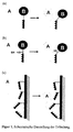
- Figure 1 shows a schematic representation of the invention.
- Adapter segments based on the interaction of proline-rich substances with WW domains and forms derived therefrom are used.
- the adapter segments can be added to the molecules both at the ends (termini) and in the form of insertions.
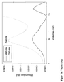
- FIG. 2 shows in (a) a comparison by means of SDS-PAGE of the protein masses and the purification efficiencies of different variants of the polyomavirus protein VP1, the PyVP1-CallS-T249C variant (comparable to the wild type of the protein) and the PyVPI-WW150 variant in which near amino acid position 150, a WW domain is inserted into a loop. Preparation and purification of the variants is described in detail in Example 1. In both variants, usually occurring degradation products of the protein are recorded, which have a smaller molecular mass. (b) circular dichroism spectra (CD) of the PyVP1-WW150 variant and the PyVP1-CallS-T249C variant. The inserted WW domain at position 150 shows native folding, which increases the ⁇ -sheet fraction in the CD spectrum.
- CD circular dichroism spectra
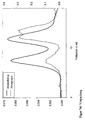
- Figure 3 shows the binding of PyVP1-WW150 to a sensor chip with immobilized proline-rich peptide, according to Example 2.
- the three measurements based on surface plasmon resonance show that the solvent additions have little effect on the affinity and specificity of the interaction, whereas those in (b ) and (c) each represent complex physiological substance mixtures.
- FCS fetal calf serum
- FIG. 4 shows an SDS gel for illustrating the specific binding of PyVPI-WW150 to a proline peptide-containing matrix.
- Lane 1 application VP1-WW150 (purified according to example 1); Lane 2 and lane 3: different wash fractions; Lane 4 and lane 5: elution fractions with 1% SDS in the elution buffer; Lane 6: 10 kDa molecular mass standard.
- the example shows that WW domain-containing proteins can be reversibly immobilized on a matrix.
- the observed double band of the PyVPI variant represents the native protein as well as a protein proteolytic degradation domain that commonly occurs in all preparations.
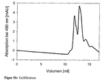
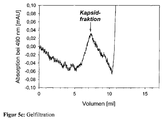
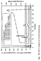
- FIG. 5 shows gel filtration (TSKGel G5000PWXL, TosoHaas) for demonstrating the binding of a proline-rich peptide to the surface of a virus-like capsid, wherein the WW domains inserted into the coat protein VP1 are located on this surface.
- the assembly of the PyVP1-WW150 protein to the capsid is carried out under the conditions given in Example 4.
- a proline-rich peptide can be bound, which is detected by the specific absorption of a dye coupled thereto.
- FIG. 5 a Evidence of capsid formation by absorption at 260 and 280 nm; the capsids elute at a volume of 6 to 8 ml, the non-assembled, free pentamers appear at 9 to 10 ml.
- Figure 5b absorbance of the fluorescently labeled peptide at 490 nm; the elution occurs in parallel to the Kapsidelution and pentamer elution at 6 to 10 ml. Above 10 ml eluted excess free peptide and the fluorescent dye.
- Figure 5c the free, unbound peptide shows no interaction with the matrix and elutes only above 10 ml.
- Figure 5d Overlay of chromatograms 5a and 5b, showing the co-elution of the bound peptide with the capsid fraction.
- FIG. 6 shows in (a) the purification of the variants PyVP1-3C-WW1 and PyVP1-3C-WW [N-14].
- the SDS gel (12%) shows the PyVPI protein without WW domain (lane 2), the PyVP1-WW150 variant from example 1 (lane 3), and the two variants from example 8 (PyVP1-3C-WW1) Lane 4 and lane 5, PyVpl-3C-WW [N-14] on lane 6). Lane 1 and lane 7, molecular mass standard (10 kDa ladder).
- Figure 7 shows the packaging of molecular substances into the interior of virus-like envelopes based on Polyomavirus VP1 variants.
- the peptide is incubated with PyVP1-3C-WW [N-14] and capsids are prepared by assembly under standard conditions. The polyproline peptides are transported via the affinity to the WW domain in the interior of the capsids.
- GFP-PLP is incubated with PyVP1-3C-WW [N-14] and capsids are prepared by assembly under standard conditions. Here, the GFP-PLP is spent on the affinity of the WW domain in the interior of the capsids.
- FIG. 8 shows an SDS gel for the illustration of the purification of proteins with WW domain from a substance mixture, here a cell extract.
- Lane 1 10 kDa molecular mass standard
- Lane 2 crude extract of (PyVP1-3C-WW1) intein-CBD fusion protein
- Lane 3 pass
- Lanes 4 to 10 Different fractions of the elution of the fusion protein, with 2% SDS in the elution buffer.
- the immobilization of the fusion protein takes place via a column with covalently bound proline-rich peptide.
- the column is washed with a total of 10 column volumes of a buffer containing 2 M NaCl.
- degradation products thereof as well as molecular chaperones which are known to bind to PyVP 1, are detected.
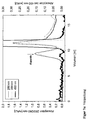
- Figure 9 shows a HPLC fluorometric plot of the disulfide bridging of a proline-rich molecular substance with a WW domain fused to glutathione-S-transferase (GST) for affinity purification (Figure 9a) and under reducing conditions (50 mM DTT, Fig. 9b).
- a linker consisting of alternating Gly-Ser amino acids is inserted before and after the WW domain.
- the viral coat protein used in the given example is the pentomolar polyomavirus VP1 coat protein pentape in solution, which in the prior art can be assembled in vitro to form a virus-like coat. From the crystal structure of the protein, it can be seen that a loop region in the structure near the amino acid position 150 may be suitable for insertion of the WW domain, since this loop region will become a virus-like envelope upon assembly of the pentameric protein the outside of the shell is located.
- PyVPI-WW150 occurs as a fusion protein with a C-terminally fused intein domain and a subsequent chitin-binding domain (CBD).
- CBD chitin-binding domain
- a plasmid is first prepared which is based on the vector pCYB2 of the IMPACT system (New England Biolabs). Using the restriction cleavage sites Ndel-XmaI (New England Biolabs), a DNA fragment produced by PCR amplification is cloned in by the standard method coding for a variant of the VP1 gene of mouse polyomavirus via the multiple cloning site of pCYB2.
- polyomavirus variant which has no cysteines in the sequence; the six cysteines of the wild-type protein were previously replaced by serine using standard mutagenization techniques.
- This variant of PyVP1 has the advantage that the redox conditions of the solution have no influence on the state of the protein; This makes it easier to handle in many applications.
- a cysteine in the inserted WW domain specific disulfide bridging of WW domain and proline-rich sequence can be made.
- Another variant uses a modification at location 249; the threonine present in the wild-type protein is replaced by cysteine.
- the protected localization in the pentamer allows labeling at this site without undesirable side effects.
- the variant of Polyomavirus VP1 used is correctly named PyVPI-CaIIS-T249C, abbreviated hereafter as PyVP1.
- vp1NImp 5'-TAT ACA TAT GGC CCC CAA AAG AAA AAG C-3 '
- vp1CImp 5'-ATA TCC CGG GAG GAA ATA CAG TCT TTG TTT TTC C- 3 '
- the C-terminal amino acids of the wild-type VP1 protein are simultaneously converted from Gly383-Asn384 to Pro383-Gly384, as a C-terminal localized asparagine for the intein cleavage system is very unfavorable in cleavage properties.
- the point mutations mentioned in the following do not influence the essential properties of the PyVPI protein.
- the tac promoter of the pCYB2 vector provides only low expression levels of the fusion protein, therefore, the fusion construct PyVP1-Intein-CBD is isolated by another PCR from the pCYB2 vector and into a highly expressing pET vector with T7lac promoter (plasmid pET21a, Fa Novagen), via NdeI - EcoRI restriction sites.
- the cloning of the WW domain as an insertion into the external loop of PyVP1 between amino acid positions 148 and 149 occurs in several steps.
- FBP11 WWaC 5'-ATA CTC TTC TAC CAC TAC CAT CAT CCG GCT TTT CCC AGG TAG ACT G-3 '
- the oligonucleotides simultaneously introduce a short linker sequence of 5 amino acids each consisting of alternating glycine-serine amino acids.
- a second PCR on the above-described vector amplifies the N-terminal fragment of PyVP1 between amino acids 1 and 148 using the oligonucleotides vp1NImp (see above) and vp1-150-WWaC (5'-ATA CTC TTC AGG TAG CGG CGT AAA CAC AAA AGG AAT TTC CAC TCC AG-3 ').
- a third PCR also amplifies the C-terminal fragment of PyVP1 between amino acids 149 and the C-terminal end of the protein using the oligonucleotides vpl-150 WWaN (5'-ATA CTC TTC AGC CGC TGC CTG TAT CTG TCG GTT TGT TGA ACC CAT G-3 ') and vp1CImp (see above).
- a subsequent PCR with the oligonucleotides vp1NImp and vp1CImp now amplifies the ligation product of the three fragments, hereinafter referred to as PyVP1-WW150.
- the PCR product can then be cloned by standard methods into the vector pCRblunt (Invitrogen). After excision of the cloned fragment PyVP1-WW150 with the help of the restriction enzymes Nde I - Sma I, the final cloning into the previously described plasmid pET21a can be carried out.
- the last resulting vector allows the expression of the fusion protein (PyVP1-WW150) -Inntein-CBD with the help of the highly expressing T7lac promoter in E. coli BL21 (DE3) cells (Novagen).
- fusion protein PyVP1-WW150
- CD34 E. coli BL21 (DE3) cells
- transformed cells in 5 1 - Erlenmeyer flasks, each containing 2 1 LB medium, grown at 37 ° C until the OD 600 of the culture is 2.0 to 2.5. Induction of protein expression is achieved by 1 mM IPTG in the medium. The cultures are then incubated for an additional 20 hours at 15 ° C; the low temperature minimizes cleavage of the intein portion in the fusion protein under in vivo conditions.
- the cells are harvested by centrifugation, dissolved in 70 ml of resuspension buffer (20 mM HEPES, 1 mM EDTA, 100 mM NaCl, 5% (w / v) glycerol, pH 8.0) and digested by high-pressure homogenization. After centrifuging the crude extract for 60 min at 48,000 g, a clear cell extract is obtained. This extract is applied at a flow rate of 0.5 ml / min at a temperature of 10 ° C to a 10 ml chitin affinity column (New England Biolabs).
- the column is then filled with 3 column volumes of resuspension buffer, 15 column volumes of high ionic strength wash buffer (20 mM HEPES, 1mM EDTA, 2M NaCl, 5% (w / v) glycerol, pH 8.0) and again washed 3 column volumes of the resuspension buffer; this will destroy all unwanted E. coli host proteins removed from the chitin matrix.
- Cleavage of the PyVPI WW150 monomer from the fusion protein by self-splicing intein activity is induced by one pulse (3 column volumes) of 50 mM dithiothreitol (DTT), 50 mM hydroxylamine or 30 mM DTT together with 30 mM hydroxylamine in the resuspension buffer.
- DTT dithiothreitol
- the loaded chitin matrix is incubated with one of the indicated solutions for 14 hours at 10 ° C.
- the PyVPI-WW150 protein is completely released and can be separated from the chitin matrix and the remaining constituents of the fusion protein adhering to the matrix by means of standard column chromatographic methods.
- a linear salt gradient with a concentration between 0.1 and 2.0 M NaCl is suitably used.
- the regeneration of the chitin matrix is carried out according to the manufacturer's instructions by washing the chitin material with 3 column volumes of an SDS-containing buffer (1% SDS (w / v) in resuspension buffer).
- the PyVP1 WW150 protein is expressed as a soluble pentamer in the described method and is native.
- 2a shows an SDS gel with the purified fractions of wild-type PyVPI (or the variant PyVP1-CallS-T249C derived therefrom) and the PyVP1-WW150 variant, which has a higher mass due to the additional inserted amino acids.
- 2b shows comparative CD spectra of the proteins produced in 10 mM HEPES, 150 mM NaCl, pH 7.2, which show a correct folding of the protein species. Deconvolution of the two CD spectra of the prior art shows that in the case of the PyVPI WW150 domain an increase in ⁇ -sheet structure over the PyVPI protein is observed. This indicates that the inserted WW domain has retained its native structure as a ⁇ -sheet.
- the example shows that, surprisingly, the WW domain with correct convolution can be inserted under suitable conditions into loop regions of protein structures without significantly disturbing their native structure.
- the solution pentamere PyVP 1 WW150 protein contains the native WW domain inserted into the polypeptide chain and presents it when assembled on the outside of the virus-like envelope (see Example 2).
- a protein can be produced which has artificially inserted a WW domain.
- the binding properties of PyVP1-WW150 with respect to proline-rich ligands can be characterized by various methods.
- a favorable method uses the surface plasmon resonance; in the given example, the device Biacore X (Biacore AB) is used.
- Biacore X Biacore AB
- a synthetic peptide of the sequence Cys-Ser-Gly-Pro-Pro-Pro-Pro-Pro-Pro-Pro-Leu-Pro is coupled to a sensor chip type CM5 via thiol or amino coupling.
- an amount of 80 resonance units (RU) of the indicated peptide is immobilized on the surface.
- the following measurements are carried out in each case at 25 ° C and a flow rate of 20 ul / min.
- Binding studies of PyVP1-WW150 are performed on the sensor chip with immobilized proline-rich peptide under various solvent conditions.
- the first measurement is carried out under standard solvent conditions, with 10 mM HEPES, 1 mM EDTA, 150 mM NaCl, pH 7.2.
- the protein concentration of PyVP1-WW150 is varied in the range of 5 to 50 nM. It can be seen from Fig. 3a that PyVP1-WW150 binds to the sensor chip with high affinity. The amount bound is, as expected, proportional to the protein concentration used.
- the binding constant K D of the PyVPI-WW150 protein is determined to be 5 nM (FIG. 3 a). As can be further seen from the figure, the binding is not permanent, but the protein dissociates again after the loading of the sensor surface in a slow process. This shows that the interaction of the interaction partners is reversible.
- the example shows that the altered solvent conditions have no significant influence on the binding of the PyVP1-WW150 to the proline-rich peptide, and suggests that the interaction of the two partners is stable even under physiological conditions.
- a principle applicability of the system is also possible under clinical conditions in the context of diagnostics or therapeutics.
- FCS fetal calf serum, Gibco Co.
- Figure 3c shows that even under these conditions there is significant and specific binding of the PyVP1 WW150 protein to the sensor surface.
- the response signal at the sensor surface is also proportional to the concentration of PyVP1-WW150 protein used. This demonstrates that the interaction of PyVP1-WW150 with the immobilized proline-rich peptide is independent of the presence of a mixture of other substances, such as those found in serum.
- Another method for characterizing binding properties is a reversible immobilization of the WW domain on an inert matrix.
- a synthetic proline-rich peptide sequence Cys-Ser-Gly-Pro-Pro-Pro-Pro-Pro-Pro-Pro-Pro-Leu-Pro
- Sulfolink column material Pierce
- the matrix modified in this way is used to load a chromatography column. This allows application of the samples to the matrix and elution of bound proteins under different conditions.
- the purified according to Example 1 PyVPI-WW150 protein is applied to the column (solvent 10 mM HEPES, 1 mM EDTA, 150 mM NaCl, 5% glycerol, pH 7.2).
- solvent 10 mM HEPES, 1 mM EDTA, 150 mM NaCl, 5% glycerol, pH 7.2 the protein binds to the matrix and appears only in small amounts in the washing fractions. Subsequent elution of the protein from the matrix is possible by adding 1% SDS or 300 mM arginine to the running buffer.
- virus-like capsids In a further experiment, the binding of a fluorescently-labeled peptide with a proline-rich sequence to the surface (outside) of virus-like capsids is investigated.
- the assembly of the protein takes place in analogy to the conditions already described in the prior art (compare Salunke, Caspar & Garcea, Polymorphism in the assembly of polyomavirus capsid protein VP1, Biophys, J. 56, pp. 887-900, 1989).
- the virus-like capsids are after dialysis of the protein against 10 mM HEPES, 50 mM NaCl, 0.5 mM CaCl 2 , 5% glycerol, pH 7.2.
- the proline-rich peptide Cys-Ser-Gly-Pro-Pro-Pro-Pro-Pro-Pro-Leu-Pro is with a fluorescein-maleimide derivative (Molecular Probes) according to the manufacturer specific to the N-terminal cysteine marked.
- Molecular Probes Fluorescein-maleimide derivative
- Gel filtration (column TSKGel G5000PWXL, TosoHaas) clearly identifies virus-like capsid shells and separates them from free, unassembled capsid building blocks as well as from excess peptide and fluorescent dye.
- FOG. 5 This example shows that the PyVP1-WW150 variant can form capsid structures (virus-like envelopes) under suitable conditions. These capsids are able to bind the proline-rich peptide. In this way, molecular substances can be directed onto the surface (outer side) of the virus-like structures via the specific and strong interaction of WW domain and proline-rich sequence.
- two different variants of the PyVPI protein are produced which contain a WW domain at the amino terminus of the native wild-type protein (PyVP1-3C-WW1 variant), or carry the WW domain at an N-terminus truncated by 14 amino acids (Variant PyVP1-3C-WW [N-14]) and a variant of the PyVPI protein carrying a proline-rich sequence at the N-terminus (PyVP1-3C- [N-14] -PLP).
- the basis for these variants is a PyVP1 variant which has the cysteines C19 and C114 and into which a specific new cysteine has additionally been introduced (analogous to the variant PyVP1-CallS-T249C). This variant is abbreviated below to PyVP1-3C.
- an amplification of the WW domain is performed by PCR;
- the template used is analogous to Example 1, the mouse FBP11 gene.
- the oligonucleotides used for the PCR are 5'-AAT ATA TCA TAT GTC CAT CAT CCG GCT TTT CCC AGG TAG ACT-3 '(with NdeI site), and 5'-TAT TAA TCA TAT GAG CGG CTG GAC AGA ACA TAA ATC ACC TGA TGG-3 'used.
- the resulting PCR product is then cloned into the expression vector pET21a from Example 1, which contains the gene for a fusion protein PyVP1-intein CBD, via the Nde I-Nde I introduced by means of the oligonucleotides; at the 5 'end of the gene is a singular Nde I - interface.
- the expressed gene product of this vector is the desired protein PyVP1-3C-WW1.
- cloning is performed with a 14 amino acid truncated fragment of PyVP1-3C (PyVPI-3C-WW [N-14]) following the standard procedure described in Example 1.
- a PCR is carried out on the PyVP1 gene fragment, with 5'-GCG CGC GCA TAT GAG CAC CAA GGC TAG CCC AAG ACC CG-3 'and the oligonucleotide vp1CImp (see Example 1).
- the resulting PCR product is digested with the restriction enzymes Nde I - Sma I and the fragment cloned by standard methods in the vector pET21 a from Example 1.
- Expression and purification of the two proteins is carried out in accordance with Example 1.
- the purified proteins are compared to the variants PyVP1 and PyVP1-WW150 in FIG. 6a. It turns out that the proteins are soluble and native to produce.
- the alteration of the N-terminus by the introduction of the WW domains has no significant negative influence on the assembling competence of the protein for the formation of virus-like enveloping structures.
- GFP is a protein that shows a green fluorescence in the native state (absorption maximum at 490 nm). It is ideal for marking complexes and associates.
- a PCR-based amplification of the GFP gene is initially carried out, using as template the plasmid pEGFP-N1 (Clontech). At the same time, suitable restriction sites are introduced into the PCR product.
- the PCR is carried out using the oligonucleotides 5'-TTA TTT ACA TAT GGT GAG CAA GGG CGA GGA G-3 '(with Nde I interface), and 5'-ATA TCT TAA GTA CAG CTC GTC CAT GCC G-3' (with Afl II interface).
- the resulting PCR product is cloned via the restriction sites in the vector pTIP and expressed there.
- This vector pTIP is a derivative of the Intein purification vector based on pET21a documented in Example 1, with additionally introduced proline-rich sequences.
- the vector is designed so that a proline-rich sequence is optionally fused at the 5 'or 3' end of a gene inserted through a multiple cloning site.
- the proline-rich sequence predominantly contains Pro-Pro-Pro-Pro-Pro-Pro-Pro-Pro-Leu-Pro.
- the preparation and purification of the GFP-PLP protein is carried out by means of chitin affinity chromatography, in accordance with the content of Example 1.
- the successful production and purification of GFP-PLP is documented in FIG. 6b.
- the GFP-PLP protein which carries the proline-rich sequence Pro-Pro-Pro-Pro Pro-Pro-Pro-Pro-Leu-Pro at the C-terminus, can be prepared in soluble form in large quantities.
- the green glowing color of the protein solution also shows that the protein can fold to its native structure.
- Py-VP1-3C-PLP variant Analogously, the preparation of the Py-VP1-3C-PLP variant was carried out.
- PyVP1-3C- [N-14] was cloned into the vector pTIP such that the proline-rich sequence contained in the vector was N-terminally fused to the Py-VP1-3C- [N-14].
- the two variants are incubated with proteins containing proline-rich sequences.
- the PyVP1-3C-WW1 protein is incubated with the previously prepared protein GFP-PLP (molar ratio 1: 6) for 10 min (10 mM HEPES, 1 mM EDTA, 150 mM NaCl, 5% glycerol, pH 7.2), and the capsid formation of the PyVPI variants is induced by dialysis against a buffer containing 0.5 mM CaCl 2 (see Example 4).
- the successful detection of capsids (Figure 7a) shows that the variant PyVP1-3C-WW1 is competent for assembly under suitable conditions.
- the protein is able to bind the proline-rich peptide and, when assembled into a protein shell, spend the proline-rich peptide inside the capsids. This is shown in the gel filtration in FIG. 7b by the specific absorption of the covalently bonded to the peptide fluorescent dye at 490 nm, which is mainly to be found in the elution range of the capsids (9 to 10 ml).
- an analogous assembly experiment can be performed using the variants of PyVP1-3C- [N-14] -PLP (proline-rich sequence at the N-terminus) and GFP-WW1 (WW-domain at the N-terminus).
- the experiments in Examples 5 and 6 show that variants of PyVP1 with N-terminally fused WW domain are capable of binding proline-rich sequences and these and the molecular substances which may be present on them when being assembled into capsids under suitable conditions To direct conditions into the interior of virus-like envelopes.
- the described method is suitable for effecting directional packaging of molecular substances into viruses or virus-like capsids. It has also been shown that variants of PyVP1 with N-terminally fused proline-rich sequence are able to bind WW domains and molecular substances located thereon.
- the interaction of the two adapter segments is thereby unlimited in time, since disulfide bridges are stable under physiological conditions, as present for example in extracellular space. If desired, in vitro under reducing conditions (for example 50 mM DTT, DTE or ⁇ -mercaptoethanol), the disulfide bridge between WW domain and proline-rich ligand can be reversed again; when the reducing agent is removed, reclosure is also possible.
- reducing conditions for example 50 mM DTT, DTE or ⁇ -mercaptoethanol
- an aspartate amino acid (position 8 in the WW domain) is converted into a cysteine by mutagenesis.
- the resulting cysteine-containing variant is named PyVP1-WW150-D8C in the following. Binding studies based on surface plasmon resonance show that this variant of the WW domain, without formation of the disulfide bridge, does not bind the proline-rich ligand Cys-Ser-Gly-Pro-Pro-Pro-Pro-Pro-Pro-Pro-Pro-Leu. Pro binds. However, the extent of interaction is somewhat lower than for PyVPI-WW150. This seems to be due to the newly introduced cysteine.
- a buffer which contains both ammonium sulfate and also maintains defined redox conditions.
- the latter conditions are achieved by using 1 mM GSSG and 5 mM GSH in the redox buffer (50 mM Tris, pH 8.5, 1 mM EDTA, 500 mM ammonium sulfate); oxidized (GSSG) or reduced glutathione (GSH) acts as a redox-shuffling system to form the disulfide bridges (see R. Rudolph, In vitro folding of inclusion body proteins, FASEB J. 10, 49-56, 1996).
- the disulfide bridging is carried out at 15 ° C for 24 h and terminated by dialysis against 50 mM Tris, 1 mM EDTA, pH 7. Under the latter conditions no disulfide exchange takes place; the formed disulfide bridges are stable.
- cysteine amino acid residues into the WW domain allows the covalent bridging of polyproline-rich ligands carrying at least one cysteine with the WW domain and thus leads to a stable covalent linking of WW domain and ligand (see Figure 9).
- Another field of application of the present invention is the separation of molecular substances from substance mixtures, as typically occurs in the purification of proteins from crude extracts (cell extracts).
- the affinity of the WW domain to proline-rich ligands is exploited to isolate proteins containing a WW domain from a complex mixture (crude extract) of proteins (principle of affinity chromatography).
- a column according to Example 3 is used; the sulfo link material (Pierce, the reactivity of the matrix with SH groups based on the iodoacetamide group at the end of a linker, consisting of 10 CH 2 groups) is thereby via a thiol coupling with the peptide Cys-Ser-Gly- Load Pro-Pro-Pro-Pro-Pro-Pro-Pro-Pro-Leu-Pro according to the manufacturer's instructions.
- the sulfo link material Pierce, the reactivity of the matrix with SH groups based on the iodoacetamide group at the end of a linker, consisting of 10 CH 2 groups
- the coupling of the peptide to other matrices is possible, for example, AffiGel 10 (Biorad, the reactivity of the matrix with NH 2 groups based on the N-hydroxysuccinimide group at the end of a linker consisting of 10 CH 2 groups ) via the N-terminus of the peptide.
- the peptide coupling or the N-terminus of the peptide can be attached to a matrix based on CH-Sepharose 4B (from Sigma, the reactive group of the matrix is likewise an N-hydroxysuccinimide ester).
- covalent binding of the proline-rich ligand to a support material results, which subsequently allows purification of WW domain proteins.
- the PyVPI variant PyVP1-3C-WW1 from Example 5 (WW domain at the N-terminus of the PyVP1 protein) is prepared analogously to the information from Example 1 as a fusion protein with an intein and a chitin-binding domain ([PyVP1-3C-] WW1] -intein-CBD). However, it is not purified by the standard method described by chitin affinity chromatography. Instead, the cell extract is applied to the column described above after cell disruption and subsequent centrifugation.
- the running buffer used is 10 mM HEPES, 150 mM NaCl, 1 mM EDTA, 5% glycerol, pH 8.0.
- binding of the fusion protein [PyVP1-3C-WW1] -Inntein-CBD takes place via the interaction of the WW domain with the immobilized proline-rich peptide on the SulfoLink matrix.
- the fusion protein is bound to the matrix and thus most other proteins of the cell extract are separated during the run or during the washing process.
- SDS elution then provides almost exclusively the complete WW domain-containing fusion protein, as well as proteolytic degradation products thereof (which in the case of PyVP1 occur in all comparable prior art preparation methods) and molecular chaperones known to bind directly to PyVP1 assets and usually can not be separated.
- this example shows that it is possible with the described system specifically to separate molecules from a substance mixture (crude extract) and to purify it.
- a variant of GFP is produced analogously to the preparation of PyVP1 using the intein-based expression system from Example 1 with a WW domain at the N-terminus of the GFP (GFP-WW1).
- a PCR is performed on the vector pEGFP-N1 (Clontech, see Example 5) with the oligonucleotides 5'-TAT AGC TAG CGT GAG CAA GGG CGA GGA GCT GTT C-3 'and 5'-GGG AAT TAA GTA CAG CTC GTC CAT GCC G-3 '.
- the PCR product is ligated via the interfaces Nhe I - Sma I into the vector pET21a from Example 5, which contains at the 3 'end of the insertion site the fusion protein of intein and chitin binding domain (CBD) described in Example 1.
- CBD chitin binding domain
- On the 5 'side of the insertion site is the WW domain described in Example 5.
- the plasmid encoding the fusion protein can be inserted into strain E. coli BL21 (DE3). Analogously to Examples 1 and 3, the preparation and purification of the fusion protein can then take place. With the described method, the protein GFP-WWI can be prepared in purified form.
- a second variant of GFP is analogously produced with a proline-rich segment (Pro-Pro-Pro-Pro-Pro-Pro-Pro-Pro-Leu-Pro) at the C-terminus.
- the preparation and purification of the GFP-PLP protein is identical to the description of the protein in Example 5.
- the two GFP variants are then incubated together.
- the two proteins are brought into contact with each other via the adapter segments, resulting in a GFP dimer which can be distinguished from the GFP monomer by gel filtration on a TSK-PW2000XL gel filtration column (Tosahas company).
Landscapes
- Chemical & Material Sciences (AREA)
- Health & Medical Sciences (AREA)
- Organic Chemistry (AREA)
- Life Sciences & Earth Sciences (AREA)
- Biochemistry (AREA)
- Medicinal Chemistry (AREA)
- General Health & Medical Sciences (AREA)
- Proteomics, Peptides & Aminoacids (AREA)
- Molecular Biology (AREA)
- Biophysics (AREA)
- Genetics & Genomics (AREA)
- Analytical Chemistry (AREA)
- Gastroenterology & Hepatology (AREA)
- Virology (AREA)
- Hematology (AREA)
- Chemical Kinetics & Catalysis (AREA)
- General Chemical & Material Sciences (AREA)
- Engineering & Computer Science (AREA)
- Animal Behavior & Ethology (AREA)
- Zoology (AREA)
- Toxicology (AREA)
- Bioinformatics & Cheminformatics (AREA)
- Pharmacology & Pharmacy (AREA)
- Epidemiology (AREA)
- Cell Biology (AREA)
- Public Health (AREA)
- Veterinary Medicine (AREA)
- Peptides Or Proteins (AREA)
- Steroid Compounds (AREA)
- Addition Polymer Or Copolymer, Post-Treatments, Or Chemical Modifications (AREA)
- Medicines That Contain Protein Lipid Enzymes And Other Medicines (AREA)
Applications Claiming Priority (3)
| Application Number | Priority Date | Filing Date | Title |
|---|---|---|---|
| DE19952956A DE19952956A1 (de) | 1999-11-03 | 1999-11-03 | Verfahren zur Verbindung von molekularen Substanzen |
| DE19952956 | 1999-11-03 | ||
| PCT/EP2000/010873 WO2001032684A2 (de) | 1999-11-03 | 2000-11-03 | Verfahren zur verbindung von molekularen substanzen basierend auf der affinität der prolinreichen aminosäure-sequenzen und proteindomänem vom typ ww |
Publications (2)
| Publication Number | Publication Date |
|---|---|
| EP1227848A2 EP1227848A2 (de) | 2002-08-07 |
| EP1227848B1 true EP1227848B1 (de) | 2006-05-24 |
Family
ID=7927813
Family Applications (1)
| Application Number | Title | Priority Date | Filing Date |
|---|---|---|---|
| EP00984960A Expired - Lifetime EP1227848B1 (de) | 1999-11-03 | 2000-11-03 | Verfahren zur verbindung von molekularen substanzen |
Country Status (8)
| Country | Link |
|---|---|
| US (1) | US7807782B2 (https=) |
| EP (1) | EP1227848B1 (https=) |
| JP (1) | JP4112859B2 (https=) |
| CN (1) | CN100357314C (https=) |
| AT (1) | ATE326984T1 (https=) |
| AU (1) | AU2154801A (https=) |
| DE (2) | DE19952956A1 (https=) |
| WO (1) | WO2001032684A2 (https=) |
Families Citing this family (20)
| Publication number | Priority date | Publication date | Assignee | Title |
|---|---|---|---|---|
| JP4885476B2 (ja) * | 2004-05-21 | 2012-02-29 | 株式会社日本触媒 | タンパク質及び/又はペプチドの細胞内導入方法 |
| US20090304719A1 (en) | 2007-08-22 | 2009-12-10 | Patrick Daugherty | Activatable binding polypeptides and methods of identification and use thereof |
| RU2636046C2 (ru) | 2009-01-12 | 2017-11-17 | Сайтомкс Терапьютикс, Инк | Композиции модифицированных антител, способы их получения и применения |
| AU2010215761B2 (en) | 2009-02-23 | 2017-04-06 | Cytomx Therapeutics, Inc | Proproteins and methods of use thereof |
| JP6236948B2 (ja) * | 2013-07-17 | 2017-11-29 | 東ソー株式会社 | 抗体精製用溶出液および当該溶出液を用いた抗体精製方法 |
| GB201409558D0 (en) | 2014-05-29 | 2014-07-16 | Ucb Biopharma Sprl | Method |
| GB201412658D0 (en) | 2014-07-16 | 2014-08-27 | Ucb Biopharma Sprl | Molecules |
| GB201412659D0 (en) | 2014-07-16 | 2014-08-27 | Ucb Biopharma Sprl | Molecules |
| GB201601077D0 (en) | 2016-01-20 | 2016-03-02 | Ucb Biopharma Sprl | Antibody molecule |
| GB201601075D0 (en) | 2016-01-20 | 2016-03-02 | Ucb Biopharma Sprl | Antibodies molecules |
| GB201601073D0 (en) | 2016-01-20 | 2016-03-02 | Ucb Biopharma Sprl | Antibodies |
| GB201521391D0 (en) | 2015-12-03 | 2016-01-20 | Ucb Biopharma Sprl | Antibodies |
| GB201521383D0 (en) | 2015-12-03 | 2016-01-20 | Ucb Biopharma Sprl And Ucb Celltech | Method |
| GB201521389D0 (en) | 2015-12-03 | 2016-01-20 | Ucb Biopharma Sprl | Method |
| GB201521382D0 (en) | 2015-12-03 | 2016-01-20 | Ucb Biopharma Sprl | Antibodies |
| GB201521393D0 (en) | 2015-12-03 | 2016-01-20 | Ucb Biopharma Sprl | Antibodies |
| US11202841B2 (en) * | 2018-08-20 | 2021-12-21 | The Trustees Of Indiana University | Virus-enabled targeted vector for imaging |
| CN111979206B (zh) * | 2019-05-24 | 2021-08-17 | 深圳瑞德林生物技术有限公司 | 固定化融合酶及用其制备谷胱甘肽的方法 |
| WO2023032994A1 (ja) * | 2021-08-31 | 2023-03-09 | 富士フイルム株式会社 | 化合物及びこれを用いた標識生体物質 |
| CN115951051B (zh) * | 2022-10-18 | 2024-01-12 | 北京卓诚惠生生物科技股份有限公司 | 一种高灵敏度新型冠状病毒抗原胶体金检测试剂盒及其制备方法 |
Family Cites Families (5)
| Publication number | Priority date | Publication date | Assignee | Title |
|---|---|---|---|---|
| US6072039A (en) | 1991-04-19 | 2000-06-06 | Rohm And Haas Company | Hybrid polypeptide comparing a biotinylated avidin binding polypeptide fused to a polypeptide of interest |
| US6503703B1 (en) * | 1995-05-19 | 2003-01-07 | Mount Sinai School Of Medicine Of New York University | Identification and use of antiviral compounds that inhibit interaction of host cell proteins and viral proteins required for viral replication |
| US6007544A (en) * | 1996-06-14 | 1999-12-28 | Beth Israel Deaconess Medical Center | Catheter apparatus having an improved shape-memory alloy cuff and inflatable on-demand balloon for creating a bypass graft in-vivo |
| US7666221B2 (en) * | 2000-05-01 | 2010-02-23 | Endovascular Technologies, Inc. | Lock modular graft component junctions |
| US7044962B2 (en) * | 2002-06-25 | 2006-05-16 | Scimed Life Systems, Inc. | Implantable prosthesis with displaceable skirt |
-
1999
- 1999-11-03 DE DE19952956A patent/DE19952956A1/de not_active Withdrawn
-
2000
- 2000-11-03 EP EP00984960A patent/EP1227848B1/de not_active Expired - Lifetime
- 2000-11-03 CN CNB008153167A patent/CN100357314C/zh not_active Expired - Fee Related
- 2000-11-03 AU AU21548/01A patent/AU2154801A/en not_active Abandoned
- 2000-11-03 DE DE50012831T patent/DE50012831D1/de not_active Expired - Lifetime
- 2000-11-03 AT AT00984960T patent/ATE326984T1/de not_active IP Right Cessation
- 2000-11-03 WO PCT/EP2000/010873 patent/WO2001032684A2/de not_active Ceased
- 2000-11-03 JP JP2001535383A patent/JP4112859B2/ja not_active Expired - Fee Related
-
2006
- 2006-06-01 US US11/446,587 patent/US7807782B2/en not_active Expired - Fee Related
Also Published As
| Publication number | Publication date |
|---|---|
| AU2154801A (en) | 2001-05-14 |
| EP1227848A2 (de) | 2002-08-07 |
| US20060252130A1 (en) | 2006-11-09 |
| WO2001032684A2 (de) | 2001-05-10 |
| DE50012831D1 (de) | 2006-06-29 |
| ATE326984T1 (de) | 2006-06-15 |
| CN100357314C (zh) | 2007-12-26 |
| DE19952956A1 (de) | 2001-05-17 |
| WO2001032684A3 (de) | 2002-04-18 |
| JP2003517300A (ja) | 2003-05-27 |
| CN1390140A (zh) | 2003-01-08 |
| JP4112859B2 (ja) | 2008-07-02 |
| US7807782B2 (en) | 2010-10-05 |
Similar Documents
| Publication | Publication Date | Title |
|---|---|---|
| EP1227848B1 (de) | Verfahren zur verbindung von molekularen substanzen | |
| DE69010206T2 (de) | Stabilisierte protein- oder peptidkonjugate. | |
| DE60033405T2 (de) | Chimäre polypeptide, verfahren zur herstellung und verwendung dafür | |
| DE69233068T2 (de) | Serin-reiche peptidlinker | |
| DE69633175T2 (de) | Multimere proteine | |
| DE10235248B4 (de) | Fusionsprotein mit verstärkter in vivo-Aktivität von Erythropoietin | |
| CN109400693A (zh) | 新型proNGF突变体及其在生产β-NGF中的用途 | |
| DE19618797C2 (de) | Vehikel zum Transport molekularer Substanz | |
| EP1427837A2 (de) | Modulare transfektionssysteme auf der basis von nukleoproteinfilamenten | |
| US20070054401A1 (en) | Composition for intracellular transport of biological particles or macromolecules | |
| DE4344350C2 (de) | Bakterien zur Herstellung stabiler Fusionsproteine und Verfahren zu deren Nachweis | |
| CN120399090A (zh) | 融合促溶短肽和细胞穿透肽的重组狂犬病病毒纳米抗体及在制备狂犬病防治产品中的应用 | |
| EP1232257B1 (de) | Modulare transportsysteme für molekulare substanzen und deren herstellung und verwendung | |
| DE19916224C1 (de) | Synthetisches biologisch aktives Molekül | |
| DE68925044T2 (de) | Herstellung von Fusionsproteinen oder -polypeptiden | |
| DE60305940T2 (de) | Modifizierte therapeutische mittel | |
| EP0453456B1 (de) | Pdgf-a, pdgf-aa, pdgf-ab, herstellungsverfahren und sie enthaltende arzneimittel | |
| EP1228199B1 (de) | Verfahren zur gerichteten verpackung von molekularen substanzen in proteinhüllen | |
| AT511130A2 (de) | Polypeptidmaterial mit flexiblen Poreneigenschaften | |
| DE10238846A1 (de) | Aktive Fusionsproteine und Verfahren zu ihrer Herstellung | |
| DE19735105A1 (de) | Transportsystem zur Einbringung von Proteinen in Zielzellen mit Hilfe eines Fusionsproteins, Nucleinsäurekonstrukte kodierend für die Komponenten des Transportsystems und Arzneimittel, die Komponenten des Transportsystems umfassen | |
| WO2005059129A2 (de) | Adapter zum ankoppeln einer an einer zelloberfläche anzukoppelnden substanz | |
| WO2025084336A1 (ja) | Aavのキャプシドと標的受容体に同時に結合することができる分子 | |
| DE4041464A1 (de) | 5-ht(pfeil abwaerts)1(pfeil abwaerts)-rezeptor | |
| DE102015108849A1 (de) | Verfahren zur Proteinimmobilisierung mittels modifizierter DNase-Domänen |
Legal Events
| Date | Code | Title | Description |
|---|---|---|---|
| PUAI | Public reference made under article 153(3) epc to a published international application that has entered the european phase |
Free format text: ORIGINAL CODE: 0009012 |
|
| 17P | Request for examination filed |
Effective date: 20020528 |
|
| AK | Designated contracting states |
Kind code of ref document: A2 Designated state(s): AT BE CH CY DE DK ES FI FR GB GR IE IT LI LU MC NL PT SE TR |
|
| AX | Request for extension of the european patent |
Free format text: AL;LT PAYMENT 20020528;LV;MK;RO PAYMENT 20020528;SI |
|
| RIN1 | Information on inventor provided before grant (corrected) |
Inventor name: BOEHM, GERALD Inventor name: PARTHIER, CHRISTOPH Inventor name: GUENTHER, CONSTANZE Inventor name: SCHMIDT, ULRICH |
|
| RIN1 | Information on inventor provided before grant (corrected) |
Inventor name: GUENTHER, CONSTANZE Inventor name: PARTHIER, CHRISTOPH Inventor name: SCHMIDT, ULRICH Inventor name: BOEHM, GERALD |
|
| 17Q | First examination report despatched |
Effective date: 20040608 |
|
| GRAP | Despatch of communication of intention to grant a patent |
Free format text: ORIGINAL CODE: EPIDOSNIGR1 |
|
| GRAS | Grant fee paid |
Free format text: ORIGINAL CODE: EPIDOSNIGR3 |
|
| GRAA | (expected) grant |
Free format text: ORIGINAL CODE: 0009210 |
|
| AK | Designated contracting states |
Kind code of ref document: B1 Designated state(s): AT BE CH CY DE DK ES FI FR GB GR IE IT LI LU MC NL PT SE TR |
|
| AX | Request for extension of the european patent |
Extension state: LT RO |
|
| PG25 | Lapsed in a contracting state [announced via postgrant information from national office to epo] |
Ref country code: IT Free format text: LAPSE BECAUSE OF FAILURE TO SUBMIT A TRANSLATION OF THE DESCRIPTION OR TO PAY THE FEE WITHIN THE PRESCRIBED TIME-LIMIT;WARNING: LAPSES OF ITALIAN PATENTS WITH EFFECTIVE DATE BEFORE 2007 MAY HAVE OCCURRED AT ANY TIME BEFORE 2007. THE CORRECT EFFECTIVE DATE MAY BE DIFFERENT FROM THE ONE RECORDED. Effective date: 20060524 Ref country code: IE Free format text: LAPSE BECAUSE OF FAILURE TO SUBMIT A TRANSLATION OF THE DESCRIPTION OR TO PAY THE FEE WITHIN THE PRESCRIBED TIME-LIMIT Effective date: 20060524 Ref country code: FI Free format text: LAPSE BECAUSE OF FAILURE TO SUBMIT A TRANSLATION OF THE DESCRIPTION OR TO PAY THE FEE WITHIN THE PRESCRIBED TIME-LIMIT Effective date: 20060524 Ref country code: NL Free format text: LAPSE BECAUSE OF FAILURE TO SUBMIT A TRANSLATION OF THE DESCRIPTION OR TO PAY THE FEE WITHIN THE PRESCRIBED TIME-LIMIT Effective date: 20060524 |
|
| REG | Reference to a national code |
Ref country code: GB Ref legal event code: FG4D Free format text: NOT ENGLISH |
|
| REG | Reference to a national code |
Ref country code: CH Ref legal event code: EP |
|
| REG | Reference to a national code |
Ref country code: IE Ref legal event code: FG4D Free format text: LANGUAGE OF EP DOCUMENT: GERMAN |
|
| REF | Corresponds to: |
Ref document number: 50012831 Country of ref document: DE Date of ref document: 20060629 Kind code of ref document: P |
|
| PG25 | Lapsed in a contracting state [announced via postgrant information from national office to epo] |
Ref country code: DK Free format text: LAPSE BECAUSE OF FAILURE TO SUBMIT A TRANSLATION OF THE DESCRIPTION OR TO PAY THE FEE WITHIN THE PRESCRIBED TIME-LIMIT Effective date: 20060824 Ref country code: SE Free format text: LAPSE BECAUSE OF FAILURE TO SUBMIT A TRANSLATION OF THE DESCRIPTION OR TO PAY THE FEE WITHIN THE PRESCRIBED TIME-LIMIT Effective date: 20060824 |
|
| GBT | Gb: translation of ep patent filed (gb section 77(6)(a)/1977) |
Effective date: 20060808 |
|
| PG25 | Lapsed in a contracting state [announced via postgrant information from national office to epo] |
Ref country code: ES Free format text: LAPSE BECAUSE OF FAILURE TO SUBMIT A TRANSLATION OF THE DESCRIPTION OR TO PAY THE FEE WITHIN THE PRESCRIBED TIME-LIMIT Effective date: 20060904 |
|
| PG25 | Lapsed in a contracting state [announced via postgrant information from national office to epo] |
Ref country code: PT Free format text: LAPSE BECAUSE OF FAILURE TO SUBMIT A TRANSLATION OF THE DESCRIPTION OR TO PAY THE FEE WITHIN THE PRESCRIBED TIME-LIMIT Effective date: 20061024 |
|
| LTIE | Lt: invalidation of european patent or patent extension |
Effective date: 20060524 |
|
| NLV1 | Nl: lapsed or annulled due to failure to fulfill the requirements of art. 29p and 29m of the patents act | ||
| ET | Fr: translation filed | ||
| PG25 | Lapsed in a contracting state [announced via postgrant information from national office to epo] |
Ref country code: BE Free format text: LAPSE BECAUSE OF NON-PAYMENT OF DUE FEES Effective date: 20061130 Ref country code: LI Free format text: LAPSE BECAUSE OF NON-PAYMENT OF DUE FEES Effective date: 20061130 Ref country code: MC Free format text: LAPSE BECAUSE OF NON-PAYMENT OF DUE FEES Effective date: 20061130 Ref country code: CH Free format text: LAPSE BECAUSE OF NON-PAYMENT OF DUE FEES Effective date: 20061130 |
|
| REG | Reference to a national code |
Ref country code: IE Ref legal event code: FD4D |
|
| PLBE | No opposition filed within time limit |
Free format text: ORIGINAL CODE: 0009261 |
|
| STAA | Information on the status of an ep patent application or granted ep patent |
Free format text: STATUS: NO OPPOSITION FILED WITHIN TIME LIMIT |
|
| 26N | No opposition filed |
Effective date: 20070227 |
|
| REG | Reference to a national code |
Ref country code: CH Ref legal event code: PL |
|
| BERE | Be: lapsed |
Owner name: ACGT PROGENOMICS A.G. Effective date: 20061130 |
|
| PG25 | Lapsed in a contracting state [announced via postgrant information from national office to epo] |
Ref country code: AT Free format text: LAPSE BECAUSE OF NON-PAYMENT OF DUE FEES Effective date: 20061103 |
|
| PG25 | Lapsed in a contracting state [announced via postgrant information from national office to epo] |
Ref country code: GR Free format text: LAPSE BECAUSE OF FAILURE TO SUBMIT A TRANSLATION OF THE DESCRIPTION OR TO PAY THE FEE WITHIN THE PRESCRIBED TIME-LIMIT Effective date: 20060825 |
|
| PG25 | Lapsed in a contracting state [announced via postgrant information from national office to epo] |
Ref country code: LU Free format text: LAPSE BECAUSE OF NON-PAYMENT OF DUE FEES Effective date: 20061103 Ref country code: TR Free format text: LAPSE BECAUSE OF FAILURE TO SUBMIT A TRANSLATION OF THE DESCRIPTION OR TO PAY THE FEE WITHIN THE PRESCRIBED TIME-LIMIT Effective date: 20060524 |
|
| PG25 | Lapsed in a contracting state [announced via postgrant information from national office to epo] |
Ref country code: CY Free format text: LAPSE BECAUSE OF FAILURE TO SUBMIT A TRANSLATION OF THE DESCRIPTION OR TO PAY THE FEE WITHIN THE PRESCRIBED TIME-LIMIT Effective date: 20060524 |
|
| PGFP | Annual fee paid to national office [announced via postgrant information from national office to epo] |
Ref country code: FR Payment date: 20101123 Year of fee payment: 11 |
|
| PGFP | Annual fee paid to national office [announced via postgrant information from national office to epo] |
Ref country code: DE Payment date: 20101130 Year of fee payment: 11 |
|
| PGFP | Annual fee paid to national office [announced via postgrant information from national office to epo] |
Ref country code: GB Payment date: 20101110 Year of fee payment: 11 |
|
| GBPC | Gb: european patent ceased through non-payment of renewal fee |
Effective date: 20111103 |
|
| REG | Reference to a national code |
Ref country code: FR Ref legal event code: ST Effective date: 20120731 |
|
| REG | Reference to a national code |
Ref country code: DE Ref legal event code: R119 Ref document number: 50012831 Country of ref document: DE Effective date: 20120601 |
|
| PG25 | Lapsed in a contracting state [announced via postgrant information from national office to epo] |
Ref country code: GB Free format text: LAPSE BECAUSE OF NON-PAYMENT OF DUE FEES Effective date: 20111103 |
|
| PG25 | Lapsed in a contracting state [announced via postgrant information from national office to epo] |
Ref country code: FR Free format text: LAPSE BECAUSE OF NON-PAYMENT OF DUE FEES Effective date: 20111130 |
|
| PG25 | Lapsed in a contracting state [announced via postgrant information from national office to epo] |
Ref country code: DE Free format text: LAPSE BECAUSE OF NON-PAYMENT OF DUE FEES Effective date: 20120601 |