EP0817968B1 - Methods for detecting colon cancer from stool samples - Google Patents
Methods for detecting colon cancer from stool samples Download PDFInfo
- Publication number
- EP0817968B1 EP0817968B1 EP96946136A EP96946136A EP0817968B1 EP 0817968 B1 EP0817968 B1 EP 0817968B1 EP 96946136 A EP96946136 A EP 96946136A EP 96946136 A EP96946136 A EP 96946136A EP 0817968 B1 EP0817968 B1 EP 0817968B1
- Authority
- EP
- European Patent Office
- Prior art keywords
- sample
- cells
- stool
- allele
- methods
- Prior art date
- Legal status (The legal status is an assumption and is not a legal conclusion. Google has not performed a legal analysis and makes no representation as to the accuracy of the status listed.)
- Expired - Lifetime
Links
- 238000000034 method Methods 0.000 title claims abstract description 82
- 206010009944 Colon cancer Diseases 0.000 title description 37
- 208000029742 colonic neoplasm Diseases 0.000 title description 10
- 230000001413 cellular effect Effects 0.000 claims abstract description 22
- 238000012216 screening Methods 0.000 claims abstract description 5
- 239000000523 sample Substances 0.000 claims description 199
- 210000004027 cell Anatomy 0.000 claims description 71
- 108700028369 Alleles Proteins 0.000 claims description 62
- 238000003556 assay Methods 0.000 claims description 50
- 108020004414 DNA Proteins 0.000 claims description 38
- 230000035772 mutation Effects 0.000 claims description 30
- 206010028980 Neoplasm Diseases 0.000 claims description 26
- 201000011510 cancer Diseases 0.000 claims description 23
- 230000003902 lesion Effects 0.000 claims description 20
- 238000012217 deletion Methods 0.000 claims description 16
- 230000037430 deletion Effects 0.000 claims description 16
- 239000000872 buffer Substances 0.000 claims description 12
- 230000008774 maternal effect Effects 0.000 claims description 12
- 230000008775 paternal effect Effects 0.000 claims description 12
- 108090000623 proteins and genes Proteins 0.000 claims description 11
- 208000032818 Microsatellite Instability Diseases 0.000 claims description 10
- 210000002919 epithelial cell Anatomy 0.000 claims description 6
- 102000012406 Carcinoembryonic Antigen Human genes 0.000 claims description 5
- 108010022366 Carcinoembryonic Antigen Proteins 0.000 claims description 5
- 108091005804 Peptidases Proteins 0.000 claims description 5
- 102000035195 Peptidases Human genes 0.000 claims description 5
- 108010040002 Tumor Suppressor Proteins Proteins 0.000 claims description 5
- 102000001742 Tumor Suppressor Proteins Human genes 0.000 claims description 5
- 239000012472 biological sample Substances 0.000 claims description 5
- 235000019833 protease Nutrition 0.000 claims description 5
- 239000003599 detergent Substances 0.000 claims description 4
- 102000004169 proteins and genes Human genes 0.000 claims description 4
- 210000001072 colon Anatomy 0.000 abstract description 22
- 230000008901 benefit Effects 0.000 abstract description 4
- 230000000112 colonic effect Effects 0.000 abstract description 2
- 239000011324 bead Substances 0.000 description 70
- 238000009396 hybridization Methods 0.000 description 46
- 230000000295 complement effect Effects 0.000 description 37
- 238000001514 detection method Methods 0.000 description 36
- 125000003729 nucleotide group Chemical group 0.000 description 31
- 239000002773 nucleotide Substances 0.000 description 30
- 208000001333 Colorectal Neoplasms Diseases 0.000 description 28
- 108020005187 Oligonucleotide Probes Proteins 0.000 description 17
- 239000000463 material Substances 0.000 description 17
- 239000002751 oligonucleotide probe Substances 0.000 description 17
- 108091092878 Microsatellite Proteins 0.000 description 16
- 102100025064 Cellular tumor antigen p53 Human genes 0.000 description 15
- 101000721661 Homo sapiens Cellular tumor antigen p53 Proteins 0.000 description 15
- 150000007523 nucleic acids Chemical group 0.000 description 15
- 102000039446 nucleic acids Human genes 0.000 description 14
- 108020004707 nucleic acids Proteins 0.000 description 14
- 210000001519 tissue Anatomy 0.000 description 14
- 238000002955 isolation Methods 0.000 description 13
- 102000040430 polynucleotide Human genes 0.000 description 13
- 108091033319 polynucleotide Proteins 0.000 description 13
- 239000002157 polynucleotide Substances 0.000 description 13
- 230000027455 binding Effects 0.000 description 11
- 238000012360 testing method Methods 0.000 description 10
- 102000053602 DNA Human genes 0.000 description 9
- 108091033380 Coding strand Proteins 0.000 description 8
- 210000004369 blood Anatomy 0.000 description 8
- 239000008280 blood Substances 0.000 description 8
- 102000054765 polymorphisms of proteins Human genes 0.000 description 8
- 238000004458 analytical method Methods 0.000 description 7
- YBJHBAHKTGYVGT-ZKWXMUAHSA-N (+)-Biotin Chemical compound N1C(=O)N[C@@H]2[C@H](CCCCC(=O)O)SC[C@@H]21 YBJHBAHKTGYVGT-ZKWXMUAHSA-N 0.000 description 6
- 239000011159 matrix material Substances 0.000 description 6
- 239000000203 mixture Substances 0.000 description 6
- 108091032973 (ribonucleotides)n+m Proteins 0.000 description 5
- 208000037062 Polyps Diseases 0.000 description 5
- 108020004682 Single-Stranded DNA Proteins 0.000 description 5
- 108700025716 Tumor Suppressor Genes Proteins 0.000 description 5
- 102000044209 Tumor Suppressor Genes Human genes 0.000 description 5
- 238000011161 development Methods 0.000 description 5
- 230000018109 developmental process Effects 0.000 description 5
- 238000002405 diagnostic procedure Methods 0.000 description 5
- 238000010586 diagram Methods 0.000 description 5
- 238000010561 standard procedure Methods 0.000 description 5
- FAPWRFPIFSIZLT-UHFFFAOYSA-M Sodium chloride Chemical compound [Na+].[Cl-] FAPWRFPIFSIZLT-UHFFFAOYSA-M 0.000 description 4
- 230000002550 fecal effect Effects 0.000 description 4
- -1 nucleoside triphosphates Chemical class 0.000 description 4
- 238000002360 preparation method Methods 0.000 description 4
- 150000003839 salts Chemical class 0.000 description 4
- 235000011178 triphosphate Nutrition 0.000 description 4
- 239000001226 triphosphate Substances 0.000 description 4
- 108090001008 Avidin Proteins 0.000 description 3
- 108010078814 Tumor Suppressor Protein p53 Proteins 0.000 description 3
- 229960002685 biotin Drugs 0.000 description 3
- 235000020958 biotin Nutrition 0.000 description 3
- 239000011616 biotin Substances 0.000 description 3
- 210000000349 chromosome Anatomy 0.000 description 3
- 230000000593 degrading effect Effects 0.000 description 3
- 201000010099 disease Diseases 0.000 description 3
- 208000037265 diseases, disorders, signs and symptoms Diseases 0.000 description 3
- 238000001839 endoscopy Methods 0.000 description 3
- 230000012010 growth Effects 0.000 description 3
- 238000000265 homogenisation Methods 0.000 description 3
- 239000011539 homogenization buffer Substances 0.000 description 3
- 239000003112 inhibitor Substances 0.000 description 3
- 238000012986 modification Methods 0.000 description 3
- 230000004048 modification Effects 0.000 description 3
- 239000002777 nucleoside Substances 0.000 description 3
- 108700025694 p53 Genes Proteins 0.000 description 3
- 239000002245 particle Substances 0.000 description 3
- 238000011002 quantification Methods 0.000 description 3
- 238000007894 restriction fragment length polymorphism technique Methods 0.000 description 3
- 239000011780 sodium chloride Substances 0.000 description 3
- 239000007787 solid Substances 0.000 description 3
- 238000001356 surgical procedure Methods 0.000 description 3
- 241000894006 Bacteria Species 0.000 description 2
- 102000016928 DNA-directed DNA polymerase Human genes 0.000 description 2
- 108010014303 DNA-directed DNA polymerase Proteins 0.000 description 2
- 108090000790 Enzymes Proteins 0.000 description 2
- 102000004190 Enzymes Human genes 0.000 description 2
- 208000031448 Genomic Instability Diseases 0.000 description 2
- 101000883798 Homo sapiens Probable ATP-dependent RNA helicase DDX53 Proteins 0.000 description 2
- 108091034117 Oligonucleotide Proteins 0.000 description 2
- 208000006994 Precancerous Conditions Diseases 0.000 description 2
- 102100038236 Probable ATP-dependent RNA helicase DDX53 Human genes 0.000 description 2
- JLCPHMBAVCMARE-UHFFFAOYSA-N [3-[[3-[[3-[[3-[[3-[[3-[[3-[[3-[[3-[[3-[[3-[[5-(2-amino-6-oxo-1H-purin-9-yl)-3-[[3-[[3-[[3-[[3-[[3-[[5-(2-amino-6-oxo-1H-purin-9-yl)-3-[[5-(2-amino-6-oxo-1H-purin-9-yl)-3-hydroxyoxolan-2-yl]methoxy-hydroxyphosphoryl]oxyoxolan-2-yl]methoxy-hydroxyphosphoryl]oxy-5-(5-methyl-2,4-dioxopyrimidin-1-yl)oxolan-2-yl]methoxy-hydroxyphosphoryl]oxy-5-(6-aminopurin-9-yl)oxolan-2-yl]methoxy-hydroxyphosphoryl]oxy-5-(6-aminopurin-9-yl)oxolan-2-yl]methoxy-hydroxyphosphoryl]oxy-5-(6-aminopurin-9-yl)oxolan-2-yl]methoxy-hydroxyphosphoryl]oxy-5-(6-aminopurin-9-yl)oxolan-2-yl]methoxy-hydroxyphosphoryl]oxyoxolan-2-yl]methoxy-hydroxyphosphoryl]oxy-5-(5-methyl-2,4-dioxopyrimidin-1-yl)oxolan-2-yl]methoxy-hydroxyphosphoryl]oxy-5-(4-amino-2-oxopyrimidin-1-yl)oxolan-2-yl]methoxy-hydroxyphosphoryl]oxy-5-(5-methyl-2,4-dioxopyrimidin-1-yl)oxolan-2-yl]methoxy-hydroxyphosphoryl]oxy-5-(5-methyl-2,4-dioxopyrimidin-1-yl)oxolan-2-yl]methoxy-hydroxyphosphoryl]oxy-5-(6-aminopurin-9-yl)oxolan-2-yl]methoxy-hydroxyphosphoryl]oxy-5-(6-aminopurin-9-yl)oxolan-2-yl]methoxy-hydroxyphosphoryl]oxy-5-(4-amino-2-oxopyrimidin-1-yl)oxolan-2-yl]methoxy-hydroxyphosphoryl]oxy-5-(4-amino-2-oxopyrimidin-1-yl)oxolan-2-yl]methoxy-hydroxyphosphoryl]oxy-5-(4-amino-2-oxopyrimidin-1-yl)oxolan-2-yl]methoxy-hydroxyphosphoryl]oxy-5-(6-aminopurin-9-yl)oxolan-2-yl]methoxy-hydroxyphosphoryl]oxy-5-(4-amino-2-oxopyrimidin-1-yl)oxolan-2-yl]methyl [5-(6-aminopurin-9-yl)-2-(hydroxymethyl)oxolan-3-yl] hydrogen phosphate Polymers Cc1cn(C2CC(OP(O)(=O)OCC3OC(CC3OP(O)(=O)OCC3OC(CC3O)n3cnc4c3nc(N)[nH]c4=O)n3cnc4c3nc(N)[nH]c4=O)C(COP(O)(=O)OC3CC(OC3COP(O)(=O)OC3CC(OC3COP(O)(=O)OC3CC(OC3COP(O)(=O)OC3CC(OC3COP(O)(=O)OC3CC(OC3COP(O)(=O)OC3CC(OC3COP(O)(=O)OC3CC(OC3COP(O)(=O)OC3CC(OC3COP(O)(=O)OC3CC(OC3COP(O)(=O)OC3CC(OC3COP(O)(=O)OC3CC(OC3COP(O)(=O)OC3CC(OC3COP(O)(=O)OC3CC(OC3COP(O)(=O)OC3CC(OC3COP(O)(=O)OC3CC(OC3COP(O)(=O)OC3CC(OC3COP(O)(=O)OC3CC(OC3CO)n3cnc4c(N)ncnc34)n3ccc(N)nc3=O)n3cnc4c(N)ncnc34)n3ccc(N)nc3=O)n3ccc(N)nc3=O)n3ccc(N)nc3=O)n3cnc4c(N)ncnc34)n3cnc4c(N)ncnc34)n3cc(C)c(=O)[nH]c3=O)n3cc(C)c(=O)[nH]c3=O)n3ccc(N)nc3=O)n3cc(C)c(=O)[nH]c3=O)n3cnc4c3nc(N)[nH]c4=O)n3cnc4c(N)ncnc34)n3cnc4c(N)ncnc34)n3cnc4c(N)ncnc34)n3cnc4c(N)ncnc34)O2)c(=O)[nH]c1=O JLCPHMBAVCMARE-UHFFFAOYSA-N 0.000 description 2
- 239000000427 antigen Substances 0.000 description 2
- 108091007433 antigens Proteins 0.000 description 2
- 102000036639 antigens Human genes 0.000 description 2
- 230000006907 apoptotic process Effects 0.000 description 2
- 210000004922 colonic epithelial cell Anatomy 0.000 description 2
- 230000000875 corresponding effect Effects 0.000 description 2
- 230000008878 coupling Effects 0.000 description 2
- 238000010168 coupling process Methods 0.000 description 2
- 238000005859 coupling reaction Methods 0.000 description 2
- 238000000151 deposition Methods 0.000 description 2
- LOKCTEFSRHRXRJ-UHFFFAOYSA-I dipotassium trisodium dihydrogen phosphate hydrogen phosphate dichloride Chemical compound P(=O)(O)(O)[O-].[K+].P(=O)(O)([O-])[O-].[Na+].[Na+].[Cl-].[K+].[Cl-].[Na+] LOKCTEFSRHRXRJ-UHFFFAOYSA-I 0.000 description 2
- 238000013399 early diagnosis Methods 0.000 description 2
- 210000000981 epithelium Anatomy 0.000 description 2
- 239000012530 fluid Substances 0.000 description 2
- 239000012634 fragment Substances 0.000 description 2
- 238000001502 gel electrophoresis Methods 0.000 description 2
- 239000007788 liquid Substances 0.000 description 2
- 239000002953 phosphate buffered saline Substances 0.000 description 2
- 238000003752 polymerase chain reaction Methods 0.000 description 2
- 210000000664 rectum Anatomy 0.000 description 2
- 230000009467 reduction Effects 0.000 description 2
- 230000000717 retained effect Effects 0.000 description 2
- 238000007789 sealing Methods 0.000 description 2
- 239000000243 solution Substances 0.000 description 2
- 208000024891 symptom Diseases 0.000 description 2
- 230000009466 transformation Effects 0.000 description 2
- OAKPWEUQDVLTCN-NKWVEPMBSA-N 2',3'-Dideoxyadenosine-5-triphosphate Chemical compound C1=NC=2C(N)=NC=NC=2N1[C@H]1CC[C@@H](CO[P@@](O)(=O)O[P@](O)(=O)OP(O)(O)=O)O1 OAKPWEUQDVLTCN-NKWVEPMBSA-N 0.000 description 1
- 108700001666 APC Genes Proteins 0.000 description 1
- 208000035143 Bacterial infection Diseases 0.000 description 1
- 201000009030 Carcinoma Diseases 0.000 description 1
- 108091026890 Coding region Proteins 0.000 description 1
- 108700025699 DCC Genes Proteins 0.000 description 1
- 230000009946 DNA mutation Effects 0.000 description 1
- 102000016911 Deoxyribonucleases Human genes 0.000 description 1
- 108010053770 Deoxyribonucleases Proteins 0.000 description 1
- 108010067770 Endopeptidase K Proteins 0.000 description 1
- LFQSCWFLJHTTHZ-UHFFFAOYSA-N Ethanol Chemical compound CCO LFQSCWFLJHTTHZ-UHFFFAOYSA-N 0.000 description 1
- 208000022559 Inflammatory bowel disease Diseases 0.000 description 1
- 108700001646 MCC Genes Proteins 0.000 description 1
- 101150067806 MCC gene Proteins 0.000 description 1
- 206010027476 Metastases Diseases 0.000 description 1
- 108700020796 Oncogene Proteins 0.000 description 1
- 208000030852 Parasitic disease Diseases 0.000 description 1
- 108020004511 Recombinant DNA Proteins 0.000 description 1
- 229920002684 Sepharose Polymers 0.000 description 1
- 108010090804 Streptavidin Proteins 0.000 description 1
- 208000036142 Viral infection Diseases 0.000 description 1
- 241000700605 Viruses Species 0.000 description 1
- HDRRAMINWIWTNU-NTSWFWBYSA-N [[(2s,5r)-5-(2-amino-6-oxo-3h-purin-9-yl)oxolan-2-yl]methoxy-hydroxyphosphoryl] phosphono hydrogen phosphate Chemical compound C1=2NC(N)=NC(=O)C=2N=CN1[C@H]1CC[C@@H](COP(O)(=O)OP(O)(=O)OP(O)(O)=O)O1 HDRRAMINWIWTNU-NTSWFWBYSA-N 0.000 description 1
- ARLKCWCREKRROD-POYBYMJQSA-N [[(2s,5r)-5-(4-amino-2-oxopyrimidin-1-yl)oxolan-2-yl]methoxy-hydroxyphosphoryl] phosphono hydrogen phosphate Chemical compound O=C1N=C(N)C=CN1[C@@H]1O[C@H](COP(O)(=O)OP(O)(=O)OP(O)(O)=O)CC1 ARLKCWCREKRROD-POYBYMJQSA-N 0.000 description 1
- 230000002159 abnormal effect Effects 0.000 description 1
- 230000005856 abnormality Effects 0.000 description 1
- 238000007792 addition Methods 0.000 description 1
- 238000001042 affinity chromatography Methods 0.000 description 1
- 230000004075 alteration Effects 0.000 description 1
- 210000003484 anatomy Anatomy 0.000 description 1
- 239000003242 anti bacterial agent Substances 0.000 description 1
- 230000002391 anti-complement effect Effects 0.000 description 1
- 229940088710 antibiotic agent Drugs 0.000 description 1
- 108010008730 anticomplement Proteins 0.000 description 1
- 238000003782 apoptosis assay Methods 0.000 description 1
- 238000013459 approach Methods 0.000 description 1
- 230000001580 bacterial effect Effects 0.000 description 1
- 208000022362 bacterial infectious disease Diseases 0.000 description 1
- 238000001574 biopsy Methods 0.000 description 1
- 210000000601 blood cell Anatomy 0.000 description 1
- 238000000423 cell based assay Methods 0.000 description 1
- 230000003196 chaotropic effect Effects 0.000 description 1
- 239000003795 chemical substances by application Substances 0.000 description 1
- 230000002759 chromosomal effect Effects 0.000 description 1
- 238000002052 colonoscopy Methods 0.000 description 1
- 238000004440 column chromatography Methods 0.000 description 1
- 150000001875 compounds Chemical class 0.000 description 1
- 238000010276 construction Methods 0.000 description 1
- 230000002596 correlated effect Effects 0.000 description 1
- URGJWIFLBWJRMF-JGVFFNPUSA-N ddTTP Chemical compound O=C1NC(=O)C(C)=CN1[C@@H]1O[C@H](COP(O)(=O)OP(O)(=O)OP(O)(O)=O)CC1 URGJWIFLBWJRMF-JGVFFNPUSA-N 0.000 description 1
- 230000034994 death Effects 0.000 description 1
- 230000007547 defect Effects 0.000 description 1
- 230000008021 deposition Effects 0.000 description 1
- 239000005546 dideoxynucleotide Substances 0.000 description 1
- 230000029087 digestion Effects 0.000 description 1
- 238000006073 displacement reaction Methods 0.000 description 1
- 230000000694 effects Effects 0.000 description 1
- GNBHRKFJIUUOQI-UHFFFAOYSA-N fluorescein Chemical compound O1C(=O)C2=CC=CC=C2C21C1=CC=C(O)C=C1OC1=CC(O)=CC=C21 GNBHRKFJIUUOQI-UHFFFAOYSA-N 0.000 description 1
- 210000001035 gastrointestinal tract Anatomy 0.000 description 1
- 230000002068 genetic effect Effects 0.000 description 1
- 238000010562 histological examination Methods 0.000 description 1
- 125000002887 hydroxy group Chemical group [H]O* 0.000 description 1
- 238000003018 immunoassay Methods 0.000 description 1
- 238000002847 impedance measurement Methods 0.000 description 1
- 230000006872 improvement Effects 0.000 description 1
- 208000015181 infectious disease Diseases 0.000 description 1
- 238000003780 insertion Methods 0.000 description 1
- 230000037431 insertion Effects 0.000 description 1
- 238000009830 intercalation Methods 0.000 description 1
- 230000002687 intercalation Effects 0.000 description 1
- 230000009545 invasion Effects 0.000 description 1
- 239000010808 liquid waste Substances 0.000 description 1
- 230000003211 malignant effect Effects 0.000 description 1
- 238000004519 manufacturing process Methods 0.000 description 1
- 239000003550 marker Substances 0.000 description 1
- 238000005259 measurement Methods 0.000 description 1
- 208000001118 melena Diseases 0.000 description 1
- 230000008018 melting Effects 0.000 description 1
- 238000002844 melting Methods 0.000 description 1
- 239000012528 membrane Substances 0.000 description 1
- 108020004999 messenger RNA Proteins 0.000 description 1
- 230000009401 metastasis Effects 0.000 description 1
- 208000037819 metastatic cancer Diseases 0.000 description 1
- 208000011575 metastatic malignant neoplasm Diseases 0.000 description 1
- 244000005700 microbiome Species 0.000 description 1
- 230000033607 mismatch repair Effects 0.000 description 1
- 238000010369 molecular cloning Methods 0.000 description 1
- 230000009871 nonspecific binding Effects 0.000 description 1
- 235000019645 odor Nutrition 0.000 description 1
- 244000045947 parasite Species 0.000 description 1
- 230000003071 parasitic effect Effects 0.000 description 1
- 230000035479 physiological effects, processes and functions Effects 0.000 description 1
- 239000003755 preservative agent Substances 0.000 description 1
- 238000004393 prognosis Methods 0.000 description 1
- 230000005522 programmed cell death Effects 0.000 description 1
- 230000002285 radioactive effect Effects 0.000 description 1
- 230000008707 rearrangement Effects 0.000 description 1
- 238000002271 resection Methods 0.000 description 1
- 238000012552 review Methods 0.000 description 1
- 238000002579 sigmoidoscopy Methods 0.000 description 1
- 238000006467 substitution reaction Methods 0.000 description 1
- 239000002344 surface layer Substances 0.000 description 1
- 238000011477 surgical intervention Methods 0.000 description 1
- 230000007704 transition Effects 0.000 description 1
- 125000002264 triphosphate group Chemical class [H]OP(=O)(O[H])OP(=O)(O[H])OP(=O)(O[H])O* 0.000 description 1
- GPRLSGONYQIRFK-MNYXATJNSA-N triton Chemical compound [3H+] GPRLSGONYQIRFK-MNYXATJNSA-N 0.000 description 1
- 210000002700 urine Anatomy 0.000 description 1
- 230000009385 viral infection Effects 0.000 description 1
- 230000000007 visual effect Effects 0.000 description 1
- 238000005406 washing Methods 0.000 description 1
- XLYOFNOQVPJJNP-UHFFFAOYSA-N water Substances O XLYOFNOQVPJJNP-UHFFFAOYSA-N 0.000 description 1
Images

Classifications
-
- C—CHEMISTRY; METALLURGY
- C12—BIOCHEMISTRY; BEER; SPIRITS; WINE; VINEGAR; MICROBIOLOGY; ENZYMOLOGY; MUTATION OR GENETIC ENGINEERING
- C12Q—MEASURING OR TESTING PROCESSES INVOLVING ENZYMES, NUCLEIC ACIDS OR MICROORGANISMS; COMPOSITIONS OR TEST PAPERS THEREFOR; PROCESSES OF PREPARING SUCH COMPOSITIONS; CONDITION-RESPONSIVE CONTROL IN MICROBIOLOGICAL OR ENZYMOLOGICAL PROCESSES
- C12Q1/00—Measuring or testing processes involving enzymes, nucleic acids or microorganisms; Compositions therefor; Processes of preparing such compositions
- C12Q1/68—Measuring or testing processes involving enzymes, nucleic acids or microorganisms; Compositions therefor; Processes of preparing such compositions involving nucleic acids
- C12Q1/6813—Hybridisation assays
- C12Q1/6834—Enzymatic or biochemical coupling of nucleic acids to a solid phase
-
- C—CHEMISTRY; METALLURGY
- C12—BIOCHEMISTRY; BEER; SPIRITS; WINE; VINEGAR; MICROBIOLOGY; ENZYMOLOGY; MUTATION OR GENETIC ENGINEERING
- C12Q—MEASURING OR TESTING PROCESSES INVOLVING ENZYMES, NUCLEIC ACIDS OR MICROORGANISMS; COMPOSITIONS OR TEST PAPERS THEREFOR; PROCESSES OF PREPARING SUCH COMPOSITIONS; CONDITION-RESPONSIVE CONTROL IN MICROBIOLOGICAL OR ENZYMOLOGICAL PROCESSES
- C12Q1/00—Measuring or testing processes involving enzymes, nucleic acids or microorganisms; Compositions therefor; Processes of preparing such compositions
- C12Q1/68—Measuring or testing processes involving enzymes, nucleic acids or microorganisms; Compositions therefor; Processes of preparing such compositions involving nucleic acids
- C12Q1/6806—Preparing nucleic acids for analysis, e.g. for polymerase chain reaction [PCR] assay
-
- C—CHEMISTRY; METALLURGY
- C12—BIOCHEMISTRY; BEER; SPIRITS; WINE; VINEGAR; MICROBIOLOGY; ENZYMOLOGY; MUTATION OR GENETIC ENGINEERING
- C12Q—MEASURING OR TESTING PROCESSES INVOLVING ENZYMES, NUCLEIC ACIDS OR MICROORGANISMS; COMPOSITIONS OR TEST PAPERS THEREFOR; PROCESSES OF PREPARING SUCH COMPOSITIONS; CONDITION-RESPONSIVE CONTROL IN MICROBIOLOGICAL OR ENZYMOLOGICAL PROCESSES
- C12Q1/00—Measuring or testing processes involving enzymes, nucleic acids or microorganisms; Compositions therefor; Processes of preparing such compositions
- C12Q1/68—Measuring or testing processes involving enzymes, nucleic acids or microorganisms; Compositions therefor; Processes of preparing such compositions involving nucleic acids
- C12Q1/6844—Nucleic acid amplification reactions
- C12Q1/6858—Allele-specific amplification
-
- C—CHEMISTRY; METALLURGY
- C12—BIOCHEMISTRY; BEER; SPIRITS; WINE; VINEGAR; MICROBIOLOGY; ENZYMOLOGY; MUTATION OR GENETIC ENGINEERING
- C12Q—MEASURING OR TESTING PROCESSES INVOLVING ENZYMES, NUCLEIC ACIDS OR MICROORGANISMS; COMPOSITIONS OR TEST PAPERS THEREFOR; PROCESSES OF PREPARING SUCH COMPOSITIONS; CONDITION-RESPONSIVE CONTROL IN MICROBIOLOGICAL OR ENZYMOLOGICAL PROCESSES
- C12Q1/00—Measuring or testing processes involving enzymes, nucleic acids or microorganisms; Compositions therefor; Processes of preparing such compositions
- C12Q1/68—Measuring or testing processes involving enzymes, nucleic acids or microorganisms; Compositions therefor; Processes of preparing such compositions involving nucleic acids
- C12Q1/6876—Nucleic acid products used in the analysis of nucleic acids, e.g. primers or probes
- C12Q1/6883—Nucleic acid products used in the analysis of nucleic acids, e.g. primers or probes for diseases caused by alterations of genetic material
- C12Q1/6886—Nucleic acid products used in the analysis of nucleic acids, e.g. primers or probes for diseases caused by alterations of genetic material for cancer
-
- G—PHYSICS
- G01—MEASURING; TESTING
- G01N—INVESTIGATING OR ANALYSING MATERIALS BY DETERMINING THEIR CHEMICAL OR PHYSICAL PROPERTIES
- G01N33/00—Investigating or analysing materials by specific methods not covered by groups G01N1/00 - G01N31/00
- G01N33/48—Biological material, e.g. blood, urine; Haemocytometers
- G01N33/50—Chemical analysis of biological material, e.g. blood, urine; Testing involving biospecific ligand binding methods; Immunological testing
- G01N33/53—Immunoassay; Biospecific binding assay; Materials therefor
- G01N33/574—Immunoassay; Biospecific binding assay; Materials therefor for cancer
- G01N33/57407—Specifically defined cancers
- G01N33/57419—Specifically defined cancers of colon
-
- C—CHEMISTRY; METALLURGY
- C12—BIOCHEMISTRY; BEER; SPIRITS; WINE; VINEGAR; MICROBIOLOGY; ENZYMOLOGY; MUTATION OR GENETIC ENGINEERING
- C12Q—MEASURING OR TESTING PROCESSES INVOLVING ENZYMES, NUCLEIC ACIDS OR MICROORGANISMS; COMPOSITIONS OR TEST PAPERS THEREFOR; PROCESSES OF PREPARING SUCH COMPOSITIONS; CONDITION-RESPONSIVE CONTROL IN MICROBIOLOGICAL OR ENZYMOLOGICAL PROCESSES
- C12Q2600/00—Oligonucleotides characterized by their use
- C12Q2600/106—Pharmacogenomics, i.e. genetic variability in individual responses to drugs and drug metabolism
-
- C—CHEMISTRY; METALLURGY
- C12—BIOCHEMISTRY; BEER; SPIRITS; WINE; VINEGAR; MICROBIOLOGY; ENZYMOLOGY; MUTATION OR GENETIC ENGINEERING
- C12Q—MEASURING OR TESTING PROCESSES INVOLVING ENZYMES, NUCLEIC ACIDS OR MICROORGANISMS; COMPOSITIONS OR TEST PAPERS THEREFOR; PROCESSES OF PREPARING SUCH COMPOSITIONS; CONDITION-RESPONSIVE CONTROL IN MICROBIOLOGICAL OR ENZYMOLOGICAL PROCESSES
- C12Q2600/00—Oligonucleotides characterized by their use
- C12Q2600/156—Polymorphic or mutational markers
Definitions
- This invention relates to methods for the early detection of colon cancer in patients and more particularly to methods for preparing stool samples for the detection of colon cancer so as to assure or increase the likelihood that the sample will contain the diagnostically relevant information if the patient has a cancerous or precancerous lesion, and to methods for stool sample analysis.
- Stool samples frequently must be prepared for medical diagnostic analysis. Stool samples may be analyzed to help diagnose medical conditions ranging from parasitic, bacterial or viral infections to inflammatory bowel disease and colorectal cancer.
- Colorectal cancer is a leading cause of death in Western society. However, if diagnosed early, it may be treated effectively by surgical removal of the cancerous tissue. Colorectal cancers originate in the colorectal epithelium and typically are not extensively vascularized (and therefore not invasive) during the early stages of development. Colorectal cancer is thought to result from the clonal expansion of a single mutant cell in the epithelial lining of the colon or rectum. The transition to a highly vascularized, invasive and ultimately metastatic cancer which spreads throughout the body commonly takes ten years or longer. If the cancer is detected prior to invasion, surgical removal of the cancerous tissue is an effective cure.
- colorectal cancer is often detected only upon manifestation of clinical symptoms, such as pain and black tarry stool. Generally, such symptoms are present only when the disease is well established, often after metastasis has occurred, and the prognosis for the patient is poor, even after surgical resection of the cancerous tissue. Early detection of colorectal cancer therefore is important in that detection may significantly reduce its morbidity.
- Invasive diagnostic methods such as endoscopic examination allow for direct visual identification, removal, and biopsy of potentially cancerous growths such as polyps. Endoscopy is expensive, uncomfortable, inherently risky, and therefore not a practical tool for screening populations to identify those with colorectal cancer.
- Non-invasive analysis of stool samples for characteristics indicative of the presence of colorectal cancer or precancer is a preferred alternative for early diagnosis, but no known diagnostic method is available which reliably achieves this goal.
- Stool diagnostic assays for colorectal cancer described in the art typically are performed on samples prepared from randomly sampled portions of voided stool.
- samples prepared according to such methods do not reproducibly yield characteristics indicative of the presence of colorectal cancer or precancer, even when prepared from stool voided by a patient with colorectal cancer or precancer.
- stool sample preparation for diagnostic testing must include taking a representative sample in order to ensure that the sample will contain any cells or cellular debris that was shed into the stool as it passed through the colon.
- methods of the invention comprise taking a cross-sectional portion of stool voided by a patient, and performing an assay to detect in the sample the presence of cells or cellular debris shed from epithelial cells lining the colon that may be indicative of cancer or precancer.
- a precancerous lesion comprises precancerous cells
- precancerous cells are cells that have a mutation that is associated with cancer and which renders such cells susceptible to becoming cancerous.
- a cross-sectional sample is a sample that contains at least an entire circumference of the stool (or portion of a stool comprising an entire cross-sectional portion), as, for example, in a coronal section or a sagittal section.
- methods of the invention comprise the steps of obtaining a cross-sectional portion of a stool voided by a patient, and performing an assay to detect debris from a clonal population of transformed cells.
- the transformed cells comprise, for example, a clonal subpopulation of cells having one or more mutations (for purposes of the present application, a mutation is a deletion, substitution, addition, modification, intercalation or rearrangement of DNA).
- Preferred methods of the invention comprise detection of characteristics of such transformed cells, including, for example, mutations, proteins expressed uniquely or in altered amounts in transformed cells, and blood.
- Particularly preferred methods of the invention comprise obtaining a cross-sectional portion of a stool sample, and performing an assay to detect DNA characteristics indicative of the presence of a clonal subpopulation of cells in the sample.
- the clonal subpopulation may be, for example, a subpopulation of cancerous or precancerous cells, having a mutation in, for example, a p53 tumor suppressor gene.
- Clonal subpopulations of cells detected by methods according to the invention are often characterized by a massive loss of DNA, resulting in a loss of heterozygosity that renders ineffective the gene or genes encompassed by the deletion.
- Methods of the invention also comprise obtaining a representative (i.e ., cross-sectional) sample of stool and homogenizing the stool in a buffer, such as a buffer comprising a detergent and a proteinase and optionally a DNase inhibitor.
- a buffer such as a buffer comprising a detergent and a proteinase and optionally a DNase inhibitor.
- an assay performed on the cross-sectional portion of stool may be an assay to detect the presence of elevated levels of carcinoembryonic antigen shed from cells lining the colon. Such an assay may also comprise detecting the presence of occult blood.
- methods of the invention preferably comprise an assay wherein the sample is exposed to an antibody that specifically binds to a molecule characteristic of cellular debris shed from cells comprising a subpopulation of cells having a mutation that is potentially associated with cancer.
- Methods of the invention are especially and most preferably useful for detecting DNA characteristics indicative of a subpopulation of transformed cells in a representative stool sample.
- the DNA characteristics may be, for example, mutations, including loss of heterozygosity, microsatellite instability, and others.
- An assay for DNA characteristics in a method of the invention may comprise the step of determining whether a difference exists in a number X of a first allele known or suspected to be mutated in a subpopulation of cells in a representative stool sample, and a number Y of an allele known or suspected not to be mutated in the sample, a statistically-significant difference being indicative of a mutation and the possible presence of cancer in a subpopulation of cells in the sample.
- the difference between a number of a tumor suppressor gene and a number of a non-cancer-associated gene are compared, a statistically-significant difference in the numbers being indicative of a mutation in the tumor suppressor gene.
- Assays useful in the practice of methods according to the invention also include an assay to detect the presence of a deletion or other mutation in a region encompassing a polymorphic nucleotide.
- an assay to detect the presence of a deletion or other mutation in a region encompassing a polymorphic nucleotide In such an assay, a number of a polymorphic nucleotide present at maternal and paternal alleles, wherein the patient is heterozygous for the polymorphic nucleotide, is determined.
- a statistically significant difference between a number of a polymorphic nucleotide in a maternal allele and a paternal allele is indicative of the presence of a deletion in one of the two alleles.
- methods of the invention provide means for screening for the presence of a cancerous or precancerous subpopulation of cells in a heterogeneous sample, such as a stool sample.
- Methods of the invention reduce morbidity and mortality associated with lesions of the colonic epithelium.
- methods of the invention comprise more accurate screening methods than are currently available in the art, because current methods take advantage of the observation that cancerous or precancerous cells shed debris only onto or into part of the surface of the forming stool.
- the present methods reliably assay over the entire circumference of the stool, thereby increasing the likelihood of detecting an abnormality if one exists. Further aspects and advantages of the invention are contained in the following detailed description thereof.
- Methods according to the present invention are useful for the preparation of stool samples that will reproducibly contain cells or cellular debris shed from a clonal population of cancerous or precancerous cells, if such a population is present at any site along the colon of a patient. These samples are then used to perform assays to detect characteristics indicative of cancer in a highly-reproducible and accurate way.
- Such methods provide an improvement over the art inasmuch as they teach removing a cross-sectional sample from a stool voided by a patient. Without the recognition that a cross-sectional sample must be obtained, there is no means for reproducibly obtaining a sample that will contain a cancerous or precancerous subpopulation of cells, if one exists.
- Sloughed cells from, for example, a polyp forming in the epithelial lining of the colon, or on early stage cancerous lesions are sloughed onto only the portion of the forming stool that comes into contact with the polyp or lesion. Accordingly, in early stage disease, only a small portion of the surface layer of the forming stool will contain sloughed cells, and if that portion happens not to be taken as part of the sample, an assay for indicia of colon cancer necessarily will produce a false-negative result.
- a brief review of the anatomy and physiology of the colon will aid in an understanding of this phenomenon.
- a typical adult colon is approximately six feet in length, with a diameter of about two to three inches. Numerous bends and folds are present throughout its length. The colon removes water from liquid or semi-liquid waste material that enters the colon, and relatively solid stool begins to form in the proximal third of the colon.
- Epithelial cells line the lumen of the colon, and the lumenal surface is organized into microscopic crypts. Colorectal epithelial cells are replaced every four to five days. The epithelial cells divide rapidly at the base of the crypts and migrate to the apeces, where cells appear to undergo apoptosis (programmed cell death), and cellular debris is shed into the lumen.
- the lining of the colorectal lumen is elastic and the diameter of the lumen is determined by the volume of stool that is passing through the colon at any given time.
- the surface of the forming stool passing through the colon is in direct contact with the epithelial lining of the lumen.
- Shed epithelial cells (which may or may not have undergone apoptosis) and cellular debris therefore are incorporated onto the surface of stool as it passes through the colon.
- Stool voided by a patient with colorectal cancer or precancer is therefore characterized by a longitudinal "stripe" of diagnostically relevant material derived from the cancerous or precancerous tissue.
- a sample that does not include material from the entire circumference of a stool voided by a patient with colorectal cancer or precancer will not reproducibly contain material derived from the cancerous or precancerous tissue.
- random, non-cross-sectional samples ("smears") of voided stool are analyzed in clinical settings. In these, sloughed cancerous or precancerous cells and cellular debris have no possibility of detection unless the sample happens by chance to contain the portion of stool which made contact with the region of the colon from which cells were sloughed.
- cancers typically develop by clonal expansion of a single mutant cell, and in the early stages of the disease, i.e., when surgical removal is an effective cure, the cancerous lesion will be very small and may lie on a small arc of the circumference of the colon. Material derived from such an early stage cancer therefore will be shed onto or into stool in a very narrow stripe (labeled C in Figure 1). Consequently, a sample that does not contain the entire circumference of a stool voided by a patient with early stage colorectal cancer or precancer only by chance will contain material indicative of the presence of the early stage cancerous or precancerous condition.
- early detection of colorectal cancer is very important for effective surgical intervention.
- the present invention provides methods for reproducible early detection of characteristics indicative of the presence of cancer or precancer in a patient.
- Analysis of a cross-sectional sample of stool as shown in Figure 1, ensures that at least a portion of cells and cellular debris shed from any existing cancerous or pre-cancerous cells (even if shed from small early stage cancerous or pre-cancerous tissue, e.g., small polyps) will be present in the portion of the stool sample to be analyzed. Indeed taking a cross-section of the stool sample avoids the possibility of analyzing stool portions that will not contain sloughed cancerous or precancerous cells even when the patient has colorectal cancer or precancer.
- a cross-sectional stool sample may be homogenized by known methods to distribute cells and cellular debris throughout the sample.
- An assay then is performed on the homogenate, or an extract of the homogenate, to detect the presence of cells and/or cellular debris in the sample.
- the assay may be any one or a combination of histological cellular assays, antibody based immunoassays (or other formats) designed to detect the presence of a molecule characteristic of transformation such as a protein, or DNA-based assays for detecting mutations or genetic characteristics indicative of colorectal cancer.
- Known assay protocols those disclosed herein or assays hereafter developed may be used in the practice of the invention. Non-limiting examples of useful known assay protocols include those disclosed in U.S.
- Patent Nos. 5,137,806 detection of sequences in selected DNA molecules
- 5,348,855 assay for nucleic acid sequences
- 5,512,441 detection of mutant alleles
- 5,272,057 and 5,380,645 RFLP analysis
- 5,527,676 detection of p53 gene sequences
- 5,330,892 detection of MCC gene sequences
- 5,352,775 detection of APC gene sequences
- 5,532,108 detection of DCC gene sequences
- WO96/08514 monoclonal antibodies against human colon carcinoma-associated antigens
- an assay for fecal occult blood may be formed as reported in U.S. Patent Nos 4,333,734 and 5,196,167.
- Assay useful in the context of the present invention also includes an assay for carcinoembryonic antigen as reported in U.S. Patent No 5,380,647.
- the sample may be prepared, as reported in U.S. Patent No 4,857,300, for histological examination to detect characteristics indicative of the presence of cancerous or precancerous cells.
- any assay protocol used in connection with obtaining at least a cross-sectional sample is to identify candidates for subsequent invasive diagnostic procedure such as colonoscopy or sigmoidoscopy.
- the assay accordingly need not definitively detect the presence of a cancerous or precancerous lesion, although false negatives obviously are to be avoided.
- the goal of the test protocol is not to determine whether in the vast quantities of cell debris in the sample there are a few cells which bear a mutation commonly associated with early stage transformation, but rather whether the sample contains debris indicative of clonal expansion of a mutant cellular subpopulation.
- a preferred assay interrogates the sample for DNA characteristics indicative of the development of cancer or precancer.
- assays for use with methods of the invention may detect any abnormal cellular debris shed from clinically-relevant transformed tissue.
- an assay is used to detect the presence of characteristics of cells which have experienced toss of heterozygosity, microsatellite instability or other mutation.
- a sample is prepared such that it contains at least a cross-sectional portion of a stool voided by a patient.
- the cross-sectional portion is removed from the voided stool by making one or more sagittal or coronal section through the stool, as shown in Figure 1.
- the removed portion comprises material from the entire circumference of the stool. Alternatively a whole stool may be used.
- the portion contains sufficient material to allow subsequent diagnostic assays to be performed.
- Stool is voided into a receptacle that is preferably small enough to be transported to a testing facility.
- the receptacle may be fitted to a conventional toilet such that the receptacle accepts stool voided in a conventional manner.
- the receptacle may comprise a mesh or screen of sufficient size and placement such that stool is retained while urine is allowed to pass through the mesh or screen and into the toilet
- the receptacle additionally may comprise means for removing a cross-sectional portion from the stool.
- the receptacle may comprise means for introducing homogenization buffer or one or more preservatives, such as alcohol, a solution of high salt concentration, antibiotics, and chaotropic salts in order to neutralize bacteria present in the stool sample.
- the homogenization buffer may be a physiologically compatible buffer such as phosphate buffered saline, and may comprise salt such as 20-100 mM NaCl or KCl.
- the homogenization buffer may also comprise a detergent, such as 1-10% SDS or triton, and/or a proteinase, such as proteinase K.
- the buffer may also contain inhibitors of DNA and RNA degrading enzymes.
- the receptacle whether adapted to fit a toilet or simply adapted for receiving the voided stool sample, should include sealing means sufficient to contain the voided stool sample and any solution added thereto and to prevent the emanation of odors.
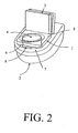
- An exemplary receptacle is shown in Figure 2.
- the receptacle has a support frame 1 which is placed directly over the toilet bowl 2.
- the support frame 1 has attached thereto an articulating cover 3 which may be placed in a raised position, as shown in figure 2, for depositing of sample or a closed position (not shown) for sealing voided stool within the receptacle.
- the support frame 1 additionally has a central opening 4 traversing from a top surface 5 through to a bottom surface 6 of the support frame 1.
- the bottom surface 6 directly communicates with a top surface 7 of the toilet 2.
- a means 8 for capturing voided stool Extending from the bottom surface 6 of the support frame 1 is a means 8 for capturing voided stool.
- Means 8 may be fixedly attached to the support frame 1 or may be removably attached for removal subsequent to deposition of stool.
- Means 8 may comprise a further means for removing at least a cross-sectional portion from the voided stool.
- a preferable sample size is at least 5-10 g or at least 5-10 ml.
- a means to assess the presence of a minimal sample size may comprise a physical diagram indicating the minimal sample size.
- a means to assess the presence of a minimal sample size may comprise the displacement of a liquid or of a mechanical device to a minimal level upon deposit of the stool sample.
- the cross-sectional stool sample is homogenized in an appropriate buffer, such as phosphate buffered saline.
- a buffer such as phosphate buffered saline.
- Homogenization means and materials for homogenization are generally known in the art. Thus, particular homogenization methods may be selected by the skilled artisan and may depend upon the assay to be employed .
- the buffer may contain detergent, salt, proteinase, inhibitors of DNA and RNA degrading enzymes.
- the composition of the buffer will depend on the type of assay to be performed. If a fecal occult blood assay is to be performed, the buffer may contain chemical compounds which react with blood to produce a color, the intensity of which can be measured. Buffers useful for detecting the presence of fecal occult blood are known in the art. If a test is to be performed for the presence of a particular protein, the buffer should not contain a proteinase capable of degrading such tumor marking antigens.
- DNA or RNA may be isolated from the homogenate using methods known in the art. Subsequent tests may be performed on the isolated DNA and RNA.
- DNA characteristics associated with the presence of colorectal cancer or a precancerous lesion may be detected in stool samples prepared according to the invention, using, for example, the methods described in the following sections.
- a careful endoscopic examination preferably is performed on positive Individuals, followed by early surgical excision of any diseased tissue.
- Methods of the invention are used to prepare a stool sample followed by detection of a deletion or other mutation in the p53 tumor suppressor gene.
- the p53 gene is a good choice because a loss of heterozygosity in p53 is often associated with colorectal cancer.
- An mRNA sequence corresponding to the DNA coding region for p53 is reported as GenBank Accession No. M92424.
- At least a cross-section of a voided stool sample is obtained and prepared according to methods of the invention as described immediately above. The sample need not be further processed for analysis. However, DNA or RNA may optionally be isolated from the sample according to methods known in the art. See, Smith-Ravin, et al ., Gut, 36 : 81-86 (1995).
- Nucleic acids may be sheared or cut into small fragments by, for example, restriction digestion.
- the size of nucleic acid fragments produced is not critical, subject to the limitations described below.
- a target allele that is suspected of being mutated (p53 in this example) and a reference allele are chosen.
- a reference allele may be any allele known normally not to be mutated in colon cancer.
- Either portions of a coding strand or its complement may be detected.
- detection of the coding strand of p53 and reference allele are described herein.
- Complement to both p53 and reference allele are removed by hybridization to anti-complement oligonucleotide probes (isolation probes) and subsequent removal of duplex formed thereby.
- Methods for removal of complement strands from a mixture of single-stranded oligonucleotides are known and include techniques such as affinity chromatography.
- sample is passed through an affinity column packed with bound isolation probe that is complementary to the sequence to be isolated away from the sample.
- affinity column packed with bound isolation probe that is complementary to the sequence to be isolated away from the sample.
- Conventional column chromatography is appropriate for isolation of complement.
- An affinity column packed with sepharose or other appropriate materials with attached complementary nucleotides may be used to isolate complement DNA in the column, while allowing DNA to be analyzed to pass through the column. See Sambrook, Supra.
- isolation beads may be used to exclude complement as discussed in detail below.
- first oligonucleotide probes which hybridize to at least a portion of the p53 allele and second oligonucleotide probes that hybridize to at least a portion of the reference allele are obtained.
- the probes are labeled with a detectable label, such as fluorescein or with detectable particles. Distinct labels for the probes are preferred. However, identical labels may be used if, for example, sample is assayed in two separate aliquots. Probes may be labeled with identical or with distinct labels. However, distinct labels are preferred.
- Labeled probes then are exposed to sample under hybridization conditions. Such conditions are well-known in the art. See, e.g., Wallace, et al., Nucleic Acids Res., 6 :3543-3557 (1979).
- First and Second oligonucleotide probes that are distinctly labeled (i.e. with different radioactive isotopes, fluorescent means, or with beads of different size, See infra) are applied to a single aliquot of sample. After exposure of the probes to sample under hybridization conditions, sample is washed to remove any unhybridized probe. Thereafter, hybridized probes are detected separately for p53 hybrids and reference allele hybrids. Standards may be used to establish background and to equilibrate results. Also, if differential fluorescent labels are used, the number of probes may be determined by counting differential fluorescent events in a sample that has been diluted sufficiently to enable detection of single fluorescent events in the sample. Duplicate samples may be analyzed in order to confirm the accuracy of results obtained
- the determination of a p53 mutation allows a clinician to recommend further treatment, such as endoscopy procedures, in order to further diagnose and, if necessary, treat the patient's condition.
- further treatment such as endoscopy procedures
- hybridization beads Enhanced quantification of binding events between hybridization probes and target or reference is accomplished by coupling hybridization probes to particles, such as beads (hybridization beads).
- particles such as beads (hybridization beads).
- hybridization beads are constructed such that each bead has attached thereto a single oligonucleotide probe.
- a single probe is attached to a bead by incubating a large excess of hybridization beads with oligonucleotide probes of a given type (i.e ., either first or second oligonucleotide probes). Coupling of probe to bead is accomplished using an affinity-binding pair. For example, beads may be coated with avidin or streptavidin and probes may be labeled with biotin to effect attachment of the probe to the bead. The mixture of beads and probes is agitated such that 100% of the probes are bound to a bead. The mixture is then exposed to a matrix, such as an affinity column or a membrane coated with oligonucleotides that are complementary to the probe.
- a matrix such as an affinity column or a membrane coated with oligonucleotides that are complementary to the probe.
- beads that have an attached probe will adhere to the matrix, the rest being washed away. Beads with coupled probe are then released from the matrix by melting hybridizations between probe and complement. Multiple exposures to the matrix and pre-washing of the column reduces non-specific binding. Moreover, naked beads ( i.e ., without attached probe) may be exposed to the matrix to determine a background number of beads that can be expected to attach to the matrix in the absence of probe.
- hybridization beads are provided in an effective 1:1 ratio with probe which allows for precise quantification of target and reference polynucleotide as described below.
- a first hybridization bead has attached thereto a single first oligonucleotide probe that is complementary to at least a portion of a target polynucleotide (e.g ., a p53 allele).
- a second hybridization bead, of a size distinct from the first hybridization bead has attached thereto a single second oligonucleotide probe that is complementary to at least a portion of a reference polynucleotide ( i.e ., one that is known or suspected not to be mutated in the sample).
- DNA is melted (denatured to form single-stranded DNA) by well-known methods See, e.g., Gyllensten, et al., in Recombinant DNA Methodology II, 565-578 (Wu, ed., 1995).
- Single-stranded complement of the target polynucleotide (e.g., p53) and reference polynucleotide are removed from the sample by binding to oligonucleotide probes that are complementary to target or reference complement.
- Such probes referred to herein as isolation probes, are attached to isolation beads prior to their introduction into the sample.
- the beads may be magnetized.
- Isolation beads preferably are introduced in vast excess in order to saturate complement binding.
- a magnetic field is applied to the sample to draw the magnetized isolation beads (both with and without hybridized complement) out of the sample. Assuming that a sufficient quantity of isolation beads are introduced into the sample, removal of the isolation beads effectively removes all target and reference complement from the sample.
- an excess of oligonucleotide probe labeled with biotin is exposed to the melted or dehybridized (single stranded) sample under hybridization conditions. Once hybridization is complete, the sample is exposed to a column containing immobilized avidin. The biotin-labeled probe, whether free or hybridized to complement, is bound by avidin on the column. The remainder of the DNA, including target and reference coding strands to be detected, pass through the column.
- beads for removal of complement may each comprise multiple complementary oligonucleotide probes.
- Two sets of hybridization beads are prepared as described above.
- Each member of a first set of hybridization beads (all of which are identical to each other) has attached thereto a single oligonucleotide probe that is complementary to at least a portion of the target polynucleotide, i.e., the portion of the genome which is altered in the cells of a cancerous lesion.
- Each member of a second set of identical hybridization beads (all of which are identical to each other but not to the first set) has attached thereto a single oligonucleotide probe that is complementary to at least a portion of the reference polynucleotide, i.e., a portion of the genome which is not likely to be altered in malignant cells.
- first and second hybridization beads are of a size or color distinct from that of members of the first set of hybridization beads.
- First and second hybridization beads may also be distinguished on the basis of other characteristics. For example, beads may have fluorescent markers that are distinguished by their fluorescence wavelength. Beads with distinct electrochemical charges also may be used. The precise modality used for distinguishing beads is not essential as long as it is possible to distinguish between first and second probe on the basis of distinctions between attached first and second beads.
- Both sets of hybridization beads are exposed to the sample under hybridization conditions thereby allowing hybridization to reference and target.
- the sample then is washed to remove unhybridized bead/probe combinations. Unhybridized bead/probe combinations are removed by, for example, passing the sample through a column lined of immobilized DNA complementary to the probe sequence. Thus, any unhybridized bead/probe combinations are retained on the column while duplex passes through.
- the sample is exposed to means for differentially counting hybridization beads in order to quantify first and second hybridization probes which have formed duplexes.
- the numbers obtained provide a precise estimate of the number of copies of the reference and target polynucleotide in the population because differential counting means count individual beads.
- One bead is equal to one probe which, in turn, signifies one copy of the nucleic acid being measured.
- a differential counting means is an impedance measuring device, such as a Coulter counter (Coulter Electronics, Inc., Miami, Florida). Sample is passed through the device which differentially detects the two types of hybridization beads by measuring their differential impedance of an electric current. Alternatively, the device may measure fluorescence, color, or other parameters.
- a multi-orifice device may be used.
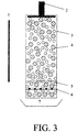
- a multi-orifice impedance counter is shown schematically in figure 2.
- a multi-orifice array is placed at one end of a column filled with an electrically-conductive fluid, such as saline. Hybridization beads with either hybridized target or reference segments are inserted at an opposite end of the column.
- Each orifice is large enough to accommodate only one hybridization bead at a time and sufficiently wide to allow reliable impedance measurements.
- a voltage is set across each orifice.
- Each hybridization bead (which is non-conducting), as it passes through one of the orifices, displaces a volume of saline, thus creating an impedance that is proportional to its size. This, in turn, creates a measurable decrease in current that is directly correlated with the size of the bead.
- the data may be analyzed to determine whether any difference between the amounts of first and second hybridization beads is statistically significant.
- a reduction in the amount of target relative to the reference is indicative of a mutation in or deletion of the target allele in a subpopulation of cells in the sample.
- the p53 gene is the target allele
- such a mutation is indicative of a cancerous or precancerous condition.
- a clinician may use such results as a basis for prescribing additional treatment, such as endoscopy and polypectomy procedures.
- the basic method described above may also be applied to detect a loss of heterozygosity or other mutation at a single base polymorphic site between maternal and paternal alleles. Such detection is typically an indication of a larger deletion or other mutation.
- a mutation at a single polymorphic nucleotide may be all that is necessary to inhibit gene function in one of the two alleles.
- a mutation in a single-base polymorphic region may be difficult to detect due to a recently-discovered phenomenon called complementary reduplication. In complementary reduplication, the loss of one of two alleles at a particular locus results in "reduplication" of the surviving allele.
- Reduplication usually takes place on the chromosome containing the surviving allele and involves the production of one or more copies of the surviving allele in close proximity on the chromosome to the position of the surviving allele.
- a locus that displays one or more single-base allelic polymorphisms i.e. , heterozygosity at the locus is determined by virtue of one or more single-base differences in one or more regions of the locus
- complementary reduplication results in the insertion on the chromosome containing the surviving allele of a duplicate of the sequence corresponding to that which was deleted.
- the deletion may not be detected because any true difference in the number of probes binding to the polymorphic site (i.e ., the allelic region encompassing the single-base polymorphism) may be obscured by an increase resulting from the other allele's reduplicated region.
- Genomic regions containing known single-base polymorphisms may be identified by reference to a nucleotide database, such as GenBank, EMBL, or any other appropriate database. The existence of polymorphisms may be determined by methods taught herein, gel electrophoresis or by other standard methods.
- a single-base polymorphism is intended to be a single polymorphic nucleotide adjacent to a non-polymorphic region of the allele regardless of whether the single polymorphic nucleotide forms part of a larger polymorphic site (i.e. the single-base polymorphism may be the terminal nucleotide of a larger, polynucleotide polymorphism).
- the regions considered are regions in which loss of heterozygosity is prevalent, such as regions containing tumor suppressor genes.
- a given individual may be homozygous or heterozygous for the polymorphic nucleotide in any identified single-base polymorphic region. Accordingly, if a number of single-base polymorphic regions are identified, the probability increases that at least one heterozygous single-base polymorphic region is found in a sample.
- a DNA sample is obtained from a patient, e.g., from blood cells, to determine which of those sites is heterozygous in normal (i.e ., non-cancerous or non pre-cancerous) cells for that individual. Then, a stool sample is prepared as described above. Double stranded DNA in the sample is converted to single-stranded DNA. Then, either the coding strand or the anti-coding strand for both alleles is removed from the sample. As will be evident from the following discussion, methods disclosed herein are indifferent as to whether coding strand or anti-coding strand is tested.
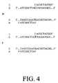
- An oligonucleotide probe is constructed that is complementary to a portion of the region of single-base polymorphism, said portion ending at the nucleotide that is immediately 3' to the polymorphic nucleotide, regardless of whether the 5'-3' (coding) strand or the 3'-5' (anticoding) strand is used as a template.
- Figure 3 shows four possible probes that are immediately 3' to the polymorphic nucleotide for each of four possible template strands as described above (the Sequences in Figure 3 are hypothetical and are not intended to represent any actual sequence).
- Figure 3 merely illustrates four hypothetical probes that are useful for hybridization to the hypothetical sequence shown.
- the length of probe sequences may be determined as appropriate for each genomic region that is analyzed. A preferable length is between about 10 and about 100 nucleotides.
- the size of the probe will also depend upon the size of the region surrounding the single-base polymorphism ( i.e ., the region 5' or 3' to the next adjacent polymorphism, if any). Details concerning the construction and hybridization of oligonucleotide probes are known in the art.
- Probe is hybridized to its specific template DNA by standard methods.
- the sample may optionally be washed to remove unhybridized probe.
- a modification of the dideoxy chain termination method as reported in Sanger, Proc. Nat'l Acad. Sci. (USA), 74 : 5463-5467 (1977) is used.
- the method involves using at least two of the four common 2', 3'-dideoxy nucleoside triphosphates (ddATP, ddCTP, ddGTP, and ddTTP).
- ddNTP dideoxy nucleoside triphosphate
- Differentially-labeled ddNTPs are available commercially, for example, from Perkin Elmer Corporation (Cat. No. 401456). At least two labeled ddNTPs then are exposed to each sample having probe hybridized to maternal and paternal alleles as described above. The choice of which two ddNTPs are used will depend upon the nucleotides at the heterozygous polymorphic site. Any 3' modified nucleoside triphosphate may be used in the method as long as the 3' modification prevents binding of an additional 3' nucleotide (i.e.
- a DNA polymerase such as SequenaseTM (Perkin-Elmer) is added to the sample mixture. Using the allelic strands as primer, the polymerase will add one ddNTP to the 3' end of the probe, the incorporated ddNTP will be complementary to the nucleotide that exists at the single-base polymorphic site. Because the ddNTPs have no 3' hydroxyl, further elongation of the hybridized probe will not occur. After completion, the sample is washed to remove excess ddNTPs. Label is then counted in each sample. The presence of two differentially-labeled ddNTPs in a sample is indicative of heterozygosity at the polymorphic site.
- genomic instability indicates the possibility of cancerous or pre-cancerous cells in the sample.
- ddNTPs are labeled with hybridization-type beads of different sizes as described above. Alleles with bound probe comprising a labeled ddNTP are counted as described above using a counting device, such as a Coulter counter. Also as described above, differential fluorescent labels or other counting means may be used to separately detect incorporated ddNTPs.
- the detection of heterozygosity at single-base polymorphic sites and the detection of the loss of heterozygosity may be determined in separate steps. For example, probes may be hybridized immediately adjacent to but not including the nucleotide determined to be polymorphic as described above. The four ddNTPs may then be added to the sample, washed, and the presence or absence of each label may be detected. Detection of only one label indicates that the individual from whom the sample was obtained is homozygous at the site of the potential polymorphic nucleotide. Detection of two labels means that the individual is heterozygous. The heterozygous loci are recorded. As noted above, baseline determinations of heterozygosity may be done using standard methods.
- the heterozygous loci are typically chromosomal areas containing tumor suppressor genes, including p53, dcc, apc, and others. Using methods described herein, a "fingerprint" of heterozygous tumor suppressor loci may be constructed. Future deviation from the fingerprint ( i.e ., deletions) provides valuable information as to the development of cancer.
- a preferred use of the foregoing methods is in the detection of colon cancer.
- a representative stool sample is prepared as described above. Double-stranded DNA is converted to single-stranded DNA and complement of the strand to be detected is removed from the sample. The remaining single-stranded DNA is exposed to multiple copies of a probe designed on the basis of known single-base polymorphisms in a cancer-associated allele such that the probe hybridizes with a desired number of nucleotides immediately adjacent the polymorphic nucleotide as described above. After hybridization is complete, the sample is washed and exposed to differentially-labeled ddNTPs and a DNA polymerase. The sample then is washed to remove unincorporated ddNTPs.
- any labeled ddNTPs is determined. If two labels are detected, the individual from whom the sample is obtained is heterozygous at the polymorphic nucleotide. The heterozygosity of the allele and the probe sequence matching the site immediately adjacent to the polymorphic allele are noted for reference in future testing for the loss of heterozygosity. Alternatively, once the patient is determined to be heterozygous at a locus, an assay may be performed immediately in the manner described above in order to determine an existing loss of heterozygosity in a subpopulation of cells in the sample.
- Microsatellites are di- or trinucleotide repeats found throughout the genome.
- a particular array of microsatellite repeats is often associated with a particular genomic sequence and is stably inherited under normal conditions.
- Expansions of microsatellite copy number typically, called “microsatellite instability,” are associated with defects in mismatch repair. Accordingly, changes in a microsatellite region indicate that the patient is at risk for a mutation in other genomic regions.
- microsatellite region associated with the gene of interest. Such regions are typically identified on a database, such as GenBank, EMBL, and others. Once a wild-type microsatellite region associated with, for example, the p53 tumor suppressor gene, is identified, an oligonucleotide probe is constructed that spans the microsatellite region and the regions immediately 5' and immediately 3' to the microsatellite region. The precise length of probes may be determined by the experimenter. Probes are constructed that hybridize to the microsatellite region, including portions extending 5' and 3', on both the maternal and paternal alleles with which the microsatellite is associated ( e.g ., p53).
- Double stranded DNA is denatured and an excess of maternal and paternal probes, as described above, are introduced into the sample under hybridization conditions.
- the probes are detectably labeled as described above. Complement of the strands to be detected may optionally be removed by methods described above.
- the sample is then washed to remove unhybridized probe and the amount of hybridized probe in quantitatively detected.
- Quantitative detection may be accomplished by any means described herein.
- probes may be attached to hybridization beads such that probes that bind to maternal allele are attached to beads of one size and probes that bind to paternal allele are attached to beads of a second size that is distinguishable from beads of the first size. Beads with attached probe may be counted as described above.
- microsatellite instability can be indicative of a mutation at the locus in which the microsatellite resides. If the microsatellite region is associated with a tumor suppressor gene or an oncogene, the detection of microsatellite instability in an allele in a subpopulation of cells in a biological sample is indicative of the potential for cancer or that cancer or precancer may have already developed. Further testing as described herein (either by invasive or noninvasive means) may then be conducted.
- a "fingerprint" of microsatellites is taken from regions associated with cancer-causing genes in a sample obtained from a patient.
- a fingerprint may be obtained by standard methods.
- the fingerprint comprises the sequence of wild-type microsatellites associated with the cancer-causing gene or genes.
- the fingerprint is stored and is used in future tests of samples from the same patient in order to monitor changes in microsatellite regions (i.e. microsatellite instability) that may be associated with the development of cancer. Changes in microsatellite length and/or sequence over time may be used to prescribe additional testing and/or treatment in order to detect and remove cancerous tissue at an early stage in its etiology.
Landscapes
- Chemical & Material Sciences (AREA)
- Life Sciences & Earth Sciences (AREA)
- Health & Medical Sciences (AREA)
- Organic Chemistry (AREA)
- Engineering & Computer Science (AREA)
- Proteomics, Peptides & Aminoacids (AREA)
- Zoology (AREA)
- Immunology (AREA)
- Wood Science & Technology (AREA)
- Analytical Chemistry (AREA)
- Molecular Biology (AREA)
- Biochemistry (AREA)
- General Health & Medical Sciences (AREA)
- Genetics & Genomics (AREA)
- Biotechnology (AREA)
- Microbiology (AREA)
- Physics & Mathematics (AREA)
- Bioinformatics & Cheminformatics (AREA)
- General Engineering & Computer Science (AREA)
- Biophysics (AREA)
- Pathology (AREA)
- Chemical Kinetics & Catalysis (AREA)
- Oncology (AREA)
- Biomedical Technology (AREA)
- Hospice & Palliative Care (AREA)
- Hematology (AREA)
- Urology & Nephrology (AREA)
- Cell Biology (AREA)
- Medicinal Chemistry (AREA)
- General Physics & Mathematics (AREA)
- Food Science & Technology (AREA)
- Measuring Or Testing Involving Enzymes Or Micro-Organisms (AREA)
- Investigating Or Analysing Biological Materials (AREA)
Priority Applications (1)
| Application Number | Priority Date | Filing Date | Title |
|---|---|---|---|
| EP02024382A EP1291657A3 (en) | 1996-01-30 | 1996-12-20 | Methods for detecting colon cancer from stool samples |
Applications Claiming Priority (5)
| Application Number | Priority Date | Filing Date | Title |
|---|---|---|---|
| US1085696P | 1996-01-30 | 1996-01-30 | |
| US10856P | 1996-01-30 | ||
| US699678 | 1996-08-14 | ||
| US08/699,678 US5741650A (en) | 1996-01-30 | 1996-08-14 | Methods for detecting colon cancer from stool samples |
| PCT/US1996/020727 WO1997028450A1 (en) | 1996-01-30 | 1996-12-20 | Methods for detecting colon cancer from stool samples |
Related Child Applications (1)
| Application Number | Title | Priority Date | Filing Date |
|---|---|---|---|
| EP02024382A Division EP1291657A3 (en) | 1996-01-30 | 1996-12-20 | Methods for detecting colon cancer from stool samples |
Publications (2)
| Publication Number | Publication Date |
|---|---|
| EP0817968A1 EP0817968A1 (en) | 1998-01-14 |
| EP0817968B1 true EP0817968B1 (en) | 2003-08-06 |
Family
ID=26681667
Family Applications (1)
| Application Number | Title | Priority Date | Filing Date |
|---|---|---|---|
| EP96946136A Expired - Lifetime EP0817968B1 (en) | 1996-01-30 | 1996-12-20 | Methods for detecting colon cancer from stool samples |
Country Status (10)
| Country | Link |
|---|---|
| US (1) | US5741650A (enExample) |
| EP (1) | EP0817968B1 (enExample) |
| JP (1) | JP4195508B2 (enExample) |
| AT (1) | ATE246806T1 (enExample) |
| AU (1) | AU704696B2 (enExample) |
| CA (1) | CA2215263C (enExample) |
| DE (1) | DE69629363T2 (enExample) |
| DK (1) | DK0817968T3 (enExample) |
| ES (1) | ES2202497T3 (enExample) |
| WO (1) | WO1997028450A1 (enExample) |
Cited By (1)
| Publication number | Priority date | Publication date | Assignee | Title |
|---|---|---|---|---|
| US11845991B2 (en) | 2009-02-03 | 2023-12-19 | Exact Sciences Corporation | Fecal sample processing and analysis comprising detection of blood |
Families Citing this family (79)
| Publication number | Priority date | Publication date | Assignee | Title |
|---|---|---|---|---|
| US5952178A (en) * | 1996-08-14 | 1999-09-14 | Exact Laboratories | Methods for disease diagnosis from stool samples |
| US6268136B1 (en) | 1997-06-16 | 2001-07-31 | Exact Science Corporation | Methods for stool sample preparation |
| US6406857B1 (en) | 1997-06-16 | 2002-06-18 | Exact Sciences Corporation | Methods for stool sample preparation |
| US6818404B2 (en) | 1997-10-23 | 2004-11-16 | Exact Sciences Corporation | Methods for detecting hypermethylated nucleic acid in heterogeneous biological samples |
| US6280947B1 (en) | 1999-08-11 | 2001-08-28 | Exact Sciences Corporation | Methods for detecting nucleotide insertion or deletion using primer extension |
| US6503718B2 (en) | 1999-01-10 | 2003-01-07 | Exact Sciences Corporation | Methods for detecting mutations using primer extension for detecting disease |
| CA2372667A1 (en) * | 1999-01-10 | 2000-11-23 | Steven Laken | Methods of detecting colorectal disease by conduction an assay to detect a mutation in a bat-26 locus |
| ATE330032T1 (de) | 1999-02-25 | 2006-07-15 | Exact Sciences Corp | Verfahren zur erhaltung der dns-integrität |
| CA2366778C (en) * | 1999-04-09 | 2008-07-22 | Exact Sciences Corporation | Methods for detecting nucleic acids indicative of cancer |
| US6335193B1 (en) | 1999-04-15 | 2002-01-01 | Padmanabhan P Nair | Isolated colonocytes |
| CA2373598C (en) * | 1999-05-20 | 2006-08-01 | National Research Council Of Canada | Method of diagnosing colorectal adenomas and cancer using proton magnetic resonance spectroscopy |
| US6821784B1 (en) * | 1999-05-20 | 2004-11-23 | The University Of Manitoba | Method of diagnosing colorectal adenomas and cancer using proton magnetic resonance spectroscopy |
| US6849403B1 (en) | 1999-09-08 | 2005-02-01 | Exact Sciences Corporation | Apparatus and method for drug screening |
| US6586177B1 (en) | 1999-09-08 | 2003-07-01 | Exact Sciences Corporation | Methods for disease detection |
| US6994971B1 (en) | 1999-10-08 | 2006-02-07 | University Of Utah Research Foundation | Particle analysis assay for biomolecular quantification |
| AU7875600A (en) * | 1999-10-08 | 2001-04-23 | University Of Utah Research Foundation | Particle analysis assay for biomolecular quantification |
| EP1238113B1 (en) * | 1999-12-07 | 2010-02-24 | EXACT Sciences Corporation | Detection of lung neoplasms in stool |
| US6919174B1 (en) | 1999-12-07 | 2005-07-19 | Exact Sciences Corporation | Methods for disease detection |
| US20030219765A1 (en) * | 2000-03-23 | 2003-11-27 | Jose Costa | Methods for evaluating cancer risk |
| DE10032608A1 (de) * | 2000-07-07 | 2002-01-24 | Magnus Von Knebel Doeberitz Ch | Für mikrosatelliteninstabile (MSI+)-Tumore relevante Gene und ihre Genprodukte |
| US20020086431A1 (en) * | 2000-08-21 | 2002-07-04 | Markham Walter Bruce | Sample preparation and slide plating apparatus and method |
| FR2815509B1 (fr) * | 2000-10-18 | 2004-07-23 | Swap Com | Systeme d'informations personnalisees sur telephone mobile en utilisant la multidiffusion "cell broadcast", et un logiciel de controle d'acces a bord du telephone, telecommande par l'operateur du service |
| US6911308B2 (en) | 2001-01-05 | 2005-06-28 | Exact Sciences Corporation | Methods for detecting, grading or monitoring an H. pylori infection |
| US6428964B1 (en) | 2001-03-15 | 2002-08-06 | Exact Sciences Corporation | Method for alteration detection |
| US20030148260A1 (en) * | 2001-08-07 | 2003-08-07 | Bernard Levin | Method of diagnosing colorectal adenomas and cancer using proton maggnetic resonance spectroscopy |
| CA2457527A1 (en) * | 2001-08-31 | 2003-03-13 | Gen-Probe Incorporated | Affinity-shifted probes for quantifying analyte polynucleotides |
| WO2003071252A2 (en) | 2002-02-15 | 2003-08-28 | Exact Sciences Corporation | Methods for analysis of molecular events |
| US20040086895A1 (en) * | 2002-11-06 | 2004-05-06 | Crothers Donald M. | Method of electrochemical detection of somatic cell mutations |
| CA2506066A1 (en) * | 2002-11-15 | 2004-06-03 | Genomic Health, Inc. | Gene expression profiling of egfr positive cancer |
| US20040231909A1 (en) | 2003-01-15 | 2004-11-25 | Tai-Yang Luh | Motorized vehicle having forward and backward differential structure |
| WO2004071572A2 (en) * | 2003-02-06 | 2004-08-26 | Genomic Health, Inc. | Gene expression markers for response to egfr inhibitor drugs |
| CA2516553C (en) * | 2003-02-20 | 2013-04-16 | Genomic Health, Inc. | Use of intronic rna to measure gene expression |
| CA2527321A1 (en) * | 2003-05-30 | 2004-12-23 | Genomic Health, Inc. | Gene expression markers for response to egfr inhibitor drugs |
| US20040259101A1 (en) * | 2003-06-20 | 2004-12-23 | Shuber Anthony P. | Methods for disease screening |
| WO2005039382A2 (en) * | 2003-06-24 | 2005-05-06 | Genomic Health | Prediction of likelihood of cancer recurrence |
| EP1644858B1 (en) | 2003-07-10 | 2017-12-06 | Genomic Health, Inc. | Expression profile algorithm and test for cancer prognosis |
| US20050014165A1 (en) * | 2003-07-18 | 2005-01-20 | California Pacific Medical Center | Biomarker panel for colorectal cancer |
| JP4033820B2 (ja) * | 2003-07-29 | 2008-01-16 | 株式会社日立製作所 | 細胞回収方法 |
| WO2005017501A1 (en) * | 2003-08-14 | 2005-02-24 | National Research Council Of Cananda | Method of diagnosing colorectal adenomas and cancer using infrared spectroscopy |
| AU2004284434A1 (en) * | 2003-10-16 | 2005-05-06 | Genomic Health, Inc. | qRT-PCR assay system for gene expression profiling |
| EP1685395A2 (en) * | 2003-11-05 | 2006-08-02 | EXACT Sciences Corporation | Repetitive reversed-field affinity electrophoresis and uses therefor |
| JP4718490B2 (ja) * | 2003-12-23 | 2011-07-06 | ジェノミック ヘルス, インコーポレイテッド | フラグメント化rnaの全般的増幅 |
| CA2848463A1 (en) | 2004-04-09 | 2005-10-27 | Genomic Health, Inc. | Gene expression markers for predicting response to chemotherapy |
| US20080241827A1 (en) * | 2004-05-10 | 2008-10-02 | Exact Sciences Corporation | Methods For Detecting A Mutant Nucleic Acid |
| US20080124714A1 (en) * | 2004-05-14 | 2008-05-29 | Exact Sciences Corporation | Method for Stabilizing Biological Samples for Nucleic Acid Analysis |
| WO2006026654A2 (en) * | 2004-08-27 | 2006-03-09 | Exact Sciences Corporation | Method for detecting a recombinant event |
| US9109256B2 (en) | 2004-10-27 | 2015-08-18 | Esoterix Genetic Laboratories, Llc | Method for monitoring disease progression or recurrence |
| JP4939425B2 (ja) * | 2004-11-05 | 2012-05-23 | ジェノミック ヘルス, インコーポレイテッド | 乳癌の治療反応の予後および予測の分子指標 |
| CA3061785A1 (en) | 2004-11-05 | 2006-05-18 | Genomic Health, Inc. | Predicting response to chemotherapy using gene expression markers |
| WO2006065598A2 (en) * | 2004-12-13 | 2006-06-22 | Geneohm Sciences, Inc. | Fluidic cartridges for electrochemical detection of dna |
| US9777314B2 (en) * | 2005-04-21 | 2017-10-03 | Esoterix Genetic Laboratories, Llc | Analysis of heterogeneous nucleic acid samples |
| US20070248546A1 (en) * | 2005-11-09 | 2007-10-25 | Exact Sciences Corporation | Clinical algorithm for excluding patients identified in virtual imaging |
| ES2355388T3 (es) * | 2006-01-31 | 2011-03-25 | Medical Research Fund Of Tel Aviv Sourasky Medical Center | Métodos para la detección precoz de cáncer. |
| EP2167683A4 (en) | 2007-05-31 | 2010-10-20 | California Pacific Med Center | METHOD FOR PREDICTING OR DIAGNOSING GASTROINTESTINAL DISORDER OR DISEASE |
| WO2009015299A1 (en) | 2007-07-26 | 2009-01-29 | California Pacific Medical Center | Method to predict or diagnose a gastrointestinal disorder or disease |
| EP2218792A4 (en) * | 2007-11-20 | 2010-12-08 | Olympus Corp | PROCESS FOR PREPARATION OF A KOTPROBE, SOLUTION FOR PREPARATION OF A KOTPROBE AND KOTSAMMELKIT |
| WO2009102788A2 (en) | 2008-02-15 | 2009-08-20 | Mayo Foundation For Medical Education And Research | Detecting neoplasm |
| JP5575239B2 (ja) | 2009-07-21 | 2014-08-20 | ジェン−プローブ・インコーポレーテッド | 拡張されたダイナミックレンジにわたる核酸配列の定量的検出のための方法および組成物 |
| WO2011119934A2 (en) | 2010-03-26 | 2011-09-29 | Mayo Foundation For Medical Education And Research | Methods and materials for detecting colorectal neoplasm |
| US8361720B2 (en) | 2010-11-15 | 2013-01-29 | Exact Sciences Corporation | Real time cleavage assay |
| WO2012106525A2 (en) | 2011-02-02 | 2012-08-09 | Exact Sciences Corporation | Digital sequence analysis of dna methylation |
| GB201107466D0 (en) | 2011-05-05 | 2011-06-15 | Loktionov Alexandre | Device and method for non-invasive collection of colorectal mucocellular layer and disease detection |
| US9127318B2 (en) | 2011-10-18 | 2015-09-08 | Exact Sciences Corporation | Multiplexed KRAS mutation detection assay |
| CA2902916C (en) | 2013-03-14 | 2018-08-28 | Mayo Foundation For Medical Education And Research | Detecting neoplasm |
| US10253358B2 (en) | 2013-11-04 | 2019-04-09 | Exact Sciences Development Company, Llc | Multiple-control calibrators for DNA quantitation |
| EP3084004A4 (en) | 2013-12-19 | 2017-08-16 | Exact Sciences Corporation | Synthetic nucleic acid control molecules |
| ES2812753T3 (es) | 2014-03-31 | 2021-03-18 | Mayo Found Medical Education & Res | Detección de neoplasma colorectal |
| US10184154B2 (en) | 2014-09-26 | 2019-01-22 | Mayo Foundation For Medical Education And Research | Detecting cholangiocarcinoma |
| US10030272B2 (en) | 2015-02-27 | 2018-07-24 | Mayo Foundation For Medical Education And Research | Detecting gastrointestinal neoplasms |
| CN107532124B (zh) | 2015-03-27 | 2022-08-09 | 精密科学公司 | 检测食管疾病 |
| AU2016315924B2 (en) | 2015-08-31 | 2022-08-25 | Exact Sciences Corporation | Detecting gastric neoplasm |
| US10370726B2 (en) | 2016-04-14 | 2019-08-06 | Mayo Foundation For Medical Education And Research | Detecting colorectal neoplasia |
| AU2017250663B2 (en) | 2016-04-14 | 2023-06-15 | Mayo Foundation For Medical Education And Research | Detecting pancreatic high-grade dysplasia |
| EP3488003B1 (en) | 2016-07-19 | 2023-10-25 | Exact Sciences Corporation | Nucleic acid control molecules from non-human organisms |
| ES2902174T3 (es) | 2016-07-19 | 2022-03-25 | Exact Sciences Dev Co Llc | ADN de control metilado |
| CA3054836A1 (en) | 2017-02-28 | 2018-09-07 | Mayo Foundation For Medical Education And Research | Detecting prostate cancer |
| CN111655869B (zh) | 2017-11-30 | 2024-06-28 | 梅约医学教育与研究基金会 | 检测乳腺癌 |
| US10507010B1 (en) * | 2018-09-18 | 2019-12-17 | Hong Min Kim | Stool monitoring and health guidance apparatus |
| WO2021215986A1 (en) | 2020-04-21 | 2021-10-28 | Sjoeblom Tobias | Cancer biomarkers |
Citations (1)
| Publication number | Priority date | Publication date | Assignee | Title |
|---|---|---|---|---|
| US5380647A (en) * | 1991-02-05 | 1995-01-10 | Farrokh Saidi | Simple test for detecting carcinoembryonic antigen in stool |
Family Cites Families (67)
| Publication number | Priority date | Publication date | Assignee | Title |
|---|---|---|---|---|
| US4101279A (en) * | 1977-04-06 | 1978-07-18 | Muhammed Javed Aslam | Device for the collection and processing of stool specimens |
| US4333734A (en) * | 1980-01-18 | 1982-06-08 | Sloan-Kettering Institute For Cancer Research | Diagnostic device for fecal occult blood and method of use |
| US4535058A (en) * | 1982-10-01 | 1985-08-13 | Massachusetts Institute Of Technology | Characterization of oncogenes and assays based thereon |
| US4309782A (en) * | 1980-09-11 | 1982-01-12 | Esteban Paulin | Device for collecting fecal specimens |
| US4445235A (en) * | 1982-09-13 | 1984-05-01 | Pearl Slover | Stool specimen collector |
| US4683195A (en) * | 1986-01-30 | 1987-07-28 | Cetus Corporation | Process for amplifying, detecting, and/or-cloning nucleic acid sequences |
| US4871838A (en) * | 1985-07-23 | 1989-10-03 | The Board Of Rijks Universiteit Leiden | Probes and methods for detecting activated ras oncogenes |
| US4705050A (en) * | 1985-10-02 | 1987-11-10 | Markham Charles W | Moisture-activated floatation device |
| US5348855A (en) * | 1986-03-05 | 1994-09-20 | Miles Inc. | Assay for nucleic acid sequences in an unpurified sample |
| CA1284931C (en) * | 1986-03-13 | 1991-06-18 | Henry A. Erlich | Process for detecting specific nucleotide variations and genetic polymorphisms present in nucleic acids |
| US4981783A (en) * | 1986-04-16 | 1991-01-01 | Montefiore Medical Center | Method for detecting pathological conditions |
| CA1341576C (en) * | 1986-08-11 | 2008-07-08 | Thaddeus P. Dryja | Diagnosis of retinoblastoma |
| US4735905A (en) * | 1986-08-15 | 1988-04-05 | V-Tech, Inc. | Specimen-gathering apparatus and method |
| AU625169B2 (en) * | 1987-03-23 | 1992-07-02 | Imperial Chemical Industries Plc | Molecular markers |
| US4857300A (en) * | 1987-07-27 | 1989-08-15 | Cytocorp, Inc. | Cytological and histological fixative formulation and methods for using same |
| US5688657A (en) * | 1988-03-31 | 1997-11-18 | International Bio-Immune Systems, Inc. | Monoclonal antibodies against human colon carcinoma-associated antigens and uses therefor |
| IL89964A0 (en) * | 1988-04-15 | 1989-12-15 | Montefiore Med Center | Method for determining malignant progression,cdna,polypeptides encoded thereby and pharmaceutical compositions containing the same |
| FR2630000A1 (fr) * | 1988-04-18 | 1989-10-20 | Sultan Bernard | Flacon destine a recueillir un echantillon biologique urinaire pour examen cytobacteriologique |
| US5272057A (en) * | 1988-10-14 | 1993-12-21 | Georgetown University | Method of detecting a predisposition to cancer by the use of restriction fragment length polymorphism of the gene for human poly (ADP-ribose) polymerase |
| US5087617A (en) * | 1989-02-15 | 1992-02-11 | Board Of Regents, The University Of Texas System | Methods and compositions for treatment of cancer using oligonucleotides |
| US5248671A (en) * | 1989-02-15 | 1993-09-28 | Board Of Regents, The University Of Texas System | Methods and compositions for treatment of cancer using oligonucleotides |
| US5380645A (en) * | 1989-03-16 | 1995-01-10 | The Johns Hopkins University | Generalized method for assessment of colorectal carcinoma |
| US5527676A (en) * | 1989-03-29 | 1996-06-18 | The Johns Hopkins University | Detection of loss of the wild-type P53 gene and kits therefor |
| US5362623A (en) * | 1991-06-14 | 1994-11-08 | The John Hopkins University | Sequence specific DNA binding by p53 |
| DE69032864T3 (de) * | 1989-03-29 | 2013-01-31 | The Johns Hopkins University | Nachweis des Ausfalls des Wildtyps des p53-Gens |
| US5196167A (en) * | 1989-04-04 | 1993-03-23 | Helena Laboratories Corporation | Fecal occult blood test product with positive and negative controls |
| ES2059895T3 (es) * | 1989-06-23 | 1994-11-16 | Canon Kk | Metodo para la deteccion de acido nucleico. |
| US5137806A (en) * | 1989-12-11 | 1992-08-11 | Board Of Regents, The University Of Texas System | Methods and compositions for the detection of sequences in selected DNA molecules |
| ATE164629T1 (de) * | 1990-01-04 | 1998-04-15 | Univ Johns Hopkins | In menschlichen kolorektalkarzinomen fehlendes gen |
| US5126239A (en) * | 1990-03-14 | 1992-06-30 | E. I. Du Pont De Nemours And Company | Process for detecting polymorphisms on the basis of nucleotide differences |
| JP3179481B2 (ja) * | 1990-06-27 | 2001-06-25 | プリンストン ユニバーシティ | 突然変異体p53検出用プローブ |
| JPH06510363A (ja) * | 1990-10-29 | 1994-11-17 | ディカルブ プラント ジェネティクス | 磁気性粒子を使用する生物学的材料の単離 |
| JPH04203966A (ja) * | 1990-11-29 | 1992-07-24 | Yuuken:Kk | 簡易採便器兼用便潜血検査具及びこの検査具を用いる検査方法 |
| US5352775A (en) * | 1991-01-16 | 1994-10-04 | The Johns Hopkins Univ. | APC gene and nucleic acid probes derived therefrom |
| ES2158847T3 (es) * | 1991-01-16 | 2001-09-16 | Univ Johns Hopkins | Mutaciones heredadas y somaticas del gen apc en el cancer colorectal del hombre. |
| EP0570497B1 (en) * | 1991-02-05 | 1996-05-08 | Lifecodes Corporation | Molecular genetic identification using probes that recognize polymorphic loci |
| US5330892A (en) * | 1991-03-13 | 1994-07-19 | The Johns Hopkins University | MCC gene (mutated in colorectal cancer) used for diagnosis of cancer in humans |
| US5468610A (en) * | 1991-05-29 | 1995-11-21 | The Government Of The United States Of America As Represented By The Secretary Of The Department Of Health And Human Services | Three highly informative microsatellite repeat polymorphic DNA markers |
| US5378602A (en) * | 1991-05-29 | 1995-01-03 | The Government Of The United States Of America As Represented By The Secretary Of The Department Of Health And Human Services | Highly informative microsatellite repeat polymorphic DNA markers twenty-[seven]six |
| US5149506A (en) * | 1991-08-09 | 1992-09-22 | Sage Products, Inc. | Stool collection and transport device |
| AU3919293A (en) * | 1992-03-27 | 1993-11-08 | University Of Maryland At Baltimore | Detection of gene mutations with mismatch repair enzymes |
| US5489508A (en) * | 1992-05-13 | 1996-02-06 | University Of Texas System Board Of Regents | Therapy and diagnosis of conditions related to telomere length and/or telomerase activity |
| JP3399948B2 (ja) * | 1992-06-26 | 2003-04-28 | ザ トラスティーズ オブ プリンストン ユニバーシティ | P90抗体またはプローブを用いた前癌細胞または癌細胞の検出方法 |
| FI96064C (fi) * | 1992-07-15 | 1996-04-25 | Outokumpu Instr Oy | Menetelmä jäähdytyksen aikaansaamiseksi ja jäähdytykseen soveltuva jäähdytyslaite |
| US5409586A (en) * | 1992-08-26 | 1995-04-25 | Hitachi, Ltd. | Method for analyzing nucleic acid or protein and apparatus therefor |
| IL103600A0 (en) * | 1992-10-30 | 1993-03-15 | Yeda Res & Dev | Method and kit for cancer diagnosis |
| US5331973A (en) * | 1993-03-15 | 1994-07-26 | Fiedler Paul N | Method for obtaining stool samples for gastrointestinal cancer testing |
| US5492808A (en) * | 1993-05-05 | 1996-02-20 | The Johns Hopkins University | Means for detecting familial colon cancer (FCC) |
| US5466576A (en) * | 1993-07-02 | 1995-11-14 | Fred Hutchinson Cancer Research Center | Modulation of PIF-1-type helicases |
| FR2709761B1 (fr) * | 1993-09-10 | 1995-11-24 | Pasteur Institut | Procédé de détection de molécules contenant des mésappariements nucléotidiques et de localisation de ces mésappariements, et application à la détection de substitutions ou de délétions de bases . |
| WO1995009928A1 (en) * | 1993-10-06 | 1995-04-13 | The Regents Of The University Of California | Methods for detecting cancer-associated chromosome deletions using in situ hybridization |
| GB9322795D0 (en) * | 1993-11-05 | 1993-12-22 | Wellcome Found | Novel compounds |
| US5561041A (en) * | 1993-11-12 | 1996-10-01 | The Johns Hopkins University School Of Medicine | Nucleic acid mutation detection by analysis of sputum |
| WO1995015400A1 (en) * | 1993-12-03 | 1995-06-08 | The Johns Hopkins University | Genotyping by simultaneous analysis of multiple microsatellite loci |
| CH686982A5 (fr) * | 1993-12-16 | 1996-08-15 | Maurice Stroun | Méthode pour le diagnostic de cancers. |
| AU1675895A (en) * | 1994-01-03 | 1995-08-01 | Children's Hospital Of Philadelphia, The | Polymorphism at codon 36 of the p53 gene |
| US6025127A (en) * | 1994-01-14 | 2000-02-15 | The Johns Hopkins University School Of Medicine | Nucleic acid mutation detection in histologic tissue |
| CN1128049A (zh) * | 1994-03-18 | 1996-07-31 | 犹他大学研究基金会 | 多肿瘤抑制因子基因种系突变及检测该基因癌肿倾向性的方法 |
| SE9500023L (sv) * | 1994-05-17 | 1995-11-18 | Beki Ab | Sätt att detektera cancer |
| US5496470A (en) * | 1994-05-27 | 1996-03-05 | Barnes International, Inc. | Magnetic separator |
| US5667987A (en) * | 1994-07-12 | 1997-09-16 | Bristol-Myers Squibb Company | P53 response genes |
| CA2198702C (en) * | 1994-08-31 | 2010-11-02 | David Sidransky | Detection of hypermutable nucleic acid sequence in tissue |
| US5702886A (en) * | 1994-10-05 | 1997-12-30 | The Johns Hopkins University | Chromosome I8Q loss and prognosis in colorectal cancer |
| EP0787203A1 (en) * | 1994-10-21 | 1997-08-06 | Zeneca Limited | Method for characterising variability in telomere dna by pcr |
| US5629178A (en) * | 1994-10-28 | 1997-05-13 | Genetics & Ivf Institute | Method for enhancing amplification in the polymerase chain reaction employing peptide nucleic acid (PNA) |
| US5512441A (en) * | 1994-11-15 | 1996-04-30 | American Health Foundation | Quantative method for early detection of mutant alleles and diagnostic kits for carrying out the method |
| US5463782A (en) * | 1994-11-21 | 1995-11-07 | Eric V. Carlson | Foldable stool sample collection device |
-
1996
- 1996-08-14 US US08/699,678 patent/US5741650A/en not_active Expired - Lifetime
- 1996-12-20 DE DE69629363T patent/DE69629363T2/de not_active Expired - Lifetime
- 1996-12-20 WO PCT/US1996/020727 patent/WO1997028450A1/en not_active Ceased
- 1996-12-20 JP JP52762597A patent/JP4195508B2/ja not_active Expired - Lifetime
- 1996-12-20 AT AT96946136T patent/ATE246806T1/de active
- 1996-12-20 DK DK96946136T patent/DK0817968T3/da active
- 1996-12-20 ES ES96946136T patent/ES2202497T3/es not_active Expired - Lifetime
- 1996-12-20 AU AU22406/97A patent/AU704696B2/en not_active Expired
- 1996-12-20 EP EP96946136A patent/EP0817968B1/en not_active Expired - Lifetime
- 1996-12-20 CA CA002215263A patent/CA2215263C/en not_active Expired - Lifetime
Patent Citations (1)
| Publication number | Priority date | Publication date | Assignee | Title |
|---|---|---|---|---|
| US5380647A (en) * | 1991-02-05 | 1995-01-10 | Farrokh Saidi | Simple test for detecting carcinoembryonic antigen in stool |
Cited By (1)
| Publication number | Priority date | Publication date | Assignee | Title |
|---|---|---|---|---|
| US11845991B2 (en) | 2009-02-03 | 2023-12-19 | Exact Sciences Corporation | Fecal sample processing and analysis comprising detection of blood |
Also Published As
| Publication number | Publication date |
|---|---|
| EP0817968A1 (en) | 1998-01-14 |
| CA2215263A1 (en) | 1997-08-07 |
| CA2215263C (en) | 2003-02-18 |
| US5741650A (en) | 1998-04-21 |
| JP4195508B2 (ja) | 2008-12-10 |
| ATE246806T1 (de) | 2003-08-15 |
| WO1997028450A1 (en) | 1997-08-07 |
| JP2002515973A (ja) | 2002-05-28 |
| DE69629363D1 (de) | 2003-09-11 |
| AU704696B2 (en) | 1999-04-29 |
| ES2202497T3 (es) | 2004-04-01 |
| DE69629363T2 (de) | 2004-06-09 |
| AU2240697A (en) | 1997-08-22 |
| DK0817968T3 (da) | 2003-12-01 |
Similar Documents
| Publication | Publication Date | Title |
|---|---|---|
| EP0817968B1 (en) | Methods for detecting colon cancer from stool samples | |
| US5952178A (en) | Methods for disease diagnosis from stool samples | |
| US5670325A (en) | Method for the detection of clonal populations of transformed cells in a genomically heterogeneous cellular sample | |
| US5928870A (en) | Methods for the detection of loss of heterozygosity | |
| US6020137A (en) | Methods for the detection of loss of heterozygosity | |
| EP0815263B1 (en) | Methods for the detection of clonal populations of transformed cells in a genomically heterogeneous cellular sample | |
| CA2352455C (en) | Methods for stool sample preparation | |
| US6203993B1 (en) | Methods for the detection of nucleic acids | |
| US20020164631A1 (en) | Methods for stool sample preparation | |
| US20060141497A1 (en) | Molecular analysis of cellular fluid and liquid cytology specimens for clinical diagnosis, characterization, and integration with microscopic pathology evaluation | |
| US20020004206A1 (en) | Methods of screening for disease | |
| US20020004201A1 (en) | Methods for the detection of loss of heterozygosity | |
| AU720489B2 (en) | Methods for detecting colon cancer from stool samples | |
| CA2331254A1 (en) | Methods for the detection of nucleic acids | |
| EP1291657A2 (en) | Methods for detecting colon cancer from stool samples | |
| JP3325270B6 (ja) | ゲノム上不均一な細胞サンプルにおける形質転換細胞のクローン集団検出のための方法 | |
| Sugimura et al. | Methods for genetic testing I: Assessing mutations in cancers |
Legal Events
| Date | Code | Title | Description |
|---|---|---|---|
| PUAI | Public reference made under article 153(3) epc to a published international application that has entered the european phase |
Free format text: ORIGINAL CODE: 0009012 |
|
| AK | Designated contracting states |
Kind code of ref document: A1 Designated state(s): AT BE CH DE DK ES FI FR GB GR IE IT LI LU MC NL PT SE |
|
| 17P | Request for examination filed |
Effective date: 19980112 |
|
| 17Q | First examination report despatched |
Effective date: 20001204 |
|
| RAP1 | Party data changed (applicant data changed or rights of an application transferred) |
Owner name: EXACT SCIENCES CORPORATION |
|
| GRAH | Despatch of communication of intention to grant a patent |
Free format text: ORIGINAL CODE: EPIDOS IGRA |
|
| GRAH | Despatch of communication of intention to grant a patent |
Free format text: ORIGINAL CODE: EPIDOS IGRA |
|
| GRAA | (expected) grant |
Free format text: ORIGINAL CODE: 0009210 |
|
| AK | Designated contracting states |
Designated state(s): AT BE CH DE DK ES FI FR GB GR IE IT LI LU MC NL PT SE |
|
| REG | Reference to a national code |
Ref country code: GB Ref legal event code: FG4D |
|
| REG | Reference to a national code |
Ref country code: CH Ref legal event code: EP |
|
| REG | Reference to a national code |
Ref country code: IE Ref legal event code: FG4D |
|
| REF | Corresponds to: |
Ref document number: 69629363 Country of ref document: DE Date of ref document: 20030911 Kind code of ref document: P |
|
| REG | Reference to a national code |
Ref country code: CH Ref legal event code: NV Representative=s name: PATENTANWAELTE FELDMANN & PARTNER AG |
|
| PG25 | Lapsed in a contracting state [announced via postgrant information from national office to epo] |
Ref country code: GR Free format text: LAPSE BECAUSE OF FAILURE TO SUBMIT A TRANSLATION OF THE DESCRIPTION OR TO PAY THE FEE WITHIN THE PRESCRIBED TIME-LIMIT Effective date: 20031106 |
|
| REG | Reference to a national code |
Ref country code: SE Ref legal event code: TRGR |
|
| REG | Reference to a national code |
Ref country code: DK Ref legal event code: T3 |
|
| PG25 | Lapsed in a contracting state [announced via postgrant information from national office to epo] |
Ref country code: LU Free format text: LAPSE BECAUSE OF NON-PAYMENT OF DUE FEES Effective date: 20031220 |
|
| PG25 | Lapsed in a contracting state [announced via postgrant information from national office to epo] |
Ref country code: MC Free format text: LAPSE BECAUSE OF NON-PAYMENT OF DUE FEES Effective date: 20031231 |
|
| PG25 | Lapsed in a contracting state [announced via postgrant information from national office to epo] |
Ref country code: PT Free format text: LAPSE BECAUSE OF FAILURE TO SUBMIT A TRANSLATION OF THE DESCRIPTION OR TO PAY THE FEE WITHIN THE PRESCRIBED TIME-LIMIT Effective date: 20040106 |
|
| RAP2 | Party data changed (patent owner data changed or rights of a patent transferred) |
Owner name: EXACT SCIENCES CORPORATION |
|
| REG | Reference to a national code |
Ref country code: ES Ref legal event code: FG2A Ref document number: 2202497 Country of ref document: ES Kind code of ref document: T3 |
|
| NLT2 | Nl: modifications (of names), taken from the european patent patent bulletin |
Owner name: EXACT SCIENCES CORPORATION |
|
| ET | Fr: translation filed | ||
| PLBE | No opposition filed within time limit |
Free format text: ORIGINAL CODE: 0009261 |
|
| STAA | Information on the status of an ep patent application or granted ep patent |
Free format text: STATUS: NO OPPOSITION FILED WITHIN TIME LIMIT |
|
| 26N | No opposition filed |
Effective date: 20040507 |
|
| PGFP | Annual fee paid to national office [announced via postgrant information from national office to epo] |
Ref country code: AT Payment date: 20131204 Year of fee payment: 18 Ref country code: DK Payment date: 20131230 Year of fee payment: 18 Ref country code: SE Payment date: 20131230 Year of fee payment: 18 Ref country code: CH Payment date: 20131230 Year of fee payment: 18 Ref country code: IE Payment date: 20131226 Year of fee payment: 18 |
|
| PGFP | Annual fee paid to national office [announced via postgrant information from national office to epo] |
Ref country code: FI Payment date: 20131230 Year of fee payment: 18 Ref country code: NL Payment date: 20131226 Year of fee payment: 18 |
|
| PGFP | Annual fee paid to national office [announced via postgrant information from national office to epo] |
Ref country code: BE Payment date: 20131227 Year of fee payment: 18 |
|
| PG25 | Lapsed in a contracting state [announced via postgrant information from national office to epo] |
Ref country code: BE Free format text: LAPSE BECAUSE OF NON-PAYMENT OF DUE FEES Effective date: 20141231 |
|
| REG | Reference to a national code |
Ref country code: DK Ref legal event code: EBP Effective date: 20141231 |
|
| REG | Reference to a national code |
Ref country code: NL Ref legal event code: V1 Effective date: 20150701 |
|
| REG | Reference to a national code |
Ref country code: NL Ref legal event code: V1 Effective date: 20150701 |
|
| PG25 | Lapsed in a contracting state [announced via postgrant information from national office to epo] |
Ref country code: FI Free format text: LAPSE BECAUSE OF NON-PAYMENT OF DUE FEES Effective date: 20141220 Ref country code: SE Free format text: LAPSE BECAUSE OF NON-PAYMENT OF DUE FEES Effective date: 20141221 |
|
| REG | Reference to a national code |
Ref country code: CH Ref legal event code: PL |
|
| REG | Reference to a national code |
Ref country code: SE Ref legal event code: EUG |
|
| REG | Reference to a national code |
Ref country code: AT Ref legal event code: MM01 Ref document number: 246806 Country of ref document: AT Kind code of ref document: T Effective date: 20141220 |
|
| REG | Reference to a national code |
Ref country code: IE Ref legal event code: MM4A |
|
| PG25 | Lapsed in a contracting state [announced via postgrant information from national office to epo] |
Ref country code: NL Free format text: LAPSE BECAUSE OF NON-PAYMENT OF DUE FEES Effective date: 20150701 |
|
| PG25 | Lapsed in a contracting state [announced via postgrant information from national office to epo] |
Ref country code: IE Free format text: LAPSE BECAUSE OF NON-PAYMENT OF DUE FEES Effective date: 20141220 Ref country code: LI Free format text: LAPSE BECAUSE OF NON-PAYMENT OF DUE FEES Effective date: 20141231 Ref country code: CH Free format text: LAPSE BECAUSE OF NON-PAYMENT OF DUE FEES Effective date: 20141231 |
|
| PG25 | Lapsed in a contracting state [announced via postgrant information from national office to epo] |
Ref country code: AT Free format text: LAPSE BECAUSE OF NON-PAYMENT OF DUE FEES Effective date: 20141220 |
|
| REG | Reference to a national code |
Ref country code: FR Ref legal event code: PLFP Year of fee payment: 20 |
|
| PG25 | Lapsed in a contracting state [announced via postgrant information from national office to epo] |
Ref country code: DK Free format text: LAPSE BECAUSE OF NON-PAYMENT OF DUE FEES Effective date: 20141231 |
|
| PGFP | Annual fee paid to national office [announced via postgrant information from national office to epo] |
Ref country code: GB Payment date: 20151229 Year of fee payment: 20 |
|
| PGFP | Annual fee paid to national office [announced via postgrant information from national office to epo] |
Ref country code: FR Payment date: 20151217 Year of fee payment: 20 Ref country code: ES Payment date: 20151228 Year of fee payment: 20 |
|
| PGFP | Annual fee paid to national office [announced via postgrant information from national office to epo] |
Ref country code: DE Payment date: 20151229 Year of fee payment: 20 |
|
| REG | Reference to a national code |
Ref country code: DE Ref legal event code: R071 Ref document number: 69629363 Country of ref document: DE |
|
| PG25 | Lapsed in a contracting state [announced via postgrant information from national office to epo] |
Ref country code: IT Free format text: LAPSE BECAUSE OF NON-PAYMENT OF DUE FEES Effective date: 20151220 |
|
| REG | Reference to a national code |
Ref country code: GB Ref legal event code: PE20 Expiry date: 20161219 |
|
| PG25 | Lapsed in a contracting state [announced via postgrant information from national office to epo] |
Ref country code: GB Free format text: LAPSE BECAUSE OF EXPIRATION OF PROTECTION Effective date: 20161219 |
|
| PG25 | Lapsed in a contracting state [announced via postgrant information from national office to epo] |
Ref country code: ES Free format text: LAPSE BECAUSE OF EXPIRATION OF PROTECTION Effective date: 20161221 |
|
| PG25 | Lapsed in a contracting state [announced via postgrant information from national office to epo] |
Ref country code: IT Free format text: LAPSE BECAUSE OF NON-PAYMENT OF DUE FEES Effective date: 20151220 |
|
| PGFP | Annual fee paid to national office [announced via postgrant information from national office to epo] |
Ref country code: IT Payment date: 20151222 Year of fee payment: 20 |
|
| PGRI | Patent reinstated in contracting state [announced from national office to epo] |
Ref country code: IT Effective date: 20170710 |