EP0393438A2 - TNF-Rezeptor, TNF bindende Proteine und dafür kodierende DNAs - Google Patents
TNF-Rezeptor, TNF bindende Proteine und dafür kodierende DNAs Download PDFInfo
- Publication number
- EP0393438A2 EP0393438A2 EP90106624A EP90106624A EP0393438A2 EP 0393438 A2 EP0393438 A2 EP 0393438A2 EP 90106624 A EP90106624 A EP 90106624A EP 90106624 A EP90106624 A EP 90106624A EP 0393438 A2 EP0393438 A2 EP 0393438A2
- Authority
- EP
- European Patent Office
- Prior art keywords
- tnf
- leu
- cys
- ser
- pro
- Prior art date
- Legal status (The legal status is an assumption and is not a legal conclusion. Google has not performed a legal analysis and makes no representation as to the accuracy of the status listed.)
- Granted
Links
Images


Classifications
-
- G—PHYSICS
- G01—MEASURING; TESTING
- G01N—INVESTIGATING OR ANALYSING MATERIALS BY DETERMINING THEIR CHEMICAL OR PHYSICAL PROPERTIES
- G01N33/00—Investigating or analysing materials by specific methods not covered by groups G01N1/00 - G01N31/00
- G01N33/48—Biological material, e.g. blood, urine; Haemocytometers
- G01N33/50—Chemical analysis of biological material, e.g. blood, urine; Testing involving biospecific ligand binding methods; Immunological testing
- G01N33/53—Immunoassay; Biospecific binding assay; Materials therefor
- G01N33/574—Immunoassay; Biospecific binding assay; Materials therefor for cancer
- G01N33/57484—Immunoassay; Biospecific binding assay; Materials therefor for cancer involving compounds serving as markers for tumor, cancer, neoplasia, e.g. cellular determinants, receptors, heat shock/stress proteins, A-protein, oligosaccharides, metabolites
-
- C—CHEMISTRY; METALLURGY
- C07—ORGANIC CHEMISTRY
- C07K—PEPTIDES
- C07K14/00—Peptides having more than 20 amino acids; Gastrins; Somatostatins; Melanotropins; Derivatives thereof
- C07K14/435—Peptides having more than 20 amino acids; Gastrins; Somatostatins; Melanotropins; Derivatives thereof from animals; from humans
- C07K14/705—Receptors; Cell surface antigens; Cell surface determinants
- C07K14/715—Receptors; Cell surface antigens; Cell surface determinants for cytokines; for lymphokines; for interferons
- C07K14/7151—Receptors; Cell surface antigens; Cell surface determinants for cytokines; for lymphokines; for interferons for tumor necrosis factor [TNF], for lymphotoxin [LT]
-
- C—CHEMISTRY; METALLURGY
- C12—BIOCHEMISTRY; BEER; SPIRITS; WINE; VINEGAR; MICROBIOLOGY; ENZYMOLOGY; MUTATION OR GENETIC ENGINEERING
- C12Q—MEASURING OR TESTING PROCESSES INVOLVING ENZYMES, NUCLEIC ACIDS OR MICROORGANISMS; COMPOSITIONS OR TEST PAPERS THEREFOR; PROCESSES OF PREPARING SUCH COMPOSITIONS; CONDITION-RESPONSIVE CONTROL IN MICROBIOLOGICAL OR ENZYMOLOGICAL PROCESSES
- C12Q1/00—Measuring or testing processes involving enzymes, nucleic acids or microorganisms; Compositions therefor; Processes of preparing such compositions
- C12Q1/68—Measuring or testing processes involving enzymes, nucleic acids or microorganisms; Compositions therefor; Processes of preparing such compositions involving nucleic acids
- C12Q1/6876—Nucleic acid products used in the analysis of nucleic acids, e.g. primers or probes
- C12Q1/6883—Nucleic acid products used in the analysis of nucleic acids, e.g. primers or probes for diseases caused by alterations of genetic material
- C12Q1/6886—Nucleic acid products used in the analysis of nucleic acids, e.g. primers or probes for diseases caused by alterations of genetic material for cancer
-
- G—PHYSICS
- G01—MEASURING; TESTING
- G01N—INVESTIGATING OR ANALYSING MATERIALS BY DETERMINING THEIR CHEMICAL OR PHYSICAL PROPERTIES
- G01N33/00—Investigating or analysing materials by specific methods not covered by groups G01N1/00 - G01N31/00
- G01N33/48—Biological material, e.g. blood, urine; Haemocytometers
- G01N33/50—Chemical analysis of biological material, e.g. blood, urine; Testing involving biospecific ligand binding methods; Immunological testing
- G01N33/68—Chemical analysis of biological material, e.g. blood, urine; Testing involving biospecific ligand binding methods; Immunological testing involving proteins, peptides or amino acids
- G01N33/6863—Cytokines, i.e. immune system proteins modifying a biological response such as cell growth proliferation or differentiation, e.g. TNF, CNF, GM-CSF, lymphotoxin, MIF or their receptors
-
- A—HUMAN NECESSITIES
- A61—MEDICAL OR VETERINARY SCIENCE; HYGIENE
- A61K—PREPARATIONS FOR MEDICAL, DENTAL OR TOILETRY PURPOSES
- A61K38/00—Medicinal preparations containing peptides
-
- C—CHEMISTRY; METALLURGY
- C12—BIOCHEMISTRY; BEER; SPIRITS; WINE; VINEGAR; MICROBIOLOGY; ENZYMOLOGY; MUTATION OR GENETIC ENGINEERING
- C12Q—MEASURING OR TESTING PROCESSES INVOLVING ENZYMES, NUCLEIC ACIDS OR MICROORGANISMS; COMPOSITIONS OR TEST PAPERS THEREFOR; PROCESSES OF PREPARING SUCH COMPOSITIONS; CONDITION-RESPONSIVE CONTROL IN MICROBIOLOGICAL OR ENZYMOLOGICAL PROCESSES
- C12Q2600/00—Oligonucleotides characterized by their use
- C12Q2600/136—Screening for pharmacological compounds
-
- Y—GENERAL TAGGING OF NEW TECHNOLOGICAL DEVELOPMENTS; GENERAL TAGGING OF CROSS-SECTIONAL TECHNOLOGIES SPANNING OVER SEVERAL SECTIONS OF THE IPC; TECHNICAL SUBJECTS COVERED BY FORMER USPC CROSS-REFERENCE ART COLLECTIONS [XRACs] AND DIGESTS
- Y02—TECHNOLOGIES OR APPLICATIONS FOR MITIGATION OR ADAPTATION AGAINST CLIMATE CHANGE
- Y02A—TECHNOLOGIES FOR ADAPTATION TO CLIMATE CHANGE
- Y02A50/00—TECHNOLOGIES FOR ADAPTATION TO CLIMATE CHANGE in human health protection, e.g. against extreme weather
- Y02A50/30—Against vector-borne diseases, e.g. mosquito-borne, fly-borne, tick-borne or waterborne diseases whose impact is exacerbated by climate change
-
- Y—GENERAL TAGGING OF NEW TECHNOLOGICAL DEVELOPMENTS; GENERAL TAGGING OF CROSS-SECTIONAL TECHNOLOGIES SPANNING OVER SEVERAL SECTIONS OF THE IPC; TECHNICAL SUBJECTS COVERED BY FORMER USPC CROSS-REFERENCE ART COLLECTIONS [XRACs] AND DIGESTS
- Y10—TECHNICAL SUBJECTS COVERED BY FORMER USPC
- Y10S—TECHNICAL SUBJECTS COVERED BY FORMER USPC CROSS-REFERENCE ART COLLECTIONS [XRACs] AND DIGESTS
- Y10S930/00—Peptide or protein sequence
- Y10S930/01—Peptide or protein sequence
- Y10S930/12—Growth hormone, growth factor other than t-cell or b-cell growth factor, and growth hormone releasing factor; related peptides
-
- Y—GENERAL TAGGING OF NEW TECHNOLOGICAL DEVELOPMENTS; GENERAL TAGGING OF CROSS-SECTIONAL TECHNOLOGIES SPANNING OVER SEVERAL SECTIONS OF THE IPC; TECHNICAL SUBJECTS COVERED BY FORMER USPC CROSS-REFERENCE ART COLLECTIONS [XRACs] AND DIGESTS
- Y10—TECHNICAL SUBJECTS COVERED BY FORMER USPC
- Y10S—TECHNICAL SUBJECTS COVERED BY FORMER USPC CROSS-REFERENCE ART COLLECTIONS [XRACs] AND DIGESTS
- Y10S930/00—Peptide or protein sequence
- Y10S930/01—Peptide or protein sequence
- Y10S930/14—Lymphokine; related peptides
- Y10S930/144—Tumor necrosis factor
Definitions
- the invention relates to a TNF receptor and to a TNF-binding protein.
- Tumor necrosis factor was first found in the serum of mice and rabbits infected with Bacillus Calmette-Guerin and injected with endotoxin and recognized for its cytotoxic and anti-tumor properties (Carswell et al., 1975). It is mainly produced by activated macrophages and monocytes. Numerous cell types that are targets for TNF have surface receptors with high affinity for this polypeptide (Old et al., 1987); Lymphotoxin (TNF- ⁇ ) was thought to bind to the same receptor (Aggarwal et al., 1985, Gullberg et al., 1987).
- TNF- ⁇ is identical to a factor called cachectin (Beutler et al., 1985) that suppresses lipoprotein lipase and leads to hypertriglyceridemia in cronic inflammatory and malignant diseases (Tortie et al., 1985, Mahoney et al., 1985) .
- TNF- ⁇ may be involved in the regulation of growth and in the differentiation and function of cells that play a role in inflammation, immune processes and hematopoiesis.
- TNF can have a positive effect on the host organism by stimulating neutrophils (Shalaby et al., 1985, Klebanoff et al., 1986) and monocytes and by inhibiting the replication of viruses (Mestan et al., 1986, Wong et al., 1986) Have an effect.
- TNF- ⁇ activates the immune defense against parasites and acts directly and / or indirectly as a mediator in immune reactions, inflammatory processes and other processes in the organism, whereby the mechanisms of action are still unclear in many cases.
- TNF- ⁇ can also have harmful effects (Tracey et al., 1986) such as shock and tissue damage that can be reversed by antibodies against TNF- ⁇ (Tracey et al., 1987).
- TNF- ⁇ seems to be a mediator of cachexia, which can occur in chronic invasive, for example parasitic diseases.
- TNF- ⁇ also appears to play an essential role in the pathogenesis of shock caused by gram-negative bacteria (endotoxin shock); it may be involved in some if not all of the effects of lipopolysaccharides (Beutler et al., 1988).
- TNF graft-versus-host reaction
- a TNF-inhibiting activity of a protein from the urine of fever patients has been reported, the effect of which is suspected to result from a competitive mechanism at the receptor level itself (similar to the action of the interleukin-1 inhibitor (Seckinger et al., 1987)) is (Seckinger et al., 1988).
- EP-A2 308 378 describes a TNF-inhibiting protein which was obtained from human urine. Its effect has been demonstrated in the urine of healthy and sick people and determined on the basis of its ability to inhibit the binding of TNF- ⁇ to its receptors on human HeLa cells and FS 11 fibroblasts as well as the cytotoxic effect of TNF- ⁇ on murine A9 cells.
- the protein was essentially purified for homogeneity and characterized by its N-terminus.
- possible ways are described to get to the DNA coding for the protein and to the recombinant protein; However, no concrete information is given as to which of the theoretically possible solutions leads to the goal.
- a protein could also be identified from uremia patients' dialysis urine which inhibits the biological effects of TNF- ⁇ by preventing its binding to its cell surface receptor by interaction with TNF- ⁇ (Olsson et al., 1988). This protein was also found to have an affinity for TNF- ⁇ .
- TNF-BP this protein
- the presence of this protein (hereinafter referred to as TNF-BP) in the concentrated dialysis urine was demonstrated by competition with the binding of radioactively labeled recombinant TNF- ⁇ to a subclone of HL-60 cells, the influence of dialyzed urine on the binding of 125I- TNF- ⁇ was measured on the cells.
- the binding experiments carried out showed a dose-dependent inhibition of TNF- ⁇ binding to the cell by concentrated dialysis urine (the possibility of interpretation that the observed reduction in the Binding could possibly be caused by TNF- ⁇ itself or TNF- ⁇ which competes for binding in the urine, were found by the finding that the reduction in binding by using TNF- ⁇ and TNF- ⁇ antibodies was not reversed could be excluded).
- TNF-BP also has affinity for TNF- ⁇ , it is approximately 1/50 of its affinity for TNF- ⁇ .
- TNF-BP was enriched 62 times from several samples of urinary patients' dialysis urine by partial purification using pressure ultrafiltration, ion exchange chromatography and gel chromatography.
- the preparations obtained were used to demonstrate the biological activity of TNF-BP by inhibiting the growth-inhibiting effect of TNF- ⁇ on HL-60-10 cells.
- a dose-dependent effect of TNF-BP on the biological effect of TNF- ⁇ was shown.
- the binding behavior of cells was also investigated by pretreatment with TNF-BP and an exclusive competition binding test. It was shown that pretreatment of the cells with TN-BP does not impair the binding of TNF- ⁇ to the cell. This shows that the effect of TNF-BP is not on its eventual binding to the cell and competition with TNF- ⁇ for binding to the Receptor based.
- the essentially homogeneous protein was obtained in highly purified form by concentrating urine from dialysis patients by ultrafiltration, dialyzing the concentrated urine and enriching it fourfold in a first purification step by means of DEAE-Sephacel chromatography. The further enrichment was carried out by means of affinity chromatography by TNF- ⁇ bound to Sepharose. The final purification was carried out by means of reverse phase chromatography (FPLC).
- FPLC reverse phase chromatography
- the N-terminal amino acid sequence of the essentially highly purified protein was elucidated. It was determined with Asp-Ser-Val-Xaa-Pro-Gln-Gly-Lys-Tyr-Ile-His-Pro-Gln- (main sequence) (in addition, the following N-terminal sequence was detected in traces: Leu- (Val ) - (Pro) - (His) -Leu-Gly-Xaa-Arg-Glu (secondary sequence)).
- the comparison of the main sequence with the N-terminal sequence of the TNF-inhibiting protein disclosed in EP-A2 308 378 shows the identity of the two proteins.
- the essentially homogeneous protein was tryptically digested and the amino acid sequences of 17 of the cleavage peptides obtained were determined. The C-terminus was also analyzed.
- TNF-BP obviously comes to the function of one Regulators of TNF activity with the ability to buffer the changes in concentration of free, biologically active TNF- ⁇ .
- TNF-BP should also influence the excretion of TNF by the kidney, because the complex formed with TNF, whose molecular weight was determined by means of gel permeation chromatography on Sephadex G 75 at approx. 75,000, is obviously not retained by the glomerulus in contrast to TNF.
- TNF-BP was detected in the urine of dialysis patients as one of three major protein components that have affinity for TNF and that elute with TNF-BP from the TNF affinity chromatography column. However, the other two proteins apparently bind in a manner that does not interfere with the binding of TNF- ⁇ to its cell surface receptor.
- the TNF-binding protein is suitable for use in indications in which a reduction in TNF activity in the organism is indicated.
- Derivatives or fragments of the TNF-binding protein with the ability to inhibit the biological action of TNF are also suitable for use in these indications.
- TNF-BP (or its functional derivatives or active fragments) can be used for the prophylactic and therapeutic treatment of the human or animal body in indications in which a damaging effect of TNF- ⁇ occurs.
- diseases include, in particular, inflammatory and infectious and parasitic diseases or shock conditions in which endogenous TNF- ⁇ is released, furthermore cachexia, GVHR, ARDS (Adult Respiratory Distress Symptom) and autoimmune diseases such as rheumatoid arthritis, etc.
- GVHR GVHR
- ARDS Adult Respiratory Distress Symptom
- autoimmune diseases such as rheumatoid arthritis, etc.
- pathological conditions understand that can occur as side effects in therapy with TNF- ⁇ , especially at high doses, e.g. severe hypotension or central nervous system disorders.
- TNF-BP is also suitable as a diagnostic agent for the determination of TNF- ⁇ and / or TNF- ⁇ , e.g. as one of the components in radioimmunoassays or enzyme immunoassays, optionally together with antibodies against TNF.
- this protein is a pharmacologically valuable active ingredient that cannot be obtained in sufficient quantities from natural sources using protein chemical methods.
- the "ability to bind TNF" in the context of the present invention is the property of a protein to understand to bind to TNF- ⁇ in such a way that the binding of TNF- ⁇ to the functional part of the receptor is prevented and the action of TNF- ⁇ in the human or animal organism is inhibited or abolished.
- This definition includes the ability of a protein to also bind to other proteins, for example to TNF- ⁇ , and to be able to inhibit their action.
- the object of the present invention was to provide the DNA coding for TNF-BP in order to enable the production of recombinant DNA molecules on the basis thereof, with which suitable host organisms can be transformed to TNF-BP or functional derivatives and To produce fragments of it.
- TNF-BP is the soluble part of a TNF receptor. This assumption was confirmed, which created the basis for elucidating the receptor sequence.
- Another object within the scope of the present invention was to provide the cDNA coding for a TNF receptor for the production of recombinant human TNF receptor.
- TNF- ⁇ The presence of a specific receptor with high affinity for TNF- ⁇ on different cell types has been shown by several working groups.
- TNF-binding protein has been reported which is believed to be the soluble form of another TNF receptor (Engelmann et al., 1990).
- TNF receptor The availability of the DNA coding for a TNF receptor is the prerequisite for the production of recombinant receptor and thus, among other things, makes it considerably easier to carry out comparative studies of different cell types on their TNF- ⁇ and / or TNF- ⁇ receptor ( en) or to the reactions triggered by the binding of TNF to the receptor in the cell. This also enables the three-dimensional structure of the receptor to be elucidated and thus the prerequisite for a rational design for the development of agonists and antagonists of TNF Created effect.
- the high sensitivity of the polymerase chain reaction (PCR, (Saiki, 1988)) was used to obtain ⁇ DNA with TNF-BP sequences from this library.
- PCR polymerase chain reaction
- an unknown DNA sequence can be obtained from an entire cDNA library, which is flanked by oligonucleotides which have been designed on the basis of known amino acid partial sequences and have been used as primers.
- Such a longer DNA fragment can subsequently be used as a hybridization probe, for example for the isolation of cDNA clones, especially the original cDNA clone, are used).
- a cDNA which is part of the cDNA coding for TNF-BP, was obtained from the cDNA library HS913T using PCR.
- This cDNA has the following nucleotide sequence: CAG GGG AAA TAT ATT CAC CCT CAA AAT AAT TCG ATT TGC TGT ACC AAG TGC CAC AAA GGA ACC TAC TTG TAC AAT GAC TGT CCA GGC CCG GGG CAG GAT ACG GAC TGC AGG GAG TGT GAG AGC GGC TCC TTC ACA GCC AAC TCA AAG.
- This DNA represents one of possible variants which are suitable for hybridizing with TNF-BP-DNAs or TNF-BP-RNAs (such variants include, for example, those DNA molecules which are obtained by PCR amplification with the aid of primers, the nucleotide sequence of which does not exactly match the sequence sought, for example due to restriction sites provided for cloning purposes or due to amino acids which have not been clearly identified in the amino acid sequence analysis).
- TNF-BP-DNAs” or TNF-BP-RNAs are to be understood as nucleic acids which code for TNF-BP or related proteins with the ability to bind TNF or which contain the sequence coding for such a protein.
- TNF-BP-DNAs also include cDNAs derived from mRNAs that have arisen from alternative splicing (or these mRNAs themselves).
- Alternative splicing is understood to mean the removal of introns in which different splicing acceptor and / or spliced donor sites are used from the same mRNA precursor. The resulting mRNAs differ from one another in terms of their total or partial presence or absence of certain exon sequences, which may result in a shift in the reading frame.
- the cDNA (or variants thereof) initially obtained according to the invention and containing part of the sequence coding for TNF-BP can thus be used as a hybridization probe in order to obtain cDNA clones containing TNF-BP DNAs from cDNA libraries. Furthermore, it can be used as a hybridization probe for mRNA preparations to isolate TNF-BP-RNAs and, e.g. to produce enriched cDNA libraries that enable a significantly simplified and more efficient screening. Another area of application is the isolation of the desired DNAs from genomic DNA libraries using these DNAs as hybridization probes.
- the DNA defined above (or its variants) is able to hybridize with DNAs (or RNAs) which code for TNF-BP or which contain the sequence coding for TNF-BP.
- DNAs or RNAs
- cDNAs can also be obtained which code for proteins whose processing results in TNF-BP. Processing means the in vivo splitting off of partial sequences.
- the N-terminal can be the signal sequence and / or other sequences and, if necessary, the C-terminal can also be the transmembrane and cytoplasmic region of the receptor. With the help of this hybridization probe it is therefore possible to search suitable cDNA libraries for the presence of cDNA which contains the complete sequence coding for a TNF receptor (this process can be carried out in several steps if necessary).
- the cDNA was the one defined above Sequence, which was obtained by means of PCR from the cDNA library of the TNF- ⁇ -induced fibrosarcoma cell line HS913 T (in ⁇ gt11), was used to search the cDNA library again, the hybridizing clones prepared, subcloned and sequenced the lambda DNA. A 1334 base long cDNA insert was obtained which contains the sequence coding for TNF-BP.
- DNAs coding for a polypeptide with the ability to bind TNF or for a polypeptide of which this TNF-binding protein is a partial sequence were provided. This also includes those DNAs that code for parts of these polypeptides.
- This nucleotide sequence has a continuous open reading frame, starting with base 213, until the end of the 1334 bp long cDNA insert. Since there are 4 codons in front of the potential translation start codon ATG (213-215) in the same reading frame, a stop codon (TAG) was assumed that the start codon is actually the translation start used in vivo.
- the first sequence that shows agreement with a tryptic cleavage peptide sequence is the sequence of fraction 12 (Leu-Val-Pro -%), which is also a secondary sequence in the analysis of the N-terminus of TNF- BP had been received.
- This N-terminal leucine corresponds to the 30th amino acid in the cDNA sequence. Since the previous section of 29 amino acids has a highly hydrophobic character and TNF-BP is a secreted protein, it can be concluded that these 29 amino acids represent the signal peptide required for the secretion process, which is split off during the secretion (in FIG 1 labeled S1-S29).
- the amino acid sequence obtained as the main sequence in the N-terminal analysis of TNF-BP corresponds to the amino acids starting with Asp-12 in the cDNA sequence.
- This aspartic acid residue follows directly the basic dipeptide Lys-Arg. Since many proteins are proteolytically cleaved in vivo after this dipeptide, it can be assumed that TNF-BP with N-terminal Asp is not produced directly by processing a precursor during the secretion process, but that the processed protein gives the N-terminal 11 amino acids at a later time
- the carboxy-terminal end of TNF-BP had been determined with Ile-Glu-Asn (C-terminal analysis; tryptic peptide fraction 27: amino acids 159-172, tryptic peptide fraction 21: amino acids 165-172) , where Asn corresponds to position 172 in the cDNA sequence.
- N-glycosylation sites of the general formula Asn-Xaa-Ser / Thr where Xaa can be any amino acid other than proline, are located at positions 25-27 (Asn-Asn-Ser), 116-118 (Asn-Cys-Ser ) and 122-124 (Asn-Gly-Thr) of the TNF-BP cDNA sequence. (That Asn-25 is glycosylated, it follows that Asn could not be identified at this point in the sequencing of the corresponding tryptic cleavage peptide.)
- TNF-BP is a glycosylated polypeptide with 172 amino acids, which by proteolytic cleavage after the 11th amino acid into a glycoprotein with 161 amino acids is converted, acts.
- the table below shows the sequenced tryptic peptides and the corresponding amino acid sequences derived from the cDNA sequence: fraction amino acids 12 1- 8 1 12-19 8th 20-32 14 / I 36-48 20th 36-53 11 54-67 (Amino acids 66-67 had not been correctly determined on the peptide) 14 / II 79-91 26 133-146 5 147-158 27th 159-172
- the cDNA obtained is the prerequisite for the production of recombinant TNF-BP.
- the cDNA initially isolated according to the invention does not contain the codon for Asn-172 the stop codon that would be expected based on the analysis of the C-terminus, but the open reading frame continues.
- the region between Val-183 and Met-204 has a strongly hydrophobic character.
- This hydrophobic region of 22 amino acids, followed by a section containing positively charged amino acids (Arg-206, Arg-209) has the typical features of a transmembrane domain that anchors proteins in the cell membrane.
- the protein portion following in the direction of the C-terminus is again strongly hydrophilic.
- the hydrophobicity profile is shown in Fig. 2 (the hydrophobicity plot was created using the Mac Molly program (Soft Gene Berlin); the window width for the calculation of the values was 11 amino acids. Hydrophobic regions correspond to positive, hydrophilic regions to negative values on the ordinate The number of amino acids, starting with the start methionine S1, is shown on the abscissa).
- the protein structure shows that the DNA coding for the soluble, secreted TNF-BP is part of a DNA coding for a larger protein:
- This protein has the characteristics of a protein anchored in the cell membrane, contains TNF-BP in for an extracellular domain typical manner and has a substantial section typical of cytoplasmic domains. Soluble TNF-BP is apparently obtained from this membrane-bound form by proteolytic cleavage just outside the transmembrane domain.
- TNF-BP The structure of the protein encoded by the obtained cDNA in connection with the ability of TNF-BP to bind TNF confirm the assumption that it is TNF-BP is part of a cellular surface receptor for TNF, the extracellular domain responsible for the binding of TNF can be cleaved proteolytically and is found again in the form of the soluble TNF-BP. (It should not be ruled out that this protein may be associated with one or more other proteins with regard to the functionality of the receptor).
- the entire cDNA is advantageously not used because the requirement for the cleavage of TNF-BP from the part of the protein which is the membrane-bound part of the TNF receptor would have to be taken into account. Rather, as already mentioned, a translation stop codon is expediently introduced after the codon for Asn-172 by directed mutagenesis in order to prevent protein synthesis going beyond the C-terminal end of TNF-BP.
- RACE "rapid amplification of cDNA ends" (Frohman et al., 1988)
- An alternative method consists in conventional screening of the cDNA library with the available cDNA or parts thereof as a probe.
- the rat TNF receptor was first used cDNA isolated with a partial sequence, the complete human TNF receptor cDNA obtained and expressed.
- the invention relates to a human TNF receptor and the DNA coding therefor.
- This definition also includes DNAs that are shortened for C- and / or N-terminal, e.g. processed, or for modified (e.g. due to changes in proteolytic cleavage sites, glycosylation sites or certain domain areas) or for fragments, e.g. encode the different domains of the TNF receptor.
- These DNAs can be used in conjunction with the control sequences required for expression as a component of recombinant DNA molecules, which are also the subject of the present invention, for the transformation of prokaryotic or eukaryotic host organisms.
- higher eukaryotic cells can be transformed with these DNAs in order to obtain studies on the mechanisms and dynamics of the TNF / receptor interaction, the signal transmission or on the relevance of the various receptor domains or sections thereof.
- the recombinant TNF receptor (or fragments or modifications thereof) can be used to examine substances for their interaction with TNF or the TNF receptor or for their influence on the signal transmission induced by TNF.
- screenings using proteins / fragments or correspondingly transformed higher ones eukaryotic cells) create the prerequisites for the identification of substances that substitute TNF, inhibit its binding to the receptor or those that can block or amplify the mechanism of signaling triggered by TNF.
- a suitable cell line preferably one which does not express an endogenous human TNF receptor, is transformed with a vector which contains the DNA coding for a functional TNF receptor and optionally modified with respect to the natural sequence.
- the effect of agonists or antagonists can be investigated in such a screening by the response to the interaction of the substance with the receptor via a suitable reporter (changed enzyme activity, for example protein kinase C, or gene activation, for example manganese superoxide dismutase, NF- ⁇ B) is being followed.
- the signal transmission and the role of the receptor domains in this regard can also be carried out, for example, by DNA segments coding for the extracellular domain of the TNF receptor (or parts thereof) with DNA Sections coding for different transmembrane domains and / or different cytoplasmic domains are combined and expressed in eukaryotic cells.
- the hybrid expression products obtainable here can be suitable, because of possibly changed properties for signal transduction, information about the relevant relevance of the to give different receptor domains, which facilitates targeted screening.
- the availability of the cDNA coding for the TNF receptor or sections thereof is the prerequisite for obtaining the genomic DNA.
- a DNA library is screened under stringent conditions and the clones obtained are examined to determine whether, in addition to the coding regions, they have the regulatory sequence elements required for gene expression (e.g. checking for promoter function by fusion with coding regions of suitable reporter genes).
- the preservation of the genomic DNA sequence offers the possibility of the regulatory sequences located in the region not coding for the TNF receptor, in particular in the 5′-flanking region, for any interaction with known substances that modulate gene expression, e.g. To investigate transcription factors, steroids or, if necessary, to find new substances that may have a specific effect for the expression of this gene.
- the results of such investigations provide the basis for the targeted use of such substances for modulating TNF receptor expression and thus for directly influencing the ability of the cell to interact with TNF. The specific reaction with the ligand and the resulting effects can thus be prevented.
- DNAs are also encoded which code for subtypes of the TNF receptor or its soluble forms, which may have different properties compared to the present TNF receptor.
- These are expression products which are due to alternative splicing have arisen and have changed structures in some areas, for example structures which can cause a change in the affinity and specificity for the ligand (TNF- ⁇ / TNF- ⁇ ) or a change in the type and efficiency of the signal transmission.
- nucleic acids can be obtained which hybridize with the cDNA or sections thereof under conditions of low stringency and code for a polypeptide with the ability to bind TNF or contain the sequence coding for such a polypeptide.
- the invention relates to the recombinant TNF-BP, preferably in secretable form, which represents the soluble part of the TNF receptor according to the invention and the DNA coding therefor.
- TNF-BP By introducing a DNA construct containing the sequence coding for TNF-BP with a sequence coding for a signal peptide under the control of a suitable promoter in suitable host organisms, advantageously in eukaryotic, preferably higher eukaryotic cells, TNF-BP can be produced in the Cell supernatant is secreted.
- the DNA coding for the signal peptide is expediently added before the codon for Asp-12 in order to obtain a uniform product.
- any signal peptide is suitable which ensures the secretion of the mature protein in the corresponding host organism.
- the signal sequence can also be placed in front of the triplet coding for Leu-1, wherein in In this case, it may be necessary to separate the form of TNF-BP resulting from the elimination of the 11 amino acid peptide at the N-terminus from the incompletely or incompletely processed TNF-BP in an additional purification step.
- a translation stop codon is expediently made with regard to the expression of TNF-BP according to the codon for Asn 172 by directed mutagenesis introduced.
- the DNA coding for TNF-BP can be modified by mutation, transposition, deletion, addition or shortening, provided that such modified DNAs code for (poly) peptides with the ability to bind TNF.
- modifications can e.g. consist in modifying one or more of the potential glycosylation sites that may not be necessary for biological activity, e.g. the Asn codon is replaced by a triplet coding for a different amino acid. Taking into account the preservation of the biological activity, modifications can also be made which result in a change in the disulfide bridges (e.g. reduction in their number).
- DNA molecules mentioned are therefore the prerequisite for the construction of recombinant DNA molecules, which are also the subject of the invention.
- recombinant DNA molecules in the form of expression vectors, containing the DNA coding for a protein with TNF-BP activity, optionally suitably modified DNA, preferably with an upstream signal sequence, and that for the Expression of the protein required control sequences, suitable host organisms can be transformed, grown and the protein can be obtained.
- host organisms suitable for expression are selected, in particular with regard to the biological action of the protein to bind TNF.
- criteria customary in the production of recombinant proteins such as compatibility with the selected vector, processing ability, isolation of the protein, expression characteristics, safety and cost aspects are taken into account in the decision about the host organism.
- the choice of a suitable vector results from the host intended for the transformation. In principle, all vectors are suitable which replicate and express the TNF-BP-encoding DNAs according to the invention (or modifications thereof).
- the possible relevance of the glycosylation criteria determined for the natural protein and a high proportion of cysteine residues for the property of binding TNF must be taken into account. It is therefore expedient to use eukaryotes, in particular suitable expression systems of higher eukaryotes, for the expression.
- TNF-BP transient and permanent expression of TNF-BP in eukaryotic cells were detected in the context of the present invention.
- TNF-B The recombinant TNF-B according to the invention as well as suitable modifications thereof which increase the ability to bind TNF can be used in the prophylactic and therapeutic treatment of the human or animal body in indications in which a damaging effect of TNF- ⁇ occurs.
- the TNF-BP has also been shown to have a TNF- ⁇ -inhibiting effect, it (or the related or modified polypeptides) can also be used for inhibition in a suitable dosage, if appropriate in an increased affinity for TNF- ⁇ -modified form the effect of TNF- ⁇ can be used in the organism.
- the invention therefore furthermore relates to pharmaceutical preparations containing an amount of recombinant TNF-BP or a related polypeptide with the ability to bind TNF which effectively inhibits the biological action of TNF- ⁇ and / or TNF- ⁇ .
- compositions are particularly suitable for parenteral use in the indications mentioned, in which a damaging effect of TNF occurs, e.g. in the form of lyophilisates or solutions.
- These contain TNF-BP or a therapeutically effective functional derivative in a therapeutically effective amount, optionally together with physiologically compatible additives such as stabilizers, buffers, preservatives, etc.
- the dosage depends above all on the respective indication and the specific form of administration, for example whether it takes place locally or systemically.
- the size of the individual doses may be based on the individual assessment of the illness taking into account factors such as general condition, medical history, age, weight, sex of the patient, etc. It is essential to determine the therapeutically effective dose by taking into account the amount of TNF released and the amount of endogenous TNF-BP responsible for the disease. In principle, it can be assumed that for the effective treatment of a disease triggered by TNF, at least the same molar amount of TNF-BP, possibly a multiple excess, is required for the amount of TNF released.
- N-terminal amino acid sequence and the amino acid sequences of peptides obtained by tryptic digestion of the protein were determined from the highly purified TNF-BP.
- the C-terminus was determined by carboxypeptidase P digestion, derivatization of the cleaved amino acids and chromatographic separation.
- regions from the peptide sequences obtained by tryptic digestion were selected on the one hand from the N-terminus and on the other hand from a tryptic peptide in such a way that the complexity of mixed oligonucleotides for hybridization with cDNA is as low as possible .
- a set of mixed oligonucleotides was produced, the area derived from the N-terminal corresponding to the mRNA and the reverse derived from the tryptic peptide being synthesized in a complementary manner to the mRNA.
- the set of oligonucleotides derived from the tryptic peptide was provided with a BamHI restriction site. Then ⁇ DNA was isolated from the TNF- ⁇ -induced fibrosarcoma cDNA library and from it a TNF-BP sequence was amplified by means of PCR. The fragment obtained was cloned and sequenced; it has 158 nucleotides and contains the sequence coding for the tryptic peptide 20 between the two sequence segments originating from the primer oligonucleotides.
- This DNA fragment was subsequently radio-labeled and used as a probe to isolate cDNA clones from the fibrosarcoma library.
- the procedure was such that initially plaques hybridized with the probe, phages of hybridizing plaques were isolated and ⁇ DNA was obtained therefrom.
- Individual cDNA clones were subcloned and sequenced; two of the clones characterized contained the sequence coding for TNF-BP.
- This sequence forms part of the sequence coding for a TNF receptor.
- the cDNA was inserted into a suitable expression plasmid so that eukaryotic cells were transformed and the expression of TNF-BP was used ELISA detected.
- the still outstanding 3 'region of the TNF receptor was obtained by using a rat brain cDNA library from the rat glia tumor cell line C6 with a TNF-BP probe searched and the complete rat TNF receptor coding cDNA was isolated.
- the cDNA was inserted into an expression plasmid and the expression of human TNF receptor in eukaryotic cells was detected by the binding of radioactively labeled TNF.
- the invention is illustrated by the following preliminary tests and examples.
- This step was carried out by loading DEAE-Sephacel columns (2.5 x 40 cm) with samples of concentrated and dialyzed urine, each containing approx. 75 g protein.
- the mixture was eluted with 800 ml of a NaCl / 10 mM Tris / HCl pH-8 gradient, the NaCl concentration being 0 to 0.4 M.
- the fractions of seven columns containing the TNF-BP with a total protein content of 114 g were stored at -20 ° C.
- rTNF- ⁇ 15 mg in 0.1 M NaHCO3, 1 M NaCl, pH 9 (coupling buffer) was coupled to 1.5 g of cyanogen bromide-activated Sepharose 4B (Pharmacia).
- the Sepharose was swollen in 1mM HCl and washed with coupling buffer.
- the suspension was rotated at room temperature for 2 hours.
- the excess CNBr groups were removed by rotating with 1M ethanolamine, pH 8, for 1 1/2 hours blocked.
- the TNF-Sepharose was washed several times alternately in 1M NaCl, 0.1 M sodium acetate pH 8 and 1 M NaCl, 0.1 M boric acid pH 4 and then stored in phosphate-buffered saline with 1mM benzamidine hydrochloride.
- the fractions obtained from step b) were adjusted to a concentration of 0.2 M NaCl, 10 mM Tris / HCl, pH 8.
- the TNF-Sepharose was packed into a column and washed with 0.2 M NaCl, 10mM Tris HCl, pH 8 and the fractions containing TNF-BP, corresponding to approx.
- TNF-BP eluted as a single activity peak corresponding to a sharp UV absorption peak. This last purification step brought about an increase in specific activity of about 29 times, the total increase in activity compared to the starting material (concentrated dialysis urine) was about 1.1 x 10⁶ times.
- SDS-PAGE of the reduced and non-reduced sample carried out as indicated in preliminary experiment 2, gave a diffuse band, which indicated the presence of a single polypeptide with a molecular weight of approximately 30,000.
- the diffuse appearance of the band may be due to the presence of one or more heterogeneous glycosylations and / or a second polypeptide present in small amounts.
- the samples containing approx. 25 ⁇ g protein (from preliminary experiment 1c) and approx. 5 ⁇ g (from 1d) in reduced ( ⁇ -mercaptoethanol) and non-reduced form, were placed on a 3% bulk gel and a 5 to 20% linear one Polyacrylamide gradient gel applied.
- the electrophoresis was carried out at 25 mA / gel without cooling.
- the molecular weight markers (Pharmacia) were phosphorylase B (MG 94,000), bovine serum albumin (MG 67,000), ovalbumin (MG 43,000), carbonic anhydrase (MG 30,000), soybean trypsin inhibitor (MG 20 100) and ⁇ -lactalbumin (MG 14 400) is used.
- the gels were stained with Coomassie Blue in 7% acetic acid / 40% ethanol and decolored in 7% acetic acid / 25% ethanol.
- TNF-BP as a polypeptide chain with a molecular weight of approx. 30,000.
- the automatic amino acid sequence analysis was carried out with an Applied Biosystems 477 A liquid phase sequencer by on-line determination of the released phenylthiohydantoin derivatives using an Applied Biosystems analyzer, model 120 A PTH. It resulted in the following N-terminal sequence as the main sequence (approx. 80% of the protein amount): Asp-Ser-Val-Xaa-Pro-Gln-Gly-Lys-Tyr-Ile-His-Pro-Gln-. In addition, the following secondary sequence was found: Leu- (Val) - (Pro) - (His) -Leu-Gly-Xaa-Arg-Glu-. (The amino acids in parentheses could not be clearly identified.)
- the sample preparation was carried out as in preliminary experiment 3, with the difference that the sample amount was 10 ⁇ g.
- the sample was taken up in 50 ul water and divided into 4 portions.
- One of the four aliquots was reduced for purity determination by means of SDS-PAGE using the method of Laemmli (24) with DTT (dithiothreitol) and separated on mini gels (Höfer, 55x80x0.75 mm, 15%);
- the molecular weight marker used in preliminary experiment 8 specified used.
- the staining was carried out according to the method of Oakley (Oakley, et al., 1986).
- the electropherogram is shown in Fig. 9. It shows a single band with a molecular weight of approximately 30,000.
- the separation of the resulting tryptic Cleavage peptides were carried out by reverse phase HPLC, using a Delta Pak C18 column (Waters, 3.9 x 150 mm, 5 ⁇ m particle diameter, 100 ⁇ pore diameter) at 30 ° C and 0.1% trifluoroacetic acid in water (eluent A) or in acetonitrile (Eluens B) were used as the mobile phase.
- the gradient increase was 0 to 55% eluent B in 55 min, after which 55% B was maintained for 15 min.
- the flow rate was 1 ml / min, the detection was carried out in parallel at 214 nm (0.5 AUFS) and at 280 nm (0.05 AUFS).
- the sequence -Xaa-Asn-Ser- for position 6-8 suggests that amino acid 6 is in glycosylated form.
- the amino acid in position 6 could also not be identified in fraction 17.
- the sequence -Xaa-Asn-Ser- (already occurring in fraction 8) for positions 6 to 8 suggests that amino acid 6 is in glycosylated form.
- the first 13 Amino acids of fraction 17 are largely identical to fraction 8; fraction 17 is therefore likely to be a peptide which has arisen from incomplete tryptic cleavage.
- the reaction mixture was left to stand in the refrigerator (approx. 8 ° C.) and 20 ⁇ l samples were taken after 10, 20, 60 and 120 minutes. The rest of the reaction mixture was left at room temperature for a further 120 minutes. All samples were acidified immediately after removal by adding 2 ⁇ l of concentrated trifluoroacetic acid and frozen at -20 ° C., which interrupted the enzymatic reaction.
- sample Diluent Picotag system from Waters
- 50 ⁇ l of this solution were analyzed with reverse phase HPLC (column, mobile phase and gradient according to the original specification of the Picotag system from Waters).
- the chromatograms of the samples and reagent blank values were compared with the chromatogram of an analogue derivatized mixture (100 pmol / amino acid) of standard amino acids (Beckmann).
- Restriction endonucleases refers to the catalytic cleavage of the DNA by means of restriction endonucleases (restriction enzymes) at these specific sites (restriction sites). Restriction endonucleases are commercially available and are sold under the conditions recommended by the manufacturers (buffer, bovine serum albumin (BSA) Carrier protein, dithiothreitol (DTT) used as oxidation protection). Restriction endonucleases are identified with an uppercase letter, usually followed by a lowercase letter and usually a Roman numeral. The letters depend on the microorganism from which the restriction endonuclease in question was isolated (for example: Sma I: Serratia marcescens).
- DNA is cut with one or more units of the enzyme in about 20 ⁇ l of buffer solution.
- An incubation period of 1 hour at 37 ° C is normally used, but can be varied according to the manufacturer's instructions.
- the 5′-phosphate group is sometimes removed from calf intestine (CIP) by incubation with alkaline phosphatase. This serves to prevent an undesired reaction of the specific site in a subsequent ligase reaction (for example circularization of a linearized plasmid without inserting a second DNA fragment).
- CIP calf intestine
- DNA fragments are usually not dephosphorylated after cutting with restriction endonucleases.
- Reaction conditions for the incubation with alkaline phosphatase can be found, for example, in the M13 cloning and sequencing manual (Cloning and Sequencing Handbook, Fa Amersham, PI / 129/83/12). After the incubation, protein is removed by extraction with phenol and chloroform and the DNA precipitated from the aqueous phase by adding ethanol.
- Isolation of a specific DNA fragment means the separation of the DNA fragments obtained by the restriction digest, for example on a 1% agarose gel. After electrophoresis and visualization of the DNA in UV light by staining with ethidium bromide (EtBr) the desired fragment is localized on the basis of molecular weight markers applied and bound to DE 81 paper (Schleicher and Schull) by further electrophoresis.
- the DNA is precipitated by adding ethanol.
- Transformation means the introduction of DNA into an organism so that the DNA can be replicated there, either extrachromosomally or chromosomally integrated. Transformation of E.coli follows the method given in the M13 Cloning and Sequencing Handbook (Cloning and Sequencing Handbook, Amersham, PI / 129/83/12).
- “Sequencing" a DNA means determining the nucleotide sequence.
- the DNA to be sequenced is first cut with various restriction enzymes, and the fragments are introduced into appropriately cut M13 mp8, mp9, mp18 or mp19 double-stranded DNA, or the DNA is fragmented using ultrasound, the ends repaired and the size-selected fragments in Sma I cut, dephosphorylated M13 mp8 DNA introduced (shotgun method).
- Another sequencing method consists of cloning the DNA to be sequenced into a vector which, among other things, carries an origin of replication of a single-stranded DNA phage (M13, fl) (eg Bluescribe or Bluescript M13 from Stratagene). After transforming E.coli JM101 with the recombinant molecule, the transformants can be transformed with a helper phage, e.g. M13K07 or R408 from Promega). The result is a mixture of helper phage and packaged, single-stranded recombinant vector.
- the sequencing template is processed in analogy to the M13 method. Double-stranded plasmid DNA was denatured according to the sequencing manual given above by alkali treatment and sequenced directly. The sequences were evaluated using the computer programs originally developed by R. Staden (Staden et al., 1982) and modified by Ch. Pieler (Pieler, 1987).
- “Ligation” refers to the process of forming phosphodiester bonds between two ends of double-stranded DNA fragments. Usually between 0.02 and 0.2 ⁇ g of DNA fragments in 10 ⁇ l are ligated with about 5 units of T4-DNA ligase (“ligase”) in a suitable buffer solution (Maniatis et al., 1982).
- ligase T4-DNA ligase
- Preparation of DNA from transformants means the isolation of the plasmid DNA from bacteria using the alkaline SDS method, modified according to Birnboim and Doly, with omission of the lysozyme. The Bacteria from 1.5 to 50 ml of culture are used.
- Oligonucleotides are short polydeoxynucleotides that are chemically synthesized.
- the Applied Biosystems Synthesizer Model 381A was used for this.
- the oligonucleotides are processed according to the Model 381A User Manual (Applied Biosystems). Sequence primers are used directly without further purification.
- oligonucleotides are purified by polyacrylamide gel electrophoresis (6% acrylamide, 0.15% bisacrylamide, 6 M urea, TBE buffer) and, after elution from the gel, desalted using a G-25 Sepharose column.
- oligonucleotides were selected with a view to their use for the amplification of cDNA by means of PCR:
- the oligonucleotides were synthesized complementarily to the mRNA and are thus oriented towards the 5 'end of the sequence.
- a BamHI linker was also provided at the 5 'end of the oligonucleotides. If, for example, the oligonucleotides TNF-BP # 4 / 5-8 are used together with TNF-BP # 3 / 1-4 for the PCR on the entire ⁇ -DNA of a library, a possibly resulting DNA fragment can be cut with BamHI.
- the partner oligonucleotides result in a straight end at the 5′-terminus, the fragment can thus be cloned into the SmaI-BamHI sites of a suitable vector.
- Each mixed oligonucleotide TNF-BP # 4/5 to 8 is a mixture of 48 individual nucleotides and does not take into account some codons, namely: Thr ACG Ala GCG and GCT Ser TCG and TCC Asn AAT
- ACG, GCG and TCG are extremely rare codons (CG rule) and were therefore not taken into account.
- the cDNA library was prepared by the method described in EP-A1-0293 567 for the human placental cDNA library, with the difference that 109 fibrosarcoma cells of the HS 913 T cell line, which were stimulated with human TNF- ⁇ (10 ng / ml) had been grown. Instead of ⁇ gt10 ⁇ gt11 was used (cDNA synthesis: Amersham RPN 1256; EcoRI digested ⁇ gt11 arms: Promega Biotech; in vitro packaging of the ligated DNA: Gigapack Plus, Stratagene).
- the PCR was carried out in a DNA Thermal Cycler (Perkin Elmer Cetus) as follows: The samples were heated at 94 ° C. for 5 minutes in order to denature the DNA, and then subjected to 40 cycles of amplification. One cycle consisted of 40 seconds incubation at 94 ° C, 2 minutes incubation at 55 ° C and 3 minutes incubation at 72 ° C. At the end of the last cycle, the samples were incubated for an additional 7 minutes at 72 ° C to ensure that the last primer extension was complete. After cooling to room temperature, the samples were freed of protein with phenol and chloroform and the DNA was precipitated with ethanol.
- a DNA Thermal Cycler Perkin Elmer Cetus
- the PCR product obtained from the primers EBI-1642 and EBI-1653 was cut with BamHI and subsequently size-separated electrophoretically in an agarose gel (1.5% Nusieve GTG agarose plus 1% Seakem GTG agarose, FMC Corporation).
- the main band a DNA fragment 0.16 kb in length, was electroeluted from the gel and precipitated with ethanol.
- This DNA Fragment was ligated with BamHI / SmaI cut plasmid pUC18 (Pharmacia) and E. coli JM101 was transformed with the ligation mixture.
- the plasmids prepared by the mini-preparation method were characterized by cutting with the restriction enzymes PvuII and EcoRI-BamHI and subsequent electrophoresis in agarose gels.
- the plasmid pUC18 contains two sites for PvuII, which flank the polycloning site in a 0.32 kb DNA fragment. Very short DNA inserts in the polycloning site of the plasmid can be made more visible in the agarose gel after cutting with PvuII, since the length increases by 0.32 kb.
- EcoRI and BamHI the DNA fragment ligated into the plasmid vector cut with BamHI and SmaI, including some base pairs of the polylinker sequence, can be obtained.
- a clone with the desired insert was named pTNF-BP3B.
- the entire DNA insert of this clone was sequenced after subcloning an EcoRI-BamHI fragment in M13mp18 (Pharmacia) according to the modified dideoxy method with Sequenase (United States Biochemical Corporation).
- the first 20 and the last 29 nucleotides correspond to the sequences of the primer oligonucleotides EBI-1642 and the complement of EBI-1653. Amino acids 38 to 43 confirm the remaining sequence of tryptic peptide 11.
- the DNA fragment generated by means of PCR contains the sequence of the peptide from fraction 20 of the tryptic digest (amino acids 20 to 34, underlined). This proves that the clone pTNF-BP3B was derived from a cDNA which codes for TNF binding protein. pTNF-BP3B thus represents a probe, for example for searching cDNA libraries for TNF-BP cDNAs.
- Approx. 720,000 phages from the HS913T cDNA library in ⁇ gt11 were plated on E.coli Y1088 ( ⁇ lacU169, pro :: Tn5, tonA2, hsdR, supE, supF, metB, trpR, F ⁇ , ⁇ ⁇ , (pMC9)) (approx. 60,000 phages per 14.5 cm petri dish, LB agar: 10 g / l tryptone, 5 g / l yeast extract, 5 g / l NaCl, 1.5% agar, plating in top agarose: 10 g / l trypton, 8 g / l NaCl, 0.8% agarose).
- pTNF-BP 3B was cut twice with BamHI and EcoRI and the approximately 0.16 kb insert was isolated. 0.6 ⁇ g of the insert in 32 ⁇ l are denatured at 100 ° C and primed with 60 pmol EBI-1642 and EBI-1653 by cooling to 80 ° C over 10 minutes and sudden cooling in ice water.
- the filters were hybridized in one Total volume of 80 ml 6xSSC / 5X Denhardt's / 0.1% SDS plus heat-denatured hybridization probe for 16 hours at 65 ° C.
- the filters were washed twice for 30 minutes at room temperature in 6xSSC / 0.01% SDS and once for 45 minutes at room temperature in 2xSSC / 0.01% SDS and three times for 30 minutes at 65 ° C in 2xSSC / 0.01% SDS.
- the filters were air dried and then exposed to Amersham Hyperfilm for 16 hours using an intensifying screen at -70 ° C. A total of 30 hybridizing plaques were identified ( ⁇ -TNF-BP # 1-30).
- 2x10⁶ phages were plated on E.coli Y1088 in top agarose (10 g / l tryptone, 8 g / l NaCl, 0.8% agarose) (14.5 cm petri dish with LB agarose (1.5% agarose, 0.2 % Glucose, 10 mM MgSO4, 10 g / l tryptone, 5 g / l yeast extract, 5 g / l NaCl) and incubated for 6 hours at 37 ° C.
- the cDNA inserts were cut out of the ⁇ -DNA with EcoRI, eluted from an agarose gel after electrophoretic separation, and co-eluted Ethanol precipitated.
- Plasmid DNA was produced from individual bacterial colonies, which had no blue color after selection on agarose plates with ampicillin and X-gal, and the presence was determined by cutting with EcoRI and HindIII and determined the orientation of the cDNA insert.
- Plasmids which contained the EcoRI insert of the phages ⁇ TNF-BP15 or ⁇ TNF-BP23 in such a way that the end corresponding to the 5'-end of the mRNA faces the T7 promoter were named pTNF-BP15 or pTNF-BP23.
- the EcoRI inserts of ⁇ TNF-BP15 and ⁇ TNF-BP23 were also ligated into M13mp19 vector cut and dephosphorylated with EcoRI and E.coli JM101 transformed.
- Single-stranded DNA was prepared from some randomly selected M13 clones and used as a template for sequencing by the dideoxy method. Both DNA strands were completely sequenced on M13 clones which contained the cDNA inserts in the opposite orientation using the universal sequencing primer and specifically synthesized oligonucleotide primers which bind to the cDNA insert.
- the complete nucleotide sequence of 1334 bases of the cDNA insert of ⁇ TNF-BP15 or pTNF-BP15 is shown in FIG. 1.
- Bases 1-6 and 1328-1334 correspond to the EcoRI linkers that had been added to the cDNA in the preparation of the cDNA library.
- the nucleotide sequence of the cDNA insert of ⁇ TNF-BP23 corresponds to that of ⁇ TNF-BP15 (bases 22-1100), flanked by EcoRI linkers.
- the clone ⁇ TNF-BP30 was also examined; its sequence corresponds to ⁇ TNF-BP15 with the difference that the sequence has a deletion of 74 bp (nucleotides 764 to 837).
- a new plasmid was constructed from parts of the expression plasmids pCDM8 (Seed and Aruffo, 1987. Seed, 1987; Invitrogen), pSV2gptDHFR20 (EP-A1 0321 842) and the plasmid Bluescript SK + (Short et al., 1988; Stratagene) Has multicloning site for the directed insertion of heterologous DNA sequences and can be multiplied in E.coli by means of ampicillin resistance with a high copy number.
- the intergenic region of M13 enables the production of single-stranded plasmid DNA by superinfecting the transformed bacteria with a helper phage (eg R408 or M13K07) for easier sequencing and mutagenesis of the plasmid DNA.
- the T7 promoter which precedes the multicloning site, enables the production of RNA transcripts in vitro.
- the expression of heterologous genes in mammalian cells is driven by the cytomegalovirus (CMV) promoter / enhancer (Boshart et al., 1985).
- the SV40 origin of replication enables suitable replication of the expression plasmid in high cell numbers and thus high rates in transient expression in suitable cell lines (for example SV40 transformed cells such as COS-7, adenovirus transformed cell line 293 (ATCC CRL1573)) and thus high rates in transient expression.
- suitable cell lines for example SV40 transformed cells such as COS-7, adenovirus transformed cell line 293 (ATCC CRL1573)
- Amplification of the expression cassette using methotrexate serves as a modified hamster minigen (promoter with coding region and the first intron) for dihydrofolate reductase (DHFR) Selection marker.
- each of the synthetic oligonucleotides EBI-1786 (5′-GGAATTCAGCCTGAATGGCGAATGGG-3 ′; binds just outside of the M13 ori region in Bluescript item 475, regardless of M13 ori orientation) and EBI-1729 (5′- CCTCGAGCGTTGCTGGCGTTTTTCC-3 ′; binds to Bluescript at pos. 1195 before ori, corresponds to the beginning of the Bluescript sequence in pCDM8, 6 bases 5 ′ yield XhoI).
- the PCR was carried out over 20 cycles (40 seconds at 94 ° C, 45 seconds at 55 ° C, 5 minutes at 72 ° C, Perkin Elmer Cetus Thermal Cycler).
- the oligonucleotides flank the intergenic region of M13 or the origin of replication (ori) with the gene in between for the ⁇ -lactamase.
- an XhoI and an EcoRI interface is generated at the end of the origin of replication.
- the reaction mixture was freed from protein by extraction with phenol-chloroform and the DNA was precipitated with ethanol.
- the DNA obtained was cut with XhoI and EcoRI and, after electrophoresis, a 2.3 kb fragment was isolated in an agarose gel.
- EBI-1733 5'-GGTCGACATTGATTATTGACTAG-3 '; binds to the CMV promoter region (pos. 1542) from pCDM8, corresponds to item 1 in pAD-CMV, SalI site for cloning
- EBI-1734 5′-GGAATTCCCTAGGAATACAGCGG-3 ′; binds to polyoma origin of 3′SV40 polyA region in pCDM8 (item 3590 )) amplified by PCR under the same conditions as described for Bluescript SK +.
- the oligonucleotides bind at the beginning of the CMV promoter / enhancer sequence and generate a SalI interface (EBI-1733) or bind at the end of the SV40 poly-adenylation site and generate an EcoRI interface (EBI-1734).
- the PCR product was cut with SalI and EcoRI and a 1.8 kb DNA fragment isolated from an agarose gel.
- the two PCR products were ligated with T4 DNA ligase, transformed with the obtained ligation product E.coli HB101 and amplified and prepared according to standard methods plasmid DNA.
- the plasmid of the desired nature was named pCMV-M13.
- the SV40 origin of replication (SV40 ori) was isolated from the plasmid pSV2gptDHFR20 (EP-A1 0321842). For this purpose, this plasmid was double cut with HindIII and PvuII and the DNA ends were blunted by subsequent treatment with the large fragment of the E.coli DNA polymerase (Klenow enzyme) in the presence of the four deoxynucleotide triphosphates. A 0.36 kb DNA fragment obtained in this way was isolated from an agarose gel and ligated in pCMV-M13 linearized with EcoRI. A plasmid obtained after transformation of E. coli HB101 with the SV40 ori in the same orientation as the ⁇ -lactamase gene and the CMV promoter was named pCMV-SV40. The construction of this plasmid is shown in Figure 3.
- coli JM109 (Stratagene) cells transformed with pUCDHFR were infected with an approximately 40-fold excess of helper phage R408 (Stratagene) and shaken in LB medium at 37 ° C. for 16 hours. Single-stranded plasmid DNA was isolated from the bacterial supernatant.
- the directed mutagenesis was carried out in two successive steps, using the in vitro mutagenesis system RPN1523 (Amersham).
- the EcoRI site at the beginning of exon 2 was destroyed by changing a base from GAATTC to GAGTTC. This base exchange does not change the encoded amino acid sequence and also corresponds to the nucleotide sequence in the natural murine DHFR gene (McGrogan et al., 1985, Mitchell et al., 1986).
- An oligonucleotide (antisense orientation) of the sequence 5′-GTACTTGAACTCGTTCCTG-3 ′ (EBI-1751) was used for the mutagenesis.
- a plasmid with the desired mutation was prepared as a single-stranded DNA, as described above, and the PstI site located in the first intron was removed from CTGCAG in CTGCTG by mutagenesis with the oligonucleotide EBI-1857 (antisense orientation, 5′-GGCAAGGGCAGCAGCCGG-3 ′) .
- the mutations were confirmed by sequencing and the plasmid obtained is named pUCDHFR-Mut2.
- the 1.7 kb BglII fragment was isolated from the plasmid pUCDHFR-Mut2 and ligated into plasmid pSV2gptDHFR20 double-cut with BglII and BamHI. After transformation of E. coli, amplification and DNA isolation, a plasmid of the desired nature was obtained, which was designated as pSV2gptDHFR-Mut2. By cutting with BamHI, a 0.12 kb DNA fragment following the BglII site was removed in the 3′-non-coding region of the DHFR gene, which also contains a KpnI cleavage site. By linking the overhanging DNA ends formed with BglII and BamHI, the recognition sequences for these two enzymes were also destroyed.
- the plasmid pCMV-SV40 was cut twice with EcoRI and BamHI, the DNA ends were subsequently blunted with Klenow enzyme.
- the DNA was purified by extraction with phenol-chloroform and ethanol precipitation, then dephosphorylated by incubation with alkaline phosphatase and the 4.4 kb vector DNA isolated from an agarose gel.
- the 2.4 kb DNA fragment with the mutated DHFR gene was isolated from an agarose gel and ligated with the pCMV-SV40 prepared as described above.
- the plasmid pCMV-SV40DHFR was cut twice with HindIII and XbaI and the vector portion was isolated from an agarose gel.
- the multicloning site formed from the two oligonucleotides EBI-1823 (5'-AGCTTCTGCAGGTCGACATCGATGGATCCGGTACCTCGAGCGGCCGCGAATTCT-3 ′) and EBI-1829 (5′-CTAGAGAATTCGonG for X -Including the KlIF for XI in the PstI, SalI, ClaI, BamHI, KpnI, XhoI, NotI and EcoRI.
- T4 polynucleotide kinase 5 units of T4 polynucleotide kinase for one hour at 37 ° C, to phosphorylate the 5'-ends.
- the reaction was stopped by heating at 70 ° C. for 10 minutes and the complementary oligonucleotides were hybridized with one another by incubating the sample at 56 ° C. for a further 10 minutes and then slowly cooling to room temperature.
- EBI-1820 (5'-AGCTCTAGAGAATTCGCGGCCGCTCGAGGTACCGGATCCATCGATGTCGACCTGCAGAAGCTTG-3 ') and EBI-1821 (5'-CTAGCAAGCTTCTGCAGGTCGACATCGATGGATCCGGTACCTCGAGCGGCCGCGAATTCTCTAG-3') prepared the expression plasmid pAD-CMV2, which contains the restriction cutting sites within the multicloning site in the reverse order.
- the plasmid pAD-CMV2 was obtained, which could be linearized with all restriction enzymes, including NotI.
- the nucleotide sequence of the 6414 bp plasmid pAD-CMV1 (FIG. 5) is shown in full in FIG.
- the sections on the plasmid correspond to the following sequences: 1- 21 EBI-1733, start of CMV enhancer - promoter (from CDM8) 632-649 T7 promoter 658-713 multi-cloning point (HindIII to XbaI from EBI-1823, EBI-1829) 714-1412 SV40 intron and poly adenylation site (from CDM8) 1413-2310 5 ′ non-coding region and promoter of the hamster DHFR gene (from pSV2gptDHFR20) 2311-2396 Hamster DHFR: Exon 1 2516 A to T mutation destroys PstI site in DHFR intron 1 2701-3178 DHFR exons 2-6 (coding region) 2707 A to G mutation destroys EcoRI site 3272-3273 Deletion between BglII and BamHI in DHFR 3 'non-coding region 3831 end of DHFR gene (from pSV2gptDH
- the production of the plasmids pAD-CMV1 and pAD-CMV2 is shown in FIG. 5.
- TNF-R cDNA cDNA coding for part of the TNF receptor
- AAT Asn-172; corresponds to item 201 in Fig. 9
- TNF-R cDNA the codon of the natural C-terminal amino acid TNF-BP
- AAT Asn-172
- Fig. 9 introduced a translation stop codon. This stops protein synthesis at this point and enables TNF-BP to be secreted directly into the cell supernatant without having to go through a subsequent, possibly rate-determining reaction of proteolytic cleavage in sections of the TNF receptor located in the C-terminal direction.
- the 5′-non-coding region of the TNF-R cDNA was shortened to include the translation start codon of another open reading frame (bases 72-203 in FIG. 9), which is 5 ′ from that of the TNF- R is located, to remove, and on 5'- or.
- a BamHI or EcoRI interface was introduced.
- plasmid pTNF-BP15 linearized with XmnI were in each case with 50 pmol of the oligonucleotides EBI-1986 (sense, 5′-CAGGATCCGAGTCTCAACCCTCAAC-3 ′) and EBI-1929 (antisense, 5′-GGGAATTCCTTATCAATTCTCTCATGGGGGTCTCATGGGGGTCTCAATCTGGGGTCTTCAATCTGGGGTCGTCAATCTGGGGTCTTCAATCTGGGGTCTTCAATCTGGGGTCTTCAATCTGGGGTCTTCAATCTGGGTCGTCAATCTGGGGTCTTCAATCTGGGGTCTTCAATCTGGGGTCATTCTCAATCTGGGGATCTCTATG ; Introduction of two stop codons and an EcoRI site) amplified in a 100 ⁇ l PCR approach over 10 cycles.
- the cycle conditions were 40 seconds at 94 ° C, 45 seconds at 55 ° C and 5 minutes at 72 ° C. After the last cycle, the mixture was incubated at 72 ° C. for a further 7 minutes and the reaction was stopped by extraction with phenol-chloroform. The DNA was precipitated with ethanol and then double cut with BamHI and EcoRI. The resulting 0.75 kb DNA fragment was isolated from an agarose gel and cloned into plasmid pT7 / T3 ⁇ -19 (BRL) double-cut with BamHI and EcoRI.
- pTNF-BP One of the plasmids obtained, which was determined to have the desired sequence based on the sequencing of the entire insert, was named pTNF-BP.
- pTNF-BP was cut with BamHI and EcoRI and the 0.75 kb DNA insert was cloned into the expression plasmid pAD-CMV1 cut with BamHI and EcoRI.
- a plasmid of the desired composition obtained was named pADTNF-BP (FIG. 7A).
- the 5′-non-coding region of the TNF-R cDNA was against the 5′-non-coding region of the human ⁇ -globin mRNA exchanged.
- the reason for this was the finding that the nucleotide sequence immediately before the translation start codon of the TNF-R sequence differs significantly from the consensus sequence found for efficient expression of eukaryotic genes (Kozak, 1987), whereas the 5′-non-coding region of the ⁇ -globin mRNA is very different agrees well with this consensus sequence (Lawn et al., 1980).
- oligonucleotide EBI-2452 5′-CACAGTCGACTTACATTTGCTTCTGACACAACTGTGTTCACTAGCAACCTCAAACAGACACCATGGGCCTCTCCACCGTGC-3 ′
- the TNF-R sequence was modified in a PCR.
- 100 ng plasmid pTNF-BP linearized with EcoRI were mixed in 100 ⁇ l reaction mixture with 50 pmol each of the oligonucleotides EBI-2452 and EBI-1922 (antisense, 5′-GAGGCTGCAATTGAAGC-3 ′; binds to the huTNF-R sequence at item 656) amplified in 20 PCR cycles (40 sec at 94 ° C, 45 sec at 55 ° C, 90 sec at 72 ° C). After purification of the PCR product by extraction with phenol-chloroform and ethanol precipitation, the DNA was cut twice with SalI and BglII and the resulting 0.51 kb DNA fragment was isolated from an agarose gel.
- the corresponding part of the TNF-R sequence was removed from the plasmid pTNF-BP by cutting with SalI and BglII, the 3.1 kb long plasmid part was isolated from an agarose gel and ligated with the 0.51 kb long PCR product. After transformation of E. coli, seven of the plasmids obtained were sequenced. One of these plasmids contained exactly the desired sequence. This plasmid was named pBTNF-BP.
- a rat brain cDNA analogous to the HS913T cDNA library (cf. Example 4) was produced from the rat glia tumor cell line C6 (ATCC No. CCL107) in ⁇ -gt11.
- phages of the rat brain cDNA library in ⁇ -gt11 were screened by hybridization as described in Example 6.
- the purified EcoRI insert from pTNF-BP30 (see Example 6) was used as the probe.
- Approximately 100 ng of DNA was radiolabelled with [1] 32 g of random hexamer primer instead of the specific oligonucleotides as described in Example 6 with [ ⁇ -32 P] dCTP. 25x106 cpm were installed.
- the filters were hybridized under the same conditions as in Example 6.
- the filters were twice in 30SS at room temperature in 2xSSC / 0.1% SDS and three times in 30 minutes at 65 ° C in 2xSSC / 0.1% SDS and twice in 30 minutes Washed 65 ° C in 0.5xSSC / 0.5% SDS.
- the air dried filters were then exposed to Kodak XAR X-ray film for 16 hours using an intensifying screen at -70 ° C.
- a total of 10 hybridizing plaques were identified and separated by plaque cleaning. After three plaque cleaning, finally three ⁇ clones ( ⁇ -raTNF-R # 3, 4, 8) were separated and the phage DNA was displayed as described.
- the length of the cDNA inserts was determined after cutting the ⁇ -DNA with EcoRI and separation in an agarose gel with 2.2 kb for the clones raTNF-R3 and raTNF-R8 and 2.1 kb for clone raTNF-R4.
- the EcoRI inserts of the clones ⁇ raTNF-R3 and 8 were cloned in M13mp19, which had also been cut, and the DNA sequence was determined using universal sequencing primers and specifically synthesized oligonucleotide primers.
- the complete nucleotide sequence of raTNF-R8 is shown in Figure 8.
- the first and last seven bases correspond to the EcoRI linkers that were added when the cDNA library was made.
- the complete cDNA of the rat TNF-R facilitated the search for the missing 3′-part of the human TNF-R cDNA.
- the 0.4 kb PCR product of the primers EBI-2316 (5'-ATTCGTGCGGCGCCTAG-3 '; binds to TNF-R with 2nd base of EcoRI, at which the TNF-R cDNA breaks off) was used as a probe for the hybridization and EBI-2467 (5′-GTCGGTAGCACCAAGGA-3 ′; binds approx. 400 bases before poly-A to cDNA clone, corresponds to item 1775 in raTNF-R) with ⁇ raTNF-R8 used as template.
- This DNA fragment corresponds to the region of the TNF-R rat cDNA, which was believed to correspond to that which follows the internal EcoRI site in human TNF-R.
- 2.5x10⁶ cpm of the raTNF-R probe was used to hybridize 600,000 plaques of the HS913T cDNA library.
- the hybridization conditions corresponded to those given in Example 6.
- the filters were washed twice for 30 minutes at room temperature in 2xSSC / 0.1% SDS and twice for 30 minutes at 65 ° C in 2xSSC / 0.1% SDS, air dried and on Kodak XAR X-ray film using an intensifying screen for 3 days Exposed at -70 ° C. Six positive plaques were identified, plaques cleaned in two further rounds and ⁇ -DNA displayed ( ⁇ -TNF-R # 2, 5, 6, 8, 11, 12).
- ⁇ TNF-R2 and 11 additionally contained a 1.3 kb EcoRI fragment.
- the two EcoRI inserts from ⁇ TNF-R2 were subcloned into the EcoRI site of plasmid pUC218 (IBI) and then sequenced.
- the sequence of the 1.3 kb EcoRI fragment corresponded to that of cDNA clone pTNF-BP15
- the 0.8 kb EcoRI fragment corresponds to the 3'-section of the TNF-R mRNA and contains a poly-A tail before the EcoRI linker sequence 16 A residues.
- ⁇ TNF-R2 therefore contains the complete coding region of human TNF-R, shown in Fig. 9.
- a plasmid was constructed in which the 5′-non-coding region of pTNF-BP15 was shortened, but in contrast to the plasmids described in Example 9, the third 'End of pTNF-BP15 was retained. This was done under identical conditions as in Example 9 with the oligonucleotide EBI-1986 and the M13 -40 universal primer (5'-GTTTTCCCAGTCACGAC-3 ') pTNF-BP15 amplified with PCR. The PCR product was cut twice with BamHI and EcoRI and cloned into the plasmid pT7 / T3 ⁇ -19. One of the plasmids obtained was named pTNF-BP15B.
- pTNF-BP15B was cut with BamHI and EcoRI and the 1.26 kb DNA insert was cloned into expression plasmid pAD-CMV1 cut with BamHI and EcoRI.
- a plasmid of the desired composition obtained was named pADTNF-BP15.
- This plasmid was linearized with EcoRI and the 0.8 kb EcoRI fragment isolated from ⁇ TNF-R2 was cloned into the cleavage site. After transformation of E. coli, some randomly isolated plasmids were checked for correct orientation of the EcoRI fragment used by cutting with various restriction enzymes. A plasmid, designated pADTNF-R (FIG. 7C), was examined in more detail for correct orientation by starting from the 3′-end of the inserted cDNA with the oligonucleotide EBI-2112 (5′-GTCCAATTATGTCACACC-3 ′), that binds to the plasmid pAD-CMV1 and its derivatives after the multicloning site has been sequenced.
- Plasmid pADBTNF-BP was cut completely with BglII to remove the 1.1 kb BglII fragment, the DNA ends were subsequently dephosphorylated with calf intestinal alkaline phosphatase and the plasmid vector (5.9 kb) with the ⁇ -globin 5′- non-coding region of the ⁇ -globin gene and the 5′-part of the TNF-R coding region isolated from an agarose gel.
- Plasmid pADTNF-R was mixed with BglII cut and the 2.5 kb DNA fragment containing the 3'-section of the TNF-R cDNA up to the promoter region of the subsequent DHFR gene, isolated from an agarose gel and cloned into the previously prepared plasmid vector.
- a plasmid obtained after transformation of E. coli with the BglII fragment inserted in the correct orientation was named pADBTNF-R (FIG. 7D).
- TNF-BP was detected using the ELISA test as follows:
- polyclonal rabbit serum polyclonal rabbit antibody, produced by precipitation of antiserum with ammonium sulfate, final concentration 50% saturation
- TNF-BP total plasma protein
- PBS polyclonal rabbit antibody
- free binding sites blocked with 150-200 ul 0.5% bovine serum albumin, 0.05% Tween-20 in PBS (PBS / BSS / Tween) for one hour at room temperature.
- the wells were washed once with 0.05% Tween-20 in PBS and 50 ul cell supernatant or known amounts of natural TNF-BP (see Tables 3 and 4) and 50 ul of a 1: 10,000-fold dilution of a polyclonal mouse serum against TNF-BP applied and incubated for two hours at room temperature.
- the wells were then washed three times with 0.05% Tween-20 in PBS and 50 ul rabbit anti-mouse Ig-peroxidase Conjugate (Dako P161; 1: 5000 in PBS / BSA / Tween), added and incubated for a further two hours at room temperature.
- the wells were washed three times with Tween / PBS and the staining reaction with orthophenylenediamine (3 mg / ml) and Na perborate (1 mg / ml) in 0.067M potassium citrate pH 5.0, 100 ⁇ l / well, 20 minutes at room temperature carried out under light protection. After adding 100 ⁇ l of 4N H2SO4, the color intensity was measured photometrically at a wavelength of 492 nm in a microfilm photometer.
- COS-7 About 106 cells (COS-7) per 80 mm petri dish were placed in RPMI-1640 medium with 10% heat-inactivated fetal calf serum 24 hours before the transfection and incubated at 37 ° C in a 5% CO2 atmosphere.
- the cells were detached from the petri dish with a rubber scraper and centrifuged for 5 minutes at 1200 rpm at room temperature (Heraeus Minifuge, swing-out rotor 3360), washed once with 5 ml of serum-free medium, centrifuged for 5 minutes at 1200 rpm and added in 1 ml of medium suspended with 250 ⁇ g / ml DEAE dextran and 10 ⁇ g plasmid DNA (see Table 3, purified by two CsCl density gradient centrifugation).
- DHFR dihydrofolate reductase
- Table 4 sample Absorption at 492 nm TNF-BP standard 1 ng / ml 0.390 10ng / ml 1,233 100ng / ml 1,875 Culture medium (negative control) 0.085 clone A1G3 0.468 A2F5 0.931 A3A12 0.924 A4B8 0.356 A5A12 0.806 A5B10 0.915 A5C1 0.966
- poly-A+ RNA isolated from HS913T (fibrosarcoma), placenta and spleen
- a kilobase ladder (Bethesda Research Laboratories) radioactively labeled by filling reaction with ( ⁇ -32 P) dCTP and Klenow enzyme was used, the formaldehyde was removed from the gel by watering and the RNA in 20x SSC on a nylon membrane (Genescreen plus, NEN-DuPont) transferred RNA was covalently bound to the membrane by UV radiation (100 seconds).
- the autoradiogram (FIG. 10) shows a singular RNA band with a length of 2.3 kb for the human TNF receptor in the analyzed tissues or the cell line HS913T.
- transient expression 5 - 10x107 COS-7 cells were incubated for 40 minutes with 10 ⁇ g pADTNF-R plasmid DNA in a solution containing 250 ⁇ g / ml DEAE dextran and 50 ⁇ g / ml chloroquine. PADCMV-1 DNA was used as a control. After transfection, the cells were washed and then grown for 48 hours. Expression of the TNF receptor was demonstrated by binding 125I-TNF. For the binding tests, the cells were washed, incubated for 1 h at 4 ° C.
Abstract
Description
- Die Erfindung bezieht sich auf einen TNF-Rezeptor sowie auf ein TNF bindendes Protein.
- Tumornekrosefaktor (TNF-α) wurde erstmals im Serum von Mäusen und Kaninchen gefunden, die mit Bacillus Calmette-Guerin infiziert und denen Endotoxin injiziert worden war, und auf Grund seiner cytotoxischen und Antitumoreigenschaften erkannt (Carswell et al., 1975). Er wird vor allem von aktivierten Makrophagen und Monozyten produziert. Zahlreiche Zelltypen, die Ziele für TNF sind, weisen Oberflächenrezeptoren mit hoher Affinität für dieses Polypeptid auf (Old et al., 1987); es wurde angenommen, daß Lymphotoxin (TNF-β) an denselben Rezeptor bindet (Aggarwal et al., 1985, Gullberg et al., 1987). TNF-α ist identisch mit einem als Cachectin bezeichneten Faktor (Beutler et al., 1985), der die Lipoproteinlipase unterdrückt und bei cronisch-entzündlichen und malignen Erkrankungen zur Hypertriglyceridämie führt (Tortie et al., 1985, Mahoney et al., 1985). TNF-α dürfte an der Regulation des Wachstums sowie an der Differenzierung und Funktion von Zellen, die bei Entzündungen, Immunvorgängen und Hämatopoese eine Rolle spielen, beteiligt sein.
- TNF kann auf den Wirtsorganismus durch Stimulation von Neutrophilen (Shalaby et al., 1985, Klebanoff et al., 1986) und Monocyten sowie durch Hemmung der Replikation von Viren (Mestan et al., 1986, Wong et al., 1986) eine positive Wirkung ausüben. Darüberhinaus aktiviert TNF-α die Immunabwehr gegen Parasiten und wirkt direkt und/oder indirekt als Mediator bei Immunreaktionen, entzündlichen Prozessen und anderen Vorgängen im Organismus, wobei die Wirkmechanismen in vielen Fällen noch ungeklärt sind.
Die Verabreichung von TNF-α (Cerami et al., 1988) kann jedoch auch von schädlichen Erscheinungen (Tracey et al., 1986) wie Schock- und Gewebeschädigungen begleitet sein, die durch Antikörper gegen TNF-α aufgehoben werden können (Tracey et al., 1987). Eine Reihe von Beobachtungen läßt auf eine Rolle von endogen freigesetztem TNF-α bei verschiedenen pathologischen Zuständen schließen. So scheint TNF-α ein Mediator der Kachexie zu sein, die bei chronisch-invasiven, z.B. parasitären Erkrankungen auftreten kann. TNF-α scheint auch eine wesentliche Rolle bei der Pathogenese des durch gram-negative Bakterien verursachten Schocks (Endotoxin-Schock) zu spielen; er dürfte an einigen, wenn nicht allen Wirkungen von Lipopolysacchariden beteiligt sein (Beutler et al., 1988). Ebenso wurde eine Funktion von TNF bei den im Rahmen von entzündlichen Prozessen in Gelenken und anderen Geweben auftretenden Gewebeschädigungen sowie bei der Letalität und Morbidität der Graft-versus-host reaction (GVHR, Transplantat-Abstoßung (Piguet et al., 1987) postuliert. Auch wurde ein Zusammenhang zwischen der Konzentration von TNF im Serum und dem tödlichen Ausgang von Meningokokkenerkrankungen berichtet (Waage et al., 1987). - Weiters wurde beobachtet, daß die Verabreichung von TNF-α über einen längeren Zeitraum einen Zustand von Anorexie und Auszehrung verursacht, die eine ähnliche Symptomatik aufweist wie die Kachexie, die mit neoplastischen und chronischen infektiösen Erkrankungen einhergeht (Oliff et al., 1987).
- Es wurde über eine TNF inhibierende Aktivität eines Proteins aus dem Harn von Fieberpatienten berichtet, von dessen Wirkung vermutet wird, daß sie auf einen kompetitiven Mechanismus auf Rezeptorebene selbst (ähnlich der Wirkung des Interleukin-1 Inhibitors (Seckinger et al., 1987)) zurückzuführen ist (Seckinger et al., 1988).
- In der EP-A2 308 378 wird ein TNF inhibierendes Protein beschrieben, das aus menschlischem Harn gewonnen wurde. Seine Wirkung wurde im Harn gesunder und kranker Personen nachgewiesen und aufgrund der Fähigkeit bestimmt, die Bindung von TNF-α an seine Rezeptoren auf humanen HeLa Zellen und FS 11 Fibroblasten sowie die zytotoxische Wirkung von TNF-α auf murine A9 Zellen zu inhibieren. Das Protein wurde im wesentlichen zur Homogenität gereinigt und durch seinen N-Terminus charakterisiert. In dieser Patentveröffentlichung werden zwar grundsätzlich mögliche Wege dargelegt, zur für das Protein kodierenden DNA und zum rekombinanten Protein zu gelangen; es werden jedoch keine konkreten Angaben gemacht, welcher der theoretisch möglichen Lösungswege zum Ziel führt.
- In Vorversuchen zur vorliegenden Erfindung konnte aus Dialyseharn von Urämiepatienten ebenfalls ein Protein identifiziert werden, das die biologischen Wirkungen von TNF-α hemmt, indem es durch Wechselwirkung mit TNF-α dessen Bindung an seinen Zelloberflächenrezeptor verhindert (Olsson et al., 1988). Von diesem Protein wurde auch eine Affinität zu TNF-β festgestellt.
- Die Anwesenheit dieses Proteins (im folgenden TNF-BP genannt) im konzentrierten Dialyseharn wurde durch Kompetition mit der Bindung von radioaktiv markiertem rekombinantem TNF-α an einen Subklon von HL-60 Zellen nachgewiesen, wobei der Einfluß von dialysiertem Harn auf die Bindung von ¹²⁵I-TNF-α an die Zellen gemessen wurde. Die durchgeführten Bindungsversuche zeigten eine dosisabhängige Hemmung der TNF-α-Bindung an die Zelle durch konzentrierten Dialyseharn (die Möglichkeit der Interpretation, daß die beobachtete Verringerung der Bindung durch gegebenenfalls im Harn vorhandenen TNF-α selbst oder TNF-β, der um die Bindung konkurriert, verursacht werden könnte, wurden durch den Befund, daß die Verringerung der Bindung durch Anwendung von TNF-α- und TNF-β-Antikörpern nicht aufgehoben werden konnte, ausgeschlossen).
- In analoger Weise wurde in Vorversuchen zur vorliegenden Erfindung nachgewiesen, daß TNF-BP auch Affinität zu TNF-β aufweist, sie beträgt ca. 1/50 seiner Affinität zu TNF-α.
- Mittels Gelchromatographie auf Sephacryl 200 wurde festgestellt, daß eine Substanz im Harn und Serum von Dialysepatienten sowie im Serum von gesunden Personen mit rekombinantem TNF-α einen Komplex mit einem Molekulargewicht von ca. 75 000 bildet.
- TNF-BP wurde aus mehreren Proben Dialyseharn von Urämiepatienten durch partielle Reinigung mittels Druckultrafiltration, Ionenaustauschchromatographie und Gelchromatographie 62fach angereichert.
- Die erhaltenen Präparationen wurden zum Nachweis der biologischen Aktivität von TNF-BP durch Hemmung der wachstumshemmenden Wirkung von TNF-α auf HL-60-10 Zellen verwendet. Es zeigte sich eine dosisabhängige Wirkung von TNF-BP auf die biologische Wirkung von TNF-α. Es wurde weiters das Bindungsverhalten von Zellen durch Vorbehandlung mit TNF-BP und ausschließendem Kompetitionsbindungstest untersucht. Dabei wurde nachgewiesen, daß Vorbehandlung der Zellen mit TN-BP die Bindung von TNF-α an die Zelle nicht beeinträchtigt. Dies zeigt, daß die Wirkung von TNF-BP nicht auf seiner etwaigen Bindung an die Zelle und Konkurrenzierung mit TNF-α um die Bindung an den Rezeptor beruht.
- Das im wesentlichen homogene Protein wurde in hochgereinigter Form erhalten, indem Harn von Dialysepatienten durch Ultrafiltration konzentriert, der konzentrierte Harn dialysiert und zunächst in einem ersten Reinigungsschritt mittels DEAE-Sephacel-Chromatographie auf das Vierfache angereichert wurde. Die weitere Anreicherung erfolgte mittels Affinitätschromatographie durch an Sepharose gebundenen TNF-α. Die Endreinigung wurde mittels Reverse Phase Chromatographie (FPLC) durchgeführt.
- Es konnte gezeigt werden, daß das im wesentlichen hochgereinigte Protein die zytotoxische Wirkung von TNF-α auf WEHI 164 Klon 13 Zellen hemmt (Olsson et al., 1989).
- Vom im wesentlichen hochgereinigten Protein wurde die N-terminale Aminosäuresequenz aufgeklärt. Sie wurde mit Asp-Ser-Val-Xaa-Pro-Gln-Gly-Lys-Tyr-Ile-His-Pro-Gln- (Hauptsequenz) bestimmt (daneben wurde in Spuren die folgende N-terminale Sequenz nachgewiesen: Leu-(Val)-(Pro)-(His)-Leu-Gly-Xaa-Arg-Glu- (Nebensequenz)). Der Vergleich der Hauptsequenz mit der N-terminalen Sequenz des in der EP-A2 308 378 geoffenbarten TNF inhibierenden Proteins zeigt die Identität der beiden Proteine.
- Es wurde folgende Aminosäurezusammensetzung, angegeben in Mol-Aminosäure pro Mol Protein und in Mol % Aminosäure, bestimmt als Mittelwert einer 24- und 48-stündigen Hydrolyse, ermittelt:
Mol Aminosäure/Mol Protein Mol % Aminosäure Asp + Asn 27,5 10,9 Thr 15,8 6,3 Ser 20,7 8,2 Glu + Gln 35,0 13,8 Pro 9,5 3,8 Gly 16,0 6,3 Ala 4,2 1,7 Cys 32,3 12,8 Val 10,8 4,3 Met 1,1 0,4 Ile 7,0 2,8 Leu 20,2 8,0 Tyr 6,1 2,4 Phe 8,1 3,2 His 11,1 4,4 Lys 15,7 6,2 Arg 11,8 4,7 Total 252,9 100 - Ein Gehalt an Glukosamin wurde mittels Aminosäureanalyse nachgewiesen. Die Ergebnisse eines mit Concanavalin A und Weizenkeimlektin durchgeführten Affinoblots zeigten ebenfalls, daß es sich bei TNF-BP um ein Glykoprotein handelt.
- Das im wesentlichen homogene Protein wurde tryptisch verdaut und von 17 der erhaltenen Spaltpeptide die Aminosäuresequenzen bestimmt. Weiters wurde der C-Terminus analysiert.
- TNF-BP kommt offensichtlich die Funktion eines Regulators der TNF-Aktivität mit der Fähigkeit zu, die Konzentrationsänderungen von freiem, biologisch aktivem TNF-α abzupuffern. TNF-BP dürfte auch die Ausscheidung von TNF durch die Niere beeinflussen, weil der mit TNF gebildete Komplex, dessen Molekulargewicht mittels Gelpermeationschromatographie auf Sephadex G 75 mit ca. 75000, bestimmt wurde, im Gegensatz zu TNF offensichtlich nicht durch den Glomerulus zurückgehalten wird.
- Das TNF-BP wurde aus dem Harn von Dialysepatienten als eine von drei Hauptproteinkomponenten, die Affinität zu TNF aufweisen und die gemeinsam mit TNF-BP von der TNF-Affinitätschromatographiesäule eluieren, nachgewiesen. Die beiden anderen Proteine binden jedoch offensichtlich in einer Weise, die die Bindung von TNF-α an seinen Zelloberflächenrezeptor nicht beeinträchtigt.
- Die zur biologischen Wirkung des TNF-BP erhaltenen Ergebnisse, insbesondere der Vergleich der Bindungskonstante mit der für den TNF-Rezeptor beschriebenen Bindungskonstante (Creasey et al., 1987) lieferten einen ersten Hinweis dafür, daß es sich bei diesem Protein um den löslichen Teil eines TNF-Rezeptors handeln könnte.
- Auf Grund seiner Fähigkeit, die biologische Wirkung von TNF-α und TNF-β zu inhibieren, ist das TNF bindende Protein geeignet, bei Indikationen eingesetzt zu werden, bei denen eine Herabsetzung der TNF-Aktivität im Organismus angezeigt ist. Geeignet zur Anwendung bei diesen Indikationen sind auch Derivate oder Fragmente des TNF bindenden Proteins mit der Fähigkeit, die biologische Wirkung von TNF zu inhibieren.
- TNF-BP (bzw. seine funktionellen Derivate oder aktiven Fragmente) kann zur prophylaktischen und therapeutischen Behandlung des menschlichen oder tierischen Körpers bei Indikationen, bei denen eine schädigende Wirkung von TNF-α auftritt, eingesetzt werden. Zu diesen Erkrankungen zählen insbesondere entzündliche sowie infektiöse und parasitäre Erkrankungen oder Schockzustände, bei denen endogenes TNF-α freigesetzt wird, weiters Kachexie, GVHR, ARDS (Adult Respiratory Distress Symptom) und Autoimmunerkrankungen wie Rheumatoide Arthritis, etc. Es sind darunter auch pathologische Zustände zu verstehen, die als Nebenwirkungen bei der Therapie mit TNF-α, besonders bei hoher Dosierung, auftreten können, z.B. schwere Hypotension oder Störungen des Zentralnervensystems.
- Auf Grund seiner TNF bindenden Eigenschaften ist TNF-BP auch als Diagnostikum für die Bestimmung von TNF-α und/oder TNF-β geeignet, z.B. als eine der Komponenten in Radioimmunoassays oder Enzymimmunoassays, gegebenenfalls zusammen mit Antikörpern gegen TNF.
- Auf Grund seiner Eigenschaften ist dieses Protein ein pharmakologisch wertvoller Wirkstoff, der aus natürlichen Quellen nicht in ausreichender Menge mittels proteinchemischer Methoden darstellbar ist.
- Es bestand daher das Bedürfnis, dieses Protein (bzw. verwandte Proteine mit der Fähigkeit, TNF zu binden) auf rekombinantem Weg herzustellen, um es für die therapeutische Anwendung in ausreichender Menge zur Verfügung zu stellen.
- Unter der "Fähigkeit, TNF zu binden" ist im Rahmen der vorliegenden Erfindung die Eigenschaft eines Proteins zu verstehen, an TNF-α derart zu binden, daß die Bindung von TNF-α an den funktionellen Teil des Rezeptors verhindert und die Wirkung von TNF-α im menschlichen oder tierischen Organismus gehemmt oder aufgehoben wird. Durch diese Definition ist die Fähigkeit eines Proteins, auch an andere Proteine, z.B. an TNF-β, zu binden und deren Wirkung inhibieren zu können, mit eingeschlossen.
- Der vorliegenden Erfindung lag die Aufgabe zugrunde, die für TNF-BP kodierende DNA zur Verfügung zu stellen, um auf deren Basis die Herstellung rekombinanter DNA-Moleküle zu ermöglichen, mit denen geeignete Wirtsorganismen transformiert werden können, um TNF-BP bzw. funktionelle Derivate und Fragmente davon zu produzieren.
- Im Rahmen dieser Aufgabenstellung sollte auch festgestellt werden, ob es sich beim TNF-BP um den löslichen Teil eines TNF-Rezeptors handelt. Diese Annahme wurde bestätigt, womit die Grundlage für die Aufklärung der Rezeptorsequenz geschaffen war.
- Eine weitere Aufgabenstellung im Rahmen der vorliegenden Erfindung bestand in der Bereitstellung der für einen TNF-Rezeptor kodierenden cDNA für die Herstellung von rekombinantem humanem TNF-Rezeptor.
- Das Vorhandensein eines spezifischen Rezeptors mit hoher Affinität zu TNF-α auf verschiedenen Zelltypen wurde von mehreren Arbeitsgruppen gezeigt. Kürzlich wurde erstmals von der Isolierung und vorläufigen Charakterisierung eines TNF-α Rezeptors berichtet (Stauber et al., 1988). Da die Bindung von radioaktiv markiertem TNF-α durch einen Überschuß TNF-β aufgehoben werden kann (Aggarwal et al., 1985), wurde vorgeschlagen, daß TNF-α und TNF-β einen gemeinsamen Rezeptor teilen. Da jedoch andererseits gezeigt werden konnte, daß bestimmte Zelltypen, die auf TNF-α ansprechen, gegen TNF-α teilweise oder gänzlich unempfindlich sind (Locksley et al., 1987), wurde die Existenz eines gemeinsamen Rezeptors wieder in Zweifel gezogen.
- Im Gegensatz dazu scheinen kürzlich erhaltene Ergebnisse zu den Bindungseigenschaften von TNF-β an Rezeptoren die Theorie eines gemeinsamen Rezeptors wieder zu erhärten (Staüber et al., 1989), wobei in dieser Arbeit vorgeschlagen wird, daß zwischen TNF-α und TNF-β Unterschiede hinsichtlich der Wechselwirkung mit dem Rezeptor bzw. zusätzlich hinsichtlich der in der Zelle nach der Ligand-Rezeptorwechselwirkung eintretenden Ereignisse bestehen.
- Kürzlich wurde von einem weiteren TNF bindenden Protein berichtet, von dem vermutet wird, daß es sich dabei um die lösliche Form eines anderen TNF-Rezeptors handelt (Engelmann et al., 1990).
- Die Verfügbarkeit der für einen TNF-Rezeptor kodierenden DNA stellt die Voraussetzung für die Herstellung von rekombinantem Rezeptor und damit u.a. eine wesentliche Erleichterung für die Durchführung vergleichender Untersuchungen verschiedener Zelltypen auf ihre(n) TNF-α- und/oder TNF-β-Rezeptor(en) bzw. auf die durch die Bindung von TNF an den Rezeptor in der Zelle ausgelösten Reaktionen dar. Dadurch wird weiters die Aufklärung der dreidimensionalen Struktur des Rezeptors ermöglicht und damit die Voraussetzung für ein rationales Design für die Entwicklung von Agonisten und Antagonisten der TNF-Wirkung geschaffen.
- Bei der Lösung der gestellten Aufgabe wurde von der Feststellung ausgegangen, daß das Durchsuchen von cDNA-Bibliotheken mit Hilfe von Hybridisierungssonden, die von Aminosäuresequenzen kurzer Peptide abgeleitet sind, auf Grund der Degeneration des genetischen Codes mitunter auf größere Schwierigkeiten stößt. Zusätzlich erschwert wird diese Vorgangsweise dann, wenn von einem Protein, wie z.B. dem TNF-BP, nicht bekannt ist, in welchen Geweben es synthetisiert wird. In diesem Fall kann bei einem Versagen dieser Methode unter Umständen nicht mit Bestimmtheit festgestellt werden, ob es auf die Wahl einer ungeeigneten cDNA-Bibliothek oder auf die zu geringe Spezifität der Hybridisierungssonden zurückzuführen ist.
- Um die für TNF-BP kodierende DNA zu erhalten, wurde daher zunächst erfindungsgemäß wie folgt vorgegangen:
- Als cDNA-Bibliothek wurde eine Bibliothek der Fibrosarkomzellinie HS913 T, die mit TNFα induziert worden war und in λ gt11 vorlag, eingesetzt. Um aus dieser Bibliothek λ DNA mit TNF-BP Sequenzen zu erhalten, wurde die große Empfindlichkeit der Polymerase Kettenreaktion (PCR, (Saiki, 1988)) ausgenützt. (Mit Hilfe dieser Methode kann aus einer gesamten cDNA-Bibliothek eine unbekannte DNA-Sequenz erhalten werden, die flankiert ist von Oligonukleotiden, die auf Basis bekannter Aminosäureteilsequenzen entworfen und als Primer eingesetzt wurden. Ein solches längeres DNA-Fragment kann nachfolgend als Hybridisierungssonde, z.B. zur Isolierung von cDNA-Klonen, insbesondere des ursprünglichen cDNA-Klons, eingesetzt werden).
- Auf Basis der N-terminalen Aminosäuresequenz (Hauptsequenz) und Aminosäuresequenzen von tryptischen Peptiden, die vom hochgereinigten TNF-BP erhalten worden waren, wurden Hybridisierungssonden hergestellt. Mit Hilfe dieser Sonden wurde mittels PCR aus der cDNA-Bibliothek HS913T eine cDNA, die einen Teil der für TNF-BP kodierenden cDNA darstellt, erhalten. Diese cDNA weist die folgende Nukleotidsequenz auf:
CAG GGG AAA TAT ATT CAC CCT CAA AAT AAT TCG ATT TGC TGT ACC AAG TGC CAC AAA GGA ACC TAC TTG TAC AAT GAC TGT CCA GGC CCG GGG CAG GAT ACG GAC TGC AGG GAG TGT GAG AGC GGC TCC TTC ACA GCC TCA GAA AAC AAC AAG . - Diese DNA stellt eine von möglichen Varianten dar, die geeignet sind, mit TNF-BP-DNAs bzw. TNF-BP-RNAs zu hybridisieren (solche Varianten umfassen z.B. diejenigen DNA-Moleküle, die durch PCR-Amplifikation mit Hilfe von Primern erhalten werden, deren Nukleotidsequenz nicht exakt mit der gesuchten Sequenz übereinstimmt, etwa aufgrund von zu Klonierungszwecken vorgesehenen Restriktionsschnittstellen oder aufgrund von bei der Aminosäuresequenzanalyse etwa nicht eindeutig ermittelten Aminosäuren). Unter "TNF-BP-DNAs" bzw. "TNF-BP-RNAs" sind Nukleinsäuren zu verstehen, die für TNF-BP bzw. verwandte Proteine mit der Fähigkeit, TNF zu binden kodieren bzw. die für ein solches Protein kodierende Sequenz enthalten.
- Unter TNF-BP-DNAs (bzw. TNF-BP-RNAs) sind auch cDNAs, abgeleitet von mRNAs, die durch alternatives Splicing entstanden sind (bzw. diese mRNAs selbst), mitumfaßt. Unter "alternativem Splicing" wird die Entfernung von Introns verstanden, bei der vom gleichen mRNA-Precursor verschiedene Spliceacceptor- und/oder Splicedonorstellen verwendet werden. Die dabei entstehenden mRNAs unterscheiden sich voneinander durch das gänzliche oder teilweise Vorhandensein oder Fehlen von bestimmten Exonsequenzen, wobei es gegebenenfalls zu einer Verschiebung des Leserasters kommen kann.
- Die erfindungsgemäß zunächst erhaltene, einen Teil der für TNF-BP kodierenden Sequenz enthaltende cDNA (bzw. Varianten davon) kann somit als Hybridisierungssonde verwendet werden, um aus cDNA-Bibliotheken cDNA-Klone, enthaltend TNF-BP-DNAs, zu erhalten. Weiters kann sie als Hybridisierungssonde für mRNA-Präparationen eingesetzt werden, um TNF-BP-RNAs zu isolieren und daraus z.B. angereicherte cDNA-Bibliotheken herzustellen, die ein wesentlich vereinfachtes und effizienteres Screening ermöglichen. Ein weiteres Anwendungsgebiet ist die Isolierung der gewünschten DNAs aus genomischen DNA-Bibliotheken mit Hilfe dieser DNAs als Hybridisierungssonden.
- Die oben definierte DNA (bzw. ihre Varianten) ist in der Lage, mit DNAs (bzw. RNAs) zu hybridisieren, die für TNF-BP kodieren bzw. die für TNF-BP kodierende Sequenz enthalten. Mit Hilfe dieser DNA als Sonde können auch cDNAs erhalten werden, die für Proteine kodieren, deren Prozessierung TNF-BP ergibt. Unter Prozessierung ist die in vivo Abspaltung von Teilsequenzen zu verstehen. Dabei kann es sich N-terminal um die Signalsequenz und/oder andere Sequenzen und gegebenenfalls zusätzlich C-terminal um die transmembrane und zytoplasmatische Region des Rezeptors handeln. Mit Hilfe dieser Hybridisierungssonde ist es daher möglich, geeignete cDNA-Bibliotheken auf das Vorhandensein von cDNA, die die vollständige für einen TNF-Rezeptor kodierende Sequenz enthält, zu durchsuchen (dieser Vorgang kann erforderlichenfalls in mehreren Schritten erfolgen).
- Erfindungsgemäß wurde die cDNA der oben definierten Sequenz, die mittels PCR aus der cDNA-Bibliothek der TNF-α induzierten Fibrosarkomzellinie HS913 T (in λ gt11) erhalten worden war, zum nochmaligen Durchsuchen der cDNA-Bibliothek verwendet, von den hybridisierenden Klonen die Lamda-DNA präpariert, subkloniert und sequenziert. Es wurde ein 1334 Basen langes cDNA Insert erhalten, das die für TNF-BP kodierende Sequenz enthält.
- Es wurden somit zunächst DNAs, kodierend für ein Polypeptid mit der Fähigkeit, TNF zu binden, bzw. für ein Polypeptid, von dem dieses TNF bindende Protein eine Teilsequenz darstellt, bereitgestellt. Darunter sind auch solche DNAs zu verstehen, die für Teile dieser Polypeptide kodieren.
- Die vollständige Nukleotidseqenz des längsten erhaltenen cDNA-Inserts ist in Fig.1 abgebildet.
- Diese Nukleotidsequenz weist einen durchgehenden offenen Leserahmen auf, beginnend mit Base 213, bis zum Ende des 1334 bp langen cDNA Inserts. Da sich in demselben Leserahmen 4 Codons vor dem potentiellen Translationsstartcodon ATG (213-215) ein Stopcodon (TAG) befindet, wurde davon ausgegangen, daß es sich bei dem Startcodon tatsächlich um den in vivo verwendeten Translationsstart handelt.
- Der Vergleich der von der Nukleotidsequenz abgeleiteten Aminosäuresequenz mit den Aminosäuresequenzen, die vom aminoterminalen Ende von TNF-BP und von tryptischen Peptiden bestimmt worden waren, zeigt hohe Übereinstimmung. Daraus ergibt sich, daß die isolierte cDNA die für das authentische TNF-BP kodierende Sequenz enthält.
- Vom N-Terminus ausgehend ist die erste Sequenz, die eine Übereinstimmung mit einer tryptischen Spaltpeptidsequenz zeigt, die Sequenz von Fraktion 12 (Leu-Val-Pro-...), die auch als Nebensequenz bei der Analyse des N-Terminus von TNF-BP erhalten worden war. Dieses N-terminale Leucin entspricht der 30. Aminosäure in der cDNA Sequenz. Da der vorangehende Abschnitt von 29 Aminosäuren einen stark hydrophoben Charakter aufweist und es sich bei TNF-BP um ein sekretiertes Protein handelt, kann gefolgert werden, daß diese 29 Aminosäuren das für den Sekretionsvorgang benötigte Signalpeptid darstellen, das bei der Sekretion abgespalten wird (in Figur 1 mit S1-S29 bezeichnet). Die als Hauptsequenz bei der N-terminalen Analyse von TNF-BP erhaltene Aminosäuresequenz entspricht den Aminosäuren beginnend mit Asp-12 in der cDNA Sequenz. Dieser Asparaginsäurerest folgt direkt dem basischen Dipeptid Lys-Arg. Da nach diesem Dipeptid in vivo sehr viele Proteine proteolytisch gespalten werden, ist anzunehmen, daß TNF-BP mit N-terminalem Asp nicht direkt durch Prozessierung eines Precursors beim Sekretionsvorgang entsteht, sondern daß vom prozessierten Protein die N-terminalen 11 Aminosäuren zu einem späteren Zeitpunkt durch extrazelluläre Proteasen abgespalten werden.Das carboxy-terminale Ende von TNF-BP war mit Ile-Glu-Asn bestimmt worden (C-terminale Analyse;tryptisches Peptid Fraktion 27: Aminosäuren 159-172, tryptisches Peptid Fraktion 21: Aminosäuren 165-172), wobei Asn der Position 172 in der cDNA Sequenz entspricht.
- Potentielle N-Glykosylierungsstellen der allgemeinen Formel Asn-Xaa-Ser/Thr, wobei Xaa jede beliebige Aminosäure außer Prolin sein kann, befinden sich an den Positionen 25-27 (Asn-Asn-Ser), 116-118 (Asn-Cys-Ser) und 122-124 (Asn-Gly-Thr) der TNF-BP cDNA Sequenz. (Daß Asn-25 glykosyliert ist, ergibt sich daraus, daß bei der Sequenzierung des entsprechenden tryptischen Spaltpeptids an dieser Stelle Asn nicht identifiziert werden konnte.)
- Die Analyse der Nukleotid- bzw. der davon abgeleiteten Aminosäuresequenz im Zusammenhang mit den durchgeführten proteinchemischen Untersuchungen zeigt, daß es sich bei TNF-BP um ein glykosyliertes Polypeptid mit 172 Aminosäuren, das durch proteolytische Spaltung nach der 11. Aminosäure in ein Glykoprotein mit 161 Aminosäuren umgewandelt wird, handelt.
Die nachfolgende Tabelle zeigt die sequenzierten tryptischen Peptide und die aus der cDNA Sequenz abgeleiteten korrespondierenden Aminosäurensequenzen:Fraktion Aminosäuren 12 1- 8 1 12- 19 8 20- 32 14/I 36- 48 20 36- 53 11 54- 67 (Aminosäuren 66-67 waren am Peptid nicht korrekt bestimmt worden) 14/II 79- 91 26 133-146 5 147-158 27 159-172 - Die erhaltene cDNA stellt die Voraussetzung für die Herstellung von rekombinantem TNF-BP dar.
- Die zunächst erfindungsgemäß isolierte cDNA enthält, wie bereits erwähnt, nach dem Codon für Asn-172 nicht das Stopcodon, das aufgrund der Analyse des C-Terminus zu erwarten wäre, sondern der offene Leserahmen wird fortgesetzt. Die Region zwischen Val-183 und Met-204 hat einen stark hydrophoben Charakter. Dieser hydrophobe Bereich von 22 Aminosäuren, gefolgt von einem Abschnitt mit einem Gehalt an positiv geladenen Aminosäuren (Arg-206, Arg-209) weist die typischen Merkmale einer Transmembrandomäne auf, die Proteine in der Zellmembran verankert. Der in Richtung C-Terminus folgende Proteinanteil ist dagegen wieder stark hydrophil.
- Das Hydrophobizitätsprofil ist in Fig.2 abgebildet (der Hydrophobizitätsplot wurde mit Hilfe des Mac Molly Programms (Fa.Soft Gene Berlin) erstellt; die Fensterweite für die Berechnung der Werte betrug 11 Aminosäuren. Hydrophobe Bereiche entsprechen positiven, hydrophile Bereiche negativen Werten auf der Ordinate. Auf der Abszisse ist die Zahl der Aminosäuren, beginnend mit dem Startmethionin S1, dargestellt).
- Aus der Proteinstruktur ergibt sich, daß die für das lösliche, sekretierte TNF-BP kodierende DNA Teil einer für ein größeres Protein kodierenden DNA ist: Dieses Protein weist die Merkmale eines in der Zellmembran verankerten Proteins auf, enthält TNF-BP in für eine extrazelluläre Domäne typische Weise und weist einen beträchtlichen für zytoplasmatische Domänen typischen Abschnitt auf. Lösliches TNF-BP wird offensichtlich von dieser membrangebundenen Form durch proteolytische Spaltung knapp außerhalb der Transmembrandomäne erhalten.
- Die Struktur des von der erhaltenen cDNA kodierten Proteins im Zusammenhang mit der Fähigkeit von TNF-BP, TNF zu binden, bestätigen die Annahme, daß es sich bei TNF-BP um einen Teil eines zellulären Oberflächenrezeptors für TNF handelt, dessen extrazelluläre, für die Bindung von TNF verantwortliche Domäne proteolytisch abgespalten werden kann und in Form des löslichen TNF-BP wiedergefunden wird. (Dabei soll nicht ausgeschlossen werden, daß im Hinblick auf die Funktionsfähigkeit des Rezeptors dieses Protein gegebenenfalls mit einem oder mehreren anderen Proteinen assoziiert ist).
- Für Zwecke der Produktion von TNF-BP in größerem Maßstab wird vorteilhafterweise nicht von der gesamten cDNA ausgegangen, weil das Erfordernis der Abspaltung von TNF-BP von dem Teil des Proteins, das den membrangebundenen Teil des TNF-Rezeptors darstellt, berücksichtigt werden müßte. Es wird vielmehr, wie bereits angeführt, zweckmäßigerweise nach dem Codon für Asn-172 durch gerichtete Mutagenese ein Translationsstopcodon eingeführt, um eine über das C-terminale Ende von TNF-BP hinausgehende Proteinsynthese zu verhindern.
- Mit der zunächst erfindungsgemäß erhaltenen cDNA, die eine Teilsequenz der für einen TNF-Rezeptor kodierenden DNA darstellt, kann man zur vollständigen Rezeptorsequenz gelangen, indem man z.B. mittels modifizierter PCR (RACE = "rapid amplification of cDNA ends" (Frohman et al., 1988)) das fehlende 3′-Ende mit Hilfe eines Primers, der aufgrund einer möglichst weit in Richtung 3′-Ende der vorhandenen cDNA gelegenen Sequenz konstruiert wurde, amplifiziert. Eine alternative Methode besteht im konventionellen Screenen der cDNA-Bibliothek mit der verfügbaren cDNA bzw. Teilen davon als Sonde.
- Erfindungsgemäß wurde zunächst die Ratten-TNF-Rezeptor cDNA isoliert mit einer Teilsequenz daraus die vollständige humane TNF-Rezeptor cDNA erhalten und zur Expression gebracht.
- Gegenstand der Erfindung ist ein humaner TNF-Rezeptor sowie die dafür kodierende DNA. Unter diese Definition fallen auch DNAs, die für C- und/oder N-terminal verkürzte, z.B.prozessierte, oder für modifizierte (z.B. durch Änderungen an proteolytischen Spaltstellen, Glykosylierungsstellen oder bestimmten Domänenbereichen) Formen bzw. für Fragmente, z.B. die verschiedenen Domänen, des TNF-Rezeptors kodieren. Diese DNAs können in Verbindung mit den für die Expression erforderlichen Kontrollsequenzen als Bestandteil rekombinanter DNA-Moleküle, die ebenfalls Gegenstand der vorliegenden Erfindung sind, zur Transformation von prokaryotischen oder eukaryotischen Wirtsorganismen verwendet werden. Dadurch wird einerseits die Voraussetzung geschaffen, den TNF-Rezeptor bzw. Modifikationen oder Fragmente davon in größeren Mengen auf rekombinantem Weg herzustellen, um z.B. die Aufklärung der dreidimensionalen Struktur des Rezeptors zu ermöglichen. Andererseits können mit diesen DNAs höhere eukaryotische Zellen transformiert werden, um Studien über Mechanismen und Dynamik der TNF/Rezeptor-Wechselwirkung, der Signalübertragung bzw. über die diesbezügliche Relevanz der verschiedenen Rezeptordomänen bzw. Abschnitten davon zu erhalten.
- Der rekombinante TNF-Rezeptor (bzw. Fragmente oder Modifikationen davon) kann dazu verwendet werden, Substanzen auf ihre Wechselwirkung mit TNF oder dem TNF-Rezeptor bzw. auf ihren Einfluß auf die durch TNF induzierte Signalübertragung zu untersuchen. Derartige Screenings (unter Verwendung von Proteinen/Fragmenten bzw. von entsprechend transformierten höheren eukaryotischen Zellen) schaffen die Voraussetzung für die Identifizierung von Substanzen, die TNF substituieren, seine Bindung an den Rezeptor hemmen bzw. solche, die den Mechanismus der durch TNF ausgelösten Signalübertragung blockieren oder verstärken können.
- Eine Möglichkeit für das Auffinden von Agonisten und Antagonisten von TNF bzw. dem TNF-Rezeptor besteht in der Etablierung eines High Capacity Screening. Dabei wird eine geeignete Zellinie, vorzugsweise eine solche, die keinen endogenen humanen TNF-Rezeptor exprimiert, mit einem Vektor transformiert, der die für einen funktionellen TNF-Rezeptor kodierende, gegenüber der natürlichen Sequenz gegebenenfalls modifizierte, DNA enthält. Die Wirkung von Agonisten bzw. Antagonisten kann in einem derartigen Screening untersucht werden, indem die Antwort auf die Wechselwirkung der Substanz mit dem Rezeptor über einen geeigneten Reporter (veränderte Enzymaktivität, z.B. Proteinkinase C, oder Genaktivierung, z.B. Mangan-Superoxiddismutase, NF-κB) verfolgt wird.
- Untersuchungen über Mechanismen und Dynamik der TNF/Rezeptor-Wechselwirkung, der Signalübertragung bzw. die diesbezügliche Rolle der Rezeptordomänen können z.B. auch durchgeführt werden, indem DNA-Abschnitte, kodierend für die extrazelluläre Domäne des TNF-Rezeptors (bzw. Teile davon) mit DNA-Abschnitten, kodierend für verschiedene Transmembrandomänen und/oder verschiedene zytoplasmatische Domänen kombiniert und in eukaryotischen Zellen zur Expression gebracht werden. Die dabei erhältlichen hybriden Expressionsprodukte können geeignet sein, aufgrund gegebenenfalls veränderter Eigenschaften für die Signaltransduktion Aufschluß über die diesbezügliche Relevanz der verschiedenen Rezeptordomänen zu geben, womit ein gezieltes Screening erleichtert wird.
- Die Verfügbarkeit der für den TNF-Rezeptor kodierenden cDNA bzw. Abschnitten davon stellt die Voraussetzung dar, zur genomischen DNA zu gelangen. Dazu wird unter stringenten Bedingungen eine DNA-Bibliothek gescreent und die erhaltenen Klone darauf untersucht, ob sie zusätzlich zu den kodierenden Regionen die für die Genexpression erforderlichen regulatorischen Sequenzelemente aufweisen (z.B. Überprüfung auf Promotorfunktion durch Fusion mit kodierenden Regionen geeigneter Reportergene). Der Erhalt der genomischen DNA-Sequenz bietet die Möglichkeit, die im nicht für den TNF-Rezeptor kodierenden Bereich, insbesondere in der 5′-flankierenden Region, gelegenen regulatorischen Sequenzen auf eine etwaige Wechselwirkung mit bekannten, die Genexpression modulierenden Substanzen, z.B. Transkriptionsfaktoren, Steroide, zu untersuchen bzw. gegebenenfalls neue Substanzen, die möglicherweise für die Expression dieses Gens spezifische Wirkung haben, aufzufinden. Die Ergebnisse solcher Untersuchungen bieten die Grundlage für den gezielten Einsatz derartiger Substanzen für die Modulation der TNF-Rezeptor-Expression und damit für eine direkte Beeinflussung der Fähigkeit der Zelle zur Interaktion mit TNF. Damit können die spezifische Reaktion mit dem Liganden und die daraus resultierenden Wirkungen unterbunden werden.
- Im Rahmen der vorliegenden Erfindung sind auch DNAs mitumfaßt, die für Subtypen des TNF-Rezeptors bzw. seiner löslichen Formen kodieren, welche gegebenenfalls unterschiedliche Eigenschaften gegenüber dem vorliegenden TNF-Rezeptor aufweisen.Dabei handelt es sich um Expressionsprodukte, die aufgrund von alternativem Splicing entstanden sind und geänderte Strukturen in Teilbereichen aufweisen, z.B. Strukturen, die eine Veränderung der Affinität und Spezifität für den Liganden (TNF-α/TNF-β) oder eine Veränderung hinsichtlich Art und Effizienz der Signalübertragung bewirken können.
- Mit Hilfe der für den TNF-Rezeptor kodierenden cDNA können Nukleinsäuren erhalten werden, die mit der cDNA oder Abschnitten davon unter Bedingungen niedriger Stringenz hybridisieren und für ein Polypeptid mit der Fähigkeit, TNF zu binden, kodieren oder die für ein solches Polypeptid kodierende Sequenz enthalten.
- Gegenstand der Erfindung ist in einem weiteren Aspekt das rekombinante TNF-BP, vorzugsweise in sekretierbarer Form, das den löslichen Teil des erfindungsgemäßen TNF-Rezeptors darstellt sowie die dafür kodierende DNA.
- Durch Einbringen eines DNA-Konstruktes, enthaltend die für TNF-BP kodierende Sequenz mit einer für ein Signalpeptid kodierenden Sequenz unter Kontrolle eines geeigneten Promoters in geeignete Wirtsorganismen, zweckmäßigerweise in eukaryotische, bevorzugt höhere eukaryotische Zellen, kann TNF-BP produziert werden, das in den Zellüberstand sekretiert wird.
- Im Falle der Verwendung eines Signalpeptids im Hinblick auf die Sekretion des Proteins wird zweckmäßigerweise die für das Signalpeptid kodierende DNA vor das Codon für Asp-12 gefügt, um ein einheitliches Produkt zu erhalten. Grundsätzlich ist jedes Signalpeptid geeignet, das im entsprechenden Wirtsorganismus die Sekretion des reifen Proteins gewährleistet. Gegebenenfalls kann die Signalsequenz auch vor das für Leu-1 kodierende Triplett gesetzt werden, wobei in diesem Fall erforderlich sein kann, die durch Abspaltung des aus 11 Aminosäuren bestehenden Peptids am N-Terminus entstehende Form von TNF-BP vom nicht oder nicht vollständig prozessierten TNF-BP in einem zusätzlichen Reinigungsschritt zu trennen.
- Da die cDNA nach dem Codon für Asn-172, das aufgrund der C-terminalen Analyse den C-Terminus darstellt, kein Stopcodon enthält, wird zweckmäßigerweise im Hinblick auf die Expression von TNF-BP nach dem Codon für Asn 172 durch gerichtete Mutagenese ein Translationsstopcodon eingeführt.
- Die für TNF-BP kodierende DNA kann durch Mutation, Transposition, Deletion, Addition oder Verkürzung modifiziert werden, sofern derartig modifizierte DNAs für (Poly)peptide mit der Fähigkeit, TNF zu binden, kodieren. Derartige Modifikationen können z.B. darin bestehen, eine oder mehrere der potentiellen, gegebenenfalls für die biologische Aktivität nicht erforderlichen Glykosylierungsstellen zu verändern, indem z.B. das Asn-Codon durch ein für eine andere Aminosäure kodierendes Triplett ersetzt wird. Unter Berücksichtigung der Erhaltung der biologischen Aktivität können auch Modifikationen vorgenommen werden, die in einer Änderung der Disulfidbrücken (z.B. Verringerung deren Anzahl) resultieren.
- Die angeführten DNA-Moleküle stellen somit die Voraussetzung für die Konstruktion rekombinanter DNA-Moleküle dar, die ebenfalls Gegenstand der Erfindung sind. Mit solchen rekombinanten DNA-Molekülen in Form von Expressionsvektoren, enthaltend die für ein Protein mit TNF-BP Aktivität kodierende, gegebenenfalls in geeigneter Weise modifizierte DNA, vorzugsweise mit einer vorgeschalteten Signalsequenz, und die für die Expression des Proteins erforderlichen Kontrollsequenzen, können geeignete Wirtsorganismen transformiert, gezüchtet und das Protein gewonnen werden.
- Ebenso wie etwaige Modifikationen der DNA-Sequenz erfolgt die Auswahl von für die Expression geeigneten Wirtsorganismen insbesondere im Hinblick auf die biologische Wirkung des Proteins, TNF zu binden. Darüberhinaus gehen die bei der Herstellung rekombinanter Proteine üblichen Kriterien wie Verträglichkeit mit dem gewählten Vektor, Prozessierungsfähigkeit, Isolierung des Proteins, Expressionscharakteristika, Sicherheits- und Kostenaspekte in die Entscheidung über den Wirtsorganismus ein. Die Wahl eines geeigneten Vektors ergibt sich aus dem für die Transformation vorgesehenen Wirt. Grundsätzlich sind alle Vektoren geeignet, die die erfindungsgemäßen für TNF-BP kodierenden DNAs (bzw. Modifikationen davon) replizieren und exprimieren.
- Im Hinblick auf die biologische Aktivität des Proteins ist bei der Expression der für TNF-BP kodierenden DNA vor allem der etwaigen Relevanz der beim natürlichen Protein festgestellten Kriterien Glykosylierung und hoher Anteil an Cysteinresten für die Eigenschaft, TNF zu binden, Rechnung zu tragen. Zweckmäßig werden daher für die Expression Eukaryonten, insbesondere geeignete Expressionssysteme höherer Eukaryonten, verwendet.
- Im Rahmen der vorliegenden Erfindung wurden sowohl transiente als auch permanente Expression von TNF-BP in eukaryotischen Zellen nachgewiesen.
- Das erfindungsgemäße rekombinante TNF-B ebenso wie geeignete Modifikationen davon, die die Fähigkeit aufweisen, TNF zu binden, können bei der prophylaktischen und therapeutischen Behandlung des menschlichen oder tierischen Körpers bei Indikationen eingesetzt werden, bei denen eine schädigende Wirkung von TNF-α auftritt.
- Da beim TNF-BP auch eine TNF-β inhibierende Wirkung nachgewiesen wurde, kann es (bzw. die verwandten bzw. modifizierten Polypeptide) in geeigneter Dosierung, gegebenenfalls in im Hinblick auf eine gesteigerte Affinität zu TNF-β modifizierter Form, auch für die Inhibierung der Wirkung von TNF-β im Organismus verwendet werden.
- Gegenstand der Erfindung sind daher weiters pharmazeutische Zübereitungen, enthaltend eine die biologische Wirkung von TNF-α und/oder TNF-β wirksam hemmende Menge von rekombinantem TNF-BP bzw. einem verwandten Polypeptid mit der Fähigkeit, TNF zu binden.
- Pharmazeutische Zübereitungen kommen insbesondere für die parenterale Anwendung bei den erwähnten Indikationen, bei denen eine schädigende Wirkung von TNF auftritt, in Betracht, z.B. in Form von Lyophilisaten oder Lösungen. Diese enthalten TNF-BP oder ein therapeutisch wirksames funktionelles Derivat in therapeutisch wirksamer Menge, gegebenenfalls zusammen mit physiologisch verträglichen Zusatzstoffen, wie Stabilisatoren, Puffern, Konservierungsmitteln, etc.
- Die Dosierung hängt vor allem von der jeweiligen Indikation und der spezifischen Verabreichungsform, z.B. ob sie lokal oder systemisch erfolgt, ab. Die Größe der Einzeldosen erfolgt gegebenenfalls an Hand der individuellen Beurteilung des Krankheitsfalles unter Einbeziehung von Faktoren wie Allgemeinzustand, Anamnese, Alter, Gewicht, Geschlecht des Patienten, etc. Wesentlich für die Bestimmung der therapeutisch wirksamen Dosis ist die Berücksichtigung der für die Erkrankung verantwortlichen Menge an ausgeschüttetem TNF sowie die Menge an endogenem TNF-BP. Grundsätzlich kann davon ausgegangen werden, daß zur wirksamen Behandlung einer durch TNF ausgelösten Erkrankung für die ausgeschüttete Menge TNF mindestens dieselbe molare Menge an TNF-BP, gegebenfalls eine mehrfacher Überschuß erforderlich ist.
- Im einzelnen wurde die gestellte Aufgabe wie folgt gelöst:
- Vom hochgereinigten TNF-BP wurden die N-terminale Aminosäuresequenz sowie die Aminosäuresequenzen von durch tryptischen Verdau des Proteins erhaltenen Peptiden bestimmt.
- Weiters wurde der C-Terminus durch Carboxypeptidase P Verdau, Derivatisierung der abgespaltenen Aminosäuren und chromatographische Auftrennung bestimmt.
- Aus den durch tryptischen Verdau erhaltenen Peptidsequenzen wurden im Hinblick auf ihren Einsatz in der PCR für die Herstellung von Oligonukleotiden Bereiche einerseits aus dem N-Terminus und andererseits aus einem tryptischen Peptid derart ausgewählt, daß die Komplexität von Mischoligonukleotiden für die Hybridisierung mit cDNA möglichst gering ist. Auf Basis dieser beiden Bereiche wurde je ein Satz Mischoligonukleotide hergestellt, wobei der vom N-terminal gelegenen Bereich abgeleitete entsprechend der mRNA und der vom tryptischen Peptid abgeleitete revers komplementär zur mRNA synthetisiert wurde. Um das nachfolgende Klonieren eines mit PCR amplifizierten Segments zu erleichtern, wurde der vom tryptischen Peptid abgeleitete Satz von Oligonukleotiden mit einer BamHI-Restriktionsstelle versehen. Darauf wurde λ DNA aus der TNF-α induzierten Fibrosarkom cDNA-Bibliothek isoliert und daraus eine TNF-BP-Sequenz mittels PCR amplifiziert. Das erhaltene Fragment wurde kloniert und sequenziert; es weist 158 Nukleotide auf und enthält zwischen den beiden von den Primer Oligonukleotiden stammenden Sequenzabschnitten die für das tryptische Peptid 20 kodierende Sequenz.
- Dieses DNA-Fragment wurde nachfolgend radioaktiv markiert und als Sonde zur Isolierung von cDNA-Klonen aus der Fibrosarkom-Bibliothek verwendet. Es wurde dazu so verfahren, daß zunächst Plaques mit der Sonde hybridisiert, Phagen von hybridisierenden Plaques vereinzelt und daraus λ DNA gewonnen wurden. Einzelne cDNA-Klone wurden subkloniert und sequenziert; zwei der charakterisierten Klone enthielten die für TNF-BP kodierende Sequenz.
- Diese Sequenz stellt einen Teil der für einen TNF-Rezeptor kodierenden Sequenz dar.
- Nach Verkürzen der 5′-nicht kodierenden Region und Einführen eines Stop-Codons nach dem Codon für die C-terminale Amminosäure des natürlichen TNF-BP wurde die cDNA in ein geeignetes Expressionsplasmid insertiert, damit eukaryotische Zellen transformiert und die Expression von TNF-BP mittels ELISA nachgewiesen.
- Die noch ausständige 3′-Region des TNF-Rezeptors wurde erhalten, indem eine Rattenhirn cDNA-Bibliothek aus der Ratten Glia Tumorzellinie C6 mit einer TNF-BP Sonde durchsucht und die vollständige für den Ratten-TNF-Rezeptor kodierende cDNA isoliert wurde.
- Der 3′gelegene Abschnitt dieser cDNA, von dem angenommen wurde, daß er der fehlenden 3′-Region hinter der EcoRI Schnittstelle des humanen TNF-Rezeptors entspricht, wurde als Sonde zum nochmaligen Durchsuchen der HS913T cDNA-Bibliothek verwendet. Dabei wurde ein Klon erhalten, der die gesamte für den TNF-Rezeptor kodierende DNA enthält.
- Nach Verkürzen der 5′-nicht kodierenden Region wurde die cDNA in ein Expressionsplasmid insertiert und die Expression von humanem TNF-Rezeptor in eukaryotischen Zellen über die Bindung von radioaktiv markiertem TNF nachgewiesen.
- Mittels Northern Blot-Analyse wurde bestätigt, daß die isolierte cDNA nahezu der gesamten TNF-R mRNA entspricht (die geringfügige Diskrepanz ergibt sich aus dem Fehlen eines Teils der 5′-nichtkodierenden Region). Daraus kann gefolgert werden, daß es sich beim exprimierten Protein um den vollständigen TNF-Rezeptor handelt.
- Die Erfindung wird an Hand der folgenden Vorversuche und Beispiele illustriert.
- 200 l Dialyseharn von Urämiepatienten, aufbewahrt in Flaschen enthaltend EDTA (10 g/l), Tris (6 g/l), NaN₃ (1 g/l) und Benzamidinhydrochlorid (1 g/l) sowie kühl gelagert, wurden durch Ultrafiltration mittels einem hochdurchlässigen Hämokapillarfilter mit einer asymmetrischen Hohlfasermembran (FH 88H, Gambro) auf 4,2 l mit einem Proteingehalt von 567 g konzentriert. Der konzentrierte Harn wurde gegen 10mM/l Tris HCl, pH 8 dialysiert. Während dieses Vorgangs wurde, wie in den nachfolgenden Schritten (außer bei der Reverse Phase Chromatographie), 1mM/l Benzamidinhydrochlorid zugefügt, um proteolytischem Verdau entgegenzuwirken. Alle nachfolgenden Reinigungsschritte wurden, wenn nicht anders angegeben, bei 4 °C durchgeführt.
- Dieser Schritt wurde durchgeführt, indem DEAE-Sephacel-Säulen (2,5 x 40 cm) mit Proben konzentrierten und dialysierten Harns, enthaltend je ca. 75 g Protein, beschickt wurden. Eluiert wurde mit 800 ml eines NaCl /10mM Tris/HCl pH-8-Gradienten, wobei die NaCl Konzentration 0 bis 0,4 M betrug. Die das TNF-BP enthaltenden Fraktionen von sieben Säulen mit einem Gesamtproteingehalt von 114 g wurden bei -20 °C gelagert.
- Zur Herstellung der TNF-Sepharosesäule wurde rTNF-α (15 mg) in 0,1 M NaHCO₃, 1 M NaCl, pH 9 (Kopplungspuffer) an 1,5 g cyanogenbromidaktivierte Sepharose 4B (Pharmacia) gekoppelt. Die Sepharose wurde in 1mM HCl gequollen und mit Kopplungspuffer gewaschen. Nach Zusatz von rTNF-α wurde die Suspension 2 Stunden lang bei Raumtemperatur rotieren gelassen. Der Überschuß an CNBr-Gruppen wurde durch eineinhalbstündige Rotation mit 1M Ethanolamin, pH 8 blockiert. Die TNF-Sepharose wurde einige Male abwechselnd in 1M NaCl, 0,1 M Natriumacetat pH 8 und 1 M NaCl, 0,1 M Borsäure pH 4 gewaschen und anschließend in phosphatgepufferter Kochsalzlösung mit 1mM Benzamidinhydrochlorid gelagert. Die aus Schritt b) erhaltenen Fraktionen wurden auf eine Konzentration von 0,2 M NaCl, 10mM Tris/HCl, pH 8 eingestellt. Die TNF-Sepharose wurde in eine Säule gepackt und mit 0,2 M NaCl, 10mM Tris HCl, pH 8 gewaschen und die TNF-BP enthaltenden Fraktionen, entsprechend ca. 30 g Protein, bei einer Durchflußrate von 10 ml/h aufgetragen und ausgiebig mit 0,2 M NaCl, 10mM Tris HCl, pH 8 gewaschen, bis im Eluat bei 280nm keine Absorption mehr nachweisbar war. Anschließend wurde TNF-BP mit 0,2 M Glycin/HCl, pH 2,5 eluiert.
- TNF-BP enthaltende Fraktionen aus 4 Auftrennungen wurden vereinigt und nach Zusatz von Polyethylenglykol (MG 6000) - bis zu einer Endkonzentration von 10 mg/ml - lyophilisiert. Die lyophilisierte Probe wurde in destilliertem Wasser gelöst und gegen destilliertes Wasser dialysiert. (Die dialysierte Probe (4 ml) wurde in tiefgefrorenem Zustand gelagert.)
- Dieser Reinigungsschritt brachte gegenüber dem vorangegangenen eine weitere Anreicherung um das ca. 9000 fache. SDS-PAGE (durchgeführt, wie in Vorversuch 2 beschrieben) der TNF-BP enthaltenden Fraktionen zeigte die Elution von drei Hauptkomponenten mit Molekulargewichten von 28 000, 30 000 und 50 000 .
- Ein aliquoter Anteil (1 ml) der aus Schritt c) erhaltenen Fraktionen mit einem Zusatz von 0,1 % Trifluoressigsäure wurde auf eine ProRPC HR 5/10 Säule (Pharmacia), die an ein FPLC-System (Pharmacia) angeschlossen war, aufgetragen. Die Säule wurde mit 0,1 %iger Trifluoressigsäure equilibriert und bei Raumtemperatur mit einem linearen 15 ml Gradienten von 10 Vol% bis 50 Vol% Acetonitril, enthaltend 0,1 % Trifluoressigsäure, beschickt; die Durchflußrate betrug 0,3 ml/min. Fraktionen von 0,5 ml wurden gesammelt und die Absorption bei 280nm sowie die Aktivität des TNF-α bindenden Proteins mit Hilfe des Kompetitionsbindungstest, wie im Beispiel 1 angegeben,bestimmt, wobei jeweils 0,01 µl Probe verwendet wurden. TNF-BP eluierte als ein einziger Aktivitätspeak entsprechend einem scharfen UV-Absorptionspeak.
Dieser letzte Reinigungsschritt brachte eine Zunahme der spezifischen Aktivität um das ca. 29 fache, die Gesamtzunahme an Aktivität gegenüber dem Ausgangsmaterial (konzentrierter Dialyseharn) betrug das ca. 1,1 x 10⁶ fache. - SDS-PAGE der reduzierten und nicht reduzierten Probe, durchgeführt wie in Vorversuch 2 angegeben, ergab eine diffuse Bande, die auf das Vorhandensein eines einzigen Polypeptids mit einem Molekulargewicht von ca. 30 000 hinwies. Das diffuse Erscheinungsbild der Bande kann auf das Vorliegen einer oder mehrerer heterogener Glykosylierungen und/oder eines zweiten, in geringer Menge vorhandenen Polypeptids zurückzuführen sein. Die Annahme, dabei könnte es sich um ein Polypeptid mit dem in Vorversuch 3d als Nebensequenz bestimmten N-Terminus handeln, das gegenüber TNF-BP am N-Terminus verlängert ist, wurde durch die Sequenz der cDNA bestätigt, wonach zwischen der Signalsequenz und Asn (Pos.12) ein Abschnitt von 11 Aminosäuren vorliegt, dessen Sequenz mit der N-terminalen Nebensequenz übereinstimmt und der offensichtlich vom prozessierten Protein abgespalten wird.
- SDS-PAGE wurde nach der Methode von Laemmli (Laemmli, 1970) auf 18 cm langen, 16 cm breiten und 1,5 mm dicken Flachgelen mit 10 Taschen mittels einer LKB 2001 Elektrophorese-Einheit durchgeführt. Der Proteingehalt der Proben aus den Reinigungsschritten c) und d) (Vorversuch 1) wurde mittels Bio-Rad Protein Assay bestimmt bzw. aus der Absorption bei 280 nm berechnet, wobei einer Absorption von 1,0 ein Gehalt von 1 mg TNF-BP/ml zugeordnet wurde.
- Die Proben, enthaltend ca. 25 µg Protein (aus Vorversuch 1c) bzw. ca. 5 µg (aus 1d) in reduzierter (β-Mercaptoethanol) und nicht reduzierter Form wurden auf ein 3%iges Sammelgel und ein 5 bis 20%iges lineares Polyacrylamidgradientengel aufgetragen. Die Elektrophorese wurde bei 25mA/Gel ohne Kühlung gefahren. Als Molekulargewichtsmarker (Pharmacia) wurden Phosphorylase B (MG 94 000), Rinderserumalbumin (MG 67 000), Ovalbumin (MG 43 000), Karboanhydrase (MG 30 000), Sojabohnen-Trypsininhibitor (MG 20 100) und a-Laktalbumin (MG 14 400) verwendet. Die Gele wurden mit Coomassie Blue in 7%iger Essigsäure/4o%igem Ethanol gefärbt und in 7%iger Essigsäure/25%igem Ethanol entfärbt.
- Das Ergebnis der SDS-PAGE zeigte TNF-BP als Polypeptidkette mit einem Molekulargewicht von ca. 30.000 .
- 15 µg des nach Vorversuch 1d) gereinigten Proteins wurden über Reverse Phase HPLC entsalzt und weiter gereinigt. Dazu wurden eine Bakerbond WP C18 Säule (Baker; 4,6 x 250 mm) und 0,1 %ige Trifluoressigsäure in Wasser (Eluens A) bzw. in Acetonitril (Eluens B) als mobile Phase verwendet. Die Gradientensteigerung betrug 20 bis 68 % Eluens B in 24 min. Die Detektion erfolgte parallel bei 214 nm und bei 280 nm. Die TNF-BP enthaltende Fraktion wurde gesammelt, getrocknet und in 75 µl 70 %iger Ameisensäure gelöst und direkt für die Aminosäuresequenzanalyse verwendet.
- Die automatische Aminosäuresequenzanalyse wurde mit einem Applied Biosystems 477 A Flüssigphasensequenator durch On-line Bestimmung der freigesetzten Phenylthiohydantoin-Derivate mittels Applied Biosystems Analysator, Modell 120 A PTH, durchgeführt.
Sie ergab die folgende N-terminale Sequenz als Hauptsequenz
(ca. 80 % der Proteinmenge): Asp-Ser-Val-Xaa-Pro-Gln-Gly-Lys-Tyr-Ile-His-Pro-Gln-.
Daneben war folgende Nebensequenz nachzuweisen: Leu-(Val)-(Pro)-(His)-Leu-Gly-Xaa-Arg-Glu-. (Die in Klammer stehenden Aminosäuren konnten nicht eindeutig identifiziert werden.) - Die Probenvorbereitung wurde wie im Vorversuch 3 durchgeführt mit dem Unterschied, daß die Probenmenge 10 µg betrug. Die Probe wurde in 50 µl Wasser aufgenommen und in 4 Portionen geteilt. Einer der vier aliquoten Teile wurde zur Reinheitsbestimmung mittels SDS-PAGE nach der Methode von Laemmli (24) mit DTT (Dithiothreitol) reduziert und auf Minigelen (Höfer, 55x80x0,75 mm, 15 %) getrennt; als Molekulargewichtsmarker wurde der im Vorversuch 8 angegebene verwendet. Die Färbung erfolgte nach der Methode von Oakley (Oakley, et al., 1986). Das Elektropherogramm ist in Fig. 9 dargestellt. Es zeigt eine einzige Bande bei einem Molekulargewicht von ca. 30 000.
- Etwa 60 µg des nach Vorversuch 1d) gereinigten Proteins wurden über Reverse Phase HPLC entsalzt und damit weiter gereinigt. Dazu wurden eine Bakerbond WP C18 Säule (Baker; 4,6 x 250 mm) und 0,1%ige Trifluoressigsäure in Wasser (Eluens A) bzw. in Acetonitril (Eluens B) als mobile Phase verwendet. Die Gradientensteigerung betrug 20 bis 68% Eluens B in 24 min. Die Detektion erfolgte parallel bei 214 nm und bei 280 nm. Die TNF-BP enthaltende Fraktion (Retentionszeit etwa 13,0 min) wurde gesammelt, getrocknet und in 60 µl 1%igem Ammoniumbicarbonat gelöst.
- Dieser Lösung wurden 1% w/w, entsprechend 0,6 µg Trypsin (Boehringer Mannheim) zugesetzt und die Reaktionsmischung 6 Stunden bei 37°C inkubiert. Danach wurden nochmals 1% w/w Trypsin zugesetzt und die Inkubation über Nacht fortgesetzt.
- Zur Reduktion der Disulfidbrücken wurde der Reaktionsansatz anschließend mit 60 µl 6 M Harnstoff und mit 12 µl 0,5 M Dithiothreitol versetzt und 2 Stunden bei Raumtemperatur stehengelassen.
- Die Auftrennung der entstandenen tryptischen Spaltpeptide erfolgte über Reverse Phase HPLC, wobei eine Delta Pak C18 Säule (Waters, 3,9 x 150 mm, 5 µm Teilchendurchmesser, 100 Å Porendurchmesser) bei 30°C und 0,1%ige Trifluoressigsäure in Wasser (Eluens A) bzw. in Acetonitril (Eluens B) als mobile Phase verwendet wurden. Die Gradientensteigerung betrug 0 bis 55% Eluens B in 55 min, danach wurde 55% B für 15 min beibehalten. Die Flußrate betrug 1 ml/min, die Detektion erfolgte parallel bei 214 nm (0,5 AUFS) und bei 280 nm (0,05 AUFS).
- Einige der nach a) gewonnenen tryptischen Spaltpeptide von TNF-BP wurden der automatischen Aminosäuresequenzanalyse unterworfen. Dazu wurden die entsprechenden Fraktionen aus der Reverse Phase HPLC gesammelt, getrocknet und in 75 µl 70%iger Ameisensäure gelöst. Diese Lösungen wurden direkt für die Sequenzierung in einem Applied Biosystems 477 A Pulsed Liquid Phase Sequenator eingesetzt. Tab.1 enthält die Ergebnisse der Sequenzanalyse der tryptischen Peptide, wobei die in Klammern angeführten Aminosäuren nicht mit Sicherheit nachgewiesen werden konnten. Die Angabe "Xaa" bedeutet, daß an dieser Stelle die Aminosäure nicht identifiziert werden konnte.
In der Fraktion 8 konnte die Aminosäure in Position 6 nicht identifiziert werden. Die Sequenz -Xaa-Asn-Ser- für die Position 6-8 läßt vermuten, daß die Aminosäure 6 in glykosylierter Form vorliegt.
In der Fraktion 17 konnte die Aminosäure in Position 6 ebenfalls nicht identifiziert werden. Die Sequenz -Xaa-Asn-Ser- (bereits in Fraktion 8 auftretend) für die Positionen 6 bis 8 läßt vermuten, daß die Aminosäure 6 in glykosylierter Form vorliegt. Die ersten 13 Aminosäuren der Fraktion 17 sind weitgehend identisch mit der Fraktion 8; bei Fraktion 17 dürfte es sich somit um ein Peptid handeln, das durch unvollständige tryptische Spaltung entstanden ist. - Auffallend ist die Identität der Fraktion 21 mit den Positionen 7 bis 14 der Fraktion 27. Sowohl in Fraktion 21 als auch in Fraktion 27 bricht die Sequenz nach der Aminosäure Asparagin (Position 8 bzw. 14) plötzlich ab, obwohl hier keine tryptische Spaltung zu erwarten ist. Dies ist ein Hinweis darauf, daß die Aminosäure Asparagin (Position 8 in Fraktion 21 bzw. Position 14 in Fraktion 27) die C-terminale Aminosäure von TNF-BP sein könnte.
- Auffallend ist die weitgehende Identität der Sequenz der nur in geringer Menge auftretenden Fraktion 12 mit der in Vorversuch 10 bestimmten Nebensequenz des N-Terminus. Daß die Proteine der Haupt- und Nebensequenz auf einer analytischen Reverse Phase HPLC-Säule (Vorversuch 3b) nicht trennbar waren, lieferte einen Hinweis dafür, daß es sich bei dem Protein mit der Nebensequenz um eine am N-Terminus verlängerte Form des TNF-BP handelt, die durch Prozessierung zum Großteil in das Protein mit der Hauptsequenz überführt wird.


- Diese Analyse wurde nach dem Prinzip der in (Hsieng et al., 1988) beschriebenen Methode durchgeführt.
- Etwa 60 µg des nach Vorversuch 2d) gereinigten Proteins wurden über Reverse Phase HPLC entsalzt und damit weiter gereinigt. Dazu wurden eine Bakerbond WP C18 Säule (Baker; 4,6 x 250 mm) und 0,1%ige Trifluoressigsäure in Wasser (Eluens A) bzw. in Acetonitril (Eluens B) als mobile Phase verwendet. Die Gradientensteigerung betrug 20 bis 68% Eluens B in 24 min. Die Detektion erfolgte parallel bei 214 nm und bei 280 nm. Die TNF-BP enthaltende Fraktion (Retentionszeit etwa 13,0 min) wurde gesammelt, getrocknet und in 120 µl 10 mM Natriumacetat (auf pH 4 gestellt mit 1 N HCl) gelöst.
- Dieser Lösung wurden 6 µl Brij 35 (10 mg/ml in Wasser) sowie 1,5 µl Carboxypeptidase P (0,1 mg/ml in Wasser, Boehringer Mannheim, Nr. 810142) zugesetzt. Das entspricht einem Gewichtsverhältnis Enzym zu Protein von 1 zu 400 (Frohman et al., 1988).
- Sofort nach Zusatz des Enzyms wurde eine Probe von 20 µl der Reaktionsmischung entnommen und darin die enzymatische Reaktion durch Ansäuern mit 2 µl konzentrierter Trifluoressigsäure und durch Gefrieren bei - 20°C unterbrochen.
- Die Reaktionsmischung wurde im Kühlschrank (ca. 8°C) stehengelassen und Proben zu je 20 µl nach 10, 20, 60 und 120 Minuten entnommen. Der Rest der Reaktionsmischung wurde weitere 120 Minuten bei Raumtemperatur belassen. Alle Proben wurden sofort nach der Entnahme durch Zusatz von 2 µl konzentrierter Trifluoressigsäure angesäuert und bei -20°C eingefroren, wodurch die enzymatische Reaktion unterbrochen wurde.
- Parallel zum beschriebenen Probenansatz mit etwa 60 µg TNF-BP wurde unter identischen Bedingungen ein Reagentienblindwert angesetzt, dem kein Protein zugesetzt worden war.
- Nach der letzten Probennahme wurden alle Proben 30 Minuten lang in einem Speed Vac Concentrator getrocknet, mit 10 µl einer Lösung aus 2 Teilen Äthanol, 2 Teilen Wasser und 1 Teil Triäthylamin (= "Redrying solution" des Picotag-Aminosäureanalysesystems der Fa. Waters) versetzt und nochmals kurz getrocknet. Danach wurden die Proben zur Derivatisierung der vom C-Terminus abgespalteten Aminosäuren mit je 20 µl des "Derivatisation Reagens" (7:1:1:1 = Äthanol : Wasser : Triäthylamin : Phenylisothiocyanat; Picotag-System) versetzt, 20 Minuten bei Raumtemperatur stehen gelassen und dann 1 Stunde in einem Speed Vac Concentrator getrocknet.
- Zur Analyse der derivatisierten Aminosäuren wurden die Proben in 100 µl "Sample Diluent" (Picotag-System der Fa.Waters) gelöst. Von diesen Lösung wurden je 50 µl mit Reverse Phase HPLC (Säule, mobile Phase und Gradient nach Originalvorschrift des Picotag-Systems der Fa.Waters) analysiert. Die Chromatogramme der Proben und Reagentienblindwerte wurden mit dem Chromatogramm eines analog derivatisierten Gemisches (100 pMol/Aminosäure) von Standardaminosäuren (Fa.Beckman) verglichen.
- Wie aus den quantitativen Ergebnissen der Picotag-Aminosäureanalyse (Tabelle 2) ersichtlich ist, ist Asparagin mit hoher Wahrscheinlichkeit die C-terminale Aminosäure von TNF-BP. Neben Asparagin konnten nach 240 Minuten Reaktionszeit auch Glutaminsäure und in geringerer Menge Isoleucin nachgewiesen werden. Signifikant über dem Reagentienblindwert liegende Mengen von anderen Aminosäuren konnten auch nach 240 Minuten Reaktionszeit nicht gefunden werden. Dieses Ergebnis (-Ile-Glu-Asn als C-Terminus) bestätigt die aus der N-terminalen Sequenzierung der tryptischen Peptide 21 und 27 abgeleitete Vermutung, daß die bei diesen Peptiden C-terminal identifizierten Aminosäuren - Ile-Glue-Asn (Beispiel 1b) den C-Terminus von TNF-BP darstellen.
Tabelle 2: Reaktionszeit Integratoreinheiten für die Aminosäuren Isoleucin Glutaminsäure Aspargin 0 - - - 10 - - - 20 - - 83.304 60 - - 168.250 120 - - 319.470 240 85.537 152.350 416.570 Quantitative Auswertung der Picotag-Aminosäureanalyse nach Reaktion von Carboxypeptidase P mit TNF-BP - In den nachfolgenden Beispielen wurden, sofern nicht ausdrücklich anders angegeben, molekularbiologische Standardmethoden verwendet, die einschlägigen Handbüchern entnommen werden können bzw. den von den Herstellern empfohlenen Bedingungen entsprechen. Um die Beschreibung der nachfolgenden Beispiele zu vereinfachen, werden oft wiederkehrende Methoden bzw. Bezeichnungen kurz beschrieben:
- "Schneiden" oder "Verdauen" von DNA bezieht sich auf die katalytische Spaltung der DNA mittels Restriktionsendonukleasen (Restriktionsenzymen) an für diese spezifischen Stellen (Restriktionsstellen). Restriktionsendonukleasen sind käuflich erhältlich und werden unter den von den Herstellern empfohlenen Bedingungen (Puffer, Rinderserumalbumin (BSA) als Trägerprotein, Dithiothreit (DTT) als Oxidationsschutz) eingesetzt. Restriktionsendonukleasen werden mit einem Großbuchstaben, meist gefolgt von Kleinbuchstaben und normalerweise einer römischen Ziffer, bezeichnet. Die Buchstaben hängen von dem Mikroorganismus ab, aus dem die betreffende Restriktionsendonuklease isoliert wurde (z.B.: Sma I: Serratia marcescens). Üblicherweise wird etwa 1 µg DNA mit einer oder mehreren Einheiten des Enzyms in etwa 20 µl Pufferlösung geschnitten. Normalerweise wird eine Inkubationsdauer von 1 Stunde bei 37°C verwendet, kann aber laut den Verwendungsvorschriften des Herstellers variiert werden. Nach dem Schneiden wird manchmal die 5′Phosphatgruppe durch Inkubation mit alkalischer Phosphatase aus Kalbsdarm (CIP) entfernt. Dies dient zur Verhinderung einer ungewünschten Reaktion der spezifischen Stelle in einer nachfolgenden Ligasereaktion (z.B. Zirkularisierung eines linearisierten Plasmids ohne Insertierung eines zweiten DNA-Fragmentes). Wenn nicht anders angegeben, werden DNA-Fragmente nach dem Schneiden mit Restriktionsendonukleasen normalerweise nicht dephosphoryliert. Reaktionsbedingungen für die Inkubation mit alkalischer Phosphatase sind z.B. dem M13 Cloning und Sequencing Handbuch (Cloning and Sequencing Handbook, Fa Amersham, PI/129/83/12) zu entnehmen.Nach der Inkubation wird Protein durch Extraktion mit Phenol und Chloroform entfernt und die DNA aus der wäßrigen Phase durch Zusatz von Äthanol präzipitiert.
- "Isolierung" eines bestimmten DNA Fragments bedeutet die Auftrennung der durch den Restriktionsverdau erhaltenen DNA-Fragmente, z.B. auf einem 1% Agarosegel. Nach der Elektrophorese und dem Sichtbarmachen der DNA im UV-Licht durch Anfärben mit Äthidiumbromid (EtBr) wird das gewünschte Fragment anhand mitaufgetragener Molekulargewichtsmarker lokalisiert und durch weitere Elektrophorese an DE 81 Papier (Schleicher und Schüll) gebunden. Die DNA wird durch Spülen mit Niedrigsalzpuffer (200 mM NaCl, 20 mM Tris pH=7,5, 1 mM EDTA) gewaschen und anschließend mit einem Hochsalzpuffer (1 M NaCl, 20 mM Tris pH=7,5, 1 mM EDTA) eluiert. Die DNA wird durch Zusatz von Äthanol präzipitiert.
- "Transformation" bedeutet das Einbringen von DNA in einen Organismus, so daß die DNA dort replizierbar ist, entweder extrachromosomal oder chromosomal integriert. Transformation von E.coli folgt der im M13 Cloning and Sequencing Handbuch (Cloning and Sequencing Handbook, Fa. Amersham, PI/129/83/12) angegebenen Methode.
- "Sequenzieren" einer DNA bedeutet die Bestimmung der Nukleotidsequenz. Dazu wird zunächst die zu sequenzierende DNA mit verschiedenen Restriktionsenzymen geschnitten, und die Fragmente werden in entsprechend geschnittene M13 mp8, mp9, mp18 oder mp19 Doppelstrang DNA eingebracht, oder es werden die DNA mittels Ultraschall fragmentiert, die Enden repariert und die größenselektionierten Fragmente in Sma I geschnittene, dephosphorylierte M13 mp8 DNA eingebracht (Shotgun Methode). Nach der Transformation von E.coli JM 101 wird Einzelstrang DNA aus rekombinanten M13 Phagen entsprechend dem M13 Cloning and Sequencing manual (Cloning and Sequencing Handbook, Fa Amersham, PI/129/83/12) isoliert und nach der Didesoxymethode (Sanger et al., 1977) sequenziert. Als Alternative zur Verwendung des Klenowfragment der E.coli DNA Polymerase I bietet sich dabei die T7-DNA Polymerase an ("Sequenase, Fa. United States Biochemical Corporation). Die Sequenzreaktionen werden entsprechend dem Handbuch "Sequenase: Step-by-Step Protocols for DNA Sequencing With Sequenase" (Version 2.0) durchgeführt.
- Eine weitere Sequenziermethode besteht im Klonieren der zu sequenzierenden DNA in einen Vektor, der unter anderem einen Replikationsursprung eines DNA-Einzelstrangphagen (M13, fl) trägt (z.B. Bluescribe oder Bluescript M13 von Stratagene). Nach Transformation von E.coli JM101 mit dem rekombinanten Molekül können die Transformanten mit einem Helferphagen, zB. M13K07 oder R408 von Promega) infiziert werden. Als Resultat erhält man eine Mischung aus Helferphagen und verpacktem, einzelsträngigem rekombinanten Vektor. Die Aufarbeitung der Sequenziervorlage (Template) erfolgt in Analogie zu der M13 Methode. Doppelsträngige Plasmid-DNA wurde entsprechend dem oben angeführten Sequenzierhandbuch durch Alkalibehandlung denaturiert und direkt sequenziert.
Die Auswertung der Sequenzen erfolgte mittels der ursprünglich von R. Staden (Staden et al., 1982) entwickelten und von Ch. Pieler (Pieler, 1987) modifizierten Computerprogramme. - "Ligieren" bezieht sich auf den Prozeß der Bildung von Phosphodiesterbindungen zwischen zwei Enden von Doppelstrang-DNA Fragmenten. Üblicherweise werden zwischen 0,02 und 0,2 µg DNA-Fragmente in 10 µl mit etwa 5 units T4-DNA Ligase ("Ligase") in einer geeigneten Pufferlösung ligiert (Maniatis et al., 1982).
"Präparation" von DNA aus Transformanten bedeutet die Isolierung der Plasmid DNA aus Bakterien mittels der alkalischen SDS Methode, modifiziert nach Birnboim und Doly, unter Weglassen des Lysozyms. Dabei werden die Bakterien aus 1,5 bis 50 ml Kultur verwendet. - "Oligonukleotide" sind kurze Polydesoxynukleotide, die chemisch synthetisiert werden. Dazu wurde der Applied Biosystems Synthesizer Modell 381A verwendet. Die Oligonukleotide werden entsprechend dem Modell 381A User Manual (Applied Biosystems) aufgearbeitet. Sequenzprimer werden ohne weitere Reinigung direkt eingesetzt. Andere Oligonukleotide werden bis zu einer Kettenlänge von 70 durch die "OPC"-Methode gereinigt (OPC = Oligonucleotid purification column, Applied Biosystems, Product Bulletin, January 1988). Längere Oligonukleotide werden durch Polyacrylamidgelelectrophorese (6% Acrylamid, 0,15% Bisacrylamid, 6 M Harnstoff, TBE-Puffer) gereinigt und nach der Elution aus dem Gel über eine G-25 Sepharosesäule entsalzt.
- Die Auswahl der Oligonukleotide wurde im Hinblick auf deren Verwendung zur Amplifizierung von cDNA mittels PCR getroffen:
- a) Aus der N-terminalen Aminosäuresequenz des TNF-Bindungsproteins (Hauptsequenz, erhalten aus Vorversuch 3 und Beispiel 1, Fraktion 1)
Asp-Ser-Val-Cys-Pro-Gln-Gly-Lys-Tyr-Ile-His-Pro-Gln-
wurde ein Heptapeptid-Bereich ausgewählt, der die niedrigste Komplexität eines gemischten Oligonukleotids zum Hybridisieren an cDNA zuläßt: Es sind dies die Aminosäuren 6 bis 12. Um die Komplexität des Mischoligonukleotids herabzusetzen, wurden vier Mischoligonukleotide mit einer Komplexität von jeweils 48 hergestellt. Die Oligonukleotide wurden in Richtung der mRNA hergestellt, sie sind somit zum 3′Ende der Sequenz orientiert und identisch mit dem nichtkodierenden Strang des TNF-BP-Gens:
- b) Aus der Aminosäuresequenz eines tryptischen Peptides (Fraktion 11 des tryptischen Verdaus) der Aminosäuresequenz
Glu-Cys-Glu-Ser-Gly-Ser-Phe-Thr-Ala-Ser-(Glu/Cys)-Asn-Asn-Lys (vgl. Beispiel 1)
wurde ein Peptid-Bereich ausgewählt und ein weiterer Satz von Mischoligonukleotiden synthetisiert: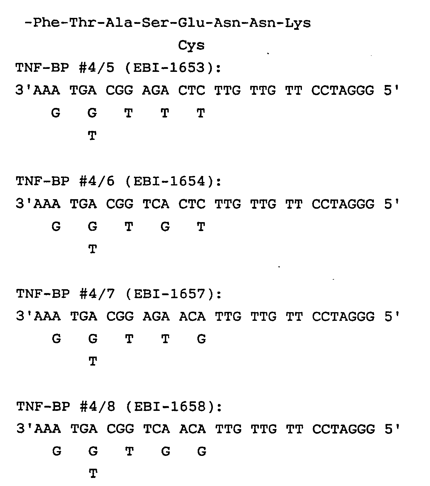
- Die Oligonukleotide wurden komplementär zur mRNA synthetisiert und sind somit zum 5′Ende der Sequenz orientiert. Um das amplifizierte DNA-Fragment im Anschluß an die PCR effizient klonieren zu können, wurde auch ein BamHI-Linker am 5′Ende der Oligonukleotide vorgesehen. Werden z.B. die Oligonukleotide TNF-BP #4/5-8 gemeinsam mit TNF-BP #3/1-4 für die PCR an der gesamten λ-DNA einer Bibliothek eingesetzt, kann ein etwa resultierendes DNA Fragment mit BamHI nachgeschnitten werden. Die Partner-Oligonukleotide ergeben ein gerades Ende am 5′Terminus, das Fragment kann somit in die SmaI-BamHI-Stellen eines geeigneten Vektors kloniert werden.
- Jedes Mischoligonukleotid TNF-BP #4/5 bis 8 ist eine Mischung aus 48 Einzelnukleotiden und berücksichtigt einige Codons nicht, und zwar:
Thr ACG
Ala GCG und GCT
Ser TCG und TCC
Asn AAT - Bei GCT wird die Möglichkeit in Betracht gezogen, daß das zu GCC (Ala) komplementäre Triplett CGG durch Ausbildung einer G-T Brücke wirksam sein kann, bei TCG (Ser) und AAT (Asn) gilt dasselbe bezüglich AGT bzw. TTG.
- ACG, GCG und TCG sind äußerst seltene Codons (CG-Regel) und wurden deshalb nicht berücksichtigt.
- Die Herstellung der cDNA Bibliothek erfolgte nach der in der EP-A1-0293 567 für die humane plazentale cDNA Bibliothek beschriebenen Methode mit dem Unterschied, daß als Ausgangsmaterial 10⁹ Fibrosarkomzellen der Zellinie HS 913 T, die unter Stimulierung mit humanem TNF-α (10 ng/ml) hochgezüchtet worden waren, verwendet wurden. Statt λ gt10 wurde λ gt11 verwendet (cDNA Synthese: Amersham RPN 1256; EcoRI verdaute λ gt11 Arme: Promega Biotech; in vitro Verpacken der ligierten DNA: Gigapack Plus, Stratagene).
- 5 ml des Phagenüberstandes der amplifizierten cDNA Bibliothek der humanen Fibrosarkom Zellinie HS913T in λ gt11 wurden mit 0,5 µg RNase A und 0,5 µg DNase I versetzt und eine Stunde bei 37°C inkubiert. Die Mischung wurde 10 min bei 5000xg zentrifugiert, der Überstand durch Extraktion mit Phenol und Chloroform von Protein befreit und die DNA aus der wässrigen Phase durch Zusatz von Ethanol präzipitiert. Die λ-DNA wurde in TE-Puffer (10 mM Tris pH=7,5; 1 mM EDTA) gelöst.
- Für die Anwendung der PCR (Saiki et al., 1988) auf DNA der HS913T cDNA Bibliothek wurden 16 Einzelreaktionen durchgeführt, in welchen jeweils eines der 4 Mischoligonukleotide EBI-1639, EBI-1640, EBI-1641, EBI-1642 als erster Primer und eines der vier Mischoligonukleotide EBI-1653, EBI-1654, EBI-1657, EBI- 1658 als zweiter Primer eingesetzt wurde. Jedes dieser Mischoligonukleotide enthält 48 verschiedene Oligonukleotide gleicher Länge.
Die Amplifizierung mittels PCR fand in 50 µl Reaktionsvolumen, enthaltend 250 ng λ-DNA der cDNA-Bibliothek, 50 mM KCl, 10 mM Tris pH=8,3, 1,5 mM MgCl₂, 0,01% Gelatine, 0,2 mM jedes der 4 desoxy- Nukleosidtriphosphate (dATP, dGTP, dCTP, dTTP), je 200 pMol erster und zweiter Primer, 1,25 Einheiten Taq Polymerase [Perkin-Elmer Cetus] statt. Um ein Verdunsten zu verhindern, wurde die Lösung mit einigen Tropfen Mineralöl (0,1 ml) überschichtet. Die PCR wurde in einem DNA Thermal Cycler (Perkin Elmer Cetus) folgendermaßen durchgeführt: Die Proben wurden 5 Minuten auf 94°C erhitzt, um die DNA zu denaturieren, und anschließend 40 Amplifikationszyklen unterworfen. Ein Zyklus bestand aus 40 Sekunden Inkubation bei 94°C, 2 Minuten Inkubation bei 55°C und 3 Minuten Inkubation bei 72°C. Am Ende des letzten Zyklus wurden die Proben für weitere 7 Minuten bei 72°C inkubiert, um sicherzustellen, daß die letzte Primer-Verlängerung vollständig verläuft. Nach Abkühlen auf Raumtemperatur wurden die Proben mit Phenol und Chloroform von Protein befreit und die DNA mit Äthanol präzipitiert. - 5 µl jeder der 16 PCR-Proben wurden auf ein Agarosegel aufgetragen und die Länge der amplifizierten DNA-Fragmente nach elektrophoretischer Auftrennung bestimmt. Die stärkste DNA Bande, ein Fragment von 0,16 kb Länge, war in den PCR-Proben zu sehen, die mit dem Oligonukleotid EBI-1653 als erstem Primer und einem der Oligonukleotide EBI-1639, EBI-1640, EBI-1641 oder EBI-1642 als zweitem Primer amplifiziert worden waren. Da die mit dem Primerpaar EBI-1653 und EBI-1642 amplifizierte Probe die größte Menge an diesem 0,16 kb DNA-Fragment enthielt, wurde diese Probe für die weitere Aufarbeitung ausgewählt.
- Das erhaltene PCR-Produkt der Primer EBI-1642 und EBI-1653 wurde mit BamHI geschnitten und nachfolgend elektrophoretisch in einem Agarosegel (1,5% Nusieve GTG Agarose plus 1% Seakem GTG Agarose, FMC Corporation) nach der Größe aufgetrennt. Die Hauptbande, ein DNA Fragment von 0,16 kb Länge, wurde aus dem Gel elektroeluiert und mit Ethanol präzipitiert. Dieses DNA Fragment wurde mit BamHI/SmaI geschnittenem Plasmid pUC18 (Pharmacia) ligiert und E. coli JM101 mit dem Ligationsgemisch transformiert. Die nach der Minipräparationsmethode hergestellten Plasmide wurden durch Schneiden mit den Restriktionsenzymen PvuII und EcoRI-BamHI und nachfolgender Elektrophorese in Agarosegelen charakterisiert. Das Plasmid pUC18 enthält zwei Schnittstellen für PvuII, die in einem 0,32 kb DNA-Fragment die Polyklonierstelle flankieren. Sehr kurze DNA-Inserts in der Polyklonierstelle des Plasmids können nach Schneiden mit PvuII leichter im Agarosegel sichtbar gemacht werden, da sich die Länge um 0,32 kb vergrößert. Durch Schneiden mit EcoRI und BamHI kann das in den mit BamHI und SmaI geschnittenen Plasmidvektor ligierte DNA-Fragment inklusive einiger Basenpaare der Polylinkersequenz erhalten werden. Ein Klon mit dem gewünschten Insert wurde als pTNF-BP3B bezeichnet. Das gesamte DNA-Insert dieses Klons wurde nach Subklonieren eines EcoRI-BamHI Fragments in M13mp18 (Pharmacia) nach der modifizierten Didesoxy Methode mit Sequenase (United States Biochemical Corporation) sequenziert.
- Die Analyse der durch PCR-amplifizierten DNA ergab folgende Sequenz (nur der nicht kodierende Strang ist abgebildet, darüber die abgeleitete Aminosäuresequenz):

- Die ersten 20 und die letzten 29 Nukleotide (in Kursivschrift) entsprechen den Sequenzen der Primer-Oligonukleotide EBI-1642 bzw. dem Komplement von EBI-1653. Die Aminosäuren 38 bis 43 bestätigen die restliche Sequenz des tryptischen Peptides 11.
- Weiters enthält das mittels PCR erzeugte DNA-Fragment die Sequenz des Peptides der Fraktion 20 des tryptischen Verdaus (Aminosäuren 20 bis 34, unterstrichen). Damit ist erwiesen, daß der Klon pTNF-BP3B von einer cDNA abgeleitet wurde, die für TNF-Bindungsprotein kodiert.
pTNF-BP3B stellt damit eine Sonde, z.B. zum Durchsuchen von cDNA-Bibliotheken nach TNF-BP cDNAs, dar. - Ca. 720.000 Phagen der HS913T cDNA Bibliothek in λ gt11 wurden auf E.coli Y1088 (Δ lacU169, pro::Tn5, tonA2, hsdR, supE, supF, metB, trpR, F⁻,λ ⁻, (pMC9)) plattiert (ca. 60.000 Phagen pro 14,5 cm Petrischale, LB-Agar: 10 g/l Trypton, 5 g/l Hefeextrakt, 5 g/l NaCl, 1,5% Agar, Plattieren in Top-Agarose: 10 g/l Trypton, 8 g/l NaCl, 0,8% Agarose). Von jeder Platte wurden zwei Nitrozellulosefilter-Abzüge hergestellt. Die Filter wurden vorgewaschen (16 Stunden bei 65°C) in:
50 mM Tris/HCl pH=8,0
1 M NaCl
1 mM EDTA
0,1 % SDS
Die Filter wurden zwei Stunden bei 65°C prähybridisiert in:
6x SSC (0,9M NaCl, 0,09 M tri-Na-citrat)
5x Denhardt's (0,1% Ficoll, 0,1% Polyvinylpyrrolidon, 0,1% BSA (=Rinderserumalbumin))
0,1% SDS - pTNF-BP 3B wurde mit BamHI und EcoRI doppelt geschnitten und das ca. 0,16 kb Insert isoliert. 0,6 µg des Inserts in 32 µl werden bei 100°C denaturiert und mit je 60 pMol EBI-1642 und EBI-1653 durch Abkühlen auf 80°C über 10 Minuten und jähes Abkühlen in Eiswasser geprimt. Nach Zusatz von
10 µl α-³²P-dCTP (100 µCi, 3,7 MBq)
5 µl 10x Priming Puffer (0,1 M Tris/HCl pH=8,0, 50 mM MgCl₂)
2 µl je 1mm dATP, dGTP, dTTP
1 µl PolIK (Klenow Fragment der E.coli DNA Polymerase I, 5 Einheiten)
wurde 90 Minuten bei Raumtemperatur inkubiert. Nach Hitzeinaktivierung (10 Minuten auf 70°C) erfolgte das Abtrennen der nichteingebauten Radioaktivität durch Chromatographie über Biogel P6DG (Biorad) in TE Puffer (10 mM Tris/HCl pH=8, 1 mM EDTA). Eingebaut wurden 65x10⁶ cpm.
Das Hybridisieren der Filter erfolgte in einem Gesamtvolumen von 80 ml 6xSSC/5X Denhardt's/0,1% SDS plus hitzedenaturierter Hybridisiersonde während 16 Stunden bei 65°C.
Die Filter wurden zweimal 30 Minuten bei Raumtemperatur in 6xSSC/0,01% SDS und einmal 45 Minuten bei Raumtemperatur in 2xSSC/0,01% SDS und dreimal 30 Minuten bei 65°C in 2xSSC/0,01% SDS gewaschen. Die Filter wurden luftgetrocknet und anschließend an Amersham Hyperfilm 16 Stunden unter Verwendung einer Verstärkerfolie bei -70°C exponiert. Insgesamt wurden 30 hybridisierende Plaques identifiziert (λ-TNF-BP #1-30).
Die Regionen mit den hybridisierenden Plaques wurden möglichst präzise ausgestochen, und die Phagen in 300 µl SM Puffer plus 30 µl Chloroform eluiert.
Durch "Plaquereinigung" (Plattieren von ca. 200 Phagen pro 9 cm Petrischale beim zweiten Durchgang, bzw. ca. 20 Phagen pro 9 cm Petrischale beim dritten Durchgang, Filterabzüge doppelt, Vorbereiten, Hybridisieren und Waschen wie beim erstmaligen Durchsuchen beschrieben) wurden letztlich 25 hybridisierende Phagen vereinzelt (λ-TNF-BP #1-10, 12-24, 29,30). - 2x10⁶ Phagen wurden auf E.coli Y1088 in Topagarose (10 g/l Trypton, 8 g/l NaCl, 0,8% Agarose) plattiert (14,5 cm Petrischale mit LB-Agarose (1,5% Agarose, 0,2% Glucose, 10 mM MgSO₄, 10 g/l Trypton, 5 g/l Hefeextrakt, 5 g/l NaCl) und 6 Stunden bei 37°C inkubiert. Nach Abkühlen der Platten (30 Minuten bei 4°C) wurde mit 10 ml λ-Diluent (10 mM Tris/HCl pH=8,0, 10 mM MgCl₂, 0,1 mM EDTA) überschichtet und 16 Stunden bei 4°C eluiert. Der Überstand wurde in 15 ml Corex Röhrchen transferiert und 10 Minuten bei 15000 rpm und 4°C zentrifugiert (Beckman J2-21 Zentrifuge, JA20 Rotor). Der Überstand wurde in 10 ml Polycarbonat-Röhrchen dekantiert und bei 50000 rpm, 20°C bis ω²t=3x10¹⁰ zentrifugiert (Beckman L8-70, 50 Ti Rotor). Das Pellet wurde in 0,5 ml λ-Diluent resuspendiert und in Eppendorf Röhrchen (1,4 ml) transferiert. Nach Zusatz von 5 µg RNase A und 0,5 µg DNaseI und Inkubation bei 37°C während 30 Minuten und Zusatz von 25 µl 0,5 M EDTA, 12,5 µl 1 M Tris/HCl pH=8.0, 6,5 µl 20% SDS erfolgte weitere Inkubation bei 70°C für 30 Minuten. Die λ-DNA wurde durch Phenol/Chloroform Extraktion gereinigt und mit Ethanol gefällt. Abschließend wurde die DNA in 100 µl TE-Puffer gelöst.
- Um die cDNAs der Klone λTNF-BP15 und λTNF-BP23, die bei der Hybridisierung die stärksten Signale gezeigt hatten, näher zu charakterisieren, wurden die cDNA-Inserts mit EcoRI aus der λ-DNA herausgeschnitten, nach elektrophoretischer Auftrennung aus einem Agarosegel eluiert und mit Äthanol präzipitiert. Die DNA-Fragmente von 1.3 kb (von λTNF-BP15) und 1.1 kb (von λTNF-BP23) wurden mit EcoRI geschnittenem und mit alkalischer Phosphatase aus Kalbsdarm dephosphoryliertem Plasmidvektor pT7/T3α-18 (Bethesda Research Laboratories) mit T4 DNA Ligase ligiert und E.coli JM101 transformiert. Von einzelnen Bakterienkolonien, die nach Selektion auf Agaroseplatten mit Ampicillin und X-gal keine blaue Färbung aufwiesen, wurde im Minipräparationsverfahren Plasmid-DNA hergestellt und durch Schneiden mit EcoRI und HindIII das Vorhandensein und die Orientierung des cDNA Inserts festgestellt. Plasmide, die das EcoRI Insert der Phagen λTNF-BP15 bzw. λTNF-BP23 so orientiert enthielten, daß das dem 5′-Ende der mRNA entsprechende Ende dem T7 Promoter zugewandt ist, wurden pTNF-BP15 bzw. pTNF-BP23 benannt. Die EcoRI Inserts von λTNF-BP15 und λTNF-BP23 wurden ebenfalls in mit EcoRI geschnittenen und dephosphorylierten M13mp19 Vektor ligiert und E.coli JM101 transformiert. Von einigen wahllos ausgewählten M13 Klonen wurde Einzelstrang-DNA präpariert und als Vorlage für die Sequenzierung nach der Didesoxy-Methode verwendet.
An M13 Klonen, die die cDNA-Inserts in entgegengesetzter Orientierung enthielten, wurden mit dem universellen Sequenzierprimer und spezifisch synthetisierten Oligonukleotidprimern, die an das cDNA-Insert binden, beide DNA-Stränge vollständig sequenziert. - Die vollständige Nukleotidsequenz von 1334 Basen des cDNA-Inserts von λTNF-BP15 bzw. pTNF-BP15 ist in Fig.1 dargestellt. Die Basen 1-6 und 1328-1334 entsprechen den EcoRI-Linkern, die bei der Herstellung der cDNA-Bibliothek an die cDNA angefügt worden waren. Die Nukleotidsequenz des cDNA-Inserts von λTNF-BP23 entspricht der von λTNF-BP15 (Basen 22-1100), flankiert von EcoRI-Linkern.
- Der Klon λTNF-BP30 wurde ebenfalls untersucht; seine Sequenz entspricht λTNF-BP15 mit dem Unterschied, daß die Sequenz eine Deletion von 74 bp (Nukleotid 764 bis 837) aufweist.
- Aus Teilen der Expressionsplasmide pCDM8 (Seed und Aruffo, 1987. Seed, 1987; Invitrogen), pSV2gptDHFR20 (EP-A1 0321 842) und dem Plasmid Bluescript SK+ (Short et al., 1988; Stratagene) wurde ein neues Plasmid konstruiert, das eine Multiklonierstelle für die gerichtete Insertion heterologer DNA-Sequenzen aufweist und sich in E.coli mittels Ampicillinresistenz mit hoher Kopienzahl vermehren läßt. Die intergenische Region von M13 ermöglicht die Herstellung einzelsträngiger Plasmid-DNA mittels Superinfektion der transformierten Bakterien mit einem Helferphagen (z.B. R408 oder M13K07) zur erleichterten Sequenzierung und Mutagenese der Plasmid-DNA. Der T7 Promoter, der der Multiklonierstelle vorangeht, ermöglicht die Herstellung von RNA Transkripten in vitro. In Säugetierzellen erfolgt die Expression heterologer Gene getrieben vom Cytomegalovirus (CMV) Promoter/Enhancer (Boshart et al., 1985). Der SV40 Replikationsursprung ermöglicht in geeigneten Zellinien (z.B. SV40 transformierte Zellen wie COS-7, Adenovirus transformierte Zellinie 293 (ATCC CRL1573) die autonome Replikation des Expressionsplasmides zu hohen Kopienzahlen und damit hohe Raten in transienter Expression. Für die Herstellung permanent transformierter Zellinien und die nachfolgende Amplifikation der Expressionskassette mittels Methotrexat dient ein modifiziertes Hamster-Minigen (Promoter mit kodierendem Bereich und dem ersten Intron) für Dihydrofolatreduktase (DHFR) als Selektionsmarker.
- Das Plasmid Bluescript SK+ wurde mit HindIII linearisiert und 5 ng DNA in einem 100 µl PCR Ansatz eingesetzt (Reaktionspuffer: 50 mM KCl, 10 mM Tris-Cl pH=8,3, 1,5 mM MgCl₂, 0,01% (w/v) Gelatine, 0,2 mM der vier Desoxynukleotidtriphosphate (dATP, dGTP, dCTP, dTTP), 2,5 Einheiten Taq Polymerase pro 100 µl). Als Primer wurden je 50 pmol der synthetischen Oligonukleotide EBI-1786 (5′-GGAATTCAGCCTGAATGGCGAATGGG-3′; bindet knapp außerhalb von M13 ori- Region in Bluescript Pos. 475, unabhängig von M13 ori- Orientierung) und EBI-1729 (5′-CCTCGAGCGTTGCTGGCGTTTTTCC-3′; bindet an Bluescript an Pos. 1195 vor ori, entspricht dem Anfang der Bluescript-Sequenz in pCDM8, 6 Basen 5′ergeben XhoI) eingesetzt. Nach 5 Minuten Denaturieren bei 94°C erfolgte die PCR über 20 Zyklen (40 sec bei 94°C, 45 sec bei 55°C, 5 Min bei 72°C, Perkin Elmer Cetus Thermal Cycler). Die Oligonukleotide flankieren die intergenische Region von M13 bzw. den Replikationsursprung (ori) mit dem dazwischenliegenden Gen für die β-Lactamase. Gleichzeitig wird am Ende des Replikationsursprungs eine XhoI- und am anderen Ende eine EcoRI-Schnittstelle erzeugt. Das Reaktionsgemisch wurde durch Extraktion mit Phenol-Chloroform von Protein befreit und die DNA mit Ethanol präzipitiert. Die erhaltene DNA wurde mit XhoI und EcoRI geschnitten und nach Elektrophorese in einem Agarosegel ein Fragment mit 2,3 kb isoliert.
- 5 ng mit SacII linearisiertes Plasmid pCDM8 wurden mit den Oligonukleotiden EBI-1733 (5′-GGTCGACATTGATTATTGACTAG-3′; bindet an CMV-Promotorregion (Pos. 1542) von pCDM8, entspricht Pos.1 in pAD-CMV, SalI-Stelle für Klonierung) und EBI-1734 (5′-GGAATTCCCTAGGAATACAGCGG-3′; bindet an Polyoma origin von 3′SV40 polyA-Region in pCDM8 (Pos. 3590)) unter identischen Bedingungen wie für Bluescript SK+ beschrieben, durch PCR amplifiziert. Die Oligonukleotide binden am Beginn der CMV-Promoter/Enhancer-Sequenz und erzeugen eine SalI Schnittstelle (EBI-1733) bzw. binden am Ende der SV40 poly-Adenylierungstelle und erzeugen eine EcoRI Schnittstelle (EBI-1734). Das PCR-Produkt wurde mit SalI und EcoRI geschnitten und ein DNA Fragment von 1,8 kb aus einem Agarosegel isoliert.
- Die beiden PCR Produkte wurden mit T4 DNA-Ligase ligiert, mit dem erhaltenen Ligationsprodukt E.coli HB101 transformiert und nach Standardmethoden Plasmid-DNA amplifiziert und präpariert. Das Plasmid der gewünschten Beschaffenheit (siehe Fig.3) wurde pCMV-M13 benannt.
- Der SV40 Replikationsursprung (SV40 ori) wurde aus dem Plasmid pSV2gptDHFR20 (EP-A1 0321842) isoliert. Dazu wurde dieses Plasmid mit HindIII und PvuII doppelt geschnitten und die DNA-Enden durch nachfolgende Behandlung mit dem großen Fragment der E.coli DNA Polymerase (Klenow Enzym) in Gegenwart der vier Desoxynukleotidtriphosphate stumpf gemacht. Ein dabei erhaltenes 0,36 kb DNA Fragment wurde aus einem Agarosegel isoliert und in mit EcoRI linearisiertem pCMV-M13 ligiert. Ein nach Transformation von E.coli HB101 erhaltenes Plasmid mit dem SV40 ori in gleicher Orientierung wie das β-Lactamase Gen und dem CMV-Promoter wurde pCMV-SV40 benannt. Die Konstruktion dieses Plasmids ist in Fig.3 dargestellt.
- Zur Herstellung eines Expressionsplasmids mit einer vielseitigen Multiklonierstelle wurden aus dem DHFR Minigen durch gerichtete Mutagenese zwei und durch Deletion drei Restriktionsenzymschnittstellen entfernt. Dazu wurde aus dem Plasmid pSV2gptDHFR20 ein 1,7 kb BglII Fragment, das die gesamte kodierende Region des Hamster DHFR-Gens enthält, in die BglII Stelle des Plasmids pUC219 (IBI) kloniert und das Plasmid pUCDHFR erhalten. Mit pUCDHFR transformierte E.coli JM109 (Stratagene) Zellen wurden mit etwa 40-fachem Überschuß des Helferphagen R408 (Stratagene) infiziert und 16 Stunden bei 37°C in LB-Medium geschüttelt. Aus dem Bakterienüberstand wurde einzelsträngige Plasmid-DNA isoliert.
- Die gerichtete Mutagenese erfolgte in zwei aufeinanderfolgenden Schritten, wobei das in vitro Mutagenese System RPN1523 (Amersham) verwendet wurde. Die am Beginn von Exon 2 befindliche EcoRI Stelle wurde durch Austausch einer Base von GAATTC zu GAGTTC zerstört. Dieser Basenaustausch führt zu keiner Änderung der kodierten Aminosäuresequenz und entspricht außerdem der Nukleotidsequenz im natürlichen murinen DHFR-Gen (McGrogan et al., 1985, Mitchell et al., 1986). Für die Mutagenese wurde ein Oligonukleotid (Antisense-Orientierung) der Sequenz 5′-GTACTTGAACTCGTTCCTG-3′ (EBI-1751) verwendet. Ein Plasmid mit der gewünschten Mutation wurde, wie oben beschrieben, als Einzelstrang-DNA präpariert und die im ersten Intron befindliche PstI Stelle durch Mutagenese mit dem Oligonukleotid EBI-1857 (Antisense Orientierung, 5′-GGCAAGGGCAGCAGCCGG-3′) von CTGCAG in CTGCTG entfernt. Die Mutationen wurden durch Sequenzierung bestätigt und da erhaltene Plasmid pUCDHFR-Mut2 benannt.
- Aus dem Plasmid pUCDHFR-Mut2 wurde das 1,7 kb BglII Fragment isoliert und in mit BglII und BamHI doppelt geschnittenes Plasmid pSV2gptDHFR20 ligiert. Nach Transformation von E.coli, Amplifikation und DNA-Isolierung wurde ein Plasmid der gewünschten Beschaffenheit erhalten, das als pSV2gptDHFR-Mut2 bezeichnet wurde. Durch Schneiden mit BamHI wurde in der 3′-nicht-kodierenden Region des DHFR Gens ein auf die BglII Stelle folgendes 0,12 kb DNA-Fragment entfernt, das außerdem noch eine KpnI Schnittstelle enthält. Durch Verknüpfen der mit BglII und BamHI entstandenen überhängenden DNA-Enden wurden auch die Erkennungssequenzen für diese beiden Enzyme zerstört.
- Das Plasmid pCMV-SV40 wurde mit EcoRI und BamHI doppelt geschnitten, die DNA-Enden nachfolgend mit Klenow-Enzym stumpf gemacht. Die DNA wurde durch Extraktion mit Phenol-Chloroform und Ethanolfällung gereinigt, anschließend durch Inkubation mit alkalischer Phosphatase dephosphoryliert und die 4,4 kb lange Vektor DNA aus einem Agarosegel isoliert.
- Das Plasmid pSV2gptDHFR-Mut2 (Fig.4) wurde mit EcoRI und PstI doppelt geschnitten und die DNA-Enden durch 20 Minuten Inkubation bei 11°C mit 5 Einheiten T4 DNA-Polymerase (50 mM Tris- HCl pH=8,0, 5 mM MgCl₂, 5 mM Dithiothreit, 0,1 mM jedes der vier Desoxynukleotidtriphosphate, 50 µg/ml Rinderserumalbumin) stumpf gemacht. Das 2,4 kb lange DNA-Fragment mit dem mutierten DHFR-Gen wurde aus einem Agarosegel isoliert und mit dem wie oben beschrieben präparierten pCMV-SV40 ligiert. Ein nach Transformation von E.coli erhaltenes Plasmid, das das DHFR-Gen in der selben Orientierung wie den CMV-Promoter enthielt, wurde pCMV-SV40DHFR benannt.
- Im letzten Schritt wurde das 0,4kb "Stuffer"-Fragment nach dem CMV-Promoter, das noch aus dem Ausgangsplasmid pCDM8 stammte, gegen eine Multiklonierstelle ausgetauscht. Dazu wurde das Plasmid pCMV-SV40DHFR mit HindIII und XbaI doppelt geschnitten und der Vektoranteil aus einem Agarosegel isoliert. Die Multiklonierstelle, gebildet aus den beiden Oligonukleotiden EBI-1823 (5′-AGCTTCTGCAGGTCGACATCGATGGATCCGGTACCTCGAGCGGCCGCGAATTCT-3′) und EBI-1829 (5′-CTAGAGAATTCGCGGCCGCTCGAGGTACCGGATCCATCGATGTCGACCTGCAGA-3′), enthält inklusive der für die Klonierung in HindIII - XbaI kompatiblen Enden Restriktionsschnittstellen für die Enzyme PstI, SalI, ClaI, BamHI, KpnI, XhoI, NotI und EcoRI.
- Je 1 µg der beiden Oligonukleotide wurden in 20 µl Reaktionspuffer (70 mM Tris-Cl pH=7,6, 10 mM MgCl₂, 5 mM Dithiothreit, 0,1 mM ATP) mit 5 Einheiten T4 Polynukleotidkinase eine Stunde bei 37°C inkubiert, um die 5′-Enden zu phosphorylieren. Die Reaktion wurde durch 10 minütiges Erhitzen auf 70°C gestoppt und die komplementären Oligonukleotide miteinander hybridisiert, indem die Probe weitere 10 Minuten bei 56°C inkubiert und anschließend langsam auf Raumtemperatur abgekühlt wurde. 4 µl der hybridisierten Oligonukleotide (100 ng) wurden mit etwa 100 ng Plasmidvektor ligiert und E.coli HB101 transformiert. Ein Plasmid, das sich mit den Enzymen der Multiklonierstelle (ausgenommen NotI) linearisieren ließ, wurde pAD-CMV1 benannt. Von vielen getesteten Klonen konnte keiner identifiziert werden, dessen Plasmid sich mit NotI schneiden ließ. Die Sequenzierung zeigte immer die Deletion von einigen Basen innerhalb der NotI Erkennungssequenz.
- In gleicher Weise wurde mit dem Oligonukleotidpaar EBI-1820 (5′-AGCTCTAGAGAATTCGCGGCCGCTCGAGGTACCGGATCCATCGATGTCGACCTGCAGAAGCTTG-3′) und EBI-1821 (5′-CTAGCAAGCTTCTGCAGGTCGACATCGATGGATCCGGTACCTCGAGCGGCCGCGAATTCTCTAG-3′) das Expressionsplasmid pAD-CMV2 hergestellt, das die Restriktionsschnittstellen innerhalb der Multiklonierstelle in umgekehrter Reihenfolge enthält. Dabei wurde das Plasmid pAD-CMV2 erhalten, das sich mit sämtlichen Restriktionsenzymen, einschließlich NotI, linearisieren ließ.
- Die Nukleotidsequenz des 6414 bp großen Plasmids pAD-CMV1 (Fig.5) ist zur Gänze in Fig.6 dargestellt.
- Die Abschnitte auf dem Plasmid (angegeben in der Numerierung der Basen) entsprechen folgenden Sequenzen:
1- 21 EBI-1733, Beginn CMV Enhancer - Promotor (aus CDM8)
632-649 T7 Promotor
658-713 Multiklonierstelle (HindIII bis XbaI aus EBI-1823, EBI-1829)
714-1412 SV40 Intron und poly-Adenylierungsstelle (aus CDM8)
1413-2310 5′nicht kodierende Region und Promotor des Hamster DHFR Gen (aus pSV2gptDHFR20)
2311-2396 Hamster DHFR: Exon 1
2516 A zu T Mutation zerstört PstI Stelle in DHFR Intron 1
2701-3178 DHFR Exons 2-6 (kodierende Region)
2707 A zu G Mutation zerstört EcoRI Stelle
3272-3273 Deletion zwischen BglII und BamHI in DHFR 3′nicht kodierender Region
3831 Ende DHFR Gen (aus pSV2gptDHFR20)
3832-4169 SV40 ori (aus pSV2gptDHFR20)
4170-4648 M13 ori (aus pBluescript SK+)
4780-5640 β-Lactamase (kodierende Region)
6395-6414 EBI-1729, Ende der pBluescript Vektorsequenz - Die Herstellung der Plasmide pAD-CMV1 und pAD-CMV2 ist in Fig.5 dargestellt.
- Um auf direktem Weg die sekretierte Form von TNF-BP herzustellen, wurde in der für einen Teil des TNF-Rezeptors kodierenden cDNA (vgl.Beispiel 7; im folgenden als TNF-R cDNA bezeichnet) nach dem Codon der C-terminalen Aminosäure des natürlichen TNF-BP (AAT, Asn-172; entspricht Pos.201 in Fig.9) ein Translationsstopcodon eingeführt. Dadurch wird die Proteinsynthese an dieser Stelle abgebrochen und ermöglicht, TNF-BP direkt in den Zellüberstand zu sekretieren, ohne eine nachfolgende, möglicherweise geschwindigkeitsbestimmende Reaktion einer proteolytischen Abspaltung in C-terminaler Richtung gelegener Abschnitte des TNF-Rezeptors durchlaufen zu müssen.
- Gleichzeitig mit der Einführung des Stopcodons mittels PCR wurde die 5′-nicht kodierende Region der TNF-R cDNA verkürzt, um das Translationsstartcodon eines weiteren offenen Leserahmens (Basen 72-203 in Fig.9), der sich 5′ von dem des TNF-R befindet, zu entfernen, und am 5′-bzw. 3′-Ende der cDNA eine BamHI bzw. EcoRI Schnittstelle eingeführt.
- 100 ng mit XmnI linearisiertes Plasmid pTNF-BP15 (vgl. Beispiel 7) wurden mit je 50 pmol der Oligonukleotide EBI-1986 (Sense, 5′-CAGGATCCGAGTCTCAACCCTCAAC-3′) und EBI-1929 (Antisense, 5′-GGGAATTCCTTATCAATTCTCAATCTGGGGTAGGCACAACTTC-3′; Einführung zweier Stop-Codons und einer EcoRI Stelle) in einem 100 µl PCR-Ansatz über 10 Zyklen amplifiziert. Die Zyklusbedingungen waren 40 Sekunden bei 94°C, 45 Sekunden bei 55°C und 5 Minuten bei 72°C. Nach dem letzten Zyklus wurde für weitere 7 Minuten bei 72°C inkubiert und die Reaktion durch Extraktion mit Phenol-Chloroform gestoppt. Die DNA wurde mit Ethanol präzipitiert und anschließend mit BamHI und EcoRI doppelt geschnitten. Das entstandene 0,75 kb DNA Fragment wurde aus einem Agarosegel isoliert und in mit BamHI und EcoRI doppelt geschnittenes Plasmid pT7/T3α-19 (BRL) kloniert. Eines der erhaltenen Plasmide, von dem aufgrund der Sequenzierung des gesamten Inserts festgestellt worden war, daß es die gewünschte Sequenz aufwies, wurde pTNF-BP benannt.
- pTNF-BP wurde mit BamHI und EcoRI geschnitten und das 0,75 kb DNA Insert in das mit BamHI und EcoRI geschnittene Expressionsplasmid pAD-CMV1 kloniert. Ein erhaltenes Plasmid der gewünschten Zusammensetzung wurde pADTNF-BP benannt (Fig.7A).
- Für eine weitere Variante eines Expressionsplasmides für die Produktion von sekretiertem TNF-BP wurde die 5′-nicht kodierende Region der TNF-R cDNA gegen die 5′-nicht kodierende Region der humanen β-Globin mRNA ausgetauscht. Der Grund dafür war die Feststellung, daß die Nukleotidsequenz unmittelbar vor dem Translationsstartcodon der TNF-R Sequenz deutlich von der für effiziente Expression eukaryotischer Gene gefundenen Konsensussequenz (Kozak,1987) abweicht, wogegen die 5′-nicht kodierende Region der β-Globin mRNA sehr gut mit dieser Konsensussequenz übereinstimmt (Lawn et al., 1980). Mittels des Oligonukleotids EBI-2452 (5′-CACAGTCGACTTACATTTGCTTCTGACACAACTGTGTTCACTAGCAACCTCAAACAGACACCATGGGCCTCTCCACCGTGC-3′), das nach einer SalI Restriktionsschnittstelle die authentische 5′-nicht kodierende Sequenz, entsprechend der humanen β-Globin mRNA-Sequenz, gefolgt von 20 Basen der kodierenden Region von TNF-BP enthielt, wurde in einer PCR die TNF-R Sequenz modifiziert. 100 ng mit EcoRI linearisiertes Plasmid pTNF-BP wurden in 100 µl Reaktionsansatz mit je 50 pmol der Oligonukleotide EBI-2452 und EBI-1922 (Antisense, 5′-GAGGCTGCAATTGAAGC-3′;bindet an die huTNF-R Sequenz bei Pos. 656) in 20 PCR-Zyklen (40 sec bei 94°C, 45 sec bei 55°C, 90 sec bei 72°C) amplifiziert. Nach Reinigung des PCR-Produktes durch Extraktion mit Phenol-Chloroform und Ethanolfällung wurde die DNA mit SalI und BglII doppelt geschnitten und das entstandene 0,51 kb DNA Fragment aus einem Agarosegel isoliert. Der entsprechende Teil der TNF-R Sequenz wurde aus dem Plasmid pTNF-BP durch Schneiden mit SalI und BglII entfernt, der 3,1 kb lange Plasmidteil aus einem Agarosegel isoliert und mit dem 0,51 kb langen PCR-Produkt ligiert. Nach Transformation von E.coli wurden sieben der erhaltenen Plasmide sequenziert. Eines dieser Plasmide enthielt genau die gewünschte Sequenz. Dieses Plasmid wurde pBTNF-BP benannt.
- Das gesamte SalI - EcoRI Insert von pBTNF-BP wurde in das ebenso geschnittene Expressionsplasmid pAD-CMV1 kloniert und das erhaltene Plasmid pADBTNF-BP benannt (Fig.7B).
- Zunächst wurde eine Rattenhirn-cDNA analog der HS913T cDNA Bibliothek (vgl. Beispiel 4) aus der Ratten Glia Tumor Zellinie C6 (ATCC Nr. CCL107) in λ-gt11 hergestellt.
- 600.000 Phagen der Rattenhirn cDNA Bibliothek in λ-gt11 wurden, wie in Beispiel 6 beschrieben, durch Hybridisierung gescreent. Als Sonde wurde das gereinigte EcoRI Insert von pTNF-BP30 (vgl.Beispiel 6) verwendet. Etwa 100 ng DNA wurden mit 1 µg Random Hexamer Primer anstatt der spezifischen Oligonukleotide, wie in Beispiel 6 beschrieben, mit [α-³²P]dCTP radioaktiv markiert. Eingebaut wurden 25x10⁶ cpm. Die Hybridisierung der Filter erfolgte unter gleichen Bedingungen wie in Beispiel 6. Die Filter wurden zweimal 30 Minuten bei Raumtemperatur in 2xSSC/0,1% SDS und dreimal 30 Minuten bei 65°C in 2xSSC/0,1%SDS und zweimal 30 Minuten bei 65°C in 0,5xSSC/0,5%SDS gewaschen. Die luftgetrockneten Filter wurden anschließend an Kodak XAR Röntgenfilm 16 Stunden unter Verwendung einer Verstärkerfolie bei -70°C exponiert. Insgesamt wurden 10 hybridisierende Plaques identifiziert und durch Plaquereinigung vereinzelt. Nach dreimaliger Plaquereinigung wurden schließlich drei λ-Klone (λ-raTNF-R #3, 4, 8) vereinzelt und die Phagen DNA, wie beschrieben, dargestellt.
- Die Länge der cDNA Inserts wurde nach Schneiden der λ-DNA mit EcoRI und Auftrennung in einem Agarosegel mit 2,2 kb für die Klone raTNF-R3 und raTNF-R8 und 2,1 kb für Klon raTNF-R4 bestimmt. Die EcoRI Inserts der Klone λraTNF-R3 und 8 wurden in ebenso geschnittenen M13mp19 kloniert und die DNA Sequenz mit universellen Sequenzierprimern und spezifisch synthetisierten Oligonukleotidprimern bestimmt.
- Die vollständige Nukleotidsequenz von raTNF-R8 ist in Fig.8 dargestellt. Die ersten und letzten sieben Basen entsprechen den EcoRI-Linkern, die bei der Herstellung der cDNA-Bibliothek angefügt worden waren.
- Die vollständige cDNA des Ratten TNF-R erleichterte die Suche nach dem noch fehlenden 3′-Teil der humanen TNF-R cDNA.
- Als Sonde für die Hybridisierung wurde das 0,4 kb lange PCR-Produkt der Primer EBI-2316 (5′-ATTCGTGCGGCGCCTAG-3′; bindet an TNF-R mit 2.Base von EcoRI, an der TNF-R cDNA abbricht) und EBI-2467 (5′-GTCGGTAGCACCAAGGA-3′; bindet ca. 400 Basen vor poly-A an cDNA-Klon, entspricht Pos. 1775 in raTNF-R) mit λraTNF-R8 als Vorlage eingesetzt. Dieses DNA Fragment entspricht der Region der Ratten TNF-R cDNA, von der angenommen wurde, daß sie der, welche der internen EcoRI Stelle im humanen TNF-R folgt, entspricht.
- 2,5x10⁶ cpm der raTNF-R Sonde wurden eingesetzt, um 600.000 Plaques der HS913T cDNA-Bibliothek zu hybridisieren. Die Hybridisierbedingungen entsprachen den in Beispiel 6 angegebenen. Die Filter wurden zweimal 30 Minuten bei Raumtemperatur in 2xSSC/0,1%SDS und zweimal 30 Minuten bei 65°C in 2xSSC/0,1%SDS gewaschen, an der Luft getrocknet und an Kodak XAR Röntgenfilm unter Verwendung einer Verstärkerfolie 3 Tage bei -70°C exponiert. Sechs positive Plaques wurden identifiziert, in zwei weiteren Runden Plaques gereinigt und λ-DNA dargestellt (λ-TNF-R #2, 5, 6, 8, 11, 12). Nach Schneiden der λ-DNA mit EcoRI wiesen alle Klone eine DNA Bande mit etwa 0,8 kb Länge auf. λTNF-R2 und 11 enthielten zusätzlich ein EcoRI Fragment mit 1,3 kb. Die beiden EcoRI Inserts aus λTNF-R2 wurden in die EcoRI Stelle von Plasmid pUC218 (IBI) subkloniert und anschließend sequenziert. Die Sequenz des 1,3 kb EcoRI Fragments entsprach der von cDNA Klon pTNF-BP15, das 0,8 kb EcoRI Fragment entspricht dem 3′-Abschnitt der TNF-R mRNA und enthält vor der EcoRI-Linker Sequenz einen poly-A Schwanz mit 16 A Resten. λTNF-R2 enthält demnach die vollständige kodierende Region des humanen TNF-R, dargestellt in Fig.9.
- Zunächst wurde, ähnlich wie im Beispiel 9 für pTNF-BP bzw. pADTNF-BP beschrieben, ein Plasmid konstruiert, in dem die 5′-nicht kodierende Region von pTNF-BP15 verkürzt, im Unterschied zu den im Beispiel 9 beschriebenen Plasmiden jedoch das 3′-Ende von pTNF-BP15 beibehalten wurde. Dazu wurde unter identischen Bedingungen wie in Beispiel 9 mit dem Oligonukleotid EBI-1986 und dem M13 -40 Universalprimer (5′-GTTTTCCCAGTCACGAC-3′) pTNF-BP15 mit PCR amplifiziert. Das PCR Produkt wurde mit BamHI und EcoRI doppelt geschnitten und in das Plasmid pT7/T3α-19 kloniert. Eines der erhaltenen Plasmide wurde pTNF-BP15B benannt.
- pTNF-BP15B wurde mit BamHI und EcoRI geschnitten und das 1,26 kb DNA Insert in mit BamHI und EcoRI geschnittenes Expressionsplasmid pAD-CMV1 kloniert. Ein erhaltenes Plasmid der gewünschten Zusammensetzung wurde pADTNF-BP15 benannt.
- Dieses Plasmid wurde mit EcoRI linearisiert und in die Schnittstelle das 0,8 kb EcoRI Fragment, isoliert aus λTNF-R2, kloniert. Nach Transformation von E.coli wurden einige wahllos isolierte Plasmide durch Schneiden mit verschiedenen Restriktionsenzymen auf die korrekte Orientierung des eingesetzten EcoRI Fragments überprüft. Ein Plasmid, bezeichnet als pADTNF-R (Fig.7C), wurde noch genauer auf korrekte Orientierung untersucht, indem das Insert ausgehend vom 3′-Ende der insertierten cDNA mit dem Oligonukleotid EBI-2112 (5′-GTCCAATTATGTCACACC-3′), das nach der Multiklonierstelle an das Plasmid pAD-CMV1 und seine Derivate bindet, sequenziert wurde.
- Ein weiteres Expressionsplasmid, in dem die 5′-nicht kodierende Region des TNF-R gegen die von β-Globin ausgetauscht ist, wurde konstruiert. Plasmid pADBTNF-BP wurde mit BglII vollständig geschnitten, um das 1,1 kb BglII Fragment zu entfernen, die DNA-Enden wurden nachfolgend mit alkalischer Phosphatase aus Kälberdarm dephosphoryliert und der Plasmidvektor (5,9 kb) mit der β-Globin 5′-nicht kodierenden Region des β-Globingens und dem 5′-Teil der TNF-R kodierenden Region aus einem Agarosegel isoliert. Plasmid pADTNF-R wurde mit BglII geschnitten und das 2,5 kb DNA Fragment, enthaltend den 3′-Abschnitt der TNF-R cDNA bis zur Promoterregion des nachfolgenden DHFR-Gens, aus einem Agarosegel isoliert und in den zuvor präparierten Plasmidvektor kloniert. Ein nach Transformation von E.coli erhaltenes Plasmid mit dem in korrekter Orientierung insertierten BglII-Fragment wurde pADBTNF-R benannt (Fig.7D).
- In diesem Beispiel wurde der Nachweis von TNF-BP mittels ELISA-Test wie folgt durchgeführt:
- 96-Napf Mikrotiterplatten wurden pro Napf mit 50 µl 1:3000 verdünntem polyklonalem Kaninchen-Serum (polyklonale Kaninchenantikörper, hergestellt durch Präzipitation von Antiserum mit Ammoniumsulfat, Endkonzentration 50 % Sättigung) gegen natürliches TNF-BP 18 Stunden bei 4°C beschichtet, einmal mit 0,05% Tween-20 in PBS gewaschen und freie Bindungsstellen mit 150-200 µl 0,5% Rinderserumalbumin, 0,05% Tween-20 in PBS (PBS/BSS/Tween) eine Stunde bei Raumtemperatur blockiert. Die Näpfe wurden einmal mit 0,05% Tween-20 in PBS gewaschen und 50 µl Zellüberstand oder bekannte Mengen von natürlichem TNF-BP (vgl. Tab.3 und 4) und 50 µl einer 1:10.000-fachen Verdünnung eines polyklonalen Mäuseserums gegen TNF-BP aufgetragen und zwei Stunden bei Raumtemperatur inkubiert. Anschließend wurden die Näpfe dreimal mit 0,05% Tween-20 in PBS gewaschen und 50 µl Kaninchen anti-Maus Ig-Peroxidase Konjugat (Dako P161; 1:5000 in PBS/BSA/Tween), zugegeben und weitere zwei Stunden bei Raumtemperatur inkubiert. Die Näpfe wurden dreimal mit Tween/PBS gewaschen und die Färbereaktion mit Orthophenylendiamin (3 mg/ml) und Na-Perborat (1 mg/ml) in 0,067M Kalium-Citrat pH 5,0, 100 µl/Napf, 20 Minuten bei Raumtemperatur unter Lichtschutz durchgeführt. Nach Zugabe von 100 µl 4N H₂SO₄ wurde die Farbintensität bei einer Wellenlänge von 492 nm in einer Mikrofilmplatten-Photometer photometrisch gemessen.
- Etwa 10⁶ Zellen (COS-7) pro 80 mm Petrischale wurden 24 Stunden vor der Transfektion in RPMI-1640 Medium mit 10% hitzeinaktiviertem fötalem Kälberserum angesetzt und bei 37°C in 5% CO₂ Atmosphäre inkubiert. Die Zellen wurden mit einem Gummischaber von der Petrischale gelöst und 5 Minuten bei 1200 UpM bei Raumtemperatur abzentrifugiert (Heraeus Minifuge, Ausschwing-Rotor 3360), einmal mit 5 ml serumfreiem Medium gewaschen, 5 Minuten bei 1200 UpM zentrifugiert und in 1 ml Medium, versetzt mit 250 µg/ml DEAE-Dextran und 10 µg Plasmid DNA (vgl. Tab.3, gereinigt durch zweimalige CsCl Dichtegradientenzentrifugation) suspendiert. Die Zellen wurden 40 Minuten bei 37°C inkubiert, einmal mit 5 ml Medium mit 10% Kälberserum gewaschen und in 5 ml Medium mit 100 µg/ml Chloroquin suspendiert. Die Zellen wurden eine Stunde bei 37°C inkubiert, einmal mit Medium gewaschen und mit 10 ml frischem Medium bei 37°C inkubiert. Nach 72 Stunden wurde der Zellüberstand geerntet und zum Nachweis des sekretierten TNF-BP verwendet.
Tabelle 3: Zellinie COS-7 ohne Plasmid < 5 ng/ml pADTNF-BP 7,5 ng/ml pADBTNF-BP 146 ng/ml - Die Dihydrofolatreduktase(DHFR)-defiziente Hamster Ovarial Zellinie CHO DUKX BII (Urlaub und Chasin, 1980) wurde mit Plasmid pADBTNF-BP mittels Kalzium-Phosphat Präzipitation transfiziert (Current protocols in molecular biology, 1987). Vier dicht bewachsene Zellkultur-Fläschchen (25 cm², 5 ml Kulturmedium pro Fläschchen) wurden mit je 5 µg DNA transfiziert; nach vierstündiger Inkubation bei 37°C wurde das Medium entfernt und durch je 5 ml Selektionsmedium (MEM alpha Medium mit 10 % dialysiertem fötalem Rinderserum) ersetzt. Nach Inkubation über Nacht wurden die Zellen mittels Trypsin-Lösung abgelöst; die Zellen aus jedem Fläschchen wurden in zwei 96-Napf Gewebekulturplatten aufgeteilt (100 µl/Napf in Selektionsmedium). In etwa wöchentlichen Abständen wurde frisches Medium zugegeben. Nach etwa vier Wochen konnten in 79 Näpfen Zellklone beobachtet werden. Die Überstände wurden im ELISA auf TNF-BP Aktivität getestet. 37 Überstände zeigten Aktivität im ELISA. Die Ergebnisse des ELISA-Tests einiger posiver Klone ist in Tab.4 dargestellt.
Tabelle 4: Probe Absorption bei 492 nm TNF-BP Standard 1 ng/ml 0,390 10 ng/ml 1,233 100 ng/ml 1,875 Kulturmedium (Negativkontrolle) 0,085 Klon A1G3 0,468 A2F5 0,931 A3A12 0,924 A4B8 0,356 A5A12 0,806 A5B10 0,915 A5C1 0,966 - Je 1 µg poly-A⁺ RNA (isoliert aus HS913T (Fibrosarkom), Plazenta und Milz wurden in einem 1,5%igen vertikalen Agarosegel (10mM Na-Phosphatpuffer pH=7,0, 6,7 % Formaldehyd) elektrophoretisch aufgetrennt. Als Größenmarker wurde eine durch Einfüllreaktion mit (α-³²P)dCTP und Klenow-Enzym radioaktiv markierte Kilobasenleiter (Bethesda Research Laboratories) verwendet. Das Formaldehyd wurde durch Wässern aus dem Gel entfernt und die RNA in 20x SSC auf eine Nylon-Membran (Genescreen plus, NEN-DuPont) transferiert. Die RNA wurde durch UV-Bestrahlung (100 Sekunden) auf der Membran kovalent gebunden. Die Membran wurde 2 Stunden bei 65°C in Church-Puffer (Church and Gilbert, 1984), (0,5 M Na-Phosphat pH=7,2, 7 % SDS, 1 mM EDTA) prähybridisiert und 19 Stunden bei 65°C in frischem Church-Puffer mit 3x10⁶ cpm P-32 markierter DNA Sonde (EcoRI Insert von pTNF-BP30) hybridisiert. Das Filter wurde dreimal 10 Minuten bei Raumtemperatur in Waschpuffer (40 mM Na-Phosphat pH=7,2, 1 % SDS) und anschließend viermal 30 Minuten bei 65°C in Waschpuffer gewaschen und 18 Stunden auf Kodak XAR Röntgenfilm unter Verwendung einer Verstärkerfolie bei -70°C exponiert.
Das Autoradiogramm (Fig.10) zeigt eine singuläre RNA Bande einer Länge von 2,3 kb für den humanen TNF-Rezeptor in den analysierten Geweben bzw. der Zellinie HS913T. - Für die transiente Expression wurden 5 - 10x10⁷ COS-7 Zellen 40 Minuten lang mit 10 µg pADTNF-R-Plasmid-DNA in einer Lösung enthaltend 250 µg/ml DEAE Dextran und 50 µg/ml Chloroquin inkubiert. Als Kontrolle wurde pADCMV-1-DNA verwendet. Nach der Transfektion wurden die Zellen gewaschen und anschließend 48 h lang gezüchtet. Die Expression des TNF-Rezeptors wurde durch Bindung von ¹²⁵I-TNF nachgewiesen. Für die Bindungstests wurden die Zellen gewaschen, 1 h lang bei 4°C mit 10 ng ¹²⁵I-TNF (spezifische Radioaktivität 38.000 cpm/ng) mit oder ohne 200fachen Überschuß an unmarkiertem TNF inkubiert, gewaschen, und die an die Zellen gebundene Radioaktivität in einem Gamma-Zähler gemessen. Die spezifische Bindung betrug bei der Kontrollprobe 2062 cpm, bei den mit TNF-Rezeptor DNA transformierten Proben 6150 cpm (die Werte sind ausgedrückt als mittlere gebundene cpm; die Standardabweichung, bestimmt aus Parallelversuchen, ist berücksichtigt. Unspezifischer Untergrund in Anwesenheit von unmarkiertem TNF wurde von den Werten abgezogen).
-
- Aggarwal, B.B., et al., 1985, Nature 318, 655-667
- Beutler B., et al., 1988, Ann. Rev. Biochem. 57, 505-18
- Beutler, B., et al., 1985, Nature 316, 552 - 554
- Boshart, M., et al., 1985, Cell 41, 521-530
- Carswell, E.A., et al., 1975, Proc.Natl.Acad.Sci.25, 3666-3670
- Cerami, A., et al., 1988, Immunol. Today 9, 28-31
- Church und Gilbert, 1984, Proc.Natl.Acad.Sci. 81, 1991-1995
- Creasey, A.A., et al., 1987, Proc.Natl.Acad.Sci. 84, 3293-3297
- Engelmann, H., et al., 1990, J.Biol.Chem.265, 1531-1536
- Frohman, M.A., et al., 1988, Proc.Natl.Acad.Sci. 85, 8998-9002
- Gullberg, U., et al., 1987, Eur. J. Haematol. 39, 241-251
- Hsieng, S.L., et al., 1988, J. Chromatography 447, 351-364
- Klebanoff, S.J., et al., 1986, J. Immunol. 136, 4220-4225
- Kozak M., 1987,Nucleic Acids Res. 15, 8125-8148
- Laemmli, U.K., 1970, Nature 227, 680-4
- Lawn, et al., 1980, Cell 21, 647-651
- Locksley, R.M., et al. 1987. J.Imunol. 139, 1891-1895
- Mahoney, J.R., et al., 1985, J. Immunol. 134, 1673-1675
- Maniatis, T., et al., 1982, Molecular Cloning A laboratory Manual. Cold Spring Harbor Laboratory, 474
- McGrogan, M., et al., 1985, J.Biol.Chem. 260, 2307-2314
- Mestan, J., et al., 1986, Nature 323, 816-819
- Mitchell, P.J., et al., 1986, Mol.Cell.Biol. 6, 425-440
- Oakley, B.R., et al., 1986, Analyt. Biochem. 105, 361-363
- 0ld, L.J., 1987, Nature 326, 330-331
- Oliff A., et al., 1987, Cell 555-63
- Olsson I., et al., 1988, Eur.J. Haematol. 41, 414 - 420
- Olsson I., et al., 1989, Eur.J. Haematol. 42, 270 - 275
- Pieler Ch., 1987, Dissertation, Universität Wien
- Piguet, P.F., et al., 1987, Immunobiol. 175, 27
- Saiki, R.K., 1988, Science 239, 487-491
- Sanger et al., 1977, Proc.Natl.Acad.Sci. 74, 5463-5467
- Seckinger P., et al., 1988, J. Exp. Med., 1511-16
- Seckinger, P., et al., 1987, J. Immunol. 139, 1546-1549
- Seed und Aruffo, 1987, Proc.Natl.Acad.Sci. 84, 8573-8577
- Seed, B., 1987, Nature 329, 840-842
- Shalaby, M.R., et al., 1985, J. Immunol. 135, 2069-2073
- Short, J.M., et al., 1988, Nucl.Acids Res.11, 5521-5540
- Staden, R., 1982, Nucleic Acid Res. 10, 4731-4751
- Stauber, G.B., et al., 1988, J.Biolog.Chem. 35, Vol.263, 19098-19104
- Stauber, G.B., et al., 1989, J.Biolog.Chem. 6, Vol.264, 3573-3576
- Torti, F.M. et al., 1985, Nature 229:867-869
- Tracey, K.J., et al., 1987, Nature 330, 662-666
- Tracey, K.J., et al., 1986, Science 234, 470-474
- Urlaub und Chasin, 1980, Proc.Natl.Acad.Sci. 77, 4216-4220
- Waage, A., et al., 1987, Lancet. ii, 355-357
- Wong, G.H.W., et al., 1986, Nature 323, 819-822
Claims (30)
ATG GGC CTC TCC ACC GTG CCT GAC CTG CTG CTG CCA CTG GTG CTC CTG GAG CTG TTG GTG GGA ATA TAC CCC TCA GGG GTT ATT GGA CTG GTC CCT CAC CTA GGG GAC AGG GAG AAG AGA GAT AGT GTG TGT CCC CAA GGA AAA TAT ATC CAC CCT CAA AAT AAT TCG ATT TGC TGT ACC AAG TGC CAC AAA GGA ACC TAC TTG TAC AAT GAC TGT CCA GGC CCG GGG CAG GAT ACG GAC TGC AGG GAG TGT GAG AGC GGC TCC TTC ACC GCT TCA GAA AAC CAC CTC AGA CAC TGC CTC AGC TGC TCC AAA TGC CGA AAG GAA ATG GGT CAG GTG GAG ATC TCT TCT TGC ACA GTG GAC CGG GAC ACC GTG TGT GGC TGC AGG AAG AAC CAG TAC CGG CAT TAT TGG AGT GAA AAC CTT TTC CAG TGC TTC AAT TGC AGC CTC TGC CTC AAT GGG ACC GTG CAC CTC TCC TGC CAG GAG AAA CAG AAC ACC GTG TGC ACC TGC CAT GCA GGT TTC TTT CTA AGA GAA AAC GAG TGT GTC TCC TGT AGT AAC TGT AAG AAA AGC CTG GAG TGC ACG AAG TTG TGC CTA CCC CAG ATT GAG AAT GTT AAG GGC ACT GAG GAC TCA GGC ACC ACA GTG CTG TTG CCC CTG GTC ATT TTC TTT GGT CTT TGC CTT TTA TCC CTC CTC TTC ATT GGT TTA ATG TAT CGC TAC CAA CGG TGG AAG TCC AAG CTC TAC TCC ATT GTT TGT GGG AAA TCG ACA CCT GAA AAA GAG GGG GAG CTT GAA GGA ACT ACT ACT AAG CCC CTG GCC CCA AAC CCA AGC TTC AGT CCC ACT CCA GGC TTC ACC CCC ACC CTG GGC TTC AGT CCC GTG CCC AGT TCC ACC TTC ACC TCC AGC TCC ACC TAT ACC CCC GGT GAC TGT CCC AAC TTT GCG GCT CCC CGC AGA GAG GTG GCA CCA CCC TAT CAG GGG GCT GAC CCC ATC CTT GCG ACA GCC CTC GCC TCC GAC CCC ATC CCC AAC CCC CTT CAG AAG TGG GAG GAC AGC GCC CAC AAG CCA CAG AGC CTA GAC ACT GAT GAC CCC GCG ACG CTG TAC GCC GTG GTG GAG AAC GTG CCC CCG TTG CGC TGG AAG GAA TTC GTG CGG CGC CTA GGG CTG AGC GAC CAC GAG ATC GAT CGG CTG GAG CTG CAG AAC GGG CGC TGC CTG CGC GAG GCG CAA TAC AGC ATG CTG GCG ACC TGG AGG CGG CGC ACG CCG CGG CGC GAG GCC ACG CTG GAG CTG CTG GGA CGC GTG CTC CGC GAC ATG GAC CTG CTG GGC TGC CTG GAG GAC ATC GAG GAG GCG CTT TGC GGC CCC GCC GCC CTC CCG CCC GCG CCC AGT CTT CTC AGA TGA
oder Abschnitte davon aufweist, einschließlich ihrer degenerierten Varianten.

ATG GGC CTC TCC ACC GTG CCT GAC CTG CTG CTG CCA CTG GTG CTC CTG GAG CTG TTG GTG GGA ATA TAC CCC TCA GGG GTT ATT GGA
steht.
met gly leu ser thr val pro asp leu leu leu pro leu val leu leu glu leu leu val gly ile tyr pro ser gly val ile gly leu val pro his leu gly asp arg glu lys arg asp ser val cys pro gln gly lys tyr ile his pro gln asn asn ser ile cys cys thr lys cys his lys gly thr tyr leu tyr asn asp cys pro gly pro gly gln asp thr asp cys arg glu cys glu ser gly ser phe thr ala ser glu asn his leu arg his cys leu ser cys ser lys cys arg lys glu met gly gln val glu ile ser ser cys thr val asp arg asp thr val cys gly cys arg lys asn gln tyr arg his tyr trp ser glu asn leu phe gln cys phe asn cys ser leu cys leu asn gly thr val his leu ser cys gln glu lys gln asn thr val cys thr cys his ala gly phe phe leu arg glu asn glu cys val ser cys ser asn cys lys lys ser leu glu cys thr lys leu cys leu pro gln ile glu asn val lys gly thr glu asp ser gly thr thr val leu leu pro leu val ile phe phe gly leu cys leu leu ser leu leu phe ile gly leu met tyr arg tyr gln arg trp lys ser lys leu tyr ser ile val cys gly lys ser thr pro glu lys glu gly glu leu glu gly thr thr thr lys pro leu ala pro asn pro ser phe ser pro thr pro gly phe thr pro thr leu gly phe ser pro val pro ser ser thr phe thr ser ser ser thr tyr thr pro gly asp cys pro asn phe ala ala pro arg arg glu val ala pro pro tyr gln gly ala asp pro ile leu ala thr ala leu ala ser asp pro ile pro asn pro leu gln lys trp glu asp ser ala his lys pro gln ser leu asp thr asp asp pro ala thr leu tyr ala val val glu asn val pro pro leu arg trp lys glu phe val arg arg leu gly leu ser asp his glu ile asp arg leu glu leu gln asn gly arg cys leu arg glu ala gln tyr ser met leu ala thr trp arg arg arg thr pro arg arg glu ala thr leu glu leu leu gly arg val leu arg asp met asp leu leu gly cys leu glu asp ile glu glu ala leu cys gly pro ala ala leu pro pro ala pro ser leu leu arg
oder ein Polypeptid bestehend aus mindestens einem Fragment davon ist.
asp ser val cys pro gln gly lys tyr ile his pro gln asn asn ser ile cys cys thr lys cys his lys gly thr tyr leu tyr asn asp cys pro gly pro gly gln asp thr asp cys arg glu cys glu ser gly ser phe thr ala ser glu asn his leu arg his cys leu ser cys ser lys cys arg lys glu met gly gln val glu ile ser ser cys thr val asp arg asp thr val cys gly cys arg lys asn gln tyr arg his tyr trp ser glu asn leu phe gln cys phe asn cys ser leu cys leu asn gly thr val his leu ser cys gln glu lys gln asn thr val cys thr cys his ala gly phe phe leu arg glu asn glu cys val ser cys ser asn cys lys lys ser leu glu cys thr lys leu cys leu pro gln ile glu asn
oder ein funktionelles Fragment oder Derivat mit der Fähigkeit, TNF zu binden, ist.
Priority Applications (10)
| Application Number | Priority Date | Filing Date | Title |
|---|---|---|---|
| JP2105102A JPH03164179A (ja) | 1989-04-21 | 1990-04-20 | Tnfレセプター、tnf結合たん白質およびこれらをコードするdna |
| US08/383,676 US6294352B1 (en) | 1989-04-21 | 1995-02-01 | TNF receptors, TNF binding proteins and DNAs coding for them |
| US08/477,639 US6221675B1 (en) | 1989-04-21 | 1995-06-07 | TNF receptors, TNF binding proteins and DNAs coding for them |
| US08/477,638 US6417158B1 (en) | 1989-04-21 | 1995-06-07 | Method of ameliorating the harmful effects of TNF using a polypeptide having the ability to bind to TNF |
| US08/484,312 US6271346B1 (en) | 1989-04-21 | 1995-06-07 | TNF Receptors, TNF binding proteins and DNAs coding for them |
| US08/484,307 US6440693B1 (en) | 1989-04-21 | 1995-06-07 | TNF receptors, TNF binding proteins and DNAS coding for them |
| US09/525,998 US7264944B1 (en) | 1989-04-21 | 2000-03-15 | TNF receptors, TNF binding proteins and DNAs coding for them |
| US09/792,356 US20020183485A1 (en) | 1989-04-21 | 2001-02-23 | TNF receptors, TNF binding proteins and DNAs coding for them |
| US09/898,234 US7282483B2 (en) | 1989-04-21 | 2001-07-03 | Method of ameliorating the harmful effects of TNF using a polypeptide having the ability to bind to TNF |
| US09/899,422 US7238776B2 (en) | 1989-04-21 | 2001-07-03 | TNF binding proteins |
Applications Claiming Priority (4)
| Application Number | Priority Date | Filing Date | Title |
|---|---|---|---|
| DE3913101A DE3913101A1 (de) | 1989-04-21 | 1989-04-21 | Tnf-(alpha) bindende proteine und dafuer kodierende dnas |
| DE3913101 | 1989-04-21 | ||
| DE3920282 | 1989-06-21 | ||
| DE19893920282 DE3920282A1 (de) | 1989-06-21 | 1989-06-21 | Tnf-rezeptor, tnf bindende proteine und dafuer kodierende dnas |
Publications (3)
| Publication Number | Publication Date |
|---|---|
| EP0393438A2 true EP0393438A2 (de) | 1990-10-24 |
| EP0393438A3 EP0393438A3 (de) | 1991-06-19 |
| EP0393438B1 EP0393438B1 (de) | 2005-02-16 |
Family
ID=25880137
Family Applications (1)
| Application Number | Title | Priority Date | Filing Date |
|---|---|---|---|
| EP90106624A Expired - Lifetime EP0393438B1 (de) | 1989-04-21 | 1990-04-06 | TNF-Rezeptor, TNF bindende Proteine und dafür kodierende DNAs |
Country Status (7)
| Country | Link |
|---|---|
| US (5) | US6294352B1 (de) |
| EP (1) | EP0393438B1 (de) |
| AT (1) | ATE289350T1 (de) |
| DE (1) | DE59010941D1 (de) |
| DK (1) | DK0393438T3 (de) |
| ES (1) | ES2238070T3 (de) |
| HK (1) | HK1017820A1 (de) |
Cited By (64)
| Publication number | Priority date | Publication date | Assignee | Title |
|---|---|---|---|---|
| EP0417563A2 (de) * | 1989-09-12 | 1991-03-20 | F. Hoffmann-La Roche Ag | TNF-bindende Proteine |
| GB2246569A (en) * | 1990-06-15 | 1992-02-05 | Charing Cross Sunley Research | Tumour necrosis factor - alpha binding protein |
| WO1992007076A1 (en) * | 1990-10-18 | 1992-04-30 | The Mathilda And Terence Kennedy Institute Of Rheumatology | Modified human tnfalpha (tumor necrosis factor alpha) receptor |
| EP0512528A2 (de) * | 1991-05-07 | 1992-11-11 | Yeda Research And Development Company, Ltd. | Pharmazeutische Zusammensetzungen die eine Anticytokin enthalten |
| WO1992022666A1 (de) * | 1991-06-19 | 1992-12-23 | Boehringer Ingelheim International Gmbh | Monoklonale antikörper gegen das humane tnf-bindende protein i (tnf-bp i) |
| EP0567566A1 (de) * | 1991-01-18 | 1993-11-03 | Synergen, Inc. | Methoden zur behandlung von krankheiten ausgelöst durch den tumor necrosis faktor |
| EP0568925A2 (de) * | 1992-05-03 | 1993-11-10 | Yeda Research And Development Co. Ltd. | Modulierung der TNF-Rezeptoraktivität |
| EP0611302A1 (de) * | 1991-10-15 | 1994-08-24 | MULLARKEY, Michael F. | Verfahren und zusammensetzungen zur behandlung von allergischen reaktionen |
| JPH06510122A (ja) * | 1991-08-23 | 1994-11-10 | エヌチップ インコーポレイテッド | パッケージされていない集積回路のバーン・イン技術 |
| US5512544A (en) * | 1987-09-13 | 1996-04-30 | Yeda Research And Development Co. Ltd. | Pharmaceutical compositions comprising an anticytokine |
| WO1997028828A1 (en) | 1996-02-09 | 1997-08-14 | Amgen Boulder Inc. | Composition comprising interleukin-1 inhibitor and controlled release polymer |
| EP0790306A2 (de) * | 1989-07-18 | 1997-08-20 | Amgen Boulder Inc. | TNF(Tumor Necrosis Factor)Inhibitor und dessen Verwendungen |
| KR970065714A (ko) * | 1996-03-11 | 1997-10-13 | 성재갑 | 종양 괴사 인자 수용체의 세포질내 도메인 단백질 및 이의 변형체 |
| US5698195A (en) * | 1991-03-18 | 1997-12-16 | New York University Medical Center | Methods of treating rheumatoid arthritis using chimeric anti-TNF antibodies |
| US5712381A (en) * | 1994-10-19 | 1998-01-27 | Genetics Institute, Inc. | MADD, a TNF receptor death domain ligand protein |
| EP0822984A1 (de) * | 1995-04-27 | 1998-02-11 | Human Genome Sciences, Inc. | Rezeptor für humanen tumor nekrosis faktor |
| US5811261A (en) * | 1988-09-12 | 1998-09-22 | Yeda Research And Development Co. Ltd. | Expression of the recombinant tumor necrosis factor binding protein I (TBP-I) |
| US5847099A (en) * | 1994-10-19 | 1998-12-08 | Genetics Institute, Inc. | TNF receptor death domain ligand proteins |
| US5919452A (en) * | 1991-03-18 | 1999-07-06 | New York University | Methods of treating TNFα-mediated disease using chimeric anti-TNF antibodies |
| EP0960196A1 (de) * | 1996-10-04 | 1999-12-01 | Immunex Corporation | Zelltod verursachender rezeptor |
| EP0961782A1 (de) * | 1996-10-30 | 1999-12-08 | Human Genome Sciences, Inc. | Rezeptor tr2 aus der tumornekrosisfaktor-rezeptor-familie. |
| WO2000021981A2 (de) * | 1998-10-09 | 2000-04-20 | Bioserv Ag | TNFα BINDENDE PEPTIDE UND IHRE ANWENDUNG FÜR DIE DETEKTION, INAKTIVIERUNG UND/ODER ENTFERNUNG VON TNFα AUS BIOLOGISCHEN FLÜSSIGKEITEN |
| US6143866A (en) * | 1989-07-18 | 2000-11-07 | Amgen, Inc. | Tumor necrosis factor (TNF) inhibitor and method for obtaining the same |
| US6221675B1 (en) | 1989-04-21 | 2001-04-24 | Amgen, Inc. | TNF receptors, TNF binding proteins and DNAs coding for them |
| US6271346B1 (en) | 1989-04-21 | 2001-08-07 | Amgen Inc. | TNF Receptors, TNF binding proteins and DNAs coding for them |
| US6277969B1 (en) | 1991-03-18 | 2001-08-21 | New York University | Anti-TNF antibodies and peptides of human tumor necrosis factor |
| US6284471B1 (en) | 1991-03-18 | 2001-09-04 | New York University Medical Center | Anti-TNFa antibodies and assays employing anti-TNFa antibodies |
| WO2003002713A2 (en) | 2001-06-26 | 2003-01-09 | Abgenix, Inc. | Antibodies to opgl |
| US6552170B1 (en) | 1990-04-06 | 2003-04-22 | Amgen Inc. | PEGylation reagents and compounds formed therewith |
| US6572852B2 (en) | 1989-09-05 | 2003-06-03 | Immunex Corporation | Method for suppressing inflammatory responses by administering TNFR |
| US6579697B1 (en) * | 1995-05-11 | 2003-06-17 | Yeda Research And Development Co. Ltd. | Modulator of TNF/NGF superfamily receptors and soluble oligomeric TNF/NGF superfamily receptors |
| WO2004046306A2 (en) | 2001-10-05 | 2004-06-03 | Amgen Inc. | Fully human antibody fab fragments with human interferon-gamma neutralizing activity |
| US6893838B1 (en) | 1998-04-07 | 2005-05-17 | Genetics Institute Llc | DADD, death activator death domain protein |
| US6989147B2 (en) | 1996-07-09 | 2006-01-24 | Amgen Inc. | Truncated soluble tumor necrosis factor type-I and type-II receptors |
| US7087224B2 (en) | 2000-10-31 | 2006-08-08 | Amgen Inc. | Method of treating anemia by administering IL-1ra |
| US7128908B2 (en) | 1991-03-18 | 2006-10-31 | Centocor, Inc. | Methods for treating systemic lupus erythematosus using anti-TNF antibodies and fragments thereof |
| US7153669B2 (en) | 2000-09-05 | 2006-12-26 | Amgen Inc. | Nucleic acids encoding MK61 receptors |
| US7238776B2 (en) | 1989-04-21 | 2007-07-03 | Amgen Inc. | TNF binding proteins |
| US7250165B2 (en) | 2000-08-07 | 2007-07-31 | Centocor, Inc. | Anti-TNF antibodies, compositions, methods and uses |
| US7344886B2 (en) | 2002-11-29 | 2008-03-18 | Boehringer Ingelheim Pharma Gmbh & Co., Kg | Neomycin-phosphotransferase-genes and methods for the selection of recombinant cells producing high levels of a desired gene product |
| WO2008054603A2 (en) | 2006-10-02 | 2008-05-08 | Amgen Inc. | Il-17 receptor a antigen binding proteins |
| US7384744B2 (en) | 2002-11-29 | 2008-06-10 | Boehringer Ingelheim Pharma Gmbh & Co., Kg | Expression vector, methods for the production of heterologous gene products and for the selection of recombinant cells producing high levels of such products |
| US7427492B1 (en) | 1995-06-05 | 2008-09-23 | Human Genome Sciences, Inc. | Polynucleotides encoding human tumor necrosis factor receptor-like2 |
| US7429646B1 (en) | 1995-06-05 | 2008-09-30 | Human Genome Sciences, Inc. | Antibodies to human tumor necrosis factor receptor-like 2 |
| EP1992636A2 (de) | 1999-11-12 | 2008-11-19 | Amgen Inc. | Verfahren zur Berichtigung einer Disulfid-Missfaltung in Fc-Molekülen |
| EP2002846A2 (de) | 1996-12-06 | 2008-12-17 | Amgen Inc. | Kombinationstherapie mit einem IL-1-Inhibitor zur Behandlung von IL-1-vermittelten Krankheiten |
| US7815909B2 (en) | 2000-08-07 | 2010-10-19 | Centocor, Inc. | Anti-TNF antibodies, compositions, methods and uses |
| EP2241577A1 (de) | 2007-08-09 | 2010-10-20 | Boehringer Ingelheim International GmbH | Antikörper gegen CD37 |
| WO2011046958A1 (en) | 2009-10-12 | 2011-04-21 | Amgen Inc. | Use of il-17 receptor a antigen binding proteins |
| EP2366715A2 (de) | 2005-11-14 | 2011-09-21 | Amgen Inc. | Chimärische Rankl-Antikörper-PTH/PTHRP-Moleküle |
| WO2013016220A1 (en) | 2011-07-22 | 2013-01-31 | Amgen Inc. | Il-17 receptor a is required for il-17c biology |
| US8415291B2 (en) | 2008-10-31 | 2013-04-09 | Centocor Ortho Biotech Inc. | Anti-TNF alpha fibronectin type III domain based scaffold compositions, methods and uses |
| US8535940B2 (en) | 2007-01-24 | 2013-09-17 | Boehringer Ingelheim Pharma Gmbh & Co. Kg | Cell growth |
| WO2014102103A2 (en) | 2012-12-31 | 2014-07-03 | Boehringer Ingelheim International Gmbh | Heterologous intron within a signal peptide |
| WO2014102101A1 (en) | 2012-12-31 | 2014-07-03 | Boehringer Ingelheim International Gmbh | Novel intron sequences |
| US8962312B2 (en) | 2008-07-23 | 2015-02-24 | Boehringer Ingelheim Pharma Gmbh & Co. Kg | Production host cell lines |
| US9028822B2 (en) | 2002-06-28 | 2015-05-12 | Domantis Limited | Antagonists against TNFR1 and methods of use therefor |
| US9045776B2 (en) | 2006-07-26 | 2015-06-02 | Boehringer Ingelheim Pharma Gmbh & Co. Kg | Regulatory nucleic acid elements |
| WO2015153144A1 (en) | 2014-03-31 | 2015-10-08 | Kirin-Amgen, Inc. | Methods of treating nail and scalp psoriasis |
| US10072085B2 (en) | 2010-01-15 | 2018-09-11 | Kirin-Amgen, Inc. | Method of treating psoriasis using an IL-17 receptor antibody formulation |
| US10465003B2 (en) | 2016-02-05 | 2019-11-05 | Janssen Biotech, Inc. | Anti-TNF antibodies, compositions, methods and use for the treatment or prevention of type 1 diabetes |
| US11014982B2 (en) | 2017-02-07 | 2021-05-25 | Janssen Biotech, Inc. | Anti-TNF antibodies, compositions, and methods for the treatment of active ankylosing spondylitis |
| US11041020B2 (en) | 2017-01-30 | 2021-06-22 | Janssen Biotech, Inc. | Methods for the treatment of active Psoriatic Arthritis |
| US11780911B2 (en) | 2019-05-23 | 2023-10-10 | Janssen Biotech, Inc. | Method of treating inflammatory bowel disease with a combination therapy of antibodies to IL-23 and TNF alpha |
Families Citing this family (22)
| Publication number | Priority date | Publication date | Assignee | Title |
|---|---|---|---|---|
| AU640400B2 (en) * | 1989-08-07 | 1993-08-26 | Peptide Technology Ltd. | Tumour necrosis factor binding ligands |
| US20030225254A1 (en) * | 1989-08-07 | 2003-12-04 | Rathjen Deborah Ann | Tumour necrosis factor binding ligands |
| US5959087A (en) * | 1989-08-07 | 1999-09-28 | Peptide Technology, Ltd. | Tumour necrosis factor binding ligands |
| US20040220103A1 (en) | 1999-04-19 | 2004-11-04 | Immunex Corporation | Soluble tumor necrosis factor receptor treatment of medical disorders |
| WO2000068378A1 (en) * | 1999-05-06 | 2000-11-16 | National Jewish Medical And Research Center | Tall-1 nucleic acid molecules, proteins, receptors and methods of use thereof |
| JP2003525612A (ja) | 2000-03-02 | 2003-09-02 | ゼンコー | TNF−α関連障害の処置のための変種TNF−αタンパク質の設計および発見 |
| BR0001078A (pt) * | 2000-04-06 | 2003-05-13 | Maria Amalia Rotolo De Moraes | Método e aparelho de estìmulo positivo mental |
| DE10059616A1 (de) * | 2000-12-01 | 2002-06-06 | Zimmer Ag | Verfahren zum Erzeugen von Polycarbonaten |
| US6886964B2 (en) * | 2001-06-26 | 2005-05-03 | Allan Gardiner | Illuminator with filter array and bandwidth controller |
| WO2003046160A2 (en) * | 2001-11-30 | 2003-06-05 | Applied Research Systems Ars Holding N.V. | Methods of increasing protein expression levels |
| EP1560851A1 (de) * | 2002-11-15 | 2005-08-10 | Ares Trading S.A. | Verfahren zur aufreinigung von tnf-bindenden proteinen mit imac |
| MXPA05005921A (es) | 2002-12-02 | 2005-10-19 | Abgenix Inc | Anticuerpos dirigidos a factor de necrosis tumoral y sus usos. |
| JP2006517191A (ja) * | 2002-12-30 | 2006-07-20 | アムジエン・インコーポレーテツド | 共刺激因子を用いた併用療法 |
| WO2004100868A2 (en) * | 2003-04-23 | 2004-11-25 | Abbott Laboratories | Method of treating transplant rejection |
| EP1781699A1 (de) * | 2004-08-16 | 2007-05-09 | Novo Nordisk A/S | Multimere von peptiden |
| RU2007119989A (ru) * | 2004-12-02 | 2009-01-10 | Домантис Лимитед (Gb) | Композиции, слитые конструкции и конъюгаты plad домена |
| AU2006315758A1 (en) * | 2005-11-10 | 2007-05-24 | Ercole Biotech, Inc. | Splice switching oligomers for TNF superfamily receptors and their use in treatment of disease |
| US7785834B2 (en) * | 2005-11-10 | 2010-08-31 | Ercole Biotech, Inc. | Soluble TNF receptors and their use in treatment of disease |
| US8007790B2 (en) | 2006-04-03 | 2011-08-30 | Stowers Institute For Medical Research | Methods for treating polycystic kidney disease (PKD) or other cyst forming diseases |
| US20090264353A1 (en) * | 2007-10-19 | 2009-10-22 | Santaris Pharma A/S | Splice Switching Oligomers for TNF Superfamily Receptors and their Use in Treatment of Disease |
| CA2777162A1 (en) * | 2009-10-09 | 2011-04-14 | Anaphore, Inc. | Polypeptides that bind il-23r |
| EP2657252B1 (de) | 2010-12-23 | 2017-03-08 | HanAll Biopharma Co., Ltd. | Modifiziertes menschliches tumornekrose-faktor-rezeptor-1-polypeptid oder fragment davon sowie herstellungsverfahren dafür |
Citations (6)
| Publication number | Priority date | Publication date | Assignee | Title |
|---|---|---|---|---|
| EP0162699A2 (de) * | 1984-05-21 | 1985-11-27 | Immunex Corporation | Interleukin-2-Rezeptor, dafür kodierende DNS und deren Herstellung |
| GB2218101A (en) * | 1988-03-31 | 1989-11-08 | Glaxo Group Ltd | TNF-alpha inhibitors |
| EP0417563A2 (de) * | 1989-09-12 | 1991-03-20 | F. Hoffmann-La Roche Ag | TNF-bindende Proteine |
| EP0433900A1 (de) * | 1989-12-13 | 1991-06-26 | Yeda Research And Development Company Limited | Expression des rekombinanten Tumor-Nekrosisfaktor-Bindungsproteins I (TBP-I) |
| EP0308378B1 (de) * | 1987-09-13 | 1994-11-30 | Yeda Research And Development Company Limited | Tumor-Nekrosisfaktor-Inhibitor-Protein und dessen Reinigung |
| IL92697A (en) * | 1989-12-13 | 1996-03-31 | Yeda Res & Dev | Molecular cloning of TNF binding protein |
Family Cites Families (51)
| Publication number | Priority date | Publication date | Assignee | Title |
|---|---|---|---|---|
| IN150740B (de) * | 1978-11-24 | 1982-12-04 | Hoffmann La Roche | |
| US4560649A (en) * | 1981-10-15 | 1985-12-24 | Cornell Research Foundation | Assaying for hLH or hCG with immobilized hormone receptors |
| JPH0751511B2 (ja) * | 1982-03-15 | 1995-06-05 | 味の素株式会社 | インターロイキン2を含有してなる癌治療剤 |
| EP0098110B1 (de) * | 1982-06-24 | 1989-10-18 | NIHON CHEMICAL RESEARCH KABUSHIKI KAISHA also known as JAPAN CHEMICAL RESEARCH CO., LTD | Zusammensetzung mit längerer Wirkung |
| US4966888A (en) * | 1985-07-08 | 1990-10-30 | Cornell Research Foundation, Inc. | hCG-hLH receptor and hCG-hLH receptor-hCG complex as antigens, antibodies thereto and contraceptive vaccine |
| DE3572982D1 (en) * | 1984-03-06 | 1989-10-19 | Takeda Chemical Industries Ltd | Chemically modified lymphokine and production thereof |
| US4578335A (en) * | 1984-05-21 | 1986-03-25 | Immunex Corporation | Interleukin 2 receptor |
| US4675285A (en) * | 1984-09-19 | 1987-06-23 | Genetics Institute, Inc. | Method for identification and isolation of DNA encoding a desired protein |
| US4935233A (en) * | 1985-12-02 | 1990-06-19 | G. D. Searle And Company | Covalently linked polypeptide cell modulators |
| CA1283046C (en) * | 1986-05-29 | 1991-04-16 | Nandini Katre | Tumor necrosis factor formulation |
| US4693684A (en) * | 1986-05-30 | 1987-09-15 | Johnson & Johnson Dental Products Company | Ratchet type dispenser for dental materials |
| US4931544A (en) * | 1986-09-04 | 1990-06-05 | Cetus Corporation | Succinylated interleukin-2 for pharmaceutical compositions |
| IL80005A (en) * | 1986-09-10 | 1992-11-15 | Yeda Res & Dev | Compositions for modulating the effect of tnf and il-1 |
| US5512544A (en) * | 1987-09-13 | 1996-04-30 | Yeda Research And Development Co. Ltd. | Pharmaceutical compositions comprising an anticytokine |
| IL98078A0 (en) * | 1991-05-07 | 1992-06-21 | Yeda Res & Dev | Pharmaceutical compositions comprising an anticytokyne |
| US4904584A (en) * | 1987-12-23 | 1990-02-27 | Genetics Institute, Inc. | Site-specific homogeneous modification of polypeptides |
| US5153265A (en) * | 1988-01-20 | 1992-10-06 | Cetus Corporation | Conjugation of polymer to colony stimulating factor-1 |
| GB8806339D0 (en) * | 1988-03-17 | 1988-04-13 | Hoffmann La Roche | Monoclonal antibodies |
| US5214131A (en) * | 1988-05-06 | 1993-05-25 | Sumitomo Pharmaceuticals Company, Limited | Polyethylene glycol derivatives, modified peptides and production thereof |
| US5811261A (en) * | 1988-09-12 | 1998-09-22 | Yeda Research And Development Co. Ltd. | Expression of the recombinant tumor necrosis factor binding protein I (TBP-I) |
| US5359037A (en) * | 1988-09-12 | 1994-10-25 | Yeda Research And Development Co. Ltd. | Antibodies to TNF binding protein I |
| IL94039A (en) * | 1989-08-06 | 2006-09-05 | Yeda Res & Dev | Antibodies to tbp - 1 and their use |
| US5162430A (en) * | 1988-11-21 | 1992-11-10 | Collagen Corporation | Collagen-polymer conjugates |
| US5089261A (en) * | 1989-01-23 | 1992-02-18 | Cetus Corporation | Preparation of a polymer/interleukin-2 conjugate |
| US4902502A (en) * | 1989-01-23 | 1990-02-20 | Cetus Corporation | Preparation of a polymer/interleukin-2 conjugate |
| US5116964A (en) * | 1989-02-23 | 1992-05-26 | Genentech, Inc. | Hybrid immunoglobulins |
| ES2238070T3 (es) * | 1989-04-21 | 2005-08-16 | Amgen Inc. | Receptor del tnf, proteina ligante del tnf y adn codante para estos. |
| DE3920282A1 (de) * | 1989-06-21 | 1991-01-03 | Boehringer Ingelheim Int | Tnf-rezeptor, tnf bindende proteine und dafuer kodierende dnas |
| DE3913101A1 (de) * | 1989-04-21 | 1990-10-31 | Boehringer Ingelheim Int | Tnf-(alpha) bindende proteine und dafuer kodierende dnas |
| DE3922089A1 (de) * | 1989-05-09 | 1990-12-13 | Basf Ag | Neue proteine und ihre herstellung |
| CA2017025C (en) * | 1989-05-18 | 2008-07-22 | David Wallach | Tumor necrosis factor binding protein ii, its purification and antibodies thereto |
| IL95031A (en) * | 1989-07-18 | 2007-03-08 | Amgen Inc | A method of producing a recombinant human necrotic factor absorber |
| GB8919598D0 (en) * | 1989-08-30 | 1989-10-11 | Exxon Chemical Patents Inc | Coating compositions |
| NZ235148A (en) * | 1989-09-05 | 1991-12-23 | Immunex Corp | Tumour necrosis factor receptor protein and dna sequences |
| AU630497B2 (en) * | 1989-09-05 | 1992-10-29 | Immunex Corporation | Tumor necrosis factor-alpha and -beta receptors |
| US5395760A (en) * | 1989-09-05 | 1995-03-07 | Immunex Corporation | DNA encoding tumor necrosis factor-α and -β receptors |
| US5605690A (en) * | 1989-09-05 | 1997-02-25 | Immunex Corporation | Methods of lowering active TNF-α levels in mammals using tumor necrosis factor receptor |
| US5136021A (en) * | 1990-02-27 | 1992-08-04 | Health Research, Inc. | TNF-inhibitory protein and a method of production |
| FI87492C (fi) | 1990-06-12 | 1993-01-11 | Valmet Paper Machinery Inc | Foerfarande och anordning foer maetning och reglering av bestrykningsmaengden |
| GB2246569A (en) * | 1990-06-15 | 1992-02-05 | Charing Cross Sunley Research | Tumour necrosis factor - alpha binding protein |
| WO1992001002A1 (fr) * | 1990-07-11 | 1992-01-23 | Teijin Limited | Inhibiteur de l'activite du facteur de la necrose tumorale et son procede de preparation |
| JPH06500077A (ja) * | 1990-07-20 | 1994-01-06 | カビ・フアーマシア・アクチエボラーグ | 接合体物質、試薬および新規なポリエーテル |
| GB9022648D0 (en) * | 1990-10-18 | 1990-11-28 | Charing Cross Sunley Research | Polypeptide and its use |
| US5252714A (en) * | 1990-11-28 | 1993-10-12 | The University Of Alabama In Huntsville | Preparation and use of polyethylene glycol propionaldehyde |
| KR100234520B1 (ko) * | 1991-01-18 | 1999-12-15 | 그레고리 아보트 | 종양괴사인자 매개병 치료용 의약품 조성물 |
| EP0573551B1 (de) * | 1991-02-27 | 2003-05-21 | Micromet AG | Serin-reiche peptidlinker |
| EP0575545B1 (de) * | 1991-03-15 | 2003-05-21 | Amgen Inc. | Pegylation von polypeptiden |
| IL99120A0 (en) * | 1991-08-07 | 1992-07-15 | Yeda Res & Dev | Multimers of the soluble forms of tnf receptors,their preparation and pharmaceutical compositions containing them |
| US5382657A (en) * | 1992-08-26 | 1995-01-17 | Hoffmann-La Roche Inc. | Peg-interferon conjugates |
| WO1994006476A1 (en) * | 1992-09-15 | 1994-03-31 | Immunex Corporation | Method of treating tnf-dependent inflammation using tumor necrosis factor antagonists |
| US5359307A (en) * | 1993-08-12 | 1994-10-25 | Hewlett-Packard Corporation | High voltage relay |
-
1990
- 1990-04-06 ES ES90106624T patent/ES2238070T3/es not_active Expired - Lifetime
- 1990-04-06 DK DK90106624T patent/DK0393438T3/da active
- 1990-04-06 AT AT90106624T patent/ATE289350T1/de not_active IP Right Cessation
- 1990-04-06 DE DE59010941T patent/DE59010941D1/de not_active Expired - Fee Related
- 1990-04-06 EP EP90106624A patent/EP0393438B1/de not_active Expired - Lifetime
-
1995
- 1995-02-01 US US08/383,676 patent/US6294352B1/en not_active Expired - Lifetime
- 1995-06-07 US US08/477,638 patent/US6417158B1/en not_active Expired - Lifetime
- 1995-06-07 US US08/484,312 patent/US6271346B1/en not_active Expired - Lifetime
- 1995-06-07 US US08/484,307 patent/US6440693B1/en not_active Expired - Lifetime
-
1998
- 1998-12-28 HK HK98117969A patent/HK1017820A1/xx not_active IP Right Cessation
-
2001
- 2001-07-03 US US09/899,429 patent/US20020169118A1/en not_active Abandoned
Patent Citations (6)
| Publication number | Priority date | Publication date | Assignee | Title |
|---|---|---|---|---|
| EP0162699A2 (de) * | 1984-05-21 | 1985-11-27 | Immunex Corporation | Interleukin-2-Rezeptor, dafür kodierende DNS und deren Herstellung |
| EP0308378B1 (de) * | 1987-09-13 | 1994-11-30 | Yeda Research And Development Company Limited | Tumor-Nekrosisfaktor-Inhibitor-Protein und dessen Reinigung |
| GB2218101A (en) * | 1988-03-31 | 1989-11-08 | Glaxo Group Ltd | TNF-alpha inhibitors |
| EP0417563A2 (de) * | 1989-09-12 | 1991-03-20 | F. Hoffmann-La Roche Ag | TNF-bindende Proteine |
| EP0433900A1 (de) * | 1989-12-13 | 1991-06-26 | Yeda Research And Development Company Limited | Expression des rekombinanten Tumor-Nekrosisfaktor-Bindungsproteins I (TBP-I) |
| IL92697A (en) * | 1989-12-13 | 1996-03-31 | Yeda Res & Dev | Molecular cloning of TNF binding protein |
Non-Patent Citations (7)
| Title |
|---|
| Cell, Band 61, No. 2, 20. April 1990, Seiten 361-370, Cambridge, Mass., US; T.J. SCHALL et al.: "Molecular Cloning and Expression of a Receptor for Human Tumor Necrosis Factor", Insgesamt. * |
| DNA and Cell Biology, Band 9, No. 10, 1990, Seiten 705-715; A. HIMMLER et al.: "Molecular Cloning and Expression of Human and Rat Tumor Necrosis Factor Receptor Chain (p60) and its Soluble Derivative, Tumor Necrosis Factor-Binding Protein", Insgesamt. * |
| Eur. J. Haematol, Band 42, Nr. 3, 1989, Seiten 270-275; I. OLSSON et al.: "Isolation and characterization of a tumor necrosis factor binding protein from urine", Insgesamt. * |
| Proceedings of the National Academy of Sciences, Band 87, No. 19, Oktober 1990, Seiten 7380-7384, Washington, DC, US; P.W. GRAY et al.: "Cloning of Human Tumor Necrosis Factor (TNF) Receptor cDNA and Expression of Recombinant Soluble TNF-Binding Protein", Insgesamt. * |
| SHIMIZU K. ET AL: 'Three transforming genes are related to the viral ras oncogenes' PROC. NATL. ACAD. SCI. USA Bd. 80, April 1983, Seiten 2112 - 2116 * |
| The Journal of Biological Chemistry, Band 264, Nr. 20, 15. Juli 1988, Seiten 11974-11980, Washington, DC, US; H. ENGELMANN et al.: "A Tumor Necrosis Factor-Binding Protein Purified to Homogeneity from Human Urine protects Cells from Tumor Necrosis Factor Toxicity", Insgesamt. * |
| Thesis for the Degree of Doctor of Philosophy by Oliver Kemper. Copy of front page and page 29. The Weizmann Institute of Science. Rehovot, Israel. November, 1994. * |
Cited By (160)
| Publication number | Priority date | Publication date | Assignee | Title |
|---|---|---|---|---|
| US5512544A (en) * | 1987-09-13 | 1996-04-30 | Yeda Research And Development Co. Ltd. | Pharmaceutical compositions comprising an anticytokine |
| US5811261A (en) * | 1988-09-12 | 1998-09-22 | Yeda Research And Development Co. Ltd. | Expression of the recombinant tumor necrosis factor binding protein I (TBP-I) |
| US7282483B2 (en) | 1989-04-21 | 2007-10-16 | Amgen Inc. | Method of ameliorating the harmful effects of TNF using a polypeptide having the ability to bind to TNF |
| US6417158B1 (en) * | 1989-04-21 | 2002-07-09 | Amgen, Inc. | Method of ameliorating the harmful effects of TNF using a polypeptide having the ability to bind to TNF |
| US6294352B1 (en) * | 1989-04-21 | 2001-09-25 | Amgen Inc. | TNF receptors, TNF binding proteins and DNAs coding for them |
| US6440693B1 (en) | 1989-04-21 | 2002-08-27 | Rudolf Hauptmann | TNF receptors, TNF binding proteins and DNAS coding for them |
| US6221675B1 (en) | 1989-04-21 | 2001-04-24 | Amgen, Inc. | TNF receptors, TNF binding proteins and DNAs coding for them |
| US7238776B2 (en) | 1989-04-21 | 2007-07-03 | Amgen Inc. | TNF binding proteins |
| US6271346B1 (en) | 1989-04-21 | 2001-08-07 | Amgen Inc. | TNF Receptors, TNF binding proteins and DNAs coding for them |
| US7264944B1 (en) | 1989-04-21 | 2007-09-04 | Amgen Inc. | TNF receptors, TNF binding proteins and DNAs coding for them |
| EP0790306A2 (de) * | 1989-07-18 | 1997-08-20 | Amgen Boulder Inc. | TNF(Tumor Necrosis Factor)Inhibitor und dessen Verwendungen |
| US6143866A (en) * | 1989-07-18 | 2000-11-07 | Amgen, Inc. | Tumor necrosis factor (TNF) inhibitor and method for obtaining the same |
| EP0790306A3 (de) * | 1989-07-18 | 1998-07-01 | Amgen Boulder Inc. | TNF(Tumor Necrosis Faktor)-Inhibitor und deren Verwendung |
| US6541620B1 (en) | 1989-07-18 | 2003-04-01 | Angen Inc. | Nucleic acids encoding TNF inhibitor and method of production |
| US7459528B2 (en) | 1989-09-05 | 2008-12-02 | Immunex Corporation | Glycoproteins which bind to TNF |
| US6572852B2 (en) | 1989-09-05 | 2003-06-03 | Immunex Corporation | Method for suppressing inflammatory responses by administering TNFR |
| US7057022B2 (en) | 1989-09-05 | 2006-06-06 | Immunex Corporation | Antibodies which specifically bind to TNF-R |
| EP0417563A3 (en) * | 1989-09-12 | 1992-04-29 | F. Hoffmann-La Roche Ag | Tnf-binding proteins |
| EP0939121A2 (de) * | 1989-09-12 | 1999-09-01 | F. Hoffmann-La Roche Ag | TFN-bindende Proteine |
| US8163522B1 (en) | 1989-09-12 | 2012-04-24 | Hoffman-Laroche Inc. | Human TNF receptor |
| US8063182B1 (en) | 1989-09-12 | 2011-11-22 | Hoffman-Laroche Inc. | Human TNF receptor fusion protein |
| EP1132471A2 (de) * | 1989-09-12 | 2001-09-12 | F. Hoffmann-La Roche Ag | TNF-bindende Proteine |
| EP0939121A3 (de) * | 1989-09-12 | 1999-11-24 | F. Hoffmann-La Roche Ag | TFN-bindende Proteine |
| US5808029A (en) * | 1989-09-12 | 1998-09-15 | Hoffmann-La Roche Inc. | DNA encoding a human TNF binding protein |
| EP1132471A3 (de) * | 1989-09-12 | 2001-11-28 | F. Hoffmann-La Roche Ag | TNF-bindende Proteine |
| EP0417563A2 (de) * | 1989-09-12 | 1991-03-20 | F. Hoffmann-La Roche Ag | TNF-bindende Proteine |
| US6552170B1 (en) | 1990-04-06 | 2003-04-22 | Amgen Inc. | PEGylation reagents and compounds formed therewith |
| GB2246569A (en) * | 1990-06-15 | 1992-02-05 | Charing Cross Sunley Research | Tumour necrosis factor - alpha binding protein |
| US5863786A (en) * | 1990-10-18 | 1999-01-26 | The Mathilda And Terence Kennedy Institute Of Rheumatology | Nucleic acid encoding modified human tnfα (tumor necrosis factor alpha) receptor |
| WO1992007076A1 (en) * | 1990-10-18 | 1992-04-30 | The Mathilda And Terence Kennedy Institute Of Rheumatology | Modified human tnfalpha (tumor necrosis factor alpha) receptor |
| US5633145A (en) * | 1990-10-18 | 1997-05-27 | The Mathilda And Terence Kennedy Institute Of Rheumatology | TNFα receptor-derived binding protein |
| EP0567566A4 (en) * | 1991-01-18 | 1994-10-05 | Synergen Inc | Methods for treating tumor necrosis factor mediated diseases |
| EP0567566A1 (de) * | 1991-01-18 | 1993-11-03 | Synergen, Inc. | Methoden zur behandlung von krankheiten ausgelöst durch den tumor necrosis faktor |
| US7192584B2 (en) | 1991-03-18 | 2007-03-20 | Centocor, Inc. | Methods of treating psoriasis with anti-TNF antibodies |
| US7169386B1 (en) | 1991-03-18 | 2007-01-30 | New York University | Methods of treating inflammation associated with viral infection with anti-TNF antibodies |
| US7160542B2 (en) | 1991-03-18 | 2007-01-09 | New York University | Method of treating psoriasis using human anti-TNF antibodies and fragments |
| US7744885B2 (en) | 1991-03-18 | 2010-06-29 | Centocor, Inc. | Methods of treating vascular inflammatory pathology using anti-TNF antibodies and fragments thereof |
| US7138118B2 (en) | 1991-03-18 | 2006-11-21 | Centocor, Inc. | Methods of treating rheumatoid arthritis with anti-TNF antibodies |
| US7425330B2 (en) | 1991-03-18 | 2008-09-16 | Centocor, Inc. | Methods of inhibiting TNFα activity in the blood of a patient |
| US6277969B1 (en) | 1991-03-18 | 2001-08-21 | New York University | Anti-TNF antibodies and peptides of human tumor necrosis factor |
| US6284471B1 (en) | 1991-03-18 | 2001-09-04 | New York University Medical Center | Anti-TNFa antibodies and assays employing anti-TNFa antibodies |
| US5919452A (en) * | 1991-03-18 | 1999-07-06 | New York University | Methods of treating TNFα-mediated disease using chimeric anti-TNF antibodies |
| US7416729B2 (en) | 1991-03-18 | 2008-08-26 | Centocor, Inc. | Methods of treating rheumatoid arthritis with anti-TNF antibodies |
| US7135179B2 (en) | 1991-03-18 | 2006-11-14 | Centocor, Inc. | Methods for treating sarcoidosis using anti-TNF antibodies and fragments thereof |
| US7404955B2 (en) | 1991-03-18 | 2008-07-29 | Centocor, Inc. | Methods of inhibiting or neutralizing TNFα in patients with tissue injury |
| US7374761B2 (en) | 1991-03-18 | 2008-05-20 | Centocor, Inc. | Recombinant A2-specific TNFα-specific antibodies |
| US7335358B2 (en) | 1991-03-18 | 2008-02-26 | Centocor, Inc. | Methods of treating psoriasis with anti-TNF antibodies |
| US7276239B2 (en) | 1991-03-18 | 2007-10-02 | Centocor, Inc. | Recombinant A2-specific TNFα-specific antibodies |
| US5698195A (en) * | 1991-03-18 | 1997-12-16 | New York University Medical Center | Methods of treating rheumatoid arthritis using chimeric anti-TNF antibodies |
| US7252823B2 (en) | 1991-03-18 | 2007-08-07 | Centocor, Inc. | Recombinant A2-specific TNFα-specific antibodies |
| US7227003B2 (en) | 1991-03-18 | 2007-06-05 | New York University | Anti-TNF antibody fragments |
| US7226593B2 (en) | 1991-03-18 | 2007-06-05 | Centocor, Inc. | Methods of treating cachexia with chimeric anti-TNF antibodies |
| US7223396B2 (en) | 1991-03-18 | 2007-05-29 | Centocor, Inc. | Methods of treatment of fistulas in Crohn's disease with anti-TNF antibodies |
| US7214376B2 (en) | 1991-03-18 | 2007-05-08 | New York University | Methods of inhibiting TNFα in patients with neoplastic disease |
| US6790444B2 (en) | 1991-03-18 | 2004-09-14 | New York University Medical Center | Anti-TNF antibodies and peptides of human necrosis factor |
| US6835823B2 (en) | 1991-03-18 | 2004-12-28 | New York University | Anti-TNF antibodies and peptides of human tumor necrosis factor |
| US7204985B2 (en) | 1991-03-18 | 2007-04-17 | Centocor, Inc. | Methods of treating disseminated intravascular coagulation by multiple administration of anti-TNF antibodies |
| US7179466B2 (en) | 1991-03-18 | 2007-02-20 | New York University | Methods of treating rheumatoid arthritis by multiple administration of anti-TNF antibodies |
| US6991791B2 (en) | 1991-03-18 | 2006-01-31 | New York University School Of Medicine | Anti-TNF antibodies and peptides of human tumor necrosis factor |
| US7179893B2 (en) | 1991-03-18 | 2007-02-20 | New York University | Recombinant anti-TNF-α antibodies |
| US7169388B2 (en) | 1991-03-18 | 2007-01-30 | New York University | Methods of inhibiting TNFα in patients with cancer |
| US7160543B2 (en) | 1991-03-18 | 2007-01-09 | New York University | Methods of inhibiting TNF-α in patients with Crohn's disease |
| US7070775B2 (en) | 1991-03-18 | 2006-07-04 | New York University | Recombinant A2-specific TNFα specific antibodies |
| US7166284B2 (en) | 1991-03-18 | 2007-01-23 | New York University | Methods of treating joint inflammation with anti-TNF antibodies |
| US7101674B2 (en) | 1991-03-18 | 2006-09-05 | New York University | Anti-idiotypic anti-TNF antibodies and related immunoassay methods |
| US7160995B2 (en) | 1991-03-18 | 2007-01-09 | New York University | DNA encoding anti-TNF antibodies and peptides |
| US7128907B2 (en) | 1991-03-18 | 2006-10-31 | Centocor, Inc. | Methods of treating crohn's disease with anti-TNF antibodies |
| US7128908B2 (en) | 1991-03-18 | 2006-10-31 | Centocor, Inc. | Methods for treating systemic lupus erythematosus using anti-TNF antibodies and fragments thereof |
| US7135178B2 (en) | 1991-03-18 | 2006-11-14 | Centocor, Inc. | Methods of treating disseminated intravascular coagulation using anti-TNF antibodies |
| EP0880970A1 (de) * | 1991-05-07 | 1998-12-02 | Yeda Research And Development Company Limited | Antizytokin-enthaltende pharmazeutische Zusammensetzungen |
| EP0512528A3 (en) * | 1991-05-07 | 1993-07-21 | Yeda Research And Development Company, Ltd. | Pharmaceutical compositions comprising an anticytokine |
| EP0512528A2 (de) * | 1991-05-07 | 1992-11-11 | Yeda Research And Development Company, Ltd. | Pharmazeutische Zusammensetzungen die eine Anticytokin enthalten |
| WO1992022666A1 (de) * | 1991-06-19 | 1992-12-23 | Boehringer Ingelheim International Gmbh | Monoklonale antikörper gegen das humane tnf-bindende protein i (tnf-bp i) |
| JPH06510122A (ja) * | 1991-08-23 | 1994-11-10 | エヌチップ インコーポレイテッド | パッケージされていない集積回路のバーン・イン技術 |
| EP0611302A1 (de) * | 1991-10-15 | 1994-08-24 | MULLARKEY, Michael F. | Verfahren und zusammensetzungen zur behandlung von allergischen reaktionen |
| EP0611302A4 (de) * | 1991-10-15 | 1994-12-28 | Michael F Mullarkey | Verfahren und zusammensetzungen zur behandlung von allergischen reaktionen. |
| US7056505B2 (en) | 1992-05-03 | 2006-06-06 | Yeda Research And Development Co., Ltd. | TNF receptor action modulation |
| US7429379B2 (en) | 1992-05-03 | 2008-09-30 | Yeda Research And Development Co., Ltd. | TNF receptor action modulation |
| EP0568925A3 (de) * | 1992-05-03 | 1995-03-15 | Yeda Res & Dev | Modulierung der TNF-Rezeptoraktivit{t. |
| US7728111B2 (en) | 1992-05-03 | 2010-06-01 | Yeda Research And Development Co., Ltd. | Anti-p55 TNF receptor antibody |
| EP0568925A2 (de) * | 1992-05-03 | 1993-11-10 | Yeda Research And Development Co. Ltd. | Modulierung der TNF-Rezeptoraktivität |
| US7186682B2 (en) | 1994-05-11 | 2007-03-06 | Yeda Research And Development Co. Ltd. | Modulator of TNF/NGF superfamily receptors and soluble oligomeric TNF/NGF superfamily receptors |
| US5847099A (en) * | 1994-10-19 | 1998-12-08 | Genetics Institute, Inc. | TNF receptor death domain ligand proteins |
| US7109307B2 (en) | 1994-10-19 | 2006-09-19 | Genetics Institute, Llc | TNF receptor death domain ligand proteins and inhibitors of ligand binding |
| US5712381A (en) * | 1994-10-19 | 1998-01-27 | Genetics Institute, Inc. | MADD, a TNF receptor death domain ligand protein |
| EP0822984A4 (de) * | 1995-04-27 | 2000-05-03 | Human Genome Sciences Inc | Rezeptor für humanen tumor nekrosis faktor |
| US7824675B2 (en) | 1995-04-27 | 2010-11-02 | Human Genome Sciences, Inc. | Use of an antibody that binds human tumor necrosis factor receptor-like 2 |
| US7446169B1 (en) | 1995-04-27 | 2008-11-04 | Human Genome Sciences, Inc. | Human tumor necrosis factor receptor-like 2 |
| EP0822984A1 (de) * | 1995-04-27 | 1998-02-11 | Human Genome Sciences, Inc. | Rezeptor für humanen tumor nekrosis faktor |
| EP2017337A1 (de) * | 1995-04-27 | 2009-01-21 | Human Genome Sciences, Inc. | Rezeptoren für den menschlichen Tumornekrosefaktor |
| US6579697B1 (en) * | 1995-05-11 | 2003-06-17 | Yeda Research And Development Co. Ltd. | Modulator of TNF/NGF superfamily receptors and soluble oligomeric TNF/NGF superfamily receptors |
| US7910321B2 (en) | 1995-06-05 | 2011-03-22 | Human Genome Sciences, Inc. | Methods of determining the level of human tumor necrosis factor receptor-like 2 |
| US7429646B1 (en) | 1995-06-05 | 2008-09-30 | Human Genome Sciences, Inc. | Antibodies to human tumor necrosis factor receptor-like 2 |
| US7427492B1 (en) | 1995-06-05 | 2008-09-23 | Human Genome Sciences, Inc. | Polynucleotides encoding human tumor necrosis factor receptor-like2 |
| WO1997028828A1 (en) | 1996-02-09 | 1997-08-14 | Amgen Boulder Inc. | Composition comprising interleukin-1 inhibitor and controlled release polymer |
| KR970065714A (ko) * | 1996-03-11 | 1997-10-13 | 성재갑 | 종양 괴사 인자 수용체의 세포질내 도메인 단백질 및 이의 변형체 |
| EP0914431B1 (de) * | 1996-07-09 | 2007-06-13 | Amgen Inc., | Gekürzte lösliche typ-i-rezeptoren für tumor-nekrose-faktor |
| US7732587B2 (en) | 1996-07-09 | 2010-06-08 | Amgen Inc. | Nucleic acids encoding truncated soluble tumor necrosis factor |
| US6989147B2 (en) | 1996-07-09 | 2006-01-24 | Amgen Inc. | Truncated soluble tumor necrosis factor type-I and type-II receptors |
| CZ296835B6 (cs) * | 1996-07-09 | 2006-06-14 | Amgen Inc. | Zkrácený rozpustný receptor faktoru nekrotizujícího tumory, zpusob jeho prípravy a pouzití, vazebnýprotein, polynukleotid, vektor, bunka, farmaceutická kompozice a zpusob její prípravy a kit |
| EP0960196A4 (de) * | 1996-10-04 | 2002-10-29 | Immunex Corp | Zelltod verursachender rezeptor |
| EP0960196A1 (de) * | 1996-10-04 | 1999-12-01 | Immunex Corporation | Zelltod verursachender rezeptor |
| EP0961782A1 (de) * | 1996-10-30 | 1999-12-08 | Human Genome Sciences, Inc. | Rezeptor tr2 aus der tumornekrosisfaktor-rezeptor-familie. |
| EP0961782A4 (de) * | 1996-10-30 | 2003-07-16 | Human Genome Sciences Inc | Rezeptor tr2 aus der tumornekrosisfaktor-rezeptor-familie. |
| EP2002846A2 (de) | 1996-12-06 | 2008-12-17 | Amgen Inc. | Kombinationstherapie mit einem IL-1-Inhibitor zur Behandlung von IL-1-vermittelten Krankheiten |
| US7576178B2 (en) | 1998-04-07 | 2009-08-18 | Genetics Institute, Llc | DADD, death activator death domain protein |
| US6893838B1 (en) | 1998-04-07 | 2005-05-17 | Genetics Institute Llc | DADD, death activator death domain protein |
| WO2000021981A3 (de) * | 1998-10-09 | 2000-07-20 | Bioserv Ag | TNFα BINDENDE PEPTIDE UND IHRE ANWENDUNG FÜR DIE DETEKTION, INAKTIVIERUNG UND/ODER ENTFERNUNG VON TNFα AUS BIOLOGISCHEN FLÜSSIGKEITEN |
| WO2000021981A2 (de) * | 1998-10-09 | 2000-04-20 | Bioserv Ag | TNFα BINDENDE PEPTIDE UND IHRE ANWENDUNG FÜR DIE DETEKTION, INAKTIVIERUNG UND/ODER ENTFERNUNG VON TNFα AUS BIOLOGISCHEN FLÜSSIGKEITEN |
| EP1992636A2 (de) | 1999-11-12 | 2008-11-19 | Amgen Inc. | Verfahren zur Berichtigung einer Disulfid-Missfaltung in Fc-Molekülen |
| US8241899B2 (en) | 2000-08-07 | 2012-08-14 | Centocor, Inc. | Anti-TNF antibodies, compositions, methods and uses |
| US9828424B2 (en) | 2000-08-07 | 2017-11-28 | Janssen Biotech, Inc. | Anti-TNF antibodies, compositions, methods and uses |
| US9321836B2 (en) | 2000-08-07 | 2016-04-26 | Janssen Biotech, Inc. | Anti-TNF antibodies, compositions, methods and uses |
| US7521206B2 (en) | 2000-08-07 | 2009-04-21 | Centocor, Inc. | Anti-TNF antibodies, compositions, methods and uses |
| US7691378B2 (en) | 2000-08-07 | 2010-04-06 | Centocor, Inc. | Anti-TNF antibodies, compositions, methods and uses |
| US7250165B2 (en) | 2000-08-07 | 2007-07-31 | Centocor, Inc. | Anti-TNF antibodies, compositions, methods and uses |
| US8603778B2 (en) | 2000-08-07 | 2013-12-10 | Janssen Biotech, Inc. | Anti-TNF antibodies, compositions, methods and uses |
| US7820169B2 (en) | 2000-08-07 | 2010-10-26 | Centocor, Inc. | Anti-TNF antibodies, compositions, methods and uses |
| US7815909B2 (en) | 2000-08-07 | 2010-10-19 | Centocor, Inc. | Anti-TNF antibodies, compositions, methods and uses |
| US7153669B2 (en) | 2000-09-05 | 2006-12-26 | Amgen Inc. | Nucleic acids encoding MK61 receptors |
| US7569387B2 (en) | 2000-09-05 | 2009-08-04 | Amgen Inc. | Antibody to TNF receptor-like molecules |
| US7867481B2 (en) | 2000-10-31 | 2011-01-11 | Amgen Inc. | Method of treating anemia by administering IL-1ra |
| US7087224B2 (en) | 2000-10-31 | 2006-08-08 | Amgen Inc. | Method of treating anemia by administering IL-1ra |
| EP2087908A1 (de) | 2001-06-26 | 2009-08-12 | Amgen, Inc. | Antikörper gegen opgl |
| EP3492100A1 (de) | 2001-06-26 | 2019-06-05 | Amgen Inc. | Antikörper gegen opgl |
| WO2003002713A2 (en) | 2001-06-26 | 2003-01-09 | Abgenix, Inc. | Antibodies to opgl |
| WO2004046306A2 (en) | 2001-10-05 | 2004-06-03 | Amgen Inc. | Fully human antibody fab fragments with human interferon-gamma neutralizing activity |
| US9028822B2 (en) | 2002-06-28 | 2015-05-12 | Domantis Limited | Antagonists against TNFR1 and methods of use therefor |
| US7344886B2 (en) | 2002-11-29 | 2008-03-18 | Boehringer Ingelheim Pharma Gmbh & Co., Kg | Neomycin-phosphotransferase-genes and methods for the selection of recombinant cells producing high levels of a desired gene product |
| US7384744B2 (en) | 2002-11-29 | 2008-06-10 | Boehringer Ingelheim Pharma Gmbh & Co., Kg | Expression vector, methods for the production of heterologous gene products and for the selection of recombinant cells producing high levels of such products |
| US8338179B2 (en) | 2002-11-29 | 2012-12-25 | Boehringer Ingelheim Pharma Gmbh & Co. Kg | Neomycin-phosphotransferase-genes and methods for the selection of recombinant cells producing high levels of a desired gene product |
| US7732181B2 (en) | 2002-11-29 | 2010-06-08 | Boehringer Ingelheim Pharma Gmbh & Co. Kg | Neomycin-phosphotransferase-genes and methods for the selection of recombinant cells producing high levels of a desired gene product |
| EP2816060A1 (de) | 2005-11-14 | 2014-12-24 | Amgen Inc. | Chimärische Rankl-Antikörper-PTH/PTHRP-Moleküle |
| EP2366715A2 (de) | 2005-11-14 | 2011-09-21 | Amgen Inc. | Chimärische Rankl-Antikörper-PTH/PTHRP-Moleküle |
| US9045776B2 (en) | 2006-07-26 | 2015-06-02 | Boehringer Ingelheim Pharma Gmbh & Co. Kg | Regulatory nucleic acid elements |
| US9708626B2 (en) | 2006-07-26 | 2017-07-18 | Boehringer Ingelheim International Gmbh | Regulatory nucleic acid elements |
| WO2008054603A2 (en) | 2006-10-02 | 2008-05-08 | Amgen Inc. | Il-17 receptor a antigen binding proteins |
| US11180564B2 (en) | 2006-10-02 | 2021-11-23 | Amgen K-A, Inc. | IL-17 Receptor A antigen binding proteins |
| US10208122B2 (en) | 2006-10-02 | 2019-02-19 | Amgen K-A, Inc. | IL-17 receptor A antigen binding proteins |
| US11858999B2 (en) | 2006-10-02 | 2024-01-02 | Amgen K-A, Inc. | IL-17 receptor A antigen binding proteins |
| EP3165539A1 (de) | 2006-10-02 | 2017-05-10 | Kirin-Amgen, Inc. | Il-18-rezeptor-a-antigen-bindende proteine |
| US8535940B2 (en) | 2007-01-24 | 2013-09-17 | Boehringer Ingelheim Pharma Gmbh & Co. Kg | Cell growth |
| EP2562187A1 (de) | 2007-08-09 | 2013-02-27 | Boehringer Ingelheim International GmbH | Antikörper gegen CD37 |
| EP2241577A1 (de) | 2007-08-09 | 2010-10-20 | Boehringer Ingelheim International GmbH | Antikörper gegen CD37 |
| US8962312B2 (en) | 2008-07-23 | 2015-02-24 | Boehringer Ingelheim Pharma Gmbh & Co. Kg | Production host cell lines |
| US8617894B2 (en) | 2008-10-31 | 2013-12-31 | Janssen Biotech, Inc. | Anti-TNF alpha fibronectin type III domain based scaffold compositions, methods and uses |
| US8415291B2 (en) | 2008-10-31 | 2013-04-09 | Centocor Ortho Biotech Inc. | Anti-TNF alpha fibronectin type III domain based scaffold compositions, methods and uses |
| WO2011046958A1 (en) | 2009-10-12 | 2011-04-21 | Amgen Inc. | Use of il-17 receptor a antigen binding proteins |
| US10808033B2 (en) | 2010-01-15 | 2020-10-20 | Amgen K-A, Inc. | IL-17 receptor antibody formulation |
| US10072085B2 (en) | 2010-01-15 | 2018-09-11 | Kirin-Amgen, Inc. | Method of treating psoriasis using an IL-17 receptor antibody formulation |
| US11505612B2 (en) | 2010-01-15 | 2022-11-22 | Amgen K-A, Inc. | Method of treating diseases using an IL-17 receptor antibody formulation |
| WO2013016220A1 (en) | 2011-07-22 | 2013-01-31 | Amgen Inc. | Il-17 receptor a is required for il-17c biology |
| WO2014102101A1 (en) | 2012-12-31 | 2014-07-03 | Boehringer Ingelheim International Gmbh | Novel intron sequences |
| WO2014102103A2 (en) | 2012-12-31 | 2014-07-03 | Boehringer Ingelheim International Gmbh | Heterologous intron within a signal peptide |
| WO2015153144A1 (en) | 2014-03-31 | 2015-10-08 | Kirin-Amgen, Inc. | Methods of treating nail and scalp psoriasis |
| US10465003B2 (en) | 2016-02-05 | 2019-11-05 | Janssen Biotech, Inc. | Anti-TNF antibodies, compositions, methods and use for the treatment or prevention of type 1 diabetes |
| US11124565B2 (en) | 2016-02-05 | 2021-09-21 | Janssen Biotech, Inc. | Method for treating pre-type 1 diabetes in a human subject |
| US11041020B2 (en) | 2017-01-30 | 2021-06-22 | Janssen Biotech, Inc. | Methods for the treatment of active Psoriatic Arthritis |
| US11014982B2 (en) | 2017-02-07 | 2021-05-25 | Janssen Biotech, Inc. | Anti-TNF antibodies, compositions, and methods for the treatment of active ankylosing spondylitis |
| US11780911B2 (en) | 2019-05-23 | 2023-10-10 | Janssen Biotech, Inc. | Method of treating inflammatory bowel disease with a combination therapy of antibodies to IL-23 and TNF alpha |
Also Published As
| Publication number | Publication date |
|---|---|
| US20020169118A1 (en) | 2002-11-14 |
| EP0393438B1 (de) | 2005-02-16 |
| US6271346B1 (en) | 2001-08-07 |
| HK1017820A1 (en) | 1999-11-26 |
| ATE289350T1 (de) | 2005-03-15 |
| US6417158B1 (en) | 2002-07-09 |
| DE59010941D1 (de) | 2005-03-24 |
| US6294352B1 (en) | 2001-09-25 |
| DK0393438T3 (da) | 2005-05-30 |
| US6440693B1 (en) | 2002-08-27 |
| EP0393438A3 (de) | 1991-06-19 |
| ES2238070T3 (es) | 2005-08-16 |
Similar Documents
| Publication | Publication Date | Title |
|---|---|---|
| EP0393438B1 (de) | TNF-Rezeptor, TNF bindende Proteine und dafür kodierende DNAs | |
| DE3852255T3 (de) | Tumor-Nekrose-Faktor-inhibierendes Protein und dessen Reinigung | |
| DE69531892T2 (de) | Fibroblasten-wachstumsfaktor 10 | |
| DE69017753T3 (de) | Tumor-Nekrosefaktor-Bindungsprotein II, seine Reinigung und spezifische Antikörper | |
| Gennarelli et al. | Survival motor-neuron gene transcript analysis in muscles from spinal muscular-atrophy patients | |
| DE69631540T2 (de) | Materialien und methoden mit bezug zur identifizierung und sequenzierung des krebs-suszeptilitaetsgens brca2 und dessen anwendungen | |
| DE69233421T2 (de) | Neuartige rezeptor-typ tyrosinkinase und deren verwendung | |
| DE69433648T2 (de) | Menschliche chemokin-polypeptide | |
| DE69534351T2 (de) | Motorneuronlebenserhaltungsgen: ein Gen für spinale Muskelatrophie | |
| DE69332041T3 (de) | Microsomales Triglyceridtransferprotein | |
| WO1992001055A1 (de) | O-glycosyliertes ifn-alpha | |
| CH681625A5 (de) | ||
| EP0790305B1 (de) | Hormones de croissance humaines mutantes et leur utilisation | |
| US7264944B1 (en) | TNF receptors, TNF binding proteins and DNAs coding for them | |
| Mori et al. | Cloning and sequence analysis of a cDNA for lymphocyte proliferation potentiating factor of rabbit polymorphonuclear leukocytes: Identification as rabbit interleukin 1β | |
| WO1995018228A1 (de) | Humanes zirkulierendes cytokin cc-1 | |
| DE69233155T2 (de) | Insulinartigen wachstumsfaktor bindendes protein | |
| EP1959013B1 (de) | Humanes zirkulierendes Virus inhibierendes Peptid (VIRIP) und seine Verwendung | |
| US20020183485A1 (en) | TNF receptors, TNF binding proteins and DNAs coding for them | |
| DE60029624T2 (de) | Homologe enzyme der humanen heparanase sowie alternative spleissformen davon | |
| DE3920282A1 (de) | Tnf-rezeptor, tnf bindende proteine und dafuer kodierende dnas | |
| EP0805204A2 (de) | Nebenhoden-spezifisches Rezeptorprotein und dessen Verwendung | |
| DE69333154T2 (de) | Für den prostaglandinrezeptor kodierendes gen, damit transformierte zelle und dessen expressionsprodukt | |
| US6221675B1 (en) | TNF receptors, TNF binding proteins and DNAs coding for them | |
| DE3913101A1 (de) | Tnf-(alpha) bindende proteine und dafuer kodierende dnas |
Legal Events
| Date | Code | Title | Description |
|---|---|---|---|
| PUAI | Public reference made under article 153(3) epc to a published international application that has entered the european phase |
Free format text: ORIGINAL CODE: 0009012 |
|
| AK | Designated contracting states |
Kind code of ref document: A2 Designated state(s): AT BE CH DE DK ES FR GB GR IT LI LU NL SE |
|
| PUAL | Search report despatched |
Free format text: ORIGINAL CODE: 0009013 |
|
| AK | Designated contracting states |
Kind code of ref document: A3 Designated state(s): AT BE CH DE DK ES FR GB GR IT LI LU NL SE |
|
| 17P | Request for examination filed |
Effective date: 19911015 |
|
| 17Q | First examination report despatched |
Effective date: 19940214 |
|
| RAP1 | Party data changed (applicant data changed or rights of an application transferred) |
Owner name: SYNERGEN, INC. |
|
| RAP1 | Party data changed (applicant data changed or rights of an application transferred) |
Owner name: AMGEN INC. |
|
| GRAP | Despatch of communication of intention to grant a patent |
Free format text: ORIGINAL CODE: EPIDOSNIGR1 |
|
| GRAS | Grant fee paid |
Free format text: ORIGINAL CODE: EPIDOSNIGR3 |
|
| GRAA | (expected) grant |
Free format text: ORIGINAL CODE: 0009210 |
|
| AK | Designated contracting states |
Kind code of ref document: B1 Designated state(s): AT BE CH DE DK ES FR GB GR IT LI LU NL SE |
|
| REG | Reference to a national code |
Ref country code: GB Ref legal event code: FG4D Free format text: NOT ENGLISH |
|
| RIC1 | Information provided on ipc code assigned before grant |
Ipc: 7C 12N 15/12 A Ipc: 7C 12P 21/02 B Ipc: 7C 07K 14/00 B |
|
| REG | Reference to a national code |
Ref country code: CH Ref legal event code: EP Ref country code: CH Ref legal event code: NV Representative=s name: KIRKER & CIE SA |
|
| REF | Corresponds to: |
Ref document number: 59010941 Country of ref document: DE Date of ref document: 20050324 Kind code of ref document: P |
|
| GBT | Gb: translation of ep patent filed (gb section 77(6)(a)/1977) |
Effective date: 20050407 |
|
| REG | Reference to a national code |
Ref country code: GR Ref legal event code: EP Ref document number: 20050401376 Country of ref document: GR |
|
| REG | Reference to a national code |
Ref country code: DK Ref legal event code: T3 |
|
| REG | Reference to a national code |
Ref country code: SE Ref legal event code: TRGR |
|
| REG | Reference to a national code |
Ref country code: ES Ref legal event code: FG2A Ref document number: 2238070 Country of ref document: ES Kind code of ref document: T3 |
|
| REG | Reference to a national code |
Ref country code: HK Ref legal event code: GR Ref document number: 1017820 Country of ref document: HK |
|
| PLBE | No opposition filed within time limit |
Free format text: ORIGINAL CODE: 0009261 |
|
| STAA | Information on the status of an ep patent application or granted ep patent |
Free format text: STATUS: NO OPPOSITION FILED WITHIN TIME LIMIT |
|
| ET | Fr: translation filed | ||
| 26N | No opposition filed |
Effective date: 20051117 |
|
| PGFP | Annual fee paid to national office [announced via postgrant information from national office to epo] |
Ref country code: LU Payment date: 20080416 Year of fee payment: 19 Ref country code: DK Payment date: 20080430 Year of fee payment: 19 Ref country code: CH Payment date: 20080415 Year of fee payment: 19 Ref country code: FR Payment date: 20080312 Year of fee payment: 19 Ref country code: DE Payment date: 20080411 Year of fee payment: 19 Ref country code: ES Payment date: 20080520 Year of fee payment: 19 |
|
| PGFP | Annual fee paid to national office [announced via postgrant information from national office to epo] |
Ref country code: AT Payment date: 20080410 Year of fee payment: 19 |
|
| PGFP | Annual fee paid to national office [announced via postgrant information from national office to epo] |
Ref country code: IT Payment date: 20080428 Year of fee payment: 19 Ref country code: BE Payment date: 20080616 Year of fee payment: 19 |
|
| PGFP | Annual fee paid to national office [announced via postgrant information from national office to epo] |
Ref country code: NL Payment date: 20080403 Year of fee payment: 19 Ref country code: SE Payment date: 20080408 Year of fee payment: 19 |
|
| PGFP | Annual fee paid to national office [announced via postgrant information from national office to epo] |
Ref country code: GB Payment date: 20080409 Year of fee payment: 19 |
|
| PGFP | Annual fee paid to national office [announced via postgrant information from national office to epo] |
Ref country code: GR Payment date: 20080313 Year of fee payment: 19 |
|
| BERE | Be: lapsed |
Owner name: *AMGEN INC. Effective date: 20090430 |
|
| REG | Reference to a national code |
Ref country code: CH Ref legal event code: PL |
|
| REG | Reference to a national code |
Ref country code: DK Ref legal event code: EBP |
|
| EUG | Se: european patent has lapsed | ||
| GBPC | Gb: european patent ceased through non-payment of renewal fee |
Effective date: 20090406 |
|
| NLV4 | Nl: lapsed or anulled due to non-payment of the annual fee |
Effective date: 20091101 |
|
| REG | Reference to a national code |
Ref country code: FR Ref legal event code: ST Effective date: 20091231 |
|
| PG25 | Lapsed in a contracting state [announced via postgrant information from national office to epo] |
Ref country code: CH Free format text: LAPSE BECAUSE OF NON-PAYMENT OF DUE FEES Effective date: 20090430 Ref country code: AT Free format text: LAPSE BECAUSE OF NON-PAYMENT OF DUE FEES Effective date: 20090406 Ref country code: LI Free format text: LAPSE BECAUSE OF NON-PAYMENT OF DUE FEES Effective date: 20090430 Ref country code: DE Free format text: LAPSE BECAUSE OF NON-PAYMENT OF DUE FEES Effective date: 20091103 |
|
| PG25 | Lapsed in a contracting state [announced via postgrant information from national office to epo] |
Ref country code: NL Free format text: LAPSE BECAUSE OF NON-PAYMENT OF DUE FEES Effective date: 20091101 |
|
| PG25 | Lapsed in a contracting state [announced via postgrant information from national office to epo] |
Ref country code: GB Free format text: LAPSE BECAUSE OF NON-PAYMENT OF DUE FEES Effective date: 20090406 Ref country code: DK Free format text: LAPSE BECAUSE OF NON-PAYMENT OF DUE FEES Effective date: 20090430 Ref country code: FR Free format text: LAPSE BECAUSE OF NON-PAYMENT OF DUE FEES Effective date: 20091222 |
|
| PG25 | Lapsed in a contracting state [announced via postgrant information from national office to epo] |
Ref country code: BE Free format text: LAPSE BECAUSE OF NON-PAYMENT OF DUE FEES Effective date: 20090430 |
|
| REG | Reference to a national code |
Ref country code: ES Ref legal event code: FD2A Effective date: 20090407 |
|
| PG25 | Lapsed in a contracting state [announced via postgrant information from national office to epo] |
Ref country code: GR Free format text: LAPSE BECAUSE OF NON-PAYMENT OF DUE FEES Effective date: 20091104 |
|
| PG25 | Lapsed in a contracting state [announced via postgrant information from national office to epo] |
Ref country code: ES Free format text: LAPSE BECAUSE OF NON-PAYMENT OF DUE FEES Effective date: 20090407 |
|
| PG25 | Lapsed in a contracting state [announced via postgrant information from national office to epo] |
Ref country code: IT Free format text: LAPSE BECAUSE OF NON-PAYMENT OF DUE FEES Effective date: 20090406 |
|
| PG25 | Lapsed in a contracting state [announced via postgrant information from national office to epo] |
Ref country code: LU Free format text: LAPSE BECAUSE OF NON-PAYMENT OF DUE FEES Effective date: 20090406 |
|
| PG25 | Lapsed in a contracting state [announced via postgrant information from national office to epo] |
Ref country code: SE Free format text: LAPSE BECAUSE OF NON-PAYMENT OF DUE FEES Effective date: 20090407 |