EP0265519B1 - Elektrochemilumineszenz-testverfahren - Google Patents
Elektrochemilumineszenz-testverfahren Download PDFInfo
- Publication number
- EP0265519B1 EP0265519B1 EP87904151A EP87904151A EP0265519B1 EP 0265519 B1 EP0265519 B1 EP 0265519B1 EP 87904151 A EP87904151 A EP 87904151A EP 87904151 A EP87904151 A EP 87904151A EP 0265519 B1 EP0265519 B1 EP 0265519B1
- Authority
- EP
- European Patent Office
- Prior art keywords
- sample
- bipyridine
- analyte
- reagent
- interest
- Prior art date
- Legal status (The legal status is an assumption and is not a legal conclusion. Google has not performed a legal analysis and makes no representation as to the accuracy of the status listed.)
- Expired - Lifetime
Links
- 238000003556 assay Methods 0.000 title abstract description 63
- 239000012491 analyte Substances 0.000 claims abstract description 97
- 150000001875 compounds Chemical class 0.000 claims abstract description 46
- 150000001413 amino acids Chemical class 0.000 claims abstract description 27
- 239000002773 nucleotide Substances 0.000 claims abstract description 9
- 125000003729 nucleotide group Chemical group 0.000 claims abstract description 9
- 125000005647 linker group Chemical group 0.000 claims abstract description 5
- 238000000034 method Methods 0.000 claims description 152
- ZFXYFBGIUFBOJW-UHFFFAOYSA-N theophylline Chemical group O=C1N(C)C(=O)N(C)C2=C1NC=N2 ZFXYFBGIUFBOJW-UHFFFAOYSA-N 0.000 claims description 146
- 239000003153 chemical reaction reagent Substances 0.000 claims description 96
- 229960000278 theophylline Drugs 0.000 claims description 80
- XLYOFNOQVPJJNP-UHFFFAOYSA-N water Substances O XLYOFNOQVPJJNP-UHFFFAOYSA-N 0.000 claims description 79
- 229910052707 ruthenium Inorganic materials 0.000 claims description 68
- KJTLSVCANCCWHF-UHFFFAOYSA-N Ruthenium Chemical compound [Ru] KJTLSVCANCCWHF-UHFFFAOYSA-N 0.000 claims description 63
- ROFVEXUMMXZLPA-UHFFFAOYSA-N Bipyridyl Chemical compound N1=CC=CC=C1C1=CC=CC=N1 ROFVEXUMMXZLPA-UHFFFAOYSA-N 0.000 claims description 55
- 229910001868 water Inorganic materials 0.000 claims description 54
- 239000000463 material Substances 0.000 claims description 50
- 239000002245 particle Substances 0.000 claims description 43
- ZMXDDKWLCZADIW-UHFFFAOYSA-N N,N-Dimethylformamide Chemical compound CN(C)C=O ZMXDDKWLCZADIW-UHFFFAOYSA-N 0.000 claims description 41
- 229910052751 metal Inorganic materials 0.000 claims description 41
- 239000002184 metal Substances 0.000 claims description 40
- BASFCYQUMIYNBI-UHFFFAOYSA-N platinum Chemical compound [Pt] BASFCYQUMIYNBI-UHFFFAOYSA-N 0.000 claims description 38
- 210000002966 serum Anatomy 0.000 claims description 38
- 108020004707 nucleic acids Proteins 0.000 claims description 35
- 150000007523 nucleic acids Chemical class 0.000 claims description 35
- 102000039446 nucleic acids Human genes 0.000 claims description 35
- 108090000765 processed proteins & peptides Proteins 0.000 claims description 35
- 235000000346 sugar Nutrition 0.000 claims description 33
- IAZDPXIOMUYVGZ-UHFFFAOYSA-N Dimethylsulphoxide Chemical compound CS(C)=O IAZDPXIOMUYVGZ-UHFFFAOYSA-N 0.000 claims description 31
- 241000700605 Viruses Species 0.000 claims description 28
- 230000000295 complement effect Effects 0.000 claims description 28
- 150000002894 organic compounds Chemical class 0.000 claims description 28
- 235000018102 proteins Nutrition 0.000 claims description 28
- 102000004169 proteins and genes Human genes 0.000 claims description 28
- 108090000623 proteins and genes Proteins 0.000 claims description 28
- 235000001014 amino acid Nutrition 0.000 claims description 26
- 229960005156 digoxin Drugs 0.000 claims description 26
- 229920000642 polymer Polymers 0.000 claims description 26
- YAYGSLOSTXKUBW-UHFFFAOYSA-N ruthenium(2+) Chemical compound [Ru+2] YAYGSLOSTXKUBW-UHFFFAOYSA-N 0.000 claims description 25
- 102000029797 Prion Human genes 0.000 claims description 24
- 108091000054 Prion Proteins 0.000 claims description 24
- 241000726445 Viroids Species 0.000 claims description 24
- 229940088597 hormone Drugs 0.000 claims description 24
- 239000005556 hormone Substances 0.000 claims description 24
- 238000004020 luminiscence type Methods 0.000 claims description 23
- LTMHDMANZUZIPE-AMTYYWEZSA-N Digoxin Natural products O([C@H]1[C@H](C)O[C@H](O[C@@H]2C[C@@H]3[C@@](C)([C@@H]4[C@H]([C@]5(O)[C@](C)([C@H](O)C4)[C@H](C4=CC(=O)OC4)CC5)CC3)CC2)C[C@@H]1O)[C@H]1O[C@H](C)[C@@H](O[C@H]2O[C@@H](C)[C@H](O)[C@@H](O)C2)[C@@H](O)C1 LTMHDMANZUZIPE-AMTYYWEZSA-N 0.000 claims description 22
- 102000003886 Glycoproteins Human genes 0.000 claims description 22
- 108090000288 Glycoproteins Proteins 0.000 claims description 22
- 108090001030 Lipoproteins Proteins 0.000 claims description 22
- 102000004895 Lipoproteins Human genes 0.000 claims description 22
- LTMHDMANZUZIPE-PUGKRICDSA-N digoxin Chemical group C1[C@H](O)[C@H](O)[C@@H](C)O[C@H]1O[C@@H]1[C@@H](C)O[C@@H](O[C@@H]2[C@H](O[C@@H](O[C@@H]3C[C@@H]4[C@]([C@@H]5[C@H]([C@]6(CC[C@@H]([C@@]6(C)[C@H](O)C5)C=5COC(=O)C=5)O)CC4)(C)CC3)C[C@@H]2O)C)C[C@@H]1O LTMHDMANZUZIPE-PUGKRICDSA-N 0.000 claims description 22
- LTMHDMANZUZIPE-UHFFFAOYSA-N digoxine Natural products C1C(O)C(O)C(C)OC1OC1C(C)OC(OC2C(OC(OC3CC4C(C5C(C6(CCC(C6(C)C(O)C5)C=5COC(=O)C=5)O)CC4)(C)CC3)CC2O)C)CC1O LTMHDMANZUZIPE-UHFFFAOYSA-N 0.000 claims description 22
- 239000002158 endotoxin Substances 0.000 claims description 22
- 235000013305 food Nutrition 0.000 claims description 22
- 239000002207 metabolite Substances 0.000 claims description 22
- 125000002524 organometallic group Chemical group 0.000 claims description 22
- 150000002632 lipids Chemical class 0.000 claims description 21
- 229920006008 lipopolysaccharide Polymers 0.000 claims description 21
- 239000002831 pharmacologic agent Substances 0.000 claims description 21
- 239000007787 solid Substances 0.000 claims description 21
- 229940125717 barbiturate Drugs 0.000 claims description 20
- 230000001413 cellular effect Effects 0.000 claims description 20
- 235000014113 dietary fatty acids Nutrition 0.000 claims description 20
- 229930195729 fatty acid Natural products 0.000 claims description 20
- 239000000194 fatty acid Substances 0.000 claims description 20
- 150000004665 fatty acids Chemical class 0.000 claims description 20
- 150000004676 glycans Chemical class 0.000 claims description 20
- -1 hapten Substances 0.000 claims description 20
- 229920001282 polysaccharide Polymers 0.000 claims description 20
- 239000005017 polysaccharide Substances 0.000 claims description 20
- 150000003431 steroids Chemical class 0.000 claims description 20
- 239000003204 tranquilizing agent Substances 0.000 claims description 20
- 230000002936 tranquilizing effect Effects 0.000 claims description 20
- 239000011782 vitamin Substances 0.000 claims description 20
- 235000013343 vitamin Nutrition 0.000 claims description 20
- 229940088594 vitamin Drugs 0.000 claims description 20
- 229930003231 vitamin Natural products 0.000 claims description 20
- 229930013930 alkaloid Natural products 0.000 claims description 19
- HNYOPLTXPVRDBG-UHFFFAOYSA-N barbituric acid Chemical compound O=C1CC(=O)NC(=O)N1 HNYOPLTXPVRDBG-UHFFFAOYSA-N 0.000 claims description 19
- 229940125725 tranquilizer Drugs 0.000 claims description 19
- 150000003722 vitamin derivatives Chemical class 0.000 claims description 19
- 102000004190 Enzymes Human genes 0.000 claims description 18
- 108090000790 Enzymes Proteins 0.000 claims description 18
- 150000003797 alkaloid derivatives Chemical class 0.000 claims description 18
- 239000007788 liquid Substances 0.000 claims description 18
- 239000000427 antigen Substances 0.000 claims description 16
- 108091007433 antigens Proteins 0.000 claims description 16
- 102000036639 antigens Human genes 0.000 claims description 16
- 239000000725 suspension Substances 0.000 claims description 16
- 108010062540 Chorionic Gonadotropin Proteins 0.000 claims description 15
- 102000011022 Chorionic Gonadotropin Human genes 0.000 claims description 14
- 230000027455 binding Effects 0.000 claims description 14
- 229940084986 human chorionic gonadotropin Drugs 0.000 claims description 14
- 229910052762 osmium Inorganic materials 0.000 claims description 13
- DKGAVHZHDRPRBM-UHFFFAOYSA-N Tert-Butanol Chemical compound CC(C)(C)O DKGAVHZHDRPRBM-UHFFFAOYSA-N 0.000 claims description 12
- 150000001450 anions Chemical group 0.000 claims description 11
- 229910052697 platinum Inorganic materials 0.000 claims description 11
- YBJHBAHKTGYVGT-ZKWXMUAHSA-N (+)-Biotin Chemical compound N1C(=O)N[C@@H]2[C@H](CCCCC(=O)O)SC[C@@H]21 YBJHBAHKTGYVGT-ZKWXMUAHSA-N 0.000 claims description 10
- 239000007789 gas Substances 0.000 claims description 10
- 239000003960 organic solvent Substances 0.000 claims description 10
- 229910052799 carbon Inorganic materials 0.000 claims description 9
- 239000003921 oil Substances 0.000 claims description 9
- 125000001436 propyl group Chemical group [H]C([*])([H])C([H])([H])C([H])([H])[H] 0.000 claims description 9
- OKTJSMMVPCPJKN-UHFFFAOYSA-N Carbon Chemical compound [C] OKTJSMMVPCPJKN-UHFFFAOYSA-N 0.000 claims description 8
- IQFYYKKMVGJFEH-XLPZGREQSA-N Thymidine Chemical group O=C1NC(=O)C(C)=CN1[C@@H]1O[C@H](CO)[C@@H](O)C1 IQFYYKKMVGJFEH-XLPZGREQSA-N 0.000 claims description 8
- 239000003446 ligand Substances 0.000 claims description 8
- SYQBFIAQOQZEGI-UHFFFAOYSA-N osmium atom Chemical compound [Os] SYQBFIAQOQZEGI-UHFFFAOYSA-N 0.000 claims description 8
- BYNZKLMEELCCLR-UHFFFAOYSA-L ruthenium(2+);diperchlorate Chemical compound [Ru+2].[O-]Cl(=O)(=O)=O.[O-]Cl(=O)(=O)=O BYNZKLMEELCCLR-UHFFFAOYSA-L 0.000 claims description 8
- VGGSQFUCUMXWEO-UHFFFAOYSA-N Ethene Chemical compound C=C VGGSQFUCUMXWEO-UHFFFAOYSA-N 0.000 claims description 7
- 239000005977 Ethylene Substances 0.000 claims description 7
- 210000004369 blood Anatomy 0.000 claims description 7
- 239000008280 blood Substances 0.000 claims description 7
- 239000003795 chemical substances by application Substances 0.000 claims description 7
- SECXISVLQFMRJM-UHFFFAOYSA-N N-Methylpyrrolidone Chemical compound CN1CCCC1=O SECXISVLQFMRJM-UHFFFAOYSA-N 0.000 claims description 6
- KDLHZDBZIXYQEI-UHFFFAOYSA-N Palladium Chemical compound [Pd] KDLHZDBZIXYQEI-UHFFFAOYSA-N 0.000 claims description 6
- HQABUPZFAYXKJW-UHFFFAOYSA-N butan-1-amine Chemical compound CCCCN HQABUPZFAYXKJW-UHFFFAOYSA-N 0.000 claims description 6
- IJGRMHOSHXDMSA-UHFFFAOYSA-N nitrogen Substances N#N IJGRMHOSHXDMSA-UHFFFAOYSA-N 0.000 claims description 6
- 210000001519 tissue Anatomy 0.000 claims description 6
- 210000002700 urine Anatomy 0.000 claims description 6
- 108090001008 Avidin Proteins 0.000 claims description 5
- SHIBSTMRCDJXLN-UHFFFAOYSA-N Digoxigenin Chemical group C1CC(C2C(C3(C)CCC(O)CC3CC2)CC2O)(O)C2(C)C1C1=CC(=O)OC1 SHIBSTMRCDJXLN-UHFFFAOYSA-N 0.000 claims description 5
- 239000002253 acid Substances 0.000 claims description 5
- 229960002685 biotin Drugs 0.000 claims description 5
- 235000020958 biotin Nutrition 0.000 claims description 5
- 239000011616 biotin Substances 0.000 claims description 5
- 239000001273 butane Substances 0.000 claims description 5
- 239000003638 chemical reducing agent Substances 0.000 claims description 5
- QONQRTHLHBTMGP-UHFFFAOYSA-N digitoxigenin Chemical group CC12CCC(C3(CCC(O)CC3CC3)C)C3C11OC1CC2C1=CC(=O)OC1 QONQRTHLHBTMGP-UHFFFAOYSA-N 0.000 claims description 5
- SHIBSTMRCDJXLN-KCZCNTNESA-N digoxigenin Chemical group C1([C@@H]2[C@@]3([C@@](CC2)(O)[C@H]2[C@@H]([C@@]4(C)CC[C@H](O)C[C@H]4CC2)C[C@H]3O)C)=CC(=O)OC1 SHIBSTMRCDJXLN-KCZCNTNESA-N 0.000 claims description 5
- IJDNQMDRQITEOD-UHFFFAOYSA-N n-butane Chemical compound CCCC IJDNQMDRQITEOD-UHFFFAOYSA-N 0.000 claims description 5
- OFBQJSOFQDEBGM-UHFFFAOYSA-N n-pentane Natural products CCCCC OFBQJSOFQDEBGM-UHFFFAOYSA-N 0.000 claims description 5
- 229910052757 nitrogen Inorganic materials 0.000 claims description 5
- 210000003296 saliva Anatomy 0.000 claims description 5
- XUJNEKJLAYXESH-REOHCLBHSA-N L-Cysteine Chemical compound SC[C@H](N)C(O)=O XUJNEKJLAYXESH-REOHCLBHSA-N 0.000 claims description 4
- FFEARJCKVFRZRR-BYPYZUCNSA-N L-methionine Chemical compound CSCC[C@H](N)C(O)=O FFEARJCKVFRZRR-BYPYZUCNSA-N 0.000 claims description 4
- 108010090804 Streptavidin Proteins 0.000 claims description 4
- XUJNEKJLAYXESH-UHFFFAOYSA-N cysteine Natural products SCC(N)C(O)=O XUJNEKJLAYXESH-UHFFFAOYSA-N 0.000 claims description 4
- 235000018417 cysteine Nutrition 0.000 claims description 4
- 229930182817 methionine Natural products 0.000 claims description 4
- DWRXFEITVBNRMK-UHFFFAOYSA-N Beta-D-1-Arabinofuranosylthymine Natural products O=C1NC(=O)C(C)=CN1C1C(O)C(O)C(CO)O1 DWRXFEITVBNRMK-UHFFFAOYSA-N 0.000 claims description 3
- ZOKXTWBITQBERF-UHFFFAOYSA-N Molybdenum Chemical compound [Mo] ZOKXTWBITQBERF-UHFFFAOYSA-N 0.000 claims description 3
- 239000003570 air Substances 0.000 claims description 3
- IQFYYKKMVGJFEH-UHFFFAOYSA-N beta-L-thymidine Natural products O=C1NC(=O)C(C)=CN1C1OC(CO)C(O)C1 IQFYYKKMVGJFEH-UHFFFAOYSA-N 0.000 claims description 3
- 239000000839 emulsion Substances 0.000 claims description 3
- 210000003608 fece Anatomy 0.000 claims description 3
- 229910052741 iridium Inorganic materials 0.000 claims description 3
- GKOZUEZYRPOHIO-UHFFFAOYSA-N iridium atom Chemical compound [Ir] GKOZUEZYRPOHIO-UHFFFAOYSA-N 0.000 claims description 3
- 229910052750 molybdenum Inorganic materials 0.000 claims description 3
- 239000011733 molybdenum Substances 0.000 claims description 3
- 229910052763 palladium Inorganic materials 0.000 claims description 3
- 229910052702 rhenium Inorganic materials 0.000 claims description 3
- WUAPFZMCVAUBPE-UHFFFAOYSA-N rhenium atom Chemical compound [Re] WUAPFZMCVAUBPE-UHFFFAOYSA-N 0.000 claims description 3
- 229910052703 rhodium Inorganic materials 0.000 claims description 3
- 239000010948 rhodium Substances 0.000 claims description 3
- MHOVAHRLVXNVSD-UHFFFAOYSA-N rhodium atom Chemical compound [Rh] MHOVAHRLVXNVSD-UHFFFAOYSA-N 0.000 claims description 3
- 229910052713 technetium Inorganic materials 0.000 claims description 3
- GKLVYJBZJHMRIY-UHFFFAOYSA-N technetium atom Chemical compound [Tc] GKLVYJBZJHMRIY-UHFFFAOYSA-N 0.000 claims description 3
- 229940104230 thymidine Drugs 0.000 claims description 3
- ZTBNJBRVZXGKJW-UHFFFAOYSA-M CC1=CC(C2=NC=CC(CCCC([O-])=O)=C2)=NC=C1.C1=CC(C2=NC=CC=C2)=NC=C1.C1=CC(C2=NC=CC=C2)=NC=C1.[Ru+2] Chemical compound CC1=CC(C2=NC=CC(CCCC([O-])=O)=C2)=NC=C1.C1=CC(C2=NC=CC=C2)=NC=C1.C1=CC(C2=NC=CC=C2)=NC=C1.[Ru+2] ZTBNJBRVZXGKJW-UHFFFAOYSA-M 0.000 claims description 2
- NINIDFKCEFEMDL-UHFFFAOYSA-N Sulfur Chemical compound [S] NINIDFKCEFEMDL-UHFFFAOYSA-N 0.000 claims description 2
- 125000000217 alkyl group Chemical group 0.000 claims description 2
- 150000001721 carbon Chemical group 0.000 claims description 2
- QJGQUHMNIGDVPM-UHFFFAOYSA-N nitrogen group Chemical group [N] QJGQUHMNIGDVPM-UHFFFAOYSA-N 0.000 claims description 2
- HAUUZZAWBNQNKJ-UHFFFAOYSA-N c1ccc(nc1)-c1ccccn1.c1ccc(nc1)-c1ccccn1.Cc1ccnc(c1)-c1cc([Ru])ccn1 Chemical compound c1ccc(nc1)-c1ccccn1.c1ccc(nc1)-c1ccccn1.Cc1ccnc(c1)-c1cc([Ru])ccn1 HAUUZZAWBNQNKJ-UHFFFAOYSA-N 0.000 claims 1
- ONCCWDRMOZMNSM-FBCQKBJTSA-N compound Z Chemical compound N1=C2C(=O)NC(N)=NC2=NC=C1C(=O)[C@H]1OP(O)(=O)OC[C@H]1O ONCCWDRMOZMNSM-FBCQKBJTSA-N 0.000 claims 1
- 125000002496 methyl group Chemical group [H]C([H])([H])* 0.000 claims 1
- 229910052717 sulfur Inorganic materials 0.000 claims 1
- 239000011593 sulfur Substances 0.000 claims 1
- 230000015572 biosynthetic process Effects 0.000 abstract description 7
- 238000003786 synthesis reaction Methods 0.000 abstract description 6
- 239000000543 intermediate Substances 0.000 abstract description 3
- 239000000243 solution Substances 0.000 description 186
- 239000000523 sample Substances 0.000 description 150
- RTZKZFJDLAIYFH-UHFFFAOYSA-N Diethyl ether Chemical compound CCOCC RTZKZFJDLAIYFH-UHFFFAOYSA-N 0.000 description 79
- 229940027941 immunoglobulin g Drugs 0.000 description 79
- 229940098773 bovine serum albumin Drugs 0.000 description 62
- 108091003079 Bovine Serum Albumin Proteins 0.000 description 59
- OKKJLVBELUTLKV-UHFFFAOYSA-N Methanol Chemical compound OC OKKJLVBELUTLKV-UHFFFAOYSA-N 0.000 description 57
- LOKCTEFSRHRXRJ-UHFFFAOYSA-I dipotassium trisodium dihydrogen phosphate hydrogen phosphate dichloride Chemical compound P(=O)(O)(O)[O-].[K+].P(=O)(O)([O-])[O-].[Na+].[Na+].[Cl-].[K+].[Cl-].[Na+] LOKCTEFSRHRXRJ-UHFFFAOYSA-I 0.000 description 55
- 239000002953 phosphate buffered saline Substances 0.000 description 55
- 238000006243 chemical reaction Methods 0.000 description 50
- 241000283973 Oryctolagus cuniculus Species 0.000 description 49
- 210000004027 cell Anatomy 0.000 description 45
- 239000000047 product Substances 0.000 description 43
- 239000000203 mixture Substances 0.000 description 37
- YMWUJEATGCHHMB-UHFFFAOYSA-N Dichloromethane Chemical compound ClCCl YMWUJEATGCHHMB-UHFFFAOYSA-N 0.000 description 36
- 238000002360 preparation method Methods 0.000 description 34
- 238000003018 immunoassay Methods 0.000 description 33
- XKRFYHLGVUSROY-UHFFFAOYSA-N Argon Chemical compound [Ar] XKRFYHLGVUSROY-UHFFFAOYSA-N 0.000 description 32
- 238000001514 detection method Methods 0.000 description 31
- 238000005259 measurement Methods 0.000 description 31
- WEVYAHXRMPXWCK-UHFFFAOYSA-N Acetonitrile Chemical compound CC#N WEVYAHXRMPXWCK-UHFFFAOYSA-N 0.000 description 30
- 239000000872 buffer Substances 0.000 description 30
- 230000005670 electromagnetic radiation Effects 0.000 description 29
- 239000000126 substance Substances 0.000 description 29
- LFQSCWFLJHTTHZ-UHFFFAOYSA-N Ethanol Chemical compound CCO LFQSCWFLJHTTHZ-UHFFFAOYSA-N 0.000 description 27
- MUBZPKHOEPUJKR-UHFFFAOYSA-N Oxalic acid Chemical compound OC(=O)C(O)=O MUBZPKHOEPUJKR-UHFFFAOYSA-N 0.000 description 27
- FAPWRFPIFSIZLT-UHFFFAOYSA-M Sodium chloride Chemical compound [Na+].[Cl-] FAPWRFPIFSIZLT-UHFFFAOYSA-M 0.000 description 26
- HEDRZPFGACZZDS-UHFFFAOYSA-N Chloroform Chemical compound ClC(Cl)Cl HEDRZPFGACZZDS-UHFFFAOYSA-N 0.000 description 22
- 238000010790 dilution Methods 0.000 description 21
- 239000012895 dilution Substances 0.000 description 21
- LYCAIKOWRPUZTN-UHFFFAOYSA-N Ethylene glycol Chemical compound OCCO LYCAIKOWRPUZTN-UHFFFAOYSA-N 0.000 description 20
- 238000012360 testing method Methods 0.000 description 20
- CSCPPACGZOOCGX-UHFFFAOYSA-N Acetone Chemical compound CC(C)=O CSCPPACGZOOCGX-UHFFFAOYSA-N 0.000 description 18
- ROOXNKNUYICQNP-UHFFFAOYSA-N ammonium persulfate Chemical compound [NH4+].[NH4+].[O-]S(=O)(=O)OOS([O-])(=O)=O ROOXNKNUYICQNP-UHFFFAOYSA-N 0.000 description 18
- 238000003756 stirring Methods 0.000 description 18
- 229940088598 enzyme Drugs 0.000 description 17
- 239000006228 supernatant Substances 0.000 description 17
- JUJWROOIHBZHMG-UHFFFAOYSA-N Pyridine Chemical compound C1=CC=NC=C1 JUJWROOIHBZHMG-UHFFFAOYSA-N 0.000 description 16
- 229910052786 argon Inorganic materials 0.000 description 16
- 238000009396 hybridization Methods 0.000 description 16
- 241000588724 Escherichia coli Species 0.000 description 15
- 241000589248 Legionella Species 0.000 description 14
- 239000011780 sodium chloride Substances 0.000 description 14
- 239000012153 distilled water Substances 0.000 description 13
- 239000002244 precipitate Substances 0.000 description 13
- 230000035945 sensitivity Effects 0.000 description 13
- UIIMBOGNXHQVGW-UHFFFAOYSA-M Sodium bicarbonate Chemical compound [Na+].OC([O-])=O UIIMBOGNXHQVGW-UHFFFAOYSA-M 0.000 description 12
- WYURNTSHIVDZCO-UHFFFAOYSA-N Tetrahydrofuran Chemical compound C1CCOC1 WYURNTSHIVDZCO-UHFFFAOYSA-N 0.000 description 12
- 238000004458 analytical method Methods 0.000 description 12
- 238000011534 incubation Methods 0.000 description 12
- 230000001939 inductive effect Effects 0.000 description 12
- 230000005855 radiation Effects 0.000 description 12
- PUZPDOWCWNUUKD-UHFFFAOYSA-M sodium fluoride Chemical compound [F-].[Na+] PUZPDOWCWNUUKD-UHFFFAOYSA-M 0.000 description 12
- QOSSAOTZNIDXMA-UHFFFAOYSA-N Dicylcohexylcarbodiimide Chemical compound C1CCCCC1N=C=NC1CCCCC1 QOSSAOTZNIDXMA-UHFFFAOYSA-N 0.000 description 11
- 208000031886 HIV Infections Diseases 0.000 description 11
- 150000002902 organometallic compounds Chemical class 0.000 description 11
- 238000002965 ELISA Methods 0.000 description 10
- 208000007764 Legionnaires' Disease Diseases 0.000 description 10
- 229920005654 Sephadex Polymers 0.000 description 10
- 239000012507 Sephadex™ Substances 0.000 description 10
- 238000002875 fluorescence polarization Methods 0.000 description 10
- 239000002609 medium Substances 0.000 description 10
- 239000008363 phosphate buffer Substances 0.000 description 10
- 239000011550 stock solution Substances 0.000 description 10
- QTBSBXVTEAMEQO-UHFFFAOYSA-N Acetic acid Chemical compound CC(O)=O QTBSBXVTEAMEQO-UHFFFAOYSA-N 0.000 description 9
- 0 CC*(cccc1)c1C(C=CC=C)=C Chemical compound CC*(cccc1)c1C(C=CC=C)=C 0.000 description 9
- YXFVVABEGXRONW-UHFFFAOYSA-N Toluene Chemical compound CC1=CC=CC=C1 YXFVVABEGXRONW-UHFFFAOYSA-N 0.000 description 9
- 239000013504 Triton X-100 Substances 0.000 description 9
- 229920004890 Triton X-100 Polymers 0.000 description 9
- 229910001870 ammonium persulfate Inorganic materials 0.000 description 9
- 235000010633 broth Nutrition 0.000 description 9
- 239000008367 deionised water Substances 0.000 description 9
- 229910021641 deionized water Inorganic materials 0.000 description 9
- 230000000694 effects Effects 0.000 description 9
- 239000000758 substrate Substances 0.000 description 9
- BQCADISMDOOEFD-UHFFFAOYSA-N Silver Chemical compound [Ag] BQCADISMDOOEFD-UHFFFAOYSA-N 0.000 description 8
- 239000007983 Tris buffer Substances 0.000 description 8
- 239000000356 contaminant Substances 0.000 description 8
- 239000013078 crystal Substances 0.000 description 8
- 238000000921 elemental analysis Methods 0.000 description 8
- 239000012634 fragment Substances 0.000 description 8
- 238000004519 manufacturing process Methods 0.000 description 8
- 235000019198 oils Nutrition 0.000 description 8
- 238000007254 oxidation reaction Methods 0.000 description 8
- 238000000425 proton nuclear magnetic resonance spectrum Methods 0.000 description 8
- UMJSCPRVCHMLSP-UHFFFAOYSA-N pyridine Natural products COC1=CC=CN=C1 UMJSCPRVCHMLSP-UHFFFAOYSA-N 0.000 description 8
- 230000009467 reduction Effects 0.000 description 8
- 238000010992 reflux Methods 0.000 description 8
- 241000894007 species Species 0.000 description 8
- 108091032973 (ribonucleotides)n+m Proteins 0.000 description 7
- 241000894006 Bacteria Species 0.000 description 7
- ZFFFJLDTCLJDHL-JQYCEVDMSA-N Pregnanediol-3-glucuronide Chemical compound O([C@H]1C[C@H]2CC[C@H]3[C@@H]4CC[C@@H]([C@]4(CC[C@@H]3[C@@]2(C)CC1)C)[C@@H](O)C)[C@@H]1O[C@H](C(O)=O)[C@@H](O)[C@H](O)[C@H]1O ZFFFJLDTCLJDHL-JQYCEVDMSA-N 0.000 description 7
- PMZURENOXWZQFD-UHFFFAOYSA-L Sodium Sulfate Chemical compound [Na+].[Na+].[O-]S([O-])(=O)=O PMZURENOXWZQFD-UHFFFAOYSA-L 0.000 description 7
- 238000002835 absorbance Methods 0.000 description 7
- PNEYBMLMFCGWSK-UHFFFAOYSA-N aluminium oxide Inorganic materials [O-2].[O-2].[O-2].[Al+3].[Al+3] PNEYBMLMFCGWSK-UHFFFAOYSA-N 0.000 description 7
- 238000001816 cooling Methods 0.000 description 7
- 239000011521 glass Substances 0.000 description 7
- 238000005374 membrane filtration Methods 0.000 description 7
- 239000007800 oxidant agent Substances 0.000 description 7
- 239000012071 phase Substances 0.000 description 7
- 239000002904 solvent Substances 0.000 description 7
- 238000000967 suction filtration Methods 0.000 description 7
- ADFXKUOMJKEIND-UHFFFAOYSA-N 1,3-dicyclohexylurea Chemical compound C1CCCCC1NC(=O)NC1CCCCC1 ADFXKUOMJKEIND-UHFFFAOYSA-N 0.000 description 6
- BPBRCLYSUCQTPZ-UHFFFAOYSA-N 2-pyridin-2-ylpyridine;ruthenium(2+) Chemical compound [Ru+2].N1=CC=CC=C1C1=CC=CC=N1.N1=CC=CC=C1C1=CC=CC=N1 BPBRCLYSUCQTPZ-UHFFFAOYSA-N 0.000 description 6
- XEKOWRVHYACXOJ-UHFFFAOYSA-N Ethyl acetate Chemical compound CCOC(C)=O XEKOWRVHYACXOJ-UHFFFAOYSA-N 0.000 description 6
- 241000607142 Salmonella Species 0.000 description 6
- CDBYLPFSWZWCQE-UHFFFAOYSA-L Sodium Carbonate Chemical compound [Na+].[Na+].[O-]C([O-])=O CDBYLPFSWZWCQE-UHFFFAOYSA-L 0.000 description 6
- HEMHJVSKTPXQMS-UHFFFAOYSA-M Sodium hydroxide Chemical compound [OH-].[Na+] HEMHJVSKTPXQMS-UHFFFAOYSA-M 0.000 description 6
- 229910052925 anhydrite Inorganic materials 0.000 description 6
- 230000008901 benefit Effects 0.000 description 6
- OSGAYBCDTDRGGQ-UHFFFAOYSA-L calcium sulfate Chemical compound [Ca+2].[O-]S([O-])(=O)=O OSGAYBCDTDRGGQ-UHFFFAOYSA-L 0.000 description 6
- 239000006285 cell suspension Substances 0.000 description 6
- 238000004587 chromatography analysis Methods 0.000 description 6
- UAOMVDZJSHZZME-UHFFFAOYSA-N diisopropylamine Chemical compound CC(C)NC(C)C UAOMVDZJSHZZME-UHFFFAOYSA-N 0.000 description 6
- 238000011984 electrochemiluminescence immunoassay Methods 0.000 description 6
- 239000000284 extract Substances 0.000 description 6
- 238000001914 filtration Methods 0.000 description 6
- 238000002372 labelling Methods 0.000 description 6
- 239000010410 layer Substances 0.000 description 6
- 238000002844 melting Methods 0.000 description 6
- 230000008018 melting Effects 0.000 description 6
- XELZGAJCZANUQH-UHFFFAOYSA-N methyl 1-acetylthieno[3,2-c]pyrazole-5-carboxylate Chemical compound CC(=O)N1N=CC2=C1C=C(C(=O)OC)S2 XELZGAJCZANUQH-UHFFFAOYSA-N 0.000 description 6
- 235000006408 oxalic acid Nutrition 0.000 description 6
- 239000007981 phosphate-citrate buffer Substances 0.000 description 6
- 238000000746 purification Methods 0.000 description 6
- 239000011541 reaction mixture Substances 0.000 description 6
- 239000011347 resin Substances 0.000 description 6
- 229920005989 resin Polymers 0.000 description 6
- 235000017557 sodium bicarbonate Nutrition 0.000 description 6
- 229910000030 sodium bicarbonate Inorganic materials 0.000 description 6
- 235000013024 sodium fluoride Nutrition 0.000 description 6
- 239000011775 sodium fluoride Substances 0.000 description 6
- 229910052938 sodium sulfate Inorganic materials 0.000 description 6
- 235000011152 sodium sulphate Nutrition 0.000 description 6
- YLQBMQCUIZJEEH-UHFFFAOYSA-N tetrahydrofuran Natural products C=1C=COC=1 YLQBMQCUIZJEEH-UHFFFAOYSA-N 0.000 description 6
- LENZDBCJOHFCAS-UHFFFAOYSA-N tris Chemical compound OCC(N)(CO)CO LENZDBCJOHFCAS-UHFFFAOYSA-N 0.000 description 6
- SPBDXSGPUHCETR-JFUDTMANSA-N 8883yp2r6d Chemical compound O1[C@@H](C)[C@H](O)[C@@H](OC)C[C@@H]1O[C@@H]1[C@@H](OC)C[C@H](O[C@@H]2C(=C/C[C@@H]3C[C@@H](C[C@@]4(O[C@@H]([C@@H](C)CC4)C(C)C)O3)OC(=O)[C@@H]3C=C(C)[C@@H](O)[C@H]4OC\C([C@@]34O)=C/C=C/[C@@H]2C)/C)O[C@H]1C.C1C[C@H](C)[C@@H]([C@@H](C)CC)O[C@@]21O[C@H](C\C=C(C)\[C@@H](O[C@@H]1O[C@@H](C)[C@H](O[C@@H]3O[C@@H](C)[C@H](O)[C@@H](OC)C3)[C@@H](OC)C1)[C@@H](C)\C=C\C=C/1[C@]3([C@H](C(=O)O4)C=C(C)[C@@H](O)[C@H]3OC\1)O)C[C@H]4C2 SPBDXSGPUHCETR-JFUDTMANSA-N 0.000 description 5
- 241000283707 Capra Species 0.000 description 5
- MHAJPDPJQMAIIY-UHFFFAOYSA-N Hydrogen peroxide Chemical compound OO MHAJPDPJQMAIIY-UHFFFAOYSA-N 0.000 description 5
- 241000589262 Tatlockia micdadei Species 0.000 description 5
- 238000010521 absorption reaction Methods 0.000 description 5
- 239000007864 aqueous solution Substances 0.000 description 5
- 239000012300 argon atmosphere Substances 0.000 description 5
- 238000012875 competitive assay Methods 0.000 description 5
- 238000000502 dialysis Methods 0.000 description 5
- WHELTKFSBJNBMQ-UHFFFAOYSA-L dichlororuthenium;2-pyridin-2-ylpyridine;hexahydrate Chemical compound O.O.O.O.O.O.[Cl-].[Cl-].[Ru+2].N1=CC=CC=C1C1=CC=CC=N1.N1=CC=CC=C1C1=CC=CC=N1.N1=CC=CC=C1C1=CC=CC=N1 WHELTKFSBJNBMQ-UHFFFAOYSA-L 0.000 description 5
- 239000003814 drug Substances 0.000 description 5
- 238000000295 emission spectrum Methods 0.000 description 5
- 230000001965 increasing effect Effects 0.000 description 5
- 230000001000 lipidemic effect Effects 0.000 description 5
- MHCFAGZWMAWTNR-UHFFFAOYSA-M lithium perchlorate Chemical compound [Li+].[O-]Cl(=O)(=O)=O MHCFAGZWMAWTNR-UHFFFAOYSA-M 0.000 description 5
- 229910001486 lithium perchlorate Inorganic materials 0.000 description 5
- 230000005291 magnetic effect Effects 0.000 description 5
- 239000012528 membrane Substances 0.000 description 5
- 238000002156 mixing Methods 0.000 description 5
- LPMXVESGRSUGHW-HBYQJFLCSA-N ouabain Chemical compound O[C@@H]1[C@H](O)[C@@H](O)[C@H](C)O[C@H]1O[C@@H]1C[C@@]2(O)CC[C@H]3[C@@]4(O)CC[C@H](C=5COC(=O)C=5)[C@@]4(C)C[C@@H](O)[C@@H]3[C@@]2(CO)[C@H](O)C1 LPMXVESGRSUGHW-HBYQJFLCSA-N 0.000 description 5
- 230000003647 oxidation Effects 0.000 description 5
- 230000001590 oxidative effect Effects 0.000 description 5
- VLTRZXGMWDSKGL-UHFFFAOYSA-N perchloric acid Chemical compound OCl(=O)(=O)=O VLTRZXGMWDSKGL-UHFFFAOYSA-N 0.000 description 5
- 239000003208 petroleum Substances 0.000 description 5
- 238000001556 precipitation Methods 0.000 description 5
- 230000002285 radioactive effect Effects 0.000 description 5
- 150000003839 salts Chemical class 0.000 description 5
- 239000012279 sodium borohydride Substances 0.000 description 5
- 229910000033 sodium borohydride Inorganic materials 0.000 description 5
- 239000012064 sodium phosphate buffer Substances 0.000 description 5
- 238000005406 washing Methods 0.000 description 5
- YTQABVCCMZQUIS-UHFFFAOYSA-N 2,2-bis[2,5-dioxo-3-(2-pyridin-2-ylpyridin-3-yl)pyrrol-1-yl]hexanoic acid Chemical group O=C1C=C(C=2C(=NC=CC=2)C=2N=CC=CC=2)C(=O)N1C(C(O)=O)(CCCC)N(C1=O)C(=O)C=C1C1=CC=CN=C1C1=CC=CC=N1 YTQABVCCMZQUIS-UHFFFAOYSA-N 0.000 description 4
- OBTZDIRUQWFRFZ-UHFFFAOYSA-N 2-(5-methylfuran-2-yl)-n-(4-methylphenyl)quinoline-4-carboxamide Chemical compound O1C(C)=CC=C1C1=CC(C(=O)NC=2C=CC(C)=CC=2)=C(C=CC=C2)C2=N1 OBTZDIRUQWFRFZ-UHFFFAOYSA-N 0.000 description 4
- LFMPLEMXVCGLMX-UHFFFAOYSA-N 4-[2-(4-methylpyridin-2-yl)pyridin-4-yl]butanoic acid Chemical compound CC1=CC=NC(C=2N=CC=C(CCCC(O)=O)C=2)=C1 LFMPLEMXVCGLMX-UHFFFAOYSA-N 0.000 description 4
- ARQXEQLMMNGFDU-JHZZJYKESA-N 4-methylumbelliferone beta-D-glucuronide Chemical compound C1=CC=2C(C)=CC(=O)OC=2C=C1O[C@@H]1O[C@H](C(O)=O)[C@@H](O)[C@H](O)[C@H]1O ARQXEQLMMNGFDU-JHZZJYKESA-N 0.000 description 4
- QGZKDVFQNNGYKY-UHFFFAOYSA-N Ammonia Chemical compound N QGZKDVFQNNGYKY-UHFFFAOYSA-N 0.000 description 4
- VHUUQVKOLVNVRT-UHFFFAOYSA-N Ammonium hydroxide Chemical compound [NH4+].[OH-] VHUUQVKOLVNVRT-UHFFFAOYSA-N 0.000 description 4
- FERIUCNNQQJTOY-UHFFFAOYSA-N Butyric acid Chemical compound CCCC(O)=O FERIUCNNQQJTOY-UHFFFAOYSA-N 0.000 description 4
- KRKNYBCHXYNGOX-UHFFFAOYSA-K Citrate Chemical compound [O-]C(=O)CC(O)(CC([O-])=O)C([O-])=O KRKNYBCHXYNGOX-UHFFFAOYSA-K 0.000 description 4
- GUBGYTABKSRVRQ-QKKXKWKRSA-N Lactose Natural products OC[C@H]1O[C@@H](O[C@H]2[C@H](O)[C@@H](O)C(O)O[C@@H]2CO)[C@H](O)[C@@H](O)[C@H]1O GUBGYTABKSRVRQ-QKKXKWKRSA-N 0.000 description 4
- MZRVEZGGRBJDDB-UHFFFAOYSA-N N-Butyllithium Chemical compound [Li]CCCC MZRVEZGGRBJDDB-UHFFFAOYSA-N 0.000 description 4
- NQTADLQHYWFPDB-UHFFFAOYSA-N N-Hydroxysuccinimide Chemical compound ON1C(=O)CCC1=O NQTADLQHYWFPDB-UHFFFAOYSA-N 0.000 description 4
- 239000007832 Na2SO4 Substances 0.000 description 4
- 239000004677 Nylon Substances 0.000 description 4
- 108020005187 Oligonucleotide Probes Proteins 0.000 description 4
- 229920002684 Sepharose Polymers 0.000 description 4
- VYPSYNLAJGMNEJ-UHFFFAOYSA-N Silicium dioxide Chemical compound O=[Si]=O VYPSYNLAJGMNEJ-UHFFFAOYSA-N 0.000 description 4
- 150000001412 amines Chemical class 0.000 description 4
- 235000011114 ammonium hydroxide Nutrition 0.000 description 4
- 150000001540 azides Chemical class 0.000 description 4
- 230000000721 bacterilogical effect Effects 0.000 description 4
- 238000005119 centrifugation Methods 0.000 description 4
- 230000002860 competitive effect Effects 0.000 description 4
- 229940079593 drug Drugs 0.000 description 4
- 238000010828 elution Methods 0.000 description 4
- 231100000655 enterotoxin Toxicity 0.000 description 4
- 230000005284 excitation Effects 0.000 description 4
- 230000002550 fecal effect Effects 0.000 description 4
- 239000012530 fluid Substances 0.000 description 4
- 239000012535 impurity Substances 0.000 description 4
- 238000010348 incorporation Methods 0.000 description 4
- 239000008101 lactose Substances 0.000 description 4
- 239000013642 negative control Substances 0.000 description 4
- 229920001778 nylon Polymers 0.000 description 4
- 239000002751 oligonucleotide probe Substances 0.000 description 4
- 239000000843 powder Substances 0.000 description 4
- 108700042226 ras Genes Proteins 0.000 description 4
- UMGDCJDMYOKAJW-UHFFFAOYSA-N thiourea Chemical compound NC(N)=S UMGDCJDMYOKAJW-UHFFFAOYSA-N 0.000 description 4
- 238000012546 transfer Methods 0.000 description 4
- LWIHDJKSTIGBAC-UHFFFAOYSA-K tripotassium phosphate Chemical compound [K+].[K+].[K+].[O-]P([O-])([O-])=O LWIHDJKSTIGBAC-UHFFFAOYSA-K 0.000 description 4
- QKNYBSVHEMOAJP-UHFFFAOYSA-N 2-amino-2-(hydroxymethyl)propane-1,3-diol;hydron;chloride Chemical compound Cl.OCC(N)(CO)CO QKNYBSVHEMOAJP-UHFFFAOYSA-N 0.000 description 3
- NHKTUSUPCAKVHT-UHFFFAOYSA-L 2-pyridin-2-ylpyridine;ruthenium(2+);dichloride;dihydrate Chemical compound O.O.Cl[Ru]Cl.N1=CC=CC=C1C1=CC=CC=N1.N1=CC=CC=C1C1=CC=CC=N1 NHKTUSUPCAKVHT-UHFFFAOYSA-L 0.000 description 3
- OHSRAWAUPZWNKY-UHFFFAOYSA-N 4-methyl-2-pyridin-2-ylpyridine Chemical compound CC1=CC=NC(C=2N=CC=CC=2)=C1 OHSRAWAUPZWNKY-UHFFFAOYSA-N 0.000 description 3
- 241000167854 Bourreria succulenta Species 0.000 description 3
- 102000010183 Bradykinin receptor Human genes 0.000 description 3
- 108050001736 Bradykinin receptor Proteins 0.000 description 3
- 108020003215 DNA Probes Proteins 0.000 description 3
- 239000003298 DNA probe Substances 0.000 description 3
- 108010001682 Dextranase Proteins 0.000 description 3
- SJRJJKPEHAURKC-UHFFFAOYSA-N N-Methylmorpholine Chemical compound CN1CCOCC1 SJRJJKPEHAURKC-UHFFFAOYSA-N 0.000 description 3
- 239000000020 Nitrocellulose Substances 0.000 description 3
- 108091034117 Oligonucleotide Proteins 0.000 description 3
- 102000035195 Peptidases Human genes 0.000 description 3
- 108091005804 Peptidases Proteins 0.000 description 3
- 102000004160 Phosphoric Monoester Hydrolases Human genes 0.000 description 3
- 108090000608 Phosphoric Monoester Hydrolases Proteins 0.000 description 3
- 229920001213 Polysorbate 20 Polymers 0.000 description 3
- 239000004793 Polystyrene Substances 0.000 description 3
- ZMANZCXQSJIPKH-UHFFFAOYSA-N Triethylamine Chemical compound CCN(CC)CC ZMANZCXQSJIPKH-UHFFFAOYSA-N 0.000 description 3
- 230000002159 abnormal effect Effects 0.000 description 3
- 239000002250 absorbent Substances 0.000 description 3
- 230000002745 absorbent Effects 0.000 description 3
- 150000001408 amides Chemical class 0.000 description 3
- 150000003857 carboxamides Chemical class 0.000 description 3
- 239000007795 chemical reaction product Substances 0.000 description 3
- 235000019693 cherries Nutrition 0.000 description 3
- KRKNYBCHXYNGOX-UHFFFAOYSA-N citric acid Chemical compound OC(=O)CC(O)(C(O)=O)CC(O)=O KRKNYBCHXYNGOX-UHFFFAOYSA-N 0.000 description 3
- 238000004140 cleaning Methods 0.000 description 3
- 230000008878 coupling Effects 0.000 description 3
- 238000010168 coupling process Methods 0.000 description 3
- 238000005859 coupling reaction Methods 0.000 description 3
- 238000002484 cyclic voltammetry Methods 0.000 description 3
- DHCWLIOIJZJFJE-UHFFFAOYSA-L dichlororuthenium Chemical compound Cl[Ru]Cl DHCWLIOIJZJFJE-UHFFFAOYSA-L 0.000 description 3
- 238000001704 evaporation Methods 0.000 description 3
- 230000008020 evaporation Effects 0.000 description 3
- 238000000695 excitation spectrum Methods 0.000 description 3
- 238000002474 experimental method Methods 0.000 description 3
- 239000000706 filtrate Substances 0.000 description 3
- 208000002672 hepatitis B Diseases 0.000 description 3
- 230000001900 immune effect Effects 0.000 description 3
- 150000002739 metals Chemical class 0.000 description 3
- 229920001220 nitrocellulos Polymers 0.000 description 3
- 238000007899 nucleic acid hybridization Methods 0.000 description 3
- 150000007524 organic acids Chemical class 0.000 description 3
- 239000000256 polyoxyethylene sorbitan monolaurate Substances 0.000 description 3
- 229920002223 polystyrene Polymers 0.000 description 3
- 230000009257 reactivity Effects 0.000 description 3
- 238000011084 recovery Methods 0.000 description 3
- 230000001172 regenerating effect Effects 0.000 description 3
- 229920006395 saturated elastomer Polymers 0.000 description 3
- 239000006152 selective media Substances 0.000 description 3
- 235000017550 sodium carbonate Nutrition 0.000 description 3
- 229910000029 sodium carbonate Inorganic materials 0.000 description 3
- BAZAXWOYCMUHIX-UHFFFAOYSA-M sodium perchlorate Chemical compound [Na+].[O-]Cl(=O)(=O)=O BAZAXWOYCMUHIX-UHFFFAOYSA-M 0.000 description 3
- 229910001488 sodium perchlorate Inorganic materials 0.000 description 3
- GGCZERPQGJTIQP-UHFFFAOYSA-N sodium;9,10-dioxoanthracene-2-sulfonic acid Chemical compound [Na+].C1=CC=C2C(=O)C3=CC(S(=O)(=O)O)=CC=C3C(=O)C2=C1 GGCZERPQGJTIQP-UHFFFAOYSA-N 0.000 description 3
- 238000010561 standard procedure Methods 0.000 description 3
- 238000010408 sweeping Methods 0.000 description 3
- 238000004809 thin layer chromatography Methods 0.000 description 3
- 239000011800 void material Substances 0.000 description 3
- NWUYHJFMYQTDRP-UHFFFAOYSA-N 1,2-bis(ethenyl)benzene;1-ethenyl-2-ethylbenzene;styrene Chemical compound C=CC1=CC=CC=C1.CCC1=CC=CC=C1C=C.C=CC1=CC=CC=C1C=C NWUYHJFMYQTDRP-UHFFFAOYSA-N 0.000 description 2
- HCYFGRCYSCXKNQ-UHFFFAOYSA-N 2-(1,3-dimethyl-2,6-dioxo-7-purinyl)acetic acid Chemical compound O=C1N(C)C(=O)N(C)C2=C1N(CC(O)=O)C=N2 HCYFGRCYSCXKNQ-UHFFFAOYSA-N 0.000 description 2
- CPYVWMZEWABQJJ-UHFFFAOYSA-N 2-(2,5-dioxopyrrol-1-yl)hexanoic acid;2-pyridin-2-ylpyridine Chemical group N1=CC=CC=C1C1=CC=CC=N1.N1=CC=CC=C1C1=CC=CC=N1.CCCCC(C(O)=O)N1C(=O)C=CC1=O CPYVWMZEWABQJJ-UHFFFAOYSA-N 0.000 description 2
- HZAXFHJVJLSVMW-UHFFFAOYSA-N 2-Aminoethan-1-ol Chemical compound NCCO HZAXFHJVJLSVMW-UHFFFAOYSA-N 0.000 description 2
- QBWVHZUSTRNLHR-UHFFFAOYSA-N 2-[4-(4-bromobutyl)pyridin-2-yl]-4-methylpyridine Chemical compound CC1=CC=NC(C=2N=CC=C(CCCCBr)C=2)=C1 QBWVHZUSTRNLHR-UHFFFAOYSA-N 0.000 description 2
- KZUZNWJDQZNLJS-UHFFFAOYSA-N 2-[4-[2-(4-methylpyridin-2-yl)pyridin-4-yl]butyl]isoindole-1,3-dione Chemical compound CC1=CC=NC(C=2N=CC=C(CCCCN3C(C4=CC=CC=C4C3=O)=O)C=2)=C1 KZUZNWJDQZNLJS-UHFFFAOYSA-N 0.000 description 2
- JKSJUJYQRLQIRH-UHFFFAOYSA-N 2-[4-[3-(1,3-dioxolan-2-yl)propyl]pyridin-2-yl]-4-methylpyridine Chemical compound CC1=CC=NC(C=2N=CC=C(CCCC3OCCO3)C=2)=C1 JKSJUJYQRLQIRH-UHFFFAOYSA-N 0.000 description 2
- BFSVOASYOCHEOV-UHFFFAOYSA-N 2-diethylaminoethanol Chemical compound CCN(CC)CCO BFSVOASYOCHEOV-UHFFFAOYSA-N 0.000 description 2
- VKIGAWAEXPTIOL-UHFFFAOYSA-N 2-hydroxyhexanenitrile Chemical compound CCCCC(O)C#N VKIGAWAEXPTIOL-UHFFFAOYSA-N 0.000 description 2
- BZSVVCFHMVMYCR-UHFFFAOYSA-N 2-pyridin-2-ylpyridine;ruthenium Chemical compound [Ru].N1=CC=CC=C1C1=CC=CC=N1.N1=CC=CC=C1C1=CC=CC=N1.N1=CC=CC=C1C1=CC=CC=N1 BZSVVCFHMVMYCR-UHFFFAOYSA-N 0.000 description 2
- GNCLXBRZMPJPTL-UHFFFAOYSA-N 2-pyridin-2-ylpyridine;ruthenium Chemical compound [Ru].N1=CC=CC=C1C1=CC=CC=N1.N1=CC=CC=C1C1=CC=CC=N1 GNCLXBRZMPJPTL-UHFFFAOYSA-N 0.000 description 2
- OVLLDIFWPZHRRQ-UHFFFAOYSA-N 3-chloro-2-(3-chloropyridin-2-yl)pyridine Chemical compound ClC1=CC=CN=C1C1=NC=CC=C1Cl OVLLDIFWPZHRRQ-UHFFFAOYSA-N 0.000 description 2
- NGBYKZGOXFIONW-UHFFFAOYSA-N 4-(1,3-dimethyl-2,6-dioxo-7h-purin-8-yl)butanoic acid Chemical compound O=C1N(C)C(=O)N(C)C2=C1NC(CCCC(O)=O)=N2 NGBYKZGOXFIONW-UHFFFAOYSA-N 0.000 description 2
- ZXLYKNKUQNVBPB-UHFFFAOYSA-N 4-[3-(1,3-dioxolan-2-yl)propyl]-4-methyl-2-pyridin-2-yl-3H-pyridine ruthenium Chemical compound [Ru].CC1(CC(=NC=C1)C1=NC=CC=C1)CCCC1OCCO1 ZXLYKNKUQNVBPB-UHFFFAOYSA-N 0.000 description 2
- NBPGPQJFYXNFKN-UHFFFAOYSA-N 4-methyl-2-(4-methylpyridin-2-yl)pyridine Chemical compound CC1=CC=NC(C=2N=CC=C(C)C=2)=C1 NBPGPQJFYXNFKN-UHFFFAOYSA-N 0.000 description 2
- RZVAJINKPMORJF-UHFFFAOYSA-N Acetaminophen Chemical compound CC(=O)NC1=CC=C(O)C=C1 RZVAJINKPMORJF-UHFFFAOYSA-N 0.000 description 2
- 229920001817 Agar Polymers 0.000 description 2
- NLXLAEXVIDQMFP-UHFFFAOYSA-N Ammonium chloride Substances [NH4+].[Cl-] NLXLAEXVIDQMFP-UHFFFAOYSA-N 0.000 description 2
- 206010003445 Ascites Diseases 0.000 description 2
- CIWBSHSKHKDKBQ-JLAZNSOCSA-N Ascorbic acid Chemical compound OC[C@H](O)[C@H]1OC(=O)C(O)=C1O CIWBSHSKHKDKBQ-JLAZNSOCSA-N 0.000 description 2
- BPYKTIZUTYGOLE-IFADSCNNSA-N Bilirubin Chemical compound N1C(=O)C(C)=C(C=C)\C1=C\C1=C(C)C(CCC(O)=O)=C(CC2=C(C(C)=C(\C=C/3C(=C(C=C)C(=O)N\3)C)N2)CCC(O)=O)N1 BPYKTIZUTYGOLE-IFADSCNNSA-N 0.000 description 2
- 206010005003 Bladder cancer Diseases 0.000 description 2
- 102000004506 Blood Proteins Human genes 0.000 description 2
- 108010017384 Blood Proteins Proteins 0.000 description 2
- 102400000967 Bradykinin Human genes 0.000 description 2
- 101800004538 Bradykinin Proteins 0.000 description 2
- DRSHXJFUUPIBHX-UHFFFAOYSA-N COc1ccc(cc1)N1N=CC2C=NC(Nc3cc(OC)c(OC)c(OCCCN4CCN(C)CC4)c3)=NC12 Chemical compound COc1ccc(cc1)N1N=CC2C=NC(Nc3cc(OC)c(OC)c(OCCCN4CCN(C)CC4)c3)=NC12 DRSHXJFUUPIBHX-UHFFFAOYSA-N 0.000 description 2
- 241000701022 Cytomegalovirus Species 0.000 description 2
- 108020004414 DNA Proteins 0.000 description 2
- 102000053602 DNA Human genes 0.000 description 2
- 229920002307 Dextran Polymers 0.000 description 2
- KCXVZYZYPLLWCC-UHFFFAOYSA-N EDTA Chemical compound OC(=O)CN(CC(O)=O)CCN(CC(O)=O)CC(O)=O KCXVZYZYPLLWCC-UHFFFAOYSA-N 0.000 description 2
- 108010010803 Gelatin Proteins 0.000 description 2
- SXRSQZLOMIGNAQ-UHFFFAOYSA-N Glutaraldehyde Chemical compound O=CCCCC=O SXRSQZLOMIGNAQ-UHFFFAOYSA-N 0.000 description 2
- QXZGBUJJYSLZLT-UHFFFAOYSA-N H-Arg-Pro-Pro-Gly-Phe-Ser-Pro-Phe-Arg-OH Natural products NC(N)=NCCCC(N)C(=O)N1CCCC1C(=O)N1C(C(=O)NCC(=O)NC(CC=2C=CC=CC=2)C(=O)NC(CO)C(=O)N2C(CCC2)C(=O)NC(CC=2C=CC=CC=2)C(=O)NC(CCCN=C(N)N)C(O)=O)CCC1 QXZGBUJJYSLZLT-UHFFFAOYSA-N 0.000 description 2
- 101000978840 Mus musculus Glycoprotein endo-alpha-1,2-mannosidase Proteins 0.000 description 2
- JLTDJTHDQAWBAV-UHFFFAOYSA-N N,N-dimethylaniline Chemical compound CN(C)C1=CC=CC=C1 JLTDJTHDQAWBAV-UHFFFAOYSA-N 0.000 description 2
- 238000005481 NMR spectroscopy Methods 0.000 description 2
- 229910019142 PO4 Inorganic materials 0.000 description 2
- 239000004743 Polypropylene Substances 0.000 description 2
- 206010036790 Productive cough Diseases 0.000 description 2
- JQYMGXZJTCOARG-UHFFFAOYSA-N Reactive blue 2 Chemical compound C1=2C(=O)C3=CC=CC=C3C(=O)C=2C(N)=C(S(O)(=O)=O)C=C1NC(C=C1S(O)(=O)=O)=CC=C1NC(N=1)=NC(Cl)=NC=1NC1=CC=CC(S(O)(=O)=O)=C1 JQYMGXZJTCOARG-UHFFFAOYSA-N 0.000 description 2
- 239000012327 Ruthenium complex Substances 0.000 description 2
- PXIPVTKHYLBLMZ-UHFFFAOYSA-N Sodium azide Chemical compound [Na+].[N-]=[N+]=[N-] PXIPVTKHYLBLMZ-UHFFFAOYSA-N 0.000 description 2
- XSQUKJJJFZCRTK-UHFFFAOYSA-N Urea Natural products NC(N)=O XSQUKJJJFZCRTK-UHFFFAOYSA-N 0.000 description 2
- 230000002378 acidificating effect Effects 0.000 description 2
- 230000009471 action Effects 0.000 description 2
- 230000004913 activation Effects 0.000 description 2
- 239000008272 agar Substances 0.000 description 2
- 239000012298 atmosphere Substances 0.000 description 2
- 201000001531 bladder carcinoma Diseases 0.000 description 2
- QXZGBUJJYSLZLT-FDISYFBBSA-N bradykinin Chemical compound NC(=N)NCCC[C@H](N)C(=O)N1CCC[C@H]1C(=O)N1[C@H](C(=O)NCC(=O)N[C@@H](CC=2C=CC=CC=2)C(=O)N[C@@H](CO)C(=O)N2[C@@H](CCC2)C(=O)N[C@@H](CC=2C=CC=CC=2)C(=O)N[C@@H](CCCNC(N)=N)C(O)=O)CCC1 QXZGBUJJYSLZLT-FDISYFBBSA-N 0.000 description 2
- 229940045348 brown mixture Drugs 0.000 description 2
- GGNALUCSASGNCK-UHFFFAOYSA-N carbon dioxide;propan-2-ol Chemical compound O=C=O.CC(C)O GGNALUCSASGNCK-UHFFFAOYSA-N 0.000 description 2
- 230000008859 change Effects 0.000 description 2
- 239000000306 component Substances 0.000 description 2
- 239000012141 concentrate Substances 0.000 description 2
- 238000011109 contamination Methods 0.000 description 2
- 239000012043 crude product Substances 0.000 description 2
- 230000006378 damage Effects 0.000 description 2
- 238000010908 decantation Methods 0.000 description 2
- MGAJEYXQYJBLQL-UHFFFAOYSA-L dichlororuthenium;2-pyridin-2-ylpyridine Chemical compound Cl[Ru]Cl.N1=CC=CC=C1C1=CC=CC=N1.N1=CC=CC=C1C1=CC=CC=N1 MGAJEYXQYJBLQL-UHFFFAOYSA-L 0.000 description 2
- 229940043279 diisopropylamine Drugs 0.000 description 2
- 239000003085 diluting agent Substances 0.000 description 2
- 238000001035 drying Methods 0.000 description 2
- 238000002848 electrochemical method Methods 0.000 description 2
- 238000001378 electrochemiluminescence detection Methods 0.000 description 2
- 230000007613 environmental effect Effects 0.000 description 2
- 238000001952 enzyme assay Methods 0.000 description 2
- 239000006153 eosin methylene blue Substances 0.000 description 2
- NLFBCYMMUAKCPC-KQQUZDAGSA-N ethyl (e)-3-[3-amino-2-cyano-1-[(e)-3-ethoxy-3-oxoprop-1-enyl]sulfanyl-3-oxoprop-1-enyl]sulfanylprop-2-enoate Chemical compound CCOC(=O)\C=C\SC(=C(C#N)C(N)=O)S\C=C\C(=O)OCC NLFBCYMMUAKCPC-KQQUZDAGSA-N 0.000 description 2
- 238000000855 fermentation Methods 0.000 description 2
- 230000004151 fermentation Effects 0.000 description 2
- 239000008273 gelatin Substances 0.000 description 2
- 229920000159 gelatin Polymers 0.000 description 2
- 235000019322 gelatine Nutrition 0.000 description 2
- 235000011852 gelatine desserts Nutrition 0.000 description 2
- 238000002523 gelfiltration Methods 0.000 description 2
- 229910021397 glassy carbon Inorganic materials 0.000 description 2
- 230000005484 gravity Effects 0.000 description 2
- 238000010438 heat treatment Methods 0.000 description 2
- XLYOFNOQVPJJNP-ZSJDYOACSA-N heavy water Substances [2H]O[2H] XLYOFNOQVPJJNP-ZSJDYOACSA-N 0.000 description 2
- 208000006454 hepatitis Diseases 0.000 description 2
- 230000000984 immunochemical effect Effects 0.000 description 2
- 230000003993 interaction Effects 0.000 description 2
- 238000005342 ion exchange Methods 0.000 description 2
- 239000003456 ion exchange resin Substances 0.000 description 2
- 229920003303 ion-exchange polymer Polymers 0.000 description 2
- 150000003951 lactams Chemical class 0.000 description 2
- 239000004816 latex Substances 0.000 description 2
- 229920000126 latex Polymers 0.000 description 2
- 238000012417 linear regression Methods 0.000 description 2
- KWGKDLIKAYFUFQ-UHFFFAOYSA-M lithium chloride Chemical compound [Li+].[Cl-] KWGKDLIKAYFUFQ-UHFFFAOYSA-M 0.000 description 2
- 239000006249 magnetic particle Substances 0.000 description 2
- NUJOXMJBOLGQSY-UHFFFAOYSA-N manganese dioxide Chemical compound O=[Mn]=O NUJOXMJBOLGQSY-UHFFFAOYSA-N 0.000 description 2
- 239000011159 matrix material Substances 0.000 description 2
- JBDAGYZKJPXEMC-UHFFFAOYSA-L methyl 2-(4-methoxycarbonylpyridin-2-yl)pyridine-4-carboxylate;ruthenium(2+);dichloride Chemical compound Cl[Ru]Cl.COC(=O)C1=CC=NC(C=2N=CC=C(C=2)C(=O)OC)=C1.COC(=O)C1=CC=NC(C=2N=CC=C(C=2)C(=O)OC)=C1 JBDAGYZKJPXEMC-UHFFFAOYSA-L 0.000 description 2
- 230000004048 modification Effects 0.000 description 2
- 238000012986 modification Methods 0.000 description 2
- 238000012544 monitoring process Methods 0.000 description 2
- 230000007935 neutral effect Effects 0.000 description 2
- GLDOVTGHNKAZLK-UHFFFAOYSA-N octadecan-1-ol Chemical compound CCCCCCCCCCCCCCCCCCO GLDOVTGHNKAZLK-UHFFFAOYSA-N 0.000 description 2
- 230000003287 optical effect Effects 0.000 description 2
- 235000005985 organic acids Nutrition 0.000 description 2
- 239000003973 paint Substances 0.000 description 2
- JRKICGRDRMAZLK-UHFFFAOYSA-L peroxydisulfate Chemical compound [O-]S(=O)(=O)OOS([O-])(=O)=O JRKICGRDRMAZLK-UHFFFAOYSA-L 0.000 description 2
- NBIIXXVUZAFLBC-UHFFFAOYSA-K phosphate Chemical compound [O-]P([O-])([O-])=O NBIIXXVUZAFLBC-UHFFFAOYSA-K 0.000 description 2
- 239000010452 phosphate Substances 0.000 description 2
- 238000005424 photoluminescence Methods 0.000 description 2
- 229920003023 plastic Polymers 0.000 description 2
- 239000004033 plastic Substances 0.000 description 2
- 238000007747 plating Methods 0.000 description 2
- 229920001467 poly(styrenesulfonates) Polymers 0.000 description 2
- 235000010486 polyoxyethylene sorbitan monolaurate Nutrition 0.000 description 2
- 229920001155 polypropylene Polymers 0.000 description 2
- 239000013641 positive control Substances 0.000 description 2
- 239000012286 potassium permanganate Substances 0.000 description 2
- 229910000160 potassium phosphate Inorganic materials 0.000 description 2
- 235000011009 potassium phosphates Nutrition 0.000 description 2
- 230000008569 process Effects 0.000 description 2
- 102000004196 processed proteins & peptides Human genes 0.000 description 2
- 235000019833 protease Nutrition 0.000 description 2
- 239000005297 pyrex Substances 0.000 description 2
- 238000003127 radioimmunoassay Methods 0.000 description 2
- 238000011160 research Methods 0.000 description 2
- 230000002441 reversible effect Effects 0.000 description 2
- 108020004418 ribosomal RNA Proteins 0.000 description 2
- 150000003304 ruthenium compounds Chemical class 0.000 description 2
- 239000000741 silica gel Substances 0.000 description 2
- 229910002027 silica gel Inorganic materials 0.000 description 2
- SQGYOTSLMSWVJD-UHFFFAOYSA-N silver(1+) nitrate Chemical compound [Ag+].[O-]N(=O)=O SQGYOTSLMSWVJD-UHFFFAOYSA-N 0.000 description 2
- 239000002356 single layer Substances 0.000 description 2
- 239000007974 sodium acetate buffer Substances 0.000 description 2
- 239000011343 solid material Substances 0.000 description 2
- 239000007790 solid phase Substances 0.000 description 2
- 230000003595 spectral effect Effects 0.000 description 2
- 210000003802 sputum Anatomy 0.000 description 2
- 208000024794 sputum Diseases 0.000 description 2
- 239000007858 starting material Substances 0.000 description 2
- 239000004094 surface-active agent Substances 0.000 description 2
- 208000010570 urinary bladder carcinoma Diseases 0.000 description 2
- 229910052727 yttrium Inorganic materials 0.000 description 2
- NWZSZGALRFJKBT-KNIFDHDWSA-N (2s)-2,6-diaminohexanoic acid;(2s)-2-hydroxybutanedioic acid Chemical compound OC(=O)[C@@H](O)CC(O)=O.NCCCC[C@H](N)C(O)=O NWZSZGALRFJKBT-KNIFDHDWSA-N 0.000 description 1
- CHEANNSDVJOIBS-MHZLTWQESA-N (3s)-3-cyclopropyl-3-[3-[[3-(5,5-dimethylcyclopenten-1-yl)-4-(2-fluoro-5-methoxyphenyl)phenyl]methoxy]phenyl]propanoic acid Chemical compound COC1=CC=C(F)C(C=2C(=CC(COC=3C=C(C=CC=3)[C@@H](CC(O)=O)C3CC3)=CC=2)C=2C(CCC=2)(C)C)=C1 CHEANNSDVJOIBS-MHZLTWQESA-N 0.000 description 1
- 102000040650 (ribonucleotides)n+m Human genes 0.000 description 1
- VEFLKXRACNJHOV-UHFFFAOYSA-N 1,3-dibromopropane Chemical compound BrCCCBr VEFLKXRACNJHOV-UHFFFAOYSA-N 0.000 description 1
- WNXJIVFYUVYPPR-UHFFFAOYSA-N 1,3-dioxolane Chemical compound C1COCO1 WNXJIVFYUVYPPR-UHFFFAOYSA-N 0.000 description 1
- SZQVEOLVJHOCMY-UHFFFAOYSA-N 2-(2,5-dioxopyrrol-1-yl)hexanoic acid Chemical compound CCCCC(C(O)=O)N1C(=O)C=CC1=O SZQVEOLVJHOCMY-UHFFFAOYSA-N 0.000 description 1
- GGZQLTVZPOGLCC-UHFFFAOYSA-N 2-(2-bromoethyl)-1,3-dioxolane Chemical compound BrCCC1OCCO1 GGZQLTVZPOGLCC-UHFFFAOYSA-N 0.000 description 1
- VJEZYZLITKUTFH-UHFFFAOYSA-N 2-(hydrazinecarbonyl)benzoic acid Chemical compound NNC(=O)C1=CC=CC=C1C(O)=O VJEZYZLITKUTFH-UHFFFAOYSA-N 0.000 description 1
- MWZADJMZKSVTAF-UHFFFAOYSA-N 2-[4-(4-bromobutyl)pyridin-2-yl]-4-methylpyridine;2-pyridin-2-ylpyridine;ruthenium Chemical group [Ru].N1=CC=CC=C1C1=CC=CC=N1.N1=CC=CC=C1C1=CC=CC=N1.CC1=CC=NC(C=2N=CC=C(CCCCBr)C=2)=C1 MWZADJMZKSVTAF-UHFFFAOYSA-N 0.000 description 1
- JQTIUAVTCSZFNT-UHFFFAOYSA-N 2-pyridin-2-ylpyridin-3-amine Chemical class NC1=CC=CN=C1C1=CC=CC=N1 JQTIUAVTCSZFNT-UHFFFAOYSA-N 0.000 description 1
- ABFYEILPZWAIBN-UHFFFAOYSA-N 3-(iminomethylideneamino)-n,n-dimethylpropan-1-amine;hydrochloride Chemical compound Cl.CN(C)CCCN=C=N ABFYEILPZWAIBN-UHFFFAOYSA-N 0.000 description 1
- IITIZHOBOIBGBW-UHFFFAOYSA-N 3-ethyl-2h-1,3-benzothiazole Chemical compound C1=CC=C2N(CC)CSC2=C1 IITIZHOBOIBGBW-UHFFFAOYSA-N 0.000 description 1
- MCSXGCZMEPXKIW-UHFFFAOYSA-N 3-hydroxy-4-[(4-methyl-2-nitrophenyl)diazenyl]-N-(3-nitrophenyl)naphthalene-2-carboxamide Chemical group Cc1ccc(N=Nc2c(O)c(cc3ccccc23)C(=O)Nc2cccc(c2)[N+]([O-])=O)c(c1)[N+]([O-])=O MCSXGCZMEPXKIW-UHFFFAOYSA-N 0.000 description 1
- HIJQFTSZBHDYKW-UHFFFAOYSA-N 4,4-dimethyloxane-2,6-dione Chemical compound CC1(C)CC(=O)OC(=O)C1 HIJQFTSZBHDYKW-UHFFFAOYSA-N 0.000 description 1
- XZKIHKMTEMTJQX-UHFFFAOYSA-N 4-Nitrophenyl Phosphate Chemical compound OP(O)(=O)OC1=CC=C([N+]([O-])=O)C=C1 XZKIHKMTEMTJQX-UHFFFAOYSA-N 0.000 description 1
- YPWTYFNCPHJVKG-UHFFFAOYSA-N 4-[2-(4-methylpyridin-2-yl)pyridin-4-yl]butanoic acid;2-pyridin-2-ylpyridine;ruthenium Chemical compound [Ru].N1=CC=CC=C1C1=CC=CC=N1.N1=CC=CC=C1C1=CC=CC=N1.CC1=CC=NC(C=2N=CC=C(CCCC(O)=O)C=2)=C1 YPWTYFNCPHJVKG-UHFFFAOYSA-N 0.000 description 1
- FNLDCZCNGSQKIC-UHFFFAOYSA-N 5,6-diamino-1,3-dimethylpyrimidine-2,4-dione;hydrate Chemical compound O.CN1C(N)=C(N)C(=O)N(C)C1=O FNLDCZCNGSQKIC-UHFFFAOYSA-N 0.000 description 1
- FCNCGHJSNVOIKE-UHFFFAOYSA-N 9,10-diphenylanthracene Chemical compound C1=CC=CC=C1C(C1=CC=CC=C11)=C(C=CC=C2)C2=C1C1=CC=CC=C1 FCNCGHJSNVOIKE-UHFFFAOYSA-N 0.000 description 1
- HRPVXLWXLXDGHG-UHFFFAOYSA-N Acrylamide Chemical compound NC(=O)C=C HRPVXLWXLXDGHG-UHFFFAOYSA-N 0.000 description 1
- 108010065511 Amylases Proteins 0.000 description 1
- 102000013142 Amylases Human genes 0.000 description 1
- LSNNMFCWUKXFEE-UHFFFAOYSA-M Bisulfite Chemical compound OS([O-])=O LSNNMFCWUKXFEE-UHFFFAOYSA-M 0.000 description 1
- ULNJXIPNFRREPA-UBNNFMAXSA-N CC(/C=C\C=C/C)C1=[I]C=CC=C1 Chemical compound CC(/C=C\C=C/C)C1=[I]C=CC=C1 ULNJXIPNFRREPA-UBNNFMAXSA-N 0.000 description 1
- 241000588919 Citrobacter freundii Species 0.000 description 1
- 235000005979 Citrus limon Nutrition 0.000 description 1
- 244000131522 Citrus pyriformis Species 0.000 description 1
- UCTLRSWJYQTBFZ-UHFFFAOYSA-N Dehydrocholesterol Natural products C1C(O)CCC2(C)C(CCC3(C(C(C)CCCC(C)C)CCC33)C)C3=CC=C21 UCTLRSWJYQTBFZ-UHFFFAOYSA-N 0.000 description 1
- 108010042407 Endonucleases Proteins 0.000 description 1
- 102000004533 Endonucleases Human genes 0.000 description 1
- 241000701959 Escherichia virus Lambda Species 0.000 description 1
- PIICEJLVQHRZGT-UHFFFAOYSA-N Ethylenediamine Chemical compound NCCN PIICEJLVQHRZGT-UHFFFAOYSA-N 0.000 description 1
- 208000019331 Foodborne disease Diseases 0.000 description 1
- 108010056771 Glucosidases Proteins 0.000 description 1
- 102000004366 Glucosidases Human genes 0.000 description 1
- 108010060309 Glucuronidase Proteins 0.000 description 1
- 102000053187 Glucuronidase Human genes 0.000 description 1
- 108010031186 Glycoside Hydrolases Proteins 0.000 description 1
- 102000005744 Glycoside Hydrolases Human genes 0.000 description 1
- 238000010268 HPLC based assay Methods 0.000 description 1
- 102000001554 Hemoglobins Human genes 0.000 description 1
- 108010054147 Hemoglobins Proteins 0.000 description 1
- 108060003951 Immunoglobulin Proteins 0.000 description 1
- 241000588915 Klebsiella aerogenes Species 0.000 description 1
- FEWJPZIEWOKRBE-JCYAYHJZSA-L L-tartrate(2-) Chemical compound [O-]C(=O)[C@H](O)[C@@H](O)C([O-])=O FEWJPZIEWOKRBE-JCYAYHJZSA-L 0.000 description 1
- JVTAAEKCZFNVCJ-UHFFFAOYSA-M Lactate Chemical compound CC(O)C([O-])=O JVTAAEKCZFNVCJ-UHFFFAOYSA-M 0.000 description 1
- 102000004317 Lyases Human genes 0.000 description 1
- 108090000856 Lyases Proteins 0.000 description 1
- FYYHWMGAXLPEAU-UHFFFAOYSA-N Magnesium Chemical compound [Mg] FYYHWMGAXLPEAU-UHFFFAOYSA-N 0.000 description 1
- PEEHTFAAVSWFBL-UHFFFAOYSA-N Maleimide Chemical compound O=C1NC(=O)C=C1 PEEHTFAAVSWFBL-UHFFFAOYSA-N 0.000 description 1
- OFOBLEOULBTSOW-UHFFFAOYSA-L Malonate Chemical compound [O-]C(=O)CC([O-])=O OFOBLEOULBTSOW-UHFFFAOYSA-L 0.000 description 1
- RVVUSLYRMFRBKW-UHFFFAOYSA-N N1=CC=CC=C1C1=CC=CC=N1.N1=CC=CC=C1C1=CC=CC=N1.CC1=CC=NC(C=2N=CC=C([Ru+])C=2)=C1 Chemical compound N1=CC=CC=C1C1=CC=CC=N1.N1=CC=CC=C1C1=CC=CC=N1.CC1=CC=NC(C=2N=CC=C([Ru+])C=2)=C1 RVVUSLYRMFRBKW-UHFFFAOYSA-N 0.000 description 1
- 229910017673 NH4PF6 Inorganic materials 0.000 description 1
- 108020004711 Nucleic Acid Probes Proteins 0.000 description 1
- 240000007817 Olea europaea Species 0.000 description 1
- 235000019502 Orange oil Nutrition 0.000 description 1
- 108091000080 Phosphotransferase Proteins 0.000 description 1
- 235000016816 Pisum sativum subsp sativum Nutrition 0.000 description 1
- 229920002594 Polyethylene Glycol 8000 Polymers 0.000 description 1
- 239000004353 Polyethylene glycol 8000 Substances 0.000 description 1
- 108010021757 Polynucleotide 5'-Hydroxyl-Kinase Proteins 0.000 description 1
- 102000008422 Polynucleotide 5'-hydroxyl-kinase Human genes 0.000 description 1
- 239000004365 Protease Substances 0.000 description 1
- LCTONWCANYUPML-UHFFFAOYSA-M Pyruvate Chemical compound CC(=O)C([O-])=O LCTONWCANYUPML-UHFFFAOYSA-M 0.000 description 1
- 108020004518 RNA Probes Proteins 0.000 description 1
- 239000003391 RNA probe Substances 0.000 description 1
- 241000577483 Salmonella enterica subsp. enterica serovar Paratyphi B Species 0.000 description 1
- 241000607121 Salmonella enterica subsp. enterica serovar Potsdam Species 0.000 description 1
- 108020004682 Single-Stranded DNA Proteins 0.000 description 1
- VMHLLURERBWHNL-UHFFFAOYSA-M Sodium acetate Chemical compound [Na+].CC([O-])=O VMHLLURERBWHNL-UHFFFAOYSA-M 0.000 description 1
- 229920002472 Starch Polymers 0.000 description 1
- 208000000389 T-cell leukemia Diseases 0.000 description 1
- 208000028530 T-cell lymphoblastic leukemia/lymphoma Diseases 0.000 description 1
- 239000004809 Teflon Substances 0.000 description 1
- 229920006362 Teflon® Polymers 0.000 description 1
- LEHOTFFKMJEONL-UHFFFAOYSA-N Uric Acid Chemical compound N1C(=O)NC(=O)C2=C1NC(=O)N2 LEHOTFFKMJEONL-UHFFFAOYSA-N 0.000 description 1
- TVWHNULVHGKJHS-UHFFFAOYSA-N Uric acid Natural products N1C(=O)NC(=O)C2NC(=O)NC21 TVWHNULVHGKJHS-UHFFFAOYSA-N 0.000 description 1
- HBSOWQXQQXKEBZ-UHFFFAOYSA-L [Ru+2].[O-]Cl(=O)(=O)=O.[O-]Cl(=O)(=O)=O.C1C(C=2N=CC=CC=2)=NC=CC1(C)CCCC1OCCO1 Chemical compound [Ru+2].[O-]Cl(=O)(=O)=O.[O-]Cl(=O)(=O)=O.C1C(C=2N=CC=CC=2)=NC=CC1(C)CCCC1OCCO1 HBSOWQXQQXKEBZ-UHFFFAOYSA-L 0.000 description 1
- NBXPGTKIFRPXNI-UHFFFAOYSA-N [Ru].CC1(CC(=NC=C1)C1=NC=CC=C1)CCCC(=O)O.N1=C(C=CC=C1)C1=NC=CC=C1.N1=C(C=CC=C1)C1=NC=CC=C1 Chemical group [Ru].CC1(CC(=NC=C1)C1=NC=CC=C1)CCCC(=O)O.N1=C(C=CC=C1)C1=NC=CC=C1.N1=C(C=CC=C1)C1=NC=CC=C1 NBXPGTKIFRPXNI-UHFFFAOYSA-N 0.000 description 1
- 238000004847 absorption spectroscopy Methods 0.000 description 1
- 229940008309 acetone / ethanol Drugs 0.000 description 1
- PBCJIPOGFJYBJE-UHFFFAOYSA-N acetonitrile;hydrate Chemical compound O.CC#N PBCJIPOGFJYBJE-UHFFFAOYSA-N 0.000 description 1
- 239000011149 active material Substances 0.000 description 1
- 238000001261 affinity purification Methods 0.000 description 1
- 230000004520 agglutination Effects 0.000 description 1
- 150000001299 aldehydes Chemical class 0.000 description 1
- 229910052782 aluminium Inorganic materials 0.000 description 1
- XAGFODPZIPBFFR-UHFFFAOYSA-N aluminium Chemical compound [Al] XAGFODPZIPBFFR-UHFFFAOYSA-N 0.000 description 1
- 230000009435 amidation Effects 0.000 description 1
- 238000007112 amidation reaction Methods 0.000 description 1
- 125000003277 amino group Chemical group 0.000 description 1
- 229910021529 ammonia Inorganic materials 0.000 description 1
- 230000003321 amplification Effects 0.000 description 1
- 235000019418 amylase Nutrition 0.000 description 1
- 229940025131 amylases Drugs 0.000 description 1
- 239000003242 anti bacterial agent Substances 0.000 description 1
- 230000002788 anti-peptide Effects 0.000 description 1
- 229940088710 antibiotic agent Drugs 0.000 description 1
- 230000009830 antibody antigen interaction Effects 0.000 description 1
- 239000008346 aqueous phase Substances 0.000 description 1
- 125000003118 aryl group Chemical group 0.000 description 1
- 229940072107 ascorbate Drugs 0.000 description 1
- 235000010323 ascorbic acid Nutrition 0.000 description 1
- 239000011668 ascorbic acid Substances 0.000 description 1
- 230000000712 assembly Effects 0.000 description 1
- 238000000429 assembly Methods 0.000 description 1
- 125000004429 atom Chemical group 0.000 description 1
- QVGXLLKOCUKJST-UHFFFAOYSA-N atomic oxygen Chemical compound [O] QVGXLLKOCUKJST-UHFFFAOYSA-N 0.000 description 1
- HSHNITRMYYLLCV-UHFFFAOYSA-N beta-methyl umbelliferone Natural products C1=C(O)C=CC2=C1OC(=O)C=C2C HSHNITRMYYLLCV-UHFFFAOYSA-N 0.000 description 1
- 238000005513 bias potential Methods 0.000 description 1
- 210000000941 bile Anatomy 0.000 description 1
- 238000010876 biochemical test Methods 0.000 description 1
- 239000013060 biological fluid Substances 0.000 description 1
- 239000012620 biological material Substances 0.000 description 1
- 239000004305 biphenyl Substances 0.000 description 1
- 238000009835 boiling Methods 0.000 description 1
- 239000007975 buffered saline Substances 0.000 description 1
- 239000006227 byproduct Substances 0.000 description 1
- 244000309466 calf Species 0.000 description 1
- 229920002301 cellulose acetate Polymers 0.000 description 1
- 229940068911 chloride hexahydrate Drugs 0.000 description 1
- 150000003841 chloride salts Chemical class 0.000 description 1
- UCTLRSWJYQTBFZ-DDPQNLDTSA-N cholesta-5,7-dien-3beta-ol Chemical compound C1[C@@H](O)CC[C@]2(C)[C@@H](CC[C@@]3([C@@H]([C@H](C)CCCC(C)C)CC[C@H]33)C)C3=CC=C21 UCTLRSWJYQTBFZ-DDPQNLDTSA-N 0.000 description 1
- 238000013375 chromatographic separation Methods 0.000 description 1
- 239000007979 citrate buffer Substances 0.000 description 1
- 239000012230 colorless oil Substances 0.000 description 1
- 238000004440 column chromatography Methods 0.000 description 1
- 230000009137 competitive binding Effects 0.000 description 1
- 238000002967 competitive immunoassay Methods 0.000 description 1
- 230000000536 complexating effect Effects 0.000 description 1
- 238000012790 confirmation Methods 0.000 description 1
- 239000006184 cosolvent Substances 0.000 description 1
- 230000009260 cross reactivity Effects 0.000 description 1
- ATDGTVJJHBUTRL-UHFFFAOYSA-N cyanogen bromide Chemical compound BrC#N ATDGTVJJHBUTRL-UHFFFAOYSA-N 0.000 description 1
- 125000004122 cyclic group Chemical group 0.000 description 1
- OPTASPLRGRRNAP-UHFFFAOYSA-N cytosine Chemical group NC=1C=CNC(=O)N=1 OPTASPLRGRRNAP-UHFFFAOYSA-N 0.000 description 1
- 230000001419 dependent effect Effects 0.000 description 1
- 238000011161 development Methods 0.000 description 1
- 230000018109 developmental process Effects 0.000 description 1
- SRBXXQDKBKTWOC-UHFFFAOYSA-J diazanium;hexachloroosmium(2-) Chemical compound [NH4+].[NH4+].[Cl-].[Cl-].[Cl-].[Cl-].[Cl-].[Cl-].[Os+4] SRBXXQDKBKTWOC-UHFFFAOYSA-J 0.000 description 1
- ZBCBWPMODOFKDW-UHFFFAOYSA-N diethanolamine Chemical compound OCCNCCO ZBCBWPMODOFKDW-UHFFFAOYSA-N 0.000 description 1
- 239000006160 differential media Substances 0.000 description 1
- SBZXBUIDTXKZTM-UHFFFAOYSA-N diglyme Chemical compound COCCOCCOC SBZXBUIDTXKZTM-UHFFFAOYSA-N 0.000 description 1
- 150000004683 dihydrates Chemical group 0.000 description 1
- ZOMNIUBKTOKEHS-UHFFFAOYSA-L dimercury dichloride Chemical class Cl[Hg][Hg]Cl ZOMNIUBKTOKEHS-UHFFFAOYSA-L 0.000 description 1
- 239000012971 dimethylpiperazine Substances 0.000 description 1
- 238000006073 displacement reaction Methods 0.000 description 1
- 238000004090 dissolution Methods 0.000 description 1
- MOTZDAYCYVMXPC-UHFFFAOYSA-N dodecyl hydrogen sulfate Chemical compound CCCCCCCCCCCCOS(O)(=O)=O MOTZDAYCYVMXPC-UHFFFAOYSA-N 0.000 description 1
- 229940043264 dodecyl sulfate Drugs 0.000 description 1
- 230000005518 electrochemistry Effects 0.000 description 1
- 239000003792 electrolyte Substances 0.000 description 1
- 238000006056 electrooxidation reaction Methods 0.000 description 1
- 239000003480 eluent Substances 0.000 description 1
- 238000004993 emission spectroscopy Methods 0.000 description 1
- 244000088681 endo Species 0.000 description 1
- 238000005516 engineering process Methods 0.000 description 1
- 229940092559 enterobacter aerogenes Drugs 0.000 description 1
- 230000000369 enteropathogenic effect Effects 0.000 description 1
- 230000000688 enterotoxigenic effect Effects 0.000 description 1
- 125000004185 ester group Chemical group 0.000 description 1
- 150000002148 esters Chemical class 0.000 description 1
- 229940011871 estrogen Drugs 0.000 description 1
- 239000000262 estrogen Substances 0.000 description 1
- 125000001495 ethyl group Chemical group [H]C([H])([H])C([H])([H])* 0.000 description 1
- 238000001506 fluorescence spectroscopy Methods 0.000 description 1
- 238000007478 fluorogenic assay Methods 0.000 description 1
- 230000004907 flux Effects 0.000 description 1
- 239000011888 foil Substances 0.000 description 1
- 235000012041 food component Nutrition 0.000 description 1
- 239000005428 food component Substances 0.000 description 1
- 238000004108 freeze drying Methods 0.000 description 1
- 239000003517 fume Substances 0.000 description 1
- 238000001641 gel filtration chromatography Methods 0.000 description 1
- 238000007429 general method Methods 0.000 description 1
- 239000011491 glass wool Substances 0.000 description 1
- 239000010439 graphite Substances 0.000 description 1
- 229910002804 graphite Inorganic materials 0.000 description 1
- 231100001261 hazardous Toxicity 0.000 description 1
- 231100000283 hepatitis Toxicity 0.000 description 1
- VOAPTKOANCCNFV-UHFFFAOYSA-N hexahydrate;hydrochloride Chemical compound O.O.O.O.O.O.Cl VOAPTKOANCCNFV-UHFFFAOYSA-N 0.000 description 1
- CTRFEEQNDJFNNL-UHFFFAOYSA-N hexanoic acid;pyrrole-2,5-dione Chemical compound O=C1NC(=O)C=C1.CCCCCC(O)=O CTRFEEQNDJFNNL-UHFFFAOYSA-N 0.000 description 1
- 238000004128 high performance liquid chromatography Methods 0.000 description 1
- IKDUDTNKRLTJSI-UHFFFAOYSA-N hydrazine monohydrate Substances O.NN IKDUDTNKRLTJSI-UHFFFAOYSA-N 0.000 description 1
- XLYOFNOQVPJJNP-UHFFFAOYSA-M hydroxide Chemical compound [OH-] XLYOFNOQVPJJNP-UHFFFAOYSA-M 0.000 description 1
- 125000002887 hydroxy group Chemical group [H]O* 0.000 description 1
- WGCNASOHLSPBMP-UHFFFAOYSA-N hydroxyacetaldehyde Natural products OCC=O WGCNASOHLSPBMP-UHFFFAOYSA-N 0.000 description 1
- 150000002466 imines Chemical class 0.000 description 1
- 230000003100 immobilizing effect Effects 0.000 description 1
- 229940127121 immunoconjugate Drugs 0.000 description 1
- 238000010166 immunofluorescence Methods 0.000 description 1
- 102000018358 immunoglobulin Human genes 0.000 description 1
- 238000013198 immunometric assay Methods 0.000 description 1
- 238000011065 in-situ storage Methods 0.000 description 1
- 238000002329 infrared spectrum Methods 0.000 description 1
- 230000005764 inhibitory process Effects 0.000 description 1
- 230000010354 integration Effects 0.000 description 1
- 230000001678 irradiating effect Effects 0.000 description 1
- 238000002955 isolation Methods 0.000 description 1
- 230000002045 lasting effect Effects 0.000 description 1
- YADSGOSSYOOKMP-UHFFFAOYSA-N lead dioxide Inorganic materials O=[Pb]=O YADSGOSSYOOKMP-UHFFFAOYSA-N 0.000 description 1
- 210000004698 lymphocyte Anatomy 0.000 description 1
- 239000011777 magnesium Substances 0.000 description 1
- 229910052749 magnesium Inorganic materials 0.000 description 1
- 230000007246 mechanism Effects 0.000 description 1
- 244000000010 microbial pathogen Species 0.000 description 1
- 230000002906 microbiologic effect Effects 0.000 description 1
- 244000005700 microbiome Species 0.000 description 1
- 235000013336 milk Nutrition 0.000 description 1
- 239000008267 milk Substances 0.000 description 1
- 210000004080 milk Anatomy 0.000 description 1
- 229940126619 mouse monoclonal antibody Drugs 0.000 description 1
- 239000004081 narcotic agent Substances 0.000 description 1
- 229910000510 noble metal Inorganic materials 0.000 description 1
- VIKNJXKGJWUCNN-XGXHKTLJSA-N norethisterone Chemical compound O=C1CC[C@@H]2[C@H]3CC[C@](C)([C@](CC4)(O)C#C)[C@@H]4[C@@H]3CCC2=C1 VIKNJXKGJWUCNN-XGXHKTLJSA-N 0.000 description 1
- 238000003199 nucleic acid amplification method Methods 0.000 description 1
- 239000002853 nucleic acid probe Substances 0.000 description 1
- 229920002113 octoxynol Polymers 0.000 description 1
- XULSCZPZVQIMFM-IPZQJPLYSA-N odevixibat Chemical compound C12=CC(SC)=C(OCC(=O)N[C@@H](C(=O)N[C@@H](CC)C(O)=O)C=3C=CC(O)=CC=3)C=C2S(=O)(=O)NC(CCCC)(CCCC)CN1C1=CC=CC=C1 XULSCZPZVQIMFM-IPZQJPLYSA-N 0.000 description 1
- 239000010502 orange oil Substances 0.000 description 1
- 239000012044 organic layer Substances 0.000 description 1
- 230000010355 oscillation Effects 0.000 description 1
- 150000002907 osmium Chemical class 0.000 description 1
- 150000002908 osmium compounds Chemical class 0.000 description 1
- MQZFZDIZKWNWFX-UHFFFAOYSA-N osmium(2+) Chemical compound [Os+2] MQZFZDIZKWNWFX-UHFFFAOYSA-N 0.000 description 1
- 229940039748 oxalate Drugs 0.000 description 1
- 150000003891 oxalate salts Chemical class 0.000 description 1
- 230000033116 oxidation-reduction process Effects 0.000 description 1
- 239000001301 oxygen Substances 0.000 description 1
- 229910052760 oxygen Inorganic materials 0.000 description 1
- 239000013618 particulate matter Substances 0.000 description 1
- 239000000447 pesticide residue Substances 0.000 description 1
- CAQLJSRXLNNKOR-UHFFFAOYSA-N phenyl(2-phenylphosphanylethenyl)phosphane Chemical group C=1C=CC=CC=1PC=CPC1=CC=CC=C1 CAQLJSRXLNNKOR-UHFFFAOYSA-N 0.000 description 1
- 102000020233 phosphotransferase Human genes 0.000 description 1
- 230000001443 photoexcitation Effects 0.000 description 1
- XKJCHHZQLQNZHY-UHFFFAOYSA-N phthalimide Chemical compound C1=CC=C2C(=O)NC(=O)C2=C1 XKJCHHZQLQNZHY-UHFFFAOYSA-N 0.000 description 1
- 231100000614 poison Toxicity 0.000 description 1
- 239000002574 poison Substances 0.000 description 1
- 238000002264 polyacrylamide gel electrophoresis Methods 0.000 description 1
- 239000004417 polycarbonate Substances 0.000 description 1
- 229920000515 polycarbonate Polymers 0.000 description 1
- 235000019446 polyethylene glycol 8000 Nutrition 0.000 description 1
- 229940085678 polyethylene glycol 8000 Drugs 0.000 description 1
- 229920000136 polysorbate Polymers 0.000 description 1
- 239000011148 porous material Substances 0.000 description 1
- 239000008057 potassium phosphate buffer Substances 0.000 description 1
- FYRHIOVKTDQVFC-UHFFFAOYSA-M potassium phthalimide Chemical compound [K+].C1=CC=C2C(=O)[N-]C(=O)C2=C1 FYRHIOVKTDQVFC-UHFFFAOYSA-M 0.000 description 1
- ZNNZYHKDIALBAK-UHFFFAOYSA-M potassium thiocyanate Chemical compound [K+].[S-]C#N ZNNZYHKDIALBAK-UHFFFAOYSA-M 0.000 description 1
- 230000001376 precipitating effect Effects 0.000 description 1
- 238000012746 preparative thin layer chromatography Methods 0.000 description 1
- 125000002924 primary amino group Chemical group [H]N([H])* 0.000 description 1
- 238000012545 processing Methods 0.000 description 1
- LVTJOONKWUXEFR-FZRMHRINSA-N protoneodioscin Natural products O(C[C@@H](CC[C@]1(O)[C@H](C)[C@@H]2[C@]3(C)[C@H]([C@H]4[C@@H]([C@]5(C)C(=CC4)C[C@@H](O[C@@H]4[C@H](O[C@H]6[C@@H](O)[C@@H](O)[C@@H](O)[C@H](C)O6)[C@@H](O)[C@H](O[C@H]6[C@@H](O)[C@@H](O)[C@@H](O)[C@H](C)O6)[C@H](CO)O4)CC5)CC3)C[C@@H]2O1)C)[C@H]1[C@H](O)[C@H](O)[C@H](O)[C@@H](CO)O1 LVTJOONKWUXEFR-FZRMHRINSA-N 0.000 description 1
- 230000005180 public health Effects 0.000 description 1
- 235000021251 pulses Nutrition 0.000 description 1
- 239000012264 purified product Substances 0.000 description 1
- 238000000197 pyrolysis Methods 0.000 description 1
- HNJBEVLQSNELDL-UHFFFAOYSA-N pyrrolidin-2-one Chemical compound O=C1CCCN1 HNJBEVLQSNELDL-UHFFFAOYSA-N 0.000 description 1
- 238000011002 quantification Methods 0.000 description 1
- 238000004445 quantitative analysis Methods 0.000 description 1
- 238000010791 quenching Methods 0.000 description 1
- 238000000163 radioactive labelling Methods 0.000 description 1
- 239000000376 reactant Substances 0.000 description 1
- 230000035484 reaction time Effects 0.000 description 1
- 108020003175 receptors Proteins 0.000 description 1
- 102000005962 receptors Human genes 0.000 description 1
- 238000001953 recrystallisation Methods 0.000 description 1
- 235000021067 refined food Nutrition 0.000 description 1
- 238000005057 refrigeration Methods 0.000 description 1
- 230000003252 repetitive effect Effects 0.000 description 1
- 230000004044 response Effects 0.000 description 1
- 108091008146 restriction endonucleases Proteins 0.000 description 1
- YYMBJDOZVAITBP-UHFFFAOYSA-N rubrene Chemical compound C1=CC=CC=C1C(C1=C(C=2C=CC=CC=2)C2=CC=CC=C2C(C=2C=CC=CC=2)=C11)=C(C=CC=C2)C2=C1C1=CC=CC=C1 YYMBJDOZVAITBP-UHFFFAOYSA-N 0.000 description 1
- 150000003303 ruthenium Chemical class 0.000 description 1
- 239000012047 saturated solution Substances 0.000 description 1
- 238000009738 saturating Methods 0.000 description 1
- 238000007790 scraping Methods 0.000 description 1
- 238000011896 sensitive detection Methods 0.000 description 1
- 238000000926 separation method Methods 0.000 description 1
- 239000010865 sewage Substances 0.000 description 1
- 239000000377 silicon dioxide Substances 0.000 description 1
- 229920002545 silicone oil Polymers 0.000 description 1
- 229910052709 silver Inorganic materials 0.000 description 1
- 239000004332 silver Substances 0.000 description 1
- 229910001961 silver nitrate Inorganic materials 0.000 description 1
- 239000001632 sodium acetate Substances 0.000 description 1
- 235000017281 sodium acetate Nutrition 0.000 description 1
- JQWHASGSAFIOCM-UHFFFAOYSA-M sodium periodate Chemical compound [Na+].[O-]I(=O)(=O)=O JQWHASGSAFIOCM-UHFFFAOYSA-M 0.000 description 1
- 239000012086 standard solution Substances 0.000 description 1
- 239000008107 starch Substances 0.000 description 1
- 235000019698 starch Nutrition 0.000 description 1
- 230000000638 stimulation Effects 0.000 description 1
- 238000003860 storage Methods 0.000 description 1
- 150000008163 sugars Chemical class 0.000 description 1
- 229940095064 tartrate Drugs 0.000 description 1
- 239000012085 test solution Substances 0.000 description 1
- 230000001225 therapeutic effect Effects 0.000 description 1
- 210000001541 thymus gland Anatomy 0.000 description 1
- 238000004448 titration Methods 0.000 description 1
- 239000003053 toxin Substances 0.000 description 1
- 231100000765 toxin Toxicity 0.000 description 1
- 108700012359 toxins Proteins 0.000 description 1
- 238000005891 transamination reaction Methods 0.000 description 1
- GPRLSGONYQIRFK-MNYXATJNSA-N triton Chemical compound [3H+] GPRLSGONYQIRFK-MNYXATJNSA-N 0.000 description 1
- 238000000108 ultra-filtration Methods 0.000 description 1
- 229940116269 uric acid Drugs 0.000 description 1
- 230000000007 visual effect Effects 0.000 description 1
- 238000001075 voltammogram Methods 0.000 description 1
- 239000002699 waste material Substances 0.000 description 1
- 239000002351 wastewater Substances 0.000 description 1
- 238000004457 water analysis Methods 0.000 description 1
- 239000003643 water by type Substances 0.000 description 1
Images


Classifications
-
- G—PHYSICS
- G01—MEASURING; TESTING
- G01N—INVESTIGATING OR ANALYSING MATERIALS BY DETERMINING THEIR CHEMICAL OR PHYSICAL PROPERTIES
- G01N33/00—Investigating or analysing materials by specific methods not covered by groups G01N1/00 - G01N31/00
- G01N33/48—Biological material, e.g. blood, urine; Haemocytometers
- G01N33/50—Chemical analysis of biological material, e.g. blood, urine; Testing involving biospecific ligand binding methods; Immunological testing
- G01N33/74—Chemical analysis of biological material, e.g. blood, urine; Testing involving biospecific ligand binding methods; Immunological testing involving hormones or other non-cytokine intercellular protein regulatory factors such as growth factors, including receptors to hormones and growth factors
- G01N33/76—Human chorionic gonadotropin including luteinising hormone, follicle stimulating hormone, thyroid stimulating hormone or their receptors
-
- G—PHYSICS
- G01—MEASURING; TESTING
- G01N—INVESTIGATING OR ANALYSING MATERIALS BY DETERMINING THEIR CHEMICAL OR PHYSICAL PROPERTIES
- G01N33/00—Investigating or analysing materials by specific methods not covered by groups G01N1/00 - G01N31/00
- G01N33/48—Biological material, e.g. blood, urine; Haemocytometers
- G01N33/50—Chemical analysis of biological material, e.g. blood, urine; Testing involving biospecific ligand binding methods; Immunological testing
- G01N33/53—Immunoassay; Biospecific binding assay; Materials therefor
- G01N33/531—Production of immunochemical test materials
- G01N33/532—Production of labelled immunochemicals
-
- C—CHEMISTRY; METALLURGY
- C07—ORGANIC CHEMISTRY
- C07D—HETEROCYCLIC COMPOUNDS
- C07D473/00—Heterocyclic compounds containing purine ring systems
- C07D473/02—Heterocyclic compounds containing purine ring systems with oxygen, sulphur, or nitrogen atoms directly attached in positions 2 and 6
- C07D473/04—Heterocyclic compounds containing purine ring systems with oxygen, sulphur, or nitrogen atoms directly attached in positions 2 and 6 two oxygen atoms
- C07D473/06—Heterocyclic compounds containing purine ring systems with oxygen, sulphur, or nitrogen atoms directly attached in positions 2 and 6 two oxygen atoms with radicals containing only hydrogen and carbon atoms, attached in position 1 or 3
- C07D473/08—Heterocyclic compounds containing purine ring systems with oxygen, sulphur, or nitrogen atoms directly attached in positions 2 and 6 two oxygen atoms with radicals containing only hydrogen and carbon atoms, attached in position 1 or 3 with methyl radicals in positions 1 and 3, e.g. theophylline
-
- C—CHEMISTRY; METALLURGY
- C07—ORGANIC CHEMISTRY
- C07F—ACYCLIC, CARBOCYCLIC OR HETEROCYCLIC COMPOUNDS CONTAINING ELEMENTS OTHER THAN CARBON, HYDROGEN, HALOGEN, OXYGEN, NITROGEN, SULFUR, SELENIUM OR TELLURIUM
- C07F15/00—Compounds containing elements of Groups 8, 9, 10 or 18 of the Periodic System
- C07F15/0006—Compounds containing elements of Groups 8, 9, 10 or 18 of the Periodic System compounds of the platinum group
- C07F15/002—Osmium compounds
- C07F15/0026—Osmium compounds without a metal-carbon linkage
-
- C—CHEMISTRY; METALLURGY
- C07—ORGANIC CHEMISTRY
- C07F—ACYCLIC, CARBOCYCLIC OR HETEROCYCLIC COMPOUNDS CONTAINING ELEMENTS OTHER THAN CARBON, HYDROGEN, HALOGEN, OXYGEN, NITROGEN, SULFUR, SELENIUM OR TELLURIUM
- C07F15/00—Compounds containing elements of Groups 8, 9, 10 or 18 of the Periodic System
- C07F15/0006—Compounds containing elements of Groups 8, 9, 10 or 18 of the Periodic System compounds of the platinum group
- C07F15/0046—Ruthenium compounds
- C07F15/0053—Ruthenium compounds without a metal-carbon linkage
-
- C—CHEMISTRY; METALLURGY
- C07—ORGANIC CHEMISTRY
- C07H—SUGARS; DERIVATIVES THEREOF; NUCLEOSIDES; NUCLEOTIDES; NUCLEIC ACIDS
- C07H21/00—Compounds containing two or more mononucleotide units having separate phosphate or polyphosphate groups linked by saccharide radicals of nucleoside groups, e.g. nucleic acids
-
- C—CHEMISTRY; METALLURGY
- C12—BIOCHEMISTRY; BEER; SPIRITS; WINE; VINEGAR; MICROBIOLOGY; ENZYMOLOGY; MUTATION OR GENETIC ENGINEERING
- C12Q—MEASURING OR TESTING PROCESSES INVOLVING ENZYMES, NUCLEIC ACIDS OR MICROORGANISMS; COMPOSITIONS OR TEST PAPERS THEREFOR; PROCESSES OF PREPARING SUCH COMPOSITIONS; CONDITION-RESPONSIVE CONTROL IN MICROBIOLOGICAL OR ENZYMOLOGICAL PROCESSES
- C12Q1/00—Measuring or testing processes involving enzymes, nucleic acids or microorganisms; Compositions therefor; Processes of preparing such compositions
- C12Q1/02—Measuring or testing processes involving enzymes, nucleic acids or microorganisms; Compositions therefor; Processes of preparing such compositions involving viable microorganisms
- C12Q1/04—Determining presence or kind of microorganism; Use of selective media for testing antibiotics or bacteriocides; Compositions containing a chemical indicator therefor
-
- C—CHEMISTRY; METALLURGY
- C12—BIOCHEMISTRY; BEER; SPIRITS; WINE; VINEGAR; MICROBIOLOGY; ENZYMOLOGY; MUTATION OR GENETIC ENGINEERING
- C12Q—MEASURING OR TESTING PROCESSES INVOLVING ENZYMES, NUCLEIC ACIDS OR MICROORGANISMS; COMPOSITIONS OR TEST PAPERS THEREFOR; PROCESSES OF PREPARING SUCH COMPOSITIONS; CONDITION-RESPONSIVE CONTROL IN MICROBIOLOGICAL OR ENZYMOLOGICAL PROCESSES
- C12Q1/00—Measuring or testing processes involving enzymes, nucleic acids or microorganisms; Compositions therefor; Processes of preparing such compositions
- C12Q1/68—Measuring or testing processes involving enzymes, nucleic acids or microorganisms; Compositions therefor; Processes of preparing such compositions involving nucleic acids
- C12Q1/6813—Hybridisation assays
- C12Q1/6816—Hybridisation assays characterised by the detection means
-
- G—PHYSICS
- G01—MEASURING; TESTING
- G01N—INVESTIGATING OR ANALYSING MATERIALS BY DETERMINING THEIR CHEMICAL OR PHYSICAL PROPERTIES
- G01N33/00—Investigating or analysing materials by specific methods not covered by groups G01N1/00 - G01N31/00
- G01N33/48—Biological material, e.g. blood, urine; Haemocytometers
- G01N33/50—Chemical analysis of biological material, e.g. blood, urine; Testing involving biospecific ligand binding methods; Immunological testing
- G01N33/53—Immunoassay; Biospecific binding assay; Materials therefor
- G01N33/531—Production of immunochemical test materials
- G01N33/532—Production of labelled immunochemicals
- G01N33/533—Production of labelled immunochemicals with fluorescent label
-
- G—PHYSICS
- G01—MEASURING; TESTING
- G01N—INVESTIGATING OR ANALYSING MATERIALS BY DETERMINING THEIR CHEMICAL OR PHYSICAL PROPERTIES
- G01N33/00—Investigating or analysing materials by specific methods not covered by groups G01N1/00 - G01N31/00
- G01N33/48—Biological material, e.g. blood, urine; Haemocytometers
- G01N33/50—Chemical analysis of biological material, e.g. blood, urine; Testing involving biospecific ligand binding methods; Immunological testing
- G01N33/53—Immunoassay; Biospecific binding assay; Materials therefor
- G01N33/543—Immunoassay; Biospecific binding assay; Materials therefor with an insoluble carrier for immobilising immunochemicals
-
- G—PHYSICS
- G01—MEASURING; TESTING
- G01N—INVESTIGATING OR ANALYSING MATERIALS BY DETERMINING THEIR CHEMICAL OR PHYSICAL PROPERTIES
- G01N33/00—Investigating or analysing materials by specific methods not covered by groups G01N1/00 - G01N31/00
- G01N33/48—Biological material, e.g. blood, urine; Haemocytometers
- G01N33/50—Chemical analysis of biological material, e.g. blood, urine; Testing involving biospecific ligand binding methods; Immunological testing
- G01N33/53—Immunoassay; Biospecific binding assay; Materials therefor
- G01N33/569—Immunoassay; Biospecific binding assay; Materials therefor for microorganisms, e.g. protozoa, bacteria, viruses
- G01N33/56911—Bacteria
-
- G—PHYSICS
- G01—MEASURING; TESTING
- G01N—INVESTIGATING OR ANALYSING MATERIALS BY DETERMINING THEIR CHEMICAL OR PHYSICAL PROPERTIES
- G01N33/00—Investigating or analysing materials by specific methods not covered by groups G01N1/00 - G01N31/00
- G01N33/48—Biological material, e.g. blood, urine; Haemocytometers
- G01N33/50—Chemical analysis of biological material, e.g. blood, urine; Testing involving biospecific ligand binding methods; Immunological testing
- G01N33/58—Chemical analysis of biological material, e.g. blood, urine; Testing involving biospecific ligand binding methods; Immunological testing involving labelled substances
- G01N33/582—Chemical analysis of biological material, e.g. blood, urine; Testing involving biospecific ligand binding methods; Immunological testing involving labelled substances with fluorescent label
-
- G—PHYSICS
- G01—MEASURING; TESTING
- G01N—INVESTIGATING OR ANALYSING MATERIALS BY DETERMINING THEIR CHEMICAL OR PHYSICAL PROPERTIES
- G01N2458/00—Labels used in chemical analysis of biological material
- G01N2458/30—Electrochemically active labels
Definitions
- This invention relates to the detection of an analyte of interest in a sample by means of electrochemiluminescence.
- the presence of these materials can often be determined by binding methods which exploit the high degree of specificity which characterizes many biochemical and biological systems. Frequently used methods are based on, for example, antigen-antibody systems, nucleic acid hybridization techniques, and protein-ligand systems. In these methods, the existence of a complex of diagnostic value is typically indicated by the presence or absence of an observable "label" which has been attached to one or more of the complexing materials.
- a preferred label should be inexpensive, safe, and capable of being attached efficiently to a wide variety of chemical, biochemical, and biological materials without changing the important binding characteristics of those materials.
- the label should give a highly characteristic signal, and should be rarely, and preferably never, found in nature.
- the label should be stable and detectable in aqueous systems over periods of time ranging up to months. Detection of the label should be rapid, sensitive, and reproducible without the need for expensive, specialized facilities or personnel. Quantification of the label should be relatively independent of variables such as temperature and the composition of the mixture to be assayed.
- labels which can be used in homogeneous systems, i.e., systems in which separation of the complexed and uncomplexed labeled material is not necessary. This is possible if the detectability of the label is modulated when the labeled material is incorporated into a specific complex.
- radioactive labels are quite versatile, and can be detected at very low concentrations. However, they are expensive, hazardous, and their use requires sophisticated equipment and trained personnel. Furthermore, the sensitivity of radioactive labels is limited by the fact that the detectable event can, in its essential nature, occur only once per radioactive atom in the labeled material. Moreover, radioactive labels cannot be used in homogeneous methods.
- non-radioactive labels include molecules observable by spectrophotometric, spin resonance, and luminescence techniques, as well as enzymes which produce such molecules.
- organometallic compounds are useful non-radioactive labeling materials. Because of the rarity of some metals in biological systems, methods which specifically assay the metal component of the organometallic compounds can be successfully exploited. For example, Cais, U.S. Patent No. 4,205,952 (1980) discloses the use of immunochemically active materials labeled with certain organometallic compounds for use in quantitating specific antigens.
- Any general method of detecting the chosen metals can be used with these labels, including emission, absorption and fluorescence spectroscopy, atomic absorption, and neutron activation. These methods often suffer from lack of sensitivity, can seldom be adapted to a homogeneous system, and as with atomic absorption, sometimes entail destruction of the sample.
- Photoluminescence is the process whereby a material is induced to luminesce when it absorbs electromagnetic radiation. Fluorescence and phosphorescence are types of photoluminescence.
- Chemiluminescent processes entail the creation of the luminescent species by a chemical transfer of energy.
- Electrohemiluminescence entails the creation of the luminescent species electrochemically.
- Suitable labels comprise electrochemiluminescent organometallic compounds. Electrochemiluminescent methods of determining the presence of labeled materials are preferred over other methods for many reasons. They are highly diagnostic of the presence of a particular label, sensitive, nonhazardous, inexpensive, and can be used in a wide variety of applications. Many organometallic compounds are suitable electrochemical labels, but of particular use are Ru-containing and Os-containing compounds.
- the present invention is concerned with the use of Ru-containing and Os-containing labels which can be detected by a wide variety of methods. These labels are advantageous for many reasons that will be discussed herein.
- Ru-containing and Os-containing organometallic compounds have been discussed in the literature. Cais discloses that any metal element or combination of metal elements, including noble metals from group VIII such as Ru, would be suitable components of organmetallic labels detectable by atomic absorption methods. (Cais, column 11, line 20). However, ruthenium is not a preferred metal in Cais, osmium is not specifically mentioned, no data are presented on the efficiency of using Ru or Os in any of the methods disclosed, and the preferred method of detection, atomic absorption, entails destruction of the sample.
- Weber U.S. Patent No. 4,293,310 (1981), discloses the use of Ru-containing and Os-containing complexes as electrochemical labels for analytes in immunoassays.
- the disclosed complexes are linked to amino groups on the analytes through a thiourea linkage.
- Weber also suggests the possibility of forming carboxylate esters between the labels and hydroxy groups on other analytes.
- the presence of the labeled materials can be determined with an apparatus and method which comprises a quencher and an electrochemical flow cell with light means.
- the photoelectrochemically active label upon photoexcitation transfers an electron to a quencher molecule; the oxidized molecule is subsequently reduced with an electron from an electrode of the flow cell which is held at suitable potential. This electron is measured as photocurrent.
- the amount of free labelled analyte in the system is determined by the photocurrent signal. Note that this method is the reverse of electrochemiluminescent detection of luminescent materials.
- the particular labels with which the present invention is concerned are electrochemiluminescent. They can often be excited to a luminescent state without their oxidation or reduction by exposing the compounds to electromagnetic radiation or to a chemical energy source such as that created by typical oxalate-H2O2 systems. In addition, luminescence of these compounds can be induced by electrochemical methods which do entail their oxidation and reduction.
- a third mechanism for production of electrochemiluminescence from Ru(bpy)32+ involves the oscillation of an electrode potential between a potential sufficiently negative to produce Ru(bpy)31+ and sufficiently positive to produce Ru(bpy)33+. These three methods are called, respectively, “oxidative-reduction,” “reductive-oxidation,” and “the Ru(bpy)3 3+/+ regenerative system”.
- the oxidative-reduction method can be performed in water, and produces an intense, efficient, stable luminescence, which is relatively insensitive to the presence of oxygen or impurities.
- This luminescence from Ru(bpy)32+ depends upon the presence of oxalate or other organic acids such as pyruvate, lactate, malonate, tartrate and citrate, and means of oxidatively producing Ru(bpy)33+ species.
- This oxidation can be performed chemically by performed strong oxidants as PbO2 or a Ce(IV) salt. It can be performed electrochemically by a sufficiently positive potential applied either continuously or intermittently.
- Suitable electrodes for the electrochemical oxidation of Ru(bpy)32+ are, for example, Pt, pyrolytic graphite, and glassy carbon. Although the oxalate or other organic acid is consumed during chemiluminescence, a strong, constant chemiluminescence for many hours can be achieved by the presence of an excess of the consumed material, or by a continuous supply of the consumed material to the reaction chamber.
- the reductive-oxidation method can be performed in partially aqueous solutions containing an organic cosolvent such as, for example, acetonitrile.
- This luminescence depends upon the presence of peroxydisulfate and a means of reductively presence Ru(bpy)31+ species.
- the reduction can be performed chemically by strong reductants such as, for example, magnesium or other metals. It can be performed electrochemically by a sufficiently negative potential applied either continuously or intermittently.
- a suitable electrode for the electrochemical reduction of Ru(bpy)32+ is, for example, a polished glassy-carbon electrode.
- continuous, intense luminescence can by achieved for many hours by inclusion of excess reagents, or by continous addition of the consumed reagents to the reaction mixture.
- the Ru(bpy)3 3+/+ regenerative system can be performed in organic solvents such as acetonitrile or in partially aqueous systems, by pulsing an electrode potential between a potential sufficiently negative to reduce Ru(bpy)32+ and a potential sufficiently positive to oxidize Ru(bpy)32+.
- a suitable electrode for such a regenerative system is, for example, a Pt electrode. This system does not consume chemical reagents and can proceed, in principle, for an unlimited duration.
- Curtis et al. reports only unpublished observations that Ru complexes can be induced to emit light when chemically excited by an oxalate/H2O2 system (Curtis et al. p. 350).
- analytes of interest and chemical moieties that bind to analytes of interest may be conveniently attached to Ru-containing or Os-containing labels through amide or amine linkages.
- the labeled materials may then be determined by any of a wide variety of means, but by far the most efficient, reliable, and sensitive means are photoluminescent, chemiluminescent, and electrochemiluminescent means.
- electrochemiluminescent labels including Ru-containing and Os-containing labels and organic molecules such as rubrene and 9,10-diphenyl anthracene, are particularly versatile and advantageous. The great advantages of the use of these novel labeled materials, and of the methods of detecting them, are further discussed hereinbelow.
- Salmonella and Staphylococcal enterotoxins Two examples which serve to illustrate these problems are the currently recognized standard methods for detecting the presence of Salmonella and Staphylococcal enterotoxins in foods.
- the detection of Salmonella in foods involves several enrichment stages due to the fact that these bacteria, when present in foods, are usually found in low numbers and are often sublethally injured. Therefore, detection methods for Salmonella must be sensitive and allow for the resuscitation and growth of injured cells.
- monoclonal antibodies in an assay system for a food contaminant assures the constant supply of a reagent which imparts unchanging specificity and sensitivity to the test itself.
- Monoclonal antibodies have been used in ELISA systems to test for food contaminants such as Salmonella (8) and Staphylococcal enterotoxins (9).
- EIA methodology Bio-Enzabead Screen Kit, Litton Bionetics
- DNA probe technology Gene-Trak, Integrated Genetics
- E . coli and/or coliforms are based on the properties of acid or gas production from the fermentation of lactose.
- the most widely used methods are: the Most Probable Number (MPN) assay and the Membrane Filtration (MF) test. Both techniques are approved by the Environmental Protection Agency (EPA) and the American Public Health Association (APHA) for the microbiological examination of water and waste water (10), and also by the Food and Drug Administration (FDA) for the bacteriological examination of milk and foods (11).
- MPN Most Probable Number
- MF Membrane Filtration
- the MPN method is actually comprised of three (12) separate assays (10).
- a nonselective medium such as Lauryl Sulfate Tryptose (LST) broth or Lactose broth is used to check for gas production from the fermentation of lactose.
- Gas positive tubes are then subcultured into a more selective medium, Brillant Green Lactose Bile (BGLB) broth for coliforms and E . coli (EC) broth for fecal coliforms, and again checked for gas production (confirmed test).
- BGLB Brillant Green Lactose Bile
- EC E . coli
- the MF technique for the bacteriological examination of water was introduced in the early 1950's (12). Unlike the MPN assay, which was tedious and time consuming, MF analysis could be completed in 24 hours without the need for further confirmations.
- the basic MF procedure is as follows: A volume of water sample, usually 100 ml is filtered through a 0.45 um pore diameter filter, and then incubated on a sterile pad saturated with selective medium. The two media most often used are the mEndo broth, selective for coliforms at 35°C, and the mFC broth, selective for fecal coliforms at 44.5°C (10).
- WO 81/01883 was published after the priority date of the present patent application but has an earlier priority date.
- This publication is directed to a method of detecting an analyte of interest in a sample by contacting the sample with a reagent comprising a metal-containing organic compound conjugate under conditions such that the analyte and reagent bind, or the analyte and reagent each combine with a complementary material in a competitive format, and then inducing the conjugate to emit electromagnetic radiation.
- electrochemiluminescence to detect an analyte of interest at a concentration below 10 ⁇ 3 molar.
- a method for detecting in a predetermined volume of a multicomponent, liquid sample the presence or absence of an analyte of interest which, if present in the sample, is at a concentration below 10 ⁇ 3 molar comprising:
- the invention also provides a method for quantitatively determining in a predetermined volume of a multicomponent, liquid sample the amount present in the sample of an analyte of interest which, if present in the sample, is at a concentration which is below 10 ⁇ 3 molar, said method comprising:
- the invention further provides a method for detecting in a predetermined volume of a multicomponent, liquid sample the presence or absence of an analyte of interest which, if present in the sample, is at a concentration below 10 ⁇ 3 molar, said method comprising:
- Figure 1 depicts electrochemiluminescent measurements made for a homogeneous immunoassay for the determination of the concentration of an antigen in solution.
- Figure 2 graphically depicts the results of a homogeneous ECL theophylline assay.
- Figure 3 graphically depicts the results of a homogeneous theophylline assay in various sera.
- Figure 4 graphically depicts the results of an ECL theophylline assay compared to the results of a fluorescence polarization theophylline assay.
- FIG. 6 graphically depicts the modulation of an ECL signal generated in an ECL digoxin immunoassay.
- FIG. 7 graphically depicts the results of an ECL digoxin immunoassay.
- FIG. 8 graphically depicts the ECL signal generated by various concentrations of MBI 38-Compound I.
- Figure 10 shows the results of a Specificity Study of MBI 38-Compound I.
- the present invention provides method for detecting in a predetermined volume of a multicomponent, liquid sample the presence or absence of an analyte of interest which, if present in the sample, is at a concentration below 10 ⁇ 3 molar, said method comprising:
- molar means the concentration of an analyte in solution in moles per liter or the amount of particulate matter present in a liquid sample in particles or units per liter. For example, 1 x 10²³ particles per liter may be expressed as 1 molar.
- the methods provided by the present invention may be performed as heterogeneous assays, i.e, assays in which unbound labeled reagent is separated from bound labeled reagent prior to exposure of the bound labeled reagent to electrochemical energy, and homogeneous assays, i.e. assays in which unbound labeled reagent and bound labeled reagent are exposed to electrochemical energy together.
- the electromagnetic radiation emitted by the bound labeled reagent is distinguishable from the electromagnetic radiation emitted by the unbound labeled reagent, either as an increase or as a decrease in the amount of electromagnetic radiation emitted by the bound labeled reagent in comparison to the unbound labeled reagent, or as electromagnetic radiation of a different wavelength. Accordingly, in one embodiment of the invention any reagent which is not combined with the analyte of interest is separated from the sample, which had been contacted with the reagent, prior to exposure of the sample to electrochemical energy. In another embodiment of the invention, prior to contacting the sample with the reagent, the sample is treated so as to immobilize the analyte of interest.
- Means for immobilizing analytes of interest include contacting the sample with a polystyrene, nitrocellulose or nylon surface, or a surface coated with whole cells, subcellular particles, viruses, prions, viroids, lipids, fatty acids, nucleic acids, polysaccharides, proteins, lipoproteins, lipopolysaccharides, glycoproteins, peptides, cellular metabolites, hormones, pharmacological agents, tranquilizers, barbiturates, alkaloids, steroids, vitamins, amino acids, sugars, nonbiological polymers, synthetic organic molecules, organometallic molecules or inorganic molecules. Additionally, the analyte of interest may be any of these substances.
- the analyte of interest is theophylline. In another embodiment of the invention, the analyte of interest is digoxin. In still another embodiment of the invention, the analyte of interest is human chorionic gonadotropin (hCG).
- the analyte of interest may be a whole cell, subcellular particle, virus, prion, viroid, nucleic acid, protein, lipoprotein, lipopolysaccharide, glycoprotein, peptide, hormone, pharmacological agent, nonbiological polymer, synthetic organic molecule, organometallic molecule or an inorganic molecule present in the sample at a concentration below about 10 ⁇ 12 molar.
- the analyte of interest may be a whole cell, subcellular particle, virus, prion, viroid or nucleic acid present in the sample at a concentration below about 10 ⁇ 15 molar.
- the reagent which is contacted with the sample may comprise an electrochemiluminescent metal-containing organic compound conjugated to a whole cell, subcellular particle, virus, prion, viroid, lipid, fatty acid, nucleic acid, polysaccharide, protein, lipoprotein, lipopolysaccharide, glycoprotein, peptide, cellular metabolite, hormone, pharmacological agent, tranquilizer, barbiturate, alkaloid, steroid, vitamin, amino acid, sugar, nonbiological polymer, synthetic organic molecule, organometallic molecule, inorganic molecule, biotin, avidin or streptavicin.
- the agent is an electrochemiluminescent metal-containing organic compound conjugated to an antibody, antigen, nucleic acid, hapten, ligand or enzyme, or biotin avidin or streptavidin.
- the metal-containing organic compound may be one wherein the metal is selected from the group consisting of ruthenium, osmium, rhenium, iridium, rhodium, platinum, palladium, molybdenum and technetium.
- the metal is ruthenium or osmium.
- the electrochemiluminescent chemical moiety is bis[(4,4′-carbomethoxy)-2,2′-bipyridine] 2-[3-(4-methyl-2,2′-bipyridine-4-yl)propyl]-1,3-dioxolane ruthenium (II).
- the electrochemiluminescent chemical moiety is bis (2,2′bipyridine) [4-(butan-1-al)-4′-methyl-2,2′-bipyridine] ruthenium (II).
- the electrochemiluminescent chemical moiety is bis (2,2′-bipyridine) [4-(4′methyl-2,2′-bipyridine-4′-yl)-butyric acid] ruthenium (II).
- the electrochemiluminescent chemical moiety is (2,2′-bipyridine)[cis-bis(1,2-diphenylphosphino)ethylene] ⁇ 2-[3-(4-methyl-2,2′-bipyridine-4′-yl)propyl]-1,3-dioxolane ⁇ osmium (II).
- the electrochemiluminescent chemical moiety is bis(2,2′-bipyridine) [4-(4′-methyl-2,2′bipyridine)-butylamine] ruthenium (II) .
- the electrochemiluminescent chemical moiety is bis(2,2′-bipyridine) [1-bromo-4(4′-methyl-2,2′-bipyridine-4-yl)butane] ruthenium (II).
- the electrochemiluminescent chemical moiety is bis(2,2′-bipyridine)maleimidohexanoic acid, 4-methyl-2,2′-bipyridine-4′-butylamide ruthenium (II).
- the electrochemiluminescent moiety is bis(2,2′-bipyridine) maleimidohexanoic acid, 4-methyl-2,2′-bipyridine-4′-butylamide ruthenium (II) diperchlorate.
- the sample may be derived from a solid, emulsion, suspension, liquid or gas. Furthermore, the sample may be derived from water, food, blood, serum, urine, feces, tissue, saliva, oils, organic solvents or air. Moreover, the sample may comprise dimethylsulfoxide, dimethylformamide, n-methyl-pyrrolidinone or tert-butyl alcohol. The sample may comprise a reducing agent or an oxidizing agent.
- the present invention also provides a competitive method for detecting in a predetermined volume of a multicomponent, liquid sample the presence or absence of an analyte of interest which, if present in the sample, is at a concentration below 10 ⁇ 3 molar, said method comprising:
- the reagent may be the analyte of interest conjugated to an electrochemiluminescent chemical moeity or an analogue of the analyte of interest conjugated to an electrochemiluminescent moiety.
- the analyte of interest may be theophylline, digoxin or hCG. Examples of specific electrochemiluminescent chemical moieties are those disclosed earlier in this specification in connection with the method of claim 1.
- the complementary material may be a whole cell, subcellular particle, virus, prion, viroid, lipid, fatty acid, nucleic acid, polysaccharide, protein, lipoprotein, lipopolysaccharide, glycoprotein, peptide, cellular metabolite, hormone, pharmacological agent, tranquilizer, barbiturate, steroid, vitamin, amino acid, sugar, non-biological polymer, synthetic organic molecule, organometallic molecule or inorganic molecule.
- This quantitative method may be performed as a heterogeneous assay or as a homogeneous assay.
- any reagent which is not combined with the anaalyte of interest is separated from the sample, which had been contacted with a known amount of the reagent, prior to the exposure of the sample to an amount of electrochemical energy from a suitable source effective to induce the reagent to repeatedly emit radiation.
- the sample prior to contacting the sample with the reagent, the sample is treated so as to immobilize the analyte of interest.
- the analyte of interest for this embodiment of the invention may be a whole cell, subcellular particle, virus, prion, viroid, lipid, fatty acid, nucleic acid, polysaccharide, protein, lipoprotein, lipopolysaccharide, glycoprotein, peptide, cellular metabolite, hormone, pharmacological agent, tranquilizer, barbiturate, alkaloid, steroid, vitamin, amino acid, sugar, non-biological polymer, synthetic organic molecule, organometallic molecule or inorganic molecule.
- the analyte of interest is theophylline.
- the analyte of interest is digoxin.
- the analyte of interest is hCG.
- the reagent with which the sample is contacted may be an electrochemiluminescent chemical moiety conjugated to a whole cell, subcellular particle, virus, prion, viroid, lipid, fatty acid, nucleic acid, polysaccharide, protein, lipoprotein, lipopolysaccharide, glycoprotein, peptide, cellular metabolite, hormone, pharmacological agent, tranquilizer, barbiturate, alkaloid, steroid, vitamin, amino acid, sugar, non-biological polymer, synthetic organic molecule, organometallic molecule or inorganic molecule.
- the reagent is a electrochemiluminescent chemical moiety conjugated to an antibody, antigen, nucleic acid, hapten, ligand or enzyme, or biotin, avidin or streptavidin.
- the electrochemiluminescent moiety may be as described hereinbefore, that is to say it may be a metal-containing organic compound wherein the metal is selected from the group consisting of ruthenium, osmium, rhenium, iridium, rhodium, platinum, palladium, molybdenum and technetium.
- the metal is ruthenium or osmium.
- electrochemiluminescent chemical moieties are bis [(4,4′-carbomethoxy)2,2′-bipyridine] 2-[3-(4-methyl-2,2′-bipyridine-4-yl)propyl]-1,3-dioxolane ruthenium (II); bis(2,2′-bipyridine)[4-(butan-1-al)-4′-methyl-2,2′-bipyridine] ruthenium (II); bis (2,2′-bipyridine) [4-(4-methyl-2,2′-bipyridine-4′-yl)-butyric acid] ruthenium (II); (2,2′-bipyridine)[cis-bis(1,2-diphenylphosphino)ethylene] ⁇ 2-[3-(4-methyl-2,2′-bipyridine-4′-yl)propyl]-1,3- dioxolane ⁇ osmium (II); bis(2,2;-bipyridine] 2-
- the sample may be derived from a solid, emulsion, suspension, liquid or gas.
- Samples which comprise the analyte of interest may be derived from water, food, blood, serum, urine, feces, tissue, saliva, oils, organic solvents or air. Additionally, samples may comprise dimethylsulfoxide, dimethylformamide, n-methylpyrrolidinone or tert-butyl alcohol. Futhermore, the sample may comprise a reducing agent or an oxidizing agent.
- the invention also provides a competitive method for quantitatively determining in a predetermined volume of a multicomponent, liquid sample the amount of an analyte of interest present in the sample.
- a method for quantitatively determining in a predetermined volume of a multicomponent, liquid sample the amount present in the sample of an analyte of interest which, if present in the sample, is at a concentration which is below 10 ⁇ 3 molar comprising:
- the analyte of interest may be theophylline, digoxin or hCG.
- the reagent is the analyte of interest, or an analogue of the analyte of interest, conjugated to an electrochemiluminescent metal-containing organic compound which may be any one of those specifically mentioned above.
- the complementary material may be a whole cell, subcellular particle, virus, prion, viroid, lipid, fatty acid, nucleic acid, polysaccharide, protein, lipoprotein, lipopolysaccharide, glycoprotein, peptide, cellular metabolite, hormone, pharmacological agent, tranquilizer, barbiturate, alkaloid, steroid, vitamin, amino acid, sugar, non-biological polymer, synthetic organic molecule, organometallic molecule or inorganic molecule.
- the reagent may comprise a metal-containing organic compound Z attached to a biological entity X where X is selected from one or more nucleotides which may be the same or different, one or more amino acids which may be the same or different, an antibody, an analyte of interest or an analogue of an analyte of interest X may be, for example, theophylline, digoxigenin or a peptide derived from hCG.
- X may be attached to Z by a linker Y, containing at least one carbon atom.
- the metal-containing organic compound has the structure wherein R is an anion and n is an integer. In one embodiment of the invention, n is 2.
- the metal-containing organic compound has the structure wherein R is an anion an n is an integer. In one embodiment of the invention, n is 2.
- the metal organic compound has the structure wherein R is an anion and n is an integer. In one embodiment of the invention, n is 3.
- the metal-conatining organic compound has the structure wherein R is an anion and n is an integer. In one embodiment of the invention, n is 3.
- the metal organic compound has the structure wherein m and n are each integers which may be the same or different. In one embodiment of the invention, m and n are both 3.
- the metal organic compound has the structure wherein R is an anion and n is an integer. In one embodiment of the invention, n is 2.
- the metal organic compound may have the structure or the structure wherein R is an anion and n is an integer. In one embodiment of the invention, n is 2. or the structure wherein R is an anion and m and n are integers. In one embodiment of the invention, m is 5 and n is 3.
- X is thymidine attached to CH at carbon 5, n is 7 and m is 3.
- Z is bis (2,2′-bipyridine) [4 -(butan-1-al)-4′ methyl-2,2′-bipyridine] ruthenium (II).
- the thymidine nucleotide is a 3′ terminal nucleotide attached to the nucleotide sequence
- composition of matter for the reagent has the structure [T-Y-Z]2+(R)2 wherein T represents theophylline, Y represents a linker group attaching T to Z, Z represents bis-(2,2′-bipyridine) [4-methyl-2,2′-bipyridine-4′-yl] ruthenium (II) and R represents an anion.
- Y is attached to the carbon at position 8 of T.
- Y has the structure (CH2) m -CO-NH-(CH2) n wherein m and n represent an integer, which may be the same or different, greater than or equal to 1.
- m is 3 and n is 4, In another embodiment of the invention, m and n are both 3.
- Y has the structure wherein m, n are r represent an integer, which may be the same or different, greater than or equal to 1. In one embodiment of the invention, m is 1, n is 1 and r is 4.
- Y has the structure wherein m, n and r represent an integer, which may the same or different, greater than or equal to 1. In one embodiment of the invention, m is 1, n is 1 and r is 4.
- Y is attached to the nitrogen at position 7 of T.
- Y has the structure (CH2) n wherein n is an integer greater than or equal to 1. In still another embodiment of the invention, n is 4.
- composition of matter suitable for use as the reagent is one have the structure X-Z wherein X represents one or more amino acids which may be the same or different, comprising at least one amino acid which is cysteine or methionine, and Z is bis (2,2′-bipyridine) maleimidohexanoic acid, 4-methyl-2,2′-bipyridine-4′-butylamide ruthenium (II) attached by the carbon at position 3 or 4 of the maleimide to a sulfur substituent of cysteine or methionine.
- X represents one or more amino acids which may be the same or different, comprising at least one amino acid which is cysteine or methionine
- Z is bis (2,2′-bipyridine) maleimidohexanoic acid, 4-methyl-2,2′-bipyridine-4′-butylamide ruthenium (II) attached by the carbon at position 3 or 4 of the maleimide to a sulfur substituent of cysteine or methionine.
- the electrochemiluminesence of tris (2,2′-bipyridyl) ruthenium (II) chloride hexahydrate was measured in a 15 ml three-neck, round bottom flask containing 10 ml of a solution prepared as described below; a 1.5mm x 10 mm magnetic stir bar; a 1.0mm diameter silver wire quasi-reference electrode; a combination 28 gauge platinum wire counter electrode; and a working electrode consisting of a 22 gauge platinum wire welded to a 1 cm x 1 cm square piece of 0.1 mm thick, highly polished platinum foil.
- the working platinum foil electrode was shaped into a 4.76 mm (3/16 of an inch) diameter semi-circle surrounding the 28 gauge platinum wire counter electrode by 2.38mm (3/32 of an inch) equidistantly.)
- the silver wire was connected into EG&G Model 178 electrometer probe of the EG&G Model 173 potentiostat/galvanostat.
- the platinum wire counter electrode and the platinum working electrode were connected to the anode and cathode respectively of the EG&G Model 173 potentiostat.
- the device was grounded.
- Cyclic voltammetry was performed with the EG&G Model 173 potentiostat to which an EG&G Model 175 universal programmer was attached. The programmer was set for 100 mV/second sweeps between +1.75 volt anodic and -1.80 volt cathodic potentials. Electrochemiluminescence was detected using a Hamamatsu R928 photomultiplier tube, set inside a Products for Research Model PR1402RF photomultiplier tube housing which was fitted with a Kodak #23A gelatin (red) filter. The photomultiplier tube housing was connected to an Oriel Model 7070 photomultiplier detection system. The cyclic voltammogram was recorded on a Houston Instruments Model 200 X-Y recorder.
- Cyclic voltammograms were generated for 1mM tris (2, 2′ - bipyridyl) ruthenium (II) chloride hexahydrate (Aldrich Chemical Company), 0.1M tetrabutylammonium tetrafluoroborate (TBABF4) (Aldrich Chemical Company) solutions prepared with the following organic solvents: acetonitrile; n-dimethylformamide; dimethylsulfoxide and 1-methyl, 2-pyrrolidinone (Aldrich Chemical Company).
- solutions were prepared as follows: sufficient amounts of tris (2,2′-bipyridyl) ruthenium (II) chloride hexahydrate and TBABF4 were dissolved in the spectroscopic grade organic solvents (Aldrich Chemical Company) described above to provide final concentrations of 1mM and 0.1M, respectively. 10ml of the resulting solution was then added to the 15 ml three-neck round bottom flask. The electrodes were immersed in the solution and the working electrode pulsed between a +1.75 and -1.45 volt potential to generate electrochemiluminescence. Electrochemiluminesence was visually observed in each of the solutions described above.
- solutions were prepared as follows: sufficient amounts of tris (2, 2′-bipyridyl) ruthenium (II) chloride hexahydrate and TBABF4 were added to the organic solvents described above to provide final concentrations of 2 mM and 0.2 M respectively. To an aliquot of this solution was added an equal volume of deionized, distilled water containing a strong oxidizing ammonium persulfate, at a concentration of 36 mM. Control solutions that did not contain the tris (2, 2′-bipyridyl) ruthenium (II) chloride hexahyrate were prepared. 10ml of the resulting solution was then added to the 15 ml threeneck round bottom flask. Electrochemiluminescence was accomplished by pulsing for one second intervals, between zero and -2.0 volts cathodic potential.
- Electrochemiluminescent measurements were performed by integrating the resulting electrochemiluminescent photomultiplier tube signal using an. integrator connected to a Micronta Model 22191 digital multimeter. The electrochemiluminescent signal was integrated for 10 seconds during the pulsing and recorded in millivolts. The results are shwon in Table I and indicate that variation of solvents effects quantum efficiency of the ruthenium (II) chloride.
- the silver wire was connected to the EG&G Model 178 electrometer probe of the EG&G Model 173 potentiostat/qalvanostat.
- the platinum wire counter electrode and the platinum working electrode were connected to the anode and cathode respectively of the EG&G Model 173 potentiostat.
- the device was grounded.
- the electrochemiluminescence emitted from the ruthenium-labeled rabbit anti-mouse IgG antibody solution was detected using an Hamamatsu R928 photomultiplier tube, set inside a Products for Research Model PR1402RF photomultiplier tube housing which was fitted with a Kodak #23A gelatin (red) filter.
- the photomultiplier tube housing was connected to an Oriel Model 7070 photomultiplier detection system.
- Electrochemiluminescence was induced by pulsing for one second intervals, between zero and -2.0 volts cathodic potential. Electrochemiluminescent measurements were performed by integrating the resulting electrochemiluminescent photomultiplier tube signal using an integrator connected to a Micronta Model 22191 digital multimeter. The electrochemiluminescent signal was integrated for 10 seconds during the pulsing and recorded in millivolts.
- a stock solution of 1.25 x 10 ⁇ 7M ruthenium-labeled rabbit anti-mouse IgG antibody was prepared from a concentrated solution (2 mg/ml, 7.5 Ru/antibody) of the labeled antibody by dilution in phosphate-buffered saline (PBS). An aliquot of this solution (80 microliters) was added to 10 ml of dimethylsulfoxide (DMSO)/deionized, distilled water (1:1) containing 0.1 M tetrabutylammonium tetrafluoroborate (TBABF4) and 18 mM ammonium persulfate in the reaction vessel. The final ruthenium-labeled antibody concentration was 1 x 10 ⁇ 9M. Electrochemiluminescence was measured as described above.
- DMSO dimethylsulfoxide
- TABF4 tetrabutylammonium tetrafluoroborate
- a solution containing 6 mg/ml rabbit anti-BSA antibody was diluted 1:20,000, 1:30,000, 1:40,000, 1:50,000, and 1:60,000 in PBS, and the dilutions were added in duplicate to the wells coated with ruthenium-labeled BSA or unlabeled BSA, and the plate was incubated for one hour at room temperature. After three washes with PBS as before, the presence of bound rabbit anti-BSA antibody was determined by adding goat anti-rabbit IgG-peroxidase conjugate (1:1000 dilution in PBS of a 0.5 mg/ml solution, 50 microliters/well) to each well and incubating the plate for one hour at room temperature.
- Rabbit anti-mouse IgG antibody labeled with 4,4′ - (dichloromethyl) - 2,2′ - bipyridyl, bis(2,2′ - bipyridyl) ruthenium (II) (ruthenium-labeled rabbit anti-mouse IgG antibody) was compared with unlabeled rabbit anti-mouse IgG antibody with respect to its ability to compete with enzyme-labeled, anti-mouse IgG antibody for binding to mouse IgG.
- the enzyme substrate p-nitrophenyl phosphate (1.5 mg/ml in 10% diethanolamine buffer, pH 9.6) was added to each well (200 microliters/well); the plate was incubated for 30 minutes at room temperature and read spectrophotometrically at 405 nm.
- the mean background absorbance in the control wells was subtracted from the mean value of duplicate readings for each of the three solutions at each dilution. These absorbance values are shown in Table IV.
- the ruthenium-labeled, anti-mouse IgG curve on a point-by-point comparison, is approximately 81% as low as the unlabeled anti-mouse IgG curve in comparison to the enzyme conjugate curve.
- the ruthenium-labeled, anti-mouse IgG antibody is immunologically reactive for its antigen (mouse IgG) , and is approximately 81% as effective as unlabeled anti-mouse IgG antibody in competing with enzyme-labeled, anti-mouse IgG antibody for binding to mouse IgG.
- a solution containing 7.8 x 10 ⁇ 6 M bovine serum albumin (BSA) labeled with 4,4′ - (dichloromethyl) - 2,2′-bipyridyl, bis(2,2′ bipyridyl) ruthenium (II) (ruthenium-labeled bovine serum albumin) was prepared from a stock solution of ruthenium-labeled BSA (2.6 mg/ml, 6 Ru/BSA) by dilution in phosphate-buffered saline. 26 microliters of this solution were added to 10 ml of DMSO/deionized, distilled water (1:1) containing 0.1 M TBABF4 and 18 mM ammonium persulfate in the reaction vessel. The final ruthenium-labeled BSA concentration was 2 x 10 ⁇ 8M. Electrochemiluminescence was measured as described in Example 2.
- a solution containing 1.25 x 10 ⁇ 6 M rabbit anti-mouse IgG antibody labeled with 4,4′ - (dichloromethyl) - 2,2′ - bipyridyl, bis(2,2′ - bipyridyl) ruthenium (II) (ruthenium-labeled, rabbit anti-mouse IgG antibody) was prepared from a stock solution of ruthenium-labeled, rabbit anti-mouse IgG antibody (2 mg/ml, 7.5 Ru/antibody) by dilution in phosphate-buffered saline.
- a solution containing 7.8 x 10 ⁇ 6M bovine serum albumin (BSA) labeled with 4,4′ - ( dichloromethyl-) - 2,2′ bipyridyl, bis (2,2′ - bipyridyl) ruthenium (II) (ruthenium-labeled bovine serum albumin) was prepared from a stock solution of ruthenium-labeled BSA (2.5 mg/ml, 6 Ru/BSA) by dilution in phosphate-buffered saline (PBS).
- BSA bovine serum albumin
- PBS phosphate-buffered saline
- a solution containing 3.75 x 10 ⁇ 5M rabbit anti-BSA antibody was prepared from a stock solution of rabbit anti-BSA antibody (6.0 mg/ml) by dilution in PBS, and an aliquot (26 microliters) was added to the solution of ruthenium-labeled BSA in the reaction vessel to give a final rabbit anti-BSA antibody concentration of 1 x 10 ⁇ 7M.
- the electrochemiluminescence of the resulting mixture of ruthenium-labeled BSA antigen and antibody was measured.
- the results shown in Table VII indicate a reduction in the electrochemiluminescence of the ruthenium-labeled BSA upon addition of rabbit anti-BSA antibody and demonstrate that a homogeneous electrochemiluminescent detection of antibody to BSA may be achieved. Based upon these results one skilled in the art would know that a homogeneous electrochemiluminescent immunoassay for detecting other analytes of interest may be developed.
- a solution containing 6.25 x 10 ⁇ 6M rabbit anti-mouse IgG antibody labeled with 4,4′ - (dichloromethyl) - 2,2′ - bipyridyl, bis (2,2′ - bipyridyl) ruthenium (II) (ruthenium-labeled rabbit anti-mouse IgG antibody) was prepared from a stock solution of ruthenium-labeled rabbit anti-mouse IgG antibody (2 mg/ml, 7.5 Ru/antibody) by dilution in phosphate-buffered saline (PBS).
- a solution containing 2.5 x 10 ⁇ 5M mouse IgG was prepared from a stock solution of mouse IgG (4.0 mg/ml) by dilution in PBS, and different aliquots (20 microliters and 40 microliters) of this solution were added to the solution of ruthenium-labeled, anti-mouse IgG antibody in the reaction vessel to give final mouse IgG concentrations of 5 x 10 ⁇ 8M and 1 x 10 ⁇ 7M, respectively.
- a formalinized suspension of the bacterium Legionella micdadei was adjusted to an optical density (at 425nm) of 1.00 by dilution with PBS buffer. Approximately 3 x 10⁹ cells were added to a conical microcentrifuge tube. The cells were centrifuged (10 minutes, 10,000 RPM), the supernatant decanted, and the cells resuspended in a 1:50 dilution of a mouse monoclonal IgG antibody, (1.45 mg/ml) specific for Legionella midadei , in PBS (1 ml). After incubation at room temperature for 1 hour, the cells were centrifuged, the supernatant decanted, the cells resuspended in PBS buffer and centrifuged again.
- the cells were resuspended in a 1:50 dilution (in PBS) of rabbit anti-mouse IgG antibody labeled with 4,4′ - (dichloromethyl) - 2,2′ - bipyridyl, bis (2,2′ - bipyridyl) ruthenium (II) i.e. ruthenium-labeled rabbit anti-mouse IgG antibody, (2mg/ml, 7.5 Ru/antibody). After incubation at room temperature for 1 hour, the cells were centrifuged, the supernatant decanted, and the cells resuspended in PBS and washed twice, with centrifugation, as before.
- II ruthenium-labeled rabbit anti-mouse IgG antibody
- the cells were resuspended in 200 microliters of PBS.
- 100 microliters of the cell suspension was added to the reaction vessel containing 10 ml of DMSO/deionized, distilled water (1:1) containing 0.1 M TBABF4 and 18 mM ammonium persulfate and transferred to the reaction vessel.
- the electrochemiluminescence was measured for the cell suspension.
- Another 100 microliters of the cell suspension was added to the reaction vessel and electrochemiluminescence measured. Electrochemiluminescence was measured for the solution without cells as a control according to the method described in Example 2.
- Table IX indicate a heterogeneous electrochemiluminescent immunoassay for Legionella using ruthenium-labeled rabbit anti-mouse IgG antibody has been successfully carried out.
- a suspension of the bacterium Legionella micdadei was prepared and incubated with a mouse monoclonal IgG antibody specific for Legionella as described in Example 9.
- the cells were centrifuged, washed, and resuspended in 0.2 ml of PBS.
- ruthenium-labeled rabbit anti-mouse IgG antibody (1.25 x 10 ⁇ 6M) was added to the cell suspension, and the mixture was incubated for 2 hours at room temperature.
- ruthenium-labeled, rabbit anti-mouse IgG antibody was incubated in the same way in the absence of the cell suspension.
- the solution of labeled antibody was added to 10 ml of DMSO/deionized, distilled water (1:1) containing 0.1 m TBABF4 and 18mM ammonium persulfate in the reaction vessel to give a final ruthenium-labeled, rabbit anti-mouse IgG antibody concentration of 1 x 10 ⁇ 8M.
- the electrochemiluminescence was measured as described in Example 2. The same procedure was followed for the cell suspension with added ruthenium-labeled rabbit anti-mouse IgG antibody.
- a formalinized suspension of the bacterium Legionella micdadei was adjusted to an optical density (at 425nm) of 1.00 by dilution with PBS buffer and 2 ml of this suspension were added to a conical microcentrifuge tube. The cells were centrifuged (10 minutes, 10,000 RPM), the supernatant decanted, and the cells were resuspended in a 1:10 dilution in PBS (0.5 ml) of a mouse monoclonal IgG antibody, (1.45 mg/ml) specific for Legionella micdadei .
- the cells were centrifgued as before, the supernatant was decanted, the cells were resuspended in PBS buffer and centrifuged again. Following decantation of the supernatant, the cells were resuspended in a 1:50 dilution (in PBS) of ruthenium-labeled, rabbit anti-mouse IgG antibody (1ml, 7.5 Ru/antibody). After incubation at room temperature for 1 hour, the cells were centrifuged as before, the supernatant was decanted, and the cells resuspended in PBS and washed twice, with centrifugation as before.
- PBS ruthenium-labeled, rabbit anti-mouse IgG antibody
- the cells were resuspended in 100 microliters of either PBS or 1.0M acetic acid - 0.9%NaCl (normal saline) solution and incubated at room temperature for 40 minutes. After centrifugation, 100 microliters of the cell supernatant fluid was transferred into the reaction vessel along with 10 ml of DMSO-deionized, distilled water (1:1) containing 0.1 M TBABF4 and 18 mM ammonium persulfate. The electrochemiluminescence of the acetic acid/normal saline cell supernatant fluid and for the supernatant fluid from the PBS washed cells was measured according to the method described in Example 2.
- Pregnane-diol-3-glucuronide may be detected and quantified in urine by contacting a sample of urine with a known amount of PD3G labeled with an electrochemiluminescent moiety and a known amount of an anti-PD3G antibody under conditions such that the PD3G present in the sample and the PD3G-electrochemiluminescent moiety compete for binding sites on the antibody. After a suitable time the resulting sample may be induced to repeatedly emit electromagnetic radiation upon direct exposure to an electrochemical energy source effective for inducing the electrochemiluminescent moiety to repeatedly emit electromagnetic radiation. Emitted radiation may be quantified, and the amount of PD3G present in the sample determined therefrom.
- Nucleic acid samples (5 to 25 micrograms) in 10mM tris hydrochloride (pH 8.0)-1mM EDTA may be heat denatured, cooled and modified by bisulphite catalysed transamination of cytosine residues with ethylenediamine for three hours at 42°C. After overnight dialysis against three changes of 5mM sodium phosphate buffer, pH 8.5, the samples may be concentrated to 100 microliters by ultrafiltration. These modified nucleic acids (1 to 10 micrograms) may be diluted in 100 microliters with 0.1 M sodium phosphate buffer, pH 8.5.
- a tris-ruthenium bipyridyl-N-hydroxysuccinimide ester derivative may be prepared as a 0.2M stock solution in N, N′ dimethylformamide (DMF) by methods known in the art. 5 microliters of this ester solution may be added to the modified nucleic acid solution in 0.1 M sodium phosphate buffer, pH 8.5 and incubated at room temperature for 1 hour. Labeled nucleic acid probes may be purified by dialysis against at least three changes of 150mM sodium chloride - 10mM sodium phosphate buffer (pH 7.0) stored at 4°C until used.
- DMF N, N′ dimethylformamide
- HTLV-III virus may be detected in whole blood by treating the blood to release the RNA from virus particles.
- a sample containing the HTLV-III RNA is contacted with a single-stranded oligonucleotide probe complementary to the HTLV-III RNA and labeled with an electrochemiluminescent moiety.
- the resulting sample may be induced to repeatedly emit electromagnetic radiation upon direct exposure to an electrochemical energy source effective for inducing the electrochemiluminescent moiety to repeatedly emit electromagnetic radiation. Emitted radiation may be quantified, and the amount of HTLV-III RNA present in the sample determined therefrom.
- Legionella bacteria may be detected in a clinical sample such as sputum by treating the sputum to release the ribosomal RNA from the bacteria.
- the resulting sample is contacted with a single-stranded oligonucleotide probe that is complementary to sequences in the ribosomal RNA, specific for Legionella, and labeled with an electrochemiluminescent moiety.
- the resulting sample may be induced to repeatedly emit electromagnetic radiation upon direct exposure to an electrochemical energy source effective for inducing the electrochemiluminescent moiety to repeatedly emit electromagnetic radiation. Emitted radiation may be quantified, and the amount of Legionella Bacteria present in the sample determined therefrom.
- Cytomegolavirus (CMV) DNA may be detected in human genomic DNA by treating a tissue sample, for example lymphocytes, to release the DNA and cleaving the DNA with restriction enzymes.
- the resulting sample containing double-stranded fragments of CMV is contacted with a single-stranded oligonucleotide probe that is complementary to the CMV DNA, labeled with a electrochemiluminescent moiety and capable of displacing one of the strands of the DNA duplex.
- the resulting sample may be induced to repeatedly emit electromagnetic radiation upon direct exposure to an electrochemical energy source effective for inducing the electrochemiluminescent moiety to repeatedly emit electromagnetic radiation. Emitted radiation may be quantified, and the amount of Cytomegalovirus RNA present in the sample determined therefrom.
- Example 17 Hydrization Assay for the Detection of the Ras-Oncogene in Human Bladder Carcinoma Cells using a Heterogeneous Method
- the ras-oncogene may be detected in human bladder carcinoma cells by treating a tissue sample to release the DNA, cleaving the DNA with restriction enyzmes, melting the DNA to single strands and binding to nitrocellulose paper as previously described in Maniatis, T. et al. (24).
- the filter paper with bound single-stranded DNA is contacted with an oligonucleotide probe specific for the ras-oncogene, and labeled with a electrochemiluminescent moiety.
- the resulting sample may be induced to repeatedly emit electromagnetic radiation upon direct exposure to an electrochemical energy source effective for inducing the electrochemiluminescent moiety to repeatedly emit electromagnetic radiation. Emitted radiation may be detected and the presence of ras-oncogene RNA in the sample determined.
- a macromolecular enzyme substrate for example starch, dextran or a synthetically prepared macromolecular polymer, may be labeled with an electrochemiluminescent moiety.
- the labeled substrate may be used to determine and quantify the presence of an enzyme present in a multicomponent system.
- the labeled substrate may also be used to quantify an amount of enzyme linked to an antibody, DNA probe, RNA probe, streptavidin, avidin, or biotin.
- the labeled substrate may be cleaved into smaller fragments selectively by an appropriate enzyme. Any enzyme-substrate combination may be used.
- enzymes include glycosidases, glucosidases, amylases, proteases, endonucleases, lyases and dextranases.
- the resulting sample may be induced to repeatedly emit electromagnetic radiation upon direct exposure to an electrochemical energy source effective for inducing the electrochemiluminescent moiety to repeatedly emit electromagnetic radiation. Emitted radiation may be quantified, and the amount of enzyme present in the sample determined therefrom.
- the advantage is that an amplification of the electrochemiluminescent signal will result from the cleaved fragments each labeled with electrochemiluminescent moiety.
- a sample containing streptococci may be contacted with a reagent mixture containing a synthetic peptide labeled with an electrochemiluminescent moiety.
- the synthetic peptide being a substrate specific for a peptidase produce by the bacteria. Action of the peptidase on the synthetic peptide results in the production of fragments labeled with the electrochemiluminescent moiety.
- the resulting sample may be induced to repeatedly emit electromagnetic radiation upon direct exposure to an electrochemical energy source effective for inducing the electrochemiluminescent moiety to repeatedly emit electromagnetic radiation. Emitted radiation may be quantified, and the amount of streptococci present in the sample determined therefrom.
- a blood sample containing Hepatitis B surface antigen (HBsag) is contacted for a suitable time with an antibody specific for the HBsag and labeled with dextranase.
- the antibody not bound to HBsag is removed and the antigen-antibody complex is contacted with a dextran polymer labeled with an electrochemiluminescent moiety. Action of the dextranase on the labeled polymers will result in the production of fragments each labeled with the electrochemiluminescent moiety.
- the resulting sample may be induced to repeatedly emit electromagnetic radiation upon direct exposure to an electrochemical energy source effective for inducing the electrochemiluminescent moiety to repeatedly emit electromagnetic radiation. Emitted radiation may be quantified, and the amount of Hepatitis B surface antigen present in the sample determined therefrom.
- a tissue sample containing bradykinin receptors is suitably prepared and contacted with bradykinin labeled with an electrochemiluminescent moiety. After a suitable amount of time the resulting sample may be induced to repeatedly emit electromagnetic radiation upon direct exposure to an electrochemical energy source effective for inducing the electrochemiluminescent moiety to repeatedly emit electromagnetic radiation. Emitted radiation may be quantified, and the amount of bradykinin receptor present in the sample determined therefrom.
- This assay could also be used for measuring other ligand-receptor interactions such as estrogen-estrogen receptor. In addition this assay may be used to determine and quantify the amount of unlabeled ligands using a competitive binding assay format.
- a sample containing nucleic acid hybrids such as double-stranded DNA, DNA-RNA duplexes, or double-stranded RNA is contacted with an electrochemiluminescent moiety that specifically intercalates into nucleic acid hybrids.
- an electrochemiluminescent moiety that specifically intercalates into nucleic acid hybrids.
- the resulting sample may be induced to repeatedly emit electromagnetic radiation upon direct exposure to an electrochemical energy source effective for inducing the electrochemiluminescent moiety to repeatedly emit electromagnetic radiation. Emitted radiation may be quantified, and the amount of nucleic acid hybrids present in the sample determined therefrom.
- Example 23 - Detection of Human T-Cell Leukemia Virus III (HTLV-III) Antigen Complexed to Antibody in Saliva by a Homogeneous Immunoassay using Electrochemiluminescence
- a saliva sample containing HTLV-III complexed to antibody is contacted with a solution containing a chaotopic agent to disrupt the antigen-antibody complexes.
- This solution is then contacted with an antibody specific for HTLV-III and labeled with an electrochemiluminescent moiety.
- the chaotopic agent is removed allowing the labeled and unlabeled antibodies to recombine with antigen.
- the resulting sample may be induced to repeatedly emit electromagnetic radiation upon direct exposure to an electrochemical energy source effective for inducing the electrochemiluminescent moiety to repeatedly emit electromagnetic radiation. Emitted radiation may be quantified, and the amount of human T-cell leukemia virus III (HTLV-III) antigen present in the sample determined therefrom.
- HTLV-III human T-cell leukemia virus III
- the resulting brown mixture was further stirred at -78°C for 2 hrs, 10 g of 2-(2-bromoethyl)-1,3-dioxolane (55 mmol) were added by syringe and the resulting mixture stirred at -78°C for 5 min.
- the reaction vessel was then placed in an ice bath (10°C) and after 30 min began to change color (after 1 hr the color was dark violet; after 2 hrs the color was blue; after 2.5 hrs the color was green; and after 3.25 hrs the color was lemon yellow.
- reaction mixture was quenched with 30 ml of saturated NaCl followed by 10 ml of water and 50 ml of ether.
- the aqueous phase was extracted twice with 300 ml of ether and the combined ether phases were backextracted with 100 ml of water and dried over anhydrous sodium sulphate.
- the sample was separated on alumina (Merck) 90, activity III, neutral.
- the eluants used were petroleum ether/diethyl ether (2:1) followed by petroleum ether/diethyl ether (1:1) (the starting material elutes completely with petroleum ether/diethyl ether (2:1) , followed by the product).
- the yellow oil was purified on a silica gel column using ethyl acetate/toluene (1:1) as the eluant, the impurity being eluted with methanol.
- the precipitate was dissolved in hot water and recrystallyzed by adding perchloric acid to the solution to produce bright orange crystals which were then filtered, washed with cold water and dried. This recrystallization procedure was repeated, yielding a total of 150 mg of bright orange crystals.
- the resulting residue was dissolved in approximately 1-3 ml of methanol, loaded onto a Sephadex LH-20 chromatography column (75 cm x 3 cm) and eluted at a flow rate of about 0.4-0.7 ml/min.
- a bright red band (product) was closely followed by a brown, nonluminescent band (impurity) and two luminescent bands.
- the red product band was found to be contaminated with a small amount of the material from the brown, nonluminescent band. This contaminating material was separated from the product by running the sample on a second Sephadex LH-20 column under similar conditions.
- the red product was obtained by stripping the solvent off by rotoevaporation.
- the resulting solid material was dissolved in approximately 1 ml of methanol and reprecipitated in approximately 75 ml of diethyl ether to yield an orange powder which was collected by filtration.
- the Ru(II)-Compound III Conjugate described in Example 33 was diluted to a final concentration of 150 nM using 0.1M phosphate buffer, pH 6.0, containing 0.35 M sodium fluoride (PBF Buffer).
- Monoclonal antibody (clone number 9-49, ascites lot number WO399, cat number 046) specific for theophylline was obtained from Kallestad Laboratories, Inc. (Chaska, MN).
- the monoclonal antibody was diluted to different concentrations using PBF Buffer (between 21.9 micrograms of protein/ml to 700 micrograms/ml).
- control MAB Another monoclonal antibody that was not reactive with theophylline was obtained from Sigma (St. Louis, MO) and was diluted to different concentrations between 21.9 micrograms of protein/ml to 700 micrograms/ml using PBF Buffer.
- a standard solution of theophylline was prepared using theophylline obtained from Aldrich Chemical Co., (Milwaukee, WI, cat number 26-140-8, M.W. 180.17). Theophylline was dissolved in PBF Buffer to give a final concentration of 75 micromolar and was diluted with PBF Buffer to 6 micromolar for use in assays.
- a solution containing 250 mM oxalic acid and 5% (v/v) Triton-X 100 (ECl solution) was added to the reaction mixture. Measurements were made using a Berthold luminometer that was modified to allow the placement of two platinum gauze electrodes into the test tube containing the reaction solution. The electrodes were connected to a potentiostat and the electrochemiluminescence measurement was made by sweeping an applied potential across the electrodes from 1.5 to 2.5 volts at a scan rate of 50 mV/sec.
- the Berthold luminometer used for the measurement had a high gain, red sensitive photomultiplier tube. The luminometer output to the recorder was adjusted to 105 counts/volt.
- the measurements were recorded on an X-Y-Y′ recoder and the peak height was used as the measurement of electrochemiluminescence.
- the electrodes were cleaned between measurements by rinsing with a buffer at pH 4.2 containing 0.1 M phosphate, 0.1 M citrate, 0.025 M oxalic acid, and 1% Triton X-100; pulsing the electrodes in this solution between +2.2 to -2.2 volts for 60 sec; and followed by +2.2 volts for 10 seconds. Next the electrodes were removed from this solution, rinsed in distilled water and wiped dry. The experiment was carried out as outlined in Table XII.
- a solution of control monoclonal antibodies, antibodies to theophylline of PBF Buffer was added to a set of test tubes (Step 1).
- a solution of theophylline or PBF Buffer was added (Step 2).
- the solutions were mixed by briefly shaking the test tubes and allowed to react for 25 min at room temperature.
- a solution of Ru(II)-Compound III Conjugate was added to the tubes (Step 3).
- the test tubes were shaken and kept at room temperature for 15 min.
- 100 microliters of the ECL solution was added to each tube and electrochemiluminescence was measured as described above. The results are listed in Table XIII.
- the electrochemiluminescence of duplicate samples were measured as described above.
- the electrochemiluminescence of Ru(II)-Compound III Conjugate used in the above study was 57,200 when measured in buffer without the addition of antibody.
- the background for the buffer mixture was 5750.
- Example 33 Assay for Theophylline in Serum Based on a Homogeneous Electrochemiluminescent Immunoassay
- Example 34 Based on the results described Example 34, a homogeneous immunoassay for theophylline was developed using antibody to theophylline and the Ru(II)-Compound III Conjugate described in Example 31 in a competitive binding format.
- the materials used were described in Example 34 except the PBF buffer was 0.1M phosphate buffer, pH 6.0, containing 0.1M sodium fluoride.
- a specific concentration of monoclonal antibody to theophylline was chosen. The antibody concentration was 55 micrograms/ml.
- the Ru(II)-Compound III Conjugate concentration was adjusted to 175 nM.
- Theophylline was added to human serum to give final concentrations of 2.5, 5, 10, 20 and 40 micrograms of theophylline/ml of serum.
- the assay was performed by adding 10 microliters of serum to 290 microliters of anti-theophylline monoclonal antibody and holding the solution at room temperature for 25 min. Then 100 microliters Ru(II)-Compound III Conjugate were added to each tube to give a final concentration of 35 nM and holding this solution at room temperature for 15 min. 100 microliters of the ECL solution described in Example 34 were then added to each tube and electrochemiluminescent properties of the solutions were measured as previously described using a sweep mode for 1.5 volts to 2.5 volts at 50 mV/sec.
- Example 34 Assay for Theophylline in Hemolyzed, Lipemic, Icteric and Normal Serum Samples Based on a Homogeneous Electrochemiluminescent Immunoassay and Comparison to a Fluorescence Polarization Assay
- the concentration of theophylline in different types of serum samples was determined using a homogeneous electrochemiluminescent immunoassay.
- the format for the assay was a competitive binding assay using a monoclonal antibody specific for theophylline and the Ru(II)-Compound III Conjugate described in Example 31.
- the reagents and methods for electrochemiluminescence are described in the previous example.
- the fluorescence polarization assay used to measure the concentration of theophylline in the different serum samples was carried out using an automated TDX instrument from Abbott Laboratories (North Chicago, IL). Hemolyzed, lipemic, icteric and normal sera were used in the assays and data for the abnormal sera are listed in Table XIV below.
- Each serum sample was also analyzed for the concentration of theophylline by a fluorescence polarization assay.
- concentration of theophylline measured by the homogeneous electrochemiluminescence immunoassay and the fluorescence polarization assay were compared.
- the data were plotted as a scattergram and are shown in Figures 4A-D.
- the data points were analyzed by linear regression and the correlation coefficients were calculated.
- the analysis demonstrates an excellent correlation between the two assays.
- the correlation coefficients (r) were between 0.98 and 1.00.
- the slopes of the curves for normal, hemolyzed, and lipemic serum samples were between 0.8 and 1.2, demonstrating excellent recovery of theophylline from these serum samples.
- the concentration of theophylline in an icteric sample may be determined by establishing a standard curve for the sample by adding known amounts of the Ru(II)-Compound Conjugate to aliquots of the icteric serum.
- a homogeneous electrochemiluminescent immunoassay offers advantages over a fluorescence polarization method because of the versatility of ECL detection, e.g., more sensitive detection at higher concentrations of biological molecules.
- a homogenous electrochemiluminescent immunoassay offers further advantages over a fluorescence polarization method because no incident light source is required; electrical excitation being the only requirement for efficient light-generation. Consequently, no sophisticated optics are necessary. Since the measurement principle is purely specific photon emission induced by electrochemical stimulation, the sensitivity of the system is potentially much greater than fluorescence polarization and a wider dynamic range will be achievable. Also, measurement of a much greater variety of analytes is possible with a homogeneous electrochemiluminescent immunoassay than is provided by the fluorescence polarization technique, due to the selective modulation of electrically-stimulated chemiluminesence by biomolecular recognition events, e.g., antibody-antigen interactions.
- Example 35 Assay for Theophylline in Serum Based on a Homogeneous Electrochemiluminescence Immunoassay and Comparison to a High Pressure Liquid Chromatographic (HPLC) Method
- Theophylline (1,3-dimethylxanthine) was separated from serum proteins by precipitation of the latter with acetonitrile.
- the supernatant fluid containing theophylline was run on an HPLC system equipped with a Waters Associates Micro Bondapak C18 column, (3.9 mmx30 cm). The chromatogram was completely resolved in less than 10 min.
- the following reagents were used: sodium acetate (reagent grade), deionized water (purified by the Millipore Milli Q ⁇ system), acetonitrile (HPLC grade) and theophylline standard, (Sigma).
- the solvent used for precipitating the serum proteins was a 20 mM sodium acetate buffer, pH 4.0, containing 14% (v/v) acetonitrile.
- the HPLC mobile phase buffer was 10 mM sodium acetate buffer, pH 4.0, containing 7% (v/v) acetonitrile.
- the flow rate was 1.5 ml/min, and the eluant was monitored by a UV spectrophotometer set at 270 nm.
- the sensitivity of the UV absorbance detector was set at 0.02 Absorbance Units Full Scale (AU FS).
- the ambient temperature ranged typically between 22°C and 24°C.
- the slope of the curve was 1.197, demonstrating excellent recovery of the theophylline from the serum sample for the homogeneous electrochemiluminescent immunoassay compared to a standard method based on HPLC.
- the homogeneous electrochemiluminescence assay offers advantages over the HPLC method because of the speed, sensitivity and ability to easily handle multiple samples. Based on these results, one skilled in the art would know that homogeneous electrochemiluminescent immunoassays for detecting analytes of interest, which may be detected by HPLC and similar methods, may be developed.
- Theophylline-bovine serum albumin (BSA) conjugates were prepared from theophylline-3-methyl-butyric acid, theophylline-8-butyric acid and theophylline-7-acetic acid by the following procedure. 50 mg of BSA were dissolved in 1.5 ml of 0.15M NaHCO3, pH 9.0.
- EDCI ethyl 3′3-dimethyl amino propyl carbodiimide hydrochloride
- NHS N-hydroxy succinimide
- 17.8 mg of theophylline-7-acetic acid 20 mg of theophyllic-8-butyric acid or 20.9 mg of theophylline-3-methyl-butyric acid dissolved in dimethylsulfoxide were added and the solution heated at 45°C for 1-2 hrs.
- the solution was added to the BSA solution dropwise and allowed to react for 1 hr.
- Theophylline conjugates of BSA were purified by gel filtration chromatography on a Sephadex G-25 column (1.6 cm x 38 cm) using 0.15 M PBS/0.1% azide, pH 7.4, as the mobile phase and a flow rate of 30 ml/hr.
- Biomag ⁇ -amine particles Advanced Magnetic, Inc., Cambridge, MA
- PBS phosphate buffered saline
- NP-40 Nonidet P-40
- the activated Biomag ⁇ particles were washed as described above for a total of 4 washes and transfered to a T-flask.
- the activated Biomag ⁇ wet cake was washed 3 times with 20 ml of 1% BSA/0.15M PBs/0.1% azide (pH 7.4), the first wash lasting for approximately 30 min using a rotary mixer.
- a mouse anti-theophylline monclonal antibody (Kallestad, lot no. WO399; 4.6 mg/ml) were centrifuged at 10,000 g for 8 min.
- the buffer was exchanged with 0.2M NaHCO3, 0.15 M NaCl, with azide, pH 9.0, using an Amicon Centricon ⁇ 30 concentrator (centrifuged at 3000 x g).
- the antibody solution was diluted to 2 ml and 3.2 mg of Compound I were added. The reaction was allowed to proceed for 2 hrs at room temperature with stirring and 79 microliters of aqueous NaBH4 at 1 mg/ml was added and the solution stirred for 30 min.
- the resulting solution was loaded onto a Sephadex G-25 (1.0 cm x 18.0 cm) previously equilibrated with tris buffer and eluted at a flow rate of 15 ml/hr.
- the fractions containing the Compound I-anti-theophylline conjugate were pooled, transfered to dialysis tubing, and dialyzed against 0.15M phosphate/0.15 M NaF (PBS), pH 6.9 (4 l).
- Example 39 Assay for theophylline in serum based on a heterogeneous electrochemiluminescent assay
- a heterogeneous assay for theophylline was developed using a Compound I labeled anti-theophylline antibody and theophylline BSA immobilized on Biomag ⁇ magnetic particles.
- the antibody concentration was 20 micrograms/ml.
- the magnetic particle concentration was 1% solids (wt/vol).
- Theophylline was added to a final concentration of 10 and 40 micrograms/ml of serum.
- the theophylline serum standards were diluted 1000 fold in PBF Buffer (sodium phosphate buffer, pH 7.0, 0.1 M sodium fluoride) containing 0.1% BSA.
- the assay was performed by the addition of 75 microliters of the diluted serum standards to 75 microliters of antibody conjugated to Compound I and incubating the solution at room temperature for 20 min. Then 50 microliters of the theophylline-BSA-Biomag ⁇ particles were added and the suspension was allowed to stand for 5 min. The particles were separated magnetically and 100 microliters of the supernatant was measured for electrochemiluminescence as described in Examine 46.
- the flask was allowed to stand in the dark with stirring in the hood for 24 hrs, at which time 6 mg (0.015 mmol) of digoxigenin and 20 mg (0.097 mmol) of DCC were added.
- the solution was capped and allowed to stir at room temperature in the dark. The next day no evidence of dicyclohexyl urea precipitation was observed.
- An additional 18 mg (0.05 mmol) digoxigenin and 103 mg (0.50 mmol) DCC were added.
- the flask was resealed and stirring continued in the dark. After 72 hrs an additional 103 mg of DCC were added and the solution stirred in the dark for 3 hrs.
- the resulting red solid was covered with aluminum foil and dried overnight under vacuum over CaSO4 in a dessicator.
- the dried solid was then dissolved in 3-5 ml of methanol and approximately 0.25 g of anhydrous solid LiClO4 were added to the mixture. Once the LiClO4 was dissolved, the solution was loaded onto a Sephadex LH-20 column (75 cm x 19 mm) and eluted with methanol at a flow rate between 7-10 sec/drop.
- Monoclonal antibody to digoxin was diluted to the following concentrations in immunoassay buffer: 1, 10, 50, 200, 400 microgams/ml
- Compound II-Digoxigenin Conjugate (50 micromolar) was diluted to 150 nM in immunoassay buffer (0.1M phosphate buffer, pH 6.0, containing 0.1M sodium fluoride).
- non-specific antibody modulation of signal was 67,000 counts compared to 36,000 counts in the presence of a specific antibody to digoxin.
- a homogenous electrochemiluminescent immunoassay for digoxin may be developed using antibody to digoxin and the Compound II-Digoxigenin conjugate using a competitive binding assay format.
- the reagents which may be used have been described in Example 41.
- a specific concentration of monoclonal antibody to digoxin would be chosen.
- the antibody concentration may be between 75 to 100 micrograms per ml.
- the Compound II-Digoxigenin Conjugate concentrations may be between 5-15 nM (Final Concentration).
- Digoxin Standard would be added to human serum to give a final concentration of 0.1, 0.5, 1, 2, 4, 8 and 16 nanograms of digoxin per ml of serum.
- the assay may be performed by adding 10-30 microliters of serum to 300 microliters of anti-digoxin monoclonal antibody and holding the solution at room temperature of 30 min. Then 100 microliters of the Compound II-Digoxigenin conjugate may be added to each tube to give a final concentration within the range of 5 to 15 nM and incubating the solution at room temperature for 20 min. 100 microliters of the ECL solution previously described may be added to each tube and ECL may be measured as previously described.
- BSA bovine serum albumin
- the resin was subjected to 100 ml washes with each of the following solutions: coupling buffer; 0.15M PBS; 1mM HCl; couling buffer; 0.2% NP-40 in PBS; and PBS.
- the resin was resuspended to 11 ml and a sufficient amount of this suspension was added to a 1.0 cm inside diameter column (Pharmacia) to give a total bed volume of 3.5 ml.
- 450 microliters of a 1 mg/ml stock solution of a mouse anti-digoxin monoclonal antibody (Cambridge Medical Diagnostics, cat. no. 200M-014, lot no. MA 2507F) were concentrated to 100 microliters using an Amicon Centricon ⁇ 30 ° concentrating unit. To this concentrate were added 900 microliters of 0.2 M sodium bicarbonate buffer, pH 9.6 and 0.6 mg of Compound I. The reaction (amidation) was allowed to proceed at room temperature for 2 hours before 30 microliters of 1.0 M NaBH4 (aq) (Sigma) was added. The resulting solution was allowed to stand for 1 hour.
- a mouse anti-digoxin monoclonal antibody Cambridge Medical Diagnostics, cat. no. 200M-014, lot no. MA 2507F
- Fractions 5, 6, 7 and 8 were pooled, loaded onto a prewashed oubain-BSA-Sepharose affinity column (3.5 ml column with 1 cm diameter), and eluted at a flow rate of 15 ml/hr. After unretained material was eluted, the flow rate was increased to 30 ml/hr until 20 ml of eluate were collected. The saline concetration of the mobile phase were increased to 0.5M and another 20 ml were eluted through the column.
- Oubain specific anti-digoxin-Compound I conjugate was removed by addition of 4 M and 6 M KSCN. Fractions corresponding to anti-digoxin-Compound I conjugate were pooled and dialyzed against 10 mM phosphate buffered saline (4 l; pH 7.4) followed by 0.1 M phosphate buffer (4 l; pH 7.0).
- ECL Buffer 0.15 M phosphate buffer, pH 7.0, containing 0.1% BSA and 0.15 M NaF
- the sample was placed into a Berthold instrument and the electrochemiluminescence was measured as previously described except the procedure was modified by stepping the applied potential from open circuit to 2.2V and integrating the photon counts for 10 sec.
- the electrode was cleaned between measurements using phosphate-citrate buffer as follows:
- DNA eluted in fractions 9 and 10 were well separated from unreacted ruthenium bipyridyl complex.
- the collected DNA sample exhibited typical UV absorption and additionally showed a fluorescent emission spectrum at 620 nm when excited at 450 nm.
- the fluoresent emission shows the presence of the ruthenium bipyridyl moiety in the DNA sample.
- the product travels as a single orange fluorescent band on polyacrylamide gel electrophoresis.
- the electrophoretic mobility of the labeled DNA (MBI 38-Compound I Conjugate) is approximately the same as the unlabeled DNA.
- the ruthenium complex was first converted into an N-hydroxysuccinimide derivative by dissolving 3 mg in 60 microliters of anhydrous dimethylformamide and treating it with a solution of N-hydroxysuccinimide (52 mg) in 200 microliters of anhydrous DMF in the presence of 94 mg dicyclohexylcarbodiimide (DCC). The reaction was allowed to proceed for 4 hrs at 0°C. Precipitated dicyclohexylurea was removed by centrifugation and the supernatant (200 microliters) was added to the solution of amino-linked DNA (described in Synthesis A) in .01 M phosphate buffer pH 8.77 (2A260 in 100 microliters of buffer).
- the reaction was allowed to proceed overnight at room temperature. A considerable amount of solid appeared in the reaction which was removed by filtration through glass wool. The filtrate was concentrated and dissolved in 0.5 ml of 1 M triethylammonium acetate (pH 6.8). The reaction mixture was then chromatographed as described in Sythesis A. The labeled DNA exhibited all spectral and electrophoretic characteristics as discussed for the material prepared in Synthesis A.
- the labeled DNA sample from Example 47, Synthesis A (MBI 38-Compound I) was used to study its electrochemiluminescent properties.
- Various concentrations of labeled DNA were dissolved in 0.5 ml of 0.1 M phosphate buffer, pH 4.2, containing 0.1 M citrate, 25 mM oxalate and 1.0% Triton X-100 and measured on a modified Berthold luminometer.
- Figure 8 shows the response of the electrochemluminescent signal to various DNA concentrations.
- the complementary strand to the 38 mer described in Example 48 was synthesized using the ABI model 380 B DNA Synthesizer and was designated MGEN-38.
- Concentrations of MG EN-38 DNA ranging from 50 ng down to 0.05 ng, were spotted on a nylon membrane and allowed to air dry. Duplicate membranes were set up. The blots were treated for 2 min each in : 1.5M NaCl-0.5M NaOH to fully denature the DNA; 1.5M, NaCl-0.5M TRIS to neutralize the blot, and finally in 2X SSC. The blot was baked in a vacuum oven at 80°C for 2 hrs.
- the hybridization probe was prepared as follows: 3 micrograms of MB I 38 and MB I 38-Compound I were kinased with 10 units of T4 kinase and 125 microcuries of gamma 32P-ATP. The percentage of isotope incorporation into DNA was determined and shown below.
- Prehybridization and hybridization solutions were prepared according to Maniatis (24). Blots were prehybridized for 4 hrs at 53°C with 50 micrograms/ml of calf thymus DNA. The blots were then placed in hybridization solution containing the respective probes at 10,000,000 cpm, and allowed to hybridize overnight (12 hrs) at 53°C. The following day, the blots were washed as follows:
- the blots were then air dried and exposed to Kodak X-omat ⁇ film at -70°C.
- Genomic DNA from several E. coli and non- E. coli strains were isolated according to methods described by Maniatis (24). The organisms used were: E. coli strain EC8 - natural isolate E. coli strain PClA - enteropathogenic strain E. coli strain 10HO7 - enterotoxigenic strain E. coli strain EC50 - natural isolate E. coli strain B - lab strain E. coli strain Kl2 - lab strain Enterobacter aerogenes Citrobacter freundii Salmonella paratyphi B Salmonella potsdam
- DNA from the above-mentioned strains were spotted in duplicate in 3 micrograms aliquots on a Gelman RP nylon membrane and on a S & S nitrocellulose membrane.
- 50 ng of phage lamda DNA were spotted.
- Positive control consisted of various concentrations of the complementary strand, MG EN-38, ranging from 50 ng down to 0.5 ng.
- the blots were prepared and treated as described in Example 49.
- the probing DNA consisted of 3.0 micrograms of the MB I 38-Compound I fragment which was T4 kinased with 125 microcuries of gamma 32P-ATP to incorporate radioactivity. The following amount of activity was incorporated.
- Precipitation was completed by cooling the solution at 0°C in an ice bath for 2 hrs.
- the product was collected by suction filtration on a glass fritted filter.
- the product was used without further purification.
- Example 51 0.9g(1.60 mmol) of the (bpyH22+ )(OsCl62 ⁇ ) salt prepared in Example 51 was weighed into a pyrex test tube. The test tube was placed into a pyrex furnace tube fitted with an argon inlet and outlet and a thermometer (400°C max. reading).
- the entire assembly was placed in a tube furnace and flushed with argon.
- the furnace was activated and brought to a temperature of 270-300°C.
- a slow stream of argon was passed through the tube throughout the reaction.
- the argon outlet was vented into the fume hood to carry HCl produced during the reaction out of the laboratory.
- HCl gas was observed to evolve from the solid. Vigorous evolution of HCl was observed for 30 min and then slowly began to subside. After 6 hrs the oven was shut off and the sample cooled to room temperature.
- the product was used without further purification.
- the precipitate was collected by suction filtration on a glass fritted filter (medium porosity) and washed with five 20 ml portions of anhydrous diethyl ether. The precipitate was then dissolved in 15 ml CH2Cl2 to give a forest green solution. The solution was allowed to gravity drip through a glass fritted stirred (medium porosity) into approximately 300 ml stirred anhydrous diethyl ether.
- a column (30 cm height x 19 mm i.d.) of SP-Sephadex C-25 ion exchange resin in water was prepared. The contents of the reaction flask were loaded onto the column. The column was eluted with H2O (approximately 500 ml) to remove ethylene glycol and excess bpyoxal ligand. elution with 0.25 NaCl (aq) solution separated a small pale yellow band (orange fluorescence under long wave UV light) followed by a non-luminescent olive green band. These bands represented side-products of the reaction. No attempt was made to identify them and they were discarded. Once these bands were removed from the column, elution with 0.5M NaCl (aq) was begun.
- the volume of solution in the fraction (approximately 400 ml) containing the (bpy) (DDPene) (bpyoxal)Os(II)2+ was reduced by evaporation to approximately 50 ml on a rotary evaporator.
- the aqueous NaCl concentrate was then extracted with three 50 ml portions of CH2Cl2.
- the CH2Cl2 extracts were combined, dried over anhydrous Na2SO4, and gravity filtered through a fluted filter paper.
- the red CH2Cl2 solution was concentrated to a volume of 10 ml by evaporation on the rotary evaporator.
- the complex was isolated as an orange solid by slowly dripping the CH2Cl2 solution into 200 ml of well-stirred petroleum ether.
- the precipitated complex was collected by suction filtration on a glass fritted filter (medium porosity).
- the product was washed three times with 15 ml portions of diethyl ether.
- the product was dried over CaSO4 overnight in the vacuum desiccator.
- the CH2Cl2 extracts were washed with six 200 ml portions of water. The water layers were tested for fluorescence (red) after each washing and washing was continued if necessary until no fluorescence could be detected in the aqueous layer.
- the CH2Cl2 extracts were dried over anhydrous Na2SO4.
- the product was isolated by evaporation of the CH2Cl2 solution of the product into a stirred 10-fold volume excess of anhydrous diethyl ether.
- the product is analytically pure.
- the conjugate was loaded onto a Sephadex G-25 column (1.0 cm x 18.0 cm) equilibrated at room temperature with 0.2M Tris, pH 8.0, and the eluant was monitored at 280 nm. The void volume peak was collected, pooled, and dialyzed against 2 liters of the Tris buffer. The conjugate was tested for immunological activity by standard ELISA methods, and stored at 4°C until used.
- Compound I-human IgG conjugate was diluted 1:50 in 0.15M phosphate buffer containing 0.1% BSA and aliquoted at 250 microliters/tube.
- 150 microliters of diluent (PB w/BSA as above) were added per tube, followed by the addition of either various dilutions of analyte (human IgG) or diluent (negative controls).
- PB w/BSA as above
- analyte human IgG
- diluent negative controls
- a non-sepecific analyte goat anti-rabbit IgG was used in some tubes to check for assay specificity.
- 50 microliters of 1% Biomag ⁇ coupled with goat anti-human IgG (H&L) were added to each tube.
- the tubes were mixed on a vortex and incubated at room temperature for 15 min with gentle shaking. The particles were magnetically removed from solution and 100 microliters of the resulting supernatant were transferred to a Berthold tube to which 400 microliters of a citrate-oxalate-Triton X-100 solution were added.
- the tube was mixed on a vortex and read in a modified Berthold luminometer fitted with an R-268 photomultiplier tube and a 0.29 inch diameter circular 52 gauge double platinum mesh electrode supported by a conductive paint covered polycarbonate support and connected to the voltage source through a 0.01 inch diameter platinium wire/silver paint contact. The potential applied was stepped from +1.4 to +2.15 V while a 10 second integration was done.
- the electrode was electrochemically cleaned by rinsing with deionized water, then immersing in 0.1M phosphate-citrate buffer containing oxalic acid and Triton X-100 and stepping the applied potential from -2.2V to +2.2V (with 3 seconds at each potential) for 2 min and 20 seconds followed by pausing the potential at +2.2V for 10 seconds.
- the electrode was removed from the cleaning solution, rinsed with deionized water and blotted dry with an absorbent wiper. In this format the electrochemiluminescent signal was directly proportional to the analyte concentration.
- hCG human chorionic gonadotropin
- the sample was then loaded onto a Bio-Gel P-2 column (Bio-Rad; 1 cm X 45 cm) which was equilibrated at room temperature with 0.2M Tris-base, pH 8.5 at a flow rate of 15 ml/hr and the eluant was monitored at 280 nm.
- the void volume of the run was collected, pooled, and loaded onto a QAE-Sephadex A-25 column (Pharmacia; 1 cm x 10 cm) which was equilibrated at room temperature with 50 mM Tris-HCl, pH 7.0.
- the void volume peak was collected, pooled from several runs, and concentrated by placing the eluant in a 12K MWCO dialysis sack which was surrounded with polyethlyene glycol 6,000 (Sigma).
- the antibody was tested for immunoreactivity using standard ELISA techniques and for purity by HPLC, which resulted in one major peak, indicating a pure, active preparation.
- hCG peptide-Compound IV Conjugate was diluted in 0.1M phosphate buffer containing 0.1M citrate, pH 6.2. Aliquots of this mixture were placed in microfuge tubes. The previously purified anti-peptide antibody was diluted to various concentrations and added to the tubes, which were mixed on a vortex and incubated for 1 hr at room temperature. Just before reading electrochemiluminescence, an aliquot was transferred to a Berthold tube to which oxalic acid and Triton X-100 (Sigma) were added.
- the tubes were mixed on a vortex and read using sweep mode (+1.5 to +2.5V at 50mV/sec of which 3 scans were done, taking the peak height of the second scan as the value) on a Berthold luminometer using an R-268 photomulitplier tube with a double platinum mesh electrode. Between readings the electrode was cleaned electrochemically by first rinsing it with deionized water and then immersing it in 0.1M phosphate-citrate buffer, pH 6.1, containing 25mM oxalate and 1% Trion X-100. The applied potential was switched, with three seconds at each potential, between +2.2V and -2.2V for 1 min and 50 sec and poised at +2.2V for 10 sec. The power was then shut off and the electrode removed from the cleaning solution and rinsed with deionized water and blotted dry with absorbent toweling.
- Ru(II)-Compound III conjugate was diluted to 300 nM in 0.1M phosphate-citrate buffer, pH 6.1, either with or without 1% normal human serum (NHS). One of each buffer type was incubated at 4°, 20°, 37° and 55°C and sampled on incubation days 3, 4, and 5. The samples were equilibrated at room temperature and evaluated for their electrochemiluminescence signal output. 400 microliters of sample was mixed with 100 microliters of 125mM oxalic acid-5% Triton X-100 in a tube and placed in a modified Berthold luminometer with a Hamamatsu R-268 photomultiplier tube and a double mesh platinum gauze electrode.
- the reading was made by sweeping the applied potential from +1.5V to +2.5V at 50 mV/second for 3 cycles and the height of the second cycle peak was recorded and converted into electrochemiluminescent counts. Between readings the electrode was cleaned electrochemically by rinsing with deionized water, immersing the electrode in 0.1M phosphate-citrate buffer containing 25 mM oxalate and 1% Triton X-100 and pulsing the potential from -2.2V to +2.2V with a 3 sec pause at each potential for 1 min and 50 sec. The potential was poised at +2.2V for 10 sec and then removed. The electrode was removed from the cleaning solution, rinsed with deionized water and blotted dry with absorbent toweling. The results show that there was consistent signal in each buffer type over the course of the study. This indicates the stability of the reagent and its capability of generating an electrochemiluminescent signal.
- the Ru(II)-Compound III conjugate was diluted and incubated under the conditions as described in Example 64. Samples were taken on days 3, 4, and 5 cooled to room temperature and tested for immunoreactivity by Particle Concentration Fluorescent Immunoassay (PCFIA) using a Pandex Screen Machine obtained from Pandex, Inc., Mundelein, IL. The PCFIA was run in a competitive assay format. The latex particles (Pandex) were conjugated with theophylline-BSA.
- PCFIA Particle Concentration Fluorescent Immunoassay
- Example 66 Electrochemiluminescence of various ruthenium and osmium compounds
- the electrochemistry of various osmium and ruthenium compounds was measured as 1 mM solutions in 10 ml of nitrogen-purged acetonitrile with 0.1 M tetrabutylammonium tetrafluoroborate as an electrolyte.
- the working electrode was a platinum disk electrode obtained from Bioanalytical Systems, Inc., West Lafayette, IN.
- a platinum wire counter electrode and a 1.0 mm silver wire was used as a reference electrode. Measurements were made by scanning from -2.2V to +2.2V (vs SCE) at a scan rate of 100 mV/second. After each electrochemical measurement the potential difference between a Saturated Calomel Reference Electrode (SCE) and the silver wire was determined. Thus, the values reported are corrected to the potential versus SCE.
- SCE Saturated Calomel Reference Electrode
- Electrochemiluminescent (ECL) measurments were made in 0.5 ml aqueous solutions containing 0.1M phosphate-citrate buffer (pH 4.2), 25 mM oxalic acid, and 1% Triton X-100.
- the electrode system used consisted of two platinum gauze (52 gauge) electrodes connected to a Radio Shack transistor socket (#276-548) by a 0.1 mm platinum wire.
- the electrodes were mounted on the outside of a 60 ml thick piece of cellulose acetate plastic. This plastic was machined so that a 6.35 mm (1/4 inch) diameter hole allows solution to easily flow between the working and counter-reference electrodes.
- the electrodes were connected to the potentiostat so that one electrode functioned as a working electrode (which was closer to the photomultiplier tube) and one electrode functioned as the counter and reference electrode. Measurements were made by sweeping from 1.5V to 2.5V (bias potential) at a scan rate of 50 mV/second. The ECl measurements are reported as the signal to noise ratio, i.e., or signal to background ratio for a given concentration of compound. Background is defined as the luminescent counts observed with buffer and no ECl compounds added. Luminescent measurements were the peak light output observed during the first or second linear sweep.
- ECL electrochemiluminescent
- cyclic voltammetric measurements of each solution were performed with either a EG&G Model 273 potentiostat or a bipotentiostat from Ursar Scientific Instruments, Oxford, England.
- the photon flux of each ECL measurement was monitored with a Berthold Biolumat LB 9500 luminometer from Wilebad, West Germany, modified so that either a two or three electrode system could be placed in the 0.5 ml measuring solution.
- Both electrochemical and electrochemiluminescent measurements were recorded and a Kipp & Zonen Model BD 91 X, Y, Y′ recorder from Delft, Holland.
- Fluorescence measurements were made with 50 micromolar solutions of the desired compound in 3.0 ml of ECL solution, or when insoluble in ECL solution, in acetonitrile. Measurements were made on a Perkin-Elmer LS-5 Fluorescence Spectrophotometer. Prescans of the solutions' excitation and emission spectra were performed before the excitation and emission spectra were recorded so that the emission spectrum could be measured while irradiating at the maximum excitation wavelength and conversely, the excitation spectrum could be recorded while monitoring the maximum emission wavelength.
- the silver salt of theophylline was prepared by mixing together an ammonia solution of theophylline (2.168 g in 25 ml conc. NH4OH and 75 ml water) and an ammonia solution of silver nitrate (2.006 g in 10 ml conc. NH4OH and 30 ml H2O), the reaction being carried out in the dark. Shortly after mixing the two solutions, a white product appeared. Once the precipitation of the product was completed, the precipitate was collected by filtration on a 60 ml medium glass frit. The precipitate was washed with dilute ammonia and dried under vacuum. The structure of the silver salt of theophylline was supported by elemental analysis.
- the red filtrate was treated with 0.2g of lithium perchlorate and after dissolution of LiClO4, the solution was evaporated under vacuum and the red residue was dried overnight in the dark under vacuum.
- the sample was redissolved in 3-5 ml of methanol and 0.25 g of anhydrous LiClO4 was added and dissolved.
- the resulting solution was chromatographed on a Sephadex LH-20 Column (75 cm x 19 cm i.d.) using methanol as the eluant.
- the major product eluted as fraction #2 (dark red band).
- Fraction #2 was concentrated, redissolved in methanol and precipitated into ethyl ether.
- the product was dried under vacuum to yield 80 mg (50% yield) of material, the structure of which was confirmed by elemental analysis, infra red and emission spectra.
Claims (47)
- Verfahren zum Nachweis des Vorhanden- oder Nicht- vorhandenseins eines relevanten Analyten in einem festgelegten Volumen einer flüssigen Multikomponentenprobe, welcher bei Vorhandensein in der Probe eine Konzentration von unter 10⁻³ Mol aufweist, bei dem mana. die Probe mit einem Reagenz in Kontakt bringt, welches (i) eine metallhaltige organische Verbindung umfaßt, die mit einer ganzen Zelle, einem subzellulären Teilchen, einem Virus, einem Prion, Viroid, Lipid, einer Fettsäure, einer Nukleinsäure einem Polysaccharid außer einem Zucker, einem Protein, Lipoprotein, Lipopolysaccharid, Glykoprotein, Peptid, zellulären Metaboliten, Hormon, pharmakologischen Wirkstoff, Beruhigungsmittel, Barbiturat, Alkaloid, Steroid, Vitamin, einer Aminosäure, einem Zucker, einem nichtbiologischen Polymeren, einem organometallischen Molekül, synthetischen organischen Molekül oder anorganischen Molekül konjugiert ist,(ii) induziert werden kann, wiederholt elektrochemische Lumineszenz auszustrahlen, wenn es dem Einfluß einer Menge an elektrochemischer Energie aus einer geeigneten Quelle ausgesetzt wird, die die Induktion einer wiederholten Ausstrahlung elektrochemischer Lumineszenz durch das Reagenz bewirkt, und(iii) mit dem relevanten Analyten kombiniert werden kann, wobei der Kontakt unter solchen Bedingungen durchgeführt wird, daß sich der Analyt und das Konjugat aneinander binden;b) die entstehende Probe der Einwirkung einer Menge an elektrochemischer Energie aus einer geeigneten Quelle aussetzt, die das Reagenz anregen kann, wiederholt elektrochemische Lumineszenz auszusenden, wobei diese Exposition unter solchen Bedingungen erfolgt, daß das Reagenz induziert wird, wiederholt elektrochemische-Lumineszenz auszustrahlen, undc) die ausgestrahlte Lumineszenz nachweist, um festzustellen, ob der relevante Analyt in der Probe vorhanden ist.
- Verfahren zum quantitativen Nachweis der Menge eines relevanten Analyten in einem festgelegten Volumen einer flüssigen Multikomponentenprobe welcher bei Vorhandensein in der Probe eine Konzentration von unter 10⁻³ Mol aufweist, bei dem mana) die Probe mit einer bekannten Menge eines Reagenz in Kontakt bringt, welches (i) eine metallhaltige organische Verbindung umfaßt, die mit einer ganzen Zelle, einem subzellulären Teilchen, einem Virus, einem Prion, Viroid, Lipid, einer Fettsäure, einer Nukleinsäure, einem Polysaccharid außer einem Zucker, einem Protein, Lipoprotein, Lipopolysaccharid, Glykoprotein, Peptid, zellulären Metaboliten, Hormon, pharmakologischen Wirkstoff, Beruhigungsmittel, Barbiturat, Alkaloid, Steroid, Vitamin, einer Aminosäure, einem Zucker, einem nichtbiologischen Polymeren, einem organometallischen Molekül, synthetischen organischen Molekül oder anorganischen Molekül konjugiert ist(ii) induziert werden kann, wiederholt elektrochemische Lumineszenz auszustrahlen, wenn es dem Einfluß einer Menge an elektrochemischer Energie aus einer geeigneten Quelle ausgesetzt wird, die die Induktion einer wiederholten Ausstrahlung elektrochemischer Lumineszenz durch das Reagenz bewirkt, und(iii) mit dem relevanten Analyten kombiniert werden kann, wobei der Kontakt unter solchen Bedingungen durchgeführt wird, daß sich der Analyt und das Konjugat aneinander binden;b) die entstehende Probe der Einwirkung einer Menge an elektrochemischer Energie aus einer geeigneten Quelle aussetzt, die das Reagenz anregen kann, wiederholt elektrochemische Lumineszenz auszusenden, wobei diese Exposition unter solchen Bedingungen erfolgt, daß das Reagenz induziert wird, wiederholt elektrochemische Lumineszenz auszustrahlen undc) die ausgestrahlte Lumineszenz quantitativ nachweist, um festzustellen, ob und in welcher Menge der relevante-Analyt in der Probe vorhanden ist.
- Verfahren nach Anspruch 1 oder 2, bei dem vor Schritt (b) jedes Reagenz, das nicht mit dem relevanten Analyten kombiniert wird, von der aus Schritt (a) resultierenden Probe abgetrennt wird.
- Verfahren nach Anspruch 1 oder 2, bei dem die Probe behandelt wird, ehe man sie in Schritt (a) in Kontakt mit dem Reagenz bringt, um den relevanten Analyten zu immobilisieren.
- Verfahren nach einem der Ansprüche 1 bis 4, bei dem der relevante Analyt eine ganze Zelle, ein subzelluläres Teilchen, ein Virus, Prion, Viroid, Lipid, eine Fettsäure, eine Nukleinsäure, ein Polysaccharid außer einem Zucker, ein Protein, Lipoprotein, Lipopolysaccharid, Glykoprotein, Peptid, zellulärer Metabolit, Hormon, pharmakologischer Wirkstoff, Beruhigungsmittel, Barbiturat, Alkaloid, Steroid, Vitamin, eine Aminosäure, ein Zucker, ein nichtbiologisches Polymer, ein synthetisches organisches Molekül, ein organometallisches Molekül oder anorganisches Molekül ist.
- Verfahren nach Anspruch 5, bei dem der pharmakologische Wirkstoff Theophyllin ist.
- Verfahren nach Anspruch 5, bei dem der pharmakologische Wirkstoff Digoxin ist.
- Verfahren nach Anspruch 5, bei dem der pharmakologische Wirkstoff menschliches choriales Gonadotropin (HCG) ist.
- Verfahren nach einem der Ansprüche 5 bis 8, bei dem der relevante Analyt eine ganze Zelle, ein subzelluläres Teilchen, ein Virus, Prion, Viroid, eine Nukleinsäure, ein Protein, Lipoprotein, Lipopolysaccharid, Glykoprotein, Peptid, Hormon, pharmakologischer Wirkstoff, nichtbiologisches Polymer, synthetisches organisches Molekül, organometallisches Molekül oder anorganisches Molekül ist und in der Probe in einer Konzentration von unter etwa 10⁻¹² Molar vorhanden ist.
- Verfahren nach Anspruch 9, bei dem der relevante Analyt eine ganze Zelle, ein subzelluläres Teilchen, ein Virus, Prion, Viroid oder eine Nukleinsäure ist und in der Probe in einer Konzentration von unter etwa 10⁻¹⁵ Molar vorhanden ist.
- Verfahren nach einem der Ansprüche 1 bis 10, bei dem das Reagenz eine metallhaltige organische Verbindung umfaßt, die mit einem Antikörper, Antigen, Hapten, Liganden, Enzym, Biotin, Avidin oder Streptavidin konjugiert ist.
- Verfahren nach einem der Ansprüche 1 bis 11, bei dem das Metall aus der metallhaltigen organischen Verbindung aus Ruthenium, Osmium, Rhenium, Iridium, Rhodium, Platin, Palladium, Molybdän und Technetium ausgewählt ist.
- Verfahren nach Anspruch 12, bei dem die metallhaltige organische Komponente aus
Bis[(4,4′-carbomethoxy)-2,2′-bipyridin]2-[3-(4-methyl-2,2′-bipyridin-4-yl)propyl]-1,3-dioxolanruthenium(II);
Bis(2,2′-bipyridin)-[4-(butan-1-al)-4′-methyl-2,2′-bipyridin]ruthenium(II);
Bis(2,2′-bipyridin)-[4-(4-methyl-2,2′-bypiridin-4′-yl)buttersäure]ruthenium(II);
Bis(2,2-bipyridin)-[cis-bis(1,2-dihenylphosphino)-ethylen]-{2-[3-(4-methyl-2,2′-bipyridin-4′-yl)propyl]1,3-dioxolan}osmium(II);
Bis(2,2′-bipyridin)-[4-(4′-methyl-2,2-bipyridin)butylamin]ruthenium(II);
Bis(2,2′-bipyridin)-[1-brom-4)-(4′-methyl-2,2′-bipyridin-4-yl)butan]ruthenium(II);
Bis[(2,2′-bipyridin)-maleinimidhexansäure,4-methyl-2,2′-bipyridin-4′-butylaminruthenium(II)diperchlorat und
Bis[(2,2′-bipyridin)-maleinimidhexansäure,4-methyl-2,2′-bipyridin-4′-butylamidruthenium(II) ausgewählt ist. - Verfahren nach einem der Ansprüche 1 bis 12, bei dem das Reagenz eine metallhaltige Verbindung Z umfaßt, welche an einer biologischen Einheit X hängt, wobei X aus einem oder mehreren Nucleotiden, welche gleich oder verschieden sein können, einer oder mehreren Aminosäuren, welche gleich oder verschieden sein können, einem Antikörper, einem Analyten von Interesse oder einem Analog eines Analyten von Interesse ausgewählt ist und wahlweise durch ein Zwischenglied Y, welches mindestens ein Kohlenstoffatom enthält, mit Z verbunden sein kann, und Z aus Verbindungen mit folgenden Strukturen ausgewählt ist:


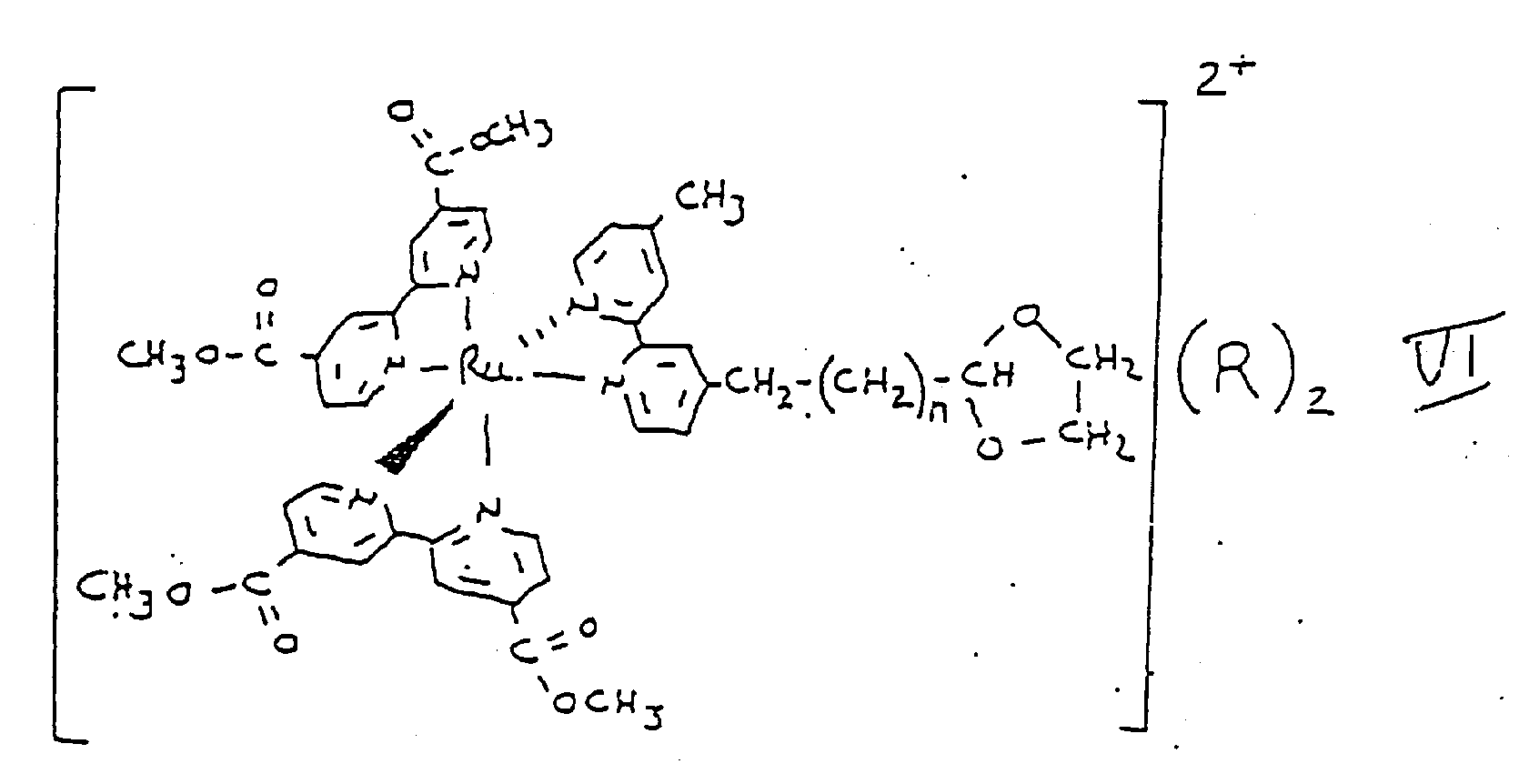

- Verfahren nach Anspruch 14, bei dem n in jeder der Formeln I, II, V und VI gleich 2 ist.
- Verfahren nach Anspruch 14, bei dem n in jeder der Formeln III und IV gleich 3 ist.
- Verfahren nach Anspruch 14, bei dem in der Formel VII m 5 und n 3 ist.
- Verfahren nach einem der Ansprüche 14 bis 17, bei dem X aus Theophyllin, Digoxigenin und einem vom menschlichen chorialen Gonadotropin (HCG) abgeleiteten Peptid ausgewählt ist.
- Verfahren nach einem der Ansprüche 1 bis 12, bei dem das Reagenz folgende Struktur hat
X-CH=CH-CO-NH-(CH₂)n-NH-CO-(CH₂)m-Z,
in der X einen oder mehrere Nucleotiden, die gleich oder verschieden sein können,
Z eine metallhaltige organische elektrochemische lumineszierende Komponente,
n eine ganze Zahl, die größer als oder gleich 1 ist, und
m eine ganze Zahl, die größer als oder gleich 1 ist, bedeutet. - Verfahren nach Anspruch 19, bei dem X ein beim Kohlenstoff 5 an CH hängendes Thymidin, n 7 und m 3 ist.
- Verfahren nach Anspruch 20, bei dem Z (Bis(2,2′-bipyridin)-[4-(butan-1-al)-4-methyl-1-2,2′-bipyridin]ruthenium(II) ist.
- Verfahren nach Anspruch 21, bei dem das Thymidinnucleotid ein 3′ endständiges Nucleotid ist, das an der Nucleotidsequenzangefügt ist.

- Verfahren nach einem der Ansprüche 1 bis 12, bei dem das Reagenz die Struktur
[T-Y-Z]²⁺(R)₂
hat, in der
T Theophyllin,
Y eine Verkettungsgruppe, die T mit Z verbindet,
Z Bis(2,2′-bipyridin)-[4-methyl-2,2′-biypridin-4′-yl]ruthenium(II) und
R ein Anion bedeutet. - Verfahren nach Anspruch 23, bei dem Y am Kohlenstoff in Stellung 8 von T angefügt ist.
- Verfahren nach Anspruch 24, bei dem Y die Struktur
(CH₂)m-CO-NH-(CH₂)n
aufweist, wobei m und n jeweils eine ganze Zahl darstellen, die gleich oder verschieden und größer als oder gleich 1 sein können. - Verfahren nach Anspruch 25, bei dem m 3 und n 4 ist.
- Verfahren nach Anspruch 25, bei dem m und n beide 3 bedeuten.
- Verfahren nach Anspruch 24, bei dem Y die Struktur

- Verfahren nach Anspruch 28, bei dem m 1, n 1 und r 4 ist.
- Verfahren nach Anspruch 24, bei dem Y die Struktur

- Verfahren nach Anspruch 30, bei dem m 1, n 1 und r 4 ist.
- Verfahren nach Anspruch 23, bei dem Y am Stickstoff in Stellung 7 von T hängt.
- Verfahren nach Anspruch 32, bei dem Y die Struktur
(CH₂)n
hat, in der n eine ganze Zahl größer als oder gleich 1 ist. - Verfahren nach Anspruch 33, bei dem n 4 ist.
- Verfahren nach einem der Ansprüche 1 bis 12, bei dem das Reagenz die Struktur
X-Z
hat, in der
X eine oder mehrere Aminosäuren bedeutet, die gleich oder verschieden sein können und mindestens eine Aminosäure umfassen, bei der es- sich um Cystein oder Methionin handelt, und
Z Bis(2,2′-bipyridin)maleinimidhexansäure,4-methyl-2,2-bipyridin-4′-butylamidruthenium(II) bedeutet, das durch den Kohlenstoff an der Stellung 3 oder 4 des Maleinimids an einen Schwefelsubstituenten des Cysteins oder Methionins von X gebunden ist. - Verfahren nach einem der Ansprüche 1 bis 35, bei dem die Probe von einem Feststoff, einer Emulsion, Suspension, Flüssigkeit oder einem Gas abgeleitet ist.
- Verfahren nach einem der Ansprüche 1 bis 36, bei dem die Probe von Wasser, Lebensmitteln, Blut, Serum, Urin, Kot, Gewebe, Speichel, Ölen, organischen Lösungsmitteln oder Luft abgeleitet ist.
- Verfahren nach einem der Ansprüche 1 bis 37, bei dem die Probe Dimethylsulfoxid, Dimethylformamid, n-Methylpyrrolidon oder tert-Butylalkohol enthält.
- Verfahren nach Anspruch 38, bei dem die Probe darüber hinaus ein Reduktionsmittel enthält.
- Verfahren nach Anspruch 38, bei dem die Probe darüber hinaus ein Oxidationsmittel enthält.
- Verfahren zum Nachweis des Vorhanden- oder Nicht- vorhandenseins eines relevanten Analyten in einem festgelegten Volumen einer flüssigen Multikomponentenprobe, welcher bei Vorhandensein in der Probe eine Konzentration von unter 10⁻³ Mol aufweist, bei dem mana) die Probe mit einem normalerweise nicht darin enthaltenden Komplementärmaterial und einem Reagenz in Kontakt bringt, welches (i) eine metallhaltige organische Verbindung umfaßt, die mit einer ganzen Zelle, einem subzellulären Teilchen, einem Virus, einem Prion, Viroid, Lipid, einer Fettsäure, einer Nukleinsäure, einem Polysaccharid außer einem Zucker, einem Protein, Lipoprotein, Lipopolysaccharid, Glykoprotein, Peptid, zellulären Metaboliten, Hormon, pharmakologischen Wirkstoff, Beruhigungsmittel, Barbiturat, Alkaloid, Steroid, Vitamin, einer Aminosäure, einem Zucker, einem nichtbiologischen Polymeren, einem organometallischen Molekül, synthetischen organischen Molekül oder anorganischen Molekül konjugiert ist,(ii) induziert werden kann, wiederholt elektrochemische Lumineszenz auszustrahlen, wenn es dem Einfluß einer Menge an elektrochemischer Energie aus einer geeigneten Quelle ausgesetzt wird, die die Induktion einer wiederholten Ausstrahlung elektrochemischer Lumineszenz durch das Reagenz bewirkt, und(iii) mit dem relevanten Analyten um Bindungsstellen auf dem normalerweise in der Probe nicht vorhandenen Komplementärmaterial in Wettbewerb treten kann, wobei der Kontakt unter geeigneten Bedingungen durchgeführt wird, daß sich der relevante Analyt und das Konjugat kompetitiv an das Komplementärmaterial binden;b) die entstehende Probe der Einwirkung einer Menge an elektrochemischer Energie aus einer geeigneten Quelle aussetzt, die das Reagenz anregen kann, wiederholt elektrochemische Lumineszenz auszusenden, wobei diese Exposition unter solchen Bedingungen erfolgt, daß das Reagenz induziert wird, wiederholt elektrochemische Lumineszenz auszustrahlen, undc) die ausgestrahlte Lumineszenz nachweist, um festzustellen, ob der relevante Analyt in der Probe vorhanden ist.
- Verfahren zum quantitativen Nachweis der Menge eines relevanten Analyten in einem festgelegten Volumen einer flüssigen Multikomponentenprobe, welcher bei Vorhandensein in der Probe eine Konzentration von unter 10⁻³ Mol aufweist, bei dem mana) die Probe mit einer bekannten Menge eines Komplementärmaterials und einer bekannten Menge eines Reagenz in Kontakt bringt, welches (i) eine metallhaltige organische Verbindung umfaßt, die mit einer ganzen Zelle, einem subzellulären Teilchen, einem Virus, einem Prion, Viroid, Lipid, einer Fettsäure, einer Nukleinsäure, einem Polysaccharid außer einem Zucker, einem Protein, Lipoprotein, Lipopolysaccharid, Glykoprotein, Peptid, zellulären Metaboliten, Hormon, pharmakologischen Wirkstoff, Beruhigungsmittel, Barbiturat, Alkaloid, Steroid, Vitamin, einer Aminosäure, einem Zucker, einem nichtbiologischen Polymeren, einem organometallischen Molekül, synthetischen organischen Molekül oder anorganischen Molekül konjugiert ist,(ii) induziert werden kann, wiederholt elektrochemische Lumineszenz auszustrahlen, wenn es dem Einfluß einer Menge an elektrochemischer Energie aus einer geeigneten Quelle ausgesetzt wird, die die Induktion einer wiederholten Ausstrahlung elektrochemischer Lumineszenz durch das Reagenz bewirkt, und(iii) mit dem relevanten Analyten in Wettbewerb um Bindungsstellen auf dem normalerweise nicht in der Probe vorhandenen Komplementärmaterial treten kann, wobei der Kontakt unter solchen Bedingungen durchgeführt wird, daß sich der relevante Analyt und das Konjugat kompetitiv an das Komplementärmaterial binden;b) die entstehende Probe der Einwirkung einer Menge an elektrochemischer Energie aus einer geeigneten Quelle aussetzt, die das Reagenz anregen kann, wiederholt elektrochemische Lumineszenz auszusenden, wobei diese Exposition unter solchen Bedingungen erfolgt, daß das Reagenz induziert wird, wiederholt elektrochemische Lumineszenz auszustrahlen undc) die ausgestrahlte Lumineszenz quantitativ nachweist, um festzustellen, ob und in welcher Menge der relevante Analyt in der Probe vorhanden ist.
- Verfahren nach Anspruch 41 oder 42, bei dem der relvante Analyt Theophyllin, Digoxin oder menschliches choriales Gonadotropin (HCG) ist.
- Verfahren nach einem der Ansprüche 41 bis 43, bei dem das Reagenz der relevante Analyt ist, der mit einer metallhaltigen organischen Verbindung konjugiert ist.
- Verfahren nach einem der Ansprüche 41 bis 43, bei dem das Reagenz ein Analogon des relevanten Analyten ist, der mit einer metallhaltigen organischen Verbindung konjugiert ist.
- Verfahren nach einem der Ansprüche 1 bis 45, bei dem die metallhaltige organische Verbindung aus den in einem der Ansprüche 13 bis 35 spezifizierten Verbindungen ausgewählt ist.
- Verfahren nach einem der Ansprüche 41 bis 46, bei dem das Komplementärmaterial eine ganze Zelle, ein subzelluläres Teilchen, ein Virus, Prion, Viroid, Lipid, eine Fettsäure, eine Nukleinsäure, ein Polysaccharid, ein Protein, Lipoprotein, Lipopolysaccharid, Glykoprotein, Peptid, zellulärer Metabolit, Hormon, pharmakologischer Wirkstoff, Beruhigungsmittel, Barbiturat, Alkaloid, Steroid, Vitamin, eine Aminosäure, ein Zucker, ein nichtbiologisches Polymer, ein synthetisches organisches Molekül, ein organometallisches Molekül oder anorganisches Molekül ist.
Priority Applications (2)
| Application Number | Priority Date | Filing Date | Title |
|---|---|---|---|
| EP94120254A EP0647849A3 (de) | 1986-04-30 | 1987-04-30 | Nachweis der Anwesenheit von Analyten in einer Probe. |
| EP94120516A EP0658564B1 (de) | 1986-04-30 | 1987-04-30 | Elektrolumineszente Verbindungen und Zwischenprodukte zur Herstellung |
Applications Claiming Priority (3)
| Application Number | Priority Date | Filing Date | Title |
|---|---|---|---|
| US85835486A | 1986-04-30 | 1986-04-30 | |
| US858354 | 1986-04-30 | ||
| PCT/US1987/000987 WO1987006706A1 (en) | 1986-04-30 | 1987-04-30 | Electrochemiluminescent assays |
Related Child Applications (3)
| Application Number | Title | Priority Date | Filing Date |
|---|---|---|---|
| EP94120516A Division EP0658564B1 (de) | 1986-04-30 | 1987-04-30 | Elektrolumineszente Verbindungen und Zwischenprodukte zur Herstellung |
| EP94120254.1 Division-Into | 1994-12-21 | ||
| EP94120516.3 Division-Into | 1994-12-23 |
Publications (3)
| Publication Number | Publication Date |
|---|---|
| EP0265519A1 EP0265519A1 (de) | 1988-05-04 |
| EP0265519A4 EP0265519A4 (de) | 1990-05-14 |
| EP0265519B1 true EP0265519B1 (de) | 1995-09-13 |
Family
ID=25328109
Family Applications (3)
| Application Number | Title | Priority Date | Filing Date |
|---|---|---|---|
| EP87904151A Expired - Lifetime EP0265519B1 (de) | 1986-04-30 | 1987-04-30 | Elektrochemilumineszenz-testverfahren |
| EP94120254A Ceased EP0647849A3 (de) | 1986-04-30 | 1987-04-30 | Nachweis der Anwesenheit von Analyten in einer Probe. |
| EP94120516A Expired - Lifetime EP0658564B1 (de) | 1986-04-30 | 1987-04-30 | Elektrolumineszente Verbindungen und Zwischenprodukte zur Herstellung |
Family Applications After (2)
| Application Number | Title | Priority Date | Filing Date |
|---|---|---|---|
| EP94120254A Ceased EP0647849A3 (de) | 1986-04-30 | 1987-04-30 | Nachweis der Anwesenheit von Analyten in einer Probe. |
| EP94120516A Expired - Lifetime EP0658564B1 (de) | 1986-04-30 | 1987-04-30 | Elektrolumineszente Verbindungen und Zwischenprodukte zur Herstellung |
Country Status (13)
| Country | Link |
|---|---|
| EP (3) | EP0265519B1 (de) |
| JP (6) | JPH0737464B2 (de) |
| KR (1) | KR960016340B1 (de) |
| AT (2) | ATE127923T1 (de) |
| AU (2) | AU644150B2 (de) |
| CA (1) | CA1339465C (de) |
| DE (2) | DE3752345T2 (de) |
| DK (1) | DK173025B1 (de) |
| FI (1) | FI102017B (de) |
| HK (2) | HK1007442A1 (de) |
| IL (1) | IL82411A0 (de) |
| NO (1) | NO176071C (de) |
| WO (1) | WO1987006706A1 (de) |
Cited By (3)
| Publication number | Priority date | Publication date | Assignee | Title |
|---|---|---|---|---|
| CN106255754A (zh) * | 2014-04-10 | 2016-12-21 | Dna吉诺特克股份有限公司 | 使用高碘酸盐用于微生物细胞溶解的方法和系统 |
| US11002646B2 (en) | 2011-06-19 | 2021-05-11 | DNA Genotek, Inc. | Devices, solutions and methods for sample collection |
| US11572581B2 (en) | 2002-06-07 | 2023-02-07 | DNA Genotek, Inc. | Compositions and methods for obtaining nucleic acids from sputum |
Families Citing this family (131)
| Publication number | Priority date | Publication date | Assignee | Title |
|---|---|---|---|---|
| US6165729A (en) * | 1986-04-30 | 2000-12-26 | Hyperion Catalysis International, Inc. | Electrochemiluminescent reaction utilizing amine-derived reductant |
| US5635347A (en) * | 1986-04-30 | 1997-06-03 | Igen, Inc. | Rapid assays for amplification products |
| US5770459A (en) * | 1986-04-30 | 1998-06-23 | Igen International, Inc. | Methods and apparatus for improved luminescence assays using particle concentration, electrochemical generation of chemiluminescence detection |
| US6271041B1 (en) * | 1986-04-30 | 2001-08-07 | Igen International, Inc. | Electrochemiluminescent reaction utilizing amine-derived reductant |
| US6451225B1 (en) | 1986-04-30 | 2002-09-17 | Igen International, Inc. | Electrochemiluminescent reaction utilizing amine-derived reductant |
| US6881536B1 (en) * | 1986-04-30 | 2005-04-19 | Bioveris Corporation | Particle based electrochemiluminescent assays |
| US6881589B1 (en) | 1987-04-30 | 2005-04-19 | Bioveris Corporation | Electrochemiluminescent localizable complexes for assay compositions |
| US5935779A (en) * | 1988-11-03 | 1999-08-10 | Igen International Inc. | Methods for improved particle electrochemiluminescence assay |
| US6702986B1 (en) | 1988-04-29 | 2004-03-09 | Igen International, Inc. | Electrochemiluminescent reaction utilizing amine-derived reductant |
| US5705402A (en) * | 1988-11-03 | 1998-01-06 | Igen International, Inc. | Method and apparatus for magnetic microparticulate based luminescence assay including plurality of magnets |
| US5962218A (en) * | 1988-11-03 | 1999-10-05 | Igen International Inc. | Methods and apparatus for improved luminescence assays |
| SG60022A1 (en) * | 1988-11-03 | 1999-02-22 | Igen Inc | Electrochemicalmiliuminescent assays |
| US5746974A (en) * | 1988-11-03 | 1998-05-05 | Igen International, Inc. | Apparatus for improved luminescence assays using particle concentration, electrochemical generation of chemiluminescence and chemiluminescence detection |
| ATE161096T1 (de) * | 1988-11-03 | 1997-12-15 | Igen Inc | Elektrochemilumineszente reaktion unter verwendung eines von einem amin abgeleiteten reduktionsmittels |
| JPH0363571A (ja) * | 1989-08-02 | 1991-03-19 | Chisso Corp | 大腸菌群の検査キット |
| IL100866A (en) * | 1991-02-06 | 1995-10-31 | Igen Inc | Luminescence test method and device based on magnetic tiny particles, containing many magnets |
| ZA92803B (en) | 1991-02-06 | 1992-11-25 | Igen Inc | Method and apparatus for magnetic microparticulate based luminescene asay including plurality of magnets |
| IL100867A (en) * | 1991-02-06 | 1995-12-08 | Igen Inc | Method and device for improved luminescence testing |
| WO1993001308A1 (en) * | 1991-07-10 | 1993-01-21 | Igen, Inc. | Methods and apparatus for improved luminescence assays using particle concentration and chemiluminescence detection |
| DE69231124T2 (de) * | 1991-11-15 | 2001-02-15 | Igen Int Inc | Schnelle bestimmungsverfahren für amplifikationsprodukte |
| ZA929351B (en) * | 1991-12-11 | 1993-06-04 | Igen Inc | Electrochemiluminescent label for DNA assays. |
| FR2699170B1 (fr) * | 1992-12-15 | 1995-07-28 | Asulab Sa | Complexes d'un métal de transition à ligands 2,2'-bipyridine substitués par au moins un radical ammonium alkyle, leur procédé de fabrication et leur application comme médiateur redox. |
| US5541113A (en) * | 1993-09-22 | 1996-07-30 | Beckman Instruments, Inc. | Method for detecting an analyte using an electrochemical luminescent transition metal label |
| DE59507438D1 (de) * | 1994-07-25 | 2000-01-20 | Roche Diagnostics Gmbh | Hydrophile metallkomplexe |
| CA2213854C (en) | 1995-03-10 | 2010-08-10 | Meso Scale Technologies, Llc | Multi-array, multi-specific electrochemiluminescence testing |
| US6319670B1 (en) | 1995-05-09 | 2001-11-20 | Meso Scale Technology Llp | Methods and apparatus for improved luminescence assays using microparticles |
| ES2285713T3 (es) | 1995-06-07 | 2007-11-16 | Bioveris Corporation | Enzimoinmunoanalisis electroquimioluminiscente. |
| JP3574457B2 (ja) * | 1995-09-12 | 2004-10-06 | エーザイ株式会社 | 電気化学発光検出方法と装置 |
| US6862465B2 (en) | 1997-03-04 | 2005-03-01 | Dexcom, Inc. | Device and method for determining analyte levels |
| US7192450B2 (en) | 2003-05-21 | 2007-03-20 | Dexcom, Inc. | Porous membranes for use with implantable devices |
| US6001067A (en) | 1997-03-04 | 1999-12-14 | Shults; Mark C. | Device and method for determining analyte levels |
| US8527026B2 (en) | 1997-03-04 | 2013-09-03 | Dexcom, Inc. | Device and method for determining analyte levels |
| US6613583B1 (en) | 1997-06-27 | 2003-09-02 | Igen International, Inc. | Electrochemiluminescent label based on multimetallic assemblies |
| DE19803528A1 (de) | 1998-01-30 | 1999-08-05 | Roche Diagnostics Gmbh | Verfahren zur Analyse einer Probe mittels eines Elektrochemolumineszenz-Bindungsreaktion-Tests |
| DE19811582A1 (de) * | 1998-03-17 | 1999-09-23 | Roche Diagnostics Gmbh | Stabilisierung und Verstärkung von Elektrochemilumineszenz-Signalen |
| DE19828441A1 (de) * | 1998-06-25 | 1999-12-30 | Roche Diagnostics Gmbh | Verwendung derivatisierter Reagenzien in Chemilumineszenz- und Elektrochemilumineszenz-Nachweisverfahren |
| US6214552B1 (en) | 1998-09-17 | 2001-04-10 | Igen International, Inc. | Assays for measuring nucleic acid damaging activities |
| US6312896B1 (en) | 1998-09-17 | 2001-11-06 | Igen Inaternational, Inc. | Assays for measuring nucleic acid binding proteins and enzyme activities |
| US7041807B1 (en) | 1999-06-23 | 2006-05-09 | Caprion Pharmaceuticals, Inc. | Antibodies to a YYX epitope of a mammalian prion protein |
| US6977722B2 (en) | 2001-06-29 | 2005-12-20 | Meso Scale Technologies, Llc. | Assay plates, reader systems and methods for luminescence test measurements |
| US20030032874A1 (en) | 2001-07-27 | 2003-02-13 | Dexcom, Inc. | Sensor head for use with implantable devices |
| EP1412487B1 (de) | 2001-07-30 | 2010-06-16 | Meso Scale Technologies LLC | Assay-elektroden mit schichten aus immobilisiertem lipid/protein sowie verfahren zu deren herstellung und verwendung |
| CA2932756C (en) | 2001-09-10 | 2020-07-07 | Meso Scale Technologies, Llc | Assay buffer, compositions containing the same, and methods of using the same |
| US8858434B2 (en) | 2004-07-13 | 2014-10-14 | Dexcom, Inc. | Transcutaneous analyte sensor |
| US8260393B2 (en) | 2003-07-25 | 2012-09-04 | Dexcom, Inc. | Systems and methods for replacing signal data artifacts in a glucose sensor data stream |
| US9282925B2 (en) | 2002-02-12 | 2016-03-15 | Dexcom, Inc. | Systems and methods for replacing signal artifacts in a glucose sensor data stream |
| US8010174B2 (en) | 2003-08-22 | 2011-08-30 | Dexcom, Inc. | Systems and methods for replacing signal artifacts in a glucose sensor data stream |
| US20040072236A1 (en) | 2002-09-27 | 2004-04-15 | Neil Cashman | PrPSc -interacting molecules and uses thereof |
| US8423113B2 (en) | 2003-07-25 | 2013-04-16 | Dexcom, Inc. | Systems and methods for processing sensor data |
| US7591801B2 (en) | 2004-02-26 | 2009-09-22 | Dexcom, Inc. | Integrated delivery device for continuous glucose sensor |
| US8788006B2 (en) | 2003-08-01 | 2014-07-22 | Dexcom, Inc. | System and methods for processing analyte sensor data |
| US8761856B2 (en) | 2003-08-01 | 2014-06-24 | Dexcom, Inc. | System and methods for processing analyte sensor data |
| US8160669B2 (en) | 2003-08-01 | 2012-04-17 | Dexcom, Inc. | Transcutaneous analyte sensor |
| US8369919B2 (en) | 2003-08-01 | 2013-02-05 | Dexcom, Inc. | Systems and methods for processing sensor data |
| US8275437B2 (en) | 2003-08-01 | 2012-09-25 | Dexcom, Inc. | Transcutaneous analyte sensor |
| US20190357827A1 (en) | 2003-08-01 | 2019-11-28 | Dexcom, Inc. | Analyte sensor |
| US7986986B2 (en) | 2003-08-01 | 2011-07-26 | Dexcom, Inc. | System and methods for processing analyte sensor data |
| US7774145B2 (en) | 2003-08-01 | 2010-08-10 | Dexcom, Inc. | Transcutaneous analyte sensor |
| US8060173B2 (en) | 2003-08-01 | 2011-11-15 | Dexcom, Inc. | System and methods for processing analyte sensor data |
| US8233959B2 (en) | 2003-08-22 | 2012-07-31 | Dexcom, Inc. | Systems and methods for processing analyte sensor data |
| US7920906B2 (en) | 2005-03-10 | 2011-04-05 | Dexcom, Inc. | System and methods for processing analyte sensor data for sensor calibration |
| US20140121989A1 (en) | 2003-08-22 | 2014-05-01 | Dexcom, Inc. | Systems and methods for processing analyte sensor data |
| WO2005051170A2 (en) | 2003-11-19 | 2005-06-09 | Dexcom, Inc. | Integrated receiver for continuous analyte sensor |
| US8615282B2 (en) | 2004-07-13 | 2013-12-24 | Dexcom, Inc. | Analyte sensor |
| US9247900B2 (en) | 2004-07-13 | 2016-02-02 | Dexcom, Inc. | Analyte sensor |
| DE602004029092D1 (de) | 2003-12-05 | 2010-10-21 | Dexcom Inc | Kalibrationsmethoden für einen kontinuierlich arbeitenden analytsensor |
| US11633133B2 (en) | 2003-12-05 | 2023-04-25 | Dexcom, Inc. | Dual electrode system for a continuous analyte sensor |
| US8364231B2 (en) | 2006-10-04 | 2013-01-29 | Dexcom, Inc. | Analyte sensor |
| US8423114B2 (en) | 2006-10-04 | 2013-04-16 | Dexcom, Inc. | Dual electrode system for a continuous analyte sensor |
| US8532730B2 (en) | 2006-10-04 | 2013-09-10 | Dexcom, Inc. | Analyte sensor |
| EP2329763B1 (de) | 2003-12-09 | 2017-06-21 | DexCom, Inc. | Signalverarbeitung in einem durchgehenden Analytsensor |
| US8808228B2 (en) | 2004-02-26 | 2014-08-19 | Dexcom, Inc. | Integrated medicament delivery device for use with continuous analyte sensor |
| ITTO20040372A1 (it) * | 2004-06-01 | 2004-09-01 | Giuseppe Caputo | Composti luminescenti aventi un braccio di legame funzionalizzato. |
| CA2571283C (en) * | 2004-06-23 | 2013-10-22 | University Of Texas System | Methods and compositions for the detection of biological molecules using a two particle complex |
| US20060270922A1 (en) | 2004-07-13 | 2006-11-30 | Brauker James H | Analyte sensor |
| US8452368B2 (en) | 2004-07-13 | 2013-05-28 | Dexcom, Inc. | Transcutaneous analyte sensor |
| WO2006038686A1 (ja) * | 2004-10-07 | 2006-04-13 | Matsushita Electric Industrial Co., Ltd. | 遺伝子検出方法、及び遺伝子検出装置 |
| EP1893990B1 (de) | 2005-06-03 | 2018-05-02 | Board of Regents of the University of Texas System | Elektrochemie und elektrogenerierte chemolumineszenz mit einer faradayschen einzelelektrode |
| EP1950566A4 (de) * | 2005-11-10 | 2008-11-26 | Univ Toyama Nat Univ Corp | Verfahren zur bewertung von bindungseigenschaften von biosubstanzen und biosubstanz bindenden substanzen |
| US20080306444A1 (en) | 2007-06-08 | 2008-12-11 | Dexcom, Inc. | Integrated medicament delivery device for use with continuous analyte sensor |
| EP4159114B1 (de) | 2007-10-09 | 2024-04-10 | DexCom, Inc. | Integriertes insulin-abgabesystem mit kontinuierlichem glucosesensor |
| EP2217608B1 (de) * | 2007-11-21 | 2012-09-05 | Bio-Rad Laboratories, Inc. | Fotolumineszente metallkomplexe für proteinfärbung |
| EP2252196A4 (de) | 2008-02-21 | 2013-05-15 | Dexcom Inc | Systeme und verfahren zur verarbeitung, übertragung und anzeige von sensordaten |
| DK2271938T3 (da) | 2008-04-11 | 2014-05-12 | Univ Texas | Fremgangsmåde og apparat til nanopartikel-elektrogenereret kemiluminescens-amplifikation |
| KR101603356B1 (ko) | 2008-05-08 | 2016-03-14 | 보드 오브 리전츠 오브 더 유니버시티 오브 텍사스 시스템 | 전기발생 화학발광에 사용되는 발광 나노구조 물질 |
| AU2014203680B2 (en) * | 2008-05-08 | 2015-03-05 | Board Of Regents Of The University Of Texas System | Luminescent nanostructured materials for use in electrogenerated chemiluminescence |
| US8264347B2 (en) | 2008-06-24 | 2012-09-11 | Trelleborg Sealing Solutions Us, Inc. | Seal system in situ lifetime measurement |
| FR2939895B1 (fr) * | 2008-12-15 | 2011-01-14 | Commissariat Energie Atomique | Procede de detection non intrusive d'element chimique |
| US20110004085A1 (en) | 2009-04-30 | 2011-01-06 | Dexcom, Inc. | Performance reports associated with continuous sensor data from multiple analysis time periods |
| US20140038834A1 (en) | 2010-07-07 | 2014-02-06 | Vereniging Voor Christelijk Hoger Onderwijs, Wetenschappelijk Onderzoek En Patiëntenzorg | Novel biomarkers for detecting neuronal loss |
| WO2012107420A1 (en) * | 2011-02-09 | 2012-08-16 | Roche Diagnostics Gmbh | New iridium-based complexes for ecl |
| JP6141827B2 (ja) | 2011-04-15 | 2017-06-07 | デックスコム・インコーポレーテッド | 検体を測定するシステムの作動方法及び該方法を実施するべく構成されたセンサシステム |
| EP2729809B1 (de) * | 2011-07-07 | 2018-10-17 | DuPont Nutrition Biosciences ApS | Assay |
| US9625465B2 (en) | 2012-05-15 | 2017-04-18 | Defined Diagnostics, Llc | Clinical diagnostic systems |
| US9213043B2 (en) | 2012-05-15 | 2015-12-15 | Wellstat Diagnostics, Llc | Clinical diagnostic system including instrument and cartridge |
| US9081001B2 (en) | 2012-05-15 | 2015-07-14 | Wellstat Diagnostics, Llc | Diagnostic systems and instruments |
| JP6560120B2 (ja) | 2012-08-02 | 2019-08-14 | エフ.ホフマン−ラ ロシュ アーゲーF. Hoffmann−La Roche Aktiengesellschaft | Ecl製造用の新規ビス−イリジウム錯体 |
| RU2661101C2 (ru) | 2013-03-13 | 2018-07-11 | Дженевив Биосайнсис, Инк. | Нерепликативные трансдукторные частицы и основанные на трансдукторных частицах репортерные системы |
| US9481903B2 (en) | 2013-03-13 | 2016-11-01 | Roche Molecular Systems, Inc. | Systems and methods for detection of cells using engineered transduction particles |
| US9540675B2 (en) | 2013-10-29 | 2017-01-10 | GeneWeave Biosciences, Inc. | Reagent cartridge and methods for detection of cells |
| ES2717535T3 (es) | 2014-12-08 | 2019-06-21 | Hoffmann La Roche | Procedimiento para la medición de vitamina D |
| EP3338094B1 (de) | 2015-08-20 | 2019-05-08 | H. Hoffnabb-La Roche Ag | Partikelbasierter immunoassay unter verwendung eines pegylierten analytspezifischen bindemittels |
| US10351893B2 (en) | 2015-10-05 | 2019-07-16 | GeneWeave Biosciences, Inc. | Reagent cartridge for detection of cells |
| BR112018017336A2 (pt) | 2016-03-07 | 2018-12-26 | Hoffmann La Roche | método in vitro para detectar um anticorpo para p53 (anticorpo anti-p53) em uma amostra e polipeptídeo de fusão |
| JP7039481B2 (ja) | 2016-03-11 | 2022-03-22 | エフ.ホフマン-ラ ロシュ アーゲー | 電気化学発光検出における分枝鎖アミン |
| EP3577460B1 (de) | 2017-02-02 | 2021-01-20 | Roche Diagnostics GmbH | Immunoassay mit mindestens zwei pegylierten analyt-spezifischen bindungsmitteln |
| US11077444B2 (en) | 2017-05-23 | 2021-08-03 | Roche Molecular Systems, Inc. | Packaging for a molecular diagnostic cartridge |
| AU2018354120A1 (en) | 2017-10-24 | 2020-04-23 | Dexcom, Inc. | Pre-connected analyte sensors |
| US11331022B2 (en) | 2017-10-24 | 2022-05-17 | Dexcom, Inc. | Pre-connected analyte sensors |
| CN111936522A (zh) | 2018-04-18 | 2020-11-13 | 豪夫迈·罗氏有限公司 | 新型抗胸苷激酶抗体 |
| US20210208080A1 (en) * | 2018-05-21 | 2021-07-08 | Beyond Diagnostics (Shanghai) Co., Ltd | Chemiluminescence analytical method and system and kit using same |
| EP3844503A1 (de) | 2018-08-31 | 2021-07-07 | F. Hoffmann-La Roche AG | Thymidinkinase (tk-1) in prognostischen indizes für dlbcl |
| JP2022542056A (ja) | 2019-07-22 | 2022-09-29 | エフ.ホフマン-ラ ロシュ アーゲー | 子宮内膜症の非侵襲的診断のための血液バイオマーカーとしてのs100a6 |
| BR112022000172A2 (pt) | 2019-07-22 | 2022-02-22 | Hoffmann La Roche | Métodos para avaliar se uma paciente tem endometriose, para selecionar uma paciente para terapia e para monitorar uma paciente |
| BR112022001140A2 (pt) | 2019-07-22 | 2022-03-15 | Hoffmann La Roche | Métodos para avaliar se um paciente tem endometriose ou está em risco de desenvolver endometriose, selecionar um paciente para terapia e monitorar um paciente que sofre de endometriose ou que está sendo tratado para endometriose |
| CN114144673A (zh) | 2019-07-22 | 2022-03-04 | 豪夫迈·罗氏有限公司 | S100a8作为血液生物标志物用于子宫内膜异位症的非侵入性诊断 |
| CN114144674A (zh) | 2019-07-22 | 2022-03-04 | 豪夫迈·罗氏有限公司 | P物质作为用于子宫内膜异位症的非侵入性诊断的血液生物标志物 |
| BR112022009371A2 (pt) | 2019-11-15 | 2022-08-09 | Hoffmann La Roche | Métodos para determinar a quantidade ou concentração de um ou mais analitos antibióticos, tubos de amostragem, usos de um reagente de derivatização nucleofílica e analito antibiótico |
| EP4200597A2 (de) | 2020-08-21 | 2023-06-28 | Meso Scale Technologies, LLC | Elektrochemische zellen mit hilfselektroden mit definiertem grenzflächenpotenzial und verfahren zur verwendung davon |
| JP2023547505A (ja) | 2020-11-02 | 2023-11-10 | エフ. ホフマン-ラ ロシュ アーゲー | SARS-CoV-2ヌクレオカプシド抗体 |
| WO2022207628A1 (en) | 2021-03-30 | 2022-10-06 | F. Hoffmann-La Roche Ag | Scf as blood biomarker for the non-invasive diagnosis of endometriosis |
| WO2022207685A1 (en) | 2021-04-01 | 2022-10-06 | F. Hoffmann-La Roche Ag | Psp94 as blood biomarker for the non-invasive diagnosis of endometriosis |
| WO2022243210A1 (en) | 2021-05-17 | 2022-11-24 | F. Hoffmann-La Roche Ag | sFRP4 AS BLOOD BIOMARKER FOR THE NON-INVASIVE DIAGNOSIS OF ADENOMYOSIS |
| AU2022325891A1 (en) | 2021-08-13 | 2024-03-14 | Meso Scale Technologies, Llc. | Electrochemical cell devices and methods of manufacturing |
| WO2023072904A1 (en) | 2021-10-26 | 2023-05-04 | F. Hoffmann-La Roche Ag | Monoclonal antibodies specific for sars-cov-2 rbd |
| US20230213476A1 (en) | 2022-01-04 | 2023-07-06 | Meso Scale Technologies, Llc. | Electrochemical cell devices and methods of manufacturing |
| WO2023131594A1 (en) | 2022-01-05 | 2023-07-13 | F. Hoffmann-La Roche Ag | Derivatization of compounds in patient samples for therapeutic drug monitoring (tdm) |
| WO2023247752A1 (en) | 2022-06-23 | 2023-12-28 | F. Hoffmann-La Roche Ag | Method for diagnosing endometriosis and for classifying the stage of endometriosis |
| WO2024017982A1 (en) | 2022-07-22 | 2024-01-25 | F. Hoffmann-La Roche Ag | Leukotriene a4 hydrolase (lta4h) as (blood) biomarker for the diagnosis of polycystic ovarian syndrome |
| WO2024017983A1 (en) | 2022-07-22 | 2024-01-25 | F. Hoffmann-La Roche Ag | Meteorin-like protein (metrnl) as (blood) biomarker for the diagnosis of polycystic ovarian syndrome |
| WO2024017985A1 (en) | 2022-07-22 | 2024-01-25 | F. Hoffmann-La Roche Ag | Fibroblast growth factor binding protein 1 (fgfbp1) as (blood) biomarker for the diagnosis of polycystic ovarian syndrome |
Family Cites Families (15)
| Publication number | Priority date | Publication date | Assignee | Title |
|---|---|---|---|---|
| IL49685A (en) * | 1976-05-31 | 1978-10-31 | Technion Res & Dev Foundation | Specific binding assay method for determining the concentration of materials and reagent means therefor |
| US4233144A (en) * | 1979-04-16 | 1980-11-11 | Technicon Instruments Corporation | Electrode for voltammetric immunoassay |
| US4280815A (en) * | 1979-06-18 | 1981-07-28 | Technicon Instruments Corporation | Electrochemiluminescent immunoassay and apparatus therefor |
| WO1981001883A1 (en) * | 1979-12-19 | 1981-07-09 | Electro Nucleonics | Chemical luminescence amplification substrate system for immunochemistry |
| US4293310A (en) * | 1980-03-14 | 1981-10-06 | University Of Pittsburgh | Photoelectrochemical immunoassay |
| EP0063810B1 (de) * | 1981-04-29 | 1986-03-05 | Ciba-Geigy Ag | Prüfmittel und Ausrüstung für immunologische Analysen |
| US4514508A (en) * | 1982-07-06 | 1985-04-30 | Biond Inc. | Assaying for a multiplicity of antigens or antibodies with a detection compound |
| GB2135773B (en) * | 1983-01-31 | 1985-12-04 | Boots Celltech Diagnostics | Enzyme inhibitor labelled immunoassay |
| EP0139373A1 (de) * | 1983-08-26 | 1985-05-02 | The Regents Of The University Of California | System für vielfache Immunoassays |
| IL75464A (en) * | 1984-06-12 | 1990-08-31 | Orgenics Ltd | Method and apparatus for multi-analyte assay |
| CA1340372C (en) * | 1984-07-09 | 1999-02-02 | John D. Clements | Production of e. coli lt-b enterotoxin subunit |
| DK365785A (da) * | 1984-09-17 | 1986-03-18 | Hoffmann La Roche | Metalcomplexer |
| US4617265A (en) * | 1984-09-19 | 1986-10-14 | Board Of Regents, University Of Texas System | Colony blot assay for enterotoxigenic bacteria |
| US5238808A (en) * | 1984-10-31 | 1993-08-24 | Igen, Inc. | Luminescent metal chelate labels and means for detection |
| US5120662A (en) * | 1989-05-09 | 1992-06-09 | Abbott Laboratories | Multilayer solid phase immunoassay support and method of use |
-
1987
- 1987-04-30 DE DE3752345T patent/DE3752345T2/de not_active Expired - Lifetime
- 1987-04-30 CA CA000536079A patent/CA1339465C/en not_active Expired - Lifetime
- 1987-04-30 EP EP87904151A patent/EP0265519B1/de not_active Expired - Lifetime
- 1987-04-30 KR KR1019870701252A patent/KR960016340B1/ko not_active IP Right Cessation
- 1987-04-30 AT AT87904151T patent/ATE127923T1/de not_active IP Right Cessation
- 1987-04-30 DE DE3751516T patent/DE3751516T2/de not_active Expired - Lifetime
- 1987-04-30 JP JP62503860A patent/JPH0737464B2/ja not_active Expired - Fee Related
- 1987-04-30 EP EP94120254A patent/EP0647849A3/de not_active Ceased
- 1987-04-30 WO PCT/US1987/000987 patent/WO1987006706A1/en active IP Right Grant
- 1987-04-30 EP EP94120516A patent/EP0658564B1/de not_active Expired - Lifetime
- 1987-04-30 AT AT94120516T patent/ATE212030T1/de not_active IP Right Cessation
- 1987-05-01 IL IL82411A patent/IL82411A0/xx not_active IP Right Cessation
- 1987-12-28 FI FI875732A patent/FI102017B/fi not_active IP Right Cessation
- 1987-12-29 NO NO875476A patent/NO176071C/no unknown
-
1991
- 1991-04-10 AU AU74338/91A patent/AU644150B2/en not_active Expired
-
1993
- 1993-02-12 DK DK199300162A patent/DK173025B1/da not_active IP Right Cessation
-
1994
- 1994-03-02 AU AU57540/94A patent/AU685071B2/en not_active Expired
- 1994-09-08 JP JP6251174A patent/JP2674957B2/ja not_active Expired - Lifetime
- 1994-11-09 JP JP27517494A patent/JP3349279B2/ja not_active Expired - Lifetime
-
1996
- 1996-06-26 JP JP8166280A patent/JP2648474B2/ja not_active Expired - Lifetime
-
1998
- 1998-06-24 HK HK98106428A patent/HK1007442A1/xx not_active IP Right Cessation
- 1998-07-06 JP JP19061598A patent/JP3476679B2/ja not_active Expired - Lifetime
- 1998-12-23 HK HK98115071A patent/HK1013653A1/xx not_active IP Right Cessation
-
2002
- 2002-07-12 JP JP2002204082A patent/JP3509812B2/ja not_active Expired - Lifetime
Non-Patent Citations (1)
| Title |
|---|
| CLINICAL CHEMISTRY, Diagnosis & Treatment, Zilva & Parnell (ed.), 3rd Ed., Lloyd Luke Ltd. (publ.); pp. 472-473/ * |
Cited By (7)
| Publication number | Priority date | Publication date | Assignee | Title |
|---|---|---|---|---|
| US11572581B2 (en) | 2002-06-07 | 2023-02-07 | DNA Genotek, Inc. | Compositions and methods for obtaining nucleic acids from sputum |
| US11002646B2 (en) | 2011-06-19 | 2021-05-11 | DNA Genotek, Inc. | Devices, solutions and methods for sample collection |
| US11536632B2 (en) | 2011-06-19 | 2022-12-27 | DNA Genotek, Inc. | Biological collection system |
| US11549870B2 (en) | 2011-06-19 | 2023-01-10 | DNA Genotek, Inc. | Cell preserving solution |
| US11592368B2 (en) | 2011-06-19 | 2023-02-28 | DNA Genotek, Inc. | Method for collecting and preserving a biological sample |
| CN106255754A (zh) * | 2014-04-10 | 2016-12-21 | Dna吉诺特克股份有限公司 | 使用高碘酸盐用于微生物细胞溶解的方法和系统 |
| US11046949B2 (en) | 2014-04-10 | 2021-06-29 | Dna Genotek Inc. | Method and system for microbial lysis using periodates |
Also Published As
Similar Documents
| Publication | Publication Date | Title |
|---|---|---|
| EP0265519B1 (de) | Elektrochemilumineszenz-testverfahren | |
| US6316607B1 (en) | Electrochemiluminescent assays | |
| US6468741B1 (en) | Electrochemiluminescent rhenium moieties | |
| US5731147A (en) | Luminescent metal chelate labels and means for detection | |
| EP0674176B1 (de) | Lumineszente Rhenium-Komplexe und Verfahren zur Herstellung | |
| US6916606B2 (en) | Electrochemiluminescent assays | |
| AU605158C (en) | Electrochemiluminescent assays | |
| WO1997032886A1 (en) | Ecl labels having improved nsb properties | |
| IL101990A (en) | Conjugate of ruthenium- or osmium- containing electrochemiluminescent chemical moieties | |
| DK172940B1 (da) | Fremgangsmåde til kvalitativ og kvantitativ elektrokemiluminescerende analysering af multikomponentvæsker |
Legal Events
| Date | Code | Title | Description |
|---|---|---|---|
| PUAI | Public reference made under article 153(3) epc to a published international application that has entered the european phase |
Free format text: ORIGINAL CODE: 0009012 |
|
| 17P | Request for examination filed |
Effective date: 19871222 |
|
| AK | Designated contracting states |
Kind code of ref document: A1 Designated state(s): AT BE CH DE FR GB IT LI LU NL SE |
|
| A4 | Supplementary search report drawn up and despatched |
Effective date: 19900514 |
|
| 17Q | First examination report despatched |
Effective date: 19910424 |
|
| GRAA | (expected) grant |
Free format text: ORIGINAL CODE: 0009210 |
|
| RAP1 | Party data changed (applicant data changed or rights of an application transferred) |
Owner name: IGEN, INC. (A CALIFORNIA CORPORATION) |
|
| AK | Designated contracting states |
Kind code of ref document: B1 Designated state(s): AT BE CH DE FR GB IT LI LU NL SE |
|
| REF | Corresponds to: |
Ref document number: 127923 Country of ref document: AT Date of ref document: 19950915 Kind code of ref document: T |
|
| XX | Miscellaneous (additional remarks) |
Free format text: TEILANMELDUNG 94120516.3 EINGEREICHT AM 30/04/87. |
|
| ITF | It: translation for a ep patent filed |
Owner name: ING. A. GIAMBROCONO & C. S.R.L. |
|
| REF | Corresponds to: |
Ref document number: 3751516 Country of ref document: DE Date of ref document: 19951019 |
|
| ET | Fr: translation filed | ||
| NLR4 | Nl: receipt of corrected translation in the netherlands language at the initiative of the proprietor of the patent | ||
| PLBE | No opposition filed within time limit |
Free format text: ORIGINAL CODE: 0009261 |
|
| STAA | Information on the status of an ep patent application or granted ep patent |
Free format text: STATUS: NO OPPOSITION FILED WITHIN TIME LIMIT |
|
| 26N | No opposition filed | ||
| REG | Reference to a national code |
Ref country code: CH Ref legal event code: PFA Free format text: IGEN, INC. (A CALIFORNIA CORPORATION) TRANSFER- IGEN INTERNATIONAL, INC. |
|
| REG | Reference to a national code |
Ref country code: GB Ref legal event code: 732E |
|
| NLS | Nl: assignments of ep-patents |
Owner name: IGEN INTERNATIONAL, INC A DELAWARE CORPORATION |
|
| REG | Reference to a national code |
Ref country code: FR Ref legal event code: TP |
|
| REG | Reference to a national code |
Ref country code: GB Ref legal event code: IF02 |
|
| REG | Reference to a national code |
Ref country code: GB Ref legal event code: 732E |
|
| REG | Reference to a national code |
Ref country code: FR Ref legal event code: TP |
|
| REG | Reference to a national code |
Ref country code: CH Ref legal event code: PUE Owner name: BIOVERIS CORPORATION Free format text: IGEN INTERNATIONAL, INC.#16020 INDUSTRIAL DRIVE#GAITHERSBURG/MD 20877 (US) -TRANSFER TO- BIOVERIS CORPORATION#16020 INDUSTRIAL DRIVE#GAITHERSBURG, MD 20877 (US) |
|
| PG25 | Lapsed in a contracting state [announced via postgrant information from national office to epo] |
Ref country code: IT Free format text: LAPSE BECAUSE OF NON-PAYMENT OF DUE FEES;WARNING: LAPSES OF ITALIAN PATENTS WITH EFFECTIVE DATE BEFORE 2007 MAY HAVE OCCURRED AT ANY TIME BEFORE 2007. THE CORRECT EFFECTIVE DATE MAY BE DIFFERENT FROM THE ONE RECORDED. Effective date: 20050430 |
|
| NLS | Nl: assignments of ep-patents |
Owner name: BIOVERIS CORPORATION |
|
| PGFP | Annual fee paid to national office [announced via postgrant information from national office to epo] |
Ref country code: AT Payment date: 20060403 Year of fee payment: 20 |
|
| PGFP | Annual fee paid to national office [announced via postgrant information from national office to epo] |
Ref country code: FR Payment date: 20060417 Year of fee payment: 20 |
|
| PGFP | Annual fee paid to national office [announced via postgrant information from national office to epo] |
Ref country code: GB Payment date: 20060424 Year of fee payment: 20 |
|
| PGFP | Annual fee paid to national office [announced via postgrant information from national office to epo] |
Ref country code: SE Payment date: 20060426 Year of fee payment: 20 Ref country code: CH Payment date: 20060426 Year of fee payment: 20 |
|
| PGFP | Annual fee paid to national office [announced via postgrant information from national office to epo] |
Ref country code: LU Payment date: 20060503 Year of fee payment: 20 |
|
| PGFP | Annual fee paid to national office [announced via postgrant information from national office to epo] |
Ref country code: BE Payment date: 20060516 Year of fee payment: 20 |
|
| PGFP | Annual fee paid to national office [announced via postgrant information from national office to epo] |
Ref country code: NL Payment date: 20060517 Year of fee payment: 20 |
|
| PGFP | Annual fee paid to national office [announced via postgrant information from national office to epo] |
Ref country code: DE Payment date: 20060531 Year of fee payment: 20 |
|
| PG25 | Lapsed in a contracting state [announced via postgrant information from national office to epo] |
Ref country code: NL Free format text: LAPSE BECAUSE OF EXPIRATION OF PROTECTION Effective date: 20070430 |
|
| REG | Reference to a national code |
Ref country code: GB Ref legal event code: PE20 |
|
| REG | Reference to a national code |
Ref country code: CH Ref legal event code: PL |
|
| NLV7 | Nl: ceased due to reaching the maximum lifetime of a patent |
Effective date: 20070430 |
|
| EUG | Se: european patent has lapsed | ||
| PG25 | Lapsed in a contracting state [announced via postgrant information from national office to epo] |
Ref country code: GB Free format text: LAPSE BECAUSE OF EXPIRATION OF PROTECTION Effective date: 20070429 |
|
| BE20 | Be: patent expired |
Owner name: *BIO VERIS CORP. Effective date: 20070430 |
|
| PGFP | Annual fee paid to national office [announced via postgrant information from national office to epo] |
Ref country code: IT Payment date: 20060430 Year of fee payment: 20 |
|
| PGRI | Patent reinstated in contracting state [announced from national office to epo] |
Ref country code: IT Effective date: 20080301 |