EP0211684B1 - Aktivierung der tumorzerstörenden Aktivität der Makrophagen mittels des Granulozyten-Makrophagen-Kolonien stimulierenden Faktors - Google Patents
Aktivierung der tumorzerstörenden Aktivität der Makrophagen mittels des Granulozyten-Makrophagen-Kolonien stimulierenden Faktors Download PDFInfo
- Publication number
- EP0211684B1 EP0211684B1 EP86306304A EP86306304A EP0211684B1 EP 0211684 B1 EP0211684 B1 EP 0211684B1 EP 86306304 A EP86306304 A EP 86306304A EP 86306304 A EP86306304 A EP 86306304A EP 0211684 B1 EP0211684 B1 EP 0211684B1
- Authority
- EP
- European Patent Office
- Prior art keywords
- granulocyte
- stimulating factor
- colony stimulating
- macrophage colony
- cells
- Prior art date
- Legal status (The legal status is an assumption and is not a legal conclusion. Google has not performed a legal analysis and makes no representation as to the accuracy of the status listed.)
- Expired - Lifetime
Links
- 108010017213 Granulocyte-Macrophage Colony-Stimulating Factor Proteins 0.000 title claims abstract description 114
- 102000004457 Granulocyte-Macrophage Colony-Stimulating Factor Human genes 0.000 title claims abstract description 105
- 210000002540 macrophage Anatomy 0.000 title claims abstract description 56
- 230000002476 tumorcidal effect Effects 0.000 title claims abstract description 37
- 230000004913 activation Effects 0.000 title description 11
- 210000001616 monocyte Anatomy 0.000 claims abstract description 47
- 238000000034 method Methods 0.000 claims abstract description 38
- 108090000623 proteins and genes Proteins 0.000 claims abstract description 23
- 108020004511 Recombinant DNA Proteins 0.000 claims abstract description 7
- 210000004027 cell Anatomy 0.000 claims description 92
- 238000003556 assay Methods 0.000 claims description 38
- 230000000694 effects Effects 0.000 claims description 16
- 102000004169 proteins and genes Human genes 0.000 claims description 14
- 101000746373 Homo sapiens Granulocyte-macrophage colony-stimulating factor Proteins 0.000 claims description 11
- 210000001185 bone marrow Anatomy 0.000 claims description 11
- 230000001332 colony forming effect Effects 0.000 claims description 8
- 210000005259 peripheral blood Anatomy 0.000 claims description 8
- 239000011886 peripheral blood Substances 0.000 claims description 8
- 239000003814 drug Substances 0.000 claims description 4
- 230000001737 promoting effect Effects 0.000 claims description 4
- 230000001419 dependent effect Effects 0.000 claims description 3
- 238000004519 manufacturing process Methods 0.000 claims description 3
- 238000011275 oncology therapy Methods 0.000 claims 2
- 206010028980 Neoplasm Diseases 0.000 abstract description 16
- 239000002243 precursor Substances 0.000 abstract description 15
- 230000003213 activating effect Effects 0.000 abstract description 4
- 238000004007 reversed phase HPLC Methods 0.000 abstract description 4
- 102000007056 Recombinant Fusion Proteins Human genes 0.000 abstract description 2
- 108010008281 Recombinant Fusion Proteins Proteins 0.000 abstract description 2
- 230000000638 stimulation Effects 0.000 abstract description 2
- 240000004808 Saccharomyces cerevisiae Species 0.000 description 32
- 235000014680 Saccharomyces cerevisiae Nutrition 0.000 description 32
- 239000002158 endotoxin Substances 0.000 description 30
- 229920006008 lipopolysaccharide Polymers 0.000 description 27
- WEVYAHXRMPXWCK-UHFFFAOYSA-N Acetonitrile Chemical compound CC#N WEVYAHXRMPXWCK-UHFFFAOYSA-N 0.000 description 21
- 239000013612 plasmid Substances 0.000 description 16
- 108010074328 Interferon-gamma Proteins 0.000 description 14
- 239000002609 medium Substances 0.000 description 14
- 230000003013 cytotoxicity Effects 0.000 description 13
- 231100000135 cytotoxicity Toxicity 0.000 description 13
- 102100037850 Interferon gamma Human genes 0.000 description 12
- 235000018102 proteins Nutrition 0.000 description 12
- 239000002773 nucleotide Substances 0.000 description 11
- 125000003729 nucleotide group Chemical group 0.000 description 11
- 239000000243 solution Substances 0.000 description 10
- DTQVDTLACAAQTR-UHFFFAOYSA-N Trifluoroacetic acid Chemical compound OC(=O)C(F)(F)F DTQVDTLACAAQTR-UHFFFAOYSA-N 0.000 description 9
- 239000002299 complementary DNA Substances 0.000 description 9
- 210000004881 tumor cell Anatomy 0.000 description 9
- 238000010790 dilution Methods 0.000 description 8
- 239000012895 dilution Substances 0.000 description 8
- 238000004128 high performance liquid chromatography Methods 0.000 description 8
- 230000001404 mediated effect Effects 0.000 description 8
- 239000000203 mixture Substances 0.000 description 8
- 239000006228 supernatant Substances 0.000 description 8
- 108091026890 Coding region Proteins 0.000 description 7
- FBPFZTCFMRRESA-FSIIMWSLSA-N D-Glucitol Natural products OC[C@H](O)[C@H](O)[C@@H](O)[C@H](O)CO FBPFZTCFMRRESA-FSIIMWSLSA-N 0.000 description 7
- 108020004414 DNA Proteins 0.000 description 7
- 239000013613 expression plasmid Substances 0.000 description 7
- 239000000600 sorbitol Substances 0.000 description 7
- 238000012360 testing method Methods 0.000 description 7
- 108010074338 Lymphokines Proteins 0.000 description 6
- 102000008072 Lymphokines Human genes 0.000 description 6
- HEMHJVSKTPXQMS-UHFFFAOYSA-M Sodium hydroxide Chemical compound [OH-].[Na+] HEMHJVSKTPXQMS-UHFFFAOYSA-M 0.000 description 6
- ISAKRJDGNUQOIC-UHFFFAOYSA-N Uracil Chemical compound O=C1C=CNC(=O)N1 ISAKRJDGNUQOIC-UHFFFAOYSA-N 0.000 description 6
- IXKSXJFAGXLQOQ-XISFHERQSA-N WHWLQLKPGQPMY Chemical compound C([C@@H](C(=O)N[C@@H](CC=1C2=CC=CC=C2NC=1)C(=O)N[C@@H](CC(C)C)C(=O)N[C@@H](CCC(N)=O)C(=O)N[C@@H](CC(C)C)C(=O)N1CCC[C@H]1C(=O)NCC(=O)N[C@@H](CCC(N)=O)C(=O)N[C@@H](CC(O)=O)C(=O)N1CCC[C@H]1C(=O)N[C@@H](CCSC)C(=O)N[C@@H](CC=1C=CC(O)=CC=1)C(O)=O)NC(=O)[C@@H](N)CC=1C2=CC=CC=C2NC=1)C1=CNC=N1 IXKSXJFAGXLQOQ-XISFHERQSA-N 0.000 description 6
- 239000013604 expression vector Substances 0.000 description 6
- 102000046157 human CSF2 Human genes 0.000 description 6
- 238000002360 preparation method Methods 0.000 description 6
- 239000000523 sample Substances 0.000 description 6
- 229920001817 Agar Polymers 0.000 description 5
- WQZGKKKJIJFFOK-GASJEMHNSA-N Glucose Natural products OC[C@H]1OC(O)[C@H](O)[C@@H](O)[C@@H]1O WQZGKKKJIJFFOK-GASJEMHNSA-N 0.000 description 5
- 108091034117 Oligonucleotide Proteins 0.000 description 5
- 239000012980 RPMI-1640 medium Substances 0.000 description 5
- DBMJMQXJHONAFJ-UHFFFAOYSA-M Sodium laurylsulphate Chemical compound [Na+].CCCCCCCCCCCCOS([O-])(=O)=O DBMJMQXJHONAFJ-UHFFFAOYSA-M 0.000 description 5
- 239000008272 agar Substances 0.000 description 5
- 230000004071 biological effect Effects 0.000 description 5
- 230000015572 biosynthetic process Effects 0.000 description 5
- 239000003153 chemical reaction reagent Substances 0.000 description 5
- 238000010828 elution Methods 0.000 description 5
- 239000008103 glucose Substances 0.000 description 5
- 230000028327 secretion Effects 0.000 description 5
- XLYOFNOQVPJJNP-UHFFFAOYSA-N water Chemical compound O XLYOFNOQVPJJNP-UHFFFAOYSA-N 0.000 description 5
- IJGRMHOSHXDMSA-UHFFFAOYSA-N Atomic nitrogen Chemical compound N#N IJGRMHOSHXDMSA-UHFFFAOYSA-N 0.000 description 4
- 206010005003 Bladder cancer Diseases 0.000 description 4
- 241000588724 Escherichia coli Species 0.000 description 4
- 102000000589 Interleukin-1 Human genes 0.000 description 4
- 108010002352 Interleukin-1 Proteins 0.000 description 4
- 241001529936 Murinae Species 0.000 description 4
- 208000035823 Non-specific autoimmune cerebellar ataxia without characteristic antibodies Diseases 0.000 description 4
- 150000001413 amino acids Chemical group 0.000 description 4
- 201000001531 bladder carcinoma Diseases 0.000 description 4
- 201000011510 cancer Diseases 0.000 description 4
- 230000009089 cytolysis Effects 0.000 description 4
- 239000012636 effector Substances 0.000 description 4
- 208000021045 exocrine pancreatic carcinoma Diseases 0.000 description 4
- 206010073071 hepatocellular carcinoma Diseases 0.000 description 4
- 238000011534 incubation Methods 0.000 description 4
- 208000008443 pancreatic carcinoma Diseases 0.000 description 4
- 239000008188 pellet Substances 0.000 description 4
- YBYRMVIVWMBXKQ-UHFFFAOYSA-N phenylmethanesulfonyl fluoride Chemical compound FS(=O)(=O)CC1=CC=CC=C1 YBYRMVIVWMBXKQ-UHFFFAOYSA-N 0.000 description 4
- 230000010076 replication Effects 0.000 description 4
- 210000000130 stem cell Anatomy 0.000 description 4
- 238000003786 synthesis reaction Methods 0.000 description 4
- 208000010570 urinary bladder carcinoma Diseases 0.000 description 4
- 239000013598 vector Substances 0.000 description 4
- 239000003643 water by type Substances 0.000 description 4
- DGVVWUTYPXICAM-UHFFFAOYSA-N β‐Mercaptoethanol Chemical compound OCCS DGVVWUTYPXICAM-UHFFFAOYSA-N 0.000 description 4
- 229930024421 Adenine Natural products 0.000 description 3
- GFFGJBXGBJISGV-UHFFFAOYSA-N Adenine Chemical compound NC1=NC=NC2=C1N=CN2 GFFGJBXGBJISGV-UHFFFAOYSA-N 0.000 description 3
- 208000002109 Argyria Diseases 0.000 description 3
- UXVMQQNJUSDDNG-UHFFFAOYSA-L Calcium chloride Chemical compound [Cl-].[Cl-].[Ca+2] UXVMQQNJUSDDNG-UHFFFAOYSA-L 0.000 description 3
- 208000010667 Carcinoma of liver and intrahepatic biliary tract Diseases 0.000 description 3
- KCXVZYZYPLLWCC-UHFFFAOYSA-N EDTA Chemical compound OC(=O)CN(CC(O)=O)CCN(CC(O)=O)CC(O)=O KCXVZYZYPLLWCC-UHFFFAOYSA-N 0.000 description 3
- PEDCQBHIVMGVHV-UHFFFAOYSA-N Glycerine Chemical compound OCC(O)CO PEDCQBHIVMGVHV-UHFFFAOYSA-N 0.000 description 3
- 206010073069 Hepatic cancer Diseases 0.000 description 3
- 206010035226 Plasma cell myeloma Diseases 0.000 description 3
- DNIAPMSPPWPWGF-UHFFFAOYSA-N Propylene glycol Chemical compound CC(O)CO DNIAPMSPPWPWGF-UHFFFAOYSA-N 0.000 description 3
- VYPSYNLAJGMNEJ-UHFFFAOYSA-N Silicium dioxide Chemical compound O=[Si]=O VYPSYNLAJGMNEJ-UHFFFAOYSA-N 0.000 description 3
- 238000002835 absorbance Methods 0.000 description 3
- 239000012190 activator Substances 0.000 description 3
- 229960000643 adenine Drugs 0.000 description 3
- 210000002798 bone marrow cell Anatomy 0.000 description 3
- 239000001110 calcium chloride Substances 0.000 description 3
- 229910001628 calcium chloride Inorganic materials 0.000 description 3
- 235000011148 calcium chloride Nutrition 0.000 description 3
- 238000005119 centrifugation Methods 0.000 description 3
- 238000010367 cloning Methods 0.000 description 3
- 239000012228 culture supernatant Substances 0.000 description 3
- 238000012258 culturing Methods 0.000 description 3
- LOKCTEFSRHRXRJ-UHFFFAOYSA-I dipotassium trisodium dihydrogen phosphate hydrogen phosphate dichloride Chemical compound P(=O)(O)(O)[O-].[K+].P(=O)(O)([O-])[O-].[Na+].[Na+].[Cl-].[K+].[Cl-].[Na+] LOKCTEFSRHRXRJ-UHFFFAOYSA-I 0.000 description 3
- 238000001962 electrophoresis Methods 0.000 description 3
- 238000000855 fermentation Methods 0.000 description 3
- 230000004151 fermentation Effects 0.000 description 3
- 239000012634 fragment Substances 0.000 description 3
- 239000000499 gel Substances 0.000 description 3
- 230000012010 growth Effects 0.000 description 3
- 230000001939 inductive effect Effects 0.000 description 3
- 201000002250 liver carcinoma Diseases 0.000 description 3
- 239000006166 lysate Substances 0.000 description 3
- 230000035800 maturation Effects 0.000 description 3
- 201000001441 melanoma Diseases 0.000 description 3
- 201000000050 myeloid neoplasm Diseases 0.000 description 3
- 238000012856 packing Methods 0.000 description 3
- 239000002953 phosphate buffered saline Substances 0.000 description 3
- 239000011148 porous material Substances 0.000 description 3
- 239000000047 product Substances 0.000 description 3
- 238000000926 separation method Methods 0.000 description 3
- 229940035893 uracil Drugs 0.000 description 3
- 108091032973 (ribonucleotides)n+m Proteins 0.000 description 2
- 201000009030 Carcinoma Diseases 0.000 description 2
- 102000004190 Enzymes Human genes 0.000 description 2
- 108090000790 Enzymes Proteins 0.000 description 2
- 102000008070 Interferon-gamma Human genes 0.000 description 2
- 108010050904 Interferons Proteins 0.000 description 2
- 102000014150 Interferons Human genes 0.000 description 2
- 241000239218 Limulus Species 0.000 description 2
- JGSARLDLIJGVTE-MBNYWOFBSA-N Penicillin G Chemical group N([C@H]1[C@H]2SC([C@@H](N2C1=O)C(O)=O)(C)C)C(=O)CC1=CC=CC=C1 JGSARLDLIJGVTE-MBNYWOFBSA-N 0.000 description 2
- 239000001888 Peptone Substances 0.000 description 2
- 108010080698 Peptones Proteins 0.000 description 2
- BQCADISMDOOEFD-UHFFFAOYSA-N Silver Chemical compound [Ag] BQCADISMDOOEFD-UHFFFAOYSA-N 0.000 description 2
- 239000002253 acid Substances 0.000 description 2
- 230000001464 adherent effect Effects 0.000 description 2
- 229940024606 amino acid Drugs 0.000 description 2
- 235000001014 amino acid Nutrition 0.000 description 2
- 239000012298 atmosphere Substances 0.000 description 2
- 230000001580 bacterial effect Effects 0.000 description 2
- 210000000601 blood cell Anatomy 0.000 description 2
- 239000000872 buffer Substances 0.000 description 2
- 229940041514 candida albicans extract Drugs 0.000 description 2
- 238000006243 chemical reaction Methods 0.000 description 2
- 238000010276 construction Methods 0.000 description 2
- 230000001472 cytotoxic effect Effects 0.000 description 2
- 238000001514 detection method Methods 0.000 description 2
- 238000011161 development Methods 0.000 description 2
- 230000018109 developmental process Effects 0.000 description 2
- 239000012153 distilled water Substances 0.000 description 2
- 231100000673 dose–response relationship Toxicity 0.000 description 2
- 239000012847 fine chemical Substances 0.000 description 2
- ZFKJVJIDPQDDFY-UHFFFAOYSA-N fluorescamine Chemical compound C12=CC=CC=C2C(=O)OC1(C1=O)OC=C1C1=CC=CC=C1 ZFKJVJIDPQDDFY-UHFFFAOYSA-N 0.000 description 2
- 229940044627 gamma-interferon Drugs 0.000 description 2
- 210000003714 granulocyte Anatomy 0.000 description 2
- 238000000338 in vitro Methods 0.000 description 2
- 238000001727 in vivo Methods 0.000 description 2
- 230000006698 induction Effects 0.000 description 2
- 229940079322 interferon Drugs 0.000 description 2
- 238000007912 intraperitoneal administration Methods 0.000 description 2
- 239000007788 liquid Substances 0.000 description 2
- 210000004072 lung Anatomy 0.000 description 2
- 239000000463 material Substances 0.000 description 2
- 108020004999 messenger RNA Proteins 0.000 description 2
- 229910052757 nitrogen Inorganic materials 0.000 description 2
- 239000002245 particle Substances 0.000 description 2
- 235000019319 peptone Nutrition 0.000 description 2
- 210000001539 phagocyte Anatomy 0.000 description 2
- PHEDXBVPIONUQT-RGYGYFBISA-N phorbol 13-acetate 12-myristate Chemical compound C([C@]1(O)C(=O)C(C)=C[C@H]1[C@@]1(O)[C@H](C)[C@H]2OC(=O)CCCCCCCCCCCCC)C(CO)=C[C@H]1[C@H]1[C@]2(OC(C)=O)C1(C)C PHEDXBVPIONUQT-RGYGYFBISA-N 0.000 description 2
- 238000002264 polyacrylamide gel electrophoresis Methods 0.000 description 2
- 230000008569 process Effects 0.000 description 2
- 238000012545 processing Methods 0.000 description 2
- 238000000746 purification Methods 0.000 description 2
- 238000003259 recombinant expression Methods 0.000 description 2
- 229910052709 silver Inorganic materials 0.000 description 2
- 239000004332 silver Substances 0.000 description 2
- 241000894007 species Species 0.000 description 2
- 206010041823 squamous cell carcinoma Diseases 0.000 description 2
- 238000010561 standard procedure Methods 0.000 description 2
- UCSJYZPVAKXKNQ-HZYVHMACSA-N streptomycin Chemical group CN[C@H]1[C@H](O)[C@@H](O)[C@H](CO)O[C@H]1O[C@@H]1[C@](C=O)(O)[C@H](C)O[C@H]1O[C@@H]1[C@@H](NC(N)=N)[C@H](O)[C@@H](NC(N)=N)[C@H](O)[C@H]1O UCSJYZPVAKXKNQ-HZYVHMACSA-N 0.000 description 2
- 238000007920 subcutaneous administration Methods 0.000 description 2
- 239000000126 substance Substances 0.000 description 2
- 238000004448 titration Methods 0.000 description 2
- 230000009466 transformation Effects 0.000 description 2
- 210000005253 yeast cell Anatomy 0.000 description 2
- 239000012138 yeast extract Substances 0.000 description 2
- SBKVPJHMSUXZTA-MEJXFZFPSA-N (2S)-2-[[(2S)-2-[[(2S)-1-[(2S)-5-amino-2-[[2-[[(2S)-1-[(2S)-6-amino-2-[[(2S)-2-[[(2S)-5-amino-2-[[(2S)-2-[[(2S)-2-[[(2S)-2-[[(2S)-2-amino-3-(1H-indol-3-yl)propanoyl]amino]-3-(1H-imidazol-4-yl)propanoyl]amino]-3-(1H-indol-3-yl)propanoyl]amino]-4-methylpentanoyl]amino]-5-oxopentanoyl]amino]-4-methylpentanoyl]amino]hexanoyl]pyrrolidine-2-carbonyl]amino]acetyl]amino]-5-oxopentanoyl]pyrrolidine-2-carbonyl]amino]-4-methylsulfanylbutanoyl]amino]-3-(4-hydroxyphenyl)propanoic acid Chemical compound C([C@@H](C(=O)N[C@@H](CC=1C2=CC=CC=C2NC=1)C(=O)N[C@@H](CC(C)C)C(=O)N[C@@H](CCC(N)=O)C(=O)N[C@@H](CC(C)C)C(=O)N[C@@H](CCCCN)C(=O)N1CCC[C@H]1C(=O)NCC(=O)N[C@@H](CCC(N)=O)C(=O)N1CCC[C@H]1C(=O)N[C@@H](CCSC)C(=O)N[C@@H](CC=1C=CC(O)=CC=1)C(O)=O)NC(=O)[C@@H](N)CC=1C2=CC=CC=C2NC=1)C1=CNC=N1 SBKVPJHMSUXZTA-MEJXFZFPSA-N 0.000 description 1
- CQMVUTWTFBRWMW-BBTVLSGASA-N 1-[(2S,4S,5R)-4-hydroxy-5-(hydroxymethyl)-2-(125I)iodanyloxolan-2-yl]pyrimidine-2,4-dione Chemical compound [125I][C@@]1(C[C@H](O)[C@@H](CO)O1)N1C(=O)NC(=O)C=C1 CQMVUTWTFBRWMW-BBTVLSGASA-N 0.000 description 1
- QKNYBSVHEMOAJP-UHFFFAOYSA-N 2-amino-2-(hydroxymethyl)propane-1,3-diol;hydron;chloride Chemical compound Cl.OCC(N)(CO)CO QKNYBSVHEMOAJP-UHFFFAOYSA-N 0.000 description 1
- 108020005065 3' Flanking Region Proteins 0.000 description 1
- 108010042708 Acetylmuramyl-Alanyl-Isoglutamine Proteins 0.000 description 1
- 101001031396 Arabidopsis thaliana Histone H2B.3 Proteins 0.000 description 1
- DCXYFEDJOCDNAF-UHFFFAOYSA-N Asparagine Natural products OC(=O)C(N)CC(N)=O DCXYFEDJOCDNAF-UHFFFAOYSA-N 0.000 description 1
- 206010003571 Astrocytoma Diseases 0.000 description 1
- 241000713838 Avian myeloblastosis virus Species 0.000 description 1
- 108091003079 Bovine Serum Albumin Proteins 0.000 description 1
- 102000005600 Cathepsins Human genes 0.000 description 1
- 108010084457 Cathepsins Proteins 0.000 description 1
- 108020004705 Codon Proteins 0.000 description 1
- 102000007644 Colony-Stimulating Factors Human genes 0.000 description 1
- 108010071942 Colony-Stimulating Factors Proteins 0.000 description 1
- 108020004635 Complementary DNA Proteins 0.000 description 1
- 229920000742 Cotton Polymers 0.000 description 1
- 102000004594 DNA Polymerase I Human genes 0.000 description 1
- 108010017826 DNA Polymerase I Proteins 0.000 description 1
- 238000001712 DNA sequencing Methods 0.000 description 1
- 102000003951 Erythropoietin Human genes 0.000 description 1
- 108090000394 Erythropoietin Proteins 0.000 description 1
- PIICEJLVQHRZGT-UHFFFAOYSA-N Ethylenediamine Chemical compound NCCN PIICEJLVQHRZGT-UHFFFAOYSA-N 0.000 description 1
- 208000006168 Ewing Sarcoma Diseases 0.000 description 1
- 229920001917 Ficoll Polymers 0.000 description 1
- 230000005526 G1 to G0 transition Effects 0.000 description 1
- 239000012981 Hank's balanced salt solution Substances 0.000 description 1
- HTTJABKRGRZYRN-UHFFFAOYSA-N Heparin Chemical compound OC1C(NC(=O)C)C(O)OC(COS(O)(=O)=O)C1OC1C(OS(O)(=O)=O)C(O)C(OC2C(C(OS(O)(=O)=O)C(OC3C(C(O)C(O)C(O3)C(O)=O)OS(O)(=O)=O)C(CO)O2)NS(O)(=O)=O)C(C(O)=O)O1 HTTJABKRGRZYRN-UHFFFAOYSA-N 0.000 description 1
- 241001622557 Hesperia Species 0.000 description 1
- 102000018713 Histocompatibility Antigens Class II Human genes 0.000 description 1
- 108010027412 Histocompatibility Antigens Class II Proteins 0.000 description 1
- 101150017040 I gene Proteins 0.000 description 1
- -1 IFN- γ Substances 0.000 description 1
- 206010061218 Inflammation Diseases 0.000 description 1
- 102100034343 Integrase Human genes 0.000 description 1
- DCXYFEDJOCDNAF-REOHCLBHSA-N L-asparagine Chemical compound OC(=O)[C@@H](N)CC(N)=O DCXYFEDJOCDNAF-REOHCLBHSA-N 0.000 description 1
- QIVBCDIJIAJPQS-VIFPVBQESA-N L-tryptophane Chemical compound C1=CC=C2C(C[C@H](N)C(O)=O)=CNC2=C1 QIVBCDIJIAJPQS-VIFPVBQESA-N 0.000 description 1
- 206010025323 Lymphomas Diseases 0.000 description 1
- 108010086123 Macrophage-Activating Factors Proteins 0.000 description 1
- 102000007436 Macrophage-Activating Factors Human genes 0.000 description 1
- 241000124008 Mammalia Species 0.000 description 1
- 108010038049 Mating Factor Proteins 0.000 description 1
- MSFSPUZXLOGKHJ-UHFFFAOYSA-N Muraminsaeure Natural products OC(=O)C(C)OC1C(N)C(O)OC(CO)C1O MSFSPUZXLOGKHJ-UHFFFAOYSA-N 0.000 description 1
- 101100226116 Neurospora crassa (strain ATCC 24698 / 74-OR23-1A / CBS 708.71 / DSM 1257 / FGSC 987) esa-1 gene Proteins 0.000 description 1
- 108091092724 Noncoding DNA Proteins 0.000 description 1
- 108020005187 Oligonucleotide Probes Proteins 0.000 description 1
- 235000019483 Peanut oil Nutrition 0.000 description 1
- 108010013639 Peptidoglycan Proteins 0.000 description 1
- 102000013566 Plasminogen Human genes 0.000 description 1
- 108010051456 Plasminogen Proteins 0.000 description 1
- 229920001030 Polyethylene Glycol 4000 Polymers 0.000 description 1
- 108010076504 Protein Sorting Signals Proteins 0.000 description 1
- 206010037660 Pyrexia Diseases 0.000 description 1
- 108010092799 RNA-directed DNA polymerase Proteins 0.000 description 1
- 239000006146 Roswell Park Memorial Institute medium Substances 0.000 description 1
- 206010039491 Sarcoma Diseases 0.000 description 1
- 108010071390 Serum Albumin Proteins 0.000 description 1
- 102000007562 Serum Albumin Human genes 0.000 description 1
- FAPWRFPIFSIZLT-UHFFFAOYSA-M Sodium chloride Chemical compound [Na+].[Cl-] FAPWRFPIFSIZLT-UHFFFAOYSA-M 0.000 description 1
- 101150006914 TRP1 gene Proteins 0.000 description 1
- 102000036693 Thrombopoietin Human genes 0.000 description 1
- 108010041111 Thrombopoietin Proteins 0.000 description 1
- 239000007983 Tris buffer Substances 0.000 description 1
- QIVBCDIJIAJPQS-UHFFFAOYSA-N Tryptophan Natural products C1=CC=C2C(CC(N)C(O)=O)=CNC2=C1 QIVBCDIJIAJPQS-UHFFFAOYSA-N 0.000 description 1
- 206010052428 Wound Diseases 0.000 description 1
- 208000027418 Wounds and injury Diseases 0.000 description 1
- OGFYGJDCQZJOFN-UHFFFAOYSA-N [O].[Si].[Si] Chemical group [O].[Si].[Si] OGFYGJDCQZJOFN-UHFFFAOYSA-N 0.000 description 1
- 238000010521 absorption reaction Methods 0.000 description 1
- 238000009825 accumulation Methods 0.000 description 1
- 230000002378 acidificating effect Effects 0.000 description 1
- 150000007513 acids Chemical class 0.000 description 1
- 239000000654 additive Substances 0.000 description 1
- 239000003463 adsorbent Substances 0.000 description 1
- 239000000443 aerosol Substances 0.000 description 1
- 125000000539 amino acid group Chemical group 0.000 description 1
- 230000003698 anagen phase Effects 0.000 description 1
- 229960001230 asparagine Drugs 0.000 description 1
- 235000009582 asparagine Nutrition 0.000 description 1
- 230000008901 benefit Effects 0.000 description 1
- WQZGKKKJIJFFOK-VFUOTHLCSA-N beta-D-glucose Chemical compound OC[C@H]1O[C@@H](O)[C@H](O)[C@@H](O)[C@@H]1O WQZGKKKJIJFFOK-VFUOTHLCSA-N 0.000 description 1
- 238000004166 bioassay Methods 0.000 description 1
- 230000037396 body weight Effects 0.000 description 1
- 210000000481 breast Anatomy 0.000 description 1
- 239000006285 cell suspension Substances 0.000 description 1
- 230000003833 cell viability Effects 0.000 description 1
- 210000002421 cell wall Anatomy 0.000 description 1
- 230000001413 cellular effect Effects 0.000 description 1
- 229920002301 cellulose acetate Polymers 0.000 description 1
- 239000003795 chemical substances by application Substances 0.000 description 1
- 238000004587 chromatography analysis Methods 0.000 description 1
- 239000013599 cloning vector Substances 0.000 description 1
- 210000001072 colon Anatomy 0.000 description 1
- 230000005757 colony formation Effects 0.000 description 1
- 238000007906 compression Methods 0.000 description 1
- 230000006835 compression Effects 0.000 description 1
- 239000000356 contaminant Substances 0.000 description 1
- 230000006378 damage Effects 0.000 description 1
- 230000004069 differentiation Effects 0.000 description 1
- 238000007865 diluting Methods 0.000 description 1
- 239000003085 diluting agent Substances 0.000 description 1
- 229940042399 direct acting antivirals protease inhibitors Drugs 0.000 description 1
- 201000010099 disease Diseases 0.000 description 1
- 208000037265 diseases, disorders, signs and symptoms Diseases 0.000 description 1
- KPUWHANPEXNPJT-UHFFFAOYSA-N disiloxane Chemical class [SiH3]O[SiH3] KPUWHANPEXNPJT-UHFFFAOYSA-N 0.000 description 1
- VHJLVAABSRFDPM-QWWZWVQMSA-N dithiothreitol Chemical compound SC[C@@H](O)[C@H](O)CS VHJLVAABSRFDPM-QWWZWVQMSA-N 0.000 description 1
- 238000005516 engineering process Methods 0.000 description 1
- 230000008029 eradication Effects 0.000 description 1
- 210000003743 erythrocyte Anatomy 0.000 description 1
- 229940105423 erythropoietin Drugs 0.000 description 1
- 239000000284 extract Substances 0.000 description 1
- 239000012894 fetal calf serum Substances 0.000 description 1
- 238000009472 formulation Methods 0.000 description 1
- 239000012737 fresh medium Substances 0.000 description 1
- 230000002068 genetic effect Effects 0.000 description 1
- 108010087005 glusulase Proteins 0.000 description 1
- ZDXPYRJPNDTMRX-UHFFFAOYSA-N glutamine Natural products OC(=O)C(N)CCC(N)=O ZDXPYRJPNDTMRX-UHFFFAOYSA-N 0.000 description 1
- 238000003306 harvesting Methods 0.000 description 1
- 230000035876 healing Effects 0.000 description 1
- 229960002897 heparin Drugs 0.000 description 1
- 229920000669 heparin Polymers 0.000 description 1
- 238000009396 hybridization Methods 0.000 description 1
- 210000000987 immune system Anatomy 0.000 description 1
- 208000015181 infectious disease Diseases 0.000 description 1
- 230000004054 inflammatory process Effects 0.000 description 1
- 239000007924 injection Substances 0.000 description 1
- 238000002347 injection Methods 0.000 description 1
- 238000003780 insertion Methods 0.000 description 1
- 230000037431 insertion Effects 0.000 description 1
- 238000007918 intramuscular administration Methods 0.000 description 1
- 239000007927 intramuscular injection Substances 0.000 description 1
- 238000010255 intramuscular injection Methods 0.000 description 1
- 239000007928 intraperitoneal injection Substances 0.000 description 1
- 238000001990 intravenous administration Methods 0.000 description 1
- 238000010253 intravenous injection Methods 0.000 description 1
- 239000002555 ionophore Substances 0.000 description 1
- 230000000236 ionophoric effect Effects 0.000 description 1
- 210000003734 kidney Anatomy 0.000 description 1
- 238000002372 labelling Methods 0.000 description 1
- 210000004185 liver Anatomy 0.000 description 1
- 210000001165 lymph node Anatomy 0.000 description 1
- 230000003211 malignant effect Effects 0.000 description 1
- 239000003550 marker Substances 0.000 description 1
- 238000010369 molecular cloning Methods 0.000 description 1
- 238000012544 monitoring process Methods 0.000 description 1
- 210000002433 mononuclear leukocyte Anatomy 0.000 description 1
- 210000002864 mononuclear phagocyte Anatomy 0.000 description 1
- 230000004660 morphological change Effects 0.000 description 1
- BSOQXXWZTUDTEL-ZUYCGGNHSA-N muramyl dipeptide Chemical compound OC(=O)CC[C@H](C(N)=O)NC(=O)[C@H](C)NC(=O)[C@@H](C)O[C@H]1[C@H](O)[C@@H](CO)O[C@@H](O)[C@@H]1NC(C)=O BSOQXXWZTUDTEL-ZUYCGGNHSA-N 0.000 description 1
- 230000001613 neoplastic effect Effects 0.000 description 1
- 231100000252 nontoxic Toxicity 0.000 description 1
- 230000003000 nontoxic effect Effects 0.000 description 1
- 239000002751 oligonucleotide probe Substances 0.000 description 1
- 238000007911 parenteral administration Methods 0.000 description 1
- 230000037361 pathway Effects 0.000 description 1
- 239000000312 peanut oil Substances 0.000 description 1
- 229940056360 penicillin g Drugs 0.000 description 1
- 108010091212 pepstatin Proteins 0.000 description 1
- FAXGPCHRFPCXOO-LXTPJMTPSA-N pepstatin A Chemical compound OC(=O)C[C@H](O)[C@H](CC(C)C)NC(=O)[C@H](C)NC(=O)C[C@H](O)[C@H](CC(C)C)NC(=O)[C@H](C(C)C)NC(=O)[C@H](C(C)C)NC(=O)CC(C)C FAXGPCHRFPCXOO-LXTPJMTPSA-N 0.000 description 1
- 239000000137 peptide hydrolase inhibitor Substances 0.000 description 1
- 210000003819 peripheral blood mononuclear cell Anatomy 0.000 description 1
- 150000002978 peroxides Chemical class 0.000 description 1
- 150000004713 phosphodiesters Chemical class 0.000 description 1
- 239000013600 plasmid vector Substances 0.000 description 1
- 238000007747 plating Methods 0.000 description 1
- 229920002401 polyacrylamide Polymers 0.000 description 1
- 229920001223 polyethylene glycol Polymers 0.000 description 1
- OXCMYAYHXIHQOA-UHFFFAOYSA-N potassium;[2-butyl-5-chloro-3-[[4-[2-(1,2,4-triaza-3-azanidacyclopenta-1,4-dien-5-yl)phenyl]phenyl]methyl]imidazol-4-yl]methanol Chemical compound [K+].CCCCC1=NC(Cl)=C(CO)N1CC1=CC=C(C=2C(=CC=CC=2)C2=N[N-]N=N2)C=C1 OXCMYAYHXIHQOA-UHFFFAOYSA-N 0.000 description 1
- 230000037452 priming Effects 0.000 description 1
- 230000035755 proliferation Effects 0.000 description 1
- 238000001742 protein purification Methods 0.000 description 1
- 210000001938 protoplast Anatomy 0.000 description 1
- 238000000163 radioactive labelling Methods 0.000 description 1
- 239000012857 radioactive material Substances 0.000 description 1
- 238000010188 recombinant method Methods 0.000 description 1
- 230000007115 recruitment Effects 0.000 description 1
- 239000006215 rectal suppository Substances 0.000 description 1
- 229940100618 rectal suppository Drugs 0.000 description 1
- 238000011160 research Methods 0.000 description 1
- 108091008146 restriction endonucleases Proteins 0.000 description 1
- 238000010839 reverse transcription Methods 0.000 description 1
- 239000007320 rich medium Substances 0.000 description 1
- 230000022932 ruffle assembly Effects 0.000 description 1
- 239000012723 sample buffer Substances 0.000 description 1
- 239000006152 selective media Substances 0.000 description 1
- 210000002966 serum Anatomy 0.000 description 1
- 239000008159 sesame oil Substances 0.000 description 1
- 235000011803 sesame oil Nutrition 0.000 description 1
- 239000000741 silica gel Substances 0.000 description 1
- 229910002027 silica gel Inorganic materials 0.000 description 1
- 239000000377 silicon dioxide Substances 0.000 description 1
- 210000004927 skin cell Anatomy 0.000 description 1
- 239000011780 sodium chloride Substances 0.000 description 1
- 239000001509 sodium citrate Substances 0.000 description 1
- NLJMYIDDQXHKNR-UHFFFAOYSA-K sodium citrate Chemical compound O.O.[Na+].[Na+].[Na+].[O-]C(=O)CC(O)(CC([O-])=O)C([O-])=O NLJMYIDDQXHKNR-UHFFFAOYSA-K 0.000 description 1
- 238000002415 sodium dodecyl sulfate polyacrylamide gel electrophoresis Methods 0.000 description 1
- 239000007787 solid Substances 0.000 description 1
- 210000004989 spleen cell Anatomy 0.000 description 1
- 230000002269 spontaneous effect Effects 0.000 description 1
- 229910001220 stainless steel Inorganic materials 0.000 description 1
- 239000010935 stainless steel Substances 0.000 description 1
- 125000004079 stearyl group Chemical group [H]C([*])([H])C([H])([H])C([H])([H])C([H])([H])C([H])([H])C([H])([H])C([H])([H])C([H])([H])C([H])([H])C([H])([H])C([H])([H])C([H])([H])C([H])([H])C([H])([H])C([H])([H])C([H])([H])C([H])([H])C([H])([H])[H] 0.000 description 1
- 230000004936 stimulating effect Effects 0.000 description 1
- 229960005322 streptomycin Drugs 0.000 description 1
- 239000007929 subcutaneous injection Substances 0.000 description 1
- 230000008093 supporting effect Effects 0.000 description 1
- 239000000725 suspension Substances 0.000 description 1
- 230000002195 synergetic effect Effects 0.000 description 1
- 125000003698 tetramethyl group Chemical group [H]C([H])([H])* 0.000 description 1
- CZDYPVPMEAXLPK-UHFFFAOYSA-N tetramethylsilane Chemical group C[Si](C)(C)C CZDYPVPMEAXLPK-UHFFFAOYSA-N 0.000 description 1
- 230000001225 therapeutic effect Effects 0.000 description 1
- 238000002560 therapeutic procedure Methods 0.000 description 1
- 230000002446 thrombocytic effect Effects 0.000 description 1
- 238000013518 transcription Methods 0.000 description 1
- 230000035897 transcription Effects 0.000 description 1
- 230000001131 transforming effect Effects 0.000 description 1
- 230000014616 translation Effects 0.000 description 1
- 150000005691 triesters Chemical class 0.000 description 1
- LENZDBCJOHFCAS-UHFFFAOYSA-N tris Chemical compound OCC(N)(CO)CO LENZDBCJOHFCAS-UHFFFAOYSA-N 0.000 description 1
- 239000000717 tumor promoter Substances 0.000 description 1
- 241001515965 unidentified phage Species 0.000 description 1
- 230000035899 viability Effects 0.000 description 1
- 239000011782 vitamin Substances 0.000 description 1
- 235000013343 vitamin Nutrition 0.000 description 1
- 229940088594 vitamin Drugs 0.000 description 1
- 229930003231 vitamin Natural products 0.000 description 1
Images


Classifications
-
- C—CHEMISTRY; METALLURGY
- C07—ORGANIC CHEMISTRY
- C07K—PEPTIDES
- C07K14/00—Peptides having more than 20 amino acids; Gastrins; Somatostatins; Melanotropins; Derivatives thereof
- C07K14/435—Peptides having more than 20 amino acids; Gastrins; Somatostatins; Melanotropins; Derivatives thereof from animals; from humans
- C07K14/52—Cytokines; Lymphokines; Interferons
- C07K14/53—Colony-stimulating factor [CSF]
- C07K14/535—Granulocyte CSF; Granulocyte-macrophage CSF
-
- A—HUMAN NECESSITIES
- A61—MEDICAL OR VETERINARY SCIENCE; HYGIENE
- A61P—SPECIFIC THERAPEUTIC ACTIVITY OF CHEMICAL COMPOUNDS OR MEDICINAL PREPARATIONS
- A61P35/00—Antineoplastic agents
-
- A—HUMAN NECESSITIES
- A61—MEDICAL OR VETERINARY SCIENCE; HYGIENE
- A61K—PREPARATIONS FOR MEDICAL, DENTAL OR TOILETRY PURPOSES
- A61K38/00—Medicinal preparations containing peptides
-
- Y—GENERAL TAGGING OF NEW TECHNOLOGICAL DEVELOPMENTS; GENERAL TAGGING OF CROSS-SECTIONAL TECHNOLOGIES SPANNING OVER SEVERAL SECTIONS OF THE IPC; TECHNICAL SUBJECTS COVERED BY FORMER USPC CROSS-REFERENCE ART COLLECTIONS [XRACs] AND DIGESTS
- Y10—TECHNICAL SUBJECTS COVERED BY FORMER USPC
- Y10S—TECHNICAL SUBJECTS COVERED BY FORMER USPC CROSS-REFERENCE ART COLLECTIONS [XRACs] AND DIGESTS
- Y10S530/00—Chemistry: natural resins or derivatives; peptides or proteins; lignins or reaction products thereof
- Y10S530/827—Proteins from mammals or birds
- Y10S530/828—Cancer
Definitions
- the present invention relates to the use of granulocyte-macrophage colony stimulating factor (hereinafter "GM-CSF”) for the manufacture of a medicament for promoting tumoricidal activity by activation and stimulation solely with GM-CSF of macrophages and precursor monocytes.
- GM-CSF granulocyte-macrophage colony stimulating factor
- Macrophages are relatively large (10-20 ⁇ m), actively motile phagocytic cells that develop from blood-borne monocytes, which in turn originate in the bone marrow. Upon activation, macrophages have been observed to undergo several functional, biochemical and morphological changes, including membrane ruffling, peroxide elaboration, increased expression of Ia antigens and increased secretion of plasminogen. Also, macrophages are considered to be a key element of the immune system. When activated they have been associated with destroying foreign particles and decrepit cells, and also more recently have been identified as providing resistance to and/or eradication of neoplastic disease. The development of activated macrophages and precursor monocytes which display these activities requires the simultaneous presence of effective activation signals and receptive mononuculear phagocytes.
- macrophage activating factors MAF.
- MAF is a distinct lymphokine or alternatively is in whole or in part composed of other lymphokines.
- IFN- ⁇ gamma interferon
- lymphokines other than IFN- ⁇ also are thought possibly to activate macrophages to the point where they are capable of killing tumor cells.
- Macrophages produce the lymphokine interleukin 1 ("IL-1") which is known to stimulate the growth of skin cells, assist in the healing of wounds and cause inflammation and fever.
- IL-1 lymphokine interleukin 1
- Onozaki et al., J. Immunol. , 135 :314 (1985) recently have suggested that IL-1 also promotes tumoricidal activity of fresh monocytes.
- macrophages and precursor monocytes also may be activated to display tumoricidal activity by treatment with various reagents, such as the bacterial products lipopolysaccharide ("LPS"), Sone et al., Cancer Res. , 42 :2227 (1982), or peptidoglycan or analogs thereof, such as muramyl dipeptide ("MDP"), Nagao et al., Infec. Immun. , 24 :304 (1979), Kleiner et al., Cancer Res. , 43 :2010 (1983).
- Other macrophage activating reagents include the tumor promoter phorbol myristate acetate (“PMA”), Pick et al., Ann. N.Y. Acad. Sci. , 332 :378 (1979), and ionophores, Pick et al., supra , and Hund et al., Nature (London) , 265 :543 (1979).
- PMA tumor promoter phorbol my
- a second signal derived, for instance, from LPS When employed in tandem, the concentrations of the priming signal (lymphokine) and triggering signal (LPS) required for a particular level of macrophage activation are much lower than if these signals are used alone, indicating a synergistic cooperation between such signals.
- the present invention concerns the use of GM-CSF to stimulate macrophages and precursor monocytes to mediate nonspecific tumoricidal activity, which activation is not dependent upon the presence of a costimulator, such as LPS or IFN- ⁇ .
- GM-CSF is a particular type of colony stimulating factor ("CSF").
- CSF refers to a family of lymphokines that induce progenitor cells found in the bone marrow to differentiate into specific types of mature blood cells. The particular type of mature blood cell that results from a progenitor cell depends upon the type of CSF(s) present.
- erythropoietin is believed to cause progenitor cells to mature into erythrocytes while thrombopoietin is thought to drive progenitor cells along the thrombocytic pathway.
- granulocyte-macrophage colony formation is dependent on the presence of GM-CSF.
- macrophages or monocyte precursors are activated to mediate nonspecific tumoricidal activity solely by a GM-CSF signal.
- the activated macrophages or monocyte precursors may be employed to inactivate tumor cells in vivo or in vitro .
- Patients suffering from tumors may be treated by isolating macrophages, monocytes or earlier precursors from the patient's body and then stimulating the isolated cells to display tumoricidal activity by culturing the cells with a therapeutically effective quantity of GM-CSF. Thereafter the stimulated cells can be administered to the patient whereupon the activated cells seek out and destroy the tumors inflicting the patient.
- formulations of GM-CSF can be administered directly to the cancer patient, either by intravenous, subcutaneous, intraperitoneal or intramuscular injection, or by nasal inhalation. It will be appreciated that through the present invention, tumor patients can be treated with activated macrophages without subjecting the patients to endotoxins, radioactive materials or other harmful substances.
- GM-CSF for use in the present invention may be produced by recombinant DNA techniques.
- the gene coding for GM-CSF is isolated and then engineered for use in systems capable of high level expression of the recombinant protein.
- the GM-CSF recovered from expression hosts is purified to homogeneity by use of reverse phase high-performance liquid chromatography.
- a procedure for demonstrating the effectiveness of activated macrophages or precursor monocytes in killing particular tumors includes culturing macrophages or monocyte precursors with GM-CSF at various concentrations. After an appropriate culture period, the GM-CSF is removed and then the target tumor cells, after being radiolabeled, are subjected to the activated macrophage effector cells. After a further culture period, the target cells that were killed are removed and then the remaining target cells lysed to measure residual counts as a quantitative index of the number of cells which survived exposure to activated macrophages.
- Macrophages in the form of peripheral blood monocytes were purified for subsequent activation by GM-CSF using standard techniques including Ficoll (Trade Mark) and Percoll (Trade Mark) separation.
- the top monocyte layer resulting from these procedures is plated in medium and then nonadherent cells removed so that the remaining monolayers can be activated by the addition of GM-CSF and other possible activating agents including LPS and IFN- ⁇ .
- the reagents used in the cell separation process were tested by Limulus Ameobocyte Lysate.
- cell populations were prepared with endotoxin free buffers and media.
- monocytes or mature macrophages can be derived from other sources, such as spleen cells, lymph node cells or lung lavages.
- GM-CSF is produced only in minute quantities in vivo .
- relatively large quantities of highly pure GM-CSF for use in activating monocytes and macrophages is produced through recombinant techniques.
- a discussion of such recombinant DNA techniques for protein production is set forth in the editorial and supporting papers or Vol. 196 of Science (April 1977).
- the gene coding for GM-CSF must first be isolated from a cDNA library, for instance, with a nick-translated cDNA probe.
- the labeled probe is derived from a murine GM-CSF cDNA library by use of a synthetic oligonucleotide probe corresponding to the portion of the nucleotide sequence of murine GM-CSF.
- RNA is extracted from lymphoma cell lines, such as HUT-102, Jurkat or HL60, or from other types of sources, such as human peripheral blood mononuclear cells.
- Polyadenylated mRNA is isolated from the total RNA extract.
- a cDNA library is constructed by reverse transcription of the polyadenylated mRNA with the enzyme avian myeloblastosis virus ("A MV") reverse transcriptase.
- a MV avian myeloblastosis virus
- the DNA is rendered double-stranded with DNA polymerase I and inserted into an appropriate cloning vector, such as a plasmid, bacteriophage or cosmid.
- Resultant recombinant cloning vectors are then used to transform an appropriate host, such as Escherichia coli (" E . coli "), yeast or other unicellular organism.
- Transformed hosts are identified and grouped into pools. Plasmid DNA, prepared from these pools, is hybridized with a nick-translated cDNA murine GM-CSF probe radiolabeled with 32P. The pool(s) of clones that give a positive signal to the probe is identified and then the putative pool subdivided and hybridization screen repeated. A single transformant corresponding to the human GM-CSF gene is eventually identified. Plasmid DNA is prepared from this transformant and characterized by DNA sequencing using, for instance, standard chain-termination methods as originated by Sanger et at., Proc. Natl . Acad. Sci. (U.S.A. ), 70 :5463 (1977).
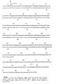
- FIGURE 1 illustrates the nucleotide sequence of the human GM-CSF gene.
- the coding region of the human CM-CSF gene extends from nucleotide No. 14 to nucleotide No. 394.
- the corresponding amino acid sequence, as determined from the nucleotide sequence, is set forth below the relevant codons.
- Plasmid DNA, designated as pHG23, prepared from the cDNA of the identified positive colony and transformed into E . coli is on deposit with the American Type Culture Collection ("ATCC”), 12301 Parklawn Drive, Rockville, Maryland 20852, U.S.A., under Accession No. 39900.
- ATCC American Type Culture Collection
- Functional GM-CSF is produced by expressing the GM-CSF gene contained in pHG23 in host cells and then tested for the ability of the expressed product to stimulate the growth of bone marrow colonies in agar.
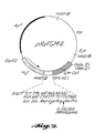
- a cDNA fragment of substantially the entire coding region of the GM-CSF gene shown in FIGURE 1 (the Sfa NI to the Nco I fragment) is inserted into an expression vector designed to direct synthesis and secretion of GM-CSF from yeast host cells, for instance a vector designated as pY ⁇ fGM-2 as shown in FIGURE 2.
- the expression vector such as pY ⁇ fGM-2, preferably contains sequences derived from plasmid pBR 332 (thick life portion in FIGURE 2) including an origin of replication and the ampicillin-resistance gene (Ap r ).
- the expression vector also includes sequences from yeast, for instance, the tryptophan-1 gene (TRP-1) as a selectable marker and the 2u yeast origin of replication (thin line portion).
- the expression vector preferably further includes the yeast pre-pro- ⁇ mating factor (" ⁇ -factor") (stippled box portion) as an efficient promoter together with leader sequences to direct the synthesis and secretion of GM-CSF in yeast hosts, followed by the sequence for the coding region of GM-CSF shown in FIGURE 1 (hatched box portion).
- ⁇ -factor yeast pre-pro- ⁇ mating factor
- the pY ⁇ fGM-2 expression plasmid is transformed into an appropriate strain of Saccharomyces cerevisiae (" S . cerevisiae ").
- Preferable strains include yeast strain Nos. 79, X2181-1B, DBY746, YNN280, and 20B-12. These strains are all ⁇ , Trp I, Leu 2 for compatibility with the ⁇ -factor promoter and for selection of Trp+ transformants. These strains are all widely available, for instance strain 79 is available from the Yeast Genetic Stock Center, Department of Biophysics and Medical Physics, University of California, Berkeley, California 94702.
- Transformation of the yeast host with the recombinant expression plasmid containing the bIL-2 gene is conducted according to well-known procedures wherein spheroplasts are formed and then washed prior to plasmid uptake. Standard protocols for this procedure have been established. See, for example, Beggs, Nature (London) , 275 :104 (1978); and, Hinnen et al., Proc. Natl. Acad. Sci. (U.S.A.) , 75 :1929 (1978).
- Yeast fermentation supernatants are assayed for biological activity by their ability to direct the formation of mixed, granulocytic and macrophage-type colonies from human bone marrow cells.
- plasmid pY ⁇ f of the same construction as pY ⁇ fGM-2, but lacking the GM-CSF sequences, was also transformed into a yeast host and the fermentation supernatant tested for biological activity.
- the pY ⁇ fGM-2 supernatant was found to direct synthesis of high levels of GM-CSF activity in the bone marrow assay (1.2 x 10⁶ colony forming units per milliliter culture (“CFU-c/ml”)) whereas no activity was detected from the supernatant derived from the pY ⁇ f control plasmid.
- the recombinant GM-CSF contained in the supernatant of the expression host cells is purified to essential homogeneity by reverse phase high-performance liquid chromatography ("HPLC").
- HPLC procedures used in the present invention preferably employ a reverse phase, tetramethyl, octadecyl, octylmethyl or diphenyl-bonded silica column having a pore size sufficiently large to be optimally utilized with the protein GM-CSF, i.e., a pore size of at least 300 ⁇ .
- Suitable reverse phase HPLC columns for use in the practice of the present invention are articles of commerce.
- a preferable column for this purpose is the Vydac line of columns commercially available from Separations Group, Hesperia, CA.
- the present invention may employ the Vydac C4 or C18 adsorbent reverse phase columns consisting of tetramethyl silane groups covalently bonded by means of siloxane (silicon-oxygen-silicon) bonded to the surface of 300 ⁇ pore diameter silica gel which has been classified to a mean particle size of from 30 to 44 microns.
- the expressed GM-CSF Prior to being applied to the column, the expressed GM-CSF is rendered acidic with an appropriate acid, such as trifluoroacetic acid ("TFA").
- TFA trifluoroacetic acid
- the elution of proteins from the HPLC column is carried out in a manner well known in the art.
- a suitable elution procedure for removing the bonded proteins from the column involves the use of a linear gradient of acetonitrile.
- a preferred gradient for this purpose is 0 to 100% (vol/vol) acetonitrile gradient in TFA (pH 2.0-2.1).
- the eluted protein can be conveniently monitored with detection systems that are well known in the art.
- the relative protein concentrations in fractions eluted from the HPLC column can be determined by measuring absorbance of the eluted material in an automated ultraviolet light, at 214 nanometers light wavelengths.
- a suitable automated ultraviolet light absorbance detection apparatus is available from Waters Associates, Milford, ME.
- Fractions recovered from the HPLC procedure are analyzed for protein by the fluorescamine assay and by SDS (sodium dodecyl sulfate) polyacrylamide gel electrophoresis followed by silver staining, as described in Example 4, infra .
- the recovered GM-CSF, designated as Fxn 57, is then assayed for biological activity using the bone marrow colony-forming assay discussed above and in Example 3, infra .
- the GM-CSF was purified to homogeneity as a single symmetric peak of biological activity. As shown in FIGURE 5, by polyacrylamide gel electrophoresis, two bands of GM-CSF activity having molecular weights of about 21,000 and 17,000 daltons were identified. These bands correspond to glycosylated (21,000) and unglycosylated (17,000) species of GM-CSF produced by yeasts.
- the activity level of the Fxn 57 was found to be 1.5 x 10⁷ CFU-c/ml.
- the specific activity of this homogeneous GM-CSF was found to be approximately 1.5 x 10⁶ CFU-c/ ⁇ g protein, or 3.0 x 10¹⁶ CFU-c/mole.
- Activation of monocyte/macrophages into tumoricidal state is assayed with respect to various types of tumor cells, including, for instance, the A375 human myeloma cell line (ATCC No. CRL 1619), the human bladder squamous carcinoma SCaBER (ATCC No. HTB3), the human astrocytoma gliobastomas U-87 MG and U-373 MG (ATCC Nos. HTB 14 and 17, respectively), the human Ewing sarcomas Esa-1 and SK-ES-2 (ATCC Nos. HTB 83 and HTB 87), the human malignant melanomas SK-MEL-28 and SK-MEL-31 (ATCC Nos.
- A375 human myeloma cell line ATCC No. CRL 1619
- the human bladder squamous carcinoma SCaBER ATCC No. HTB3
- the human astrocytoma gliobastomas U-87 MG and U-373 MG ATCC Nos
- the target cells Prior to assay, the target cells are grown in monolayers in medium supplemented with various additives, such as fetal calf serum ("FCS"). Thereafter, the target cells are radiolabeled, for instance with [125I] iododeoxyuridine ("[125 I] IUdR").
- FCS fetal calf serum
- test samples of putative activator substances i.e., GM-CSF, IFN- ⁇ , LPS are added to the plated monocytes, prepared above.
- an appropriate culture period i.e., 24 hours
- the test sample is removed and radiolabeled target cells added to the culture wells.
- the culture supernatants are removed and fresh medium replaced.
- target cells killed by the activated monocytes are removed and the remaining viable cells in each well lysed with an appropriate reagent, such as NaOH.
- the radioactivity of the lysate is then measured in a gamma counter.
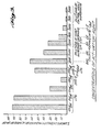
- FIGURES 3 and 4 illustrate the tumorcidal activity of human peripheral blood monocytes activated with various concentrations of recombinant human GM-CSF, LPS, IFN- ⁇ or LPS together with IFN- ⁇ . Specifically, FIGURE 3 illustrates the dose dependent capacity of LPS and IFN- ⁇ individually and combined to induce macrophage-mediated tumor target destruction. FIGURE 3 also shows the activity of various concentrations of yeast supernatant containing recombinant GM-CSF.
- yeast supernatant at a dilution of 1:500 stimulated macrophages to generate approximately 65% specific cytotoxicity.
- control yeast fermentation supernate was not capable of inducing macrophage cytotoxicity.
- Crude yeast supernate has a titer of approximately 1-2 x 10⁵ CFU-c/ml.
- FIGURE 4 shows a full course dose response titration of purified recombinant human GM-CSF.
- a half-maximal induction of macrophage mediated tumor-cell cytotoxicity occurs at a dilution of the column fraction containing homogeneous GM-CSF of approximately 1:1,000,000(15 CFU-c/ml).
- Control data illustrating the cytotoxicity of IFN- ⁇ , LPS and LPS combined with IFN- ⁇ are also shown in FIGURE 4.
- GM-CSF effectively functioned as a singular signal to induce monocytes to exhibit tumorcidal activity. This is a biological property of GSM-CSF that heretofore has been unknown.
- Purified recombinant GM-CSF as prepared above may be employed to treat patients or other mammals suffering from tumors using various therapy procedures.
- Therapeutically effective doses of GM-CSF can be administered to a subject in need of treatment by a variety of conventional routes, such as parenterally or transdermally.
- the recombinant GM-CSF may be administered in dosages of about 1.0 to 106 ⁇ g/kg of body weight of the subject per day.
- Preferably treatment is initiated at a lower level of this dosage range and then increased until the desired therapeutic effect is achieved.
- variations in dosage will necessarily occur depending upon various factors, for instance the condition of the subject being treated. The person responsible for administration will, in any event, determine the appropriate dose for the individual subject.
- the recombinant GM-CSF may be introduced into the subject in single or multiple doses by way of, for example, subcutaneous, intramuscular, intraperitoneal, or intravenous injection.
- the GM-CSF could be introduced via aerosol inhalation, transdermal or transbuccal absorption or rectal suppository.
- solutions of recombinant GM-CSF in sesame oil or peanut oil or aqueous propylene glycol may be employed, as well as sterile nontoxic, nonallergic solutions of distilled water, serum albumin, Ringer's solution, Hank's solution, etc.
- Such solutions could be suitably buffered if necessary and the liquid diluent first rendered isotonic with sufficient saline or glucose.
- macrophages or precursor cells thereof can be isolated from a donor and stimulated into tumoricidal activity by culturing the cells with a therapeutically effective quantity of GM-CSF.
- the activated macrophages or precursor cells can then be administered to a recipient using one of the techniques discussed above. Typically, but not in all instances, the donor and recipient are the same individual.
- tumors include all carcinomas, such as bladder, kidney, squamous cells, lung, liver, breast and colon.
- carcinomas such as bladder, kidney, squamous cells, lung, liver, breast and colon.
- tumors also include any melanoma or sarcoma.
- peripheral blood monocytes obtained from Portland, Oregon, Red Cross
- RPMI Roswell Park Memorial Institute
- heparin 50 U/ml, Sigma Chemical Co., St. Louis, MO
- Isolymph Trade Mark Gallard-Schlesinger, Carle Place, NY
- the interface cells were washed and than layered, at a concentration of from 4 to 5 x 10⁷ cells in 2ml, on continuous Percoll (Trade Mark Pharmacia Fine Chemicals) gradients and then centrifuged at 200 x g for 20 minutes at 4°C to separate the monocytes from the remainder of the mononuclear leukocytes.
- the resulting top, monocyte layer was collected, washed in RPMI-1640 with 5% FCS and then plated at a concentration of from 1 to 2 x 10⁵ cells per well in flat-bottomed 96-well Microtiter plates (Costar, Cambridge, MA) together with RPMI-1640 and 5% FCS.
- the nonadherent cells were removed by aspiration with an 18 gauge needle (outer diameter 1.27mm, inner diameter 0.84mm) and then the monocyte layers rinsed twice with serum free RPMI-1640 medium thereby readying the monocytes for addition of test samples composed of various concentrations of putative activators, such as GM-CSF, IFN- ⁇ , LPS combined with IFN- ⁇ as discussed more fully below.
- putative activators such as GM-CSF, IFN- ⁇ , LPS combined with IFN- ⁇ as discussed more fully below.
- pY ⁇ fGM-2 Substantially the entire coding region and a portion of the 3' flanking region of the GM-CSF gene were removed from the cDNA clone of FIGURE 1 and employed to form a recombinant expression plasmid, designated as pY ⁇ fGM-2 to direct GM-CSF expression in yeast host cells.
- the pY ⁇ fGM-2 expression plasmid is on deposit with the ATCC under Accession No. 53157.
- pY ⁇ fGM-2 includes an origin of replication and an Ap r resistant gene from plasmid pBR322 (thick line portion).
- the expression plasmid also includes the yeast 2 ⁇ circle origin of replication and a Trp I gene for selection of transformed yeast hosts (TRP - [Trp-auxotrophs], thin line portion in FIGURE 2).
- the expression plasmid further includes the yeast ⁇ -factor promoter and leader sequences (stippled box portion) for use in directing transcription and secretion of GM-CSF.
- the GM-CSF sequences hatchched box portion
- the Sfa NI enzyme cleaves the GM-CSF gene from the pHG23 clone at a location which is two nucleotides downstream from the 5' terminus of the region coding for the mature protein (nucleotide No. 14), since no restriction site was found to correspond precisely to nucleotide No. 14.
- oligonucleotide was chemically synthesized to add back the 5' terminal portion of the coding region of the mature GM-CSF gene and also to add a second ⁇ -factor processing site to obtain complete processing of the signal sequence for secretion of the mature form of GM-CSF.
- the composition of the oligonucleotide as shown in Table I below, and in FIGURE 2 (open box portion), includes a Hind III cohesive 5' terminal, followed by a Cathepsin B-like maturation site composed of the sequence: TCT TTG CAT AAA AGA, followed by a Sfa NI cohesive 3' terminal coding for the first two amino acid residues of the mature GM-CSF protein.
- oligonucleotide shown in Table I was chemically synthesized by triester technique as detailed by Sood et al., Nucl. Acad. Res. , 4 :2557 (1977); and, Hirose et al., Tet. Lett. , 28 :2449 (1978), it is to be understood that the oligonucleotide can be prepared by other methods, such as by the phosphodiester method.
- the pY ⁇ fGM-2 vector was transformed into yeast strain 79 ( ⁇ , Trp 1-1, Leu 2-1) of S . cerevisiae for selection of Trp+ transformants by standard techniques.
- the strain 79 was grown either in selective media (YNB-trp, consisting of 0.67% Yeast Nitrogen Base (Difco Labs, Detroit, MI), 0.5% (Casamino acids, 2% glucose, 10 micrograms/milliliter (" ⁇ g/ml”) adenine and 20 ⁇ g/ml uracil); or, in rich media (YPD, consisting of 1% yeast extract, 2% peptone and 2% glucose supplemented with 80 ⁇ g/ml adenine and 80 ⁇ g/ml uracil). Cells were harvested by centrifugation at 1000 x g for 5 minutes at 22°C, and then the resulting pellet was washed with sterile, distilled water.
- YNB-trp consisting of 0.6
- the yeast cells were then concentrated by resuspending in 1/10 vol. of SED (1 M sorbitol, 25 mM ethylene diamine tetracetate (“EDTA”) [pH 8.0], and 50 mM dithiothreitol) and incubating for 10 minutes at 30°C.
- SED 1 M sorbitol, 25 mM ethylene diamine tetracetate
- EDTA ethylene diamine tetracetate
- the cell-buffer mixture was then centrifuged for 5 minutes at 300 x g. The pellet was washed once with 1/10 vol. of 1 M sorbitol and the cells resuspended in 20 milliliters of SCE (1 M sorbitol, 0.1 M sodium citrate [pH 5.8], 0.01 M EDTA). Glusulase, to break down the cell walls, in an amount of 10 ⁇ 3 vol.
- spheroplasts was assayed by diluting 10 microliters (" ⁇ l") of the yeast cells into a drop of 5% SDS (wt./vol.) on a microscope slide to observe for "ghosts" at 400 X phase contrast. The cell mixture was then centrifuged at 300 x g for 3 minutes. The resulting pellet was twice washed with 1/10 vol. of 1 M sorbitol and then once with CaS (1 M sorbitol, 10 mM CaCl2).
- the yeast spheroplasts were transformed with the previously prepared plasmid vector in a procedure adapted from Beggs, supra .
- the pelleted spheroplasts were suspended in 1/200 vol. of CaS and divided into 100 microliter aliquots in 1.5 ml Eppendorf tubes. From 1 to 10 ⁇ l of the plasmid DNA were added to each aliquot (0.5 to 5 ⁇ g). The mixture was incubated at room temperature for 10 minutes and then 1 ml of PEG (20% PEG 4000, 10 mM CaCl2, 10 mM Tris-HCl [pH 7.4]) was added to each aliquot to promote DNA uptake.
- the protoplast/DNA mixture selective plates Prior to plating, the protoplast/DNA mixture selective plates were preincubated at 37°C. Three ml of melted top agar (45°C), composed of 18.2 ml of sorbitol, 2 g agar, 0.6 g Difco yeast nitrogen base (without amino acids), 2 g glucose, 0.1 ml of 1% adenine, 0.4 ml of 1% uracil and amino acids as required, was then added to each aliquot of transformed cells and the tube contents poured on the selective plates. The plates were incubated from 2 to 4 days at 30°C. Colonies which developed in the Trp minus medium contained plasmids that have the Trp 1 gene, i.e., those that are transformed.
- the transformants Prior to biological and tumoricidal assays, the transformants were grown in 20-50 ml of rich medium at 30°C to stationary phase. At the time of harvest, the protease inhibitors phenylmethylsulfonyl fluoride (PMSF) and Pepstatin A were added to a final concentration of 1 mM and 10 ⁇ M, respectively. The cells were then removed by centrifugation at 400 x g and the medium was filtered through a 0.45 ⁇ cellulose acetate filter (Corning Glass Works, Corning, NY). The sterile supernates were stored at 4°C.
- PMSF phenylmethylsulfonyl fluoride
- Pepstatin A Pepstatin A
- Example 2 The presence of human GM-CSF harvested from the yeast cultures in Example 2 was confirmed by assaying the ability of the supernatant to stimulate growth of human bone marrow colonies in agar.
- human bone marrow from the iliac crest of healthy donors was collected in a heparinized syringe.
- the marrow was diluted 1:3 with phosphate buffered saline (PBS) at room temperature and layered onto a solution of 54% Percoll(Trade Mark)(Pharmacia Fine Chemicals). After centrifugation at 500 x g at room temperature for 20 minutes, the interface was collected and washed with 20 volumes of PBS.
- PBS phosphate buffered saline
- the suspension was then centrifuged at 250 x g for 10 minutes at room temperature.
- the cells were then resuspended in 10ml of ⁇ -Minimal Essential Medium with nucleotides (" ⁇ -Mem", Gibco) for cell counting and viability determination.
- FCS was then added and the cell suspension stored on ice until the assay was carried out.
- bone marrow cells as prepared above were added at a final concentration of 1 x 10⁵/ml to an incubation medium consisting of: (a) seven parts of a solution containing 28.1% FCS, 0.7 x 10 ⁇ 4 M 2-mercaptoethanol, 0.12 mg/ml asparagine, 0.7 mg/ml glutamine, 150 units of penicillin G, 150 units of streptomycin, 1.1 x ⁇ -MEM with nucleotides, and 2.2 x vitamins (Gibco); and, (b) three parts of 1.4% bacto-agar solution (Difco). The cultures were incubated in a humidified atmosphere at 37°C in the presence of 5% CO2.
- the number and types of colonies were determined.
- the GM-CSF gene from the pY ⁇ fGM-2 clones directed synthesis of GM-CSF activity at the high level of 1.25 x 10⁶ colony forming units per 105 (CFU-c/ml). This activity level was determined by multiplying by 50 the reciprocal of the dilution giving 50% of the maximum colony number.
- the average number of colonies from 1 x 10⁵ bone marrow cells was 96 ⁇ 29.
- the colonies formed at 14 days by the recombinant GM-CSF were well defined and consisted of three types: approximately 1/3 mixed granulocyte-macrophage colonies; approximately 1/3 tight granulocyte colonies, and approximately 1/3 dispersed macrophage colonies.
- a plasmid identical to pY ⁇ fGM-2, but lacking the GM-CSF sequences was also transformed into yeast strain 79.
- the culture supernatant from the yeast produced no GM-CSF activity in the bone marrow colony forming assay.
- the load column was washed with 0.1% TFA to remove nonbound sample components until the absorbance at 214 nm dropped to baseline (preloading) values. Elution of the bound protein was accomplished with a linear gradient of 0-95% acetonitrile in 0.1% TFA (v/v) at a rate of 1% acetonitrile/min. The gradient was formed with a Waters liquid chromatograph consisting of a Model 680 gradient former, 2 M-45 pumps and a Model 414 detector monitoring at 214 nm. Peak protein fractions were observed at from 55 to 60% acetonitrile.
- Fractions were analyzed for protein by fluorescamine assay. Also, peak fractions were again analyzed by SDS polyacrylamide gel electrophoresis followed by silver staining. In the electrophoresis process, 20 ⁇ l aliquots from the fractions collected during the elution procedure are dried under vacuum after the addition of 2 ml of 10% SDS to each aliquot. The dried residue was dissolved in 40 ⁇ l of nonreducing sample buffer composed of 0.0625 M Tris (pH 6.8); 2% SDS (w/u); 10% glycerol (v/v); and, 5% 2-mercaptoethanol (v/v). The solution was boiled for 3 min.
- the activity of this homogeneous material was found to be approximately 1.5 x 10⁷ CFU -c /ml, with a specific activity of 1.5 x 10⁶ CFU -c / ⁇ g of protein.
- the pI value was found to be approximately from 5.0 to 5.4.
- Target cells were maintained in RP MI 1640 medium supplemented with 5% at 37°C in a humidified atmosphere containing 5% CO2. Cytotoxicity essays were performed when the cultured target cells were in their exponential growth phase.
- Target cells included the human melanoma cell line A375 (ATCC No. CRL 1619), the human liver carcinoma cell line SK-Hep-1 (ATCC No. HTB-52), the human pancreatic carcinoma MIA-PACA-2 (ATCC No. CRL-1420) and the human bladder carcinoma 5637 (ATCC No. HTB-9).
- the exponentially growing target cells were radiolabeled with 125I-IUdR.
- target cells were incubated at 37°C for 24 hours in RP MI medium supplemented with 5% FCS medium containing 125I-IUdR (0.3 ⁇ Ci/ml (11.1x10³ Bq/ml); specific activity, 200 mCi/ml (7.4x10⁹ Bq/ml); New England Nuclear, Boston, MA).
- the cells were then washed twice to remove unbound radiolabel and then harvested by a 1 min. trypsinization with 0.25% tryspin (Difco Labs, Detroit, MI) and 0.02% EDTA.
- the labeled cells were resuspended in RPMI-1640 medium supplemented with 5% FCS.
- Approximately 104 125I-UdR-labeled target cells from Example 5 were added to each of the monocyte wells to obtain an initial target-to-effector (activated monocyte) cell ratio of between 1:10 and 1:20. Also, radiolabeled target cells were plated alone as an additional control group. Because the assay measures lysis of adherent target cells in which cell-to-cell contact between effector and target cells is required, after 24 hours the culture supernatants were aspirated off and fresh RP MI-1640 medium replaced to eliminate potential errors due to cells that did not adhere but were not necessarily killed in the assay. After an additional 48 hours, the monolayers were vigorously rinsed to remove target cells killed by the monocytes.
- FIGURES 3 and 4 contain the assay results. As shown in FIGURE 3, and as expected, LPS and IFN- ⁇ interferon did activate macrophages to the point where they mediated tumoricidal activity. Also as previously reported, the effectiveness of IFN- ⁇ as a macrophage activator was only significant when added together with a small amount of LPS. As further shown in FIGURE 3, supernate produced by yeast containing the GM-CSF plasmid was effective in inducing macrophages to kill tumor cells while control supernate (produced by yeast which did not contain the GM-CSF plasmid) was unsuccessful in inducing macrophage cytotoxicity.
- FIGURE 4 shows a full course dose titration of purified recombinant GM-CSF.
- GM-CSF served as an effective single signal to activate macrophages into tumoricidal activity in the absence of exogenous LPS.
- the tumoricidal activity assay set forth in Example 6 is also conducted with respect to the human bladder squamous carcinoma SCaBER (ATCC No. HTB 3). These targets cells are radiolabeled in a manner detailed in Example 5. The assay is conducted using the same procedure set forth in Example 6 with the exception that radiolabeled SCaBER target cells are used in place of the A375 target cells.
- the tumoricidal assay procedure of Examples 6 and 7 is employed in conjunction with the human malignant myeloma SK-MEL-28 (ATCC No. HTB 72).
- the target cells are radiolabeled as detailed in Example 5.
- the assay is conducted in the same manner as set forth in Examples 6 and 7 with the exception that SK-MEL-28 cells are employed as target cells.
- the tumoricidal activity assay set forth in Example 6 was also conducted with respect to the human liver carcinoma SK-Hep-1 (ATCC No. HTB-52).
- the target cells were radiolabeled in a manner detailed in Example 5.
- the GM-CSF was employed at a concentration of 10 nanograms per milliliter (ng/ml) of assay volume.
- Approximately 104 125IUdr-labeled target cells were used in each assay to obtain an initial target: effector (activated monocyte) cell ratio of between 1:20 and 1:30.
- radiolabeled target cells were plated alone as a control.
- the GM-CSF activated monocytes were found on the average to mediate specific cytotoxicity of the tumor cells at a level of about 14%.
- Example 9 The tumoricidal assay procedure of Example 9 was employed in conjunction with the human pancreatic carcinoma cell line MIA PACA-2 (ATCC No. CRL 1420).
- the target cells were radiolabeled as detailed in Example 5.
- the assay was conducted in the manner delineated in Example 9, with the exception that the MIA PACA-2 cells were employed as target cells. In this particular assay, the average percentage of generated cytotoxicity was found to be approximately 38%.
- Example 9 The tumoricidal assay procedure of Example 9 was also employed in conjunction with the human bladder carcinoma cell line 5637 (ATCC No. HTB-9).
- the target cells were labeled as detailed in Example 5.
- the assay was conducted in the same manner as set forth in Example 9 with the exception that 5637 cells were employed as target cells. In this particular assay, the percentage of generated cytotoxicity was found to be about 30%.
- This particular assay and those set forth above illustrate that GM-CSF served as an effective single signal to activate macrophages into tumoricidal activity.
Landscapes
- Health & Medical Sciences (AREA)
- Chemical & Material Sciences (AREA)
- Organic Chemistry (AREA)
- Life Sciences & Earth Sciences (AREA)
- Medicinal Chemistry (AREA)
- General Health & Medical Sciences (AREA)
- Biochemistry (AREA)
- Gastroenterology & Hepatology (AREA)
- Biophysics (AREA)
- Zoology (AREA)
- Genetics & Genomics (AREA)
- Toxicology (AREA)
- Molecular Biology (AREA)
- Proteomics, Peptides & Aminoacids (AREA)
- Immunology (AREA)
- Nuclear Medicine, Radiotherapy & Molecular Imaging (AREA)
- General Chemical & Material Sciences (AREA)
- Chemical Kinetics & Catalysis (AREA)
- Pharmacology & Pharmacy (AREA)
- Animal Behavior & Ethology (AREA)
- Public Health (AREA)
- Veterinary Medicine (AREA)
- Medicines That Contain Protein Lipid Enzymes And Other Medicines (AREA)
- Preparation Of Compounds By Using Micro-Organisms (AREA)
- Peptides Or Proteins (AREA)
- Micro-Organisms Or Cultivation Processes Thereof (AREA)
- Medicines Containing Material From Animals Or Micro-Organisms (AREA)
- Pharmaceuticals Containing Other Organic And Inorganic Compounds (AREA)
Claims (10)
- Verwendung des Granulozyten-Makrophagen-Kolonien stimulierenden Faktors zur Erzeugung eines Medikamentes zum Fördern der tumorzerstörenden Aktivität von Zellen des Makrophagen-Stammbaumes, bei welcher Verwendung die Zellen des Makrophagen-Stammbaumes der Einwirkung einer wirksamen Menge dieses Medikamentes unterworfen werden.
- Verwendung des Granulozyten-Makrophagen-Kolonien stimulierenden Faktors nach Anspruch 1, bei welcher Verwendung das Verfahren zum Fördern der tumorzerstörenden Aktivität von Zellen des Makrophagen-Stammbaumes die folgenden Stufen umfaßt:(a) Isolieren von Zellen des Makrophagen-Stammbaumes aus dem Körper;(b) Inberührungbringen der isolierten Zellen mit einer wirksamen Menge eines Granulozyten-Makrophagen-Kolonien stimulierenden Faktors; und(c) anschließend Einbringen der Zellen in den Körper eines Empfängers.
- Verwendung des Granulozyten-Makrophagen-Kolonien stimulierenden Faktors nach Anspruch 1, bei welcher Verwendung das Verfahren zum Fördern der tumorzerstörenden Aktivität von Zellen des Makrophagen-Stammbaumes das Einbringen einer wirksamen Menge eines Granulozyten-Makrophagen-Kolonien stimulierenden Faktors in den Körper eines Menschen umfaßt.
- Verwendung des Granulozyten-Makrophagen-Kolonien stimulierenden Faktors zur Erzeugung eines Medikamentes für die Krebstherapie.
- Verwendung des Granulozyten-Makrophagen-Kolonien stimulierenden Faktors nach Anspruch 4, wobei die Methode der Krebstherapie die Stufe des Einbringens einer wirksamen Menge eines Grunulozyten-Makrophagen-Kolonien stimulierenden Faktors in den Körper eines Menschen umfaßt.
- Verwendung des Grunulozyten-Makrophagen-Kolonien stimulierenden Faktors nach einem der Ansprüche 1 bis 5, wobei der Granulozyten-Makrophagen-Kolonien stimulierende Faktor nach Techniken unter Einsatz von rekombinanter DNA erzeugt wird.
- Verwendung des Granulozyten-Makrophagen-Kolonien stimulierenden Faktors nach einem der Ansprüche 1 bis 6, wobei der Granulozyten-Makrophagen-Kolonien stimulierende Faktor bis auf eine nach dem Knochenmarks-Kolonienbildungstest bestimmte, spezifische Aktivität von wenigstens 1,5 x 10⁶ kolonienbildenden Einheiten je µg Protein gereinigt wird.
- Verwendung des Granulozyten-Makrophagen-Kolonien stimulierenden Faktors nach einem der Ansprüche 1 bis 3 oder nach Anspruch 6 oder 7, wenn diese letztgenannten Ansprüche auf die Ansprüche 1 bis 3 rückbezogen sind, wobei die Makrophagen aus Monozyten im peripheren Blut stammen.
- Verwendung des Granulozyten-Makrophagen-Kolonien stimulierenden Faktors nach einem der Ansprüche 1 bis 8, wobei 0,01 bis 100 ng (Nanogramm) des Granulozyten-Makrophagen-Kolonien stimulierenden Faktors je 2 x 10⁵ Zellen des Makephagen-Stammbaumes verwendet werden.
- Verwendung nach einem der Ansprüche 1 bis 9, wobei der Granulozyten-Makrophagen-Kolonien stimulierende Faktor Human-Granulozyten-Makrophagen-Kolonien stimulierender Faktor ist.
Priority Applications (1)
| Application Number | Priority Date | Filing Date | Title |
|---|---|---|---|
| AT86306304T ATE77958T1 (de) | 1985-08-16 | 1986-08-15 | Aktivierung der tumorzerstoerenden aktivitaet der makrophagen mittels des granulozyten-makrophagen- kolonien stimulierenden faktors. |
Applications Claiming Priority (4)
| Application Number | Priority Date | Filing Date | Title |
|---|---|---|---|
| US76689385A | 1985-08-16 | 1985-08-16 | |
| US766893 | 1985-08-16 | ||
| US06/888,995 US5078996A (en) | 1985-08-16 | 1986-07-31 | Activation of macrophage tumoricidal activity by granulocyte-macrophage colony stimulating factor |
| US888995 | 1986-07-31 |
Publications (3)
| Publication Number | Publication Date |
|---|---|
| EP0211684A2 EP0211684A2 (de) | 1987-02-25 |
| EP0211684A3 EP0211684A3 (en) | 1988-06-08 |
| EP0211684B1 true EP0211684B1 (de) | 1992-07-08 |
Family
ID=27117812
Family Applications (1)
| Application Number | Title | Priority Date | Filing Date |
|---|---|---|---|
| EP86306304A Expired - Lifetime EP0211684B1 (de) | 1985-08-16 | 1986-08-15 | Aktivierung der tumorzerstörenden Aktivität der Makrophagen mittels des Granulozyten-Makrophagen-Kolonien stimulierenden Faktors |
Country Status (8)
| Country | Link |
|---|---|
| US (1) | US5078996A (de) |
| EP (1) | EP0211684B1 (de) |
| JP (1) | JPH0780781B2 (de) |
| AT (1) | ATE77958T1 (de) |
| AU (1) | AU586876B2 (de) |
| CA (1) | CA1341142C (de) |
| DE (1) | DE3685918T2 (de) |
| DK (1) | DK174603B1 (de) |
Families Citing this family (114)
| Publication number | Priority date | Publication date | Assignee | Title |
|---|---|---|---|---|
| RU2130493C1 (ru) * | 1984-07-06 | 1999-05-20 | Новартис Аг | Способ получения белка с активностью gm-csf приматов |
| US5795568A (en) * | 1984-09-19 | 1998-08-18 | Novartis Corporation | Method of treating infectious disease with GM-CSF |
| US5556620A (en) * | 1985-02-05 | 1996-09-17 | Cetus Oncology Corporation | Use of recombinant colony stimulating factor-1 to enhance wound healing |
| US5837229A (en) * | 1985-02-05 | 1998-11-17 | Chiron Corporation | Uses of recombinant colony stimulating factor-1 |
| US5422105A (en) * | 1985-02-05 | 1995-06-06 | Cetus Oncology Corporation | Use of recombinant colony stimulating factor 1 |
| US5104650A (en) * | 1985-02-05 | 1992-04-14 | Cetus Corporation | Uses of recombinant colony stimulating factor-1 |
| US5093115A (en) * | 1985-06-11 | 1992-03-03 | The United States Of America As Represented By The Department Of Health And Human Services | Method of preparing activated killer monocytes for treating colorectal cancer |
| US5391706A (en) * | 1987-07-16 | 1995-02-21 | Schering Plough Corporation | Purification of GM-CSF |
| EP0318184A1 (de) * | 1987-11-12 | 1989-05-31 | Schering Corporation | Knochenbildungbeschleunigung durch GM-CSF |
| BE1002141A5 (fr) * | 1988-01-13 | 1990-07-31 | Bartholeyns Jacques | Procede de culture et d'activation de cellules humaines et conditionnement pour le traitement du cancer. |
| JPH01193227A (ja) * | 1988-01-29 | 1989-08-03 | Res Dev Corp Of Japan | 癌免疫療法補助剤 |
| AU4219689A (en) * | 1988-08-19 | 1990-03-23 | Genetics Institute Inc. | Treatment for malignant melanoma |
| GB8824591D0 (en) * | 1988-10-20 | 1988-11-23 | Royal Free Hosp School Med | Fractionation process |
| DE69001686T2 (de) * | 1989-01-30 | 1993-09-02 | Schering Corp | Behandlung von leukozytstoerungen mit gm-csf. |
| US5154921A (en) * | 1990-07-13 | 1992-10-13 | Dana-Farber Cancer Institute, Inc. | Promotion of maturation of hematopoietic progenitor cells |
| US5199942A (en) * | 1991-06-07 | 1993-04-06 | Immunex Corporation | Method for improving autologous transplantation |
| US5637483A (en) * | 1991-10-04 | 1997-06-10 | Whitehead Institute For Biomedical Research | Irradiated tumor cell vaccine engineered to express GM-CSF |
| US5904920A (en) * | 1991-10-04 | 1999-05-18 | Whitehead Institute For Biomedical Research | Regulation of systemic immune responses utilizing cytokines and antigens |
| US5292513A (en) * | 1992-05-18 | 1994-03-08 | Anthony G. Gristina | Method for nonspecific cellular immune stimulation |
| US5662899A (en) * | 1993-05-18 | 1997-09-02 | I.D.M. Immuno-Designed Molecules | Macrophages, process for preparing the same and their use as active substances of pharmaceutical compositions |
| US6001351A (en) * | 1993-05-18 | 1999-12-14 | I.D.M. Immuno-Designed Molecules | Macrophages, process for preparing the same and their use as active substances of pharmaceutical compositions |
| US6540994B1 (en) * | 1997-07-18 | 2003-04-01 | I.D.M. Immuno-Designed Molecules | Macrophages, process for preparing the same and their use as active substances of pharmaceutical compositions |
| JPH11509845A (ja) | 1995-07-21 | 1999-08-31 | アメリカ合衆国 | 造血多分化性細胞移植のための放射線または放射線類似作用化学療法の減少方法 |
| US6267955B1 (en) | 1995-09-15 | 2001-07-31 | Yeda Research And Development Co. Ltd. | Mononuclear phagocytes and their use to promote axonal regeneration |
| US5800812A (en) * | 1995-09-15 | 1998-09-01 | Yeda Research And Development Co. Ltd. | Methods of use of mononuclear phagocytes to promote axonal regeneration |
| US5942253A (en) * | 1995-10-12 | 1999-08-24 | Immunex Corporation | Prolonged release of GM-CSF |
| US6080409A (en) * | 1995-12-28 | 2000-06-27 | Dendreon Corporation | Immunostimulatory method |
| US5891432A (en) | 1997-07-29 | 1999-04-06 | The Immune Response Corporation | Membrane-bound cytokine compositions comprising GM=CSF and methods of modulating an immune response using same |
| EP1125133A1 (de) * | 1998-11-03 | 2001-08-22 | Cell Genesys, Inc. | Krebs-assoziierte antigene und verfahren zu ihrer identifizierung |
| NZ515322A (en) * | 1999-05-13 | 2004-03-26 | Wyeth Corp | Adjuvant combination formulations |
| CA2388337C (en) | 1999-10-22 | 2013-01-08 | Aventis Pasteur Limited | Method of inducing and/or enhancing an immune response to tumor antigens |
| US6911204B2 (en) | 2000-08-11 | 2005-06-28 | Favrille, Inc. | Method and composition for altering a B cell mediated pathology |
| HK1052314A1 (zh) * | 2000-08-11 | 2003-09-11 | Favrille, Inc. | 改变t细胞介导的病理的方法和组合物 |
| IL155690A0 (en) * | 2000-11-10 | 2003-11-23 | Wyeth Corp | Adjuvant combination formulations |
| US20070128229A1 (en) * | 2002-04-12 | 2007-06-07 | Wyeth | Surface proteins of Streptococcus pyogenes |
| BR0208874A (pt) * | 2001-04-13 | 2004-06-22 | Wyeth Corp | Proteìnas superficiais de streptococcus pyogenes |
| EP2277897A1 (de) | 2001-04-16 | 2011-01-26 | Wyeth Holdings Corporation | Polypeptideantigene codierende offene Leseraster aus Streptococcus pneumoniae und Verwendungen davon |
| MX339524B (es) * | 2001-10-11 | 2016-05-30 | Wyeth Corp | Composiciones inmunogenicas novedosas para la prevencion y tratamiento de enfermedad meningococica. |
| AU2003218162B2 (en) * | 2002-03-15 | 2009-10-29 | The Curators Of The University Of Missouri | Mutants of the P4 protein of nontypable haemophilus influenzae with reduced enzymatic activity |
| US7785608B2 (en) * | 2002-08-30 | 2010-08-31 | Wyeth Holdings Corporation | Immunogenic compositions for the prevention and treatment of meningococcal disease |
| US7301554B2 (en) * | 2002-09-20 | 2007-11-27 | Ricoh Company, Ltd. | Light scanning device, scanning line adjusting method, scanning line adjusting control method, image forming apparatus, and image forming method |
| WO2004035086A2 (en) | 2002-10-16 | 2004-04-29 | Hunter Samuel F | Method for treatment of demyelinating central nervous system disease using gm-csf |
| US20040197312A1 (en) * | 2003-04-02 | 2004-10-07 | Marina Moskalenko | Cytokine-expressing cellular vaccine combinations |
| WO2005020902A2 (en) * | 2003-08-21 | 2005-03-10 | Cytokine Pharmasciences, Inc. | Compositions and methods for treating and diagnosing irritable bowel syndrome |
| KR101157694B1 (ko) | 2003-12-17 | 2012-06-20 | 와이어쓰 엘엘씨 | 면역원성 펩티드 캐리어 컨쥬게이트 및 이의 제조 방법 |
| JP4696079B2 (ja) | 2003-12-17 | 2011-06-08 | ヤンセン アルツハイマー イミュノセラピー | Aβ免疫原性ペプチド担体結合物およびそれの製造方法 |
| CA2559652C (en) * | 2004-03-15 | 2017-02-14 | Sunesis Pharmaceuticals, Inc. | Methods of using sns-595 in treating cancer |
| GB0410220D0 (en) | 2004-05-07 | 2004-06-09 | Kirkham Lea Ann | Mutant pneumolysin proteins |
| US7674456B2 (en) * | 2004-06-14 | 2010-03-09 | Charles Wiseman | Breast cancer cell lines and uses thereof |
| TW200613554A (en) | 2004-06-17 | 2006-05-01 | Wyeth Corp | Plasmid having three complete transcriptional units and immunogenic compositions for inducing an immune response to HIV |
| CN101031323A (zh) * | 2004-07-16 | 2007-09-05 | 尼克塔治疗亚拉巴马公司 | Gm-csf部分与聚合物的缀合物 |
| US20060062764A1 (en) * | 2004-08-25 | 2006-03-23 | Seshidar-Reddy Police | Fiber-modified adenoviral vectors for enhanced transduction of tumor cells |
| US20060057127A1 (en) * | 2004-09-10 | 2006-03-16 | Pocheng Liu | Cytokine-expressing cellular vaccines for treatment of prostate cancer |
| EP2298341A3 (de) | 2004-10-21 | 2011-07-13 | Wyeth LLC | Immunogene zusammensetzungen von staphylococcus epidermidis polypeptide und polynucleotide antigenen |
| WO2006050058A2 (en) * | 2004-10-28 | 2006-05-11 | The General Hospital Corporation | Methods of detection and therapy of inflamed tissues using immune modulation |
| US20080193376A1 (en) * | 2004-10-28 | 2008-08-14 | The General Hospital Corporation | Methods of Enhanced Detection and Therapy of Inflamed Tissues Using Immune Modulation |
| RU2271392C1 (ru) * | 2004-11-15 | 2006-03-10 | Институт Биоорганической химии им. акад. М.М. Шемякина и Ю.А. Овчинникова Российской Академии Наук | РЕКОМБИНАНТНАЯ ПЛАЗМИДНАЯ ДНК pFGM17, КОДИРУЮЩАЯ ПОЛИПЕПТИД ГРАНУЛОЦИТАРНО-МАКРОФАГАЛЬНОГО КОЛОНИЕСТИМУЛИРУЮЩЕГО ФАКТОРА ЧЕЛОВЕКА, И ШТАММ БАКТЕРИЙ Escherichia coli BL21(DE3)/pFGM17 - ПРОДУЦЕНТ ПОЛИПЕПТИДА ГРАНУЛОЦИТАРНО-МАКРОФАГАЛЬНОГО КОЛОНИЕСТИМУЛИРУЮЩЕГО ФАКТОРА ЧЕЛОВЕКА |
| US8580814B2 (en) * | 2006-04-03 | 2013-11-12 | Sunesis Pharmaceuticals, Inc. | Methods of using (+)-1,4-dihydro-7-[(3S,4S)-3-methoxy-4-(methylamino)-1-pyrrolidinyl]-4- oxo-1-(2-thiazolyl)-1,8-naphthyridine-3-carboxylic acid for treatment of cancer |
| WO2007123737A2 (en) | 2006-03-30 | 2007-11-01 | University Of California | Methods and compositions for localized secretion of anti-ctla-4 antibodies |
| US20070231298A1 (en) * | 2006-03-31 | 2007-10-04 | Cell Genesys, Inc. | Cytokine-expressing cancer immunotherapy combinations |
| US7919079B2 (en) | 2006-03-31 | 2011-04-05 | Biosante Pharmaceuticals, Inc. | Cancer immunotherapy compositions and methods of use |
| TW200806315A (en) | 2006-04-26 | 2008-02-01 | Wyeth Corp | Novel formulations which stabilize and inhibit precipitation of immunogenic compositions |
| AU2007258280B2 (en) | 2006-06-12 | 2013-05-02 | Sunesis Pharmaceuticals, Inc. | Compounds and compositions for treatment of cancer |
| EP3255141B1 (de) | 2006-07-13 | 2021-12-01 | Wyeth LLC | Herstellung von antikörpern mit verbessertem glykosylierungsmuster |
| ES2541546T3 (es) | 2006-11-03 | 2015-07-21 | Wyeth Llc | Sustancias que inhiben la glucólisis en cultivo celular |
| US20090285819A1 (en) * | 2006-11-15 | 2009-11-19 | Functional Genetics, Inc. | Methods and compositions for treating influenza |
| AR064642A1 (es) | 2006-12-22 | 2009-04-15 | Wyeth Corp | Polinucleotido vector que lo comprende celula recombinante que comprende el vector polipeptido , anticuerpo , composicion que comprende el polinucleotido , vector , celula recombinante polipeptido o anticuerpo , uso de la composicion y metodo para preparar la composicion misma y preparar una composi |
| AU2008223133A1 (en) | 2007-03-02 | 2008-09-12 | Wyeth | Use of copper and glutamate in cell culture for production of polypeptides |
| WO2008109818A1 (en) * | 2007-03-07 | 2008-09-12 | Mayo Foundation For Medical Education And Research | Treating cancer with granulocyte-macrophage colony stimulating factor |
| US20100255513A1 (en) * | 2007-03-30 | 2010-10-07 | Denson Lee A | Serological markers of inflammatory bowel disease phenotype and disease progression |
| US20080267935A1 (en) * | 2007-04-06 | 2008-10-30 | Cell Genesys, Inc. | AraC in combination with a cytokine-secreting cell and methods of use thereof |
| WO2009014708A2 (en) * | 2007-07-23 | 2009-01-29 | Cell Genesys, Inc. | Pd-1 antibodies in combination with a cytokine-secreting cell and methods of use thereof |
| US8425897B2 (en) * | 2007-08-30 | 2013-04-23 | Immutep S.A. | Compositions containing LAG-3 and cells that secrete GM-CSF and methods of use |
| RU2010124788A (ru) * | 2007-12-21 | 2012-01-27 | Ваейт ЭлЭлСи (US) | Генетически модифицированный ослабленный вирус везикулярного стоматита, композиции и способы их применения |
| EP2650014A2 (de) | 2008-06-20 | 2013-10-16 | Wyeth LLC | Zusammensetzungen und Verfahren zur Verwendung von ORF1358 von Beta-hämolytischen Streptokokken-stämmen |
| MX2011004755A (es) * | 2008-11-05 | 2011-10-10 | Wyeth Llc | Composicion inmunogenica multicomponente para la prevencion de enfermedad estreptococica beta-hemolitica (bhs). |
| SG10201406432RA (en) | 2009-06-22 | 2014-11-27 | Wyeth Llc | Compositions and methods for preparing staphylococcus aureus serotype 5 and 8 capsular polysaccharide conjugate immunogenic compositions |
| SA110310528B1 (ar) | 2009-06-22 | 2014-07-02 | Wyeth Llc | التركيبات المولدة للمناعة لأنتيجينات ستافيلوكوكس أوريس |
| EP2335730A1 (de) | 2009-12-18 | 2011-06-22 | Deutsches Krebsforschungszentrum | Fusionspolypeptide und ihre Verwendung zur Vorbeugung und Behandlung von Krebs |
| PL2575870T3 (pl) | 2010-06-04 | 2017-05-31 | Wyeth Llc | Preparaty szczepionek |
| CN107913396B (zh) | 2010-08-23 | 2022-03-08 | 惠氏有限责任公司 | 脑膜炎萘瑟氏菌rLP2086抗原的稳定制剂 |
| AU2011300409B2 (en) | 2010-09-10 | 2015-03-26 | Wyeth Llc | Non-lipidated variants of Neisseria meningitidis ORF2086 antigens |
| EP2654784B1 (de) | 2010-12-22 | 2016-12-07 | Wyeth LLC | Stabile immunogene zusammensetzungen aus staphylococcus aureus-antigenen |
| ITMI20111182A1 (it) | 2011-06-28 | 2012-12-29 | Canio Buonavoglia | Vaccino per coronavirus canino |
| SA115360586B1 (ar) | 2012-03-09 | 2017-04-12 | فايزر انك | تركيبات لعلاج الالتهاب السحائي البكتيري وطرق لتحضيرها |
| MY198910A (en) | 2012-03-09 | 2023-10-02 | Pfizer | Neisseria meningitidis compositions and methods thereof |
| HK1209140A1 (en) | 2012-07-19 | 2016-03-24 | Zoetis Services Llc | Bovine influenza c virus compositions |
| WO2014018724A1 (en) | 2012-07-27 | 2014-01-30 | Zoetis Llc | Tick toxin compositions |
| JP6502262B2 (ja) | 2012-12-20 | 2019-04-17 | ファイザー・インク | 糖複合化方法 |
| US9724366B2 (en) | 2012-12-30 | 2017-08-08 | Carmel-Haifa University Economic Corporation Ltd | CD11 B[low] macrophages and conditioned media thereof for treating cancer and/or fibrosis |
| WO2014136064A2 (en) | 2013-03-08 | 2014-09-12 | Pfizer Inc. | Immunogenic fusion polypeptides |
| DE102013004595A1 (de) | 2013-03-15 | 2014-09-18 | Emergent Product Development Germany Gmbh | RSV-Impfstoffe |
| EP2983703A1 (de) | 2013-03-15 | 2016-02-17 | Zoetis Services LLC | Kreuzschutz von rindern gegen b. trehalosi-infektion durch einen mehrwertigen impfstoff |
| RU2662968C2 (ru) | 2013-09-08 | 2018-07-31 | Пфайзер Инк. | Иммуногенная композиция против neisseria meningitidis (варианты) |
| GB201322626D0 (en) | 2013-12-19 | 2014-02-05 | Immutep S A | Combined preparations for the treatment of cancer |
| WO2016130569A1 (en) | 2015-02-09 | 2016-08-18 | Mj Biologics, Inc. | A composition comprising pedv antigens and methods for making and using the composition |
| CN106535930A (zh) | 2014-03-11 | 2017-03-22 | 明尼苏达大学董事会 | 猪流行性腹泻病毒疫苗及其使用方法 |
| GB201500374D0 (en) | 2015-01-09 | 2015-02-25 | Immutep S A | Combined preparations for the treatment of cancer |
| WO2016132294A1 (en) | 2015-02-19 | 2016-08-25 | Pfizer Inc. | Neisseria meningitidis compositions and methods thereof |
| MX380387B (es) | 2015-03-12 | 2025-03-12 | Zoetis Services Llc | Metodos de piolisina y composiciones. |
| MY182282A (en) | 2015-05-04 | 2021-01-18 | Pfizer | Group b streptococcus polysaccharide-protein conjugates, methods for producing conjugates, immunogenic compositions comprising conjugates, and uses thereof |
| CA2995101A1 (en) | 2015-08-14 | 2017-02-23 | Zoetis Services Llc | Mycoplasma bovis compositions |
| WO2017123201A1 (en) | 2016-01-11 | 2017-07-20 | Zoetis Services Llc | Novel cross protective vaccine compositions for porcine epidemic diarrhea virus |
| US11033615B2 (en) | 2016-05-31 | 2021-06-15 | The Government of the United States, As Represented by the Secretary of the Army Fort Detrick, Maryland | Zika virus vaccine and methods of production |
| HUE057035T2 (hu) | 2016-10-05 | 2022-04-28 | Zoetis Services Llc | Liofilizálási eljárások, amelyek stabilan dhidratált egysejtûeket eredményeznek, potens élõ vakcinaként történõ alkalmazásra |
| US10751402B2 (en) | 2016-11-09 | 2020-08-25 | Pfizer Inc. | Immunogenic compositions and uses thereof |
| AU2018215585B2 (en) | 2017-01-31 | 2022-03-17 | Pfizer Inc. | Neisseria meningitidis compositions and methods thereof |
| GB201703529D0 (en) | 2017-03-06 | 2017-04-19 | Cambridge Entpr Ltd | Vaccine composition |
| WO2021059181A1 (en) | 2019-09-27 | 2021-04-01 | Pfizer Inc. | Neisseria meningitidis compositions and methods thereof |
| EP4136095A1 (de) | 2020-04-17 | 2023-02-22 | Regents of the University of Minnesota | Sars-cov-2-spike-rezeptorbindungsdomäne und zusammensetzungen und verfahren dafür |
| KR20230056727A (ko) | 2020-08-26 | 2023-04-27 | 화이자 인코포레이티드 | B군 스트렙토코쿠스 폴리사카라이드-단백질 접합체, 접합체를 생산하는 방법, 접합체를 포함하는 면역원성 조성물, 및 그의 용도 |
| EP4241791A1 (de) | 2022-03-07 | 2023-09-13 | InnaTher Gene Therapy S.à.r.l. | Kombinierte gen- und strahlentherapie zur behandlung von krebs |
| EP4241790A1 (de) | 2022-03-07 | 2023-09-13 | InnaTher Gene Therapy S.à.r.l. | Expressionssystem zur behandlung von krebs |
| WO2024069420A2 (en) | 2022-09-29 | 2024-04-04 | Pfizer Inc. | Immunogenic compositions comprising an rsv f protein trimer |
Family Cites Families (9)
| Publication number | Priority date | Publication date | Assignee | Title |
|---|---|---|---|---|
| US4342828A (en) * | 1979-07-20 | 1982-08-03 | Morinaga Milk Industry Co., Ltd. | Method for producing substance capable of stimulating differentiation and proliferation of human granulopoietic stem cells |
| US4438032A (en) * | 1981-01-30 | 1984-03-20 | The Regents Of The University Of California | Unique T-lymphocyte line and products derived therefrom |
| JPS59137417A (ja) * | 1983-01-28 | 1984-08-07 | Morinaga Milk Ind Co Ltd | 人尿由来コロニ−形成刺激因子及びカリクレインの製造法 |
| JPS59169489A (ja) * | 1983-03-15 | 1984-09-25 | Otsuka Pharmaceut Co Ltd | ヒト培養株化細胞 |
| AU594014B2 (en) * | 1984-03-21 | 1990-03-01 | Research Corporation Technologies, Inc. | Recombinant DNA molecules |
| AU588819B2 (en) * | 1984-10-29 | 1989-09-28 | Immunex Corporation | Cloning of human granulocyte-macrophage colony stimulating factor gene |
| JP3038348B2 (ja) * | 1984-11-20 | 2000-05-08 | シェーリング コーポレイション | ヒト顆粒状、マクロファージおよび好酸球の細胞増殖因子活性を示すポリペプチドをコードするcDNAクローン |
| US4810643A (en) * | 1985-08-23 | 1989-03-07 | Kirin- Amgen Inc. | Production of pluripotent granulocyte colony-stimulating factor |
| DE3545568A1 (de) * | 1985-12-21 | 1987-07-16 | Hoechst Ag | Gm-csf-protein, seine derivate, herstellung solcher proteine und ihre verwendung |
-
1986
- 1986-07-31 US US06/888,995 patent/US5078996A/en not_active Expired - Lifetime
- 1986-08-08 AU AU61031/86A patent/AU586876B2/en not_active Expired
- 1986-08-11 DK DK198603829A patent/DK174603B1/da not_active IP Right Cessation
- 1986-08-15 JP JP61191561A patent/JPH0780781B2/ja not_active Expired - Lifetime
- 1986-08-15 AT AT86306304T patent/ATE77958T1/de not_active IP Right Cessation
- 1986-08-15 EP EP86306304A patent/EP0211684B1/de not_active Expired - Lifetime
- 1986-08-15 DE DE8686306304T patent/DE3685918T2/de not_active Expired - Lifetime
- 1986-08-15 CA CA000516034A patent/CA1341142C/en not_active Expired - Lifetime
Also Published As
| Publication number | Publication date |
|---|---|
| AU586876B2 (en) | 1989-07-27 |
| US5078996A (en) | 1992-01-07 |
| CA1341142C (en) | 2000-11-21 |
| JPS6289621A (ja) | 1987-04-24 |
| DK174603B1 (da) | 2003-07-14 |
| DK382986D0 (da) | 1986-08-11 |
| AU6103186A (en) | 1987-02-19 |
| DE3685918T2 (de) | 1993-02-04 |
| EP0211684A3 (en) | 1988-06-08 |
| JPH0780781B2 (ja) | 1995-08-30 |
| ATE77958T1 (de) | 1992-07-15 |
| EP0211684A2 (de) | 1987-02-25 |
| DK382986A (da) | 1987-02-17 |
| DE3685918D1 (de) | 1992-08-13 |
Similar Documents
| Publication | Publication Date | Title |
|---|---|---|
| EP0211684B1 (de) | Aktivierung der tumorzerstörenden Aktivität der Makrophagen mittels des Granulozyten-Makrophagen-Kolonien stimulierenden Faktors | |
| Rand et al. | Human eosinophils express functional interleukin 2 receptors. | |
| Strieter et al. | Human alveolar macrophage gene expression of interleukin-8 by tumor necrosis factor-α, lipopolysaccharide, and interleukin-1 β | |
| Dinarello et al. | Interleukin 1 induces interleukin 1. I. Induction of circulating interleukin 1 in rabbits in vivo and in human mononuclear cells in vitro. | |
| Cavaillon et al. | Cytokine response by monocytes and macrophages to free and lipoprotein-bound lipopolysaccharide | |
| Helle et al. | Interleukin 6 is involved in interleukin 1‐induced activities | |
| EP0212489B1 (de) | Modulator der anabolischen Aktivität und seine Verwendungen | |
| Portier et al. | In vivo interleukin 6 gene expression in the tumoral environment in multiple myeloma | |
| US5008374A (en) | IL-1α derivatives and drugs | |
| EP0317568B1 (de) | Verwendung von Granulozyt-Makrophagen-Kolonie Erregenden Faktor zur Herstellung eines Medikaments zur Behandlung von Bakterieller krankheiten | |
| EP0237967B1 (de) | IL-1 beta Derivate und Medikamente | |
| CA2202470C (en) | Intracellular isoform of the interleukin-1-receptor antagonist | |
| Kownatzki et al. | Modulation of human neutrophilic granulocyte functions by recombinant human tumor necrosis factor and recombinant human lymphotoxin. Promotion of adherence, inhibition of chemotactic migration and superoxide anion release from adherent cells | |
| JP2000060577A (ja) | 成熟ヒトインタ―ロイキン1タンパク質 | |
| BG64757B1 (bg) | Аминотерминално скъсени rаnтеs като хемокинови антагонисти | |
| JP2610409B2 (ja) | ウシインターロイキン−2タンパク質 | |
| Fey et al. | Regulation of Rat Liver Acute Phase Genes by Interleukin‐6 and Production of Hepatocyte Stimulating Factors by Rat Hepatoma Cells a | |
| Maestrelli et al. | The identification and partial characterization of a human mononuclear cell-derived neutrophil chemotactic factor apparently distinct from IL-1, IL-2, GM-CSF, TNF and IFN-gamma | |
| Maestrelli et al. | Antigen-induced neutrophil chemotactic factor from cloned human T lymphocytes | |
| JP2564245B2 (ja) | ロイコレギュリン細胞表面レセプターに特異的なモノクローナル抗体、その製造法、及びその使用 | |
| US5236706A (en) | Pharmaceutical preparation for the maturation of prothymocytes | |
| Megyeri et al. | An Endotoxin‐lnduced Factor Distinct From lnterleukin‐1 and Tumour Necrosis Factor α Produced by the THP‐1 Human Macrophage Line Stimulates Polymorphonuclear Leukocyte Infiltration In Vivo | |
| Dunlap et al. | T helper/inducer (CD4+) cells prestimulated with PPD induce monocytes to produce interleukin-1β | |
| AU595864B2 (en) | Il-1 alpha derivatives and drugs | |
| Jones et al. | Human MCF activates monocytes to produce IL-1 but not TNF or CSF-1 |
Legal Events
| Date | Code | Title | Description |
|---|---|---|---|
| PUAI | Public reference made under article 153(3) epc to a published international application that has entered the european phase |
Free format text: ORIGINAL CODE: 0009012 |
|
| AK | Designated contracting states |
Kind code of ref document: A2 Designated state(s): AT BE CH DE FR GB IT LI LU NL SE |
|
| RIN1 | Information on inventor provided before grant (corrected) |
Inventor name: CONLON, PAUL J., III Inventor name: COSMAN, DAVID J. Inventor name: CERRETTI, DOUGLAS P. Inventor name: PRICE, VIRGINIA L. Inventor name: ANDERSON, DIRK M. Inventor name: LARSEN, ALF D. Inventor name: CANTRELL, MICHAEL A. Inventor name: GRABSTEIN, KENNETH H. |
|
| PUAL | Search report despatched |
Free format text: ORIGINAL CODE: 0009013 |
|
| AK | Designated contracting states |
Kind code of ref document: A3 Designated state(s): AT BE CH DE FR GB IT LI LU NL SE |
|
| 17P | Request for examination filed |
Effective date: 19881205 |
|
| 17Q | First examination report despatched |
Effective date: 19900430 |
|
| GRAA | (expected) grant |
Free format text: ORIGINAL CODE: 0009210 |
|
| AK | Designated contracting states |
Kind code of ref document: B1 Designated state(s): AT BE CH DE FR GB IT LI LU NL SE |
|
| REF | Corresponds to: |
Ref document number: 77958 Country of ref document: AT Date of ref document: 19920715 Kind code of ref document: T |
|
| REF | Corresponds to: |
Ref document number: 3685918 Country of ref document: DE Date of ref document: 19920813 |
|
| ET | Fr: translation filed | ||
| ITF | It: translation for a ep patent filed | ||
| REG | Reference to a national code |
Ref country code: GB Ref legal event code: 711B |
|
| PLBI | Opposition filed |
Free format text: ORIGINAL CODE: 0009260 |
|
| 26 | Opposition filed |
Opponent name: SANDOZ LTD. Effective date: 19930407 |
|
| NLR1 | Nl: opposition has been filed with the epo |
Opponent name: SANDOZ LTD. |
|
| EPTA | Lu: last paid annual fee | ||
| EAL | Se: european patent in force in sweden |
Ref document number: 86306304.6 |
|
| PLBO | Opposition rejected |
Free format text: ORIGINAL CODE: EPIDOS REJO |
|
| APAC | Appeal dossier modified |
Free format text: ORIGINAL CODE: EPIDOS NOAPO |
|
| APAE | Appeal reference modified |
Free format text: ORIGINAL CODE: EPIDOS REFNO |
|
| APAC | Appeal dossier modified |
Free format text: ORIGINAL CODE: EPIDOS NOAPO |
|
| PLBN | Opposition rejected |
Free format text: ORIGINAL CODE: 0009273 |
|
| STAA | Information on the status of an ep patent application or granted ep patent |
Free format text: STATUS: OPPOSITION REJECTED |
|
| 27O | Opposition rejected |
Effective date: 19971021 |
|
| NLR2 | Nl: decision of opposition | ||
| REG | Reference to a national code |
Ref country code: GB Ref legal event code: 711L |
|
| REG | Reference to a national code |
Ref country code: GB Ref legal event code: 711G Ref country code: GB Ref legal event code: 711E |
|
| REG | Reference to a national code |
Ref country code: GB Ref legal event code: IF02 |
|
| PGFP | Annual fee paid to national office [announced via postgrant information from national office to epo] |
Ref country code: NL Payment date: 20050716 Year of fee payment: 20 |
|
| PGFP | Annual fee paid to national office [announced via postgrant information from national office to epo] |
Ref country code: AT Payment date: 20050720 Year of fee payment: 20 |
|
| PGFP | Annual fee paid to national office [announced via postgrant information from national office to epo] |
Ref country code: GB Payment date: 20050810 Year of fee payment: 20 |
|
| PGFP | Annual fee paid to national office [announced via postgrant information from national office to epo] |
Ref country code: FR Payment date: 20050817 Year of fee payment: 20 |
|
| PGFP | Annual fee paid to national office [announced via postgrant information from national office to epo] |
Ref country code: SE Payment date: 20050819 Year of fee payment: 20 Ref country code: CH Payment date: 20050819 Year of fee payment: 20 |
|
| PGFP | Annual fee paid to national office [announced via postgrant information from national office to epo] |
Ref country code: IT Payment date: 20050829 Year of fee payment: 20 |
|
| PGFP | Annual fee paid to national office [announced via postgrant information from national office to epo] |
Ref country code: BE Payment date: 20050902 Year of fee payment: 20 |
|
| REG | Reference to a national code |
Ref country code: GB Ref legal event code: PE20 |
|
| PGFP | Annual fee paid to national office [announced via postgrant information from national office to epo] |
Ref country code: DE Payment date: 20050930 Year of fee payment: 20 |
|
| APAH | Appeal reference modified |
Free format text: ORIGINAL CODE: EPIDOSCREFNO |
|
| PGFP | Annual fee paid to national office [announced via postgrant information from national office to epo] |
Ref country code: LU Payment date: 20051005 Year of fee payment: 20 |
|
| PG25 | Lapsed in a contracting state [announced via postgrant information from national office to epo] |
Ref country code: GB Free format text: LAPSE BECAUSE OF EXPIRATION OF PROTECTION Effective date: 20060814 |
|
| PG25 | Lapsed in a contracting state [announced via postgrant information from national office to epo] |
Ref country code: NL Free format text: LAPSE BECAUSE OF EXPIRATION OF PROTECTION Effective date: 20060815 |
|
| REG | Reference to a national code |
Ref country code: CH Ref legal event code: PL |
|
| NLV7 | Nl: ceased due to reaching the maximum lifetime of a patent |
Effective date: 20060815 |
|
| EUG | Se: european patent has lapsed | ||
| BE20 | Be: patent expired |
Owner name: *IMMUNEX CORP. Effective date: 20060815 |