WO2022075384A1 - コロナウイルス感染性細胞及びその調製方法 - Google Patents
コロナウイルス感染性細胞及びその調製方法 Download PDFInfo
- Publication number
- WO2022075384A1 WO2022075384A1 PCT/JP2021/037069 JP2021037069W WO2022075384A1 WO 2022075384 A1 WO2022075384 A1 WO 2022075384A1 JP 2021037069 W JP2021037069 W JP 2021037069W WO 2022075384 A1 WO2022075384 A1 WO 2022075384A1
- Authority
- WO
- WIPO (PCT)
- Prior art keywords
- coronavirus
- cells
- level
- cell
- test sample
- Prior art date
- Legal status (The legal status is an assumption and is not a legal conclusion. Google has not performed a legal analysis and makes no representation as to the accuracy of the status listed.)
- Ceased
Links
Images




Classifications
-
- G—PHYSICS
- G01—MEASURING; TESTING
- G01N—INVESTIGATING OR ANALYSING MATERIALS BY DETERMINING THEIR CHEMICAL OR PHYSICAL PROPERTIES
- G01N33/00—Investigating or analysing materials by specific methods not covered by groups G01N1/00 - G01N31/00
- G01N33/48—Biological material, e.g. blood, urine; Haemocytometers
- G01N33/50—Chemical analysis of biological material, e.g. blood, urine; Testing involving biospecific ligand binding methods; Immunological testing
- G01N33/53—Immunoassay; Biospecific binding assay; Materials therefor
- G01N33/569—Immunoassay; Biospecific binding assay; Materials therefor for microorganisms, e.g. protozoa, bacteria, viruses
- G01N33/56983—Viruses
-
- C—CHEMISTRY; METALLURGY
- C07—ORGANIC CHEMISTRY
- C07K—PEPTIDES
- C07K14/00—Peptides having more than 20 amino acids; Gastrins; Somatostatins; Melanotropins; Derivatives thereof
- C07K14/435—Peptides having more than 20 amino acids; Gastrins; Somatostatins; Melanotropins; Derivatives thereof from animals; from humans
- C07K14/52—Cytokines; Lymphokines; Interferons
- C07K14/53—Colony-stimulating factor [CSF]
-
- C—CHEMISTRY; METALLURGY
- C07—ORGANIC CHEMISTRY
- C07K—PEPTIDES
- C07K14/00—Peptides having more than 20 amino acids; Gastrins; Somatostatins; Melanotropins; Derivatives thereof
- C07K14/435—Peptides having more than 20 amino acids; Gastrins; Somatostatins; Melanotropins; Derivatives thereof from animals; from humans
- C07K14/52—Cytokines; Lymphokines; Interferons
- C07K14/53—Colony-stimulating factor [CSF]
- C07K14/535—Granulocyte CSF; Granulocyte-macrophage CSF
-
- C—CHEMISTRY; METALLURGY
- C07—ORGANIC CHEMISTRY
- C07K—PEPTIDES
- C07K16/00—Immunoglobulins [IG], e.g. monoclonal or polyclonal antibodies
- C07K16/08—Immunoglobulins [IG], e.g. monoclonal or polyclonal antibodies against material from viruses
- C07K16/10—RNA viruses
- C07K16/102—Coronaviridae (F)
- C07K16/104—Severe acute respiratory syndrome coronavirus 2 [SARS‐CoV‐2]
-
- C—CHEMISTRY; METALLURGY
- C12—BIOCHEMISTRY; BEER; SPIRITS; WINE; VINEGAR; MICROBIOLOGY; ENZYMOLOGY; MUTATION OR GENETIC ENGINEERING
- C12N—MICROORGANISMS OR ENZYMES; COMPOSITIONS THEREOF; PROPAGATING, PRESERVING, OR MAINTAINING MICROORGANISMS; MUTATION OR GENETIC ENGINEERING; CULTURE MEDIA
- C12N15/00—Mutation or genetic engineering; DNA or RNA concerning genetic engineering, vectors, e.g. plasmids, or their isolation, preparation or purification; Use of hosts therefor
- C12N15/09—Recombinant DNA-technology
- C12N15/11—DNA or RNA fragments; Modified forms thereof; Non-coding nucleic acids having a biological activity
- C12N15/52—Genes encoding for enzymes or proenzymes
-
- C—CHEMISTRY; METALLURGY
- C12—BIOCHEMISTRY; BEER; SPIRITS; WINE; VINEGAR; MICROBIOLOGY; ENZYMOLOGY; MUTATION OR GENETIC ENGINEERING
- C12N—MICROORGANISMS OR ENZYMES; COMPOSITIONS THEREOF; PROPAGATING, PRESERVING, OR MAINTAINING MICROORGANISMS; MUTATION OR GENETIC ENGINEERING; CULTURE MEDIA
- C12N15/00—Mutation or genetic engineering; DNA or RNA concerning genetic engineering, vectors, e.g. plasmids, or their isolation, preparation or purification; Use of hosts therefor
- C12N15/09—Recombinant DNA-technology
- C12N15/63—Introduction of foreign genetic material using vectors; Vectors; Use of hosts therefor; Regulation of expression
- C12N15/79—Vectors or expression systems specially adapted for eukaryotic hosts
- C12N15/85—Vectors or expression systems specially adapted for eukaryotic hosts for animal cells
- C12N15/86—Viral vectors
-
- C—CHEMISTRY; METALLURGY
- C12—BIOCHEMISTRY; BEER; SPIRITS; WINE; VINEGAR; MICROBIOLOGY; ENZYMOLOGY; MUTATION OR GENETIC ENGINEERING
- C12N—MICROORGANISMS OR ENZYMES; COMPOSITIONS THEREOF; PROPAGATING, PRESERVING, OR MAINTAINING MICROORGANISMS; MUTATION OR GENETIC ENGINEERING; CULTURE MEDIA
- C12N5/00—Undifferentiated human, animal or plant cells, e.g. cell lines; Tissues; Cultivation or maintenance thereof; Culture media therefor
- C12N5/06—Animal cells or tissues; Human cells or tissues
- C12N5/0602—Vertebrate cells
- C12N5/0634—Cells from the blood or the immune system
-
- C—CHEMISTRY; METALLURGY
- C12—BIOCHEMISTRY; BEER; SPIRITS; WINE; VINEGAR; MICROBIOLOGY; ENZYMOLOGY; MUTATION OR GENETIC ENGINEERING
- C12N—MICROORGANISMS OR ENZYMES; COMPOSITIONS THEREOF; PROPAGATING, PRESERVING, OR MAINTAINING MICROORGANISMS; MUTATION OR GENETIC ENGINEERING; CULTURE MEDIA
- C12N5/00—Undifferentiated human, animal or plant cells, e.g. cell lines; Tissues; Cultivation or maintenance thereof; Culture media therefor
- C12N5/06—Animal cells or tissues; Human cells or tissues
- C12N5/0602—Vertebrate cells
- C12N5/0634—Cells from the blood or the immune system
- C12N5/0639—Dendritic cells, e.g. Langherhans cells in the epidermis
-
- C—CHEMISTRY; METALLURGY
- C12—BIOCHEMISTRY; BEER; SPIRITS; WINE; VINEGAR; MICROBIOLOGY; ENZYMOLOGY; MUTATION OR GENETIC ENGINEERING
- C12N—MICROORGANISMS OR ENZYMES; COMPOSITIONS THEREOF; PROPAGATING, PRESERVING, OR MAINTAINING MICROORGANISMS; MUTATION OR GENETIC ENGINEERING; CULTURE MEDIA
- C12N5/00—Undifferentiated human, animal or plant cells, e.g. cell lines; Tissues; Cultivation or maintenance thereof; Culture media therefor
- C12N5/06—Animal cells or tissues; Human cells or tissues
- C12N5/0602—Vertebrate cells
- C12N5/0634—Cells from the blood or the immune system
- C12N5/0645—Macrophages, e.g. Kuepfer cells in the liver; Monocytes
-
- C—CHEMISTRY; METALLURGY
- C12—BIOCHEMISTRY; BEER; SPIRITS; WINE; VINEGAR; MICROBIOLOGY; ENZYMOLOGY; MUTATION OR GENETIC ENGINEERING
- C12N—MICROORGANISMS OR ENZYMES; COMPOSITIONS THEREOF; PROPAGATING, PRESERVING, OR MAINTAINING MICROORGANISMS; MUTATION OR GENETIC ENGINEERING; CULTURE MEDIA
- C12N5/00—Undifferentiated human, animal or plant cells, e.g. cell lines; Tissues; Cultivation or maintenance thereof; Culture media therefor
- C12N5/10—Cells modified by introduction of foreign genetic material
-
- C—CHEMISTRY; METALLURGY
- C12—BIOCHEMISTRY; BEER; SPIRITS; WINE; VINEGAR; MICROBIOLOGY; ENZYMOLOGY; MUTATION OR GENETIC ENGINEERING
- C12N—MICROORGANISMS OR ENZYMES; COMPOSITIONS THEREOF; PROPAGATING, PRESERVING, OR MAINTAINING MICROORGANISMS; MUTATION OR GENETIC ENGINEERING; CULTURE MEDIA
- C12N7/00—Viruses; Bacteriophages; Compositions thereof; Preparation or purification thereof
-
- C—CHEMISTRY; METALLURGY
- C12—BIOCHEMISTRY; BEER; SPIRITS; WINE; VINEGAR; MICROBIOLOGY; ENZYMOLOGY; MUTATION OR GENETIC ENGINEERING
- C12N—MICROORGANISMS OR ENZYMES; COMPOSITIONS THEREOF; PROPAGATING, PRESERVING, OR MAINTAINING MICROORGANISMS; MUTATION OR GENETIC ENGINEERING; CULTURE MEDIA
- C12N7/00—Viruses; Bacteriophages; Compositions thereof; Preparation or purification thereof
- C12N7/02—Recovery or purification
-
- C—CHEMISTRY; METALLURGY
- C12—BIOCHEMISTRY; BEER; SPIRITS; WINE; VINEGAR; MICROBIOLOGY; ENZYMOLOGY; MUTATION OR GENETIC ENGINEERING
- C12N—MICROORGANISMS OR ENZYMES; COMPOSITIONS THEREOF; PROPAGATING, PRESERVING, OR MAINTAINING MICROORGANISMS; MUTATION OR GENETIC ENGINEERING; CULTURE MEDIA
- C12N9/00—Enzymes; Proenzymes; Compositions thereof; Processes for preparing, activating, inhibiting, separating or purifying enzymes
- C12N9/14—Hydrolases (3)
- C12N9/48—Hydrolases (3) acting on peptide bonds (3.4)
- C12N9/485—Exopeptidases (3.4.11-3.4.19)
-
- C—CHEMISTRY; METALLURGY
- C12—BIOCHEMISTRY; BEER; SPIRITS; WINE; VINEGAR; MICROBIOLOGY; ENZYMOLOGY; MUTATION OR GENETIC ENGINEERING
- C12N—MICROORGANISMS OR ENZYMES; COMPOSITIONS THEREOF; PROPAGATING, PRESERVING, OR MAINTAINING MICROORGANISMS; MUTATION OR GENETIC ENGINEERING; CULTURE MEDIA
- C12N9/00—Enzymes; Proenzymes; Compositions thereof; Processes for preparing, activating, inhibiting, separating or purifying enzymes
- C12N9/14—Hydrolases (3)
- C12N9/48—Hydrolases (3) acting on peptide bonds (3.4)
- C12N9/50—Proteinases, e.g. Endopeptidases (3.4.21-3.4.25)
- C12N9/64—Proteinases, e.g. Endopeptidases (3.4.21-3.4.25) derived from animal tissue
- C12N9/6421—Proteinases, e.g. Endopeptidases (3.4.21-3.4.25) derived from animal tissue from mammals
- C12N9/6424—Serine endopeptidases (3.4.21)
-
- C—CHEMISTRY; METALLURGY
- C12—BIOCHEMISTRY; BEER; SPIRITS; WINE; VINEGAR; MICROBIOLOGY; ENZYMOLOGY; MUTATION OR GENETIC ENGINEERING
- C12Q—MEASURING OR TESTING PROCESSES INVOLVING ENZYMES, NUCLEIC ACIDS OR MICROORGANISMS; COMPOSITIONS OR TEST PAPERS THEREFOR; PROCESSES OF PREPARING SUCH COMPOSITIONS; CONDITION-RESPONSIVE CONTROL IN MICROBIOLOGICAL OR ENZYMOLOGICAL PROCESSES
- C12Q1/00—Measuring or testing processes involving enzymes, nucleic acids or microorganisms; Compositions therefor; Processes of preparing such compositions
- C12Q1/70—Measuring or testing processes involving enzymes, nucleic acids or microorganisms; Compositions therefor; Processes of preparing such compositions involving virus or bacteriophage
-
- C—CHEMISTRY; METALLURGY
- C12—BIOCHEMISTRY; BEER; SPIRITS; WINE; VINEGAR; MICROBIOLOGY; ENZYMOLOGY; MUTATION OR GENETIC ENGINEERING
- C12N—MICROORGANISMS OR ENZYMES; COMPOSITIONS THEREOF; PROPAGATING, PRESERVING, OR MAINTAINING MICROORGANISMS; MUTATION OR GENETIC ENGINEERING; CULTURE MEDIA
- C12N2501/00—Active agents used in cell culture processes, e.g. differentation
- C12N2501/20—Cytokines; Chemokines
- C12N2501/22—Colony stimulating factors (G-CSF, GM-CSF)
-
- C—CHEMISTRY; METALLURGY
- C12—BIOCHEMISTRY; BEER; SPIRITS; WINE; VINEGAR; MICROBIOLOGY; ENZYMOLOGY; MUTATION OR GENETIC ENGINEERING
- C12N—MICROORGANISMS OR ENZYMES; COMPOSITIONS THEREOF; PROPAGATING, PRESERVING, OR MAINTAINING MICROORGANISMS; MUTATION OR GENETIC ENGINEERING; CULTURE MEDIA
- C12N2501/00—Active agents used in cell culture processes, e.g. differentation
- C12N2501/20—Cytokines; Chemokines
- C12N2501/23—Interleukins [IL]
- C12N2501/2304—Interleukin-4 (IL-4)
-
- C—CHEMISTRY; METALLURGY
- C12—BIOCHEMISTRY; BEER; SPIRITS; WINE; VINEGAR; MICROBIOLOGY; ENZYMOLOGY; MUTATION OR GENETIC ENGINEERING
- C12N—MICROORGANISMS OR ENZYMES; COMPOSITIONS THEREOF; PROPAGATING, PRESERVING, OR MAINTAINING MICROORGANISMS; MUTATION OR GENETIC ENGINEERING; CULTURE MEDIA
- C12N2506/00—Differentiation of animal cells from one lineage to another; Differentiation of pluripotent cells
- C12N2506/45—Differentiation of animal cells from one lineage to another; Differentiation of pluripotent cells from artificially induced pluripotent stem cells
-
- C—CHEMISTRY; METALLURGY
- C12—BIOCHEMISTRY; BEER; SPIRITS; WINE; VINEGAR; MICROBIOLOGY; ENZYMOLOGY; MUTATION OR GENETIC ENGINEERING
- C12N—MICROORGANISMS OR ENZYMES; COMPOSITIONS THEREOF; PROPAGATING, PRESERVING, OR MAINTAINING MICROORGANISMS; MUTATION OR GENETIC ENGINEERING; CULTURE MEDIA
- C12N2510/00—Genetically modified cells
- C12N2510/04—Immortalised cells
-
- C—CHEMISTRY; METALLURGY
- C12—BIOCHEMISTRY; BEER; SPIRITS; WINE; VINEGAR; MICROBIOLOGY; ENZYMOLOGY; MUTATION OR GENETIC ENGINEERING
- C12N—MICROORGANISMS OR ENZYMES; COMPOSITIONS THEREOF; PROPAGATING, PRESERVING, OR MAINTAINING MICROORGANISMS; MUTATION OR GENETIC ENGINEERING; CULTURE MEDIA
- C12N2533/00—Supports or coatings for cell culture, characterised by material
- C12N2533/50—Proteins
- C12N2533/52—Fibronectin; Laminin
-
- C—CHEMISTRY; METALLURGY
- C12—BIOCHEMISTRY; BEER; SPIRITS; WINE; VINEGAR; MICROBIOLOGY; ENZYMOLOGY; MUTATION OR GENETIC ENGINEERING
- C12N—MICROORGANISMS OR ENZYMES; COMPOSITIONS THEREOF; PROPAGATING, PRESERVING, OR MAINTAINING MICROORGANISMS; MUTATION OR GENETIC ENGINEERING; CULTURE MEDIA
- C12N2740/00—Reverse transcribing RNA viruses
- C12N2740/00011—Details
- C12N2740/10011—Retroviridae
- C12N2740/16011—Human Immunodeficiency Virus, HIV
- C12N2740/16041—Use of virus, viral particle or viral elements as a vector
- C12N2740/16043—Use of virus, viral particle or viral elements as a vector viral genome or elements thereof as genetic vector
-
- C—CHEMISTRY; METALLURGY
- C12—BIOCHEMISTRY; BEER; SPIRITS; WINE; VINEGAR; MICROBIOLOGY; ENZYMOLOGY; MUTATION OR GENETIC ENGINEERING
- C12N—MICROORGANISMS OR ENZYMES; COMPOSITIONS THEREOF; PROPAGATING, PRESERVING, OR MAINTAINING MICROORGANISMS; MUTATION OR GENETIC ENGINEERING; CULTURE MEDIA
- C12N2770/00—MICROORGANISMS OR ENZYMES; COMPOSITIONS THEREOF; PROPAGATING, PRESERVING, OR MAINTAINING MICROORGANISMS; MUTATION OR GENETIC ENGINEERING; CULTURE MEDIA ssRNA viruses positive-sense
- C12N2770/00011—Details
- C12N2770/20011—Coronaviridae
- C12N2770/20021—Viruses as such, e.g. new isolates, mutants or their genomic sequences
-
- C—CHEMISTRY; METALLURGY
- C12—BIOCHEMISTRY; BEER; SPIRITS; WINE; VINEGAR; MICROBIOLOGY; ENZYMOLOGY; MUTATION OR GENETIC ENGINEERING
- C12N—MICROORGANISMS OR ENZYMES; COMPOSITIONS THEREOF; PROPAGATING, PRESERVING, OR MAINTAINING MICROORGANISMS; MUTATION OR GENETIC ENGINEERING; CULTURE MEDIA
- C12N2770/00—MICROORGANISMS OR ENZYMES; COMPOSITIONS THEREOF; PROPAGATING, PRESERVING, OR MAINTAINING MICROORGANISMS; MUTATION OR GENETIC ENGINEERING; CULTURE MEDIA ssRNA viruses positive-sense
- C12N2770/00011—Details
- C12N2770/20011—Coronaviridae
- C12N2770/20051—Methods of production or purification of viral material
-
- C—CHEMISTRY; METALLURGY
- C12—BIOCHEMISTRY; BEER; SPIRITS; WINE; VINEGAR; MICROBIOLOGY; ENZYMOLOGY; MUTATION OR GENETIC ENGINEERING
- C12Y—ENZYMES
- C12Y304/00—Hydrolases acting on peptide bonds, i.e. peptidases (3.4)
- C12Y304/17—Metallocarboxypeptidases (3.4.17)
- C12Y304/17023—Angiotensin-converting enzyme 2 (3.4.17.23)
-
- G—PHYSICS
- G01—MEASURING; TESTING
- G01N—INVESTIGATING OR ANALYSING MATERIALS BY DETERMINING THEIR CHEMICAL OR PHYSICAL PROPERTIES
- G01N2333/00—Assays involving biological materials from specific organisms or of a specific nature
- G01N2333/005—Assays involving biological materials from specific organisms or of a specific nature from viruses
- G01N2333/08—RNA viruses
- G01N2333/165—Coronaviridae, e.g. avian infectious bronchitis virus
-
- G—PHYSICS
- G01—MEASURING; TESTING
- G01N—INVESTIGATING OR ANALYSING MATERIALS BY DETERMINING THEIR CHEMICAL OR PHYSICAL PROPERTIES
- G01N2469/00—Immunoassays for the detection of microorganisms
- G01N2469/20—Detection of antibodies in sample from host which are directed against antigens from microorganisms
-
- Y—GENERAL TAGGING OF NEW TECHNOLOGICAL DEVELOPMENTS; GENERAL TAGGING OF CROSS-SECTIONAL TECHNOLOGIES SPANNING OVER SEVERAL SECTIONS OF THE IPC; TECHNICAL SUBJECTS COVERED BY FORMER USPC CROSS-REFERENCE ART COLLECTIONS [XRACs] AND DIGESTS
- Y02—TECHNOLOGIES OR APPLICATIONS FOR MITIGATION OR ADAPTATION AGAINST CLIMATE CHANGE
- Y02A—TECHNOLOGIES FOR ADAPTATION TO CLIMATE CHANGE
- Y02A50/00—TECHNOLOGIES FOR ADAPTATION TO CLIMATE CHANGE in human health protection, e.g. against extreme weather
- Y02A50/30—Against vector-borne diseases, e.g. mosquito-borne, fly-borne, tick-borne or waterborne diseases whose impact is exacerbated by climate change
Definitions
- the present invention relates to cells for evaluating coronavirus infection, a method for preparing the cells, a method for evaluating coronavirus infection using the cells, and the like.
- a model of SARS-CoV-2 infection in immune cells of the human blood cell system which is a source of cytokine production, has not yet been reported.
- the present invention has been made in view of the above-mentioned problems of the prior art, and is a human cell capable of stably infecting a coronavirus as a human cell for conducting an infection study of a coronavirus such as SARS-CoV-2.
- the purpose is to provide.
- the present invention also describes a method for confirming the presence of coronavirus in a sample, a method for producing coronavirus, an anti-coronavirus activity in a test sample, or the presence or absence of a substance having an anti-coronavirus neutralizing effect in a test sample.
- the purpose is to provide a method for evaluation.
- the present inventors introduced coronavirus into proliferative immortalized human monocytes derived from iPS cells and the like by introducing ACE2 and TMPRSS2. Succeeded in preparing cells that efficiently infect and proliferate. Infection of the prepared cells with coronavirus also caused the production of inflammatory cytokines. Furthermore, the present inventors have found that by differentiating the obtained cells, the amount of viral proliferation and cytokine release associated with coronavirus infection increases. Therefore, it was found that the presence of virus can be confirmed by using these cells by using the increase in virus level and / or the production amount of inflammatory cytokine as an index.
- the present invention relates to the following invention.
- a method for preparing coronavirus-infectious human immortalized myeloid cells which comprises introducing ACE2 and TMPRSS2 into human immortalized myeloid cells.
- the human immortalized myeloid cell according to (8) which further expresses M-CSF and / or GM-CSF.
- test sample is evaluated to have anti-coronavirus activity if it is below the level of coronavirus.
- (21) A method for identifying whether or not a substance having a coronavirus neutralizing effect is contained in the test sample.
- the coronavirus is mixed with the culture medium of the human immortalized myeloid cell according to any one of (9) to (11), and the above-mentioned in the culture supernatant of the cell. Including measuring the level of coronavirus
- the level of the coronavirus in the culture supernatant to which the coronavirus is added in the presence of the test sample is in the culture supernatant to which the coronavirus is added in the absence of the test sample.
- the test sample is evaluated to contain a substance having a coronavirus neutralizing effect.
- a method for evaluating the neutralizing antibody-inducing ability of a coronavirus vaccine A method comprising performing the method according to (22) or (23) using a blood sample from a mammal inoculated with the vaccine as a test sample.
- any one of (20) to (24) the measurement of the level of the coronavirus in the culture supernatant is performed 1 to 7 days after the addition of the test sample or the sample.
- the method described. The item according to any one of (20) to (25), wherein the detection of the coronavirus or the measurement of the coronavirus level is performed by the detection of the RNA of the coronavirus or the measurement of the RNA level of the coronavirus. Method.
- the method according to (26), wherein the RNA level of the coronavirus is measured by quantitative PCR.
- the method according to any one of (20) to (25), wherein the coronavirus level is measured by measuring the inflammatory cytokine level in the culture supernatant.
- the method according to (28), wherein the inflammatory cytokine is IL-6.
- coronavirus-infectious human cells By using the cells of the present invention, it becomes possible to stably supply coronavirus-infectious human cells. This allows cells of the human immune system to be used to study coronavirus infections. In addition, cells having coronavirus infectivity can be stably provided.
- coronavirus-infectious human immortalized myeloid cells of the present invention it becomes possible to infect human immune system cells with a coronavirus such as SARS-CoV-2, and corona under conditions closer to those in a human body. Virus infection can be assessed.
- coronavirus-infectious cells of the present invention it is possible to carry out virus infection experiments in vitro.
- coronavirus infectious cells can be used for the development of therapeutic agents and vaccines.
- the measured values obtained by measuring the changes of CD14, CD54, and CD209 with a flow cytometer. It is a Giemsa-stained figure of the prepared human immortalized myeloid cell (K-ML2).
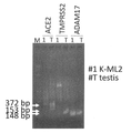
- 6 is an electrophoretic photograph of a transcript in which the expression levels of various genes (ACE2, TMPRSS2, ADAM17) in the prepared human immortalized myeloid cells (K-ML2) were confirmed by a quantitative PCR method.
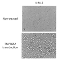
- K-ML2 (TMPRSS2 transduction; upper photo) gene-introduced with a vector containing the TMPRSS2 gene and drug resistance gene, and K-ML2 (Non-treated; upper photo) without the gene introduced into the cells after culturing in the presence of a drug.
- K-ML2 / TMPRSS2 K-ML2 / TMPRSS2
- the figure on the left shows the number of copies per sample, and the figure on the right shows the numerical value (magnification change) obtained by dividing the number of copies by the number of copies measured by K-ML2.
- the horizontal axis of the graph shows the cell name, and the vertical axis of the graph shows the number of copies of TMPRSS2 RNA (left figure) and the change in magnification (right figure).
- the figure on the left shows the number of copies per sample, and the figure on the right shows the numerical value (magnification change) obtained by dividing the number of copies by the number of copies measured by K-ML2.
- the horizontal axis of the graph shows the cell name, and the vertical axis of the graph shows the number of copies of RNA of ACE2 (left figure) and the change in magnification (right figure).
- SARS dependent on K-ML2 K-ML2 / TMPRSS2 / ACE2 expressing ACE2 and TMPRSS2, and M-CSF and GM-CSF expressing human immortalized myeloid cells (K-ML2) derived from K strain.
- -It is a graph which shows the virus amount on the 3rd day after infecting with CoV-2 virus.
- TMPRSS2-expressing Vero cells (Vero / TMPRSS2) were used, and as a negative control (None), the virus was inoculated into a cell-free medium.
- the horizontal axis shows the virus concentration (copy / ⁇ L) of SARS-CoV-2 at the time of virus addition (day 0), and the vertical axis shows SARS-in the culture supernatant measured by quantitative PCR on the third day of culture.
- the virus concentration (copy / ⁇ L) of CoV-2 is shown.
- FIG. 6A is a result of analysis of virus proliferation ability when infected with SARS-CoV-2 virus (1 ⁇ 10 3 copy / ⁇ L).
- K-ML2 (K-ML2 / TMPRSS2 / ACE2) expressing ACE2 and TMPRSS2 in FIG. 6A
- M-CSF and GM-CSF expressing humans derived from the K strain with the negative control (None) value in FIG. 6A being 1. It is shown as a percentage of the virus concentration in the culture supernatant in immortalized myeloid cells (K-ML2).
- the horizontal axis indicates the sample name, and the vertical axis is the value obtained by dividing the virus concentration in the culture supernatant of each cell by the amount of virus in the sample added to the cell-free medium (Fold expression (reactive to no TD)). ) Is shown.
- M-CSF and GM-CSF expressing human immortalized myeloid cells Mylc; K-ML2
- Mylc-DC dendritic cells obtained by differentiating Mylc
- Mylc Mylc
- ACE2 Mylc
- TMPRSS2 dendritic cells obtained by differentiating Mylc
- cMylc expressing ACE2
- TMPRSS2 dendritic cells obtained by differentiating cMylc with SARS-CoV-2.
- TMPRSS2-expressing Vero cells Vero / TMPRSS2
- None negative control
- the horizontal axis shows the SARS-CoV-2 virus concentration (copy / ⁇ L) at the time of virus addition (day 0), and the vertical axis shows the SARS-CoV-2 virus concentration (copy) measured by quantitative PCR 72 hours after culturing. / ⁇ L) is shown.
- M-CSF and GM-CSF expressing human immortalized myeloid cells (cMylc) expressing Mylc, Mylc-DC, ACE2 and TMPRSS2, and dendritic cells (cMylc-DC) obtained by differentiating the cells. It is a graph which measured the production amount of IL-6 in the culture supernatant obtained by adding SARS-CoV-2 to, and culturing these cells for 72 hours by ELISA.
- the vertical axis of the graph shows the measured value of ELISA
- the horizontal axis of the graph shows the dilution rate of the supernatant cultured for 72 hours.
- the line graph with white dots is obtained by diluting the supernatant containing SARS-CoV-2 virus previously cultured in Vero cells 100-fold (x100), adding it to cMylc-DC, and then culturing it for 72 hours.
- myeloid cells are also called bone marrow cells and are defined as cells expressing CD11b molecule or CD33 molecule, and the origin thereof is not particularly limited, but for example, pluripotent stem cells.
- myeloid cells from which they are derived, or myeloid cells collected from living organisms (eg, peripheral blood). Specifically, myeloid cells are destined to differentiate into leukocyte granulocyte cells (neutrophils, eosinophils, neutrophils), monocytes, dendritic cells, macrophages, or these cells. Examples include more undifferentiated cells (myeloid progenitor cells and the like).
- coronavirus is defined as a single-stranded RNA virus belonging to the coronaviridae family, and causes respiratory infections such as fever and dry cough. Specific examples thereof include SARS-CoV, SARS-CoV-2, Middle East respiratory syndrome-related virus (MARS-CoV), HCoV-229E, HCoV-OC43, HCoV-NL63, and HCoV-HKU1, but are preferable. , SARS-CoV-2.
- coronaviruses include not only humans but also livestock, pets, and wild animals. Specific examples of the coronavirus that infects other than humans include porcine epidemic diarrhea virus (PEDV) and feline infectious peritonitis virus (FIPV).
- SARS-CoV-2 "SARS-CoV-2 / Hu / DP / Kng / 19-020 (GenBank: LC528232)" separated by the Kanagawa Prefectural Institute of Public Health can be used.
- coronavirus infectivity means the ability to be infected by coronavirus. Therefore, coronavirus-infectious cells mean cells infected by coronavirus.
- the term "level” means an index relating to a quantified abundance, and includes, for example, an index that can be used as a concentration, a quantity, or an alternative.
- the level may also be an absolute number (ie, the measured abundance) or a relative number compared to a control set as needed.
- the invention relates to a method for preparing coronavirus-infectious human immortalized myeloid cells, comprising introducing ACE2 and TMPRSS2 into human immortalized myeloid cells.
- the human immortalized myeloid cells used in this method may be immortalized monocytes, or may be cells prepared by introducing M-CSF and GM-CSF into the immortalized monocytes. ..
- Monospheres can be prepared by collecting from peripheral blood, inducing differentiation from pluripotent stem cells, inducing differentiation of other somatic cells such as fibroblasts from an undifferentiated state, or by direct reprogramming. It can be done by the method of inducing.
- monocytes When monocytes are collected from peripheral blood, monocytes can be obtained as cells expressing the CD14 molecule in human peripheral blood by a known separation / preparation method.
- human peripheral blood is gently diluted with an equal amount of physiological saline, phosphate buffered physiological saline, Hanks buffer solution, etc., and placed on Ficoll (registered trademark) (GE Healthcare) in a centrifuge tube. Gently stack and centrifuge at 15-30 ° C for 20 minutes at 500-1000 xg to form a white band of blood between yellowish plasma and clear Ficoll peripheral blood consisting of lymphocytes and monospheres. It can be recovered as a mononuclear cell (PBMC) fraction.
- PBMC mononuclear cell
- the collected peripheral blood mononuclear cell fraction may be further washed and used if necessary.
- Monocytes can be obtained by further recovering cells expressing the CD14 molecule from the recovered PBMC fraction.
- monocytes can be separated and recovered by contacting PBMC with a solid phase to which an anti-CD14 antibody is bound to bind monocytes to the solid phase, and washing and removing unbound cells.
- a method of using magnetic beads as such a solid phase is known (Dynabeads (registered trademark) CD14 (Thermo Fisher Scientific Inc.) and the like). It is also possible to obtain monocytes by directly contacting the peripheral blood with the solid phase to which the anti-CD14 antibody is bound, without separating the PBMC from the peripheral blood.
- pluripotent stem cell means a cell having pluripotency and self-renewal ability.
- pluripotency is synonymous with pluripotency and means a state of cells that can differentiate into cells of multiple lineages by differentiation.
- pluripotency refers to stem cells, embryonic stem (ES) cells, embryonic stem cells derived from cloned embryos obtained by nuclear transplantation (“ntES cells”), germ stem cells (“GS cells”), and the like.
- EG cells embryonic stem cells
- iPS artificial pluripotent stem
- hematopoietic stem cells Whether or not a cell is a pluripotent stem cell is desired, for example, when the test cell forms an embryoid body in an in vitro culture system or after culturing (differentiation treatment) under differentiation-inducing conditions. When differentiating into a cell of, the cell can be determined to be a pluripotent stem cell.
- M-CSF macrophage colony stimulating factor
- IL-3 interleukin 3
- IFN- ⁇ or PMA Annabelle Grolleau et al., J Immunol 1999; see 162: 3491-3497
- M-CSF macrophage colony stimulating factor
- GM-CSF granulocyte macrophage colony stimulating factor
- / 043651, JP-A-2017-131136 and JP-A-2018-171005 can be brought into contact with pluripotent stem cells.
- the differentiation-induced monocytes only the cells expressing CD14 may be recovered and purified by the above-mentioned method, if necessary.
- Immortalization of monospheres is performed by adding at least one gene selected from the BMI1 gene, EZH2 gene, MDM2 gene, MDM4 gene, HIF1A gene, BCL2 gene, and LYL1 gene to the monosphere obtained as described above, and the cMYC gene.
- a combination of at least one gene selected from the BMI1 gene, EZH2 gene, MDM2 gene, MDM4 gene, and BCL2 gene and the cMYC gene, or a combination of the BMI1 gene and the cMYC gene is used.
- the introduction of these genes into monocytes can be carried out in consideration of the descriptions in WO2012 / 043651 and JP-A-2017-131136.
- the gene can be introduced by the gene introduction method described below.
- Immortalized monocytes can maintain their proliferative capacity by further introducing M-CSF and GM-CSF. Therefore, the method of the present invention may further include the introduction of M-CSF and GM-CSF into immortalized myeloid cells or immortalized monocytes. Alternatively, cells expressing M-CSF and GM-CSF as immortalized myeloid cells or immortalized monocytes may be used. The introduction of these genes into immortalized myeloid cells or immortalized monocytes can be carried out in consideration of the description in JP-A-2018-171005.
- Coronavirus-infectious human immortalized myeloid cells can be prepared by introducing the genes of ACE2 and TMRPSS2 into the human immortalized monocytes obtained above according to the method described in the method for introducing the gene below. can. More specifically, these genes may be introduced by a lentiviral vector.
- ACE2 is an abbreviation for angiotensin converting enzyme II and is an essential reverse-regulatory carboxypeptidase of the renin-angiotensin hormone system, which is an important regulator of cardiovascular homeostasis.
- the human ACE2 protein typically has the amino acid sequence set forth in Table 2 or SEQ ID NO: 10.
- TMPRSS2 is an abbreviation for type II transmembrane serine protease, which is a serine protease immobilized on the proteolytic membrane involved in the proteolytic cascade associated with the normal physiological function of the prostate.
- the human TMPRSS2 protein typically has the amino acid sequence set forth in Table 2 or SEQ ID NO: 8.
- ACE2 and TMPRSS2 introduced into cells are isoforms and variants thereof, 1 to 5 or 1 to 3 amino acids, as long as the introduced cells acquire infectivity to coronavirus or improve infectivity.
- ACE2 and TMPRSS2 replaced, inserted, deleted, or added are 80% or more, 85% or more, 90% or more of the above ACE2 and TMPRSS2. , 95% or more, 98% or more, or 99% or more identity.
- ACE2 since ACE2 is cleaved by ADAM17 and its binding to coronavirus is inhibited, ACE2 having a mutation in the cleavage site by ADAM17 may be used.
- mutations include mutations in which methionine at position 706 in the amino acid sequence set forth in SEQ ID NO: 10 in Table 2 is replaced with proline.
- mutations in the 26th and 27th lysine and threonine in the amino acid sequence enhance the binding of SARS-CoV-2 to ACE2 (doi: https: //doi.org/10.1101/2020).
- the 26th lysine of SEQ ID NO: 10 may be replaced with arginine and / or the 27th threonine may be replaced with proline.
- a mutant in which leucine at position 584 of ACE2 is replaced with alanine can be used (Am J Physiol Lung Cell Mol Physiol. 2009 Jul; 297 ( 1): L84-96.).
- ⁇ Gene introduction method> It can be performed by using a known method, and the method is not particularly limited as long as the desired effect is obtained.
- the gene introduction method include a method of introducing a desired gene into a cell by using a vector such as a virus, a plasmid, or an artificial chromosome, or by a method such as lipofection, liposome, microinjection, or electroporation. Can be done.
- Viral vectors include retroviral vectors and lentiviral vectors (above, Cell, 126, pp.663-676, 2006; Cell, 131, pp.861-872,2007; Science, 318, pp.1917-1920, 2007.
- the artificial chromosome vector includes, for example, a human artificial chromosome (HAC), a yeast artificial chromosome (YAC), a bacterial artificial chromosome (BAC, PAC) and the like.
- HAC human artificial chromosome
- YAC yeast artificial chromosome
- BAC bacterial artificial chromosome
- PAC bacterial artificial chromosome
- the plasmid a plasmid for mammalian cells can be used (Science, 322: 949-953, 2008).
- the nucleic acid of interest can be integrated into the genome of a host cell non-virally using a transposon, and for example, a PiggyBac vector can be used.
- the vector can contain regulatory sequences such as promoters, enhancers, ribosome binding sequences, terminators, polyadenylation sites, etc. so that the desired gene can be expressed, and if desired, drug resistance genes (eg, canamycin). Resistance gene, ampicillin resistance gene, puromycin resistance gene, neomycin resistance gene, etc.), selection marker sequence (eg, thymidin kinase gene, diphtheriatoxin gene, etc.), reporter gene sequence (eg, fluorescent protein gene such as green fluorescent protein, etc., ⁇ -galactosidase gene, luciferase gene, etc.) can be included.
- drug resistance genes eg, canamycin. Resistance gene, ampicillin resistance gene, puromycin resistance gene, neomycin resistance gene, etc.), selection marker sequence (eg, thymidin kinase gene, diphtheriatoxin gene, etc.), reporter gene sequence (eg, fluorescent protein gene such as green fluorescent protein, etc., ⁇
- the vector encodes a functional replication origin in mammalian cells and a protein that binds to the replication origin and controls replication so that it is replicated without integration into the chromosome and exists episomally. It may have a gene to be used.
- a functional replication origin in mammalian cells and a protein that binds to the replication origin and controls replication include the replication origin oriP and the EBNA-1 gene in EBV, and the SV40.
- the replication origin ori and the SV40 large T antigen gene and the like can be mentioned (WO2009 / 115295, WO2009 / 157201 and WO2009 / 149233).
- an expression vector that is polycistronically expressed may be used in order to simultaneously introduce a plurality of desired genes.
- the sequence encoding the gene may be linked by an internal ribosome entry site (IRES) or foot-and-mouth disease virus (FMDV) 2A coding region (Science, 322: 949). -935, 2008 and WO2009 / 0920 422009/152259).
- IRS internal ribosome entry site
- FMDV foot-and-mouth disease virus
- the cell into which the gene of interest has been introduced can be obtained as a surviving cell by culturing the gene-introduced cell in the presence of a drug corresponding to the drug resistance gene integrated into the cell. can.
- a luminescence or fluorescence assay corresponding to the reporter gene integrated into the cell can be performed to obtain a cell that has luminescence or fluorescence.
- luminescence or fluorescence assays may be performed using commercially available kits.
- a cell in which expression of mRNA of the target gene in the cell is confirmed by performing quantitative PCR using DNA extracted from the cell as a template may be used as a cell into which the target gene has been introduced.
- Quantitative PCR may be performed using a commercially available kit.
- the target protein produced by the cell may be detected by ELISA or the like, and the cell in which the expression of the protein of the target gene is confirmed may be the cell into which the target gene has been introduced.
- the human immortalized myeloid cells may be human immortalized monocytes or human dendritic cells expressing ACE2 and TMPRSS2.
- human immortalized myeloid cells may preferably have M-CSF and / or GM-CSF introduced or expressed.
- coronavirus infectious human dendritic cells By inducing differentiation of ACE2 and TMPRSS2-introduced immortalized monocytes obtained as described above, they can be differentiated into dendritic cells.
- coronavirus-infectious human immortalized myeloid cells include dendritic cells expressing such ACE2 and TMPRSS2.
- the method of the present invention may also include the differentiation of human immortalized monocytes into dendritic cells. Induction of differentiation from monocytes into dendritic cells has been reported (Francoise Chapuis et al., Eur J Immunol. (1997) 27 (2): 431-41 .; Marc Dauer et al., J Immunol (2003) 170 (8) :.
- monocytes can be induced to differentiate into dendritic cells by culturing monocytes in the presence of 10 to 500 ng / ml IL-4.
- coronavirus-infectious human immortalized myeloid cells coronavirus-infectious human immortalized monospheres, and coronavirus-infectious human dendritic cells can be used for coronavirus infection.
- these cells can be used as a composition for coronavirus infection together with other components.
- One aspect of the present invention relates to a method for determining the presence of coronavirus in a sample. Specifically, the method adds a sample to the culture medium of coronavirus-infectious human immortalized myeloid cells, and detects coronavirus in the culture supernatant of the monosphere or dendritic cell. This includes measuring the level of coronavirus.
- Examples of the medium used in the determination of the present invention include IMDM, RPMI1640, ⁇ -MEM, MEM, DMEM and the like.
- human or bovine plasma protein fraction bovine fetal serum and human serum, saccharides such as glucose and D-mannitol, amino acids, nucleic acid bases such as adenine, sodium hydrogen phosphate, ⁇ -tocopherol, linoleic acid, Cholesterol, sodium selenate, human holotransferase, human insulin, ethanolamine, 2-mercaptoethanol, G-CSF, GM-CSF, sodium hydrogencarbonate, methyl cellulose and the like may be contained.
- antibiotics such as gentamicin, ampicillin, penicillin, and strepromycin; inorganic salts; buffers such as HEPES, phosphate buffer, and acetate buffer may be added.
- gentamicin gentamicin
- ampicillin penicillin
- strepromycin inorganic salts
- buffers such as HEPES, phosphate buffer, and acetate buffer
- IL-4 can be used as an additive, for example, 50 to 200 ng / mL IL-4.
- the contained medium can be used.
- the culture conditions may be about 37 ° C. or about 5% CO 2 .
- Samples include body fluids (blood, nasal discharge, sputum, urine, etc.) or tissues (oropharynx, nasopharynx, etc.), cultured cells, cell culture fluid (cell culture supernatant), drinking water, etc. derived from humans or animals. Food, environmental cleaning fluids, wiping fluids, etc. can be used, for example, blood samples. These may be appropriately concentrated or diluted, or may be subjected to pretreatment such as extraction or purification.
- the term "blood sample” includes blood fractions or treatments such as whole blood, plasma, serum, whole blood or blood cells, and dilutions and concentrates thereof, preferably serum. Or it may be a diluted solution thereof. If the sample is a liquid, a diluent diluted at any dilution factor may be used for evaluation.
- coronavirus-infectious human immortalized myeloid cells including coronavirus-infectious human immortalized monospheres or coronavirus-infectious human dendritic cells, the same applies hereinafter
- the sample can be added after cell passage or medium exchange at predetermined times and intervals. After adding the sample, further culture the cells while exchanging the medium at predetermined times and intervals as needed.
- the culture period after adding the sample may be one type or two or more types.
- the culture time can be 1 to 7 days, 2 to 6 days, 2 to 5 days, 2 to 4 days, or 3 days.
- the number of passages or medium exchanges may be, for example, once, twice, three times, four times, or five times or more.
- the interval of passage or medium change may be, for example, once every 2 to 3 days, once every 4 to 5 days, or once a week. May be good.
- Detection of coronavirus or measurement of coronavirus level in the culture supernatant after completion of culture should be performed by detection of coronavirus RNA or measurement of coronavirus RNA level, plaque assay, virus titer measurement such as TCID50, etc. Can be done. In order to detect and measure promptly and quantitatively after the completion of culture, a measurement method using RNA as an index is often used. Detection of RNA or measurement of RNA level of coronavirus is not particularly limited as long as it is a method capable of detecting RNA or measuring the amount of RNA, but generally, it specifically binds to coronavirus RNA. It is done by the method of utilizing the substance to be used.
- the "substance that specifically binds to coronavirus RNA” can be a nucleic acid molecule, preferably a nucleic acid molecule having a sequence complementary to the coronavirus RNA. Nucleic acid molecules having a sequence complementary to coronavirus RNA can be specifically bound to coronavirus RNA by hybridization.
- nucleic acid includes DNA, RNA, or artificially created nucleic acids (including cross-linked nucleic acids such as PNA and Locked Nucleic Acid (2', 4'-BNA)), or combinations thereof. Nucleic acid molecules that specifically bind to coronavirus RNA may contain at least a portion of an artificially designed sequence (eg, a sequence for labeling or tagging).
- a “probe” is typically a nucleic acid molecule that has a sequence complementary to the coronavirus RNA sequence and is used to measure binding to the coronavirus RNA sequence.
- the probe is usually a nucleic acid molecule such as 10-30 mer, 10-20 mer, etc. that can specifically bind to coronavirus RNA.
- Methods for measuring the binding level between coronavirus RNA and the probe include Southern hybridization BR> [ion, Northern hybridization, dot hybridization, fluorescence in situ hybridization (FISH), microarray, ASO method, etc. Specifically, a method using GeneChipTM miRNA Hybrid Strip (Thermo Fisher Scientific Co., Ltd.) or Agilent miRNA microarray (Agilent Technologies) can be used.
- the detection of coronavirus RNA or the measurement of coronavirus RNA level can be performed by detecting the binding of coronavirus RNA bound to a substance that specifically binds to coronavirus RNA, or by measuring the binding level.
- the "binding level" can be the amount of binding, the number of bindings, or the binding ratio, or a numerical value representing them (for example, the measured value itself such as the measured fluorescence intensity).
- a labeled substance may be used as a substance that specifically binds to the coronavirus RNA, or the coronavirus RNA may be labeled and used.
- standard samples are measured at the same time, a standard curve or calibration curve is created based on the standard sample, and the value is calculated from the measured value of the measurement sample, or the value is standardized using the standard sample level as an index. Is determined as the binding level.
- Examples of the labeling method include radioisotope (RI) labeling, fluorescent labeling, and enzyme labeling.
- RI radioisotope
- coronavirus RNA levels may be measured by the following (a)-(c): (A) Contacting the culture supernatant with a nucleic acid molecule (probe) that binds to at least one coronavirus RNA base sequence or a part thereof; (B) Measuring the binding level of coronavirus RNA in the culture supernatant bound to the probe; (C) Determine the presence of coronavirus in the culture supernatant from the measured binding level.
- a nucleic acid molecule probe
- the coronavirus RNA may be detected by the following (a)-(c): (A) Contacting the culture supernatant with a nucleic acid molecule (probe) that binds to at least one coronavirus RNA base sequence or a part thereof; (B) Detecting the binding of coronavirus RNA in the culture supernatant bound to the probe; (C) When binding is detected, it is determined that coronavirus is present in the culture supernatant.
- a nucleic acid molecule probe
- detection of coronavirus RNA or measurement of coronavirus RNA level can be performed by a method using PCR, for example, using a nucleic acid (primer) that specifically binds to coronavirus RNA, qPCR, ARMS ( It may be measured by performing Amplification Reference Mutation System), RT-PCR (Reverse transcriptase-PCR), or Next PCR.
- a nucleic acid that specifically binds to coronavirus RNA
- qPCR qPCR
- ARMS It may be measured by performing Amplification Reference Mutation System
- RT-PCR Reverse transcriptase-PCR
- Next PCR Next PCR.
- the Invader® method may be used.
- a "primer” is a nucleic acid molecule of 10 to 30 mer (preferably 17 to 25 mer, 15 to 20 mer, etc.) usually used for nucleic acid amplification, and at least a part thereof (preferably 7 mer or more, 8 mer or more, etc.).
- RNA has a sequence complementary to the terminal sequence of coronavirus RNA (9 mer or more, 10 mer or more).
- primer for example, a nucleic acid molecule having the base sequence set forth in SEQ ID NOs: 11 to 14 or consisting of the base sequence set forth in SEQ ID NOs: 11 to 14 can be used.
- coronavirus RNA levels may be measured by the following steps: (A) Using the culture supernatant as a template and using a nucleic acid molecule (primer) that can specifically bind to the coronavirus RNA, amplify all or part of the coronavirus RNA in the culture supernatant: (B) Measuring the level of amplified nucleic acid molecules; and (C) Determine the presence of coronavirus in the culture supernatant from the level of amplified nucleic acid molecules.
- A Using the culture supernatant as a template and using a nucleic acid molecule (primer) that can specifically bind to the coronavirus RNA, amplify all or part of the coronavirus RNA in the culture supernatant:
- B Measuring the level of amplified nucleic acid molecules; and (C) Determine the presence of coronavirus in the culture supernatant from the level of amplified nucleic acid molecules.
- Coronavirus RNA may also be detected by the following steps: (A) Using the culture supernatant as a template and using a nucleic acid molecule (primer) that can specifically bind to the coronavirus RNA, amplify all or part of the coronavirus RNA in the culture supernatant: (B) Detection of amplified nucleic acid molecules; and (C) When the amplified nucleic acid molecule is detected, it is determined that the coronavirus is present in the culture supernatant.
- A Using the culture supernatant as a template and using a nucleic acid molecule (primer) that can specifically bind to the coronavirus RNA, amplify all or part of the coronavirus RNA in the culture supernatant:
- B Detection of amplified nucleic acid molecules; and
- C When the amplified nucleic acid molecule is detected, it is determined that the coronavirus is present in the culture supernatant.
- Amplification of all or part of the coronavirus RNA in the culture supernatant can be carried out by performing a PCR reaction or the like using the culture supernatant as a template.
- the level of amplified nucleic acid is the dot blot hybridization method, surface plasmon resonance method (SPR method), PCR-RFLP method, Insitu RT-PCR method, PCR-SSO (single-strand conformation Oligonucleotide) method, PCR-SSP. It can be measured by a method, an APPFLP (Amplify fragment length hybridization) method, an MVR-PCR method, or a PCR-SCSP (single strand conformation polymorphism) method.
- RNA binding means that the substance has a substantially higher affinity for a nucleic acid having a coronavirus RNA sequence than an affinity for a nucleic acid having another base sequence. Means to combine.
- substantially high affinity means an affinity to which a nucleic acid having a coronavirus RNA sequence can be detected separately from a nucleic acid having another base sequence.
- Other base sequences are preferably different to the extent that they can be distinguished from the coronavirus RNA sequence, and are base sequences having 50% or less, 40% or less, 30% or less, 20% or less, and 10% or less identity. You may.
- substantially high affinity means that the amount of binding to coronavirus RNA is 3 times or more, 4 times or more, 5 times or more, 6 times or more, 7 times or more, 8 times the amount of binding to other base sequences. It may be 9 times or more, 10 times or more, 15 times or more, 20 times or more, 30 times or more, and 50 times or more.
- coronavirus RNA or the measured coronavirus RNA level is determined based on the detected coronavirus RNA or the measured coronavirus RNA level. Specifically, when coronavirus RNA is detected in the culture supernatant, it may be determined that coronavirus is present in the sample. In addition, when the measured amount of coronavirus RNA level in the culture supernatant is increased as compared with the negative control, it may be determined that the coronavirus is present in the sample. As the negative control, the medium or the like used in this evaluation may be used.
- the detection of coronavirus or the measurement of the coronavirus level in the culture supernatant after the completion of the culture can be performed by the detection of the coronavirus protein or the measurement of the coronavirus protein level.
- Detection of coronaviral protein or measurement of protein level is not particularly limited as long as it is a method capable of detecting protein or measuring the amount of protein, but generally, it specifically binds to coronavirus protein. It is done by the method of utilizing the substance.
- the "substance that specifically binds to a coronavirus protein” includes an antibody or an antigen-binding fragment thereof, an aptamer, a ligand / receptor or a binding fragment thereof, or a fusion thereof with another substance. Can be done.
- the term "antigen-binding fragment” refers to a protein or peptide containing a part (partial fragment) of an antibody, which retains the action (immune reactivity / binding) of the antibody on the antigen. means.
- the antigen-binding fragment include F (ab') 2 , Fab', Fab, Fab 3 , single-chain Fv (hereinafter referred to as “scFv”), and (tandem) bispecific single-chain Fv (sc (Fv).
- the antibody and immunoreactive fragment may be monospecific, bispecific, trispecific, or multispecific.
- the detection of coronavirus protein or the measurement of coronavirus protein level is performed by detecting the binding of coronavirus protein bound to a substance that specifically binds to coronavirus protein or by measuring the binding level.
- the measured "binding level" can be the amount of binding, the number of bindings, or the binding ratio of these substances, or a numerical value representing them (for example, the measured value itself such as the measured fluorescence intensity).
- a labeled substance may be used as a substance that specifically binds to the coronavirus protein, or the coronavirus protein may be labeled and used.
- the standard sample is measured at the same time, and the value calculated by creating a standard curve or calibration curve based on the standard sample, or the value standardized using the standard sample level as an index is determined as the binding level.
- the method of the invention may include: (A) Contacting the culture supernatant with a substance that binds to at least one coronavirus protein; (B) Detecting the binding of the coronavirus protein in the culture supernatant bound to the substance that binds to the coronavirus protein, or measuring the binding level; and. (C) Determine the presence of coronavirus in the culture supernatant from the detected or measured binding level.
- the measurement of binding can be based on a known detection and / or measurement method.
- labeled immunoassays such as enzyme immunoassay (EIA method), simplified EIA method, enzyme-linked immunosorbent assay (ELISA method), radioimmunosassay (RIA method), and fluorescent immunoassay (FIA method).
- Immunoblotting method such as Western blotting method; Immunochromatography method such as gold colloid aggregation method; Chromatography method such as ion exchange chromatography method and affinity chromatography method; Turbidity method (TIA method); Deaf method (NIA method); Colorimetric method Method; Latex agglomeration method (LIA method); Particle counting method (CIA method); Chemical emission measurement method (CLIA method, CLEIA method); Precipitation reaction method; Surface plasmon resonance method (SPR method); Resonant mirror detector method (RMD method) ); The coupling can be measured by a comparative interference method or the like.
- the culture supernatant is brought into contact with the antibody of the present invention immobilized on a solid phase or an antigen-binding fragment thereof, washed, and then a labeled antibody capable of binding to the protein to be detected or measured is added.
- the unbound antibody can be removed by washing, and the protein to be detected or measured can be detected or the level thereof can be determined by detecting the label of the antibody or measuring the labeled amount (for example, the intensity of the label).
- the culture supernatant is contacted with a labeled antibody capable of binding to the first unimmobilized detection or measurement target protein, and then the mixture is subjected to the second detection or measurement target.
- An antibody capable of binding to a protein or an antigen-binding fragment thereof is detected by contacting a carrier immobilized on a specific site and detecting the labeled antibody at the site or measuring the labeling amount (for example, the strength of the label).
- the labeling amount for example, the strength of the label.
- the protein to be measured can be detected or the level of the protein to be measured can be determined.
- Labeling methods include, for example, radioisotope (RI) labeling, fluorescent labeling, and enzyme labeling.
- the term "specifically binding" to a protein means that the substance has a substantially higher affinity for a nucleic acid having a coronavirus protein sequence than an affinity for a protein having another amino acid sequence. Means to combine.
- substantially high affinity means an affinity to the extent that a coronavirus protein can be detected separately from a protein having another amino acid sequence.
- the other amino acid sequences are preferably different to the extent that they can be distinguished from the coronavirus protein sequence, and are amino acid sequences having 50% or less, 40% or less, 30% or less, 20% or less, and 10% or less identity. You may.
- substantially high affinity means that the amount of binding to coronavirus protein is 3 times or more, 4 times or more, 5 times or more, 6 times or more, 7 times or more, 8 times the amount of binding to other amino acid sequences. It may be 9 times or more, 10 times or more, 15 times or more, 20 times or more, 30 times or more, and 50 times or more.
- coronavirus is present in the sample based on the detected coronavirus protein or the measured coronavirus protein level. Specifically, when the coronavirus protein is detected in the culture supernatant, it may be determined that the coronavirus is present in the sample. Further, when the measured amount of the coronavirus protein level in the culture supernatant is increased as compared with the negative control, it may be determined that the coronavirus is present in the sample. As the negative control, the medium or the like used in this evaluation may be used.
- the detection of coronavirus or the measurement of the coronavirus level in the culture supernatant after the completion of the culture can be performed by the detection of the inflammatory cytokine or the measurement of the inflammatory cytokine level.
- inflammatory cytokines produced by immune cells due to viral infection cause inflammation of internal organs, leading to aggravation of viral infections. Evaluating the production of such inflammatory cytokines in virus-infected cells is expected to lead to the elucidation of the pathophysiology and treatment of aggravation.
- the inflammatory cytokine include IL-1 ⁇ , IL-6, IL-8, TNF- ⁇ , MIP-1 ⁇ and the like.
- Detection of inflammatory cytokines or measurement of inflammatory cytokine levels is not particularly limited as long as it is a method capable of detecting proteins or measuring the amount of proteins, but is generally specific to inflammatory cytokines. It is done by utilizing the binding substance.
- “substances that specifically bind to inflammatory cytokines” include antibodies or antigen-binding fragments thereof, aptamers, ligands / receptors or binding fragments thereof, or fusions thereof with other substances. Can be done.
- detection of inflammatory cytokines or measurement of inflammatory cytokine levels is performed by detecting the binding of inflammatory cytokines bound to substances that specifically bind to inflammatory cytokines or by measuring the binding level.
- the measured "binding level" can be the amount of binding, the number of bindings, or the binding ratio of these substances, or a numerical value representing them (for example, the measured value itself such as the measured fluorescence intensity).
- a labeled substance may be used as a substance that specifically binds to the inflammatory cytokine, or an inflammatory cytokine may be labeled and used.
- the standard sample is measured at the same time, and the value calculated by creating a standard curve or calibration curve based on the standard sample, or the value standardized using the standard sample level as an index is determined as the binding level.
- the method of the invention may include: (A) Contacting the culture supernatant with a substance that binds to at least one inflammatory cytokine; (B) To detect the binding of the inflammatory cytokine in the culture supernatant bound to the substance that binds to the inflammatory cytokine, or to measure the binding level; and. (C) Determine the presence of coronavirus in the culture supernatant from the detected or measured binding level.
- coronavirus is present in the sample based on the detected inflammatory cytokine or the measured inflammatory cytokine level. Specifically, when an inflammatory cytokine is detected in the culture supernatant, it may be determined that the coronavirus is present in the sample. In addition, if the measured amount of inflammatory cytokine level in the culture supernatant is higher than that of the negative control, it may be determined that coronavirus is present in the sample. As the negative control, the medium or the like used in this evaluation may be used.
- One aspect of the present invention relates to a method for producing a coronavirus. Specifically, the production method is to add coronavirus to a culture medium of coronavirus-infectious human immortalized myeloid cells (including immortalized human monospheres and human dendritic cells) to infect the cells. And recovering the coronavirus in the cell culture medium.
- coronavirus-infectious human immortalized myeloid cells including immortalized human monospheres and human dendritic cells
- Immortalized human myeloid cells are seeded in a medium, and after cell passage or medium exchange at predetermined times and intervals as needed, coronavirus is added to the cultured cells.
- the coronavirus that infects cells for example, the culture supernatant of the cell culture solution that has already been infected with coronavirus may be used as it is, or if necessary, it may be diluted with a diluted solution such as a medium and used. good.
- After adding the sample further culture the cells while exchanging the medium at predetermined times and intervals as needed.
- the culture period after the addition of coronavirus can be 1 to 7 days, 2 to 6 days, 2 to 5 days, 2 to 4 days, or 3 days.
- the culture conditions for other immortalized human myeloid cells can be in accordance with the conditions described in the above-mentioned ⁇ Method for determining the presence of coronavirus>.
- the culture solution after the culture is completed (for example, the culture supernatant) can be used as it is as a coronavirus-containing solution.
- a coronavirus-containing solution may be obtained by appropriately concentrating or purifying the culture solution (for example, the culture supernatant) after the completion of the culture.
- the present invention relates to a method for evaluating the anti-coronavirus activity of a test sample.
- This evaluation method is to add coronavirus to the culture medium of coronavirus-infectious human immortalized myeloid cells (including immortalized human monospheres or human dendritic cells) in the presence of the test sample, and to add coronavirus to the cells. Includes measuring the level of coronavirus in the culture supernatant.
- anti-coronavirus activity means an activity that brings about a therapeutic effect, a symptom-alleviating effect, and / or a preventive effect against a coronavirus infection, and is typically a neutralizing activity.
- growth inhibitory activity eg, RNA strand synthesis inhibitory activity, RNA polymerase inhibitory activity, etc.
- Immortalized human myeloid cells are seeded in a medium, and after cell passage or medium exchange at predetermined times and intervals as needed, coronavirus is added to the cultured cells.
- the coronavirus that infects cells for example, the culture supernatant of the cell culture solution that has already been infected with coronavirus may be used as it is, or if necessary, it may be diluted with a diluted solution such as a medium and used. good.
- After adding the sample further culture the cells while exchanging the medium at predetermined times and intervals as needed.
- the culture period after the addition of coronavirus can be 1 to 7 days, 2 to 6 days, 2 to 5 days, 2 to 4 days, or 3 days.
- the culture conditions for other immortalized human myeloid cells can be in accordance with the conditions described in the above-mentioned ⁇ Method for determining the presence of coronavirus>.
- the test sample can be added to the cell culture medium before, at the same time as the addition of the coronavirus, or after the addition of the coronavirus.
- it is preferably added before the addition of the coronavirus or at the same time as the addition of the coronavirus.
- how to evaluate the anti-coronavirus activity of a test sample is Adding the test sample to the culture medium of coronavirus-infectious human immortalized myeloid cells, It comprises adding the coronavirus to the cell culture medium and measuring the level of the coronavirus in the culture supernatant of the cells.
- the level of the coronavirus in the culture supernatant to which the coronavirus is added in the presence of the test sample is in the culture supernatant to which the coronavirus is added in the absence of the test sample. If the level of the coronavirus is lower than that of the coronavirus, the test sample may be evaluated as having anti-coronavirus activity.
- the method for evaluating the anti-coronavirus activity of the test sample of the present invention may be a method for evaluating the effect of the test sample on already infected cells, and specifically, it may be a method for evaluating the effect of the test sample on already infected cells.
- Coronavirus Addition of coronavirus to the culture medium of infectious human immortalized myeloid cells This includes adding a test sample to the cell culture medium and measuring the level of the coronavirus in the culture supernatant of the cells.
- the level of the coronavirus in the culture supernatant to which the coronavirus is added in the presence of the test sample is in the culture supernatant to which the coronavirus is added in the absence of the test sample. If the level of the coronavirus is lower than that of the coronavirus, the test sample may be evaluated as having anti-coronavirus activity.
- the modality of the test sample is not limited, but may be, for example, an antibody, a receptor or a fragment thereof, a medium molecule, a peptide, a small molecule, an aptamer, or a nucleic acid drug.
- the test sample may be diluted in any dilution ratio series for evaluation.
- the test sample does not have to consist of a single substance, and is a human or animal-derived body fluid (blood, nasal discharge, sputum, urine, etc.) or tissue (oropharyngeal, nasopharyngeal, etc.), cultured cells, cell culture fluid ().
- test sample may be a cell culture supernatant), drinking water or food, a cleaning solution or wiping solution in the environment, a biological component such as a blood sample, a mixture, a composition, a plant extract, or the like.
- a biological component such as a blood sample, a mixture, a composition, a plant extract, or the like.
- a component typically an antibody
- the test sample may be a test drug consisting of only a single component, excluding a solution such as a solvent or a medium component or a diluted solution.
- any time can be set as the cell culture period after the addition of the coronavirus and the test sample.
- the culture time can be 1 to 7 days, 2 to 6 days, 2 to 5 days, 2 to 4 days, or 3 days.
- the culture conditions for other immortalized human myeloid cells can be in accordance with the conditions described in the above-mentioned ⁇ Method for determining the presence of coronavirus>.
- the coronavirus level in the culture supernatant after the lapse of the culture period can be measured according to the above-mentioned ⁇ method for determining the presence of coronavirus>.
- Whether or not the test sample has anti-coronavirus activity is determined by comparing the measured coronavirus level in the culture supernatant with the control coronavirus level.
- a culture supernatant of cells cultured with coronavirus in the absence of a test sample can be used. Specifically, if the level of coronavirus in the culture supernatant containing the test sample is lower than the level of coronavirus in the culture supernatant not containing the test sample, the test sample has anti-coronavirus activity. It may be determined to have.
- the present invention relates to a method for identifying whether or not a substance having anti-coronavirus activity (anti-coronavirus substance) is contained in a test sample.
- the identification method of the present invention is to mix coronavirus with a culture medium of coronavirus-infectious human immortalized myeloid cells (including immortalized human monospheres or human dendritic cells) in the presence of a test sample. It involves measuring the level of the coronavirus in the culture supernatant of the cells, wherein the level of the coronavirus in the culture supernatant supplemented with the coronavirus in the presence of the test sample.
- the test sample is identified as containing a substance having anti-coronavirus activity.
- the "test sample” is a sample for which it is desired to determine whether or not an anti-coronavirus substance is contained, and any sample to which the method of the present invention can be applied shall be used.
- the sample itself does not have cytotoxicity, and for example, in addition to the above-mentioned blood sample, food, beverage, or plant extract can be used.
- it can be an antibody, blood or serum or extract after vaccination.
- the test substance may be diluted in any dilution ratio series for evaluation.
- the anti-coronavirus substance identified by the identification method of the present invention can conform to the above-mentioned modality, for example, can be a substance having coronavirus neutralizing activity, and is preferably a coronavirus neutralizing antibody. Is.
- Each specific step in the method for identifying the anti-coronavirus substance in the test sample can be carried out according to the above-mentioned ⁇ Method for evaluating the anti-coronavirus activity of the test sample>.
- the test sample contains an anti-coronavirus substance is identified by comparing the measured coronavirus level in the culture supernatant with the control coronavirus level.
- a cell culture supernatant cultured with the coronavirus in the absence of the test sample can be used.
- the test sample is an anti-coronavirus substance. May be identified as containing.
- the method for evaluating the anti-coronavirus activity of the test sample of the present invention and the method for identifying the anti-coronavirus substance in the test sample are such that a neutralizing antibody is produced in vivo by vaccination (or produced by vaccination). It can be used to determine (whether or not the obtained antibody has neutralizing activity).
- a neutralizing antibody is produced in vivo by vaccination (or produced by vaccination). It can be used to determine (whether or not the obtained antibody has neutralizing activity).
- the blood of a vaccinated mammal mamouse, rat, guinea pig, rabbit, dog, pig, cow, horse, monkey, human, etc.
- a blood sample prepared from the blood is used as a test sample.
- the present invention is a method for evaluating the neutralizing antibody-inducing ability of a coronavirus vaccine, in which a blood sample derived from a mammal inoculated with the vaccine is used as a test sample, and the anti-coronavirus of the test sample of the present invention is used.
- the present invention relates to a method for evaluating activity, or a method including carrying out a method for identifying a coronavirus neutralizing substance in a test sample of the present invention.
- the method for evaluating a coronavirus vaccine of the present invention may further include a step of administering the vaccine, for example, inoculating a test mammal with a vaccine candidate substance, or collecting blood from the test mammal.
- the present invention includes a kit for determining the presence of coronavirus, a kit for producing coronavirus, a kit for evaluating anti-coronavirus activity of a test sample, and a kit for evaluating the anti-coronavirus activity of a test sample, including coronavirus-infectious human immortalized myeloid cells.
- the kit may include coronavirus-infectious human immortalized myeloid cells, as well as packages and instructions, as well as culture media, culture additives, substances capable of binding to coronavirus RNA or protein, and It may contain one or more kinds selected from the desired standard substances and the like.
- the culture medium the medium described in the section ⁇ Method for determining the presence of coronavirus> can be used.
- Example 1 Preparation of human induced pluripotent stem cell-derived immortalized monocyte cells
- Human induced pluripotent stem (iPS) cell-derived immortalized monocyte cells have been reported (WO2012 / 043651, JP-A-2017-131136). It was prepared with reference to Kai 2018-171005).
- undifferentiated iPS cells K strain ((WO2012 / 043651, JP-A-2017-131136 and JP-A-2018-171005)) are used as Laminin 511 (iMatrix-511-E8, Nippi Co., Ltd., Cat #: 892).
- ⁇ -MEM fetal bovine serum
- FBS fetal bovine serum
- TrypLE TM Select (containing 1 mM ethylenediamine tetraacetic acid (EDTA)) (GIBCO Inc., Cat #: A12859-01) and 0.5 mM EDTA / PBS (Nakalitesk Co., Ltd., Cat). #: 13567-84) was mixed in equal amounts, and the cells were exfoliated and recovered by treating at 37 ° C. for 60 minutes using the prepared solution (final concentration 0.75 mM EDTA), and collected by pipetting operation. A cell suspension was prepared.
- EDTA ethylenediamine tetraacetic acid
- the cell suspension collected from one dish with a diameter of 10 cm or 1 well of a 6-well plate was collected in 10 mL of D-MEM medium (Fuji Film Wako Pure Chemical Industries, Ltd., Cat #: 044-2795). ), Suspended in a culture medium supplemented with 10% fetal bovine serum (hereinafter referred to as FBS) (Cytiva Inc., Cat #: SH30088.03), and seeded on 2 or 1 dishes having a diameter of 10 cm. The cells were allowed to stand in a 5% CO 2 incubator at ° C.
- D-MEM medium Feji Film Wako Pure Chemical Industries, Ltd., Cat #: 044-2795
- FBS fetal bovine serum
- the floating cells that did not adhere to the dish having a diameter of 10 cm were collected and passed through a 100 ⁇ m cell strainer (Corning Inc., Cat #: 352360). Obtained a cell suspension excluding aggregated cell clumps.
- the cell suspension obtained above was used in ⁇ -MEM medium (containing 20% FBS), 100 ng / mL M-CSF (Peprotech Inc., Cat #: AF-300-25), and 100 ng / mL GM-CSF (Peprotech). Inc., Cat #: 300-03) was mixed in the culture medium and seeded in a T25 flask (CellSeed, HydroCell, Cat #: CSF025, or Thermo Fisher Scientific Inc., Cat # 169900). Culturing was performed. After that, about 3 to 9 days later, planktonic or weakly adherent cells appeared, and it was observed that the number of cells gradually increased.
- a third-generation lentivirus vector (SignaGen Inc.), which is a strain lacking human immunodeficiency virus type 1 (HIV-I) proliferative ability and the like, and whose protein expression promoter is EF1a, was applied to the obtained floating cells or weakly adherent cells.
- c-MYC and BMI1 immortalized monocytic cells derived from human artificial pluripotent stem cells were prepared.
- This immortalized monocyte cell line was prepared at 37 ° C., 5 in ⁇ -MEM medium containing 20% FBS, 50-100 ng / mL M-CSF, and 50-100 ng / mL GM-CSF in a 24-well plate.
- the cells were cultured in a% CO 2 incubator and acquired as proliferative cells 3 to 5 weeks after the start of the culture.
- immortalized monocyte cells derived from human induced pluripotent stem cells are prepared under liquid nitrogen using a commercially available cell preservation solution, for example, a serum-free cell preservation solution Bunker (GCLTEC Inc., Cat #: CS-02-001). saved.
- Example 2 Preparation of immortalized monocytes derived from human artificial pluripotent stem cells expressing M-CSF and GM-CSF Immortalized monocytes derived from human artificial pluripotent stem cells expressing M-CSF and GM-CSF.
- the cells were prepared with reference to the previously reported (Japanese Patent Laid-Open No. 2018-171005).
- the human artificial pluripotent stem cell-derived immortalized monocytic cell obtained in Example 1 is a strain lacking human immunodeficiency virus type 1 (HIV-I) proliferative ability, etc., and the promoter for protein expression is EF1a.
- HAV-I human immunodeficiency virus type 1
- a human M-CSF gene and a human GM-CSF gene expression vector were simultaneously introduced using a 3rd generation lentiviral vector (SignaGen Inc.). From 3 days after gene transfer, culture in ⁇ -MEM (20% FBS-containing) medium containing neither human M-CSF nor GM-CSF in a 5% CO 2 incubator at 37 ° C. After 5 weeks, the cells were acquired as proliferative cells and stored under liquid nitrogen using a commercially available cell preservation solution.
- Example 3 Induction of dendritic cells from immortalized monocyte cells derived from human artificial pluripotent stem cells expressing M-CSF and GM-CSF Human artificial pluripotent stem cells expressing M-CSF and GM-CSF The preparation of dendritic cells from the derived immortalized monocyte cells was carried out with reference to the previous report (Japanese Patent Laid-Open No. 2018-171005). Specifically, the ⁇ -MEM medium (Fuji) is obtained by thawing the cells in which the immortalized monocytic cells derived from human artificial pluripotent stem cells expressing M-CSF and GM-CSF prepared in Example 2 are used as they are or stored.
- a culture medium (hereinafter, ⁇ ) in which 10% fetal bovine serum (hereinafter referred to as FBS) (Cytiva Inc., Cat #: SH30088.03) is added to Film Wako Junyaku Co., Ltd., Cat # 137-17215).
- FBS fetal bovine serum
- Giemsa stain For Giemsa stain, use a commercially available Giemsa stain (Merck Inc., Cat #: HX69072204) and follow the protocol provided by the manufacturer to perform Giemsa stain or May-Grimza stain (Nakalitesk Co., Ltd., May-Grunwald solution, Cat #). : 37126-14) and measured with a high-magnification microscope Orimpus, IX70, 400-1000x). The measurement results are shown in FIG. Expression of CD14 and CD54 was confirmed in immortalized monocyte cells derived from human induced pluripotent stem cells expressing M-CSF and GM-CSF.
- K-ML2 human immortalized monocyte cells prepared from the K strain
- Each gene expression in K-ML2 prepared in Example 2 was expressed in a PCR solution (Takara Bio Inc., One Step TB Green PrimeScript TM RT-PCR Kit II,) using the primer set (Eurofins Inc.) shown in Table 1. It was detected using Cat #: RR086A) and a real-time PCR device (Step One Plus, Applied Biosystems Inc.). The PCR reaction was performed under 3 Step conditions of 50 ° C. for 2 minutes, 95 ° C. for 2 minutes, denaturation at 95 ° C. for 20 seconds, pairing at 54-56 ° C. for 30 seconds, and extension at 72 ° C. for 30 seconds. I went to the cycle.
- TMPRSS2 gene by lentivirus vector
- K-ML2 cells suspended in ⁇ -MEM medium were seeded on a 24-well plate at 1 ⁇ 10 6 cells / well, and a 2 ⁇ 10 6 TU (titer unit) amount of virus vector solution was added.
- the vector was introduced by the above.
- the vector-introduced cells were suspended in ⁇ -MEM medium, seeded on a 24-well plate so that the final cell number was 1 ⁇ 106 cells / well, and cultured at 37 ° C. in a 5% CO 2 incubator.
- Puromycin Sigma-Aldrich, Puromycin, Cat #: P883 Cat #: P8833
- Figure 3 shows the results of drug selection. Since the control group without gene transfer had no drug resistance, it was confirmed that the cells died immediately after the drug was added. In the group into which the TMPRSS2 gene was introduced, the cells were proliferating and had drug resistance, so it was expected that the lentiviral vector sequence containing the TMPRSS2 gene was introduced.
- TMPRSS2 in cells into which the TMPRSS2 gene was introduced was confirmed at the mRNA level.
- the amount of mRNA was determined by an absolute quantification method using the TMPRSS2 primer set shown in Table 1 above and a template having a known concentration (Takara Bio Inc., One Step TB Green PrimeScript TM RT-PCR Kit II, Cat #). : RR086A) and a real-time PCR device (Step One Plus, Applied Biosystems). The PCR reaction was carried out at 50 ° C. for 2 minutes and 95 ° C. for 2 minutes, followed by 40 cycles of denaturation at 95 ° C. for 20 seconds, pairing at 54 ° C.
- Example 6 Introduction of ACE2 gene by lentivirus vector Human immunodeficiency virus type 1 (HIV-I) proliferative deficient strain, having ACE2 (SEQ ID NO: 10) downstream of the EF1a promoter, downstream of the CMV promoter.
- the ACE2 gene was introduced into / TMPRSS2).
- K-ML2 / TMPRSS2 cells suspended in ⁇ -MEM medium (containing 20% FBS) were seeded on a 24-well plate at 1 ⁇ 10 6 cells / well, and a 2 ⁇ 10 6 TU amount of virus vector solution was added.
- the vector was introduced.
- the vector-introduced cells were suspended in ⁇ -MEM medium and seeded on a 24-well plate in ⁇ -MEM medium (containing 20% FBS) so that the final cell count was 1 ⁇ 10 6 cells / well, and seeded at 37 ° C., 5 Incubated in% CO 2 incubator.
- transgenic cells were sorted in the presence of neomycin (G418, Enzo Life Sciences, Cat #: ALX-380-013-G001).
- neomycin G418, Enzo Life Sciences, Cat #: ALX-380-013-G001.
- the cells were proliferating and had drug resistance, so it was expected that the lentiviral vector sequence containing the ACE2 gene was introduced.
- ACE2 The expression of ACE2 in the cells prepared by the above procedure was confirmed by measuring mRNA and protein.
- the amount of mRNA was determined by an absolute quantification method using the primer set of ACE2 in Table 1 above and a known concentration template (Takara Bio Inc., One Step TB Green PrimeScript TM RT-PCR Kit II, Cat #. : A25742) and a real-time PCR device (Step One Plus, Applied Biosystems).
- the PCR reaction was performed at 50 ° C. for 2 minutes and 95 ° C. for 2 minutes, followed by denaturation at 95 ° C. for 20 seconds, pairing at 54-56 ° C. for 30 seconds, and extension at 72 ° C. for 30 seconds for 40 cycles. gone.
- the results are shown in FIG.
- K-ML2 cells K-ML2 / TMPRSS2 / ACE2 into which ACE2 and TMPRSS2 have been introduced are also referred to as "cMylc" derived from the K strain.
- Example 7 Measurement of coronavirus infectivity to cMylc
- cMylc for detection of infectious virus, (1) direct observation with an electron microscope, (2) method by antigen-antibody reaction targeting viral protein, (3) virus.
- virus There are amplification methods for gene sequences and (4) detection of virus-infected cells (dead cells).
- Vero cells which are widely known as SARS-CoV-2 infectious cells
- the efficiency of SARS-CoV-2 infection can be measured by quantitative PCR specific to the gene sequence derived from the proliferating virus released into the medium. Has been done. Therefore, the viral load was measured by quantitative PCR.
- the cMylc derived from the K strain prepared in Example 6 was cultured in an ⁇ -MEM medium (containing 20% FBS) at 37 ° C. in a 5% CO 2 incubator and maintained by exchanging the medium once every 3 days. Prepared. The cMylc derived from this K strain was seeded on a 96-well plate so that the number of cells was 2.0 ⁇ 10 4 cells / well.
- SARS-CoV-2 a virus (SARS-CoV-2 / Hu / DP / Kng / 19-020) that was inoculated into cultured cells (Vero cells) and propagated at the Kanagawa Prefectural Institute of Public Health was used. board.
- the culture supernatant released into the infected Vero cell culture medium was ⁇ -MEM so that the final concentrations of the virus were 1.0 ⁇ 10 1 , 1.0 ⁇ 10 2 , 1.0 ⁇ 10 3 copies / ⁇ L. It was prepared in medium (containing 20% FBS), added to each well, and then cultured at 37 ° C. in a 5% CO 2 incubator.
- TMPRSS2-expressing Vero cells (Vero / TMPRSS2) (PLoS One. 2019; 14 (4): e0215822, purchased from the National Institutes of Biomedical Innovation, Health and Nutrition JCRB Cell Bank (cell number: JCRB1818)) was used, and ⁇ -MEM medium (containing 20% FBS) was used as a negative control.
- the SARS-CoV-2 virus was found in the culture supernatant of each cell infected with the virus at a final concentration of 1.0 ⁇ 10 1 , 1.0 ⁇ 10 2 , 1.0 ⁇ 10 3 copy / ⁇ L. After RNA was extracted, it was subjected to real-time PCR. Viral RNA was extracted by QIAamp Viral RNA Mini Kit (Kiagen), and real-time PCR was performed on the sequence of SARS-CoV-2 / Hu / DP / Kng / 19-020 (Ascension # LC528232.1) based on a known method.
- PCR reaction was performed under the following 2 step conditions of 42 ° C. for 5 minutes and 95 ° C. for 10 seconds, denaturation at 95 ° C. for 5 seconds, and extension at 60 ° C. for 34 seconds based on the above-mentioned primer sequence of Sigma-Aldrich. I went in a cycle. The viral load was calculated from the standard curve.
- SARS-CoV-2 was infected on the third day of culture by infection of 1.0 ⁇ 10 2 copy / ⁇ L or more in Vero cells and 1.0 ⁇ 10 3 copy / ⁇ L or more in cMylc cells. Proliferation was confirmed. Ratio of virus amount to virus amount in cell-free group 3 days after addition of 1.0 ⁇ 10 3 copy / ⁇ L virus ((virus amount in culture supernatant of each experimental group) / (cell non-cell load) The result of comparing the virus amount)) in addition with K-ML2 and cMylc is shown in FIG. 6B.
- Example 8 Preparation of dendritic cells from immortalized monocyte cells
- K-ML2 (cMylc) prepared in Example 3 K strain-derived Mylc, hereinafter abbreviated as "Mylc”. ”
- Mylc K strain-derived Mylc, hereinafter abbreviated as "Mylc”.
- cMylc prepared in Example 5 are cultured in an ⁇ -MEM medium (containing 20% FBS) at 37 ° C. in a 5% CO 2 incubator, and the medium is exchanged once every 3 days. Maintained and prepared in.
- Mylc and cMylc are used after being cryopreserved once, they are boiled in hot water at 37 ° C., suspended in ⁇ -MEM medium (containing 20% FBS), and centrifuged (300 ⁇ g) to recover them twice. After that, the above-mentioned culture and medium exchange were performed.
- the prepared cMylc was cultured in the presence of 100 ng / mL IL-4 (Peprotech, Cat #: 200-04) for 3 days to differentiate into dendritic cells to obtain Mylc or cMylc-derived dendritic cells. rice field.
- Mylc-DC the dendritic cells obtained by differentiating Mylc
- cMylc-DC the dendritic cells obtained by differentiating cMylc-DC.
- Example 9 Measurement of coronavirus infectivity to cMylc-DC SARS-CoV-2 is a virus (SARS-) that has propagated by infecting cultured cells (Vero cells) with infected specimens at the Kanagawa Prefectural Institute of Public Health. CoV-2 / Hu / DP / Kng / 19-020) was used. As the virus, the culture supernatant released into the infected Vero cell culture medium was used. As the evaluation medium, ⁇ -MEM (Gibco, Cat #: C1187500BT) containing 10% FBS was used.
- Mylc-DC and cMylc-DC obtained in Example 8 are ⁇ -MEM medium (containing 20% FBS and 100 ng / ml IL-4) containing IL-4 in a T25 flask, and in Example 3.
- the obtained Mylc and the cMylc obtained in Example 5 were cultured in a T25 flask in an IL-4 -free ⁇ -MEM medium (containing 20% FBS) at 37 ° C. in a 5% CO 2 incubator for 3 days. bottom. Then, all the cells were seeded on a 96-well plate so as to be 1 ⁇ 10 4 cells / well.
- TMPRSS2-expressing Vero cells (Vero / TMPRSS2) (purchased from the National Institutes of Biomedical Innovation, Health and Nutrition JCRB Cell Bank (cell number: JCRB1818)) were used.
- the prepared SARS-CoV-2 was added to Mylc, cMylc, Mylc-DC, or cMylc-DC in a 96-well plate for infection, and the evaluation medium (containing 10% FBS) was used under 37 ° C. and 5% CO 2 conditions. It was cultured in ⁇ -MEM). After 72 hours, the culture supernatant was collected, and the amount of progeny virus infection contained in the supernatant was measured by a quantitative PCR method. Specifically, SARS virus RNA is extracted from the culture supernatant of each cell infected with 1.08 ⁇ 10 3 , 1.08 ⁇ 10 4 , 1.08 ⁇ 10 5 TU virus, and then subjected to real-time PCR. bottom. The primer set, PCR solution, real-time PCR device, and real-time PCR conditions were the same as in Example 7.
- Figure 7 shows the amount of virus released by SARS-CoV-2 sensitization. There was no difference in viral shedding between Mylc and Mylc-DC compared to the negative control (none), whereas the viral shedding of cMylc and cMylc-DC was significantly higher than that of the negative control (none). .. In particular, since the amount of virus released from cMylc-DC was higher than that of cMylc, it was clarified that it is preferable to use more differentiated cells for the proliferation of SARS-CoV-2.
- Example 10 Detection of cytokine expression by coronavirus infection Immortalized monocyte cells or dendritic cells secrete IL-6 as an inflammatory cytokine. Therefore, it was confirmed whether the prepared cells express cytokines by corona infection.
- Frozen storage Mylc cells or cMylc cells are boiled at 37 ° C., suspended in ⁇ -MEM medium (containing 20% FBS), centrifuged (300 ⁇ g) and collected twice, and then 10 ml of ⁇ . -Culture medium containing 10% of FBS (Cytiva Inc., Cat #: SH30088.03) added to MEM (Gibco BRL) or 2 or 5% HPL (Human platelet lysate, human platelet-derived supplement (AdvantaCell, Cat #: HPCHXC) ) was added to the culture medium, and the cells were seeded in a T25 flask and then cultured for 3 days.
- ⁇ -MEM medium containing 20% FBS
- Mylc-DC and cMylc-DC obtained in Example 8 are ⁇ -MEM medium (containing 20% FBS) containing IL-4 (100 ng / ml) in a T25 flask, and Mylc and cMylc are T25 flasks.
- the cells were cultured in ⁇ -MEM medium (containing 20% FBS) containing no IL-4 at 37 ° C. for 3 days in a 5% CO 2 incubator. Then, cMylc or cMylc-DC was seeded on a 96-well plate at 2 ⁇ 10 4 cells / well.
- the culture supernatant released in the infected Vero cell culture medium obtained in the same manner as in Example 9 was diluted 100-fold ( ⁇ 100) or 1000-fold ( ⁇ ) with ⁇ -MEM medium (containing 10% FBS). 1000) SARS-CoV-2 virus-containing solution was added to Mylc, Mylc-DC, cMylc, or cMylc-DC in a 96-well plate for infection, and the evaluation medium was subjected to the conditions of 37 ° C. and 5% CO 2 . The cells were cultured in ⁇ -MEM medium (containing 10% FBS), and the obtained culture supernatant was used for evaluation. After 72 hours, the culture supernatant was collected and stored at ⁇ 80 ° C. until measurement.
- IL-6 contained in the culture supernatant was measured using BioLegend's TELISA MAX TM Deluxe Set HUMAN IL-6 kit, Cat #: 43504, according to the manufacturer's protocol. A medium-added medium was used as a negative control.
- FIG. 8 shows the amount of IL-6 produced by SARS-CoV-2 virus sensitization.
- IL-6 secreted by cMylc increased depending on the amount of sensitized virus, which was significantly higher than that of Mylc.
- the amount of IL-6 in cMylc-DC was even higher than that in cMylc, it was clarified that more differentiated cells produced more IL-6 associated with the SARS-CoV-2 virus.
- cMylc produced by the method described in this example and cells obtained by differentiating it are capable of infection and proliferation by SARS-CoV-2 virus. ..
- cMylc and its differentiated cells it is possible to carry out a detection experiment equivalent to a SARS-CoV-2 virus infection experiment using conventional non-human cells (for example, Vero cells) in human cells.
Landscapes
- Health & Medical Sciences (AREA)
- Life Sciences & Earth Sciences (AREA)
- Chemical & Material Sciences (AREA)
- Engineering & Computer Science (AREA)
- Organic Chemistry (AREA)
- Genetics & Genomics (AREA)
- Zoology (AREA)
- Wood Science & Technology (AREA)
- Biomedical Technology (AREA)
- Bioinformatics & Cheminformatics (AREA)
- Biotechnology (AREA)
- General Health & Medical Sciences (AREA)
- Biochemistry (AREA)
- Immunology (AREA)
- Molecular Biology (AREA)
- General Engineering & Computer Science (AREA)
- Microbiology (AREA)
- Virology (AREA)
- Medicinal Chemistry (AREA)
- Proteomics, Peptides & Aminoacids (AREA)
- Biophysics (AREA)
- Hematology (AREA)
- Cell Biology (AREA)
- Physics & Mathematics (AREA)
- Gastroenterology & Hepatology (AREA)
- Analytical Chemistry (AREA)
- Urology & Nephrology (AREA)
- Toxicology (AREA)
- Food Science & Technology (AREA)
- General Physics & Mathematics (AREA)
- Pathology (AREA)
- Plant Pathology (AREA)
- Tropical Medicine & Parasitology (AREA)
- Vascular Medicine (AREA)
- Measuring Or Testing Involving Enzymes Or Micro-Organisms (AREA)
- Micro-Organisms Or Cultivation Processes Thereof (AREA)
Priority Applications (4)
| Application Number | Priority Date | Filing Date | Title |
|---|---|---|---|
| CN202180068564.7A CN116710478A (zh) | 2020-10-07 | 2021-10-06 | 冠状病毒感染性细胞及其制备方法 |
| US18/030,965 US20230374452A1 (en) | 2020-10-07 | 2021-10-06 | Coronavirus-infective cells and method of preparing same |
| JP2022555547A JPWO2022075384A1 (https=) | 2020-10-07 | 2021-10-06 | |
| EP21877683.9A EP4227407A1 (en) | 2020-10-07 | 2021-10-06 | Coronavirus-infective cells and method of preparing same |
Applications Claiming Priority (2)
| Application Number | Priority Date | Filing Date | Title |
|---|---|---|---|
| JP2020170141 | 2020-10-07 | ||
| JP2020-170141 | 2020-10-07 |
Publications (1)
| Publication Number | Publication Date |
|---|---|
| WO2022075384A1 true WO2022075384A1 (ja) | 2022-04-14 |
Family
ID=81126497
Family Applications (1)
| Application Number | Title | Priority Date | Filing Date |
|---|---|---|---|
| PCT/JP2021/037069 Ceased WO2022075384A1 (ja) | 2020-10-07 | 2021-10-06 | コロナウイルス感染性細胞及びその調製方法 |
Country Status (5)
| Country | Link |
|---|---|
| US (1) | US20230374452A1 (https=) |
| EP (1) | EP4227407A1 (https=) |
| JP (1) | JPWO2022075384A1 (https=) |
| CN (1) | CN116710478A (https=) |
| WO (1) | WO2022075384A1 (https=) |
Cited By (1)
| Publication number | Priority date | Publication date | Assignee | Title |
|---|---|---|---|---|
| CN115261336A (zh) * | 2022-06-06 | 2022-11-01 | 干细胞转化研究中心有限公司 | 人源胚外滋养层细胞模型在病毒高效制备、检测和药物筛选中的应用 |
Citations (9)
| Publication number | Priority date | Publication date | Assignee | Title |
|---|---|---|---|---|
| WO2009115295A1 (en) | 2008-03-17 | 2009-09-24 | Helmholtz Zentrum München - Deutsches Forschungszentrum für Gesundheit und Umwelt (GmbH) | Vectors and methods for generating vector-free induced pluripotent stem (ips) cells using site-specific recombination |
| WO2009149233A1 (en) | 2008-06-04 | 2009-12-10 | Stem Cell Products, Inc. | Methods for the production of ips cells using non-viral approach |
| WO2009157201A1 (en) | 2008-06-26 | 2009-12-30 | Osaka University | Method and kit for preparing ips cells |
| WO2010008054A1 (ja) | 2008-07-16 | 2010-01-21 | ディナベック株式会社 | 染色体非組み込み型ウイルスベクターを用いてリプログラムされた細胞を製造する方法 |
| WO2012043651A1 (ja) | 2010-09-30 | 2012-04-05 | 国立大学法人 熊本大学 | ミエロイド系血液細胞の製造方法 |
| JP2017131136A (ja) | 2016-01-27 | 2017-08-03 | 国立大学法人 熊本大学 | 血液由来単球の増殖誘導方法 |
| JP2018171005A (ja) | 2017-03-31 | 2018-11-08 | 国立大学法人 熊本大学 | ヒトミエロイド系血液細胞及び該細胞の作製方法 |
| WO2020045368A1 (ja) * | 2018-08-27 | 2020-03-05 | マイキャン・テクノロジーズ株式会社 | 不死化単球細胞および誘導細胞を用いた抗感染症薬,ワクチンなどの評価方法 |
| JP2020170141A (ja) | 2019-04-02 | 2020-10-15 | 中原大學 | ポータブル式知能型騒音抑制及び音声信号放送装置 |
-
2021
- 2021-10-06 WO PCT/JP2021/037069 patent/WO2022075384A1/ja not_active Ceased
- 2021-10-06 EP EP21877683.9A patent/EP4227407A1/en not_active Withdrawn
- 2021-10-06 CN CN202180068564.7A patent/CN116710478A/zh active Pending
- 2021-10-06 JP JP2022555547A patent/JPWO2022075384A1/ja active Pending
- 2021-10-06 US US18/030,965 patent/US20230374452A1/en active Pending
Patent Citations (9)
| Publication number | Priority date | Publication date | Assignee | Title |
|---|---|---|---|---|
| WO2009115295A1 (en) | 2008-03-17 | 2009-09-24 | Helmholtz Zentrum München - Deutsches Forschungszentrum für Gesundheit und Umwelt (GmbH) | Vectors and methods for generating vector-free induced pluripotent stem (ips) cells using site-specific recombination |
| WO2009149233A1 (en) | 2008-06-04 | 2009-12-10 | Stem Cell Products, Inc. | Methods for the production of ips cells using non-viral approach |
| WO2009157201A1 (en) | 2008-06-26 | 2009-12-30 | Osaka University | Method and kit for preparing ips cells |
| WO2010008054A1 (ja) | 2008-07-16 | 2010-01-21 | ディナベック株式会社 | 染色体非組み込み型ウイルスベクターを用いてリプログラムされた細胞を製造する方法 |
| WO2012043651A1 (ja) | 2010-09-30 | 2012-04-05 | 国立大学法人 熊本大学 | ミエロイド系血液細胞の製造方法 |
| JP2017131136A (ja) | 2016-01-27 | 2017-08-03 | 国立大学法人 熊本大学 | 血液由来単球の増殖誘導方法 |
| JP2018171005A (ja) | 2017-03-31 | 2018-11-08 | 国立大学法人 熊本大学 | ヒトミエロイド系血液細胞及び該細胞の作製方法 |
| WO2020045368A1 (ja) * | 2018-08-27 | 2020-03-05 | マイキャン・テクノロジーズ株式会社 | 不死化単球細胞および誘導細胞を用いた抗感染症薬,ワクチンなどの評価方法 |
| JP2020170141A (ja) | 2019-04-02 | 2020-10-15 | 中原大學 | ポータブル式知能型騒音抑制及び音声信号放送装置 |
Non-Patent Citations (21)
| Title |
|---|
| AM J PHYSIOL LUNG CELL MOL PHYSIOL, vol. 297, no. 1, July 2009 (2009-07-01), pages L84 - 96 |
| ANONYMOUS: "Providing new coronavirus research cells derived from iPS cells Cell products that accelerate research on infectious diseases and immune system diseases", MICAN TECHNOLOGIES, 27 March 2020 (2020-03-27), pages 1 - 2, XP055919518 * |
| CELL STEM CELL, 27 July 2020 (2020-07-27), pages 125 - 136 |
| CELL, vol. 126, 2006, pages 663 - 676 |
| CELL, vol. 131, 2007, pages 861 - 872 |
| CHRISTOPH ET AL., ANTIBODIES, vol. 1, 2012, pages 88 - 123 |
| FERNADNDO O. MARTINEZ ET AL., EXPERIMENTAL HEMATOLOGY, vol. 36, 2008, pages 1167 - 1175 |
| FIGDOR CG ET AL., NAT MED, vol. 10, no. 5, 2004, pages 475 - 80 |
| FRANCOISE CHAPUIS ET AL., EUR J IMMUNOL, vol. 27, no. 12, 1997, pages 3135 - 42 |
| GROLLEAU ET AL., J IMMUNOL, vol. 162, 1999, pages 3491 - 3497 |
| HOFFMANN MARKUS; KLEINE-WEBER HANNAH; SCHROEDER SIMON; KRÜGER NADINE; HERRLER TANJA; ERICHSEN SANDRA; SCHIERGENS TOBIAS S; HERRLER: "SARS-CoV-2 Cell Entry Depends on ACE2 and TMPRSS2 and Is Blocked by a Clinically Proven Protease Inhibitor", CELL, vol. 181, no. 2, 5 March 2020 (2020-03-05), Amsterdam NL , pages 271, XP086136225, ISSN: 0092-8674, DOI: 10.1016/j.cell.2020.02.052 * |
| MANEESH JAIN ET AL., TRENDS IN BIOTECHNOLOGY, vol. 25, no. 7, 2007, pages 307 - 316 |
| MARC DAUER ET AL., J IMMUNOL, vol. 170, no. 8, 2003, pages 4069 - 4076 |
| MARKUS HOFFMANN ET AL., CELL, vol. 181, April 2020 (2020-04-01), pages 271 - 280 |
| MATSUYAMA SHUTOKU, NAO NAGANORI, SHIRATO KAZUYA, KAWASE MIYUKI, SAITO SHINJI, TAKAYAMA IKUYO, NAGATA NORIYO, SEKIZUKA TSUYOSHI, KA: "Enhanced isolation of SARS-CoV-2 by TMPRSS2-expressing cells", PROCEEDINGS OF THE NATIONAL ACADEMY OF SCIENCES, vol. 117, no. 13, 31 March 2020 (2020-03-31), pages 7001 - 7003, XP055813015, ISSN: 0027-8424, DOI: 10.1073/pnas.2002589117 * |
| NATURE BIOTECHNOLOGY, vol. 29, no. 1, 2011, pages 5 - 6 |
| PLOS ONE, vol. 14, no. 4, 2019, pages e0215822 |
| PNAS, vol. 117, no. 13, March 2020 (2020-03-01), pages 7001 - 7003 |
| SCIENCE, vol. 318, 2007, pages 1917 - 1920 |
| SCIENCE, vol. 322, 2008, pages 949 - 953 |
| SCIENCE, vol. 369, 2020, pages 50 - 54 |
Cited By (4)
| Publication number | Priority date | Publication date | Assignee | Title |
|---|---|---|---|---|
| CN115261336A (zh) * | 2022-06-06 | 2022-11-01 | 干细胞转化研究中心有限公司 | 人源胚外滋养层细胞模型在病毒高效制备、检测和药物筛选中的应用 |
| WO2023237126A1 (en) * | 2022-06-06 | 2023-12-14 | Centre for Translational Stem Cell Biology Limited | Stem cell compositions for culturing coronaviruses and methods of making and using thereof |
| CN115261336B (zh) * | 2022-06-06 | 2024-03-29 | 干细胞转化研究中心有限公司 | 人源胚外滋养层细胞模型在病毒高效制备、检测和药物筛选中的应用 |
| GB2637234A (en) * | 2022-06-06 | 2025-07-16 | Centre For Translational Stem Cell Biology Ltd | Stem cell compositions for culturing coronaviruses and methods of making and using thereof |
Also Published As
| Publication number | Publication date |
|---|---|
| EP4227407A1 (en) | 2023-08-16 |
| CN116710478A (zh) | 2023-09-05 |
| JPWO2022075384A1 (https=) | 2022-04-14 |
| US20230374452A1 (en) | 2023-11-23 |
Similar Documents
| Publication | Publication Date | Title |
|---|---|---|
| Adombi et al. | Monkey CV1 cell line expressing the sheep–goat SLAM protein: a highly sensitive cell line for the isolation of peste des petits ruminants virus from pathological specimens | |
| US12404503B2 (en) | Methods and compositions for identifying epitopes | |
| CN104046593A (zh) | 一种低免疫原性的人细胞及其制备方法 | |
| JP7382628B2 (ja) | アフリカ豚コレラウイルスの製造方法及び検出方法 | |
| Wang et al. | Human airway lineages derived from pluripotent stem cells reveal the epithelial responses to SARS-CoV-2 infection | |
| CN110191966B (zh) | 评价细胞的分化状态的方法 | |
| AU2024204269A1 (en) | HPV Immunotherapy | |
| US20240295544A1 (en) | ASSAY FOR SARS-CoV-2 INFECTION OF VULNERABLE HUMAN CELLS | |
| Xie et al. | Identifying TNFSF4low-MSCs superiorly treating idiopathic pulmonary fibrosis through Tregs differentiation modulation | |
| CN104884612A (zh) | Mait样细胞及其制作方法 | |
| WO2022075384A1 (ja) | コロナウイルス感染性細胞及びその調製方法 | |
| CN110592031B (zh) | 一株S蛋白融合HiBiT的传染性支气管炎重组病毒及制备方法与应用 | |
| CN116574674A (zh) | 利用cd3254激活rna外切体驱动体细胞重建多能性 | |
| CN109022546A (zh) | 一种Nanos2启动子核心区域关键转录因子的验证方法 | |
| CN111316104B (zh) | 表达btnl8的t-细胞的检测 | |
| US20230122115A1 (en) | Markers specific for pluripotent stem cells, and methods of using the same | |
| WO2020125423A1 (zh) | 重组单纯疱疹病毒、试剂盒及其用途 | |
| JPWO2002103033A1 (ja) | dlkを用いた未分化肝細胞の検出および分離方法 | |
| Duhen et al. | Cellular receptors, differentiation and endocytosis requirements are key factors for type I IFN response by human epithelial, conventional and plasmacytoid dendritic infected cells by measles virus | |
| CN103388001B (zh) | 一种含凝血酶切割位点gst膜型表达载体及转染阳性细胞的方法 | |
| KR20230087589A (ko) | 분비체-함유 조성물의 분석을 위한 방법 및 어세이 | |
| CN110093359B (zh) | 含cd3启动子序列和car序列的可分离的核酸及应用 | |
| CN117241806A (zh) | 用于预防或治疗肾病的组合物 | |
| US20220221457A1 (en) | Marker for detecting proliferation of stem cell and high-efficiency proliferation method of stem cell using same | |
| US20250027155A1 (en) | Markers specific for pluripotent stem cells, and methods of using the same |
Legal Events
| Date | Code | Title | Description |
|---|---|---|---|
| 121 | Ep: the epo has been informed by wipo that ep was designated in this application |
Ref document number: 21877683 Country of ref document: EP Kind code of ref document: A1 |
|
| ENP | Entry into the national phase |
Ref document number: 2022555547 Country of ref document: JP Kind code of ref document: A |
|
| WWE | Wipo information: entry into national phase |
Ref document number: 202180068564.7 Country of ref document: CN |
|
| NENP | Non-entry into the national phase |
Ref country code: DE |
|
| ENP | Entry into the national phase |
Ref document number: 2021877683 Country of ref document: EP Effective date: 20230508 |