WO2021172591A1 - センシング用ナノ粒子及びその製造方法 - Google Patents
センシング用ナノ粒子及びその製造方法 Download PDFInfo
- Publication number
- WO2021172591A1 WO2021172591A1 PCT/JP2021/007726 JP2021007726W WO2021172591A1 WO 2021172591 A1 WO2021172591 A1 WO 2021172591A1 JP 2021007726 W JP2021007726 W JP 2021007726W WO 2021172591 A1 WO2021172591 A1 WO 2021172591A1
- Authority
- WO
- WIPO (PCT)
- Prior art keywords
- group
- sensing
- substrate
- nanoparticles
- substance
- Prior art date
- Legal status (The legal status is an assumption and is not a legal conclusion. Google has not performed a legal analysis and makes no representation as to the accuracy of the status listed.)
- Ceased
Links
Images





Classifications
-
- G—PHYSICS
- G01—MEASURING; TESTING
- G01N—INVESTIGATING OR ANALYSING MATERIALS BY DETERMINING THEIR CHEMICAL OR PHYSICAL PROPERTIES
- G01N33/00—Investigating or analysing materials by specific methods not covered by groups G01N1/00 - G01N31/00
- G01N33/48—Biological material, e.g. blood, urine; Haemocytometers
- G01N33/50—Chemical analysis of biological material, e.g. blood, urine; Testing involving biospecific ligand binding methods; Immunological testing
- G01N33/53—Immunoassay; Biospecific binding assay; Materials therefor
- G01N33/531—Production of immunochemical test materials
- G01N33/532—Production of labelled immunochemicals
- G01N33/533—Production of labelled immunochemicals with fluorescent label
-
- G—PHYSICS
- G01—MEASURING; TESTING
- G01N—INVESTIGATING OR ANALYSING MATERIALS BY DETERMINING THEIR CHEMICAL OR PHYSICAL PROPERTIES
- G01N33/00—Investigating or analysing materials by specific methods not covered by groups G01N1/00 - G01N31/00
- G01N33/48—Biological material, e.g. blood, urine; Haemocytometers
- G01N33/50—Chemical analysis of biological material, e.g. blood, urine; Testing involving biospecific ligand binding methods; Immunological testing
- G01N33/53—Immunoassay; Biospecific binding assay; Materials therefor
- G01N33/536—Immunoassay; Biospecific binding assay; Materials therefor with immune complex formed in liquid phase
- G01N33/542—Immunoassay; Biospecific binding assay; Materials therefor with immune complex formed in liquid phase with steric inhibition or signal modification, e.g. fluorescent quenching
-
- G—PHYSICS
- G01—MEASURING; TESTING
- G01N—INVESTIGATING OR ANALYSING MATERIALS BY DETERMINING THEIR CHEMICAL OR PHYSICAL PROPERTIES
- G01N33/00—Investigating or analysing materials by specific methods not covered by groups G01N1/00 - G01N31/00
- G01N33/48—Biological material, e.g. blood, urine; Haemocytometers
- G01N33/50—Chemical analysis of biological material, e.g. blood, urine; Testing involving biospecific ligand binding methods; Immunological testing
- G01N33/53—Immunoassay; Biospecific binding assay; Materials therefor
- G01N33/543—Immunoassay; Biospecific binding assay; Materials therefor with an insoluble carrier for immobilising immunochemicals
- G01N33/54313—Immunoassay; Biospecific binding assay; Materials therefor with an insoluble carrier for immobilising immunochemicals the carrier being characterised by its particulate form
- G01N33/54346—Nanoparticles
-
- G—PHYSICS
- G01—MEASURING; TESTING
- G01N—INVESTIGATING OR ANALYSING MATERIALS BY DETERMINING THEIR CHEMICAL OR PHYSICAL PROPERTIES
- G01N2333/00—Assays involving biological materials from specific organisms or of a specific nature
- G01N2333/435—Assays involving biological materials from specific organisms or of a specific nature from animals; from humans
- G01N2333/475—Assays involving growth factors
- G01N2333/485—Epidermal growth factor [EGF] (urogastrone)
-
- G—PHYSICS
- G01—MEASURING; TESTING
- G01N—INVESTIGATING OR ANALYSING MATERIALS BY DETERMINING THEIR CHEMICAL OR PHYSICAL PROPERTIES
- G01N2333/00—Assays involving biological materials from specific organisms or of a specific nature
- G01N2333/435—Assays involving biological materials from specific organisms or of a specific nature from animals; from humans
- G01N2333/705—Assays involving receptors, cell surface antigens or cell surface determinants
- G01N2333/71—Assays involving receptors, cell surface antigens or cell surface determinants for growth factors; for growth regulators
-
- G—PHYSICS
- G01—MEASURING; TESTING
- G01N—INVESTIGATING OR ANALYSING MATERIALS BY DETERMINING THEIR CHEMICAL OR PHYSICAL PROPERTIES
- G01N2333/00—Assays involving biological materials from specific organisms or of a specific nature
- G01N2333/435—Assays involving biological materials from specific organisms or of a specific nature from animals; from humans
- G01N2333/76—Assays involving albumins other than in routine use for blocking surfaces or for anchoring haptens during immunisation
- G01N2333/765—Serum albumin, e.g. HSA
-
- G—PHYSICS
- G01—MEASURING; TESTING
- G01N—INVESTIGATING OR ANALYSING MATERIALS BY DETERMINING THEIR CHEMICAL OR PHYSICAL PROPERTIES
- G01N2333/00—Assays involving biological materials from specific organisms or of a specific nature
- G01N2333/435—Assays involving biological materials from specific organisms or of a specific nature from animals; from humans
- G01N2333/79—Transferrins, e.g. lactoferrins, ovotransferrins
-
- G—PHYSICS
- G01—MEASURING; TESTING
- G01N—INVESTIGATING OR ANALYSING MATERIALS BY DETERMINING THEIR CHEMICAL OR PHYSICAL PROPERTIES
- G01N2600/00—Assays involving molecular imprinted polymers/polymers created around a molecular template
Definitions
- the present invention relates to sensing nanoparticles and a method for producing the same.
- Biomaterials such as proteins are involved in many biological phenomena such as metabolism, signal transduction, immunity, and formation of biological structures, and provide useful information in diagnosing diseases. Then, in order to further enhance the utility value of such biomolecules, research is being conducted to apply a material that recognizes biomolecules to a biosensor.
- Antibodies used as receptors for recognizing biomolecules in biosensors greatly contribute to the performance of biosensors due to their excellent specificity and affinity.
- an antibody since an antibody is a biomolecule, it has inherent problems such as being vulnerable to external stimuli such as heat and pH, and being expensive.
- MIP molecular imprint polymer
- Non-Patent Document 1 reports an artificial biosensor substrate capable of improving the detection sensitivity of a biomarker bound to a molecular imprint space.
- This artificial biosensor substrate eliminates the need to use antibodies for initial binding by creating a molecular imprint space and replaces the secondary labeled antibody with a capacitance-dependent biosensor for complex samples. It improves the sensitivity to detect and quantify low-concentration biomolecules in a rapid and unlabeled manner with high sensitivity.
- Non-Patent Document 2 reports an artificial biosensor substrate that improves the specificity and affinity of the molecular imprint space and makes it possible to read out the binding information by changing the fluorescence.
- this artificial biosensor substrate two kinds of functional monomers having different reversible bonds (disulfide group and oxyimino group) are introduced into ⁇ -fetoprotein (AFP) as a template by covalent bond, and molecular imprinting is performed. After forming the MIP film, the reversible bond is cleaved to remove the AFP, thereby localizing two types of functional groups (thiol group and oxyamino group) derived from the reversible bond in the molecular imprinted space. ing.
- AFP ⁇ -fetoprotein
- the specificity and affinity of AFP with respect to the molecular imprint space can be improved, and by introducing a fluorescent reporter molecule into the other.
- the binding information of AFP to the molecular imprint space is read out by the fluorescence change.
- Non-Patent Document 1 substitutes the antibody by the molecular imprint space, and aims to increase the detection sensitivity of the recognized molecule.
- the cognitive ability exhibited by the molecular imprint space is only exhibited based on the complementary shape of the surface of the target molecule, and its specificity and affinity are not completely satisfactory.
- Non-Patent Document 2 The technique of Non-Patent Document 2 is to add an interaction group with a target molecule in the molecular imprint space to make the affinity between the molecular imprint space and the target molecule not only the shape of the space but also the interaction. It is improved by utilizing the interaction with the group.
- a molecular imprinting protocol for localizing interacting groups in the molecular imprint space reduces nonspecific adsorption outside the space.
- a special step of covalently bonding two kinds of functional monomers to the mold in advance is required, which complicates the manufacturing method.
- the present inventor has studied to simplify the manufacturing method by eliminating the above-mentioned special process in the technique of Non-Patent Document 2. Specifically, by using a functional monomer having a functional group (interacting group) that interacts non-covalently (with a non-covalent bond or a similar weak covalent bond) with the biomolecule as a template, a polymerization system is used. In the process, the functional monomer was adsorbed on the surface of the mold, and an attempt was made to localize the interacting group in the molecular imprint space.
- a functional monomer having a functional group (interacting group) that interacts non-covalently (with a non-covalent bond or a similar weak covalent bond) with the biomolecule as a template a polymerization system is used. In the process, the functional monomer was adsorbed on the surface of the mold, and an attempt was made to localize the interacting group in the molecular imprint space.
- the functional monomer was used as an interacting group and a signal substance-binding group for the purpose of localizing the group into which the fluorescent reporter molecule was introduced (signal substance-binding group) in the molecular imprint space. Designed to have both. However, even if such a manufacturing method is used, the obtained artificial biosensor substrate faces a problem that the specificity becomes poor.
- an object of the present invention is to provide an artificial biosensor that is easy to manufacture and has excellent specificity, and a method for manufacturing the artificial biosensor.
- the present inventor uses a monomer having both an interacting group and a signal substance-binding group to form the MIP in the form of particles, which facilitates no special steps for the template.
- a monomer having both an interacting group and a signal substance-binding group to form the MIP in the form of particles, which facilitates no special steps for the template.
- the present invention has been completed by further studies based on this finding.
- the present invention provides the inventions of the following aspects.
- Item 1 Contains a molecular imprint polymer with a molecular imprint space for biological material
- the molecular imprint polymer comprises a structural unit derived from a functional monomer having a functional group containing an interacting group with the biological substance and a signal substance binding group different from the interacting group.
- Item 2. Item 2. The sensing nanoparticle according to Item 1, wherein the functional group is represented by the following formula (1). [R 1 represents an interacting group with the biological substance, R 2 represents a linking group containing the signal substance binding group, and L 1 represents a direct binding or linking group. ]
- the interacting group is selected from the group consisting of a substituted or unsubstituted amino group, a substituted or unsubstituted aromatic group, an amidino group, a guanidino group, a carboxyl group, a sulfo group, a boronyl group, and a ligand of the biomolecule.
- Item 5. The sensing nanoparticle according to any one of Items 1 to 4, wherein the functional group is represented by the following formula (11) or (12).
- R 11 represents a carboxylaryl group or a sulfoaryl group, and L 11 represents an alkylene group having 1 to 4 carbon atoms.
- [R 11 represents a carboxylaryl group or a sulfoaryl group, L 12 represents a divalent linking group, R 21 represents an amino group, a carboxyl group, a thiol group, a disulfide group-containing group, an aminooxy group, an aldehyde group, Alternatively, it represents a hydroxyl group, and L 21 represents a divalent linking group.
- Item 6. The sensing nanoparticle according to any one of Items 1 to 5, which contains a signal group bonded to the signal substance-binding group.
- Item 7. Item 6. The sensing nanoparticles according to any one of Items 1 to 6, wherein the molecular imprint polymer contains a structural unit derived from N-substituted or unsubstituted (meth) acrylamide and / or a biocompatible monomer.
- Item 8. Item 2. The sensing nanoparticles according to any one of Items 1 to 7, wherein the biological substance is selected from the group consisting of proteins, sugar chains, lipids, and complex molecules of these two or more.
- Item 9. Item 2.
- Item 10. Item 2. The sensing nanoparticle according to any one of Items 1 to 9, which is used for detecting a biological substance.
- Item 11. The biomaterial is a substance attached or bound to the biomembrane of the membrane structure, a molecule corresponding to the exposed partial structure of the biofilm, or a fragment of a specific biomolecule, which is based on the interacting group.
- Item 12. Item 2.
- Item 13 Item 2.
- Item 14 A sensing substrate comprising the substrate and the sensing nanoparticles according to any one of Items 1 to 13 fixed to the substrate.
- an artificial biosensor that is easy to manufacture and has excellent specificity, and a method for manufacturing the artificial biosensor.
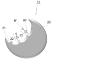
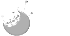
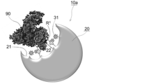
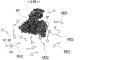
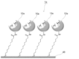
- An example of the sensing nanoparticles of the present invention is schematically shown. Other examples of sensing nanoparticles of the present invention are schematically shown. An example of the usage mode of the sensing nanoparticles of the present invention is schematically shown. Step 1 in an example of the method for producing sensing nanoparticles of the present invention is schematically shown. Steps 2 and 3 in an example of the method for producing sensing nanoparticles of the present invention are schematically shown. An example of the sensing substrate of the present invention is schematically shown. Other examples of the sensing substrate of the present invention are schematically shown. The result of the protein adsorption test by Example 2 (corresponding to the sensing substrate of FIG. 7) obtained in Test Example 5 is shown.
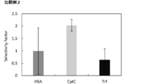
- the results of the protein adsorption test by Comparative Example 2 (sensing substrate for comparison provided with a MIP film) obtained in Test Example 5 are shown.
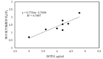
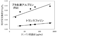
- the correlation between the HSA concentration by the BCP method obtained in Test Example 6 and the relative fluorescence intensity based on the HSA bond measured on the sensing substrate is shown.
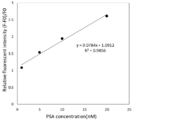
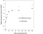
- the correlation between the obtained relative fluorescence intensity and the PSA concentration is shown for the sensing substrate on which the fluorescent group-introduced MIP-NGs of Example 4 obtained in Test Example 7 are fixed.
- the result of the protein adsorption test by the sensing substrate (corresponding to the sensing substrate of FIG. 7) on which the fluorescent group-introduced MIP-NGs of Example 4 was fixed, which was obtained in Test Example 7, is shown.
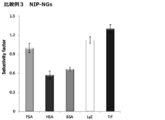
- the results of the protein adsorption test using the sensing substrate on which the fluorescent group-introduced NIP-NGs of Comparative Example 3 obtained in Test Example 7 are fixed are shown.
- the results of the PSA adsorption test in the PSA-mixed beef extraction sample obtained in Test Example 7 using the sensing substrate (corresponding to the sensing substrate in FIG. 7) on which the fluorescent group-introduced MIP-NGs of Example 4 were fixed are shown.
- the change in relative fluorescence intensity at 667 nm when added at a concentration is shown.
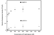
- the relative fluorescence intensity change when the Fc domain fragment is added to the Fc domain sensing substrate (and IgG sensing via Fc domain capture) obtained in Test Example 9 is shown.
- the relative fluorescence intensity change when the Fc domain fragment, complete IgG or lysosome is added to the substrate for Fc domain sensing (and for IgG sensing via Fc domain capture) obtained in Test Example 9 is shown.
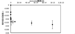
- the relationship between the relative fluorescence intensity and the exosome concentration by the substrate for exosome sensing via HER2 capture obtained in Test Example 10 is shown.
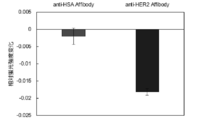
- the exosomes derived from the HER2 overexpressing breast cancer cell line obtained in Test Example 10 were used.
- the relative fluorescence intensity change when is added is shown.
- the sensing nanoparticles of the present invention include a molecular imprint polymer having a molecular imprint space of a biological substance; the molecular imprint polymer is an interacting group with the biological substance and a signal substance different from the interacting group. It contains a structural unit derived from a functional monomer having a functional group including a binding group; the functional group is present on the surface of the molecular imprint space.
- the sensing nanoparticles of the present invention will be described in detail.
- FIG. 1 schematically shows an example of the sensing nanoparticles of the present invention.
- the sensing nanoparticles 10 include a molecular imprint polymer 20 having a molecular imprint space 21 of the biological material; the molecular imprint polymer 20 interacts with the biological material (in the figure, It has a functional group 22 including (schematically represented by R 1 ) and a signal substance binding group (included in the structure schematically represented by R 2 in the figure) different from the interacting group. Containing structural units derived from functional monomers; functional groups 22 are present on the surface of the molecular imprint space 21.
- the sensing nanoparticles 10 are free of signal substance-binding groups, and can be used as a sensor for recognizing biological substances by introducing a signal group into the signal substance-binding groups to form the sensing nanoparticles 10a. Can be done.
- the sensing nanoparticles 10 allow the user to freely select and customize the signal group to be introduced into the signal substance binding group.
- FIG. 2 schematically shows another example of the sensing nanoparticles of the present invention.
- the signal group 31 is introduced in advance into the signal substance-binding group of the sensing nanoparticles 10 shown in FIG. Therefore, the sensing nanoparticles 10a can be used as a sensor for recognizing biological substances by itself.
- FIG. 3 schematically shows an example of the usage mode of the sensing nanoparticles of the present invention.
- FIG. 3 shows how the sensing nanoparticles 10a (sensor for recognizing a biological substance) shown in FIG. 2 recognizes and captures the biological substance 90 to be sensed.
- the molecular imprint polymer 20 is in the form of particles and has a molecular imprint space (recess) 21 of the biological material (biological material 90 shown in FIG. 3) to be sensed.
- the number of imprint spaces 21 provided in one molecular imprint polymer molecule 20 may be one as shown in the figure, or may be a plurality (for example, 2 to 3, preferably 2). ) May be. Since the molecular imprint space 21 is formed by molecular imprinting using the biological substance to be sensed as a template, it has a shape complementary to the surface shape of the biological substance.
- the fact that the molecular imprint polymer 20 is in the form of particles essentially contributes to the expression of excellent specificity of the sensing nanoparticles 10a.
- the present invention is a sensor substrate for recognizing a biological substance produced by forming a flat film-like molecular imprint polymer (MIP film) on a substrate having the same monomer composition as the molecular imprint polymer 20 and introducing a signal group. It is not possible to express the specificity as good as the sensing nanoparticles of. Although the specific mechanism by which the expression of such excellent specificity can be obtained as an effect peculiar to the present invention is not clear, the following reasons can be considered as a consideration.
- the molecular imprint space tends to be randomly present, so that the molecular imprint space is non-uniform (when the size of the opening diameter of the molecular imprint space varies widely). ) Is formed, which increases the proportion of molecular imprint spaces that can cause non-specific adsorption for all molecular imprint spaces (eg, those that do not exhibit sufficient selectivity due to too small opening diameter).
- a biological substance template that is relatively hydrophilic with respect to the polymer tends to exist near the interface between the polymer and the polymerization reaction solution.
- the molecular imprint polymer 20 is in the form of particles is not limited to the shape of the substrate as the specific shape of the sensor for recognizing biological substances. Therefore, for example, it can be formulated as a liquid sensing reagent and is versatile. It is also excellent in terms of.
- the average particle size of the molecular imprint polymer 20 varies depending on the size and application of the biological material to be sensed, but for example, the volume average particle size (volume particles) measured by a dynamic light scattering method (DLS).
- the cumulative 50% value of the diameter distribution) is 5 to 500 nm
- the Z average particle size measured by DLS is 5 to 500 nm.
- the average particle size of the molecular imprint polymer 20 when administered in vivo and used for in-vitro imaging for observing the inside of cells is measured by a dynamic light scattering method (DLS).
- the volume average particle size to be measured is 5 to 100 nm
- the Z average particle size measured by DLS is 5 to 100 nm.
- the average particle size of the molecular imprint polymer 20 when used without being administered into a living body is a volume average particle size of 5 to 500 nm measured by a dynamic light scattering method (DLS), which is determined by DLS.
- the measured Z average particle size is 5 to 500 nm.
- the average particle size of the molecular imprint polymer 20 is 7 to 50 nm, 8 to 40 nm, 15 to 30 nm, or 20 to 25 nm in volume average particle size, and 7 to 50 nm, 8 to 25 nm in Z average particle size. Examples include 40 nm, 15 to 30 nm, or 20 to 25 nm.
- the functional group 22 is present in the molecular imprint space 21.
- the functional group 22 contributes to the recognition of the biological substance to be sensed and the provision of signal information based on the recognition.
- the functional group 22 is present on the surface of the molecular imprint polymer 20 other than the molecular imprint space 21.
- the shape of the protein to be sensed and the presence of the functional group 22 suppress non-specific adsorption by molecules other than the biological substance to be sensed, resulting in excellent peculiarity. Demonstrate sex.
- the functional group 22 includes an interaction group with a biological substance to be sensed ( schematically represented by R 1 in the figure) and a signal substance binding group (schematically represented by R 2 in the figure). Included in the structure represented by). Furthermore, the interacting group and the signal substance binding group are designed as different groups from each other.
- An interacting group is a group that can interact with a biological substance to be sensed to form a non-covalent bond.
- FIG. 3 shows how the interacting group (R 1 ) interacts with the surface of the biological material 90 to be sensed.
- Non-covalent bonds include non-covalent bonds and, among covalent bonds, bonds that are as weak as non-covalent bonds.
- Types of non-covalent bonds include hydrogen bonds, electrostatic interaction bonds (ionic bonds), hydrophobic bonds, and van der Waals bonds.
- covalent bonds that are weak to the extent of non-covalent bonds include covalent bonds that can be cleaved only by changing the liquid property without adding a cleaving reagent that exhibits a strong oxidizing or reducing action such as an oxidizing agent or a reducing agent.
- a covalent bond formed between the sugar group and the borate group of the glycoprotein can be mentioned. This facilitates the removal of the template biological material after synthesizing the molecular imprinted polymer particles in molecular imprinting.
- the interacting group exists at a position corresponding to the specific amino acid residue and / or the specific hydrophobic region of the biological substance on the surface of the imprint space 21 having a shape complementary to the surface shape of the protein. doing. Therefore, in the molecular imprint polymer 20, the molecular imprint space 21 has a shape peculiar to the biological substance, and the interacting group existing in the molecular imprint space 21 is located at a position peculiar to the biological substance. By being present, it exhibits excellent recognition ability for the biological substance.
- a signal substance binding group is a group capable of binding to a signal substance.
- the signal group 31 is introduced like the sensing nanoparticles 10a. Since the signal group 31 undergoes an environmental change due to the non-covalent bond of the biological substance to be sensed with the interacting group, the signal group 31 causes a signal change before and after the binding of the biological substance.
- the signal group 31 exposed in FIG. 2 (before recognition of the biological substance 90) undergoes an environmental change due to the non-covalent bond of the biological substance 90 to be sensed with the interacting group (R 1).
- the situation is referred to. That is, the sensing nanoparticles 10a can read out the binding information of the biological substance to be sensed by the signal change.
- the environmental change that the signal group 31 receives due to the interaction with the biological substance to be sensed is not particularly limited as long as it causes the signal change due to the proximity of the biological substance to be sensed.
- the polarity group of the biological substance to be sensed approaches the vicinity of the signal group 31, so that the electron of the signal group 31 is pulled by the polar group;
- sensing The polar group of the target biological substance is close to the signal group 31, and the water molecule existing in the vicinity of the signal group 31 is eliminated, so that the electron of the signal group 31 is attracted to the water molecule in the vicinity.
- the polar group of the biological material to be sensed is close to the vicinity of the signal group 31, so that the energy on the signal group 31 side is bound to the biological material to be sensed.
- the polar group of the biological material to be sensed approaches the signal group 31 and shields the signal group 31, so that the excitation energy does not reach the signal group 31. Be done.
- the interacting group is located on the distal end side of the functional group 22 and the signal substance binding group is located on the proximal end side of the functional group 22, but in the functional group 22, the interacting group is located.
- the positional relationship between the signal group and the signal substance-binding group is as long as the biological substance to be sensed can be non-covalently bonded to the interacting group and the signal group 31 is subjected to environmental changes when it is non-covalently bonded. , Not limited to this.
- the positional relationship between the interacting group and the signal substance binding group may be reversed from the illustrated embodiment.
- the signal group is more reliably performed.
- the interacting group is located on the distal end side of the functional group 22, and the signal substance binding group is the group of the functional group 22. It is preferably located on the end side.
- R 1 represents an interacting group
- R 2 represents a linking group containing a signal substance binding group
- L 1 represents a direct binding or linking group.
- the interaction group R 1 is designed according to the surface state of the biological substance to be sensed.
- the interaction group R 1 can be derived from hydrogen bonds, electrostatic interaction bonds (ionic bonds), hydrophobic bonds, and van der Waals bonds by interaction with specific structures constituting the biological substance. Included are groups capable of forming one or more bonds selected from the group.
- the aromatic groups in the [2] substituted or unsubstituted aromatic groups include [21] substituted or unsubstituted nitrogen-containing aromatic groups and [22] substituted or unsubstituted non-nitrogen aromatic groups.
- the interaction group R 1 may be a group consisting of any of these groups, or may be a group containing two or more combinations selected from these groups.
- a specific example of the interacting group R 1 is a group capable of forming a hydrogen bond and / or an electrostatic interaction with a specific amino acid residue constituting the protein.
- groups capable of forming a hydrophobic bond with a specific hydrophobic region of the protein are shown in the table below.
- substituent in the substituted amino group a linear or branched alkyl group having 1 to 8 carbon atoms, a substituted or unsubstituted aralkyl group; an alkenyl having 4 to 6 carbon atoms constituting a cyclic secondary amino group.
- substituents in the substituted amino group include a substituted or unsubstituted aryl group.
- Specific examples of the substituted or unsubstituted aryl group include the groups listed in [251] substituted or unsubstituted nitrogen-containing aryl group and [252] substituted or unsubstituted non-nitrogen aryl group described later.
- substituted or unsubstituted amino groups include amino groups (unsubstituted); N-methylamino groups, N-ethylamino groups, Nn-propylamino groups, N-isopropylamino groups, N.
- Secondary amino groups such as -n-butylamino group, N-isobutylamino group, N-tert-butylamino group, N-benzylamino group, N-phenylamino group, N-mesylamino group, N-tosylamino group; N , N-dimethylamino group, N, N-diethylamino group, N, N-dibenzylamino group, N-ethyl-N-methylamino group, N, N-di-n-propylamino group, N, N-diisopropyl Secondary amino groups such as amino groups, N, N-diphenylamino groups, N-methyl-N-phenylamino groups, N-methyl-N-benzylamino groups, N-mesyl-N-methylamino groups; piperidyl groups, Cyclic secondary amino groups such as pyrrolidyl groups can be mentioned.
- the nitrogen-containing aryl group in the [251] substituted or unsubstituted nitrogen-containing aryl group includes a nitrogen-containing aryl group having 2 to 12 carbon atoms.
- the substituent in the substituted nitrogen-containing aryl group include a linear or branched alkyl group having 1 to 8 carbon atoms.
- substituted or unsubstituted nitrogen-containing aryl group examples include a pyridyl group, a pyrimidyl group, a pyridazyl group, a pyrazil group, an imidazolyl group, a triazolyl group, and a methylpyridyl group (2-methylpyridyl group, 3-methylpyridyl).
- the non-nitrogen-containing aryl group in the [252] substituted or unsubstituted non-nitrogen aryl group includes a phenyl group and a naphthyl group having 6 to 16 carbon atoms. 1-naphthyl group, 2-naphthyl group, etc.) and the like.
- the substituent in the substituted aryl group include a linear or branched alkyl group, a nitro group, a halogen group (fluoro group, chloro group, bromo group, etc.) having 1 to 8 carbon atoms.
- substituted or unsubstituted aryl group examples include a phenyl group, a naphthyl group, a tolyl group (o-tolyl group, m-tolyl group, p-tolyl group, etc.), and an ethylphenyl group (4-ethylphenyl).
- the [1] substitution which is a monovalent group among the groups [1] to [7].
- Boronyl groups include groups containing a combination of [26] substituted or unsubstituted arylene groups ([261] nitrogen-containing arylene groups, [262] non-nitrogen-containing arylene groups).
- the [261] nitrogen-containing arylene group constitutes an aromatic ring from the groups listed as the above [251] substituted or unsubstituted nitrogen-containing aryl groups. It is a group from which one hydrogen bonded to the carbon is removed.
- the [262] non-nitrogen-containing arylene group constitutes an aromatic ring from the groups listed as the above [252] substituted or unsubstituted non-nitrogen aryl groups. It is a group from which one hydrogen bonded to the carbon is removed.
- Specific groups including two or more combinations selected from the groups [1] to [7] above include amidino, which is a combination of a [3] amidino group and a [26] substituted or unsubstituted arylene group.
- Examples thereof include an aryl group, a boronylaryl group which is a combination of a [7] boronyl group and a [26] substituted or unsubstituted arylene group.
- a boronylaryl group which is a combination of a [7] boronyl group and a [26] substituted or unsubstituted arylene group.
- preferable groups are shown below.
- the ligand is not particularly limited as long as it is a ligand corresponding to the analysis when the biological substance to be sensed is an analysis.
- the biological substance is an enzyme
- antibody mimetic (affibody, etc.), nanobody, etc. can be mentioned
- the biological substance is a sugar chain, lectin, etc.
- the biological substance is a phospholipid.
- phospholipid-binding proteins Evectin 2, Anexin A2, Anexin V, Tim4 and the like can be mentioned
- the biological substance is an enzyme, an enzyme substrate or an enzyme inhibitor can be mentioned.
- the signal substance-binding group is the binding group (described later in FIG. 65) of the signal substance (signal substance 30 in FIG. 5 described later) from which the signal group 31 to be introduced is derived. It is designed according to the binding group 32).
- Specific examples of the signal substance-binding group include an amino group (including a monovalent amino group and a divalent amino group), a carboxyl group, a thiol group, a disulfide group-containing group (monovalent group), an aminooxy group, and an aldehyde group. And hydroxyl groups.
- the linking group R 2 is a non-branched or branched divalent group.
- linking group R 2 When the linking group R 2 is an unbranched, signal substances binding groups are themselves form a linking group R 2 (i.e., the linking group R 2 is, consisting of a signal substance bonding group).
- the linking group R 2 is, consisting of a signal substance bonding group.
- Examples of such a non-branched linking group R 2, that is, a signal substance-binding group include a divalent amino group represented by the following formula (2).
- R 3 is hydrogen or a linear or branched alkyl group having 1 to 8 carbon atoms, and preferably represents hydrogen.
- the signal substance binding group is a monovalent group present in the branch.
- Examples of such a linking group R 2 include a divalent group represented by the following formula (3).
- R 21 represents an amino group (monovalent), a carboxyl group, a thiol group, a disulfide group-containing group (monovalent group), an aminooxy group, an aldehyde group, or a hydroxyl group, and is preferably a thiol group. Alternatively, it represents a disulfide group-containing group.
- the amino group (monovalent), which is an example of the interacting group R 1 [1] include the groups exemplified as a substituted or unsubstituted amino group.
- the disulfide group-containing group (monovalent group) is represented by the following formula (4).
- R 211 represents a substituted or unsubstituted alkyl group.
- alkyl group in the substituted or unsubstituted alkyl group include a linear or branched alkyl group having 1 to 8 carbon atoms.
- the interaction group R 1 does not interfere with the interaction between the biological substance as a template, the disulfide bond does not interfere with the reaction with the signal substance, and the compound as a whole is water-soluble.
- Any functional group to be secured can be appropriately selected by those skilled in the art, and specifically, a hydroxyl group, an amino group (specifically, an example of the above-mentioned interacting group R 1 [1] substitution or Examples include the groups listed as unsubstituted amino groups), carboxy groups, alkoxy groups (eg, alkoxy groups having 1 to 4, preferably 1 to 3, more preferably 1 to 2, and even more preferably 1). Examples thereof include a polyethylene glycol group (examples 1 to 6 as the number of ethylene oxide additions), an alkyl group (for example, an alkyl group having 1 to 4 carbon atoms), an amide group, a sulfo group, a guanidino group, an amidino group and the like. ..
- the position of the substituent in the substituted alkyl group includes the end of the branch of the linking group R 2.
- L 21 represents a direct bond or linking group.
- the linking group include an alkylene group, a carbonyl group, an ester bond (-COO- or -OCO-), an amide bond (-CONH- or -NHCO-), and a group consisting of any combination thereof, which is preferable.
- the alkylene group include an alkylene group having 1 to 4, preferably 1 to 3, more preferably 1 to 2, and even more preferably 1.
- L 1 (Direct bond or linking group L 1 )
- the direct binding or linking group L 1 intervenes between the interacting group R 1 and the linking group R 2 containing the signaling substance binding group.
- L 1 is more preferably a linking group.
- linking group examples include an alkylene group, a carbonyl group, an ester bond (-COO- or -OCO-), an amide bond (-CONH- or -NHCO-), and a group consisting of any combination thereof.
- alkylene group examples include an alkylene group having 1 to 4 carbon atoms.
- R 11 corresponds to the interacting group R 1 of formula (1), and the divalent amino group (-NH-) is a linking group R 2 containing the signal substance binding group of formula (1). It corresponds to (itself constitutes a signal substance binding group), and L 11 corresponds to the direct binding or linking group L 1 of the formula (1).
- R 11 represents a carboxyaryl group or a sulfoaryl group, and specifically, a carboxyaryl group which is a combination of the above-mentioned [5] carboxyl group and [26] substituted or unsubstituted arylene group.
- [6] As described as a sulfoaryl group which is a combination of a sulfo group and a [26] substituted or unsubstituted arylene group.
- L 11 represents an alkylene group having 1 to 4 carbon atoms.
- L 11 preferably includes an alkylene group having 1 to 3 carbon atoms, more preferably 1 to 2 carbon atoms, and further preferably 1 alkylene group.
- R 11 corresponds to the interacting group R 1 of formula (1)
- the divalent group (-CH (L 21- R 21 )-) is the signal substance binding group of formula (1).
- L 12 corresponds to the direct bond or linking group L 1 of the formula (1).
- R 11 represents a carboxyaryl group or a sulfoaryl group, specifically a carboxyaryl group which is a combination of the above-mentioned [5] carboxyl group and [26] substituted or unsubstituted arylene group.
- [6] As described as a sulfoaryl group which is a combination of a sulfo group and a [26] substituted or unsubstituted arylene group.
- the divalent group (-CH (L 21- R 21 )-) is as described as the above formula (3).
- L 12 represents a divalent linking group
- the divalent linking group includes an alkylene group, a carbonyl group, an ester bond (-COO- or -OCO-), and an amide bond (-CONH-).
- a group consisting of -NHCO-) and any combination thereof can be mentioned, preferably an amide bond.
- These functional groups 22 may have only one type or a plurality of types per one sensing nanoparticle, and even if only one type exists, a plurality of these functional groups exist. You may be. Further, these functional groups 22 may have only one type or a plurality of types per one molecular print space, and even if only one type exists, a plurality of types exist. You may be doing it.
- the polymer constituting the molecular imprint polymer 20 contains at least a structural unit derived from a functional monomer having a functional group 22.
- the polymer constituting the molecular imprint polymer 20 can contain a structural unit derived from an N-substituted or unsubstituted acrylamide and / or a biocompatible monomer, as well as a structural unit derived from a functional monomer.
- the polymer constituting the molecular imprint polymer 20 contains a structural unit derived from a functional monomer, as well as an N-substituted or unsubstituted (meth) acrylamide and a structural unit derived from a biocompatible monomer.
- the polymer constituting the molecular imprint polymer 20 may contain a functional monomer, N-substituted or unsubstituted acrylamide, and a structural unit derived from a monomer different from the biocompatible monomer.
- the functional monomer having the functional group 22 is not particularly limited as long as it is a monomer containing a functional group and an ethylenically unsaturated group, and is represented by, for example, the following formula (5).
- X represents an ethylenically unsaturated group
- L 5 represents a linking group
- -R 2- L 1- R 1 represents a functional group.
- the ethylenically unsaturated group include an acrylic group and a methacrylic group.
- the linking group include an alkylene group, a carbonyl group, an ester bond (-COO- or -OCO-), an amide bond (-CONH- or -NHCO-), and a group consisting of any combination thereof, preferably. Examples thereof include groups in which an arbitrary number of alkylene groups and amide bonds are bonded.
- the functional group is as described in detail in "1-1-3. Structure of the functional group" above.
- the functional monomer represented by the formula (5) include the functional monomers represented by the following formulas (51a), (52a), (51b) and (52b).
- N-substituted or unsubstituted acrylamide N-substituted (meth) acrylamide is represented by the following formula (6).
- R 61 represents hydrogen or a methyl group, preferably hydrogen, and R 62 and R 63 are independently hydrogen; a linear or branched alkyl group having 1 to 6 carbon atoms.
- Linear or branched alkyl groups having 1 to 6 carbon atoms include methyl group, ethyl group, n-propyl group, isopropyl group, n-butyl group, sec-butyl group, tert-butyl group and n-. Examples include a pentyl group and an n-hexyl group.
- the linear or branched hydroxyalkyl group having 1 to 6 carbon atoms includes a hydroxymethyl group, a 1-hydroxyethyl group, a 2-hydroxyethyl group, a 1-hydroxy-n-propyl group, and a 2-hydroxy-n group.
- the linear or branched aminoalkyl group having 1 to 6 carbon atoms includes an aminomethyl group, a 1-aminoethyl group, a 2-aminoethyl group, a 1-amino-n-propyl group, and a 2-amino-n group.
- -Propyl group 3-amino-n-propyl group, 1-aminoisopropyl group, 2-aminoisopropyl group, 1-amino-n-butyl group, 1-amino-n-pentyl group, 1-amino-n-hexyl Group etc. can be mentioned.
- Examples of the saturated 5- to 7-membered ring containing an oxygen atom as well as a nitrogen atom include a morpholine ring and the like.
- N-substituted or unsubstituted (meth) acrylamides N-substituted (meth) acrylamides are preferable, N-mono-substituted (meth) acrylamides are more preferable, and N-mono-substituted (meth) acrylamides are more preferable.
- Alkyl (meth) acrylamide (the alkyl group is a linear or branched alkyl group having 1 to 6 carbon atoms) is mentioned, more preferably N-isopropyl (meth) acrylamide is mentioned, and particularly preferably. Examples include N-isopropylacrylamide.
- Biocompatible monomer Biocompatibility refers to the property of not inducing adhesion of biological substances. By including a component derived from the polymer biocompatible monomer constituting the molecular imprint polymer 20, non-specific adsorption can be further suppressed.
- the biocompatible monomer preferably includes a hydrophilic monomer, and more preferably a zwitterionic monomer.
- the diionic monomer contains an anionic group derived from an acidic functional group (for example, a phosphoric acid group, a sulfuric acid group, a carboxyl group, etc.) and a basic functional group (for example, a primary amino group, a secondary amino group, and a tertiary group). Both an amino group and a cation group derived from a quaternary ammonium group, etc.) are contained in one molecule.
- Examples of the type of zwitterionic monomer include phosphobetaine, sulfobetaine, and carboxybetaine.
- Examples of the phosphobetaine include a molecule having a phosphorylcholine group in the side chain, and preferably 2-methacryloyloxyethyl phosphorylcholine (MPC) and the like.
- Examples of sulfobetaines include N, N-dimethyl-N- (3-sulfopropyl) -3'-methacryloylaminopropaneaminium inner salt (SPB), N, N-dimethyl-N- (4-sulfobutyl) -3'.
- SPB N-dimethyl-N- (4-sulfobutyl) -3'.
- -Methacrylaminopropaneaminium inner salt (SBB) and the like can be mentioned.
- carboxybetaine examples include N, N-dimethyl-N- (1-carboxymethyl) -2'-methacryloyloxyethaneaminium inner salt (CMB), N, N-dimethyl-N- (2-carboxyethyl)-. Examples thereof include 2'-methacryloyloxyethaneaminium inner salt (CEB).
- phosphobetaine is preferable, a molecule having a phosphorylcholine group in the side chain is more preferable, and 2-methacryloyloxyethyl phosphorylcholine (MPC) is more preferable.
- the signal group 31 functions to provide binding information between the biological substance 90 to be sensed and the interaction group R 1 in the molecular imprint space 21.
- a signal group 31 is not particularly limited as long as the signal by receiving environmental changes by the binding of the biological material 90 and interacting groups R 1 to be the sensing target is changed.
- the signal change mode include a change in signal intensity and a change in spectrum (for example, peak shift).
- the signal group 31 examples include a fluorescent group, a radioactive element-containing group, a magnetic group and the like. From the viewpoint of detectability and the like, the signal group 31 is preferably a fluorescent group.
- the fluorescent group is preferably a non-biological group, and examples thereof include a cyanine dye such as a fluorescein dye and an indocyanine dye, and a fluorescent dye such as a rhodamine dye.
- the radioactive element-containing group include sugars, amino acids, nucleic acids labeled with a radioisotope such as 18 F, and an MRI probe labeled with 19 F.
- the magnetic group examples include a group having a magnetic substance such as ferrichrome.
- a capping modifying group is a group introduced by a compound that inhibits the function of a functional monomer.
- the compound (capping compound) that inhibits the function of the functional monomer is a non-signaling substance and has a compound used for suppressing the adsorption of biological substances (for example, a compound having an oligoethylene glycol chain) and a sugar chain structure. Examples include compounds.
- the bonding position of the capping modifying groups include interacting group R 1 and / or signaling substances bonding group of the functional group 22. Since such a capping modifying group shields the functional group 22 from the outside, it is possible to obtain a higher effect of suppressing non-specific binding and background signals.
- the sensing target of the sensing nanoparticles of the present invention is a biological substance.
- the biological substance refers to a molecular species classified into molecules that are basic materials constituting a living body, and generally contains carbon and hydrogen, and further contains nitrogen, oxygen, phosphorus, and / or sulfur. It is a molecule that contains such as as a constituent element and has a biological function or biological significance.
- Specific examples of biological substances include proteins (including peptides; the same applies hereinafter), sugar chains, lipids (for example, phospholipids, etc.), two or more complex molecules thereof, and proteins and cofactors (for example, coenzymes and prosthetic groups).
- the biological substance may be a natural product or an artificial product.
- human serum albumin HSA
- the biological substance 90 is intended to be any biological substance to be sensed by the present invention.
- a preferable example of a biological substance is a protein.
- the protein includes a normal protein having no sugar, lipid, or cofactor, and a complex protein containing at least one of sugar, lipid, and cofactor (for example, glycoprotein, lipoprotein, metalloprotein). Etc.) are included.
- the protein may be a natural protein or an artificial protein.
- Intrinsically disordered proteins are proteins produced as a result of natural selection. Examples of the artificial protein include those in which a part of the amino sequence of the natural protein is artificially modified, those in which a modifying group is artificially introduced or excluded from the natural protein, and the like.
- the biological substance 90 includes free molecules (eg, free proteins, free sugar chains, etc.) secreted into body fluids, membrane structures (eg, cells, viruses, extracellular vesicles, etc.) from the viewpoint of their existence form in the living body.
- Substances eg, membrane proteins, membrane sugar chains
- biological membranes eg, exosomes, etc.
- organelles e.g., lipids, lipids, etc.
- molecules that correspond to exposed partial structures of biological membranes eg, phosphatidyl.
- Phospholipids such as serine
- the biomaterial 90 may be a fragment of a specific biomolecule.
- Examples of such a fragment include a molecule obtained by cutting out a specific region (for example, a domain) from a specific biomolecule (for example, an antibody or the like), and a molecule obtained by artificially producing only the region portion.
- examples of the biological substance 90 include plasma proteins, biomarkers, domains, enzymes, hormones and the like from the viewpoint of its function.
- plasma proteins include albumin, ⁇ -globulin, fibrinogen and the like.
- Examples of biomarkers include renal function markers, liver function markers, inflammation markers, tumor markers, and the like, and more specifically, urinary albumin, aspartate aminotransferase (AST), alanine aminotransferase (ALT), transferrin, and the like.
- LDH lactate dehydrogenase
- ALP alkaline phosphatase
- ⁇ -GTP ⁇ -glutamyl transpeptidase
- CRP C-reactive protein
- AFP ⁇ -fetoprotein
- various antigens eg, sugar chains
- Antigen 19-9 CA19-9
- CEA Cancer Fetal Antigen
- PSA Prostate Specific Antigen
- Exosome Specific Antigen CD63, CD9, CD81, CD37, CD53, CD82, CD13, CD11, CD86, ICAM-1 , Rab5, Annexin V, LAMP1 etc.
- Examples of the biological substance 90 include a human-derived biological substance, a non-human biological biological substance, and the like from the viewpoint of the derived organism.
- the organisms other than humans are not particularly limited, and include vertebrates such as fish, amphibians, reptiles, and mammals; invertebrates such as spores and shellfish; and all organisms such as seeds, nudes, and plants such as algae. Mammals are preferred, such as mice, rats, monkeys, dogs, cats, cows, horses, pigs, hamsters, rabbits, and goats.
- a biological substance derived from animals and plants corresponding to haram can also be mentioned from the viewpoint of Islamic precepts.
- the sensing nanoparticles of the present invention can be used for detecting the above-mentioned biological substances.
- a substance for example, a membrane protein, a membrane sugar chain
- the biological substance is attached to or bound to the biological membrane of a membrane structure (for example, a cell, a virus, an extracellular vesicle or an organelle), and If it is a molecule corresponding to the exposed partial structure of the biomolecule (for example, a phospholipid such as phosphatidylserine), or if it is a fragment of a specific biomolecule (for example, a specific region such as a domain), this
- the sensing nanoparticles of the present invention are a membrane structure (cells, viruses, extracellular vesicles (eg, exosomes, etc.) or organelles, etc.) or fragments (eg, cells, etc.) having the biological substance via the biological substance. It can be used for detecting a specific biomolecule (for example, an antibody or
- the sensing nanoparticles of the present invention differ depending on the type of biological substance, but for example, the sensing nanoparticles of the present invention include renal function markers, liver function markers, inflammation markers, tumor markers, and the like. When designed for sensing biomarkers, it may be used for diagnosis based on renal function, liver function, presence / absence or degree of inflammation, presence / absence or degree of tumor, and the like.
- the sensing nanoparticles of the present invention can identify the same kind of biological substance between different organisms due to their excellent specificity, the biological substance can be used as a more specific application of the sensing nanoparticles of the present invention.
- the biological substance can be used as a more specific application of the sensing nanoparticles of the present invention.
- a biomaterial derived from animals and plants corresponding to haram there is also an application of halal check for confirming the presence or absence of a mixture of biomaterials derived from animals and plants corresponding to haram (for example, porcine serum albumin) in food.
- the present invention also provides the method for producing the sensing nanoparticles of the present invention described in "1. Sensing nanoparticles" above.
- the method for producing sensing nanoparticles of the present invention uses a biological substance as a template to obtain a functional monomer having a functional group containing an interacting group with the biological substance and a signal substance-binding group different from the interacting group. It comprises step 1 of synthesizing a molecular imprint polymer by polymerizing the containing monomer components; and step 2 of removing the template from the molecular imprint polymer.
- the method for producing sensing nanoparticles of the present invention can include a step 3 of binding a signal substance to a signal substance binding group in the molecular imprint polymer after the step 2.
- the monomer component in step 1 may further contain N-substituted or unsubstituted (meth) acrylamide and / or biocompatible monomer.
- this production method 1 prior to step 1, surface modification by covalent bond (for example, an interacting group and a signal substance binding group are localized in the molecular imprint space) with respect to the biological substance as a template. It is not necessary to take measures) etc. that are performed for the purpose of this. Further, this production method 1 can be applied to any of the sensing nanoparticles of the present invention described in the above "1. Sensing nanoparticles", but the interacting group is a substituted or unsubstituted amino group or a substituent.
- it is preferably applied when it is selected from the group consisting of an unsubstituted aromatic group, an amidino group, a guanidino group, a carboxyl group, a sulfo group and a boronyl group.
- FIG. 4 schematically shows step 1 in an example of the method for producing sensing nanoparticles of the present invention.
- FIG. 5 schematically shows steps 2 and 3 in an example of the method for producing sensing nanoparticles of the present invention.
- step 1 In step 1, as shown in FIG. 4, the biological material 90 'as a template, the biological material 90' functional groups comprise different signal substances binding group and the interacting group R 1 and the interacting group R 1 with 22
- a molecular imprint polymer is synthesized by polymerizing a monomer component containing the functional monomer M22 having the above.
- the monomer component comprises N-substituted or unsubstituted (meth) acrylamide M23 and biocompatible monomer M24 together with the functional monomer M22.
- the biological material 90'as a template is a biological material of the same type as the biological material 90 to be sensed.
- HSA biological substance 90 to be sensed
- HSA can also be used as the biological substance 90'as a template. Therefore, examples of the biological substance 90'as a template include those listed in "1-4. Biological substance to be sensed" above.
- the concentration of the biological substance 90'that serves as a template in the polymerization reaction solution is not particularly limited, but is, for example, 0.5 to 5 ⁇ mol / L, preferably 1 to 3 ⁇ mol / L, and more preferably 1.5 to 2.5 ⁇ mol / L. Can be mentioned.
- the functional monomer M22 is as described in "(Functional monomer)" of "1-1-4. Constituent monomer of molecular imprint polymer”.
- the concentration of the functional monomer M22 in the polymerization reaction solution is not particularly limited, and examples thereof include 1 to 6 mmol / L, preferably 2 to 4 mmol / L, and more preferably 2.5 to 3.5 mmol / L.
- the amount of the functional monomer M22 used with respect to the template biological substance 90' is preferably 0.5 to 4 mmol, more preferably 0.5 to 4 mmol, as the amount of the functional monomer M22 used per 1 ⁇ mol of the template biological substance 90'. Is 1 to 2 mmol, more preferably 1.3 to 1.7 mmol.
- N-substituted or unsubstituted (meth) acrylamide M23 is described in "(N-substituted or unsubstituted (meth) acrylamide)" of "1-1-4. Constituent Monomer of Molecular Imprint Polymer” above. It's a street.
- the concentration of N-substituted or unsubstituted (meth) acrylamide M23 in the polymerization reaction solution is not particularly limited, but is, for example, 20 to 60 mmol / L, preferably 30 to 50 mmol / L, and more preferably 35 to 40 mmol / L. Can be mentioned.
- the amount of N-substituted or unsubstituted (meth) acrylamide M23 used for the template biological substance 90' is as follows: N-substituted or unsubstituted (meth) acrylamide per 1 ⁇ mol of the template biological substance 90'.
- the amount of M23 used is preferably 5 to 40 mmol, more preferably 10 to 33 mmol, still more preferably 15 to 20 mmol.
- the amount of N-substituted or unsubstituted (meth) acrylamide M23 used for the functional monomer M22 is the amount of N-substituted or unsubstituted (meth) acrylamide M23 used per mole of the functional monomer M22. , Preferably 5 to 30 mol, more preferably 8 to 20 mol, still more preferably 10 to 15 mol.
- the biocompatible monomer M24 is as described in "(Biocompatible monomer)" of "1-1-4. Constituent monomer of molecular imprint polymer”.
- the concentration of the biocompatible monomer M24 in the polymerization reaction solution is not particularly limited, but is, for example, 0.5 to 5 mmol / L, preferably 1 to 3.5 mmol / L, and more preferably 1.5 to 2.5 mmol / L. L is mentioned.
- the amount of the biocompatible monomer M24 used with respect to the biomaterial 90'as a template is preferably 0.3 to 3.
- the amount of the biocompatible monomer M24 used per 1 ⁇ mol of the biomaterial 90'as a template 5 mmol, more preferably 0.5 to 2 mmol, still more preferably 0.8 to 1.2 mmol.
- the amount of the biocompatible monomer M24 used with respect to the functional monomer M22 is preferably 0.1 to 1.5 mol, more preferably 0.1 to 1.5 mol, as the amount of the biocompatible monomer M24 used per mol of the functional monomer M22. Is 0.2 to 1 mol, more preferably 0.4 to 0.8 mol.
- the polymerization reaction solution contains a cross-linking agent, an initiator, and a solvent as appropriate.
- cross-linking agent examples include compounds in which two or more ethylenically unsaturated groups are bonded by a linker group.
- Specific examples of the cross-linking agent include those represented by the following formula (7).
- W represents an ethylenically unsaturated group that may be the same or different from each other
- Z represents a linker group.
- the ethylenically unsaturated group include an acrylic group and a methacrylic group.
- the linker group includes, for example, an alkylene group having 1 to 6 carbon atoms, preferably 2 to 6 carbon atoms, an amino group (-NH-), an ether group, a carbonyl group, and an ester bond (-COO- or -OCO).
- amide bond ((-CONH- or -NHCO-)), a sulfoxide group (-SO-), a sulfonyl group (-SO 2- ), and a group in which two or more of these are bonded.
- the number of bonds is preferably 5 or less or 4 or less, and more preferably 3 or less or 2.
- more specific cross-linking agents include small molecule cross-linking agents such as N, N'-methylene bisacrylamide and ethylene glycol dimethacrylate, preferably N, N'-methylene bisacrylamide.
- the concentration of the cross-linking agent in the polymerization reaction solution is not particularly limited, and examples thereof include 0.5 to 5 mmol / L, preferably 1 to 3.5 mmol / L, and more preferably 1.5 to 2.5 mmol / L. Be done.
- the initiator examples include peroxides such as ammonium persulfate and potassium persulfate, azobisisobutyronitrile, and azo-based polymerization initiators such as 2,2'-azobis (2-methylpropionamidine) dihydrochloride.
- peroxides such as ammonium persulfate and potassium persulfate
- azobisisobutyronitrile examples include azo-based polymerization initiators
- 2,2'-azobis (2-methylpropionamidine) dihydrochloride Preferably an azo-based polymerization initiator, and more preferably 2,2'-azobis (2-methylpropionamidine) dihydrochloride.
- the concentration of the initiator in the polymerization reaction solution is not particularly limited, and examples thereof include 1 to 15 mmol / L, preferably 3 to 12 mmol / L, and more preferably 5 to 10 mmol / L.
- an aqueous solvent such as a buffer solution is used, and a phosphate buffer solution is preferable.
- the solvent preferably contains NaCl, and the concentration of NaCl is, for example, 100 to 200 mM, preferably 120 to 160 mM.
- a pH that does not cause denaturation of the mold 90' is appropriately selected, and examples thereof include 6 to 8, preferably 6.8 to 7.8, and more preferably 7.2 to 7.6. ..
- the functional monomer M22 of the monomer component specifically interact with a predetermined portion of the template 90 'through interaction group R 1 Then, a complex of the functional monomer M22-template 90'is formed.
- a molecular imprint space (recess) complementary to the surface shape of the template 90'is formed, and the functional group 22 is molecularly inserted. It can be present on the surface of the print space (recess). Further, since the template 90'is likely to be present near the polymer interface, the molecular imprint space is efficiently formed.
- Specific polymerization methods for obtaining nanoparticles of the molecular imprint polymer include an emulsifier-free precipitation polymerization method, a dispersion polymerization method, an emulsion polymerization method, a seed emulsion polymerization method, and the like, and an emulsifier-free precipitation polymerization method is preferable. Can be mentioned.
- step 1 in order to sufficiently form a complex of the functional monomer M22-template 90'before subjecting the polymerization reaction solution to a temperature condition for advancing the polymerization reaction, 1 to 15 ° C., preferably 1 to 10 ° C. , More preferably, it is subjected to a low temperature condition of 2 to 6 ° C. Examples of the time for subjecting to low temperature conditions include 20 to 30 hours. Examples of the temperature condition for advancing the polymerization reaction include 40 to 76 ° C., preferably 50 to 74 ° C., and more preferably 60 to 72 ° C. Examples of the time for subjecting to the temperature condition for advancing the polymerization reaction include 10 to 15 hours.
- the shape of the template is maintained to the extent that it does not cause denaturation that loses the specificity of the molecular imprint space, so that the specificity of the molecular imprint space is effective. Can be acquired.
- the molecular imprint polymer 20 portion is synthesized to obtain a composite of the molecular imprint polymer 20 and the template 90'.
- the complex of the molecular imprint polymer 20 and the template 90' is weak to non-covalent bonds (non-covalent bonds and, among covalent bonds, non-covalent bonds) between the interacting group R 1 and the template 90'. It is formed by (including bonds).
- step 2 the mold 90'is removed from the composite of the molecular imprint polymer 20 and the mold 90'.
- the bond between the interacting group R 1 and the template 90' is a weak non-covalent bond, so that strong oxidation or reduction of an oxidizing agent or a reducing agent or the like is performed.
- the template 90' can be easily dissociated and removed without adding a cleavage reagent showing an action or the like.
- a functional monomer having a reversible bond (covalent bond such as a disulfide group or an oxyimino group) is introduced into the template in advance, and the nanoparticles are produced by molecular imprinting polymerization.
- the reversible bond is cleaved with a cleavage reagent to remove the template, and it functions as a functional group derived from the reversible bond (a signal substance-binding group such as a thiol group or an oxyamino group and / or an interacting group).
- the template cannot be easily removed even with a cleavage reagent because the template and the molecular imprint polymer are covalently bonded.
- a cleavage reagent because the template and the molecular imprint polymer are covalently bonded.
- the template in order to remove the template more reliably, not only the functional monomer is introduced into the template in advance, but also such a template is supported on a carrier such as silica to carry out molecular imprinting polymerization. In the later mold removal, it is necessary to remove the whole carrier.
- the production method of the present invention does not require a step of introducing a functional monomer into the template 90'or a step of supporting the template 90' on a carrier prior to the molecular imprinting polymerization, and also requires a molecular imprint polymer. Since the mold 90'is easily removed from the composite of the mold 20 and the mold 90', it is also excellent in terms of ease of manufacture.
- Removal of the template 90' can be appropriately determined by those skilled in the art based on the mode of binding between the interaction group R 1 and the template 90'.
- the reaction solution after the step 1 is subjected to chromatography (preferably anion exchange chromatography), and the mobile phase used at this time is also used as an eluent for the template molecule to remove the template 90'.
- chromatography preferably anion exchange chromatography
- the removed template 90' is separated from the molecular imprint polymer 20 by chromatography, and the molecular imprint polymer 20 is purified.
- the mobile phase is preferably Tris-hydrochloric acid buffer.
- the mobile phase preferably contains NaCl, and the concentration of NaCl may be the same as the concentration in the solvent used in step 1, and examples thereof include 100 to 200 mM, preferably 120 to 160 mM. ..
- the pH of the mobile phase may be the same as the pH of the solvent used in step 1, and is, for example, 6 to 8, preferably 6.8 to 7.8, and more preferably 7.2 to 7.6. Can be mentioned.
- the sensing nanoparticles 10 are synthesized.
- step 3 In step 3, as shown in FIG. 5, the signal substance 30 is bound to the signal substance binding group in the molecular imprint polymer 20.
- the signal substance 30 contains a signal group 31 and a binding group 32.
- the signal group 31 is as described in "1-2. Signal group” described above.
- the binding group 32 is a group that can be covalently bonded to the signal substance binding group, and specifically, the type of the signal substance binding group (amino group (including monovalent amino group and divalent amino group), carboxyl).
- a group, a thiol group, a disulfide group-containing group (monovalent group), an aminooxy group, an aldehyde group, or a hydroxyl group) can be appropriately determined by those skilled in the art.
- Specific conditions for introducing the signal substance 30 into the molecular imprint polymer 20 are conditions under which both can chemically react to form a covalent bond based on the type of the binding group 32 and the type of the gunal substance binding group. Those skilled in the art can make appropriate decisions.
- the sensing nanoparticles 10a are synthesized.
- a very small amount of a biological substance (a biological substance 90 to be sensed and a biological substance of the same type as the biological substance 90'as a template) is added to the sensing nanoparticles 10, and the molecular imprint space 21 is created in the living body.
- Protect by covering with a substance (protection process).
- a compound that inhibits the function of the functional monomer, the interacting group R 1 and / of the functional group 22 existing on the surface of the molecular imprint polymer 20 other than the molecular imprint space 21
- a capping modifying group is attached to the signal substance binding group (capping modifying step).
- Compounds that inhibit the function of functional monomers include non-signal substances, compounds used for suppressing adsorption of general biological substances (for example, compounds having oligoethylene glycol chains), and sugar chains. Examples thereof include compounds having a structure.
- the molecular imprint space 21 is regenerated by removing the bound biological substance (deprotection step).
- the method for producing sensing nanoparticles of the present invention comprises the step 11 of preparing a template for molecular imprint in which the surface of a biological material is modified with a polymerizable functional group via a reversible linking group; In the step 12 of synthesizing a molecular imprint polymer for a part of the surface of the template using the above as a substrate; and in the step 13 of cleaving the reversible linking group to remove the template from the molecular imprint polymer; A step 14 of introducing a molecule having a functional group including a group for binding an interacting group with a substance and a signal substance-binding group via a reversible linking group can be included.
- a step of binding a molecule that gives an interacting group with the biological substance to a group for binding the interacting group with the biological substance in addition to the steps of performing the above steps 11 to 14; a step of binding a molecule that gives an interacting group with the biological substance to a group for binding the interacting group with the biological substance. And; can include a step of binding a signal substance to the signal substance binding group.
- This production method 2 can be applied to any of the sensing nanoparticles of the present invention described in the above "1. Sensing nanoparticles", but is applied when the interacting group is a ligand of the biomolecule. Is preferable.
- Sensing substrate and sensing reagent Examples of application of the sensing nanoparticles of the present invention described in "1. Sensing nanoparticles" include a sensing substrate and a sensing reagent.
- the sensing substrate includes a substrate and sensing nanoparticles fixed to the substrate.
- FIG. 6 schematically shows an example of a sensing substrate.
- FIG. 7 schematically shows another example of the sensing substrate.
- the sensing substrate 1 shown in FIG. 6 includes a substrate 40 and sensing nanoparticles 10 fixed to the substrate 40.
- the sensing substrate 1a shown in FIG. 7 includes a substrate 40 and sensing nanoparticles 10a fixed to the substrate 40.
- Examples of the material of the substrate 40 include a material selected from the group consisting of metal, glass, and resin.
- the metal include gold, silver, copper, aluminum, tungsten, molybdenum and the like.
- Resins include polymethacrylate, polyacrylate, polystyrene, ABS (acrylonitrile-butadiene-styrene copolymer), polycarbonate, polyester, polyethylene, polypropylene, nylon, polyurethane, silicone resin, fluororesin, methylpentene resin, phenol resin, and melamine. Examples thereof include resin, epoxy resin, vinyl chloride resin and the like.
- the substrate 40 may be formed by combining a plurality of materials selected from the above-mentioned materials.
- the substrate 40 may have a metal film provided on the surface of glass or resin.
- the substrate 40 one suitable for the sensing means of the signal group 31 can be used.
- the substrate 40 includes a surface plasmon (SPR) substrate, a surface-enhanced Raman scattering (SERS) substrate, and the like.
- the sensing substrate 1 fixed to the substrate 40 is as described as the sensing nanoparticles 10 described in "1. Sensing nanoparticles" above.
- the orientation of the sensing nanoparticles 10 is unified for convenience, but the orientation of the sensing nanoparticles 10 may be random.
- the sensing substrate 1 shown in FIG. 6 is obtained by fixing the sensing nanoparticles 10 described in the above "1. Sensing nanoparticles" to the surface of the substrate 40.
- the method of fixing the sensing nanoparticles 10 to the surface of the substrate 40 is not particularly limited, and a person skilled in the art can appropriately determine a general method of fixing the polymer nanoparticles to the surface of the substrate.
- Such a method includes, for example, a molecular film forming step of forming a molecular film having a binding functional group on the surface of the substrate 40, and sensing nanoparticles 10 on the binding functional group on the formed molecular film surface. It can be carried out by the step of combining the above.
- Examples of the functional group on the surface of the sensing nanoparticles 10 that binds to the binding functional group on the surface of the molecular film include an interacting group and / or a signal substance binding group derived from the functional monomer remaining on the surface. / Alternatively, an initiator-derived functional group remaining at the terminal of the polymer constituting the sensing nanoparticles 10 can be mentioned.
- a molecular film having a carboxyl group as a binding functional group is formed on the surface of the substrate 40, and an amide bond is formed between the carboxyl group and the amino group on the surface of the sensing nanoparticles 10. This shows an example in which the sensing nanoparticles 10 are bonded.
- the sensing substrate 1a shown in FIG. 7 can be obtained by introducing a signal group into the sensing substrate 1 described above.
- the method for introducing the signal group is as described in "2-3. Step 3" above.
- the sensing substrate 1a shown in FIG. 7 uses sensing nanoparticles 10a instead of sensing nanoparticles 10 as sensing nanoparticles fixed to the substrate 40, and is the same as the manufacturing method of the sensing substrate 1 described above. You can also get it.
- the sensing substrate can be used for detecting the biological substances shown in items 1-4 above.
- a substance for example, a membrane protein, a membrane sugar chain
- a membrane structure for example, a cell, a virus, an extracellular vesicle or an organelle
- Sensing when it is a molecule corresponding to the exposed partial structure of the biomolecule for example, a phospholipid such as phosphatidylserine
- a fragment of a specific biomolecule for example, a specific region such as a domain.
- the substrate for use is a membrane structure (cell, virus, extracellular vesicle (for example, exosome, etc.) or organelle, etc.) or fragment (for example, domain, etc.) having the biological substance via the biological substance. It can be used for detecting a specific biomolecule (for example, an antibody or the like) having a partial structure (region). More specific applications vary depending on the type of biological material to be sensed, but for example, a sensing substrate designed for sensing biomarkers such as renal function markers, liver function markers, inflammation markers, and tumor markers. , Renal function, liver function, presence / absence or degree of inflammation, presence / absence or degree of tumor, etc.
- the biological substance is a biological substance derived from animals and plants corresponding to albumin
- a biological substance derived from animals and plants corresponding to albumin for example, pig
- halal check to confirm the presence or absence of mixture (serum albumin, etc.).
- Sensing reagents include sensing nanoparticles.
- the sensing nanoparticles in the sensing reagent are not fixed to a bulk substrate such as a substrate.
- the sensing reagent is formulated in liquid or solid form (for example, powder form).
- Sensing reagents include other pharmaceutically acceptable components.
- solids and / or liquids that do not affect the stability and cognitive ability of the sensing reagent are appropriately selected.
- water, salts such as NaCl, buffers, stabilizers, antioxidants, preservatives are selected.
- the sensing reagent can be obtained by using the sensing nanoparticles 10 or the sensing nanoparticles 10a and the above-mentioned other components for formulation based on a usual formulation method.
- sensing reagent As an example of the use of the sensing reagent, the same use as mentioned above as the use of the sensing substrate can be mentioned.
- Other examples of applications of sensing reagents include in vitro imaging and in vivo imaging utilizing the feature that sensing particles are not fixed to a substrate.
- nanoparticles 10a for sensing targeting intracellular or surface biological substances are prepared and added to the cells to fluoresce the dynamics of intracellular or surface biological substances 90. Examples thereof include a method of imaging with a signal detection means such as a microscope.
- nanoparticles 10a for sensing are prepared for sensing biological substances existing in the body, administered to the circulatory system of animals such as experimental animals, for example, blood, and the pharmacokinetics of the biological substance 90 is signaled.
- examples thereof include a method of imaging with a detection means.
- step 1 Synthesis of molecular imprint polymer (step 1) A mixture of the components shown in the table below (prepolymer solution) was prepared in 50 mL of Schlenk flask and incubated at 4 ° C. for 24 hours in order to fully interact with the functional monomer and HAS, and then under a nitrogen atmosphere. Emulsifier-free precipitation polymerization was carried out at 70 ° C. for 12 hours. On the other hand, for comparison NIP-NGs, emulsifier-free precipitation polymerization was carried out under the same conditions except that the prepolymer solution did not contain HSA.
- the functional monomer represented by the formula 51a has a benzene ring, a carboxyl group, and a secondary amino group.
- the benzene ring interacts with the hydrophobic pocket of HSA, and the carboxyl group is HSA. It is considered that it interacts with a positively charged site represented by a lysine residue, and a secondary amino group is used for introducing a fluorescent molecule in the post-imprinting modification (step 3) described later.
- step 2 Particles were purified using anion exchange chromatography as follows. In this step, the template was removed from the molecular imprinted polymer by utilizing the strong interaction between the anion exchange chromatography carrier and the HSA.
- the solution after the polymerization reaction was subjected to ultrafiltration three times (25 ° C., 7500 ⁇ g, 20 minutes) using a dialysis tube of Amicon Ultra-4, 10 kDa to remove unreacted monomers and 140 mM NaCl from PBS.
- the solvent was replaced with 10 mM Tris-chloride buffer pH 7.4 containing the above to obtain an unpurified sample.
- Fluorescence derived from the functional monomer (functional group) of the fractionated fraction (400 nm at an excitation wavelength of 280 nm) and fluorescence derived from HSA (340 nm at an excitation wavelength of 280 nm) were measured after the F-2500 manufactured by Hitachi High-Technology Corporation. , It was confirmed that HAS was removed. All the 2nd to 7th fractions were mixed, the solvent was replaced with PBS using a 10 kDa dialysis tube, and the mixture was concentrated to 5 mL. As a result, a purified sample solution of MIP-NGs or NIP-NGs was obtained.
- w 0 is the weight (g) of the 1.5 mL microtube
- w 1 is the weight before drying (g)
- w 2 is the weight after drying (g).
- the solid content concentration was calculated based on. The results are shown in the table below.
- the NHS ester on the surface of the molecular film and the functional group on the surface of the particle (the functional group is an amino group derived from a functional monomer and / or derived from an initiator remaining at the terminal of the polymer constituting the particle.
- the particles are immobilized on the substrate.
- 1M 2-aminoethanol (pH 8.5) was injected at a flow rate of 20 ⁇ L / min for 7 minutes to block the unreacted NHS ester on the surface of the molecular membrane.
- MIP-NGs had a significantly larger binding constant than NIP-NGs, confirming that they show high affinity for HSA due to the molecular imprint effect. Further, as shown in Table 6, since the binding constant of HSA is remarkably large among the three types of proteins, it is inferred that MIP-NGs have a binding space specific to HSA. ..
- Test Example 3 Fabrication of Sensing Substrate for Comparison with MIP Film
- Test Example 5 For the comparison test in Test Example 5 described later, a flat film-like MIP film was formed on the substrate using the same monomer as the monomer used in Test Example 1, and an HSA sensing substrate for comparison was prepared. ..
- the substrate provided with the mold removing MIP film (Comparative Example 2) was washed with pure water and then immersed in a 1 M NaCl aqueous solution and a 0.5 wt% SDS aqueous solution (each for 2 hours) to remove HSA.
- the HSA sensing MIP film substrate for this comparison has an HSA molecular imprint space (recess) on the surface of the MIP film provided on the substrate, and functions at least in the molecular imprint space (recess).
- a functional group derived from a sex monomer and a fluorescent group are arranged. The fluorescent group increases fluorescence due to changes in the environment when the imprint space (recess) adsorbs the sensing target.
- step 3 Introduction of signal substance (fluorescent substance) (step 3)
- 10 mM PBS of 10 mg / mL fluorescent substance (ATTO647N NHS, excitation wavelength 646 nm, fluorescence wavelength 664 nm) was added.
- 5 ⁇ L of the containing 0.5 wt% DMSO solution was added and incubated at 25 ° C. for 2 hours.
- the binding constant of HSA was further improved by introducing a fluorescent group. This is because the introduced fluorescent group does not inhibit the bond between the HSA to be sensed and the interacting group in the molecular imprint space, and the introduced fluorescent group is hydrophobic, so that the interaction occurs. It is presumed that the interaction between the benzene ring and the hydrophobic region of HSA in the group was added to the interaction between the hydrophobic fluorescent group and the hydrophobic region of HSA.
- Protein adsorption test-2 A protein adsorption test based on the Selectivity factor was performed on the obtained fluorescent group-introduced MIP-NGs fixed HSA sensing substrate and the HSA sensing substrate for comparison provided with the MIP film obtained in Test Example 3. The features of each HSA sensing substrate compared are as shown in the table below.
- HSA Human serum albumin
- PBS 10 mM, pH 7.4, containing 140 mM NaCl
- Cyt c Cyt c
- transferrin Trf; 80 kDa, pI: 6.1
- the Selectivity factor was calculated by dividing the rate of change in fluorescence intensity before and after the addition of each protein to the sensing substrate by the rate of change in fluorescence intensity of the HSA to be sensed. Specifically, the Selectivity factor is expressed by the following formula. In the formula, F 0 represents the fluorescence intensity of the substrate surface in the buffer solution, F sample represents the fluorescence intensity of the substrate surface in the sample containing each protein, and F HSA represents the HSA to be sensed. Represents the fluorescence intensity of the substrate surface in the containing sample.
- Example 2 The results of the fluorescent group-introduced MIP-NGs (particle) fixed HSA sensing substrate (Example 2) are shown in FIG. 8, and the HSA sensing MIP film substrate for comparison provided with the MIP film obtained in Test Example 3 (Example 2).
- Example 2 The results of Comparative Example 2) are shown in FIG.
- Test Example 6 Measurement of HSA in serum sample
- the HSA in the serum sample was measured using the HSA sensing substrate (Example 2) corresponding to the sensing substrate 1a of FIG. 7 produced in Test Example 5.
- each sample was diluted 1000-fold with a buffer solution (PBS (10 mM, pH 7.4, containing 140 mM NaCl)) for HSA sensing corresponding to the sensing substrate 1a of FIG. 7 prepared in Test Example 5.
- a buffer solution PBS (10 mM, pH 7.4, containing 140 mM NaCl)
- the relative fluorescence intensity was determined in the same manner as in the protein adsorption test-2 of Test Example 5.
- the correlation between the obtained relative fluorescence intensity and the concentration obtained by the BCP method is shown in FIG.
- the relative fluorescence intensity is FF 0 / F 0 (F 0 represents the fluorescence intensity of the substrate surface in the buffer solution, and F represents the fluorescence intensity of the substrate surface in the sample). expressed.
- F 0 represents the fluorescence intensity of the substrate surface in the buffer solution
- F represents the fluorescence intensity of the substrate surface in the sample
- PSA sensing substrate was prepared by immobilizing particles whose sensing target was porcine serum albumin (PSA) on the substrate, and PSA was used as a marker for porcine-derived components in halal food. A halal check was performed using this substrate.
- PSA porcine serum albumin
- a gold-coated glass SPR substrate (5 mm ⁇ 10 mm) is washed with ethanol and pure water, treated with a UV-O3 cleaner for 20 minutes, immersed in a 1 mM 11-amino-1-undecaneoctanetic ethanol solution, and 24 at 25 ° C. Incubated for hours.
- the substrate after the reaction was washed with ethanol and pure water and dried with nitrogen gas.
- an aqueous solution 100 ⁇ L
- EDC 0.4M
- NHS 0.1M
- FIG. 11 shows the correlation between the obtained relative fluorescence intensity and the PSA concentration of the PSA sensing substrate on which the fluorescent group-introduced MIP-NGs of Example 4 were fixed.
- the relative fluorescence intensity is the same as in Test Example 6.
- a high correlation was found between the PSA concentration and the relative fluorescence intensity based on the PSA binding measured on the PSA sensing substrate. From this, it was shown that not only human serum albumin HSA but also porcine serum albumin PSA can be detected and quantified by changing the template of the imprint recess.
- PSA PSA Pig serum albumin
- HSA human serum albumin
- BSA bovine serum albumin
- LyZ lysoteam
- Trf transferrin
- the Selectivity factor is the ratio of the fluorescence change in the sample to the fluorescence change in PSA of 40 nM, and is represented by the following formula.
- F 0 represents the fluorescence intensity of the substrate surface in the buffer solution
- F sample represents the fluorescence intensity of the substrate surface in the sample
- F PSA represents the fluorescence intensity of the substrate surface at 40 nM PSA. Represents.
- FIG. 12 shows the results of the sensing substrate in which the fluorescent group-introduced MIP-NGs of Example 4 were fixed
- FIG. 13 shows the results of the sensing substrate in which the fluorescent group-introduced NIP-NGs of Comparative Example 3 were fixed. show.
- each sensing substrate was found to have the ability to recognize PSA.
- the sensing substrate on which the fluorescent group-introduced NIP-NGs of Comparative Example 3 are fixed not only the PSA to be sensed but also the protein not to be sensed undergoes a large fluorescence change. It was observed to show, and it was found that it lacked specificity.
- FIG. 12 it was confirmed that the sensing substrate on which the fluorescent group-introduced MIP-NGs of Example 4 was fixed was highly specific for the PSA to be sensed.
- Halal check-1 1 g of shredded beef was added to 4 mL phosphate buffer (pH 7.4) and the beef was homogenized at 10,000 rpm for 1 minute using a homogenizer.
- the supernatant obtained by centrifugation (16,000 ⁇ g, 30 minutes, 4 ° C.) was filtered three times with a 0.2 ⁇ m filter, diluted 500-fold with phosphate buffer (pH 7.4), and the mixture was diluted 500-fold.
- a beef extract sample was obtained.
- PSA was added to the obtained beef extract sample at various concentrations to prepare a PSA-mixed beef extract sample. Separately, PSA was added to a phosphate buffer solution (pH 7.4) at the same concentration to prepare a PSA sample.
- a phosphate buffer solution pH 7.4
- the fluorescence change was measured using the sensing substrate on which the fluorescent group-introduced MIP-NGs of Example 4 were fixed, and the relative fluorescence intensity change was derived in the same manner as described above. The results are shown in FIG.
- the sensing substrate on which the fluorescent group-introduced MIP-NGs of Example 4 was fixed also contained PSA mixed in the beef extraction sample (in 500 ⁇ beef extract) in the phosphate buffer solution (in 500 ⁇ beef extract). It was confirmed that it can be quantified in the same manner as PSA in PBS buffer).
- Halal check-2 A beef extract sample mixed with a pig extract sample was prepared. First, a 500-fold diluted beef extract sample was obtained in the same manner as described above. Further, a pork extract sample diluted 500 times was obtained in the same manner except that pork was used instead of beef. Both extraction samples were mixed so that the content rate of the beef extraction sample and the mixing rate of the pork extraction sample were the ratios shown in the table below, and a beef extraction sample mixed with the pork extraction sample was obtained. The ratio shown in the table indicates the amount corresponding to the amount of protein (weight basis) in the extracted sample.
- the fluorescence change was measured using the sensing substrate on which the fluorescent group-introduced MIP-NGs of Example 4 were fixed, and the relative fluorescence intensity was derived in the same manner as described above.
- the results are shown in FIG. In FIG. 15, the horizontal axis represents the pork extract sample mixing ratio, and the vertical axis represents the relative fluorescence intensity.
- Particles whose sensing target is PSA fluorescent group-introduced MIP-NGs; Example 4 are prepared in the same manner as in Test Example 7 "Synthesis of Sensing Nanoparticles-3" and “Synthesis of Sensing Nanoparticles-4". Made.
- Protein solution (10 mM phosphate containing 140 mM NaCl) so that the final concentration of protein (PSA or transferrin) is 5,10,50,100,500,1000 ⁇ g / mL in 80 ⁇ g / mL fluorescent group-introduced MIP-NGs. (In buffer (pH 7.4)) was added (final solution volume 500 ⁇ L) and incubated at 25 ° C. for 15 minutes. Then, the fluorescence spectrum of the solution was measured with a fluorescence spectrophotometer (F-2500, Hitachi High-Technologies Corporation) (excitation wavelength ⁇ ex: 647 nm, ⁇ em: 658-690 nm). The result is shown in FIG.
- F-2500 fluorescence spectrophotometer
- Example 9 Synthesis of nanoparticles for sensing-5
- the functional monomer represented by the formula 51b was designed and synthesized according to the following scheme.
- the functional monomer represented by the formula 51b has a boronylaryl group and a divalent amino group, and in MIP-NGs, the boronylaryl group interacts with the sugar chain of the Fc domain of IgG. Secondary amino groups are used to introduce fluorescent molecules in post-imprinting modifications.
- Ethylenediamine monomethacrylamide hydrochloride (ca. 1.57 mmol), 4-formylphenylboronic acid (215 mg, 1.5 mmol) and triethylamine (210 ⁇ L, 1.5 mmol) were dissolved in methanol (10 mL) and stirred at room temperature (10 mL). 0.5h). Reaction tracking was performed by TLC (MeOH ⁇ UV @ 254 nm and ninhydrin), and UV absorption and the appearance of spots stained with ninhydrin (Rf: 0.55, purple) were confirmed, and Schiff bases were formed. NaBH 4 (120 mg, 9.0 mmol) was added thereto. After the addition, the mixture was further stirred at room temperature overnight (8 hours).
- Fc Domain 10 mg of IgG was dissolved in 10 mM phosphate buffer (pH 7.4,140 mM NaCl) (5 mL) to prepare an antibody solution.
- An enzyme solution was prepared by dissolving 0.02 ML-cysteine, 0.002 M EDTA, and 0.1 mg / mL papain in 10 mM phosphate buffer (pH 7.4,140 mM NaCl).
- the antibody solution and the enzyme solution were mixed in equal amounts and reacted at 37 ° C. for 24 hours.
- the reaction solution was replaced with a 20 mM phosphate buffer (pH 7.0) using a 10 kDa ultrafiltration membrane (7500 g, 20 minutes ⁇ 3).
- the resulting solution (2 mL) was subjected to size exclusion chromatography (Sephadex G-100) to remove unreacted monomers.
- the resulting solution (1 mL) and 40 mg / mL SDS aqueous solution (1 mL) are mixed and incubated for 5 minutes, followed by an anion exchange resin (DEAE-Sephadex, 10 cm, 1.5 cm id).
- the mixture was added and eluted with a 10 mM Tris-HCl buffer (pH 7.4,140 mM NaCl) as an eluent (step 2).
- purification was carried out by passing through a desalting column (PD-10) (eluent: 10 mM phosphate buffer (pH 7.4, 140 mM NaCl)).
- the particle size of the obtained MIP-NGs (Example 5) and NIP-NGs (Comparative Example 4) was measured in the same manner as in Test Example 1, the presence of particles on the order of nanometers was confirmed. Specifically, the Z average particle size was 21 nm for MIP-NGs (Example 5) and 18 nm for NIP-NGs (Comparative Example 4). It was shown that the Z potential was positively charged at 19 mV for MIP-NGs (Example 5) and 2.9 mV for NIP-NGs (Comparative Example 4). The charge for this is believed to be derived from the secondary amino group (and polymerization initiator) of the functional monomer.
- Sensing board-4 The gold sputtered glass substrate (4.3 ⁇ 9.8 mm) was washed with pure water and EtOH, and UV-O3 washing (20 minutes) was performed. The washed substrate was immersed in an EtOH solution of 1 mM 11-mercaptoundecanoic acid to introduce a carboxy group on the surface utilizing self-assembled monolayer formation (25 ° C., 24 hours). After washing the substrate with EtOH, the surface carboxy group was activated by immersing the substrate in an aqueous solution in which 0.2 M EDC and 0.05 M NHS were dissolved (25 ° C., 1 hour).
- the nanoparticles were immobilized by the above method (25 ° C., 1 hour).
- the unreacted active ester was inactivated by dropping a 1 M aminoethanol aqueous solution (50 ⁇ L) (25 ° C., 0.5 hours).
- Protein free blocking buffer (50 ⁇ L) was added dropwise to block the surface (25 ° C., 0.5 hours).
- the fluorescence intensity on the surface before and after immobilization was measured, and an increase in fluorescence intensity was observed on the substrate after immobilization, so that the immobilization of particles was confirmed.
- Fluorescence measurement The fluorescence detection ability of the produced sensing substrate was examined using a fluorescence microscope equipped with an automatic liquid handling robot. The measurement sequence conditions are as follows. Fluorescence microscope Filter: Cy5 Objective lens: ⁇ 5 Exposure time: 0.1 seconds Light intensity: 12% Light source: Mercury lamp ROI: Sequence of 15 automatic dispensing devices in the center of the substrate 3 ⁇ 5. Tip mounting 2. Sample suction 150 ⁇ L 3. 3. Reaction 5 minutes (25 ° C) 4. Repeat measurement position keep 2 ⁇ 4
- FIG. 18 shows the change in relative fluorescence intensity when the Fc domain fragment (0-1600 nM), which is a template protein, was added to the prepared substrate for sensing.
- the substrate on which NIP-NGs (Comparative Example 4) was immobilized the substrate on which MIP-NGs (Example 5) was immobilized showed a large fluorescence change.
- the binding space of the Fc domain fragment is formed by molecular imprinting, and the functional monomer is incorporated into the space and fluorescently labeled by post-imprinting modification to convert the binding of the Fc domain fragment into a fluorescent signal. It was suggested that it was possible.
- the apparent binding constant (K d ) was calculated to be 9.8 ⁇ 10 -9 M from the obtained rate of change in fluorescence.
- MIP-NGs (Example 5) prepared by using the Fc domain fragment as a template can also detect IgG through the capture of the Fc domain.
- NIP-NGs (Comparative Example 4) both complete IgG and Lyz showed a larger fluorescence response than the Fc domain fragment. It is presumed that this is non-specifically bound to the functional monomer residues randomly present on the surface of NIP-NGs (Comparative Example 4) and the fluorescent molecules introduced therein.
- Whole IgG has a pI of ⁇ 8.5 and Lyz of 11.2, and is positively charged in a neutral solution. From this, it is considered that the electrostatic interaction of the functional monomer with the boronylaryl group, the hydrogen bond with the amide bond site, and the like contribute to the bond.
- Test Example 10 In this test example, it has a molecular imprint recess for sensing HER2 (biomaterial), and in the molecular imprint recess, an anti-HER2 affibody (interacting group with a biological substance) that interacts with HER2 and a signal.
- Particles having a functional group having the substance ATTO647N were prepared as nanoparticles for sensing exosomes (exosomes expressed by SK-BR-3 cells, which are HER2 overexpressing breast cancer cell lines) via capture of HER2.
- step 11 As described below, a template HSA modified with a methacryloyl group (polymerizable functional group) via a disulfide bond (reversible linking group) was prepared.
- 3-mercaptopropionic acid (0.900 g, 8.50 mmol) was dissolved in ethanol 20 mL, and 1.6 mL acetic acid was added.
- 2,2-dipyridyl disulfide (3.75 g, 17 mmol) dissolved in 30 ml of ethanol was added dropwise thereto with stirring at room temperature.
- Compound (1) was obtained by reacting at 25 ° C. for 3 hours.
- the purified compound (1) (1.43 g, 3.97 mmol) was dissolved in 15 mL of ethanol, and then 2- (Boc-amino) ethanethiol (1.69 mL, 9.99 mmol, 2eq) was dissolved in 20 mL of ethanol. rice field.
- Compound (2) was obtained by reacting at room temperature for 3 hours.
- step 12 Particles of molecular imprint polymer were synthesized as follows.
- particles of molecular imprint polymer MIP-NGs were synthesized by performing emulsifier-free precipitation polymerization at 70 ° C. for 16 hours in a Schlenk flask 50 mL in a nitrogen atmosphere according to the polymerization recipe shown in the table below.
- step 13 The template was removed from the molecular imprinted polymer.
- TCEP was screened using Sephadex G-100, a chromatographic filler for size exclusion chromatography, and ultrafiltration (25 ° C, 7500 g, 20 min, 3 times) using a dialysis tube of Amicon Ultra-4 10 kDa. Removal and solvent substitution with 140 mM NaCl 10 mM Tris-hydrogen buffer pH 7.4 were performed. In addition, the template HSA was removed by anion exchange chromatography. As a result, particles of molecular imprint polymer MIP-NGs were obtained.
- the 11-Mercaptoundecanoic acid was immersed in an ethanol solution prepared to 1 mM and incubated overnight at 25 ° C. After the reaction, the substrate is washed with ethanol and pure water, dried with nitrogen gas, and then using Biacore3000, 0.1 M N-Hydroxysuccinimide (NHS), 0.4 M 1- (3-dimethylaminopropyl) -3-ethylcarbodiimide hydrochloride ( A PBS solution of EDC) was injected and incubated at 25 ° C. for 10 minutes.
- NHS N-Hydroxysuccinimide
- a PBS solution of EDC was injected and incubated at 25 ° C. for 10 minutes.
- the obtained substrate (the surface of the substrate modified with a carboxylic acid active ester group as a binding group) was injected with a 400 ⁇ g / mL MIP-NGs solution and incubated at 25 ° C. for 10 minutes to obtain MIP-.
- a substrate on which NGs were immobilized was obtained.
- step 14 The functional molecule was introduced into the binding group of the molecular imprint recess via a disulfide bond (reversible linking group) as follows.
- the substrate on which the molecular imprint polymer particles MIP-NGs obtained in 10-4 were immobilized was immersed in a 900 ⁇ g / mL PBS solution of the functional molecule (compound (3)) obtained in 10-5.
- the pyridyl disulfide group of the functional molecule and the thiol group of the molecular imprint polymer were converted into a disulfide bond (reversible linking group).
- a 10 ⁇ g / mL PBS solution of an anti-Her2 affibody molecule (a molecule that provides an interacting group with a biological substance) is reacted with the substrate obtained in 10-6 above (room temperature, 2 hours), and a pyridyl disulfide group (with a biological substance) is reacted.
- An anti-Her2 affibody group (a group that interacts with a biological substance) was introduced into the group for binding the interacting group of the above. Then, the remaining pyridyl disulfide group was capped by reacting with a 100 ⁇ M 2-mercaptoethanol solution.
- the substrate was then reacted with an Atto647N-NHS solution (room temperature, 2 hours) to introduce Atto647N (signal group) into the amino group (signal substance binding group).
- Atto647N signal group
- the measurement points were 3 points on the substrate, the filter was Cy5, the objective lens was 5 times, the exposure time was 0.1 seconds, the amount of light was 100%, and the light source was a mercury lamp.
- FIG. 21 shows the change in relative fluorescence intensity when 5 ⁇ 10 -17 M) was added.
- exosomes expressed by SK-BR-3 cells were specifically detected by an exosome sensing substrate into which anti-HER2 affibody was introduced and mediated by HER2 capture.
Landscapes
- Health & Medical Sciences (AREA)
- Immunology (AREA)
- Life Sciences & Earth Sciences (AREA)
- Engineering & Computer Science (AREA)
- Chemical & Material Sciences (AREA)
- Molecular Biology (AREA)
- Biomedical Technology (AREA)
- Hematology (AREA)
- Urology & Nephrology (AREA)
- Food Science & Technology (AREA)
- Biochemistry (AREA)
- Cell Biology (AREA)
- Biotechnology (AREA)
- Medicinal Chemistry (AREA)
- Physics & Mathematics (AREA)
- Analytical Chemistry (AREA)
- Microbiology (AREA)
- General Health & Medical Sciences (AREA)
- General Physics & Mathematics (AREA)
- Pathology (AREA)
- Nanotechnology (AREA)
- Measuring Or Testing Involving Enzymes Or Micro-Organisms (AREA)
- Peptides Or Proteins (AREA)
- Investigating, Analyzing Materials By Fluorescence Or Luminescence (AREA)
Priority Applications (11)
| Application Number | Priority Date | Filing Date | Title |
|---|---|---|---|
| EP21759830.9A EP4113118A4 (en) | 2020-02-27 | 2021-03-01 | Nanoparticles for sensing use and production method for same |
| CN202180031163.4A CN115667932A (zh) | 2020-02-27 | 2021-03-01 | 传感用纳米颗粒及其制造方法 |
| US17/802,852 US20230152306A1 (en) | 2020-02-27 | 2021-03-01 | Nanoparticles for sensing use and production method for same |
| JP2022503390A JP7357975B2 (ja) | 2020-02-27 | 2021-03-01 | センシング用ナノ粒子及びその製造方法 |
| CN202180065216.4A CN116324409A (zh) | 2020-09-23 | 2021-08-27 | 用于制作检测对象分析用传感器的基材、检测对象分析用传感器和检测对象的分析方法 |
| JP2022505352A JP7216460B2 (ja) | 2020-09-23 | 2021-08-27 | 検出対象の分析用センサ作製用基材、検出対象の分析用センサ、及び検出対象の分析法 |
| US18/246,300 US20230375537A1 (en) | 2020-09-23 | 2021-08-27 | Base material for manufacturing sensor for analyzing detection object, sensor for analyzing detection object, and method for analyzing detection object |
| EP21872092.8A EP4220160A4 (en) | 2020-09-23 | 2021-08-27 | BASE MATERIAL FOR MANUFACTURING DETECTION OBJECT ANALYSIS SENSOR, DETECTION OBJECT ANALYSIS SENSOR, AND DETECTION OBJECT ANALYSIS METHOD |
| PCT/JP2021/031621 WO2022064960A1 (ja) | 2020-09-23 | 2021-08-27 | 検出対象の分析用センサ作製用基材、検出対象の分析用センサ、及び検出対象の分析法 |
| JP2023003938A JP2023037003A (ja) | 2020-09-23 | 2023-01-13 | 検出対象の分析用センサ作製用基材、検出対象の分析用センサ、及び検出対象の分析法 |
| JP2023152316A JP7799329B2 (ja) | 2020-02-27 | 2023-09-20 | センシング用ナノ粒子及びその製造方法 |
Applications Claiming Priority (4)
| Application Number | Priority Date | Filing Date | Title |
|---|---|---|---|
| JP2020031457 | 2020-02-27 | ||
| JP2020-031457 | 2020-02-27 | ||
| JP2020159008 | 2020-09-23 | ||
| JP2020-159008 | 2020-09-23 |
Publications (1)
| Publication Number | Publication Date |
|---|---|
| WO2021172591A1 true WO2021172591A1 (ja) | 2021-09-02 |
Family
ID=77491598
Family Applications (1)
| Application Number | Title | Priority Date | Filing Date |
|---|---|---|---|
| PCT/JP2021/007726 Ceased WO2021172591A1 (ja) | 2020-02-27 | 2021-03-01 | センシング用ナノ粒子及びその製造方法 |
Country Status (5)
| Country | Link |
|---|---|
| US (1) | US20230152306A1 (cg-RX-API-DMAC7.html) |
| EP (1) | EP4113118A4 (cg-RX-API-DMAC7.html) |
| JP (2) | JP7357975B2 (cg-RX-API-DMAC7.html) |
| CN (1) | CN115667932A (cg-RX-API-DMAC7.html) |
| WO (1) | WO2021172591A1 (cg-RX-API-DMAC7.html) |
Cited By (4)
| Publication number | Priority date | Publication date | Assignee | Title |
|---|---|---|---|---|
| CN114106012A (zh) * | 2021-12-13 | 2022-03-01 | 宜昌博仁凯润药业有限公司 | 一种化合物Biotin-PEG3-SS-DBCO、制备方法及其应用 |
| JPWO2023027184A1 (cg-RX-API-DMAC7.html) * | 2021-08-27 | 2023-03-02 | ||
| CN115739049A (zh) * | 2022-11-24 | 2023-03-07 | 吉林大学 | 一种磷脂分子印迹方法及其应用 |
| WO2024143431A1 (ja) * | 2022-12-27 | 2024-07-04 | 株式会社TearExo | 粒子を組み合わせたセンサ |
Families Citing this family (2)
| Publication number | Priority date | Publication date | Assignee | Title |
|---|---|---|---|---|
| JP7357975B2 (ja) * | 2020-02-27 | 2023-10-10 | 株式会社TearExo | センシング用ナノ粒子及びその製造方法 |
| EP4365213A4 (en) * | 2021-06-29 | 2024-10-23 | PHC Holdings Corporation | POLYMER, REAGENT LAYER AND SENSOR |
Citations (4)
| Publication number | Priority date | Publication date | Assignee | Title |
|---|---|---|---|---|
| JP2016194443A (ja) * | 2015-03-31 | 2016-11-17 | 株式会社森永生科学研究所 | ブタ血清アルブミンの検出方法、検出キット及び抗ブタ血清アルブミン抗体 |
| JP2017150815A (ja) * | 2014-05-29 | 2017-08-31 | 株式会社日立製作所 | 臨床検査キット及び化学物質検出方法 |
| JP2018132527A (ja) * | 2017-02-17 | 2018-08-23 | 学校法人 芝浦工業大学 | 蛍光性官能基を有する分子インプリントナノ粒子 |
| WO2018221271A1 (ja) * | 2017-05-29 | 2018-12-06 | 国立大学法人神戸大学 | 検出対象の分析用センサ作製用基材、検出対象の分析用センサ、及び検出対象の分析法 |
Family Cites Families (4)
| Publication number | Priority date | Publication date | Assignee | Title |
|---|---|---|---|---|
| JP2009128169A (ja) | 2007-11-22 | 2009-06-11 | Canon Inc | 標的物質の検出方法、検出カートリッジ、検出キット |
| US11237162B2 (en) | 2016-05-24 | 2022-02-01 | Jsr Corporation | Composite particles, coated particles, method for producing composite particles, ligand-containing solid phase carrier and method for detecting or separating target substance in sample |
| WO2018194152A1 (ja) | 2017-04-21 | 2018-10-25 | 株式会社ハプロファーマ | アルドステロン及びレニンの検出方法 |
| JP7357975B2 (ja) * | 2020-02-27 | 2023-10-10 | 株式会社TearExo | センシング用ナノ粒子及びその製造方法 |
-
2021
- 2021-03-01 JP JP2022503390A patent/JP7357975B2/ja active Active
- 2021-03-01 WO PCT/JP2021/007726 patent/WO2021172591A1/ja not_active Ceased
- 2021-03-01 CN CN202180031163.4A patent/CN115667932A/zh active Pending
- 2021-03-01 US US17/802,852 patent/US20230152306A1/en active Pending
- 2021-03-01 EP EP21759830.9A patent/EP4113118A4/en active Pending
-
2023
- 2023-09-20 JP JP2023152316A patent/JP7799329B2/ja active Active
Patent Citations (4)
| Publication number | Priority date | Publication date | Assignee | Title |
|---|---|---|---|---|
| JP2017150815A (ja) * | 2014-05-29 | 2017-08-31 | 株式会社日立製作所 | 臨床検査キット及び化学物質検出方法 |
| JP2016194443A (ja) * | 2015-03-31 | 2016-11-17 | 株式会社森永生科学研究所 | ブタ血清アルブミンの検出方法、検出キット及び抗ブタ血清アルブミン抗体 |
| JP2018132527A (ja) * | 2017-02-17 | 2018-08-23 | 学校法人 芝浦工業大学 | 蛍光性官能基を有する分子インプリントナノ粒子 |
| WO2018221271A1 (ja) * | 2017-05-29 | 2018-12-06 | 国立大学法人神戸大学 | 検出対象の分析用センサ作製用基材、検出対象の分析用センサ、及び検出対象の分析法 |
Non-Patent Citations (5)
| Title |
|---|
| ANGEWANDTE CHEMIE INTERNATIONAL EDITION., vol. 55, 2016, pages 13023 - 13027 |
| JOURNAL OF VISUALIZED EXPERIMENTS, vol. 132, 2018, pages e57208 |
| MATSUMOTO, HIROKI; SUNAYAMA, HIROBUMI; TAKANO, ERI; KITAYAMA, YUKIYA; TAKEUCHI, TOSHIFUMI: "5p-A203-13 Prostate specific antigen sensing on Molecularly Imprinted Polymer Thin films Prepared via Post-Imprinting Modification", 78TH JSAP AUTUMN MEETING, JAPAN SOCIETY OF APPLIED PHYSICS; FUKUOKA, JAPAN; SEPTEMBER 5-8, 2017, - 2017, pages 100000001-036, XP009539017 * |
| MATSUURA, RYO; TAKANO, ERI; TAWA, KEIKO; SUNAYAMA, HIROBUMI; KITAYAMA, YUKIYA: "19p-W331-14 Highly Sensitive Plasmonic Sensing for Human Serum Albumin by Molecularly Imprinted Polymer-Coated Plasmonic Chips", 63RD JSAP SPRING MEETING, JAPAN SOCIETY OF APPLIED PHYSICS AND RELATED SOCIETIES ; 64 (TOKYO) : 2016.03.19-22, vol. 63, 30 November 2015 (2015-11-30) - 22 March 2016 (2016-03-22), pages 10-204, XP009539018 * |
| See also references of EP4113118A4 |
Cited By (6)
| Publication number | Priority date | Publication date | Assignee | Title |
|---|---|---|---|---|
| JPWO2023027184A1 (cg-RX-API-DMAC7.html) * | 2021-08-27 | 2023-03-02 | ||
| JP7502835B2 (ja) | 2021-08-27 | 2024-06-19 | 国立大学法人神戸大学 | 基材上に収率よく空孔を形成する技術のためのコアシェル鋳型分子・粒子 |
| CN114106012A (zh) * | 2021-12-13 | 2022-03-01 | 宜昌博仁凯润药业有限公司 | 一种化合物Biotin-PEG3-SS-DBCO、制备方法及其应用 |
| CN115739049A (zh) * | 2022-11-24 | 2023-03-07 | 吉林大学 | 一种磷脂分子印迹方法及其应用 |
| CN115739049B (zh) * | 2022-11-24 | 2024-04-26 | 吉林大学 | 一种磷脂分子印迹方法及其应用 |
| WO2024143431A1 (ja) * | 2022-12-27 | 2024-07-04 | 株式会社TearExo | 粒子を組み合わせたセンサ |
Also Published As
| Publication number | Publication date |
|---|---|
| EP4113118A4 (en) | 2024-04-17 |
| JPWO2021172591A1 (cg-RX-API-DMAC7.html) | 2021-09-02 |
| JP7799329B2 (ja) | 2026-01-15 |
| EP4113118A1 (en) | 2023-01-04 |
| JP2023182627A (ja) | 2023-12-26 |
| US20230152306A1 (en) | 2023-05-18 |
| JP7357975B2 (ja) | 2023-10-10 |
| CN115667932A (zh) | 2023-01-31 |
Similar Documents
| Publication | Publication Date | Title |
|---|---|---|
| JP7357975B2 (ja) | センシング用ナノ粒子及びその製造方法 | |
| Kalecki et al. | Oriented immobilization of protein templates: a new trend in surface imprinting | |
| JP7438966B2 (ja) | ナノアレイおよびマイクロアレイを製造する方法 | |
| Wackerlig et al. | Applications of molecularly imprinted polymer nanoparticles and their advances toward industrial use: a review | |
| Kan et al. | Preparation and recognition properties of bovine hemoglobin magnetic molecularly imprinted polymers | |
| KR20230118570A (ko) | 강화된 결합 및 검출 특성을 갖는 친화성 시약 | |
| US9308582B2 (en) | Solution stable and chemically reactive metallic nanoparticles | |
| JP2023037003A (ja) | 検出対象の分析用センサ作製用基材、検出対象の分析用センサ、及び検出対象の分析法 | |
| EP1813946B1 (en) | Biomaterial structure, method of producing the same and uses thereof | |
| JPWO2018221271A1 (ja) | 検出対象の分析用センサ作製用基材、検出対象の分析用センサ、及び検出対象の分析法 | |
| Gu et al. | Hierarchically structured molecularly imprinted nanotransducers for truncated HER2-targeted photodynamic therapy of therapeutic antibody-resistant breast cancer | |
| Son et al. | Amine-reactive poly (pentafluorophenyl acrylate) brush platforms for cleaner protein purification | |
| Oliverio et al. | Versatile and high-throughput strategy for the quantification of proteins bound to nanoparticles | |
| JPWO2003066644A1 (ja) | フェライト結合有機物質及びその製造方法 | |
| JP2006508095A (ja) | 粒子の生物活性化 | |
| JP5409671B2 (ja) | アフィニティヒドロゲルとその標識非依存性検出方法 | |
| JP7454225B2 (ja) | 検出対象の分析用センサ作製用基材、検出対象の分析用センサ、及び検出対象の分析法 | |
| JP7511844B2 (ja) | 標的物質のポリマーレプリカ基板及びその製造方法、並びに基板センサの製造方法 | |
| Linkov et al. | Selection of the optimal chromatography medium for purification of quantum dots and their bioconjugates | |
| EP4394383A1 (en) | Core-shell template molecule/particle for technique for high-yield formation of pores on substrate | |
| HK40092517A (en) | Base material for manufacturing sensor for analyzing detection object, sensor for analyzing detection object, and method for analyzing detection object | |
| CN117120627A (zh) | 具有增强的结合和检测特征的亲和试剂 | |
| CN114630913B (zh) | 使用自组装单层预官能化的纳米颗粒和制备其的方法 | |
| EP3338806A1 (en) | Method for functionalising nanoparticles | |
| Sunayama et al. | Functional Molecule‐Integrated Nano‐Interfaces for Detection of Extracellular Vesicles |
Legal Events
| Date | Code | Title | Description |
|---|---|---|---|
| 121 | Ep: the epo has been informed by wipo that ep was designated in this application |
Ref document number: 21759830 Country of ref document: EP Kind code of ref document: A1 |
|
| ENP | Entry into the national phase |
Ref document number: 2022503390 Country of ref document: JP Kind code of ref document: A |
|
| NENP | Non-entry into the national phase |
Ref country code: DE |
|
| ENP | Entry into the national phase |
Ref document number: 2021759830 Country of ref document: EP Effective date: 20220927 |