WO2021117667A1 - 検出方法 - Google Patents
検出方法 Download PDFInfo
- Publication number
- WO2021117667A1 WO2021117667A1 PCT/JP2020/045438 JP2020045438W WO2021117667A1 WO 2021117667 A1 WO2021117667 A1 WO 2021117667A1 JP 2020045438 W JP2020045438 W JP 2020045438W WO 2021117667 A1 WO2021117667 A1 WO 2021117667A1
- Authority
- WO
- WIPO (PCT)
- Prior art keywords
- nucleic acid
- signal
- wells
- well
- stranded
- Prior art date
Links
Images


Classifications
-
- C—CHEMISTRY; METALLURGY
- C12—BIOCHEMISTRY; BEER; SPIRITS; WINE; VINEGAR; MICROBIOLOGY; ENZYMOLOGY; MUTATION OR GENETIC ENGINEERING
- C12Q—MEASURING OR TESTING PROCESSES INVOLVING ENZYMES, NUCLEIC ACIDS OR MICROORGANISMS; COMPOSITIONS OR TEST PAPERS THEREFOR; PROCESSES OF PREPARING SUCH COMPOSITIONS; CONDITION-RESPONSIVE CONTROL IN MICROBIOLOGICAL OR ENZYMOLOGICAL PROCESSES
- C12Q1/00—Measuring or testing processes involving enzymes, nucleic acids or microorganisms; Compositions therefor; Processes of preparing such compositions
- C12Q1/68—Measuring or testing processes involving enzymes, nucleic acids or microorganisms; Compositions therefor; Processes of preparing such compositions involving nucleic acids
- C12Q1/6813—Hybridisation assays
- C12Q1/6816—Hybridisation assays characterised by the detection means
- C12Q1/682—Signal amplification
-
- C—CHEMISTRY; METALLURGY
- C12—BIOCHEMISTRY; BEER; SPIRITS; WINE; VINEGAR; MICROBIOLOGY; ENZYMOLOGY; MUTATION OR GENETIC ENGINEERING
- C12Q—MEASURING OR TESTING PROCESSES INVOLVING ENZYMES, NUCLEIC ACIDS OR MICROORGANISMS; COMPOSITIONS OR TEST PAPERS THEREFOR; PROCESSES OF PREPARING SUCH COMPOSITIONS; CONDITION-RESPONSIVE CONTROL IN MICROBIOLOGICAL OR ENZYMOLOGICAL PROCESSES
- C12Q1/00—Measuring or testing processes involving enzymes, nucleic acids or microorganisms; Compositions therefor; Processes of preparing such compositions
- C12Q1/68—Measuring or testing processes involving enzymes, nucleic acids or microorganisms; Compositions therefor; Processes of preparing such compositions involving nucleic acids
- C12Q1/6844—Nucleic acid amplification reactions
- C12Q1/6846—Common amplification features
Definitions
- the present invention relates to a detection method.
- the present application claims priority with respect to Japanese Patent Application No. 2019-222153 filed in Japan on December 9, 2019, the contents of which are incorporated herein by reference.
- nucleic acids such as DNA and RNA are quantified by a real-time PCR method or the like.
- Non-Patent Document 1 describes a technique for detecting a fluorescent signal by performing an enzymatic reaction in a large number of microcompartments. This technique is called digital measurement.
- the sample solution is divided into an extremely large number of micro solutions. Then, the signal from each microsolution is binarized, only the presence or absence of the target molecule is determined, and the number of target molecules is measured. According to the digital measurement, the detection sensitivity and the quantitativeness can be remarkably improved as compared with the conventional real-time PCR method and the like.
- Non-Patent Document 1 discloses a method using micro-sized droplets formed so that the volume of each well is several nanoliters.
- ICA digital Invasive Cleave Assay
- FIG. 1 is a diagram schematically showing a method of detecting a target nucleic acid by digital PCR, which is a conventional detection method.
- the target nucleic acid is amplified in the step of detecting the target nucleic acid. Therefore, as illustrated in FIG. 1, the target nucleic acid is housed in a microcompartment without going through an amplification step.
- Target nucleic acids which are DNAs and RNAs purified from biological samples such as blood and cells, are single-stranded nucleic acids (referred to as single-stranded nucleic acids 1) and single-stranded nucleic acids complementary to single-stranded nucleic acids 1.
- Nucleic acid (referred to as single-stranded nucleic acid 2) and double-stranded nucleic acid in which single-stranded nucleic acid 1 and single-stranded nucleic acid 2 are complementary to each other (referred to as double-stranded nucleic acid 3). Includes.
- the sample solution before dropletization contains 2 molecules of single-stranded nucleic acid 1 and 2 molecules of single-stranded nucleic acid 2 and 4 molecules of double-stranded nucleic acid 3.
- digital PCR which is a conventional detection method
- the target nucleic acid is detected by using digital PCR, which is a conventional detection method, in the state of droplets, single-stranded and double-stranded, for example, droplet 4, droplet 5, and droplet 6. It will be confined without distinguishing between.
- the PCR products of the target nucleic acids of Droplet 4, Droplet 5, and Droplet 6 are substantially the same. That is, as illustrated in FIG. 1, in digital PCR, eight detection signals are detected like the droplet 7.
- the present invention has been made in view of the above circumstances, and an object of the present invention is to provide a technique capable of distinguishing and quantifying single-stranded nucleic acid and double-stranded nucleic acid with higher accuracy.
- the present invention includes the following aspects.
- the target nucleic acid includes a first nucleic acid, a second nucleic acid which is a complementary strand of the first nucleic acid, and a double-stranded nucleic acid in which the first nucleic acid and the second nucleic acid are complementarily bound.
- the double-stranded nucleic acid is sealed in the well in a double-stranded state, and in the step of amplifying the signal, a first specific binding substance is bound to the first nucleic acid. Then, the first signal is emitted, the second specific binding substance binds to the second nucleic acid, and the second signal is emitted, and the first signal and the second signal are different from each other.
- the detection step it is determined that only the single-stranded first nucleic acid was present in the well in which only the first signal was detected, and the well in which only the second signal was detected was detected. It was determined that only the single-stranded nucleic acid was present in the well, and it was determined that the double-stranded nucleic acid was present in the well in which both the first signal and the second signal were detected.
- the detection method according to any one of [1] to [3].
- the detection method according to any one of [1] to [4], further comprising a step of counting the number of the wells in which the signal is detected in the detection step.
- the counting step the number of the wells in which only the first signal is detected, the number of wells in which only the second signal is detected, and the first signal and the second signal are both detected.
- the detection method according to [5], wherein the number of wells is counted.
- the target nucleic acid comprises a first nucleic acid, a second nucleic acid which is a complementary strand of the first nucleic acid, and a double-stranded nucleic acid in which the first nucleic acid and the second nucleic acid are complementaryly bound.
- the sealing step the double-stranded nucleic acid is sealed in the well in a double-stranded state, and in the step of amplifying the signal, a first specific binding substance binds to the first nucleic acid.
- the present invention includes the following aspects as another aspect.
- a method for detecting a target nucleic acid in a liquid in which the liquid is brought into contact with a device having a well array having a plurality of wells so that the target nucleic acid is reduced to one molecule or less per well in the wells.
- a step of introducing the target nucleic acid into the well a sealing step of sealing the well so that the target nucleic acid does not move between the plurality of wells, and a signal amplification step of amplifying a signal caused by the target nucleic acid in the well.
- the target nucleic acid includes a first nucleic acid and a second nucleic acid which is a complementary strand of the first nucleic acid, and in the signal amplification step.
- the first specific binding substance binds to the first nucleic acid to emit a first signal
- the second specific binding substance binds to the second nucleic acid and a second signal is emitted
- the first signal is emitted.
- a detection method in which the signal is different from the second signal.
- a method for detecting a target nucleic acid in a liquid in which the liquid is brought into contact with a device having a well array having a plurality of wells so that the target nucleic acid is reduced to one molecule or less per well in the wells.
- a detection step of detecting a signal emitted from the well the target nucleic acid includes a first nucleic acid and a second nucleic acid which is a complementary strand of the first nucleic acid, and in the signal amplification step.
- a detection method in which a first specific binding substance binds to the first nucleic acid and a first signal is emitted.
- the present invention comprises, in one embodiment, a step of bringing a liquid containing a target nucleic acid into contact with a device having a well array having a plurality of wells and introducing the target nucleic acid into the wells so as to have one molecule or less per well. It has a step of sealing the well so that the target nucleic acid does not move between a plurality of wells, a step of amplifying a signal caused by the target nucleic acid in the well, and a step of detecting a signal emitted from the well.
- the target nucleic acid includes a first nucleic acid, a second nucleic acid which is a complementary strand of the first nucleic acid, and a double-stranded nucleic acid which is a double strand in which the first nucleic acid and the second nucleic acid are complementarily bound.
- the double-stranded nucleic acid is sealed in the well in a double-stranded state, and in the signal amplification step, the first specific binding substance binds to the first nucleic acid to form a first.
- a detection method in which a signal is emitted, a second specific binding substance binds to a second nucleic acid, and a second signal is emitted, and the first signal and the second signal are different from each other.
- each well contains only the first nucleic acid or only the second nucleic acid. It is possible to distinguish whether it contains only a double strand in which the first nucleic acid and the second nucleic acid are complementarily bound. As a result, the number of molecules of the first nucleic acid, the second nucleic acid, and the double-stranded nucleic acid in which the first nucleic acid and the second nucleic acid are complementarily bound, which are contained in the liquid, can be quantified more accurately. Can be done.
- the target nucleic acid is derived from a biological sample
- by accurately quantifying the number of molecules of each of the first nucleic acid, the second nucleic acid, and the double-stranded nucleic acid it is possible to relate to the cancer status, diseases associated with gene mutation, and the like. New findings may be obtained.
- the liquid containing the target nucleic acid includes the first nucleic acid 11, the single-stranded second nucleic acid 12 complementary to the first nucleic acid 11, and the first nucleic acid 11. It contains a double-stranded double-stranded nucleic acid 13 to which the second nucleic acid 12 is complementarily bound.
- the sample solution before dropletization contains two molecules of the single-stranded first nucleic acid 11 and two molecules of the single-stranded second nucleic acid 12 and four molecules of the double-stranded nucleic acid 13.
- the double-stranded nucleic acid 13 is trapped in the droplet 14 and the second nucleic acid 12 is trapped in the droplet 15 by the introduction step and the sealing step.
- the first nucleic acid 11 is trapped in the droplet 16.
- the first signal is emitted from the droplet 18, the second signal is emitted from the droplet 19, and both the first signal and the second signal are emitted from the droplet 17.
- the droplet 17, the droplet 18, and the droplet 19 can be distinguished in the detection step. That is, according to the detection method according to the present embodiment, the first nucleic acid 11, the second nucleic acid 12, and the double-stranded nucleic acid 13 contained in each droplet can be distinguished. As a result, it can be determined that the liquid containing the target nucleic acid contained two molecules of the first nucleic acid 11, two molecules of the second nucleic acid 12, and four molecules of the double-stranded nucleic acid 13.
- a device having a well array having a plurality of wells is used.
- the device for example, the following devices can be used, but the device is not limited thereto.
- the shape, size, and arrangement of the wells are not particularly limited, but a well array consisting of a liquid containing the target nucleic acid used in the method of the present invention and a well capable of accommodating a certain amount of reagent solution used in the detection step. It is preferable to use it.
- the well may be used as it is without treatment, or at least one of an extraction reagent, a detection reagent such as an antibody, a specific binding substance, and the like may be immobilized on the inner wall of the well in advance, depending on the purpose.
- pretreatment such as covering the well opening with a lipid bilayer membrane may be performed.
- the device may have a flow path, or a liquid in which the target nucleic acid is dispersed may be sent through the flow path.
- the shape, structure, capacity, etc. of the flow path are not particularly limited, but it is preferable to use a device having a flow path. With such a device, the target nucleic acid is introduced into each well of the well array when the liquid containing the target nucleic acid is delivered, and each well is individually sealed when the encapsulant is inserted into the flow path. It is possible to form fine droplets.
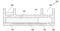
- FIG. 2 is a schematic cross-sectional view of the device according to one aspect of the present invention.
- the device 100 includes a base material 104 and a lid material 101.
- the lid material 101 has a convex portion.
- the convex portion is connected to the base material 104.
- the lid material 101 is provided with a liquid feeding port 102 and a waste liquid port 103 having holes penetrating the lid material 101.
- the substrate 104 has a plurality of wells 105.
- the flow path 106 is located between the lid material 101 and the plurality of wells 105.
- the base material 104 may be formed of a light-transmitting resin.
- the substrate 104 of this embodiment may be substantially transparent.
- the well 105 of the base material 104 is open to the surface of the base material 104.
- the shape, dimensions, and arrangement of the well 105 are not particularly limited.
- a plurality of wells 105 having the same shape and the same size capable of accommodating the reagent liquid 107 (the liquid in which the target nucleic acid is dispersed) are formed on the base material 104.
- particles When particles are used in the detection method according to the present embodiment, they have a shape and dimensions capable of accommodating one or more particles, and have the same shape capable of accommodating a certain amount of reagent solution 107 containing the particles.
- Wells 105 of the same size may be formed on the base material 104.
- the diameter of the well 105 may be 100 nm to 30 ⁇ m, preferably 1 ⁇ m to 15 ⁇ m, and more preferably 3 ⁇ m to 15 ⁇ m.
- the depth of the well may be 100 nm to 30 ⁇ m, preferably 1 ⁇ m to 15 ⁇ m, and more preferably 3 ⁇ m to 15 ⁇ m.
- the diameter of the well may be about 3 ⁇ m, and the depth of the well 105 may be, for example, about 4.5 ⁇ m.
- the wells 105 may be arranged on the base material 104 so as to form a triangular lattice shape or a square lattice shape.
- the number of wells included in the device 100 is preferably 100,000 to 6 million.
- the total volume of the wells is preferably 0.1 to 10 ⁇ L.
- the region of the base material 104 including the plurality of wells 105 is a region filled with the reagent solution 107 to be analyzed. Inside this region, a flow path 106 is provided between the base material 104 and the lid member 101.
- the lid material 101 may be welded or adhered to the base material 104.
- the lid material 101 may be formed of a thermoplastic resin such as a cycloolefin polymer or a cycloolefin copolymer.
- the base material 104 is formed using, for example, a resin.
- the type of resin is not particularly limited, but it is preferable to use a resin that is resistant to a reagent and a sealing liquid for forming droplets. Further, when observing the signal by fluorescence, it is preferable to select a resin that transmits light of the detection wavelength and has less autofluorescence.
- a resin that transmits light of the detection wavelength and has less autofluorescence For example, cycloolefin polymer, cycloolefin copolymer, silicone, polypropylene, polycarbonate, polystyrene, polyethylene, polyvinyl acetate, fluororesin, amorphous fluororesin and the like can be mentioned. Note that these materials shown as examples of the base material 104 are merely examples, and the materials are not limited to these.
- a plurality of wells 105 may be formed on one surface in the plate thickness direction.
- Examples of the forming method using a resin include injection molding, thermal imprinting, and optical imprinting.
- a fluororesin for example, a layer of CYTOP (registered trademark) (Asahi Glass) is provided on the base material 104, and minute holes formed in the CYTOP (registered trademark) are wells 105. You may.
- the lid material 101 is formed so as to have a convex portion on the surface facing the base material 104 at the time of assembly.
- a fluid of a thermoplastic resin may be molded into a plate shape having a convex portion by molding using a molding die.
- the lid material 101 is formed with the liquid feeding port 102 and the waste liquid port 103, but the lid material 101 is not limited to this, and at least one of the liquid feeding port 102 and the waste liquid port 103 is not formed. You may.
- the lid material 101 and the base material 104 are molded as described above, the lid material 101 and the base material 104 are overlapped with each other so that the convex portion of the lid material 101 comes into contact with the surface of the base material 104 on the side where the well 105 opens. Be done. Further, the lid material 101 and the base material 104 are welded by laser welding or the like in a state of being overlapped as described above.
- the device is brought into contact with a liquid containing the target nucleic acid, and the target nucleic acid is introduced into the wells so as to have one molecule or less per well.
- "Introducing" the target nucleic acid into the wells refers to distributing the target nucleic acid to each well of the well array.
- the reagent solution 107 diluted so that one molecule or less of the target nucleic acid per well contains the target nucleic acid in the well 105 of the device 100. (That is, the liquid containing the target nucleic acid) may be fed from the liquid feeding port 102 of the lid material 101 to the flow path 106 between the base material 104 and the lid material 101.
- the reagent liquid 107 sent to the flow path 106 between the base material 104 and the lid material 101 is housed inside the plurality of wells 105.
- the target nucleic acid may be dispersed in the liquid.
- the liquid for dispersing the target nucleic acid a general liquid used in the biochemical analysis performed using the above-mentioned device can be used, and an aqueous solution is preferable.
- the aqueous solution may contain a surfactant or the like to facilitate encapsulation of the liquid in the wells. It may also include reagents and the like necessary for the step of extracting the target nucleic acid and the step of detecting the target nucleic acid, which will be described later.
- the liquid in which the target nucleic acid is dispersed contains an ICA reaction reagent such as an allele probe, an ICA oligo, Flap end-specification-1 (FEN-1), and a fluorescent substrate. You may.
- the means for introducing the target nucleic acid into the well is not particularly limited, and an appropriate means can be selected according to the selected target nucleic acid.
- a substance that captures the target nucleic acid is used to bind the capture to the target nucleic acid that is difficult to settle due to its own weight and send the liquid, or the trap is immobilized in a well in advance and the liquid is sent. It is also possible to improve the introduction efficiency by capturing the target nucleic acid.
- the target nucleic acid is introduced so as to be 1 molecule or less per well. That is, 0 molecule or 1 molecule of target nucleic acid is introduced into one well.
- "Introducing the target nucleic acid so as to be one molecule or less per well” means that one of one molecule of the first nucleic acid, one molecule of the second nucleic acid, and one molecule of the double-stranded nucleic acid is introduced into all the wells. It refers to the state of the well to be introduced and the well in which none of the first nucleic acid, the second nucleic acid and the double-stranded nucleic acid is introduced.
- the target nucleic acid can be detected in units of one (one molecule), that is, digital measurement becomes possible. Also, it is not necessary to introduce the target nucleic acid into all the wells of the well array.
- target nucleic acid examples include DNA, RNA, miRNA, mRNA, artificial nucleic acid and the like.
- the target nucleic acid may be artificially synthesized or isolated from a biological sample.
- biological samples include human cells, blood, lymph, interstitial fluid, body cavity fluid, digestive juice, sweat, tears, runny nose, urine, semen, vaginal fluid, amniotic fluid, milk and cultured cells.
- the length of the target nucleic acid is not particularly limited, but is preferably 10 bases to 1000 bases, and more preferably 30 bases to 300 bases.
- the target nucleic acid is cfDNA in blood, it is preferably 100 to 200 bases.
- the target nucleic acid may be a fragmented nucleic acid chain separated from the biological sample. Fragmentation of the nucleic acid chain can be performed using, for example, a DNA fragmentation apparatus (for example, Kovalis, MS Equipment Co., Ltd.).
- the target nucleic acid may be a nucleic acid sequence known to be associated with a disease.
- it may be a nucleic acid sequence containing a region known to have a mutation in a cancer patient.
- Specific examples thereof include a partial base sequence of the human EGFR (epidermal growth factor receptor) locus, a partial base sequence of the human VEGF (vascular endothelial growth factor) locus, and the like.
- T790M is an example of a mutation occurring in a part of the base sequence of the human EGFR (epidermal growth factor receptor) locus.
- the target nucleic acid includes a first nucleic acid and a second nucleic acid which is a complementary strand of the first nucleic acid.
- the sequence of the first nucleic acid and the sequence of the second nucleic acid may or may not be complementary in all sequences.
- the sequence of the first nucleic acid and the sequence of the second nucleic acid may have a mismatch of 1% to 20% of the bases of the entire base sequence of the longer chain.
- a nucleic acid in which a single-stranded first nucleic acid, a single-stranded second nucleic acid, and a double-stranded first nucleic acid and a second nucleic acid are complementarily bound is contained. Can be included.
- the double-stranded nucleic acid may be completely double-stranded, and some sequences may not be complementary. For example, in a double-stranded nucleic acid, 1% to 20% of bases may be mismatched with respect to the entire base sequence of a longer strand. Further, the double-stranded nucleic acid may be a double-stranded nucleic acid of the same type or a double-stranded nucleic acid of a different type among DNA, RNA, miRNA, mRNA and artificial nucleic acid. Examples of heterologous double strands include double strands of DNA strands and RNA strands, double strands of DNA strands and artificial nucleic acid strands, and the like.
- one molecule of the first nucleic acid refers to one single-stranded first nucleic acid
- one molecule of the second nucleic acid refers to one single-stranded second nucleic acid
- a double-stranded nucleic acid in which one molecule of the first nucleic acid and the second nucleic acid are complementarily bound is one double-stranded nucleic acid in which the first nucleic acid and the second nucleic acid are complementarily bound. Point to.
- the wells are sealed so that the target nucleic acid does not move between the plurality of wells.
- the means for sealing is not particularly limited, and for example, a layer of the sealing liquid is formed on the upper layer of the liquid introduced into the well, the liquid is sealed in the well, and fine droplets of the liquid are formed in the well. You may.
- the liquid feeding port 102 of the lid material 101 is sealed in the flow path 106 between the base material 104 and the lid material 101.
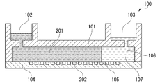
- Stopping liquid 201 for example, oil-based oil is sent to seal the plurality of wells 105 individually.
- the sealing liquid 201 is the reagent liquid 107 that is not contained in the well 105 among the reagent liquid 107 that was sent to the flow path 106 between the base material 104 and the lid material 101 in the above liquid feeding step.
- the sealing liquid 201 individually seals the plurality of wells 105, and the wells 105 become independent reaction spaces (micro-compartments 202).
- Each of the wells 105 after the sealing step contains one molecule or less of the target nucleic acid.
- the double-stranded nucleic acid is sealed in the well in a double-stranded state without being dissociated into a single strand by denaturation.
- the above-mentioned sealing liquid 201 is a liquid capable of forming droplets (microdroplets) by individually sealing the liquids (reagent liquid 107) introduced into a plurality of wells so as not to be mixed with each other.
- an oily solution more preferably an oil.
- an oil a fluorine-based oil, a silicone-based oil, a hydrocarbon-based oil, or a mixture thereof or the like can be used, and for example, the trade name "FC-40" manufactured by Sigma Co., Ltd. can be used.
- the device used in this embodiment does not have to have a flow path as long as the wells can be sealed so that the target nucleic acid does not move between the plurality of wells.
- oil may be dropped from above into each well containing a liquid containing a target nucleic acid to seal the liquid in each well.
- the device used in the present embodiment may be a microwell plate having no lid material and a flow path as long as the target nucleic acid can be prevented from moving between a plurality of wells.
- a liquid containing the target nucleic acid may be introduced into the well, and then the plate member may be brought into close contact with the well to seal the well opening.
- a liquid containing the target nucleic acid is introduced into the wells and then on the well plate in order to prevent the target nucleic acid from moving between the plurality of wells. Excess liquid may be removed. For example, the excess liquid on the well plate may be removed by squeegee or by suction with a suction device.
- the double-stranded nucleic acid present in the individually sealed wells may be denatured.
- the denaturation conditions can be appropriately set depending on the heat resistance of the enzyme used in the signal amplification step.
- the denaturation temperature may be 55 ° C. to 99 ° C., preferably 70 ° C. to 99 ° C.
- the heating time to the denaturation temperature may be 25 seconds to 90 seconds, preferably 30 seconds to 60 seconds.
- the holding time at the denaturation temperature may be 10 seconds to 40 seconds, preferably 30 seconds to 40 seconds.
- the denaturation temperature, the temperature rise time to the denaturation temperature, and the holding time can be arbitrarily combined with the above ranges.
- the double-stranded nucleic acid is denatured by raising the temperature from room temperature (for example, 25 ° C.) to the denaturation temperature of 55 ° C. to 99 ° C. over 25 seconds to 90 seconds and holding the denaturation temperature for 10 seconds to 40 seconds. You may.
- the double-stranded nucleic acid need not be denatured before the signal amplification step.
- the signal caused by the target nucleic acid is amplified in the well, and the signal caused by the target nucleic acid is amplified to a detectable level.
- the reaction for amplifying the signal proceeds in the well 105, and the micro solution 301 that emits the signal is contained in the well 105 containing the target nucleic acid. It will be in a sealed state.
- the signal to be amplified in this step is not particularly limited, and examples thereof include fluorescence, chemiluminescence, color development, potential change, and pH change.
- the signal amplification reaction may be, for example, a biochemical reaction, more specifically, an enzymatic reaction.
- the signal amplification reaction is an isothermal reaction in which a device containing a reagent solution containing an enzyme for signal amplification is maintained in a well under a constant temperature condition at which a desired enzyme activity can be obtained.
- This constant temperature condition is, for example, maintained at 60 ° C. or higher and 99 ° C. or lower, preferably about 66 ° C., for example, for at least 10 minutes, preferably about 15 minutes.
- the signal amplification reaction examples include an ICA reaction such as the Invader (registered trademark) method.
- the signal amplification reaction it is particularly preferable to use the ICA reaction (see, for example, International Publication No. 2009/0544474).
- This is related to the principle of the ICA reaction that signal amplification proceeds by two reaction cycles: (1) complementary binding between nucleic acids and (2) recognition and cleavage of the triple chain structure by an enzyme.
- the influence of reaction cycle inhibition by impurities other than the target nucleic acid is small. Therefore, even when various components other than the target nucleic acid are present in the microsection, the target nucleic acid can be detected accurately by using the ICA reaction.
- the liquid for introducing the target nucleic acid into the well includes the reaction reagent and the target nucleic acid necessary for the ICA reaction.
- the biochemical reaction in the signal amplification step is an ICA reaction
- the fluorescent substance is released from the quenching substance when the target nucleic acid is present in the well due to the enzymatic reaction by the isothermal reaction, so that the fluorescent substance is determined in response to the excitation light. Fluorescent signal is emitted.
- the detection of the target nucleic acid can also be performed by binding a substance that specifically binds to the target nucleic acid (specific binding substance) to the target nucleic acid and detecting the bound specific binding substance.
- a substance that specifically binds to the target nucleic acid specifically binding substance
- the specific binding substance the same substance as the specific binding substance for the target nucleic acid described later, for example, an antibody, an antibody fragment, a polypeptide, an aptamer, or the like can be used.
- the signal amplification step is an ICA reaction
- a flap probe, an invasion probe, or the like can be used as the specific binding substance.
- the flap probe and the invading probe those prepared by using a known method can be used so that the first nucleic acid and the second nucleic acid can be recognized.
- the difference between the Tm (also referred to as melting temperature) between the first specific binding substance and the first nucleic acid and the Tm between the second specific binding substance and the second nucleic acid is preferably 10 ° C. or less.
- the difference between the Tm of the first specific binding substance and the first nucleic acid and the Tm of the second specific binding substance and the second nucleic acid is 10 ° C. or less, the first nucleic acid is subjected to an isothermal reaction such as an ICA reaction. And the second nucleic acid can both be detected.
- the first signal caused by the first nucleic acid and the second signal caused by the second nucleic acid are amplified.
- the first signal and the second signal are different. More specifically, when the biochemical reaction in the signal amplification step is an ICA reaction, the flap probe and the invading probe for the first nucleic acid are generated by the dissociation of the single-stranded first nucleic acid or the double-stranded nucleic acid. 1 Binds to nucleic acid.
- the flap probe and the invading probe for the second nucleic acid bind to the second nucleic acid generated by the dissociation of the single-stranded second nucleic acid and the double-stranded nucleic acid.
- the first signal and the second signal may be luminescence signals.
- the wavelength of the first signal and the wavelength of the second signal are different.
- the signal amplification step is an ICA reaction
- the wavelength of the first signal and the wavelength of the second signal are made different by using two kinds of fluorescent substances that emit different fluorescence from each other. Can be done. That is, when the first fluorescent substance for detecting the first nucleic acid and the second fluorescent substance for detecting the second nucleic acid that emits fluorescence having a wavelength different from that of the first fluorescent substance are used, the first signal and the first signal are used. Each of the two signals can be amplified.
- the signal amplified in the signal amplification step is detected.
- the signal detection method a known appropriate method can be selected depending on the type of signal to be detected. For example, in the case of performing detection in a bright field, for example, white light is irradiated in the direction perpendicular to the base material provided with the well array.
- the fluorescence signal for example, the excitation light corresponding to the fluorescent substance is irradiated into the well from the bottom side of the well, and the fluorescence emitted by the fluorescent substance is detected.
- an image of the whole or a part of the well array may be taken and stored, and image processing by a computer system may be performed.
- the target nucleic acid contained in each well is only one molecule of the first nucleic acid, only one molecule of the second nucleic acid, or the first nucleic acid and the second nucleic acid are complementary. Either only one molecule of double-stranded nucleic acid bound specifically. There may also be wells that do not contain the target nucleic acid.
- the detection method according to the present embodiment may further include a counting step of counting the number of wells in which a signal is detected.
- the counting step the number of wells in which only the first signal is detected, the number of wells in which only the second signal is detected, and the number of wells in which both the first signal and the second signal are detected are counted. You may.
- the number of wells in which only the single-stranded first nucleic acid was present, the number of wells in which only the single-stranded second nucleic acid was present, and the first nucleic acid and the second nucleic acid are complementary.
- the number of wells in which the double-stranded nucleic acid bound to is present can be calculated respectively.
- the first nucleic acid The ratio of double-stranded nucleic acid in which and the second nucleic acid are complementarily bound can be calculated.
- an integrated device including a device into which the target nucleic acid is introduced, a light source used in the detection step, and a detector for detecting a signal is used. May be good.
- This device may further have a processing unit that processes an image of a detection signal or the like and calculates the ratio of the target nucleic acid described above.
- the method for detecting a target nucleic acid in a liquid includes a storage device on which a device into which the target nucleic acid is introduced is supported, a light source device used in the detection step, and a detection device for detecting a detection signal.
- a system may be used. This system may further have a processing apparatus that processes an image of a detection signal or the like and calculates the ratio of the target nucleic acid described above.
- the present invention brings a liquid containing a target nucleic acid into contact with a device having a well array having a plurality of wells, and introduces the target nucleic acid into the wells so as to have one molecule or less per well.
- the target nucleic acid is a first nucleic acid, a second nucleic acid which is a complementary strand of the first nucleic acid, and the first nucleic acid and the second nucleic acid are complementarily bound to each other.
- the double-stranded nucleic acid is sealed in the well in a double-stranded state in the sealing step, and in the step of amplifying the signal, the first nucleic acid is first.
- a detection method in which a specific binding substance binds and a first signal is emitted.
- each well contains the first nucleic acid or contains the first nucleic acid by detecting the signal caused by the first nucleic acid. It is possible to distinguish whether or not it is.
- the well containing the first nucleic acid either contains a single-stranded first nucleic acid or contains a double strand in which the first nucleic acid and the second nucleic acid are complementarily bound. is there.
- a first nucleic acid, a second nucleic acid which is a complementary strand of the first nucleic acid, and the first nucleic acid and the second nucleic acid are complementarily bound to a device having a well array having a plurality of wells.
- a step of sealing the well a step of modifying the double-stranded nucleic acid after the sealing step, a step of amplifying a signal caused by the target nucleic acid in the well, and a step of being emitted from the well.
- the step of detecting the signal the double-stranded nucleic acid is sealed in the well in a double-stranded state in the sealing step, and in the step of amplifying the signal, the first The first specific binding substance binds to the nucleic acid to emit a first signal, and the second specific binding substance binds to the second nucleic acid to emit a second signal.
- a detection method that is different from the second signal.
- the detection step it is determined that only the single-stranded first nucleic acid was present in the well in which only the first signal was detected, and the well in which only the second signal was detected was detected. It was determined that only the single-stranded nucleic acid was present in the well, and it was determined that the double-stranded nucleic acid was present in the well in which both the first signal and the second signal were detected.
- the detection method according to any one of [1] to [3]. [22] The detection method according to any one of [1] to [4], further comprising a step of counting the number of the wells in which the signal is detected in the detection step. [23] In the counting step, the number of the wells in which only the first signal is detected, the number of wells in which only the second signal is detected, and the first signal and the second signal are both detected. The detection method according to [5], wherein the number of wells is counted. [24] From the number of the wells in which only the first signal is detected and the number of wells in which both the first signal and the second signal are detected, the single-stranded first signal in the liquid is used.
- the difference between the melting temperature Tm of the first specific binding substance and the first nucleic acid and the melting temperature Tm of the second specific binding substance and the second nucleic acid is 10 ° C. or less.
- the step of amplifying the signal is an isothermal reaction.
- the detection method according to any one of [18] to [26], wherein the step of denaturing and the step of amplifying the signal are performed at the same time.
- Example 1 (Detection of intracellular nucleic acid by ICA method) Quantitative detection of the target nucleic acid was performed using a region containing a part of the base sequence of the human EGFR (epidermal growth factor receptor) locus as the target nucleic acid.
- human EGFR epidermal growth factor receptor
- some regions of human EGFR are regions known to have mutations in some cancer patients.
- the sense strand (SEQ ID NO: 7) at the EGFR locus was used as the target nucleic acid.
- Genomic DNA is separated from cultured human cells (HT29) using a DNA extraction kit (AllPrep, QIAGEN), and a DNA fragmentation device (Kovalis, MS Equipment Co., Ltd.) is used to serve as a simulated sample of circulating DNA in the blood.
- the DNA separated using the above was fragmented to prepare a DNA sample of Preparation Example 1.
- the DNA sample was heat-denatured at 95 ° C. for 10 minutes and then rapidly cooled to form a single strand. Almost all DNA samples immediately after quenching are in a single-stranded state. Subsequently, the absorbance of the DNA sample after quenching was measured using an ultratrace spectrometer (Nanodrop, Thermo Fisher Scientific Co., Ltd.), and it was confirmed that the concentration in the single-stranded state was 1.1 fM. That is, the concentration was 0.55 fM when all the DNA samples were double-stranded nucleic acids.
- the concentration of the target nucleic acid in the single-stranded state means the concentration of the total number of the number of sense strands and the number of antisense strands at the EGFR locus.
- the concentration of the target nucleic acid in the double-stranded state means the concentration of the number of sets consisting of the sense strand and the antisense strand at the EGFR locus.
- the mass volume concentration of DNA was calculated from the measured value of the absorbance of the nanodrop. Subsequently, assuming that the 3 pg DNA sample contains the sense strand of one EGFR locus and the antisense strand of one EGFR locus, the single-stranded target nucleic acid contained in the DNA sample after quenching. The concentration of was calculated.
- ICA reaction reagent was prepared as a nucleic acid detection reagent.
- the ICA reaction reagents in this example are 0.5 ⁇ M allerprobe 1 (SEQ ID NO: 1) (flap probe), 0.1 ⁇ M invader oligo 1 (SEQ ID NO: 2) (also referred to as invasion probe and ICA oligo) (these are Fasmac).
- ICA reaction reagents 4 ⁇ M FRET Cassette (Alexa488-BHQ) (SEQ ID NO: 3) (Japan Bioservices) (fluorescent substrate), 50 mM Tris-HCl (pH 7.9), 20 mM MgCl 2 and 0.05 mg / mL FEN-1. I'm out.
- concentration of each component in these ICA reaction reagents is the final concentration in the mixture of the ICA reaction reagent and the DNA sample according to Experimental Example 1. Aller probe 1 and invader oligo 1 are used in the ICA reaction, and both specifically recognize one of the nucleotide strands of the sense strand and the antisense strand of the EGFR locus.
- ⁇ Device preparation> A base material provided with a large number of minute wells was prepared of COP (cycloolefin polymer), and a lid material made of COP was attached to fabricate the device.
- the total volume of wells per cm 2 was 0.93 ⁇ L.
- the total number of wells used for the measurement was 1,000,000.
- ⁇ Sending of reaction mixture solution 8 ⁇ l of a solution prepared by mixing the DNA sample of Preparation Example 1 and the above ICA reaction reagent was sent to the device and introduced into each well. Subsequently, 200 ⁇ l of FC-40 (Sigma) was sent as a sealing liquid to seal each well.
- a single-stranded first nucleic acid to which the allergic probe 1 complementarily binds a single-stranded second nucleic acid that is a complementary strand of the first nucleic acid, and a first nucleic acid. Either it contains only one selected from the double strands to which the second nucleic acid is complementarily bound, or it does not contain the target nucleic acid.
- the concentration of DNA that is complementary to the allelic probe and contains a single nucleotide polymorphism can be calculated as follows.
- the liquid introduced into the well of the device is a solution obtained by diluting the sample measured using Nanodrop 20-fold.
- one molecule or less of DNA per well was sealed in the well. That is, in each well, as the target nucleic acid, the first nucleic acid, the single-stranded second nucleic acid which is the complementary strand of the first nucleic acid, and the double strand in which the first nucleic acid and the second nucleic acid are complementaryly bound. Either it contained only one selected from, or it did not contain the target nucleic acid.
- a well containing only a single-stranded first nucleic acid, a well containing only a single-stranded second nucleic acid which is a complementary strand of the first nucleic acid, and a well containing only the single-stranded second nucleic acid, and the first nucleic acid and the second nucleic acid are complementary. Signals are detected in all wells containing only double strands bound to.
- the concentration of the single-stranded first nucleic acid contained in the DNA sample of Preparation Example 1, the single-stranded second nucleic acid, and the single-stranded first nucleic acid and the second nucleic acid were found.
- the sum with the concentration of the complementaryly bound double strand was calculated to be 1 fM.
- Example 2 Quantitative detection of the target nucleic acid was performed using the sense strand of the EGFR locus described in Experimental Example 1 and the antisense strand of one EGFR locus as the target nucleic acids.
- Genomic DNA was extracted from cultured human cells (HT29) using a DN extraction kit (AllPrep, QIAGEN). Using a service commissioned by MS Equipment Co., Ltd. (DNA Sharing Service), the extracted genomic DNA was crushed to 150 bp. After evaporating the liquid of 1.3 ml of the crushed genomic DNA solution using a centrifugal dryer (DNA Speedback, Thermo Fisher Scientific), 100 ⁇ l of distilled water was added to prepare a DNA sample of Preparation Example 2.
- the absorbance of the DNA sample was measured and it was confirmed that the concentration in the single-stranded state was 1.0 fM. That is, the concentration was 0.5 fM when all the DNA samples were double-stranded nucleic acids.
- ICA reaction reagent was prepared as a nucleic acid detection reagent.
- the ICA reaction reagents in this example are 0.5 ⁇ M aller probe 1 (SEQ ID NO: 1) (flap probe), 0.1 ⁇ M invader oligo 1 (SEQ ID NO: 2) (also referred to as invasion probe and ICA oligo), and 0.2 ⁇ M allele.
- Probe 2 (SEQ ID NO: 4) (Flap probe), 5 nM Invader Oligo 2 (SEQ ID NO: 5) (Invasion probe) (Fasmac), 4 ⁇ M FRET Cassette (Alexa488-BHQ) (SEQ ID NO: 3) (Japan Bioservices) ) (Fluorescent substrate), 2 ⁇ M FRET Cassette (Redmond Red-Epoch Eclipse Quencher) (SEQ ID NO: 6) (Tsukuba Oligo Service) (Fluorescent substrate) 50 mM Tris-HCl (pH 7.9), 20 mM MgCl 2 and 0.05 mg / Contains mL FEN-1.
- the concentration of each component in these ICA reaction reagents is the final concentration in the mixture of the ICA reaction reagent and the DNA sample according to Experimental Example 2.
- the allerprobe 1, invader oligo 1, allerprobe 2 and invader oligo 2 are used in the ICA reaction. Aller probe 1 and invader oligo 1 specifically recognize one nucleotide strand of the sense strand and antisense strand of the EGFR locus, and aller probe 2 and invader oligo 2 specifically recognize the sense strand and antisense strand of the EGFR locus. Specific recognition of the other nucleotide strand of.
- the well in which only the fluorescent signal from Alexa488 was detected indicates that the well has only one of the sense strand and the antisense strand at the EGFR locus.
- the well in which only the fluorescent signal from Redmond Red is detected indicates that the well has only the other of the sense strand and the antisense strand of the EGFR locus.
- the wells in which the fluorescence signals of both Alexa488 and Redmond Red were detected indicate that the wells in which the double-stranded nucleic acids of the sense strand and the antisense strand of the EGFR locus are present at the time of being sealed in the wells.
Landscapes
- Chemical & Material Sciences (AREA)
- Life Sciences & Earth Sciences (AREA)
- Organic Chemistry (AREA)
- Engineering & Computer Science (AREA)
- Zoology (AREA)
- Wood Science & Technology (AREA)
- Proteomics, Peptides & Aminoacids (AREA)
- Health & Medical Sciences (AREA)
- Biophysics (AREA)
- General Engineering & Computer Science (AREA)
- Immunology (AREA)
- Microbiology (AREA)
- Molecular Biology (AREA)
- Analytical Chemistry (AREA)
- Physics & Mathematics (AREA)
- Genetics & Genomics (AREA)
- Biochemistry (AREA)
- Bioinformatics & Cheminformatics (AREA)
- Biotechnology (AREA)
- General Health & Medical Sciences (AREA)
- Chemical Kinetics & Catalysis (AREA)
- Measuring Or Testing Involving Enzymes Or Micro-Organisms (AREA)
- Apparatus Associated With Microorganisms And Enzymes (AREA)
Abstract
この検出方法は、複数のウェルを有するデバイスのウェル内に標的核酸を1ウェルあたり1分子以下となるように導入する工程と、標的核酸が複数のウェル間で移動しないようにウェルを封止する工程と、ウェル内で標的核酸に起因するシグナルを増幅する工程と、ウェルから発せられるシグナルを検出する工程と、を有し、標的核酸が、第1核酸と、第1核酸の相補鎖である第2核酸と第1核酸と第2核酸とが相補的に結合した2本鎖核酸を含み、封止する工程において2本鎖核酸が、2本鎖の状態でウェル内に封止され、シグナルを増幅する工程において、第1核酸に第1の特異的結合物質が結合して第1シグナルが発せられ、第2核酸に第2の特異的結合物質が結合して第2シグナルが発せられ、第1シグナルと、第2シグナルとは異なるものである。
Description
本発明は、検出方法に関する。
本願は、2019年12月9日に日本に出願された特願2019-222153号について優先権を主張し、その内容をここに援用する。
本願は、2019年12月9日に日本に出願された特願2019-222153号について優先権を主張し、その内容をここに援用する。
生体試料中の標的分子を定量的に検出することにより、疾患の早期発見や投薬の効果予測が行われている。DNAやRNAなどの核酸の定量は、リアルタイムPCR法等により行われている。
近年、疾患をより早期に発見する等の目的で、標的分子をより精度よく検出するニーズが高まっている。標的分子を精度よく検出する手法として、例えば、非特許文献1には、多数の微小区画内で酵素反応を行い、蛍光シグナルを検出する技術が記載されている。この手法は、デジタル計測と呼ばれている。
デジタル計測では、試料溶液を極めて多数の微小溶液に分割する。そして、各微小溶液からの信号を2値化し、標的分子が存在するか否かのみを判別して、標的分子数を計測する。デジタル計測によれば、従来のリアルタイムPCR法等と比較して、検出感度及び定量性を格段に向上させることができる。
デジタル計測の1種であるデジタルPCRでは、微小区画内の1つの微小液滴に存在する鋳型となる核酸が0個ないし1個になるように、PCR反応試薬と核酸との混合物が希釈されている。デジタルPCRでは、核酸増幅の感度を高めるために、また多数の微小液滴に対して同時に核酸増幅を行うために、各微小液滴の体積は小さい方が好ましい。例えば、非特許文献1には、各ウェルの容積が数ナノリットルとなるように形成されたマイクロサイズの液滴を用いた方法が開示されている。
また、デジタル計測の別の手法として、デジタルInvasive Cleavage Assay(ICA)がある。特許文献1には、PCR反応によりDNAサンプルを増幅させ、変性させたPCR産物をマイクロウェルを有するデバイスにDNAを含むサンプルを導入し、検出時にはDNAを増幅せずにインベーダー法によってDNAを検出する手法が開示されている。
Olmedillas-Lopez S., et al., Current and Emerging Applications of Droplet Digital PCR in Oncology. Mol Diagn Ther. 2017 Oct;21(5):493-510
図1は、従来の検出方法であるデジタルPCRにより標的核酸を検出する方法を模式的に示した図である。デジタルPCRにより標的核酸を検出する場合、標的核酸を検出する工程において標的核酸が増幅される。そのため、図1に例示するように、標的核酸は、増幅工程を経ずに微小区画内に収容される。血液や細胞等の生体試料から精製されたDNAやRNAである標的核酸は、1本鎖の核酸(これを1本鎖核酸1とする)と、1本鎖核酸1と相補的な1本鎖の核酸(これを1本鎖核酸2とする)と、1本鎖核酸1と1本鎖核酸2が相補的に結合している2本鎖の核酸(これを2本鎖核酸3とする)を含んでいる。
例えば、図1に例示するように、液滴化する前の試料溶液は、1本鎖核酸1を2分子、1本鎖核酸2を2分子、2本鎖核酸3を4分子含んでいる。例えば、従来の検出方法であるデジタルPCRを用いて標的核酸を検出する場合、液滴の状態では、例えば、液滴4、液滴5及び液滴6のように、1本鎖と2本鎖とを区別せずに閉じ込めることになる。液滴4、液滴5及び液滴6の標的核酸のPCR産物はほぼ同一のものとなる。つまり図1に例示するように、デジタルPCRにおいては、液滴7のように8個の検出シグナルが検出される。この検出結果からは、試料溶液中には、例えば、1本鎖核酸1が8分子あったのか、1本鎖核酸2が8分子あったのか、2本鎖核酸3が8分子あったのか、を区別することができない。すなわち、デジタルPCR法では、溶液中の、1本鎖核酸1と、1本鎖核酸2と、2本鎖核酸3とを区別して定量することができない。
本発明は、上述した事情に鑑みてなされたものであって、1本鎖の核酸と、2本鎖の核酸とを、より高精度に区別して定量できる技術を提供することを目的とする。
本発明は、以下の態様を含む。
[1]複数のウェルを有するウェルアレイを有するデバイスに、標的核酸を含む液体を接触させ、前記ウェル内に前記標的核酸を1ウェルあたり1分子以下となるように導入する工程と、前記標的核酸が複数の前記ウェル間で移動しないように前記ウェルを封止する工程と、前記ウェル内で前記標的核酸に起因するシグナルを増幅する工程と、前記ウェルから発せられる前記シグナルを検出する工程と、を有し、前記標的核酸が、第1核酸と、前記第1核酸の相補鎖である第2核酸と、前記第1核酸と前記第2核酸とが相補的に結合した2本鎖核酸を含み、前記封止する工程において前記2本鎖核酸が、2本鎖の状態で前記ウェル内に封止され、前記シグナルを増幅する工程において、前記第1核酸に第1の特異的結合物質が結合して第1シグナルが発せられ、前記第2核酸に第2の特異的結合物質が結合して第2シグナルが発せられ、前記第1シグナルと、前記第2シグナルとは異なるものである、検出方法。
[2]前記第1シグナル及び前記第2シグナルは発光シグナルであり、前記第1シグナルの波長と、前記第2シグナルの波長とが異なる、[1]に記載の検出方法。
[3]前記シグナルを増幅する工程が、インベーシブ・クリベージ・アッセイによって行われる、[1]または[2]に記載の検出方法。
[4]前記検出する工程において、前記第1シグナルのみを検出した前記ウェルには、1本鎖状の前記第1核酸のみが存在していたと判定し、前記第2シグナルのみを検出した前記ウェルには、1本鎖状の前記第2核酸のみが存在していたと判定し、前記第1シグナル及び前記第2シグナルを共に検出した前記ウェルには、前記2本鎖核酸が存在していたと判定する、[1]~[3]のいずれかに記載の検出方法。
[5]前記検出する工程において前記シグナルが検出された前記ウェルの数を数える工程を更に有する、[1]~[4]のいずれかに記載の検出方法。
[6]前記数える工程において、前記第1シグナルのみを検出した前記ウェルの数、前記第2シグナルのみを検出した前記ウェルの数、及び、前記第1シグナル及び前記第2シグナルを共に検出した前記ウェルの数、をそれぞれ数える、[5]に記載の検出方法。
[7]前記第1シグナルのみを検出した前記ウェルの数と、前記第1シグナル及び前記第2シグナルを共に検出した前記ウェルの数とから、前記液体中の、1本鎖状の前記第1核酸、及び、前記2本鎖核酸の総数に対する、前記2本鎖核酸の数の割合を算出する、[6]に記載の検出方法。
[8]前記第1の特異的結合物質と前記第1核酸との融解温度Tmと前記第2の特異的結合物質と前記第2核酸との融解温度Tmとの差は、10℃以下である、[1]~[7]のいずれかに記載の検出方法。
[9]複数のウェルを有するウェルアレイを有するデバイスに、標的核酸を含む液体を接触させ、前記ウェル内に前記標的核酸を1ウェルあたり1分子以下となるように導入する工程と、前記標的核酸が複数の前記ウェル間で移動しないように前記ウェルを封止する工程と、前記ウェル内で前記標的核酸に起因するシグナルを増幅する工程と、前記ウェルから発せられるシグナルを検出する工程と、を有し、前記標的核酸が、第1核酸と、前記第1核酸の相補鎖である第2核酸と、前記第1核酸と前記第2核酸とが相補的に結合した2本鎖核酸を含み、前記封止する工程において前記2本鎖核酸が、2本鎖の状態で前記ウェル内に封止され、前記シグナルを増幅する工程において、前記第1核酸に第1の特異的結合物質が結合して第1シグナルが発せられる、検出方法。
本発明は、もう一つの側面として以下の態様を含む。
[10]液体中の標的核酸の検出方法であって、複数のウェルを有するウェルアレイを有するデバイスに、前記液体を接触させ、前記ウェル内に前記標的核酸を1ウェルあたり1分子以下となるように導入する導入工程と、前記標的核酸が複数の前記ウェル間で移動しないように前記ウェルを封止する封止工程と、前記ウェル内で前記標的核酸に起因するシグナルの増幅を行うシグナル増幅工程と、前記ウェルから発せられるシグナルを検出する検出工程と、を有し、前記標的核酸が、第1核酸と、前記第1核酸の相補鎖である第2核酸とを含み、前記シグナル増幅工程において、前記第1核酸に第1の特異的結合物質が結合して第1シグナルが発せられ、前記第2核酸に第2の特異的結合物質が結合して第2シグナルが発せられ、前記第1シグナルと、前記第2シグナルとは異なるものである、検出方法。
[11]前記第1シグナル及び前記第2シグナルは発光シグナルであり、前記第1シグナルの波長と、前記第2シグナルの波長とが異なる、[10]に記載の検出方法。
[12]前記シグナル増幅工程が、インベーシブ・クリベージ・アッセイによって行われる、[10]または[11]に記載の検出方法。
[13]前記検出工程において、前記第1シグナルのみを検出した前記ウェルには、1本鎖状の前記第1核酸のみが存在していたと判定し、前記第2シグナルのみを検出した前記ウェルには、1本鎖状の前記第2核酸のみが存在していたと判定し、前記第1シグナル及び前記第2シグナルを共に検出した前記ウェルには、前記第1核酸と前記第2核酸とが相補的に結合した2本鎖が存在していたと判定する、[10]~[12]のいずれかに記載の検出方法。
[14]前記検出工程において前記シグナルが検出された前記ウェルの数を数える計数工程を更に有する、[10]~[13]のいずれかに記載の検出方法。
[15]前記計数工程において、前記第1シグナルのみを検出した前記ウェルの数、前記第2シグナルのみを検出した前記ウェルの数、及び、前記第1シグナル及び前記第2シグナルを共に検出した前記ウェル、をそれぞれ数える、[14]に記載の検出方法。
[16]前記第1シグナルのみを検出した前記ウェルの数と、前記第1シグナル及び前記第2シグナルを共に検出した前記ウェルの数とから、前記液体中の、1本鎖状の前記第1核酸、及び、前記第1核酸と前記第2核酸とが相補的に結合した2本鎖のうち、前記第1核酸と前記第2核酸とが相補的に結合した2本鎖の割合を算出する、[15]に記載の検出方法。
[17]液体中の標的核酸の検出方法であって、複数のウェルを有するウェルアレイを有するデバイスに、前記液体を接触させ、前記ウェル内に前記標的核酸を1ウェルあたり1分子以下となるように導入する導入工程と、前記標的核酸が複数の前記ウェル間で移動しないように前記ウェルを封止する封止工程と、前記ウェル内で前記標的核酸に起因するシグナルの増幅を行うシグナル増幅工程と、前記ウェルから発せられるシグナルを検出する検出工程と、を有し、前記標的核酸が、第1核酸と、前記第1核酸の相補鎖である第2核酸とを含み、前記シグナル増幅工程において、前記第1核酸に第1の特異的結合物質が結合して第1シグナルが発せられる、検出方法。
本発明によれば、1本鎖の核酸と、2本鎖の核酸とを、より高精度に区別して定量できる技術を提供することができる。
以下、場合により図面を参照しつつ、本発明の実施形態について詳細に説明する。なお、図面中、同一又は相当部分には同一又は対応する符号を付し、重複する説明は省略する。なお、各図における寸法比は、説明のため誇張している部分があり、必ずしも実際の寸法比とは一致しない。
[検出方法]
本発明は、一実施形態において、複数のウェルを有するウェルアレイを有するデバイスに、標的核酸を含む液体を接触させ、ウェル内に標的核酸を1ウェルあたり1分子以下となるように導入する工程と、標的核酸が複数のウェル間で移動しないようにウェルを封止する工程と、ウェル内で標的核酸に起因するシグナルを増幅する工程と、ウェルから発せられるシグナルを検出する工程と、を有し、標的核酸が、第1核酸と、第1核酸の相補鎖である第2核酸と、前記第1核酸と前記第2核酸とが相補的に結合した2本鎖である2本鎖核酸を含み、前記封止する工程において前記2本鎖核酸が、2本鎖の状態で前記ウェル内に封止され、シグナル増幅工程において、第1核酸に第1の特異的結合物質が結合して第1シグナルが発せられ、第2核酸に第2特異的結合物質が結合して第2シグナルが発せられ、第1シグナルと、第2シグナルとは異なるものである、検出方法を提供する。
本発明は、一実施形態において、複数のウェルを有するウェルアレイを有するデバイスに、標的核酸を含む液体を接触させ、ウェル内に標的核酸を1ウェルあたり1分子以下となるように導入する工程と、標的核酸が複数のウェル間で移動しないようにウェルを封止する工程と、ウェル内で標的核酸に起因するシグナルを増幅する工程と、ウェルから発せられるシグナルを検出する工程と、を有し、標的核酸が、第1核酸と、第1核酸の相補鎖である第2核酸と、前記第1核酸と前記第2核酸とが相補的に結合した2本鎖である2本鎖核酸を含み、前記封止する工程において前記2本鎖核酸が、2本鎖の状態で前記ウェル内に封止され、シグナル増幅工程において、第1核酸に第1の特異的結合物質が結合して第1シグナルが発せられ、第2核酸に第2特異的結合物質が結合して第2シグナルが発せられ、第1シグナルと、第2シグナルとは異なるものである、検出方法を提供する。
本実施形態に係る検出方法によれば、後述するように、標的核酸に起因するシグナルを検出することにより、各ウェルに、第1核酸のみが含まれているか、第2核酸のみが含まれているか、第1核酸と第2核酸が相補的に結合している2本鎖のみが含まれているか、を区別することができる。その結果、液体中に含まれている、第1核酸、第2核酸、及び第1核酸と第2核酸が相補的に結合している2本鎖核酸のそれぞれ分子数をより正確に定量することができる。
例えば、標的核酸が生体試料に由来する場合、第1核酸、第2核酸、及び2本鎖核酸のそれぞれの分子数を正確に定量することにより、癌のステータス、及び遺伝子変異を伴う疾病等に関する新たな知見が得られる可能性がある。
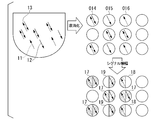
より具体的には、図5に例示するように、標的核酸を含む液体は、第1核酸11と、第1核酸11と相補的な1本鎖の第2核酸12と、第1核酸11と第2核酸12が相補的に結合している2本鎖の2本鎖核酸13を含んでいる。液滴化する前の試料溶液は、1本鎖の第1核酸11を2分子、1本鎖の第2核酸12を2分子、2本鎖核酸13を4分子含んでいる。例えば、本実施形態に係る検出方法によれば、導入工程、封止工程により、例えば、液滴14には2本鎖核酸13が閉じ込められ、液滴15には第2核酸12が閉じ込められ、液滴16には第1核酸11が閉じ込められる。
続いて、シグナル増幅工程において、液滴18からは第1シグナルが発せられ、液滴19からは第2シグナルが発せられ、液滴17からは第1シグナル及び第2シグナルが共に発せられる。ここで、第1シグナルと第2シグナルとは異なるものであるため、検出工程において、液滴17と、液滴18と、液滴19とを区別することができる。すなわち、本実施形態に係る検出方法によれば、各々の液滴に含まれていた、第1核酸11と、第2核酸12と、2本鎖核酸13とを区別することができる。その結果、標的核酸を含む液体中には、2分子の第1核酸11と、2分子の第2核酸12と、4分子の2本鎖核酸13が含まれていたと判定することができる。
(デバイス)
本実施形態に係る検出方法において、複数のウェルを有するウェルアレイを有するデバイスを用いる。デバイスとしては、例えば、次に挙げるようなものを用いることができるが、これに限定されない。
本実施形態に係る検出方法において、複数のウェルを有するウェルアレイを有するデバイスを用いる。デバイスとしては、例えば、次に挙げるようなものを用いることができるが、これに限定されない。
ウェルの形状、寸法、及び配置は特に限定されないが、本発明の方法において用いられる標的核酸を含む液体、及び、検出工程で使用する一定量の試薬液等を収容可能なウェルから成るウェルアレイを使用することが好ましい。ウェルは、無処理でそのまま使用してもよいし、目的に応じて、予めウェル内壁に抽出試薬、抗体等の検出試薬及び特異的結合物質等の少なくとも一つを固定化してもよい。また、ウェル開口部を脂質二重膜で覆ったりする等の前処理を施してもよい。
デバイスは、流路を有していてもよく、流路を介して標的核酸が分散した液体を送液してもよい。流路の形状、構造及び容量等は特に限定されないが、流路を有するデバイスを使用することが好ましい。このようなデバイスを使用すると、標的核酸を含む液体を送液した際にウェルアレイの各ウェルに標的核酸が導入され、かつ封止液を流路に挿入した際に各ウェルが個別に封止され微小液滴を形成することができる。
(デバイスの一例)
図2は、本発明の一態様におけるデバイスの模式断面図である。図2に示すように、デバイス100は、基材104と蓋材101とを備えている。蓋材101は、凸部を有している。凸部は、基材104と接続している。蓋材101には、蓋材101を貫通する孔を有する送液ポート102及び廃液ポート103が設けられている。基材104は、複数のウェル105を有する。蓋材101と、複数のウェル105との間に流路106が位置している。
図2は、本発明の一態様におけるデバイスの模式断面図である。図2に示すように、デバイス100は、基材104と蓋材101とを備えている。蓋材101は、凸部を有している。凸部は、基材104と接続している。蓋材101には、蓋材101を貫通する孔を有する送液ポート102及び廃液ポート103が設けられている。基材104は、複数のウェル105を有する。蓋材101と、複数のウェル105との間に流路106が位置している。
基材104は、光透過性樹脂から形成されていてよい。本実施形態の基材104は、実質的に透明であってもよい。
基材104のウェル105は、基材104の表面に開口している。ウェル105の形状、寸法、および配置は特に限定されない。図2に示す例では、デバイス100において、試薬液107(標的核酸が分散した液体)を収容可能な同形同大の複数のウェル105が基材104に形成されている。また、本実施形態に係る検出方法において粒子が使用される場合には、粒子を1つ以上収容可能な形状及び寸法を有し、粒子を含んだ一定量の試薬液107を収容可能な同形同大のウェル105が基材104に形成されていてもよい。
デバイス100では、ウェル105の直径は、100nm~30μmであってよく、1μm~15μmが好ましく、3μm~15μmであることがより好ましい。ウェルの深さは、100nm~30μmであってよく、1μm~15μmが好ましく、3μm~15μmであることがより好ましい。一例として、ウェルの直径は3μm程度であってもよく、ウェル105の深さは例えば4.5μm程度であってもよい。また、ウェル105は、三角格子状又は正方格子状を形成するように整列して基材104に形成されていてもよい。
デバイス100が有するウェルの個数は、10万個~600万個であることが好ましい。また、ウェルの総容量は0.1~10μLであることが好ましい。
基材104の、複数のウェル105を含んだ領域は、分析対象となる試薬液107が充填される領域である。この領域の内側では、基材104と蓋材101との間に流路106が設けられている。
蓋材101は、基材104に対して溶着または接着されていてもよい。例えば、蓋材101は、シクロオレフィンポリマー又はシクロオレフィンコポリマーなどの熱可塑性樹脂から形成されていてもよい。
基材104は、例えば樹脂を用いて形成される。樹脂の種類は特に限定されないが、試薬及び液滴を形成する際の封止液に対し耐性のあるものを用いることが好ましい。また、シグナルを蛍光観察する場合には、検出波長の光を透過し、自家蛍光の少ない樹脂を選ぶことが好ましい。例えば、シクロオレフィンポリマー、シクロオレフィンコポリマー、シリコーン、ポリプロピレン、ポリカーボネート、ポリスチレン、ポリエチレン、ポリ酢酸ビニル、フッ素樹脂及びアモルファスフッ素樹脂等が挙げられる。なお、基材104の例として示されたこれらの材質はあくまでも例であり、材質はこれらには限られない。
基材104に対しては、板厚方向の一方の面に複数のウェル105が形成されていてもよい。樹脂を用いた形成方法としては、射出成形のほか、熱インプリントや、光インプリントなどが挙げられる。また、フッ素樹脂を用いる場合には、例えば、基材104の上にCYTOP(登録商標)(旭硝子)の層を有し、CYTOP(登録商標)に形成された微小な孔がウェル105となっていてもよい。
蓋材101は、組立時に基材104に向けられる面に凸部を有するように成形される。例えば熱可塑性樹脂の流動体を、成形型を用いて成形することで、凸部を有する板状に成形してもよい。図2に示すデバイス100では、蓋材101には送液ポート102および廃液ポート103が形成されているが、これに限定されず、送液ポート102及び廃液ポート103の少なくとも一方が形成されていなくてもよい。
蓋材101および基材104が上記のように成形されたら、基材104においてウェル105が開口する側の面に蓋材101の凸部が接するように、蓋材101と基材104とが重ねられる。さらに、蓋材101と基材104とが上記のように重ねられた状態で、レーザー溶着等により溶着される。
以下、各工程について詳細に説明する。
(導入工程)
本工程において、デバイスに、標的核酸を含む液体を接触させ、ウェル内に標的核酸を1ウェルあたり1分子以下となるように導入する。標的核酸をウェルに「導入する」とは、ウェルアレイの各ウェルに標的核酸を分配することを指す。
(導入工程)
本工程において、デバイスに、標的核酸を含む液体を接触させ、ウェル内に標的核酸を1ウェルあたり1分子以下となるように導入する。標的核酸をウェルに「導入する」とは、ウェルアレイの各ウェルに標的核酸を分配することを指す。
より具体的には、本実施形態においてデバイス100を用いる場合、図2に例示されるように、デバイス100のウェル105に1ウェル当たり1分子以下の標的核酸が入るように希釈された試薬液107(つまり、標的核酸を含む液体)を、蓋材101の送液ポート102から、基材104と蓋材101との間の流路106に送液してもよい。基材104と蓋材101との間の流路106に送液された試薬液107は、複数のウェル105の内部に収容される。
標的核酸は液体中に分散されていてもよい。標的核酸を分散させる液体としては、上述のデバイスを用いて行われる生化学分析において使用される一般的な液体を用いることができ、好ましくは水性溶液である。ウェル内に液体を封入しやすくするために、水性溶液が界面活性剤等を含んでいてもよい。また、後述する、標的核酸を抽出する工程や、標的核酸を検出する工程に必要な試薬等を含んでいてもよい。例えば、標的核酸の検出にICA反応を用いる場合は、標的核酸を分散させる液体中に、アレルプローブ、ICAオリゴ、Flap endonuclease-1(FEN-1)及び蛍光基質等のICA反応試薬が含まれていてもよい。
標的核酸をウェルに導入する手段は、特に限定されず、選択した標的核酸に応じた適切な手段を選択することができる。あるいは、標的核酸を捕捉する物質(捕捉物)を利用し、自重で沈降し難い標的核酸に捕捉物を結合させて送液したり、予めウェルに捕捉物を固定化させておき、送液された標的核酸を捕捉したりすることで、導入効率を向上させることもできる。
導入工程において標的核酸を1ウェルあたり1分子以下となるように導入する。つまり、1つのウェルに0分子又は1分子の標的核酸が導入される。「標的核酸を1ウェルあたり1分子以下となるように導入する」とは、全ウェルを、1分子の第1核酸、1分子の第2核酸及び1分子の2本鎖核酸のうちの1つが導入されるウェルと、第1核酸、第2核酸及び2本鎖核酸のいずれも導入されないウェルのいずれかの状態とすることを指す。これにより、標的核酸の検出を1個単位(1分子単位)で行うことができ、すなわちデジタル計測が可能となる。また、ウェルアレイの全てのウェルに標的核酸が導入される必要はない。
本明細書において、標的核酸の具体的な例としては、DNA、RNA、miRNA、mRNA及び人工核酸等が挙げられる。標的核酸は、人工的に合成されたものでもよく、生体試料から分離されたものでもよい。生体試料としては、例えばヒト細胞、血液、リンパ液、組織間液、体腔液、消化液、汗、涙、鼻水、尿、精液、膣液、羊水、乳汁及び培養細胞等が挙げられる。
標的核酸の長さは特に限定されないが、10塩基~1000塩基であることが好ましく、30塩基~300塩基であることがより好ましい。標的核酸が血液中のcfDNAである場合には、100塩基~200塩基であることが好ましい。
標的核酸は、生体試料から分離された核酸鎖を断片化したものであってもよい。核酸鎖の断片化は、例えばDNA断片化装置(例えばコバリス、エムエス機器社)を用いて行うことができる。
標的核酸は、疾病と関連することが知られている核酸配列であってもよい。例えば、がん患者において変異を有することが知られている領域を含む核酸配列であってもよい。具体的には、ヒトEGFR(上皮増殖因子受容体)遺伝子座の一部の塩基配列、ヒトVEGF(血管内皮増殖因子)遺伝子座の一部の塩基配列等が挙げられる。ヒトEGFR(上皮増殖因子受容体)遺伝子座の一部の塩基配列に生じる変異の一例として、T790Mが挙げられる。
本実施形態において、標的核酸は、第1核酸と、第1核酸の相補鎖である第2核酸とを含む。第1核酸の配列と、第2核酸の配列とは、全ての配列が相補的であってもよいし、一部の配列が相補的でなくてもよい。例えば、第1核酸の配列と、第2核酸の配列は、より長い鎖の全塩基配列に対する1%~20%の塩基がミスマッチしていてもよい。
標的核酸を含む液体中には、1本鎖状態の第1核酸と、1本鎖状態の第2核酸と、2本鎖状態の第1核酸と第2核酸とが相補的に結合した核酸が含まれ得る。
2本鎖核酸は、2本鎖が完全に相補的であってもよく、一部の配列が相補的でなくてもよい。例えば2本鎖核酸において、より長い鎖の全塩基配列に対する1%~20%の塩基がミスマッチしていてもよい。また、2本鎖核酸は、DNA、RNA、miRNA、mRNA及び人工核酸のうち、同一種の2本鎖であってもよく、異種の2本鎖であってもよい。異種の2本鎖の例としては、DNA鎖とRNA鎖の2本鎖及びDNA鎖と人工核酸鎖の2本鎖等が挙げられる。
本実施形態において、「1分子の第1核酸」は1個の1本鎖の第1核酸を指し、「1分子の第2核酸」は1個の1本鎖の第2核酸を指す。また、「1分子の第1核酸と第2核酸とが相補的に結合した2本鎖核酸」は第1核酸と第2核酸とが相補的に結合した、1個の2本鎖核酸のことを指す。
(封止工程)
本工程において、標的核酸が複数のウェル間で移動しないようにウェルを封止する。封止する手段としては特に限定されず、例えば、ウェルに導入された液体の上層に封止液の層を形成させ、液体をウェル内に封入し、ウェル中に液体の微小液滴を形成させてもよい。
本工程において、標的核酸が複数のウェル間で移動しないようにウェルを封止する。封止する手段としては特に限定されず、例えば、ウェルに導入された液体の上層に封止液の層を形成させ、液体をウェル内に封入し、ウェル中に液体の微小液滴を形成させてもよい。
例えば、より具体的には、本実施形態においてデバイス100を用いる場合、図3に示すように、蓋材101の送液ポート102から、基材104と蓋材101との流路106に、封止液201、例えば油性のオイルを送液して複数のウェル105を個別に封止する。封止工程において、封止液201は、上記の送液工程において基材104と蓋材101との流路106に送液された試薬液107のうち、ウェル105に収容されていない試薬液107を置換する。これにより、封止液201が複数のウェル105を個別に封止し、ウェル105は独立した反応空間(微小区画202)となる。
封止工程後のウェル105のそれぞれには、1分子以下の標的核酸が存在する。標的核酸として2本鎖核酸が存在するウェル105では、2本鎖核酸が変性により1本鎖に解離することなく、2本鎖の状態でウェルに封止されている。
上述の封止液201は、複数のウェルに導入された液体(試薬液107)同士が互いに混合されないように個別に封止して液滴(微小液滴)を形成することができる液であり、好ましくは油性溶液、より好ましくはオイルである。オイルとしては、フッ素系オイル、シリコーン系オイル、炭化水素系オイル、又はこれらの混合物等を使用することができ、例えばシグマ社製の商品名「FC-40」等を用いることができる。
標的核酸が複数のウェル間で移動しないようにウェルを封止することができる限り、本実施形態において用いるデバイスは流路を有していなくてもよい。このようなデバイスを用いる場合、例えば、標的核酸を含む液体を収容した各ウェルに上からオイルを滴下して、各ウェル中の液体を封止してもよい。
また、本実施形態において用いるデバイスは、標的核酸が複数のウェル間で移動しないようにすることができる限り、蓋材と流路とを有さないマイクロウェルプレートであってもよい。このようなデバイスを用いる場合、標的核酸を含む液体をウェルに導入した後、上から板部材を密着させてウェル開口部を封止してもよい。
また、蓋材と流路とを有さないマイクロウェルプレートを用いる場合、標的核酸が複数のウェル間で移動しないようにするために、標的核酸を含む液体をウェルに導入した後、ウェルプレート上の余剰の液体を除去してもよい。例えば、ウェルプレート上の余剰の液体は、スキージで除去してもよいし、吸引器で吸引して除去してもよい。
封止工程後、シグナル増幅工程の前に、個別に封止されたウェルに存在する2本鎖核酸を変性させてもよい。変性の条件は、シグナル増幅工程に用いる酵素の耐熱性によって適宜設定することができる。変性温度は、55℃~99℃であってもよく、70℃~99℃であることが好ましい。変性温度までの昇温時間は、25秒間~90秒間であってもよく、30秒間~60秒間であることが好ましい。変性温度での保持時間は、10秒間~40秒間であってもよく、30秒間~40秒間であることが好ましい。
変性の条件における、変性温度、変性温度までの昇温時間及び保持時間は、上記範囲を任意に組み合わせることができる。例えば、室温(例えば25℃)から変性温度である55℃~99℃まで25秒間~90秒間かけて昇温し、変性温度で10秒間~40秒間保持することで、2本鎖核酸を変性させてもよい。
後述するシグナル増幅工程において標的核酸が加熱され、2本鎖核酸の変性を行うことができる場合は、シグナル増幅工程前に2本鎖核酸の変性を行わなくてもよい。
(シグナル増幅工程)
本工程において、ウェル内で標的核酸に起因するシグナルの増幅を行い、標的核酸に起因するシグナルを検出可能なレベルまで増幅させる。
本工程において、ウェル内で標的核酸に起因するシグナルの増幅を行い、標的核酸に起因するシグナルを検出可能なレベルまで増幅させる。
例えば、本実施形態においてデバイス100を用いる場合、図4に例示されるように、ウェル105内でシグナルを増幅させる反応が進行し、標的核酸を含むウェル105内にはシグナルを発する微小溶液301が封入されている状態となる。
本工程において増幅させるシグナルとしては特に限定されず、例えば、蛍光、化学発光、発色、電位変化、及びpH変化等が挙げられる。
シグナル増幅反応は、例えば生化学反応、より具体的には酵素反応であってもよい。一例として、シグナル増幅反応は、シグナル増幅のための酵素を含んだ試薬液がウェル内に収容されているデバイスを、所望の酵素活性が得られる一定温度条件下で維持する等温反応である。この一定温度条件とは、例えば60℃以上99℃以下、好ましくは約66℃で、例えば少なくとも10分間、好ましくは約15分間、維持することである。
シグナル増幅反応の例としては、具体的には、インベーダー(登録商標)法等のICA反応等が挙げられる。
シグナル増幅反応としては、ICA反応を用いることが特に好ましい(例えば、国際公開第2009/054474号を参照)。これは、(1)核酸同士の相補的結合と、(2)酵素による三重鎖構造の認識および切断との2つの反応のサイクルによってシグナル増幅が進行するというICA反応の原理に関連する。このようなシグナル増幅反応においては、標的核酸以外の夾雑物による反応サイクル阻害の影響が小さい。したがって、標的核酸以外の様々な成分が微小区画内に存在する場合でも、ICA反応を用いることにより、標的核酸を精度よく検出することができる。例えば、シグナル増幅反応にICA反応を用いる場合、ウェルに標的核酸を導入するための液体(標的核酸を分散させる液体)は、ICA反応に必要な反応試薬及び標的核酸を含む。シグナル増幅工程における生化学反応がICA反応である場合、等温反応による酵素反応によって、ウェルに標的核酸が存在する場合には、蛍光物質が消光物質から遊離することによって、励起光に対応して所定の蛍光シグナルを発する。
シグナル増幅反応としては、ICA反応を用いることが特に好ましい(例えば、国際公開第2009/054474号を参照)。これは、(1)核酸同士の相補的結合と、(2)酵素による三重鎖構造の認識および切断との2つの反応のサイクルによってシグナル増幅が進行するというICA反応の原理に関連する。このようなシグナル増幅反応においては、標的核酸以外の夾雑物による反応サイクル阻害の影響が小さい。したがって、標的核酸以外の様々な成分が微小区画内に存在する場合でも、ICA反応を用いることにより、標的核酸を精度よく検出することができる。例えば、シグナル増幅反応にICA反応を用いる場合、ウェルに標的核酸を導入するための液体(標的核酸を分散させる液体)は、ICA反応に必要な反応試薬及び標的核酸を含む。シグナル増幅工程における生化学反応がICA反応である場合、等温反応による酵素反応によって、ウェルに標的核酸が存在する場合には、蛍光物質が消光物質から遊離することによって、励起光に対応して所定の蛍光シグナルを発する。
あるいは、標的核酸の検出は、標的核酸に配列特異的に結合する物質(特異的結合物質)を標的核酸に結合させ、結合した特異的結合物質を検出することによっても、行うことができる。
特異的結合物質としては、後述する標的核酸に対する特異的結合物質と同様のもの、例えば抗体、抗体断片、ポリペプチド又はアプタマー等を使用することができる。シグナル増幅工程がICA反応である場合、特異的結合物質としては、フラッププローブ及び侵入プローブ等を使用することができる。フラッププローブ及び侵入プローブとしては、第1核酸及び第2核酸を認識できるように、公知の方法を用いて作製したものを用いることができる。
第1の特異的結合物質と第1核酸とのTm(融解温度ともいう)と第2の特異的結合物質と第2核酸とのTmとの差は、10℃以下であることが好ましい。第1の特異的結合物質と第1核酸とのTmと第2の特異的結合物質と第2核酸とのTmとの差が10℃以下であると、ICA反応などの等温反応において第1核酸と第2核酸の両方を検出することができる。
本工程において、第1核酸に起因する第1シグナルと、第2核酸に起因する第2シグナルとが増幅される。第1シグナルと、第2シグナルとは異なるものである。より具体的には、シグナル増幅工程の生化学反応がICA反応である場合、第1核酸に対するフラッププローブ及び侵入プローブは、1本鎖の第1核酸、又は、2本鎖核酸の解離により生じる第1核酸に結合する。また、第2核酸に対するフラッププローブ及び侵入プローブは、1本鎖の第2核酸、2本鎖核酸の解離により生じる第2核酸に結合する。
第1シグナル及び第2シグナルは発光シグナルであってもよい。第1シグナル及び第2シグナルが発光シグナルである場合、第1シグナルの波長と、第2シグナルの波長とは異なるものである。より具体的には、シグナル増幅工程がICA反応である場合、互いに異なる蛍光を発する2種類の蛍光物質を用いることにより、第1シグナルの波長と、第2シグナルの波長とを異なるものとすることができる。つまり、第1核酸の検出のための第1蛍光物質と、第1蛍光物質とは異なる波長の蛍光を発する第2核酸の検出のための第2蛍光物質とを用いると、第1シグナルと第2シグナルをそれぞれ増幅することができる。
(検出工程)
本工程において、シグナル増幅工程において増幅されたシグナルを検出する。シグナルの検出方法は、検出するシグナルの種類に応じて公知の適切な方法を選択することができる。例えば、明視野で検出を行う場合は、例えば、ウェルアレイが設けられた基材に対して垂直方向に白色光を照射する。蛍光シグナルを検出する場合は、例えば、蛍光物質に対応する励起光をウェルの底側からウェル内へ照射し、蛍光物質が発する蛍光を検出する。本工程において、例えば、ウェルアレイの全体又は一部の画像を撮影して保存し、コンピューターシステムによる画像処理を行ってもよい。
本工程において、シグナル増幅工程において増幅されたシグナルを検出する。シグナルの検出方法は、検出するシグナルの種類に応じて公知の適切な方法を選択することができる。例えば、明視野で検出を行う場合は、例えば、ウェルアレイが設けられた基材に対して垂直方向に白色光を照射する。蛍光シグナルを検出する場合は、例えば、蛍光物質に対応する励起光をウェルの底側からウェル内へ照射し、蛍光物質が発する蛍光を検出する。本工程において、例えば、ウェルアレイの全体又は一部の画像を撮影して保存し、コンピューターシステムによる画像処理を行ってもよい。
本実施形態において、導入工程において上述したように、各ウェルに含まれる標的核酸は、1分子の第1核酸のみか、1分子の第2核酸のみか、第1核酸と第2核酸とが相補的に結合した1分子の2本鎖核酸のみか、のいずれかである。また、標的核酸を含まないウェルも存在してもよい。
したがって、各ウェルからは、第1シグナルのみが検出されるか、第2シグナルのみが検出されるか、第1シグナルと第2シグナルとが共に検出されるか、シグナルが検出されないか、のいずれかである。
言い換えれば、第1シグナルのみを検出したウェルには、1本鎖状の第1核酸のみが存在していたと判定し、第2シグナルのみを検出したウェルには、1本鎖状の第2核酸のみが存在していたと判定し、第1シグナル及び第2シグナルを共に検出したウェルには、第1核酸と第2核酸とが相補的に結合した2本鎖核酸が存在していたと判定することができる。
(計数工程)
本実施形態に係る検出方法は、シグナルが検出されたウェルの数を数える計数工程を更に有していてもよい。
本実施形態に係る検出方法は、シグナルが検出されたウェルの数を数える計数工程を更に有していてもよい。
より具体的には、計数工程において、第1シグナルのみを検出したウェルの数、第2シグナルのみを検出したウェルの数、及び、第1シグナル及び第2シグナルを共に検出したウェル、をそれぞれ数えてもよい。
これにより、1本鎖状の第1核酸のみが存在していたウェルの数、1本鎖状の第2核酸のみが存在していたウェルの数、第1核酸と第2核酸とが相補的に結合した2本鎖核酸が存在していたウェルの数を、をそれぞれ算出することができる。
したがって、ウェルに分配された標的核酸を含む液体中の、1本鎖状の第1核酸、及び、第1核酸と第2核酸とが相補的に結合した2本鎖核酸のうち、第1核酸と第2核酸とが相補的に結合した2本鎖の割合を算出することができる。
本実施形態に係る液体中の標的核酸の検出方法においては、標的核酸が導入されるデバイスと、検出工程において用いられる光源と、シグナルを検出するための検出器とを備える一体の装置を用いてもよい。この装置は、検出シグナルの画像等を処理し、上述した標的核酸の割合を算出する処理部を更に有していてもよい。
本実施形態に係る液体中の標的核酸の検出方法においては、標的核酸が導入されるデバイスが担持される収容装置と、検出工程において用いられる光源装置と、検出シグナルを検出する検出装置とを有するシステムを用いてもよい。このシステムは、検出シグナルの画像等を処理し、上述した標的核酸の割合を算出する処理装置を更に有していてもよい。
1実施形態において、本発明は、複数のウェルを有するウェルアレイを有するデバイスに、標的核酸を含む液体を接触させ、前記ウェル内に前記標的核酸を1ウェルあたり1分子以下となるように導入する工程と、前記標的核酸が複数の前記ウェル間で移動しないように前記ウェルを封止する工程と、前記ウェル内で前記標的核酸に起因するシグナルを増幅する工程と、前記ウェルから発せられる前記シグナルを検出する工程と、を有し、前記標的核酸が、第1核酸と、前記第1核酸の相補鎖である第2核酸と、前記第1核酸と前記第2核酸とが相補的に結合した2本鎖核酸を含み、前記封止する工程において前記2本鎖核酸が、2本鎖の状態で前記ウェル内に封止され、前記シグナルを増幅する工程において、前記第1核酸に第1の特異的結合物質が結合して第1シグナルが発せられる、検出方法を提供する。
本実施形態に係る検出方法によれば、実施例において後述するように、第1核酸に起因するシグナルを検出することにより、各ウェルに、第1核酸が含まれているか、第1核酸が含まれていないかを区別することができる。第1核酸が含まれているウェルは、1本鎖の第1核酸を含んでいるか、第1核酸と第2核酸が相補的に結合している2本鎖を含んでいるか、のいずれかである。その結果、実施例において後述するように、液体中に含まれている、1本鎖の第1核酸、及び、第1核酸と第2核酸が相補的に結合している2本鎖核酸の合計の分子数を正確に定量することができる。
本発明は、もう一つの側面として以下の態様を含む。
[18]複数のウェルを有するウェルアレイを有するデバイスに、第1核酸と、前記第1核酸の相補鎖である第2核酸と、前記第1核酸と前記第2核酸とが相補的に結合した2本鎖核酸とを標的核酸として含む液体を接触させ、前記ウェル内に前記標的核酸を1ウェルあたり1分子以下となるように導入する工程と、前記標的核酸が複数の前記ウェル間で移動しないように前記ウェルを封止する工程と、前記封止する工程後に前記2本鎖核酸を変性させる工程と、前記ウェル内で前記標的核酸に起因するシグナルを増幅する工程と、前記ウェルから発せられる前記シグナルを検出する工程と、を有し、前記封止する工程において前記2本鎖核酸が、2本鎖の状態で前記ウェル内に封止され、前記シグナルを増幅する工程において、前記第1核酸に第1の特異的結合物質が結合して第1シグナルが発せられ、前記第2核酸に第2の特異的結合物質が結合して第2シグナルが発せられ、前記第1シグナルと、前記第2シグナルとは異なるものである、検出方法。
[19]前記第1シグナル及び前記第2シグナルは発光シグナルであり、前記第1シグナルの波長と、前記第2シグナルの波長とが異なる、[1]に記載の検出方法。
[20]前記シグナルを増幅する工程が、インベーシブ・クリベージ・アッセイによって行われる、[1]または[2]に記載の検出方法。
[21]前記検出する工程において、前記第1シグナルのみを検出した前記ウェルには、1本鎖状の前記第1核酸のみが存在していたと判定し、前記第2シグナルのみを検出した前記ウェルには、1本鎖状の前記第2核酸のみが存在していたと判定し、前記第1シグナル及び前記第2シグナルを共に検出した前記ウェルには、前記2本鎖核酸が存在していたと判定する、[1]~[3]のいずれかに記載の検出方法。
[22]前記検出する工程において前記シグナルが検出された前記ウェルの数を数える工程を更に有する、[1]~[4]のいずれかに記載の検出方法。
[23]前記数える工程において、前記第1シグナルのみを検出した前記ウェルの数、前記第2シグナルのみを検出した前記ウェルの数、及び、前記第1シグナル及び前記第2シグナルを共に検出した前記ウェルの数、をそれぞれ数える、[5]に記載の検出方法。
[24]前記第1シグナルのみを検出した前記ウェルの数と、前記第1シグナル及び前記第2シグナルを共に検出した前記ウェルの数とから、前記液体中の、1本鎖状の前記第1核酸、及び、前記2本鎖核酸の総数に対する、前記2本鎖核酸の数の割合を算出する、[6]に記載の検出方法。
[25]前記第1の特異的結合物質と前記第1核酸との融解温度Tmと前記第2の特異的結合物質と前記第2核酸との融解温度Tmとの差は、10℃以下である、[1]~[7]のいずれかに記載の検出方法。
[26]前記シグナルを増幅する工程は、等温反応である、[18]~[25]のいずれかに記載の検出方法。
[27]前記変性させる工程と前記シグナルを増幅する工程は、同時に行われる、[18]~[26]のいずれかに記載の検出方法。
[28]前記変性させる工程は、前記シグナルを増幅する工程より高い温度で行われる、[18]~[27]のいずれかに記載の検出方法。
[29]前記変性させる工程は、前記シグナルを増幅する工程より高い温度で行われる、[18]~[28]のいずれかに記載の検出方法。
[30]前記変性させる工程は、55℃~99℃で10秒間~40秒間保持することを含む、[18]~[29]のいずれかに記載の検出方法。
以下、実施例により本発明を説明するが、本発明は以下の実施例に限定されるものではない。
[実験例1]
(ICA法による細胞内核酸の検出)
ヒトEGFR(上皮増殖因子受容体)遺伝子座の一部の塩基配列を含む領域を標的核酸として、標的核酸の定量検出を行った。ここで、ヒトEGFRの一部の領域は、一部のがん患者において変異を有することが知られている領域である。本実験例では、EGFR遺伝子座のセンス鎖(配列番号7)を標的核酸とした。
(ICA法による細胞内核酸の検出)
ヒトEGFR(上皮増殖因子受容体)遺伝子座の一部の塩基配列を含む領域を標的核酸として、標的核酸の定量検出を行った。ここで、ヒトEGFRの一部の領域は、一部のがん患者において変異を有することが知られている領域である。本実験例では、EGFR遺伝子座のセンス鎖(配列番号7)を標的核酸とした。
<DNAサンプルの調製>
培養したヒト細胞(HT29)からDNA抽出キット(AllPrep、QIAGEN社)を用いてゲノムDNAを分離し、血中循環DNAの模擬検体となるように、DNA断片化装置(コバリス、エムエス機器社)を用いて分離したDNAを断片化し、調製例1のDNAサンプルとした。
培養したヒト細胞(HT29)からDNA抽出キット(AllPrep、QIAGEN社)を用いてゲノムDNAを分離し、血中循環DNAの模擬検体となるように、DNA断片化装置(コバリス、エムエス機器社)を用いて分離したDNAを断片化し、調製例1のDNAサンプルとした。
断片化したDNAサンプルの濃度を測定するために、95℃で10分間、DNAサンプルを熱変性した後に急冷し、1本鎖化した。急冷直後のDNAサンプルは、ほぼ全て1本鎖の状態にある。続いて、超微量分光計(ナノドロップ、サーモフィッシャー社)を用いて、急冷後のDNAサンプルの吸光度を測定し、1本鎖状態での濃度が1.1fMであることを確かめた。つまり、DNAサンプルが全て2本鎖核酸であったとした場合の濃度は0.55fMであった。
ここで、1本鎖状態の標的核酸の濃度は、EGFR遺伝子座におけるセンス鎖の個数と、アンチセンス鎖の個数とを合計した個数の濃度を意味する。また、2本鎖状態の標的核酸の濃度は、EGFR遺伝子座におけるセンス鎖とアンチセンス鎖とからなるセットの個数の濃度を意味する。例えば、1個の2倍体細胞中には、全て1本鎖状態であったとした場合には4個の標的核酸が存在し、全て2本鎖状態であったとした場合には2個の標的核酸が存在することになる。
上述の濃度の算出においては、まず、ナノドロップの吸光度の測定値からDNAの質量体積濃度を算出した。続いて、3pgのDNAサンプルは、1個のEGFR遺伝子座のセンス鎖と、1個のEGFR遺伝子座のアンチセンス鎖とを含むとして、急冷後のDNAサンプルに含まれる1本鎖状態の標的核酸の濃度を算出した。
<核酸検出試薬の調製>
ICA反応による核酸検出を行うため、核酸検出試薬として、ICA反応試薬を調製した。本実施例におけるICA反応試薬は、0.5μM アレルプローブ1(配列番号1)(フラッププローブ)、0.1μM インベーダーオリゴ1(配列番号2)(侵入プローブ、ICAオリゴともいう)(以上ファスマック社)、4μM FRET Cassette(Alexa488-BHQ)(配列番号3)(日本バイオサービス社)(蛍光基質)、50mM Tris-HCl(pH7.9)、20mM MgCl2及び0.05mg/mL FEN-1を含んでいる。なお、これらのICA反応試薬における各成分の濃度は、実験例1に係るICA反応試薬とDNAサンプルとの混合液における終濃度である。アレルプローブ1及びインベーダーオリゴ1は、ICA反応に用いられるものであり、いずれも、EGFR遺伝子座のセンス鎖及びアンチセンス鎖のうち、一方のヌクレオチド鎖を特異的に認識する。
ICA反応による核酸検出を行うため、核酸検出試薬として、ICA反応試薬を調製した。本実施例におけるICA反応試薬は、0.5μM アレルプローブ1(配列番号1)(フラッププローブ)、0.1μM インベーダーオリゴ1(配列番号2)(侵入プローブ、ICAオリゴともいう)(以上ファスマック社)、4μM FRET Cassette(Alexa488-BHQ)(配列番号3)(日本バイオサービス社)(蛍光基質)、50mM Tris-HCl(pH7.9)、20mM MgCl2及び0.05mg/mL FEN-1を含んでいる。なお、これらのICA反応試薬における各成分の濃度は、実験例1に係るICA反応試薬とDNAサンプルとの混合液における終濃度である。アレルプローブ1及びインベーダーオリゴ1は、ICA反応に用いられるものであり、いずれも、EGFR遺伝子座のセンス鎖及びアンチセンス鎖のうち、一方のヌクレオチド鎖を特異的に認識する。
<デバイスの準備>
多数の微小のウェルを設けた基材をCOP(シクロオレフィンポリマー)で作製し、COP製の蓋材を貼合することでデバイスを作製した。1cm2あたりのウェルの総体積は0.93μLであった。計測に用いられた総ウェル数は、1000000個であった。
多数の微小のウェルを設けた基材をCOP(シクロオレフィンポリマー)で作製し、COP製の蓋材を貼合することでデバイスを作製した。1cm2あたりのウェルの総体積は0.93μLであった。計測に用いられた総ウェル数は、1000000個であった。
<反応混合溶液の送液>
デバイスに、調製例1のDNAサンプルと上記ICA反応試薬とを混合した溶液8μlを送液し、各ウェルに導入した。続いて、封止液としてFC-40(Sigma社)を200μl送液し、各ウェルを封止した。
デバイスに、調製例1のDNAサンプルと上記ICA反応試薬とを混合した溶液8μlを送液し、各ウェルに導入した。続いて、封止液としてFC-40(Sigma社)を200μl送液し、各ウェルを封止した。
これにより、1ウェル当たり1分子以下のDNAがウェル内に封止される。すなわち、各ウェルには、標的核酸として、アレルプローブ1が相補的に結合する1本鎖の第1核酸、第1核酸の相補鎖である1本鎖の第2核酸、及び、第1核酸と第2核酸とが相補的に結合した2本鎖から選択される一種のみが含まれているか、又は、標的核酸が含まれていないか、のいずれかである。
<核酸検出反応>
反応混合溶液の送液後の上記デバイスをホットプレート上にセットし、66℃で25分間、反応させた。これにより、アレルプローブ及びインベーダーオリゴによるEGFR遺伝子領域の認識、FEN-1によるアレルプローブの切断、放出されたアレルプローブ断片のFRET Cassetteへの結合、FEN-1によるFRET Cassetteの切断が進行し、Alexa488から蛍光シグナルが発せられた。
反応混合溶液の送液後の上記デバイスをホットプレート上にセットし、66℃で25分間、反応させた。これにより、アレルプローブ及びインベーダーオリゴによるEGFR遺伝子領域の認識、FEN-1によるアレルプローブの切断、放出されたアレルプローブ断片のFRET Cassetteへの結合、FEN-1によるFRET Cassetteの切断が進行し、Alexa488から蛍光シグナルが発せられた。
<ウェルの蛍光観察>
66℃で25分間加熱した後、蛍光顕微鏡(BZ-700、KEYENCE社)で4倍の対物レンズ、GFPの蛍光フィルターを用いて、デバイスにおける各ウェルの核酸検出反応で得られる蛍光シグナルの蛍光画像を撮影した。露光時間は、3000msecとした。一塩基多型を含むEGFR遺伝子領域を有する標的核酸が存在するウェルでは、標的核酸に起因する蛍光シグナルを観察することができる。蛍光顕微鏡観察の結果、1cm2あたり13.7個のウェルが蛍光を発していることが確認された。
66℃で25分間加熱した後、蛍光顕微鏡(BZ-700、KEYENCE社)で4倍の対物レンズ、GFPの蛍光フィルターを用いて、デバイスにおける各ウェルの核酸検出反応で得られる蛍光シグナルの蛍光画像を撮影した。露光時間は、3000msecとした。一塩基多型を含むEGFR遺伝子領域を有する標的核酸が存在するウェルでは、標的核酸に起因する蛍光シグナルを観察することができる。蛍光顕微鏡観察の結果、1cm2あたり13.7個のウェルが蛍光を発していることが確認された。
<濃度の算出>
この結果から、アレルプローブと相補的であり、かつ、一塩基多型を含むDNAの濃度を次のように算出することができる。なお、デバイスのウェルに導入された液体は、ナノドロップを用いて測定した試料を20倍に希釈した溶液である。
濃度=(13.7個/cm2) ÷ (6.02×10²³) ÷ 0.93μL/cm2 × 106 × 20
=49×10-17 (M)
=0.49 (fM)
すなわち、調製例1のDNAサンプルにおいて、アレルプローブ1が相補的に結合する1本鎖の第1核酸の濃度と、第1核酸と第2核酸とが相補的に結合した2本鎖の濃度との合計は0.49fMであったと算出された。ナノドロップによる測定結果に基づいて、調製例1のDNAサンプルに含まれる標的核酸の濃度(0.55fM)と同等の計測結果となった。
[比較例1]
<デジタルPCR法による細胞内核酸の検出>
ddPCR QX100と、PrimePCR for ddPCR EGFR T790M(バイオラッド社)とを用いて、説明書通りの手順でDNAサンプルの濃度測定を行った。DNAサンプルは実験例1と同じ調製例1のDNAサンプルを用いた。
<デジタルPCR法による細胞内核酸の検出>
ddPCR QX100と、PrimePCR for ddPCR EGFR T790M(バイオラッド社)とを用いて、説明書通りの手順でDNAサンプルの濃度測定を行った。DNAサンプルは実験例1と同じ調製例1のDNAサンプルを用いた。
上述の実験例1と同様に、1ウェル当たり1分子以下のDNAをウェル内に封止した。すなわち、各ウェルには、標的核酸として、第1核酸、第1核酸の相補鎖である1本鎖の第2核酸、及び、第1核酸と第2核酸とが相補的に結合した2本鎖から選択される一種のみが含まれているか、又は、標的核酸が含まれていないか、のいずれかであった。
デジタルPCRにおいては、1本鎖の第1核酸のみを含むウェル、第1核酸の相補鎖である1本鎖の第2核酸のみを含むウェル、及び、第1核酸と第2核酸とが相補的に結合した2本鎖のみを含むウェルの全てから、シグナルが検出される。
デジタルPCRによる測定の結果、調製例1のDNAサンプルに含まれる、1本鎖の第1核酸の濃度と、1本鎖の第2核酸と、1本鎖の第1核酸と第2核酸とが相補的に結合した2本鎖の濃度との合計は1fMと算出された。
ナノドロップを用いた結果から、調製例1のDNAサンプルに含まれる、ほぼ全て1本鎖の状態にある標的核酸の合計の濃度は1.1fMであることが確認された。また、比較例1の結果から、1本鎖の第1核酸の濃度と、1本鎖の第2核酸と、1本鎖の第1核酸と第2核酸とが相補的に結合した2本鎖の濃度との合計は1fMであった。この結果から、調製例1のDNAサンプルに含まれる標的核酸は、大部分が1本鎖の状態にあることが明らかになった。
一方、実験例1の結果から、アレルプローブ1が相補的に結合する1本鎖の第1核酸の濃度は0.49fMであることが明らかになった。調製例1のDNAサンプルにおいて、第1核酸の濃度は、大部分が1本鎖の状態にある標的核酸の合計の濃度の約半分であることが明らかになった。
[実験例2]
実験例1にて説明したEGFR遺伝子座のセンス鎖と、1個のEGFR遺伝子座のアンチセンス鎖を標的核酸として、標的核酸の定量検出を行った。
実験例1にて説明したEGFR遺伝子座のセンス鎖と、1個のEGFR遺伝子座のアンチセンス鎖を標的核酸として、標的核酸の定量検出を行った。
<DNAサンプルの調製>
培養したヒト細胞(HT29)からDN抽出キット(AllPrep、QIAGEN社)を用いてゲノムDNAを抽出した。エムエス機器社の委託サービス(DNA Shearing Service)を利用し、抽出したゲノムDNAを150bpとなるよう破砕した。破砕後のゲノムDNA溶液1.3mlを遠心乾燥機(DNAスピードバック、Thermo Fisher Scientific社)を用いて液体を蒸発させた後、蒸留水100μlを加え、調製例2のDNAサンプルとした。
培養したヒト細胞(HT29)からDN抽出キット(AllPrep、QIAGEN社)を用いてゲノムDNAを抽出した。エムエス機器社の委託サービス(DNA Shearing Service)を利用し、抽出したゲノムDNAを150bpとなるよう破砕した。破砕後のゲノムDNA溶液1.3mlを遠心乾燥機(DNAスピードバック、Thermo Fisher Scientific社)を用いて液体を蒸発させた後、蒸留水100μlを加え、調製例2のDNAサンプルとした。
DNAサンプルの吸光度を測定し、1本鎖状態での濃度が1.0fMであることを確かめた。つまり、DNAサンプルが全て2本鎖核酸であったとした場合の濃度は0.5fMであった。
<核酸検出試薬の調製>
ICA反応による核酸検出を行うため、核酸検出試薬として、ICA反応試薬を調製した。本実施例におけるICA反応試薬は、0.5μM アレルプローブ1(配列番号1)(フラッププローブ)、0.1μM インベーダーオリゴ1(配列番号2)(侵入プローブ、ICAオリゴともいう)、0.2μM アレルプローブ2(配列番号4)(フラッププローブ)、5nM インベーダーオリゴ2(配列番号5)(侵入プローブ)(以上ファスマック社)、4μM FRET Cassette(Alexa488-BHQ)(配列番号3)(日本バイオサービス社)(蛍光基質)、2μM FRET Cassette(Redmond Red-Epoch Eclipse Quencher)(配列番号6)(つくばオリゴサービス社)(蛍光基質)50mM Tris-HCl(pH7.9)、20mM MgCl2及び0.05mg/mL FEN-1を含んでいる。なお、これらのICA反応試薬における各成分の濃度は、実験例2に係るICA反応試薬とDNAサンプルとの混合液における終濃度である。アレルプローブ1、インベーダーオリゴ1、アレルプローブ2及びインベーダーオリゴ2は、ICA反応に用いられるものである。アレルプローブ1及びインベーダーオリゴ1は、EGFR遺伝子座のセンス鎖及びアンチセンス鎖の一方のヌクレオチド鎖を特異的に認識し、アレルプローブ2及びインベーダーオリゴ2は、EGFR遺伝子座のセンス鎖及びアンチセンス鎖の他方のヌクレオチド鎖を特異的に認識する。
ICA反応による核酸検出を行うため、核酸検出試薬として、ICA反応試薬を調製した。本実施例におけるICA反応試薬は、0.5μM アレルプローブ1(配列番号1)(フラッププローブ)、0.1μM インベーダーオリゴ1(配列番号2)(侵入プローブ、ICAオリゴともいう)、0.2μM アレルプローブ2(配列番号4)(フラッププローブ)、5nM インベーダーオリゴ2(配列番号5)(侵入プローブ)(以上ファスマック社)、4μM FRET Cassette(Alexa488-BHQ)(配列番号3)(日本バイオサービス社)(蛍光基質)、2μM FRET Cassette(Redmond Red-Epoch Eclipse Quencher)(配列番号6)(つくばオリゴサービス社)(蛍光基質)50mM Tris-HCl(pH7.9)、20mM MgCl2及び0.05mg/mL FEN-1を含んでいる。なお、これらのICA反応試薬における各成分の濃度は、実験例2に係るICA反応試薬とDNAサンプルとの混合液における終濃度である。アレルプローブ1、インベーダーオリゴ1、アレルプローブ2及びインベーダーオリゴ2は、ICA反応に用いられるものである。アレルプローブ1及びインベーダーオリゴ1は、EGFR遺伝子座のセンス鎖及びアンチセンス鎖の一方のヌクレオチド鎖を特異的に認識し、アレルプローブ2及びインベーダーオリゴ2は、EGFR遺伝子座のセンス鎖及びアンチセンス鎖の他方のヌクレオチド鎖を特異的に認識する。
<デバイスの準備と反応混合液の送液>
実験例1に記載のデバイスを用い、実験例1と同じ手順で反応混合溶液の送液を行った。
実験例1に記載のデバイスを用い、実験例1と同じ手順で反応混合溶液の送液を行った。
<核酸検出反応>
反応混合溶液の送液後の上記デバイスをホットプレート上にセットし、66℃で25分間、反応させた。これにより、アレルプローブ及びインベーダーオリゴによるEGFR遺伝子領域の認識、FEN-1によるアレルプローブの切断、放出されたアレルプローブ断片のFRET Cassetteへの結合、FEN-1によるFRET Cassetteの切断が進行し、Alexa488及びRedmond Redから蛍光シグナルが発せられた。
反応混合溶液の送液後の上記デバイスをホットプレート上にセットし、66℃で25分間、反応させた。これにより、アレルプローブ及びインベーダーオリゴによるEGFR遺伝子領域の認識、FEN-1によるアレルプローブの切断、放出されたアレルプローブ断片のFRET Cassetteへの結合、FEN-1によるFRET Cassetteの切断が進行し、Alexa488及びRedmond Redから蛍光シグナルが発せられた。
<ウェルの蛍光観察>
実験例1と同じ条件で、デバイスにおける各ウェルの核酸検出反応で得られる蛍光シグナルの蛍光画像を撮影したその結果、Alexa488からの蛍光シグナルのみが検出されたウェル数は、508個であり、Redmond Redからの蛍光シグナルのみが検出されたウェルは、429個であり、Alexa488及びRedmond Redの双方の蛍光シグナルが検出されたウェルは、3個であった。
実験例1と同じ条件で、デバイスにおける各ウェルの核酸検出反応で得られる蛍光シグナルの蛍光画像を撮影したその結果、Alexa488からの蛍光シグナルのみが検出されたウェル数は、508個であり、Redmond Redからの蛍光シグナルのみが検出されたウェルは、429個であり、Alexa488及びRedmond Redの双方の蛍光シグナルが検出されたウェルは、3個であった。
Alexa488からの蛍光シグナルのみが検出されたウェルは、EGFR遺伝子座のセンス鎖及びアンチセンス鎖の一方のみが存在するウェルであることを示す。Redmond Redからの蛍光シグナルのみが検出されたウェルは、EGFR遺伝子座のセンス鎖及びアンチセンス鎖の他方のみが存在するウェルであることを示す。Alexa488及びRedmond Redの双方の蛍光シグナルが検出されたウェルは、ウェル内に封止された時点においてEGFR遺伝子座のセンス鎖及びアンチセンス鎖の2本鎖核酸が存在するウェルであることを示す。
なお、Alexa488及びRedmond Redの双方の蛍光シグナルが検出されたウェルは、偶然にEGFR遺伝子座のセンス鎖及びアンチセンス鎖の両方が1つのウェルに封止された可能性も考えられるが、デバイスの全ウェル数が924890個であり、そのうち蛍光シグナルが検出されたウェルの個数が上述の値であることを考慮すると、その可能性は著しく低いと考えられる。つまり、Alexa488及びRedmond Redの双方の蛍光シグナルが検出されたウェルは、偶然にEGFR遺伝子座のセンス鎖及びアンチセンス鎖の両方が1つのウェルに封止されたウェルではなく、ウェル内に封止された時点においてEGFR遺伝子座のセンス鎖及びアンチセンス鎖の2本鎖核酸が存在するウェルであると考えられる。
実験例2の結果から、第1核酸に対するプローブに加えて、第2核酸に対するプローブを適宜設計して用いることにより、1本鎖の第1核酸、1本鎖の第2核酸、及び、第1核酸と第2核酸とが相補的に結合した2本鎖を含むウェルを個別に検出できることがわかった。この結果を基に、実験例1に記載の算出方法で分子数及び濃度をそれぞれ定量することができることが明らかになった。
本発明によれば、1本鎖の核酸と、2本鎖の核酸とを、より高精度に区別して定量できる技術を提供することができる。
1…1本鎖核酸、2…1本鎖核酸、3…2本鎖核酸、4、5、6、7…液滴、11…第1核酸、12…第2核酸、13…2本鎖核酸、14、15、16、17、18、19…液滴、100…デバイス、101…蓋材、102…送液ポート、103…廃液ポート、104…基材、105…ウェル、106…流路、107…試薬液、108…ウェルに充填された試薬液、201…封止液、202…微小区画、301…シグナルを発する微小溶液
Claims (9)
- 複数のウェルを有するウェルアレイを有するデバイスに、標的核酸を含む液体を接触させ、前記ウェル内に前記標的核酸を1ウェルあたり1分子以下となるように導入する工程と、
前記標的核酸が複数の前記ウェル間で移動しないように前記ウェルを封止する工程と、
前記ウェル内で前記標的核酸に起因するシグナルを増幅する工程と、
前記ウェルから発せられる前記シグナルを検出する工程と、を有し、
前記標的核酸が、第1核酸と、前記第1核酸の相補鎖である第2核酸と、前記第1核酸と前記第2核酸とが相補的に結合した2本鎖核酸を含み、
前記封止する工程において前記2本鎖核酸が、2本鎖の状態で前記ウェル内に封止され、
前記シグナルを増幅する工程において、前記第1核酸に第1の特異的結合物質が結合して第1シグナルが発せられ、前記第2核酸に第2の特異的結合物質が結合して第2シグナルが発せられ、
前記第1シグナルと、前記第2シグナルとは異なるものである、検出方法。 - 前記第1シグナル及び前記第2シグナルは発光シグナルであり、
前記第1シグナルの波長と、前記第2シグナルの波長とが異なる、請求項1に記載の検出方法。 - 前記シグナルを増幅する工程が、インベーシブ・クリベージ・アッセイによって行われる、請求項1または2に記載の検出方法。
- 前記検出する工程において、
前記第1シグナルのみを検出した前記ウェルには、1本鎖状の前記第1核酸のみが存在していたと判定し、
前記第2シグナルのみを検出した前記ウェルには、1本鎖状の前記第2核酸のみが存在していたと判定し、
前記第1シグナル及び前記第2シグナルを共に検出した前記ウェルには、前記2本鎖核酸が存在していたと判定する、請求項1~3のいずれか一項に記載の検出方法。 - 前記検出する工程において前記シグナルが検出された前記ウェルの数を数える工程を更に有する、請求項1~4のいずれか一項に記載の検出方法。
- 前記数える工程において、前記第1シグナルのみを検出した前記ウェルの数、前記第2シグナルのみを検出した前記ウェルの数、及び、前記第1シグナル及び前記第2シグナルを共に検出した前記ウェルの数、をそれぞれ数える、請求項5に記載の検出方法。
- 前記第1シグナルのみを検出した前記ウェルの数と、前記第1シグナル及び前記第2シグナルを共に検出した前記ウェルの数とから、前記液体中の、1本鎖状の前記第1核酸、及び、前記2本鎖核酸の総数に対する、前記2本鎖核酸の数の割合を算出する、請求項6に記載の検出方法。
- 前記第1の特異的結合物質と前記第1核酸との融解温度Tmと前記第2の特異的結合物質と前記第2核酸との融解温度Tmとの差は、10℃以下である、請求項1~7のいずれか一項に記載の検出方法。
- 複数のウェルを有するウェルアレイを有するデバイスに、標的核酸を含む液体を接触させ、前記ウェル内に前記標的核酸を1ウェルあたり1分子以下となるように導入する工程と、
前記標的核酸が複数の前記ウェル間で移動しないように前記ウェルを封止する工程と、
前記ウェル内で前記標的核酸に起因するシグナルを増幅する工程と、
前記ウェルから発せられるシグナルを検出する工程と、を有し、
前記標的核酸が、第1核酸と、前記第1核酸の相補鎖である第2核酸と、前記第1核酸と前記第2核酸とが相補的に結合した2本鎖核酸を含み、
前記封止する工程において前記2本鎖核酸が、2本鎖の状態で前記ウェル内に封止され、
前記シグナル増幅工程において、前記第1核酸に第1の特異的結合物質が結合して第1シグナルが発せられる、検出方法。
Priority Applications (4)
| Application Number | Priority Date | Filing Date | Title |
|---|---|---|---|
| JP2021563942A JPWO2021117667A1 (ja) | 2019-12-09 | 2020-12-07 | |
| EP20898809.7A EP4074823A4 (en) | 2019-12-09 | 2020-12-07 | DETECTION PROCESS |
| CN202080084779.3A CN114829625A (zh) | 2019-12-09 | 2020-12-07 | 检测方法 |
| US17/806,099 US20220340953A1 (en) | 2019-12-09 | 2022-06-09 | Detection method |
Applications Claiming Priority (2)
| Application Number | Priority Date | Filing Date | Title |
|---|---|---|---|
| JP2019222153 | 2019-12-09 | ||
| JP2019-222153 | 2019-12-09 |
Related Child Applications (1)
| Application Number | Title | Priority Date | Filing Date |
|---|---|---|---|
| US17/806,099 Continuation US20220340953A1 (en) | 2019-12-09 | 2022-06-09 | Detection method |
Publications (1)
| Publication Number | Publication Date |
|---|---|
| WO2021117667A1 true WO2021117667A1 (ja) | 2021-06-17 |
Family
ID=76330355
Family Applications (1)
| Application Number | Title | Priority Date | Filing Date |
|---|---|---|---|
| PCT/JP2020/045438 WO2021117667A1 (ja) | 2019-12-09 | 2020-12-07 | 検出方法 |
Country Status (5)
| Country | Link |
|---|---|
| US (1) | US20220340953A1 (ja) |
| EP (1) | EP4074823A4 (ja) |
| JP (1) | JPWO2021117667A1 (ja) |
| CN (1) | CN114829625A (ja) |
| WO (1) | WO2021117667A1 (ja) |
Families Citing this family (1)
| Publication number | Priority date | Publication date | Assignee | Title |
|---|---|---|---|---|
| CN116908265B (zh) * | 2023-09-11 | 2023-12-12 | 常州先趋医疗科技有限公司 | 检测核酸lamp扩增产物电化学生物传感器的制备方法 |
Citations (6)
| Publication number | Priority date | Publication date | Assignee | Title |
|---|---|---|---|---|
| WO2004090517A1 (ja) * | 2003-04-04 | 2004-10-21 | National Institute Of Advanced Industrial Science And Technology | 蛍光寿命を利用した物質の定量用試薬、方法及び装置 |
| JP2007089446A (ja) * | 2005-09-28 | 2007-04-12 | Ebara Corp | センス鎖とアンチセンス鎖を含むpcr増幅産物からいずれか一方の一本鎖核酸を単離する方法 |
| WO2009054474A1 (ja) | 2007-10-26 | 2009-04-30 | Toppan Printing Co., Ltd. | アレル判定装置及び方法、ならびに、コンピュータプログラム |
| JP5597939B2 (ja) * | 2009-06-01 | 2014-10-01 | 凸版印刷株式会社 | 部分競合型プローブを用いた標的塩基配列の検出方法 |
| JP2016527918A (ja) * | 2013-08-19 | 2016-09-15 | シンギュラー・バイオ・インコーポレイテッド | 単分子検出アッセイおよびその使用 |
| JP6183471B2 (ja) * | 2014-01-31 | 2017-08-23 | 凸版印刷株式会社 | 生体分子解析キット及び生体分子解析方法 |
Family Cites Families (1)
| Publication number | Priority date | Publication date | Assignee | Title |
|---|---|---|---|---|
| CN116218963A (zh) * | 2013-03-15 | 2023-06-06 | 伯乐生命医学产品有限公司 | 关联靶的数字测定 |
-
2020
- 2020-12-07 WO PCT/JP2020/045438 patent/WO2021117667A1/ja unknown
- 2020-12-07 EP EP20898809.7A patent/EP4074823A4/en active Pending
- 2020-12-07 CN CN202080084779.3A patent/CN114829625A/zh active Pending
- 2020-12-07 JP JP2021563942A patent/JPWO2021117667A1/ja active Pending
-
2022
- 2022-06-09 US US17/806,099 patent/US20220340953A1/en active Pending
Patent Citations (6)
| Publication number | Priority date | Publication date | Assignee | Title |
|---|---|---|---|---|
| WO2004090517A1 (ja) * | 2003-04-04 | 2004-10-21 | National Institute Of Advanced Industrial Science And Technology | 蛍光寿命を利用した物質の定量用試薬、方法及び装置 |
| JP2007089446A (ja) * | 2005-09-28 | 2007-04-12 | Ebara Corp | センス鎖とアンチセンス鎖を含むpcr増幅産物からいずれか一方の一本鎖核酸を単離する方法 |
| WO2009054474A1 (ja) | 2007-10-26 | 2009-04-30 | Toppan Printing Co., Ltd. | アレル判定装置及び方法、ならびに、コンピュータプログラム |
| JP5597939B2 (ja) * | 2009-06-01 | 2014-10-01 | 凸版印刷株式会社 | 部分競合型プローブを用いた標的塩基配列の検出方法 |
| JP2016527918A (ja) * | 2013-08-19 | 2016-09-15 | シンギュラー・バイオ・インコーポレイテッド | 単分子検出アッセイおよびその使用 |
| JP6183471B2 (ja) * | 2014-01-31 | 2017-08-23 | 凸版印刷株式会社 | 生体分子解析キット及び生体分子解析方法 |
Non-Patent Citations (1)
| Title |
|---|
| OLMEDILLAS-LOPEZ S. ET AL.: "Current and Emerging Applications of Droplet Digital PCR in Oncology", MOL DIAGN THER, vol. 21, no. 5, October 2017 (2017-10-01), pages 493 - 510, XP055531296, DOI: 10.1007/s40291-017-0278-8 |
Also Published As
| Publication number | Publication date |
|---|---|
| JPWO2021117667A1 (ja) | 2021-06-17 |
| EP4074823A1 (en) | 2022-10-19 |
| EP4074823A4 (en) | 2023-08-16 |
| CN114829625A (zh) | 2022-07-29 |
| US20220340953A1 (en) | 2022-10-27 |
Similar Documents
| Publication | Publication Date | Title |
|---|---|---|
| US20220154248A1 (en) | Combined multiple-displacement amplification and pcr in an emulsion microdroplet | |
| Fan et al. | Fluorescent analysis of bioactive molecules in single cells based on microfluidic chips | |
| JP6169111B2 (ja) | マイクロ流体工学を利用した複数の単一細胞の捕捉及び処理の方法、システム、並びにデバイス | |
| US20170283796A1 (en) | GENE EXPRESSION ANALYSIS METHOD USING TWO DIMENSIONAL cDNA LIBRARY | |
| CA2874343C (en) | Single-particle analysis of particle populations | |
| US20130118905A1 (en) | Biological sample immobilizing apparatus | |
| US20120045748A1 (en) | Particulate labels | |
| CN103221529A (zh) | 用于整合的样品制备、反应和检测的器械和方法 | |
| US20210308678A1 (en) | Co-detection and digital quantification of bioassay | |
| JPWO2019098301A1 (ja) | 標的分子の検出方法 | |
| EP3411710A1 (en) | Multi-stage, multiplexed target isolation and processing from heterogeneous populations | |
| KR20220097404A (ko) | 미세유체 스크리닝을 위한 방법 및 시스템 | |
| US11584951B2 (en) | Method for integrally detecting nondestructive measurement information and genome-related information of one cell | |
| WO2020235607A1 (ja) | 標的分子の検出方法 | |
| US20220106631A1 (en) | Barcoding in droplets for cell-cell interaction and secreted protein detection and analysis | |
| WO2021117667A1 (ja) | 検出方法 | |
| US20120010097A1 (en) | Microchannel, and nucleic acid hybridization microchip, column, system and method | |
| JP7279885B2 (ja) | 1種以上の被検物質と共存した細胞のゲノム関連情報を検出する方法 | |
| WO2019012270A1 (en) | METHODS OF DETECTING LATENT HIV INFECTION CELLS | |
| WO2023080137A1 (ja) | 標的物質の検出方法 | |
| EP3663410A1 (en) | Nucleic acid detection method and detection reagent | |
| WO2019131592A1 (ja) | 標的分子の検出における偽陰性判定の発生を抑制する方法および検出デバイス | |
| WO2024029511A1 (ja) | 評価方法、複合体及びキット | |
| WO2024085141A1 (ja) | 流体デバイス及び標的分子の検出方法 | |
| US20230250468A1 (en) | Homogenous assays in microdroplets |
Legal Events
| Date | Code | Title | Description |
|---|---|---|---|
| 121 | Ep: the epo has been informed by wipo that ep was designated in this application |
Ref document number: 20898809 Country of ref document: EP Kind code of ref document: A1 |
|
| ENP | Entry into the national phase |
Ref document number: 2021563942 Country of ref document: JP Kind code of ref document: A |
|
| NENP | Non-entry into the national phase |
Ref country code: DE |
|
| ENP | Entry into the national phase |
Ref document number: 2020898809 Country of ref document: EP Effective date: 20220711 |