WO2020105700A1 - 抗体コンジュゲート - Google Patents
抗体コンジュゲートInfo
- Publication number
- WO2020105700A1 WO2020105700A1 PCT/JP2019/045589 JP2019045589W WO2020105700A1 WO 2020105700 A1 WO2020105700 A1 WO 2020105700A1 JP 2019045589 W JP2019045589 W JP 2019045589W WO 2020105700 A1 WO2020105700 A1 WO 2020105700A1
- Authority
- WO
- WIPO (PCT)
- Prior art keywords
- antibody
- tau protein
- conjugate
- epitope
- antibody conjugate
- Prior art date
- Legal status (The legal status is an assumption and is not a legal conclusion. Google has not performed a legal analysis and makes no representation as to the accuracy of the status listed.)
- Ceased
Links
Images

Classifications
-
- G—PHYSICS
- G01—MEASURING; TESTING
- G01N—INVESTIGATING OR ANALYSING MATERIALS BY DETERMINING THEIR CHEMICAL OR PHYSICAL PROPERTIES
- G01N33/00—Investigating or analysing materials by specific methods not covered by groups G01N1/00 - G01N31/00
- G01N33/48—Biological material, e.g. blood, urine; Haemocytometers
- G01N33/50—Chemical analysis of biological material, e.g. blood, urine; Testing involving biospecific ligand binding methods; Immunological testing
- G01N33/68—Chemical analysis of biological material, e.g. blood, urine; Testing involving biospecific ligand binding methods; Immunological testing involving proteins, peptides or amino acids
- G01N33/6893—Chemical analysis of biological material, e.g. blood, urine; Testing involving biospecific ligand binding methods; Immunological testing involving proteins, peptides or amino acids related to diseases not provided for elsewhere
- G01N33/6896—Neurological disorders, e.g. Alzheimer's disease
-
- C—CHEMISTRY; METALLURGY
- C07—ORGANIC CHEMISTRY
- C07K—PEPTIDES
- C07K16/00—Immunoglobulins [IGs], e.g. monoclonal or polyclonal antibodies
- C07K16/18—Immunoglobulins [IGs], e.g. monoclonal or polyclonal antibodies against material from animals or humans
-
- C—CHEMISTRY; METALLURGY
- C07—ORGANIC CHEMISTRY
- C07K—PEPTIDES
- C07K17/00—Carrier-bound or immobilised peptides; Preparation thereof
- C07K17/14—Peptides being immobilised on, or in, an inorganic carrier
-
- G—PHYSICS
- G01—MEASURING; TESTING
- G01N—INVESTIGATING OR ANALYSING MATERIALS BY DETERMINING THEIR CHEMICAL OR PHYSICAL PROPERTIES
- G01N33/00—Investigating or analysing materials by specific methods not covered by groups G01N1/00 - G01N31/00
- G01N33/48—Biological material, e.g. blood, urine; Haemocytometers
- G01N33/50—Chemical analysis of biological material, e.g. blood, urine; Testing involving biospecific ligand binding methods; Immunological testing
- G01N33/53—Immunoassay; Biospecific binding assay; Materials therefor
- G01N33/531—Production of immunochemical test materials
- G01N33/532—Production of labelled immunochemicals
- G01N33/535—Production of labelled immunochemicals with enzyme label or co-enzymes, co-factors, enzyme inhibitors or enzyme substrates
-
- G—PHYSICS
- G01—MEASURING; TESTING
- G01N—INVESTIGATING OR ANALYSING MATERIALS BY DETERMINING THEIR CHEMICAL OR PHYSICAL PROPERTIES
- G01N33/00—Investigating or analysing materials by specific methods not covered by groups G01N1/00 - G01N31/00
- G01N33/48—Biological material, e.g. blood, urine; Haemocytometers
- G01N33/50—Chemical analysis of biological material, e.g. blood, urine; Testing involving biospecific ligand binding methods; Immunological testing
- G01N33/53—Immunoassay; Biospecific binding assay; Materials therefor
- G01N33/543—Immunoassay; Biospecific binding assay; Materials therefor with an insoluble carrier for immobilising immunochemicals
- G01N33/54313—Immunoassay; Biospecific binding assay; Materials therefor with an insoluble carrier for immobilising immunochemicals the carrier being characterised by its particulate form
- G01N33/54326—Magnetic particles
-
- C—CHEMISTRY; METALLURGY
- C07—ORGANIC CHEMISTRY
- C07K—PEPTIDES
- C07K2317/00—Immunoglobulins specific features
- C07K2317/30—Immunoglobulins specific features characterized by aspects of specificity or valency
- C07K2317/31—Immunoglobulins specific features characterized by aspects of specificity or valency multispecific
-
- C—CHEMISTRY; METALLURGY
- C07—ORGANIC CHEMISTRY
- C07K—PEPTIDES
- C07K2317/00—Immunoglobulins specific features
- C07K2317/30—Immunoglobulins specific features characterized by aspects of specificity or valency
- C07K2317/34—Identification of a linear epitope shorter than 20 amino acid residues or of a conformational epitope defined by amino acid residues
-
- C—CHEMISTRY; METALLURGY
- C07—ORGANIC CHEMISTRY
- C07K—PEPTIDES
- C07K2317/00—Immunoglobulins specific features
- C07K2317/30—Immunoglobulins specific features characterized by aspects of specificity or valency
- C07K2317/35—Valency
-
- G—PHYSICS
- G01—MEASURING; TESTING
- G01N—INVESTIGATING OR ANALYSING MATERIALS BY DETERMINING THEIR CHEMICAL OR PHYSICAL PROPERTIES
- G01N2333/00—Assays involving biological materials from specific organisms or of a specific nature
- G01N2333/435—Assays involving biological materials from specific organisms or of a specific nature from animals; from humans
- G01N2333/46—Assays involving biological materials from specific organisms or of a specific nature from animals; from humans from vertebrates
- G01N2333/47—Assays involving proteins of known structure or function as defined in the subgroups
- G01N2333/4701—Details
- G01N2333/4709—Amyloid plaque core protein
-
- G—PHYSICS
- G01—MEASURING; TESTING
- G01N—INVESTIGATING OR ANALYSING MATERIALS BY DETERMINING THEIR CHEMICAL OR PHYSICAL PROPERTIES
- G01N2440/00—Post-translational modifications [PTMs] in chemical analysis of biological material
- G01N2440/14—Post-translational modifications [PTMs] in chemical analysis of biological material phosphorylation
-
- G—PHYSICS
- G01—MEASURING; TESTING
- G01N—INVESTIGATING OR ANALYSING MATERIALS BY DETERMINING THEIR CHEMICAL OR PHYSICAL PROPERTIES
- G01N2800/00—Detection or diagnosis of diseases
- G01N2800/28—Neurological disorders
- G01N2800/2814—Dementia; Cognitive disorders
- G01N2800/2821—Alzheimer
Definitions
- the present invention relates to antibody conjugates and the like.
- AD Alzheimer's disease
- PHF Paired Helical Filaments
- tau protein including phosphorylated tau protein
- the present inventors have found that when an individual anti-tau protein antibody is used by using an antibody conjugate in which two or more anti-tau protein antibodies are bound to a carrier in immunological measurement. As a result, they have found that the detection sensitivity of tau protein is improved, and have completed the present invention.
- the present invention is as follows.
- the antibody conjugate of [2] which comprises a second antibody that recognizes an epitope existing between position 207.
- Kit including the following: (I) an antibody conjugate in which two or more anti-tau protein antibodies are bound to a carrier; and (ii) an anti-tau protein antibody that recognizes an epitope different from the anti-tau protein antibody contained in the antibody conjugate.
- a method for detecting tau protein which comprises detecting tau protein in a sample using an antibody conjugate in which two or more anti-tau protein antibodies are bound to a carrier.
- the method of [9] wherein the sample is a body fluid sample collected from human.
- the detection sensitivity of tau protein is improved in immunological measurement such as sandwich immunoassay as compared with the case of using individual anti-tau protein antibody. Detection of tau protein using the antibody conjugate of the present invention is useful for highly accurate diagnosis of neurodegenerative diseases such as Alzheimer's disease.
- CV coefficient of variation
- the present invention provides an antibody conjugate (also referred to as “hybrid conjugate”) in which two or more anti-tau protein antibodies are bound to a carrier.
- the tau protein is preferably human tau protein.
- Human tau protein contains multiple isoforms, and it is known that 4 to 6 isoforms are detected in the adult brain, and only one isoform is detected in the embryonic brain. This isoform diversity results from alternative mRNA splicing from a gene present on human chromosome 17. These isoforms differ from each other with or without the insertion of a 3 or 4 repeat domain in the C-terminal part and a region of 29 or 58 amino acid residues in the N-terminal part.
- the amino acid number in the tau protein is represented herein as the amino acid number corresponding to the longest isoform (441 amino acids) shown in SEQ ID NO: 1.
- Tau proteins include modified tau proteins. Examples of the modified tau protein include phosphorylated tau protein. Phosphorylated tau protein refers to tau protein in which hydroxyl group-containing amino acid residues (serine residue, threonine residue, tyrosine residue) are phosphorylated.
- An anti-tau protein antibody is an antibody that recognizes at least part of the amino acid sequence of tau protein as an epitope.
- the two or more anti-tau protein antibodies may be, for example, 2 to 5, preferably 2 to 4, more preferably 2 to 3, and particularly preferably 2 anti-tau protein antibodies. It is preferable that the two or more anti-tau protein antibodies each recognize non-overlapping epitopes in the tau protein.
- the two or more anti-tau protein antibodies include a first antibody and a second antibody.
- the epitope recognized by the first antibody is preferably between positions 153 and 169, more preferably between positions 155 and 167, and even more preferably between positions 157 and 165 in the amino acid sequence of SEQ ID NO: 1. It may be an existing epitope, particularly preferably PPGQK (between positions 159 and 163, SEQ ID NO: 2).
- the epitope recognized by the second antibody is preferably between positions 188 and 207, more preferably between positions 190 and 204, even more preferably between positions 192 and 201 in the amino acid sequence of SEQ ID NO: 1. It may be an existing epitope, and particularly preferably DRSGYS (between positions 193 and 198, SEQ ID NO: 3).
- the number of amino acid residues between the epitope recognized by the first antibody and the epitope recognized by the second antibody in the amino acid sequence of SEQ ID NO: 1 is, for example, 18 amino acids or more, preferably 20 amino acids or more, more preferably 22 amino acids or more. It may be even more preferably 24 amino acids or more, particularly preferably 26 amino acids or more.
- the number of amino acid residues between the epitopes may be, for example, 44 amino acids or less, preferably 41 amino acids or less, more preferably 38 amino acids or less, even more preferably 35 amino acids or less, and particularly preferably 33 amino acids or less.
- the number of amino acid residues between the epitopes is, for example, 18 to 44 amino acids, preferably 20 to 41 amino acids, more preferably 22 to 38 amino acids, even more preferably 24 to 35 amino acids, particularly preferably 26 amino acids. It may be up to 33 amino acids.
- the epitope recognized by two or more anti-tau protein antibodies may be either an unmodified peptide or a modified peptide.
- modified peptides include phosphorylated peptides.
- the phosphorylated tau protein refers to a peptide in which a hydroxyl group-containing amino acid residue (serine residue, threonine residue, tyrosine residue) is phosphorylated.
- the anti-tau protein antibody may be either a polyclonal antibody or a monoclonal antibody.
- the anti-tau protein antibody may be any isotype of immunoglobulin (eg, IgG, IgM, IgA, IgD, IgE, IgY).
- the anti-tau protein antibody may also be a full length antibody.
- a full-length antibody refers to an antibody that includes a heavy chain and a light chain that include a variable region and a constant region (eg, an antibody that includes two Fab and Fc portions).
- the anti-tau protein antibody may also be an antibody fragment derived from such a full-length antibody.
- the antibody fragment is a part of a full-length antibody, and examples thereof include constant region-deleted antibodies (eg, F (ab ′) 2 , Fab ′, Fab, Fv).
- the anti-tau protein antibody may also be a modified antibody such as a single chain antibody.
- the anti-tau protein antibody can be produced using a method known in the art.
- an anti-tau protein antibody can be prepared using the above epitope as an antigen.
- many anti-tau protein antibodies that recognize the above-mentioned epitopes are commercially available, such commercially available products can also be used.
- the carrier may be a linker compound or a solid phase.
- the linker compound refers to a compound capable of binding to a plurality of antibodies (eg, full-length antibody, antibody fragment).
- the linker compound may be a linker compound that has the ability to bind to a label, a linker compound that is bound to a label, or a linker compound that is itself a label.
- linker compound examples include maleimide, haloacetyl, isothiocyanate, sulfonyl chloride, N-hydroxysuccinimide, azide, polysaccharide (eg, dextran), peptide, polypeptide, protein (eg, bovine serum albumin (BSA)), nucleic acid (DNA, RNA), and polyethylene glycol.
- solid phase examples include a solid phase that can be suspended or dispersed in a liquid phase (eg, solid phase carriers such as particles and beads), and a solid phase that can contain or mount the liquid phase (eg, plate, membrane, Supports such as test tubes, and well plates, microchannels, glass capillaries, nanopillars, containers such as monolith columns) can be mentioned.
- the solid phase for the carrier is preferably a solid phase that can be suspended or dispersed in the liquid phase.
- solid phase materials include glass, silica, polymer compounds (eg, polystyrene, plastic), metals, and carbon.
- a non-magnetic material or a magnetic material can also be used as the material of the solid phase.
- a conventionally known method can be used as a method of binding the antibody to the linker compound.
- a conventionally known method include a physical adsorption method, a covalent bond method, a method using an affinity substance (eg, biotin, streptavidin), and an ionic bond method.
- an affinity substance eg, biotin, streptavidin
- an ionic bond method e.g, an ionic bond method.
- the covalent bond method for example, the periodate method, the glutaraldehyde method, the maleimide method, or the N-hydroxysuccinimide method can be used to covalently bond the antibody to the linker compound.
- the same method as the method for binding the antibody to the linker compound can be used as the method for binding the antibody to the linker compound.
- the antibody conjugate of the present invention may include a label.
- the carrier may be a labeled carrier, or the antibody may be bound to the label, or both.
- the labeled carrier means a carrier bound to the label or a carrier which is itself a label.
- an enzyme eg, peroxidase, alkaline phosphatase, luciferase, ⁇ -galactosidase
- an affinity substance eg, one of streptavidin and biotin, a nucleic acid having a sense strand and an antisense strand complementary to each other
- a fluorescent substance eg, fluorescein, fluorescein isothiocyanate, rhodamine, green fluorescent protein, red fluorescent protein
- a luminescent substance eg, luciferin, aequorin, acridinium ester, tris (2,2′-bipyridyl) ruthenium
- radioactive materials eg, 3 H, 14 C, 32 P, 35 S, 125 I
- colloidal gold eg, 3 H, 14 C, 32 P, 35 S, 125 I
- the carrier bound to the label may be, for example, particles or beads (eg, fluorescent beads) containing a fluorescent substance therein.
- the material for the particles and beads the same material as the solid phase for the carrier can be used.
- the carrier which is itself a label includes, for example, an enzyme (eg, peroxidase, alkaline phosphatase, luciferase, ⁇ -galactosidase), an affinity substance (eg, streptavidin and biotin, a sense strand and an antisense strand complementary to each other).
- the antibody conjugate containing the label may be produced by binding the label to the antibody conjugate, or may be produced by attaching the antibody to the label carrier.
- a conventionally known method can be used as the coupling method.
- the antibody conjugate of the present invention can be used for detection of tau protein by immunoassay.
- immunoassays include, for example, direct competition method, indirect competition method, sandwich method, Western blotting method, and immunohistochemical staining method.
- Such an immunoassay may preferably be a sandwich method.
- immunoassays include chemiluminescent immunoassay (CLIA) (eg, chemiluminescent enzyme immunoassay (CLEIA)), immunoturbidimetric assay (TIA), enzyme immunoassay (EIA) (eg, direct competitive ELISA).
- CLIA chemiluminescent immunoassay
- TIA immunoturbidimetric assay
- EIA enzyme immunoassay
- the antibody conjugate of the present invention may be used as a detection antibody or a capture antibody, preferably a detection antibody.
- a detection antibody tau protein can be detected by binding a label to the antibody conjugate and detecting the label bound to the antibody conjugate.
- tau protein can be detected by detecting the label in the antibody conjugate bound to tau protein.
- the detection of the label can be performed based on a method appropriately selected from known methods according to the type of the label.
- the label can be detected by detecting the enzyme activity using a signal-generating substrate (eg, fluorescent substrate, luminescent substrate, chromogenic substrate).
- a signal-generating substrate eg, fluorescent substrate, luminescent substrate, chromogenic substrate.
- the label is an affinity substance
- the enzyme or signal-generating substance capable of binding to such an affinity substance may be an enzyme or signal-generating substance bound to a substance capable of binding to the affinity substance.
- the label is a fluorescent substance, a luminescent substance, or a radioactive substance
- the label can be detected by detecting the signal generated from these labels.
- the antibody conjugate of the present invention may be provided in the form of a composition (eg, solution).
- the antibody conjugate of the present invention may be provided in the form of a device (eg, a form in which the antibody conjugate is contained in the device).
- the invention also provides a kit, including: (I) an antibody conjugate in which two or more anti-tau protein antibodies are bound to a carrier; and (ii) an anti-tau protein antibody that recognizes an epitope different from the anti-tau protein antibody contained in the antibody conjugate.
- an epitope recognized by an “anti-tau protein antibody that recognizes an epitope different from the anti-tau protein antibody included in the antibody conjugate” (hereinafter, referred to as “another anti-tau protein antibody”) (hereinafter, “another epitope”) May be an epitope that does not overlap with the epitope recognized by the anti-tau protein antibody contained in the antibody conjugate, and is preferably between position 170 and position 187 or position 209 in the amino acid sequence of SEQ ID NO: 1.
- PPAPKTP between positions 176 and 182, preferably T is a phosphorylated threonine residue, SEQ ID NO: 4
- PPTREPK between positions 218 and 224, SEQ ID NO: 5
- Another epitope may be either an unmodified peptide or a modified peptide. Examples of the modified peptide include those mentioned above. Examples of the modified amino acid residue in another epitope include phosphorylated threonine at position 181 in the amino acid sequence of SEQ ID NO: 1.
- the kit of the present invention can be used for detection of modified tau protein.
- Another anti-tau protein antibody may be immobilized on the solid phase.
- the method of immobilizing (binding) the antibody to the solid phase the same method as the method of binding the antibody to the linker compound described above can be used.
- the kit of the present invention can be used in an immunoassay for tau protein.
- an immunoassay include the methods described above, and the sandwich method is preferable.
- the sandwich method preferably the antibody conjugate and another anti-tau protein antibody may be used as the detection antibody and the capture antibody, respectively.
- the kit of the present invention comprises (i) an antibody conjugate in which two or more anti-tau protein antibodies are bound to a carrier (antibody conjugate); and (ii) another anti-tau protein antibody contained in the antibody conjugate. It is preferable to include an anti-tau protein antibody that recognizes the epitope (another anti-tau protein antibody) in a form isolated from each other.
- the antibody conjugate and another anti-tau protein antibody may be housed in different containers (eg, tubes, plates).
- the antibody conjugate and another anti-tau protein antibody may be provided in the form of a composition (eg, solution).
- the kit of the invention may be provided in the form of a device.
- all of its components may be housed in the device.
- some of the components may be contained in the device, and the rest may not necessarily be contained in the device (eg, in a form of different containers).
- components not housed in the device may be injected into the device upon detection of tau protein.
- the kit of the present invention may have a configuration depending on the type of immunoassay to be adopted.
- the kit of the present invention may include a label, a diluent (buffer), a substrate that reacts with the label, and a tau protein preparation as optional components.
- the antibody conjugate or another anti-tau protein antibody may be immobilized on magnetic particles.
- Specific examples of the constitution of the kit of the present invention include an antibody conjugate containing a labeled carrier, another anti-tau protein antibody immobilized on a solid phase (eg, magnetic particles, support, container), a substrate that reacts with a label, And tau protein preparation.
- the present invention also provides a method for detecting tau protein.
- the method of the invention comprises detecting tau protein in a sample using an antibody conjugate in which two or more anti-tau protein antibodies are bound to a carrier.
- the "antibody conjugate in which two or more anti-tau protein antibodies are bound to a carrier" is as described above.
- Detection of tau protein in a sample may be performed by immunoassay.
- immunoassay examples include those described above, and the sandwich method is preferable.
- the antibody conjugate may be used as a detection antibody or a capture antibody, and preferably as a detection antibody.
- Detection of tau protein can be performed, for example, by treating the sample with an antibody conjugate to bind the antibody conjugate to tau protein in the sample (eg, contacting the sample with the antibody conjugate), and binding to tau protein. It may be performed by the step of detecting the antibody conjugate.
- the detection of tau protein may further include the step of removing the antibody conjugate not bound to tau protein (eg, washing step).
- the detection of the antibody conjugate bound to the tau protein may be performed by the step of binding the label to the antibody conjugate and the step of detecting the label bound to the antibody conjugate.
- detection of the antibody conjugate bound to tau protein may be performed by detecting the label in the antibody conjugate.
- the method of the present invention may further comprise contacting the sample with an anti-tau protein antibody that recognizes an epitope different from the anti-tau protein antibody contained in the antibody conjugate (“another anti-tau protein antibody”).
- another anti-tau protein antibody is as described above.
- the detection of tau protein is performed, for example, by treating the sample with another anti-tau protein antibody to bind (capture) tau protein in the sample to another anti-tau protein antibody (eg, when the sample is different from the sample).
- Detection of tau protein can be accomplished by removing free tau protein or another anti-tau protein antibody (B / F separation or washing step), or removing antibody conjugate not bound to tau protein (eg, washing). Step), or both steps may be further included. Detection of antibody conjugate bound to tau protein is as described above.
- the sample may be, for example, a liquid sample (eg, body fluid, standard sample), a tissue sample, or a blotting sample.
- the sample may be a body fluid sample taken from a human.
- the body fluid sample includes, for example, a blood sample (eg, whole blood, plasma, serum), cerebrospinal fluid, urine, and saliva, and preferably cerebrospinal fluid, plasma, or serum.
- Examples of the detection of tau protein include evaluation of the presence or absence or amount of tau protein, evaluation of the presence or absence or amount of modified tau protein (eg, phosphorylated tau protein), modification of tau protein (eg, phosphorus The degree of oxidation) is evaluated. Based on the evaluation of these tau proteins, neurodegenerative diseases such as Alzheimer's disease can be diagnosed. Diagnosis of neurodegenerative diseases such as Alzheimer's disease based on the evaluation of tau protein can be carried out based on a known method (eg, Japanese Patent Publication Nos. 8-502898 and 9-506771).
- Example 1 Preparation of Alkaline Phosphatase-Labeled Hybrid Conjugate Monoclonal antibody BT2 (epitope: DRSGYS (SEQ ID NO: 3), Fujirebio Europe Ltd.) specifically recognizing tau protein and pepsin in citrate buffer (pH 3.5). ), And incubated for 1 hour at 37 ° C. for pepsin digestion. After stopping the reaction, gel filtration purification was performed using a Superdex200 column (manufactured by GE Healthcare Bioscience) to obtain an F (ab ′) 2 fragment. Next, the concentration of the purified F (ab ′) 2 fragment was determined and, if necessary, adjusted to a concentration of 2.5 mg / mL.
- Bromelain was mixed with a monoclonal antibody HT7 (epitope: PPGQK (SEQ ID NO: 2), Fujirebio Europe Ltd.) specifically recognizing tau protein, and incubated at 37 ° C. for 1 hour to perform bromelain digestion. After stopping the reaction, gel filtration purification was performed to obtain an F (ab ′) 2 fragment. Next, the concentration of the purified F (ab ′) 2 fragment was determined and, if necessary, adjusted to a concentration of 2.5 mg / mL.
- HT7 epitopope: PPGQK (SEQ ID NO: 2), Fujirebio Europe Ltd.
- alkaline phosphatase ALP
- GMBS N- (4-maleimidobutyryloxy) -succinimide
- the resulting conjugate reaction mixture was desalted and purified, and then added to a HiLoad Superdex200 column (manufactured by GE Healthcare Bioscience) to purify the antibody by gel filtration.
- a HiLoad Superdex200 column manufactured by GE Healthcare Bioscience
- fractions in which two or more Fab's were bound to one ALP were pooled and used as a hybrid conjugate.
- 0.1 mg / mL in 0.1 M MES buffer (1 mM MgCl 2 , 0.1 mM ZnCl 2 , 0.1% NaN 3 , 0.1% BSA, pH 6.8).
- the concentration was adjusted to
- Comparative Example 1 Preparation of alkaline phosphatase-labeled single monoclonal antibody Monoclonal antibody BT2 and pepsin were mixed in a citrate buffer (pH 3.5) and incubated at 37 ° C for 1 hour to perform pepsin digestion. After stopping the reaction, gel filtration purification was performed using a Superdex200 column (manufactured by GE Healthcare Bioscience) to obtain an F (ab ′) 2 fragment. Next, the concentration of the purified F (ab ′) 2 fragment was determined and, if necessary, adjusted to a concentration of 2.5 mg / mL. The resulting BT2 F (ab ′) 2 fragment was incubated with 2-MEA hydrochloride at 37 ° C. for 90 minutes and reduced to give the BT2 Fab ′ fragment.
- ALP alkaline phosphatase
- GMBS N- (4-maleimidobutyryloxy) -succinimide
- the resulting ALP-labeled BT2 Fab 'fragment was subjected to gel filtration purification using a Superdex200 column (manufactured by GE Healthcare Bioscience). Of the multiple peaks at the absorbance of 280 nm of the obtained fraction, the fraction in which two or more Fab's were bound to one ALP was divided into two groups, a low molecular weight side and a high molecular weight side, and pooled. The low molecular weight fraction was used as the first ALP-labeled BT2 Fab 'fragment (1: X), and the high molecular weight fraction was used as the second ALP-labeled BT2 Fab' fragment (1: XX).
- each ALP-labeled BT2 Fab ′ fragment was purified in 0.1 M MES buffer (1 mM MgCl 2 , 0.1 mM ZnCl 2 , 0.1% NaN 3 , 0.1% BSA, pH 6.8). The concentration was adjusted to 0.05 mg / mL.
- the first and second ALP-labeled HT7 Fab 'fragments were prepared similarly to the first and second ALP-labeled BT2 Fab' fragments.
- the first ALP-labeled BT2 Fab 'fragment solution prepared above and the first ALP-labeled HT7 Fab' fragment solution were mixed at a volume ratio of 1: 1 to obtain a first ALP-labeled Fab 'fragment mixture. It was In addition, the second ALP-labeled BT2 Fab 'fragment solution prepared above and the second ALP-labeled HT7 Fab' fragment solution were mixed at a volume ratio of 1: 1 to give a second ALP-labeled Fab 'fragment mixture.
- Example 2 Preparation of antibody-bound magnetic particles
- a mouse monoclonal antibody AT270 epitopope: PPAPKT (p) P (SEQ ID NO: 4)
- T (p) represents a phosphorylated threonine residue
- a mouse monoclonal antibody AT120 epitopope: PPTREPK (SEQ ID NO: 5)
- EDC ethyl (dimethylaminopropyl) carbodiimide
- NHS N-hydroxysuccinimide
- AT270-bonded magnetic particles were collected with a magnet and separated from the reaction solution, and the particles were washed with 50 mM Tris buffer (0.15M NaCl, containing 3% BSA, pH 7.2) to remove AT270-bound magnetic particles. Obtained.
- the obtained AT270-bonded magnetic particles were suspended in a 50 mM MOPS buffer to a concentration of 0.25 mg / mL to obtain an AT270-bonded magnetic particle liquid.
- AT120-bonded magnetic particles were also prepared in the same manner as AT270-bonded magnetic particles.
- Example 3 Measurement of phosphorylated tau peptide A phosphorylated tau peptide antigen in which the 181th threonine of tau protein is phosphorylated is diluted with 100 mM Tris buffer, and each concentration is 0, 4.5, 9. , Phosphorylated tau standard solution was prepared so as to have a concentration of 18, 36, 72, 150 pM. 150 ⁇ L of the AT270-bonded magnetic particle solution prepared in Example 2 and 100 ⁇ L of the diluted phosphorylated tau standard solution were dispensed into a reaction tank, and after stirring, they were incubated at 37 ° C. for 10 minutes. Then, B / F separation and washing were performed.
- a "Lumipulse G (registered trademark)" exclusive cleaning liquid (Lumipulse G Wash Solution, manufactured by Fujirebio Co., Ltd.) was used. After washing, 250 ⁇ L of a solution prepared by diluting the hybrid conjugate prepared in Example 1 with a 50 mM MOPS buffer (containing 1% BSA, pH 6.8) to 0.2 ⁇ g / mL was dispensed into a reaction tank, and the mixture was incubated at 37 ° C. And incubated for 10 minutes. Then, B / F separation and washing were performed.
- MOPS buffer containing 1% BSA, pH 6.8
- a chemiluminescent substrate 3- (2'-spiroadamantane) -4-methoxy-4- (3 "-phosphoryloxy) phenyl-1,2-dioxetane disodium salt (AMPPD)
- AMPPD phenyl-1,2-dioxetane disodium salt
- a substrate solution Lipulse G Substrate Solution, manufactured by Fujirebio Co., Ltd.
- 200 ⁇ L was dispensed, stirred and reacted at 37 ° C. for 5 minutes, and the luminescence amount was measured by a luminometer to obtain a count value.
- the actual measurement was performed using a fully automatic chemiluminescent enzyme immunoassay system "Lumipulse G" (manufactured by Fujirebio).
- the ALP-labeled single antibody (first ALP-labeled Fab ′ fragment mixture and second ALP-labeled Fab ′ fragment mixture) prepared in Comparative Example 1 was used in the same manner as the standard instead of the hybrid conjugate solution. The solution was measured and the count value was obtained.
- the results are shown in Table 1.
- the count value of each phosphorylated tau standard solution is shown as an average value of the count values measured in duplicate. Further, the ratio (S / N ratio) of the count value at 4.5 pM to the count value at 0 pM was obtained.
- the first ALP-labeled Fab 'fragment mixture produced low counts.
- the second ALP-labeled Fab 'fragment mixture produced similar count values as the hybrid conjugate only in the high concentration standard solution. On the other hand, only the hybrid conjugate showed a very high count value even for the low-concentration standard solution.
- the ratio (S / N ratio) of the count value of the lowest standard solution (4.5 pM) to the count value of the blank (0 pM) was found to be the ALP-labeled Fab ′ fragment mixture (ALP) when the hybrid conjugate was used. It was revealed that it was about 6 times higher than that when the labeled single antibody) was used.
- a high S / N ratio is an important factor for obtaining a stable assay system and high sensitivity, and it has been shown that a highly sensitive and stable assay system can be constructed by using a hybrid conjugate. It was
- Example 4 Measurement of total tau protein 150 ⁇ L of the AT120-bound magnetic particle solution prepared in Example 2 and a calibrator reagent for measuring total tau protein (“LUMIPULSE G Total Tau Calibrators set”, manufactured by Fujirebio Inc.) or the calibrator reagent.
- a calibrator reagent for measuring total tau protein (“LUMIPULSE G Total Tau Calibrators set”, manufactured by Fujirebio Inc.) or the calibrator reagent.
- 50 mM Tris buffer to prepare 75 ⁇ L of a tau protein standard solution
- 50 ⁇ L of a solution prepared by diluting the hybrid conjugate prepared in Example 1 to 0.2 ⁇ g / mL with 50 mM Tris buffer was added to a reaction tank.
- the mixture was aliquoted, stirred and incubated at 37 ° C. for 20 minutes. Then, B / F separation and washing were performed.
- biotinylated monoclonal antibody BT2 manufactured by Fujirebio Europe
- biotinylated monoclonal antibody HT7 manufactured by Fujirebio Europe
- streptavidin-labeled ALP streptavidin-labeled ALP
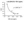
- Table 2 shows the measured count value, S / N ratio, and LoQ (quantification limit value) determined by both assay systems.
- the count value of each standard solution (CAL) is shown as an average value of the count values obtained by measuring the standard solution in duplicate.
- the S / N ratio was determined as the ratio of the count value of 600 pg / mL to the count value of the blank (0 pg / mL).
- the count value of 600 pg / mL was calculated from the count value of the standard solution.
- FIG. 1 hybrid conjugate
- FIG. 2 antibody mixture
- FIG. 1 shows the concentration of each sample (LoQ1 to LoQ5) calculated based on the standard curve prepared from the average count value and concentration of each standard solution, and the concentration of each sample (LoQ1).
- the coefficient of variation (CV) of the count value when n 10 is measured for (-LoQ5).
- LoQ CV 20% was determined for both assay systems based on the accuracy profile.
- the use of hybrid conjugates produced much higher counts than the mixture of each labeled antibody, and the S / N ratio was also higher for the hybrid conjugate than for each labeled antibody mixture. It increased about 6 times. Furthermore, the LoQ using the hybrid conjugate was about 1/10 of the LoQ using the mixture of each labeled antibody. Therefore, it was found that low concentrations of total tau protein can be quantified by using the hybrid conjugate.
- Example 5 Preparation of bovine serum albumin (BSA) linker hybrid conjugate 1
- BSA bovine serum albumin
- GMBS bovine serum albumin
- BSA and GMBS were mixed at a molar ratio of 1:10 and incubated at 30 ° C. for 1 hour to carry out maleimidization of BSA.
- the mixture of Fab ′ fragment of BT2 and Fab ′ fragment of HT7 obtained in Example 1 and maleimidated BSA were mixed at a molar ratio of 2: 1 and incubated at 25 ° C. for 1 hour.
- Coupling was performed. The coupling reaction was stopped by adding 2-MEA hydrochloride and further incubated for 30 minutes at 25 ° C. The reaction was then quenched by adding 0.5 M iodoacetamide and incubating at 25 ° C. for 30 minutes to give a conjugate reaction mixture.
- the obtained conjugate reaction mixture was added to a HiLoad Superdex200 column (manufactured by GE Healthcare Bioscience) equilibrated with a phosphate buffer (pH 6.8), and gel filtration purification of the antibody was performed. Of the multiple peaks at the absorbance of 280 nm of the obtained fractions, the fractions in which two or more Fab's were bound to one BSA were pooled to obtain a BSA linker hybrid conjugate.
- the BSA linker hybrid conjugate thus obtained and NHS-PEG4-Bition were mixed at a molar ratio of 1:30 and incubated at 25 ° C for 45 minutes to biotinate the BSA linker hybrid conjugate. Further desalting treatment was performed to obtain biotinylated BSA linker hybrid conjugate 1. Furthermore, it was diluted to 0.4 ⁇ g / mL in 50 mM MOPS buffer (150 mM sodium chloride, 1.0% BSA, pH 6.8), and alkaline phosphatase-labeled streptavidin (ALP-SA) (Streptavidin, Alkaline Phosphate Conjugate, Invitrogen) was added. Was added at a concentration of 0.4 ⁇ g / mL to obtain a reaction solution containing the BSA linker hybrid conjugate 1 labeled with ALP (hybrid conjugate reaction solution 1).
- ALP-SA alkaline phosphatase-labeled streptavidin
- Example 6 Preparation of BSA linker hybrid conjugate 2 2-MEA hydrochloride was added to BSA and incubated (37 ° C. for 60 minutes) for reduction (thiolation). Further desalting treatment was performed to obtain thiolated BSA. Further, thiolated BSA and NHS-PEG4-Bition (made by Thermo Fisher) have a molar ratio of 1:25, and thiolated BSA and 1,2-bis (maleimide) ethane (made by Tokyo Kasei Kogyo) have a molar ratio of 1: 350. Were mixed and incubated at 30 ° C. for 1 hour, and BSA was biotinylated and maleimidated at the same time.
- the mixture of the Fab ′ fragment of BT2 and the Fab ′ fragment of HT7 obtained in Example 1 was mixed with biotinylated maleimidated BSA at a molar ratio of 2: 1 and incubated at 25 ° C. for 1 hour. Then, coupling was performed. The coupling reaction was stopped by adding 2-MEA hydrochloride and further incubated for 30 minutes at 25 ° C. Then, the reaction was quenched by adding 0.5 M iodoacetamide and incubating at 25 ° C. for 30 minutes to obtain biotinylated BSA linker hybrid conjugate 2.
- the obtained biotinylated BSA linker hybrid conjugate 2 was diluted to 0.2 ⁇ g / mL in 50 mM MOPS buffer (150 mM sodium chloride, 1.0% BSA, pH 6.8), and ALP-SA (Streptavidin, Alkaline Phosphatase Conjugate) was diluted. , Invitrogen) was added at a concentration of 0.4 ⁇ g / mL to obtain a reaction solution containing the BSA linker hybrid conjugate 2 labeled with ALP (hybrid conjugate reaction solution 2).
- Comparative Example 2 Preparation of BSA Linker Single Monoclonal Antibody Monoclonal antibody BT2 and pepsin were mixed in a citrate buffer (pH 3.5) and incubated at 37 ° C for 1 hour to perform pepsin digestion. After stopping the reaction, gel filtration purification was performed using a Superdex200 column (manufactured by GE Healthcare Bioscience) to obtain an F (ab ′) 2 fragment. Next, the concentration of the purified F (ab ′) 2 fragment was determined and, if necessary, adjusted to a concentration of 2.5 mg / mL. The resulting BT2 F (ab ′) 2 fragment was incubated with 2-MEA hydrochloride at 37 ° C. for 90 minutes and reduced to give the BT2 Fab ′ fragment.
- the BSA linker BT2 Fab 'fragment and the BSA linker HT7 Fab' fragment obtained were diluted with 50 ⁇ M MOPS buffer (150 mM sodium chloride, 1.0% BSA, pH 6.8) at 0.2 ⁇ g / mL each, and ALP-SA was added. (Streptavidin, Alkaline Phosphatase Conjugate, Invitrogen) was added at a concentration of 0.4 ⁇ g / mL, and a mixture of ALP-labeled BSA linker BT2 Fab 'fragment and ALP-labeled BSA linker BT2 Fab' fragment (mixed conjugate reaction solution).
- Example 7 Measurement of phosphorylated tau peptide
- a phosphorylated tau peptide antigen in which the 181th threonine of tau protein is phosphorylated is diluted with 100 mM Tris buffer, and each concentration is 0, 40, 100, 200.
- a phosphorylated tau standard solution was prepared so as to have a concentration of 400 pg / mL.
- 150 ⁇ L of the AT270-bonded magnetic particle solution prepared in Example 2 and 30 ⁇ L of the diluted phosphorylated tau standard solution were dispensed into a reaction tank, and after stirring, they were incubated at 37 ° C. for 10 minutes. Then, B / F separation and washing were performed.
- a "Lumipulse G (registered trademark)" exclusive cleaning liquid (Lumipulse G Wash Solution, manufactured by Fujirebio Co., Ltd.) was used. After washing, 250 ⁇ L of the hybrid conjugate reaction solution 1 prepared in Example 5 or the hybrid conjugate reaction solution 2 prepared in Example 6 was dispensed into the reaction tank and incubated at 37 ° C. for 10 minutes. Then, B / F separation and washing were performed. After washing, 200 ⁇ L of a substrate solution (Lumipulse G Substrate Solution, manufactured by Fujirebio Co., Ltd.) containing AMPPD was dispensed into the reaction tank, and after stirring, the mixture was reacted at 37 ° C.
- a substrate solution Lipulse G Substrate Solution, manufactured by Fujirebio Co., Ltd.
- the standard solution was similarly measured using the mixed conjugate reaction solution prepared in Comparative Example 2 instead of the hybrid conjugate reaction solution, and the count value was obtained.
- the results are shown in Table 3.
- the count value of each phosphorylated tau standard solution is shown as an average value of the count values measured in duplicate. Further, the ratio (S / N ratio) of the count value at 40 pg / mL to the count value at 0 pg / mL was obtained. As a result, a low count was generated when the mixed conjugate reaction solution was used. On the other hand, when the hybrid conjugate reaction solution 1 and the hybrid conjugate reaction solution 2 were used, a very high count value was shown even for the low-concentration standard solution.
- the ratio (S / N ratio) of the count value of the lowest standard solution (40 pg / mL) to the count value of the blank (0 pg / mL) was the same when the hybrid conjugate reaction solution 1 was used. It is about 9 times higher than when using the reaction solution (mixture of ALP-labeled single antibodies), and when using the hybrid conjugate reaction solution 2, it is about 13 times higher than when using the mixed conjugate reaction solution. It became clear that it was twice as expensive.
- a high S / N ratio is an important factor for obtaining a stable assay system and high sensitivity, and it has been shown that a highly sensitive and stable assay system can be constructed by using a hybrid conjugate. It was
Landscapes
- Health & Medical Sciences (AREA)
- Life Sciences & Earth Sciences (AREA)
- Chemical & Material Sciences (AREA)
- Immunology (AREA)
- Engineering & Computer Science (AREA)
- Molecular Biology (AREA)
- Biomedical Technology (AREA)
- Urology & Nephrology (AREA)
- Hematology (AREA)
- General Health & Medical Sciences (AREA)
- Biochemistry (AREA)
- Medicinal Chemistry (AREA)
- Organic Chemistry (AREA)
- Proteomics, Peptides & Aminoacids (AREA)
- Biotechnology (AREA)
- General Physics & Mathematics (AREA)
- Microbiology (AREA)
- Physics & Mathematics (AREA)
- Analytical Chemistry (AREA)
- Cell Biology (AREA)
- Pathology (AREA)
- Food Science & Technology (AREA)
- Biophysics (AREA)
- Genetics & Genomics (AREA)
- Neurosurgery (AREA)
- Neurology (AREA)
- Inorganic Chemistry (AREA)
- Peptides Or Proteins (AREA)
Priority Applications (5)
| Application Number | Priority Date | Filing Date | Title |
|---|---|---|---|
| US17/294,931 US20220018856A1 (en) | 2018-11-22 | 2019-11-21 | Antibody conjugate |
| CN201980076655.8A CN113166235A (zh) | 2018-11-22 | 2019-11-21 | 抗体偶联物 |
| EP19886375.5A EP3885362A4 (en) | 2018-11-22 | 2019-11-21 | Antibody conjugate |
| JP2020557617A JP7594913B2 (ja) | 2018-11-22 | 2019-11-21 | 抗体コンジュゲート |
| JP2024161331A JP2024178283A (ja) | 2018-11-22 | 2024-09-18 | 抗体コンジュゲート |
Applications Claiming Priority (2)
| Application Number | Priority Date | Filing Date | Title |
|---|---|---|---|
| JP2018-219558 | 2018-11-22 | ||
| JP2018219558 | 2018-11-22 |
Publications (1)
| Publication Number | Publication Date |
|---|---|
| WO2020105700A1 true WO2020105700A1 (ja) | 2020-05-28 |
Family
ID=70773827
Family Applications (1)
| Application Number | Title | Priority Date | Filing Date |
|---|---|---|---|
| PCT/JP2019/045589 Ceased WO2020105700A1 (ja) | 2018-11-22 | 2019-11-21 | 抗体コンジュゲート |
Country Status (5)
| Country | Link |
|---|---|
| US (1) | US20220018856A1 (enExample) |
| EP (1) | EP3885362A4 (enExample) |
| JP (2) | JP7594913B2 (enExample) |
| CN (1) | CN113166235A (enExample) |
| WO (1) | WO2020105700A1 (enExample) |
Cited By (2)
| Publication number | Priority date | Publication date | Assignee | Title |
|---|---|---|---|---|
| JPWO2021065306A1 (enExample) * | 2019-09-30 | 2021-04-08 | ||
| CN113533746A (zh) * | 2021-07-22 | 2021-10-22 | 深圳市天大生物医疗器械有限公司 | 一种P-Tau蛋白化学发光检测试剂盒及其制备方法 |
Families Citing this family (2)
| Publication number | Priority date | Publication date | Assignee | Title |
|---|---|---|---|---|
| EP4399524A4 (en) | 2021-09-09 | 2025-10-08 | Alzpath Inc | PHOSPHO-TAU ANTIBODIES AND METHODS OF USE |
| CN115015228A (zh) * | 2022-02-22 | 2022-09-06 | 上海科华生物工程股份有限公司 | 一种检测血液中p-tau181蛋白的化学发光试剂盒及其制备方法 |
Citations (5)
| Publication number | Priority date | Publication date | Assignee | Title |
|---|---|---|---|---|
| JPH08502898A (ja) | 1992-12-14 | 1996-04-02 | エヌ・ブイ・インノジェネティクス・ソシエテ・アノニム | 微小管関連タンパク質タウに対するモノクローナル抗体、これらの抗体を分泌するハイブリドーマ、これらのモノクローナル抗体による抗原認識及びこれらの応用 |
| JPH09506771A (ja) | 1993-12-21 | 1997-07-08 | インノジェネティクス・エヌ・ブイ | Phf−タウに特異的なモノクローナル抗体、これらを分泌するハイブリドーマ、これらの抗体による抗原認識及びこれらの応用 |
| JP2010189413A (ja) * | 2002-12-31 | 2010-09-02 | Immunomedics Inc | 非コンジュゲートおよびコンジュゲート抗体、抗体の組合せおよび融合タンパク質を用いるb細胞悪性腫瘍および自己免疫疾患の免疫療法 |
| JP2014523742A (ja) * | 2011-07-14 | 2014-09-18 | カソリック ウニヴェルシテイト ルーヴェン | リン酸化タウ凝集体に対する抗体 |
| JP2018512863A (ja) * | 2015-04-17 | 2018-05-24 | アイジーエム バイオサイエンシズ エー/エス | 多価ヒト免疫不全ウイルス抗原結合分子およびその使用方法 |
Family Cites Families (5)
| Publication number | Priority date | Publication date | Assignee | Title |
|---|---|---|---|---|
| JPH06239899A (ja) * | 1993-02-12 | 1994-08-30 | Teijin Ltd | ヒトタウ蛋白に対する抗体、並びに該抗体を利用する体液中のヒトタウ蛋白の測定方法 |
| JP5247963B2 (ja) * | 2000-01-24 | 2013-07-24 | インノジェネティクス・エヌ・ブイ | タウオパチーの診断 |
| WO2011109112A2 (en) | 2010-03-05 | 2011-09-09 | Albert Einstein College Of Medicine Of Yeshiva University | Method of detecting tau protein and tau fragments in serum |
| WO2014011972A1 (en) * | 2012-07-13 | 2014-01-16 | Bristol-Myers Squibb Company | Tau immunoassay |
| JP2017512056A (ja) * | 2014-02-10 | 2017-05-18 | メルク・シャープ・アンド・ドーム・コーポレーションMerck Sharp & Dohme Corp. | ヒト・タウに結合する抗体および該抗体を使用してヒト・タウを定量するためのアッセイ |
-
2019
- 2019-11-21 WO PCT/JP2019/045589 patent/WO2020105700A1/ja not_active Ceased
- 2019-11-21 CN CN201980076655.8A patent/CN113166235A/zh active Pending
- 2019-11-21 JP JP2020557617A patent/JP7594913B2/ja active Active
- 2019-11-21 US US17/294,931 patent/US20220018856A1/en active Pending
- 2019-11-21 EP EP19886375.5A patent/EP3885362A4/en active Pending
-
2024
- 2024-09-18 JP JP2024161331A patent/JP2024178283A/ja active Pending
Patent Citations (5)
| Publication number | Priority date | Publication date | Assignee | Title |
|---|---|---|---|---|
| JPH08502898A (ja) | 1992-12-14 | 1996-04-02 | エヌ・ブイ・インノジェネティクス・ソシエテ・アノニム | 微小管関連タンパク質タウに対するモノクローナル抗体、これらの抗体を分泌するハイブリドーマ、これらのモノクローナル抗体による抗原認識及びこれらの応用 |
| JPH09506771A (ja) | 1993-12-21 | 1997-07-08 | インノジェネティクス・エヌ・ブイ | Phf−タウに特異的なモノクローナル抗体、これらを分泌するハイブリドーマ、これらの抗体による抗原認識及びこれらの応用 |
| JP2010189413A (ja) * | 2002-12-31 | 2010-09-02 | Immunomedics Inc | 非コンジュゲートおよびコンジュゲート抗体、抗体の組合せおよび融合タンパク質を用いるb細胞悪性腫瘍および自己免疫疾患の免疫療法 |
| JP2014523742A (ja) * | 2011-07-14 | 2014-09-18 | カソリック ウニヴェルシテイト ルーヴェン | リン酸化タウ凝集体に対する抗体 |
| JP2018512863A (ja) * | 2015-04-17 | 2018-05-24 | アイジーエム バイオサイエンシズ エー/エス | 多価ヒト免疫不全ウイルス抗原結合分子およびその使用方法 |
Non-Patent Citations (1)
| Title |
|---|
| See also references of EP3885362A4 |
Cited By (4)
| Publication number | Priority date | Publication date | Assignee | Title |
|---|---|---|---|---|
| JPWO2021065306A1 (enExample) * | 2019-09-30 | 2021-04-08 | ||
| WO2021065306A1 (ja) * | 2019-09-30 | 2021-04-08 | ニプロ株式会社 | 血液試料を検体とするタウタンパク質検出方法 |
| JP7718270B2 (ja) | 2019-09-30 | 2025-08-05 | ニプロ株式会社 | 血液試料を検体とするタウタンパク質検出方法 |
| CN113533746A (zh) * | 2021-07-22 | 2021-10-22 | 深圳市天大生物医疗器械有限公司 | 一种P-Tau蛋白化学发光检测试剂盒及其制备方法 |
Also Published As
| Publication number | Publication date |
|---|---|
| CN113166235A (zh) | 2021-07-23 |
| JP2024178283A (ja) | 2024-12-24 |
| JPWO2020105700A1 (ja) | 2021-10-07 |
| EP3885362A1 (en) | 2021-09-29 |
| EP3885362A4 (en) | 2022-08-17 |
| US20220018856A1 (en) | 2022-01-20 |
| JP7594913B2 (ja) | 2024-12-05 |
Similar Documents
| Publication | Publication Date | Title |
|---|---|---|
| JP2024178283A (ja) | 抗体コンジュゲート | |
| US10107826B2 (en) | Immunoassay methods and reagents for decreasing nonspecific binding | |
| US20080108084A1 (en) | Cancer Detection Methods and Reagents | |
| KR20210049107A (ko) | 바이오마커 검출 방법 | |
| JP7606506B2 (ja) | 血中アミロイドβの免疫測定方法及びそのためのキット | |
| US11169148B2 (en) | Method for detecting test substance and reagent kit for detecting test substance | |
| WO2003069332A2 (en) | Detection and/or monitoring of synuclein-related diseases | |
| US11802867B2 (en) | Cardiac troponin assay method and assay reagent | |
| JPWO2017061546A1 (ja) | Pivka−iiの測定方法、及びpivka−ii免疫測定試薬又はキットの製造方法 | |
| US20030138860A1 (en) | Cancer detection methods and reagents | |
| JP2023017986A (ja) | 自己抗体の直接イムノアッセイ測定法 | |
| JP6048923B2 (ja) | B型慢性肝炎の検出方法および検出キット | |
| JP2019053060A (ja) | B型肝炎ウイルスコア抗体の免疫測定方法 | |
| JPH0727764A (ja) | Fc部位をブロックした免疫学的測定用抗体、該抗体を含む免疫学的測定用試薬、該免疫学的測定用試薬を使用する免疫学的測定法及びFc部位をブロックするブロック試薬 | |
| JP7671956B2 (ja) | 抗原の測定方法及び固相化抗体の製造方法 | |
| JP7680425B2 (ja) | ヒトiv型コラーゲン7sドメインを含む断片の測定方法及びこれに用いるためのキット | |
| JP2024058961A (ja) | 肝細胞がんの検出を補助する方法 | |
| WO2019167935A1 (ja) | プレセプシン測定に有用な抗cd14抗体の使用 | |
| JP2003083966A (ja) | 抗原/抗体水溶液 | |
| HK1195617B (en) | Immunoassay methods and reagents for decreasing nonspecific binding | |
| JPH09196921A (ja) | モノクローナル抗体担持不溶性担体及びその調製方法 | |
| HK1195617A (en) | Immunoassay methods and reagents for decreasing nonspecific binding |
Legal Events
| Date | Code | Title | Description |
|---|---|---|---|
| 121 | Ep: the epo has been informed by wipo that ep was designated in this application |
Ref document number: 19886375 Country of ref document: EP Kind code of ref document: A1 |
|
| ENP | Entry into the national phase |
Ref document number: 2020557617 Country of ref document: JP Kind code of ref document: A |
|
| NENP | Non-entry into the national phase |
Ref country code: DE |
|
| ENP | Entry into the national phase |
Ref document number: 2019886375 Country of ref document: EP Effective date: 20210622 |