WO2016047068A1 - ターゲット分子の検出方法及びこれに用いられるキット - Google Patents
ターゲット分子の検出方法及びこれに用いられるキット Download PDFInfo
- Publication number
- WO2016047068A1 WO2016047068A1 PCT/JP2015/004577 JP2015004577W WO2016047068A1 WO 2016047068 A1 WO2016047068 A1 WO 2016047068A1 JP 2015004577 W JP2015004577 W JP 2015004577W WO 2016047068 A1 WO2016047068 A1 WO 2016047068A1
- Authority
- WO
- WIPO (PCT)
- Prior art keywords
- target molecule
- antibody
- detection
- enzyme
- procedure
- Prior art date
Links
Images


Classifications
-
- G—PHYSICS
- G01—MEASURING; TESTING
- G01N—INVESTIGATING OR ANALYSING MATERIALS BY DETERMINING THEIR CHEMICAL OR PHYSICAL PROPERTIES
- G01N33/00—Investigating or analysing materials by specific methods not covered by groups G01N1/00 - G01N31/00
- G01N33/48—Biological material, e.g. blood, urine; Haemocytometers
- G01N33/50—Chemical analysis of biological material, e.g. blood, urine; Testing involving biospecific ligand binding methods; Immunological testing
- G01N33/53—Immunoassay; Biospecific binding assay; Materials therefor
- G01N33/543—Immunoassay; Biospecific binding assay; Materials therefor with an insoluble carrier for immobilising immunochemicals
- G01N33/54393—Improving reaction conditions or stability, e.g. by coating or irradiation of surface, by reduction of non-specific binding, by promotion of specific binding
-
- G—PHYSICS
- G01—MEASURING; TESTING
- G01N—INVESTIGATING OR ANALYSING MATERIALS BY DETERMINING THEIR CHEMICAL OR PHYSICAL PROPERTIES
- G01N33/00—Investigating or analysing materials by specific methods not covered by groups G01N1/00 - G01N31/00
- G01N33/48—Biological material, e.g. blood, urine; Haemocytometers
- G01N33/50—Chemical analysis of biological material, e.g. blood, urine; Testing involving biospecific ligand binding methods; Immunological testing
- G01N33/53—Immunoassay; Biospecific binding assay; Materials therefor
- G01N33/531—Production of immunochemical test materials
- G01N33/532—Production of labelled immunochemicals
- G01N33/535—Production of labelled immunochemicals with enzyme label or co-enzymes, co-factors, enzyme inhibitors or enzyme substrates
-
- G—PHYSICS
- G01—MEASURING; TESTING
- G01N—INVESTIGATING OR ANALYSING MATERIALS BY DETERMINING THEIR CHEMICAL OR PHYSICAL PROPERTIES
- G01N33/00—Investigating or analysing materials by specific methods not covered by groups G01N1/00 - G01N31/00
- G01N33/48—Biological material, e.g. blood, urine; Haemocytometers
- G01N33/50—Chemical analysis of biological material, e.g. blood, urine; Testing involving biospecific ligand binding methods; Immunological testing
- G01N33/53—Immunoassay; Biospecific binding assay; Materials therefor
- G01N33/536—Immunoassay; Biospecific binding assay; Materials therefor with immune complex formed in liquid phase
- G01N33/542—Immunoassay; Biospecific binding assay; Materials therefor with immune complex formed in liquid phase with steric inhibition or signal modification, e.g. fluorescent quenching
-
- C—CHEMISTRY; METALLURGY
- C12—BIOCHEMISTRY; BEER; SPIRITS; WINE; VINEGAR; MICROBIOLOGY; ENZYMOLOGY; MUTATION OR GENETIC ENGINEERING
- C12Q—MEASURING OR TESTING PROCESSES INVOLVING ENZYMES, NUCLEIC ACIDS OR MICROORGANISMS; COMPOSITIONS OR TEST PAPERS THEREFOR; PROCESSES OF PREPARING SUCH COMPOSITIONS; CONDITION-RESPONSIVE CONTROL IN MICROBIOLOGICAL OR ENZYMOLOGICAL PROCESSES
- C12Q1/00—Measuring or testing processes involving enzymes, nucleic acids or microorganisms; Compositions therefor; Processes of preparing such compositions
- C12Q1/68—Measuring or testing processes involving enzymes, nucleic acids or microorganisms; Compositions therefor; Processes of preparing such compositions involving nucleic acids
Definitions
- the present invention relates to a method for detecting a target molecule and a kit used therefor. More specifically, the present invention relates to a method for detecting a target molecule based on an antigen-antibody reaction between the target molecule and an antibody on a carrier.
- a biosensing technique for detecting a disease marker (biomarker) present at a low concentration in a biological sample with high sensitivity is required.
- biomarker disease marker
- the blood concentration of the biomarker is about 30 aM.
- Non-Patent Document 1 describes a method for detecting a protein using a single molecule enzyme-linked immunosorbent assay (ELISA).
- ELISA enzyme-linked immunosorbent assay
- Patent Document 1 discloses an array device capable of forming ultra-high-density minute droplets as a “single molecule digital counting device”. By performing the ELISA in a small volume droplet, the signal from the target molecule can be binarized and measured (digital ELISA method). Specifically, first, a target molecule, a bead whose surface has been modified with a capture antibody, and a detection antibody are reacted to form a “capture antibody-target molecule-detection antibody” complex on the bead surface. When the concentration of the target molecule is low, individual beads will either bind only one molecule of the complex or not at all. Next, one bead is encapsulated in each of a large number of microdroplets formed on the array device.
- the number of droplets that emit a signal derived from the detection antibody is counted as the number of target molecules.
- the signal from the target molecule can be binarized to 0 or 1, and the target molecule can be detected and quantified with high sensitivity and high accuracy.
- Patent Document 2 discloses that, in an enzyme immunoassay, a restriction enzyme is used as a label for an antibody that reacts with a target substance, and a DNA strand having a restriction base sequence of the restriction enzyme is conjugated with a complex restriction enzyme. A method of detecting a target substance by cleaving and analyzing and measuring the cleaved DNA strand fragment is disclosed.
- the main object of the present invention is to provide a technology for detecting a signal from a target molecule with high sensitivity and high accuracy by eliminating noise caused by nonspecific adsorption of a detection antibody.
- the present invention provides the following [1] to [8].
- [2] The detection method according to [1], including an analysis procedure for processing two or more fluorescence detection signals having different fluorescence wavelengths as the detection signal of the target molecule.
- [3] The detection according to [1] or [2], including a sealing procedure in which the carrier is sealed in each of the droplets formed on the substrate between the complex formation procedure and the detection procedure.
- Method. [4] The detection method according to any one of [1] to [3], wherein the carrier is microbeads.
- [5] The detection method according to any one of [1] to [4], wherein the first antibody and the second antibody bind to different epitopes of the target molecule.
- [6] The detection method according to any one of [1] to [5], which is a digital ELISA method.
- a target molecule [7] a target molecule; A carrier modified with a first antibody that specifically binds to the target molecule; Two or more second antibodies that specifically bind to the target molecule and are labeled differently from each other; To form a complex consisting of the first antibody, the target molecule and the second antibody on the carrier, and A detection procedure for detecting a signal from the label; An analysis procedure for processing the signals from two or more different labels as detection signals of the target molecule.
- a carrier modified with a first antibody that specifically binds to a target molecule Two or more antibodies labeled with an enzyme that specifically binds to the target molecule and has a substrate-cleaving activity, the second antibody having different substrate specificities of the enzyme, and a cleavage site of the enzyme And two or more substrates having a fluorescent substance bonded to one end of the cleavage site and a decoloring substance bonded to the other end, wherein the fluorescent wavelengths of the fluorescent substances are different from each other;
- An enzyme-linked immunosorbent assay (ELISA) kit comprising:
- the present invention in a method for detecting a target molecule based on an antigen-antibody reaction between the target molecule and an antibody on a carrier, noise caused by nonspecific adsorption of the detection antibody to the carrier is eliminated, and the target molecule is separated from the target molecule.
- a technique for detecting the signal of the above with high sensitivity and high accuracy is provided.
- the target molecule detection method includes the following procedures.
- each procedure will be described by taking as an example an embodiment in which the method for detecting a target molecule according to the present invention is applied to single molecule digital counting by ELISA (digital ELISA method).
- a target molecule a carrier modified with a first antibody that specifically binds to the target molecule, and two or more labeled with an enzyme that specifically binds to the target molecule and has a substrate cleavage activity
- An antibody which reacts with a second antibody having different substrate specificities of the enzyme, to form a complex comprising the first antibody, the target molecule, and the second antibody on the carrier;
- the complex formation procedure is described by taking as an example an embodiment in which the method for detecting a target molecule according to the present invention is applied to single molecule digital counting by ELISA (digital ELISA method).
- the target molecule to be detected may be any substance that binds to an antibody by an antigen-antibody reaction, and in particular, microorganisms such as bacteria and fungi, viruses, proteins, nucleic acids, sugars, and the like. These are biomolecules such as composites.
- the target molecule to be detected is not limited to one type, and two or more types of target molecules can be detected simultaneously. For example, by using four types of antibodies, a first antibody and a second antibody against protein A, and a first antibody and a second antibody against protein B, two types of target molecules, protein A and protein B, can be distinguished. Simultaneous detection is possible.
- a target molecule In the complex formation procedure, a target molecule, a carrier modified with a first antibody that specifically binds to the target molecule, and an enzyme that specifically binds to the target molecule and has a substrate cleavage activity Is reacted with a second antibody labeled to form a complex composed of the first antibody, the target molecule and the second antibody on the carrier.
- a second antibody two or more antibodies labeled with enzymes having different substrate specificities are used.
- the “enzyme having substrate cleavage activity” is not particularly limited as long as it can realize dissociation (described in detail later) of the fluorescent substance and the decoloring substance by cleavage of the substrate.
- Examples of the “enzyme having substrate cleavage activity” include a transferase classified as EC2 by an EC number (enzyme number, EnzymeEnCommission Number), a hydrolase classified as EC3, and an additional desorbase classified as EC4. Enzymes belonging to can be used. Examples of combinations of specific enzymes and their substrates (and cleavage sites in the substrates) include the following.
- Fig. 1 shows the complex formed by the complex formation procedure.
- the carrier 2 modified with the first antibody 3 that specifically binds to the target molecule 1 and the second antibody that specifically binds to the target molecule 1 and is labeled with the enzymes 51 and 52. 41 and 42 are prepared.
- antibody specifically binds means that it can bind to an antigen (here, target molecule 1), but does not bind to other substances or is weakly bound. Further, “weak binding” means that the binding affinity to other substances is sufficiently low to be distinguishable as compared with the binding affinity to the antigen.
- the binding affinity (affinity) of an antibody can be measured, for example, by a known method such as a surface plasma resonance (SPR) method.
- the first antibody 3 functions to capture the target molecule 1 on the carrier 2.
- the second antibodies 41 and 42 function in order to enable optical detection of the target molecule 1 captured on the carrier 2.
- the first antibody 3 and the second antibody 41, 42 preferably bind to different epitopes of the target molecule 1.
- the epitope of the first antibody 3, the epitope of the second antibody 41, and the epitope of the second antibody 42 are preferably all different.
- the first antibody 3 is also referred to as “capture antibody 3”
- the second antibody is also referred to as “detection antibodies 41 and 42”.
- micro beads are widely used.
- carrier 2 is also referred to as “microbead 2”.
- microbead is used synonymously with “particle” and is a technical term commonly used in the art.
- the shape of the microbead is not particularly limited, but is usually a spherical shape.
- the material of the microbead is not particularly limited, and may be glass, silica gel, polystyrene, polypropylene, a membrane, a magnetic material, and the like.
- Specific materials include cellulose, cellulose derivatives, acrylic resins, glass, silica gel, polystyrene, gelatin, polyvinyl pyrrolidone, copolymers of vinyl and acrylamide, polystyrene cross-linked with divinylbenzene, polyacrylamide, latex gel, polystyrene dextran. , Rubber, silicon, plastic, nitrocellulose, cellulose, natural sponge, silica gel, glass, metal plastic, cellulose, crosslinked dextran (Sephadex TM) and agarose gel (Sepharose TM).
- the beads may be porous.
- the beads preferably have an average particle diameter of 5 ⁇ m or less, for example, about 1 ⁇ m to 4 ⁇ m. In addition, an average particle diameter can be measured using electron microscope observation or a dynamic light scattering method, for example.
- the modification of the capture antibody 3 to the microbead 2 is performed by binding the capture antibody 3 to a modification group on the surface of the microbead 2 via a linker.
- the capture antibody 3 is covalently bonded to an amino group on the surface of the amino group-modified bead via a cross-linking agent having N-hydroxysuccinimide or the like.
- the enzymes 51 and 52 labeled with the detection antibodies 41 and 42 have different substrate specificities.
- substrate specificity means that in the cleavage of a substrate catalyzed by an enzyme, the enzyme does not catalyze the cleavage of a substance other than the substrate, or the degree of the catalyst is sufficiently weak.
- an esterase is used as the enzyme 51 as the “enzyme having different substrate specificities”, glucosidase, phosphatase, or the like, which is an enzyme that does not use an ester bond as a cleavage site, is used as the enzyme 52.
- restriction enzymes having different recognition sequences (cleavage sites) are used as the enzymes 51 and 52 having different substrate specificities.
- the restriction enzyme include AccI, AluI, ApaI, BamHI, BglII, BssHII, BstEII, ClaI, DdeI, DraI, EcoRI, EcoRV, HaeIII, HincII, HindIII, HpaI, HpaII, KpnI, NluI, KpnI, MluN, , NheI, NotI, PstI, PvuI, PvuII, RsaI, SacI, SalI, ScaI, SmaI, SpeI, SphI, SspI, StuI, XbaI, XhoI and the like.
- the enzymes 51 and 52 two enzymes having different substrate specificities can be used in any combination. Below, the example which uses a restriction enzyme as the enzymes 51 and 52 is mainly demonstrated, and the enzymes 51 and 52 shall also be called "restriction enzymes 51 and 52.”
- the detection enzymes 41 and 42 are labeled with the restriction enzymes 51 and 52 by forming a cross-linking structure between the detection antibodies 41 and 42 and the restriction enzymes 51 and 52 using a cross-linking agent (crosslinker reagent). Can do.
- a cross-linking agent crosslinker reagent
- the target molecule 1, the microbead 2 modified with the capture antibody 3, and the detection antibodies 41 and 42 labeled with the restriction enzymes 51 and 52 are reacted.
- a complex composed of the capture antibody 3, the target molecule 1, and the detection antibodies 41 and 42 is formed on the microbead 2 (see FIG. 1A).
- the reaction of the target molecule 1, the microbead 2 and the detection antibodies 41 and 42 may be performed in one step or in two steps. That is, the target molecule 1, the microbead 2 and the detection antibodies 41 and 42 may be reacted at the same time, or after the target molecule 1 and the microbead 2 are reacted, the target molecule 1 that has not bound to the capture antibody 3 is removed. For this purpose, the microbeads 2 may be washed and then reacted with the detection antibodies 41 and 42.
- the reaction of the target molecule 1, the microbeads 2 and the detection antibodies 41 and 42 may be performed by bringing them into contact with each other in an appropriate solution, and can be performed under the same conditions as those of conventionally known enzyme-linked immunosorbent assays.
- concentration of the target molecule 1 is low, the individual microbeads 2 after the reaction have only one molecule complex or none at all.
- the restriction enzyme 51, 42 labeled with the detection antibody 41, 42 is brought into contact with the microbead 2 having the complex formed in this procedure on the surface and the substrate to which the fluorescent substance is bound. Fluorescence generated by cleavage of the substrate by 52 is detected. At this time, if non-specific adsorption of the detection antibodies 41 and 42 to the microbeads 2 occurs, the fluorescence due to the cleavage of the substrate is also emitted from the microbeads 2 having the nonspecifically adsorbed detection antibodies 41 and 42 on the surface. Will occur.
- antibody adsorbs non-specifically means that the antibody adsorbs to a non-antigen portion of a substance containing an antigen or adsorbs to a substance that does not contain an antigen.
- -It shall refer to adsorption to a substance regardless of antibody reaction.
- FIGS. 1B and 1C show non-specific adsorption of the detection antibodies 41 and 42 to the microbeads 2 that can occur in the complex formation procedure.
- FIG. 1B shows a state in which one of the detection antibody 41 and the detection antibody 42 is adsorbed nonspecifically on the surface of the microbead 2.
- FIG. 1C shows a state where both the detection antibody 41 and the detection antibody 42 are adsorbed nonspecifically on the surface of the microbead 2.
- non-specific adsorption of the detection antibodies 41 and 42 as shown in FIGS. 1B and 1C may occur in addition to the target complex formation shown in FIG. 1A.
- the frequency at which non-specific adsorption of both the detection antibody 41 and the detection antibody 42 shown in FIG. 1C occurs is sufficiently smaller than the frequency at which non-specific adsorption of either one of the antibodies shown in FIG. Little impact on detection accuracy. For example, assuming that 1% of the microbead 2 causes non-specific adsorption of the detection antibody 41 and similarly 1% of the microbead 2 causes non-specific adsorption of the detection antibody 42, both the detection antibody 41 and the detection antibody 42 The frequency with which non-specific adsorption occurs is only 0.01%.
- two or more fluorescence detection signals having different fluorescence wavelengths are processed as detection signals (signals) of the target molecule 1, thereby resulting from the fluorescence detection signals generated by non-specific adsorption shown in FIG. 1B. Eliminate noise.
- the microbeads 2 are encapsulated in droplets formed on the substrate. This procedure is performed when the target molecule detection method according to the present invention is applied to the digital ELISA method, and is not an essential procedure of the detection method according to the present invention.
- the microbeads 2 are enclosed one by one in a small volume droplet that can contain only one microbead 2.
- a single-molecule digital counting device disclosed in Patent Document 1 can be suitably used to form microdroplets and enclose the microbeads 2 in the microdroplets.
- microbeads 2 can be encapsulated in the droplets at the same time while forming microdroplets at an extremely high density on the substrate.
- the microbeads 2 after the complex formation procedure may be washed in order to remove the detection antibodies 41 and 42 that have not bound to the target molecule 1, and then resuspended in an appropriate solvent and subjected to this procedure. .
- the microbeads 2 after the complex formation procedure are a mixture of microbeads that have formed a complex (see FIG. 1A) and microbeads that have not formed a complex. Furthermore, the microbeads that do not form a complex include microbeads (see FIGS. 1B and 1C) to which the detection antibodies 41 and 42 are adsorbed nonspecifically.
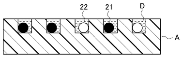
- FIG. 2 shows the microbeads 2 encapsulated in microdroplets.
- One microbead 2 is encapsulated in each droplet D formed on the substrate A.
- each microbead 2 has only one molecule complex or not at all.
- the microbead with the complex formed on the surface is denoted by reference numeral 21, and the microbead without the complex is denoted by reference numeral 22.
- the microbeads 21 are in the state shown in FIG. 1A, and the microbeads 22 include beads in the state shown in FIG. 1B or C.
- the microbead 2 in which the complex is formed is referred to as “microbead 21”
- the microbead 2 in which the complex is not formed is referred to as “microbead 22”.
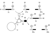
- Detection Procedure In the detection procedure, a substrate (hereinafter referred to as “probe”) having a recognition sequence of restriction enzymes 51 and 52, a fluorescent substance bound to one end side of the recognition sequence serving as a cleavage site, and a decolorizing substance bound to the other end side. And the complex formed on the surface of the microbead 2 in the complex formation procedure, and the fluorescence emitted from the fluorescent substance is detected.
- a substrate hereinafter referred to as “probe” having a recognition sequence of restriction enzymes 51 and 52, a fluorescent substance bound to one end side of the recognition sequence serving as a cleavage site, and a decolorizing substance bound to the other end side.
- FIG. 3 shows the reaction between the probe and the complex in this procedure.
- the probe denoted by reference numeral 61 in the figure includes a cleavage site 71 of the restriction enzyme 51 labeled with the detection antibody 41, and a fluorescent substance 81 is bound to one region across the cleavage site 71, and the other A decoloring substance 91 is bonded to the region.
- the probe denoted by reference numeral 62 in the figure includes a cleavage site 72 of the restriction enzyme 52 labeled on the detection antibody 42, and the fluorescent substance 82 is bound to one region across the cleavage site 72 and erased to the other region.
- a color substance 92 is bound.
- the fluorescent materials 81 and 82 of the probes 71 and 72 are fluorescent materials having different fluorescence wavelengths and optically distinguishable and detectable.
- the probe 61 is connected to the detection antibody 41.
- a substance containing a labeled esterase cleavage site 71 (ester bond), a fluorescent material 81 bound to one region across the cleavage site 71, and a decolorizing material 91 bound to the other region is used.
- the probe 62 includes a glucosidase cleavage site 72 (glycoside bond) labeled with the detection antibody 42, and a fluorescent substance 82 is bound to one region across the cleavage site 72, and a decoloring material is disposed to the other region. A combination of 92 is used.
- the decolorizable materials 91 and 92 block the emission of the fluorescent materials 81 and 82 while being located within a certain distance that allows energy transfer between the fluorescent materials 81 and 82 (quenching).
- fluorescent materials 81 and 82 and the decoloring materials 91 and 92 fluorescent materials and quenchers that are widely used in nucleic acid optical detection techniques such as real-time quantitative PCR can be used.
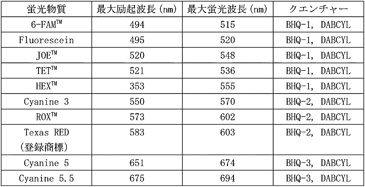
- Examples of combinations of fluorescent substances and quenchers include Alexa Fluor (registered trademark) 488 (manufactured by Invitrogen), ATTO 488 (manufactured by ATTO-TEC GmbH), Alexa Fluor (registered trademark) 594 (manufactured by Invitrogen) and Examples thereof include a combination of a fluorescent substance selected from the group consisting of ROX (Carboxy-X-rhodamine) and BHQ (registered trademark, Black hole quencher) -1 or BHQ (registered trademark) -2. Moreover, the combination of a fluorescein and DABCYL etc. are mentioned. The combinations of commonly used fluorescent substances and quenchers are shown in the table below.
- the reaction is performed by bringing the probes 61 and 62 into contact with the microbeads 2 enclosed in the microdroplet D.
- the microbeads 2 that have been washed after the complex formation procedure are resuspended in a solution containing the probes 61 and 62, thereby bringing them into contact with each other.
- the reaction is preferably performed using a buffer having a composition suitable for the types of restriction enzymes 51 and 52, and it is preferable to form microdroplets using such a buffer in the encapsulation procedure. Note that buffers optimized for various restriction enzymes are commercially available in combination with restriction enzymes.
- the cleavage site 71 of the probe 61 is cleaved by the restriction enzyme 51 labeled on the detection antibody 41 that forms a complex.
- the probe 61 is cleaved into the fragment 61a and the fragment 61b, and the fluorescent material 81 is dissociated from the decolorizable material 91 and becomes ready for light emission.
- the fluorescent material 82 is also dissociated from the decolored material 92 and becomes capable of emitting light.
- Fluorescence is detected by detecting fluorescence from each microdroplet in which the microbeads 2 are enclosed, using a fluorescence microscope, an image sensor, or the like.
- the detection can be performed, for example, by observing the presence or absence of the microbead 2 under a microscope, and can also be performed by a method of detecting scattered light by the microbead 2 or a method using potential measurement by a field effect transistor (FET). It can be carried out.
- FET field effect transistor
- the microbeads 2 enclosed in the individual microdroplets D are the microbeads 21 having only one molecule complex, or not at all. Therefore, the number of microdroplets D that generate a detection signal of the target molecule 1 can be regarded as the number of target molecules 1. Further, the target molecule 1 is selected from the total number of microbeads 2 by using the number of microdroplets D enclosing the microbeads 21 and 22 and the number of microdroplets D enclosing the microbeads 21. The ratio of the number captured can be calculated. This makes it possible to quantify the concentration of the target molecule.
- the fluorescence from the fluorescent material 81 and the fluorescence from the fluorescent material 92 having a wavelength different from that of the fluorescence are detected.
- the detection antibody 41 or the detection antibody 42 as shown in FIG. 1B is adsorbed nonspecifically on the surface of the microbeads 2 but does not form a complex, the fluorescent substance 81 Only the fluorescence from one of the fluorescent material 92 and the fluorescent material 92 is detected.
- both the detection antibody 41 and the detection antibody 42 as shown in FIG. 1C are adsorbed nonspecifically on the surface of the microbead 2, fluorescence of both the fluorescent substance 81 and the fluorescent substance 92 is generated.
- this fluorescence hardly affects the quantification of the target molecule 1.
- the microbeads 2, detection antibodies 41 and 42 (see FIG. 1), and probes 71 and 72 (see FIG. 3) used in this embodiment are kits for carrying out the target molecule detection method according to the present invention. It can be suitably implemented. That is, the present invention in one aspect, (I) a carrier modified with a first antibody that specifically binds to a target molecule; (Ii) two or more antibodies that specifically bind to the target molecule and are labeled with an enzyme having a substrate cleavage activity, wherein the enzymes have different substrate specificities; (Iii) two or more substrates having a cleavage site of the enzyme, a fluorescent substance bonded to one end of the cleavage site, and a decoloring substance bonded to the other end, wherein the fluorescent wavelength of the fluorescent substance is Different substrates, An enzyme-linked immunosorbent assay (ELISA) kit is also provided.
- ELISA enzyme-linked immunosorbent assay
- the microbead 2 may be one obtained by modifying the capture antibody 3 (first antibody) in advance, and the antibody is bound to the modifying group on the surface of the bead at the time of use via a linker. May be.
- the detection antibodies 41 and 42 may be those in which an enzyme is labeled in advance, and may be those in which an enzyme is bound to the antibody using a cross-linking agent at the time of use.
- the kit according to the present invention is a reagent such as a cross-linking agent used for modification of the capture antibody 3 on the microbead 2 or labeling of the enzyme on the detection antibodies 41 and 42, and a complex formation procedure and a detection procedure.
- a reagent such as a cross-linking agent used for modification of the capture antibody 3 on the microbead 2 or labeling of the enzyme on the detection antibodies 41 and 42, and a complex formation procedure and a detection procedure.
- Various buffers used, and a substrate A (see FIG. 2) used in the encapsulation procedure may be further included.
- two detection antibodies and two corresponding probes are used, and two fluorescence detection signals with different fluorescence wavelengths are used as target molecule detection signals, resulting in non-specific adsorption of the detection antibody.
- An embodiment for reducing noise is described.
- three or more pairs of detection antibodies and probes may be used.
- three or more fluorescence detection signals having different fluorescence wavelengths are used as the target molecule detection signals. That's fine. The greater the number of detection antibodies and probes used, the higher the noise reduction effect derived from non-specific adsorption of detection antibodies.
- a detection antibody labeled with an enzyme having a substrate cleavage activity is used as the second antibody, and fluorescence is generated by cleaving the cleavage site in the probe with the enzyme.
- a detection antibody labeled with an enzyme conventionally used for chemical color development or a detection antibody labeled with a fluorescent dye can be applied as the second antibody.
- this invention also includes the detection method of the target molecule
- A a target molecule, a carrier modified with a first antibody that specifically binds to the target molecule, and two or more second antibodies that specifically bind to the target molecule and have different labels And forming a complex comprising the first antibody, the target molecule, and the second antibody on the carrier.
- B A detection procedure for detecting a signal from the label.
- C An analysis procedure for processing the signals from two or more different labels as detection signals of the target molecule.
- the “signal from the sign” includes a signal generated directly and indirectly from the sign.
- the “signal from the label” refers to fluorescence generated from the fluorescent dye when the detection antibody labeled with the fluorescent dye is applied as the second antibody (see FIG. 4B).
- the “signal from the label” refers to chemical coloration catalyzed by the enzyme when the detection antibody labeled with the enzyme used for chemical coloration is applied as the second antibody (see FIG. 4A).
- the procedure (A) is the same as that in the above-described embodiment (first embodiment) except that the second antibody is an enzyme conventionally used for chemical color development such as alkaline phosphatase or galactosidase, or a detection antibody labeled with various fluorescent substances. This can be performed in the same manner as in the procedure (1) of the embodiment. Moreover, when this embodiment is implemented as a digital ELISA method, the encapsulation procedure described as the procedure (2) in the first embodiment may be included.
- the detection antibody can be labeled with an enzyme or a fluorescent dye by the above-described known method. Various commercially available antibodies labeled with enzymes or fluorescent dyes may be used.
- step (B) the signal from the label of the detection antibody forming a complex on the surface of the carrier is detected.
- FIG. 4A shows that when the antibody labeled with alkaline phosphatase and the antibody labeled with galactosidase are used as the detection antibodies 41 and 42, “capture antibody 3-target substance 1-second antibody 41 formed on the microbead 2”. , 42 ".
- the signal can be detected by a coloring method using a substrate of the enzyme.
- BCIP Bis-Bromo-4-chloro-3-indolyl-phosphate
- NBT non-Nitro blue tetrazolium chloride
- X-Gal 5-bromo-4-chloro-3-indolyl- ⁇ -D-galactopyranoside
- Two or more antibodies labeled with different enzymes are used as the detection antibody, and two or more compounds are used as the chromogenic substrate depending on the enzyme labeled with each antibody.
- signals derived from the respective enzymes can be detected.
- FIG. 4B shows that when a detection antibody 41 or 42 is labeled with FITC and an antibody labeled with Texas Red (registered trademark), the “capture antibody 3-target substance 1-second” formed on the microbead 2 is used. A complex of two antibodies 41, 42 "is shown. Two or more antibodies labeled with fluorescent substances having different fluorescence wavelengths are used as detection antibodies. By detecting the fluorescence from the fluorescent substance for each wavelength band, signals derived from the respective fluorescent substances can be detected.
- step (C) signals from two or more different labels (alkaline phosphatase and galactosidase, or FITC and Texas Red in the above example) are processed as detection signals for the target molecule.
- the detection antibody is adsorbed nonspecifically on the surface of the carrier and does not form a complex, only the signal from one of the two or more labels is used. Not detected (see FIG. 1). Therefore, by processing signals from two or more different labels as detection signals for the target molecule, noise resulting from nonspecific adsorption of the detection antibody to the carrier can be greatly reduced. Thereby, it is possible to improve the detection accuracy of the target molecule, and further its quantitativeness.
- 1 target molecule
- 2 microbead (carrier)
- 3 capture antibody (first antibody)
- 41, 42 detection antibody (second antibody)
- 51, 52 restriction enzyme
- 61, 62 probe
- 71, 72 cleavage site
- 81, 82 fluorescent substance
- 91, 92 decoloring substance
Landscapes
- Health & Medical Sciences (AREA)
- Immunology (AREA)
- Life Sciences & Earth Sciences (AREA)
- Engineering & Computer Science (AREA)
- Chemical & Material Sciences (AREA)
- Molecular Biology (AREA)
- Biomedical Technology (AREA)
- Hematology (AREA)
- Urology & Nephrology (AREA)
- Biochemistry (AREA)
- Microbiology (AREA)
- Physics & Mathematics (AREA)
- Analytical Chemistry (AREA)
- Biotechnology (AREA)
- General Health & Medical Sciences (AREA)
- Cell Biology (AREA)
- Food Science & Technology (AREA)
- Medicinal Chemistry (AREA)
- General Physics & Mathematics (AREA)
- Pathology (AREA)
- Chemical Kinetics & Catalysis (AREA)
- Investigating Or Analysing Materials By The Use Of Chemical Reactions (AREA)
- Investigating, Analyzing Materials By Fluorescence Or Luminescence (AREA)
- Organic Chemistry (AREA)
- Proteomics, Peptides & Aminoacids (AREA)
- Wood Science & Technology (AREA)
- Zoology (AREA)
- Biophysics (AREA)
- Bioinformatics & Cheminformatics (AREA)
- General Engineering & Computer Science (AREA)
- Genetics & Genomics (AREA)
- Measuring Or Testing Involving Enzymes Or Micro-Organisms (AREA)
Abstract
Description
[1]ターゲット分子と、
前記ターゲット分子に特異的に結合する第一の抗体が修飾された担体と、
前記ターゲット分子に特異的に結合し、基質切断活性を有する酵素が標識された二以上の抗体であって、前記酵素の基質特異性が互いに異なる第二の抗体と、
を反応させて、前記担体上に前記第一の抗体と前記ターゲット分子と前記第二抗体とからなる複合体を形成させる複合体形成手順と、
前記酵素の切断部位を有し、該切断部位の一端側に蛍光物質が結合され他端側に消色物質が結合された二以上の基質であって、前記蛍光物質の蛍光波長が互いに異なる基質と、
前記複合体と、
を反応させて、前記蛍光物質から発せられる蛍光を検出する検出手順と、を含むターゲット分子の検出方法。
[2]蛍光波長の異なる二以上の蛍光の検出信号を前記ターゲット分子の検出信号として処理する解析手順を含む、[1]の検出方法。
[3]前記複合体形成手順と前記検出手順との間に、基板上に形成された液滴のそれぞれに前記担体を一つずつ封入する封入手順を含む、[1]又は[2]の検出方法。
[4]前記担体がマイクロビーズである[1]~[3]のいずれかの検出方法。
[5]前記第一の抗体と前記第二の抗体が前記ターゲット分子の異なるエピトープに結合する[1]~[4]のいずれかの検出方法。
[6]デジタルELISA法である[1]~[5]のいずれかの検出方法。
[7]ターゲット分子と、
前記ターゲット分子に特異的に結合する第一の抗体が修飾された担体と、
前記ターゲット分子に特異的に結合し、互いに異なる標識がされた二以上の第二の抗体と、
を反応させて、前記担体上に前記第一の抗体と前記ターゲット分子と前記第二抗体とからなる複合体を形成させる複合体形成手順と、
前記標識からの信号を検出する検出手順と、
異なる二以上の前記標識からの前記信号を前記ターゲット分子の検出信号として処理する解析手順と、を含むターゲット分子の検出方法。
前記ターゲット分子に特異的に結合し、基質切断活性を有する酵素が標識された二以上の抗体であって、前記酵素の基質特異性が互いに異なる第二の抗体と、前記酵素の切断部位を有し、該切断部位の一端側に蛍光物質が結合され他端側に消色物質が結合された二以上の基質であって、前記蛍光物質の蛍光波長が互いに異なる基質と、
を含んでなる酵素結合免疫吸着アッセイ(ELISA)キット。
(1)ターゲット分子と、前記ターゲット分子に特異的に結合する第一の抗体が修飾された担体と、前記ターゲット分子に特異的に結合し、基質切断活性を有する酵素が標識された二以上の抗体であって、前記酵素の基質特異性が互いに異なる第二の抗体と、を反応させて、前記担体上に前記第一の抗体と前記ターゲット分子と前記第二抗体とからなる複合体を形成させる複合体形成手順。
(2)基板上に形成された液滴のそれぞれに前記担体を一つずつ封入する封入手順。
(3)前記酵素の切断部位を有し、該切断部位の一端側に蛍光物質が結合され他端側に消色物質が結合された二以上の基質であって、前記蛍光物質の蛍光波長が互いに異なる基質と、前記複合体と、を反応させて、前記蛍光物質から発せられる蛍光を検出する検出手順。
(4)蛍光波長の異なる二以上の蛍光の検出信号を前記ターゲット分子の検出信号として処理する解析手順。
複合体形成手順では、ターゲット分子と、前記ターゲット分子に特異的に結合する第一の抗体が修飾された担体と、前記ターゲット分子に特異的に結合し、基質切断活性を有する酵素が標識された第二の抗体と、を反応させて、前記担体上に前記第一の抗体と前記ターゲット分子と前記第二の抗体とからなる複合体を形成させる。第二の抗体には、基質特異性が互いに異なる酵素が標識された二以上の抗体が用いられる。

封入手順では、基板上に形成された液滴にマイクロビーズ2を封入する。本手順は、本発明に係るターゲット分子の検出方法をデジタルELISA法に適用した場合に行われる手順であり、本発明に係る検出方法の必須の手順となるものではない。
検出手順では、制限酵素51,52の認識配列を有し、切断部位となる該認識配列の一端側に蛍光物質が結合され他端側に消色物質が結合された基質(以下「プローブ」とも称する)と、複合体形成手順においてマイクロビーズ2の表面に形成された複合体とを反応させて、蛍光物質から発せられる蛍光を検出する。
解析手順では、蛍光波長の異なる二以上の蛍光の検出信号をターゲット分子1の検出信号として処理し、ターゲット分子1の検出信号を発する微小液滴Dの数をターゲット分子の数としてカウントする。
(i)ターゲット分子に特異的に結合する第一の抗体が修飾された担体と、
(ii)前記ターゲット分子に特異的に結合し、基質切断活性を有する酵素が標識された二以上の抗体であって、前記酵素の基質特異性が互いに異なる第二の抗体と、
(iii)前記酵素の切断部位を有し、該切断部位の一端側に蛍光物質が結合され他端側に消色物質が結合された二以上の基質であって、前記蛍光物質の蛍光波長が互いに異なる基質と、
を含んでなる酵素結合免疫吸着アッセイ(ELISA)キットをも提供するものである。
(A)ターゲット分子と、前記ターゲット分子に特異的に結合する第一の抗体が修飾された担体と、前記ターゲット分子に特異的に結合し、互いに異なる標識がされた二以上の第二の抗体と、を反応させて、前記担体上に前記第一の抗体と前記ターゲット分子と前記第二抗体とからなる複合体を形成させる複合体形成手順。
(B)前記標識からの信号を検出する検出手順。
(C)異なる二以上の前記標識からの前記信号を前記ターゲット分子の検出信号として処理する解析手順。
Claims (7)
- ターゲット分子と、
前記ターゲット分子に特異的に結合する第一の抗体が修飾された担体と、
前記ターゲット分子に特異的に結合し、基質切断活性を有する酵素が標識された二以上の抗体であって、前記酵素の基質特異性が互いに異なる第二の抗体と、
を反応させて、前記担体上に前記第一の抗体と前記ターゲット分子と前記第二抗体とからなる複合体を形成させる複合体形成手順と、
前記酵素の切断部位を有し、該切断部位の一端側に蛍光物質が結合され他端側に消色物質が結合された二以上の基質であって、前記蛍光物質の蛍光波長が互いに異なる基質と、
前記複合体と、
を反応させて、前記蛍光物質から発せられる蛍光を検出する検出手順と、
蛍光波長の異なる二以上の蛍光の検出信号を前記ターゲット分子の検出信号として処理する解析手順と、を含むターゲット分子の検出方法。 - 前記複合体形成手順と前記検出手順との間に、基板上に形成された液滴のそれぞれに前記担体を一つずつ封入する封入手順を含む、請求項1記載の検出方法。
- 前記担体がマイクロビーズである、請求項1又は2記載の検出方法。
- 前記第一の抗体と前記第二の抗体が前記ターゲット分子の異なるエピトープに結合する、請求項1~3のいずれか一項に記載の検出方法。
- デジタルELISA法である、請求項1~4のいずれか一項に記載の検出方法。
- ターゲット分子と、
前記ターゲット分子に特異的に結合する第一の抗体が修飾された担体と、
前記ターゲット分子に特異的に結合し、互いに異なる標識がされた二以上の第二の抗体と、
を反応させて、前記担体上に前記第一の抗体と前記ターゲット分子と前記第二抗体とからなる複合体を形成させる複合体形成手順と、
前記標識からの信号を検出する検出手順と、
異なる二以上の前記標識からの前記信号を前記ターゲット分子の検出信号として処理する解析手順と、を含むターゲット分子の検出方法。 - ターゲット分子に特異的に結合する第一の抗体が修飾された担体と、
前記ターゲット分子に特異的に結合し、基質切断活性を有する酵素が標識された二以上の抗体であって、前記酵素の基質特異性が互いに異なる第二の抗体と、前記酵素の切断部位を有し、該切断部位の一端側に蛍光物質が結合され他端側に消色物質が結合された二以上の基質であって、前記蛍光物質の蛍光波長が互いに異なる基質と、
を含んでなる酵素結合免疫吸着アッセイキット。
Priority Applications (11)
| Application Number | Priority Date | Filing Date | Title |
|---|---|---|---|
| JP2016549922A JP6522636B2 (ja) | 2014-09-22 | 2015-09-09 | ターゲット分子の検出方法及びこれに用いられるキット |
| RU2017107871A RU2017107871A (ru) | 2014-09-22 | 2015-09-09 | Способ детектирования молекулы-мишени и набор для использования в этом способе |
| US15/502,652 US20170227538A1 (en) | 2014-09-22 | 2015-09-09 | Method for detecting target molecule and kit for use in said method |
| BR112017005252-0A BR112017005252B1 (pt) | 2014-09-22 | 2015-09-09 | Método para detecção de molécula alvo e kit para utilização no referido método |
| CA2958042A CA2958042C (en) | 2014-09-22 | 2015-09-09 | Method for detecting target molecule, and kit for use in said method |
| KR1020177005022A KR102461615B1 (ko) | 2014-09-22 | 2015-09-09 | 타겟 분자의 검출 방법 및 이것에 사용되는 키트 |
| CN201580049497.9A CN107076740B (zh) | 2014-09-22 | 2015-09-09 | 标靶分子的检测方法及其中所用的试剂盒 |
| SG11201701312TA SG11201701312TA (en) | 2014-09-22 | 2015-09-09 | Method for detecting target molecule, and kit for use in said method |
| EP15843546.1A EP3199948B1 (en) | 2014-09-22 | 2015-09-09 | Method for detecting target molecule, and kit for use in said method |
| ES15843546T ES2792873T3 (es) | 2014-09-22 | 2015-09-09 | Método para detectar molécula diana y kit para su uso en dicho método |
| AU2015323168A AU2015323168B2 (en) | 2014-09-22 | 2015-09-09 | Method for detecting target molecule, and kit for use in said method |
Applications Claiming Priority (2)
| Application Number | Priority Date | Filing Date | Title |
|---|---|---|---|
| JP2014192311 | 2014-09-22 | ||
| JP2014-192311 | 2014-09-22 |
Publications (1)
| Publication Number | Publication Date |
|---|---|
| WO2016047068A1 true WO2016047068A1 (ja) | 2016-03-31 |
Family
ID=55580619
Family Applications (1)
| Application Number | Title | Priority Date | Filing Date |
|---|---|---|---|
| PCT/JP2015/004577 WO2016047068A1 (ja) | 2014-09-22 | 2015-09-09 | ターゲット分子の検出方法及びこれに用いられるキット |
Country Status (12)
| Country | Link |
|---|---|
| US (1) | US20170227538A1 (ja) |
| EP (1) | EP3199948B1 (ja) |
| JP (2) | JP6522636B2 (ja) |
| KR (1) | KR102461615B1 (ja) |
| CN (1) | CN107076740B (ja) |
| AU (1) | AU2015323168B2 (ja) |
| BR (1) | BR112017005252B1 (ja) |
| CA (1) | CA2958042C (ja) |
| ES (1) | ES2792873T3 (ja) |
| RU (2) | RU2017107871A (ja) |
| SG (1) | SG11201701312TA (ja) |
| WO (1) | WO2016047068A1 (ja) |
Cited By (1)
| Publication number | Priority date | Publication date | Assignee | Title |
|---|---|---|---|---|
| WO2017200070A1 (ja) | 2016-05-19 | 2017-11-23 | 凸版印刷株式会社 | 標的分子の検出方法及び標的分子検出キット |
Families Citing this family (5)
| Publication number | Priority date | Publication date | Assignee | Title |
|---|---|---|---|---|
| JP7454945B2 (ja) | 2017-07-03 | 2024-03-25 | アボット・ラボラトリーズ | 血液中のユビキチンカルボキシ末端ヒドロラーゼl1レベルを測定するための、改善された方法 |
| BR112019028254A2 (pt) | 2017-12-09 | 2020-07-14 | Abbott Laboratories | métodos para ajudar no diagnóstico e avaliação de um paciente que sofreu uma lesão ortopédica e que sofreu ou pode ter sofrido uma lesão na cabeça, tal como uma lesão cerebral traumática (lct) leve, usando a proteína ácida fibrilar glial (gfap) e/ou a hidrolase carbóxi-terminal da ubiquitina l1 (uch-l1) |
| BR112020010085A2 (pt) | 2017-12-09 | 2020-10-13 | Abbott Laboratories | métodos para auxiliar no diagnóstico e avaliar uma lesão cerebral traumática em um indivíduo humano usando uma combinação de gfap e uch-l1 |
| CN110865060A (zh) * | 2019-11-29 | 2020-03-06 | 中国人民大学 | 一种“一步法”检测甘胆酸的方法 |
| CN112816706B (zh) * | 2021-01-06 | 2023-08-29 | 上海理工大学 | 一种数字elisa系统及其使用方法 |
Citations (5)
| Publication number | Priority date | Publication date | Assignee | Title |
|---|---|---|---|---|
| JP2007525661A (ja) * | 2003-12-12 | 2007-09-06 | セントルイス ユニバーシティー | 大分子その他の分析物の検出用生物センサー |
| JP2009000673A (ja) * | 2007-05-24 | 2009-01-08 | Metawater Co Ltd | 浄水プロセスの監視装置及び監視方法 |
| JP2009506312A (ja) * | 2005-08-25 | 2009-02-12 | ケンブリッジ エンタープライズ リミテッド | ホログラフィックセンサの使用 |
| US20090176259A1 (en) * | 2007-06-05 | 2009-07-09 | Markus Kalkum | Methods for detection of botulinum neurotoxin |
| US20140228239A1 (en) * | 2013-02-08 | 2014-08-14 | Bio-Rad Laboratories, Inc. | Affinity-based partition assay for detection of target molecules |
Family Cites Families (5)
| Publication number | Priority date | Publication date | Assignee | Title |
|---|---|---|---|---|
| JP3296077B2 (ja) * | 1994-03-29 | 2002-06-24 | 株式会社ニコン | 酵素免疫検査法 |
| AU9111098A (en) * | 1998-01-09 | 1999-07-26 | Minnesota Mining And Manufacturing Company | Enzyme-specific cleavable polynucleotide substrate and assay method |
| US6383740B2 (en) * | 1999-07-30 | 2002-05-07 | Bioergonomics, Inc. | Methods for simultaneously detecting both members of a binding pair |
| US20100330592A1 (en) * | 2008-01-29 | 2010-12-30 | Key Marc E | Method for detecting truncated molecules |
| WO2012121310A1 (ja) | 2011-03-08 | 2012-09-13 | 独立行政法人科学技術振興機構 | ビーズ封入方法、ターゲット分子を検出する方法、アレイ、キット及びターゲット分子検出装置 |
-
2015
- 2015-09-09 RU RU2017107871A patent/RU2017107871A/ru unknown
- 2015-09-09 JP JP2016549922A patent/JP6522636B2/ja active Active
- 2015-09-09 CA CA2958042A patent/CA2958042C/en active Active
- 2015-09-09 SG SG11201701312TA patent/SG11201701312TA/en unknown
- 2015-09-09 US US15/502,652 patent/US20170227538A1/en not_active Abandoned
- 2015-09-09 WO PCT/JP2015/004577 patent/WO2016047068A1/ja active Application Filing
- 2015-09-09 AU AU2015323168A patent/AU2015323168B2/en active Active
- 2015-09-09 ES ES15843546T patent/ES2792873T3/es active Active
- 2015-09-09 RU RU2020129667A patent/RU2020129667A/ru unknown
- 2015-09-09 BR BR112017005252-0A patent/BR112017005252B1/pt active IP Right Grant
- 2015-09-09 EP EP15843546.1A patent/EP3199948B1/en active Active
- 2015-09-09 CN CN201580049497.9A patent/CN107076740B/zh active Active
- 2015-09-09 KR KR1020177005022A patent/KR102461615B1/ko active IP Right Grant
-
2019
- 2019-04-24 JP JP2019082608A patent/JP2019144264A/ja active Pending
Patent Citations (5)
| Publication number | Priority date | Publication date | Assignee | Title |
|---|---|---|---|---|
| JP2007525661A (ja) * | 2003-12-12 | 2007-09-06 | セントルイス ユニバーシティー | 大分子その他の分析物の検出用生物センサー |
| JP2009506312A (ja) * | 2005-08-25 | 2009-02-12 | ケンブリッジ エンタープライズ リミテッド | ホログラフィックセンサの使用 |
| JP2009000673A (ja) * | 2007-05-24 | 2009-01-08 | Metawater Co Ltd | 浄水プロセスの監視装置及び監視方法 |
| US20090176259A1 (en) * | 2007-06-05 | 2009-07-09 | Markus Kalkum | Methods for detection of botulinum neurotoxin |
| US20140228239A1 (en) * | 2013-02-08 | 2014-08-14 | Bio-Rad Laboratories, Inc. | Affinity-based partition assay for detection of target molecules |
Non-Patent Citations (1)
| Title |
|---|
| See also references of EP3199948A4 * |
Cited By (4)
| Publication number | Priority date | Publication date | Assignee | Title |
|---|---|---|---|---|
| WO2017200070A1 (ja) | 2016-05-19 | 2017-11-23 | 凸版印刷株式会社 | 標的分子の検出方法及び標的分子検出キット |
| CN109477834A (zh) * | 2016-05-19 | 2019-03-15 | 凸版印刷株式会社 | 目标分子的检测方法及目标分子检测试剂盒 |
| JPWO2017200070A1 (ja) * | 2016-05-19 | 2019-03-22 | 凸版印刷株式会社 | 標的分子の検出方法及び標的分子検出キット |
| JP6996502B2 (ja) | 2016-05-19 | 2022-01-17 | 凸版印刷株式会社 | 標的分子の検出方法 |
Also Published As
| Publication number | Publication date |
|---|---|
| BR112017005252B1 (pt) | 2023-12-26 |
| EP3199948A4 (en) | 2018-04-11 |
| CA2958042C (en) | 2022-05-31 |
| JP6522636B2 (ja) | 2019-05-29 |
| AU2015323168B2 (en) | 2020-07-02 |
| CN107076740B (zh) | 2020-06-02 |
| RU2020129667A (ru) | 2020-10-08 |
| CA2958042A1 (en) | 2016-03-31 |
| JPWO2016047068A1 (ja) | 2017-07-06 |
| CN107076740A (zh) | 2017-08-18 |
| EP3199948B1 (en) | 2020-03-11 |
| EP3199948A1 (en) | 2017-08-02 |
| BR112017005252A2 (ja) | 2018-08-07 |
| RU2017107871A3 (ja) | 2019-03-07 |
| SG11201701312TA (en) | 2017-04-27 |
| RU2017107871A (ru) | 2018-09-10 |
| AU2015323168A1 (en) | 2017-03-09 |
| KR102461615B1 (ko) | 2022-11-01 |
| JP2019144264A (ja) | 2019-08-29 |
| US20170227538A1 (en) | 2017-08-10 |
| KR20170057241A (ko) | 2017-05-24 |
| ES2792873T3 (es) | 2020-11-12 |
Similar Documents
| Publication | Publication Date | Title |
|---|---|---|
| JP2019144264A (ja) | ターゲット分子の検出方法及びこれに用いられるキット | |
| AU2012308326B2 (en) | Molecular diagnostic assay device and method of use | |
| JP5551798B2 (ja) | ビーズまたは他の捕捉物を用いた分子または粒子の超高感度検出 | |
| US8614101B2 (en) | In situ lysis of cells in lateral flow immunoassays | |
| KR101029343B1 (ko) | 면역분석 기반의 항원 검출용 키트 및 항원 검출 방법 | |
| US9678068B2 (en) | Ultra-sensitive detection of molecules using dual detection methods | |
| US8470608B2 (en) | Combined visual/fluorescence analyte detection test | |
| US8029985B2 (en) | Amplified bioassay | |
| EP3080272B1 (en) | Aptamer-gated nanoparticles lateral flow assays | |
| US20160123969A1 (en) | Methods, materials, and kits for covalently associating molecular species with a surface of an object | |
| JPWO2019098301A1 (ja) | 標的分子の検出方法 | |
| WO2009152209A2 (en) | Combined visual/fluorescence analyte detection test | |
| US20220205992A1 (en) | Materials and kits relating to association of reporter species and targeting entities with beads | |
| US20140255916A1 (en) | Serology assays | |
| WO2020235607A1 (ja) | 標的分子の検出方法 | |
| KR101376363B1 (ko) | 효소 검출 기법 | |
| CN110632040B (zh) | 一种血清中前列腺特异性抗原的分析方法 | |
| WO2004048975A1 (ja) | 黄色ブドウ球菌の検査方法 | |
| US20050148005A1 (en) | Dye solubilization binding assay | |
| KR101941040B1 (ko) | 스트립 및 그 스트립의 제조방법 | |
| KR102220357B1 (ko) | 면역진단 키트 및 이를 이용한 면역진단 방법 | |
| CN118067688A (en) | SERS substrate nanoparticle based on silicon core gold shell, SERS label and detection product |
Legal Events
| Date | Code | Title | Description |
|---|---|---|---|
| 121 | Ep: the epo has been informed by wipo that ep was designated in this application |
Ref document number: 15843546 Country of ref document: EP Kind code of ref document: A1 |
|
| ENP | Entry into the national phase |
Ref document number: 2958042 Country of ref document: CA |
|
| REEP | Request for entry into the european phase |
Ref document number: 2015843546 Country of ref document: EP |
|
| WWE | Wipo information: entry into national phase |
Ref document number: 2015843546 Country of ref document: EP |
|
| ENP | Entry into the national phase |
Ref document number: 20177005022 Country of ref document: KR Kind code of ref document: A |
|
| ENP | Entry into the national phase |
Ref document number: 2015323168 Country of ref document: AU Date of ref document: 20150909 Kind code of ref document: A Ref document number: 2016549922 Country of ref document: JP Kind code of ref document: A |
|
| ENP | Entry into the national phase |
Ref document number: 2017107871 Country of ref document: RU Kind code of ref document: A |
|
| NENP | Non-entry into the national phase |
Ref country code: DE |
|
| REG | Reference to national code |
Ref country code: BR Ref legal event code: B01A Ref document number: 112017005252 Country of ref document: BR |
|
| ENP | Entry into the national phase |
Ref document number: 112017005252 Country of ref document: BR Kind code of ref document: A2 Effective date: 20170315 |