WO2015012315A1 - 末梢循環腫瘍細胞又は希少細胞分離用デバイス、及び末梢循環腫瘍細胞又は希少細胞分離方法 - Google Patents
末梢循環腫瘍細胞又は希少細胞分離用デバイス、及び末梢循環腫瘍細胞又は希少細胞分離方法 Download PDFInfo
- Publication number
- WO2015012315A1 WO2015012315A1 PCT/JP2014/069469 JP2014069469W WO2015012315A1 WO 2015012315 A1 WO2015012315 A1 WO 2015012315A1 JP 2014069469 W JP2014069469 W JP 2014069469W WO 2015012315 A1 WO2015012315 A1 WO 2015012315A1
- Authority
- WO
- WIPO (PCT)
- Prior art keywords
- cells
- rare
- circulating tumor
- filter
- peripheral circulating
- Prior art date
- Legal status (The legal status is an assumption and is not a legal conclusion. Google has not performed a legal analysis and makes no representation as to the accuracy of the status listed.)
- Ceased
Links
Images











Classifications
-
- C—CHEMISTRY; METALLURGY
- C12—BIOCHEMISTRY; BEER; SPIRITS; WINE; VINEGAR; MICROBIOLOGY; ENZYMOLOGY; MUTATION OR GENETIC ENGINEERING
- C12M—APPARATUS FOR ENZYMOLOGY OR MICROBIOLOGY; APPARATUS FOR CULTURING MICROORGANISMS FOR PRODUCING BIOMASS, FOR GROWING CELLS OR FOR OBTAINING FERMENTATION OR METABOLIC PRODUCTS, i.e. BIOREACTORS OR FERMENTERS
- C12M1/00—Apparatus for enzymology or microbiology
- C12M1/12—Apparatus for enzymology or microbiology with sterilisation, filtration or dialysis means
-
- C—CHEMISTRY; METALLURGY
- C12—BIOCHEMISTRY; BEER; SPIRITS; WINE; VINEGAR; MICROBIOLOGY; ENZYMOLOGY; MUTATION OR GENETIC ENGINEERING
- C12N—MICROORGANISMS OR ENZYMES; COMPOSITIONS THEREOF; PROPAGATING, PRESERVING, OR MAINTAINING MICROORGANISMS; MUTATION OR GENETIC ENGINEERING; CULTURE MEDIA
- C12N5/00—Undifferentiated human, animal or plant cells, e.g. cell lines; Tissues; Cultivation or maintenance thereof; Culture media therefor
- C12N5/06—Animal cells or tissues; Human cells or tissues
- C12N5/0602—Vertebrate cells
- C12N5/0693—Tumour cells; Cancer cells
-
- A—HUMAN NECESSITIES
- A61—MEDICAL OR VETERINARY SCIENCE; HYGIENE
- A61M—DEVICES FOR INTRODUCING MEDIA INTO, OR ONTO, THE BODY; DEVICES FOR TRANSDUCING BODY MEDIA OR FOR TAKING MEDIA FROM THE BODY; DEVICES FOR PRODUCING OR ENDING SLEEP OR STUPOR
- A61M1/00—Suction or pumping devices for medical purposes; Devices for carrying-off, for treatment of, or for carrying-over, body-liquids; Drainage systems
- A61M1/34—Filtering material out of the blood by passing it through a membrane, i.e. hemofiltration or diafiltration
-
- A—HUMAN NECESSITIES
- A61—MEDICAL OR VETERINARY SCIENCE; HYGIENE
- A61M—DEVICES FOR INTRODUCING MEDIA INTO, OR ONTO, THE BODY; DEVICES FOR TRANSDUCING BODY MEDIA OR FOR TAKING MEDIA FROM THE BODY; DEVICES FOR PRODUCING OR ENDING SLEEP OR STUPOR
- A61M1/00—Suction or pumping devices for medical purposes; Devices for carrying-off, for treatment of, or for carrying-over, body-liquids; Drainage systems
- A61M1/36—Other treatment of blood in a by-pass of the natural circulatory system, e.g. temperature adaptation, irradiation ; Extra-corporeal blood circuits
-
- B—PERFORMING OPERATIONS; TRANSPORTING
- B01—PHYSICAL OR CHEMICAL PROCESSES OR APPARATUS IN GENERAL
- B01D—SEPARATION
- B01D37/00—Processes of filtration
-
- B—PERFORMING OPERATIONS; TRANSPORTING
- B01—PHYSICAL OR CHEMICAL PROCESSES OR APPARATUS IN GENERAL
- B01D—SEPARATION
- B01D39/00—Filtering material for liquid or gaseous fluids
- B01D39/10—Filter screens essentially made of metal
-
- B—PERFORMING OPERATIONS; TRANSPORTING
- B03—SEPARATION OF SOLID MATERIALS USING LIQUIDS OR USING PNEUMATIC TABLES OR JIGS; MAGNETIC OR ELECTROSTATIC SEPARATION OF SOLID MATERIALS FROM SOLID MATERIALS OR FLUIDS; SEPARATION BY HIGH-VOLTAGE ELECTRIC FIELDS
- B03C—MAGNETIC OR ELECTROSTATIC SEPARATION OF SOLID MATERIALS FROM SOLID MATERIALS OR FLUIDS; SEPARATION BY HIGH-VOLTAGE ELECTRIC FIELDS
- B03C1/00—Magnetic separation
- B03C1/02—Magnetic separation acting directly on the substance being separated
-
- C—CHEMISTRY; METALLURGY
- C12—BIOCHEMISTRY; BEER; SPIRITS; WINE; VINEGAR; MICROBIOLOGY; ENZYMOLOGY; MUTATION OR GENETIC ENGINEERING
- C12M—APPARATUS FOR ENZYMOLOGY OR MICROBIOLOGY; APPARATUS FOR CULTURING MICROORGANISMS FOR PRODUCING BIOMASS, FOR GROWING CELLS OR FOR OBTAINING FERMENTATION OR METABOLIC PRODUCTS, i.e. BIOREACTORS OR FERMENTERS
- C12M1/00—Apparatus for enzymology or microbiology
- C12M1/26—Inoculator or sampler
- C12M1/266—Magnetic separators
-
- C—CHEMISTRY; METALLURGY
- C12—BIOCHEMISTRY; BEER; SPIRITS; WINE; VINEGAR; MICROBIOLOGY; ENZYMOLOGY; MUTATION OR GENETIC ENGINEERING
- C12N—MICROORGANISMS OR ENZYMES; COMPOSITIONS THEREOF; PROPAGATING, PRESERVING, OR MAINTAINING MICROORGANISMS; MUTATION OR GENETIC ENGINEERING; CULTURE MEDIA
- C12N5/00—Undifferentiated human, animal or plant cells, e.g. cell lines; Tissues; Cultivation or maintenance thereof; Culture media therefor
- C12N5/06—Animal cells or tissues; Human cells or tissues
- C12N5/0602—Vertebrate cells
-
- A—HUMAN NECESSITIES
- A61—MEDICAL OR VETERINARY SCIENCE; HYGIENE
- A61M—DEVICES FOR INTRODUCING MEDIA INTO, OR ONTO, THE BODY; DEVICES FOR TRANSDUCING BODY MEDIA OR FOR TAKING MEDIA FROM THE BODY; DEVICES FOR PRODUCING OR ENDING SLEEP OR STUPOR
- A61M2205/00—General characteristics of the apparatus
- A61M2205/33—Controlling, regulating or measuring
-
- G—PHYSICS
- G01—MEASURING; TESTING
- G01N—INVESTIGATING OR ANALYSING MATERIALS BY DETERMINING THEIR CHEMICAL OR PHYSICAL PROPERTIES
- G01N33/00—Investigating or analysing materials by specific methods not covered by groups G01N1/00 - G01N31/00
- G01N33/48—Biological material, e.g. blood, urine; Haemocytometers
- G01N33/483—Physical analysis of biological material
- G01N33/487—Physical analysis of biological material of liquid biological material
- G01N33/49—Blood
- G01N33/491—Blood by separating the blood components
Definitions
- the present invention is a specific porous metal filter (hereinafter also simply referred to as “CTC separation filter”) for separating peripheral circulating tumor cells (Circulating Tumor Cell; hereinafter referred to as “CTC”) or rare cells in body fluids.
- CTC separation filter a specific porous metal filter for separating peripheral circulating tumor cells (Circulating Tumor Cell; hereinafter referred to as “CTC”) or rare cells in body fluids.
- CTC separation device for separating peripheral circulating tumor cells or rare cells.
- the present invention further relates to a method for separating peripheral circulating tumor cells or rare cells from bodily fluids, comprising separating CTCs or rare cells from bodily fluids such as blood of cancer patients using the device.
- CTC Tumor cells or cancer cells that circulate in the blood of cancer patients are called CTCs.
- CTC is considered to be one of the causes of metastatic cancer, and it has been suggested that CTC detection may be effective for early diagnosis of metastatic cancer.
- CTC is one of the rare cells and is extremely low in the blood, for example, about 1 per 10 8 to 10 9 blood cell components. For this reason, several methods for separating CTC have been proposed. .
- separation methods include magnetic particles with anti-EpCAM (epithelial cell adhesion molecule) antibodies immobilized, or microfluidic devices with capture antibodies bound to resin surfaces such as columnar structures, and CTCs and blood cells
- EpCAM epidermal cell adhesion molecule
- microfluidic devices with capture antibodies bound to resin surfaces such as columnar structures, and CTCs and blood cells
- a method of separating using a filter using a difference in size is known (Isolation of rare circulating tumour cells in cancer patients by microchip technology.
- Sunitha Nagrath et al. Nature, 2007, 450: 1235-1239) JP 2011-163830, JP 2013-42689, JP 2005-10177, JP 2007-525642, JP 2009-106936).
- the material of the filter is parylene (US Patent Application No. 20110111412 A1), polycarbonate (F. Farace et al., Br. J. Cancer, 2011, 105: 847-853 ), Etc., and a microfilter having a large number of pores of 7 to 10 ⁇ m on its surface.
- the filter has a pore size that allows blood cells to pass through and capture CTC on the filter.
- a preferable metal is selected from gold, silver, copper, aluminum, tungsten, nickel, chromium, stainless steel, or an alloy thereof.
- the short side length is 5.0 to 15.0 ⁇ m
- the average aperture ratio is 0.1 to 50%
- the filter thickness is 3 to 100 ⁇ m.
- a nickel filter average aperture ratio: 1.4%, pore size: 8 ⁇ 30 ⁇ m, pore shape: rounded rectangle
- the cancer cell recovery rate is 74.9 ⁇ 10.5% and the remaining white blood cell count is 697 ⁇ 84.
- the size-selective CTC concentrator using a filter as described in the background art above has advantages such as simplicity and low cost as compared with the magnetic bead method.
- pretreatment is required to hemolyze the blood sample, and a pump is used as the driving force for filtration, cell stress is applied to CTC, and living cells are used for cell fixation and flow path closure. 1 cell sorting was difficult.
- the present invention aims to achieve high-rate recovery of living CTCs or rare cells, and to enable simple and soft single living cell sorting.
- the present invention includes the following features. [1] It has a dent that can capture peripheral circulating tumor cells or rare cells in body fluids, and a hole that is formed in the dent and allows the peripheral circulating tumor cells or body fluid cells other than the rare cells to pass therethrough.
- Peripheral circulating tumor cell or rare cell separation device provided with a metal filter.
- a bodily fluid selected from blood, ascites or peritoneal lavage fluid of a cancer patient is injected into the peripheral circulating tumor cell or rare cell separation device according to any one of [1] to [7], and the bodily fluid A peripheral circulating tumor cell or a rare cell separation method in which the peripheral circulating tumor cell or the rare cell therein is captured in the depression of the metal filter and then recovered alive.
- the body fluid is mixed with the magnetic nanoparticle-containing cationic liposome, and the magnetic nanoparticles are incorporated into the peripheral circulating tumor cells or the rare cells and white blood cells in the body fluid.
- MKN-45 cells were cultured for 3 days in a medium containing a small piece of metal filter, and the cytotoxicity of the metal filter was examined. Cell growth curves in the presence of a control, palladium (Pd) / nickel (Ni) alloy (80:20) filter and nickel (Ni) filter are shown. MKN-45 cells in the presence of Pd • Ni filter showed almost the same growth rate (low cytotoxicity) as the control. The Pd / Ni filter was clearly less cytotoxic than the Ni filter. MKN-45 cells were cultured for 3 days in a medium containing metal filter pieces, and the morphology of the cells was observed. 3 shows cell morphology on day 3.
- MKN-45 cells in the presence of the Ni filter were rounder in shape than in the presence of the control or Pd ⁇ / Ni filter, and some apoptotic cells were also observed. It is a SEM image of 3D Pd * Ni filter. Indentation diameter 30 ⁇ m, indentation depth 10 ⁇ m, hole diameter 8 ⁇ m, hole density 99,178 / cm 2 (ultra-high hole density), houndstooth arrangement It is a SEM image of 3D Pd * Ni filter.
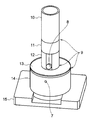
- FIG. 1 It is a schematic diagram of the component of a device. It is a front view which shows typically the device which assembled each unit. It is a SEM image of the 3D filter which captured the cancer cell (arrow) in the hollow (the right is an enlarged view on the left).
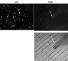
- the upper left is a GFP fluorescence image (cancer cell detection), and the upper right is a stained image (leukocyte detection) of a PE-labeled anti-CD45 antibody. Only a small number of white blood cells are observed, and no image of cancer cells and white blood cells coexisting in the same depression is observed.
- the lower right is a bright-field image when a manipulator (glass capillary) is manually operated on the filter cassette and one cancer cell is collected.
- the arrow is the manipulator.
- a sample of 500 ml of cultured cancer cell sputum (COLM5-EGFP) mixed with 2.5 mL of healthy human blood and diluted 10-fold with PBS is filtered using a 3D Pd / Ni filter, and the cancer cells are changed by changing the flow rate of the device.
- the recovery rate (%) of was investigated.
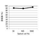
- a sample of healthy human blood (2.5 mL) mixed with cultured cancer cell sputum (COLM5-EGFP) and diluted 10-fold with PBS is filtered using a 3D Pd ⁇ Ni filter, and the number of cancer cells is changed. The recovery rate (%) was examined.
- the flow rate of the device is 2 ml / min.
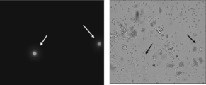
- Peripheral blood of a gastric cancer patient was filtered through a 3D Pd / Ni filter, and the cells were stained with a fluorescently labeled antibody.
- the upper left is a stained image of Alexa488-labeled anti-EpCAM antibody
- the upper right is a stained image of PE-labeled anti-CD45 antibody
- the lower left is a stained image of Hoechst 33342
- the lower right is a bright field image.
- the arrow is CTC.
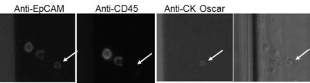
- the peripheral blood of a breast cancer patient was filtered through a 3D Pd / Ni filter, and the cells were stained with a fluorescently labeled antibody.
- Peripheral circulating tumor cells or metal filters that separate rare cells ⁇ CTC '' in this specification refers to circulating tumor cells contained in the blood of cancer patients and is much more in comparison with body fluid cells such as blood cells. A rare cell with a small number of cells.
- rare cells refers to rare tumor cells (“rare cancer cells”) contained in body fluids such as ascites, peritoneal lavage fluid, lymph fluid, and spinal fluid of cancer patients.
- the metal filter 17 of the present embodiment is a porous metal filter in which a very large number of holes (pore density of 1 ⁇ 10 4 / cm 2 or more) are uniformly or regularly formed in an ultrathin plate made of a specific metal.
- a conventionally known CTC separation filter is formed from a resin (or polymer) such as parylene or polycarbonate, or silicon.
- the CTC separation filter of the present embodiment is a metal filter 17, and has excellent characteristics different from a resin filter and the like as described below.
- the material of the metal filter 17 includes, for example, at least one of palladium (Pd), platinum (Pt), gold (Au), silver (Ag), iridium (Ir), rhodium (Rh), and ruthenium (Ru). .
- This material may be a single metal of palladium (Pd), platinum (Pt), gold (Au), silver (Ag), iridium (Ir), rhodium (Rh) or ruthenium (Ru) or, for example, palladium (Pd ) / Nickel (Ni) alloy, platinum (Pt) / nickel (Ni) alloy, or gold (Au) / nickel (Ni) alloy.
- the ratio of the above metal is preferably large with respect to a counterpart metal such as nickel.
- These metals for example, have very low toxicity to cells such as CTC and rare cells compared to metals such as nickel (Ni) (FIG. 1). This is because palladium (Pd) itself has low toxicity, and an alloy of Pd and nickel (Ni) can prevent elution of nickel (Ni) by forming a solid solution.
- palladium or an alloy of palladium (Pd) / nickel (Ni) is preferable.
- the metal filters of this embodiment such as Pd and Ni alloy filters and Pd filters are acid and heat resistant, and can be dyed in various ways such as the FISH method as they are. be able to.
- the cells are hard and durable, and the cells are difficult to adhere without surface treatment, micromanipulation with a glass capillary on the filter is easy (FIG. 4B). Therefore, the metal filter 17 of the present embodiment has an excellent advantage that it can immediately cope with the automation of sorting of one cell.
- the shape of the metal filter 17 is not particularly limited as long as it can be arranged on a filter ring (cassette) attached to the filtration unit of the CTC separation device, but includes, for example, a circular shape, a square shape, and preferably a circular shape.
- the size of the metal filter 17 can be appropriately determined in consideration of physical factors such as sample (blood) volume, pore size, time, flow rate, pressure resistance, operability, cost, and the like. For example, when processing 5 mL of blood, the diameter (in the case of a circle) or the length and width (in the case of a rectangle) is usually about 10 to 15 mm, but the size is not limited depending on the blood volume For example, it may be in the range of about 5 to 20 mm.
- the thickness of the metal filter 17 is appropriately determined in consideration of the relationship with the hole density, pressure resistance, cost, etc., and is usually 10 to 40 ⁇ m, preferably about 15 to 40 ⁇ m.
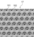
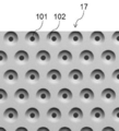
- the metal filter 17 has a large number of holes 101 that are uniformly and regularly arranged.
- the hole 101 is formed in a recess 102 described later.
- the density of the holes 101 per 1 cm 2 of the filter surface area varies depending on the arrangement form such as a lattice arrangement or a staggered arrangement, but is usually 1 ⁇ 10 4 to 2 ⁇ 10 5 / cm 2 , preferably 5 ⁇ 10 4 to 1 ⁇ 10 5 / cm 2 .
- the pore diameter of the hole 101 has a size that does not allow CTC or rare cells to pass therethrough and allows blood components such as blood cells to pass through body fluid cells other than CTC or rare cells.
- the size (major axis) of human blood cell components is about 6-7 ⁇ m for red blood cells, about 7-9 ⁇ m for white blood cells, and less than 5 ⁇ m for platelets as a result of histogram analysis. About 10 to 20 ⁇ m. Therefore, the hole diameter of the hole 101 at the opening position to the recess 102 is usually about 7 to 10 ⁇ m, preferably about 7.5 to 9 ⁇ m, and more preferably about 7.5 to 8.5 ⁇ m.
- the metal filter 17 further has a recess 102 (or a recess) having a size capable of capturing CTC or rare cells.
- the size of the recess 102 may be any size that can be captured by CTC or rare cells.
- the size is 20 to 30 ⁇ m in diameter and 5 to 15 ⁇ m in depth, preferably 25 to 30 ⁇ m in diameter and 10 ⁇ m in depth.
- the recess 102 may be formed above all or a part of the holes 101, and such a form is preferable in order to maintain a high hole density. If the depth is less than 5 ⁇ m, CTC cannot enter. A depth of at least about 5-15 ⁇ m is required to allow CTC to penetrate almost completely.
- the number of the depressions 102 is preferably 80 to 100%, more preferably 100% with respect to the number of the holes 101 (that is, the depressions 102 are present above all the holes 101).
- the “regularly arranged” holes 101 in the metal filter 17 means that the holes 101 are arranged as densely as possible, and all the holes 101 are arranged with a specific regularity. .
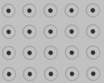
- regularity includes, for example, a lattice arrangement, a staggered arrangement, a radial arrangement, a concentric arrangement, and the like (FIGS. 2A, 2B, and 2C).
- 2A and 2B (houndstooth arrangement) has a recess 102 having a depth of 10 ⁇ m formed in the upper portion of the hole 101 and has a three-dimensional structure as a whole. This filter is called a 3D filter.
- 2C (lattice arrangement) has a recess having a depth of 1 ⁇ m at the top of the hole 101, but has a structure close to a plane as a whole. In this specification, the depth of such a recess is 5 ⁇ m.
- a filter of the form below is called a 2D filter. This 2D filter corresponds to the size selection microcavity array described in FIG. 5 of JP 2011-163830 A. This 2D filter cavity is too shallow for the size of the CTC or rare cells, so the CTC or rare cells trapped in the pores cannot be isolated from the surrounding residual blood cells.
- the metal filter 17 of the present embodiment is a 3D filter, and by providing the depression 102, 1) CTCs can be arrayed (patterned) one by one without contamination of blood cells, and 2) certain conditions.
- the strength with which the cells are trapped in the hole 101 can be controlled.
- Soft one-cell picking is possible (FIG. 4A).
- the depth of the dent 102 is deep (over 15 ⁇ m)
- there will be no effect on CTC recovery and filtration speed but conversely, one-cell patterning will be incomplete or resistance will be caused during picking. Can occur.
- the peripheral edge of the metal filter 17 is edged without a hole.
- the presence of the edge makes CTC measurements accurate and allows pinching with tweezers without damaging the metal filter 17.
- the metal filter 17 may be slightly warped depending on the electroforming conditions by providing the depression 102, the warp can be corrected if the support material such as polycarbonate is edged by ultrasonic welding from above and below. is there.
- the metal filter 17 of the present embodiment can be manufactured by using, for example, LIGA (Lithogaphie Galvanoformung Abfomung) technology (Hattori Tadashi, Surface Technology, Vol. 62, No. 12, 619-624, 2011; W. Elufeld and H. Lhe, Radiat. Phys. Chem., 45 (3): 340-365, 1995).
- LIGA LiGA
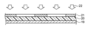
- a resist, an absorber (optional component), and a mask are stacked on a substrate, and a resist pattern is formed by irradiation with ultraviolet rays, X-rays, or synchrotron radiation, and then electroforming is performed using the substrate as an electrode.
- An electroformed filter can be produced by a method including performing metal plating, finishing electroforming where a predetermined opening is left, and further removing the resist (see FIGS. 8A to 8F).
- Peripheral circulating tumor cell (CTC) or rare cell separation device This embodiment further comprises a porous metal filter 17, a device for separating CTC or rare cells from body fluids such as blood. provide.
- Porous metal filter 17 is as described in section 1 above.
- a preferable filter is a palladium (Pd) filter or a palladium (Pd) / nickel (Ni) alloy filter.
- the metal filter 17 is mounted so that the flow path is perpendicular to the metal filter 17.
- the metal filter 17 is usually set to be removable or non-removable to the filter cassette 16, and the filter cassette 16 is fixed to the device.
- the filter cassette 16 is composed of two upper and lower members (upper and lower filters) having an edge with an appropriate width for fixing the metal filter 17 and an open space inside.
- the metal filter 17 is fixed between the members. If necessary, packing (for example, Teflon (registered trademark), rubber, paper, etc.) may be sandwiched between the member and the metal filter 17, thereby enabling deformation of the metal filter 17 and prevention of liquid leakage. (FIGS. 3B to 3C).
- the members of the filter cassette 16 are made of a material such as resin (polymer), rubber, or metal, and the two members are provided with means for fixing the filter. Examples of such means include a screw groove.
- the filter cassette 16 may have a structure in which the metal filter 17 is simply fixed (press-fit) with pressure from above and below.
- the filter cassette 16 may be manufactured so that the metal filter 17 and the filter cassette 16 are integrated. In this case, the metal filter 17 is fixed to the filter cassette 16 so that it cannot be removed.
- the device can further include a reservoir unit 10, a flow rate adjustment unit 12, and a drainage recovery unit 14 (a liquid reservoir base) in addition to a filtration unit 11 including a filter cassette 16 to which a metal filter 17 is attached (see FIG. 3A).
- the reservoir unit 10 is a container for storing a body fluid sample such as blood.
- the reservoir unit 10 is made of a material that can distinguish the liquid level such as glass or polymer (for example, transparent). You may have.
- the filtration unit 11 is as described above and includes a filter cassette 16 for detachably mounting the metal filter 17.
- the flow rate adjusting unit 12 has a device for adjusting the flow rate or flow rate of a body fluid such as blood that naturally flows down by its own weight (ie, gravity) through the filtration unit 11 including the reservoir unit 10 and the filter cassette 16.
- a device for example, a screw-in type convex screw having a sharp-angled structure tip, and when the screw is tightened, the waste tube is squeezed and the flow rate is reduced or zero (stopped), while When opening the screw, the flow rate increases.
- the screw is made of a material such as metal, resin, or polymer.
- the drainage recovery unit 14 includes a container (a sump base) that collects body fluid such as blood after filtration, drainage when the device is washed, and the like.
- the drainage recovery unit 14 is made of a material such as glass, resin, polymer, or metal.
- the fixing between the units may be performed using, for example, a screw mechanism 9 (FIG. 3A) that is attached or removed by rotation, or may be performed using a plug-in gap fitting 18 (FIG. 3C).
- a screw mechanism 9 FIG. 3A
- a plug-in gap fitting 18 FIG. 3C
- other conventional fixtures may be used.
- a stand for installing a structure in which the units are assembled can be provided.
- FIG. 3A An example of the device is shown in FIG. 3A (appearance), FIG. 3B (component), and FIG. 3C (front view of the device during assembly).
- the filtration unit 11 includes a filter cassette 16.
- the flow rate adjusting unit 12 includes a liquid amount adjusting screw 8.
- the drainage recovery unit 14 is provided with a liquid storage lid 13 on the upper surface and an air hole 7 on the upper side surface.
- the device further includes a stand 15.
- the device is preferably formed of a resin or polymer such as acrylic as a whole in terms of ease of processing and cost.
- a transparent material is preferable because the flow rate of the liquid can be monitored.
- the size of the device is about 10 to 15 cm in width and about 20 to 30 cm in height for an assembled structure excluding the stand 15, and is a size suitable for filtering a blood sample of about 5 to 7.5 ⁇ mL. Even if the sample volume is large, this size can be adequately handled by applying it in small portions.
- the above description is the specifications of the prototype, and the mass production type is separately manufactured based on the prototype, and is made of a material such as resin or plastic. Production is possible.
- the device of the present embodiment is a pumpless (that is, no pump used) system that obtains a flow rate for filtration by its own weight (gravity), and is therefore a release system. This is different from the driving force of the pump type or negative pressure tube type employed in the known filter type CTC separation device.
- these conventional methods there are problems such as applying stress to the cells, fixing the cells, and making it difficult to sort one living cell due to the closing of the flow path. Can solve such a problem.
- the device of this embodiment is applied to the metal filter 17 such as palladium, which has very low toxicity to cells, the ultra-high pore density of the metal filter 17, flat surface processing, and 3D conversion of the metal filter 17, thereby Even without pretreatment of bodily fluids, high-speed filtration of blood cells was enabled while maintaining a relatively low flow rate at which CTC or rare cells were trapped in the pore 101. Specifically, the flow rate through the hole 101 is relatively slow while high-speed filtration in about 30 minutes including washing is possible for 5 ml of whole blood. In addition, since the surface properties of the pore 101 are extremely smooth, stress and damage to cells are low. As a result, CTCs or rare cells can be aligned in a state where there is little contamination such as blood cells.
- the metal filter 17 such as palladium
- CTC Peripheral Circulating Tumor Cell
- Rare Cell Separation Method This embodiment further provides a method for separating CTC or rare cells from body fluid of cancer patients using a device equipped with the metal filter 17 described above. .
- this method involves injecting a body fluid selected from, for example, blood, ascites, peritoneal lavage fluid, cerebrospinal fluid or lymph fluid of a cancer patient into the device of this embodiment, and a flow rate that relies solely on gravity.
- a body fluid selected from, for example, blood, ascites, peritoneal lavage fluid, cerebrospinal fluid or lymph fluid of a cancer patient
- a flow rate that relies solely on gravity.
- the body fluid of a cancer patient as a specimen is a body fluid containing CTC or rare cells or suspected of containing CTC or rare cells, and is mainly blood or lymph.
- the amount (volume) of body fluid used for separation of CTC or rare cells is usually 1 to 20 mL, preferably 3 to 7 mL.
- body fluid particularly blood
- PBS phosphate buffered saline
- PBS PBS containing 0.25 to 1 mM EDTA depending on the purpose
- the following pretreatment may be performed.
- the pretreatment is performed, for example, by mixing a body fluid with a magnetic nanoparticle-containing cationic liposome (MCL) and incorporating the magnetic nanoparticles into CTC or rare cells and leukocytes in the body fluid, and then the metal filter of this embodiment.
- MCL magnetic nanoparticle-containing cationic liposome
- This method includes removing cells such as erythrocytes that are not magnetized with a magnet disposed in the middle of the flow path from the body fluid (prefilter). This utilizes the property that magnetic nanoparticles are not normally taken up by red blood cells, whereas magnetic nanoparticles are taken up by CTC or rare cells or white blood cells.
- This method is a simple method of injecting body fluid into the device and filtering by gravity flow rate, but it is a 3D metal with ultra-high pore density (eg 5 ⁇ 10 4 to 1.5 ⁇ 10 5 / cm 2 )
- a sufficiently rapid process that is acceptable can be achieved.
- about 5.0 mL of specimen (whole blood) can be processed in about 30 minutes including washing.
- the washing solution uses phosphate buffered saline (PBS), but may contain EDTA if necessary.
- a fluorescently labeled antibody that specifically binds to CTC or rare cells such as Alexa488-labeled anti-EpCAM antibody or PE label that specifically binds to leukocytes.
- an antibody mixture such as an anti-CD45 antibody
- CTC or rare cells are stained, and the number of CTC or rare cells is measured with a microscope. At this time, the staining can be performed directly on the metal filter 17 fixed to the device.
- the FISH method for examining the presence or absence of gene amplification can be performed by removing the metal filter 17 from the filter cassette 16 and fixing it as it is.
- the 3D-Pd / Ni alloy filter device of the present embodiment can also be used for measuring CTC in the peripheral blood of cancer-bearing mice.
- the present inventors have developed a CTC model (mouse) that can detect CTC with high reproducibility using a subcutaneous transplantation natural metastasis model such as a GFP-introduced colon cancer cell line (COLM5-EGFP) and a gastric cancer cell line (GCIY-EGFP). Multiple strains were prepared.
- the mouse vena cava blood 1 to 3 months after transplantation was collected and filtered with the 3D-Pd / Ni alloy filter device of this embodiment, and the number of CTCs was measured using GFP fluorescence as an index.
- the method of this embodiment can be expected to play an important role in both the diagnosis and treatment of metastatic cancer, such as early diagnosis of metastasis recurrence, companion diagnosis and evaluation of therapeutic effects of drugs.
- the device and method of the present embodiment are expected to be useful for basic research such as elucidation of metastasis mechanisms such as CTC in vivo.
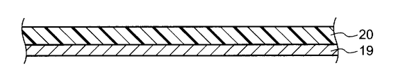
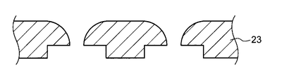
- Example 1 [Production method of palladium / nickel filter] As shown in FIG. 8A, a photosensitive resist 20 was applied to the substrate 19 to a predetermined thickness.
- a conductive film layer for example, copper
- a conductive material such as a stainless steel substrate, a nickel substrate, or a copper substrate may be used.
- a photosensitive resist 20 was patterned on the substrate 19 through a development process.
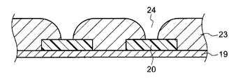
- a deposited film 23 of palladium / nickel alloy (Pd: Ni weight ratio, for example, 80:20) is deposited from the surface of the substrate 19 by electroforming and patterned.
- the Pd / Ni deposition film 23 was continuously deposited by electroforming so as to cover the photosensitive resist 20, and the electroforming was terminated when a predetermined hole 24 was left.
- the formed photosensitive resist 20 was removed with an organic solvent.
- This photosensitive resist 20 may be removed by dry etching using oxygen plasma.
- the substrate 19 was peeled off or removed by etching.
- a palladium / nickel filter also referred to as “3D filter” or “Pd / Ni filter” was produced.
- Example 2 [Characteristics of palladium / nickel filters] Since this palladium / nickel filter is manufactured by microfabrication using a unique and advanced electroforming technology, it has a high pore density of more than 100,000 / cm 2 and 10% or more compared to other polycarbonate, parylene or silicon filters. Usually, high aperture ratio of 50% or more can be achieved. As a result, high-speed filtration of whole blood became possible, and CTC could be collected at a high rate and quickly without clogging (FIGS. 2A and 2B). In addition, high-precision processing such as smooth surface properties and uniformity of pores and the use of palladium, which has low cytotoxicity, can significantly reduce cell damage (FIGS. 1A and 1B).
- this palladium / nickel filter can be heat-treated at 121 ° C for 30 minutes (Autoclave), and in chemical resistance tests (1 hour) with hypochlorous acid (commercially 1% hyter) and 1N hydrochloric acid. There was no change in shape, and there was no change in functionality such as CTC recovery. As a result, sterilization can be performed, and cost reduction such as use and reuse in the medical field can be achieved.
- Example 3 [Cell Spike experiment using cultured cancer cells] Human gastric cancer cells MKN-45, GCIY and human colon cancer cells COLM-5 were seeded in RPMI medium containing 10% fetal bovine serum so that the concentration was 1.0 ⁇ 10 6 cells / 10 cm dish, and the temperature was 37 ° C. / Static culture was performed under 5% CO 2 . After 3 to 4 days of culture, the medium was removed by suction, washed with PBS (pH 7.2), 0.2% trypsin / 2 mM EDTA was added, and the mixture was incubated at 37 ° C./5% CO 2 for 3 to 5 minutes. .
- RPMI medium containing serum was added to stop the action of trypsin, and then pipetting was performed to collect the cells.
- the collected cells were centrifuged (1200 rpm, 3 minutes), the supernatant was aspirated and resuspended in fresh RPMI medium.
- the number of viable cells was counted by a dye exclusion method using trypan blue, adjusted to 1 ⁇ 10 6 cells / ml, and then seeded on a new dish.
- COLM5-EGFP cells which are cell lines in which the green fluorescent protein (GFP) gene is forcibly expressed in human colon cancer cells COLM-5 and human gastric cancer cells GCIY.
- GFP cells were used.
- 50 to 500 or 5000 cultured cancer cells were mixed with 2 to 2.5 ml of blood collected from a healthy person into a vacuum blood collection tube containing EDTA2Na to prepare a sample. .
- the fluid volume adjusting screw is tightened, the flow path is closed, and 500 ⁇ l of cell staining solution (Alexa488-labeled anti-EpCAM antibody, PE-labeled anti-CD45 antibody and Hoechst 33342, 0.5 ⁇ g / ml each).
- cell staining solution Alexa488-labeled anti-EpCAM antibody, PE-labeled anti-CD45 antibody and Hoechst 33342, 0.5 ⁇ g / ml each.
- the latter two cell staining solutions were introduced. After reacting at room temperature for 30 minutes with light shielding, the liquid volume adjusting screw was opened and the cell staining solution was discharged.
- the cells on the filter were washed by introducing PBS containing 5 ml of 1 mM EDTA / 0.5% BSA into the reservoir. Perform this operation once more with PBS alone, tighten the liquid volume adjustment screw, then immediately remove the filter cassette from the CTC separation device, add 100-200 ⁇ l PBS to the liquid reservoir at the top of the filter cassette, and place it on the microscope stage. The fluorescence microscope observation was performed as it was.
- an upright reflection fluorescent microscope (Eclipse LV100ND; Nikon) equipped with a cooled CCD camera (Ri-1; Nikon) was used. Images were acquired using UV-1A, FITC, and Cy3 filters, respectively, to observe fluorescence from Hoechst 33342, Alexa 488, and PE. NIS-Elements (Nikon) was used as image acquisition and analysis software.
- the CTC separation device equipped with the 3D filter has a liquid amount adjusting screw, and the flow rate can be controlled by adjusting the amount of liquid flowing by its own weight.
- the flow rate is changed to 2 ml / min, 4 ml / min, or 8 ml / min using 10-fold diluted blood mixed with 500 cultured cancer cells (2.5 ml of blood diluted 10-fold with PBS) The recovery rate was examined.
- a Pd / Ni filter having a depression diameter of 30 ⁇ m, a depression depth of 10 ⁇ m, a hole diameter of 8 ⁇ m, a hole density of 99,178 / cm 2 , and a staggered lattice arrangement was used.
- high recovery rates of about 90% and 80% were exhibited at 2 ml / min and 4 ml / min, respectively, but at 8 ml / min, the recovery rate was 70% or less. Since it was found that a recovery rate of 80 to 90% can be obtained at a flow rate of 2 to 4 ml / min, the subsequent experiments were conducted at a flow rate in this range (FIG. 5A).
- the flow rate of the device is 2 ml / min.
- the recovery rate from diluted blood mixed with 50 cells, 500 cells, and 5000 cells was 85 to 95% (FIG. 5B).
- the recovery rate from diluted blood mixed with only 10 cells was 70% on average, and it was shown that even when a small number of cells were taken into consideration, the recovery rate was high even when the number of cells was counted.
- the metal filter is processed into a 3D shape having a recess in the upper part of the through hole as shown in FIG. 2A.
- This shape is preferably 20 to 30 ⁇ m in diameter and 5 to 15 ⁇ m in depth.
- the hollow is most recovered when the diameter is 30 ⁇ m. It was easy.
- FIG. 4B shows a bright-field image when one cancer cell is collected with a manipulator.
- Example 4 [Separation of CTC from CTC model mouse] Since in vitro cultured cells and in vivo CTC have significantly different cell living environments, it is difficult to accurately evaluate the performance of the CTC separation device only by the Cell Spike experiment in which cultured cancer cells are added to the blood of healthy persons. On the other hand, for patient clinical specimens, it is difficult to evaluate the performance of a device unless many cases with different degrees of cancer progression (Stage) are collected, measured, and statistically analyzed. On the other hand, if there is a mouse in which CTC appears reproducibly with time after cancer cell transplantation (hereinafter referred to as “CTC model mouse”), rapid and accurate evaluation of the performance of the CTC separation device can be expected. .
- the CTC model mouse is also extremely useful for basic research of CTC, such as analysis of CTC in vivo dynamics. Therefore, a CTC model mouse was independently produced.
- lung metastases are limited to a small number of micrometastasis until 1 to 2 months after transplantation, and the number of CTCs is almost 0 (zero), rarely 1 to 2 CTCs are detected. It was the degree that was done.
- lung metastases are also observed macroscopically, and a small number of metastases are also observed in the liver and kidneys. At this time, about 10 to 100 CTCs are detected, and more than 1000 are detected. CTCs were detected (results are shown in FIGS. 6A and 6B).
- CTCs were detected in accordance with the appearance of gross and systemic metastases.
- this device was also used for experiments in a lung cancer model using the mouse Lewis lung cancer cell line (Lewis-GFP), and it was revealed that the appearance of CTC was synchronized with multi-organ metastasis to the liver, kidney, and the like. Since CTC appears synchronously with gross metastasis and systemic metastasis, it was suggested that CTC detection may be useful for early diagnosis of cancer metastasis.
- studies using CTC model mice showed that this device has sufficient sensitivity and accuracy to accurately capture the in-vivo dynamics of CTC.
- Example 5 [Separation of CTC from peripheral blood of gastric cancer patients and breast cancer patients] 5 ml of blood was collected from the elbow vein of 4 gastric cancer patients, 6 breast cancer patients, and 4 healthy persons using a blood collection tube containing EDTA2Na.
- Four stomach cancer patients and six breast cancer patients are patients admitted to Aichi Cancer Center Central Hospital (Nagoya, Japan). Blood collection from the patients was approved by the Aichi Cancer Center Ethics Committee (Nagoya, Japan), and the consent was obtained from the patients.
- a 3D Pd ⁇ Ni filter having a recess diameter of 24 ⁇ m, a recess depth of 10 ⁇ m, a hole diameter of 8 ⁇ m, and a hole density of 136,325 / cm 2 was used. Filtration was performed at a flow rate of 2.5 ml / min.
- the cells on the filter cassette were washed with PBS, fixed with 10% formalin for 10 minutes at room temperature, washed, and then stained with Alexa488-labeled anti-EpCAM antibody, PE-labeled anti-CD45 antibody, and optionally Alexa350-labeled anti-CK Oscar antibody.
- the solution was added onto the filter cassette and allowed to react for 30 minutes at room temperature. Further, after washing with PBS, nuclear staining was performed with Hoechst 33342. After washing with PBS, the entire filter cassette was transferred to an upright fluorescent microscope, EpCAM + / (CK Oscar + ) / CD45 ⁇ / Hoechst 33342 + cells were determined as CTC, and the number of CTCs was counted.
- FIG. 7A shows a stained image of a stomach cancer patient
- FIG. 7B shows a stained image of a breast cancer patient
- FIG. 7C shows the measurement result of the number of CTCs.
- Example 6 [Combination of magnetic separation of cells by magnetic nanoparticles] Aiming for total recovery of CTC using gastric cancer cell N87, combined with magnetic separation using magnetic nanoparticles (pretreatment) and separation by size using 3D Pd / Ni filter to improve recovery rate Tried.
- Magnetic Cationic Liposome (diameter 150 nm) 100 pg / cell was allowed to act on a suspension of 100 N87 cells for 2 hours, mixed with bovine serum containing 200,000 white blood cells, and then combined with a magnetic separation channel and a filter. Captured with device. Approximately 80 to 90% of cells could be captured and recovered, and an improvement in recovery rate was observed compared to the filter alone.
- the device equipped with the metal filter of the present invention achieves the separation of CTC or rare cells in body fluids such as blood at a high recovery rate, and CTC or rare cells can be collected in an almost intact state. It can be used for genetic analysis of single CTC cells as well as evaluation of the number, and may be used effectively for early diagnosis of metastatic recurrence, monitoring of therapeutic effects, and Liquid biopsy (fluid biopsy) high.
Landscapes
- Health & Medical Sciences (AREA)
- Engineering & Computer Science (AREA)
- Life Sciences & Earth Sciences (AREA)
- Chemical & Material Sciences (AREA)
- Biomedical Technology (AREA)
- Bioinformatics & Cheminformatics (AREA)
- Wood Science & Technology (AREA)
- Biotechnology (AREA)
- Organic Chemistry (AREA)
- Zoology (AREA)
- General Health & Medical Sciences (AREA)
- Genetics & Genomics (AREA)
- Heart & Thoracic Surgery (AREA)
- Microbiology (AREA)
- Biochemistry (AREA)
- General Engineering & Computer Science (AREA)
- Vascular Medicine (AREA)
- Public Health (AREA)
- Hematology (AREA)
- Veterinary Medicine (AREA)
- Animal Behavior & Ethology (AREA)
- Anesthesiology (AREA)
- Cell Biology (AREA)
- Chemical Kinetics & Catalysis (AREA)
- Medicinal Chemistry (AREA)
- Oncology (AREA)
- Sustainable Development (AREA)
- Cardiology (AREA)
- Analytical Chemistry (AREA)
- Apparatus Associated With Microorganisms And Enzymes (AREA)
- Micro-Organisms Or Cultivation Processes Thereof (AREA)
- Investigating Or Analysing Biological Materials (AREA)
- Measuring Or Testing Involving Enzymes Or Micro-Organisms (AREA)
Abstract
Description
[1] 体液中の末梢循環腫瘍細胞又は希少細胞を捕捉することができる窪みと、該窪みに形成され前記末梢循環腫瘍細胞又は前記希少細胞以外の体液細胞を通過させることができる孔とを有する金属フィルターを備えた末梢循環腫瘍細胞又は希少細胞分離用デバイス。
[2] 前記フィルターの金属材質が、パラジウム又はパラジウム・ニッケル合金である[1]に記載の末梢循環腫瘍細胞又は希少細胞分離用デバイス。
[3] 前記窪みの直径は20~30μmであり、前記窪みの深さは5~15μmである[1]又は[2]に記載の末梢循環腫瘍細胞又は希少細胞分離用デバイス。
[4] 前記孔の直径は7~10μmである[1]~[3]のいずれか1つに記載の末梢循環腫瘍細胞又は希少細胞分離用デバイス。
[5] 前記窪み及び前記孔の孔密度は1cm2あたり1×10⁴~2×10⁵個である[1]~[4]のいずれか1つに記載の末梢循環腫瘍細胞又は希少細胞分離用デバイス。
[6] 前記金属フィルターの厚さは、10~40μmである[1]~[5]のいずれかに記載の末梢循環腫瘍細胞又は希少細胞分離用デバイス。
[7] 前記金属フィルターを装着するためのフィルターカセットを有する[1]~[6]のいずれかに記載の末梢循環腫瘍細胞又は希少細胞分離用デバイス。
[8] [1]~[7]のいずれかに記載の末梢循環腫瘍細胞又は希少細胞分離用デバイスに、がん患者の血液、腹水又は腹腔洗浄液から選択される体液を注入して、該体液中の前記末梢循環腫瘍細胞又は前記希少細胞を、前記金属フィルターの前記窪みで捕捉した後、生きたまま回収する末梢循環腫瘍細胞又は希少細胞分離方法。
[9] 前記末梢循環腫瘍細胞又は前記希少細胞を特異的に染色することをさらに含む、[8]に記載の末梢循環腫瘍細胞又は希少細胞分離方法。
[10] 前記体液を希釈又は前処理することを含む、[8]又は[9]に記載の末梢循環腫瘍細胞又は希少細胞分離方法。
[11] 前記前処理が、前記体液を磁性ナノ粒子含有カチオン性リポソームと混合して、前記体液中の前記末梢循環腫瘍細胞又は前記希少細胞及び白血球細胞中に前記磁性ナノ粒子を取り込ませたのち、前記金属フィルターに通じる流路に流し込み、前記金属フィルターの前に配置された磁石で磁性ナノ粒子を取り込まない赤血球等の細胞を該体液から除去することを含む、[10]に記載の末梢循環腫瘍細胞又は希少細胞分離方法。
本明細書中の「CTC」とは、がん患者の血液に含まれる循環腫瘍細胞であり、血液細胞等の体液細胞に比べて非常に細胞数が少ない希少な細胞をいう。
本実施形態はさらに、多孔性の金属フィルター17を備えたことを特徴とする、血液等の体液からCTC又は希少細胞を分離するためのデバイスを提供する。
その結果、血球細胞などのコンタミネーションが少ない状態で、CTC又は希少細胞を整列させることができる。しかも孔101の上に窪み102を設けることにより、一定の条件下では流体力学的に重力と逆方向の流れが発生すると考えられる。このため、CTCが孔101に深くトラップされることなく、従ってCTCにかかる剪断応力を最小限に抑えることができる。このため、CTC又は希少細胞の細胞傷害を最小限に抑えることができ、CTC又は希少細胞をほぼ100%の生存率でかつ約80%以上の回収率で分離することが可能である。
本実施形態はさらに、上で説明した金属フィルター17を装着したデバイスを使用してがん患者の体液からCTC又は希少細胞を分離する方法を提供する。
[パラジウム・ニッケルフィルターの製作方法]
図8Aに示すように、基板19に感光性レジスト20を所定の厚さに塗布した。尚、基板19として、導電性のないシリコン基板やガラス基板を用いる場合は導電膜層(例えば銅)を付加するが、ステンレス基板、ニッケル基板、銅基板などの導電性を有するものでもよい。
このような工程を経て、パラジウム・ニッケルフィルター(「3Dフィルター」又は「Pd・Niフィルター」ともいう)を製作した。
[パラジウム・ニッケルフィルターの特徴づけ]
本パラジウム・ニッケルフィルターは、独自かつ高度の電鋳技術で微細加工して作製されるため、他のポリカーボネート、パリレンまたはシリコン製のフィルターにくらべて100,000/cm2以上の高孔密度、10%以上、通常50%以上の高開口率を達成できる。これにより全血の高速濾過が可能になり、目詰まりもなく、CTCの高率かつ迅速な回収が可能となった(図2Aおよび図2B)。また、平滑な表面性状や孔の均一性など高精度な加工と細胞毒性の少ないパラジウムを材質とすることが相まって細胞障害も大幅に低減しうる(図1A,図1B)。
[培養がん細胞を用いたCell Spike実験]
ヒト胃がん細胞MKN-45、GCIYおよびヒト大腸がん細胞COLM-5を、10%牛胎児血清を含むRPMI培地に、1.0×10⁶個/10cm dishとなるように播種し、37℃/5%CO2下で静置培養した。培養3~4日後、培地を吸引除去し、PBS(pH7.2)で洗浄した後、0.2%トリプシン/2mM EDTAを添加し、37℃/5%CO2下で3~5分インキュベートした。検鏡し細胞がdishから剥離していることを確認して、血清を含むRPMI培地を加え、トリプシンの作用を停止させた後ピペッティングし、細胞を回収した。回収した細胞は遠心分離(1200rpm、3分)し、上清を吸引除去して新鮮なRPMI培地に再懸濁した。トリパンブルーを用いた色素排除法により生細胞数を計数し、1×106細胞/mlに調製後、新しいdishに播種した。
3Dフィルターを装着したCTC分離デバイスのリザーバーユニットに1mM EDTA/0.5%BSAを含むPBSを5ml導入してリザーバーおよびフィルターを洗浄した後、GFP導入あるいは非導入培養がん細胞 (50、500、又は5000個)を添加した血液(2~2.5ml)をPBSで5~10倍に希釈し導入した。約10分間の濾過後、残留する血球成分を洗浄するためPBSを5ml導入した。GFP非導入培養がん細胞の場合、PBS洗浄液を流しきった後、液量調節ねじを締め、流路を閉じた後、500μlの細胞染色液(Alexa488標識抗EpCAM抗体、PE標識抗CD45抗体およびHoechst33342。それぞれ0.5μg/ml)を導入した。GFP導入培養がん細胞の場合、後2者からなる細胞染色液を導入した。遮光して30分室温で反応させた後、液量調節ねじを開放し、細胞染色液を排出した。リザーバーに5mlの1mM EDTA/0.5%BSAを含むPBSを導入してフィルター上の細胞を洗浄した。この作業をPBSのみでもう1回行い、液量調節ねじを締めた後、CTC分離デバイスからフィルターカセットを直ちに取り出し、フィルターカセット上部の液貯めに100~200μlのPBSを添加し、顕微鏡ステージに載せ、そのまま蛍光顕微鏡観察を行った。
3Dフィルターを装着したCTC分離デバイスには液量調節ねじがあり、自重によって流れる液量を調節することにより、流速を制御することができる。培養がん細胞500個を混合した10倍希釈血液(2.5mlの血液をPBSで10倍に希釈)を用いて、2ml/min、4ml/min、又は8ml/minに流速を変化させた場合の回収率を検討した。3Dフィルターは、窪みの直径30μm、窪みの深さ10μm、孔の直径8μm、孔密度99,178/cm2、千鳥格子配列のPd・Niフィルターを使用した。その結果、2ml/min、4ml/minでは各々約90%、80%と高い回収率を示したが、8ml/minでは70%以下であった。2~4ml/minの流速であれば、80~90%の回収率を得られることが分かったので、以降の実験はこの範囲の流速で行うこととした(図5A)。
5000個の培養がん細胞を混合した希釈血液(2.5mlの血液をPBSで10倍に希釈)を3Dフィルターで分離し、捕捉した細胞の生存率についてトリパンブルーを用いた色素排除法により計数した。3Dフィルターは、窪みの直径30μm、窪みの深さ10μm、孔の直径8μm、孔密度99,178/cm2のPd・Niフィルターを使用した。回収した細胞の生存率は99%で、非常に細胞傷害性の低い回収方法であることが示された。ただし、1mM以上のEDTA存在下で濾過した場合、回収したがん細胞の増殖性を見ると、対照(EDTA無添加)に比べて有意に低かった。しかし、抗凝固剤としてヘパリンを用い、コラーゲンコートdishを用いて接着を促進させると増殖性は有意に回復した。従って、EDTA存在下で濾過した場合の回収がん細胞の増殖性の低下はフィルターデバイスからのストレスよりも長時間のEDTA処理が原因と考えられた。
がん細胞の捕捉、整列および回収の効率を向上させる目的で、金属フィルターは図2Aに示すように貫通孔の上部に窪みを設けた3D形状に加工している。この形状は直径20~30μm、深さ5~15μmが望ましいが、先端径を40μm程度に加工したガラスキャピラリーを用いて捕捉した細胞を吸引回収する場合、窪みの直径は30μmの場合が最も回収しやすかった。窪みの深さ5μmと10μmとを比較すると、深さは5μmよりも10μmの方が、マニピュレーション等により液面が動いても細胞は捕捉された窪みから移動しにくいため、安定して、しかもコンタミネーションなく、目標とする1細胞の回収が可能であった(図4Bに、マニピュレーターでがん細胞1個を回収しているときの明視野像を示す。)。抗凝固剤としてヘパリンを用いると、細胞の金属フィルターへの接着性が増し(なお、EDTA存在下では細胞は金属フィルターへの接着性を殆ど示さない)、マニピュレーターによる細胞の回収率が低下した。従って、抗凝固剤は目的に応じて使い分ける必要があると考えられる。
[CTCモデルマウスからのCTCの分離]
in vitroの培養細胞とin vivoのCTCでは細胞の生存環境が著しく異なるため、健常人血液に培養がん細胞を添加するCell Spike実験だけではCTC分離デバイスの性能を正確に評価することは難しい。一方、患者の臨床検体では、がんの進行度(Stage)の異なる多数の症例を収集し、これを測定し、統計的に解析しないとデバイスの性能を評価することは難しい。これらに対し、がん細胞移植後、経時的に再現性をもってCTCが出現するマウス(以下「CTC モデルマウス」と呼ぶ)が存在すれば、CTC分離デバイスの性能の迅速かつ正確な評価が期待できる。また、CTCモデルマウスは、CTCの生体内におけるダイナミックスの解析など、CTCの基礎的な研究にも極めて有用である。そこで独自にCTCモデルマウスの作製を行った。
GFP遺伝子が導入されたことにより高輝度の緑色蛍光を発する転移性の高い、胃がん細胞株(GCIY-EGFP)と大腸がん細胞株(COLM5-EGFP)をそれぞれ、ヌードマウスの皮下に移植した。経時的にマウスを屠殺し、後大静脈から0.5~1.0mlの血液を採取し、PBSで10倍希釈後にCTC分離デバイスでCTCを測定した。同時に倒立型蛍光顕微鏡で肺、肝、腎などの微小転移の有無を検討した。大腸がんCOLM5-EGFP皮下移植モデルでは、移植後1~2ヶ月までは肺転移は少数の微小転移に限られ、CTC数もほとんど0(ゼロ)で、稀に1~2個のCTCが検出された程度であった。一方、移植後2~3ヶ月では、肉眼的にも肺転移が認められ、少数ながら肝、腎にも転移が認められ、このとき10~100個程度のCTCが検出され、多いときには1000個以上のCTCが検出された(図6A及び図6Bに結果を示す)。胃がんGCIY-EGFP皮下移植モデルでも、肉眼的転移および全身転移の出現に合わせてCTCが検出された。同様に本デバイスを用いてマウスLewis肺がん細胞株(Lewis-GFP)を用いた肺がんモデルでも実験を行い、CTCの出現が肝、腎などへの多臓器転移と同期することを明らかにした。CTCが肉眼的転移や全身的転移と同期して出現することから、CTC検出は、がん転移の早期診断に有用である可能性が示唆された。また、CTCモデルマウスを用いた検討から、本デバイスはCTCの生体内動態を正確にとらえることができる充分な感度と精度を備えていることが示された。
[胃がん患者及び乳がん患者の末梢血からのCTC分離]
胃がん患者4名、乳がん患者6名、健常人4名の肘静脈からEDTA2Na入り採血管で5mlの採血を行った。胃がん患者4名および乳がん患者6名は、愛知県がんセンター中央病院(日本、名古屋)に入院した患者である。患者からの採血は、愛知県がんセンター倫理委員会(日本、名古屋)で承認され、患者から同意書を取得した上で行われた。
[磁性ナノ粒子による細胞の磁気分離の併用]
胃がん細胞N87を用いて、CTCの全回収を目指し、磁性ナノ粒子を用いた磁気分離(前処理)と、3D Pd・Niフィルターを用いた大きさによる分離とを併用し、回収率の向上を試みた。Magnetic Cationic Liposome(直径150nm)100pg/細胞をN87細胞100個の細胞懸濁液に2時間作用させ、白血球細胞20万個を含むウシ血清に混和させた後、磁気分離流路とフィルターを組み合わせたデバイスで捕捉した。約80~90%の細胞を捕捉回収することができ、フィルター単独に比べて回収率の向上が認められた。
7・・空気孔
8・・液量調節ねじ
9・・ねじ機構(固定用)
10・・リザーバーユニット
11・・濾過ユニット(中にフィルターカセットを含む)
12・・流量調節ユニット
13・・液だめフタ
14・・排液回収ユニット(液だめベース)
15・・スタンド
16・・フィルターカセット(フィルターリングとも呼ぶ)
17・・金属フィルター
18・・隙間はめ(差込み式)
19・・基板
20・・感光性レジスト
21・・Crマスク
22・・紫外線
23・・パラジウム(Pd)・ニッケル(Ni)合金の析出成膜
24・・開孔
Claims (11)
- 体液中の末梢循環腫瘍細胞又は希少細胞を捕捉することができる窪みと、該窪みに形成され前記末梢循環腫瘍細胞又は前記希少細胞以外の体液細胞を通過させることができる孔とを有する金属フィルターを備えた末梢循環腫瘍細胞又は希少細胞分離用デバイス。
- 前記金属フィルターの材質が、パラジウム又はパラジウム・ニッケル合金である請求項1に記載の末梢循環腫瘍細胞又は希少細胞分離用デバイス。
- 前記窪みの直径は20~30μmであり、前記窪みの深さは5~15μmである請求項1又は請求項2に記載の末梢循環腫瘍細胞又は希少細胞分離用デバイス。
- 前記孔の直径は7~10μmである請求項1~請求項3のいずれか1項に記載の末梢循環腫瘍細胞又は希少細胞分離用デバイス。
- 前記窪み及び前記孔の孔密度は、1cm2あたり1×10⁴~2×10⁵個である請求項1~請求項4のいずれか1項に記載の末梢循環腫瘍細胞又は希少細胞分離用デバイス。
- 前記金属フィルターの厚さは、10~40μmである請求項1~5のいずれか1項に記載の末梢循環腫瘍細胞又は希少細胞分離用デバイス。
- 前記金属フィルターを装着するためのフィルターカセットを有する請求項1~請求項6のいずれか1項に記載の末梢循環腫瘍細胞又は希少細胞分離用デバイス。
- 請求項1~7のいずれか1項に記載の末梢循環腫瘍細胞又は希少細胞分離用デバイスに、がん患者の血液、腹水又は腹腔洗浄液から選択される体液を注入して、該体液中の前記末梢循環腫瘍細胞又は前記希少細胞を、前記金属フィルターの前記窪みで捕捉した後、生きたまま回収する末梢循環腫瘍細胞又は希少細胞分離方法。
- 前記末梢循環腫瘍細胞又は希少細胞を特異的に染色することをさらに含む、請求項8に記載の末梢循環腫瘍細胞又は希少細胞分離方法。
- 前記体液を希釈又は前処理することを含む、請求項8又は請求項9に記載の末梢循環腫瘍細胞又は希少細胞分離方法。
- 前記前処理が、前記体液を磁性ナノ粒子含有カチオン性リポソームと混合して、前記体液中の前記末梢循環腫瘍細胞又は前記希少細胞及び白血球細胞中に前記磁性ナノ粒子を取り込ませたのち、前記金属フィルターに通じる流路に流し込み、前記金属フィルターの前に配置された磁石で磁性ナノ粒子を取り込まない赤血球等の細胞を該体液から除去することを含む、請求項10に記載の末梢循環腫瘍細胞又は希少細胞分離方法。
Priority Applications (5)
| Application Number | Priority Date | Filing Date | Title |
|---|---|---|---|
| JP2014546215A JP5961889B2 (ja) | 2013-07-24 | 2014-07-23 | 末梢循環腫瘍細胞分離用デバイス、希少細胞分離用デバイス、末梢循環腫瘍細胞分離方法及び希少細胞分離方法 |
| KR1020147034140A KR101630110B1 (ko) | 2013-07-24 | 2014-07-23 | 말초 순환 종양 세포 또는 희소 세포 분리용 디바이스 및 말초 순환 종양 세포 또는 희소 세포 분리 방법 |
| US14/407,466 US10022659B2 (en) | 2013-07-24 | 2014-07-23 | Device for isolating periphery circulating tumor cells or rare cells, and method of isolating periphery circulating tumor cells or rare cells |
| EP14801905.2A EP2857495B1 (en) | 2013-07-24 | 2014-07-23 | Device for isolating peripheral circulating tumor cells or rare cells, and method for isolating peripheral circulating tumor cells or rare cells |
| CN201480001457.2A CN104520420B (zh) | 2013-07-24 | 2014-07-23 | 末梢循环肿瘤细胞或稀少细胞分离用装置及末梢循环肿瘤细胞或稀少细胞分离方法 |
Applications Claiming Priority (2)
| Application Number | Priority Date | Filing Date | Title |
|---|---|---|---|
| JP2013-153717 | 2013-07-24 | ||
| JP2013153717 | 2013-07-24 |
Publications (1)
| Publication Number | Publication Date |
|---|---|
| WO2015012315A1 true WO2015012315A1 (ja) | 2015-01-29 |
Family
ID=52393348
Family Applications (1)
| Application Number | Title | Priority Date | Filing Date |
|---|---|---|---|
| PCT/JP2014/069469 Ceased WO2015012315A1 (ja) | 2013-07-24 | 2014-07-23 | 末梢循環腫瘍細胞又は希少細胞分離用デバイス、及び末梢循環腫瘍細胞又は希少細胞分離方法 |
Country Status (6)
| Country | Link |
|---|---|
| US (1) | US10022659B2 (ja) |
| EP (1) | EP2857495B1 (ja) |
| JP (1) | JP5961889B2 (ja) |
| KR (1) | KR101630110B1 (ja) |
| CN (1) | CN104520420B (ja) |
| WO (1) | WO2015012315A1 (ja) |
Cited By (26)
| Publication number | Priority date | Publication date | Assignee | Title |
|---|---|---|---|---|
| WO2016068249A1 (ja) * | 2014-10-31 | 2016-05-06 | 国立大学法人東京農工大学 | 細胞単離方法及び細胞捕捉フィルタ |
| JP2016195986A (ja) * | 2015-04-06 | 2016-11-24 | 富士フイルム株式会社 | ろ材、フィルタユニット、及び細胞シート |
| WO2017022810A1 (ja) * | 2015-08-06 | 2017-02-09 | 株式会社オプトニクス精密 | 細胞分離フィルタ及び細胞培養容器 |
| WO2017130563A1 (ja) * | 2016-01-25 | 2017-08-03 | 日立化成株式会社 | 希少細胞を捕捉する方法 |
| WO2017135153A1 (ja) * | 2016-02-01 | 2017-08-10 | 日立化成株式会社 | 細胞捕捉フィルター、細胞捕捉デバイス、細胞捕捉方法、細胞観察方法、及び、細胞培養方法 |
| US9739783B1 (en) * | 2016-03-15 | 2017-08-22 | Anixa Diagnostics Corporation | Convolutional neural networks for cancer diagnosis |
| WO2018020897A1 (ja) * | 2016-07-25 | 2018-02-01 | 株式会社オプトニクス精密 | 細胞のスライドグラス標本作製装置、細胞のスライドグラス標本作製方法、及びdna又はrnaの抽出方法 |
| WO2018038060A1 (ja) * | 2016-08-26 | 2018-03-01 | 学校法人東京理科大学 | 細胞分離方法及び細胞分離装置 |
| JP2018050548A (ja) * | 2016-09-29 | 2018-04-05 | 住友ゴム工業株式会社 | がん細胞捕捉方法 |
| JP2018518525A (ja) * | 2015-06-22 | 2018-07-12 | セリス エシペ.ゼット オ.オ.[エルテデ.] | 細胞標的化有効成分送達系 |
| WO2018163757A1 (ja) * | 2017-03-10 | 2018-09-13 | 株式会社村田製作所 | 細胞濾過フィルタ |
| JP2019520485A (ja) * | 2016-06-29 | 2019-07-18 | トングク・ユニバーシティ・キョンジュ・キャンパス・インダストリー−アカデミー・コーオペレイション・ファウンデーション | 微細噴霧用多孔性フィルタの製造方法およびこれを用いて製造された多孔性フィルタ |
| JP2019129836A (ja) * | 2019-03-27 | 2019-08-08 | 住友ゴム工業株式会社 | がん細胞捕捉方法 |
| JP2019141089A (ja) * | 2016-03-18 | 2019-08-29 | 株式会社村田製作所 | 金属製多孔膜、それを用いた分級方法、および分級装置 |
| KR20190116878A (ko) * | 2018-04-05 | 2019-10-15 | 광운대학교 산학협력단 | 혈액암 치료용 마이크로웨이브 치료 장치 및 방법 |
| JP2019213566A (ja) * | 2015-09-29 | 2019-12-19 | 東京応化工業株式会社 | 構造体の製造方法 |
| JP2020112452A (ja) * | 2019-01-11 | 2020-07-27 | 公益財団法人がん研究会 | 末梢循環腫瘍細胞、及び希少細胞濃縮デバイス |
| JP2020178661A (ja) * | 2019-04-26 | 2020-11-05 | 株式会社オプトニクス精密 | フィルタ装置、及びスライドグラス標本作製方法 |
| JPWO2020145096A1 (ja) * | 2019-01-07 | 2021-11-18 | 株式会社村田製作所 | 濾過フィルタ |
| US11226330B2 (en) | 2018-02-14 | 2022-01-18 | Sumitomo Rubber Industries, Ltd. | Method for capturing specific cells |
| JPWO2022113713A1 (ja) * | 2020-11-24 | 2022-06-02 | ||
| US11360078B2 (en) | 2016-09-29 | 2022-06-14 | Sumitomo Rubber Industries, Ltd. | Medical analysis device and cell analysis method |
| JP2022536796A (ja) * | 2019-06-17 | 2022-08-18 | ジョージア テック リサーチ コーポレイション | クラスター化粒子を分離するためのろ過ベースのシステムおよび方法 |
| WO2022210056A1 (ja) * | 2021-03-30 | 2022-10-06 | 東レ株式会社 | フィルター及びその製造方法、フィルターデバイス、稀少細胞を分離または分取する方法、並びに細胞懸濁液中の稀少細胞の分析方法 |
| US11573232B2 (en) | 2018-02-14 | 2023-02-07 | Sumitomo Rubber Industries, Ltd. | Method for capturing specific cells |
| US11614440B2 (en) | 2019-01-24 | 2023-03-28 | Sumitomo Rubber Industries, Ltd. | Specific cell fractionating and capturing methods |
Families Citing this family (19)
| Publication number | Priority date | Publication date | Assignee | Title |
|---|---|---|---|---|
| KR101766450B1 (ko) * | 2015-04-30 | 2017-08-08 | 주식회사 싸이토젠 | 표적 세포 회수 방법 |
| WO2017022891A1 (ko) * | 2015-08-05 | 2017-02-09 | 주식회사 싸이토젠 | 세포 포집 필터 및 이를 갖는 세포 포집 장치 |
| CN114018667A (zh) * | 2015-09-14 | 2022-02-08 | 国立大学法人滋贺医科大学 | 观察标本制作用细胞保持基材保持件及包括其的套件以及观察标本的制作方法 |
| US20170080142A1 (en) * | 2015-09-23 | 2017-03-23 | Oded Biran | Filtration of circulating tumor cells for theraputic purposes |
| KR101712770B1 (ko) | 2016-03-16 | 2017-03-07 | 한국기계연구원 | 표적세포 포획용 필터소자 및 이를 이용한 표적세포 회수방법 |
| KR101895207B1 (ko) * | 2016-05-12 | 2018-09-07 | 주식회사 싸이토젠 | 환자 유래 체액 종양세포를 활용한 맞춤형 항암제 선별방법 |
| TWI607796B (zh) * | 2016-07-21 | 2017-12-11 | Porous substrate and oscillating assembly | |
| US9934364B1 (en) | 2017-02-28 | 2018-04-03 | Anixa Diagnostics Corporation | Methods for using artificial neural network analysis on flow cytometry data for cancer diagnosis |
| US11164082B2 (en) | 2017-02-28 | 2021-11-02 | Anixa Diagnostics Corporation | Methods for using artificial neural network analysis on flow cytometry data for cancer diagnosis |
| US10360499B2 (en) | 2017-02-28 | 2019-07-23 | Anixa Diagnostics Corporation | Methods for using artificial neural network analysis on flow cytometry data for cancer diagnosis |
| JP7045924B2 (ja) * | 2018-05-11 | 2022-04-01 | 東京応化工業株式会社 | 細胞捕捉用フィルター膜及びその使用 |
| US11890616B2 (en) | 2019-03-26 | 2024-02-06 | The Curators Of The University Of Missouri | Microfluidic device for capture of micrometer scale objects and methods of using the device |
| CN110361536A (zh) * | 2019-07-04 | 2019-10-22 | 昆山汇先医药技术有限公司 | 一种肿瘤细胞表面标志分子pd-l1的检测方法 |
| KR102196493B1 (ko) * | 2020-04-16 | 2020-12-29 | 유니젠바이오 주식회사 | 암진단을 위한 CTCs 분석 시스템 |
| CN111537423A (zh) * | 2020-04-21 | 2020-08-14 | 山东第一医科大学(山东省医学科学院) | 一种循环肿瘤细胞CTCs的检测鉴定方法 |
| CN113546232B (zh) * | 2020-04-24 | 2023-10-27 | 京东方科技集团股份有限公司 | 治疗设备及细胞滤膜的制备方法 |
| KR20240073374A (ko) | 2022-11-18 | 2024-05-27 | 주식회사 씨티셀즈 | 원심력을 이용한 희소세포 분리장치 |
| WO2024192141A1 (en) | 2023-03-13 | 2024-09-19 | Dana-Farber Cancer Institute, Inc. | Treatment of cancers having a drug-resistant mesenchymal cell state |
| CN121712886A (zh) * | 2023-12-13 | 2026-03-20 | 香港城市大学 | 循环肿瘤细胞的富集方法、分析方法和试剂盒 |
Citations (11)
| Publication number | Priority date | Publication date | Assignee | Title |
|---|---|---|---|---|
| JPH06288672A (ja) * | 1991-09-26 | 1994-10-18 | Gold Star Co Ltd | 冷蔵庫の青果物新鮮度維持装置 |
| JP2005010177A (ja) | 1998-02-12 | 2005-01-13 | Immunivest Corp | 循環ガン細胞の迅速かつ効率的な単離のための方法および試薬 |
| JP2007525642A (ja) | 2003-02-27 | 2007-09-06 | イムニベスト・コーポレイション | 循環腫瘍細胞(ctc):転移癌患者における増悪までの時間、生存および療法に対する応答の早期評価 |
| JP3142031U (ja) * | 2007-12-26 | 2008-06-05 | 禎尚 並木 | 生体利用できる安全性の高い小型強力磁石。 |
| JP2009106936A (ja) | 2008-12-05 | 2009-05-21 | Immunivest Corp | 磁性ナノ粒子の制御された凝集による増大した分離効率 |
| JP2010110272A (ja) * | 2008-11-07 | 2010-05-20 | Nagoya Univ | がん細胞挙動評価モデル及びその用途 |
| JP2011510656A (ja) * | 2008-01-29 | 2011-04-07 | カリフォルニア インスティチュート オブ テクノロジー | 細胞分離を行う精密濾過の方法及び装置 |
| US20110111412A1 (en) | 2005-04-21 | 2011-05-12 | California Institute Of Technology | Uses of Parylene Membrane Filters |
| JP2011163830A (ja) | 2010-02-05 | 2011-08-25 | Tokyo Univ Of Agriculture & Technology | サイズ選択マイクロキャビティアレイを用いた循環腫瘍細胞の検出 |
| JP2013501574A (ja) * | 2009-08-12 | 2013-01-17 | デューク ユニバーシティー | 細胞でコーティングされた移植可能デバイス |
| JP2013042689A (ja) | 2011-08-23 | 2013-03-04 | Hitachi Chemical Co Ltd | 癌細胞濃縮フィルター |
Family Cites Families (7)
| Publication number | Priority date | Publication date | Assignee | Title |
|---|---|---|---|---|
| US7312085B2 (en) * | 2002-04-01 | 2007-12-25 | Fluidigm Corporation | Microfluidic particle-analysis systems |
| US7846743B2 (en) * | 2005-04-21 | 2010-12-07 | California Institute Of Technology | Uses of parylene membrane filters |
| WO2013078409A2 (en) * | 2011-11-21 | 2013-05-30 | Creatv Microtech, Inc. | Polymer microfiltration devices, methods of manufacturing the same and the uses of the microfiltration devices |
| KR20120042515A (ko) * | 2010-10-25 | 2012-05-03 | 주식회사 싸이토젠 | 다공성 금속필터 |
| KR101275744B1 (ko) * | 2010-10-25 | 2013-06-14 | 주식회사 싸이토젠 | 금속 스크린필터 |
| CN107296991A (zh) | 2011-10-14 | 2017-10-27 | 日立化成株式会社 | 金属过滤器 |
| CN105102606A (zh) * | 2013-04-04 | 2015-11-25 | 日立化成株式会社 | 生物体物质捕获用的过滤器 |
-
2014
- 2014-07-23 CN CN201480001457.2A patent/CN104520420B/zh not_active Expired - Fee Related
- 2014-07-23 KR KR1020147034140A patent/KR101630110B1/ko not_active Expired - Fee Related
- 2014-07-23 JP JP2014546215A patent/JP5961889B2/ja not_active Expired - Fee Related
- 2014-07-23 US US14/407,466 patent/US10022659B2/en not_active Expired - Fee Related
- 2014-07-23 EP EP14801905.2A patent/EP2857495B1/en not_active Not-in-force
- 2014-07-23 WO PCT/JP2014/069469 patent/WO2015012315A1/ja not_active Ceased
Patent Citations (11)
| Publication number | Priority date | Publication date | Assignee | Title |
|---|---|---|---|---|
| JPH06288672A (ja) * | 1991-09-26 | 1994-10-18 | Gold Star Co Ltd | 冷蔵庫の青果物新鮮度維持装置 |
| JP2005010177A (ja) | 1998-02-12 | 2005-01-13 | Immunivest Corp | 循環ガン細胞の迅速かつ効率的な単離のための方法および試薬 |
| JP2007525642A (ja) | 2003-02-27 | 2007-09-06 | イムニベスト・コーポレイション | 循環腫瘍細胞(ctc):転移癌患者における増悪までの時間、生存および療法に対する応答の早期評価 |
| US20110111412A1 (en) | 2005-04-21 | 2011-05-12 | California Institute Of Technology | Uses of Parylene Membrane Filters |
| JP3142031U (ja) * | 2007-12-26 | 2008-06-05 | 禎尚 並木 | 生体利用できる安全性の高い小型強力磁石。 |
| JP2011510656A (ja) * | 2008-01-29 | 2011-04-07 | カリフォルニア インスティチュート オブ テクノロジー | 細胞分離を行う精密濾過の方法及び装置 |
| JP2010110272A (ja) * | 2008-11-07 | 2010-05-20 | Nagoya Univ | がん細胞挙動評価モデル及びその用途 |
| JP2009106936A (ja) | 2008-12-05 | 2009-05-21 | Immunivest Corp | 磁性ナノ粒子の制御された凝集による増大した分離効率 |
| JP2013501574A (ja) * | 2009-08-12 | 2013-01-17 | デューク ユニバーシティー | 細胞でコーティングされた移植可能デバイス |
| JP2011163830A (ja) | 2010-02-05 | 2011-08-25 | Tokyo Univ Of Agriculture & Technology | サイズ選択マイクロキャビティアレイを用いた循環腫瘍細胞の検出 |
| JP2013042689A (ja) | 2011-08-23 | 2013-03-04 | Hitachi Chemical Co Ltd | 癌細胞濃縮フィルター |
Non-Patent Citations (4)
| Title |
|---|
| F. FARACE ET AL., BR. J. CANCER, vol. 105, 2011, pages 847 - 853 |
| SUNITHA NAGRATH ET AL., NATURE, vol. 450, 2007, pages 1235 - 1239 |
| TADASHI HATTORI, HYOMEN GIJUTSU, vol. 62, no. 12, 2011, pages 619 - 624 |
| W. ELUFELD; H. LHE, RADIAT. PHYS. CHEM., vol. 45, no. 3, 1995, pages 340 - 365 |
Cited By (46)
| Publication number | Priority date | Publication date | Assignee | Title |
|---|---|---|---|---|
| US10538732B2 (en) | 2014-10-31 | 2020-01-21 | National University Corporation Tokyo University Of Agriculture And Technology | Cell isolation method and cell trapping filter |
| WO2016068249A1 (ja) * | 2014-10-31 | 2016-05-06 | 国立大学法人東京農工大学 | 細胞単離方法及び細胞捕捉フィルタ |
| JP2016195986A (ja) * | 2015-04-06 | 2016-11-24 | 富士フイルム株式会社 | ろ材、フィルタユニット、及び細胞シート |
| US12485192B2 (en) | 2015-06-22 | 2025-12-02 | Cellis Ag | Cellular targeted label delivery system |
| JP2018518525A (ja) * | 2015-06-22 | 2018-07-12 | セリス エシペ.ゼット オ.オ.[エルテデ.] | 細胞標的化有効成分送達系 |
| JP7092505B2 (ja) | 2015-06-22 | 2022-06-28 | セリス エージー | 細胞標的化有効成分送達系 |
| US11464879B2 (en) | 2015-06-22 | 2022-10-11 | Cellis Ag | Cellular targeted active ingredient delivery system |
| JPWO2017022810A1 (ja) * | 2015-08-06 | 2017-08-10 | 株式会社オプトニクス精密 | 細胞分離フィルタ及び細胞培養容器 |
| WO2017022810A1 (ja) * | 2015-08-06 | 2017-02-09 | 株式会社オプトニクス精密 | 細胞分離フィルタ及び細胞培養容器 |
| JP2019213566A (ja) * | 2015-09-29 | 2019-12-19 | 東京応化工業株式会社 | 構造体の製造方法 |
| US11359175B2 (en) | 2015-09-29 | 2022-06-14 | Tokyo Ohka Kogyo Co., Ltd. | Substrate, structure, structure-manufacturing method, cell-sorting method, cell-manufacturing method, and secretion-producing method |
| WO2017130563A1 (ja) * | 2016-01-25 | 2017-08-03 | 日立化成株式会社 | 希少細胞を捕捉する方法 |
| WO2017135153A1 (ja) * | 2016-02-01 | 2017-08-10 | 日立化成株式会社 | 細胞捕捉フィルター、細胞捕捉デバイス、細胞捕捉方法、細胞観察方法、及び、細胞培養方法 |
| JPWO2017135153A1 (ja) * | 2016-02-01 | 2018-09-27 | 日立化成株式会社 | 細胞捕捉フィルター、細胞捕捉デバイス、細胞捕捉方法、細胞観察方法、及び、細胞培養方法 |
| US9739783B1 (en) * | 2016-03-15 | 2017-08-22 | Anixa Diagnostics Corporation | Convolutional neural networks for cancer diagnosis |
| US10889796B2 (en) | 2016-03-18 | 2021-01-12 | Murata Manufacturing Co., Ltd. | Metallic porous membrane, classifying method using the same, and classifying device |
| JP2019141089A (ja) * | 2016-03-18 | 2019-08-29 | 株式会社村田製作所 | 金属製多孔膜、それを用いた分級方法、および分級装置 |
| JP2019520485A (ja) * | 2016-06-29 | 2019-07-18 | トングク・ユニバーシティ・キョンジュ・キャンパス・インダストリー−アカデミー・コーオペレイション・ファウンデーション | 微細噴霧用多孔性フィルタの製造方法およびこれを用いて製造された多孔性フィルタ |
| JPWO2018020897A1 (ja) * | 2016-07-25 | 2018-07-26 | 株式会社オプトニクス精密 | 細胞のスライドグラス標本作製装置、細胞のスライドグラス標本作製方法、及びdna又はrnaの抽出方法 |
| US10641689B2 (en) | 2016-07-25 | 2020-05-05 | Optnics Precision Co., Ltd. | Method of preparing glass slide specimen of cells |
| WO2018020897A1 (ja) * | 2016-07-25 | 2018-02-01 | 株式会社オプトニクス精密 | 細胞のスライドグラス標本作製装置、細胞のスライドグラス標本作製方法、及びdna又はrnaの抽出方法 |
| JP7039801B2 (ja) | 2016-08-26 | 2022-03-23 | 学校法人東京理科大学 | 細胞分離方法及び細胞分離装置 |
| WO2018038060A1 (ja) * | 2016-08-26 | 2018-03-01 | 学校法人東京理科大学 | 細胞分離方法及び細胞分離装置 |
| JPWO2018038060A1 (ja) * | 2016-08-26 | 2019-07-18 | 学校法人東京理科大学 | 細胞分離方法及び細胞分離装置 |
| JP2018050548A (ja) * | 2016-09-29 | 2018-04-05 | 住友ゴム工業株式会社 | がん細胞捕捉方法 |
| US10620186B2 (en) | 2016-09-29 | 2020-04-14 | Sumitomo Rubber Industries, Ltd. | Method for capturing cancer cells |
| US11360078B2 (en) | 2016-09-29 | 2022-06-14 | Sumitomo Rubber Industries, Ltd. | Medical analysis device and cell analysis method |
| WO2018163757A1 (ja) * | 2017-03-10 | 2018-09-13 | 株式会社村田製作所 | 細胞濾過フィルタ |
| US10960331B2 (en) | 2017-03-10 | 2021-03-30 | Murata Manufacturing Co., Ltd. | Cell-capturing filter |
| US11226330B2 (en) | 2018-02-14 | 2022-01-18 | Sumitomo Rubber Industries, Ltd. | Method for capturing specific cells |
| US11573232B2 (en) | 2018-02-14 | 2023-02-07 | Sumitomo Rubber Industries, Ltd. | Method for capturing specific cells |
| KR102084786B1 (ko) | 2018-04-05 | 2020-03-04 | 광운대학교 산학협력단 | 혈액암 치료용 마이크로웨이브 치료 장치 |
| KR20190116878A (ko) * | 2018-04-05 | 2019-10-15 | 광운대학교 산학협력단 | 혈액암 치료용 마이크로웨이브 치료 장치 및 방법 |
| JPWO2020145096A1 (ja) * | 2019-01-07 | 2021-11-18 | 株式会社村田製作所 | 濾過フィルタ |
| JP7111184B2 (ja) | 2019-01-07 | 2022-08-02 | 株式会社村田製作所 | 濾過フィルタ |
| JP7198452B2 (ja) | 2019-01-11 | 2023-01-04 | 公益財団法人がん研究会 | 末梢循環腫瘍細胞、及び希少細胞濃縮デバイス |
| JP2020112452A (ja) * | 2019-01-11 | 2020-07-27 | 公益財団法人がん研究会 | 末梢循環腫瘍細胞、及び希少細胞濃縮デバイス |
| US11614440B2 (en) | 2019-01-24 | 2023-03-28 | Sumitomo Rubber Industries, Ltd. | Specific cell fractionating and capturing methods |
| JP2019129836A (ja) * | 2019-03-27 | 2019-08-08 | 住友ゴム工業株式会社 | がん細胞捕捉方法 |
| JP2020178661A (ja) * | 2019-04-26 | 2020-11-05 | 株式会社オプトニクス精密 | フィルタ装置、及びスライドグラス標本作製方法 |
| JP2022536796A (ja) * | 2019-06-17 | 2022-08-18 | ジョージア テック リサーチ コーポレイション | クラスター化粒子を分離するためのろ過ベースのシステムおよび方法 |
| JP7733582B2 (ja) | 2019-06-17 | 2025-09-03 | ジョージア テック リサーチ コーポレイション | クラスター化粒子を分離するためのろ過ベースのシステムおよび方法 |
| WO2022113713A1 (ja) * | 2020-11-24 | 2022-06-02 | 株式会社村田製作所 | フィルタ |
| JP7338803B2 (ja) | 2020-11-24 | 2023-09-05 | 株式会社村田製作所 | フィルタ |
| JPWO2022113713A1 (ja) * | 2020-11-24 | 2022-06-02 | ||
| WO2022210056A1 (ja) * | 2021-03-30 | 2022-10-06 | 東レ株式会社 | フィルター及びその製造方法、フィルターデバイス、稀少細胞を分離または分取する方法、並びに細胞懸濁液中の稀少細胞の分析方法 |
Also Published As
| Publication number | Publication date |
|---|---|
| EP2857495A4 (en) | 2016-06-29 |
| KR101630110B1 (ko) | 2016-06-13 |
| CN104520420A (zh) | 2015-04-15 |
| CN104520420B (zh) | 2016-09-07 |
| US20160136552A1 (en) | 2016-05-19 |
| KR20150035571A (ko) | 2015-04-06 |
| EP2857495A1 (en) | 2015-04-08 |
| JPWO2015012315A1 (ja) | 2017-03-02 |
| US10022659B2 (en) | 2018-07-17 |
| EP2857495B1 (en) | 2017-09-20 |
| JP5961889B2 (ja) | 2016-08-03 |
Similar Documents
| Publication | Publication Date | Title |
|---|---|---|
| JP5961889B2 (ja) | 末梢循環腫瘍細胞分離用デバイス、希少細胞分離用デバイス、末梢循環腫瘍細胞分離方法及び希少細胞分離方法 | |
| US20220170913A1 (en) | Method for Processing Blood Sample | |
| JP5704590B2 (ja) | サイズ選択マイクロキャビティアレイを用いた循環腫瘍細胞の検出 | |
| JP6163560B2 (ja) | マイクロ流体細胞の捕捉チップ及びその製作方法 | |
| JP2021041217A (ja) | 高分子精密濾過装置、その製造方法及び精密濾過装置の使用 | |
| US20140315295A1 (en) | Polymer microfilters, devices comprising the same, methods of manufacturing the same, and uses thereof | |
| JP6464340B2 (ja) | 細胞のスライドグラス標本作製方法 | |
| US20150076049A1 (en) | Microfilter and apparatus for separating a biological entity from a sample volume | |
| EP2908140B1 (en) | Microchannel chip for microparticle separation, microparticle separation method and system for microparticle separation using chip | |
| Zhou et al. | Nanoparticle modification of microfluidic cell separation for cancer cell detection and isolation | |
| US20160193606A1 (en) | Methods of and devices for capturing circulating tumor cells | |
| JP6244589B2 (ja) | 微粒子分離用マイクロ流路チップ、移流集積ユニット、微粒子分離用システム及び微粒子分離方法 | |
| US11860157B2 (en) | Polymer microfilters, devices comprising the same, methods of manufacturing the same, and uses thereof | |
| CN108562743B (zh) | 一种模块化小室及其对血液中稀有细胞高效捕获的应用 | |
| CN111500417A (zh) | 一种高通量细胞分选富集装置及其使用方法 | |
| Hasegawa et al. | Surface coating of a metal mesh device sensor with gold to improve the separation and sensing of mammalian cells | |
| CN103255054B (zh) | 一种滑块闭合式生物芯片 |
Legal Events
| Date | Code | Title | Description |
|---|---|---|---|
| ENP | Entry into the national phase |
Ref document number: 2014546215 Country of ref document: JP Kind code of ref document: A |
|
| REEP | Request for entry into the european phase |
Ref document number: 2014801905 Country of ref document: EP |
|
| WWE | Wipo information: entry into national phase |
Ref document number: 2014801905 Country of ref document: EP |
|
| ENP | Entry into the national phase |
Ref document number: 20147034140 Country of ref document: KR Kind code of ref document: A |
|
| WWE | Wipo information: entry into national phase |
Ref document number: 14407466 Country of ref document: US |
|
| 121 | Ep: the epo has been informed by wipo that ep was designated in this application |
Ref document number: 14801905 Country of ref document: EP Kind code of ref document: A1 |
|
| NENP | Non-entry into the national phase |
Ref country code: DE |