WO2013146993A1 - 末梢循環腫瘍細胞単位の悪性度の検出方法及びそのキット - Google Patents
末梢循環腫瘍細胞単位の悪性度の検出方法及びそのキット Download PDFInfo
- Publication number
- WO2013146993A1 WO2013146993A1 PCT/JP2013/059202 JP2013059202W WO2013146993A1 WO 2013146993 A1 WO2013146993 A1 WO 2013146993A1 JP 2013059202 W JP2013059202 W JP 2013059202W WO 2013146993 A1 WO2013146993 A1 WO 2013146993A1
- Authority
- WO
- WIPO (PCT)
- Prior art keywords
- epithelial
- mesenchymal
- cells
- circulating tumor
- antibody
- Prior art date
Links
Images

Classifications
-
- G—PHYSICS
- G01—MEASURING; TESTING
- G01N—INVESTIGATING OR ANALYSING MATERIALS BY DETERMINING THEIR CHEMICAL OR PHYSICAL PROPERTIES
- G01N33/00—Investigating or analysing materials by specific methods not covered by groups G01N1/00 - G01N31/00
- G01N33/48—Biological material, e.g. blood, urine; Haemocytometers
- G01N33/50—Chemical analysis of biological material, e.g. blood, urine; Testing involving biospecific ligand binding methods; Immunological testing
- G01N33/53—Immunoassay; Biospecific binding assay; Materials therefor
- G01N33/574—Immunoassay; Biospecific binding assay; Materials therefor for cancer
- G01N33/57484—Immunoassay; Biospecific binding assay; Materials therefor for cancer involving compounds serving as markers for tumor, cancer, neoplasia, e.g. cellular determinants, receptors, heat shock/stress proteins, A-protein, oligosaccharides, metabolites
- G01N33/57492—Immunoassay; Biospecific binding assay; Materials therefor for cancer involving compounds serving as markers for tumor, cancer, neoplasia, e.g. cellular determinants, receptors, heat shock/stress proteins, A-protein, oligosaccharides, metabolites involving compounds localized on the membrane of tumor or cancer cells
-
- C—CHEMISTRY; METALLURGY
- C12—BIOCHEMISTRY; BEER; SPIRITS; WINE; VINEGAR; MICROBIOLOGY; ENZYMOLOGY; MUTATION OR GENETIC ENGINEERING
- C12N—MICROORGANISMS OR ENZYMES; COMPOSITIONS THEREOF; PROPAGATING, PRESERVING, OR MAINTAINING MICROORGANISMS; MUTATION OR GENETIC ENGINEERING; CULTURE MEDIA
- C12N5/00—Undifferentiated human, animal or plant cells, e.g. cell lines; Tissues; Cultivation or maintenance thereof; Culture media therefor
- C12N5/06—Animal cells or tissues; Human cells or tissues
- C12N5/0602—Vertebrate cells
- C12N5/0693—Tumour cells; Cancer cells
-
- G—PHYSICS
- G01—MEASURING; TESTING
- G01N—INVESTIGATING OR ANALYSING MATERIALS BY DETERMINING THEIR CHEMICAL OR PHYSICAL PROPERTIES
- G01N33/00—Investigating or analysing materials by specific methods not covered by groups G01N1/00 - G01N31/00
- G01N33/48—Biological material, e.g. blood, urine; Haemocytometers
- G01N33/50—Chemical analysis of biological material, e.g. blood, urine; Testing involving biospecific ligand binding methods; Immunological testing
- G01N33/53—Immunoassay; Biospecific binding assay; Materials therefor
- G01N33/574—Immunoassay; Biospecific binding assay; Materials therefor for cancer
-
- G—PHYSICS
- G01—MEASURING; TESTING
- G01N—INVESTIGATING OR ANALYSING MATERIALS BY DETERMINING THEIR CHEMICAL OR PHYSICAL PROPERTIES
- G01N33/00—Investigating or analysing materials by specific methods not covered by groups G01N1/00 - G01N31/00
- G01N33/48—Biological material, e.g. blood, urine; Haemocytometers
- G01N33/50—Chemical analysis of biological material, e.g. blood, urine; Testing involving biospecific ligand binding methods; Immunological testing
- G01N33/58—Chemical analysis of biological material, e.g. blood, urine; Testing involving biospecific ligand binding methods; Immunological testing involving labelled substances
- G01N33/582—Chemical analysis of biological material, e.g. blood, urine; Testing involving biospecific ligand binding methods; Immunological testing involving labelled substances with fluorescent label
Definitions
- the present invention relates to a method for detecting malignancy of peripheral circulating tumor cells from peripheral blood of cancer patients, a malignancy detecting kit for peripheral circulating tumor cells, and an apparatus for detecting malignancy of peripheral circulating tumor cells.
- CTC peripheral circulating tumor cell
- CTC measurement is performed using a Cell Search system (made in the United States).
- a Cell Search system made in the United States.
- this technique performs nuclear staining and cytokeratin staining, and reacts with a CD326 antibody magnetic bead to scan cells that have been levitated using a magnetic field, by scanning a laser beam.
- a fluorescent image is acquired, and a human being sees the image to determine CTC.
- CTC-chip there is a technique called CTC-chip.
- Patent Document 2 blood is passed through a chip in which 80,000 microposts are formed on a business card size silicon wafer, and the total number of 80,000 microposts coated with an anti-EpCAM antibody.
- Patent Document 3 collects CTCs detected by the method of Patent Document 1 and performs gene analysis of CTCs by fluorescence in situ hybridization (hereinafter sometimes referred to as FISH method). Disclosure.
- Patent Literature 4 and Patent Literature 9 describe a technique of a flow cytometer using a disposable tip. This technology enables contamination-free measurement.
- Patent Document 5 describes the technical content of a conventional flow cytometer in which a liquid feeding system including a flow cell is fixed.
- Patent Document 6 discloses a method for recovering CTCs detected by the method of Patent Document 1 and evaluating abnormalities of the IGF-1R gene of CTCs by the FISH method.
- Patent Document 8 describes a method of concentrating CTCs by cell size using a microfilter.
- Non-Patent Document 1 discloses a method in which a virus that infects only cancer cells is used, GFP, which is a fluorescent protein, is expressed in the cancer cells by the virus, and is detected with a fluorescence microscope.
- Non-Patent Document 3 states that epithelial-mesenchymal transition (hereinafter sometimes referred to as EMT) occurs in epithelial cell-derived tumors, so that cancer cells are easily released and easily moved to other locations. It has been shown.
- EMT epithelial-mesenchymal transition
- This EMT is a phenomenon proposed by Elizabeth Hay et al. In the early 1980s to change the shape of epithelial cells into mesenchymal cells. It describes that the progression of EMT is related to cancer metastasis.
- Non-Patent Document 2 shows that EMT occurs even in epithelial cell-derived CTCs.
- Non-Patent Document 7 describes knowledge about EMT of cultured cells by protein analysis by electrophoresis. According to this, the ratio of the expression level of cytokeratin and vimentin does not change depending on the cell cycle and is constant. Since this expression ratio is a result of electrophoresis, it is obtained as a result of a population average of a large number of cells.
- Non-Patent Document 4 describes a method of separating and concentrating prostate cancer-derived CTCs from leukocyte groups using CPT Vacutainer tubes manufactured by Becton Dickinson.
- Non-Patent Document 5 discloses a CYTOTRACK system of US CYTOTRACK, which does not have a special concentration step prior to detection, and distributes and fixes a group of cells containing CTC on a disk after fluorescent antibody labeling. Later, laser scanning is performed with a compact disk system to detect CTC on the disk.
- the method described in Non-Patent Document 6 is a technique of magnetically concentrating CTC using EpCAM and a technique of detecting CTC using the apparatus technology of Patent Document 4.
- reagent kit for basic research for magnetically concentrating CTC from peripheral blood CD326 (EpCAM) Tumor Cell Enrichment and Detection Kit (Mertenyi Biotech, catalog number 130-090-500) is known.
- EpCAM EpCAM Tumor Cell Enrichment and Detection Kit
- a reagent kit for magnetically concentrating epithelial cells of Stem Cell Technology (HUMAN EpCAM POSITION SELECTION KIT; Stem Cell Technology, catalog number 18356) is also known.
- Reagent kit for cross-linking CTCs in blood using red blood cells, white blood cells, and antibodies other than CTC, and negative selection by density gradient centrifugation (Tumor Cell Enrichment Cocktail; Stem Cell Technology Inc., catalog number 15167)
- a reagent kit that concentrates CTCs by negative selection with antibody magnetic beads and magnets is also known.
- the method for detecting and counting CTCs must detect and count about 1 / mL CTC from a high concentration of 10 9 / mL erythrocytes and leukocytes.
- the first problem is a problem relating to the detection of CTC that has undergone the following epithelial-mesenchymal transition by EpCAM.
- the method disclosed in Patent Document 1 employs a CTC concentration method using EpCAM.
- CTC derived from epithelial cancer cells has been reported to cause epithelial-mesenchymal transition (EMT). It is thought that when epithelial-mesenchymal transition occurs, cancer cells forming a population can migrate as single cells, and cancer is likely to metastasize. And since EpCAM expression falls by EMT, in the method of concentrating and detecting CTC using EpCAM, it is difficult to detect CTC having advanced MMT and high metastasis.
- the second problem is a problem related to evaluation of the degree of progression of cancer by the rate of occurrence of epithelial-mesenchymal transition in the following CTC.
- CTCs the higher the number (density) of CTCs, the more advanced the cancer is based on the correlation between the number (density) of CTCs contained in the peripheral blood of the patient and the prognosis of the patient.
- Non-Patent Document 2 it has been shown that patients with cancer metastasis have a higher proportion of CTCs with EMT than early cancer patients. Therefore, it is considered that the prognosis of the patient is related to the ratio of CTC in which EMT occurs in addition to the number (density) of CTC.
- the evaluation method for the presence or absence of EMT in CTC is performed by performing double fluorescent staining of cytokeratin (CK) and vimentin, and CTC is CK.
- the presence or absence of EMT is determined based on whether Vimentin is also fluorescently stained. According to this report, EMT occurs in more than 70% of CTCs in early cancer patients, and EMT occurs in 100% of CTCs in patients with metastatic cancer. However, since EMT occurs when the CTC of cancer patients is 70% or more, it is difficult to determine whether cancer metastasis has occurred based on the ratio of CTC in which EMT occurs.
- An object of the present invention is to provide an evaluation method capable of accurately determining cancer metastasis, cancer progression, or cancer malignancy.
- the present inventors have surprisingly found that the expression level of a marker (for example, cytokeratin) expressed in epithelial cells in individual peripheral circulating tumor cells And by measuring the expression level of a marker (for example, vimentin) expressed in mesenchymal cells and expressing it with an EMT index, it was found that cancer metastasis, cancer progression, or cancer malignancy can be accurately diagnosed. .
- a marker for example, cytokeratin
- the present invention [1] (a) A test sample that may contain peripheral circulating tumor cells, and a fluorescent or luminescent enzyme-labeled epithelial binding component and mesenchymal cells that specifically bind to a marker expressed in epithelial cells A step of contacting a mesenchymal binding component labeled with a fluorescent label or a luminescent enzyme that specifically binds to a marker expressed in the cell, (b) fluorescence or luminescent signal of epithelial binding component bound to peripheral circulating tumor cells and mesenchymal binding Detecting the fluorescence or luminescence signal of the component for individual cells, and (c) the signal amount of the epithelial binding component (E) and the signal amount of the mesenchymal binding component (M).
- a method for detecting the degree of malignancy of a peripheral circulating tumor cell unit comprising the step of determining the progress of conversion, [2] The detection of the malignancy of the peripheral circulating tumor cell unit according to [1], further comprising (d) a step of removing red blood cells and / or white blood cells in the test sample before or after the step (a).
- a method for detecting the malignancy of a peripheral circulating tumor cell unit according to [1] or [2], [4] The method for detecting malignancy of a peripheral circulating tumor cell unit according to any one of [1] to [3], wherein in the detection step (b), a fluorescence or luminescence signal is detected by a flow cytometer or an image analyzer , [5]
- a kit for detecting malignancy of peripheral circulating tumor cells comprising a labeled or luminescent enzyme-labeled mesenchymal binding antibody, [10] (c) a fluorescently labeled or luminescent enzyme-labeled epithelial bound antibody that specifically binds to a marker expressed in leukocytes; and / or (d) a fluorescent substance or luminescent enzyme that labels the epithelial bound antibody.
- a kit for detecting malignancy of peripheral circulating tumor cells according to [9], further comprising: bound particles; and (e) a particle bound with a fluorescent substance or a luminescent enzyme labeling the mesenchymal bound antibody, [11]
- the epithelial binding antibody is an antibody that specifically binds to cytokeratin, EpCAM, or E-cadherin
- the mesenchymal binding antibody is an antibody that specifically binds to vimentin or N-cadherin.
- a device for detecting malignancy of peripheral circulating tumor cells characterized by [15]
- the apparatus for detecting malignancy of peripheral circulating tumor cells according to [14], represented by a formula selected from the group consisting of: [16]
- the epithelial binding component is an antibody that specifically binds to cytokeratin EpCAM or E-cadherin
- the mesenchymal binding component is an antibody that specifically binds to vimentin or N-cadherin.
- the detection device for malignancy of peripheral circulating tumor cells according to [15], [17]
- the quantity of a plurality of molecules expressed in each cell is evaluated in cell units, and the characteristic quantity of each cell is quantified using the quantity of the plurality of molecules.
- a cell evaluation method characterized by displaying, [18]
- the amount of the plurality of molecules measured in each cell is the amount of marker expressed in epithelial cells (E) and the amount of marker expressed in mesenchymal cells (M), and the characteristic amount of cells Is the degree of progression of epithelial-mesenchymal transition and is an amount indexed by the formulas E and M [17], [19]
- E epithelial cells
- M mesenchymal cells
- the characteristic amount of cells Is the degree of progression of epithelial-mesenchymal transition and is an amount indexed by the formulas E and M [17], [19]
- the number of a plurality of molecules expressed in each cell is evaluated in cell units, and the characteristic amount of each cell is determined using the number of the plurality of molecules.
- Cell evaluation apparatus characterized by quantifying and displaying, or [20]
- the amount of a plurality of molecules measured in individual cells is expressed in the amount of marker (E) expressed in epithelial cells and mesenchymal cells
- the amount of marker (M) wherein the characteristic amount of the cell is the degree of progression of epithelial-mesenchymal transition and is an amount indexed by the formulas E and M, [19] About.
- the detection method of the present invention can be used for predicting the occurrence of cancer metastasis after cancer removal or monitoring the progress of cancer. Furthermore, according to the present invention, it can be used for monitoring the therapeutic effect of an anticancer agent by measuring the number of CTCs after administration of the anticancer agent and evaluating the progress of EMT in CTC.
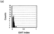
- (B) It is a histogram in the case where the EMT progress degree is zero and the EMT progress degree is 100% in the EMT index. It is the result of having mixed various cell lines with the peripheral blood of a healthy person, and evaluating each detection efficiency. This indicates that detection is possible even in a cell line in which EpCAM expression is zero, and detection is possible even in a CTC in which EMT is progressing. It is the figure which showed the example in the case of applying the evaluation result of an EMT index to the diagnosis of the possibility of cancer metastasis.
- the method for detecting the malignancy of peripheral circulating tumor cells includes (a) a test sample that may contain peripheral circulating tumor cells, and epithelial cells.
- Fluorescent or luminescent enzyme-labeled epithelial binding component that specifically binds to the marker to be expressed, and fluorescent or luminescent enzyme-labeled mesenchymal binding component that specifically binds to the marker that is expressed on mesenchymal cells (B) detecting the fluorescence or luminescence signal of the epithelial binding component and the fluorescence or luminescence signal of the mesenchymal binding component bound to the peripheral circulating tumor cells for each cell, and (c) the signal amount of the epithelial binding component (E) and determining the progression of epithelial-mesenchymal transition of peripheral circulating tumor cells from the signal amount (M) of the mesenchymal binding component.
- the method for detecting the degree of malignancy of peripheral circulating tumor cells of the present invention may further comprise the step of (d) removing red blood cells and / or white blood cells in the test sample before or after the step (a).
- the detection method of the present invention is used as a cancer metastasis detection (diagnosis) method, a cancer progression detection (diagnosis) method, or a cancer therapeutic effect monitoring method (particularly, a therapeutic effect monitoring method of an anticancer agent). You can also.
- the contact step (a) in the detection method of the present invention comprises a test sample that may contain peripheral circulating tumor cells and a fluorescently or luminescent enzyme-labeled epithelium that specifically binds to a marker expressed in epithelial cells.
- a binding component and a marker that is specifically bound to a marker expressed in mesenchymal cells are contacted with a fluorescent label or a luminescent enzyme-labeled mesenchymal binding component.
- epithelial cell markers Markers expressed in epithelial cells are not particularly limited as long as they are expressed in peripheral circulating tumor cells, and examples include surface proteins or sugars of epithelial cells. it can. More specifically, cytokeratin, EpCAM, and E-cadherin can be used as epithelial cell markers, and cytokeratin is particularly preferable. As cytokeratin, at least one cytokeratin selected from the group consisting of CK1, CK4, CK5, CK8, CK10, CK14, CK15, CK16, CK18, and CK19 can be used, but preferably two of these are used. It is preferable to use a combination of the above.
- cytokeratin, EpCAM, and E-cadherin may be subjected to a common fluorescent labeling, and the sum of their expression levels may be used as the expression level of a marker expressed in epithelial cells.
- the combination is free but preferably contains cytokeratin.
- the epithelial binding component that specifically binds to the epithelial cell marker is not particularly limited as long as it can bind to the epithelial cell marker.
- the antibody, antibody fragment, antigen, DNA, RNA, receptor Body, receptor ligand, enzyme, enzyme ligand, enzyme analog, enzyme substrate from which the enzyme analog is derived, lectin, or sugar is a protein
- an antibody, an antibody fragment, a ligand for a receptor, a ligand for an enzyme DNA (for example, aptamer), or RNA can be used.
- the epithelial cell marker is a protein, an antibody, an antibody fragment, or a lectin can be used.
- the marker expressed in mesenchymal cells is not particularly limited as long as it is expressed in peripheral circulating tumor cells.
- a surface protein or sugar of mesenchymal cells is used.
- vimentin, twist, or N-cadherin can be used as a mesenchymal cell marker. It is also possible to label vimentin, twist, and N-cadherin with a common fluorescent color, and to add the total expression level to the expression level of a marker expressed in mesenchymal cells. Combinations are free, but preferably include vimentin.
- the mesenchymal binding component that specifically binds to the mesenchymal cell marker is not particularly limited as long as it can bind to the mesenchymal cell marker.
- an antibody, an antibody fragment, an antigen, DNA examples thereof include RNA, a receptor, a ligand for the receptor, an enzyme, a ligand for the enzyme, an enzyme analog, a substrate of the enzyme from which the enzyme analog is based, a lectin, or a sugar.
- DNA for example, aptamer
- RNA can be used.
- the mesenchymal cell marker is a protein, an antibody, an antibody fragment, or a lectin can be used.
- the antibody used as the epithelial binding component or mesenchymal binding component is not particularly limited, and examples thereof include a polyclonal antibody, a monoclonal antibody, a recombinant antibody, or an antibody fragment having an antigen binding site of those antibodies.
- antibody fragments include F (ab ′) 2 , Fab ′, Fab, or Fv. These antibody fragments can be obtained, for example, by digesting an antibody with a proteolytic enzyme (for example, pepsin or papain) by a conventional method, and subsequently purifying by a conventional method for separating and purifying a protein. .
- the epithelial binding component and the mesenchymal binding component are preferably those labeled with a fluorescent label or a luminescent enzyme.
- the fluorescent substance used for the fluorescent label is not particularly limited.
- AMCA Alexa Floor 350, Murina Blue, Cascade Blue, Cascade Yellow, Pacific Blue, Alexa Floor 405, Alexa Floor 488, Qdot (R) , FITC, PE / RD1, ECD / PE-TexasRed, PC5 / SPRD / PE-Cy5, PC5.5 / PE-Cy5.5, PE Alexa Floor 750, PC7 / PE-Cy7, TRITC, Cy3, Texas Red, Alexa Examples include: Flour 647, Alexa Flour 700, Cy5, Cy5.5, APC, APC7 / APC-Cy7, APC Alexa Flour 750.
- a luminescent enzyme is not specifically limited, Luciferase can be mentioned.
- the origin of luciferase is not particularly limited, and examples include firefly-derived and bacterial-derived luciferases.
- a luminescent enzyme emits light by reacting with a substrate specific to the enzyme. Therefore, it is desirable to use in analysis by an image analyzer described later.
- the substrate of the luminescent enzyme may be any one that is specific for each luminescent enzyme, and can be appropriately selected. In the case of luciferase, luciferin having substrate specificity for each luciferase can be used.
- a test sample that may contain peripheral circulating tumor cells is contacted with a leukocyte binding component labeled with a fluorescent label or a luminescent enzyme that specifically binds to a marker expressed on leukocytes. May be included.
- a leukocyte binding component labeled with a fluorescent label or a luminescent enzyme that specifically binds to a marker expressed on leukocytes May be included.
- fluorescently staining the marker expressed in leukocytes peripheral circulating tumor cells and leukocytes can be reliably separated with a flow cytometer or an image analyzer in the detection step described later. Detection of peripheral circulating tumor cells This is because the accuracy can be raised.
- the marker expressed in leukocytes is not particularly limited as long as it is a marker specifically expressed in leukocytes.
- CD45 marker is present in most leukocytes.
- the leukocyte binding component that specifically binds to the marker is not particularly limited.
- antibody, antibody fragment, antigen, DNA, RNA, receptor, ligand for receptor, enzyme, ligand for enzyme, enzyme an analog, a substrate of an enzyme that is a base of an enzyme analog, a lectin, or a sugar can be mentioned, and an antibody or an antibody fragment is preferable.
- an antibody is used as the leukocyte binding component, the antibody described in “(Antibody or antibody fragment)” can be used, although not limited thereto.
- the fluorescent label or luminescent enzyme label for labeling the leukocyte binding component is not limited, but those described in the column of “(Fluorescent label or luminescent enzyme label)” can be used.
- the cells in the test sample may be nuclear-stained. This is because by performing nuclear staining, peripheral circulating tumor cells can be accurately distinguished from membrane fragments.
- the test sample used is not particularly limited as long as it is a test sample of a patient suspected of having cancer. That is, it is not limited as long as it is a test sample that may contain peripheral circulating tumor cells. Specifically, it is a liquid sample derived from a living body that may contain peripheral circulating tumor cells, and examples thereof include blood, urine, lymph, tissue fluid, spinal fluid, ascites, and pleural effusion. Peripheral blood is desirable because it can be easily collected by blood sampling.
- the cancer from which the test sample is collected is an epithelial tumor
- this type of cancer includes bladder cancer, breast cancer, colon cancer, rectal cancer, kidney cancer, liver cancer, lung cancer, small cell lung cancer
- Mention may be made of esophageal cancer, gallbladder cancer, ovarian cancer, pancreatic cancer, gastric cancer, cervical cancer, thyroid cancer, prostate cancer, squamous cell carcinoma, skin cancer, duodenal cancer, vaginal cancer, or brain tumor.
- peripheral circulating tumor cells refer to cancer cells that are detected at extremely low concentrations in the blood of cancer patients and are also referred to as “blood circulating tumor cells” or simply “circulating tumor cells”.
- the peripheral circulating tumor cells detected in the present invention are not particularly limited, but those derived from epithelial cancer are preferred, and those that cause epithelial-mesenchymal transition (EMT) are preferred.
- EMT epithelial-mesenchymal transition
- Peripheral circulating tumor cells have been reported to have a cancer cell half-life of 1 to 2.4 hours, and many die by apoptosis. Thus, for blood cells 10 9 / mL, only a few to several tens or so absent.
- Detection step (b) In the detection step (b), the fluorescence or luminescence signal of the epithelial binding component bound to the peripheral circulating tumor cells and the fluorescence or luminescence signal of the mesenchymal binding component are detected. Fluorescence or luminescence signals bound to epithelial cell markers and mesenchymal cell markers are measured for individual peripheral circulating tumor cells. It is a condition for detecting as peripheral circulating tumor cells that the fluorescence or luminescence signal of the epithelial cell marker has a certain threshold value or more.
- a device capable of identifying individual cells is required, and for example, a flow cytometer or an image analyzer can be used.
- a flow cytometer using a disposable microchannel chip is preferable in that it can prevent cross-contamination between samples.
- low-density CTC fluorescence signals can be individually performed at high speed. Therefore, as disclosed in Patent Document 4 and Patent Document 9, it is preferable to perform measurement with a flow cytometer that realizes contamination-free with a disposable microchannel chip.
- fluorescently stained cells are the measurement target.
- the measurement data is the fluorescence intensity of the cell unit, the signal intensity can be used to quantify the progress of the epithelial-mesenchymal transition of the cell unit, which is different from the case of the next image analyzer. Is a point.
- the image analysis analyzer is a fluorescently stained cell or a cell stained so as to emit light spontaneously, and measures the fluorescence intensity distribution or emission intensity distribution emission signal of the cell. Unlike the flow cytometer, the image analyzer is a light intensity distribution image including a plurality of cells in a certain range. Cell quantification requires a cell contour extraction function based on image recognition. Specifically, the integral amount of the fluorescence intensity distribution of the epithelial binding component inside the cell outline and the integral amount of the fluorescence illuminance distribution of the mesenchymal binding component are obtained. When quantifying the degree of progress of epithelial-mesenchymal transition in the next step, the above integration amount is necessary.
- the progress determination step (c) quantifies the progress of epithelial-mesenchymal transition of peripheral circulating tumor cells based on the signal amount (E) of the epithelial binding component and the signal amount (M) of the mesenchymal binding component.
- the signal amount (E) of the epithelial binding component represents the expression level of the epithelial cell marker in peripheral circulating tumor cells
- the signal amount (M) of the mesenchymal binding component is the expression of the mesenchymal cell marker in peripheral circulating tumor cells. Represents an amount. Therefore, it corresponds to quantifying the progress of epithelial-mesenchymal transition using the expression level of the epithelial cell marker and the expression level of the mesenchymal cell marker.
- the EMT progression degree is particularly determined if the signal amount E is indicated by an index whose ratio changes to the signal amount M using the signal amount (E) of the epithelial connection component and the signal amount (M) of the mesenchymal connection component. It is not limited.
- An index whose numerical value increases as the ratio of the signal amount E to the signal amount M increases is referred to as an EMT index (P) in this specification.
- P M / (E + M) (1)
- P has the merit that the upper and lower limits of the value are standardized in the range of 0 to 1.
- the index indicating the degree of progress of EMT can be defined by the following formula.
- P E / (E + M) (2)
- a value of 1 corresponds to no EMT occurring.
- the value of P has an advantage that an upper limit and a lower limit are determined. Further, this P may be shown on a logarithmic scale.
- the EMT index (P) will be described according to an example described later.
- P [(CTC Vimentin expression level) / ((CTC CK expression level) + (CTC Vimentin expression level))] This P is zero when EMT has not occurred, and is an amount that approaches 1 when the Vimentin expression level increases.
- the lower limit value is zero and the upper limit value is normalized to 1. Is the amount. Therefore, it is convenient as an index for setting the diagnostic criteria in common for the results of different apparatuses.
- the actual procedure for measuring the EMT index is as follows.
- CK is stained with the anti-CK antibody FITC label
- the signal intensity detected in the wavelength range of 510 to 550 nm is FL1
- Vimentin is stained with the anti-Vimentin antibody PE label
- the fluorescence is in the wavelength range of 550 to 600 nm.
- EMT index [A (FL2 / (FL1 + FL2)) + B].
- a and B are device constants.
- A is an apparatus constant that depends on individual differences in the fluorescence detection sensitivity of FL1 and FL2
- B is an apparatus constant determined by the fluorescence signal sero level.
- a and B are set so that the value is 1 when the Vimentin expression level is zero and the value is zero, and the cell count in which only Vimentin is expressed when the CK expression level is zero.
- the type of each fluorescent label is not limited to the above type.
- the device constants A and B can be determined as follows. In order to obtain a standard line of the fluorescence signal value of “100% CK (FITC)” in which only cytokeratin is expressed and vimentin is not expressed, it is necessary to measure a single-stained cell by FITC. However, since it takes time to prepare such a sample in addition to a patient specimen, the standard straight line may be obtained by measuring particles bound with FITC with a flow cytometer. Similarly, a method for obtaining a standard line of “100% Vimentin (PE)” in which only vimentin is expressed and cytokeratin is not expressed can also be obtained by measuring PE-bound particles with a flow cytometer. Good.
- PE 100% Vimentin
- the line of 100% CK (FITC) described in the scatter diagram of FIG. 3A is a line determined by the ratio of FL1 and FL2 determined by the fluorescence spectrum of FITC. Data of particles and cells having only FITC fluorescence are Distributed on this line. Similarly, in the 100% Vimentin (PE) line, data of particles and cells having only PE fluorescence are distributed on this line. A and B are determined so that the EMT index values of these two types of data are zero and one, respectively.
- epithelial-mesenchymal transition In epithelial-mesenchymal transition, epithelial cells are converted to mesenchymal cells, and adhesion between cells is reduced. In the case of cancer cells, this EMT leads to the ability to dissociate from cancer tissue and metastasize, so that malignancy is considered to progress.
- the signal amount (E) of the epithelial binding component is large, the progression of cancer is low, and when the signal amount (M) of the mesenchymal binding component is large, the progression of cancer is high.
- the signal amount (E) of the epithelial binding component represents the expression level of the epithelial cell marker
- the signal amount (M) of the mesenchymal binding component represents the expression level of the mesenchymal cell marker.
- Non-patent document 2 shows that EMT occurs in CTCs of 70% or more of early cancer patients, and EMT occurs in 100% of CTCs of metastatic cancer patients. It has not been shown that the degree of progression of cancer changes due to fluctuations in the expression level of epithelial cell markers (eg, cytokeratin) and mesenchymal cell marker (vimentin) in cells.
- the peripheral circulation is determined according to the ratio of the signal amount of epithelial binding component (E) and the signal amount of mesenchymal binding component (M).
- E epithelial binding component
- M mesenchymal binding component
- Tumor cell malignancy, cancer metastasis prediction, or cancer progression can be determined. Furthermore, it is possible to predict the occurrence of cancer metastasis after removal of cancer, monitor the progress of cancer, and monitor the therapeutic effect of an anticancer agent according to the ratio.
- the method for detecting the malignancy of peripheral circulating tumor cells of the present invention may further comprise (d) a step of removing red blood cells and / or white blood cells in the test sample.
- Removing step (d) the detection method previously of the present invention, is not an essential step, for peripheral circulating tumor cells, to the blood cells 10 9 / mL, only a few to several tens or so no It is preferable to remove red blood cells and / or white blood cells before or after the contacting step (a), and it is particularly preferable to perform the removing step (d) before the contacting step (a).
- the method for removing erythrocytes is not particularly limited, and an ammonium chloride solution for removing erythrocytes or a commercially available buffer for removing erythrocytes (buffer) can be used.
- the method for removing leukocytes is not limited, and examples thereof include a negative selection of leukocytes using antibody magnetic beads against a leukocyte surface marker and a magnetic field. This method is preferable in that it is a CTC concentration method that does not depend on EpCAM.
- Standard setting process (e) In the method for detecting the degree of malignancy of peripheral circulating tumor cells of the present invention, by measuring particles bound with a fluorescent substance labeled with an epithelial binding component and particles bound with a fluorescent substance labeled with the mesenchymal binding antibody. A step of setting a reference between a state (0) where epithelial-mesenchymal transition has not started and a state (1) where epithelial-mesenchymal transition has been completed can be further included.
- the “particles bound with the fluorescent substance labeled with the epithelial binding component” correspond to the particles bound with the above-mentioned FITC, and the “particles bound with the fluorescent substance labeled with the mesenchymal binding antibody” are described above.
- the fluorescent material is not limited to FITC or PE.
- the fluorescent material described in “(Fluorescent label or luminescent enzyme label)” can be used.
- the particles are not particularly limited as long as they can be used in a flow cytometer or an image analyzer, and examples thereof include cells or beads having a uniform size.
- the reference setting step (e) may be performed independently.
- the detection step (b) particles are detected simultaneously, and the progress determination step ( In c), the reference may be set simultaneously. When performed simultaneously, it is preferable to use beads with a uniform particle size and smaller than cells in order to distinguish them from peripheral circulating tumor cells.
- FIG. 3 is a diagram showing a threshold setting method for using the CTC malignancy evaluation result for diagnosis in addition to the conventional CTC count in CTC measurement.
- CTC detection and CTC count are performed, and in the progress determination step (c), an EMT index is calculated. Determine the degree of mesenchymal transition.
- an EMT index is calculated. Determine the degree of mesenchymal transition.
- a set value for example, it can be determined that “the possibility of cancer metastasis is low”.
- the set value for example, it can be determined that “the possibility of cancer metastasis is low”.
- the set value for example, it can be determined that “the possibility of cancer metastasis is low”.
- the set value threshold value: cut-off value
- the value is equal to or less than the set value (threshold value: cut-off value)
- the set value can be set as appropriate in each determination.
- the set value can be set without any limitation as long as cancer metastasis, cancer malignancy, cancer progression, etc. can be diagnosed. That is, different setting values (threshold value: cut-off value) can be used in each determination. However, the set value (threshold: cut-off value) is preferably determined by a controlled clinical trial.
- Kit for detecting malignancy of peripheral circulating tumor cells is (a) a fluorescent label or a luminescent enzyme label that specifically binds to a marker expressed in epithelial cells. And (b) a mesenchymal-binding antibody labeled with a fluorescent label or a luminescent enzyme that specifically binds to a marker expressed in mesenchymal cells.
- the peripheral circulating tumor cell malignancy detection kit of the present invention includes (c) a fluorescently or luminescent enzyme-labeled epithelial binding antibody that specifically binds to a marker expressed on leukocytes; and / or (d) It may further include a particle bound with a fluorescent substance or luminescent enzyme labeled with an epithelial bound antibody, and (e) a particle bound with a fluorescent substance or luminescent enzyme labeled with the mesenchymal bound antibody.
- the detection kit of the present invention can be used in the method for detecting the malignancy of peripheral circulating tumor cells.
- the method for detecting (diagnosing) cancer metastasis by the method for detecting malignancy according to the present invention the method for detecting (diagnosing) the progression of cancer, or the method for monitoring the therapeutic effect of cancer (in particular, monitoring the therapeutic effect of an anticancer agent)
- the method can also be used as a kit thereof.
- the epithelial bound antibody (a) is an antibody that can specifically bind to the epithelial cell marker in the detection method of the present invention.
- the epithelial cell marker include those described in the column of “(epithelial cell marker)”, particularly cytokeratin, EpCAM, or E-cadherin.
- the antibody is not particularly limited as long as it can bind to the epithelial cell marker, and examples thereof include a polyclonal antibody, a monoclonal antibody, a recombinant antibody, or an antibody fragment having the antigen-binding site of those antibodies. it can.
- antibody fragments include F (ab ′) 2 , Fab ′, Fab, or Fv. These antibody fragments can be obtained, for example, by digesting an antibody with a proteolytic enzyme (for example, pepsin or papain) by a conventional method, and subsequently purifying by a conventional method for separating and purifying a protein. .
- the mesenchymal binding antibody (b) is an antibody that can specifically bind to the mesenchymal cell marker in the detection method of the present invention.
- the mesenchymal cell marker include those described in the above-mentioned section “(Mesenchymal cell marker)”, particularly vimentin or N-cadherin.
- the antibody is not particularly limited as long as it can bind to the aforementioned mesenchymal cell marker, and examples thereof include a polyclonal antibody, a monoclonal antibody, a recombinant antibody, or an antibody fragment having the antigen-binding site of those antibodies. Can do.
- antibody fragments include F (ab ′) 2 , Fab ′, Fab, or Fv. These antibody fragments can be obtained, for example, by digesting an antibody with a proteolytic enzyme (for example, pepsin or papain) by a conventional method, and subsequently purifying by a conventional method for separating and purifying a protein. .
- a proteolytic enzyme for example, pepsin or papain
- the leukocyte-binding antibody (c) is an antibody that can specifically bind to the leukocyte marker in the detection method of the present invention.
- the leukocyte marker include the markers described in the column “(Fluorescence or luminescence staining of leukocytes)”, and particularly CD45.
- the antibody is not particularly limited as long as it can bind to the leukocyte marker, and examples thereof include a polyclonal antibody, a monoclonal antibody, a recombinant antibody, or an antibody fragment having the antigen-binding site of those antibodies.
- antibody fragments include F (ab ′) 2 , Fab ′, Fab, or Fv. These antibody fragments can be obtained, for example, by digesting an antibody with a proteolytic enzyme (for example, pepsin or papain) by a conventional method, and subsequently purifying by a conventional method for separating and purifying a protein. .
- the epithelial binding antibody (a), mesenchymal binding antibody (b) and leukocyte binding antibody (c) are those labeled with a fluorescent label or a luminescent enzyme, and luminescent substances and luminescent enzymes are usually used in the art.
- the fluorescent substance and the luminescent enzyme described in the column “(Fluorescent label or luminescent enzyme label)” can be used.
- the kit of the present invention can include particles bound with a fluorescent substance or a luminescent enzyme labeled with the epithelial binding antibody (a) contained in the kit. This particle is used to determine a standard straight line (100% CK (FITC)) of peripheral circulating tumor cells in which only the epithelial cell marker described in FIG. 3 is expressed. Therefore, when the epithelial bound antibody (a) is labeled with FITC, the particles are also labeled with FITC.
- FITC 50% CK
- the kit of the present invention can include particles bound with a fluorescent substance or a luminescent enzyme labeled with the mesenchymal binding antibody (b) included in the kit. This particle is used to determine a standard straight line (100% vimentin (PE)) of peripheral circulating tumor cells in which only the mesenchymal cell marker described in FIG. 3 is expressed. Therefore, when the mesenchymal binding antibody (b) is labeled with PE, the particles are also labeled with PE.
- the detection kit of the present invention may include an instruction manual showing a method for using particles bound with the fluorescent substance or the luminescent enzyme and / or a method for calculating an EMT index.
- the particles bound with the fluorescent substance or the luminescent enzyme are not particularly limited, but for example, cells may be used, or polystyrene beads containing a single fluorescent substance may be used.
- the particle size of the particles is not particularly limited, but is preferably slightly smaller than the cell size.
- the particle size range is preferably 3 to 6 ⁇ m, and the particle size is desirably uniform.
- the epithelial binding antibody (a) that specifically binds to a marker expressed in the epithelial cells can be used for the production of a kit for detecting malignancy of peripheral circulating tumor cells.
- the mesenchymal binding antibody (b) that specifically binds to a marker expressed in mesenchymal cells can be used for the production of a kit for detecting malignancy of peripheral circulating tumor cells.
- the epithelial binding antibody (a) and the mesenchymal binding antibody (b) those described above can be used.
- the leukocyte-binding antibody (c) can be used for the production of a kit for detecting malignancy of peripheral circulating tumor cells.
- a kit for detecting malignancy of peripheral circulating tumor cells including a particle bound with a fluorescent substance or luminescent enzyme labeled with the epithelial-bound antibody, and a particle bound with a fluorescent substance or luminescent enzyme labeled with a mesenchymal-bound antibody Can be used for manufacturing.
- Peripheral circulating tumor cell malignancy detection apparatus includes (a) a fluorescence or luminescence signal amount of an epithelial binding component bound to peripheral circulating tumor cells, and peripheral Means for receiving a fluorescence or luminescence signal amount of mesenchymal binding component bound to circulating tumor cells, (b) a ratio of the received signal amount (E) of epithelial binding component and signal amount (M) of mesenchymal binding component Means for calculating and determining the degree of epithelial-mesenchymal transition of peripheral circulating tumor cells.
- the detection apparatus of the present invention can be used in the method for detecting the malignancy of peripheral circulating tumor cells of the present invention, and the fluorescence or luminescence signal amount of the epithelial binding component and the fluorescence or luminescence signal of the mesenchymal binding component Having means for receiving the quantity;
- Specific examples of the means include a fluorescence detector and a luminescence detector.
- the progress degree determining means includes a step of calculating an EMT index from the received signal amount (E) of the epithelial connection component and signal amount (M) of the mesenchymal connection component.
- the calculation of the EMT index can be performed by a computer in which a program is incorporated in advance after the measurement of E and M is completed.
- the definition formula of the EMT index is not particularly limited as long as it indicates the ratio of the signal amount (E) of the epithelial connection component and the signal amount (M) of the mesenchymal connection component.
- the receiving means and the progress degree determining means can be executed in accordance with a program incorporated in a computer.
- Example 1 in order to establish the detection method of the present invention, A549, a cell line derived from lung cancer, was mixed as CTC into the peripheral blood of volunteers, cytokeratin and vimentin in A549 cells were detected, and the EMT index was calculated. did. 45 mL of Lysing buffer was added to 4 mL of peripheral blood mixed with 100 A549 cells and mixed, and left on ice for 20 minutes to destroy erythrocytes. A solution obtained by diluting ImmunoTOKUI (on-chip biotechnology) by 20 times with PBS buffer containing 0.5% BSA and 2 mM EDTA (hereinafter referred to as T-buffer) was centrifuged, and the supernatant was centrifuged.
- T-buffer PBS buffer containing 0.5% BSA and 2 mM EDTA
- the obtained cell pellet is resuspended with 200 ⁇ L of T-buffer, and further 100 ⁇ L of Fc Blocking Reagent is added and incubated at 4 ° C. for 15 minutes. After incubation, 200 ⁇ L of T-buffer was added to obtain a cell suspension.
- the beads adsorbed with the CD45 antibody for removing leukocytes were prepared as follows. Magnetic beads adsorbed with 400 ⁇ L of CD45 antibody (Dynabeads, Life Technologies) were placed in an Eppendorf tube. 400 ⁇ L of T-buffer was added to the beads and mixed well by moving the tube up and down. The tube was brought close to the magnet and waited for 1 minute until the beads were completely trapped on the wall of the tube. The supernatant was slowly removed, 400 ⁇ L of T-buffer was added to the beads, and the mixture was stirred well by tapping to obtain a mixture of Dyna beads.
- the mixture was centrifuged at 600 ⁇ g for 5 minutes, and washed again with 2 mL of T-buffer. After centrifuging the Eppendorf tube at 600 ⁇ g for 5 minutes, Alexa700 fluorescence-labeled CD45 antibody, PE fluorescence-labeled vimentin antibody, and FITC fluorescence-labeled cytokeratin antibody were added again in a total of 30 uL of the antibody reaction solution to which 4 ⁇ L and T-buffer 26 ⁇ L were added, respectively. Suspended and reacted at 4 ° C.
- a flow cytometer (FISHMAN-R, manufactured by On-Chip Biotechnology Co., Ltd.) using a disposable disposable microchannel chip made of acrylic as a flow cell is used. And measured.
- This apparatus has four colors of fluorescence detection. When each detection signal is indicated by FL1, FL2, FL3, and FL4, the respective wavelength regions are FL1 for 510 nm to 550 nm, FL2 for 565 nm to 605 nm, and FL3 for 656 nm to 656 nm. 696 nm, and FL4 is 700 nm to 850 nm.
- FIG. 1 (a) shows a scatter diagram based on a forward scatter signal (FS) and a side scatter signal (SS).
- FS forward scatter signal
- SS side scatter signal
- FIG. 1B shows a scatter diagram of FL1 corresponding to FITC and FL4 corresponding to ALEXA700. Using this scatter plot, FITC labeled CK (+) was selected.
- FIG. 1 (c) is a scatter diagram developed with FL3 and FL4. The spectrum of 7-AAD and that of ALEXA700 are distinguished from each other.
- FIG. 1 (d) is a graph of the EMT index calculated using FL1 and FL2 for CK (+) CTC cells that do not contain white blood cells, and is a scatter diagram with the vertical axis representing the EMT index and the horizontal axis representing FS. . A549 was found to be distributed with an EMT index of about 10%.
- FIG. 2 shows a histogram of CTC EMT index values.
- FITC fluorescence signal value
- particles to which FITC was bound were measured with a flow cytometer.
- particles bound with PE were measured with a flow cytometer.
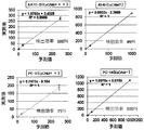
- the device constants A and B were specifically calculated as follows. In order to obtain a standard line of fluorescence signal value of “100% CK (FITC)” in which only cytokeratin is expressed and vimentin is not expressed, particles having a particle size of 3 ⁇ m combined with FITC were measured with a flow cytometer. A standard straight line was obtained. In order to obtain a standard line of “100% Vimentin (PE)”, particles having a particle size of 3 ⁇ m combined with PE were measured with a flow cytometer. A and B were set so that the EMT index values of the above two types of data would be 0 and 1. This will be described below with reference to FIG. FIG. 2A shows the relationship between the EMT index value and the fluorescence signal value.
- “100% CK (FITC)” in FIG. 2 is a standard straight line when FITC (cytokeratin) obtained from the measured value of the particles to which FITC is bound is 100%, and “100% Vimentin (PE)” is The standard straight line when the PE (vimentin) obtained from the particles to which PE is bound is 100%.
- cytokeratin and vimentin are expressed alone, they match either standard line. That is, since the fluorescence label of cytokeratin is FITC, when EMT does not occur and cytokeratin is 100%, the CTC data is on the standard line (100% CK (FITC)) corresponding to the fluorescence spectrum of FITC. Distributed.
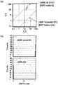
- FIG. 3B is a histogram of the particle data combined with PE and the particle data combined with FITC.
- the device constants A and B are initialized so that the mode values are 1 and 0 (zero), respectively. Set. Therefore, it is possible to compare the EMT index distribution between different apparatuses.
- the comparison of the raw data of the fluorescence signal intensity of a normal flow cytometer is usually not performed between different apparatuses due to apparatus fluctuations or the like. However, for medical diagnosis, an index that can be compared between different devices is required. The above EMT index satisfies this.
- Example 3 the recovery rate of detection according to the present invention was examined using A549 cells or cancer cells other than A549 cells.
- the procedure of Example 1 was repeated except that A549 cells were used or KATO-III cells, PC-9 cells, and PC-14 cells were used instead of A549 cells, and EMT index values were calculated.
- the detection rate of each cell was also calculated at the same time. As shown in FIG. 4, the detection rate of each cell was about 100% for KATO-III, 89% for A549, 75% for PC-9, and 100% for PC-14.
- the detection efficiency of PC-14 in which EpCAM is not expressed is 100%, which indicates that it is also effective in detecting CTC of metastatic cancer in which EMT has progressed.
- an analog quantity indicating a specific cell characteristic (in the present invention, CTC EMT progress) comprising a plurality of signal intensities and apparatus constants is obtained, and the apparatus constant is corrected. Realized the evaluation. In other words, it is possible to compare measurement results of individual cell levels between different devices. It can also be expressed that a method of using analog information at an individual cell level for use in medical diagnosis has been realized.
- the number of cells detected in a certain signal intensity range is diagnostic information. This conventional information is not analog information in individual cells, but only the detected number. In other words, conventional flow cytometers were unable to express analog quantities indicating the characteristics of individual cells. It can be said that the present invention has realized a method for using an analog amount of each cell unit contained in a patient specimen for medical diagnosis.
- the present invention has the advantage of (1) detecting CTC only by collecting blood from a patient and finding cancer. Moreover, (2) it has the merit that the patient who removed the cancer tissue by surgery can detect the recurrence early by only blood collection after the operation. As mentioned above, although this invention was demonstrated along the specific aspect, the deformation
Abstract
Description
CTCをがん患者の末梢血から検出することを考えると、高濃度の血球細胞群の中からそれらの濃度に対して1000,000,000分の1の割合程度の非常に低濃度の細胞を検出することになる。このため、数え落としや患者の検体間のコンタミネーションが大きな誤診断につながる。CTC計測としては、Cell Searchシステム(米国製)を用いて行われている例がある。この技術は、特許文献1に開示されているように、核染色とサイトケラチン染色を行い、CD326抗体磁気ビーズを反応させて磁場を利用して浮上させた細胞に、レーザービームを走査することで蛍光イメージを取得し、人間がその画像をみてCTCと判断する方法である。次にCTC-chipと呼ばれる技術がある。この技術は、特許文献2に開示されているように、名刺サイズのシリコンウエハーに8万本の微小ポストを形成したチップに血液を通し、抗EpCAM抗体をコーティングした8万本の微小ポストの全数画像認識により吸着したCTCを識別計数する方法である。特許文献3は、特許文献1の方法で検出されたCTCを回収し、蛍光 in situ ハイブリダイゼーション(fluorescence in situ hybridization;以下、FISH法と称することがある)法によりCTCの遺伝子解析を行う方法を開示している。特許文献4と特許文献9には、使い捨てチップを用いるフローサイトメーターの技術を記載している。この技術によってコンタミネーションフリーの測定が可能となっている。特許文献5は、フローセルを含む送液系が固定である従来方式のフローサイトメーターの技術内容を記載している。特許文献6は、特許文献1の方法で検出されたCTCを回収し、FISH法によりCTCのIGF-1R遺伝子の異常を評価するための方法を開示している。特許文献8には、マイクロフィルターを用いて細胞サイズによりCTCを濃縮する方法を記載している。非特許文献1は、がん細胞のみに感染するウイルスを用い、そのウイルスによってがん細胞に蛍光タンパクであるGFPを発現させて、蛍光顕微鏡で検出する方式を開示している。非特許文献3には、上皮細胞由来の腫瘍において上皮間葉転換(以下、EMTと称することがある)が起こることで、がん細胞が遊離しやすくなり、他の場所に移動しやすくなるということが示されている。このEMTは1980年代初めにElizabeth Hayらが提唱した、上皮細胞が間葉系様細胞に形態変化する現象である。このEMTの進行が、がんの転移に関係しているということを記載している。非特許文献2は、上皮細胞由来のCTCにおいてもEMTが生じていることが示されている。非特許文献7は、電気泳動によるタンパク質解析によって培養細胞のEMTについて知見を記載している。これによると細胞周期によって、サイトケラチンとビメンチンの発現量の比率が変化せず一定であるということである。この発現の比率は、電気泳動による結果なので多数の細胞の集団平均の結果として求められている。
非特許文献4は、ベクトン・ディッキンソン社製のCPT Vacutainer tubesを利用して、前立腺癌由来CTCを白血球群から分離して濃縮する方法が記載されている。非特許文献5は米国CYTOTRACK社のCYTOTRACKシステムを開示しており、この方法は、検出の前に特別な濃縮工程がなく、蛍光抗体ラベル後にディスク上にCTCを含む細胞群を分布させて固定したあとに、コンパクトディスク方式でレーザー走査して、ディスク上のCTCを検出する方式をとっている。非特許文献6に記載の方法は、EpCAMを利用してCTCを磁気濃縮する方法と特許文献4の装置技術を利用してCTCを検出する技術である。
第1の課題は、以下の上皮間葉転換を起こしたCTCのEpCAMによる検出に関する問題である。
特許文献1に開示されている方法は、EpCAMを利用したCTC濃縮方法を採用している。しかしながら、非特許文献2に報告されているように、上皮性がん細胞由来のCTCは上皮間葉転換(EMT)を起こすことが報告されている。上皮間葉転換を起こすと、集団を形成している癌細胞が単細胞で遊走することが可能になり、がんが転移しやすくなると考えられている。そして、EMTによりEpCAM発現が低下するため、EpCAMを利用してCTCを濃縮して検出する方法では、EMTが進行した悪性度の高く転移しやすいCTCほど検出が困難である。
現在、患者の末梢血に含まれるCTCの検出される数(密度)と患者の予後との相関により、CTCの数(密度)が多いほどがんが進行していると判断されている。しかしながら、非特許文献2で示されたように、がん転移が起こっている患者は初期のがん患者よりEMTが発生しているCTCの比率が多いということが示されている。従って患者の予後は、CTCの数(密度)以外に、EMTが生じているCTCの割合が関係していることが考えられる。すなわち、個々のCTCのEMTに関する評価が重要と考えられる。CTCのEMTの有無の評価方法は、非特許文献2に記載されているように、サイトケラチン(CK)とビメンチン(Vimentin)の2重の蛍光染色を行い、蛍光顕微鏡画像により、CTCがCKの他にVimentinも蛍光染色されているかどうかで、EMTの有無を判断している。この報告によれば、初期のがん患者の70%以上のCTCでEMTが生じており、そして転移がん患者のCTCの100%でEMTが発生していることを報告している。しかしながら、がん患者のCTCは70%以上でEMTが生じているので、EMTが生じているCTCの割合に基づいて、がん転移が起こっているか否かどうか判断することは困難である。なぜならば、CTCの検出数はそもそも7mLの末梢血中のCTC数は10個程度であるのが一般的で、ポアッソン分布による統計誤差を考えると、10個程度のCTC計数値にはそれ自体に±30%程度の誤差を含んでいる。この±30%の精度で、EMTが生じているCTC数の割合が70%(初期がん患者)から100%(がん転移患者)までの変動を議論することは不可能である。
すなわち、初期のがん患者においても末梢循環腫瘍細胞の70%でEMTが生じているため、末梢循環腫瘍細胞におけるEMTの発生の割合から、癌の進行度又は癌の悪性度を評価することは非常に困難であった。また、非特許文献7に記載されているEMTの評価方法は、細胞数が少ないので末梢循環腫瘍細胞に適用することは不可能である。
本発明の目的は、癌の転移、癌の進行度、又は癌の悪性度を正確に判断できる評価方法を提供することである。
本発明は、こうした知見に基づくものである。
[1](a)末梢循環腫瘍細胞を含む可能性のある被検試料と、上皮系細胞に発現するマーカーと特異的に結合する蛍光標識又は発光酵素標識された上皮結合成分及び間葉系細胞に発現するマーカーと特異的に結合する蛍光標識又は発光酵素標識された間葉結合成分とを接触させる工程、(b)末梢循環腫瘍細胞に結合した上皮結合成分の蛍光又は発光信号及び間葉結合成分の蛍光又は発光信号を、個々の細胞について検出する工程、及び(c)上皮結合成分の信号量(E)及び間葉結合成分の信号量(M)から、末梢循環腫瘍細胞の上皮間葉転換の進行度を決定する工程、を含むことを特徴とする、末梢循環腫瘍細胞単位の悪性度の検出方法、
[2]前記工程(a)の前又は後に、(d)被検試料中の赤血球及び/又は白血球を除去する工程を更に含む、[1]に記載の末梢循環腫瘍細胞単位の悪性度の検出方法、
[3]前記工程(a)において、末梢循環腫瘍細胞を含む可能性のある被検試料と、白血球に発現するマーカーと特異的に結合する蛍光標識又は発光酵素標識された白血球結合成分を接触させることを含む、[1]又は[2]に記載の末梢循環腫瘍細胞単位の悪性度の検出方法、
[4]前記検出工程(b)において、蛍光又は発光信号をフローサイトメーター又はイメージアナライザーにより検出する、[1]~[3]のいずれかに記載の末梢循環腫瘍細胞単位の悪性度の検出方法、
[5]前記工程(c)における進行度が、式(1)
P=M/(E+M) (1)、
式(2)
P=E/(E+M) (2)、
式(3)
P=Log[M/(E+M)] (3)、及び
式(4)
P=Log[E/(E+M)] (4)
(各式中、Pは進行度、Eは上皮結合成分の信号量、Mは間葉結合成分の信号量である)
からなる群から選択される式で表される、[1]~[4]のいずれかに記載の末梢循環腫瘍細胞単位の悪性度の検出方法、
[6](e)前記上皮結合成分を標識している蛍光物質が結合した粒子及び前記間葉結合抗体を標識している蛍光物質が結合した粒子の測定によって、上皮間葉転換が開始していない状態(0)と上皮間葉転換が終了した状態(1)との基準を設定する工程、を更に含む、[1]~[5]のいずれかに記載の末梢循環腫瘍細胞単位の悪性度の検出方法、
[7]前記工程(e)を工程(b)及び(c)と同時に行う、[6]に記載の末梢循環腫瘍細胞単位の悪性度の検出方法、
[8]前記上皮結合成分がサイトケラチン、EpCAM、又はE-カドヘリンに特異的に結合する抗体又はアプタマーであり、そして間葉結合成分がビメンチン、又はN-カドヘリンに特異的に結合する抗体又はアプタマーである、[1]~[7]のいずれかに記載の末梢循環腫瘍細胞単位の悪性度の検出方法、
[9](a)上皮系細胞に発現するマーカーに特異的に結合する蛍光標識又は発光酵素標識された上皮結合抗体、及び(b)間葉系細胞に発現するマーカーに特異的に結合する蛍光標識又は発光酵素標識された間葉結合抗体、を含む末梢循環腫瘍細胞の悪性度の検出キット、
[10](c)白血球に発現するマーカーと特異的に結合する蛍光標識又は発光酵素標識された上皮結合抗体;及び/又は(d)前記上皮結合抗体を標識している蛍光物質又は発光酵素が結合した粒子、及び(e)前記間葉結合抗体を標識している蛍光物質又は発光酵素が結合した粒子、を更に含む[9]に記載の末梢循環腫瘍細胞の悪性度の検出キット、
[11]前記上皮結合抗体がサイトケラチン、EpCAM、又はE-カドヘリンに特異的に結合する抗体であり、そして間葉結合抗体がビメンチン、又はN-カドヘリンに特異的に結合する抗体である、[9]又は[10]に記載の末梢循環腫瘍細胞の悪性度の検出キット、
[12]上皮系細胞に発現するマーカーに特異的に結合する上皮結合抗体及び/又は間葉系細胞に発現するマーカーに特異的に結合する間葉結合抗体の、末梢循環腫瘍細胞の悪性度の検出キットの製造のための使用、
[13]前記上皮結合抗体がサイトケラチン、EpCAM、又はE-カドヘリンに対する抗体であり、そして前記間葉結合抗体が、ビメンチン、又はN-カドヘリンに対する抗体である、[12]に記載の使用、
[14](a)末梢循環腫瘍細胞に結合した上皮結合成分の蛍光又は発光信号量、及び末梢循環腫瘍細胞に結合した間葉結合成分の蛍光又は発光信号量を細胞単位で受信する手段、(b)前記受信した上皮結合成分の信号量(E)及び間葉結合成分の信号量(M)から、末梢循環腫瘍細胞の上皮間葉転換の進行度を細胞単位で決定する手段、を含むことを特徴とする、末梢循環腫瘍細胞の悪性度の検出装置、
[15] 前記工程(b)における進行度が、式(1)
P=M/(E+M) (1)、
式(2)
P=E/(E+M) (2)、
式(3)
P=Log[M/(E+M)] (3)、及び
式(4)
P=Log[E/(E+M)] (4)
(各式中、Pは進行度、Eは上皮結合成分の信号量、Mは間葉結合成分の信号量である)
からなる群から選択される式で表される、[14]に記載の末梢循環腫瘍細胞の悪性度の検出装置、
[16]前記上皮結合成分がサイトケラチンEpCAM、又はE-カドヘリンに特異的に結合する抗体であり、そして間葉結合成分がビメンチン又はN-カドヘリンに特異的に結合する抗体である、[14]又は[15]に記載の末梢循環腫瘍細胞の悪性度の検出装置、
[17]細胞を計測する方法において、個々の細胞に発現している複数の分子の数量を細胞単位で評価し、それらの複数の分子の数量を用いて個々の細胞が有する特性量を定量化し表示することを特徴とする細胞評価方法、
[18]個々の細胞で計測する複数の分子の量は、上皮系細胞に発現するマーカーの量(E)と間葉系細胞に発現するマーカーの量(M)であって、細胞の特性量とは上皮間葉転換の進行度であってEとMの式で指数化した量である[17]に記載の細胞評価方法、
[19]細胞を計測する細胞評価装置において、個々の細胞に発現している複数の分子の数量を細胞単位で評価し、それらの複数の分子の数量を用いて個々の細胞が有する特性量を定量化し表示することを特徴とする細胞評価装置、又は
[20]個々の細胞で計測する複数の分子の量は、上皮系細胞に発現するマーカーの量(E)と間葉系細胞に発現するマーカーの量(M)であって、細胞の特性量とは上皮間葉転換の進行度であってEとMの式で指数化した量である[19]に記載の細胞評価装置、
に関する。
本発明の末梢循環腫瘍細胞単位の悪性度の検出方法は(a)末梢循環腫瘍細胞を含む可能性のある被検試料と、上皮系細胞に発現するマーカーと特異的に結合する蛍光標識又は発光酵素標識された上皮結合成分及び間葉系細胞に発現するマーカーと特異的に結合する蛍光標識又は発光酵素標識された間葉結合成分とを接触させる工程、(b)末梢循環腫瘍細胞に結合した上皮結合成分の蛍光又は発光信号及び間葉結合成分の蛍光又は発光信号を個々の細胞について検出する工程、及び(c)上皮結合成分の信号量(E)及び間葉結合成分の信号量(M)から、末梢循環腫瘍細胞の上皮間葉転換の進行度を決定する工程、を含む。
本発明の末梢循環腫瘍細胞の悪性度の検出方法は、更に前記工程(a)の前又は後に、(d)被検試料中の赤血球及び/又は白血球を除去する工程を含んでもよい。
本発明の検出方法における接触工程(a)は、末梢循環腫瘍細胞を含む可能性のある被検試料と、上皮系細胞に発現するマーカーと特異的に結合する蛍光標識又は発光酵素標識された上皮結合成分及び間葉系細胞に発現するマーカーと特異的に結合する蛍光標識又は発光酵素標識された間葉結合成分とを接触させるものである。
上皮系細胞に発現するマーカー(以下、上皮系細胞マーカーと称する)は、末梢循環腫瘍細胞に発現される限り、特に限定されるものではなく、例えば上皮系細胞の表面タンパク質又は糖を挙げることができる。より具体的には、サイトケラチン、EpCAM、E-カドヘリンを上皮系細胞マーカーとして用いることができるが、特にはサイトケラチンが好ましい。サイトケラチンとしては、CK1、CK4、CK5、CK8、CK10、CK14、CK15、CK16、CK18、及びCK19からなる群から選択される少なくとも1つのサイトケラチンを用いることができるが、好ましくはこれらの2つ以上の組み合わせを用いることが好ましい。また、サイトケラチンとEpCAMとE-カドヘリンを共通の蛍光標識を行って、それら発現量の合計を上皮系細胞に発現するマーカーの発現量とすることも可能である。組合せは自由であるが、サイトケラチンを含むのが好ましい。
前記上皮系細胞マーカーに、特異的に結合する上皮結合成分は、上皮系細胞マーカーに結合することができる限り、特に限定されるものではなく、例えば抗体、抗体フラグメント、抗原、DNA、RNA、受容体、受容体に対するリガンド、酵素、酵素に対するリガンド、酵素アナログ、酵素アナログの元となる酵素の基質、レクチン、又は糖を挙げることができる。具体的には、前記上皮系細胞マーカーがタンパク質の場合、抗体、抗体フラグメント、受容体に対するリガンド、酵素に対するリガンド、DNA(例えば、アプタマー)、又はRNAを用いることができる。また、前記上皮系細胞マーカーがタンパク質の場合、抗体、抗体フラグメント、又はレクチンを挙げることができる。
間葉系細胞に発現するマーカー(以下、間葉系細胞マーカーと称する)は、末梢循環腫瘍細胞に発現される限り、特に限定されるものではなく、例えば間葉系細胞の表面タンパク質又は糖を挙げることができる。より具体的には、ビメンチン、ツイスト(Twist)、又はN-カドヘリンを間葉系細胞マーカーとして用いることができる。また、ビメンチンとツイストとN-カドヘリンを共通の蛍光色で標識を行って、それら発現量の合計を間葉系細胞に発現するマーカーの発現量とすることも可能である。組合せは自由であるが、ビメンチンを含むことがのぞましい。
前記間葉系細胞マーカーに、特異的に結合する間葉結合成分は、間葉系細胞マーカーに結合することができる限り、特に限定されるものではなく、例えば抗体、抗体フラグメント、抗原、DNA、RNA、受容体、受容体に対するリガンド、酵素、酵素に対するリガンド、酵素アナログ、酵素アナログの元となる酵素の基質、レクチン、又は糖を挙げることができる。具体的には、前記間葉系細胞マーカーがタンパク質の場合、抗体、抗体フラグメント、受容体に対するリガンド、酵素に対するリガンド、DNA(例えば、アプタマー)、又はRNAを用いることができる。また、前記間葉系細胞マーカーがタンパク質の場合、抗体、抗体フラグメント、又はレクチンを挙げることができる。
前記上皮結合成分又は間葉結合成分として用いる抗体は、特に限定されないが、例えば、ポリクローナル抗体、モノクローナル抗体、組換え抗体、又はそれらの抗体の抗原結合部位を有する抗体フラグメントなどを挙げることができる。抗体フラグメントとしては、例えば、F(ab’)2、Fab’、Fab、又はFv等を挙げることができる。これらの抗体フラグメントは、例えば、抗体を常法によりタンパク質分解酵素(例えば、ペプシン又はパパイン等)によって消化し、続いて、常法のタンパク質の分離精製の方法により精製することにより、得ることができる。
前記上皮結合成分及び間葉結合成分は、蛍光標識又は発光酵素標識されているものが好ましい。蛍光標識に用いる蛍光物質は、特に限定されるものではないが、例えば、AMCA、Alexa Flour 350、Murina Blue、Cascade Blue、Cascade Yellow、Pacific Blue、Alexa Flour 405、Alexa Flour 488、Qdot(R)605、FITC、PE/RD1、ECD/PE-TexasRed、PC5/SPRD/PE-Cy5、PC5.5/PE-Cy5.5、PE Alexa Flour 750、PC7/PE-Cy7、TRITC、Cy3、Texas Red、Alexa Flour 647、Alexa Flour 700、Cy5、Cy5.5、APC、APC7/APC-Cy7、APC Alexa Flour 750、を挙げることができる。
また、発光酵素も特に限定されるものではないが、ルシフェラーゼを挙げることができる。ルシフェラーゼの由来も特に限定されるものではなく、ホタル由来、細菌由来のルシフェラーゼを挙げることができる。発光酵素はその酵素に特異的な基質と反応することによって発光する。従って後述のイメージアナライザーによる解析で用いることが望ましい。発光酵素の基質は、それぞれの発光酵素に特異的なものを用いればよく、適宜選択することが可能であるが、ルシフェラーゼの場合、それぞれのルシフェラーゼに基質特異性を有するルシフェリンを用いることができる。
本発明の接触工程(a)においては、末梢循環腫瘍細胞を含む可能性のある被検試料と、白血球に発現するマーカーと特異的に結合する蛍光標識又は発光酵素標識された白血球結合成分を接触させることを含んでもよい。
白血球に発現するマーカーを蛍光染色することにより、末梢循環腫瘍細胞と白血球とを、後述の検出工程において、フローサイトメーター又はイメージアナライザーで確実に分別することが可能であり、末梢循環腫瘍細胞の検出の確度を挙げることができるからである。
白血球に発現するマーカーは、白血球に特異的に発現しているマーカーであれば、特に限定されるものではないが、例えばCD45、CD2、CD3、CD4、CD5、CD8、CD10、CD11b、CD14、CD15、CD16、CD19、CD20、CD24、CD25、CD27、CD29、CD33、CD36、CD38、CD41、CD45、CD45RA、CD45RO、CD56、CD66b、CD66e、CD69、又はCD124を挙げることができ、特にはCD45が好ましい。CD45マーカーは、ほとんどの白血球に存在しているからである。また、マーカーと特異的に結合する白血球結合成分も、特に限定されるものではないが、例えば抗体、抗体フラグメント、抗原、DNA、RNA、受容体、受容体に対するリガンド、酵素、酵素に対するリガンド、酵素アナログ、酵素アナログの元となる酵素の基質、レクチン、又は糖を挙げることができるが、抗体又は抗体フラグメントが好ましい。
白血球結合成分として、抗体を用いる場合は、限定されるものではないが、前記「(抗体又は抗体フラグメント)」に記載のものを用いることが可能である。また、白血球結合成分を標識する蛍光標識又は発光酵素標識は、限定されるものはないが、前記「(蛍光標識又は発光酵素標識)」の欄に記載のものを用いることができる。
更に、本発明の末梢循環腫瘍細胞の悪性度の検出方法においては、被検試料中の細胞を核染色してもよい。核染色を行うことによって、末梢循環腫瘍細を膜断片などと区別して的確に区別することができるからである。
本発明の末梢循環腫瘍細胞の悪性度の検出方法において、用いる被検試料としては、癌の疑いのある患者の被検試料であれば特に限定されるものではない。すなわち、末梢循環腫瘍細胞を含む可能性のある被検試料であれば限定されない。具体的には、末梢循環腫瘍細胞を含む可能性のある生体由来の液体試料であり、例えば、血液、尿、リンパ液、組織液、髄液、腹水、胸水を挙げることができる。採血で簡単に採取できるという点から末梢血がのぞましい。
また、被検試料を採取される対象の癌は、上皮性腫瘍であり、この種の癌としては、膀胱癌、乳癌、大腸癌、直腸癌、腎臓癌、肝臓癌、肺癌、小細胞肺癌、食道癌、胆嚢癌、卵巣癌、膵臓癌、胃癌、子宮頸部癌、甲状腺癌、前立腺癌、扁平上皮癌、皮膚癌、十二指腸癌、腟癌、又は脳腫瘍を挙げることができる。
本発明において検出される末梢循環腫瘍細胞は、特に限定されるものではないが、上皮性癌由来のものが好ましく、上皮間葉転換(EMT)を起こすものが好ましい。末梢循環腫瘍細胞の癌細胞の半減期は1~2.4時間との報告もあり、多くはアポトーシスで死滅する。従って、血液細胞109個/mLに対して、数個~数十個程度しか存在しない。
検出工程(b)において、末梢循環腫瘍細胞に結合した上皮結合成分の蛍光又は発光信号及び間葉結合成分の蛍光又は発光信号を検出するものである。
個別の末梢循環腫瘍細胞について、上皮系細胞マーカー及び間葉系細胞マーカーに結合した蛍光又は発光信号を測定する。上皮系細胞マーカーの蛍光又は発光信号が一定の閾値以上有することが、末梢循環腫瘍細胞として検出するための条件である。
特に、使い捨てマイクロ流路チップを用いたフローサイトメーターは、検体間のクロスコンタミネーションを防止することができる点で好ましい。また、低密度のCTCの蛍光信号を個々に高速で行うことができる。従って、特許文献4や特許文献9に開示した様に、使い捨てマイクロ流路チップによりコンタミネーションフリーを実現するフローサイトメーターにより計測を行うことが好ましい。フローサイトメーターの場合は、蛍光染色した細胞が測定対象となる。さらにその測定データは細胞単位の蛍光強度であるために、細胞単位の上皮間葉転換の進行度の定量化に、その信号強度を用いることができる点が、次のイメージアナライザーの場合とは異なる点である。
イマージアナライザーは、蛍光染色した細胞または自発光する様に染色した細胞が測定対象であり、細胞の蛍光強度分布又は発光強度分布発光信を計測する。イメージアナライザーはフローサイトメーターとは異なり、その測定データは一定範囲に複数個の細胞を含む光強度分布イメージである。細胞単位の信号定量化には画像認識による細胞輪郭抽出機能が必要である。具体的には、細胞輪郭の内側の上皮結合成分の蛍光強度分布の積分量と間葉結合成分の蛍光照度分布の積分量とを求める。次の工程の上皮間葉転換の進行度の定量化を行う場合は、上記の積分量が必要である。
進行度決定工程(c)は、上皮結合成分の信号量(E)及び間葉結合成分の信号量(M)によって、末梢循環腫瘍細胞の上皮間葉転換の進行度を定量化するものである。
上皮結合成分の信号量(E)は、上皮系細胞マーカーの末梢循環腫瘍細胞における発現量を表し、そして間葉結合成分の信号量(M)は間葉系細胞マーカーの末梢循環腫瘍細胞における発現量を表す。従って、上皮間葉転換の進行度を上皮系細胞マーカーの発現量と、間葉系細胞マーカーの発現量とを用いて定量化することに対応する。
EMT進行度は、上皮結合成分の信号量(E)及び間葉結合成分の信号量(M)を用いて、信号量Eが信号量Mに割合が変化する指数で示すものであれば、特に限定されるものではない。信号量Eが信号量Mに割合が多いほど数値が多くなる指数を、本明細書においてEMTインデックス(P)と称する。例えば「M/(E+M)」がのぞましい。
従って、EMTインデックス(P)としては、
式(1)
P=M/(E+M) (1)
を挙げることができる。
Pは値の上限と下限が0から1の範囲で規格化されているメリットがある。これに対して例えば、Pとして「M/E」で定義した場合を考えると、この量はゼロから無限大まで変化するが、Eの値がゼロに近いほどすなわち測定の検出限界に近い不正確な測定データほど大きくなるという結果となるので、診断用の指数としては適さない。また、EMTの進行度を示す指数としては、次の式で定義することもできる。
式(2)
P=E/(E+M) (2)
この場合は、値1がEMTが発生していないことを対応する。同様にこのPの値も上限と下限が決っているというメリットがある。また、このPを対数スケールで示しても良い。
P=[(CTCのVimentin発現量)/((CTCのCK発現量)+(CTCのVimentin発現量))]
このPは、EMTが発生していない場合はゼロであって、Vimentin発現量が増大すると1に近づく量であり、EMTの進行度を示す量として下限値がゼロで上限値が1に規格化した量である。そのため診断基準を異なる装置による結果に対して共通に設定する指数として都合が良い。
このEMTインデックスの計測は、実際の手順は以下の様に行う。すなわちCKを抗CK抗体FITCラベルで染色し、その蛍光を波長範囲が510nmから550nmで検出した信号強度をFL1とし、Vimentinを抗Vimentin抗体PEラベルで染色し、その蛍光を波長範囲が550nmから600nmで検出した信号強度をFL2とすると、EMTインデックス=[A(FL2/(FL1+FL2))+B]という式で表すことができる。但し、AとBは装置定数である。AはFL1とFL2の蛍光検出感度の個体差に依存する装置定数、Bは蛍光信号セロレベルで決まる装置定数である。これらの定数の設定方法は、Vimentin発現量ゼロで値がゼロ、CK発現量ゼロでVimentinのみが発現している細胞計測で値が1、となる様にAとBを設定する。以上において、それぞれの蛍光ラベルの種類は上記の種類に限定するものではない。
サイトケラチンのみが発現し、ビメンチンが発現していない「100%CK(FITC)」の蛍光信号値の標準直線を求めるために、FITCによる単染色した細胞を測定することが必要である。しかしながら、この様なサンプルを患者の検体の他に準備するのに時間がかかるので、FITCを結合させた粒子をフローサイトメーターで測定して標準直線を求めてもよい。また、同様にビメンチンのみが発現し、サイトケラチンが発現していない「100%Vimentin(PE)」の標準直線を求める方法も同様に、PEを結合させた粒子をフローサイトメーターで測定してもよい。この様な単一の蛍光スペクトルのみを有する2種類の標準粒子を検体のCTCと同時に測定すると、EMTインデックスの下限と上限についての標準設定と測定対象のCTCのEMTインデックス値評価を同時に実施することができる。これによってEMTインデックスの評価における装置の個体差を確実に補正することができる。この補正は、EMTインデックスに対する診断閾値を、異なる複数の装置で得られた結果に対して共通に適用する場合に重要である。
図3(A)の散布図に記載した100%CK(FITC)のラインは、FITCの蛍光スペクトルで決まるFL1とFL2の比率で決まるラインであり、FITCの蛍光のみを有する粒子や細胞のデータはこのライン上に分布する。100%Vimentin(PE)のラインは、同様にPEの蛍光のみを有する粒子や細胞のデータがこのライン上に分布する。この2種類のデータのEMTインデックス値がそれぞれゼロと1になるようにAとBを決定する。
上皮間葉転換は、上皮系細胞が間葉系細胞に転換することによって細胞間の接着性が薄れる。このEMTは癌細胞の場合はがん組織から遊離して転移する能力につながるため悪性度が進行するものであると考えられる。ここで、前記上皮結合成分の信号量(E)が多い場合は癌の進行度が低く、間葉結合成分の信号量(M)が多い場合は癌の進行度が高いものである。ここで、上皮結合成分の信号量(E)は、上皮系細胞マーカーの発現量を表し、そして間葉結合成分の信号量(M)は間葉系細胞マーカーの発現量を表す。
非特許文献2では、初期のがん患者の70%以上のCTCでEMTが生じており、そして転移がん患者のCTCの100%でEMTが発生していることは示しているが、1つの細胞において、上皮系細胞マーカー(例えば、サイトケラチン)の発現量及び間葉系細胞マーカー(ビメンチン)の発現量が変動することによって、癌の進行度が変化することは示していない。
本発明の末梢循環腫瘍細胞の悪性度の検出方法においては、前記EMTインデックスを用いることによって、上皮結合成分の信号量(E)及び間葉結合成分の信号量(M)の比率によって、末梢循環腫瘍細胞の悪性度、癌の転移の予測、又は癌の進行度を判断することができる。更には、前記比率によって、癌摘出後のがん転移の発生の予測、又は癌の進行度のモニタリング、抗癌剤の治療効果のモニタリングを行うことができる。
本発明の末梢循環腫瘍細胞の悪性度の検出方法においては、(d)被検試料中の赤血球及び/又は白血球を除去する工程を更に含むことができる。除去工程(d)は、本発明の検出方法おいて、必須の工程ではないが、末梢循環腫瘍細胞が、血液細胞109個/mLに対して、数個~数十個程度しか存在しないため、接触工程(a)の前、又は後に赤血球及び/又は白血球を除去することが好ましく、特には接触工程(a)の前に除去工程(d)を行うことが好ましい。接触工程(a)の前に、除去工程(d)を行うことにより、効率よく上皮系細胞マーカー、及び間葉系細胞マーカー等の蛍光染色等を行うことができるからである。
赤血球の除去方法としては、特に限定されるものではないが、赤血球除去用塩化アンモニウム溶液、又は市販されている赤血球除去用の緩衝液(buffer)を用いることができる。
また、白血球の除去方法としては、限定されるものではないが、白血球の表面マーカーに対する抗体磁気ビーズと磁場を利用した白血球のネガティブセレクションを挙げることができる。この方法は、EpCAMに依存しないCTC濃縮法である点で好ましい。
本発明の末梢循環腫瘍細胞の悪性度の検出方法においては、上皮結合成分を標識している蛍光物質が結合した粒子及び前記間葉結合抗体を標識している蛍光物質が結合した粒子の測定によって、上皮間葉転換が開始していない状態(0)と上皮間葉転換が終了した状態(1)との基準を設定する工程、を更に含むことができる。「上皮結合成分を標識している蛍光物質が結合した粒子」は、前記のFITCを結合させた粒子に相当し、「間葉結合抗体を標識している蛍光物質が結合した粒子」は前記のPEを結合させた粒子に相当するが、蛍光物質はFITC又はPEに限定されるものはなく、例えば前記「(蛍光標識又は発光酵素標識)」に記載の蛍光物質を用いることができる。また、粒子もフローサイトメーター又はイメージアナライザーで用いることのできる限り、特に限定されるものではなく、細胞、又はサイズが均一なビーズを挙げることができる。
本発明の末梢循環腫瘍細胞の悪性度の検出方法において、基準設定工程(e)は、独立して行ってもよく、検出工程(b)において粒子の検出を同時に行い、そして進行度決定工程(c)において基準の設定を同時に行ってもよい。同時に行う場合は、末梢循環腫瘍細胞と区別するために、粒径が均一で細胞より小さいビーズを用いるのがのぞましい。
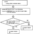
図3は、CTC計測において、従来のCTC計数の他にCTCの悪性度の評価結果を診断に利用するための閾値の設定方法を示した図である。
前記接触工程(a)、及び検出工程(b)によって、CTCの検出及びCTCの計数を行い、更に進行度決定工程(c)によって、EMTインデックスを計算し、個々の末梢循環腫瘍細胞において、上皮間葉転換の進行度を決定する。そして、「EMTインデックスが一定以上の末梢循環腫瘍細胞の数が、予め設定した数値(閾値)より上である場合」、又は「EMTインデックスの平均値が予め設定した数値(閾値)より上である場合」に、例えば「癌の転移の可能性が高い」と判断することができる。また、設定値(閾値)以下である場合に、例えば「癌の転移の可能性が低い」と判断することができる。また同様に、設定値(閾値:カットオフ値)を超えた場合、「悪性度が高い」、「癌が進行している」、「抗癌剤の効果が少ない」などと判断することができ、逆に設定値(閾値:カットオフ値)以下の場合、「悪性度が低い」、「癌が進行していない」、「抗癌剤の効果が高い」などと判断することができる。設定値(閾値:カットオフ値)は、それぞれの判断において、適宜設定することが可能である。
本発明の末梢循環腫瘍細胞の悪性度の検出キットは、(a)上皮系細胞に発現するマーカーに特異的に結合する蛍光標識又は発光酵素標識された上皮結合抗体、及び(b)間葉系細胞に発現するマーカーに特異的に結合する蛍光標識又は発光酵素標識された間葉結合抗体を含む。また、本発明の末梢循環腫瘍細胞の悪性度の検出キットは、(c)白血球に発現するマーカーと特異的に結合する蛍光標識又は発光酵素標識された上皮結合抗体;及び/又は(d)前記上皮結合抗体を標識している蛍光物質又は発光酵素が結合した粒子、及び(e)前記間葉結合抗体を標識している蛍光物質又は発光酵素が結合した粒子、を更に含むことができる。
本発明の検出キットは、前記末梢循環腫瘍細胞の悪性度の検出方法に用いることのできるものである。従って、本発明の悪性度の検出方法による癌の転移の検出(診断)方法、癌の進行度の検出(診断)方法、又は癌の治療効果のモニタリング方法(特には、抗癌剤の治療効果のモニタリング方法)にも使用することが可能であり、それらのキットとして、用いることができる。
上皮結合抗体(a)は、本発明の検出方法における上皮系細胞マーカーに特異的に結合することのできる抗体である。上皮系細胞マーカーとしては、前記の「(上皮系細胞マーカー)」の欄に記載のものを挙げることができ、特にはサイトケラチン、EpCAM、又はE-カドヘリンである。
抗体としては、前記の上皮系細胞マーカーに結合することのできる限り、特に限定されないが、ポリクローナル抗体、モノクローナル抗体、組換え抗体、又はそれらの抗体の抗原結合部位を有する抗体フラグメントなどを挙げることができる。抗体フラグメントとしては、例えば、F(ab’)2、Fab’、Fab、又はFv等を挙げることができる。これらの抗体フラグメントは、例えば、抗体を常法によりタンパク質分解酵素(例えば、ペプシン又はパパイン等)によって消化し、続いて、常法のタンパク質の分離精製の方法により精製することにより、得ることができる。
間葉結合抗体(b)は、本発明の検出方法における間葉系細胞マーカーに特異的に結合することのできる抗体である。間葉系細胞マーカーとしては、前記の「(間葉系細胞マーカー)」の欄に記載のものを挙げることができ、特にはビメンチン又はN-カドヘリンである。
抗体としては、前記の間葉系細胞マーカーに結合することのできる限り、特に限定されないが、ポリクローナル抗体、モノクローナル抗体、組換え抗体、又はそれらの抗体の抗原結合部位を有する抗体フラグメントなどを挙げることができる。抗体フラグメントとしては、例えば、F(ab’)2、Fab’、Fab、又はFv等を挙げることができる。これらの抗体フラグメントは、例えば、抗体を常法によりタンパク質分解酵素(例えば、ペプシン又はパパイン等)によって消化し、続いて、常法のタンパク質の分離精製の方法により精製することにより、得ることができる。
白血球結合抗体(c)は、本発明の検出方法における白血球マーカーに特異的に結合することのできる抗体である。白血球マーカーとしては、前記の「(白血球の蛍光又は発光染色)」の欄に記載のマーカーを挙げることができ、特にはCD45である。
抗体としては、前記の白血球マーカーに結合することのできる限り、特に限定されないが、ポリクローナル抗体、モノクローナル抗体、組換え抗体、又はそれらの抗体の抗原結合部位を有する抗体フラグメントなどを挙げることができる。抗体フラグメントとしては、例えば、F(ab’)2、Fab’、Fab、又はFv等を挙げることができる。これらの抗体フラグメントは、例えば、抗体を常法によりタンパク質分解酵素(例えば、ペプシン又はパパイン等)によって消化し、続いて、常法のタンパク質の分離精製の方法により精製することにより、得ることができる。
前記上皮結合抗体(a)、間葉結合抗体(b)及び白血球結合抗体(c)は、蛍光標識又は発光酵素標識されたものであり、発光物質及び発光酵素は、通常当分野で用いられているものを制限なく使用することができるが、例えば、前記「(蛍光標識又は発光酵素標識)」の欄に記載された蛍光物質、及び発光酵素を用いることができる。
本発明のキットは、キットに含まれる上皮結合抗体(a)が標識されている蛍光物質又は発光酵素で結合した粒子を含むことができる。この粒子は、図3に記載の上皮系細胞マーカーのみが発現している末梢循環腫瘍細胞の標準直線(100%CK(FITC))を決定するために使用するものである。従って、上皮結合抗体(a)がFITCで標識されている場合、粒子もFITCで標識する。
本発明のキットは、キットに含まれる間葉結合抗体(b)が標識されている蛍光物質又は発光酵素で結合した粒子を含むことができる。この粒子は、図3に記載の間葉系細胞マーカーのみが発現している末梢循環腫瘍細胞の標準直線(100%ビメンチン(PE))を決定するために使用するものである。従って、間葉結合抗体(b)がPEで標識されている場合、粒子もPEで標識する。
本発明の検出キットは、前記の蛍光物質又は発光酵素で結合した粒子の使用方法、及び/又はEMTインデックスの計算方法を示した使用説明書を含むことができる。
前記上皮系細胞に発現するマーカーに特異的に結合する上皮結合抗体(a)は、末梢循環腫瘍細胞の悪性度の検出キットの製造のために使用することができる。また、間葉系細胞に発現するマーカーに特異的に結合する間葉結合抗体(b)は、末梢循環腫瘍細胞の悪性度の検出キットの製造のために使用することができる。上皮結合抗体(a)及び間葉結合抗体(b)は、前記のものを用いることができる。更に、白血球結合抗体(c)は、末梢循環腫瘍細胞の悪性度の検出キットの製造のために使用することができる。
前記上皮結合抗体を標識している蛍光物質又は発光酵素が結合した粒子、及び間葉結合抗体を標識している蛍光物質又は発光酵素が結合した粒子も、末梢循環腫瘍細胞の悪性度の検出キットの製造のために使用することができる。
本発明の末梢循環腫瘍細胞の悪性度の検出装置は、(a)末梢循環腫瘍細胞に結合した上皮結合成分の蛍光又は発光信号量、及び末梢循環腫瘍細胞に結合した間葉結合成分の蛍光又は発光信号量を受信する手段、(b)前記受信した上皮結合成分の信号量(E)及び間葉結合成分の信号量(M)の比率を計算し、末梢循環腫瘍細胞の上皮間葉転換の進行度を決定する手段、を含む。
(受信手段)
本発明の検出装置は、前記本発明の末梢循環腫瘍細胞の悪性度の検出方法に用いることのできるものであり、上皮結合成分の蛍光又は発光信号量、及び間葉結合成分の蛍光又は発光信号量を受信する手段を有する。前記手段としては、具体的には、蛍光検出器又は発光検出器を挙げることができる。
進行度決定手段は、受信した上皮結合成分の信号量(E)及び間葉結合成分の信号量(M)からEMTインデックスを計算することのできる工程を含む。例えばEMTインデックスの計算は、EとMの測定終了後に予めプログラムの組み込まれたコンピュータで行うことができる。
前記EMTインデックスの定義式は、上皮結合成分の信号量(E)及び間葉結合成分の信号量(M)の割合を示すものであれば、特に限定されるものではないが、例えば「M/(E+M)」、「E/(E+M)」、「Log[M/(E+M)]」、「Log[E/(E+M)]」、などを挙げることができる。
例えば、式(1)
P=M/(E+M) (1)、
式(2)
P=E/(E+M) (2)、
式(3)
P=Log[M/(E+M)] (3)、又は
式(4)
P=Log[E/(E+M)] (4)
(各式中、Pは進行度、Eは上皮結合成分の信号量、Mは間葉結合成分の信号量である)で表すこともできる。
本実施例では、本発明の検出方法の確立のため、ボランティアの抹消血に肺がん由来の細胞株であるA549をCTCとして混入させ、A549細胞のサイトケラチンと、ビメンチンを検出し、EMTインデックスを計算した。
A549細胞100個を混入した末梢血4mLにLysing bufferを45mL加えて混合し、20分氷上で放置し、赤血球を破壊した。ImmunoTOKUI(オンチップバイオテクノロジーズ社製)を、0.5%BSAと2mM EDTAを含むPBSバッファーで20倍に希釈した溶液(以下、T-bufferと称する)0.1mLを加えて遠心し、上清を吸引し、細胞ペレットを得る。得られた細胞ペレットを200μLのT-bufferで再懸濁し、更に100μLのFc Blocking Reagentを加え、4℃で15分インキュベートする。インキュベート後、T-bufferを200μL加え、細胞懸濁液とした。
チューブをマグネットに近づけ、10秒程度ビーズがチューブの壁に完全にトラップされるのを待ち、上澄みを回収した。500μLのT-bufferをビーズに加え、撹拌した。600×g、5分遠心して上澄みを取り除き、純水で1xにしたBD Lyse/Fix bufferを10mL加えた。チューブを上下にさせて緩やかに攪拌させて20分常温で放置した。固定後、600×g、5分遠心し、再度2mLのT-bufferで洗浄した。
エッペンチューブを600×g、5分遠心後、Alexa700蛍光標識CD45抗体、PE蛍光標識ビメンチン抗体、及びFITC蛍光標識サイトケラチン抗体を、それぞれ4μL及びT-buffer26μLを加えた合計30uLの抗体反応液で再懸濁して4℃で反応させた。1mLのT-bufferを加え600×g、5分遠心した後、200μLのT-bufferに細胞を再度懸濁し、Alexa700蛍光標識CD45抗体、PE蛍光標識ビメンチン抗体、及びFITC蛍光標識サイトケラチン抗体で染色されたサンプルを得た。
前記サンプルを、フローサイトメーターにアプライし、FL1でFITC蛍光標識サイトケラチン抗体、FL2でPE蛍光標識ビメンチン抗体、FL3で核染色剤である7-AAD、FL4でAlexa700蛍光標識CD45抗体を検出し、以下の手順に従ってEMTインデックスを測定した。
本実施例では、実施例1で用いたEMTインデックス=[A(FL2/(FL1+FL2))+B]の式におけるフローサイトメーターの装置定数A及びBの決定の方法を説明する。
サイトケラチンのみが発現し、ビメンチンが発現していない「100%CK(FITC)」の蛍光信号値の標準直線を求めるために、FITCを結合させた粒子をフローサイトメーターで測定した。また、ビメンチンのみが発現し、サイトケラチンが発現していない「100%Vimentin(PE)」の標準直線を求めるために、PEを結合させた粒子をフローサイトメーターで測定した。
サイトケラチンのみが発現し、ビメンチンが発現していない「100%CK(FITC)」の蛍光信号値の標準直線を求めるために、FITCを結合させた粒径3μmの粒子をフローサイトメーターで測定して標準直線を求めた。また、「100%Vimentin(PE)」の標準直線を求めるために、PEを結合させた粒径3μmの粒子をフローサイトメーターで測定した。以上の2種類のデータのEMTインデックスの値が、0と1になるように、AとBを設定した。以下図2を用いて説明する。
図2(A)に、EMTインデックス値と蛍光信号値との関係を示した。図2の「100%CK(FITC)」がFITCを結合させた粒子の測定値から得られたFITC(サイトケラチン)が100%の場合の標準直線であり、「100%Vimentin(PE)」が、PEを結合させた粒子から得られたPE(ビメンチン)が100%の場合の標準直線である。サイトケラチン及びビメンチンが単独で発現している場合は、いずれかの標準直線に一致する。
すなわち、サイトケラチンの蛍光ラベルがFITCであるので、EMTが生じておらずサイトケラチンが100%の場合は、CTCのデータはFITCの蛍光スペクトルに対応する標準直線(100%CK(FITC))上に分布する。この標準直線上の場合、EMTインデックス値は0(ゼロ)である。これに対し、ビメンチンが100%の場合は、CTCのデータはPEの蛍光スペクトルに対応する標準直線(100%Vimentin(PE))上に分布する。この標準直線上が、EMTインデックス値が1となる。
図3(B)はPEを結合させた粒子のデータ及びFITCを結合させた粒子データのヒストグラムであり、それぞれの最頻値が1と0(ゼロ)となるように装置定数A及びBを初期設定した。このため異なる装置間でのEMTインデックス分布の比較が可能である。通常のフローサイトメーターの蛍光信号強度の生データの比較は、装置変動等により異なる装置間では行わないのが普通である。しかしながら、医療診断用としては異なる装置間で比較が可能な指数が必要である。上記のEMTインデックスがこれを満足する。
本実施例では、A549細胞又はA549細胞以外の癌細胞を用いて、本発明による検出の回収率を検討した。
A549細胞を用いるか、又はA549細胞に代えて、KATO-III細胞、PC-9細胞、PC-14細胞を用いたことを除いて、実施例1の操作を繰り返して、EMTインデックス値を計算し、それぞれの細胞の検出率も同時に計算した。
図4に示すように、それぞれの細胞の検出率は、KATO-IIIは約100%、A549は89%、PC-9は75%、PC-14は100%であった。ここで、EpCAMが発現していないPC-14の検出効率が100%であることで、EMTが進行した転移がんのCTCの検出にも有効であることが示された。
個々の細胞の蛍光信号強度を計測する装置において、複数の信号強度と装置定数からなる特定の細胞特性(本発明ではCTCのEMTの進行度)を示すアナログ量を求め、装置定数を補正した量の評価を実現した。つまり異なる装置間の個々の細胞レベルの測定結果の比較を可能としたのである。個々の細胞レベルのアナログ情報を、医療診断に使用するために使用する方法を実現したと表現することもできる。従来のフローサイトメーターは、ある信号強度範囲で検出した細胞個数が診断用の情報となっている。この従来の情報は、個々の細胞内のアナログ情報ではなく、検出した個数のみが情報である。つまり従来のフローサイトメーターでは個々の細胞の特徴を示すアナログ量を表現することが不可能であったのである。本発明は、患者の検体中に含まれる個々の細胞単位のアナログ量を医療診断に用いるための方法を実現したとも言える。
以上、本発明を特定の態様に沿って説明したが、当業者に自明の変形や改良は本発明の範囲に含まれる。
Claims (20)
- (a)末梢循環腫瘍細胞を含む可能性のある被検試料と、上皮系細胞に発現するマーカーと特異的に結合する蛍光標識又は発光酵素標識された上皮結合成分及び間葉系細胞に発現するマーカーと特異的に結合する蛍光標識又は発光酵素標識された間葉結合成分とを接触させる工程、
(b)末梢循環腫瘍細胞に結合した上皮結合成分の蛍光又は発光信号及び間葉結合成分の蛍光又は発光信号を、個々の細胞について検出する工程、及び
(c)上皮結合成分の信号量(E)及び間葉結合成分の信号量(M)から、末梢循環腫瘍細胞の上皮間葉転換の進行度を決定する工程、
を含むことを特徴とする、末梢循環腫瘍細胞単位の悪性度の検出方法。 - 前記工程(a)の前又は後に、
(d)被検試料中の赤血球及び/又は白血球を除去する工程を更に含む、請求項1に記載の末梢循環腫瘍細胞単位の悪性度の検出方法。 - 前記工程(a)において、末梢循環腫瘍細胞を含む可能性のある被検試料と、白血球に発現するマーカーと特異的に結合する蛍光標識又は発光酵素標識された白血球結合成分を接触させることを含む、請求項1又は2に記載の末梢循環腫瘍細胞単位の悪性度の検出方法。
- 前記検出工程(b)において、蛍光又は発光信号をフローサイトメーター又はイメージアナライザーにより検出する、請求項1~3のいずれか一項に記載の末梢循環腫瘍細胞単位の悪性度の検出方法。
- 前記工程(c)における進行度が、式(1)
P=M/(E+M) (1)、
式(2)
P=E/(E+M) (2)、
式(3)
P=Log[M/(E+M)] (3)、及び
式(4)
P=Log[E/(E+M)] (4)
(各式中、Pは進行度、Eは上皮結合成分の信号量、Mは間葉結合成分の信号量である)
からなる群から選択される式で表される、請求項1~4のいずれか一項に記載の末梢循環腫瘍細胞単位の悪性度の検出方法。 - (e)前記上皮結合成分を標識している蛍光物質が結合した粒子及び前記間葉結合抗体を標識している蛍光物質が結合した粒子の測定によって、上皮間葉転換が開始していない状態(0)と上皮間葉転換が終了した状態(1)との基準を設定する工程、を更に含む、請求項1~5のいずれか一項に記載の末梢循環腫瘍細胞単位の悪性度の検出方法。
- 前記工程(e)を、工程(b)及び(c)と同時に行う、請求項6に記載の末梢循環腫瘍細胞単位の悪性度の検出方法。
- 前記上皮結合成分がサイトケラチン、EpCAM、又はE-カドヘリンに特異的に結合する抗体又はアプタマーであり、そして間葉結合成分がビメンチン、又はN-カドヘリンに特異的に結合する抗体又はアプタマーである、請求項1~7のいずれか一項に記載の末梢循環腫瘍細胞単位の悪性度の検出方法。
- (a)上皮系細胞に発現するマーカーに特異的に結合する蛍光標識又は発光酵素標識された上皮結合抗体、及び
(b)間葉系細胞に発現するマーカーに特異的に結合する蛍光標識又は発光酵素標識された間葉結合抗体、
を含む末梢循環腫瘍細胞の悪性度の検出キット。 - (c)白血球に発現するマーカーと特異的に結合する蛍光標識又は発光酵素標識された上皮結合抗体;及び/又は
(d)前記上皮結合抗体を標識している蛍光物質又は発光酵素が結合した粒子、及び
(e)前記間葉結合抗体を標識している蛍光物質又は発光酵素が結合した粒子、
を更に含む請求項9に記載の末梢循環腫瘍細胞の悪性度の検出キット。 - 前記上皮結合抗体がサイトケラチン、EpCAM、又はE-カドヘリンに特異的に結合する抗体であり、そして間葉結合抗体がビメンチン、又はN-カドヘリンに特異的に結合する抗体である、請求項9又は10に記載の末梢循環腫瘍細胞の悪性度の検出キット。
- 上皮系細胞に発現するマーカーに特異的に結合する上皮結合抗体及び/又は間葉系細胞に発現するマーカーに特異的に結合する間葉結合抗体の、末梢循環腫瘍細胞の悪性度の検出キットの製造のための使用。
- 前記上皮結合抗体がサイトケラチン、EpCAM、又はE-カドヘリンに対する抗体であり、そして前記間葉結合抗体が、ビメンチン、又はN-カドヘリンに対する抗体である、請求項12に記載の使用。
- (a)末梢循環腫瘍細胞に結合した上皮結合成分の蛍光又は発光信号量、及び末梢循環腫瘍細胞に結合した間葉結合成分の蛍光又は発光信号量を細胞単位で受信する手段、
(b)前記受信した上皮結合成分の信号量(E)及び間葉結合成分の信号量(M)から、末梢循環腫瘍細胞の上皮間葉転換の進行度を細胞単位で決定する手段、
を含むことを特徴とする、末梢循環腫瘍細胞の悪性度の検出装置。 - 前記工程(b)における進行度が、式(1)
P=M/(E+M) (1)、
式(2)
P=E/(E+M) (2)、
式(3)
P=Log[M/(E+M)] (3)、及び
式(4)
P=Log[E/(E+M)] (4)
(各式中、Pは進行度、Eは上皮結合成分の信号量、Mは間葉結合成分の信号量である)
からなる群から選択される式で表される、請求項14に記載の末梢循環腫瘍細胞の悪性度の検出装置。 - 前記上皮結合成分がサイトケラチンEpCAM、又はE-カドヘリンに特異的に結合する抗体であり、そして間葉結合成分がビメンチン又はN-カドヘリンに特異的に結合する抗体である、請求項14又は15に記載の末梢循環腫瘍細胞の悪性度の検出装置。
- 細胞を計測する方法において、個々の細胞に発現している複数の分子の数量を細胞単位で評価し、それらの複数の分子の数量を用いて個々の細胞が有する特性量を定量化し表示することを特徴とする細胞評価方法。
- 個々の細胞で計測する複数の分子の量は、上皮系細胞に発現するマーカーの量(E)と間葉系細胞に発現するマーカーの量(M)であって、細胞の特性量とは上皮間葉転換の進行度であってEとMの式で指数化した量である請求項17に記載の細胞評価方法。
- 細胞を計測する細胞評価装置において、個々の細胞に発現している複数の分子の数量を細胞単位で評価し、それらの複数の分子の数量を用いて個々の細胞が有する特性量を定量化し表示することを特徴とする細胞評価装置。
- 個々の細胞で計測する複数の分子の量は、上皮系細胞に発現するマーカーの量(E)と間葉系細胞に発現するマーカーの量(M)であって、細胞の特性量とは上皮間葉転換の進行度であってEとMの式で指数化した量である請求項19に記載の細胞評価装置。
Priority Applications (5)
| Application Number | Priority Date | Filing Date | Title |
|---|---|---|---|
| EP13770163.7A EP2833149B1 (en) | 2012-03-28 | 2013-03-28 | Method for detecting degree of malignancy of circulating tumor cell unit, and kit for same |
| EP18193730.1A EP3441767B1 (en) | 2012-03-28 | 2013-03-28 | Apparatus for detecting degree of malignancy of circulating tumor cell unit, and kit for same |
| JP2014508010A JP6198717B2 (ja) | 2012-03-28 | 2013-03-28 | 末梢循環腫瘍細胞単位の悪性度の検出方法及びそのキット |
| US14/388,445 US20150064722A1 (en) | 2012-03-28 | 2013-03-28 | Method for detecting the degree of malignancy of each of the circulating tumor cells, and a kit for the same |
| US16/123,444 US11280792B2 (en) | 2012-03-28 | 2018-09-06 | Apparatus for detecting the degree of malignancy of each of circulating tumor cells |
Applications Claiming Priority (2)
| Application Number | Priority Date | Filing Date | Title |
|---|---|---|---|
| JP2012-074030 | 2012-03-28 | ||
| JP2012074030 | 2012-03-28 |
Related Child Applications (2)
| Application Number | Title | Priority Date | Filing Date |
|---|---|---|---|
| US14/388,445 A-371-Of-International US20150064722A1 (en) | 2012-03-28 | 2013-03-28 | Method for detecting the degree of malignancy of each of the circulating tumor cells, and a kit for the same |
| US16/123,444 Division US11280792B2 (en) | 2012-03-28 | 2018-09-06 | Apparatus for detecting the degree of malignancy of each of circulating tumor cells |
Publications (1)
| Publication Number | Publication Date |
|---|---|
| WO2013146993A1 true WO2013146993A1 (ja) | 2013-10-03 |
Family
ID=49260241
Family Applications (1)
| Application Number | Title | Priority Date | Filing Date |
|---|---|---|---|
| PCT/JP2013/059202 WO2013146993A1 (ja) | 2012-03-28 | 2013-03-28 | 末梢循環腫瘍細胞単位の悪性度の検出方法及びそのキット |
Country Status (4)
| Country | Link |
|---|---|
| US (2) | US20150064722A1 (ja) |
| EP (2) | EP3441767B1 (ja) |
| JP (2) | JP6198717B2 (ja) |
| WO (1) | WO2013146993A1 (ja) |
Cited By (6)
| Publication number | Priority date | Publication date | Assignee | Title |
|---|---|---|---|---|
| WO2016121574A1 (ja) * | 2015-01-29 | 2016-08-04 | コニカミノルタ株式会社 | 相互作用する分子を有する血中細胞の同時検出方法 |
| JP2016186454A (ja) * | 2015-03-27 | 2016-10-27 | コニカミノルタ株式会社 | 血液中の稀少細胞検出方法 |
| JP2017063652A (ja) * | 2015-09-29 | 2017-04-06 | 富士フイルム株式会社 | 細胞評価装置および方法並びにプログラム |
| WO2017086332A1 (ja) * | 2015-11-19 | 2017-05-26 | 国立大学法人金沢大学 | 間葉系kras変異型がん治療剤 |
| JP2017527799A (ja) * | 2014-08-25 | 2017-09-21 | クリエイティブ マイクロテック インコーポレイテッドCreatv Microtech, Inc. | 疾患の検出及び診断のための血液中の循環細胞バイオマーカーの使用並びにそれらを単離する方法 |
| CN110726838A (zh) * | 2019-09-29 | 2020-01-24 | 武汉纺织大学 | 一种基于免疫微球的循环肿瘤细胞明场负向鉴定方法 |
Families Citing this family (2)
| Publication number | Priority date | Publication date | Assignee | Title |
|---|---|---|---|---|
| CN110389219B (zh) * | 2019-06-12 | 2023-04-14 | 杭州华得森生物技术有限公司 | 一种上皮间质混合型及pd-l1阳性循环肿瘤细胞的富集检测方法 |
| JP2021083431A (ja) * | 2019-11-29 | 2021-06-03 | シスメックス株式会社 | 細胞解析方法、細胞解析装置、細胞解析システム、及び細胞解析プログラム |
Citations (11)
| Publication number | Priority date | Publication date | Assignee | Title |
|---|---|---|---|---|
| US20020172987A1 (en) | 1998-02-12 | 2002-11-21 | Terstappen Leon W.M.M. | Methods and reagents for the rapid and efficient isolation of circulating cancer cells |
| JP2006029921A (ja) | 2004-07-14 | 2006-02-02 | Institute Of Physical & Chemical Research | フローサイトメーター |
| US20060254972A1 (en) | 2005-04-21 | 2006-11-16 | California Institute Of Technology | Membrane filter for capturing circulating tumor cells |
| US20070026469A1 (en) | 2005-07-29 | 2007-02-01 | Martin Fuchs | Devices and methods for enrichment and alteration of circulating tumor cells and other particles |
| JP2007178193A (ja) | 2005-12-27 | 2007-07-12 | Srl Inc | 浮遊細胞の検査方法 |
| JP2008533490A (ja) * | 2005-03-16 | 2008-08-21 | オーエスアイ・ファーマスーティカルズ・インコーポレーテッド | 上皮成長因子受容体キナーゼ阻害剤に対する抗癌応答を予測する生物学的マーカー |
| JP2010014416A (ja) | 2008-07-01 | 2010-01-21 | Onchip Biotechnologies Inc | フローサイトメーターおよびそのフローセル |
| JP2010181349A (ja) | 2009-02-06 | 2010-08-19 | Onchip Biotechnologies Inc | 使い捨てチップ型フローセルとそれを用いたフローサイトメーター |
| JP2011503520A (ja) * | 2007-02-27 | 2011-01-27 | セントクローネ エービー | 細胞外マーカーに結合する剤のパネルを用いる腫瘍細胞の多重検出 |
| JP2011515109A (ja) | 2008-03-25 | 2011-05-19 | ベリデックス・エルエルシー | FISH法を用いた循環腫瘍細胞中のIGF1R/Chr15を検出するための方法 |
| WO2011086990A1 (ja) | 2010-01-15 | 2011-07-21 | 株式会社オンチップ・バイオテクノロジーズ | 使い捨てチップ型フローセルとそれを用いたセルソーター |
Family Cites Families (7)
| Publication number | Priority date | Publication date | Assignee | Title |
|---|---|---|---|---|
| US6235723B1 (en) * | 1992-03-16 | 2001-05-22 | Isis Pharmaceuticals , Inc. | Antisense oligonucleotide modulation of human protein kinase C-δ expression |
| WO2000070092A1 (en) * | 1999-05-14 | 2000-11-23 | Chiron Corporation | Expression of ets-domain proteins in cancer |
| CA2755341A1 (en) * | 2009-03-13 | 2010-09-16 | Bergen Teknologioverforing As | Methods using axl as biomarker of epithelial-to-mesnchymal transition |
| CN102482703A (zh) * | 2009-09-03 | 2012-05-30 | 斯克里普斯研究所 | 对循环肿瘤细胞进行分类的方法 |
| WO2011093927A1 (en) * | 2010-01-27 | 2011-08-04 | Duke University | Biomarkers for circulating tumor cells |
| US9341550B2 (en) * | 2010-11-19 | 2016-05-17 | On-Chip Biotechnologies Co., Ltd. | Method for detecting low concentrations of specific cell from high concentrations of cell populations, and method for collecting and analyzing detected cell |
| WO2012149014A1 (en) * | 2011-04-25 | 2012-11-01 | OSI Pharmaceuticals, LLC | Use of emt gene signatures in cancer drug discovery, diagnostics, and treatment |
-
2013
- 2013-03-28 EP EP18193730.1A patent/EP3441767B1/en active Active
- 2013-03-28 WO PCT/JP2013/059202 patent/WO2013146993A1/ja active Application Filing
- 2013-03-28 US US14/388,445 patent/US20150064722A1/en not_active Abandoned
- 2013-03-28 EP EP13770163.7A patent/EP2833149B1/en active Active
- 2013-03-28 JP JP2014508010A patent/JP6198717B2/ja active Active
-
2017
- 2017-08-22 JP JP2017159092A patent/JP6485759B2/ja active Active
-
2018
- 2018-09-06 US US16/123,444 patent/US11280792B2/en active Active
Patent Citations (11)
| Publication number | Priority date | Publication date | Assignee | Title |
|---|---|---|---|---|
| US20020172987A1 (en) | 1998-02-12 | 2002-11-21 | Terstappen Leon W.M.M. | Methods and reagents for the rapid and efficient isolation of circulating cancer cells |
| JP2006029921A (ja) | 2004-07-14 | 2006-02-02 | Institute Of Physical & Chemical Research | フローサイトメーター |
| JP2008533490A (ja) * | 2005-03-16 | 2008-08-21 | オーエスアイ・ファーマスーティカルズ・インコーポレーテッド | 上皮成長因子受容体キナーゼ阻害剤に対する抗癌応答を予測する生物学的マーカー |
| US20060254972A1 (en) | 2005-04-21 | 2006-11-16 | California Institute Of Technology | Membrane filter for capturing circulating tumor cells |
| US20070026469A1 (en) | 2005-07-29 | 2007-02-01 | Martin Fuchs | Devices and methods for enrichment and alteration of circulating tumor cells and other particles |
| JP2007178193A (ja) | 2005-12-27 | 2007-07-12 | Srl Inc | 浮遊細胞の検査方法 |
| JP2011503520A (ja) * | 2007-02-27 | 2011-01-27 | セントクローネ エービー | 細胞外マーカーに結合する剤のパネルを用いる腫瘍細胞の多重検出 |
| JP2011515109A (ja) | 2008-03-25 | 2011-05-19 | ベリデックス・エルエルシー | FISH法を用いた循環腫瘍細胞中のIGF1R/Chr15を検出するための方法 |
| JP2010014416A (ja) | 2008-07-01 | 2010-01-21 | Onchip Biotechnologies Inc | フローサイトメーターおよびそのフローセル |
| JP2010181349A (ja) | 2009-02-06 | 2010-08-19 | Onchip Biotechnologies Inc | 使い捨てチップ型フローセルとそれを用いたフローサイトメーター |
| WO2011086990A1 (ja) | 2010-01-15 | 2011-07-21 | 株式会社オンチップ・バイオテクノロジーズ | 使い捨てチップ型フローセルとそれを用いたセルソーター |
Non-Patent Citations (13)
| Title |
|---|
| BREAST CANCER RESEARCH (GREAT BRITAIN, vol. 13, 2011, pages R59 |
| CYTOMETRY PART A (UNITED STATES, vol. 79A, 2011, pages 107 - 117 |
| CYTOTRACK POSTER, 2009 |
| GIANNI WALTER ET AL.: "CIRCULATING TUMOR CELLS IN COLORECTAL CANCER: A COMPARISON BETWEEN YOUNG AND ELDERLY PATIENTS", ANN ONCOL 0923-7534, vol. 22, no. 5, 2011, pages V111 P-0327, XP055170378 * |
| JOURNAL OF CLINICAL ONCOLOGY (UNITED STATES, vol. 29, no. 7, 2011 |
| JOURNAL OF MAMMARY GLAND BIOLOGY AND NEOPLASIA (GERMANY, vol. 15, 2010, pages 117 - 134 |
| KALLERGI G ET AL.: "EPITHELIAL-MESENCHYMAL TRANSITION MARKERS ARE EXPRESSED IN CTCS OF BREAST CANCER PATIENTS", ANN ONCOL, vol. 20, no. 2, 2009, pages II56 170P, XP055170392 * |
| KIYOTSUGU YOSHIKAWA: "Stemness and epithelial- mesenchymal transition in breast cancer", SAISHIN IGAKU, vol. 65, no. 6, 2010, pages 1331 - 1342, XP008175024 * |
| KSIAZKIEWICZ MAGDALENA ET AL.: "Epithelial- Mesenchymal Transition: A Hallmark in Metastasis Formation Linking Circulating Tumor Cells and Cancer Stem Cells", PATHOBIOLOGY, vol. 79, no. 4, 4 April 2012 (2012-04-04), pages 195 - 208, XP055170254 * |
| See also references of EP2833149A4 |
| THE JOURNAL OF CELL BIOLOGY (UNITED STATES, vol. 99, 1984, pages 1424 - 1433 |
| THE JOURNAL OF CLINICAL INVESTIGATION (UNITED STATES, vol. 119, 2009, pages 3172 - 3181 |
| YASUSHI SHINTANI ET AL.: "Haigan Hoshasen Kagaku Ryohogo no Setsujo Hyohon ni Okeru Johi Kan'yo Tenkan EMT Marker no Kento", JAPANESE JOURNAL OF LUNG CANCER 0386-9628, vol. 50, no. 5, 2010, pages 510, 0 - 59, XP008175023 * |
Cited By (7)
| Publication number | Priority date | Publication date | Assignee | Title |
|---|---|---|---|---|
| JP2017527799A (ja) * | 2014-08-25 | 2017-09-21 | クリエイティブ マイクロテック インコーポレイテッドCreatv Microtech, Inc. | 疾患の検出及び診断のための血液中の循環細胞バイオマーカーの使用並びにそれらを単離する方法 |
| WO2016121574A1 (ja) * | 2015-01-29 | 2016-08-04 | コニカミノルタ株式会社 | 相互作用する分子を有する血中細胞の同時検出方法 |
| JPWO2016121574A1 (ja) * | 2015-01-29 | 2017-11-24 | コニカミノルタ株式会社 | 相互作用する分子を有する血中細胞の同時検出方法 |
| JP2016186454A (ja) * | 2015-03-27 | 2016-10-27 | コニカミノルタ株式会社 | 血液中の稀少細胞検出方法 |
| JP2017063652A (ja) * | 2015-09-29 | 2017-04-06 | 富士フイルム株式会社 | 細胞評価装置および方法並びにプログラム |
| WO2017086332A1 (ja) * | 2015-11-19 | 2017-05-26 | 国立大学法人金沢大学 | 間葉系kras変異型がん治療剤 |
| CN110726838A (zh) * | 2019-09-29 | 2020-01-24 | 武汉纺织大学 | 一种基于免疫微球的循环肿瘤细胞明场负向鉴定方法 |
Also Published As
| Publication number | Publication date |
|---|---|
| EP2833149B1 (en) | 2018-10-03 |
| EP2833149A1 (en) | 2015-02-04 |
| EP2833149A4 (en) | 2016-03-23 |
| JP2017207516A (ja) | 2017-11-24 |
| US11280792B2 (en) | 2022-03-22 |
| US20150064722A1 (en) | 2015-03-05 |
| JPWO2013146993A1 (ja) | 2015-12-14 |
| US20190011450A1 (en) | 2019-01-10 |
| JP6198717B2 (ja) | 2017-09-20 |
| EP3441767A1 (en) | 2019-02-13 |
| EP3441767B1 (en) | 2021-02-24 |
| JP6485759B2 (ja) | 2019-03-20 |
Similar Documents
| Publication | Publication Date | Title |
|---|---|---|
| JP6485759B2 (ja) | 末梢循環腫瘍細胞単位の悪性度の検出方法及びそのキット | |
| US9506927B2 (en) | Method for detecting low concentrations of specific cell from high concentrations of cell populations, and method for collecting and analyzing detected cell | |
| CA2516795C (en) | Circulating tumor cells (ctc's): early assessment of time to progression,_survival and response to therapy in metastatic cancer patients | |
| Ross et al. | Circulating and disseminated tumor cells in the management of breast cancer | |
| JP2008533487A (ja) | 循環腫瘍細胞を用いる転移性乳癌患者の療法中の各追跡期間ポイントでの無増悪および全生存を予測する方法 | |
| WO2005116264A2 (en) | A blood test to monitor the genetic changes of progressive cancer using immunomagnetic enrichment and fluorescence in situ hybridization (fish) | |
| WO2011002649A1 (en) | Analysis of circulating tumor-related microparticles | |
| Leitão et al. | Circulating tumor cell detection methods in renal cell carcinoma: A systematic review | |
| CN107250796A (zh) | 用于鉴别对雄激素受体靶向疗法的抵抗性的循环肿瘤细胞诊断 | |
| Karimi et al. | Circulating tumor cells detection in patients with early breast cancer using MACS immunomagnetic flow cytometry | |
| Wang et al. | Enrichment of circulating myeloma cells by immunomagnetic beads combined with flow cytometry for monitoring minimal residual disease and relapse in patients with multiple myeloma | |
| Aktar et al. | Current status of circulating tumor cells in head and neck squamous cell carcinoma: a review | |
| TWI784163B (zh) | 用於純化分離及分析腫瘤表面標記陰性且血球表面標記陰性之有核細胞的方法及腫瘤表面標記陰性且血球表面標記陰性之有核細胞之應用 | |
| Takahashi et al. | Integrated system for detection and molecular characterization of circulating tumor cells | |
| WO2013003898A1 (en) | Method for detection of cancer in a patient | |
| JP2018535432A (ja) | 転移性去勢抵抗性前立腺癌(mCRPC)患者における治療選択のためのバイオマーカーとしてのアンドロゲン受容体バリアント7 | |
| JP2012022002A (ja) | 循環腫瘍細胞を用いる転移性乳癌患者の療法中の各追跡期間ポイントでの無増悪および全生存を予測する方法 | |
| Carneiro et al. | Minimizing false positives for CTC identification | |
| JP2020144054A (ja) | 末梢血循環癌細胞の検出方法及び検出装置 | |
| TW202127033A (zh) | 癌幹細胞之生物標誌 | |
| EP3679061A1 (en) | Methods of prognosing early stage breast lesions | |
| TWI727132B (zh) | 肺癌幹細胞之生物標誌 | |
| CN112946270B (zh) | 用于诊断体内恶性实体肿瘤的方法和试剂盒 | |
| JP2005221323A (ja) | 骨髄異形成症候群の検出方法 | |
| Purcell | Isolation and Characterization of Circulating Biomarkers to Predict Patient Outcomes in Late-Stage Non-Small Cell Lung Cancer |
Legal Events
| Date | Code | Title | Description |
|---|---|---|---|
| 121 | Ep: the epo has been informed by wipo that ep was designated in this application |
Ref document number: 13770163 Country of ref document: EP Kind code of ref document: A1 |
|
| ENP | Entry into the national phase |
Ref document number: 2014508010 Country of ref document: JP Kind code of ref document: A |
|
| WWE | Wipo information: entry into national phase |
Ref document number: 14388445 Country of ref document: US |
|
| WWE | Wipo information: entry into national phase |
Ref document number: 2013770163 Country of ref document: EP |
|
| NENP | Non-entry into the national phase |
Ref country code: DE |