WO2013105363A1 - Procédé d'encapsulation de cellules, et procédé d'observation de cellules - Google Patents
Procédé d'encapsulation de cellules, et procédé d'observation de cellules Download PDFInfo
- Publication number
- WO2013105363A1 WO2013105363A1 PCT/JP2012/081328 JP2012081328W WO2013105363A1 WO 2013105363 A1 WO2013105363 A1 WO 2013105363A1 JP 2012081328 W JP2012081328 W JP 2012081328W WO 2013105363 A1 WO2013105363 A1 WO 2013105363A1
- Authority
- WO
- WIPO (PCT)
- Prior art keywords
- cells
- adhesive tape
- cell
- contrast microscope
- transparent adhesive
- Prior art date
Links
Images


Classifications
-
- A—HUMAN NECESSITIES
- A61—MEDICAL OR VETERINARY SCIENCE; HYGIENE
- A61B—DIAGNOSIS; SURGERY; IDENTIFICATION
- A61B10/00—Other methods or instruments for diagnosis, e.g. instruments for taking a cell sample, for biopsy, for vaccination diagnosis; Sex determination; Ovulation-period determination; Throat striking implements
- A61B10/02—Instruments for taking cell samples or for biopsy
-
- G—PHYSICS
- G02—OPTICS
- G02B—OPTICAL ELEMENTS, SYSTEMS OR APPARATUS
- G02B21/00—Microscopes
- G02B21/34—Microscope slides, e.g. mounting specimens on microscope slides
-
- G—PHYSICS
- G01—MEASURING; TESTING
- G01N—INVESTIGATING OR ANALYSING MATERIALS BY DETERMINING THEIR CHEMICAL OR PHYSICAL PROPERTIES
- G01N1/00—Sampling; Preparing specimens for investigation
- G01N1/28—Preparing specimens for investigation including physical details of (bio-)chemical methods covered elsewhere, e.g. G01N33/50, C12Q
- G01N1/2813—Producing thin layers of samples on a substrate, e.g. smearing, spinning-on
- G01N2001/2833—Collecting samples on a sticky, tacky, adhesive surface
Definitions
- the present invention relates to a cell encapsulation method for use in observation with a phase contrast microscope and a new cell observation method using a phase contrast microscope.
- Patent Document 1 the stratum corneum cells collected from the skin are stained with a pigment, and the stratum corneum cells are observed with an optical microscope, and this result is discriminated as to whether the skin barrier function is good or not. It is described to apply.
- Non-Patent Document 1 In the case of using an optical microscope, it is necessary to use a staining solution in order to observe a colorless and transparent sample such as a cell, and in Patent Document 1, Sudan Black B is also used. However, if you want to observe the sample alive, the sample may be killed by staining. In such a case, a phase contrast microscope can be used without staining a sample that is nearly colorless and transparent. Observation is possible (see Non-Patent Document 1).
- the stratum corneum cells can be observed easily because the cosmetics selection is performed mainly by the department store booth, the store, or the customer of the cosmetics user by providing counseling and advice on the skin condition.
- the phase contrast microscope is used, a colorless and transparent sample can be observed, and it is not necessary to use a staining solution. Therefore, this is a preferable method for simple observation of stratum corneum cells.
- a halo called “halo” appears around the object on the observed image when the cells are observed without being enclosed. Therefore, when observing cells using a phase contrast microscope, it is necessary to encapsulate the cells with a liquid encapsulant and a cover glass. Such an enclosing operation takes time and effort at a department store booth or a store or a customer of a cosmetic user who actually observes the stratum corneum cells of the skin and selects the cosmetic based on the result, and the cover glass is thin. Difficult to work such as easy to break.
- the present invention has been made under such circumstances, and a cell encapsulation method that can be easily handled by a salesperson at a department store booth, a store, or a customer of a cosmetic user, and observation of cells using a phase contrast microscope A method is provided.
- the inventors of the present invention conducted an eager study to solve the above-described problems and conducted an eager study. Using a pressure-sensitive adhesive tape having a pressure-sensitive adhesive layer, a simple method of sandwiching and sticking cells from both sides with the pressure-sensitive adhesive tape. It was found that the problem can be solved by adopting the above, and the present invention has been completed.
- the present invention is as follows.
- a cell encapsulation method for use in observation with a phase contrast microscope A method for encapsulating cells, comprising placing cells between an adhesive layer of a transparent adhesive tape and an adhesive layer of the transparent adhesive tape, and sandwiching and adhering the cells between the adhesive layers of the two adhesive tapes.
- a cell observation method using a phase contrast microscope Preparing a transparent adhesive tape in which cells are adhered to the adhesive layer; A step of sticking a pressure-sensitive adhesive layer of the transparent pressure-sensitive adhesive tape to which the cells are stuck and a pressure-sensitive adhesive layer of another transparent pressure-sensitive adhesive tape to prepare a transparent pressure-sensitive adhesive tape laminate in which the cells are encapsulated, and a layer prepared in the step Observing the body with a phase contrast microscope, A method for observing cells using a phase contrast microscope.
- a transparent adhesive tape laminate in which a transparent adhesive tape composed of a film layer and an adhesive layer is adhered so that the adhesive layers face each other, and cells are enclosed between the adhesive layer and the adhesive layer. Transparent adhesive tape laminate.
- the transparent adhesive tape laminate according to (7) which is used for observation with a phase contrast microscope.
- the cell encapsulation method of the present invention makes it possible to easily produce an adhesive tape laminate in which cells are encapsulated, and even if it does not have specialized skills, it can be used in department store booths, stores or cosmetics users.
- the salesperson can easily observe stratum corneum cells.
- the adhesive tape laminate in which the cells of the present invention are encapsulated does not generate a halo when observed using a phase contrast microscope, and when corneal cells are observed, melanin and nuclei in the horny layer cells are sufficient. Since the arrangement regularity can be observed, information from the stratum corneum can be obtained simply and immediately.
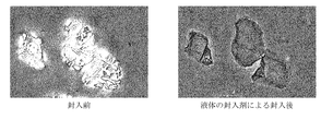
- Example 1 of this invention it is a photograph of the result of having observed the horny layer cell with the phase-contrast microscope (drawing substitute photograph).
- Example 2 of this invention it is a photograph of the result of having observed the horny layer cell with the phase-contrast microscope (drawing substitute photograph).
- the first aspect of the present invention is a cell encapsulation method for use in observation with a phase contrast microscope. Since the encapsulation method of the present invention makes it possible to observe cells with a phase-contrast microscope, cells are arranged between the adhesive layer of the transparent adhesive tape and the adhesive layer of the transparent adhesive tape, and the two adhesive tapes It is sandwiched and stuck with an adhesive layer.
- the cells to be observed in the present invention are not particularly limited, but particularly cells used for evaluating skin conditions such as stratum corneum cells, keratinocytes (keratinocytes), and fibroblasts are preferably exemplified. Hereinafter, explanation will be made by taking horny layer cells as an example.
- the stratum corneum is a flat cell having a thickness of about 1 ⁇ m constituting the stratum corneum and has a lot of skin information. Therefore, the stratum corneum cells should be considered when selecting cosmetics by evaluating the stratum corneum cells. It becomes possible to know the characteristics of the skin.
- the stratum corneum cells are filled with keratin, which is a major component, forming a very strong cell structure that protects the skin from various external stimuli.
- keratin which is a major component
- the adhesive force between the stratum corneum cells is weakened, it can be easily collected.
- the horny layer cells already obtained from a person are used, the step of collecting the horny layer cells from a person is not included in the scope of the present invention.
- the present invention is a cell encapsulation method for use in observation with a phase contrast microscope.
- a phase-contrast microscope is an optical microscope that can be observed by converting the phase difference of a light beam into contrast, and can observe a sample without staining or noninvasively.
- the present invention is characterized in that a transparent adhesive tape is used to encapsulate cells.
- the term “transparent” means that light can be transmitted and the above can be seen, and as long as the object is observed with a phase-contrast microscope, the degree and coloring are not limited at all. Absent.
- the linear light transmittance of light is preferably 80% or more, and more preferably 90% or more.
- the transparent adhesive tape preferably has an optical property turbidity HAZE of less than 1.5%, more preferably less than 1.0%. Moreover, it is preferable that it is colorless.
- the turbidity HAZE (cloudiness) is a value (%) indicating the degree of cloudiness of an object, and the smaller the value, the clearer the transparent adhesive tape is and the higher the transparency.
- (Turbidity HAZE (%) Td / Tt ⁇ 100
- Td diffuse transmittance
- Tt total light transmittance
- “linear transmittance of light (parallel transmittance)” and “turbidity HAZE (cloudiness)” Represents a value measured based on Japanese Industrial Standards JISK7136 (International Standardization Organization Standard ISO14782), JISK7361 (International Standardization Organization Standard ISO13468), and can be measured using, for example, HASE METER NDH500 manufactured by Nippon Denshoku Industries Co., Ltd.
- “linear transmittance of light” and “turbidity HAZE” represent values measured in the cross-sectional direction of the transparent adhesive tape.
- the adhesive tape used for this invention can use the general adhesive tape which consists of a transparent film layer and an adhesive layer, and can use what is marketed.
- Scotch registered trademark
- transparent beautiful color manufactured by 3M
- Scotch (registered trademark) 313 manufactured by 3M
- Scotch (registered trademark) 3450 manufactured by 3M
- Diahalo registered trademark
- Mitsubishi Plastics manufactured by Mitsubishi Plastics
- Cellotape registered trademark
- Damplon registered trademark
- Sekisui Bercel Sekisui Prime
- Sekisui Chemical Co., Ltd. and the like.
- an adhesive tape having a refractive index different from that of the cell in consideration of the characteristics of the phase contrast microscope, it is preferable to use an adhesive tape having a refractive index different from that of the cell, and more preferably, the difference between the refractive index of the adhesive tape and the refractive index of the cell is 0. It is preferable to use an adhesive tape that is 0.03 or more.
- the upper limit of the refractive index is not particularly limited, but is usually 0.60 or less.
- the refractive index of the stratum corneum is 1.55.
- the material of the film layer of the adhesive tape is not particularly limited as long as it satisfies the above requirement for transparency, and the material of the adhesive layer is not particularly limited.
- the material of the film layer is generally polyethylene, polypropylene, cellophane (chemical name: cellulose), polyethylene terephthalate (PET), polyolefin, etc.
- the material of the adhesive layer is generally acrylic copolymer or olefin Copolymers, urethane copolymers, epoxy copolymers, synthetic rubber, natural rubber and the like are used.
- the thickness of the adhesive tape is not particularly limited as long as it can be observed with a phase contrast microscope.
- the encapsulating method of the present invention is characterized in that cells are arranged between two adhesive layers of adhesive tape, and the cells are sandwiched and adhered by the adhesive layers of two adhesive tapes.
- sandwiching and adhering the cells between the adhesive tapes it is possible to observe the cells with a phase contrast microscope without generating halos without using a liquid encapsulant and a cover glass.
- the transparent adhesive tape laminate in which cells are encapsulated by the encapsulation method of the present invention is the second aspect of the present invention. That is, the second aspect of the present invention is a transparent pressure-sensitive adhesive tape laminate in which a transparent pressure-sensitive adhesive tape comprising a film layer and a pressure-sensitive adhesive layer is bonded so that the pressure-sensitive adhesive layers face each other. It is a transparent adhesive tape laminated body in which cells are enclosed between.
- the third aspect of the present invention is a method for observing cells using a phase-contrast microscope, the step of preparing a transparent adhesive tape in which cells are adhered to an adhesive layer, and the transparent adhesive tape in which the cells are adhered.
- the adhesive layer and the adhesive layer of another transparent adhesive tape are pasted to prepare a transparent adhesive tape laminate in which cells are encapsulated, and the laminate prepared in the above step is observed with a phase contrast microscope
- a method for observing cells using a phase-contrast microscope will be described with reference to the drawings.
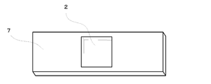
- FIG. 1 is a schematic cross-sectional view of a transparent adhesive tape laminate in which cells are encapsulated, showing a second embodiment of the present invention.
- the encapsulated cells are stratum corneum cells.
- the transparent adhesive tape laminated body 1 of this invention is stuck so that the mutual adhesion layer 6 of the adhesive tape 2 and the adhesive tape 3 may oppose.
- the stratum corneum cells 4 are encapsulated between the adhesive layer 6 and the adhesive layer 6.
- the manufacturing method of the transparent adhesive tape laminated body 1 of this invention and the observation method of the horny layer cell using a phase contrast microscope by the transparent adhesive tape laminated body 1 of this invention are demonstrated using FIG.
- FIG. 3 is a conceptual diagram showing an example of the steps of an observation method for stratum corneum cells using a phase contrast microscope.
- the transparent adhesive tape which the horny layer cell stuck to the adhesion layer is prepared.
- the transparent adhesive tape to which the stratum corneum cells are adhered may be prepared by any method, but it is convenient to use the tape strip kit 7 shown in FIG.
- the tape strip kit of FIG. 2 is made of a plastic plate and has a sampling hole in the center. The sampling hole is attached with an adhesive tape from the back side of the drawing, and the adhesive layer of the adhesive tape 2 is exposed at the sampling hole portion. Therefore, stratum corneum cells can be easily collected by pressing the collection hole of the tape strip kit 7 against the human skin.
- a transparent adhesive tape having stratum corneum cells adhered to the adhesive layer is prepared by pressing the tape strip kit 7 against a human cheek or the like.
- the step of pressing the tape strip kit 7 against a person's cheek is not included in the present invention.
- the adhesive layer of the transparent adhesive tape to which the horny layer cells are attached and the adhesive layer of another transparent adhesive tape are attached to prepare a transparent adhesive tape laminate in which the horny layer cells are encapsulated.
- the transparent adhesive tape in which the horny layer cells 4 are adhered to the adhesive layer is made to oppose the adhesive layer and the adhesive layer of another adhesive tape 3, and adheres the adhesive layers to each other.
- the existing horny layer cells 4 are encapsulated.
- the transparent adhesive tape laminated body 1 which is the 2nd aspect of this invention can be manufactured.
- the prepared laminate is observed with a phase contrast microscope.
- the transparent adhesive tape laminate 1 of the present invention is observed with the phase contrast microscope 9
- the horny layer cells can be observed by a simple method without using an encapsulating liquid or a cover glass.
- Example 1 Using the tape stripping kit (plastic plate-like member) shown in FIG. 2, stratum corneum cells were collected from the subject's cheek.
- the adhesive tape used in the tape stripping kit shown in FIG. 2 is a Diahalo tape (Mitsubishi Resin Co., Ltd.) with a refractive index of 1.48 to 1.50, a linear light transmittance of 92.374%, and a turbidity HAZE of 0.55%. Met.
- the linear transmittance (parallel transmittance) and turbidity HAZE of light are average values obtained by measuring the cross-sectional direction of the Diahalo tape five times using Nippon Denshoku Industries Co., Ltd. HASE METER NDH500. is there.
- the collected stratum corneum cells were attached using the same adhesive tape so that the adhesive layer and the adhesive layer were opposed to each other to enclose the stratum corneum cells, thereby preparing an adhesive tape laminate 1. At this time, when visually confirmed, air bubbles were not mixed.
- Example 2 The stratum corneum cells were observed with a phase contrast microscope in the same manner as in Example 1 except that the adhesive tape was changed to the 3M Scotch transparent beautiful color. No halo was observed, and the stratum corneum cells could be clearly observed. It was. The linear transmittance of light of 3M Scotch transparent beautiful color was 91.882%, and turbidity HAZE was 0.82%.
- Example 3 When the horny layer cells were observed with a phase contrast microscope in the same manner as in Example 1 except that the adhesive tape was changed to a cello tape made by Nichiban, the horny layer cells could be clearly observed without the occurrence of halo.
- the linear transmittance of light of the cellophane manufactured by Nichiban Co. was 89.608%, and the turbidity HAZE was 2.31%.
- Example 4 When corneal cells were observed with a phase contrast microscope in the same manner as in Example 1 except that the adhesive tape was changed to Scotch 313 manufactured by 3M, horny cells were clearly observed without the occurrence of halos. Note that the light transmittance of Scotch 313 manufactured by 3M was 92.30%, and the turbidity HAZE was 0.88%.
- Example 5 When the stratum corneum was observed with a phase contrast microscope in the same manner as in Example 1 except that the adhesive tape was changed to Scotch 3450 manufactured by 3M, the stratum corneum could be clearly observed without the occurrence of halo. In addition, the linear transmittance of light of Scotch 3450 manufactured by 3M was 91.76%, and the turbidity HAZE was 1.29%.
- Example 6 The stratum corneum cells were observed with a phase contrast microscope in the same manner as in Example 1 except that the adhesive tape was changed to Sekisui Chemical Co., Ltd. transparent packaging tape P83T. I was able to observe. In addition, Sekisui Chemical Co., Ltd. transparent packaging tape P83T had a linear light transmittance of 92.45% and a turbidity HAZE of 0.48%.
- the stratum corneum cells were observed by a method which is a method commonly used in phase contrast microscope observation. After collecting the stratum corneum cells using the tape stripping kit shown in FIG. 2, a drop of the liquid encapsulant was dropped into the collection hole and encapsulated using a cover glass. The observation photograph of a horny layer cell is shown in FIG. Even with a simple method using the adhesive tape of the present invention, stratum corneum cells could be clearly observed in the same manner as that conventionally performed by phase contrast microscopy.
- the encapsulation method, observation method, and adhesive tape laminate of the present invention enable a salesperson to easily observe stratum corneum cells when selecting cosmetics at a department store booth, a store, or a customer of a cosmetic user. Based on this observation result, an appropriate cosmetic can be selected and sold.
Landscapes
- Health & Medical Sciences (AREA)
- Life Sciences & Earth Sciences (AREA)
- Physics & Mathematics (AREA)
- Heart & Thoracic Surgery (AREA)
- Molecular Biology (AREA)
- General Physics & Mathematics (AREA)
- Pathology (AREA)
- Analytical Chemistry (AREA)
- Engineering & Computer Science (AREA)
- Biomedical Technology (AREA)
- Chemical & Material Sciences (AREA)
- Medical Informatics (AREA)
- Optics & Photonics (AREA)
- Surgery (AREA)
- Animal Behavior & Ethology (AREA)
- General Health & Medical Sciences (AREA)
- Public Health (AREA)
- Veterinary Medicine (AREA)
- Sampling And Sample Adjustment (AREA)
- Adhesives Or Adhesive Processes (AREA)
- Investigating Or Analysing Biological Materials (AREA)
- Microscoopes, Condenser (AREA)
Abstract
L'objet de la présente invention est la mise au point d'un procédé pour encapsuler une cellule, qui peut être réalisé facilement même par des vendeurs dans des cabines de grands magasins ou dans des boutiques ou au domicile de l'utilisateur de produits cosmétiques, et peut être utilisé pour l'observation sous un microscope à contraste de phase. La présente invention concerne aussi un procédé pour observer une cellule à l'aide d'un microscope à contraste de phase, et un procédé d'encapsulation de cellules destiné à être utilisé pour l'observation sous un microscope à contraste de phase, ledit procédé d'encapsulation consistant à placer la cellule entre une couche adhésive d'un ruban adhésif transparent et une couche adhésive d'un autre ruban adhésif transparent, puis à coller la cellule avec les couches adhésives des deux bandes adhésives afin qu'elle soit retenue entre les couches adhésives.
Priority Applications (1)
| Application Number | Priority Date | Filing Date | Title |
|---|---|---|---|
| JP2013553210A JP6126536B2 (ja) | 2012-01-13 | 2012-12-04 | 細胞の封入方法及び細胞の観察方法 |
Applications Claiming Priority (2)
| Application Number | Priority Date | Filing Date | Title |
|---|---|---|---|
| JP2012-005335 | 2012-01-13 | ||
| JP2012005335 | 2012-01-13 |
Publications (1)
| Publication Number | Publication Date |
|---|---|
| WO2013105363A1 true WO2013105363A1 (fr) | 2013-07-18 |
Family
ID=48754720
Family Applications (1)
| Application Number | Title | Priority Date | Filing Date |
|---|---|---|---|
| PCT/JP2012/081328 WO2013105363A1 (fr) | 2012-01-13 | 2012-12-04 | Procédé d'encapsulation de cellules, et procédé d'observation de cellules |
Country Status (4)
| Country | Link |
|---|---|
| JP (1) | JP6126536B2 (fr) |
| CN (2) | CN103207448B (fr) |
| TW (1) | TWI624654B (fr) |
| WO (1) | WO2013105363A1 (fr) |
Families Citing this family (4)
| Publication number | Priority date | Publication date | Assignee | Title |
|---|---|---|---|---|
| WO2013105363A1 (fr) * | 2012-01-13 | 2013-07-18 | ポーラ化成工業株式会社 | Procédé d'encapsulation de cellules, et procédé d'observation de cellules |
| US20180340866A1 (en) * | 2015-11-05 | 2018-11-29 | Shiseido Company, Ltd. | Stratum corneum collecting device and stratum corneum collecting/detecting kit |
| CN105739076A (zh) * | 2016-04-08 | 2016-07-06 | 苏州雅睿生物技术有限公司 | 一种载玻片 |
| CN107991159A (zh) * | 2017-11-20 | 2018-05-04 | 珠海伊斯佳科技股份有限公司 | 一种通过皮肤角质细胞形态评估皮肤状况的方法 |
Citations (10)
| Publication number | Priority date | Publication date | Assignee | Title |
|---|---|---|---|---|
| JPS5943358A (ja) * | 1982-09-03 | 1984-03-10 | Kanebo Ltd | 皮膚表面形態の検査方法及び検査用具 |
| JPS6348137U (fr) * | 1986-09-17 | 1988-04-01 | ||
| JPS6453165A (en) * | 1987-08-24 | 1989-03-01 | Shiseido Co Ltd | Preparation of cutaneous cell sample |
| JPH0527670U (ja) * | 1991-04-26 | 1993-04-09 | 株式会社テクノメデイカ | ぎよう虫卵検査紙 |
| JPH11344489A (ja) * | 1998-06-02 | 1999-12-14 | Shiseido Co Ltd | 角層のメラニンの定量方法 |
| JP2000005134A (ja) * | 1998-06-17 | 2000-01-11 | Pola Chem Ind Inc | 角層標本の作成方法 |
| WO2002025272A1 (fr) * | 2000-09-21 | 2002-03-28 | Shiseido Company, Ltd. | Procede d'evaluation du degre de maturite de cellules cornees |
| JP2005168693A (ja) * | 2003-12-10 | 2005-06-30 | Pola Chem Ind Inc | 角層細胞の形状の鑑別方法 |
| JP2009115813A (ja) * | 2008-12-26 | 2009-05-28 | Tokiwa Yakuhin Kogyo Kk | アトピー性皮膚炎の局所病態の非侵襲的評価方法 |
| JP2009257954A (ja) * | 2008-04-17 | 2009-11-05 | Hitachi Chem Co Ltd | 光学顕微鏡観察用試料の調整方法及びそれを用いた、粒子の凝集状態・分散状態の観察方法、撮影方法 |
Family Cites Families (3)
| Publication number | Priority date | Publication date | Assignee | Title |
|---|---|---|---|---|
| JP2005249410A (ja) * | 2004-03-01 | 2005-09-15 | Menicon Co Ltd | 生体組織検査シートおよびそれを用いた生体組織検査方法 |
| BRPI0611281A2 (pt) * | 2005-05-20 | 2010-08-31 | Premd Inc | ensaio direto de proteìna de pele em amostras de pele removidas por remocão da fita |
| WO2013105363A1 (fr) * | 2012-01-13 | 2013-07-18 | ポーラ化成工業株式会社 | Procédé d'encapsulation de cellules, et procédé d'observation de cellules |
-
2012
- 2012-12-04 WO PCT/JP2012/081328 patent/WO2013105363A1/fr active Application Filing
- 2012-12-04 JP JP2013553210A patent/JP6126536B2/ja active Active
-
2013
- 2013-01-11 TW TW102101011A patent/TWI624654B/zh active
- 2013-01-14 CN CN201310012849.6A patent/CN103207448B/zh active Active
- 2013-01-14 CN CN2013200171345U patent/CN203324569U/zh not_active Expired - Lifetime
Patent Citations (10)
| Publication number | Priority date | Publication date | Assignee | Title |
|---|---|---|---|---|
| JPS5943358A (ja) * | 1982-09-03 | 1984-03-10 | Kanebo Ltd | 皮膚表面形態の検査方法及び検査用具 |
| JPS6348137U (fr) * | 1986-09-17 | 1988-04-01 | ||
| JPS6453165A (en) * | 1987-08-24 | 1989-03-01 | Shiseido Co Ltd | Preparation of cutaneous cell sample |
| JPH0527670U (ja) * | 1991-04-26 | 1993-04-09 | 株式会社テクノメデイカ | ぎよう虫卵検査紙 |
| JPH11344489A (ja) * | 1998-06-02 | 1999-12-14 | Shiseido Co Ltd | 角層のメラニンの定量方法 |
| JP2000005134A (ja) * | 1998-06-17 | 2000-01-11 | Pola Chem Ind Inc | 角層標本の作成方法 |
| WO2002025272A1 (fr) * | 2000-09-21 | 2002-03-28 | Shiseido Company, Ltd. | Procede d'evaluation du degre de maturite de cellules cornees |
| JP2005168693A (ja) * | 2003-12-10 | 2005-06-30 | Pola Chem Ind Inc | 角層細胞の形状の鑑別方法 |
| JP2009257954A (ja) * | 2008-04-17 | 2009-11-05 | Hitachi Chem Co Ltd | 光学顕微鏡観察用試料の調整方法及びそれを用いた、粒子の凝集状態・分散状態の観察方法、撮影方法 |
| JP2009115813A (ja) * | 2008-12-26 | 2009-05-28 | Tokiwa Yakuhin Kogyo Kk | アトピー性皮膚炎の局所病態の非侵襲的評価方法 |
Also Published As
| Publication number | Publication date |
|---|---|
| CN203324569U (zh) | 2013-12-04 |
| TWI624654B (zh) | 2018-05-21 |
| CN103207448A (zh) | 2013-07-17 |
| JPWO2013105363A1 (ja) | 2015-05-11 |
| TW201333441A (zh) | 2013-08-16 |
| JP6126536B2 (ja) | 2017-05-10 |
| CN103207448B (zh) | 2017-11-24 |
Similar Documents
| Publication | Publication Date | Title |
|---|---|---|
| JP6126536B2 (ja) | 細胞の封入方法及び細胞の観察方法 | |
| TW200921097A (en) | Devices, systems, and methods for the containment and use of liquid solutions | |
| US8403582B2 (en) | Apparatus for treating a stain in clothing | |
| WO2016025430A1 (fr) | Systèmes et procédés photoniques épidermiques | |
| Han et al. | Wearable hydrogel‐based epidermal sensor with thermal compatibility and long term stability for smart colorimetric multi‐signals monitoring | |
| FR2831789A1 (fr) | Procede et dispositif pour l'evaluation de la secheresse cutanee notamment | |
| US20200330029A1 (en) | Devices, systems, and methods for monitoring hair | |
| Song et al. | Nanopore thin film enabled optical platform for drug loading and release | |
| JP5654301B2 (ja) | メイク化粧料の塗布色チェック用具および塗布色チェック方法 | |
| CN107773275A (zh) | 一种皮肤检测采样包及其应用 | |
| JP2014198259A (ja) | メイク化粧料の塗布色チェック用具および塗布色チェック方法 | |
| JP2009020026A (ja) | 検査シート | |
| CN208659417U (zh) | 角质贴载具 | |
| FR2719830A1 (fr) | Paquet-gants. | |
| JP5467570B2 (ja) | 肌の鑑別法 | |
| JP3635742B2 (ja) | 角質細胞採取用シート | |
| SE520948C2 (sv) | Allergitestelement | |
| ITUB20150376A1 (it) | Dispositivo per il campionamento della superficie oculare mediante imprinting | |
| CN214335359U (zh) | 一种载玻片 | |
| KR20090110959A (ko) | 현미경 확대 관찰 및 보관 겸용 샘플 접착 시트 | |
| JPH11344489A (ja) | 角層のメラニンの定量方法 | |
| JP3157840U (ja) | 試料塗布膜形成用ツールおよび滑り止めシート | |
| US11020044B2 (en) | Multiple well epicutaneous test patch array | |
| JP2004065675A (ja) | 皮膚反応試験方法とそれに用いる試験部材 | |
| Rodrigues et al. | Conceptual and empirical problems in organizational commitment research: A critical analysis of J. Meyer and N. Allen three-component model |
Legal Events
| Date | Code | Title | Description |
|---|---|---|---|
| 121 | Ep: the epo has been informed by wipo that ep was designated in this application |
Ref document number: 12864883 Country of ref document: EP Kind code of ref document: A1 |
|
| ENP | Entry into the national phase |
Ref document number: 2013553210 Country of ref document: JP Kind code of ref document: A |
|
| 122 | Ep: pct application non-entry in european phase |
Ref document number: 12864883 Country of ref document: EP Kind code of ref document: A1 |
|
| NENP | Non-entry into the national phase |
Ref country code: DE |