WO2012043746A1 - イムノクロマトグラフィー用テストストリップおよびその製造方法 - Google Patents
イムノクロマトグラフィー用テストストリップおよびその製造方法 Download PDFInfo
- Publication number
- WO2012043746A1 WO2012043746A1 PCT/JP2011/072427 JP2011072427W WO2012043746A1 WO 2012043746 A1 WO2012043746 A1 WO 2012043746A1 JP 2011072427 W JP2011072427 W JP 2011072427W WO 2012043746 A1 WO2012043746 A1 WO 2012043746A1
- Authority
- WO
- WIPO (PCT)
- Prior art keywords
- conjugate
- antibody
- test strip
- substance
- membrane carrier
- Prior art date
Links
Images


Classifications
-
- G—PHYSICS
- G01—MEASURING; TESTING
- G01N—INVESTIGATING OR ANALYSING MATERIALS BY DETERMINING THEIR CHEMICAL OR PHYSICAL PROPERTIES
- G01N33/00—Investigating or analysing materials by specific methods not covered by groups G01N1/00 - G01N31/00
- G01N33/48—Biological material, e.g. blood, urine; Haemocytometers
- G01N33/50—Chemical analysis of biological material, e.g. blood, urine; Testing involving biospecific ligand binding methods; Immunological testing
- G01N33/53—Immunoassay; Biospecific binding assay; Materials therefor
- G01N33/543—Immunoassay; Biospecific binding assay; Materials therefor with an insoluble carrier for immobilising immunochemicals
-
- G—PHYSICS
- G01—MEASURING; TESTING
- G01N—INVESTIGATING OR ANALYSING MATERIALS BY DETERMINING THEIR CHEMICAL OR PHYSICAL PROPERTIES
- G01N33/00—Investigating or analysing materials by specific methods not covered by groups G01N1/00 - G01N31/00
- G01N33/48—Biological material, e.g. blood, urine; Haemocytometers
- G01N33/50—Chemical analysis of biological material, e.g. blood, urine; Testing involving biospecific ligand binding methods; Immunological testing
- G01N33/53—Immunoassay; Biospecific binding assay; Materials therefor
- G01N33/569—Immunoassay; Biospecific binding assay; Materials therefor for microorganisms, e.g. protozoa, bacteria, viruses
- G01N33/56983—Viruses
-
- G—PHYSICS
- G01—MEASURING; TESTING
- G01N—INVESTIGATING OR ANALYSING MATERIALS BY DETERMINING THEIR CHEMICAL OR PHYSICAL PROPERTIES
- G01N33/00—Investigating or analysing materials by specific methods not covered by groups G01N1/00 - G01N31/00
- G01N33/48—Biological material, e.g. blood, urine; Haemocytometers
- G01N33/50—Chemical analysis of biological material, e.g. blood, urine; Testing involving biospecific ligand binding methods; Immunological testing
- G01N33/53—Immunoassay; Biospecific binding assay; Materials therefor
- G01N33/531—Production of immunochemical test materials
-
- G—PHYSICS
- G01—MEASURING; TESTING
- G01N—INVESTIGATING OR ANALYSING MATERIALS BY DETERMINING THEIR CHEMICAL OR PHYSICAL PROPERTIES
- G01N33/00—Investigating or analysing materials by specific methods not covered by groups G01N1/00 - G01N31/00
- G01N33/48—Biological material, e.g. blood, urine; Haemocytometers
- G01N33/50—Chemical analysis of biological material, e.g. blood, urine; Testing involving biospecific ligand binding methods; Immunological testing
- G01N33/53—Immunoassay; Biospecific binding assay; Materials therefor
- G01N33/543—Immunoassay; Biospecific binding assay; Materials therefor with an insoluble carrier for immobilising immunochemicals
- G01N33/54366—Apparatus specially adapted for solid-phase testing
- G01N33/54386—Analytical elements
- G01N33/54387—Immunochromatographic test strips
- G01N33/54388—Immunochromatographic test strips based on lateral flow
-
- G—PHYSICS
- G01—MEASURING; TESTING
- G01N—INVESTIGATING OR ANALYSING MATERIALS BY DETERMINING THEIR CHEMICAL OR PHYSICAL PROPERTIES
- G01N33/00—Investigating or analysing materials by specific methods not covered by groups G01N1/00 - G01N31/00
- G01N33/48—Biological material, e.g. blood, urine; Haemocytometers
- G01N33/50—Chemical analysis of biological material, e.g. blood, urine; Testing involving biospecific ligand binding methods; Immunological testing
- G01N33/53—Immunoassay; Biospecific binding assay; Materials therefor
- G01N33/558—Immunoassay; Biospecific binding assay; Materials therefor using diffusion or migration of antigen or antibody
-
- G—PHYSICS
- G01—MEASURING; TESTING
- G01N—INVESTIGATING OR ANALYSING MATERIALS BY DETERMINING THEIR CHEMICAL OR PHYSICAL PROPERTIES
- G01N33/00—Investigating or analysing materials by specific methods not covered by groups G01N1/00 - G01N31/00
- G01N33/48—Biological material, e.g. blood, urine; Haemocytometers
- G01N33/50—Chemical analysis of biological material, e.g. blood, urine; Testing involving biospecific ligand binding methods; Immunological testing
- G01N33/53—Immunoassay; Biospecific binding assay; Materials therefor
- G01N33/569—Immunoassay; Biospecific binding assay; Materials therefor for microorganisms, e.g. protozoa, bacteria, viruses
-
- G—PHYSICS
- G01—MEASURING; TESTING
- G01N—INVESTIGATING OR ANALYSING MATERIALS BY DETERMINING THEIR CHEMICAL OR PHYSICAL PROPERTIES
- G01N2333/00—Assays involving biological materials from specific organisms or of a specific nature
- G01N2333/005—Assays involving biological materials from specific organisms or of a specific nature from viruses
- G01N2333/08—RNA viruses
- G01N2333/11—Orthomyxoviridae, e.g. influenza virus
Definitions
- the present invention relates to an immunochromatographic test strip for detecting a substance to be detected such as influenza virus and a method for producing the same.
- Immunochromatography is a conjugate (detection reagent) by immobilizing an antigen to be detected or an antibody against an antibody or an antigen on an insoluble membrane carrier that is a chromatographic medium to create a detection part that is a stationary phase.
- a label sensitized by the antibody or antigen capable of binding to the detection substance as a mobile phase, specifically reacting the detection substance and the conjugate that is the mobile phase, and in the detection part that is the stationary phase
- the substance to be detected bound to the conjugate is reacted specifically with the antibody or antigen immobilized on the detection part.
- colloidal metal particles such as gold colloid or color latex particles are used as the label, so the presence of the substance to be detected in the sample or the amount thereof is detected from the color in the detection part. .
- the immunochromatographic test strip is composed of a sample pad for supplying a sample, a conjugate pad for arranging a conjugate as a mobile phase, and a composite of the sample and the conjugate.
- an insoluble membrane carrier having a detection unit for detection and an absorption pad for absorbing a sample that has developed the insoluble membrane carrier are arranged while the body is developed.
- the conventional immunochromatographic test strip having the above-described configuration has a problem in that it is difficult to release the conjugate from the conjugate pad, so that it takes time to complete the reaction or the background tends to increase.
- the conjugate pad is obtained by uniformly impregnating and drying the conjugate.
- the conjugate pad is subjected to the step of impregnating the conjugate, it is difficult to automate the production, and the conjugate impregnation is likely to be uneven. There is a problem that the performance may vary.
- An object of the present invention is to provide an immunochromatographic test strip having a short reaction completion time and excellent sensitivity. Another object of the present invention is to provide a method for producing the immunochromatographic test strip that does not include a step of impregnating with a liquid, is easily automated, and has uniform performance.
- the immunochromatographic test strip of the present invention detects a substance to be detected by developing a sample that may contain the substance to be detected, and includes the following (1) and (2):
- the lower surface of the sample supply part of the conjugate pad is not in contact with the upper surface of the insoluble membrane carrier, and the lower surface of the conjugate part of the conjugate pad is in contact with the upper surface of the insoluble membrane carrier.
- a sample supply unit that supplies a sample that may contain a substance to be detected, and an antibody or antigen that immunologically reacts with the substance to be detected downstream of the sample supply part
- a conjugate part having a line-shaped conjugate part containing the conjugate immobilized on the conjugate pad (2) at least a detection part immobilized with an antibody or antigen that immunologically reacts with the substance to be detected
- immobilizing an antigen or antibody means that the antigen or antibody is physically or chemically supported on a label or an insoluble membrane carrier.
- the conjugate part is arranged in a line in the sample development direction, that is, the direction perpendicular to the line connecting the center of the sample supply part of the conjugate pad and the center of the upstream end of the insoluble membrane carrier. It is preferable.
- the detection in the present invention includes not only qualitative detection but also quantitative detection for a substance to be detected that can be quantified.
- the immunochromatographic test strip immunologically reacts with an antibody or antigen that immunologically reacts with a target substance immobilized on a labeled body, and with a target substance immobilized on a detector. It is preferable that the antibody or antigen to be separated is different. That is, an antibody or antigen that immunologically reacts with a target substance immobilized on a label and an antibody or antigen that reacts immunologically with a target substance immobilized on a detection unit These are preferably antibodies or antigens that each recognize a different part of the substance to be detected. By immobilizing another antibody or antigen on the label and the detection part, the sensitivity is further improved.
- the center line of the line of the line-shaped conjugate part is preferably arranged on the downstream side of the upstream end part of the insoluble membrane carrier.
- the center line of the line of the line-shaped conjugate part is preferably arranged 10 to 20 mm downstream from the upstream end of the conjugate pad.
- the method for producing the immunochromatographic test strip is also one aspect of the present invention.
- an antibody or an antigen that immunologically reacts with a substance to be detected is immobilized on a label on a part of a pad-like porous material to be a conjugate pad.
- a conjugate liquid containing the prepared conjugate was applied in a line and dried to form a conjugate part, and a conjugate pad was prepared using a part other than the conjugate part of the porous material as a sample supply part.
- the conjugate pad is placed on the upstream side of the insoluble membrane carrier having at least one detection portion on which an antibody or antigen that immunologically reacts with the substance to be detected, and the lower surface of the sample supply portion is an insoluble membrane.
- Laminate so that the bottom surface of the conjugate part does not come into contact with the carrier and is in contact with the insoluble membrane carrier. And butterflies.
- the manufacturing method does not include a step of impregnating the conjugate with the porous material, and the conjugate part is formed by application of a liquid that is easy to automate, so that the manufacturing can be automated as a whole, and The liquid coating material is relatively easy to control, and the performance of the resulting immunochromatographic test strip can be easily maintained uniformly.
- the conjugate part is formed in a line shape, and the conjugate part and the insoluble membrane carrier are in a specific positional relationship. It can be composed of an insoluble membrane carrier, has excellent release properties of the conjugate from the conjugate pad, completes the reaction in a short time, and has excellent sensitivity. Further, the release property and sensitivity are more excellent by arranging the center line of the line-shaped conjugate portion on the downstream side of the upstream end portion of the insoluble membrane carrier. Since the production method of the present invention does not include a step of impregnating the entire conjugate liquid with the conjugate liquid unlike the conventional conjugate pad, automation of the production of the conjugate pad is facilitated. Moreover, since the conjugate is formed by being applied in a line shape, it is easy to adjust the amount and application position of the conjugate, and these can be made uniform. As a result, the immunochromatography has uniform performance. A test strip is obtained.
- test strip for immunochromatography of this invention is shown (device preparation example 2,3,4)
- the test strip for immunochromatography of this invention is shown (device preparation example 7).
- the test strip for immunochromatography of a reference example is shown (device preparation example 5).
- a conventional test strip for immunochromatography is shown (device production example 1).
- the substance to be detected includes viruses and physiologically active substances such as proteins that can generally be measured using an antigen-antibody reaction.
- viruses include influenza viruses such as influenza A virus and influenza B virus, hepatitis B virus, hepatitis C virus, human immunodeficiency virus, etc.
- influenza viruses such as influenza A virus and influenza B virus, hepatitis B virus, hepatitis C virus, human immunodeficiency virus, etc.
- the protein include human hemoglobin, Examples include hepatitis B virus antibody, hepatitis C virus antibody, and human immunodeficiency virus antibody.
- it is preferable to use influenza virus as a substance to be detected and it is more preferable to form a plurality of detection parts described later and to use influenza A virus and influenza B virus as substances to be detected.
- examples of a sample that may contain a substance to be detected include substances derived mainly from living bodies (organisms) such as body fluids, and extracts obtained by extracting substances to be detected from them.
- substances derived from living organisms (organisms) include blood, urine, stool, nasal discharge and nasal discharge from nasal cavity / nasal cavity / pharynx / nasopharynx, secretions collected as sputum and swab specimens Examples include liquid and saliva.
- the sample is preferably a nasal secretion or nasal aspirate derived from the nostril, nasal cavity, pharynx, nasopharynx, etc., or a secreted liquid collected as a sputum or swab specimen.
- the living body (organism) -derived substance and the extract thereof may be used as a sample as they are, or may be appropriately diluted with a diluent to obtain a sample. Moreover, you may use what was filtered suitably as a sample.
- the antibody or antigen that immunologically reacts with the substance to be detected used in the present invention is an antibody or antigen that can bind to the substance to be detected.
- the substance to be detected is a virus or an antigen
- the antibody or the substance to be detected When is an antibody, an antigen is preferred.
- An antibody or antigen that reacts immunologically with a substance to be detected is immobilized on a label and a detection part described later.
- the antibody or antigen immobilized on the label and the detection unit may be the same, but it is preferable that the label and the detection unit are different.
- the substance to be detected bound to the conjugate in the immunochromatographic test strip obtained by using different antibodies or antigens immobilized on the label and those immobilized on the detection part. Can be prevented from competing with the reaction between the detection substance bound to the antibody or antigen of the detection unit and the reaction between the detected substance bound to the conjugate and the unreacted conjugate, and to the detected substance bound to the conjugate. And the antibody or antigen in the detection part can be increased, and as a result, the sensitivity of the immunochromatographic test strip is improved.
- another means that the type is different, in the case of an antibody, an antibody that recognizes a different epitope, and in the case of an antigen, an antigen having a different epitope.
- the antibody immobilized on the label and the detection part is preferably a monoclonal antibody.
- a monoclonal antibody By using a monoclonal antibody, the specificity of the reaction can be increased.
- the antibody immobilized on the label and the detection part may be any antibody that can detect influenza virus, but anti-influenza A virus monoclonal antibody, anti-influenza B virus monoclonal Anti-influenza virus monoclonal antibodies such as antibodies are preferred.
- functional fragments of antibodies having antigen-antibody reaction activity are also treated as antibodies in the present invention.
- Examples of the functional fragment of the antibody include those obtained through an immunization process for animals, those obtained using genetic recombination techniques, and chimeric antibodies.
- Examples of the functional fragment of an antibody include F (ab ′) 2 and Fab ′. These functional fragments can be produced by treating the antibody with a proteolytic enzyme (for example, pepsin or papain). Of these, in the present invention, it is particularly preferable to use F (ab ′) 2 as the antibody immobilized on the label.
- F (ab ′) 2 which is a functionally fragmented antibody for the labeled body
- the size of the conjugate in which the antibody is bound to the labeled body can be reduced, and the conjugate pad and the insoluble membrane carrier It has excellent expandability inside.
- the specificity of the reaction can be increased by using a functional fragmented antibody.
- the label used in the present invention a known label conventionally used for a test strip for immunochromatography can be used.
- colloidal metal particles such as gold colloid particles and platinum colloid particles, color latex particles, and magnetic particles are preferable, and gold colloid particles are particularly preferable.
- the particle size of the label it is preferable to use a label having an appropriate particle size according to the label used.
- the particle size is preferably 20 to 60 nm, particularly preferably 45 to 55 nm.
- the gold colloidal particles can be produced by a generally known method, for example, by dropping and stirring a trisodium citrate aqueous solution into a heated tetrachloroauric (III) acid aqueous solution.
- the conjugate used in the present invention is obtained by immobilizing an antibody or antigen that immunologically reacts with a substance to be detected on the label as described above.
- the conjugate is preferably a colloidal gold particle in which an anti-influenza virus monoclonal antibody is immobilized.
- Examples of a method for immobilizing an antibody or antigen that immunologically reacts with a substance to be detected to a label include physical adsorption and chemical bonding, and are generally immobilized by physical adsorption.
- colloidal gold particles and anti-influenza virus monoclonal antibody are usually added to a buffer solution and immobilized by physical adsorption.
- the antibody concentration is preferably adjusted to 20 to 100 ⁇ g / mL.
- the buffer and its pH are preferably, for example, 0.5 to 5 mM phosphate buffer (pH 6 to 7), 0.5 to 5 mM borate buffer (pH 8 to 9.5), and the like.
- it is preferable to block the region where the antibody or antigen is not bound on the label such as colloidal gold particles with BSA or the like.
- the conjugate pad used in the present invention is composed of a pad-like porous material on which a sample can be developed and can hold the conjugate, and a sample supply part and a line-like conjugate part are formed in a part thereof. Have.
- a porous material part that does not contain the conjugate between the sample supply part and the conjugate part, and the sample supplied to the sample supply part expands the porous material and the conjugate part. It is preferable to reach.
- the sample supply part is a part for supplying a sample that may contain a substance to be detected, and is formed in a part of the porous material.
- the sample supply part is on the upstream side of the conjugate pad.
- the conjugate part is a part containing a conjugate, and is formed in a line shape on the porous material on the downstream side of the sample supply part.
- the line-shaped conjugate part is arranged in a line in the sample development direction, that is, the direction perpendicular to the line connecting the center of the sample supply part of the conjugate pad and the center of the upstream end of the insoluble membrane carrier described later. It is preferable. Moreover, it is desirable that the line-shaped conjugate portion is disposed downstream of the center of the length of the conjugate pad in the sample development direction. In consideration of the general size of the immunochromatographic test strip, the center line of the line-shaped conjugate portion is located 10 to 20 mm downstream from the upstream end portion of the conjugate pad.
- the line width of the line-shaped conjugate portion is sufficient if it has a width that allows the amount of the conjugate necessary for detection of the substance to be detected to be contained, and is preferably 3 to 5 mm.
- the conjugate pad is laminated with the insoluble membrane carrier so that the lower surface of the downstream end thereof is in contact with the upper surface of the insoluble membrane carrier described later.
- the conjugate pad is laminated with the insoluble membrane carrier such that the lower surface of the sample supply portion does not contact the upper surface of the insoluble membrane carrier, and the lower surface of the conjugate portion contacts the upper surface of the insoluble membrane carrier.
- the contact portion between the lower surface of the conjugate portion and the upper surface of the insoluble membrane carrier may be a part of the lower surface of the conjugate portion, but is preferably half or more.
- the entire lower surface of the conjugate portion is in contact with the upper surface of the insoluble membrane carrier.
- the contact of more than half means that the center line of the line-shaped conjugate portion is arranged downstream of the upstream end portion of the insoluble membrane carrier.
- a sample supply part is formed in a part upstream of the conjugate part of the conjugate pad, and this part corresponds to a part serving as a sample pad provided in a so-called conventional immunochromatographic test strip. To do.
- the sample When a sample that may contain a substance to be detected is supplied to the sample supply part of the conjugate pad, the sample flows downstream from the upstream sample supply part through the porous material part not containing the conjugate. It flows to the conjugate part.
- the substance to be detected influenza virus
- the conjugate gold colloid particles on which the anti-influenza virus monoclonal antibody is immobilized
- form a complex aggregate
- the sample is then developed into an insoluble membrane carrier that is placed in contact with the lower surface of the conjugate portion.
- porous material constituting the conjugate pad examples include pads made of nonwoven fibers such as paper, cellulose mixture, nitrocellulose, polyester, acrylonitrile copolymer, glass, and rayon. Of these, a glass fiber pad (glass fiber pad) is preferred.
- the insoluble membrane carrier used in the present invention has at least one detection unit on which an antibody or antigen that immunologically reacts with a substance to be detected is immobilized. Immobilization of an antibody or antigen that reacts immunologically with a substance to be detected on an insoluble membrane carrier can be carried out by a conventionally known method. In the case of a lateral flow type immunochromatographic test strip, immobilization is performed as follows. Prepare a liquid containing the antibody or antigen at a predetermined concentration, and then use a device having a mechanism that can move the liquid in a horizontal direction while discharging the liquid from the nozzle at a constant speed. Can be applied to an insoluble membrane carrier in a line and dried to be immobilized.
- the concentration of antibody or antigen in the above solution is preferably 0.1 to 5 mg / mL, more preferably 0.5 to 2 mg / mL.
- the amount of antibody or antigen immobilized on an insoluble membrane carrier can be optimized by adjusting the ejection speed from the nozzle of the above apparatus in the case of the lateral flow type, and is preferably 0.5 to 2 ⁇ L / cm. It is.
- the measurement method using the lateral flow type immunochromatography test strip is such that the sample supplied from the portion of the conjugate pad that contacts the insoluble carrier moves in a parallel direction with respect to the insoluble membrane carrier by capillary action. It is a measurement method of the system developed so as to.
- a solution containing the antibody or antigen described above at a predetermined concentration can be prepared by adding the antibody or antigen to a buffer solution.
- the buffer include normal buffers such as phosphate buffer, Tris buffer, and Good's buffer.
- the pH of the buffer is preferably in the range of 6.0 to 9.5, more preferably 6.5 to 8.5, and even more preferably 7.0 to 8.0.
- the buffer may further contain salts such as sodium chloride, stabilizers such as sucrose, preservatives, preservatives such as procrine, and the like.
- the salts include those added for the purpose of adjusting the pH of the buffer solution, such as sodium hydroxide, in addition to those included for adjusting the ionic strength, such as sodium chloride.
- blocking can also be carried out by coating a region other than the region where the antibody or antigen is immobilized with a commonly used blocking agent in the form of a solution or vapor.
- a control capture reagent conventionally used in immunochromatographic test strips may be immobilized on the insoluble membrane carrier.
- the control capture reagent is a reagent for ensuring the reliability of the assay, and captures the control reagent contained in the conjugate pad. For example, when KLH labeled on the conjugate pad is included as a control reagent, an anti-KLH antibody or the like corresponds to the control capture reagent.
- the position at which the control capture reagent is immobilized can be appropriately selected to suit the design of the assay system.
- the membrane constituting the insoluble membrane carrier used in the present invention a known membrane conventionally used as an insoluble membrane carrier for immunochromatographic test strips can be used.
- membranes composed of fibers made of polyethylene, polyethylene terephthalate, nylons, glass, polysaccharides such as cellulose and cellulose derivatives, ceramics, and the like.
- Specific examples include glass fiber filter paper and cellulose filter paper commercially available from Sartorius, Millipore, Toyo Roshi, Whatman, and the like. Of these, Sartorius and UniSart® CN140 are preferred.
- the pore size and structure of the insoluble membrane carrier it is possible to control the speed at which the complex of the conjugate and the substance to be detected in the sample flows through the insoluble membrane carrier.
- the absorption pad is a portion having liquid absorbency that controls the development of the sample by absorbing the sample that has moved and passed through the insoluble membrane carrier.
- the absorbent pad a known absorbent pad that has been conventionally used for immunochromatographic test strips is used.
- filter paper can be used.
- Whatman, 740-E is used.
- the immunochromatographic test strip of the present invention includes the conjugate pad and an insoluble membrane carrier.
- the conjugate pad and the insoluble membrane carrier are laminated so that the lower surface of the conjugate pad and the upper surface of the insoluble membrane carrier are in contact with each other.
- the lower surface of the sample supply part of the conjugate pad is not in contact with the upper surface of the insoluble membrane carrier, and part or all of the lower surface of the conjugate part of the conjugate pad, preferably more than half, It arrange
- an absorption pad is further disposed at the downstream end of the insoluble membrane carrier.
- the immunochromatographic test strip is preferably disposed on a solid support such as a plastic adhesive sheet.
- the solid support is composed of a material that does not interfere with the capillary flow of the sample and conjugate.
- the immunochromatographic test strip may be fixed on a solid support with an adhesive or the like.
- the adhesive component and the like are also composed of a substance that does not interfere with the capillary flow of the sample and the conjugate. It is also possible to laminate a polyester film or the like for the purpose of increasing the mechanical strength of the insoluble membrane carrier and preventing moisture evaporation (drying) during the assay.
- the immunochromatographic test strip should be placed in an appropriate container (housing) in consideration of the size of the immunochromatographic test strip, the method and position of addition of the sample, the formation position of the detection part of the insoluble membrane carrier, the signal detection method, etc. It can be stored and used, and the state stored and mounted in this way is called “device”.
- the test strip for immunochromatography of the present invention includes a conjugate pad and an insoluble membrane carrier, and may further include other reagents and configurations according to measurement conditions and samples. Examples of other reagents include blocking agents that prevent non-specific reactions, and examples of other configurations include a 3rd pad for removing components unnecessary for measurement in a sample.
- the method for producing the immunochromatographic test strip is not particularly limited, but the conjugate liquid is applied in a line to a part of the pad-like porous material (the part to be the conjugate part) and dried to form a conjugate pad. After the preparation, the conjugate pad is preferably brought into contact with an insoluble membrane carrier having a detection portion to form a test strip for immunochromatography.
- the method for producing a test strip for immunochromatography according to the present invention includes the following steps (1) to (3).
- Conjugate solution containing a conjugate in which an antibody or antigen that immunologically reacts with a substance to be detected is immobilized on a label in a part of a pad-like porous material to be a conjugate pad
- Step of The conjugate solution is a buffer solution containing a conjugate that is usually used, and the concentration of the conjugate is appropriately adjusted according to the amount of conjugate to be contained in the conjugate portion.
- the conjugate liquid is applied to a part of the pad-like porous material to be a conjugate pad (part to be a conjugate part) using a nozzle or the like that can discharge the liquid at a constant speed.
- Application is easier to control than impregnation, etc., and it is easier to control the amount of conjugate by applying the conjugate liquid, and the conjugate amount of the resulting conjugate pad is excellent in uniformity. It becomes.
- the conjugate pad is obtained by drying by heat drying, natural drying or the like.
- the conjugate pad has a sample supply part
- the sample supply part is a porous material part that does not contain the conjugate, and it is appropriate depending on the developability of the sample, the container for storing the test strip for immunochromatography, etc. Can be placed in position.
- the method for forming the insoluble membrane carrier, the method for laminating the conjugate pad and the insoluble membrane carrier, and the like are as described above.
- the production of the immunochromatographic test strip of the present invention can be carried out by appropriately modifying or altering the method described in the examples.
- the signal derived from the conjugate may be measured according to a known method, for example, the absorbance or the intensity of reflected light may be measured.
- Anti-influenza A virus monoclonal antibodies (Clone # 622212, Clone # 622241A) and anti-influenza B virus monoclonal antibodies (Clone # 612108, Clone # 614216) used in the following tests use recombinant influenza nucleoprotein as an antigen, The mouse was immunized and obtained using methods routinely used by those skilled in the art to produce monoclonal antibodies.
- BSA bovine serum albumin
- 2 mL of 10% bovine serum albumin (BSA) aqueous solution is added to the gold colloid particle-anti-influenza A virus monoclonal antibody mixture and the gold colloid-anti-influenza B virus monoclonal antibody mixture, and the mixture is further stirred for 5 minutes. Then, it centrifuged at 10,000 rpm for 45 minutes at 10 degreeC, and the deposit (conjugate) was obtained. To the resulting conjugate, 1.2 mL of Conjugate Dilution Buffer (Scripps) was added to suspend the conjugate. The absorbance of each conjugate was measured at 531 nm (the maximum absorption wavelength of the colloidal gold particles used). Absorbance was measured in the same manner in the following tests.
- BSA bovine serum albumin
- conjugate pad The conjugate prepared in 1) above is mixed with 1.33% casein, 4% sucrose solution (pH 7.5) so that the concentration is 8-20 OD / mL. Then, a fixed volume of glass fiber pad (Nippon Pole Co., No. 8964) was impregnated with 1.2 times the volume of the pad. The conjugate pad was dried by heating at 70 ° C. for 30 minutes in a dry oven. Moreover, when adding additives, such as a sensitizer, after adding required amount to the said detection reagent, the same operation was performed.
- additives such as a sensitizer
- test strip When the test strip was assembled, the lines were applied from the upstream side in the order of influenza A virus monoclonal antibody (c1), anti-influenza B virus monoclonal antibody (c2), and control antibody (c3).
- the membrane was dried in a dry oven at 70 ° C. for 45 minutes to obtain an antibody-immobilized membrane.
- the goat anti-mouse IgG specifically recognizes the F (ab ′) 2 portion of the antibody.
- sample pad A glass fiber pad (Lydall) was used as a sample pad.
- FIG. 4 shows a schematic configuration diagram of a conventional immunochromatographic test strip.
- the line-shaped conjugate part (g) formed at a position 15 mm from the upstream end of the conjugate pad has an upstream end on the upstream side of the antibody-immobilized membrane (b). It is formed at a position away from the downstream end so as to be downstream from the end. That is, the lower surface of the conjugate part (g) is in contact with the upper surface of the antibody-immobilized membrane over the entire surface.
- the immunochromatographic test strip was in the form of an immunochromatographic test device in the same manner as in Device Preparation Example 1.
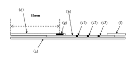
- FIG. 1 shows a schematic configuration diagram of the immunochromatographic test strip of the present invention.
- the conjugate part is formed at a position 12 mm from the upstream end of the conjugate pad, and the center line is upstream from the upstream end face of the antibody-immobilized membrane.
- the downstream end portion of the conjugate portion is disposed downstream of the upstream end portion of the antibody-immobilized membrane. That is, the lower surface of the conjugate part is less than half but is in contact with the antibody-immobilized membrane.
- the immunochromatographic test strip was in the form of an immunochromatographic test device in the same manner as in Device Preparation Example 1.
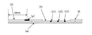
- FIG. 2 shows a schematic configuration diagram of the immunochromatographic test strip of the present invention.
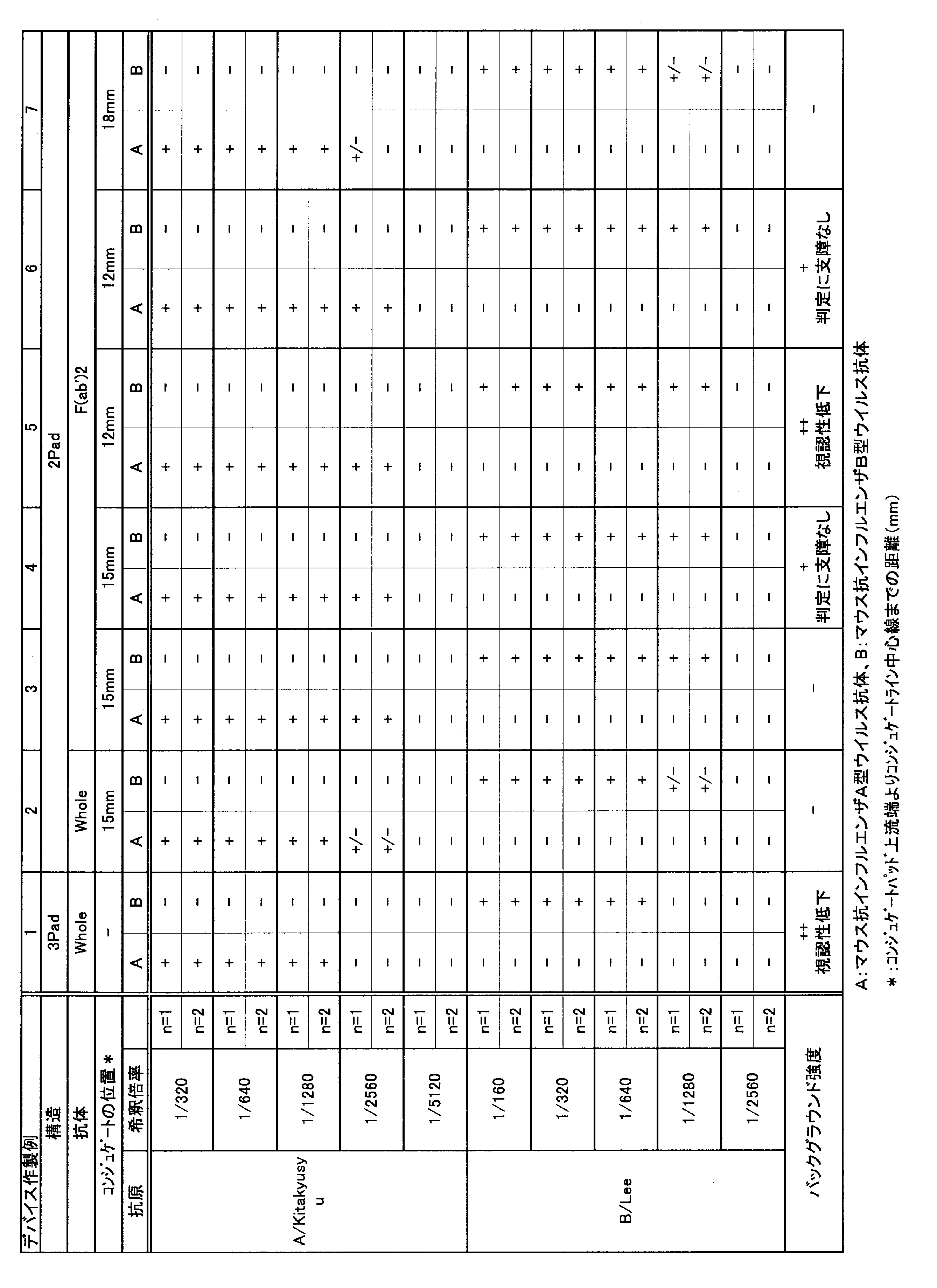
- Example 1 Device preparation example 1 above. ⁇ 7. Influenza virus detection test was performed using the immunochromatographic test device prepared in 1. 1. Test method (1) Sample The following antigens diluted to 1/160 to 1/2560 with PBS (pH 7.4) containing 2% BSA were used as simulated samples. Influenza virus type A antigen; derived from Kitakyusyu 159/93 strain Influenza virus type B antigen: derived from Lee 40 strain (2) Procedure 115 ⁇ L of the simulated sample prepared in (1) above was added from the sample supply window of the device for immunochromatography, After 15 minutes, the presence or absence of a red detection spot on the antibody-immobilized membrane was observed from the detection window.
- the immunochromatographic test strip of the present invention can provide an immunochromatographic test strip with a short reaction completion time and excellent sensitivity. Further, by using the method for producing an immunochromatographic test strip of the present invention, the above-described immunochromatographic test strip can be obtained which does not include a step of impregnating a conjugate solution, is easily automated, and has uniform performance. .
Abstract
Description
しかしながら、従来の上記構成のイムノクロマトグラフィー用テストストリップは、コンジュゲートパッドからコンジュゲートがリリースされ難いため、反応が完了するまでに時間がかかったり、或いは、バックグラウンドが上昇し易いといった問題点がある。
また、コンジュゲートパッドは、コンジュゲートを均一に含浸させ、乾燥させたものであるが、コンジュゲートを含浸させる工程を経るため、製造の自動化が難しく、さらに、コンジュゲートの含浸が不均一なり易く、性能にばらつきが生じる恐れがあるといった問題点がある。
(1)被検出物質を含有する可能性のあるサンプルを供給するサンプル供給部と、前記サンプル供給部よりも下流側に、被検出物質に対して免疫学的に反応する抗体または抗原が標識体に固定化されたコンジュゲートを含有するライン状のコンジュゲート部とを有する、コンジュゲートパッド
(2)被検出物質に対して免疫学的に反応する抗体または抗原が固定化された検出部を少なくとも1つ有する、不溶性メンブレン担体
イムノクロマトグラフィー用テストストリップを上記構成とすることで、コンジュゲートパッドからのコンジュゲートのリリース性が優れたものとなり、短時間で反応が完了するとともに、感度にも優れたものとなる。
尚、本発明において、抗原または抗体を固定化させるとは、標識体または不溶性メンブレン担体に物理的あるいは化学的に抗原または抗体を担持させることである。
また、上記コンジュゲート部は、サンプル展開方向、即ち、コンジュゲートパッドのサンプル供給部の中心と不溶性メンブレン担体の上流側端部の中心を結ぶ線に対して直行する方向にライン状に配置されているのが好ましい。
さらに、本発明でいう検出とは、定性的な検出だけでなく、定量が可能な被検出物質については定量的な検出も含む。
上記製造方法は、コンジュゲートを多孔質材料に含浸させる工程を含有せず、自動化が容易な液の塗布によりコンジュゲート部を形成しているので、全体として製造の自動化が可能であり、かつ、液の塗布料は制御が比較的容易であり、得られるイムノクロマトグラフィー用テストストリップの性能を均一に維持し易い。
また、ライン状のコンジュゲート部のラインの中心線を不溶性メンブレン担体の上流側端部よりも下流側に配置することにより、上記リリース性及び感度がより優れたものとなる。
本発明の製造方法は、従来のコンジュゲートパッドのようにコンジュゲート液をパッド全体に含浸させる工程を含まないことから、コンジュゲートパッドの製造の自動化が容易となる。また、コンジュゲートがライン状に塗布されて形成されることから、コンジュゲートの量および塗布位置の調整がし易く、これらを均一とすることができ、その結果均一な性能を有する上記イムノクロマトグラフィー用テストストリップが得られる。
本発明において、被検出物質としては、ウイルス、及び一般に抗原抗体反応を利用して測定し得るタンパク質などの生理活性物質等が挙げられる。
上記ウイルスとしては、例えば、インフルエンザA型ウイルスやインフルエンザB型ウイルスなどのインフルエンザウイルス、B型肝炎ウイルス、C型肝炎ウイルス、ヒト免疫不全ウイルス等が挙げられ、上記タンパク質としては、例えば、ヒトヘモグロビン、B型肝炎ウイルス抗体、C型肝炎ウイルス抗体、ヒト免疫不全ウイルス抗体等が挙げられる。中でも、インフルエンザウイルスを被検出物質とするのが好ましく、後述する検出部を複数個所形成して、インフルエンザA型ウイルスおよびインフルエンザB型ウイルスを被検出物質とするのがより好ましい。
本発明において、被検出物質を含有する可能性のあるサンプルとしては、体液などの主に生体(生物)由来の物質やそれらから被検出物質を抽出した抽出液等が挙げられる。生体(生物)由来の物質としては、具体的には、血液、尿、便、鼻孔・鼻腔・咽頭・鼻咽頭などを由来とする鼻汁液や鼻汁吸引液、喀痰やスワブ検体として収集された分泌液、唾液等が挙げられる。中でも、被検出物質をインフルエンザウイルスとする場合、サンプルとしては、鼻孔・鼻腔・咽頭・鼻咽頭などを由来とする鼻汁液や鼻汁吸引液、喀痰やスワブ検体として収集された分泌液等が好ましい。上記の生体(生物)由来物質やその抽出液は、そのままサンプルとして用いてもよく、適宜希釈液によって希釈してサンプルとしてもよい。また、適宜濾過したものをサンプルとして用いてもよい。
本発明で用いられる被検出物質に対して免疫学的に反応する抗体または抗原は、被検出物質に結合可能な抗体または抗原であり、被検出物質がウイルスや抗原の場合は抗体、被検出物質が抗体の場合は抗原が好ましい。被検出物質に対して免疫学的に反応する抗体または抗原は、後述する標識体および検出部に固定化される。標識体および検出部に固定化される抗体または抗原は同一であってもよいが、標識体と検出部とで別のものであることが好ましい。標識体に固定化される抗体または抗原と、検出部に固定化される抗体または抗原とで、別のものを用いることにより、得られるイムノクロマトグラフィー用テストストリップにおいて、コンジュゲートと結合した被検出物質と検出部の抗体または抗原との反応と、コンジュゲートと結合した被検出物質と未反応のコンジュゲートとの反応とが競合するのを抑制することができるとともに、コンジュゲートと結合した被検出物質と検出部の抗体または抗原との反応性を上げることができ、結果としてイムノクロマトグラフィー用テストストリップの感度が良好になる。なお、別のものとは、種類が異なることをいい、抗体の場合は異なるエピトープを認識する抗体であり、抗原の場合は異なるエピトープを有する抗原をいう。
さらに、標識体および検出部に固定化される抗体はモノクローナル抗体が好ましい。モノクローナル抗体を用いることで、反応の特異性を上げることができる。
被検出物質がインフルエンザウイルスの場合、標識体及び検出部に固定化される抗体は、インフルエンザウイルスを検出できる抗体であればいずれでもよいが、抗インフルエンザA型ウイルスモノクローナル抗体、抗インフルエンザB型ウイルスモノクローナル抗体等の抗インフルエンザウイルスモノクローナル抗体が好ましい。
また、これらの抗体の分子全体のほかに、抗原抗体反応活性を有する抗体の機能性断片も本発明では同じく抗体として取り扱う。抗体の機能性断片としては、動物への免疫工程を経て得られたもののほか、遺伝子組み換え技術を使用して得られたものや、キメラ抗体が挙げられる。抗体の機能性断片としては、例えば、F(ab')2 、Fab’などが挙げられる。これらの機能性断片は前記抗体をタンパク質分解酵素(例えば、ペプシンやパパインなど)で処理することにより製造できる。本発明ではこのうちでも特に、標識体に固定化される抗体としてF(ab')2を用いるのが好ましい。標識体に機能性断片化抗体であるF(ab')2を用いることにより、例えば、標識体に抗体が結合されたコンジュゲートの大きさを小さくすることができ、コンジュゲートパッド及び不溶性メンブレン担体中における展開性に優れたものとなる。また、被検出物質によっては、機能性断片化抗体を用いることにより、反応の特異性を上げることが可能となる。
本発明で用いられる標識体は、従来からイムノクロマトグラフィー用テストストリップに用いられている公知の標識体を用いることができる。例えば、金コロイド粒子や白金コロイド粒子などのコロイド状金属粒子、カラーラテックス粒子、磁性粒子などが好ましく、特に金コロイド粒子が好ましい。
標識体の粒径は、用いる標識体に応じて適当な粒径の標識体を用いるのが好ましい。例えば、標識体として金コロイド粒子を用いる場合、その粒径としては20~60nmが好ましく、特に45~55nmが好ましい。上記の金コロイド粒子は、一般に知られている方法、例えば、加熱したテトラクロロ金(III)酸水溶液にクエン酸三ナトリウム水溶液を滴下攪拌することによって製造することができる。
本発明で用いられるコンジュゲートは、上記のような標識体に被検出物質に対して免疫学的に反応する抗体または抗原が固定化されたものである。被検出物質がインフルエンザウイルスの場合、コンジュゲートは、金コロイド粒子に抗インフルエンザウイルスモノクローナル抗体が固定化されたものが好ましい。
被検出物質に対して免疫学的に反応する抗体または抗原を標識体へ固定化させる方法としては、物理吸着、化学結合等が挙げられ、物理吸着により固定化させるのが一般的である。例えば、抗インフルエンザウイルスモノクローナル抗体を金コロイド粒子へ固定化させる場合、通常緩衝液に金コロイド粒子及び抗インフルエンザウイルスモノクローナル抗体を添加し、物理吸着によって固定化させ。この際、抗体濃度は20~100μg/mLに調製されるのが好ましい。緩衝液とそのpHは、例えば、0.5~5mMリン酸緩衝液(pH6~7)、0.5~5mMホウ酸緩衝液(pH8~9.5)等が好ましい。
また、金コロイド粒子などの標識体上の、抗体または抗原が結合していない領域は、BSAなどでブロッキングするのが好適である。
本発明で用いられるコンジュゲートパッドは、サンプルが展開可能であり、かつ、コンジュゲートを保持可能なパッド状の多孔質材料からなり、その一部にサンプル供給部とライン状のコンジュゲート部とを有する。また、サンプル供給部とコンジュゲート部との間には、コンジュゲートを含まない多孔質材料部分が存在しており、サンプル供給部に供給されたサンプルは、多孔質材料を展開してコンジュゲート部に到達するようにされているのが好ましい。
上記サンプル供給部は、被検出物質を含有する可能性のあるサンプルを供給する部位であり、多孔質材料の一部に形成されており、サンプル供給部がコンジュゲートパッドの上流側となる。
上記コンジュゲート部は、コンジュゲートを含有する部位であり、上記サンプル供給部よりも下流側の多孔質材料上にライン状に形成される。ライン状のコンジュゲート部は、サンプル展開方向、即ち、コンジュゲートパッドのサンプル供給部の中心と後述する不溶性メンブレン担体の上流側端部の中心を結ぶ線に対して直行する方向にライン状に配置されているのが好ましい。また、ライン状のコンジュゲート部は、コンジュゲートパッドのサンプル展開方向の長さの中央よりも下流側に配置されることが望ましい。イムノクロマトグラフィー用テストストリップの一般的な大きさを考慮すると、ライン状のコンジュゲート部のラインの中心線が、コンジュゲートパッドの上流側端部よりも10~20mm下流側に配置されているのが好ましく、12~18mm下流側に配置されているのがより好ましい。
尚、コンジュゲートパッドのコンジュゲート部よりも下流側の全てがコンジュゲート部である必要はなく、コンジュゲート部のさらに下流側にはコンジュゲートを含まない多孔質材料部分が存在していてもよい。
また、ライン状のコンジュゲート部のライン幅は、被検出物質の検出に必要な量のコンジュゲートを含有させられる程度の幅があればよく、3~5mmが望ましい。
コンジュゲートパッドのコンジュゲート部よりも上流側の部分には、サンプル供給部が形成されており、この部分はいわゆる従来のイムノクロマトグラフィー用テストストリップに設けられているサンプルパッドの役割を担う部分に相当する。被検出物質を含有する可能性のあるサンプルがコンジュゲートパッドのサンプル供給部に供給されると、サンプルは、上流側のサンプル供給部から、コンジュゲートを含まない多孔質材料部分を通って下流側のコンジュゲート部へと流れる。コンジュゲート部では、サンプル中の被検出物質(インフルエンザウイルス)とコンジュゲート(抗インフルエンザウイルスモノクローナル抗体が固定化された金コロイド粒子)とが複合体(凝集体)を形成する。その後、サンプルはコンジュゲート部の下面に接触して配置されている不溶性メンブレン担体へと展開される。
本発明で用いられる不溶性メンブレン担体は、被検出物質に対して免疫学的に反応する抗体または抗原が固定化された少なくとも1つの検出部を有する。被検出物質に対して免疫学的に反応する抗体または抗原の不溶性メンブレン担体への固定化は、従来公知の方法で実施することができる。ラテラルフロー式のイムノクロマトグラフィー用テストストリップの場合には、次のように固定化を行う。上記の抗体または抗原を所定の濃度で含有する液を調製し、次に、ノズルから液を一定の速度で吐出しながら水平方向に移動させることのできる機構を有する装置などを用いて、上記液をライン状に不溶性メンブレン担体に塗布し、乾燥させることにより固定化させることができる。
上記液の抗体または抗原の濃度は0.1~5mg/mLが好ましく、0.5~2mg/mLがさらに好適である。また、抗体または抗原の不溶性メンブレン担体への固定化量は、ラテラルフロー式の場合には上記の装置のノズルからの吐出速度を調節することによって最適化でき、0.5~2μL/cmが好適である。
なお、上記ラテラルフロー式のイムノクロマトグラフィー用テストストリップを用いた測定方法は、コンジュゲートパッドの、不溶性担体と接触する部分から供給されるサンプルが、毛細管現象により不溶性メンブレン担体に対して並行方向に移動するように展開する方式の測定方法である。
また、上記の抗体または抗原を所定の濃度で含有する液は、緩衝液に抗体または抗原を添加することにより調製することができる。該緩衝液の種類としては、リン酸緩衝液、トリス緩衝液、グッド緩衝液など通常使用される緩衝液をあげることができる。緩衝液のpHは6.0~9.5の範囲が好ましく、6.5~8.5がより好ましく、7.0~8.0がさらに好ましい。緩衝液には、さらに塩化ナトリウムなどの塩類、スクロースなどの安定剤や保存剤、プロクリンなどの防腐剤等を含んでもよい。塩類は塩化ナトリウムなどのようにイオン強度の調整のために含ませるもののほか、水酸化ナトリウムなど緩衝液のpHを調整する目的で添加するものも含まれる。
不溶性メンブレン担体に抗体または抗原を固定化した後、さらに、通常使用されるブロッキング剤を溶液あるいは蒸気状にして抗体または抗原を固定化した部位以外を被覆し、ブロッキングを行うこともできる。
なお、不溶性メンブレン担体には、従来からイムノクロマトグラフィー用テストストリップで用いられているコントロール捕捉試薬を固定化してもよい。該コントロール捕捉試薬は、アッセイの信頼性を担保するための試薬であって、コンジュゲートパッドに含ませたコントロール試薬を捕捉するものである。例えば、コンジュゲートパッドに標識されたKLHをコントロール試薬として含む場合には、抗KLH抗体などがコントロール捕捉試薬に該当する。コントロール捕捉試薬を固定化する位置は、アッセイ系の設計に適合するよう適宜選択することができる。
本発明のイムノクロマトグラフィー用テストストリップにおいては、上記不溶性メンブレン担体の下流側端部に吸収パッドを設置するのが好ましい。吸収パッドとは、不溶性メンブレン担体を移動・通過したサンプルを吸収することにより、サンプルの展開を制御する液体吸収性を有する部位である。吸収パッドとしては、従来からイムノクロマトグラフィー用テストストリップに用いられている公知の吸収パッドが用いられ、例えば、ろ紙を用いることができる。好適には、Whatman社、740-Eが用いられる。
本発明のイムノクロマトグラフィー用テストストリップは、上記コンジュゲートパッドと不溶性メンブレン担体を含む。コンジュゲートパッドおよび不溶性メンブレン担体は、コンジュゲートパッドの下面と不溶性メンブレン担体の上面とが接触するように積層されている。ここで、コンジュゲートパッドのサンプル供給部の下面は不溶性メンブレン担体の上面と接触しておらず、コンジュゲートパッドのコンジュゲート部の下面の一部または全部、好ましくは半分以上は、不溶性メンブレン担体の上面と接触するように配置されている。また、上記で述べた通り、不溶性メンブレン担体の下流側端部には、さらに吸収パッドが配置されていることが好ましい。
上記イムノクロマトグラフィー用テストストリップは、プラスチック製粘着シートのような固相支持体上に配置させることが好ましい。該固相支持体は、サンプル及びコンジュゲートの毛管流を妨げない物質で構成する。また、イムノクロマトグラフィー用テストストリップを固相支持体上に接着剤等で固定化してもよい。この場合、接着剤の成分等においてもサンプル及びコンジュゲートの毛管流を妨げない物質で構成する。なお、不溶性メンブレン担体の機械的強度を上げ且つアッセイ中の水分の蒸発(乾燥)を防ぐ目的でポリエステルフィルムなどをラミネートすることも可能である。該イムノクロマトグラフィー用テストストリップは、イムノクロマトグラフィー用テストストリップの大きさ、サンプルの添加方法や添加位置、不溶性メンブレン担体の検出部の形成位置、シグナルの検出方法などを考慮した適当な容器(ハウジング)に格納・搭載して使用することができ、このように格納・搭載された状態を「デバイス」という。
また、本発明のイムノクロマトグラフィー用テストストリップは、コンジュゲートパッドと不溶性メンブレン担体を含み、さらに測定条件、サンプルに応じて他の試薬や構成を含み得る。他の試薬としては、例えば非特異反応を防止するブロッキング剤が挙げられ、他の構成としては、例えば、試料中における測定に不要な成分を除去するための3rd padが挙げられる。
上記イムノクロマトグラフィー用テストストリップの製造方法は特には限定されないが、パッド状の多孔質材料の一部(コンジュゲート部となる部分)にコンジュゲート液をライン状に塗布し、乾燥させてコンジュゲートパッドを作成した後、このコンジュゲートパッドと検出部を有する不溶性メンブレン担体とを接触させてイムノクロマトグラフィー用テストストリップとすることが好ましい。
例えば、本発明のイムノクロマトグラフィー用テストストリップの製造方法は、以下の(1)~(3)の工程を有するものである。
(1)コンジュゲートパッドとなるパッド状の多孔質材料の一部に、被検出物質に対して免疫学的に反応する抗体または抗原が標識体に固定化されたコンジュゲートを含有するコンジュゲート液をライン状に塗布し、乾燥させてコンジュゲート部を形成し、前記コンジュゲート部以外の一部をサンプル供給部とする、コンジュゲートパッドを作成する工程
(2)被検出物質に対して免疫学的に反応する抗体または抗原が固定化された検出部を少なくとも1つ有する不溶性メンブレン担体を準備する工程
(3)(2)で準備した不溶性メンブレン担体の上流側に、(1)で得られたコンジュゲートパッドを、サンプル供給部の下面は不溶性メンブレン担体に接触せず、かつ、コンジュゲート部の下面は不溶性メンブレン担体と接触するように積層する工程
上記コンジュゲート液は、通常用いられる緩衝液にコンジュゲートを含有させたものであり、コンジュゲートの濃度はコンジュゲート部に含有させるコンジュゲート量に応じて適宜調整される。
上記コンジュゲート液は、液を一定速度で吐出することのできるノズル等を用いて、コンジュゲートパッドとなるパッド状の多孔質材料の一部(コンジュゲート部となる部分)に、塗布される。塗布は、含浸等よりも塗布量の制御が行い易く、コンジュゲート液を塗布することで、コンジュゲートの量を制御し易くなり、得られるコンジュゲートパッドのコンジュゲート量も均一性に優れたものとなる。その後、加熱乾燥、自然乾燥等により乾燥させてコンジュゲートパッドが得られる。
尚、コンジュゲートパッドはサンプル供給部を有するが、サンプル供給部はコンジュゲートを含まない多孔質材料部分であり、サンプルの展開性、イムノクロマトグラフィー用テストストリップを格納する容器等に応じて、適切な位置に配置することができる。
不溶性メンブレン担体の形成方法、コンジュゲートパッドと不溶性メンブレン担体との積層方法等は上記で述べた通りである。
本発明のイムノクロマトグラフィー用テストストリップの作製は実施例に記載の方法を適宜、修飾・改変して行うことができる。
コンジュゲートに由来するシグナルを測定する方法としては、公知の方法に従って行えばよく、例えば、吸光度あるいは反射光の強度を測定すればよい。
以下の試験に用いた抗インフルエンザA型ウイルスモノクローナル抗体(Clone#622212、Clone#62241A)および抗インフルエンザB型ウイルスモノクローナル抗体(Clone#612108、Clone#614216)は、抗原としてリコンビナントインフルエンザ核タンパクを用い、マウスに免疫し、当業者がモノクローナル抗体を製造するために通常行う方法を用いて得られた。
1)金コロイド粒子標識抗インフルエンザウイルスモノクローナル抗体(コンジュゲート)の作製
抗インフルエンザA型ウイルスモノクローナル抗体および抗インフルエンザB型ウイルスモノクローナル抗体を以下に示す抗体濃度と緩衝液条件に調製した。1 OD/mLの金コロイド粒子(粒径50nm)溶液20mLに対し各抗体溶液を1mL添加し、室温で10分間撹拌した。該金コロイド粒子-抗インフルエンザA型ウイルスモノクローナル抗体混合液および該金コロイド-抗インフルエンザB型ウイルスモノクローナル抗体混合液、に対し、10%ウシ血清アルブミン(BSA)水溶液を2mL添加し、さらに5分間撹拌後、10℃にて、10,000rpmで45分間遠心し、沈渣(コンジュゲート)を得た。得られたコンジュゲートに対し、Conjugate Dilution Buffer(Scripps社製)を1.2mL添加しコンジュゲートを懸濁させた。各コンジュゲートの吸光度を531nm(使用した金コロイド粒子の最大吸収波長)で測定した。吸光度の測定は、以下の試験においても同様に行った。
i) マウス抗インフルエンザA型ウイルスモノクローナル抗体
Clone#622212(25μg/mL)、2mMリン酸緩衝液
ii) マウス抗インフルエンザB型ウイルスモノクローナル抗体
Clone#612108(35μg/mL)、2mMリン酸緩衝液
上記1)で調製したコンジュゲートを、8~20 OD/mLとなるように、1.33%カゼイン、4%スクロース溶液(pH7.5)と混合してコンジュゲート液を作製し、一定体積のグラスファイバー製パッド(日本ポール社、No.8964)に該パッド体積の1.2倍容量滲みこませた。ドライオーブン内で70℃、30分間加温することにより乾燥させ、コンジュゲートパッドとした。また、増感剤などの添加剤を添加する場合には、前記検出試薬に必要量を添加した後、同様の操作を行った。
ニトロセルロースメンブレン(ザルトリウス社、Unisart CN140)の短辺の一端に、2.0mg/mLに調製した下記抗インフルエンザA型ウイルスモノクローナル抗体及び2.5%スクロースを含む20mM TBS(pH8.0)を、イムノクロマト用ディスペンサー「XYZ3050」(BIO DOT社)を用いて1.0μL/cmとなるよう設定し、ライン状に塗布した。また、同様に、1.0mg/mLに調製した下記抗インフルエンザB型ウイルスモノクローナル抗体及び2.5%スクロースを含む20mM TBS(pH8.0)をイムノクロマト用ディスペンサーを用いて1.0L/cmとなるよう設定し、ライン状に塗布した。コントロール抗体としては、0.75mg/mLに調整したヤギ抗マウスIgG及び2.5%スクロースを含む10mM TBS(pH8.0)をイムノクロマト用ディスペンサーを用いて1.0μL/cmとなるよう設定し、ライン状に塗布した。ラインは、テストストリップを組み立てた際に上流側から、インフルエンザA型ウイルスモノクローナル抗体(c1)、抗インフルエンザB型ウイルスモノクローナル抗体(c2)、コントロール抗体(c3)の順になるように塗布した。
ドライオーブン内で70℃、45分乾燥し、抗体固定化メンブレンとした。前記ヤギ抗マウスIgGは抗体の F(ab')2部分を特異的に認識するものである。
i) マウス抗インフルエンザA型ウイルスモノクローナル抗体(コンジュゲートに用いた抗体とは異なるエピトープを認識する抗体)
Clone#62241A
ii) マウス抗インフルエンザB型ウイルス抗体(同上)
Clone#614216
iii) ヤギ抗マウスIgG
グラスファイバー製パッド(Lydall社)をサンプルパッドとして用いた。
プラスチック製粘着シート(a)に上記抗体固定化メンブレン(b)を貼り、次いで、上記2)で作製したコンジュゲートパッド(d)を配置装着し、さらにこのコンジュゲートパッドに重なるようにサンプルパッド(e)を配置装着し、反対側の端には吸収パッド(f)(Whatman社、740-E)を配置装着した。このように各構成要素を重ね合わせた構造物を一定幅に切断してイムノクロマトグラフィー用テストストリップを作製した。該イムノクロマトグラフィー用テストストリップは、アッセイの際、プラスチック製の専用のハウジング(サンプル供給窓部及び検出窓部を有する、図4中図示せず)に格納・搭載し、イムノクロマトグラフィー用テストデバイスの形態にした。図4に従来のイムノクロマトグラフィー用テストストリップの模式構成図を示した。
1)金コロイド粒子標識抗インフルエンザウイルスモノクローナル抗体(コンジュゲート)の作製
上記1.と同様に作製した。
2)コンジュゲートパッドの作製
上記1)で調製したコンジュゲートを、8~20 OD/mLとなるように、1.33%カゼイン、4%スクロース溶液(pH7.5)と混合してコンジュゲート液を作製し、一定体積のグラスファイバー製パッド(日本ポール社、No.8964)にパッドの上流端より15mmの位置に幅5mmのラインを形成するように滲みこませた。ドライオーブン内で70℃、30分間加温することにより乾燥させ、コンジュゲートパッドとした。また、増感剤などの添加剤を添加する場合には、前記検出試薬に必要量を添加した後、同様の操作を行った。上記1.の従来のイムノクロマトグラフィー用テストストリップでは、コンジュゲートパッドとサンプルパッドが別体であったが、本発明のイムノクロマトグラフィー用テストストリップでは、サンプルパッドがなく、コンジュゲートパッドの一部にサンプル供給部(図2中図示せず)とライン状のコンジュゲート部(g)が存在する。
3)抗インフルエンザウイルスモノクローナル抗体固定化メンブレンの作製
上記1.と同様に作製した。
4)イムノクロマトグラフィー用テストストリップの作製
コンジュゲートパッドの上流端より15mmの位置に形成されたライン状のコンジュゲート部(g)は、その上流側端部が抗体固定化メンブレン(b)の上流側端部よりも下流になるように、かつ、下流端部から離れた位置に形成されている。すなわち、コンジュゲート部(g)は全面にわたって、その下面が抗体固定化メンブレンの上面と接触している。該イムノクロマトグラフィー用テストストリップは、デバイス作製例1と同様に、イムノクロマトグラフィー用テストデバイスの形態にした。図1に本発明のイムノクロマトグラフィー用テストストリップの模式構成図を示した。
用いる抗体をWholeの抗体(すなわち、断片化されていない抗体) からその断片であるF(ab')2に置き換えた以外は、デバイス作製例2.と同様に作製した。
コンジュゲートパッドの上流端より15mmの位置に形成されたライン状のコンジュゲート部(g)の中心線は、上流側端部は抗体固定化メンブレン(b)の上流側端部よりも下流になるように配置されているが、コンジュゲート部(g)の上流側端部は抗体固定化メンブレン(b)の上流側端部よりも上流になるように配置されている。すなわち、コンジュゲート部(g)は半分以上の下面が抗体固定化メンブレンの上面と接触しているが、抗体固定化メンブレンと接触しない部分がある。該イムノクロマトグラフィー用テストストリップは、デバイス作製例1と同様に、イムノクロマトグラフィー用テストデバイスの形態にした。
コンジュゲート部はコンジュゲートパッドの上流端より12mmの位置に形成され、コンジュゲート部の下流側端部が抗体固定化メンブレンの上流側端部よりも上流に配置されている。すなわち、コンジュゲート部の下面と抗体固定化メンブレンは接触していない。該イムノクロマトグラフィー用テストストリップは、デバイス作製例1と同様に、イムノクロマトグラフィー用テストデバイスの形態にした。図3に本参考例のイムノクロマトグラフィー用テストストリップの模式構成図を示した。
コンジュゲート部はコンジュゲートパッドの上流端より12mmの位置に形成され、中心線が抗体固定化メンブレンの上流側端面よりも上流になるように配置されているが、コンジュゲート部の下流側端部は抗体固定化メンブレンの上流側端部よりも下流になるように配置されている。すなわち、コンジュゲート部の下面は半分未満であるが抗体固定化メンブレンと接触している。該イムノクロマトグラフィー用テストストリップは、デバイス作製例1と同様に、イムノクロマトグラフィー用テストデバイスの形態にした。
コンジュゲート部がコンジュゲートパッドの下流側の最端部すなわち、コンジュゲートパッド上流端から18mmの位置に形成されていること、以外は前記デバイス作製例3.と同様に作製した。図2に本発明のイムノクロマトグラフィー用テストストリップの模式構成図を示した。

上記デバイス作製例1.~7.で作製したイムノクロマトグラフィー用テストデバイスを用いて、インフルエンザウイルスの検出試験を行った。
1.試験方法
(1)試料
以下の抗原を2%BSAを含むPBS(pH7.4)でそれぞれ1/160~1/2560に希釈したものを模擬試料とした。
インフルエンザウイルスA型抗原;Kitakyusyu 159/93株由来
インフルエンザウイルスB型抗原;Lee 40株由来
(2)手順
上記(1)で調整した模擬試料115μLをイムノクロマトグラフィー用デバイスのサンプル供給窓部より添加し、15分後に、検出窓部より抗体固定化メンブレン上の赤色の検出スポット有無を観察した。
(3)評価基準
(3-1)検出感度
判定は以下の3段階で行った。
+ 陽性
- 陰性
+/- 判定保留(わずかに判別できる程度)
(3-2)バックグラウンド強度
判定は以下の3段階で行った。
++ 視認性が低下するくらいにバックグラウンド強度が大きい
+ 判定に支障は無い程度だがバックグラウンド強度が大きい
- バックグラウンドによる影響なし
2.試験結果
結果を表2に示す。
(1)3padと2padによる違い
デバイス作製例1と2の結果を比較すると、本発明の2padタイプの方が、A型抗原およびB型抗原のいずれにおいても低濃度での検出が可能であり検出感度が高いことが確認できた。また、バックグラウウンドの強度も小さく、視認性に優れていた。
(2)Whole抗体とF(ab’)2抗体による違い
デバイス作製例2と3の結果を比較すると、抗体の断片を用いたデバイス作製例3の方が、A型抗原およびB型抗原のいずれにおいても低濃度での検出が可能であり検出感度が高いことが確認できた。
(3)コンジュゲートパッドラインの位置、不溶性メンブレン担体との重なりの関係
デバイス作製例3~7の比較により、以下のことが言える。
(i)コンジュゲート部は従来のようにコンジュゲートパッド全体にコンジュゲートを含浸することなく、パッドの一部にライン状に形成した場合の方が十分な検出感度が得られ、ラインの位置が、コンジュゲートパッド上流端より12~18mmの位置で良好な感度が得られた。
(ii)コンジュゲート部と不溶性メンブレン担体との位置関係については、コンジュゲート部の一部がその下面において不溶性メンブレン担体上面と接触する必要があり、コンジュゲート部の中心線が不溶性メンブレン担体上流側端部よりも下流(内側)に配置されることが望ましい。すなわち、コンジュゲート部下面の半分以上が不溶性メンブレン担体と接触することが望ましい。
(b)抗体固定化メンブレン
(c1)抗インフルエンザA型ウイルスモノクローナル抗体
(c2)抗インフルエンザB型ウイルスモノクローナル抗体
(c3)コントロール抗体
(d)コンジュゲートパッド
(e)サンプルパッド
(f)吸収パッド
(g)コンジュゲート部
Claims (8)
- 被検出物質を含有する可能性のあるサンプルを展開させることにより被検出物質を検出するイムノクロマトグラフィー用テストストリップであって、以下の(1)および(2)を含み、かつ、コンジュゲートパッドのサンプル供給部の下面は不溶性メンブレン担体の上面と接触せず、コンジュゲートパッドのコンジュゲート部の下面は不溶性メンブレン担体の上面と接触していることを特徴とするイムノクロマトグラフィー用テストストリップ。
(1)被検出物質を含有する可能性のあるサンプルを供給するサンプル供給部と、前記サンプル供給部よりも下流側に、被検出物質に対して免疫学的に反応する抗体または抗原が標識体に固定化されたコンジュゲートを含有するライン状のコンジュゲート部とを有する、コンジュゲートパッド
(2)被検出物質に対して免疫学的に反応する抗体または抗原が固定化された検出部を少なくとも1つ有する、不溶性メンブレン担体 - 標識体に固定化された被検出物質に対して免疫学的に反応する抗体または抗原と、検出部に固定化された被検出物質に対して免疫学的に反応する抗体または抗原とは、別のものである、請求項1に記載のイムノクロマトグラフィー用テストストリップ。
- ライン状のコンジュゲート部のラインの中心線が、不溶性メンブレン担体の上流側端部よりも下流側に配置されている、請求項1または2に記載のイムノクロマトグラフィー用テストストリップ。
- ライン状のコンジュゲート部の全ての下面が不溶性メンブレン担体の上面と接触している、請求項1~3のいずれかに記載のイムノクロマトグラフィー用テストストリップ。
- ラテラルフロー式である、請求項1~4のいずれかに記載のイムノクロマトグラフィー用テストストリップ。
- 被検出物質がインフルエンザウイルスであり、標識体に固定化された被検出物質に対して免疫学的に反応する抗体または抗原および検出部に固定化された被検出物質に対して免疫学的に反応する抗体または抗原が抗インフルエンザウイルスモノクローナル抗体である、請求項1~5のいずれかに記載のイムノクロマトグラフィー用テストストリップ。
- 標識体に固定化された抗インフルエンザウイルスモノクローナル抗体が、抗インフルエンザウイルスモノクローナル抗体のF(ab')2断片である、請求項6に記載のイムノクロマトグラフィー用テストストリップ。
- 請求項1~7のいずれかに記載のイムノクロマトグラフィー用テストストリップの製造方法であって、以下の工程を有する製造方法。
(1)コンジュゲートパッドとなるパッド状の多孔質材料の一部に、被検出物質に対して免疫学的に反応する抗体または抗原が標識体に固定化されたコンジュゲートを含有するコンジュゲート液をライン状に塗布し、乾燥させてコンジュゲート部を形成し、前記コンジュゲート部以外の一部をサンプル供給部とする、コンジュゲートパッドを作成する工程
(2)被検出物質に対して免疫学的に反応する抗体または抗原が固定化された検出部を少なくとも1つ有する不溶性メンブレン担体を準備する工程
(3)(2)で準備した不溶性メンブレン担体の上流側に、(1)で得られたコンジュゲートパッドを、サンプル供給部の下面は不溶性メンブレン担体に接触せず、かつ、コンジュゲート部の下面は不溶性メンブレン担体と接触するように積層する工程
Priority Applications (5)
| Application Number | Priority Date | Filing Date | Title |
|---|---|---|---|
| EP11829299.4A EP2623982B1 (en) | 2010-09-30 | 2011-09-29 | Test strip for immunochromatography, and process for production thereof |
| KR1020137010940A KR101959574B1 (ko) | 2010-09-30 | 2011-09-29 | 면역 크로마토그래피용 테스트 스트립 및 그의 제조 방법 |
| US13/877,024 US9435806B2 (en) | 2010-09-30 | 2011-09-29 | Immunochromatographic test strip and manufacturing method thereof |
| JP2012536561A JP5866291B2 (ja) | 2010-09-30 | 2011-09-29 | イムノクロマトグラフィー用テストストリップおよびその製造方法 |
| CN201180057097.4A CN103314297B (zh) | 2010-09-30 | 2011-09-29 | 免疫层析测试条及其制造方法 |
Applications Claiming Priority (2)
| Application Number | Priority Date | Filing Date | Title |
|---|---|---|---|
| JP2010-222418 | 2010-09-30 | ||
| JP2010222418 | 2010-09-30 |
Publications (1)
| Publication Number | Publication Date |
|---|---|
| WO2012043746A1 true WO2012043746A1 (ja) | 2012-04-05 |
Family
ID=45893184
Family Applications (1)
| Application Number | Title | Priority Date | Filing Date |
|---|---|---|---|
| PCT/JP2011/072427 WO2012043746A1 (ja) | 2010-09-30 | 2011-09-29 | イムノクロマトグラフィー用テストストリップおよびその製造方法 |
Country Status (6)
| Country | Link |
|---|---|
| US (1) | US9435806B2 (ja) |
| EP (1) | EP2623982B1 (ja) |
| JP (1) | JP5866291B2 (ja) |
| KR (1) | KR101959574B1 (ja) |
| CN (1) | CN103314297B (ja) |
| WO (1) | WO2012043746A1 (ja) |
Cited By (9)
| Publication number | Priority date | Publication date | Assignee | Title |
|---|---|---|---|---|
| CN104345141A (zh) * | 2013-08-08 | 2015-02-11 | 北京和杰创新生物医学科技有限公司 | 检测膜条的生产方法及其模具 |
| WO2015080286A1 (ja) | 2013-11-29 | 2015-06-04 | 積水メディカル株式会社 | イムノクロマトグラフィーを利用した検出方法 |
| JP2015127666A (ja) * | 2013-12-27 | 2015-07-09 | 大阪府 | クドア・セプテンプンクタータの迅速検出法 |
| JPWO2017065213A1 (ja) * | 2015-10-16 | 2018-08-02 | 東洋紡株式会社 | イムノクロマト試験片 |
| JPWO2017094825A1 (ja) * | 2015-12-02 | 2018-09-20 | 東洋紡株式会社 | イムノクロマト試験片 |
| JP2020063911A (ja) * | 2018-10-15 | 2020-04-23 | 積水メディカル株式会社 | イムノクロマトグラフィー用テストストリップおよび被検出物質の検出方法 |
| JP2021529940A (ja) * | 2018-06-18 | 2021-11-04 | ベクトン・ディキンソン・アンド・カンパニーBecton, Dickinson And Company | 側方流動アッセイのシグナルを増幅させるシステム、装置および方法 |
| WO2022138470A1 (ja) | 2020-12-21 | 2022-06-30 | 国立研究開発法人理化学研究所 | 物質の検出デバイス |
| JP7226878B1 (ja) | 2022-06-30 | 2023-02-21 | 積水メディカル株式会社 | 検査方法、イムノクロマトグラフィーテストストリップ、及びイムノクロマトグラフィーキット |
Families Citing this family (17)
| Publication number | Priority date | Publication date | Assignee | Title |
|---|---|---|---|---|
| US20180110499A1 (en) | 2016-10-21 | 2018-04-26 | Keith Rubin | Nasal irrigation diagnostic device |
| US11179513B2 (en) | 2012-10-30 | 2021-11-23 | Preva, Llc | Irrigation assembly |
| US10478547B2 (en) | 2012-10-30 | 2019-11-19 | Preva, Llc. | Irrigation assembly |
| WO2015072724A1 (ko) * | 2013-11-12 | 2015-05-21 | 광주과학기술원 | 광범위한 농도범위의 생체물질 농도 측정이 가능한 면역크로마토그래피 스트립 센서 |
| CN105980547A (zh) * | 2014-02-11 | 2016-09-28 | 加利福尼亚大学董事会 | 用于活动性肝炎病毒感染的检测的combo-肝炎抗原测定法 |
| US11311706B2 (en) | 2014-02-13 | 2022-04-26 | Preva, Llc | Nasal irrigation assembly and system |
| US10265462B2 (en) | 2014-02-13 | 2019-04-23 | Preva, Llc. | Nasal irrigation assembly and system |
| US9891216B2 (en) * | 2014-04-02 | 2018-02-13 | Chembio Diagnostic Systems, Inc. | Immunoassay methods utilizing trapping conjugate |
| WO2017170824A1 (ja) * | 2016-03-31 | 2017-10-05 | 積水メディカル株式会社 | 簡易免疫測定を利用した癌胎児性フィブロネクチンの検出方法 |
| WO2017184873A2 (en) * | 2016-04-20 | 2017-10-26 | Aelan Cell Technologies, Inc. | Compositions and methods related to the methylation of histone h1.0 protein |
| US20190145970A1 (en) * | 2016-07-13 | 2019-05-16 | Sekisui Medical Co., Ltd. | Immunochromatographic detection method |
| CN106324257A (zh) * | 2016-08-22 | 2017-01-11 | 北京华科泰生物技术有限公司 | 一种检测犬NT‑proBNP的试纸条及其制备方法 |
| CN108535475A (zh) * | 2018-03-28 | 2018-09-14 | 韶关学院 | 三元体系免疫竞争法检测舒林酸的量子点免疫层析检测卡及检测方法 |
| WO2019241188A1 (en) * | 2018-06-11 | 2019-12-19 | Glaxosmithkline Consumer Healthcare Holdings (Us) Llc | Antibody pairs for use in a rapid influenza b diagnostic test |
| CN110050724B (zh) * | 2019-03-14 | 2021-09-10 | 河北北方学院 | 一种预防传染的隔离用鸡笼 |
| CN110095595A (zh) * | 2019-05-27 | 2019-08-06 | 武汉上成生物科技有限公司 | 一种检测黄曲霉毒素b1的荧光免疫层析试纸 |
| EP3816626A1 (en) * | 2019-10-30 | 2021-05-05 | Feral GmbH | Lateral flow test arrangement suitable for detection of an analyte in saliva |
Citations (13)
| Publication number | Priority date | Publication date | Assignee | Title |
|---|---|---|---|---|
| JP2000321277A (ja) * | 1999-05-13 | 2000-11-24 | Matsushita Electric Ind Co Ltd | クロマト定量測定装置 |
| JP2001318100A (ja) * | 2000-05-08 | 2001-11-16 | Matsushita Electric Ind Co Ltd | クロマトグラフィー測定方法 |
| US6352862B1 (en) | 1989-02-17 | 2002-03-05 | Unilever Patent Holdings B.V. | Analytical test device for imuno assays and methods of using same |
| WO2002037099A1 (fr) * | 2000-10-27 | 2002-05-10 | International Reagents Corporation | Procede de diagnostic de la nephropathie |
| WO2003085402A1 (fr) * | 2002-04-05 | 2003-10-16 | Matsushita Electric Industrial Co., Ltd. | Piece d'essai pour chromatographie et son procede de production |
| JP2005308485A (ja) * | 2004-04-20 | 2005-11-04 | Sekisui Chem Co Ltd | 磁性体内包粒子及びその製造方法、免疫測定用粒子、並びに、免疫測定法 |
| JP2006194785A (ja) * | 2005-01-14 | 2006-07-27 | Obihiro Univ Of Agriculture & Veterinary Medicine | ウマバベシア感染用診断キット |
| JP2007114097A (ja) * | 2005-10-21 | 2007-05-10 | Rohto Pharmaceut Co Ltd | 検査具用ケース及び液体試料検査具 |
| JP2009264879A (ja) * | 2008-04-24 | 2009-11-12 | Japan Advanced Institute Of Science & Technology Hokuriku | ラテラルフロー型のクロマトストリップ及び生体材料の固相化方法 |
| JP2010014507A (ja) * | 2008-07-03 | 2010-01-21 | Kainosu:Kk | 検体検査用具 |
| JP2010014631A (ja) * | 2008-07-04 | 2010-01-21 | Furukawa Electric Co Ltd:The | 標識粒子として、蛍光粒子と着色粒子とを含有するイムノクロマト法用コンジュゲートパッド、それを用いたイムノクロマト法用テストストリップおよび検査方法 |
| JP2010038797A (ja) * | 2008-08-06 | 2010-02-18 | Sumitomo Bakelite Co Ltd | 検体採取用容器及び検査キット |
| JP2010512537A (ja) * | 2006-12-11 | 2010-04-22 | ジェンザイム・コーポレーション | 間接側方流動サンドイッチアッセイ |
Family Cites Families (14)
| Publication number | Priority date | Publication date | Assignee | Title |
|---|---|---|---|---|
| FI92882C (fi) * | 1992-12-29 | 1995-01-10 | Medix Biochemica Ab Oy | Kertakäyttöinen testiliuska ja menetelmä sen valmistamiseksi |
| US7291477B2 (en) * | 2001-07-03 | 2007-11-06 | Xenotope Diagnostics, Inc. | Method and device for trichomonas detection |
| CN1403813A (zh) * | 2001-12-15 | 2003-03-19 | 河南省农业科学院生物技术研究所 | 旋毛虫病快速诊断试纸条 |
| WO2004081528A2 (en) | 2003-03-10 | 2004-09-23 | Robinson Joseph R | Assay device and method |
| US20040219690A1 (en) | 2003-05-02 | 2004-11-04 | Choi Young Ho | Chromatographic assay system |
| JP2005010001A (ja) * | 2003-06-18 | 2005-01-13 | Nitto Denko Corp | 免疫測定法 |
| US20060046310A1 (en) * | 2004-08-25 | 2006-03-02 | Zong-Li Xia | Amplification method for solid phase immunoassays |
| US20060246513A1 (en) * | 2005-05-02 | 2006-11-02 | Bohannon Robert C | Method and device to detect the presence of analytes in a sample |
| CN100507565C (zh) * | 2005-07-15 | 2009-07-01 | 万积成 | 一种快速检测盐酸克伦特罗残留的胶体金试纸 |
| US20080171399A1 (en) * | 2006-10-09 | 2008-07-17 | Independent Forensics, Inc. | Forensic Test for Human Blood |
| JP2010507780A (ja) * | 2006-10-21 | 2010-03-11 | アーバー ビータ コーポレイション | B型インフルエンザウイルスの検出 |
| WO2009003177A1 (en) * | 2007-06-27 | 2008-12-31 | Inbios International, Inc. | Lateral flow assay system and methods for its use |
| CN101726597A (zh) * | 2008-10-27 | 2010-06-09 | 中国科学院动物研究所 | 一种检测禽流感病毒的试纸条及其制备方法 |
| CN201555851U (zh) * | 2009-12-01 | 2010-08-18 | 广州弗赛生物科技有限公司 | 金标试纸条 |
-
2011
- 2011-09-29 US US13/877,024 patent/US9435806B2/en active Active
- 2011-09-29 JP JP2012536561A patent/JP5866291B2/ja active Active
- 2011-09-29 CN CN201180057097.4A patent/CN103314297B/zh active Active
- 2011-09-29 KR KR1020137010940A patent/KR101959574B1/ko active IP Right Grant
- 2011-09-29 WO PCT/JP2011/072427 patent/WO2012043746A1/ja active Application Filing
- 2011-09-29 EP EP11829299.4A patent/EP2623982B1/en active Active
Patent Citations (13)
| Publication number | Priority date | Publication date | Assignee | Title |
|---|---|---|---|---|
| US6352862B1 (en) | 1989-02-17 | 2002-03-05 | Unilever Patent Holdings B.V. | Analytical test device for imuno assays and methods of using same |
| JP2000321277A (ja) * | 1999-05-13 | 2000-11-24 | Matsushita Electric Ind Co Ltd | クロマト定量測定装置 |
| JP2001318100A (ja) * | 2000-05-08 | 2001-11-16 | Matsushita Electric Ind Co Ltd | クロマトグラフィー測定方法 |
| WO2002037099A1 (fr) * | 2000-10-27 | 2002-05-10 | International Reagents Corporation | Procede de diagnostic de la nephropathie |
| WO2003085402A1 (fr) * | 2002-04-05 | 2003-10-16 | Matsushita Electric Industrial Co., Ltd. | Piece d'essai pour chromatographie et son procede de production |
| JP2005308485A (ja) * | 2004-04-20 | 2005-11-04 | Sekisui Chem Co Ltd | 磁性体内包粒子及びその製造方法、免疫測定用粒子、並びに、免疫測定法 |
| JP2006194785A (ja) * | 2005-01-14 | 2006-07-27 | Obihiro Univ Of Agriculture & Veterinary Medicine | ウマバベシア感染用診断キット |
| JP2007114097A (ja) * | 2005-10-21 | 2007-05-10 | Rohto Pharmaceut Co Ltd | 検査具用ケース及び液体試料検査具 |
| JP2010512537A (ja) * | 2006-12-11 | 2010-04-22 | ジェンザイム・コーポレーション | 間接側方流動サンドイッチアッセイ |
| JP2009264879A (ja) * | 2008-04-24 | 2009-11-12 | Japan Advanced Institute Of Science & Technology Hokuriku | ラテラルフロー型のクロマトストリップ及び生体材料の固相化方法 |
| JP2010014507A (ja) * | 2008-07-03 | 2010-01-21 | Kainosu:Kk | 検体検査用具 |
| JP2010014631A (ja) * | 2008-07-04 | 2010-01-21 | Furukawa Electric Co Ltd:The | 標識粒子として、蛍光粒子と着色粒子とを含有するイムノクロマト法用コンジュゲートパッド、それを用いたイムノクロマト法用テストストリップおよび検査方法 |
| JP2010038797A (ja) * | 2008-08-06 | 2010-02-18 | Sumitomo Bakelite Co Ltd | 検体採取用容器及び検査キット |
Non-Patent Citations (1)
| Title |
|---|
| See also references of EP2623982A4 |
Cited By (13)
| Publication number | Priority date | Publication date | Assignee | Title |
|---|---|---|---|---|
| CN104345141A (zh) * | 2013-08-08 | 2015-02-11 | 北京和杰创新生物医学科技有限公司 | 检测膜条的生产方法及其模具 |
| WO2015080286A1 (ja) | 2013-11-29 | 2015-06-04 | 積水メディカル株式会社 | イムノクロマトグラフィーを利用した検出方法 |
| JP5831918B2 (ja) * | 2013-11-29 | 2015-12-09 | 積水メディカル株式会社 | イムノクロマトグラフィーを利用した検出方法 |
| JP2015127666A (ja) * | 2013-12-27 | 2015-07-09 | 大阪府 | クドア・セプテンプンクタータの迅速検出法 |
| JPWO2017065213A1 (ja) * | 2015-10-16 | 2018-08-02 | 東洋紡株式会社 | イムノクロマト試験片 |
| JPWO2017094825A1 (ja) * | 2015-12-02 | 2018-09-20 | 東洋紡株式会社 | イムノクロマト試験片 |
| JP7451431B2 (ja) | 2018-06-18 | 2024-03-18 | ベクトン・ディキンソン・アンド・カンパニー | 側方流動アッセイのシグナルを増幅させるシステム、装置および方法 |
| JP2021529940A (ja) * | 2018-06-18 | 2021-11-04 | ベクトン・ディキンソン・アンド・カンパニーBecton, Dickinson And Company | 側方流動アッセイのシグナルを増幅させるシステム、装置および方法 |
| JP2020063911A (ja) * | 2018-10-15 | 2020-04-23 | 積水メディカル株式会社 | イムノクロマトグラフィー用テストストリップおよび被検出物質の検出方法 |
| JP7181547B2 (ja) | 2018-10-15 | 2022-12-01 | 積水メディカル株式会社 | イムノクロマトグラフィー用テストストリップおよび被検出物質の検出方法 |
| WO2022138470A1 (ja) | 2020-12-21 | 2022-06-30 | 国立研究開発法人理化学研究所 | 物質の検出デバイス |
| JP7226878B1 (ja) | 2022-06-30 | 2023-02-21 | 積水メディカル株式会社 | 検査方法、イムノクロマトグラフィーテストストリップ、及びイムノクロマトグラフィーキット |
| JP2024005344A (ja) * | 2022-06-30 | 2024-01-17 | 積水メディカル株式会社 | 検査方法、イムノクロマトグラフィーテストストリップ、及びイムノクロマトグラフィーキット |
Also Published As
| Publication number | Publication date |
|---|---|
| KR101959574B1 (ko) | 2019-03-18 |
| CN103314297B (zh) | 2015-10-14 |
| EP2623982A1 (en) | 2013-08-07 |
| EP2623982B1 (en) | 2015-07-08 |
| JP5866291B2 (ja) | 2016-02-17 |
| US9435806B2 (en) | 2016-09-06 |
| EP2623982A4 (en) | 2014-03-26 |
| KR20130101071A (ko) | 2013-09-12 |
| US20130244314A1 (en) | 2013-09-19 |
| CN103314297A (zh) | 2013-09-18 |
| JPWO2012043746A1 (ja) | 2014-02-24 |
Similar Documents
| Publication | Publication Date | Title |
|---|---|---|
| JP5866291B2 (ja) | イムノクロマトグラフィー用テストストリップおよびその製造方法 | |
| JP2020530567A (ja) | 分析物検出の改善のためのアッセイ法 | |
| JP5831918B2 (ja) | イムノクロマトグラフィーを利用した検出方法 | |
| JP5753942B2 (ja) | 赤血球含有サンプル中の対象物を検出するためのイムノクロマトグラフィー用テストストリップおよびイムノクロマトグラフィーを利用した検出方法 | |
| JP6084759B1 (ja) | 免疫学的検出方法及びこれに用いるテストストリップ | |
| JPWO2012133615A1 (ja) | 検体不添加サンプルを操作不適サンプルと判定できるイムノクロマトグラフィーに基づく検出方法及びこれに用いるテストストリップ | |
| CN113661393A (zh) | 免疫层析测定方法和在所述免疫层析测定方法中使用的测试条 | |
| JP6751055B2 (ja) | イムノクロマトグラフィーを利用した癌胎児性フィブロネクチンの検出方法 | |
| KR102586991B1 (ko) | 면역크로마토그래피 장치 | |
| JP6399632B2 (ja) | 赤血球含有サンプル中の対象物を検出するためのイムノクロマトグラフィー用テストストリップ、および該テストストリップを使用するイムノクロマトグラフィー | |
| JP2021188960A (ja) | イムノクロマトグラフィー用テストストリップ | |
| JP5248264B2 (ja) | イムノクロマトグラフ法による高感度測定キット | |
| JP2020020724A (ja) | イムノクロマト用試験片及びイムノクロマト検出キット | |
| WO2020067233A1 (ja) | イムノクロマト用試験片 | |
| JP7181547B2 (ja) | イムノクロマトグラフィー用テストストリップおよび被検出物質の検出方法 | |
| JP6464308B1 (ja) | イムノクロマト用試験片 | |
| WO2020105567A1 (ja) | イムノクロマト用試験片及びイムノクロマト検出キット | |
| CN113614533A (zh) | 免疫层析测定方法和用于所述方法的测试条 | |
| JP2020052028A (ja) | イムノクロマト用試験片 | |
| US20220011306A1 (en) | Immunochromatographic test strip and immunochromatographic detection kit | |
| JP6470147B2 (ja) | 免疫学的検出方法 | |
| JP7137168B2 (ja) | イムノクロマト用試験片 |
Legal Events
| Date | Code | Title | Description |
|---|---|---|---|
| 121 | Ep: the epo has been informed by wipo that ep was designated in this application |
Ref document number: 11829299 Country of ref document: EP Kind code of ref document: A1 |
|
| ENP | Entry into the national phase |
Ref document number: 2012536561 Country of ref document: JP Kind code of ref document: A |
|
| WWE | Wipo information: entry into national phase |
Ref document number: 13877024 Country of ref document: US |
|
| NENP | Non-entry into the national phase |
Ref country code: DE |
|
| WWE | Wipo information: entry into national phase |
Ref document number: 2011829299 Country of ref document: EP |
|
| ENP | Entry into the national phase |
Ref document number: 20137010940 Country of ref document: KR Kind code of ref document: A |