US9797889B2 - Activation of bioluminescence by structural complementation - Google Patents
Activation of bioluminescence by structural complementation Download PDFInfo
- Publication number
- US9797889B2 US9797889B2 US14/209,546 US201414209546A US9797889B2 US 9797889 B2 US9797889 B2 US 9797889B2 US 201414209546 A US201414209546 A US 201414209546A US 9797889 B2 US9797889 B2 US 9797889B2
- Authority
- US
- United States
- Prior art keywords
- met
- polypeptide
- amino acid
- peptide
- interaction
- Prior art date
- Legal status (The legal status is an assumption and is not a legal conclusion. Google has not performed a legal analysis and makes no representation as to the accuracy of the status listed.)
- Active
Links
- KBZKWCVXJWMZGF-KCZQBBSRSA-N C.CC[C@@H](C)[C@H](C)C(=O)C[C@H](CC(C)C)C(=O)N[C@@H](C)C(=O)CCCCOCCOCCOCCCCC(=O)C1=CC(C2=C3C=CC(=N(C)C)C=C3OC3=CC(N(C)C)=CC=C32)=C(C(=O)O)C=C1.CNC(=O)[C@@H](CCCNC(=N)N)CC(=O)[C@H](CCC(=O)O)NC(=O)[C@@H](CS)CC(=O)[C@H](CC(C)C)NC(=O)[C@@H](CCCNC(=N)N)CC(=O)[C@H](CC1=CNC2=C1C=CC=C2)NC(=O)CCC(=O)[C@@H](NC(=O)[C@H](CC(=O)CN)C(C)C)[C@H](C)O Chemical compound C.CC[C@@H](C)[C@H](C)C(=O)C[C@H](CC(C)C)C(=O)N[C@@H](C)C(=O)CCCCOCCOCCOCCCCC(=O)C1=CC(C2=C3C=CC(=N(C)C)C=C3OC3=CC(N(C)C)=CC=C32)=C(C(=O)O)C=C1.CNC(=O)[C@@H](CCCNC(=N)N)CC(=O)[C@H](CCC(=O)O)NC(=O)[C@@H](CS)CC(=O)[C@H](CC(C)C)NC(=O)[C@@H](CCCNC(=N)N)CC(=O)[C@H](CC1=CNC2=C1C=CC=C2)NC(=O)CCC(=O)[C@@H](NC(=O)[C@H](CC(=O)CN)C(C)C)[C@H](C)O KBZKWCVXJWMZGF-KCZQBBSRSA-N 0.000 description 1
- DNMIISLCZLKYTO-URVVNLEGSA-O CCC(=O)N[C@@H](CC1=CNC2=C1C=CC=C2)C(=O)C[C@@H](CCCNC(=N)N)C(=O)N[C@@H](CC(C)C)C(=O)C[C@@H](CS)C(=O)N[C@@H](CCC(=O)O)C(=O)C[C@@H](CCCNC(=N)N)C(=O)N[C@H](C(=O)C[C@@H](CC(C)C)C(=O)N[C@@H](C)C(=O)O)[C@@H](C)CC.O=C=O.[H]C1=CC=C(C(=O)N[C@@H](CC(=O)O)C(=O)C[C@@H](CCC(=O)O)C(=O)N[C@H](C(=O)C[C@@H](CC(=O)O)C(=O)NCC(=O)C[C@H](C(=O)N[C@H](C(=O)CC)[C@@H](C)O)C(C)C)C(C)C)C=C1C1=C2C=C3CCCC[N+]4=C3C(=C2OC2=C1C=C1CCCCN3CCCC2=C13)CCC4 Chemical compound CCC(=O)N[C@@H](CC1=CNC2=C1C=CC=C2)C(=O)C[C@@H](CCCNC(=N)N)C(=O)N[C@@H](CC(C)C)C(=O)C[C@@H](CS)C(=O)N[C@@H](CCC(=O)O)C(=O)C[C@@H](CCCNC(=N)N)C(=O)N[C@H](C(=O)C[C@@H](CC(C)C)C(=O)N[C@@H](C)C(=O)O)[C@@H](C)CC.O=C=O.[H]C1=CC=C(C(=O)N[C@@H](CC(=O)O)C(=O)C[C@@H](CCC(=O)O)C(=O)N[C@H](C(=O)C[C@@H](CC(=O)O)C(=O)NCC(=O)C[C@H](C(=O)N[C@H](C(=O)CC)[C@@H](C)O)C(C)C)C(C)C)C=C1C1=C2C=C3CCCC[N+]4=C3C(=C2OC2=C1C=C1CCCCN3CCCC2=C13)CCC4 DNMIISLCZLKYTO-URVVNLEGSA-O 0.000 description 1
Images


Classifications
-
- G—PHYSICS
- G01—MEASURING; TESTING
- G01N—INVESTIGATING OR ANALYSING MATERIALS BY DETERMINING THEIR CHEMICAL OR PHYSICAL PROPERTIES
- G01N33/00—Investigating or analysing materials by specific methods not covered by groups G01N1/00 - G01N31/00
- G01N33/48—Biological material, e.g. blood, urine; Haemocytometers
- G01N33/50—Chemical analysis of biological material, e.g. blood, urine; Testing involving biospecific ligand binding methods; Immunological testing
- G01N33/53—Immunoassay; Biospecific binding assay; Materials therefor
- G01N33/531—Production of immunochemical test materials
- G01N33/532—Production of labelled immunochemicals
- G01N33/533—Production of labelled immunochemicals with fluorescent label
-
- A—HUMAN NECESSITIES
- A61—MEDICAL OR VETERINARY SCIENCE; HYGIENE
- A61K—PREPARATIONS FOR MEDICAL, DENTAL OR TOILETRY PURPOSES
- A61K51/00—Preparations containing radioactive substances for use in therapy or testing in vivo
- A61K51/02—Preparations containing radioactive substances for use in therapy or testing in vivo characterised by the carrier, i.e. characterised by the agent or material covalently linked or complexing the radioactive nucleus
- A61K51/04—Organic compounds
- A61K51/08—Peptides, e.g. proteins, carriers being peptides, polyamino acids, proteins
-
- C—CHEMISTRY; METALLURGY
- C07—ORGANIC CHEMISTRY
- C07K—PEPTIDES
- C07K14/00—Peptides having more than 20 amino acids; Gastrins; Somatostatins; Melanotropins; Derivatives thereof
- C07K14/435—Peptides having more than 20 amino acids; Gastrins; Somatostatins; Melanotropins; Derivatives thereof from animals; from humans
- C07K14/43504—Peptides having more than 20 amino acids; Gastrins; Somatostatins; Melanotropins; Derivatives thereof from animals; from humans from invertebrates
- C07K14/43509—Peptides having more than 20 amino acids; Gastrins; Somatostatins; Melanotropins; Derivatives thereof from animals; from humans from invertebrates from crustaceans
-
- C—CHEMISTRY; METALLURGY
- C07—ORGANIC CHEMISTRY
- C07K—PEPTIDES
- C07K19/00—Hybrid peptides, i.e. peptides covalently bound to nucleic acids, or non-covalently bound protein-protein complexes
-
- C—CHEMISTRY; METALLURGY
- C07—ORGANIC CHEMISTRY
- C07K—PEPTIDES
- C07K7/00—Peptides having 5 to 20 amino acids in a fully defined sequence; Derivatives thereof
- C07K7/02—Linear peptides containing at least one abnormal peptide link
-
- C—CHEMISTRY; METALLURGY
- C07—ORGANIC CHEMISTRY
- C07K—PEPTIDES
- C07K7/00—Peptides having 5 to 20 amino acids in a fully defined sequence; Derivatives thereof
- C07K7/04—Linear peptides containing only normal peptide links
- C07K7/08—Linear peptides containing only normal peptide links having 12 to 20 amino acids
-
- C—CHEMISTRY; METALLURGY
- C12—BIOCHEMISTRY; BEER; SPIRITS; WINE; VINEGAR; MICROBIOLOGY; ENZYMOLOGY; MUTATION OR GENETIC ENGINEERING
- C12N—MICROORGANISMS OR ENZYMES; COMPOSITIONS THEREOF; PROPAGATING, PRESERVING, OR MAINTAINING MICROORGANISMS; MUTATION OR GENETIC ENGINEERING; CULTURE MEDIA
- C12N9/00—Enzymes; Proenzymes; Compositions thereof; Processes for preparing, activating, inhibiting, separating or purifying enzymes
- C12N9/0004—Oxidoreductases (1.)
- C12N9/0069—Oxidoreductases (1.) acting on single donors with incorporation of molecular oxygen, i.e. oxygenases (1.13)
-
- C—CHEMISTRY; METALLURGY
- C12—BIOCHEMISTRY; BEER; SPIRITS; WINE; VINEGAR; MICROBIOLOGY; ENZYMOLOGY; MUTATION OR GENETIC ENGINEERING
- C12Q—MEASURING OR TESTING PROCESSES INVOLVING ENZYMES, NUCLEIC ACIDS OR MICROORGANISMS; COMPOSITIONS OR TEST PAPERS THEREFOR; PROCESSES OF PREPARING SUCH COMPOSITIONS; CONDITION-RESPONSIVE CONTROL IN MICROBIOLOGICAL OR ENZYMOLOGICAL PROCESSES
- C12Q1/00—Measuring or testing processes involving enzymes, nucleic acids or microorganisms; Compositions therefor; Processes of preparing such compositions
- C12Q1/66—Measuring or testing processes involving enzymes, nucleic acids or microorganisms; Compositions therefor; Processes of preparing such compositions involving luciferase
-
- G—PHYSICS
- G01—MEASURING; TESTING
- G01N—INVESTIGATING OR ANALYSING MATERIALS BY DETERMINING THEIR CHEMICAL OR PHYSICAL PROPERTIES
- G01N33/00—Investigating or analysing materials by specific methods not covered by groups G01N1/00 - G01N31/00
- G01N33/48—Biological material, e.g. blood, urine; Haemocytometers
- G01N33/50—Chemical analysis of biological material, e.g. blood, urine; Testing involving biospecific ligand binding methods; Immunological testing
- G01N33/53—Immunoassay; Biospecific binding assay; Materials therefor
- G01N33/536—Immunoassay; Biospecific binding assay; Materials therefor with immune complex formed in liquid phase
- G01N33/542—Immunoassay; Biospecific binding assay; Materials therefor with immune complex formed in liquid phase with steric inhibition or signal modification, e.g. fluorescent quenching
-
- G—PHYSICS
- G01—MEASURING; TESTING
- G01N—INVESTIGATING OR ANALYSING MATERIALS BY DETERMINING THEIR CHEMICAL OR PHYSICAL PROPERTIES
- G01N33/00—Investigating or analysing materials by specific methods not covered by groups G01N1/00 - G01N31/00
- G01N33/48—Biological material, e.g. blood, urine; Haemocytometers
- G01N33/50—Chemical analysis of biological material, e.g. blood, urine; Testing involving biospecific ligand binding methods; Immunological testing
- G01N33/58—Chemical analysis of biological material, e.g. blood, urine; Testing involving biospecific ligand binding methods; Immunological testing involving labelled substances
- G01N33/581—Chemical analysis of biological material, e.g. blood, urine; Testing involving biospecific ligand binding methods; Immunological testing involving labelled substances with enzyme label (including co-enzymes, co-factors, enzyme inhibitors or substrates)
-
- G—PHYSICS
- G06—COMPUTING OR CALCULATING; COUNTING
- G06Q—INFORMATION AND COMMUNICATION TECHNOLOGY [ICT] SPECIALLY ADAPTED FOR ADMINISTRATIVE, COMMERCIAL, FINANCIAL, MANAGERIAL OR SUPERVISORY PURPOSES; SYSTEMS OR METHODS SPECIALLY ADAPTED FOR ADMINISTRATIVE, COMMERCIAL, FINANCIAL, MANAGERIAL OR SUPERVISORY PURPOSES, NOT OTHERWISE PROVIDED FOR
- G06Q30/00—Commerce
- G06Q30/02—Marketing; Price estimation or determination; Fundraising
- G06Q30/0283—Price estimation or determination
-
- G—PHYSICS
- G06—COMPUTING OR CALCULATING; COUNTING
- G06Q—INFORMATION AND COMMUNICATION TECHNOLOGY [ICT] SPECIALLY ADAPTED FOR ADMINISTRATIVE, COMMERCIAL, FINANCIAL, MANAGERIAL OR SUPERVISORY PURPOSES; SYSTEMS OR METHODS SPECIALLY ADAPTED FOR ADMINISTRATIVE, COMMERCIAL, FINANCIAL, MANAGERIAL OR SUPERVISORY PURPOSES, NOT OTHERWISE PROVIDED FOR
- G06Q30/00—Commerce
- G06Q30/06—Buying, selling or leasing transactions
- G06Q30/0601—Electronic shopping [e-shopping]
- G06Q30/0631—Recommending goods or services
-
- H—ELECTRICITY
- H04—ELECTRIC COMMUNICATION TECHNIQUE
- H04W—WIRELESS COMMUNICATION NETWORKS
- H04W4/00—Services specially adapted for wireless communication networks; Facilities therefor
- H04W4/02—Services making use of location information
-
- H—ELECTRICITY
- H04—ELECTRIC COMMUNICATION TECHNIQUE
- H04W—WIRELESS COMMUNICATION NETWORKS
- H04W4/00—Services specially adapted for wireless communication networks; Facilities therefor
- H04W4/30—Services specially adapted for particular environments, situations or purposes
- H04W4/35—Services specially adapted for particular environments, situations or purposes for the management of goods or merchandise
-
- H—ELECTRICITY
- H04—ELECTRIC COMMUNICATION TECHNIQUE
- H04W—WIRELESS COMMUNICATION NETWORKS
- H04W4/00—Services specially adapted for wireless communication networks; Facilities therefor
- H04W4/30—Services specially adapted for particular environments, situations or purposes
- H04W4/38—Services specially adapted for particular environments, situations or purposes for collecting sensor information
-
- C—CHEMISTRY; METALLURGY
- C07—ORGANIC CHEMISTRY
- C07K—PEPTIDES
- C07K2319/00—Fusion polypeptide
- C07K2319/60—Fusion polypeptide containing spectroscopic/fluorescent detection, e.g. green fluorescent protein [GFP]
Definitions
- compositions and methods for the assembly of a bioluminescent complex from two or more non-luminescent (e.g., substantially non-luminescent) peptide and/or polypeptide units are provided herein.
- bioluminescent activity is conferred upon a non-luminescent polypeptide via structural complementation with another, complementary non-luminescent peptide.
- the present invention relates to compositions comprising complementary non-luminescent amino acid chains (e.g., substantially non-luminescent peptides and/or polypeptides that are not fragments of a preexisting protein), complexes thereof, and methods of generating an optically detectable bioluminescent signal upon association of the non-luminescent amino acid chains (e.g., peptides and/or polypeptides).
- the present invention provides two or more non-luminescent, or substantially non-luminescent peptides and/or polypeptides, that, when brought together, assemble into a bioluminescent complex.
- a pair of substantially non-luminescent peptide and/or polypeptide units assembles into a bioluminescent complex.
- three or more substantially non-luminescent peptide and/or polypeptide units assemble into a bioluminescent complex (e.g., ternary complex, tertiary complex, etc.).
- a bioluminescent complex e.g., ternary complex, tertiary complex, etc.
- the assembled pair catalyzes a chemical reaction of an appropriate substrate into a high energy state, and light is emitted upon return of the substrate to a more stable form.
- a bioluminescent complex exhibits luminescence in the presence of substrate (e.g., coelenterazine, furimazine, etc.).
- embodiments described herein primarily describe and refer to complementary, non-luminescent amino acid chains that form bioluminescent complexes, it is noted that the present technology can equally be applied to other enzymatic activities.
- the embodiments described herein relating to luminescence should be viewed as applying to complementary, substantially non-enzymatically active amino acid chains (e.g., peptides and/or polypeptides that are not fragments of a preexisting protein), complexes thereof, and methods of generating an enzymatic activity upon association of the complementary, substantially non-enzymatically active amino acid chains (e.g., peptides and/or polypeptides).
- embodiments described herein that refer to non-luminescent peptides and/or polypeptides are applied, in some embodiments, to substantially non-luminescent peptides and/or polypeptides.
- the invention is further directed to assays for the detection of molecular interactions between molecules of interest by linking the interaction of a pair of non-luminescent peptides/polypeptides to the interaction molecules of interest (e.g., transient association, stable association, complex formation, etc.).
- a pair of a non-luminescent elements are tethered (e.g., fused) to molecules of interest and assembly of the bioluminescent complex is operated by the molecular interaction of the molecules of interest. If the molecules of interest engage in a sufficiently stable interaction, the bioluminescent complex forms, and a bioluminescent signal is generated.
- the bioluminescent complex will not form or only form weakly, and a bioluminescent signal is not detectable or is substantially reduced (e.g., substantially undetectable, essentially not detectable, etc.).
- the magnitude of the detectable bioluminescent signal is proportional (e.g., directly proportional) to the strength, favorability, and/or stability of the molecular interactions between the molecules of interest.
- the present invention provides peptides comprising an amino acid sequence having less than 100% and greater than 40% (e.g., >40%, >45%, >50%, >55%, >60%, >65%, >70%, >75%, >80%, >85%, >90%, >95%, >98%, >99%) sequence identity with SEQ ID NO: 2, wherein a detectable bioluminescent signal is produced when the peptide contacts a polypeptide consisting of SEQ ID NO: 440.
- 40% e.g., >40%, >45%, >50%, >55%, >60%, >65%, >70%, >75%, >80%, >85%, >90%, >95%, >98%, >99%
- a detectable bioluminescent signal is produced when the peptide contacts a polypeptide having less than 100% and greater than 40% (e.g., >40%, >45%, >50%, >55%, >60%, >65%, >70%, >75%, >80%, >85%, >90%, >95%, >98%, >99%) sequence identity with SEQ ID NO: 440.
- the detectable bioluminescent signal is produced, or is substantially increased, when the peptide associates with the polypeptide comprising or consisting of SEQ ID NO: 440, or a portion thereof.
- the peptide exhibits alteration (e.g., enhancement) of one or more traits compared to a peptide of SEQ ID NO: 2, wherein the traits are selected from: affinity for the polypeptide consisting of SEQ ID NO: 440, expression, intracellular solubility, intracellular stability and bioluminescent activity when combined with the polypeptide consisting of SEQ ID NO: 440.
- the peptide amino acid sequence may be selected from amino acid sequences of SEQ ID NOS: 3-438.
- fusion polypeptides comprise: (a) an above described peptide, and (b) a first interaction polypeptide that forms a duplex with a second interaction polypeptide upon contact of the first interaction polypeptide and the second interaction polypeptide.
- bioluminescent complexes comprise: (a) a first fusion polypeptide described above and (b) a second fusion polypeptide comprising: (i) the second interaction polypeptide and (ii) a complement polypeptide that emits a detectable bioluminescent signal when associated with the peptide comprising an amino acid sequence having less than 100% and greater than 40% sequence identity with SEQ ID NO: 2; wherein the first fusion polypeptide and second fusion polypeptide are associated; and wherein the peptide comprising an amino acid sequence having less than 100% and greater than 40% sequence identity with SEQ ID NO: 2 and the complement polypeptide are associated.
- the present invention provides polypeptides comprising an amino acid sequence having less than 100% and greater than 40% (e.g., >40%, >45%, >50%, >55%, >60%, >65%, >70%, >75%, >80%, >85%, >90%, >95%, >98%, >99%) sequence identity with SEQ ID NO: 440, wherein a detectable bioluminescent signal is produced when the polypeptide contacts a peptide consisting of SEQ ID NO: 2.
- a detectable bioluminescent signal is produced when the polypeptide contacts a peptide having less than 100% and greater than 40% (e.g., >40%, >45%, >50%, >55%, >60%, >65%, >70%, >75%, >80%, >85%, >90%, >95%, >98%, >99%) sequence identity with SEQ ID NO: 2.
- the polypeptide exhibits alteration (e.g., enhancement) of one or more traits compared to a peptide of SEQ ID NO: 440, wherein the traits are selected from: affinity for the peptide consisting of SEQ ID NO: 2, expression, intracellular solubility, intracellular stability, and bioluminescent activity when combined with the peptide consisting of SEQ ID NO: 2.
- the polypeptide amino acid sequence may be selected from one of the amino acid sequences of SEQ ID NOS: 441-2156.
- the detectable bioluminescent signal is produced when the polypeptide associates with the peptide consisting of SEQ ID NO: 2.
- a fusion polypeptide comprises: (a) a polypeptide described above and (b) a first interaction polypeptide that forms a duplex with a second interaction polypeptide upon contact of the first interaction polypeptide and the second interaction polypeptide.
- a bioluminescent complex comprises: (a) a first fusion polypeptide described above; and (b) a second fusion polypeptide comprising: (i) the second interaction polypeptide and (ii) a complement peptide that causes the polypeptide comprising an amino acid sequence having less than 100% and greater than 40% (e.g., >40%, >45%, >50%, >55%, >60%, >65%, >70%, >75%, >80%, >85%, >90%, >95%, >98%, >99%) sequence identity with SEQ ID NO: 440 to emit a detectable bioluminescent signal when an association is formed between the two; wherein the first fusion polypeptide and second fusion polypeptide are associated; and wherein the polypeptide comprising an amino acid sequence having less than 100% and greater than 40% (e.g., >40%, >45%, >50%, >55%, >60%, >65%, >70%, >75%, >80%, >85%, >98%, >99%)
- the present invention provides nucleic acids (e.g., DNA, RNA, etc.), oligonucleotides, vectors, etc., that code for any of the peptides, polypeptides, fusion proteins, etc., described herein.
- a nucleic acid comprising or consisting of one of the nucleic acid sequences of SEQ ID NOS: 3-438 (coding for non-luminescent peptides) and/or SEQ ID NOS 441-2156 (coding for non-luminescent polypeptides) are provided.
- other nucleic acid sequences coding for amino acid sequences of SEQ ID NOS: 3-438 and/or SEQ ID NOS 441-2156 are provided.
- the present invention provides bioluminescent complexes comprising: (a) a peptide comprising a peptide amino acid sequence having less than 100% and greater than 40% (e.g., >40%, >45%, >50%, >55%, >60%, >65%, >70%, >75%, >80%, >85%, >90%, >95%, >98%, >99%) sequence identity with SEQ ID NO: 2; and (b) a polypeptide comprising a polypeptide amino acid sequence having less than 100% and greater than 40% (e.g., >40%, >45%, >50%, >55%, >60%, >65%, >70%, >75%, >80%, >85%, >90%, >95%, >98%, >99%) sequence identity with SEQ ID NO: 440, wherein the bioluminescent complex exhibits detectable luminescence.
- the peptide amino acid sequence is selected from one of the amino acid sequences provided in
- bioluminescent complexes comprise: (a) a first amino acid sequence that is not a fragment of a preexisting protein; and (b) a second amino acid sequence that is not a fragment of a preexisting protein, wherein the bioluminescent complex exhibits detectable luminescence, wherein the first amino acid sequence and the second amino acid sequence are associated, and wherein the bioluminescent complex emits a detectable bioluminescent signal when the first amino acid sequence and the second amino acid sequence are associated.
- bioluminescent complexes further comprise: (c) a third amino acid sequence comprising a first member of an interaction pair, wherein the third amino acid sequence is covalently attached to the first amino acid sequence; and (d) a fourth amino acid sequence comprising a second member of an interaction pair, wherein the fourth amino acid sequence is covalently attached to the second amino acid sequence.
- interactions e.g., non-covalent interactions (e.g., hydrogen bonds, ionic bonds, van der Waals forces, hydrophobic interactions, etc.) covalent interactions (e.g., disulfide bonds), etc.) between the first amino acid sequence and the second amino acid sequence do not significantly associate the first amino acid sequence and the second amino acid sequence in the absence of the interactions between the first member and the second member of the interaction pair.
- a first polypeptide chain comprises the first amino acid sequence and the third amino acid sequence
- a second polypeptide chain comprises the second amino acid sequence and the fourth amino acid sequence.
- the first polypeptide chain and the second polypeptide chain are expressed within a cell.
- the present invention provides a bioluminescent complex comprising: (a) a pair of non-luminescent elements, wherein each non-luminescent element is not a fragment of a preexisting protein; (b) an interaction pair, wherein each interaction element of the interaction pair is covalently attached to one of the non-luminescent elements.
- Various embodiments described herein provide methods of detecting an interaction between a first amino acid sequence and a second amino acid sequence comprising, for example, the steps of: (a) attaching the first amino acid sequence to a third amino acid sequence and attaching the second amino acid sequence to a fourth amino acid sequence, wherein the third and fourth amino acid sequences are not fragments of a preexisting protein, wherein a complex of the third and fourth amino acid sequences emits a detectable bioluminescent signal (e.g., substantially increased bioluminescence relative to the polypeptide chains separately), wherein the non-covalent interactions between the third and fourth amino acid sequences are insufficient to form, or only weakly form, a complex of the third and fourth amino acid sequences in the absence of additional stabilizing and/or aggregating forces, and wherein a interaction between the first amino acid sequence and the second amino acid sequence provides the additional stabilizing and/or aggregating forces to produce a complex of the third and fourth amino acid sequences; (b) placing the first, second, third, and fourth amino
- attaching the first amino acid sequence to the third amino acid sequence and the second amino acid sequence to the fourth amino acid sequence comprises forming a first fusion protein comprising the first amino acid sequence and the third amino acid sequence and forming a second fusion protein comprising the second amino acid sequence and the fourth amino acid sequence.
- the first fusion protein and the second fusion protein further comprise linkers between said first and third amino acid sequences and said second and fourth amino acid sequences, respectively.
- the first fusion protein is expressed from a first nucleic acid sequence coding for the first and third amino acid sequences
- the second fusion protein is expressed from a second nucleic acid sequence coding for the second and fourth amino acid sequences.
- a single vector comprises the first nucleic acid sequence and the second nucleic acid sequence.
- the first nucleic acid sequence and the second nucleic acid sequence are on separate vectors.
- the steps of (a) “attaching” and (b) “placing” comprise expressing the first and second fusion proteins within a cell.
- methods of creating, producing, generating, and/or optimizing a pair of non-luminescent elements comprising: (a) aligning the sequences of three or more related proteins; (b) determining a consensus sequence for the related proteins; (c) providing first and second fragments of a protein related to three or more proteins (or providing first and second fragments of one of the three or more proteins), wherein the fragments are individually substantially non-luminescent but exhibit luminescence upon interaction of the fragments; (d) mutating the first and second fragments at one or more positions each, wherein the mutations alter the sequences of the fragments to be more similar to a corresponding portion of the consensus sequence (e.g., wherein the mutating results in a pair of non-luminescent elements that are not fragments of a preexisting protein), and (e) testing the pair of non-luminescent elements for the absence (e.g., essential absence, substantial absence, etc.) of luminescence when unassociated, and luminescence upon association of the non-
- the non-luminescent elements exhibit enhancement of one or more traits compared to the first and second fragments, wherein the traits are selected from: increased reconstitution affinity, decreased reconstitution affinity, enhanced expression, increased intracellular solubility, increased intracellular stability, and increased intensity of reconstituted luminescence.
- the present invention provides detection reagents comprising: (a) a polypeptide comprising an amino acid sequence having less than 100% and greater than 40% sequence identity with SEQ ID NO: 440, wherein a detectable bioluminescent signal is produced when the polypeptide contacts a peptide consisting of SEQ ID NO: 2, and (b) a substrate for a bioluminescent complex produced by the polypeptide and a peptide consisting of SEQ ID NO: 2.
- the present invention provides detection reagents comprising: (a) a peptide comprising an amino acid sequence having less than 100% and greater than 40% sequence identity with SEQ ID NO: 2, wherein a detectable bioluminescent signal is produced when the peptide contacts a polypeptide consisting of SEQ ID NO: 440, and (b) a substrate for a bioluminescent complex produced by the peptide and a polypeptide consisting of SEQ ID NO: 440.
- FIG. 1 shows a graph depicting the effect of various mutations of the GVTGWRLCKRISA (SEQ ID NO: 236) peptide on luminescence resulting from complementation with SEQ ID NO: 440.
- FIG. 2 shows a graph depicting the effect of various mutations of the SEQ ID NO: 440 polypeptide on luminescence resulting from complementation with GVTGWRLCKRISA (SEQ ID NO: 236) or GVTGWRLFKRISA (SEQ ID NO: 108) peptides.
- FIG. 3A shows the luminescence (RLUs) detected in each non-luminescent polypeptide (NLpoly) mutant containing a single glycine to alanine substitution.
- FIG. 3B shows the fold increase in luminescence over wild-type.
- FIG. 4A show the luminescence (RLUs) detected in each NLpoly mutant containing a composite of glycine to alanine substitutions.
- FIG. 4B shows the fold increase in luminescence over wild-type.
- FIG. 5 shows a graph depicting the luminescence (RLUs) detected in HT-NLpeptide fusions.
- FIG. 6 shows a graph depicting the luminescence (RLUs) detected in NLpeptide-HT fusions.
- FIG. 7 shows a graph depicting the luminescence (RLUs) detected in NLpeptide-HT fusions.
- FIG. 8 shows the luminescence (RLUs) generated by a luminescent complex after freeze-thaw cycles of non-luminescent peptide (NLpep).
- FIG. 9 shows concentration normalized activity of peptides, and the TMR gel used to determine the relative concentrations.
- NLpep concentration FIG. 10 shows a graph of the luminescence of various mutations of residue R11 of NLpoly-5A2 in the presence of NLpep53 (top) and in the absence of complimentary peptide (bottom).
- FIG. 11 shows a graph of the luminescence of various mutations of residue A15 of NLpoly 5A2 in the presence of NLpep53 (top) and in the absence of complimentary peptide (bottom).
- FIG. 12 shows a graph of the luminescence of various mutations of residue L18 of NLpoly 5A2 in the presence of NLpep53 (top) and in the absence of complimentary peptide (bottom).
- FIG. 13 shows a graph of the luminescence of various mutations of residue F31 of NLpoly 5A2 in the presence of NLpep53 (top) and in the absence of complimentary peptide (bottom).
- FIG. 14 shows a graph of the luminescence of various mutations of residue V58 of NLpoly 5A2 in the presence of NLpep53 (top) and in the absence of complimentary peptide (bottom).
- FIG. 15 shows a graph of the luminescence of various mutations of residue A67 of NLpoly 5A2 in the presence of NLpep53 (top) and in the absence of complimentary peptide (bottom).
- FIG. 16 shows a graph of the luminescence of various mutations of residue M106 of NLpoly 5A2 in the presence of NLpep53 (top) and in the absence of complimentary peptide (bottom).
- FIG. 17 shows a graph of the luminescence of various mutations of residue L149 of NLpoly 5A2 in the presence of NLpep53 (top) and in the absence of complimentary peptide (bottom).
- FIG. 18 shows a graph of the luminescence of various mutations of residue V157 of NLpoly 5A2 in the presence of NLpep53 (top) and in the absence of complimentary peptide (bottom).
- FIG. 19 shows a graph of the luminescence of NLpep-HT fusions.
- FIG. 20 shows a graph of the luminescence of NLpep-HT fusions, and a TMR gel indicating their relative expression levels.
- FIG. 21 shows a graph of the luminescence of NLpep-HT fusions.
- FIG. 22 shows a graph of the luminescence of NLpoly 5A2 (top) and NLpoly5A2+R11E in the presence of various NLpeps (bottom).
- FIG. 23 shows a graph of the luminescence of NLpep-HT fusions.
- FIG. 24 shows a graph of the luminescence of NLpolys 1-13 with NLpep53 (top) and without complimentary peptide (bottom).
- FIG. 25 shows a graph of the luminescence of various NLpolys with NLpep53 with NANOGLO or DMEM buffer and furimazine or coelenterazine substrate.
- FIG. 26 shows a graph comparing luminescence in the presence of a ratio of furimazine with coelenterazine for various NLpolys and NLpep53.
- FIG. 27 shows a graph comparing luminescence in the presence of a ratio of furimazine to coelenterazine for various NLpolys and NLpep53.
- FIG. 28 shows a graph comparing luminescence in the presence of furimazine with coelenterazine for various NLpolys and NLpep53 in HEK293 cell lysate.
- FIG. 29 shows a graph of the luminescence of various combinations of NLpoly and NLpep pairs in DMEM buffer with furimazine.
- FIG. 30 shows a graph of the signal/background luminescence of various combinations of NLpoly and NLpep pairs in DMEM buffer with furimazine.
- FIG. 31 shows a graph of luminescence and substrate specificity of various NLpoly mutants with NLpep69 using either furimazine or coelenterazine as a substrate.
- FIG. 32 shows a comparison of luminescence and substrate specificity of various NLpoly mutants with NLpep69 using either furimazine or coelenterazine as a substrate, and under either lytic (bottom graph) or live cell (top graph) conditions.
- FIG. 33 shows a comparison of luminescence and substrate specificity of NLpoly mutants with NLpep78 using either furimazine or coelenterazine as a substrate, and under either lytic (bottom graph) or live cell (top graph) conditions.
- FIG. 34 shows a comparison of luminescence and substrate specificity of various NLpoly mutants with NLpep79 using either furimazine or coelenterazine as a substrate, and under either lytic (bottom graph) or live cell (top graph) conditions.
- FIG. 35 shows graphs of the luminescence of NLpep78-HT (top) and NLpep79-HT (bottom) fusions in the presence of various NLpolys.
- FIG. 36 shows a graph of the luminescence of various NLpolys in the absence of NLpep.
- FIG. 37 shows graphs of the luminescence of NLpep78-HT (top) and NLpep79-HT (bottom) fusions in the presence of various NLpolys with either furimazine or coelenterazine substrates.
- FIG. 38 shows a graph of the luminescence of NLpep78-HT with various NLpolys expressed in CHO and HeLa cells.
- FIG. 39 shows graphs of raw and normalized luminescence from NLpoly fused to firefly luciferase expressed in HEK293, Hela, and CHO cell lysates.
- FIG. 40 shows graphs of raw and normalized luminescence from NLpoly fused to click beetle red luciferase expressed in HEK293, Hela, and CHO cell lysates.
- FIG. 41 shows a graphs of luminescence of complementation in live cells using either NLpoly wild-type or 5P.
- FIG. 42 shows graphs luminescence of cell-free complementation of NLpep78-HT fusion (top) and NLpep79-HT fusion (bottom) with various NLpolys.
- FIG. 43 shows a graph of binding affinities for various combinations of NLpeps and NLpolys expressed in HeLa, HEK293 and CHO cell lysate.
- FIG. 44 shows a graph of binding affinities for various combinations of NLpeps and NLpolys in PBS or NANOGLO buffer.
- FIG. 45 shows a graph of binding affinities for NLpoly 5P with NLpep9 (SEQ ID NO: 236) or NLpep53 (SEQ ID NO: 324) expressed in HeLa, HEK293 or CHO cell lysate.
- FIG. 46 shows a graph of luminescence of varying amounts of NLpolys in the absence of NLpep.
- FIG. 47 shows a graph of background luminescence of various NLpoly variants.
- FIG. 48 shows a graph of background luminescence of various NLpoly variants.
- FIG. 49 shows a SDS-PAGE gel of total lysate and soluble fraction of several NLpoly variants
- FIG. 50 shows (a) a SDS-PAGE gel of the total lysate and soluble fraction of NLpoly variants and (b) background luminescence of NLpoly variants.
- FIG. 51 shows graphs of the luminescence generated with several NLpoly variants when complemented with 10 nm (right) or 100 nM (left) of NLpep78.
- FIG. 52 shows graphs depicting background luminescence in E. coli lysate of various NLpoly variants.
- FIG. 53 shows graphs depicting luminescence in E. coli lysate of various NLpoly variants complemented with NLpep78.
- FIG. 54 shows graphs depicting luminescence in E. coli lysate of various NLpoly variants complemented with NLpep79.
- FIG. 55 shows a graph of signal to background of various NLPolys complemented NLpoly variants complemented with NLpep78 or NLpep79 and normalized to NLpoly 5P.
- FIG. 56 shows a graph depicting background, luminescence with NLpep79 (right) or NLpep78 (left) and signal-to-noise or various NLpoly variants.
- FIG. 57 shows a SDS-PAGE gel of the total lysate and soluble fraction in various NLpoly 5P variants.
- FIG. 58 shows (A) the amount of total lysate and soluble fraction of NLpoly 5P and NLpoly I107L, (B) luminescence generated by NLpoly 5P or NLpoly I107L without NLpep or with NLpep78 or NLpep79 and (C) the improved signal-to-background of NLpoly I107L over NLpoly 5P.
- FIG. 59 shows graphs of luminescence for various NLpoly variants (A) without complementary peptide, (B) with NLpep78-HT and (C) with NLpep79-HT.
- FIG. 60 shows graphs of luminescence for various NLpoly variants (A) without complementary peptide, (B) with NLpep78-HT and (C) with NLpep79-HT.
- FIG. 61 shows graphs of luminescence for various NLpoly variants (A) without complementary peptide, (B) with NLpep78-HT and (C) with NLpep79-HT.
- FIG. 62 shows graphs of luminescence for various NLpoly variants (A) without complementary peptide, (B) with NLpep78-HT and (C) with NLpep79-HT.
- FIG. 63 shows binding affinity between an elongated NLpoly variant (additional amino acids at the C-terminus) and a shortened NLpep (deleted amino acids at the N-terminus).
- FIG. 64 shows a graph of binding affinity of various NLpoly variants with NLpep78.
- FIG. 65 shows the binding and Vmax of NLpep80 and NLpep87 to 5P expressed in mammalian cells (CHO, HEK293T and HeLa).
- FIG. 66 shows the binding and Vmax of NLpep80 and NLpep87 to NLpoly 5P expressed in E. coli.
- FIG. 67 shows a graph of luminescence of shortened NLpolys with elongated NLpeps.
- FIG. 68 shows graphs of Kd and Vmax of NLpoly 5P in HeLa lysate with various complementary NLpeps.
- FIG. 69 shows a graph of binding affinities for several NLpoly variants with NLpep81.
- FIG. 70 shows a graph of binding affinities for several NLpoly variants with NLpep82.
- FIG. 71 shows a graph of binding affinities for several NLpoly mutants with NLpep78.
- FIG. 72 shows a graph of Michaelis constants for several NLpoly mutants with NLpep78.
- FIG. 73 shows graphs of luminescence from a tertiary complementation of two NLpeps and NLpoly 5P-B9.
- FIG. 74 shows a graph of luminescence of titration of NLpoly 5P with NLpep88-HT.
- FIG. 75 shows images of intracellular localization of various NLpep fusions with HaloTag (HT).
- FIG. 76 shows images of intracellular localization NLpoly(wt) and NLpoly(5P).
- FIG. 77 demonstrates the ability to detect via complementation an NLPep-conjugated protein of interest following separation by SDS-PAGE and transfer to a PVDF membrane.
- FIG. 78 shows a graph of relative luminescent signal from various NLpoly variants compared to NLpoly 5P (in the absence of NLpep).
- FIG. 79 shows a graph of relative luminescent signal over background from various NLpolys compared to NLpoly 5P (in the absence of NLpep).
- FIG. 80 compares the dissociation constants for NLpeps consisting of either 1 (SEQ ID NO: 156) or 2 (SEQ ID NO: 2258) repeat units of NLpep78.
- FIG. 81 shows the affinity between NLpoly 5A2 and NLpep86.
- FIG. 82 shows graphs of the luminescence from NLpoly variants without NLpep, with NLpep78, and NLpep79.
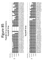
- FIG. 83-90 show the dissociation constants as well as the Vmax values for NLpoly 5A2, 5P, 8S and 11S with 96 variants of NLpeps.
- the upper graphs show the values for SEQ ID Nos: 2162-2209.
- the lower graphs show the values for SEQ ID NOs: 2210-2257.
- FIG. 91 shows an image of a protein gel of total lysates and the soluble fraction of the same lysate for NLpoly variants.
- FIG. 92 shows an image of a protein gel of total lysates and the soluble fraction of the same lysate for NLpoly variants as well as a table containing the dissociation constants for the same variants.
- FIG. 93 shows the substrate specificity for NLpoly 5P and 11S with NLpep79 and demonstrates that NLpoly 11S has superior specificity for furimazine than 5P.
- FIG. 94 shows an image of a protein gel that follows the affinity purification of NLpoly 8S through binding NLpep78.
- FIG. 95 contains a table of the association and dissociation rate constants for the binding of NLpoly WT or 11S to NLpepWT, 78 or 79.
- FIG. 96 shows the Km values for various pairs of NLpoly/NLpep.
- FIG. 97 compares the dissociation constant for NLpoly 11S/NLpep79 at sub-saturating and saturating concentrations of furimazine.
- FIG. 98 compares the Km values for NLpoly 5A2 with NLpepWT, 78 and 79.
- FIG. 99 shows the luminescence of NLpolys from various steps in the evolution process in the absence of NLpep.
- FIG. 100 shows the improvement in luminescence from E. coli -derived NLpoly over the course of the evolution process with an overall ⁇ 10 5 improvement (from NLpolyWT:NLpepWT to NLpoly11S:NLpep80).
- FIG. 101 shows the improvement in luminescence from HeLa-expressed NLpoly over the course of the evolution process with an overall ⁇ 10 5 improvement (from NLpolyWT:NLpepWT to NLpoly11S:NLpep80).
- FIG. 102 shows the improvement in luminescence from HEK293 cell-expressed NLpoly over the course of the evolution process with an overall ⁇ 10 4 improvement (from NLpolyWT:NLpepWT to NLpoly11S:NLpep80).
- FIG. 103 shows dissociation constants and demonstrates a ⁇ 10 4 fold improvement in binding affinity from NLpolyWT:NLpepWT to NLpoly11S:NLpep86.
- FIG. 104 shows an image of a protein gel of total lysates and the soluble fraction of the same lysate for NLpoly variants from various steps of the evolution process.
- FIG. 105 shows luminescence of various NLpolys in the absence of NLpep and in the presence of NLpep78 and NLpep79.
- FIG. 106 shows luminescence of various NLpolys in the absence of NLpep and in the presence of NLpep78 and NLpep79.
- FIG. 107 shows luminescence of various NLpolys in the absence of NLpep and in the presence of NLpep78 and NLpep79.
- FIG. 108 shows a comparison of luminescence generated by cells expressing different combinations of FRB and FKBP fused to NLpoly5P and NLpep80/87 after 15 min treatment with rapamycin or vehicle.
- Fold induction refers to signal generated in the presence of rapamycin compared to signal generated with vehicle.
- FIG. 109 shows a comparison of luminescence generated by cells expressing different combinations of FRB and FKBP fused to NLpoly5P and NLpep80/87 after 60 min treatment with rapamycin or vehicle.
- FIG. 110 shows a comparison of luminescence generated by cells expressing different combinations of FRB and FKBP fused to NLpoly5P and NLpep80/87 after 120 min treatment with rapamycin or vehicle.
- FIG. 111 shows a comparison of luminescence generated by cells expressing different combinations of FRB and FKBP fused to NLpoly5P and NLpep80/87 after 120 min treatment with rapamycin or vehicle. All 8 possible combinations of FRB and FKBP fused to NLpoly/NLpep were tested and less total DNA was used.
- FIG. 112 shows a comparison of luminescence generated by FRB or FKBP fusions expressed in the absence of binding partner.
- FIG. 113 shows a comparison of luminescence generated by cells transfected with varying amounts of FRB-NLpoly5P and FKBP-NLpep80/87 DNA.
- FIG. 114 shows a comparison of luminescence generated by cells transfected with varying amounts of FRB-NLpoly5P or FKBP-NLpep80/87 DNA in the absence of binding partner.
- FIG. 115 shows a comparison of luminescence generated by cells transfected with varying amounts of FRB-NLpoly5P and FKBP-NLpep80/87 DNA. This example differs from FIG. 113 in that lower levels of DNA were used.
- FIG. 116 shows a comparison of luminescence generated by cells transfected with varying amounts of FRB-NLpoly5P or FKBP-NLpep80/87 DNA in the absence of binding partner. This differs from FIG. 114 in that lower levels of DNA were used.
- FIG. 117 shows a comparison of luminescence generated by cells transfected with varying amounts of FRB-NLpoly5P and FKBP-NLpep80 DNA after treatment with rapamycin for different lengths of time.
- FIG. 118 shows a comparison of luminescence generated by cells transfected with varying amounts of FRB-NLpoly5P and FKBP-NLpep87 DNA after treatment with rapamycin for different lengths of time.
- FIG. 119 shows a comparison of luminescence generated by cells expressing different combinations of FRB-NLpoly5P with FKBP-NLpep80/87/95/96/97. Assay was performed in both a two-day and three-day format.
- FIG. 120 shows a comparison of luminescence generated by cells expressing different combinations of FRB-NLpoly5A2 with FKBP-NLpep80/87/95/96/97. Assay was performed in both a two-day and three-day format.
- FIG. 121 shows a comparison of luminescence generated by cells expressing different combinations of FRB-NLpoly5A2 or FRB-NLpoly11S with FKBP-NLpep101/104/105/106/107/108/109/110.
- FIG. 122 shows a comparison of luminescence generated by cells transfected with different combinations of FRB-NLpoly5A2 or FRB-NLpoly11S with FKBP-NLpep87/96/98/99/100/101/102/103.
- FIG. 123 shows a comparison of luminescence generated by cells transfected with different levels of FRB-NLpoly11S and FKBP-NLpep87/101/102/107 DNA.
- FIG. 124 shows a comparison of luminescence generated by cells transfected with different levels of FRB-NLpoly5A2 and FKBP-NLpep87/101/102/107 DNA.
- FIG. 125 shows a rapamycin dose response curve showing luminescence of cells expressing FRB-NLpoly5P and FKBP-NLpep80/87 DNA.
- FIG. 126 shows a rapamycin dose response curve showing luminescence of cells expressing FRB-NLpoly5A2 or FRB-NLply11S and FKBP-NLpep87/101 DNA.
- FIG. 127 shows a comparison of luminescence generated by cells expressing FRB-11S and FKBP-101 and treated with substrate PBI-4377 or furimazine.
- FIG. 128 shows a rapamycin time course of cells expressing FRB-NLpoly11S/5A2 and FKBP-NLpep87/101 conducted in the presence or absence of rapamycin wherein the rapamycin was added manually.
- FIG. 129 shows a rapamycin time course of cells expressing FRB-NLpoly11S/5A2 and FKBP-NLpep87/101 conducted in the presence or absence of rapamycin wherein the rapamycin was added via instrument injector.
- FIG. 130 shows luminescence generated by FRB-NLpoly11S and FKBP-NLpep101 as measured on two different luminescence-reading instruments.
- FIG. 131 provides images showing luminescence of cells expressing FRB-NLpoly11S and FKBP-NLpep101 at various times after treatment with rapamycin.
- FIG. 132 provides a graph showing Image J quantitation of the signal generated by individual cells expressing FRB-NLpoly11S and FKBP-NLpep101 at various times after treatment with rapamycin.
- FIG. 133 shows a comparison of luminescence in different cell lines expressing FRB-NLpoly11S and FKBP-NLpep101.
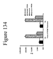
- FIG. 134 shows a comparison of luminescence generated by cells expressing FRB-NLpoly11S and FKBP-NLpep101 after treatment with the rapamycin competitive inhibitor FK506.
- FIG. 135 shows (left side) luminescence generated by cells expressing FRB-NLpoly11S and FKBP-NLpep101 after treatment with the rapamycin competitive inhibitor FK506, and (right side) the percent of luminescence remaining after treatment with FK506.
- FIG. 136 shows luminescence generated by cells transfected with different combinations of V2R-NLpoly5A2 or V2R-NLpoly11S with NLpep87/101-ARRB2 in the presence or absence of the V2R agonist AVP.
- FIG. 137 shows an AVP treatment time course showing luminescence generated by cells transfected with V2R-NLpoly11S and NLpep87/101-ARRB2 after treatment with AVP wherein AVP was added manually.
- FIG. 138 shows an AVP treatment time course showing luminescence generated by cells transfected with different combinations of V2R-NLpoly5A2 or V2R-NLpoly11S with NLpep87/101-ARRB2 after treatment with AVP wherein AVP was added via instrument injector.
- FIG. 139 shows an AVP treatment time course at 37° C. showing luminescence generated by cells expressing different configurations of V2R and ARRB2 fused to NLpoly11S and NLpep 101 after treatment with AVP.
- FIG. 140 shows a comparison of luminescence in different cell lines expressing V2R-NLpep11S and NLpep101-ARRB2.
- FIG. 142 shows 150 ⁇ images showing luminescence of cells expressing V2R-NLpoly11S and NLpep101-ARRB2 at various times after treatment with AVP.
- FIG. 143 shows a protein gel of total lysates and the soluble fraction of the same lysate for NLpoly variants.
- FIG. 144 shows the dissociation constants for NLpoly 5P and combinations of mutations at positions 31, 46, 75, 76, and 93 in NLpoly 5P.
- FIG. 145 shows a transferase example of post translational modification enzyme activity detection using an NLpep and aminopeptidase.
- FIG. 146 shows a hydrolase example of post translational modification enzyme activity detection using an NLpep and methyl-specific antibody.
- FIG. 147 contains wavelength scans for NLpoly WT complemented with either NLpepWT or NLpepWT conjugated to TMR.
- FIG. 148 contains wavelength scans for NanoLuc fused to HaloTag (NL-HT) and NLpoly 5A2 complemented with NLPepWT with 4 additional amino acids (DEVD) and conjugated to Non-chloroTOM (NCT) (SEQ ID NO: 2259).
- FIG. 149 shows a schematic a tertiary interaction wherein the energy transfer with an NLpoly and NLpep can also be used to measure three molecules interacting.
- a GPCR labeled with an NLpoly and a GPCR interacting protein labeled with a NLpep form a bioluminescent complex when they interact. This allows measurement of the binary interaction. If a small molecule GPCR ligand bearing an appropriate fluorescent moiety for energy transfer interacts with this system, energy transfer will occur. Therefore, the binary protein-protein interaction and the ternary drug-protein-protein interaction can be measured in the same experiment.
- the term “substantially” means that the recited characteristic, parameter, and/or value need not be achieved exactly, but that deviations or variations, including for example, tolerances, measurement error, measurement accuracy limitations and other factors known to skill in the art, may occur in amounts that do not preclude the effect the characteristic was intended to provide.
- a characteristic or feature that is substantially absent e.g., substantially non-luminescent
- bioluminescence refers to production and emission of light by a chemical reaction catalyzed by, or enabled by, an enzyme, protein, protein complex, or other biomolecule (e.g., bioluminescent complex).
- a substrate for a bioluminescent entity e.g., bioluminescent protein or bioluminescent complex
- the substrate subsequently emits light as it returns to its more stable form.
- complementary refers to the characteristic of two or more structural elements (e.g., peptide, polypeptide, nucleic acid, small molecule, etc.) of being able to hybridize, dimerize, or otherwise form a stable complex with each other.
- a “complementary peptide and polypeptide” are capable of coming together to form a stable complex.
- Complementary elements may require assistance to form a stable complex (e.g., from interaction elements), for example, to place the elements in the proper conformation for complementarity, to co-localize complementary elements, to lower interaction energy for complementary, etc.
- the term “complex” refers to an assemblage or aggregate of molecules (e.g., peptides, polypeptides, etc.) in direct and/or indirect contact with one another.
- “contact,” or more particularly, “direct contact” means two or more molecules are close enough so that attractive noncovalent interactions, such as Van der Waal forces, hydrogen bonding, ionic and hydrophobic interactions, and the like, dominate the interaction of the molecules.
- a complex of molecules e.g., a peptide and polypeptide
- non-luminescent refers to an entity (e.g., peptide, polypeptide, complex, protein, etc.) that exhibits the characteristic of not emitting energy as light in the visible spectrum (e.g., in the presence or absence of a substrate).
- An entity may be referred to as non-luminescent if it does not exhibit detectable luminescence in a given assay.
- non-luminescent is synonymous with the term “substantially non-luminescent.”
- An entity is “non-luminescent” if any light emission is sufficiently minimal so as not to create interfering background for a particular assay.
- non-luminescent peptide (NLpep) and “non-luminescent polypeptide” (NLpoly) refer to peptides and polypeptides that exhibit substantially no luminescence (e.g., in the presence or absence of a substrate), or an amount that is virtually undetectable (e.g., beneath the noise) under standard conditions (e.g., physiological conditions, assay conditions, etc.) and with typical instrumentation (e.g., luminometer, etc.).
- standard conditions e.g., physiological conditions, assay conditions, etc.
- typical instrumentation e.g., luminometer, etc.
- such non-luminescent peptides and polypeptides assemble, according to the criteria described herein, to form a bioluminescent complex.
- non-luminescent element is a non-luminescent peptide or non-luminescent polypeptide.
- bioluminescent complex refers to the assembled complex of two or more non-luminescent peptides and/or non-luminescent polypeptides. The bioluminescent complex catalyzes or enables the conversion of a substrate for the bioluminescent complex into an unstable form; the substrate subsequently emits light as it returns to its more stable form.
- non-luminescent pair two non-luminescent elements that form a bioluminescent complex may be referred to as a “non-luminescent pair.” If a bioluminescent complex is formed by three or more non-luminescent peptides and/or non-luminescent polypeptides, the uncomplexed constituents of the bioluminescent complex may be referred to as a “non-luminescent group.”
- interaction element refers to a moiety that assists in bringing together a pair of non-luminescent elements or a non-luminescent group to form a bioluminescent complex.
- a pair of interaction elements a.k.a. “interaction pair” is attached to a pair of non-luminescent elements (e.g., non-luminescent peptide/polypeptide pair), and the attractive interaction between the two interaction elements facilitates formation of the bioluminescent complex; although the present invention is not limited to such a mechanism, and an understanding of the mechanism is not required to practice the invention.
- Interaction elements may facilitate formation of the bioluminescent complex by any suitable mechanism (e.g., bringing non-luminescent pair/group into close proximity, placing a non-luminescent pair/group in proper conformation for stable interaction, reducing activation energy for complex formation, combinations thereof, etc.).
- An interaction element may be a protein, polypeptide, peptide, small molecule, cofactor, nucleic acid, lipid, carbohydrate, antibody, etc.
- An interaction pair may be made of two of the same interaction elements (i.e. homopair) or two different interaction elements (i.e. heteropair).
- the interaction elements may be the same type of moiety (e.g., polypeptides) or may be two different types of moieties (e.g., polypeptide and small molecule).
- an interaction pair in which complex formation by the interaction pair is studied, an interaction pair may be referred to as a “target pair” or a “pair of interest,” and the individual interaction elements are referred to as “target elements” (e.g., “target peptide,” “target polypeptide,” etc.) or “elements of interest” (e.g., “peptide of interest,” “polypeptide or interest,” etc.).
- preexisting protein refers to an amino acid sequence that was in physical existence prior to a certain event or date.
- a “peptide that is not a fragment of a preexisting protein” is a short amino acid chain that is not a fragment or sub-sequence of a protein (e.g., synthetic or naturally-occurring) that was in physical existence prior to the design and/or synthesis of the peptide.
- fragment refers to a peptide or polypeptide that results from dissection or “fragmentation” of a larger whole entity (e.g., protein, polypeptide, enzyme, etc.), or a peptide or polypeptide prepared to have the same sequence as such. Therefore, a fragment is a subsequence of the whole entity (e.g., protein, polypeptide, enzyme, etc.) from which it is made and/or designed.
- a peptide or polypeptide that is not a subsequence of a preexisting whole protein is not a fragment (e.g., not a fragment of a preexisting protein).
- a peptide or polypeptide that is “not a fragment of a preexisting bioluminescent protein” is an amino acid chain that is not a subsequence of a protein (e.g., natural of synthetic) that: (1) was in physical existence prior to design and/or synthesis of the peptide or polypeptide, and (2) exhibits substantial bioluminescent activity.
- sequence refers to peptide or polypeptide that has 100% sequence identify with another, larger peptide or polypeptide.
- the subsequence is a perfect sequence match for a portion of the larger amino acid chain.
- sequence identity refers to the degree two polymer sequences (e.g., peptide, polypeptide, nucleic acid, etc.) have the same sequential composition of monomer subunits.
- sequence similarity refers to the degree with which two polymer sequences (e.g., peptide, polypeptide, nucleic acid, etc.) have similar polymer sequences.
- similar amino acids are those that share the same biophysical characteristics and can be grouped into the families, e.g., acidic (e.g., aspartate, glutamate), basic (e.g., lysine, arginine, histidine), non-polar (e.g., alanine, valine, leucine, isoleucine, proline, phenylalanine, methionine, tryptophan) and uncharged polar (e.g., glycine, asparagine, glutamine, cysteine, serine, threonine, tyrosine).
- acidic e.g., aspartate, glutamate
- basic e.g., lysine, arginine, histidine
- non-polar e.g., alanine, valine, leucine, isoleucine, proline, phenylalanine, methionine, tryptophan
- uncharged polar e.g.
- the “percent sequence identity” is calculated by: (1) comparing two optimally aligned sequences over a window of comparison (e.g., the length of the longer sequence, the length of the shorter sequence, a specified window), (2) determining the number of positions containing identical (or similar) monomers (e.g., same amino acids occurs in both sequences, similar amino acid occurs in both sequences) to yield the number of matched positions, (3) dividing the number of matched positions by the total number of positions in the comparison window (e.g., the length of the longer sequence, the length of the shorter sequence, a specified window), and (4) multiplying the result by 100 to yield the percent sequence identity or percent sequence similarity.
- a window of comparison e.g., the length of the longer sequence, the length of the shorter sequence, a specified window
- peptides A and B are both 20 amino acids in length and have identical amino acids at all but 1 position, then peptide A and peptide B have 95% sequence identity. If the amino acids at the non-identical position shared the same biophysical characteristics (e.g., both were acidic), then peptide A and peptide B would have 100% sequence similarity.
- peptide C is 20 amino acids in length and peptide D is 15 amino acids in length, and 14 out of 15 amino acids in peptide D are identical to those of a portion of peptide C, then peptides C and D have 70% sequence identity, but peptide D has 93.3% sequence identity to an optimal comparison window of peptide C.
- percent sequence identity or “percent sequence similarity” herein, any gaps in aligned sequences are treated as mismatches at that position.
- physiological conditions encompasses any conditions compatible with living cells, e.g., predominantly aqueous conditions of a temperature, pH, salinity, chemical makeup, etc. that are compatible with living cells.
- sample is used in its broadest sense. In one sense, it is meant to include a specimen or culture obtained from any source, as well as biological and environmental samples.
- Biological samples may be obtained from animals (including humans) and encompass fluids, solids, tissues, and gases.
- Biological samples include blood products, such as plasma, serum and the like.
- Sample may also refer to cell lysates or purified forms of the peptides and/or polypeptides described herein.
- Cell lysates may include cells that have been lysed with a lysing agent or lysates such as rabbit reticulocyte or wheat germ lysates.
- Sample may also include cell-free expression systems.
- Environmental samples include environmental material such as surface matter, soil, water, crystals and industrial samples. Such examples are not however to be construed as limiting the sample types applicable to the present invention.
- peptide and polypeptide refer to polymer compounds of two or more amino acids joined through the main chain by peptide amide bonds (—C(O)NH—).
- peptide typically refers to short amino acid polymers (e.g., chains having fewer than 25 amino acids), whereas the term “polypeptide” typically refers to longer amino acid polymers (e.g., chains having more than 25 amino acids).
- protein interactions with small molecules, nucleic acids, other proteins, etc. are detected based on the association of two non-luminescent elements that come together to from a bioluminescent complex capable of producing a detectable signal (e.g., luminescence).
- a bioluminescent complex capable of producing a detectable signal (e.g., luminescence).
- the formation of the bioluminescent complex is dependent upon the protein interaction that is being monitored.
- compositions and methods for the assembly of a bioluminescent complex from two or more non-luminescent peptide and/or polypeptide units e.g., non-luminescent pair.
- the non-luminescent peptide and/or polypeptide units are not fragments of a preexisting protein (e.g., are not complementary subsequences of a known polypeptide sequence).
- bioluminescent activity is conferred upon a non-luminescent polypeptide via structural complementation with a non-luminescent peptide.
- non-luminescent pairs for use in detecting and monitoring molecular interactions (e.g., protein-protein, protein-DNA, protein-RNA interactions, RNA-DNA, protein-small molecule, RNA-small-molecule, etc.).
- complementary panels of interchangeable non-luminescent elements e.g., peptides and polypeptides
- variable affinities and luminescence upon formation of the various bioluminescent complexes e.g., a high-affinity/high-luminescence pair, a moderate-affinity/high-luminescence pair, a low-affinity/moderate-luminescence pair, etc.
- non-luminescent elements Utilizing different combinations of non-luminescent elements provides an adaptable system comprising various pairs ranging from lower to higher affinities, luminescence and other variable characteristics. This adaptability allows the detection/monitoring of molecular interactions to be fine-tuned to the specific molecule(s) of interest and expands the range of molecular interactions that can be monitored to include interactions with very high or low affinities. Further provided herein are methods by which non-luminescent pairs (or groups) and panels of non-luminescent pairs (or groups) are developed and tested.
- the interaction between the peptide/polypeptide members of the non-luminescent pair alone is insufficient to form the bioluminescent complex and produce the resulting bioluminescent signal.
- an interaction element is attached to each peptide/polypeptide member of the non-luminescent pair, then the interactions of the interaction pair (e.g., to form an interaction complex) facilitate formation of the bioluminescent complex.
- the bioluminescent signal from the bioluminescent complex serves as a reporter for the formation of the interaction complex.
- an interaction complex is formed, then a bioluminescent complex is formed, and a bioluminescent signal is detected/measured/monitored (e.g., in the presence of substrate). If an interaction complex fails to form (e.g., due to unfavorable conditions, due to unstable interaction between the interaction elements, due to incompatible interaction elements), then a stable bioluminescent complex does not form, and a bioluminescent signal is not produced.
- the interaction pair comprises two molecules of interest (e.g., proteins of interest).
- assays can be performed to detect the interaction of two molecules of interest by tethering each one to a separate member of a non-luminescent pair. If the molecules of interest interact (e.g., transiently interact, stably interact, etc.), the non-luminescent pair is brought into close proximity in a suitable conformation and a bioluminescent complex is formed (and bioluminescent signal is produced/detected (in the presence of substrate)).
- the non-luminescent pair does not interact in a stable enough manner, and a bioluminescent signal is not produced or only weakly produced.
- Such embodiments can be used to study the effect of inhibitors on complex formation, the effect of mutations on complex formation, the effect of conditions (e.g., temperature, pH, etc.) on complex formation, the interaction of a small molecule (e.g., potential therapeutic) with a target molecule, etc.
- Non-luminescent pairs may require different strength, duration and/or stability of the interaction complex to result in bioluminescent complex formation.
- a stable interaction complex is required to produce a detectable bioluminescent signal.
- even a weak or transient interaction complex results in bioluminescent complex formation.
- the strength of an interaction complex is directly proportional to the strength of the resulting bioluminescent signal.
- non-luminescent pairs require an interaction pair with a low millimolar (e.g., K d ⁇ 100 mM), micromolar (e.g., K d ⁇ 1 mM), nanomolar (e.g., K d ⁇ 1 ⁇ M), or even picomolar (e.g., K d ⁇ 1 nM) dissociation constant in order to produce a bioluminescent complex with a detectable signal.
- a low millimolar e.g., K d ⁇ 100 mM
- micromolar e.g., K d ⁇ 1 mM
- nanomolar e.g., K d ⁇ 1 ⁇ M
- picomolar e.g., K d ⁇ 1 nM
- one or more of the non-luminescent peptides/polypeptides are not fragments of a pre-existing protein. In some embodiments, one or more of the non-luminescent peptides/polypeptides are not fragments of a pre-existing bioluminescent protein. In some embodiments, neither/none of the non-luminescent peptides/polypeptides are fragments of a pre-existing protein. In some embodiments, neither/none of the non-luminescent peptides/polypeptides are fragments of a pre-existing bioluminescent protein.
- neither the non-luminescent peptide nor non-luminescent polypeptide that assemble together to form a bioluminescent complex are fragments of a pre-existing protein.
- a non-luminescent element for use in embodiments of the present invention is not a subsequence of a preexisting protein.
- a non-luminescent pair for use in embodiments described herein does not comprise complementary subsequences of a preexisting protein.
- non-luminescent peptides/polypeptides are substantially non-luminescent in isolation.
- suitable conditions e.g., physiological conditions
- non-luminescent peptides/polypeptides when placed in suitable conditions (e.g., physiological conditions), interact to form a bioluminescent complex and produce a bioluminescent signal in the presence of substrate.
- non-luminescent peptides/polypeptides without the addition of one or more interaction elements (e.g., complementary interaction elements attached to the component non-luminescent peptide and non-luminescent polypeptide), non-luminescent peptides/polypeptides are unable to form a bioluminescent complex or only weakly form a complex.
- non-luminescent peptides/polypeptides are substantially non-luminescent in each other's presence alone, but produce significant detectable luminescence when aggregated, associated, oriented, or otherwise brought together by interaction elements.
- interaction elements e.g., complementary interaction elements attached to the component peptide and polypeptide
- peptides and/or polypeptides that assemble into the bioluminescent complex produce a low level of luminescence in each other's presence, but undergo a significant increase in detectable luminescence when aggregated, associated, oriented, or otherwise brought together by interaction elements.
- compositions and methods described herein comprise one or more interaction elements.
- an interaction element is a moiety (e.g., peptide, polypeptide, protein, small molecule, nucleic acid, lipid, carbohydrate, etc.) that is attached to a peptide and/or polypeptide to assemble into the bioluminescent complex.
- the interaction element facilitates the formation of a bioluminescent complex by any suitable mechanism, including: interacting with one or both non-luminescent elements, inducing a conformational change in a non-luminescent element, interacting with another interaction element (e.g., an interaction element attached to the other non-luminescent element), bringing non-luminescent elements into close proximity, orienting non-luminescent elements for proper interaction, etc.
- a bioluminescent complex by any suitable mechanism, including: interacting with one or both non-luminescent elements, inducing a conformational change in a non-luminescent element, interacting with another interaction element (e.g., an interaction element attached to the other non-luminescent element), bringing non-luminescent elements into close proximity, orienting non-luminescent elements for proper interaction, etc.
- one or more interaction elements are added to a solution containing the non-luminescent elements, but are not attached to the non-luminescent elements.
- the interaction element(s) interact with the non-luminescent elements to induce formation of the bioluminescent complex or create conditions suitable for formation of the bioluminescent complex.
- a single interaction element is attached to one member of a non-luminescent pair.
- the lone interaction element interacts with one or both of the non-luminescent elements to create favorable interactions for formation of the bioluminescent complex.
- one interaction element is attached to each member of a non-luminescent pair. Favorable interactions between the interaction elements facilitate interactions between the non-luminescent elements.
- the interaction pair may stably interact, transiently interact, form a complex, etc.
- the interaction of the interaction pair facilitates interaction of the non-luminescent elements (and formation of a bioluminescent complex) by any suitable mechanism, including, but not limited to: bringing the non-luminescent pair members into close proximity, properly orienting the non-luminescent pair members from interaction, reducing non-covalent forces acting against non-luminescent pair interaction, etc.
- an interaction pair comprises any two chemical moieties that facilitate interaction of an associated non-luminescent pair.
- An interaction pair may consist of, for example: two complementary nucleic acids, two polypeptides capable of dimerization (e.g., homodimer, heterodimer, etc.), a protein and ligand, protein and small molecule, an antibody and epitope, a reactive pair of small molecules, etc. Any suitable pair of interacting molecules may find use as an interaction pair.
- an interaction pair comprises two molecules of interest (e.g., proteins of interest) or target molecules.
- compositions and methods herein provide useful assays (e.g., in vitro, in vivo, in situ, whole animal, etc.) for studying the interactions between a pair of target molecules.
- a pair of interaction elements each attached to one of the non-luminescent elements, interact with each other and thereby facilitate formation of the bioluminescent complex.
- the present of a ligand, substrate, co-factor or addition interaction element e.g., not attached to non-luminescent element
- detecting a signal from the bioluminescent complex indicates the presence of the ligand, substrate, co-factor or addition interaction element or conditions that allow for interaction with the interaction elements.
- a pair of interaction elements, and a pair of non-luminescent elements are all present in a single amino acid chain (e.g., (interaction element 1)-NLpep-(interaction element 2)-NLpoly, NLpoly-(interaction element 1)-NLpep--(interaction element 2), NLpoly-(interaction element 1)-(interaction element 2)-NLpep, etc.).
- a pair of interaction elements, and a pair of non-luminescent elements are all present in a single amino acid chain
- a ligand, substrate, co-factor or addition interaction element is required for the interaction pair to form an interaction complex and facilitate formation of the bioluminescent complex.
- an interaction element and a non-luminescent element are attached, fused, linked, connected, etc.
- a first non-luminescent element and a first interaction element are attached to each other, and a second non-luminescent element and a second interaction element are attached to each other.
- Attachment of signal and interaction elements may be achieved by any suitable mechanism, chemistry, linker, etc.
- the interaction and non-luminescent elements are typically attached through covalent connection, but non-covalent linking of the two elements is also provided.
- the signal and interaction elements are directly connected and, in other embodiments, they are connected by a linker.
- the signal and interaction elements are contained within a single amino acid chain.
- a single amino acid chain comprises, consists of, or consists essentially of a non-luminescent element and interaction element.
- a single amino acid chain comprises, consists of, or consists essentially of a non-luminescent element, an interaction element, and optionally one or more an N-terminal sequence, a C-terminal sequence, regulatory elements (e.g., promoter, translational start site, etc.), and a linker sequence.
- the signal and interaction elements are contained within a fusion polypeptide. The signal and interaction elements (and any other amino acid segments to be included in the fusion) may be expressed separately; however, in other embodiments, a fusion protein is expressed that comprises or consist of both the interaction and signal sequences.
- a first fusion protein comprising a first non-luminescent element and first interaction element as well as a second fusion protein comprising a second non-luminescent element and second interaction element are expressed within the same cells.
- the first and second fusion proteins are purified and/or isolated from the cells, or the interaction of the fusion proteins is assayed within the cells.
- first and second fusion proteins are expressed in separate cells and combined (e.g., following purification and/or isolation) for signal detection.
- one or more fusion proteins are expressed in cell lysate (e.g., rabbit reticulocyte lysate) or in a cell-free system.
- nucleic acids DNA, RNA, vectors, etc. are provided that encode peptide, polypeptides, fusion polypeptide, fusion proteins, etc. of the present invention.
- Such nucleic acids and vectors may be used for expression, transformation, transfection, injection, etc.
- a non-luminescent element and interaction element are connected by a linker.
- a linker connects the signal and interaction elements while providing a desired amount of space/distance between the elements.
- a linker allows both the signal and interaction elements to form their respective pairs (e.g., non-luminescent pair and interaction pair) simultaneously.
- a linker assists the interaction element in facilitating the formation of a non-luminescent pair interaction.
- the linkers that connect each non-luminescent element to their respective interaction elements position the non-luminescent elements at the proper distance and conformation to form a bioluminescent complex.
- an interaction element and non-luminescent element are held in close proximity (e.g., ⁇ 4 monomer units) by a linker.
- a linker provides a desired amount of distance (e.g., 1, 2, 3, 4, 5, 6 . . . 10 . . . 20, or more monomer units) between signal and interaction elements (e.g., to prevent undesirable interactions between signal and interaction elements, for steric considerations, to allow proper orientation of non-luminescent element upon formation of interaction complex, to allow propagation of a complex-formation from interaction complex to non-luminescent elements, etc.).
- a linker provides appropriate attachment chemistry between the signal and interaction elements.
- a linker may also improve the synthetic process of making the signal and interaction element (e.g., allowing them to be synthesized as a single unit, allowing post synthesis connection of the two elements, etc.).
- a linker is any suitable chemical moiety capable of linking, connecting, or tethering a non-luminescent element to an interaction element.
- a linker is a polymer of one or more repeating or non-repeating monomer units (e.g., nucleic acid, amino acid, carbon-containing polymer, carbon chain, etc.).
- a linker when present, is typically an amino acid chain.
- a linker may comprise any chemical moiety with functional (or reactive) groups at either end that are reactive with functional groups on the signal and interaction elements, respectively. Any suitable moiety capable of tethering the signal and interaction elements may find use as a linker.
- linker is a single covalent bond.
- the linker comprises a linear or branched, cyclic or heterocyclic, saturated or unsaturated, structure having 1-20 nonhydrogen atoms (e.g., C, N, P, O and S) and is composed of any combination of alkyl, ether, thioether, imine, carboxylic, amine, ester, carboxamide, sulfonamide, hydrazide bonds and aromatic or heteroaromatic bonds.
- linkers are longer than 20 nonhydrogen atoms (e.g.
- the linker comprises 1-50 non-hydrogen atoms (in addition to hydrogen atoms) selected from the group of C, N, P, O and S (e.g. 1, 2, 3, 4, 5, 6, 7, 8, 9, 10, 11, 12, 13, 14, 15, 16, 17, 18, 19, 20, 21, 22, 23, 24, 25, 26, 27, 28, 29, 30, 31, 32, 33, 34, 35, 36, 37, 38, 39, 40, 41, 42, 43, 44, 45, 46, 47, 48, 49, or 50 non-hydrogen atoms).
- 1-50 non-hydrogen atoms selected from the group of C, N, P, O and S (e.g. 1, 2, 3, 4, 5, 6, 7, 8, 9, 10, 11, 12, 13, 14, 15, 16, 17, 18, 19, 20, 21, 22, 23, 24, 25, 26, 27, 28, 29, 30, 31, 32, 33, 34, 35, 36, 37, 38, 39, 40, 41, 42, 43, 44, 45, 46, 47, 48, 49, or 50 non-hydrogen atoms).
- the present invention is not limited by the types of linkers available.
- the signal and interaction elements are linked, either directly (e.g. linker consists of a single covalent bond) or linked via a suitable linker.
- the present invention is not limited to any particular linker group.
- linker groups are contemplated, and suitable linkers could comprise, but are not limited to, alkyl groups, methylene carbon chains, ether, polyether, alkyl amide linker, a peptide linker, a modified peptide linker, a Poly(ethylene glycol) (PEG) linker, a streptavidin-biotin or avidin-biotin linker, polyaminoacids (e.g.
- polylysine functionalised PEG
- polysaccharides polysaccharides
- glycosaminoglycans dendritic polymers
- PEG-chelant polymers W94/08629, WO94/09056 and WO96/26754, herein incorporated by reference in their entireties
- oligonucleotide linker phospholipid derivatives, alkenyl chains, alkynyl chains, disulfide, or a combination thereof.
- the linker is cleavable (e.g., enzymatically (e.g., TEV protease site), chemically, photoinduced, etc.
- substantially non-luminescent peptides and polypeptides are provided with less than 100% sequence identity and/or similarity to any portion of an existing luciferase (e.g., a firefly luciferase, a Renilla luciferase, an Oplophorus luciferase, enhanced Oplophorus luciferases as described in U.S. Pat. App. 2010/0281552 and U.S. Pat. App. 2012/0174242, herein incorporated by reference in their entireties).
- an existing luciferase e.g., a firefly luciferase, a Renilla luciferase, an Oplophorus luciferase, enhanced Oplophorus luciferases as described in U.S. Pat. App. 2010/0281552 and U.S. Pat. App. 2012/0174242, herein incorporated by reference in their entireties.
- Certain embodiments of the present invention involve the formation of bioluminescent complexes of non-luminescent peptides and polypeptides with less than 100%, but more than 40% (e.g., >40%, >45%, >50%, >55%, >60%, >65%, >70%, >75%, >80%, >85%, >90%, >95%, >98%, >99%) sequence identity with all or a portion (e.g., 8 or more amino acids, less than about 25 amino acids for peptides) of SEQ ID NO: 2157 (e.g., complete NANOLUC sequence).
- 40% e.g., >40%, >45%, >50%, >55%, >60%, >65%, >70%, >75%, >80%, >85%, >90%, >95%, >98%, >99%
- non-luminescent peptides and polypeptides are provided with less than 100%, but more than 40% (e.g., >40%, >45%, >50%, >55%, >60%, >65%, >70%, >75%, >80%, >85%, >90%, >95%, >98%, >99%) sequence similarity with a portion (e.g., 8 or more amino acids, less than about 25 amino acids for peptides) of SEQ ID NO: 2157 (e.g., peptides and polypeptides that interact to form bioluminescent complexes).
- 40% e.g., >40%, >45%, >50%, >55%, >60%, >65%, >70%, >75%, >80%, >85%, >90%, >95%, >98%, >99%
- Non-luminescent peptides are provided that have less than 100%, but more than 40% (e.g., >40%, >45%, >50%, >55%, >60%, >65%, >70%, >75%, >80%, >85%, >90%, >95%, >98%, >99%) sequence identity and/or similarity with about a 25 amino acid or less portion of SEQ ID NO: 2157, wherein such peptides form a bioluminescent complex when combined under appropriate conditions (e.g., stabilized by an interaction pair) with a polypeptide having less than 100%, but more than 40% (e.g., >40%, >45%, >50%, >55%, >60%, >65%, >70%, >75%, >80%, >85%, >90%, >95%, >98%, >99%) sequence identity and/or similarity with another portion SEQ ID NO: 2157.
- 40% e.g., >40%, >45%, >50%, >55%, >60%
- non-luminescent polypeptides that have less than 100%, but more than 40% (e.g., >40%, >45%, >50%, >55%, >60%, >65%, >70%, >75%, >80%, >85%, >90%, >95%, >98%, >99%) sequence identity or similarity with a portion of SEQ ID NO: 2157, wherein such polypeptides form a bioluminescent complex when combined under appropriate conditions (e.g., stabilized by an interaction pair) with a peptide having less than 100%, but more than 40% (e.g., >40%, >45%, >50%, >55%, >60%, >65%, >70%, >75%, >80%, >85%, >90%, >95%, >98%, >99%) sequence identity and/or similarity with another portion SEQ ID NO: 2157.
- 40% e.g., >40%, >45%, >50%, >55%, >60%, >65%, >70%, >7
- non-luminescent peptides with less than 100%, but more than 40% (e.g., >40%, >45%, >50%, >55%, >60%, >65%, >70%, >75%, >80%, >85%, >90%, >95%, >98%, >99%) sequence identity or similarity with SEQ ID NO: 2 are provided.
- non-luminescent polypeptides with less than 100%, but more than 40% e.g., >40%, >45%, >50%, >55%, >60%, >65%, >70%, >75%, >80%, >85%, >90%, >95%, >98%, >99% sequence identity or similarity with SEQ ID NO: 440 are provided.
- non-luminescent peptides that find use in embodiments of the present invention include peptides with one or more amino acid substitutions, deletions, or additions from GVTGWRLCKRISA (SEQ ID NO: 236).
- the present invention provides peptides comprising one or more of amino acid sequences of Table 1, and/or nucleic acids comprising the nucleic acid sequences of Table 1 (which code for the peptide sequences of Table 1).
- MGVTGWRLNERILA 21 NLpep11 (w/ Met) N.A. ATGGGAGTGACCGGCTGGCGGCTGCAGGAACGCATTCTGGCG 22 NLpep11 (w/ Met) A.A. MGVTGWRLQERILA 23 NLpep12 (w/ Met) N.A. ATGGGAGTGACCGGCTGGCGGCTGAAGAAGCGCCGGAGCCGG 24 NLpep12 (w/ Met) A.A. MGVTGWRLKKRRSR 25 NLpep13 (w/ Met) N.A. ATGAACGTGACCGGCTGGCGGCTGTGCAAGCGCATTAGCGCG 26 NLpep13 (w/ Met) A.A.
- MGKTGWRLCKRISA 39 NLpep20 (w/ Met) N.A. ATGGGACAGACCGGCTGGCGGCTGTGCAAGCGCATTAGCGCG 40 NLpep20 (w/ Met) A.A. MGQTGWRLCKRISA 41 NLpep21 (w/ Met) N.A. ATGGGAAGCACCGGCTGGCGGCTGTGCAAGCGCATTAGCGCG 42 NLpep21 (w/ Met) A.A. MGSTGWRLCKRISA 43 NLpep22 (w/ Met) N.A. ATGGGAGTGGTGGGCTGGCGGCTGTGCAAGCGCATTAGCGCG 44 NLpep22 (w/ Met) A.A.
- MGVVGWRLCKRISA 45 NLpep23 (w/ Met) N.A. ATGGGAGTGAAGGGCTGGCGGCTGTGCAAGCGCATTAGCGCG 46 NLpep23 (w/ Met) A.A. MGVKGWRLCKRISA 47 NLpep24 (w/ Met) N.A. ATGGGAGTGCAGGGCTGGCGGCTGTGCAAGCGCATTAGCGCG 48 NLpep24 (w/ Met) A.A. MGVQGWRLCKRISA 49 NLpep25 (w/ Met) N.A. ATGGGAGTGACCGGCACCCGGCTGTGCAAGCGCATTAGCGCG 50 NLpep25 (w/ Met) A.A.
- MGVTGWRLCKRVSA 75 NLpep38 (w/ Met) N.A. ATGGGAGTGACCGGCTGGCGGCTGTGCAAGCGCCAGAGCGCGCG 76 NLpep38 (w/ Met) A.A. MGVTGWRLCKRQSA 77 NLpep39 (w/ Met) N.A. ATGGGAGTGACCGGCTGGCGGCTGTGCAAGCGCGAGAGCGCG 78 NLpep39 (w/ Met) A.A. MGVTGWRLCKRESA 79 NLpep40 (w/ Met) N.A. ATGGGAGTGACCGGCTGGCGGCTGTGCAAGCGCCGGAGCGCG 80 NLpep40 (w/ Met) A.A.
- MGVTGWRLCKRRSA 81 NLpep41 (w/ Met) N.A. ATGGGAGTGACCGGCTGGCGGCTGTGCAAGCGCTTCAGCGCG 82 NLpep41 (w/ Met) A.A. MGVTGWRLCKRFSA 83 NLpep42 (w/ Met) N.A. ATGGGAGTGACCGGCTGGCGGCTGTGCAAGCGCATTAGCAAC 84 NLpep42 (w/ Met) A.A. MGVTGWRLCKRISN 85 NLpep43 (w/ Met) N.A. ATGGGAGTGACCGGCTGGCGGCTGTGCAAGCGCATTAGCACC 86 NLpep43 (w/ Met) A.A.
- MGVTGWRLCKRISE 93 NLpep47 (w/ Met) N.A. ATGGGAGTGACCGGCTTCCGGCTGTGCAAGCGCATTAGCGCG 94 NLpep47 (w/ Met) A.A. MGVTGFRLCKRISA 95 NLpep48 (w/ Met) N.A. ATGGGAGTGACCGGCTACCGGCTGTGCAAGCGCATTAGCGCG 96 NLpep48 (w/ Met) A.A. MGVTGYRLCKRISA 97 NLpep49 (w/ Met) N.A. ATGGGAGTGACCGGCGAGCGGCTGTGCAAGCGCATTAGCGCG 98 NLpep49 (w/ Met) A.A.
- MGVTGWRLAKRISA 117 NLpep59 (w/ Met) N.A.
- ATGGGAGTGACCGGCTGGCGGCTGCAGAAGCGCATTAGCGCG 118 NLpep59 (w/ Met) A.A.
- MGVTGWRLQKRISA 119 NLpep60 (w/ Met) N.A.
- ATGGGAGTGACCGGCTGGCGGCTGCTGAAGCGCATTAGCGCG 120 NLpep60 (w/ Met) A.A.
- MGVTGWRLLKRISA 121 NLpep61 (w/ Met) N.A.
- ATGGGAGTGACCGGCTGGCGGCTGAAGAAGCGCATTAGCGCG 122 NLpep61 (w/ Met) A.A.
- MGVTGWRLKKRISA 123 NLpep62 (w/ Met) N.A. ATGAACCACACCGGCTGGCGGCTGAACAAGAAGGTGAGCAAC 124 NLpep62 (w/ Met) A.A. MNITGWRLNKKVSN 125 NLpep63 (w/ Met) N.A. ATGAACCACACCGGCTACCGGCTGAACAAGAAGGTGAGCAAC 126 NLpep63 (w/ Met) A.A. MNITGYRLNKKVSN 127 NLpep64 (w/ Met) N.A. ATGTGCGTGACCGGCTGGCGGCTGTTCAAGCGCATTAGCGCG 128 NLpep64 (w/ Met) A.A.
- MNVTGWRLFKRILN 147 NLpep74 (w/ Met) N.A. ATGAACGTGACCGGCTGGCGGCTGTTCAAGCGCATTAGCGCG 148 NLpep74 (w/ Met) A.A. MNVTGWRLFKRISA 149 NLpep75 (w/ Met) N.A. ATGAACGTGACCGGCTGGCGGCTGTTCGAAAAGATTAGCAAC 150 NLpep75 (w/ Met) A.A. MNVTGWRLFEKISN 151 NLpep76 (w/ Met) N.A. ATGAACGTGAGCGGCTGGCGGCTGTTCGAAAAGATTAGCAAC 152 NLpep76 (w/ Met) A.A.
- MGWRLFKKISN 165 NLpep83 (w/ Met) N.A. ATGAACGTGAGCGGCTGGCGGCTGTTCAAGAAGATTAGC 166 NLpep83 (w/ Met) A.A. MNVSGWRLFKKIS 167 NLpep84 (w/ Met) N.A. ATGAACGTGAGCGGCTGGCGGCTGTTCAAGAAGATT 168 NLpep84 (w/ Met) A.A. MNVSGWRLFKKI 169 NLpep85 (w/ Met) N.A. ATGAACGTGAGCGGCTGGCGGCTGTTCAAGAAG 170 NLpep85 (w/ Met) A.A.
- MNVSGWGLFKKISN 177 NLpep89 (w/ Met) N.A. ATGCCCGTGAGCGGCTGGCGGCTGTTCAAGAAGATTAGCAAC 178 NLpep89 (w/ Met) A.A. MPVSGWRLFKKISN 179 NLpep90 (w/ Met) N.A. ATGAACCCCGTGAGCGGCTGGCGGCTGTTCAAGAAGATTAGCAAC 180 NLpep90 (w/ Met) A.A. MNPVSGWRLFKKISN 181 NLpep91 (w/ Met) N.A. ATGATCAACCCCGTGAGCGGCTGGCGGCTGTTCAAGAAGATTAGCAAC 182 NLpep91 (w/ Met) A.A.
- MINPVSGWRLFKKISN 183 NLpep92 (w/ Met) N.A. ATGACCATCAACCCCGTGAGCGGCTGGCGGCTGTTCAAGAAGATTAGCAAC 184 NLpep92 (w/ Met) A.A. MTINPVSGWRLFKKISN 185 NLpep93 (w/ Met) N.A. ATGGTGACCATCAACCCCGTGAGCGGCTGGCGGCTGTTCAAGAAGATTAGCAAC 186 NLpep93 (w/ Met) A.A. MVTINPVSGWRLFKKISN 187 NLpep94 (w/ Met) N.A.
- ATGGTGACCGGCTACCGGCTGTTCGAGCAGATTAGC 200 NLpep100 (w/ Met) A.A.
- MVTGYRLFEQIS 201 NLpep101 (w/ Met) N.A.
- ATGGTGACCGGCTACCGGCTGTTCGAGAAGGAGAGC 202 NLpep101 (w/ Met) A.A.
- MVTGYRLFEKES 203 NLpep102 (w/ Met) N.A.
- ATGGTGACCGGCTACCGGCTGTTCGAGCAGGAGAGC NLpep102 (w/ Met) A.A.
- MVTGYRLFEQES 205 NLpep103 (w/ Met) N.A.
- ATGGTGACCGGCTACCGGCTGTTCGAGCAGGAGCTG 206 NLpep103 (w/ Met) A.A.
- MVTGYRLFEQEL 207 NLpep104 (w/ Met) N.A.
- ATGGTGGAGGGCTACCGGCTGTTCGAGAAGATTAGC 208 NLpep104 (w/ Met) A.A.
- MVEGYRLFEKIS 209 NLpep105 (w/ Met) N.A.
- ATGGTGGAGGGCTACCGGCTGTTCGAGCAGATTAGC NLpep105 (w/ Met) A.A.
- ATGGTGGAGGGCTACCGGCTGTTCGAGAAGGAGAGC 212 NLpep106 (w/ Met) A.A.
- MVEGYRLFEKES 213 NLpep107 (w/ Met) N.A.
- ATGGTGGAGGGCTACCGGCTGTTCGAGCAGGAGC 214 NLpep107 (w/ Met) A.A.
- MVEGYRLFEQES 215 NLpep108 (w/ Met) N.A.
- MVEGYRLFEQEL 217 NLpep109 (w/ Met) N.A.
- GGAGTGACCGCCTGGCGGCTGTGCGAACGCATTCTGGCG 224 NLpep3 (w/o Met) A.A.
- GGAGTGACCGGCTGGCGGCTGTGCAAGCGCATTAGCGCG 236 NLpep9 (w/o Met) A.A.
- GVTGWRLCKRISA 237 NLpep10 (w/o Met) N.A. GGAGTGACCGGCTGGCGGCTGAACGAACGCATTCTGGCG 238 NLpep10 (w/o Met) A.A.
- GVTGWRLNERILA 239 NLpep11 (w/o Met) N.A. GGAGTGACCGGCTGGCGGCTGCAGGAACGCATTCTGGCG 240 NLpep11 (w/o Met) A.A.
- NLpep12 (w/o Met) A.A.
- GVTGWRLKKRRSR 243 NLpep13 (w/o Met) N.A. AACGTGACCGGCTGGCGGCTGTGCAAGCGCATTAGCGCG 244 NLpep13 (w/o Met) A.A.
- NVTGWRLCKRISA 245 NLpep14 (w/o Met) N.A. AGCGTGACCGGCTGGCGGCTGTGCAAGCGCATTAGCGCG 246 NLpep14 (w/o Met) A.A.
- SVTGWRLCKRISA 247 NLpep15 (w/o Met) N.A.
- GGAGTGACCGGCTGGCGGACCTGCAAGCGCATTAGCGCG 278 NLpep30 (w/o Met) A.A.
- GVTGWRTCKRISA 279 NLpep31 (w/o Met) N.A. GGAGTGACCGGCTGGCGGTACTGCAAGCGCATTAGCGCG 280 NLpep31 (w/o Met) A.A.
- GVTGWRKCKRISA 283 NLpep33 (w/o Met) N.A.
- GGAGTGACCGGCTGGCGGCTGAACAAGCGCATTAGCGCG 284 NLpep33 (w/o Met) A.A.
- GVTGWRLNKRISA 285 NLpep34 (w/o Met) N.A. GGAGTGACCGGCTGGCGGCTGACCAAGCGCATTAGCGCG 286 NLpep34 (w/o Met) A.A.
- GGAGTGACCGGCTGGCGGCTGTGCAAGAACATTAGCGCG 290 NLpep36 (w/o Met) A.A.
- GGAGTGACCGGCTGGCGGCTGATGAAGCGCATTAGCGCG 332 NLpep57 (w/o Met) A.A. GVTGWRLMKRISA 333 NLpep58 (w/o Met) N.A. GGAGTGACCGGCTGGCGGCTGGCCAAGCGCATTAGCGCG 334 NLpep58 (w/o Met) A.A. GVTGWRLAKRISA 335 NLpep59 (w/o Met) N.A. GGAGTGACCGGCTGGCGGCTGCAGAAGCGCATTAGCGCG 336 NLpep59 (w/o Met) A.A. GVTGWRLQKRISA 337 NLpep60 (w/o Met) N.A.
- GGAGTGACCGGCTGGCGGCTGCTGAAGCGCATTAGCGCG 338 NLpep60 (w/o Met) A.A.
- GVTGWRLLKRISA 339 NLpep61 (w/o Met) N.A.
- GGAGTGACCGGCTGGCGGCTGAAGAAGCGCATTAGCGCG 340 NLpep61 (w/o Met) A.A.
- AACCACACCGGCTGGCGGCTGAACAAGAAGGTGAGCAAC 342 NLpep62 (w/o Met) A.A.
- NITGWRLNKKVSN 343 NLpep63 (w/o Met) N.A.
- NLpep84 (w/o Met) A.A.
- NVSGWRLFKKI 387 NLpep85 (w/o Met) N.A.
- AACGTGAGCGGCTGGCGGCTGTTCAAGAAG 388 NLpep85 (w/o Met) A.A.
- TINPVSGWRLFKKISN 403 NLpep93 (w/o Met) N.A. GTGACCATCAACCCCGTGAGCGGCTGGCGGCTGTTCAAGAAGATTAGCAAC 404 NLpep93 (w/o Met) A.A. VTINPVSGWRLFKKISN 405 NLpep94 (w/o Met) N.A. CGGGTGACCATCAACCCCGTGAGCGGCTGGCGGCTGTTCAAGAAGATTAGCAAC 406 NLpep94 (w/o Met) A.A. RVTINPVSGWRLFKKISN 407 NLpep95 (w/o Met) N.A.
- VEGYRLFEQES 433 NLpep108 (w/o Met) N.A. GTGGAGGGCTACCGGCTGTTCGAGCAGGAGCTG 434 NLpep108 (w/o Met) A.A.
- VEGYRLFEQEL 435 NLpep109 (w/o Met) N.A. ATTAGCGGCTGGCGGCTGATGAAGAACATTAGC 436 NLpep109 (w/o Met) A.A. ISGWRLMKNIS 437 NLpep110 (w/o Met) N.A. GTGGAGGGCTACCGGCTGTTCAAGAAGATTAGC 438 NLpep110 (w/o Met) A.A.
- VEGYRLFKKIS NLpep108 (w/o Met) N.A.
- GTGGAGGGCTACCGGCTGTTCGAGCAGGAGCTG 434 NLpep108 (w/o Met) A.A.
- VEGYRLFEQEL 435 NLpep109 (w/o Met)
- a peptide from Table 1 is provided.
- peptides comprise a single amino acid difference from GVTGWRLCKRISA (SEQ ID NO: 236) and/or any of the peptides listed in Table 1.
- peptides comprise two or more (e.g., 2, 3, 4, 5, 6, 7, 8, 9, 10, etc.) amino acid differences from GVTGWRLCKRISA (SEQ ID NO: 236) and/or any of the peptides listed in Table 1.
- peptides are provided comprising one of the amino acid sequences of SEQ ID NOS: 3-438.
- peptides are provided comprising one of the amino acid sequences of SEQ ID NOS: 3-438 with one or more additions, substitutions, and/or deletions.
- a peptide or a portion thereof comprises greater than 70% sequence identity (e.g., 71%, 75%, 80%, 85%, 90%, 95%, 99%) with one or more of the amino acid sequence of SEQ ID NOS: 3-438.
- nucleic acids are provided comprising one of the nucleic acid coding sequences of SEQ ID NOS: 3-438.
- nucleic acids are provided comprising one of the nucleic acid sequences of SEQ ID NOS: 3-438 with one or more additions, substitutions, and/or deletions.
- a nucleic acid or a portion thereof comprises greater than 70% sequence identity (e.g., 71%, 75%, 80%, 85%, 90%, 95%, 99%) with one or more of the nucleic acid sequence of SEQ ID NOS: 3-438.
- nucleic acids are provided that code for one of the amino acid sequences of SEQ ID NOS: 3-438.
- nucleic acids are provided that code for one of the amino acid sequences of SEQ ID NOS: 3-438 with one or more additions, substitutions, and/or deletions.
- a nucleic acid is provided that codes for an amino acid with greater than 70% sequence identity (e.g., 71%, 75%, 80%, 85%, 90%, 95%, 99%) with one or more of the amino acid sequences of SEQ ID NOS: 3-438.
- nucleic acid from Table 1 is provided.
- a nucleic acid encoding a peptide from Table 1 is provided.
- a nucleic acid of the present invention codes for a peptide that comprises a single amino acid difference from MGVTGWRLCERILA (SEQ ID NO: 2) and/or any of the peptides listed in Table 1.
- nucleic acids code for peptides comprising two or more (e.g., 2, 3, 4, 5, 6, 7, 8, 9, 10, etc.) amino acid differences from MGVTGWRLCERILA (SEQ ID NO: 2) and/or any of the peptides listed in Table 1.
- nucleic acids are provided comprising the sequence of one of the nucleic acids in Table 1. In some embodiments, nucleic acids are provided comprising one of the nucleic acids of Table 1 with one or more additions, substitutions, and/or deletions. In some embodiments, a nucleic acid or a portion thereof comprises greater than 70% sequence identity (e.g., 71%, 75%, 80%, 85%, 90%, 95%, 99%) with one or more of the nucleic acids of Table 1.
- non-luminescent polypeptides that find use in embodiments of the present invention include polypeptides with one or more amino acid substitutions, deletions, or additions from SEQ ID NO: 440.
- the present invention provides polypeptides comprising one or more of amino acid sequences of Table 2, and/or nucleic acids comprising the nucleic acid sequences of Table 2 (which code for the polypeptide sequences of Table 2).
- 5A2 (G15A/G35A/G51A/G67A/G71A) 540 A.A 5A2 (G15A/G35A/G51A/G67A/G71A) 541 N.A. 5A2 + A15G 542 A.A 5A2 + A15G 543 N.A. 5A2 + A35G 544 A.A 5A2 + A35G 545 N.A. 5A2 + A51G 546 A.A 5A2 + A51G 547 N.A. 5A2 + A67G 548 A.A 5A2 + A67G 549 N.A. 5A2 + A71G 550 A.A 5A2 + A71G 551 N.A.
- 6P 5P + I56N 936 A.A 6P (5P + I56N) 937 N.A. 5E (3E + D108N + N144T) 938 A.A 5E (3E + D108N + N144T) 939 N.A. 6E (5E + I56N) 940 A.A 6E (5E + I56N) 941 N.A.
- NLpoly1 (5A2 + R11N + A15S + L18Q + F31I + V58A + A67D + M106V + L149M + V157D) 942 A.
- NLpoly1 (5A2 + R11N + A15S + L18Q + F31I + V58A + A67D + M106V + L149M + V157D) 943 N.A.
- NLpoly2 (5A2 + A15S + L18Q + F31I + V58A + A67D + M106V + L149M + V157D) 944 A.
- a NLpoly2 (5A2 + A15S + L18Q + F31I + V58A + A67D + M106V + L149M + V157D) 945 N.
- NLpoly3 (5A2 + R11N + L18Q + F31I + V58A + A67D + M106V + L149M + V157D) 946 A.
- a NLpoly3 (5A2 + R11N + L18Q + F31I + V58A + A67D + M106V + L149M + V157D) 947 N.A.
- NLpoly4 (5A2 + R11N + A15S + F31I + V58A + A67D + M106V + L149M + V157D) 948 A.
- a NLpoly4 (5A2 + R11N + A15S + F31I + V58A + A67D + M106V + L149M + V157D) 949 N.A.
- NLpoly5 (5A2 + R11N + A15S + L18Q + V58A + A67D + M106V + L149M + V157D) 950 A.
- a NLpoly5 (5A2 + R11N + A15S + L18Q + V58A + A67D + M106V + L149M + V157D) 951 N.A.
- NLpoly6 (5A2 + R11N + A15S + L18Q + F31I + A67D + M106V + L149M + V157D) 952 A.
- a NLpoly6 (5A2 + R11N + A15S + L18Q + F31I + A67D + M106V + L149M + V157D) 953 N.
- NLpoly7 (5A2 + R11N + A15S + L18Q + F31I + V58A + M106V + L149M + V157D) 954 A.
- a NLpoly7 (5A2 + R11N + A15S + L18Q + F31I + V58A + M106V + L149M + V157D) 955 N.A.
- NLpoly8 (5A2 + R11N + A15S + L18Q + F31I + V58A + A67D + L149M + V157D) 956 A.
- a NLpoly8 (5A2 + R11N + A15S + L18Q + F31I + V58A + A67D + L149M + V157D) 957 N.
- NLpoly9 (5A2 + R11N + A15S + L18Q + F31I + V58A + A67D + M106V + V157D) 958 A.
- a NLpoly9 (5A2 + R11N + A15S + L18Q + F31I + V58A + A67D + M106V + V157D) 959 N.A.
- NLpoly10 (5A2 + R11N + A15S + L18Q + F31I + V58A + A67D + M106V + L149M) 960 A.
- a NLpoly10 (5A2 + R11N + A15S + L18Q + F31I + V58A + A67D + M106V + L149M) 961 N.A.
- NLpoly11 (5A2 + A15S + L18Q + M106V + L149M + V157D) 962 A.
- a NLpoly11 (5A2 + A15S + L18Q + M106V + L149M + V157D) 963 N.A.
- NLpoly12 (5A2 + A15S + L18Q + A67D + M106V + L149M + V157D) 964 A.
- a NLpoly12 (5A2 + A15S + L18Q + A67D + M106V + L149M + V157D) 965 N.A.
- NLpoly13 (5A2 + R11N + A15S + L18Q + M106V + L149M + V157D) 966 A.
- a NLpoly13 (5A2 + R11N + A15S + L18Q + M106V + L149M + V157D) 967 N.A. 5P + V 968 A.A 5P + V 969 N.A. 5P + A 970 A.A 5P + A 971 N.A.
- 5P D2 ( ⁇ 156-157) 1006 A.A 5P D2 ( ⁇ 156-157) 1007 N.A. 5P D3 ( ⁇ 155-157) 1008 A.A 5P D3 ( ⁇ 155-157) 1009 N.A. 5P D4 ( ⁇ 154-157) 1010 A.A 5P D4 ( ⁇ 154-157) 1011 N.A. 5P D5 ( ⁇ 153-157) 1012 A.A 5P D5 ( ⁇ 153-157) 1013 N.A. 5P D6 ( ⁇ 152-157) 1014 A.A 5P D6 ( ⁇ 152-157) 1015 N.A. 5P D7 ( ⁇ 151-157) 1016 A.A 5P D7 ( ⁇ 151-157) 1017 N.A.
- 8S (5P + L46R + H93P + P157S + F31L) 1284 A.
- 12S (8S + A51T + K75E + I76V + I107L) 1290 A.A 12S (8S + A51T + K75E + I76V + I107L) 1291 N.A. 11S (12-A51T) 1292 A.A 11S (12-A51T) 1293 N.A. 12S-K75E 1294 A.A 12S-K75E 1295 N.A. 12S-I76V 1296 A.A 12S-I76V 1297 N.A. 12S-I107L 1298 A.A 12S-I107L
- polypeptides and coding nucleic acid sequences of Table 2 all contain N-terminal Met residues (amino acids) or ATG start codons (nucleic acids). In some embodiments, the polypeptides and coding nucleic acid sequences of Table 2 are provided without N-terminal Met residues or ATG start codons (SEQ ID NOS: 1299-2156).
- polypeptide of one of the amino acid polymers of SEQ ID NOS: 441-2156 is provided.
- polypeptides comprise a single amino acid difference from SEQ ID NO: 440.
- polypeptides comprise two or more (e.g., 2, 3, 4, 5, 6, 7, 8, 9, 10, 11, 12, 13, 14, 15, 16, 17, 18, 19, 20, 21, 22, 23, 24, 25, 26, 27, 28, 29, 30 . . . 35 . . . 40 . . . 45 . . . 50, or more) amino acid differences from SEQ ID NO: 440 and/or any of the amino acid polymers of SEQ ID NOS:441-2156.
- polypeptides are provided comprising the sequence of one of the amino acid polymers of SEQ ID NOS: 441-2156 with one or more additions, substitutions, and/or deletions.
- a polypeptide or a portion thereof comprises greater than 70% sequence identity (e.g., >71%, >75%, >80%, >85%, >90%, >91%, >92%, >93%, >94%, >95%, >96%, >97%, >98%, or >99%) with one or more of the amino acid polymers of SEQ ID NOS: 441-2156.
- nucleic acid from Table 2 is provided.
- a nucleic acid encoding a polypeptide from Table 2 is provided.
- a nucleic acid of the present invention codes for a polypeptide that comprises a single amino acid difference from SEQ ID NO: 440 and/or any of the amino acid polymers of SEQ ID NOS: 441-2156.
- nucleic acids code for a polypeptide comprising two or more (e.g., 2, 3, 4, 5, 6, 7, 8, 9, 10, 11, 12, 13, 14, 15, 16, 17, 18, 19, 20, 21, 22, 23, 24, 25, 26, 27, 28, 29, 30 . . . 35 . . 40 . . . 45 . . .
- nucleic acids are provided comprising the sequence of one of the nucleic acid polymers of SEQ ID NOS: 441-2156. In some embodiments, nucleic acids are provided comprising the sequence of one of the nucleic acid polymers of SEQ ID NOS: 441-2156 with one or more additions, substitutions, and/or deletions.
- a nucleic acid or a portion thereof comprises greater than 70% sequence identity (e.g., >71%, >75%, >80%, >85%, >90%, >91%, >92%, >93%, >94%, >95%, >96%, >97%, >98%, or >99%) with one or more of the nucleic acid polymers of SEQ ID NOS: 441-2156.
- a nucleic acid or a portion thereof codes for an polypeptide comprising greater than 70% sequence identity (e.g., >71%, >75%, >80%, >85%, >90%, >91%, >92%, >93%, >94%, >95%, >96%, >97%, >98%, or >99%) with one or more of the amino acid polymers of SEQ ID NOS: 441-2156.
- nucleic acids are provided that code for one of the polypeptides of SEQ ID NOS: 441-2156.
- nucleic acids are provided that code for one of the polypeptides of SEQ ID NOS: 441-2156 with one or more additions, substitutions, and/or deletions.
- a non-luminescent peptide or polypeptide and/or an interaction element comprises a synthetic peptide, peptide containing one or more non-natural amino acids, peptide mimetic, conjugated synthetic peptide (e.g., conjugated to a functional group (e.g., fluorophore, luminescent substrate, etc.)).
- a functional group e.g., fluorophore, luminescent substrate, etc.
- the present invention provides compositions and methods that are useful in a variety of fields including basic research, medical research, molecular diagnostics, etc.
- reagents and assays described herein are not limited to any particular applications, and any useful application should be viewed as being within the scope of the present invention, the following are exemplary assays, kits, fields, experimental set-ups, etc. that make use of the presently claimed invention.
- Typical applications that make use of embodiments of the present invention involve the monitoring/detection of protein dimerization (e.g., heterodimers, homodimers), protein-protein interactions, protein-RNA interactions, protein-DNA interactions, nucleic acid hybridization, protein-small molecule interactions, or any other combinations of molecular entities.
- a first entity of interest is attached to a first member of a non-luminescent pair and the second entity of interest is attached to the second member of a non-luminescent pair. If a detectable signal is produced under the particular assay conditions, then interaction of the first and second entities are inferred.
- Such assays are useful for monitoring molecular interactions under any suitable conditions (e.g., in vitro, in vivo, in situ, whole animal, etc.), and find use in, for example, drug discovery, elucidating molecular pathways, studying equilibrium or kinetic aspects of complex assembly, high throughput screening, proximity sensor, etc.
- a non-luminescent pair of known characteristics e.g., spectral characteristics, mutual affinity of pair
- characteristics e.g., spectral characteristics, mutual affinity of pair
- a well-characterized interaction pair is used to determine the characteristics (e.g., spectral characteristics, mutual affinity of pair) of a non-luminescent pair.
- Embodiments described herein may find use in drug screening and/or drug development. For example, the interaction of a small molecule drug or an entire library of small molecules with a target protein of interest (e.g., therapeutic target) is monitored under one or more relevant conditions (e.g., physiological conditions, disease conditions, etc.). In other embodiments, the ability of a small molecule drug or an entire library of small molecules to enhance or inhibit the interactions between two entities (e.g., receptor and ligand, protein-protein, etc.) is assayed. In some embodiments, drug screening applications are carried out in a high through-put format to allow for the detection of the binding of tens of thousands of different molecules to a target, or to test the effect of those molecules on the binding of other entities.
- a target protein of interest e.g., therapeutic target
- relevant conditions e.g., physiological conditions, disease conditions, etc.
- two entities e.g., receptor and ligand, protein-protein, etc.
- drug screening applications are carried out in
- the present invention provides the detection of molecular interactions in living organisms (e.g., bacteria, yeast, eukaryotes, mammals, primates, human, etc.) and/or cells.
- living organisms e.g., bacteria, yeast, eukaryotes, mammals, primates, human, etc.
- fusion proteins comprising signal and interaction (target) polypeptides are co-expressed in the cell or whole organism, and signal is detected and correlated to the formation of the interaction complex.
- cells are transiently and/or stably transformed or transfected with vector(s) coding for non-luminescent element(s), interaction element(s), fusion proteins (e.g., comprising a signal and interaction element), etc.
- transgenic organisms are generated that code for the necessary fusion proteins for carrying out the assays described herein.
- vectors are injected into whole organisms.
- a transgenic animal or cell e.g., expressing a fusion protein
- a transgenic animal or cell is used to monitor the biodistribution of a small molecule or a biologic tethered (e.g., conjugated or genetically fused) to NLpeptide sequence that would form a complex in the subcellular compartments and/or tissues where it concentrates.
- compositions and methods provided herein, as well as any techniques or technologies based thereon find use in a variety of applications and fields, a non-limiting list of example applications follows:
- compositions and methods of the present invention are not limiting and may be modified in any suitable manner while still being within the scope of the present invention.
- the present invention also provides methods for the design and/or optimization of non-luminescent pairs/groups and the bioluminescent complexes that form therefrom. Any suitable method for the design of non-luminescent pairs/groups that are consistent with embodiments described herein, and/or panels thereof, is within the scope of the present invention.
- non-luminescent pairs/groups are designed de novo to lack luminescence individually and exhibit luminescence upon association.
- the strength of the interaction between the non-luminescent elements is insufficient to produce a bioluminescent signal in the absence of interaction elements to facilitate formation of the bioluminescent complex.
- non-luminescent elements and/or non-luminescent pairs are rationally designed, for example, using a bioluminescent protein (e.g., SEQ ID NO: 2157) as a starting point.
- a bioluminescent protein e.g., SEQ ID NO: 2157
- such methods may comprise: (a) aligning the sequences of three or more related proteins; (b) determining a consensus sequence for the related proteins; (c) providing first and second fragments of a bioluminescent protein that is related to the ones from which the consensus sequence was determined, wherein the fragments are individually substantially non-luminescent but exhibit luminescence upon interaction of the fragments; (d) mutating the first and second fragments at one or more positions each (e.g., in vitro, in silico, etc.), wherein said mutations alter the sequences of the fragments to be more similar to a corresponding portion of the consensus sequence, wherein the mutating results in a non-luminescent pair that are not fragments of a pre
- the above methods are not limited to the design and/or optimization of non-luminescent pairs.
- the same steps are performed to produce pairs of elements that lack a given functionality (e.g., enzymatic activity) individually, but display such functionality when associated.
- the strength of the interaction between the non-luminescent pair elements may be altered via mutations to ensure that it is insufficient to produce functionality in the absence of interaction elements that facilitate formation of the bioluminescent complex.
- Peptide constructs were generated by one of three methods: annealing 5′-phosphorylated oligonucleotides followed by ligation to pF4Ag-Barnase-HALOTAG vector (Promega Corporation; cut with SgfI and XhoI) or pFN18A (Promega Corporation; cut with SgfI and XbaI), site directed mutagenesis using Quik Change Lightning Multi kit from Agilent or outsourcing the cloning to Gene Dynamics.
- the peptides generated in Example 1 were prepared for analysis by inoculating a single colony of KRX E. coli cells (Promega Corporation) transformed with a plasmid encoding a peptide into 2-5 ml of LB culture and grown at 37° C. overnight. The overnight cultures (10 ml) were then diluted into 1 L of LB and grown at 37° C. for 3 hours. The cultures were then induced by adding 10 ml 20% rhamnose to the 1 L culture and induced at 25° C. for 18 hours.
- peptides generated in Examples 1-2 contained single mutations to the peptide sequence: GVTGWRLCKRISA (SEQ ID NO: 236). All of the peptides were fused to a HALOTAG protein (Promega Corporation). Peptides identified as “HT-NLpep” indicate that the peptide is located at the C-terminus of the HALOTAG protein. In this case, the gene encoding the peptide includes a stop codon, but does not include a methionine to initiate translation. Peptides identified as “NLpep-HT” indicate that the peptide is at the N-terminus of the HALOTAG protein. In this case, the peptide does include a methionine to initiate translation, but does not include a stop codon.
- the small peptide mutant cultures were assayed for activity.
- the cultures containing the WT 1-156 fragment were pooled, mixed with 10 ml of 2 ⁇ Lysis Buffer (50 mM HEPES pH 7.4, 0.3 ⁇ Passive Lysis Buffer, and 1 mg/ml lysozyme) and incubated at room temperature for 10 minutes.
- 30 ⁇ l of the lysed WT 1-156 culture was then aliquoted into wells of a white, round bottom 96-well assay plate (Costar 3355). To wells of the assay plate, 20 ⁇ l of a peptide culture was added, and the plate incubated at room temperature for 10 minutes.
- NANOGLO Luciferase Assay Reagent Promega Corporation
- Luminescence was measured on a GLOMAX luminometer with 0.5 s integrations.
- the results demonstrate various mutations in the peptide (relative to SEQ ID NO: 1) that altered (e.g., increased, decreased) the luminescence following complementation with the wild-type non-luminescent polypeptide.
- the increased luminescence is thought to stem from one (or a combination) of five main factors, any of which are beneficial: affinity between the non-luminescent peptide and non-luminescent polypeptide, expression of the peptide, intracellular solubility, intracellular stability, and bioluminescent activity.
- affinity between the non-luminescent peptide and non-luminescent polypeptide any of which are beneficial: affinity between the non-luminescent peptide and non-luminescent polypeptide, expression of the peptide, intracellular solubility, intracellular stability, and bioluminescent activity.
- affinity between the non-luminescent peptide and non-luminescent polypeptide expression of the peptide
- intracellular solubility intracellular solubility
- intracellular stability intracellular stability
- pF4Ag-NanoLuc1-156 WT 1-156
- error-prone PCR epPCR
- the resulting PCR product was digested with SgfI and XbaI and ligated to pF4Ag-Barnase (Promega Corporation), a version of the commercially-available pF4A vector (Promega) which contains T7 and CMV promoters and was modified to contain an E. coli ribosome-binding site.
- KRX E. coli cells Promega Corporation
- individual colonies were used to inoculate 200 ⁇ l cultures in clear, flat bottom 96-well plates (Costar 3370).
- Example 4 To determine the luminescence of the non-luminescent polypeptide mutants generated in Example 4, individual colonies of the KRX E. coli cells (Promega Corporation) transformed with a plasmid containing one of the non-luminescent polypeptide mutants from Example 4 was grown according to the procedure used in Example 3. The bacterial cultures were also induced according to the procedure used in Example 3.
- the following example identified glycine residues within the non-luminescent polypeptide that can be substituted to alanine to provide an improved (e.g., greater luminescent signal) non-luminescent polypeptide.
- the substitutions were made singly (See FIG. 3 ), or in composites ( FIG. 2 ).
- Non-luminescent polypeptides containing glycine to alanine substitutions were generated as described in Example 1.
- Each single mutant colony was inoculated in 200 ⁇ l Minimal Media (lx M9 salts, 0.1 mM CaCl 2 , 2 mM MgSO 4 , 1 mM Thiamine HCl, 1% gelatin, 0.2% glycerol and 1 ⁇ ampicillin) and incubated with shaking at 37° C. for 20 hours.
- 10 ⁇ l of the culture was then added to 190 ⁇ l of fresh Minimal Media and incubated again with shaking at 37° C. for 20 hours.
- 10 ⁇ l of the second culture was then added to 190 ⁇ l Auto-Induction Media (Minimal Media+5% glucose+2% rhamnose) and incubated with shaking at 25° C. for 18 hours to allow expression of the non-luminescent polypeptide.
- NLpep9-HT E. coli clarified lysate 5 ml LB was inoculated with a single E. coli colony of NLpep9-HT and incubated at 37° C. overnight. 500 ⁇ l of the overnight culture was then diluted in 50 mls LB and incubated at 37° C. for 3 hours. 500 ⁇ l of 20% rhamnose was added and incubated at 25° C. for 18 hours.
- the expression culture was centrifuged at 3000 ⁇ g for 30 minutes, and the cell pellet resuspended in 5 ml peptide lysis buffer (25 mM HEPES, pH 7.5, 0.1 ⁇ Passive Lysis Buffer, 1 mg/ml lysozyme, and 0.3 U/ ⁇ l RQ1 DNase) and incubated at room temperature for 10 minutes.
- the lysed sample was placed on dry ice for 15 minutes, thawed in a room temperature water bath and centrifuged at 3500 ⁇ g for 30 minutes. The supernatant was the clarified lysate.
- FIGS. 3 and 4 demonstrate the effects of the mutations on luminescence.
- mutations were made in the non-luminescent peptide based on alignment to other fatty acid binding proteins (FABPs) and were chosen based on high probability (frequency in FABPs) to identify a mutation that retains/improves activity (such as NLpep2, 4, and 5) or establish that a mutation is not likely to be tolerated at that position (such as NLpep3).
- NLpep1-5 contain single mutations (See Table 1)
- NLpep6-9 are composite sets of the mutations in NLpep2, 4, and 5 (See Table 1). Mutants were generated as described in Example 1.
- Each mutant colony was inoculated in 200 ⁇ l Minimal Media and incubated with shaking at 37° C. for 20 hours. 10 ⁇ l of the culture was then added to 190 ⁇ l of fresh Minimal Media and incubated again with shaking at 37° C. for 20 hours. 10 ⁇ l of the second culture was then added to 190 ⁇ l Auto-Induction Media and incubated with shaking at 25° C. for 18 hours to allow expression of the non-luminescent peptide mutant.
- FIG. 1 shows the luminescence (RLUs) detected in each non-luminescent peptide mutant.
- the results demonstrate various positions that are able to tolerate a mutation without substantial loss in luminescence, as well as a few specific mutations that improve luminescence.
- Example 7 Single colony of each peptide-HT fusion was grown according to the procedure used in Example 7. The bacterial cultures were also induced according to the procedure used in Example 7. Luminescence was assayed and detected according to the procedure used in Example 7. FIGS. 6 and 7 demonstrate the luminescence (RLUs) detected in each peptide-HT fusion. The results demonstrate combinations of mutations that produce similar luminescence as NLpep1.
- NLpep9-HT 1 ml of NLpep9-HT was frozen on dry ice for 5 minutes and then thawed in a room temperature water bath for 5 minutes. 60 ⁇ l was then removed for assaying. The freeze-thaw procedure was then repeated another 10 times. After each freeze-thaw cycle, 60 ⁇ l of sample was removed for assaying.
- TMR gel analysis was used to normalize the concentration of the non-luminescent peptide mutants to distinguish mutations that alter the expression from those that alter luminescence (e.g., altered luminescence may stem from altered binding affinity).
- 5 ml of LB was inoculated with a single mutant peptide colony and incubated with shaking at 37° C. for 20 hours.
- 50 ⁇ l of the overnight culture was diluted into 5 ml of fresh LB and incubated with shaking at 37° C. for 3 hours.
- 50 ⁇ l of 20% rhamnose was then added and incubated with shaking at 25° C. for 18 hours.
- TMR gel analysis 79 ⁇ l of each induced culture was mixed with 10 ⁇ l 10 ⁇ Fast Break Lysis Buffer (Promega Corporation), 10 ⁇ l of a 1:100 dilution of HALOTAG TMR ligand (Promega Corporation) non-luminescent polypeptide and 10 ⁇ l of RQ1 DNase and incubated at room temperature for 10 minutes. 33.3 ⁇ l of 4 ⁇ SDS-loading buffer was added, and the samples incubated at 95° C. for 5 minutes. 15 ⁇ l of each sample was loaded onto an SDS gel and run according to the manufacturer's directions. The gel was then scanned on a Typhoon.
- Each culture was diluted based on the TMR-gel intensity to normalize concentrations. 20 ⁇ l of each diluted culture was then mixed with 30 ⁇ l assay lysis buffer containing non-luminescent polypeptide (1:10 dilution of SEQ ID NO: 2 E. coli clarified lysate) and incubated with shaking at room temperature for 10 minutes. 50 ⁇ l of NANOGLO Luciferase Assay Reagent was added, and the samples incubated at room temperature for 10 minutes. Luminescence was measured on a GLOMAX luminometer with 0.5 s integrations (SEE FIG. 9 ).
- positions 11, 15, 18, 31, 58, 67, 106, 149, and 157 were identified as sites of interest from screening the library of random mutations in wild-type non-luminescent polypeptide. All 20 amino acids at these positions (built on 5A2 non-luminescent mutant generated in Example 6 (SEQ ID NOS: 539 and 540) to validate with other mutations in the 5A2 mutant) were compared to determine the optimal amino acid at that position. Mutant non-luminescent polypeptides were generated as previously described in Example 1. Single colony of each non-luminescent polypeptide mutant was grown according to the procedure used in Example 6. The bacterial cultures were also induced according to the procedure used in Example 6. Luminescence was assayed and detected according to the procedure used in Example 6 expect NLpep53 E. coli clarified lysate was used at 1:11.85 dilution.
- FIGS. 10-18 demonstrate the effect of the mutations on the ability to produce luminescence with and without NLpep.
- NLpoly1, SEQ ID NOS: 941,942 9 mutations from the library screens were combined into a composite clone (NLpoly1, SEQ ID NOS: 941,942), and then one of the mutations reverted back to the original amino acid (NLpoly2-10, SEQ ID NOS: 943-960) in order to identify the optimal composite set.
- NLpoly11-13 SEQ ID NOS: 961-966
- Mutant NLpolys were generated as previously described in Example 1. Single colony of each non-luminescent polypeptide mutant was grown according to the procedure used in Example 6. The bacterial cultures were also induced according to the procedure used in Example 6 Luminescence was assayed and detected according to the procedure used in Example 6 expect NLpep53 E. coli clarified lysate was used at 1:11.85 dilution.
- FIG. 24 demonstrates the luminescence of NLpolys containing multiple mutations.
- HEK 293 cells were plated at 100,000 cells/ml into wells of a 24 well plates containing 0.5 ml DMEM+10% FBS (50,000/well). The cells were incubated in a 37° C., 5% CO 2 incubator overnight. DNA for expression of each non-luminescent polypeptide mutant was transfected in duplicate. 1 ug plasmid DNA containing a non-luminescent polypeptide mutant was mixed with OptiMEM (Life Technologies) to a final volume of 52 ul. 3.3 ⁇ l of Fugene HD (Promega Corporation) was added, and samples incubated for 15 minutes at room temperature.
- FIGS. 25-26 luminescence was assayed and detected according to the procedure used in Example 6, except NLpep53 E. coli clarified lysate was used at 1:10 dilution and either Furimazine or coelenterazine in either NanoGlo Luciferase Assay buffer or DMEM were used.
- This data demonstrates the luminescence of NLpolys in NANOGLO and DMEM with either Furimazine or Coelenterazine as the substrate. This indicates the substrate specificity (Furimazine versus Coelenterazine) of the NLpoly in both NANOGLO and DMEM.
- luminescence was assayed and detected according to the procedure used in Example 6, except E. coli clarified lysate from various non-luminescent peptides (NLpep1, NLpep9, NLpep48, NLpep53, NLpep69 or NLpep76) were used at 1:10 dilution. In addition, either Furimazine or coelenterazine in either NanoGlo Luciferase Assay buffer were used. This data demonstrates the substrate specificity of NLpoly/NLpep pairs.
- luminescence was assayed and detected by separately diluting NLpep53-HT fusion 1:10 and the non-luminescent polypeptide lysates 1:10 in DMEM+0.1% Prionex. 20 ⁇ l of non-luminescent peptide and 20 ⁇ l non-luminescent polypeptide were then combined and incubated for 10 minutes at room temperature. 40 ⁇ l of NanoGlo Buffer with 100 uM Furimazine or DMEM with 0.1% Prionex and 20 uM Furimazine was then added to the samples, and luminescence detected on GloMax Multi. This data demonstrates the substrate specificity of NLpolys expressed in HEK293 cells.
- luminescence was assayed and detected by separately diluting NLpep1-HT, NLpep53-HT, NLpep69-HT or NLpep76-HT fusion 1:10 and the non-luminescent polypeptide lysates 1:10 in DMEM+0.1% Prionex. 20 ⁇ l of non-luminescent peptide and 20 ⁇ l non-luminescent polypeptide were then combined and incubated for 10 minutes at room temperature. 40 ⁇ l of NanoGlo Buffer with 100 uM Furimazine or DMEM with 0.1% Prionex and 20 uM Furimazine was then added to the samples, and luminescence detected on GloMax Multi. This data demonstrates the luminescence of NLpolys expressed in mammalian cells and assayed with various NLpeps.
- Luminescence generated from various non-luminescent polypeptide mutants was measured using either Furimazine or coelenterazine as a substrate as well as with various non-luminescent peptides.
- HEK 293 cells were plated at 15,000 cells/well in 100 ⁇ l DMEM+10% FBS into wells of 96-well plates. The cells were incubated in a 37° C., 5% CO 2 incubator overnight. Transfection complexes were prepared by adding 0.66 ug each of plasmid DNA for expression of a non-luminescent polypeptide mutant and a non-luminescent peptide mutant plasmid to a final volume of 31 ⁇ l in OptiMem. 2 ⁇ l Fugene HD was added to each transfection complex and incubated for 15 minutes at room temperature.
- FIG. 30 demonstrates the substrate specificity of various NLpoly/NLpep pairs when the NLpoly is expressed in mammalian cells.
- the following example investigates the luminescence and substrate specificity of various non-luminescent polypeptide mutants with NLpep69 and using either Furimazine or coelenterazine as a substrate.
- CHO cells were plated at 20,000 cells/well in 100 ⁇ l of DMEM+10% FBS into wells of 96-well plates. The cells were incubated in a 37° C., 5% CO2 incubator overnight.
- Transfection complexes were prepared by adding 0.66 ug each of plasmid DNA for expression of a non-luminescent polypeptide mutant and a non-luminescent peptide mutant plasmid to a final volume of 31 ⁇ l in OptiMem. 2 ⁇ l Fugene HD was added to each transfection complex and incubated for 15 minutes at room temperature.
- FIG. 31 demonstrates the substrate specificity when NLpolys are coexpressed in mammalian cells with NLpep69.
- the following example investigates the luminescence and substrate specificity of various non-luminescent polypeptide mutants with NLpep69, NLpep78 or NLpep79, using either Furimazine or coelenterazine as a substrate and under either lytic or live cell conditions.
- HEK 293 cells were plated at 15,000 cells/well in 100 ⁇ l DMEM+10% FBS into wells of 96-well plates. The cells were incubated in a 37° C., 5% CO2 incubator overnight. Transfection complexes were prepared by adding 0.66 ug each of plasmid DNA for expression of a non-luminescent polypeptide mutant and a non-luminescent peptide mutant plasmid to a final volume of 31 ⁇ l in OptiMem. 2 ⁇ l Fugene HD was added to each transfection complex and incubated for 15 minutes at room temperature.
- FIGS. 32-34 demonstrate the substrate specificity of NLPolys coexpressed in mammalian cells with NLpep69, 78, or 79 in live-cell and lytic formats.
- Example 7 A single colony of each non-luminescent polypeptide was grown according to the procedure used in Example 7. The bacterial cultures were also induced according to the procedure used in Example 7. Luminescence was assayed and detected according to the procedure used in Example 7 except NLpep78-HT or NLpep79-HT at 1:1,000 dilution was used. FIG. 35 demonstrates the luminescence of NLpolys expressed in E. coli and assayed with NLpep78 or 79.
- FIG. 36 demonstrates the luminescence of NLpolys expressed in E. coli and assayed in the absence of NLpep.
- FIG. 37 demonstrates the substrate specificity of NLpolys expressed in E. coli and assayed with NLpep78 or 79.
- CHO and Hela cells (CHO: 100,000 seeded the day prior to transfection; Hela: 50,000 seeded the day prior to transfection) were transfected with 5 ng of a non-luminescent polypeptide mutant 5A2 or 5P or with wild-type non-luminescent polypeptide using Fugene HD into wells of a 24-well plate and incubated at 37° C. overnight. After the overnight incubation, the media was replaced with DMEM without phenol red, and the cells frozen at ⁇ 80° C. for 30 minutes. The cells were then thawed and transferred to a 1.5 ml tube.
- FIG. 38 demonstrates the luminescence of NLpolys expressed in mammalian cells and assayed with NLpep78.
- HEK293, Hela or CHO cells were transfected with 5 ng 5P NLpoly-firefly luciferase fusion, 5P NLpoly-click beetle luciferase fusion, wild-type 5P-firefly luciferase fusion or wild-type 5P-click beetle luciferase fusion according to the procedure in Example 22. Lysates were also prepared according to Example 22. The cell lysates were then diluted 1:10 DMEM without phenol red, 20 ⁇ l mixed with NLpep78 (diluted 1:100 in DMEM without phenol red; E. coli lysate) and shaken at room temperature for 10 minutes.
- FIGS. 39 and 40 demonstrate the specific activity of 5P versus WT NLpoly expressed in mammalian cells and assayed with NLpep78.
- This example demonstrates complementation in live-cells using either wild-type or 5P NLpoly.
- Hela cells plated into wells of 96-well plated, transfected with 0.5 ng of wild-type or 5P non-luminescent polypeptide plasmid DNA using Fugene HD and incubated at 37° C. overnight. After the overnight incubation, the cells were then transfected with 0.5 ng NLpep78-HT plasmid DNA using Fugene HD and incubated at 37° C. for 3 hours. The media was then replaced with CO 2 -independent media+0.1% FBS and 20 uM PBI-4377, and luminescence measured at 37° C. on a GloMax with 0.5 second integration.
- FIG. 41 demonstrates the live-cell complementation between 5P or WT NLpoly and NLpep78.
- FIG. 42 demonstrates luminescence from complementing NLpoly/NLpep pairs expressed in a cell-free format.
- non-luminescent polypeptide lysates from Hela, HEK293 and CHO cells were prepared as previously described and diluted 1:10 PBS+0.1% Prionex. 4 ⁇ concentrations of non-luminescent peptide (synthetic) were made in PBS+0.1% Prionex. 20 ⁇ l of the non-luminescent polypeptide lysate was mixed with 20 ⁇ l non-luminescent peptide and shaken at room temperature for 10 minutes. 40 ⁇ l of NanoGlo Luciferase Assay Reagent or PBS+0.1% Prionex with Furimazine was added and shaken at room temperature for 10 minutes.
- Luminescence was detected on a GloMax with 0.5 s integration. Kd values were determined using Graphpad Prism, One Site-Specific Binding.
- FIGS. 43 and 44 demonstrate the dissociation constants measured under various buffer conditions (PBS for complementation then NanoGlo for detection, PBS for complementation and detection, NanoGlo for complementation and detection).
- non-luminescent polypeptide mutant lysates from Hela, HEK293 and CHO cells were prepared as previously described and diluted 1:10 PBS+0.1% Prionex. 4 ⁇ concentrations of non-luminescent peptide (NLpep) were made in PBS+0.1% Prionex+10 mM DTT. 20 ⁇ l of the non-luminescent polypeptide lysate was mixed with 20 ⁇ l non-luminescent peptide and shaken at room temperature for 10 minutes.
- FIG. 45 demonstrates NLpep C8F mutation significantly improves the binding affinity for 5P.
- FIG. 46 demonstrates the luminescence of NLpoly WT or 5P in live Hela cells without NLpep after transfection of various amounts of plasmid DNA.
- non-luminescent polypeptide variants Ile-11 (Ile at residue 11), Val-11, Tyr-11, Glu-11, Glu-157, Pro-157, Asp-157, Ser-157, Met-149, Leu-106, NLpoly11, and NLpoly12 were generated as described below, and their expression analyzed. The additional non-luminescent polypeptide variants were made in the 5A2 non-luminescent polypeptide background.
- Fresh individual colonies (KRX) of each additional non-luminescent polypeptide variants were picked and grown overnight in LB+ampicillin (100 ug/ml) at 30° C. and then diluted 1:100 in LB+ampicillin and grown at 37° C. for 2.5 hours (OD 600 ⁇ 0.5).
- Rhamnose was added to a final concentration of 0.2%, and the cells were split in triplicate and grown overnight at 25° C. for ⁇ 18 h.
- Cells were lysed using 0.5 ⁇ Fast Break for 30 minutes at ambient temperature, snap-frozen on dry ice, and stored at ⁇ 20° C. Upon fast thawing, soluble fractions were prepared by centrifugation at 10K for 15 min at 4° C. Samples were assayed for luminescence on a Tecan Infinite F-500 luminometer.
- FIG. 49 demonstrates that total lysate and soluble fraction of each non-luminescent polypeptide variant as analyzed by SDS-PAGE.
- the data provides information about expression, solubility and stability of the additional non-luminescent polypeptide variants.
- a majority of the additional non-luminescent polypeptide variants produced more protein (total and soluble) than wild-type, but in many cases, the difference is subtle. Improved expression for NLpoly11 and NLpoly12 was more noticeable.
- the background luminescence of the additional non-luminescent polypeptide variants generated in Example 29 was measured by incubating 25 ⁇ l of non-luminescent polypeptide variant lysate with 25 ⁇ l DMEM at room temperature for 10 minutes. 50 ⁇ l NanoGlo Luciferase Assay Reagent was then added, and luminescence measured at 5 and 30 minutes on a Tecan Infinite F500. NLpep53 (Pep 53) alone and DMEM (DMEM) alone were used as controls.
- FIG. 47 demonstrates that a majority of the additional non-luminescent polypeptide variants showed elevated background luminescence.
- Luminescence of the additional non-luminescent polypeptide variants generated in Example 28 was measured by incubating 25 ⁇ l of non-luminescent polypeptide variant lysate with 25 ⁇ l NLpep-53 at room temperature for 10 minutes 50 ⁇ l NanoGlo Luciferase Assay Reagent was then added, and luminescence measured at 5 and 30 minutes on a Tecan Infinite F500.
- NLpep53 (Pep 53) alone and DMEM (DMEM) alone were used as controls.
- FIG. 48 demonstrates that the non-luminescent polypeptide variants Val-11, Glu-11, Glu-157, Pro-157, Asp-157, Ser-157 and Met-149 generated significantly more luminescence than parental 5A2.
- FIG. 50A shows the total lysate and soluble fraction of each non-luminescent polypeptide variant.
- FIG. 50B shows the background luminescence of each non-luminescent polypeptide variant.
- FIG. 51 shows the luminescence generated with each non-luminescent polypeptide variant when complemented with 10 or 100 nM NLpep78 (NVSGWRLFKKISN) in LB medium.
- Background luminescence in E. coli lysates FIG. 52
- luminescence generated after complementation with NLpep78 FIG.
- FIG. 55 shows the signal-to-background of the non-luminescent polypeptide 5P variants.
- FIG. 56 provides a summary of the luminescent results.
- FIG. 57 shows the amount of total lysate and soluble fraction in each non-luminescent polypeptide 5P variant.
- FIG. 58 shows the amount of total lysate and soluble fraction of 5P and I107L (A), luminescence generated by 5P or I107L without non-luminescent peptide or with NLpep78 or NLpep79 (B) and the improved signal-to-background of I107L over 5P(C).
- Mutations identified in a screening of random mutations in the 5P non-luminescent polypeptide variant were generated as previously described. Each single 5P non-luminescent polypeptide mutant colony was inoculated in 200 ⁇ l Minimal Media and incubated with shaking at 37° C. for 20 hours. 10 ⁇ l of the culture was then added to 190 ⁇ l of fresh Minimal Media and incubated again with shaking at 37° C. for 20 hours. 10 ⁇ l of the second culture was then added to 190 ⁇ l Auto-Induction Media (Minimal Media+5% glucose+2% rhamnose) and incubated with shaking at 25° C. for 18 hours to allow expression of the non-luminescent polypeptide mutant.
- Auto-Induction Media Minimal Media+5% glucose+2% rhamnose
- binding affinity between an elongated non-luminescent polypeptide variant, i.e., containing additional amino acids at the C-terminus, and a deleted non-luminescent peptide, i.e., deleted amino acids at the N-terminus.
- Lysates of E. coli expressing non-luminescent polypeptide 5P/+V/+VT/+VTG prepared as previously described were diluted 1:2000 in PBS+0.1% Prionex. 25 ⁇ l of the diluted lysate was incubated with 25 ⁇ l of NLpep78, NLpep80, NLpep81 or NLpep82 (diluted 0-500 nM in dilution buffer) for 5 min at room temp. 50 ⁇ l of Furimazine diluted to 1 ⁇ with NanoGlo Assay Buffer was added to each sample and incubated for 10 minutes at room temperature. Luminescence was measured on a GloMax Multi with 0.5 s integration time.
- FIG. 63 demonstrates the binding affinity between NLpolys with additional amino acids at the C-terminus with NLpeps with amino acids deleted from the N-terminus.
- Non-luminescent polypeptide LB lysates were prepared and diluted 1:100 into PBS+0.1% Prionex. 2 ⁇ dilutions of synthetic NLpep78 were made in PBS+0.1% Prionex. 25 ⁇ l of the diluted non-luminescent polypeptide lysate was mixed with 25 ⁇ l of each dilution of non-luminescent peptide and incubated 3 minutes at ambient temperature. 50 ⁇ l of NanoGlo Luciferase Assay Reagent was added, incubated for 5 minutes at room temperature, and luminescence measured on a GloMax Multi+.
- FIG. 64 shows the calculated Kd values using one-site specific binding.
- Lysates of CHO, HEK293T, or HeLa cells expressing NLpoly 5P were diluted 1:1000 in dilution buffer (PBS+0.1% Prionex.) 25 ⁇ l of diluted lysate was incubated with 25 ⁇ l of NLpep80/87 (diluted 0-5 ⁇ M in dilution buffer) for 5 min at room temp. 50 ⁇ l of furimazine (diluted to 1 ⁇ with NanoGlo buffer) was added to each well, and the plate was incubated for 10 min at room temp. Luminescence was then read on a GloMax Multi with 0.5 s integration time ( FIG. 65 ).
- Lysates of E. coli expressing NLpoly 5P were diluted 1:2000 in dilution buffer (PBS+0.1% Prionex.) 25 ⁇ l of diluted lysate was incubated with 25 ⁇ l of NLpep80/87 (diluted 0-5 ⁇ M in dilution buffer) for 5 min at room temp. 50 ⁇ l of furimazine (diluted to 1 ⁇ with NanoGlo buffer) was added to each well, and the plate was incubated for 10 min at room temp. Luminescence was then read on a GloMax Multi with 0.5 s integration time ( FIG. 66 ).
- NLpep-HT E. coli clarified lysates as prepared as previously described in Example 6. The amount of NLpep-HT was quantitated via the HaloTag fusion. Briefly, 10 ⁇ l of clarified lysate was mixed with 10 ⁇ l HaloTag-TMR ligand (diluted 1:100) and 80 ⁇ l water and incubated at room temperature for 10 minutes. 33.3 ⁇ l 4 ⁇ SDS Loading Buffer was added and incubated at 95° C.
- 5P non-luminescent polypeptide lysate was prepared from Hela cells as previously described and diluted prepared 1:10 in PBS+0.1% Prionex. 4 ⁇ concentrations (range determined in preliminary titration experiment) of non-luminescent peptide (synthetic peptide; by Peptide 2.0 (Virginia); made at either 5, 10, or 20 mg scale; blocked at the ends by acetylation and amidation, and verified by net peptide content analysis) was prepared in PBS+0.1% Prionex. 20 ⁇ l P non-luminescent polypeptide and 20 ⁇ l non-luminescent peptide were mixed and shaken at room temperature for 10 minutes.
- FIG. 68 demonstrates the binding affinity and corresponding luminescence between 5P and truncated versions of NLpep78.
- the binding affinity is increased when 1 amino acid is removed from the N-terminus, the C-terminus, or 1 amino acid from each terminus. Removing more than 1 amino acid from either terminus lowers the affinity but does not always lower the Vmax to the same extent.
- binding affinity between an elongated non-luminescent polypeptide, i.e., one with 2 extra amino acids on C-terminus, and a truncated non-luminescent peptide, i.e., one with 2 amino acids removed from N-terminus was determined.
- Non-luminescent polypeptide lysate was prepared as previously described and diluted prepared 1:100 in PBS+0.1% Prionex. 2 ⁇ dilutions of NLpep81 (synthetic peptide; by Peptide 2.0 (Virginia); made at either 5, 10, or 20 mg scale; blocked at the ends by acetylation and amidation, and verified by net peptide content analysis) was prepared in PBS+0.1% Prionex. 25 ⁇ l non-luminescent polypeptide and 25 ⁇ l of each non-luminescent peptide dilution were mixed and shaken at room temperature for 3 minutes. 50 ⁇ l of NanoGlo Luciferase Assay reagent was added and shaken at room temperature for 5 minutes. Luminescence was measured on GloMax with 0.5 s integration. FIG. 69 shows the calculate Kd values using one-site specific binding.
- binding affinity between an elongated non-luminescent polypeptide, i.e., one with 3 extra amino acids on C-terminus, and a truncated non-luminescent peptide, i.e., one with 3 amino acids removed from N-terminus was determined.
- Non-luminescent polypeptide lysate was prepared and diluted prepared 1:100 in PBS+0.1% Prionex. 2 ⁇ dilutions of NLpep82 (synthetic peptide; by Peptide 2.0 (Virginia); made at either 5, 10, or 20 mg scale; blocked at the ends by acetylation and amidation, and verified by net peptide content analysis) was prepared in PBS+0.1% Prionex. 25 ⁇ l non-luminescent polypeptide and 25 ⁇ l of each non-luminescent peptide dilution were mixed and shaken at room temperature for 3 minutes. 50 ⁇ l of NanoGlo Luciferase Assay reagent was added and shaken at room temperature for 5 minutes. Luminescence was measured on GloMax with 0.5 s integration. FIG. 70 shows the calculate Kd values derived using one-site specific binding.
- Non-luminescent polypeptide variants were grown in M9 minimal media. Individual colonies were inoculated and grown overnight at 37° C. Samples were diluted 1:20 in M9 minimal media and grown overnight at 37° C. Samples were again diluted 1:20 in M9 induction media and grown overnight at 25° C. Samples were pooled, and 100 ⁇ l of the pooled cells were lysed with 400 ⁇ l of PLB lysis buffer and incubate at room temperature for 10 minutes. The lysates were diluted 1:100 in PBS+0.1% Prionex. 2 ⁇ dilutions of synthetic NLpep78 were made in PBS+0.1% Prionex.
- FIG. 71 shows the calculate Kd values derived using one-site specific binding.
- NLpoly 5P-B9 (5P with residues 147-157 deleted) and NLpep B9-HT (Met+ residues 147-157 fused to N-terminus of HT7) lysates were prepared.
- NLpoly 5P-B9+ NLpoly B9 was titrated with NLpep78.
- 20 ⁇ l 5P-B9 (undiluted) was mixed with 20 ⁇ l peptideB9-HT (undiluted).
- 20 ⁇ l NLpep78 was added to 40 ⁇ l of the 5P-B9+peptideB9-HT mixture and shaken at room temperature for 10 minutes.
- 60 ⁇ l NanoGlo Luciferase Assay Reagent was added and shaken at room temperature for 10 minutes. Luminescence was measured on GloMax with 0.5 s integration.
- FIG. 73 demonstrates the feasibility of a ternary system consisting of 2 different NLpeps and a truncated NLpoly. Since all 3 components are non-luminescent without the other 2, this system could be configured such that each NLpep is fused (synthetically or genetic engineering) to a binding moiety and the truncated NLpoly used at high concentrations to produce light only in the presence of an interaction between the binding moieties, or such that each of the 3 components are fused to binding moieties to produce light only in the event of ternary complex formation.
- NLpep88-HT and 5P E. coli clarified lysates were prepared as previously described. Serial dilutions of NLpep88-HT lysate were made in PBS+0.1% Prionex. 20 ⁇ l of 5P lysate and 20 ⁇ l NLpep88-HT lysate were mixed and shaken at room temperature for 10 minutes. 40 ⁇ l of NanoGlo Luciferase Assay Reagent was added and shaken at room temperature for 10 minutes. Luminescence was measured on GloMax with 0.5 s integration. FIG. 74 demonstrates the importance of the arginine residue at the 6th position of the NLpep. While there is no increase in luminescence above 5P alone at lower concentrations of NLpep88, high concentrations of NLpep increased the luminescence suggesting a catalytically compromised complex and not a lack of interaction between 5P and NLpep88.
- U2OS cells were plated and left to recover overnight at 37° C. Cells were then transfected with HaloTag alone DNA construct or the HaloTag-NanoLuc peptide DNA constructs (all under the control of CMV promoter): P1-HT, P78-HT or P79-HT diluted 1:10 with carrier DNA (pSI) using FuGENE HD and incubated for 24 hours at 37° C. Cells were then labeled with HaloTag-TMR ligand by the manufacturer's standard rapid labeling protocol and imaged. FIG. 75 demonstrates that NLpep78 and 79 do not alter the intracellular localization of the HaloTag protein.
- U2OS cells were plated and left to recover overnight at 37° C.
- Cells were either kept as non-transfection controls or transfected with the NanoLuc DNA constructs: FL, NLpoly (wt) or NLpoly(5P) diluted 1:10 with carrier DNA (pSI) using FuGENE HD and incubated for 24 hours at room temperature.
- Cells were fixed and subsequently processed for ICC.
- ICC was done using 1:5000 GS (PRO) primary antibody overnight at 4° C. followed by an Alexa488 goat anti-rabbit secondary antibody.
- FIG. 76 demonstrates that both NLpoly WT and NLpoly 5P localize uniformly in cells.
- Single amino acid change variants were constructed onto NLpoly 5P (pF4Ag vector background) at the sites according to table 5 below. In effect, the native residue was varied to each of the 19 alternative amino acids for a total of 95 variants.
- Luminescence (RLU) data for the (+) peptide samples were normalized to the readings for the 5P control, and these results are presented in FIG. 78 .
- Luminescence (RLU) data for the (+) peptide samples were also normalized to 5P, but then also normalized to the values in FIG. 76 in order to represent signal to background (S/B; FIG. 79 ).
- MAGNEHALOTAG beads (Promega Corporation; G728A) were equilibrated as follows: a) 1 mL of beads were placed on magnet for ⁇ 30 sec, and the buffer removed; b) the beads were removed from magnet, resuspended in 1 mL PBS+0.1% Prionex, and shaken for 5 min at RT; and c) steps a) and b) were repeated two more times NLpep78-HaloTag ( E. coli clarified lysate) was bound to MAGNEHALOTAG beads by resuspending the beads in 1 mL NLpep78-HT clarified lysate, shaking for 1 hr at RT and placing on magnet for ⁇ 30 sec.
- NLpep78-HaloTag E. coli clarified lysate
- NLpoly 8S E. coli clarified lysate
- NLpep78 bound-MagneHaloTag beads was bound to the NLpep78 bound-MagneHaloTag beads from the step above by resuspending the beads in 1.5 mL 8S lysate, shaking for 1 hr at RT and placing on a magnet for ⁇ 30 sec.
- the lysate (flow through) was removed and saved for analysis.
- the beads were resuspended in 1 mL PBS+0.1% Prionex, shaken for 5 min at RT, placed on magnet for ⁇ 30 sec, and PBS (wash) removed. The beads were washed three more times.
- the beads were resuspended in 500 uL 1 ⁇ SDS buffer and shaken for 5 min at RT. The beads were then placed on a magnet for ⁇ 30 sec; the SDS buffer (elution) removed and saved for analysis. The elution was repeated one more time.
- the samples were then analyzed by gel. 37.5 uL of sample (except elutions) was mixed with 12.5 uL 4 ⁇ SDS buffer and incubated at 95° C. for 5 min. 5 uL was loaded onto a Novex 4-20% Tris-Glycine gel and run at ⁇ 180V for ⁇ 50 min. The gel was stained with SimplyBlue Safe Stain and imaged on a LAS4000 imager.
- FIG. 94 illustrates that the affinity of NLpoly and NLpep is sufficient to allow for purification from an E. coli lysate.
- NLpoly 8S was purified from an E. coli lysate, it is reasonable to expect a protein fused to NLpoly 8S (or other variant described herein) could also be purified in a similar fashion. While in this example the NLpep was immobilized and used to purify NLpoly, it is also reasonable to expect a similar result if NLpoly were immobilized.
- FIG. 95 illustrates the association and dissociation rate constants for the binding between NLpolys and NLpeps.
- NLpoly was diluted into PBS+0.1% Prionex as follows: WT at 1:10 5 , 5P at 1:10 7 , and 11S at 1:10 8 .
- NLpep was diluted into PBS+0.1% Prionex as follows: 30 uM for WT NLpoly studies or 3 uM for NLpoly 5P and 11S studies.
- 50 uL NLpoly/NLpep was incubated at RT for 5 min, 50 uL NanoGlo+Fz (ranging from 100 uM to 1.2 uM, 2 ⁇ ) added, and incubated for 10 min at RT.
- Luminescence was measured on GloMax® Multi+ with 0.5 sec integration.
- Km was derived using Graphpad Prism, Michaelis-Menton best-fit values.
- FIG. 96 illustrates the Km values for various NLpoly/NLpep pairs.
- 11S E. coli clarified lysate
- Synthetic NLpep79 was diluted serially (1:2) from 800 nM to 0.39 nM (2 ⁇ ). 20 uL 11S+20 uL NLpep79 were then mixed and incubated for 5 min at RT. 40 uL NanoGlo+5 uM or 50 uM Fz was added and incubated another 5 min at RT. Luminescence was measured on GloMax® Multi+ with 0.5 sec integration. Kd was derived using Graphpad prism, One site-Specific binding value.
- FIG. 97 illustrates that saturating concentrations of furimazine increase the affinity between 11S and NLpep79.
- NLpoly 5A2 was diluted into PBS+0.1% Prionex at 1:10 5 .
- NLpep (WT, NLpep 78 or NLpep79) was diluted into PBS+0.1% Prionex to 30 uM.
- 50 uL NLpoly/NLpep was incubated at RT for 5 min.
- 50 uL NanoGlo+Fz (ranging from 100 uM to 1.2 uM, 2 ⁇ ) was added and incubated for 10 min at RT.
- Luminescence was measured on GloMax® Multi+ with 0.5 sec integration.
- Km was derived using Graphpad Prism, Michaelis-Menton best-fit values.
- FIG. 98 illustrates the Km values for NLpoly5A2 and NLpep WT, 78, and 79.
- E. coli clarified lysate were prepared as described previously for NLpoly WT, 5A2, 5P, 8S and 11S. 50 uL of each lysate and 50 uL NanoGlo+Fz were mixed and incubated for 5 min RT. Luminescence was measured on GloMax® Multi+ with 0.5 sec integration. FIG. 99 illustrates that the ability of the NLpoly to produce luminescence in the absence of NLpep gradually increased throughout the evolution process resulting in ⁇ 500 fold higher luminescence for 11S than WT NLpoly.
- a single NLpoly colony of WT, 5A2, 5P, 8S or 11S was inoculated in 200 uL minimal media and grown for 20 hrs at 37° C. on shaker. 10 uL of the overnight culture was diluted into 190 uL fresh minimal media and grown for 20 hrs at 37° C. on shaker. 10 uL of this overnight culture was diluted into 190 uL auto-induction media (previously described) and grown for 18 hrs at 25° C. on shaker.
- the auto-induced cultures were diluted 50-fold (4 uL into 196 uL assay lysis buffer), 10 uL expression culture added to 40 uL of assay lysis buffer containing NLpep (synthetic; 1 nM; WT, NLpep78, NL79 or NLpep80) and shaken for 10 min at RT. 50 uL NanoGlo+Fz was added, and samples shaken for 5 min at RT. Luminescence was measured on a GloMax luminometer with 0.5 sec integration.
- FIG. 100 illustrates the improvement in luminescence from E. coli -derived NLpoly over the course of the evolution process, an overall ⁇ 10 5 improvement (from NLpolyWT:NLpepWT to NLpoly11S:NLpep80).
- NLpep WT, NLpep78, NLpep79 or NLpep 80 were diluted to 10 nM in PBS+0.1% Prionex, and 25 ul mixed with 25 uL of each of the NLpoly cell lysate.
- FIG. 101 illustrates the improvement in luminescence from HeLa-expressed NLpoly over the course of the evolution process, an overall ⁇ 10 5 improvement (from NLpolyWT:NLpepWT to NLpoly11S:NLpep80).
- plasmid DNA expressing NLpoly WT, 5A2, 5P, 8S or 11S was transfected into HEK293 cells into wells of a 12-well plate using FugeneHD. The cells were then incubated overnight at 37° C./5% CO 2 . The media was replaced with 500 uL DMEM without phenol red, and the cells frozen at ⁇ 80° C. for >30 min. The cells were thawed and transferred to 1.5 mL tubes.
- NLpep WT, NLpep78, NLpep79 or NLpep 80 were diluted to 10 nM in PBS+0.1% Prionex, and 25 ul mixed with 25 uL of each of the NLpoly cell lysate. The samples were shaken for 10 min at RT, and then 50 uL NanoGlo+100 uM Fz added and incubated for 5 min at RT. Luminescence was measured on a GloMax luminometer with 0.5 s integration. FIG.
- 102 illustrates the improvement in luminescence from HEK293-expressed NLpoly over the course of the evolution process, an overall ⁇ 10 4 improvement (from NLpolyWT:NLpepWT to NLpoly11S:NLpep80).
- NLpoly WT, 5A2, 5P, 8S or 11S E. coli clarified lysates
- PBS+0.1% Prionex as follows: WT 1:10 4 ; 5A2 1:105; 5P 1:10 6 ; 8S 1:10 7 ; and 11S 1:10 7 .
- NLpepWT, NLpep78, NLpep79 or NLpep80 synthetic were serially into PBS+0.1% Prionex to 4 ⁇ concentration. 25 uL NLpoly and 25 uL NLpep were mixed and incubated for 10 min at RT. 50 uL NanoGlo+100 uM Fz was added and incubated for 5 min at RT.
- FIG. 103 illustrates a 10 4 fold improved affinity (starting affinity: NLpolyWT:NLpepWT, Kd-10 uM) of K d ⁇ 1 nM (NLpoly11S:NLpep86 or NLpoly11S:NLpep80) of the variants tested over wild-type.
- Single NLpoly variant colonies were inoculated with 200 uL minimal media and grown for 20 hrs at 37° C. on a shaker. 10 uL of the overnight culture were diluted into 190 uL fresh minimal media and grown for 20 hrs at 37° C. on a shaker. 10 uL of this overnight culture was then diluted into 190 uL auto-induction media (previously described) and grown for 18 hrs at 25° C. on a shaker.
- a single NLpoly variant colony (SEE FIG. 143 ) was inoculated into 5 mL LB culture and incubated at 37° C. overnight with shaking. The overnight culture was diluted 1:100 into fresh LB and incubated at 37° C. for 3 hrs with shaking. Rhamnose was added to the cultures to 0.2% and incubated 25° C. overnight with shaking. 900 ul of these overnight cultures were mixed with 100 uL 10 ⁇ FastBreak Lysis Buffer (Promega Corporation) and incubated for 15 min at RT. A 75 uL aliquot (total) was removed from each culture and saved for analysis. The remaining culture from each sample were centrifuged at 14,000 ⁇ rpm in a benchtop microcentrifuge at 4° C.
- FIG. 143 shows a protein gel of total lysates and the soluble fraction of the same lysate for the NLpoly variants.
- NLpoly variant lysate (SEE FIG. 144 ; prepared as described previously) was diluted 1:10 into PBS+0.1% Prionex. 4 ⁇ concentrations of NLpep78 (synthetic NLpep78) were made in PBS+0.1% Prionex. 20 uL NLpoly variant lysate and 20 uL NLpep were mixed and shaken for 10 min at RT. 40 uL NanoGlo/Fz was added and shaken for 10 min at RT. Luminescence was measured on a GloMax® luminometer with 0.5 s integration. Kd determined using Graphpad Prism, One site-specific binding, best-fit values. FIG. 144 illustrates dissociation constants of NLpep78 with various NLpolys.
- HEK293T cells (400,000) were reverse-transfected with 1 ⁇ g pF4A Ag FKBP or 1 ⁇ g pF4A Ag FRB vectors expressing N- or C-terminal fusions of NLpoly5P and/or NLpep80/87 using FuGENE HD at a DNA-to-FuGENE HD ratio of 1:4.
- 24-hours post transfection cells were trypsinized and re-plated in opaque 96-well assay plates at a density of 10,000 cells per well. 24-hours after plating, cells were washed with PBS and then incubated with or without 20 nM rapamycin for 15, 60 or 120 min in phenol red-free OptiMEMI.
- FIGS. 108 (15 min induction), 109 (60 min induction) and 110 (120 min induction) illustrate a general increase in induction over time, with NLpoly5P and NLpep80 combinations generating the most luminescence. Individual components contribute minimally to signal.
- Example 65 tested all 8 possible combinations of FRB and FKBP fused to NLpoly/NLpep as well as used less total DNA.
- HEK293T cells (400,000) were reverse-transfected with a total of 0.001 ⁇ g pF4A Ag FRB-NLpoly5P and 0.001 ⁇ g pF4A Ag FKBP-NLpep80/NLpep87 using FuGENE HD at a DNA-to-FuGENE ratio of 1 to 8.
- pGEM-3Zf(+) DNA was added to bring total DNA in each transfection to 1 ⁇ g. 24-hours post-transfection, 10,000 cells were re-plated in opaque 96-well assay plates and incubated an additional 24 hours.
- FIG. 111 illustrates that NLpep80 combinations generated the highest luminescence and that all configurations respond to rapamycin treatment.
- HEK293T cells (400,000) were reverse-transfected with a total of 0.001 ⁇ g pF4A Ag FRB-NLpoly5P or pF4A Ag FKBP-NLpep80/NLpep87 using FuGENE HD at a DNA-to-FuGENE ratio of 1 to 8.
- pGEM-3Zf(+) DNA was added to bring total DNA in each transfection to 1 ⁇ g. 24-hours post-transfection, 10,000 cells were re-plated in opaque 96-well assay plates and incubated an additional 24 hours. Cells were washed with PBS and then incubated in phenol red-free OptiMEMI with 0 or 50 nM rapamycin for 2 h.
- FIG. 112 illustrates that the individual components generate a low basal level of luminescence that is not responsive to rapamycin treatment.
- HEK293T (400,000) cells were reverse-transfected with a total of 2, 0.2, 0.02, or 0.002 ⁇ g pF4A Ag FRB-NLpoly5P and pF4A Ag FKBP-NLpep80 using FuGENE HD at a DNA-to-FuGENE ratio of 1 to 4.
- pGEM-3Zf(+) DNA was added to bring total DNA in each transfection to 2 ⁇ g. 24-hours post-transfection, 10,000 cells were re-plated in opaque 96-well assay plates and incubated an additional 24 hours. Cells were washed with PBS and then incubated in phenol red-free OptiMEMI with or without 20 nM rapamycin for 2 h.
- FIG. 113 illustrates that transfection with less DNA decreases overall luminescence but increases fold induction.
- HEK293T cells (400,000) were reverse-transfected with a total of 2, 0.2, 0.02, or 0.002 ⁇ g pF4A Ag FRB-NLpoly5P or pF4A Ag FKBP-NLpep80 using FuGENE HD at a DNA-to-FuGENE ratio of 1 to 4.
- pGEM-3Zf(+) DNA was added to bring total DNA in each transfection to 2 ⁇ g. 24-hours post-transfection, 10,000 cells were replated in opaque 96-well assay plates and incubated an additional 24 hours. Cells were washed with PBS and then incubated in phenol red-free OptiMEMI with or without 20 nM rapamycin for 2 h.
- FIG. 114 illustrates that lower DNA levels do not change overall luminescence of cells transfected with individual components.
- HEK293T cells (400,000) were reverse-transfected with a total of 0.2, 0.02, 0.002, or 0.0002 ⁇ g pF4A Ag FRB-NLpoly5P and pF4A Ag FKBP-NLpep80/NLpep87 using FuGENE HD at a DNA-to-FuGENE ratio of 1 to 4.
- pGEM-3Zf(+) DNA was added to bring total DNA in each transfection to 2 ⁇ g. 24-hours post-transfection, 10,000 cells were re-plated in opaque 96-well assay plates and incubated an additional 24 hours. Cells were washed with PBS and then incubated in phenol red-free OptiMEMI with or without 50 nM rapamycin for 2 h.
- FIG. 115 illustrates that luminescence above background, as determined in Examples 69 and 71, and rapamycin induction can be achieved with DNA levels down to 2.5 pg.
- HEK293T cells (400,000) were reverse-transfected with a total of 0.2, 0.02, 0.002, or 0.0002 ⁇ g pF4A Ag FRB-NLpoly5P or pF4A Ag FKBP-NLpep80/NLpep87 using FuGENE HD at a DNA-to-FuGENE ratio of 1 to 4.
- pGEM-3Zf(+) DNA was added to bring total DNA in each transfection to 2 ⁇ g. 24-hours post-transfection, 10,000 cells were re-plated in opaque 96-well assay plates and incubated an additional 24 hours. Cells were washed with PBS and then incubated in phenol red-free OptiMEMI with or without 50 nM rapamycin for 2 h.
- FIG. 116 illustrates no significant change in luminescence generated by individual components when less DNA was used.
- HEK293T cells (400,000) were reverse-transfected with a total of 2, 0.2, 0.02, or 0.002 ⁇ g pF4A Ag FRB-NLpoly5P and pF4A Ag FKBP-NLpep80 or FKBP-NLpep87 using FuGENE HD at a DNA-to-FuGENE ratio of 1 to 4.
- pGEM-3Zf(+) DNA was added to bring total DNA in each transfection to 2 ⁇ g. 24-hours post-transfection, 10,000 cells were re-plated in opaque 96-well assay plates and incubated an additional 24 hours.
- FIGS. 117 and 118 illustrates a decline in luminescence with less DNA and an increase in rapamycin induction over time.
- the assay was performed in both a two-day and three-day format.
- 20,000 HEK293T cells were reverse-transfected in opaque 96-well assay plates with a total of 0.1 ng pF4A Ag FRB-NLpoly5P or FRB-NLpoly5A2 and pF4A Ag FKBP-NLpep80/87/95/96/97 using FuGENE HD at a DNA-to-FuGENE ratio of 1 to 8.
- pGEM-3Zf(+) DNA was added to bring total DNA in each transfection to 1 ⁇ g.
- 400,000 HEK293T cells were reverse-transfected with a total of 0.002 ⁇ g pF4A Ag FRB-NLpoly5P and pF4A Ag FKBP-NLpep80/87/95/96/97 using FuGENE HD at a DNA-to-FuGENE ratio of 1 to 8.
- pGEM-3Zf(+) DNA was added to bring total DNA in each transfection to 1 ⁇ g.
- 24-hours post-transfection 10,000 cells were re-plated in opaque 96-well assay plates and incubated an additional 24 hours. Cells were washed with PBS and then incubated in phenol red-free OptiMEMI with or without 50 nM rapamycin for 2 h.
- FIGS. 119 and 120 illustrate similar levels of luminescence in both the 2 day and 3 day assays. Assays performed with NLpoly5A2 showed greater rapamycin induction relative to NLpoly5P, and assays performed with NLpoly5A2 and NLpep96 showed greatest rapamycin induction of all tested combinations.
- HEK293T cells (20,000) were reverse-transfected in opaque 96-well assay plates with a total of 0.1 ng pF4A Ag FRB-NLpoly5A2/11S and pF4A Ag FKBP-NLpep101/104/105/106/107/108/109/110 using FuGENE HD at a DNA-to-FuGENE ratio of 1 to 8.
- pGEM-3Zf(+) DNA was added to bring total DNA in each transfection to 1 ⁇ g. 24 hours-post transfection, cells were washed with PBS and then incubated in phenol red-free OptiMEMI with or without 50 nM rapamycin for 2 h.
- FIG. 121 illustrates that, of tested combinations, NLpoly11S with NLpep101 showed the greatest rapamycin induction and one of the strongest rapamycin-specific luminescent signals.
- HEK293T cells (20,000) were reverse-transfected in opaque 96-well assay plates with a total of 0.1 ng pF4A Ag FRB-NLpoly5A2/11S and pF4A Ag FKBP-NLpep87/96/98/99/100/101/102/103 using FuGENE HD at a DNA-to-FuGENE ratio of 1 to 8.
- pGEM-3Zf(+) DNA was added to bring total DNA in each transfection to 1 ⁇ g. 24 hours-post transfection, cells were washed with PBS and then incubated in phenol red-free OptiMEMI with or without 50 nM rapamycin for 2 h.
- FIG. 122 illustrates that the NLpoly11S and NLpep101 combination produces the highest induction while maintaining high levels of specific luminescence.
- HEK293T cells (20,000) were reverse-transfected in opaque 96-well assay plates with a total of 0.01, 0.1, 1, or 10 ng pF4A Ag FRB-NLpoly11S and pF4A Ag FKBP-NLpep87/101/102/107 using FuGENE HD at a DNA-to-FuGENE ratio of 1 to 8.
- pGEM-3Zf(+) DNA was added to bring total DNA in each transfection to 1 ⁇ g. 24 hours-post transfection, cells were washed with PBS and then incubated in phenol red-free OptiMEMI with or without 50 nM rapamycin for 1.5 h.
- FIG. 123 illustrates NLpoly11S with NLpep101 produces the overall lowest luminescence in untreated samples at all tested DNA levels, and the combination maintains relatively high levels of luminescence in rapamycin-treated samples.
- HEK293T cells (20,000) were reverse-transfected in opaque 96-well assay plates with a total of 0.01, 0.1, 1, or 10 ng pF4A Ag FRB-NLpoly5A2 and pF4A Ag FKBP-NLpep87/101/102/107 using FuGENE HD at a DNA-to-FuGENE ratio of 1 to 8.
- pGEM-3Zf(+) DNA was added to bring total DNA in each transfection to 1 ⁇ g. 24 hours-post transfection, cells were washed with PBS and then incubated in phenol red-free OptiMEMI with or without 50 nM rapamycin for 1.5 h.
- FIG. 124 illustrates that NLpoly5A2 generates higher luminescence in untreated samples than NLpoly11S shown in example 75.
- Rapamycin Dose Response Curve Showing Luminescence of Cells Expressing FRB-NLpoly5P and FKBP-NLpep80/87 DNA
- HEK293T cells (400,000) were reverse-transfected with a total of 0.001 ⁇ g pF4A Ag FRB-NLpoly5P and 0.001 ⁇ g pF4A Ag FKBP-NLpep80/NLpep87 using FuGENE HD at a DNA-to-FuGENE ratio of 1 to 8.
- pGEM-3Zf(+) DNA was added to bring total DNA in each transfection to 1 ⁇ g. 24-hours post-transfection, 10,000 cells were re-plated in opaque 96-well assay plates and incubated an additional 24 hours. Cells were washed with PBS and then incubated in phenol red-free OptiMEMI with 0 to 500 nM rapamycin for 2 h.
- FIG. 125 illustrates a rapamycin-specific increase in luminescence.
- Rapamycin Dose Response Curve Showing Luminescence of Cells Expressing FRB-NLpoly5A2 and FKBP-NLpep87/101 DNA
- HEK293T cells (20,000) were reverse-transfected in opaque 96-well assay plates with a total of 0.1 ng pF4A Ag FRB-NLpoly5A2/11S and pF4A Ag FKBP-NLpep87/101 using FuGENE HD at a DNA-to-FuGENE ratio of 1 to 8.
- pGEM-3Zf(+) DNA was added to bring total DNA in each transfection to 1 ⁇ g. 24 hours-post transfection, cells were washed with PBS and then incubated in phenol red-free OptiMEMI with 0 to 1 ⁇ M rapamycin for 1.5 h.
- FIG. 126 illustrates a sigmoidal dose response to rapamycin with NLpoly5A2/NLpep101 and NLpoly11S/NLpep101 combinations. While combinations with NLpep87 show an increase in luminescence with rapamycin, the collected data points deviate more from the sigmoidal curve.
- HEK293T cells (20,000) were reverse-transfected in opaque 96-well assay plates with a total of 0.1/1/10 ng pF4A Ag FRB-NLpoly11S and pF4A Ag FKBP-NLpep101 using FuGENE HD at a DNA-to-FuGENE ratio of 1 to 8.
- pGEM-3Zf(+) DNA was added to bring total DNA in each transfection to 1 ⁇ g. 24 hours-post transfection, cells were washed with PBS and then incubated in phenol red-free OptiMEMI with 0 or 50 nM rapamycin for 1.5 h.
- FIG. 127 illustrates a decrease in luminescence and fold induction with the PBI-4377 substrate compared to the furimazine substrate.
- HEK293T cells (20,000) were reverse-transfected in opaque 96-well assay plates with a total of 0.1 ng pF4A Ag FRB-NLpoly11S/5A2 and pF4A Ag FKBP-NLpep87/101 using FuGENE HD at a DNA-to-FuGENE ratio of 1 to 8.
- pGEM-3Zf(+) DNA was added to bring total DNA in each transfection to 1 ⁇ g. 24 hours-post transfection, cells were washed with PBS and then phenol red-free OptiMEMI with 0 or 50 nM rapamycin and 10 ⁇ M furimazine was added either manually or via instrument injection Luminescence was immediately measured on a GloMax Multi with 0.5 s integration time.
- FIGS. 128 and 129 illustrate that, of all combinations tested, NLpoly11S with NLpep101 has the lowest luminescence at time 0, hits a luminescent plateau faster and has the largest dynamic range.
- HEK293T cells (20,000) were reverse-transfected in opaque 96-well assay plates with a total of 0.1 ng pF4A Ag FRB-NLpoly11S and pF4A Ag FKBP-NLpep101 using FuGENE HD at a DNA-to-FuGENE ratio of 1 to 8.
- pGEM-3Zf(+) DNA was added to bring total DNA in each transfection to 1 ⁇ g. 24 hours-post transfection, cells were washed with PBS and then phenol red-free OptiMEMI with 0 or 50 nM rapamycin was added for 20 min.
- FIG. 130 illustrates that the rapamycin-specific induction of FRB-NLpoly11S and FKBP-NLpep101 can be measured on different instruments.
- HeLa cells (500,000) were reverse transfected with 1 ⁇ g pF4 Ag FRB-NLpoly11S and 1 ⁇ g pF4 Ag FKBP-NLpep101 using FuGENE HD at a DNA to FuGENE ratio of 1 to 4.
- Cells were transfected in 35 mm glass bottom culture dishes (MatTek #p35gc-1.5-14-C). 24 hours post-transfection, cells were washed with PBS and then incubate with 10 ⁇ M furimazine in OptiMEM for 5 min. 50 nM rapamycin in OptiMEMI was added to cells and luminescent images were acquired with LV200 at 10 s intervals for a total of 20 min.
- FIG. 131 illustrates that imaging can detect an increase in cellular luminescence in cells expressing FRB-NLpoly11S and FKBP-NLpep101 following rapamycin treatment.
- HeLa cells (500,000) were reverse transfected with 1 ⁇ g pF4 Ag FRB-NLpoly11S and 1 ⁇ g pF4 Ag FKBP-NLpep101 using FuGENE HD at a DNA to FuGENE ratio of 1 to 4.
- Cells were transfected in 35 mm glass bottom culture dishes (MatTek #p35gc-1.5-14-C). 24 hours post-transfection, cells were washed with PBS and then incubate with 10 ⁇ M furimazine in OptiMEM for 5 min. 50 nM rapamycin in OptiMEMI was added to cells, and luminescent images were acquired with LV200 at 10 s intervals for a total of 20 min.
- FIG. 132 illustrates that signal generated by individual cells can be measured and that the increase in signal by each cell parallels the increase observed in the 96-well plate assay shown in FIGS. 128 and 129 .
- HEK293T, HeLa, or U2-OS cells (20,000) were reverse-transfected in opaque 96-well assay plates with a total of 0.1 ng pF4A Ag FRB-NLpoly11S and pF4A Ag FKBP-NLpep101 using FuGENE HD at a DNA-to-FuGENE ratio of 1 to 8.
- pGEM-3Zf(+) DNA was added to bring total DNA in each transfection to 1 ⁇ g. 24 hours-post transfection, cells were washed with PBS and then phenol red-free OptiMEMI with 0 or 50 nM rapamycin was added for 20 min.
- FIG. 133 illustrates similar levels of luminescence generated in the absence and presence of rapamycin in three different cells lines transfected with FRB-NLpoly11S and FKBP-NLpep101.
- HEK293T cells (20,000) were reverse-transfected in opaque 96-well assay plates with a total of 0.1 ng pF4A Ag FRB-NLpoly11S and pF4A Ag FKBP-NLpep101 using FuGENE HD at a DNA-to-FuGENE ratio of 1 to 8.
- pGEM-3Zf(+) DNA was added to bring total DNA in each transfection to 1 ⁇ g. 24 hours-post transfection, cells were washed with PBS and then phenol red-free OptiMEMI with 0 or 20 nM rapamycin was added for 20 min.
- FK506 inhibitor in OptiMEM was added to cell at final concentration of 5 ⁇ M and incubated for 3 or 5 hours.
- FIG. 134 illustrates a decrease in rapamycin-induced luminescence after treatment with the competitive inhibitor FK506.
- HEK293T cells (20,000) were reverse-transfected in opaque 96-well assay plates with a total of 0.1 ng pF4A Ag FRB-NLpoly11S and pF4A Ag FKBP-NLpep101 using FuGENE HD at a DNA-to-FuGENE ratio of 1 to 8.
- pGEM-3Zf(+) DNA was added to bring total DNA in each transfection to 1 ⁇ g. 24 hours-post transfection, cells were washed with PBS and then phenol red-free OptiMEMI with 0 or 20 nM rapamycin was added for 2.5 hours.
- FK506 inhibitor in OptiMEM was added to cell via injector at final concentration of 0, 1 or 10 ⁇ M in OptiMEM with 10 ⁇ M Luminescence was measured every 10 min for 4 hours on a GloMax Multi set to 37° C. with 0.5 s integration time.
- FIG. 135 illustrates that by 200 s, FK506 inhibitor can reduce luminescence close to levels of untreated cells.
- V2R-NLpoly5A2 or V2R-NLpoly11S Luminescence Generated by Cells Transfected with Different Combinations of V2R-NLpoly5A2 or V2R-NLpoly11S with NLpep87/101-ARRB2 in the Presence or Absence of the V2R Agonist AVP
- HEK293T cells (20,000) were reverse-transfected in opaque 96-well assay plates with a total of 0.1, 1, or 10 ng pF4A Ag V2R-NLpoly11S and pF4A Ag ARRB2-NLpep87/101 using FuGENE HD at a DNA-to-FuGENE ratio of 1 to 8.
- pGEM-3Zf(+) DNA was added to bring total DNA in each transfection to 1 ⁇ g. 24 hours-post transfection, cells were washed with PBS and then phenol red-free OptiMEMI with 0 or 1 ⁇ M AVP and 10 ⁇ M furimazine was added for 25 min. Luminescence was then measured on a GloMax Multi with 0.5 s integration time.
- FIG. 136 illustrates that V2R-NLpoly11S with NLpep101 generates the greatest AVP-specific increase in luminescence. Combinations with NLpep87 show no significant response to AVP.
- HEK293T cells (20,000) were reverse-transfected in opaque 96-well assay plates with a total of 0.1 or 1 ng pF4A Ag V2R-NLpoly11S or 1 ng pF4A Ag V2R-NLpoly5A2 and pF4A Ag ARRB2-NLpep87/101 using FuGENE HD at a DNA-to-FuGENE ratio of 1 to 8.
- pGEM-3Zf(+) DNA was added to bring total DNA in each transfection to 1 ⁇ g. 24 hours-post transfection, cells were washed with PBS and then phenol red-free OptiMEMI with 0 or 1 ⁇ M AVP and 10 ⁇ M furimazine was added either manually ( FIG.
- FIGS. 137 and 138 illustrate a time-dependent increase in AVP-induced luminescence for V2R-NLpoly11S with NLpep101-ARRB2 that begins to peak at 600 s. Combinations with V2R-NLpoly5A2 and NLpep87 do not show a significant increase in luminescence over time.
- FIG. 139 illustrates that at 37° C. all NLpoly11S and NLpep101 combinations tested show a time-dependent increase in AVP-induced luminescence that levels out around 200 s.
- HEK293T, HeLa, or U2-OS cells (20,000) were reverse-transfected in opaque 96-well assay plates with a total of 1 ng pF4A Ag V2R-NLpoly11S and pF4A Ag ARRB2-NLpep87/101 using FuGENE HD at a DNA-to-FuGENE ratio of 1 to 8.
- pGEM-3Zf(+) DNA was added to bring total DNA in each transfection to 1 ⁇ g. 24 hours-post transfection, cells were washed with PBS and then phenol red-free OptiMEMI with 0 or 1 ⁇ M AVP was added for 20 min. Furimazine in OptiMEM was then added to a final concentration of 10 ⁇ M on cells, and luminescence was measured on a GloMax Multi with 0.5 s integration time.
- FIG. 140 illustrates similar luminescence levels in three different cell lines expressing V2R-NLpoly11S and NLpep101-ARRB2 in the presence and absence of AVP.
- HeLa cells (500,000) were reverse transfected with 1 ⁇ g pF4 Ag V2R-NLpoly11S and 1 ⁇ g pF4 Ag ARRB2-NLpep101 using FuGENE HD at a DNA to FuGENE ratio of 1 to 4.
- Cells were transfected in 35 mm glass bottom culture dishes (MatTek #p35gc-15-14-C). 24 hours post-transfection, cells were washed with PBS and then incubate with 10 ⁇ M furimazine in OptiMEM for 5 min.
- 1 ⁇ M AVP in OptiMEMI was added to cells, and luminescent images were acquired with LV200 at 15 s intervals for a total of 30 min. Instrument was at 37° C., objective was 60 ⁇ or 150 ⁇ , gain was 600, and exposure was is or 2 s.
- FIGS. 141 and 142 illustrate that imaging can detect the increase in luminescence and formation of punctuate in individual cells after treatment with AVP.
- NLpoly 5P E. coli clarified lysate (prepared as described previously) was diluted 1:1,000 into PBS+0.1% Prionex. 4 ⁇ concentrations of NLpep78-HT ( E. coli clarified lysate prepared as described previously) were made in PBS+0.1% Prionex. 20 uL NLpoly 5P and 20 uL NLpep78 were mixed and shaken for 10 min at RT. 40 uL NanoGlo/Fz was added and shaken for 10 min at RT. Luminescence was measured on GloMax luminometer with 0.5 s integration. Kd was determined using Graphpad Prism, One site-specific binding, best-fit values. FIG. 80 compares the dissociation constants for an NLpep consisting of either 1 or 2 repeat units of NLpep78.
- NLpoly 5A2 lysate (prepared as described previously after transfecting CHO cells) was diluted 1:10 into PBS+0.1% Prionex. 4 ⁇ concentrations of NLpep86 (synthetic NLpep) were made in PBS+0.1% Prionex. 20 uL NLpoly and 20 uL NLpep were mixed and shaken for 10 min at RT. 40 uL NanoGlo/Fz was added and shaken for 10 min at RT. Luminescence was measured on GloMax luminometer with 0.5 s integration. Kd was determined using Graphpad Prism, One site-specific binding, best-fit values.
- FIG. 81 illustrates the affinity between NLpoly 5A2 and NLpep86.
- a single colony of various NLpolys were inoculated individually into 200 uL minimal media and grown for 20 hrs at 37° C. on shaker.
- 10 uL of overnight culture was diluted into 190 uL fresh minimal media and grown for 20 hrs at 37° C. on shaker.
- 10 uL of this overnight culture was diluted into 190 uL auto-induction media (previously described) and grow for 18 hrs at 25° C. on shaker.
- 10 uL of the expression culture was mixed with 40 uL of assay lysis buffer (previously described) without NLpep or with NLpep78-HT (1:3,860 dilution) or NLpep79-HT (1:10,000 dilution).
- FIG. 82 demonstrates the luminescence from NLpoly variants without an NLpepa NLpep or with NLpep78 or NLpep79. The results show that the NLpoly variant 11S (12S-51) has improved luminescence over the other variants.
- NLpeps were synthesized in array format by New England Peptide (peptides blocked at N-terminus by acetylation and at C-terminus by amidation; peptides in arrays were synthesized at ⁇ 1 mg scale). Each peptide was lyophilized in 3 separate plates. Each well from 1 of the 3 plates of peptides was dissolved in 100 uL nanopure water, and the A260 measured and used to calculate the concentration using the extinction coefficient of each peptide. The concentration was then adjusted based on the purity of the peptide, and nanopure water was added to give a final concentration of 750 uM.
- Peptides were diluted to 12.66 uM (4 ⁇ ) in PBS+0.1% Prionex and then diluted serially 7 times (8 concentrations total) in 0.5 log steps (3.162 fold dilution).
- NLpolys 5P, 8S, 5A2 or 11S were diluted into PBS+0.1% Prionex as follows: 5P 1:2,000; 8S 1:10,000; 11S 1:150,000, 5A2 1:1,000. 25 uL each NLpep+25 uL each NLpoly were mixed and incubated for 30 min at RT. 50 uL NanoGlo+100 uM Fz was added and incubated for 30 min at RT. Luminescence was measure on a GloMax Multi+ with 0.5 sec integration.
- FIGS. 83-90 illustrate the dissociation constant and Vmax values from NLpolys with the 96 variant NLpeps. The results indicate specific mutations in the NLpeps that exhibit lower binding affinity without loss in Vmax.
- a single NLpoly 5A2, 12S, 11S, 12S-75, 12S-107 or 5P-B9 colony was inoculated into 5 mL LB culture and incubated at 37° C. overnight with shaking.
- the overnight culture was diluted 1:100 into fresh LB and incubated at 37° C. for 3 hrs with shaking.
- Rhamnose was added to the cultures to 0.2% and incubated 25° C. overnight with shaking.
- 900 ul of these overnight cultures were mixed with 100 uL 10 ⁇ FastBreak Lysis Buffer (Promega Corporation) and incubated for 15 min at RT. A 75 uL aliquot (total) was removed from each culture and saved for analysis.
- FIG. 91 shows a protein gel of total lysates and the soluble fraction of the same lysate for the NLpoly variants. With the exception of 5A2, all variants exhibit a percentage of NLpoly in the soluble fraction.
- a single NLpoly colony (listed in FIG. 92 ) was inoculated into 5 mL LB culture and incubated at 37° C. overnight with shaking. The overnight culture was diluted 1:100 into fresh LB and incubated at 37° C. for 3 hrs with shaking. Rhamnose was added to the cultures to 0.2% and incubated 25° C. overnight with shaking. 900 ul of these overnight cultures were mixed with 100 uL 10 ⁇ FastBreak Lysis Buffer (Promega Corporation) and incubated for 15 min at RT. A 75 uL aliquot (total) was removed from each culture and saved for analysis. The remaining culture from each sample was centrifuged at 14,000 ⁇ rpm in a benchtop microcentrifuge at 4° C.
- FIG. 92 shows a protein gel of total lysates and the soluble fraction of the same lysate for NLpoly variants as well a table containing the dissociation constants for the same variants.
- E. coli clarified lysates were prepared for NLpoly 5P or 11S as described previously.
- the NLpoly lysates were then serially diluted in steps of 10-fold into PBS+0.1% Prionex.
- 25 uL NLpoly and 25 uL synthetic NLpep79 (400 nM, 4 ⁇ ) were mixed and incubated for 10 min at RT.
- 50 uL NanoGlo+100 uM Fz was added, incubated for 10 min at RT, luminescence measured on a GloMax Multi+ with 0.5 sec integration.
- FIG. 93 shows the substrate specificity for 5P and 11S with NLpep79 and demonstrates that 11S has superior specificity for furimazine than 5P.
- a single NLpoly WT, 5A2, 5P, 8S or 11S colony was inoculated into 5 mL LB culture and incubated at 37° C. overnight with shaking.
- the overnight culture was diluted 1:100 into fresh LB and incubated at 37° C. for 3 hrs with shaking.
- Rhamnose was added to the cultures to 0.2% and incubated 25° C. overnight with shaking.
- 900 ul of these overnight cultures were mixed with 100 uL 10 ⁇ FastBreak Lysis Buffer (Promega Corporation) and incubated for 15 min at RT. A 75 uL aliquot (total) was removed from each culture and saved for analysis.
- the remaining culture from each sample was centrifuged at 14,000 ⁇ rpm in a benchtop microcentrifuge at 4° C. for 15 min. A 75 uL aliquot of supernatant (soluble) was removed from each sample and saved for analysis. 25 uL of 4 ⁇ SDS buffer was added to the saved aliquots and incubated at 95° C. for 5 min. 5 ul of each sample was loaded onto a 4-20% Tris-Glycine SDS gel and run at ⁇ 190V for ⁇ 50 min. The gel was stained with SimplyBlue Safe Stain and imaged on a LAS4000.
- FIG. 104 shows a protein gel of total lysates and the soluble fraction of the same lysate for NLpoly variants from various steps of the evolution process.
- the non-luminescent peptides (NLpeps) of the present invention can be used to chemically label proteins.
- a NLpep of the present invention can be synthesized to contain a reactive group, e.g., biotin, succinimidyl ester, maleimide, etc., and attached (e.g., conjugated, linked, labeled, etc.) to a protein, e.g., antibody.
- the NLpep-labeled protein, e.g., NLpep-antibody can then be used in a variety of applications, e.g., ELISA.
- the interaction/binding of the NLpep-labeled protein, e.g., NLpep-antibody, to its target/binding partner would be detected by adding a NLpoly of the present invention and NanoGlo® assay reagent.
- the luminescence generated by the interaction of the NLpep-labeled protein and NLpoly would correlate to the interaction of the NL-labeled protein to its target/binding partner.
- This concept could allow for multiple NLpeps to be attached to a single protein molecule thereby resulting in multiple NLpep-labeled protein/NLpoly interactions leading to signal amplification.
- AMPylation AMP is added to the target protein by a phosphodiester bond using ATP as the donor molecule.
- ADP-ribosylation an ADP-ribose moiety is added to target proteins through a phosphodiester bond using NAD+ as the donor molecule. It has been shown that the N6-position of both ATP and NAD+ can be used to tag linkers without affecting the posttranslational event.
- the target proteins would be modified to contain the chloroalkane-ATP or -NAD+.
- N6-modified ATP/NAD has been used in combination with click-chemistry to develop in-gel fluorescent-based detection systems. Detection of these post-translational modifications by western blotting techniques requires antibodies, which are often not specific or not available.
- An alternative approach could be to combine the properties of HaloTag® technology and the high luminescence of NanoLuc® luciferase (NL).
- samples can be resolved by SDS-PAGE and transferred to PVDF membrane.
- the blot can be incubated with HaloTag-NLpep.
- HaloTag will bind to the post-translationally-modified proteins.
- the NLpoly and furimazine could be added to the blot to detect the bioluminescence.
- This detection method is an alternative to a chemiluminescent-based approach for detection of western blots.
- a chemiluminescent-based approach could involve incubation HaloTag-protein G fusions (as a primary) in the next step any secondary antibody-linked to HRP could be used followed by ECL reaction.
- PTMs Post translational modifications of proteins are central to all aspects of biological regulation. PTMs amplify the diverse functions of the proteome by covalently adding functional groups to proteins. These modifications include phosphorylation, methylation, acetylation, glycosylation, ubiquitination, nitrosylation, lipidation and influence many aspects of normal cell biology and pathogenesis. More specifically, histone related PTMs are of great importance. Epigenetic covalent modifications of histone proteins have a strong effect on gene transcriptional regulation and cellular activity.
- post translational modification enzymes include but not limited to, Kinases/Phosphatases, Methyltransferases (HMT)/Demethylases (HDMT), Acetyltransferases/Histone Deacetylases, Glycosyltransferases/Glucanases and ADP-Ribosyl Transferases.
- HMT Methyltransferases
- HDMT Demethylases
- Acetyltransferases/Histone Deacetylases Acetyltransferases/Histone Deacetylases
- Glycosyltransferases/Glucanases and ADP-Ribosyl Transferases.
- PTM enzymes Under normal physiological conditions, the regulation of PTM enzymes is tightly regulated. However, under pathological conditions, these enzymes activity can be dysregulated, and the disruption of the intracellular networks governed by these enzymes leads to many diseases including cancer and inflammation.
- non-luminescent peptides (NLpep) and non-luminescent polypeptides (NLpoly) of the present invention can be used to determine the activity of PMT enzymes by monitoring changes in covalent group transfer (e.g. phosphoryl, acetyl) to a specific peptide substrate linked to a NLpep of the present invention.
- the NLpep will be linked through peptide synthesis to small PTM enzyme specific peptide and used as a substrate for the PTM enzyme.
- an aminopeptidase can be used to degrade the non-modified peptide (NLpep; control).
- the methylatedmodified (acetylated) peptide (NLpep-PTM enzyme substrate) would be degraded at a very slow rate or would not be degraded at all as the aminopeptidase activity is known not to degrade to be affected by a PTM.
- the aminopeptidase reaction is complete, the NLpoly is added with the NanoGlo® assay reagent containing Furimazine Luminescence would be generated from the sample where PTM occurred via the interaction of the NLpep and NLpoly.
- the reaction would be performed under optimal enzyme reaction condition using the histone peptide substrate linked to NLpep of the present invention and Acetyl-CoA or SAM as the acetyl or methyl group donor.
- a buffer containing aminopeptidase or a mixture of aminopeptidases would be added to degrade specifically all the non-modified substrates.
- a buffer containing a NLpoly of the present invention and an aminopeptidase inhibitor would be added.
- NanoGlo® assay reagent would be added, and luminescence detected.
- Luminescence generated would be proportional to the amount of non-degraded NLpep present, and therefore would correlate with the amount of methylated or acetylated substrates, thereby indicating the amount of methyl or acetyl transferase activity.
- the assay can also be applied to PTM such as phosphorylation, glycosylation, ubiquitination, nitrosylation, and lipidation.
- Histone Demethylases In a similar concept to A) can be used for Histone Demethylases (HDMT). However, instead of an aminopeptidase, a PMT-specific antibody can be used to create activity interference.
- An NLpepA NLpep of the present invention could be linked through peptide synthesis to small methylated peptide and used as a substrate for the hydrolase. Once a hydrolase reaction has been completed, an anti-methyl antibody can be added to the reaction. This antibody will bind specifically to the methylated peptide (control). The peptide product generated by the HDMT will not bind to the antibody. Then, an NLpoly of the present invention can be added. If the antibody interferes with the interaction of NLpep and NLpoly, no luminescence will be generated. If there was hydrolysis of the PTM by the demethylase, the NLpep and NLpoly will interact, and luminescence will be generated. This concept is exemplified in FIG. 146 H3K4/
- the concept of aminopeptidase degradation of the non-modified substrate can also be used for a hydrolase assay except it would be a loss of signal assay instead of a gain of signal.
- the reaction would be performed under optimal enzyme reaction condition using a modified (methylated or acetylated) histone peptide substrate linked to an NLpepa NLpep of the present invention.
- a buffer containing an antibody capable of recognizing the methyl or acetyl group would be added.
- a buffer containing an NLpolya NLpoly of the present invention would be added.
- the NLpoly would interact with NLpep not bound to the antibody.
- NanoGlo® assay reagent would be added, and luminescence detected.
- the luminescence generated would be proportional to the amount of NLpep not bound to the antibody, and therefore would correlate with the amount of demethylated or deacetylated substrate, thereby indicating the amount of demethylase or deacetylase activity.
- hydrolase assay concepts can also be applied to PTM hydrolases such as phosphatases, glucanases and deubiquitinases.
- the PTM transfer or hydrolysis on the peptide-NLpep would be alone sufficient to reduce or enhance the interaction of NLpep with NLpoly and therefore decrease or increase the luminescence signal without the need of aminopeptidase or antibody.
- RNAs Noncoding RNA or mRNA
- the non-luminescent peptide (NLpep) and non-luminescent polypeptide (NLpoly) of the present invention can be tethered to an RNA binding domain (RBD) with engineered sequence specificity.
- RBD RNA binding domain
- the specificity of the RBD can be changed with precision by changing unique amino acids that confers the base-specificity of the RBD.
- An example of one such RBD is the human pumilio domain (referred here as PUM).
- PUM human pumilio domain
- the RNA recognition code of PUM has been very well established.
- PUM is composed of eight tandem repeats (each repeat consists of 34 amino acids which folds into tightly packed domains composed of alpha helices). conserveed amino acids from the center of each repeat make specific contacts with individual bases within the RNA recognition sequence (composed of eight bases).
- the sequence specificity of the PUM can be altered precisely by changing the conserved amino acid (by site-directed mutagenesis) involved in base recognition within the RNA recognition sequence.
- PUM domains (PUM1 and PUM2) with customized sequence specificities for the target RNA can be tethered to a NLpep and NLpoly of the present invention (e.g., as a genetic fusion protein via genetic engineering) and can be expressed in mammalian cells.
- PUM1 and PUM2 are designed to recognize 8-nucleotide sequences in the target RNA which are proximal to each other (separated by only few base pairs, determined experimentally).
- Optimal interaction of PUM1 and PUM2 to their target sequence is warranted by introducing a flexible linker (sequence and length of the linker to be determined experimentally) that separates the PUM and the non-luminescent peptide and non-luminescent polypeptide.
- a flexible linker sequence and length of the linker to be determined experimentally
- Binding of the PUM1 and PUM2 to their target sequence will bring the NLpep and NLpoly into close proximity in an orientation that results in a functional complex formation capable of generating bioluminescent signal under our specific assay condition.
- a functional bioluminescent complex would not be generated in the absence of the RNA target due to the unstable interaction of the NLpep and NLpoly pairs that constitutes the complex.
- NLpep-PUM fusion proteins with customized RNA specificity can be expressed and purified from suitable protein expression system (such as E. coli or mammalian protein expression system). Purified components can be added to the biological sample along with suitable substrate and assay components to generate the bioluminescent signal.
- suitable protein expression system such as E. coli or mammalian protein expression system.
- RNA Oligo-Based Detection of Specific RNA (Noncoding RNA or mRNA) in Clinical Sample or Mammalian Cell Lysate
- a non-luminescent peptide (NLpep) and non-luminescent polypeptide (NLpoly) of the present invention can be attached to oligonucleotides complementary to the target RNA with suitable linker (amino acids or nucleotides). Functional assembly of bioluminescent complex occurs only when sequence specific hybridization of DNA oligo to their target RNA brings the NLpep and NLpoly into close proximity in an ideal conformation optimal for the generation of a bioluminescent signal under the assay conditions.
- the detection can also be achieved through a three-component complementation system involving two NLpeps and a third NLpoly. For example, two NLpep-DNA conjugates will be mixed with the target RNA.
- RNAs derived from infectious agents viral RNAs
- specific RNA biomarkers embedded in many disease conditions such as various forms of cancers, liver diseases, and heart diseases
- Biotechnology-derived products including antibodies, peptides and proteins, hold great promises as therapeutics agents.
- biologics are large molecules with secondary and tertiary structures and often contain posttranslational modifications. Internalization, intracellular trafficking, bio-distribution, pharmacokinetics and pharmacodynamics (PK/PD), immunogenicity, etc. of biologics differ significantly from small molecule drugs, and there is a need for new tools to ‘track’ these antibodies in vivo.
- Conventional chemical labeling with enzyme reporters (HRP, luciferase, etc.) or small fluorescent tags can significantly alter the therapeutic value of the biologics and are not ideal for in vivo imaging using biologics. Radioisotope-labeling for PET-based imaging is also not convenient.
- the NLpolys and NLpeps described herein offer a novel solution for in vivo imaging of biologics.
- the NLpep can be genetically encoded into a biologic therapeutics without any synthetic steps. Genetic encoding allows precise control over amount of peptide per biologic molecule as well as its position, thereby minimizing any perturbation to its therapeutic value.
- a NLpoly along with substrate, e.g., furimazine can be injected into the animal. If the NLpep-biologic and NLpoly interact, luminescence would be generated.
- a transgenic animal expressing NLpoly can be used as a model system.
- This concept fundamentally measures three moieties coming together.
- Two of the NLpolys and/or NLpeps form a complex, and the third moiety, which is either fluorescent or bioluminescent, provides an energy transfer component. If the complex formed is bioluminescent, both bioluminescence and energy transfer (i.e., BRET) can be measured. If the complex formed is fluorescent, the magnitude of energy transfer can be measured if the third component is a bioluminescent molecule.
- This example demonstrates a fluorescent dye attached to a NLpep.
- a fluorescent protein could be fused, e.g., a fusion protein, with a NLpoly or NLpep (created from a genetic construct).
- E. coli clarified lysate of NLpoly WT was prepared as described previously. 40 uL NLpoly WT lysate was mixed with 10 uL of PBI-4730 (NLpep1) or PBI-4877 (NLpep1-TMR) and incubated for 10 min at RT. 50 uL 100 uM furimazine in 50 mM HEPES pH 7.4 was added and incubated for 30 min at RT. Luminescence was measured over 400-700 nm on TECAN M1000.
- FIG. 147 illustrates very efficient energy transfer from the NLPoly/NLPep complex (donor) to TMR (acceptor), and the corresponding red shift in the wavelength of light being emitted.
- An enzyme that also changes the distance, either through causing a conformational change of the sensor as above or through cleavage of the sensor from the fluorescent moiety, can be measured through a system as described herein.
- a NLpoly or NLpep bound to a fluorescent moiety gives energy transfer when the NLpoly and NLpep interact.
- One example of this is a peptide sensor that has been made wherein the NLpep is conjugated to a fluorescent TOM dye via a DEVD linker (Caspase-3 cleavage site). When exposed to the NLpoly, energy transfer is observed. When exposed to Caspase-3, energy transfer is eliminated, but luminescence at 460 nm remains.
- NLpoly 5A2 and NL-HT (NanoLuc fused to HaloTag) were purified. 20 uL of 8 ⁇ M NL-HT was mixed with 20 uL of 100 nM PBI-3781 (See, e.g., U.S. patent application Ser. No. 13/682,589, herein incorporated by reference in its entirety) and incubated for 10 min at RT. 40 uL NanoGlo+100 uM furimazine was added, and luminescence measured over 300-800 nm on TECAN M1000.
- FIG. 148 illustrates energy transfer from the NLPoly/NLPep complex (donor) to TOM-dye (acceptor), and the corresponding red shift in the wavelength of light being emitted.
- the energy transfer with an NLpoly and NLpep can also be used to measure three molecules interacting.
- One example would be a GPCR labeled with NLpoly and a GPCR interacting protein with NLpep that forms a bioluminescent complex when they interact. This allows measurement of the binary interaction. If a small molecule GPCR ligand bearing an appropriate fluorescent moiety for energy transfer interacts with this system, energy transfer will occur. Therefore, the binary protein-protein interaction and the ternary drug-protein-protein interaction can be measured in the same experiment. Also, the fluorescent molecule only causes a signal when interacting with a protein pair, which removes any signal from the ligand interacting with an inactive protein ( FIG. 149 ).
- the fully protected peptide Boc-GVTGWRLCERILA-resin (SEQ ID NO: 2262) was synthesized by standard solid phase peptide synthesis using Fmoc techniques, then cleaved from the resin using dichloroacetic acid to liberate the fully protected peptide as a white solid.
- the fully protected peptide H-DEVDGVTGWRLCERILA-resin (SEQ ID NO: 2259) was synthesized by standard solid phase peptide synthesis using Fmoc techniques. While still on the resin, a solution of 6-TOM (PBI-3739) succidimidyl ester was added and allowed to react with the free N-terminus. The peptide was then cleaved from the resin and fully deprotected using trifluoroacetic acid (TFA) to provide a blue solid. This solid was purified with reverse phase HPLC using a gradient of ACN in 0.1% aqueous TFA to provide PBI 5074 as a blue powder: MS (M+Z/2) calcd 1238.9, found 1238.8.
- TFA trifluoroacetic acid
Landscapes
- Health & Medical Sciences (AREA)
- Life Sciences & Earth Sciences (AREA)
- Chemical & Material Sciences (AREA)
- Engineering & Computer Science (AREA)
- Immunology (AREA)
- Molecular Biology (AREA)
- Organic Chemistry (AREA)
- General Health & Medical Sciences (AREA)
- Biochemistry (AREA)
- Medicinal Chemistry (AREA)
- Biomedical Technology (AREA)
- Proteomics, Peptides & Aminoacids (AREA)
- Physics & Mathematics (AREA)
- Genetics & Genomics (AREA)
- Hematology (AREA)
- Urology & Nephrology (AREA)
- Zoology (AREA)
- Biotechnology (AREA)
- Microbiology (AREA)
- Wood Science & Technology (AREA)
- Business, Economics & Management (AREA)
- General Physics & Mathematics (AREA)
- Analytical Chemistry (AREA)
- Biophysics (AREA)
- Bioinformatics & Cheminformatics (AREA)
- Cell Biology (AREA)
- Pathology (AREA)
- Food Science & Technology (AREA)
- Accounting & Taxation (AREA)
- Finance (AREA)
- Development Economics (AREA)
- General Engineering & Computer Science (AREA)
- Signal Processing (AREA)
- Strategic Management (AREA)
- Computer Networks & Wireless Communication (AREA)
- General Business, Economics & Management (AREA)
- Theoretical Computer Science (AREA)
- Marketing (AREA)
- Economics (AREA)
- Veterinary Medicine (AREA)
Priority Applications (1)
| Application Number | Priority Date | Filing Date | Title |
|---|---|---|---|
| US14/209,546 US9797889B2 (en) | 2013-03-15 | 2014-03-13 | Activation of bioluminescence by structural complementation |
Applications Claiming Priority (2)
| Application Number | Priority Date | Filing Date | Title |
|---|---|---|---|
| US201361791549P | 2013-03-15 | 2013-03-15 | |
| US14/209,546 US9797889B2 (en) | 2013-03-15 | 2014-03-13 | Activation of bioluminescence by structural complementation |
Publications (2)
| Publication Number | Publication Date |
|---|---|
| US20140348747A1 US20140348747A1 (en) | 2014-11-27 |
| US9797889B2 true US9797889B2 (en) | 2017-10-24 |
Family
ID=51581027
Family Applications (11)
| Application Number | Title | Priority Date | Filing Date |
|---|---|---|---|
| US14/209,546 Active US9797889B2 (en) | 2013-03-15 | 2014-03-13 | Activation of bioluminescence by structural complementation |
| US14/209,610 Active US9797890B2 (en) | 2013-03-15 | 2014-03-13 | Activation of bioluminescence by structural complementation |
| US15/073,249 Active 2034-07-10 US9869670B2 (en) | 2013-03-15 | 2016-03-17 | Activation of bioluminescence by structural complementation |
| US15/717,534 Active US10107800B2 (en) | 2013-03-15 | 2017-09-27 | Activation of bioluminescence by structural complementation |
| US15/899,219 Active US10184936B2 (en) | 2013-03-15 | 2018-02-19 | Activation of bioluminescence by structural complementation |
| US16/023,972 Active US10288605B2 (en) | 2013-03-15 | 2018-06-29 | Activation of bioluminescence by structural complementation |
| US16/035,117 Active US10648971B2 (en) | 2013-03-15 | 2018-07-13 | Activation of bioluminescence by structural complementation |
| US16/851,841 Active 2035-04-01 US11493504B2 (en) | 2013-03-15 | 2020-04-17 | Activation of bioluminescene by structural complementation |
| US18/053,273 Active 2034-05-18 US12367513B2 (en) | 2013-03-15 | 2022-11-07 | Activation of bioluminescence by structural complementation |
| US18/890,453 Active US12366572B2 (en) | 2013-03-15 | 2024-09-19 | Activation of bioluminescence by structural complementation |
| US18/893,549 Pending US20250045806A1 (en) | 2013-03-15 | 2024-09-23 | Activation of bioluminescence by structural complementation |
Family Applications After (10)
| Application Number | Title | Priority Date | Filing Date |
|---|---|---|---|
| US14/209,610 Active US9797890B2 (en) | 2013-03-15 | 2014-03-13 | Activation of bioluminescence by structural complementation |
| US15/073,249 Active 2034-07-10 US9869670B2 (en) | 2013-03-15 | 2016-03-17 | Activation of bioluminescence by structural complementation |
| US15/717,534 Active US10107800B2 (en) | 2013-03-15 | 2017-09-27 | Activation of bioluminescence by structural complementation |
| US15/899,219 Active US10184936B2 (en) | 2013-03-15 | 2018-02-19 | Activation of bioluminescence by structural complementation |
| US16/023,972 Active US10288605B2 (en) | 2013-03-15 | 2018-06-29 | Activation of bioluminescence by structural complementation |
| US16/035,117 Active US10648971B2 (en) | 2013-03-15 | 2018-07-13 | Activation of bioluminescence by structural complementation |
| US16/851,841 Active 2035-04-01 US11493504B2 (en) | 2013-03-15 | 2020-04-17 | Activation of bioluminescene by structural complementation |
| US18/053,273 Active 2034-05-18 US12367513B2 (en) | 2013-03-15 | 2022-11-07 | Activation of bioluminescence by structural complementation |
| US18/890,453 Active US12366572B2 (en) | 2013-03-15 | 2024-09-19 | Activation of bioluminescence by structural complementation |
| US18/893,549 Pending US20250045806A1 (en) | 2013-03-15 | 2024-09-23 | Activation of bioluminescence by structural complementation |
Country Status (15)
| Country | Link |
|---|---|
| US (11) | US9797889B2 (enExample) |
| EP (4) | EP4169935A1 (enExample) |
| JP (4) | JP6654557B2 (enExample) |
| KR (3) | KR102675860B1 (enExample) |
| CN (2) | CN117024548A (enExample) |
| AU (3) | AU2014236949C1 (enExample) |
| BR (1) | BR112015023394B8 (enExample) |
| CA (3) | CA3249512A1 (enExample) |
| DK (2) | DK2970412T5 (enExample) |
| ES (2) | ES2926463T3 (enExample) |
| IL (3) | IL286816B (enExample) |
| PL (2) | PL3783011T3 (enExample) |
| PT (2) | PT2970412T (enExample) |
| SG (2) | SG11201507306VA (enExample) |
| WO (1) | WO2014151736A1 (enExample) |
Cited By (17)
| Publication number | Priority date | Publication date | Assignee | Title |
|---|---|---|---|---|
| US10107800B2 (en) * | 2013-03-15 | 2018-10-23 | Promega Corporation | Activation of bioluminescence by structural complementation |
| WO2019232384A1 (en) | 2018-06-01 | 2019-12-05 | Promega Corporation | Inhibitors of oplophorus luciferase-derived bioluminescent complexes |
| WO2020113036A2 (en) | 2018-11-28 | 2020-06-04 | Promega Corporation | Reactive peptide labeling |
| WO2020117954A2 (en) | 2018-12-04 | 2020-06-11 | Promega Corporation | Broad spectrum gpcr binding agents |
| US10962538B2 (en) | 2018-06-14 | 2021-03-30 | The Trustees Of Columbia University In The City Of New York | Assays using arrestin recruitment and unmodified receptors |
| WO2021108765A1 (en) | 2019-11-27 | 2021-06-03 | Promega Corporation | Multipartite luciferase peptides and polypeptides |
| WO2021119149A1 (en) | 2019-12-10 | 2021-06-17 | Promega Corporation | Compositions and methods for bioluminescent detection using multifunctional probes |
| WO2021237118A2 (en) | 2020-05-22 | 2021-11-25 | Promega Corporation | Enhancement of kinase target engagement |
| WO2022047186A1 (en) | 2020-08-28 | 2022-03-03 | Promega Corporation | Target engagement assay for ras proteins |
| US20230341410A1 (en) * | 2015-02-05 | 2023-10-26 | Promega Corporation | Luciferase-based thermal shift assays |
| WO2023215432A1 (en) | 2022-05-04 | 2023-11-09 | Promega Corporation | Circularly permuted dehalogenase variants |
| WO2023215514A2 (en) | 2022-05-04 | 2023-11-09 | Promega Corporation | Bioluminescence-triggered photocatalytic labeling |
| WO2023215505A1 (en) | 2022-05-04 | 2023-11-09 | Promega Corporation | Modified dehalogenase with extended surface loop regions |
| WO2023215452A2 (en) | 2022-05-04 | 2023-11-09 | Promega Corporation | Split modified dehalogenase variants |
| US11913944B2 (en) | 2019-03-20 | 2024-02-27 | Promega Corporation | Photoaffinity probes |
| WO2025158412A1 (en) | 2024-01-26 | 2025-07-31 | Biohaven Therapeutics Ltd. | BIFUNCTIONAL DEGRADERS OF IgG4 IMMUNOGLOBULINS |
| US12606863B2 (en) | 2021-05-13 | 2026-04-21 | Promega Corporation | Bioluminescent detection of DNA synthesis |
Families Citing this family (23)
| Publication number | Priority date | Publication date | Assignee | Title |
|---|---|---|---|---|
| EP4219736A1 (en) * | 2014-09-12 | 2023-08-02 | Promega Corporation | Internal protein tags |
| US11365402B2 (en) | 2014-09-12 | 2022-06-21 | Promega Corporation | Internal protein tags |
| US10634680B2 (en) | 2016-04-26 | 2020-04-28 | University Of Utah Research Foundation | Target-binding activated split reporter systems for analyte detection and related components and methods |
| WO2018075783A2 (en) * | 2016-10-20 | 2018-04-26 | President And Fellows Of Harvard College | In vitro and cell based assays for measuring the activity of botulinum neurotoxins |
| EP3395803A1 (en) | 2017-04-28 | 2018-10-31 | Institut Pasteur | Imidazopyrazine derivatives, process for preparation thereof, and their uses as luciferins |
| US11579149B2 (en) * | 2017-11-01 | 2023-02-14 | Queen's University At Kingston | Hippo pathway bioluminescent biosensor |
| PL3759233T4 (pl) * | 2018-02-26 | 2022-10-10 | Technische Universiteit Eindhoven | Bioczujnik bioluminescencyjny do wykrywania i oznaczania ilościowego biocząsteczek |
| EP3807419A4 (en) * | 2018-06-12 | 2022-09-07 | Promega Corporation | MULTIPLE LUCIFERASE |
| CN112996923B (zh) | 2018-10-03 | 2025-02-18 | 普洛麦格公司 | 用于稳定腔肠素及其类似物和衍生物的组合物和方法 |
| BR112021020354A2 (pt) * | 2019-04-10 | 2021-12-14 | Promega Corp | Composições e métodos para detecção de analito com o uso de bioluminescência |
| US11360096B2 (en) | 2019-05-16 | 2022-06-14 | Duke University | Complex BRET technique for measuring biological interactions |
| CN114450326B (zh) | 2019-06-24 | 2025-02-11 | 普罗美加公司 | 用于将生物分子递送到细胞中的改性聚胺聚合物 |
| DE102019127894B4 (de) * | 2019-10-16 | 2022-05-12 | Sensor-Instruments Entwicklungs- Und Vertriebs-Gmbh | Produktkennzeichnungssystem und verfahren zur kennzeichnung eines produkts |
| US20230031446A1 (en) * | 2019-11-22 | 2023-02-02 | The Regents Of The University Of California | Split-enzyme system to detect specific dna in living cells |
| EP4127148A1 (en) | 2020-03-25 | 2023-02-08 | Erasmus University Rotterdam Medical Center | Reporter system for radionuclide imaging |
| US20230257726A1 (en) * | 2020-05-11 | 2023-08-17 | Chan Zuckerberg Biohub, Inc. | Ace2 compositions and methods |
| US20240252689A1 (en) * | 2021-05-18 | 2024-08-01 | The Board Of Regents Of The University Of Texas System | Poly-adp ribose (par) tracker optimized split-protein reassembly par detection reagents |
| WO2024151471A1 (en) * | 2023-01-09 | 2024-07-18 | The United States Of America, As Represented By The Secretary, Department Of Health And Human Services | High-throughput assay based on ligand-biased structural dynamics response |
| EP4700040A1 (en) * | 2023-04-21 | 2026-02-25 | Shenzhen Loggene Life Science & Technology Co., Ltd. | Peptide and luminescent complex containing same |
| AU2024265176A1 (en) * | 2023-05-04 | 2025-12-11 | Promega Corporation | Complementation-based tags and reporters for dual-modality labeling |
| WO2025064581A1 (en) * | 2023-09-18 | 2025-03-27 | The Brigham And Women’S Hospital, Inc. | Biosensor platform for the rapid detection of analytes |
| WO2026008850A1 (en) | 2024-07-04 | 2026-01-08 | Insempra Gmbh | Linear nucleic acid templates for high-efficient cell-free protein expression |
| WO2026008852A2 (en) | 2024-07-04 | 2026-01-08 | Insempra Gmbh | Optimized media for highly efficient cell-free protein expression systems |
Citations (18)
| Publication number | Priority date | Publication date | Assignee | Title |
|---|---|---|---|---|
| WO1993006868A1 (en) | 1991-10-07 | 1993-04-15 | COCKBAIN, Jilian, Roderick, Michaelson | Dendrimeric polychelants |
| WO1994008629A1 (en) | 1992-10-14 | 1994-04-28 | Sterling Winthrop Inc. | Mr imaging compositions, based on poly(alkyleneoxide) |
| WO1994009056A1 (en) | 1992-10-14 | 1994-04-28 | Sterling Winthrop Inc. | Chelating polymers |
| WO1996026754A2 (en) | 1995-02-28 | 1996-09-06 | Bernard Sams | Incrementing mechanism in particular for use with a membral syringe |
| WO1998034120A1 (en) | 1997-01-31 | 1998-08-06 | Universite De Montreal | Protein fragment complementation assays to detect biomolecular interactions |
| US6242345B1 (en) | 1997-02-20 | 2001-06-05 | Tower Semiconductor Ltd. | Batch process for forming metal plugs in a dielectric layer of a semiconductor wafer |
| US20040115814A1 (en) * | 2002-09-30 | 2004-06-17 | Protein Design Labs, Inc. | Efficient generation of stable expression cell lines through the use of scorable homeostatic reporter genes |
| US7314729B2 (en) | 2002-07-19 | 2008-01-01 | Promega Corp. | Methods and kits for transferases |
| US20080009444A1 (en) * | 2005-04-20 | 2008-01-10 | Zenyth Operations Pty Ltd | Biologically active complex of NR6 and cardiotrophin-like-cytokine |
| US20090075313A1 (en) | 2007-05-04 | 2009-03-19 | Stanford University | Split protein fragments, split protein systems, methods of making split protein systems, and methods of using split protein systems |
| US7601517B2 (en) | 2006-01-10 | 2009-10-13 | Stanford University | Split protein self complementing fragments, systems, and methods of use thereof |
| US20100272675A1 (en) * | 2007-11-15 | 2010-10-28 | Endocyte, Inc. | Method of administering conjugates |
| US8057824B2 (en) | 2001-02-12 | 2011-11-15 | Abbott Cardiovascular Systems Inc. | Compositions for achieving a therapeutic effect in an anatomical structure and methods of using the same |
| WO2012061529A1 (en) * | 2010-11-02 | 2012-05-10 | Promega Corporation | Novel coelenterazine substrates and methods of use |
| US8241860B2 (en) | 2007-11-01 | 2012-08-14 | The Arizona Board Of Regents Of Behalf Of The University Of Arizona | Cell free methods for detecting protein-ligand binding |
| US8557970B2 (en) | 2009-05-01 | 2013-10-15 | Promega Corporation | Synthetic Oplophorus luciferases with enhanced light output |
| WO2014151736A1 (en) | 2013-03-15 | 2014-09-25 | Promega Corporation | Activation of bioluminescence by structural complementation |
| US20150125438A1 (en) * | 2012-07-20 | 2015-05-07 | Sang Jae Kim | Anti-Inflammatory Peptides and Composition Comprising the Same |
Family Cites Families (11)
| Publication number | Priority date | Publication date | Assignee | Title |
|---|---|---|---|---|
| US4444879A (en) * | 1981-01-29 | 1984-04-24 | Science Research Center, Inc. | Immunoassay with article having support film and immunological counterpart of analyte |
| JP4613441B2 (ja) * | 2000-04-26 | 2011-01-19 | チッソ株式会社 | 新規ルシフェラーゼおよび発光蛋白質 |
| EP1156103B1 (en) | 2000-04-26 | 2010-08-11 | Chisso Corporation | Oplophorus luciferase |
| US20040181830A1 (en) | 2001-05-07 | 2004-09-16 | Kovalic David K. | Nucleic acid molecules and other molecules associated with plants and uses thereof for plant improvement |
| EP1931787A4 (en) | 2005-09-30 | 2010-10-20 | Univ Montana State | SYSTEM FOR DETECTING PROTEIN-PROTEIN INTERACTIONS |
| US20130332133A1 (en) | 2006-05-11 | 2013-12-12 | Ramot At Tel Aviv University Ltd. | Classification of Protein Sequences and Uses of Classified Proteins |
| GB2479847B (en) | 2009-02-09 | 2014-02-19 | Jnc Corp | Coelenterazine analogs and manufacturing method thereof |
| WO2010119721A1 (ja) | 2009-04-17 | 2010-10-21 | 独立行政法人産業技術総合研究所 | 超高輝度で安定な人工生物発光酵素 |
| EP4012714A1 (en) | 2010-03-23 | 2022-06-15 | Iogenetics, LLC. | Bioinformatic processes for determination of peptide binding |
| AU2013359166B2 (en) * | 2012-12-12 | 2019-09-19 | Promega Corporation | Recognition of cellular target binding by a bioactive agent using intracellular bioluminescence resonance energy transfer |
| EP4219736A1 (en) * | 2014-09-12 | 2023-08-02 | Promega Corporation | Internal protein tags |
-
2014
- 2014-03-13 SG SG11201507306VA patent/SG11201507306VA/en unknown
- 2014-03-13 IL IL286816A patent/IL286816B/en unknown
- 2014-03-13 US US14/209,546 patent/US9797889B2/en active Active
- 2014-03-13 PL PL20197758.4T patent/PL3783011T3/pl unknown
- 2014-03-13 ES ES14769752T patent/ES2926463T3/es active Active
- 2014-03-13 KR KR1020217008951A patent/KR102675860B1/ko active Active
- 2014-03-13 SG SG10201601929YA patent/SG10201601929YA/en unknown
- 2014-03-13 PT PT147697528T patent/PT2970412T/pt unknown
- 2014-03-13 CA CA3249512A patent/CA3249512A1/en active Pending
- 2014-03-13 CN CN202310649630.0A patent/CN117024548A/zh active Pending
- 2014-03-13 EP EP22196592.4A patent/EP4169935A1/en active Pending
- 2014-03-13 US US14/209,610 patent/US9797890B2/en active Active
- 2014-03-13 CN CN201480015213.XA patent/CN105143249A/zh active Pending
- 2014-03-13 KR KR1020157024687A patent/KR20150129721A/ko not_active Ceased
- 2014-03-13 KR KR1020237017712A patent/KR20230079494A/ko not_active Ceased
- 2014-03-13 EP EP14769752.8A patent/EP2970412B1/en active Active
- 2014-03-13 PL PL14769752.8T patent/PL2970412T3/pl unknown
- 2014-03-13 AU AU2014236949A patent/AU2014236949C1/en active Active
- 2014-03-13 PT PT201977584T patent/PT3783011T/pt unknown
- 2014-03-13 BR BR112015023394A patent/BR112015023394B8/pt active IP Right Grant
- 2014-03-13 EP EP22182344.6A patent/EP4177261A1/en active Pending
- 2014-03-13 EP EP20197758.4A patent/EP3783011B1/en active Active
- 2014-03-13 DK DK14769752.8T patent/DK2970412T5/da active
- 2014-03-13 CA CA2906063A patent/CA2906063C/en active Active
- 2014-03-13 ES ES20197758T patent/ES2929362T3/es active Active
- 2014-03-13 CA CA3254603A patent/CA3254603A1/en active Pending
- 2014-03-13 DK DK20197758.4T patent/DK3783011T5/da active
- 2014-03-13 WO PCT/US2014/026354 patent/WO2014151736A1/en not_active Ceased
- 2014-03-13 JP JP2016502113A patent/JP6654557B2/ja active Active
-
2015
- 2015-09-01 IL IL240985A patent/IL240985B/en active IP Right Grant
-
2016
- 2016-03-17 US US15/073,249 patent/US9869670B2/en active Active
-
2017
- 2017-09-27 US US15/717,534 patent/US10107800B2/en active Active
-
2018
- 2018-02-19 US US15/899,219 patent/US10184936B2/en active Active
- 2018-06-29 US US16/023,972 patent/US10288605B2/en active Active
- 2018-07-13 US US16/035,117 patent/US10648971B2/en active Active
- 2018-10-31 AU AU2018256548A patent/AU2018256548B2/en active Active
-
2020
- 2020-01-30 JP JP2020013133A patent/JP7280842B2/ja active Active
- 2020-04-16 IL IL273989A patent/IL273989B/en unknown
- 2020-04-17 US US16/851,841 patent/US11493504B2/en active Active
- 2020-08-04 AU AU2020213288A patent/AU2020213288B2/en active Active
-
2022
- 2022-11-07 US US18/053,273 patent/US12367513B2/en active Active
-
2023
- 2023-01-13 JP JP2023004036A patent/JP7532562B2/ja active Active
-
2024
- 2024-07-31 JP JP2024124929A patent/JP7843318B2/ja active Active
- 2024-09-19 US US18/890,453 patent/US12366572B2/en active Active
- 2024-09-23 US US18/893,549 patent/US20250045806A1/en active Pending
Patent Citations (22)
| Publication number | Priority date | Publication date | Assignee | Title |
|---|---|---|---|---|
| WO1993006868A1 (en) | 1991-10-07 | 1993-04-15 | COCKBAIN, Jilian, Roderick, Michaelson | Dendrimeric polychelants |
| WO1994008629A1 (en) | 1992-10-14 | 1994-04-28 | Sterling Winthrop Inc. | Mr imaging compositions, based on poly(alkyleneoxide) |
| WO1994009056A1 (en) | 1992-10-14 | 1994-04-28 | Sterling Winthrop Inc. | Chelating polymers |
| WO1996026754A2 (en) | 1995-02-28 | 1996-09-06 | Bernard Sams | Incrementing mechanism in particular for use with a membral syringe |
| WO1998034120A1 (en) | 1997-01-31 | 1998-08-06 | Universite De Montreal | Protein fragment complementation assays to detect biomolecular interactions |
| US6270964B1 (en) | 1997-01-31 | 2001-08-07 | Odyssey Pharmaceuticals Inc. | Protein fragment complementation assays for the detection of biological or drug interactions |
| US6242345B1 (en) | 1997-02-20 | 2001-06-05 | Tower Semiconductor Ltd. | Batch process for forming metal plugs in a dielectric layer of a semiconductor wafer |
| US8057824B2 (en) | 2001-02-12 | 2011-11-15 | Abbott Cardiovascular Systems Inc. | Compositions for achieving a therapeutic effect in an anatomical structure and methods of using the same |
| US7314729B2 (en) | 2002-07-19 | 2008-01-01 | Promega Corp. | Methods and kits for transferases |
| US20040115814A1 (en) * | 2002-09-30 | 2004-06-17 | Protein Design Labs, Inc. | Efficient generation of stable expression cell lines through the use of scorable homeostatic reporter genes |
| US20080009444A1 (en) * | 2005-04-20 | 2008-01-10 | Zenyth Operations Pty Ltd | Biologically active complex of NR6 and cardiotrophin-like-cytokine |
| US7601517B2 (en) | 2006-01-10 | 2009-10-13 | Stanford University | Split protein self complementing fragments, systems, and methods of use thereof |
| US20090075313A1 (en) | 2007-05-04 | 2009-03-19 | Stanford University | Split protein fragments, split protein systems, methods of making split protein systems, and methods of using split protein systems |
| US8241860B2 (en) | 2007-11-01 | 2012-08-14 | The Arizona Board Of Regents Of Behalf Of The University Of Arizona | Cell free methods for detecting protein-ligand binding |
| US20100272675A1 (en) * | 2007-11-15 | 2010-10-28 | Endocyte, Inc. | Method of administering conjugates |
| US8557970B2 (en) | 2009-05-01 | 2013-10-15 | Promega Corporation | Synthetic Oplophorus luciferases with enhanced light output |
| WO2012061529A1 (en) * | 2010-11-02 | 2012-05-10 | Promega Corporation | Novel coelenterazine substrates and methods of use |
| US8669103B2 (en) | 2010-11-02 | 2014-03-11 | Promega Corporation | Oplophorus-derived luciferases, novel coelenterazine substrates, and methods of use |
| US8809529B2 (en) | 2010-11-02 | 2014-08-19 | Promega Corporation | Imidazo[1,2-α]pyrazine derivatives |
| US20150125438A1 (en) * | 2012-07-20 | 2015-05-07 | Sang Jae Kim | Anti-Inflammatory Peptides and Composition Comprising the Same |
| WO2014151736A1 (en) | 2013-03-15 | 2014-09-25 | Promega Corporation | Activation of bioluminescence by structural complementation |
| US20140363375A1 (en) | 2013-03-15 | 2014-12-11 | Promega Corporation | Activation of bioluminescence by structural complementation |
Non-Patent Citations (16)
| Title |
|---|
| Berman et al., The Protein Data Bank, Nucleic Acids Res., 2000, 28:235-242. |
| Golla et al, A homogeneous enzyme fragment complementation cyclic AMP screen for GPCR agonists, J Biomol Screen, 2002, 7(6):515-25. |
| Hall et al., Engineered luciferase reporter from a deep sea shrimp utilizing a novel imidazopyrazinone substrate, ACS Chem Biol, 2012, 7:1848-1857. |
| Inouye et al. Secretional luciferase of the luminous shrimp Oplophorus gracilirostris . . . FEBS Letters. 2000, vol. 481, pp. 19-25. * |
| International Search Report and Written Opinion for PCT/US2014/026354, dated Jul. 31, 2014, 15 pages. |
| Kerppola, Complementary methods for studies of protein interactions in living cells, Nat Methods, 2006, 3:969-971. |
| Komiya et al., Homogeneous sandwich immunoassay based on the enzymatic complementation induced by single-chain Fv fragments, Anal Biochem, 2004, 327(2):241-6. |
| Lim et al., Noncompetitive detection of low molecular weight peptides by open sandwich immunoassay, Anal Chem., 2007, 79(16):6193-200. |
| Mie et al. Development of a homogeneous immunoassay system using protein A fusion fragmented Renilla luciferase, The Analyst, 2012, 137(5):1085-9. |
| Shirasu et al., Noncompetitive immunodetection of benzaldehyde by open sandwich ELISA, Anal Sci., 2009, 25(9):1095-100. |
| Stains et al., A general approach for receptor and antibody-targeted detection of native proteins utilizing split-luciferase reassembly, ACS Chem Biol., 2010, 5(10):943-52. |
| The UniProt Consortium, "Reorganizing the protein space at the Universal Protein Resource (UniProt)", Nucleic Acids Res., 2012, 40: D71-D75. |
| Tomalia et al. in Angew. Chem. Int. Ed. Engl. 29:138-175 (1990). |
| Ueda et al., An optimized homogeneous noncompetitive immunoassay based on the antigen-driven enzymatic complementation, J Immunol Methods, 2003, 279(1-2):209-18. |
| Ueda et al., Open sandwich ELISA: a novel immunoassay based on the interchain interaction of antibody variable region, Nat Biotechnol., 1996, 14(13):1714-8. |
| Yang et al., Homogeneous enzyme immunoassay modified for application to luminescence-based biosensors, Anal Biochem, 2005, 336(1):102-7. |
Cited By (33)
| Publication number | Priority date | Publication date | Assignee | Title |
|---|---|---|---|---|
| US20230342826A1 (en) * | 2013-03-15 | 2023-10-26 | Promega Corporation | Activation of bioluminescence by structural complementation |
| US20180313825A1 (en) * | 2013-03-15 | 2018-11-01 | Promega Corporation | Activation of bioluminescence by structural complementation |
| US10184936B2 (en) * | 2013-03-15 | 2019-01-22 | Promega Corporation | Activation of bioluminescence by structural complementation |
| US10288605B2 (en) * | 2013-03-15 | 2019-05-14 | Promega Corporation | Activation of bioluminescence by structural complementation |
| US11493504B2 (en) | 2013-03-15 | 2022-11-08 | Promega Corporation | Activation of bioluminescene by structural complementation |
| US10648971B2 (en) * | 2013-03-15 | 2020-05-12 | Promega Corporation | Activation of bioluminescence by structural complementation |
| US12367513B2 (en) * | 2013-03-15 | 2025-07-22 | Promega Corporation | Activation of bioluminescence by structural complementation |
| US12366572B2 (en) * | 2013-03-15 | 2025-07-22 | Promega Corporation | Activation of bioluminescence by structural complementation |
| US10107800B2 (en) * | 2013-03-15 | 2018-10-23 | Promega Corporation | Activation of bioluminescence by structural complementation |
| US20250022022A1 (en) * | 2013-03-15 | 2025-01-16 | Promega Corporation | Activation of bioluminescence by structural complementation |
| US20230341410A1 (en) * | 2015-02-05 | 2023-10-26 | Promega Corporation | Luciferase-based thermal shift assays |
| US11899021B2 (en) * | 2015-02-05 | 2024-02-13 | Promega Corporation | Luciferase-based thermal shift assays |
| EP4140991A1 (en) | 2018-06-01 | 2023-03-01 | Promega Corporation | Inhibitors of oplophorus luciferase-derived bioluminescent complexes |
| US12065421B2 (en) | 2018-06-01 | 2024-08-20 | Promega Corporation | Inhibitors of oplophorus luciferase-derived bioluminescent complexes |
| US11390599B2 (en) | 2018-06-01 | 2022-07-19 | Promega Corporation | Inhibitors of oplophorus luciferase-derived bioluminescent complexes |
| WO2019232384A1 (en) | 2018-06-01 | 2019-12-05 | Promega Corporation | Inhibitors of oplophorus luciferase-derived bioluminescent complexes |
| US10962538B2 (en) | 2018-06-14 | 2021-03-30 | The Trustees Of Columbia University In The City Of New York | Assays using arrestin recruitment and unmodified receptors |
| US11175228B2 (en) | 2018-11-28 | 2021-11-16 | Promega Corporation | Reactive peptide labeling |
| WO2020113036A2 (en) | 2018-11-28 | 2020-06-04 | Promega Corporation | Reactive peptide labeling |
| WO2020117954A2 (en) | 2018-12-04 | 2020-06-11 | Promega Corporation | Broad spectrum gpcr binding agents |
| US11913944B2 (en) | 2019-03-20 | 2024-02-27 | Promega Corporation | Photoaffinity probes |
| US12241839B2 (en) | 2019-11-27 | 2025-03-04 | Promega Corporation | Multipartite luciferase peptides and polypeptides |
| WO2021108765A1 (en) | 2019-11-27 | 2021-06-03 | Promega Corporation | Multipartite luciferase peptides and polypeptides |
| US12061202B2 (en) | 2019-12-10 | 2024-08-13 | Promega Corporation | Compositions and methods for bioluminescent detection using multifunctional probes |
| WO2021119149A1 (en) | 2019-12-10 | 2021-06-17 | Promega Corporation | Compositions and methods for bioluminescent detection using multifunctional probes |
| WO2021237118A2 (en) | 2020-05-22 | 2021-11-25 | Promega Corporation | Enhancement of kinase target engagement |
| WO2022047186A1 (en) | 2020-08-28 | 2022-03-03 | Promega Corporation | Target engagement assay for ras proteins |
| US12606863B2 (en) | 2021-05-13 | 2026-04-21 | Promega Corporation | Bioluminescent detection of DNA synthesis |
| WO2023215452A2 (en) | 2022-05-04 | 2023-11-09 | Promega Corporation | Split modified dehalogenase variants |
| WO2023215505A1 (en) | 2022-05-04 | 2023-11-09 | Promega Corporation | Modified dehalogenase with extended surface loop regions |
| WO2023215514A2 (en) | 2022-05-04 | 2023-11-09 | Promega Corporation | Bioluminescence-triggered photocatalytic labeling |
| WO2023215432A1 (en) | 2022-05-04 | 2023-11-09 | Promega Corporation | Circularly permuted dehalogenase variants |
| WO2025158412A1 (en) | 2024-01-26 | 2025-07-31 | Biohaven Therapeutics Ltd. | BIFUNCTIONAL DEGRADERS OF IgG4 IMMUNOGLOBULINS |
Also Published As
Similar Documents
| Publication | Publication Date | Title |
|---|---|---|
| US12366572B2 (en) | Activation of bioluminescence by structural complementation |
Legal Events
| Date | Code | Title | Description |
|---|---|---|---|
| AS | Assignment |
Owner name: PROMEGA CORPORATION, WISCONSIN Free format text: ASSIGNMENT OF ASSIGNORS INTEREST;ASSIGNORS:DIXON, ANDREW S.;ENCELL, LANCE P.;HALL, MARY;AND OTHERS;SIGNING DATES FROM 20140319 TO 20140325;REEL/FRAME:033228/0571 |
|
| STCF | Information on status: patent grant |
Free format text: PATENTED CASE |
|
| AS | Assignment |
Owner name: JPMORGAN CHASE BANK, N.A., AS COLLATERAL AGENT, IL Free format text: SECURITY INTEREST;ASSIGNORS:PROMEGA CORPORATION;PROMEGA BIOSCIENCES, LLC;TERSO SOLUTIONS, INC.;AND OTHERS;REEL/FRAME:048790/0259 Effective date: 20190328 Owner name: JPMORGAN CHASE BANK, N.A., AS COLLATERAL AGENT, ILLINOIS Free format text: SECURITY INTEREST;ASSIGNORS:PROMEGA CORPORATION;PROMEGA BIOSCIENCES, LLC;TERSO SOLUTIONS, INC.;AND OTHERS;REEL/FRAME:048790/0259 Effective date: 20190328 |
|
| MAFP | Maintenance fee payment |
Free format text: PAYMENT OF MAINTENANCE FEE, 4TH YEAR, LARGE ENTITY (ORIGINAL EVENT CODE: M1551); ENTITY STATUS OF PATENT OWNER: LARGE ENTITY Year of fee payment: 4 |
|
| MAFP | Maintenance fee payment |
Free format text: PAYMENT OF MAINTENANCE FEE, 8TH YEAR, LARGE ENTITY (ORIGINAL EVENT CODE: M1552); ENTITY STATUS OF PATENT OWNER: LARGE ENTITY Year of fee payment: 8 |