EP2420573A1 - Stable artificial bioluminescent enzyme having super-high brightness - Google Patents
Stable artificial bioluminescent enzyme having super-high brightness Download PDFInfo
- Publication number
- EP2420573A1 EP2420573A1 EP10764307A EP10764307A EP2420573A1 EP 2420573 A1 EP2420573 A1 EP 2420573A1 EP 10764307 A EP10764307 A EP 10764307A EP 10764307 A EP10764307 A EP 10764307A EP 2420573 A1 EP2420573 A1 EP 2420573A1
- Authority
- EP
- European Patent Office
- Prior art keywords
- amino acid
- gluc
- luciferase
- replacement
- bioluminescence
- Prior art date
- Legal status (The legal status is an assumption and is not a legal conclusion. Google has not performed a legal analysis and makes no representation as to the accuracy of the status listed.)
- Withdrawn
Links
Images


Classifications
-
- C—CHEMISTRY; METALLURGY
- C12—BIOCHEMISTRY; BEER; SPIRITS; WINE; VINEGAR; MICROBIOLOGY; ENZYMOLOGY; MUTATION OR GENETIC ENGINEERING
- C12Q—MEASURING OR TESTING PROCESSES INVOLVING ENZYMES, NUCLEIC ACIDS OR MICROORGANISMS; COMPOSITIONS OR TEST PAPERS THEREFOR; PROCESSES OF PREPARING SUCH COMPOSITIONS; CONDITION-RESPONSIVE CONTROL IN MICROBIOLOGICAL OR ENZYMOLOGICAL PROCESSES
- C12Q1/00—Measuring or testing processes involving enzymes, nucleic acids or microorganisms; Compositions therefor; Processes of preparing such compositions
- C12Q1/66—Measuring or testing processes involving enzymes, nucleic acids or microorganisms; Compositions therefor; Processes of preparing such compositions involving luciferase
-
- C—CHEMISTRY; METALLURGY
- C12—BIOCHEMISTRY; BEER; SPIRITS; WINE; VINEGAR; MICROBIOLOGY; ENZYMOLOGY; MUTATION OR GENETIC ENGINEERING
- C12N—MICROORGANISMS OR ENZYMES; COMPOSITIONS THEREOF; PROPAGATING, PRESERVING, OR MAINTAINING MICROORGANISMS; MUTATION OR GENETIC ENGINEERING; CULTURE MEDIA
- C12N9/00—Enzymes; Proenzymes; Compositions thereof; Processes for preparing, activating, inhibiting, separating or purifying enzymes
- C12N9/0004—Oxidoreductases (1.)
- C12N9/0069—Oxidoreductases (1.) acting on single donors with incorporation of molecular oxygen, i.e. oxygenases (1.13)
-
- C—CHEMISTRY; METALLURGY
- C12—BIOCHEMISTRY; BEER; SPIRITS; WINE; VINEGAR; MICROBIOLOGY; ENZYMOLOGY; MUTATION OR GENETIC ENGINEERING
- C12Y—ENZYMES
- C12Y113/00—Oxidoreductases acting on single donors with incorporation of molecular oxygen (oxygenases) (1.13)
- C12Y113/12—Oxidoreductases acting on single donors with incorporation of molecular oxygen (oxygenases) (1.13) with incorporation of one atom of oxygen (internal monooxygenases or internal mixed function oxidases)(1.13.12)
- C12Y113/12005—Renilla-luciferin 2-monooxygenase (1.13.12.5), i.e. renilla-luciferase
Definitions
- the present invention relates to the establishment of a method for improving the optical properties and bioluminescence stability of marine luciferase by way of genetic modification, and also relates to a superluminescent and stable artificial luciferase synthesized by the method.
- Non-patent Literature 6 and 7 the inventors measured translocation of transcription factors into the nucleus and nongenomic protein-protein interactions in the cytosol using protein splicing. Further, the inventors developed a single molecule-format bioluminescent probe in which all of the necessary elements for signal recognition and bioluminescence emission are integrated (Non-patent Literature 8 and 9). Thereafter, the probes were multicolorized, thereby enabling it to simultaneously image multiple signal-transduction processes (Non-patent Literature 10).
- Non-patent Literature 11 a circular permutation technique
- Non-patent Literature 12 a molecular design technology using a small luminescent enzyme
- These technologies have been used as means for efficiently measuring cellular and noncellular molecular phenomena. Fluorescence imaging is more widespread than luminescence imaging as a method for exploring intra- or extracellular molecular phenomena.
- fluorescent proteins company autofluorescence, and therefore generate a high background, requiring an external light source. Therefore, fluorescence imaging requires a large instrument such as a fluorescence microscope, and a sophisticated filtering system.
- Fluorescence imaging also has a drawback in that maturation of fluorescence chromophore takes at least several hours to several days. Further, when using a fluorescence microscope, the observable cell numbers for each measurement are limited, which has hampered quantitative analysis (Non-patent Literature 6).
- the bioluminescence imaging using a luciferase has, despite the many advantages, a critical problem of poor bioluminescence intensities of luciferases, which results in hampering popular use of bioluminescence imaging compared to fluorescence imaging. Since the bioluminescence intensity of luciferases is poor, high sensitivity detectors are required, and bioluminescence imaging is considered to be inappropriate for single-cell imaging or exploration of organelle.
- fluorescent protein fluorescent protein with multicolors has been studied well, and many facts regarding the coloring mechanisms thereof have been discovered. Therefore, based on these findings, many fluorescent proteins with diversified fluorescent characteristics have been developed. On the other hand, there exist only a few luciferase that can exhibit multicolor bioluminescence. Therefore, although the advantages of diversifying the colors of bioluminescence, including (i) simultaneous measurements of multiple signals and (ii) high tissue permeability of the bioluminescence of longer wavelengths into living tissues, have been recited, a systematic study for diversifying the colors of bioluminescence based on the luminescence principles thereof have hardly been conducted.
- Patent Literature 1 is a luciferase derived from a marine animal, advantageously small in size and higher exhibits higher bioluminescence intensities compared to other luciferases, and the inventors have developed various luminescence imaging methods using the Gaussia luciferase (Japanese Patent Application Nos. JP2008-116098 , JP2009-025229 and Jap2007-332253 ).
- JP2008-116098 JP2009-025229 and Jap2007-332253
- the bioluminescence of marine animal-derived luciferases including Gaussia luciferase (GLuc) is unstable has exhibits poor protein folding, which results in insufficient bioluminescence intensities.
- An object of the present invention is to establish a method for enhancement of bioluminescence intensity, shifting the bioluminescence to the longer wavelength side, and improvement of bioluminescence stability of luciferase; and to provide a superluminescent and stable artificial luciferase produced by the method.
- Another object of the present invention is to establish a bioluminescence imaging method using the luciferase.
- the present inventors focused their attention on marine luciferases among various luciferases.
- luciferases have variable structures and share no considerable phylogenetical similarities. Therefore, it is difficult to determine generally acceptable rules among luciferases.
- they share common substrates, and have significantly high similarities in sequence. Owing to these advantages, the inventors speculated that, if the conventional knowledge regarding marine luciferases were combined, it may be possible to predict the steric-structure of interaction between enzyme and substrate and estimate the active site of luciferase enzyme.
- the inventors attempted to enhance bioluminescence intensity, shift the bioluminescence to the longer wavelength side and improve bioluminescence stability of luciferase by genetically modifying the putative active site and vicinity thereof. More specifically, the inventors utilized the gene sequence of Gaussia-derived luciferase (GLuc) (GENE ACCESSION #: FJ010198) which has the smallest molecular weight among the marine luciferases; codon-optimized and modified to create many variants with genetic mutations at the putative active sites or adjacent amino acids. Each variant was observed for its changes in bioluminescence intensity and stability, or tendency to a shift to the longer wavelength side.
- GLuc Gaussia-derived luciferase
- the present invention was completed. Further, by introducing mutation into Metridia longa (MLuc) and Metridia pacifica (MpLuc1), which are marine luciferases similar to GLuc, at the positions corresponding to the mutated sites of GLuc, an enhancement in bioluminescence intensity and a shift to a longer wavelength similar to those exhibited by GLuc were observed. This proved that the above mutation sites are common performance-improving sites of marine luciferases.
- the new luciferase synthesized by the present invention as a reporter gene for reporter-gene assay or mammalian two-hybrid assay, or by using the luciferases as a dissected luciferase in bioluminescent probe methods, the strong bioluminescence was also stably maintained in these assays, confirming the effect of reduced assay times and improved S/N ratios.
- the usage of the luciferase variants of the present invention in a reporter-gene assay was found to be a revolutionary modification technique of hitherto known assays; therefore, as a separate invention, a patent application of this technology was filed with the Japan Patent Office on the same date as the application date of the present invention.
- the present invention comprises the following items.
- a luciferase variant with improved optical property obtained by replacing at least one amino acid residue among the amino acid sequence of a marine luciferases at positions corresponding to 89 to 118 in the amino acid sequence of Gaussia luciferase (GLuc), wherein an amino acid residue at a position corresponding to at least one position selected from positions 89, 90, 95, 97, 100, 108, 112, 115, and 118 in the amino acid sequence of GLuc is replaced by way of conservative amino acid replacement.
- the luciferase variant according to item 2 wherein the replacement of a hydrophobic amino acid residue with another hydrophobic amino acid residue includes replacement of a hydrophobic and nonpolar aliphatic amino acid residue at a position corresponding at position 90, 108, 112, 115, or 118 in the amino acid sequence of GLuc with another hydrophobic and nonpolar aliphatic amino acid residue, or replacement of a hydrophobic aromatic amino acid residue at a position corresponding to position 89 or 97 in the amino acid sequence of GLuc with another hydrophobic aromatic amino acid residue.
- the luciferases variant according to item 3 wherein the replacement of a hydrophobic and nonpolar aliphatic amino acid residue with another hydrophobic and nonpolar aliphatic amino acid residue is replacement of isoleucine with an amino acid residue selected from leucine, tryptophan, and valine.
- the luciferases variant according to item 3 wherein the replacement of a hydrophobic aromatic amino residue with another hydrophobic aromatic amino residue is replacement of an amino acid residue at a position corresponding to position 89 or 97 in the amino acid sequence of GLuc with tryptophan (W).
- the luciferase variant according to item 2 wherein the replacement of a hydrophilic amino acid residue at a position corresponding to position 95 or 100 in the amino acid sequence of GLuc that forms a hydrogen bond with another hydrophilic amino acid residue is replacement of an amino acid residue at a position corresponding to position 95 in the amino acid sequence of GLuc with glutamic acid (E), or replacement of an amino acid residue at a position corresponding to position 100 in the amino acid sequence of GLuc with asparagine (N).
- a luciferase variant obtained by way of conservative amino acid replacement that includes replacement of an amino acid residue at a position corresponding to position 90 in the amino acid sequence of GLuc among the amino acid sequences of marine luciferase with leucine.
- GLuc Gaussia luciferase
- MLuc MpLucl
- MpLuc2 Copepod luciferases
- a DNA encoding the luciferase variant according to any one of items 1 to 10.
- An expression vector comprising the DNA according to stem 11.
- a method for improving luminescent optical property of marine luciferase comprising replacing at least one of the amino acid residues among the amino acid sequences of marine luciferase at positions corresponding to positions 89 to 118 in the amino acid sequence of Gaussia luciferase (GLuc), wherein an amino acid residue at a position corresponding to at least one position selected from positions 89, 90, 95, 97, 100, 108, 112, 115, and 118 in the amino acid sequence of GLuc is replaced by way of conservative amino acid replacement.
- a bioluminescent probe with improved optical property that adopts a working mechanism of resurrecting luminescent function by reconstituting dissected luciferases, the probe using the luciferase variant according to any one of items 1 to 10 as the luciferase.
- the present invention successfully improved the activity of luminescence enzyme, i.e., enhanced bioluminescence intensity in a longer wavelength region due to enhanced bioluminescence intensity of luciferase or a shift to the longer wavelength side, and also attained an enhanced stability, only by replacing at least one amino acid residue in the luciferase active region and in the vicinity thereof conserved in a wide range of general marine luciferases including GLuc, with a hydrophobic amino acid residue, an aromatic amino acid residue and/or an amino acid residue that forms a hydrogen bond.
- the superluminescent marine luciferase of the present invention can create a luminescent probe with high bioluminescence intensity and stability, thereby significantly improving measurement efficiency in these assays. It should be noted that the disclosures of the prior art documents and specifications of patent applications referred to in the present invention are included in the disclosure of the specification of the present invention.
- marine luciferase designates a luciferase produced by a “luminescent marine animal", which can, in a broad sense, include Obelin, a kind of luminescent plankton, aqualine Pleuromanma, Oplophorus and the like, as well as Gaussia, Renilla reniformis, Cypridina, Metridia etc.
- Gaussia luciferase (GLuc) and Metridia luciferase are very similar in terms of distribution of hydrophilic and hydrophobic amino acids in the entire enzyme; moreover, they have high similarity in amino acid sequence in the putative enzymatic activie cite ( Fig. 1 ). Therefore, the luciferase groups are simply called “marine luciferase", or specifically called “Gaussia-like luciferase”.
- the luciferase group also includes luciferases variants with amino acid sequences and/or encoded by the base sequences having a sequence homology of 80% or more, preferably 90% or more, more preferably 95% or more with the amino acid sequence of GLuc (Gene Bank Assession Number: AY015993), MLuc (Gene Bank Assession Number: AY364164), MpLuc1 (Gene Bank Assession Number: AB195233), or MpLuc2 (Gene Bank Assession Number: AB195234).
- GLuc Gene Bank Assession Number: AY015993
- MLuc Gene Bank Assession Number: AY364164
- MpLuc1 Gene Bank Assession Number: AB195233
- MpLuc2 Gene Bank Assession Number: AB195234
- MLuc, MpLuc1, and MLuc2 as Gaussia-like luciferases are slightly different from GLuc, as they have a greater molecular weight than GLuc; however, they have very similar sequences, as explained above. Further, in terms of enzymatic characteristics such as substrate or luminescence activity, they are almost identical to GLuc. Therefore, the following description mainly discusses a typical Gaussia luciferase (GLuc). Any finding regarding GLuc may also be applied to other Gaussia luciferases.
- the mutation site is indicated based on the amino acid sequence of GLuc, unless otherwise specified.
- amino acid 90 of GLuc corresponds to the amino acid 123 of MLuc. It also corresponds to the amino acid 114 of MpLuc1 and 93 of MpLuc2.
- Gaussia luciferase (GLuc) has the following characteristics.
- “enhancement in optical properties of marine luciferases” mainly means enhancement in bioluminescence intensity and improvement in stability of observation of reporter gene expression. However, it also encompasses a shift of the luminescence wavelength to the longer wavelength side. This is an important nature to expand the utilization of the reporter gene, because the shift to the long wavelength side enhances transmittance through cells or skin.
- the two-dimensional distribution of bioluminescence intensity can be obtained by measuring bioluminescence intensity in a specific wavelength range after addition of the substrate using a known luminescence spectrophotometer. By performing such measurement with time, luminescence stability with time may also be measured.
- a luminescence plate reader which has superior sample processing property, may also be used to obtain more accurate data.
- the areas of respective luminescence spectrums are measured after sufficient cell lysis time (about 20 minutes).
- the shift to a long wavelength side may be measured by a wavelength scanning method, or using a long wavelength filter.
- the modification of the amino acid sequence of the marine luciferase of the present invention may be performed by causing a chemical change directly on the desired amino acid residue or residues of the amino acid sequence; however, the modification is generally performed by point mutation of the base corresponding to the target amino acid among the base sequence encoding the enzyme.
- the mutation of the base may be performed by a well-known method, such as site mutation. The following description discusses the case of introducing point mutation according to the "Quick-Change method" (Non-patent Literature 20); however, the present invention is not limited to this method.
- the introduction of mutation is conducted by (i) performing PCR reaction in the co-existence of a pair of sense primer and antisense primer of about 30 bases prepared to have a mutation point with a E-coli-derived plasmid template containing DNA coding GLuc, thereby inducing site-specific mutation. (ii) DpnI enzyme treatment, which enables decomposition of only E-coli-derived plasmid, is performed to decompose the plasmid used as the template contained in the PCR product. (iii) The presence or absence of mutation is confirmed by performing gene sequence assay.
- the base sequence (GENE ACCESSION #: FJ010198) registered in a known database may be used without modification, or with partial modification.
- a modified DNA for further improvement of the function as a template DNA for the Quick-Change method.
- a set of multiple primers are prepared first. Then, the primers are set to the sense and antisense positions to be subjected to PCR reaction, thereby extending the two fragments to create a DNA template containing the entire length of Gaussia luciferase gene.
- This template may be mass-produced by, for example, introducing the template into eukaryotic expression vectors pcDNA3.1(+) and culturing them in procaryotic cells such as Escherichia coli.
- the replacement that rendered the greatest improvement was a conservative amino acid replacement at a position corresponding to position 90, 108, 112, 115, or 118 in the amino acid sequence of GLuc based on the aforementioned strategy (iii), or a conservative amino acid replacement at a position corresponding to position 89 or 97 in the amino acid sequence of GLuc based on the aforementioned strategy (ii).
- the replacement enhanced the intensity of luciferase, stabilized the luminescence, and caused a shift to the longer wavelength side.
- hydrophobic nonpolar amino acid residue more specifically, isoleucine
- the variant (I90L) obtained by replacing isoleucine at position 90 with leucine was a superior luciferase variant having significantly high and stable bioluminescence intensity, and a shift to a long wavelength side.
- the replacement of leucine at position 90 with thyrosine is also regarded as a replacement between hydrophobic amino acid residues, and therefore may also be regarded as a conservative amino acid replacement. This replacement caused a shift to long wavelength, even though no enhancement in bioluminescence intensity was observed.
- the above effect can also be expressed such that "replacement of hydrophobic amino acid residues corresponding to position 89, 90, 97, 108, 112, 115, or 118 in GLuc respectively with another hydrophobic amino acid residue improves activity of a luminescence enzyme; such improvement is specifically substantiated by enhancement and stabilization of bioluminescence intensity and/or a shift to the longer wavelength side.
- optical property was further improved by introducing multiple mutations according to the aforementioned strategies (i), (ii) and (iii) instead of introducing only one mutation into one of the above positions. More specifically, in the present invention, it is also effective to introduce mutations into multiple sites at the same time.
- the mutation for enhancing bioluminescence intensity and for improving luminescence stability (more specifically, the mutation of at least one position corresponding to positions 89, 90, 108, 112, 115, and 118 in the amino acid sequence of GLuc with a hydrophobic amino acid or acids) with the mutation for causing the shift of the luminescence wavelength to a long wavelength side (more specifically, the mutation of at least one position corresponding to positions 90, 95, 97 and 100 in the amino acid sequence of GLuc), it becomes possible to further enhance the bioluminescence intensity, improve luminescence stability, and cause a shift to the longer wavelength side.
- both the modified luciferase (F89W/I90L) obtained by replacing isoleucine (I) at a position corresponding to position 90 in the amino acid sequence of GLuc with leucine (L), and replacing phenylalanine (F) at a position corresponding to position 89 in the amino acid sequence of GLuc with tryptophan (W); and a modified luciferases (I90L/I115L) obtained by replacing isoleucine (I) at a position corresponding to position 90 in the amino acid sequence of GLuc with leucine (L), and replacing isoleucine (I) at a position corresponding to position 115 in the amino acid sequence of GLuc with leucine (L) were revolutionary luciferases that have very high and stable bioluminescence intensity, and cause a shift to the longer wavelength side.
- a modified luciferases having mutations at "F89W/I90L” was further mutated by replacing histidine (H) at a position corresponding to position 95 in the amino acid sequence of GLuc with glutamic acid (E) or glutamine (Q), and replacing thyrosine (Y) at a position corresponding to position 97 in the amino acid sequence of GLuc with tryptophan (W) (F89W/I90L/H95E/Y97W); and a further shift to the long wavelength side was observed while maintaining the improved bioluminescence intensity and stability.
- H histidine
- E glutamic acid
- Q glutamine
- Y thyrosine
- the modified luciferase "F89W/I90L/H95E/Y97W” showed a great shift to the long wavelength side, which was shown as an increase of the peak value from 471 nm (absorbency), which indicates the maximum bioluminescence intensity of the original GLuc, to 506 nm (absorbency).
- the stability of the bioluminescence intensity is confirmed by introducing a substrate (dissolved in a PBS buffer) into each variant, and observing its changes in bioluminescence with time.
- a substrate dissolved in a PBS buffer
- bioluminescence intensities As a result of a 5-second observation of I90L, H95E, and Y97W that are typical GLuc variants, it was found that they had stable bioluminescence intensities ( Fig. 7A ).
- Further observation (10 minutes) revealed that, whereas the decrease in bioluminescence intensity of the original GLuc (white circle) was significant, the bioluminescence intensity of I90L (black square) and the bioluminescence intensity of I90V (gray triangle) were relatively stable ( Fig. 7B ).
- a Gaussia luciferases derived from a Copepod which is a Gaussia-like marine animal very similar to Gaussia, namely, luciferases of MpLuc1 (derived from Metridia pacifica ) and MLuc (derived from Metridia longa )
- a MLuc-I123L variant and a MpLuc1-I114L variant were obtained by carrying out conservative amino acid replacements of an amino acid at position 123 of MLuc and an amino acid at position 114 of MpLuc1 corresponding to the amino acid at position 90 of GLuc; also obtained were a Y122W/I123L/H128E/Y130W variant (MLuc4) of MLuc and a Y113W/I114L/H119E/Y121W variant (MpLuc4) of MpLuc1 corresponding to a F89W/I90L/H95E/Y97W variant (Mon3) of GLuc.
- the marine luciferase of the present invention in particular, the Gaussia luciferase, is obtained by "conservative amino acid replacement" of at least one amino acid residue selected from the amino acid residues at a position corresponding to positions 89, 90, 95, 97, 100, 108, 112, 115, and 118 among the amino acid sequence at positions corresponding to positions 89 to 118 of Gaussia luciferase (GLuc).
- such replacement includes replacement of a hydrophobic amino acid residue at a position corresponding to position 89, 90, 97, 108, 112, 115, or 118 in the amino acid sequence of GLuc with another hydrophobic amino acid residue, or replacement of a hydrophilic amino acid residues at a position corresponding to position 95 or 100 in the amino acid sequence of GLuc that forms a hydrogen bond with another hydrophilic amino acid residue.
- This hydrophobic amino acid is preferably a hydrophobic amino acid having a greater molecular weight than a small-size hydrophobic amino acid such as glycine (Gly).
- An aromatic amino acid, such as tryptophan (Trp) may be used insofar as it is hydrophobic and nonpolar.
- hydrophobic amino acids aliphatic amino acids, such as leucine (Leu), valine (Val), isoleucine (lie) or tryptophan (Trp), are preferable.
- Leucine (Leu), valine (Val), and tryptophan (Trp) are particularly preferable.
- the replacement of a hydrophobic amino acid residue with another hydrophobic amino acid residue is replacement of a hydrophobic nonpolar aliphatic amino acid residue at a position corresponding to position 90, 108, 112, 115, or 118 in the amino acid sequence of GLuc with another hydrophobic nonpolar aliphatic amino acid residue (in particular, the replacement of isoleucine with one amino acid residue selected from leucine, tryptophan and valine), or replacement of a hydrophobic aromatic amino residue at a position corresponding to position 89 or 97 with another hydrophobic aromatic amino residue (the replacement of phenylalanine (Phe) at a position corresponding to position 89 or thyrosine (Tyr) at a position corresponding to positions 97 with tryptophan (Trp)), the resulting luciferase variant has an enhanced bioluminescence intensity, as well as stable bioluminescence intensity or a shift to the longer wavelength side.
- the enhancement in activity of luciferase can be accomplished.
- the most preferable combination of the mutation position and the mutating amino acid for such replacement is replacement of a position corresponding to position 89 in the amino acid sequence of GLuc with tryptophan (Trp), replacement of a position corresponding to position 90 in the amino acid sequence of GLuc with Leu (leucine) or Val (valine), and replacement of position 108 with Val (valine).
- hydrophilic amino acid residue that forms a hydrogen bond with another hydrophilic amino acid residue is replacement of histidine (His) at position 95 in the amino acid sequence of GLuc with glutamic acid (Glu), or replacement of asparagic acid (Asp) at position 100 in the amino acid sequence of GLuc with asparagine (Asn).
- His histidine
- Asp asparagic acid
- the modified luciferases (GLuc-I90L, MpLuc1-I114L, MpLuc1-I114L) having a mutation caused by replacement of isoleucine corresponding to position 90 in the amino acid sequence of GLuc had the highest intensity, and improved stability, and a shift to the long wavelength side.
- the leucine variants (GLuc-I90L, MpLuc1-I114L, MpLuc1-I114L) having mutation at a position corresponding to position 90 in the amino acid sequence of GLuc are excellent modified luciferases with not only a high, bioluminescence intensity but also a significant shift to the longer wavelength side.
- a luciferase variant (modified luciferase) preferably denotes a variant basically having no mutation with respect to the amino acid sequence of the original Gaussia luciferase other than the aforementioned "conservative amino acid replacement", in the region corresponding to positions 89 to 118 in the amino acid sequence of GLuc, i.e., in the active center of the amino acid sequence.
- the variant may have mutation in other positions on the condition that the mutation is not conducive to a severe change in the entire insteric structure.
- the variant may contain a deletion at a part of the N- or C-terminal region, for example, deletion of 1 to 50, preferably 1 to 30, more preferably 1 to 20, further preferably 1 to 10 amino acid residues in the N- or C-terminal region. More specifically, the variant may mean a modified Gaussia-like luciferase having an amino acid sequence with homology of 70% or more, preferably 80% or more, more preferably 90% or more, most preferably 95%, except for the region corresponding to positions 89 to 118 in the amino acid sequence of GLuc.
- the usages of the marine luciferase of the present invention are described in Section 4 below.
- the usages include those as a luciferase component of a known bioluminescent probe, and, owing to its high bioluminescence intensity, as a substitute for a reporter gene for fluorescent imaging in vino. Since analysis methods using reporter genes, such as the reporter-gene assay or the two-hybrid method, are performed in the hitherto-established host cell systems, the series of procedures of producing a vector by binding the luciferase variant gene of the present invention with the promoter of the target gene, and culturing the host cell incorporating the vector may be performed according to well-known methods.
- host cells used herein include yeast cells, bacteria cells such as Escherichia coli, and insect cells, as well as mammalian cells (COS cell, CHO-K1 cell, HeLa cell, HEK293 cell, NIH3T3 cell) used for general gene recombination.
- the present invention is mainly used in mammalian cells, such as humans, in vivo or in vitro.
- the luciferase variant gene of the present invention is preferably modified to have codons suitable for the target cell, thereby modifying the base sequence into a sequence that is more easily expressed in the host cell.
- the luciferase variant gene of the present invention also needs a modification to contain a restriction enzyme site to be introduced into a vector.
- modifications may also be performed according to known methods.
- a modification strategy of a variant assumed to be used in mammalian cells, such as humans in vivo or in mammalian cells in vitro is described below.
- the modifications for improving other functions include modification of the codons corresponding to the amino acid into those suitable for mammalian so that the gene has a base sequence more easily expressed in the bodies of mammalian such as humans, and a modification to introduce a restriction enzyme site at a desired position. Accordingly, the present invention adopts such modifications as required.
- the modification strategies for improving functions are specifically described below. However, the present invention is not limited to these examples.
- a template of DNA encoding GLuc was designed according to the sequence information of GLuc, in consideration of the above points (i) to (iii).
- a primer set for synthesizing the DNA was constructed, and the template of GLuc gene was completed by PCR reaction.
- the template was ligated into pcDNA3.1(+) of a eukaryotic cell-expression vector to carry out subcloning.
- a point mutation was introduced according to the aforementioned "Quick-Change method.” More specifically, a PCR reaction was performed by coexisting a sense primer and an antisense primer of about 30 bases prepared by containing a mutation point, thereby inducing site-specific mutation. Then, only the E. coli-derived plasmid containing the GLuc gene template was decomposed by DpnIenzyme, and the gene sequence was analyzed to confirm the presence or absence of mutation.
- the enzymatic activity of mutated GLuc may be verified, for example, in the following manner.
- an expression vector having a GLuc variant is introduced into African monkey cells (COS-7); while also introducing, as a control, an expression vector having a known unmodified GLuc without any mutation into the cells in the same manner.
- COS-7 African monkey cells
- a cell lysate is prepared using a known lysis solution. Thereafter, the cell lysate is mixed with a known substrate solution containing coelenterazine, and its optical intensity, short-term stability in luminescence, etc., are measured.
- the bioluminescence intensity may be found by measuring the intensity at a specific wavelength using a known luminescence spectrophotometer after addition of a known substrate. By performing the measurement every minute, the short-term stability in luminescence can be found. To measure a shift to the longer wavelength side, scanning of the entire wavelength is necessary.
- the variant obtained by replacing hydrophobic amino acids at positions corresponding to positions 89, 90, 108, 112, 115, and 118 in GLuc with other hydrophobic amino acids had enhanced bioluminescence intensity.
- a variant obtained by replacing the amino acid 89 with tryptophan, 90 with leucineor valine, 108 with valine, 112 with leucineor valine, 115 with valine, and 118 with leucineor valine showed an intense luminescence spectrum peak (luminescence spectrophotometer).
- the glutamic acid variant having a mutation at position 95 and the tryptophan variant having a mutation at position 97 showed a significant enhancement in bioluminescence intensity, as well as the leucine variant having a mutation at position 90; further, these variants all caused a shift to the longer wavelength side.
- the leucine variant (I90L) having a mutation at position 90 is a superior luciferase variant that has a high luminescence intensity and a significant shift to the longer wavelength side.
- the variant with two simultaneous mutations F89W/I90L or I90L/I115L), i.e., the leucine replacement at position 90, and tryptophan replacement at position 89 or leucine replacement at position 115 at the same time; and the variant with four simultaneous mutations (F89W/I90L/H95E/Y97W), i.e., leucine replacement at position 90, tryptophan replacement at position 89, glutamic acid replacement at position 95, and tryptophan replacement at position 97 at the same time, both showed a significant bioluminescence intensity and a significant shift to the long wavelength side.
- the variant having replacement with glutamine (Gln) or asparagine (Asn), which is a neutral amino acid forming a hydrogen bond, at position 95 or 100 showed a shift of bioluminescence to the long wavelength side.
- the variants having mutations at positions 90 and 97 showed a shift of bioluminescence to the longer wavelength side, regardless of whether the mutation introduces leucine (Leu), which is a hydrophobic amino acid, thyrosine (Tyr), which is an amino acid forming a hydrogen bond and also an aromatic amino acid, or tryptophan (Trp), which is an aromatic amino acid and also a hydrophobic amino acid.
- Leu leucine
- Tyr thyrosine
- Trp tryptophan
- the shift to the longer wavelength side was substantiated by the luminescence peaks of variant Y97W and variant 190Y, which were 486 nm and 492 nm, respectively, relative to the luminescence peak of GLuc, which was 471 nm. Further, the variants I90L, F89W/I90L, and I90L/I115L also showed significant shifts to the long wavelength side, as they had luminescence peaks of 486 nm, 481 nm, and 483 nm, respectively. In particular, the luminescence peak of the variant F89M/I90L/H95E/Y97W was 506 nm, showing a revolutionary shift to the longer wavelength side. Such a long wavelength bioluminescence improved transmittance through living tissues, and is thereby highly useful as a valuable analytical signal.
- the gene of the present invention encoding a .luciferase variant is most expected to be used as a reporter gene for various analysis systems, as with the conventional luciferase genes, and as a bioluminescent probe that is currently under research and development.
- the marine luciferase variant of the present invention may be used as the widely known reporter-gene assay (reporter-gene assay), yeast two-hybrid assay, mammalian two-hybrid assay, protein splicing assay, protein complementation assay, circular permutation assay, bioluminescence resonance energy transfer assay (BRET) or the like as a core element, and is conducive to significant improvement in measurement efficiency in these assays.
- reporter-gene assay reporter-gene assay
- yeast two-hybrid assay yeast two-hybrid assay
- mammalian two-hybrid assay mammalian two-hybrid assay
- protein splicing assay protein complementation assay
- circular permutation assay circular permutation assay
- bioluminescence resonance energy transfer assay (BRET) or the like as a core element
- Non-patent Literature 9 Non-patent Literature 10
- Non-patent Literature 11 Non-patent Literature 12
- Non-patent Literature 4 Non-patent Literature 6, and Non-patent Literature 7 which are recited in the pending patents applied by the present inventors, the presence or absence of ligand and the intensity of ligand activity can be observed with high bioluminescence intensity.
- the probe components By comprising, as the probe components, (i) the dissected luciferase (N- and C-terminal fragments), and (ii) a ligand-binding protein responsive to the target ligand and (iii) a recognition protein that recognizes the bond of the ligand with the ligand-binding protein, which are linked to the vicinity of the dissected luciferase, it is possible to form a high-performance luminescent probe.
- This luminescent probe functions such that, as the recognition protein recognizes the bond of the ligand binding of the ligand-binding protein, the two fragments of the dissected enzyme complement compensate each other and thereby change the enzymatic activity.
- the recognition protein recognizes the bond of the ligand binding of the ligand-binding protein
- the two fragments of the dissected enzyme complement compensate each other and thereby change the enzymatic activity.
- due to the high bioluminescence intensity and stability of the dissected enzyme it is possible to
- single molecule-format luminescent probe denotes a known bioluminescent probe in which all components for visualization imaging are integrated in a single fusion molecule (disclosed in Patent Literature 7, etc.).
- single molecule-format luminescent probe denotes a fusion protein that comprises, as fundamental components, the two fragments of N- and C-terminals obtained by dissecting the marine luciferase variant of the present invention, a ligand-binding protein, and a recognition protein for recognizing the ligand-binding protein.
- two molecule-format luminescent probe in the present invention denotes a bioluminescent probe in which the two fragments of N- and C-terminals obtained by dissecting the marine luciferase variant of the present invention are present in the fusion protein containing the ligand-binding protein, and in the fusion protein containing the recognition protein, respectively.
- the marine luciferase superluminant variant of the present invention when used for these bioluminescent probes, the variant must be dissected into a N-terminal fragment and a C-terminal fragment.
- the dissection portion is the same as the dissection portion of the marine luciferase used for the known "single molecule-format luminescent probe" and "two molecule-format luminescent probes". More specifically, the GLuc variant of the present invention is dissected at a portion between 105 and 110, and the MLuc variant of the present invention is dissected at a portion between 138 and 142. Further, in addition to the single molecule-format bioluminescent probe, the superluminant luciferases may also be incorporated in the circular permutation replacement probe (Non-patent Literature 11; Non-patent Literature 12) developed by the present inventors, thereby enabling efficient calculation of intracellular molecular phenomenon with stable and high bioluminescence intensity. In addition to 8990N and Mon3N, i.e., the single molecule-format bioluminescent probe described in Example 9 of the present invention, the circular permutation replacement probes are cPresso and cPressoMax in the sequence listing.
- Patent Literature 7 discloses the details thereof. More specifically, the marine luciferase of the present invention is dissected, and a chimera DNA encoding a luminescent probe in which a ligand-binding protein and a peptide sequence, which recognizes the change in steric structure upon binding of a ligand to the protein, are linked in a linear chain form.
- the chimera DNA is subcloned into a vector suitable for the cells in which the chimera DNA is intended to be expressed, and the vector is introduced into the cells to be expressed.
- the chimera DNA may be ligated to a control sequence at an upstream portion to be directly introduced into the cells.
- the target cells are preferably mammalian-derived cells such as human cells. Other suitable examples include cells that exist in a living subject, and culture cells that retain the native function, yeast cells, insect cells, and procaryotic cells such as Escherichia coli.
- the type of the vector is also not particularly limited. A suitable vector capable of being expressed in the target host cells is appropriately selected.
- the introduction of the vector into the cells are performed using known transfection methods such as a microinjection method or an electroporation method, or a transfection method using a lipid reagent (BioPORTER (Gene Therapy Systems, Inc.), Chariot (Active Motif), etc.). Since the bioluminescent probe using the superluminescent luciferase of the present invention is introduced into cells as a chimera DNA and expressed in the cells as a fusion protein, by measuring the change in light amount emitted from the cells after subjecting the transgenic cell to ligand stimulation, the property or levels of activity of the ligand may be evaluated.
- the "ligand-binding protein” which can be incorporated in the probe together with the luciferase, is intended to mean a protein that binds with a ligand at the ligand binding site.
- the ligand-binding protein may serve to, in response to the bond with the ligand, for example, change the steric structure, cause phosphorylation, or facilitate protein-protein interaction.
- Examples of such ligand-binding proteins include nuclear receptors (NR) to which such ligands as hormones, chemical substances, or signal transduction proteins bind; cytokine receptors; and various protein kinases.
- a suitable ligand-binding protein is selected depending on the target ligand.
- the ligand that binds to the ligand-binding protein is not particularly limited insofar as it binds to the ligand-binding protein.
- the ligand may be an extracellular ligand that is introduced in response to extracellular stimulus, or an intracellular ligand that is produced inside the cells.
- Examples thereof include agonists or antagonists of the receptor protein (for example, intranuclear receptor, or G-protein-linked receptor), signal transduction proteins such as cytokine, chemokine, or insulin, intracellular second messenger, lipid second messenger, phosphorylated amino acid residue, G-protein-linked receptor ligand and like ligands that specifically bind to proteins involved in intracellular signal transduction.
- the receptor protein for example, intranuclear receptor, or G-protein-linked receptor
- signal transduction proteins such as cytokine, chemokine, or insulin
- intracellular second messenger for example, intranuclear receptor, or G-protein-linked receptor
- lipid second messenger lipid second messenger
- phosphorylated amino acid residue G-protein-linked receptor ligand and like ligands that specifically bind to proteins involved in intracellular signal transduction.
- the binding domain of each second messenger may be used as the ligand-binding protein.
- “Second messenger” denotes a different kind of intracellular signal transduction substance that is newly produced as a result of the bond of the extracellular signal transduction substance, such as a hormone or neurotransmission substance, with a receptor that exists in the cell membrane.
- the second messengers include cGMP, AMP, PIP, PIP 2 , PIP 3 , inositol trisphosphate (IP 3 ), IP 4 , Ca 2+ , diacylglycerol, and arachidonic achid.
- calmodulin CaM
- CaM calmodulin
- iPS cells novel induced pluripotent stem cells
- iPS induced pluripotent stem cells
- a molecular probe containing the luciferase is introduced into somatic cells before the embryo is formed; after the introduction of the luciferase-containing probe, the embryo is differentiated into various tissues. This enables measurement of specific molecular phenomena in respective organs at high sensitivity.
- Non-patent Literature 19 This process is performed according to the method of Yamanaka et al. (Non-patent Literature 19). Further, by linking the superluminescent luciferase of the present invention to a suitable signal peptide, the luciferase can be used for luminance imaging of various organelles. For example, by linking a GAP-43-derived MLCCMRRTKQV sequence to an N- or C-terminal of GLuc, the luciferase may be localized in cell membranes. Linking a GRKKRRQRRR sequence to a terminal enables localization in cell cytoplasm.
- KDEL and DPKKKRKV sequences are linked to a terminal.
- HIS-tag HHHHHH
- FLAG-tag DYKDDDDK
- Myc-tag EQKLISEEDL
- HA-tag YPYDVPDYA
- V5-tag GKPIPNPLLGLDST
- T7-tag MASMTGGQQMG
- a pcDNA3.1(+) vector into which the DNA of GLuc variants prepared in Example 2 was inserted was transfected into COS-7 cells cultured in a 12-well plate, followed by incubation for 16 hours. Subsequently, cell lysates were prepared from the COS-7 cells in each well (5-minute lysis), and a spectrum was measured in the presence of a luciferase-specific substrate (coelenterazine), using a fluorescence spectrophotometer (F-7000, Hitachi), thereby examining the effect of the introduced mutation.
- Figs. 4B-1 and 4C show the results. According to Figs.
- I90L variant also causes a shift of 15 nm at maximum to the long wavelength side ( Fig. 4B-2 ).
- the above-described variants are advantageous particularly in terms of the observation of bioluminescence in vivo because the variants show long-wavelength bioluminescence.
- Fig. 4E shows the results of the observations using a 610 nm long-pass filter on bioluminescence of 610 nm or longer (high tissue permeability) emitted by each variant. As predicted, it was found that I90L and the like exhibit a much higher optical intensity compared to the original GLuc.
- a mutant enzyme having high bioluminescence intensity was found when a hydrophobic amino acid mutation was introduced into a putative enzyme active site, resulting in the enhancement of the optical intensities.
- Fig. 4B-1 shows a graph formed using a luminescence spectrophotometer (F-7000, Hitachi) with the peak of each luminescence spectrum as the index. Additionally, in the preparation of cell samples, the samples are immersed in a lysis solution for 5 minutes. This allows preparation of non-uniform samples of cells whose organelles are not lysed with this quick manipulation procedure.
- GLuc variants having particularly high bioluminescence intensity are obtained when the original GLuc is mutated to be F89W, I90L, I90V, I108V, I112L, I115V, I118L, and I118V. From this result, it was found that the bioluminescence intensities are greatly increased as a result of the introduction of amino acids into the mutation sites that hydrophobically interact with the substrate of GLuc. For example, F89W was 9-times, I90L was 14-times, I108V was 6.7-times, and I118L was 4.2-times higher than GLuc in terms of the bioluminescence intensity.
- a protocol for immersing the lysate samples in lysis solution for 20 minutes was employed (uniform samples of cells whose organelles are lysed).
- the area of luminescence spectrum measured by a luminescence plate reader was used as the index of the bioluminescence intensity.
- particularly high bioluminescence intensity was shown when the original GLuc was mutated to be I90L, H95E, Y97W, F89W/I90L, I90L/I115L, and F89W/I90L/H95E/Y98W.
- the figure also shows that each of these variants was shifted to the longer wavelength side.
- the bioluminescence increases its intensity to a very high level as a result of the introduction of amino acid mutations that hydrophobically interact with the substrate into the region corresponding to the active center of GLuc.
- I90L was 7.1 times (hereinafter, based on the area ratio), H95E was 3.0 times, Y97W was 2.1 times, F89W/I90L was 10.2 times, I90L/I115L was 4.9 times, and F89W/I90L/H95E/Y98W was 4.4 times higher than GLuc in terms of the bioluminescence intensity.
- I90L in particular, demonstrated bioluminescence that is 7.1 times higher than that of the original GLuc and 10,000 times higher than that of FLuc, as shown in the experimental results in Fig. 4B-2 .
- FIG. 6 shows bioluminescence intensities of the spectra, where the intensities of the GLuc variant (I90L) of the present invention were compared with those of known luciferases (firefly luciferase (FLuc), Gaussia luciferase (GLuc), click beetle luciferase (CBLuc), and the like). This figure also indicates that the bioluminescence intensity of the GLuc variant is remarkably high.
- FLuc firefly luciferase
- GLuc Gaussia luciferase
- CBLuc click beetle luciferase
- bioluminescence stability as well as the high bioluminescence intensity is an important enzymatic property of ideal luciferase. Therefore, in order to examine the stability of the bioluminescence of the luciferase variants having high bioluminescence intensity, which was obtained in the present invention, time-course of the bioluminescence intensity after the introduction of the substrate were measured ( Fig. 7 ). First, a gene encoding each GLuc variant was cloned into pcDNA3.1(+), and the aliquots of the plasmids were introduced into COS-7 cells. The cells were collected 16 hours after the introduction, lysed with a cell lysis buffer, and then transferred to a microplate reader.
- a PBS buffer in which coelenterazine is dissolved was set to a substrate injector of the plate reader.
- changes in the bioluminescence intensities were measured every 0.05 seconds.
- the enzymatic activities of GLuc itself, F89W, I90L, H95E, and Y97W were measured.
- variants such as I90L, H95E, and Y97W showed relatively stable bioluminescence.
- GLuc and F89W showed relatively unstable bioluminescence intensity ( Fig. 7A ).
- Fig. 7C shows the results exhibiting the time-courses of the bioluminescence intensities during a short time span (5 seconds).
- Fig. 7D shows the results, after stepwise introduction of the substrate, exhibiting time-courses of the bioluminescence intensities every second at each stage.
- composition of the modified Matthew's buffer contains the following components:
- the above synthetic GLuc was synthesized based on the above study on mutation to create a further modified gene ( Fig. 8 ).
- the I90L variant of the synthetic GLuc shown in Fig. 8A was: (A) modified to resemble codons of human genes (GC-rich) for optimal expression in human cells, based on the information on the proportion of human codons acquired from GenBank at NCBI; and (B) restriction enzyme site was added to the middle or end of the gene in order to enhance the availability of the gene as a GLuc reporter.
- SacII and EcoRV were introduced at base positions 312 and 340, respectively.
- Non-patent Literature 12 Non-patent Literature 12
- BamHI was introduced at the end of the gene in order for this site to be effectively used as a multicloning site when binding to DNAs of other proteins.
- Fig. 8B shows the preparation of a GLuc enzyme variant (Mon3: F89W/I90L/H95E/Y97W) with the mutations introduced into all four sites, using human-optimized codons.
- a specific modification strategy for Mon3 is as follows.
- the substrate selectivity of the GLuc variants obtained in the present invention were examined ( Fig. 9 ).
- COS-7 cells were cultured in a 24-well plate. Thereafter, a plasmid encoding each GLuc variant was introduced into the cells, further followed by incubation for 16 hours. Subsequently, 30 ⁇ L of cell lysate was added to the cells. Afterward, the bioluminescence intensity of each variant was measured under various conditions of coexistence of substrates.
- Fig. 9A shows the results. The results confirmed that each variant has substrate selectivity.
- each of the variants including the original GLuc itself, exhibited strong bioluminescence only in the coexistence of coelenterazine (CTZ), coelenterazine fcp, or coelenterazine ip.
- CTZ coelenterazine
- fcp coelenterazine
- ip coelenterazine
- strong bioluminescence was not shown in the coexistence of coelenterazine cp, coelenterazine hcp, or coelenterazine 400A.
- Fig. 9B shows the typical spectra
- Fig. 9C shows the highest bioluminescence intensity according to each substrate. The results show that the spectrum of each of the variants, including the original GLuc itself, is influenced by the type of substrate.
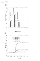
- a novel reporter-gene assay system was constructed by adding the GLuc variants to pG5Luc (Promega), which is a conventional reporter-gene assay system ( Fig. 10 ).
- pG5Luc Promega
- a vector system having an androgen response element (ARE) in the upstream was created, and a gene encoding the original GLuc or GLuc variant of the present invention was linked to the vector system at the downstream by genetic engineering ( Fig. 10A ).
- This novel vector was introduced into COS-7 cells, and the difference in the bioluminescence intensities was measured using a luminometer under the conditions of presence and absence of androgen (male hormone) ( Fig. 10B ).
- the reporter-gene assay system carrying the GLuc variant of the present invention indicated a better signal-to-noise ratio than the reporter-gene assay system carrying the original GLuc, and also showed an absolute bioluminescence intensity about 5 times higher than the conventional case ( Fig. 10B ). Further, the kinetics of the bioluminescence intensity was observed using a plate reader under similar experimental conditions ( Fig. 10C ). Specifically, after COS-7 cells transfected with the above vector were stimulated for 16 hours under conditions of presence and absence of male hormone (androgen), cell lysate was prepared, and a specific substrate (coelenterazine) was added thereto. Then, the difference in the bioluminescence intensity was immediately measured using a plate reader.
- changes in the bioluminescence intensity can be quantitatively compared by the area from when the substrate was introduced to when a certain time span (1 second) has passed.
- the activity level of male hormone being tested can be accurately quantified from the change in the bioluminescence intensity of the reporter gene (I90L) by subtracting the area defined by the control (GLuc) from the area defined by the reporter gene (I90L) ( Fig. 10D ). Further, the areas on the bioluminescence intensity graph in Fig. 10D were compared. As a result, it was confirmed that the transformed cells transfected with pARE-I90L has a larger area (higher bioluminescence intensity) ( Fig. 10E ).
- plasmids were prepared based on pACT, pBIND, and pG5Luc, which are the conventional plasmids for a typical two-hybrid assay: (1) plasmid encoding ER LBD and BIND, (2) plasmid encoding SH2 domain and ACT, and (3) pG5 plasmid carrying the conventional GLuc or the highly luminescent variant (I90L) of the present invention as a reporter gene.
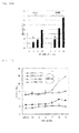
- the bioluminescence intensity was raised up to 4-fold by the 6-hour stimulation; however, in the case of the conventional plasmid, the increase in the bioluminescence intensity was not significant. Further, time courses in the bioluminescence intensity were measured using a plate reader ( Figs. 11C and 11D ). As a result, an increase in the very strong bioluminescence was observed in the case of the plasmid carrying the GLuc variant. Additionally, the difference in the bioluminescence depending on the estrogen concentration was observed ( Fig. 12 ). For this experiment, COS-7 cells were cultured in a 96-well plate.
- pG5-GLuc that expresses the original GLuc itself was introduced into the control group.
- pG5-I90L that expresses the relevant GLuc variant was introduced into the experimental group. After cultivation for 12 hours, estrogen stimulation was performed, followed by cultivation for an additional 8 hours. Subsequently, a lysis buffer was added to the cells, and the bioluminescence intensity was measured for 2 seconds after the substrate was introduced with the automated program. As a result, in comparison to the control group, the experimental group showed a significant difference in the bioluminescence intensity even by 10 -7 M estrogen stimulation.
- the degree of luminescence was more remarkable as the concentration of estrogen was higher.
- the superior luminescence properties of the relevant GLuc variant led to an improvement in the detection limit.
- the living cells into which the above plasmids (1), (2), and (3) were concurrently introduced were stimulated with the solvent itself (0.5% DMSO) or woman sex hormone (estrogen) for 0 hours, 3 hours, 6 hour, and 18 hours. Subsequently, the cells were collected, and the bioluminescence intensities were measured using a luminescence spectrophotometer ( Fig. 11E ).
- the transformed cells having the plasmid carrying one of the GLuc variants of the present invention indicated a superior S/N ratio when stimulated with a woman sex hormone.
- the bioluminescence intensity was raised up to 2-fold by the 3-hour stimulation; however, in the case of the plasmid carrying the conventional enzyme, an increase in the bioluminescence intensity was indistinctive.
- This result shows that, because the GLuc variant prepared by the present inventors has high bioluminescence intensity, a distinctive increase in strong luminescence was observed even when the stimulation was performed for a significantly reduced time span (i.e., even when the expression level was low) compared to the conventional case.
- a single-molecule-format bioluminescent probe was synthesized based on this variant ( Fig. 13 ).
- the variant having high bioluminescence intensity GLuc variant having F89W/I90L mutations
- GLuc variant having four mutations F89W/I90L/H95E/Y97W
- AR LBD and an LXXLL motif were inserted into the space
- a probe having the former variant was named 8990N
- a probe having the latter variant was named Mon3N.
- a control probe was prepared by dissecting the original GLuc itself into two fragments in a similar manner and inserting GR LBD and an LXXLL motif into the space (named SimGR3), and the S/N ratio was measured under conditions of presence and absence of stress hormone (cortisol).
- Fig. 13A shows the operating principle of this probe.
- a stress hormone receptor recognizes the stress hormone, and causes an alteration in the structure.
- the receptor binds to the adjacent LXXLL motif, which thereby causes recombination of the fragments of the dissected luciferase.
- the activity of the stress hormone can be measured using the reversed enzyme activity as the index.
- the results of the measurement of the luminescence spectrum of each variant show that I114L exhibits a higher bioluminescence intensity compared to the original MpLuc1. It was also observed that MpLuc4 showed a spectrum shift to the longer wavelength side, compared to the original MpLuc1 ( Fig. 14A ). Further, as a result of the examination of the bioluminescence intensity of the above variant using a 610 nm long-pass filter, the variant was found to show stronger bioluminescence, compared to the original MpLuc1 ( Fig. 14B ).
- the position of the maximum bioluminescence intensity ( ⁇ max) also varies due to the introduction of the mutation.
- time courses in the bioluminescence intensity after the introduction of the substrate were measured every 0.5 seconds ( Fig. 15B ).
- MLuc variant (MLuc4) with mutations introduced into 4 positions showed the most stable bioluminescence.
- the original MLuc was found to lose 90% of the initial bioluminescence intensity only 10 seconds after the introduction of the substrate.
- the bioluminescence intensities of the variants were measured using a 610 nm long-pass filter.
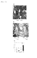
- pcDNA3.1 plasmid encoding 8990 which is a GLuc variant having high bioluminescence intensity
- pcDNA3.1 plasmid encoding the original GLuc itself was similarly introduced into COS-7 cells.
- both of these transformed cells were transplanted into subcutaneous tissues of the back on the left and right sides of BALB/c nude mice (5-week-old female).
- telomeres carrying a mammalian two-hybrid system that expresses the GLuc variant were prepared, and an in vivo imaging system was established using the prepared living cells ( Fig. 16B ).
- two plasmids pBIND and pACT in which a woman sex hormone receptor (ER LBD) binds to BIND and Src SH2 domain binds to ACT, respectively, were prepared.
- ER LBD woman sex hormone receptor
- Src SH2 domain binds to ACT
- the above three plasmids (pBIND, pACT, and pG5-I90L or pG5-GLuc) were co-transfected into COS-7 cells, thereby preparing transformed cells.
- the transformed cells were transplanted into subcutaneous tissues of the back on the left and right sides of BALB/c nude mice (5-week-old female).
- the control cells (pG5-GLuc) were transplanted into the left back side, and the transformed cells carrying pG5-I90L were transplanted into the right back side.
- a 12-hour waiting time was further allowed for stabilization of the transplanted cells.
- the measurement efficiency of assays is dramatically improved by using luciferase of the present invention in measurements of ligands based on the conventionally widely used reporter-gene assays, or by using them as an alternative of bioluminescence enzymes in the conventional bioluminescence probes. Accordingly, the luciferase of the present invention can be widely used in applications such as basic biological studies, and development of diagnostic reagents in medical and pharmaceutical sciences, and analytical chemistry.
Landscapes
- Chemical & Material Sciences (AREA)
- Organic Chemistry (AREA)
- Life Sciences & Earth Sciences (AREA)
- Health & Medical Sciences (AREA)
- Engineering & Computer Science (AREA)
- Zoology (AREA)
- Wood Science & Technology (AREA)
- Genetics & Genomics (AREA)
- Bioinformatics & Cheminformatics (AREA)
- General Health & Medical Sciences (AREA)
- General Engineering & Computer Science (AREA)
- Biochemistry (AREA)
- Microbiology (AREA)
- Molecular Biology (AREA)
- Biotechnology (AREA)
- Proteomics, Peptides & Aminoacids (AREA)
- Analytical Chemistry (AREA)
- Physics & Mathematics (AREA)
- Immunology (AREA)
- Biophysics (AREA)
- Medicinal Chemistry (AREA)
- Biomedical Technology (AREA)
- Measuring Or Testing Involving Enzymes Or Micro-Organisms (AREA)
- Investigating Or Analysing Materials By The Use Of Chemical Reactions (AREA)
- Enzymes And Modification Thereof (AREA)
- Micro-Organisms Or Cultivation Processes Thereof (AREA)
Abstract
This invention provides a luciferase variant that is produced by genetic engineering modification of a marine luciferase such as Gaussia luciferase, which has bioluminescence intensity, and has high bioluminescence stability or bioluminescence shifted to the longer wavelength side. Specifically disclosed is a luciferase variant with improved optical property obtained by replacing at least one amino acid residue among the amino acid sequence of a marine luciferase at positions corresponding to positions 89 to 118 in the amino acid sequence of Gaussia luciferase (GLuc), wherein an amino acid residue at a position corresponding to at least one position selected from positions 89, 90, 95, 97, 100, 108, 112, 115, and 118 in the amino acid sequence of GLuc is replaced by way of conservative amino acid replacement. The above-mentioned replacement in a marine luciferase improves enzymatic activity of the luciferase. Also disclosed is a bioluminescent probe having an improved optical property, which is produced using the luciferase variant of the present invention.
Description
- The present invention relates to the establishment of a method for improving the optical properties and bioluminescence stability of marine luciferase by way of genetic modification, and also relates to a superluminescent and stable artificial luciferase synthesized by the method.
- Many molecular phenomena occurring in cells or cell-free systems in nature (for example, conformation changes and phosphorylation of proteins, interaction between proteins, and production of secondary signal transduction substances) are highly important signatures to understand nature and to explore the molecular mechanisms dominating life phenomena. Presently, various methods, including (i) FRET (Non-patent
Literature 1 and 2), (ii) BRET (Non-patent Literature 3), and (iii) protein-fragment complementation assay (Non-patentLiterature 4 and 5), have been developed to measure such molecular phenomena.
In recent years, the present inventors have conducted research and development regarding bioluminescence imaging using unique molecular design technology. More specifically, the inventors measured translocation of transcription factors into the nucleus and nongenomic protein-protein interactions in the cytosol using protein splicing (Non-patent Literature 6 and 7). Further, the inventors developed a single molecule-format bioluminescent probe in which all of the necessary elements for signal recognition and bioluminescence emission are integrated (Non-patentLiterature 8 and 9). Thereafter, the probes were multicolorized, thereby enabling it to simultaneously image multiple signal-transduction processes (Non-patent Literature 10). Moreover, the inventors further developed a circular permutation technique (Non-patent Literature 11) and a molecular design technology using a small luminescent enzyme (Non-patent Literature 12), as methods for improving the ligand sensitivity of the bioluminescent probe itself. These technologies have been used as means for efficiently measuring cellular and noncellular molecular phenomena.
Fluorescence imaging is more widespread than luminescence imaging as a method for exploring intra- or extracellular molecular phenomena. However, fluorescent proteins company autofluorescence, and therefore generate a high background, requiring an external light source. Therefore, fluorescence imaging requires a large instrument such as a fluorescence microscope, and a sophisticated filtering system. Fluorescence imaging also has a drawback in that maturation of fluorescence chromophore takes at least several hours to several days. Further, when using a fluorescence microscope, the observable cell numbers for each measurement are limited, which has hampered quantitative analysis (Non-patent Literature 6).
On the other hand, the bioluminescence imaging using a luciferase has, despite the many advantages, a critical problem of poor bioluminescence intensities of luciferases, which results in hampering popular use of bioluminescence imaging compared to fluorescence imaging. Since the bioluminescence intensity of luciferases is poor, high sensitivity detectors are required, and bioluminescence imaging is considered to be inappropriate for single-cell imaging or exploration of organelle.
Meanwhile, as for fluorescent protein, fluorescent protein with multicolors has been studied well, and many facts regarding the coloring mechanisms thereof have been discovered. Therefore, based on these findings, many fluorescent proteins with diversified fluorescent characteristics have been developed. On the other hand, there exist only a few luciferase that can exhibit multicolor bioluminescence. Therefore, although the advantages of diversifying the colors of bioluminescence, including (i) simultaneous measurements of multiple signals and (ii) high tissue permeability of the bioluminescence of longer wavelengths into living tissues, have been recited, a systematic study for diversifying the colors of bioluminescence based on the luminescence principles thereof have hardly been conducted.
Accordingly, it has been strongly desired to establish a systematic mutagenesis strategy that can exert superluminescence and stable bioluminescence intensity even in luciferases. Further, there has also been an urgent need for systematic studies for a technology for shifting the luminescence of luciferases to the longer wavelength side. - The present inventors have long focused attention on Gaussia luciferase (Patent Literature 1), which is a luciferase derived from a marine animal, advantageously small in size and higher exhibits higher bioluminescence intensities compared to other luciferases, and the inventors have developed various luminescence imaging methods using the Gaussia luciferase (Japanese Patent Application Nos.
JP2008-116098 JP2009-025229 Jap2007-332253 Patent Literature 2 to 6). Although the anchor peptide (JP2009-025229 -
- [Patent Literature I] International Publication No.
1999-49019 W01999/049019 ) - [Patent Literature 2] Specification of
US Patent No. 6232107 - [Patent Literature 3] Specification of
US Patent No. 6436682 - [Patent Literature 4] Specification of
US Patent No. 6780974 - [Patent Literature 5] Specification of
US Patent No. 7045599 - [Patent Literature 6] Specification of
US Patent No. 7238497 - [Patent Literature 7] International Publication No.
2006-84869 W02008/084869 ) -
- [Non-patent Literature 1] Awais, M.; Sato, M.; Lee, X. F.; Umezawa, Y.; Angew. Chem. Int. Ed. 2006, 45, 2707-2712.
- [Non-patent Literature 2] Awais, M.; Sato, M.; Sasaki, K.; Umezawa, Y.; Anal. Chem. 2004, 76, 2181-2186.
- [Non-patent Literature 3] Hoshino, H.; Nakajima, Y.; Ohmiya, Y.; Nature Methods 2007, 4, 637-639.
- [Non-patent Literature 4] Paulmurugan, R.; Gambhir, S.S.; Anal. Chem. 2005, 77, 1295-1302.
- [Non-patent Literature 5] aulmurugan, R.; Gambhir, S.S.; Proc. Natl. Acad. Sci. U.S.A. 2006, 103, 15883-15888.
- [Non-patent Literature 6] Kim, S.B.; Ozawa, T.; Watanabe, S.; Umezawa, Y.; Proc. Natl. Acad. Sci. U. S. A. 2004, 101, 11542-11547.
- [Non-patent Literature 7] Kim, S.B.; Ozawa, T.; Umezawa, Y.; Anal. Chem. 2005, 77, 6588-6593.
- [Non-patent Literature 8] Kim, S.B.; Awais, M.; Sato, M.; Umezawa, Y.; Tao, H.; Anal. Chem. 2007, 79, 1874-1880.
- [Non-patent Literature 9] Kim, S.B.; Otani, Y.; Umezawa, Y.; Tao, H.; (2007) Anal. Chem. , 79, 4820- 4826.
- [Non-patent Literature 10] Kim, S.B.; Umezawa, Y.; Kanno, K. A.; Tao, H.; ACS Chemical Biology 2008, 3, 359-372.
- [Non-patent Literature 11] Kim, S.B.; Sato, M.; Tao, H.; Bioconjugate Chem. 2008, 19, 2480-2486.
- [Non-patent Literature 12] Kim, S.B.; Sato, M.; Tao, H.; Anal. Chem. 2009, 81, 67-74.
- [Non-patent Literature 13] Matthews, L.; Berry, A.; Tersigni, M.; D'Acquisto, F.; Ianaro, A.; Ray, D.; Endocrinology 2009, 150, 75-86.
- [Non-patent Literature 14] Kim, S.B.; Sato, M.; Tao, H.; Anal. Chem. 2009, 81, 67-74.
- [Non-patent Literature 15] Loening A.M.; Fenn T.D.; Gambhir S.S.; J. Mol. Biol. 2007, 374 (4), 1017-1028.
- [Non-patent Literature 16] Cubitt A.B.; Woollenweber L.A.; Heim R.; Meth. Cell Biol. 1999, 58, 19-30.
- [Non-patent Literature 17] Atsushi Miyawaki; Takeharu Nagai; Hideaki Mizuno; Curr. Opinion Chem. Biol. 2003, 7:557-562.
- [Non-patent Literature 18] N.C. Shaner; G.H. Patterson; M.W. Davidson; J. Cell Sci., 2007 120 (24), 4247-4260.
- [Non-patent Literature 19] Okita K.; Ichisaka T.; Yamanaka S.; Nature, 2007, 448 (7151) 313-317.
- [Non-patent Literature 20] Papworth, C.; Bauer, J.C.; Braman, J; Wright, D.A.; Strategies, 1996, 9(3):3-4.
- An object of the present invention is to establish a method for enhancement of bioluminescence intensity, shifting the bioluminescence to the longer wavelength side, and improvement of bioluminescence stability of luciferase; and to provide a superluminescent and stable artificial luciferase produced by the method. Another object of the present invention is to establish a bioluminescence imaging method using the luciferase.
- In order to attain the above object, the present inventors focused their attention on marine luciferases among various luciferases. On the whole, luciferases have variable structures and share no considerable phylogenetical similarities. Therefore, it is difficult to determine generally acceptable rules among luciferases. However, upon spotlight on marine luciferases, they share common substrates, and have significantly high similarities in sequence. Owing to these advantages, the inventors speculated that, if the conventional knowledge regarding marine luciferases were combined, it may be possible to predict the steric-structure of interaction between enzyme and substrate and estimate the active site of luciferase enzyme. On the basis of this speculation, the inventors attempted to enhance bioluminescence intensity, shift the bioluminescence to the longer wavelength side and improve bioluminescence stability of luciferase by genetically modifying the putative active site and vicinity thereof. More specifically, the inventors utilized the gene sequence of Gaussia-derived luciferase (GLuc) (GENE ACCESSION #: FJ010198) which has the smallest molecular weight among the marine luciferases; codon-optimized and modified to create many variants with genetic mutations at the putative active sites or adjacent amino acids. Each variant was observed for its changes in bioluminescence intensity and stability, or tendency to a shift to the longer wavelength side.
The observation revealed that conservative amino acid replacement of at least one of the amino acid residues at a position corresponding topositions 89, 90, 95, 97, 100, 108, 112, 115, and 118 in GLuc successfully improved activity of the luciferases, i.e., enhanced bioluminescence intensity of luciferases with stabilized bioluminescence, or bioluminescence shifted to the longer wavelength side. More specifically, the improvements in enzymatic properties of luciferase such as enhancement in bioluminescence intensity or the shift to the longer wavelength side, are observed through mutating one or more hydrophobic amino acid residues at positions corresponding to 89, 90, 97, 108, 112, 115, and 118 in GLuc to other hydrophobic amino acid residues. In particular, by individually replacing one or more hydrophobic nonpolar aliphatic amino acid residues at a positions corresponding to positions 90, 108, 112, 115, and 118 in GLuc with other hydrophobic nonpolar aliphatic amino acid residues; or replacing aromatic amino residues at a position corresponding to position 89 and/or 97 in GLuc with other aromatic amino residue(s) demonstrated significant enhancement in activity of luminescence enzyme, as substantiated by an enhancement in bioluminescence intensity and stability, and a shift to the longer wavelength side. Further, it was also confirmed that replacement of hydrophilic amino acid residues at a position corresponding to position 95 and/or 100 in GLuc forming a. hydrogen bond with other hydrophilic amino acid residue caused a shift to the longer wavelength side.
Based on these findings, the present invention was completed.
Further, by introducing mutation into Metridia longa (MLuc) and Metridia pacifica (MpLuc1), which are marine luciferases similar to GLuc, at the positions corresponding to the mutated sites of GLuc, an enhancement in bioluminescence intensity and a shift to a longer wavelength similar to those exhibited by GLuc were observed. This proved that the above mutation sites are common performance-improving sites of marine luciferases.
Moreover, by using the new luciferase synthesized by the present invention as a reporter gene for reporter-gene assay or mammalian two-hybrid assay, or by using the luciferases as a dissected luciferase in bioluminescent probe methods, the strong bioluminescence was also stably maintained in these assays, confirming the effect of reduced assay times and improved S/N ratios. Among these, the usage of the luciferase variants of the present invention in a reporter-gene assay was found to be a revolutionary modification technique of hitherto known assays; therefore, as a separate invention, a patent application of this technology was filed with the Japan Patent Office on the same date as the application date of the present invention. - Specifically, the present invention comprises the following items.
- A luciferase variant with improved optical property obtained by replacing at least one amino acid residue among the amino acid sequence of a marine luciferases at positions corresponding to 89 to 118 in the amino acid sequence of Gaussia luciferase (GLuc), wherein an amino acid residue at a position corresponding to at least one position selected from
positions 89, 90, 95, 97, 100, 108, 112, 115, and 118 in the amino acid sequence of GLuc is replaced by way of conservative amino acid replacement. - The luciferase variant according to
item 1, wherein the conservative amino acid replacement includes replacement of a hydrophobic amino acid residue at a position corresponding to positions 89, 90, 97, 108, 112, 115, or 118 in the amino acid sequence of GLuc with another hydrophobic amino acid residue, or replacement of a hydrophilic amino acid residue at a position corresponding topositions 95 or 100 in the amino acid sequence of GLuc that forms a hydrogen bond with another hydrophilic amino acid residue. - The luciferase variant according to
item 2, wherein the replacement of a hydrophobic amino acid residue with another hydrophobic amino acid residue includes replacement of a hydrophobic and nonpolar aliphatic amino acid residue at a position corresponding at position 90, 108, 112, 115, or 118 in the amino acid sequence of GLuc with another hydrophobic and nonpolar aliphatic amino acid residue, or replacement of a hydrophobic aromatic amino acid residue at a position corresponding to position 89 or 97 in the amino acid sequence of GLuc with another hydrophobic aromatic amino acid residue. - The luciferases variant according to
item 3, wherein the replacement of a hydrophobic and nonpolar aliphatic amino acid residue with another hydrophobic and nonpolar aliphatic amino acid residue is replacement of isoleucine with an amino acid residue selected from leucine, tryptophan, and valine. - The luciferases variant according to
item 3, wherein the replacement of a hydrophobic aromatic amino residue with another hydrophobic aromatic amino residue is replacement of an amino acid residue at a position corresponding to position 89 or 97 in the amino acid sequence of GLuc with tryptophan (W). - The luciferase variant according to
item 2, wherein the replacement of a hydrophilic amino acid residue at a position corresponding to position 95 or 100 in the amino acid sequence of GLuc that forms a hydrogen bond with another hydrophilic amino acid residue is replacement of an amino acid residue at a position corresponding to position 95 in the amino acid sequence of GLuc with glutamic acid (E), or replacement of an amino acid residue at a position corresponding to position 100 in the amino acid sequence of GLuc with asparagine (N). - A luciferase variant obtained by way of conservative amino acid replacement that includes replacement of an amino acid residue at a position corresponding to position 90 in the amino acid sequence of GLuc among the amino acid sequences of marine luciferase with leucine.
- The luciferase variant according to
item 7, wherein the conservative amino acid replacement further includes replacement of an amino acid residue corresponding to position 89 in the amino acid sequence of GLuc with tryptophan, or replacement of an amino acid residue corresponding to position 115 5 in the amino acid sequence of GLuc with leucine. - The luciferase variant according to
item - The luciferase variant according to any one of
items 1 to 9, wherein the marine luciferase is s one luciferase selected from Gaussia luciferase (GLuc) and Copepod luciferases (MLuc, MpLucl, and MpLuc2). - A DNA encoding the luciferase variant according to any one of
items 1 to 10. - An expression vector comprising the DNA according to
stem 11. - A transformed cell in which the DNA according to
item 11 is introduced in a manner such that the DNA can be expressed. - A method for improving luminescent optical property of marine luciferase, comprising replacing at least one of the amino acid residues among the amino acid sequences of marine luciferase at positions corresponding to positions 89 to 118 in the amino acid sequence of Gaussia luciferase (GLuc), wherein an amino acid residue at a position corresponding to at least one position selected from
positions 89, 90, 95, 97, 100, 108, 112, 115, and 118 in the amino acid sequence of GLuc is replaced by way of conservative amino acid replacement. - A bioluminescent probe with improved optical property that adopts a working mechanism of resurrecting luminescent function by reconstituting dissected luciferases, the probe using the luciferase variant according to any one of
items 1 to 10 as the luciferase. - A DNA encoding a bioluminescent probe according to
item 15. - The present invention successfully improved the activity of luminescence enzyme, i.e., enhanced bioluminescence intensity in a longer wavelength region due to enhanced bioluminescence intensity of luciferase or a shift to the longer wavelength side, and also attained an enhanced stability, only by replacing at least one amino acid residue in the luciferase active region and in the vicinity thereof conserved in a wide range of general marine luciferases including GLuc, with a hydrophobic amino acid residue, an aromatic amino acid residue and/or an amino acid residue that forms a hydrogen bond.
By being used as an element of a luciferase for hitherto well-known reporter-gene assay, yeast two-hybrid assay, mammalian two-hybrid assay, protein splicing assay, protein complementation assay, circular permutation assay, bioluminescence resonance energy transfer (BRET) assay (Non-patent Literature 3 to 12) or the like, the superluminescent marine luciferase of the present invention can create a luminescent probe with high bioluminescence intensity and stability, thereby significantly improving measurement efficiency in these assays.
It should be noted that the disclosures of the prior art documents and specifications of patent applications referred to in the present invention are included in the disclosure of the specification of the present invention. -
- [
Fig. 1A ] A graph showing hydrophobicity distribution in each amino acid sequence of various marine luciferases. - [
Fig. 1B ] A comparative chart of amino acid sequences of various marine luciferases. The dotted box denotes a putative enzyme active site. - [
Fig. 2A ] A chart showing the entire amino acid sequence of Gaussia luciferase ((GLuc). The sequence of GLuc was compared with the sequence of luciferase derived from Metridia longa (MLuc). The arrow indicates the enzyme dissection sites in the present study. - [
Fig. 2B ] A chart showing a known crystallographic structure showing the active site of Renilla luciferase. The chemical substance in the middle shows a molecular structure of the specific substrate (caelenterazine). The respective positions of amino acids shows that the enzyme active site resides between the group of hydrophobic amino acids (single lined circle) and the group of hydrophilic amino acids (double lined circle). - [
Fig. 3A ] A graph showing recovery level of bioluminescence intensity at each dissection site of GLuc. - [
Fig. 3B ] A chart showing distribution of hydrophobic amino acids of GLuc. An hydrophilic region covers the vicinity of amino acid numbers 90 to 100. - [
Fig. 3C ] A drawing for comparing chromophores of fluorescent protein and luminescence protein. Section A shows the crystal structure of fluorescent protein and the chemical structure of the chromophore. Section B shows, from left to right, the crystal structure of firefly luciferases, the chemical structure of coelenterazine, and the crystal structure of Renilla luciferases. - [
Fig. 4A ] A map showing mutation sites in the amino acid sequence of GLuc. - [
Fig. 4B-1 ] A graph (1) showing a comparison among different GLuc variants with respect to bioluminescence intensities. The graph compares peak values of luminescence spectrum measured by a spectrophotometer, showing relative bioluminescence intensities upon comparison with the bioluminescence intensity of native GLuc (the leftmost value). Here, the cell dissolution time was set to 5 minutes, the time of which is not enough to dissolve organelles. - [
Fig. 4B-2 ] A graph (2) showing a comparison among different GLuc variants in terms of bioluminescence intensity. The graph compares areas of luminescence spectrum measured by a luminescence plate reader, showing relative bioluminescence intensities based on the original bioluminescence intensity (the leftmost value). Here, the cell dissolution time was set to 20 minutes, thereby dissolving organelle to obtain uniform lysis solutions. The graph shows maximum values of luminescence wavelengths for the variants with high bioluminescence intensities. - [
Fig. 4C ] A graph showing a redshift of bioluminescence spectrum of GLuc variants. The upper graph shows mutation of 190 into Tyr(Y), the middle graph shows mutation of H95 and D100 into Q and N, respectively, and the lower graph shows mutation of H97 into W. - [
Fig. 4D ] A graph showing redshifts of bioluminescence spectra of GLuc variants. The graph shows each luminescence spectrum of Y97W, I90L, and F89W/I90L/H95E/Y97W (Mon3), compared with a conventional GLuc. - [
Fig. 4E ] graph showing relative long wavelength bioluminescence intensities measured by a 610 nm long-pass filter. The graph shows respective wavelengths of luminescence variants compared with a known GLuc (white bar). The graph reveals that the variants show strong bioluminescences at long wavelengths. - [
Fig. 5 ] Photos comparing bioluminescence intensities of GLuc variants with significantly strong bioluminescences.- (A) A digital photo showing the positions of luminescent tubes. The photo shows lysis solutions expressing, from left to right, firefly luciferase (FLuc), known Gaussia luciferase (GLuc), and I90L Variant of GLuc.
- (B) A photo comparing luminescence levels of respective tubes. The photo reveals that the variant I90L of GLuc on the far right has the strongest bioluminescence.
- [
Fig. 6 ] A graph comparing bioluminescence spectrums of typical luciferases. The graph reveals that the bioluminescence intensity of the GLuc variant (solid line) is far stronger than those of known GLuc (dotted line), FLuc, and click beetle luciferase (CBLuc). - [
Fig. 7AB ] Graphs showing changes in bioluminescence intensity with time.- (A) The graph shows a result of a short-time (5 seconds) observation of changes in bioluminescence with time after introducing a substrate (dissolved in a PBS buffer) into each variant. The graph shows that I90L, H95E, and Y97W exhibit relatively stable bioluminescences.
- (B) The graph shows a result of a relatively long-time (10 minutes) observation of changes in bioluminescence with time after introducing the substrate (dissolved in a PBS buffer) into each variant. The graph reveals that the bioluminescence intensity (white circle) of the original GLuc significantly decreased, whereas the bioluminescence intensities of I90L (black square) and I90V (gray triangle) are relatively stable.
- [
Fig. 7CD ] Graphs showing changes in bioluminescence intensity with time.- (C) The graph shows a result of a short-time (5 seconds) observation of changes in bioluminescence with time after introducing the substrate (dissolved in a modified Matthew's buffer into each variant.
- (D) The graph shows a result of observation of changes in bioluminescence with time that was conducted per second for each stage after introducing the substrate (dissolved in a modified Matthew's buffer) into each variant stepwise. The graph reveals that all variants are very stable, showing strong bioluminescences.
- [
Fig. 8A ] A drawing showing a base sequence modification example (1) of a GLuc variant suitable for expression in mammals including human.Fig. 8(1) shows a base sequence of a variant I90L of GLuc consisting of codons suitable for mammals including human, to which a restriction enzyme was introduced. The underline shows the restriction enzyme site, and the italicized letters indicate the mutation sites. - [
Fig. 8B ] A drawing showing a base sequence modification example (2) of a GLuc variant suitable for expression in mammals including human.Fig. 8(2) shows a base sequence of GLuc enzyme variant (Mon3 with four mutations including I90L) consisting of codons suitable for mammals including human, to which an EcoRV restriction enzyme was introduced. The underline shows the restriction enzyme site, and the italicized letters indicate the mutation sites. - [
Fig. 9AB ] Tables showing substrate selectivities of the GLuc variants of the present invention.- (A) The table shows comparison between the respective variants in terms of substrate selectivity using their bioluminescence intensities as index. The table shows levels of relative bioluminescence intensities based on the bioluminescence intensity of the standard substrate (coelenterazine; CTZ) shown as "++++."
- (B) The table shows the maximum luminescence wavelength (λmax) of luminescence spectrum by each synthetic coelenterazine. The table reveals that each variant has a different luminescence color depending on the type of synthetic coelenterazine.
- [
Fig. 9C ] Graph showing substrate selectivity spectrums of the GLuc variants of the present invention. The graph shows, as an example, bioluminescence spectrums of the variants in the presence of coelenterazine fcp. - [
Fig. 10A ] A drawing showing reporter-gene assay based on the GLuc variants of the present invention.- (A) The drawing shows the principle of the assay. The androgen receptor is activated by ligand stimulation (androgen), duplicated, and then bound to an androgen responsive element (ARE), thereby expressing the reporter gene (GLuc variant).
- [
Fig. 10BC ] The graphs showing a reporter-gene assay based on the GLuc variants of the present invention.- (B) The graph shows bioluminescence intensities in the presence and absence of androgen. The graph reveals that the GLuc variant (I90L) shows an excellent signal-to-noise ratio (S/N ratio) compared with a known GLuc.
- (C) The graph shows changes in bioluminescence with time of the expressed reporter protein. The graph reveals that both the intensity and stability of the bioluminescence were superior when I90L was incorporated.
- [
Fig. 10DE ] Graphs showing a reporter-gene assay based on the GLuc variants of the present invention.- (D) The graph shows a comparison in terms of bioluminescence intensity in the reporter-gene assay between the case of using a known Gaussia luciferase (GLuc) and the case of using GLuc variant (I90L) of the present invention. The graph reveals that the area of the bioluminescence intensity was greater in case of using I90L.
- (E) The graph shows comparison of luminescence areas obtained in (D).
- [
Fig. 11AB ] A graph showing a mammalian two-hybrid assay based on the GLuc variants of the present invention.- (A) The drawing shows the principle of two-hybrid assay. Src SH2 domain and ER LBD were used as the model proteins. The SH2 domain was ligated to Ga14, and the ER LBD was ligated to VP16. They were bound to each other by stimulation of an estrogen. In response to this, the GAL responsive element of pG5 induced expression of the reporter protein (GLuc variant).
- (B) The graph shows changes in bioluminescence intensity depending on the estrogen stimulation time. A sufficient bioluminescence was observed by 6-hour estrogen stimulation when using GLuc variant of the present invention.
- [
Fig. 11CD ] A graph showing mammalian two-hybrid assay based on the GLuc variants of the present invention.- (C) The graph shows changes in bioluminescence intensity with time when using the original GLuc (pG5-GLuc).
- (D) The graph shows changes in bioluminescence intensity with time when using the GLuc variant of the present invention. The graph reveals that the absolute luminescence of the Gluc variant was higher than GLuc, and that the bioluminescence thereof was efficiently enhanced even by a short-time estrogene stimulation.
- [
Fig. 11E ] A graph showing a mammalian two-hybrid assay based on the GLuc variants of the present invention.- (E) The graph shows bioluminescence intensities (measured by a luminometer) after (after 0 hour, 3 hours, 6 hours, and 18 hours) estrogene stimulation when incorporating Glue and GLuc variant (9096) in a pG5 vector.
- [
Fig. 12 ] A graph showing the results of a mammalian two-hybrid assay using the GLuc variant of the present invention. The graph shows that the bioluminescence intensity of the GLuc variant of the present invention was significantly enhanced depending on the concentration of estrogene. - [
Fig. 13AB ] A drawing showing a single-molecule-format bioluminescent probe using the GLuc variant of the present invention.- (A) The GLuc variant was dissected into two fragments, and a stress hormone receptor (glucocorticoid receptor) and a LXXLL motif were inserted between the fragments. A GLuc-based probe ("SimGR3") was used as a control. In the figure, "8990N" denotes a GLuc variant having mutations at F89W/I90L, and "Mon3N" denotes a GLuc variant having mutations at F89W/I90L/H95E/Y97W.
- (B) The drawing shows a molecular structure diagram of single-molecule-format bioluminescent probes containing GLuc variants of the present invention.
- [
Fig. 13CD ] Graphs showing data regarding single-molecule-format bioluminescent probes containing GLuc variants of the present invention.- (C) The graph shows a response to stress hormone for each single-molecule-format bioluminescent probe containing the GLuc variant of the present invention. "8990N" exhibited a relatively strong bioluminescence in response to stress hormone.
- (D) The graph shows changes in bioluminescence intensity with time of "8990N." "8990N" exhibited strong bioluminescence in the presence of stress hormone.
- [
Fig. 13E ] A graph showing data regarding single-molecule-format luciferase probes containing GLuc variants of the present invention.- (E) The graph shows measurement of relative stress hormone sensitivities of SimGR3 and 8990N measured by a luminescence plate reader equipped with a 610-nm long-pass filter. The bioluminescence of "8990N" was greater, showing a higher sensitivity to stress hormone stimulation.
- [
Fig. 14AB ] Graphs showing luminescence characteristics of MpLuc1 (Metridia pacifica-derived luciferases) variant.- (A) The graph shows luminescence spectrums of MpLuc1 itself, a I114L variant of MpLuc1 (I114L), and a Y113W/I114L/H119E/Y121W variant of MpLuc1 (MpLuc4).
- (B) The graph shows measurement of bioluminescence intensity of MpLuc1 variant with respect to a long wavelength (610 nm or more) using a 610-nm long-pass filter.
- [
Fig. 14C ] The graph shows measurements of luminescence stability of the MpLuc1 variants. The graph reveals that both I114L and MpLuc4 variants showed more stable bioluminescences compared with the known MpLuc1. - [
Fig. 15AB ] Graphs showing luminescence characteristics of MLuc variants (Metridia longa-derived luciferases).- (A) The graph shows luminescence spectrums of MLuc itself, a I123L variant of MLuc (I123L), and a Y122W/I123L/H128E/Y130W variant of MLuc (MLuc4).
- (B) The graph shows measurement of luminescence stabilities of MLuc variants. The graph reveals that the Mluc4 variant showed more stable bioluminescence compared with the known MLuc itself.
- [
Fig. 15CD ] Graphs showing luminescence characteristics of MLuc variant.- (C) The graph shows measurement of bioluminescence intensities of MLuc variants with respect to a long wavelength (610 nm or more) using a 610-nm long-pass filter.
- (D) The graph shows changes in bioluminescence intensity with time around substrate introduction.
- [
Fig. 16 ] (A) A photograph for observation of the animal tissue permeability of bioluminescence of a GLuc variant (8990N) of the present invention having mutations at F89W/190L. A conventional GLuc and 8990N were expressed at the subcutaneous layers of the left back and the right back, respectively, of a mouse. The observation revealed that the bioluminescence from 8990N (right) was stronger than that from GLuc (left).- (B) A photograph exhibiting bioluminescence intensities from a two-hybrid assay system in the tissues of living animals. Transformed cells, that was obtained by cotransfecting pG5-GLuc or pG5-90L into a COS-7cell, as well as pACT-SH2 or pBIND-ER LBD, was implanted into the subcutaneous layers of the left back or the right back portion of a mouse. The measurement was performed in the presence and absence of estrogene. A stronger bioluminescence was observed in the tissue where pG5-90L was introduced (right: expression of I90L variant of GLuc).
- (C) A graph comparing relative bioluminescence intensities at the respective transplantation portions of the mouse in the above experiment (B). The values correspond to the sites (1), (2), (3), and (4), respectively, from left to right.
- In the present invention, "marine luciferase" designates a luciferase produced by a "luminescent marine animal", which can, in a broad sense, include Obelin, a kind of luminescent plankton, aqualine Pleuromanma, Oplophorus and the like, as well as Gaussia, Renilla reniformis, Cypridina, Metridia etc. Gaussia luciferase (GLuc) and Metridia luciferase (such as MLuc derived from Metridia longa, or MpLuc1 and MpLuc2 which are luciferase groups derived from Metridia pacifica) are very similar in terms of distribution of hydrophilic and hydrophobic amino acids in the entire enzyme; moreover, they have high similarity in amino acid sequence in the putative enzymatic activie cite (
Fig. 1 ). Therefore, the luciferase groups are simply called "marine luciferase", or specifically called "Gaussia-like luciferase". The luciferase group also includes luciferases variants with amino acid sequences and/or encoded by the base sequences having a sequence homology of 80% or more, preferably 90% or more, more preferably 95% or more with the amino acid sequence of GLuc (Gene Bank Assession Number: AY015993), MLuc (Gene Bank Assession Number: AY364164), MpLuc1 (Gene Bank Assession Number: AB195233), or MpLuc2 (Gene Bank Assession Number: AB195234). These Gaussia-like luciferases can be easily obtained by a homology search for the base sequence of the aforementioned GLuc, MLuc, MpLuc1, or MpLuc2 in the PSI-BLAST database or a similar database. It is also possible to establish those luciferases from natural Gaussia-like marine animal genes using a probe or primer based on those base sequences.
MLuc, MpLuc1, and MLuc2 as Gaussia-like luciferases are slightly different from GLuc, as they have a greater molecular weight than GLuc; however, they have very similar sequences, as explained above. Further, in terms of enzymatic characteristics such as substrate or luminescence activity, they are almost identical to GLuc. Therefore, the following description mainly discusses a typical Gaussia luciferase (GLuc). Any finding regarding GLuc may also be applied to other Gaussia luciferases.
Hereinafter, the mutation site is indicated based on the amino acid sequence of GLuc, unless otherwise specified. For example, "amino acid 90 of GLuc" corresponds to the amino acid 123 of MLuc. It also corresponds to the amino acid 114 of MpLuc1 and 93 of MpLuc2.
Gaussia luciferase (GLuc) has the following characteristics. - (i) GLuc is the smallest luciferase ever discovered. Therefore, the use of GLuc for molecular imaging is much less burdensome for the host cell or host protein.
- (ii) GLuc has significant resistance to pH, surfactant, and chemical denaturant.
- (iii) GLuc has the highest bioluminescence intensity among luciferases.
- (iv) GLuc has a quick turnover cycle of enzyme, seven times or more than that of known firefly luciferase.
- In the present invention, "enhancement in optical properties of marine luciferases" mainly means enhancement in bioluminescence intensity and improvement in stability of observation of reporter gene expression. However, it also encompasses a shift of the luminescence wavelength to the longer wavelength side. This is an important nature to expand the utilization of the reporter gene, because the shift to the long wavelength side enhances transmittance through cells or skin.
The two-dimensional distribution of bioluminescence intensity can be obtained by measuring bioluminescence intensity in a specific wavelength range after addition of the substrate using a known luminescence spectrophotometer. By performing such measurement with time, luminescence stability with time may also be measured. A luminescence plate reader, which has superior sample processing property, may also be used to obtain more accurate data. Here, the areas of respective luminescence spectrums are measured after sufficient cell lysis time (about 20 minutes). The shift to a long wavelength side may be measured by a wavelength scanning method, or using a long wavelength filter. - The modification of the amino acid sequence of the marine luciferase of the present invention may be performed by causing a chemical change directly on the desired amino acid residue or residues of the amino acid sequence; however, the modification is generally performed by point mutation of the base corresponding to the target amino acid among the base sequence encoding the enzyme. The mutation of the base may be performed by a well-known method, such as site mutation. The following description discusses the case of introducing point mutation according to the "Quick-Change method" (Non-patent Literature 20); however, the present invention is not limited to this method.
More specifically, the introduction of mutation is conducted by (i) performing PCR reaction in the co-existence of a pair of sense primer and antisense primer of about 30 bases prepared to have a mutation point with a E-coli-derived plasmid template containing DNA coding GLuc, thereby inducing site-specific mutation. (ii) DpnI enzyme treatment, which enables decomposition of only E-coli-derived plasmid, is performed to decompose the plasmid used as the template contained in the PCR product. (iii) The presence or absence of mutation is confirmed by performing gene sequence assay.
For the template DNA encoding GLuc, the base sequence (GENE ACCESSION #: FJ010198) registered in a known database may be used without modification, or with partial modification. Here, as stated below inSection 3, it is possible to use a modified DNA for further improvement of the function as a template DNA for the Quick-Change method. To synthesize the DNA encoding the GLuc or a modified DNA, a set of multiple primers are prepared first. Then, the primers are set to the sense and antisense positions to be subjected to PCR reaction, thereby extending the two fragments to create a DNA template containing the entire length of Gaussia luciferase gene. This template may be mass-produced by, for example, introducing the template into eukaryotic expression vectors pcDNA3.1(+) and culturing them in procaryotic cells such as Escherichia coli. - To determine the replacement sites of amino acid residues in marine luciferase, the present inventors first focused attention on the following points.
- (i) According to a hydrophobic sequence assay of the entire amino acid sequence of GLuc, there is a hydrophilic amino acid region in the central region of the enzyme (
Fig. 4B ). The hydrophilic region in the central region of a protein denotes that the portion is externally exposed. Therefore, the inventors presumed that the portion is a region where the substrate can easily approach; more specifically, the portion is an interaction site with a substrate, i.e., the region includes the active center of the enzyme. - (ii) According to a report (Non-patent Literature 15) regarding the crystal analysis of Renilla-derived luciferase (Renilla luciferase; RLuc), which is a marine luciferase, a hydrophobic amino acid, such as isoleucine (Ile), is present at regular intervals in the vicinity of the hydrophilic region, which is the presumed enzyme active site (as explained in (i)). Since such regular appearance of hydrophobic amino acids is also observed in GLuc, the inventors speculated that the region near the isoleucine (Ile) repeat sequence is the critical area of enzymatic activity where the interaction with the substrate takes place.
- (iii) In the past, the present inventors conducted a series of divisional studies in which six portions of the central region of GLuc are cleaved, and thereby found that the regions in the vicinity of amino acids No. 93 and 98 are the critical area of enzymatic activity (Non-patent Literature 14) (
Fig. 3A ). This finding is consistent with the findings described in (i) and (ii) above. Following a thorough study of the above findings (i) to (iii), the inventors speculated that the area corresponding to 74 to 131 of GLuc among the central region of the amino acid sequence of marine luciferase is the active center of enzyme. The inventors further speculated that the area near the hydrophobic amino acid repeat sequence is the portion where the interaction with the substrate actually takes place. - According to the results of the experiment based on the aforementioned strategies (i), (ii) and (iii), improvement in optical properties was observed in amino acids having mutations at positions 89 to 118 among the amino acids 74 to 131 of GLuc. More specifically, conservative amino acid replacement was performed with respect to an amino acid at a position corresponding to position 89, 90, 95, 97, 100, 108, 112, 115, or 118 in the amino acid sequence of GLuc, thereby enhancing the bioluminescence intensity of luciferase and/or stabilizing the bioluminescence intensity, and/or causing a shift to the longer wavelength side. Thus, improvement in luminescent function of luminescence enzyme was accomplished. The replacement that rendered the greatest improvement was a conservative amino acid replacement at a position corresponding to position 90, 108, 112, 115, or 118 in the amino acid sequence of GLuc based on the aforementioned strategy (iii), or a conservative amino acid replacement at a position corresponding to position 89 or 97 in the amino acid sequence of GLuc based on the aforementioned strategy (ii). The replacement enhanced the intensity of luciferase, stabilized the luminescence, and caused a shift to the longer wavelength side. More specifically, the replacement of the hydrophobic nonpolar (in particular, aliphatic) amino acid residue (more specifically, isoleucine) at position 90, 108, 112, 115, or 118 with other hydrophobic nonpolar (in particular, aliphatic) amino acid residue (more specifically, leucine, tryptophan, or valine); and the replacement of the aromatic amino acid residues at position 89 or 97 (more specifically, phenylalanine at position 89 or thyrosine at position 97) with other aromatic amino acid residue (more specifically, tryptophan) particularly enhanced and stabilized the bioluminescence intensity, while causing a shift to the longer wavelength side. In particular, the variant (I90L) obtained by replacing isoleucine at position 90 with leucine was a superior luciferase variant having significantly high and stable bioluminescence intensity, and a shift to a long wavelength side.
The replacement of leucine at position 90 with thyrosine is also regarded as a replacement between hydrophobic amino acid residues, and therefore may also be regarded as a conservative amino acid replacement. This replacement caused a shift to long wavelength, even though no enhancement in bioluminescence intensity was observed. According to the fact that the replacement of phenyl alanine at position 89 or thyrosine at position 97 with tryptophan is also considered replacement between hydrophobic amino acid residues, the above effect can also be expressed such that "replacement of hydrophobic amino acid residues corresponding to position 89, 90, 97, 108, 112, 115, or 118 in GLuc respectively with another hydrophobic amino acid residue improves activity of a luminescence enzyme; such improvement is specifically substantiated by enhancement and stabilization of bioluminescence intensity and/or a shift to the longer wavelength side.
Further, since the replacement of a hydrophilic amino acid residue at a position corresponding to position 95 or 100 in the amino acid sequence of GLuc that forms a hydrogen bond with another hydrophilic amino acid residue (more specifically, replacement of histidine at position 95 with glutamic acid, or replacement of asparagic acid atposition 100 with asparagine) causes a shift to the longer wavelength side, it was confirmed that the aforementioned strategy (i) is also effective to cause a shift of luminescence wavelength to a long wavelength side. A shift to a long wavelength side is equivalent to an enhancement in light transmittance in the observation through cell membrane or skin. Therefore, this is a desired characteristic. - Furthermore, it was also confirmed that the optical property was further improved by introducing multiple mutations according to the aforementioned strategies (i), (ii) and (iii) instead of introducing only one mutation into one of the above positions. More specifically, in the present invention, it is also effective to introduce mutations into multiple sites at the same time. For example, by combining the mutation for enhancing bioluminescence intensity and for improving luminescence stability (more specifically, the mutation of at least one position corresponding to positions 89, 90, 108, 112, 115, and 118 in the amino acid sequence of GLuc with a hydrophobic amino acid or acids) with the mutation for causing the shift of the luminescence wavelength to a long wavelength side (more specifically, the mutation of at least one position corresponding to
positions 90, 95, 97 and 100 in the amino acid sequence of GLuc), it becomes possible to further enhance the bioluminescence intensity, improve luminescence stability, and cause a shift to the longer wavelength side.
More specifically, both the modified luciferase (F89W/I90L) obtained by replacing isoleucine (I) at a position corresponding to position 90 in the amino acid sequence of GLuc with leucine (L), and replacing phenylalanine (F) at a position corresponding to position 89 in the amino acid sequence of GLuc with tryptophan (W); and a modified luciferases (I90L/I115L) obtained by replacing isoleucine (I) at a position corresponding to position 90 in the amino acid sequence of GLuc with leucine (L), and replacing isoleucine (I) at a position corresponding to position 115 in the amino acid sequence of GLuc with leucine (L) were revolutionary luciferases that have very high and stable bioluminescence intensity, and cause a shift to the longer wavelength side. - In order to obtain a variant with further improved luminescent function, a modified luciferases having mutations at "F89W/I90L" was further mutated by replacing histidine (H) at a position corresponding to position 95 in the amino acid sequence of GLuc with glutamic acid (E) or glutamine (Q), and replacing thyrosine (Y) at a position corresponding to position 97 in the amino acid sequence of GLuc with tryptophan (W) (F89W/I90L/H95E/Y97W); and a further shift to the long wavelength side was observed while maintaining the improved bioluminescence intensity and stability. In particular, the modified luciferase "F89W/I90L/H95E/Y97W" showed a great shift to the long wavelength side, which was shown as an increase of the peak value from 471 nm (absorbency), which indicates the maximum bioluminescence intensity of the original GLuc, to 506 nm (absorbency).
- The stability of the bioluminescence intensity is confirmed by introducing a substrate (dissolved in a PBS buffer) into each variant, and observing its changes in bioluminescence with time. As a result of a 5-second observation of I90L, H95E, and Y97W that are typical GLuc variants, it was found that they had stable bioluminescence intensities (
Fig. 7A ). Further observation (10 minutes) revealed that, whereas the decrease in bioluminescence intensity of the original GLuc (white circle) was significant, the bioluminescence intensity of I90L (black square) and the bioluminescence intensity of I90V (gray triangle) were relatively stable (Fig. 7B ). The same result was obtained from a short-time (5 seconds) observation of each variant for its changes in bioluminescence with time after introducing a substrate (dissolved in a modified Matthew's buffer) (Fig. 7C ). Furthermore, another observation of 1 minute for each stage of changes in bioluminescence with time after introducing a substrate (dissolved in a modified Matthew's buffer) stepwise into each variant also revealed that each variant had very stable and strong bioluminescence (Fig. 7D ). - Using a Gaussia luciferases derived from a Copepod (Metridia pacifica or Metridia longa), which is a Gaussia-like marine animal very similar to Gaussia, namely, luciferases of MpLuc1 (derived from Metridia pacifica) and MLuc (derived from Metridia longa), a MLuc-I123L variant and a MpLuc1-I114L variant were obtained by carrying out conservative amino acid replacements of an amino acid at position 123 of MLuc and an amino acid at position 114 of MpLuc1 corresponding to the amino acid at position 90 of GLuc; also obtained were a Y122W/I123L/H128E/Y130W variant (MLuc4) of MLuc and a Y113W/I114L/H119E/Y121W variant (MpLuc4) of MpLuc1 corresponding to a F89W/I90L/H95E/Y97W variant (Mon3) of GLuc. The luminescence characteristics of these variants were measured, with the result that their bioluminescence intensities were enhanced as in the GLuc variant. The results also revealed an enhancement in stability and a shift to the long wavelength side (
Fig. 14A to 14C andFig. 15A to 15D ). This confirmed that the findings regarding GLuc are also applicable to all of the other Gaussia luciferases. - The above results revealed that the marine luciferase of the present invention, in particular, the Gaussia luciferase, is obtained by "conservative amino acid replacement" of at least one amino acid residue selected from the amino acid residues at a position corresponding to
positions 89, 90, 95, 97, 100, 108, 112, 115, and 118 among the amino acid sequence at positions corresponding to positions 89 to 118 of Gaussia luciferase (GLuc). More specifically, such replacement includes replacement of a hydrophobic amino acid residue at a position corresponding to position 89, 90, 97, 108, 112, 115, or 118 in the amino acid sequence of GLuc with another hydrophobic amino acid residue, or replacement of a hydrophilic amino acid residues at a position corresponding to position 95 or 100 in the amino acid sequence of GLuc that forms a hydrogen bond with another hydrophilic amino acid residue.
This hydrophobic amino acid is preferably a hydrophobic amino acid having a greater molecular weight than a small-size hydrophobic amino acid such as glycine (Gly). An aromatic amino acid, such as tryptophan (Trp), may be used insofar as it is hydrophobic and nonpolar. In particular, among the hydrophobic amino acids, aliphatic amino acids, such as leucine (Leu), valine (Val), isoleucine (lie) or tryptophan (Trp), are preferable. Leucine (Leu), valine (Val), and tryptophan (Trp) are particularly preferable.
Further, when the replacement of a hydrophobic amino acid residue with another hydrophobic amino acid residue is replacement of a hydrophobic nonpolar aliphatic amino acid residue at a position corresponding to position 90, 108, 112, 115, or 118 in the amino acid sequence of GLuc with another hydrophobic nonpolar aliphatic amino acid residue (in particular, the replacement of isoleucine with one amino acid residue selected from leucine, tryptophan and valine), or replacement of a hydrophobic aromatic amino residue at a position corresponding to position 89 or 97 with another hydrophobic aromatic amino residue (the replacement of phenylalanine (Phe) at a position corresponding to position 89 or thyrosine (Tyr) at a position corresponding to positions 97 with tryptophan (Trp)), the resulting luciferase variant has an enhanced bioluminescence intensity, as well as stable bioluminescence intensity or a shift to the longer wavelength side. Thus, the enhancement in activity of luciferase can be accomplished. The most preferable combination of the mutation position and the mutating amino acid for such replacement is replacement of a position corresponding to position 89 in the amino acid sequence of GLuc with tryptophan (Trp), replacement of a position corresponding to position 90 in the amino acid sequence of GLuc with Leu (leucine) or Val (valine), and replacement of position 108 with Val (valine).
Further, the replacement of a hydrophilic amino acid residue that forms a hydrogen bond with another hydrophilic amino acid residue is replacement of histidine (His) at position 95 in the amino acid sequence of GLuc with glutamic acid (Glu), or replacement of asparagic acid (Asp) atposition 100 in the amino acid sequence of GLuc with asparagine (Asn). Such a replacement causes a shift to the longer wavelength side of the resulting modified luciferase.
Among the obtained modified luciferases, the modified luciferases (GLuc-I90L, MpLuc1-I114L, MpLuc1-I114L) having a mutation caused by replacement of isoleucine corresponding to position 90 in the amino acid sequence of GLuc had the highest intensity, and improved stability, and a shift to the long wavelength side. It is more preferable to produce a variant by replacing an amino acid corresponding to position 89 in the amino acid sequence of GLuc with tryptophan, replacing an amino acid at position 90 with leucine or valine, replacing an amino acid at position 95 with glutamic acid, replacing an amino acid at position 97 with tryptophan, replacing an amino acid at position 108 with valine, replacing an amino acid at position 112 with leucine or valine, replacing an amino acid at position 115 with valine, or replacing an amino acid at position 118 with leucine or valine, so as to further significantly enhance bioluminescence intensity and cause a shift to the longer wavelength side. In particular, the leucine variants (GLuc-I90L, MpLuc1-I114L, MpLuc1-I114L) having mutation at a position corresponding to position 90 in the amino acid sequence of GLuc are excellent modified luciferases with not only a high, bioluminescence intensity but also a significant shift to the longer wavelength side.
In the present invention, "a luciferase variant (modified luciferase)" preferably denotes a variant basically having no mutation with respect to the amino acid sequence of the original Gaussia luciferase other than the aforementioned "conservative amino acid replacement", in the region corresponding to positions 89 to 118 in the amino acid sequence of GLuc, i.e., in the active center of the amino acid sequence. However, as long as the specified region has no mutation other than the conservative amino acid replacement, the variant may have mutation in other positions on the condition that the mutation is not conducive to a severe change in the entire insteric structure. In particular, the variant may contain a deletion at a part of the N- or C-terminal region, for example, deletion of 1 to 50, preferably 1 to 30, more preferably 1 to 20, further preferably 1 to 10 amino acid residues in the N- or C-terminal region. More specifically, the variant may mean a modified Gaussia-like luciferase having an amino acid sequence with homology of 70% or more, preferably 80% or more, more preferably 90% or more, most preferably 95%, except for the region corresponding to positions 89 to 118 in the amino acid sequence of GLuc. - The usages of the marine luciferase of the present invention are described in
Section 4 below. Typically, the usages include those as a luciferase component of a known bioluminescent probe, and, owing to its high bioluminescence intensity, as a substitute for a reporter gene for fluorescent imaging in vino. Since analysis methods using reporter genes, such as the reporter-gene assay or the two-hybrid method, are performed in the hitherto-established host cell systems, the series of procedures of producing a vector by binding the luciferase variant gene of the present invention with the promoter of the target gene, and culturing the host cell incorporating the vector may be performed according to well-known methods. Examples of host cells used herein include yeast cells, bacteria cells such as Escherichia coli, and insect cells, as well as mammalian cells (COS cell, CHO-K1 cell, HeLa cell, HEK293 cell, NIH3T3 cell) used for general gene recombination. The present invention is mainly used in mammalian cells, such as humans, in vivo or in vitro.
Further, the luciferase variant gene of the present invention is preferably modified to have codons suitable for the target cell, thereby modifying the base sequence into a sequence that is more easily expressed in the host cell. The luciferase variant gene of the present invention also needs a modification to contain a restriction enzyme site to be introduced into a vector. These modifications may also be performed according to known methods. As one example, a modification strategy of a variant assumed to be used in mammalian cells, such as humans in vivo or in mammalian cells in vitro, is described below.
More specifically, the modifications for improving other functions include modification of the codons corresponding to the amino acid into those suitable for mammalian so that the gene has a base sequence more easily expressed in the bodies of mammalian such as humans, and a modification to introduce a restriction enzyme site at a desired position. Accordingly, the present invention adopts such modifications as required. The modification strategies for improving functions are specifically described below. However, the present invention is not limited to these examples. -
- (i) The modification was conducted by taking into consideration the fact that organisms have a common molecular mechanism for expressing the same DNA, but may have a different gene codon proportion depending on the species. For example, an animal cell has a higher proportion of G or C in the nucleic acid than that in Escherichia coli, or tends to express genes with a higher proportion of G and C. Therefore, in order to ensure effective expression of GLuc in an animal cell, the known gene sequence of the GLuc is modified by optimizing the codons to have a higher proportion of G or C.
- (ii) The Kozak sequence (GCCACC ATG G) is introduced in the first position of the gene so as to ensure high expression in eukaryotic cells.
- (iii) The original extracellular secretion signal (1-17AA) on the upstream of GLuc gene, which is irrelevant to the luminescence activity, is deleted or replaced with another known extracellular secretion signal or intracellular signal. Alternatively, an additional intracellular signal may be provided in the C-terminal end while leaving the secretion signal as is.
- In the embodiments of the present invention, a template of DNA encoding GLuc was designed according to the sequence information of GLuc, in consideration of the above points (i) to (iii). A primer set for synthesizing the DNA was constructed, and the template of GLuc gene was completed by PCR reaction. The template was ligated into pcDNA3.1(+) of a eukaryotic cell-expression vector to carry out subcloning.
Using the above expression vector as a template, a point mutation was introduced according to the aforementioned "Quick-Change method." More specifically, a PCR reaction was performed by coexisting a sense primer and an antisense primer of about 30 bases prepared by containing a mutation point, thereby inducing site-specific mutation. Then, only the E. coli-derived plasmid containing the GLuc gene template was decomposed by DpnIenzyme, and the gene sequence was analyzed to confirm the presence or absence of mutation. - The enzymatic activity of mutated GLuc may be verified, for example, in the following manner.
First, using a known lipid reagent for gene introduction, an expression vector having a GLuc variant is introduced into African monkey cells (COS-7); while also introducing, as a control, an expression vector having a known unmodified GLuc without any mutation into the cells in the same manner. At a predetermined time (from 10 to 20 hours, for example, 16 hours) after the introduction of the vector, a cell lysate is prepared using a known lysis solution.
Thereafter, the cell lysate is mixed with a known substrate solution containing coelenterazine, and its optical intensity, short-term stability in luminescence, etc., are measured.
The bioluminescence intensity may be found by measuring the intensity at a specific wavelength using a known luminescence spectrophotometer after addition of a known substrate. By performing the measurement every minute, the short-term stability in luminescence can be found. To measure a shift to the longer wavelength side, scanning of the entire wavelength is necessary.
In the embodiments of the present invention, the variant obtained by replacing hydrophobic amino acids at positions corresponding to positions 89, 90, 108, 112, 115, and 118 in GLuc with other hydrophobic amino acids had enhanced bioluminescence intensity. More specifically, a variant obtained by replacing the amino acid 89 with tryptophan, 90 with leucineor valine, 108 with valine, 112 with leucineor valine, 115 with valine, and 118 with leucineor valine showed an intense luminescence spectrum peak (luminescence spectrophotometer). In the measurement of the area of luminescence spectrum using a luminescence plate reader, the glutamic acid variant having a mutation at position 95 and the tryptophan variant having a mutation at position 97 showed a significant enhancement in bioluminescence intensity, as well as the leucine variant having a mutation at position 90; further, these variants all caused a shift to the longer wavelength side. In particular, the leucine variant (I90L) having a mutation at position 90 is a superior luciferase variant that has a high luminescence intensity and a significant shift to the longer wavelength side. Further, the variant with two simultaneous mutations (F89W/I90L or I90L/I115L), i.e., the leucine replacement at position 90, and tryptophan replacement at position 89 or leucine replacement at position 115 at the same time; and the variant with four simultaneous mutations (F89W/I90L/H95E/Y97W), i.e., leucine replacement at position 90, tryptophan replacement at position 89, glutamic acid replacement at position 95, and tryptophan replacement at position 97 at the same time, both showed a significant bioluminescence intensity and a significant shift to the long wavelength side. Thus, they both were revolutionary luciferase variants.
Further, the variant having replacement with glutamine (Gln) or asparagine (Asn), which is a neutral amino acid forming a hydrogen bond, atposition 95 or 100 showed a shift of bioluminescence to the long wavelength side. Similarly, it was confirmed that the variants having mutations at positions 90 and 97 showed a shift of bioluminescence to the longer wavelength side, regardless of whether the mutation introduces leucine (Leu), which is a hydrophobic amino acid, thyrosine (Tyr), which is an amino acid forming a hydrogen bond and also an aromatic amino acid, or tryptophan (Trp), which is an aromatic amino acid and also a hydrophobic amino acid. The shift to the longer wavelength side was substantiated by the luminescence peaks of variant Y97W and variant 190Y, which were 486 nm and 492 nm, respectively, relative to the luminescence peak of GLuc, which was 471 nm. Further, the variants I90L, F89W/I90L, and I90L/I115L also showed significant shifts to the long wavelength side, as they had luminescence peaks of 486 nm, 481 nm, and 483 nm, respectively. In particular, the luminescence peak of the variant F89M/I90L/H95E/Y97W was 506 nm, showing a revolutionary shift to the longer wavelength side. Such a long wavelength bioluminescence improved transmittance through living tissues, and is thereby highly useful as a valuable analytical signal. - The gene of the present invention encoding a .luciferase variant is most expected to be used as a reporter gene for various analysis systems, as with the conventional luciferase genes, and as a bioluminescent probe that is currently under research and development.
- The marine luciferase variant of the present invention may be used as the widely known reporter-gene assay (reporter-gene assay), yeast two-hybrid assay, mammalian two-hybrid assay, protein splicing assay, protein complementation assay, circular permutation assay, bioluminescence resonance energy transfer assay (BRET) or the like as a core element, and is conducive to significant improvement in measurement efficiency in these assays. The marine luciferase variant of the present invention can completely overcome the following drawbacks of conventional firefly luciferase:
- (i) Due to its large molecular weight, it takes a long time to start expression, thereby imposing a great burden on the host cells.
- (ii) Due to the low bioluminescence intensity of firefly luciferase, it generally takes 1 to 2 days after stimulation to obtain a sufficient accumulation of luciferase (reporter).
- Further, by incorporating the superluminescent luciferase of the present invention into the single-molecule-format luminescent probe (
Non-patent Literature 9,Non-patent Literature 10,Non-patent Literature 11,Non-patent Literature 12, Japanese Patent Application No.2007-005144 2007-202308 2007-332253 2008-116098 2009-025229 PCT/JP2008/050370 US12/025532 US12/343830 Non-patent Literature 4,Non-patent Literature 6, and Non-patent Literature 7) which are recited in the pending patents applied by the present inventors, the presence or absence of ligand and the intensity of ligand activity can be observed with high bioluminescence intensity. By comprising, as the probe components, (i) the dissected luciferase (N- and C-terminal fragments), and (ii) a ligand-binding protein responsive to the target ligand and (iii) a recognition protein that recognizes the bond of the ligand with the ligand-binding protein, which are linked to the vicinity of the dissected luciferase, it is possible to form a high-performance luminescent probe. This luminescent probe functions such that, as the recognition protein recognizes the bond of the ligand binding of the ligand-binding protein, the two fragments of the dissected enzyme complement compensate each other and thereby change the enzymatic activity. Here, due to the high bioluminescence intensity and stability of the dissected enzyme, it is possible to perform reliable measurement with an improved detection limit. - In the present invention, "single molecule-format luminescent probe" denotes a known bioluminescent probe in which all components for visualization imaging are integrated in a single fusion molecule (disclosed in
Patent Literature 7, etc.). For example, "single molecule-format luminescent probe" denotes a fusion protein that comprises, as fundamental components, the two fragments of N- and C-terminals obtained by dissecting the marine luciferase variant of the present invention, a ligand-binding protein, and a recognition protein for recognizing the ligand-binding protein. Similarly, "two molecule-format luminescent probe" in the present invention denotes a bioluminescent probe in which the two fragments of N- and C-terminals obtained by dissecting the marine luciferase variant of the present invention are present in the fusion protein containing the ligand-binding protein, and in the fusion protein containing the recognition protein, respectively.
As such, when the marine luciferase superluminant variant of the present invention is used for these bioluminescent probes, the variant must be dissected into a N-terminal fragment and a C-terminal fragment. The dissection portion is the same as the dissection portion of the marine luciferase used for the known "single molecule-format luminescent probe" and "two molecule-format luminescent probes".
More specifically, the GLuc variant of the present invention is dissected at a portion between 105 and 110, and the MLuc variant of the present invention is dissected at a portion between 138 and 142.
Further, in addition to the single molecule-format bioluminescent probe, the superluminant luciferases may also be incorporated in the circular permutation replacement probe (Non-patent Literature 11; Non-patent Literature 12) developed by the present inventors, thereby enabling efficient calculation of intracellular molecular phenomenon with stable and high bioluminescence intensity. In addition to 8990N and Mon3N, i.e., the single molecule-format bioluminescent probe described in Example 9 of the present invention, the circular permutation replacement probes are cPresso and cPressoMax in the sequence listing. - Regarding the actual method for using the superluminescent luciferase of the present invention as a single molecule-format luminescent probe,
Patent Literature 7, and Japanese Patent Application Nos.2007-332253 2009-025229
Since the bioluminescent probe using the superluminescent luciferase of the present invention is introduced into cells as a chimera DNA and expressed in the cells as a fusion protein, by measuring the change in light amount emitted from the cells after subjecting the transgenic cell to ligand stimulation, the property or levels of activity of the ligand may be evaluated. - When the superlurninesoent luciferase of the present invention is incorporated in the bioluminescent probe, the "ligand-binding protein," which can be incorporated in the probe together with the luciferase, is intended to mean a protein that binds with a ligand at the ligand binding site. The ligand-binding protein may serve to, in response to the bond with the ligand, for example, change the steric structure, cause phosphorylation, or facilitate protein-protein interaction. Examples of such ligand-binding proteins include nuclear receptors (NR) to which such ligands as hormones, chemical substances, or signal transduction proteins bind; cytokine receptors; and various protein kinases. A suitable ligand-binding protein is selected depending on the target ligand. The ligand that binds to the ligand-binding protein is not particularly limited insofar as it binds to the ligand-binding protein. The ligand may be an extracellular ligand that is introduced in response to extracellular stimulus, or an intracellular ligand that is produced inside the cells. Examples thereof include agonists or antagonists of the receptor protein (for example, intranuclear receptor, or G-protein-linked receptor), signal transduction proteins such as cytokine, chemokine, or insulin, intracellular second messenger, lipid second messenger, phosphorylated amino acid residue, G-protein-linked receptor ligand and like ligands that specifically bind to proteins involved in intracellular signal transduction.
For example, when the intracellular second messenger, the lipid second messenger or the like is used as a ligand, the binding domain of each second messenger may be used as the ligand-binding protein. "Second messenger" denotes a different kind of intracellular signal transduction substance that is newly produced as a result of the bond of the extracellular signal transduction substance, such as a hormone or neurotransmission substance, with a receptor that exists in the cell membrane.
Examples of the second messengers include cGMP, AMP, PIP, PIP2, PIP3, inositol trisphosphate (IP3), IP4, Ca2+, diacylglycerol, and arachidonic achid. For example, for Ca2+ as the second messenger, calmodulin (CaM) may be used as the ligand-binding protein. - Further, using the gene encoding the superluminescent luciferase enables stable introduction of the luciferase into various cell strains. For example, using the gene enables stable introduction of the luciferase into the novel induced pluripotent stem cells (iPS cells). Since the induced pluripotent stem cells (iPS) do not emit light themselves, it has been very difficult to research the intracellular molecular phenomenon and tissue specificity of the cells. To address this difficulty, a molecular probe containing the luciferase is introduced into somatic cells before the embryo is formed; after the introduction of the luciferase-containing probe, the embryo is differentiated into various tissues. This enables measurement of specific molecular phenomena in respective organs at high sensitivity. This process is performed according to the method of Yamanaka et al. (Non-patent Literature 19).
Further, by linking the superluminescent luciferase of the present invention to a suitable signal peptide, the luciferase can be used for luminance imaging of various organelles. For example, by linking a GAP-43-derived MLCCMRRTKQV sequence to an N- or C-terminal of GLuc, the luciferase may be localized in cell membranes. Linking a GRKKRRQRRR sequence to a terminal enables localization in cell cytoplasm. Further, for localization in endoplasmic reticulum (ER) and cellular nucleus, KDEL and DPKKKRKV sequences, respectively, are linked to a terminal. Furthermore, by linking to HIS-tag (HHHHHH), FLAG-tag (DYKDDDDK), Myc-tag (EQKLISEEDL), HA-tag (YPYDVPDYA), V5-tag (GKPIPNPLLGLDST), T7-tag (MASMTGGQQMG) or like antigen sites, the luciferase can be used for immunostaining or separation/refinement in acellular systems. In these usages, known immunostaining technologies or immunocytochemistry may be adopted. - The other terms and concepts used in the present invention are specifically defined in the descriptions of embodiments and examples. The terms are generally selected from the IUPAC-IUB Commission on Biochemical Nomenclature, or based on the interpretations of idiomatic terms and words in the related field. Further, except for the technologies with apparent sources, the various technologies used to carry out the present invention can be easily and consistently performed by persons skilled in the art in reference to published documents, etc. For example, genetic engineering and molecular biological techniques can be carried out according to J. Sambrook, E. F. Fritsch & T. Maniatis, "Molecular Cloning: A Laboratory Manual (2nd edition)," Cold Spring Harbor Laboratory Press, Cold Spring Harbor, New York (1989); D. M. Glover et al. ed., "DNA Cloning," 2nd ed., Vols. 1 to 4, (The Practical Approach Series), IRL Press, Oxford University Press (1995); Ausubel, F. M. et al., Current Protocols in Molecular Biology, John Wiley & Sons, New York, N.Y., 1995; " Zoku Seikagaku Jikken Koza 1 (Continuation of Biochemistry Experimental Series 1), Idensi Kenkyu Ho (Gene Study Method) II" edited by Tokyo Kagaku Dojin (1986); "Shin Seikagaku Jikken Koza 2 (New Biochemistry Experimental Series 2), Kakusan (Nucleic Acid) III (DNA Recombinant Technology)" edited by Tokyo Kagaku Dojin (1986); Japanese Biochemical Society, Tokyo Kagaku Dojin (1992); R. Wu ed., "Methods in Enzymology," Vol. 68 (Recombinant DNA), Academic Press, New York (1980); R. Wu et al. ed., "Methods in Enzymology," Vols. 100 (Recombinant DNA, Part B) & 101 (Recombinant DNA, Part C), Academic Press, New York (1983); R. Wu et al. ed., "Methods in Enzymology," Vols. 153 (Recombinant DNA, Part D), 154 (Recombinant DNA, Part E) & 155 (Recombinant DNA, Part F), Academic Press, New York (1987), etc.; the methods mentioned in the documents referenced in these documents, or other various similar methods and modified methods thereof that are substantially the same as the disclosed methods.
Further, the various proteins, peptides, and DNAs encoding them used in the present invention may be obtained from existing databases (URL: http://www.ncbi.nlm.nih.gov, etc.). - Hereinbelow, the present invention is specifically described in detail with reference to examples; however, the present invention is not limited to these examples.
Note that the disclosures of the technical documents, patent publications, and patent application specifications are incorporated into the specification by reference. - In the determination of a new gene sequence based on a known gene sequence (GENE ACCESSION #: FJ010198) of Gaussia luciferase (GLuc), codons were optimized for known mammalian cells. Specifically, codons that contain many Gs and Cs must be produced for optimal expression in mammalian cells. For example, of two bases, TTT and TTC, encoding Phe among the amino acids, TTC is more suitable for mammalian cells. Additionally, in order to increase the level of protein expression, a known Kozak sequence (GCCRCC ATG G) was inserted before and after the translation initiation codon (AUG).
In order to generate the above gene sequence, sense and antisense primers were palindromically placed, and a PCR reaction was performed to synthesize the full-length sequence. The full-length GLuc was used as a genetic template upon introduction experiments of other mutations.Fig. 2 shows the full length. -
- Inference 1: Through a detailed observation of the crystallographic structure (
Fig. 3C ) formed by Renilla luciferase (RLuc) upon binding to coelenterazine, which is a common substrate of marine luciferases, it was found that, in the active site of RLuc, (1) there is a region where hydrophobic amino acids are found at certain intervals (2 to 4 amino acid residues), and (2) the active site has a structure in which the substrate is sandwiched between hydrophilic and hydrophobic amino acid sites (Fig. 2B ).
Through an examination of GLuc amino acid sequence, it was found that, in the region at a similar site (amino acid numbers 74-161), hydrophobic amino acids (isoleucine (I), leucine (L), valine (V), phenylalanine (F), tryptophan (W), and the like) appear at certain intervals (2 to 4 amino acid residues); and it was inferred that, also in GLuc, this region is the substrate-binding region, which is the key site that influences luciferase activity. - Inference 2: First, in order to estimate the active site in the luciferase, several sites in the center of the enzyme were fragmented, and calmodulin (CaM) was inserted between the fragments, thereby examining whether the enzymatic activity is reconstituted. As a result, hardly any enzyme activity was observed at dissection sites #1-#3. On the other hand, a very strong bioluminescence was observed at dissection site #4 (
Fig. 3A ).
It was confirmed from the study on the dissection sites in GLuc that the amino acid sequences near thedissection sites # 3 and #4 are the key regions in GLuc that influence the enzyme activity of GLuc. - Inference 3: Further, the hydrophobic sequence search of GLuc revealed that the above site is the region of intersection of hydrophilic and hydrophobic amino acid domains (
Fig. 3B ). - Inference 4: Conventional studies on the chrompphores of fluorescent protein were examined, and the following techniques were brought into focus:
- (1) Introduction of amino acids (tyrosine (Y), serine (S), and the like) forming a hydrogen bond with a fluorescent chromophore into a position near the chromophore can produce an effect of extending a n-bond of the chromophore, as well as a color change to the longer wavelengths and a change in the bioluminescence intensity (Non-patent Literature 16).
- (2) Because the fluorescent chromophore contains a phenyl group in its skeleton, it was possible to make a better fluorescent protein by introducing an amino acid containing a phenyl group as a side chain into a position near the chromophore so as to interact with the phenyl group of the chromophore (Non-patent Literature 17).
- (3) A stable and high-intensity fluorescent color was achieved by introducing a hydrophobic amino acid into a position near a fluorescent chromophore so as to hydrophobically interact with the fluorescent chromophore (Non-patent Literature 18).
As described above, it was found that, in regard to the fluorescent protein, there is a case where the introduction of a mutation into an amino acid near a fluorescent protein chromophore effectively improves the fluorescent protein.
The comparison of the substrate of marine luciferases with the fluorescent protein chromophore reveals that their structures are closely similar to each other. Therefore, it was inferred that they share chromophores having very similar chemical structures; and that the fluorescent protein embeds the chromophore inside of the molecule, whereas the luciferase recruits the chromophore outside of the molecule in form of the substrate (Fig. 3C ). - Inference 5: In a previous study by the present inventors on luciferase, there is a case of successful improvement of luminescence properties by partially overlapping the sequences near the enzyme active site. From this case, improvement in the properties of luciferase can also be expected by a technique of partially overlapping the sequences near the enzyme active site.
- (2-2) Based on the above five inferences, a strategy for introduction of various mutations by conservative amino acid replacement into 18 positions including a putative luciferase active site and the vicinity thereof was examined (
Fig. 4A ), and a DNA encoding 57 GLuc variants was prepared in accordance with the strategy (Fig. 4 ). In the figure, "L74I," for example, indicates that a DNA mutation that replaces L (Leu) at position 74 with I (Ile) in the amino acid sequence of GLuc was introduced into GLuc gene; and "F89W, I90L" refers to a variant that replaced F (Phe) at position 89 with W (Trp) and, simultaneously, I (Ile) at position 90 with L (Leu). - A pcDNA3.1(+) vector into which the DNA of GLuc variants prepared in Example 2 was inserted was transfected into COS-7 cells cultured in a 12-well plate, followed by incubation for 16 hours.
Subsequently, cell lysates were prepared from the COS-7 cells in each well (5-minute lysis), and a spectrum was measured in the presence of a luciferase-specific substrate (coelenterazine), using a fluorescence spectrophotometer (F-7000, Hitachi), thereby examining the effect of the introduced mutation.Figs. 4B-1 and 4C show the results.
According toFigs. 4B-1 and 4C , some variants were found to exhibit longer-wavelength bioluminescence compared to the original GLuc, and a variant having remarkably high bioluminescence intensity was also discovered. Specifically, first, in the case of an introduction of a mutation that potentially induces an interaction between the substrate and a phenyl group, a redshift of about 10 nm was observed with a variant (Y97W) in which tyrosine (Y) at position 97 was mutated to tryptophan (W). From this result, it can be analyzed that a π-bond was extended because a bond was formed between the substrate and the phenyl group.
Further, as a result of a series of introductions of amino acid mutations that form a hydrogen bond with the above substrate, a redshift of about 7 nm in the spectrum was observed when histidine (H) at position 95 and aspartic acid (D) atposition 100 were multiply mutated to glutamine (Q) and asparagine (N), respectively. From this result, it can be analyzed that a π-bond was partially extended because a hydrogen bond was formed between the substrate and each amino acid. Further, when F89W/I90L/H95E/Y97W mutations were introduced into the original GLuc, a variant whose peak luminescence wavelength was shifted to the longer wavelength side as much as 35 nm was obtained (Fig. 4D ). Note that, although it is not shown inFigs. 4C or4D , it has been confirmed that I90L variant also causes a shift of 15 nm at maximum to the long wavelength side (Fig. 4B-2 ).
The above-described variants are advantageous particularly in terms of the observation of bioluminescence in vivo because the variants show long-wavelength bioluminescence.Fig. 4E shows the results of the observations using a 610 nm long-pass filter on bioluminescence of 610 nm or longer (high tissue permeability) emitted by each variant. As predicted, it was found that I90L and the like exhibit a much higher optical intensity compared to the original GLuc. - Also, a mutant enzyme having high bioluminescence intensity was found when a hydrophobic amino acid mutation was introduced into a putative enzyme active site, resulting in the enhancement of the optical intensities.
Fig. 4B-1 shows a graph formed using a luminescence spectrophotometer (F-7000, Hitachi) with the peak of each luminescence spectrum as the index. Additionally, in the preparation of cell samples, the samples are immersed in a lysis solution for 5 minutes. This allows preparation of non-uniform samples of cells whose organelles are not lysed with this quick manipulation procedure. As shown in the figure, GLuc variants having particularly high bioluminescence intensity are obtained when the original GLuc is mutated to be F89W, I90L, I90V, I108V, I112L, I115V, I118L, and I118V.
From this result, it was found that the bioluminescence intensities are greatly increased as a result of the introduction of amino acids into the mutation sites that hydrophobically interact with the substrate of GLuc. For example, F89W was 9-times, I90L was 14-times, I108V was 6.7-times, and I118L was 4.2-times higher than GLuc in terms of the bioluminescence intensity. In the case of I90L in particular, one amino acid mutation resulted in bioluminescence 14-times higher than the conventional case. This bioluminescence is 14,000 times higher than that of firefly luciferase (FLUC), which is currently the most used commercially.
Further, transformed COS-7 cells with genes of GLuc variants comprising additional mutation sites were prepared, and the above-described experiment for measurement of bioluminescence intensity was repeated under the same experimental conditions, including preparation of the cell lysate (Fig. 4B-2 ). Note thatFig. 4B-2 shows GLuc variants having particularly high bioluminescence intensity along with the wavelengths that showed maximum bioluminescence intensity. In this case, a protocol for immersing the lysate samples in lysis solution for 20 minutes was employed (uniform samples of cells whose organelles are lysed). The area of luminescence spectrum measured by a luminescence plate reader was used as the index of the bioluminescence intensity. According toFig. 4B-2 , particularly high bioluminescence intensity was shown when the original GLuc was mutated to be I90L, H95E, Y97W, F89W/I90L, I90L/I115L, and F89W/I90L/H95E/Y98W. The figure also shows that each of these variants was shifted to the longer wavelength side.
The above result confirmed that the bioluminescence increases its intensity to a very high level as a result of the introduction of amino acid mutations that hydrophobically interact with the substrate into the region corresponding to the active center of GLuc. For example, I90L was 7.1 times (hereinafter, based on the area ratio), H95E was 3.0 times, Y97W was 2.1 times, F89W/I90L was 10.2 times, I90L/I115L was 4.9 times, and F89W/I90L/H95E/Y98W was 4.4 times higher than GLuc in terms of the bioluminescence intensity. I90L, in particular, demonstrated bioluminescence that is 7.1 times higher than that of the original GLuc and 10,000 times higher than that of FLuc, as shown in the experimental results inFig. 4B-2 . - Further, in order to visually compare the GLuc variant having high bioluminescence intensity with other luciferases with respect to the bioluminescence intensity, a substrate (coelenterazine) was added to tubes containing solutions of FLuc, original GLuc, and the GLuc variant exhibiting high bioluminescence intensity (
Fig. 5 ). As a result, it was visually observed that the very strong bioluminescence from the Superluminescent variant of GLuc (I90L) was demonstrated.
Fig. 6 shows bioluminescence intensities of the spectra, where the intensities of the GLuc variant (I90L) of the present invention were compared with those of known luciferases (firefly luciferase (FLuc), Gaussia luciferase (GLuc), click beetle luciferase (CBLuc), and the like). This figure also indicates that the bioluminescence intensity of the GLuc variant is remarkably high. - The bioluminescence stability as well as the high bioluminescence intensity is an important enzymatic property of ideal luciferase. Therefore, in order to examine the stability of the bioluminescence of the luciferase variants having high bioluminescence intensity, which was obtained in the present invention, time-course of the bioluminescence intensity after the introduction of the substrate were measured (
Fig. 7 ).
First, a gene encoding each GLuc variant was cloned into pcDNA3.1(+), and the aliquots of the plasmids were introduced into COS-7 cells. The cells were collected 16 hours after the introduction, lysed with a cell lysis buffer, and then transferred to a microplate reader. A PBS buffer in which coelenterazine is dissolved was set to a substrate injector of the plate reader. At the measurement, after the substrate was automatically injected into each well, changes in the bioluminescence intensities were measured every 0.05 seconds. Specifically, the enzymatic activities of GLuc itself, F89W, I90L, H95E, and Y97W were measured. As a result, variants such as I90L, H95E, and Y97W showed relatively stable bioluminescence. In contrast, GLuc and F89W showed relatively unstable bioluminescence intensity (Fig. 7A ).
Further, in order to examine changes in the bioluminescence intensity over a longer time span, the bioluminescence intensities by I90L, F89W, and I90V were observed during 10 minutes after introduction of the substrate, and the optical values relative to the original GLuc were illustrated (Fig. 7B ). As a result, it was observed that while I90L and I90V were stable, F89W and native GLuc itself were relatively unstable, and the bioluminescence intensities thereof were decreased by about 60% within 10 minutes after introduction of the substrate. From this result, it was confirmed that not only high bioluminescence intensity but also high stability are ensured when isoleucine (I) at position 90 is mutated to leucine (L) or valine (V).
Further, in order to examine the stability of the bioluminescence intensity, optical properties of each of the variants, I90L, F89W/I90L, and I90L/I115L, were examined using a substrate solution based on a modified Matthew's buffer.Fig. 7C shows the results exhibiting the time-courses of the bioluminescence intensities during a short time span (5 seconds).Fig. 7D shows the results, after stepwise introduction of the substrate, exhibiting time-courses of the bioluminescence intensities every second at each stage. The results show that each of the variants (I90L, F89W/I90L, and I90L/I115L) demonstrated a very quick response time after the introduction of the substrate, and the bioluminescence intensity was also much higher than that of the original GLuc. These experimental results also indicate that each of these variants shows a very stable and strong bioluminescence.
Note that the composition of the modified Matthew's buffer contains the following components: - 100 mM potassium phosphate, 500 mM sodium chloride, 1 mM ethylenediaminetetraacetic acid (EDTA), 0.15 mg/ml porcine gelatin, pH 5.0, 1% Nonylphenol ethoxylates (Tergitol NP-9) and 1% Antifoam, 25 mM thiourea, and 0.1% DMSO.
- The above synthetic GLuc was synthesized based on the above study on mutation to create a further modified gene (
Fig. 8 ). The I90L variant of the synthetic GLuc shown inFig. 8A was: (A) modified to resemble codons of human genes (GC-rich) for optimal expression in human cells, based on the information on the proportion of human codons acquired from GenBank at NCBI; and (B) restriction enzyme site was added to the middle or end of the gene in order to enhance the availability of the gene as a GLuc reporter. As shown inFig. 8A , SacII and EcoRV were introduced at base positions 312 and 340, respectively. These sites were found as appropriate dissection sites by the present inventors in their previous study (Non-patent Literature 12), and are the optimal insertion sites for protein insertion. Additionally, BamHI was introduced at the end of the gene in order for this site to be effectively used as a multicloning site when binding to DNAs of other proteins.
Based on the findings obtained from the experimental results in Examples 1 to 7, there are four modification sites including I90L for desired GLuc mutations.Fig. 8B shows the preparation of a GLuc enzyme variant (Mon3: F89W/I90L/H95E/Y97W) with the mutations introduced into all four sites, using human-optimized codons. A specific modification strategy for Mon3 is as follows. - (A) First, because GLuc itself has an extracellular secretion signal at the N-terminal, KDEL as an ER localization signal must be linked to the end of the sequence in order to retain GLuc in the cells.
- (B) It has been found that the putative enzyme active site of GLuc is located in the center of the enzyme, and that the key sites that are particularly involved in the activity are distributed from F89 to I131.
- (C) Amino acids that are particularly important in the enzyme activity and properties of GLuc are indicated by underlines in the sequence.
- (D) A restriction enzyme site must be provided at the center (enzyme active site) of the DNA sequence encoding GLuc enzyme so as to facilitate easy preparation of a bioluminescence probe that can be cleaved by the enzyme at a later date. For this reason, introducing an EcoRV site within the D114 and I115 region is recommended.
- (E) Introducing a BamHI site at the end of GLuc for facilitating fusion with other gene sequences is also effective.
- The substrate selectivity of the GLuc variants obtained in the present invention were examined (
Fig. 9 ). COS-7 cells were cultured in a 24-well plate. Thereafter, a plasmid encoding each GLuc variant was introduced into the cells, further followed by incubation for 16 hours. Subsequently, 30 µL of cell lysate was added to the cells. Afterward, the bioluminescence intensity of each variant was measured under various conditions of coexistence of substrates.Fig. 9A shows the results.
The results confirmed that each variant has substrate selectivity. Specifically, each of the variants, including the original GLuc itself, exhibited strong bioluminescence only in the coexistence of coelenterazine (CTZ), coelenterazine fcp, or coelenterazine ip. On the other hand, strong bioluminescence was not shown in the coexistence of coelenterazine cp, coelenterazine hcp, or coelenterazine 400A.
Fig. 9B shows the typical spectra, andFig. 9C shows the highest bioluminescence intensity according to each substrate. The results show that the spectrum of each of the variants, including the original GLuc itself, is influenced by the type of substrate. Depending on the substrate, some variants showed a shift of the bioluminescence intensity to the long wavelength side, while other showed a shift to the short wavelength side. From these results, it was found that the luminescence of each variant can be shifted to the longer or shorter wavelength sides by changing the substrate. - In order to prove the usability of the GLuc variants developed in the present invention, a novel reporter-gene assay system was constructed by adding the GLuc variants to pG5Luc (Promega), which is a conventional reporter-gene assay system (
Fig. 10 ). First, a vector system having an androgen response element (ARE) in the upstream was created, and a gene encoding the original GLuc or GLuc variant of the present invention was linked to the vector system at the downstream by genetic engineering (Fig. 10A ). This novel vector was introduced into COS-7 cells, and the difference in the bioluminescence intensities was measured using a luminometer under the conditions of presence and absence of androgen (male hormone) (Fig. 10B ).
As a result, the reporter-gene assay system carrying the GLuc variant of the present invention indicated a better signal-to-noise ratio than the reporter-gene assay system carrying the original GLuc, and also showed an absolute bioluminescence intensity about 5 times higher than the conventional case (Fig. 10B ).
Further, the kinetics of the bioluminescence intensity was observed using a plate reader under similar experimental conditions (Fig. 10C ). Specifically, after COS-7 cells transfected with the above vector were stimulated for 16 hours under conditions of presence and absence of male hormone (androgen), cell lysate was prepared, and a specific substrate (coelenterazine) was added thereto. Then, the difference in the bioluminescence intensity was immediately measured using a plate reader.
InFig. 10C above, changes in the bioluminescence intensity can be quantitatively compared by the area from when the substrate was introduced to when a certain time span (1 second) has passed. The activity level of male hormone being tested can be accurately quantified from the change in the bioluminescence intensity of the reporter gene (I90L) by subtracting the area defined by the control (GLuc) from the area defined by the reporter gene (I90L) (Fig. 10D ). Further, the areas on the bioluminescence intensity graph inFig. 10D were compared. As a result, it was confirmed that the transformed cells transfected with pARE-I90L has a larger area (higher bioluminescence intensity) (Fig. 10E ). - In order to prove the usability of the GLuc variants, an eukaryotic two-hybrid assay system carrying the GLuc variants was constructed, and an experiment to measure the protein-protein interaction was performed (
Fig. 11 ).
In order to perform the experiment, first, the following plasmids were prepared based on pACT, pBIND, and pG5Luc, which are the conventional plasmids for a typical two-hybrid assay: (1) plasmid encoding ER LBD and BIND, (2) plasmid encoding SH2 domain and ACT, and (3) pG5 plasmid carrying the conventional GLuc or the highly luminescent variant (I90L) of the present invention as a reporter gene.
Living cells into which the above plasmids (1), (2), and (3) were concurrently introduced were stimulated with the solvent itself (0.5% DMSO) and woman sex hormone (estrogen), separately, for 0 hours and 6 hours. Subsequently, the cells were collected, and the bioluminescence intensities were measured using a luminescence spectrophotometer (Fig. 11B ). As a result, the cells having the plasmid carrying the GLuc variant of the present invention indicated a superior S/N ratio when stimulated with woman sex hormone. For example, in the case of the plasmid carrying GLuc variant, the bioluminescence intensity was raised up to 4-fold by the 6-hour stimulation; however, in the case of the conventional plasmid, the increase in the bioluminescence intensity was not significant.
Further, time courses in the bioluminescence intensity were measured using a plate reader (Figs. 11C and 11D ). As a result, an increase in the very strong bioluminescence was observed in the case of the plasmid carrying the GLuc variant.
Additionally, the difference in the bioluminescence depending on the estrogen concentration was observed (Fig. 12 ). For this experiment, COS-7 cells were cultured in a 96-well plate. In addition to the genes encoding ERLBD-BIND and SH2-ACT, pG5-GLuc that expresses the original GLuc itself was introduced into the control group.
In contrast, in addition to the genes encoding ERLBD-BIND and SH2-ACT, pG5-I90L that expresses the relevant GLuc variant was introduced into the experimental group. After cultivation for 12 hours, estrogen stimulation was performed, followed by cultivation for an additional 8 hours. Subsequently, a lysis buffer was added to the cells, and the bioluminescence intensity was measured for 2 seconds after the substrate was introduced with the automated program.
As a result, in comparison to the control group, the experimental group showed a significant difference in the bioluminescence intensity even by 10-7 M estrogen stimulation. The degree of luminescence was more remarkable as the concentration of estrogen was higher. As a result, the superior luminescence properties of the relevant GLuc variant led to an improvement in the detection limit.
Next, the living cells into which the above plasmids (1), (2), and (3) were concurrently introduced were stimulated with the solvent itself (0.5% DMSO) or woman sex hormone (estrogen) for 0 hours, 3 hours, 6 hour, and 18 hours. Subsequently, the cells were collected, and the bioluminescence intensities were measured using a luminescence spectrophotometer (Fig. 11E ). As a result, the transformed cells having the plasmid carrying one of the GLuc variants of the present invention indicated a superior S/N ratio when stimulated with a woman sex hormone. For example, in the case of the plasmid carrying one of the GLuc variants, the bioluminescence intensity was raised up to 2-fold by the 3-hour stimulation; however, in the case of the plasmid carrying the conventional enzyme, an increase in the bioluminescence intensity was indistinctive. This result shows that, because the GLuc variant prepared by the present inventors has high bioluminescence intensity, a distinctive increase in strong luminescence was observed even when the stimulation was performed for a significantly reduced time span (i.e., even when the expression level was low) compared to the conventional case. - In order to further examine the usability of the highly-luminescent variants of the present invention, a single-molecule-format bioluminescent probe was synthesized based on this variant (
Fig. 13 ). For the synthesis, first, the variant having high bioluminescence intensity (GLuc variant having F89W/I90L mutations) and GLuc variant having four mutations (F89W/I90L/H95E/Y97W) were both dissected into two fragments, and AR LBD and an LXXLL motif were inserted into the space (a probe having the former variant was named 8990N, and a probe having the latter variant was named Mon3N). These probes were inserted into living cells, and the difference in the bioluminescence intensity was measured under conditions of presence and absence of male hormone. Further, a control probe was prepared by dissecting the original GLuc itself into two fragments in a similar manner and inserting GR LBD and an LXXLL motif into the space (named SimGR3), and the S/N ratio was measured under conditions of presence and absence of stress hormone (cortisol).
Fig. 13A shows the operating principle of this probe. First, a stress hormone receptor recognizes the stress hormone, and causes an alteration in the structure. As a result, the receptor binds to the adjacent LXXLL motif, which thereby causes recombination of the fragments of the dissected luciferase. At this time, the activity of the stress hormone can be measured using the reversed enzyme activity as the index.
The sensitivity of SimGR3, 8990N, and Mon3N prepared for this experiment to the stress hormone was measured (Fig. 13B ). As a result, 8990N carrying the GLuc variant of the present invention showed strong bioluminescence, and the sensitivity thereof was not inferior to that of the probe carrying the original GLuc. The luminescence properties of 8990N also showed similar results in the measurement of time courses in the bioluminescence intensity (Fig. 13C ). Further, as a result of the measurement of the luminescent values of the conventional SimGR3 and 8990N of the present invention using a 610 nm long-pass filter, 8990N showed luminescence properties superior to those of the original SimGR3. In contrast, in the case of the conventional SimGR3, the difference in the bioluminescence intensities was not significant enough to distinguish between the presence and absence of stress hormone (Fig. 13D ). - Further, to test the general applicability of the enzyme active site and the mutated sites, which were found in the present invention, on marine luciferases, a group of novel bioluminescence enzyme variants based on Metridia pacifica luciferases (MpLuc1), which is an example of marine animals, was prepared (
Fig. 14 ).
Specifically, the following were synthesized by genetic engineering: original MpLuc1 itself, MpLuc1 with a mutation introduced into Y113 (hereinafter named "Y113F"; this corresponds to F89 of GLuc), MpLuc1 with a mutation introduced into I114 (hereinafter named "I114L"; this corresponds to 190 of GLuc), and a variant transfected with four mutations including I114L of MpLuc1 (this variant carries Y113W, I114L, H119E, and Y121W mutations, and is named "MpLuc4"; and these mutations correspond to F89, I90, H95, and Y97 of GLuc, respectively). These variants were individually introduced in equal amounts into living cells, and their relative bioluminescence intensities were measured.
First, the results of the measurement of the luminescence spectrum of each variant show that I114L exhibits a higher bioluminescence intensity compared to the original MpLuc1. It was also observed that MpLuc4 showed a spectrum shift to the longer wavelength side, compared to the original MpLuc1 (Fig. 14A ). Further, as a result of the examination of the bioluminescence intensity of the above variant using a 610 nm long-pass filter, the variant was found to show stronger bioluminescence, compared to the original MpLuc1 (Fig. 14B ). Still further, time courses in the luminescence properties after the introduction of the substrate were observed using a microplate reader in order to examine the luminescence properties of each variant, and the results showed that each variant demonstrates stable bioluminescence, compared to the original MpLuc1 (Fig. 14C ). Unlike other variants, MpLuc4 in particular was found to sustain 60% of its initial bioluminescence intensity in 10 seconds after the introduction of the substrate. - To test the general applicability of the enzyme active site and the mutated sites, which were found in the present invention, on marine luciferases, a novel luciferase variant based on Metridia longa luciferase (MLuc), which is an example of marine animals, was prepared (
Fig. 15 ).
Specifically, the following were synthesized by genetic engineering: original MLuc itself, MLuc with I123L mutation introduced (hereinafter named "I123L"; this corresponds to I90 of GLuc), and a variant transfected with four mutations including I123L of MLuc (named "MLuc4"; four mutation sites are Y122W, I123L, H128E, and Y130W, which correspond to F89, I90, H95, and Y97 of GLuc, respectively). These variants were individually introduced in equal amounts into living cells, and their relative luminescence spectra were measured. As a result, it was found that the bioluminescence intensity of I123L was increased compared to the original MLuc because of the introduction of the mutation (Fig. 15A ). It was also found that the position of the maximum bioluminescence intensity (λ max) also varies due to the introduction of the mutation. In order to examine the luminescence stability of the variants produced in the present invention, time courses in the bioluminescence intensity after the introduction of the substrate were measured every 0.5 seconds (Fig. 15B ). As a result, MLuc variant (MLuc4) with mutations introduced into 4 positions showed the most stable bioluminescence. In contrast, the original MLuc was found to lose 90% of the initial bioluminescence intensity only 10 seconds after the introduction of the substrate. Further, in order to examine the long-wavelength bioluminescence intensities of the variants, the bioluminescence intensities of the variants were measured using a 610 nm long-pass filter. The results show that I123L produces strong bioluminescence in the red wavelength. Results similar to the above tendency were obtained from the measurement of the bioluminescence intensities before and after the introduction of the substrate (Fig. 15D ). It was found that the enzyme variants into which the variants were introduced based on the findings of the present invention show bioluminescence properties superior to those of the original type. - Taking advantage of the improved properties (high bioluminescence intensity, a longer-wavelength shift, stability, and the like) of the GLuc variants, the applicability thereof to the biological system was examined (
Fig. 16 ).
First, pcDNA3.1plasmid encoding 8990, which is a GLuc variant having high bioluminescence intensity, was produced and introduced into COS-7 cells. At the same time, as a control, pcDNA3.1 plasmid encoding the original GLuc itself was similarly introduced into COS-7 cells. Subsequently, both of these transformed cells were transplanted into subcutaneous tissues of the back on the left and right sides of BALB/c nude mice (5-week-old female). After waiting 12 hours to allow stabilization of the transplanted cells, an equal amount of substrate (coelenterazine) was injected. Subsequently, the developed bioluminescence intensities were compared (Fig. 16A ). As a result, stronger bioluminescence was observed at the site into which the transformed cells that express 8990 were transplanted. - Further, living cells carrying a mammalian two-hybrid system that expresses the GLuc variant were prepared, and an in vivo imaging system was established using the prepared living cells (
Fig. 16B ).
First, two plasmids (pBIND and pACT) in which a woman sex hormone receptor (ER LBD) binds to BIND and Src SH2 domain binds to ACT, respectively, were prepared. At the same time, as a signal responsive system, a plasmid containing a Gal4-responsive element in the upstream and expressing I90L was also prepared (pG5-I90L). As a control, a plasmid that expresses the original GLuc itself was prepared (pG5-GLuc). - The above three plasmids (pBIND, pACT, and pG5-I90L or pG5-GLuc) were co-transfected into COS-7 cells, thereby preparing transformed cells. The transformed cells were transplanted into subcutaneous tissues of the back on the left and right sides of BALB/c nude mice (5-week-old female). At this time, the control cells (pG5-GLuc) were transplanted into the left back side, and the transformed cells carrying pG5-I90L were transplanted into the right back side. A 12-hour waiting time was further allowed for stabilization of the transplanted cells.
Subsequently, woman sex hormone or a solvent (0.1% DMSO) was injected into each mouse, and the bioluminescence intensity was observed 6 hours after the injection (Fig. 16B ). As a result, the mouse stimulated with the female hormone showed stronger bioluminescence. Additionally, in the mouse stimulated with the female hormone, the right back side (expressing I90L) showed stronger bioluminescence than the left side (expressing GLuc). This result can be analyzed as confirming that even when the same amount of GLuc or I90L was expressed by woman sex hormone stimulation, I90L showed stronger bioluminescence because the luminescence of I90L is higher and more stable. - The measurement efficiency of assays is dramatically improved by using luciferase of the present invention in measurements of ligands based on the conventionally widely used reporter-gene assays, or by using them as an alternative of bioluminescence enzymes in the conventional bioluminescence probes. Accordingly, the luciferase of the present invention can be widely used in applications such as basic biological studies, and development of diagnostic reagents in medical and pharmaceutical sciences, and analytical chemistry.


The amino acids to be replaced were selected according to the three hypothesized interactions with the substrate in the active center, i.e., (a) a hydrogen bond with the substrate (b) an aromatic interaction with the phenyl group of the substrate, and (c) an interaction between respective hydrophobic residues of the substrate and the enzyme. The inventors hypothesized that the enzyme active group could be improved by adjusting these three interactions.
Further, in the replacement of amino acid residues, the inventors set the major condition "conservative amino acid replacement," in which the candidate is selected from a group of similar amino acid residues, so as to avoid changes in the environment of the enzyme active center where enzymatic activity takes place.
More specifically, the amino acids that presumably form a hydrogen bond with the substrate as stated in (b) are amino acids irrelevant to the peptide bond, or polar amino acids having carboxy group, amide group or hydroxyl group; namely, asparagine, serine, threonine, glutamine, cysteine or like neutral amino acids; asparagic acid, and glutamic acid or like acidic amino acids; and arginine, histidine, and lysin or like basic amino acids. Among these amino acids, asparagine, threonine, serine and glutamine as typical neutral amino acids, glutamic acid as typical acidic amino acid, and histidine as typical basic amino acid were selected to replace amino acids at positions 90, 93 to 95, 97, 99, and 100.
Specific examples of amino acids capable of aromatic interaction with the phenyl group of the substrate as stated in (b) include aromatic amino acids or heterocyclic amino acids having a π-electron, namely, phenylalanine (Phe), thyrosine (Tyr), tryptophan (Trp), and histidine (His). Using these amino acids, the amino acids at positions 90, 93 to 95, 97 and 130 are replaced. Among them, thyrosine (Tyr) and histidine (His) belong to the aforementioned amino acid group (a), and phenylalanine (Phe) and tryptophan (Trp) belong to the amino acid group (c).
Specific examples of amino acids capable of interaction between respective hydrophobic residues of the substrate and the enzyme as stated in (c) are hydrophobic nonpolar amino acid residues, namely, Leu (leucine), Ile (isoleucine), Val (valine), Ala (alanine), Phe (phenylalanine), Pro (proline), Met (methionine), Trp (tryptophan), and Gly (glycine). As typical examples of these amino acids, Leu (leucine), Ile (isoleucine), Val (valine), phenylalanine (Phe), tryptophan (Trp), and glycine (Gly) were used to replace the amino acids at
More specifically, the superluminescent luciferase of the present invention is adopted to these reporter-gene assays in such a manner that the luciferase is linked to a known eukaryotic cell expression vector containing a special promoter in an upsteam portion, and the vector is then introduced into a eukaryotic cell. After a predetermined time, the measurement is performed using the luciferase either in the presence or absence of signal (stimulation) (Non-patent Literature 13). A known p-TransLucent vector can be used as this expression vector for reporter-gene assay that can carry the lucifierase of the present invention; the luciferase can easily be incorporated therein using a known method. The commercially available pG5Luc vector can be used as this vector for the two-hybrid assay; the luciferase can easily be incorporated therein using a known method.
Claims (16)
- A luciferase variant with improved optical property obtained by replacing at least one amino acid residue among the amino acid sequence of a marine luciferase at positions corresponding to 89 to 118 in the amino acid sequence of Gaussia luciferase (GLuc), wherein an amino acid residue at a position corresponding to at least one position selected from positions 89, 90, 95, 97, 100, 108, 112, 115, and 118 is replaced by way of conservative amino acid replacement.
- The luciferase variant according to claim 1, wherein the conservative amino acid replacement includes replacement of a hydrophobic amino acid residue at a position corresponding to positions 89, 90, 97, 108, 112, 115, or 118 in the amino acid sequence of GLuc with another hydrophobic amino acid residue, or replacement of a hydrophilic amino acid residue at a position corresponding to positions 95 or 100 in the amino acid sequence of GLuc that forms a hydrogen bond with another hydrophilic amino acid residue.
- The luciferase variant according to claim 2, wherein the replacement of a hydrophobic amino acid residue with another hydrophobic amino acid residue includes replacement of a hydrophobic and nonpolar aliphatic amino acid residue at a position corresponding at position 90, 108, 112, 115, or 118 in the amino acid sequence of GLuc with another hydrophobic and nonpolar aliphatic amino acid residue, or replacement of a hydrophobic aromatic amino acid residue at a position corresponding to position 89 or 97 in the amino acid sequence of GLuc with another hydrophobic aromatic amino acid residue.
- The luciferase variant according to claim 3, wherein the replacement of a hydrophobic and nonpolar aliphatic amino acid residue with another hydrophobic and nonpolar aliphatic amino acid residue is replacement of isoleucine with an amino acid residue selected from leucine, tryptophan, and valine.
- The luciferase variant according to claim 3, wherein the replacement of a hydrophobic aromatic amino residue with another hydrophobic aromatic amino residue is replacement of an amino acid residue at a position corresponding to position 89 or 97 in the amino acid sequence of GLuc with tryptophan (W).
- The luciferase variant according to claim 2, wherein the replacement of a hydrophilic amino acid residue at a position corresponding to position 95 or 100 in the amino acid sequence of GLuc that forms a hydrogen bond with another hydrophilic amino acid residue is replacement of an amino acid residue at a position corresponding to position 95 in the amino acid sequence of GLuc with glutamic acid (E), or replacement of an amino acid residue at a position corresponding to position 100 in the amino acid sequence of GLuc with asparagine (N).
- A luciferase variant obtained by way of conservative amino acid replacement that includes replacement of an amino acid residue at a position corresponding to position 90 in the amino acid sequence of GLuc among the amino acid sequences of marine luciferase with leucine.
- The luciferase variant according to claim 7, wherein the conservative amino acid replacement further includes replacement of an amino acid residue corresponding to position 89 in the amino acid sequence of GLuc with tryptophan, or replacement of an amino acid residue corresponding to position 115 in the amino acid sequence of GLuc with leucine.
- The luciferase variant according to claim 7 or 8, wherein the conservative amino acid replacement further includes replacement of an amino acid residue at a position corresponding to position 95 in the amino acid sequence of GLuc with glutamic acid or glutamine, and replacement of an amino acid residue at a position corresponding to position 97 in the amino acid sequence of GLuc with tryptophan.
- The luciferase variant according to any one of claims 1 to 9, wherein the marine luciferase is one luciferase selected from Gaussia luciferase (GLuc) and Copepod luciferase (MLuc, MpLuc1, and MpLuc2).
- A DNA encoding the luciferase variant according to any one of claims 1 to 10.
- An expression vector comprising the DNA according to claim 11.
- A transformed cell in which the DNA according to claim 11 is introduced in a manner such that the DNA can be expressed.
- A method for improving luminescent optical property of marine luciferase, comprising replacing at least one of the amino acid residues among the amino acid sequences of marine luciferase at positions corresponding to positions 89 to 118 in the amino acid sequence of Gaussia luciferase (GLuc), wherein an amino acid residue at a position corresponding to at least one position selected from positions 89, 90, 95, 97, 100, 108, 112, 115, and 118 in the amino acid sequence of GLuc is replaced by way of conservative amino acid replacement.
- A bioluminescent probe with improved optical property that adopts a working mechanism of resurrecting luminescent function by reconstituting dissected luciferase, the probe using the luciferase variant according to any one of claims 1 to 10 as the luciferase.
- A DNA encoding a bioluminescent probe according to claim 15.
Applications Claiming Priority (2)
| Application Number | Priority Date | Filing Date | Title |
|---|---|---|---|
| JP2009101025 | 2009-04-17 | ||
| PCT/JP2010/052511 WO2010119721A1 (en) | 2009-04-17 | 2010-02-19 | Stable artificial bioluminescent enzyme having super-high brightness |
Publications (1)
| Publication Number | Publication Date |
|---|---|
| EP2420573A1 true EP2420573A1 (en) | 2012-02-22 |
Family
ID=42982391
Family Applications (1)
| Application Number | Title | Priority Date | Filing Date |
|---|---|---|---|
| EP10764307A Withdrawn EP2420573A1 (en) | 2009-04-17 | 2010-02-19 | Stable artificial bioluminescent enzyme having super-high brightness |
Country Status (4)
| Country | Link |
|---|---|
| US (1) | US20120034672A1 (en) |
| EP (1) | EP2420573A1 (en) |
| JP (1) | JPWO2010119721A1 (en) |
| WO (1) | WO2010119721A1 (en) |
Cited By (3)
| Publication number | Priority date | Publication date | Assignee | Title |
|---|---|---|---|---|
| US20130273582A1 (en) * | 2010-12-03 | 2013-10-17 | John Daly | Light-emitting molecules |
| US9840730B2 (en) | 2010-11-02 | 2017-12-12 | Promega Corporation | Oplophorus-derived luciferases, novel coelenterazine substrates, and methods of use |
| CN104781401B (en) * | 2012-10-26 | 2018-06-05 | 独立行政法人产业技术综合研究所 | Artificial bio-membrane's Luminescence Enzyme |
Families Citing this family (12)
| Publication number | Priority date | Publication date | Assignee | Title |
|---|---|---|---|---|
| PL3409764T3 (en) | 2009-05-01 | 2020-07-13 | Promega Corporation | Synthetic oplophorus luciferases with enhanced light output |
| WO2011102178A1 (en) * | 2010-02-19 | 2011-08-25 | 独立行政法人産業技術総合研究所 | Analysis system using modified reporter gene |
| SG189494A1 (en) | 2010-11-02 | 2013-05-31 | Promega Corp | Coelenterazine derivatives and methods of using same |
| WO2012091101A1 (en) * | 2010-12-28 | 2012-07-05 | 独立行政法人産業技術総合研究所 | Multi-recognition-type bioluminescent probe system |
| US8435777B1 (en) | 2012-01-20 | 2013-05-07 | Cayla | CPG-free gene for a new secreted reporter protein |
| WO2013154429A1 (en) | 2012-04-11 | 2013-10-17 | Stichting Vu-Vumc | Multiplex reporter assays for monitoring multiple variables |
| RU2495929C1 (en) * | 2012-05-30 | 2013-10-20 | ФЕДЕРАЛЬНОЕ ГОСУДАРСТВЕННОЕ БЮДЖЕТНОЕ УЧРЕЖДЕНИЕ НАУКИ Институт биофизики Сибирского отделения Российской академии наук | TRUNCATED MUTANT LUCIFERASE OF Metridia longa FOR USE AS BIOLUMINESCENT REPORTER IN LIVING CELLS |
| JP6132350B2 (en) * | 2012-10-26 | 2017-05-24 | 国立研究開発法人産業技術総合研究所 | Artificial bioluminescent enzyme |
| AU2014236949C1 (en) | 2013-03-15 | 2018-11-29 | Promega Corporation | Activation of bioluminescence by structural complementation |
| WO2015056762A1 (en) * | 2013-10-18 | 2015-04-23 | 独立行政法人産業技術総合研究所 | Luminescent substrate for use in artificial bioluminescent enzyme |
| KR102018248B1 (en) * | 2017-03-15 | 2019-09-05 | 한양대학교 산학협력단 | Mutant Gaussia luciferase with enhanced bioluminescence intensity |
| WO2019081620A1 (en) * | 2017-10-25 | 2019-05-02 | Roche Diagnostics Gmbh | Improved modified/mutant bacterial luciferases |
Family Cites Families (9)
| Publication number | Priority date | Publication date | Assignee | Title |
|---|---|---|---|---|
| US2553208A (en) | 1948-09-03 | 1951-05-15 | Stewart Warner Corp | Remote-control positioning circuit |
| EP1064360B1 (en) | 1998-03-27 | 2008-03-05 | Prolume, Ltd. | Luciferases, gfp fluorescent proteins, their nucleic acids and the use thereof in diagnostics |
| US6780974B2 (en) | 2001-06-12 | 2004-08-24 | Cellomics, Inc. | Synthetic DNA encoding an orange seapen-derived green fluorescent protein with codon preference of mammalian expression systems and biosensors |
| JP2007005144A (en) | 2005-06-23 | 2007-01-11 | Sony Corp | Guide roll electrode for ion bombardment, ion bombardment device using this, and ion bombardment method |
| JP4670661B2 (en) | 2006-01-26 | 2011-04-13 | 株式会社デンソー | AC generator for vehicles |
| JP2007332253A (en) | 2006-06-14 | 2007-12-27 | Fujifilm Corp | Method and system for producing saponified film and the resultant saponified film, and optical compensation film |
| JP5007856B2 (en) | 2006-11-02 | 2012-08-22 | 大和冷機工業株式会社 | Ice making machine and ice making method |
| US8124424B2 (en) | 2007-01-12 | 2012-02-28 | National Institute Of Advanced Industrial Science And Technology | Single molecule-format bioluminescent probe |
| JP5039934B2 (en) | 2007-07-23 | 2012-10-03 | セイコーインスツル株式会社 | Hydrogen sensor |
-
2010
- 2010-02-19 EP EP10764307A patent/EP2420573A1/en not_active Withdrawn
- 2010-02-19 JP JP2011509234A patent/JPWO2010119721A1/en active Pending
- 2010-02-19 WO PCT/JP2010/052511 patent/WO2010119721A1/en active Application Filing
-
2011
- 2011-10-17 US US13/274,764 patent/US20120034672A1/en not_active Abandoned
Non-Patent Citations (1)
| Title |
|---|
| See references of WO2010119721A1 * |
Cited By (11)
| Publication number | Priority date | Publication date | Assignee | Title |
|---|---|---|---|---|
| US9840730B2 (en) | 2010-11-02 | 2017-12-12 | Promega Corporation | Oplophorus-derived luciferases, novel coelenterazine substrates, and methods of use |
| EP2635595B1 (en) * | 2010-11-02 | 2019-01-09 | Promega Corporation | Oplophorus-derived luciferases, novel coelenterazine substrates, and methods of use |
| EP3498710A1 (en) * | 2010-11-02 | 2019-06-19 | Promega Corporation | Oplophorus-derived luciferases, novel coelenterazine substrates, and methods of use |
| EP3498709A1 (en) * | 2010-11-02 | 2019-06-19 | Promega Corporation | Oplophorus-derived luciferases, novel coelenterazine substrates, and methods of use |
| US10774364B2 (en) | 2010-11-02 | 2020-09-15 | Promega Corporation | Oplophorus-derived luciferases, novel coelenterazine substrates, and methods of use |
| US11661623B2 (en) | 2010-11-02 | 2023-05-30 | Promega Corporation | Oplophorus-derived luciferases, novel coelenterazine substrates, and methods of use |
| US20130273582A1 (en) * | 2010-12-03 | 2013-10-17 | John Daly | Light-emitting molecules |
| EP2646548A4 (en) * | 2010-12-03 | 2014-05-14 | Gene Stream Pty Ltd | Improved light-emitting molecules |
| US10428317B2 (en) * | 2010-12-03 | 2019-10-01 | Gene Stream Pty Ltd | Light-emitting molecules |
| US11661587B2 (en) | 2010-12-03 | 2023-05-30 | Promega Corporation | Light-emitting molecules |
| CN104781401B (en) * | 2012-10-26 | 2018-06-05 | 独立行政法人产业技术综合研究所 | Artificial bio-membrane's Luminescence Enzyme |
Also Published As
| Publication number | Publication date |
|---|---|
| JPWO2010119721A1 (en) | 2012-10-22 |
| US20120034672A1 (en) | 2012-02-09 |
| WO2010119721A1 (en) | 2010-10-21 |
Similar Documents
| Publication | Publication Date | Title |
|---|---|---|
| EP2420573A1 (en) | Stable artificial bioluminescent enzyme having super-high brightness | |
| Kim et al. | Superluminescent variants of marine luciferases for bioassays | |
| JP4459058B2 (en) | Photoprotein with improved bioluminescence | |
| EP2913400B1 (en) | Artificial bioluminescent enzyme | |
| JP6444856B2 (en) | Novel polypeptide exhibiting fluorescence characteristics and use thereof | |
| EP3778650A1 (en) | Fluorescent probe for branched chain amino acids and use thereof | |
| TW200846469A (en) | Secreted MLuc7 luciferase and use thereof | |
| Ding et al. | Far-red acclimating cyanobacterium as versatile source for bright fluorescent biomarkers | |
| JP5553326B2 (en) | Single molecule probe and use thereof | |
| JP2017529840A (en) | Luciferase sequences that utilize infrared luminescent substrates to produce enhanced luminescence | |
| US10214766B2 (en) | Luminescent substrate for use in artificial bioluminescent enzyme | |
| KR20070029630A (en) | Isolated photoprotein mtclytin, and use thereof | |
| US7544484B2 (en) | Nucleic acids encoding membrane-binding proteins and methods of using same | |
| US20210247384A1 (en) | Biosensors for detecting arrestin signaling | |
| EP3358010B1 (en) | Novel artificial bioluminescent enzymes | |
| JP5582476B2 (en) | Marine plankton-derived photoprotein | |
| JP7356749B2 (en) | modified luciferase | |
| US10907134B2 (en) | Mutated genes for the catalytic protein of oplophorus luciferase and use thereof | |
| US20220235345A1 (en) | Methods for preparation of active separase | |
| KR20080021018A (en) | Isolated aqdecay photoprotein and use thereof | |
| CA2499644A1 (en) | Process for producing ligand binding protein with the use of cell-free protein synthesis system and utilization thereof |
Legal Events
| Date | Code | Title | Description |
|---|---|---|---|
| PUAI | Public reference made under article 153(3) epc to a published international application that has entered the european phase |
Free format text: ORIGINAL CODE: 0009012 |
|
| 17P | Request for examination filed |
Effective date: 20111117 |
|
| AK | Designated contracting states |
Kind code of ref document: A1 Designated state(s): AT BE BG CH CY CZ DE DK EE ES FI FR GB GR HR HU IE IS IT LI LT LU LV MC MK MT NL NO PL PT RO SE SI SK SM TR |
|
| STAA | Information on the status of an ep patent application or granted ep patent |
Free format text: STATUS: THE APPLICATION HAS BEEN WITHDRAWN |
|
| 18W | Application withdrawn |
Effective date: 20120628 |
|
| DAX | Request for extension of the european patent (deleted) |