US8916161B2 - Method of treating or preventing benign prostatic hyperplasia using modified pore-forming proteins - Google Patents
Method of treating or preventing benign prostatic hyperplasia using modified pore-forming proteins Download PDFInfo
- Publication number
- US8916161B2 US8916161B2 US11/921,964 US92196406A US8916161B2 US 8916161 B2 US8916161 B2 US 8916161B2 US 92196406 A US92196406 A US 92196406A US 8916161 B2 US8916161 B2 US 8916161B2
- Authority
- US
- United States
- Prior art keywords
- prostate
- mpp
- mpps
- cells
- sequence
- Prior art date
- Legal status (The legal status is an assumption and is not a legal conclusion. Google has not performed a legal analysis and makes no representation as to the accuracy of the status listed.)
- Expired - Fee Related, expires
Links
Images


Classifications
-
- A—HUMAN NECESSITIES
- A61—MEDICAL OR VETERINARY SCIENCE; HYGIENE
- A61K—PREPARATIONS FOR MEDICAL, DENTAL OR TOILETRY PURPOSES
- A61K38/00—Medicinal preparations containing peptides
- A61K38/16—Peptides having more than 20 amino acids; Gastrins; Somatostatins; Melanotropins; Derivatives thereof
-
- A—HUMAN NECESSITIES
- A61—MEDICAL OR VETERINARY SCIENCE; HYGIENE
- A61K—PREPARATIONS FOR MEDICAL, DENTAL OR TOILETRY PURPOSES
- A61K38/00—Medicinal preparations containing peptides
- A61K38/16—Peptides having more than 20 amino acids; Gastrins; Somatostatins; Melanotropins; Derivatives thereof
- A61K38/164—Peptides having more than 20 amino acids; Gastrins; Somatostatins; Melanotropins; Derivatives thereof from bacteria
-
- A—HUMAN NECESSITIES
- A61—MEDICAL OR VETERINARY SCIENCE; HYGIENE
- A61K—PREPARATIONS FOR MEDICAL, DENTAL OR TOILETRY PURPOSES
- A61K9/00—Medicinal preparations characterised by special physical form
- A61K9/0012—Galenical forms characterised by the site of application
- A61K9/0019—Injectable compositions; Intramuscular, intravenous, arterial, subcutaneous administration; Compositions to be administered through the skin in an invasive manner
-
- A—HUMAN NECESSITIES
- A61—MEDICAL OR VETERINARY SCIENCE; HYGIENE
- A61K—PREPARATIONS FOR MEDICAL, DENTAL OR TOILETRY PURPOSES
- A61K9/00—Medicinal preparations characterised by special physical form
- A61K9/0012—Galenical forms characterised by the site of application
- A61K9/0034—Urogenital system, e.g. vagina, uterus, cervix, penis, scrotum, urethra, bladder; Personal lubricants
-
- A—HUMAN NECESSITIES
- A61—MEDICAL OR VETERINARY SCIENCE; HYGIENE
- A61P—SPECIFIC THERAPEUTIC ACTIVITY OF CHEMICAL COMPOUNDS OR MEDICINAL PREPARATIONS
- A61P13/00—Drugs for disorders of the urinary system
- A61P13/08—Drugs for disorders of the urinary system of the prostate
-
- C—CHEMISTRY; METALLURGY
- C07—ORGANIC CHEMISTRY
- C07K—PEPTIDES
- C07K14/00—Peptides having more than 20 amino acids; Gastrins; Somatostatins; Melanotropins; Derivatives thereof
- C07K14/195—Peptides having more than 20 amino acids; Gastrins; Somatostatins; Melanotropins; Derivatives thereof from bacteria
-
- A—HUMAN NECESSITIES
- A61—MEDICAL OR VETERINARY SCIENCE; HYGIENE
- A61K—PREPARATIONS FOR MEDICAL, DENTAL OR TOILETRY PURPOSES
- A61K38/00—Medicinal preparations containing peptides
Definitions
- the present invention relates to the field of benign prostatic hypertrophy, and in particular to the use of modified pore-forming proteins for the treatment of benign prostatic hyperplasia (BPH).
- BPH benign prostatic hyperplasia
- cytolytic proteins have been described (Lesieur et al. Mol. Membr. Biol. 14:45064, 1997). These naturally occurring cytotoxic proteins include mammalian proteins such as perforin, and bacterial proteins such as aerolysin (produced by Aeromonas hydrophila ), ⁇ -hemolysin (produced by Staphylococcus aureus ), alpha toxin (produced by Clostridium septicum ), ⁇ -toxin (produced by Bacillus thuringiensis ), anthrax protective antigen, Vibrio cholerae VCC toxin, Staphylococcus leucocidins, LSL toxin from Laetiporus sulphureus , epsilon toxin from Clostridium perfringens , and hydralysins produced by Cnidaria spp.
- mammalian proteins such as perforin
- bacterial proteins such as aerolysin (produced by Aeromonas hydrophila
- cytotoxic proteins for example, proaerolysin and alpha toxin
- protoxins contain discrete functionalities including a binding domain, which allows binding of the protoxin to a cell, a toxin domain, and either an N-terminal or a C-terminal inhibitory peptide domain that contains a protease cleavage site. Cleavage of the inhibitory peptide domain at the protease cleavage site results in activation of the protoxin, leading to oligomerization of the cytotoxin in the plasma membrane, producing pores that lead to rapid cytolytic cell death (Rossjohn et al. J. Struct. Biol.
- Pore formation physically disrupts the cell membranes, and results in death of cells in all phases of the cell cycle, including non-proliferating cells (i.e. G 0 arrested).
- G 0 arrested non-proliferating cells
- cytotoxins are not specific in the type of cells they are able to kill, as their binding domains target molecules that are found on most cells, and they are generally activated by proteases that are not cell-specific.
- Cytolytic pore-forming proteins or modified versions of these proteins have been proposed as potential therapeutics for the treatment of cancer.
- U.S. Pat. No. 5,777,078 describes pore-forming agents that are activated at the surface of a cell by a number of conditions, including proteolysis, to lyse the cell. These pore-forming agents can be used generally to destroy unwanted cells associated with a pathological condition in an animal. Such cells include but are not limited to tumor cells, cells which are chronically infected with virus, or cells, which when improperly regulated or expressed, result in a disease state, e.g., cells of the immune system.
- WO 98/020135 describes methods and compositions relating to Pseudomonas exotoxin proproteins modified for selective toxicity.
- the exotoxin is modified to be activated by a desired protease by insertion of a protease susceptible sequence in the proprotein.
- the exotoxin is modified to insert a prostate specific antigen (PSA) cleavage site for the purpose of targeting and killing prostate cancer cells.
- PSA prostate specific antigen
- U.S. Patent Application No. 2004/0235095 describes the use of modified cytolytic pore-forming proteins for the treatment of prostate and other cancers.
- the cytolytic proteins can be modified to include a prostate-specific cleavage site, and/or a prostate-specific targeting domain and can be used to selectively target and kill prostate cancer cells.
- Cancer is characterized by an increase in the number of abnormal, or neoplastic cells derived from a normal tissue which proliferate to form a tumor mass, the invasion of adjacent tissues by these neoplastic tumor cells, and the generation of malignant cells which eventually spread via the blood or lymphatic system to regional lymph nodes and to distant sites via a process called metastasis.
- a cell proliferates under conditions in which normal cells would not grow.
- cancer cells continue to reproduce, they do not specialize or become mature, and they have the ability to spread from the tissue of origin to other locations within the body.
- These characteristics of cancer cells generally result from changes in the relative pattern of gene expression within these cells compared to that in normal cells.
- Many strategies for developing therapeutics for the treatment of cancer have focused on taking advantage of the differences in gene expression between normal cells and cancer cells, and targeting cancer cells using molecular markers that are specific to cancer cells.
- benign prostatic hyperplasia (BPH, also known as benign prostatic hypertrophy) is a non-cancerous condition resulting from enlargement of the prostate gland as a consequence of the natural progression of prostate growth with age. Enlargement of the prostate can be a result of increased prostate cell proliferation, or an increase in prostate cell size. This progressive prostate growth does not usually cause problems until late in life.
- NASH National Institute of Health
- Approximately 115 million males worldwide in the 50+ age group have varying degrees of BPH. Due to the aging of the population, the prevalence is expected to increase substantially over the next 20 years. Severe BPH can cause serious problems such as urinary tract infections, bladder and kidney damage, including bladder stones, incontinence and most seriously, gross hematuria and renal failure due to obstructive uropathy.
- the adverse events following currently available treatments for BPH include impotence (for various surgical procedures ranging from about 4% to 40%, the incidence of impotence is also increased after some medical treatments), incontinence (stress incontinence about 3% after surgery, with total urinary incontinence approaching 1%), and the need for re-treatment.
- impotence for various surgical procedures ranging from about 4% to 40%, the incidence of impotence is also increased after some medical treatments
- incontinence stress incontinence about 3% after surgery, with total urinary incontinence approaching 1%
- the need for re-treatment Combined analysis of published data estimated that the mean probability for perioperative mortality (death within 90 days of a procedure) was 1.5% for TURP. For open surgery it was 2.4% and for balloon dilation it was 3.5%.
- Finasteride commercially available under the tradename ProscarTM from Merck & Co. Inc., Whitehouse Station, N.J., is a synthetic 4-azasteroid compound, a specific inhibitor of steroid Type II 5 ⁇ -reductase, and an intracellular enzyme that converts the androgen testosterone into 5 ⁇ -dihydrotestosterone (DHT).
- DHT 5 ⁇ -dihydrotestosterone
- Finasteride helps to shrink the enlarged prostate and reduces elevated PSA due to benign prostate conditions.
- finasteride is known to cause undesirable side effects, which include impotence or lessened desire for sex, problems with ejaculation, and breast enlargement and/or tenderness.
- Dutasteride (Duagen) is another drug for the treatment of BPH and it is capable of blocking both types I and II 5 ⁇ -reductase. Sexual side effects are similar to those of finasteride.
- Alpha-1 adrenoceptor blocking agents are also currently used for clinical treatment of benign prostatic hyperplasia. Examples include tamsulasin hydrochloride, terazosin hydrochloride, alfuzosin hydrochloride and doxazosin mesylate.
- the reduction in symptoms of BPH and improvement in urine flow rates following administration of an alpha-1 adrenoceptor blocking agent are related to relaxation of smooth muscle produced by blockage of alpha-1 adrenoceptors in the bladder neck and prostate.
- plant sterols and extracts have also been used for the treatment of benign prostatic hyperplasia.
- United States Patent Application No. 20040081659 describes conjugates useful to treat BPH comprising 1) oligopeptides with amino acid sequences which are selectively and proteolytically cleaved by PSA, chemically linked to 2) vinca alkaloid cytotoxic agents. Theoretically, the cytotoxic activity of the alkaloid is low in the conjugate and increased when the linkage is cleaved by PSA.
- European Patent Application 0652014 describes a treatment for BPH comprising administration of PSA (prostate-specific antigen) linked to an immunogenic carrier to induce the production of anti-PSA antibodies.
- PSA prostate-specific antigen
- Anti-PSA antibodies may also be used.
- the immunogenic carrier can be tetanus toxin, diphtheria toxin or cholera toxin chain B.
- U.S. Pat. No. 6,379,669 describes a method of targeting a specific organ by coupling a therapeutic agent to an antibody or fragments thereof.
- Such coupled therapeutic agents can be used to treat prostate cancer, BPH, or prostatitis.
- the immunoconjugates included are antibodies against PSA that are linked to various bioactive agents.
- the bioactive agents may include bacterial toxins.
- United States Patent Application No. 20020001588 the chemical linkage of antibodies and various bioactive therapeutic agents is explored further.
- An object of the present invention is to provide a method of treating or preventing benign prostatic hyperplasia using modified pore-forming proteins.
- a modified pore-forming protein for use in decreasing prostate size in a subject, said modified pore-forming protein derived from a naturally-occurring pore-forming protein and comprising one or more prostate-selective modifications selected from an activation sequence cleavable by a prostate-specific protease, and one or more prostate-specific targeting domains capable of selectively targeting prostate cells, wherein said modified pore-forming protein is capable of selectively killing prostate cells.
- a modified pore-forming protein for use in the treatment of benign prostatic hyperplasia (BPH), said modified pore-forming protein derived from a naturally-occurring pore-forming protein and comprising one or more prostate-selective modifications selected from an activation sequence cleavable by a prostate-specific protease, and one or more prostate-specific targeting domains capable of selectively targeting prostate cells, wherein said modified pore-forming protein is capable of selectively killing prostate cells.
- BPH benign prostatic hyperplasia
- a modified pore-forming protein in the preparation of a medicament for decreasing prostate size in a subject, said modified pore-forming protein derived from a naturally-occurring pore-forming protein and comprising one or more prostate-selective modifications selected from an activation sequence cleavable by a prostate-specific protease, and one or more prostate-specific targeting domains capable of selectively targeting prostate cells, wherein said modified pore-forming protein is capable of selectively killing prostate cells.
- a modified pore-forming protein in the preparation of a medicament for the treatment of benign prostatic hyperplasia (BPH), said modified pore-forming protein derived from a naturally-occurring pore-forming protein and comprising one or more prostate-selective modifications selected from an activation sequence cleavable by a prostate-specific protease, and one or more prostate-specific targeting domains capable of selectively targeting prostate cells, wherein said modified pore-forming protein is capable of selectively killing prostate cells.
- BPH benign prostatic hyperplasia
- a method of decreasing prostate size in a subject comprising administering to said subject an effective amount of a modified pore-forming protein, said modified pore-forming protein derived from a naturally-occurring pore-forming protein and comprising one or more prostate-selective modifications selected from an activation sequence cleavable by a prostate-specific protease, and one or more prostate-specific targeting domains capable of selectively targeting prostate cells, wherein said modified pore-forming protein is capable of selectively killing prostate cells.
- a method of treating benign prostatic hyperplasia (BPH) in a subject comprising administering to said subject an effective amount of a modified pore-forming protein, said modified pore-forming protein derived from a naturally-occurring pore-forming protein and comprising one or more prostate-selective modifications selected from an activation sequence cleavable by a prostate-specific protease, and one or more prostate-specific targeting domains capable of selectively targeting prostate cells, wherein said modified pore-forming protein is capable of selectively killing prostate cells.
- BPH benign prostatic hyperplasia
- a modified proaerolysin protein comprising one or more mutations in a large lobe binding domain, and one or more prostate-specific modifications selected from a prostate-specific targeting domain capable of selectively targeting prostate cells and an activation sequence cleavable by a prostate-specific protease, wherein said modified proaerolysin is capable of selectively killing prostate cells.
- FIG. 1 presents a schematic of proaerolysin domains (not drawn to scale) and shows the result of activation by furin.
- FIG. 2 depicts a bar graph showing the results of a hemolysis assay in which MPP1 is preincubated with human plasma or human plasma spiked with enzymatically active PSA (10,000 ng/ml).
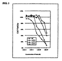
- FIG. 3 depicts a graph comparing the in vitro toxicity of several MPPs according to embodiments of the invention to that of proaerolysin.
- the MPPs are derived from proaerolysin, and include a PSA cleavage site in place of the native furin site.
- FIGS. 4A-4E are schematic drawings (not to scale) showing how a proaerolysin protein can be altered to generate several different MPPs derived from proaerolysin according to embodiments of the present invention.
- the “*” symbol represents one or more point mutations, and/or one or more deletions which decrease proaerolysin binding domain function (i.e. the ability to concentrate in a cell membrane).
- FIG. 4A represents a schematic drawing of a wild-type proaerolysin.
- FIG. 4B represents a schematic drawing of an MPP derived from proaerolysin, with an activation sequence modified to include a prostate-specific protease cleavage site.
- FIG. 4C represents a schematic drawing of an MPP derived from proaerolysin, with an activation sequence modified to include one or more prostate-specific protease cleavage sites.
- FIG. 4D represents a schematic drawing of an MPP derived from proaerolysin, with an activation sequence modified to include a prostate-specific protease cleavage site and with a functionally deleted native binding domain.
- the functionally deleted native binding domain is generated by one or more point mutations or one or more deletions.
- FIG. 4E represents a schematic drawing of an MPP derived from proaerolysin, with an activation sequence modified to include a prostate-specific protease cleavage site and with a functionally replaced native binding domain.
- the functionally deleted native binding domain is generated as described for FIG. 4D .
- One or more prostate-specific targeting domains may be attached at the N-terminus of the MPP, or at the C-terminal end of the toxin domain of the MPP, in this embodiment.
- FIG. 5A represents a schematic drawing of an MPP derived from proaerolysin, with an activation sequence modified to include a prostate-specific protease cleavage site and with a functionally replaced native binding domain.
- the native binding domain is modified by one or more point mutations or one or more deletions.
- One or more prostate-specific targeting domains can be optionally attached to the MPP at Y215C, or A300C.
- FIG. 5B represents a schematic drawing of an MPP derived from proaerolysin, with an activation sequence modified to include a prostate-specific protease cleavage site and with a functionally deleted native binding domain.
- the native binding domain is functionally deleted by deletion of one of the native binding domains of proaerolysin.
- FIG. 5C represents a schematic drawing of an MPP derived from proaerolysin, with an activation sequence modified to include a prostate-specific protease cleavage site and with a functionally replaced native binding domain.
- One or more prostate-specific targeting domains may be attached to either the N-terminus of the toxin domain of the MPP, or to the C-terminal end of the toxin domain of the MPP in this embodiment.
- One of the native binding domains of the MPP is deleted as described in FIG. 5B .
- FIG. 5D represents a schematic drawing of an MPP derived from proaerolysin, with an activation sequence modified to include a prostate-specific protease cleavage site and with a functionally replaced native binding domain.
- One or more prostate-specific targeting domains may be attached to the MPP at Y215C, or A300C.
- One of the native binding domains of the MPP is deleted as described in FIG. 5B .
- FIG. 6A represents a schematic drawing of an MPP according to one embodiment of the invention derived from proaerolysin with a functionally replaced native binding domain.
- the MPP further comprises one or more prostate-specific targeting domains.
- a native binding domain of the MPPP is functionally deleted by mutation or deletion of one or more amino acid residues.
- FIG. 6B represents a schematic drawing of an MPP according to one embodiment of the invention derived from proaerolysin with a functionally replaced native binding domain.
- the MPP further comprises one or more prostate-specific targeting domains attached to proaerolysin at Y215C or A300C.
- a native binding domain of the MPPP is functionally deleted by mutation or deletion of one or more amino acid residues.
- FIG. 6C represents a schematic drawing of an MPP according to one embodiment of the invention derived from proaerolysin with a functionally replaced native binding domain.
- the MPP further comprises one or more prostate-specific targeting domains.
- the native binding domain is functionally deleted by deletion of one of the native binding domains of proaerolysin.
- FIG. 6D represents a schematic drawing of an MPP according to another embodiment of the invention derived from proaerolysin with a functionally replaced native binding domain.
- the MPP further comprises one or more prostate-specific targeting domains attached to proaerolysin at Y215C or A300C.
- the native binding domain is functionally deleted by deletion of one of the native binding domains of proaerolysin.
- FIG. 7 depicts a wild-type proaerolysin cDNA sequence (SEQ ID NO:1).
- FIG. 8 depicts a wild-type proaerolysin amino acid sequence (SEQ ID NO:2).
- FIG. 9 depicts the cDNA sequence (SEQ ID NO:3) of an MPP according to one embodiment of the invention (MPP1), wherein the furin site of proaerolysin has been replaced with a PSA cleavage site.
- FIG. 10 depicts the amino acid sequence (SEQ ID NO:4) of an MPP according to one embodiment of the invention (MPP1), wherein the furin site of proaerolysin has been replaced with a PSA cleavage site.
- FIG. 11 depicts the amino acid sequence (SEQ ID NO:5) of a PSA cleavage site found in human semenogelin I and II proteins.
- FIG. 12 depicts the cDNA sequence (SEQ ID NO:6) of an MPP according to one embodiment of the invention (MPP2), wherein the furin site of proaerolysin has been replaced with a PSA cleavage site.
- FIG. 13 depicts the amino acid sequence (SEQ ID NO:7) of an MPP according to one embodiment of the invention (MPP2), wherein the furin site of proaerolysin has been replaced with a PSA cleavage site.
- FIG. 14 depicts an example of a PSA cleavage site (SEQ ID NO:8).
- FIG. 15 depicts the cDNA sequence (SEQ ID NO:9) of an MPP according to one embodiment of the invention (MPP3), wherein the furin site of proaerolysin has been replaced with a PSA cleavage site.
- FIG. 16 depicts the amino acid sequence (SEQ ID NO:10) of an MPP according to one embodiment of the invention (MPP3), wherein the furin site of proaerolysin has been replaced with a PSA cleavage site.
- FIG. 17 depicts a second example of a PSA cleavage site (SEQ ID NO:11).
- FIG. 18 depicts the cDNA sequence (SEQ ID NO:12) of an MPP according to one embodiment of the invention (MPP4), wherein the furin site of proaerolysin has been replaced with a PSA cleavage site.
- FIG. 19 depicts the amino acid sequence (SEQ ID NO:13) of an MPP according to one embodiment of the invention (MPP4), wherein the furin site of proaerolysin has been replaced with a PSA cleavage site.
- FIGS. 20-27 depict the amino acid sequences of alternative PSA cleavage sites according to the present invention (SEQ ID NOs:14-21, respectively).
- FIG. 28 depicts a native luteinizing hormone releasing hormone (LHRH) amino acid sequence (SEQ ID NO:22).
- FIG. 29 depicts a modified luteinizing hormone releasing hormone (LHRH) amino acid sequence (SEQ ID NO:23).
- FIG. 30 depicts the amino acid sequence (SEQ ID NO:24) of an MPP according to one embodiment of the present invention (MPP6), in which the furin site of proaerolysin has been replaced with a PSA cleavage site, and wherein the native binding domain of proaerolysin has been modified.
- FIG. 31 depicts the amino acid sequence of an MPP according to one embodiment of the present invention (MPP7), in which the furin site of proaerolysin is retained, and the native binding domain of proaerolysin has been deleted and replaced with SEQ ID NO:23 (SEQ ID NO:25).
- FIG. 32 depicts the effects of an MPP according to one embodiment of the invention (MPP5) in the prostate gland of monkeys after treatment for 3 days.
- a and B depict the prostate glands of control monkeys treated with vehicle alone;
- C and D depict the prostate glands of monkeys treated with 1 ⁇ g of the MPP;
- E and F depict the prostate glands of monkeys treated with 5 ⁇ g of the MPP;
- G and H depict the prostate glands of monkeys treated with 25 ⁇ g of the MPP.
- FIG. 33 depicts the effects of an MPP according to one embodiment of the invention (MPP5) in the prostate gland of monkeys treated for 15 days.
- a and B depict the prostate glands of control monkeys treated with vehicle alone;
- C and D depict the prostate glands of monkeys treated with 1 ⁇ g of the MPP;
- E and F depict the prostate glands of monkeys treated with 5 ⁇ g of the MPP;
- G and H depict the prostate glands of monkeys treated with 25 ⁇ g of the MPP.
- FIG. 34 depicts the nucleotide sequence of MPP5 (SEQ ID NO:30).
- the ATG start codon and TAA stop codon are underlined and in bold.
- the HindIII and EcoRI restriction sites are in bold text.
- the PSA cut site is underlined and the 6 His tag is in bold italicized text.
- FIG. 35 depicts the amino acid sequence of MPP5 (SEQ ID NO:31).
- the amino acid sequence was derived from the nucleic acid sequence shown in FIG. 34 .
- Amino acids 427-432 (the PSA cut site) are underlined and in bold.
- the 6 His tag is in bold text.
- FIG. 36 depicts activation of MPP5 in prostate tissue fragment conditioned media assayed by degree of hydrolysis of washed red blood cells.
- FIG. 37 depicts the ability of sera from various species to cleave MPP5.
- FIG. 38 depicts the effect of MPP5 on monkey prostates.
- FIG. 39 depicts the humoral response to administration of MPP5 in monkeys.
- FIG. 40 depicts the nucleotide sequence (SEQ ID NO:73) of a wild-type Clostridium septicum alpha toxin.
- FIG. 41 depicts the amino acid sequence (SEQ ID NO:74) of a wild-type Clostridium septicum alpha toxin.
- the present invention relates to the use of modified pore-forming proteins for the treatment of BPH.
- the MPPs are derived from naturally-occurring pore-forming proteins (nPPs) that kill cells by inserting into the membrane and forming pores or channels in the cell membranes of target cells, resulting in cell death.
- nPPs naturally-occurring pore-forming proteins
- the MPP inserts into the cell membrane, irreversibly, and thus bystander cells are not affected.
- the MPPs comprise prostate-selective modifications that result in the ability of the MPPs to selectively target normal prostate cells relative to cells from other tissues.
- the MPPs are capable of selectively killing normal prostate cells in vivo, and are capable of decreasing the weight or volume of normal prostate gland in vivo.
- the MPPs according to the present invention may be used alone, or in combination with other therapies for the treatment of BPH. This is in contrast to the molecules described in U.S. Patent Application No. 20040235095 which describes the use of modified cytolytic proteins to treat localized or metastatic prostate cancer.
- the term “about” refers to a +/ ⁇ 10% variation from the nominal value. It is to be understood that such a variation is always included in any given value provided herein, whether or not it is specifically referred to.
- prostate-specific indicates that the entity/moiety, or a property of the entity/moiety, is selective to prostate cells when compared to other cell types.
- a prostate specific entity/moiety can be selectively expressed by prostate cells, selectively associated with prostate cells, selectively activated by prostate cells, be capable of selectively binding to prostate cells, or the like.
- prostate-specific activation sequence refers to a sequence of amino acid residues which incorporates one or more prostate-specific protease cleavage sites, which are selectively cleaved or hydrolysed by a prostate-specific protease.
- prostate-specific targeting domain refers to a molecule such as a peptide ligand, toxin, or antibody, which is capable of selectively binding to a prostate cell when compared to its ability to bind to other cell types.
- gene refers to a segment of nucleic acid that encodes an individual protein or RNA (also referred to as a “coding sequence” or “coding region”) together with associated regulatory regions such as promoters, operators, terminators and the like, that may be located upstream or downstream of the coding sequence.
- hybridize refers to the ability of a nucleic acid to bind detectably and specifically to a second nucleic acid.
- Polynucleotides, oligonucleotides and fragments thereof selectively hybridize to target nucleic acid strands under hybridization and wash conditions that minimize appreciable amounts of detectable binding to non-specific nucleic acids.
- High stringency conditions can be used to achieve selective hybridization conditions as known in the art and discussed herein.
- hybridization and washing conditions are performed at high stringency according to conventional hybridization procedures. Washing conditions are typically 1-3 ⁇ SSC, 0.1-1% SDS, 50-70° C. with a change of wash solution after about 5-30 minutes.
- corresponding to indicates that a polynucleotide sequence is identical to all or a portion of a reference polynucleotide sequence.
- the term “complementary to” is used herein to indicate that the polynucleotide sequence is identical to all or a portion of the complementary strand of a reference polynucleotide sequence.
- the nucleotide sequence “TATAC” corresponds to a reference sequence “TATAC” and is complementary to a reference sequence “GTATA.”
- a “reference sequence” is a defined sequence used as a basis for a sequence comparison; a reference sequence may be a subset of a larger sequence, for example, as a segment of a full-length cDNA, gene or protein sequence, or may comprise a complete cDNA, gene or protein sequence.
- a reference polynucleotide sequence is at least 20 nucleotides in length, and often at least 50 nucleotides in length.
- a reference polypeptide sequence is generally at least 7 amino acids in length and often at least 17 amino acids in length.
- a “window of comparison”, as used herein, refers to a conceptual segment of the reference sequence of at least 15 contiguous nucleotide positions or at least 5 contiguous amino acid positions over which a candidate sequence may be compared to the reference sequence and wherein the portion of the candidate sequence in the window of comparison may comprise additions or deletions (i.e. gaps) of 20 percent or less as compared to the reference sequence (which does not comprise additions or deletions) for optimal alignment of the two sequences.
- the present invention contemplates various lengths for the window of comparison, up to and including the full length of either the reference or candidate sequence.
- Optimal alignment of sequences for aligning a comparison window may be conducted using the local homology algorithm of Smith and Waterman ( Adv. Appl. Math .
- sequence identity means that two polynucleotide or polypeptide sequences are identical (i.e. on a nucleotide-by-nucleotide or amino acid-by-amino acid basis) over the window of comparison.
- percent (%) sequence identity is defined as the percentage of nucleotide or amino acid residues in a candidate sequence that are identical with the residues in the reference polypeptide sequence over the window of comparison after optimal alignment of the sequences and introducing gaps, if necessary, to achieve the maximum percent sequence identity, without considering any conservative substitutions as part of the sequence identity.
- substantially identical denotes a characteristic of a polynucleotide or polypeptide sequence, wherein the polynucleotide or polypeptide comprises a sequence that has at least 50% sequence identity as compared to a reference sequence over the window of comparison.
- Polynucleotide and polypeptide sequences which have at least 60% sequence identity, at least 70% sequence identity, at least 80% sequence identity, or at least 90% sequence identity as compared to a reference sequence over the window of comparison are also considered to have substantial identity with the reference sequence.
- a mutation denotes a mutation, partial or complete deletion, insertion, or other variation made to a gene sequence which renders that part of the gene sequence non-functional.
- functional deletion of a proaerolysin (PA) binding domain results in a decrease in the ability of PA to bind to and concentrate on the cell membrane.
- This functional deletion can be reversed by inserting another functional binding domain into proaerolysin, such as a prostate-specific targeting domain, for example, an LHRH peptide.
- functional replacement Such reversal of a functional deletion is referred to herein as “functional replacement.”
- functional deletion of a native PA furin cleavage site results in a decrease in the ability of PA to be cleaved and activated by furin, when compared to a wild-type PA molecule.
- therapy and treatment refer to an intervention performed with the intention of improving a subject's status.
- the improvement can be subjective or objective and is related to ameliorating the symptoms associated with, preventing the development of, or altering the pathology of a disease or disorder being treated.
- therapy and treatment are used in the broadest sense, and include the prevention (prophylaxis), moderation, reduction, and curing of a disease or disorder at various stages. Preventing deterioration of a subject's status is also encompassed by the term.
- Subjects in need of therapy/treatment thus include those already having the disease or disorder as well as those prone to, or at risk of developing, the disease or disorder and those in whom the disease or disorder is to be prevented.
- ameliorate includes the arrest, prevention, decrease, or improvement in one or more the symptoms, signs, and features of the disease or disorder being treated, both temporary and long-term.
- subject or “patient” as used herein refers to an animal in need of treatment.
- animal refers to both human and non-human animals, including, but not limited to, mammals, birds and fish.
- Administration of the proteins or polypeptides of the invention “in combination with” one or more further therapeutic agents or additional treatment is intended to include simultaneous (concurrent) administration and consecutive administration. Consecutive administration is intended to encompass administration of the therapeutic agent(s) or additional treatment and the compound(s) of the invention to the subject in various orders and via various routes.
- antigenic material are used interchangeably herein to refer to a molecule, molecules, a portion or portions of a molecule, or a combination of molecules, up to and including whole cells and tissues, which are capable of inducing an immune response in an animal.
- the antigenic material may comprise a single epitope or antigenic determinant or it may comprise a plurality of epitopes or antigenic determinants.
- immune response refers to an alteration in the reactivity of the immune system of an animal in response to an antigen or antigenic material and may involve antibody production, induction of cell-mediated immunity, complement activation and/or development of immunological tolerance.
- inhibitor means to decrease, reduce, slow-down or prevent.
- Binding pair refers to two moieties (e.g. chemical or biochemical) that have an affinity for one another.
- binding pairs include homo-dimers, hetero-dimers, antigen/antibodies, lectin/avidin, target polynucleotide/probe, oligonucleotide, antibody/anti-antibody, receptor/ligand, enzyme/ligand and the like.
- One member of a binding pair refers to one moiety of the pair, such as an antigen or ligand.
- isolated polynucleotide refers to a polynucleotide of genomic, cDNA, or synthetic origin or some combination thereof, which by virtue of its origin the “isolated polynucleotide” (1) is not associated with the cell in which the “isolated polynucleotide” is found in nature, or (2) is operably linked to a polynucleotide which it is not linked to in nature.
- polypeptide is used herein as a generic term to refer to an amino acid sequence of at least 20 amino acids in length that can be a wild-type (naturally-occurring) protein sequence, a fragment of a wild-type protein sequence, a variant of a wild-type protein sequence, a derivative of a wild-type protein sequence, or an analogue of a wild-type protein sequence.
- native protein sequences and fragments, variants, derivatives and analogues of native protein sequences, as defined herein are considered to be species of the polypeptide genus.
- isolated polypeptide refers to a polypeptide which by virtue of its origin is not associated with other polypeptides with which it is normally associated with in nature, and/or is isolated from the cell in which it normally occurs and/or is free of other polypeptides from the same cellular source and/or is expressed by a cell from a different species, and/or does not occur in nature.
- a polypeptide or polynucleotide sequence that is present in an organism (including viruses) that can be isolated from a source in nature and which has not been intentionally modified by man in the laboratory is naturally-occurring.
- “Operably linked” refers to a juxtaposition wherein the components so described are in a relationship permitting them to function in their intended manner.
- a control sequence “operably linked” to a coding sequence is ligated in such a way that expression of the coding sequence is achieved under conditions compatible with the control sequences.
- Control sequence refers to polynucleotide sequences which are necessary to effect the expression of coding and non-coding sequences to which they are ligated. The nature of such control sequences differs depending upon the host organism; in prokaryotes, such control sequences generally include promoter, ribosomal binding site, and transcription termination sequence; in eukaryotes, generally, such control sequences include promoters and transcription termination sequences.
- control sequences is intended to include, at a minimum, components whose presence can influence expression, and can also include additional components whose presence is advantageous, for example, leader sequences and fusion partner sequences.
- Polynucleotide refers to a polymeric form of nucleotides of at least 10 bases in length, either ribonucleotides or deoxynucleotides or a modified form of either type of nucleotide.
- the term includes single and double stranded forms of DNA or RNA.
- Polypeptide fragment refers to a polypeptide that has an amino-terminal and/or carboxy-terminal deletion, but where the remaining amino acid sequence is usually identical to the corresponding positions in the naturally-occurring sequence deduced, for example, from a full-length cDNA sequence. Fragments typically are at least 5, 6, 8 or 10 amino acids long. In one embodiment, a fragment is at least 14 amino acids long. In another embodiment, a fragment is at least 20 amino acids long. In still another embodiment, a fragment is at least 50 amino acids long. In yet another embodiment the fragment is at least 70 amino acids long.
- label refers to incorporation of a detectable marker, e.g., by incorporation of a radiolabeled amino acid or attachment to a polypeptide of biotinyl moieties that can be detected by marked avidin (e.g., streptavidin containing a fluorescent marker or enzymatic activity that can be detected by optical or colorimetric methods).
- marked avidin e.g., streptavidin containing a fluorescent marker or enzymatic activity that can be detected by optical or colorimetric methods.
- Various methods of labeling polypeptides and glycoproteins are known in the art and may be used.
- labels for polypeptides include, but are not limited to, the following: radioisotopes (e.g., 3 H, 14 C, 35 S, 125 I, 131 I), fluorescent labels (e.g., FITC, rhodamine, lanthanide phosphors), enzymatic labels (or reporter genes) (e.g., horseradish peroxidase, ⁇ -galactosidase, ( ⁇ -latamase, luciferase, alkaline phosphatase), chemiluminescent, biotinyl groups, predetermined polypeptide epitopes recognized by a secondary reporter (e.g., leucine zipper pair sequences, binding sites for secondary antibodies, metal binding domains, epitope tags).
- labels are attached by spacer arms of various lengths to reduce potential steric hindrance.
- peptide sequences set out herein are written according to the generally accepted convention whereby the N-terminal amino acid is on the left and the C-terminal amino acid is on the right.
- L-amino acids are represented by upper case letters and D-amino acids by lower case letters.
- MPPs Modified Pore-forming Proteins
- the modified pore-forming proteins (MPPs) of the present invention are derived from naturally-occurring pore-forming proteins (nPPs), and have been modified to include one or more prostate-selective modifications such that they are capable of selectively killing normal prostate cells relative to cells from other normal tissues.
- nPPs naturally-occurring pore-forming proteins
- prostate-selective modifications such that they are capable of selectively killing normal prostate cells relative to cells from other normal tissues.
- selective killing of normal prostate cells relative to cells from other normal tissues is meant that the MPPs are capable of killing normal prostate cells more effectively than other types of normal cells such as, for example, lung, spleen, or blood cells.
- Suitable MPPs include those described in United States Patent Application No. 20040235095.
- nPPs Naturally-Occurring Pore-forming Proteins
- Suitable mPPs from which the MPPs of the present invention can be derived include various bacterial toxins that are capable of forming pores or channels in the membrane of a target cell leading to cell death.
- Suitable bacterial toxins include those that are produced as protoxins and are subsequently activated by proteolytic cleavage as well as those that are produced in an active from and do not require additional processing.
- the nPPs are large cytotoxic proteins that are synthesized as protoxins which are activated by protease cleavage at an activation sequence to form pores or channels in the cell membrane of target cells, thus leading to rapid cytolytic cell death.
- Suitable nPPs in accordance with this embodiment have the following features: a pore-forming activity that is activated by removal of an inhibitory domain via protease cleavage, and the ability to bind to receptors that are present on cell membranes through one or more binding domains.
- Numerous such nPPs have been cloned and recombinant forms produced (see, for example, Imagawa et al., FEMS. Microbiol. Lett. 17:287-92, 1994; Meza et al. FEMS Microbiol. Lett. 145:333-9, 1996).
- the MPPs are derived from nPPs such as aerolysin or aerolysin-related polypeptides.
- nPPs such as aerolysin or aerolysin-related polypeptides.
- aerolysin homologues such as proaerolysin from Aeromonas hydrophila, Aeromonas trota and Aeromonas salmonicida , and alpha toxin from Clostridium septicum (Ballard et al., Infect. Immun. 63:340-4, 1995; Gordon et al. J. Biol. Chem. 274:27274-80, 1999; Genbank Accession No.
- Bacillus anthraces protective antigen Bacillus anthraces protective antigen, Vibrio cholerae VCC toxin, epsilon toxin from Clostridium perfringens , and Bacillus thuringiensis delta toxins (Genbank Accession No. D00117).
- PA polypeptides from the Aeromonas species noted above have been characterized. These polypeptides exhibit greater than 80% pairwise sequence identity between them (Parker et al., Progress in Biophysics & Molecular Biology 88 (2005) 91-142). Each of these PA polypeptides is an approximately 52 kDa protoxin with approximately 470 amino acid residues.
- the cDNA sequence for wild-type PA from A. hydrophila is shown in SEQ ID NO: 1 ( FIG. 7 ) and the corresponding amino acid sequence of this wild-type PA is shown in SEQ ID NO:2 ( FIG. 8 ).
- SEQ ID NO: 1 The cDNA sequence for wild-type PA from A. hydrophila is shown in SEQ ID NO: 1 ( FIG. 7 ) and the corresponding amino acid sequence of this wild-type PA is shown in SEQ ID NO:2 ( FIG. 8 ).
- the nucleotide and protein sequences for numerous naturally occurring nPPs are known in the art. Non-limiting examples are listed in the following Table:
- the A. hydrophila PA protein includes a binding domain (approximately amino acids 1-83 of SEQ ID NO: 2) in what is known as the small lobe of the polypeptide and referred to herein as the small lobe binding domain (SBD), and a C-terminal inhibitory peptide (CIP) domain (approximately amino acids 427-470 of SEQ ID NO: 2) that is removed by protease cleavage at an activation sequence to activate PA. Cleavage at the activation sequence to remove the CIP domain can be carried out by a number of ubiquitous proteases including furin and trypsin.
- the amino acid residues from approximately 84-426 of SEQ ID NO: 2 are known as the large lobe of the PA polypeptide, and contain a toxin domain and other functional domains, including a second binding domain, referred to herein as the large lobe binding domain (LBD).
- LBD large lobe binding domain
- SEQ ID NO: 1 The amino acid residues from approximately 84-426 of SEQ ID NO: 2 are known as the large lobe of the PA polypeptide, and contain a toxin domain and other functional domains, including a second binding domain, referred to herein as the large lobe binding domain (LBD).
- LBD large lobe binding domain
- Alpha toxin from C. septicum is considered to be a homologue of proaerolysin based on significant sequence identity and other similarities (Parker et al., supra).
- Alpha toxin is secreted as a 46,450 Da protoxin (approximately 443 amino acids) that is activated by protease cleavage at an activation sequence to remove a C-terminal inhibitory peptide (CIP) domain, and it also binds to glycosyl-phosphatidylinositol (GPI)-anchored proteins.
- CIP C-terminal inhibitory peptide
- GPI glycosyl-phosphatidylinositol
- Activation of this polypeptide occurs by protease cleavage at a furin cleavage site (Gordon et al., Infect. Immun. 65:4130-4, 1997).
- An example of a Clostridium septicum alpha toxin nucleic acid sequence is provided in GenBankTM Accession No. S75954 (SEQ ID NO:73, FIG. 40 ), and an example of a Clostridium septicum alpha toxin protein sequence is provided in GenBankTM Accession No. AAB32892 (SEQ ID NO:74, FIG. 41 ). Based on the sequence homology, alpha toxin is thought to have a similar structure and similar ability to bind to GPI-anchored proteins.
- Bacillus thuringiensis delta-toxin The activation sequence of Bacillus thuringiensis delta-toxin is cleaved by proteases in the midgut of certain insects to produce active endotoxin (Miranda et al., Insect Biochem. Mol. Biol. 31:1155-63, 2001).
- the structure of this endotoxin has been solved and shown to consist of three domains, a channel-forming domain, a binding domain, and a stabilizing domain.
- the MPPs according to the present invention are derived from proaerolysin polypeptides. In a further embodiment, the MPPs are derived from proaerolysin polypeptides from A. hydrophila . In another embodiment of the invention, the MPPs are derived from alpha toxin polypeptides.
- the MPPs are derived from nPPs that do not require protease cleavage for activation, and thus do not have an activation sequence.
- These nPPs can be modified to insert a prostate-specific protease cleavage site into the nPP resulting in an MPP that is capable of being selectively activated to kill prostate cells.
- examples of such nPPs include Staphylococcus aureus a hemolysin.
- an activation sequence can be inserted inot the center of the pore-forming domain as known in the art (Panchal et al., (1996) Nat. Biotech. 14:852-856).
- the present invention further includes MPPs that are derived from biologically active fragments of nPPs.
- Biologically active fragments of nPPs are those that are capable of forming pores and killing cells.
- Suitable fragments include those that are capable of being activated to form pores in target cells by removal of a CIP domain.
- a suitable fragment would be one that comprised a binding domain of the protein as well as the CIP domain and activation sequence.
- the MPP is derived from a fragment of proaerolysin that includes a binding domain, the CIP domain and the activation sequence.
- the MPP is derived from a fragment of proaerolysin that comprises the binding domain, the activation sequence, but only part of a CIP domain.
- the selected nPP is modified to form a MPP by inclusion of one or more prostate-specific modifications.
- Prostate-specific modifications contemplated by the present invention include incorporation of a prostate-specific activation sequence and/or functional deletion (including functional replacement) of one or more binding domains, and/or addition of a prostate-specific targeting domain.
- the MPPs according to the present invention comprise a prostate-specific activation sequence that allows for selective activation of the MPPs in prostate cells.
- a prostate-specific activation sequence may be generated by modification of the naturally-occurring activation sequence of a nPP, or it may be generated by the addition of a prostate-specific activation sequence to a nPP that does not have a naturally-occurring activation sequence.
- the MPPs comprise a prostate-specific activation sequence and one or more prostate-specific targeting domains.
- the MPPs comprise a prostate-specific activation sequence and a modification to the SBD.
- the MPPs comprise a prostate-specific activation sequence and a modification to the LBD.
- the MPPs according to the present invention comprise one or more prostate-specific targeting domains that allow for selective activation of the MPPs in prostate cells.
- the MPPs comprise one or more prostate-specific targeting domain and a modification to the SBD.
- the MPPs comprise a prostate-specific targeting domain and a modification to the LBD.
- the MPPs comprise a prostate-specific activation sequence, one or more prostate-specific targeting domain and a modification to the LBD. In another embodiment, the MPPs comprise a prostate-specific activation sequence, one or more prostate-specific targeting domains, and a modification to the SBD.
- the MPP comprises a prostate-specific activation sequence and one or more modifications to the native binding domain. In another embodiment, the MPP comprises a prostate-specific targeting domain and one or more modifications to the native binding domain. In still another embodiment, the MPP comprises a prostate-specific activation sequence, a prostate-specific targeting domain, and one or more modifications to the native binding domain.
- FIGS. 4 , 5 , and 6 Representative, non-limiting examples of combinations of prostate-specific modifications that can be made to proaerolysin are shown in FIGS. 4 , 5 , and 6 .
- a nPP can be modified to incorporate a prostate-specific activation sequence by modification of the naturally occurring activation sequence to provide a prostate-specific activation sequence, or a prostate-specific activation sequence can be added to an nPP that does not have a naturally occurring activation sequence.
- a prostate-specific activation sequence is accordance with the present invention is a sequence of amino acids that incorporates one or more prostate-specific protease cleavage sites.
- a prostate-specific protease cleavage site is a sequence of amino acids which is recognized and selectively and efficiently hydrolyzed (cleaved) by a prostate-specific protease.
- a prostate-specific protease is a protease that is expressed at higher levels in prostate cells than in other cell types.
- prostate-specific proteases include, but are not limited to: PSA (prostate-specific antigen), PSMA (prostate-specific membrane antigen), and HK2 (human glandular kallikrein 2) cleavage sequences. Numerous examples of cleavage sites recognized by these prostate-specific proteases are known in the art and will be described further below.
- Modifications to the naturally-occurring activation sequence to provide a prostate-specific protease activation sequence may be achieved as is known in the art. Modification of the naturally occurring activation sequence results in functional deletion of the native activation sequence. Functional deletion can be achieved by mutation, partial or complete deletion, insertion, or other variation made to the naturally occurring activation sequence that renders it inactive.
- the naturally-occurring activation sequence of the nPP is functionally deleted by insertion of a prostate-specific activation sequence.
- functional deletion of the naturally occurring activation sequence is achieved via mutations in one or more amino acid residues of the native activation sequence which produce a prostate-specific activation sequence.
- the naturally occurring activation sequence of the nPP is functionally deleted by replacing the native protease cleavage site of the activation sequence with a prostate-specific protease cleavage site.
- the one or more prostate-specific protease cleavage sites functionally replace the native protease cleavage site of the MPP.
- a prostate-specific protease cleavage site can functionally replace the native furin cleavage site of PA (see FIG. 4B ).
- This replacement results in a MPP that becomes cytolytically active in the presence of an enzymatically active prostate-specific protease, such as PSA, PSMA, or HK2.
- PSA, PSMA, or HK2 cleavage sites are known in the art and are described below.
- the MPPs according to the present invention can be generated by deleting the native protease cleavage site of the nPP and inserting a prostate-specific activation sequence.
- a prostate-specific activation sequence For example the furin cleavage site of PA (amino acids 427-432 of SEQ ID NO: 2) can be deleted and a prostate-specific protease cleavage site, such as a PSA cleavage site, inserted (see FIG. 4B ).
- the native protease cleavage site of the nPP is mutated such that it is no longer functional and a prostate-specific activation sequence is inserted within the mutated protease cleavage site, or added to the N- or C-terminus of the native protease cleavage site.
- a prostate-specific protease cleavage site such as a PSA cleavage site, inserted within, or added to the N- or C-terminus of the mutated furin site (see FIG. 4C ).
- a prostate-specific activation sequence is added to an nPP that does not have a naturally occurring activation sequence.
- Staphylococcus aureus ⁇ -hemolysin which does not require protease cleavage in order to be activated to kill cells, may be engineered to include one or more prostate-specific protease cleavage sites, thus rendering it capable of being selectively activated to kill prostate cells.
- prostate-specific proteases and the protease cleavage sites they recognize are known in the art. Examples include, but are not limited to, PSA, PSMA and HK2.
- the MPP is modified to include a prostate-specific activation sequence that includes a PSA-specific cleavage site.
- a PSA-specific cleavage site is a sequence of amino acids which is recognized and selectively and efficiently hydrolyzed (cleaved) by prostate specific antigen (PSA).
- PSA prostate specific antigen
- PSA is a serine protease with the ability to recognize and hydrolyze specific peptide sequences. It is secreted by prostate cells in an enzymatically active form and becomes inactivated upon entering the circulation. Since neither blood nor normal tissue other than the prostate contains enzymatically active PSA, the proteolytic activity of PSA can be used to activate MPPs at the prostate gland.
- PSA-specific cleavage sites are known in the art.
- the MPP has an activation sequence that includes the PSA cleavage site shown in SEQ ID NO: 5.
- PSA-specific cleavage sites are known, based on the PSA-cleavage map of human seminal proteins semenogelin I and II, and a cellulose membrane based assay (see Table 3 and Denmeade et al., Cancer Res., 57:4924-30, 1997) and can be used to produce the modified MPPs according to the present invention.
- the MPPs according to the present invention can be modified to include one of the PSA-cleavage sites as shown in Table 3, which can substitute for the wild-type furin protease activation site of proaerolysin (amino acids 427-432 of SEQ ID NO: 2), as is known in the art.
- the MPP has an amino acid sequence of any one of SEQ ID NOs: 3, 4, 6, 7, 9, 10, 12, 13, and 24, which include an activation sequence containing a PSA cleavage site.
- PSA substrates PSA cleavage sites
- kinetics of PSA hydrolysis PSA substrate (SEQ ID NO) K m ( ⁇ M) K cat (s ⁇ 1 ) K cat /K m (s ⁇ 1 M ⁇ 1 ) KGISSQY (15) 160 0.043 270 SRKSQQY (16) 90 0.023 260 ATKSKQH (17) 1310 0.0091 6.9 KGLSSQC (18) 300 0.0017 5.6 LGGSSQL (19) 900 0.0037 4.1
- the MPP comprises a prostate-specific activation sequence that includes a PSMA-specific cleavage site.
- PSMA-specific cleavage sites are known in the art and can be found, for example, in International Publication No. WO 02/43773.
- a PSMA cleavage site includes at least the dipeptide X 1 X 2 .
- the dipeptide contains the amino acids Glu or Asp at position X 1 .
- X 2 can be Glu, Asp, Gln, or Asn.
- Tripeptides X 1 X 2 X 3 are also suitable, with X 1 and X 2 defined as before, with X 3 as Glu, Asp, Gln or Asn.
- Tetrapeptides X 1 X 2 X 3 X 4 are also suitable, with X 1-3 defined as above, and with X 4 as Glu, Asp, Gln or Asn.
- Pentapeptides X 1 X 2 X 3 X 4 X 5 are also suitable, with X 1-4 defined as above, and with X 5 as Glu, Asp, Gln or Asn.
- Hexapeptides X 1 X 2 X 3 X 4 X 5 X 6 are also suitable, with X 1-5 defined as above, and with X 6 as Glu, Asp, Gln or Asn. Further peptides of longer sequence length can be constructed in similar fashion.
- the peptides are of the following sequence: X 1 . . . X n , where n is 2 to 30, 2 to 20, 2 to 15, or 2 to 6, where X 1 is Glu, Asp, Gln or Asn.
- X 1 is Glu or Asp
- X 2 -X n are independently selected from Glu, Asp, Gln and Asn.
- Other possible peptide sequences are as above, except that X 2 -X n-1 are independently selected from Glu, and Asp, and X n is independently selected from Glu, Asp, Gln and Asn.
- PSMA cleavage sites are Asp-Glu, Asp-Asp, Asp-Asn, Asp-Gln, Glu-Glu-Glu, Glu-Asp-Glu, Asp-Glu-Glu, Glu-Glu-Asp, Glu-Asp-Asp, Asp-Glu-Asp, Asp-Asp-Glu, Asp-Asp-Asp, Glu-Glu-Gln, Glu-Asp-Gln, Asp-Glu-Gln, Glu-Glu-Asn, Glu-Asp-Asn, Asp-Glu-Asn, Asp-Asp-Gln, and Asp-Asp-Asn.
- the MPP comprises a prostate-specific activation sequence that includes an HK2-specific cleavage site.
- HK2-specific cleavage sites are also known in the art and described, for example, in International Publication No. WO01/09165.
- the cleavage site recognized by HK2 is flanked by at least an amino acid sequence X 4 X 3 X 2 X 1 .
- This amino acid sequence contains the amino acid arginine, histidine or lysine at position X 1 .
- X 2 can be arginine, phenylalanine, lysine, or histidine.
- X 3 can be lysine, serine, alanine, histidine or glutamine.
- X 4 can be from 0 to 20 further amino acids, and can be at least two further amino acids.
- the HK2 cleavage site includes a sequence for X 4 that is substantially identical to the 20 amino acids in the wild type semenogelin I or semenogelin II sequence that are the from fourth to twenty fourth amino acids to the N-terminal side of recognized semenogelin cleavage sites.
- the amino acid sequence can further comprise X 1 , which is linked to the carboxy terminus of X 1 to create the amino acid sequence X 4 X 3 X 2 X 1 X 1 .
- X 1 is up to a further 10 amino acids, and can include various amino acids.
- X 1 may have a leucine, alanine or serine linked to the carboxy terminus of X 1 .
- X 1 can include L- or D-amino acids.
- the HK2 cleavage site is located at the carboxy terminal side of X 1 .
- HK2 cleavage sites are shown in Table 4 (Note that the symbol][denotes an Hk2 cleavage site):
- the MPPs comprise one or more prostate-specific targeting domains to allow selective targeting of prostate cells.
- the prostate-specific targeting domain is capable of directing the MPP to the prostate cell, where the MPP can be activated and subsequently kill the prostate cell.
- the targeting domain can be located at the N- or C-terminus of the MPP, or both. Alternatively, the targeting domain can located at another region of the MPP, as long as it does not interfere with the pore-forming activity of the MPP.
- prostate-specific targeting domains include, but are not limited to molecules such as a peptide ligand, toxin, or antibody, which have a higher specificity for prostate cells than for other cell types.
- a prostate tissue specific binding domain has a lower K D in prostate tissue or cells than in other cell types, (i.e. binds selectively to prostate tissues as compared to other normal tissues), for example at least a 10-fold lower K D , such as an at least 20-, 50-, 75-, 100- or even 200-fold lower K D .
- Such molecules can be used to target a MPP to the prostate.
- Examples include, but are not limited to: antibodies which recognize proteins that are relatively prostate-specific such as PSA, PSMA, HK2, prostasin, and hepsin; ligands which have prostate-specific receptors such as natural and synthetic luteinizing hormone releasing hormone (LHRH); and endothelin (binding to cognate endothelin receptor).
- addition of the prostate-specific targeting domain results in functional deletion of the native binding domain of the nPP.
- the native non-specific GPI-anchor protein binding domain of proaerolysin is functionally deleted and replaced with a prostate-specific targeting domain.
- Specific examples of MPPs derived from proaerolysin that have a functionally deleted native binding domain are depicted in FIGS. 4D and 5B .
- Examples of MPPs derived from proaerolysin that include a prostate-specific targeting domain which functionally substitutes for the native proaerolysin binding domain are shown in FIGS. 4E , 5 A, 5 C and 5 D and 6 A- 6 D.
- One or more prostate tissue-specific binding domains can be linked to one or more amino acids of the MPPs, but ideally, do not interfere significantly with the ability to form pores in cell membranes, or, where applicable, with the ability of the MPP to be activated by a prostate-specific protease such as PSA.
- Methods of conjugating proteins or peptides to MPPs are known in the art and include for example, changing the N-terminal amino acid of the protein to be modified to a Cys or other amino acid before attaching the prostate-tissue specific binding domain, to assist in linking the prostate-tissue specific binding domain to the MPP.
- prostate tissue specific binding domains are linked or inserted at the N-and/or C-terminus of an MPP derived from proaerolysin (for example, see FIGS. 4E and 5C ).
- the native binding domain of proaerolysin is deleted (i.e. amino acids 1-83 of SEQ ID NO: 2 or 4), such that attachment or linking of a prostate tissue specific binding domain to the N-terminus results in attachment to amino acid 84 of SEQ ID NO: 2 or 4 (for example, see FIGS. 5C and 6C ).
- smaller deletions or point mutations are introduced into the native binding domain of proaerolysin, such that attachment or linking of a prostate tissue specific binding domain to the N-terminus results in attachment to amino acid 1 of SEQ ID NO: 2 or 4 (or whichever amino acid is N-terminal following functional deletion of the native proaerolysin binding domain) (for example, see FIGS. 4E and 5D ).
- the prostate specific targeting domain is an antibody or antibody fragment that specifically binds to an antigen that is associated with prostate cells, thus targeting the MPP to prostate cells.
- Antigens associated with prostate cells that may be specifically bound by such prostate-specific targeting domains include PSA, and PSMA and the LHRH receptor, the expression of which is elevated in prostate cells.
- Antibodies can be attached to the N- or C-terminus of the MPP using gene fusion methods well known in the art (for example see Debinski and Pastan, Clin. Cancer Res. 1:1015-22, 1995).
- antibodies can be attached to an MPP by covalent crosslinking (for example see Woo et al., Arch. Pharm. Res. 22(5):459-63, 1999 and Debinski and Pastan, Clin.
- Crosslinking can be non-specific, for example by using a homobifunctional-lysine-reactive crosslinking agent, or it can be specific, for example by using a crosslinking agent that reacts with amino groups on the antibody and with cysteine residues located in the MPP.
- proaerolysin amino acids such as amino acids Cys19, Cys75, Cys159, and/or Cys164 of SEQ ID NO: 2 can be used to crosslink antibodies to the modified proaerolysin molecule.
- the antibody could replace the native binding domain of the nPP to be modified, or the antibody could be added to an MPP already having mutations in the native binding domain.
- Such an MPP can also include a prostate-specific activation sequence to increase specificity.
- the antibody is a single chain antibody to PSMA fused to the toxin domain of PA.
- Suitable antibodies include intact antibodies as well as antibody fragments such as, for example, (i) an Fab fragment consisting of the VL, VH, CL and CH1 domains; (ii) an Fd fragment consisting of the VH and CH1 domains; (iii) an Fv fragment consisting of the VL and VH domains of a single arm of an antibody, (iv) a dAb fragment (Ward et al., Nature 341:544-6, 1989) which consists of a VH domain; (v) an isolated complementarity determining region (CDR); and (vi) an F(ab′)2 fragment, a bivalent fragment comprising two Fab fragments linked by a disulfide bridge at the hinge region.
- antibody fragments such as, for example, (i) an Fab fragment consisting of the VL, VH, CL and CH1 domains; (ii) an Fd fragment consisting of the VH and CH1 domains; (iii) an Fv fragment consisting of the VL
- Suitable antibodies include single chain Fv antibodies, which are prepared by recombinant methods resulting in the two domains of an Fv fragment being linked via a synthetic linker (Bird et al. Science 242:423-6, 1988; and Huston et al., Proc. Natl. Acad. Sci. 85:5879-83, 1988), and camelized antibodies (for example see Tanha et al., J. Biol. Chem. 276:24774-80, 2001).
- the antibody fragments are capable of crosslinking their target antigen, e.g., bivalent fragments such as F(ab′)2 fragments.
- an antibody fragment which does not itself crosslink its target antigen e.g., a Fab fragment
- a secondary antibody which serves to crosslink the antibody fragment, thereby crosslinking the target antigen.
- Antibodies can be fragmented using conventional techniques and the fragments screened for utility in the same manner as described for whole antibodies, and as is known in the art.
- An antibody is further intended to include nanobodies, and bispecific and chimeric molecules that specifically bind the target antigen.
- Specifically binds when used in reference to an antibody, refers to the ability of individual antibodies to specifically immunoreact with a specific antigen.
- the binding is a non-random binding reaction between an antibody molecule and an antigenic determinant of the antigen.
- the desired binding specificity is typically determined from the reference point of the ability of the antibody to differentially bind the specific and an unrelated antigen, and therefore distinguish between two different antigens, particularly where the two antigens have unique epitopes.
- An antibody that specifically binds to a particular epitope is referred to as a “specific antibody”.
- the prostate-specific targeting domain is a small peptide ligand that binds to its cognate prostate-specific receptor expressed on the membrane of prostate cells.
- examples include, but are not limited to, natural and synthetic luteinizing hormone releasing hormone (LHRH) agonist peptides (for example see Genbank Accession No. CAA25526 and SEQ ID NOS: 22 and 23), which bind with high affinity to LHRH receptors, and peptides that can bind selectively to PSMA.
- LHRH receptors are displayed by prostate cells, and only a few other cells. This differential expression provides binding specificity.
- Small peptide ligands may be modified as is known in the art in order to facilitate their attachment to the MPP.
- certain residues of LHRH such as the Gly at the 6th position (Gly6), can be substituted without compromising receptor binding affinity (Janaky et al., Proc. Natl. Acad. Sci. USA 89:972-6, 1992; Nechushtan et al., J. Biol. Chem., 272:11597-603, 1997). Therefore, an MPP (in which the native binding domain is functionally deleted) can be produced which is covalently coupled to purified LHRH D-Lys6 (at the epsilon amine of this lysine).
- LHRH D-Lys6 (SEQ ID NO: 23) can be attached at various positions within an nPP to provide an MPP having a prostate-specific targeting domain. As noted above, attachment of the small peptide ligand will not significantly interfere with the ability of the toxin to insert into the membrane to form a pore.
- the epsilon amine of the D-Lys6 analog can be coupled to the amino terminus of the MPP using methods known in the art such as for example via a dicarboxylic acid linker. Activation of the MPP by cleavage of the activation sequence will result in release of the C-terminal inhibitory portion while the toxin remains bound to the LHRH receptor.
- the small peptide ligand can be coupled directly to the C-terminus of the MPP.
- the epsilon amine of the D-Lys6 analog of LHRH can be coupled directly to the C-terminal carboxyl of the MPP by the addition of a Cys to the C-terminus of the MPP, then crosslinking this Cys to the epsilon amine of the D-Lys6 analog of LHRH.
- This coupling will produce an MPP in which the LHRH peptide is attached to the C-terminal inhibitory domain.
- Activation of the MPP by cleavage of the activation sequence will liberate the MPP and leave the inhibitory fragment bound to the LHRH receptor.
- recombinant fusion proteins can be produced in which modified LHRH peptides are fused to both the N- and C-terminus of the MPP.
- the small peptide ligand may be attached to an MPP via a disulfide bridge.
- a cysteine residue is introduced into the 6th position of the LHRH peptide and the peptide attached to an MPP via a disulfide bridge.
- the cysteine with which the peptide forms a disulphide bridge can be present in the native nPP sequence or the nPP can be mutated to include a cysteine residue.
- an MPP derived from PA can have a cysteine residue introduced, for example at amino acids 215 and/or 300 of SEQ ID NO: 2, wherein amino acid 215 and/or 300 has been mutated to a cysteine.
- a recombinant protein is produced in which LHRH peptide is fused to the amino terminus of the MPP.
- an MPP may be produced by attaching or linking one or more prostate-specific targeting domains to other amino acids of the MPP.
- amino acids such as amino acid 215 or 300 of SEQ ID NO: 2 or 4 (for example, see FIGS. 5A , 5 D, 6 B and 6 D) may be used to attach the one or more prostate specific targeting domains.
- a Cys amino acid replaces the native amino acid at that position.
- the following changes can be made to SEQ ID NO: 2 or 4: Tyr215Cys or Ala300Cys.
- cysteine residues present in the native sequence of the nPP can be utilized.
- amino acids such as amino acids Cys19, Cys75, Cys159, and/or Cys164 of SEQ ID NO: 2, are suitable for this purpose.
- the MPP is derived from proaerolysin and has a sequence selected from SEQ ID NOs:24 and 25, which comprises LHRH as a prostate-specific targeting domain.
- MPPs according to the present invention are derived from nPPs that comprise one or more binding domains, as known in the art.
- nPP when an nPP comprises one binding domain, it is considered to be a “large lobe binding domain.”
- MPPs according to the present invention may comprise modifications to one or more binding domains, as applicable.
- native proaerolysin from Aeromonas species comprises two binding domains, a small lobe binding domain, and a large lobe binding domain.
- native alpha toxin from Clostridium septicum comprises only a large lobe binding domain.
- modifications of the binding domains include functional deletion of a binding domain.
- a functionally deleted binding domain in an MPP results in an MPP that has an attenuated ability to bind to its cell surface receptor, yet still retains pore-forming ability.
- Functional deletions can be made by deleting or mutating one or more binding domains of an MPP. In one embodiment, the entire binding domain or portions thereof, may be deleted. In an additional embodiment, insertion of heterologous sequences into the binding domain may also be used to functionally delete the binding domain. Addition of these heterologous sequences may confer an additional functionality to the MPP (i.e. functional replacement of the binding domain). For example, addition of a heterologous sequence can result in the addition of a region that can function as a prostate-specific targeting domain as described herein. In still another embodiment, point mutations to the amino acid sequence of the native binding domain of the nPP can also be made to decrease the ability of the binding domain to bind to its receptor. Further details regarding these modifications are described below.
- MPPs lacking a binding domain retain their cytolytic activity, but may need to be administered at higher doses to ensure concentration of the toxin in the cell membrane.
- MPPs with functional deletions in the binding domain may be prepared using methods known in the art. These methods include the use of recombinant DNA technology as described in Sambrook et al., supra. Alternatively, functional deletions of the binding domain may also be achieved by direct modification of the protein itself according to methods known in the art, such as proteolysis to generate fragments of the MPP, which can then be chemically linked together.
- the MPP is modified by functional deletion of its small lobe binding domain (SBD).
- SBD small lobe binding domain
- Exemplary functional deletions of the SBD may be made in the A. hydrophila proaerolysin polypeptide as follows. The entire SBD, corresponding to amino acid 1-83 of SEQ ID NO:2 may be deleted, or portions of this region may be deleted, for example amino acids 45-66 of SEQ ID NO:2.
- point mutations can be made as follows W45A, I47E, M57A, Y61A, K66Q (amino acid numbers refer to SEQ ID NO: 2 or SEQ ID NO:4) and as described in Mackenzie et al. J. Biol. Chem. 274: 22604-22609, 1999.
- FIG. 4D A schematic diagram representing an example of an MPP with one or more mutations in a binding domain is shown in FIG. 4D , where * represents one or more mutations or deletions.
- the nPP is modified by functional deletion of its large lobe binding domain (LBD).
- LBD large lobe binding domain
- Exemplary functional deletions of the LBD of proaerolysin (contained in approximately amino acid residues 84-426 of SEQ ID NO:2) that may be made to provide MPPs are as follows. The entire LBD of proaerolysin may be deleted.
- the MPP derived from proaerolysin comprises one or more point mutations in the LBD to amino acid residues Y162, W324, R323, R336, and/or W127.
- the MPP derived from proaerolysin comprises one or more point mutations at positions W127 and/or R336.
- the MPP derived from proaerolysin comprises the point mutations Y162A and/or W324A. In a further embodiment the MPP derived from proaerolysin comprises the point mutations R336A, R336c, and/or W127T. In another embodiment, MPPs comprise mutations to other residues that interact directly with the GPI-protein ligand.
- MPPs derived from alpha toxin include at least one substituted amino acid in the receptor binding domains of the alpha toxin which include amino acid residues 53, 54, 62, 84-102, 259-274 and 309-315 of the sequence of the native alpha toxin as shown in SEQ ID NO: 33.
- MPPs derived from alpha toxin include mutations to one or more of the following residues: W85, Y128, R292, Y293, and 8305.
- the present invention contemplates further modification of MPPs that do not affect the ability of the MPPs to selectively target prostate cells.
- modifications include amino acid substitutions, insertions or deletions, modifications to reduce antigenicity, and modifications to enhance the stability or improve the pharmacokinetics of the MPPs.
- further modifications to MPPs result in a polypeptide that differs by only a small number of amino acids from the MPP.
- modifications include deletions (for example of 1-3 or more amino acids), insertions (for example of 1-3 or more residues), or substitutions that do not interfere with the ability of the MPPs to selectively target and kill normal prostate cells.
- further modifications to the MPPs result in a polypeptide that retains at least 70%, 80%, 85%, 90%, 95%, 98%, or greater sequence identity to the MPP and maintains the ability of the MPP to selectively target and kill normal prostate cells.
- MPPs may be modified by substitution whereby at least one residue in the amino acid sequence has been removed and a different residue inserted in its place.
- the substitution is a conservative substitution.
- a conservative substitution is one in which one or more amino acids (for example 2, 5 or 10 residues) are substituted with amino acid residues having similar biochemical properties.
- conservative substitutions have little to no impact on the activity of a resulting polypeptide.
- an MPP including one or more conservative substitutions retains the activity of the corresponding nPP.
- amino acids which may be substituted for an original amino acid in a protein and which are regarded as conservative substitutions include: Ser for Ala; Lys for Arg; Gln or His for Asn; Glu for Asp; Ser for Cys; Asn for Gln; Asp for Glu; Pro for Gly; Asn or Gln for His; Leu or Val for Ile; Ile or Val for Leu; Arg or Gln for Lys; Leu or Ile for Met; Met, Leu or Tyr for Phe; Thr for Ser; Ser for Thr; Tyr for Trp; Trp or Phe for Tyr; and Ile or Leu for Val.
- An MPP can be modified to include one or more conservative substitutions by manipulating the nucleotide sequence that encodes that polypeptide using, for example, standard procedures such as site-directed mutagenesis or PCR. Further information about conservative substitutions can be found in, among other locations, Ben-Bassat et al., (J. Bacteriol. 169:751-7, 1987), O'Regan et al., (Gene 77:237-51, 1989), Sahin-Toth et al., (Protein Sci. 3:240-7, 1994), Hochuli et al., (Bio/Technology 6:1321-5, 1988), WO 00/67796 (Curd et al.) and in standard textbooks of genetics and molecular biology.
- substitution is a permissive substitution.
- Permissive substitutions are non-conservative amino acid substitutions, but also do not significantly alter MPP activity.
- An example is substitution of Cys for Ala at position 300 of SEQ ID NO: 2 or 4 in a proaerolysin polypeptide.
- MPPs are modified to include 1 or more amino acid substitutions of single residues. In another embodiment, the MPPs are modified to include 1 amino acid substitution. In another embodiment, the MPPs are modified to include from about 2 to about 10 amino acid substitutions. In another embodiment, the MPPs are modified to include about 3 to about 5 amino acid substitutions.
- Peptidomimetic and organomimetic embodiments are also contemplated, whereby the three-dimensional arrangement of the chemical constituents of such peptido- and organomimetics mimic the three-dimensional arrangement of the polypeptide backbone and component amino acid side chains in the polypeptide, resulting in such peptido- and organomimetics of an MPP which have the ability to lyse prostate cells.
- a pharmacophore is an idealized, three-dimensional definition of the structural requirements for biological activity.
- Peptido- and organomimetics can be designed to fit each pharmacophore with current computer modeling software (using computer assisted drug design or CADD).
- MPPs modifications that may be made to the MPPs include, for example, modifications to the carboxylic acid groups of the MPP, whether carboxyl-terminal or side chain, in which these groups are in the form of a salt of a pharmaceutically-acceptable cation or esterified to form a C 1 -C 16 ester, or converted to an amide of formula NR 1 R 2 wherein R 1 and R 2 are each independently H or C 1 -C 16 alkyl, or combined to form a heterocyclic ring, such as a 5- or 6-membered ring.
- Amino groups of the polypeptide can be in the form of a pharmaceutically-acceptable acid addition salt, such as the HCl, HBr, acetic, benzoic, toluene sulfonic, maleic, tartaric and other organic salts, or may be modified to C 1 -C 16 alkyl or dialkyl amino or further converted to an amide.
- a pharmaceutically-acceptable acid addition salt such as the HCl, HBr, acetic, benzoic, toluene sulfonic, maleic, tartaric and other organic salts
- hydroxyl groups of the polypeptide side chain can be converted to C 1 -C 16 alkoxy or to a C 1 -C 16 ester using well-recognized techniques.
- Phenyl and phenolic rings of the polypeptide side chain can be substituted with one or more halogen atoms, such as F, Cl, Br or I, or with C 1 -C 16 alkyl, C 1 -C 16 alkoxy, carboxylic acids and esters thereof, or amides of such carboxylic acids.
- Methylene groups of the polypeptide side chains can be extended to homologous C 2 -C 4 alkylenes.
- Thiols can be protected with any one of a number of well-recognized protecting groups, such as acetamide groups.
- cyclic structures into the polypeptides described herein to select and provide conformational constraints to the structure that result in enhanced stability.
- a carboxyl-terminal or amino-terminal cysteine residue can be added to the polypeptide, so that when oxidized the polypeptide will contain a disulfide bond, generating a cyclic peptide.
- Other peptide cyclizing methods include the formation of thioethers and carboxyl- and amino-terminal amides and esters.
- the present invention also contemplates further modifications to MPPs in which the MPPs are linked or immobilized to a surface, such as a bead.
- the bead can also include a prostate-specific ligand to enhance targeting to a prostate cell.
- Immobilized refers to binding to a surface, such as a solid surface.
- a solid surface can be polymeric, such as polystyrene or polypropylene.
- the solid surface may be in the form of a bead.
- the surface includes an immobilized MPP, and in other embodiments further includes one or more prostate-specific binding ligands, such as LHRH peptide, PSMA antibody, and PSMA single chain antibody.
- the MPP is liberated from the bead once the bead reaches the prostate cell target.
- Methods of immobilizing peptides on a solid surface are known in the art and can be found in WO 94/29436, and U.S. Pat. No. 5,858,358.
- the MPP can comprise further modifications intended to improve the pharmacokinetic properties of the molecule when administered to a subject.
- modifications to reduce immunogenicity and/or improve the half-life of therapeutic proteins are known in the art.
- the MMPs can undergo glycosylation, isomerization, or deglycosylation according to standard methods known in the art.
- the MPP can be modified by non-naturally occurring covalent modification for example by addition of polyethylene glycol moieties (pegylation) or lipidation.
- the MPPs of the invention are conjugated to polyethylene glycol (PEGylated) to improve their pharmacokinetic profiles.
- antigenic epitopes can be identified and altered by mutagenesis. Methods of identifying antigenic epitopes are known in the art (see for example, Sette et al., Biologicals 29:271-276), as are methods of mutating such antigenic epitopes.
- MPPs according to the present invention can be prepared by many standard methods, as known in the art. Modifications to the MPP can be made, for example, by engineering the nucleic acid encoding the MPP using recombinant DNA technology. Alternatively, modifications to the MPP may be made by modifying the MPP polypeptide itself, using chemical modifications and/or limited proteolysis. Combinations of these methods may also be used to prepare the MPPs according to the present invention, as is also known in the art.
- nucleic acid encoding the protein first be isolated and cloned. Sequences for various nPPs are available from GenBank as noted herein. Isolation and cloning of the nucleic acid sequence encoding these proteins can thus be achieved using standard techniques [see, for example, Ausubel et al., Current Protocols in Molecular Biology , Wiley & Sons, NY (1997 and updates); Sambrook et al., Molecular Cloning: A Laboratory Manual , Cold-Spring Harbor Press, NY (2001)].
- the nucleic acid sequence can be obtained directly from a suitable organism, such as Aeromonas hydrophila , by extracting the mRNA by standard techniques and then synthesizing cDNA from the mRNA template (for example, by RT-PCR).
- the nucleic acid sequence encoding the nPP can be obtained from an appropriate cDNA or genomic DNA library by standard procedures.
- the isolated cDNA or genomic DNA is then inserted into a suitable vector.
- suitable vectors include, but are not limited to, plasmids, phagemids, cosmids, bacteriophage, baculoviruses, retroviruses or DNA viruses.
- the vector may be a cloning vector or it may be an expression vector.
- mutations in one or more of the binding domain or activation sequence can be introduced at specific, pre-selected locations by in vitro site-directed mutagenesis techniques well-known in the art. Mutations can be introduced by deletion, insertion, substitution, inversion, or a combination thereof, of one or more of the appropriate nucleotides making up the coding sequence. This can be achieved, for example, by PCR based techniques for which primers are designed that incorporate one or more nucleotide mismatches, insertions or deletions. The presence of the mutation can be verified by a number of standard techniques, for example by restriction analysis or by DNA sequencing.
- the nucleic acid sequence encoding the MPP can be inserted into a suitable expression vector.
- suitable expression vectors include, but are not limited to, plasmids, phagemids, cosmids, bacteriophages, baculoviruses and retroviruses, and DNA viruses.
- the expression vector may further include regulatory elements, such as transcriptional elements, required for efficient transcription of the MPP-encoding sequences.
- regulatory elements such as transcriptional elements
- Examples of regulatory elements that can be incorporated into the vector include, but are not limited to, promoters, enhancers, terminators, and polyadenylation signals.
- the present invention therefore, provides vectors comprising a regulatory element operatively linked to a nucleic acid sequence encoding a genetically engineered MPP.
- selection of suitable regulatory elements is dependent on the host cell chosen for expression of the genetically engineered MPP and that such regulatory elements may be derived from a variety of sources, including bacterial, fungal, viral, mammalian or insect genes.
- a prostate-specific promoter responsive to testosterone and other androgens can be used to promote gene expression in prostate cells.
- Examples include, but are not limited to the probasin promoter; the prostate specific antigen (PSA) promoter; the prostate specific membrane antigen (PSMA) promoter; and the human glandular kallikrein 2 (HK2) promoter.
- the expression vector may additionally contain heterologous nucleic acid sequences that facilitate the purification of the expressed MPP.
- heterologous nucleic acid sequences include, but are not limited to, affinity tags such as metal-affinity tags, histidine tags, avidin/strepavidin encoding sequences, glutathione-S-transferase (GST) encoding sequences and biotin encoding sequences.
- affinity tags such as metal-affinity tags, histidine tags, avidin/strepavidin encoding sequences, glutathione-S-transferase (GST) encoding sequences and biotin encoding sequences.
- GST glutathione-S-transferase
- biotin encoding sequences biotin encoding sequences.
- the amino acids corresponding to expression of the nucleic acids can be removed from the expressed MPP prior to use according to methods known in the art.
- the amino acids corresponding to expression of heterologous nucleic acid sequences
- the MPP is expressed as a histidine tagged protein.
- the histidine tag is located at the carboxyl terminus of the MPP.
- the expression vectors can be introduced into a suitable host cell or tissue by one of a variety of methods known in the art. Such methods can be found generally described in Ausubel et al., Current Protocols in Molecular Biology , Wiley & Sons, NY (1997 and updates); Sambrook et al., Molecular Cloning: A Laboratory Manual , Cold-Spring Harbor Press, NY (2001) and include, for example, stable or transient transfection, lipofection, electroporation, and infection with recombinant viral vectors.
- host cells include, but are not limited to, bacterial, yeast, insect, plant and mammalian cells.
- a host cell may be chosen which modulates the expression of the inserted sequences, or modifies and processes the gene product in a specific, desired fashion. Such modifications (e.g., glycosylation) and processing (e.g., cleavage) of protein products may be important for the function of the protein.
- Different host cells have characteristic and specific mechanisms for the post-translational processing and modification of proteins and gene products. Appropriate cell lines or host systems can be chosen to ensure the correct modification and processing of the foreign protein expressed.
- eukaryotic host cells that possess the cellular machinery for proper processing of the primary transcript, and for post-translational modifications such as glycosylation and phosphorylation of the gene product can be used.
- mammalian host cells include, but are not limited to, CHO, VERO, BHK, HeLa, COS, MDCK, 293, 3T3, WI38.
- MPPs can be produced in a prokaryotic host (e.g., E. coli, A. salmonicida or B.
- subtilis or in a eukaryotic host (e.g., Saccharomyces or Pichia ; mammalian cells, e.g., COS, NIH 3T3, CHO, BHK, 293, or HeLa cells; or insect cells).
- a eukaryotic host e.g., Saccharomyces or Pichia ; mammalian cells, e.g., COS, NIH 3T3, CHO, BHK, 293, or HeLa cells; or insect cells.
- the MPPs can be purified from the host cells by standard techniques known in the art. If desired, the changes in amino acid sequence engineered into the protein can be determined by standard peptide sequencing techniques using either the intact protein or proteolytic fragments thereof.
- a cloned gene expressing a pore-forming protein can be subjected to random mutagenesis by techniques known in the art. Subsequent expression and screening of the mutant forms of the protein thus generated would allow the identification and isolation of MPPs according to the present invention.
- the MPPs according to the present invention can also be prepared as fragments or fusion proteins.
- a fusion protein is one which includes an MPP linked to other amino acid sequences that do not inhibit the ability of the MPP to selectively target and kill normal prostate cells.
- the other amino acid sequences are no more than 5, 6, 7, 8, 9, 10, 20, 30, or 50 amino acid residues in length.
- fusion proteins are well known to those skilled in the art.
- U.S. Pat. No. 6,057,133 discloses methods for making fusion molecules composed of human interleukin-3 (hIL-3) variant or mutant proteins functionally joined to a second colony stimulating factor, cytokine, lymphokine, interleukin, hematopoietic growth factor or IL-3 variant.
- U.S. Pat. No. 6,072,041 to Davis et al. discloses the generation of fusion proteins comprising a single chain Fv molecule directed against a transcytotic receptor covalently linked to a therapeutic protein.
- Linker regions can be used to space the two portions of the protein from each other and to provide flexibility between them.
- the linker region is generally a polypeptide of between 1 and 500 amino acids in length, for example less than 30 amino acids in length.
- the linker joining the two molecules can be designed to (1) allow the two molecules to fold and act independently of each other, (2) not have a propensity for developing an ordered secondary structure which could interfere with the functional domains of the two proteins, (3) have minimal hydrophobic or charged characteristic which could interact with the functional protein domains and/or (4) provide steric separation of the two regions.
- surface amino acids in flexible protein regions include Gly, Asn and Ser.
- Other neutral amino acids, such as Thr and Ala can also be used in the linker sequence.
- Additional amino acids can be included in the linker to provide unique restriction sites in the linker sequence to facilitate construction of the fusions.
- Other moieties can also be included, as desired.
- detectable markers can be attached to the fusion protein, so that the traffic of the fusion protein through a body or cell can be monitored conveniently.
- markers include radionuclides, enzymes, fluorophores, and the like.
- Fusing of the nucleic acid sequences of the MPP with the nucleic acid sequence of another protein (or variant, fragment etc. thereof), can be accomplished by the use of intermediate vectors.
- one gene can be cloned directly into a vector containing the other gene.
- Linkers and adapters can be used for joining the nucleic acid sequences, as well as replacing lost sequences, where a restriction site was internal to the region of interest.
- Genetic material (DNA) encoding one polypeptide, peptide linker, and the other polypeptide is inserted into a suitable expression vector which is used to transform prokaryotic or eukaryotic cells, for example bacteria, yeast, insect cells or mammalian cells.
- the transformed organism is grown and the protein isolated by standard techniques, for example by using a detectable marker such as nickel-chelate affinity chromatography, if a polyhistidine tag is used.
- the resulting product is therefore a new protein, a fusion protein, which has the MPP joined to a second protein, optionally via a linker.
- the purified protein can be, for example, subjected to electrophoresis in SDS-polyacrylamide gels, and transferred onto nitrocellulose membrane filters using established methods.
- the protein products can be identified by Western blot analysis using antibodies directed against the individual components, i.e., polyhistidine tag and/or the MPP.
- a peptide bond serves as the linker between the MPP and the prostate-specific targeting domain.
- a recombinant fusion protein of a single chain Fv fragment of an antibody and a pore-forming protein toxin can be made according to methods known in the art, e.g., Huston et al., Meth. Enzymol. 203:46-88, 1991.
- DNA can be altered in numerous ways without affecting the biological activity of the encoded protein.
- PCR can be used to produce variations in the DNA sequence which encodes an MPP.
- variations in the DNA sequence encoding an MPP can be used to optimize for codon preference in a host cell used to express the protein, or may contain other sequence changes that facilitate expression.
- the prostate-specific targeting domains and optional linkers noted above may be added to the MPPs of the present invention via a covalent or non-covalent bond, or both.
- Non-covalent interactions can be ionic, hydrophobic, or hydrophilic, such as interactions involved in a leucine-zipper or antibody-Protein G interaction (Derrick et al., Nature 359:752, 1992).
- additional non-covalent interactions include but are not restricted to the following binding pairs: antigen or hapten with antibody; antibody with anti-antibody; receptor with ligand; enzyme or enzyme fragment with substrate, substrate analogue or ligand; biotin or lectin with avidin or streptavidin; lectin with carbohydrate; pairs of leucine zipper motifs (see, for example, U.S. Pat. No. 5,643,731), as well as various homodimers and heterodimers known in the art.
- the MPP may be modified to include one member of the binding pair
- the prostate-specific targeting domain may be modified to include the other member of the binding pair.
- a covalent linkage may take the form of a disulfide bond.
- the DNA encoding one of the components can be engineered to contain a unique cysteine codon. Alternatively, use can be made of a naturally occurring cysteine residue.
- the second component can be derivatized with a sulfhydryl group reactive with the cysteine of the first component.
- a sulfhydryl group either by itself or as part of a cysteine residue, can be introduced using solid phase polypeptide techniques. For example, the introduction of sulfhydryl groups into peptides is described by Hiskey (Peptides 3:137, 1981).
- Proteins can be chemically modified by standard techniques to add a sulfhydryl group.
- Traut's reagent (2-iminothiolane-HCl) (Pierce Chemicals, Rockford, Ill.) can be used to introduce a sulfhydryl group on primary amines, such as lysine residues or N-terminal amines.
- a protein or peptide modified with Traut's reagent can then react with a protein or peptide which has been modified with reagents such as N-succinimidyl 3-(2-pyridyldithio) propionate (SPDP) or succinimidyl 4-(N-maleimidomethyl)cyclohexane-1-carboxylate (SMCC) (Pierce Chemicals, Rockford, Ill.).
- SPDP N-succinimidyl 3-(2-pyridyldithio) propionate
- SCC succinimidyl 4-(N-maleimidomethyl)cyclohexane-1-carboxylate
- cysteine residue of one of the components can be activated prior to addition to the reaction mixture with 5,5′-dithiobis(2-nitrobenzoic) acid (DTNB) or 2,2′-dithiopyridine, using methods known in the art.
- DTNB 5,5′-dithiobis(2-nitrobenzoic) acid
- 2,2′-dithiopyridine can be activated prior to addition to the reaction mixture with 5,5′-dithiobis(2-nitrobenzoic) acid (DTNB) or 2,2′-dithiopyridine, using methods known in the art.
- DTNB 5,5′-dithiobis(2-nitrobenzoic) acid
- 2,2′-dithiopyridine 2,2′-dithiopyridine
- the components can also be joined using the polymer, monomethoxy-polyethylene glycol (mPEG), as described in Maiti et al., Int. J. Cancer Suppl. 3:17-22, 1988.
- mPEG monomethoxy-polyethylene glycol
- the prostate-specific targeting domain and the nPP or MPP can also be conjugated through the use of standard conjugation chemistries as is known in the art, such as, carbodiimide-mediated coupling (for example, DCC, EDC or activated EDC), and the use of 2-iminothiolane to convert epsilon amino groups to thiols for crosslinking and m-maleimidobenzoyl-n-hydroxysuccinimidyl ester (MBS) as a crosslinking agent.
- standard conjugation chemistries as is known in the art, such as, carbodiimide-mediated coupling (for example, DCC, EDC or activated EDC), and the use of 2-iminothiolane to convert epsilon amino groups to thiols for crosslinking and m-maleimidobenzoyl-n-hydroxysuccinimidyl ester (MBS) as a crosslinking agent.
- MBS m-maleimido
- the preparation of the MPPs can also be conducted on a large scale, for example for manufacturing purposes, using standard techniques known in the art, such as large scale fermentation processes for production of recombinant proteins, and ultrafiltration, ion exchange chromatography, immobilized metal ion affinity chromatography for purification of recombinant proteins.
- the MPPs according to the present invention retain their pore-forming activity and selectively kill prostate cells.
- the ability of the MPPs to selectively kill prostate cells can be tested using standard techniques known in the art. Exemplary methods of testing candidate MPPs are provided below and in the Examples included herein. One skilled in the art will understand that other methods of testing candidate MPPs are known in the art and are also suitable for testing the MPPs according to the present invention.
- MPPs according to the present invention that contain a prostate-specific activation sequence can be tested for their ability to be cleaved by the appropriate prostate-specific protease according to methods known in the art.
- the MPP can be incubated with varying concentrations of the appropriate protease and the incubation products can be electrophoresed on SDS-PAGE gels and cleavage of the MPP can be assessed by examining the size of the polypeptide on the gel.
- reaction products can be tested in a hemolysis assay as is known in the art.
- a hemolysis assay is described in Howard, S. P., and Buckley, J. T. 1985. Activation of the hole-forming toxin aerolysin by extracellular processing. J. Bacteriol. 163:336-340.
- MPPs according to the present invention can be tested for their ability to kill prostate cells as is known in the art.
- the ability of the MPPs to kill prostate cells can be assayed in vitro using a suitable prostate cell line.
- cells of the selected test cell line are grown to an appropriate density and the candidate MPP is added. After an appropriate incubation time (for example, about 48 to 72 hours), cell survival is assessed.
- Methods of determining cell survival are well known in the art and include, but are not limited to, the resazurin reduction test (see Fields & Lancaster (1993) Am. Biotechnol. Lab. 11:48-50; O'Brien et al., (2000) Eur. J. Biochem. 267:5421-5426 and U.S.
- Cytotoxicity is determined by comparison of cell survival in the treated culture with cell survival in one or more control cultures, for example, untreated cultures and/or cultures pre-treated with a control compound (typically a known therapeutic), or other appropriate control.
- MPPs considered to be effective in killing normal prostate cells are capable of decreasing cell survival, for example, by at least 10%, at least 20%, at least 30%, at least 40%, or at least 50%.
- MPPs comprising a prostate-specific targeting domain can be assessed for their ability to selectively target prostate cells, for example, by comparing the ability of the MPP to kill normal prostate cells to its ability to kill cells from other tissues.
- flow cytometric methods as is known in the art, may be used to determine if an MPP comprising prostate-specific targeting domain is able to selectively target prostate cells.
- the binding ligand for the prostate-specific targeting domain can be incorporated into artificial lipid membranes and the ability of the MPP to form channels can be measured using methods familiar to those skilled in the art.
- MPPs according to the present invention Assays which can be used to test the MPPs according to the present invention for their ability to specifically lyse prostate cells are described for example, in Examples 2 and 3.
- an MPP having a PSA cleavage site can be assessed for its ability to specifically lyse PSA-producing cells compared to its ability to lyse non-PSA producing cells.
- MPPs according to the present invention when contacted with a PSA-producing cell (such as a prostate cell), promote lysis and death of the cell, at lower concentrations than are required to kill a non-PSA producing cell, for example, by at least 2-fold, 5-fold, 10-fold or 100-fold lower concentrations.
- prostate cell-lines suitable for testing the candidate MPPs are known in the art and many are commercially available (for example, from the American Type Culture Collection, Manassas, Va.). Examples of suitable prostate cell-lines for in vitro testing include, but are not limited to PNT1A, PNT2, BPH-1, DuK50, NRP152, PS-1 cell lines.
- the toxicity of the MPPs to non-prostate cells can also be initially assessed in vitro using standard techniques.
- human primary fibroblasts can be transfected in vitro with the MPP and then tested at different time points following treatment for their viability using a standard viability assay, such as the assays described above, or the trypan-blue exclusion assay.
- Cells can also be assayed for their ability to synthesize DNA, for example, using a thymidine incorporation assay, and for changes in cell cycle dynamics, for example, using a standard cell sorting assay in conjunction with a fluorocytometer cell sorter (FACS).
- FACS fluorocytometer cell sorter
- MPPs according to the present invention in plasma or serum can also be tested as known in the art.
- the MPPs can be incubated with serum for a suitable period of time, after which the degree of activation of the MPP is measured using, for example, electrophoresis and densitometric analysis of electrophoresed bands corresponding to activated MPP.
- the toxicity of the MPPs according to the present invention can be tested in vivo according to methods known in the art.
- the overall systemic toxicity of the MPPs can be tested by determining the dose that kills 100% of mice (i.e. LD 100 ) following a single intravenous injection as described in Example 4.
- Toxicity due to systemic or intraprostatic administration of an MPP can also be assessed in vivo, for example, by administering the MPP to dogs, rats or monkeys.
- the ability of the MPPs according to the present invention to decrease the size of the prostate, thus indicating suitability for the treatment of BPH can be tested in vivo using animal models known in the art.
- the in vivo activity of MPPs can be tested using dogs, or non-human primates such as the cynomologous monkey, chimpanzee and baboon.
- the MPPs can be administered, for example, by perianal intraprostatic injection. Changes in prostate volume after administration can be evaluated, for example, by magnetic resonance imaging or by postmortem examination of the prostate tissue and/or determination of prostate weight.
- MPPs capable of decreasing the size of the prostate gland in an animal model, or attenuating further growth of the prostate gland are considered to be suitable for the treatment of BPH.
- Decreasing the size of the prostate gland refers to a decrease in the weight or volume of a prostate gland
- attenuating of further growth of the prostate gland refers to the situation where there is minimal or no increase in the weight or volume of a prostate gland in an animal subsequent to administration of the test compound.
- MPPs contemplated by the present invention when administered to an animal, are capable of decreasing prostate gland size, for example, by at least 10%, 20%, 30%, 40%, or 50%.
- Therapeutic proteins may elicit some level of antibody response when adminstered to a subject, which in some cases can lead to potentially serious side effects. Therefore, if necessary, the antigenicity of the MPPs can be assessed as known in the art and described below. In addition, methods to reduce potential antigenicity are described.
- the kinetics and magnitude of the antibody response to the MPPs described herein can be determined, for example, in immunocompetent mice, and can be used to facilitate the development of a dosing regimen that can be used in an immunocompetent human.
- Immunocompetent mice such as the strain C57-BL6 are administered intravenous doses of the MPP. Mice are sacrificed at varying intervals (e.g. following single dose, following multiple doses) and serum obtained.
- An ELISA-based assay can be used to detect the presence of anti-MPP antibodies.
- the native binding domain of the MPP can be functionally deleted and replaced, for example with a prostate-specific targeting domain as described above.
- the antigenicity of such MPPs can be determined following exposure to varying schedules of the MPP which lack portions of the native binding domain using the methods described above.
- Another method that can be used to allow continued treatment with MPPs is to use sequentially administered alternative MPPs derived from other nPPs with non-overlapping antigenicity.
- an MPP derived from proaerolysin can be used alternately with an MPP derived from Clostridium septicum alpha toxin or Bacillus thuringiensis delta-toxin.
- MPPs would target prostate cells, but would not be recognized or neutralized by the same antibodies.
- Another example is to use an MPP derived from human tissues, such as human perforin produced by cytolytic human T cells. Such MPPs, can be administered and not produce an antibody response because the proteins are of human origin.
- compositions comprising an MPP and one or more non-toxic pharmaceutically acceptable carriers, diluents, excipients and/or adjuvants. If desired, other active ingredients may be included in the compositions. As indicated above, such compositions are used in the treatment of BPH.
- compositions may comprise from about 1% to about 95% of a MPP of the invention.
- Compositions formulated for administration in a single dose form may comprise, for example, about 20% to about 90% of the MPPs of the invention, whereas compositions that are not in a single dose form may comprise, for example, from about 5% to about 20% of the MPPs of the invention.
- Concentration of the MPP in the final formulation can be as low as 0.01 ⁇ g/mL.
- the concentration in the final formulation can be between about 0.01 ⁇ g/mL and about 1,000 ⁇ g/mL.
- the concentration in the final formulation is between about 0.01 ⁇ g/mL and about 100 ⁇ g/mL.
- Non-limiting examples of unit dose forms include dragées, tablets, ampoules, vials, suppositories and capsules.
- Non-limiting examples of unit dose forms include dragées, tablets, ampoules, vials, suppositories and capsules.
- the composition can be a liquid solution, suspension, emulsion, tablet, pill, capsule, sustained release formulation, or powder.
- conventional non-toxic solid carriers can include, for example, pharmaceutical grades of mannitol, lactose, starch, sodium saccharine, cellulose, magnesium carbonate, or magnesium stearate.
- the composition can be formulated as a suppository, with traditional binders and carriers such as triglycerides.
- the pharmaceutical compositions can be formulated for administration by a variety of routes.
- the compositions can be formulated for oral, topical, rectal or parenteral administration or for administration by inhalation or spray.
- parenteral as used herein includes subcutaneous injections, intravenous, intramuscular, intrathecal, intrasternal injection or infusion techniques. Direct injection or infusion into the prostate gland is also contemplated. Convection enhanced delivery, a standard administration technique for protein toxins, is also contemplated by the present invention.
- the MPPs can be delivered along with a pharmaceutically acceptable vehicle. Ideally, such a vehicle would enhance the stability and/or delivery properties.
- the present invention also provides for formulation of the MPP with a suitable vehicle, such as an artificial membrane vesicle (including a liposome, noisome, nanosome and the like), microparticle or microcapsule, or as a colloidal formulation that comprises a pharmaceutically acceptable polymer.
- a suitable vehicle such as an artificial membrane vesicle (including a liposome, noisome, nanosome and the like), microparticle or microcapsule, or as a colloidal formulation that comprises a pharmaceutically acceptable polymer.
- the MPP formulations can include additives to stabilise the protein in vivo, such as human serum albumin, or other stabilisers for protein therapeutics known in the art.
- compositions for oral use can be formulated, for example, as tablets, troches, lozenges, aqueous or oily suspensions, dispersible powders or granules, emulsion hard or soft capsules, or syrups or elixirs.
- Such compositions can be prepared according to standard methods known to the art for the manufacture of pharmaceutical compositions and may contain one or more agents selected from the group of sweetening agents, flavoring agents, colouring agents and preserving agents in order to provide pharmaceutically elegant and palatable preparations.
- Tablets contain the active ingredient in admixture with suitable non-toxic pharmaceutically acceptable excipients including, for example, inert diluents, such as calcium carbonate, sodium carbonate, lactose, calcium phosphate or sodium phosphate; granulating and disintegrating agents, such as corn starch, or alginic acid; binding agents, such as starch, gelatine or acacia, and lubricating agents, such as magnesium stearate, stearic acid or talc.
- suitable non-toxic pharmaceutically acceptable excipients including, for example, inert diluents, such as calcium carbonate, sodium carbonate, lactose, calcium phosphate or sodium phosphate; granulating and disintegrating agents, such as corn starch, or alginic acid; binding agents, such as starch, gelatine or acacia, and lubricating agents, such as magnesium stearate, stearic acid or talc.
- the tablets can be uncoated,
- compositions for oral use can also be presented as hard gelatine capsules wherein the active ingredient is mixed with an inert solid diluent, for example, calcium carbonate, calcium phosphate or kaolin, or as soft gelatine capsules wherein the active ingredient is mixed with water or an oil medium such as peanut oil, liquid paraffin or olive oil.
- an inert solid diluent for example, calcium carbonate, calcium phosphate or kaolin
- an oil medium such as peanut oil, liquid paraffin or olive oil.
- compositions formulated as aqueous suspensions contain the active compound(s) in admixture with one or more suitable excipients, for example, with suspending agents, such as sodium carboxymethylcellulose, methyl cellulose, hydropropylmethylcellulose, sodium alginate, polyvinylpyrrolidone, hydroxypropyl- ⁇ -cyclodextrin, gum tragacanth and gum acacia; dispersing or wetting agents such as a naturally-occurring phosphatide, for example, lecithin, or condensation products of an alkylene oxide with fatty acids, for example, polyoxyethyene stearate, or condensation products of ethylene oxide with long chain aliphatic alcohols, for example, hepta-decaethyleneoxycetanol, or condensation products of ethylene oxide with partial esters derived from fatty acids and a hexitol for example, polyoxyethylene sorbitol monooleate, or condensation products of ethylene oxide with partial esters derived from fatty acids
- the aqueous suspensions may also contain one or more preservatives, for example ethyl, or n-propyl p-hydroxy-benzoate, one or more colouring agents, one or more flavoring agents or one or more sweetening agents, such as sucrose or saccharin.
- preservatives for example ethyl, or n-propyl p-hydroxy-benzoate
- colouring agents for example ethyl, or n-propyl p-hydroxy-benzoate
- flavoring agents for example sucrose or saccharin.
- sweetening agents such as sucrose or saccharin.
- compositions can be formulated as oily suspensions by suspending the active compound(s) in a vegetable oil, for example, arachis oil, olive oil, sesame oil or coconut oil, or in a mineral oil such as liquid paraffin.
- the oily suspensions may contain a thickening agent, for example, beeswax, hard paraffin or cetyl alcohol.
- Sweetening agents such as those set forth above, and/or flavoring agents may be added to provide palatable oral preparations.
- These compositions can be preserved by the addition of an anti-oxidant such as ascorbic acid.
- the pharmaceutical compositions can be formulated as a dispersible powder or granules, which can subsequently be used to prepare an aqueous suspension by the addition of water.
- Such dispersible powders or granules provide the active ingredient in admixture with one or more dispersing or wetting agents, suspending agents and/or preservatives. Suitable dispersing or wetting agents and suspending agents are exemplified by those already mentioned above. Additional excipients, for example, sweetening, flavoring and coloring agents, can also be included in these compositions.
- compositions of the invention can also be formulated as oil-in-water emulsions.
- the oil phase can be a vegetable oil, for example, olive oil or arachis oil, or a mineral oil, for example, liquid paraffin, or it may be a mixture of these oils.
- Suitable emulsifying agents for inclusion in these compositions include naturally-occurring gums, for example, gum acacia or gum tragacanth; naturally-occurring phosphatides, for example, soy bean, lecithin; or esters or partial esters derived from fatty acids and hexitol, anhydrides, for example, sorbitan monoleate, and condensation products of the said partial esters with ethylene oxide, for example, polyoxyethylene sorbitan monoleate.
- the emulsions can also optionally contain sweetening and flavoring agents.
- compositions can be formulated as a syrup or elixir by combining the active ingredient(s) with one or more sweetening agents, for example glycerol, propylene glycol, sorbitol or sucrose. Such formulations can also optionally contain one or more demulcents, preservatives, flavoring agents and/or coloring agents.
- sweetening agents for example glycerol, propylene glycol, sorbitol or sucrose.
- Such formulations can also optionally contain one or more demulcents, preservatives, flavoring agents and/or coloring agents.
- the pharmaceutical compositions can be formulated as a sterile injectable aqueous or oleaginous suspension according to methods known in the art and using suitable one or more dispersing or wetting agents and/or suspending agents, such as those mentioned above.
- the sterile injectable preparation can be a sterile injectable solution or suspension in a non-toxic parentally acceptable diluent or solvent, for example, as a solution in 1,3-butanediol.
- Acceptable vehicles and solvents that can be employed include, but are not limited to, water, Ringer's solution, lactated Ringer's solution and isotonic sodium chloride solution.
- sterile, fixed oils which are conventionally employed as a solvent or suspending medium
- a variety of bland fixed oils including, for example, synthetic mono- or diglycerides.
- Fatty acids such as oleic acid can also be used in the preparation of injectables.
- compositions and methods of preparing pharmaceutical compositions are known in the art and are described, for example, in “ Remington: The Science and Practice of Pharmacy ” (formerly “ Remingtons Pharmaceutical Sciences ”); Gennaro, A., Lippincott, Williams & Wilkins, Philadelphia, Pa. (2000).
- compositions of the present invention described above include one or more MPPs of the invention in an amount effective to achieve the intended purpose.
- therapeutically effective dose refers to the amount of the MPP that ameliorates the symptoms or characteristics of BPH. Determination of a therapeutically effective dose of a compound is well within the capability of those skilled in the art.
- the therapeutically effective dose can be estimated initially either in cell culture assays, or in animal models, such as those described herein. Animal models can also be used to determine the appropriate concentration range and route of administration. Such information can then be used to determine useful doses and routes for administration in other animals, including humans, using standard methods known in those of ordinary skill in the art.
- Therapeutic efficacy and toxicity can also be determined by standard pharmaceutical procedures such as, for example, by determination of the median effective dose, or ED 50 (i.e. the dose therapeutically effective in 50% of the population) and the median lethal dose, or LD 50 (i.e. the dose lethal to 50% of the population).
- the dose ratio between therapeutic and toxic effects is known as the “therapeutic index,” which can be expressed as the ratio, LD 50 /ED 50 .
- the data obtained from cell culture assays and animal studies can be used to formulate a range of dosage for human or animal use.
- the dosage contained in such compositions is usually within a range of concentrations that include the ED 50 and demonstrate little or no toxicity. The dosage varies within this range depending upon the dosage form employed, sensitivity of the subject, and the route of administration and the like.
- the exact dosage to be administered to a subject can be determined by the practitioner, in light of factors related to the subject requiring treatment. Dosage and administration are adjusted to provide sufficient levels of the MPP and/or to maintain the desired effect. Factors which may be taken into account when determining an appropriate dosage include the severity of the disease state, general health of the subject, age, weight, and gender of the subject, diet, time and frequency of administration, drug combination(s), reaction sensitivities, and tolerance/response to therapy. Dosing regimens can be designed by the practitioner depending on the above factors as well as factors such as the half-life and clearance rate of the particular formulation.
- MPPs of the present invention can be formulated with pharmaceutically acceptable carriers for parenteral, oral, nasal, rectal, topical, transdermal administration or the like, according to conventional methods.
- Formulations may further include one or more diluents, fillers, emulsifiers, preservatives, buffers, excipients, and the like, and may be provided in such forms as liquids, powders, emulsions, suppositories, liposomes, transdermal patches and tablets, for example.
- Slow or extended-release delivery systems including any of a number of biopolymers (biological-based systems), systems employing liposomes, and polymeric delivery systems, can also be utilized with the compositions described herein to provide a continuous or long-term source of MPP.
- Such slow release systems are applicable to formulations, for example, for oral, topical and parenteral use.
- pharmaceutically acceptable carrier refers to a carrier medium which does not interfere with the effectiveness of the biological activity of the active ingredients and which is not toxic to the host or patient.
- the MPP is conjugated to a water-soluble polymer, e.g., to increase stability or circulating half life or reduce immunogenicity.
- water-soluble polymers include, but are not limited to, polyethylene glycol (PEG), polyethylene glycol propionaldehyde, carboxymethylcellulose, dextran, polyvinyl alcohol (PVA), polyvinylpyrrolidone (PVP), polypropylene glycol homopolymers (PPG), polyoxyethylated polyols (POG) (e.g., glycerol) and other polyoxyethylated polyols, polyoxyethylated sorbitol, or polyoxyethylated glucose, and other carbohydrate polymers.
- PEG polyethylene glycol
- PVA polyethylene glycol alcohol
- PVP polyvinylpyrrolidone
- PPG polypropylene glycol homopolymers
- POG polyoxyethylated polyols
- PEG polyoxyethy
- the MPPs according to the present invention selectively kill normal prostate cells relative to cells from other normal tissues.
- the MPPs according to the present invention are useful in the treatment or prevention of BPH.
- treatment of BPH refers to a decrease in the size of the prostate gland, in a subject with BPH.
- the size of the prostate gland can be measured in terms of its volume, by methods known in the art including, for example, planimetry, prolate ellipse volume calculation (HWL), and an ellipsoid volume measurement technique.
- Prostate size can also be measured directly, for example by digital rectal examination, or rectal ultrasound or cytoscopy, or indirectly, for example, by measuring changes in the levels of blood PSA or changes in the proportions of free and total PSA in the blood.
- administration of MPP decreases the volume of the prostate gland in a subject.
- the disclosed methods can reduce prostate volume, for example, by at least 10%, by at least 20%, or by at least 30% by at least 40%, or by at least 50%.
- treatment of BPH refers to the decrease in the degree of severity of one or more symptoms of BPH.
- Symptoms of BPH include changes or problems with urination, such as a hesitant, interrupted or weak stream, urgency and leaking or dribbling, or more frequent urination, especially at night. These symptoms are also known as lower urinary tract symptoms (LUTS).
- LUTS can be measured as known in the art using the American Urological Association (AUA) Symptom Index, the Madsen-Iversen Scoring System, or the Boyarsky System.
- treatment of BPH refers to the prevention or inhibition of continued growth of the prostate gland and can be measured by a reduction in the rate of increase in the volume or the rate of increase of blood PSA or reduction in symptoms of BPH as described above.
- the MPPs according to the present invention can be used alone or in combination with one or more additional treatments for BPH.
- the additional treatments for BPH include administration of drugs such as ⁇ -1-adrenoreceptor antagonists and 5- ⁇ reductase inhibitors, phytotherapies, surgical procedures, and minimally invasive techniques.
- Examples of ⁇ -1-adrenoreceptor antagonists are alfuzosin/prazosin, tamsulosin, terazosin, and doxazosin.
- Examples of 5- ⁇ reductase inhibitors are finasteride and dutasteride.
- phytotherapies include Saw palmetto berry/dwarf palm ( Serenoa repens ), African plum bark ( Pygeum africanum ), South African star grass/beta-sitosterol ( Hipoxis rooperi ), Purple cone flower ( Echinacea purpurea ), Pumpkin seeds ( Cucurbita pepo ), Rye ( Secale cereale ), and Stinging nettle ( Urtica dioica ).
- Examples of surgical procedures are transurethral resection of the prostate (TURP), transurethral needle ablation (TUNA), transurethral incision of the prostate (TUIP), transurethral microwave thermotherapy (TUMT), laser prostatectomy, balloon dilation, electrical vaporization and open prostatectomy.
- TURP transurethral resection of the prostate
- TUNA transurethral needle ablation
- TUIP transurethral incision of the prostate
- TUMT transurethral microwave thermotherapy
- laser prostatectomy laser prostatectomy
- balloon dilation balloon dilation
- electrical vaporization electrical vaporization
- open prostatectomy open prostatectomy.
- immunosuppressive therapies can be administered in combination with the MPPs.
- immunosuppressive therapies include, but are not limited to, systemic or topical corticosteroids (Suga et al., Ann. Thorac. Surg. 73:1092-7, 2002), cyclosporin A (Fang et al., Hum. Gene Ther. 6:1039-44, 1995), cyclophosphamide (Smith et al., Gene Ther. 3:496-502, 1996), deoxyspergualin (Kaplan et al., Hum. Gene Ther. 8:1095-1104, 1997) and antibodies to T and/or B cells [e.g.
- anti-CD40 ligand anti CD4 antibodies
- anti-CD20 antibody (Rituximab)] (Manning et al., Hum. Gene Ther. 9:477-85, 1998).
- Such agents can be administered before, during, or subsequent to administration of the MPP.
- the MPPs of the present invention may be administered separately, sequentially or simultaneously with the above noted treatments.
- a therapeutically effective amount of an MPP according to the present invention, or a nucleic acid encoding an MPP can be administered locally or systemically using methods known in the art, to subjects having BPH.
- the MPPs are injected into the prostate gland (intraprostatically) in a subject having BPH.
- an administration approach similar to the multiple injection approach of brachytherapy can be used, in which multiple aliquots of the purified peptides, adapted as compositions or formulations and in the appropriate dosage form, may be injected using a needle through the perineum.
- a therapeutically effective amount of an MPP refers to an amount sufficient to achieve a desired biological effect, for example an amount that is effective to decrease the size (i.e. volume and/or weight) of the prostate gland, or attenuate further growth of the prostate gland, or decrease symptoms of BPH. In one embodiment, it is an amount sufficient to decrease the signs or symptoms of BPH in a subject. In particular examples, it is an amount effective to decrease the volume of a prostate gland by at least 10%, 20%, 30%, 40%, or 50%. In another embodiment, it is an amount sufficient to prevent further increase in volume or weight of the prostate gland. Effective doses can be extrapolated from dose-response curves derived from in vitro or animal model test systems.
- an effective amount of an MPP can be administered in a single dose, or in several doses, for example daily, during a course of treatment. However, the effective amount of MPP will be dependent on the subject being treated, the severity and type of the condition being treated, and the manner of administration.
- a therapeutically effective amount of an MPP can vary from about 0.01 to 50 ⁇ g per gram prostate weight, administered intraprostatically.
- a therapeutically effective amount of an MPP can vary from about 0.02 to 40 ⁇ g per gram prostate weight, administered intraprostatically.
- a therapeutically effective amount of an MPP can vary from about 0.02 to 35 ⁇ g per gram prostate weight, administered intraprostatically.
- a therapeutically effective amount of an MPP can vary from about 0.03 to 25 ⁇ g per gram prostate weight, administered intraprostatically. In another embodiment, a therapeutically effective amount of an MPP can vary from about 0.04 to 20 ⁇ g per gram prostate weight, administered intraprostatically. In another embodiment, a therapeutically effective amount of an MPP can vary from about 0.04 to 10 ⁇ g per gram prostate weight, administered intraprostatically.
- an effective intravenous dose intraprostatically an MPP for a 70 kg human is from about 1 mg to about 10 mg of MPP. In another embodiment an effective intravenous dose is from about 1 mg to about 5 mg. In another embodiment, an effective intravenous dose is from about 1 mg to about 3 mg. In still another embodiment, an effective intravenous dose is about 2.8 mg. In one embodiment, an effective intraprostatic dose of an MPP for a 70 kg human is from about 10 mg to about 100 mg of MPP. In another embodiment, an effective intraprostatic dose of an MPP for a 70 kg human is from about 10 mg to about 50 mg of MPP. In another embodiment, an effective intraprostatic dose of an MPP for a 70 kg human is from about 10 mg to about 30 mg of MPP. In another embodiment, an effective intraprostatic dose of an MPP is about 28 mg for a 70 kg human.
- long term or systemic treatment of BPH can be achieved by expressing nucleic acids encoding MPPs in vivo.
- the present invention contemplates the use of nucleic acids or DNA molecules encoding MPPs for the treatment of BPH.
- DNA molecules can be obtained through standard molecular biology laboratory techniques and the sequence information disclosed herein.
- Suitable DNA molecules and nucleotide include those which hybridize under stringent conditions to the DNA sequences disclosed, or fragments thereof, provided that they encode a functional MPP.
- Hybridization conditions resulting in particular degrees of stringency vary depending upon the nature of the hybridization method and the composition and length of the hybridizing DNA used. Generally, the temperature of hybridization and the ionic strength (especially the Na + concentration) of the hybridization buffer determines hybridization stringency. Calculations regarding hybridization conditions required for attaining particular amounts of stringency are discussed by Sambrook et al. (Molecular Cloning: A Laboratory Manual, Cold Spring Harbor, N.Y., 1989, Chapters 9 and 11).
- Hybridization with a target probe labeled with [ 32 P]-dCTP is generally carried out in a solution of high ionic strength such as 6.times.SSC at a temperature that is about 5-25° C. below the melting temperature, T m .
- An example of stringent conditions is a salt concentration of at least about 0.01 to 1.0 M Na ion concentration (or other salts) at pH 7.0 to 8.3 and a temperature of at least about 30° C. for short probes (e.g. 10 to 50 nucleotides).
- Stringent conditions can also be achieved with the addition of destabilizing agents such as formamide.
- 5.times.SSPE 750 mM NaCl, 50 mM Na phosphate, 5 mM EDTA, pH 7.4 at 25-30° C. are suitable for allele-specific probe hybridizations.
- the degeneracy of the genetic code further allows for variations in the nucleotide sequence of a DNA molecule while maintaining the amino acid sequence of the encoded protein.
- the amino acid Ala is encoded by the nucleotide codon triplet GCT, GCG, GCC and GCA.
- the nucleotide sequence could be changed without affecting the amino acid composition of the encoded protein or the characteristics of the protein.
- variant DNA molecules may be derived from a reference DNA molecule using standard DNA mutagenesis techniques as described above, or by synthesis of DNA sequences. DNA sequences which do not hybridize under stringent conditions to the DNA sequences disclosed by virtue of sequence variation based on the degeneracy of the genetic code are also comprehended by this disclosure.
- the present invention provides methods of expressing MPPs, for example a modified proaerolysin polypeptide in a cell or tissue in vivo.
- transfection of the cell or tissue occurs in vitro.
- the cell or tissue (such as a graft) is removed from a subject and then transfected with an expression vector containing a cDNA encoding the protein of interest.
- the transfected cells will produce functional protein and can be reintroduced into the subject.
- a nucleic acid encoding the protein of interest is administered to a subject directly (such as intravenous, or intraprostate), and transfection occurs in vivo.
- the DNA or protein elements may only be necessary to introduce the DNA or protein elements into certain cells or tissues, for example, the prostate. However, in some instances, it may be more therapeutically effective and simple to treat all of a subject's cells, or more broadly disseminate the vector, for example by intravascular (i.v.) or oral administration.
- the nucleic acid sequence encoding the MPP is under the control of a suitable promoter.
- suitable promoters which can be used include, but are not limited to, the gene's native promoter, retroviral LTR promoter, or adenoviral promoters, such as the adenoviral major late promoter; the CMV promoter; the RSV promoter; inducible promoters, such as the MMTV promoter; the metallothionein promoter; heat shock promoters; the albumin promoter; the histone promoter; the x-actin promoter; TK promoters; B19 parvovirus promoters; and the ApoAI promoter.
- the promoter is a prostate-specific promoter, such as a probasin promoter.
- the disclosure is not limited to specific foreign genes or promoters.
- the recombinant nucleic acid can be administered to the subject by known methods which allows the recombinant nucleic acid to reach the appropriate cells. These methods include injection, infusion, deposition, implantation, or topical administration. Injections can be intradermal or subcutaneous.
- the recombinant nucleic acid can be delivered as part of a viral vector, such as avipox viruses, recombinant vaccinia virus, replication-deficient adenovirus strains or poliovirus, or as a non-infectious form such as naked DNA or liposome encapsulated DNA, as further described below.
- Adenoviral vectors include essentially the complete adenoviral genome (Shenk et al., Curr. Top. Microbiol. Immunol. 111: 1-39, 1984).
- the adenoviral vector is a modified adenoviral vector in which at least a portion of the adenoviral genome has been deleted.
- the vector includes an adenoviral 5′ ITR; an adenoviral 3′ ITR; an adenoviral encapsidation signal; a DNA sequence encoding a therapeutic agent; and a promoter for expressing the DNA sequence encoding a therapeutic agent.
- the vector is free of at least the majority of adenoviral E1 and E3 DNA sequences, but is not necessarily free of all of the E2 and E4 DNA sequences, and DNA sequences encoding adenoviral proteins transcribed by the adenoviral major late promoter.
- the vector is an adeno-associated virus (AAV) such as described in U.S. Pat. No. 4,797,368 (Carter et al.) and in McLaughlin et al. (J. Virol. 62:1963-73, 1988) and AAV type 4 (Chiorini et al. J. Virol. 71:6823-33, 1997) and AAV type 5 (Chiorini et al. J. Virol. 73:1309-19, 1999).
- AAV adeno-associated virus
- Such a vector can be constructed according to standard techniques, using a shuttle plasmid which contains, beginning at the 5′ end, an adenoviral 5′ ITR, an adenoviral encapsidation signal, and an E1a enhancer sequence; a promoter (which may be an adenoviral promoter or a foreign promoter); a tripartite leader sequence, a multiple cloning site (which may be as herein described); a poly A signal; and a DNA segment which corresponds to a segment of the adenoviral genome.
- a shuttle plasmid which contains, beginning at the 5′ end, an adenoviral 5′ ITR, an adenoviral encapsidation signal, and an E1a enhancer sequence; a promoter (which may be an adenoviral promoter or a foreign promoter); a tripartite leader sequence, a multiple cloning site (which may be as herein described); a poly A signal; and a DNA
- the DNA segment serves as a substrate for homologous recombination with a modified or mutated adenovirus, and may encompass, for example, a segment of the adenovirus 5′ genome no longer than from base 3329 to base 6246.
- the plasmid can also include a selectable marker and an origin of replication.
- the origin of replication may be a bacterial origin of replication.
- a desired DNA sequence encoding a therapeutic agent can be inserted into the multiple cloning site of the plasmid.
- vectors which can be used to practice the methods disclosed herein include, but are not limited to, those disclosed in: WO 95/27512 to Woo et al.; WO 01/127303 to Walsh et al.; U.S. Pat. No. 6,221,349 to Couto et al.; U.S. Pat. No. 6,093,392 to High et al.
- MPPs for the treatment of BPH in in vitro and in animal models
- the MPPs should be tested in clinical trials in order to further evaluate their efficacy in the treatment of BPH and to obtain regulatory approval for therapeutic use.
- clinical trials progress through phases of testing, which are identified as Phases I, II, III, and IV.
- Phase I trials are used to determine the best mode of administration (for example, by pill or by injection), the frequency of administration, and the toxicity for the compounds.
- Phase I studies frequently include laboratory tests, such as blood tests and biopsies, to evaluate the effects of the potential therapeutic in the body of the patient.
- a Phase I trial a small group of patients with BPH are treated with a specific dose of MPP.
- the dose is typically increased group by group in order to determine the maximum tolerated dose (MTD) and the dose-limiting toxicities (DLT) associated with the compound. This process determines an appropriate dose to use in a subsequent Phase II trial.
- MTD maximum tolerated dose
- DLT dose-limiting toxicities
- a Phase II trial can be conducted to further evaluate the effectiveness and safety of the MPP.
- the MPP is administered to groups of patients with BPH, using the dosage found to be effective in Phase I trials.
- Phase III trials focus on determining how the MPP compares to the standard, or most widely accepted, treatment.
- patients are randomly assigned to one of two or more “arms”.
- one arm will receive the standard treatment (control group) and the other arm will receive MPP treatment (investigational group).
- Phase IV trials are used to further evaluate the long-term safety and effectiveness of an MPP. Phase IV trials are less common than Phase I, II and III trials and take place after the MPP has been approved for standard use.
- Participant eligibility criteria can range from general (for example, age, sex, type of disease) to specific (for example, type and number of prior treatments, disease characteristics, blood cell counts, organ function).
- eligible patients have been diagnosed with BPH.
- Eligibility criteria may also vary with trial phase. Patients eligible for clinical trials can also be chosen based on objective measurement of urinary obstruction, and failure to respond to oral treatment for BPH. For example, in Phase I and II trials, the criteria often exclude patients who may be at risk from the investigational treatment because of abnormal organ function or other factors. In Phase II and III trials additional criteria are often included regarding disease type and stage, and number and type of prior treatments.
- Phase I trials usually comprise 15 to 30 participants for whom other treatment options have not been effective.
- Phase II trials typically comprise up to 100 participants who have already received drug therapy or surgery, but for whom the treatment has not been effective. Participation in Phase II trials is often restricted based on the previous treatment received.
- Phase III trials usually comprise hundreds to thousands of participants. This large number of participants is necessary in order to determine whether there are true differences between the effectiveness of MPP and the standard treatment.
- Phase III may comprise patients ranging from those newly diagnosed with BPH to those with extensive disease in order to cover the disease continuum.
- clinical trials should be designed to be as inclusive as possible without making the study population too diverse to determine whether the treatment might be as effective on a more narrowly defined population.
- the more diverse the population included in the trial the more applicable the results could be to the general population, particularly in Phase III trials. Selection of appropriate participants in each phase of clinical trial is considered to be within the ordinary skills of a worker in the art.
- Patients Prior to commencement of the study, several measures known in the art can be used to first classify the patients. Patients can first be assessed, for example, using the benign hyperplasia symptom index found on the Family Practice Notebook website. Patients can also be classified according to the type and/or stage of their disease and/or by prostate size.
- MPP is typically administered to the trial participants by injection.
- the MPP is administered by intraprostatic injection.
- a dose range is from about 0.01 ⁇ g/g prostate to about 50 ⁇ g/g prostate. In one embodiment, a dose range is from about 0.02 ⁇ g/g prostate to about 40 ⁇ g/g prostate. In one embodiment, a dose range is from about 0.02 ⁇ g/g prostate to about 35 ⁇ g/g prostate. In one embodiment, a dose range is from about 0.03 ⁇ g/g prostate to about 25 ⁇ g/g prostate. In one embodiment, a dose range is from about 0.044 ⁇ g/g prostate to about 20 ⁇ g/g prostate.
- a dose range is from about 0.04 ⁇ g/g prostate to about 10 ⁇ g/g prostate. In one embodiment, a dose range is from about 0.1 ⁇ g/g prostate to about 5 ⁇ g/g prostate. In one embodiment, a dose range is from about 0.2 ⁇ g/g prostate to about 3 ⁇ g/g prostate. In one embodiment, a dose range is from about 0.5 ⁇ g/g prostate to about 2 ⁇ g/g prostate.
- distribution of the MPP is monitored, for example, by chemical analysis of samples, such as blood or urine, collected at regular intervals.
- samples can be taken at regular intervals up until about 72 hours after the start of infusion.
- samples can be placed on dry ice after collection and subsequently transported to a freezer to be stored at ⁇ 70° C. until analysis can be conducted.
- Samples can be prepared for analysis using standard techniques known in the art and the amount of MPP present can be determined, for example, by high-performance liquid chromatography (HPLC).
- Pharmacokinetic data can be generated and analyzed in collaboration with an expert clinical pharmacologist and used to determine, for example, clearance, half-life and maximum plasma concentration.
- the endpoint of a clinical trial is a measurable outcome that indicates the effectiveness of a compound under evaluation.
- the endpoint is established prior to the commencement of the trial and will vary depending on the type and phase of the clinical trial. Examples of endpoints include, for example decline in prostate volume, decline in blood PSA levels, improved urinary tract symptoms, improved urinary flow, and reduction in acute urinary retention. Other endpoints include toxicity and quality of life.
- the present invention additionally provides for therapeutic kits or packs containing one or more MPPs or a pharmaceutical composition comprising one or more MPPs for use in the treatment of BPH.
- the MPPs can be provided in the kit in unit dosage form. Individual components of the kit can be packaged in separate containers, associated with which, when applicable, can be a notice in the form prescribed by a governmental agency regulating the manufacture, use or sale of pharmaceuticals or biological products, which notice reflects approval by the agency of manufacture, use or sale for human or animal administration.
- the kit can optionally further contain one or more other therapeutic agents for use in combination with the MPPs of the invention.
- the kit may optionally contain instructions or directions outlining the method of use or dosing regimen for the MPPs and/or additional therapeutic agents.
- the liquid solution can be an aqueous solution, for example a sterile aqueous solution.
- the container means may itself be an inhalant, syringe, pipette, eye dropper, or other such like apparatus, from which the composition may be administered to a patient or applied to and mixed with the other components of the kit.
- kits of the invention may also be provided in dried or lyophilised form and the kit can additionally contain a suitable solvent for reconstitution of the lyophilised components.
- the kits of the invention also may comprise an instrument for assisting with the administration of the composition to a patient.
- Such an instrument may be an inhalant, syringe, pipette, forceps, measured spoon, eye dropper or similar medically approved delivery vehicle.
- MPPs are derived from proaerolysin.
- proaerolysin derived from proaerolysin.
- similar methods can be used to produce other MPPs which are activated by PSA or any other prostate-specific protease.
- proteins can be produced by substituting the furin sequence of proaerolysin with a prostate-specific protease cleavage site, such as a PSA-specific cleavage sequence.
- the MPPs shown in Table 6 include a proaerolysin sequence (wild-type PA shown in SEQ ID NOS: 1 and 2) in which the six amino acid furin protease recognition site (amino acids 427-432 of SEQ ID NO: 2) was replaced with a PSA cleavage site.
- MPP1 SEQ ID NOS: 3 and 4
- MPP1 includes a proaerolysin sequence in which the furin cleavage site was replaced by the PSA substrate HSSKLQ (SEQ ID NO: 5).
- Recombinant PCR was used to substitute the furin site of aerolysin (amino acids 427-432 of SEQ ID NO: 2) with a PSA-specific cleavage site (SEQ ID NO: 5, 8, 11 or 14) using methods previously described (Vallette et al., Nucl. Acids Res. 17:723-33, 1988).
- recombinant PCR was performed in a final volume of 50 ⁇ l which contained 0.2 mM deoxynucleoside triphosphate (dNTPs), 0.5 ⁇ M forward and reverse primers, 0.1 ⁇ g template DNA and 2.5 units cloned pfu polymerase in pfu Reaction Buffer [20 mM Tris-HCl (pH 8.8), 10 mM KCl, 10 mM (NH 4 ) 2 SO 4 , 2 mM MgSO 4 , 0.1% Triton X-100, and 0.1 mg/ml BSA].
- dNTPs deoxynucleoside triphosphate
- forward and reverse primers 0.1 ⁇ g template DNA
- 2.5 units cloned pfu polymerase in pfu Reaction Buffer [20 mM Tris-HCl (pH 8.8), 10 mM KCl, 10 mM (NH 4 ) 2 SO 4 , 2 mM MgSO 4
- Screening transformed cells for the proaerolysin insert was performed by PCR using Taq polymerase.
- a cocktail was prepared in PCR reaction buffer [50 mM KCl, 1.5 mM MgCl 2 , and 10 mM Tris-HCl (pH 9.0)] containing 0.2 mM dNTPs, 0.5 ⁇ M forward and reverse primers and 5 units of Taq polymerase.
- Ten ⁇ l samples of this cocktail were aliquoted into 0.2 ml tubes and transformed cells were added using sterile toothpicks.
- the final PCR products were digested using appropriate restriction enzymes, then ligated into the cloning vector pTZ18u (BioRad) for amplification. Briefly, restriction digests were performed at 37° C. for 90 minutes in Pharmacia One-Phor-All buffer [10 mM Tris-acetate (pH 7.5), 10 mM Mg-acetate, and 50 mM K-acetate] containing about one unit of restriction enzyme for every ⁇ g of DNA. The resulting insert, and pTZ18u vector DNA, were mixed together in a ratio of approximately 5:1 and heated at 45° C. for 15 minutes.
- the samples were diluted in One-Phor All buffer and ATP added to a final concentration of 1 mM for cohesive-end ligations or 0.5 mM for blunt-end ligations. Then, 11 units of T4 DNA ligase were added to each sample and the samples mixed gently. Ligations were carried out at 13° C. for 4 hours (cohesive-end ligations) or 16 hours (blunt-end ligations).
- the cells were pelleted again, and resuspended in two volumes of cold 100 mM CaCl 2 . The cells were then incubated on ice for approximately 45 minutes. The cells were then centrifuged and resuspended in 1/10 volume of 100 mM CaCl 2 . Incubation continued for an additional 45 minutes before the addition of glycerol to a final concentration of 15%. Competent cells were stored at ⁇ 70° C. until use.
- Transformation of recombinant plasmids into competent E. coli cells was performed according to the method of Inoue et al. (Gene 96: 23-8, 1990). Competent cells (200 ⁇ l aliquots) were incubated with 0.5-10 ng of DNA for one hour on ice. The cells were then subjected to heat shock at 42° C. for 4 minutes. The cells were quickly transferred back onto ice for 5 minutes. Subsequently, 500 ⁇ l of LB media was added to each sample and the cells incubated for 1 hour at 37° C. with mild agitation. Aliquots (150 ⁇ l) were plated onto LB agar containing 50 ⁇ g/ml ampicillin. These plates were incubated overnight at 37° C.
- Recombinant pMMB66HE clones were transferred into Aeromonas salmonicida strain CB3 (see Buckley, Biochem. Cell. Biol. 68:221-4, 1990) by conjugation using the filter-mating technique of Harayama et al. (Mol. Gen. Genet. 180:47-56, 1980).
- Use of this protease-deficient strain of A. salmonicida resulted in production of MPPs that were not contaminated by activated aerolysin and resulted in production of large quantities of protein.
- the MPPs were purified by hydroxyapatite chromatography and ion exchange chromatography as previously described (Buckley, Biochem. Cell. Biol. 68:221-4, 1990). This method resulted in preparations of the MPPs identical from batch to batch.
- MPP1 Specifically LYSES PSA-producing Cells In Vitro
- This example describes methods used to determine the specificity of the MPPs according to embodiments of the invention as described in Example 1. Such methods can be used to test the specificity of MPPs that include a PSA-specific cleavage site.
- MPP1 was tested against PSA-producing LNCaP cells (American Type Culture Collection, Manassas, Va.) and non-PSA-producing TSU cells (Dr. T. Itzumi, Teikyo University, Japan). Cells were incubated in the presence of 10 ⁇ 12 M to 10 ⁇ 6 M MPP1 for 24 hours. Subsequently, cells were counted and scored for percent viable cells based on ability to exclude Trypan Blue. Concentration required to kill 50% of cells (IC 50 ) was determined for MPP1 against both LNCaP and TSU lines.
- the LD 50 for MPP1 against PSA-producing cells was 10 ⁇ 10 M. In contrast, against non-PSA producing TSU cells the LD 50 was about 5 ⁇ 10 ⁇ 8 M. This result demonstrates that MPP1 is specifically activated by PSA as evidenced by a 500-fold difference in toxicity against PSA-producing versus non-PSA producing human cell lines.
- MPP1 is not Activated in Blood Containing PSA
- MPPs which include a PSA cleavage site should not be activated in blood, because PSA is enzymatically inactivated in the blood due to the presence of a large molar excess of serum protease inhibitors such as alpha-1-antichymotrypsin and alpha-2-macroglobulin.
- a sensitive hemolysis assay was performed as follows. Red blood cells (RBCs, 2% v/v) were added to plasma or buffer containing MPP1 ⁇ PSA. The extent of hemolysis was assayed by measuring release of hemoglobin into the supernatant. Addition of 0.1% Triton results in 100% hemolysis within a few seconds and was used as the positive control. Amount of hydrolysis was expressed as a ratio of sample absorbance at 540 nm to absorbance of Triton treated sample. Pre-incubation of the MPP1 (10 ⁇ 8 M) with PSA in aqueous buffer alone for 1 hour prior to adding RBCs resulted in about 45% hemolysis ( FIG. 2 ).
- MPP1 (10 ⁇ 8 M) was incubated in 50% human plasma for 1 hour.
- excess PSA 10,000 ng/ml was first added to the human plasma and allowed to incubate for several hours.
- MPP1 containing plasma ⁇ PSA was then incubated with human RBCs (2% v/v).
- This example describes methods used to determine the in vitro and in vivo toxicity of MPPs.
- a cell viability assay was performed as follows. EL4 mouse T-cell lymphoma cells (ATCC TIB-39) were cultured at 10 5 cells per well in MTS/PMS Cell Titer 96 (Promega). MPP1, MPP2 and MPP3 at 1 ⁇ 10 ⁇ 13 M ⁇ 1 ⁇ 10 ⁇ 7 M were added as shown in FIG. 3 , and incubated with the cells for 4 hours at 37° C. Cell viability was subsequently determined by reading the plate on a plate reader, as directed by the manufacturer of the MTS/PMS kit. As shown in FIG.
- the MPPs are less toxic than wild-type proaerolysin, with an LC 50 of 4 ⁇ 10 ⁇ 9 (MPP1), 1 ⁇ 10 ⁇ 9 (MPP2), and 1 ⁇ 10 ⁇ 7 (MPP3), in contrast to an LC 50 of 1.5 ⁇ 10 ⁇ 10 for wild-type.
- MPP1 4 ⁇ 10 ⁇ 9
- MPP2 1 ⁇ 10 ⁇ 9
- MPP3 1 ⁇ 10 ⁇ 7
- MPPs were administered to mice intravenously.
- Wild-type proaerolysin (SEQ ID NO: 2) was highly toxic to mice; a dose of 1 ⁇ g caused death within one hour and the LD 100 at 24 hours (i.e. the dose that kills 100% of animals within 24 hours) following a single IV injection was 0.1 ⁇ g.
- the LD 100 of MPP1 (SEQ ID NO: 4) at 24 hours post injection was 25-fold higher (i.e. 2.5 ⁇ g total dose).
- a histidine-tagged MPP according to the present invention (MPP5) was prepared as follows.
- the plasmid insert containing the gene encoding MPP1 was modified to improve ease of purification and yield.
- a stretch of 35 nucleotides, including the ribosome binding site and the ATG start codon, that when transcribed, forms a secondary loop structure, which causes reduction in production of the protein (Burr et al., J. Bacteriol. 183: 5956-63, 2001) was modified to prevent loop formation, leaving 23 nucleotides upstream of the ATG start codon (Diep et al., Mol. Microbiol. 30: 341-52, 1998).
- the construct with the new upstream sequence is designated as the ⁇ 123 promoter construct.
- the change was carried out by digesting the MPP1 construct with KpnI and EcoRI, followed by ligation of the resulting fragment into the KpnI/EcoRI site of ⁇ 123-aerA::pTZ18U. The resulting construct was called ⁇ 123-MPP1::pTZ18U.
- Addition of a His tag was accomplished using a 2-step QuikChange (Stratagene) protocol.
- 3 His residues were added to the end of the ⁇ 123-MPP1 DNA by synthesizing two primers (the 3 CAT codons encode the extra His residues):
- EndHis1 (sense): (SEQ ID NO: 26) 5′-GCT GCC AAT CAA CAT CAT CAT TAA CGG CAG CGC-3′ EndHis1 (antisense) (SEQ ID: 27) 5′-GCG CTG CCG TTA ATG ATG ATG TTG ATT GGC AGC-3′
- EndHis2 (sense) (SEQ ID NO: 28) 5′-GCC AAT CAA CAT CAT CAT CAT CAT CAT TAA CGG CAG CGC-3′ EndHis2 (antisense) (SEQ ID NO: 29) 5′-GCC AAT CAA CAT CAT CAT CAT CAT CAT CAT TAA CGG CAG CGC-3′
- ⁇ 123-MPP5::pTZ18U The resulting plasmid was named ⁇ 123-MPP5::pTZ18U, and the ⁇ 123-PSAH6 insert region was sequenced (see FIG. 34 ).
- the nucleic acid sequence of ⁇ 123-MPP5 differs from that of the MPP1 construct in the region before the ATG start codon, with the ⁇ 123 promoter replacing the normal aerA upstream sequence.
- the sequence also has the additional CAT repeats immediately before the TAA stop codon.
- This plasmid was chosen as it confers chloramphenicol resistance, it contains an IPTG inducible tac promoter, and it can be mobilized into Aeromonas salmonicida by transconjugation.
- the ⁇ 123-PSA PAH6 insert was cloned into the HindIII and EcoRI sites of the pMMB208 plasmid.
- the resultant plasmid, ⁇ 123-MPP5::208 was transconjugated into A. salmonicida strain CB3 for the production of MPP5.
- GLP MPP5 In order to purify GLP MPP5, 30 L of sterile defined media set at an initial pH of 6.90 ⁇ 0.15 and temperature 27° C. was inoculated with approximately 1% v/v of shake flask inoculum. The pH of the medium was maintained at 6.90 ⁇ 0.15 by automatic addition of sterile acid/alkali during the fermentation. Fermenter RPM and SLM were adjusted to maintain Dissolved Oxygen Tension (DOT) of ⁇ 20% in the vessel at all times. Expression of MPP5 was induced by addition of isopropyl thiogalactopyranoside (IPTG) when the cell density reached an OD at 600 nm of 0.3-0.6. Expressed protein was secreted into the medium and harvested several hours after induction.
- IPTG isopropyl thiogalactopyranoside
- the supernatant containing MPP5 was recovered using a 60 SP CUNO filter. Following cell separation, the supernatant was concentrated using a 30,000 NMWC TFF membrane. Concentrated supernatant was purified using two chromatographic steps that yielded protein of acceptable purity; nickel chelating chromatography, followed by anion exchange chromatography. The MPP5 containing fractions from the anion exchange chromatography step were pooled and the resulting material was concentrated to approximately 1 mg/mL by ultrafiltration and subsequently diafiltered into formulation buffer. Purified MPP5 was sterile filtered using a 0.22 ⁇ m absolute filter in a certified biological safety cabinet and frozen to ⁇ 70° C. until required. Final yield of purified MPP5 using this process was approximately 100 mg/L.
- This example shows preliminary results of a study indicating that intraprostatic administration of a histidine-tagged MPP (MPP5) to rats was able to decrease the weight of the prostate gland.
- MPP5 is a histidine tagged proaerolysin molecule which comprises a PSA cleavage site.
- Red fur staining was predominantly seen in main study animals from all IP dose levels including the controls, but generally with a higher incidence in treated animals. This clinical sign was considered likely treatment-related. Red fur staining (muzzle) was also observed in all main study IV animals. Additional clinical signs were noted at the site of injection (tail) and included skin scabs and/or redness or other discoloration. In addition, two animals required tail amputation due to lesions and suspected self-mutilation. These latter changes suggested an irritant potential of the test article formulation which was further supported following histopathological examinations.
- Dose related increases in mean aspartate aminotransferase and alanine aminotransferase concentration were seen on Day 2 and considered marked at 25 ⁇ g. Increases in direct bilirubin, urea, creatinine and triglyceride concentration were also noted at 25 ⁇ g IP. Decreases were seen in glucose concentration at 25 ⁇ g and albumin concentration was decreased at ⁇ 10 ⁇ g with an associated decrease in albumin to globulin ratio and decrease in total protein concentration at 25 ⁇ g only. In animals treated 25 ⁇ g IV, a slight increase was seen in urea and calcium concentration and a slight increase in globulin concentration was also noted with a corresponding decrease in albumin to globulin ratio. These changes were considered related to the inflammation observed at the site of injection. On Day 29, an increase in alanine aminophosphatase concentration and a decrease in indirect bilirubin concentration were noted at 25 ⁇ g IP only.
- the composite terminal half-life was estimated at 12.8 hours for intravenous administration.
- the C max could be back extrapolated to time 0 hours with a value of 81.3 ng/mL.
- the observed peak value (at the first sampling time) was 74.3 ng/mL.
- the systemic clearance (CL) and volume of distribution (V z ) was estimated as 46.3 mL/h and 841 mL, respectively.
- observed t max occurred at 4 hours post dose for all cases, with peak levels of 2.95 and 3.51 ng/mL at 10 and 25 ⁇ g, respectively.
- the observed AUC 0-tlast appeared to decrease at the higher dose, due to the different t last observed.
- Dose linearity for the intraprostatic route was assessed using C max and AUC 0-tlast . Both dose normalized exposure parameters were decreased from 10 ⁇ g to 25 ⁇ g and the corresponding estimated bioavailabilities were 23.7 and 4.38%, all of which was indicative of limited absorption of MPP5 into the systemic circulation from the prostate with increasing dose level.
- mice A total of 180 male Sprague Dawley (Crl:CD® (SD) IGS BR) rats ( Rattus norvegicus ) were used. At the start of treatment, animals were 12 to 15 weeks of age and ranged in weight from 399 to 495 g.
- Animals assigned to the MTD Study were terminated following a 48-hour observation period.
- animals 6001 and 8001 assigned to the IP dose regimen were replaced by spare animals 6101 and 8101 , respectively, due to a technical error (overdosed by 10 times their assigned dose).
- animal 8001 (400 ⁇ g) was found dead approximately 4 hours post dose and animal 6001 (100 ⁇ g) was euthanized at the end of Day 1 due to poor condition.
- These animals were subject to necropsy including a detailed external and internal examination, however; no tissues were retained for histopathological examination.
- clinical signs for animal 6001 included weakness, decreased muscle tone, eyes partly closed, cold to touch, decreased activity and abnormal gait.
- Clinical signs for animal 8001 included lying on side, labored breathing, decreased respiratory rate, blue skin, pale eyes, cold to touch, decreased activity, weakness and decreased muscle tone. Gross examination for animal 6001 revealed only a single dark area at the administration site (prostate). For animal 8001 , bilateral dark discoloration was noted in the mandibular lymph nodes and swelling was noted at the injection site (prostate). These deaths were considered probably related to the high MPP5 administration and are reported in order to provide further reference for the MTD in this study.
- animal 1025 Prior to initiation of dosing, animal 1025 was considered unsuitable for use in the study due to a malocclusion and was replaced by a spare animal, which became animal 1125 . All animals remaining unassigned to groups were released from the study and their disposition documented.
- the test article was MPP5 (Lot Number PTIC-MF-PAL-DS-001), at a concentration of 3.2 mg/mL.
- the test article was colorless solid when frozen, and was stored at ⁇ 20° C., out of direct light.
- the vehicle control was Phosphate Buffered Saline-EDTA, pH 7.4.
- Dose formulations were prepared on the day of use. On each day, appropriate amounts of the 3.2 mg/mL stock solution of MPP5 was measured and diluted with appropriate amounts of PBS, 1 mM EDTA.
- test article was administered by intravenous injection, via the tail vein at a dose volume of 0.5 mL.
- mice were anesthetized using isoflurane. At least 1 hour prior to surgery and up to 2 days following surgery, animals received an intramuscular antibiotic injection of Benzathine penicillin G and Procaine Penicillin G (0.1 mL). Animals also received a subcutaneous injection of Buprenorphine (0.05 mg/kg) on the day of surgery.
- a midline incision of approximately 2 cm was made, starting 0.5 cm cranially of the penis.
- the abdomen was cut on the same length.
- the two ventral lobes of the prostate were localized and 20 ⁇ L of formulated MPP5 or PBS/EDTA pH 7.4 was injected into the right ventral lobe using an appropriate syringe.
- the site Prior to closing, the site was irrigated with warm (approximately 37° C.) 0.9% Sodium Chloride Injection, U.S.P.
- the site was closed in layers using appropriate suture material.
- test article For animals assigned to the intravenous route, the test article was administered intravenously, via the tail vein at a dose volume of 0.5 mL.
- test article was administered intravenously, via the tail vein at a dose volume of 0.05 mL. After completion of treatment, main study animals were maintained undosed for a 1, 14 or 28-day recovery period.
- Body Weights Individual body weights were measured for all animals on the day of randomization and twice weekly throughout treatment and recovery periods (main study animals). In addition, each main study/recovery animal was weighed (fasted) before scheduled necropsy. MTD study animals were weighed prior to dosing and prior to terminal sacrifice.
- Opthalmology Once prior to the start of treatment (all animals) and again prior to necropsy (main study animals), funduscopic (indirect opthalmoscopy) and biomicroscopic (slit lamp) examinations were performed by a board-certified veterinary ophthalmologist.
- Urine samples were collected prior to necropsy on Days 2, 15 and 29 from main study animals placed in metabolism cages for an approximate 16-hour collection period, during which the animals were deprived of food.
- the hematological parameters examined were: activated partial thromboplastin time, blood cell morphology, erythrocyte indices (MCV, MCH, MCHC and RDW); hematocrit hemoglobin, mean platelet volume platelet count prothrombin time, red blood cell count, reticulocyte count (absolute and percent), white blood cell count (total, absolute and percent differential).
- Urinalysis Parameters examined were: bilirubin, blood color and appearance, creatinine, glucose, ketones, microscopy of centrifuged deposit nitrite, pH, protein, specific gravity urobilinogen volume.
- Blood samples were collected from each main study rat pre-dose (baseline) from a jugular vein and at terminal sacrifice from the abdominal aorta following isoflurane anesthesia (along with blood sampling for clinical pathology). Samples were placed in serum separation tubes, inverted several times, allowed to clot at room temperature for 20 to 30 minutes, and then centrifuged at approximately 1200 g for 10 minutes at approximately 4° C.
- Blood samples (4.5 mL) were collected by venipuncture of the jugular vein into K 3 EDTA tubes alternately from three toxicokinetic study rats per group per time point, at pre-dose, 15, and 30 minutes and 1, 2, 4, 8, 24 and 48 hours post-dose.
- Blood samples were placed immediately on wet ice until separated by refrigerated centrifugation (approximately 2 to 8° C.) at approximately 2700 rpm for 10 minutes. Plasma was separated, transferred into a second tube and placed on dry ice. Plasma samples were stored at approximately ⁇ 20° C. and analyzed for levels of MPP5.
- Plasma samples were analyzed by Enzyme Linked Immunosorbent Assay (ELISA) based on the antibody sandwich principle.
- the capture antibody mouse anti-aerolysin monoclonal antibody
- the secondary antibody rabbit anti-aerolysin polyclonal antibody
- the secondary antibody that binds to a different epitope of the analyte molecule, was then added to complete the antibody-analyte-antibody sandwich.
- the detection antibody enzyme conjugate (goat anti-rabbit IgG, horse radish peroxidase conjugate) that binds to the constant region of the rabbit IgG antibody, was then added.
- the captured conjugate was visualized using the tetramethylbenzidine substrate and measured at 450 nm using a SpectraMAX plate reader.
- toxicokinetic analysis included assessment of the t max , C max , AUC, k and t 1/2 .
- the t max and C max are observed values.
- the AUC parameter was calculated by the trapezoidal rule method (Gibaldi and Perrier, 1982) using the standard computer software program WinNonlin (Version 3.2).
- non-compartmental toxicokinetic analysis was performed on the plasma concentration data.
- toxicokinetic analysis included assessment of the t max , C max , AUC, k, t 1/2 , V z and CL.
- the C max will be back-extrapolated to time 0 hour.
- the AUC parameter was calculated by the trapezoidal rule method (Gibaldi and Perrier, 1982) using the standard computer software program WinNonlin (Version 3.2).
- the k was determined by linear regression analysis of selected time points in the apparent terminal phase of the concentration vs. time curves.
- Organ Weights For each main study animal euthanized at scheduled necropsy, the following organs were dissected free of fat and weighed: adrenal glands, brain, heart, kidneys, liver, lungs, testes, pituitary, prostate, spleen, thymus, thyroid lobes (with parathyroids). Paired organs were weighed together and organ weight ratios relative to body weights were calculated.
- Tissue Preservation On completion of the necropsy of each main study animal, the following tissues and organs were retained: Abnormalities, animal identification, adrenals, aorta (thoracic), bone and marrow (sternum), brain (cerebrum, cerebellum, midbrain and medulla oblongata), cecum, colon, duodenum, epididymis, esophagus, eyes, Harderian glands, heart (including section of aorta), ileum, injection site (prostate) Groups 1 to 4, injection site (tail vein) Group 5, jejunum, kidneys, lacrimal glands, liver (sample of 2 lobes), lungs (sample of 2 lobes), lymph nodes (mandibular and mesenteric), mammary gland (inguinal), nasal cavities and sinuses (3 levels), optic nerves, pancreas, pituitary, prostate (uninjected lobes), rectum, salivary gland, sciatic nerve, semin
- Neutral buffered 10% formalin was used for tissue fixation and preservation except for epididymis, eyes, optic nerves and testes, which were fixed in Zenker's fluid (euthanized animals only).
- 3 femoral bone marrow smears were prepared and stained. The smears were retained but not evaluated.
- Tissues were embedded in paraffin wax, sectioned (nasal cavities and sinuses, sternum and tail vein injection site were decalcified prior to sectioning), and stained with hematoxylin and eosin and examined histopathologically as follows:
- Group 2 and Group 3 Tissues showing treatment-related findings, all gross lesions and target tissues listed below:
- Tissue samples of target tissues including brain, heart, kidneys, liver, lymph nodes, injection site (prostate), prostate (uninjected lobes), spleen, thyroid lobes (and parathyroids), ureter and urinary bladder, were processed and examined for all main study animals in all dose groups.
- Optic nerves, parathyroid glands and mammary gland were only examined histopathologically if present in routine sections of eyes, thyroid and skin, respectively.
- Treatment-related effects were seen in animals treated at all MTD IP dose levels (10 to 40 ⁇ g). Red fur staining was seen in 2/3 animals from all groups at one or more sites including the muzzle, jaw, forepaw, periorbital, and ventral/dorsal cervical between Days 1 and 4. Blue skin discoloration (surgical site, abdominal, urogenital or inguinal sites) was noted at 20 and 40 ⁇ g in 1/3 animals each, including urogenital swelling in animal 8002 (40 ⁇ g). Although these observations may be related to the surgical procedure, the incidence and severity was increased at 40 ⁇ g.
- treatment-related clinical signs were limited to red fur staining (muzzle and/or periorbital), which were noted with a greater incidence in animals treated at 50 ⁇ g. Up to 5/5 high dose animals were seen with these signs compared to 1/3 that were observed with these signs at ⁇ 50 ⁇ g during Days 1 to 4.
- Red fur staining was noted in some animals from all IP dose levels including 3/7 control animals, 1/7 animals at 2 ⁇ g, 5/7 animals at 10 ⁇ g and 3/7 animals at 25 ⁇ g.
- Animal 4014 25 ⁇ g was observed with reddish discharge from both eyes and animal 4015 had decreased activity and was cold to touch prior to necropsy on Day 2.
- Plasma concentrations of MPP5 were generally similar between individual animals of each group at each time point. Some variability in plasma concentration values was expected as dose levels were not normalized according to body or prostate weight. MPP5 was not quantifiable in samples collected from control animals or from samples collected pre dose.
- a terminal phase could be estimated for the composite IV profile, where the terminal half-life was estimated as 12.8 hours. For the remaining profiles, a terminal phase could not be estimated with confidence. Therefore, all parameters derived from k (t 1/2 , AUC 0-inf and % extrapolated) were not reported. For the IV profile, the percent of AUC 0-inf extrapolated from AUC 0-tlast was less than 6%, indicating that this profile was well characterized from the experimental data.
- Dose linearity following intraprostatic dosing was assessed at 10 to 25 ⁇ g dose levels using dose normalized mean peak plasma concentration and area under the curve exposure parameters (C max and AUC 0-tlast , respectively). Both dose normalized exposure parameters were decreased from 10 ⁇ g to 25 ⁇ g. This could be indicative of limited absorption of MPP5 into the systemic circulation from the prostate.
- the percent bioavailability was determined by comparing these areas (normalized to dose level) to the dose normalized area obtained following intravenous administration.
- the estimated bioavailabilities were 23.7 and 4.38%.
- organ weight to body weight Changes in absolute and relative organ weights (organ weight to body weight), occasionally attaining statistical significance were noted in the prostate on all sacrifice days and in the spleen on sacrifice Day 2.
- Prostatic weights ranged from 26% to 45% higher in animals at 10 ⁇ g and 25 ⁇ g on Day 2 compared to controls.
- Prostate weights were 16% to 24% lower compared to controls in treated animals on Day 15 and 19% to 21% lower at ⁇ 10 ⁇ g on Day 29. These changes were consistent with the observed microscopic findings.
- Spleen weights were approximately 36% lower at 25 ⁇ g on Day 2 compared to controls. This was considered a secondary change and recovered by Day 15.
- Prostatic injection site changes were identified at all IP dose levels. Alterations included dark discoloration/area, mottling, and enlargement/swelling. Animals replaced on Day 1 (Nos. 6001, 8001) or found dead (No. 8101) on Day 2 following IP injection had some of the prostatic injection site changes as described above. A specific cause of death was not determined. Intravenous (IV) injection in the tail vein was associated with scabs on the tail of Group 8 and 9 animals. Other changes were considered sporadic, procedure-related or agonal and not treatment-related.
- Prostatic injection site changes were identified on all sacrifice days. Day 2 changes were characterized by dark discoloration/area/foci, adhesion, and enlargement/swelling at all dose levels. Day 15 and 29 alterations were described as pale area, raised, firm, and/or small in animals at ⁇ 10 ⁇ g. Adhesions and pale areas were frequently noted on the liver and spleen on sacrifice Day 15 and 29 at ⁇ 10 ⁇ g and were likely secondary to the prostatic injection site alterations. Animal 4005 (25 ⁇ g IP) found dead on Day 2 had some of the prostatic injection site changes as described above. A specific cause of death was not determined.
- Intravenous injection in the tail vein was associated with scabs on the tail on all sacrifice days. Ulceration was present on Day 15 and 29 with or without loss of the tail tip. Liver enlargement was noted on sacrifice Day 2.
- Minimal to severe acute inflammation was observed at all doses on Day 2, generally, with a dose-dependent increase in severity.
- the inflammation was characterized by fibrin, mixed cell infiltrate and acinar necrosis. Edema and hemorrhage were observed in treated and control animals and therefore, considered partially procedure-related.
- Minimal to marked chronic inflammation and/or fibrosis were observed at all doses on Day 15 and 29, generally, with a dose-dependent increase in the severity. Fibrosis, mononuclear infiltrate and acinar necrosis characterized the inflammation. These changes were frequently accompanied by concurrent acinar atrophy/dilatation.
- Minimal fibrosis and acinar atrophy/dilatation were infrequently observed in controls on Day 29 and therefore, considered primarily treatment-related. Acute and chronic changes were also observed in the adjacent prostatic lobe, generally, with a lower incidence and severity of change. Acute inflammation, edema and hemorrhage correlated with higher prostate weights on Day 2 and chronic inflammation, fibrosis and acinar atrophy/dilatation with lower prostate weights on Day 15 and 29 and generally correlated with the macroscopic findings. These alterations indicate an irritating effect of MPP5 at the prostatic injection site.
- Reactive and/or degenerative changes included: bone marrow myeloid hyperplasia; increased extramedullary hematopoiesis in the spleen; lymphoid atrophy and/or hyperplasia in lymph nodes, spleen and thymus; hepatic mononuclear infiltrate, single cell necrosis and increased mitotic figures, and; testicular atrophy (with concurrent epididymis oligo/aspermia).
- Splenic lymphoid atrophy would account for the low spleen weights noted in males at 25 ⁇ g on Day 2 and therefore, considered secondary.
- Minimal to marked acute inflammation of the dermis and subcutis on Day 2 was characterized by fibrin, hemorrhage, necrosis and mixed cell infiltrate with or without epidermal ulceration and crust formation in the majority of animals.
- a reduced incidence and severity of changes on Day 15 and 29 suggested progressive recovery.
- Microscopic findings generally correlated with macroscopic alterations. These alterations indicate an irritating effect of MPP5, particularly in perivascular tissue at the injection site.
- a low incidence of changes in other tissues was considered secondary to the injection site inflammatory changes observed on Day 2 and generally, recovered by Day 15 and 29. These included: necrosis and inflammation in the spleen; perivascular neutrophil, mononuclear or mixed cell infiltrate in the epididymis, seminal vesicles, testis and liver; bone marrow myeloid hyperplasia; lymph node edema; lymphoid atrophy in lymph nodes, spleen and thymus, and; testicular atrophy observed on Day 29. Increased liver and spleen weights on Day 2 had no microscopic correlate.
- This example shows preliminary results of a study indicating that intraprostatic administration of a histidine-tagged MPP comprising a PSA cleavage site (MPP5) results in dose-dependent damage to the prostate.
- the objective of this study was to assess the potential localized toxicity of a single intraprostatic injection of MPP5 in sexually mature male cynomolgus monkeys over a 2 week period.
- a total of 16 male cynomolgus monkeys ( Macaca fascicularis ) were assigned to treatment groups as shown in the Table 11 below.
- the animals were approximately 3.6 to 11.7 years of age and weighed approximately 2.8 to 7.9 kg.
- the animals were imported from China, Vietnam, Indonesia and Mauritius.
- the test article was MPP5, Lot No. PTIC-MF-PAL-DS-001 and the control article was PBS-EDTA.
- Animals were initially assigned Provantis numbers that reflected the origin of the monkey as shown in the table below.
- Animals assigned Provantis numbers of 6001-6008 were of Chinese origin.
- Animals assigned Provantis numbers of 7001-7004 were of Indonesian origin.
- Animals assigned Provantis numbers of 8001-8003 were of Mauritius origin.
- the animal assigned Provantis number 9001 was of Vietnamese origin.
- Dosing was carried out as follows. The route of injection was Perianal intraprostatic bolus injection and the frequency was once. Monkeys were initially sedated with an intramuscular injection of ketamine and a temporary intravenous catheter was placed for administering sedatives and/or anesthetics during the surgical procedures. A small skin incision was made in the perianal region below the anus and muscle and subcutaneous tissues were blunt dissected to allow visualization and identification of the prostate gland. The test and control articles were administered on a prostate gland weight basis.
- the test and control articles were administered in approximately equal volumes to each of the left and right lobes of the prostate gland.
- the perianal route was chosen because it is the most precise means of administering the test article directly to the prostate gland.
- the test article will also be administered locally to the prostate in humans.
- Cage side observations These were made twice daily (a.m. and p.m.), beginning at least 7 days prior to the day of dosing and continuing through the last day of sample collection. Each animal was observed for changes in general appearance and behavior.
- Body weight measurements were taken prior to the first dose (Day ⁇ 1), and on Days 3, 8 and 15 according to the following procedure. Food was withheld before body weights were measured.
- Electrocardiograms were recorded Prestudy, on Day 2 and 14 using Leads: I, II, III, aVR, aVL and aVF. Monkeys were temporarily restrained for the procedure outside their cages in primate chairs, but were not sedated.
- Ophthalmic Examinations were conducted by a veterinarian prestudy (within 3 weeks of Day 1) and on Day 14. Under light sedation with ketamine, a direct opthalmoscope was used to examine the anterior and posterior chambers of the eye. A few drops of a mydriatic solution (typically 1% tropicamide) was instilled into each eye to facilitate the examination.
- a mydriatic solution typically 1% tropicamide
- Blood samples for evaluation of serum chemistry, hematology and coagulation parameters were collected from all animals during Week ⁇ 1 and on Days 3 (prior to necropsy) and 14 (prior to ophthalmic examinations). The animals were fasted for at least 8 hours (but not more than 16 hours, without appropriate justification) prior to blood collections for serum chemistry.
- Urine was collected for urinalysis by cage pan collection prestudy and on the morning following dosing (Day 2, approximately 24 hours after dosing) and in terminated animals by cystocentesis at each necropsy (Days 3 and 15)
- Venipuncture Any available vein, preferably femoral
- Blood samples were collected by venipuncture of any available vein, preferably femoral.
- the collection volume was 1 ml and the anticoagulant used was EDTA.
- Samples were collected by venipuncture of any available vein, preferably femoral.
- the collection volume was 1.8 mL and the anticoagulant was sodium citrate.
- the samples were processed to plasma and the following parameters analyzed: Activated partial thromboplastin time (APTT), prothrombin time (PT), and fibrinogen.
- APTT Activated partial thromboplastin time
- PT prothrombin time
- fibrinogen fibrinogen
- Samples were collected by the cagepan collection method (Prestudy and at approximately 24 hours after dosing) and by cystocentesis; obtained at necropsy. The collection volume was as available, up to 5 mL. Samples were processed according to standard procedures known in the art.
- Samples were collected by venipuncture of any available vein, preferably femoral. Samples were taken prior to dosing and at 1, 2, 4, 8, 24 and 48 hours postdose. The collection volume was 2 mL and no anticoagulant was used. Samples were processed to serum.
- Sera were divided into two aliquots of approximately equal volume. Each sample was labeled with the animal number, dose group, day of collection, date, nominal collection time, study number and aliquot number. Samples were stored at approximately ⁇ 70° C., and were analyzed for MPP5 concentration by ELISA.
- Samples from all available groups/animals were tested. Samples were collected by venipuncture of any available vein, preferably femoral, prior to dosing and on Day 14. The collection volume was 2 mL. No anticoagulant was used. Samples were processed to serum. Samples were stored at approximately ⁇ 70° C.
- Samples from all available groups/animals were tested. Samples were collected by venipuncture of any available vein, preferably femoral, pior to dosing and on Days 2, 3, and 10. The collection volume was 2 mL. No anticoagulant was used. Samples were processed to serum and stored at approximately ⁇ 70° C.
- Termination The animals were terminated by exsanguination while under deep anesthesia induced with ketamine and Beuthanasia®-D or equivalent. Food rations were withheld overnight prior to the day of sacrifice. The animals were sacrificed according to the following schedule:
- Gross Necroscopy A complete gross necropsy was conducted on all animals found dead or sacrificed during the study (both scheduled and unscheduled sacrifices). The necropsy included examination of: Carcass and musculoskeletal system, all external surfaces and orifices, cranial cavity and external surface of the brain, neck with associated organs and tissues, thoracic, abdominal and pelvic cavities with their associated organs and tissues.
- Urine samples Urine (as available to a maximum of 5 mL) was collected from the bladder at necropsy and analyzed as described in the Clinical Pathology section of this protocol.
- Organ weights The following organs (when present) were weighed before fixation. Paired organs will be weighed together unless gross abnormalities are present, in which case they will be weighed separately. The pituitary was weighed post fixation.
- Organ/body weight ratios were calculated (using the final body weight obtained prior to necropsy), as well as organ/brain weight ratios.
- Tissue collection and preservation The following tissues and organs (or portions of), were collected and preserved in neutral-buffered 10% formalin (except for the eyes, which were preserved in Davidson's fixative for optimum fixation).
- Prostatic urethras had some inflammatory cell infiltrates, likely secondary to changes in the prostates.
- the experimental protocol was as follows. Fresh prostate tissue was obtained from a Sprague Dawley rat and a single beagle dog. Frozen prostate tissues were obtained from a single Cynomolgus monkey and from a single human. Human prostate tissue was obtained as archived research material from Johns Hopkins University IRB approved clinical study. For this analysis, prostates were sectioned ( ⁇ 100-500 mg pieces) and suspended in serum-free RPMI 1640 cell culture media at a concentration of equal volume of media per volume of tissue. Tissue samples were incubated in this media at 37° C. for 2 hours. After centrifugation, supernatant was frozen at ⁇ 80° C.
- Hemolysis assay Samples were thawed at 37° C., centrifuged, and the supernatants collected. Protein concentration of each supernatant was determined using the Bradford Assay. Samples were diluted to the same starting protein concentration. Aliquots of supernatant from monkey and human samples were obtained for PSA determination using a standard ELISA (Hybritech®, Beckman Coulter) methodology. A solution of 2% fresh human red blood cells (RBCs) suspended in phenol red-free Hanks Buffered Salt Solution (HBSS) was prepared each day. Red blood cells were pelleted, resuspended in three volumes of HBSS to remove excess serum and resuspended to produce a 50% solution in phenol red-free HBSS.
- RBCs human red blood cells
- HBSS Hanks Buffered Salt Solution
- MPP5 (1.073 mg/mL) was thawed on ice at 4° C., aliquoted, and refrozen at ⁇ 80° C. Two assays were performed for each condition. 5 ⁇ g MPP5 was incubated in 20 mM HEPES buffer (pH 7.4) containing 150 mM NaCl and 25 ⁇ L human, monkey, dog, rat or mouse serum at 37° C. for 10 minutes in a total volume of 250 ⁇ L. In control experiments, 25 ⁇ g chymotrypsin was added to the reaction mixture before addition of serum (positive control). In other control experiments serum was replaced with an equal volume of buffer (negative control).
- reaction was stopped by addition of 5 ⁇ L of 100 mM PMSF in isopropyl alcohol followed by cooling on ice. A 15 ⁇ L aliquot of stopped reaction mixture was added to an equal amount of 2 ⁇ BioRad sample loading buffer containing 0.5% ⁇ -mercaptoethanol and heated at 95° C. for 5 min to denature all proteins and block protein-protein interactions. A 5 sample was electrophoresed on pre-cast 4-12% Bis-Tris gels (Invitrogen) using XT MOPS running buffer (BioRad Laboratories) at 100 V for 60 minutes.
- Proteins in the gels were transblotted onto nitrocellulose (BioRad Laboratories) and the membrane blocked with non-fat milk (5% in tris-buffered saline with 0.1% Tween 20 (TBST) for 1 hour at room temperature.
- MPP5 was detected by incubating the membranes in purified polyclonal rat anti-MPP5 at a dilution of 1:250,000 in TBST for 1 hour at room temperature. After washing three times with TBST, the blot was incubated in HRP-linked goat anti-rat antibody (Jackson ImmunoResearch) at a dilution of 1:20,000 in TBST.
- Antibody binding to the membrane was detected using chemiluminescence according to the kit manufacturer (Cell Signaling) and recorded real-time using an Alpha Innotech Fluor Chem SP with chemiluminescence autoexposure settings to avoid saturation using a 4 megapixel CCD camera. Blots were quantified densitometrically using a voxel based program (ImageQuant software; Alpha Innotech, San Leandro, Calif.). Percent of cleaved protein remaining was determined for each lane by dividing density of the cleaved band by the sum of the intact and cleaved bands, after correction for background. Percent cleaved was then compared to no-serum and serum plus chymotrypsin controls.
- MPP5 percent cleavage of MPP5 was accomplished by electrophoresis and densitometric quantification of western blots. The percent cleaved was then compared to MPP5 only (negative) and MPP5+ chymotrypsin (positive) controls to determine whether MPP5 is cleaved by serum enzymes. Under these experimental conditions, cleavage of MPP5 by human, monkey, dog, rat or mouse serum was not detectable. Table 14 shows the percentage of MPP5 that is cleaved after incubation with various sera.
- FIG. 37 shows western blots of MPP5 after incubation for 10 minutes in the absence or presence of various sera.
- Panel A shows MPP5 incubated with either 25 ⁇ L serum from human males (H) (lanes 2-5) or Cynomolgous monkey (Mk) (lanes 6-7) in 250 ⁇ L assay volume.
- Lane 3 contains MPP5, human serum and chymotrypsin, but was not incubated.
- Lane 8 is molecular weight marker.
- B MPP5 incubated with either 25 ⁇ L serum from Mouse (Mu) (lanes 2-5), Dog (D) (lanes 6-7) or Rat (R) (lanes 8-9) in 250 ⁇ L assay volume.
- Lane 3 contains MPP5, human serum and chymotrypsin, but was not incubated.
- Lane 10 is molecular weight marker.
- the assays were carried out as follows. Native proaerolysin (wt PA; 0.84 mg/mL) and MPP5 at 1.073 mg/mL were used for assays testing all proteases except for assay #2 with furin, in which MPP5 of Lot # N-PTIC-MF-PAL-BX; at 1 mg/ml was used.
- thrombin dilution buffer provided with the thrombin kit (Novagen) used in these experiments.
- Two reaction mixtures were prepared containing 10 ⁇ g of MPP5 (N-PTIC-MF-PAL-BX) in 1 ⁇ cleavage buffer as provided with the thrombin kit.
- Two reaction mixtures containing native proaerolysin with a His tag (PA-EndHis) were prepared the same way. Thrombin was added to one of the PA-EndHis mixtures at 0.15 units and to one of the MPP5 mixtures at 0.4 units.
- Total incubation volume was 50 ⁇ l in each case.
- the reaction mixtures were incubated at room temperature for 6.5 hours, and this was followed by inhibition of proteolysis by the addition of phenylmethyl sulfonyl fluoride (Sigma) to a final concentration of 1 mM.
- the samples were stored overnight on ice at 4° C. They were then prepared in 1 ⁇ LDS sample buffer (Invitrogen) and heated at 70° C. for 10 minutes before being loaded and run on a 10% Bis-Tris NuPAGE gel (Invitrogen) under non-reducing conditions at 200 V constant voltage for 50 minutes in 1 ⁇ MOPS-SDS running buffer. The proteins were detected by silver staining.
- aerolysin-related protein (wt PA or MPP5) was incubated in 20 mM HEPES buffer (pH 7.4) containing 150 mM NaCl and various amounts of Type I trypsin (0-500 ng Trypsin according to a logarithmic scale) at 37° C. for 10 minutes in a total volume of ⁇ L.
- the reaction was stopped by addition of 5 ⁇ L of 100 mM PMSF in isopropyl alcohol followed by cooling on ice.
- a 15 ⁇ L aliquot of stopped reaction mixture was added to an equal amount of 2 ⁇ BioRad sample loading buffer containing 0.5% ( ⁇ -mercaptoethanol and heated at 95° C. for 5 minutes.
- the sample was electrophoresed on pre-cast 10% Tris-HCl gels using XT MOPS running buffer (BioRad Laboratories) at 200 V for 30 minutes.
- the proteins were detected by silver staining.
- the sample (100 ng) was electrophoresed on pre-cast 10% Novex Bis-Tris NuPAGE gels (Invitrogen) using 1 ⁇ MOPS-SDS running buffer under reducing conditions at 200V for 50 minutes.
- the proteins were detected by silver staining.
- the sample (100 ng) was electrophoresed on pre-cast 10% Novex Bis-Tris NuPAGE gels (Invitrogen) using 1 ⁇ MOPS-SDS running buffer under reducing conditions at 200V for 50 minutes.
- the proteins were detected by silver staining.
- the sample (100 ng) was electrophoresed on pre-cast 10% Novex Bis-Tris NuPAGE gels (Invitrogen) using 1 ⁇ MOPS-SDS running buffer under reducing conditions at 200V for 50 minutes The proteins were detected by silver staining.
- hK1 was activated by 0.05 ⁇ g thermolysin in a final volume of 50 ⁇ l of TCN buffer (50 mM Tris, 10 mM CaCl 2 , 0.15 M NaCl, pH 7.5), incubated at 37° C. for 1 hour and inhibited with 2.5 ⁇ l 200 mM 1,10-phenanthroline monohydrate in 95% ethanol (R & D Systems method).
- TCN buffer 50 mM Tris, 10 mM CaCl 2 , 0.15 M NaCl, pH 7.5
- 2.5 ⁇ l 200 mM 1,10-phenanthroline monohydrate in 95% ethanol
- One ⁇ g of PA and PSA-PAH1 were incubated separately in 20 mM HEPES buffer (pH 7.4) containing 150 mM NaCl, 1 mM EDTA, and 1-0.015625 ⁇ g of activated hK1 at 37° C.
- the sample (100 ng) was electrophoresed on pre-cast 10% Novex Bis-Tris NuPAGE gels (Invitrogen) using 1 ⁇ MOPS-SDS running buffer under reducing conditions at 200 V for 50 minutes.
- the proteins were detected by silver staining.
- a radiolabeled quantitative whole body autoradiography study using 125 I-MPP5 was performed in male Sprague-Dawley rats.
- the radioactivity concentration ( ⁇ S.D.) in the dose formulation was 1.6 ⁇ 10 9 ⁇ 44.02 ⁇ 106 dpm/g (730.94 ⁇ C/g). Based on the standard deviation and the coefficient of variation around the mean concentration value, the dose formulation was considered homogeneous.
- the mean dose of formulated 125 I-MPP5 administered by injection into the prostate gland was 8.73 ⁇ g/animal (5.86 ⁇ Ci/animal in a volume of 10 ⁇ L.
- Duplicate aliquots of blood (2 ⁇ 50 ⁇ L) were sampled for radioactivity analysis. Blood was centrifuged at 3500 rpm and 4° C. for approximately 10 minutes (within 60 minutes of collection) and duplicate aliquots of plasma (2 ⁇ 50 ⁇ L) were sampled for radioactivity analysis. Duplicate weighed aliquots of whole blood were solubilized (Soluene-350) and decolorized with hydrogen peroxide (30% w/v) prior to mixing with liquid scintillation fluid for radioactivity measurement. Duplicate aliquots of plasma were mixed directly with liquid scintillation fluid for radioactivity measurement.
- mice were deep frozen in a mixture of hexane and dry ice for 20 minutes. Animals were then embedded lying on their right side in a 2% CMC medium using a freezing frame according to Standard Operating Procedures in order to collect sagittal whole-body sections. Twelve holes were made in each frozen CMC block in order to incorporate ten 125 I standard solutions and the two quality control solutions. Blood spiked with 14 C or 125 I were inserted in four drilled holes of each CMC block, which were used, if required, as reference dots for identification of structures presenting a low radioactivity level or low contrast. Each animal specimen block was sectioned using the Leica CM 3600 cryomicrotome. 30 ⁇ m sections were collected and identified the animal no., time point, section no., section date and knife position.
- Thyroid levels of radioactivity ( 125 I) increased over time from 12 to 48-h post dose and remained elevated until the final time point at 96-h post treatment. Sequestration of 125 I in the thyroid may be indicative of free 125 I distributing to the thyroid.
- Low concentrations of radioactivity were observed in the adrenal gland ( ⁇ 0.034 ⁇ g Eq/g), kidney ( ⁇ 0.032 ⁇ g Eq/g), liver ( ⁇ 0.045 ⁇ g Eq/g), lung ( ⁇ 0.041 ⁇ g Eq/g) and pancreas ( ⁇ 0.022 ⁇ g Eq/g). The brain exhibited the lowest concentration of radioactivity ( ⁇ 0.003 ⁇ g Eq/g).
- the toxicokinetics of MPP5 were also established following intraprostatic administration in sexually mature male Cynomolgus monkeys as described in Example 7.
- Four monkeys per group were dosed intraprostatically with control saline or 0.35, 4.14, or 25.79 ⁇ g MPP5/g prostate tissue using 2 ⁇ 25 ⁇ L injections (25 ⁇ L/lobe).
- Blood samples were obtained from all monkeys (16) prior to dose and at 1, 2, 4, 8, 24, and 48-h post-dose.
- Preliminary results of this study are also described in Example 7. The following represents the finalized toxicokinetic results of this study.
- Study samples were analyzed for MPP5 using a validated ELISA method.
- the lower limit of quantitation (LLOQ) of the ELISA method was 5 ng/mL using 50 ⁇ L serum in duplicate analysis. No appreciable systemic levels of MPP5 were detected following intraprostatic administration.
- One animal in Group 2 presented concentrations over the LLOQ (5 ng/mL) for all time points. This was considered unusual compared to animals from the same dose group, and all samples from this animal were repeated in a subsequent assay for confirmation. All original results were confirmed in the repeat analysis.
- the pre-dose sample was also over the LLOQ, it was determined that the observed concentrations in this animal were likely due to matrix interference and were not treatment related.
- H&E staining was performed to examine morphology of treated prostate
- immunohistochemical staining for PSA was performed to examine distribution of PSA
- MPP5 staining was performed in order to examine distribution of MPP5. The protocols used are described following.
- Image analysis Histological sections of monkey prostate were evaluated using 1.25 ⁇ objective. MetamorphTM software package (Molecular Devices, Sunnyvale, Calif.) was used to outline the total area of the prostate gland and the total area of MPP5 induced injury. This software provides total area as number of pixels. A 1 ⁇ 1 cm square was placed on each slide as a standard to determine number of pixels/cm 2 . The area of damage from each dose of MPP5 was then determined and converted into cm 2 of damage. Percent area of damage was determined by ratio of injured area/total prostate area multiplied by 100.
- Cynomolgus monkey prostate is similar to the human prostate in terms of glandular morphology, and distribution of PSA is restricted to columnar epithelial cells lining the ducts.
- the monkey prostate gland like the human, surrounds the urethra. Therefore, the monkey prostate represents the best available animal model for studying activation and toxicity of the PSA-activated protein toxin, MPP5, when injected intraprostatically.
- a small volume (25 ⁇ L) injection of MPP5 into a single lobe of the prostate can produce significant infarction of a large area of PSA-producing glandular tissue in the normal monkey prostate without significant injury to non-PSA producing structures (e.g., urethra).
- non-PSA producing structures e.g., urethra
- MPP5 was administered to male beagle dogs (1/group) via intraprostatic injection (left lobe) of 0, 50, 107, 200, or 400 ⁇ g in a dose volume of 100 ⁇ L (based on prostate weight, these doses were equivalent to 0, 22, 24.4, 40, and 72.2 ⁇ g/g prostate, respectively).
- the animals were observed for 1-week post-dose. There was no mortality, or treatment-related effects on clinical observations, body weight, food consumption, or clinical pathology. There was no apparent MPP5-related effect on prostate weight.
- the dog prostate shares structural similarities with the human prostate, including a 2-lobe structure, nature of the acinar ducts and the existence of abundant stroma (Wientjes et al., 2005). Although man possesses a higher fraction of stromal tissue than the dog prostate gland, it is not known to what degree the architecture of fibrous partitions, and the blood and lymphatic drainage patterns, differ between man and dog prostate. Nonetheless, the dog has previously been demonstrated as a useful model to study the effects of intraprostatic injection (Rosser et al., 2004). However, in the case of MPP5, beagle dogs did not appear overtly sensitive to the cytolytic effects of this compound.
- necrosis was ongoing on Day 15 and changes consistent with repair were confined to the margins of necrotic lesions. In contrast, there were no changes in peri-prostatic or systemic tissues attributable to MPP5 on either Days 3 or 15.
- the potential immune response to MPP5 was also evaluated.
- the animals were treated with MPP5 as described in Example 7.
- the potential immune response to MPP5 was determined as follows.
- Serum (approximately 0.5 mL) was collected from the animals prior to the day of injection and again at day 14 after injection. Serum was stored at ⁇ 70° C. until assay. Immunoglobulin response was measured by ELISA as described separately. Briefly, proaerolysin (0.5 ⁇ g/mL in phosphate buffered saline) was bound to an EIA plate by coating overnight at 4° C. Non-specific binding was inhibited by coating with 5% BSA (Sigma) at room temperature.
- a series of ten-fold dilutions (1:100-1:1,000,000) of pooled normal monkey serum was used in quadruplicate to form a comparison curve for titer determination in serum from animals taken at various times after MPP5 administration.
- Samples of serum from MPP5-treated monkeys were diluted ten-fold (1:100-1:1,000,000) to provide concentrations with the normal serum.
- Peroxidase conjugated goat anti-rat IgG Jackson ImmunoResearch
- Color was developed by addition of OPD peroxidase substrate according to manufacturer's instructions.
- Absorbance at 490 nm was measured on a Molecular Dynamics VERSAmax microplate reader. Titer was defined as the dilution above which the absorbance reading was less than that of normal pooled serum +2 standard deviations for the same dilution.
- FIG. 39 demonstrates the antibody titer in monkeys after administration of MPP5. None of the titers were above 10 4 . This suggests that intraprostatic administration of MPP5 does not elicit a strong immune response. However, 4 of 6 animals treated exhibited a titer above pooled serum.
- Antibody titers were noted in one of two monkeys treated with 0.35 or 4.14 ⁇ g/g prostate and both animals that received 25.79 ⁇ g/g prostate exhibited a titer. Thus, administration of MPP5 induced a detectable, but low-titer immune response in some monkeys.
- the systemic NOAEL was the highest MPP5 dose tested, 25.79 ⁇ g/g prostate (based on actual prostate weight).
- the effects observed in the prostate were both dose-dependent and anticipated based on the known mechanism of action of MPP5. Effects in the prostate were observed at all dose levels, including the lowest dose tested, 0.35 ⁇ g/g prostate, which damaged approximately 25% of the prostate. Therefore, the Lowest-Observed-Adverse-Effect Level (LOAEL) for local prostate effects was 0.35 ⁇ g/g prostate in this study.
- LOAEL Lowest-Observed-Adverse-Effect Level
- MPP5 Tissue Distribution of Demonstrated that MPP5 had limited Radioactivity in Male systemic bioavailability/distribution Sprague-Dawley Rats following intraprostatic administration following Single and nonhomogeneous distribution Injection of 125 I-MPP5 throughout the prostate. into the Prostate Gland [Example 11] MPP5: A 2-Week Demonstrated a lack of systemic exposure Intraprostatic Acute following intraprostatic injection (1, Toxicity Study in 5, or 25 ⁇ g/g prostate).
- the single dose monkey studies described herein are selected as the basis for estimating a safe intraprostatic starting dose in humans.
- the dog was not sensitive to the effects of MPP5 in comparison to rats and monkeys; thus, the dog was not considered to be an appropriate model for estimating the safe starting dose of MPP5.
- Data from the rat studies described herein suggest that, despite the fact that the rat does not have the PSA coding gene, MPP5 is likely activated by a PSA-like S3 kallikrein identified in the rat ventral prostate (Onozawa et al., 2001).
- the starting dose of MPP5 in a BPH clinical trial is selected from the range of 0.03 ⁇ g/g to 0.25 ⁇ g/g prostate.
- a potential starting dose is set at 0.03 ⁇ g/g prostate, based on the application of a 10-fold safety factor to the lowest dose tested in the single dose monkey study (0.35 ⁇ g/g prostate).
- no systemic toxicity was observed, while local prostate gland changes were noted. While all 3 doses showed local ablation of prostate tissue, the mid and higher doses demonstrated the most marked alterations and lack of healing at 14 days post injection.
- An exemplary method of administration of MPP5 in the BPH trials is the common transurethral route of administration with only 4 injections per dose (2 injections into each lateral lobe).
- transrectal ultrasound can be used for guidance during injection.
- the total volume to be administered in the BPH trial is 50 ⁇ L/gram of prostate.
- a gel or viscous formulation can be used to reduce backflow during injection.
Landscapes
- Health & Medical Sciences (AREA)
- Chemical & Material Sciences (AREA)
- Life Sciences & Earth Sciences (AREA)
- General Health & Medical Sciences (AREA)
- Medicinal Chemistry (AREA)
- Organic Chemistry (AREA)
- Pharmacology & Pharmacy (AREA)
- Veterinary Medicine (AREA)
- Public Health (AREA)
- Animal Behavior & Ethology (AREA)
- Gastroenterology & Hepatology (AREA)
- Proteomics, Peptides & Aminoacids (AREA)
- Epidemiology (AREA)
- Bioinformatics & Cheminformatics (AREA)
- Engineering & Computer Science (AREA)
- Genetics & Genomics (AREA)
- Immunology (AREA)
- Biochemistry (AREA)
- Biophysics (AREA)
- Molecular Biology (AREA)
- Urology & Nephrology (AREA)
- Gynecology & Obstetrics (AREA)
- Reproductive Health (AREA)
- Dermatology (AREA)
- Chemical Kinetics & Catalysis (AREA)
- General Chemical & Material Sciences (AREA)
- Nuclear Medicine, Radiotherapy & Molecular Imaging (AREA)
- Medicines That Contain Protein Lipid Enzymes And Other Medicines (AREA)
- Peptides Or Proteins (AREA)
- Solid-Sorbent Or Filter-Aiding Compositions (AREA)
Priority Applications (1)
| Application Number | Priority Date | Filing Date | Title |
|---|---|---|---|
| US11/921,964 US8916161B2 (en) | 2005-06-14 | 2006-06-14 | Method of treating or preventing benign prostatic hyperplasia using modified pore-forming proteins |
Applications Claiming Priority (3)
| Application Number | Priority Date | Filing Date | Title |
|---|---|---|---|
| US69026905P | 2005-06-14 | 2005-06-14 | |
| US11/921,964 US8916161B2 (en) | 2005-06-14 | 2006-06-14 | Method of treating or preventing benign prostatic hyperplasia using modified pore-forming proteins |
| PCT/CA2006/000971 WO2006133553A1 (en) | 2005-06-14 | 2006-06-14 | Method of treating or preventing benign prostatic hyperplasia using modified pore-forming proteins |
Related Parent Applications (1)
| Application Number | Title | Priority Date | Filing Date |
|---|---|---|---|
| PCT/CA2006/000971 A-371-Of-International WO2006133553A1 (en) | 2005-06-14 | 2006-06-14 | Method of treating or preventing benign prostatic hyperplasia using modified pore-forming proteins |
Related Child Applications (1)
| Application Number | Title | Priority Date | Filing Date |
|---|---|---|---|
| US14/551,811 Continuation US9950029B2 (en) | 2005-06-14 | 2014-11-24 | Method of treating or preventing benign prostatic hyperplasia using modified pore-forming proteins |
Publications (2)
| Publication Number | Publication Date |
|---|---|
| US20100233191A1 US20100233191A1 (en) | 2010-09-16 |
| US8916161B2 true US8916161B2 (en) | 2014-12-23 |
Family
ID=37531919
Family Applications (2)
| Application Number | Title | Priority Date | Filing Date |
|---|---|---|---|
| US11/921,964 Expired - Fee Related US8916161B2 (en) | 2005-06-14 | 2006-06-14 | Method of treating or preventing benign prostatic hyperplasia using modified pore-forming proteins |
| US14/551,811 Expired - Fee Related US9950029B2 (en) | 2005-06-14 | 2014-11-24 | Method of treating or preventing benign prostatic hyperplasia using modified pore-forming proteins |
Family Applications After (1)
| Application Number | Title | Priority Date | Filing Date |
|---|---|---|---|
| US14/551,811 Expired - Fee Related US9950029B2 (en) | 2005-06-14 | 2014-11-24 | Method of treating or preventing benign prostatic hyperplasia using modified pore-forming proteins |
Country Status (15)
| Country | Link |
|---|---|
| US (2) | US8916161B2 (zh) |
| EP (1) | EP1891093B1 (zh) |
| JP (1) | JP5220594B2 (zh) |
| KR (1) | KR101365538B1 (zh) |
| CN (1) | CN101238145B (zh) |
| AT (1) | ATE502954T1 (zh) |
| AU (1) | AU2006257664B2 (zh) |
| CA (1) | CA2611839C (zh) |
| DE (1) | DE602006020867D1 (zh) |
| DK (1) | DK1891093T3 (zh) |
| ES (1) | ES2366380T3 (zh) |
| IL (1) | IL188143A (zh) |
| NZ (1) | NZ564954A (zh) |
| WO (1) | WO2006133553A1 (zh) |
| ZA (1) | ZA200710813B (zh) |
Cited By (3)
| Publication number | Priority date | Publication date | Assignee | Title |
|---|---|---|---|---|
| US10406240B2 (en) | 2007-08-17 | 2019-09-10 | Purdue Research Foundation | PSMA binding ligand-linker conjugates and methods for using |
| US10676723B2 (en) | 2015-05-11 | 2020-06-09 | David Gordon Bermudes | Chimeric protein toxins for expression by therapeutic bacteria |
| US11931430B2 (en) | 2013-10-18 | 2024-03-19 | Novartis Ag | Labeled inhibitors of prostate specific membrane antigen (PSMA) as agents for the treatment of prostate cancer |
Families Citing this family (17)
| Publication number | Priority date | Publication date | Assignee | Title |
|---|---|---|---|---|
| WO2006133553A1 (en) | 2005-06-14 | 2006-12-21 | Protox Therapeutics Incorporated | Method of treating or preventing benign prostatic hyperplasia using modified pore-forming proteins |
| US20090016988A1 (en) * | 2005-11-21 | 2009-01-15 | Buckley J Thomas | Modified Pore-Forming Protein Toxins and Use Thereof |
| WO2007140618A1 (en) * | 2006-06-09 | 2007-12-13 | Protox Therapeutics Incorporated | Modified protein toxins targeting cytokine -receptor bearing cells and use thereof for treating disease |
| JP2010533000A (ja) * | 2007-07-12 | 2010-10-21 | ユニヴァーシティ オブ メディシン アンド デンティストリ オブ ニュージャーシィ | 抗毒素不安定化技術 |
| EA036569B1 (ru) | 2008-12-09 | 2020-11-24 | Галозим, Инк. | Способ получения полипептида гиалуронидазы ph20 |
| JP2012512183A (ja) * | 2008-12-15 | 2012-05-31 | プロトックス セラピューティクス インコーポレイテッド | 改変された孔形成性タンパク質プロアエロリシンを用いた前立腺炎の処置のための方法 |
| JP2013509618A (ja) | 2009-10-28 | 2013-03-14 | アレンティック マイクロサイエンス インコーポレイテッド | 顕微撮像法 |
| US9075225B2 (en) | 2009-10-28 | 2015-07-07 | Alentic Microscience Inc. | Microscopy imaging |
| US20160354472A1 (en) * | 2010-10-22 | 2016-12-08 | Protox Therapeutics Corp. | Use of human serum albumin to decrease antigenicity of therapeutic proteins |
| ES2634669T3 (es) | 2011-02-08 | 2017-09-28 | Halozyme, Inc. | Composición y formulación lipídica de una enzima de degradación de hialuronano y uso de la misma para el tratamiento de la hiperplasia benigna de próstata |
| US10441561B2 (en) | 2012-07-26 | 2019-10-15 | The William M. Yanbrough Foundation | Method for treating benign prostatic hyperplasia (BPH), prostatitis, and prostate cancer |
| US20140044812A1 (en) * | 2012-08-07 | 2014-02-13 | Kuwait Institute For Scientific Research | Anesthetic and therapeutic composition for use in aquaculture |
| US9518920B2 (en) | 2013-06-26 | 2016-12-13 | Alentic Microscience Inc. | Sample processing improvements for microscopy |
| US10502666B2 (en) | 2013-02-06 | 2019-12-10 | Alentic Microscience Inc. | Sample processing improvements for quantitative microscopy |
| KR101691479B1 (ko) * | 2013-10-23 | 2017-01-02 | 주식회사 젬백스앤카엘 | 전립선 비대증 치료 및 예방용 조성물 |
| CN110033449B (zh) * | 2019-04-15 | 2023-06-27 | 苏州金唯智生物科技有限公司 | 电泳图的识别方法、装置、设备及存储介质 |
| JP2022121814A (ja) * | 2021-02-09 | 2022-08-22 | 国立大学法人神戸大学 | 藻類抑制剤 |
Citations (49)
| Publication number | Priority date | Publication date | Assignee | Title |
|---|---|---|---|---|
| WO1994025616A1 (en) | 1993-04-28 | 1994-11-10 | Worcester Foundation For Experimental Biology | Cell-targeted lytic pore-forming agents |
| WO1995026204A1 (en) | 1994-03-25 | 1995-10-05 | Isis Pharmaceuticals, Inc. | Immune stimulation by phosphorothioate oligonucleotide analogs |
| WO1996002555A1 (en) | 1994-07-15 | 1996-02-01 | The University Of Iowa Research Foundation | Immunomodulatory oligonucleotides |
| WO1996020688A2 (en) | 1994-12-27 | 1996-07-11 | Worcester Foundation For Biomedical Research | Triggered pore-forming agents |
| US5599686A (en) | 1994-06-28 | 1997-02-04 | Merck & Co., Inc. | Peptides |
| US5677274A (en) | 1993-02-12 | 1997-10-14 | The Government Of The United States As Represented By The Secretary Of The Department Of Health And Human Services | Anthrax toxin fusion proteins and related methods |
| WO1997040857A1 (en) | 1996-04-30 | 1997-11-06 | President And Fellows Of Harvard College | Drug delivery using terminal complement components |
| WO1998011211A2 (en) | 1996-09-10 | 1998-03-19 | Hybridon, Inc. | METHOD FOR USING OLIGONUCLEOTIDES HAVING MODIFIED CpG DINUCLEOSIDES |
| WO1998018810A1 (en) | 1996-10-30 | 1998-05-07 | The University Of Iowa Research Foundation | Immunostimulatory nucleic acid molecules |
| WO1998020135A2 (en) | 1996-11-06 | 1998-05-14 | The Government Of The United States Of America, Represented By The Secretary Of The Department Of Health And Human Services | Protease-activatable pseudomonas exotoxin a-like proproteins |
| US5798218A (en) | 1996-09-24 | 1998-08-25 | University Of British Columbia Innovation And Development Corporation | Compositions and methods for the specific detection of thy-1 antigen |
| WO1998037919A1 (en) | 1997-02-28 | 1998-09-03 | University Of Iowa Research Foundation | USE OF NUCLEIC ACIDS CONTAINING UNMETHYLATED CpG DINUCLEOTIDE IN THE TREATMENT OF LPS-ASSOCIATED DISORDERS |
| WO1998040100A1 (en) | 1997-03-10 | 1998-09-17 | Ottawa Civic Loeb Research Institute | USE OF NUCLEIC ACIDS CONTAINING UNMETHYLATED CpG DINUCLEOTIDE AS AN ADJUVANT |
| WO1998052581A1 (en) | 1997-05-20 | 1998-11-26 | Ottawa Civic Hospital Loeb Research Institute | Vectors and methods for immunization or therapeutic protocols |
| WO1998052966A1 (en) | 1997-05-19 | 1998-11-26 | The Johns Hopkins University School Of Medecine | Tissue specific prodrug |
| US5858358A (en) | 1992-04-07 | 1999-01-12 | The United States Of America As Represented By The Secretary Of The Navy | Methods for selectively stimulating proliferation of T cells |
| WO1999002175A1 (en) | 1997-07-10 | 1999-01-21 | Merck & Co., Inc. | Conjugates useful in the treatment of prostate cancer |
| US5866679A (en) | 1994-06-28 | 1999-02-02 | Merck & Co., Inc. | Peptides |
| WO1999020252A1 (en) | 1997-10-16 | 1999-04-29 | The Victoria University Of Manchester | Particles |
| US5948750A (en) | 1996-10-30 | 1999-09-07 | Merck & Co., Inc. | Conjugates useful in the treatment of prostate cancer |
| US5985877A (en) | 1997-08-15 | 1999-11-16 | Cephalon, Inc. | Combination of tyrosine kinase inhibitor and chemical castration to treat prostate cancer |
| US5998362A (en) | 1996-09-12 | 1999-12-07 | Merck & Co., Inc. | Conjugates useful in the treatment of prostate cancer |
| WO2000001419A1 (en) | 1998-07-06 | 2000-01-13 | Jcrt Radiation Oncology Support Services, Inc. | Drug complex for treatment of metastatic prostate cancer |
| US6143864A (en) | 1994-06-28 | 2000-11-07 | Merck & Co., Inc. | Peptides |
| EP1052288A1 (en) | 1999-05-10 | 2000-11-15 | Transgene S.A. | Complex for transferring an anionic substance of interest into a cell |
| EP1052287A2 (en) | 1999-05-10 | 2000-11-15 | Transgene S.A. | Complex for transferring an anionic substance of interest into a cell |
| US6179831B1 (en) * | 1999-04-29 | 2001-01-30 | Galil Medical Ltd. | Method of cryoablating benign prostate hyperplasia |
| WO2001009165A2 (en) | 1999-07-29 | 2001-02-08 | The Johns Hopkins University | ACTIVATION OF PEPTIDE PRODRUGS BY hK2 |
| US6190657B1 (en) | 1995-06-07 | 2001-02-20 | Yale University | Vectors for the diagnosis and treatment of solid tumors including melanoma |
| US6200573B1 (en) * | 1999-12-03 | 2001-03-13 | Starcor Pharmaceuticals, Inc. | Method of medical management for lower urinary tract symptoms and benign prostatic hyperplasia |
| US6268377B1 (en) * | 1998-09-28 | 2001-07-31 | Merck & Co., Inc. | Method for treating androgen-related conditions |
| WO2001085777A2 (en) | 2000-05-09 | 2001-11-15 | Greenville Hospital System | Therapeutic pore-forming peptides |
| US6331415B1 (en) | 1983-04-08 | 2001-12-18 | Genentech, Inc. | Methods of producing immunoglobulins, vectors and transformed host cells for use therein |
| US20020035090A1 (en) * | 2000-05-15 | 2002-03-21 | Zeldis Jerome B. | Compositions and methods for the treatment of cancer |
| WO2002026777A1 (en) | 2000-09-29 | 2002-04-04 | Licentia Ltd. | Peptide ligands for prostate specific antigen |
| US20020041880A1 (en) | 2000-07-05 | 2002-04-11 | Defeo-Jones Deborah | Method of treating cancer |
| US20020045736A1 (en) | 2001-05-09 | 2002-04-18 | Xianxhang Yu | Lytic peptide prodrugs |
| WO2002038799A2 (en) | 2000-11-07 | 2002-05-16 | University Of Victoria Innovation And Development Corporation | Detection of proteases and screening for protease inhibitors |
| US6391305B1 (en) | 1997-09-10 | 2002-05-21 | Merck & Co., Inc. | Conjugates useful in the treatment of prostate cancer |
| WO2002043773A2 (en) | 2000-12-01 | 2002-06-06 | The Johns Hopkins University | Tissue specific prodrugs |
| US20020082556A1 (en) * | 2000-11-13 | 2002-06-27 | Iulian Cioanta | Treatment catheters with thermally insulated regions |
| US6495315B2 (en) | 1999-04-09 | 2002-12-17 | The Johns Hopkins University | Channel forming toxins as antiviral agents |
| US6504014B1 (en) | 1997-05-19 | 2003-01-07 | The John Hopkins University | Tissue specific prodrug |
| WO2003018611A2 (en) | 2001-08-24 | 2003-03-06 | University Of Victoria Innovation And Development Corporation | Proaerolysin containing protease activation sequences and methods of use for treatment of prostate cancer |
| US6545131B1 (en) | 1997-05-19 | 2003-04-08 | The Johns Hopkins University | Tissue specific prodrug |
| US20030092689A1 (en) * | 2001-07-10 | 2003-05-15 | Escandon M. Alejandro Sousa | Regimen for treating prostate tissue and surgical kit for use in the regimen |
| US6593095B1 (en) | 1998-01-20 | 2003-07-15 | University Of Victoria Innovation And Development Corporation | Detection of GPI anchored proteins |
| WO2004060915A2 (en) | 2002-12-03 | 2004-07-22 | Sloan-Kettering Institute For Cancer Research | Prostate specific antigens, conjugates thereof, methods for their preparation and uses thereof. |
| US7053042B1 (en) | 1999-07-29 | 2006-05-30 | Samuel Denmeade | Activation of peptide prodrugs by hK2 |
Family Cites Families (2)
| Publication number | Priority date | Publication date | Assignee | Title |
|---|---|---|---|---|
| CN100341569C (zh) * | 2004-09-17 | 2007-10-10 | 温州三天制药有限公司 | 一种治疗前列腺增生症的复方蛋白酶药物及制备方法 |
| WO2006133553A1 (en) | 2005-06-14 | 2006-12-21 | Protox Therapeutics Incorporated | Method of treating or preventing benign prostatic hyperplasia using modified pore-forming proteins |
-
2006
- 2006-06-14 WO PCT/CA2006/000971 patent/WO2006133553A1/en active Application Filing
- 2006-06-14 AU AU2006257664A patent/AU2006257664B2/en not_active Ceased
- 2006-06-14 CA CA2611839A patent/CA2611839C/en not_active Expired - Fee Related
- 2006-06-14 JP JP2008516088A patent/JP5220594B2/ja not_active Expired - Fee Related
- 2006-06-14 KR KR1020087001038A patent/KR101365538B1/ko active IP Right Grant
- 2006-06-14 DK DK06761050.1T patent/DK1891093T3/da active
- 2006-06-14 ES ES06761050T patent/ES2366380T3/es active Active
- 2006-06-14 AT AT06761050T patent/ATE502954T1/de active
- 2006-06-14 EP EP06761050A patent/EP1891093B1/en not_active Not-in-force
- 2006-06-14 NZ NZ564954A patent/NZ564954A/en not_active IP Right Cessation
- 2006-06-14 US US11/921,964 patent/US8916161B2/en not_active Expired - Fee Related
- 2006-06-14 CN CN2006800284736A patent/CN101238145B/zh not_active Expired - Fee Related
- 2006-06-14 DE DE602006020867T patent/DE602006020867D1/de active Active
-
2007
- 2007-12-12 ZA ZA200710813A patent/ZA200710813B/xx unknown
- 2007-12-13 IL IL188143A patent/IL188143A/en not_active IP Right Cessation
-
2014
- 2014-11-24 US US14/551,811 patent/US9950029B2/en not_active Expired - Fee Related
Patent Citations (59)
| Publication number | Priority date | Publication date | Assignee | Title |
|---|---|---|---|---|
| US6331415B1 (en) | 1983-04-08 | 2001-12-18 | Genentech, Inc. | Methods of producing immunoglobulins, vectors and transformed host cells for use therein |
| US5858358A (en) | 1992-04-07 | 1999-01-12 | The United States Of America As Represented By The Secretary Of The Navy | Methods for selectively stimulating proliferation of T cells |
| US5677274A (en) | 1993-02-12 | 1997-10-14 | The Government Of The United States As Represented By The Secretary Of The Department Of Health And Human Services | Anthrax toxin fusion proteins and related methods |
| WO1994025616A1 (en) | 1993-04-28 | 1994-11-10 | Worcester Foundation For Experimental Biology | Cell-targeted lytic pore-forming agents |
| US5817771A (en) | 1993-04-28 | 1998-10-06 | Worcester Foundation For Experimental Biology | Cell-targeted lytic pore-forming agents |
| US5777078A (en) | 1993-04-28 | 1998-07-07 | Worcester Foundation For Experimental Biology | Triggered pore-forming agents |
| US5824776A (en) | 1993-04-28 | 1998-10-20 | Worcester Foundation For Experimental Biology | Cell-targeted lytic pore-forming agents |
| WO1995026204A1 (en) | 1994-03-25 | 1995-10-05 | Isis Pharmaceuticals, Inc. | Immune stimulation by phosphorothioate oligonucleotide analogs |
| US5866679A (en) | 1994-06-28 | 1999-02-02 | Merck & Co., Inc. | Peptides |
| US5599686A (en) | 1994-06-28 | 1997-02-04 | Merck & Co., Inc. | Peptides |
| US6143864A (en) | 1994-06-28 | 2000-11-07 | Merck & Co., Inc. | Peptides |
| WO1996002555A1 (en) | 1994-07-15 | 1996-02-01 | The University Of Iowa Research Foundation | Immunomodulatory oligonucleotides |
| WO1996020688A2 (en) | 1994-12-27 | 1996-07-11 | Worcester Foundation For Biomedical Research | Triggered pore-forming agents |
| US6190657B1 (en) | 1995-06-07 | 2001-02-20 | Yale University | Vectors for the diagnosis and treatment of solid tumors including melanoma |
| WO1997040857A1 (en) | 1996-04-30 | 1997-11-06 | President And Fellows Of Harvard College | Drug delivery using terminal complement components |
| WO1998011211A2 (en) | 1996-09-10 | 1998-03-19 | Hybridon, Inc. | METHOD FOR USING OLIGONUCLEOTIDES HAVING MODIFIED CpG DINUCLEOSIDES |
| US5998362A (en) | 1996-09-12 | 1999-12-07 | Merck & Co., Inc. | Conjugates useful in the treatment of prostate cancer |
| US6368598B1 (en) | 1996-09-16 | 2002-04-09 | Jcrt Radiation Oncology Support Services, Inc. | Drug complex for treatment of metastatic prostate cancer |
| US5798218A (en) | 1996-09-24 | 1998-08-25 | University Of British Columbia Innovation And Development Corporation | Compositions and methods for the specific detection of thy-1 antigen |
| US5948750A (en) | 1996-10-30 | 1999-09-07 | Merck & Co., Inc. | Conjugates useful in the treatment of prostate cancer |
| WO1998018810A1 (en) | 1996-10-30 | 1998-05-07 | The University Of Iowa Research Foundation | Immunostimulatory nucleic acid molecules |
| WO1998020135A2 (en) | 1996-11-06 | 1998-05-14 | The Government Of The United States Of America, Represented By The Secretary Of The Department Of Health And Human Services | Protease-activatable pseudomonas exotoxin a-like proproteins |
| WO1998037919A1 (en) | 1997-02-28 | 1998-09-03 | University Of Iowa Research Foundation | USE OF NUCLEIC ACIDS CONTAINING UNMETHYLATED CpG DINUCLEOTIDE IN THE TREATMENT OF LPS-ASSOCIATED DISORDERS |
| WO1998040100A1 (en) | 1997-03-10 | 1998-09-17 | Ottawa Civic Loeb Research Institute | USE OF NUCLEIC ACIDS CONTAINING UNMETHYLATED CpG DINUCLEOTIDE AS AN ADJUVANT |
| US6265540B1 (en) | 1997-05-19 | 2001-07-24 | The Johns Hopkins University School Of Medicine | Tissue specific prodrug |
| WO1998052966A1 (en) | 1997-05-19 | 1998-11-26 | The Johns Hopkins University School Of Medecine | Tissue specific prodrug |
| US6410514B1 (en) | 1997-05-19 | 2002-06-25 | The Johns Hopkins University | Tissue specific prodrug |
| US6545131B1 (en) | 1997-05-19 | 2003-04-08 | The Johns Hopkins University | Tissue specific prodrug |
| US6504014B1 (en) | 1997-05-19 | 2003-01-07 | The John Hopkins University | Tissue specific prodrug |
| WO1998052581A1 (en) | 1997-05-20 | 1998-11-26 | Ottawa Civic Hospital Loeb Research Institute | Vectors and methods for immunization or therapeutic protocols |
| WO1999002175A1 (en) | 1997-07-10 | 1999-01-21 | Merck & Co., Inc. | Conjugates useful in the treatment of prostate cancer |
| US5985877A (en) | 1997-08-15 | 1999-11-16 | Cephalon, Inc. | Combination of tyrosine kinase inhibitor and chemical castration to treat prostate cancer |
| US6391305B1 (en) | 1997-09-10 | 2002-05-21 | Merck & Co., Inc. | Conjugates useful in the treatment of prostate cancer |
| WO1999020252A1 (en) | 1997-10-16 | 1999-04-29 | The Victoria University Of Manchester | Particles |
| US6593095B1 (en) | 1998-01-20 | 2003-07-15 | University Of Victoria Innovation And Development Corporation | Detection of GPI anchored proteins |
| WO2000001419A1 (en) | 1998-07-06 | 2000-01-13 | Jcrt Radiation Oncology Support Services, Inc. | Drug complex for treatment of metastatic prostate cancer |
| US6268377B1 (en) * | 1998-09-28 | 2001-07-31 | Merck & Co., Inc. | Method for treating androgen-related conditions |
| US6495315B2 (en) | 1999-04-09 | 2002-12-17 | The Johns Hopkins University | Channel forming toxins as antiviral agents |
| US6179831B1 (en) * | 1999-04-29 | 2001-01-30 | Galil Medical Ltd. | Method of cryoablating benign prostate hyperplasia |
| EP1052288A1 (en) | 1999-05-10 | 2000-11-15 | Transgene S.A. | Complex for transferring an anionic substance of interest into a cell |
| EP1052287A2 (en) | 1999-05-10 | 2000-11-15 | Transgene S.A. | Complex for transferring an anionic substance of interest into a cell |
| US7053042B1 (en) | 1999-07-29 | 2006-05-30 | Samuel Denmeade | Activation of peptide prodrugs by hK2 |
| WO2001009165A2 (en) | 1999-07-29 | 2001-02-08 | The Johns Hopkins University | ACTIVATION OF PEPTIDE PRODRUGS BY hK2 |
| US6200573B1 (en) * | 1999-12-03 | 2001-03-13 | Starcor Pharmaceuticals, Inc. | Method of medical management for lower urinary tract symptoms and benign prostatic hyperplasia |
| US20020077454A1 (en) | 2000-05-09 | 2002-06-20 | Xianxhang Yu | Therapeutic pore-forming peptides |
| WO2001085777A2 (en) | 2000-05-09 | 2001-11-15 | Greenville Hospital System | Therapeutic pore-forming peptides |
| US20020035090A1 (en) * | 2000-05-15 | 2002-03-21 | Zeldis Jerome B. | Compositions and methods for the treatment of cancer |
| US20020041880A1 (en) | 2000-07-05 | 2002-04-11 | Defeo-Jones Deborah | Method of treating cancer |
| WO2002026777A1 (en) | 2000-09-29 | 2002-04-04 | Licentia Ltd. | Peptide ligands for prostate specific antigen |
| WO2002038799A2 (en) | 2000-11-07 | 2002-05-16 | University Of Victoria Innovation And Development Corporation | Detection of proteases and screening for protease inhibitors |
| US20050266512A1 (en) | 2000-11-07 | 2005-12-01 | Buckley J T | Detection of proteases and screening for protease inhibitors |
| US20020082556A1 (en) * | 2000-11-13 | 2002-06-27 | Iulian Cioanta | Treatment catheters with thermally insulated regions |
| WO2002043773A2 (en) | 2000-12-01 | 2002-06-06 | The Johns Hopkins University | Tissue specific prodrugs |
| US20020045736A1 (en) | 2001-05-09 | 2002-04-18 | Xianxhang Yu | Lytic peptide prodrugs |
| US20030092689A1 (en) * | 2001-07-10 | 2003-05-15 | Escandon M. Alejandro Sousa | Regimen for treating prostate tissue and surgical kit for use in the regimen |
| US20040235095A1 (en) | 2001-08-24 | 2004-11-25 | Denmeade Samuel R. | Proaerolysin containing protease activation sequences and methods of use for treatment of prostate cancer |
| WO2003018611A2 (en) | 2001-08-24 | 2003-03-06 | University Of Victoria Innovation And Development Corporation | Proaerolysin containing protease activation sequences and methods of use for treatment of prostate cancer |
| US7282476B2 (en) | 2001-08-24 | 2007-10-16 | University Of Victoria Innovation And Development Corporation | Proaerolysin containing protease activation sequences and methods of use for treatment of prostate cancer |
| WO2004060915A2 (en) | 2002-12-03 | 2004-07-22 | Sloan-Kettering Institute For Cancer Research | Prostate specific antigens, conjugates thereof, methods for their preparation and uses thereof. |
Non-Patent Citations (35)
| Title |
|---|
| Abrami et al. (1998) Journal of Biological Chemistry 273(49):32656-32661. |
| Audtho et al., "Production of Chymotrypsin-Resistant Bacillus thuringiensis Cry2Aa1 delta-Endotoxin by Protein Engineering," Appl. Environ. Micriobiol. 65:4601-4605 (1999). |
| Audtho et al., "Production of Chymotrypsin-Resistant Bacillus thuringiensis Cry2Aa1 δ-Endotoxin by Protein Engineering," Appl. Environ. Micriobiol. 65:4601-4605 (1999). |
| Ballard et al., "The Primary Structure of Clostridium septicum Alpha-Toxin Exhibits Similarity with That of Aeromonas hydrophila Aerolysin," Infect. Immun. 63:340-344 (1995). |
| Bander N. H. et al., Targeting Metastatic Prostate Cancer With Radiolabeled Monoclonal Antibody J591 to the Extracellular Domain of Prostate Specific Membrane Antigen, The Journal of Urology, (2003), vol. 170, 1717-1721. |
| Barry et al., "The Channel-Forming Protein Proaerolysin Remains a Dimer at Low Concentrations in Solution," J Biol. Chem. 276:551-554, 2001. |
| Chen et al., "Glycophosphatidylinositol-Anchored Protein Deficiency as a Marker of Mutator Phenotypes in Cancer," Cancer Res. 61:654-658 (2001). |
| Correale et al., "Generation of Human Cytolytic T Lymphocyte Lines Directed Against Prostate-Specific Antigen (PSA) Employing a PSA Oligoepitope Peptide," J. Immunol. 161:3186-3194 (1998). |
| Declaration of Dr. Samuel R. Denmeade in granted EP Patent Application No. 06761050.1, published as WO 2006/133553. |
| Denmeade et al. (1997) Cancer Research 57:4924-4930. |
| Denmeade S. R. et al., Prostate-Specific Antigen-Activated Thapsigargin Prodrug as Targeted Therapy for Prostate Cancer, (2003) Journal of the National Cancer Institute, vol. 95, No. 13, pp. 990-1000. |
| Gerber et al. (BJU International, 94, pp. 338-344, Published 2004). * |
| Gong et al., "Overview of Evolving Strategies Incorporating Prostate-Specific Membrane Antigen as Target for Therapy," Mol. Urol. 4:217-222 (2000). |
| Gordon et al., "Clostridium septicum Alpha-Toxin Is Proteolytically Activated by Furin," Infect. Immun. 65:4130-4134 (1997). |
| Imagawa T. et al., Cloning, nucleotide sequence and expression of a hemolysin gene of Clostridium septicum, FEMS Microbiology Letters (1994) 117, 287-292. |
| Leskinen MJ et al., (2002) Adult Urology 60 (2):300-304. |
| Mackenzie et al. (1999) Journal of Biological Chemistry 274(32):22604-22609. |
| Meza R. et al, Isolation of Cry1Ab protein mutants of Bacillus thuringiensis by a highly efficient PCR site-directed mutagenesis system, FEMS Microbiology Letters (1996) 145, 333-339. |
| Panchal R. G. et al., Pore-Forming Proteins and their Application in Biotechnology, Current Pharmaceutical Biotechnology, (2002), 3:99-115. |
| Pang et al., "Activation and Fragmentation of Bacillus thuringiensis delta-Endotoxin by High Concentrations of Proteolytic Enzymes," Can. J. Microbiol. 45:816-825 (1999). |
| Pang et al., "Activation and Fragmentation of Bacillus thuringiensis δ-Endotoxin by High Concentrations of Proteolytic Enzymes," Can. J. Microbiol. 45:816-825 (1999). |
| Park H. S. et al., In vivo characterization of a prostate-specific antigen promoter-based suicide gene therapy for the treatment of benign prostatic hyperplasia, Gene Therapy, (2003),10:1129-1134. |
| Patri A. K. et al., Synthesis and In Vitro Testing of J591 Antibody-Dendrimer Conjugates for Targeted Prostate Cancer Therapy, Bioconjugate Chem. (2004), 15, 1174-1181. |
| Potts J et al., (2007) Cleveland Clinic Journal of Medicine, 74 (3) S63-S71. |
| Presti J. C. et al., Multicenter, Randomized, Double-blind, Placebo controlled study to investigate the effect of Finasteride (MK-906) on stage D prostate cancer, The Journal of Urology (1992), vol. 148, 1201-1204. |
| Rich et al., (2008) American Family Physician, pp. 1-3. |
| Rosen et al. "Enzymatic Activation of a Modified Proaerolysin Toxin by Prostate-Specific Antigen (PSA) as Treatment for Prostate Cancer," Proceedings of the American Association for Cancer Research Annual Meeting 43:92 (2002). |
| Rossjohn et al., "Aerolysin and Pertussis Toxin Share a Common Receptor-Binding Domain," EMBO J. 16:3426-3434 (1997). |
| Spyres et al., "Cytosolic Delivery and Characterization of the TcdB Glucosylating Domain by Using a Heterologous Protein Fusion," Infection Immun. 69:599-601 (2001). |
| Tanha et al., "Optimal Design Features of Camelized Human Single-Domain Antibody Libraries," J. Biol. Chem. 276:24774-24780 (2001). |
| Vallette F. et al., Construction of mutant and chimeric genes using the polymerase chain reaction, Nucleic Acids Research (1989), vol. 17, No. 2, 723-733. |
| Walker and Bayley, "A Pore-Forming Protein with a Protease-Activated Trigger," Protein Eng. 7:91-97 (1994). |
| Williams et al., "Tumoricidal Effects of a PSA-Activated Pore-Forming Toxin," Proceedings of the American Association for Cancer Research 46:1446 (2005). |
| Williams S. A. et al., A Prostate-Specific Antigen-Activated Channel-Forming Toxin as Therapy for Prostatic Disease, J Natl Cancer Inst (2007); 99:376-385. |
| Zlotta et al. (European Urology, vol. 44, Issue 1, pp. 89-93, Published 2003). * |
Cited By (17)
| Publication number | Priority date | Publication date | Assignee | Title |
|---|---|---|---|---|
| US10828282B2 (en) | 2007-08-17 | 2020-11-10 | Purdue Research Foundation | PSMA binding ligand-linker conjugates and methods for using |
| US10624971B2 (en) | 2007-08-17 | 2020-04-21 | Purdue Research Foundation | PSMA binding ligand-linker conjugates and methods for using |
| US10406240B2 (en) | 2007-08-17 | 2019-09-10 | Purdue Research Foundation | PSMA binding ligand-linker conjugates and methods for using |
| US10517957B2 (en) | 2007-08-17 | 2019-12-31 | Purdue Research Foundation | PSMA binding ligand-linker conjugates and methods for using |
| US11083710B2 (en) | 2007-08-17 | 2021-08-10 | Purdue Research Foundation | PSMA binding ligand-linker conjugates and methods for using |
| US10624970B2 (en) | 2007-08-17 | 2020-04-21 | Purdue Research Foundation | PSMA binding ligand-linker conjugates and methods for using |
| US10646581B2 (en) | 2007-08-17 | 2020-05-12 | Purdue Research Foundation | PSMA binding ligand-linker conjugates and methods for using |
| US11298341B2 (en) | 2007-08-17 | 2022-04-12 | Purdue Research Foundation | PSMA binding ligand-linker conjugates and methods for using |
| US10517956B2 (en) | 2007-08-17 | 2019-12-31 | Purdue Research Foundation | PSMA binding ligand-linker conjugates and methods for using |
| US10485878B2 (en) | 2007-08-17 | 2019-11-26 | Purdue Research Foundation | PSMA binding ligand-linker conjugates and methods for using |
| US11717514B2 (en) | 2007-08-17 | 2023-08-08 | Purdue Research Foundation | PSMA binding ligand-linker conjugates and methods for using |
| US11318121B2 (en) | 2007-08-17 | 2022-05-03 | Purdue Research Foundation | PSMA binding ligand-linker conjugates and methods for using |
| US11369590B2 (en) | 2007-08-17 | 2022-06-28 | Purdue Research Foundation | PSMA binding ligand-linker conjugates and methods for using |
| US11504357B2 (en) | 2007-08-17 | 2022-11-22 | Purdue Research Foundation | PSMA binding ligand-linker conjugates and methods for using |
| US11931430B2 (en) | 2013-10-18 | 2024-03-19 | Novartis Ag | Labeled inhibitors of prostate specific membrane antigen (PSMA) as agents for the treatment of prostate cancer |
| US11951190B2 (en) | 2013-10-18 | 2024-04-09 | Novartis Ag | Use of labeled inhibitors of prostate specific membrane antigen (PSMA), as agents for the treatment of prostate cancer |
| US10676723B2 (en) | 2015-05-11 | 2020-06-09 | David Gordon Bermudes | Chimeric protein toxins for expression by therapeutic bacteria |
Also Published As
| Publication number | Publication date |
|---|---|
| US20160375095A1 (en) | 2016-12-29 |
| EP1891093A4 (en) | 2008-10-01 |
| JP2008546650A (ja) | 2008-12-25 |
| WO2006133553A1 (en) | 2006-12-21 |
| IL188143A (en) | 2012-10-31 |
| US9950029B2 (en) | 2018-04-24 |
| ES2366380T3 (es) | 2011-10-19 |
| AU2006257664A1 (en) | 2006-12-21 |
| CA2611839C (en) | 2016-02-02 |
| DE602006020867D1 (de) | 2011-05-05 |
| CN101238145B (zh) | 2013-04-24 |
| KR101365538B1 (ko) | 2014-02-21 |
| US20100233191A1 (en) | 2010-09-16 |
| ATE502954T1 (de) | 2011-04-15 |
| EP1891093A1 (en) | 2008-02-27 |
| JP5220594B2 (ja) | 2013-06-26 |
| KR20080042067A (ko) | 2008-05-14 |
| DK1891093T3 (da) | 2011-07-11 |
| ZA200710813B (en) | 2008-10-29 |
| CA2611839A1 (en) | 2006-12-21 |
| IL188143A0 (en) | 2008-03-20 |
| EP1891093B1 (en) | 2011-03-23 |
| NZ564954A (en) | 2011-02-25 |
| AU2006257664B2 (en) | 2013-01-10 |
| CN101238145A (zh) | 2008-08-06 |
Similar Documents
| Publication | Publication Date | Title |
|---|---|---|
| US9950029B2 (en) | Method of treating or preventing benign prostatic hyperplasia using modified pore-forming proteins | |
| US7745395B2 (en) | Proaerolysin containing protease activation sequences and methods of use for treatment of prostate cancer | |
| US20090016988A1 (en) | Modified Pore-Forming Protein Toxins and Use Thereof | |
| US10654900B2 (en) | Method for treating prostatitis utilizing the pore-forming protein proaerolysin PRX302 | |
| US7060789B1 (en) | Ricin-like toxins for treatment of cancer | |
| WO2002014369A9 (en) | Human kininogen d5 domain polypeptides and their use | |
| JP5822407B2 (ja) | 軟骨・骨破壊抑制剤 |
Legal Events
| Date | Code | Title | Description |
|---|---|---|---|
| AS | Assignment |
Owner name: PROTOX THERAPEUTICS INC., CANADA Free format text: ASSIGNMENT OF ASSIGNORS INTEREST;ASSIGNOR:BUCKLEY, J. THOMAS;REEL/FRAME:020663/0243 Effective date: 20080312 |
|
| AS | Assignment |
Owner name: OXFORD FINANCE LLC, VIRGINIA Free format text: SECURITY AGREEMENT;ASSIGNOR:PROTOX THERAPEUTICS INC.;REEL/FRAME:026661/0653 Effective date: 20110725 |
|
| AS | Assignment |
Owner name: SOPHIRIS BIO INC., CANADA Free format text: CHANGE OF NAME;ASSIGNOR:PROTOX THERAPEUTICS INC.;REEL/FRAME:034161/0834 Effective date: 20120402 |
|
| STCF | Information on status: patent grant |
Free format text: PATENTED CASE |
|
| AS | Assignment |
Owner name: SOPHIRIS BIO INC., CANADA Free format text: RELEASE BY SECURED PARTY;ASSIGNORS:OXFORD FINANCE LLC, AS COLLATERAL AGENT;OXFORD FINANCE FUNDING TRUST 2012-1;REEL/FRAME:039454/0136 Effective date: 20130117 |
|
| MAFP | Maintenance fee payment |
Free format text: PAYMENT OF MAINTENANCE FEE, 4TH YR, SMALL ENTITY (ORIGINAL EVENT CODE: M2551) Year of fee payment: 4 |
|
| FEPP | Fee payment procedure |
Free format text: MAINTENANCE FEE REMINDER MAILED (ORIGINAL EVENT CODE: REM.); ENTITY STATUS OF PATENT OWNER: SMALL ENTITY |
|
| LAPS | Lapse for failure to pay maintenance fees |
Free format text: PATENT EXPIRED FOR FAILURE TO PAY MAINTENANCE FEES (ORIGINAL EVENT CODE: EXP.); ENTITY STATUS OF PATENT OWNER: SMALL ENTITY |
|
| STCH | Information on status: patent discontinuation |
Free format text: PATENT EXPIRED DUE TO NONPAYMENT OF MAINTENANCE FEES UNDER 37 CFR 1.362 |
|
| FP | Lapsed due to failure to pay maintenance fee |
Effective date: 20221223 |