US10624971B2 - PSMA binding ligand-linker conjugates and methods for using - Google Patents
PSMA binding ligand-linker conjugates and methods for using Download PDFInfo
- Publication number
- US10624971B2 US10624971B2 US15/990,144 US201815990144A US10624971B2 US 10624971 B2 US10624971 B2 US 10624971B2 US 201815990144 A US201815990144 A US 201815990144A US 10624971 B2 US10624971 B2 US 10624971B2
- Authority
- US
- United States
- Prior art keywords
- linker
- compound
- salt
- psma
- acid
- Prior art date
- Legal status (The legal status is an assumption and is not a legal conclusion. Google has not performed a legal analysis and makes no representation as to the accuracy of the status listed.)
- Active
Links
- 101000892862 Homo sapiens Glutamate carboxypeptidase 2 Proteins 0.000 title claims abstract description 131
- 102100041003 Glutamate carboxypeptidase 2 Human genes 0.000 title claims abstract description 130
- 238000000034 method Methods 0.000 title abstract description 36
- 229920001481 poly(stearyl methacrylate) Polymers 0.000 title 1
- 239000008194 pharmaceutical composition Substances 0.000 claims abstract description 7
- 125000005647 linker group Chemical group 0.000 claims description 313
- -1 beta-amino Ala Chemical compound 0.000 claims description 76
- 239000003446 ligand Substances 0.000 claims description 76
- 150000001875 compounds Chemical class 0.000 claims description 62
- 125000000217 alkyl group Chemical group 0.000 claims description 45
- 125000004429 atom Chemical group 0.000 claims description 45
- 125000003118 aryl group Chemical group 0.000 claims description 33
- 235000001014 amino acid Nutrition 0.000 claims description 31
- 229940024606 amino acid Drugs 0.000 claims description 31
- 150000001413 amino acids Chemical class 0.000 claims description 31
- 229910052760 oxygen Inorganic materials 0.000 claims description 26
- WHUUTDBJXJRKMK-VKHMYHEASA-N L-glutamic acid Chemical compound OC(=O)[C@@H](N)CCC(O)=O WHUUTDBJXJRKMK-VKHMYHEASA-N 0.000 claims description 19
- 125000003710 aryl alkyl group Chemical group 0.000 claims description 19
- 150000003839 salts Chemical class 0.000 claims description 18
- 239000001257 hydrogen Substances 0.000 claims description 15
- 229910052739 hydrogen Inorganic materials 0.000 claims description 15
- WHUUTDBJXJRKMK-UHFFFAOYSA-N Glutamic acid Natural products OC(=O)C(N)CCC(O)=O WHUUTDBJXJRKMK-UHFFFAOYSA-N 0.000 claims description 14
- CKLJMWTZIZZHCS-REOHCLBHSA-N L-aspartic acid Chemical compound OC(=O)[C@@H](N)CC(O)=O CKLJMWTZIZZHCS-REOHCLBHSA-N 0.000 claims description 14
- 125000003107 substituted aryl group Chemical group 0.000 claims description 10
- 235000013922 glutamic acid Nutrition 0.000 claims description 9
- 239000004220 glutamic acid Substances 0.000 claims description 9
- XUJNEKJLAYXESH-REOHCLBHSA-N L-Cysteine Chemical compound SC[C@H](N)C(O)=O XUJNEKJLAYXESH-REOHCLBHSA-N 0.000 claims description 8
- 235000003704 aspartic acid Nutrition 0.000 claims description 8
- OQFSQFPPLPISGP-UHFFFAOYSA-N beta-carboxyaspartic acid Natural products OC(=O)C(N)C(C(O)=O)C(O)=O OQFSQFPPLPISGP-UHFFFAOYSA-N 0.000 claims description 8
- 125000004435 hydrogen atom Chemical group [H]* 0.000 claims description 7
- SLXKOJJOQWFEFD-UHFFFAOYSA-N 6-aminohexanoic acid Chemical compound NCCCCCC(O)=O SLXKOJJOQWFEFD-UHFFFAOYSA-N 0.000 claims description 6
- ZDXPYRJPNDTMRX-VKHMYHEASA-N L-glutamine Chemical compound OC(=O)[C@@H](N)CCC(N)=O ZDXPYRJPNDTMRX-VKHMYHEASA-N 0.000 claims description 6
- 125000004103 aminoalkyl group Chemical group 0.000 claims description 6
- 125000004181 carboxyalkyl group Chemical group 0.000 claims description 6
- 125000000753 cycloalkyl group Chemical group 0.000 claims description 6
- 230000002209 hydrophobic effect Effects 0.000 claims description 6
- MTCFGRXMJLQNBG-REOHCLBHSA-N (2S)-2-Amino-3-hydroxypropansäure Chemical compound OC[C@H](N)C(O)=O MTCFGRXMJLQNBG-REOHCLBHSA-N 0.000 claims description 5
- 125000003342 alkenyl group Chemical group 0.000 claims description 5
- 229960002684 aminocaproic acid Drugs 0.000 claims description 5
- 125000002843 carboxylic acid group Chemical group 0.000 claims description 5
- ZDXPYRJPNDTMRX-UHFFFAOYSA-N glutamine Natural products OC(=O)C(N)CCC(N)=O ZDXPYRJPNDTMRX-UHFFFAOYSA-N 0.000 claims description 5
- 235000004554 glutamine Nutrition 0.000 claims description 5
- 125000002768 hydroxyalkyl group Chemical group 0.000 claims description 5
- 230000002285 radioactive effect Effects 0.000 claims description 5
- QNAYBMKLOCPYGJ-UHFFFAOYSA-N D-alpha-Ala Natural products CC([NH3+])C([O-])=O QNAYBMKLOCPYGJ-UHFFFAOYSA-N 0.000 claims description 4
- DHMQDGOQFOQNFH-UHFFFAOYSA-N Glycine Chemical compound NCC(O)=O DHMQDGOQFOQNFH-UHFFFAOYSA-N 0.000 claims description 4
- MTCFGRXMJLQNBG-UHFFFAOYSA-N Serine Natural products OCC(N)C(O)=O MTCFGRXMJLQNBG-UHFFFAOYSA-N 0.000 claims description 4
- RXQNHIDQIJXKTK-UHFFFAOYSA-N azane;pentanoic acid Chemical compound [NH4+].CCCCC([O-])=O RXQNHIDQIJXKTK-UHFFFAOYSA-N 0.000 claims description 4
- UCMIRNVEIXFBKS-UHFFFAOYSA-N beta-alanine Chemical compound NCCC(O)=O UCMIRNVEIXFBKS-UHFFFAOYSA-N 0.000 claims description 4
- XUJNEKJLAYXESH-UHFFFAOYSA-N cysteine Natural products SCC(N)C(O)=O XUJNEKJLAYXESH-UHFFFAOYSA-N 0.000 claims description 4
- 235000018417 cysteine Nutrition 0.000 claims description 4
- 229930195712 glutamate Natural products 0.000 claims description 4
- 235000004400 serine Nutrition 0.000 claims description 4
- 125000000547 substituted alkyl group Chemical group 0.000 claims description 4
- 125000006552 (C3-C8) cycloalkyl group Chemical group 0.000 claims description 3
- 239000004475 Arginine Substances 0.000 claims description 3
- ODKSFYDXXFIFQN-BYPYZUCNSA-P L-argininium(2+) Chemical compound NC(=[NH2+])NCCC[C@H]([NH3+])C(O)=O ODKSFYDXXFIFQN-BYPYZUCNSA-P 0.000 claims description 3
- KDXKERNSBIXSRK-YFKPBYRVSA-N L-lysine Chemical compound NCCCC[C@H](N)C(O)=O KDXKERNSBIXSRK-YFKPBYRVSA-N 0.000 claims description 3
- AYFVYJQAPQTCCC-GBXIJSLDSA-N L-threonine Chemical compound C[C@@H](O)[C@H](N)C(O)=O AYFVYJQAPQTCCC-GBXIJSLDSA-N 0.000 claims description 3
- KDXKERNSBIXSRK-UHFFFAOYSA-N Lysine Natural products NCCCCC(N)C(O)=O KDXKERNSBIXSRK-UHFFFAOYSA-N 0.000 claims description 3
- WDLRUFUQRNWCPK-UHFFFAOYSA-N Tetraxetan Chemical group OC(=O)CN1CCN(CC(O)=O)CCN(CC(O)=O)CCN(CC(O)=O)CC1 WDLRUFUQRNWCPK-UHFFFAOYSA-N 0.000 claims description 3
- ODKSFYDXXFIFQN-UHFFFAOYSA-N arginine Natural products OC(=O)C(N)CCCNC(N)=N ODKSFYDXXFIFQN-UHFFFAOYSA-N 0.000 claims description 3
- 235000009697 arginine Nutrition 0.000 claims description 3
- 125000000732 arylene group Chemical group 0.000 claims description 3
- 229910052751 metal Inorganic materials 0.000 claims description 3
- 239000002184 metal Substances 0.000 claims description 3
- DCXYFEDJOCDNAF-UHFFFAOYSA-N Asparagine Natural products OC(=O)C(N)CC(N)=O DCXYFEDJOCDNAF-UHFFFAOYSA-N 0.000 claims description 2
- DCXYFEDJOCDNAF-REOHCLBHSA-N L-asparagine Chemical compound OC(=O)[C@@H](N)CC(N)=O DCXYFEDJOCDNAF-REOHCLBHSA-N 0.000 claims description 2
- 239000004472 Lysine Substances 0.000 claims description 2
- AYFVYJQAPQTCCC-UHFFFAOYSA-N Threonine Natural products CC(O)C(N)C(O)=O AYFVYJQAPQTCCC-UHFFFAOYSA-N 0.000 claims description 2
- 239000004473 Threonine Substances 0.000 claims description 2
- 150000001335 aliphatic alkanes Chemical class 0.000 claims description 2
- 150000001336 alkenes Chemical class 0.000 claims description 2
- 150000001345 alkine derivatives Chemical class 0.000 claims description 2
- 235000009582 asparagine Nutrition 0.000 claims description 2
- 229960001230 asparagine Drugs 0.000 claims description 2
- 150000001924 cycloalkanes Chemical class 0.000 claims description 2
- 239000003937 drug carrier Substances 0.000 claims description 2
- 125000002349 hydroxyamino group Chemical group [H]ON([H])[*] 0.000 claims description 2
- 150000003949 imides Chemical class 0.000 claims description 2
- 235000018977 lysine Nutrition 0.000 claims description 2
- 239000000546 pharmaceutical excipient Substances 0.000 claims description 2
- AHLPHDHHMVZTML-BYPYZUCNSA-N L-Ornithine Chemical compound NCCC[C@H](N)C(O)=O AHLPHDHHMVZTML-BYPYZUCNSA-N 0.000 claims 1
- AHLPHDHHMVZTML-UHFFFAOYSA-N Orn-delta-NH2 Natural products NCCCC(N)C(O)=O AHLPHDHHMVZTML-UHFFFAOYSA-N 0.000 claims 1
- UTJLXEIPEHZYQJ-UHFFFAOYSA-N Ornithine Natural products OC(=O)C(C)CCCN UTJLXEIPEHZYQJ-UHFFFAOYSA-N 0.000 claims 1
- 229960003104 ornithine Drugs 0.000 claims 1
- 239000003814 drug Substances 0.000 abstract description 90
- 239000012216 imaging agent Substances 0.000 abstract description 41
- 239000000203 mixture Substances 0.000 abstract description 25
- 239000000032 diagnostic agent Substances 0.000 abstract description 19
- 230000008569 process Effects 0.000 abstract description 18
- 230000001225 therapeutic effect Effects 0.000 abstract description 15
- 238000004519 manufacturing process Methods 0.000 abstract 1
- 210000004027 cell Anatomy 0.000 description 120
- 206010028980 Neoplasm Diseases 0.000 description 79
- 229940079593 drug Drugs 0.000 description 75
- 241000699670 Mus sp. Species 0.000 description 71
- JFCQEDHGNNZCLN-UHFFFAOYSA-N glutaric acid Chemical compound OC(=O)CCCC(O)=O JFCQEDHGNNZCLN-UHFFFAOYSA-N 0.000 description 63
- IJGRMHOSHXDMSA-UHFFFAOYSA-N Atomic nitrogen Chemical compound N#N IJGRMHOSHXDMSA-UHFFFAOYSA-N 0.000 description 55
- XKRFYHLGVUSROY-UHFFFAOYSA-N Argon Chemical compound [Ar] XKRFYHLGVUSROY-UHFFFAOYSA-N 0.000 description 44
- 229910052757 nitrogen Inorganic materials 0.000 description 35
- 125000002496 methyl group Chemical group [H]C([H])([H])* 0.000 description 33
- 0 CC(CCC(C)=O)CCN(C(CC1S(C)CC(C)(C(O)=O)[N+]C([*+]C(*)(CC*)*=C)=O)=O)C1=* Chemical compound CC(CCC(C)=O)CCN(C(CC1S(C)CC(C)(C(O)=O)[N+]C([*+]C(*)(CC*)*=C)=O)=O)C1=* 0.000 description 32
- DTQVDTLACAAQTR-UHFFFAOYSA-N Trifluoroacetic acid Chemical compound OC(=O)C(F)(F)F DTQVDTLACAAQTR-UHFFFAOYSA-N 0.000 description 30
- 201000010099 disease Diseases 0.000 description 29
- 208000037265 diseases, disorders, signs and symptoms Diseases 0.000 description 29
- VCMJCVGFSROFHV-WZGZYPNHSA-N tenofovir disoproxil fumarate Chemical compound OC(=O)\C=C\C(O)=O.N1=CN=C2N(C[C@@H](C)OCP(=O)(OCOC(=O)OC(C)C)OCOC(=O)OC(C)C)C=NC2=C1N VCMJCVGFSROFHV-WZGZYPNHSA-N 0.000 description 28
- 238000011282 treatment Methods 0.000 description 28
- 239000012634 fragment Substances 0.000 description 27
- 125000005842 heteroatom Chemical group 0.000 description 26
- 241001465754 Metazoa Species 0.000 description 25
- 239000012071 phase Substances 0.000 description 25
- 230000002441 reversible effect Effects 0.000 description 25
- 239000003643 water by type Substances 0.000 description 25
- 125000001424 substituent group Chemical group 0.000 description 24
- QVGXLLKOCUKJST-UHFFFAOYSA-N atomic oxygen Chemical compound [O] QVGXLLKOCUKJST-UHFFFAOYSA-N 0.000 description 23
- 238000012377 drug delivery Methods 0.000 description 23
- 238000004128 high performance liquid chromatography Methods 0.000 description 23
- 239000001301 oxygen Substances 0.000 description 23
- 108090000765 processed proteins & peptides Proteins 0.000 description 23
- 229910052786 argon Inorganic materials 0.000 description 22
- 230000015572 biosynthetic process Effects 0.000 description 22
- 239000007787 solid Substances 0.000 description 22
- 125000002915 carbonyl group Chemical group [*:2]C([*:1])=O 0.000 description 21
- 238000003786 synthesis reaction Methods 0.000 description 21
- IAZDPXIOMUYVGZ-WFGJKAKNSA-N Dimethyl sulfoxide Chemical compound [2H]C([2H])([2H])S(=O)C([2H])([2H])[2H] IAZDPXIOMUYVGZ-WFGJKAKNSA-N 0.000 description 20
- 210000003734 kidney Anatomy 0.000 description 20
- 108010082117 matrigel Proteins 0.000 description 20
- 150000003254 radicals Chemical class 0.000 description 20
- YMWUJEATGCHHMB-UHFFFAOYSA-N Dichloromethane Chemical compound ClCCl YMWUJEATGCHHMB-UHFFFAOYSA-N 0.000 description 19
- WYURNTSHIVDZCO-UHFFFAOYSA-N Tetrahydrofuran Chemical compound C1CCOC1 WYURNTSHIVDZCO-UHFFFAOYSA-N 0.000 description 18
- 239000011541 reaction mixture Substances 0.000 description 18
- 239000002904 solvent Substances 0.000 description 18
- 229910052717 sulfur Inorganic materials 0.000 description 18
- 210000001519 tissue Anatomy 0.000 description 18
- USFZMSVCRYTOJT-UHFFFAOYSA-N Ammonium acetate Chemical compound N.CC(O)=O USFZMSVCRYTOJT-UHFFFAOYSA-N 0.000 description 17
- 125000000524 functional group Chemical group 0.000 description 17
- 230000001717 pathogenic effect Effects 0.000 description 17
- 229940039227 diagnostic agent Drugs 0.000 description 16
- 238000002330 electrospray ionisation mass spectrometry Methods 0.000 description 16
- 238000005160 1H NMR spectroscopy Methods 0.000 description 15
- ZMXDDKWLCZADIW-UHFFFAOYSA-N N,N-Dimethylformamide Chemical compound CN(C)C=O ZMXDDKWLCZADIW-UHFFFAOYSA-N 0.000 description 15
- NINIDFKCEFEMDL-UHFFFAOYSA-N Sulfur Chemical compound [S] NINIDFKCEFEMDL-UHFFFAOYSA-N 0.000 description 15
- 238000003776 cleavage reaction Methods 0.000 description 15
- 239000011347 resin Substances 0.000 description 15
- 229920005989 resin Polymers 0.000 description 15
- 230000007017 scission Effects 0.000 description 15
- 239000011593 sulfur Substances 0.000 description 15
- 230000000694 effects Effects 0.000 description 14
- 241000699666 Mus <mouse, genus> Species 0.000 description 13
- 239000002253 acid Substances 0.000 description 13
- 238000003384 imaging method Methods 0.000 description 13
- 238000004895 liquid chromatography mass spectrometry Methods 0.000 description 13
- 230000007246 mechanism Effects 0.000 description 13
- XLYOFNOQVPJJNP-UHFFFAOYSA-N water Substances O XLYOFNOQVPJJNP-UHFFFAOYSA-N 0.000 description 13
- SOAPXKSPJAZNGO-WDSKDSINSA-N (2s)-2-[[(1s)-1,3-dicarboxypropyl]carbamoylamino]pentanedioic acid Chemical compound OC(=O)CC[C@@H](C(O)=O)NC(=O)N[C@H](C(O)=O)CCC(O)=O SOAPXKSPJAZNGO-WDSKDSINSA-N 0.000 description 12
- XEKOWRVHYACXOJ-UHFFFAOYSA-N Ethyl acetate Chemical compound CCOC(C)=O XEKOWRVHYACXOJ-UHFFFAOYSA-N 0.000 description 12
- COLNVLDHVKWLRT-QMMMGPOBSA-N L-phenylalanine Chemical compound OC(=O)[C@@H](N)CC1=CC=CC=C1 COLNVLDHVKWLRT-QMMMGPOBSA-N 0.000 description 12
- UIIMBOGNXHQVGW-UHFFFAOYSA-M Sodium bicarbonate Chemical compound [Na+].OC([O-])=O UIIMBOGNXHQVGW-UHFFFAOYSA-M 0.000 description 12
- HEDRZPFGACZZDS-MICDWDOJSA-N Trichloro(2H)methane Chemical compound [2H]C(Cl)(Cl)Cl HEDRZPFGACZZDS-MICDWDOJSA-N 0.000 description 12
- ZMANZCXQSJIPKH-UHFFFAOYSA-N Triethylamine Chemical compound CCN(CC)CC ZMANZCXQSJIPKH-UHFFFAOYSA-N 0.000 description 12
- 125000002947 alkylene group Chemical group 0.000 description 12
- 239000012298 atmosphere Substances 0.000 description 12
- 125000003917 carbamoyl group Chemical group [H]N([H])C(*)=O 0.000 description 12
- 229910052799 carbon Inorganic materials 0.000 description 12
- 239000007924 injection Substances 0.000 description 12
- 238000002347 injection Methods 0.000 description 12
- 238000002953 preparative HPLC Methods 0.000 description 12
- 229940124597 therapeutic agent Drugs 0.000 description 12
- 239000003795 chemical substances by application Substances 0.000 description 11
- 239000010949 copper Substances 0.000 description 11
- 108091003079 Bovine Serum Albumin Proteins 0.000 description 10
- JGFZNNIVVJXRND-UHFFFAOYSA-N N,N-Diisopropylethylamine (DIPEA) Chemical compound CCN(C(C)C)C(C)C JGFZNNIVVJXRND-UHFFFAOYSA-N 0.000 description 10
- 150000001408 amides Chemical class 0.000 description 10
- 238000006243 chemical reaction Methods 0.000 description 10
- 239000012091 fetal bovine serum Substances 0.000 description 10
- 238000013467 fragmentation Methods 0.000 description 10
- 238000006062 fragmentation reaction Methods 0.000 description 10
- 125000001072 heteroaryl group Chemical group 0.000 description 10
- 239000000543 intermediate Substances 0.000 description 10
- UCSJYZPVAKXKNQ-HZYVHMACSA-N streptomycin Chemical compound CN[C@H]1[C@H](O)[C@@H](O)[C@H](CO)O[C@H]1O[C@@H]1[C@](C=O)(O)[C@H](C)O[C@H]1O[C@@H]1[C@@H](NC(N)=N)[C@H](O)[C@@H](NC(N)=N)[C@H](O)[C@H]1O UCSJYZPVAKXKNQ-HZYVHMACSA-N 0.000 description 10
- 239000011782 vitamin Substances 0.000 description 10
- 235000013343 vitamin Nutrition 0.000 description 10
- 229940088594 vitamin Drugs 0.000 description 10
- 229930003231 vitamin Natural products 0.000 description 10
- 125000003088 (fluoren-9-ylmethoxy)carbonyl group Chemical group 0.000 description 9
- 125000002068 L-phenylalanino group Chemical group [H]OC(=O)[C@@]([H])(N([H])[*])C([H])([H])C1=C([H])C([H])=C([H])C([H])=C1[H] 0.000 description 9
- 239000012979 RPMI medium Substances 0.000 description 9
- 125000004432 carbon atom Chemical group C* 0.000 description 9
- 125000005518 carboxamido group Chemical group 0.000 description 9
- VLKZOEOYAKHREP-UHFFFAOYSA-N n-Hexane Chemical compound CCCCCC VLKZOEOYAKHREP-UHFFFAOYSA-N 0.000 description 9
- QJGQUHMNIGDVPM-UHFFFAOYSA-N nitrogen group Chemical group [N] QJGQUHMNIGDVPM-UHFFFAOYSA-N 0.000 description 9
- 239000000126 substance Substances 0.000 description 9
- YLQBMQCUIZJEEH-UHFFFAOYSA-N tetrahydrofuran Natural products C=1C=COC=1 YLQBMQCUIZJEEH-UHFFFAOYSA-N 0.000 description 9
- OKTJSMMVPCPJKN-UHFFFAOYSA-N Carbon Chemical compound [C] OKTJSMMVPCPJKN-UHFFFAOYSA-N 0.000 description 8
- OUYCCCASQSFEME-QMMMGPOBSA-N L-tyrosine Chemical compound OC(=O)[C@@H](N)CC1=CC=C(O)C=C1 OUYCCCASQSFEME-QMMMGPOBSA-N 0.000 description 8
- 241000699660 Mus musculus Species 0.000 description 8
- 206010060862 Prostate cancer Diseases 0.000 description 8
- 208000000236 Prostatic Neoplasms Diseases 0.000 description 8
- XSQUKJJJFZCRTK-UHFFFAOYSA-N Urea Chemical compound NC(N)=O XSQUKJJJFZCRTK-UHFFFAOYSA-N 0.000 description 8
- 239000013078 crystal Substances 0.000 description 8
- 229940127089 cytotoxic agent Drugs 0.000 description 8
- 238000002474 experimental method Methods 0.000 description 8
- 125000000623 heterocyclic group Chemical group 0.000 description 8
- 150000007857 hydrazones Chemical class 0.000 description 8
- 238000011580 nude mouse model Methods 0.000 description 8
- 239000008363 phosphate buffer Substances 0.000 description 8
- DAEPDZWVDSPTHF-UHFFFAOYSA-M sodium pyruvate Chemical compound [Na+].CC(=O)C([O-])=O DAEPDZWVDSPTHF-UHFFFAOYSA-M 0.000 description 8
- 239000000243 solution Substances 0.000 description 8
- BWGNESOTFCXPMA-UHFFFAOYSA-N Dihydrogen disulfide Chemical compound SS BWGNESOTFCXPMA-UHFFFAOYSA-N 0.000 description 7
- 229930182555 Penicillin Natural products 0.000 description 7
- 125000005843 halogen group Chemical group 0.000 description 7
- 150000002431 hydrogen Chemical class 0.000 description 7
- 238000001727 in vivo Methods 0.000 description 7
- 239000003112 inhibitor Substances 0.000 description 7
- 239000011734 sodium Substances 0.000 description 7
- 125000006850 spacer group Chemical group 0.000 description 7
- 230000008685 targeting Effects 0.000 description 7
- 150000003722 vitamin derivatives Chemical class 0.000 description 7
- IAKHMKGGTNLKSZ-INIZCTEOSA-N (S)-colchicine Chemical compound C1([C@@H](NC(C)=O)CC2)=CC(=O)C(OC)=CC=C1C1=C2C=C(OC)C(OC)=C1OC IAKHMKGGTNLKSZ-INIZCTEOSA-N 0.000 description 6
- VGIRNWJSIRVFRT-UHFFFAOYSA-N 2',7'-difluorofluorescein Chemical compound OC(=O)C1=CC=CC=C1C1=C2C=C(F)C(=O)C=C2OC2=CC(O)=C(F)C=C21 VGIRNWJSIRVFRT-UHFFFAOYSA-N 0.000 description 6
- OKKJLVBELUTLKV-UHFFFAOYSA-N Methanol Chemical compound OC OKKJLVBELUTLKV-UHFFFAOYSA-N 0.000 description 6
- 239000006146 Roswell Park Memorial Institute medium Substances 0.000 description 6
- 239000003875 Wang resin Substances 0.000 description 6
- 125000003545 alkoxy group Chemical group 0.000 description 6
- 239000002246 antineoplastic agent Substances 0.000 description 6
- 201000011510 cancer Diseases 0.000 description 6
- 230000002354 daily effect Effects 0.000 description 6
- 239000007850 fluorescent dye Substances 0.000 description 6
- 125000004433 nitrogen atom Chemical group N* 0.000 description 6
- 102000004196 processed proteins & peptides Human genes 0.000 description 6
- 108090000623 proteins and genes Proteins 0.000 description 6
- 125000006413 ring segment Chemical group 0.000 description 6
- 229910000030 sodium bicarbonate Inorganic materials 0.000 description 6
- 229910052721 tungsten Inorganic materials 0.000 description 6
- KXDHJXZQYSOELW-UHFFFAOYSA-M Carbamate Chemical compound NC([O-])=O KXDHJXZQYSOELW-UHFFFAOYSA-M 0.000 description 5
- UFHFLCQGNIYNRP-UHFFFAOYSA-N Hydrogen Chemical compound [H][H] UFHFLCQGNIYNRP-UHFFFAOYSA-N 0.000 description 5
- JGSARLDLIJGVTE-MBNYWOFBSA-N Penicillin G Chemical compound N([C@H]1[C@H]2SC([C@@H](N2C1=O)C(O)=O)(C)C)C(=O)CC1=CC=CC=C1 JGSARLDLIJGVTE-MBNYWOFBSA-N 0.000 description 5
- FAPWRFPIFSIZLT-UHFFFAOYSA-M Sodium chloride Chemical compound [Na+].[Cl-] FAPWRFPIFSIZLT-UHFFFAOYSA-M 0.000 description 5
- IQFYYKKMVGJFEH-XLPZGREQSA-N Thymidine Chemical compound O=C1NC(=O)C(C)=CN1[C@@H]1O[C@H](CO)[C@@H](O)C1 IQFYYKKMVGJFEH-XLPZGREQSA-N 0.000 description 5
- 102100026144 Transferrin receptor protein 1 Human genes 0.000 description 5
- 239000007983 Tris buffer Substances 0.000 description 5
- 150000003862 amino acid derivatives Chemical class 0.000 description 5
- 230000005587 bubbling Effects 0.000 description 5
- VSJKWCGYPAHWDS-FQEVSTJZSA-N camptothecin Chemical compound C1=CC=C2C=C(CN3C4=CC5=C(C3=O)COC(=O)[C@]5(O)CC)C4=NC2=C1 VSJKWCGYPAHWDS-FQEVSTJZSA-N 0.000 description 5
- 235000013877 carbamide Nutrition 0.000 description 5
- 230000008859 change Effects 0.000 description 5
- 238000005094 computer simulation Methods 0.000 description 5
- 231100000433 cytotoxic Toxicity 0.000 description 5
- 230000001472 cytotoxic effect Effects 0.000 description 5
- 150000002148 esters Chemical class 0.000 description 5
- 235000019439 ethyl acetate Nutrition 0.000 description 5
- MHMNJMPURVTYEJ-UHFFFAOYSA-N fluorescein-5-isothiocyanate Chemical compound O1C(=O)C2=CC(N=C=S)=CC=C2C21C1=CC=C(O)C=C1OC1=CC(O)=CC=C21 MHMNJMPURVTYEJ-UHFFFAOYSA-N 0.000 description 5
- 125000004404 heteroalkyl group Chemical group 0.000 description 5
- 125000004356 hydroxy functional group Chemical group O* 0.000 description 5
- 238000000338 in vitro Methods 0.000 description 5
- 238000010348 incorporation Methods 0.000 description 5
- 230000003993 interaction Effects 0.000 description 5
- 125000000325 methylidene group Chemical group [H]C([H])=* 0.000 description 5
- 229940049954 penicillin Drugs 0.000 description 5
- JFCQEDHGNNZCLN-MABBKULESA-N pentanedioic acid Chemical class O[14C](=O)CCC[14C](O)=O JFCQEDHGNNZCLN-MABBKULESA-N 0.000 description 5
- ACVYVLVWPXVTIT-UHFFFAOYSA-N phosphinic acid Chemical compound O[PH2]=O ACVYVLVWPXVTIT-UHFFFAOYSA-N 0.000 description 5
- XNQULTQRGBXLIA-UHFFFAOYSA-O phosphonic anhydride Chemical compound O[P+](O)=O XNQULTQRGBXLIA-UHFFFAOYSA-O 0.000 description 5
- 125000002924 primary amino group Chemical group [H]N([H])* 0.000 description 5
- 210000002307 prostate Anatomy 0.000 description 5
- 235000018102 proteins Nutrition 0.000 description 5
- 102000004169 proteins and genes Human genes 0.000 description 5
- 229960005322 streptomycin Drugs 0.000 description 5
- 238000007920 subcutaneous administration Methods 0.000 description 5
- 125000004434 sulfur atom Chemical group 0.000 description 5
- LENZDBCJOHFCAS-UHFFFAOYSA-N tris Chemical compound OCC(N)(CO)CO LENZDBCJOHFCAS-UHFFFAOYSA-N 0.000 description 5
- 239000012114 Alexa Fluor 647 Substances 0.000 description 4
- KXDHJXZQYSOELW-UHFFFAOYSA-N Carbamic acid Chemical group NC(O)=O KXDHJXZQYSOELW-UHFFFAOYSA-N 0.000 description 4
- 101000865507 Chlamydia trachomatis (strain D/UW-3/Cx) Probable periplasmic serine endoprotease DegP-like Proteins 0.000 description 4
- XUJNEKJLAYXESH-UWTATZPHSA-N D-Cysteine Chemical compound SC[C@@H](N)C(O)=O XUJNEKJLAYXESH-UWTATZPHSA-N 0.000 description 4
- AOJJSUZBOXZQNB-TZSSRYMLSA-N Doxorubicin Chemical compound O([C@H]1C[C@@](O)(CC=2C(O)=C3C(=O)C=4C=CC=C(C=4C(=O)C3=C(O)C=21)OC)C(=O)CO)[C@H]1C[C@H](N)[C@H](O)[C@H](C)O1 AOJJSUZBOXZQNB-TZSSRYMLSA-N 0.000 description 4
- ULGZDMOVFRHVEP-RWJQBGPGSA-N Erythromycin Chemical compound O([C@@H]1[C@@H](C)C(=O)O[C@@H]([C@@]([C@H](O)[C@@H](C)C(=O)[C@H](C)C[C@@](C)(O)[C@H](O[C@H]2[C@@H]([C@H](C[C@@H](C)O2)N(C)C)O)[C@H]1C)(C)O)CC)[C@H]1C[C@@](C)(OC)[C@@H](O)[C@H](C)O1 ULGZDMOVFRHVEP-RWJQBGPGSA-N 0.000 description 4
- PEEHTFAAVSWFBL-UHFFFAOYSA-N Maleimide Chemical compound O=C1NC(=O)C=C1 PEEHTFAAVSWFBL-UHFFFAOYSA-N 0.000 description 4
- 239000012901 Milli-Q water Substances 0.000 description 4
- 229930012538 Paclitaxel Natural products 0.000 description 4
- KDLHZDBZIXYQEI-UHFFFAOYSA-N Palladium Chemical compound [Pd] KDLHZDBZIXYQEI-UHFFFAOYSA-N 0.000 description 4
- NBIIXXVUZAFLBC-UHFFFAOYSA-N Phosphoric acid Chemical compound OP(O)(O)=O NBIIXXVUZAFLBC-UHFFFAOYSA-N 0.000 description 4
- JXLYSJRDGCGARV-WWYNWVTFSA-N Vinblastine Natural products O=C(O[C@H]1[C@](O)(C(=O)OC)[C@@H]2N(C)c3c(cc(c(OC)c3)[C@]3(C(=O)OC)c4[nH]c5c(c4CCN4C[C@](O)(CC)C[C@H](C3)C4)cccc5)[C@@]32[C@H]2[C@@]1(CC)C=CCN2CC3)C JXLYSJRDGCGARV-WWYNWVTFSA-N 0.000 description 4
- DHKHKXVYLBGOIT-UHFFFAOYSA-N acetaldehyde Diethyl Acetal Natural products CCOC(C)OCC DHKHKXVYLBGOIT-UHFFFAOYSA-N 0.000 description 4
- 150000001241 acetals Chemical class 0.000 description 4
- 150000007513 acids Chemical class 0.000 description 4
- 125000002252 acyl group Chemical group 0.000 description 4
- 125000001797 benzyl group Chemical group [H]C1=C([H])C([H])=C(C([H])=C1[H])C([H])([H])* 0.000 description 4
- 239000011230 binding agent Substances 0.000 description 4
- 210000004369 blood Anatomy 0.000 description 4
- 239000008280 blood Substances 0.000 description 4
- 238000010504 bond cleavage reaction Methods 0.000 description 4
- 239000004202 carbamide Substances 0.000 description 4
- 125000003178 carboxy group Chemical group [H]OC(*)=O 0.000 description 4
- 239000002552 dosage form Substances 0.000 description 4
- 230000008030 elimination Effects 0.000 description 4
- 238000003379 elimination reaction Methods 0.000 description 4
- 230000012202 endocytosis Effects 0.000 description 4
- 238000009472 formulation Methods 0.000 description 4
- QTQAWLPCGQOSGP-GBTDJJJQSA-N geldanamycin Chemical compound N1C(=O)\C(C)=C/C=C\[C@@H](OC)[C@H](OC(N)=O)\C(C)=C/[C@@H](C)[C@@H](O)[C@H](OC)C[C@@H](C)CC2=C(OC)C(=O)C=C1C2=O QTQAWLPCGQOSGP-GBTDJJJQSA-N 0.000 description 4
- RWSXRVCMGQZWBV-WDSKDSINSA-N glutathione Chemical compound OC(=O)[C@@H](N)CCC(=O)N[C@@H](CS)C(=O)NCC(O)=O RWSXRVCMGQZWBV-WDSKDSINSA-N 0.000 description 4
- 238000007912 intraperitoneal administration Methods 0.000 description 4
- 230000000670 limiting effect Effects 0.000 description 4
- 238000005259 measurement Methods 0.000 description 4
- 230000001404 mediated effect Effects 0.000 description 4
- 230000002503 metabolic effect Effects 0.000 description 4
- 125000001181 organosilyl group Chemical group [SiH3]* 0.000 description 4
- 229960001592 paclitaxel Drugs 0.000 description 4
- 239000004417 polycarbonate Substances 0.000 description 4
- 239000011780 sodium chloride Substances 0.000 description 4
- 229940054269 sodium pyruvate Drugs 0.000 description 4
- 238000003756 stirring Methods 0.000 description 4
- RCINICONZNJXQF-MZXODVADSA-N taxol Chemical compound O([C@@H]1[C@@]2(C[C@@H](C(C)=C(C2(C)C)[C@H](C([C@]2(C)[C@@H](O)C[C@H]3OC[C@]3([C@H]21)OC(C)=O)=O)OC(=O)C)OC(=O)[C@H](O)[C@@H](NC(=O)C=1C=CC=CC=1)C=1C=CC=CC=1)O)C(=O)C1=CC=CC=C1 RCINICONZNJXQF-MZXODVADSA-N 0.000 description 4
- 125000003396 thiol group Chemical group [H]S* 0.000 description 4
- ZGYICYBLPGRURT-UHFFFAOYSA-N tri(propan-2-yl)silicon Chemical compound CC(C)[Si](C(C)C)C(C)C ZGYICYBLPGRURT-UHFFFAOYSA-N 0.000 description 4
- 150000003672 ureas Chemical class 0.000 description 4
- 229960003048 vinblastine Drugs 0.000 description 4
- JXLYSJRDGCGARV-XQKSVPLYSA-N vincaleukoblastine Chemical compound C([C@@H](C[C@]1(C(=O)OC)C=2C(=CC3=C([C@]45[C@H]([C@@]([C@H](OC(C)=O)[C@]6(CC)C=CCN([C@H]56)CC4)(O)C(=O)OC)N3C)C=2)OC)C[C@@](C2)(O)CC)N2CCC2=C1NC1=CC=CC=C21 JXLYSJRDGCGARV-XQKSVPLYSA-N 0.000 description 4
- WEVYAHXRMPXWCK-UHFFFAOYSA-N Acetonitrile Chemical compound CC#N WEVYAHXRMPXWCK-UHFFFAOYSA-N 0.000 description 3
- KLWPJMFMVPTNCC-UHFFFAOYSA-N Camptothecin Natural products CCC1(O)C(=O)OCC2=C1C=C3C4Nc5ccccc5C=C4CN3C2=O KLWPJMFMVPTNCC-UHFFFAOYSA-N 0.000 description 3
- RWRDLPDLKQPQOW-UHFFFAOYSA-N Pyrrolidine Chemical compound C1CCNC1 RWRDLPDLKQPQOW-UHFFFAOYSA-N 0.000 description 3
- HEMHJVSKTPXQMS-UHFFFAOYSA-M Sodium hydroxide Chemical compound [OH-].[Na+] HEMHJVSKTPXQMS-UHFFFAOYSA-M 0.000 description 3
- 108010033576 Transferrin Receptors Proteins 0.000 description 3
- HWCIETDQUHYHGQ-YHVCZDCZSA-N Tubulysin B Chemical compound C([C@@H](C[C@H](C)C(O)=O)NC(=O)C=1N=C(SC=1)[C@H](OC(C)=O)C[C@@H](N(COC(=O)CCC)C(=O)[C@@H](NC(=O)[C@@H]1N(CCCC1)C)[C@@H](C)CC)C(C)C)C1=CC=C(O)C=C1 HWCIETDQUHYHGQ-YHVCZDCZSA-N 0.000 description 3
- 230000009471 action Effects 0.000 description 3
- 239000013543 active substance Substances 0.000 description 3
- 230000001464 adherent effect Effects 0.000 description 3
- 125000004183 alkoxy alkyl group Chemical group 0.000 description 3
- 125000000278 alkyl amino alkyl group Chemical group 0.000 description 3
- 125000006350 alkyl thio alkyl group Chemical group 0.000 description 3
- 125000006356 alkylene carbonyl group Chemical group 0.000 description 3
- MDFFNEOEWAXZRQ-UHFFFAOYSA-N aminyl Chemical compound [NH2] MDFFNEOEWAXZRQ-UHFFFAOYSA-N 0.000 description 3
- 239000004599 antimicrobial Substances 0.000 description 3
- 238000007068 beta-elimination reaction Methods 0.000 description 3
- 229940127093 camptothecin Drugs 0.000 description 3
- 150000004657 carbamic acid derivatives Chemical class 0.000 description 3
- BVKZGUZCCUSVTD-UHFFFAOYSA-N carbonic acid Chemical compound OC(O)=O BVKZGUZCCUSVTD-UHFFFAOYSA-N 0.000 description 3
- 150000004649 carbonic acid derivatives Chemical class 0.000 description 3
- 150000001732 carboxylic acid derivatives Chemical class 0.000 description 3
- 150000001735 carboxylic acids Chemical class 0.000 description 3
- 230000003197 catalytic effect Effects 0.000 description 3
- 230000001413 cellular effect Effects 0.000 description 3
- 238000000262 chemical ionisation mass spectrometry Methods 0.000 description 3
- 230000021615 conjugation Effects 0.000 description 3
- 239000012043 crude product Substances 0.000 description 3
- 125000002993 cycloalkylene group Chemical group 0.000 description 3
- 238000003745 diagnosis Methods 0.000 description 3
- 125000002228 disulfide group Chemical group 0.000 description 3
- VSJKWCGYPAHWDS-UHFFFAOYSA-N dl-camptothecin Natural products C1=CC=C2C=C(CN3C4=CC5=C(C3=O)COC(=O)C5(O)CC)C4=NC2=C1 VSJKWCGYPAHWDS-UHFFFAOYSA-N 0.000 description 3
- 238000003818 flash chromatography Methods 0.000 description 3
- 230000006870 function Effects 0.000 description 3
- 230000012010 growth Effects 0.000 description 3
- 125000004446 heteroarylalkyl group Chemical group 0.000 description 3
- 238000011081 inoculation Methods 0.000 description 3
- 239000007928 intraperitoneal injection Substances 0.000 description 3
- 210000004185 liver Anatomy 0.000 description 3
- 125000000449 nitro group Chemical group [O-][N+](*)=O 0.000 description 3
- 210000000056 organ Anatomy 0.000 description 3
- 125000004430 oxygen atom Chemical group O* 0.000 description 3
- 238000002360 preparation method Methods 0.000 description 3
- 230000002035 prolonged effect Effects 0.000 description 3
- 238000000746 purification Methods 0.000 description 3
- 239000000700 radioactive tracer Substances 0.000 description 3
- 239000007858 starting material Substances 0.000 description 3
- 229910052713 technetium Inorganic materials 0.000 description 3
- 230000001988 toxicity Effects 0.000 description 3
- 231100000419 toxicity Toxicity 0.000 description 3
- 239000003053 toxin Substances 0.000 description 3
- 231100000765 toxin Toxicity 0.000 description 3
- 108700012359 toxins Proteins 0.000 description 3
- 108010061146 tubulysin B Proteins 0.000 description 3
- HWCIETDQUHYHGQ-UHFFFAOYSA-N tubulysin B Natural products C1CCCN(C)C1C(=O)NC(C(C)CC)C(=O)N(COC(=O)CCC)C(C(C)C)CC(OC(C)=O)C(SC=1)=NC=1C(=O)NC(CC(C)C(O)=O)CC1=CC=C(O)C=C1 HWCIETDQUHYHGQ-UHFFFAOYSA-N 0.000 description 3
- 210000004881 tumor cell Anatomy 0.000 description 3
- NNJPGOLRFBJNIW-HNNXBMFYSA-N (-)-demecolcine Chemical compound C1=C(OC)C(=O)C=C2[C@@H](NC)CCC3=CC(OC)=C(OC)C(OC)=C3C2=C1 NNJPGOLRFBJNIW-HNNXBMFYSA-N 0.000 description 2
- TXUICONDJPYNPY-UHFFFAOYSA-N (1,10,13-trimethyl-3-oxo-4,5,6,7,8,9,11,12,14,15,16,17-dodecahydrocyclopenta[a]phenanthren-17-yl) heptanoate Chemical compound C1CC2CC(=O)C=C(C)C2(C)C2C1C1CCC(OC(=O)CCCCCC)C1(C)CC2 TXUICONDJPYNPY-UHFFFAOYSA-N 0.000 description 2
- DLMYFMLKORXJPO-FQEVSTJZSA-N (2R)-2-amino-3-[(triphenylmethyl)thio]propanoic acid Chemical compound C=1C=CC=CC=1C(C=1C=CC=CC=1)(SC[C@H](N)C(O)=O)C1=CC=CC=C1 DLMYFMLKORXJPO-FQEVSTJZSA-N 0.000 description 2
- OTKXCALUHMPIGM-FQEVSTJZSA-N (2s)-2-(9h-fluoren-9-ylmethoxycarbonylamino)-5-[(2-methylpropan-2-yl)oxy]-5-oxopentanoic acid Chemical compound C1=CC=C2C(COC(=O)N[C@@H](CCC(=O)OC(C)(C)C)C(O)=O)C3=CC=CC=C3C2=C1 OTKXCALUHMPIGM-FQEVSTJZSA-N 0.000 description 2
- HONKEGXLWUDTCF-YFKPBYRVSA-N (2s)-2-amino-2-methyl-4-phosphonobutanoic acid Chemical compound OC(=O)[C@](N)(C)CCP(O)(O)=O HONKEGXLWUDTCF-YFKPBYRVSA-N 0.000 description 2
- DLKUYSQUHXBYPB-NSSHGSRYSA-N (2s,4r)-4-[[2-[(1r,3r)-1-acetyloxy-4-methyl-3-[3-methylbutanoyloxymethyl-[(2s,3s)-3-methyl-2-[[(2r)-1-methylpiperidine-2-carbonyl]amino]pentanoyl]amino]pentyl]-1,3-thiazole-4-carbonyl]amino]-2-methyl-5-(4-methylphenyl)pentanoic acid Chemical compound N([C@@H]([C@@H](C)CC)C(=O)N(COC(=O)CC(C)C)[C@H](C[C@@H](OC(C)=O)C=1SC=C(N=1)C(=O)N[C@H](C[C@H](C)C(O)=O)CC=1C=CC(C)=CC=1)C(C)C)C(=O)[C@H]1CCCCN1C DLKUYSQUHXBYPB-NSSHGSRYSA-N 0.000 description 2
- 125000000229 (C1-C4)alkoxy group Chemical group 0.000 description 2
- KBLDTMXSLGYQAE-UHFFFAOYSA-N *.*.NC(CC(=O)O)C(=O)N1CC1 Chemical compound *.*.NC(CC(=O)O)C(=O)N1CC1 KBLDTMXSLGYQAE-UHFFFAOYSA-N 0.000 description 2
- QGJDXUIYIUGQGO-UHFFFAOYSA-N 1-[2-[(2-methylpropan-2-yl)oxycarbonylamino]propanoyl]pyrrolidine-2-carboxylic acid Chemical compound CC(C)(C)OC(=O)NC(C)C(=O)N1CCCC1C(O)=O QGJDXUIYIUGQGO-UHFFFAOYSA-N 0.000 description 2
- VSNHCAURESNICA-NJFSPNSNSA-N 1-oxidanylurea Chemical compound N[14C](=O)NO VSNHCAURESNICA-NJFSPNSNSA-N 0.000 description 2
- 238000001644 13C nuclear magnetic resonance spectroscopy Methods 0.000 description 2
- ZLVSTOJKRGCGEB-UHFFFAOYSA-N 2-[[hydroxy(4-phenylbutyl)phosphoryl]methyl]pentanedioic acid Chemical compound OC(=O)CCC(C(O)=O)CP(O)(=O)CCCCC1=CC=CC=C1 ZLVSTOJKRGCGEB-UHFFFAOYSA-N 0.000 description 2
- JFDJAIYMCRVVBE-UHFFFAOYSA-N 2-carbamoylpentanedioic acid Chemical class NC(=O)C(C(O)=O)CCC(O)=O JFDJAIYMCRVVBE-UHFFFAOYSA-N 0.000 description 2
- 125000000094 2-phenylethyl group Chemical group [H]C1=C([H])C([H])=C(C([H])=C1[H])C([H])([H])C([H])([H])* 0.000 description 2
- 125000000339 4-pyridyl group Chemical group N1=C([H])C([H])=C([*])C([H])=C1[H] 0.000 description 2
- STQGQHZAVUOBTE-UHFFFAOYSA-N 7-Cyan-hept-2t-en-4,6-diinsaeure Natural products C1=2C(O)=C3C(=O)C=4C(OC)=CC=CC=4C(=O)C3=C(O)C=2CC(O)(C(C)=O)CC1OC1CC(N)C(O)C(C)O1 STQGQHZAVUOBTE-UHFFFAOYSA-N 0.000 description 2
- HBAQYPYDRFILMT-UHFFFAOYSA-N 8-[3-(1-cyclopropylpyrazol-4-yl)-1H-pyrazolo[4,3-d]pyrimidin-5-yl]-3-methyl-3,8-diazabicyclo[3.2.1]octan-2-one Chemical class C1(CC1)N1N=CC(=C1)C1=NNC2=C1N=C(N=C2)N1C2C(N(CC1CC2)C)=O HBAQYPYDRFILMT-UHFFFAOYSA-N 0.000 description 2
- 239000012103 Alexa Fluor 488 Substances 0.000 description 2
- 239000012099 Alexa Fluor family Substances 0.000 description 2
- NMKUAEKKJQYLHK-UHFFFAOYSA-N Allocolchicine Natural products CC(=O)NC1CCC2=CC(OC)=C(OC)C(OC)=C2C2=CC=C(C(=O)OC)C=C21 NMKUAEKKJQYLHK-UHFFFAOYSA-N 0.000 description 2
- 101800002638 Alpha-amanitin Proteins 0.000 description 2
- 108010027164 Amanitins Proteins 0.000 description 2
- APKFDSVGJQXUKY-KKGHZKTASA-N Amphotericin-B Natural products O[C@H]1[C@@H](N)[C@H](O)[C@@H](C)O[C@H]1O[C@H]1C=CC=CC=CC=CC=CC=CC=C[C@H](C)[C@@H](O)[C@@H](C)[C@H](C)OC(=O)C[C@H](O)C[C@H](O)CC[C@@H](O)[C@H](O)C[C@H](O)C[C@](O)(C[C@H](O)[C@H]2C(O)=O)O[C@H]2C1 APKFDSVGJQXUKY-KKGHZKTASA-N 0.000 description 2
- 108010006654 Bleomycin Proteins 0.000 description 2
- COVZYZSDYWQREU-UHFFFAOYSA-N Busulfan Chemical compound CS(=O)(=O)OCCCCOS(C)(=O)=O COVZYZSDYWQREU-UHFFFAOYSA-N 0.000 description 2
- BVKZGUZCCUSVTD-UHFFFAOYSA-L Carbonate Chemical compound [O-]C([O-])=O BVKZGUZCCUSVTD-UHFFFAOYSA-L 0.000 description 2
- 102000000844 Cell Surface Receptors Human genes 0.000 description 2
- 108010001857 Cell Surface Receptors Proteins 0.000 description 2
- 229930186147 Cephalosporin Natural products 0.000 description 2
- CMSMOCZEIVJLDB-UHFFFAOYSA-N Cyclophosphamide Chemical compound ClCCN(CCCl)P1(=O)NCCCO1 CMSMOCZEIVJLDB-UHFFFAOYSA-N 0.000 description 2
- UHDGCWIWMRVCDJ-CCXZUQQUSA-N Cytarabine Chemical compound O=C1N=C(N)C=CN1[C@H]1[C@@H](O)[C@H](O)[C@@H](CO)O1 UHDGCWIWMRVCDJ-CCXZUQQUSA-N 0.000 description 2
- WEAHRLBPCANXCN-UHFFFAOYSA-N Daunomycin Natural products CCC1(O)CC(OC2CC(N)C(O)C(C)O2)c3cc4C(=O)c5c(OC)cccc5C(=O)c4c(O)c3C1 WEAHRLBPCANXCN-UHFFFAOYSA-N 0.000 description 2
- NNJPGOLRFBJNIW-UHFFFAOYSA-N Demecolcine Natural products C1=C(OC)C(=O)C=C2C(NC)CCC3=CC(OC)=C(OC)C(OC)=C3C2=C1 NNJPGOLRFBJNIW-UHFFFAOYSA-N 0.000 description 2
- OFDNQWIFNXBECV-UHFFFAOYSA-N Dolastatin 10 Natural products CC(C)C(N(C)C)C(=O)NC(C(C)C)C(=O)N(C)C(C(C)CC)C(OC)CC(=O)N1CCCC1C(OC)C(C)C(=O)NC(C=1SC=CN=1)CC1=CC=CC=C1 OFDNQWIFNXBECV-UHFFFAOYSA-N 0.000 description 2
- 241000196324 Embryophyta Species 0.000 description 2
- JRZJKWGQFNTSRN-UHFFFAOYSA-N Geldanamycin Natural products C1C(C)CC(OC)C(O)C(C)C=C(C)C(OC(N)=O)C(OC)CCC=C(C)C(=O)NC2=CC(=O)C(OC)=C1C2=O JRZJKWGQFNTSRN-UHFFFAOYSA-N 0.000 description 2
- CEAZRRDELHUEMR-URQXQFDESA-N Gentamicin Chemical compound O1[C@H](C(C)NC)CC[C@@H](N)[C@H]1O[C@H]1[C@H](O)[C@@H](O[C@@H]2[C@@H]([C@@H](NC)[C@@](C)(O)CO2)O)[C@H](N)C[C@@H]1N CEAZRRDELHUEMR-URQXQFDESA-N 0.000 description 2
- 229930182566 Gentamicin Natural products 0.000 description 2
- 108010024636 Glutathione Proteins 0.000 description 2
- 108090000288 Glycoproteins Proteins 0.000 description 2
- 102000003886 Glycoproteins Human genes 0.000 description 2
- 101000616438 Homo sapiens Microtubule-associated protein 4 Proteins 0.000 description 2
- FFEARJCKVFRZRR-BYPYZUCNSA-N L-methionine Chemical compound CSCC[C@H](N)C(O)=O FFEARJCKVFRZRR-BYPYZUCNSA-N 0.000 description 2
- FBOZXECLQNJBKD-ZDUSSCGKSA-N L-methotrexate Chemical compound C=1N=C2N=C(N)N=C(N)C2=NC=1CN(C)C1=CC=C(C(=O)N[C@@H](CCC(O)=O)C(O)=O)C=C1 FBOZXECLQNJBKD-ZDUSSCGKSA-N 0.000 description 2
- 108010052285 Membrane Proteins Proteins 0.000 description 2
- 102100021794 Microtubule-associated protein 4 Human genes 0.000 description 2
- 229930192392 Mitomycin Natural products 0.000 description 2
- ZDZOTLJHXYCWBA-VCVYQWHSSA-N N-debenzoyl-N-(tert-butoxycarbonyl)-10-deacetyltaxol Chemical compound O([C@H]1[C@H]2[C@@](C([C@H](O)C3=C(C)[C@@H](OC(=O)[C@H](O)[C@@H](NC(=O)OC(C)(C)C)C=4C=CC=CC=4)C[C@]1(O)C3(C)C)=O)(C)[C@@H](O)C[C@H]1OC[C@]12OC(=O)C)C(=O)C1=CC=CC=C1 ZDZOTLJHXYCWBA-VCVYQWHSSA-N 0.000 description 2
- ZPHFFBPIGHHBTR-UHFFFAOYSA-N N=1C2=CC=C3C=CC=CC3=C2C(=O)C=1C1CC1 Chemical compound N=1C2=CC=C3C=CC=CC3=C2C(=O)C=1C1CC1 ZPHFFBPIGHHBTR-UHFFFAOYSA-N 0.000 description 2
- 239000007832 Na2SO4 Substances 0.000 description 2
- KYRVNWMVYQXFEU-UHFFFAOYSA-N Nocodazole Chemical compound C1=C2NC(NC(=O)OC)=NC2=CC=C1C(=O)C1=CC=CS1 KYRVNWMVYQXFEU-UHFFFAOYSA-N 0.000 description 2
- MLTUUFJRBQWJAI-UHFFFAOYSA-N O=C=O.[H]C(C)CCC(=O)O Chemical compound O=C=O.[H]C(C)CCC(=O)O MLTUUFJRBQWJAI-UHFFFAOYSA-N 0.000 description 2
- 108010038807 Oligopeptides Proteins 0.000 description 2
- 102000015636 Oligopeptides Human genes 0.000 description 2
- 238000012879 PET imaging Methods 0.000 description 2
- 108091005804 Peptidases Proteins 0.000 description 2
- 239000004365 Protease Substances 0.000 description 2
- ASNFTDCKZKHJSW-REOHCLBHSA-N Quisqualic acid Chemical compound OC(=O)[C@@H](N)CN1OC(=O)NC1=O ASNFTDCKZKHJSW-REOHCLBHSA-N 0.000 description 2
- 239000012980 RPMI-1640 medium Substances 0.000 description 2
- 102100037486 Reverse transcriptase/ribonuclease H Human genes 0.000 description 2
- OWPCHSCAPHNHAV-UHFFFAOYSA-N Rhizoxin Natural products C1C(O)C2(C)OC2C=CC(C)C(OC(=O)C2)CC2CC2OC2C(=O)OC1C(C)C(OC)C(C)=CC=CC(C)=CC1=COC(C)=N1 OWPCHSCAPHNHAV-UHFFFAOYSA-N 0.000 description 2
- IWUCXVSUMQZMFG-AFCXAGJDSA-N Ribavirin Chemical compound N1=C(C(=O)N)N=CN1[C@H]1[C@H](O)[C@H](O)[C@@H](CO)O1 IWUCXVSUMQZMFG-AFCXAGJDSA-N 0.000 description 2
- RXGJTYFDKOHJHK-UHFFFAOYSA-N S-deoxo-amaninamide Natural products CCC(C)C1NC(=O)CNC(=O)C2Cc3c(SCC(NC(=O)CNC1=O)C(=O)NC(CC(=O)N)C(=O)N4CC(O)CC4C(=O)NC(C(C)C(O)CO)C(=O)N2)[nH]c5ccccc35 RXGJTYFDKOHJHK-UHFFFAOYSA-N 0.000 description 2
- PMZURENOXWZQFD-UHFFFAOYSA-L Sodium Sulfate Chemical compound [Na+].[Na+].[O-]S([O-])(=O)=O PMZURENOXWZQFD-UHFFFAOYSA-L 0.000 description 2
- BXFOFFBJRFZBQZ-QYWOHJEZSA-N T-2 toxin Chemical compound C([C@@]12[C@]3(C)[C@H](OC(C)=O)[C@@H](O)[C@H]1O[C@H]1[C@]3(COC(C)=O)C[C@@H](C(=C1)C)OC(=O)CC(C)C)O2 BXFOFFBJRFZBQZ-QYWOHJEZSA-N 0.000 description 2
- BXFOFFBJRFZBQZ-UHFFFAOYSA-N T2 Toxin Natural products C1=C(C)C(OC(=O)CC(C)C)CC2(COC(C)=O)C1OC1C(O)C(OC(C)=O)C2(C)C11CO1 BXFOFFBJRFZBQZ-UHFFFAOYSA-N 0.000 description 2
- 229910021626 Tin(II) chloride Inorganic materials 0.000 description 2
- RHQDFWAXVIIEBN-UHFFFAOYSA-N Trifluoroethanol Chemical compound OCC(F)(F)F RHQDFWAXVIIEBN-UHFFFAOYSA-N 0.000 description 2
- 108010059993 Vancomycin Proteins 0.000 description 2
- HCHKCACWOHOZIP-UHFFFAOYSA-N Zinc Chemical group [Zn] HCHKCACWOHOZIP-UHFFFAOYSA-N 0.000 description 2
- RUDNHCHNENLLKM-UHFFFAOYSA-N ac1mj1v6 Chemical compound O=C1NC(CC(O)=O)C(=O)N2CC(O)CC2C(=O)NC(C(C)C(O)CO)C(=O)NC(C2)C(=O)NCC(=O)NC(C(C)CC)C(=O)NCC(=O)NC1CSC1=C2C2=CC=C(O)C=C2N1 RUDNHCHNENLLKM-UHFFFAOYSA-N 0.000 description 2
- 229960004150 aciclovir Drugs 0.000 description 2
- MKUXAQIIEYXACX-UHFFFAOYSA-N aciclovir Chemical compound N1C(N)=NC(=O)C2=C1N(COCCO)C=N2 MKUXAQIIEYXACX-UHFFFAOYSA-N 0.000 description 2
- 229930013930 alkaloid Natural products 0.000 description 2
- 125000003302 alkenyloxy group Chemical group 0.000 description 2
- 125000004453 alkoxycarbonyl group Chemical group 0.000 description 2
- 125000005599 alkyl carboxylate group Chemical group 0.000 description 2
- 229940100198 alkylating agent Drugs 0.000 description 2
- 239000002168 alkylating agent Substances 0.000 description 2
- 125000001118 alkylidene group Chemical group 0.000 description 2
- 239000004007 alpha amanitin Substances 0.000 description 2
- CIORWBWIBBPXCG-SXZCQOKQSA-N alpha-amanitin Chemical compound O=C1N[C@@H](CC(N)=O)C(=O)N2C[C@H](O)C[C@H]2C(=O)N[C@@H]([C@@H](C)[C@@H](O)CO)C(=O)N[C@@H](C2)C(=O)NCC(=O)N[C@@H]([C@@H](C)CC)C(=O)NCC(=O)N[C@H]1C[S@@](=O)C1=C2C2=CC=C(O)C=C2N1 CIORWBWIBBPXCG-SXZCQOKQSA-N 0.000 description 2
- CIORWBWIBBPXCG-UHFFFAOYSA-N alpha-amanitin Natural products O=C1NC(CC(N)=O)C(=O)N2CC(O)CC2C(=O)NC(C(C)C(O)CO)C(=O)NC(C2)C(=O)NCC(=O)NC(C(C)CC)C(=O)NCC(=O)NC1CS(=O)C1=C2C2=CC=C(O)C=C2N1 CIORWBWIBBPXCG-UHFFFAOYSA-N 0.000 description 2
- 229960003805 amantadine Drugs 0.000 description 2
- DKNWSYNQZKUICI-UHFFFAOYSA-N amantadine Chemical compound C1C(C2)CC3CC2CC1(N)C3 DKNWSYNQZKUICI-UHFFFAOYSA-N 0.000 description 2
- 239000002647 aminoglycoside antibiotic agent Substances 0.000 description 2
- APKFDSVGJQXUKY-INPOYWNPSA-N amphotericin B Chemical compound O[C@H]1[C@@H](N)[C@H](O)[C@@H](C)O[C@H]1O[C@H]1/C=C/C=C/C=C/C=C/C=C/C=C/C=C/[C@H](C)[C@@H](O)[C@@H](C)[C@H](C)OC(=O)C[C@H](O)C[C@H](O)CC[C@@H](O)[C@H](O)C[C@H](O)C[C@](O)(C[C@H](O)[C@H]2C(O)=O)O[C@H]2C1 APKFDSVGJQXUKY-INPOYWNPSA-N 0.000 description 2
- 229960003942 amphotericin b Drugs 0.000 description 2
- 238000004458 analytical method Methods 0.000 description 2
- 239000003098 androgen Substances 0.000 description 2
- 229940030486 androgens Drugs 0.000 description 2
- 230000002280 anti-androgenic effect Effects 0.000 description 2
- 229940046836 anti-estrogen Drugs 0.000 description 2
- 230000001833 anti-estrogenic effect Effects 0.000 description 2
- 230000000340 anti-metabolite Effects 0.000 description 2
- 239000000051 antiandrogen Substances 0.000 description 2
- 229940030495 antiandrogen sex hormone and modulator of the genital system Drugs 0.000 description 2
- 229940100197 antimetabolite Drugs 0.000 description 2
- 239000002256 antimetabolite Substances 0.000 description 2
- 229940045695 antineooplastic colchicine derivative Drugs 0.000 description 2
- 229940045985 antineoplastic platinum compound Drugs 0.000 description 2
- 230000006907 apoptotic process Effects 0.000 description 2
- 239000012300 argon atmosphere Substances 0.000 description 2
- VSRXQHXAPYXROS-UHFFFAOYSA-N azanide;cyclobutane-1,1-dicarboxylic acid;platinum(2+) Chemical compound [NH2-].[NH2-].[Pt+2].OC(=O)C1(C(O)=O)CCC1 VSRXQHXAPYXROS-UHFFFAOYSA-N 0.000 description 2
- 230000003115 biocidal effect Effects 0.000 description 2
- 229960001561 bleomycin Drugs 0.000 description 2
- OYVAGSVQBOHSSS-UAPAGMARSA-O bleomycin A2 Chemical compound N([C@H](C(=O)N[C@H](C)[C@@H](O)[C@H](C)C(=O)N[C@@H]([C@H](O)C)C(=O)NCCC=1SC=C(N=1)C=1SC=C(N=1)C(=O)NCCC[S+](C)C)[C@@H](O[C@H]1[C@H]([C@@H](O)[C@H](O)[C@H](CO)O1)O[C@@H]1[C@H]([C@@H](OC(N)=O)[C@H](O)[C@@H](CO)O1)O)C=1N=CNC=1)C(=O)C1=NC([C@H](CC(N)=O)NC[C@H](N)C(N)=O)=NC(N)=C1C OYVAGSVQBOHSSS-UAPAGMARSA-O 0.000 description 2
- 230000037396 body weight Effects 0.000 description 2
- 238000009835 boiling Methods 0.000 description 2
- 239000012267 brine Substances 0.000 description 2
- 229960002092 busulfan Drugs 0.000 description 2
- 150000001721 carbon Chemical group 0.000 description 2
- 229960004562 carboplatin Drugs 0.000 description 2
- 150000007942 carboxylates Chemical class 0.000 description 2
- 239000000969 carrier Substances 0.000 description 2
- 230000004663 cell proliferation Effects 0.000 description 2
- 230000007248 cellular mechanism Effects 0.000 description 2
- 229940124587 cephalosporin Drugs 0.000 description 2
- 150000001780 cephalosporins Chemical class 0.000 description 2
- JCKYGMPEJWAADB-UHFFFAOYSA-N chlorambucil Chemical compound OC(=O)CCCC1=CC=C(N(CCCl)CCCl)C=C1 JCKYGMPEJWAADB-UHFFFAOYSA-N 0.000 description 2
- 229960004630 chlorambucil Drugs 0.000 description 2
- 229960005091 chloramphenicol Drugs 0.000 description 2
- WIIZWVCIJKGZOK-RKDXNWHRSA-N chloramphenicol Chemical compound ClC(Cl)C(=O)N[C@H](CO)[C@H](O)C1=CC=C([N+]([O-])=O)C=C1 WIIZWVCIJKGZOK-RKDXNWHRSA-N 0.000 description 2
- DQLATGHUWYMOKM-UHFFFAOYSA-L cisplatin Chemical compound N[Pt](N)(Cl)Cl DQLATGHUWYMOKM-UHFFFAOYSA-L 0.000 description 2
- 229960004316 cisplatin Drugs 0.000 description 2
- KDLRVYVGXIQJDK-AWPVFWJPSA-N clindamycin Chemical compound CN1C[C@H](CCC)C[C@H]1C(=O)N[C@H]([C@H](C)Cl)[C@@H]1[C@H](O)[C@H](O)[C@@H](O)[C@@H](SC)O1 KDLRVYVGXIQJDK-AWPVFWJPSA-N 0.000 description 2
- 229960002227 clindamycin Drugs 0.000 description 2
- 229960001338 colchicine Drugs 0.000 description 2
- 239000012230 colorless oil Substances 0.000 description 2
- 239000003246 corticosteroid Substances 0.000 description 2
- 229960001334 corticosteroids Drugs 0.000 description 2
- 125000000392 cycloalkenyl group Chemical group 0.000 description 2
- 229960004397 cyclophosphamide Drugs 0.000 description 2
- 239000002254 cytotoxic agent Substances 0.000 description 2
- 231100000599 cytotoxic agent Toxicity 0.000 description 2
- STQGQHZAVUOBTE-VGBVRHCVSA-N daunorubicin Chemical compound O([C@H]1C[C@@](O)(CC=2C(O)=C3C(=O)C=4C=CC=C(C=4C(=O)C3=C(O)C=21)OC)C(C)=O)[C@H]1C[C@H](N)[C@H](O)[C@H](C)O1 STQGQHZAVUOBTE-VGBVRHCVSA-N 0.000 description 2
- NDMPLJNOPCLANR-PETVRERISA-N deacetylvinblastine Chemical compound C([C@@H](C[C@]1(C(=O)OC)C=2C(=CC3=C([C@]45[C@H]([C@@]([C@H](O)[C@]6(CC)C=CCN([C@H]56)CC4)(O)C(=O)OC)N3C)C=2)OC)C[C@@](C2)(O)CC)N2CCC2=C1NC1=CC=CC=C21 NDMPLJNOPCLANR-PETVRERISA-N 0.000 description 2
- 230000007423 decrease Effects 0.000 description 2
- 125000004985 dialkyl amino alkyl group Chemical group 0.000 description 2
- 239000000539 dimer Substances 0.000 description 2
- 238000010494 dissociation reaction Methods 0.000 description 2
- 230000005593 dissociations Effects 0.000 description 2
- 150000002019 disulfides Chemical class 0.000 description 2
- NTUGPDFKMVHCCJ-VIFPVBQESA-N ditert-butyl (2s)-2-aminopentanedioate Chemical compound CC(C)(C)OC(=O)CC[C@H](N)C(=O)OC(C)(C)C NTUGPDFKMVHCCJ-VIFPVBQESA-N 0.000 description 2
- 229930188854 dolastatin Natural products 0.000 description 2
- OFDNQWIFNXBECV-VFSYNPLYSA-N dolastatin 10 Chemical compound CC(C)[C@H](N(C)C)C(=O)N[C@@H](C(C)C)C(=O)N(C)[C@@H]([C@@H](C)CC)[C@H](OC)CC(=O)N1CCC[C@H]1[C@H](OC)[C@@H](C)C(=O)N[C@H](C=1SC=CN=1)CC1=CC=CC=C1 OFDNQWIFNXBECV-VFSYNPLYSA-N 0.000 description 2
- 108010045524 dolastatin 10 Proteins 0.000 description 2
- 229960004679 doxorubicin Drugs 0.000 description 2
- 229930013356 epothilone Natural products 0.000 description 2
- HESCAJZNRMSMJG-KKQRBIROSA-N epothilone A Chemical class C/C([C@@H]1C[C@@H]2O[C@@H]2CCC[C@@H]([C@@H]([C@@H](C)C(=O)C(C)(C)[C@@H](O)CC(=O)O1)O)C)=C\C1=CSC(C)=N1 HESCAJZNRMSMJG-KKQRBIROSA-N 0.000 description 2
- 229960003276 erythromycin Drugs 0.000 description 2
- FRPJXPJMRWBBIH-RBRWEJTLSA-N estramustine Chemical compound ClCCN(CCCl)C(=O)OC1=CC=C2[C@H]3CC[C@](C)([C@H](CC4)O)[C@@H]4[C@@H]3CCC2=C1 FRPJXPJMRWBBIH-RBRWEJTLSA-N 0.000 description 2
- 229960001842 estramustine Drugs 0.000 description 2
- 229940011871 estrogen Drugs 0.000 description 2
- 239000000262 estrogen Substances 0.000 description 2
- 239000000328 estrogen antagonist Substances 0.000 description 2
- 229940093499 ethyl acetate Drugs 0.000 description 2
- 125000001495 ethyl group Chemical group [H]C([H])([H])C([H])([H])* 0.000 description 2
- 239000003172 expectorant agent Substances 0.000 description 2
- 238000011049 filling Methods 0.000 description 2
- 238000002866 fluorescence resonance energy transfer Methods 0.000 description 2
- 239000012737 fresh medium Substances 0.000 description 2
- 229910052733 gallium Inorganic materials 0.000 description 2
- IRSCQMHQWWYFCW-UHFFFAOYSA-N ganciclovir Chemical compound O=C1NC(N)=NC2=C1N=CN2COC(CO)CO IRSCQMHQWWYFCW-UHFFFAOYSA-N 0.000 description 2
- 229960002963 ganciclovir Drugs 0.000 description 2
- 229960002518 gentamicin Drugs 0.000 description 2
- 229960003180 glutathione Drugs 0.000 description 2
- 125000001188 haloalkyl group Chemical group 0.000 description 2
- 229940088597 hormone Drugs 0.000 description 2
- 125000001165 hydrophobic group Chemical group 0.000 description 2
- 238000011503 in vivo imaging Methods 0.000 description 2
- 229910052738 indium Inorganic materials 0.000 description 2
- 230000002757 inflammatory effect Effects 0.000 description 2
- 230000003834 intracellular effect Effects 0.000 description 2
- UWKQSNNFCGGAFS-XIFFEERXSA-N irinotecan Chemical compound C1=C2C(CC)=C3CN(C(C4=C([C@@](C(=O)OC4)(O)CC)C=4)=O)C=4C3=NC2=CC=C1OC(=O)N(CC1)CCC1N1CCCCC1 UWKQSNNFCGGAFS-XIFFEERXSA-N 0.000 description 2
- 229960004768 irinotecan Drugs 0.000 description 2
- 239000007788 liquid Substances 0.000 description 2
- 230000003211 malignant effect Effects 0.000 description 2
- IBIKHMZPHNKTHM-RDTXWAMCSA-N merck compound 25 Chemical compound C1C[C@@H](C(O)=O)[C@H](O)CN1C(C1=C(F)C=CC=C11)=NN1C(=O)C1=C(Cl)C=CC=C1C1CC1 IBIKHMZPHNKTHM-RDTXWAMCSA-N 0.000 description 2
- 229930182817 methionine Natural products 0.000 description 2
- 235000006109 methionine Nutrition 0.000 description 2
- 229960000485 methotrexate Drugs 0.000 description 2
- NMKUAEKKJQYLHK-KRWDZBQOSA-N methyl (7s)-7-acetamido-1,2,3-trimethoxy-6,7-dihydro-5h-dibenzo[5,3-b:1',2'-e][7]annulene-9-carboxylate Chemical compound CC(=O)N[C@H]1CCC2=CC(OC)=C(OC)C(OC)=C2C2=CC=C(C(=O)OC)C=C21 NMKUAEKKJQYLHK-KRWDZBQOSA-N 0.000 description 2
- 231100000782 microtubule inhibitor Toxicity 0.000 description 2
- 239000000178 monomer Substances 0.000 description 2
- 210000003205 muscle Anatomy 0.000 description 2
- CMEGANPVAXDBPL-INIZCTEOSA-N n-[(7s)-1,2,3-trimethoxy-10-methylsulfanyl-9-oxo-6,7-dihydro-5h-benzo[a]heptalen-7-yl]acetamide Chemical compound C1([C@@H](NC(C)=O)CC2)=CC(=O)C(SC)=CC=C1C1=C2C=C(OC)C(OC)=C1OC CMEGANPVAXDBPL-INIZCTEOSA-N 0.000 description 2
- 229950006344 nocodazole Drugs 0.000 description 2
- 235000021590 normal diet Nutrition 0.000 description 2
- 230000000269 nucleophilic effect Effects 0.000 description 2
- XLYOFNOQVPJJNP-UHFFFAOYSA-O oxonium Chemical compound [OH3+] XLYOFNOQVPJJNP-UHFFFAOYSA-O 0.000 description 2
- 125000001820 oxy group Chemical group [*:1]O[*:2] 0.000 description 2
- 239000006201 parenteral dosage form Substances 0.000 description 2
- 150000002960 penicillins Chemical class 0.000 description 2
- 239000000816 peptidomimetic Substances 0.000 description 2
- 230000035699 permeability Effects 0.000 description 2
- 125000001997 phenyl group Chemical group [H]C1=C([H])C([H])=C(*)C([H])=C1[H] 0.000 description 2
- 108010073025 phenylalanylphenylalanine Proteins 0.000 description 2
- 230000004962 physiological condition Effects 0.000 description 2
- 150000003058 platinum compounds Chemical class 0.000 description 2
- 229920000515 polycarbonate Polymers 0.000 description 2
- 125000003367 polycyclic group Chemical group 0.000 description 2
- XOFYZVNMUHMLCC-ZPOLXVRWSA-N prednisone Chemical compound O=C1C=C[C@]2(C)[C@H]3C(=O)C[C@](C)([C@@](CC4)(O)C(=O)CO)[C@@H]4[C@@H]3CCC2=C1 XOFYZVNMUHMLCC-ZPOLXVRWSA-N 0.000 description 2
- 229960004618 prednisone Drugs 0.000 description 2
- 230000000770 proinflammatory effect Effects 0.000 description 2
- 150000003180 prostaglandins Chemical class 0.000 description 2
- 210000005267 prostate cell Anatomy 0.000 description 2
- 150000003212 purines Chemical class 0.000 description 2
- 150000003230 pyrimidines Chemical class 0.000 description 2
- 238000000163 radioactive labelling Methods 0.000 description 2
- 238000001959 radiotherapy Methods 0.000 description 2
- 102000005962 receptors Human genes 0.000 description 2
- 108020003175 receptors Proteins 0.000 description 2
- 230000000717 retained effect Effects 0.000 description 2
- OWPCHSCAPHNHAV-LMONGJCWSA-N rhizoxin Chemical compound C/C([C@H](OC)[C@@H](C)[C@@H]1C[C@H](O)[C@]2(C)O[C@@H]2/C=C/[C@@H](C)[C@]2([H])OC(=O)C[C@@](C2)(C[C@@H]2O[C@H]2C(=O)O1)[H])=C\C=C\C(\C)=C\C1=COC(C)=N1 OWPCHSCAPHNHAV-LMONGJCWSA-N 0.000 description 2
- 229960000329 ribavirin Drugs 0.000 description 2
- HZCAHMRRMINHDJ-DBRKOABJSA-N ribavirin Natural products O[C@@H]1[C@H](O)[C@@H](CO)O[C@H]1N1N=CN=C1 HZCAHMRRMINHDJ-DBRKOABJSA-N 0.000 description 2
- 229960001225 rifampicin Drugs 0.000 description 2
- JQXXHWHPUNPDRT-WLSIYKJHSA-N rifampicin Chemical compound O([C@](C1=O)(C)O/C=C/[C@@H]([C@H]([C@@H](OC(C)=O)[C@H](C)[C@H](O)[C@H](C)[C@@H](O)[C@@H](C)\C=C\C=C(C)/C(=O)NC=2C(O)=C3C([O-])=C4C)C)OC)C4=C1C3=C(O)C=2\C=N\N1CC[NH+](C)CC1 JQXXHWHPUNPDRT-WLSIYKJHSA-N 0.000 description 2
- 230000019491 signal transduction Effects 0.000 description 2
- 229910052710 silicon Inorganic materials 0.000 description 2
- 210000003491 skin Anatomy 0.000 description 2
- 210000000813 small intestine Anatomy 0.000 description 2
- 229910052938 sodium sulfate Inorganic materials 0.000 description 2
- HPALAKNZSZLMCH-UHFFFAOYSA-M sodium;chloride;hydrate Chemical compound O.[Na+].[Cl-] HPALAKNZSZLMCH-UHFFFAOYSA-M 0.000 description 2
- 210000000952 spleen Anatomy 0.000 description 2
- 239000001119 stannous chloride Substances 0.000 description 2
- 235000011150 stannous chloride Nutrition 0.000 description 2
- 239000000021 stimulant Substances 0.000 description 2
- 239000000758 substrate Substances 0.000 description 2
- 125000004963 sulfonylalkyl group Chemical group 0.000 description 2
- 230000004083 survival effect Effects 0.000 description 2
- 238000010189 synthetic method Methods 0.000 description 2
- 150000004579 taxol derivatives Chemical class 0.000 description 2
- 229940063683 taxotere Drugs 0.000 description 2
- NRUKOCRGYNPUPR-QBPJDGROSA-N teniposide Chemical compound COC1=C(O)C(OC)=CC([C@@H]2C3=CC=4OCOC=4C=C3[C@@H](O[C@H]3[C@@H]([C@@H](O)[C@@H]4O[C@@H](OC[C@H]4O3)C=3SC=CC=3)O)[C@@H]3[C@@H]2C(OC3)=O)=C1 NRUKOCRGYNPUPR-QBPJDGROSA-N 0.000 description 2
- 229960001278 teniposide Drugs 0.000 description 2
- NYBWUHOMYZZKOR-UHFFFAOYSA-N tes-adt Chemical class C1=C2C(C#C[Si](CC)(CC)CC)=C(C=C3C(SC=C3)=C3)C3=C(C#C[Si](CC)(CC)CC)C2=CC2=C1SC=C2 NYBWUHOMYZZKOR-UHFFFAOYSA-N 0.000 description 2
- 150000003536 tetrazoles Chemical class 0.000 description 2
- 238000002560 therapeutic procedure Methods 0.000 description 2
- 150000007944 thiolates Chemical class 0.000 description 2
- FWPIDFUJEMBDLS-UHFFFAOYSA-L tin(II) chloride dihydrate Chemical compound O.O.Cl[Sn]Cl FWPIDFUJEMBDLS-UHFFFAOYSA-L 0.000 description 2
- 125000002023 trifluoromethyl group Chemical group FC(F)(F)* 0.000 description 2
- 229960003962 trifluridine Drugs 0.000 description 2
- VSQQQLOSPVPRAZ-RRKCRQDMSA-N trifluridine Chemical compound C1[C@H](O)[C@@H](CO)O[C@H]1N1C(=O)NC(=O)C(C(F)(F)F)=C1 VSQQQLOSPVPRAZ-RRKCRQDMSA-N 0.000 description 2
- UCPYLLCMEDAXFR-UHFFFAOYSA-N triphosgene Chemical compound ClC(Cl)(Cl)OC(=O)OC(Cl)(Cl)Cl UCPYLLCMEDAXFR-UHFFFAOYSA-N 0.000 description 2
- 229930184737 tubulysin Natural products 0.000 description 2
- 210000003932 urinary bladder Anatomy 0.000 description 2
- MYPYJXKWCTUITO-LYRMYLQWSA-N vancomycin Chemical compound O([C@@H]1[C@@H](O)[C@H](O)[C@@H](CO)O[C@H]1OC1=C2C=C3C=C1OC1=CC=C(C=C1Cl)[C@@H](O)[C@H](C(N[C@@H](CC(N)=O)C(=O)N[C@H]3C(=O)N[C@H]1C(=O)N[C@H](C(N[C@@H](C3=CC(O)=CC(O)=C3C=3C(O)=CC=C1C=3)C(O)=O)=O)[C@H](O)C1=CC=C(C(=C1)Cl)O2)=O)NC(=O)[C@@H](CC(C)C)NC)[C@H]1C[C@](C)(N)[C@H](O)[C@H](C)O1 MYPYJXKWCTUITO-LYRMYLQWSA-N 0.000 description 2
- 229960003165 vancomycin Drugs 0.000 description 2
- MYPYJXKWCTUITO-UHFFFAOYSA-N vancomycin Natural products O1C(C(=C2)Cl)=CC=C2C(O)C(C(NC(C2=CC(O)=CC(O)=C2C=2C(O)=CC=C3C=2)C(O)=O)=O)NC(=O)C3NC(=O)C2NC(=O)C(CC(N)=O)NC(=O)C(NC(=O)C(CC(C)C)NC)C(O)C(C=C3Cl)=CC=C3OC3=CC2=CC1=C3OC1OC(CO)C(O)C(O)C1OC1CC(C)(N)C(O)C(C)O1 MYPYJXKWCTUITO-UHFFFAOYSA-N 0.000 description 2
- 239000003981 vehicle Substances 0.000 description 2
- OGWKCGZFUXNPDA-XQKSVPLYSA-N vincristine Chemical compound C([N@]1C[C@@H](C[C@]2(C(=O)OC)C=3C(=CC4=C([C@]56[C@H]([C@@]([C@H](OC(C)=O)[C@]7(CC)C=CCN([C@H]67)CC5)(O)C(=O)OC)N4C=O)C=3)OC)C[C@@](C1)(O)CC)CC1=C2NC2=CC=CC=C12 OGWKCGZFUXNPDA-XQKSVPLYSA-N 0.000 description 2
- 229960004528 vincristine Drugs 0.000 description 2
- OGWKCGZFUXNPDA-UHFFFAOYSA-N vincristine Natural products C1C(CC)(O)CC(CC2(C(=O)OC)C=3C(=CC4=C(C56C(C(C(OC(C)=O)C7(CC)C=CCN(C67)CC5)(O)C(=O)OC)N4C=O)C=3)OC)CN1CCC1=C2NC2=CC=CC=C12 OGWKCGZFUXNPDA-UHFFFAOYSA-N 0.000 description 2
- 102000035029 vitamin receptors Human genes 0.000 description 2
- 108091005463 vitamin receptors Proteins 0.000 description 2
- 229960002555 zidovudine Drugs 0.000 description 2
- HBOMLICNUCNMMY-XLPZGREQSA-N zidovudine Chemical compound O=C1NC(=O)C(C)=CN1[C@@H]1O[C@H](CO)[C@@H](N=[N+]=[N-])C1 HBOMLICNUCNMMY-XLPZGREQSA-N 0.000 description 2
- 229960005502 α-amanitin Drugs 0.000 description 2
- YSSXJYWVWWFWMY-WHFBIAKZSA-N (2s)-2-[[(1r)-1-carboxy-2-sulfanylethyl]carbamoylamino]pentanedioic acid Chemical compound OC(=O)CC[C@@H](C(O)=O)NC(=O)N[C@@H](CS)C(O)=O YSSXJYWVWWFWMY-WHFBIAKZSA-N 0.000 description 1
- ZNWIUVIHLUVLTB-JTQLQIEISA-N (2s)-2-[[benzyl(hydroxy)phosphoryl]amino]pentanedioic acid Chemical compound OC(=O)CC[C@@H](C(O)=O)NP(O)(=O)CC1=CC=CC=C1 ZNWIUVIHLUVLTB-JTQLQIEISA-N 0.000 description 1
- QIMHASXDVZXXRJ-ZETCQYMHSA-N (2s)-2-[[butyl(hydroxy)phosphoryl]amino]pentanedioic acid Chemical compound CCCCP(O)(=O)N[C@H](C(O)=O)CCC(O)=O QIMHASXDVZXXRJ-ZETCQYMHSA-N 0.000 description 1
- ROSYJHOMEFISAK-YFKPBYRVSA-N (2s)-2-[[ethyl(hydroxy)phosphoryl]amino]pentanedioic acid Chemical compound CCP(O)(=O)N[C@H](C(O)=O)CCC(O)=O ROSYJHOMEFISAK-YFKPBYRVSA-N 0.000 description 1
- SVWHVKOIOWZDQX-LBPRGKRZSA-N (2s)-2-[[hydroxy(3-phenylpropyl)phosphoryl]amino]pentanedioic acid Chemical compound OC(=O)CC[C@@H](C(O)=O)NP(O)(=O)CCCC1=CC=CC=C1 SVWHVKOIOWZDQX-LBPRGKRZSA-N 0.000 description 1
- XVXVUGZUKCQIOK-BYPYZUCNSA-N (2s)-2-[[hydroxy(methyl)phosphoryl]amino]pentanedioic acid Chemical compound CP(O)(=O)N[C@H](C(O)=O)CCC(O)=O XVXVUGZUKCQIOK-BYPYZUCNSA-N 0.000 description 1
- ZYRJKHLQJIZQAQ-JTQLQIEISA-N (2s)-2-[[hydroxy(phenyl)phosphoryl]-methylamino]pentanedioic acid Chemical compound OC(=O)CC[C@@H](C(O)=O)N(C)P(O)(=O)C1=CC=CC=C1 ZYRJKHLQJIZQAQ-JTQLQIEISA-N 0.000 description 1
- OXVBJVMKAPKLRB-VIFPVBQESA-N (2s)-2-[[hydroxy(phenyl)phosphoryl]amino]pentanedioic acid Chemical compound OC(=O)CC[C@@H](C(O)=O)NP(O)(=O)C1=CC=CC=C1 OXVBJVMKAPKLRB-VIFPVBQESA-N 0.000 description 1
- XQFINHZUZNLWMM-LURJTMIESA-N (2s)-2-[[hydroxy(propyl)phosphoryl]amino]pentanedioic acid Chemical compound CCCP(O)(=O)N[C@H](C(O)=O)CCC(O)=O XQFINHZUZNLWMM-LURJTMIESA-N 0.000 description 1
- PSLCKQYQNVNTQI-BHFSHLQUSA-N (2s)-2-aminobutanedioic acid;(2s)-2-aminopentanedioic acid Chemical compound OC(=O)[C@@H](N)CC(O)=O.OC(=O)[C@@H](N)CCC(O)=O PSLCKQYQNVNTQI-BHFSHLQUSA-N 0.000 description 1
- 125000004178 (C1-C4) alkyl group Chemical group 0.000 description 1
- UWSXSBIQQUBDSC-UHFFFAOYSA-N *.*.*.O=C=O.[H]C1=C(C=O)C=C(C(=O)O)C(C=O)=C1 Chemical compound *.*.*.O=C=O.[H]C1=C(C=O)C=C(C(=O)O)C(C=O)=C1 UWSXSBIQQUBDSC-UHFFFAOYSA-N 0.000 description 1
- MZVQVTHDZFCTFK-UHFFFAOYSA-N *.*.CC(C)(C)SS Chemical compound *.*.CC(C)(C)SS MZVQVTHDZFCTFK-UHFFFAOYSA-N 0.000 description 1
- JROJIZFFANVHRY-UHFFFAOYSA-N *.*.CC(C)(SS)C(N)C(=O)O Chemical compound *.*.CC(C)(SS)C(N)C(=O)O JROJIZFFANVHRY-UHFFFAOYSA-N 0.000 description 1
- FQZGUICBGRLHKK-UHFFFAOYSA-N *.*.CC(C)(SSCCO)C(N)C(=O)O Chemical compound *.*.CC(C)(SSCCO)C(N)C(=O)O FQZGUICBGRLHKK-UHFFFAOYSA-N 0.000 description 1
- XJGBNZRVGOENRN-UHFFFAOYSA-N *.*.CC(F)(F)C(N)=O Chemical compound *.*.CC(F)(F)C(N)=O XJGBNZRVGOENRN-UHFFFAOYSA-N 0.000 description 1
- SDNUTQAYPMABGF-UHFFFAOYSA-N *.*.CC(F)(F)C=O Chemical compound *.*.CC(F)(F)C=O SDNUTQAYPMABGF-UHFFFAOYSA-N 0.000 description 1
- IKZWQDXMCQQSLL-HWKANZROSA-N *.*.CC/C=N/N Chemical compound *.*.CC/C=N/N IKZWQDXMCQQSLL-HWKANZROSA-N 0.000 description 1
- JRQHXZCNQQDNTJ-ZZXKWVIFSA-N *.*.CC/C=N/NC(N)=O Chemical compound *.*.CC/C=N/NC(N)=O JRQHXZCNQQDNTJ-ZZXKWVIFSA-N 0.000 description 1
- DXGBABXDDADEKJ-HWKANZROSA-N *.*.CC/C=N/NC=O Chemical compound *.*.CC/C=N/NC=O DXGBABXDDADEKJ-HWKANZROSA-N 0.000 description 1
- JJQATHGVYIUCJQ-UHFFFAOYSA-N *.*.CCC1CCC(OC)CC1 Chemical compound *.*.CCC1CCC(OC)CC1 JJQATHGVYIUCJQ-UHFFFAOYSA-N 0.000 description 1
- WJKYOQDIQYJXSD-UHFFFAOYSA-N *.*.CCC=N Chemical compound *.*.CCC=N WJKYOQDIQYJXSD-UHFFFAOYSA-N 0.000 description 1
- KIODMEAZCZQWQW-UHFFFAOYSA-N *.*.CCC[SiH](C)C1CCCCC1 Chemical compound *.*.CCC[SiH](C)C1CCCCC1 KIODMEAZCZQWQW-UHFFFAOYSA-N 0.000 description 1
- QGBMSFLTRRZTGI-UHFFFAOYSA-N *.*.CC[SiH](C)C Chemical compound *.*.CC[SiH](C)C QGBMSFLTRRZTGI-UHFFFAOYSA-N 0.000 description 1
- HHYVXVUKDCVIRM-UHFFFAOYSA-N *.*.COC1(O)CCC(C(N)=O)CC1 Chemical compound *.*.COC1(O)CCC(C(N)=O)CC1 HHYVXVUKDCVIRM-UHFFFAOYSA-N 0.000 description 1
- SSOZBCCITNPUMJ-UHFFFAOYSA-N *.*.COC1(O)CCCCC1 Chemical compound *.*.COC1(O)CCCCC1 SSOZBCCITNPUMJ-UHFFFAOYSA-N 0.000 description 1
- RUAQCSVWLWGIQP-UHFFFAOYSA-N *.*.COC1=CC=C(C(O)O)C=C1 Chemical compound *.*.COC1=CC=C(C(O)O)C=C1 RUAQCSVWLWGIQP-UHFFFAOYSA-N 0.000 description 1
- YRBWGQDHNJVXSO-UHFFFAOYSA-N *.*.COC1CCC(C=O)CC1 Chemical compound *.*.COC1CCC(C=O)CC1 YRBWGQDHNJVXSO-UHFFFAOYSA-N 0.000 description 1
- AFVFQIVMOAPDHO-UHFFFAOYSA-N *.*.CS(=O)(=O)O Chemical compound *.*.CS(=O)(=O)O AFVFQIVMOAPDHO-UHFFFAOYSA-N 0.000 description 1
- MJFCGTSNCHSGPL-UHFFFAOYSA-N *.*.C[Si](O)(CCC[SH](=O)=O)C1CCCCC1 Chemical compound *.*.C[Si](O)(CCC[SH](=O)=O)C1CCCCC1 MJFCGTSNCHSGPL-UHFFFAOYSA-N 0.000 description 1
- VJTKAGRMQCEVHM-UHFFFAOYSA-N *.*.C[Y].NC(=O)OCC1=CC=C(S)C=C1 Chemical compound *.*.C[Y].NC(=O)OCC1=CC=C(S)C=C1 VJTKAGRMQCEVHM-UHFFFAOYSA-N 0.000 description 1
- LHWDBSXMSLYTRB-UHFFFAOYSA-N *.*.C[Y].O=COCC1=CC=C(O)C=C1 Chemical compound *.*.C[Y].O=COCC1=CC=C(O)C=C1 LHWDBSXMSLYTRB-UHFFFAOYSA-N 0.000 description 1
- URHBIGANSLXETO-UHFFFAOYSA-N *.*.C[Y].O=COCC1=CC=C(S)C=C1 Chemical compound *.*.C[Y].O=COCC1=CC=C(S)C=C1 URHBIGANSLXETO-UHFFFAOYSA-N 0.000 description 1
- WMLTZMMFXZJZLH-UHFFFAOYSA-N *.*.N=CCC=O Chemical compound *.*.N=CCC=O WMLTZMMFXZJZLH-UHFFFAOYSA-N 0.000 description 1
- JLSJEUQOXVVCPN-UHFFFAOYSA-N *.*.NC(=O)CCS Chemical compound *.*.NC(=O)CCS JLSJEUQOXVVCPN-UHFFFAOYSA-N 0.000 description 1
- DESOKPVCITVVDS-UHFFFAOYSA-N *.*.NC(=O)OCCS Chemical compound *.*.NC(=O)OCCS DESOKPVCITVVDS-UHFFFAOYSA-N 0.000 description 1
- XMYQHJDBLRZMLW-UHFFFAOYSA-N *.*.NCO Chemical compound *.*.NCO XMYQHJDBLRZMLW-UHFFFAOYSA-N 0.000 description 1
- DSJZKVCWSWVJQH-UHFFFAOYSA-N *.*.NCOC=O Chemical compound *.*.NCOC=O DSJZKVCWSWVJQH-UHFFFAOYSA-N 0.000 description 1
- UOOSMZZWKUTOFL-UHFFFAOYSA-N *.*.O=C=O.[H]C(N)CC(=O)N1CC1 Chemical compound *.*.O=C=O.[H]C(N)CC(=O)N1CC1 UOOSMZZWKUTOFL-UHFFFAOYSA-N 0.000 description 1
- RCEYITVVBHQZFS-UHFFFAOYSA-N *.*.O=C=O.[H]C1=C(C=O)C=C(C=O)C(C(=O)O)=C1 Chemical compound *.*.O=C=O.[H]C1=C(C=O)C=C(C=O)C(C(=O)O)=C1 RCEYITVVBHQZFS-UHFFFAOYSA-N 0.000 description 1
- BMYLOHDOJPEVOR-UHFFFAOYSA-N *.*.O=CCCS Chemical compound *.*.O=CCCS BMYLOHDOJPEVOR-UHFFFAOYSA-N 0.000 description 1
- UAEQUXKDKOVLOU-UHFFFAOYSA-N *.*.O=COCCS Chemical compound *.*.O=COCCS UAEQUXKDKOVLOU-UHFFFAOYSA-N 0.000 description 1
- ANNOAXSNNSIVAM-UHFFFAOYSA-N *.*.O=COCN1C(=O)CC(S)C1=O Chemical compound *.*.O=COCN1C(=O)CC(S)C1=O ANNOAXSNNSIVAM-UHFFFAOYSA-N 0.000 description 1
- IMCPZZDFFRYPFA-UHFFFAOYSA-N *.*.O=COCN1C(=O)CCC1=O Chemical compound *.*.O=COCN1C(=O)CCC1=O IMCPZZDFFRYPFA-UHFFFAOYSA-N 0.000 description 1
- UEFCKYIRXORTFI-UHFFFAOYSA-N 1,2-thiazolidin-3-one Chemical class O=C1CCSN1 UEFCKYIRXORTFI-UHFFFAOYSA-N 0.000 description 1
- ASOKPJOREAFHNY-UHFFFAOYSA-N 1-Hydroxybenzotriazole Chemical compound C1=CC=C2N(O)N=NC2=C1 ASOKPJOREAFHNY-UHFFFAOYSA-N 0.000 description 1
- 125000001637 1-naphthyl group Chemical group [H]C1=C([H])C([H])=C2C(*)=C([H])C([H])=C([H])C2=C1[H] 0.000 description 1
- KJUGUADJHNHALS-UHFFFAOYSA-N 1H-tetrazole Substances C=1N=NNN=1 KJUGUADJHNHALS-UHFFFAOYSA-N 0.000 description 1
- SOAPXKSPJAZNGO-UHFFFAOYSA-N 2-(1,3-dicarboxypropylcarbamoylamino)pentanedioic acid Chemical compound OC(=O)CCC(C(O)=O)NC(=O)NC(C(O)=O)CCC(O)=O SOAPXKSPJAZNGO-UHFFFAOYSA-N 0.000 description 1
- VYHUGVOVCKRCKG-UHFFFAOYSA-N 2-(2-phenylethylsulfinylmethyl)pentanedioic acid Chemical compound OC(=O)CCC(C(O)=O)CS(=O)CCC1=CC=CC=C1 VYHUGVOVCKRCKG-UHFFFAOYSA-N 0.000 description 1
- BFAYIORIWKEONN-UHFFFAOYSA-N 2-(2-phenylethylsulfonylmethyl)pentanedioic acid Chemical compound OC(=O)CCC(C(O)=O)CS(=O)(=O)CCC1=CC=CC=C1 BFAYIORIWKEONN-UHFFFAOYSA-N 0.000 description 1
- JKMKVJHRNQTGPW-UHFFFAOYSA-N 2-(3-phenylpropylsulfinylmethyl)pentanedioic acid Chemical compound OC(=O)CCC(C(O)=O)CS(=O)CCCC1=CC=CC=C1 JKMKVJHRNQTGPW-UHFFFAOYSA-N 0.000 description 1
- OTZATDKDOPYOAI-UHFFFAOYSA-N 2-(3-phenylpropylsulfonylmethyl)pentanedioic acid Chemical compound OC(=O)CCC(C(O)=O)CS(=O)(=O)CCCC1=CC=CC=C1 OTZATDKDOPYOAI-UHFFFAOYSA-N 0.000 description 1
- POAGWXMZXJQLNS-UHFFFAOYSA-N 2-(benzenesulfonylmethyl)pentanedioic acid Chemical compound OC(=O)CCC(C(O)=O)CS(=O)(=O)C1=CC=CC=C1 POAGWXMZXJQLNS-UHFFFAOYSA-N 0.000 description 1
- VFPYZWRTUDAZGN-UHFFFAOYSA-N 2-(benzylsulfinylmethyl)pentanedioic acid Chemical compound OC(=O)CCC(C(O)=O)CS(=O)CC1=CC=CC=C1 VFPYZWRTUDAZGN-UHFFFAOYSA-N 0.000 description 1
- FTDAIZALXRHAIP-UHFFFAOYSA-N 2-(benzylsulfonylmethyl)pentanedioic acid Chemical compound OC(=O)CCC(C(O)=O)CS(=O)(=O)CC1=CC=CC=C1 FTDAIZALXRHAIP-UHFFFAOYSA-N 0.000 description 1
- UBPNBZGCZKGWIK-UHFFFAOYSA-N 2-(butylsulfinylmethyl)pentanedioic acid Chemical compound CCCCS(=O)CC(C(O)=O)CCC(O)=O UBPNBZGCZKGWIK-UHFFFAOYSA-N 0.000 description 1
- AQLGDVIEDOXUGY-UHFFFAOYSA-N 2-(butylsulfonylmethyl)pentanedioic acid Chemical compound CCCCS(=O)(=O)CC(C(O)=O)CCC(O)=O AQLGDVIEDOXUGY-UHFFFAOYSA-N 0.000 description 1
- NAWOQIXSDKDBEM-UHFFFAOYSA-N 2-(ethylsulfinylmethyl)pentanedioic acid Chemical compound CCS(=O)CC(C(O)=O)CCC(O)=O NAWOQIXSDKDBEM-UHFFFAOYSA-N 0.000 description 1
- SXSQDMPJGXEWGO-UHFFFAOYSA-N 2-(ethylsulfonylmethyl)pentanedioic acid Chemical compound CCS(=O)(=O)CC(C(O)=O)CCC(O)=O SXSQDMPJGXEWGO-UHFFFAOYSA-N 0.000 description 1
- YKJPKUXMGYHQKV-UHFFFAOYSA-N 2-(methylsulfinylmethyl)pentanedioic acid Chemical compound CS(=O)CC(C(O)=O)CCC(O)=O YKJPKUXMGYHQKV-UHFFFAOYSA-N 0.000 description 1
- KMOWMURCEKHBKW-UHFFFAOYSA-N 2-(methylsulfonylmethyl)pentanedioic acid Chemical compound CS(=O)(=O)CC(C(O)=O)CCC(O)=O KMOWMURCEKHBKW-UHFFFAOYSA-N 0.000 description 1
- ISEYJGQFXSTPMQ-UHFFFAOYSA-N 2-(phosphonomethyl)pentanedioic acid Chemical class OC(=O)CCC(C(O)=O)CP(O)(O)=O ISEYJGQFXSTPMQ-UHFFFAOYSA-N 0.000 description 1
- GYGXSGJIYJXSFJ-UHFFFAOYSA-N 2-(propylsulfinylmethyl)pentanedioic acid Chemical compound CCCS(=O)CC(C(O)=O)CCC(O)=O GYGXSGJIYJXSFJ-UHFFFAOYSA-N 0.000 description 1
- NULDUARXSZRCAX-UHFFFAOYSA-N 2-(propylsulfonylmethyl)pentanedioic acid Chemical compound CCCS(=O)(=O)CC(C(O)=O)CCC(O)=O NULDUARXSZRCAX-UHFFFAOYSA-N 0.000 description 1
- CDALMBXQUMRRBP-UHFFFAOYSA-N 2-(pyridin-4-ylsulfinylmethyl)pentanedioic acid Chemical compound OC(=O)CCC(C(O)=O)CS(=O)C1=CC=NC=C1 CDALMBXQUMRRBP-UHFFFAOYSA-N 0.000 description 1
- ORVBOXYBVFKIAM-UHFFFAOYSA-N 2-(pyridin-4-ylsulfonylmethyl)pentanedioic acid Chemical compound OC(=O)CCC(C(O)=O)CS(=O)(=O)C1=CC=NC=C1 ORVBOXYBVFKIAM-UHFFFAOYSA-N 0.000 description 1
- UTWIARBFTRTHPR-UHFFFAOYSA-N 2-(sulfinylmethyl)pentanedioic acid Chemical compound OC(=O)CCC(C(O)=O)C=S=O UTWIARBFTRTHPR-UHFFFAOYSA-N 0.000 description 1
- RHPCHKVYDYPCAJ-UHFFFAOYSA-N 2-(sulfonylmethyl)pentanedioic acid Chemical compound OC(=O)CCC(C(O)=O)C=S(=O)=O RHPCHKVYDYPCAJ-UHFFFAOYSA-N 0.000 description 1
- IZXIZTKNFFYFOF-UHFFFAOYSA-N 2-Oxazolidone Chemical class O=C1NCCO1 IZXIZTKNFFYFOF-UHFFFAOYSA-N 0.000 description 1
- PILHIJIJVLWKNF-UHFFFAOYSA-N 2-[2-(hydroxyamino)-2-oxoethyl]pentanedioic acid Chemical compound ONC(=O)CC(C(O)=O)CCC(O)=O PILHIJIJVLWKNF-UHFFFAOYSA-N 0.000 description 1
- ZSUPTWOVEHNLSW-UHFFFAOYSA-N 2-[[aminomethyl(hydroxy)phosphoryl]methyl]pentanedioic acid Chemical compound NCP(O)(=O)CC(C(O)=O)CCC(O)=O ZSUPTWOVEHNLSW-UHFFFAOYSA-N 0.000 description 1
- NNCRKIKXRSDRFI-UHFFFAOYSA-N 2-[[benzyl(hydroxy)phosphoryl]methyl]pentanedioic acid Chemical compound OC(=O)CCC(C(O)=O)CP(O)(=O)CC1=CC=CC=C1 NNCRKIKXRSDRFI-UHFFFAOYSA-N 0.000 description 1
- MIINQJNPWDYQGB-UHFFFAOYSA-N 2-[[butyl(hydroxy)phosphoryl]methyl]pentanedioic acid Chemical compound CCCCP(O)(=O)CC(C(O)=O)CCC(O)=O MIINQJNPWDYQGB-UHFFFAOYSA-N 0.000 description 1
- UNHSYVCKZOSDNN-UHFFFAOYSA-N 2-[[cyclohexyl(hydroxy)phosphoryl]methyl]pentanedioic acid Chemical compound OC(=O)CCC(C(O)=O)CP(O)(=O)C1CCCCC1 UNHSYVCKZOSDNN-UHFFFAOYSA-N 0.000 description 1
- ZDECCUDWIMXEKV-UHFFFAOYSA-N 2-[[ethyl(hydroxy)phosphoryl]methyl]pentanedioic acid Chemical compound CCP(O)(=O)CC(C(O)=O)CCC(O)=O ZDECCUDWIMXEKV-UHFFFAOYSA-N 0.000 description 1
- YIXLYFSFJYJUBT-UHFFFAOYSA-N 2-[[hydroxy(3-phenylpentyl)phosphoryl]methyl]pentanedioic acid Chemical compound OC(=O)CCC(C(O)=O)CP(O)(=O)CCC(CC)C1=CC=CC=C1 YIXLYFSFJYJUBT-UHFFFAOYSA-N 0.000 description 1
- VAZDQVAVNHZARH-UHFFFAOYSA-N 2-[[hydroxy(3-phenylpropyl)phosphoryl]methyl]pentanedioic acid Chemical compound OC(=O)CCC(C(O)=O)CP(O)(=O)CCCC1=CC=CC=C1 VAZDQVAVNHZARH-UHFFFAOYSA-N 0.000 description 1
- WATYBUNAXQXJTC-UHFFFAOYSA-N 2-[[hydroxy(4-phenylpentyl)phosphoryl]methyl]pentanedioic acid Chemical compound OC(=O)CCC(C(O)=O)CP(O)(=O)CCCC(C)C1=CC=CC=C1 WATYBUNAXQXJTC-UHFFFAOYSA-N 0.000 description 1
- CAGAVCIHAJPIJR-UHFFFAOYSA-N 2-[[hydroxy(methyl)phosphoryl]methyl]pentanedioic acid Chemical compound CP(O)(=O)CC(C(O)=O)CCC(O)=O CAGAVCIHAJPIJR-UHFFFAOYSA-N 0.000 description 1
- VDCGGAXVFSYLTM-UHFFFAOYSA-N 2-[[hydroxy(oxan-2-yl)phosphoryl]methyl]pentanedioic acid Chemical compound OC(=O)CCC(C(O)=O)CP(O)(=O)C1CCCCO1 VDCGGAXVFSYLTM-UHFFFAOYSA-N 0.000 description 1
- UIVPEQQNMNDWDC-UHFFFAOYSA-N 2-[[hydroxy(phenyl)phosphoryl]methyl]pentanedioic acid Chemical compound OC(=O)CCC(C(O)=O)CP(O)(=O)C1=CC=CC=C1 UIVPEQQNMNDWDC-UHFFFAOYSA-N 0.000 description 1
- IKWYHDVSCUMYSM-UHFFFAOYSA-N 2-[[hydroxy(propyl)phosphoryl]methyl]pentanedioic acid Chemical compound CCCP(O)(=O)CC(C(O)=O)CCC(O)=O IKWYHDVSCUMYSM-UHFFFAOYSA-N 0.000 description 1
- PGAFASWIERZOFP-UHFFFAOYSA-N 2-[[hydroxy(pyridin-2-ylmethyl)phosphoryl]methyl]pentanedioic acid Chemical compound OC(=O)CCC(C(O)=O)CP(O)(=O)CC1=CC=CC=N1 PGAFASWIERZOFP-UHFFFAOYSA-N 0.000 description 1
- WIJGRKWAABBWJZ-UHFFFAOYSA-N 2-[[hydroxy(pyridin-4-ylmethyl)phosphoryl]methyl]pentanedioic acid Chemical compound OC(=O)CCC(C(O)=O)CP(O)(=O)CC1=CC=NC=C1 WIJGRKWAABBWJZ-UHFFFAOYSA-N 0.000 description 1
- OXJXOPBWKDCAHJ-UHFFFAOYSA-N 2-[benzyl(hydroxy)phosphoryl]oxypentanedioic acid Chemical compound OC(=O)CCC(C(O)=O)OP(O)(=O)CC1=CC=CC=C1 OXJXOPBWKDCAHJ-UHFFFAOYSA-N 0.000 description 1
- MXFCNMASDUEDAA-UHFFFAOYSA-N 2-[butyl(hydroxy)phosphoryl]oxypentanedioic acid Chemical compound CCCCP(O)(=O)OC(C(O)=O)CCC(O)=O MXFCNMASDUEDAA-UHFFFAOYSA-N 0.000 description 1
- PCLYIWOLCVMHAT-UHFFFAOYSA-N 2-[ethyl(hydroxy)phosphoryl]oxypentanedioic acid Chemical compound CCP(O)(=O)OC(C(O)=O)CCC(O)=O PCLYIWOLCVMHAT-UHFFFAOYSA-N 0.000 description 1
- ZOQVILHRYCANIG-UHFFFAOYSA-N 2-[hydroxy(methyl)phosphoryl]oxypentanedioic acid Chemical compound CP(O)(=O)OC(C(O)=O)CCC(O)=O ZOQVILHRYCANIG-UHFFFAOYSA-N 0.000 description 1
- VZEGIOSPGNDJBZ-UHFFFAOYSA-N 2-[hydroxy(phenyl)phosphoryl]oxypentanedioic acid Chemical compound OC(=O)CCC(C(O)=O)OP(O)(=O)C1=CC=CC=C1 VZEGIOSPGNDJBZ-UHFFFAOYSA-N 0.000 description 1
- WQMVADYYRIKMPQ-UHFFFAOYSA-N 2-[hydroxy(propyl)phosphoryl]oxypentanedioic acid Chemical compound CCCP(O)(=O)OC(C(O)=O)CCC(O)=O WQMVADYYRIKMPQ-UHFFFAOYSA-N 0.000 description 1
- QMGPVQRWSFZHJK-UHFFFAOYSA-N 2-[hydroxy(pyridin-2-ylmethyl)phosphoryl]oxypentanedioic acid Chemical compound OC(=O)CCC(C(O)=O)OP(O)(=O)CC1=CC=CC=N1 QMGPVQRWSFZHJK-UHFFFAOYSA-N 0.000 description 1
- CPKYVOYKTIWOHJ-UHFFFAOYSA-N 2-[hydroxy(pyridin-4-ylmethyl)phosphoryl]oxypentanedioic acid Chemical compound OC(=O)CCC(C(O)=O)OP(O)(=O)CC1=CC=NC=C1 CPKYVOYKTIWOHJ-UHFFFAOYSA-N 0.000 description 1
- OZDAOHVKBFBBMZ-UHFFFAOYSA-N 2-aminopentanedioic acid;hydrate Chemical compound O.OC(=O)C(N)CCC(O)=O OZDAOHVKBFBBMZ-UHFFFAOYSA-N 0.000 description 1
- 125000002941 2-furyl group Chemical group O1C([*])=C([H])C([H])=C1[H] 0.000 description 1
- YEDUAINPPJYDJZ-UHFFFAOYSA-N 2-hydroxybenzothiazole Chemical compound C1=CC=C2SC(O)=NC2=C1 YEDUAINPPJYDJZ-UHFFFAOYSA-N 0.000 description 1
- JOMNTHCQHJPVAZ-UHFFFAOYSA-N 2-methylpiperazine Chemical compound CC1CNCCN1 JOMNTHCQHJPVAZ-UHFFFAOYSA-N 0.000 description 1
- 125000001622 2-naphthyl group Chemical group [H]C1=C([H])C([H])=C2C([H])=C(*)C([H])=C([H])C2=C1[H] 0.000 description 1
- 125000004105 2-pyridyl group Chemical group N1=C([*])C([H])=C([H])C([H])=C1[H] 0.000 description 1
- 125000000175 2-thienyl group Chemical group S1C([*])=C([H])C([H])=C1[H] 0.000 description 1
- QXDOFVVNXBGLKK-UHFFFAOYSA-N 3-Isoxazolidinone Chemical class OC1=NOCC1 QXDOFVVNXBGLKK-UHFFFAOYSA-N 0.000 description 1
- 125000003682 3-furyl group Chemical group O1C([H])=C([*])C([H])=C1[H] 0.000 description 1
- BWRWNUQAQPAYCK-UHFFFAOYSA-N 3-methoxypyrrolidine Chemical compound COC1CCNC1 BWRWNUQAQPAYCK-UHFFFAOYSA-N 0.000 description 1
- 125000006201 3-phenylpropyl group Chemical group [H]C1=C([H])C([H])=C(C([H])=C1[H])C([H])([H])C([H])([H])C([H])([H])* 0.000 description 1
- 125000003349 3-pyridyl group Chemical group N1=C([H])C([*])=C([H])C([H])=C1[H] 0.000 description 1
- 125000001541 3-thienyl group Chemical group S1C([H])=C([*])C([H])=C1[H] 0.000 description 1
- KBDDHSXZOLZZGF-UHFFFAOYSA-N 4-[[1,5-bis[(2-methylpropan-2-yl)oxy]-1,5-dioxopentan-2-yl]carbamoylamino]-5-[(2-methylpropan-2-yl)oxy]-5-oxopentanoic acid Chemical compound CC(C)(C)OC(=O)CCC(C(=O)OC(C)(C)C)NC(=O)NC(CCC(O)=O)C(=O)OC(C)(C)C KBDDHSXZOLZZGF-UHFFFAOYSA-N 0.000 description 1
- OCKGFTQIICXDQW-ZEQRLZLVSA-N 5-[(1r)-1-hydroxy-2-[4-[(2r)-2-hydroxy-2-(4-methyl-1-oxo-3h-2-benzofuran-5-yl)ethyl]piperazin-1-yl]ethyl]-4-methyl-3h-2-benzofuran-1-one Chemical compound C1=C2C(=O)OCC2=C(C)C([C@@H](O)CN2CCN(CC2)C[C@H](O)C2=CC=C3C(=O)OCC3=C2C)=C1 OCKGFTQIICXDQW-ZEQRLZLVSA-N 0.000 description 1
- OPVPGKGADVGKTG-BQBZGAKWSA-N Ac-Asp-Glu Chemical compound CC(=O)N[C@@H](CC(O)=O)C(=O)N[C@H](C(O)=O)CCC(O)=O OPVPGKGADVGKTG-BQBZGAKWSA-N 0.000 description 1
- 108010083590 Apoproteins Proteins 0.000 description 1
- 102000006410 Apoproteins Human genes 0.000 description 1
- NOWKCMXCCJGMRR-UHFFFAOYSA-N Aziridine Chemical compound C1CN1 NOWKCMXCCJGMRR-UHFFFAOYSA-N 0.000 description 1
- KLYCPFXDDDMZNQ-UHFFFAOYSA-N Benzyne Chemical compound C1=CC#CC=C1 KLYCPFXDDDMZNQ-UHFFFAOYSA-N 0.000 description 1
- LSNNMFCWUKXFEE-UHFFFAOYSA-M Bisulfite Chemical compound OS([O-])=O LSNNMFCWUKXFEE-UHFFFAOYSA-M 0.000 description 1
- GUTXWCDSNXGJRP-UHFFFAOYSA-N C1CS1.C1CS1.C=C1C=CC(=S)C=C1.C=C1C=CC(=S)C=C1.CN.CN.CNC(=O)OCC1=CC=C(SSC)C=C1.CNC(=O)OCCSSC.CO.CO.COC(=O)OCC1=CC=C(SSC)C=C1.COC(=O)OCCSSC.CSC.CSC.CSC.CSC.O=C=O.O=C=O.O=C=O.O=C=O.[CH3-].[CH3-].[CH3-].[CH3-] Chemical compound C1CS1.C1CS1.C=C1C=CC(=S)C=C1.C=C1C=CC(=S)C=C1.CN.CN.CNC(=O)OCC1=CC=C(SSC)C=C1.CNC(=O)OCCSSC.CO.CO.COC(=O)OCC1=CC=C(SSC)C=C1.COC(=O)OCCSSC.CSC.CSC.CSC.CSC.O=C=O.O=C=O.O=C=O.O=C=O.[CH3-].[CH3-].[CH3-].[CH3-] GUTXWCDSNXGJRP-UHFFFAOYSA-N 0.000 description 1
- FEBRFZPOAYYUIW-UKXVPTOLSA-N CC.[H][C@@](CCC(=O)OC(C)(C)C)(NC(=O)N[C@]([H])(C)CCC(=O)O)C(=O)OC(C)(C)C.[H][C@@](CCC(=O)OC(C)(C)C)(NC(=O)N[C@]([H])(C)CCC(=O)OCC1=CC=CC=C1)C(=O)OC(C)(C)C.[H][C@](N)(CCC(=O)OC(C)(C)C)C(=O)OC(C)(C)C Chemical compound CC.[H][C@@](CCC(=O)OC(C)(C)C)(NC(=O)N[C@]([H])(C)CCC(=O)O)C(=O)OC(C)(C)C.[H][C@@](CCC(=O)OC(C)(C)C)(NC(=O)N[C@]([H])(C)CCC(=O)OCC1=CC=CC=C1)C(=O)OC(C)(C)C.[H][C@](N)(CCC(=O)OC(C)(C)C)C(=O)OC(C)(C)C FEBRFZPOAYYUIW-UKXVPTOLSA-N 0.000 description 1
- XEZUMANFRAQQLN-SZAYQFODSA-N CCCC(=O)OCN(C(=O)[C@@H](CC(=O)[C@H]1CCCCN1C)C(C)CC)[C@H](C[C@@H](OC(C)=O)C1=NC(C(=O)N[C@@H](CC2=CC=C(O)C=C2)C[C@H](C)C(=O)CNC(=O)OCC)=CS1)C(C)C.O=C=O.[H]C([H])(CCC(=O)O)NC(=O)N[C@@]([H])(CCC(=O)NCCC(=O)N[C@@H](CC1=CC=CC=C1)C(=O)C[C@@H](CC1=CC=CC=C1)C(=O)NC[C@H](N)C(=O)N[C@@H](CC(=O)O)C(=O)C[C@H](CSSCC)C(=O)O)C(=O)O Chemical compound CCCC(=O)OCN(C(=O)[C@@H](CC(=O)[C@H]1CCCCN1C)C(C)CC)[C@H](C[C@@H](OC(C)=O)C1=NC(C(=O)N[C@@H](CC2=CC=C(O)C=C2)C[C@H](C)C(=O)CNC(=O)OCC)=CS1)C(C)C.O=C=O.[H]C([H])(CCC(=O)O)NC(=O)N[C@@]([H])(CCC(=O)NCCC(=O)N[C@@H](CC1=CC=CC=C1)C(=O)C[C@@H](CC1=CC=CC=C1)C(=O)NC[C@H](N)C(=O)N[C@@H](CC(=O)O)C(=O)C[C@H](CSSCC)C(=O)O)C(=O)O XEZUMANFRAQQLN-SZAYQFODSA-N 0.000 description 1
- TVZOPKUFZVUOHE-YGCGNCNLSA-N CCCC(=O)OCN(C(=O)[C@@H](CC(=O)[C@H]1CCCCN1C)C(C)CC)[C@H](C[C@@H](OC(C)=O)C1=NC(C(=O)N[C@@H](CC2=CC=C(O)C=C2)C[C@H](C)C(=O)CNC(=O)OCC)=CS1)C(C)C.[H][C@@](CCC(=O)NCCC(=O)N[C@@H](CC1=CC=CC=C1)C(=O)C[C@@H](CC1=CC=CC=C1)C(=O)NC[C@H](N)C(=O)N[C@@H](CC(=O)O)C(=O)C[C@H](CSSCC)C(=O)O)(NC(=O)N[C@@]([H])(CCC(=O)O)OC=O)C(=O)O Chemical compound CCCC(=O)OCN(C(=O)[C@@H](CC(=O)[C@H]1CCCCN1C)C(C)CC)[C@H](C[C@@H](OC(C)=O)C1=NC(C(=O)N[C@@H](CC2=CC=C(O)C=C2)C[C@H](C)C(=O)CNC(=O)OCC)=CS1)C(C)C.[H][C@@](CCC(=O)NCCC(=O)N[C@@H](CC1=CC=CC=C1)C(=O)C[C@@H](CC1=CC=CC=C1)C(=O)NC[C@H](N)C(=O)N[C@@H](CC(=O)O)C(=O)C[C@H](CSSCC)C(=O)O)(NC(=O)N[C@@]([H])(CCC(=O)O)OC=O)C(=O)O TVZOPKUFZVUOHE-YGCGNCNLSA-N 0.000 description 1
- YCGMQDVZGCNRCS-JSKRZPHPSA-N CCCC(=O)OCN(C(=O)[C@@H](CC(=O)[C@H]1CCCCN1C)C(C)CC)[C@H](C[C@@H](OC(C)=O)C1=NC(C(=O)N[C@@H](CC2=CC=C(O)C=C2)C[C@H](C)C(C)=O)=CS1)C(C)C.[H][C@@](CCC(=O)N[C@@H](CCC(=O)C[C@@H](CC(=O)O)C(=O)N[C@H](CC1=CC=CC=C1)C(=O)C[C@H](CSSCCOC(=O)NCC)C(=O)O)C(=O)O)(NC(=O)N[C@@]([H])(CCC(=O)O)OC=O)C(=O)O Chemical compound CCCC(=O)OCN(C(=O)[C@@H](CC(=O)[C@H]1CCCCN1C)C(C)CC)[C@H](C[C@@H](OC(C)=O)C1=NC(C(=O)N[C@@H](CC2=CC=C(O)C=C2)C[C@H](C)C(C)=O)=CS1)C(C)C.[H][C@@](CCC(=O)N[C@@H](CCC(=O)C[C@@H](CC(=O)O)C(=O)N[C@H](CC1=CC=CC=C1)C(=O)C[C@H](CSSCCOC(=O)NCC)C(=O)O)C(=O)O)(NC(=O)N[C@@]([H])(CCC(=O)O)OC=O)C(=O)O YCGMQDVZGCNRCS-JSKRZPHPSA-N 0.000 description 1
- WZPAXYYTOGZPLA-AHMWITDZSA-N CCCC(=O)OCN(C(=O)[C@@H](NC(=O)[C@H]1CCCCN1C)C(C)CC)[C@H](C[C@@H](OC(C)=O)C1=NC(C(=O)N[C@@H](CC2=CC=C(O)C=C2)C[C@H](C)C(=O)NCC(=O)OCCSSC2=CC=CC=N2)=CS1)C(C)C.CCCC(=O)OCN(C(=O)[C@@H](NC(=O)[C@H]1CCCCN1C)C(C)CC)[C@H](C[C@@H](OC(C)=O)C1=NC(C(=O)N[C@@H](CC2=CC=C(O)C=C2)C[C@H](C)C(=O)O)=CS1)C(C)C.NCC(=O)OCCSSC1=CC=CC=N1.[H][C@@](CCC(=O)N(C)CCC(=O)N[C@@H](CC1=CC=CC=C1)C(=O)C[C@@H](CC1=CC=CC=C1)C(=O)NC[C@H](N)C(=O)N[C@@H](CC(=O)O)C(=O)C[C@@H](CSSCCOC(=O)NC(C)C(=O)[C@@H](C)C[C@H](CC1=CC=C(O)C=C1)NC(=O)C1=CSC([C@@H](C[C@H](C(C)C)N(COC(=O)CCC)C(=O)[C@@H](CC(=O)[C@H]2CCCCN2C)C(C)CC)OC(C)=O)=N1)C(=O)O)(NC(=O)N[C@@]([H])(CCC(=O)O)OC=O)C(=O)O.[H][C@@](CCC(=O)NCCCCCCC(=O)NC(CC1=CC=CC=C1)C(=O)CC(CC1=CC=CC=C1)C(=O)NC[C@H](N)C(=O)N[C@@H](CC(=O)O)C(=O)C[C@H](CS)C(=O)O)(NC(=O)N[C@@]([H])(CCC(=O)O)OC=O)C(=O)O Chemical compound CCCC(=O)OCN(C(=O)[C@@H](NC(=O)[C@H]1CCCCN1C)C(C)CC)[C@H](C[C@@H](OC(C)=O)C1=NC(C(=O)N[C@@H](CC2=CC=C(O)C=C2)C[C@H](C)C(=O)NCC(=O)OCCSSC2=CC=CC=N2)=CS1)C(C)C.CCCC(=O)OCN(C(=O)[C@@H](NC(=O)[C@H]1CCCCN1C)C(C)CC)[C@H](C[C@@H](OC(C)=O)C1=NC(C(=O)N[C@@H](CC2=CC=C(O)C=C2)C[C@H](C)C(=O)O)=CS1)C(C)C.NCC(=O)OCCSSC1=CC=CC=N1.[H][C@@](CCC(=O)N(C)CCC(=O)N[C@@H](CC1=CC=CC=C1)C(=O)C[C@@H](CC1=CC=CC=C1)C(=O)NC[C@H](N)C(=O)N[C@@H](CC(=O)O)C(=O)C[C@@H](CSSCCOC(=O)NC(C)C(=O)[C@@H](C)C[C@H](CC1=CC=C(O)C=C1)NC(=O)C1=CSC([C@@H](C[C@H](C(C)C)N(COC(=O)CCC)C(=O)[C@@H](CC(=O)[C@H]2CCCCN2C)C(C)CC)OC(C)=O)=N1)C(=O)O)(NC(=O)N[C@@]([H])(CCC(=O)O)OC=O)C(=O)O.[H][C@@](CCC(=O)NCCCCCCC(=O)NC(CC1=CC=CC=C1)C(=O)CC(CC1=CC=CC=C1)C(=O)NC[C@H](N)C(=O)N[C@@H](CC(=O)O)C(=O)C[C@H](CS)C(=O)O)(NC(=O)N[C@@]([H])(CCC(=O)O)OC=O)C(=O)O WZPAXYYTOGZPLA-AHMWITDZSA-N 0.000 description 1
- IUZJSBSNDRTLBP-SEDHEHDBSA-N CCCC(=O)OCN(C(=O)[C@@H](NC(=O)[C@H]1CCCCN1C)C(C)CC)[C@H](C[C@@H](OC(C)=O)C1=NC(C(=O)N[C@@H](CC2=CC=C(O)C=C2)C[C@H](C)C(=O)NCC(=O)OCCSSC2=CC=CC=N2)=CS1)C(C)C.CCCC(=O)OCN(C(=O)[C@@H](NC(=O)[C@H]1CCCCN1C)C(C)CC)[C@H](C[C@@H](OC(C)=O)C1=NC(C(=O)N[C@@H](CC2=CC=C(O)C=C2)C[C@H](C)C(=O)O)=CS1)C(C)C.NCC(=O)OCCSSC1=CC=CC=N1.[H][C@@](CCC(=O)N[C@@H](CCC(=O)C[C@@H](CC(=O)O)C(=O)N[C@@H](CC1=CC=CC=C1)C(=O)C[C@@H](CSSCCOC(=O)NC(C)C(=O)[C@@H](C)C[C@H](CC1=CC=C(O)C=C1)NC(=O)C1=CSC([C@@H](C[C@H](C(C)C)N(COC(=O)CCC)C(=O)[C@@H](CC(=O)[C@H]2CCCCN2C)C(C)CC)OC(C)=O)=N1)C(=O)O)C(=O)O)(NC(=O)N[C@@]([H])(CCC(=O)O)OC=O)C(=O)O.[H][C@](CCC(=O)O)(NC(=O)N[C@]([H])(CCC(=O)NC(CCC(=O)CC(CC(=O)O)C(=O)NC(CC1=CC=CC=C1)C(=O)CC(CS)C(=O)O)C(=O)O)C(=O)O)OC=O Chemical compound CCCC(=O)OCN(C(=O)[C@@H](NC(=O)[C@H]1CCCCN1C)C(C)CC)[C@H](C[C@@H](OC(C)=O)C1=NC(C(=O)N[C@@H](CC2=CC=C(O)C=C2)C[C@H](C)C(=O)NCC(=O)OCCSSC2=CC=CC=N2)=CS1)C(C)C.CCCC(=O)OCN(C(=O)[C@@H](NC(=O)[C@H]1CCCCN1C)C(C)CC)[C@H](C[C@@H](OC(C)=O)C1=NC(C(=O)N[C@@H](CC2=CC=C(O)C=C2)C[C@H](C)C(=O)O)=CS1)C(C)C.NCC(=O)OCCSSC1=CC=CC=N1.[H][C@@](CCC(=O)N[C@@H](CCC(=O)C[C@@H](CC(=O)O)C(=O)N[C@@H](CC1=CC=CC=C1)C(=O)C[C@@H](CSSCCOC(=O)NC(C)C(=O)[C@@H](C)C[C@H](CC1=CC=C(O)C=C1)NC(=O)C1=CSC([C@@H](C[C@H](C(C)C)N(COC(=O)CCC)C(=O)[C@@H](CC(=O)[C@H]2CCCCN2C)C(C)CC)OC(C)=O)=N1)C(=O)O)C(=O)O)(NC(=O)N[C@@]([H])(CCC(=O)O)OC=O)C(=O)O.[H][C@](CCC(=O)O)(NC(=O)N[C@]([H])(CCC(=O)NC(CCC(=O)CC(CC(=O)O)C(=O)NC(CC1=CC=CC=C1)C(=O)CC(CS)C(=O)O)C(=O)O)C(=O)O)OC=O IUZJSBSNDRTLBP-SEDHEHDBSA-N 0.000 description 1
- UWLMTPWVDGGHER-UHFFFAOYSA-N CCCCC=O.CCSC1CC(=O)N(CC=O)C1=O Chemical compound CCCCC=O.CCSC1CC(=O)N(CC=O)C1=O UWLMTPWVDGGHER-UHFFFAOYSA-N 0.000 description 1
- SSEPNQLCLAQGKY-XAXMXNABSA-N CCCCCCCCC(=O)C[C@@H](CC1=CC=CC=C1)C(=O)N[C@@H](CC1=CC=CC=C1)C(=O)CC[C@H](NC(=O)OC(C)(C)C)C(=O)C[C@@H](CC(=O)OC(C)(C)C)C(=O)N[C@@H](CSC(C1=CC=CC=C1)(C1=CC=CC=C1)C1=CC=CC=C1)C(=O)OCC1=CC=C(OCC)C=C1.[H][C@@](CCC(=O)CCCCCCCCC(=O)N[C@@H](CC1=CC=CC=C1)C(=O)C[C@@H](CC1=CC=CC=C1)C(=O)NC[C@H](N)C(=O)N[C@@H](CC(=O)O)C(=O)N[C@@H](CS)C(=O)O)(NC(=O)N[C@@]([H])(CCC(=O)O)OC=O)C(=O)O.[H][C@@](CCC(=O)NCCCCCCCC(=O)C[C@@H](CC1=CC=CC=C1)C(=O)N[C@@H](CC1=CC=CC=C1)C(=O)CC[C@H](NC(=O)OC(C)(C)C)C(=O)C[C@@H](CC(=O)OC(C)(C)C)C(=O)N[C@@H](CSC(C1=CC=CC=C1)(C1=CC=CC=C1)C1=CC=CC=C1)C(=O)OCC1=CC=C(OCC)C=C1)(NC(=O)N[C@]([H])(C)CCC(=O)OC(C)(C)C)C(=O)OC(C)(C)C Chemical compound CCCCCCCCC(=O)C[C@@H](CC1=CC=CC=C1)C(=O)N[C@@H](CC1=CC=CC=C1)C(=O)CC[C@H](NC(=O)OC(C)(C)C)C(=O)C[C@@H](CC(=O)OC(C)(C)C)C(=O)N[C@@H](CSC(C1=CC=CC=C1)(C1=CC=CC=C1)C1=CC=CC=C1)C(=O)OCC1=CC=C(OCC)C=C1.[H][C@@](CCC(=O)CCCCCCCCC(=O)N[C@@H](CC1=CC=CC=C1)C(=O)C[C@@H](CC1=CC=CC=C1)C(=O)NC[C@H](N)C(=O)N[C@@H](CC(=O)O)C(=O)N[C@@H](CS)C(=O)O)(NC(=O)N[C@@]([H])(CCC(=O)O)OC=O)C(=O)O.[H][C@@](CCC(=O)NCCCCCCCC(=O)C[C@@H](CC1=CC=CC=C1)C(=O)N[C@@H](CC1=CC=CC=C1)C(=O)CC[C@H](NC(=O)OC(C)(C)C)C(=O)C[C@@H](CC(=O)OC(C)(C)C)C(=O)N[C@@H](CSC(C1=CC=CC=C1)(C1=CC=CC=C1)C1=CC=CC=C1)C(=O)OCC1=CC=C(OCC)C=C1)(NC(=O)N[C@]([H])(C)CCC(=O)OC(C)(C)C)C(=O)OC(C)(C)C SSEPNQLCLAQGKY-XAXMXNABSA-N 0.000 description 1
- JJGCKUWXSITIMI-UHFFFAOYSA-N CCCP(=O)(O)CC(CCC(=O)O)C(=O)O.O=C(O)CC(CP(=O)(O)O)C(=O)O.O=C(O)CCC(CP(=O)(O)O)C(=O)O.O=C(O)CCCC(CP(=O)(O)O)C(=O)O.O=C(O)CCP(=O)(O)O.O=COC(CCC(=O)O)CP(=O)(O)CC(CCC(=O)O)C(=O)O.O=COCCP(=O)(O)CC(CCC(=O)O)C(=O)O.O=P(O)(O)O Chemical compound CCCP(=O)(O)CC(CCC(=O)O)C(=O)O.O=C(O)CC(CP(=O)(O)O)C(=O)O.O=C(O)CCC(CP(=O)(O)O)C(=O)O.O=C(O)CCCC(CP(=O)(O)O)C(=O)O.O=C(O)CCP(=O)(O)O.O=COC(CCC(=O)O)CP(=O)(O)CC(CCC(=O)O)C(=O)O.O=COCCP(=O)(O)CC(CCC(=O)O)C(=O)O.O=P(O)(O)O JJGCKUWXSITIMI-UHFFFAOYSA-N 0.000 description 1
- AQYCMVICBNBXNA-BYPYZUCNSA-N C[C@@H](CCC(=O)O)C(=O)O Chemical compound C[C@@H](CCC(=O)O)C(=O)O AQYCMVICBNBXNA-BYPYZUCNSA-N 0.000 description 1
- CVZZASDFCQGBHL-BYPYZUCNSA-N C[C@@H](CCC(=O)O)C1=NN=NN1 Chemical compound C[C@@H](CCC(=O)O)C1=NN=NN1 CVZZASDFCQGBHL-BYPYZUCNSA-N 0.000 description 1
- RKASTUBYZXMDQZ-DWYMYLEFSA-N C[H]NC(CS)C(=O)[Fm]=O.[H][C@@](CCC(=O)OC(C)(C)C)(NC(=O)N[C@]([H])(C)C[Fm]=S)C(=O)OC(C)(C)C.[H][C@](N)(CCC(=O)OC(C)(C)C)C(=O)OC(C)(C)C Chemical compound C[H]NC(CS)C(=O)[Fm]=O.[H][C@@](CCC(=O)OC(C)(C)C)(NC(=O)N[C@]([H])(C)C[Fm]=S)C(=O)OC(C)(C)C.[H][C@](N)(CCC(=O)OC(C)(C)C)C(=O)OC(C)(C)C RKASTUBYZXMDQZ-DWYMYLEFSA-N 0.000 description 1
- CURLTUGMZLYLDI-UHFFFAOYSA-N Carbon dioxide Chemical compound O=C=O CURLTUGMZLYLDI-UHFFFAOYSA-N 0.000 description 1
- 208000005443 Circulating Neoplastic Cells Diseases 0.000 description 1
- 102000005853 Clathrin Human genes 0.000 description 1
- 108010019874 Clathrin Proteins 0.000 description 1
- RYGMFSIKBFXOCR-UHFFFAOYSA-N Copper Chemical compound [Cu] RYGMFSIKBFXOCR-UHFFFAOYSA-N 0.000 description 1
- ASNFTDCKZKHJSW-UHFFFAOYSA-N DL-Quisqualic acid Natural products OC(=O)C(N)CN1OC(=O)NC1=O ASNFTDCKZKHJSW-UHFFFAOYSA-N 0.000 description 1
- 102000004860 Dipeptidases Human genes 0.000 description 1
- 108090001081 Dipeptidases Proteins 0.000 description 1
- 108010016626 Dipeptides Proteins 0.000 description 1
- 102000004190 Enzymes Human genes 0.000 description 1
- 108090000790 Enzymes Proteins 0.000 description 1
- VGGSQFUCUMXWEO-UHFFFAOYSA-N Ethene Chemical group C=C VGGSQFUCUMXWEO-UHFFFAOYSA-N 0.000 description 1
- 239000005977 Ethylene Substances 0.000 description 1
- 229910052688 Gadolinium Inorganic materials 0.000 description 1
- GYHNNYVSQQEPJS-UHFFFAOYSA-N Gallium Chemical compound [Ga] GYHNNYVSQQEPJS-UHFFFAOYSA-N 0.000 description 1
- PAQUJCSYVIBPLC-AVGNSLFASA-N Glu-Asp-Phe Chemical class OC(=O)CC[C@H](N)C(=O)N[C@@H](CC(O)=O)C(=O)N[C@H](C(O)=O)CC1=CC=CC=C1 PAQUJCSYVIBPLC-AVGNSLFASA-N 0.000 description 1
- KOSRFJWDECSPRO-WDSKDSINSA-N Glu-Glu Chemical compound OC(=O)CC[C@H](N)C(=O)N[C@@H](CCC(O)=O)C(O)=O KOSRFJWDECSPRO-WDSKDSINSA-N 0.000 description 1
- XMBSYZWANAQXEV-QWRGUYRKSA-N Glu-Phe Chemical compound OC(=O)CC[C@H](N)C(=O)N[C@H](C(O)=O)CC1=CC=CC=C1 XMBSYZWANAQXEV-QWRGUYRKSA-N 0.000 description 1
- YTRBQAQSUDSIQE-FHWLQOOXSA-N Glu-Phe-Phe Chemical compound C([C@H](NC(=O)[C@H](CCC(O)=O)N)C(=O)N[C@@H](CC=1C=CC=CC=1)C(O)=O)C1=CC=CC=C1 YTRBQAQSUDSIQE-FHWLQOOXSA-N 0.000 description 1
- WQZGKKKJIJFFOK-GASJEMHNSA-N Glucose Natural products OC[C@H]1OC(O)[C@H](O)[C@@H](O)[C@@H]1O WQZGKKKJIJFFOK-GASJEMHNSA-N 0.000 description 1
- 108090000369 Glutamate Carboxypeptidase II Proteins 0.000 description 1
- 102000003958 Glutamate Carboxypeptidase II Human genes 0.000 description 1
- 108010048963 Glutamate carboxypeptidase Proteins 0.000 description 1
- IEFJWDNGDZAYNZ-BYPYZUCNSA-N Gly-Glu Chemical compound NCC(=O)N[C@H](C(O)=O)CCC(O)=O IEFJWDNGDZAYNZ-BYPYZUCNSA-N 0.000 description 1
- 108010043121 Green Fluorescent Proteins Proteins 0.000 description 1
- 239000007821 HATU Substances 0.000 description 1
- 229940122957 Histamine H2 receptor antagonist Drugs 0.000 description 1
- 101000624947 Homo sapiens Nesprin-1 Proteins 0.000 description 1
- 101000766306 Homo sapiens Serotransferrin Proteins 0.000 description 1
- DGAQECJNVWCQMB-PUAWFVPOSA-M Ilexoside XXIX Chemical compound C[C@@H]1CC[C@@]2(CC[C@@]3(C(=CC[C@H]4[C@]3(CC[C@@H]5[C@@]4(CC[C@@H](C5(C)C)OS(=O)(=O)[O-])C)C)[C@@H]2[C@]1(C)O)C)C(=O)O[C@H]6[C@@H]([C@H]([C@@H]([C@H](O6)CO)O)O)O.[Na+] DGAQECJNVWCQMB-PUAWFVPOSA-M 0.000 description 1
- ODKSFYDXXFIFQN-BYPYZUCNSA-N L-arginine Chemical compound OC(=O)[C@@H](N)CCCN=C(N)N ODKSFYDXXFIFQN-BYPYZUCNSA-N 0.000 description 1
- 239000002879 Lewis base Substances 0.000 description 1
- 206010067125 Liver injury Diseases 0.000 description 1
- 206010058467 Lung neoplasm malignant Diseases 0.000 description 1
- 101710141452 Major surface glycoprotein G Proteins 0.000 description 1
- 102000018697 Membrane Proteins Human genes 0.000 description 1
- 241001529936 Murinae Species 0.000 description 1
- 125000000534 N(2)-L-lysino group Chemical group [H]OC(=O)[C@@]([H])(N([H])[*])C([H])([H])C([H])([H])C(C([H])([H])N([H])[H])([H])[H] 0.000 description 1
- 125000003047 N-acetyl group Chemical group 0.000 description 1
- OTCCIMWXFLJLIA-UHFFFAOYSA-N N-acetyl-DL-aspartic acid Natural products CC(=O)NC(C(O)=O)CC(O)=O OTCCIMWXFLJLIA-UHFFFAOYSA-N 0.000 description 1
- OTCCIMWXFLJLIA-BYPYZUCNSA-N N-acetyl-L-aspartic acid Chemical compound CC(=O)N[C@H](C(O)=O)CC(O)=O OTCCIMWXFLJLIA-BYPYZUCNSA-N 0.000 description 1
- 108010090363 N-acetylaspartyl-beta-linked glutamate Proteins 0.000 description 1
- XMBSYZWANAQXEV-UHFFFAOYSA-N N-alpha-L-glutamyl-L-phenylalanine Natural products OC(=O)CCC(N)C(=O)NC(C(O)=O)CC1=CC=CC=C1 XMBSYZWANAQXEV-UHFFFAOYSA-N 0.000 description 1
- 108010002311 N-glycylglutamic acid Proteins 0.000 description 1
- 238000005481 NMR spectroscopy Methods 0.000 description 1
- 208000001894 Nasopharyngeal Neoplasms Diseases 0.000 description 1
- 206010061306 Nasopharyngeal cancer Diseases 0.000 description 1
- 102100023306 Nesprin-1 Human genes 0.000 description 1
- LFTFMOXYCPJGCN-UHFFFAOYSA-N O=C=O.O=C=O.O=C=O.O=C=O.O=C=O.O=C=O.O=C=O.O=C=O.O=C=O.O=C=O.[H]C(CCC(=O)O)CP(=O)(O)CC(CC1=CC=CC=C1)C(=O)O.[H]C(CCC(=O)O)CP(=O)(O)CC(CCC(=O)O)C(=O)O.[H]C(CCC(=O)O)CP(=O)(O)CCC.[H]C(CCC(=O)O)CP(=O)(O)CCC(=O)O.[H]C(CCC(=O)O)CP(=O)(O)O.[H]CC([H])CP(=O)(O)O.[H]CCCC([H])CP(=O)(O)O.[H]CCP(=O)(O)O Chemical compound O=C=O.O=C=O.O=C=O.O=C=O.O=C=O.O=C=O.O=C=O.O=C=O.O=C=O.O=C=O.[H]C(CCC(=O)O)CP(=O)(O)CC(CC1=CC=CC=C1)C(=O)O.[H]C(CCC(=O)O)CP(=O)(O)CC(CCC(=O)O)C(=O)O.[H]C(CCC(=O)O)CP(=O)(O)CCC.[H]C(CCC(=O)O)CP(=O)(O)CCC(=O)O.[H]C(CCC(=O)O)CP(=O)(O)O.[H]CC([H])CP(=O)(O)O.[H]CCCC([H])CP(=O)(O)O.[H]CCP(=O)(O)O LFTFMOXYCPJGCN-UHFFFAOYSA-N 0.000 description 1
- KUTPKOPPLLFKSS-CRPWEAKJSA-N O=C=O.[H]C(CCC(=O)O)NC(=O)N[C@@H](CCC(=O)C[C@@H](CCC(=O)N[C@@H](CC(=O)O)C(=O)C[C@@H](CC1=CC=CC=C1)C(=O)N[C@@H](CSSCCOC(=O)CNC(=O)[C@@]1(O)[C@H](O)[C@]2(CC)C=CCN3CC[C@@]4(C5=CC([C@@]6(C(=O)OC)CC7CN(CCC8=C6CC6=CC=CC=C68)C[C@](O)(CC)C7)=C(OC)C=C5N(C)[C@@]14[H])[C@@]32[H])C(=O)O)C(=O)O)C(=O)O Chemical compound O=C=O.[H]C(CCC(=O)O)NC(=O)N[C@@H](CCC(=O)C[C@@H](CCC(=O)N[C@@H](CC(=O)O)C(=O)C[C@@H](CC1=CC=CC=C1)C(=O)N[C@@H](CSSCCOC(=O)CNC(=O)[C@@]1(O)[C@H](O)[C@]2(CC)C=CCN3CC[C@@]4(C5=CC([C@@]6(C(=O)OC)CC7CN(CCC8=C6CC6=CC=CC=C68)C[C@](O)(CC)C7)=C(OC)C=C5N(C)[C@@]14[H])[C@@]32[H])C(=O)O)C(=O)O)C(=O)O KUTPKOPPLLFKSS-CRPWEAKJSA-N 0.000 description 1
- IGMKECDZZDGJDB-UHFFFAOYSA-N O=C=O.[H]C(CCCS)CCC(=O)O Chemical compound O=C=O.[H]C(CCCS)CCC(=O)O IGMKECDZZDGJDB-UHFFFAOYSA-N 0.000 description 1
- PGWWVQSRNXVTRY-MFKPNYIPSA-N O=C=O.[H]C([H])(CCC(=O)O)NC(=O)N[C@@]([H])(CCC(=O)CCCCC(=O)N[C@@H](CC1=CC=CC=C1)C(=O)C[C@@H](CC1=CC=CC=C1)C(=O)NC[C@H](N)C(=O)N[C@@H](CC(=O)O)C(=O)C[C@@H](CSC)C(=O)O)C(=O)O Chemical compound O=C=O.[H]C([H])(CCC(=O)O)NC(=O)N[C@@]([H])(CCC(=O)CCCCC(=O)N[C@@H](CC1=CC=CC=C1)C(=O)C[C@@H](CC1=CC=CC=C1)C(=O)NC[C@H](N)C(=O)N[C@@H](CC(=O)O)C(=O)C[C@@H](CSC)C(=O)O)C(=O)O PGWWVQSRNXVTRY-MFKPNYIPSA-N 0.000 description 1
- CYEOFWRQZMRODO-BEOFFMKJSA-N O=C=O.[H]C([H])(CCC(=O)O)NC(=O)N[C@@]([H])(CCC(=O)CCCCC(=O)N[C@@H](CC1=CC=CC=C1)C(=O)C[C@@H](CC1=CC=CC=C1)C(=O)NC[C@H](N)C(=O)N[C@@H](CC(=O)O)C(=O)C[C@@H](CSC1CC(=O)N(C2=CC=C(C3=C4C=C(F)C(=O)C=C4OC4=CC(O)=C(F)C=C43)C(C(=O)O)=C2)C1=O)C(=O)O)C(=O)O Chemical compound O=C=O.[H]C([H])(CCC(=O)O)NC(=O)N[C@@]([H])(CCC(=O)CCCCC(=O)N[C@@H](CC1=CC=CC=C1)C(=O)C[C@@H](CC1=CC=CC=C1)C(=O)NC[C@H](N)C(=O)N[C@@H](CC(=O)O)C(=O)C[C@@H](CSC1CC(=O)N(C2=CC=C(C3=C4C=C(F)C(=O)C=C4OC4=CC(O)=C(F)C=C43)C(C(=O)O)=C2)C1=O)C(=O)O)C(=O)O CYEOFWRQZMRODO-BEOFFMKJSA-N 0.000 description 1
- JTLGERRGWVMAJD-HUUHFLMQSA-N O=C=O.[H]C([H])(CCC(=O)O)NC(=O)N[C@@]([H])(CCC(=O)CCCCC(=O)N[C@@H](CC1=CC=CC=C1)C(=O)C[C@@H](CC1=CC=CC=C1)C(=O)NC[C@H](N)C(=O)N[C@@H](CC(=O)O)C(=O)C[C@@H](CSC1CC(=O)N(CCNC(=O)CCC2=N3C(=CC4=C(C)C=C(C)N4[B-]3(F)F)C=C2)C1=O)C(=O)O)C(=O)O Chemical compound O=C=O.[H]C([H])(CCC(=O)O)NC(=O)N[C@@]([H])(CCC(=O)CCCCC(=O)N[C@@H](CC1=CC=CC=C1)C(=O)C[C@@H](CC1=CC=CC=C1)C(=O)NC[C@H](N)C(=O)N[C@@H](CC(=O)O)C(=O)C[C@@H](CSC1CC(=O)N(CCNC(=O)CCC2=N3C(=CC4=C(C)C=C(C)N4[B-]3(F)F)C=C2)C1=O)C(=O)O)C(=O)O JTLGERRGWVMAJD-HUUHFLMQSA-N 0.000 description 1
- ZYSDNRUNILRFDK-HBSCIXIUSA-N O=C=O.[H]C1=CC(N2C(=O)CC(SC[C@H](NC(=O)[C@@H](CC(=O)CCOCCOCCCC(=O)CC[C@]([H])(NC(=O)N[C@@]([H])(CCC(=O)O)OC=O)C(=O)O)CC3=CC=CC=C3)C(=O)O)C2=O)=CC=C1/C1=C2\C=C(F)C(=O)C=C2OC2=CC(O)=C(F)C=C21.[H][C@@](CCC(=O)CCCOCCOCCC(=O)C[C@@H](CC1=CC=CC=C1)C(=O)N[C@@H](CS)C(=O)O)(NC(=O)N[C@@]([H])(CCC(=O)O)OC=O)C(=O)O Chemical compound O=C=O.[H]C1=CC(N2C(=O)CC(SC[C@H](NC(=O)[C@@H](CC(=O)CCOCCOCCCC(=O)CC[C@]([H])(NC(=O)N[C@@]([H])(CCC(=O)O)OC=O)C(=O)O)CC3=CC=CC=C3)C(=O)O)C2=O)=CC=C1/C1=C2\C=C(F)C(=O)C=C2OC2=CC(O)=C(F)C=C21.[H][C@@](CCC(=O)CCCOCCOCCC(=O)C[C@@H](CC1=CC=CC=C1)C(=O)N[C@@H](CS)C(=O)O)(NC(=O)N[C@@]([H])(CCC(=O)O)OC=O)C(=O)O ZYSDNRUNILRFDK-HBSCIXIUSA-N 0.000 description 1
- ZZMBJIZHZXHMDH-AFOFAGDHSA-N O=C=O.[H][C@@](CCC(=O)NCCC(=O)N[C@@H](CC1=CC=CC=C1)C(=O)C[C@@H](CC1=CC=CC=C1)C(=O)NC[C@H](N)C(=O)N[C@@H](CC(=O)O)C(=O)C[C@@H](CSSCCOC(=O)NCC(=O)[C@]1(O)C2N(C)C3=CC(OC)=C([C@@]4(C)CC5N(CCC[C@@]5(O)CC)CCC5=C4NC4=CC=CC=C45)C=C3[C@@]23CCN2CC=C[C@@](CC)([C@H]1O)[C@]23[H])C(=O)O)(NC(=O)N[C@@]([H])(CCC(=O)O)OC=O)C(=O)O Chemical compound O=C=O.[H][C@@](CCC(=O)NCCC(=O)N[C@@H](CC1=CC=CC=C1)C(=O)C[C@@H](CC1=CC=CC=C1)C(=O)NC[C@H](N)C(=O)N[C@@H](CC(=O)O)C(=O)C[C@@H](CSSCCOC(=O)NCC(=O)[C@]1(O)C2N(C)C3=CC(OC)=C([C@@]4(C)CC5N(CCC[C@@]5(O)CC)CCC5=C4NC4=CC=CC=C45)C=C3[C@@]23CCN2CC=C[C@@](CC)([C@H]1O)[C@]23[H])C(=O)O)(NC(=O)N[C@@]([H])(CCC(=O)O)OC=O)C(=O)O ZZMBJIZHZXHMDH-AFOFAGDHSA-N 0.000 description 1
- FMEZHSDORLNMGN-UFHQIJFQSA-N O=COC(CCC1=CC=CC=C1)CP(=O)(O)CC(CCC(=O)O)C(=O)O.[H][C@@](CCC(=O)O)(NC(=O)N[C@@]([H])(CCC(=O)O)OC=O)C(=O)O.[H][C@@](CCC(=O)O)(NC(=O)N[C@@]([H])(CCC1=NN=NN1)OC=O)C(=O)O.[H][C@@](CCC(=O)O)(NC(=O)N[C@@]([H])(CS)OC=O)C(=O)O.[H][C@@](CCC(=O)O)(NC(=O)N[C@@]([H])(CSC(C)(C)C)OC=O)C(=O)O Chemical compound O=COC(CCC1=CC=CC=C1)CP(=O)(O)CC(CCC(=O)O)C(=O)O.[H][C@@](CCC(=O)O)(NC(=O)N[C@@]([H])(CCC(=O)O)OC=O)C(=O)O.[H][C@@](CCC(=O)O)(NC(=O)N[C@@]([H])(CCC1=NN=NN1)OC=O)C(=O)O.[H][C@@](CCC(=O)O)(NC(=O)N[C@@]([H])(CS)OC=O)C(=O)O.[H][C@@](CCC(=O)O)(NC(=O)N[C@@]([H])(CSC(C)(C)C)OC=O)C(=O)O FMEZHSDORLNMGN-UFHQIJFQSA-N 0.000 description 1
- UTSLGUQXNZGFDO-PZYQPQLVSA-N O=[Tc].O=[Tc].[H][C@@](CCC(=O)CCCCCCCCC(=O)N[C@@H](CC1=CC=CC=C1)C(=O)C[C@@H](CC1=CC=CC=C1)C(=O)NC[C@H](N)C(=O)N[C@@H](CC(=O)O)C(=O)N[C@@H](CS)C(=O)O)(NC(=O)N[C@@]([H])(CCC(=O)O)OC=O)C(=O)O.[H][C@@](CCC(=O)CCCCCCCCC(=O)N[C@@H](CC1=CC=CC=C1)C(=O)C[C@@H](CC1=CC=CC=C1)C(=O)NC[C@H](NC)C(=O)N(C)[C@@H](CC(=O)O)C(=O)N(C)[C@@H](CSC)C(=O)O)(NC(=O)N[C@@]([H])(CCC(=O)O)OC=O)C(=O)O.[H][C@@](CCC(=O)CCCCCCCCC(=O)N[C@@H](CC1=CC=CC=C1)C(=O)C[C@@H](CC1=CC=CC=C1)C(=O)NC[C@H](NC)C(=O)N(C)[C@@H](CC(=O)O)C(=O)N(C)[C@@H](CSC)C(=O)O)(NC(=O)N[C@@]([H])(CCC(=O)O)OC=O)C(=O)O Chemical compound O=[Tc].O=[Tc].[H][C@@](CCC(=O)CCCCCCCCC(=O)N[C@@H](CC1=CC=CC=C1)C(=O)C[C@@H](CC1=CC=CC=C1)C(=O)NC[C@H](N)C(=O)N[C@@H](CC(=O)O)C(=O)N[C@@H](CS)C(=O)O)(NC(=O)N[C@@]([H])(CCC(=O)O)OC=O)C(=O)O.[H][C@@](CCC(=O)CCCCCCCCC(=O)N[C@@H](CC1=CC=CC=C1)C(=O)C[C@@H](CC1=CC=CC=C1)C(=O)NC[C@H](NC)C(=O)N(C)[C@@H](CC(=O)O)C(=O)N(C)[C@@H](CSC)C(=O)O)(NC(=O)N[C@@]([H])(CCC(=O)O)OC=O)C(=O)O.[H][C@@](CCC(=O)CCCCCCCCC(=O)N[C@@H](CC1=CC=CC=C1)C(=O)C[C@@H](CC1=CC=CC=C1)C(=O)NC[C@H](NC)C(=O)N(C)[C@@H](CC(=O)O)C(=O)N(C)[C@@H](CSC)C(=O)O)(NC(=O)N[C@@]([H])(CCC(=O)O)OC=O)C(=O)O UTSLGUQXNZGFDO-PZYQPQLVSA-N 0.000 description 1
- AWZJFZMWSUBJAJ-UHFFFAOYSA-N OG-514 dye Chemical compound OC(=O)CSC1=C(F)C(F)=C(C(O)=O)C(C2=C3C=C(F)C(=O)C=C3OC3=CC(O)=C(F)C=C32)=C1F AWZJFZMWSUBJAJ-UHFFFAOYSA-N 0.000 description 1
- 208000001132 Osteoporosis Diseases 0.000 description 1
- WYNCHZVNFNFDNH-UHFFFAOYSA-N Oxazolidine Chemical compound C1COCN1 WYNCHZVNFNFDNH-UHFFFAOYSA-N 0.000 description 1
- 229910019142 PO4 Inorganic materials 0.000 description 1
- GKZIWHRNKRBEOH-HOTGVXAUSA-N Phe-Phe Chemical compound C([C@H]([NH3+])C(=O)N[C@@H](CC=1C=CC=CC=1)C([O-])=O)C1=CC=CC=C1 GKZIWHRNKRBEOH-HOTGVXAUSA-N 0.000 description 1
- 108010004729 Phycoerythrin Proteins 0.000 description 1
- XBDQKXXYIPTUBI-UHFFFAOYSA-N Propionic acid Chemical class CCC(O)=O XBDQKXXYIPTUBI-UHFFFAOYSA-N 0.000 description 1
- 125000000066 S-methyl group Chemical group [H]C([H])([H])S* 0.000 description 1
- VYPSYNLAJGMNEJ-UHFFFAOYSA-N Silicium dioxide Chemical compound O=[Si]=O VYPSYNLAJGMNEJ-UHFFFAOYSA-N 0.000 description 1
- 229940123237 Taxane Drugs 0.000 description 1
- 108050003222 Transferrin receptor protein 1 Proteins 0.000 description 1
- 102100026143 Transferrin receptor protein 2 Human genes 0.000 description 1
- 101710145355 Transferrin receptor protein 2 Proteins 0.000 description 1
- 241000282485 Vulpes vulpes Species 0.000 description 1
- PTFCDOFLOPIGGS-UHFFFAOYSA-N Zinc dication Chemical compound [Zn+2] PTFCDOFLOPIGGS-UHFFFAOYSA-N 0.000 description 1
- 101710204001 Zinc metalloprotease Proteins 0.000 description 1
- WPDUABIPJRXZAT-XYBSZTOOSA-N [H][C@@](CCC(=O)CCCCCCCCC(=O)N[C@@H](CC1=CC=CC=C1)C(=O)C[C@@H](CCC(=O)NC[C@H](N)C(=O)N[C@@H](CC(=O)O)C(=O)N[C@@H](CS)C(=O)O)C(=O)O)(NC(=O)N[C@@]([H])(CCC(=O)O)OC=O)C(=O)O Chemical compound [H][C@@](CCC(=O)CCCCCCCCC(=O)N[C@@H](CC1=CC=CC=C1)C(=O)C[C@@H](CCC(=O)NC[C@H](N)C(=O)N[C@@H](CC(=O)O)C(=O)N[C@@H](CS)C(=O)O)C(=O)O)(NC(=O)N[C@@]([H])(CCC(=O)O)OC=O)C(=O)O WPDUABIPJRXZAT-XYBSZTOOSA-N 0.000 description 1
- OPGRVMJDCLMURB-VBOXBLTFSA-N [H][C@@](CCC(=O)CCCCCCCCC(=O)N[C@@H](CC1=CC=CC=C1)C(=O)C[C@H](CNC(CCCCNC(=O)CCC1=N2C(=CC3=C(C)C=C(C)N3[B-]2(F)F)C=C1)C(=O)CC(CSSCCNS(=O)(=O)C1=CC(S(=O)(=O)[O-])=C(C2=C3C=CC(N(CC)CC)=CC3=[O+]C3=CC(N(CC)CC)=CC=C32)C=C1)C(=O)O)CC1=CC=CC=C1)(NC(=O)N[C@@]([H])(CCC(=O)O)OC=O)C(=O)O Chemical compound [H][C@@](CCC(=O)CCCCCCCCC(=O)N[C@@H](CC1=CC=CC=C1)C(=O)C[C@H](CNC(CCCCNC(=O)CCC1=N2C(=CC3=C(C)C=C(C)N3[B-]2(F)F)C=C1)C(=O)CC(CSSCCNS(=O)(=O)C1=CC(S(=O)(=O)[O-])=C(C2=C3C=CC(N(CC)CC)=CC3=[O+]C3=CC(N(CC)CC)=CC=C32)C=C1)C(=O)O)CC1=CC=CC=C1)(NC(=O)N[C@@]([H])(CCC(=O)O)OC=O)C(=O)O OPGRVMJDCLMURB-VBOXBLTFSA-N 0.000 description 1
- NSWODBLDKBWNLO-UTXDEELTSA-N [H][C@@](CCC(=O)CCCNC(=O)CCOCCOCCOCCOCCOCCOCCNC(=S)NC1=CC=C(C2=C3C=CC(=O)C=C3OC3=CC(O)=CC=C32)C(C(=O)O)=C1)(NC(=O)N[C@@]([H])(CCC(=O)O)OC=O)C(=O)O Chemical compound [H][C@@](CCC(=O)CCCNC(=O)CCOCCOCCOCCOCCOCCOCCNC(=S)NC1=CC=C(C2=C3C=CC(=O)C=C3OC3=CC(O)=CC=C32)C(C(=O)O)=C1)(NC(=O)N[C@@]([H])(CCC(=O)O)OC=O)C(=O)O NSWODBLDKBWNLO-UTXDEELTSA-N 0.000 description 1
- BISNPZWUIUUJOU-FBQIMODBSA-N [H][C@@](CCC(=O)CCCNC(=O)CC[C@@H](CC(=O)C(CC1=CC=CC=C1)NC(=O)C(CC(=O)C1=CC=C([18F])C=C1)CC1=CC=CC=C1)OC=O)(NC(=O)N[C@@]([H])(CCC(=O)O)OC=O)C(=O)O.[H][C@@](CCC(=O)C[C@@H](CC1=CC=CC=C1)C(=O)NCCCC(=O)CNC(=O)[C@@H](CC(=O)[C@H](CC1=CC=CC=C1)NC(=O)C1=CC([N+](=O)[O-])=CC=C1[18F])CC1=CC=CC=C1)(NC(=O)N[C@@]([H])(CCC(=O)O)OC=O)C(=O)O Chemical compound [H][C@@](CCC(=O)CCCNC(=O)CC[C@@H](CC(=O)C(CC1=CC=CC=C1)NC(=O)C(CC(=O)C1=CC=C([18F])C=C1)CC1=CC=CC=C1)OC=O)(NC(=O)N[C@@]([H])(CCC(=O)O)OC=O)C(=O)O.[H][C@@](CCC(=O)C[C@@H](CC1=CC=CC=C1)C(=O)NCCCC(=O)CNC(=O)[C@@H](CC(=O)[C@H](CC1=CC=CC=C1)NC(=O)C1=CC([N+](=O)[O-])=CC=C1[18F])CC1=CC=CC=C1)(NC(=O)N[C@@]([H])(CCC(=O)O)OC=O)C(=O)O BISNPZWUIUUJOU-FBQIMODBSA-N 0.000 description 1
- NVOMOZNRRPUYQZ-LWIMATGVSA-N [H][C@@](CCC(=O)CCCNC(=O)CC[C@@H](CC(=O)C(CC1=CC=CC=C1)NC(=O)C(CC(=O)CN(CCN(CCN(COC=O)CC(=O)O)CC(=O)O)COC=O)CC1=CC=CC=C1)OC=O)(NC(=O)N[C@@]([H])(CCC(=O)O)OC=O)C(=O)O Chemical compound [H][C@@](CCC(=O)CCCNC(=O)CC[C@@H](CC(=O)C(CC1=CC=CC=C1)NC(=O)C(CC(=O)CN(CCN(CCN(COC=O)CC(=O)O)CC(=O)O)COC=O)CC1=CC=CC=C1)OC=O)(NC(=O)N[C@@]([H])(CCC(=O)O)OC=O)C(=O)O NVOMOZNRRPUYQZ-LWIMATGVSA-N 0.000 description 1
- NDCBSRXZYKCEIV-CAYZWFBLSA-N [H][C@@](CCC(=O)CCCNC(=O)CC[C@@H](CC(=O)C(CC1=CC=CC=C1)NC(=O)C(CC(=O)CN1CCN(COC=O)CCN(CC(=O)O)CCN(CC(=O)O)CC1)CC1=CC=CC=C1)OC=O)(NC(=O)N[C@@]([H])(CCC(=O)O)OC=O)C(=O)O Chemical compound [H][C@@](CCC(=O)CCCNC(=O)CC[C@@H](CC(=O)C(CC1=CC=CC=C1)NC(=O)C(CC(=O)CN1CCN(COC=O)CCN(CC(=O)O)CCN(CC(=O)O)CC1)CC1=CC=CC=C1)OC=O)(NC(=O)N[C@@]([H])(CCC(=O)O)OC=O)C(=O)O NDCBSRXZYKCEIV-CAYZWFBLSA-N 0.000 description 1
- QIBJBVBUKXLVPG-AKYHDMDLSA-O [H][C@@](CCC(=O)CCCNC(=O)CC[C@@H](CC(=O)C(CC1=CC=CC=C1)NC(=O)C(CC(=S)CC1=CC=C(C2=C3C=CC(=O)C=C3OC3=CC(O)=CC=C32)C(C(=O)O)=C1)CC1=CC=CC=C1)OC=O)(NC(=O)N[C@@]([H])(CCC(=O)O)OC=O)C(=O)O.[H][C@@](CCC(=O)CCCNC(=O)[C@@H](CC(=O)CCCCCCCNC(=S)NC1=CC=C(C2=C3C=CC(=O)C=C3OC3=CC(O)=CC=C32)C(C(=O)O)=C1)CC1=CC=CC=C1)(NC(=O)N[C@@]([H])(CCC(=O)O)OC=O)C(=O)O.[H][C@@](CCC(=O)CCCNC(=O)[C@@H](CC(=O)CCCCCCCNC(=S)NC1=CC=C(C2=C3C=CC(=[N+](CC)CC)C=C3OC3=CC(N(CC)CC)=CC=C32)C(C(=O)O)=C1)CC1=CC=CC=C1)(NC(=O)N[C@@]([H])(CCC(=O)O)OC=O)C(=O)O.[H][C@@](CCC(=O)C[C@@H](CC1=CC=CC=C1)C(=O)NCCCC(=O)CNC(=O)[C@@H](CC(=O)[C@H](CC1=CC=CC=C1)NC(=S)NC1=CC=C(C2=C3C=CC(=O)C=C3OC3=CC(O)=CC=C32)C(C(=O)O)=C1)CC1=CC=CC=C1)(NC(=O)N[C@@]([H])(CCC(=O)O)OC=O)C(=O)O Chemical compound [H][C@@](CCC(=O)CCCNC(=O)CC[C@@H](CC(=O)C(CC1=CC=CC=C1)NC(=O)C(CC(=S)CC1=CC=C(C2=C3C=CC(=O)C=C3OC3=CC(O)=CC=C32)C(C(=O)O)=C1)CC1=CC=CC=C1)OC=O)(NC(=O)N[C@@]([H])(CCC(=O)O)OC=O)C(=O)O.[H][C@@](CCC(=O)CCCNC(=O)[C@@H](CC(=O)CCCCCCCNC(=S)NC1=CC=C(C2=C3C=CC(=O)C=C3OC3=CC(O)=CC=C32)C(C(=O)O)=C1)CC1=CC=CC=C1)(NC(=O)N[C@@]([H])(CCC(=O)O)OC=O)C(=O)O.[H][C@@](CCC(=O)CCCNC(=O)[C@@H](CC(=O)CCCCCCCNC(=S)NC1=CC=C(C2=C3C=CC(=[N+](CC)CC)C=C3OC3=CC(N(CC)CC)=CC=C32)C(C(=O)O)=C1)CC1=CC=CC=C1)(NC(=O)N[C@@]([H])(CCC(=O)O)OC=O)C(=O)O.[H][C@@](CCC(=O)C[C@@H](CC1=CC=CC=C1)C(=O)NCCCC(=O)CNC(=O)[C@@H](CC(=O)[C@H](CC1=CC=CC=C1)NC(=S)NC1=CC=C(C2=C3C=CC(=O)C=C3OC3=CC(O)=CC=C32)C(C(=O)O)=C1)CC1=CC=CC=C1)(NC(=O)N[C@@]([H])(CCC(=O)O)OC=O)C(=O)O QIBJBVBUKXLVPG-AKYHDMDLSA-O 0.000 description 1
- QUZDRDRALFFLQN-XNMCCTNNSA-N [H][C@@](CCC(=O)CCCNC(=O)CC[C@H](NC(=S)NC1=CC=C(C2=C3C=CC(=O)C=C3OC3=CC(O)=CC=C32)C(C(=O)O)=C1)C(=O)O)(NC(=O)N[C@@]([H])(CCC(=O)O)OC=O)C(=O)O Chemical compound [H][C@@](CCC(=O)CCCNC(=O)CC[C@H](NC(=S)NC1=CC=C(C2=C3C=CC(=O)C=C3OC3=CC(O)=CC=C32)C(C(=O)O)=C1)C(=O)O)(NC(=O)N[C@@]([H])(CCC(=O)O)OC=O)C(=O)O QUZDRDRALFFLQN-XNMCCTNNSA-N 0.000 description 1
- UAKNETVAIVGEID-YYFQZIEXSA-N [H][C@@](CCC(=O)CC[C@H](N)C(=O)N[C@@H](CC(=O)O)C(=O)N[C@@H](CS)C(=O)O)(NC(=O)N[C@@]([H])(CCC(=O)O)OC=O)C(=O)O Chemical compound [H][C@@](CCC(=O)CC[C@H](N)C(=O)N[C@@H](CC(=O)O)C(=O)N[C@@H](CS)C(=O)O)(NC(=O)N[C@@]([H])(CCC(=O)O)OC=O)C(=O)O UAKNETVAIVGEID-YYFQZIEXSA-N 0.000 description 1
- BQFRJUMJTTUNNN-FLURGVRDSA-K [H][C@@](CCC(=O)C[C@@H](CC1=CC=CC=C1)C(=O)NCCCC(=O)CNC(=O)[C@@H](CC(=O)[C@H](CC1=CC=CC=C1)NC(=O)C1=CC=C([18F])C=C1)CC1=CC=CC=C1)(NC(=O)N[C@@]([H])(CCC(=O)O)OC=O)C(=O)O.[H][C@@](CCC(=O)C[C@@H](CC1=CC=CC=C1)C(=O)NCCCC(=O)CNC(=O)[C@@H](CC(=O)[C@H](CC1=CC=CC=C1)NC(=O)CCCC1(C)C2=C(C=CC(S(=O)(=O)[O-])=C2)N(CCCS(=O)(=O)[O-])/C1=C\C=C\C1=[O+]C2=C(C=CC(N(CC)CCCS(=O)(=O)[O-])=C2)C(C(C)(C)C)=C1)CC1=CC=CC=C1)(NC(=O)N[C@@]([H])(CCC(=O)O)OC=O)C(=O)O.[H][C@@](CCC(=O)C[C@@H](CC1=CC=CC=C1)C(=O)NCCCC(=O)CNC(=O)[C@@H](CC(=O)[C@H](CC1=CC=CC=C1)NC(=O)CCCC1(C)C2=C(C=CC=C2)N(CCCS(=O)(=O)[O-])/C1=C/C=C1\CCCC(/C=C/C2=[N+](CCCS(=O)(=O)[O-])C3=CC=CC=C3C2(C)C)=C1OC1=CC=C(S(=O)(=O)O)C=C1)CC1=CC=CC=C1)(NC(=O)N[C@@]([H])(CCC(=O)O)OC=O)C(=O)O Chemical compound [H][C@@](CCC(=O)C[C@@H](CC1=CC=CC=C1)C(=O)NCCCC(=O)CNC(=O)[C@@H](CC(=O)[C@H](CC1=CC=CC=C1)NC(=O)C1=CC=C([18F])C=C1)CC1=CC=CC=C1)(NC(=O)N[C@@]([H])(CCC(=O)O)OC=O)C(=O)O.[H][C@@](CCC(=O)C[C@@H](CC1=CC=CC=C1)C(=O)NCCCC(=O)CNC(=O)[C@@H](CC(=O)[C@H](CC1=CC=CC=C1)NC(=O)CCCC1(C)C2=C(C=CC(S(=O)(=O)[O-])=C2)N(CCCS(=O)(=O)[O-])/C1=C\C=C\C1=[O+]C2=C(C=CC(N(CC)CCCS(=O)(=O)[O-])=C2)C(C(C)(C)C)=C1)CC1=CC=CC=C1)(NC(=O)N[C@@]([H])(CCC(=O)O)OC=O)C(=O)O.[H][C@@](CCC(=O)C[C@@H](CC1=CC=CC=C1)C(=O)NCCCC(=O)CNC(=O)[C@@H](CC(=O)[C@H](CC1=CC=CC=C1)NC(=O)CCCC1(C)C2=C(C=CC=C2)N(CCCS(=O)(=O)[O-])/C1=C/C=C1\CCCC(/C=C/C2=[N+](CCCS(=O)(=O)[O-])C3=CC=CC=C3C2(C)C)=C1OC1=CC=C(S(=O)(=O)O)C=C1)CC1=CC=CC=C1)(NC(=O)N[C@@]([H])(CCC(=O)O)OC=O)C(=O)O BQFRJUMJTTUNNN-FLURGVRDSA-K 0.000 description 1
- DAHMWRGPWZZQFL-NCVSDBLLSA-N [H][C@@](CCC(=O)C[C@@H](CC1=CC=CC=C1)C(=O)NCCCC(=O)CNC(=O)[C@@H](CC(=O)[C@H](CC1=CC=CC=C1)NC(=O)CN1CCN(COC=O)CCN(CC(=O)O)CCN(CC(=O)O)CC1)CC1=CC=CC=C1)(NC(=O)N[C@@]([H])(CCC(=O)O)OC=O)C(=O)O Chemical compound [H][C@@](CCC(=O)C[C@@H](CC1=CC=CC=C1)C(=O)NCCCC(=O)CNC(=O)[C@@H](CC(=O)[C@H](CC1=CC=CC=C1)NC(=O)CN1CCN(COC=O)CCN(CC(=O)O)CCN(CC(=O)O)CC1)CC1=CC=CC=C1)(NC(=O)N[C@@]([H])(CCC(=O)O)OC=O)C(=O)O DAHMWRGPWZZQFL-NCVSDBLLSA-N 0.000 description 1
- NDGXDGJCMFRRCT-PDOSAGMDSA-N [H][C@@](CCC(=O)NCCC(=O)N[C@@H](CC1=CC=CC=C1)C(=O)C[C@@H](CC1=CC=CC=C1)C(=O)NC[C@H](N)C(=O)N[C@@H](CC(=O)O)C(=O)C[C@@H](CSSCCOC(=O)O[C@]1(CC)C(=O)OC/C2=C1/C=C1C3=N/C4=CC=CC=C4\C=C3\CN/1C2=O)C(=O)O)(NC(=O)N[C@@]([H])(CCC(=O)O)OC=O)C(=O)O Chemical compound [H][C@@](CCC(=O)NCCC(=O)N[C@@H](CC1=CC=CC=C1)C(=O)C[C@@H](CC1=CC=CC=C1)C(=O)NC[C@H](N)C(=O)N[C@@H](CC(=O)O)C(=O)C[C@@H](CSSCCOC(=O)O[C@]1(CC)C(=O)OC/C2=C1/C=C1C3=N/C4=CC=CC=C4\C=C3\CN/1C2=O)C(=O)O)(NC(=O)N[C@@]([H])(CCC(=O)O)OC=O)C(=O)O NDGXDGJCMFRRCT-PDOSAGMDSA-N 0.000 description 1
- FORDWGMLSORMGY-HJGMIHNOSA-N [H][C@@](CCC(=O)NCCCCCCC(=O)N[C@@H](CC1=CC=CC=C1)C(=O)C[C@@H](CC1=CC=CC=C1)C(=O)NC[C@H](N)C(=O)NC(CC(=O)O)C(=O)CC(CS)C(=O)O)(NC(=O)N[C@@]([H])(CCC(=O)O)OC=O)C(=O)O Chemical compound [H][C@@](CCC(=O)NCCCCCCC(=O)N[C@@H](CC1=CC=CC=C1)C(=O)C[C@@H](CC1=CC=CC=C1)C(=O)NC[C@H](N)C(=O)NC(CC(=O)O)C(=O)CC(CS)C(=O)O)(NC(=O)N[C@@]([H])(CCC(=O)O)OC=O)C(=O)O FORDWGMLSORMGY-HJGMIHNOSA-N 0.000 description 1
- VOQAOBQNUXYAFB-FOVXMUJNSA-N [H][C@@](CCC(=O)NCCOCCOCCOCCOCCOCCOCCC(=O)N[C@@H](CC1=CC=CC=C1)C(=O)CC[C@H](N)C(=O)N[C@@H](CC(=O)O)C(=O)N[C@@H](CS)C(=O)O)(NC(=O)N[C@@]([H])(CCC(=O)O)OC=O)C(=O)O Chemical compound [H][C@@](CCC(=O)NCCOCCOCCOCCOCCOCCOCCC(=O)N[C@@H](CC1=CC=CC=C1)C(=O)CC[C@H](N)C(=O)N[C@@H](CC(=O)O)C(=O)N[C@@H](CS)C(=O)O)(NC(=O)N[C@@]([H])(CCC(=O)O)OC=O)C(=O)O VOQAOBQNUXYAFB-FOVXMUJNSA-N 0.000 description 1
- NJTLGQLWMTWGTF-LOWIYNLUSA-N [H][C@@](CCC(=O)N[C@@H](CC1=CC=CC=C1)C(=O)C[C@H](CCC(=O)NCC(=O)C[C@@H](CC1=CC=CC=C1)C(=O)N[C@@H](CC1=CC=CC=C1)C(=O)CC[C@H](N)C(=O)N[C@@H](CC(=O)O)C(=O)N[C@@H](CS)C(=O)O)C(=O)O)(NC(=O)N[C@@]([H])(CCC(=O)O)OC=O)C(=O)O Chemical compound [H][C@@](CCC(=O)N[C@@H](CC1=CC=CC=C1)C(=O)C[C@H](CCC(=O)NCC(=O)C[C@@H](CC1=CC=CC=C1)C(=O)N[C@@H](CC1=CC=CC=C1)C(=O)CC[C@H](N)C(=O)N[C@@H](CC(=O)O)C(=O)N[C@@H](CS)C(=O)O)C(=O)O)(NC(=O)N[C@@]([H])(CCC(=O)O)OC=O)C(=O)O NJTLGQLWMTWGTF-LOWIYNLUSA-N 0.000 description 1
- SAELFEYLIDWIDM-BUEUIASKSA-N [H][C@@](CCC(=O)N[C@@H](CCC(=O)C[C@@H](CC1=CC=CC=C1)C(=O)NC[C@H](N)C(=O)N[C@@H](CC(=O)O)C(=O)N[C@@H](CS)C(=O)O)C(=O)O)(NC(=O)N[C@@]([H])(CCC(=O)O)OC=O)C(=O)O Chemical compound [H][C@@](CCC(=O)N[C@@H](CCC(=O)C[C@@H](CC1=CC=CC=C1)C(=O)NC[C@H](N)C(=O)N[C@@H](CC(=O)O)C(=O)N[C@@H](CS)C(=O)O)C(=O)O)(NC(=O)N[C@@]([H])(CCC(=O)O)OC=O)C(=O)O SAELFEYLIDWIDM-BUEUIASKSA-N 0.000 description 1
- GNPDVTLYGAHIEZ-QGTXDPQJSA-N [H][C@@](CCC(=O)O)(NC(=O)N[C@@]([H])(CSC1CC(=O)N(CCCCCC(=O)CC[C@H](N)C(=O)N[C@@H](CC(=O)O)C(=O)N[C@@H](CS)C(=O)O)C1=O)C(=O)O)OC=O Chemical compound [H][C@@](CCC(=O)O)(NC(=O)N[C@@]([H])(CSC1CC(=O)N(CCCCCC(=O)CC[C@H](N)C(=O)N[C@@H](CC(=O)O)C(=O)N[C@@H](CS)C(=O)O)C1=O)C(=O)O)OC=O GNPDVTLYGAHIEZ-QGTXDPQJSA-N 0.000 description 1
- ZSQBRAOULXWAGL-OATUJAORSA-M [H][C@](CCC(=O)O)(NC(=O)N[C@]([H])(CCC(=O)NCCOCCOCCC(=O)NC(CC1=CC=CC=C1)C(=O)CC(CS)C(=O)C(=O)O)C(=O)O)OC=O.[H][C@](CCC(=O)O)(NC(=O)N[C@]([H])(CCC(=O)NCCOCCOCCC(=O)NC(CC1=CC=CC=C1)C(=O)CC(CSC1CC(=O)N(CCCC(=O)C2=CC=C(C3=C4C=CC(=[NH2+])C(S(=O)(=O)[O-])=C4OC4=C(S(=O)(=O)[O-])C(N)=CC=C43)C(C(=O)O)=C2)C1=O)C(=O)C(=O)O)C(=O)O)OC=O Chemical compound [H][C@](CCC(=O)O)(NC(=O)N[C@]([H])(CCC(=O)NCCOCCOCCC(=O)NC(CC1=CC=CC=C1)C(=O)CC(CS)C(=O)C(=O)O)C(=O)O)OC=O.[H][C@](CCC(=O)O)(NC(=O)N[C@]([H])(CCC(=O)NCCOCCOCCC(=O)NC(CC1=CC=CC=C1)C(=O)CC(CSC1CC(=O)N(CCCC(=O)C2=CC=C(C3=C4C=CC(=[NH2+])C(S(=O)(=O)[O-])=C4OC4=C(S(=O)(=O)[O-])C(N)=CC=C43)C(C(=O)O)=C2)C1=O)C(=O)C(=O)O)C(=O)O)OC=O ZSQBRAOULXWAGL-OATUJAORSA-M 0.000 description 1
- 238000005903 acid hydrolysis reaction Methods 0.000 description 1
- 230000002378 acidificating effect Effects 0.000 description 1
- 230000001154 acute effect Effects 0.000 description 1
- 235000011037 adipic acid Nutrition 0.000 description 1
- 150000001279 adipic acids Chemical class 0.000 description 1
- 239000002671 adjuvant Substances 0.000 description 1
- 150000001298 alcohols Chemical class 0.000 description 1
- 150000001299 aldehydes Chemical class 0.000 description 1
- 125000005084 alkoxyalkylaminoalkyl group Chemical group 0.000 description 1
- 125000003282 alkyl amino group Chemical group 0.000 description 1
- 125000002877 alkyl aryl group Chemical group 0.000 description 1
- 150000008052 alkyl sulfonates Chemical class 0.000 description 1
- 125000004390 alkyl sulfonyl group Chemical group 0.000 description 1
- 125000004414 alkyl thio group Chemical group 0.000 description 1
- 150000001356 alkyl thiols Chemical class 0.000 description 1
- 125000006361 alkylene amino carbonyl group Chemical group 0.000 description 1
- 125000000304 alkynyl group Chemical group 0.000 description 1
- KOSRFJWDECSPRO-UHFFFAOYSA-N alpha-L-glutamyl-L-glutamic acid Natural products OC(=O)CCC(N)C(=O)NC(CCC(O)=O)C(O)=O KOSRFJWDECSPRO-UHFFFAOYSA-N 0.000 description 1
- 229910052782 aluminium Inorganic materials 0.000 description 1
- XAGFODPZIPBFFR-UHFFFAOYSA-N aluminium Chemical compound [Al] XAGFODPZIPBFFR-UHFFFAOYSA-N 0.000 description 1
- 125000004397 aminosulfonyl group Chemical group NS(=O)(=O)* 0.000 description 1
- 229940035676 analgesics Drugs 0.000 description 1
- 239000000730 antalgic agent Substances 0.000 description 1
- 230000003288 anthiarrhythmic effect Effects 0.000 description 1
- 239000003242 anti bacterial agent Substances 0.000 description 1
- 230000003257 anti-anginal effect Effects 0.000 description 1
- 230000002424 anti-apoptotic effect Effects 0.000 description 1
- 230000001088 anti-asthma Effects 0.000 description 1
- 230000003466 anti-cipated effect Effects 0.000 description 1
- 230000001430 anti-depressive effect Effects 0.000 description 1
- 230000003474 anti-emetic effect Effects 0.000 description 1
- 239000002260 anti-inflammatory agent Substances 0.000 description 1
- 230000003561 anti-manic effect Effects 0.000 description 1
- 230000002921 anti-spasmodic effect Effects 0.000 description 1
- 229940124345 antianginal agent Drugs 0.000 description 1
- 239000003416 antiarrhythmic agent Substances 0.000 description 1
- 239000000924 antiasthmatic agent Substances 0.000 description 1
- 229940088710 antibiotic agent Drugs 0.000 description 1
- 229940125681 anticonvulsant agent Drugs 0.000 description 1
- 239000001961 anticonvulsive agent Substances 0.000 description 1
- 239000000935 antidepressant agent Substances 0.000 description 1
- 229940005513 antidepressants Drugs 0.000 description 1
- 239000002111 antiemetic agent Substances 0.000 description 1
- 229940125683 antiemetic agent Drugs 0.000 description 1
- 239000000427 antigen Substances 0.000 description 1
- 108091007433 antigens Proteins 0.000 description 1
- 102000036639 antigens Human genes 0.000 description 1
- 229940125715 antihistaminic agent Drugs 0.000 description 1
- 239000000739 antihistaminic agent Substances 0.000 description 1
- 239000002220 antihypertensive agent Substances 0.000 description 1
- 229940030600 antihypertensive agent Drugs 0.000 description 1
- 239000000228 antimanic agent Substances 0.000 description 1
- 239000002579 antinauseant Substances 0.000 description 1
- 229940125688 antiparkinson agent Drugs 0.000 description 1
- 239000000939 antiparkinson agent Substances 0.000 description 1
- 229940124575 antispasmodic agent Drugs 0.000 description 1
- 239000003434 antitussive agent Substances 0.000 description 1
- 229940124584 antitussives Drugs 0.000 description 1
- 239000003443 antiviral agent Substances 0.000 description 1
- 239000007864 aqueous solution Substances 0.000 description 1
- 125000000637 arginyl group Chemical group N[C@@H](CCCNC(N)=N)C(=O)* 0.000 description 1
- 238000003556 assay Methods 0.000 description 1
- 238000011717 athymic nude mouse Methods 0.000 description 1
- FFBZKUHRIXKOSY-UHFFFAOYSA-N aziridine-1-carboxamide Chemical compound NC(=O)N1CC1 FFBZKUHRIXKOSY-UHFFFAOYSA-N 0.000 description 1
- 125000004541 benzoxazolyl group Chemical group O1C(=NC2=C1C=CC=C2)* 0.000 description 1
- 239000002876 beta blocker Substances 0.000 description 1
- 229940097320 beta blocking agent Drugs 0.000 description 1
- IQFYYKKMVGJFEH-UHFFFAOYSA-N beta-L-thymidine Natural products O=C1NC(=O)C(C)=CN1C1OC(CO)C(O)C1 IQFYYKKMVGJFEH-UHFFFAOYSA-N 0.000 description 1
- 230000003851 biochemical process Effects 0.000 description 1
- 230000001370 bioreducing effect Effects 0.000 description 1
- 230000008499 blood brain barrier function Effects 0.000 description 1
- 210000001218 blood-brain barrier Anatomy 0.000 description 1
- 238000006664 bond formation reaction Methods 0.000 description 1
- 210000000988 bone and bone Anatomy 0.000 description 1
- 125000005620 boronic acid group Chemical class 0.000 description 1
- 210000004556 brain Anatomy 0.000 description 1
- 229940124630 bronchodilator Drugs 0.000 description 1
- 239000000168 bronchodilator agent Substances 0.000 description 1
- 125000000484 butyl group Chemical group [H]C([*])([H])C([H])([H])C([H])([H])C([H])([H])[H] 0.000 description 1
- 239000003560 cancer drug Substances 0.000 description 1
- 125000002837 carbocyclic group Chemical group 0.000 description 1
- 150000001720 carbohydrates Chemical class 0.000 description 1
- 229940097217 cardiac glycoside Drugs 0.000 description 1
- 239000002368 cardiac glycoside Substances 0.000 description 1
- 239000002327 cardiovascular agent Substances 0.000 description 1
- 229940125692 cardiovascular agent Drugs 0.000 description 1
- 230000003915 cell function Effects 0.000 description 1
- 230000003833 cell viability Effects 0.000 description 1
- 210000003169 central nervous system Anatomy 0.000 description 1
- 239000003576 central nervous system agent Substances 0.000 description 1
- 229940125693 central nervous system agent Drugs 0.000 description 1
- 230000007073 chemical hydrolysis Effects 0.000 description 1
- 239000003153 chemical reaction reagent Substances 0.000 description 1
- 238000002512 chemotherapy Methods 0.000 description 1
- 210000000349 chromosome Anatomy 0.000 description 1
- 230000001684 chronic effect Effects 0.000 description 1
- 229930193282 clathrin Natural products 0.000 description 1
- 239000005515 coenzyme Substances 0.000 description 1
- 238000002648 combination therapy Methods 0.000 description 1
- 230000002153 concerted effect Effects 0.000 description 1
- 230000001268 conjugating effect Effects 0.000 description 1
- 229910052802 copper Inorganic materials 0.000 description 1
- 230000008878 coupling Effects 0.000 description 1
- 238000010168 coupling process Methods 0.000 description 1
- 238000005859 coupling reaction Methods 0.000 description 1
- 125000004093 cyano group Chemical group *C#N 0.000 description 1
- 125000000113 cyclohexyl group Chemical group [H]C1([H])C([H])([H])C([H])([H])C([H])(*)C([H])([H])C1([H])[H] 0.000 description 1
- 125000004210 cyclohexylmethyl group Chemical group [H]C([H])(*)C1([H])C([H])([H])C([H])([H])C([H])([H])C([H])([H])C1([H])[H] 0.000 description 1
- 125000001559 cyclopropyl group Chemical group [H]C1([H])C([H])([H])C1([H])* 0.000 description 1
- 230000001086 cytosolic effect Effects 0.000 description 1
- 239000000850 decongestant Substances 0.000 description 1
- 229940124581 decongestants Drugs 0.000 description 1
- 230000003247 decreasing effect Effects 0.000 description 1
- 238000001514 detection method Methods 0.000 description 1
- 125000004663 dialkyl amino group Chemical group 0.000 description 1
- 125000004212 difluorophenyl group Chemical group 0.000 description 1
- 239000003085 diluting agent Substances 0.000 description 1
- 238000006471 dimerization reaction Methods 0.000 description 1
- 230000008034 disappearance Effects 0.000 description 1
- FORDKHPUQVAVQA-UHFFFAOYSA-N ditert-butyl 2-[[1-[(2-methylpropan-2-yl)oxy]-1-oxo-3-sulfanylpropan-2-yl]carbamoylamino]pentanedioate Chemical compound CC(C)(C)OC(=O)CCC(C(=O)OC(C)(C)C)NC(=O)NC(CS)C(=O)OC(C)(C)C FORDKHPUQVAVQA-UHFFFAOYSA-N 0.000 description 1
- 239000002934 diuretic Substances 0.000 description 1
- 229940030606 diuretics Drugs 0.000 description 1
- DIGMLMRBDDSXMD-MSOXLDFBSA-N dnc005791 Chemical compound N=1N=NN(CCCCC(=O)NCCCOCCOCCOCCCNC(=O)[C@H](CS(O)(=O)=O)NC(=O)[C@H](CS(O)(=O)=O)NC(=O)[C@H](CS(O)(=O)=O)NC(=O)[C@H](CS(O)(=O)=O)NC(=O)CC[C@H](NC(=O)CN(CCN(CCN(CC(O)=O)CC(O)=O)CC(O)=O)CC(O)=O)C(=O)N[C@@H](CS(O)(=O)=O)C(=O)N[C@@H](CS(O)(=O)=O)C(=O)N[C@@H](CS(O)(=O)=O)C(=O)N[C@@H](CS(O)(=O)=O)C(=O)NCCCOCCOCCOCCCNC(=O)CCCCN2C(=NN=N2)C(C)(C)CCCCOC=2N=C(C=C(C=2)C=2C=CC=CC=2)C=2C=CC=CC=2)C=1C(C)(C)CCCCOC(N=1)=CC(C=2C=CC=CC=2)=CC=1C1=CC=CC=C1 DIGMLMRBDDSXMD-MSOXLDFBSA-N 0.000 description 1
- 238000009510 drug design Methods 0.000 description 1
- 210000001198 duodenum Anatomy 0.000 description 1
- 229920001971 elastomer Polymers 0.000 description 1
- 210000001163 endosome Anatomy 0.000 description 1
- 230000002255 enzymatic effect Effects 0.000 description 1
- 239000002532 enzyme inhibitor Substances 0.000 description 1
- 238000011156 evaluation Methods 0.000 description 1
- 230000003203 everyday effect Effects 0.000 description 1
- 230000003419 expectorant effect Effects 0.000 description 1
- 229940066493 expectorants Drugs 0.000 description 1
- 230000002349 favourable effect Effects 0.000 description 1
- 239000012530 fluid Substances 0.000 description 1
- 125000005519 fluorenylmethyloxycarbonyl group Chemical group 0.000 description 1
- GNBHRKFJIUUOQI-UHFFFAOYSA-N fluorescein Chemical compound O1C(=O)C2=CC=CC=C2C21C1=CC=C(O)C=C1OC1=CC(O)=CC=C21 GNBHRKFJIUUOQI-UHFFFAOYSA-N 0.000 description 1
- 125000001153 fluoro group Chemical group F* 0.000 description 1
- 125000004407 fluoroaryl group Chemical group 0.000 description 1
- 125000001207 fluorophenyl group Chemical group 0.000 description 1
- 229940014144 folate Drugs 0.000 description 1
- OVBPIULPVIDEAO-LBPRGKRZSA-N folic acid Chemical compound C=1N=C2NC(N)=NC(=O)C2=NC=1CNC1=CC=C(C(=O)N[C@@H](CCC(O)=O)C(O)=O)C=C1 OVBPIULPVIDEAO-LBPRGKRZSA-N 0.000 description 1
- 235000019152 folic acid Nutrition 0.000 description 1
- 239000011724 folic acid Substances 0.000 description 1
- UIWYJDYFSGRHKR-UHFFFAOYSA-N gadolinium atom Chemical compound [Gd] UIWYJDYFSGRHKR-UHFFFAOYSA-N 0.000 description 1
- OWQDWQKWSLFFFR-WDSKDSINSA-N gamma-Glu-Glu Chemical compound OC(=O)[C@@H](N)CCC(=O)N[C@H](C(O)=O)CCC(O)=O OWQDWQKWSLFFFR-WDSKDSINSA-N 0.000 description 1
- 239000007789 gas Substances 0.000 description 1
- SDUQYLNIPVEERB-QPPQHZFASA-N gemcitabine Chemical compound O=C1N=C(N)C=CN1[C@H]1C(F)(F)[C@H](O)[C@@H](CO)O1 SDUQYLNIPVEERB-QPPQHZFASA-N 0.000 description 1
- 229960005277 gemcitabine Drugs 0.000 description 1
- 230000008570 general process Effects 0.000 description 1
- 239000008103 glucose Substances 0.000 description 1
- 125000000291 glutamic acid group Chemical group N[C@@H](CCC(O)=O)C(=O)* 0.000 description 1
- 150000002307 glutamic acids Chemical class 0.000 description 1
- 108010055341 glutamyl-glutamic acid Proteins 0.000 description 1
- 150000002334 glycols Chemical class 0.000 description 1
- 239000001963 growth medium Substances 0.000 description 1
- 125000004994 halo alkoxy alkyl group Chemical group 0.000 description 1
- 210000002216 heart Anatomy 0.000 description 1
- 231100000234 hepatic damage Toxicity 0.000 description 1
- 125000004474 heteroalkylene group Chemical group 0.000 description 1
- 125000004415 heterocyclylalkyl group Chemical group 0.000 description 1
- 239000000710 homodimer Substances 0.000 description 1
- 239000005556 hormone Substances 0.000 description 1
- 238000001794 hormone therapy Methods 0.000 description 1
- 238000006460 hydrolysis reaction Methods 0.000 description 1
- NPZTUJOABDZTLV-UHFFFAOYSA-N hydroxybenzotriazole Substances O=C1C=CC=C2NNN=C12 NPZTUJOABDZTLV-UHFFFAOYSA-N 0.000 description 1
- 125000004029 hydroxymethyl group Chemical group [H]OC([H])([H])* 0.000 description 1
- 230000028993 immune response Effects 0.000 description 1
- 230000002055 immunohistochemical effect Effects 0.000 description 1
- 239000007943 implant Substances 0.000 description 1
- 238000002513 implantation Methods 0.000 description 1
- APFVFJFRJDLVQX-UHFFFAOYSA-N indium atom Chemical compound [In] APFVFJFRJDLVQX-UHFFFAOYSA-N 0.000 description 1
- 125000001041 indolyl group Chemical group 0.000 description 1
- 239000012678 infectious agent Substances 0.000 description 1
- 230000005764 inhibitory process Effects 0.000 description 1
- 229910052500 inorganic mineral Inorganic materials 0.000 description 1
- 230000008863 intramolecular interaction Effects 0.000 description 1
- 238000011835 investigation Methods 0.000 description 1
- 229910052741 iridium Inorganic materials 0.000 description 1
- 125000000959 isobutyl group Chemical group [H]C([H])([H])C([H])(C([H])([H])[H])C([H])([H])* 0.000 description 1
- 150000000001 isothiazolidines Chemical class 0.000 description 1
- 150000002546 isoxazolidines Chemical class 0.000 description 1
- 238000002372 labelling Methods 0.000 description 1
- 150000002596 lactones Chemical class 0.000 description 1
- 150000007527 lewis bases Chemical class 0.000 description 1
- 150000002632 lipids Chemical class 0.000 description 1
- 239000002502 liposome Substances 0.000 description 1
- 230000008818 liver damage Effects 0.000 description 1
- 230000007774 longterm Effects 0.000 description 1
- 210000004072 lung Anatomy 0.000 description 1
- 201000005202 lung cancer Diseases 0.000 description 1
- 208000020816 lung neoplasm Diseases 0.000 description 1
- 210000003712 lysosome Anatomy 0.000 description 1
- 230000001868 lysosomic effect Effects 0.000 description 1
- 210000002540 macrophage Anatomy 0.000 description 1
- 230000036210 malignancy Effects 0.000 description 1
- 150000002691 malonic acids Chemical class 0.000 description 1
- 238000000816 matrix-assisted laser desorption--ionisation Methods 0.000 description 1
- 239000002609 medium Substances 0.000 description 1
- 239000012528 membrane Substances 0.000 description 1
- 230000004060 metabolic process Effects 0.000 description 1
- 239000011707 mineral Substances 0.000 description 1
- 238000000302 molecular modelling Methods 0.000 description 1
- 125000005322 morpholin-1-yl group Chemical group 0.000 description 1
- 230000000510 mucolytic effect Effects 0.000 description 1
- 229940066491 mucolytics Drugs 0.000 description 1
- 229940035363 muscle relaxants Drugs 0.000 description 1
- 239000003158 myorelaxant agent Substances 0.000 description 1
- CMWYAOXYQATXSI-UHFFFAOYSA-N n,n-dimethylformamide;piperidine Chemical compound CN(C)C=O.C1CCNCC1 CMWYAOXYQATXSI-UHFFFAOYSA-N 0.000 description 1
- 125000001624 naphthyl group Chemical group 0.000 description 1
- YPJKMVATUPSWOH-UHFFFAOYSA-N nitrooxidanyl Chemical compound [O][N+]([O-])=O YPJKMVATUPSWOH-UHFFFAOYSA-N 0.000 description 1
- 239000012038 nucleophile Substances 0.000 description 1
- 235000015097 nutrients Nutrition 0.000 description 1
- 235000020939 nutritional additive Nutrition 0.000 description 1
- 239000003921 oil Substances 0.000 description 1
- 210000003463 organelle Anatomy 0.000 description 1
- 230000003204 osmotic effect Effects 0.000 description 1
- 150000002917 oxazolidines Chemical class 0.000 description 1
- 230000001590 oxidative effect Effects 0.000 description 1
- 229940094443 oxytocics prostaglandins Drugs 0.000 description 1
- 210000004197 pelvis Anatomy 0.000 description 1
- 230000000149 penetrating effect Effects 0.000 description 1
- 238000010647 peptide synthesis reaction Methods 0.000 description 1
- COLNVLDHVKWLRT-UHFFFAOYSA-N phenylalanine Natural products OC(=O)C(N)CC1=CC=CC=C1 COLNVLDHVKWLRT-UHFFFAOYSA-N 0.000 description 1
- UIVYRGNJIZIXRC-UHFFFAOYSA-N phenylmethanone Chemical compound O=[C+]C1=CC=CC=C1 UIVYRGNJIZIXRC-UHFFFAOYSA-N 0.000 description 1
- NBIIXXVUZAFLBC-UHFFFAOYSA-K phosphate Chemical compound [O-]P([O-])([O-])=O NBIIXXVUZAFLBC-UHFFFAOYSA-K 0.000 description 1
- 239000010452 phosphate Substances 0.000 description 1
- ACVYVLVWPXVTIT-UHFFFAOYSA-M phosphinate Chemical group [O-][PH2]=O ACVYVLVWPXVTIT-UHFFFAOYSA-M 0.000 description 1
- 150000003904 phospholipids Chemical class 0.000 description 1
- 229910052698 phosphorus Inorganic materials 0.000 description 1
- 238000002428 photodynamic therapy Methods 0.000 description 1
- 125000004194 piperazin-1-yl group Chemical group [H]N1C([H])([H])C([H])([H])N(*)C([H])([H])C1([H])[H] 0.000 description 1
- 125000000587 piperidin-1-yl group Chemical group [H]C1([H])N(*)C([H])([H])C([H])([H])C([H])([H])C1([H])[H] 0.000 description 1
- XUWHAWMETYGRKB-UHFFFAOYSA-N piperidin-2-one Chemical class O=C1CCCCN1 XUWHAWMETYGRKB-UHFFFAOYSA-N 0.000 description 1
- 125000004482 piperidin-4-yl group Chemical group N1CCC(CC1)* 0.000 description 1
- 150000003053 piperidines Chemical class 0.000 description 1
- 229920000768 polyamine Polymers 0.000 description 1
- 230000003389 potentiating effect Effects 0.000 description 1
- 239000002243 precursor Substances 0.000 description 1
- 239000000047 product Substances 0.000 description 1
- 230000001737 promoting effect Effects 0.000 description 1
- 230000000644 propagated effect Effects 0.000 description 1
- 125000006239 protecting group Chemical group 0.000 description 1
- XNSAINXGIQZQOO-SRVKXCTJSA-N protirelin Chemical compound NC(=O)[C@@H]1CCCN1C(=O)[C@@H](NC(=O)[C@H]1NC(=O)CC1)CC1=CN=CN1 XNSAINXGIQZQOO-SRVKXCTJSA-N 0.000 description 1
- 238000000425 proton nuclear magnetic resonance spectrum Methods 0.000 description 1
- 229940001470 psychoactive drug Drugs 0.000 description 1
- 239000004089 psychotropic agent Substances 0.000 description 1
- 230000000506 psychotropic effect Effects 0.000 description 1
- 125000004076 pyridyl group Chemical group 0.000 description 1
- 125000000714 pyrimidinyl group Chemical group 0.000 description 1
- 150000003235 pyrrolidines Chemical class 0.000 description 1
- 150000004040 pyrrolidinones Chemical class 0.000 description 1
- 238000011002 quantification Methods 0.000 description 1
- 238000004064 recycling Methods 0.000 description 1
- 230000001850 reproductive effect Effects 0.000 description 1
- 230000004044 response Effects 0.000 description 1
- 238000003757 reverse transcription PCR Methods 0.000 description 1
- 229910052702 rhenium Inorganic materials 0.000 description 1
- WUAPFZMCVAUBPE-UHFFFAOYSA-N rhenium atom Chemical compound [Re] WUAPFZMCVAUBPE-UHFFFAOYSA-N 0.000 description 1
- PYWVYCXTNDRMGF-UHFFFAOYSA-N rhodamine B Chemical compound [Cl-].C=12C=CC(=[N+](CC)CC)C=C2OC2=CC(N(CC)CC)=CC=C2C=1C1=CC=CC=C1C(O)=O PYWVYCXTNDRMGF-UHFFFAOYSA-N 0.000 description 1
- 238000007363 ring formation reaction Methods 0.000 description 1
- CVHZOJJKTDOEJC-UHFFFAOYSA-N saccharin Chemical group C1=CC=C2C(=O)NS(=O)(=O)C2=C1 CVHZOJJKTDOEJC-UHFFFAOYSA-N 0.000 description 1
- 210000003079 salivary gland Anatomy 0.000 description 1
- 229920006395 saturated elastomer Polymers 0.000 description 1
- 210000000582 semen Anatomy 0.000 description 1
- SCPYDCQAZCOKTP-UHFFFAOYSA-N silanol Chemical compound [SiH3]O SCPYDCQAZCOKTP-UHFFFAOYSA-N 0.000 description 1
- 239000010703 silicon Substances 0.000 description 1
- 150000003384 small molecules Chemical class 0.000 description 1
- 229910052708 sodium Inorganic materials 0.000 description 1
- 239000007790 solid phase Substances 0.000 description 1
- GUCKKCMJTSNWCU-BQBZGAKWSA-N spaglumic acid Chemical compound CC(=O)N[C@H](C(O)=O)CC(=O)N[C@H](C(O)=O)CCC(O)=O GUCKKCMJTSNWCU-BQBZGAKWSA-N 0.000 description 1
- 229960000341 spaglumic acid Drugs 0.000 description 1
- 241000894007 species Species 0.000 description 1
- 230000006641 stabilisation Effects 0.000 description 1
- 238000011105 stabilization Methods 0.000 description 1
- 230000000087 stabilizing effect Effects 0.000 description 1
- 229930002534 steroid glycoside Natural products 0.000 description 1
- 150000008143 steroidal glycosides Chemical class 0.000 description 1
- 235000011044 succinic acid Nutrition 0.000 description 1
- 150000003444 succinic acids Chemical class 0.000 description 1
- KZNICNPSHKQLFF-UHFFFAOYSA-N succinimide Chemical class O=C1CCC(=O)N1 KZNICNPSHKQLFF-UHFFFAOYSA-N 0.000 description 1
- 125000000626 sulfinic acid group Chemical group 0.000 description 1
- BDHFUVZGWQCTTF-UHFFFAOYSA-M sulfonate Chemical compound [O-]S(=O)=O BDHFUVZGWQCTTF-UHFFFAOYSA-M 0.000 description 1
- 125000000542 sulfonic acid group Chemical group 0.000 description 1
- 238000001356 surgical procedure Methods 0.000 description 1
- 230000008961 swelling Effects 0.000 description 1
- GKLVYJBZJHMRIY-UHFFFAOYSA-N technetium atom Chemical compound [Tc] GKLVYJBZJHMRIY-UHFFFAOYSA-N 0.000 description 1
- IMCGHZIGRANKHV-AJNGGQMLSA-N tert-butyl (3s,5s)-2-oxo-5-[(2s,4s)-5-oxo-4-propan-2-yloxolan-2-yl]-3-propan-2-ylpyrrolidine-1-carboxylate Chemical compound O1C(=O)[C@H](C(C)C)C[C@H]1[C@H]1N(C(=O)OC(C)(C)C)C(=O)[C@H](C(C)C)C1 IMCGHZIGRANKHV-AJNGGQMLSA-N 0.000 description 1
- 125000000999 tert-butyl group Chemical group [H]C([H])([H])C(*)(C([H])([H])[H])C([H])([H])[H] 0.000 description 1
- 238000012360 testing method Methods 0.000 description 1
- 125000004192 tetrahydrofuran-2-yl group Chemical group [H]C1([H])OC([H])(*)C([H])([H])C1([H])[H] 0.000 description 1
- ABZLKHKQJHEPAX-UHFFFAOYSA-N tetramethylrhodamine Chemical compound C=12C=CC(N(C)C)=CC2=[O+]C2=CC(N(C)C)=CC=C2C=1C1=CC=CC=C1C([O-])=O ABZLKHKQJHEPAX-UHFFFAOYSA-N 0.000 description 1
- MPLHNVLQVRSVEE-UHFFFAOYSA-N texas red Chemical compound [O-]S(=O)(=O)C1=CC(S(Cl)(=O)=O)=CC=C1C(C1=CC=2CCCN3CCCC(C=23)=C1O1)=C2C1=C(CCC1)C3=[N+]1CCCC3=C2 MPLHNVLQVRSVEE-UHFFFAOYSA-N 0.000 description 1
- 150000003548 thiazolidines Chemical class 0.000 description 1
- VOVUARRWDCVURC-UHFFFAOYSA-N thiirane Chemical compound C1CS1 VOVUARRWDCVURC-UHFFFAOYSA-N 0.000 description 1
- 150000003573 thiols Chemical class 0.000 description 1
- 239000003204 tranquilizing agent Substances 0.000 description 1
- 230000002936 tranquilizing effect Effects 0.000 description 1
- 230000009466 transformation Effects 0.000 description 1
- 238000000844 transformation Methods 0.000 description 1
- 230000004614 tumor growth Effects 0.000 description 1
- OUYCCCASQSFEME-UHFFFAOYSA-N tyrosine Natural products OC(=O)C(N)CC1=CC=C(O)C=C1 OUYCCCASQSFEME-UHFFFAOYSA-N 0.000 description 1
- 210000001215 vagina Anatomy 0.000 description 1
- 229940124549 vasodilator Drugs 0.000 description 1
- 239000003071 vasodilator agent Substances 0.000 description 1
- 238000012447 xenograft mouse model Methods 0.000 description 1
- 229910052725 zinc Inorganic materials 0.000 description 1
- 239000011701 zinc Substances 0.000 description 1
Images


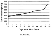


Classifications
-
- A—HUMAN NECESSITIES
- A61—MEDICAL OR VETERINARY SCIENCE; HYGIENE
- A61K—PREPARATIONS FOR MEDICAL, DENTAL OR TOILETRY PURPOSES
- A61K31/00—Medicinal preparations containing organic active ingredients
- A61K31/33—Heterocyclic compounds
- A61K31/395—Heterocyclic compounds having nitrogen as a ring hetero atom, e.g. guanethidine or rifamycins
- A61K31/41—Heterocyclic compounds having nitrogen as a ring hetero atom, e.g. guanethidine or rifamycins having five-membered rings with two or more ring hetero atoms, at least one of which being nitrogen, e.g. tetrazole
- A61K31/425—Thiazoles
- A61K31/426—1,3-Thiazoles
-
- A—HUMAN NECESSITIES
- A61—MEDICAL OR VETERINARY SCIENCE; HYGIENE
- A61K—PREPARATIONS FOR MEDICAL, DENTAL OR TOILETRY PURPOSES
- A61K47/00—Medicinal preparations characterised by the non-active ingredients used, e.g. carriers or inert additives; Targeting or modifying agents chemically bound to the active ingredient
- A61K47/50—Medicinal preparations characterised by the non-active ingredients used, e.g. carriers or inert additives; Targeting or modifying agents chemically bound to the active ingredient the non-active ingredient being chemically bound to the active ingredient, e.g. polymer-drug conjugates
- A61K47/51—Medicinal preparations characterised by the non-active ingredients used, e.g. carriers or inert additives; Targeting or modifying agents chemically bound to the active ingredient the non-active ingredient being chemically bound to the active ingredient, e.g. polymer-drug conjugates the non-active ingredient being a modifying agent
- A61K47/62—Medicinal preparations characterised by the non-active ingredients used, e.g. carriers or inert additives; Targeting or modifying agents chemically bound to the active ingredient the non-active ingredient being chemically bound to the active ingredient, e.g. polymer-drug conjugates the non-active ingredient being a modifying agent the modifying agent being a protein, peptide or polyamino acid
- A61K47/64—Drug-peptide, drug-protein or drug-polyamino acid conjugates, i.e. the modifying agent being a peptide, protein or polyamino acid which is covalently bonded or complexed to a therapeutically active agent
-
- A—HUMAN NECESSITIES
- A61—MEDICAL OR VETERINARY SCIENCE; HYGIENE
- A61K—PREPARATIONS FOR MEDICAL, DENTAL OR TOILETRY PURPOSES
- A61K31/00—Medicinal preparations containing organic active ingredients
- A61K31/33—Heterocyclic compounds
- A61K31/395—Heterocyclic compounds having nitrogen as a ring hetero atom, e.g. guanethidine or rifamycins
- A61K31/435—Heterocyclic compounds having nitrogen as a ring hetero atom, e.g. guanethidine or rifamycins having six-membered rings with one nitrogen as the only ring hetero atom
- A61K31/47—Quinolines; Isoquinolines
- A61K31/4738—Quinolines; Isoquinolines ortho- or peri-condensed with heterocyclic ring systems
- A61K31/4745—Quinolines; Isoquinolines ortho- or peri-condensed with heterocyclic ring systems condensed with ring systems having nitrogen as a ring hetero atom, e.g. phenantrolines
-
- A—HUMAN NECESSITIES
- A61—MEDICAL OR VETERINARY SCIENCE; HYGIENE
- A61K—PREPARATIONS FOR MEDICAL, DENTAL OR TOILETRY PURPOSES
- A61K31/00—Medicinal preparations containing organic active ingredients
- A61K31/33—Heterocyclic compounds
- A61K31/395—Heterocyclic compounds having nitrogen as a ring hetero atom, e.g. guanethidine or rifamycins
- A61K31/435—Heterocyclic compounds having nitrogen as a ring hetero atom, e.g. guanethidine or rifamycins having six-membered rings with one nitrogen as the only ring hetero atom
- A61K31/47—Quinolines; Isoquinolines
- A61K31/475—Quinolines; Isoquinolines having an indole ring, e.g. yohimbine, reserpine, strychnine, vinblastine
-
- A—HUMAN NECESSITIES
- A61—MEDICAL OR VETERINARY SCIENCE; HYGIENE
- A61K—PREPARATIONS FOR MEDICAL, DENTAL OR TOILETRY PURPOSES
- A61K47/00—Medicinal preparations characterised by the non-active ingredients used, e.g. carriers or inert additives; Targeting or modifying agents chemically bound to the active ingredient
- A61K47/50—Medicinal preparations characterised by the non-active ingredients used, e.g. carriers or inert additives; Targeting or modifying agents chemically bound to the active ingredient the non-active ingredient being chemically bound to the active ingredient, e.g. polymer-drug conjugates
- A61K47/51—Medicinal preparations characterised by the non-active ingredients used, e.g. carriers or inert additives; Targeting or modifying agents chemically bound to the active ingredient the non-active ingredient being chemically bound to the active ingredient, e.g. polymer-drug conjugates the non-active ingredient being a modifying agent
- A61K47/54—Medicinal preparations characterised by the non-active ingredients used, e.g. carriers or inert additives; Targeting or modifying agents chemically bound to the active ingredient the non-active ingredient being chemically bound to the active ingredient, e.g. polymer-drug conjugates the non-active ingredient being a modifying agent the modifying agent being an organic compound
- A61K47/542—Carboxylic acids, e.g. a fatty acid or an amino acid
-
- A—HUMAN NECESSITIES
- A61—MEDICAL OR VETERINARY SCIENCE; HYGIENE
- A61K—PREPARATIONS FOR MEDICAL, DENTAL OR TOILETRY PURPOSES
- A61K47/00—Medicinal preparations characterised by the non-active ingredients used, e.g. carriers or inert additives; Targeting or modifying agents chemically bound to the active ingredient
- A61K47/50—Medicinal preparations characterised by the non-active ingredients used, e.g. carriers or inert additives; Targeting or modifying agents chemically bound to the active ingredient the non-active ingredient being chemically bound to the active ingredient, e.g. polymer-drug conjugates
- A61K47/51—Medicinal preparations characterised by the non-active ingredients used, e.g. carriers or inert additives; Targeting or modifying agents chemically bound to the active ingredient the non-active ingredient being chemically bound to the active ingredient, e.g. polymer-drug conjugates the non-active ingredient being a modifying agent
- A61K47/54—Medicinal preparations characterised by the non-active ingredients used, e.g. carriers or inert additives; Targeting or modifying agents chemically bound to the active ingredient the non-active ingredient being chemically bound to the active ingredient, e.g. polymer-drug conjugates the non-active ingredient being a modifying agent the modifying agent being an organic compound
- A61K47/547—Chelates, e.g. Gd-DOTA or Zinc-amino acid chelates; Chelate-forming compounds, e.g. DOTA or ethylenediamine being covalently linked or complexed to the pharmacologically- or therapeutically-active agent
-
- A—HUMAN NECESSITIES
- A61—MEDICAL OR VETERINARY SCIENCE; HYGIENE
- A61K—PREPARATIONS FOR MEDICAL, DENTAL OR TOILETRY PURPOSES
- A61K47/00—Medicinal preparations characterised by the non-active ingredients used, e.g. carriers or inert additives; Targeting or modifying agents chemically bound to the active ingredient
- A61K47/50—Medicinal preparations characterised by the non-active ingredients used, e.g. carriers or inert additives; Targeting or modifying agents chemically bound to the active ingredient the non-active ingredient being chemically bound to the active ingredient, e.g. polymer-drug conjugates
- A61K47/51—Medicinal preparations characterised by the non-active ingredients used, e.g. carriers or inert additives; Targeting or modifying agents chemically bound to the active ingredient the non-active ingredient being chemically bound to the active ingredient, e.g. polymer-drug conjugates the non-active ingredient being a modifying agent
- A61K47/54—Medicinal preparations characterised by the non-active ingredients used, e.g. carriers or inert additives; Targeting or modifying agents chemically bound to the active ingredient the non-active ingredient being chemically bound to the active ingredient, e.g. polymer-drug conjugates the non-active ingredient being a modifying agent the modifying agent being an organic compound
- A61K47/548—Phosphates or phosphonates, e.g. bone-seeking
-
- A—HUMAN NECESSITIES
- A61—MEDICAL OR VETERINARY SCIENCE; HYGIENE
- A61K—PREPARATIONS FOR MEDICAL, DENTAL OR TOILETRY PURPOSES
- A61K49/00—Preparations for testing in vivo
- A61K49/001—Preparation for luminescence or biological staining
- A61K49/0013—Luminescence
- A61K49/0017—Fluorescence in vivo
- A61K49/0019—Fluorescence in vivo characterised by the fluorescent group, e.g. oligomeric, polymeric or dendritic molecules
- A61K49/0021—Fluorescence in vivo characterised by the fluorescent group, e.g. oligomeric, polymeric or dendritic molecules the fluorescent group being a small organic molecule
- A61K49/0041—Xanthene dyes, used in vivo, e.g. administered to a mice, e.g. rhodamines, rose Bengal
- A61K49/0043—Fluorescein, used in vivo
-
- A—HUMAN NECESSITIES
- A61—MEDICAL OR VETERINARY SCIENCE; HYGIENE
- A61K—PREPARATIONS FOR MEDICAL, DENTAL OR TOILETRY PURPOSES
- A61K49/00—Preparations for testing in vivo
- A61K49/001—Preparation for luminescence or biological staining
- A61K49/0013—Luminescence
- A61K49/0017—Fluorescence in vivo
- A61K49/005—Fluorescence in vivo characterised by the carrier molecule carrying the fluorescent agent
- A61K49/0052—Small organic molecules
-
- A—HUMAN NECESSITIES
- A61—MEDICAL OR VETERINARY SCIENCE; HYGIENE
- A61K—PREPARATIONS FOR MEDICAL, DENTAL OR TOILETRY PURPOSES
- A61K49/00—Preparations for testing in vivo
- A61K49/001—Preparation for luminescence or biological staining
- A61K49/0013—Luminescence
- A61K49/0017—Fluorescence in vivo
- A61K49/005—Fluorescence in vivo characterised by the carrier molecule carrying the fluorescent agent
- A61K49/0056—Peptides, proteins, polyamino acids
-
- A—HUMAN NECESSITIES
- A61—MEDICAL OR VETERINARY SCIENCE; HYGIENE
- A61K—PREPARATIONS FOR MEDICAL, DENTAL OR TOILETRY PURPOSES
- A61K51/00—Preparations containing radioactive substances for use in therapy or testing in vivo
- A61K51/02—Preparations containing radioactive substances for use in therapy or testing in vivo characterised by the carrier, i.e. characterised by the agent or material covalently linked or complexing the radioactive nucleus
- A61K51/04—Organic compounds
- A61K51/0402—Organic compounds carboxylic acid carriers, fatty acids
-
- A—HUMAN NECESSITIES
- A61—MEDICAL OR VETERINARY SCIENCE; HYGIENE
- A61K—PREPARATIONS FOR MEDICAL, DENTAL OR TOILETRY PURPOSES
- A61K51/00—Preparations containing radioactive substances for use in therapy or testing in vivo
- A61K51/02—Preparations containing radioactive substances for use in therapy or testing in vivo characterised by the carrier, i.e. characterised by the agent or material covalently linked or complexing the radioactive nucleus
- A61K51/04—Organic compounds
- A61K51/0489—Phosphates or phosphonates, e.g. bone-seeking phosphonates
-
- A—HUMAN NECESSITIES
- A61—MEDICAL OR VETERINARY SCIENCE; HYGIENE
- A61K—PREPARATIONS FOR MEDICAL, DENTAL OR TOILETRY PURPOSES
- A61K51/00—Preparations containing radioactive substances for use in therapy or testing in vivo
- A61K51/02—Preparations containing radioactive substances for use in therapy or testing in vivo characterised by the carrier, i.e. characterised by the agent or material covalently linked or complexing the radioactive nucleus
- A61K51/04—Organic compounds
- A61K51/08—Peptides, e.g. proteins, carriers being peptides, polyamino acids, proteins
- A61K51/088—Peptides, e.g. proteins, carriers being peptides, polyamino acids, proteins conjugates with carriers being peptides, polyamino acids or proteins
-
- A—HUMAN NECESSITIES
- A61—MEDICAL OR VETERINARY SCIENCE; HYGIENE
- A61P—SPECIFIC THERAPEUTIC ACTIVITY OF CHEMICAL COMPOUNDS OR MEDICINAL PREPARATIONS
- A61P13/00—Drugs for disorders of the urinary system
- A61P13/08—Drugs for disorders of the urinary system of the prostate
-
- A—HUMAN NECESSITIES
- A61—MEDICAL OR VETERINARY SCIENCE; HYGIENE
- A61P—SPECIFIC THERAPEUTIC ACTIVITY OF CHEMICAL COMPOUNDS OR MEDICINAL PREPARATIONS
- A61P25/00—Drugs for disorders of the nervous system
- A61P25/28—Drugs for disorders of the nervous system for treating neurodegenerative disorders of the central nervous system, e.g. nootropic agents, cognition enhancers, drugs for treating Alzheimer's disease or other forms of dementia
-
- A—HUMAN NECESSITIES
- A61—MEDICAL OR VETERINARY SCIENCE; HYGIENE
- A61P—SPECIFIC THERAPEUTIC ACTIVITY OF CHEMICAL COMPOUNDS OR MEDICINAL PREPARATIONS
- A61P35/00—Antineoplastic agents
-
- A—HUMAN NECESSITIES
- A61—MEDICAL OR VETERINARY SCIENCE; HYGIENE
- A61P—SPECIFIC THERAPEUTIC ACTIVITY OF CHEMICAL COMPOUNDS OR MEDICINAL PREPARATIONS
- A61P43/00—Drugs for specific purposes, not provided for in groups A61P1/00-A61P41/00
-
- C—CHEMISTRY; METALLURGY
- C07—ORGANIC CHEMISTRY
- C07C—ACYCLIC OR CARBOCYCLIC COMPOUNDS
- C07C323/00—Thiols, sulfides, hydropolysulfides or polysulfides substituted by halogen, oxygen or nitrogen atoms, or by sulfur atoms not being part of thio groups
- C07C323/50—Thiols, sulfides, hydropolysulfides or polysulfides substituted by halogen, oxygen or nitrogen atoms, or by sulfur atoms not being part of thio groups containing thio groups and carboxyl groups bound to the same carbon skeleton
- C07C323/51—Thiols, sulfides, hydropolysulfides or polysulfides substituted by halogen, oxygen or nitrogen atoms, or by sulfur atoms not being part of thio groups containing thio groups and carboxyl groups bound to the same carbon skeleton having the sulfur atoms of the thio groups bound to acyclic carbon atoms of the carbon skeleton
- C07C323/52—Thiols, sulfides, hydropolysulfides or polysulfides substituted by halogen, oxygen or nitrogen atoms, or by sulfur atoms not being part of thio groups containing thio groups and carboxyl groups bound to the same carbon skeleton having the sulfur atoms of the thio groups bound to acyclic carbon atoms of the carbon skeleton the carbon skeleton being acyclic and saturated
-
- C—CHEMISTRY; METALLURGY
- C07—ORGANIC CHEMISTRY
- C07D—HETEROCYCLIC COMPOUNDS
- C07D207/00—Heterocyclic compounds containing five-membered rings not condensed with other rings, with one nitrogen atom as the only ring hetero atom
- C07D207/02—Heterocyclic compounds containing five-membered rings not condensed with other rings, with one nitrogen atom as the only ring hetero atom with only hydrogen or carbon atoms directly attached to the ring nitrogen atom
- C07D207/30—Heterocyclic compounds containing five-membered rings not condensed with other rings, with one nitrogen atom as the only ring hetero atom with only hydrogen or carbon atoms directly attached to the ring nitrogen atom having two double bonds between ring members or between ring members and non-ring members
- C07D207/34—Heterocyclic compounds containing five-membered rings not condensed with other rings, with one nitrogen atom as the only ring hetero atom with only hydrogen or carbon atoms directly attached to the ring nitrogen atom having two double bonds between ring members or between ring members and non-ring members with hetero atoms or with carbon atoms having three bonds to hetero atoms with at the most one bond to halogen, e.g. ester or nitrile radicals, directly attached to ring carbon atoms
- C07D207/36—Oxygen or sulfur atoms
- C07D207/40—2,5-Pyrrolidine-diones
- C07D207/416—2,5-Pyrrolidine-diones with hetero atoms or with carbon atoms having three bonds to hetero atoms with at the most one bond to halogen, e.g. ester or nitrile radicals, directly attached to other ring carbon atoms
-
- C—CHEMISTRY; METALLURGY
- C07—ORGANIC CHEMISTRY
- C07K—PEPTIDES
- C07K5/00—Peptides containing up to four amino acids in a fully defined sequence; Derivatives thereof
- C07K5/04—Peptides containing up to four amino acids in a fully defined sequence; Derivatives thereof containing only normal peptide links
- C07K5/08—Tripeptides
Definitions
- the invention described herein pertains to compounds and methods for treating diseases of the prostate, such as prostate cancer and related diseases. More specifically, embodiments of the invention described herein pertain to conjugates of biologically active agents conjugated to PSMA binding ligands.
- the prostate is one of the male reproductive organs found in the pelvis below the urinary bladder. It functions to produce and store seminal fluid which provides nutrients and fluids that are vital for the survival of sperm introduced into the vagina during reproduction. Like many other tissues, the prostate glands are also prone to develop either malignant (cancerous) or benign (non-cancerous) tumors.
- the American Cancer Society predicted that over 230,000 men would be diagnosed with prostrate cancer and over 30,000 men would die from the disease in year 2005. In fact, prostate cancer is one of the most common male cancers in western societies, and is the second leading form of malignancy among American men.
- Current treatment methods for prostrate cancer include hormonal therapy, radiation therapy, surgery, chemotherapy, photodynamic therapy, and combination therapy.
- PSMA prostate specific membrane antigen
- PSMA is named largely due to its higher level of expression on prostate cancer cells; however, its particular function on prostate cancer cells remains unresolved.
- PSMA is over-expressed in the malignant prostate tissues when compared to other organs in the human body such as kidney, proximal small intestine, and salivary glands. Though PSMA is expressed in brain, that expression is minimal, and most ligands of PSMA are polar and are not capable of penetrating the blood brain barrier.
- PSMA is a type II cell surface membrane-bound glycoprotein with ⁇ 110 kD molecular weight, including an intracellular segment (amino acids 1-18), a transmembrane domain (amino acids 19-43), and an extensive extracellular domain (amino acids 44-750).
- PSMA plays a role in central nervous system, where it metabolizes N-acetyl-aspartyl glutamate (NAAG) into glutamic and N-acetyl aspartic acid. Accordingly, it is also sometimes referred to as an N-acetyl alpha linked acidic dipeptidase (NAALADase).
- NAAG N-acetyl-aspartyl glutamate
- NAALADase N-acetyl alpha linked acidic dipeptidase
- PSMA is also sometimes referred to as a folate hydrolase I (FOLH I) or glutamate carboxypeptidase (GCP II) due to its role in the proximal small intestine where it removes ⁇ -linked glutamate from poly- ⁇ -glutamated folate and ⁇ -linked glutamate from peptides and small molecules.
- FOLH I folate hydrolase I
- GCP II glutamate carboxypeptidase
- PSMA also shares similarities with human transferrin receptor (TfR), because both PSMA and TfR are type II glycoproteins. More specifically, PSMA shows 54% and 60% homology to TfR1 and TfR2, respectively. However, though TfR exists only in dimeric form due to the formation of inter-strand sulfhydryl linkages, PSMA can exist in either dimeric or monomeric form.
- TfR human transferrin receptor
- PSMA undergoes rapid internalization into the cell in a similar fashion to cell surface bound receptors like vitamin receptors. PSMA is internalized through clathrin-coated pits and subsequently can either recycle to the cell surface or go to lysosomes. It has been suggested that the dimer and monomer form of PSMA are inter-convertible, though direct evidence of the interconversion is being debated. Even so, only the dimer of PSMA possesses enzymatic activity, and the monomer does not.
- PSMA represents a viable target for the selective and/or specific delivery of biologically active agents, including diagnostic agents, imaging agents, and therapeutic agents to such prostate cells.
- PSMA prostate specific membrane antigen
- linker may be useful in the imaging, diagnosis, and/or treatment of prostate cancer, and related diseases that involve pathogenic cell populations expressing or over-expressing PSMA.
- PSMA is a cell surface protein that is internalized in a process analogous to endocytosis observed with cell surface receptors, such as vitamin receptors. Accordingly, it has been discovered that certain conjugates that include a linker having a predetermined length, and/or a predetermined diameter, and/or preselected functional groups along its length may be used to treat, image, and/or diagnose such diseases.
- conjugates having the formula B-L-D are described wherein B is a prostate specific membrane antigen (PSMA) binding or targeting ligand, L is a linker, and D is a drug.
- PSMA prostate specific membrane antigen
- D is a drug.
- drug D collectively includes therapeutic agents, cytotoxic agents, imaging agents, diagnostic agents, and the like, unless otherwise indicated or by the context.
- the conjugate described herein is used to eliminate a pathogenic population of cells and therefore the drug D is a therapeutic agent, a cytotoxic agent, and the like.
- the conjugate described herein is used to image and/or diagnose a disease or disease state, and therefore the drug D is an imaging agent, a diagnostic agent, and the like.
- the drug D is an imaging agent, a diagnostic agent, and the like.
- Other configurations are also contemplated and described herein. It is to be understood that analogs and derivatives of each of the foregoing B, L, and D are also contemplated and described herein, and that when used herein, the terms B, L, and D collectively refer to such analogs and derivatives.
- the linker L may be a releasable or non-releasable linker. In one aspect, the linker L is at least about 7 atoms in length. In one variation, the linker L is at least about 10 atoms in length. In one variation, the linker L is at least about 14 atoms in length. In another variation, the linker L is between about 7 and about 31, between about 7 and about 24, or between about 7 and about 20 atoms in length. In another variation, the linker L is between about 14 and about 31, between about 14 and about 24, or between about 14 and about 20 atoms in length.
- the linker L is at least about 10 angstroms ( ⁇ ) in length. In one variation, the linker L is at least about 15 ⁇ in length. In another variation, the linker L is at least about 20 ⁇ in length. In another variation, the linker L is in the range from about 10 ⁇ to about 30 ⁇ in length.
- At least a portion of the length of the linker L is about 5 ⁇ in diameter or less at the end connected to the binding ligand B. In one variation, at least a portion of the length of the linker L is about 4 ⁇ or less, or about 3 ⁇ or less in diameter at the end connected to the binding ligand B. It is appreciated that the illustrative embodiments that include a diameter requirement of about 5 ⁇ or less, about 4 ⁇ or less, or about 3 ⁇ or less may include that requirement for a predetermined length of the linker, thereby defining a cylindrical-like portion of the linker.
- the linker includes a cylindrical portion at the end connected to the binding ligand that is at least about 7 ⁇ in length and about 5 ⁇ or less, about 4 ⁇ or less, or about 3 ⁇ or less in diameter.
- the linker L includes one or more hydrophilic linkers capable of interacting with one or more residues of PSMA, including amino acids that have hydrophobic side chains, such as Ser, Thr, Cys, Arg, Orn, Lys, Asp, Glu, Gln, and like residues.
- the linker L includes one or more hydrophobic linkers capable of interacting with one or more residues of PSMA, including amino acids that have hydrophilic side chains, such as Val, Leu, Ile, Phe, Tyr, Met, and like residues. It is to be understood that the foregoing embodiments and aspects may be included in the linker L either alone or in combination with each other.
- linkers L that are at least about 7 atoms in length and about 5 ⁇ , about 4 ⁇ or less, or about 3 ⁇ or less in diameter or less are contemplated and described herein, and also include one or more hydrophilic linkers capable of interacting with one or more residues of PSMA, including Val, Leu, Ile, Phe, Tyr, Met, and like residues are contemplated and described herein.
- one end of the linker is not branched and comprises a chain of carbon, oxygen, nitrogen, and sulfur atoms.
- the linear chain of carbon, oxygen, nitrogen, and sulfur atoms is at least 5 atoms in length.
- the linear chain is at least 7 atoms, or at least 10 atoms in length.
- the chain of carbon, oxygen, nitrogen, and sulfur atoms are not substituted.
- a portion of the chain of carbon, oxygen, nitrogen, and sulfur atoms is cyclized with a divalent fragment.
- a linker (L) comprising the dipeptide Phe-Phe may include a piperazin-1,4-diyl structure by cyclizing two nitrogens with an ethylene fragment, or substituted variation thereof.
- compositions are described herein, where the pharmaceutical composition includes the conjugates described herein in amounts effective to treat diseases and disease states, diagnose diseases or disease states, and/or image tissues and/or cells that are associated with pathogenic populations of cells expressing or over expressing PSMA.
- the pharmaceutical compositions also include one or more carriers, diluents, and/or excipients.
- methods for treating diseases and disease states, diagnosing diseases or disease states, and/or imaging tissues and/or cells that are associated with pathogenic populations of cells expressing or over expressing PSMA are described herein. Such methods include the step of administering the conjugates described herein, and/or pharmaceutical compositions containing the conjugates described herein, in amounts effective to treat diseases and disease states, diagnose diseases or disease states, and/or image tissues and/or cells that are associated with pathogenic populations of cells expressing or over expressing PSMA.
- FIG. 1B In Vitro Binding Studies Using LNCaP Cells and SK33 (14 atom linker). LNCaP cells containing increasing concentrations of DUPA- 99m Tc in the presence ( ⁇ ) or absence ( ⁇ ) of excess PMPA
- FIG. 2 Cell bound radioactivity verses concentration of SK28- 99m Tc; at 4° C. ( ⁇ ) and at 37° C. ( ⁇ ).
- FIG. 3B K D values for DUPA-Linker- 99m Tc compounds binding to LNCaP cells.
- FIG. 4 Plot of days post injection verses tumor volume for LNCaP tumors: (a) 2.5 million+Matrigel; (b) 2.5 million+HC Matrigel; (c) 5 million+Matrigel; (d) 5 million+HC Matrigel.
- FIG. 5A Plot of days post injection verses tumor volume for LNCaP tumors (27 mice) and (b) KB cells (5 mice) and A549 cells (5 mice).
- FIG. 5B Plot of days post injection versus tumor volume for KB cells (5 mice) and A549 cells (5 mice).
- FIG. 6A Mice (Set 1) previously injected with LNCaP tumors, treated with 1 ng/kg SK28- 99m Tc (14-atom linker), the left hand image shows white light images and image shows an overlay of the radioimage with the white light image.
- the right mouse was treated with 50 mg/kg PMPA (to block PSMA binding) and the left mouse was treated without added PMPA.
- FIG. 6B Mice (Set 2) previously injected with LNCaP tumors, treated with 1 ng/kg SK28- 99m Tc (14-atom linker), the left hand image shows white light images and image shows an overlay of the radioimage with the white light image.
- the right mouse was treated with 50 mg/kg PMPA (to block PSMA binding) and the left mouse was treated without added PMPA.
- FIG. 6C Mice (Set 3) previously injected with LNCaP tumors, treated with 1 ng/kg SK28- 99m Tc (14-atom linker), the left hand image shows white light images and image shows an overlay of the radioimage with the white light image.
- the right mouse was treated with 50 mg/kg PMPA (to block PSMA binding) and the left mouse was treated without added PMPA.
- FIG. 6D Shows a single mouse study for LNCaP tumors imaged using Kodak imager 4 hours after subcutaneous (administered through intraperitoneal) injection of 1 ng/kg SK28- 99m Tc showing in the left hand image an overlay of radioimage with kidney shield and white light image with no shield and in the right hand image an overlay of radioimage with kidney shield and X-ray image with no shield.
- FIG. 7A Mice previously injected with LNCaP tumors treated using SK60- 99m Tc (zero atom linker).
- the left image shows white light images
- the center image shows overlay of radioimage with white light image
- the right image shows overlay of radioimage with white light image by shielding the kidney of mice.
- FIG. 7B Mice previously injected with KB cells treated using SK28- 99m Tc (14 atom linker).
- the left image shows white light images
- the center image shows overlay of radioimage with white light image
- the right image shows overlay of radioimage with white light image by shielding the kidney of mice.
- FIG. 7C Mice previously injected with A549 cells treated using SK28- 99m Tc (14 atom linker).
- the left image shows white light images
- the center image shows overlay of radioimage with white light image
- the right image shows overlay of radioimage with white light image by shielding the kidney of mice.
- FIG. 7D Whole body images of solid tumor xenografts in nu/nu mice taken 4 h after injection of 150 ⁇ Ci DUPA- 99m Tc. Overlay of whole-body radioimages on white light images of mice bearing LNCaP tumors that were treated with DUPA- 99m Tc in the absence (a, c) or presence (b, d) of 100-fold molar excess PMPA. Overlay of radioimages on white light images of mice bearing an A549 tumor (e) or a KB tumor (f) that were similarly treated with DUPA- 99m Tc.
- FIG. 8A Bio-distribution data as measured for direct cpm count verses tissue for SK28- 99m Tc with or without PMPA (as a competitor) on LNCaP, (a) without, (b) with; (c) A549; and (d) KB tumors implanted on axial of male nude mice.
- FIG. 8B Bio-distribution data just for tumor and kidney for SK28- 99m Tc with (b) or without (a) PMPA (as a competitor) on LNCaP tumors implant on axial of male nude mice.
- FIG. 8C Biodistribution studies of DUPA- 99m Tc in nu/nu mice bearing LNCaP, A549, or KB tumors.
- FIG. 9A Acute MTD (single dose) showing percentage weight change after a single dose of SK71; saline alone, 1.1 ⁇ mol/kg, 2.3 ⁇ mol/kg, 4.5 ⁇ mol/kg, and 9 ⁇ mol/kg.
- FIG. 9B Chronic MTD showing percentage weight change after 5 doses given on alternate days (M, W, F, M, W); saline alone, 2 ⁇ mol/kg and 4 ⁇ mol/kg.
- FIG. 10A Efficacy study showing tumor volume in animals treated with the conjugate SK71 administered in 5 doses on alternate days (M, W, F, M, W) at 1 ⁇ mol/kg.
- FIG. 10B Efficacy study (control group) showing tumor volume in animals treated with saline alone administered in 5 doses on alternate days (M, W, F, M, W).
- FIG. 10C Efficacy study (competition) showing tumor volume in animals treated with excess PSMA and the conjugate SK71 administered in 5 doses on alternate days (M, W, F, M, W) at 1 ⁇ mol/kg.
- FIG. 11 Efficacy Study (1 micromole/kg every other day for 10 days; i.e. 5 doses).
- FIG. 12A [ 3 H]-Thymidine incorporation of LNCaP cells after treatment with SK71 (IC 50 ⁇ 2 nM).
- FIG. 12B [ 3 H]-Thymidine incorporation of LNCaP cells after treatment with SK77 (IC 50 ⁇ 3 nM).
- FIG. 12C [ 3 H]-Thymidine incorporation of LNCaP cells after treatment with SK37 (IC 50 ⁇ 33 nM).
- FIG. 12D [ 3 H]-Thymidine incorporation of LNCaP cells after treatment with SK45 (IC 50 ⁇ 29 nM).
- FIG. 13A The effect of treatment with SK71 (1.5 ⁇ mol/kg) on tumor volume in nu/nu mice previously treated with LNCaP cells in HC Matrigel. Treated mice ( ⁇ ), untreated mice ( ⁇ ), treated mice pre-injected with 100-fold molar excess of PMPA ( ⁇ ).
- FIG. 13B The effect of treatment with SK71 (1.5 ⁇ mol/kg) on percentage weight change in nu/nu mice previously treated with LNCaP cells in HC Matrigel. Treated mice ( ⁇ ), untreated mice ( ⁇ ), treated mice pre-injected with 100-fold molar excess of PMPA ( ⁇ ).
- FIG. 13C The effect of treatment with SK71 (2.0 ⁇ mol/kg) on tumor volume in nu/nu mice previously treated with LNCaP cells in HC Matrigel. Treated mice ( ⁇ ), untreated mice ( ⁇ ), treated mice pre-injected with 30-fold molar excess of PMPA ( ⁇ ).
- FIG. 13D The effect of treatment with SK71 (2.0 ⁇ mol/kg) on percentage weight change in nu/nu mice previously treated with LNCaP cells in HC Matrigel. Treated mice ( ⁇ ), untreated mice ( ⁇ ), treated mice pre-injected with 30-fold molar excess of PMPA ( ).
- FIG. 14A The effect of treatment with SK77 (2.0 ⁇ mol/kg) on tumor volume in nu/nu mice previously treated with LNCaP cells in HC Matrigel. Untreated mice ( ⁇ ), treated mice ( ⁇ ).
- FIG. 14B The effect of treatment with SK77 (2.0 ⁇ mol/kg) on percentage weight change in nu/nu mice previously treated with LNCaP cells in HC Matrigel. Untreated mice ( ⁇ ), treated mice ( ⁇ ).
- FIG. 15A MUPA 99mTc imaging agent conjugate (9-atom linker) with energy minimized computer model.
- FIG. 15B MUPA 99mTc imaging agent conjugate (syn-SK33, 14-atom linker) with energy minimized computer model.
- Drug delivery conjugates are described herein where a PSMA binding ligand is attached to a releasable or non-releasable linker which is attached to a drug, therapeutic agent, diagnostic agent, or imaging agent.
- the bivalent linkers described herein may be included in linkers used to prepare PSMA-binding drug conjugates, PSMA-binding imaging agent conjugates, and PSMA-binding diagnostic agent conjugates of the following formulae: B-L-TA B-L-IA B-L-DA where B is a PSMA-binding moiety, including analogs or derivatives thereof, L is a linker, TA is a therapeutic agent, including analogs or derivatives thereof, IA is an imaging agent, including analogs or derivatives thereof, and DA is a diagnostic agent, including analogs or derivatives thereof.
- the linker L can comprise multiple bivalent linkers, including the bivalent linkers described herein.
- TA collectively refers to therapeutic agents, and analogs and derivatives thereof
- IA collectively refers to imaging agents, and analogs and derivatives thereof
- DA collectively refers to diagnostic agents, and analogs and derivatives thereof.
- the linker may also include one or more spacer linkers and optionally additional releasable linkers.
- the spacer and releasable linkers may be attached to each other in any order or combination.
- the PSMA binding ligand may be attached to a spacer linker or to a releasable linker.
- the drug, therapeutic agent, diagnostic agent, or imaging agent may be attached to a spacer linker or to a releasable linker.
- Each of these components of the conjugates may be connected through existing or additional heteroatoms on the targeting ligand, drug, therapeutic agent, diagnostic agent, imaging agent, releasable or spacer linker.
- Illustrative heteroatoms include nitrogen, oxygen, sulfur, and the formulae —(NHR 1 NHR 2 )—, —SO—, —(SO 2 )—, and —N(R 3 )O—, wherein R 1 , R 2 , and R 3 are each independently selected from hydrogen, alkyl, heteroalkyl, heterocyclyl, aryl, heteroaryl, arylalkyl, heteroarylalkyl, and the like, each of which may be optionally substituted.
- compounds are described herein that include linkers having predetermined length and diameter dimensions.
- linkers are described herein that satisfy one or more minimum length requirements, or a length requirement falling within a predetermined range.
- satisfaction of a minimum length requirement may be understood to be determined by computer modeling of the extended conformations of linkers.
- satisfaction of a minimum length requirement may be understood to be determined by having a certain number of atoms, whether or not substituted, forming a backbone chain of atoms connecting the binding ligand (B) with the drug (D).
- the backbone chain of atoms is cyclized with another divalent fragment.
- linkers are described herein that satisfy one or more maximum or minimum diameter requirements.
- satisfaction of a maximum or minimum diameter requirement may be understood to be determined by computer modeling of various conformations of linkers modeled as the space-filling. CPK, or like configurations.
- satisfaction of a maximum or minimum diameter requirement may be understood to be apply to one or more selected portions of the linker, for example the portion of the linker proximal to the binding ligand (B), or the portion of the linker proximal to the drug (D), and the like.
- linkers are described herein that satisfy one or more chemical composition requirements, such as linkers that include one or more polar groups that may positively interact with the one or more Arg or Lys side-chain nitrogens and/or Asp or Glu side chain oxygens found in the funnel portion of PSMA.
- linkers are described herein that satisfy one or more chemical composition requirements, such as linkers that include one or more non-polar groups that may positively interact with the one or more Tyr or Phe side-chain carbons found in the funnel portion of PSMA.
- the atom-length of the linker is defined by the number of atoms separating the binding or targeting ligand B, or analog or derivative thereof, and the drug D, or analog or derivative thereof. Accordingly, in configurations where the binding ligand B, or analog or derivative thereof, is attached directly to the drug D, or analog or derivative thereof, the attachment is also termed herein as a “0-atom” linker. It is understood that such 0-atom linkers include the configuration wherein B and D are directly attached by removing a hydrogen atom from each attachment point on B and D, respectively.
- such 0-atom linkers include the configuration wherein B and D are attached through an overlapping heteroatom by removing a hydrogen atom from one of B or D, and a heteroatom functional group, such as OH, SH, NH 2 , and the like from the other of B or D. It is also understood that such 0-atom linkers include the configuration wherein B and D are attached through a double bond, which may be formed by removing two hydrogen atoms from each attachment point on B and D, respectively, or whereby B and D are attached through one or more overlapping heteroatoms by removing two hydrogen atoms, one hydrogen and one heteroatom functional group, or two heteroatom functional groups, such as OH, SH, NH 2 , and the like, from each of B or D.
- B and D may be attached through a double bond formed by removing a double bonded heteroatom functional group, such as O, S, NH, and the like, from one or both of B or D.
- heteroatom functional groups include those attached to saturated carbon atoms, unsaturated carbon atoms (including carbonyl groups), and other heteroatoms.
- linkers that are greater than 0 atoms are defined in an analogous manner.
- linkers (L) are described having a chain length of at least 7 atoms. In one variation, linkers (L) are described having a chain length of at least 14 atoms. In another variation, linkers (L) are described having a chain length in the range from about 7 atoms to about 20 atoms. In another variation, linkers (L) are described having a chain length in the range from about 14 atoms to about 24 atoms.
- the length of the linker (L) is defined by measuring the length of an extended conformation of the linker.
- extended conformations may be measured in art-recognized computer modeling programs, such as PC Model 7 (MMX).
- MMX PC Model 7
- linkers are described having a chain length of at least 15 ⁇ , at least 20 ⁇ , or at least 25 ⁇ .
- linkers are described having at least one hydrophobic side chain group, such as an alkyl, cycloalkyl, aryl, arylalkyl, or like group, each of which is optionally substituted.
- the hydrophobic group is included in the linker by incorporating one or more Phe or Tyr groups, including substituted variants thereof, and analogs and derivatives thereof, in the linker chain. It is appreciated that such Phe and/or Tyr side chain groups may form positive pi-pi ( ⁇ - ⁇ ) interactions with Tyr and Phe residues found in the funnel of PSMA.
- the funnel shaped tunnel leading to the catalytic site or active site of PSMA imposes length, shape, and/or chemical composition requirements on the linker portion of conjugates of PSMA binding ligands and therapeutic, diagnostic, and imaging agents that positively and negatively affect the interactions between PSMA and those conjugates.
- length, shape, and/or chemical composition requirements were assessed using molecular modeling.
- the space filling and surface model of the PSMA complex with (s)-2-(4-iodobenzensylphosphonomethyl)-pentanedioic [2-PMPA derivative] PDB ID code 2C6P were generated using PROTEIN EXPLORER.
- the PROTEIN EXPLORER model verified the 20 ⁇ deep funnel, and also showed diameter features at various locations along the funnel that may be used to define linkers having favorable structural features.
- the model showed that close to the active site of PSMA, there are a higher number of hydrophobic residues that may provide additional binding interactions when the corresponding functional groups are included in the linker.
- the model showed the presence of three hydrophobic pockets that may provide additional binding interactions when the corresponding functional groups are included in the linker.
- the following molecular models were created for a conjugate of MUPA and a tripeptide 99m Tc imaging agent connected by a 9-atom linker, as shown in FIG. 15A , and syn-SK33 including a branched 14-atom linker, as shown in FIG. 15B .
- Such models may be used to calculate the length of the linker connecting the binding ligand (B) and the drug (D).
- models may be modified to create extended conformations, and subsequently used to calculate the length of the linker connecting the binding ligand (B) and the drug (D).
- the first human PSMA gene was cloned from LNCaP cells and is reported to be located in chromosome 11p11-12. In addition, there is a PSMA-like gene located at the loci 11q14.3.
- the crystal structure of PSMA has been reported by two different groups at different resolutions, and each shows that the active site contains two zinc atoms, confirming that PSMA is also considered a zinc metalloprotease.
- Davis et al. PNAS, 102:5981-86, (2005) reported the crystal structure at low resolution (3.5 ⁇ ), while Mesters et al, The EMBO Journal, 1-10 (2006) reported the crystal structure at higher resolution (2-2.2 ⁇ ), the disclosures of which are incorporated herein by reference.
- the crystal structures show that PSMA is a homodimer that contains a protease domain, an apical domain, a helical domain and a CPG2 dimerization domain.
- the protease domain of PSMA contains a binuclear zinc site, catalytic residues and a substrate binding region including three arginine residues (also referred to as a substrate binding arginine patch).
- the two zinc ions in the active site are each ligated to an oxygen of phosphate, or to the phosphinate moiety of the inhibitor GPI 18431 for the co-crystal structure.
- PSMA was co-crystallized with both potent inhibitors, weak inhibitors, and glutamate at 2.0, 2.4, and 2.2 ⁇ , respectively.
- the high resolution crystal structure shows a 20 ⁇ deep funnel shaped tunnel leads to the catalytic site or active site of PSMA.
- the funnel is lined with the side chains of a number of Arg and Lys residues, Asp and Glu residues, and Tyr and Phe residues.
- the linker (L) is a chain of atoms selected from C, N, O, S, Si, and P.
- the linker may have a wide variety of lengths, such as in the range from about 7 to about 100.
- the atoms used in forming the linker may be combined in all chemically relevant ways, such as chains of carbon atoms forming alkylene groups, chains of carbon and oxygen atoms forming polyoxyalkylene groups, chains of carbon and nitrogen atoms forming polyamines, and others.
- bonds connecting atoms in the chain may be either saturated or unsaturated, such that for example, alkanes, alkenes, alkynes, cycloalkanes, arylenes, imides, and the like may be divalent radicals that are included in the linker.
- the atoms forming the linker may also be cyclized upon each other to form divalent cyclic radicals in the linker.
- the chain forming the linker may be substituted with a wide variety of groups.
- linkers (L) are described that include at least one releasable linker. In one variation, linkers (L) are described that include at least two releasable linkers. In another variation, linkers (L) are described that include at least one self-immolative linker. In another variation, linkers (L) are described that include at least one releasable linker that is not a disulfide. In another embodiment, linkers (L) are described that do not include a releasable linker.
- linker L is a non-releasable linker. It is appreciated that non-releasable linkers may be used when the drug is advantageously retained by the binding ligand-linker conjugate, such as in imaging, diagnosing, uses of the conjugates described herein.
- linkers L described herein comprise various atoms, chains of atoms, functional groups, and combinations of functional groups.
- the linker L may be referred to by the presence of spacer linkers, releasable linkers, and heteroatoms.
- such references are not to be construed as limiting the definition of the linkers L described herein.
- the linker (L) comprising spacer and/or releasable linkers can be any biocompatible linker.
- the releasable or cleavable linker can be, for example, a linker susceptible to cleavage under the reducing or oxidizing conditions present in or on cells, a pH-sensitive linker that may be an acid-labile or base-labile linker, or a linker that is cleavable by biochemical or metabolic processes, such as an enzyme-labile linker.
- the spacer and/or releasable linker comprises about 1 to about 30 atoms, or about 2 to about 20 atoms.
- Lower molecular weight linkers i.e., those having an approximate molecular weight of about 30 to about 300 are also described.
- Precursors to such linkers may be selected to have either nucleophilic or electrophilic functional groups, or both, optionally in a protected form with a readily cleavable protecting group to facilitate their use in synthesis of the intermediate species.
- releasable linker refers to a linker that includes at least one bond that can be broken under physiological conditions (e.g., a pH-labile, acid-labile, oxidatively-labile, or enzyme-labile bond).
- physiological conditions e.g., a pH-labile, acid-labile, oxidatively-labile, or enzyme-labile bond.
- the cleavable bond or bonds may be present in the interior of a cleavable linker and/or at one or both ends of a cleavable linker.
- physiological conditions resulting in bond breaking include standard chemical hydrolysis reactions that occur, for example, at physiological pH, or as a result of compartmentalization into a cellular organelle such as an endosome having a lower pH than cytosolic pH.
- the bivalent linkers described herein may undergo cleavage under other physiological or metabolic conditions, such as by the action of a glutathione mediated mechanism. It is appreciated that the lability of the cleavable bond may be adjusted by including functional groups or fragments within the bivalent linker L that are able to assist or facilitate such bond breakage, also termed anchimeric assistance.
- the lability of the cleavable bond can also be adjusted by, for example, substitutional changes at or near the cleavable bond, such as including alpha branching adjacent to a cleavable disulfide bond, increasing the hydrophobicity of substituents on silicon in a moiety having a silicon-oxygen bond that may be hydrolyzed, homologating alkoxy groups that form part of a ketal or acetal that may be hydrolyzed, and the like.
- additional functional groups or fragments may be included within the bivalent linker L that are able to assist or facilitate additional fragmentation of the PSMA binding drug linker conjugates after bond breaking of the releasable linker.
- the linker includes radicals that form one or more spacer linkers and/or releasable linkers that are taken together to form the linkers described herein having certain length, diameter, and/or functional group requirements.
- linkers described herein include releasable linkers that cleave under the conditions described herein by a chemical mechanism involving beta elimination.
- releasable linkers include beta-thio, beta-hydroxy, and beta-amino substituted carboxylic acids and derivatives thereof, such as esters, amides, carbonates, carbamates, and ureas.
- such releasable linkers include 2- and 4-thioarylesters, carbamates, and carbonates.
- releasable linkers may also be referred to by the functional groups they contain, illustratively such as disulfide groups, ketal groups, and the like, as described herein. Accordingly, it is understood that a cleavable bond can connect two adjacent atoms within the releasable linker and/or connect other linkers, or the binding ligand B, or the therapeutic, diagnostic, or imaging agent D, as described herein, at either or both ends of the releasable linker. In the case where a cleavable bond connects two adjacent atoms within the releasable linker, following breakage of the bond, the releasable linker is broken into two or more fragments.
- the releasable linker is separated from the other moiety.
- another moiety such as an additional heteroatom, a spacer linker, another releasable linker, the drug D, or analog or derivative thereof, or the binding ligand B, or analog or derivative thereof, following breakage of the bond.
- the releasable and spacer linkers may be arranged in such a way that subsequent to the cleavage of a bond in the bivalent linker, released functional groups anchimerically assist the breakage or cleavage of additional bonds, as described above.
- An illustrative embodiment of such a bivalent linker or portion thereof includes compounds having the formula:
- X is an heteroatom, such as nitrogen, oxygen, or sulfur
- n is an integer selected from 0, 1, 2, and 3
- R is hydrogen, or a substituent, including a substituent capable of stabilizing a positive charge inductively or by resonance on the aryl ring, such as alkoxy, and the like, and the symbol (*) indicates points of attachment for additional spacer or releasable linkers, or heteroatoms, forming the bivalent linker, or alternatively for attachment of the drug, or analog or derivative thereof, or the vitamin, or analog or derivative thereof.
- Assisted cleavage may include mechanisms involving benzylium intermediates, benzyne intermediates, lactone cyclization, oxonium intermediates, beta-elimination, and the like. It is further appreciated that, in addition to fragmentation subsequent to cleavage of the releasable linker, the initial cleavage of the releasable linker may be facilitated by an anchimerically assisted mechanism.
- the hydroxyalkanoic acid which may cyclize, facilitates cleavage of the methylene bridge, by for example an oxonium ion, and facilitates bond cleavage or subsequent fragmentation after bond cleavage of the releasable linker.
- acid catalyzed oxonium ion-assisted cleavage of the methylene bridge may begin a cascade of fragmentation of this illustrative bivalent linker, or fragment thereof.
- acid-catalyzed hydrolysis of the carbamate may facilitate the beta elimination of the hydroxyalkanoic acid, which may cyclize, and facilitate cleavage of methylene bridge, by for example an oxonium ion. It is appreciated that other chemical mechanisms of bond breakage or cleavage under the metabolic, physiological, or cellular conditions described herein may initiate such a cascade of fragmentation. It is appreciated that other chemical mechanisms of bond breakage or cleavage under the metabolic, physiological, or cellular conditions described herein may initiate such a cascade of fragmentation.
- Illustrative mechanisms for cleavage of the bivalent linkers described herein include the following 1,4 and 1,6 fragmentation mechanisms
- X is an exogenous or endogenous nucleophile, glutathione, or bioreducing agent, and the like, and either of Z or Z′ is a PSMA binding ligand, or a drug, therapeutic agent, diagnostic agent, or imaging agent, or either of Z or Z′ is a PSMA binding ligand, or a drug, therapeutic agent, diagnostic agent, or imaging agent connected through other portions of the bivalent linker.
- Z or Z′ is a PSMA binding ligand, or a drug, therapeutic agent, diagnostic agent, or imaging agent connected through other portions of the bivalent linker.
- the bond cleavage may also occur by acid catalyzed elimination of the carbamate moiety, which may be anchimerically assisted by the stabilization provided by either the aryl group of the beta sulfur or disulfide illustrated in the above examples.
- the releasable linker is the carbamate moiety.
- the fragmentation may be initiated by a nucleophilic attack on the disulfide group, causing cleavage to form a thiolate.
- the thiolate may intermolecularly displace a carbonic acid or carbamic acid moiety and form the corresponding thiacyclopropane.
- the resulting phenyl thiolate may further fragment to release a carbonic acid or carbamic acid moiety by forming a resonance stabilized intermediate.
- the releaseable nature of the illustrative bivalent linkers described herein may be realized by whatever mechanism may be relevant to the chemical, metabolic, physiological, or biological conditions present.
- Z is the vitamin, or analog or derivative thereof, or the drug, or analog or derivative thereof, or each is a vitamin or drug moiety in conjunction with other portions of the polyvalent linker, such as a drug or vitamin moiety including one or more spacer linkers and/or other releasable linkers.
- acid-catalyzed elimination of the carbamate leads to the release of CO 2 and the nitrogen-containing moiety attached to Z, and the formation of a benzyl cation, which may be trapped by water, or any other Lewis base.
- the releasable linker includes a disulfide.
- the releasable linker may be a divalent radical comprising alkyleneaziridin-1-yl, alkylenecarbonylaziridin-1-yl, carbonylalkylaziridin-1-yl, alkylenesulfoxylaziridin-1-yl, sulfoxylalkylaziridin-1-yl, sulfonylalkylaziridin-1-yl, or alkylenesulfonylaziridin-1-yl, wherein each of the releasable linkers is optionally substituted with a substituent X 2 , as defined below.
- Additional illustrative releasable linkers include methylene, 1-alkoxyalkylene, 1-alkoxycycloalkylene, 1-alkoxyalkylenecarbonyl, 1-alkoxycycloalkylenecarbonyl, carbonylarylcarbonyl, carbonyl(carboxyaryl)carbonyl, carbonyl(biscarboxyaryl)carbonyl, haloalkylenecarbonyl, alkylene(dialkylsilyl), alkylene(alkylarylsilyl), alkylene(diarylsilyl), (dialkylsilyl)aryl, (alkylarylsilyl)aryl, (diarylsilyl)aryl, oxycarbonyloxy, oxycarbonyloxyalkyl, sulfonyloxy, oxysulfonylalkyl, iminoalkylidenyl, carbonylalkylideniminyl, iminocycloalkylidenyl, carbonylcycl
- the releasable linker may include oxygen, and the releasable linkers can be methylene, 1-alkoxyalkylene, 1-alkoxycycloalkylene, 1-alkoxyalkylenecarbonyl, and 1-alkoxycycloalkylenecarbonyl, wherein each of the releasable linkers is optionally substituted with a substituent X 2 , as defined below, and the releasable linker is bonded to the oxygen to form an acetal or ketal.
- the releasable linker may include oxygen, and the releasable linker can be methylene, wherein the methylene is substituted with an optionally-substituted aryl, and the releasable linker is bonded to the oxygen to form an acetal or ketal.
- the releasable linker may include oxygen, and the releasable linker can be sulfonylalkyl, and the releasable linker is bonded to the oxygen to form an alkylsulfonate.
- the releasable linker may include nitrogen, and the releasable linkers can be iminoalkylidenyl, carbonylalkylideniminyl, iminocycloalkylidenyl, and carbonylcycloalkylideniminyl, wherein each of the releasable linkers is optionally substituted with a substituent X 2 , as defined below, and the releasable linker is bonded to the nitrogen to form an hydrazone.
- the hydrazone may be acylated with a carboxylic acid derivative, an orthoformate derivative, or a carbamoyl derivative to form various acylhydrazone releasable linkers.
- the releasable linker may include oxygen
- the releasable linkers can be alkylene(dialkylsilyl), alkylene(alkylarylsilyl), alkylene(diarylsilyl), (dialkylsilyl)aryl, (alkylarylsilyl)aryl, and (diarylsilyl)aryl, wherein each of the releasable linkers is optionally substituted with a substituent X 2 , as defined below, and the releasable linker is bonded to the oxygen to form a silanol.
- the drug can include a nitrogen atom
- the releasable linker may include nitrogen
- the releasable linkers can be carbonylarylcarbonyl, carbonyl(carboxyaryl)carbonyl, carbonyl(biscarboxyaryl)carbonyl, and the releasable linker can be bonded to the heteroatom nitrogen to form an amide, and also bonded to the drug nitrogen to form an amide.
- the drug can include an oxygen atom
- the releasable linker may include nitrogen
- the releasable linkers can be carbonylarylcarbonyl, carbonyl(carboxyaryl)carbonyl, carbonyl(biscarboxyaryl)carbonyl, and the releasable linker can form an amide, and also bonded to the drug oxygen to form an ester.
- the substituents X 2 can be alkyl, alkoxy, alkoxyalkyl, hydroxy, hydroxyalkyl, amino, aminoalkyl, alkylaminoalkyl, dialkylaminoalkyl, halo, haloalkyl, sulfhydrylalkyl, alkylthioalkyl, aryl, substituted aryl, arylalkyl, substituted arylalkyl, heteroaryl, substituted heteroaryl, carboxy, carboxyalkyl, alkyl carboxylate, alkyl alkanoate, guanidinoalkyl, R 4 -carbonyl, R 5 -carbonylalkyl, R 6 -acylamino, and R 7 -acylaminoalkyl, wherein R 4 and R 5 are each independently selected from amino acids, amino acid derivatives, and peptides, and wherein R 6 and R 7 are each independently selected from amino acids, amino acid derivatives, and peptides.
- the heterocycles can be pyrrolidines, piperidines, oxazolidines, isoxazolidines, thiazolidines, isothiazolidines, pyrrolidinones, piperidinones, oxazolidinones, isoxazolidinones, thiazolidinones, isothiazolidinones, and succinimides.
- polyvalent linkers described herein are or include compounds of the following formulae:
- n is an integer selected from 1 to about 4;
- R a and R b are each independently selected from the group consisting of hydrogen and alkyl, including lower alkyl such as C 1 -C 4 alkyl that are optionally branched; or R a and R b are taken together with the attached carbon atom to form a carbocyclic ring;
- R is an optionally substituted alkyl group, an optionally substituted acyl group, or a suitably selected nitrogen protecting group; and (*) indicates points of attachment for the drug, vitamin, imaging agent, diagnostic agent, other polyvalent linkers, or other parts of the conjugate.
- polyvalent linkers described herein are or include compounds of the following formulae
- R is an optionally substituted alkyl group, an optionally substituted acyl group, or a suitably selected nitrogen protecting group; and (*) indicates points of attachment for the drug, vitamin, imaging agent, diagnostic agent, other polyvalent linkers, or other parts of the conjugate.
- the linker L includes one or more spacer linkers.
- spacer linkers can be 1-alkylenesuccinimid-3-yl, optionally substituted with a substituent X 1 , as defined below
- the releasable linkers can be methylene, 1-alkoxyalkylene, 1-alkoxycycloalkylene, 1-alkoxyalkylenecarbonyl, 1-alkoxycycloalkylenecarbonyl, wherein each of the releasable linkers is optionally substituted with a substituent X 2 , as defined below, and wherein the spacer linker and the releasable linker are each bonded to the spacer linker to form a succinimid-1-ylalkyl acetal or ketal.
- the spacer linkers can be carbonyl, thionocarbonyl, alkylene, cycloalkylene, alkylenecycloalkyl, alkylenecarbonyl, cycloalkylenecarbonyl, carbonylalkylcarbonyl, 1-alkylenesuccinimid-3-yl, 1-(carbonylalkyl)succinimid-3-yl, alkylenesulfoxyl, sulfonylalkyl, alkylenesulfoxylalkyl, alkylenesulfonylalkyl, carbonyltetrahydro-2H-pyranyl, carbonyltetrahydrofuranyl, 1-(carbonyltetrahydro-2H-pyranyl)succinimid-3-yl, and 1-(carbonyltetrahydrofuranyl)succinimid-3-yl, wherein each of the spacer linkers is optionally substituted with a substituent X 1 , as defined below.
- the spacer linker may include an additional nitrogen, and the spacer linkers can be alkylenecarbonyl, cycloalkylenecarbonyl, carbonylalkylcarbonyl, 1-(carbonylalkyl)succinimid-3-yl, wherein each of the spacer linkers is optionally substituted with a substituent X 1 , as defined below, and the spacer linker is bonded to the nitrogen to form an amide.
- the spacer linker may include an additional sulfur, and the spacer linkers can be alkylene and cycloalkylene, wherein each of the spacer linkers is optionally substituted with carboxy, and the spacer linker is bonded to the sulfur to form a thiol.
- the spacer linker can include sulfur, and the spacer linkers can be 1-alkylenesuccinimid-3-yl and 1-(carbonylalkyl)succinimid-3-yl, and the spacer linker is bonded to the sulfur to form a succinimid-3-ylthiol.
- the spacer linker can include nitrogen
- the releasable linker can be a divalent radical comprising alkyleneaziridin-1-yl, carbonylalkylaziridin-1-yl, sulfoxylalkylaziridin-1-yl, or sulfonylalkylaziridin-1-yl, wherein each of the releasable linkers is optionally substituted with a substituent X 2 , as defined below.
- the spacer linkers can be carbonyl, thionocarbonyl, alkylenecarbonyl, cycloalkylenecarbonyl, carbonylalkylcarbonyl, 1-(carbonylalkyl)succinimid-3-yl, wherein each of the spacer linkers is optionally substituted with a substituent X 1 , as defined below, and wherein the spacer linker is bonded to the releasable linker to form an aziridine amide.
- the substituents X 1 can be alkyl, alkoxy, alkoxyalkyl, hydroxy, hydroxyalkyl, amino, aminoalkyl, alkylaminoalkyl, dialkylaminoalkyl, halo, haloalkyl, sulfhydrylalkyl, alkylthioalkyl, aryl, substituted aryl, arylalkyl, substituted arylalkyl, heteroaryl, substituted heteroaryl, carboxy, carboxyalkyl, alkyl carboxylate, alkyl alkanoate, guanidinoalkyl, R 4 -carbonyl, R 5 -carbonylalkyl, R 6 -acylamino, and R 7 -acylaminoalkyl, wherein R 4 and R 5 are each independently selected from amino acids, amino acid derivatives, and peptides, and wherein R 6 and R 7 are each independently selected from amino acids, amino acid derivatives, and peptides.
- Additional illustrative spacer linkers include alkylene-amino-alkylenecarbonyl, alkylene-thio-(carbonylalkylsuccinimid-3-yl), and the like, as further illustrated by the following formulae:
- linkers that include hydrophilic regions are also described.
- the hydrophilic region of the linker forms part or all of a spacer linker included in the conjugates described herein.
- Illustrative hydrophilic spacer linkers are described in PCT international application serial No. PCT/US2008/068093, filed Jun. 25, 2008, the disclosure of which is incorporated herein by reference.
- cycloalkyl as used herein includes molecular fragments or radicals comprising a bivalent chain of carbon atoms, a portion of which forms a ring. It is to be understood that the term cycloalkyl as used herein includes fragments and radicals attached at either ring atoms or non-ring atoms, such as, such as cyclopropyl, cyclohexyl, 3-ethylcyclopent-1-yl, cyclopropylethyl, cyclohexylmethyl, and the like.
- cycloalkylene as used herein includes molecular fragments or radicals comprising a bivalent chain of carbon atoms, a portion of which forms a ring. It is to be understood that the term cycloalkyl as used herein includes fragments and radicals attached at either ring atoms or non-ring atoms, such as cycloprop-1,1-diyl, cycloprop-1,2-diyl, cyclohex-1,4-diyl, 3-ethylcyclopent-1,2-diyl, 1-methylenecyclohex-4-yl, and the like.
- heteroalkyl and “heteroalkylene” as used herein includes molecular fragments or radicals comprising monovalent and divalent, respectively, groups that are formed from a linear or branched chain of carbon atoms and heteroatoms, wherein the heteroatoms are selected from nitrogen, oxygen, and sulfur, such as alkoxyalkyl, alkyleneoxyalkyl, aminoalkyl, alkylaminoalkyl, alkyleneaminoalkyl, alkylthioalkyl, alkylenethioalkyl, alkoxyalkylaminoalkyl, alkyl aminoalkoxyalkyl, alkyleneoxyalkylaminoalkyl, and the like.
- heterocyclyl as used herein includes molecular fragments or radicals comprising a monovalent chain of carbon atoms and heteroatoms, wherein the heteroatoms are selected from nitrogen, oxygen, and sulfur, a portion of which, including at least one heteroatom, form a ring, such as aziridine, pyrrolidine, oxazolidine, 3-methoxypyrrolidine, 3-methylpiperazine, and the like. Accordingly, as used herein, heterocyclyl includes alkylheterocyclyl, heteroalkylheterocyclyl, heterocyclylalkyl, heterocyclylheteroalkyl, and the like.
- heterocyclyl as used herein includes fragments and radicals attached at either ring atoms or non-ring atoms, such as tetrahydrofuran-2-yl, piperidin-1-yl, piperidin-4-yl, piperazin-1-yl, morpholin-1-yl, tetrahydrofuran-2-ylmethyl, piperidin-1-ylethyl, piperidin-4-ylmethyl, piperazin-1-ylpropyl, morpholin-1-ylethyl, and the like.
- aryl as used herein includes molecular fragments or radicals comprising an aromatic mono or polycyclic ring of carbon atoms, such as phenyl, naphthyl, and the like.
- heteroaryl as used herein includes molecular fragments or radicals comprising an aromatic mono or polycyclic ring of carbon atoms and at least one heteroatom selected from nitrogen, oxygen, and sulfur, such as pyridinyl, pyrimidinyl, indolyl, benzoxazolyl, and the like.
- substituted aryl or “substituted heteroaryl” as used herein includes molecular fragments or radicals comprising aryl or heteroaryl substituted with one or more substituents, such as alkyl, heteroalkyl, halo, hydroxy, amino, alkyl or dialkylamino, alkoxy, alkylsulfonyl, aminosulfonyl, carboxylate, alkoxycarbonyl, aminocarbonyl, cyano, nitro, and the like. It is to be understood that the alkyl groups in such substituents may be optionally substituted with halo.
- amino acid as used herein includes molecular fragments or radicals comprising an aminoalkylcarboxylate, where the alkyl radical is optionally substituted with alkyl, hydroxy alkyl, sulfhydrylalkyl, aminoalkyl, carboxyalkyl, and the like, including groups corresponding to the naturally occurring amino acids, such as serine, cysteine, methionine, aspartic acid, glutamic acid, and the like.
- amino acid is a divalent radical having the general formula: —N(R)—(CR′R′′) q —C(O)— where R is hydrogen, alkyl, acyl, or a suitable nitrogen protecting group, R′ and R′′ are hydrogen or a substituent, each of which is independently selected in each occurrence, and q is an integer such as 1, 2, 3, 4, or 5.
- R′ and/or R′′ independently correspond to, but are not limited to, hydrogen or the side chains present on naturally occurring amino acids, such as methyl, benzyl, hydroxymethyl, thiomethyl, carboxyl, carboxylmethyl, guanidinopropyl, and the like, and derivatives and protected derivatives thereof.
- the amino acid may be selected from asparagine, aspartic acid, cysteine, glutamic acid, lysine, glutamine, arginine, serine, ornitine, threonine, and the like.
- the amino acid may be selected from phenylalanine, tyrosine, and the like, derivatives thereof, and substituted variants thereof.
- arylalkyl and heteroarylalkyl as used herein includes molecular fragments or radicals comprising aryl and heteroaryl, respectively, as defined herein substituted with a linear or branched alkylene group, such as benzyl, phenethyl, ⁇ -methylbenzyl, picolinyl, pyrimidinylethyl, and the like.
- haloalkoxyalkyl referring to for example trifluoromethyloxyethyl, 1,2-difluoro-2-chloroeth-1-yloxypropyl, and the like.
- amino acid derivative refers generally to aminoalkylcarboxylate, where the amino radical or the carboxylate radical are each optionally substituted with alkyl, carboxylalkyl, alkylamino, and the like, or optionally protected; and the intervening divalent alkyl fragment is optionally substituted with alkyl, hydroxy alkyl, sulfhydrylalkyl, aminoalkyl, carboxyalkyl, and the like, including groups corresponding to the side chains found in naturally occurring amino acids, such as are found in serine, cysteine, methionine, aspartic acid, glutamic acid, and the like.
- peptide as used herein includes molecular fragments or radicals comprising a series of amino acids and amino acid analogs and derivatives covalently linked one to the other by amide bonds.
- the bivalent linker comprises a spacer linker and a releasable linker taken together to form 3-thiosuccinimid-1-ylalkyloxymethyloxy, where the methyl is optionally substituted with alkyl or substituted aryl.
- the bivalent linker comprises a spacer linker and a releasable linker taken together to form 3-thiosuccinimid-1-ylalkylcarbonyl, where the carbonyl forms an acylaziridine with the drug, or analog or derivative thereof.
- the bivalent linker comprises an a spacer linker and a releasable linker taken together to form 1-alkoxycycloalkylenoxy.
- the bivalent linker comprises a spacer linker and a releasable linker taken together to form alkyleneaminocarbonyl(dicarboxylarylene)carboxylate.
- the bivalent linker comprises a releasable linker, a spacer linker, and a releasable linker taken together to form dithioalkylcarbonylhydrazide, where the hydrazide forms an hydrazone with the drug, or analog or derivative thereof.
- the bivalent linker comprises a spacer linker and a releasable linker taken together to form 3-thiosuccinimid-1-ylalkylcarbonylhydrazide, where the hydrazide forms an hydrazone with the drug, or analog or derivative thereof.
- the bivalent linker comprises a spacer linker and a releasable linker taken together to form 3-thioalkylsulfonylalkyl(disubstituted silyl)oxy, where the disubstituted silyl is substituted with alkyl or optionally substituted aryl.
- the bivalent linker comprises a plurality of spacer linkers selected from the group consisting of the naturally occurring amino acids and stereoisomers thereof.
- the bivalent linker comprises a releasable linker, a spacer linker, and a releasable linker taken together to form 3-dithioalkyloxycarbonyl, where the carbonyl forms a carbonate with the drug, or analog or derivative thereof.
- the bivalent linker comprises a releasable linker, a spacer linker, and a releasable linker taken together to form 3-dithioarylalkyloxycarbonyl, where the carbonyl forms a carbonate with the drug, or analog or derivative thereof, and the aryl is optionally substituted.
- the bivalent linker comprises a spacer linker and a releasable linker taken together to form 3-thiosuccinimid-1-ylalkyloxyalkyloxyalkylidene, where the alkylidene fours an hydrazone with the drug, or analog or derivative thereof, each alkyl is independently selected, and the oxyalkyloxy is optionally substituted with alkyl or optionally substituted aryl.
- the bivalent linker comprises a releasable linker, a spacer linker, and a releasable linker taken together to form 3-dithioalkyloxycarbonylhydrazide.
- the bivalent linker comprises a releasable linker, a spacer linker, and a releasable linker taken together to form 3-dithioalkylamino, where the amino forms a vinylogous amide with the drug, or analog or derivative thereof.
- the bivalent linker comprises a releasable linker, a spacer linker, and a releasable linker taken together to form 3-dithioalkylamino, where the amino forms a vinylogous amide with the drug, or analog or derivative thereof, and the alkyl is ethyl.
- the bivalent linker comprises a releasable linker, a spacer linker, and a releasable linker taken together to form 3-dithioalkylaminocarbonyl, where the carbonyl forms a carbamate with the drug, or analog or derivative thereof.
- the bivalent linker comprises a releasable linker, a spacer linker, and a releasable linker taken together to form 3-dithioalkylaminocarbonyl, where the carbonyl forms a carbamate with the drug, or analog or derivative thereof, and the alkyl is ethyl.
- the bivalent linker comprises a releasable linker, a spacer linker, and a releasable linker taken together to form 3-dithioarylalkyloxycarbonyl, where the carbonyl forms a carbamate or a carbamoylaziridine with the drug, or analog or derivative thereof.
- the polyvalent linker includes spacer linkers and releasable linkers connected to form a polyvalent 3-thiosuccinimid-1-ylalkyloxymethyloxy group, illustrated by the following formula
- n is an integer from 1 to 6, the alkyl group is optionally substituted, and the methyl is optionally substituted with an additional alkyl or optionally substituted aryl group, each of which is represented by an independently selected group R.
- the (*) symbols indicate points of attachment of the polyvalent linker fragment to other parts of the conjugates described herein.
- the polyvalent linker includes spacer linkers and releasable linkers connected to form a polyvalent 3-thiosuccinimid-1-ylalkylcarbonyl group, illustrated by the following formula
- the polyvalent linker includes spacer linkers and releasable linkers connected to form a polyvalent 3-thioalkylsulfonylalkyl(disubstituted silyl)oxy group, where the disubstituted silyl is substituted with alkyl and/or optionally substituted aryl groups.
- the polyvalent linker includes spacer linkers and releasable linkers connected to form a polyvalent dithioalkylcarbonylhydrazide group, or a polyvalent 3-thiosuccinimid-1-ylalkylcarbonylhydrazide, illustrated by the following formulae
- n is an integer from 1 to 6, the alkyl group is optionally substituted, and the hydrazide forms an hydrazone with (B), (D), or another part of the polyvalent linker (L).
- the (*) symbols indicate points of attachment of the polyvalent linker fragment to other parts of the conjugates described herein.
- the polyvalent linker includes spacer linkers and releasable linkers connected to form a polyvalent 3-thiosuccinimid-1-ylalkyloxyalkyloxyalkylidene group, illustrated by the following formula
- each n is an independently selected integer from 1 to 6, each alkyl group independently selected and is optionally substituted, such as with alkyl or optionally substituted aryl, and where the alkylidene forms an hydrazone with (B), (D), or another part of the polyvalent linker (L).
- the (*) symbols indicate points of attachment of the polyvalent linker fragment to other parts of the conjugates described herein.
- Additional illustrative linkers are described in WO 2006/012527, the disclosure of which is incorporated herein by reference. Additional linkers are described in the following Table, where the (*) atom is the point of attachment of additional spacer or releasable linkers, the drug, and/or the binding ligand.
- Each of the spacer and releasable linkers described herein is bivalent.
- the connections between spacer linkers, releasable linkers, drugs D and ligands B may occur at any atom found in the various spacer linkers, releasable linkers, drugs D, and ligands B.
- the drug can include a nitrogen atom, and the releasable linker can be haloalkylenecarbonyl, optionally substituted with a substituent X 2 , and the releasable linker is bonded to the drug nitrogen to form an amide.
- the drug can include an oxygen atom, and the releasable linker can be haloalkylenecarbonyl, optionally substituted with a substituent X 2 , and the releasable linker is bonded to the drug oxygen to form an ester.
- the drug can include a double-bonded nitrogen atom, and in this embodiment, the releasable linkers can be alkylenecarbonylamino and 1-(alkylenecarbonylamino)succinimid-3-yl, and the releasable linker can be bonded to the drug nitrogen to form an hydrazone.
- the releasable linkers can be alkylenecarbonylamino and 1-(alkylenecarbonylamino)succinimid-3-yl, and the releasable linker can be bonded to the drug nitrogen to form an hydrazone.
- the drug can include a sulfur atom, and in this embodiment, the releasable linkers can be alkylenethio and carbonylalkylthio, and the releasable linker can be bonded to the drug sulfur to form a disulfide.
- the binding or targeting ligand capable of binding or targeting PSMA is a phosphoric, phosphonic, or phosphinic acid or derivative thereof.
- the phosphoric, phosphonic, or phosphinic acid or derivative thereof includes one or more carboxylic acid groups.
- the phosphoric, phosphonic, or phosphinic acid or derivative thereof includes one or more thiol groups or derivatives thereof.
- the phosphoric, phosphonic, or phosphinic acid or derivative thereof includes one or more carboxylic acid bioisosteres, such as an optionally substituted tetrazole, and the like.
- the PSMA ligand is a derivative of pentanedioic acid.
- the pentanedioic acid derivative is a compound of the formula:
- X is RP(O)(OH)CH 2 — (see, e.g., U.S. Pat. No. 5,968,915 incorporated herein by reference); RP(O)(OH)N(R 1 )— (see, e.g., U.S. Pat. No. 5,863,536 incorporated herein by reference); RP(O)(OH)O— (see, e.g., U.S. Pat. No. 5,795,877 incorporated herein by reference); RN(OH)C(O)Y— or RC(O)NH(OH)Y, wherein Y is —CR 1 R 2 —, —NR 3 — or —O— (see, e.g., U.S. Pat.
- R, R 1 , R 2 , and R 3 are each independently selected from hydrogen, C 1 -C 9 straight or branched chain alkyl, C 2 -C 9 straight or branched chain alkenyl, C 3 -C 8 cycloalkyl, C 5 -C 7 cycloalkenyl, and aryl.
- each of R, R 1 , R 2 , and R 3 may be optionally substituted, such as with one or more groups selected from C 3 -C 8 cycloalkyl, C 5 -C 7 cycloalkenyl, halo, hydroxy, nitro, trifluoromethyl, C 1 -C 6 straight or branched chain alkyl, C 2 -C 6 straight or branched chain alkenyl, C 1 -C 4 alkoxy, C 2 -C 4 alkenyloxy, phenoxy, benzyloxy, amino, aryl.
- aryl is selected from 1-naphthyl, 2-naphthyl, 2-indolyl, 3-indolyl, 2-furyl, 3-furyl, 2-thienyl, 3-thienyl, 2-pyridyl, 3-pyridyl, 4-pyridyl, benzyl, and phenyl, and in each case aryl may be optionally substituted with one or more, illustratively with one to three, groups selected from halo, hydroxy, nitro, trifluoromethyl, C 1 -C 6 straight or branched chain alkyl, C 2 -C 6 straight or branched chain alkenyl, C 1 -C 4 alkoxy, C2-C 4 alkenyloxy, phenoxy, benzyloxy, and amino.
- R is not hydrogen.
- PSMA ligands described in U.S. Pat. No. 5,968,915 include 2-[[methylhydroxyphosphinyl]methyl]pentanedioic acid; 2-[[ethylhydroxyphosphinyl]methyl]pentanedioic acid; 2-[[propylhydroxyphosphinyl]methyl]pentanedioic acid; 2-[[butylhydroxyphosphinyl]methyl]pentanedioic acid; 2-[[cyclohexylhydroxyphosphinyl]methyl]pentanedioic acid; 2-[[phenylhydroxyphosphinyl]methyl]pentanedioic acid; 2-[[[2-(tetrahydrofuranyl)hydroxyphosphinyl]methyl]pentanedioic acid; 2-[[[(2-tetrahydropyranyl)hydroxyphosphinyl]methyl]pentanedioic acid; 2-[[[((4-pyridy
- PSMA ligands described in U.S. Pat. No. 5,863,536 include N-[methylhydroxyphosphinyl]glutamic acid; N-[ethylhydroxyphosphinyl]glutamic acid; N-[propylhydroxyphosphinyl]glutamic acid; N-[butylhydroxyphosphinyl]glutamic acid; N-[phenylhydroxyphosphinyl]glutamic acid; N-[(phenylmethyl)hydroxyphosphinyl]glutamic acid; N-[((2-phenylethyl)methyl)hydroxyphosphinyl]glutamic acid; and N-methyl-N-[phenylhydroxyphosphinyl]glutamic acid.
- PSMA ligands described in U.S. Pat. No. 5,795,877 include 2-[[methylhydroxyphosphinyl]oxy]pentanedioic acid; 2-[[ethylhydroxyphosphinyl]oxy]pentanedioic acid; 2-[[propylhydroxyphosphinyl]oxy]pentanedioic acid; 2-[[butylhydroxyphosphinyl]oxy]pentanedioic acid; 2-[[phenylhydroxyphosphinyl]oxy]pentanedioic acid; 2-[[((4-pyridyl)methyl)hydroxyphosphinyl]oxy]pentanedioic acid; 2-[[((2-pyridyl)methyl)hydroxyphosphinyl]oxy]pentanedioic acid; 2-[[(phenylmethyl)hydroxyphosphinyl]oxy]pentanedioic acid; and 2[[[((2-phenyleth
- PSMA ligands described in U.S. Pat. No. 5,962,521 include 2-[[(N-hydroxy)carbamoyl]methyl]pentanedioic acid; 2-[[(N-hydroxy-N-methyl)carbamoyl]methyl]pentanedioic acid; 2-[[(N-butyl-N-hydroxy)carbamoyl]methyl]pentanedioic acid; 2-[[(N-benzyl-N-hydroxy)carbamoyl]methyl]pentanedioic acid; 2-[[(N-hydroxy-N-phenyl)carbamoyl]methyl]pentanedioic acid; 2-[[[(N-hydroxy-N-2-phenylethyl)carbamoyl]methyl]pentanedioic acid; 2-[[(N-ethyl-N-hydroxy)carbamoyl]methyl]pentanedioic acid; 2-[[
- PSMA ligands described in U.S. Pat. No. 5,902,817 include 2-[(sulfinyl)methyl]pentanedioic acid; 2-[(methylsulfinyl)methyl]pentanedioic acid; 2-[(ethylsulfinyl)methyl]pentanedioic acid; 2-[(propylsulfinyl)methyl]pentanedioic acid; 2-[(butylsulfinyl)methyl]pentanedioic acid; 2-[(phenylsulfinyl]methyl]pentanedioic acid; 2-[[(2-phenylethyl)sulfinyl]methyl]pentanedioic acid; 2-[[[(3-phenylpropyl)sulfinyl]methyl]pentanedioic acid; 2-[[[(4-pyridyl)sulfinyl]methyl
- Pentanedioic acid derivatives described herein have been reported to have high binding affinity at PSMA, including but not limited to the following phosphonic and phosphinic acid derivatives
- the pentanedioic acid derivative includes a thiol group, such as compounds of the following formulae:
- the PSMA ligand is a urea of two amino acids.
- the amino acids include one or more additional carboxylic acids.
- the amino acids include one or more additional phosphoric, phosphonic, phosphinic, sulfinic, sulfonic, or boronic acids.
- the amino acids include one or more thiol groups or derivatives thereof.
- the amino acids includes one or more carboxylic acid bioisosteres, such as tetrazoles and the like.
- the PSMA ligand is a aminocarbonyl derivative of pentanedioic acid.
- the aminocarbonylpentanedioic acid derivative is a compound of the formula:
- R 1 and R 2 are each selected from hydrogen, optionally substituted carboxylic acids, such as thiolacetic acids, thiolpropionic acids, and the like; malonic acids, succinic acids, glutamic acids, adipic acids, and the like; and others.
- carboxylic acids such as thiolacetic acids, thiolpropionic acids, and the like
- malonic acids succinic acids, glutamic acids, adipic acids, and the like
- succinic acids glutamic acids, adipic acids, and the like
- others are described in J. Med. Chem. 44:298-301 (2001) and J. Med. Chem. 47:1729-38 (2004), the disclosures of which are incorporated herein by reference.
- the PSMA ligand is a compound of the formula:
- urea compounds described herein may also be advantageous in the preparation of the ligands also described herein due to the sub-nanomolar potency, water solubility, and/or long term stability of these compounds.
- the urea compounds described herein may generally be prepared from commercially available starting materials as described herein.
- each of the above illustrative pentanedioic acid compounds and urea compounds there is at least one asymmetric carbon atom.
- the above illustrative formulae are intended to refer both individually and collectively to all stereoisomers as pure enantiomers, or mixtures of enantiomers and/or di astereomers, including but not limited to racemic mixtures, mixtures that include one epimer at a first asymmetric carbon but allow mixtures at other asymmetric carbons, including racemic mixtures, and the like.
- the binding agent is a urea of an amino dicarboxylic acid, such as aspartic acid, glutamic acid, and the like, and another amino dicarboxylic acid, or an analog thereof, such as a binding agent of the formulae
- Q is a an amino dicarboxylic acid, such as aspartic acid, glutamic acid, or an analog thereof, n and m are each selected from an integer between 1 and about 6, and (*) represents the point of attachment for the linker L.
- the PSMA ligand includes at least four carboxylic acid groups, or at least three free carboxylic acid groups after the PSMA ligand is conjugated to the agent or linker. It is understood that as described herein, carboxylic acid groups on the PSMA ligand include bioisosteres of carboxylic acids.
- the PSMA ligand is a compound of the formulae:
- the PSMA ligand is 2-[3-(1-Carboxy-2-mercapto-ethyl)-ureido]-pentanedioic acid (MUPA) or 2-[3-(1,3-Dicarboxy-propyl)-ureido]-pentanedioic acid (DUPA)
- PSMA ligands include peptide analogs such as quisqualic acid, aspartate glutamate (Asp-Glu), Glu-Glu, Gly-Glu, ⁇ -Glu-Glu, beta-N-acetyl-L-aspartate-L-glutamate ( ⁇ -NAAG), and the like.
- the binding agent is a urea of an amino dicarboxylic acid, such as aspartic acid, glutamic acid, and the like, and another amino dicarboxylic acid, or an analog thereof
- the linker is peptide of amino acids, including naturally occurring and non-naturally occurring amino acids.
- the linker is a peptide comprising amino acids selected from Glu, Asp, Phe, Cys, beta-amino Ala, and aminoalkylcarboxylic acids, such as Gly, beta Ala, amino valeric acid, amino caproic acid, and the like.
- the linker is a peptide consisting of amino acids selected from Glu, Asp, Phe, Cys, beta-amino Ala, and aminoalkylcarboxylic acids, such as Gly, beta Ala, amino valeric acid, amino caproic acid, and the like.
- the linker is a peptide comprising at least one Phe.
- the linker is a peptide comprising at least two Phe residues, or at least three Phe residues.
- the linker is a peptide comprising Glu-Phe or a dipeptide of an aminoalkylcarboxylic acid and Phe.
- the linker is a peptide comprising Glu-Phe-Phe or a tripeptide of an aminoalkylcarboxylic acid and two Phe residues.
- the linker is a peptide comprising one or more Phe residues, where at least one Phe is about 7 to about 11, or about 7 to about 14 atoms from the binding ligand B.
- the linker is a peptide comprising Phe-Phe about 7 to about 11, or about 7 to about 14 atoms from the binding ligand B. It is to be understood that in each of the foregoing embodiments and variations, one or more Phe residues may be replaced with Tyr, or another substituted variation thereof.
- the binding agent is a urea of an amino dicarboxylic acid, such as aspartic acid, glutamic acid, and the like, and another amino dicarboxylic acid, or an analog thereof
- the linker includes one or more aryl or arylalkyl groups, each of which is optionally substituted, attached to the backbone of the linker.
- the linker is a peptide comprising one or more aryl or arylalkyl groups, each of which is optionally substituted, attached to the backbone of the linker about 7 to about 11 atoms from the binding ligand B.
- the linker is a peptide comprising two aryl or arylalkyl groups, each of which is optionally substituted, attached to the backbone of the linker, where one aryl or arylalkyl group is about 7 to about 11, or about 7 to about 14 atoms from the binding ligand B, and the other aryl or arylalkyl group is about 10 to about 14, or about 10 to about 17 atoms from the binding ligand B.
- the conjugates are targeted to cells that express or over-express PSMA, using a PSMA binding ligand. Once delivered, the conjugates bind to PSMA. It is understood that in certain embodiments the conjugates remain on the surface of the cell for a period of time sufficient for imaging and/or diagnosis. In other embodiments, the conjugates are internalized in the cell expressing or over-expressing PSMA by endogenous cellular mechanisms, such as endocytosis, for subsequent imaging and/or diagnosis, or treatment. Once internalized, the conjugates may remain intact or be decomposed, degraded, or otherwise altered to allow the release of the agent forming the conjugate.
- the agent may remain intact as the conjugate or be released once it has been internalized into the targeted cell. It is further appreciated that in therapeutic configurations, the agent is advantageously released from the conjugate once it has been internalized into the targeted cell.
- the drug is an imaging agent. In another illustrative variation, the drug is a diagnostic agent. In another illustrative variation, the drug is an chemotherapeutic agent.
- the imaging agent is a radioisotope covalently attached to the linker.
- the imaging agent is a radioactive isotope, such as a radioactive isotope of a metal, coordinated to a chelating group.
- radioactive metal isotopes include technetium, rhenium, gallium, gadolinium, indium, copper, and the like, including isotopes 111 In, 99m Tc, 64 Cu, 67 Cu, 67 Ga, 68 Ga, and the like. Additional illustrative examples of radionuclide imaging agents are described in U.S. Pat. No. 7,128,893, the disclosure of which is incorporated herein by reference. Additional illustrative chelating groups are tripeptide or tetrapeptides, including but not limited to tripeptides having the formula:
- R is independently selected in each instance H, alkyl, heteroalkyl, cycloalkyl, heterocyclyl, alkenyl, alkynyl, aryl, heteroaryl, arylalkyl, heteroarylalkyl, and the like, each of which is optionally substituted. It is to be understood that one R includes a heteroatom, such as nitro, oxygen, or sulfur, and is the point of attachment of linker L. Illustratively, the following chelating groups are described:
- X is oxygen, nitrogen, or sulfur, and where X is attached to linker L, and n is an integer from 1 to about 5.
- the imaging agent is a fluorescent agent.
- Fluorescent agents include Oregon Green fluorescent agents, including but not limited to Oregon Green 488, Oregon Green 514, and the like, AlexaFluor fluorescent agents, including but not limited to AlexaFluor 488, AlexaFluor 647, and the like, fluorescein, and related analogs, BODIPY fluorescent agents, including but not limited to BODIPY F1, BODIPY 505, and the like, rhodamine fluorescent agents, including but not limited to tetramethylrhodamine, and the like, DyLight fluorescent agents, including but not limited to DyLight 680, DyLight 800, and the like, CW 800, Texas Red, phycoerythrin, and others.
- Illustrative fluorescent agent are shown in the following illustrative general structures:
- X is oxygen, nitrogen, or sulfur, and where X is attached to linker L;
- Y is OR a , NR a 2 , or NR a 3 + ; and
- Y′ is O, NR a , or NR a 2 + ; where each R is independently selected in each instance from H, fluoro, sulfonic acid, sulfonate, and salts thereof, and the like; and R a is hydrogen or alkyl.
- X is oxygen, nitrogen, or sulfur, and where X is attached to linker L; and each R is independently selected in each instance from H, alkyl, heteroalkyl, and the like; and n is an integer from 0 to about 4.
- the imaging agent is a PET imaging agent, or a FRET imaging agent.
- FRET imaging agents include 64 Cu, 65 Cu, and the like.
- the imaging isotope may be present on any part of the linker, or alternatively may be present on a structure attached to the linker.
- fluoroaryl groups such as fluorophenyl, difluorophenyl, fluoronitrophenyl, and the like are described.
- alkyl and alkyl aryl are described.
- the chemotherapeutic agent is a cytotoxic compound.
- the cytotoxic compounds described herein operate by any of a large number of mechanisms of action. Generally, cytotoxic compounds disrupt cellular mechanisms that are important for cell survival and/or cell proliferation and/or cause apoptosis.
- the drug can be any molecule capable of modulating or otherwise modifying cell function, including pharmaceutically active compounds.
- Suitable molecules can include, but are not limited to: peptides, oligopeptides, retro-inverso oligopeptides, proteins, protein analogs in which at least one non-peptide linkage replaces a peptide linkage, apoproteins, glycoproteins, enzymes, coenzymes, enzyme inhibitors, amino acids and their derivatives, receptors and other membrane proteins; antigens and antibodies thereto; haptens and antibodies thereto; hormones, lipids, phospholipids, liposomes; toxins; antibiotics; analgesics; bronchodilators; beta-blockers; antimicrobial agents; antihypertensive agents; cardiovascular agents including antiarrhythmics, cardiac glycosides, antianginals and vasodilators; central nervous system agents including stimulants, psychotropics, antimanics, and depressants; antiviral agents; antihistamines; cancer
- the drug can be any drug known in the art which is cytotoxic, enhances tumor permeability, inhibits tumor cell proliferation, promotes apoptosis, decreases anti-apoptotic activity in target cells, is used to treat diseases caused by infectious agents, enhances an endogenous immune response directed to the pathogenic cells, or is useful for treating a disease state caused by any type of pathogenic cell.
- Drugs suitable for use in accordance with this invention include adrenocorticoids and corticosteroids, alkylating agents, antiandrogens, antiestrogens, androgens, aclamycin and aclamycin derivatives, estrogens, antimetabolites such as cytosine arabinoside, purine analogs, pyrimidine analogs, and methotrexate, busulfan, carboplatin, chlorambucil, cisplatin and other platinum compounds, taxanes, such as tamoxiphen, taxol, paclitaxel, paclitaxel derivatives, Taxotere, and the like, maytansines and analogs and derivatives thereof, cyclophosphamide, daunomycin, doxorubicin, rhizoxin, T2 toxin, plant alkaloids, prednisone, hydroxyurea, teniposide, mitomycins, discodermolides, microtubule inhibitors, epothilones
- the invention described herein also includes pharmaceutical compositions comprising an amount of a binding ligand (B) drug delivery conjugate effective to eliminate a population of pathogenic cells in a host animal when administered in one or more doses.
- the binding ligand drug delivery conjugate is preferably administered to the host animal parenterally, e.g., intradermally, subcutaneously, intramuscularly, intraperitoneally, intravenously, or intrathecally.
- the binding ligand drug delivery conjugate can be administered to the host animal by other medically useful processes, such as orally, and any effective dose and suitable therapeutic dosage form, including prolonged release dosage forms, can be used.
- parenteral dosage forms include aqueous solutions of the active agent, in an isotonic saline, 5% glucose or other well-known pharmaceutically acceptable liquid carriers such as liquid alcohols, glycols, esters, and amides.
- the parenteral dosage form in accordance with this invention can be in the form of a reconstitutable lyophilizate comprising the dose of the drug delivery conjugate.
- any of a number of prolonged release dosage forms known in the art can be administered such as, for example, the biodegradable carbohydrate matrices described in U.S. Pat. Nos. 4,713,249; 5,266,333; and 5,417,982, the disclosures of which are incorporated herein by reference, or, alternatively, a slow pump (e.g., an osmotic pump) can be used.
- a slow pump e.g., an osmotic pump
- At least one additional composition comprising a therapeutic factor can be administered to the host in combination or as an adjuvant to the above-detailed methodology, to enhance the binding ligand drug delivery conjugate-mediated elimination of the population of pathogenic cells, or more than one additional therapeutic factor can be administered.
- the therapeutic factor can be selected from a chemotherapeutic agent, or another therapeutic factor capable of complementing the efficacy of the administered binding ligand drug delivery conjugate.
- therapeutically effective combinations of these factors can be used.
- chemotherapeutic agents which are, for example, cytotoxic themselves or can work to enhance tumor permeability, are also suitable for use in the method of the invention in combination with the binding ligand drug delivery conjugates.
- chemotherapeutic agents include adrenocorticoids and corticosteroids, alkylating agents, antiandrogens, antiestrogens, androgens, aclamycin and aclamycin derivatives, estrogens, antimetabolites such as cytosine arabinoside, purine analogs, pyrimidine analogs, and methotrexate, busulfan, carboplatin, chlorambucil, cisplatin and other platinum compounds, tamoxiphen, taxol, paclitaxel, paclitaxel derivatives, Taxotere®, cyclophosphamide, daunomycin, doxorubicin, rhizoxin, T2 toxin, plant alkaloids, prednisone, hydroxyurea,
- the therapeutic factor can be administered to the host animal prior to, after, or at the same time as the binding ligand drug delivery conjugates and the therapeutic factor can be administered as part of the same composition containing the binding ligand drug delivery conjugate or as part of a different composition than the binding ligand drug delivery conjugate. Any such therapeutic composition containing the therapeutic factor at a therapeutically effective dose can be used in the present invention.
- binding ligand drug delivery conjugate can be used.
- the host animal can be treated with conjugates with different vitamins, but the same drug in a co-dosing protocol.
- the host animal can be treated with conjugates comprising the same binding ligand linked to different drugs, or various binding ligands linked to various drugs.
- binding ligand drug delivery conjugates with the same or different vitamins, and the same or different drugs comprising multiple vitamins and multiple drugs as part of the same drug delivery conjugate could be used.
- any effective regimen for administering the binding ligand drug delivery conjugates can be used.
- the binding ligand drug delivery conjugates can be administered as single doses, or can be divided and administered as a multiple-dose daily regimen.
- a staggered regimen for example, one to three days per week can be used as an alternative to daily treatment, and for the purpose of defining this invention such intermittent or staggered daily regimen is considered to be equivalent to every day treatment and within the scope of this invention.
- the host is treated with multiple injections of the binding ligand drug delivery conjugate to eliminate the population of pathogenic cells.
- the host is injected multiple times (preferably about 2 up to about 50 times) with the binding ligand drug delivery conjugate, for example, at 12-72 hour intervals or at 48-72 hour intervals.
- additional injections of the binding ligand drug delivery conjugate can be administered to the patient at an interval of days or months after the initial injections(s) and the additional injections prevent recurrence of the disease state caused by the pathogenic cells.
- the binding ligand drug delivery conjugates can be administered parenterally to the animal or patient suffering from the disease state, for example, intradermally, subcutaneously, intramuscularly, intraperitoneally, or intravenously in combination with a pharmaceutically acceptable carrier.
- the binding ligand drug delivery conjugates can be administered to the animal or patient by other medically useful procedures and effective doses can be administered in standard or prolonged release dosage forms.
- the therapeutic method can be used alone or in combination with other therapeutic methods recognized for treatment of disease states mediated by activated macrophages.
- Described herein is a method for imaging pathogenic cell populations that express or over-express PSMA.
- Described herein is a method for diagnosing diseases and disease states that are related to pathogenic cell populations that express or over-express PSMA.
- the compounds described herein bind selectively and/or specifically to cells that express or over-express PSMA. In addition, they not only show selectivity between pathogenic cells and normal tissues, they show selectivity among pathogenic cell populations (see FIG. 8 where PSMA expressing LnCAP cells are preferentially visualized compared to A549 tumors or KB tumors, which are not).
- the response is specific to PSMA binding as indicated by competition studies conducted with the conjugates described herein where binding is determined with the conjugate alone or in the presence of excess PMPA, a known binding ligand of PSMA. Binding at both the kidney and tumor is blocked in the presence of excess PMPA (see, for example, Method Examples described herein).
- the conjugate has a binding constant K d of about 100 nM or less. In another aspect, the conjugate has a binding constant K d of about 75 nM or less. In another aspect, the conjugate has a binding constant K d of about 50 nM or less. In another aspect, the conjugate has a binding constant K d of about 25 nM or less.
- the conjugates described herein exhibit selectivity for PSMA expressing or PSMA over-expressing cells or tissues relative to normal tissues such as blood, hear, lung, liver, spleen, duodenum, skin, muscle, bladder, and prostate, with at least 3-fold selectivity, or at least 5-fold selectivity.
- the conjugates described herein exhibit selectivity for PSMA expressing or PSMA over-expressing cells or tissues relative to normal tissues with at least 10-fold selectivity. It is appreciated that the selectivity observed for imaging is indicative of the selectivity that may be observed in treating disease states responsive to the selective or specific elimination of cells or cell populations that express or over-express PSMA.
- the unitary daily dosage of the drug delivery conjugate can vary significantly depending on the host condition, the disease state being treated, the molecular weight of the conjugate, its route of administration and tissue distribution, and the possibility of co-usage of other therapeutic treatments such as radiation therapy.
- the effective amount to be administered to a patient is based on body surface area, patient weight, and physician assessment of patient condition. Effective doses can range, for example, from about 1 ng/kg to about 1 mg/kg, from about 1 ⁇ g/kg to about 500 ⁇ g/kg, from about 1 ⁇ g/kg to about 100 ⁇ g/kg, and from about 1 ⁇ g/kg to about 10 ⁇ g/kg.
- any manner of forming a conjugate between the bivalent linker (L) and the binding ligand (B), or analog or derivative thereof, between the bivalent linker (L) and the drug, or analog or derivative thereof, including any intervening heteroatoms can be utilized in accordance with the present invention.
- any art-recognized method of forming a conjugate between the spacer linker, the releasable linker, and one or more heteroatoms to form the bivalent linker (L) can be used.
- the conjugate can be formed by direct conjugation of any of these molecules, for example, through hydrogen, ionic, or covalent bonds. Covalent bonding can occur, for example, through the formation of amide, ester, disulfide, or imino bonds between acid, aldehyde, hydroxy, amino, sulfhydryl, or hydrazo groups.
- the synthetic methods are chosen depending upon the selection of the optionally included heteroatoms or the heteroatoms that are already present on the spacer linkers, releasable linkers, the drug, and/or the binding ligand.
- the relevant bond forming reactions are described in Richard C. Larock, “Comprehensive Organic Transformations, a guide to functional group preparations,” VCH Publishers, Inc. New York (1989), and in Theodora E. Greene & Peter G. M. Wuts, “Protective Groups ion Organic Synthesis,” 2d edition, John Wiley & Sons, Inc. New York (1991), the disclosures of which are incorporated herein by reference.
- disulfide groups can be generally formed by reacting an alkyl or aryl sulfonylthioalkyl derivative, or the corresponding heteroaryldithioalkyl derivative such as a pyridin-2-yldithioalkyl derivative, and the like, with an alkylenethiol derivative.
- an alkyl or aryl sulfonylthioalkyl derivative may be prepared according to the method of Ranasinghe and Fuchs, Synth. Commun 18 (3), 227-32 (1988), the disclosure of which is incorporated herein by reference.
- unsymmetrical dialkyl disulfides are based on a transthiolation of unsymmetrical heteroaryl-alkyl disulfides, such as 2-thiopyridinyl, 3-nitro-2-thiopyridinyl, and like disulfides, with alkyl thiol, as described in WO 88/01622, European Patent Application No. 0116208A1, and U.S. Pat. No. 4,691,024, the disclosures of which are incorporated herein by reference.
- carbonates, thiocarbonates, and carbamates can generally be formed by reacting an hydroxy-substituted compound, a thio-substituted compound, or an amine-substituted compound, respectively, with an activated alkoxycarbonyl derivative having a suitable leaving group.
- the compounds described herein may be prepared by conventional organic synthetic methods. In addition, the compounds described herein may be prepared as indicated below. Unless otherwise indicated, all starting materials and reagents are available from commercial supplies. All amino acid starting materials were purchased from Chem-Impex Int (Chicago, Ill.). 1 H NMR spectra were obtained using a Bruker 500 MHz cryoprobe, unless otherwise indicated.
- the reaction mixture was allowed to come to room temperature over a period of 1 h and continued to stir at room temperature overnight.
- the reaction mixture was washed with 1N HCl, brine and dried over Na 2 SO 4 .
- the reaction mixture was allowed to come to room temperature over a period of 1 h and continued to stir at room temperature overnight.
- the reaction mixture was washed with 1N HCl, brine and dried over Na 2 SO 4 .
- Fmoc Fluorenylmethyloxycarbonyl
- SPPS Fluorenylmethyloxycarbonyl
- Radio labeling of SK28 with 99m Tc may be performed according to published procedures. A formulation vial was warmed to room temperature for 10 min and heated in a boiling water bath for 3 min. Then 15 mCi of sodium pertechnetate 99m Tc (1.0 mL) was injected and an equal volume of gas was withdrawn from the vial to normalize the pressure. The vial was heated in the boiling water bath for 15-20 min and then cooled to room temperature before using in the experiment. Radiochemical purity was analyzed by radioactive TLC (>98%), that showed syn and anti isomers of the radio labeled compound (SK33/SK28- 99m Tc).
- PSMA imaging agent conjugates illustrated for SK59 using Universal PSMA (DUPA) resin, a 2-atom linker, and FITC. This conjugate may also be used for detecting circulating tumor cells in prostate cancer patients.
- DUPA Universal PSMA
- 2-atom linker a 2-atom linker
- FITC a 2-atom linker
- This conjugate may also be used for detecting circulating tumor cells in prostate cancer patients.
- the pendant Mmt (4-Methoxytrityl) was removed with 1M HOBT (1-Hyroxybenzotriazole) in DCM/TFE (trifluoroethanol).
- the resin intermediate can be washed with DMF and used immediately in subsequent synthetic steps or washed with DCM/DMF and then with MeOH, and dried for later use.
- SK63 (7-atom linker, C 39 H 40 N 6 O 17 , Mol. Wt.: 864.76). was prepared using universal PSMA resin and standard Fmoc SPPS conjugated with Fmoc-Glu-(O t Bu)-OH.
- n is an integer from 4 to about 30 may also be prepared according to the processes described herein.
- n is an integer from 4 to about 30 may also be prepared according to the processes described herein.
- DUPA imaging agent conjugate compounds SK51, SK45, and SK49 were prepared according to the processes described herein, where n is 5:
- SK51 25-atom linker, and AlexaFluor 647, MW ⁇ 2300 (commercially available from Invitrogen)
- n is an integer from 0 to about 12 may also be prepared according to the processes described herein.
- SK45 25 Atom linker BODIPY 505, C67H87BF2N13O20S, Mol. Wt.: 1475.35
- n is an integer from 0 to about 12 may also be prepared according to the processes described herein.
- SK49 25 Atom linker-Oregon Green 488, C71H76F2N10O24, Mol. Wt.: 1523.478
- n is an integer from 0 to about 12 may also be prepared according to the processes described herein.
- DUPA vinblastine and DUPA camptothecin compounds were prepared according to the processes described herein.
- SK45 camptothecin conjugate, C 70 H 83 N 11 O 23 S 2 , Mol. Wt.: 1510.60 prepared in 66% yield.
- DOTA conjugate capable of chelating for example 64 Cu, 65 Cu, and the like
- DOTA conjugate capable of chelating for example 64 Cu, 65 Cu, and the like
- DTPA conjugate capable of chelating for example In, Ga, Ir, Yr, and the like
- Tripeptide conjugate capable of chelating for example Tc, Tc oxides, and the like
- LNCaP cells a human prostate cancer cell line over-expressing PSMA, purchased from American Type Culture Collection (ATCC)
- ATCC American Type Culture Collection
- LNCaP cells were seeded in two 24-well (120,000 cells/well) falcon plates and allowed to grow to adherent monolayers for 48 hours in RPMI with glutamine (2 mM) (Gibco RPMI medium 1640, catalog #22400) plus 10% FBS (Fetal Bovine Serum), 1% sodium pyruvate (100 mM) and 1% PS (penicillin streptomycin) in a 5%-CO2 atmosphere at 37° C.
- glutamine 2 mM
- FBS Fetal Bovine Serum
- PS penicillin streptomycin
- Cells of one 24-well plate were incubated with increasing concentrations of SK28-99mTc from 0 nM-450 nM (triplicates for each concentration) in a 5%-CO2 atmosphere at 37° C. for 1 hour.
- Cells of the second 24-well plate were incubated with 50 uM PMPA in a 5%-CO2 atmosphere at 37° C. for 30 minutes, then incubated with increasing concentrations of SK28-99mTc from 0 nM-450 nM (triplicates for each concentration) in a 5%-CO2 atmosphere at 37° C. for 1 hour (competition study). Cells were rinsed three times with 1.0 mL of RPMI.
- LNCaP cells (150,000 cells/well) were seeded onto 24-well Falcon plates and allowed to form confluent monolayers over 48 h. Spent medium in each well was replaced with fresh medium (0.5 mL) containing increasing concentrations of DUPA-99mTc in the presence ( ⁇ ) or absence ( ⁇ ) of excess PMPA. After incubating for 1 h at 37° C., cells were rinsed with culture medium (2 ⁇ 1.0 mL) and tris buffer (1 ⁇ 1.0 mL) to remove any unbound radioactivity.
- LNCaP cells were seeded in a 24-well falcon plate and allowed to grow to adherent monolayers for 48 hours in RPMI (Gibco RPMI medium 1640, catalog #22400) plus 10% FBS (Fetal Bovine Serum), 1% glutaric and 1% PS (penicillin streptomycin) in a 5%-CO2 atmosphere at 37° C. Cells were then incubated with increasing concentrations of SK28-99mTc from 0 nM-450 nM (triplicates for each concentration) in a 5%-CO2 atmosphere at 4° C. or at 37° C. for 1 hour.
- RPMI Gibco RPMI medium 1640, catalog #22400
- FBS Fetal Bovine Serum
- PS penicillin streptomycin
- the cell bound radioactivity at the saturation point at 4° C. 12 000 cpm.
- the number of atoms at the saturation point (1.76 ⁇ 107 atom) ⁇ (12 000 cpm).
- the number of cells/well 245,000.
- LNCaP cells were seeded in 24-well (120,000 cells/plate) falcon plates (10 plates) and allowed to grow to adherent monolayers for 48 hours in RPMI (Gibco RPMI medium 1640, catalog #22400) plus 10% FBS (Fetal Bovine Serum), 1% sodium pyruvate and 1% PS (penicillin streptomycin) in a 5%-CO2 atmosphere at 37° C.
- RPMI Gibco RPMI medium 1640, catalog #22400
- FBS Fetal Bovine Serum
- PS penicillin streptomycin
- SK60-99mTc zero atom spacer
- SK62-99mTc 7 atom spacer
- SK28-99mTc 14 atom spacer
- SK38-99mTc 16 atom spacer
- SK57-99mTc 24 atom spacer
- Cells were rinsed three times with 1.0 mL of RPMI. Cells were lysed with tris-buffer, transferred to individual gamma scintigraphy vials, and radioactivity was counted.
- the plot of cell bound radioactivity verses concentration of the radiolabeled compound was used to calculate the Kd value.
- the plot of % saturation verses concentration of the radiolabeled compound as well as the plot for Kd verses spacer length are shown ( FIGS. 3A and B).
- LNCaP cells were maintained in RPMI 1640 (Gibco RPMI medium 1640, catalog #22400) with glutamine (2 mM), 10% FBS (Fetal Bovine Serum), 1% sodium pyruvate (100 mM) and 1% PS (penicillin streptomycin) in a 5%-CO2 atmosphere at 37° C.
- RPMI 1640 Gibco RPMI medium 1640, catalog #22400
- glutamine 2 mM
- FBS Fetal Bovine Serum
- sodium pyruvate 100 mM
- PS penicillin streptomycin
- mice were allowed to acclimate for one week prior to inoculation of LNCaP cells.
- Matrigel and high concentrated (HC) matrigel were purchased from BD Biosciences. Nude mice were inoculated with either 2.5 ⁇ 106 or 5.0 ⁇ 106 in vitro propagated LNCaP cells in 50% matrigel (100 uL RPMI medium+100 uL of matrigel) or 50% high concentrated matrigel (100 uL RPMI medium+100 uL of HC matrigel) to determine optimal conditions, including number of cells, vehicle, etc. Cells were subcutaneously injected into each axial and each flank of the nude mice to determine the optimal site. The volume of each tumor was measured in perpendicular directions twice a week using a caliper and body weight was measured once a week (data not shown).
- L measurement of longest axis in millimeters
- W measurement of axis perpendicular to L in millimeters.
- LNCaP, KB, and A549 cells were maintained in RPMI 1640 (Gibco RPMI medium 1640, catalog #22400) with glutamine (2 mM), 10% FBS (Fetal Bovine Serum), 1% sodium pyruvate (100 mM) and 1% PS (penicillin streptomycin) in a 5%-CO2 atmosphere at 37° C.
- RPMI 1640 Gibco RPMI medium 1640, catalog #22400
- glutamine 2 mM
- FBS Fetal Bovine Serum
- sodium pyruvate 100 mM
- PS penicillin streptomycin
- 5.0 ⁇ 106 LNCaP cells in 50% high concentrated matrigel, 1.0 ⁇ 106 KB cells in RPMI medium, or 1.0 ⁇ 106 A549 cells in RPMI medium were subcutaneously injected into the right axial (some animals were injected in both) of the nude mice.
- the volume of each tumor was measured in two perpendicular directions twice a week using a caliper (See FIGS. 5A and 5B ) and body weight was measured once a week (data not shown).
- FIGS. 6A, 6B, and 6C show images of mice with human LNCaP tumors using SK28-99mTc (radiolabeled 14 Atom spacer).
- FIGS. 6A-6C represent 3 separate sets of mice: the left hand image shows white light images and the right hand image shows an overlay of the radioimage with the white light image of mice with LNCaP tumors imaged using a Kodak camera imager 4 hours after subcutaneous (administered through intraperitoneal) injection of 1 ng/kg SK28-99mTc without [left mouse in each set of images] and with 50 mg/kg PMPA [right mouse in both sets of images] to block PSMA (as a competitor).
- 6D shows a single mouse study for LNCaP tumors imaged using Kodak imager 4 hours after subcutaneous (administered through intraperitoneal) injection of 1 ng/kg SK28-99mTc showing in the left hand image an overlay of radioimage with kidney shield and white light image with no shield and in the right hand image an overlay of radioimage with kidney shield and X-ray image with no shield.
- DUPA-99mTc was injected intraperitoneally (i.p.) into athymic nude mice bearing LNCaP tumors on their shoulders. After 4 h to allow for clearance of unbound conjugate, the distribution of the retained DUPA-99mTc was imaged by gamma scintigraphy. As seen in FIGS. 7D (a) and 7 D(c), the targeted 99mTc radiotracer accumulated mainly in the PSMA positive LNCaP tumor, with little or no radioactivity in other tissues except the kidneys.
- kidney uptake may be peculiar to the mouse, since immunohistochemical and RT-PCR analyses suggest that PSMA expression is high in murine kidneys but minimal in human kidneys.
- In vivo specificity of the PSMA-targeted imaging agent was further tested by prior administration of excess PMPA to block all PSMA sites before DUPA-99mTc administration. As shown in FIGS. 7D (b) and 7 D(d), blocked LNCaP tumors display no DUPA-99mTc uptake, confirming the specificity of the DUPA conjugate for PSMA in vivo.
- the radiotracer was also administered to two PSMA negative mouse xenograft models [A549 (a human lung cancer cell line) and KB (a human nasopharyngeal cancer cell line)], and again whole body images were taken. As anticipated, no radioactivity was observed in either KB or A549 tumors ( FIGS. 7D (e) and 7 D(f)), even after shielding of the kidneys was performed to allow detection of low levels of DUPA-99mTc in other tissues. These studies thus confirm that very little DUPA-99mTc binding occurs to sites unrelated to PSMA in vivo.
- FIG. 7D shows the whole body images of solid tumor xenografts in nu/nu mice taken 4 h after injection of 150 ⁇ Ci DUPA-99mTc.
- Overlay of radioimages on white light images were also obtained of mice bearing an A549 tumor 7 D(e) or a KB tumor 7 D(f) that were similarly treated with DUPA-99mTc. Except in images 7 D(a) and 7 D(b), kidneys were shielded with lead pads. All images were taken using a Kodak Imaging Station 4 h after an intraperitoneal injection of DUPA-99mTc. Arrows indicate solid tumor xenografts. Similar images were obtained on all 5 mice in each treatment group.
- LNCaP tumors solid bars
- LNCaP tumors in mice pre-injected with 100-fold molar excess of PMPA open bars
- A549 tumors cross-hatched bars
- KB tumors horizontally-hatched bars
- SK71 Multiple dose toxicity in live mice.
- SK71 was administered in 5 doses on alternate days (M, W, F, M, W).
- the data show that the MTD for SK71 is 2 ⁇ mol/kg for multiple dosing, and that the conjugate is effective on LNCaP tumors (mice used for MTD 2 weeks after implantation of LNCaP cells before treatment was initiated). All 4 mice in the saline control group had large tumors, whereas no mice in the two treated groups had visible tumors after 18 days of treatment. (See FIG. 9B )
- FIGS. 13A and 13C Effect of SK71 on the growth of subcutaneous tumors ( FIGS. 13A and 13C , and on the weights of the treated mice ( FIGS. 13B and 13D ).
- LNCaP cells in HC Matrigel were implanted subcutaneously into shoulders of nu/nu male mice. Once tumors reached either 100 mm3 ( 13 A, 13 B) or 330 mm3 ( 13 C, 13 D) in volume, animals were treated with SK71 [1.5 ⁇ mol/kg (a, b) or 2.0 ⁇ mol/kg (c, d)].
- FIG. 10 SK71 potency in vivo.
- FIG. 14A Effect of SK77 on the growth of subcutaneous tumors ( FIG. 14A ), and on the weights of the treated mice ( FIG. 14B ).
Abstract
Description
B-L-D
are described wherein B is a prostate specific membrane antigen (PSMA) binding or targeting ligand, L is a linker, and D is a drug. As used herein, the term drug D collectively includes therapeutic agents, cytotoxic agents, imaging agents, diagnostic agents, and the like, unless otherwise indicated or by the context. For example, in one illustrative configuration, the conjugate described herein is used to eliminate a pathogenic population of cells and therefore the drug D is a therapeutic agent, a cytotoxic agent, and the like. In another illustrative configuration, the conjugate described herein is used to image and/or diagnose a disease or disease state, and therefore the drug D is an imaging agent, a diagnostic agent, and the like. Other configurations are also contemplated and described herein. It is to be understood that analogs and derivatives of each of the foregoing B, L, and D are also contemplated and described herein, and that when used herein, the terms B, L, and D collectively refer to such analogs and derivatives.
B-L-TA
B-L-IA
B-L-DA
where B is a PSMA-binding moiety, including analogs or derivatives thereof, L is a linker, TA is a therapeutic agent, including analogs or derivatives thereof, IA is an imaging agent, including analogs or derivatives thereof, and DA is a diagnostic agent, including analogs or derivatives thereof. The linker L can comprise multiple bivalent linkers, including the bivalent linkers described herein. It is also to be understood that as used herein, TA collectively refers to therapeutic agents, and analogs and derivatives thereof, IA collectively refers to imaging agents, and analogs and derivatives thereof, and DA collectively refers to diagnostic agents, and analogs and derivatives thereof.

where X is an heteroatom, such as nitrogen, oxygen, or sulfur, n is an integer selected from 0, 1, 2, and 3, R is hydrogen, or a substituent, including a substituent capable of stabilizing a positive charge inductively or by resonance on the aryl ring, such as alkoxy, and the like, and the symbol (*) indicates points of attachment for additional spacer or releasable linkers, or heteroatoms, forming the bivalent linker, or alternatively for attachment of the drug, or analog or derivative thereof, or the vitamin, or analog or derivative thereof. It is appreciated that other substituents may be present on the aryl ring, the benzyl carbon, the alkanoic acid, or the methylene bridge, including but not limited to hydroxy, alkyl, alkoxy, alkylthio, halo, and the like. Assisted cleavage may include mechanisms involving benzylium intermediates, benzyne intermediates, lactone cyclization, oxonium intermediates, beta-elimination, and the like. It is further appreciated that, in addition to fragmentation subsequent to cleavage of the releasable linker, the initial cleavage of the releasable linker may be facilitated by an anchimerically assisted mechanism.

where X is an exogenous or endogenous nucleophile, glutathione, or bioreducing agent, and the like, and either of Z or Z′ is a PSMA binding ligand, or a drug, therapeutic agent, diagnostic agent, or imaging agent, or either of Z or Z′ is a PSMA binding ligand, or a drug, therapeutic agent, diagnostic agent, or imaging agent connected through other portions of the bivalent linker. It is to be understood that although the above fragmentation mechanisms are depicted as concerted mechanisms, any number of discrete steps may take place to effect the ultimate fragmentation of the bivalent linker to the final products shown. For example, it is appreciated that the bond cleavage may also occur by acid catalyzed elimination of the carbamate moiety, which may be anchimerically assisted by the stabilization provided by either the aryl group of the beta sulfur or disulfide illustrated in the above examples. In those variations of this embodiment, the releasable linker is the carbamate moiety. Alternatively, the fragmentation may be initiated by a nucleophilic attack on the disulfide group, causing cleavage to form a thiolate. The thiolate may intermolecularly displace a carbonic acid or carbamic acid moiety and form the corresponding thiacyclopropane. In the case of the benzyl-containing bivalent linkers, following an illustrative breaking of the disulfide bond, the resulting phenyl thiolate may further fragment to release a carbonic acid or carbamic acid moiety by forming a resonance stabilized intermediate. In any of these cases, the releaseable nature of the illustrative bivalent linkers described herein may be realized by whatever mechanism may be relevant to the chemical, metabolic, physiological, or biological conditions present.
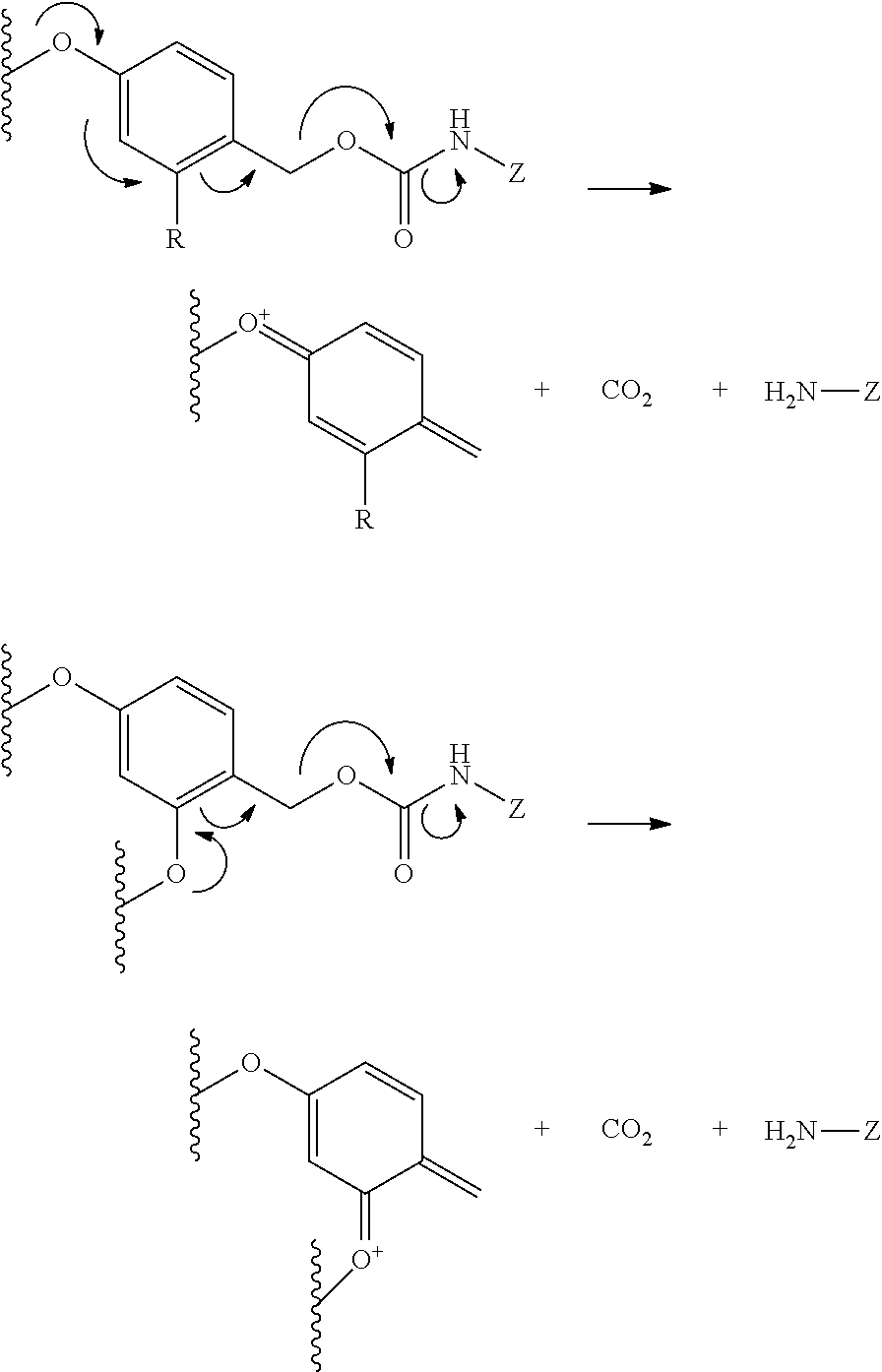
where Z is the vitamin, or analog or derivative thereof, or the drug, or analog or derivative thereof, or each is a vitamin or drug moiety in conjunction with other portions of the polyvalent linker, such as a drug or vitamin moiety including one or more spacer linkers and/or other releasable linkers. In this embodiment, acid-catalyzed elimination of the carbamate leads to the release of CO2 and the nitrogen-containing moiety attached to Z, and the formation of a benzyl cation, which may be trapped by water, or any other Lewis base.

where n is an integer selected from 1 to about 4; Ra and Rb are each independently selected from the group consisting of hydrogen and alkyl, including lower alkyl such as C1-C4 alkyl that are optionally branched; or Ra and Rb are taken together with the attached carbon atom to form a carbocyclic ring; R is an optionally substituted alkyl group, an optionally substituted acyl group, or a suitably selected nitrogen protecting group; and (*) indicates points of attachment for the drug, vitamin, imaging agent, diagnostic agent, other polyvalent linkers, or other parts of the conjugate.

where m is an integer selected from 1 to about 4; R is an optionally substituted alkyl group, an optionally substituted acyl group, or a suitably selected nitrogen protecting group; and (*) indicates points of attachment for the drug, vitamin, imaging agent, diagnostic agent, other polyvalent linkers, or other parts of the conjugate.

where m is an integer selected from 1 to about 4; R is an optionally substituted alkyl group, an optionally substituted acyl group, or a suitably selected nitrogen protecting group; and (*) indicates points of attachment for the drug, vitamin, imaging agent, diagnostic agent, other polyvalent linkers, or other parts of the conjugate.

where the integers x and y are 1, 2, 3, 4, or 5:
—N(R)—(CR′R″)q—C(O)—
where R is hydrogen, alkyl, acyl, or a suitable nitrogen protecting group, R′ and R″ are hydrogen or a substituent, each of which is independently selected in each occurrence, and q is an integer such as 1, 2, 3, 4, or 5. Illustratively. R′ and/or R″ independently correspond to, but are not limited to, hydrogen or the side chains present on naturally occurring amino acids, such as methyl, benzyl, hydroxymethyl, thiomethyl, carboxyl, carboxylmethyl, guanidinopropyl, and the like, and derivatives and protected derivatives thereof. The above described formula includes all stereoisomeric variations. For example, the amino acid may be selected from asparagine, aspartic acid, cysteine, glutamic acid, lysine, glutamine, arginine, serine, ornitine, threonine, and the like. In one variation, the amino acid may be selected from phenylalanine, tyrosine, and the like, derivatives thereof, and substituted variants thereof.

where n is an integer from 1 to 6, the alkyl group is optionally substituted, and the methyl is optionally substituted with an additional alkyl or optionally substituted aryl group, each of which is represented by an independently selected group R. The (*) symbols indicate points of attachment of the polyvalent linker fragment to other parts of the conjugates described herein.

where n is an integer from 1 to 6, and the alkyl group is optionally substituted. The (*) symbols indicate points of attachment of the polyvalent linker fragment to other parts of the conjugates described herein. In another embodiment, the polyvalent linker includes spacer linkers and releasable linkers connected to form a polyvalent 3-thioalkylsulfonylalkyl(disubstituted silyl)oxy group, where the disubstituted silyl is substituted with alkyl and/or optionally substituted aryl groups.

where n is an integer from 1 to 6, the alkyl group is optionally substituted, and the hydrazide forms an hydrazone with (B), (D), or another part of the polyvalent linker (L). The (*) symbols indicate points of attachment of the polyvalent linker fragment to other parts of the conjugates described herein.

where each n is an independently selected integer from 1 to 6, each alkyl group independently selected and is optionally substituted, such as with alkyl or optionally substituted aryl, and where the alkylidene forms an hydrazone with (B), (D), or another part of the polyvalent linker (L). The (*) symbols indicate points of attachment of the polyvalent linker fragment to other parts of the conjugates described herein.
 |
|
|
 |
|
|
 |
 |
 |
|
|
 |
 |
 |
|
|
 |
 |
|
|
|
|
 |
 |
 |
|
|
|
|
|
|
|
|
|
|
 |
|
|
|
|
|
|
|
|
 |
 |
 |
|
|
 |
 |

wherein X is RP(O)(OH)CH2— (see, e.g., U.S. Pat. No. 5,968,915 incorporated herein by reference); RP(O)(OH)N(R1)— (see, e.g., U.S. Pat. No. 5,863,536 incorporated herein by reference); RP(O)(OH)O— (see, e.g., U.S. Pat. No. 5,795,877 incorporated herein by reference); RN(OH)C(O)Y— or RC(O)NH(OH)Y, wherein Y is —CR1R2—, —NR3— or —O— (see, e.g., U.S. Pat. No. 5,962,521 incorporated herein by reference); RS(O)Y, RSO2Y, or RS(O)(NH)Y, wherein Y is —CR1R2—, —NR3— or —O— (see, e.g., U.S. Pat. No. 5,902,817 incorporated herein by reference); and RS-alkyl, wherein R is for example hydrogen, alkyl, aryl, or arylalkyl, each of which may be optionally substituted (see, e.g., J. Med. Chem. 46:1989-1996 (2003) incorporated herein by reference).

with the dissociation constants (Ki values) shown for the E-I complex (see, Current Medicinal Chem. 8:949-.957 (2001); Silverman, “The Organic Chemistry of Drug Design and Drug Action,” Elsevier Academic Press (2nd Ed. 2003), the disclosures of which are incorporated herein by reference);

with the inhibition constants (IC50 values) shown for the E-I complex.

wherein R1 and R2 are each selected from hydrogen, optionally substituted carboxylic acids, such as thiolacetic acids, thiolpropionic acids, and the like; malonic acids, succinic acids, glutamic acids, adipic acids, and the like; and others. Illustrative aminocarbonylpentanedioic acid derivatives are described in J. Med. Chem. 44:298-301 (2001) and J. Med. Chem. 47:1729-38 (2004), the disclosures of which are incorporated herein by reference.
 |
| R1 | Ki (nM) | ||
 |
6.9 (R = H) 29 (R = tert-Bu) | ||
 |
8 | ||
 |
2.1 (R = H) 5.9 (R = OH) | ||
 |
12 (R = H) 3.0 (R = OH) | ||
 |
0.9 (R = H) 5.3 (R = CH2CH2CN) | ||
 |
335 | ||

wherein Q is a an amino dicarboxylic acid, such as aspartic acid, glutamic acid, or an analog thereof, n and m are each selected from an integer between 1 and about 6, and (*) represents the point of attachment for the linker L.


wherein R is independently selected in each instance H, alkyl, heteroalkyl, cycloalkyl, heterocyclyl, alkenyl, alkynyl, aryl, heteroaryl, arylalkyl, heteroarylalkyl, and the like, each of which is optionally substituted. It is to be understood that one R includes a heteroatom, such as nitro, oxygen, or sulfur, and is the point of attachment of linker L. Illustratively, the following chelating groups are described:

where X is oxygen, nitrogen, or sulfur, and where X is attached to linker L, and n is an integer from 1 to about 5.

where X is oxygen, nitrogen, or sulfur, and where X is attached to linker L; Y is ORa, NRa 2, or NRa 3 +; and Y′ is O, NRa, or NRa 2 +; where each R is independently selected in each instance from H, fluoro, sulfonic acid, sulfonate, and salts thereof, and the like; and Ra is hydrogen or alkyl.

where X is oxygen, nitrogen, or sulfur, and where X is attached to linker L; and each R is independently selected in each instance from H, alkyl, heteroalkyl, and the like; and n is an integer from 0 to about 4.


Related analogs where n is an integer from 4 to about 30 may also be prepared according to the processes described herein.

Related analogs where n is an integer from 4 to about 30 may also be prepared according to the processes described herein.


Claims (17)

Priority Applications (1)
| Application Number | Priority Date | Filing Date | Title |
|---|---|---|---|
| US15/990,144 US10624971B2 (en) | 2007-08-17 | 2018-05-25 | PSMA binding ligand-linker conjugates and methods for using |
Applications Claiming Priority (7)
| Application Number | Priority Date | Filing Date | Title |
|---|---|---|---|
| US95648907P | 2007-08-17 | 2007-08-17 | |
| US7435808P | 2008-06-20 | 2008-06-20 | |
| PCT/US2008/073375 WO2009026177A1 (en) | 2007-08-17 | 2008-08-15 | Psma binding ligand-linker conjugates and methods for using |
| US12/673,931 US9193763B2 (en) | 2007-08-17 | 2008-08-15 | PSMA binding ligand-linker conjugates and methods for using |
| US14/794,482 US10046054B2 (en) | 2007-08-17 | 2015-11-23 | PSMA binding ligand-linker conjugates and methods for using |
| US15/606,913 US10406240B2 (en) | 2007-08-17 | 2017-05-26 | PSMA binding ligand-linker conjugates and methods for using |
| US15/990,144 US10624971B2 (en) | 2007-08-17 | 2018-05-25 | PSMA binding ligand-linker conjugates and methods for using |
Related Parent Applications (1)
| Application Number | Title | Priority Date | Filing Date |
|---|---|---|---|
| US15/606,913 Continuation US10406240B2 (en) | 2007-08-17 | 2017-05-26 | PSMA binding ligand-linker conjugates and methods for using |
Publications (2)
| Publication Number | Publication Date |
|---|---|
| US20180289829A1 US20180289829A1 (en) | 2018-10-11 |
| US10624971B2 true US10624971B2 (en) | 2020-04-21 |
Family
ID=40378553
Family Applications (24)
| Application Number | Title | Priority Date | Filing Date |
|---|---|---|---|
| US12/673,931 Active 2030-11-16 US9193763B2 (en) | 2007-08-17 | 2008-08-15 | PSMA binding ligand-linker conjugates and methods for using |
| US13/826,079 Active US8907058B2 (en) | 2007-08-17 | 2013-03-14 | PSMA binding ligand-linker conjugates and methods for using |
| US14/794,482 Active US10046054B2 (en) | 2007-08-17 | 2015-11-23 | PSMA binding ligand-linker conjugates and methods for using |
| US15/606,913 Active US10406240B2 (en) | 2007-08-17 | 2017-05-26 | PSMA binding ligand-linker conjugates and methods for using |
| US15/959,110 Active US10624969B2 (en) | 2007-08-17 | 2018-04-20 | PSMA binding ligand-linker conjugates and methods for using |
| US15/990,136 Active US10646581B2 (en) | 2007-08-17 | 2018-05-25 | PSMA binding ligand-linker conjugates and methods for using |
| US15/990,152 Active US10828282B2 (en) | 2007-08-17 | 2018-05-25 | PSMA binding ligand-linker conjugates and methods for using |
| US15/990,144 Active US10624971B2 (en) | 2007-08-17 | 2018-05-25 | PSMA binding ligand-linker conjugates and methods for using |
| US15/990,095 Active US10624970B2 (en) | 2007-08-17 | 2018-05-25 | PSMA binding ligand-linker conjugates and methods for using |
| US15/990,083 Active US10517956B2 (en) | 2007-08-17 | 2018-05-25 | PSMA binding ligand-linker conjugates and methods for using |
| US15/990,111 Active US10485878B2 (en) | 2007-08-17 | 2018-05-25 | PSMA binding ligand-linker conjugates and methods for using |
| US15/997,451 Active US10517957B2 (en) | 2007-08-17 | 2018-06-04 | PSMA binding ligand-linker conjugates and methods for using |
| US16/507,713 Active US11298341B2 (en) | 2007-08-17 | 2019-07-10 | PSMA binding ligand-linker conjugates and methods for using |
| US16/717,753 Abandoned US20200188523A1 (en) | 2007-08-17 | 2019-12-17 | Psma binding ligand-linker conjugates and methods for using |
| US16/749,896 Abandoned US20200297701A1 (en) | 2007-08-17 | 2020-01-22 | Psma binding ligand-linker conjugates and methods for using |
| US16/749,827 Active US11083710B2 (en) | 2007-08-17 | 2020-01-22 | PSMA binding ligand-linker conjugates and methods for using |
| US16/869,268 Active 2028-10-26 US11504357B2 (en) | 2007-08-17 | 2020-05-07 | PSMA binding ligand-linker conjugates and methods for using |
| US17/086,063 Active 2029-07-19 US11717514B2 (en) | 2007-08-17 | 2020-10-30 | PSMA binding ligand-linker conjugates and methods for using |
| US17/358,726 Pending US20220096445A1 (en) | 2007-08-17 | 2021-06-25 | Psma binding ligand-linker conjugates and methods for using |
| US17/359,277 Active US11369590B2 (en) | 2007-08-17 | 2021-06-25 | PSMA binding ligand-linker conjugates and methods for using |
| US17/359,314 Active US11318121B2 (en) | 2007-08-17 | 2021-06-25 | PSMA binding ligand-linker conjugates and methods for using |
| US17/573,304 Pending US20230000836A1 (en) | 2007-08-17 | 2022-01-11 | Psma binding ligand-linker conjugates and methods for using |
| US17/991,013 Pending US20230293497A1 (en) | 2007-08-17 | 2022-11-21 | Psma binding ligand-linker conjugates and methods for using |
| US18/181,487 Pending US20230346752A1 (en) | 2007-08-17 | 2023-03-09 | Psma binding ligand-linker conjugates and methods for using |
Family Applications Before (7)
| Application Number | Title | Priority Date | Filing Date |
|---|---|---|---|
| US12/673,931 Active 2030-11-16 US9193763B2 (en) | 2007-08-17 | 2008-08-15 | PSMA binding ligand-linker conjugates and methods for using |
| US13/826,079 Active US8907058B2 (en) | 2007-08-17 | 2013-03-14 | PSMA binding ligand-linker conjugates and methods for using |
| US14/794,482 Active US10046054B2 (en) | 2007-08-17 | 2015-11-23 | PSMA binding ligand-linker conjugates and methods for using |
| US15/606,913 Active US10406240B2 (en) | 2007-08-17 | 2017-05-26 | PSMA binding ligand-linker conjugates and methods for using |
| US15/959,110 Active US10624969B2 (en) | 2007-08-17 | 2018-04-20 | PSMA binding ligand-linker conjugates and methods for using |
| US15/990,136 Active US10646581B2 (en) | 2007-08-17 | 2018-05-25 | PSMA binding ligand-linker conjugates and methods for using |
| US15/990,152 Active US10828282B2 (en) | 2007-08-17 | 2018-05-25 | PSMA binding ligand-linker conjugates and methods for using |
Family Applications After (16)
| Application Number | Title | Priority Date | Filing Date |
|---|---|---|---|
| US15/990,095 Active US10624970B2 (en) | 2007-08-17 | 2018-05-25 | PSMA binding ligand-linker conjugates and methods for using |
| US15/990,083 Active US10517956B2 (en) | 2007-08-17 | 2018-05-25 | PSMA binding ligand-linker conjugates and methods for using |
| US15/990,111 Active US10485878B2 (en) | 2007-08-17 | 2018-05-25 | PSMA binding ligand-linker conjugates and methods for using |
| US15/997,451 Active US10517957B2 (en) | 2007-08-17 | 2018-06-04 | PSMA binding ligand-linker conjugates and methods for using |
| US16/507,713 Active US11298341B2 (en) | 2007-08-17 | 2019-07-10 | PSMA binding ligand-linker conjugates and methods for using |
| US16/717,753 Abandoned US20200188523A1 (en) | 2007-08-17 | 2019-12-17 | Psma binding ligand-linker conjugates and methods for using |
| US16/749,896 Abandoned US20200297701A1 (en) | 2007-08-17 | 2020-01-22 | Psma binding ligand-linker conjugates and methods for using |
| US16/749,827 Active US11083710B2 (en) | 2007-08-17 | 2020-01-22 | PSMA binding ligand-linker conjugates and methods for using |
| US16/869,268 Active 2028-10-26 US11504357B2 (en) | 2007-08-17 | 2020-05-07 | PSMA binding ligand-linker conjugates and methods for using |
| US17/086,063 Active 2029-07-19 US11717514B2 (en) | 2007-08-17 | 2020-10-30 | PSMA binding ligand-linker conjugates and methods for using |
| US17/358,726 Pending US20220096445A1 (en) | 2007-08-17 | 2021-06-25 | Psma binding ligand-linker conjugates and methods for using |
| US17/359,277 Active US11369590B2 (en) | 2007-08-17 | 2021-06-25 | PSMA binding ligand-linker conjugates and methods for using |
| US17/359,314 Active US11318121B2 (en) | 2007-08-17 | 2021-06-25 | PSMA binding ligand-linker conjugates and methods for using |
| US17/573,304 Pending US20230000836A1 (en) | 2007-08-17 | 2022-01-11 | Psma binding ligand-linker conjugates and methods for using |
| US17/991,013 Pending US20230293497A1 (en) | 2007-08-17 | 2022-11-21 | Psma binding ligand-linker conjugates and methods for using |
| US18/181,487 Pending US20230346752A1 (en) | 2007-08-17 | 2023-03-09 | Psma binding ligand-linker conjugates and methods for using |
Country Status (16)
| Country | Link |
|---|---|
| US (24) | US9193763B2 (en) |
| EP (6) | EP3838298A3 (en) |
| JP (8) | JP2010536790A (en) |
| CN (2) | CN104873982A (en) |
| AU (1) | AU2008289108B2 (en) |
| CA (1) | CA2696627C (en) |
| DK (1) | DK2187965T3 (en) |
| ES (1) | ES2768224T3 (en) |
| HK (1) | HK1212588A1 (en) |
| HU (1) | HUE047200T2 (en) |
| IL (1) | IL203998A (en) |
| NZ (2) | NZ583931A (en) |
| PL (1) | PL2187965T3 (en) |
| PT (1) | PT2187965T (en) |
| SI (1) | SI2187965T1 (en) |
| WO (1) | WO2009026177A1 (en) |
Cited By (5)
| Publication number | Priority date | Publication date | Assignee | Title |
|---|---|---|---|---|
| US10828282B2 (en) | 2007-08-17 | 2020-11-10 | Purdue Research Foundation | PSMA binding ligand-linker conjugates and methods for using |
| US11202836B2 (en) | 2018-03-23 | 2021-12-21 | Case Western Reserve University | PSMA targeted conjugate compounds and uses thereof |
| US11708393B2 (en) | 2011-11-08 | 2023-07-25 | Case Western Reserve University | Targeted non-invasive imaging probes of EGFR expressing cells |
| US11931430B2 (en) | 2013-10-18 | 2024-03-19 | Novartis Ag | Labeled inhibitors of prostate specific membrane antigen (PSMA) as agents for the treatment of prostate cancer |
| US11951190B2 (en) | 2013-10-18 | 2024-04-09 | Novartis Ag | Use of labeled inhibitors of prostate specific membrane antigen (PSMA), as agents for the treatment of prostate cancer |
Families Citing this family (101)
| Publication number | Priority date | Publication date | Assignee | Title |
|---|---|---|---|---|
| NZ529808A (en) | 2001-05-02 | 2007-02-23 | Purdue Research Foundation | Treatment and diagnosis of macrophage mediated disease |
| US8043603B2 (en) | 2002-02-07 | 2011-10-25 | Endocyte, Inc. | Folate targeted enhanced tumor and folate receptor positive tissue optical imaging technology |
| US8043602B2 (en) | 2002-02-07 | 2011-10-25 | Endocyte, Inc. | Folate targeted enhanced tumor and folate receptor positive tissue optical imaging technology |
| CA2527196C (en) | 2003-05-30 | 2012-10-16 | Purdue Research Foundation | Diagnostic method for atherosclerosis |
| WO2006012527A1 (en) | 2004-07-23 | 2006-02-02 | Endocyte, Inc. | Bivalent linkers and conjugates thereof |
| JP5175723B2 (en) | 2005-07-05 | 2013-04-03 | パーデュー・リサーチ・ファウンデーション | Preparation of compositions for treating monocyte-mediated diseases |
| US8795633B2 (en) | 2005-09-23 | 2014-08-05 | Purdue Research Foundation | Multiphoton in vivo flow cytometry method and device |
| CA2668197A1 (en) | 2006-11-03 | 2008-05-15 | Philip S. Low | Ex vivo flow cytometry method and device |
| HUE026216T2 (en) * | 2006-11-08 | 2016-05-30 | Molecular Insight Pharm Inc | Heterodimers of glutamic acid |
| EP2567711A3 (en) | 2007-02-07 | 2013-05-01 | Purdue Research Foundation | Positron emission tomography imaging method |
| AU2008224988A1 (en) | 2007-03-14 | 2008-09-18 | Endocyte, Inc. | Binding ligand linked drug delivery conjugates of tubulysins |
| WO2008148001A2 (en) | 2007-05-25 | 2008-12-04 | Purdue Research Foundation | Method of imaging localized infections |
| US9877965B2 (en) | 2007-06-25 | 2018-01-30 | Endocyte, Inc. | Vitamin receptor drug delivery conjugates for treating inflammation |
| CN104383553A (en) | 2007-06-25 | 2015-03-04 | 恩多塞特公司 | Conjugates containing hydrophilic spacer linkers |
| GB0718967D0 (en) * | 2007-09-28 | 2007-11-07 | Ge Healthcare Ltd | Peptide imaging agents |
| US8852630B2 (en) | 2008-05-13 | 2014-10-07 | Yale University | Chimeric small molecules for the recruitment of antibodies to cancer cells |
| PT2291659E (en) * | 2008-05-13 | 2016-01-22 | Univ Yale | Chimeric small molecules for the recruitment of antibodies to cancer cells |
| WO2010108125A2 (en) | 2009-03-19 | 2010-09-23 | The Johns Hopkins University | Psma-targeting compounds and uses thereof |
| US20180009767A9 (en) | 2009-03-19 | 2018-01-11 | The Johns Hopkins University | Psma targeted fluorescent agents for image guided surgery |
| WO2010135313A1 (en) | 2009-05-19 | 2010-11-25 | Aic Blab Company | Composite current collector and methods therefor |
| US8394922B2 (en) | 2009-08-03 | 2013-03-12 | Medarex, Inc. | Antiproliferative compounds, conjugates thereof, methods therefor, and uses thereof |
| CA2790577A1 (en) * | 2010-02-25 | 2011-09-01 | Purdue Research Foundation | Psma binding ligand-linker conjugates and methods for using |
| US9951324B2 (en) | 2010-02-25 | 2018-04-24 | Purdue Research Foundation | PSMA binding ligand-linker conjugates and methods for using |
| US10363309B2 (en) | 2011-02-04 | 2019-07-30 | Case Western Reserve University | Targeted nanoparticle conjugates |
| BR112013031819B1 (en) | 2011-06-10 | 2022-05-03 | Mersana Therapeutics, Inc | Polymeric support, pharmaceutical composition, compound and support use |
| EP2721039B1 (en) * | 2011-06-15 | 2018-01-10 | Cancer Targeted Technology LLC | Chelated psma inhibitors |
| US8784774B2 (en) | 2011-09-16 | 2014-07-22 | General Electric Company | Labeled molecular imaging agents and methods of use |
| WO2013126797A1 (en) * | 2012-02-24 | 2013-08-29 | Purdue Research Foundation | Cholecystokinin b receptor targeting for imaging and therapy |
| US20140080175A1 (en) | 2012-03-29 | 2014-03-20 | Endocyte, Inc. | Processes for preparing tubulysin derivatives and conjugates thereof |
| LT3210627T (en) | 2012-07-12 | 2023-04-11 | Hangzhou Dac Biotech Co., Ltd | Conjugates of cell binding molecules with cytotoxic agents |
| US20140081659A1 (en) | 2012-09-17 | 2014-03-20 | Depuy Orthopaedics, Inc. | Systems and methods for surgical and interventional planning, support, post-operative follow-up, and functional recovery tracking |
| KR20150070318A (en) | 2012-10-16 | 2015-06-24 | 엔도사이트, 인코포레이티드 | Drug delivery conjugates containing unnatural amino acids and methods for using |
| US9636413B2 (en) | 2012-11-15 | 2017-05-02 | Endocyte, Inc. | Conjugates for treating diseases caused by PSMA expressing cells |
| ES2701076T3 (en) | 2012-11-24 | 2019-02-20 | Hangzhou Dac Biotech Co Ltd | Hydrophilic linkers and their uses for the conjugation of drugs to molecules that bind to cells |
| JP6334553B2 (en) | 2012-12-10 | 2018-05-30 | メルサナ セラピューティクス,インコーポレイティド | Protein-polymer-drug conjugate |
| US9872918B2 (en) | 2012-12-12 | 2018-01-23 | Mersana Therapeutics, Inc. | Hydroxyl-polymer-drug-protein conjugates |
| RS56169B1 (en) | 2013-02-14 | 2017-11-30 | Bristol Myers Squibb Co | Tubulysin compounds, methods of making and use |
| US10207005B2 (en) | 2013-02-15 | 2019-02-19 | Case Western Reserve University | Photodynamic therapy composition |
| EP2958596B1 (en) * | 2013-02-15 | 2019-12-04 | Case Western Reserve University | Psma ligands and uses thereof |
| WO2014153164A1 (en) | 2013-03-14 | 2014-09-25 | The California Institute For Biomedical Research | Targeting agent antibody conjugates and uses thereof |
| US10434194B2 (en) | 2013-06-20 | 2019-10-08 | Case Western Reserve University | PSMA targeted nanobubbles for diagnostic and therapeutic applications |
| WO2015057692A1 (en) * | 2013-10-14 | 2015-04-23 | The Johns Hopkins University | Prostate-specific membrane antigen-targeted photosensitizers for photodynamic therapy |
| KR102339240B1 (en) | 2013-10-15 | 2021-12-15 | 더 스크립스 리서치 인스티튜트 | Peptidic chimeric antigen receptor t cell switches and uses thereof |
| CN105814083A (en) * | 2013-10-15 | 2016-07-27 | 加州生物医学研究所 | Chimeric antigen receptor T cell switches and uses thereof |
| JP2017501115A (en) * | 2013-11-06 | 2017-01-12 | パーデュー・リサーチ・ファウンデーションPurdue Research Foundation | DUPA-indenoisoquinoline complex |
| WO2015073678A1 (en) * | 2013-11-14 | 2015-05-21 | Endocyte, Inc. | Compounds for positron emission tomography |
| US9468693B2 (en) | 2014-01-23 | 2016-10-18 | General Electric Company | Labeled molecular imaging agents and methods of use |
| US9468692B2 (en) | 2014-01-23 | 2016-10-18 | General Electric Company | Labeled molecular imaging agents and methods of use |
| EP3122757B1 (en) | 2014-02-28 | 2023-09-06 | Hangzhou Dac Biotech Co., Ltd | Charged linkers and their uses for conjugation |
| EP3140282B1 (en) | 2014-05-06 | 2019-07-10 | The Johns Hopkins University | Metal/radiometal-labeled psma inhibitors for psma-targeted imaging and radiotherapy |
| JP6753843B2 (en) | 2014-05-14 | 2020-09-16 | タルグルムーネ セラピウティクス エージー | Improved Polyethylene Imine Polyethylene Glycol Vector |
| CN104262458B (en) * | 2014-08-29 | 2017-02-01 | 邵国强 | Polypeptide in targeted combination with PSMA (prostate specific membrane antigen) membrane out-region, radionuclide labeled polypeptide and application thereof |
| JPWO2016047794A1 (en) * | 2014-09-26 | 2017-09-28 | 株式会社カネカ | Method for producing hydrophobic peptide |
| WO2016065145A2 (en) | 2014-10-22 | 2016-04-28 | The Johns Hopkins University | Psma targeted reversed carbamates and methods of use thereof |
| US10077287B2 (en) | 2014-11-10 | 2018-09-18 | Bristol-Myers Squibb Company | Tubulysin analogs and methods of making and use |
| US10188759B2 (en) | 2015-01-07 | 2019-01-29 | Endocyte, Inc. | Conjugates for imaging |
| US10800828B2 (en) | 2015-03-26 | 2020-10-13 | The Scripps Research Institute | Switchable non-scFv chimeric receptors, switches, and methods of use thereof to treat cancer |
| WO2016168773A2 (en) | 2015-04-15 | 2016-10-20 | The California Institute For Biomedical Research | Optimized pne-based chimeric receptor t cell switches and uses thereof |
| US10406238B2 (en) | 2015-05-11 | 2019-09-10 | Purdue Research Foundation | Ligand ionophore conjugates |
| EP3319936A4 (en) | 2015-07-12 | 2019-02-06 | Suzhou M-conj Biotech Co., Ltd. | Bridge linkers for conjugation of cell-binding molecules |
| US9839687B2 (en) | 2015-07-15 | 2017-12-12 | Suzhou M-Conj Biotech Co., Ltd. | Acetylenedicarboxyl linkers and their uses in specific conjugation of a cell-binding molecule |
| EP3334466B1 (en) | 2015-08-14 | 2020-05-13 | Endocyte, Inc. | Method of imaging with a chelating compound |
| US9808538B2 (en) * | 2015-09-09 | 2017-11-07 | On Target Laboratories, LLC | PSMA-targeted NIR dyes and their uses |
| US10842887B2 (en) * | 2015-09-09 | 2020-11-24 | On Target Laboratories, LLC | PSMA-targeted NIR dyes and their uses |
| LT3356385T (en) * | 2015-09-30 | 2021-02-25 | Deutsches Krebsforschungszentrum | 18f-tagged inhibitors of prostate specific membrane antigen (psma) and their use as imaging agents for prostate cancer |
| CA3002774A1 (en) * | 2015-10-23 | 2017-04-27 | Sorrento Therapeutics, Inc. | Programmable universal cell receptors and methods of using the same |
| US11793880B2 (en) | 2015-12-04 | 2023-10-24 | Seagen Inc. | Conjugates of quaternized tubulysin compounds |
| EP3383420B1 (en) | 2015-12-04 | 2022-03-23 | Seagen Inc. | Conjugates of quaternized tubulysin compounds |
| EP3429637A4 (en) | 2016-03-16 | 2020-03-11 | Endocyte, Inc. | Carbonic anhydrase ix inhibitor conjugates and uses thereof |
| WO2017161170A1 (en) * | 2016-03-16 | 2017-09-21 | Purdue Research Foundation | Carbonic anhydrase ix targeting agents and methods |
| US20170296679A1 (en) | 2016-04-18 | 2017-10-19 | Intuitive Surgical Operations, Inc. | Compositions of Near IR Closed Chain, Sulfo-Cyanine Dyes and Prostate Specific Membrane Antigen Ligands |
| CA3035542A1 (en) * | 2016-09-09 | 2018-03-15 | On Target Laboratories, LLC | Psma-targeted nir dyes and their uses |
| CA3040343A1 (en) | 2016-10-19 | 2018-04-26 | California Institute For Biomedical Research | Chimeric antigen receptor effector cell switches with humanized targeting moieties and/or optimized chimeric antigen receptor interacting domains and uses thereof |
| KR20220147721A (en) | 2016-11-14 | 2022-11-03 | 항저우 디에이씨 바이오테크 씨오, 엘티디 | Conjugation linkers, cell binding molecule-drug conjugates containing the likers, methods of making and uses such conjugates with the linkers |
| CN110167577A (en) * | 2016-11-16 | 2019-08-23 | 普渡研究基金会 | Ligand ion carrier conjugate |
| CN116474108A (en) | 2016-12-14 | 2023-07-25 | 普渡研究基金会 | Fibroblast Activation Protein (FAP) -targeted imaging and therapy |
| KR102085798B1 (en) | 2016-12-28 | 2020-03-06 | 주식회사 인투셀 | Compounds comprising beta-galactoside self-immolative linker |
| US11524082B2 (en) | 2017-08-22 | 2022-12-13 | Purdue Research Foundation | FBSA-based therapeutic and radioimaging conjugates targeting carbonic anhydrase positive cancers |
| DK3684416T3 (en) * | 2017-09-22 | 2023-05-15 | Heidelberg Pharma Res Gmbh | PSMA-TARGETING AMANITIN CONJUGATES |
| WO2019157037A1 (en) | 2018-02-06 | 2019-08-15 | The Johns Hopkins University | Psma targeted radiohalogenated urea-polyaminocarboxylates for cancer radiotherapy |
| US20210276971A1 (en) * | 2018-06-20 | 2021-09-09 | The Research Foundation For The State University Of New York | Triazamacrocycle-derived chelator compositions for coordination of imaging and therapy metal ions and methods of using same |
| CN110922450B (en) * | 2018-09-19 | 2021-11-02 | 薪火炙药(北京)科技有限公司 | PSMA activated antitumor prodrug CPT-X and preparation method and application thereof |
| RU2697519C1 (en) * | 2018-10-15 | 2019-08-15 | Общество с ограниченной ответственностью "Изварино Фарма" | Peptide agent comprising a psma-binding ligand based on a urea derivative, a method for production thereof and use thereof for preparing a conjugate with a drug and diagnostic agent |
| WO2020108753A1 (en) * | 2018-11-28 | 2020-06-04 | ITM Isotopen Technologien München AG | Novel tumor antigen binding agents and uses thereof |
| EP3893942A4 (en) * | 2018-12-14 | 2022-12-14 | The Johns Hopkins University | Psma-targeted pamam dendrimers for specific delivery of imaging, contrast and therapeutic agents |
| WO2020150740A1 (en) * | 2019-01-18 | 2020-07-23 | Case Western Reserve University | Psma ligand targeted compounds and uses thereof |
| JP7421232B2 (en) | 2019-01-30 | 2024-01-24 | テクニシェ ユニバーシタット ミュンヘン | Silicon fluoride acceptor-substituted radiopharmaceuticals and their precursors |
| CN110305187B (en) * | 2019-06-06 | 2021-02-23 | 原子高科股份有限公司 | Prostate cancer PET diagnostic reagent68Ga-NOTA-ANCP-PSMA and preparation method and application thereof |
| CN110305186B (en) * | 2019-06-06 | 2021-02-23 | 原子高科股份有限公司 | Prostate cancer PET diagnostic reagent68Ga-DOTA-ANCP-PSMA and preparation method and application thereof |
| JP2022540395A (en) | 2019-06-29 | 2022-09-15 | ハンジョウ ディーエーシー バイオテック シーオー.,エルティディ. | Cell-binding molecule-tubulysin derivative conjugate and method for preparing the same |
| RU2713151C1 (en) * | 2019-07-02 | 2020-02-04 | Общество с ограниченной ответственностью "Изварино Фарма" | Conjugate of a fluorescent dye with a peptide substance which contains a psma-binding ligand based on a urea derivative for visualizing cells expressing psma, a method for production thereof and use thereof |
| AU2020311925A1 (en) | 2019-07-10 | 2022-02-03 | Cybrexa 3, Inc. | Peptide conjugates of microtubule-targeting agents as therapeutics |
| PE20220563A1 (en) | 2019-07-10 | 2022-04-13 | Cybrexa 2 Inc | PEPTIDE CONJUGATES OF CYTOTOXINS AS THERAPEUTICS |
| CN114945388A (en) * | 2019-10-23 | 2022-08-26 | 胶原蛋白医疗有限责任公司 | Fibrin-binding compounds for imaging and therapy |
| US20230096410A1 (en) | 2020-03-06 | 2023-03-30 | Sorrento Therapeutics, Inc. | Innate Immunity Killer Cells Targeting PSMA Positive Tumor Cells |
| WO2021250240A1 (en) | 2020-06-12 | 2021-12-16 | Orano | Diaza-18-crown-6 derivative useful for chelating radium, conjugate and radium chelate comprising the same, and uses thereof |
| WO2022074152A1 (en) | 2020-10-08 | 2022-04-14 | Targimmune Therapeutics Ag | Immunotherapy for the treatment of cancer |
| US20230406847A1 (en) | 2020-11-12 | 2023-12-21 | Abx Advanced Biochemical Compounds Gmbh | Ligands of prostate specific membrane antigen (psma) containing heteroaromatic linker building blocks |
| US20230414762A1 (en) * | 2020-11-17 | 2023-12-28 | The Johns Hopkins University | Prostate-specific membrane antigen (psma)-targeted prodrug for selective killing of cells expressing psma |
| CA3212963A1 (en) * | 2021-04-23 | 2022-10-27 | Reinier HERNANDEZ | Psma-targeting ligands with optimal properties for imaging and therapy |
| WO2023079142A2 (en) | 2021-11-05 | 2023-05-11 | Targimmune Therapeutics Ag | Targeted linear conjugates comprising polyethyleneimine and polyethylene glycol and polyplexes comprising the same |
Citations (219)
| Publication number | Priority date | Publication date | Assignee | Title |
|---|---|---|---|---|
| EP0116208A1 (en) | 1982-12-07 | 1984-08-22 | Kyowa Hakko Kogyo Co., Ltd. | Mitomycin analogues |
| US4691024A (en) | 1984-06-01 | 1987-09-01 | Kyowa Hakko Kogyo Kabushiki Kaisha | New mitomycin derivatives, preparation thereof and pharmaceutical compositions containing them |
| US4713249A (en) | 1981-11-12 | 1987-12-15 | Schroeder Ulf | Crystallized carbohydrate matrix for biologically active substances, a process of preparing said matrix, and the use thereof |
| WO1988001622A1 (en) | 1986-08-29 | 1988-03-10 | Kyowa Hakko Kogyo Kabusiki Kaisha | Mitomycin derivatives |
| WO1991007418A1 (en) | 1989-11-13 | 1991-05-30 | Xoma Corporation | Chimeric mouse-human a10 antibody with specificity to a human tumor cell antigen |
| US5266333A (en) | 1985-03-06 | 1993-11-30 | American Cyanamid Company | Water dispersible and water soluble carbohydrate polymer compositions for parenteral administration of growth hormone |
| US5418982A (en) | 1990-12-21 | 1995-05-30 | Japanic Corporation | Raw sewage disposal apparatus and prefab for accommodating the same |
| US5627165A (en) | 1990-06-13 | 1997-05-06 | Drug Innovation & Design, Inc. | Phosphorous prodrugs and therapeutic delivery systems using same |
| US5795877A (en) | 1996-12-31 | 1998-08-18 | Guilford Pharmaceuticals Inc. | Inhibitors of NAALADase enzyme activity |
| US5863536A (en) | 1996-12-31 | 1999-01-26 | Guilford Pharmaceuticals Inc. | Phosphoramidate derivatives |
| US5866679A (en) | 1994-06-28 | 1999-02-02 | Merck & Co., Inc. | Peptides |
| US5902817A (en) | 1997-04-09 | 1999-05-11 | Guilford Pharmaceuticals Inc. | Certain sulfoxide and sulfone derivatives |
| US5948750A (en) | 1996-10-30 | 1999-09-07 | Merck & Co., Inc. | Conjugates useful in the treatment of prostate cancer |
| WO1999045374A2 (en) | 1998-03-03 | 1999-09-10 | Mosaic Technologies | Purification and detection processes using reversible affinity electrophoresis |
| US5962521A (en) | 1997-04-04 | 1999-10-05 | Guilford Pharmaceuticals Inc. | Hydroxamic acid derivatives |
| US5962237A (en) | 1996-04-05 | 1999-10-05 | The Johns Hopkins University School Of Medicine | Method of enriching rare cells |
| US5968915A (en) | 1996-06-17 | 1999-10-19 | Guilford Pharmaceuticals Inc. | Naaladase inhibitors |
| US5998362A (en) | 1996-09-12 | 1999-12-07 | Merck & Co., Inc. | Conjugates useful in the treatment of prostate cancer |
| US6054444A (en) | 1997-04-24 | 2000-04-25 | Guilford Pharmaceuticals Inc. | Phosphonic acid derivatives |
| US6127333A (en) | 1997-07-10 | 2000-10-03 | Merck & Co., Inc. | Conjugates useful in the treatment of prostate cancer |
| WO2000064911A1 (en) | 1999-04-28 | 2000-11-02 | Georgetown University | Ligands for metabotropic glutamate receptors |
| WO2000066091A1 (en) | 1999-05-04 | 2000-11-09 | Biotech Australia Pty Limited | Amplification of folate-mediated targeting to tumor cells using polymers |
| US6174858B1 (en) | 1998-11-17 | 2001-01-16 | Merck & Co., Inc. | Conjugates useful in the treatment of prostate cancer |
| US6177404B1 (en) | 1996-10-15 | 2001-01-23 | Merck & Co., Inc. | Conjugates useful in the treatment of benign prostatic hyperplasia |
| US6232287B1 (en) | 1998-03-13 | 2001-05-15 | The Burnham Institute | Molecules that home to various selected organs or tissues |
| US20010031252A1 (en) | 2000-03-31 | 2001-10-18 | Low Philip Stewart | Method of treatment using ligand-immunogen conjugates |
| US20020001782A1 (en) | 1993-11-19 | 2002-01-03 | Fuji Photo Film Co., Ltd | Concentrated fixing solution and method for processing silver halide photographic material using the same |
| US6368598B1 (en) | 1996-09-16 | 2002-04-09 | Jcrt Radiation Oncology Support Services, Inc. | Drug complex for treatment of metastatic prostate cancer |
| US20020055121A1 (en) | 1992-01-31 | 2002-05-09 | Vielkind Juergen R. | Melanoma and prostate cancer specific antibodies for immunodetection and immunotherapy |
| US6391305B1 (en) | 1997-09-10 | 2002-05-21 | Merck & Co., Inc. | Conjugates useful in the treatment of prostate cancer |
| WO2002043773A2 (en) | 2000-12-01 | 2002-06-06 | The Johns Hopkins University | Tissue specific prodrugs |
| US20020103136A1 (en) | 1998-03-05 | 2002-08-01 | Dong-Mei Feng | Conjugates useful in the treatment of prostate cancer |
| US6428785B1 (en) | 1999-10-28 | 2002-08-06 | Immunolytics Inc. | Method and composition for treating prostate cancer |
| WO2002062398A2 (en) | 2001-01-08 | 2002-08-15 | Neorx Corporation | Radioactively labelled conjugates of phosphonates |
| US20020115596A1 (en) | 1997-10-27 | 2002-08-22 | Merk & Co., Inc. | Conjugates useful in the treatment of prostate cancer |
| US20020132983A1 (en) | 2000-11-30 | 2002-09-19 | Junghans Richard P. | Antibodies as chimeric effector cell receptors against tumor antigens |
| WO2002098885A1 (en) | 2001-02-07 | 2002-12-12 | Beth Israel Deaconess Medical Center | Modified psma ligands and uses related thereto |
| WO2003000201A2 (en) | 2001-06-25 | 2003-01-03 | Drug Innovation & Design, Incorporated | Exponential pattern recognition based cellular targeting, compositions, methods and anticancer applications |
| US6528499B1 (en) | 2000-04-27 | 2003-03-04 | Georgetown University | Ligands for metabotropic glutamate receptors and inhibitors of NAALADase |
| US20030086900A1 (en) | 2001-09-28 | 2003-05-08 | Low Philip S. | Method of treatment using ligand-immunogen conjugates |
| US20030133927A1 (en) | 2001-10-10 | 2003-07-17 | Defeo-Jones Deborah | Conjugates useful in the treatment of prostate cancer |
| WO2003060523A1 (en) | 2002-01-10 | 2003-07-24 | Johns Hopkins University | Imaging agents and methods of imaging naaladase of psma |
| US20030138432A1 (en) | 1999-11-15 | 2003-07-24 | Drug Innovation & Design, Inc. | Selective cellular targeting: multifunctional delivery vehicles, multifunctional prodrugs, use as antineoplastic drugs |
| US20030207808A1 (en) | 1999-02-18 | 2003-11-06 | Kinneret Savitzky | Novel nucleic acid and amino acid sequences |
| WO2003092742A1 (en) | 2002-05-06 | 2003-11-13 | Endocyte, Inc. | Vitamin-targeted imaging agents |
| US20030215456A1 (en) | 2001-10-02 | 2003-11-20 | Sui-Long Yao | Method of treating cancer |
| US20030220241A1 (en) | 1999-04-05 | 2003-11-27 | Defeo-Jones Deborah | Method of treating cancer |
| WO2003097647A1 (en) | 2002-05-15 | 2003-11-27 | Endocyte, Inc. | Vitamin-mitomycin conjugates |
| US20030232760A1 (en) | 2001-09-21 | 2003-12-18 | Merck & Co., Inc. | Conjugates useful in the treatment of prostate cancer |
| US20040001846A1 (en) | 1995-02-24 | 2004-01-01 | Sloan-Kettering Institute For Cancer Research | Prostate-specific membrane antigen and uses thereof |
| US20040018203A1 (en) | 2001-06-08 | 2004-01-29 | Ira Pastan | Pegylation of linkers improves antitumor activity and reduces toxicity of immunoconjugates |
| WO2004010957A2 (en) | 2002-07-31 | 2004-02-05 | Seattle Genetics, Inc. | Drug conjugates and their use for treating cancer, an autoimmune disease or an infectious disease |
| US6692724B1 (en) | 1999-10-25 | 2004-02-17 | Board Of Regents, The University Of Texas System | Ethylenedicysteine (EC)-drug conjugates, compositions and methods for tissue specific disease imaging |
| US20040052727A1 (en) | 2002-02-28 | 2004-03-18 | Dalton James T. | Radiolableled selective androgen receptor modulators andtheir use in prostate cancer imaging and therapy |
| US20040058857A1 (en) | 2001-11-29 | 2004-03-25 | Siu-Long Yao | Method of treating cancer |
| US20040146516A1 (en) | 1999-06-17 | 2004-07-29 | Utah Ventures Ii L.P. | Lumen-exposed molecules and methods for targeted delivery |
| WO2004069159A2 (en) | 2003-01-27 | 2004-08-19 | Endocyte, Inc. | Vitamin receptor binding drug delivery conjugates |
| US20040213791A1 (en) | 2001-06-01 | 2004-10-28 | Neil Bander | Modified antibodies to prostate-specific membrane antigen and uses thereof |
| US20040242582A1 (en) | 2001-04-24 | 2004-12-02 | Green Mark A | Folate mimetics and folate-receptor binding conjugates thereof |
| US20050107325A1 (en) | 2003-04-17 | 2005-05-19 | Muthiah Manoharan | Modified iRNA agents |
| US20050119166A1 (en) | 1997-12-02 | 2005-06-02 | Brady Stephen F. | Conjugates useful in the treatment of prostate cancer |
| US20050158780A1 (en) | 2000-10-16 | 2005-07-21 | Gilead Sciences, Inc. | Nucleic acid ligands to the prostate specific membrane antigen |
| WO2005082023A2 (en) | 2004-02-23 | 2005-09-09 | Genentech, Inc. | Heterocyclic self-immolative linkers and conjugates |
| US6946133B1 (en) | 1996-03-20 | 2005-09-20 | The United States Of America As Represented By The Department Of Health And Human Services | Prostate specific antigen oligo-epitope peptide |
| JP2005274569A (en) | 2004-03-15 | 2005-10-06 | F Hoffmann La Roche Ag | Use of cardiac hormone for assessing risk of suffering from cardiovascular complication as consequence of volume overload |
| US20050234247A1 (en) | 2004-01-30 | 2005-10-20 | Ulrich Klar | New effector conjugates, process for their production and their pharmaceutical use |
| US20050239739A1 (en) | 2001-05-18 | 2005-10-27 | Sirna Therapeutics, Inc. | Conjugates and compositions for cellular delivery |
| CA2606138A1 (en) | 2004-04-19 | 2005-10-27 | Proscan Rx Pharma | Prostate cancer diagnosis and treatment |
| US20050255042A1 (en) | 2003-11-24 | 2005-11-17 | The Regents Of The University Of California Office Of Technology Transfer, University Of California | On-demand cleavable linkers for radioconjugates for cancer imaging and therapy |
| US20060024317A1 (en) | 2004-05-19 | 2006-02-02 | Medarex, Inc | Chemical linkers and conjugates thereof |
| WO2006012527A1 (en) | 2004-07-23 | 2006-02-02 | Endocyte, Inc. | Bivalent linkers and conjugates thereof |
| US7008765B1 (en) | 1997-04-10 | 2006-03-07 | The Johns Hopkins University | PCA3, PCA3 genes, and methods of use |
| US20060052312A1 (en) | 2003-03-07 | 2006-03-09 | Erhardt Paul W | Paclitaxel hybrid derivatives |
| US20060062793A1 (en) | 2004-03-03 | 2006-03-23 | Webb Iain J | Modified antibodies to prostate-specific membrane antigen and uses thereof |
| JP2006514961A (en) | 2003-01-13 | 2006-05-18 | ブラッコ・イメージング・ソシエタ・ペル・アチオニ | Improved gastrin-related peptide compounds |
| US20060106047A1 (en) | 2004-10-27 | 2006-05-18 | Weiqin Jiang | Trisubstituted thiophenes as progesterone receptor modulators |
| US20060105975A1 (en) | 2004-04-19 | 2006-05-18 | Shannon Pendergrast | Aptamer-mediated intracellular delivery of therapeutic oligonucleotides |
| US20060140871A1 (en) | 2004-11-30 | 2006-06-29 | Sillerud Laurel O | Magnetic resonance imaging of prostate cancer |
| US20060148718A1 (en) | 1997-12-02 | 2006-07-06 | Brady Stephen F | Conjugates useful in the treatment of prostate cancer |
| US20060155021A1 (en) | 2005-01-13 | 2006-07-13 | Lenges Christian P | Coating compositions containing rheology control agents |
| US20060155146A1 (en) | 2005-01-13 | 2006-07-13 | Lenges Christian P | Rheology control agents |
| WO2006093991A1 (en) | 2005-03-02 | 2006-09-08 | The Cleveland Clinic Foundation | Compounds which bind psma and uses thereof |
| WO2006096754A2 (en) | 2005-03-07 | 2006-09-14 | Archemix Corp. | Stabilized aptamers to psma and their use as prostate cancer therapeutics |
| US7129254B2 (en) | 2002-07-31 | 2006-10-31 | Schering Aktiengesellschaft | Effector conjugates, process for their production and their pharmaceutical use |
| US7147837B2 (en) | 1996-04-01 | 2006-12-12 | Epix Pharmaceuticals, Inc. | Bioactivated diagnostic imaging constrast agents |
| WO2006136564A1 (en) | 2005-06-24 | 2006-12-28 | Bracco Imaging S.P.A. | Multidentate aza ligands able to complex metal ions and the use thereof in diagnostics and therapy |
| US20070010014A1 (en) | 2005-07-06 | 2007-01-11 | General Electric Company | Compositions and methods for enhanced delivery to target sites |
| WO2007006041A2 (en) | 2005-07-05 | 2007-01-11 | Purdue Research Foundation | Imaging and therapeutic method using monocytes |
| US20070020327A1 (en) | 1998-11-10 | 2007-01-25 | John Fikes | Inducing cellular immune responses to prostate cancer antigens using peptide and nucleic acid compositions |
| US20070031326A1 (en) | 2003-05-01 | 2007-02-08 | Neurosurvival Technologies, Ltd. | Compounds that selectively bind to membranes of apoptotic cells |
| US20070031438A1 (en) | 2001-12-10 | 2007-02-08 | Junghans Richard P | Antibodies as chimeric effector cell receptors against tumor antigens |
| WO2007022494A2 (en) | 2005-08-19 | 2007-02-22 | Endocyte, Inc. | Multi-drug ligand conjugates |
| US20070041901A1 (en) | 2002-06-18 | 2007-02-22 | Diener John L | Stabilized aptamers to PSMA and their use as prostate cancer therapeutics |
| US7192586B2 (en) | 2001-09-20 | 2007-03-20 | Cornell Research Foundation, Inc. | Methods and compositions for treating or preventing skin disorders using binding agents specific for prostate specific membrane antigen |
| WO2007042504A2 (en) | 2005-10-07 | 2007-04-19 | Guerbet | Compounds comprising a biological target recognizing part, coupled to a signal part capable of complexing gallium |
| US20070117153A1 (en) | 2005-11-23 | 2007-05-24 | Christopher Bieniarz | Molecular conjugate |
| US20070128670A1 (en) | 2003-12-24 | 2007-06-07 | Assistance Publique, Hopitaux De Paris | Methods for the identification and preparation of regulator/suppressor t lymphocytes, compositions and use thereof |
| US20070134332A1 (en) | 2005-11-21 | 2007-06-14 | Medivas, Llc | Polymer particles for delivery of macromolecules and methods of use |
| US7232805B2 (en) | 2003-09-10 | 2007-06-19 | Inflabloc Pharmaceuticals, Inc. | Cobalamin conjugates for anti-tumor therapy |
| US20070142296A1 (en) | 2003-06-13 | 2007-06-21 | Immunomedics, Inc. | D-amino acid peptides |
| US20070148662A1 (en) | 1992-11-05 | 2007-06-28 | Sloan Kettering Institute For Cancer Research | Prostate-specific membrane antigen |
| US20070160617A1 (en) | 2005-06-20 | 2007-07-12 | Psma Development Company, Llc | PSMA antibody-drug conjugates |
| US20070179100A1 (en) | 2003-04-09 | 2007-08-02 | Muthiah Manoharan | Protected monomers |
| US20070219165A1 (en) | 2006-03-14 | 2007-09-20 | Cancer Targeted Technology Llc | Peptidomimetic Inhibitors Of PSMA, Compounds Comprising Them, And Methods of Use |
| US20070225213A1 (en) | 2006-03-23 | 2007-09-27 | Kosak Matthew K | Nucleic acid carriers for delivery of therapeutic agents |
| US20070244055A1 (en) | 1997-12-02 | 2007-10-18 | Brady Stephen F | Conjugates useful in the treatment of prostate cancer |
| US20070254316A1 (en) | 2002-08-05 | 2007-11-01 | Johns Hopkins University | Peptides for targeting the prostate specific membrane antigen |
| US20070254317A1 (en) | 2004-03-03 | 2007-11-01 | Biomerieux | Method for Detecting the Activatable Free Form of Psa and the Use Thereof for Diagnosing Benign Pathologies of the Prostate and Adenocarcinoma of the Prostate |
| US20080008649A1 (en) | 2003-01-13 | 2008-01-10 | Bracco Imaging S.P.A. | Gastrin Releasing Peptide Compounds |
| US20080008719A1 (en) | 2004-07-10 | 2008-01-10 | Bowdish Katherine S | Methods and compositions for the treatment of prostate cancer |
| US20080089869A1 (en) | 2001-08-24 | 2008-04-17 | University of Victoria Innovation | Proaerolysin containing protease activation sequences and methods of use for treatment of prostate cancer |
| US7361338B2 (en) | 1999-10-05 | 2008-04-22 | Agensys, Inc. | Methods to inhibit growth of prostate cancer cells |
| WO2008058192A2 (en) | 2006-11-08 | 2008-05-15 | Molecular Insight Pharmaceuticals, Inc. | Heterodimers of glutamic acid |
| US20080114153A1 (en) | 2003-10-10 | 2008-05-15 | Immunogen. Inc. | Method of targeting specific cell populations using cell-binding agent maytansinoid conjugates linked via a non-cleavable linker, said conjugates and methods of making said conjugates |
| WO2008057437A2 (en) | 2006-11-03 | 2008-05-15 | Purdue Research Foundation | Ex vivo flow cytometry method and device |
| US7399460B2 (en) | 1998-05-16 | 2008-07-15 | Bracco International B.V. | Metal complexes derivatized with folate for use in diagnostic and therapeutic applications |
| WO2008088648A2 (en) | 2007-01-11 | 2008-07-24 | Immunomedics, Inc. | Methods and compositions for improved f-18 labeling of proteins, peptides and other molecules |
| WO2008098112A2 (en) | 2007-02-07 | 2008-08-14 | Purdue Research Foundation | Positron emission tomography imaging method |
| WO2008101231A2 (en) | 2007-02-16 | 2008-08-21 | Endocyte, Inc. | Methods and compositions for treating and diagnosing kidney disease |
| US20080214436A1 (en) | 2007-01-26 | 2008-09-04 | City Of Hope | Methods and compositions for the treatment of cancer or other diseases |
| WO2008121949A1 (en) | 2007-03-30 | 2008-10-09 | Bind Biosciences, Inc. | Cancer cell targeting using nanoparticles |
| US20080269105A1 (en) | 2006-12-05 | 2008-10-30 | David Taft | Delivery of drugs |
| WO2009002529A2 (en) | 2007-06-26 | 2008-12-31 | The Johns Hopkins University | Labeled inhibitors of prostate specific membrane antigen (psma), biological evaluation, and use as imaging agents |
| WO2009002993A1 (en) | 2007-06-25 | 2008-12-31 | Endocyte, Inc. | Conjugates containing hydrophilic spacer linkers |
| US7485299B2 (en) | 1998-06-01 | 2009-02-03 | Agensys, Inc. | Serpentine transmembrane antigens expressed in human cancers and uses thereof |
| CA2696627A1 (en) | 2007-08-17 | 2009-02-26 | Purdue Research Foundation | Psma binding ligand-linker conjugates and methods for using |
| US7514078B2 (en) | 2001-06-01 | 2009-04-07 | Cornell Research Foundation, Inc. | Methods of treating prostate cancer with anti-prostate specific membrane antigen antibodies |
| US20090123467A1 (en) | 2007-07-31 | 2009-05-14 | The Johns Hopkins University | Polypeptide-Nucleic Acid Conjugate for Immunoprophylaxis or Immunotherapy for Neoplastic or Infectious Disorders |
| US7534580B2 (en) | 2002-05-01 | 2009-05-19 | Ambrilia Biopharma Inc. | PSP94 diagnostic reagents and assays |
| WO2009070302A1 (en) | 2007-11-30 | 2009-06-04 | The Johns Hopkins University | Prostate specific membrane antigen (psma) targeted nanoparticles for therapy of prostate cancer |
| WO2009082606A2 (en) | 2007-12-04 | 2009-07-02 | Alnylam Pharmaceuticals, Inc. | Folate conjugates |
| US20090180951A1 (en) | 2007-12-12 | 2009-07-16 | Molecular Insight Pharmaceuticals, Inc. | Inhibitors of integrin vla-4 |
| US20090258002A1 (en) | 2005-02-01 | 2009-10-15 | Government Of The Us, As Represented By The Secretary, Department Of Health And Human Services | Biomarkers for Tissue Status |
| US20090274625A1 (en) | 2006-01-05 | 2009-11-05 | The Johns Hopkins University | Compositions and Methods for The Treatment of Cancer |
| US7635682B2 (en) | 2006-01-06 | 2009-12-22 | Genspera, Inc. | Tumor activated prodrugs |
| US7638122B2 (en) | 2003-03-07 | 2009-12-29 | University Of South Florida | Stat3 antagonists and their use as vaccines against cancer |
| WO2010014933A2 (en) | 2008-08-01 | 2010-02-04 | The Johns Hopkins University | Psma-binding agents and uses thereof |
| US7659395B2 (en) | 2004-08-30 | 2010-02-09 | Neuromed Pharmaceuticals Ltd. | Urea derivatives as calcium channel blockers |
| US7662795B2 (en) | 2002-08-08 | 2010-02-16 | The Johns Hopkins University | Enhancement of adenoviral oncolytic activity by modification of the E1A gene product |
| US20100048490A1 (en) | 2007-03-14 | 2010-02-25 | Iontcho Radoslavov Vlahov | Binding ligand linked drug delivery conjugates of tubulysins |
| US7713944B2 (en) | 2004-10-13 | 2010-05-11 | Isis Pharmaceuticals, Inc. | Oligomers comprising activated disulfides which bind to plasma proteins and their use for delivery to cells |
| WO2010065902A2 (en) | 2008-12-05 | 2010-06-10 | Molecular Insight Pharmaceuticals, Inc. | Technetium- and rhenium-bis(heteroaryl) complexes and methods of use thereof for inhibiting psma |
| WO2010065899A2 (en) | 2008-12-05 | 2010-06-10 | Molecular Insight Pharmaceuticals, Inc. | Technetium-and rhenium-bis(heteroaryl)complexes and methods of use thereof |
| WO2010065906A2 (en) | 2008-12-05 | 2010-06-10 | Molecular Insight Pharmaceuticals, Inc. | Ca-ix specific radiopharmaceuticals for the treatment and imaging of cancer |
| US7740847B2 (en) | 2004-08-04 | 2010-06-22 | Applied Molecular Evolution, Inc. | Variant Fc regions |
| US7767202B2 (en) | 2001-03-16 | 2010-08-03 | The Johns Hopkins University | Modulation of systemic immune responses by transplantation of hematopoietic stem cells transduced with genes encoding antigens and antigen presenting cell regulatory molecules |
| US20100209343A1 (en) | 2009-02-17 | 2010-08-19 | Cornell Research Foundation, Inc. | Methods and kits for diagnosis of cancer and prediction of therapeutic value |
| US7794929B2 (en) | 2002-03-07 | 2010-09-14 | The Johns Hopkins University School Of Medicine | Genomic screen for epigenetically silenced genes associated with cancer |
| US20100240701A1 (en) | 2007-10-25 | 2010-09-23 | Endocyte, Inc. | Tubulysins and processes for preparing |
| WO2010108125A2 (en) | 2009-03-19 | 2010-09-23 | The Johns Hopkins University | Psma-targeting compounds and uses thereof |
| CN101863924A (en) | 2010-05-17 | 2010-10-20 | 北京师范大学 | Labeled 99mTc hydrazino-nicotinamide-dioxodecoyl-folic acid coordination compound and preparation method |
| EP2240171A2 (en) | 2008-01-09 | 2010-10-20 | Molecular Insight Pharmaceuticals, Inc. | Inhibitors of carbonic anhydrase ix |
| US20110008253A1 (en) | 2009-06-15 | 2011-01-13 | Molecular Insight Pharmaceuticals, Inc. | Process for production of heterodimers of glutamic acid |
| US7872235B2 (en) | 2005-01-13 | 2011-01-18 | Spectrum Dynamics Llc | Multi-dimensional image reconstruction and analysis for expert-system diagnosis |
| US7875586B2 (en) | 2002-12-20 | 2011-01-25 | The Johns Hopkins University | Treatment of metastatic colon cancer with b-subunit of shiga toxin |
| US7879981B2 (en) | 1999-05-28 | 2011-02-01 | Ludwig Institute For Cancer Research Ltd. | Breast, gastric and prostate cancer associated antigens and uses therefor |
| US20110027180A1 (en) | 2001-06-21 | 2011-02-03 | Glycomimetics, Inc. | Detection and treatment of prostate cancer |
| WO2011014821A1 (en) | 2009-07-31 | 2011-02-03 | Endocyte, Inc. | Folate-targeted diagnostics and treatment |
| US20110027274A1 (en) | 2009-08-03 | 2011-02-03 | Medarex, Inc. | Antiproliferative compounds, conjugates thereof, methods therefor, and uses thereof |
| US20110172254A1 (en) | 2008-09-17 | 2011-07-14 | Endocyte, Inc. | Folate receptor binding conjugates of antifolates |
| US20110176998A1 (en) | 2007-04-10 | 2011-07-21 | The Johns Hopkins University | Imaging and therapy of virus-associated tumors |
| US7990533B2 (en) | 2007-02-14 | 2011-08-02 | Chemimage Corporation | System and method for analyzing biological samples using Raman molecular imaging |
| US8000773B2 (en) | 2004-11-09 | 2011-08-16 | Spectrum Dynamics Llc | Radioimaging |
| WO2011106639A1 (en) | 2010-02-25 | 2011-09-01 | Purdue Research Foundation | Psma binding ligand-linker conjugates and methods for using |
| US20110288152A1 (en) | 2008-10-17 | 2011-11-24 | Purdue Research Foundation | Psma binding ligand-linker conjugates and methods for using |
| US8101369B2 (en) | 2005-05-24 | 2012-01-24 | Humasis Co., Ltd. | Diagnose device for measuring the ratio of proteins with similar structure |
| US8101713B2 (en) | 2003-01-28 | 2012-01-24 | Proscan Rx Pharma Inc. | Prostate cancer diagnosis and treatment |
| US8153595B2 (en) | 2007-07-13 | 2012-04-10 | The Johns Hopkins University | B7-DC variants immunogenic compositions and methods of use thereof |
| WO2012078534A1 (en) | 2010-12-06 | 2012-06-14 | Molecular Insight Pharmaceuticals | Psma-targeted dendrimers |
| US8211473B2 (en) | 2009-12-11 | 2012-07-03 | Bind Biosciences, Inc. | Stable formulations for lyophilizing therapeutic particles |
| US8211635B2 (en) | 2007-07-03 | 2012-07-03 | Board Of Regents, The University Of Texas System | P53 modulator and cancer target |
| US8258111B2 (en) | 2008-05-08 | 2012-09-04 | The Johns Hopkins University | Compositions and methods related to miRNA modulation of neovascularization or angiogenesis |
| US20120276162A1 (en) | 2008-06-16 | 2012-11-01 | Zale Stephen E | Drug Loaded Polymeric Nanoparticles and Methods of Making and Using Same |
| US8313128B2 (en) | 2009-08-20 | 2012-11-20 | Rhoost, Llc. | Safety locking mechanism for doors |
| WO2012166923A2 (en) | 2011-05-31 | 2012-12-06 | Bind Biosciences | Drug loaded polymeric nanoparticles and methods of making and using same |
| US20130034494A1 (en) | 2011-08-05 | 2013-02-07 | Molecular Insight Pharmaceuticals | Radiolabeled prostate specific membrane antigen inhibitors |
| WO2013028664A1 (en) | 2011-08-22 | 2013-02-28 | Siemens Medical Solutions Usa, Inc. | Psma imaging agents |
| US8388977B2 (en) | 2001-05-02 | 2013-03-05 | Purdue Research Foundation | Diagnosis of macrophage mediated disease |
| US8404817B2 (en) | 2005-05-10 | 2013-03-26 | City Of Hope | Humanized anti-prostate stem cell antigen monoclonal antibody |
| US8414898B2 (en) | 1998-06-01 | 2013-04-09 | Genentech, Inc. | Serpentine transmembrane antigens expressed in human cancers and uses thereof |
| US8445851B2 (en) | 2004-11-09 | 2013-05-21 | Spectrum Dynamics Llc | Radioimaging |
| US8450290B2 (en) | 2007-11-26 | 2013-05-28 | Enzon Pharmaceuticals, Inc. | Methods for treating androgen receptor dependent disorders including cancers |
| US8487128B2 (en) | 2002-11-26 | 2013-07-16 | Chs Pharma, Inc. | Protection of normal cells |
| US8507434B2 (en) | 2007-01-03 | 2013-08-13 | The Johns Hopkins University | Peptide modulators of angiogenesis and use thereof |
| WO2013130776A1 (en) | 2012-02-29 | 2013-09-06 | Purdue Research Foundation | Folate receptor alpha binding ligands |
| US8557772B2 (en) | 2005-09-12 | 2013-10-15 | The Johns Hopkins University | Compositions having antiangiogenic activity and uses thereof |
| US8606349B2 (en) | 2004-11-09 | 2013-12-10 | Biosensors International Group, Ltd. | Radioimaging using low dose isotope |
| US8644910B2 (en) | 2005-07-19 | 2014-02-04 | Biosensors International Group, Ltd. | Imaging protocols |
| US8685891B2 (en) | 2009-08-27 | 2014-04-01 | Nuclea Biotechnologies, Inc. | Method and assay for determining FAS expression |
| US20140107316A1 (en) | 2012-10-16 | 2014-04-17 | Endocyte, Inc. | Drug delivery conjugates containing unnatural amino acids and methods for using |
| US8703918B2 (en) | 2008-04-22 | 2014-04-22 | Universita' Degli Studi Di Verona | Isolated monoclonal antibody or fragment thereof binding prostate specific membrane antigen, conjugates and uses thereof |
| WO2014062697A2 (en) | 2012-10-16 | 2014-04-24 | Endocyte, Inc. | Drug delivery conjugates containing unnatural amino acids and methods for using |
| US8709483B2 (en) | 2006-03-31 | 2014-04-29 | Massachusetts Institute Of Technology | System for targeted delivery of therapeutic agents |
| WO2014078484A1 (en) | 2012-11-15 | 2014-05-22 | Endocyte, Inc. | Conjugates for treating diseases caused by psma expressing cells |
| US20140154702A1 (en) | 2012-11-30 | 2014-06-05 | Endocyte, Inc. | Methods For Treating Cancer Using Combination Therapies |
| WO2014106208A1 (en) | 2012-12-28 | 2014-07-03 | Blend Therapeutics, Inc. | Targeted conjugates encapsulated in particles and formulations thereof |
| US8772459B2 (en) | 2009-12-02 | 2014-07-08 | Imaginab, Inc. | J591 minibodies and Cys-diabodies for targeting human prostate specific membrane antigen (PSMA) and methods for their use |
| US8772226B2 (en) | 2009-03-17 | 2014-07-08 | The Johns Hopkins University | Methods and compositions for the detection of cancer |
| WO2014127365A1 (en) | 2013-02-15 | 2014-08-21 | Case Western Reserve University | Psma ligands and uses thereof |
| US8816095B2 (en) | 2008-08-15 | 2014-08-26 | Georgetown University | Na channels, disease, and related assays and compositions |
| WO2014134543A1 (en) | 2013-03-01 | 2014-09-04 | Endocyte, Inc. | Processes for preparing tubulysins |
| US8852630B2 (en) | 2008-05-13 | 2014-10-07 | Yale University | Chimeric small molecules for the recruitment of antibodies to cancer cells |
| US8859509B2 (en) | 2008-05-13 | 2014-10-14 | Yale University | Chimeric small molecules for the recruitment of antibodies to cancer cells |
| US20140314864A1 (en) | 2006-03-31 | 2014-10-23 | Massachusetts Institute Of Technology | System for Targeted Delivery of Therapeutic Agents |
| US8916161B2 (en) | 2005-06-14 | 2014-12-23 | Sophiris Bio Inc. | Method of treating or preventing benign prostatic hyperplasia using modified pore-forming proteins |
| US8940871B2 (en) | 2006-03-20 | 2015-01-27 | The Regents Of The University Of California | Engineered anti-prostate stem cell antigen (PSCA) antibodies for cancer targeting |
| US8946388B2 (en) | 2008-09-16 | 2015-02-03 | Ganymed Pharmaceuticals Ag | Monoclonal antibodies for treatment of cancer |
| DE202014008232U1 (en) | 2013-10-17 | 2015-03-06 | Deutsches Krebsforschungszentrum Heidelberg | A double-labeled probe for molecular imaging and its use |
| US8987319B2 (en) | 2010-02-04 | 2015-03-24 | Radius Health, Inc. | Selective androgen receptor modulators |
| WO2015055318A1 (en) | 2013-10-18 | 2015-04-23 | Deutsches Krebsforschungszentrum | Labeled inhibitors of prostate specific membrane antigen (psma), their use as imaging agents and pharmaceutical agents for the treatment of prostate cancer |
| WO2015057250A1 (en) | 2013-10-18 | 2015-04-23 | Psma Development Company, Llc | Combination therapies with psma ligand conjugates |
| US20150315196A1 (en) | 2012-12-21 | 2015-11-05 | Spirogen Sàrl | Unsymmetrical pyrrolobenzodiazepines-dimers for use in the treatment of proliferative and autoimmune diseases |
| WO2015171792A1 (en) | 2014-05-06 | 2015-11-12 | The Johns Hopkins University | Metal/radiometal-labeled psma inhibitors for psma-targeted imaging and radiotherapy |
| US9242012B2 (en) | 2008-09-08 | 2016-01-26 | Psma Development Company, Llc | Methods for killing PSMA-expressing, taxane-resistant cancer cells |
| WO2016030329A1 (en) | 2014-08-24 | 2016-03-03 | MAX-PLANCK-Gesellschaft zur Förderung der Wissenschaften e.V. | Method for the production of 18f-labeled active esters and their application exemplified by the preparation of a psma-specific pet-tracer |
| US9278067B2 (en) | 2000-06-09 | 2016-03-08 | Regulon, Inc. | Encapsulation of plasmid DNA (lipogenes™) and therapeutic agents with nuclear localization signal/fusogenic peptide conjugates into targeted liposome complexes |
| WO2016040179A1 (en) | 2014-09-08 | 2016-03-17 | Molecular Insight Pharmaceuticals, Inc. | Organ protection in psma-targeted radionuclide therapy of prostate cancer |
| US20160151508A1 (en) | 2010-02-25 | 2016-06-02 | Purdue Research Foundation | Psma binding ligand-linker conjugates and methods for using |
| US20160287731A1 (en) | 2013-11-14 | 2016-10-06 | Endocyte, Inc. | Compounds for Positron Emission Tomography |
Family Cites Families (6)
| Publication number | Priority date | Publication date | Assignee | Title |
|---|---|---|---|---|
| US5417982A (en) | 1994-02-17 | 1995-05-23 | Modi; Pankaj | Controlled release of drugs or hormones in biodegradable polymer microspheres |
| JP2004064945A (en) | 2002-07-31 | 2004-02-26 | Hitachi Koki Co Ltd | Rotator drive unit |
| WO2005112919A2 (en) * | 2004-05-19 | 2005-12-01 | Medarex, Inc. | Self-immolative linkers and drug conjugates |
| US7403088B2 (en) | 2005-06-29 | 2008-07-22 | Eaton Corporation | Handle apparatus and electrical switching apparartus employing the same |
| EP2382995A3 (en) | 2005-08-19 | 2013-09-25 | Endocyte, Inc. | Ligand conjugates of Vinca alkaloids, analogs and derivatives |
| FR3043970B1 (en) | 2015-11-25 | 2019-06-21 | Medtech Sa | MECHANICAL GROUND STABILIZATION SYSTEM FOR VEHICLES WITH CASTERS |
-
2008
- 2008-08-15 NZ NZ583931A patent/NZ583931A/en unknown
- 2008-08-15 EP EP20196752.8A patent/EP3838298A3/en active Pending
- 2008-08-15 EP EP20196755.1A patent/EP3831380A3/en active Pending
- 2008-08-15 EP EP08798020.7A patent/EP2187965B1/en not_active Revoked
- 2008-08-15 EP EP19188077.2A patent/EP3656403B1/en active Active
- 2008-08-15 SI SI200832106T patent/SI2187965T1/en unknown
- 2008-08-15 EP EP18175078.7A patent/EP3388086B1/en active Active
- 2008-08-15 WO PCT/US2008/073375 patent/WO2009026177A1/en active Application Filing
- 2008-08-15 CA CA2696627A patent/CA2696627C/en active Active
- 2008-08-15 US US12/673,931 patent/US9193763B2/en active Active
- 2008-08-15 AU AU2008289108A patent/AU2008289108B2/en active Active
- 2008-08-15 PL PL08798020T patent/PL2187965T3/en unknown
- 2008-08-15 PT PT87980207T patent/PT2187965T/en unknown
- 2008-08-15 ES ES08798020T patent/ES2768224T3/en active Active
- 2008-08-15 HU HUE08798020A patent/HUE047200T2/en unknown
- 2008-08-15 CN CN201510221167.5A patent/CN104873982A/en active Pending
- 2008-08-15 JP JP2010521210A patent/JP2010536790A/en active Pending
- 2008-08-15 CN CN200880111794.1A patent/CN102014956B/en active Active
- 2008-08-15 EP EP20216457.0A patent/EP3858347A3/en active Pending
- 2008-08-15 NZ NZ600085A patent/NZ600085A/en unknown
- 2008-08-15 DK DK08798020.7T patent/DK2187965T3/en active
-
2010
- 2010-02-17 IL IL203998A patent/IL203998A/en active IP Right Grant
-
2013
- 2013-03-14 US US13/826,079 patent/US8907058B2/en active Active
-
2014
- 2014-06-18 JP JP2014125603A patent/JP5902237B2/en active Active
-
2015
- 2015-11-23 US US14/794,482 patent/US10046054B2/en active Active
-
2016
- 2016-01-15 HK HK16100437.3A patent/HK1212588A1/en unknown
- 2016-03-09 JP JP2016046076A patent/JP2016153410A/en active Pending
-
2017
- 2017-05-26 US US15/606,913 patent/US10406240B2/en active Active
- 2017-11-21 JP JP2017223872A patent/JP6596479B2/en active Active
-
2018
- 2018-04-20 US US15/959,110 patent/US10624969B2/en active Active
- 2018-05-25 US US15/990,136 patent/US10646581B2/en active Active
- 2018-05-25 US US15/990,152 patent/US10828282B2/en active Active
- 2018-05-25 US US15/990,144 patent/US10624971B2/en active Active
- 2018-05-25 JP JP2018100343A patent/JP6625690B2/en active Active
- 2018-05-25 US US15/990,095 patent/US10624970B2/en active Active
- 2018-05-25 US US15/990,083 patent/US10517956B2/en active Active
- 2018-05-25 US US15/990,111 patent/US10485878B2/en active Active
- 2018-06-04 US US15/997,451 patent/US10517957B2/en active Active
-
2019
- 2019-07-10 US US16/507,713 patent/US11298341B2/en active Active
- 2019-09-30 JP JP2019179190A patent/JP6838118B2/en active Active
- 2019-12-17 US US16/717,753 patent/US20200188523A1/en not_active Abandoned
-
2020
- 2020-01-22 US US16/749,896 patent/US20200297701A1/en not_active Abandoned
- 2020-01-22 US US16/749,827 patent/US11083710B2/en active Active
- 2020-05-07 US US16/869,268 patent/US11504357B2/en active Active
- 2020-10-30 US US17/086,063 patent/US11717514B2/en active Active
-
2021
- 2021-02-10 JP JP2021019732A patent/JP7079355B2/en active Active
- 2021-06-25 US US17/358,726 patent/US20220096445A1/en active Pending
- 2021-06-25 US US17/359,277 patent/US11369590B2/en active Active
- 2021-06-25 US US17/359,314 patent/US11318121B2/en active Active
-
2022
- 2022-01-11 US US17/573,304 patent/US20230000836A1/en active Pending
- 2022-05-19 JP JP2022082105A patent/JP2022110118A/en active Pending
- 2022-11-21 US US17/991,013 patent/US20230293497A1/en active Pending
-
2023
- 2023-03-09 US US18/181,487 patent/US20230346752A1/en active Pending
Patent Citations (366)
| Publication number | Priority date | Publication date | Assignee | Title |
|---|---|---|---|---|
| US4713249A (en) | 1981-11-12 | 1987-12-15 | Schroeder Ulf | Crystallized carbohydrate matrix for biologically active substances, a process of preparing said matrix, and the use thereof |
| EP0116208A1 (en) | 1982-12-07 | 1984-08-22 | Kyowa Hakko Kogyo Co., Ltd. | Mitomycin analogues |
| US4691024A (en) | 1984-06-01 | 1987-09-01 | Kyowa Hakko Kogyo Kabushiki Kaisha | New mitomycin derivatives, preparation thereof and pharmaceutical compositions containing them |
| US5266333A (en) | 1985-03-06 | 1993-11-30 | American Cyanamid Company | Water dispersible and water soluble carbohydrate polymer compositions for parenteral administration of growth hormone |
| US5103018A (en) | 1986-08-26 | 1992-04-07 | Kyowa Hakko Kogyo Kabushiki Kaisha | Mitomycin derivatives |
| WO1988001622A1 (en) | 1986-08-29 | 1988-03-10 | Kyowa Hakko Kogyo Kabusiki Kaisha | Mitomycin derivatives |
| WO1991007418A1 (en) | 1989-11-13 | 1991-05-30 | Xoma Corporation | Chimeric mouse-human a10 antibody with specificity to a human tumor cell antigen |
| US5627165A (en) | 1990-06-13 | 1997-05-06 | Drug Innovation & Design, Inc. | Phosphorous prodrugs and therapeutic delivery systems using same |
| US5418982A (en) | 1990-12-21 | 1995-05-30 | Japanic Corporation | Raw sewage disposal apparatus and prefab for accommodating the same |
| US20020055121A1 (en) | 1992-01-31 | 2002-05-09 | Vielkind Juergen R. | Melanoma and prostate cancer specific antibodies for immunodetection and immunotherapy |
| US20070148662A1 (en) | 1992-11-05 | 2007-06-28 | Sloan Kettering Institute For Cancer Research | Prostate-specific membrane antigen |
| US20020001782A1 (en) | 1993-11-19 | 2002-01-03 | Fuji Photo Film Co., Ltd | Concentrated fixing solution and method for processing silver halide photographic material using the same |
| US5866679A (en) | 1994-06-28 | 1999-02-02 | Merck & Co., Inc. | Peptides |
| US20040001846A1 (en) | 1995-02-24 | 2004-01-01 | Sloan-Kettering Institute For Cancer Research | Prostate-specific membrane antigen and uses thereof |
| US6946133B1 (en) | 1996-03-20 | 2005-09-20 | The United States Of America As Represented By The Department Of Health And Human Services | Prostate specific antigen oligo-epitope peptide |
| US7147837B2 (en) | 1996-04-01 | 2006-12-12 | Epix Pharmaceuticals, Inc. | Bioactivated diagnostic imaging constrast agents |
| US5962237A (en) | 1996-04-05 | 1999-10-05 | The Johns Hopkins University School Of Medicine | Method of enriching rare cells |
| US5968915A (en) | 1996-06-17 | 1999-10-19 | Guilford Pharmaceuticals Inc. | Naaladase inhibitors |
| US5998362A (en) | 1996-09-12 | 1999-12-07 | Merck & Co., Inc. | Conjugates useful in the treatment of prostate cancer |
| US20030035804A1 (en) | 1996-09-16 | 2003-02-20 | Beth Israel Deaconess Medical Center | Drug complex for treatment of metastatic prostate cancer |
| US6368598B1 (en) | 1996-09-16 | 2002-04-09 | Jcrt Radiation Oncology Support Services, Inc. | Drug complex for treatment of metastatic prostate cancer |
| US6177404B1 (en) | 1996-10-15 | 2001-01-23 | Merck & Co., Inc. | Conjugates useful in the treatment of benign prostatic hyperplasia |
| US5948750A (en) | 1996-10-30 | 1999-09-07 | Merck & Co., Inc. | Conjugates useful in the treatment of prostate cancer |
| US5863536A (en) | 1996-12-31 | 1999-01-26 | Guilford Pharmaceuticals Inc. | Phosphoramidate derivatives |
| US5795877A (en) | 1996-12-31 | 1998-08-18 | Guilford Pharmaceuticals Inc. | Inhibitors of NAALADase enzyme activity |
| US5962521A (en) | 1997-04-04 | 1999-10-05 | Guilford Pharmaceuticals Inc. | Hydroxamic acid derivatives |
| US5902817A (en) | 1997-04-09 | 1999-05-11 | Guilford Pharmaceuticals Inc. | Certain sulfoxide and sulfone derivatives |
| US7008765B1 (en) | 1997-04-10 | 2006-03-07 | The Johns Hopkins University | PCA3, PCA3 genes, and methods of use |
| US6054444A (en) | 1997-04-24 | 2000-04-25 | Guilford Pharmaceuticals Inc. | Phosphonic acid derivatives |
| US6127333A (en) | 1997-07-10 | 2000-10-03 | Merck & Co., Inc. | Conjugates useful in the treatment of prostate cancer |
| US6391305B1 (en) | 1997-09-10 | 2002-05-21 | Merck & Co., Inc. | Conjugates useful in the treatment of prostate cancer |
| US20020115596A1 (en) | 1997-10-27 | 2002-08-22 | Merk & Co., Inc. | Conjugates useful in the treatment of prostate cancer |
| US20050119166A1 (en) | 1997-12-02 | 2005-06-02 | Brady Stephen F. | Conjugates useful in the treatment of prostate cancer |
| US20070244055A1 (en) | 1997-12-02 | 2007-10-18 | Brady Stephen F | Conjugates useful in the treatment of prostate cancer |
| US20060148718A1 (en) | 1997-12-02 | 2006-07-06 | Brady Stephen F | Conjugates useful in the treatment of prostate cancer |
| JP2002506204A (en) | 1998-03-03 | 2002-02-26 | モザイク テクノロジーズ | Purification and detection process using reversible affinity electrophoresis |
| WO1999045374A2 (en) | 1998-03-03 | 1999-09-10 | Mosaic Technologies | Purification and detection processes using reversible affinity electrophoresis |
| US20020103136A1 (en) | 1998-03-05 | 2002-08-01 | Dong-Mei Feng | Conjugates useful in the treatment of prostate cancer |
| US6232287B1 (en) | 1998-03-13 | 2001-05-15 | The Burnham Institute | Molecules that home to various selected organs or tissues |
| US7399460B2 (en) | 1998-05-16 | 2008-07-15 | Bracco International B.V. | Metal complexes derivatized with folate for use in diagnostic and therapeutic applications |
| US8414898B2 (en) | 1998-06-01 | 2013-04-09 | Genentech, Inc. | Serpentine transmembrane antigens expressed in human cancers and uses thereof |
| US7485299B2 (en) | 1998-06-01 | 2009-02-03 | Agensys, Inc. | Serpentine transmembrane antigens expressed in human cancers and uses thereof |
| US20070020327A1 (en) | 1998-11-10 | 2007-01-25 | John Fikes | Inducing cellular immune responses to prostate cancer antigens using peptide and nucleic acid compositions |
| US6174858B1 (en) | 1998-11-17 | 2001-01-16 | Merck & Co., Inc. | Conjugates useful in the treatment of prostate cancer |
| US20030207808A1 (en) | 1999-02-18 | 2003-11-06 | Kinneret Savitzky | Novel nucleic acid and amino acid sequences |
| US20030220241A1 (en) | 1999-04-05 | 2003-11-27 | Defeo-Jones Deborah | Method of treating cancer |
| EP1177200A1 (en) | 1999-04-28 | 2002-02-06 | Georgetown University | Ligands for metabotropic glutamate receptors |
| US7381745B2 (en) | 1999-04-28 | 2008-06-03 | Georgetown University | Ligands for metabotropic glutamate receptors and inhibitors of NAALADase |
| US20080176821A1 (en) | 1999-04-28 | 2008-07-24 | Georgetown University | Ligands for metabotropic glutamate receptors and inhibitors of naaladase |
| US6479470B1 (en) | 1999-04-28 | 2002-11-12 | Georgetown University | Ligands for metabotropic glutamate receptors and inhibitors of NAALAdase |
| US20040002478A1 (en) | 1999-04-28 | 2004-01-01 | Kozikowski Alan P. | Ligands for metabotropic glutamate receptors and inhibitors of NAALADase |
| WO2000064911A1 (en) | 1999-04-28 | 2000-11-02 | Georgetown University | Ligands for metabotropic glutamate receptors |
| WO2000066091A1 (en) | 1999-05-04 | 2000-11-09 | Biotech Australia Pty Limited | Amplification of folate-mediated targeting to tumor cells using polymers |
| US7879981B2 (en) | 1999-05-28 | 2011-02-01 | Ludwig Institute For Cancer Research Ltd. | Breast, gastric and prostate cancer associated antigens and uses therefor |
| US20040146516A1 (en) | 1999-06-17 | 2004-07-29 | Utah Ventures Ii L.P. | Lumen-exposed molecules and methods for targeted delivery |
| US7361338B2 (en) | 1999-10-05 | 2008-04-22 | Agensys, Inc. | Methods to inhibit growth of prostate cancer cells |
| US6692724B1 (en) | 1999-10-25 | 2004-02-17 | Board Of Regents, The University Of Texas System | Ethylenedicysteine (EC)-drug conjugates, compositions and methods for tissue specific disease imaging |
| US6428785B1 (en) | 1999-10-28 | 2002-08-06 | Immunolytics Inc. | Method and composition for treating prostate cancer |
| US20030138432A1 (en) | 1999-11-15 | 2003-07-24 | Drug Innovation & Design, Inc. | Selective cellular targeting: multifunctional delivery vehicles, multifunctional prodrugs, use as antineoplastic drugs |
| US20010031252A1 (en) | 2000-03-31 | 2001-10-18 | Low Philip Stewart | Method of treatment using ligand-immunogen conjugates |
| US6528499B1 (en) | 2000-04-27 | 2003-03-04 | Georgetown University | Ligands for metabotropic glutamate receptors and inhibitors of NAALADase |
| US9278067B2 (en) | 2000-06-09 | 2016-03-08 | Regulon, Inc. | Encapsulation of plasmid DNA (lipogenes™) and therapeutic agents with nuclear localization signal/fusogenic peptide conjugates into targeted liposome complexes |
| US20050158780A1 (en) | 2000-10-16 | 2005-07-21 | Gilead Sciences, Inc. | Nucleic acid ligands to the prostate specific membrane antigen |
| US20020132983A1 (en) | 2000-11-30 | 2002-09-19 | Junghans Richard P. | Antibodies as chimeric effector cell receptors against tumor antigens |
| US20040029778A1 (en) | 2000-12-01 | 2004-02-12 | Isaacs John T. | Tissue specific prodrugs |
| WO2002043773A2 (en) | 2000-12-01 | 2002-06-06 | The Johns Hopkins University | Tissue specific prodrugs |
| JP2004536034A (en) | 2001-01-08 | 2004-12-02 | ネオルクス コーポレイション | Therapeutic and diagnostic compounds, compositions and methods |
| WO2002062398A2 (en) | 2001-01-08 | 2002-08-15 | Neorx Corporation | Radioactively labelled conjugates of phosphonates |
| US20050245486A1 (en) | 2001-02-07 | 2005-11-03 | Beth Israel Deaconess Medical Center | Modified PSMA ligands and uses related thereto |
| US20040229845A1 (en) | 2001-02-07 | 2004-11-18 | Beth Israel Deaconess Medical Center | Modified PSMA ligands and uses related thereto |
| US20040110723A1 (en) | 2001-02-07 | 2004-06-10 | Frangioni John V. | Modified psma ligands and uses related thereto |
| WO2002098885A1 (en) | 2001-02-07 | 2002-12-12 | Beth Israel Deaconess Medical Center | Modified psma ligands and uses related thereto |
| US20080175789A1 (en) | 2001-02-07 | 2008-07-24 | Beth Israel Deaconess Medical Center | Modified PSMA ligands and uses related thereto |
| US6875886B2 (en) | 2001-02-07 | 2005-04-05 | Beth Israel Deaconess Medical Center, Inc. | Modified PSMA ligands and uses related thereto |
| US7767202B2 (en) | 2001-03-16 | 2010-08-03 | The Johns Hopkins University | Modulation of systemic immune responses by transplantation of hematopoietic stem cells transduced with genes encoding antigens and antigen presenting cell regulatory molecules |
| US20040242582A1 (en) | 2001-04-24 | 2004-12-02 | Green Mark A | Folate mimetics and folate-receptor binding conjugates thereof |
| US8388977B2 (en) | 2001-05-02 | 2013-03-05 | Purdue Research Foundation | Diagnosis of macrophage mediated disease |
| US8916167B2 (en) | 2001-05-02 | 2014-12-23 | Purdue Research Foundation | Treatment and diagnosis of macrophage mediated disease |
| US20050239739A1 (en) | 2001-05-18 | 2005-10-27 | Sirna Therapeutics, Inc. | Conjugates and compositions for cellular delivery |
| US20040213791A1 (en) | 2001-06-01 | 2004-10-28 | Neil Bander | Modified antibodies to prostate-specific membrane antigen and uses thereof |
| US7514078B2 (en) | 2001-06-01 | 2009-04-07 | Cornell Research Foundation, Inc. | Methods of treating prostate cancer with anti-prostate specific membrane antigen antibodies |
| US20040018203A1 (en) | 2001-06-08 | 2004-01-29 | Ira Pastan | Pegylation of linkers improves antitumor activity and reduces toxicity of immunoconjugates |
| US20110027180A1 (en) | 2001-06-21 | 2011-02-03 | Glycomimetics, Inc. | Detection and treatment of prostate cancer |
| US20070172422A1 (en) | 2001-06-25 | 2007-07-26 | Arnold Glazier | Exponential pattern recognition based cellular targeting compositions, methods and anticancer applications |
| WO2003000201A2 (en) | 2001-06-25 | 2003-01-03 | Drug Innovation & Design, Incorporated | Exponential pattern recognition based cellular targeting, compositions, methods and anticancer applications |
| US20080089869A1 (en) | 2001-08-24 | 2008-04-17 | University of Victoria Innovation | Proaerolysin containing protease activation sequences and methods of use for treatment of prostate cancer |
| US7192586B2 (en) | 2001-09-20 | 2007-03-20 | Cornell Research Foundation, Inc. | Methods and compositions for treating or preventing skin disorders using binding agents specific for prostate specific membrane antigen |
| US20030232760A1 (en) | 2001-09-21 | 2003-12-18 | Merck & Co., Inc. | Conjugates useful in the treatment of prostate cancer |
| US20030086900A1 (en) | 2001-09-28 | 2003-05-08 | Low Philip S. | Method of treatment using ligand-immunogen conjugates |
| US20030215456A1 (en) | 2001-10-02 | 2003-11-20 | Sui-Long Yao | Method of treating cancer |
| US20030133927A1 (en) | 2001-10-10 | 2003-07-17 | Defeo-Jones Deborah | Conjugates useful in the treatment of prostate cancer |
| US20040058857A1 (en) | 2001-11-29 | 2004-03-25 | Siu-Long Yao | Method of treating cancer |
| US20070031438A1 (en) | 2001-12-10 | 2007-02-08 | Junghans Richard P | Antibodies as chimeric effector cell receptors against tumor antigens |
| US20040054190A1 (en) | 2002-01-10 | 2004-03-18 | Johns Hopkins University | Imaging agents and methods of imaging NAALADase or PSMA |
| US7408079B2 (en) | 2002-01-10 | 2008-08-05 | The Johns Hopkins University | Imaging agents and methods of imaging NAALADase or PSMA |
| WO2003060523A1 (en) | 2002-01-10 | 2003-07-24 | Johns Hopkins University | Imaging agents and methods of imaging naaladase of psma |
| US8227634B2 (en) | 2002-01-10 | 2012-07-24 | Martin Gilbert Pomper | Imaging agents and methods of imaging NAALADase or PSMA |
| US20150079001A1 (en) | 2002-01-10 | 2015-03-19 | The Johns Hopkins University | Imaging agents and methods of imaging naaladase or psma |
| US20090117042A1 (en) | 2002-01-10 | 2009-05-07 | The Johns Hopkins University | Imaging agents and methods of imaging NAALADase or PSMA |
| EP1472541A1 (en) | 2002-01-10 | 2004-11-03 | The Johns Hopkins University | Imaging agents and methods of imaging naaladase of psma |
| US20040052727A1 (en) | 2002-02-28 | 2004-03-18 | Dalton James T. | Radiolableled selective androgen receptor modulators andtheir use in prostate cancer imaging and therapy |
| US7794929B2 (en) | 2002-03-07 | 2010-09-14 | The Johns Hopkins University School Of Medicine | Genomic screen for epigenetically silenced genes associated with cancer |
| US7534580B2 (en) | 2002-05-01 | 2009-05-19 | Ambrilia Biopharma Inc. | PSP94 diagnostic reagents and assays |
| US8834842B2 (en) | 2002-05-06 | 2014-09-16 | Endocyte, Inc. | Vitamin-targeted imaging agents |
| WO2003092742A1 (en) | 2002-05-06 | 2003-11-13 | Endocyte, Inc. | Vitamin-targeted imaging agents |
| US8313728B2 (en) | 2002-05-06 | 2012-11-20 | Endocyte, Inc. | Vitamin-targeted imaging agents |
| JP2006501149A (en) | 2002-05-06 | 2006-01-12 | エンドサイト,インコーポレイテッド | Contrast agents targeting vitamins |
| US20040033195A1 (en) | 2002-05-06 | 2004-02-19 | Leamon Christopher P. | Vitamin-targeted imaging agents |
| US7128893B2 (en) | 2002-05-06 | 2006-10-31 | Endocyte, Inc. | Vitamin-targeted imaging agents |
| US7862798B2 (en) | 2002-05-06 | 2011-01-04 | Endocyte, Inc. | Vitamin-targeted imaging agents |
| CN1662263A (en) | 2002-05-06 | 2005-08-31 | 恩多塞特公司 | Vitamin-targeted imaging agents |
| US8865126B2 (en) | 2002-05-06 | 2014-10-21 | Endocyte, Inc. | Vitamin-targeted imaging agents |
| US7910594B2 (en) | 2002-05-15 | 2011-03-22 | Endocyte, Inc. | Vitamin-mitomycin conjugates |
| WO2003097647A1 (en) | 2002-05-15 | 2003-11-27 | Endocyte, Inc. | Vitamin-mitomycin conjugates |
| US7767803B2 (en) | 2002-06-18 | 2010-08-03 | Archemix Corp. | Stabilized aptamers to PSMA and their use as prostate cancer therapeutics |
| US20070041901A1 (en) | 2002-06-18 | 2007-02-22 | Diener John L | Stabilized aptamers to PSMA and their use as prostate cancer therapeutics |
| WO2004010957A2 (en) | 2002-07-31 | 2004-02-05 | Seattle Genetics, Inc. | Drug conjugates and their use for treating cancer, an autoimmune disease or an infectious disease |
| US7129254B2 (en) | 2002-07-31 | 2006-10-31 | Schering Aktiengesellschaft | Effector conjugates, process for their production and their pharmaceutical use |
| US20070254316A1 (en) | 2002-08-05 | 2007-11-01 | Johns Hopkins University | Peptides for targeting the prostate specific membrane antigen |
| US7662795B2 (en) | 2002-08-08 | 2010-02-16 | The Johns Hopkins University | Enhancement of adenoviral oncolytic activity by modification of the E1A gene product |
| US8487128B2 (en) | 2002-11-26 | 2013-07-16 | Chs Pharma, Inc. | Protection of normal cells |
| US7875586B2 (en) | 2002-12-20 | 2011-01-25 | The Johns Hopkins University | Treatment of metastatic colon cancer with b-subunit of shiga toxin |
| JP2006514961A (en) | 2003-01-13 | 2006-05-18 | ブラッコ・イメージング・ソシエタ・ペル・アチオニ | Improved gastrin-related peptide compounds |
| US20080008649A1 (en) | 2003-01-13 | 2008-01-10 | Bracco Imaging S.P.A. | Gastrin Releasing Peptide Compounds |
| US7601332B2 (en) | 2003-01-27 | 2009-10-13 | Endocyte, Inc. | Vitamin receptor binding drug delivery conjugates |
| US20050002942A1 (en) | 2003-01-27 | 2005-01-06 | Vlahov Iontcho R. | Vitamin receptor binding drug delivery conjugates |
| US20160220694A1 (en) | 2003-01-27 | 2016-08-04 | Endocyte, Inc. | Vitamin-Receptor Binding Drug Delivery Conjugates |
| WO2004069159A2 (en) | 2003-01-27 | 2004-08-19 | Endocyte, Inc. | Vitamin receptor binding drug delivery conjugates |
| US8105568B2 (en) | 2003-01-27 | 2012-01-31 | Endocyte, Inc. | Vitamin receptor binding drug delivery conjugates |
| JP2006518712A (en) | 2003-01-27 | 2006-08-17 | エンドサイト,インコーポレイテッド | Vitamin receptor binding drug delivery conjugates |
| US8101713B2 (en) | 2003-01-28 | 2012-01-24 | Proscan Rx Pharma Inc. | Prostate cancer diagnosis and treatment |
| US20060052312A1 (en) | 2003-03-07 | 2006-03-09 | Erhardt Paul W | Paclitaxel hybrid derivatives |
| US7638122B2 (en) | 2003-03-07 | 2009-12-29 | University Of South Florida | Stat3 antagonists and their use as vaccines against cancer |
| US20070179100A1 (en) | 2003-04-09 | 2007-08-02 | Muthiah Manoharan | Protected monomers |
| US20050107325A1 (en) | 2003-04-17 | 2005-05-19 | Muthiah Manoharan | Modified iRNA agents |
| US20070031326A1 (en) | 2003-05-01 | 2007-02-08 | Neurosurvival Technologies, Ltd. | Compounds that selectively bind to membranes of apoptotic cells |
| US20070142296A1 (en) | 2003-06-13 | 2007-06-21 | Immunomedics, Inc. | D-amino acid peptides |
| US7232805B2 (en) | 2003-09-10 | 2007-06-19 | Inflabloc Pharmaceuticals, Inc. | Cobalamin conjugates for anti-tumor therapy |
| US20080114153A1 (en) | 2003-10-10 | 2008-05-15 | Immunogen. Inc. | Method of targeting specific cell populations using cell-binding agent maytansinoid conjugates linked via a non-cleavable linker, said conjugates and methods of making said conjugates |
| US20050255042A1 (en) | 2003-11-24 | 2005-11-17 | The Regents Of The University Of California Office Of Technology Transfer, University Of California | On-demand cleavable linkers for radioconjugates for cancer imaging and therapy |
| JP2007521803A (en) | 2003-12-24 | 2007-08-09 | アシスタンス ピュブリク−オピトー ドゥ パリ | Method for identifying and preparing regulator / suppressor T lymphocytes, compositions thereof, and uses thereof |
| US20070128670A1 (en) | 2003-12-24 | 2007-06-07 | Assistance Publique, Hopitaux De Paris | Methods for the identification and preparation of regulator/suppressor t lymphocytes, compositions and use thereof |
| US20050234247A1 (en) | 2004-01-30 | 2005-10-20 | Ulrich Klar | New effector conjugates, process for their production and their pharmaceutical use |
| WO2005082023A2 (en) | 2004-02-23 | 2005-09-09 | Genentech, Inc. | Heterocyclic self-immolative linkers and conjugates |
| US20070254317A1 (en) | 2004-03-03 | 2007-11-01 | Biomerieux | Method for Detecting the Activatable Free Form of Psa and the Use Thereof for Diagnosing Benign Pathologies of the Prostate and Adenocarcinoma of the Prostate |
| US20060062793A1 (en) | 2004-03-03 | 2006-03-23 | Webb Iain J | Modified antibodies to prostate-specific membrane antigen and uses thereof |
| US20050239138A1 (en) | 2004-03-15 | 2005-10-27 | Georg Hess | Use of cardiac hormones to assess risk of cardiovascular complication from volume overload |
| JP2005274569A (en) | 2004-03-15 | 2005-10-06 | F Hoffmann La Roche Ag | Use of cardiac hormone for assessing risk of suffering from cardiovascular complication as consequence of volume overload |
| CA2606138A1 (en) | 2004-04-19 | 2005-10-27 | Proscan Rx Pharma | Prostate cancer diagnosis and treatment |
| US20060105975A1 (en) | 2004-04-19 | 2006-05-18 | Shannon Pendergrast | Aptamer-mediated intracellular delivery of therapeutic oligonucleotides |
| US20100092496A1 (en) | 2004-05-19 | 2010-04-15 | Medarex, Inc. | Chemical linkers and conjugates thereof |
| US20060024317A1 (en) | 2004-05-19 | 2006-02-02 | Medarex, Inc | Chemical linkers and conjugates thereof |
| US20080008719A1 (en) | 2004-07-10 | 2008-01-10 | Bowdish Katherine S | Methods and compositions for the treatment of prostate cancer |
| WO2006012527A1 (en) | 2004-07-23 | 2006-02-02 | Endocyte, Inc. | Bivalent linkers and conjugates thereof |
| US7740847B2 (en) | 2004-08-04 | 2010-06-22 | Applied Molecular Evolution, Inc. | Variant Fc regions |
| US7659395B2 (en) | 2004-08-30 | 2010-02-09 | Neuromed Pharmaceuticals Ltd. | Urea derivatives as calcium channel blockers |
| US7713944B2 (en) | 2004-10-13 | 2010-05-11 | Isis Pharmaceuticals, Inc. | Oligomers comprising activated disulfides which bind to plasma proteins and their use for delivery to cells |
| US20060106047A1 (en) | 2004-10-27 | 2006-05-18 | Weiqin Jiang | Trisubstituted thiophenes as progesterone receptor modulators |
| US8445851B2 (en) | 2004-11-09 | 2013-05-21 | Spectrum Dynamics Llc | Radioimaging |
| US8000773B2 (en) | 2004-11-09 | 2011-08-16 | Spectrum Dynamics Llc | Radioimaging |
| US8606349B2 (en) | 2004-11-09 | 2013-12-10 | Biosensors International Group, Ltd. | Radioimaging using low dose isotope |
| US20060140871A1 (en) | 2004-11-30 | 2006-06-29 | Sillerud Laurel O | Magnetic resonance imaging of prostate cancer |
| US7872235B2 (en) | 2005-01-13 | 2011-01-18 | Spectrum Dynamics Llc | Multi-dimensional image reconstruction and analysis for expert-system diagnosis |
| US20060155146A1 (en) | 2005-01-13 | 2006-07-13 | Lenges Christian P | Rheology control agents |
| US20060155021A1 (en) | 2005-01-13 | 2006-07-13 | Lenges Christian P | Coating compositions containing rheology control agents |
| US20090258002A1 (en) | 2005-02-01 | 2009-10-15 | Government Of The Us, As Represented By The Secretary, Department Of Health And Human Services | Biomarkers for Tissue Status |
| WO2006093991A1 (en) | 2005-03-02 | 2006-09-08 | The Cleveland Clinic Foundation | Compounds which bind psma and uses thereof |
| US20080311037A1 (en) | 2005-03-02 | 2008-12-18 | Heston Warren D W | Compounds which bind PSMA and uses thereof |
| WO2006096754A2 (en) | 2005-03-07 | 2006-09-14 | Archemix Corp. | Stabilized aptamers to psma and their use as prostate cancer therapeutics |
| US8404817B2 (en) | 2005-05-10 | 2013-03-26 | City Of Hope | Humanized anti-prostate stem cell antigen monoclonal antibody |
| US8101369B2 (en) | 2005-05-24 | 2012-01-24 | Humasis Co., Ltd. | Diagnose device for measuring the ratio of proteins with similar structure |
| US8916161B2 (en) | 2005-06-14 | 2014-12-23 | Sophiris Bio Inc. | Method of treating or preventing benign prostatic hyperplasia using modified pore-forming proteins |
| US20070160617A1 (en) | 2005-06-20 | 2007-07-12 | Psma Development Company, Llc | PSMA antibody-drug conjugates |
| WO2006136564A1 (en) | 2005-06-24 | 2006-12-28 | Bracco Imaging S.P.A. | Multidentate aza ligands able to complex metal ions and the use thereof in diagnostics and therapy |
| US20090214636A1 (en) | 2005-07-05 | 2009-08-27 | Low Philip S | Imaging and Therapeutic Method Using Monocytes |
| WO2007006041A2 (en) | 2005-07-05 | 2007-01-11 | Purdue Research Foundation | Imaging and therapeutic method using monocytes |
| US20070010014A1 (en) | 2005-07-06 | 2007-01-11 | General Electric Company | Compositions and methods for enhanced delivery to target sites |
| US8644910B2 (en) | 2005-07-19 | 2014-02-04 | Biosensors International Group, Ltd. | Imaging protocols |
| WO2007022494A2 (en) | 2005-08-19 | 2007-02-22 | Endocyte, Inc. | Multi-drug ligand conjugates |
| US20080248052A1 (en) | 2005-08-19 | 2008-10-09 | Iontcho Radoslavov Vlahov | Multi-Drug Ligand Conjugates |
| US8557772B2 (en) | 2005-09-12 | 2013-10-15 | The Johns Hopkins University | Compositions having antiangiogenic activity and uses thereof |
| WO2007042504A2 (en) | 2005-10-07 | 2007-04-19 | Guerbet | Compounds comprising a biological target recognizing part, coupled to a signal part capable of complexing gallium |
| JP2009519209A (en) | 2005-10-07 | 2009-05-14 | ゲルベ | A compound comprising a moiety for recognition of a biological target coupled to a signal moiety capable of complexing with gallium |
| US8926945B2 (en) | 2005-10-07 | 2015-01-06 | Guerbet | Compounds comprising a biological target recognizing part, coupled to a signal part capable of complexing gallium |
| US20070134332A1 (en) | 2005-11-21 | 2007-06-14 | Medivas, Llc | Polymer particles for delivery of macromolecules and methods of use |
| US20070117153A1 (en) | 2005-11-23 | 2007-05-24 | Christopher Bieniarz | Molecular conjugate |
| US20090274625A1 (en) | 2006-01-05 | 2009-11-05 | The Johns Hopkins University | Compositions and Methods for The Treatment of Cancer |
| US7635682B2 (en) | 2006-01-06 | 2009-12-22 | Genspera, Inc. | Tumor activated prodrugs |
| US7696185B2 (en) | 2006-03-14 | 2010-04-13 | Cancer Targered Technology LLC | Peptidomimetic inhibitors of PSMA, compounds comprising them, and methods of use |
| USRE42275E1 (en) | 2006-03-14 | 2011-04-05 | Cancer Targeted Technology Llc | Peptidomimetic inhibitors of PSMA, compounds comprising them, and methods of use |
| WO2007106869A1 (en) | 2006-03-14 | 2007-09-20 | Cancer Targeted Technology Llc | Peptidomimetic inhibitors of psma,compounds comprising them, and methods of use |
| US20100183517A1 (en) | 2006-03-14 | 2010-07-22 | Cancer Targeted Technology Llc | Peptidomimetic Inhibitors Of PSMA, Compounds Comprising Them, And Methods Of Use |
| US20070219165A1 (en) | 2006-03-14 | 2007-09-20 | Cancer Targeted Technology Llc | Peptidomimetic Inhibitors Of PSMA, Compounds Comprising Them, And Methods of Use |
| US8940871B2 (en) | 2006-03-20 | 2015-01-27 | The Regents Of The University Of California | Engineered anti-prostate stem cell antigen (PSCA) antibodies for cancer targeting |
| US20070225213A1 (en) | 2006-03-23 | 2007-09-27 | Kosak Matthew K | Nucleic acid carriers for delivery of therapeutic agents |
| US8709483B2 (en) | 2006-03-31 | 2014-04-29 | Massachusetts Institute Of Technology | System for targeted delivery of therapeutic agents |
| US8802153B2 (en) | 2006-03-31 | 2014-08-12 | Massachusetts Institute Of Technology | System for targeted delivery of therapeutic agents |
| US20140314864A1 (en) | 2006-03-31 | 2014-10-23 | Massachusetts Institute Of Technology | System for Targeted Delivery of Therapeutic Agents |
| US20150023875A1 (en) | 2006-03-31 | 2015-01-22 | Massachusetts Institute Of Technology | System for Targeted Delivery of Therapeutic Agents |
| WO2008057437A2 (en) | 2006-11-03 | 2008-05-15 | Purdue Research Foundation | Ex vivo flow cytometry method and device |
| US20100055735A1 (en) | 2006-11-03 | 2010-03-04 | Philip Stewart Low | Ex vivo flow cytometry method and device |
| EP2097111A2 (en) | 2006-11-08 | 2009-09-09 | Molecular Insight Pharmaceuticals, Inc. | Heterodimers of glutamic acid |
| WO2008058192A2 (en) | 2006-11-08 | 2008-05-15 | Molecular Insight Pharmaceuticals, Inc. | Heterodimers of glutamic acid |
| EP2942065A1 (en) | 2006-11-08 | 2015-11-11 | Molecular Insight Pharmaceuticals, Inc. | Heterodimers of glutamic acid |
| US20080193381A1 (en) | 2006-11-08 | 2008-08-14 | Molecular Insight Pharmaceuticals, Inc. | Heterodimers of glutamic acid |
| US9309193B2 (en) | 2006-11-08 | 2016-04-12 | Molecular Insight Pharmaceuticals, Inc. | Heterodimers of glutamic acid |
| US8487129B2 (en) | 2006-11-08 | 2013-07-16 | Molecular Insight Pharmaceuticals, Inc. | Heterodimers of glutamic acid |
| US20080269105A1 (en) | 2006-12-05 | 2008-10-30 | David Taft | Delivery of drugs |
| US8507434B2 (en) | 2007-01-03 | 2013-08-13 | The Johns Hopkins University | Peptide modulators of angiogenesis and use thereof |
| WO2008088648A2 (en) | 2007-01-11 | 2008-07-24 | Immunomedics, Inc. | Methods and compositions for improved f-18 labeling of proteins, peptides and other molecules |
| JP2010515732A (en) | 2007-01-11 | 2010-05-13 | イミューノメディクス、インコーポレイテッド | Methods and compositions for improved F-18 labeling of proteins, peptides and other molecules |
| US20080214436A1 (en) | 2007-01-26 | 2008-09-04 | City Of Hope | Methods and compositions for the treatment of cancer or other diseases |
| WO2008098112A2 (en) | 2007-02-07 | 2008-08-14 | Purdue Research Foundation | Positron emission tomography imaging method |
| JP2010518112A (en) | 2007-02-07 | 2010-05-27 | パーデュー・リサーチ・ファウンデーション | Positron emission tomography |
| US7990533B2 (en) | 2007-02-14 | 2011-08-02 | Chemimage Corporation | System and method for analyzing biological samples using Raman molecular imaging |
| WO2008101231A2 (en) | 2007-02-16 | 2008-08-21 | Endocyte, Inc. | Methods and compositions for treating and diagnosing kidney disease |
| US20100048490A1 (en) | 2007-03-14 | 2010-02-25 | Iontcho Radoslavov Vlahov | Binding ligand linked drug delivery conjugates of tubulysins |
| US8603500B2 (en) | 2007-03-30 | 2013-12-10 | Bind Therapeutics, Inc. | Cancer cell targeting using nanoparticles |
| US8246968B2 (en) | 2007-03-30 | 2012-08-21 | Bind Biosciences, Inc. | Cancer cell targeting using nanoparticles |
| US8603499B2 (en) | 2007-03-30 | 2013-12-10 | Bind Therapeutics, Inc. | Cancer cell targeting using nanoparticles |
| WO2008121949A1 (en) | 2007-03-30 | 2008-10-09 | Bind Biosciences, Inc. | Cancer cell targeting using nanoparticles |
| EP2136788A1 (en) | 2007-03-30 | 2009-12-30 | Bind Biosciences, Inc. | Cancer cell targeting using nanoparticles |
| US20110176998A1 (en) | 2007-04-10 | 2011-07-21 | The Johns Hopkins University | Imaging and therapy of virus-associated tumors |
| WO2009002993A1 (en) | 2007-06-25 | 2008-12-31 | Endocyte, Inc. | Conjugates containing hydrophilic spacer linkers |
| WO2009002529A2 (en) | 2007-06-26 | 2008-12-31 | The Johns Hopkins University | Labeled inhibitors of prostate specific membrane antigen (psma), biological evaluation, and use as imaging agents |
| EP2921482A2 (en) | 2007-06-26 | 2015-09-23 | The Johns Hopkins University | Labeled inhibitors of prostate-specific membrane antigen (PSMA), biological evaluation, and use as imaging agents |
| US20150246144A1 (en) | 2007-06-26 | 2015-09-03 | The Johns Hopkins University | Labeled inhibitors of prostate specific membrane antigen (psma) biological evaluation, and use of imaging agents |
| US9044468B2 (en) | 2007-06-26 | 2015-06-02 | The Johns Hopkins University | Labeled inhibitors of prostate specific membrane antigen (PSMA), biological evaluation, and use as imaging agents |
| EP2170075A2 (en) | 2007-06-26 | 2010-04-07 | The Johns Hopkins University | Labeled inhibitors of prostate specific membrane antigen (psma), biological evaluation, and use as imaging agents |
| US20110064657A1 (en) | 2007-06-26 | 2011-03-17 | The Johns Hopkins University | Labeled inhibitors of prostate specific membrane antigen (psma), biological evaluation, and use as imaging agents |
| US8211635B2 (en) | 2007-07-03 | 2012-07-03 | Board Of Regents, The University Of Texas System | P53 modulator and cancer target |
| US8153595B2 (en) | 2007-07-13 | 2012-04-10 | The Johns Hopkins University | B7-DC variants immunogenic compositions and methods of use thereof |
| US20090123467A1 (en) | 2007-07-31 | 2009-05-14 | The Johns Hopkins University | Polypeptide-Nucleic Acid Conjugate for Immunoprophylaxis or Immunotherapy for Neoplastic or Infectious Disorders |
| JP2018150350A (en) | 2007-08-17 | 2018-09-27 | パーデュー・リサーチ・ファウンデーションPurdue Research Foundation | Psma binding ligand-linker conjugates and methods for using |
| US20140073763A1 (en) | 2007-08-17 | 2014-03-13 | Purdue Research Foundation | Psma binding ligand-linker conjugates and methods for using |
| US20180243431A1 (en) | 2007-08-17 | 2018-08-30 | Purdue Research Foundation | Psma binding ligand-linker conjugates and methods for using |
| US8907058B2 (en) | 2007-08-17 | 2014-12-09 | Purdue Research Foundation | PSMA binding ligand-linker conjugates and methods for using |
| JP2014221779A (en) | 2007-08-17 | 2014-11-27 | パーデュー・リサーチ・ファウンデーションPurdue Research Foundation | Psma binding ligand-linker conjugates and methods for using |
| JP2010536790A (en) | 2007-08-17 | 2010-12-02 | パーデュー・リサーチ・ファウンデーション | PSMA binding ligand-linker conjugate and method of use |
| US20180271989A1 (en) | 2007-08-17 | 2018-09-27 | Purdue Research Foundation | Psma binding ligand-linker conjugates and methods for using |
| US20100324008A1 (en) | 2007-08-17 | 2010-12-23 | Purdue Research Foundation | Psma binding ligand-linker conjugates and methods for using |
| US20180271988A1 (en) | 2007-08-17 | 2018-09-27 | Purdue Research Foundation | Psma binding ligand-linker conjugates and methods for using |
| CN104873982A (en) | 2007-08-17 | 2015-09-02 | 普渡研究基金会 | PSMA binding ligand-linker conjugates and methods for using |
| EP2187965A1 (en) | 2007-08-17 | 2010-05-26 | Purdue Research Foundation | Psma binding ligand-linker conjugates and methods for using |
| US10046054B2 (en) | 2007-08-17 | 2018-08-14 | Purdue Research Foundation | PSMA binding ligand-linker conjugates and methods for using |
| US20180289828A1 (en) | 2007-08-17 | 2018-10-11 | Purdue Research Foundation | Psma binding ligand-linker conjugates and methods for using |
| US20180271990A1 (en) | 2007-08-17 | 2018-09-27 | Purdue Research Foundation | Psma binding ligand-linker conjugates and methods for using |
| WO2009026177A1 (en) | 2007-08-17 | 2009-02-26 | Purdue Research Foundation | Psma binding ligand-linker conjugates and methods for using |
| US9193763B2 (en) | 2007-08-17 | 2015-11-24 | Purdue Research Foundation | PSMA binding ligand-linker conjugates and methods for using |
| US20170258923A1 (en) | 2007-08-17 | 2017-09-14 | Purdue Research Foundation | Psma binding ligand-linker conjugates and methods for using |
| US20160067341A1 (en) | 2007-08-17 | 2016-03-10 | Purdue Research Foundation | Psma binding ligand-linker conjugates and methods for using |
| US20180289827A1 (en) | 2007-08-17 | 2018-10-11 | Purdue Research Foundation | Psma binding ligand-linker conjugates and methods for using |
| CN102014956A (en) | 2007-08-17 | 2011-04-13 | 普渡研究基金会 | PSMA binding ligand-linker conjugates and methods for using |
| AU2008289108A1 (en) | 2007-08-17 | 2009-02-26 | Purdue Research Foundation | PSMA binding ligand-linker conjugates and methods for using |
| CA2696627A1 (en) | 2007-08-17 | 2009-02-26 | Purdue Research Foundation | Psma binding ligand-linker conjugates and methods for using |
| JP2016153410A (en) | 2007-08-17 | 2016-08-25 | パーデュー・リサーチ・ファウンデーションPurdue Research Foundation | Psma binding ligand-linker conjugates and methods for using |
| JP5902237B2 (en) | 2007-08-17 | 2016-04-13 | パーデュー・リサーチ・ファウンデーションPurdue Research Foundation | PSMA binding ligand-linker conjugate and method of use |
| EP3388086A1 (en) | 2007-08-17 | 2018-10-17 | Purdue Research Foundation | Psma binding ligand-linker conjugates and methods for using |
| US20180303950A1 (en) | 2007-08-17 | 2018-10-25 | Purdue Research Foundation | Psma binding ligand-linker conjugates and methods for using |
| EP2436376A1 (en) | 2007-09-28 | 2012-04-04 | Bind Biosciences, Inc. | Cancer cell targeting using nanoparticles |
| EP2644594A1 (en) | 2007-09-28 | 2013-10-02 | Bind Therapeutics, Inc. | Cancer Cell Targeting Using Nanoparticles |
| EP2644192A1 (en) | 2007-09-28 | 2013-10-02 | Bind Therapeutics, Inc. | Cancer Cell Targeting Using Nanoparticles |
| US8603501B2 (en) | 2007-09-28 | 2013-12-10 | Bind Therapeutics, Inc. | Cancer cell targeting using nanoparticles |
| US8273363B2 (en) | 2007-09-28 | 2012-09-25 | Bind Biosciences, Inc. | Cancer cell targeting using nanoparticles |
| US20130172406A1 (en) | 2007-09-28 | 2013-07-04 | Bind Biosciences, Inc. | Cancer Cell Targeting Using Nanoparticles |
| US8236330B2 (en) | 2007-09-28 | 2012-08-07 | Bind Biosciences, Inc. | Cancer cell targeting using nanoparticles |
| US9295727B2 (en) | 2007-09-28 | 2016-03-29 | Bind Therapeutics, Inc. | Cancer cell targeting using nanoparticles |
| US20100240701A1 (en) | 2007-10-25 | 2010-09-23 | Endocyte, Inc. | Tubulysins and processes for preparing |
| US8450290B2 (en) | 2007-11-26 | 2013-05-28 | Enzon Pharmaceuticals, Inc. | Methods for treating androgen receptor dependent disorders including cancers |
| US20110200677A1 (en) | 2007-11-30 | 2011-08-18 | The Johns Hopkins University | Prostate specific membrane antigen (psma) targeted nanoparticles for therapy of prostate cancer |
| WO2009070302A1 (en) | 2007-11-30 | 2009-06-04 | The Johns Hopkins University | Prostate specific membrane antigen (psma) targeted nanoparticles for therapy of prostate cancer |
| US20090247614A1 (en) | 2007-12-04 | 2009-10-01 | Alnylam Pharmaceuticals, Inc. | Folate Conjugates |
| WO2009082606A2 (en) | 2007-12-04 | 2009-07-02 | Alnylam Pharmaceuticals, Inc. | Folate conjugates |
| US20090180951A1 (en) | 2007-12-12 | 2009-07-16 | Molecular Insight Pharmaceuticals, Inc. | Inhibitors of integrin vla-4 |
| EP2823826A2 (en) | 2008-01-09 | 2015-01-14 | Molecular Insight Pharmaceuticals, Inc. | Inhibitors of carbonic anhydrase IX |
| US20100183509A1 (en) | 2008-01-09 | 2010-07-22 | Molecular Insight Pharmaceuticals, Inc. | Technetium- and rhenium-bis(heteroaryl) complexes and methods of use thereof |
| US8840865B2 (en) | 2008-01-09 | 2014-09-23 | Molecular Insight Pharmaceuticals, Inc. | Technetium- and rhenium-bis(heteroaryl) complexes and methods of use thereof |
| EP2240171A2 (en) | 2008-01-09 | 2010-10-20 | Molecular Insight Pharmaceuticals, Inc. | Inhibitors of carbonic anhydrase ix |
| US8962799B2 (en) | 2008-01-09 | 2015-02-24 | Molecular Insight Pharmaceuticals, Inc. | Technetium—and rhenium-bis(heteroaryl) complexes and methods of use thereof |
| US8877970B2 (en) | 2008-01-09 | 2014-11-04 | Molecular Insight Pharmaceuticals, Inc. | Inhibitors of carbonic anhydrase IX |
| US8562945B2 (en) | 2008-01-09 | 2013-10-22 | Molecular Insight Pharmaceuticals, Inc. | Technetium- and rhenium-bis(heteroaryl) complexes and methods of use thereof |
| US8703918B2 (en) | 2008-04-22 | 2014-04-22 | Universita' Degli Studi Di Verona | Isolated monoclonal antibody or fragment thereof binding prostate specific membrane antigen, conjugates and uses thereof |
| US8258111B2 (en) | 2008-05-08 | 2012-09-04 | The Johns Hopkins University | Compositions and methods related to miRNA modulation of neovascularization or angiogenesis |
| US8852630B2 (en) | 2008-05-13 | 2014-10-07 | Yale University | Chimeric small molecules for the recruitment of antibodies to cancer cells |
| US8859509B2 (en) | 2008-05-13 | 2014-10-14 | Yale University | Chimeric small molecules for the recruitment of antibodies to cancer cells |
| US20120276162A1 (en) | 2008-06-16 | 2012-11-01 | Zale Stephen E | Drug Loaded Polymeric Nanoparticles and Methods of Making and Using Same |
| US20160114060A1 (en) | 2008-08-01 | 2016-04-28 | The Johns Hopkins University | Psma-binding agents and uses thereof |
| US20110142760A1 (en) | 2008-08-01 | 2011-06-16 | The Johns Hopkins University | Psma-binding agents and uses thereof |
| US8778305B2 (en) | 2008-08-01 | 2014-07-15 | The Johns Hopkins University | PSMA-binding agents and uses thereof |
| WO2010014933A2 (en) | 2008-08-01 | 2010-02-04 | The Johns Hopkins University | Psma-binding agents and uses thereof |
| US9226981B2 (en) | 2008-08-01 | 2016-01-05 | The Johns Hopkins University | PSMA-binding agents and uses thereof |
| EP2318366A2 (en) | 2008-08-01 | 2011-05-11 | The Johns Hopkins University | Psma-binding agents and uses thereof |
| US8816095B2 (en) | 2008-08-15 | 2014-08-26 | Georgetown University | Na channels, disease, and related assays and compositions |
| US9242012B2 (en) | 2008-09-08 | 2016-01-26 | Psma Development Company, Llc | Methods for killing PSMA-expressing, taxane-resistant cancer cells |
| US8946388B2 (en) | 2008-09-16 | 2015-02-03 | Ganymed Pharmaceuticals Ag | Monoclonal antibodies for treatment of cancer |
| US20110172254A1 (en) | 2008-09-17 | 2011-07-14 | Endocyte, Inc. | Folate receptor binding conjugates of antifolates |
| US20110288152A1 (en) | 2008-10-17 | 2011-11-24 | Purdue Research Foundation | Psma binding ligand-linker conjugates and methods for using |
| WO2010065906A2 (en) | 2008-12-05 | 2010-06-10 | Molecular Insight Pharmaceuticals, Inc. | Ca-ix specific radiopharmaceuticals for the treatment and imaging of cancer |
| EP2706057B1 (en) | 2008-12-05 | 2016-04-20 | Molecular Insight Pharmaceuticals, Inc. | Bis(imidazolyl)compounds and radionuclide complexes |
| US8211401B2 (en) | 2008-12-05 | 2012-07-03 | Molecular Insight Pharmaceuticals, Inc. | Technetium- and rhenium-bis(heteroaryl) complexes and methods of use thereof for inhibiting PSMA |
| EP2373621A2 (en) | 2008-12-05 | 2011-10-12 | Molecular Insight Pharmaceuticals, Inc. | Technetium-and rhenium-bis(heteroaryl)complexes and methods of use thereof |
| EP2759535A1 (en) | 2008-12-05 | 2014-07-30 | Molecular Insight Pharmaceuticals, Inc. | CA-IX specific radiopharmaceuticals for the treatment and imaging of cancer |
| US8211402B2 (en) | 2008-12-05 | 2012-07-03 | Molecular Insight Pharmaceuticals, Inc. | CA-IX specific radiopharmaceuticals for the treatment and imaging of cancer |
| WO2010065902A2 (en) | 2008-12-05 | 2010-06-10 | Molecular Insight Pharmaceuticals, Inc. | Technetium- and rhenium-bis(heteroaryl) complexes and methods of use thereof for inhibiting psma |
| WO2010065899A2 (en) | 2008-12-05 | 2010-06-10 | Molecular Insight Pharmaceuticals, Inc. | Technetium-and rhenium-bis(heteroaryl)complexes and methods of use thereof |
| EP2389361A2 (en) | 2008-12-05 | 2011-11-30 | Molecular Insight Pharmaceuticals, Inc. | Technetium- and rhenium-bis(heteroaryl) complexes and methods of use thereof for inhibiting psma |
| US20100178246A1 (en) | 2008-12-05 | 2010-07-15 | Molecular Insight Pharmaceuticals, Inc. | Technetium- and rhenium-bis(heteroaryl) complexes and methods of use thereof for inhibiting psma |
| US20100209343A1 (en) | 2009-02-17 | 2010-08-19 | Cornell Research Foundation, Inc. | Methods and kits for diagnosis of cancer and prediction of therapeutic value |
| US8772226B2 (en) | 2009-03-17 | 2014-07-08 | The Johns Hopkins University | Methods and compositions for the detection of cancer |
| US9056841B2 (en) | 2009-03-19 | 2015-06-16 | The Johns Hopkins University | PSMA-targeting compounds and uses thereof |
| WO2010108125A2 (en) | 2009-03-19 | 2010-09-23 | The Johns Hopkins University | Psma-targeting compounds and uses thereof |
| US20150104387A1 (en) | 2009-03-19 | 2015-04-16 | The Johns Hopkins University | Psma-targeting compounds and uses thereof |
| EP2408755A2 (en) | 2009-03-19 | 2012-01-25 | The Johns Hopkins University | Psma-targeting compounds and uses thereof |
| US20120009121A1 (en) | 2009-03-19 | 2012-01-12 | The Johns Hopkins University | Psma-targeting compounds and uses thereof |
| US20110008253A1 (en) | 2009-06-15 | 2011-01-13 | Molecular Insight Pharmaceuticals, Inc. | Process for production of heterodimers of glutamic acid |
| US8465725B2 (en) | 2009-06-15 | 2013-06-18 | Molecular Insight Pharmaceuticlas, Inc. | Process for production of heterodimers of glutamic acid |
| WO2011014821A1 (en) | 2009-07-31 | 2011-02-03 | Endocyte, Inc. | Folate-targeted diagnostics and treatment |
| US20140140925A1 (en) | 2009-07-31 | 2014-05-22 | Endocyte, Inc. | Folate-targeted diagnostics and treatment |
| US20110027274A1 (en) | 2009-08-03 | 2011-02-03 | Medarex, Inc. | Antiproliferative compounds, conjugates thereof, methods therefor, and uses thereof |
| US8313128B2 (en) | 2009-08-20 | 2012-11-20 | Rhoost, Llc. | Safety locking mechanism for doors |
| US8685891B2 (en) | 2009-08-27 | 2014-04-01 | Nuclea Biotechnologies, Inc. | Method and assay for determining FAS expression |
| US8772459B2 (en) | 2009-12-02 | 2014-07-08 | Imaginab, Inc. | J591 minibodies and Cys-diabodies for targeting human prostate specific membrane antigen (PSMA) and methods for their use |
| US8211473B2 (en) | 2009-12-11 | 2012-07-03 | Bind Biosciences, Inc. | Stable formulations for lyophilizing therapeutic particles |
| US8987319B2 (en) | 2010-02-04 | 2015-03-24 | Radius Health, Inc. | Selective androgen receptor modulators |
| US9951324B2 (en) | 2010-02-25 | 2018-04-24 | Purdue Research Foundation | PSMA binding ligand-linker conjugates and methods for using |
| US20120322741A1 (en) | 2010-02-25 | 2012-12-20 | Purdue Research Foundation | Psma binding ligand-linker conjugates and methods for using |
| WO2011106639A1 (en) | 2010-02-25 | 2011-09-01 | Purdue Research Foundation | Psma binding ligand-linker conjugates and methods for using |
| US20160151508A1 (en) | 2010-02-25 | 2016-06-02 | Purdue Research Foundation | Psma binding ligand-linker conjugates and methods for using |
| CN101863924A (en) | 2010-05-17 | 2010-10-20 | 北京师范大学 | Labeled 99mTc hydrazino-nicotinamide-dioxodecoyl-folic acid coordination compound and preparation method |
| WO2012078534A1 (en) | 2010-12-06 | 2012-06-14 | Molecular Insight Pharmaceuticals | Psma-targeted dendrimers |
| EP2648766A1 (en) | 2010-12-06 | 2013-10-16 | Molecular Insight Pharmaceuticals | Psma-targeted dendrimers |
| US20130336888A1 (en) | 2010-12-06 | 2013-12-19 | Molecular Insight Pharmaceuticals | Psma-targeted dendrimers |
| US9687572B2 (en) | 2010-12-06 | 2017-06-27 | Molecular Insight Pharmaceuticals, Inc. | PSMA-targeted dendrimers |
| WO2012166923A2 (en) | 2011-05-31 | 2012-12-06 | Bind Biosciences | Drug loaded polymeric nanoparticles and methods of making and using same |
| US20130034494A1 (en) | 2011-08-05 | 2013-02-07 | Molecular Insight Pharmaceuticals | Radiolabeled prostate specific membrane antigen inhibitors |
| US8926944B2 (en) | 2011-08-05 | 2015-01-06 | Molecular Insight Pharmaceuticals | Radiolabeled prostate specific membrane antigen inhibitors |
| WO2013022797A1 (en) | 2011-08-05 | 2013-02-14 | Molecular Insight Pharmaceuticals | Radiolabeled prostate specific membrane antigen inhibitors |
| WO2013028664A1 (en) | 2011-08-22 | 2013-02-28 | Siemens Medical Solutions Usa, Inc. | Psma imaging agents |
| WO2013130776A1 (en) | 2012-02-29 | 2013-09-06 | Purdue Research Foundation | Folate receptor alpha binding ligands |
| US20140107316A1 (en) | 2012-10-16 | 2014-04-17 | Endocyte, Inc. | Drug delivery conjugates containing unnatural amino acids and methods for using |
| WO2014062697A2 (en) | 2012-10-16 | 2014-04-24 | Endocyte, Inc. | Drug delivery conjugates containing unnatural amino acids and methods for using |
| US20150297735A1 (en) | 2012-11-15 | 2015-10-22 | Endocyte, Inc. | Conjugates for treating diseases caused by psma expressing cells |
| WO2014078484A1 (en) | 2012-11-15 | 2014-05-22 | Endocyte, Inc. | Conjugates for treating diseases caused by psma expressing cells |
| US20160361433A1 (en) | 2012-11-15 | 2016-12-15 | Endocyte, Inc. | Conjugates for treating diseases caused by psma expressing cells |
| US20160361432A1 (en) | 2012-11-15 | 2016-12-15 | Endocyte, Inc. | Conjugates for treating diseases caused by psma expressing cells |
| US20160361376A1 (en) | 2012-11-15 | 2016-12-15 | Endocyte, Inc. | Conjugates for treating diseases caused by psma expressing cells |
| US9636413B2 (en) | 2012-11-15 | 2017-05-02 | Endocyte, Inc. | Conjugates for treating diseases caused by PSMA expressing cells |
| US20140154702A1 (en) | 2012-11-30 | 2014-06-05 | Endocyte, Inc. | Methods For Treating Cancer Using Combination Therapies |
| US20150315196A1 (en) | 2012-12-21 | 2015-11-05 | Spirogen Sàrl | Unsymmetrical pyrrolobenzodiazepines-dimers for use in the treatment of proliferative and autoimmune diseases |
| US20140187501A1 (en) | 2012-12-28 | 2014-07-03 | Blend Therapeutics, Inc. | Targeted Conjugates Encapsulated in Particles and Formulations Thereof |
| WO2014106208A1 (en) | 2012-12-28 | 2014-07-03 | Blend Therapeutics, Inc. | Targeted conjugates encapsulated in particles and formulations thereof |
| US20160074526A1 (en) | 2012-12-28 | 2016-03-17 | Blend Therapeutics, Inc. | Sstr-targeted conjugates encapsulated in particles and formulations thereof |
| EP2938364A1 (en) | 2012-12-28 | 2015-11-04 | Blend Therapeutics, Inc. | Targeted conjugates encapsulated in particles and formulations thereof |
| EP2958596A1 (en) | 2013-02-15 | 2015-12-30 | Case Western Reserve University | Psma ligands and uses thereof |
| WO2014127365A1 (en) | 2013-02-15 | 2014-08-21 | Case Western Reserve University | Psma ligands and uses thereof |
| US20150366968A1 (en) | 2013-02-15 | 2015-12-24 | Case Western Reserve University | Psma ligands and uses thereof |
| WO2014134543A1 (en) | 2013-03-01 | 2014-09-04 | Endocyte, Inc. | Processes for preparing tubulysins |
| US20150110715A1 (en) | 2013-10-17 | 2015-04-23 | Ruprecht-Karls-Universitat Heidelberg | Double-Labeled Probe for Molecular Imaging and Use Thereof |
| DE202014008232U1 (en) | 2013-10-17 | 2015-03-06 | Deutsches Krebsforschungszentrum Heidelberg | A double-labeled probe for molecular imaging and its use |
| WO2015057250A1 (en) | 2013-10-18 | 2015-04-23 | Psma Development Company, Llc | Combination therapies with psma ligand conjugates |
| EP3038996A1 (en) | 2013-10-18 | 2016-07-06 | Deutsches Krebsforschungszentrum | Labeled inhibitors of prostate specific membrane antigen (psma), their use as imaging agents and pharmaceutical agents for the treatment of prostate cancer |
| WO2015055318A1 (en) | 2013-10-18 | 2015-04-23 | Deutsches Krebsforschungszentrum | Labeled inhibitors of prostate specific membrane antigen (psma), their use as imaging agents and pharmaceutical agents for the treatment of prostate cancer |
| US20160287731A1 (en) | 2013-11-14 | 2016-10-06 | Endocyte, Inc. | Compounds for Positron Emission Tomography |
| WO2015171792A1 (en) | 2014-05-06 | 2015-11-12 | The Johns Hopkins University | Metal/radiometal-labeled psma inhibitors for psma-targeted imaging and radiotherapy |
| WO2016030329A1 (en) | 2014-08-24 | 2016-03-03 | MAX-PLANCK-Gesellschaft zur Förderung der Wissenschaften e.V. | Method for the production of 18f-labeled active esters and their application exemplified by the preparation of a psma-specific pet-tracer |
| WO2016040179A1 (en) | 2014-09-08 | 2016-03-17 | Molecular Insight Pharmaceuticals, Inc. | Organ protection in psma-targeted radionuclide therapy of prostate cancer |
Non-Patent Citations (456)
Cited By (11)
| Publication number | Priority date | Publication date | Assignee | Title |
|---|---|---|---|---|
| US10828282B2 (en) | 2007-08-17 | 2020-11-10 | Purdue Research Foundation | PSMA binding ligand-linker conjugates and methods for using |
| US11083710B2 (en) | 2007-08-17 | 2021-08-10 | Purdue Research Foundation | PSMA binding ligand-linker conjugates and methods for using |
| US11298341B2 (en) | 2007-08-17 | 2022-04-12 | Purdue Research Foundation | PSMA binding ligand-linker conjugates and methods for using |
| US11318121B2 (en) | 2007-08-17 | 2022-05-03 | Purdue Research Foundation | PSMA binding ligand-linker conjugates and methods for using |
| US11369590B2 (en) | 2007-08-17 | 2022-06-28 | Purdue Research Foundation | PSMA binding ligand-linker conjugates and methods for using |
| US11504357B2 (en) | 2007-08-17 | 2022-11-22 | Purdue Research Foundation | PSMA binding ligand-linker conjugates and methods for using |
| US11717514B2 (en) | 2007-08-17 | 2023-08-08 | Purdue Research Foundation | PSMA binding ligand-linker conjugates and methods for using |
| US11708393B2 (en) | 2011-11-08 | 2023-07-25 | Case Western Reserve University | Targeted non-invasive imaging probes of EGFR expressing cells |
| US11931430B2 (en) | 2013-10-18 | 2024-03-19 | Novartis Ag | Labeled inhibitors of prostate specific membrane antigen (PSMA) as agents for the treatment of prostate cancer |
| US11951190B2 (en) | 2013-10-18 | 2024-04-09 | Novartis Ag | Use of labeled inhibitors of prostate specific membrane antigen (PSMA), as agents for the treatment of prostate cancer |
| US11202836B2 (en) | 2018-03-23 | 2021-12-21 | Case Western Reserve University | PSMA targeted conjugate compounds and uses thereof |
Also Published As
Similar Documents
| Publication | Publication Date | Title |
|---|---|---|
| US11318121B2 (en) | PSMA binding ligand-linker conjugates and methods for using |
Legal Events
| Date | Code | Title | Description |
|---|---|---|---|
| FEPP | Fee payment procedure |
Free format text: ENTITY STATUS SET TO UNDISCOUNTED (ORIGINAL EVENT CODE: BIG.); ENTITY STATUS OF PATENT OWNER: LARGE ENTITY Free format text: ENTITY STATUS SET TO UNDISCOUNTED (ORIGINAL EVENT CODE: BIG.); ENTITY STATUS OF PATENT OWNER: SMALL ENTITY |
|
| FEPP | Fee payment procedure |
Free format text: ENTITY STATUS SET TO SMALL (ORIGINAL EVENT CODE: SMAL); ENTITY STATUS OF PATENT OWNER: LARGE ENTITY Free format text: ENTITY STATUS SET TO SMALL (ORIGINAL EVENT CODE: SMAL); ENTITY STATUS OF PATENT OWNER: SMALL ENTITY |
|
| STPP | Information on status: patent application and granting procedure in general |
Free format text: NON FINAL ACTION MAILED |
|
| STPP | Information on status: patent application and granting procedure in general |
Free format text: RESPONSE TO NON-FINAL OFFICE ACTION ENTERED AND FORWARDED TO EXAMINER |
|
| STPP | Information on status: patent application and granting procedure in general |
Free format text: FINAL REJECTION MAILED |
|
| STPP | Information on status: patent application and granting procedure in general |
Free format text: NOTICE OF ALLOWANCE MAILED -- APPLICATION RECEIVED IN OFFICE OF PUBLICATIONS |
|
| AS | Assignment |
Owner name: PURDUE RESEARCH FOUNDATION, INDIANA Free format text: ASSIGNMENT OF ASSIGNORS INTEREST;ASSIGNORS:LOW, PHILIP STEWART;KULARATNE, SUMITH A.;REEL/FRAME:051224/0323 Effective date: 20080916 |
|
| STPP | Information on status: patent application and granting procedure in general |
Free format text: PUBLICATIONS -- ISSUE FEE PAYMENT RECEIVED |
|
| STCF | Information on status: patent grant |
Free format text: PATENTED CASE |
|
| FEPP | Fee payment procedure |
Free format text: ENTITY STATUS SET TO UNDISCOUNTED (ORIGINAL EVENT CODE: BIG.); ENTITY STATUS OF PATENT OWNER: LARGE ENTITY |
|
| MAFP | Maintenance fee payment |
Free format text: PAYMENT OF MAINTENANCE FEE, 4TH YEAR, LARGE ENTITY (ORIGINAL EVENT CODE: M1551); ENTITY STATUS OF PATENT OWNER: LARGE ENTITY Year of fee payment: 4 |