US11160902B2 - Cartilage regenerative material and method for producing same - Google Patents
Cartilage regenerative material and method for producing same Download PDFInfo
- Publication number
- US11160902B2 US11160902B2 US15/705,669 US201715705669A US11160902B2 US 11160902 B2 US11160902 B2 US 11160902B2 US 201715705669 A US201715705669 A US 201715705669A US 11160902 B2 US11160902 B2 US 11160902B2
- Authority
- US
- United States
- Prior art keywords
- cartilage
- biocompatible polymer
- amino acid
- porous body
- regenerative material
- Prior art date
- Legal status (The legal status is an assumption and is not a legal conclusion. Google has not performed a legal analysis and makes no representation as to the accuracy of the status listed.)
- Active, expires
Links
Images


Classifications
-
- A—HUMAN NECESSITIES
- A61—MEDICAL OR VETERINARY SCIENCE; HYGIENE
- A61L—METHODS OR APPARATUS FOR STERILISING MATERIALS OR OBJECTS IN GENERAL; DISINFECTION, STERILISATION OR DEODORISATION OF AIR; CHEMICAL ASPECTS OF BANDAGES, DRESSINGS, ABSORBENT PADS OR SURGICAL ARTICLES; MATERIALS FOR BANDAGES, DRESSINGS, ABSORBENT PADS OR SURGICAL ARTICLES
- A61L27/00—Materials for grafts or prostheses or for coating grafts or prostheses
- A61L27/14—Macromolecular materials
- A61L27/22—Polypeptides or derivatives thereof, e.g. degradation products
- A61L27/222—Gelatin
-
- A—HUMAN NECESSITIES
- A61—MEDICAL OR VETERINARY SCIENCE; HYGIENE
- A61L—METHODS OR APPARATUS FOR STERILISING MATERIALS OR OBJECTS IN GENERAL; DISINFECTION, STERILISATION OR DEODORISATION OF AIR; CHEMICAL ASPECTS OF BANDAGES, DRESSINGS, ABSORBENT PADS OR SURGICAL ARTICLES; MATERIALS FOR BANDAGES, DRESSINGS, ABSORBENT PADS OR SURGICAL ARTICLES
- A61L27/00—Materials for grafts or prostheses or for coating grafts or prostheses
- A61L27/14—Macromolecular materials
- A61L27/22—Polypeptides or derivatives thereof, e.g. degradation products
- A61L27/225—Fibrin; Fibrinogen
-
- A—HUMAN NECESSITIES
- A61—MEDICAL OR VETERINARY SCIENCE; HYGIENE
- A61L—METHODS OR APPARATUS FOR STERILISING MATERIALS OR OBJECTS IN GENERAL; DISINFECTION, STERILISATION OR DEODORISATION OF AIR; CHEMICAL ASPECTS OF BANDAGES, DRESSINGS, ABSORBENT PADS OR SURGICAL ARTICLES; MATERIALS FOR BANDAGES, DRESSINGS, ABSORBENT PADS OR SURGICAL ARTICLES
- A61L27/00—Materials for grafts or prostheses or for coating grafts or prostheses
- A61L27/14—Macromolecular materials
- A61L27/22—Polypeptides or derivatives thereof, e.g. degradation products
- A61L27/227—Other specific proteins or polypeptides not covered by A61L27/222, A61L27/225 or A61L27/24
-
- A—HUMAN NECESSITIES
- A61—MEDICAL OR VETERINARY SCIENCE; HYGIENE
- A61L—METHODS OR APPARATUS FOR STERILISING MATERIALS OR OBJECTS IN GENERAL; DISINFECTION, STERILISATION OR DEODORISATION OF AIR; CHEMICAL ASPECTS OF BANDAGES, DRESSINGS, ABSORBENT PADS OR SURGICAL ARTICLES; MATERIALS FOR BANDAGES, DRESSINGS, ABSORBENT PADS OR SURGICAL ARTICLES
- A61L27/00—Materials for grafts or prostheses or for coating grafts or prostheses
- A61L27/14—Macromolecular materials
- A61L27/22—Polypeptides or derivatives thereof, e.g. degradation products
- A61L27/24—Collagen
-
- A—HUMAN NECESSITIES
- A61—MEDICAL OR VETERINARY SCIENCE; HYGIENE
- A61L—METHODS OR APPARATUS FOR STERILISING MATERIALS OR OBJECTS IN GENERAL; DISINFECTION, STERILISATION OR DEODORISATION OF AIR; CHEMICAL ASPECTS OF BANDAGES, DRESSINGS, ABSORBENT PADS OR SURGICAL ARTICLES; MATERIALS FOR BANDAGES, DRESSINGS, ABSORBENT PADS OR SURGICAL ARTICLES
- A61L27/00—Materials for grafts or prostheses or for coating grafts or prostheses
- A61L27/36—Materials for grafts or prostheses or for coating grafts or prostheses containing ingredients of undetermined constitution or reaction products thereof, e.g. transplant tissue, natural bone, extracellular matrix
- A61L27/3604—Materials for grafts or prostheses or for coating grafts or prostheses containing ingredients of undetermined constitution or reaction products thereof, e.g. transplant tissue, natural bone, extracellular matrix characterised by the human or animal origin of the biological material, e.g. hair, fascia, fish scales, silk, shellac, pericardium, pleura, renal tissue, amniotic membrane, parenchymal tissue, fetal tissue, muscle tissue, fat tissue, enamel
- A61L27/3612—Cartilage, synovial fluid
-
- A—HUMAN NECESSITIES
- A61—MEDICAL OR VETERINARY SCIENCE; HYGIENE
- A61L—METHODS OR APPARATUS FOR STERILISING MATERIALS OR OBJECTS IN GENERAL; DISINFECTION, STERILISATION OR DEODORISATION OF AIR; CHEMICAL ASPECTS OF BANDAGES, DRESSINGS, ABSORBENT PADS OR SURGICAL ARTICLES; MATERIALS FOR BANDAGES, DRESSINGS, ABSORBENT PADS OR SURGICAL ARTICLES
- A61L27/00—Materials for grafts or prostheses or for coating grafts or prostheses
- A61L27/36—Materials for grafts or prostheses or for coating grafts or prostheses containing ingredients of undetermined constitution or reaction products thereof, e.g. transplant tissue, natural bone, extracellular matrix
- A61L27/3604—Materials for grafts or prostheses or for coating grafts or prostheses containing ingredients of undetermined constitution or reaction products thereof, e.g. transplant tissue, natural bone, extracellular matrix characterised by the human or animal origin of the biological material, e.g. hair, fascia, fish scales, silk, shellac, pericardium, pleura, renal tissue, amniotic membrane, parenchymal tissue, fetal tissue, muscle tissue, fat tissue, enamel
- A61L27/3633—Extracellular matrix [ECM]
-
- A—HUMAN NECESSITIES
- A61—MEDICAL OR VETERINARY SCIENCE; HYGIENE
- A61L—METHODS OR APPARATUS FOR STERILISING MATERIALS OR OBJECTS IN GENERAL; DISINFECTION, STERILISATION OR DEODORISATION OF AIR; CHEMICAL ASPECTS OF BANDAGES, DRESSINGS, ABSORBENT PADS OR SURGICAL ARTICLES; MATERIALS FOR BANDAGES, DRESSINGS, ABSORBENT PADS OR SURGICAL ARTICLES
- A61L27/00—Materials for grafts or prostheses or for coating grafts or prostheses
- A61L27/36—Materials for grafts or prostheses or for coating grafts or prostheses containing ingredients of undetermined constitution or reaction products thereof, e.g. transplant tissue, natural bone, extracellular matrix
- A61L27/3641—Materials for grafts or prostheses or for coating grafts or prostheses containing ingredients of undetermined constitution or reaction products thereof, e.g. transplant tissue, natural bone, extracellular matrix characterised by the site of application in the body
- A61L27/3645—Connective tissue
- A61L27/3654—Cartilage, e.g. meniscus
-
- A—HUMAN NECESSITIES
- A61—MEDICAL OR VETERINARY SCIENCE; HYGIENE
- A61L—METHODS OR APPARATUS FOR STERILISING MATERIALS OR OBJECTS IN GENERAL; DISINFECTION, STERILISATION OR DEODORISATION OF AIR; CHEMICAL ASPECTS OF BANDAGES, DRESSINGS, ABSORBENT PADS OR SURGICAL ARTICLES; MATERIALS FOR BANDAGES, DRESSINGS, ABSORBENT PADS OR SURGICAL ARTICLES
- A61L27/00—Materials for grafts or prostheses or for coating grafts or prostheses
- A61L27/36—Materials for grafts or prostheses or for coating grafts or prostheses containing ingredients of undetermined constitution or reaction products thereof, e.g. transplant tissue, natural bone, extracellular matrix
- A61L27/38—Materials for grafts or prostheses or for coating grafts or prostheses containing ingredients of undetermined constitution or reaction products thereof, e.g. transplant tissue, natural bone, extracellular matrix containing added animal cells
- A61L27/3804—Materials for grafts or prostheses or for coating grafts or prostheses containing ingredients of undetermined constitution or reaction products thereof, e.g. transplant tissue, natural bone, extracellular matrix containing added animal cells characterised by specific cells or progenitors thereof, e.g. fibroblasts, connective tissue cells, kidney cells
- A61L27/3817—Cartilage-forming cells, e.g. pre-chondrocytes
-
- A—HUMAN NECESSITIES
- A61—MEDICAL OR VETERINARY SCIENCE; HYGIENE
- A61L—METHODS OR APPARATUS FOR STERILISING MATERIALS OR OBJECTS IN GENERAL; DISINFECTION, STERILISATION OR DEODORISATION OF AIR; CHEMICAL ASPECTS OF BANDAGES, DRESSINGS, ABSORBENT PADS OR SURGICAL ARTICLES; MATERIALS FOR BANDAGES, DRESSINGS, ABSORBENT PADS OR SURGICAL ARTICLES
- A61L27/00—Materials for grafts or prostheses or for coating grafts or prostheses
- A61L27/36—Materials for grafts or prostheses or for coating grafts or prostheses containing ingredients of undetermined constitution or reaction products thereof, e.g. transplant tissue, natural bone, extracellular matrix
- A61L27/38—Materials for grafts or prostheses or for coating grafts or prostheses containing ingredients of undetermined constitution or reaction products thereof, e.g. transplant tissue, natural bone, extracellular matrix containing added animal cells
- A61L27/3839—Materials for grafts or prostheses or for coating grafts or prostheses containing ingredients of undetermined constitution or reaction products thereof, e.g. transplant tissue, natural bone, extracellular matrix containing added animal cells characterised by the site of application in the body
- A61L27/3843—Connective tissue
- A61L27/3852—Cartilage, e.g. meniscus
-
- A—HUMAN NECESSITIES
- A61—MEDICAL OR VETERINARY SCIENCE; HYGIENE
- A61L—METHODS OR APPARATUS FOR STERILISING MATERIALS OR OBJECTS IN GENERAL; DISINFECTION, STERILISATION OR DEODORISATION OF AIR; CHEMICAL ASPECTS OF BANDAGES, DRESSINGS, ABSORBENT PADS OR SURGICAL ARTICLES; MATERIALS FOR BANDAGES, DRESSINGS, ABSORBENT PADS OR SURGICAL ARTICLES
- A61L27/00—Materials for grafts or prostheses or for coating grafts or prostheses
- A61L27/50—Materials characterised by their function or physical properties, e.g. injectable or lubricating compositions, shape-memory materials, surface modified materials
- A61L27/54—Biologically active materials, e.g. therapeutic substances
-
- A—HUMAN NECESSITIES
- A61—MEDICAL OR VETERINARY SCIENCE; HYGIENE
- A61L—METHODS OR APPARATUS FOR STERILISING MATERIALS OR OBJECTS IN GENERAL; DISINFECTION, STERILISATION OR DEODORISATION OF AIR; CHEMICAL ASPECTS OF BANDAGES, DRESSINGS, ABSORBENT PADS OR SURGICAL ARTICLES; MATERIALS FOR BANDAGES, DRESSINGS, ABSORBENT PADS OR SURGICAL ARTICLES
- A61L27/00—Materials for grafts or prostheses or for coating grafts or prostheses
- A61L27/50—Materials characterised by their function or physical properties, e.g. injectable or lubricating compositions, shape-memory materials, surface modified materials
- A61L27/56—Porous materials, e.g. foams or sponges
-
- A—HUMAN NECESSITIES
- A61—MEDICAL OR VETERINARY SCIENCE; HYGIENE
- A61L—METHODS OR APPARATUS FOR STERILISING MATERIALS OR OBJECTS IN GENERAL; DISINFECTION, STERILISATION OR DEODORISATION OF AIR; CHEMICAL ASPECTS OF BANDAGES, DRESSINGS, ABSORBENT PADS OR SURGICAL ARTICLES; MATERIALS FOR BANDAGES, DRESSINGS, ABSORBENT PADS OR SURGICAL ARTICLES
- A61L27/00—Materials for grafts or prostheses or for coating grafts or prostheses
- A61L27/50—Materials characterised by their function or physical properties, e.g. injectable or lubricating compositions, shape-memory materials, surface modified materials
- A61L27/58—Materials at least partially resorbable by the body
-
- A—HUMAN NECESSITIES
- A61—MEDICAL OR VETERINARY SCIENCE; HYGIENE
- A61L—METHODS OR APPARATUS FOR STERILISING MATERIALS OR OBJECTS IN GENERAL; DISINFECTION, STERILISATION OR DEODORISATION OF AIR; CHEMICAL ASPECTS OF BANDAGES, DRESSINGS, ABSORBENT PADS OR SURGICAL ARTICLES; MATERIALS FOR BANDAGES, DRESSINGS, ABSORBENT PADS OR SURGICAL ARTICLES
- A61L2430/00—Materials or treatment for tissue regeneration
- A61L2430/06—Materials or treatment for tissue regeneration for cartilage reconstruction, e.g. meniscus
-
- A—HUMAN NECESSITIES
- A61—MEDICAL OR VETERINARY SCIENCE; HYGIENE
- A61L—METHODS OR APPARATUS FOR STERILISING MATERIALS OR OBJECTS IN GENERAL; DISINFECTION, STERILISATION OR DEODORISATION OF AIR; CHEMICAL ASPECTS OF BANDAGES, DRESSINGS, ABSORBENT PADS OR SURGICAL ARTICLES; MATERIALS FOR BANDAGES, DRESSINGS, ABSORBENT PADS OR SURGICAL ARTICLES
- A61L2430/00—Materials or treatment for tissue regeneration
- A61L2430/24—Materials or treatment for tissue regeneration for joint reconstruction
Definitions
- This application includes an electronically submitted sequence listing in .txt format.
- the .txt file contains a sequence listing entitled “2019-03-19 2870-0680PUS1 ST25-txt” created on Mar. 19, 2019 and is 31,880 bytes in size.
- the sequence listing contained in this .txt file is part of the specification and is hereby incorporated by reference herein in its entirety.
- the present invention relates to a cartilage regenerative material including a porous body of a biocompatible polymer and a biocompatible polymer film, and a method for producing the cartilage regenerative material.
- articular osteochondral defects are not likely to be accompanied by spontaneous regeneration, and thus, regenerative medicine based on cell transplantation therapy has been actively attempted. Specifically, transplanting cells in the form of a cell construct as cultured cartilage by utilizing a scaffold has been attempted.
- WO2011/108537A describes a cell support formed from a porous body constructed from a biodegradable material and having predetermined characteristics.
- the cell support described in WO2011/108537A can be used as a carrier for culturing cells, and in the Examples, it is described that a porous body formed from a recombinant gelatin or a naturally occurring gelatin material is used as a cell support.
- JP2001-519210A describes a multilayer film composed of a matrix layer formed mainly from type II collagen and having a sponge-like open structure; and at least one barrier layer having a relatively impermeable closed structure. It is described in JP2001-519210A that the multilayer film is appropriate for the use intended particularly for in vivo regeneration of a bone tissue or a cartilage tissue.
- the cell support described in WO2011/108537A is formed from a porous body that is constructed from a biodegradable material and has a predetermined void volume, a predetermined average pore size, hole interconnecting pores, and a predetermined water absorption rate.
- the cell support is useful as a bone regenerative material; however, it is unclear about cartilage regenerative capacity.
- type II collagen may be useful for the culture of chondrocytes; however, transplantation of type II collagen into a joint may induce arthritis (collagen-induced arthritis), and may cause injury in peripheral normal articular cartilage. Therefore, it is not preferable to use type II collagen in reality. Even in a case in which the barrier layer described in JP2001-519210A is provided, in fact, ossification of cartilage or infiltration of fibrous soft tissue caused by infiltration of inflammation or infiltration of blood vessels cannot be suppressed.
- the inventors of the present invention conducted a thorough investigation in order to solve the problems described above, and as a result, the inventors found that in regard to a cartilage regenerative material including a porous body of a biocompatible polymer and a biocompatible polymer film, in a case in which the porous body contains chondrocytes and cartilage matrix, and the cartilage matrix exists in a region occupying 10% or more of a region extending from the surface of the transplant face of the porous body to a depth of 150 ⁇ m along the thickness, infiltration of fibrous soft tissue can be suppressed, and also, satisfactory cartilage regeneration is brought about.
- the invention was completed based on these findings.
- a cartilage regenerative material comprising a porous body of a biocompatible polymer and a biocompatible polymer film, in which the porous body contains chondrocytes and cartilage matrix, and the cartilage matrix exists in a region occupying 10% or more of a region extending from the surface of the transplant face of the porous body to a depth of 150 ⁇ m along the thickness.
- cartilage regenerative material according to any one of (1) to (3), in which the biocompatible polymer of the porous body is a recombinant gelatin or a chemically synthesized gelatin.
- n units of X each independently represent any amino acid residue
- n units of Y each independently represent any amino acid residue
- m represents an integer from 2 to 10
- n represents an integer from 3 to 100
- A represents an arbitrary amino acid residue or amino acid sequence
- B represents an arbitrary amino acid residue or amino acid sequence.
- a peptide having biocompatibility comprising an amino acid sequence obtained by modifying the amino acid sequence set forth in SEQ ID NO:1 by deletion, substitution or addition of one or several amino acid residues;
- W represents the mass of a tube containing a sample, which is recorded after decomposition by a collagenase and freeze-drying;
- We represents the blank mass of the tube that has been recorded in advance;
- wo represents the actual amount of addition of the sample;
- T represents the time taken for shaking in a collagenase solution.
- chondrocytes are at least one type of chondrocytes selected from the group consisting of articular cartilage-derived chondrocytes, auricular cartilage-derived chondrocytes, nasal cartilage-derived chondrocytes, iPS cell-derived chondrocytes, ES cell-derived chondrocytes, mesenchymal stem cell-derived chondrocytes, and chondrocytes obtained by a direct reprogramming method.
- a method for producing a cartilage regenerative material being the cartilage regenerative material according to any one of (1) to (17), the method comprising:
- Step B of inoculating chondrocytes into the porous body obtained in Step A and culturing the chondrocytes;
- Step C of providing a biocompatible polymer film
- a cartilage regenerative material for use in the treatment of cartilage regeneration comprising a porous body of a biocompatible polymer and a biocompatible polymer film, in which the porous body contains chondrocytes and cartilage matrix, and the cartilage matrix exists in a region occupying 10% or more of a region extending from the surface of the transplant face of the porous body to a depth of 150 ⁇ m along the thickness.
- a method for regenerating cartilage comprising a step of transplanting a cartilage regenerative material to a patient in need of cartilage regeneration, the cartilage regenerative material including a porous body of a biocompatible polymer and a biocompatible polymer film, in which the porous body contains chondrocytes and cartilage matrix, and the cartilage matrix exists in a region occupying 10% or more of a region extending from the surface of the transplant face of the porous body to a depth of 150 ⁇ m along the thickness.
- the cartilage regenerative material of the invention suppresses infiltration of fibrous soft tissue, brings about satisfactory cartilage regeneration, and is useful for cell transplantation therapy. According to the method for producing a cartilage regenerative material of the invention, the cartilage regenerative material of the invention that suppresses the infiltration of fibrous soft tissue described above and brings about satisfactory cartilage regeneration can be produced.
- FIG. 1 illustrates a liquid temperature profile obtained under Condition A.
- FIG. 2 illustrates a liquid temperature profile obtained under Condition B.
- FIG. 3 illustrates a liquid temperature profile obtained under Condition C.
- FIG. 4 illustrates a liquid temperature profile obtained under Condition AA.
- FIG. 5 illustrates a liquid temperature profile obtained under Condition BB.
- FIG. 6 shows the results of staining of a tissue onto which only a sponge was transplanted (without film).
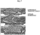
- FIG. 7 shows the results of staining of a tissue onto which a sponge (without cells) and a film were transplanted.
- FIG. 8 shows the results of staining of a tissue before transplantation and after transplantation of a cell culture sponge having a cartilage matrix filling proportion at the transplant face of 90% and a film.
- FIG. 9 shows the results of staining of a tissue before transplantation and after transplantation of a cell culture sponge having a cartilage matrix filling proportion at the transplant face of 20% or 33% and a film.
- FIG. 10 shows the results of staining of a tissue before transplantation and after transplantation of a cell culture sponge having a cartilage matrix filling proportion at the transplant face of 2.9% or 5.4% and a film.
- FIG. 11 shows the results of haematoxylin and eosin (HE) staining of a tissue onto which a cell culture sponge having a cartilage matrix filling proportion at the transplant face of 20% was transplanted without a film or with a film.
- HE haematoxylin and eosin
- FIG. 12 shows the results of safranin O staining of a tissue onto which a cell culture sponge having a cartilage matrix filling proportion at the transplant face of 20% was transplanted without a film or with a film.
- FIG. 13 shows the results of a test (6 months) for organ transplantation onto rabbit knee joint cartilage defect, using a cell culture sponge having a cartilage matrix filling proportion at the transplant face of 33% and a film (degree of crosslinking of 6 or 13).
- FIG. 14 shows in vivo decomposition of the film (degree of crosslinking 13).
- FIG. 15 shows the results of in vivo verification of split transplantation of a cartilage regenerative material.
- FIG. 16 shows the results of verification of the fixation of a cartilage regenerative material to a site of defect.
- FIG. 17 shows the production of pins made of CBE3.
- FIG. 18 shows the results of verification of fixability of a cartilage regenerative material by a pin made of CBE3.
- FIG. 19 shows the positional relation between a sponge and a film.
- the cartilage regenerative material of the invention comprises a porous body of a biocompatible polymer and a biocompatible polymer film, in which the porous body contains chondrocytes and cartilage matrix, and the cartilage matrix exists in a region occupying 10% or more of a region extending from the surface of a transplant face of the porous body to a depth of 150 ⁇ m along the thickness.
- the cartilage regenerative material of the invention suppresses infiltration of fibrous soft tissue and brings about satisfactory cartilage regeneration, and therefore, the cartilage regenerative material can be used for cartilage regeneration.
- the cartilage regenerative material of the invention can be used as, for example, a transplant material to be transplanted to a cartilage defect site.
- the cartilage regenerative material of the invention comprising a porous body of a biocompatible polymer and a biocompatible polymer film, in which the porous body contains chondrocytes and cartilage matrix, and the cartilage matrix exists in a region occupying 10% or more of a region extending from the surface of a transplant face of the porous body to a depth of 150 ⁇ m along the thickness, suppresses infiltration of fibrous soft tissue and brings about satisfactory cartilage regeneration as described above.
- WO2011/108537A description on bone regeneration is given; however, no investigation was conducted on cartilage regeneration. Bone regeneration and cartilage regeneration are different phenomena, and cartilage regeneration action cannot be predicted from bone regeneration action.
- JP2001-519210A the feature of the invention that cartilage matrix exists in a region occupying 10% or more of a region extending from the surface of a transplant face of a porous body to a depth of 150 ⁇ m along the thickness, is neither described nor suggested. JP2001-519210A suggests nothing about the possibility of achieving an effect that infiltration of fibrous soft tissue is suppressed and satisfactory cartilage regeneration is brought about as a result of the above-described feature.
- the cartilage matrix exists in a region occupying 10% or more (preferably 20% or more, and more preferably 30% or more) of a region extending from the surface of a transplant face of a porous body to a depth of 150 ⁇ m along the thickness.
- the proportion of the region in which the cartilage matrix exists in the region extending from the surface of a transplant face of the porous body to a depth of 150 ⁇ m along the thickness is referred to as “cartilage matrix filling proportion” in the present specification.
- the cartilage regenerative material of the invention is enabled to suppress infiltration of fibrous soft tissue and bring about satisfactory cartilage regeneration.
- the upper limit of the cartilage matrix filling proportion on the transplant face side is not particularly limited, and the upper limit may be 100%, or may be less than 100%.
- cartilage matrix exists in a region of 10% or more (more preferably 20% or more, even more preferably 30% or more, and still more preferably 50% or more) of a region extending from the surface of the articular cavity face of the porous body to a depth of 150 ⁇ m along the thickness.
- the upper limit of the cartilage matrix filling proportion on the articular cavity face side is not particularly limited, and the upper limit may be 100%, or may be less than 100%.
- a transplant face of the porous body means the face on the side that is brought into contact with the defect part in vivo (in the case of the sponge of FIG. 19 , the lower surface), and the articular cavity face of the porous body means the face on the opposite side of the transplant face (in the case of the sponge of FIG. 19 , the upper surface).
- Measurement of the cartilage matrix filling proportion can be carried out according to the method described in “[14] Evaluation of samples having different cartilage matrix filling proportions of bottom face” in the Examples of the present specification. That is, slices of a porous body are produced (formalin fixation and paraffin embedment), and cross-sections are visualized by performing safranin O staining. Thus, evaluation is carried out. That is, attention was paid to a thickness of 150 ⁇ m from the surface of the transplant face (bottom face) or the articular cavity face (face on the opposite side of the transplant face) of the stained tissue slices, and the area of a region that was positive for safranin O staining was measured. By determining the proportion of the area of the safranin O staining positive region with respect to the total area of the region from the surface to a depth of 150 ⁇ m, the cartilage matrix filling proportion in the 150- ⁇ m layer can be determined.
- the porous body used in the invention is constructed from a biocompatible polymer.
- Biocompatibility means that in a case in which the material is brought into contact with a living body, the material does not give a rise to a noticeably harmful reaction such as a long-term and chronic inflammation reaction.
- the biocompatible polymer used in the invention is decomposed in vivo is not particularly limited, as long as the polymer has biocompatibility; however, it is preferable that the polymer is a biodegradable polymer.
- a non-biodegradable polymer include polytetrafluoroethylene (PTFE), polyurethane, polypropylene, polyester, vinyl chloride, polycarbonate, acryl, stainless steel, titanium, silicone, and MPC (2-methacryloyloxyethylphosphorylcholine).
- biodegradable polymer examples include polypeptides such as a recombinant peptide and a chemically synthesized peptide (for example, gelatin that will be explained below), polylactic acid, polyglycolic acid, a lactic acid-glycolic acid copolymer (PLGA), hyaluronic acid, glycosaminoglycan, proteoglycan, chondroitin, cellulose, agarose, carboxymethyl cellulose, chitin, and chitosan.
- a recombinant peptide is particularly preferred.
- biocompatible polymers may be devised in order to increase the cell adhesiveness.
- methods such as “coating of a base material surface with a cell adhesion matrix (fibronectin, vitronectin, or laminin) or a cell adhesion sequence (an ROD sequence, a LDV sequence, a REDV sequence (SEQ ID NO: 2), a YIGSR sequence (SEQ ID NO: 3), a PDSGR sequence (SEQ ID NO: 4), a RYVVLPR sequence (SEQ ID NO: 5), a LGTIPG sequence (SEQ ID NO: 6), a RNIAEIIKDI sequence (SEQ ID NO: 7), an IKVAV sequence (SEQ ID NO: 8), a LRE sequence, a DGEA sequence (SEQ ID NO: 9), or a HAV sequence; all indicated by one-letter codes of amino acids) peptide”, “amination or cationization of the base material surface”, or “hydrophilic treatment of the base material surface by a plasma treatment or corona discharge” can be used.
- a cell adhesion matrix fibro
- the type of the polypeptide such as a recombinant peptide or a chemically synthesized peptide is not particularly limited as long as the polypeptide has biocompatibility; however, for example, gelatin, collagen, elastin, fibronectin, pronectin, laminin, tenascin, fibrin, fibroin, entactin, thrombospondin, and retronectin are preferred, while gelatin, collagen, and atelocollagen are most preferred.
- Gelatin that is intended to be used in the invention is preferably naturally occurring gelatin, a recombinant gelatin, or a chemically synthesized gelatin, and more preferred is a recombinant gelatin.
- naturally occurring gelatin as used herein means a gelatin produced from naturally occurring collagen.
- chemically synthesized peptide or chemically synthesized gelatin means a peptide or gelatin that has been artificially synthesized. Synthesis of a peptide such as gelatin may be solid-phase synthesis or liquid-phase synthesis; however, solid-phase synthesis is preferred. Solid-phase synthesis of peptides is well known to those ordinarily skilled in the art, and examples include a Fmoc group synthesis method of using a Fmoc group (Fluorenyl-Methoxy-Carbonyl group) as a protective group for an amino group; and a Boc group synthesis method of using a Boc group (tert-ButylOxyCarbonyl group) as a protective group for an amino group. Regarding preferred embodiments of the chemically synthesized gelatin, the matters described in section (1-3) Recombinant gelatin given below in the present specification can be applied.
- the hydrophilicity value “1/IOB” value of the biocompatible polymer used in the invention is preferably from 0 to 1.0.
- the hydrophilicity value is more preferably from 0 to 0.6, and even more preferably from 0 to 0.4.
- IOB is an index of hydrophilicity/hydrophobicity based on an organic conceptual diagram showing the polarity/non-polarity of organic compounds suggested by FUJITA, Atsushi, and the details thereof are explained in, for example, “Pharmaceutical Bulletin”, Vol. 2, 2, pp. 163-173 (1954), “Kagaku no Ryoiki (Domain of Chemistry)”, Vol. 11, 10, pp. 719-725 (1957), and “Fragrance Journal”, Vol. 50, pp.
- the root of all organic compounds is considered to be methane (CH 4 ), and other compounds are all regarded as derivatives of methane.
- Certain values are set respectively for the number of carbon atoms, substituents, modified parts, rings, and the like of the compounds, and the scores are added to determine the organic values (OV) and the inorganic values (IV). These values are plotted on a graph, with the X-axis representing the organic values and the Y-axis representing the inorganic values.
- the IOB in the organic conceptual diagram means the ratio of the inorganic value (IV) with respect to the organic value (OV) in the organic conceptual diagram, that is, “inorganic value (IV)/organic value (OV)”.
- the biocompatible polymer By adjusting the “1/IOB” value of the biocompatible polymer used in the invention to the range described above, the biocompatible polymer has higher hydrophilicity and has enhanced water absorbing properties. Accordingly, it is speculated that the high hydrophilicity acts effectively on the retention of nutrient components.
- the hydrophilicity/hydrophobicity index represented by the Grand average of hydropathicity (GRAVY) value of the polypeptide is preferably 0.3 or lower and ⁇ 9.0 or higher, and more preferably 0.0 or lower and ⁇ 7.0 or higher.
- the Grand average of hydropathicity (GRAVY) value can be obtained by the method described in “Gasteiger E., Hoogland C., Gattiker A., Duvaud S., Wilkins M. R., Appel R. D., Bairoch A.; Protein Identification and Analysis Tools on the ExPASy Server; (In) John M. Walker (ed): The Proteomics Protocols Handbook, Humana Press (2005).
- the biocompatible polymer By adjusting the GRAVY value of the biocompatible polymer used in the invention to the range described above, the biocompatible polymer has higher hydrophilicity and has enhanced water absorbing properties. Accordingly, it is speculated that the high hydrophilicity acts effectively on the retention of nutrient components.
- the biocompatible polymer used in the invention may be a crosslinked polymer, or may be a polymer that is not crosslinked; however, a crosslinked polymer is preferred.
- a crosslinked biocompatible polymer By using a crosslinked biocompatible polymer, there is obtained an effect that in a case in which the cartilage regenerative material of the invention is cultured in a medium, and in a case in which the cartilage regenerative material is transplanted into a living body, the cartilage regenerative material being instantaneously decomposed is prevented.
- thermal crosslinking crosslinking, crosslinking by means of an aldehyde (for example, formaldehyde or glutaraldehyde), crosslinking by means of a condensing agent (carbodiimide, cyanamide, or the like), enzymatic crosslinking, photocrosslinking, ultraviolet crosslinking, hydrophobic interaction, hydrogen bonding, ionic interaction, and the like are known.
- the crosslinking method used in the invention is preferably thermal crosslinking, ultraviolet crosslinking, or enzymatic crosslinking, and particularly preferably thermal crosslinking.
- the enzyme is not particularly limited as long as the enzyme has an effect of crosslinking between polymer molecules; however, preferably a transglutaminase and a laccase, and most preferably a transglutaminase, can be used.
- a transglutaminase and a laccase preferably a transglutaminase
- the polymer that is enzymatically crosslinked by a transglutaminase as long as the polymer is a protein having a lysine residue and a glutamine residue.
- the transglutaminase may be a mammal-derived enzyme or a microbially derived enzyme, and specifically, ACTIVA series manufactured by Ajinomoto Co., Inc., and mammal-derived transglutaminases that are released as reagents, for example, Guinea pig liver-derived transglutaminase, goat-derived transglutaminase, and rabbit-derived transglutaminase, which are products of Oriental Yeast Co., Ltd.; Upstate USA, Inc.; Biodesign International, Inc.; and the like, and human-derived blood coagulation factor (Factor XIIIa, Haematologic Technologies, Inc.).
- ACTIVA series manufactured by Ajinomoto Co., Inc.
- mammal-derived transglutaminases that are released as reagents, for example, Guinea pig liver-derived transglutaminase, goat-derived transglutaminase, and rabbit-derived transglutaminase, which are products
- the reaction temperature in the case of performing crosslinking is not particularly limited as long as crosslinking is enabled; however, the reaction temperature is preferably ⁇ 100° C. to 500° C., more preferably 0° C. to 300° C., even more preferably 50° C. to 300° C., still more preferably 100° C. to 250° C., and even more preferably 120° C. to 200° C.
- the recombinant gelatin as used herein means a polypeptide or protein-like substance having an amino acid sequence similar to that of gelatin, which is produced by a gene recombination technology. It is preferable that the recombinant gelatin that can be used in the invention has repeats of a sequence represented by Gly-X-Y (where X and Y each independently represent any amino acid residue), which is characteristic to collagen.
- Gly-X-Y where X and Y each independently represent any amino acid residue
- a plurality of the Gly-X-Y sequences may be identical to or different from one another.
- two or more sequences of cell adhesion signals are included in one molecule.
- a recombinant gelatin having an amino acid sequence derived from a partial amino acid sequence of collagen can be used.
- the recombinant gelatins described in EP1014176B, U.S. Pat. No. 6,992,172B, WO2004/85473A, and WO2008/103041A can be used; however, the examples are not limited to these.
- Preferred examples of the recombinant gelatin that is used in the invention are recombinant gelatins of the following embodiments.
- a recombinant gelatin has the original properties of naturally occurring gelatin and thus has excellent biocompatibility. Also, since it is not a substance derived from a natural source, a recombinant gelatin has no risk of bovine spongiform encephalopathy (BSE) or the like, and has an excellent characteristic of being non-infectious. Since a recombinant gelatin is homogeneous compared to naturally occurring gelatin and has a predetermined sequence, it is possible to precisely design a recombinant gelatin with fewer fluctuations, in connection with strength and degradability, through crosslinking or the like.
- BSE bovine spongiform encephalopathy
- the molecular weight of the recombinant gelatin is not particularly limited; however, the molecular weight is preferably from 2,000 to 100,000 (from 2 kDa to 100 kDa), more preferably from 2,500 to 95,000 (from 2.5 kDa to 95 kDa), even more preferably from 5,000 to 90,000 (from 5 kDa to 90 kDa), and most preferably from 10,000 to 90,000 (from 10 kDa to 90 kDa).
- the recombinant gelatin has repeats of a sequence represented by Gly-X-Y, which is characteristic to collagen.
- Gly-X-Y a sequence represented by Gly-X-Y
- a plurality of the Gly-X-Y sequences may be identical to or different from one another.
- Gly-X-Y Gly represents glycine
- X and Y each represent an arbitrary amino acid (preferably, any arbitrary amino acid other than glycine).
- the sequence represented by Gly-X-Y characteristic to collagen is a highly specific partial structure present in the amino acid compositions and sequences of gelatin and collagen, compared to other proteins.
- glycine accounts for about one-third of the whole composition, and in the amino acid sequence, glycine repeatedly appears at a rate of one in every three amino acid residues.
- Glycine is the simplest amino acid, and there are fewer restrictions to the arrangement in a molecular chain. Thus, glycine greatly contributes to regeneration of the helix structure in the case of gelation.
- the amino acids represented by X and Y include a large proportion of imino acids (proline and oxyproline), and imino acids account for 10% to 45% of the total amount of the amino acids.
- amino acids that account for 80% or more, more preferably 95% or more, and most preferably 99% or more, of the sequence of the recombinant gelatin constitute the repeating structure of Gly-X-Y.
- polar amino acids that have an electric charge and polar amino acids that are uncharged exist at a ratio of 1:1.
- polar amino acid specifically refers to cysteine, aspartic acid, glutamic acid, histidine, lysine, asparagine, glutamine, serine, threonine, tyrosine, or arginine, and among these, polar uncharged amino acids include cysteine, asparagine, glutamine, serine, threonine, and tyrosine.
- the proportion of polar amino acids among all the amino acids that constitute the recombinant gelatin is 10% to 40%, and preferably 20% to 30%.
- the proportion of uncharged amino acids in the polar amino acids is preferably 5% or more and less than 20%, and more preferably 5% or more and less than 10%. It is also preferable that any one amino acid, and preferably 2 or more amino acids, of serine, threonine, asparagine, tyrosine, and cysteine are not included in the amino acid sequence.
- minimal amino acid sequences that function as cell adhesion signal sequences are known (for example, “Byotai Seiri (Pathophysiology)”, Vol. 9, No. 7 (1990), p. 527, published by Nagai Shoten Co., Ltd.). It is preferable that the recombinant gelatin used in the invention contains two or more such minimal amino acid sequences that function as cell adhesion signals in one molecule.
- More preferred sequences include an RGD sequence, a YIGSR sequence (SEQ ID NO: 3), a PDSGR sequence (SEQ ID NO: 4), a LGTIPG sequence (SEQ ID NO: 6), an IKVAV sequence (SEQ ID NO: 8), and a HAV sequence, and particularly preferred is an RGD sequence.
- RGD sequences an ERGD sequence (SEQ ID NO: 10) is preferred.
- GAG glycosaminoglycans
- the number of amino acids between RGD sequences is between 0 and 100, and preferably between 25 and 60, and is not uniform.
- the content of these minimal amino acid sequences is preferably 3 to 50, more preferably 4 to 30, even more preferably 5 to 20, and most preferably 12, in one molecule of protein, from the viewpoints of cell adhesion and proliferation properties.
- the proportion of the RGD sequences (motifs) with respect to the total number of amino acid residues is preferably at least 0.4%.
- each stretch of 350 amino acid residues includes at least one RGD motif.
- the proportion of the RGD motif with respect to the total number of amino acid residues is more preferably at least 0.6%, even more preferably at least 0.8%, still more preferably at least 1.0%, even more preferably at least 1.2%, and most preferably at least 1.5%.
- the number of RGD motifs within a recombinant peptide is preferably at least 4, more preferably at least 6, even more preferably at least 8, still more preferably from 12 to 16, per 250 amino acid residues.
- the proportion of 0.4% of the RGD motifs corresponds to at least one RGD sequence per 250 amino acid residues. Since the number of the RGD motifs is an integer, in order to satisfy the characteristic requirement of 0.4%, a gelatin molecule containing 251 amino acid residues must include at least two RGD sequences.
- the recombinant gelatin of the invention includes at least two RGD sequences per 250 amino acid residues; more preferably includes at least three RGD sequences per 250 amino acid residues; and even more preferably includes at least four RGD sequences per 250 amino acid residues.
- the recombinant gelatin includes at least four RGD motifs, preferably at least six RGD motifs, more preferably at least eight RGD motifs, and still more preferably from 12 to 16 RGD motifs.
- the recombinant gelatin may be partially hydrolyzed.
- the recombinant gelatin used in the invention is represented by Formula 1: A-[(Gly-X-Y) n ] m -B.
- n units of X each independently represent any one amino acid residue
- n units of Y each independently represent any one amino acid residue.
- m represents an integer from 2 to 10, and preferably 3 to 5.
- n represents an integer from 3 to 100, preferably 15 to 70, and more preferably 50 to 65.
- A represents an arbitrary amino acid residue or amino acid sequence
- B represents an arbitrary amino acid residue or amino acid sequence.
- the recombinant gelatin used in the invention is represented by formula: Gly-Ala-Pro-[(Gly-X-Y) 63 ] 3 -Gly (SEQ ID NO: 11)(in the formula, 63 units of X each independently represent any one amino acid residue; 63 units of Y each independently represent any one amino acid residue; and 63 units of Gly-X-Y may be identical to or different from one another).
- the naturally occurring collagen as used herein may be any collagen substance that exists in nature; however, the collagen is preferably type I, type II, type III, type IV, or type V collagen.
- the collagen is more preferably type I, type II, or type III collagen.
- the source of the above-mentioned collagens is preferably human, cow, pig, mouse, or rat, and more preferably a human source.
- the isoelectric point of the recombinant gelatin used in the invention is preferably 5 to 10, more preferably 6 to 10, and even more preferably 7 to 9.5.
- the recombinant gelatin is not deaminated.
- the recombinant gelatin does not have a telopeptide.
- the recombinant gelatin is a substantially pure polypeptide produced from a nucleic acid that encodes an amino acid sequence.
- the recombinant gelatin used in the invention is particularly preferably:
- amino acid sequence obtained by modifying . . . by deletion, substitution or addition of one or several amino acid residues means preferably 1 to 20, more preferably 1 to 10, even more preferably 1 to 5, and particularly preferably 1 to 3.
- the recombinant gelatin used in the invention can be produced by a gene recombination technology that is known to those ordinarily skilled in the art, and the recombinant gelatin can be produced according to the methods described in, for example, EP1014176A2, U.S. Pat. No. 6,992,172B, WO2004/85473A, and WO2008/103041A.
- a gene that encodes the amino acid sequence of a predetermined recombinant gelatin is obtained, this is incorporated into an expression vector to produce a recombinant expression vector, and this is introduced into an appropriate host.
- a transformant is produced.
- the transformant thus obtained is cultured in an appropriate medium, and thereby, a recombinant gelatin is produced.
- the recombinant gelatin thus produced is collected from the culture product. Thereby, the recombinant gelatin used in the invention can be produced.
- the method for producing a porous body of a biocompatible polymer is not particularly limited; however, for example, a porous body of a biocompatible polymer can be obtained by freeze-drying an aqueous solution including a biocompatible polymer.
- a production method including:
- Step (b) a step of freezing the solution in an unfrozen state of the biocompatible polymer obtained in Step (a);
- Step (c) a step of freeze-drying the frozen solution of the biocompatible polymer obtained in Step (b).
- the difference between the temperature of a part having the highest liquid temperature in the solution and the temperature of a part having the lowest liquid temperature in the solution is adjusted to be 2.5° C. or less (preferably 2.3° C. or less, and more preferably 2.1° C. or less), that is, as the difference in temperature is adjusted to be smaller, the difference in the size of the pores in the porous body thus obtainable is made smaller.
- the lower limit of the difference between the temperature of a part having the highest liquid temperature in the solution and the temperature of a part having the lowest liquid temperature in the solution is not particularly limited, and the temperature difference may be 0° C. or more, and for example, may be 0.1° C. or more, 0.5° C. or more, 0.8° C. or more, or 0.9° C. or more.
- a porous body produced thereby can achieve a superior cartilage regeneration effect.
- Step (a) it is preferable to perform cooling by means of, for example, a material having a thermal conductivity lower than that of water (preferably, TEFLON (registered trademark)), and the part having the highest liquid temperature in the solution can be assumed to be a part remotest from the cooling surface, and the part having the lowest liquid temperature in the solution can be assumed to be the liquid temperature at the cooling surface.
- a material having a thermal conductivity lower than that of water preferably, TEFLON (registered trademark)
- the difference between the temperature of a part having the highest liquid temperature in the solution and the temperature of a part having the lowest liquid temperature in the solution immediately before the generation of the heat of solidification is 2.5° C. or less, more preferably 2.3° C. or less, and even more preferably 2.1° C. or less.
- the “temperature difference immediately before the generation of the heat of solidification” means the temperature difference at the time when the temperature difference becomes the largest in a time period between 1 second and 10 seconds before the generation of the heat of solidification.
- the temperature of a part having the lowest liquid temperature in the solution is (melting point of the solvent—5° C.) or lower, more preferably (melting point of the solvent—5° C.) or lower and (melting point of the solvent—20° C.) or higher, and even more preferably (melting point of the solvent—6° C.) or lower and (melting point of the solvent—16° C.) or higher.
- the solvent of the “melting point of the solvent” is the solvent of the solution of the biocompatible polymer.
- Step (b) the solution of the biocompatible polymer in an unfrozen state obtained in Step (a) is frozen.
- the cooling temperature for freezing in Step (b) is not particularly limited and may vary depending on the cooling equipment.
- the cooling temperature is a temperature lower by 3° C. to 30° C., more preferably a temperature lower by 5° C. to 25° C., and even more preferably a temperature lower by 10° C. to 20° C., than the temperature of the part having the lowest liquid temperature in the solution.
- Step (c) the frozen solution of the biocompatible polymer obtained in Step (b) is freeze-dried.
- Freeze-drying can be carried out by a conventional method, and for example, freeze-drying can be carried out by performing vacuum drying at a temperature lower than the melting point of the solvent, and further performing vacuum drying at room temperature (20° C.).
- the shape and size of the porous body of the biocompatible polymer are not particularly limited, and a porous body having an appropriate shape and an appropriate size for the purpose of use can be used.
- Examples of the shape include, but are not particularly limited to, a cylinder and a cuboid, and any shape that coincides with the shape of the defect part, which is an affected part, can be employed.
- the size of the cylinder is preferably such that the diameter is 2 mm to 2 cm, and the height (thickness) is 1 mm to 2 cm.
- the size of the cuboid is preferably such that the longitudinal length and the horizontal length are 2 mm to 2 cm, and the height (thickness) is 1 mm to 2 cm.
- the average pore size of the porous body of the biocompatible polymer is not particularly limited; however, the average pore size is preferably 10 to 400 ⁇ m, more preferably 20 to 200 ⁇ m, even more preferably 30 to 100 ⁇ m, and particularly preferably 40 to 90 ⁇ m.
- the average pore size of the porous body can be measured according to the method described in “[8] Evaluation of pore size of recombinant peptide porous body” in the Examples.
- any chondrocytes can be used as long the cells are capable of cell transplantation and exhibiting cartilage regenerative capacity, and the type of the cells is not particularly limited. Furthermore, one kind of chondrocytes may be used, or multiple kinds of chondrocytes may also be used in combination. Furthermore, the chondrocytes to be used are preferably animal cells, more preferably vertebrate-derived cells, and particularly preferably human-derived cells.
- chondrocytes at least one type of chondrocytes selected from the group consisting of articular cartilage-derived chondrocytes, auricular cartilage-derived chondrocytes, nasal cartilage-derived chondrocytes, induced pluripotent stem cell (iPS cell)-derived chondrocytes, embryonic stem cell (ES cell)-derived chondrocytes, mesenchymal stem cell (MSC)-derived chondrocytes, and chondrocytes obtained by a direct reprogramming method.
- a direct reprogramming method is a technique of directly changing cells such as fibroblasts extracted from the skin to chondrocytes.
- the origin of the cells may be any of autologous cells and heterologous cells.
- Cartilage matrix means components produced by chondrocytes, and it is mainly extracellular matrix.
- Cartilage matrix includes glycosaminoglycans (GAG), chondroitin sulfate, and proteoglycans as main components, and depending on cases, cartilage matrix also includes collagenous fibers and elastic fibers. The presence of the cartilage matrix can be checked by safranin O staining.
- the amount of use of chondrocytes with respect to the porous body of a biocompatible polymer is not particularly limited; however, the amount of use is preferably 1.0 ⁇ 10 5 cells/cm 3 to 1.0 ⁇ 10 8 cells/cm 3 , more preferably 1.0 ⁇ 10 6 cells/cm 3 to 5.0 ⁇ 10 7 cells/cm 3 , and even more preferably 2.0 ⁇ 10 6 cells/cm 3 to 1.0 ⁇ 10 7 cells/cm 3 , per unit volume of the porous body of the biocompatible polymer.
- the components that may be optionally present inside the porous body of the biocompatible polymer can be supplied to the cells.
- the components inside the porous body of a biocompatible polymer are not particularly limited; however, the components that are included in the medium that will be described below may be mentioned.
- the cartilage regenerative material of the invention includes a biocompatible polymer film together with a porous body of a biocompatible polymer. That is, according to the invention, a porous body of a biocompatible polymer including chondrocytes and cartilage matrix is used in combination with a biocompatible polymer film.
- the porous body of a biocompatible polymer and the biocompatible polymer film may be supplied separately in the form of kits, or the porous body of a biocompatible polymer and the biocompatible polymer film may also be supplied in the form of a product bonded together.
- the porous body of a biocompatible polymer and the biocompatible polymer film are in the form of separate kits.
- the user may bond the porous body of a biocompatible polymer and the biocompatible polymer film together and then transplant the resultant.
- the use may transplant the biocompatible polymer film and then transplant the porous body of a biocompatible polymer.
- the biocompatible polymer film is used as a film for isolating a portion or the entirety of the transplant face of the porous body of a biocompatible polymer from the site of transplantation.
- the biocompatible polymer film is transplanted first onto the site of transplantation, and subsequently, the porous body of a biocompatible polymer is transplanted onto the top surface (surface on the opposite side of the surface that is in contact with the site of transplantation) of the biocompatible polymer film.
- the porous body of a biocompatible polymer and the biocompatible polymer film are bonded together and then transplanted, it is preferable to transplant the biocompatible polymer film so as to be brought into direct contact with the site of transplantation.
- biocompatible polymer that constitutes the biocompatible polymer film are the same as those in the case of the biocompatible polymer that constitutes the porous body of a biocompatible polymer, and specifically, the specific examples and the preferred ranges are as described in the above sections (1-1) Biocompatible polymer, (1-2) Crosslinking, and (1-3) Recombinant gelatin in the present specification.
- the biocompatible polymer that constitutes the biocompatible polymer film may be the same as, or may be different from, the biocompatible polymer that constitutes the porous body of a biocompatible polymer.
- the method for producing a biocompatible polymer film is not particularly limited, and the production can be carried out by a conventional method.
- a biocompatible polymer film can be produced by causing an aqueous solution of a biocompatible polymer to flow into a plastic tray, and drying the aqueous solution at low temperature (for example, in a refrigerator).
- the biocompatible polymer film can be crosslinked.
- the degree of crosslinking is not particularly limited; however, the degree of crosslinking is generally 4 to 15, more preferably 6 to 13, even more preferably 4 to 8, and particularly preferably 5 to 7.
- the degree of crosslinking is the number of crosslinks per molecule. Measurement of the degree of crosslinking can be carried out using the TNBS (2,4,6-trinitrobenzenesulfonic acid) method described in section [10] Method for measuring degree of crosslinking in the Examples.
- the rate of decomposition of the biocompatible polymer film varies depending on the degree of crosslinking.
- the rate of decomposition of the biocompatible polymer film can be measured and evaluated by the method described below in section [11] Method for measuring rate of decomposition in the Examples.
- a sample (film) is introduced into a tube, the mass of which has been measured in advance, and the actual amount of addition is recorded.
- PBS phosphate buffered saline
- Rate of decomposition (( W ⁇ We ) ⁇ wo )/ wo/T (Formula 4)
- W represents the mass of the tube containing the sample, which was recorded after freeze-drying; and We represents the blank mass of the tube that was recorded in advance.
- wo represents the actual amount of addition of the sample.
- T represents the time taken for shaking in the collagenase solution.
- the rate of decomposition of the biocompatible polymer film measured by the method described above is not particularly limited; however, the rate of decomposition is generally 0.1 to 20 [mass %/hour], preferably 0.5 to 20 [mass %/hour], more preferably 1 to 10 [mass %/hour], and particularly preferably 5 to 10 [mass %/hour].
- the invention also provides a method for producing the cartilage regenerative material of the invention described above.
- the production method includes Step A of freeze-drying an aqueous solution including a biocompatible polymer and obtaining a porous body; Step B of inoculating chondrocytes into the porous body obtained in Step A and culturing the chondrocytes; and Step C of providing a biocompatible polymer film.
- Step A can be carried out as described in the above section “(1-4) Method for producing porous body of biocompatible polymer”.
- a porous body can be obtained by stirring an aqueous solution including a biocompatible polymer and then freeze-drying the aqueous solution.
- Step B is a step of inoculating chondrocytes into the porous body and culturing the chondrocytes.
- the inoculation and culturing of chondrocytes in the porous body can be carried out by a conventional method.
- the proportion of the region in which the cartilage matrix exists in a region extending from the surface of the transplant face of the porous body to a depth of 150 ⁇ m along the thickness (cartilage matrix filling proportion on the transplant face side) and the proportion of the region in which cartilage matrix exists in a region extending from the surface of the articular cavity face of the porous body to a depth of 150 ⁇ m along the thickness (cartilage matrix filling proportion on the articular cavity face side) can be regulated.
- Step C is a step of providing a biocompatible polymer film.
- a biocompatible polymer film can be provided.
- the cartilage regenerative material of the invention can be used for the purpose of cell transplantation to a diseased site of cartilage defect.
- the disease associated with cartilage defect include, but are not particularly limited to, arthrosis deformans, osteochondral defect, osteochondritis dissecans, traumatic cartilage injury, osteoarthritis, relapsing polychondritis, achondroplasia, injury of intervertebral discs, and hernia of intervertebral discs.
- Examples of the method for transplantation include incision, injection, arthroscopy, and endoscopy.
- the cartilage regenerative material of the invention unlike cell transplants such as a cell sheet, the size of the cartilage regenerative material can be made small, and therefore, a less invasive transplantation method such as transplantation by injection is enabled.
- the cartilage regenerative material of the invention is such that the cartilage regenerative material can be fixed to a site of defect with pins after transplantation.
- the material of the pins is not particularly limited; however, it is preferable to use a biocompatible polymer.
- Specific examples and preferred range of the biocompatible polymer that constitutes the pins are the same as those in the case of the biocompatible polymer that constitutes the porous body of a biocompatible polymer, and specifically, the specific examples and the preferred range are as described in the above sections (1-1) Biocompatible polymer, (1-2) Crosslinking, and (1-3) Recombinant gelatin in the present specification.
- the biocompatible polymer that constitutes the pins may be the same as, or may be different from, the biocompatible polymer that constitutes the porous body of a biocompatible polymer.
- the amount used in the case of transplanting the cartilage regenerative material of the invention can be appropriately selected according to the diseased state or the like; however, the number of cells to be transplanted is preferably 1.0 ⁇ 10 4 cells/cm 3 to 2.0 ⁇ 10 7 cells/cm 3 , and more preferably 2.5 ⁇ 10 5 cells/cm 3 to 5.0 ⁇ 10 6 cells/cm 3 .
- transplantation may be carried out only once, or transplantation may be carried out two or more times as necessary.
- a cartilage regenerative material intended for use for the treatment of cartilage regeneration
- the cartilage regenerative material including a porous body of a biocompatible polymer and a biocompatible polymer film, in which the porous body contains chondrocytes and cartilage matrix, and the cartilage matrix exists in a region of 10% or more of a region extending from the surface of the transplant face of the porous body to a depth of 150 ⁇ m along the thickness.
- Preferred ranges of the various constituent components are similar to those described above in the present specification.
- cartilage regeneration method including a step of transplanting the cartilage regenerative material of the invention as described above, to a patient in need of cartilage regeneration.
- Preferred ranges of the various constituent components of the cartilage regenerative material are as described above in the present specification.
- a porous body of a biocompatible polymer and a biocompatible polymer film for the production of a cartilage regenerative material, in which the porous body contains chondrocytes and cartilage matrix, and the cartilage matrix exists in a region of 10% or more of a region extending from the surface of the transplant face of the porous body to a depth of 150 ⁇ m along the thickness.
- Preferred ranges of the porous body of a biocompatible polymer and the biocompatible polymer film are as described above in the present specification.
- RGD sequence 12 sequences
- CBE3 comprises an ERGD sequence.
- GAP GAP(GAPGLQGAPGLQGMPGERGAAGLPGPKGERGDAGPKGADGAPGAPG LQGMPGERGAAGLPGPKGERGDAGPKGADGAPGKDGVRGLAGPIGPPGER GAAGLPGPKGERGDAGPKGADGAPGKDGVRGLAGPIGPPGPAGAPGAPGL QGMPGERGAAGLPGPKGERGDAGPKGADGAPGKDGVRGLAGPIGPPGPAGAPGAPGL QGMPGERGAAGLPGPKGERGDAGPKGADGAPGKDGVRGLAGPP)3G
- porous body and a sponge according to the present specification are synonyms.
- a cylindrical cup-shaped container made of polytetrafluoroethylene (PTFE) and having a bottom face thickness of 3 mm, a diameter of 51 mm, a lateral face thickness of 8 mm, and a height of 25 mm was prepared.
- the cylindrical cup was such that when the curved face was erected as the lateral face, the lateral face was closed with a PTFE plate having a thickness of 8 mm, and the bottom face (circular-shaped flat plate) was also closed with a PTFE plate having a thickness of 3 mm. Meanwhile, the cylindrical cup had an open top face. Therefore, the inner diameter of the cylindrical cup was 43 mm.
- this container will be referred to as PTFE thick cylindrical container.
- a cylindrical cup-shaped container made of aluminum and having a thickness of 1 mm and a diameter of 47 mm was prepared.
- the cylindrical cup was such that when the curved face was erected as the lateral face, the lateral face was closed with an aluminum plate with a thickness of 1 mm, and the bottom face (circular-shaped flat plate) was also closed with an aluminum plate having a thickness of 1 mm. Meanwhile, the cylindrical cup had an open top face.
- a TEFLON (registered trademark) plate having a thickness of 1 mm was uniformly lined over the entire surface on the inner side of the lateral face, and as a result, the inner diameter of the cylindrical cup was 45 mm.
- the bottom face of this container was in a state of being joined with a glass plate having a thickness of 2.2 mm on the outside of aluminum.
- this container will be referred to as an aluminum glass cylindrical container.
- the container, the final concentration of the aqueous solution of CBE3, the liquid amount, and the setting of the shelf board temperature employed in this case were as described below.
- the liquid temperature of the liquid surface at the circle center in the container was measured as the liquid temperature at the remotest place from the cooling side (non-cooling surface liquid temperature) within the solution, and the liquid temperature at the bottom in the container was measured as the liquid temperature closest to the cooling side (cooling surface liquid temperature) within the solution.
- the temperature difference at the time when the non-cooling surface liquid temperature reached the melting point (0° C.), the temperature difference immediately before lowering of the shelf board temperature from ⁇ 10° C. to ⁇ 20° C., and the temperature difference immediately before the generation of the heat of solidification will be described in conjunction with Condition A, Condition B, and Condition C.
- the “temperature difference immediately before” as used in the present specification means the largest temperature difference among the temperature differences detectable in a period between 1 second and 20 seconds before the main event.
- a 1 mass % (w/w) ethanol-containing aqueous solution of CBE3 was respectively poured into the PTFE thick cylindrical container and the aluminum glass plate cylindrical container, and the aqueous solution of CBE3 was cooled through the bottom face using a cooling shelf board inside a vacuum freeze-drying machine (TF5-85ATNNN: Takara Co., Ltd.). Since an ethanol-containing aqueous solution at a final concentration of 1 mass % was used, the melting point was ⁇ 0.4° C. The melting point change at the ethanol/water concentration ratio was calculated from literature “Pickering S. U.: A Study of the Properties of Some Strong Solutions. J. Chem. Soc. London, 63 (1893), 998-1027”.
- the container, the final concentration of the aqueous solution of CBE3, the liquid amount, and the setting of the shelf board temperature employed in this case were as described below.
- the liquid temperature of the liquid surface at the circle center in the container was measured as the liquid temperature at the remotest place from the cooling side (non-cooling surface liquid temperature) within the solution, and the liquid temperature at the bottom in the container was measured as the liquid temperature closest to the cooling side (cooling surface liquid temperature) within the solution.
- the solvent melting point was ⁇ 0.4° C.
- the melting point change at the ethanol/water concentration ratio was calculated from literature “Pickering S. U.: A Study of the Properties of Some Strong Solutions. J. Chem. Soc. London, 63 (1893), 998-1027”.
- a cylindrical cup-shaped container made of aluminum and having a thickness of 1 mm and a diameter of 47 mm was prepared.
- the cylindrical cup was such that when the curved face was erected as the lateral face, the lateral face was closed with an aluminum plate having a thickness of 1 mm, and the bottom face (circular-shaped flat plate) was also closed with an aluminum plate having a thickness of 1 mm. Meanwhile, the cylindrical cup had an open top face.
- a TEFLON (registered trademark) plate having a thickness of 1 mm was uniformly lined over the entire surface on the inner side of the lateral face, and as a result, the inner diameter of the cylindrical cup was 45 mm.
- this container will be referred to as cylindrical container.
- An aqueous solution of CBE3 was prepared, and this aqueous solution of CBE3 was caused to flow into the cylindrical container.
- the aqueous solution of CBE3 was cooled through the bottom face using a cooling shelf board inside a refrigerator.
- the temperature of the cooling shelf board, the thickness of the heat-insulating plate (glass plate) interposed between the shelf board and the cylindrical container, the final concentration of the aqueous solution of CBE3 to be introduced, and the amount of the aqueous solution were as described below.
- Temperature of shelf board ⁇ 40° C., thickness of glass plate: 2.2 mm, final concentration of aqueous solution of CBE3: 4.0%, amount of aqueous solution: 4 mL.
- the frozen CBE3 block obtained as described above was freeze-dried, and a CBE3 porous body was obtained.
- the porous body obtained as described above will be hereinafter referred to as thin layer frozen porous body.
- a stirring method porous body was produced using the recombinant peptide CBE3.
- a solution was prepared at the following composition, and the solution was stirred for 30 seconds at 17,000 rpm with a homogenizer (AM-11, manufactured by Nihon Seiki Co., Ltd.) at 4° C.
- the solution was transferred into an aluminum cup container and was rapidly cooled for 3 hours at ⁇ 80° C. Subsequently, freeze-drying was performed for 3 days in a freeze-dryer, and thus a porous body was obtained.
- the porous body obtained as described above will be hereinafter referred to as stirring method porous body.
- Composition 10-mL portion of 10 mass % of porous body (CBE3: 1,000 mg, ultrapure water: 9,895 ⁇ L, 1 mol/L HCl: 105 ⁇ L)
- the respective porous bodies were subjected to thermal crosslinking for 20 hours at 160° C. under reduced pressure.
- the various porous bodies obtained in the above section [7] were swollen with physiological saline for a sufficient time. Subsequently, frozen tissue slices were produced with a microtome, and HE (haematoxylin and eosin) stained specimens were produced. Cross-sectional images having a size of 1.5 mm on the actual scale were prepared from the specimens, and the individual pore areas were measured. Subsequently, the equivalent circle diameter obtainable in a case in which the area was considered equivalent to a circle was calculated, and this value was designated as the pore size. The average value of 20 or more sites of these pores was designated as the average pore size.
- the average pore size of the freezing step with small temperature difference/porous body derived from sections [2] and [3] was 59 ⁇ m
- the average pore size of the ethanol-containing freezing step with small temperature difference/porous body derived from section [4] was 72 ⁇ m
- the average pore size of the stirring method porous body derived from section [6] was 82 ⁇ m
- the average pore size of the thin layer frozen porous body derived from section [5] was 45 ⁇ m.
- aqueous solution of CBE3 at a concentration of 4 mass % was prepared, and 5.4 ml of this aqueous solution of CBE3 was caused to flow into a plastic tray provided with a silicon frame (8 cm ⁇ 3.5 cm). This plastic tray was transferred into a refrigerator, and the aqueous solution was dried until no moisture left. Thus, a recombinant peptide film was obtained. The recombinant peptide film was taken out from the plastic tray/silicon frame, and was subjected to thermal crosslinking at 160° C. under reduced pressure (crosslinking time was 48 hours or 72 hours). Thus, a sample for an animal test was obtained.
- the degree of crosslinking (number of crosslinks per molecule) of the film produced in the above section [9] was calculated.
- a TNBS (2,4,6-trinitrobenzenesulfonic acid) method was used.
- a sample (about 10 mg), a 4 mass % aqueous solution of NaHCO 3 (1 mL), and a 1 mass % aqueous solution of TNBS (2 mL) were introduced into a glass vial, and the mixture was shaken for 3 hours at 37° C. Subsequently, 37 mass % hydrochloric acid (10 mL) and pure water (5 mL) were added thereto, and then the mixture was left to stand for 16 hours or longer at 37° C. The resultant was used as a sample.
- a sample (about 10 mg), a 4 mass % aqueous solution of NaHCO 3 (1 mL), and a 1 mass % aqueous solution of TNBS (2 mL) were introduced into a glass vial, 37 mass % hydrochloric acid (3 mL) was added thereto immediately thereafter, and the mixture was shaken for 3 hours at 37° C. Subsequently, 37 mass % hydrochloric acid (7 mL) and pure water (5 mL) were added thereto, and then the mixture was left to stand for 16 hours or longer at 37° C. The resultant was used as a blank.
- Form 2 represents the amount of lysine (molar equivalent) per gram of the recombinant peptide.
- the film obtained by crosslinking for 48 hours in the above section [9] had a degree of crosslinking of 6, and the film obtained by crosslinking for 72 hours in the above section [6] had a degree of crosslinking of 13.
- the degree of crosslinking of the porous body of the above section [7] measured in the same manner was 9.
- the tube was centrifuged for 1 minute at 10,000 G, and the supernatant was removed using a pipette. This operation was repeated one more time. Subsequently, the sample was freeze-dried, and the mass of the tube containing the sample was recorded.
- Rate of decomposition (( W ⁇ We ) ⁇ wo )/ wo/T (Formula 4)
- W represents the mass of the tube containing the sample, which was recorded after freeze-drying; and We represents the blank mass of the tube that was recorded in advance.
- wo represents the actual amount of addition of the sample.
- T represents the time taken for shaking in the collagenase solution, and in this test, T was 5 hours.
- the film of the above section [9] resulted in a rate of decomposition of 6.9 [mass %/hour] under crosslinking for 48 hours, and a rate of decomposition of 0.5 [mass %/hour] under crosslinking for 72 hours.
- the medium used as described above was a medium for chondrocyte culture in all cases, and the medium is a medium composed of DULBECCO's modified Eagle medium (DMEM), 10 vol % fetal bovine serum (FBS), 20 mM HEPES (4-(2-hydroyethyl)-1-piperazine ethanesulfonic acid), 50 ⁇ g/mL magnesium L-ascorbyl phosphate, 0.25 ⁇ g/mL amphotericin B, and 50 ⁇ g/mL gentamycin.
- DMEM DULBECCO's modified Eagle medium
- FBS fetal bovine serum
- HEPES 4-(2-hydroyethyl)-1-piperazine ethanesulfonic acid
- 50 ⁇ g/mL magnesium L-ascorbyl phosphate 0.25 ⁇ g/mL amphotericin B
- 50 ⁇ g/mL gentamycin 50 ⁇ g/mL gentamycin.
- the stirring method porous body (pore size 82 ⁇ m) prepared in the above section [6] was cut out into a size with a diameter of 5 mm and a thickness of 2 mm, the rabbit chondrocytes prepared in the above section [12] were inoculated thereon at a concentration of 5.0 ⁇ 10 6 cells/cm 3 , and culture was carried out. Therefore, a sponge with cartilage matrix was obtained.
- a porous body that was not inoculated with rabbit chondrocytes was also prepared, and that was prepared as a sponge without cartilage matrix.
- the sponge with cartilage matrix obtained by inoculating cells and culturing in the above section [13] could be used to produce sponges with cartilage matrix having different cartilage matrix filling proportions of the bottom faces with the elapse time of culture (3 days, 7 days, 14 days, 21 days, and 28 days).
- tissue slices of the sponge with cartilage matrix were produced (formalin fixation and paraffin embedment), and the cross-sections of the slices were visualized by performing safranin O staining.
- the cross-sections were evaluated ( FIG. 8 to FIG. 10 ).
- cartilage matrix filling proportions in 150- ⁇ m layer of 2.9%, 5.4%, 20%, 33%, and 90%, which were produced in the above sections [13] and [14]
- cartilage matrix existed in a region of 30% or more of a region extending from the surface of the articular cavity face to a depth of 150 ⁇ m along the thickness.
- Comparative Sponge with cartilage matrix (cartilage Suppression of infiltration of fibrous soft C C Example 3 matrix filling proportion: 2.9%) was tissue was not recognized, and cartilage transplanted integrally with film regeneration was also not recognized. having diameter of 5 mm, with the film being placed below the sponge.
- Comparative Sponge with cartilage matrix (cartilage Suppression of infiltration of fibrous soft C C Example 4 matrix filling proportion: 5.4%) was tissue was not recognized, and cartilage transplanted integrally with film regeneration was also not recognized. having diameter of 5 mm, with the film being placed below the sponge.
- Example 1 Sponge with cartilage matrix (cartilage Suppression of infiltration of fibrous soft A A matrix filling proportion: 20%) was tissue was significantly recognized, and it transplanted integrally with film was understood that cartilage regeneration having diameter of 5 mm, with the film was also satisfactory. being placed below the sponge.
- Example 2 Sponge with cartilage matrix (cartilage Suppression of infiltration of fibrous soft A A matrix filling proportion: 33%) was tissue was significantly recognized, and it transplanted integrally with film was understood that cartilage regeneration having diameter of 5 mm, with the film was also satisfactory. being placed below the sponge.
- Example 3 Sponge with cartilage matrix (cartilage Suppression of infiltration of fibrous soft A A matrix filling proportion: 90%) was tissue was significantly recognized, and it transplanted integrally with film was understood that cartilage regeneration having diameter of 5 mm, with the film was also satisfactory. being placed below the sponge. Comparative Only sponge with cartilage matrix Suppression of infiltration of fibrous soft C C Example 5 (cartilage matrix filling proportion: tissue was not recognized, and cartilage 20%) was transplanted. regeneration was also not recognized.
- FIG. 13 The results are presented in FIG. 13 .
- a sponge with cartilage matrix (cartilage matrix filling proportion: 33%) and a film having a degree of crosslinking of 6 were transplanted
- a sponge with cartilage matrix (cartilage matrix filling proportion: 33%) and a film having a degree of crosslinking of 13 were transplanted
- the film having a degree of crosslinking of 13 produced in the above sections [9] and [10] was placed at the rabbit cartilage defect site produced in the above section [15], and after one month and after two months, the rabbits were autopsied. Osteochondral tissue slices of the periphery of the transplantation site were produced. The tissue was subjected to formalin fixation, followed by paraffin embedment, and tissue slices including the transplant were produced. Regarding the slice staining, HE staining (haematoxylin and eosin staining) was carried out.
- transplantation therapy is enabled by using arthroscopy.
- cartilage regeneration is recognized to the same extent as in the case of using transplants that are not split.
- the sponge with cartilage matrix (cartilage matrix filling proportion 90%) produced in the above sections [13] and [14] was split as shown in FIG. 15 , and then the split sponges were transplanted together with the film having a degree of crosslinking of 13 produced in the above sections [9] and [10] onto the rabbit cartilage defect site produced in the above section [15]. Transplantation was carried out such that the film would exist on the transplant face (lower bone side).
- the rabbits were autopsied at the time point of one month after transplantation, and osteochondral tissue slices of the periphery of the site of transplantation were produced. The tissues were subjected to formalin fixation, followed by paraffin embedment, and tissue slices including the transplant were produced. Regarding the slice staining, HE staining (haematoxylin and eosin staining), or safranin O staining, or safranin O and fast green staining were carried out.
- the sponge with cartilage matrix produced in the above sections [13] and [14] (cartilage matrix filling proportion 90%) and the film having a degree of crosslinking of 13 produced in the above sections [9] and [10] were transplanted onto the rabbit cartilage defect site produced in the above section [15], and subsequently, a through-hole that reached the sponge, the film and the lower bone was opened using a Kirschner wire having a diameter of 0.8 mm or a diameter of 1.5 mm.
- the rabbits were autopsied at the time point of passing one month in the state of having the through-hole maintained, and osteochondral tissue slices of the periphery of the site of transplantation were produced.
- tissue slices including the transplant were produced.
- slice staining HE staining (haematoxylin and eosin staining), or safranin O staining, or safranin O and fast green staining were carried out.
- samples obtained by cutting the film having a degree of crosslinking of 13 (measured in section [10]), which was prepared in the above section [9], into the size of the low bottom face area (diameter: 5 mm) of the defect, were used.
- the samples were installed in the form in which the film existed on the transplant face (lower bone side).
- a fixing pin that accomplishes fixation such as in the above section [19]
- the pin is bioabsorbable.
- GRANDFIX manufactured by Gunze, Ltd.
- PLANDFIX manufactured by Gunze, Ltd.
- a pin made of a recombinant peptide could be produced using CBE3 of the above section [1].
- a 10 mass % aqueous solution of CBE3 was prepared using the CBE3 of section [1]. Subsequently, the aqueous solution was caused to flow into a template made of TEFLON (registered trademark) shown in FIG. 17 and FIG. 18 , and the aqueous solution was dried. Subsequently, CBE3 was subjected to thermal crosslinking by a heat treatment for 20 hours at 160° C. under reduced pressure, and thus pins (diameter: 1 mm, length: 5 mm) made of CBE3 were produced.
- a sponge bone test material was prepared as a substituent for bone, and a through-hole was produced using a Kirschner wire (K-wire) having a diameter of 0.8 mm was produced therein.
- K-wire Kirschner wire
- the above-mentioned CBE3 pin was pierced into the hole having a diameter of 0.8 mm produced in the sponge bone test material, and the material was sufficiently swollen with water.
- This CBE3 pin maintained the shape without escaping even after being placed therein for one day. Thus, it was confirmed that the pin was a pin that can be used for fixation ( FIG. 18 ).
Landscapes
- Health & Medical Sciences (AREA)
- Life Sciences & Earth Sciences (AREA)
- Chemical & Material Sciences (AREA)
- Engineering & Computer Science (AREA)
- Biomedical Technology (AREA)
- Medicinal Chemistry (AREA)
- Animal Behavior & Ethology (AREA)
- General Health & Medical Sciences (AREA)
- Public Health (AREA)
- Veterinary Medicine (AREA)
- Dermatology (AREA)
- Oral & Maxillofacial Surgery (AREA)
- Transplantation (AREA)
- Epidemiology (AREA)
- Botany (AREA)
- Chemical Kinetics & Catalysis (AREA)
- Zoology (AREA)
- Urology & Nephrology (AREA)
- Cell Biology (AREA)
- Molecular Biology (AREA)
- Vascular Medicine (AREA)
- Biophysics (AREA)
- Rheumatology (AREA)
- Dispersion Chemistry (AREA)
- Materials For Medical Uses (AREA)
- Peptides Or Proteins (AREA)
- Immobilizing And Processing Of Enzymes And Microorganisms (AREA)
- Micro-Organisms Or Cultivation Processes Thereof (AREA)
Applications Claiming Priority (4)
| Application Number | Priority Date | Filing Date | Title |
|---|---|---|---|
| JPJP2015-054887 | 2015-03-18 | ||
| JP2015-054887 | 2015-03-18 | ||
| JP2015054887 | 2015-03-18 | ||
| PCT/JP2016/058541 WO2016148246A1 (ja) | 2015-03-18 | 2016-03-17 | 軟骨再生材料及びその製造方法 |
Related Parent Applications (1)
| Application Number | Title | Priority Date | Filing Date |
|---|---|---|---|
| PCT/JP2016/058541 Continuation WO2016148246A1 (ja) | 2015-03-18 | 2016-03-17 | 軟骨再生材料及びその製造方法 |
Publications (2)
| Publication Number | Publication Date |
|---|---|
| US20190216976A1 US20190216976A1 (en) | 2019-07-18 |
| US11160902B2 true US11160902B2 (en) | 2021-11-02 |
Family
ID=56918956
Family Applications (1)
| Application Number | Title | Priority Date | Filing Date |
|---|---|---|---|
| US15/705,669 Active 2036-07-17 US11160902B2 (en) | 2015-03-18 | 2017-09-15 | Cartilage regenerative material and method for producing same |
Country Status (5)
| Country | Link |
|---|---|
| US (1) | US11160902B2 (zh) |
| EP (1) | EP3260142B1 (zh) |
| JP (1) | JP6467493B2 (zh) |
| CN (1) | CN107405422A (zh) |
| WO (1) | WO2016148246A1 (zh) |
Families Citing this family (1)
| Publication number | Priority date | Publication date | Assignee | Title |
|---|---|---|---|---|
| WO2020050205A1 (ja) * | 2018-09-03 | 2020-03-12 | 富士フイルム株式会社 | ゲル形成キット、ゲルおよびゲルの製造方法 |
Citations (24)
| Publication number | Priority date | Publication date | Assignee | Title |
|---|---|---|---|---|
| US5041138A (en) * | 1986-11-20 | 1991-08-20 | Massachusetts Institute Of Technology | Neomorphogenesis of cartilage in vivo from cell culture |
| US5447966A (en) * | 1988-07-19 | 1995-09-05 | United States Surgical Corporation | Treating bioabsorbable surgical articles by coating with glycerine, polalkyleneoxide block copolymer and gelatin |
| AU1499000A (en) | 1999-02-10 | 2000-08-17 | Isotis N.V. | cartilage tissue engineering |
| CN1280509A (zh) | 1997-10-10 | 2001-01-17 | 埃德盖斯特利希索恩化学工业股份公司 | 导引组织再生的薄膜 |
| JP2001293081A (ja) | 2000-02-07 | 2001-10-23 | Mitsuo Ochi | 軟骨移植用材料及びその製造方法 |
| US6387693B2 (en) * | 1996-06-04 | 2002-05-14 | Sulzer Orthopedics Ltd. | Method for producing cartilage tissue and implants for repairing enchondral and osteochondral defects as well as arrangement for carrying out the method |
| US20030050709A1 (en) | 2001-02-23 | 2003-03-13 | Ulrich Noth | Trabecular bone-derived human mesenchymal stem cells |
| JP2003275294A (ja) | 2002-03-25 | 2003-09-30 | Olympus Optical Co Ltd | 骨再生シート |
| JP2004501700A (ja) | 2000-06-26 | 2004-01-22 | ユニヴエルシテ・ピエール・エ・マリー・キユリー・パリ・シス | 連通セルを用いた三次元構造を有する生体適合性ポリマー、その調製方法、および医薬ならびに手術における適用 |
| JP2004357694A (ja) | 2003-05-15 | 2004-12-24 | Yukie Iwamoto | 組織プラグの製造方法 |
| JP2005152006A (ja) | 2003-11-20 | 2005-06-16 | Teijin Ltd | 軟骨組織再生用基材および軟骨細胞との複合体とその製造方法 |
| WO2006022671A1 (en) | 2004-07-30 | 2006-03-02 | Histogenics Corporation | Method for in situ repair of injured, damaged, diseased or aged articular cartilage using neo-cartilage constructs and a method for preparation thereof |
| US20060212125A1 (en) | 2005-03-18 | 2006-09-21 | Pentax Corporation | Bone repairing material using a chondrocyte having the potential for hypertrophy and a scaffold |
| CN1933859A (zh) | 2004-03-19 | 2007-03-21 | 首尔国立大学工业基金会 | 其表面上固定有促骨生长肽的骨移植材料和支架材料 |
| US20090228105A1 (en) * | 2005-10-31 | 2009-09-10 | Son Young Sook | Artificial cartilage containing chondrocytes obtained from costal cartilage and preparation process thereof |
| JP2010535602A (ja) | 2007-08-14 | 2010-11-25 | ウニベルズィテート ベルン | コンドロレギュレイティブ基質を用いた軟骨および骨における欠損または損傷の修復 |
| WO2011021712A1 (ja) | 2009-08-21 | 2011-02-24 | 独立行政法人農業生物資源研究所 | 細胞・組織供給用支持体、細胞・組織供給体及びその製造方法、組織再生方法、並びに多孔質体の製造方法 |
| WO2011108517A1 (ja) | 2010-03-01 | 2011-09-09 | 富士フイルム株式会社 | 生体親和性を有する高分子ブロックと細胞からなる細胞構造体 |
| WO2011108537A1 (ja) | 2010-03-02 | 2011-09-09 | 富士フイルム株式会社 | 細胞支持体及び骨再生材 |
| US20120165263A1 (en) | 2009-09-04 | 2012-06-28 | Fujifilm Corporation | Bone regeneration agent comprising gelatin |
| US20130071441A1 (en) | 2011-08-31 | 2013-03-21 | Fujifilm Corporation | Cell construct for cell transplantation and cell aggregate for cell transplantation |
| WO2014133081A1 (ja) | 2013-02-27 | 2014-09-04 | 富士フイルム株式会社 | 細胞移植用細胞構造体、生体親和性高分子ブロック及びそれらの製造方法 |
| CN104244999A (zh) | 2012-03-12 | 2014-12-24 | 富士胶片株式会社 | 组织修复材料的制造方法 |
| JP2015054887A (ja) * | 2013-09-11 | 2015-03-23 | 日東電工株式会社 | 片面粘着シート |
-
2016
- 2016-03-17 EP EP16765075.3A patent/EP3260142B1/en active Active
- 2016-03-17 JP JP2017506615A patent/JP6467493B2/ja active Active
- 2016-03-17 WO PCT/JP2016/058541 patent/WO2016148246A1/ja active Application Filing
- 2016-03-17 CN CN201680016571.1A patent/CN107405422A/zh active Pending
-
2017
- 2017-09-15 US US15/705,669 patent/US11160902B2/en active Active
Patent Citations (42)
| Publication number | Priority date | Publication date | Assignee | Title |
|---|---|---|---|---|
| US5041138A (en) * | 1986-11-20 | 1991-08-20 | Massachusetts Institute Of Technology | Neomorphogenesis of cartilage in vivo from cell culture |
| US5447966A (en) * | 1988-07-19 | 1995-09-05 | United States Surgical Corporation | Treating bioabsorbable surgical articles by coating with glycerine, polalkyleneoxide block copolymer and gelatin |
| US6387693B2 (en) * | 1996-06-04 | 2002-05-14 | Sulzer Orthopedics Ltd. | Method for producing cartilage tissue and implants for repairing enchondral and osteochondral defects as well as arrangement for carrying out the method |
| CN1280509A (zh) | 1997-10-10 | 2001-01-17 | 埃德盖斯特利希索恩化学工业股份公司 | 导引组织再生的薄膜 |
| JP2001519210A (ja) | 1997-10-10 | 2001-10-23 | エド・ガイストリヒ・ゼーネ・アクチエンゲゼルシャフト・フューア・ヒェーミシェ・インドゥストリー | 組織再生誘導に使用する膜 |
| US20020177903A1 (en) | 1997-10-10 | 2002-11-28 | Peter Geistlich | Membrane for use in guided tissue regeneration |
| US20040234577A1 (en) | 1997-10-10 | 2004-11-25 | Ed. Geistlich Soehne Ag Fuer Chemische Industrie | Membrane for use in guided tissue regeneration |
| AU1499000A (en) | 1999-02-10 | 2000-08-17 | Isotis N.V. | cartilage tissue engineering |
| JP2001293081A (ja) | 2000-02-07 | 2001-10-23 | Mitsuo Ochi | 軟骨移植用材料及びその製造方法 |
| JP2004501700A (ja) | 2000-06-26 | 2004-01-22 | ユニヴエルシテ・ピエール・エ・マリー・キユリー・パリ・シス | 連通セルを用いた三次元構造を有する生体適合性ポリマー、その調製方法、および医薬ならびに手術における適用 |
| US20040062809A1 (en) | 2000-06-26 | 2004-04-01 | Universite Pierre Et Marie Curie (Paris Vi) | Biocompatible polymer with a three-dimensional structure with communicating cells, a process for its preparation, and application in medicine and in surgery |
| US20030050709A1 (en) | 2001-02-23 | 2003-03-13 | Ulrich Noth | Trabecular bone-derived human mesenchymal stem cells |
| JP2003275294A (ja) | 2002-03-25 | 2003-09-30 | Olympus Optical Co Ltd | 骨再生シート |
| JP4122280B2 (ja) | 2003-05-15 | 2008-07-23 | 幸英 岩本 | 組織プラグの製造方法 |
| JP2004357694A (ja) | 2003-05-15 | 2004-12-24 | Yukie Iwamoto | 組織プラグの製造方法 |
| JP2005152006A (ja) | 2003-11-20 | 2005-06-16 | Teijin Ltd | 軟骨組織再生用基材および軟骨細胞との複合体とその製造方法 |
| CN1933859A (zh) | 2004-03-19 | 2007-03-21 | 首尔国立大学工业基金会 | 其表面上固定有促骨生长肽的骨移植材料和支架材料 |
| US20070160681A1 (en) | 2004-03-19 | 2007-07-12 | Park Yoon J | Bone graft and scaffolding materials immobilized with osteogenesis enhancing peptides on the surface |
| WO2006022671A1 (en) | 2004-07-30 | 2006-03-02 | Histogenics Corporation | Method for in situ repair of injured, damaged, diseased or aged articular cartilage using neo-cartilage constructs and a method for preparation thereof |
| US20060212125A1 (en) | 2005-03-18 | 2006-09-21 | Pentax Corporation | Bone repairing material using a chondrocyte having the potential for hypertrophy and a scaffold |
| JP2006289062A (ja) | 2005-03-18 | 2006-10-26 | Pentax Corp | 肥大化能を有する軟骨細胞と足場を用いた骨補填材料 |
| US20090228105A1 (en) * | 2005-10-31 | 2009-09-10 | Son Young Sook | Artificial cartilage containing chondrocytes obtained from costal cartilage and preparation process thereof |
| JP2010535602A (ja) | 2007-08-14 | 2010-11-25 | ウニベルズィテート ベルン | コンドロレギュレイティブ基質を用いた軟骨および骨における欠損または損傷の修復 |
| US20110184381A1 (en) | 2007-08-14 | 2011-07-28 | Universitaet Bern | Repair of defects or lesions in cartilage and bone using a chondro-regulative matrix |
| US20120148639A1 (en) | 2009-08-21 | 2012-06-14 | Kyoto University | Substrate for feeding cells and/or tissues, cell/tissue-feeder and method for the production of the same, method for the regeneration of tissues, and method for the production of porous bodies |
| WO2011021712A1 (ja) | 2009-08-21 | 2011-02-24 | 独立行政法人農業生物資源研究所 | 細胞・組織供給用支持体、細胞・組織供給体及びその製造方法、組織再生方法、並びに多孔質体の製造方法 |
| US20120165263A1 (en) | 2009-09-04 | 2012-06-28 | Fujifilm Corporation | Bone regeneration agent comprising gelatin |
| CN102625717A (zh) | 2009-09-04 | 2012-08-01 | 富士胶片株式会社 | 包含明胶的骨再生剂 |
| CN102858381A (zh) | 2010-03-01 | 2013-01-02 | 富士胶片株式会社 | 包括具有生物相容性的聚合物块和细胞的细胞构建体 |
| WO2011108517A1 (ja) | 2010-03-01 | 2011-09-09 | 富士フイルム株式会社 | 生体親和性を有する高分子ブロックと細胞からなる細胞構造体 |
| EP2543397A1 (en) | 2010-03-01 | 2013-01-09 | FUJIFILM Corporation | Cell structure comprising cells and macromolecular blocks having biocompatibility |
| US20120329157A1 (en) | 2010-03-01 | 2012-12-27 | Fujifilm Corporation | Cell construct comprising polymer blocks having biocompatibility and cells |
| US20130004549A1 (en) | 2010-03-02 | 2013-01-03 | Fujifilm Corporation | Cell support and bone regeneration material |
| WO2011108537A1 (ja) | 2010-03-02 | 2011-09-09 | 富士フイルム株式会社 | 細胞支持体及び骨再生材 |
| CN102791301A (zh) | 2010-03-02 | 2012-11-21 | 富士胶片株式会社 | 细胞支持体和骨再生材料 |
| US20130071441A1 (en) | 2011-08-31 | 2013-03-21 | Fujifilm Corporation | Cell construct for cell transplantation and cell aggregate for cell transplantation |
| JP2014012114A (ja) | 2011-08-31 | 2014-01-23 | Fujifilm Corp | 細胞移植用細胞構造体および細胞移植用細胞集合体 |
| CN104244999A (zh) | 2012-03-12 | 2014-12-24 | 富士胶片株式会社 | 组织修复材料的制造方法 |
| US20140378662A1 (en) | 2012-03-12 | 2014-12-25 | Fujifilm Corporation | Method for producing tissue repair material |
| WO2014133081A1 (ja) | 2013-02-27 | 2014-09-04 | 富士フイルム株式会社 | 細胞移植用細胞構造体、生体親和性高分子ブロック及びそれらの製造方法 |
| US20150352252A1 (en) | 2013-02-27 | 2015-12-10 | Fujifilm Corporation | Cell construct for cell transplantation, biocompatible polymer block, and method for producing the same |
| JP2015054887A (ja) * | 2013-09-11 | 2015-03-23 | 日東電工株式会社 | 片面粘着シート |
Non-Patent Citations (32)
| Title |
|---|
| Chinese Office Action and Search Report for Chinese Application No. 201680016000.8, dated Jan. 6, 2020, with English translation. |
| Chinese Office Action and Search Report for Chinese Application No. 201680016571.1, dated Jul. 21, 2021, with English translation of the Office Action. |
| Chinese Office Action and Search Report, dated Nov. 13, 2019 for corresponding Chinese Application No. 201680016571.1, with an English translation of the Chinese Office Action. |
| Chinese Office Action for Application No. 201680016000.8, dated Feb. 22, 2021, with machine English translation. |
| Chinese Office Action for Application No. 201680016571.1, dated Feb. 22, 2021, with machine English translation. |
| Chinese Office Action for Chinese Application No. 201680016000.8, dated May 27, 2021, with English translation. |
| Chinese Office Action for Chinese Application No. 201680016000.8, dated Sep. 22, 2020, with English translation. |
| Chinese Office Action for corresponding Chinese Application No. 201680016571.1, dated Jul. 30, 2020, with an English translation. |
| European Communication pursuant to Article 94(3) EPC for corresponding European Application No. 16765075.3, dated Oct. 26, 2020. |
| European Office Action for European Application No. 16765074.6, dated Aug. 27, 2020. |
| European Office Action for European Application No. 16765074.6, dated May 7, 2019. |
| Extended European Search Report for corresponding European Application No. 16765074.6, dated Feb. 14, 2018. |
| Extended European Search Report for corresponding European Application No. 16765075.3, dated Feb. 15, 2018. |
| Huang et al., "Solid freeform-fabricated scaffolds designed to carry multicellular mesenchymal stem cell spheroids for cartilage regeneration," European Cells and Materials, vol. 26, 2013, pp. 179-194. |
| International Preliminary Report on Patentability and Written Opinion of the International Searching Authority (Forms PCT/IB/373, PCT/IB/326 and PCT/ISA/237), dated Sep. 28, 2017, for International Application No. PCT/JP2016/058540, with an English translation of the Written Opinion. |
| International Preliminary Report on Patentability and Written Opinion of the International Searching Authority (Forms PCT/IB/373, PCT/IB/326 and PCT/ISA/237), dated Sep. 28, 2017, for International Application No. PCT/JP2016/058541, with an English translation of the Written Opinion. |
| International Search Report and English translation (Form PCT/ISA/210), dated Jun. 21, 2016, for International Application No. PCT/JP2016/058541. |
| International Search Report and English translation (Form PCT/ISA/210), dated Jun. 7, 2016, for International Application No. PCT/JP2016/058540. |
| Japanese Notification of Reasons for Refusal for Japanese Application No. 2017-506615, dated Apr. 24, 2018, with machine translation. |
| Japanese Office Action and English translation for Application No. 2017-506614, dated May 22, 2018. |
| Japanese Office Action, dated Oct. 16, 2018, for corresponding Japanese Application No. 2017-506615, with an English machine translation. |
| Lee et al., "Transplantation of scaffold-free spheroids composed of synovium-derived cells and chondrocytes for the treatment of cartilage defects of the knee," European Cells and Materials, vol. 22, 2011, pp. 275-290. |
| Pettersson et al., Biodegradable gelatin microcarriers in tissue engineering, In vitro studies on cartilage and bone, Dissertation No. 1147, 2009 (Year: 2009). * |
| Ponticello et al., "Gelatin-Based Resorbable Spong as a Carrier Matrix for Human Mesenchymal Stem Cells in Cartilage Regeneration Therapy," J Biomed Mater Res, vol. 52, 2000, pp. 246-255. |
| Pulkkinen et al., The use of recombinant human type II collagen for articular cartilage tissue engineering, Dissertation in Health Sciences, No. 142, 2012 (Year: 2012). * |
| Raghunath et al., "Advancing Cartilage Tissue Engineering: the Application of Stem Cell Technology," Current Opinions in Biotechnology, vol. 16, 2005 (published on web Sep. 8, 2005), pp. 503-509. |
| Toh et al., "Advances in mesenchymal stem cell-based strategies for cartilage repair and regeneration," Stem Cell Reviews and Reports, vol. 10, 2014 (published online May 29, 2014), pp. 686-696. |
| U.S. Final Office Action for U.S. Appl. No. 16/660,003, dated May 5, 2021. |
| U.S. Non-Final Office Action dated Jul. 22, 2019, for U.S. Appl. No. 15/705,826. |
| U.S. Office Action for U.S. Appl. No. 16/660,003, dated Oct. 15, 2020. |
| Yoshioka, "Recombinant collagen technology applying on regenerative medicine," Medical Science Digest, vol. 40, No. 12, 2014, pp. 46-49 (6 pages total). |
| Zhang et al., "The Role of Tissue Engineering Articular Cartilage Repair and Regeneration," Crit. Rev. Biomed. Eng., vol. 37, No. 1-2, 2009, pp. 1-44. |
Also Published As
| Publication number | Publication date |
|---|---|
| EP3260142A1 (en) | 2017-12-27 |
| EP3260142B1 (en) | 2023-04-26 |
| JP6467493B2 (ja) | 2019-02-13 |
| EP3260142A4 (en) | 2018-03-21 |
| WO2016148246A1 (ja) | 2016-09-22 |
| CN107405422A (zh) | 2017-11-28 |
| JPWO2016148246A1 (ja) | 2018-02-01 |
| US20190216976A1 (en) | 2019-07-18 |
Similar Documents
| Publication | Publication Date | Title |
|---|---|---|
| JP6506326B2 (ja) | 細胞移植用細胞構造体、生体親和性高分子ブロック及びそれらの製造方法 | |
| JP6535072B2 (ja) | 生体親和性高分子多孔質体の製造方法、生体親和性高分子多孔質体、生体親和性高分子ブロック並びに細胞構造体 | |
| US20120329157A1 (en) | Cell construct comprising polymer blocks having biocompatibility and cells | |
| US11027044B2 (en) | Method for producing sheet-like cell structure and sheet-like cell structure | |
| US11241518B2 (en) | Cartilage regenerative material | |
| US10471180B2 (en) | Cell structure and method for producing cell structure | |
| US11160902B2 (en) | Cartilage regenerative material and method for producing same | |
| US11471564B2 (en) | Angiogenic agent and method of manufacturing the same | |
| US10576184B2 (en) | Tubular structure, device for manufacturing tubular structure, and method for manufacturing tubular structure | |
| US10898612B2 (en) | Cell structure and method for producing cell structure | |
| EP3597731B1 (en) | Method for producing cell structures |
Legal Events
| Date | Code | Title | Description |
|---|---|---|---|
| FEPP | Fee payment procedure |
Free format text: ENTITY STATUS SET TO UNDISCOUNTED (ORIGINAL EVENT CODE: BIG.); ENTITY STATUS OF PATENT OWNER: LARGE ENTITY |
|
| AS | Assignment |
Owner name: FUJIFILM CORPORATION, JAPAN Free format text: ASSIGNMENT OF ASSIGNORS INTEREST;ASSIGNORS:NAKAMURA, KENTARO;MIYOSHI, HAYATO;HADA, SATOKO;AND OTHERS;SIGNING DATES FROM 20170901 TO 20170904;REEL/FRAME:043615/0555 Owner name: JAPAN TISSUE ENGINEERING CO., LTD., JAPAN Free format text: ASSIGNMENT OF ASSIGNORS INTEREST;ASSIGNORS:NAKAMURA, KENTARO;MIYOSHI, HAYATO;HADA, SATOKO;AND OTHERS;SIGNING DATES FROM 20170901 TO 20170904;REEL/FRAME:043615/0555 |
|
| STPP | Information on status: patent application and granting procedure in general |
Free format text: NON FINAL ACTION MAILED |
|
| STPP | Information on status: patent application and granting procedure in general |
Free format text: RESPONSE TO NON-FINAL OFFICE ACTION ENTERED AND FORWARDED TO EXAMINER |
|
| STPP | Information on status: patent application and granting procedure in general |
Free format text: FINAL REJECTION MAILED |
|
| STPP | Information on status: patent application and granting procedure in general |
Free format text: DOCKETED NEW CASE - READY FOR EXAMINATION |
|
| STPP | Information on status: patent application and granting procedure in general |
Free format text: NOTICE OF ALLOWANCE MAILED -- APPLICATION RECEIVED IN OFFICE OF PUBLICATIONS |
|
| STPP | Information on status: patent application and granting procedure in general |
Free format text: AWAITING TC RESP., ISSUE FEE NOT PAID |
|
| STPP | Information on status: patent application and granting procedure in general |
Free format text: NOTICE OF ALLOWANCE MAILED -- APPLICATION RECEIVED IN OFFICE OF PUBLICATIONS |
|
| STPP | Information on status: patent application and granting procedure in general |
Free format text: PUBLICATIONS -- ISSUE FEE PAYMENT VERIFIED |
|
| STCF | Information on status: patent grant |
Free format text: PATENTED CASE |