JP2017514629A - 位相コントラストx線イメージングのためのシステム及び方法 - Google Patents
位相コントラストx線イメージングのためのシステム及び方法 Download PDFInfo
- Publication number
- JP2017514629A JP2017514629A JP2016566901A JP2016566901A JP2017514629A JP 2017514629 A JP2017514629 A JP 2017514629A JP 2016566901 A JP2016566901 A JP 2016566901A JP 2016566901 A JP2016566901 A JP 2016566901A JP 2017514629 A JP2017514629 A JP 2017514629A
- Authority
- JP
- Japan
- Prior art keywords
- grating
- images
- interferometer
- ray
- phase contrast
- Prior art date
- Legal status (The legal status is an assumption and is not a legal conclusion. Google has not performed a legal analysis and makes no representation as to the accuracy of the status listed.)
- Granted
Links
- 238000003384 imaging method Methods 0.000 title claims abstract description 42
- 238000000034 method Methods 0.000 title claims description 37
- 238000005562 fading Methods 0.000 claims description 9
- 235000010627 Phaseolus vulgaris Nutrition 0.000 claims description 2
- 244000046052 Phaseolus vulgaris Species 0.000 claims description 2
- 230000007246 mechanism Effects 0.000 claims description 2
- 238000001514 detection method Methods 0.000 abstract description 4
- 230000005855 radiation Effects 0.000 abstract description 3
- 238000009548 contrast radiography Methods 0.000 description 15
- 230000001429 stepping effect Effects 0.000 description 13
- 238000009607 mammography Methods 0.000 description 12
- 210000004872 soft tissue Anatomy 0.000 description 12
- 238000002601 radiography Methods 0.000 description 11
- 238000010521 absorption reaction Methods 0.000 description 9
- 230000006870 function Effects 0.000 description 8
- 210000000038 chest Anatomy 0.000 description 7
- 230000000694 effects Effects 0.000 description 7
- 230000003287 optical effect Effects 0.000 description 7
- 238000002059 diagnostic imaging Methods 0.000 description 6
- LFEUVBZXUFMACD-UHFFFAOYSA-H lead(2+);trioxido(oxo)-$l^{5}-arsane Chemical compound [Pb+2].[Pb+2].[Pb+2].[O-][As]([O-])([O-])=O.[O-][As]([O-])([O-])=O LFEUVBZXUFMACD-UHFFFAOYSA-H 0.000 description 6
- 230000008859 change Effects 0.000 description 5
- 239000013078 crystal Substances 0.000 description 5
- 238000004519 manufacturing process Methods 0.000 description 5
- 230000008569 process Effects 0.000 description 5
- 238000005516 engineering process Methods 0.000 description 4
- 239000000463 material Substances 0.000 description 4
- 230000008901 benefit Effects 0.000 description 3
- 238000004891 communication Methods 0.000 description 3
- 238000013461 design Methods 0.000 description 3
- 238000003745 diagnosis Methods 0.000 description 3
- 239000000835 fiber Substances 0.000 description 3
- 239000010931 gold Substances 0.000 description 3
- 230000010354 integration Effects 0.000 description 3
- 238000009659 non-destructive testing Methods 0.000 description 3
- 230000000737 periodic effect Effects 0.000 description 3
- 238000012545 processing Methods 0.000 description 3
- 238000011160 research Methods 0.000 description 3
- 230000035945 sensitivity Effects 0.000 description 3
- 210000001519 tissue Anatomy 0.000 description 3
- 206010028980 Neoplasm Diseases 0.000 description 2
- 238000013459 approach Methods 0.000 description 2
- 238000003491 array Methods 0.000 description 2
- 230000009286 beneficial effect Effects 0.000 description 2
- 230000005540 biological transmission Effects 0.000 description 2
- 210000000845 cartilage Anatomy 0.000 description 2
- 230000002708 enhancing effect Effects 0.000 description 2
- 238000001914 filtration Methods 0.000 description 2
- 230000004927 fusion Effects 0.000 description 2
- 230000033001 locomotion Effects 0.000 description 2
- 238000005259 measurement Methods 0.000 description 2
- 230000010363 phase shift Effects 0.000 description 2
- 238000010008 shearing Methods 0.000 description 2
- 238000001228 spectrum Methods 0.000 description 2
- 210000002435 tendon Anatomy 0.000 description 2
- 238000003325 tomography Methods 0.000 description 2
- 238000002604 ultrasonography Methods 0.000 description 2
- 206010006187 Breast cancer Diseases 0.000 description 1
- 208000026310 Breast neoplasm Diseases 0.000 description 1
- 208000004434 Calcinosis Diseases 0.000 description 1
- 206010007710 Cartilage injury Diseases 0.000 description 1
- 229910004613 CdTe Inorganic materials 0.000 description 1
- 241000156978 Erebia Species 0.000 description 1
- FAPWRFPIFSIZLT-UHFFFAOYSA-M Sodium chloride Chemical compound [Na+].[Cl-] FAPWRFPIFSIZLT-UHFFFAOYSA-M 0.000 description 1
- 210000001015 abdomen Anatomy 0.000 description 1
- 230000003187 abdominal effect Effects 0.000 description 1
- 230000005856 abnormality Effects 0.000 description 1
- 239000012620 biological material Substances 0.000 description 1
- 238000001574 biopsy Methods 0.000 description 1
- 230000000903 blocking effect Effects 0.000 description 1
- 210000004204 blood vessel Anatomy 0.000 description 1
- 210000005013 brain tissue Anatomy 0.000 description 1
- 210000000481 breast Anatomy 0.000 description 1
- 239000002775 capsule Substances 0.000 description 1
- 238000012512 characterization method Methods 0.000 description 1
- 239000002131 composite material Substances 0.000 description 1
- 238000002591 computed tomography Methods 0.000 description 1
- 238000004590 computer program Methods 0.000 description 1
- 230000021615 conjugation Effects 0.000 description 1
- 230000007423 decrease Effects 0.000 description 1
- 230000007547 defect Effects 0.000 description 1
- 230000001066 destructive effect Effects 0.000 description 1
- 238000002405 diagnostic procedure Methods 0.000 description 1
- 238000010586 diagram Methods 0.000 description 1
- 201000010099 disease Diseases 0.000 description 1
- 208000037265 diseases, disorders, signs and symptoms Diseases 0.000 description 1
- 238000002474 experimental method Methods 0.000 description 1
- 210000003414 extremity Anatomy 0.000 description 1
- 239000012530 fluid Substances 0.000 description 1
- PCHJSUWPFVWCPO-UHFFFAOYSA-N gold Chemical compound [Au] PCHJSUWPFVWCPO-UHFFFAOYSA-N 0.000 description 1
- 229910052737 gold Inorganic materials 0.000 description 1
- 210000003128 head Anatomy 0.000 description 1
- 230000006872 improvement Effects 0.000 description 1
- 238000010348 incorporation Methods 0.000 description 1
- 238000007689 inspection Methods 0.000 description 1
- 230000003993 interaction Effects 0.000 description 1
- 238000005305 interferometry Methods 0.000 description 1
- 238000010030 laminating Methods 0.000 description 1
- 210000003041 ligament Anatomy 0.000 description 1
- 150000002632 lipids Chemical class 0.000 description 1
- 230000005389 magnetism Effects 0.000 description 1
- 238000001393 microlithography Methods 0.000 description 1
- 238000012986 modification Methods 0.000 description 1
- 230000004048 modification Effects 0.000 description 1
- 210000003205 muscle Anatomy 0.000 description 1
- 230000010355 oscillation Effects 0.000 description 1
- 230000002093 peripheral effect Effects 0.000 description 1
- 229920002120 photoresistant polymer Polymers 0.000 description 1
- 210000002381 plasma Anatomy 0.000 description 1
- 229920000642 polymer Polymers 0.000 description 1
- 230000010076 replication Effects 0.000 description 1
- 238000012216 screening Methods 0.000 description 1
- 239000011780 sodium chloride Substances 0.000 description 1
- 230000003595 spectral effect Effects 0.000 description 1
- 238000012360 testing method Methods 0.000 description 1
- XLYOFNOQVPJJNP-UHFFFAOYSA-N water Substances O XLYOFNOQVPJJNP-UHFFFAOYSA-N 0.000 description 1
- 239000002023 wood Substances 0.000 description 1
Images


Classifications
-
- A—HUMAN NECESSITIES
- A61—MEDICAL OR VETERINARY SCIENCE; HYGIENE
- A61B—DIAGNOSIS; SURGERY; IDENTIFICATION
- A61B6/00—Apparatus or devices for radiation diagnosis; Apparatus or devices for radiation diagnosis combined with radiation therapy equipment
- A61B6/02—Arrangements for diagnosis sequentially in different planes; Stereoscopic radiation diagnosis
- A61B6/03—Computed tomography [CT]
- A61B6/032—Transmission computed tomography [CT]
-
- A—HUMAN NECESSITIES
- A61—MEDICAL OR VETERINARY SCIENCE; HYGIENE
- A61B—DIAGNOSIS; SURGERY; IDENTIFICATION
- A61B6/00—Apparatus or devices for radiation diagnosis; Apparatus or devices for radiation diagnosis combined with radiation therapy equipment
- A61B6/40—Arrangements for generating radiation specially adapted for radiation diagnosis
- A61B6/4035—Arrangements for generating radiation specially adapted for radiation diagnosis the source being combined with a filter or grating
-
- A—HUMAN NECESSITIES
- A61—MEDICAL OR VETERINARY SCIENCE; HYGIENE
- A61B—DIAGNOSIS; SURGERY; IDENTIFICATION
- A61B6/00—Apparatus or devices for radiation diagnosis; Apparatus or devices for radiation diagnosis combined with radiation therapy equipment
- A61B6/42—Arrangements for detecting radiation specially adapted for radiation diagnosis
- A61B6/4291—Arrangements for detecting radiation specially adapted for radiation diagnosis the detector being combined with a grid or grating
-
- A—HUMAN NECESSITIES
- A61—MEDICAL OR VETERINARY SCIENCE; HYGIENE
- A61B—DIAGNOSIS; SURGERY; IDENTIFICATION
- A61B6/00—Apparatus or devices for radiation diagnosis; Apparatus or devices for radiation diagnosis combined with radiation therapy equipment
- A61B6/48—Diagnostic techniques
- A61B6/484—Diagnostic techniques involving phase contrast X-ray imaging
-
- A—HUMAN NECESSITIES
- A61—MEDICAL OR VETERINARY SCIENCE; HYGIENE
- A61B—DIAGNOSIS; SURGERY; IDENTIFICATION
- A61B6/00—Apparatus or devices for radiation diagnosis; Apparatus or devices for radiation diagnosis combined with radiation therapy equipment
- A61B6/50—Apparatus or devices for radiation diagnosis; Apparatus or devices for radiation diagnosis combined with radiation therapy equipment specially adapted for specific body parts; specially adapted for specific clinical applications
- A61B6/502—Apparatus or devices for radiation diagnosis; Apparatus or devices for radiation diagnosis combined with radiation therapy equipment specially adapted for specific body parts; specially adapted for specific clinical applications for diagnosis of breast, i.e. mammography
-
- A—HUMAN NECESSITIES
- A61—MEDICAL OR VETERINARY SCIENCE; HYGIENE
- A61B—DIAGNOSIS; SURGERY; IDENTIFICATION
- A61B6/00—Apparatus or devices for radiation diagnosis; Apparatus or devices for radiation diagnosis combined with radiation therapy equipment
- A61B6/52—Devices using data or image processing specially adapted for radiation diagnosis
- A61B6/5205—Devices using data or image processing specially adapted for radiation diagnosis involving processing of raw data to produce diagnostic data
-
- A—HUMAN NECESSITIES
- A61—MEDICAL OR VETERINARY SCIENCE; HYGIENE
- A61B—DIAGNOSIS; SURGERY; IDENTIFICATION
- A61B6/00—Apparatus or devices for radiation diagnosis; Apparatus or devices for radiation diagnosis combined with radiation therapy equipment
- A61B6/52—Devices using data or image processing specially adapted for radiation diagnosis
- A61B6/5211—Devices using data or image processing specially adapted for radiation diagnosis involving processing of medical diagnostic data
- A61B6/5229—Devices using data or image processing specially adapted for radiation diagnosis involving processing of medical diagnostic data combining image data of a patient, e.g. combining a functional image with an anatomical image
- A61B6/5235—Devices using data or image processing specially adapted for radiation diagnosis involving processing of medical diagnostic data combining image data of a patient, e.g. combining a functional image with an anatomical image combining images from the same or different ionising radiation imaging techniques, e.g. PET and CT
-
- G—PHYSICS
- G01—MEASURING; TESTING
- G01N—INVESTIGATING OR ANALYSING MATERIALS BY DETERMINING THEIR CHEMICAL OR PHYSICAL PROPERTIES
- G01N23/00—Investigating or analysing materials by the use of wave or particle radiation, e.g. X-rays or neutrons, not covered by groups G01N3/00 – G01N17/00, G01N21/00 or G01N22/00
- G01N23/02—Investigating or analysing materials by the use of wave or particle radiation, e.g. X-rays or neutrons, not covered by groups G01N3/00 – G01N17/00, G01N21/00 or G01N22/00 by transmitting the radiation through the material
- G01N23/04—Investigating or analysing materials by the use of wave or particle radiation, e.g. X-rays or neutrons, not covered by groups G01N3/00 – G01N17/00, G01N21/00 or G01N22/00 by transmitting the radiation through the material and forming images of the material
- G01N23/041—Phase-contrast imaging, e.g. using grating interferometers
-
- G—PHYSICS
- G01—MEASURING; TESTING
- G01N—INVESTIGATING OR ANALYSING MATERIALS BY DETERMINING THEIR CHEMICAL OR PHYSICAL PROPERTIES
- G01N23/00—Investigating or analysing materials by the use of wave or particle radiation, e.g. X-rays or neutrons, not covered by groups G01N3/00 – G01N17/00, G01N21/00 or G01N22/00
- G01N23/02—Investigating or analysing materials by the use of wave or particle radiation, e.g. X-rays or neutrons, not covered by groups G01N3/00 – G01N17/00, G01N21/00 or G01N22/00 by transmitting the radiation through the material
- G01N23/04—Investigating or analysing materials by the use of wave or particle radiation, e.g. X-rays or neutrons, not covered by groups G01N3/00 – G01N17/00, G01N21/00 or G01N22/00 by transmitting the radiation through the material and forming images of the material
- G01N23/046—Investigating or analysing materials by the use of wave or particle radiation, e.g. X-rays or neutrons, not covered by groups G01N3/00 – G01N17/00, G01N21/00 or G01N22/00 by transmitting the radiation through the material and forming images of the material using tomography, e.g. computed tomography [CT]
-
- G—PHYSICS
- G01—MEASURING; TESTING
- G01N—INVESTIGATING OR ANALYSING MATERIALS BY DETERMINING THEIR CHEMICAL OR PHYSICAL PROPERTIES
- G01N23/00—Investigating or analysing materials by the use of wave or particle radiation, e.g. X-rays or neutrons, not covered by groups G01N3/00 – G01N17/00, G01N21/00 or G01N22/00
- G01N23/20—Investigating or analysing materials by the use of wave or particle radiation, e.g. X-rays or neutrons, not covered by groups G01N3/00 – G01N17/00, G01N21/00 or G01N22/00 by using diffraction of the radiation by the materials, e.g. for investigating crystal structure; by using scattering of the radiation by the materials, e.g. for investigating non-crystalline materials; by using reflection of the radiation by the materials
- G01N23/20075—Investigating or analysing materials by the use of wave or particle radiation, e.g. X-rays or neutrons, not covered by groups G01N3/00 – G01N17/00, G01N21/00 or G01N22/00 by using diffraction of the radiation by the materials, e.g. for investigating crystal structure; by using scattering of the radiation by the materials, e.g. for investigating non-crystalline materials; by using reflection of the radiation by the materials by measuring interferences of X-rays, e.g. Borrmann effect
-
- G—PHYSICS
- G01—MEASURING; TESTING
- G01N—INVESTIGATING OR ANALYSING MATERIALS BY DETERMINING THEIR CHEMICAL OR PHYSICAL PROPERTIES
- G01N2223/00—Investigating materials by wave or particle radiation
- G01N2223/30—Accessories, mechanical or electrical features
- G01N2223/33—Accessories, mechanical or electrical features scanning, i.e. relative motion for measurement of successive object-parts
-
- G—PHYSICS
- G02—OPTICS
- G02B—OPTICAL ELEMENTS, SYSTEMS OR APPARATUS
- G02B5/00—Optical elements other than lenses
- G02B5/18—Diffraction gratings
- G02B5/1866—Transmission gratings characterised by their structure, e.g. step profile, contours of substrate or grooves, pitch variations, materials
- G02B5/1871—Transmissive phase gratings
-
- G—PHYSICS
- G21—NUCLEAR PHYSICS; NUCLEAR ENGINEERING
- G21K—TECHNIQUES FOR HANDLING PARTICLES OR IONISING RADIATION NOT OTHERWISE PROVIDED FOR; IRRADIATION DEVICES; GAMMA RAY OR X-RAY MICROSCOPES
- G21K2201/00—Arrangements for handling radiation or particles
- G21K2201/06—Arrangements for handling radiation or particles using diffractive, refractive or reflecting elements
- G21K2201/067—Construction details
Landscapes
- Health & Medical Sciences (AREA)
- Life Sciences & Earth Sciences (AREA)
- Engineering & Computer Science (AREA)
- Medical Informatics (AREA)
- Pathology (AREA)
- General Health & Medical Sciences (AREA)
- Physics & Mathematics (AREA)
- Radiology & Medical Imaging (AREA)
- Nuclear Medicine, Radiotherapy & Molecular Imaging (AREA)
- Optics & Photonics (AREA)
- Veterinary Medicine (AREA)
- Biomedical Technology (AREA)
- Heart & Thoracic Surgery (AREA)
- Molecular Biology (AREA)
- Surgery (AREA)
- Animal Behavior & Ethology (AREA)
- Biophysics (AREA)
- Public Health (AREA)
- High Energy & Nuclear Physics (AREA)
- Chemical & Material Sciences (AREA)
- Analytical Chemistry (AREA)
- Biochemistry (AREA)
- General Physics & Mathematics (AREA)
- Immunology (AREA)
- Theoretical Computer Science (AREA)
- Pulmonology (AREA)
- Computer Vision & Pattern Recognition (AREA)
- Crystallography & Structural Chemistry (AREA)
- Dentistry (AREA)
- Oral & Maxillofacial Surgery (AREA)
- Apparatus For Radiation Diagnosis (AREA)
- Analysing Materials By The Use Of Radiation (AREA)
Abstract
Description
本開示は、X線システムに関し、詳しくは、微分位相コントラストX線イメージングシステム及びX線照射システムに関する。
以下の引用文献は、引用によって全体が本願に援用される。
[2] "X-ray phase-contrast imaging: from pre-clinical applications towards clinics" Phys. Med. Biol. 58 (2013) R1-R35
[3] Stutman D., Finkenthal M., "Glancing angle Talbot-Lau grating interferometers for phase contrast imaging at high x-ray energy", Appl. Phys. Lett. 101, 091108 (2012)
[4] D. Stutman ; J. W. Stayman ; M. Finkenthal ; J. H. Siewerdsen "High energy x-ray phase-contrast imaging using glancing angle grating interferometers" Proc. SPIE 8668, Medical Imaging 2013: Physics of Medical Imaging, 866814 (March 19, 2013)
[5] A. Sarapata, J. W. Stayman, M. Finkenthal, J. H. Siewerdsen, F. Pfeiffer and D. Stutman "High energy x-ray phase contrast CT using glancing-angle grating interferometers"
In print in Medical Physics 2014
[6] Zanette, M. Bech, F. Pfeiffer, and T. Weitkamp, "Interlaced phase stepping in phase-contrast x-ray tomography" Appl. Phys. Lett. 98, 094101 (2011)
[6] www.teledynedalsa.com/imaging/products/x-ray/scanning/argus/
[7] www.dectris.com/
[8] D. Stutman and M. Finkenthal "K-edge and mirror filtered X-ray grating interferometers" INTERNATIONAL WORKSHOP ON X-RAY AND NEUTRON PHASE IMAGING WITH GRATINGS, Tokyo, Japan, 2012 AIP Conf. Proc. 1466, pp. 229-236
I(n)=A+B・sin(n・x/G・2π+φ)n=1,2,..N
Claims (18)
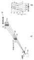
- マルチセクタ線源格子、ビームスプリッタ格子及びアナライザ格子を含み、前記ビームスプリッタ格子及び前記アナライザ格子の間に物体が配置される干渉計を用いて、前記物体を位相コントラストイメージングするための方法であって、
前記マルチセクタ線源格子にX線ビームを方向付けることであって、前記マルチセクタ線源格子の各セクタは、所定の量によってオフセットされていることと、
一回の露光の間に、前記物体又は前記干渉計を平行移動させることによって複数のイメージを取得することであって、前記複数のイメージは、異なる干渉計フェージングを有することと、
取得された前記複数のイメージを結合して、前記物体の位相コントラストイメージを生成することと、
を含む方法。 - 前記マルチセクタ線源格子は、3個より多い異なるセクタを有する請求項1に記載の方法。
- 前記複数のイメージは、前記物体を介して、異なる角度で取得される請求項1に記載の方法。
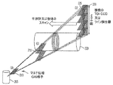
- 前記干渉計の長さは、約1.8mである請求項1に記載の方法。
- 前記アナライザ格子の厚さは、約数十cmである請求項1記載の方法。
- 前記複数のイメージは、前記アナライザ格子の背後に配置され、約1cm離された複数のラインスキャン又はスロットスキャン検出器を用いて取得される請求項1に記載の方法。
- 前記複数のイメージの間の角度は、約0.3°である請求項1に記載の方法。
- 四つのイメージの間の角度範囲は、約0.9°である請求項1に記載の方法。
- 前記アナライザ格子は、検出器の長さをカバーするように積層された複数の視射角格子を含む請求項1に記載の方法。
- 物体の位相コントラストイメージングのためのデバイスであって、
マルチセクタ線源格子、ビームスプリッタ格子及びアナライザ格子を含み、前記物体が前記ビームスプリッタ格子及び前記アナライザ格子の間に配置される干渉計と、
前記マルチセクタ線源格子にX線ビーン(bean)を方向付けるよう動作可能なX線源であって、前記マルチセクタ線源格子の各セクタは、所定の量によってオフセットされているX線源と、
前記物体又は前記干渉計を平行移動させるように動作可能な平行移動メカニズムと、
一回の露光の間に前記物体の複数のイメージを取得するように動作可能な検出器と、
取得された前記複数のイメージを結合して、前記物体の位相コントラストイメージを生成するように動作可能なプロセッサと、
を備えるデバイス。 - 前記マルチセクタ線源格子は、3個より多い異なるセクタを有する請求項10に記載のデバイス。
- 前記複数のイメージは、前記物体を介して、異なる角度で取得される請求項10に記載のデバイス。
- 前記干渉計の長さは、約1.8mである請求項10に記載のデバイス。
- 前記アナライザ格子の厚さは、約数十cmである請求項10に記載のデバイス。
- 前記検出器は、ラインスキャン又はスロットスキャン検出器であり、前記複数のイメージは、前記アナライザ格子の背後に配置され、約1cm離された複数の前記ラインスキャン又はスロットスキャン検出器を用いて取得される請求項10に記載のデバイス。
- 前記複数のイメージの間の角度は、約0.3°である請求項10に記載のデバイス。
- 四つのイメージの間の角度範囲は、約0.9°である請求項10に記載のデバイス。
- 前記アナライザ格子は、前記検出器の長さをカバーするように積層された複数の視射角格子を含む請求項10に記載のデバイス。
Applications Claiming Priority (5)
| Application Number | Priority Date | Filing Date | Title |
|---|---|---|---|
| US201461990831P | 2014-05-09 | 2014-05-09 | |
| US61/990,831 | 2014-05-09 | ||
| US14/701,812 US9632040B2 (en) | 2014-05-09 | 2015-05-01 | System and method for phase-contrast X-ray imaging using a multi-sector source grating |
| PCT/US2015/028758 WO2015171451A1 (en) | 2014-05-09 | 2015-05-01 | System and method for phase-contrast x-ray imaging |
| US14/701,812 | 2015-05-01 |
Publications (2)
| Publication Number | Publication Date |
|---|---|
| JP2017514629A true JP2017514629A (ja) | 2017-06-08 |
| JP6495943B2 JP6495943B2 (ja) | 2019-04-03 |
Family
ID=54367626
Family Applications (1)
| Application Number | Title | Priority Date | Filing Date |
|---|---|---|---|
| JP2016566901A Active JP6495943B2 (ja) | 2014-05-09 | 2015-05-01 | 位相コントラストx線イメージングのためのシステム及び方法 |
Country Status (6)
| Country | Link |
|---|---|
| US (1) | US9632040B2 (ja) |
| EP (1) | EP3139836B1 (ja) |
| JP (1) | JP6495943B2 (ja) |
| KR (1) | KR20170015886A (ja) |
| CN (1) | CN106659444B (ja) |
| WO (1) | WO2015171451A1 (ja) |
Cited By (2)
| Publication number | Priority date | Publication date | Assignee | Title |
|---|---|---|---|---|
| JP2019020313A (ja) * | 2017-07-20 | 2019-02-07 | 株式会社島津製作所 | X線位相イメージング装置および繊維を含む材料の欠陥検出手法 |
| JP2020180818A (ja) * | 2019-04-24 | 2020-11-05 | 株式会社島津製作所 | X線位相イメージング装置 |
Families Citing this family (29)
| Publication number | Priority date | Publication date | Assignee | Title |
|---|---|---|---|---|
| US20150117599A1 (en) | 2013-10-31 | 2015-04-30 | Sigray, Inc. | X-ray interferometric imaging system |
| US10295485B2 (en) | 2013-12-05 | 2019-05-21 | Sigray, Inc. | X-ray transmission spectrometer system |
| US10269528B2 (en) | 2013-09-19 | 2019-04-23 | Sigray, Inc. | Diverging X-ray sources using linear accumulation |
| US10297359B2 (en) | 2013-09-19 | 2019-05-21 | Sigray, Inc. | X-ray illumination system with multiple target microstructures |
| USRE48612E1 (en) | 2013-10-31 | 2021-06-29 | Sigray, Inc. | X-ray interferometric imaging system |
| US10304580B2 (en) | 2013-10-31 | 2019-05-28 | Sigray, Inc. | Talbot X-ray microscope |
| US10401309B2 (en) | 2014-05-15 | 2019-09-03 | Sigray, Inc. | X-ray techniques using structured illumination |
| US10352880B2 (en) | 2015-04-29 | 2019-07-16 | Sigray, Inc. | Method and apparatus for x-ray microscopy |
| US10295486B2 (en) | 2015-08-18 | 2019-05-21 | Sigray, Inc. | Detector for X-rays with high spatial and high spectral resolution |
| EP3465182A4 (en) * | 2016-06-05 | 2020-03-18 | Sigray Inc. | METHOD AND DEVICE FOR X-RAY MICROSCOPY |
| US10247683B2 (en) | 2016-12-03 | 2019-04-02 | Sigray, Inc. | Material measurement techniques using multiple X-ray micro-beams |
| JP6937380B2 (ja) | 2017-03-22 | 2021-09-22 | シグレイ、インコーポレイテッド | X線分光を実施するための方法およびx線吸収分光システム |
| JP6908106B2 (ja) * | 2017-04-07 | 2021-07-21 | コニカミノルタ株式会社 | 品質検査方法 |
| CN107714067A (zh) * | 2017-10-23 | 2018-02-23 | 中国科学院苏州生物医学工程技术研究所 | 乳腺相衬ct成像设备 |
| US10578566B2 (en) | 2018-04-03 | 2020-03-03 | Sigray, Inc. | X-ray emission spectrometer system |
| US10989822B2 (en) | 2018-06-04 | 2021-04-27 | Sigray, Inc. | Wavelength dispersive x-ray spectrometer |
| GB2591630B (en) | 2018-07-26 | 2023-05-24 | Sigray Inc | High brightness x-ray reflection source |
| US10656105B2 (en) | 2018-08-06 | 2020-05-19 | Sigray, Inc. | Talbot-lau x-ray source and interferometric system |
| US10962491B2 (en) | 2018-09-04 | 2021-03-30 | Sigray, Inc. | System and method for x-ray fluorescence with filtering |
| DE112019004478T5 (de) | 2018-09-07 | 2021-07-08 | Sigray, Inc. | System und verfahren zur röntgenanalyse mit wählbarer tiefe |
| EP3626174A1 (en) * | 2018-09-20 | 2020-03-25 | Koninklijke Philips N.V. | Switchable grating |
| WO2021046059A1 (en) | 2019-09-03 | 2021-03-11 | Sigray, Inc. | System and method for computed laminography x-ray fluorescence imaging |
| US11175243B1 (en) | 2020-02-06 | 2021-11-16 | Sigray, Inc. | X-ray dark-field in-line inspection for semiconductor samples |
| US11215572B2 (en) | 2020-05-18 | 2022-01-04 | Sigray, Inc. | System and method for x-ray absorption spectroscopy using a crystal analyzer and a plurality of detector elements |
| CN111595877B (zh) * | 2020-05-27 | 2022-03-29 | 合肥工业大学 | 一种x射线衍射增强成像的多衬度图像提取方法 |
| US11549895B2 (en) | 2020-09-17 | 2023-01-10 | Sigray, Inc. | System and method using x-rays for depth-resolving metrology and analysis |
| US11686692B2 (en) | 2020-12-07 | 2023-06-27 | Sigray, Inc. | High throughput 3D x-ray imaging system using a transmission x-ray source |
| CN113063809B (zh) * | 2021-03-24 | 2022-05-10 | 合肥工业大学 | 一种基于霍夫变换法的x射线光栅干涉仪成像方法 |
| US11885755B2 (en) | 2022-05-02 | 2024-01-30 | Sigray, Inc. | X-ray sequential array wavelength dispersive spectrometer |
Citations (5)
| Publication number | Priority date | Publication date | Assignee | Title |
|---|---|---|---|---|
| JP2007203074A (ja) * | 2006-02-01 | 2007-08-16 | Siemens Ag | 投影または断層撮影による位相コントラスト画像の作成方法 |
| JP2012147824A (ja) * | 2011-01-17 | 2012-08-09 | Fujifilm Corp | 放射線画像撮影装置および放射線画像検出器 |
| JP2013529984A (ja) * | 2010-06-28 | 2013-07-25 | パウル・シェラー・インスティトゥート | 平面形状の回折格子構造を用いたx線位相コントラストおよび暗視野イメージングのための方法 |
| JP2013541699A (ja) * | 2010-09-03 | 2013-11-14 | コーニンクレッカ フィリップス エヌ ヴェ | サンプリングを改善した微分位相差イメージング |
| JP2014500947A (ja) * | 2010-10-19 | 2014-01-16 | コーニンクレッカ フィリップス エヌ ヴェ | 微分位相コントラスト画像形成 |
Family Cites Families (10)
| Publication number | Priority date | Publication date | Assignee | Title |
|---|---|---|---|---|
| EP2442722B1 (en) * | 2009-06-16 | 2017-03-29 | Koninklijke Philips N.V. | Correction method for differential phase contrast imaging |
| US9143198B2 (en) | 2009-06-29 | 2015-09-22 | Sigma Designs Israel S.D.I. Ltd. | Power line communication method and apparatus |
| JP5548085B2 (ja) * | 2010-03-30 | 2014-07-16 | 富士フイルム株式会社 | 回折格子の調整方法 |
| JP5796976B2 (ja) * | 2010-05-27 | 2015-10-21 | キヤノン株式会社 | X線撮像装置 |
| EP2630476B1 (en) * | 2010-10-19 | 2017-12-13 | Koninklijke Philips N.V. | Differential phase-contrast imaging |
| WO2013019322A2 (en) * | 2011-07-29 | 2013-02-07 | The Johns Hopkins University | Differential phase contrast x-ray imaging system and components |
| BR112014017853A8 (pt) * | 2012-01-24 | 2017-07-11 | Koninklijke Philips Nv | Sistema de geração de imagens por raios x para a geração de imagens de contraste de fase de um objeto, método para a geração de imagens por de contraste de fase de raios x de um objeto, elemento de programa de computador para o controle de um aparelho, e meio legível por computador |
| JP6150648B2 (ja) * | 2012-08-02 | 2017-06-21 | キヤノン株式会社 | 被検体情報取得装置及び被検体情報取得システム |
| DE102012213876A1 (de) * | 2012-08-06 | 2014-02-06 | Siemens Aktiengesellschaft | Anordnung und Verfahren zur inversen Röntgen-Phasenkontrast-Bildgebung |
| US9439613B2 (en) * | 2013-02-12 | 2016-09-13 | The Johns Hopkins University | System and method for phase-contrast X-ray imaging |
-
2015
- 2015-05-01 JP JP2016566901A patent/JP6495943B2/ja active Active
- 2015-05-01 EP EP15788573.2A patent/EP3139836B1/en active Active
- 2015-05-01 WO PCT/US2015/028758 patent/WO2015171451A1/en active Application Filing
- 2015-05-01 US US14/701,812 patent/US9632040B2/en active Active
- 2015-05-01 KR KR1020167031169A patent/KR20170015886A/ko active IP Right Grant
- 2015-05-01 CN CN201580024299.7A patent/CN106659444B/zh active Active
Patent Citations (6)
| Publication number | Priority date | Publication date | Assignee | Title |
|---|---|---|---|---|
| JP2007203074A (ja) * | 2006-02-01 | 2007-08-16 | Siemens Ag | 投影または断層撮影による位相コントラスト画像の作成方法 |
| JP2013529984A (ja) * | 2010-06-28 | 2013-07-25 | パウル・シェラー・インスティトゥート | 平面形状の回折格子構造を用いたx線位相コントラストおよび暗視野イメージングのための方法 |
| US20140112440A1 (en) * | 2010-06-28 | 2014-04-24 | Paul Scherrer Institut | Method for x-ray phase contrast and dark-field imaging using an arrangement of gratings in planar geometry |
| JP2013541699A (ja) * | 2010-09-03 | 2013-11-14 | コーニンクレッカ フィリップス エヌ ヴェ | サンプリングを改善した微分位相差イメージング |
| JP2014500947A (ja) * | 2010-10-19 | 2014-01-16 | コーニンクレッカ フィリップス エヌ ヴェ | 微分位相コントラスト画像形成 |
| JP2012147824A (ja) * | 2011-01-17 | 2012-08-09 | Fujifilm Corp | 放射線画像撮影装置および放射線画像検出器 |
Cited By (3)
| Publication number | Priority date | Publication date | Assignee | Title |
|---|---|---|---|---|
| JP2019020313A (ja) * | 2017-07-20 | 2019-02-07 | 株式会社島津製作所 | X線位相イメージング装置および繊維を含む材料の欠陥検出手法 |
| JP2020180818A (ja) * | 2019-04-24 | 2020-11-05 | 株式会社島津製作所 | X線位相イメージング装置 |
| JP7188261B2 (ja) | 2019-04-24 | 2022-12-13 | 株式会社島津製作所 | X線位相イメージング装置 |
Also Published As
| Publication number | Publication date |
|---|---|
| EP3139836A4 (en) | 2017-12-27 |
| KR20170015886A (ko) | 2017-02-10 |
| US20150323478A1 (en) | 2015-11-12 |
| CN106659444A (zh) | 2017-05-10 |
| EP3139836B1 (en) | 2021-07-07 |
| JP6495943B2 (ja) | 2019-04-03 |
| WO2015171451A1 (en) | 2015-11-12 |
| US9632040B2 (en) | 2017-04-25 |
| EP3139836A1 (en) | 2017-03-15 |
| CN106659444B (zh) | 2020-02-21 |
Similar Documents
| Publication | Publication Date | Title |
|---|---|---|
| JP6495943B2 (ja) | 位相コントラストx線イメージングのためのシステム及び方法 | |
| US9439613B2 (en) | System and method for phase-contrast X-ray imaging | |
| Wilkins et al. | On the evolution and relative merits of hard X-ray phase-contrast imaging methods | |
| Zhou et al. | Development of phase-contrast X-ray imaging techniques and potential medical applications | |
| Auweter et al. | X-ray phase-contrast imaging of the breast—advances towards clinical implementation | |
| JP4847568B2 (ja) | X線撮像装置およびx線撮像方法 | |
| JP5111528B2 (ja) | X線撮像装置およびx線撮像方法 | |
| JP2017514583A (ja) | X線干渉イメージングシステム | |
| JP5665834B2 (ja) | X線撮像装置 | |
| Donath et al. | Phase-contrast imaging and tomography at 60 keV using a conventional x-ray tube source | |
| Meiser et al. | Increasing the field of view in grating based X-ray phase contrast imaging using stitched gratings | |
| Ando et al. | Dark-field imaging: Recent developments and potential clinical applications | |
| Ando et al. | Crystal analyser-based X-ray phase contrast imaging in the dark field: implementation and evaluation using excised tissue specimens | |
| JP2016509872A (ja) | 高エネルギにおけるx線位相コントラストイメージング及びctのための大視野格子干渉計 | |
| Ghani et al. | Impact of a single distance phase retrieval algorithm on spatial resolution in X-ray inline phase sensitive imaging | |
| Wang et al. | X-ray phase radiography and tomography with grating interferometry and the reverse projection technique | |
| Mittone et al. | Low-dose quantitative phase contrast medical CT | |
| Ghani et al. | Quantitative comparison of spatial resolution in step-and-shoot and continuous motion digital breast tomosynthesis | |
| Cai et al. | Dose efficiency consideration for volume-of-interest breast imaging using x-ray differential phase-contrast CT | |
| JP2004113708A (ja) | 放射線撮像方法及び放射線撮像装置、並びに、放射線撮像プログラム | |
| Bertilson et al. | Analyzer-free hard x-ray interferometry | |
| Gullberg et al. | X‐ray bi‐prism interferometry—A design study of proposed novel hardware | |
| Daskalaki et al. | Image quality evaluation of phase contrast mammographic techniques | |
| Pyakurel | Phase and dark field radiography and CT with mesh-based structured illumination and polycapillary optics | |
| JP6797762B2 (ja) | 放射線画像生成装置及び放射線画像生成方法 |
Legal Events
| Date | Code | Title | Description |
|---|---|---|---|
| RD01 | Notification of change of attorney |
Free format text: JAPANESE INTERMEDIATE CODE: A7426 Effective date: 20170605 |
|
| A521 | Request for written amendment filed |
Free format text: JAPANESE INTERMEDIATE CODE: A821 Effective date: 20170605 |
|
| A621 | Written request for application examination |
Free format text: JAPANESE INTERMEDIATE CODE: A621 Effective date: 20180306 |
|
| A977 | Report on retrieval |
Free format text: JAPANESE INTERMEDIATE CODE: A971007 Effective date: 20180910 |
|
| A131 | Notification of reasons for refusal |
Free format text: JAPANESE INTERMEDIATE CODE: A131 Effective date: 20180918 |
|
| A521 | Request for written amendment filed |
Free format text: JAPANESE INTERMEDIATE CODE: A523 Effective date: 20181213 |
|
| TRDD | Decision of grant or rejection written | ||
| A01 | Written decision to grant a patent or to grant a registration (utility model) |
Free format text: JAPANESE INTERMEDIATE CODE: A01 Effective date: 20190205 |
|
| A61 | First payment of annual fees (during grant procedure) |
Free format text: JAPANESE INTERMEDIATE CODE: A61 Effective date: 20190307 |
|
| R150 | Certificate of patent or registration of utility model |
Ref document number: 6495943 Country of ref document: JP Free format text: JAPANESE INTERMEDIATE CODE: R150 |
|
| R250 | Receipt of annual fees |
Free format text: JAPANESE INTERMEDIATE CODE: R250 |
|
| R250 | Receipt of annual fees |
Free format text: JAPANESE INTERMEDIATE CODE: R250 |
|
| R250 | Receipt of annual fees |
Free format text: JAPANESE INTERMEDIATE CODE: R250 |