EP4349854A2 - Pd-1-cd28-fusionsproteine und deren verwendung in der medizin - Google Patents
Pd-1-cd28-fusionsproteine und deren verwendung in der medizin Download PDFInfo
- Publication number
- EP4349854A2 EP4349854A2 EP24156660.3A EP24156660A EP4349854A2 EP 4349854 A2 EP4349854 A2 EP 4349854A2 EP 24156660 A EP24156660 A EP 24156660A EP 4349854 A2 EP4349854 A2 EP 4349854A2
- Authority
- EP
- European Patent Office
- Prior art keywords
- cells
- cell
- seq
- fusion protein
- transduced
- Prior art date
- Legal status (The legal status is an assumption and is not a legal conclusion. Google has not performed a legal analysis and makes no representation as to the accuracy of the status listed.)
- Pending
Links
- 108020001507 fusion proteins Proteins 0.000 title claims abstract description 267
- 102000037865 fusion proteins Human genes 0.000 title claims abstract description 267
- 239000003814 drug Substances 0.000 title abstract description 9
- 150000007523 nucleic acids Chemical class 0.000 claims abstract description 206
- 108020004707 nucleic acids Proteins 0.000 claims abstract description 201
- 102000039446 nucleic acids Human genes 0.000 claims abstract description 201
- 239000013598 vector Substances 0.000 claims abstract description 86
- 230000014509 gene expression Effects 0.000 claims abstract description 38
- 238000011282 treatment Methods 0.000 claims abstract description 25
- 239000008194 pharmaceutical composition Substances 0.000 claims abstract description 22
- 108010074708 B7-H1 Antigen Proteins 0.000 claims abstract 3
- 210000004027 cell Anatomy 0.000 claims description 323
- 210000001744 T-lymphocyte Anatomy 0.000 claims description 285
- 108090000765 processed proteins & peptides Proteins 0.000 claims description 190
- 102000004196 processed proteins & peptides Human genes 0.000 claims description 179
- 229920001184 polypeptide Polymers 0.000 claims description 177
- 206010028980 Neoplasm Diseases 0.000 claims description 111
- 102100027213 T-cell-specific surface glycoprotein CD28 Human genes 0.000 claims description 99
- 101000914514 Homo sapiens T-cell-specific surface glycoprotein CD28 Proteins 0.000 claims description 98
- 125000003275 alpha amino acid group Chemical group 0.000 claims description 57
- 230000003834 intracellular effect Effects 0.000 claims description 55
- 108091008874 T cell receptors Proteins 0.000 claims description 52
- 102000016266 T-Cell Antigen Receptors Human genes 0.000 claims description 51
- 201000011510 cancer Diseases 0.000 claims description 33
- 230000027455 binding Effects 0.000 claims description 28
- 102100040678 Programmed cell death protein 1 Human genes 0.000 claims description 23
- 101710089372 Programmed cell death protein 1 Proteins 0.000 claims description 21
- 108010019670 Chimeric Antigen Receptors Proteins 0.000 claims description 10
- 208000008443 pancreatic carcinoma Diseases 0.000 claims description 9
- 210000003171 tumor-infiltrating lymphocyte Anatomy 0.000 claims description 9
- 230000000735 allogeneic effect Effects 0.000 claims description 8
- 230000004186 co-expression Effects 0.000 claims description 7
- 101000611936 Homo sapiens Programmed cell death protein 1 Proteins 0.000 claims description 6
- 108091005703 transmembrane proteins Proteins 0.000 claims description 6
- 102000035160 transmembrane proteins Human genes 0.000 claims description 6
- 206010006187 Breast cancer Diseases 0.000 claims description 5
- 208000026310 Breast neoplasm Diseases 0.000 claims description 5
- 208000017897 Carcinoma of esophagus Diseases 0.000 claims description 5
- 206010009944 Colon cancer Diseases 0.000 claims description 5
- 208000032612 Glial tumor Diseases 0.000 claims description 5
- 206010018338 Glioma Diseases 0.000 claims description 5
- 206010058467 Lung neoplasm malignant Diseases 0.000 claims description 5
- 206010027406 Mesothelioma Diseases 0.000 claims description 5
- 208000003445 Mouth Neoplasms Diseases 0.000 claims description 5
- 206010030155 Oesophageal carcinoma Diseases 0.000 claims description 5
- 206010033128 Ovarian cancer Diseases 0.000 claims description 5
- 206010061535 Ovarian neoplasm Diseases 0.000 claims description 5
- 208000006265 Renal cell carcinoma Diseases 0.000 claims description 5
- 201000000582 Retinoblastoma Diseases 0.000 claims description 5
- 208000005718 Stomach Neoplasms Diseases 0.000 claims description 5
- 208000019065 cervical carcinoma Diseases 0.000 claims description 5
- 208000006990 cholangiocarcinoma Diseases 0.000 claims description 5
- 208000029742 colonic neoplasm Diseases 0.000 claims description 5
- 201000005619 esophageal carcinoma Diseases 0.000 claims description 5
- 208000021045 exocrine pancreatic carcinoma Diseases 0.000 claims description 5
- 206010017758 gastric cancer Diseases 0.000 claims description 5
- 201000010536 head and neck cancer Diseases 0.000 claims description 5
- 208000014829 head and neck neoplasm Diseases 0.000 claims description 5
- 206010073071 hepatocellular carcinoma Diseases 0.000 claims description 5
- 231100000844 hepatocellular carcinoma Toxicity 0.000 claims description 5
- 208000012987 lip and oral cavity carcinoma Diseases 0.000 claims description 5
- 201000005202 lung cancer Diseases 0.000 claims description 5
- 208000020816 lung neoplasm Diseases 0.000 claims description 5
- 201000001441 melanoma Diseases 0.000 claims description 5
- 201000011549 stomach cancer Diseases 0.000 claims description 5
- 206010044412 transitional cell carcinoma Diseases 0.000 claims description 5
- 210000003705 ribosome Anatomy 0.000 claims description 3
- 102000048362 human PDCD1 Human genes 0.000 claims description 2
- 102000008096 B7-H1 Antigen Human genes 0.000 claims 2
- 101100005713 Homo sapiens CD4 gene Proteins 0.000 claims 1
- 230000002489 hematologic effect Effects 0.000 claims 1
- 238000000034 method Methods 0.000 abstract description 60
- 108700030875 Programmed Cell Death 1 Ligand 2 Proteins 0.000 abstract description 38
- 101100407308 Mus musculus Pdcd1lg2 gene Proteins 0.000 abstract description 30
- 102100024213 Programmed cell death 1 ligand 2 Human genes 0.000 abstract description 30
- 208000037265 diseases, disorders, signs and symptoms Diseases 0.000 abstract description 28
- 201000010099 disease Diseases 0.000 abstract description 27
- 102100024216 Programmed cell death 1 ligand 1 Human genes 0.000 abstract description 2
- 108090000623 proteins and genes Proteins 0.000 description 189
- 235000018102 proteins Nutrition 0.000 description 150
- 102000004169 proteins and genes Human genes 0.000 description 150
- 241000699670 Mus sp. Species 0.000 description 84
- 241001529936 Murinae Species 0.000 description 67
- 150000001413 amino acids Chemical class 0.000 description 55
- 230000004927 fusion Effects 0.000 description 46
- 238000002474 experimental method Methods 0.000 description 45
- 101000716102 Homo sapiens T-cell surface glycoprotein CD4 Proteins 0.000 description 44
- 102100036011 T-cell surface glycoprotein CD4 Human genes 0.000 description 44
- 235000001014 amino acid Nutrition 0.000 description 44
- 108010074328 Interferon-gamma Proteins 0.000 description 38
- 102100037850 Interferon gamma Human genes 0.000 description 37
- 210000004881 tumor cell Anatomy 0.000 description 32
- 241000699666 Mus <mouse, genus> Species 0.000 description 30
- 238000000684 flow cytometry Methods 0.000 description 28
- 230000000694 effects Effects 0.000 description 26
- 102000005962 receptors Human genes 0.000 description 25
- 108020003175 receptors Proteins 0.000 description 25
- 230000000638 stimulation Effects 0.000 description 23
- 108010002350 Interleukin-2 Proteins 0.000 description 21
- 102000000588 Interleukin-2 Human genes 0.000 description 21
- 102000004127 Cytokines Human genes 0.000 description 20
- 108090000695 Cytokines Proteins 0.000 description 20
- 238000002965 ELISA Methods 0.000 description 19
- 238000012217 deletion Methods 0.000 description 17
- 239000006228 supernatant Substances 0.000 description 17
- 230000037430 deletion Effects 0.000 description 16
- 239000002609 medium Substances 0.000 description 16
- 238000006467 substitution reaction Methods 0.000 description 16
- 238000012360 testing method Methods 0.000 description 16
- 230000001225 therapeutic effect Effects 0.000 description 16
- 230000004913 activation Effects 0.000 description 15
- 239000000427 antigen Substances 0.000 description 15
- 239000011324 bead Substances 0.000 description 15
- 238000003780 insertion Methods 0.000 description 15
- 230000037431 insertion Effects 0.000 description 15
- 239000003446 ligand Substances 0.000 description 15
- 108091007433 antigens Proteins 0.000 description 14
- 102000036639 antigens Human genes 0.000 description 14
- 210000004369 blood Anatomy 0.000 description 14
- 239000008280 blood Substances 0.000 description 14
- 238000000338 in vitro Methods 0.000 description 14
- 238000004519 manufacturing process Methods 0.000 description 14
- 239000000203 mixture Substances 0.000 description 14
- 230000035755 proliferation Effects 0.000 description 14
- 210000001266 CD8-positive T-lymphocyte Anatomy 0.000 description 13
- 238000004458 analytical method Methods 0.000 description 13
- 230000011664 signaling Effects 0.000 description 13
- 238000003556 assay Methods 0.000 description 12
- 230000006698 induction Effects 0.000 description 12
- 230000001177 retroviral effect Effects 0.000 description 12
- 238000010361 transduction Methods 0.000 description 12
- 230000026683 transduction Effects 0.000 description 12
- 238000012546 transfer Methods 0.000 description 12
- 108010058846 Ovalbumin Proteins 0.000 description 11
- 229940092253 ovalbumin Drugs 0.000 description 11
- 241001465754 Metazoa Species 0.000 description 10
- 239000002953 phosphate buffered saline Substances 0.000 description 10
- 108020004414 DNA Proteins 0.000 description 9
- 210000000612 antigen-presenting cell Anatomy 0.000 description 9
- 238000001727 in vivo Methods 0.000 description 9
- 230000035772 mutation Effects 0.000 description 9
- 210000004988 splenocyte Anatomy 0.000 description 9
- 230000002463 transducing effect Effects 0.000 description 9
- 230000035899 viability Effects 0.000 description 9
- 102000043850 Programmed Cell Death 1 Ligand 2 Human genes 0.000 description 8
- 101150063416 add gene Proteins 0.000 description 8
- 238000005516 engineering process Methods 0.000 description 8
- 239000003550 marker Substances 0.000 description 8
- 230000004083 survival effect Effects 0.000 description 8
- 102000003812 Interleukin-15 Human genes 0.000 description 7
- 108090000172 Interleukin-15 Proteins 0.000 description 7
- 150000001875 compounds Chemical class 0.000 description 7
- 230000002101 lytic effect Effects 0.000 description 7
- 230000001404 mediated effect Effects 0.000 description 7
- JVJGCCBAOOWGEO-RUTPOYCXSA-N (2s)-2-[[(2s)-2-[[(2s)-2-[[(2s)-2-[[(2s)-4-amino-2-[[(2s,3s)-2-[[(2s,3s)-2-[[(2s)-2-azaniumyl-3-hydroxypropanoyl]amino]-3-methylpentanoyl]amino]-3-methylpentanoyl]amino]-4-oxobutanoyl]amino]-3-phenylpropanoyl]amino]-4-carboxylatobutanoyl]amino]-6-azaniumy Chemical compound OC[C@H](N)C(=O)N[C@@H]([C@@H](C)CC)C(=O)N[C@@H]([C@@H](C)CC)C(=O)N[C@@H](CC(N)=O)C(=O)N[C@H](C(=O)N[C@@H](CCC(O)=O)C(=O)N[C@@H](CCCCN)C(=O)N[C@@H](CC(C)C)C(O)=O)CC1=CC=CC=C1 JVJGCCBAOOWGEO-RUTPOYCXSA-N 0.000 description 6
- 238000003501 co-culture Methods 0.000 description 6
- 238000010212 intracellular staining Methods 0.000 description 6
- 108020004999 messenger RNA Proteins 0.000 description 6
- 230000002829 reductive effect Effects 0.000 description 6
- 230000019491 signal transduction Effects 0.000 description 6
- 239000000243 solution Substances 0.000 description 6
- 238000010186 staining Methods 0.000 description 6
- 239000000126 substance Substances 0.000 description 6
- 238000001890 transfection Methods 0.000 description 6
- LKKMLIBUAXYLOY-UHFFFAOYSA-N 3-Amino-1-methyl-5H-pyrido[4,3-b]indole Chemical compound N1C2=CC=CC=C2C2=C1C=C(N)N=C2C LKKMLIBUAXYLOY-UHFFFAOYSA-N 0.000 description 5
- FWMNVWWHGCHHJJ-SKKKGAJSSA-N 4-amino-1-[(2r)-6-amino-2-[[(2r)-2-[[(2r)-2-[[(2r)-2-amino-3-phenylpropanoyl]amino]-3-phenylpropanoyl]amino]-4-methylpentanoyl]amino]hexanoyl]piperidine-4-carboxylic acid Chemical compound C([C@H](C(=O)N[C@H](CC(C)C)C(=O)N[C@H](CCCCN)C(=O)N1CCC(N)(CC1)C(O)=O)NC(=O)[C@H](N)CC=1C=CC=CC=1)C1=CC=CC=C1 FWMNVWWHGCHHJJ-SKKKGAJSSA-N 0.000 description 5
- 102000017420 CD3 protein, epsilon/gamma/delta subunit Human genes 0.000 description 5
- 102100025137 Early activation antigen CD69 Human genes 0.000 description 5
- 101000934374 Homo sapiens Early activation antigen CD69 Proteins 0.000 description 5
- 102100031413 L-dopachrome tautomerase Human genes 0.000 description 5
- 101710093778 L-dopachrome tautomerase Proteins 0.000 description 5
- 230000001270 agonistic effect Effects 0.000 description 5
- 108700010039 chimeric receptor Proteins 0.000 description 5
- 230000000139 costimulatory effect Effects 0.000 description 5
- 230000016396 cytokine production Effects 0.000 description 5
- 238000004520 electroporation Methods 0.000 description 5
- 230000002401 inhibitory effect Effects 0.000 description 5
- 208000032839 leukemia Diseases 0.000 description 5
- 210000001165 lymph node Anatomy 0.000 description 5
- 239000012528 membrane Substances 0.000 description 5
- 210000004985 myeloid-derived suppressor cell Anatomy 0.000 description 5
- 238000004806 packaging method and process Methods 0.000 description 5
- 230000002062 proliferating effect Effects 0.000 description 5
- 210000003289 regulatory T cell Anatomy 0.000 description 5
- 102100036301 C-C chemokine receptor type 7 Human genes 0.000 description 4
- 229940045513 CTLA4 antagonist Drugs 0.000 description 4
- 206010011968 Decreased immune responsiveness Diseases 0.000 description 4
- 208000017604 Hodgkin disease Diseases 0.000 description 4
- 208000021519 Hodgkin lymphoma Diseases 0.000 description 4
- 208000010747 Hodgkins lymphoma Diseases 0.000 description 4
- 101000716065 Homo sapiens C-C chemokine receptor type 7 Proteins 0.000 description 4
- 101001018097 Homo sapiens L-selectin Proteins 0.000 description 4
- 108010065805 Interleukin-12 Proteins 0.000 description 4
- 102000013462 Interleukin-12 Human genes 0.000 description 4
- 108010002586 Interleukin-7 Proteins 0.000 description 4
- 102000000704 Interleukin-7 Human genes 0.000 description 4
- 102100033467 L-selectin Human genes 0.000 description 4
- 208000015914 Non-Hodgkin lymphomas Diseases 0.000 description 4
- 108091028043 Nucleic acid sequence Proteins 0.000 description 4
- 206010061902 Pancreatic neoplasm Diseases 0.000 description 4
- 206010035226 Plasma cell myeloma Diseases 0.000 description 4
- 238000000692 Student's t-test Methods 0.000 description 4
- 230000037396 body weight Effects 0.000 description 4
- 210000004899 c-terminal region Anatomy 0.000 description 4
- 230000030833 cell death Effects 0.000 description 4
- 238000012512 characterization method Methods 0.000 description 4
- 238000012258 culturing Methods 0.000 description 4
- 230000001419 dependent effect Effects 0.000 description 4
- 239000000975 dye Substances 0.000 description 4
- 239000012091 fetal bovine serum Substances 0.000 description 4
- MHMNJMPURVTYEJ-UHFFFAOYSA-N fluorescein-5-isothiocyanate Chemical compound O1C(=O)C2=CC(N=C=S)=CC=C2C21C1=CC=C(O)C=C1OC1=CC(O)=CC=C21 MHMNJMPURVTYEJ-UHFFFAOYSA-N 0.000 description 4
- 239000012634 fragment Substances 0.000 description 4
- 230000006870 function Effects 0.000 description 4
- 238000002347 injection Methods 0.000 description 4
- 239000007924 injection Substances 0.000 description 4
- 229940117681 interleukin-12 Drugs 0.000 description 4
- 229940100994 interleukin-7 Drugs 0.000 description 4
- 238000001990 intravenous administration Methods 0.000 description 4
- 230000002147 killing effect Effects 0.000 description 4
- 238000001325 log-rank test Methods 0.000 description 4
- 208000015486 malignant pancreatic neoplasm Diseases 0.000 description 4
- 238000005259 measurement Methods 0.000 description 4
- 201000000050 myeloid neoplasm Diseases 0.000 description 4
- 201000002528 pancreatic cancer Diseases 0.000 description 4
- 229920000642 polymer Polymers 0.000 description 4
- RXWNCPJZOCPEPQ-NVWDDTSBSA-N puromycin Chemical compound C1=CC(OC)=CC=C1C[C@H](N)C(=O)N[C@H]1[C@@H](O)[C@H](N2C3=NC=NC(=C3N=C2)N(C)C)O[C@@H]1CO RXWNCPJZOCPEPQ-NVWDDTSBSA-N 0.000 description 4
- 230000028327 secretion Effects 0.000 description 4
- DAEPDZWVDSPTHF-UHFFFAOYSA-M sodium pyruvate Chemical compound [Na+].CC(=O)C([O-])=O DAEPDZWVDSPTHF-UHFFFAOYSA-M 0.000 description 4
- 210000000952 spleen Anatomy 0.000 description 4
- 238000010561 standard procedure Methods 0.000 description 4
- 238000002198 surface plasmon resonance spectroscopy Methods 0.000 description 4
- 210000001519 tissue Anatomy 0.000 description 4
- 238000013518 transcription Methods 0.000 description 4
- 230000035897 transcription Effects 0.000 description 4
- DGVVWUTYPXICAM-UHFFFAOYSA-N β‐Mercaptoethanol Chemical compound OCCS DGVVWUTYPXICAM-UHFFFAOYSA-N 0.000 description 4
- 108091003079 Bovine Serum Albumin Proteins 0.000 description 3
- 102000008203 CTLA-4 Antigen Human genes 0.000 description 3
- 108010021064 CTLA-4 Antigen Proteins 0.000 description 3
- 101100166600 Homo sapiens CD28 gene Proteins 0.000 description 3
- ZDXPYRJPNDTMRX-VKHMYHEASA-N L-glutamine Chemical compound OC(=O)[C@@H](N)CCC(N)=O ZDXPYRJPNDTMRX-VKHMYHEASA-N 0.000 description 3
- 229930182816 L-glutamine Natural products 0.000 description 3
- 101100519207 Mus musculus Pdcd1 gene Proteins 0.000 description 3
- 230000006044 T cell activation Effects 0.000 description 3
- 230000006052 T cell proliferation Effects 0.000 description 3
- 108091023040 Transcription factor Proteins 0.000 description 3
- 108700019146 Transgenes Proteins 0.000 description 3
- 241000700605 Viruses Species 0.000 description 3
- 125000000539 amino acid group Chemical group 0.000 description 3
- 230000003321 amplification Effects 0.000 description 3
- 230000000259 anti-tumor effect Effects 0.000 description 3
- 238000013459 approach Methods 0.000 description 3
- 230000004071 biological effect Effects 0.000 description 3
- 239000000969 carrier Substances 0.000 description 3
- 238000002659 cell therapy Methods 0.000 description 3
- 230000004069 differentiation Effects 0.000 description 3
- 238000010494 dissociation reaction Methods 0.000 description 3
- 230000005593 dissociations Effects 0.000 description 3
- 239000012636 effector Substances 0.000 description 3
- 239000013604 expression vector Substances 0.000 description 3
- 238000001943 fluorescence-activated cell sorting Methods 0.000 description 3
- 238000010230 functional analysis Methods 0.000 description 3
- 239000001963 growth medium Substances 0.000 description 3
- 230000036039 immunity Effects 0.000 description 3
- 230000005764 inhibitory process Effects 0.000 description 3
- 108091008042 inhibitory receptors Proteins 0.000 description 3
- 239000002502 liposome Substances 0.000 description 3
- 230000007246 mechanism Effects 0.000 description 3
- 210000003071 memory t lymphocyte Anatomy 0.000 description 3
- 238000000386 microscopy Methods 0.000 description 3
- 230000004048 modification Effects 0.000 description 3
- 238000012986 modification Methods 0.000 description 3
- 238000003199 nucleic acid amplification method Methods 0.000 description 3
- 125000003729 nucleotide group Chemical group 0.000 description 3
- 210000000056 organ Anatomy 0.000 description 3
- 230000026731 phosphorylation Effects 0.000 description 3
- 238000006366 phosphorylation reaction Methods 0.000 description 3
- 239000013612 plasmid Substances 0.000 description 3
- 108091033319 polynucleotide Proteins 0.000 description 3
- 102000040430 polynucleotide Human genes 0.000 description 3
- 239000002157 polynucleotide Substances 0.000 description 3
- 230000010076 replication Effects 0.000 description 3
- 208000024891 symptom Diseases 0.000 description 3
- 238000002560 therapeutic procedure Methods 0.000 description 3
- 230000003827 upregulation Effects 0.000 description 3
- 239000013603 viral vector Substances 0.000 description 3
- JKMHFZQWWAIEOD-UHFFFAOYSA-N 2-[4-(2-hydroxyethyl)piperazin-1-yl]ethanesulfonic acid Chemical compound OCC[NH+]1CCN(CCS([O-])(=O)=O)CC1 JKMHFZQWWAIEOD-UHFFFAOYSA-N 0.000 description 2
- UXVMQQNJUSDDNG-UHFFFAOYSA-L Calcium chloride Chemical compound [Cl-].[Cl-].[Ca+2] UXVMQQNJUSDDNG-UHFFFAOYSA-L 0.000 description 2
- 241000206602 Eukaryota Species 0.000 description 2
- 101000609762 Gallus gallus Ovalbumin Proteins 0.000 description 2
- 239000007995 HEPES buffer Substances 0.000 description 2
- 241000282412 Homo Species 0.000 description 2
- 101000914484 Homo sapiens T-lymphocyte activation antigen CD80 Proteins 0.000 description 2
- 206010062016 Immunosuppression Diseases 0.000 description 2
- 102100030704 Interleukin-21 Human genes 0.000 description 2
- 102000015696 Interleukins Human genes 0.000 description 2
- 108010063738 Interleukins Proteins 0.000 description 2
- LRQKBLKVPFOOQJ-YFKPBYRVSA-N L-norleucine Chemical group CCCC[C@H]([NH3+])C([O-])=O LRQKBLKVPFOOQJ-YFKPBYRVSA-N 0.000 description 2
- 241000124008 Mammalia Species 0.000 description 2
- 239000012980 RPMI-1640 medium Substances 0.000 description 2
- 230000005867 T cell response Effects 0.000 description 2
- 102100027222 T-lymphocyte activation antigen CD80 Human genes 0.000 description 2
- 102000040945 Transcription factor Human genes 0.000 description 2
- 125000003277 amino group Chemical group 0.000 description 2
- 230000005809 anti-tumor immunity Effects 0.000 description 2
- 230000006907 apoptotic process Effects 0.000 description 2
- 230000008901 benefit Effects 0.000 description 2
- 230000002457 bidirectional effect Effects 0.000 description 2
- 230000033228 biological regulation Effects 0.000 description 2
- 238000001574 biopsy Methods 0.000 description 2
- 230000000903 blocking effect Effects 0.000 description 2
- 230000017531 blood circulation Effects 0.000 description 2
- 210000001185 bone marrow Anatomy 0.000 description 2
- 229940098773 bovine serum albumin Drugs 0.000 description 2
- 239000001110 calcium chloride Substances 0.000 description 2
- 229910001628 calcium chloride Inorganic materials 0.000 description 2
- 239000001506 calcium phosphate Substances 0.000 description 2
- 229910000389 calcium phosphate Inorganic materials 0.000 description 2
- 235000011010 calcium phosphates Nutrition 0.000 description 2
- 125000003178 carboxy group Chemical group [H]OC(*)=O 0.000 description 2
- -1 cationic lipid Chemical class 0.000 description 2
- 230000022534 cell killing Effects 0.000 description 2
- 238000010367 cloning Methods 0.000 description 2
- 238000012937 correction Methods 0.000 description 2
- 230000009089 cytolysis Effects 0.000 description 2
- 231100000433 cytotoxic Toxicity 0.000 description 2
- 230000001472 cytotoxic effect Effects 0.000 description 2
- 230000003247 decreasing effect Effects 0.000 description 2
- 238000001514 detection method Methods 0.000 description 2
- 230000003292 diminished effect Effects 0.000 description 2
- 229940079593 drug Drugs 0.000 description 2
- 239000003937 drug carrier Substances 0.000 description 2
- 230000009977 dual effect Effects 0.000 description 2
- 239000000839 emulsion Substances 0.000 description 2
- 239000003623 enhancer Substances 0.000 description 2
- 238000002825 functional assay Methods 0.000 description 2
- 238000010353 genetic engineering Methods 0.000 description 2
- 230000001506 immunosuppresive effect Effects 0.000 description 2
- 238000002847 impedance measurement Methods 0.000 description 2
- 230000001976 improved effect Effects 0.000 description 2
- 230000006872 improvement Effects 0.000 description 2
- 230000001939 inductive effect Effects 0.000 description 2
- 230000008595 infiltration Effects 0.000 description 2
- 238000001764 infiltration Methods 0.000 description 2
- 238000001802 infusion Methods 0.000 description 2
- 108010074108 interleukin-21 Proteins 0.000 description 2
- 239000000463 material Substances 0.000 description 2
- 230000010534 mechanism of action Effects 0.000 description 2
- 210000002901 mesenchymal stem cell Anatomy 0.000 description 2
- 230000011278 mitosis Effects 0.000 description 2
- 230000000394 mitotic effect Effects 0.000 description 2
- 210000000066 myeloid cell Anatomy 0.000 description 2
- 238000007857 nested PCR Methods 0.000 description 2
- 238000006386 neutralization reaction Methods 0.000 description 2
- 230000003472 neutralizing effect Effects 0.000 description 2
- 238000010606 normalization Methods 0.000 description 2
- 239000002773 nucleotide Substances 0.000 description 2
- 230000036961 partial effect Effects 0.000 description 2
- 210000005259 peripheral blood Anatomy 0.000 description 2
- 239000011886 peripheral blood Substances 0.000 description 2
- 230000002688 persistence Effects 0.000 description 2
- 239000000546 pharmaceutical excipient Substances 0.000 description 2
- 230000003389 potentiating effect Effects 0.000 description 2
- 239000003755 preservative agent Substances 0.000 description 2
- 230000008569 process Effects 0.000 description 2
- 229950010131 puromycin Drugs 0.000 description 2
- 230000009257 reactivity Effects 0.000 description 2
- 238000003259 recombinant expression Methods 0.000 description 2
- 230000001105 regulatory effect Effects 0.000 description 2
- 108010056030 retronectin Proteins 0.000 description 2
- 101150049069 rpsM gene Proteins 0.000 description 2
- 238000002741 site-directed mutagenesis Methods 0.000 description 2
- 229940054269 sodium pyruvate Drugs 0.000 description 2
- 125000006850 spacer group Chemical group 0.000 description 2
- 230000004936 stimulating effect Effects 0.000 description 2
- UCSJYZPVAKXKNQ-HZYVHMACSA-N streptomycin Chemical compound CN[C@H]1[C@H](O)[C@@H](O)[C@H](CO)O[C@H]1O[C@@H]1[C@](C=O)(O)[C@H](C)O[C@H]1O[C@@H]1[C@@H](NC(N)=N)[C@H](O)[C@@H](NC(N)=N)[C@H](O)[C@H]1O UCSJYZPVAKXKNQ-HZYVHMACSA-N 0.000 description 2
- 238000007920 subcutaneous administration Methods 0.000 description 2
- 230000009885 systemic effect Effects 0.000 description 2
- 230000009261 transgenic effect Effects 0.000 description 2
- QORWJWZARLRLPR-UHFFFAOYSA-H tricalcium bis(phosphate) Chemical compound [Ca+2].[Ca+2].[Ca+2].[O-]P([O-])([O-])=O.[O-]P([O-])([O-])=O QORWJWZARLRLPR-UHFFFAOYSA-H 0.000 description 2
- 230000004614 tumor growth Effects 0.000 description 2
- 238000007492 two-way ANOVA Methods 0.000 description 2
- XLYOFNOQVPJJNP-UHFFFAOYSA-N water Substances O XLYOFNOQVPJJNP-UHFFFAOYSA-N 0.000 description 2
- 238000001262 western blot Methods 0.000 description 2
- 108091032973 (ribonucleotides)n+m Proteins 0.000 description 1
- UKAUYVFTDYCKQA-UHFFFAOYSA-N -2-Amino-4-hydroxybutanoic acid Natural products OC(=O)C(N)CCO UKAUYVFTDYCKQA-UHFFFAOYSA-N 0.000 description 1
- ARSRBNBHOADGJU-UHFFFAOYSA-N 7,12-dimethyltetraphene Chemical compound C1=CC2=CC=CC=C2C2=C1C(C)=C(C=CC=C1)C1=C2C ARSRBNBHOADGJU-UHFFFAOYSA-N 0.000 description 1
- CZVCGJBESNRLEQ-UHFFFAOYSA-N 7h-purine;pyrimidine Chemical compound C1=CN=CN=C1.C1=NC=C2NC=NC2=N1 CZVCGJBESNRLEQ-UHFFFAOYSA-N 0.000 description 1
- 101150026450 Act5C gene Proteins 0.000 description 1
- 239000012114 Alexa Fluor 647 Substances 0.000 description 1
- 101710145634 Antigen 1 Proteins 0.000 description 1
- 241000894006 Bacteria Species 0.000 description 1
- 241000283690 Bos taurus Species 0.000 description 1
- 108700031361 Brachyury Proteins 0.000 description 1
- 101150100936 CD28 gene Proteins 0.000 description 1
- 108010029697 CD40 Ligand Proteins 0.000 description 1
- 102100032937 CD40 ligand Human genes 0.000 description 1
- 241000282836 Camelus dromedarius Species 0.000 description 1
- 102100025570 Cancer/testis antigen 1 Human genes 0.000 description 1
- OKTJSMMVPCPJKN-UHFFFAOYSA-N Carbon Chemical compound [C] OKTJSMMVPCPJKN-UHFFFAOYSA-N 0.000 description 1
- 208000005623 Carcinogenesis Diseases 0.000 description 1
- 241000700199 Cavia porcellus Species 0.000 description 1
- 102000000844 Cell Surface Receptors Human genes 0.000 description 1
- 108010001857 Cell Surface Receptors Proteins 0.000 description 1
- 102000019034 Chemokines Human genes 0.000 description 1
- 108010012236 Chemokines Proteins 0.000 description 1
- 108020004705 Codon Proteins 0.000 description 1
- 108020004635 Complementary DNA Proteins 0.000 description 1
- 241000699800 Cricetinae Species 0.000 description 1
- 241000702421 Dependoparvovirus Species 0.000 description 1
- 239000006144 Dulbecco’s modified Eagle's medium Substances 0.000 description 1
- 102100030801 Elongation factor 1-alpha 1 Human genes 0.000 description 1
- 108010042407 Endonucleases Proteins 0.000 description 1
- 102000004533 Endonucleases Human genes 0.000 description 1
- 101800001467 Envelope glycoprotein E2 Proteins 0.000 description 1
- 241000283073 Equus caballus Species 0.000 description 1
- 108060002716 Exonuclease Proteins 0.000 description 1
- 241000282326 Felis catus Species 0.000 description 1
- 241000287828 Gallus gallus Species 0.000 description 1
- WQZGKKKJIJFFOK-GASJEMHNSA-N Glucose Natural products OC[C@H]1OC(O)[C@H](O)[C@@H](O)[C@@H]1O WQZGKKKJIJFFOK-GASJEMHNSA-N 0.000 description 1
- 102000001398 Granzyme Human genes 0.000 description 1
- 108060005986 Granzyme Proteins 0.000 description 1
- 101000856237 Homo sapiens Cancer/testis antigen 1 Proteins 0.000 description 1
- 101000920078 Homo sapiens Elongation factor 1-alpha 1 Proteins 0.000 description 1
- 101001046686 Homo sapiens Integrin alpha-M Proteins 0.000 description 1
- 101000599940 Homo sapiens Interferon gamma Proteins 0.000 description 1
- 101100519206 Homo sapiens PDCD1 gene Proteins 0.000 description 1
- 101000738771 Homo sapiens Receptor-type tyrosine-protein phosphatase C Proteins 0.000 description 1
- PMMYEEVYMWASQN-DMTCNVIQSA-N Hydroxyproline Chemical compound O[C@H]1CN[C@H](C(O)=O)C1 PMMYEEVYMWASQN-DMTCNVIQSA-N 0.000 description 1
- 108060003951 Immunoglobulin Proteins 0.000 description 1
- 102100022338 Integrin alpha-M Human genes 0.000 description 1
- 102000008070 Interferon-gamma Human genes 0.000 description 1
- 108090001005 Interleukin-6 Proteins 0.000 description 1
- 101150008942 J gene Proteins 0.000 description 1
- QNAYBMKLOCPYGJ-REOHCLBHSA-N L-alanine Chemical compound C[C@H](N)C(O)=O QNAYBMKLOCPYGJ-REOHCLBHSA-N 0.000 description 1
- UKAUYVFTDYCKQA-VKHMYHEASA-N L-homoserine Chemical group OC(=O)[C@@H](N)CCO UKAUYVFTDYCKQA-VKHMYHEASA-N 0.000 description 1
- ROHFNLRQFUQHCH-YFKPBYRVSA-N L-leucine Chemical compound CC(C)C[C@H](N)C(O)=O ROHFNLRQFUQHCH-YFKPBYRVSA-N 0.000 description 1
- FFEARJCKVFRZRR-BYPYZUCNSA-N L-methionine Chemical group CSCC[C@H](N)C(O)=O FFEARJCKVFRZRR-BYPYZUCNSA-N 0.000 description 1
- QEFRNWWLZKMPFJ-ZXPFJRLXSA-N L-methionine (R)-S-oxide Chemical group C[S@@](=O)CC[C@H]([NH3+])C([O-])=O QEFRNWWLZKMPFJ-ZXPFJRLXSA-N 0.000 description 1
- QEFRNWWLZKMPFJ-UHFFFAOYSA-N L-methionine sulphoxide Chemical group CS(=O)CCC(N)C(O)=O QEFRNWWLZKMPFJ-UHFFFAOYSA-N 0.000 description 1
- 241000713666 Lentivirus Species 0.000 description 1
- ROHFNLRQFUQHCH-UHFFFAOYSA-N Leucine Natural products CC(C)CC(N)C(O)=O ROHFNLRQFUQHCH-UHFFFAOYSA-N 0.000 description 1
- 206010025323 Lymphomas Diseases 0.000 description 1
- 241000282341 Mustela putorius furo Species 0.000 description 1
- 229930193140 Neomycin Natural products 0.000 description 1
- 238000000636 Northern blotting Methods 0.000 description 1
- 108010038807 Oligopeptides Proteins 0.000 description 1
- 102000015636 Oligopeptides Human genes 0.000 description 1
- 108700020796 Oncogene Proteins 0.000 description 1
- 241000283973 Oryctolagus cuniculus Species 0.000 description 1
- 101150087384 PDCD1 gene Proteins 0.000 description 1
- 241001494479 Pecora Species 0.000 description 1
- 229930182555 Penicillin Natural products 0.000 description 1
- JGSARLDLIJGVTE-MBNYWOFBSA-N Penicillin G Chemical compound N([C@H]1[C@H]2SC([C@@H](N2C1=O)C(O)=O)(C)C)C(=O)CC1=CC=CC=C1 JGSARLDLIJGVTE-MBNYWOFBSA-N 0.000 description 1
- 241000009328 Perro Species 0.000 description 1
- 241000288906 Primates Species 0.000 description 1
- 241000700159 Rattus Species 0.000 description 1
- 102100037422 Receptor-type tyrosine-protein phosphatase C Human genes 0.000 description 1
- 108020004511 Recombinant DNA Proteins 0.000 description 1
- 108010008281 Recombinant Fusion Proteins Proteins 0.000 description 1
- 102000007056 Recombinant Fusion Proteins Human genes 0.000 description 1
- 108020005091 Replication Origin Proteins 0.000 description 1
- 101150086694 SLC22A3 gene Proteins 0.000 description 1
- 238000002105 Southern blotting Methods 0.000 description 1
- 101800001271 Surface protein Proteins 0.000 description 1
- 210000000662 T-lymphocyte subset Anatomy 0.000 description 1
- 210000000068 Th17 cell Anatomy 0.000 description 1
- 241000251539 Vertebrata <Metazoa> Species 0.000 description 1
- 208000008383 Wilms tumor Diseases 0.000 description 1
- 238000009825 accumulation Methods 0.000 description 1
- 230000021736 acetylation Effects 0.000 description 1
- 238000006640 acetylation reaction Methods 0.000 description 1
- 230000009471 action Effects 0.000 description 1
- 239000000654 additive Substances 0.000 description 1
- 230000002411 adverse Effects 0.000 description 1
- 235000004279 alanine Nutrition 0.000 description 1
- 150000001408 amides Chemical group 0.000 description 1
- 230000000692 anti-sense effect Effects 0.000 description 1
- 239000004599 antimicrobial Substances 0.000 description 1
- 239000003963 antioxidant agent Substances 0.000 description 1
- 238000003782 apoptosis assay Methods 0.000 description 1
- SCJNCDSAIRBRIA-DOFZRALJSA-N arachidonyl-2'-chloroethylamide Chemical compound CCCCC\C=C/C\C=C/C\C=C/C\C=C/CCCC(=O)NCCCl SCJNCDSAIRBRIA-DOFZRALJSA-N 0.000 description 1
- 125000004429 atom Chemical group 0.000 description 1
- 230000005784 autoimmunity Effects 0.000 description 1
- 230000009286 beneficial effect Effects 0.000 description 1
- 229930189065 blasticidin Natural products 0.000 description 1
- 235000011148 calcium chloride Nutrition 0.000 description 1
- 230000036952 cancer formation Effects 0.000 description 1
- 230000000711 cancerogenic effect Effects 0.000 description 1
- 229910052799 carbon Inorganic materials 0.000 description 1
- UHBYWPGGCSDKFX-UHFFFAOYSA-N carboxyglutamic acid Chemical compound OC(=O)C(N)CC(C(O)=O)C(O)=O UHBYWPGGCSDKFX-UHFFFAOYSA-N 0.000 description 1
- 231100000357 carcinogen Toxicity 0.000 description 1
- 231100000504 carcinogenesis Toxicity 0.000 description 1
- 239000003183 carcinogenic agent Substances 0.000 description 1
- 230000003915 cell function Effects 0.000 description 1
- 230000010261 cell growth Effects 0.000 description 1
- 230000004663 cell proliferation Effects 0.000 description 1
- 238000001516 cell proliferation assay Methods 0.000 description 1
- 239000006285 cell suspension Substances 0.000 description 1
- 230000003833 cell viability Effects 0.000 description 1
- 239000002738 chelating agent Substances 0.000 description 1
- 238000006243 chemical reaction Methods 0.000 description 1
- 239000003153 chemical reaction reagent Substances 0.000 description 1
- 210000000038 chest Anatomy 0.000 description 1
- WHTVZRBIWZFKQO-UHFFFAOYSA-N chloroquine Chemical compound ClC1=CC=C2C(NC(C)CCCN(CC)CC)=CC=NC2=C1 WHTVZRBIWZFKQO-UHFFFAOYSA-N 0.000 description 1
- 239000011248 coating agent Substances 0.000 description 1
- 238000000576 coating method Methods 0.000 description 1
- 238000007796 conventional method Methods 0.000 description 1
- 230000004940 costimulation Effects 0.000 description 1
- 238000011262 co‐therapy Methods 0.000 description 1
- 238000005520 cutting process Methods 0.000 description 1
- 230000001461 cytolytic effect Effects 0.000 description 1
- 230000001086 cytosolic effect Effects 0.000 description 1
- 238000002784 cytotoxicity assay Methods 0.000 description 1
- 231100000263 cytotoxicity test Toxicity 0.000 description 1
- 230000034994 death Effects 0.000 description 1
- 238000013461 design Methods 0.000 description 1
- 238000011161 development Methods 0.000 description 1
- 239000008121 dextrose Substances 0.000 description 1
- 230000029087 digestion Effects 0.000 description 1
- 239000003085 diluting agent Substances 0.000 description 1
- LOKCTEFSRHRXRJ-UHFFFAOYSA-I dipotassium trisodium dihydrogen phosphate hydrogen phosphate dichloride Chemical compound P(=O)(O)(O)[O-].[K+].P(=O)(O)([O-])[O-].[Na+].[Na+].[Cl-].[K+].[Cl-].[Na+] LOKCTEFSRHRXRJ-UHFFFAOYSA-I 0.000 description 1
- 208000035475 disorder Diseases 0.000 description 1
- 230000009429 distress Effects 0.000 description 1
- 238000009826 distribution Methods 0.000 description 1
- PMMYEEVYMWASQN-UHFFFAOYSA-N dl-hydroxyproline Natural products OC1C[NH2+]C(C([O-])=O)C1 PMMYEEVYMWASQN-UHFFFAOYSA-N 0.000 description 1
- 230000002222 downregulating effect Effects 0.000 description 1
- 239000003792 electrolyte Substances 0.000 description 1
- 230000002708 enhancing effect Effects 0.000 description 1
- 102000013165 exonuclease Human genes 0.000 description 1
- 238000010195 expression analysis Methods 0.000 description 1
- 230000002349 favourable effect Effects 0.000 description 1
- 238000001914 filtration Methods 0.000 description 1
- 239000012530 fluid Substances 0.000 description 1
- 238000009472 formulation Methods 0.000 description 1
- 230000005714 functional activity Effects 0.000 description 1
- 210000001035 gastrointestinal tract Anatomy 0.000 description 1
- 238000010363 gene targeting Methods 0.000 description 1
- 238000001415 gene therapy Methods 0.000 description 1
- 230000002068 genetic effect Effects 0.000 description 1
- 230000013595 glycosylation Effects 0.000 description 1
- 238000006206 glycosylation reaction Methods 0.000 description 1
- 230000036541 health Effects 0.000 description 1
- 210000002443 helper t lymphocyte Anatomy 0.000 description 1
- 229910052739 hydrogen Inorganic materials 0.000 description 1
- 239000001257 hydrogen Substances 0.000 description 1
- 125000004435 hydrogen atom Chemical group [H]* 0.000 description 1
- 229960002591 hydroxyproline Drugs 0.000 description 1
- 210000002865 immune cell Anatomy 0.000 description 1
- 239000012642 immune effector Substances 0.000 description 1
- 210000000987 immune system Anatomy 0.000 description 1
- 238000003119 immunoblot Methods 0.000 description 1
- 102000018358 immunoglobulin Human genes 0.000 description 1
- 230000006054 immunological memory Effects 0.000 description 1
- 229940121354 immunomodulator Drugs 0.000 description 1
- 238000001114 immunoprecipitation Methods 0.000 description 1
- 238000011534 incubation Methods 0.000 description 1
- 239000000411 inducer Substances 0.000 description 1
- 239000011261 inert gas Substances 0.000 description 1
- 230000036512 infertility Effects 0.000 description 1
- 239000004615 ingredient Substances 0.000 description 1
- 230000003993 interaction Effects 0.000 description 1
- 229960003130 interferon gamma Drugs 0.000 description 1
- 229940047122 interleukins Drugs 0.000 description 1
- 230000002601 intratumoral effect Effects 0.000 description 1
- 238000010253 intravenous injection Methods 0.000 description 1
- 238000002955 isolation Methods 0.000 description 1
- 238000005304 joining Methods 0.000 description 1
- 238000011005 laboratory method Methods 0.000 description 1
- 210000000265 leukocyte Anatomy 0.000 description 1
- 230000000670 limiting effect Effects 0.000 description 1
- 239000007788 liquid Substances 0.000 description 1
- 238000002826 magnetic-activated cell sorting Methods 0.000 description 1
- 208000037819 metastatic cancer Diseases 0.000 description 1
- 208000011575 metastatic malignant neoplasm Diseases 0.000 description 1
- 229930182817 methionine Chemical group 0.000 description 1
- LSDPWZHWYPCBBB-UHFFFAOYSA-O methylsulfide anion Chemical compound [SH2+]C LSDPWZHWYPCBBB-UHFFFAOYSA-O 0.000 description 1
- 238000012737 microarray-based gene expression Methods 0.000 description 1
- 108091005601 modified peptides Proteins 0.000 description 1
- 238000009126 molecular therapy Methods 0.000 description 1
- 239000000178 monomer Substances 0.000 description 1
- 238000012243 multiplex automated genomic engineering Methods 0.000 description 1
- 229960004927 neomycin Drugs 0.000 description 1
- 230000009826 neoplastic cell growth Effects 0.000 description 1
- 201000008026 nephroblastoma Diseases 0.000 description 1
- 235000015097 nutrients Nutrition 0.000 description 1
- VYNDHICBIRRPFP-UHFFFAOYSA-N pacific blue Chemical compound FC1=C(O)C(F)=C2OC(=O)C(C(=O)O)=CC2=C1 VYNDHICBIRRPFP-UHFFFAOYSA-N 0.000 description 1
- 229940124583 pain medication Drugs 0.000 description 1
- 210000000496 pancreas Anatomy 0.000 description 1
- 229940049954 penicillin Drugs 0.000 description 1
- 239000000816 peptidomimetic Substances 0.000 description 1
- 210000003819 peripheral blood mononuclear cell Anatomy 0.000 description 1
- 230000008823 permeabilization Effects 0.000 description 1
- 230000000144 pharmacologic effect Effects 0.000 description 1
- 108091005981 phosphorylated proteins Proteins 0.000 description 1
- BZQFBWGGLXLEPQ-REOHCLBHSA-N phosphoserine Chemical compound OC(=O)[C@@H](N)COP(O)(O)=O BZQFBWGGLXLEPQ-REOHCLBHSA-N 0.000 description 1
- 230000001766 physiological effect Effects 0.000 description 1
- 239000013600 plasmid vector Substances 0.000 description 1
- 230000002265 prevention Effects 0.000 description 1
- 210000004986 primary T-cell Anatomy 0.000 description 1
- 125000002924 primary amino group Chemical group [H]N([H])* 0.000 description 1
- 210000001948 pro-b lymphocyte Anatomy 0.000 description 1
- 239000000047 product Substances 0.000 description 1
- 230000005522 programmed cell death Effects 0.000 description 1
- 230000001737 promoting effect Effects 0.000 description 1
- 230000000069 prophylactic effect Effects 0.000 description 1
- 230000004853 protein function Effects 0.000 description 1
- 230000004850 protein–protein interaction Effects 0.000 description 1
- 238000011002 quantification Methods 0.000 description 1
- 239000011535 reaction buffer Substances 0.000 description 1
- 238000011160 research Methods 0.000 description 1
- 230000004044 response Effects 0.000 description 1
- 108091008146 restriction endonucleases Proteins 0.000 description 1
- 230000000717 retained effect Effects 0.000 description 1
- 238000012216 screening Methods 0.000 description 1
- 210000002966 serum Anatomy 0.000 description 1
- 230000007480 spreading Effects 0.000 description 1
- 238000003892 spreading Methods 0.000 description 1
- 230000006641 stabilisation Effects 0.000 description 1
- 238000011105 stabilization Methods 0.000 description 1
- 239000003381 stabilizer Substances 0.000 description 1
- 238000007619 statistical method Methods 0.000 description 1
- 238000011146 sterile filtration Methods 0.000 description 1
- 239000008174 sterile solution Substances 0.000 description 1
- 238000003860 storage Methods 0.000 description 1
- 229960005322 streptomycin Drugs 0.000 description 1
- 238000010254 subcutaneous injection Methods 0.000 description 1
- 239000007929 subcutaneous injection Substances 0.000 description 1
- 230000001629 suppression Effects 0.000 description 1
- 230000008685 targeting Effects 0.000 description 1
- 208000008732 thymoma Diseases 0.000 description 1
- 230000009258 tissue cross reactivity Effects 0.000 description 1
- 231100000419 toxicity Toxicity 0.000 description 1
- 230000001988 toxicity Effects 0.000 description 1
- FGMPLJWBKKVCDB-UHFFFAOYSA-N trans-L-hydroxy-proline Natural products ON1CCCC1C(O)=O FGMPLJWBKKVCDB-UHFFFAOYSA-N 0.000 description 1
- 230000002103 transcriptional effect Effects 0.000 description 1
- 239000012096 transfection reagent Substances 0.000 description 1
- 230000009466 transformation Effects 0.000 description 1
- 238000013519 translation Methods 0.000 description 1
- 238000011269 treatment regimen Methods 0.000 description 1
- 241000701161 unidentified adenovirus Species 0.000 description 1
- 241001515965 unidentified phage Species 0.000 description 1
- 241001430294 unidentified retrovirus Species 0.000 description 1
- 238000012762 unpaired Student’s t-test Methods 0.000 description 1
- 239000003981 vehicle Substances 0.000 description 1
- 210000003462 vein Anatomy 0.000 description 1
- 230000004580 weight loss Effects 0.000 description 1
- 239000000080 wetting agent Substances 0.000 description 1
- 238000011816 wild-type C57Bl6 mouse Methods 0.000 description 1
Images


Classifications
-
- C—CHEMISTRY; METALLURGY
- C07—ORGANIC CHEMISTRY
- C07K—PEPTIDES
- C07K14/00—Peptides having more than 20 amino acids; Gastrins; Somatostatins; Melanotropins; Derivatives thereof
- C07K14/435—Peptides having more than 20 amino acids; Gastrins; Somatostatins; Melanotropins; Derivatives thereof from animals; from humans
- C07K14/705—Receptors; Cell surface antigens; Cell surface determinants
- C07K14/70503—Immunoglobulin superfamily
- C07K14/70521—CD28, CD152
-
- A—HUMAN NECESSITIES
- A61—MEDICAL OR VETERINARY SCIENCE; HYGIENE
- A61P—SPECIFIC THERAPEUTIC ACTIVITY OF CHEMICAL COMPOUNDS OR MEDICINAL PREPARATIONS
- A61P35/00—Antineoplastic agents
-
- C—CHEMISTRY; METALLURGY
- C07—ORGANIC CHEMISTRY
- C07K—PEPTIDES
- C07K14/00—Peptides having more than 20 amino acids; Gastrins; Somatostatins; Melanotropins; Derivatives thereof
- C07K14/435—Peptides having more than 20 amino acids; Gastrins; Somatostatins; Melanotropins; Derivatives thereof from animals; from humans
- C07K14/705—Receptors; Cell surface antigens; Cell surface determinants
- C07K14/70503—Immunoglobulin superfamily
-
- C—CHEMISTRY; METALLURGY
- C12—BIOCHEMISTRY; BEER; SPIRITS; WINE; VINEGAR; MICROBIOLOGY; ENZYMOLOGY; MUTATION OR GENETIC ENGINEERING
- C12N—MICROORGANISMS OR ENZYMES; COMPOSITIONS THEREOF; PROPAGATING, PRESERVING, OR MAINTAINING MICROORGANISMS; MUTATION OR GENETIC ENGINEERING; CULTURE MEDIA
- C12N15/00—Mutation or genetic engineering; DNA or RNA concerning genetic engineering, vectors, e.g. plasmids, or their isolation, preparation or purification; Use of hosts therefor
- C12N15/09—Recombinant DNA-technology
- C12N15/11—DNA or RNA fragments; Modified forms thereof; Non-coding nucleic acids having a biological activity
- C12N15/62—DNA sequences coding for fusion proteins
-
- C—CHEMISTRY; METALLURGY
- C12—BIOCHEMISTRY; BEER; SPIRITS; WINE; VINEGAR; MICROBIOLOGY; ENZYMOLOGY; MUTATION OR GENETIC ENGINEERING
- C12N—MICROORGANISMS OR ENZYMES; COMPOSITIONS THEREOF; PROPAGATING, PRESERVING, OR MAINTAINING MICROORGANISMS; MUTATION OR GENETIC ENGINEERING; CULTURE MEDIA
- C12N15/00—Mutation or genetic engineering; DNA or RNA concerning genetic engineering, vectors, e.g. plasmids, or their isolation, preparation or purification; Use of hosts therefor
- C12N15/09—Recombinant DNA-technology
- C12N15/63—Introduction of foreign genetic material using vectors; Vectors; Use of hosts therefor; Regulation of expression
- C12N15/79—Vectors or expression systems specially adapted for eukaryotic hosts
- C12N15/85—Vectors or expression systems specially adapted for eukaryotic hosts for animal cells
-
- C—CHEMISTRY; METALLURGY
- C07—ORGANIC CHEMISTRY
- C07K—PEPTIDES
- C07K2319/00—Fusion polypeptide
-
- C—CHEMISTRY; METALLURGY
- C07—ORGANIC CHEMISTRY
- C07K—PEPTIDES
- C07K2319/00—Fusion polypeptide
- C07K2319/01—Fusion polypeptide containing a localisation/targetting motif
- C07K2319/03—Fusion polypeptide containing a localisation/targetting motif containing a transmembrane segment
Definitions
- the present invention relates to PD-1-CD28 fusion proteins, nucleic acid molecules, vectors, transduced cells carrying nucleic acid molecules or vectors of the present invention or expressing the fusion proteins of the present invention, methods and kits comprising the nucleic acid molecules, vectors and/or the fusion proteins of the present invention.
- the invention also provides the use of said transduced cells in a method for the treatment of particular diseases as well as a pharmaceutical composition/medicament comprising said transduced cells expressing the fusion proteins of the present invention for use in a method of treating of diseases, in particular in the medical intervention of diseases characterized by PD-L1 and/or PD-L2 expression.
- Adoptive T cell therapy is a powerful approach to treat even advanced stages of metastatic cancer ( Rosenberg, Nat Rev Clin Oncol 8(10) (2011), 577-585 ).
- antigen-specific T cells are isolated or engineered and are expanded in vitro prior to reinfusion to the patient ( Gattinoni et al., Nat Rev Immunol 6(5) (2006), 383-393 ).
- ACT adoptive T cell therapy
- antigen-specific T cells are isolated or engineered and are expanded in vitro prior to reinfusion to the patient.
- ACT In clinical trials, unparalleled response rates in some cancer patients have been achieved by ACT in conjunction with total body irradiation. However, the majority of patients do not respond to this treatment ( Dudley et al., J Clin Oncol 26(32) (2008), 5233-5239 ; Rosenberg et al., Clin Cancer Res 17(13) (2011), 4550-4557 ).
- PD-1 programmed death receptor-1
- a potential strategy to pursue PD-1-PD-L1 blockade without non-selective T cell activation is to limit its effect to the tumor reactive T cells.
- the principal compatibility of signaling between a CD28 extracellular and a PD-1 intracellular domain has been demonstrated ( Riley and June, Blood (2005), 105(1), 13-21 ; Chemnitz et al., J Immunol 173(2) (2004), 945-954 ).
- the present invention relates to a fusion protein comprising a PD-1 polypeptide and an intracellular domain of a CD28 polypeptide, wherein the PD-1 polypeptide comprises the extracellular domain and the transmembrane domain of PD-1.
- the herein described PD-1-CD28 fusion protein is characterized in that the PD-1 polypeptide which comprises the extracellular domain and the transmembrane domain of PD-1 is operably linked via its C-terminus to the N-terminus of an intracellular domain of a CD28 polypeptide.
- the PD-1-CD28 fusion protein of the present invention comprises the transmembrane domain of the PD-1 polypeptide.
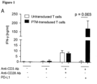
- the architecture of the PD-1-CD28 fusion proteins previously described showed only modest cytokine induction (2- to 3-fold) and little or no difference in lytic activity when transduced into primary T cells.
- the PD-1-CD28 fusion protein of the present invention carrying the PD-1 transmembrane domain (PTM) referring to PD-1-CD28 fusion proteins having an amino acid sequence as shown in SEQ ID NO: 14 (murine/mouse) or SEQ ID NO: 24 (human)) is superior to previously described PD-1-CD28 fusion constructs described in Prosser et al., Mol. Immunol.
- PTM PD-1 transmembrane domain
- the fusion protein of the PD-1 extracellular and transmembrane domain with the CD28 intracellular domain protects the antigen-specific T cells from PD-1-PD-L1-mediated anergy and turns the inhibitory signal into a co-stimulation.
- cells, like T cells, transduced with the PD-1-CD28 fusion protein of the present invention are resistant to the PD-1-PD-L1-mediated anergy.
- the functionality of PD-1-CD28 fusion constructs based on the human PD-1 and CD28 sequences is shown in Fig. 8 .
- the present invention relates to a fusion protein comprising a PD-1 polypeptide that is operably linked via its C-terminus to the N-terminus of an intracellular domain of a CD28 polypeptide, wherein the polypeptide comprises the extracellular domain and the transmembrane domain of PD-1.
- fusion protein relates to a protein which is made of polypeptide parts from different sources. Accordingly, it may be also understood as a “chimeric protein".
- fusion proteins are proteins created through the joining of two or more genes (or preferably cDNAs) that originally coded for separate proteins. Translation of this fusion gene (or fusion cDNA) results in a single polypeptide, preferably with functional properties derived from each of the original proteins.
- Recombinant fusion proteins are created artificially by recombinant DNA technology for use in biological research or therapeutics. Further details to the production of the fusion protein of the present invention are described herein below.
- polypeptide In the context of the present invention, the terms “polypeptide”, “peptide” and “protein” are used interchangeably to refer to a polymer of amino acid residues. The term also applies to amino acid polymers in which one or more amino acid residues is an artificial chemical mimetic or a corresponding naturally occurring amino acid, as well as to naturally occurring amino acid polymers. Accordingly, in the context of the present invention, the term “polypeptide” relates to a molecule which comprises or consists of chains of amino acid monomers linked by peptide (amide) bonds. Peptide bonds are covalent chemical bonds which are formed when the carboxyl group of one amino acid reacts with the amino group of another.
- a "polypeptide” is not restricted to a molecule with a defined length.
- polypeptide relates to a peptide, an oligopeptide, a protein, or a polypeptide which encompasses amino acid chains, wherein the amino acid residues are linked by covalent peptide bonds.
- polypeptide also encompasses peptidomimetics of such proteins/polypeptides wherein amino acid(s) and/or peptide bond(s) have been replaced by functional analogs.
- polypeptide also refers to, and does not exclude, modifications of the polypeptide, e.g., glycosylation, acetylation, phosphorylation and the like. Such modifications are well described in the art.
- amino acid refers to naturally occurring and synthetic amino acids, as well as amino acid analogs and amino acid mimetics that function in a manner similar to the naturally occurring amino acids.
- Naturally occurring amino acids are those encoded by the genetic code, as well as those amino acids that are later modified, e.g. hydroxyproline, ⁇ -carboxyglutamate, and O-phosphoserine.
- Amino acid analogs refers to compounds that have the same basic chemical structure as a naturally occurring amino acid, i.e., an ⁇ carbon that is bound to a hydrogen, a carboxyl group, an amino group, and an R group, e.g., homoserine, norleucine, methionine sulfoxide, methionine methyl sulfonium. Such analogs have modified R groups (e.g., norleucine) or modified peptide backbones, but retain the same basic chemical structure as a naturally occurring amino acid.
- Amino acid mimetics refers to chemical compounds that have a structure that is different from the general chemical structure of an amino acid, but that function in a manner similar to a naturally occurring amino acid. Amino acids may be referred to herein by either their commonly known three letter symbols or by the one-letter symbols recommended by the IUPAC-IUB Biochemical Nomenclature Commission.
- the fusion protein may comprise a fragment/polypeptide part of the full length PD-1 polypeptide and a fragment/polypeptide part of the full length CD28 polypeptide.
- the "PD-1 polypeptide" which is comprised in the herein provided fusion protein is a fragment/polypeptide part of the full length PD-1 polypeptide.
- the amino acid sequences of murine/mouse and human full length PD-1 are shown herein as SEQ ID NOs: 2 (murine/mouse as encoded by the cDNA sequence shown in SEQ ID NO: 1) and 4 (human as encoded by the cDNA sequence shown in SEQ ID NO: 3), respectively (the Uni Prot Entry number of human full length PD-1 is Q15116 (accession number with the entry version number 138 and version 3 of the sequence); the Uni Prot Entry number of the murine/mouse full length PD-1 is Q02242 (accession number with the entry version number 125 and version 1 of the sequence).
- CD28 polypeptide which is comprised in the herein provided fusion protein is a fragment/polypeptide part of the full length CD28 polypeptide.
- the amino acid sequences of human and murine/mouse full length CD28 are shown herein as SEQ ID NOs: 26 (murine/mouse as encoded by the copy DNA (cDNA) sequence shown in SEQ ID NO: 25) and 28 (human as encoded by the cDNA sequence shown in SEQ ID NO: 27), respectively.
- the herein provided fusion protein comprises a PD-1 polypeptide which is operably linked via its C-terminus to the N-terminus of an intracellular domain of a CD28 polypeptide, wherein the PD-1 polypeptide comprises the extracellular domain and the transmembrane domain of PD-1.
- the herein provided fusion protein may comprise the amino acids 1 to 200, preferably the amino acids 1 to 190 of the amino acid sequence of PD-1 as shown in SEQ ID NO: 2 (murine/mouse full length PD-1 as encoded by the cDNA sequence shown in SEQ ID NO: 1).
- the herein provided PD-1-CD28 fusion protein may comprise the amino acids 1 to 180, 1 to 181, 1 to 182, 1 to 183, 1 to 184, 1 to 185, 1 to 186, 1 to 187, 1 to 188, 1 to 189, 1 to 190, 1 to 191, 1 to 192, 1 to 193, 1 to 194, 1 to 195, 1 to 196, 1 to 197, 1 to 198, 1 to 199, or 1 to 200 of the amino acid sequence of PD-1 as shown in SEQ ID NO: 2 (as encoded by the cDNA sequence shown in SEQ ID NO: 1).
- the PD-1 polypeptide which is comprised in the fusion protein of the present invention may comprise or consist of the amino acid sequence as shown in SEQ ID NO: 8 (as encoded by the cDNA sequence shown in SEQ ID NO: 7) (murine/mouse).
- the fusion protein of the present invention comprises polypeptides which are derived from a human origin.
- the herein provided fusion protein comprises the amino acids 1 to 200, even more preferably the amino acids 1 to 191 of the amino acid sequence of PD-1 as shown in SEQ ID NO: 4 (human full length PD-1 as encoded by the cDNA shown in SEQ ID NO: 3).
- the herein provided fusion protein preferably comprises the amino acids 1 to 180, 1 to 181, 1 to 182, 1 to 183, 1 to 184, 1 to 186, 1 to 187, 1 to 188, 1 to 189, 1 to 190, 1 to 191, 1 to 192, 1 to 193, 1 to 194, 1 to 195, 1 to 196, 1 to 197, 1 to 198, 1 to 199, or 1 to 200 of the amino acid sequence of PD-1 as shown in SEQ ID NO: 4 (human full length PD-1 as encoded by the cDNA shown in SEQ ID NO: 3).
- the PD-1 polypeptide which is comprised in the fusion protein of the present invention may comprise or consist of the amino acid sequence as shown in SEQ ID NO: 16 (as encoded by the cDNA shown in SEQ ID NO: 15).
- the PD-1-CD28 fusion protein comprises the sequence as shown in SEQ ID NO: 16 or a sequence which has up to 1, 2, 3, 4, 5, 6, 7, 8, 9 or 10 substitutions, deletions or insertions in comparison to SEQ ID NO: 16 and which is characterized by having a PD-L1 or PD-L2 binding activity.
- the above-mentioned substation, deletion, insertion/addition may be a conservative mutation.
- a "conservative mutation” refers to substitutions of amino acids in a protein with other amino acids having similar characteristics (e.g. charge, side-chain size, hydrophobicity/hydrophilicity, backbone conformation, rigidity, etc.) such that the changes can be frequently be made without altering the biological activity of the protein.
- Those of skill in the art recognize that, in general, single amino acid substitutions in non-essential regions of a polypeptide do not substantially alter biological activity (see, e.g., Watson Molecular Biology of the Gene, The Benjamin/Cummings Pub. Co., p. 224 (4th Ed.)) (1987 ).
- binding compounds of the present invention comprise polypeptide chains with sequences that include up to none, 1, 2, 3, 4, 5, 6, 7, 8, 9 or 10 conservative amino acid substitutions when compared with the specific amino acid sequences disclosed herein, e.g. SEQ ID NOs: 6, 8, 10, 16, 18 or 20.
- the PD-1 polypeptide which comprises the extracellular domain and the transmembrane domain of PD-1 may comprise a sequence having the amino acid sequence as shown in SEQ ID NO: 16, wherein the amino acid sequence has up to 1, 2, 3, 4, 5, 6, 7, 8, 9 or 10, preferably 1 to 8, more preferably 1 to 6, even more preferably 1 to 5, even more preferably 1 or 2, or even more preferably 1 substitution(s), deletion(s) or insertion(s) in comparison to amino acid sequence as shown in SEQ ID NO: 16.
- the PD-1 polypeptide comprises one or more substitution(s), deletion(s) or insertion(s) in comparison to the amino acid sequence of SEQ ID NO: 16, respectively, then said fusion protein is characterized by having a PD-L1 and/or PD-L2 binding activity.
- This binding activity is defined as the ability to bind the PD-L1 and/or PD-L2 ligand either with the same, enhanced or reduced affinity as compared to the natural full length PD-1 protein (e.g. a protein having the amino acid sequence as shown in SEQ ID NO: 4).
- the natural full length PD-1 protein binds to the PD-L1 ligand with an equilibrium dissociation constant (KD) of 770 nM or less ( Butte et al., Molecular Immunology 45 (2008), 3567-3572 ) and the natural full length PD-1 proteins binds to the PD-L2 ligand with an equilibrium dissociation constant (KD) of 140 nM or less ( Butte et al., Molecular Immunology 45 (2008), 3567-3572 ).
- KD equilibrium dissociation constant
- the PD-1 polypeptide which comprises the extracellular domain and the transmembrane domain of PD-1 (as e.g.
- the PD-1 polypeptide which comprises the extracellular domain and the transmembrane domain of PD-1 (as e.g.
- SEQ ID NO: 16 or a variant thereof having up to 1, 2, 3, 4, 5, 6, 7, 8, 9, or 10 substitution(s), deletion(s) or insertion(s) in comparison to amino acid sequence as shown in SEQ ID NO: 16) may bind to PD-L1 and/or PD-L2 ligand with a binding affinity that is at least 1000, 100, 50, 40, 30, 20, 10, 5-fold higher (i.e. enhanced) or lower (i.e. reduced) compared to the natural full length PD-1 protein.
- KD is intended to refer to the dissociation constant and is expressed as a molar concentration (M). KD values for protein-protein interactions between e.g.
- the PD-1 polypeptide described herein above and PD-L1 and/or PD-L2 can be determined using methods well established in the art. Methods for determining the binding affinity towards PD-L1 and/PD-L2 ligand are known in the art and described herein below in more detail and include, e.g., surface plasmon resonance (SPR), biacore measurement, flow cytometry or ELISA.
- SPR surface plasmon resonance
- biacore measurement e.g., flow cytometry or ELISA.
- the PD-1-CD28 fusion protein comprises the extracellular domain of PD-1 which is located at amino acids 1 to 169 of the mouse full length PD-1 protein as shown in SEQ ID NO: 2 (as encoded by the cDNA shown in SEQ ID NO: 1).
- the fusion protein comprises the extracellular domain of PD-1 which is located at amino acids 1 to 170 of the human full length PD-1 protein as shown in SEQ ID NO: 4 (as encoded by the cDNA shown in SEQ ID NO: 3).
- the PD-1-CD28 fusion protein comprises or consists of the extracellular domain of PD-1 as shown in SEQ ID NO: 8 (as encoded by the cDNA sequence shown in SEQ ID NO: 7) or more preferably as shown in SEQ ID NO: 18 (as encoded by the cDNA sequence shown in SEQ ID NO: 17).
- the extracellular domain of the PD-1 protein (which is comprised in the herein provided fusion protein) is characterized by the ability to bind the natural ligands of PD-1 (i.e.
- amino acid sequences of human full length PD-L1 and human full length PD-L2 are shown herein as SEQ ID NO: 34 (PD-L1 as encoded by the cDNA sequence shown in SEQ ID NO: 33) or SEQ ID NO: 36 (PD-L2 as encoded by the cDNA sequence shown in SEQ ID NO: 35).
- a reduced (i.e. diminished), same (i.e. equal) or preferably enhanced affinity to PD-L1 or PD-L2 can be achieved by point mutations in the extracellular domain of the herein provide fusion protein.
- changing the alanine at the position corresponding to amino acid position 132 of SEQ ID NO: 18 by a leucine enhances PD-1 affinity (i.e.
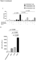
- the activity of the herein provided fusion protein in cells is enhanced in terms of cytokine secretion, proliferation and lysis.
- Binding affinity of the herein provided fusion protein to PD-L1 or PD-L2 can be assessed by methods well known by those skilled in the art including but not limited to flow cytometry, ELISA, immunoprecipitation, Western blot, confocal or conventional microscopy ( Terawaki et al., International Immunology, 19(7) (2007), 881-890 ; Cheng et al., J Biol Chem. 288(17) (2013), 11771-11785 ; Ghiotto et al., Int Immunol. 22(8) (2010), 651-660 .).
- binding of the said fusion protein to PD-L1 or PD-L2 leads to clustering of T cells around the target cell which could be a tumor or another immune cell and activation of those cells by means of the fusion protein.
- the fusion protein itself clusters to the contact point with the target cell. This can be measured and/or visualized, e.g., by confocal or conventional microscopy. Binding of the fusion protein (i.e. of the PD-1 polypeptide which is comprised in the fusion protein) to either PD-L1 or PD-L2 is central for any subsequent signaling via the CD28 signaling motifs of the fusion protein and the effects resulting thereof.
- Mutations in the fusion protein resulting in enhanced affinity for the ligands PD-L1 and/or PD-L2 can enhance the functionality of the fusion protein.
- Methods for measuring affinity of one protein to another are well known to those skilled in the art and include surface plasmon resonance (SPR), biacore measurement, flow cytometry or ELISA.
- the herein provided PD-1-CD28 fusion protein comprises the transmembrane domain of PD-1 which is located at amino acids 170 to 191 of the mouse full length PD-1 protein as shown in SEQ ID NO: 2 (as encoded by the cDNA shown in SEQ ID NO: 1).
- the transmembrane domain of PD-1 is located at amino acids 171 to 191 of the human full length PD-1 protein (as shown in SEQ ID NO: 4 (as encoded by the cDNA shown in SEQ ID NO: 3)).
- the transmembrane domain of PD-1 is an important component of the fusion protein of the present invention and allows for signal transduction to the intracellular domains of CD28 upon engagement of the PD-1-receptor (i.e. the PD-1 polypeptide of the fusion protein).
- the transmembrane domain which is comprised in PD-1-CD28 fusion proteins may comprise or consist of the amino acid sequence as shown in SEQ ID NO: 10 (murine/mouse as encoded by cDNA sequence shown in SEQ ID NO: 9) or SEQ ID NO: 20 (human as encoded by the cDNA sequence shown in SEQ ID NO: 19).
- the transmembrane domain which is comprised in fusion protein of the present invention may comprise or consist of an amino acid sequence which includes up to 1, 2, 3, 4, 5, 6, 7, 8, 9 or 10, preferably, 1 to 8, more preferably 1 to 6, even more preferably 1 to 4, even more preferably 1 to 2, or even more preferably 1 substitution(s), deletion(s) or insertion(s) in comparison to amino acid sequence as shown in SEQ ID NO: 10 (murine/mouse) or 20 (human).
- the transmembrane domain comprises none, 1, 2, 3, 4, 5, 6, 7, 8, 9 or 10 substitution(s), deletion(s) or insertion(s) in comparison to the amino acid sequence as shown in SEQ ID NO: 10 (murine/mouse) or 20 (human), then said fusion protein is characterized by showing the same or preferably an enhanced signal transduction activity.
- Signal transduction activity can be measured by flow cytometry, Western blot or ELISA based assays detecting phosphorylated proteins such as V-akt murine thymoma oncogene homologue 1 (AKT).
- Signal transduction activity can also be detected by downstream functional effects such as cytokine release, proliferation or lytic activity of cells, such as T cells (as described e.g. herein in the appended Examples and in Krutzik et al., Methods Mol Biol. 699 (2011), 179-202 ; Ekkens et al., Infect Immun. 75(5) (2007), 2291-2296 ; Ge et al., Proc Natl Acad Sci USA. 99(5) (2002), 2983-2988 ; Düwell et al., Cell Death Differ. 21(12) (2014), 1825-1837 , Erratum in: Cell Death Differ. 21(12) (2014 ), 161).
- T cells as described e.g. herein in the appended Examples and in Krutzik et al., Methods Mol Biol. 699 (2011), 179-202 ; Ekkens et al., Infect Immun. 75(5) (2007), 2291-2296 ; Ge et al.,
- the transmembrane domain of PD-1 is an important component of the PD-1-CD28 fusion protein of the present invention and provides signal transduction to the intracellular domains of CD28 upon engagement of the PD-1-receptor domain (i.e. the PD-1 polypeptide of the fusion protein) which can be measured by, e.g, the cytokine production, proliferation or lytic activity of cells such as T cells.
- the transmembrane of PD-1 has the amino acid sequence shown in SEQ ID NO: 20 (as encoded by the cDNA shown in SEQ ID NO: 19).
- the intracellular domain of the PD-1-CD28 fusion protein of the present invention is derived from the (human) CD28 gene (Uni Prot Entry No: P10747 (accession number with the entry version: 164 and version 1 of the sequence) and provides CD28 activity, defined as cytokine production, proliferation and lytic activity of the transduced cell described herein, like a transduced T cell.
- CD28 activity can be measured by release of cytokines by ELISA or flow cytometry of cytokines such as interferon-gamma (IFN-y) or interleukin 2 (IL-2) (as described herein below in the appended Examples), proliferation of T cells measured e.g.
- the signaling domains PYAP (AA 208 to 211 of SEQ ID NO: 28 (as encoded by cDNA sequence shown in SEQ ID NO: 27)) and YMNM (AA 191 to 194 of SEQ ID NO: 28) are beneficial for the function of the CD28 polypeptide and the functional effects enumerated above.
- the amino acid sequence of the YMNM domain is shown in SEQ ID NO: 29; the amino acid sequence of the PYAP domain is shown in SEQ ID NO: 30.
- the CD28 polypeptide preferably comprises a sequence derived from intracellular domain of a CD28 polypeptide having the sequences YMNM (SEQ ID NO: 29) and/or PYAP (SEQ ID NO: 30).
- a CD28 activity defined as cytokine production, proliferation and lytic activity of a transduced cell described herein, like e.g. a transduced T cell.
- the intracellular domain of the PD-1-CD28 fusion protein of the present invention has the amino acid sequence of SEQ ID NO: 22 (human) (as encoded by the cDNA sequence shown in SEQ ID NO: 21) or SEQ ID NO: 12 (mouse/murine) (as encoded by the cDNA sequence shown in SEQ ID NO: 11).
- SEQ ID NO: 22 human
- SEQ ID NO: 12 mouse/murine
- one or both of these domains may be mutated to FMNM (SEQ ID NO: 31) and/or AYAA (SEQ ID NO: 32), respectively.
- the PD-1-CD28 fusion protein may comprise the amino acids 170 to 218, preferably the amino acids 178 to 218 of the amino acid sequence of CD28 as shown in SEQ ID NO: 26 (mouse full length CD28 as encoded by cDNA sequence shown in SEQ ID NO: 25).
- the intracellular CD28 polypeptide may be of any length provided that the intracellular domain of the fusion protein of the present invention comprises the sequences YMNM (SEQ ID NO: 29) and/or PYAP (SEQ ID NO: 30).
- the intracellular domain of the CD28 of the PD-1-CD28 fusion protein may comprise a sequence derived from the intracellular domain of CD28 polypeptide having the sequences YMNM (SEQ ID NO: 29) and/or PYAP (SEQ ID NO: 30).
- the CD28 polypeptide which is comprised in the fusion protein of the present invention may comprise or consist of the amino acid sequence as shown in SEQ ID NO: 12 (as encoded by the cDNA sequence shown in SEQ ID NO: 11).
- the fusion protein preferably comprises polypeptides of human origin.
- the herein provided fusion protein comprises the amino acids 170-220, even more preferably the amino acids 180 to 220 of the amino acid sequence of CD28 as shown in SEQ ID NO: 28 (human full length CD28 as encoded by cDNA sequence shown in SEQ ID NO: 27).
- the CD28 polypeptide which is comprised in the fusion protein of the present invention may comprise or consist of the amino acid sequence as shown in SEQ ID NO: 22 (as encoded by the cDNA sequence shown in SEQ ID NO: 21).
- the intracellular CD28 polypeptide may be of any length provided that the intracellular domain of the fusion protein of the present invention comprises the sequences YMNM (SEQ ID NO: 29) and/or PYAP (SEQ ID NO: 30). Accordingly, in the context of the present invention the intracellular domain of the CD28 of the PD-1-CD28 fusion protein may comprise a sequence derived from the intracellular domain of CD28 polypeptide having the sequences YMNM (SEQ ID NO: 29) and/or PYAP (SEQ ID NO: 30).
- the CD28 polypeptide which is comprised in the fusion protein of the present invention may comprise or consist of the amino acid sequence as shown in SEQ ID NO: 12 (murine/mouse) or 22 (human).
- the CD28 polypeptide of the PD-1-CD28 fusion protein has the amino acid sequence of SEQ ID NO: 22.
- the fusion protein comprises an intracellular domain of a CD28 polypeptide having the sequences YMNM (SEQ ID NO: 29) and/or the PYAP (SEQ ID NO: 30). Accordingly, in the context of the present, the CD28 polypeptide has the amino acid sequence of SEQ ID NO: 22 (human).
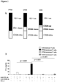
- the herein provided PD-1-CD28 fusion protein may comprise or consist of an amino acid sequence as shown in SEQ ID NO: 14 (murine/mouse PTM (mPTM)-fusion protein as encoded by the cDNA sequence shown in SEQ ID NO: 13).
- the herein provided fusion protein comprises or consists of an amino acid sequence as shown in SEQ ID NO: 24 (human PTM (hPTM) fusion protein as encoded by the cDNA sequence shown in SEQ ID NO: 23).
- the present invention relates to a PD-1-CD28 fusion protein which has the amino acid sequence of SEQ ID NO: 24.
- the present invention relates to a fusion of protein which consists of SEQ ID NO: 24, wherein said fusion protein has
- the binding PD-L1 and/or PD-L2 binding activity is defined as the ability to bind the PD-L1 and/or PD-L2 ligand either with the same, enhanced or reduced affinity as compared to the natural full length PD-1 protein (e.g. a protein having the amino acid sequence as shown in SEQ ID NO: 4 (as encoded by the cDNA sequence shown in SEQ ID NO: 3).
- the PD-1 polypeptide which comprises the extracellular domain of PD-1 may bind to the PD-L1 and/or PD-L2 ligand with the same binding as the natural full length PD-1 protein does.
- the PD-1 polypeptide which comprises the extracellular domain may bind to PD-L1 and/or PD-L2 ligand with a binding affinity that is at least 1000, 100, 50, 40, 30, 20, 10, 5-fold higher (i.e. enhanced) or lower (i.e. reduced) compared to the natural full length PD-1 protein.
- a binding affinity that is at least 1000, 100, 50, 40, 30, 20, 10, 5-fold higher (i.e. enhanced) or lower (i.e. reduced) compared to the natural full length PD-1 protein.
- methods for the determination of the PD-L1 and/or PD-L2 binding activity are well known to the skilled person and described herein above.
- PD-1 relates to the programmed cell death protein 1, also known as PD-1 and CD279 (cluster of differentiation 279).
- PD-1 is a protein that in humans is encoded by the PDCD1 gene.
- PD-1 is a cell surface receptor that belongs to the immunoglobulin superfamily and is expressed on T cells and pro-B cells.
- PD-1 is known to bind to two ligands, PD-L1 and PD-L2.
- PD-1 and its ligands play an important role in down regulating the immune system by preventing the activation of T cells, which in turn reduces autoimmunity and promotes self-tolerance.
- the inhibitory effect of PD-1 is accomplished through a dual mechanism of promoting apoptosis (programmed cell death) in antigen specific T cells in lymph nodes while simultaneously reducing apoptosis in regulatory T cells (suppressor T cells).
- the protein sequences of the human and mouse PD-1 are shown herein in SEQ ID NO: 4 (as encoded by the cDNA sequence shown in SEQ ID NO: 3) or SEQ ID NO: 2 (as encoded by the cDNA sequence shown in SEQ ID NO: 1).
- the nucleic acid sequences of the human and mouse PD-1 are shown in SEQ ID NOs: 3 (human) and 1 (murine/mouse), respectively.
- CD28 refers to the receptor "cluster of differentiation 28".
- CD28 is one of the proteins expressed on T cells that provide costimulatory signals required for T cell activation and survival. T cell stimulation through CD28 in addition to the T cell receptor (TCR) can provide a potent signal for the production of various interleukins (e.g. IL-6).
- CD28 is the receptor for CD80 (B7.1) and CD86 (B7.2) proteins.
- SEQ ID NO: 28 as encoded by the cDNA sequence shown in SEQ ID NO: 27
- SEQ ID NO: 26 as encoded by the cDNA sequence shown in SEQ ID NO: 25
- the nucleic acid sequences of the human and mouse CD28 are shown in SEQ ID NOs: 27 (human) and 25 (murine/mouse), respectively.
- operably linked refers to functional linkage between at least two protein sequences, i.e. between the herein described PD-1 polypeptide comprising the extracellular domain and the transmembrane domain of PD-1 and the intracellular domain of CD28.
- the PD-1 polypeptide and the CD28 polypeptide as comprised in the fusion proteins of the present invention may be covalently linked.
- a covalent bond is a chemical bonding that is characterized by the sharing of pairs of electrons between atoms, as, inter alia, obtained by the herein exemplified cross-binding via chemical compounds.
- constructs as disclosed herein i.e.
- PD-1 polypeptide comprising the extracellular domain and the transmembrane domain of PD-1 and a covalently bound intracellular domain of a CD28 polypeptide is envisaged.
- "operably linked” refers to a functional linkage between at least two polypeptides which means that both polypeptides retain their functionalities.
- the herein provided fusion protein may comprise a linker (or "spacer").
- a linker is usually a peptide having a length of up to 20 amino acids. Accordingly, in the context of the present invention the linker may have a length of 1, 2, 3, 4, 5, 6, 7, 8, 9, 10, 11, 12, 13, 14, 15, 16, 17, 18, 19 or 20 amino acids.
- the herein provided fusion protein may comprise a linker between the PD-1 polypeptide and the CD28 polypeptide.
- linkers have that advantage that they can make it more likely that the different polypeptides of the fusion protein (i.e. the PD-1 polypeptide and the CD28 polypeptide) fold independently and behave as expected.
- the PD-1 polypeptide and the CD28 polypeptide may be comprised in a single-chain multifunctional polypeptide.
- a single-chain PD-1-CD28 fusion construct e.g. may consist of (a) polypeptide(s) comprising (a) PD-1 derived domain(s) and (a) intracellular domain of a CD28 polypeptide. Said domains are connected by a polypeptide linker, wherein said linker is disposed between said PD-1-derived domain(s) and said intracellular CD28 polypeptide domain.
- N-terminus may be used interchangeably with the amino terminus of a polypeptide, the NH2-terminus, the N-terminal end or amine-terminus.
- the term means the natural start of a protein or polypeptide.
- C-terminus may be used interchangeably with the carboxy terminus of a polypeptide, the carboxyl-terminus, the carboxy-terminus, the C-terminal tail, the C-terminal end or COOH-terminus.
- the term means the natural end of a protein or polypeptide.
- extracellular domain in particular in the context of the extracellular domain of PD-1, refers to the part of the receptor (i.e. of PD-1) that sticks out of the membrane on the outside of the cell.
- the activity of the extracellular domain is to bind to a specific ligand (e.g. PD-L1 or PD-L2).
- the extracellular domain of the PD-1-CD28 fusion protein has the amino acid/polypeptide sequence of SEQ ID NO: 18 or a polypeptide sequence which has up to 1, 2, 3, 4, 5, 6, 7, 8, 9 or 10 substitution(s), deletion(s) or insertion(s) in comparison to SEQ ID NO: 18 and which is characterized by having PD-L1 and/or PD-L2 binding activity as described herein above.
- transmembrane domain in particular in the context of transmembrane domain of PD-1, refers to the part of the receptor (i.e. PD-1) which is naturally located within the membrane of the cell (e.g. the T cell).
- the transmembrane domain of PD-1 has the amino acid sequence of SEQ ID NO: 20 or an amino acid sequence which has up to 1, 2, 3, 4, 5, 6, 7, 8, 9 or 10 substitution(s), deletion(s) or insertion(s) in comparison to SEQ ID NO: 20 and which is characterized by showing the same or preferably an enhanced signal transduction activity as described herein above.
- intracellular domain in particular in the context of intracellular domain of CD28, refers to the cytoplasmic domain of the receptor (i.e. CD28).
- the intracellular domain refers to an amino acid sequence which is derived from the intracellular domain of a CD28 polypeptide having the sequences YMNM (SEQ ID NO: 29) and/or PYAP (SEQ ID NO: 30), like the amino acid sequence shown in SEQ ID NO: 22.
- the intracellular domain interacts with the interior of the cell.
- the production of the fusion proteins of the present invention is commonly known in the art.
- the fusion protein as described herein may be created through genetic engineering of a fusion gene. This typically involves removing the stop codon from a cDNA sequence coding for the first polypeptide (i.e. the PD-1 polypeptide), then appending the cDNA sequence of the second polypeptide (i.e. the CD28 polypeptide) in frame through ligation or overlap extension PCR. That DNA sequence can then be expressed by a cell as a single protein.
- the fusion protein of the present invention can be generated by overlap PCR and recombinant expression cloning into a retroviral vector (e.g. the pMP71 vector) as described in the illustrative appended examples.
- nucleic acid molecule(s) encoding the fusion proteins of the invention.
- nucleic acid molecule relates to the sequence of bases comprising purine- and pyrimidine bases which are comprised by polynucleotides, whereby said bases represent the primary structure of a nucleic acid molecule.
- nucleic acid molecule includes DNA, cDNA, genomic DNA, RNA, synthetic forms of DNA and mixed polymers comprising two or more of these molecules.
- nucleic acid molecule includes both, sense and antisense strands.
- the herein described “nucleic acid molecule” may contain non-natural or derivatized nucleotide bases, as will be readily appreciated by those skilled in the art. Exemplary nucleic acid molecules encoding the human and mouse fusion protein of the present invention are shown in SEQ ID NO: 23 (human) or SEQ ID NO: 13 (mouse).
- the nucleic acid molecules of the invention may be under the control of regulatory sequences.
- promoters for example, promoters, transcriptional enhancers and/or sequences which allow for induced expression of the fusion protein of the invention may be employed.
- the nucleic acid molecules are expressed under the control of constitutive or inducible promoter. Suitable promoters are e.g.
- the CMV promoter Qin et al., PLoS One 5(5) (2010), e10611
- the UBC promoter Qin et al., PLoS One 5(5) (2010), e10611
- PGK Qin et al., PLoS One 5(5) (2010), e10611
- the EF1A promoter Qin et al., PLoS One 5(5) (2010), e10611
- the CAGG promoter Qin et al., PLoS One 5(5) (2010), e10611
- the SV40 promoter Qin et al., PLoS One 5(5) (2010), e10611
- the COPIA promoter Qin et al., PLoS One 5(5) (2010), e10611
- the ACT5C promoter Qin et al., PLoS One 5(5) (2010), e10611
- the TRE promoter Qin et al., PLoS One
- control sequence refers to DNA sequences, which are necessary to effect the expression of coding sequences to which they are ligated. The nature of such control sequences differs depending upon the host organism. In prokaryotes, control sequences generally include promoter, ribosomal binding site, and terminators. In eukaryotes generally control sequences include promoters, terminators and, in some instances, enhancers, transactivators or transcription factors. The term “control sequence” is intended to include, at a minimum, all components the presence of which are necessary for expression, and may also include additional advantageous components.
- nucleic acid molecules may contain, for example, thioester bonds and/or nucleotide analogues. Said modifications may be useful for the stabilization of the nucleic acid molecule against endo- and/or exonucleases in the cell.
- Said nucleic acid molecules may be transcribed by an appropriate vector containing a chimeric gene which allows for the transcription of said nucleic acid molecule in the cell.
- polynucleotide can be used for "gene targeting" or "gene therapeutic” approaches.
- said nucleic acid molecules are labeled.
- nucleic acid molecule(s) may be a recombinantly produced chimeric nucleic acid molecule comprising any of the aforementioned nucleic acid molecules either alone or in combination.
- the nucleic acid molecule is part of a vector.
- the present invention therefore also relates to vectors comprising the nucleic acid molecule described in the present invention.
- the term "vector” relates to a circular or linear nucleic acid molecule which can autonomously replicate in a host cell (i.e. in a transduced cell) into which it has been introduced.
- the "vector” as used herein particularly refers to a plasmid, a cosmid, a virus, a bacteriophage and other vectors commonly used in genetic engineering.
- the vector of the invention is suitable for the transformation of cells, preferably of T cells.
- the vector as provided herein is an expression vector. Expression vectors have been widely described in the literature.
- the herein provided vector preferably comprises a recombinant polynucleotide (i.e. a nucleic acid molecule encoding the fusion protein of the present invention) as well as expression control sequences operably linked to the nucleotide sequence to be expressed.
- the vector as provided herein preferably further comprises a promoter.
- the herein described vector may also comprise a selection marker gene and a replication-origin ensuring replication in the host (i.e. the transduced cell).
- the herein provided vector may also comprise a termination signal for transcription. Between the promoter and the termination signal there is preferably at least one restriction site or a polylinker which enables the insertion of a nucleic acid molecule (e.g.
- nucleic acid molecule encoding the fusion protein of the invention desired to be expressed.
- vectors suitable to comprise a nucleic acid molecule of the present invention to form the vector of the present invention are known in the art.
- suitable vectors include cosmids, plasmids (e.g., naked or contained in liposomes) and viruses (e.g., lentiviruses, retroviruses, adenoviruses, and adeno-associated viruses) that incorporate the nucleic acid molecule of the invention (i.e. the nucleic acid molecule encoding the fusion protein of the present invention).
- the vector of the present invention is a viral vector. More preferably, the vector of the present invention is a lentiviral vector, and even more preferably, the vector of the present invention is a retroviral vector (e.g. the pMP71 vector). Accordingly, in the context of the present invention, the vector is a lentiviral vector or a retroviral vector.
- the vector of the present invention allows for constitutive or conditional expression of the nucleic acid molecule encoding the PD-1-CD28 fusion protein of the present invention.
- suitable retoviral vectors for the expression of the fusion protein of the present invention are known in the art such as SAMEN CMV/SRa ( Clay et al., J. Immunol.
- LZRS-id3-IHRES Heemskerk et al., J. Exp. Med. 186 (1997), 1597-1602 ), FeLV ( Neil et al., Nature 308 (1984), 814-820 ), SAX ( Kantoff et al., Proc. Natl. Acad. Sci. USA 83 (1986), 6563-6567 ), pDOL ( Desiderio, J. Exp. Med. 167 (1988), 372-388 ), N2 ( Kasid et al., Proc. Natl. Acad. Sci.
- LNL6 Tiberghien et al., Blood 84 (1994), 1333-1341
- pZipNEO Chen et al., J. Immunol. 153 (1994), 3630-3638
- LASN Mullen et al., Hum. Gene Ther. 7 (1996), 1123-1129
- pG1XsNa Taylor et al., J. Exp. Med. 184 (1996), 2031-2036
- LCNX Sun et al., Hum. Gene Ther. 8 (1997), 1041-1048
- SFG Gallardo et al., Blood 90 (1997 )
- LXSN Sun et al., Hum.
- suitable lentiviral vectors for the expression of the fusion protein of the present invention are, e.g. PL-SIN lentiviral vector ( Hotta et al., Nat Methods.
- the invention also relates to transduced cells expressing a fusion protein encoded by a nucleic acid molecule encoding the fusion protein of the present invention.
- the transduced cell may comprise a nucleic acid molecule encoding the fusion protein of the present invention or a vector of the present invention which expresses a fusion protein of the present invention.
- transduced cell relates to a genetically modified cell (i.e. a cell wherein a nucleic acid molecule has been introduced deliberately).
- the herein provided transduced cell may comprise the vector of the present invention.
- the herein provided transduced cell comprises the nucleic acid molecule (encoding the fusion protein) of the present invention and/or the vector of the present invention.
- the transduced cell of the invention may be a cell which transiently or stably expresses the foreign DNA (i.e. the nucleic acid molecule which has been introduced into the cell).
- the nucleic acid molecule encoding the fusion protein of the present invention can be stably integrated into the genome of the cell by using a retroviral or lentiviral transduction.
- the nucleic acid molecule encoding the fusion protein of the present invention may be expressed transiently.
- the herein provided transduced cell has been genetically modified by introducing a nucleic acid molecule in the cell via a viral vector (e.g. a retroviral vector or a lentiviral vector).
- the expression can be constitutive or constitutional, depending on the system used.
- the PD-1-CD28 fusion protein is expressed on the surface of the herein provided transduced cell.
- the extracellular proportion of the PD-1 polypeptide of the fusion protein can be detected on the cell surface, while the intracellular (both transmembrane and e.g. a part of the intracellular domain of the PD-1 polypeptide) are bound to the membrane but are not detectable on cell surface.
- the detection of the extracellular domain of the PD-1 polypeptide can be carried out by using an antibody which specifically binds to this extracellular domain of the PD-1 polypeptide.
- Examples for such antibodies are well known in the art and include clone EH12.2H7 (commercially available from BioLegend, catalogue no.: 329911), clone J116 (commercially available from eBioscience, catalogue no.: 16-9989-80), J105 (commercially available from eBioscience, catalogue no.: 8012-2799) or MiH4 (commercially available from eBioscience, catalogue no.: 14-9969).
- the extracellular domain can be detected using these antibodies by flow cytometry or microscopy.
- the transduced cell of the present invention may be, e.g., a CD4+-T cell, a CD8+-T cell, a ⁇ T cell, a natural killer (NK) T cell, a natural killer (NK) cell, a tumor-infiltrating lymphocyte (TIL) cell, a myeloid cell, or a mesenchymal stem cell.
- the herein provided transduced cell is a T cell (e.g. an autologous T cell), more preferably, the transduced cell is a CD8+ T cell.
- the transduced cell is a CD8+ T cell.
- the transduced cell is an autologous T cell.
- the transduced cell is preferably an autologous CD8+ T cell.
- the transduced cell may also be an allogeneic cell, such as an allogeneic CD8+ T cell.
- the invention provides a transduced CD4+ T cell, which may be an autologous CD4+ T cell or an allogeneic CD4+ T cell.
- the invention provides populations of transduced CD4+ T cells and/or CD8+ T cells (including but not limited to populations comprising transduced CD4+ T cells, populations comprising transduced CD8+ T cells, and populations comprising both transduced CD4+ T cells and transduced CD8+ T cells), wherein the transduced cells may be autologous and/or allogenic (e.g., the population may comprise only autologous cells, only allogenic cells, or combinations of both autologous and allogenic cells).
- the populations of T cells of the invention as described herein may comprise both transduced and non-transduced T cells.
- allogeneic cells are based on the fact that cells, preferably T cells can recognize a specific antigen epitope presented by foreign antigen-presenting cells (APC), provided that the APC express the MHC molecule, class I or class II, to which the specific responding cell population, i.e. T cell population is restricted, along with the antigen epitope recognized by the T cells.
- APC foreign antigen-presenting cells
- allogeneic refers to cells coming from an unrelated donor individual/subject which is human leukocyte antigen (HLA) compatible to the individual/subject which will be treated by e.g. the herein described PD-1-CD28 fusion protein expressing transduced cell.
- Autologous cells refer to cells which are isolated/obtained as described herein above from the subject to be treated with the transduced cell described herein.
- the transduced cell of the present invention is transduced with a nucleic acid molecule expressing the herein provided fusion protein.
- a nucleic acid molecule expressing the herein provided fusion protein.
- TIL tumor-infiltrating lymphocyte cells
- 2152-2159 doi: 10.1200/JCO.2012.46.6441
- antigen-specific cells sorted from the peripheral blood of patients for their tumor-specificity by flow cytometry ( Hunsucker et al., Cancer Immunol Res.
- the cells described herein would only be transduced with the fusion protein of the present invention.
- the transduced cell of the invention may be co-transduced with further nucleic acid molecules, e.g. with a nucleic acid molecule encoding a T cell receptor or a chimeric antigen receptor.
- T cell receptor is commonly known in the art.
- T cell receptor refers to any T cell receptor, provided that the following three criteria are fulfilled: (i) tumor specificity, (ii) recognition of (most) tumor cells, which means that an antigen or target should be expressed in (most) tumor cells and (iii) that the TCR matches to the HLA-type of the subject to be treated.
- T cell receptors which fulfill the above mentioned three criteria are known in the art such as receptors recognizing WT1 (Wilms tumor specific antigen 1; for sequence information(s) see, e.g., Sugiyama, Japanese Journal of Clinical Oncology 40 (2010), 377-87 ), MAGE (for sequence see, e.g., WO-A1 2007/032255 and PCT/US2011/57272 ), SSX ( U.S. Provisional Application No. 61/388,983 ), NY-ESO-1 (for sequence information(s) see, e.g., PCT/GB2005/001924 ) and/or HER2neu (for sequence information(s) see WO-A1 2011/0280894 ).
- chimeric antigen receptor or "chimeric receptor” is known in the art and refers to a receptor constituted of an extracellular portion of a single chain antibody domain fused by a spacer sequence to the signal domains of CD3z and CD28. Again, this chimeric antigen receptor should provide tumor specify and allow for the recognition of most tumor cells.
- Suitable chimeric receptors include: anti-EGFRv3-CAR (for sequence see WO-A1 2012/138475 ), anti-CD22-CAR (see WO-A1 2013/059593 ), anti-BCMA-CAR (see WO-A1 2013/154760 ), anti-CD19-CAR (see WO-A1 2012/079000 or US-A1 2014/0271635 ), anti-CD123-CAR (see US-A1 2014/0271582 ), anti-CD30-CAR (see WO-A1 2015/028444 ) or anti-Mesothelin-CAR (see WO-A1 2013/142034 ).
- the present invention also relates to a method for the production of a transduced cell expressing a fusion protein of the invention, comprising the steps of transducing a cell with a vector of the present invention, culturing the transduced cell under conditions allowing the expressing of the fusion protein in or on said transduced cell and recovering said transduced cell.
- the transduced cell of the present invention is preferably produced by the following process: cells (e.g., T cells) are isolated/obtained from a subject (preferably a human patient). Methods for isolating/obtaining cells (e.g. T cells) from patients or from donors are well known in the art and in the context of the present the cells (e.g. T cells) from patients or from donors may be isolated by blood draw or removal of bone marrow. After isolating/obtaining cells as a sample of the patient, the cells (e.g. T cells) are separated from the other ingredients of the sample. Several methods for separating cells (e.g. T cells) from the sample are known and include, without being limiting, e.g.
- leukapheresis for obtaining cells from the peripheral blood sample from a patient or from a donor, isolating/obtaining cells by using a FACSort apparatus, picking living of dead cells from fresh biopsy specimens harboring living cells by hand or by using a micromanipulator (see, e.g., Dudley, Immunother. 26 (2003), 332-342 ; Robbins, Clin. Oncol. 29 (201 1), 917-924 or Leisegang, J. Mol. Med. 86 (2008), 573-58 ).
- the methods and processes disclosed in this paragraph and otherwise disclosed herein are preferably performed with respect to CD8+ T cells, but are also applicable to other T cell types such as CD4+ T cells.
- fresh patient biopsy refers to tissue (preferably tumor tissue) removed from a subject by surgical or any other known means as well as tumor cell lines or (isolated) cells from a tumor tissue/tumor cell.
- the isolated/obtained cells T cells e.g. CD8+ T cells
- IL-2 interleukin-2
- the cells are artificially/genetically modified/transduced by methods known in the art (see, e.g., Lemoine, J Gene Med 6 (2004), 374-386 ).
- Methods for transducing cells are known in the art and include, without being limited, in a case where nucleic acid or a recombinant nucleic acid is transduced, for example, an electroporation method, calcium phosphate method, cationic lipid method or liposome method.
- the nucleic acid to be transduced can be conventionally and highly efficiently transduced by using a commercially available transfection reagent, for example, Lipofectamine (manufactured by Invitrogen, catalogue no.: 11668027).
- the vector can be transduced in the same manner as the above-mentioned nucleic acid as long as the vector is a plasmid vector (i.e. a vector which is not a viral vector
- the methods for transducing cells include retroviral or lentiviral T cell transduction as well as mRNA transfection.
- mRNA transfection refers to a method well known to those skilled in the art to transiently express a protein of interest, like in the present case the PD-1-CD28 fusion protein of the present invention, in a cell to be transduced.
- cells may be electroporated with the mRNA coding for the fusion protein of the present by using an electroporation system (such as e.g. Gene Pulser, Bio-Rad) and thereafter cultured by standard cell (e.g. T cell) culture protocol as described above (see Zhao et al., Mol Ther. 13(1) (2006), 151-159 .)
- the transduced cell of the invention is a T cell (most preferably a CD8+ T cell) and is generated by lentiviral, or most preferably retroviral T cell transduction.
- suitable retroviral vectors for transducing cells are known in the art such as SAMEN CMV/SRa ( Clay et al., J. Immunol. 163 (1999), 507-513 ), LZRS-id3-IHRES ( Heemskerk et al., J. Exp. Med. 186 (1997), 1597-1602 ), FeLV ( Neil et al., Nature 308 (1984), 814-820 ), SAX ( Kantoff et al., Proc. Natl. Acad. Sci. USA 83 (1986), 6563-6567 ), pDOL ( Desiderio, J. Exp. Med.
- SAMEN CMV/SRa Clay et al., J. Immunol. 163 (1999), 507-513
- LZRS-id3-IHRES Heemskerk et al., J. Exp. Med. 186 (1997), 1597-1602
- FeLV Neil et al., Nature 308
- suitable lentiviral vector for transducing cells are, e.g. PL-SIN lentiviral vector ( Hotta et al., Nat Methods.
- the transduced cell/cells of the present invention is/are preferably grown under controlled conditions, outside of their natural environment.
- the term "culturing” means that cells (e.g. the transduced cell(s) of the invention) which are derived from multi-cellular eukaryotes (preferably from a human patient) are grown in vitro. Culturing cells is a laboratory technique of keeping cells alive which are separated from their original tissue source.
- the transduced cell of the present invention is cultured under conditions allowing the expression of the fusion protein of the present invention in or on said transduced cells. Conditions which allow the expression or a transgene (i.e.
- the fusion protein of the present invention are commonly known in the art and include, e.g., agonistic anti-CD3- and anti-CD28 antibodies and the addition of cytokines such as interleukin 2 (IL-2), interleukin 7 (IL-7), interleukin 12 (IL-12) and/or interleukin 15 (IL-15).
- IL-2 interleukin 2
- IL-7 interleukin 7
- IL-12 interleukin 12
- IL-15 interleukin 15
- transduced cell expressing a fusion protein encoded by a nucleic acid molecule of the invention obtainable by the method of the present invention.
- the invention provides a pharmaceutical composition/medicament comprising a transduced cell expressing a fusion protein of the present invention or a transduced cell as obtained by/produced by the method disclosed above.
- said composition is a pharmaceutical composition further comprising, optionally, suitable formulations of carrier, stabilizers and/or excipients.
- the term “medicament” is used interchangeably with the term "pharmaceutical composition” and relates to a composition for administration to a patient, preferably a human patient.
- medicament/pharmaceutical composition is to be administered to a patient from which the transduced cells were isolated/obtained.
- the patient refers to a human patient.
- patient suffers from a disease characterized by expressing a ligand for PD-1 (i.e. PD-L1 and/or PD-L2).
- diseases which are characterized by expressing a ligand for PD-1 i.e.
- PD-L1 and/or PD-L2 are known in the art and include e.g. lung cancer ( Dong et al., Nat Med. 8(8) (2002), 793-800 ), ovarian cancer ( Dong et al., Nat Med. 8(8) (2002), 793-800 ), melanoma ( Dong et al., Nat Med. 8(8) (2002), 793-800 ), colon cancer ( Dong et al., Nat Med. 8(8) (2002), 793-800 ), gastric cancer ( Chen et al., World J Gastroenterol.
- the pharmaceutical composition which comprises a transduced cell of the present invention or a transduced cell produced by the method of the present invention is to be administered in combination intervening treatment protocols.
- intervening treatment protocols include but are not limited to, administration of pain medications, administration of chemotherapeutics, surgical handling of the disease or a symptom thereof.
- the treatment regimens as disclosed herein encompass the administration of the transduced cell expressing a fusion protein as described herein together with none, one, or more than one treatment protocol suitable for the treatment or prevention of a disease, or a symptom thereof, as described herein or as known in the art.
- transduced cell expressing the fusion protein of the present invention can be used for the treatment of a proliferative disease, preferably cancer. More preferably, the herein provided transduced cell expressing the fusion protein of the present invention is used for the treatment of a disease (preferably a cancer), wherein the tumor cells express a ligand for PD-1 (i.e. PD-L1 and/or PD-L2). Cancer types which are preferably treated with the herein provided transduced cell expressing the fusion protein of the present invention are described herein above.
- the transduced cell expressing a fusion protein of the present invention encoded by a nucleic acid molecule described herein can be used in a method of treating any disease where tumor cells express a ligand for PD-1 (i.e. PD-L1 and/or PD-L2).
- the treatment method preferably involves cell collection by a method described above like isolating/collection of the cells by blood draw or removal of bone marrow.
- the isolated cells are modified virally or by mRNA electroporation with the fusion receptor (and optionally co-transduced with further nucleic acid molecules, e.g. with a nucleic acid molecule encoding a T cell receptor or a chimeric receptor).
- the present invention provides a method for the treatment of diseases comprising the steps of isolating cells (e.g. T cells, preferably CD8+ T cells) from a subject, transducing said isolated cells with a nucleic acid encoding the PD-1-CD28 fusion protein as described herein above, co-transducing said isolated cells with further nucleic acid molecules, e.g. with a nucleic acid molecule encoding a T cell receptor or a chimeric receptor as described above, expanding the transduced cells, and administering the transduced cells back to said subject.
- This treatment method described herein may be repeated e.g. one or two times per week.
- treatment covers any treatment of a disease in a subject and includes: (a) preventing and/or ameliorating a proliferative disease (preferably cancer) from occurring in a subject which may be predisposed to the disease; (b) inhibiting the disease, i.e. arresting its development, like the inhibition cancer progression; or (c) relieving the disease, i.e. causing regression of the disease, like the repression of cancer.
- a proliferative disease preferably cancer
- the term "treatment” as used herein relates to medical intervention of an already manifested disorder, like the treatment of a diagnosed cancer.
- the "subject” may be a vertebrate.
- the term “subject” includes both humans and other animals, particularly mammals, and other organisms.
- said subject may be an animal such as a mouse, rat, hamster, rabbit, guinea pig, ferret, cat, dog, chicken, sheep, bovine species, horse, camel, or primate.
- the subject is a mammal. Most preferably the subject is a human being.
- the present invention relates to a "pharmaceutical composition” comprising the herein provided transduced cell expressing the fusion protein of the present invention (encoded by the nucleic acid molecule of the present invention).
- Said pharmaceutical composition may further comprise a pharmaceutically acceptable carrier, excipient and/or diluent.
- suitable pharmaceutical carriers include phosphate buffered saline solutions, water, emulsions, such as oil/water emulsions, various types of wetting agents, sterile solutions etc.
- the carrier may be a solution that isotonic with the blood of the recipient.
- Compositions comprising such carriers can be formulated by well known conventional methods. The dosage regimen will be determined by the attending physician and clinical factors.
- Intravenous carriers include fluid and nutrient replenishers, electrolyte replenishers (such as those based on Ringer's dextrose), and the like preservatives and other additives may also be present in the pharmaceutical composition of the present invention, such as, e.g., antimicrobials, antioxidants, chelating agents, inert gases and the like.
- the pharmaceutical composition of the present invention may be used in co-therapy in conjunction with, e.g., molecules capable of providing an activation signal for immune effector cells, for cell proliferation or for cell stimulation.
- Said molecule may be, e.g., a further primary activation signal for T-cells (e.g. a further costimulatory molecule: molecules of B7 family, Ox40L, 4.1 BBL, CD40L, anti-CTLA-4, anti-PD-1), or a further cytokine interleukin (e.g., IL-2).
- the components of the pharmaceutical composition to be used for therapeutic administration are preferably sterile. Sterility may readily be accomplished by, e.g., filtration through sterile filtration membranes (e.g., 0.2 micron membranes).
- the pharmaceutical composition of the present invention may be prepared by contacting the components of the pharmaceutical composition uniformly with liquid carriers. After its production, the pharmaceutical composition of the invention may be placed into a container having a sterile access port, for example, an intravenous solution bag or vial having a stopper pierceable by a hypodermic injection needle.
- the invention also relates to a method for the treatment of diseases which are characterized by expressing a ligand for PD-1 (i.e. PD-L1 and/or PD-L2) such as, e.g., lung cancer, ovarian cancer, melanoma, colon cancer, gastric cancer, renal cell carcinoma, esophageal carcinoma, glioma, urothelial cancer, retinoblastoma, breast cancer, Non-Hodgkin lymphoma, pancreatic carcinoma, Hodgkin's lymphoma, myeloma, hepatocellular carcinoma, leukemia, cervical carcinoma, cholangiocarcinoma, oral cancer, head and neck cancer, or mesothelioma comprising the administration of a transduced cell as described herein to a subject.
- a ligand for PD-1 i.e. PD-L1 and/or PD-L2
- a ligand for PD-1 i.e.
- said subject is a human (as explained above).
- a method for the treatment of a disease comprises the steps of isolating cells (e.g. T cells, preferably CD8+ T cells) from a subject, transducing said isolated cells with a nucleic acid encoding the PD-1-CD28 fusion protein as described herein above, and administering the transduced cells to said subject.
- said transduced cells are administered to said subject by intravenous infusion.
- the present invention provides a method for the treatment of diseases comprising the steps of isolating cells (e.g.
- T cells preferably CD8+ T cells
- a nucleic acid encoding the PD-1-CD28 fusion protein as described herein above co-transducing said isolated cells with further nucleic acid molecules, e.g. with a nucleic acid molecule encoding a T cell receptor or a chimeric receptor as described above, expanding the transduced cells, and administering the transduced cells back to said subject.
- the above mentioned expanding step of the transduced cells may be performed in the presence of (stimulating) cytokines such as interleukin-2 (IL-2) and/or interleukin-15 (IL-15).
- the expanding step may also be performed in the presence of interleukin-12 (IL-12), interleukin-7 (IL-7) and/or interleukin-21 (IL-21).
- the expanding step of the transduced cells may also be performed in the presence of anti-CD3 and/or anti-CD28 antibodies.
- the PD-1-CD28 fusion proteins of the invention may also be combined with other engineered polypeptides as disclosed herein or as known in the art. Such combinations include the co-expression the PD-1-CD28 fusions proteins of the invention and engineered polypeptides in the same cell population and/or within the same cell.
- combination in the context of the PD1-CD28 fusion protein with another engineered polypeptide may refer to a cell population that, as a whole, expresses both one or more PD1-CD28 fusion proteins of the invention and one or more engineered polypeptides, wherein the individual cells within the population have been engineered according to standard methods known in the art or as described herein to express (a) one or more of a PD1-CD28 fusion protein of the invention or one or more engineered polypeptide; or (b) one or more of a PD1-CD28 fusion protein of the invention and one or more engineered polypeptide.
- the combination of one or more PDl-CD28 fusion protein of the invention and one or more engineered polypeptide is co-expression within the same cell, such that the cell concurrently expresses both the one or more PD1-CD28 fusion protein of the invention and the one or more engineered polypeptide and/or such that expression of both the one or more PD1-CD28 fusion protein of the invention and the one or more engineered polypeptide can be detected within the same cell.
- Engineered polypeptides that can be co-expressed with one or more PD1-CD28 fusion proteins of the invention include
- the PD-1-CD28 fusion protein of the invention and the second polypeptide can be encoded by different nucleic acids that are both introduced into the same cell.
- the invention provides a composition comprising a nucleic acid encoding one or more of a PD1-CD28 fusion protein as described herein (e.g., SEQ ID NO.: 14 or SEQ ID NO.:24, preferably SEQ ID NO:24) and a nucleic acid encoding one or more engineered (second polypeptide as comprised herein).
- the invention also provides a composition comprising a first and second vector, the first vector comprising a nucleic acid encoding one or more of a PD1-CD28 fusion protein as described herein (e.g., SEQ ID NO.:14 or SEQ ID NO.:24, preferably SEQ ID NO:24) and the second vector comprising a nucleic acid encoding one or more engineered (second polypeptide as comprised herein).
- the first and second vectors can be the same or different.
- the first and second vectors (comprising nucleic acids encoding one or more PD1-CD28 fusion protein of the invention (e.g., SEQ ID NO.:14 or SEQ ID NO.:24, preferably SEQ ID NO:24) and the one or more engineered (second) polypeptide, respectively) can be the same vector ( e.g., the same expression vector) but for the insert encoding the PD1-CD28 fusion protein or the engineered (second) polypeptide.
- the first and second vectors may be different vectors, e.g., comprising different expression promoters.
- the expression of the encoded polypeptides from the first and second vectors can be driven by the same or different promoters.
- the invention provides a composition comprising (a) a first nucleic acid comprising a first promoter operably linked to a nucleic acid sequence encoding a PD1-CD28 fusion protein of the invention, and/or a first vector comprising the first nucleic acid; and (b) a second nucleic acid comprising a second promoter operably linked to a second nucleic acid sequence encoding an engineered (second) polypeptide, wherein the first and second vectors, and/or first and second promoters, are the same or different.
- the PD-1-CD28 fusion protein of the invention and the second polypeptide can be encoded by the same nucleic acid, i.e., a single nucleic acid comprising a sequence that encodes a fusion protein of the invention and a sequence that encodes the second polypeptide.
- the invention provides a single nucleic acid comprising a PD1-CD28 fusion protein of the invention and/or an engineered polypeptide as described above.
- a single nucleic acid comprising both the gene encoding the PD1-CD28 fusion protein of the invention and the gene encoding the co-expressed polypeptide may be used according to standard methods known in the art or described herein for replication/duplication of the nucleic acid and/or components thereof (e.g., replication of the nucleic acid sequences encoding the PD1-CD28 and/or engineered polypeptide), may be used to modify the nucleic acid and/or components thereof, or may be used to express the encoded proteins separately or concurrently.
- the invention provides a nucleic acid comprising a gene encoding a PD-1-CD28 fusion protein of the invention (e.g., SEQ ID NO.:14 or SEQ ID NO.:24, preferably SEQ ID NO:24) and a gene encoding an engineered polypeptide (e.g., a chimeric antigen receptor, an alpha/beta T cell receptor, a natural T cell receptor, an anti-CD3 T cell engagers expressed as soluble polypeptide or presented as transmembrane protein on the cell surface, and/or a T-cell Receptor (TCR) fusion protein (TFP) (e.g.
- a PD-1-CD28 fusion protein of the invention e.g., SEQ ID NO.:14 or SEQ ID NO.:24, preferably SEQ ID NO:24
- an engineered polypeptide e.g., a chimeric antigen receptor, an alpha/beta T cell receptor, a natural T cell receptor, an
- the invention also provides a vector comprising a nucleic acid comprising a gene encoding a PD-1-CD28 fusion protein of the invention (e.g., SEQ ID NO.:14 or SEQ ID NO.:24, preferably SEQ ID NO:24), a gene encoding an engineered polypeptide (e.g., a chimeric antigen receptor, an alpha/beta T cell receptor, a natural T cell receptor, an anti-CD3 T cell engagers expressed as soluble polypeptide or presented as transmembrane protein on the cell surface, and/or a T-cell Receptor (TCR) fusion protein (TFP) (e.g.
- a PD-1-CD28 fusion protein of the invention e.g., SEQ ID NO.:14 or SEQ ID NO.:24, preferably SEQ ID NO:24
- an engineered polypeptide e.g., a chimeric antigen receptor, an alpha/beta T cell receptor, a natural
- the invention also provides a host cell comprising one or more nucleic acids and/or vectors as described in this paragraph.
- the expression of the PD-1-CD28 fusion protein and the second polypeptide can be driven independently by identical or different promoters. Promoters can be arranged in a bidirectional fashion in divergent configurations coordinating the regulation of two or more transgenes (bidirectional promoter vectors as known in the art).
- Promoters can also be arranged in a unidirectional orientation (dual promoter vectors) independently coordinating the regulation of two or more transgenes as known in the art. Accordingly, the invention provides a nucleic acid comprising a first promoter operably linked to a sequence encoding a PD1-CD28 fusion protein of the invention and a second promoter operably linked to a second polypeptide, wherein the first and second promoters are the same or different.
- the PD-1-CD28 fusion protein and second polypeptide that is co-expressed can be under the control of a single ( i.e., the same) promoter.
- the different proteins may be engineered to be expressed under the control of the same promoter using any method known in the art or disclosed herein.
- the gene encoding the first protein may be ligated to a sequence encoding a 2A peptide or 2A-like sequences (e.g., Szymczak et al., Nature Biotechnol.
- the fusion protein of the invention and second polypeptide coding sequences can be linked by an IRES (internal ribosomal entry site).
- nucleic acids as provided herein can be introduced into target cells by any method known in the art. Such methods include, but are not limited to, cationic lipid method or liposome method, electroporation or calcium phosphate method. As used herein the cells into which the nucleic acids and/or vectors are introduced are also referenced as "transduced cells”.
- the co-expression of the PD1-CD28 fusion protein and the second polypeptide can be independently constitutive or constitutional, depending on the system used.
- the PD-1-CD28 fusion protein and the second polypeptide can be expressed on the surface of the herein provided transduced cell or may be otherwise detectable within or on the cell according to standard methods known in the art.
- the transduced cell for co-expression of a PD1-CD28 of the present invention and a second polypeptide may be, e.g., a CD4+-T cell, a CD8+-T cell, a ⁇ T cell, a natural killer (NK) T cell, a natural killer (NK) cell, a tumor-infiltrating lymphocyte (TIL) cell, a myeloid cell, or a mesenchymal stem cell.
- the herein provided transduced cell for co-expression is a T cell (e.g. an autologous T cell), more preferably, the transduced cell is a CD8+ T cell or a CD4+ T cell.
- the transduced cell can be a CD8+ T cell that co-expresses both a PD1-CD28 fusion protein of the invention (e.g., SEQ ID NO.:14 or SEQ ID NO.:24, preferably SEQ ID NO:24) and a second polypeptide (e.g., a chimeric antigen receptor, an alpha/beta T cell receptor, a natural T cell receptor, an anti-CD3 T cell engagers expressed as soluble polypeptide or presented as transmembrane protein on the cell surface, and/or a T-cell Receptor (TCR) fusion protein (TFP) (e.g. that comprise a TCR subunit and a human or humanized antibody domain) as described above).
- a PD1-CD28 fusion protein of the invention e.g., SEQ ID NO.:14 or SEQ ID NO.:24, preferably SEQ ID NO:24
- a second polypeptide e.g., a chimeric antigen
- the transduced cell for co-expression can be an autologous T cell or an allogenic T cell as described herein.
- the transduced cell can be a CD4+ T cell that co-expresses both a PD1-CD28 fusion protein of the invention (e.g., SEQ ID NO.: 14 or SEQ ID NO.:24, preferably SEQ ID NO:24) and a second polypeptide (e.g., a chimeric antigen receptor, an alpha/beta T cell receptor, a natural T cell receptor, an anti-CD3 T cell engagers expressed as soluble polypeptide or presented as transmembrane protein on the cell surface, and/or a T-cell Receptor (TCR) fusion protein (TFP) (e.g.
- TCR T-cell Receptor
- the transduced CD4+ T cell can be an autologous CD4+ T cell or an allogeneic CD4+ T cell.
- the invention provides populations of transduced CD4+ T cells and/or CD8+ T cells (including but not limited to populations comprising transduced CD4+ T cells, populations comprising transduced CD8+ T cells, and populations comprising both transduced CD4+ T cells and transduced CD8+ T cells), wherein each of the transduced cells expresses a PD1-CD28 fusion protein of the invention, an engineered (second) polypeptide as described above, or expresses both a PD1-CD28 fusion protein of the invention and an engineered (second) polypeptide as described above.
- the population of co-expressing transduced CD4+ and/or CD8+ T cells may be autologous and/or allogenic (e.g., the population may comprise only autologous cells, only allogenic cells, or combinations of both autologous and allogenic cells).
- the co-expressing populations of T cells of the invention as described herein may comprise both transduced and non-transduced T cells, such that the population as a whole expresses or a representative sample of the population expresses both a PD1-CD28 fusion protein of the invention and an engineered (second) polypeptide as described above.
- the present invention relates to a kit comprising the nucleic acid molecule of the invention, the vector of the invention and/or the fusion protein of the invention.
- the kit of the present invention further comprises optionally (a) reaction buffer(s), storage solutions (i.e. preservatives), wash solutions and/or remaining reagents or materials required for the conduction of the assays as described herein.
- parts of the kit of the invention can be packaged individually in vials or bottles or in combination in containers or multicontainer units.
- the kit may contain instructions for use.
- the manufacture of the kit of the present invention follows preferably standard procedures which are known to the person skilled in the art.
- the kit provided herein is useful for treating a subject (preferably a human patient) which has a disease which is characterized by expressing a ligand for PD-1 (i.e. PD-L1 and/or PD-L2) such as, e.g., lung cancer, ovarian cancer, melanoma, colon cancer, gastric cancer, renal cell carcinoma, esophageal carcinoma, glioma, urothelial cancer, retinoblastoma, breast cancer, Non-Hodgkin lymphoma, pancreatic carcinoma, Hodgkin's lymphoma, myeloma, hepatocellular carcinoma, leukemia, cervical carcinoma, cholangiocarcinoma, oral cancer, head and neck cancer, or mesothelioma.
- a ligand for PD-1 i.e. PD-L1 and/or PD-L2
- a ligand for PD-1 i.e. PD-L1 and/or
- PTM fusion protein (as depicted in SEQ ID NOs: 13 (nucleic acid (cDNA)); 14 (protein))-transduced or untransduced primary murine/mouse T cells were either stimulated with anti-CD3 antibody, anti-CD3 plus anti-CD28 antibodies or with anti-CD3 antibody plus recombinant PD-L1 (SEQ ID NOs: 37 (nucleic acid (cDNA)); 38 (protein)) and resulting IFN- ⁇ release was measured by ELISA.
- PTM fusion protein-transduced or untransduced primary murine T cells were either left unstimulated or stimulated with anti-CD3 antibody or with anti-CD3 antibody plus recombinant PD-L1 and phosphorylation of AKT was measured by flow cytometry.
- C PTM-fusion protein-transduced or untransduced primary murine T cells were either stimulated with anti-CD3 antibody, anti-CD3 plus anti-CD28 antibodies or with anti-CD3 antibody plus recombinant PD-L1 (SEQ ID NOs: 37 (nucleic acid); 38 (protein)) and cell numbers were normalized to standardized counting beads.
- PTM-fusion protein SEQ ID NOs: 13 (nucleic acid (cDNA)); 14 (protein) or untransduced primary murine T cells were either stimulated with anti-CD3 antibody, anti-CD3 plus anti-CD28 antibodies or with anti-CD3 antibody plus recombinant PD-L1 (SEQ ID NOs: (SEQ ID NOs: 37 (nucleic acid (cDNA)); 38 (protein)) for 24 h and stained intracellularly for the mitosis marker ki67.
- E PTM-fusion protein (SEQ ID NOs: 13 (nucleic acid); 14 (protein)) or untransduced OT-1 T cells were cocultured with Panc02-OVA-PD-Ll in the presence or absence of anti-PD-1 antibody or anti-mouse H2kb SIINFEKL antibody and resulting IFN- ⁇ production was measured by ELISA.
- F PTM-fusion protein (SEQ ID NOs: 13 (nucleic acid (cDNA)); 14 (protein)) or untransduced OT-1 T cells were prestimulated with anti-CD3 antibody and with recombinant PD-L1.
- Panc02-OVA-PD-L1 cells were seeded and grown prior to the addition of prestimulated T cells (arrow).
- the conditions are as follows Panc02-OVA-PD-L1 only (1), Panc02-OVA-PD-L1 + prestimulated untransduced T cells (2), Panc02-OVA-PD-L1 + prestimulated PTM-fusion protein transduced T cells (3), PTM-fusion protein (4) and untransduced T cells (5) and medium (6).
- Panc02-OVA cell viability was measured by impedance-based measurement.
- Experiments A to E are representative of at least three independent experiments each performed in triplicates.
- Experiment F is representative of three independent experiments performed in duplicates for technical reasons. Bars represent SD and p-values from Student's t-test are shown. All tests are two-sided.
- FIG. 1 Schematic overview of the structure of the different PD-1-CD28 fusion proteins: PTM (SEQ ID NOs: 13 (nucleic acid (cDNA)); 14 (protein)): PD-1 extracellular domain (PD-1-ex) and transmembrane domain (PD-1-trans) (as depicted in SEQ ID NOs: 5 (nucleic acid); 6 (protein)) fused to the CD28 intracellular domain (CD28-intra; SEQ ID NOs: 11 (nucleic acid (cDNA)); 12 (protein)).
- PTM SEQ ID NOs: 13 (nucleic acid (cDNA)
- 14 protein): PD-1 extracellular domain (PD-1-ex) and transmembrane domain (PD-1-trans) (as depicted in SEQ ID NOs: 5 (nucleic acid); 6 (protein)) fused to the CD28 intracellular domain (CD28-intra; SEQ ID NOs: 11 (nucleic acid (cDNA)); 12 (protein
- CTM SEQ ID NOs: 43 (nucleic acid (cDNA)); 44 (protein): PD-1 extracellular domain (SEQ ID NOs: 39 (nucleic acid (cDNA)); 40 (protein)) fused to CD28 transmembrane (CD28-trans) and intracellular domain (SEQ ID NOs: 41 (nucleic acid); 42 (protein)); and CEX (SEQ ID NOs: 49 (nucleic acid (cDNA)); 50 (protein)): PD-1 extracellular domain (SEQ ID NOs: 45 (nucleic acid (cDNA)); 46 (protein)) fused to CD28 extracellular segment (CD28-ex) and CD28 transmembrane and intracellular domain (SEQ ID NOs: 47 (nucleic acid (cDNA)); 48 (protein)).
- PTM- (SEQ ID NOs: 49, 50), CTM-(SEQ ID NOs: 43, 44), CEX (SEQ ID NOs: 49, 50)- or untransduced primary murine T cells were stimulated with anti-CD3 antibody, anti-CD3 plus anti-CD28 antibodies or anti-CD3 antibody plus recombinant PD-L1 (SEQ ID NOs: 37 (nucleic acid); 38 (protein)) and IFN- ⁇ production was measured by ELISA.
- C PTM-, CTM-, CEX-fusion protein-transduced or untransduced primary murine T cells were stimulated with anti-CD3 antibody, anti-CD3 plus anti-CD28 antibodies or anti-CD3 antibody plus recombinant PD-L1 37 (nucleic acid); 38 (protein)) and resulting cell numbers were normalized to counting beads.
- D PTM-, CTM-, CEX-fusion protein-transduced or untransduced T cells were incubated with recombinant PD-L1 37 (nucleic acid); 38 (protein)) and PD-L1 binding was measured by flow cytometry.
- Figure 3 Functional comparison of different mutated PTM fusion proteins in their putative signaling domains.
- PTM-fusion protein mutants PTM (YMNM-PYAP, wild type (SEQ ID NOs: 13 (nucleic acid (cDNA)); 14 (protein)), PTM-FMNM (tyrosine-mutated, Y to F (SEQ ID NOs: 51 (nucleic acid (cDNA)); 52 (protein)), PTM-AYAA (proline-mutated, P to A (SEQ ID NOs: 53 (nucleic acid (cDNA)); 54 (protein)) and PTM-FMNM-AYAA (proline- and tyrosine-mutated (SEQ ID NOs: 55 (nucleic acid (cDNA)); 56 (protein)).
- PTM SEQ ID NOs: 13, 14
- PTM-tyrosine-mutated PTM-FMNM
- PTM-proline-mutated PTM-AYAA
- PTM-tyrosine- and proline-mutated PTM-FMNM-AYAA
- untransduced T cells were stimulated with anti-CD3 antibody or with anti-CD3 antibody plus recombinant PD-L1 (SEQ ID NOs: 37 (nucleic acid (cDNA)); 38 (protein)) and IFN- ⁇ production was measured by ELISA.
- Figure 4 Therapeutic efficacy of PTM fusion protein-transduced OT-1 T cells in vivo and induction of immunological memory
- mice 18 mice were injected subcutaneously with Panc02-OVA cells. Once the tumors were established, 6 mice each were randomized either to no treatment, to adoptive transfer of untransduced OT-1 T cells or to adoptive transfer of PTM-protein-transduced OT-1 T cells. Tumor size was measured in a blinded fashion every other day. The experiment is representative of three independent experiments with 6 mice per group.
- mice having cleared Panc02-OVA tumors after transfer of PTM-fusion protein transduced OT-1 T cells or from wild-type mice were adoptively transferred on wild type mice. These mice were challenged with Panc02-OVA cells. Transfer of splenocytes from tumor-free mice prevented tumor outgrowth in 3 of 9 mice. Survival analysis was performed using the log-rank test. For comparison of experimental conditions of individual mice, the Mann-Whitney test was used. All tests are two-sided.
- mice 18 mice were subcutaneously injected with Panc02-OVA cells. Once the tumors were established, the mice were randomized to adoptive transfer of either untransduced OT-1 T cells or of T cells transduced with a deleted PD-1 receptor (SEQ ID NOs: 57 (nucleic acid (cDNA)); 58 (protein)) or with unmodified PTM fusion protein (SEQ ID NOs: 13 (nucleic acid (cDNA)); 14 (protein)). Tumor size was measured every other day in a blinded fashion. The experiment is representative of three independent experiments with 6 mice per group.
- T cells from CD45.1-OT-1 mice were transduced with PTM-fusion protein and T cells from CD90.1-OT-1 mice were left untransduced.
- T cells were analyzed in the different compartments and the ratio of PTM-fusion protein (SEQ ID NOs: 13 (nucleic acid); 14 (protein)) transduced to untransduced OT-1-T cells was compared.
- the experiment is representative of three independent experiments with 6 mice per tumor-bearing group.
- CD45.1 OT-1 T cells were transduced with PTM-fusion protein
- FIG. 6 Rationale for the use of a PD1-CD28-fusion protein (PTM; SEQ ID NOs: 13 (cDNA), 14 (protein): PD1-upregulation by intratumoral T cells and PD-L1 expression on the tumor cell
- OT-1 T cells were adoptively transferred into Panc02-OVA tumor bearing mice. One week later, infiltration of the tumor as well as the spleen by GFP+ OT-1 T cells and PD-1 expression of infiltrating T cells was analyzed by flow cytometry. Almost all tumor infiltrating OT-1 T cells were positive for PD-1 in contrast to OT-1 T cells in the spleen.
- B Exemplary histograms of PD-1 expression on tumor-infiltrating OT-1-T cells (dark grey) and spleen infiltrating OT-1-T cells (light grey).
- Panc02-OVA cells were stimulated in vitro with IFN- ⁇ (2, 20 and 100 ng/ml) and expression of PD-L1 was analyzed by flow cytometry 48 h later. IFN- ⁇ upregulated PD-L1 expression dose dependently.
- D Exemplary histograms of PD-L1 expression on Panc02-OVA tumor cells after stimulation with 100 ng/ml IFN- ⁇ (dark grey) and without stimulation (light grey).
- PTM fusion protein SEQ ID NOs: 13 (nucleic acid (cDNA)); 14 (protein))-transduced T cells were stimulated either with anti-CD3 antibody, recombinant PD-L1 (SEQ ID NOs: 37 (cDNA), 38 (protein)) or both.
- IL-2 induction was analyzed 48 h later by ELISA. Combined stimulation with both compounds lead to a strong induction of IL-2.
- PTM fusion protein SEQ ID NOs: 13 (nucleic acid); 14 (protein)
- PTM fusion protein SEQ ID NOs: 13 (nucleic acid); 14 (protein)
- T cells were stimulated either with anti-CD3 antibody, anti-CD28 antibody, recombinant PD-L1 (SEQ ID NOs: 37 (nucleic acid); 38 (protein)), anti-CD3 antibody and recombinant PD-L1 or all three stimuli.
- IFN- ⁇ induction was analyzed 48 h later by ELISA. Combined stimulation with all compounds lead to a strong induction of IFN- ⁇ .
- mice were subcutaneously injected with Panc02-OVA cells. Once the tumors were established, mice (n 5 per group) were randomized to be treated with PTM fusion protein-transduced OT-1 T cells together with 200 ⁇ g IFN- ⁇ neutralizing antibody or isotype control. Antibodies were applied i.p. every three days for four doses from time point of randomization. Tumor size was measured every other day in a blinded fashion. IFN- ⁇ neutralizing antibodies abrogated the therapeutic impact of PTM-transduced OT-1 T cells.
- FIG 8 Functional analysis of human PD-1-CD28-fusion protein (hPTM; SEQ ID NOs: 23 (nucleic acid), 24 (protein))
- hPTM SEQ ID NOs: 23 (nucleic acid); 24 (protein)
- hPTM SEQ ID NOs: 23 (nucleic acid); 24 (protein)
- PD-L1 recombinant PD-L1
- IFN- ⁇ production was measured by ELISA. Differences between stimulation groups were assessed by unpaired Student's t-test. Arrows display SEM and exact p-values are depicted in the Figure.
- PTM fusion protein (as depicted in SEQ ID NOs: 13 (nucleic acid (cDNA)); 14 (protein))-transduced or untransduced primary murine/mouse CD4+ T cells were either stimulated with anti-CD3 antibody, anti-CD3 plus anti-CD28 antibodies or with anti-CD3 antibody plus recombinant PD-L1 (SEQ ID NOs: 37 (nucleic acid (cDNA)); 38 (protein)).
- PTM fusion protein (as depicted in SEQ ID NOs: 13 (nucleic acid (cDNA)); 14 (protein))-transduced or untransduced primary murine/mouse CD4+ T cells were either stimulated with anti-CD3 antibody, anti-CD3 plus anti-CD28 antibodies or with anti-CD3 antibody plus recombinant PD-L1 (SEQ ID NOs: 37 (nucleic acid (cDNA)); 38 (protein)).
- Experiments A and B are representative of at least two independent experiments each performed in triplicate. Bars represent SD. All tests are two-sided.
- OVA Antigen (OVA) specific, pre-stimulated PTM (as depicted in SEQ ID NOs: 13 (nucleic acid (cDNA)); 14 (protein))-transduced or untransduced primary murine T cells were co-cultured with PancOVA tumor cells.
- Timepoint 1 is after 36 h stimulation with agonistic anti-CD3e antibody and recombinant murine PD-L1.
- Timepoint 2 is after 12 h subsequent co-culture with PancOVA cells.
- Figure 12 Therapeutic efficacy of PTM fusion protein-transduced OT-1 T cells in vivo in EG7-PD-L1 tumor model
- mice 14 mice were injected subcutaneously with EG7-PD-L1 cells. Once the tumors were established, 6, 3 and 5 mice were randomized either to no treatment (PBS), to adoptive transfer of untransduced OT-1 T cells and to adoptive transfer of PTM-protein-transduced OT-1 T cells, respectively.
- Example 4 relates to the functional analysis of a human PD-1-CD28 fusion protein (SEQ ID NOs: 23 (nucleic acid (cDNA)); 24 (protein)) comprising a PD-1 polypeptide which is operably linked/fused via its C-terminus to the N-terminus of an intracellular domain of a human CD28 polypeptide (SEQ ID NOs: 21 (nucleic acid (cDNA)); 22 (protein)), wherein the PD-1 polypeptide comprises the extracellular domain of PD-1 (SEQ ID NOs: 17 (cDNA), 18 (protein)) and the transmembrane domain of PD-1 (SEQ ID NOs: 19 (cDNA), 20 (protein) as depicted in SEQ ID NO: 15 (nucleic acid) and 16 (protein).
- SEQ ID NOs: 23 nucleic acid (cDNA)
- 24 protein
- PD-1 polypeptide which is operably linked/fused via its C-terminus to the N-terminus of an intracellular domain of
- Example 1.1 The murine PD-1-CD28 fusion proteins PTM (SEQ ID NOs: 13 (nucleic acid (cDNA)) and 14 (protein), CTM (SEQ ID NOs: 43 (nucleic acid (cDNA)) and 44 (protein)), CEX (SEQ ID NOs: 49 (nucleic acid (cDNA)) and 50 (protein)), PTM-FMNM (SEQ ID NOs: 51 (nucleic acid (cDNA)) and 52 (protein)), PTM-AYAA (SEQ ID NOs: 53 (nucleic acid (cDNA)); 54 (protein)) and PTM-FMNM-AYAA (SEQ ID NOs: 55 (nucleic acid (cDNA)); 56 (protein))
- the PD-1-CD28 fusion proteins were generated by overlap extension PCR and recombinant expression cloning into the retroviral pMP71 vector ( Schambach et al., Mol Ther 2(5) (2000), 435-45 ; EP-B1 0 955 374 ). Amplification was done in three steps: first, PD-1-extracellular and transmembrane domain was amplified with a partial overlap for CD28 intracellular domain (5'-ATAGCGGCCG CGCCACCATG TGGGTCCGG-3' (SEQ ID NO: 59); 5'-CCTTCTACTA TTGCAGAAGA CAG-3' (SEQ ID NO: 60)).
- CD28 intracellular was amplified with a partial overlap for PD-1-transmembrane domain (5'-CTGTCTTCTG CAATAGTAGA AGG-3' (SEQ ID NO: 61); 5'-TATGAATTCT CAGGGGCGGT ACGCTGCA-3' (SEQ ID NO: 62)).
- both products were used as amplification templates using the 5'- PD-1-primer (5'-ATAGCGGCCG CGCCACCATG TGGGTCCGG-3' (SEQ ID NO: 63) and the 3'-CD28 primer (5'-TATGAATTCT CAGGGGCGGT ACGCTGCA-3' (SEQ ID NO: 64)).
- the insert was ligated into the pMP71 vector using EcoRI and NotI restriction enzyme cutting and DNA-ligation.
- the resulting murine PD-1-transmembrane fusion protein (PTM; SEQ ID NOs: 13 (nucleic acid (cDNA)) and 14 (protein)) consists of murine PD-1 (mPD-1) (Uniprot Entry Q02242 (accession number with version number: 125 and version 1 of the sequence) amino acids (AA) 1-190; SEQ ID NOs: 5 (nucleic acid (cDNA)) and 6 (protein)) and murine CD28 (mCD28) (Uniprot Entry No.: P31041 (accession number with version number: 127 and version 2 of the sequence) AA 178-218; SEQ ID NOs: 11 (nucleic acid (cDNA)) and 12 (protein)).
- the resulting murine CD28-transmembrane fusion protein (SEQ ID NOs: 43 (nucleic acid (cDNA)) and 44 (protein)) consists of murine PD-1 (AA 1-169; SEQ ID NOs: 39 (nucleic acid (cDNA)) and SEQ ID NO: 40 (protein)) and murine CD28 (AA 151-218 (SEQ ID NOs: 41 (nucleic acid (cDNA)) and 42 (protein)).
- the resulting murine CD28 extra- and transmembrane fusion protein (CEX (SEQ ID NOs: 49 (nucleic acid (cDNA)) and SEQ ID NO: 50 (protein)) consists of murine PD-1 (AA 1-169 (SEQ ID NOs: 45 (nucleic acid (cDNA)) and SEQ ID NO: 46 (protein)) and murine CD28 (AA 115-218 (SEQ ID NOs: 47 (nucleic acid (cDNA)) and SEQ ID NO: 48 (protein)).
- the murine PTM variants were generated from PTM fusion receptor (SEQ ID NOs: 13 (nucleic acid (cDNA)); 14 (protein)) by point mutations as follows: mutation of YMNM (AA 189-192; SEQ ID NO: 29) to FMNM (SEQ ID NO: 31) resulting in the construct PTM-FMNM (SEQ ID NOs: 51 (nucleic acid (cDNA)); 52 (protein)); mutation of PYAP (AA 206-209; SEQ ID NO: 30)) to AYAA (SEQ ID NO: 32) resulting in the construct PTM-AYAA (SEQ ID NOs: 53 (nucleic acid (cDNA)); 54 (protein)); and the double mutant PTM-FMNM-AYAA (SEQ ID NOs: 55 (nucleic acid (cDNA)); 56 (protein)).
- the variants of the murine PTM fusion protein were generated by site directed mutagenesis using the wild type construct using the Gene Art site directed mutagenesis kit (Life Technologies) to exchange either one (YMNM (SEQ ID NO: 29) to FMNM (SEQ ID NO: 31)) or two (PYAP (SEQ ID NO: 30) to AYAA (SEQ ID NO: 32)) base pairs.
- Example 1.2 PD-1-deletion mutant (SEQ ID NOs: 57 (nucleic acid (cDNA)) and 58 (protein))
- the PD-1 deletion mutant has been previously described by Okazaki T et al., Proc Natl Acad Sci USA 98(24) (2001), 13866-13871 .
- the PD-1 deletion mutant contains the sequences as depicted in SEQ ID NO: 57 (nucleic acid (cDNA)) and 58 (protein).
- Panc02 The murine pancreatic cancer cell line Panc02 and its ovalbumin-transfected counterpart Panc02-OVA have been previously described ( Jacobs et al., Int J Cancer 128(4) (2011), 897-907 ).
- the Panc02-cell line was generated through injection of the carcinogen Methycholantren A into the pancreas of wild type C57B1/6 mice to induce carcinogenesis.
- Panc02-OVA-PD-L1 and Panc02-PD-L1 were generated by transduction with pMXs-puro ( Kitamura et al., Exp. Hematol.
- PD-L1 full length murine PD-L1 (SEQ ID NOs: 29 (nucleic acid (cDNA)) and 30 (protein)) and selection with puromycin with an end concentration of 10 ⁇ g/ml.
- the packaging cell line Plat-E has been previously described by Morita et al., Gene Ther 7 (2000), 1063-6 ). All cells were cultured in DMEM with 10% fetal bovine serum (FBS, Life Technologies, USA), 1% penicillin and streptomycin (PS) and 1% L-glutamine (all from PAA, Germany).
- Plat-E Platinum-E
- Primary murine T cells were cultured in RPMI 1640 with 10% FBS, 1% PS and 1% L-glutamine, 1% sodium pyruvate, 1 mM HEPES and 50 ⁇ M ⁇ -mercaptoethanol were added to the T cell medium.
- mice transgenic for a T cell receptor specific for ovalbumin were obtained from the Jackson laboratory, USA (stock number 003831) and were bred in our animal facility under specific-pathogen free (SPF) conditions.
- OT-1 mice were crossed to CD45.1 congenic marker mice (obtained from the Jackson laboratory, stock number 002014) and to CD90.1 congeneic marker mice (Stock number: 000406) to generate CD45.1-OT-1 and CD90.1-OT-1 mice, respectively.
- Wild type C57B1/6 mice were purchased from Janvier, France. Tumors were induced by subcutaneous injection of 2 ⁇ 10 6 tumor cells and mice were treated by i.v. injection of T cells as indicated.
- mice were injected subcutaneously with 0.5 ⁇ 10 6 cells in the flank opposite to the site of the previously rejected tumor. All experiments were randomized and blinded.
- anti-IFN- ⁇ antibody R4-6A2 BioXcell, USA
- isotype control BioXcell, USA
- the retroviral vector pMP71 ( Schambach et al., Mol Ther 2(5) (2000), 435-45 ; EP-B1 0 955 374 ) was used for transfection of the ecotrophic packaging cell line Plat-E (Platinum-E). Transduction was performed according to the method described by Leisegang et al., J Mol Med 86 (2008), 573-83 ; Mueller et al., J Virol. 86 (2012), 10866-10869 ; Kobold et al., J Natl Cancer Inst (2014 ), in press.
- packaging cell line Plat E (as described by Morita et al., Gene Ther 7 (2000), 1063-6 ) was seeded in 6-well plates and grown over night to 70 - 80% confluence. On day one, 16 ⁇ g of DNA were mixed together with 100 mM CaCl2 (Merck, Germany) and 126.7 ⁇ M Chloroquin (Sigma, USA). Plat-E cells were starved for 30 min in low serum medium (3%) and then incubated for 6 h with the precipitated DNA. Medium was then removed and exchanged with culture medium. On day two, primary splenocytes were harvested from C57B1/6 mice (Harlan Laboratories, The Netherlands).
- Single cell suspensions of splenocytes were stimulated with anti-CD3 (clone 145-2c11 BD Pharmingen, USA), anti-CD28 (clone 37.51, BD Pharmingen, USA) and recombinant murine IL-2 (Peprotech, Germany) in T cell medium over night.
- anti-CD3 clone 145-2c11 BD Pharmingen, USA
- anti-CD28 clone 37.51, BD Pharmingen, USA
- recombinant murine IL-2 Peprotech, Germany
- 24-well plates were coated with 12,5 ⁇ g/ml recombinant retronectin (Takara Biotech, Japan) for 2 h at room temperature, blocked with 2% bovine serum albumin (Roth, Germany) for 30 min at 37°C and washed with PBS.
- Supernatant of Plat E was harvested and passed through a filter (40 ⁇ m, Milipore, USA).
- Fresh T cell medium was then added to Plat E cells. 1 ml of filtered supernatant was distributed in each well and spinoculated for 2 h at 4°C. Supernatant was then removed from the 24-well plate. 10 6 T cells were seeded in one ml T cell medium supplemented with 10U IL-2 and 400000 anti-CD3 and anti-CD28 beads (Invitrogen, Germany) per well and spinoculated at 800 g for 30 min at 32°C. On day four, Plat E supernatant was again harvested and filtered. 1 ml was added to each well of the 24-well plate and spinoculated at 800g for 90 min at 32°C. Cells were subsequently incubated for 6 additional hours at 37°C.
- T cell medium 1 ml supernatant was replaced by T cell medium with IL-2.
- cells were harvested, counted and reseeded at 10 6 cells/ml density in T cell medium supplemented with 10 ng IL-15 per ml (Peprotech, Germany). T cells were kept at this density until day 10 when cell analysis or functional assays were performed.
- T cells and tumor cells were co-cultured for 48 h at a ratio of 10:1 in the presence or absence of anti-PD-1 blocking antibody (10 ⁇ g/ml, clone RPM1-14, Biolegend) or anti-mouse H2kb SIINFEKL antibody (20 ⁇ g/ml, clone 25.D1-16, Miltenyi Biotech, Germany).
- supernatants were analyzed for IFN- ⁇ by ELISA (BD).
- T cells were stimulated with either anti-CD3e antibody (100 ng/ml, clone 145-2C11, eBioscience), anti-CD28 antibody (2 ⁇ g/ml, clone 37.51, eBioscience) or recombinant PD-L1 Fc chimera (5 ⁇ g/ml, R&D Systems) or the combination of these for 48 h.
- Cytokines were quantified in supernatants by ELISA (IL-2 and IFN- ⁇ , both BD; catalogue number: DY402 and DY485, respectively) or qualitatively detected by immunoblotting (murine cytokine array from R & D; catalogue number: ARY006).
- T cells were stimulated as described above for 24 h before staining as described. Cells were counted by addition of counting beads (Life Technologies, Germany; catalogue number: C36950) and subsequent analysis by flow cytometry. For killing assays, 300.000 T cells transformed with the fusion proteins were stimulated for 40 h with anti-CD3e antibody and recombinant PD-L1 (R&D, catalog number: 1019-B7-100), as indicated above. In the meantime, PancOVA-tumor cells were seeded at a density of 15.000 per well of an 8-well plate and grown overnight. After 16 h, stimulated T cells were added to the wells containing tumor cells and cell numbers were continuously monitored by impedance measurement using an ICELLigence instrument (ACEA Bioscience, USA).
- Human CD3 + PBMCs were retrovirally transduced with human PTM (hPTM) fusion protein (SEQ ID NOs: 23 (nucleic acid (cDNA)) and 24 (protein)), and expanded for 4 days using IL-2 (Peprotech) and IL-15 (Peprotech). T cell stimulation was determined performing a stimulation assay.
- hPTM human PTM
- SEQ ID NOs: 23 nucleic acid (cDNA)
- IL-15 protein
- Splenocytes from mice after tumor rejection or from control mice were incubated for 4 h (ICS), 1 ⁇ g/ml of the peptides SIINFEKL (SEQ ID NO: 65), TRP2 or P15E (all from JPT Peptides, Germany) prior to analysis by flow cytometry. Background normalization was done by calculating the ratio of stimulation with active versus control peptide TRP2: calculated with the formula: % CD8+ IFN ⁇ + cells from target condition / %CD8+ IFNy+ of TRP2 condition.
- Multi-color flow cytometry in a BD FACS Canto II used the following antibody panels: for analysis of adoptively transferred OT-1 T cells, anti-PD-1 (PE-Cy7, clone 29F.1A12) and anti-CD8 (APC, clone 53.6-7, both from Biolegend, USA); for p-Akt analysis, anti-mouse CD8a (Pacific Blue, clone 53-6.7) and anti-mouse PD-1 (PeCy7, clone 29F.1A12, both Biolegend, USA), subsequent fixation and permeabilization using a Foxp3 / Transcription Factor Staining Buffer Set (eBioscience, USA) and staining with anti-Akt (pS473) (AlexaFluor 647, clone M89-61, eBioscience).
- cells were stained with anti-mouse CD8a (APC-Cy7, clone 53-6.7, Biolegend), anti-mouse CD45.1 (APC, clone A20, eBioscience) or anti-mouse CD90.1 (PeCy7 or Pacific BlueTM, Biolegend clone OX7).
- APC-Cy7 anti-mouse CD8a
- APC anti-mouse CD45.1
- CD90.1 Pacific BlueTM, Biolegend clone OX7
- cells were stained with anti-CD3e-APC (clone 145-2C11, Biolegend), anti-CD8-PerCP (clone 53-6.7, Biolegend) and anti-IFN- ⁇ -FITC (clone XMG1.2, Biolegend) antibodies.
- PD-L1 binding capacity cells were incubated with 5 ⁇ g/ml of recombinant PD-L1-Fc (R & D) and were stained with anti-human IgG-APC antibody (clone HP6017, Biolegend).
- tumor cells were stimulated with recombinant IFN- ⁇ as indicated (Peprotech, Germany) for 48 h.
- Regulatory T cells were detected by anti-CD4 (APC-Cy7, clone GK1.5, Biolegend), anti-CD8 (Percp, 53-6.7, Biolegend) antibodies and intracellular staining for Foxp3 (eFluor450, clone FJK-16s, eBioscience).
- Efficacy of adoptive transfer of OVA-specific CD8 + T cells was assessed in mice bearing established OVA-expressing Panc02 (Panc02-OVA) tumors. Transferred T cells failed to reject tumors in most animals. This was paralleled by upregulation of PD-1 on the transferred T cells infiltrating the tumor ( Figure 5A and 5B ). Given that Panc02-OVA cells express the ligand for PD-1 (PD-L1), which is upregulated by IFN- ⁇ ( Figure 5C and 5D ), this points to a relevant role of the PD-1-PD-L1 axis in suppressing the antigen-specific T cell response in the tumor.
- PD-1 ligand for PD-1
- PD-1 is a member of the CD28/CTLA-4 family
- receptor signaling could be compatible and that a fusion PD-1-CD28 receptor construct could turn engagement of PD-1 by PD-L1 into CD28 costimulatory activity (scheme in Figure 5E). Therefore fusion receptors consisting of the extra- and transmembrane portion of PD-1 with the intracellular domain of CD28 for transduction in primary murine T cells were designed.
- PTM murine PD-1-transmembrane PD-1-CD28-fusion protein
- cDNA murine PD-1-transmembrane PD-1-CD28-fusion protein
- Cytokine induction was paralleled by downstream phosphorylation of AKT upon PD-L1 engagement ( Figure 1B ), demonstrating CD28 signaling in transduced T cells.
- This increase in cell numbers was associated with strong ki67 upregulation by the transduced T cells ( Figure 1D ), indicating strong mitotic activity.
- CD28 transmembrane domain CTM, SEQ ID NOs: 43 (nucleic acid (cDNA)); 44 (protein)
- CEX CD28 transmembrane domain plus part of the CD28 extracellular domain
- the enhanced activity was paralleled by enhanced binding of PD-L1 to the PTM fusion protein as opposed to the CTM and CEX fusion protein (MFI 9315 +/- 165 vs 2311 +/- 144 vs 2997 +/- 167, p ⁇ 0.001, Figure 2D ).
- the enhanced binding of the PTM fusion protein can only partly be explained by increased surface expression of this construct, as expression on CD8-T cells by flow cytometry was not largely superior for all fusion proteins ( Figure 2E ).
- the enhanced binding of the PTM fusion protein may be responsible for its markedly superior functional activity in comparison to the other fusion proteins CEX and CTM.
- PTM-FMNM mutant construct SEQ ID NOs: 51 (nucleic acid (cDNA)); 52 (protein)), a PTM-AYAA mutant construct (SEQ ID NOs: 53 (nucleic acid (cDNA)); 54 (protein)) and a PTM-FMNM-AYAA double mutant construct (SEQ ID NOs: 55 (nucleic acid (cDNA)); 56 (protein)) for expression in primary murine T cells ( Figure 3A ).
- T cells expressing the PTM construct or one of the three mutant constructs were stimulated with anti-CD3 antibodies and recombinant PD-L1.
- PTM fusion constructtransduced T cells produced statistically significantly more IFN- ⁇ than PTM-FMNM, PTM-AYAA or PTM-FMNM-AYAA (22 +/- 2 vs 8 +/- 1 vs 1 +/- 0.07 vs 0.1 +/- 0.05 ng / ml, p ⁇ 0.001, Figure 3B ).
- PTM fusion construct engagement induced proliferation in a PYAP-dependent manner, while YMNM was dispensable for the proliferative effect ( Figure 3C ).
- production of various cytokines and chemokines by PTM fusion construct engagement seems to be dependent on both motifs, since mutant constructs were weaker inducers compared to native PTM fusion protein ( Figure 3D ).
- PTM murine PD-1-CD28
- SEQ ID NOs: 13 nucleic acid (cDNA)
- 14 protein
- PTM receptor-transduced T cells induced superior anti-tumor immunity as compared to mice receiving untransduced T cells ( Figure 4A ).
- PD-1 deletion mutant devoid of the intracellular portion of PD-1 (PD-1del, SEQ ID NOs: 57 (nucleic acid (cDNA)); 58 (protein)).
- T cell infiltration and persistence of PTM fusion protein-transduced T cells at the tumor site seem to be dependent on both YMNM (SEQ ID NO: 29) and PYAP (SEQ ID NO: 30) motifs, since T cells carrying the mutants were found in lower amounts compared to T cells carrying the wild type receptor ( Figure 5D ).
- the increase in infiltrating PTM fusion protein-transduced OT-1 T cells shifted the ratio of infiltrating CD8 + T cells to myeloid-derived suppressor cells (MDSC) in favor of the PTM fusion protein-transduced OT-1 T cells (0.7 +/- 0.1 vs.
- Example 4 Generation of a PD-1-CD28 receptor with human sequence (SEQ ID NOs: 23 (nucleic acid (cDNA) and 24 (protein))
- the human homologue PD-1-CD28 fusion protein was generated and cloned into the pMP71-vector after Not I and Eco RI digestion and ligation.
- the resulting human PD-1-transmembrane fusion protein (hPTM (SEQ ID NOs: 23 (nucleic acid (cDNA)) and 24 (protein)) consists of the PD-1 extracellular (Uniprot Entry No.: Q15116 (accession number with the entry version number 138 and version 3 of the sequence), AA 1-170; SEQ ID NOs: 17 (nucleic acid (cDNA)) and 18 (protein)), the human PD-1-transmembrane sequence (Uniprot Entry No.: Q15116 (accession number with the entry version number 138 and version 3 of the sequence), AA 171-191, SEQ ID NOs: 19 (nucleic acid) and 20 (protein)) and the human CD28 intracellular region (Uniprot Entry No.: Q15116 (accession number
- T cells were transduced with the human PD-1-CD28 fusion protein (hPTM, SEQ ID NOs: 23 (cDNA) and 24 (protein)) and stimulated with anti-CD3 antibody, alone or in combination with anti-CD28 antibody or with recombinant PD-L1 (R&D, Catalogue no.: 156-B7-100).
- Simultaneous stimulation of T cells with anti-CD3 antibody and PD-L1 lead to significantly increased induction of IFN- ⁇ in transduced T cells, but not in untransduced T cells, compared to anti-CD3 antibody-stimulated cells. This demonstrates the functionality and stimulation advantage of T cells transduced with the human PD-1-CD28-fusion protein.
- Example 3 Select experiments of Example 3 were repeated using a sorted CD4+ T cell population and/or the tumor cell line, EG7-PD-Ll. The materials and methods for these experiments were identical to those outlined in Examples 2 and 3 with the exceptions indicated below.
- the murine pancreatic cancer cell line Panc02 and its ovalbumin-transfected counterpart Panc02-OVA were as described in section 2.1, above.
- the tumor cell line EG7-PD-L1 was based on cell line EL4.
- the tumor cell line EL4 was created via lymphoma-induction in a C57BL mouse by 9,10-dimethyl-1,2-benzanthracene. Electroporation based transfection with the pAc-neo-OVA plasmid, carrying a copy of chicken ovalbumin (OVA) mRNA and a neomycin (G418) resistance gene, lead to the establishment of the EL4 derivate E.G7-OVA ( Moore MW et al., Cell 54; 777-785, 1988 ).
- OVA ovalbumin
- G418 neomycin
- EG7-OVA-PDL1 was then generated by retroviral transduction with pMX-s ( Kitamura et al., Exp. Hematol. 31 (2003), 1007-1014 ) containing full length murine PD-L1 SEQ ID NOs: 29 (nucleic acid (cDNA)) and 30 (protein) and by subsequent FACS-based cell sort for PD-L1 positive cells (anti-PD-L1, APC, clone 10F.9G2, BioLegend).
- EG7-PD-L1 tumor cells were cultured in RPMI 1640 with 10% FBS, 1% PS and 1% L-glutamine, 1% sodium pyruvate, 1 mM HEPES and 50 ⁇ M ⁇ -mercaptoethanol were added to the T cell medium.
- mice transgenic for a CD8+ T cell receptor specific for ovalbumin (OT-1) and for a CD4+ T cell receptor specific for ovalbumin (OT2) were obtained from the Jackson Laboratory, USA, and were bred in our animal facility under specific-pathogen free (SPF) conditions. Wild type C57BL/6 mice were purchased from Janvier, France.
- the retroviral vector pMP71 ( Schambach et al., Mol Ther 2(5) (2000), 435-45 ; EP-B1 0 955 374 ) was used for transfection of the ecotrophic packaging cell line Plat-E (Platinum-E). Transduction was performed according to the method described by Leisegang et al., J Mol Med 86 (2008), 573-83 ; Mueller et al., J Virol. 86 (2012), 10866-10869 ; Kobold et al., J Natl Cancer Inst (2014 ), in press.
- packaging cell line Plat-E (as described by Morita et al., Gene Ther 7 (2000), 1063-6 ) was seeded in 6-well plates and grown over night to 70 - 80% confluence. On day one, 18 ⁇ g of DNA were mixed together with 100 mM CaCl 2 (Merck, Germany). Plat-E cells were incubated for 6 h with the precipitated DNA. Medium was then removed and exchanged with culture medium. On day two, primary splenocytes were harvested from C57Bl/6 mice (Harlan Laboratories, The Netherlands) and sorted for CD4+ T cells with a MACS CD4+ (L3T4) T cell isolation kit (Miltenyi Biotec, Germany).
- the CD4+ T cells were stimulated with anti-CD3 (clone 145-2c11 BD Pharmingen, USA), anti- CD28 (clone 37.51, BD Pharmingen, USA), recombinant murine IL-2 (Peprotech, Germany) and 50 ⁇ M ⁇ -Mercaptoethanol in T cell medium over night.
- anti-CD3 clone 145-2c11 BD Pharmingen, USA
- anti- CD28 clone 37.51, BD Pharmingen, USA
- recombinant murine IL-2 Peprotech, Germany
- 50 ⁇ M ⁇ -Mercaptoethanol in T cell medium over night.
- 24-well plates were coated with 12.5 ⁇ g/ml recombinant retronectin (Takara Biotech, Japan) for 2 h at room temperature, blocked with 2% bovine serum albumin (Roth, Germany) for 30 min at 37°C and washed with PBS.
- Virus-containing supernatant from Plat-E cultures was harvested and passed through a filter (45 ⁇ m, Millipore, USA). Fresh T cell medium was then added to Plat-E cells. One ml of filtered supernatant was distributed in each well of the 24 well plates and spinoculated for 2 h at 4°C. Supernatant was then removed from the 24-well plate. 1 ⁇ 10 6 T cells were seeded in one ml T cell medium supplemented with 100 U IL-2 and 400,000 anti-mouse CD3 and anti-mouse CD28 beads (Invitrogen, Germany) per well and spinoculated at 800 ⁇ g for 30 min at 32°C. On day four, Platinum-E supernatant was again harvested and filtered.
- T cell medium supplemented with 10 ng IL-15 per ml (Peprotech, Germany) and 50 ⁇ M ⁇ -Mercaptoethanol. T cells were kept at this density until day 10 when cell analysis or functional assays were performed.
- 96 well plates were prepared by coating with PBS (vehicle solution) containing murine anti-CD3 antibody (100 ng/ml, clone 145-2C11, eBioscience) alone or in combination with either anti-CD28 antibody (2 ⁇ g/ml, clone 37.51, eBioscience) or recombinant PD-L1 Fc chimera (5 ⁇ g/ml, R&D Systems) for 12 hours at 4°C. Wells containing only PBS were also prepared as controls. Subsequently, 300,000 T cells per well added and incubated for 36 hours.
- PBS vehicle solution
- murine anti-CD3 antibody 100 ng/ml, clone 145-2C11, eBioscience
- anti-CD28 antibody 2 ⁇ g/ml, clone 37.51, eBioscience
- recombinant PD-L1 Fc chimera 5 ⁇ g/ml, R&D Systems
- Cytokines were quantified in supernatants by ELISA (for IL-2 and IFN- ⁇ , both BD according to manufacturer's instructions).
- transduced or untransduced CD8+ and/or CD4+ T cells were stimulated in a 96-well plate previously coated with murine anti-CD3e antibody and recombinant murine PD-L1 as described above at 0.3 ⁇ 10 6 cells per well (for isolated CD4+ cells, 0.15 ⁇ 10 6 cells per well) for 36 hours (end of stimulation was timepoint 1).
- end of stimulation was timepoint 1.
- 0.030 ⁇ 10 6 PancOVA tumor cells per well in the experiments with isolated CD4+ cells, 0.04 ⁇ 10 6 PancOVA cells per well) were seeded in a new 96 well plate.
- T cell phenotypes CD62L, CCR7
- activation markers CD69, PD-1
- transduced or untransduced, CD8+ and/or CD4+ T cells per well were stimulated for 36 hours as described above with anti-CD3e antibody and recombinant PD-L1 Fc chimera.
- tumor cells were seeded in a 96-well plate as described above. The exact number of tumor cells depended on the tumor cells applied: 0.03 - 0.040 ⁇ 10 6 PancOVA, and 0.02 ⁇ 10 6 EG7-PD-L1.
- two thirds of the volume of a well containing the pre-stimulated of T cells was added to a well containing the target tumor cells.
- Co-cultures ran from 8 to 18 hours, depending on the tumor cells applied (8 hours for PancOVA tumor cells, 18 hours for EG7-PDL1). Then, the supernatant was collected and T cell killing capacity was analyzed using LDH-based cytotoxicity assay (Promega, US) or murine Granzyme B ELISA (R&D Systems, US). IFN- ⁇ levels in supernatants were quantified by ELISA (IFN- ⁇ , BD).
- 200,000 EG7-PD-L1 cells were subcutaneously injected per mouse.
- 10 ⁇ 10 6 PTM transduced T cells or untransduced OT-1 per mouse were intravenously injected via the tail vein.
- Tumor size was measured every 2-3 days, and mice were sacrificed when tumors had reached a maximal size of 225 mm 2 .
- Cured mice and control animals were re-challenged with 30,000 tumor cells (per mouse, again subcutaneously injected) 30 days after tumor rejection.
- Multi-color flow cytometry was performed using a BD FACS Canto II (BD bioscience, Germany), and used the following antibody panels.
- anti-IL17 FITC, clone TC11-18H10.1, BioLegend
- anti-FoxP3 PE, clone 150D, BioLegend
- PTM murine PD-1-transmembrane PD-1-CD28-fusion protein
- SEQ ID NOs: 13 (cDNA) and 14 (protein) primary murine CD4+ T cells were transduced and stimulated with agonistic anti-CD3 antibodies or agonistic anti-CD3 antibodies in combination with either recombinant PD-L1 (SEQ ID NOs: 29 and 30) or anti-CD28 antibodies.
- PTM-transduced CD4+ T cells showed markedly increased IFN- ⁇ (19998 vs 291 pg/ml, p ⁇ 0.001, Figure 9A ) as compared to untransduced CD4+ T cells.
- the activation of the PTM receptor was also not associated with any increase in IL-17 or FoxP3 production.
- the expression of IL-17 and FoxP3 in PTM transduced CD4+ T cells was similar to that in control cells ( Figure 10A and 10B , respectively), indicating that activation of the PTM receptor does not result in a phenotypic shift towards either a Th17 or Treg cell subtype within the T helper cell population.
- the present invention relates to the following items:
Applications Claiming Priority (4)
| Application Number | Priority Date | Filing Date | Title |
|---|---|---|---|
| EP15172913 | 2015-06-19 | ||
| PCT/EP2016/064195 WO2016203048A1 (en) | 2015-06-19 | 2016-06-20 | Pd-1-cd28 fusion proteins and their use in medicine |
| EP21155542.0A EP3909972B1 (de) | 2015-06-19 | 2016-06-20 | Pd1-cd28-fusionsproteine und deren verwendung in der medizin |
| EP16736802.6A EP3310805B1 (de) | 2015-06-19 | 2016-06-20 | Pd-1-cd28-fusionsproteine und deren verwendung in der medizin |
Related Parent Applications (2)
| Application Number | Title | Priority Date | Filing Date |
|---|---|---|---|
| EP21155542.0A Division EP3909972B1 (de) | 2015-06-19 | 2016-06-20 | Pd1-cd28-fusionsproteine und deren verwendung in der medizin |
| EP16736802.6A Division EP3310805B1 (de) | 2015-06-19 | 2016-06-20 | Pd-1-cd28-fusionsproteine und deren verwendung in der medizin |
Publications (1)
| Publication Number | Publication Date |
|---|---|
| EP4349854A2 true EP4349854A2 (de) | 2024-04-10 |
Family
ID=53476720
Family Applications (3)
| Application Number | Title | Priority Date | Filing Date |
|---|---|---|---|
| EP21155542.0A Active EP3909972B1 (de) | 2015-06-19 | 2016-06-20 | Pd1-cd28-fusionsproteine und deren verwendung in der medizin |
| EP16736802.6A Active EP3310805B1 (de) | 2015-06-19 | 2016-06-20 | Pd-1-cd28-fusionsproteine und deren verwendung in der medizin |
| EP24156660.3A Pending EP4349854A2 (de) | 2015-06-19 | 2016-06-20 | Pd-1-cd28-fusionsproteine und deren verwendung in der medizin |
Family Applications Before (2)
| Application Number | Title | Priority Date | Filing Date |
|---|---|---|---|
| EP21155542.0A Active EP3909972B1 (de) | 2015-06-19 | 2016-06-20 | Pd1-cd28-fusionsproteine und deren verwendung in der medizin |
| EP16736802.6A Active EP3310805B1 (de) | 2015-06-19 | 2016-06-20 | Pd-1-cd28-fusionsproteine und deren verwendung in der medizin |
Country Status (13)
| Country | Link |
|---|---|
| US (2) | US11192935B2 (de) |
| EP (3) | EP3909972B1 (de) |
| JP (2) | JP7085988B2 (de) |
| CN (3) | CN115925971A (de) |
| AU (1) | AU2016277883B2 (de) |
| CA (1) | CA2989949A1 (de) |
| DK (2) | DK3310805T3 (de) |
| ES (1) | ES2870174T3 (de) |
| FI (1) | FI3909972T3 (de) |
| HU (1) | HUE054201T2 (de) |
| PL (1) | PL3310805T3 (de) |
| PT (1) | PT3310805T (de) |
| WO (1) | WO2016203048A1 (de) |
Families Citing this family (20)
| Publication number | Priority date | Publication date | Assignee | Title |
|---|---|---|---|---|
| EP3466967A1 (de) | 2015-05-18 | 2019-04-10 | TCR2 Therapeutics Inc. | Zusammensetzungen und verfahren für tcr-programmierung mit fusionsproteinen |
| CN115925971A (zh) * | 2015-06-19 | 2023-04-07 | 塞巴斯蒂安·科博尔德 | Pd-1-cd28融合蛋白及其在医学中的用途 |
| JP2018520679A (ja) * | 2015-06-29 | 2018-08-02 | ザ・ジョンズ・ホプキンス・ユニバーシティー | 免疫チェックポイントキメラ抗原受容体療法 |
| CN105331585A (zh) * | 2015-11-13 | 2016-02-17 | 科济生物医药(上海)有限公司 | 携带pd-l1阻断剂的嵌合抗原受体修饰的免疫效应细胞 |
| JP7250519B2 (ja) * | 2016-03-23 | 2023-04-03 | ヘルムホルツ ツェントゥルム ミュンヘン ドイチェス フォルシュングスツェントゥルム フューア ゲズントハイト ウント ウムヴェルト (ゲーエムベーハー) | Pd-1及び4-1bbの融合タンパク質 |
| EP3478722A1 (de) * | 2016-06-30 | 2019-05-08 | F. Hoffmann-La Roche AG | Verbesserte adoptive t-zelltherapie |
| US11242376B2 (en) | 2016-08-02 | 2022-02-08 | TCR2 Therapeutics Inc. | Compositions and methods for TCR reprogramming using fusion proteins |
| ES2875959T3 (es) | 2016-10-07 | 2021-11-11 | Tcr2 Therapeutics Inc | Composiciones y métodos para reprogramación de receptores de linfocitos T mediante el uso de proteínas de fusión |
| CA3044593A1 (en) | 2016-11-22 | 2018-05-31 | TCR2 Therapeutics Inc. | Compositions and methods for tcr reprogramming using fusion proteins |
| AU2017382902A1 (en) * | 2016-12-21 | 2019-07-18 | TCR2 Therapeutics Inc. | Engineered T cells for the treatment of cancer |
| WO2018140725A1 (en) * | 2017-01-26 | 2018-08-02 | Novartis Ag | Cd28 compositions and methods for chimeric antigen receptor therapy |
| MX2019010812A (es) | 2017-03-17 | 2019-12-11 | Hutchinson Fred Cancer Res | Proteinas de fusion inmunomoduladoras y sus usos. |
| EP3762410A4 (de) * | 2018-03-06 | 2022-04-06 | The Trustees of the University of Pennsylvania | Prostataspezifische membran-antigen-cars und verfahren zu deren verwendung |
| CN108727504B (zh) * | 2018-04-16 | 2021-08-27 | 泉州向日葵生物科技有限公司 | 一种ifn与抗pd-l1抗体的融合蛋白及其应用 |
| CN111606999B (zh) * | 2019-02-26 | 2022-09-06 | 南京惟亚德生物医药有限公司 | 兼具激活免疫共刺激信号通路和阻断免疫检查点的复制型溶瘤腺病毒及其应用 |
| CA3148072A1 (en) * | 2019-07-19 | 2021-01-28 | Memorial Sloan-Kettering Cancer Center | Fusion polypeptide for immunotherapy |
| CN113832111A (zh) * | 2020-06-23 | 2021-12-24 | 南京大学 | 一种类外泌体技术用于制备新型溶瘤病毒的方法 |
| CN113832114A (zh) * | 2020-06-23 | 2021-12-24 | 南京大学 | 一种新型溶瘤腺病毒EM/VSV-G Ad5-P及其在制备抗肿瘤药物中的应用 |
| TW202313668A (zh) * | 2021-05-20 | 2023-04-01 | 美商阿徹羅伊斯生物製藥公司 | 免疫檢查點多價粒子組合物及其使用方法 |
| WO2023235479A1 (en) * | 2022-06-02 | 2023-12-07 | TCR2 Therapeutics Inc. | Compositions and methods for treating cancer |
Citations (10)
| Publication number | Priority date | Publication date | Assignee | Title |
|---|---|---|---|---|
| EP0955374B1 (de) | 1998-05-08 | 2005-08-10 | Heinrich-Pette-Institut | Retrovirale Gentransfervektoren |
| WO2007032255A1 (ja) | 2005-09-13 | 2007-03-22 | Mie University | T細胞レセプター及び該レセプターをコードする核酸 |
| WO2012079000A1 (en) | 2010-12-09 | 2012-06-14 | The Trustees Of The University Of Pennsylvania | Use of chimeric antigen receptor-modified t cells to treat cancer |
| WO2012138475A1 (en) | 2011-04-08 | 2012-10-11 | The United States Of America, As Represented By The Secretary, Department Of Health And Human Services | Anti-epidermal growth factor receptor variant iii chimeric antigen receptors and use of same for the treatment of cancer |
| WO2013059593A1 (en) | 2011-10-20 | 2013-04-25 | The United States Of America, As Represented By The Secretary, Department Of Health And Human Services | Anti-cd22 chimeric antigen receptors |
| WO2013142034A1 (en) | 2012-03-23 | 2013-09-26 | The United States Of America, As Represented By The Secretary, Department Of Health And Human Services | Anti-mesothelin chimeric antigen receptors |
| WO2013154760A1 (en) | 2012-04-11 | 2013-10-17 | The United States Of America, As Represented By The Secretary, Department Of Health And Human Services | Chimeric antigen receptors targeting b-cell maturation antigen |
| US20140271635A1 (en) | 2013-03-16 | 2014-09-18 | The Trustees Of The University Of Pennsylvania | Treatment of cancer using humanized anti-cd19 chimeric antigen receptor |
| US20140271582A1 (en) | 2013-03-15 | 2014-09-18 | City Of Hope | Cd123-specific chimeric antigen receptor redirected t cells and methods of their use |
| WO2015028444A1 (en) | 2013-08-26 | 2015-03-05 | Universität Zu Köln | Anti cd30 chimeric antigen receptor and its use |
Family Cites Families (10)
| Publication number | Priority date | Publication date | Assignee | Title |
|---|---|---|---|---|
| EP2318434A1 (de) | 2008-07-31 | 2011-05-11 | Helmholtz Zentrum München Deutsches Forschungszentrum für Gesundheit und Umwelt (GmbH) | Her2/neu-spezifische t-zellen-rezeptoren |
| ZA200807283B (en) | 2008-08-22 | 2009-10-28 | Mechlift Invest Pty Ltd | Data unit for a bin lifting device |
| AU2012290342A1 (en) * | 2011-07-29 | 2014-01-30 | The Trustees Of The University Of Pennsylvania | Switch costimulatory receptors |
| WO2013062365A2 (ko) * | 2011-10-26 | 2013-05-02 | 국립암센터 | 변이 ctla4 유전자 이입 t 세포 및 이를 포함하는 항암 면역치료용 조성물 |
| JP6074435B2 (ja) * | 2011-10-26 | 2017-02-01 | ナショナル キャンサー センター | 変異ctla4遺伝子移入t細胞及びこれを含む抗がん免疫治療用組成物 |
| GB201206559D0 (en) | 2012-04-13 | 2012-05-30 | Ucl Business Plc | Polypeptide |
| CN107709353A (zh) | 2014-12-02 | 2018-02-16 | 普拉斯派克特查特凯尔Rwmc有限责任公司 | 治疗癌症的方法和组合物 |
| CN115925971A (zh) | 2015-06-19 | 2023-04-07 | 塞巴斯蒂安·科博尔德 | Pd-1-cd28融合蛋白及其在医学中的用途 |
| CA3071661A1 (en) | 2017-09-06 | 2019-03-14 | Fred Hutchinson Cancer Research Center | Strep-tag specific chimeric receptors and uses thereof |
| CN115418362A (zh) * | 2019-12-02 | 2022-12-02 | 吉林凯莱英医药化学有限公司 | 共固定化酶、其制备方法及其应用 |
-
2016
- 2016-06-20 CN CN202210854297.2A patent/CN115925971A/zh active Pending
- 2016-06-20 HU HUE16736802A patent/HUE054201T2/hu unknown
- 2016-06-20 EP EP21155542.0A patent/EP3909972B1/de active Active
- 2016-06-20 DK DK16736802.6T patent/DK3310805T3/da active
- 2016-06-20 EP EP16736802.6A patent/EP3310805B1/de active Active
- 2016-06-20 CA CA2989949A patent/CA2989949A1/en active Pending
- 2016-06-20 PL PL16736802T patent/PL3310805T3/pl unknown
- 2016-06-20 JP JP2018517487A patent/JP7085988B2/ja active Active
- 2016-06-20 FI FIEP21155542.0T patent/FI3909972T3/fi active
- 2016-06-20 EP EP24156660.3A patent/EP4349854A2/de active Pending
- 2016-06-20 CN CN201680048221.3A patent/CN108026151B/zh active Active
- 2016-06-20 DK DK21155542.0T patent/DK3909972T3/da active
- 2016-06-20 US US15/737,877 patent/US11192935B2/en active Active
- 2016-06-20 AU AU2016277883A patent/AU2016277883B2/en active Active
- 2016-06-20 ES ES16736802T patent/ES2870174T3/es active Active
- 2016-06-20 WO PCT/EP2016/064195 patent/WO2016203048A1/en unknown
- 2016-06-20 CN CN202210834160.0A patent/CN115927474A/zh active Pending
- 2016-06-20 PT PT167368026T patent/PT3310805T/pt unknown
-
2021
- 2021-10-29 US US17/515,250 patent/US20220169699A1/en active Pending
-
2022
- 2022-03-22 JP JP2022044885A patent/JP7460675B2/ja active Active
Patent Citations (10)
| Publication number | Priority date | Publication date | Assignee | Title |
|---|---|---|---|---|
| EP0955374B1 (de) | 1998-05-08 | 2005-08-10 | Heinrich-Pette-Institut | Retrovirale Gentransfervektoren |
| WO2007032255A1 (ja) | 2005-09-13 | 2007-03-22 | Mie University | T細胞レセプター及び該レセプターをコードする核酸 |
| WO2012079000A1 (en) | 2010-12-09 | 2012-06-14 | The Trustees Of The University Of Pennsylvania | Use of chimeric antigen receptor-modified t cells to treat cancer |
| WO2012138475A1 (en) | 2011-04-08 | 2012-10-11 | The United States Of America, As Represented By The Secretary, Department Of Health And Human Services | Anti-epidermal growth factor receptor variant iii chimeric antigen receptors and use of same for the treatment of cancer |
| WO2013059593A1 (en) | 2011-10-20 | 2013-04-25 | The United States Of America, As Represented By The Secretary, Department Of Health And Human Services | Anti-cd22 chimeric antigen receptors |
| WO2013142034A1 (en) | 2012-03-23 | 2013-09-26 | The United States Of America, As Represented By The Secretary, Department Of Health And Human Services | Anti-mesothelin chimeric antigen receptors |
| WO2013154760A1 (en) | 2012-04-11 | 2013-10-17 | The United States Of America, As Represented By The Secretary, Department Of Health And Human Services | Chimeric antigen receptors targeting b-cell maturation antigen |
| US20140271582A1 (en) | 2013-03-15 | 2014-09-18 | City Of Hope | Cd123-specific chimeric antigen receptor redirected t cells and methods of their use |
| US20140271635A1 (en) | 2013-03-16 | 2014-09-18 | The Trustees Of The University Of Pennsylvania | Treatment of cancer using humanized anti-cd19 chimeric antigen receptor |
| WO2015028444A1 (en) | 2013-08-26 | 2015-03-05 | Universität Zu Köln | Anti cd30 chimeric antigen receptor and its use |
Non-Patent Citations (114)
| Title |
|---|
| "Uniprot", Database accession no. P10747 |
| ABATE-DAGA ET AL., BLOOD, vol. 122, no. 6, 2013, pages 1399 - 871 |
| ANKRI ET AL., J. IMMUNOL, vol. 191, no. 8, 2013, pages 4121 - 4129 |
| BADOUAL ET AL., CANCER RES., vol. 73, no. 1, 2013, pages 128 - 138 |
| BAUER ET AL., GUT, vol. 56, no. 9, 2007, pages 1275 - 1282 |
| BRUNGER ET AL., PROC NATL ACAD SCI U S A, vol. 111, no. 9, 2014, pages 798 - 806 |
| BRUNGER ET AL., PROC NATL ACAD SCI USA, vol. 111, no. 9, 2014, pages 798 - 806 |
| BUTTE ET AL., MOLECULAR IMMUNOLOGY, vol. 45, 2008, pages 3567 - 3572 |
| CAMPEAU ET AL., PLOS ONE, vol. 4, no. 8, 2009, pages e6529 |
| CARPENITO ET AL., PROC. NATL. ACAD. SCI. U.S.A., vol. 106, 2009, pages 3360 - 3365 |
| CHANG ET AL., MOLECULAR THERAPY, vol. 9, 2004, pages S367 - S367 |
| CHEMNITZ ET AL., J IMMUNOL, vol. 173, no. 2, 2004, pages 945 - 954 |
| CHEN ET AL., J. IMMUNOL., vol. 153, 1994, pages 3630 - 3638 |
| CHEN ET AL., WORLD J GASTROENTEROL., vol. 9, no. 6, 2003, pages 1370 - 1373 |
| CHENG ET AL., J BIOL CHEM., vol. 288, no. 17, 2013, pages 11771 - 11785 |
| CLAY ET AL., J. IMMUNOL., vol. 163, 1999, pages 507 - 513 |
| COCHLOVIUS ET AL., CANCER IMMUNOL. IMMUNOTHER., vol. 46, 1998, pages 61 - 66 |
| DE WITTE ET AL., J. IMMUNOL., vol. 181, 2008, pages 5128 - 5136 |
| DESIDERIO, J. EXP. MED., vol. 167, 1988, pages 372 - 388 |
| DIAOWHITE, GENETICS, vol. 190, 2012, pages 1139 - 1144 |
| DONG ET AL., NAT MED., vol. 8, no. 8, 2002, pages 793 - 800 |
| DUDLEY ET AL., J CLIN ONCOL, vol. 26, no. 32, 2008, pages 5233 - 5239 |
| DUDLEY, CLIN. ONCOL., vol. 26, 2008, pages 5233 - 5239 |
| DUDLEY, IMMUNOTHER., vol. 26, 2003, pages 332 - 342 |
| DUWELL ET AL., CELL DEATH DIFFER., vol. 21, no. 12, 2014, pages 1825 - 1837 |
| EKKENS ET AL., INFECT IMMUN., vol. 75, no. 5, 2007, pages 2291 - 2296 |
| ENGELS ET AL., HUM. GENE THER., vol. 14, 2003, pages 1155 - 1168 |
| GALLARDO ET AL., BLOOD, vol. 90, 1997, pages 952 - 957 |
| GATTINONI ET AL., NAT REV IMMUNOL, vol. 6, no. 5, 2006, pages 383 - 393 |
| GE ET AL., PROC NATL ACAD SCI USA., vol. 99, no. 5, 2002, pages 2983 - 2988 |
| GENG ET AL., J CANCER RES CLIN ONCOL., vol. 134, no. 9, 2008, pages 1021 - 1027 |
| GHEBEH ET AL., NEOPLASIA, vol. 8, no. 3, 2006, pages 190 - 198 |
| GHIOTTO ET AL., INT IMMUNOL., vol. 22, no. 8, 2010, pages 651 - 660 |
| GILHAM ET AL., J. IMMUNOTHER., vol. 25, 2002, pages 139 - 151 |
| GODING ET AL., J IMMUNOL, vol. 190, no. 9, 2013, pages 4899 - 4909 |
| GROS ET AL., J CLIN INVEST, vol. 10, 2014 |
| HEEMSKERK ET AL., J. EXP. MED., vol. 186, 1997, pages 1597 - 1602 |
| HOTTA ET AL., NAT METHODS., vol. 6, no. 5, 2009, pages 370 - 376 |
| HU ET AL., MOL CANCER RES., vol. 7, no. 11, 2009, pages 1756 - 1770 |
| HUNSUCKER ET AL., CANCER IMMUNOL RES., vol. 3, no. 3, 2015, pages 228 - 235 |
| JACOBS ET AL., INT J CANCER, vol. 128, no. 4, 2011, pages 897 - 907 |
| KANG ET AL., SCI SIGNAL., vol. 6, no. 287, 2013, pages 13 |
| KANTOFF ET AL., PROC. NATL. ACAD. SCI. USA, vol. 83, 1986, pages 6563 - 6567 |
| KARIM ET AL., CLIN CANCER RES., vol. 15, no. 20, 2009, pages 6341 - 6347 |
| KARYAMPUDI ET AL., CANCER RES, 2014 |
| KASID ET AL., PROC. NATL. ACAD. SCI. USA, vol. 87, 1990, pages 473 - 477 |
| KITAMURA ET AL., EXP. HEMATOL., vol. 31, 2003, pages 1007 - 1014 |
| KOBOLD ET AL.: "J Natl Cancer Inst", 2014, PRESS |
| KOZAKO ET AL., LEUKEMIA, vol. 23, no. 2, 2009, pages 375 - 382 |
| KRUTZIK ET AL., METHODS MOL BIOL., vol. 699, 2011, pages 179 - 202 |
| LAZEBNIK ET AL., J BIOL CHEM., vol. 283, no. 7, 2008, pages 11078 - 82 |
| LEEN ET AL., ANNU REV IMMUNOL, vol. 25, 2007, pages 243 - 265 |
| LEISEGANG ET AL., J MOL MED, vol. 86, 2008, pages 573 - 83 |
| LEISEGANG, J. MOL. MED., vol. 86, 2008, pages 573 - 58 |
| LEMOINE, J, GENE MED, vol. 6, 2004, pages 374 - 386 |
| LIU ET AL., BLOOD, vol. 110, no. 1, 2007, pages 296 - 304 |
| LOIS ET AL., SCIENCE, vol. 295, no. 5556, 2002, pages 868 - 872 |
| MALASPINA ET AL., CANCER IMMUNOL IMMUNOTHER., vol. 60, no. 7, 2011, pages 965 - 974 |
| MANSFIELD ET AL., J THORAC ONCOL., vol. 9, no. 7, 2014, pages 1036 - 1040 |
| MARR ET AL., J MOL NEUROSCI, vol. 22, no. 1-2, 2004, pages 5 - 11 |
| MARR ET AL., J MOL NEUROSCI., vol. 22, no. 1-2, 2004, pages 5 - 11 |
| MILONE ET AL., MOL. THER., vol. 17, 2009, pages 1453 - 1464 |
| MIYOSHI ET AL., J VIROL., vol. 72, no. 10, 1998, pages 8150 - 8157 |
| MOORE MW ET AL., CELL, vol. 54, 1988, pages 777 - 785 |
| MORALES-KASTRESANA ET AL., CLIN CANCER RES, vol. 19, no. 20, 2013, pages 5546 - 5548 |
| MORGAN ET AL., J IMMUNOTHER, vol. 36, no. 2, 2013, pages 133 - 151 |
| MORGAN ET AL., J. IMMUNOL., vol. 171, 2003, pages 3287 - 3295 |
| MORITA ET AL., GENE THER, vol. 7, 2000, pages 1063 - 6 |
| MUELLER ET AL., J VIROL., vol. 86, 2012, pages 10866 - 10869 |
| MULLEN ET AL., HUM. GENE THER., vol. 7, 1996, pages 1123 - 1129 |
| NAKANISHI ET AL., CANCER IMMUNOL IMMUNOTHER., vol. 56, no. 8, 2007, pages 1173 - 1182 |
| NEIL ET AL., NATURE, vol. 308, 1984, pages 814 - 820 |
| OKAZAKI T ET AL., PROC NATL ACAD SCI USA, vol. 98, no. 24, 2001, pages 13866 - 13871 |
| PROSSER ET AL., MOL. IMMUNOL., vol. 51, no. 3-4, 2012, pages 263 - 272 |
| PROVOST ET AL., GENESIS, vol. 45, 2007, pages 625 - 629 |
| PULE ET AL., MOL. THER., vol. 12, 2005, pages 933 - 941 |
| QIN ET AL., PLOS ONE, vol. 5, no. 5, 2010, pages e10611 |
| RAISSI ET AL., MOL CELL NEUROSCI., vol. 57, 2013, pages 23 - 32 |
| RAPOPORT ET AL., NATURE MEDICINE, vol. 21, 2015, pages 914 - 921 |
| RILEYJUNE, BLOOD, vol. 105, no. 1, 2005, pages 13 - 21 |
| RITZ-LASER ET AL., DIABETOLOGIA, vol. 46, no. 6, 2003, pages 810 - 821 |
| ROBBINS, CLIN. ONCOL., vol. 29, 2011, pages 917 - 924 |
| ROSENBERG ET AL., CLIN CANCER RES, vol. 17, no. 13, 2011, pages 4550 - 4557 |
| ROSENBERG ET AL., CLIN. CANCER RES., vol. 17, 2011, pages 4550 - 4557 |
| ROSENBERG, NAT REV CLIN ONCOL, vol. 8, no. 10, 2011, pages 577 - 585 |
| SCHAMBACH ET AL., MOL THER, vol. 2, no. 5, 2000, pages 435 - 45 |
| SOLOMON ET AL., NAT GENET., vol. 45, no. 12, 2013, pages 1428 - 30 |
| SUGIYAMA, JAPANESE JOURNAL OF CLINICAL ONCOLOGY, vol. 40, 2010, pages 377 - 87 |
| SUN ET AL., HUM. GENE THER., vol. 8, 1997, pages 1041 - 1048 |
| SZYMCZAK ET AL., NATURE BIOTECHNOL., vol. 22, 2004, pages 589 - 594 |
| TAYLOR ET AL., J. EXP. MED., vol. 184, 1996, pages 2031 - 2036 |
| TERAWAKI ET AL., INTERNATIONAL IMMUNOLOGY, vol. 19, no. 7, 2007, pages 881 - 890 |
| THAKUR ET AL., BIOSENS BIOELECTRON., vol. 35, no. 1, 2012, pages 503 - 506 |
| TIBERGHIEN ET AL., BLOOD, vol. 84, 1994, pages 1333 - 1341 |
| TIL, DUDLEY ET AL., J CLIN ONCOL., vol. 31, no. 17, 2013, pages 2152 - 2159 |
| TOPALIAN ET AL., N ENGL J MED, vol. 366, no. 26, 2012, pages 2455 - 2465 |
| USUI ET AL., INVEST OPHTHALMOL VIS SCI., vol. 47, no. 10, 2006, pages 4607 - 4613 |
| VIEILLARD ET AL., PROC. NATL. ACAD. SCI. USA, vol. 94, 1997, pages 11595 - 11600 |
| WANG ET AL., CANCER IMMUNOL. RES., vol. 3, 2015, pages 815 - 826 |
| WANG ET AL., HUM. GENE THER., vol. 18, 2007, pages 712 - 725 |
| WANG ET AL., NAT CELL BIOL., vol. 16, no. 4, 2014, pages 345 - 356 |
| WATSON: "Molecular Biology of the Gene", 1987, THE BENJAMIN/CUMMINGS PUB. CO., pages: 224 |
| WEITJENS ET AL., GENE THER, vol. 5, 1998, pages 1195 - 1203 |
| WINTTERLE ET AL., CANCER RES., vol. 63, no. 21, 2003, pages 7462 - 7467 |
| WU ET AL., CELL RES., vol. 15, no. 5, 2005, pages 317 - 24 |
| XERRI ET AL., HUM PATHOL., vol. 39, no. 7, 2008, pages 1050 - 1058 |
| XIE ET AL., J CEREB BLOOD FLOW METAB., vol. 33, no. 12, 2013, pages 1875 - 85 |
| YAMAMOTO ET AL., BLOOD, vol. 111, no. 6, 2008, pages 3220 - 3224 |
| YANG ET AL., HUM. GENE THER., vol. 10, 1999, pages 977 - 982 |
| YE ET AL., J SURG ONCOL., vol. 100, no. 6, 2009, pages 500 - 504 |
| YIN ET AL., J LEUKOC BIOL, vol. 73, no. 1, 2003, pages 178 - 182 |
| YOKOSUKA ET AL., J EXP MED, vol. 209, no. 6, 2012, pages 1201 - 1217 |
| ZHAO ET AL., J. IMMUNOL., vol. 174, 2005, pages 4415 - 4423 |
| ZHAO ET AL., MOL THER., vol. 13, no. 1, 2006, pages 151 - 159 |
Also Published As
| Publication number | Publication date |
|---|---|
| AU2016277883B2 (en) | 2020-09-17 |
| HUE054201T2 (hu) | 2021-08-30 |
| DK3310805T3 (da) | 2021-05-03 |
| AU2016277883A8 (en) | 2018-01-25 |
| CN108026151B (zh) | 2022-07-15 |
| CN115927474A (zh) | 2023-04-07 |
| PL3310805T3 (pl) | 2021-08-23 |
| EP3310805B1 (de) | 2021-02-17 |
| US11192935B2 (en) | 2021-12-07 |
| CN108026151A (zh) | 2018-05-11 |
| EP3909972A1 (de) | 2021-11-17 |
| JP2018518990A (ja) | 2018-07-19 |
| EP3909972B1 (de) | 2024-02-14 |
| JP7460675B2 (ja) | 2024-04-02 |
| DK3909972T3 (da) | 2024-04-29 |
| JP7085988B2 (ja) | 2022-06-17 |
| JP2022084787A (ja) | 2022-06-07 |
| US20220169699A1 (en) | 2022-06-02 |
| US20190010207A1 (en) | 2019-01-10 |
| WO2016203048A1 (en) | 2016-12-22 |
| PT3310805T (pt) | 2021-05-19 |
| CA2989949A1 (en) | 2016-12-22 |
| ES2870174T3 (es) | 2021-10-26 |
| AU2016277883A1 (en) | 2018-01-18 |
| EP3310805A1 (de) | 2018-04-25 |
| CN115925971A (zh) | 2023-04-07 |
| FI3909972T3 (fi) | 2024-05-03 |
Similar Documents
| Publication | Publication Date | Title |
|---|---|---|
| US20220169699A1 (en) | Pd-1-cd28 fusion proteins and their use in medicine | |
| EP3177314B1 (de) | Wt-1-spezifische t-zellen-immuntherapie | |
| JP6676759B2 (ja) | 抗原特異的tcrの新規生成 | |
| CN109803983B (zh) | 靶向nkg2dl的特异性嵌合抗原受体t细胞,其制备方法和应用 | |
| CN110330567B (zh) | 双特异性嵌合抗原受体t细胞,其制备方法和应用 | |
| AU2017236069B2 (en) | Fusion proteins of PD-1 and 4-1BB | |
| JP2018532386A (ja) | Ny−eso−1特異的tcrおよびそれらの使用方法 | |
| EP3099708B1 (de) | Verfahren und zusammensetzungen zur herstellung einer einen t-zell-rezeptor exprimierenden zelle | |
| AU2017205637A1 (en) | Compositions and libraries comprising recombinant T-cell receptors and methods of using recombinant T-cell receptors | |
| US11723922B2 (en) | CXCR6-transduced T cells for targeted tumor therapy | |
| CN111094328A (zh) | 用于治疗癌症的方法和组合物 | |
| WO2022098750A1 (en) | Hla class ii-restricted tcrs against the kras g12>v activating mutation | |
| US20220211831A1 (en) | Siglec-based chimeric polypeptides and uses thereof | |
| WO2023076523A1 (en) | Chimeric adaptor polypeptides | |
| WO2023019251A1 (en) | Engineered soluble decoy receptors to enhance cancer immunotherapy | |
| JP2022554349A (ja) | Hla拘束性hormad1 t細胞受容体およびその使用 |
Legal Events
| Date | Code | Title | Description |
|---|---|---|---|
| PUAI | Public reference made under article 153(3) epc to a published international application that has entered the european phase |
Free format text: ORIGINAL CODE: 0009012 |
|
| STAA | Information on the status of an ep patent application or granted ep patent |
Free format text: STATUS: REQUEST FOR EXAMINATION WAS MADE |
|
| 17P | Request for examination filed |
Effective date: 20240208 |
|
| AC | Divisional application: reference to earlier application |
Ref document number: 3310805 Country of ref document: EP Kind code of ref document: P Ref document number: 3909972 Country of ref document: EP Kind code of ref document: P |
|
| AK | Designated contracting states |
Kind code of ref document: A2 Designated state(s): AL AT BE BG CH CY CZ DE DK EE ES FI FR GB GR HR HU IE IS IT LI LT LU LV MC MK MT NL NO PL PT RO RS SE SI SK SM TR |