EP4159767A1 - Verbesserte chimäre aus t-zell-rezeptor und costimulatorischen molekülen - Google Patents
Verbesserte chimäre aus t-zell-rezeptor und costimulatorischen molekülen Download PDFInfo
- Publication number
- EP4159767A1 EP4159767A1 EP21799497.9A EP21799497A EP4159767A1 EP 4159767 A1 EP4159767 A1 EP 4159767A1 EP 21799497 A EP21799497 A EP 21799497A EP 4159767 A1 EP4159767 A1 EP 4159767A1
- Authority
- EP
- European Patent Office
- Prior art keywords
- tcr
- chain
- modified
- cells
- star
- Prior art date
- Legal status (The legal status is an assumption and is not a legal conclusion. Google has not performed a legal analysis and makes no representation as to the accuracy of the status listed.)
- Pending
Links
Images


Classifications
-
- A—HUMAN NECESSITIES
- A61—MEDICAL OR VETERINARY SCIENCE; HYGIENE
- A61K—PREPARATIONS FOR MEDICAL, DENTAL OR TOILETRY PURPOSES
- A61K40/00—Cellular immunotherapy
- A61K40/40—Cellular immunotherapy characterised by antigens that are targeted or presented by cells of the immune system
- A61K40/41—Vertebrate antigens
-
- C—CHEMISTRY; METALLURGY
- C07—ORGANIC CHEMISTRY
- C07K—PEPTIDES
- C07K14/00—Peptides having more than 20 amino acids; Gastrins; Somatostatins; Melanotropins; Derivatives thereof
- C07K14/435—Peptides having more than 20 amino acids; Gastrins; Somatostatins; Melanotropins; Derivatives thereof from animals; from humans
- C07K14/705—Receptors; Cell surface antigens; Cell surface determinants
- C07K14/70503—Immunoglobulin superfamily
- C07K14/7051—T-cell receptor (TcR)-CD3 complex
-
- A—HUMAN NECESSITIES
- A61—MEDICAL OR VETERINARY SCIENCE; HYGIENE
- A61K—PREPARATIONS FOR MEDICAL, DENTAL OR TOILETRY PURPOSES
- A61K40/00—Cellular immunotherapy
- A61K40/10—Cellular immunotherapy characterised by the cell type used
- A61K40/11—T-cells, e.g. tumour infiltrating lymphocytes [TIL] or regulatory T [Treg] cells; Lymphokine-activated killer [LAK] cells
-
- A—HUMAN NECESSITIES
- A61—MEDICAL OR VETERINARY SCIENCE; HYGIENE
- A61K—PREPARATIONS FOR MEDICAL, DENTAL OR TOILETRY PURPOSES
- A61K40/00—Cellular immunotherapy
- A61K40/30—Cellular immunotherapy characterised by the recombinant expression of specific molecules in the cells of the immune system
- A61K40/31—Chimeric antigen receptors [CAR]
-
- A—HUMAN NECESSITIES
- A61—MEDICAL OR VETERINARY SCIENCE; HYGIENE
- A61K—PREPARATIONS FOR MEDICAL, DENTAL OR TOILETRY PURPOSES
- A61K40/00—Cellular immunotherapy
- A61K40/30—Cellular immunotherapy characterised by the recombinant expression of specific molecules in the cells of the immune system
- A61K40/32—T-cell receptors [TCR]
-
- A—HUMAN NECESSITIES
- A61—MEDICAL OR VETERINARY SCIENCE; HYGIENE
- A61K—PREPARATIONS FOR MEDICAL, DENTAL OR TOILETRY PURPOSES
- A61K40/00—Cellular immunotherapy
- A61K40/40—Cellular immunotherapy characterised by antigens that are targeted or presented by cells of the immune system
- A61K40/41—Vertebrate antigens
- A61K40/42—Cancer antigens
- A61K40/4202—Receptors, cell surface antigens or cell surface determinants
- A61K40/421—Immunoglobulin superfamily
- A61K40/4211—CD19 or B4
-
- A—HUMAN NECESSITIES
- A61—MEDICAL OR VETERINARY SCIENCE; HYGIENE
- A61K—PREPARATIONS FOR MEDICAL, DENTAL OR TOILETRY PURPOSES
- A61K40/00—Cellular immunotherapy
- A61K40/40—Cellular immunotherapy characterised by antigens that are targeted or presented by cells of the immune system
- A61K40/41—Vertebrate antigens
- A61K40/42—Cancer antigens
- A61K40/4202—Receptors, cell surface antigens or cell surface determinants
- A61K40/4221—CD20
-
- A—HUMAN NECESSITIES
- A61—MEDICAL OR VETERINARY SCIENCE; HYGIENE
- A61K—PREPARATIONS FOR MEDICAL, DENTAL OR TOILETRY PURPOSES
- A61K40/00—Cellular immunotherapy
- A61K40/40—Cellular immunotherapy characterised by antigens that are targeted or presented by cells of the immune system
- A61K40/41—Vertebrate antigens
- A61K40/42—Cancer antigens
- A61K40/4261—Proteoglycans, e.g. glypican, brevican or CSPG4
-
- A—HUMAN NECESSITIES
- A61—MEDICAL OR VETERINARY SCIENCE; HYGIENE
- A61P—SPECIFIC THERAPEUTIC ACTIVITY OF CHEMICAL COMPOUNDS OR MEDICINAL PREPARATIONS
- A61P35/00—Antineoplastic agents
-
- C—CHEMISTRY; METALLURGY
- C07—ORGANIC CHEMISTRY
- C07K—PEPTIDES
- C07K14/00—Peptides having more than 20 amino acids; Gastrins; Somatostatins; Melanotropins; Derivatives thereof
- C07K14/435—Peptides having more than 20 amino acids; Gastrins; Somatostatins; Melanotropins; Derivatives thereof from animals; from humans
- C07K14/705—Receptors; Cell surface antigens; Cell surface determinants
- C07K14/70503—Immunoglobulin superfamily
- C07K14/70521—CD28, CD152
-
- C—CHEMISTRY; METALLURGY
- C07—ORGANIC CHEMISTRY
- C07K—PEPTIDES
- C07K14/00—Peptides having more than 20 amino acids; Gastrins; Somatostatins; Melanotropins; Derivatives thereof
- C07K14/435—Peptides having more than 20 amino acids; Gastrins; Somatostatins; Melanotropins; Derivatives thereof from animals; from humans
- C07K14/705—Receptors; Cell surface antigens; Cell surface determinants
- C07K14/70578—NGF-receptor/TNF-receptor superfamily, e.g. CD27, CD30, CD40, CD95
-
- C—CHEMISTRY; METALLURGY
- C07—ORGANIC CHEMISTRY
- C07K—PEPTIDES
- C07K14/00—Peptides having more than 20 amino acids; Gastrins; Somatostatins; Melanotropins; Derivatives thereof
- C07K14/435—Peptides having more than 20 amino acids; Gastrins; Somatostatins; Melanotropins; Derivatives thereof from animals; from humans
- C07K14/705—Receptors; Cell surface antigens; Cell surface determinants
- C07K14/715—Receptors; Cell surface antigens; Cell surface determinants for cytokines; for lymphokines; for interferons
- C07K14/7155—Receptors; Cell surface antigens; Cell surface determinants for cytokines; for lymphokines; for interferons for interleukins [IL]
-
- C—CHEMISTRY; METALLURGY
- C07—ORGANIC CHEMISTRY
- C07K—PEPTIDES
- C07K16/00—Immunoglobulins [IG], e.g. monoclonal or polyclonal antibodies
- C07K16/18—Immunoglobulins [IG], e.g. monoclonal or polyclonal antibodies against material from animals or humans
- C07K16/28—Immunoglobulins [IG], e.g. monoclonal or polyclonal antibodies against material from animals or humans against receptors, cell surface antigens or cell surface determinants
- C07K16/30—Immunoglobulins [IG], e.g. monoclonal or polyclonal antibodies against material from animals or humans against receptors, cell surface antigens or cell surface determinants from tumour cells
-
- C—CHEMISTRY; METALLURGY
- C12—BIOCHEMISTRY; BEER; SPIRITS; WINE; VINEGAR; MICROBIOLOGY; ENZYMOLOGY; MUTATION OR GENETIC ENGINEERING
- C12N—MICROORGANISMS OR ENZYMES; COMPOSITIONS THEREOF; PROPAGATING, PRESERVING, OR MAINTAINING MICROORGANISMS; MUTATION OR GENETIC ENGINEERING; CULTURE MEDIA
- C12N15/00—Mutation or genetic engineering; DNA or RNA concerning genetic engineering, vectors, e.g. plasmids, or their isolation, preparation or purification; Use of hosts therefor
- C12N15/09—Recombinant DNA-technology
- C12N15/63—Introduction of foreign genetic material using vectors; Vectors; Use of hosts therefor; Regulation of expression
- C12N15/79—Vectors or expression systems specially adapted for eukaryotic hosts
- C12N15/85—Vectors or expression systems specially adapted for eukaryotic hosts for animal cells
- C12N15/86—Viral vectors
-
- C—CHEMISTRY; METALLURGY
- C12—BIOCHEMISTRY; BEER; SPIRITS; WINE; VINEGAR; MICROBIOLOGY; ENZYMOLOGY; MUTATION OR GENETIC ENGINEERING
- C12N—MICROORGANISMS OR ENZYMES; COMPOSITIONS THEREOF; PROPAGATING, PRESERVING, OR MAINTAINING MICROORGANISMS; MUTATION OR GENETIC ENGINEERING; CULTURE MEDIA
- C12N5/00—Undifferentiated human, animal or plant cells, e.g. cell lines; Tissues; Cultivation or maintenance thereof; Culture media therefor
- C12N5/06—Animal cells or tissues; Human cells or tissues
- C12N5/0602—Vertebrate cells
- C12N5/0634—Cells from the blood or the immune system
- C12N5/0636—T lymphocytes
-
- C—CHEMISTRY; METALLURGY
- C12—BIOCHEMISTRY; BEER; SPIRITS; WINE; VINEGAR; MICROBIOLOGY; ENZYMOLOGY; MUTATION OR GENETIC ENGINEERING
- C12N—MICROORGANISMS OR ENZYMES; COMPOSITIONS THEREOF; PROPAGATING, PRESERVING, OR MAINTAINING MICROORGANISMS; MUTATION OR GENETIC ENGINEERING; CULTURE MEDIA
- C12N9/00—Enzymes; Proenzymes; Compositions thereof; Processes for preparing, activating, inhibiting, separating or purifying enzymes
- C12N9/10—Transferases (2.)
- C12N9/12—Transferases (2.) transferring phosphorus containing groups, e.g. kinases (2.7)
-
- C—CHEMISTRY; METALLURGY
- C12—BIOCHEMISTRY; BEER; SPIRITS; WINE; VINEGAR; MICROBIOLOGY; ENZYMOLOGY; MUTATION OR GENETIC ENGINEERING
- C12Y—ENZYMES
- C12Y207/00—Transferases transferring phosphorus-containing groups (2.7)
- C12Y207/11—Protein-serine/threonine kinases (2.7.11)
- C12Y207/11025—Mitogen-activated protein kinase kinase kinase (2.7.11.25), i.e. MAPKKK or MAP3K
-
- A—HUMAN NECESSITIES
- A61—MEDICAL OR VETERINARY SCIENCE; HYGIENE
- A61K—PREPARATIONS FOR MEDICAL, DENTAL OR TOILETRY PURPOSES
- A61K39/00—Medicinal preparations containing antigens or antibodies
- A61K2039/62—Medicinal preparations containing antigens or antibodies characterised by the link between antigen and carrier
- A61K2039/627—Medicinal preparations containing antigens or antibodies characterised by the link between antigen and carrier characterised by the linker
-
- A—HUMAN NECESSITIES
- A61—MEDICAL OR VETERINARY SCIENCE; HYGIENE
- A61K—PREPARATIONS FOR MEDICAL, DENTAL OR TOILETRY PURPOSES
- A61K2121/00—Preparations for use in therapy
-
- A—HUMAN NECESSITIES
- A61—MEDICAL OR VETERINARY SCIENCE; HYGIENE
- A61K—PREPARATIONS FOR MEDICAL, DENTAL OR TOILETRY PURPOSES
- A61K2239/00—Indexing codes associated with cellular immunotherapy of group A61K40/00
- A61K2239/10—Indexing codes associated with cellular immunotherapy of group A61K40/00 characterized by the structure of the chimeric antigen receptor [CAR]
- A61K2239/22—Intracellular domain
-
- A—HUMAN NECESSITIES
- A61—MEDICAL OR VETERINARY SCIENCE; HYGIENE
- A61K—PREPARATIONS FOR MEDICAL, DENTAL OR TOILETRY PURPOSES
- A61K2239/00—Indexing codes associated with cellular immunotherapy of group A61K40/00
- A61K2239/27—Indexing codes associated with cellular immunotherapy of group A61K40/00 characterized by targeting or presenting multiple antigens
- A61K2239/29—Multispecific CARs
-
- A—HUMAN NECESSITIES
- A61—MEDICAL OR VETERINARY SCIENCE; HYGIENE
- A61K—PREPARATIONS FOR MEDICAL, DENTAL OR TOILETRY PURPOSES
- A61K2239/00—Indexing codes associated with cellular immunotherapy of group A61K40/00
- A61K2239/31—Indexing codes associated with cellular immunotherapy of group A61K40/00 characterized by the route of administration
-
- A—HUMAN NECESSITIES
- A61—MEDICAL OR VETERINARY SCIENCE; HYGIENE
- A61K—PREPARATIONS FOR MEDICAL, DENTAL OR TOILETRY PURPOSES
- A61K2239/00—Indexing codes associated with cellular immunotherapy of group A61K40/00
- A61K2239/38—Indexing codes associated with cellular immunotherapy of group A61K40/00 characterised by the dose, timing or administration schedule
-
- A—HUMAN NECESSITIES
- A61—MEDICAL OR VETERINARY SCIENCE; HYGIENE
- A61K—PREPARATIONS FOR MEDICAL, DENTAL OR TOILETRY PURPOSES
- A61K2239/00—Indexing codes associated with cellular immunotherapy of group A61K40/00
- A61K2239/46—Indexing codes associated with cellular immunotherapy of group A61K40/00 characterised by the cancer treated
- A61K2239/48—Blood cells, e.g. leukemia or lymphoma
-
- A—HUMAN NECESSITIES
- A61—MEDICAL OR VETERINARY SCIENCE; HYGIENE
- A61K—PREPARATIONS FOR MEDICAL, DENTAL OR TOILETRY PURPOSES
- A61K2300/00—Mixtures or combinations of active ingredients, wherein at least one active ingredient is fully defined in groups A61K31/00 - A61K41/00
-
- C—CHEMISTRY; METALLURGY
- C07—ORGANIC CHEMISTRY
- C07K—PEPTIDES
- C07K16/00—Immunoglobulins [IG], e.g. monoclonal or polyclonal antibodies
- C07K16/18—Immunoglobulins [IG], e.g. monoclonal or polyclonal antibodies against material from animals or humans
- C07K16/28—Immunoglobulins [IG], e.g. monoclonal or polyclonal antibodies against material from animals or humans against receptors, cell surface antigens or cell surface determinants
- C07K16/2803—Immunoglobulins [IG], e.g. monoclonal or polyclonal antibodies against material from animals or humans against receptors, cell surface antigens or cell surface determinants against the immunoglobulin superfamily
-
- C—CHEMISTRY; METALLURGY
- C07—ORGANIC CHEMISTRY
- C07K—PEPTIDES
- C07K16/00—Immunoglobulins [IG], e.g. monoclonal or polyclonal antibodies
- C07K16/18—Immunoglobulins [IG], e.g. monoclonal or polyclonal antibodies against material from animals or humans
- C07K16/28—Immunoglobulins [IG], e.g. monoclonal or polyclonal antibodies against material from animals or humans against receptors, cell surface antigens or cell surface determinants
- C07K16/2887—Immunoglobulins [IG], e.g. monoclonal or polyclonal antibodies against material from animals or humans against receptors, cell surface antigens or cell surface determinants against CD20
-
- C—CHEMISTRY; METALLURGY
- C07—ORGANIC CHEMISTRY
- C07K—PEPTIDES
- C07K16/00—Immunoglobulins [IG], e.g. monoclonal or polyclonal antibodies
- C07K16/18—Immunoglobulins [IG], e.g. monoclonal or polyclonal antibodies against material from animals or humans
- C07K16/28—Immunoglobulins [IG], e.g. monoclonal or polyclonal antibodies against material from animals or humans against receptors, cell surface antigens or cell surface determinants
- C07K16/30—Immunoglobulins [IG], e.g. monoclonal or polyclonal antibodies against material from animals or humans against receptors, cell surface antigens or cell surface determinants from tumour cells
- C07K16/303—Liver or Pancreas
-
- C—CHEMISTRY; METALLURGY
- C07—ORGANIC CHEMISTRY
- C07K—PEPTIDES
- C07K2317/00—Immunoglobulins specific features
- C07K2317/50—Immunoglobulins specific features characterized by immunoglobulin fragments
- C07K2317/56—Immunoglobulins specific features characterized by immunoglobulin fragments variable (Fv) region, i.e. VH and/or VL
- C07K2317/569—Single domain, e.g. dAb, sdAb, VHH, VNAR or nanobody®
-
- C—CHEMISTRY; METALLURGY
- C07—ORGANIC CHEMISTRY
- C07K—PEPTIDES
- C07K2317/00—Immunoglobulins specific features
- C07K2317/60—Immunoglobulins specific features characterized by non-natural combinations of immunoglobulin fragments
- C07K2317/62—Immunoglobulins specific features characterized by non-natural combinations of immunoglobulin fragments comprising only variable region components
- C07K2317/622—Single chain antibody (scFv)
-
- C—CHEMISTRY; METALLURGY
- C07—ORGANIC CHEMISTRY
- C07K—PEPTIDES
- C07K2319/00—Fusion polypeptide
- C07K2319/01—Fusion polypeptide containing a localisation/targetting motif
- C07K2319/03—Fusion polypeptide containing a localisation/targetting motif containing a transmembrane segment
-
- C—CHEMISTRY; METALLURGY
- C12—BIOCHEMISTRY; BEER; SPIRITS; WINE; VINEGAR; MICROBIOLOGY; ENZYMOLOGY; MUTATION OR GENETIC ENGINEERING
- C12N—MICROORGANISMS OR ENZYMES; COMPOSITIONS THEREOF; PROPAGATING, PRESERVING, OR MAINTAINING MICROORGANISMS; MUTATION OR GENETIC ENGINEERING; CULTURE MEDIA
- C12N2510/00—Genetically modified cells
-
- C—CHEMISTRY; METALLURGY
- C12—BIOCHEMISTRY; BEER; SPIRITS; WINE; VINEGAR; MICROBIOLOGY; ENZYMOLOGY; MUTATION OR GENETIC ENGINEERING
- C12N—MICROORGANISMS OR ENZYMES; COMPOSITIONS THEREOF; PROPAGATING, PRESERVING, OR MAINTAINING MICROORGANISMS; MUTATION OR GENETIC ENGINEERING; CULTURE MEDIA
- C12N2740/00—Reverse transcribing RNA viruses
- C12N2740/00011—Details
- C12N2740/10011—Retroviridae
- C12N2740/15011—Lentivirus, not HIV, e.g. FIV, SIV
- C12N2740/15041—Use of virus, viral particle or viral elements as a vector
- C12N2740/15043—Use of virus, viral particle or viral elements as a vector viral genome or elements thereof as genetic vector
Definitions
- the present invention relates to the field of biomedicine, to an improved T cell receptor-costimulatory molecule chimera, in particular to a T cell receptor (TCR) or TCR complex comprising a costimulatory molecule, an immune cell comprising the TCR or TCR complex, and the use thereof.
- TCR T cell receptor
- TCR complex comprising a costimulatory molecule
- immune cell comprising the TCR or TCR complex
- CAR-T chimeric antigen receptor T cell
- CAR-T therapy is based on the expression of CAR molecules in T cells.
- a CAR molecule consists of three parts: an ectodomain, which is an antigen recognition domain derived from an antibody and is responsible for recognizing a target antigen; a transmembrane domain; and an endodomain, which is a signal molecule and costimulatory signal molecule derived from a T cell receptor and is responsible for transducing a T cell activation signal after receiving stimulation.
- ectodomain which is an antigen recognition domain derived from an antibody and is responsible for recognizing a target antigen
- a transmembrane domain a transmembrane domain
- an endodomain which is a signal molecule and costimulatory signal molecule derived from a T cell receptor and is responsible for transducing a T cell activation signal after receiving stimulation.
- TCR-T therapy is based on a T cell receptor (TCR).
- TCR is the identity of T cells, which can be divided into ⁇ T cells and ⁇ ⁇ T cells based on the type of TCR.
- a T precursor cell will undergo VDJ rearrangement in TCR ⁇ and TCR ⁇ chains, which, if rearrangement is successful, will develop into a ⁇ T cell, or if rearrangement ends in failure, will undergo VDJ recombination in TCR ⁇ and TCR ⁇ chains, and then develop into a ⁇ T cell.
- ⁇ T cells account for 90%-95% of peripheral blood T cells, while ⁇ ⁇ T cells account for 5%-10% of peripheral blood T cells.
- the two types of T cells recognize antigens in MHC-restricted and MHC-unrestricted ways, respectively, which play an important role in the immunity to pathogens and tumors.
- a T cell receptor (TCR) complex molecule contains multiple chains, in which TCR ⁇ and TCR ⁇ chains (or TCR ⁇ and TCR ⁇ chains) are responsible for recognizing MHC-polypeptide molecules, and the other six CD3 subunits bind to TCR ⁇ / ⁇ chains (or TCR ⁇ / ⁇ chains) to play the role of signal transduction.
- the natural TCR complex contains ten ITAM signal sequences, which can transmit stronger signals than CAR in theory. By employing the signal transduction function of natural TCR, it is possible to construct a new receptor to alleviate T cell disability, which can play a better anti-solid tumor role.
- the ectodomain of TCR is very similar to the Fab domain of an antibody, so the variable region sequence of TCR can be replaced by a variable region sequence of an antibody, so as to obtain a Synthetic TCR and Antigen Receptor (STAR), which not only has antibody specificity, but also has superior signal transduction function of a natural TCR on mediating T cell activation.
- STAR Synthetic TCR and Antigen Receptor
- STAR-T derived from the natural TCR lacks the costimulatory signal in T cell activation, and its proliferation and activation ability are often affected. Therefore, an improved TCR and corresponding TCR-T therapy are still needed in this field.
- a sequence of a protein or nucleic acid which may consist of said sequence, or may have additional amino acids or nucleotides at one or both ends of said protein or nucleic acid but still possess the activity described herein.
- methionine encoded by the start codon at the N-terminal of a polypeptide is retained in certain practical situations (e.g. when expressed in a particular expression system), but does not substantially affect the function of the polypeptide.
- a specific polypeptide amino acid sequence while it may not contain methionine encoded by the start codon at its N-terminal, but still covers a sequence comprising the methionine by the time, and correspondingly, the coding nucleotide sequences thereof may also contain the start codon; and vice versa.
- amino acid number being made reference to SEQ ID NO: x means that the position number of the particular amino acid described is the position number of the amino acid corresponding to that amino acid on SEQ ID NO: x.
- amino acid correspondence between different amino acid sequences can be determined by sequence alignment methods known in the art. For example, the amino acid correspondence can be determined by an EMBL-EBI online alignment tool (https://www.ebi.ac.uk/Tools/psa/), in which two sequences can be aligned using Needleman-Wunsch algorithm with default parameters.
- alanine at position 46 starting from the N-terminal of a polypeptide is aligned in sequence alignment with an amino acid at position 48 of SEQ ID NO: x
- the amino acid in the polypeptide may also be described herein as "alanine at position 48 of the polypeptide, the amino acid position being made reference to SEQ ID NO: x".
- SEQ ID NO: 3 for the amino acid position related to ⁇ chain constant region.
- SEQ ID NO: 4 for the amino acid position related to ⁇ chain constant region.
- a modified T cell receptor (TCR) complex wherein, the TCR may be ⁇ TCR, the ⁇ TCR complex comprising a TCR ⁇ chain, TCR ⁇ chain, CD3 ⁇ , CD3 ⁇ , CD3 ⁇ and CD3 ⁇ ; wherein at least one functional domain is connected to the C-terminal of at least one of TCR ⁇ chain, TCR ⁇ chain, CD3 ⁇ , CD3 ⁇ , CD3 ⁇ and CD3 ⁇ ; wherein, the TCR ⁇ chain comprises a first constant region, and the TCR ⁇ chain comprises a second constant region; or the TCR may be a ⁇ ⁇ TCR, the ⁇ ⁇ TCR complex comprising a TCR ⁇ chain, TCR ⁇ chain, CD3 ⁇ , CD3 ⁇ , CD3 ⁇ and CD3 ⁇ ; wherein at least one functional domain is connected to the C-terminal of at least one of TCR ⁇ chain, TCR ⁇ chain, CD3 ⁇ , CD3 ⁇ , CD3 ⁇ ;
- the TCR ⁇ chain further comprises a first target binding region. In some embodiments, the TCR ⁇ chain further comprises a second target binding region. In some embodiments, the TCR ⁇ chain further comprises a first target binding region, and the TCR ⁇ chain further comprises a second target binding region.
- the TCR ⁇ chain further comprises a first target binding region. In some embodiments, the TCR ⁇ chain further comprises a second target binding region. In some embodiments, the TCR ⁇ chain further comprises a first target binding region, and the TCR ⁇ chain further comprises a second target binding region.
- the target binding region is located at the N-terminal of the constant region, both of which can be connected directly or via a linker.
- a modified T cell receptor (TCR) is provided herein, wherein, the TCR may be a ⁇ TCR comprising TCR ⁇ and ⁇ chains, wherein at least one functional domain is connected to the C-terminal of the TCR ⁇ chain and/or ⁇ chain; wherein, the TCR ⁇ chain comprises a first constant region, and the TCR ⁇ chain comprises a second constant region; or the TCR may be a ⁇ TCR comprising TCR ⁇ and ⁇ chains, wherein at least one functional domain is connected to the C-terminal of the TCR ⁇ chain and/or ⁇ chain; wherein, the TCR ⁇ chain comprises a first constant region, and the TCR ⁇ chain comprises a second constant region.
- the TCR ⁇ chain further comprises a first target binding region. In some embodiments, the TCR ⁇ chain further comprises a second target binding region. In some embodiments, the TCR ⁇ chain further comprises a first target binding region, and the TCR ⁇ chain further comprises a second target binding region.
- the TCR ⁇ chain further comprises a first target binding region. In some embodiments, the TCR ⁇ chain further comprises a second target binding region. In some embodiments, the TCR ⁇ chain further comprises a first target binding region, and the TCR ⁇ chain further comprises a second target binding region.
- the natural endodomain of at least one of TCR ⁇ chain, TCR ⁇ chain, CD3 ⁇ , CD3 ⁇ , CD3 ⁇ and CD3 ⁇ in the ⁇ TCR complex is deleted, or wherein the natural endodomain of at least one of TCR ⁇ chain, TCR ⁇ chain, CD3 ⁇ , CD3 ⁇ , CD3 ⁇ and CD3 ⁇ in the ⁇ ⁇ TCR complex is deleted.
- the natural endodomain of the TCR ⁇ chain and/or TCR ⁇ chain in the ⁇ TCR is deleted, or the natural endodomain of the TCR ⁇ chain and/or TCR ⁇ chain in the ⁇ TCR complex is deleted.
- the functional domain is connected directly or via a linker to the C-terminal of at least one of TCR ⁇ chain, TCR ⁇ chain, CD3 ⁇ , CD3 ⁇ , CD3 ⁇ and CD3 ⁇ in which the natural endodomain is deleted.
- the functional domain is connected directly or via a linker to the C-terminal of the TCR ⁇ chain and TCR ⁇ chain with the natural endodomain deleted.
- the functional domain is connected directly or via a linker to the C-terminal of at least one of TCR ⁇ chain, TCR ⁇ chain, CD3 ⁇ , CD3 ⁇ , CD3 ⁇ and CD3 ⁇ in which the natural endodomain is deleted.
- the functional domain is connected directly or via a linker to the C-terminal of at least one of the TCR ⁇ chain, TCR ⁇ chain in which the natural endodomain is deleted.
- n represents an integer from 1 to 10.
- n is an integer from 1 to 6, more preferably n is an integer from 2 to 5, and most preferably n is 3.
- At least one functional domain is connected to the C-terminal of one of TCR ⁇ chain, TCR ⁇ chain, CD3 ⁇ , CD3 ⁇ , CD3 ⁇ and CD3 ⁇ in the ⁇ TCR complex.
- At least one functional domain is connected to the C-terminal of TCR ⁇ chain and/or TCR ⁇ chain in the ⁇ TCR.
- At least one functional domain is connected to the C-terminal of one of TCR y chain, TCR ⁇ chain, CD3 ⁇ , CD3 ⁇ , CD3 ⁇ and CD3 ⁇ in the y8 TCR complex.
- At least one functional domain is connected to the C-terminal of TCR ⁇ chain and/or TCR ⁇ chain in the ⁇ TCR.
- CD3 ⁇ , CD3 ⁇ , CD3 ⁇ and CD3 ⁇ in the TCR complex do not comprise at least one functional domain additionally connected to the C-terminal thereof.
- At least one functional domain is connected to the C-terminal of TCR ⁇ chain in the ⁇ TCR complex.
- At least one functional domain is connected to the C-terminal of TCR ⁇ chain in the ⁇ TCR.
- the natural endodomain of the TCR ⁇ chain is deleted.
- the functional domain is connected directly or via a linker to the C-terminal of the TCR ⁇ chain in which the natural endodomain is deleted.
- the linker is (G 4 S)n, where n represents an integer from 1 to 10, preferably 1 to 6, more preferably 2 to 5, and most preferably, n is 3.
- At least one functional domain is connected to the C-terminal of TCR ⁇ chain in the y8 TCR complex.
- At least one functional domain is connected to the C-terminal of TCR ⁇ chain in the ⁇ TCR.
- the natural endodomain of the TCR ⁇ chain is deleted.
- the functional domain is connected directly or via a linker to the C-terminal of the TCR ⁇ chain in which the natural endodomain is deleted.
- the linker is (G 4 S)n, where n represents an integer from 1 to 10, preferably 1 to 6, more preferably 2 to 5, and most preferably, n is 3.
- At least one functional domain is connected to the C-terminals of two of TCR ⁇ chain, TCR ⁇ chain, CD3 ⁇ , CD3 ⁇ , CD3 ⁇ and CD3 ⁇ in the ⁇ TCR complex.
- At least one functional domain is connected to the C-terminals of two of TCR ⁇ chain, TCR ⁇ chain, CD3 ⁇ , CD3 ⁇ , CD3 ⁇ and CD3 ⁇ in the y8 TCR complex.
- At least one functional domain is connected to the respective C-terminals of TCR ⁇ chain and TCR ⁇ chain in the ⁇ TCR complex.
- At least one functional domain is connected to the respective C-terminals of TCR ⁇ chain and TCR ⁇ chain in the ⁇ TCR.
- the natural endodomain of each of the TCR ⁇ chain and TCR ⁇ chain is deleted.
- the functional domain is connected directly or via a linker to the C-terminal of each of the TCR ⁇ chain and TCR ⁇ chain in which the natural endodomain is deleted.
- the linker is (G 4 S)n, where n represents an integer from 1 to 10, preferably 1 to 6, more preferably 2 to 5, and most preferably, n is 3.
- At least one functional domain is connected to the respective C-terminals of TCR y chain and TCR ⁇ chain in the y8 TCR complex.
- At least one functional domain is connected to the respective C-terminals of TCR ⁇ chain and TCR ⁇ chain in the ⁇ TCR.
- the natural endodomain of each of the TCR ⁇ chain and TCR ⁇ chain is deleted.
- the functional domain is connected directly or via a linker to the C-terminal of each of the TCR ⁇ chain and TCR ⁇ chain in which the natural endodomain is deleted.
- the linker is (G 4 S)n, where n represents an integer from 1 to 10, preferably 1 to 6, more preferably 2 to 5, and most preferably, n is 3.
- two or more of TCR ⁇ chain, TCR ⁇ chain, CD3 ⁇ , CD3 ⁇ , CD3 ⁇ and CD3 ⁇ in the ⁇ TCR complex are connected with the same or different functional domain.
- the TCR ⁇ chain and/or TCR ⁇ chain in the ⁇ TCR is connected to the same or different functional domain.
- two or more of TCR ⁇ chain, TCR ⁇ chain, CD3 ⁇ , CD3 ⁇ , CD3 ⁇ and CD3 ⁇ in the ⁇ TCR complex are connected to the same or different functional domain.
- the TCR ⁇ chain and/or TCR ⁇ chain in the ⁇ TCR are connected to the same or different functional domain.
- 1, 2, 3, 4, 5, 6, 7, 8, 9, 10 or more functional domains are connected to the C-terminal of at least one of TCR ⁇ chain, TCR ⁇ chain, CD3 ⁇ , CD3 ⁇ , CD3 ⁇ and CD3 ⁇ in the ⁇ TCR complex.
- 1, 2, 3, 4, 5, 6, 7, 8, 9, 10 or more functional domains are connected to the C-terminal of TCR ⁇ chain and/or TCR ⁇ chain in the ⁇ TCR.
- 1, 2, 3, 4, 5, 6, 7, 8, 9, 10 or more functional domains are connected to the C-terminal of at least one of TCR ⁇ chain, TCR ⁇ chain, CD3 ⁇ , CD3 ⁇ , CD3 ⁇ and CD3 ⁇ in the y8 TCR complex.
- 1, 2, 3, 4, 5, 6, 7, 8, 9, 10 or more functional domains are connected to the C-terminal of the TCR ⁇ chain and/or TCR ⁇ chain in the ⁇ TCR.
- At least one functional domain such as a costimulatory molecule endodomain, is connected to the C-terminal of 1, 2, 3, 4, 5 or 6 of TCR ⁇ chain, TCR ⁇ chain, CD3 ⁇ , CD3 ⁇ , CD3 ⁇ and CD3 ⁇ in the complex.
- At least one functional domain such as a costimulatory molecule endodomain is connected to the C-terminal of one of TCR ⁇ chain, TCR ⁇ chain, CD3 ⁇ , CD3 ⁇ , CD3 ⁇ and CD3 ⁇ in the complex.
- At least one functional domain such as a costimulatory molecule endodomain
- the costimulatory molecule endodomain is OX40 or ICOS.
- the TCR ⁇ chain, CD3 ⁇ , CD3 ⁇ , CD3 ⁇ and CD3 ⁇ do not contain the at least one functional domain, such as a costimulatory molecule endodomain, additionally connected to the C-terminal thereof.
- At least one functional domain is connected to the C-terminal of CD3 ⁇ in the complex.
- TCR ⁇ , TCR ⁇ , CD3 ⁇ , CD3 ⁇ and CD3 ⁇ do not contain the at least one functional domain, such as a costimulatory molecule endodomain, connected to the C-terminal thereof.
- At least one functional domain such as a costimulatory molecule endodomain, is connected to the C-terminals of two of TCR ⁇ chain, TCR ⁇ chain, CD3 ⁇ , CD3 ⁇ , CD3 ⁇ and CD3 ⁇ in the complex.
- At least one functional domain such as a costimulatory molecule endodomain
- a costimulatory molecule endodomain is connected to the C-terminals of the TCR ⁇ chain and TCR ⁇ chain in the complex.
- the costimulatory molecule endodomain is OX40 or ICOS.
- CD3 ⁇ , CD3 ⁇ , CD3 ⁇ and CD3 ⁇ do not contain the at least one functional domain, such as a costimulatory molecule endodomain, additionally connected to the C-terminal thereof.
- the functional domain is an exogenous functional domain. In some embodiments, the functional domain is an exogenous endodomain, such as a domain which is responsible for intracellular transduction function.
- exogenous means a protein or nucleic acid sequence derived from a foreign species, or, if derived from the same species, means a protein or nucleic acid sequence that has undergone significant changes in composition and/or position from its natural form by deliberate human intervention.
- a "functional domain” is selected from the endodomain of a costimulatory molecule such as CD40, OX40, ICOS, CD28, 4-1BB, CD27, and CD137; or the endodomain of a co-inhibitory molecule such as TIM3, PD1, CTLA4, and LAG3; or the endodomain of a cytokine receptor such as an interleukin receptor (such as IL-2 receptor, IL-7 ⁇ receptor, or IL-21 receptor), an interferon receptor, a tumor necrosis factor superfamily receptor, a colony-stimulating factor receptor, a chemokine receptor, a growth factor receptor, or other membrane proteins, or the domain of an intracellular protein such as NIK.
- a costimulatory molecule such as CD40, OX40, ICOS, CD28, 4-1BB, CD27, and CD137
- a co-inhibitory molecule such as TIM3, PD1, CTLA4, and LAG3
- a cytokine receptor such
- the functional domain may also be the fusion of a cytokine receptor endodomain with a human STATS activation moiety (the amino acid sequence shown in SEQ ID NO: 35) either directly or via a linker (e.g. (GaS) n, where n represents an integer from 1 to 10).
- a linker e.g. (GaS) n, where n represents an integer from 1 to 10
- the functional domain is a costimulatory molecule endodomain, preferably OX40 or ICOS endodomain, and more preferably OX40 endodomain.
- An exemplary CD40 endodomain contains the amino acid sequence shown in SEQ ID NO: 10.
- An exemplary OX40 endodomain contains the amino acid sequence shown in SEQ ID NO: 11.
- An exemplary ICOS endodomain contains the amino acid sequence shown in SEQ ID NO: 12.
- An exemplary CD28 endodomain contains the amino acid sequence shown in SEQ ID NO: 13.
- An exemplary 4-1BB endodomain contains the amino acid sequence shown in SEQ ID NO: 14.
- An exemplary CD27 endodomain contains the amino acid sequence shown in SEQ ID NO: 15.
- An exemplary IL-2 ⁇ receptor endodomain contains the amino acid sequence shown in SEQ ID NO: 32.
- An exemplary IL-17 ⁇ receptor endodomain contains the amino acid sequence shown in SEQ ID NO: 33.
- An exemplary IL-21 receptor endodomain contains the amino acid sequence shown in SEQ ID NO: 34.
- An exemplary fusion amino acid sequence of IL-2 ⁇ receptor endodomain with a human STATS activation moiety is shown in SEQ ID NO: 36.
- An exemplary fusion amino acid sequence of IL-17 ⁇ receptor endodomain with a human STAT5 activation moiety is shown in SEQ ID NO: 37.
- the first constant region is a native TCR ⁇ chain constant region, for example, a native human TCR ⁇ chain constant region (an exemplary human TCR ⁇ chain constant region amino acid sequence is shown in SEQ ID NO: 1) or a native mouse TCR ⁇ chain constant region (an exemplary mouse TCR ⁇ chain constant region amino acid sequence is shown in SEQ ID NO: 3); or the first constant region is a native TCR y chain constant region, for example, a native human TCR y chain constant region (an exemplary human TCR ⁇ chain constant region amino acid sequence is shown in SEQ ID NO: 58) or a native mouse TCR ⁇ chain constant region (an exemplary mouse TCR ⁇ chain constant region amino acid sequence is shown in SEQ ID NO: 59).
- the first constant region is a modified TCR ⁇ chain constant region or a modified TCR ⁇ chain constant region.
- the modified TCR ⁇ chain constant region is derived from the mouse TCR ⁇ chain constant region, in which the amino acid at position 48, such as threonine (T), is mutated to cysteine (C) as compared to the wild-type mouse TCR ⁇ chain constant region.
- T threonine
- C cysteine
- the modified TCR ⁇ chain constant region is derived from the mouse TCR ⁇ chain constant region, in which, the amino acid at position 112, such as serine (S), is mutated to leucine (L), the amino acid at position 114, such as methionine (M), is mutated to isoleucine (I), and the amino acid at position 115, such as glycine (G), is mutated to valine(V), as compared to the wild-type mouse TCR ⁇ chain constant region.
- the amino acid at position 112 such as serine (S)
- L the amino acid at position 114, such as methionine (M)
- M is mutated to isoleucine
- G glycine
- the modified TCR ⁇ chain constant region is derived from a mouse TCR ⁇ chain constant region, in which the amino acid (e.g. E) at position 6 is substituted by D, K at position 13 is substituted by R, and the amino acids at positions 15 to 18 are deleted, as compared to the wild-type mouse TCR ⁇ chain constant region.
- the modified TCR ⁇ chain constant region is derived from the mouse TCR ⁇ chain constant region, in which the amino acid at position 48, such as threonine (T), is mutated to cysteine (C), the amino acid at position 112, such as serine (S), is mutated to leucine (L), the amino acid at position 114, such as methionine (M), is mutated to isoleucine (I), and the amino acid at position 115, such as glycine (G), is mutated to valine(V), as compared to the wild-type mouse TCR ⁇ chain constant region.
- T threonine
- S serine
- L leucine
- M methionine
- I isoleucine
- G glycine
- the modified TCR ⁇ chain constant region is derived from the mouse TCR ⁇ chain constant region, in which the amino acid (e.g. E) at position 6 is substituted by D, K at position 13 is substituted by R, the amino acids at positions 15 to 18 are deleted, the amino acid at position 48, such as threonine (T), is mutated to cysteine (C), the amino acid at position 112, such as serine (S), is mutated to leucine (L), the amino acid at position 114, such as methionine (M), is mutated to isoleucine (I), and the amino acid at position 115, such as glycine (G), is mutated to valine(V), as compared to the wild-type mouse TCR ⁇ chain constant region.
- the amino acid e.g. E
- K at position 13 is substituted by R
- the amino acids at positions 15 to 18 are deleted
- the amino acid at position 48 such as threonine (T)
- the modified TCR ⁇ chain constant region is derived from a mouse TCR ⁇ chain constant region, in which the constant region endodomain is deleted, for example, amino acids at position 136-137 are deleted, as compared to the wild-type mouse TCR ⁇ chain constant region.
- the first constant region comprises the amino acid sequence shown in one of SEQ ID Nos: 1, 3, 5, 7, 8, 26, 41, 42, and 56.
- the second constant region is a native TCR ⁇ chain constant region, for example, a native human TCR ⁇ chain constant region (an exemplary human TCR ⁇ chain constant region amino acid sequence is shown in SEQ ID NO: 2) or a native mouse TCR ⁇ chain constant region (an exemplary mouse TCR ⁇ chain constant region amino acid sequence is shown in SEQ ID NO: 4); or the second constant region is a native TCR ⁇ chain constant region, for example, a native human TCR ⁇ chain constant region (an exemplary human TCR ⁇ chain constant region amino acid sequence is shown in SEQ ID NO: 60) or a native mouse TCR ⁇ chain constant region (an exemplary mouse TCR ⁇ chain constant region amino acid sequence is shown in SEQ ID NO: 61).
- a native human TCR ⁇ chain constant region an exemplary human TCR ⁇ chain constant region amino acid sequence is shown in SEQ ID NO: 2
- a native mouse TCR ⁇ chain constant region an exemplary mouse TCR ⁇ chain constant region amino acid sequence is shown in SEQ ID
- the second constant region is a modified TCR ⁇ chain constant region ; or a modified TCR ⁇ chain constant region.
- the modified TCR ⁇ chain constant region is derived from the mouse TCR ⁇ chain constant region, in which the amino acid at position 56, such as threonine (S), is mutated to cysteine (C) as compared to the wild-type mouse TCR ⁇ chain constant region.
- the modified TCR ⁇ chain constant region is derived from the mouse TCR ⁇ chain constant region, in which the amino acid (e.g. R) at position 6 is substituted by K, the amino acid (e.g. T) at position 6 is substituted by F, K at position 9 is substituted by E, S at position 11 is substituted by A, L at position 12 is substituted by V, and the amino acids at positions 17 and 21 to 25 are deleted, as compared to the wild-type mouse TCR ⁇ chain constant region.
- the amino acid (e.g. R) at position 6 is substituted by K
- the amino acid (e.g. T) at position 6 is substituted by F
- K at position 9 is substituted by E
- S at position 11 is substituted by A
- L at position 12 is substituted by V
- the amino acids at positions 17 and 21 to 25 are deleted, as compared to the wild-type mouse TCR ⁇ chain constant region.
- the modified TCR ⁇ chain constant region is derived from the mouse TCR ⁇ chain constant region, in which the amino acid at position 56, such as serine (S), is mutated to cysteine (C), the amino acid (e.g. R) at position 3 is substituted by K, the amino acid (e.g. T) at position 6 is substituted by F, K at position 9 is substituted by E, S at position 11 is substituted by A, L at position 12 is substituted by V, and the amino acids at positions 17 and 21 to 25 are deleted, as compared to the wild-type mouse TCR ⁇ chain constant region.
- the amino acid at position 56 such as serine (S)
- cysteine (C) cyste.g. R) at position 3 is substituted by K
- the amino acid (e.g. T) at position 6 is substituted by F
- K at position 9 is substituted by E
- S at position 11 is substituted by A
- L at position 12 is substituted by V
- the amino acids at positions 17 and 21 to 25 are deleted
- the modified TCR ⁇ chain constant region is derived from the mouse TCR ⁇ chain constant region, in which the constant region endodomain is deleted, for example, amino acids at position 167-172 are deleted, as compared to the wild-type mouse TCR ⁇ chain constant region.
- the modified TCR ⁇ chain constant region comprises the amino acid sequence shown in one of SEQ ID Nos: 2, 4, 6, 9, 27, 43, and 57.
- a target binding region refers to a domain capable of binding (preferably specifically binding) to a target molecule.
- the target is an antigen. Therefore, in some embodiments, the target binding region is an "antigen-binding region”.
- the target binding region alone or in combination with another target binding region (preferred antigen-binding region) may specifically bind to a target molecule (preferred target antigen).
- the first target binding region and the second target binding region are combined each other to specifically bind to a target antigen.
- the antigen-binding region is derived from an antibody that specifically binds to a target antigen.
- the antigen-binding region may also be derived from a specific receptor where the ligand of the receptor may serve as an antigen to be targeted.
- the specific receptor may be a native T cell receptor.
- the antigen-binding region comprises a variable region from a native T cell receptor.
- the antigen-binding region may also be derived from a ligand particularly where the antigen to be targeted is a receptor.
- the antigen-binding region is derived from a native-specific T cell receptor.
- the first antigen-binding region comprises a variable region shown in SEQ ID NO: 44 and the second antigen-binding region comprises a variable region shown in SEQ ID NO: 45.
- the first antigen-binding region comprises a variable region shown in SEQ ID NO: 46 and the second antigen-binding region comprises a variable region shown in SEQ ID NO: 47.
- the first antigen-binding region comprises a variable region shown in SEQ ID NO: 48 and the second antigen-binding region comprises a variable region shown in SEQ ID NO: 49.
- the target antigen is a disease-associated antigen, preferably a cancer-associated antigen, such as a cancer-associated antigen selected from the group consisting of: phosphatidylinositol proteoglycan 3 (GPC3), CD16, CD64, CD78, CD96, CLL1, CD116, CD117, CD71, CD45, CD71, CD123, CD138, ErbB2 ( HER2/neu), carcinoembryonic antigen (CEA), epithelial cell adhesion molecule (EpCAM), epidermal growth factor receptor (EGFR), EGFR Variant III ( EGFRvIII), CD19, CD20, CD30, CD40, disialoganglioside GD2, ductal epithelial mucin, gp36, TAG-72, glycosphingolipids, glioma-associated antigen, ⁇ -human chorionic gonadotropin, ⁇ fetal globulin (AFP), exogenous lectin
- M-CSF prostase, prostate-specific antigen (PSA), PAP, NY-ESO-1, LAGA-1a, p53, Prostein, PSMA, survival and telomerase, prostate cancer tumor antigen-1 (PCTA-1), MAGE, ELF2M, neutrophil elastase, ephrin B2, CD22, insulin growth factor (IGF1)-I, IGF-II, IGFI receptor, mesothelin, major histocompatibility complex (MHC) molecule presenting tumor-specific peptide epitopes, 5T4, ROR1, Nkp30, NKG2D, tumor matrix antigen, extradomain A (EDA) and extradomain B (EDB) of fibronectin, A1 domain of tenascin-C (TnC A1), fibroblast associated protein (fap), CD3, CD4, CD8, CD24, CD25, CD33, CD34, CD133, CD138, Foxp3, B7-1 (CD80), B7-2 (CD86),
- the target antigen is an antigen derived from a pathogen or a surface antigen of a cell infected by a pathogen, such as RSVF (prevention of respiratory syncytial virus), PA (inhaled anthrax), CD4 (HIV infection), etc.
- RSVF prevention of respiratory syncytial virus
- PA inhaled anthrax
- CD4 HIV infection
- the target antigen is disease-causing cells or molecules produced and secreted by cells, such as CD3 (involving transplant rejection), CD25 (involving acute rejection of kidney transplantation), C5 (involving paroxysmal nocturnal hemoglobinuria), IL-1 ⁇ (cryopyrin-associated periodic syndromes), RANKL (involving cancer-associated bone injury), von Willebrand factor (involving adult acquired thrombotic platelet purpura), plasma kallikrein (involving angioedema), calcitonin gene-related peptide receptor (involving migraine in adults), FGF23 (involving X-linked hypophosphatemia), etc.
- the antigen-binding region may be derived from one or more known antibodies, including any commercially available antibodies, such as FMC63, rituximab, alemtuzumab, epratuzumab, trastuzumab, bivatuzumab, cetuximab, labetuzumab, palivizumab, sevirumab, tuvirumab, basiliximab, daclizumab, infliximab, om alizumab, efalizumab, keliximab, siplizumab, natalizumab, clenoliximab, pemtumomab, edrecolomab, cantuzumab, etc.
- FMC63 FMC63
- rituximab alemtuzumab
- epratuzumab trastuzumab
- bivatuzumab cetuximab
- the first antigen-binding region comprises a heavy chain variable region of an antibody that specifically binds to a target antigen
- the second antigen binding region comprises a light chain variable region of the antibody
- the first antigen-binding region comprises a light chain variable region of an antibody that specifically binds to a target antigen
- the second antigen-binding region comprises a heavy chain variable region of the antibody
- the first antigen-binding region comprises a single chain antibody or a single domain antibody that specifically binds to a target antigen; and/or the second antigen-binding region comprises a single chain antibody or a single domain antibody that specifically binds to a target antigen.
- the single chain antibody comprises a heavy chain variable region and a light chain variable region linked by a linker, such as (G 4 S)n, where n represents an integer from 1 to 10, preferably n is 1 or 3.
- a linker such as (G 4 S)n, where n represents an integer from 1 to 10, preferably n is 1 or 3.
- the first antigen-binding region and the second antigen-binding region bind to the same target antigen.
- the first antigen-binding region and the second antigen-binding region bind to different regions (e.g. different epitopes) of the same target antigen.
- the first antigen-binding region and the second antigen-binding region bind to different target antigens.
- the two antigen-binding regions may bind to CD19 and CD20, respectively, or to CD19 and CD22, respectively, or to CD38 and BCMA, respectively, or to PDL1 and EGFR, respectively.
- the first antigen-binding region and/or the second antigen-binding region specifically bind to CD19.
- the first antigen-binding region comprises a heavy chain variable region amino acid sequence as shown in SEQ ID NO: 50
- the second antigen-binding region comprises a light chain variable region amino acid sequence as shown in SEQ ID NO: 51; alternatively, the first antigen-binding region comprises a light chain variable region amino acid sequence as shown in SEQ ID NO: 51, and the second antigen-binding region comprises a heavy chain variable region amino acid sequence as shown in SEQ ID NO: 50, whereby the TCR or TCR complex specifically binds to CD19.
- the first antigen-binding region comprises a light chain variable region amino acid sequence as shown in SEQ ID NO: 52
- the second antigen-binding region comprises a heavy chain variable region amino acid sequence as shown in SEQ ID NO: 53; alternatively, the first antigen-binding region comprises a heavy chain variable region amino acid sequence as shown in SEQ ID NO: 53, and the second antigen-binding region comprises a light chain variable region amino acid sequence as shown in SEQ ID NO: 52, whereby the TCR or TCR complex specifically binds to GPC3.
- the first antigen-binding region comprises a heavy chain variable region amino acid sequence as shown in SEQ ID NO: 54
- the second antigen-binding region comprises a light chain variable region amino acid sequence as shown in SEQ ID NO: 55; alternatively, the first antigen-binding region comprises a light chain variable region amino acid sequence as shown in SEQ ID NO: 55, and the second antigen-binding region comprises a heavy chain variable region amino acid sequence as shown in SEQ ID NO: 54, whereby the TCR or TCR complex specifically binds to CD19.
- the first antigen-binding region comprises a heavy chain variable region amino acid sequence as shown in SEQ ID NO: 62
- the second antigen-binding region comprises a light chain variable region amino acid sequence as shown in SEQ ID NO: 63
- the first antigen-binding region comprises a light chain variable region amino acid sequence as shown in SEQ ID NO: 63
- the second antigen-binding region comprises a heavy chain variable region amino acid sequence as shown in SEQ ID NO: 62, whereby the TCR or TCR complex specifically binds to CD20.
- the first antigen-binding region and/or the second antigen-binding region comprise a single chain antibody comprising a heavy chain variable region amino acid sequence as shown in SEQ ID NO: 50 and a light chain variable region amino acid sequence as shown in SEQ ID NO: 51, whereby the first antigen-binding region and/or the second antigen-binding region specifically bind to CD19.
- the heavy chain variable region amino acid sequence shown in SEQ ID NO: 50 and the light chain variable region amino acid sequence shown in SEQ ID NO: 51 are connected via a linker.
- the linker is (G4S)n, where n represents an integer from 1 to 10, preferably n is 1 or 3.
- the first antigen-binding region and/or the second antigen-binding region comprise a single chain antibody comprising a heavy chain variable region amino acid sequence as shown in SEQ ID NO: 62 and a light chain variable region amino acid sequence as shown in SEQ ID NO: 63, whereby the first antigen-binding region and/or the second antigen-binding region specifically bind to CD20.
- the heavy chain variable region amino acid sequence shown in SEQ ID NO: 62 and the light chain variable region amino acid sequence shown in SEQ ID NO: 63 are connected via a linker.
- the linker is (G4S)n, where n represents an integer from 1 to 10, preferably n is 1 or 3.
- the first antigen-binding region and/or the second antigen-binding region comprise a single chain antibody comprising a heavy chain variable region amino acid sequence as shown in SEQ ID NO: 52 and a light chain variable region amino acid sequence as shown in SEQ ID NO: 53, whereby the first antigen-binding region and/or the second antigen-binding region specifically bind to GPC3.
- the heavy chain variable region amino acid sequence shown in SEQ ID NO: 52 and the light chain variable region amino acid sequence shown in SEQ ID NO: 53 are connected via a linker.
- the linker is (G4S)n, where n represents an integer from 1 to 10, preferably n is 1 or 3.
- the first antigen-binding region and/or the second antigen-binding region comprise a single chain antibody comprising a heavy chain variable region amino acid sequence as shown in SEQ ID NO: 54 and a light chain variable region amino acid sequence as shown in SEQ ID NO: 55, whereby the first antigen-binding region and/or the second antigen-binding region specifically bind to CD19.
- the heavy chain variable region amino acid sequence shown in SEQ ID NO: 54 and the light chain variable region amino acid sequence shown in SEQ ID NO: 55 are connected via a linker.
- the linker is (G4S)n, where n represents an integer from 1 to 10, preferably n is 1 or 3.
- the first antigen-binding region comprises an scFv amino acid sequence as shown in SEQ ID NO: 38
- the second antigen-binding region comprises an scFv amino acid sequence as shown in SEQ ID NO: 39
- the first antigen-binding region comprises an scFv amino acid sequence as shown in SEQ ID NO: 39
- the second antigen-binding region comprises an scFv amino acid sequence as shown in SEQ ID NO: 38, whereby the TCR or TCR complex specifically binds to both CD19 and CD20.
- the CD3y, CD3 ⁇ , CD3 ⁇ and/ or CD3 ⁇ is humanized.
- the human CD3y comprises the amino acid sequence shown in SEQ ID No: 28.
- the human CD3y comprises the amino acid sequence shown in SEQ ID No: 29.
- the human CD3y comprises the amino acid sequence shown in SEQ ID No: 30.
- the human CD3y comprises the amino acid sequence shown in SEQ ID No: 31.
- an isolated therapeutic immune cell comprising the modified T cell receptor (TCR) or TCR complex of the present invention.
- TCR modified T cell receptor
- the immune cell is a T cell. In other embodiments, the immune cell is a NK cell.
- the present invention provides an isolated polynucleotide comprising a nucleotide sequence encoding at least one of TCR ⁇ chain, TCR ⁇ chain, TCR ⁇ chain, TCR ⁇ chain, CD3 ⁇ , CD3 ⁇ , CD3 ⁇ and CD3 ⁇ as defined above, wherein at least one exogenous functional endodomain is connected to the C-terminal of at least one of the TCR ⁇ chain, TCR ⁇ chain, TCR ⁇ chain, TCR ⁇ chain, CD3 ⁇ , CD3 ⁇ and CD3 ⁇ .
- the present invention provides an isolated polynucleotide comprising a nucleotide sequence encoding TCR as defined above.
- the isolated polynucleotide comprises a nucleotide sequence encoding TCR ⁇ chain and/or TCR ⁇ chain which is connected to at least one costimulatory molecule endodomain at the C-terminal thereof.
- the polynucleotide comprises i) a nucleotide sequence encoding the ⁇ chain, ii) a nucleotide sequence encoding the ⁇ chain, and iii) a nucleotide sequence encoding a self-cleavage peptide located between i) and ii) in the same reading frame.
- the nucleotide sequence encoding the ⁇ chain may be located at the 5' end or the 3' end of the nucleotide sequence encoding the ⁇ chain.
- the "self-cleavage peptide” means a peptide that can carry out self-cleavage in cells.
- the self-cleavage peptide may contain a protease recognition site, so as to be recognized and specifically cleaved by proteases in cells.
- the self-cleavage peptide may be a 2A polypeptide.
- the 2A polypeptide is a kind of short peptide from virus, and its self-cleavage occurs during translation.
- a common 2A polypeptide may be P2A from porcine techovirus-1, T2A from Thosea asigna virus, E2A from equine rhinitis A virus, and F2A from foot-and-mouth disease virus. Among them, P2A has the highest cutting efficiency and is therefore preferred.
- a variety of functional variants of these 2A polypeptides are also known in the art, which can also be used in the present invention.
- the present invention provides an expression vector comprising the polynucleotide of the present invention operably linked to a regulatory sequence.
- the "expression vector" of the present invention may be a linear nucleic acid fragment, a cyclic plasmid, a viral vector, or an RNA capable of translation (e.g. mRNA).
- the expression vector is a viral vector, such as a lentiviral vector.
- regulatory sequence and “regulatory element” are used interchangeably to refer to a nucleotide sequence that is located upstream (5' non-coding sequence), intermediate or downstream (3' non-coding sequence) of a coding sequence and affect the transcription, RNA processing or stability or translation of the relevant coding sequence.
- An expression regulatory element refers to a nucleotide sequence that can control the transcription, RNA processing or stability, or translation of a nucleotide sequence of interest.
- a regulatory sequence may include, but is not limited to, a promoter, a translation leader sequence, an intron, an enhancer, and a polyadenylation recognition sequence.
- operably linked means that a regulatory element (e.g., but not limited to, a promoter sequence, a transcription termination sequence, etc.) is linked to a nucleic acid sequence (e.g., a coding sequence or an open reading frame) such that the nucleotide sequence transcription is controlled and regulated by the transcriptional regulatory element.
- a regulatory element e.g., but not limited to, a promoter sequence, a transcription termination sequence, etc.
- nucleic acid sequence e.g., a coding sequence or an open reading frame
- the present invention provides a method for preparing the therapeutic immune cell of the present invention, comprising introducing the polynucleotide or expression vector of the present invention into the immune cell.
- the immune cell of the present invention such as a T cell or NK cell, may be obtained by various non-limiting methods from a number of non-limiting sources, including peripheral blood mononuclear cells, bone marrow, lymph node tissue, cord blood, thymus tissue, ascite, pleural effusion, spleen tissue, and tumor.
- the cell may be derived from a healthy donor or from a patient diagnosed as cancer.
- the cell may be part of a mixed population of cells showing distinct phenotypic profiles.
- T cells can be obtained by isolating peripheral blood mononuclear cells (PBMC), then activating and amplifying with a specific antibody.
- PBMC peripheral blood mononuclear cells
- the immune cells are derived from autologous cells of a subject.
- autologous means that cells, cell line, or population of cells used to treat a subject is derived from the subject.
- the immune cells, such as T cells are derived from allogeneic cells, such as a donor compatible with the subject human leukocyte antigen (HLA). Cells from donors can be converted into non-alloreactive cells using standard protocols and replicated as needed to produce cells that can be administered to one or more patients.
- HLA human leukocyte antigen
- the present invention provides a pharmaceutical composition, which comprises the therapeutic immune cell of the present invention and a pharmaceutically acceptable carrier.
- a “pharmaceutically acceptable carrier” includes any and all physiologically compatible solvents, dispersion medium, coatings, antibacterial and antifungal agents, isotonic agents and absorption retarders, etc.
- the carrier is suitable for intravenous, intramuscular, subcutaneous, parenteral, spinal, or epidermal administration (e.g. by injection or infusion).
- the present invention provides the use of the therapeutic immune cell of the present invention in the preparation of a medicine for the treatment of diseases in a subject.
- subject refers to an organism that suffers from or is prone to suffer from a disease (e.g., cancer) that can be treated by the cell, method, or pharmaceutical composition of the present invention.
- a disease e.g., cancer
- a non-limiting example includes a human, a cattle, a rat, a mouse, a dog, a monkey, a goat, a sheep, a cow, a deer, and other non-mammals.
- the subject is a human.
- the present invention provides a method for treating a disease such as cancer in a subject, the method comprising administering to the subject an effective amount of therapeutic immune cell or pharmaceutical composition of the present invention.
- a “therapeutically effective amount” or “therapeutically effective dose” or “effective amount” refers to the amount of a substance, compound, material or cell that is at least sufficient to produce a therapeutic effect after administration to a subject. Therefore, it is an amount necessary to prevent, cure, improve, block or partially block the symptoms of disease or disorder.
- an "effective amount" of the cell or pharmaceutical composition of the present invention may preferably result in a decrease in the severity of disorder symptoms, an increase in the frequency and duration of the asymptomatic period of the disorder, or the prevention of injury or disability as a result of suffering from the disorder.
- an "effective amount" of the cell or pharmaceutical composition of the present invention may preferably inhibit tumor cell growth or tumor growth by at least about 10%, preferably at least about 20%, more preferably at least about 30%, more preferably at least about 40%, more preferably at least about 50%, more preferably at least about 60%, more preferably at least about 70%, and more preferably at least about 80%, as compared to an untreated subject.
- the ability to inhibit tumor growth can be evaluated in an animal model system that may predict efficacy in a human tumor. Alternatively, it is possible to perform evaluation by examining the ability to inhibit the growth of tumor cells which may be determined in vitro by tests known to those skilled in the art.
- the dose level of cells in the pharmaceutical composition of the present invention may vary to obtain an amount of the active ingredient that can effectively achieve the desired therapeutic response to a specific patient, composition and administration route without toxicity to the patient.
- the chosen dose level depends on a variety of pharmacokinetic factors, including the activity of the applied particular composition of the invention, administration route, administration time, excretion rate of the applied particular compound, duration of treatment, applied other drugs, compounds and/or materials in combination with the applied particular composition, age, gender, weight, condition, general health and medical history of the patient to be treated, and similar factors known in the medical field.
- the administration of the therapeutic immune cell or pharmaceutical composition or drug according to the present invention may be carried out in any convenient manner, such as through injection, infusion, implantation or transplantation.
- the administration of the cell or composition described herein may be intravenous, intralymphatic, intradermal, intratumoral, intramedullary, intramuscular, or intraperitoneal administration.
- the cell or composition of the present invention is preferably administered by intravenous injection.
- the disease is, for example, cancer
- cancers include, but are not limited to, lung cancer, ovarian cancer, colon cancer, rectal cancer, melanoma, kidney cancer, bladder cancer, breast cancer, liver cancer, lymphoma, malignant hematological diseases, head and neck cancer, glioma, gastric cancer, nasopharyngeal carcinoma, laryngeal cancer, cervical cancer, uterine body tumor, osteosarcoma, bone cancer, pancreatic cancer, skin cancer, prostate cancer, uterine cancer, anal cancer, testicular cancer, fallopian tube cancer, endometrial carcinoma, vaginal cancer, vulva cancer, Hodgkin's disease, non-Hodgkin's lymphoma, esophageal cancer, small intestinal cancer, endocrine system cancer, thyroid cancer, parathyroid carcinoma, adrenal cancer, soft tissue sarcoma, urethral cancer, penis cancer, chronic or acute leukemia (including acute myecta, adidg
- the disease is of, for example, pathogen infection
- examples of the pathogen include, but are not limited to, respiratory syncytial virus, Bacillus anthracis, human immunodeficiency virus, and the like.
- the disease is of, for example, cardiovascular disease, diabetes, neurological diseases, anti-rejection after transplantation, and so on.
- the secreted antibody (Antibody, Ab) or B-cell receptor (BCR) produced by B cells has great similarity to the T-cell receptor (TCR) in terms of genetic structure, protein structure and spatial conformation.
- Both the antibody and TCR consist of a variable region and a constant region, in which the variable region plays the role of antigen-recognizing and binding, while the constant region domain plays the role of structural interaction and signal transduction.
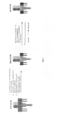
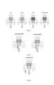
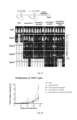
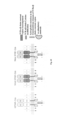
- an artificially synthetic chimeric molecule called Synthetic T-Cell Receptor and Antibody Receptor (STAR/WT-STAR) can be constructed with the structure thereof shown in Figure 1 (left).
- a STAR molecule has two chains, wherein the first chain is obtained by fusing an antigen recognition sequence (such as an antibody heavy chain variable region) with a constant region (C ⁇ ) of a T cell receptor ⁇ chain (TCR ⁇ ), and the second chain is obtained by fusing an antigen recognition sequence (such as an antibody light chain variable region) with a constant region (C ⁇ ) of a T cell receptor ⁇ chain (TCR ⁇ ).
- the antigen recognition domain (such as VH, VL or scFv) and the constant domain (constant domain of TCR ⁇ , ⁇ , ⁇ and ⁇ ) in the construct can be arranged and combined to form a variety of constructs with different configurations but similar functions.
- the first and second chains of STAR molecule after expressing in T cells, will combined with endogenous CD3 ⁇ ⁇ , CD3 ⁇ ⁇ and CD3 ⁇ chains in the endoplasmic reticulum to form a eight-subunit complex, which is present on the surface of cell membrane in the form of complex.
- An immunoreceptor tyrosine-based activation motif is a signal transduction motif in a TCR molecule, with its conserved sequence of YxxL/V.
- the endodomain of CD3 ⁇ , ⁇ , ⁇ and ⁇ chains comprises one ITAM sequence, and that of CD3 ⁇ chain comprises three ITAM sequences, so a complete STAR complex has a total of ten ITAM sequences.
- STAR When the antigen recognition sequence of a STAR receptor binds to its specific antigen, the intracellular ITAM sequence will be phosphorylated successively, which then in turn activate the downstream signaling pathway, activating transcription factors such as NF- ⁇ ⁇ , NFAT, and AP-1, to initiate the activation of T cells and produce effector functions.
- transcription factors such as NF- ⁇ ⁇ , NFAT, and AP-1
- the STAR prototype design used a humanzied TCR ⁇ / ⁇ chain (or TCR ⁇ and ⁇ chains) constant region sequence (wild-type human TCR ⁇ constant region, SEQ ID NO: 1; wild-type human TCR ⁇ constant region, SEQ ID NO: 2). Due to the constant region sequences of human, primate and murine TCR ⁇ / ⁇ chains (mouse TCRaC-WT, SEQ ID NO: 3; mouse TCRbC-WT, SEQ ID NO: 4) are highly conserved, and have the same key amino acid sequence as well, they can be replaced with each other.
- STAR molecules After being transferred into T cells, STAR molecules will mismatch with the endogenous TCR of T cells through the constant region. On one hand, this mismatch problem may reduce the efficiency of correct pairing of STAR molecules and weakens their functions, which, on the other hand, may increase the possibility of unknown specificity due to mismatch and increases the security risk.
- the constant region of a STAR molecule was replaced with a murine sequence by the inventors to enhance the functions of the STAR molecule after being transferred into human T cells.
- cysteine mutation was carried out on the STAR molecule by the inventors to introduce an intermolecular disulfide bond, thereby enhancing mutual pairing between two chains of the STAR molecule, and reducing mismatch with an endogenous TCR.
- threonine (T) at position 48 was mutated to cysteine (C) (mouse TCRaC-Cys, SEQ ID NO: 5) in TCR ⁇ chain constant region
- S serine
- the two new added cysteines will form a disulfide bond between the two chains of STAR, thereby reducing mismatch between the two chains of STAR with the endogenous TCR chain, and helping STAR molecules form more stable complexes, thus obtaining better functions.
- hydrophobic amino acid substitution was performed by the inventors on the transmembrane domain of the STAR molecule to increase the stability of the STAR molecule and help it play a more lasting function.
- lysine in the endodomain and transmembrane domain of the TCR molecule was subject to ubiquitin modifications through a series of ubiquitination reactions by a ubiquitin activating enzyme, ubiquitin binding enzyme and ubiquitin ligase ubiquitinate, thereby producing T cell endocytosis, leading to the endocytosis of TCR molecules into the cells for further degradation by lysosomes, thus reducing the concentration of TCR molecules on the surface of the T cell membrane, resulting in the continuous decline of the effect of T cell activation.
- the amino acids in the transmembrane domain or endodomain of the ⁇ and ⁇ chains in the mut-STAR molecule were modified by the inventors, which comprising: mutating lysine in the endodomain of the ⁇ chain constant region and the transmembrane domain of the ⁇ chain constant region of the STAR molecule to arginine, producing the constant region sequence of Mouse TCR ⁇ C-Arg mut (SEQ ID NO: 8) and the constant region sequence of Mouse TCR ⁇ C-Arg mut (SEQ ID NO: 9), respectively, to reduce the endocytosis of STAR molecule caused by lysine ubiquitination.
- mut-STAR cells In order to improve the proliferation ability in vivo of mut-STAR cells, the effect survival time and the ability to infiltrate into the tumor microenvironment to kill the target cells efficiently, a new structure was designed by the inventors, wherein, the mut-STAR complex was modified, and an enhanced mut-STAR cell can be tailored as needed, so as to improve the clinical response of TCR-T and realize a lasting curative effect.
- TCR is a special marker on the surface of all T cells, which can be divided into ⁇ TCR and ⁇ ⁇ TCR, and the corresponding T cells thereof are ⁇ T cells and ⁇ ⁇ T cells respectively.
- ⁇ -STAR and ⁇ -STAR were modified with costimulatory signals, respectively, by the inventors, to improve the performance of ⁇ T cells and ⁇ ⁇ T cells, respectively.
- TCR of ⁇ T cells consists of TCR ⁇ and TCR ⁇ chains, accounting for 90%-95% of the total T cells.
- ⁇ TCR consists of a variable region and a constant region, in which the variable region has a wide diversity and plays the role of antigen recognition and binding, while the constant region domain plays the role of structural interaction and signal transduction.
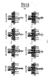
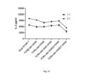
- an endodomain sequence of a humanized costimulatory receptor is introduced to the C-terminal of the ⁇ -STAR constant region ( Fig. 2 ), to study the influence on STAR T cell functions.
- the STAR constant region of the present invention includes an unmodified WT-STAR constant region, a cys-STAR constant region containing an additional intermolecular disulfide bond, a murinized hm-STAR constant region, and a mut-STAR with a combination of the three modifications described in subsection 1.
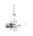
- the costimulatory signal transduction structure comprises intracellular signal transduction domains of CD40, OX40, ICOS, CD28, 4-1BB or CD27, with the sequences of SEQ ID NO: 10, SEQ ID NO: 11, SEQ ID NO: 12, SEQ ID NO: 13, SEQ ID NO: 14 and SEQ ID NO: 15, respectively.
- the costimulatory endodomain can be connected to the C-terminal of TCR ⁇ chain or TCR ⁇ chain or both TCR ⁇ chain and ⁇ chain (co-STAR).
- the costimulatory endodomain can be connected directly or via a linker G4S/(G4S)n (G4S linker sequences are SEQ ID NO: 16, SEQ ID NO: 17, SEQ ID NO: 18, SEQ ID NO: 19, SEQ ID NO: 20, SEQ ID NO: 21, SEQ ID NO: 22, SEQ ID NO: 23, SEQ ID NO: 24, SEQ ID NO: 25, respectively) to the C-terminal of TCR constant region, or the C-terminal of TCR constant region with the endodomain sequence of the TCR molecule deleted (endodomain-deleted TCR ⁇ chain constant region comprising both cysteine substitution and hydrophobic region modification (mouse TCRaC-del mut, SEQ ID NO: 26), endodomain-deleted TCR ⁇ chain constant region comprising cysteine substitution (mous
- TCR of ⁇ ⁇ T cells consists of TCR ⁇ and TCR ⁇ chains
- ⁇ ⁇ T cells can be divided into three subgroups: ⁇ ⁇ 1, ⁇ ⁇ 2 and ⁇ ⁇ 3 based on the type of TCR ⁇ chain, with different subgroups having different distribution in human bodies.
- ⁇ ⁇ T cells recognize an antigen in an MHC-restricted way, which plays an important role in the surveillance of pathogens and tumors.
- CD28 or 4-1BB and similar costimulatory signals played an important role in the activation and proliferation of ⁇ ⁇ T cells.
- the endodomain sequence of human costimulatory molecule receptor was introduced to the C terminal of TCR ⁇ and TCR ⁇ , respectively ( Fig. 2 , right), by the inventors, to improve the performance of ⁇ ⁇ T cells.
- a CD3 subunit includes ⁇ chain, ⁇ chain, ⁇ chain and ⁇ chain, and forms a T cell receptor complex with a TCR molecule, which transmits signals from the ectodomain to endodomain, so as to regulate the state of cells and response to stimuli.
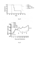
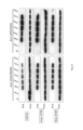
- a CD3 molecule was modified by the inventors by introducing a human costimulatory molecule receptor endodomain to the C terminal of CD3 ⁇ chain (SEQ ID NO: 28), ⁇ chain (SEQ ID NO: 29), ⁇ chain (SEQ ID NO: 30) and ⁇ chain (SEQ ID NO: 31) ( Fig. 4 ).
- the modified CD3 molecule was expressed in mut-STAR T cells to improve its function.
- CD3 molecule comprising the stimulatory region of cytokine receptor (cytokine-STAR, CK-STAR)
- Cytokines play an important role in proliferation, anti-tumor and differentiation of T cells. Different cytokines combine with their respective receptors to transmit signals from the ectodomain to endodomain, so as to regulate the state of cells and response to stimuli. In addition, studies showed that the downstream molecule STATS (SEQ ID NO: 35) was activated by cascade reaction at the IL-2 receptor endodomain, thus enhancing the transcription of T cell proliferation-related molecules and enhancing the proliferation ability of CAR-T cells.
- STATS SEQ ID NO: 35
- the STAR molecule was modified by the inventors by linking the intracellular signal transduction domain of a human cytokine receptor (e.g., IL-2 ⁇ receptor endodomain IL2Rb, SEQ ID NO: 32; IL-7 ⁇ receptor endodomain, SEQ ID NO: 33; IL-21 receptor endodomain, SEQ ID NO: 34, etc.)) to the C-terminal of TCR ⁇ chain or ⁇ chain or both ⁇ and ⁇ chains, or by further linking the STATS activation moiety to the IL-2 ⁇ or IL-7R ⁇ receptor endodomain via G4S (IL-2RbQ, SEQ ID NO: 36; IL-7RbQ, SEQ ID NO: 37) ( Fig. 5 ).
- a human cytokine receptor e.g., IL-2 ⁇ receptor endodomain IL2Rb, SEQ ID NO: 32; IL-7 ⁇ receptor endodomain, SEQ ID NO: 33; IL-21 receptor endodomain, SEQ ID NO:
- the vectors used in the present invention including viral vectors, plasmid vectors, etc. were purchased from or synthesized by commercial companies, and the full-length sequences of these vectors are obtained, and the specific cleavage sites are known.
- the TCR mentioned in the invention can be any functional TCR, including WT-STAR, mut-STAR, ub-STAR, co-STAR, co-linker-STAR, CK-STAR, co-CD3-STAR and the like used in the invention.
- Gene fragments, such as the variable region of TCR, the constant region of TCR, the endodomain of a costimulatory molecule receptor, the intracellular signal transduction region of a cytokine receptor, a tag sequence, a linker and the like used in the present invention were all synthesized by commercial companies. These gene fragments were linked by PCR.
- the optimization of TCR complex was validated using STAR comprising ScFv shown in SEQ ID NO: 38 (derived from a CD20-targeting antibody, OFA) and/or ScFv shown in SEQ ID NO: 39 (derived from a CD19-targeting antibody, FMC63), and was compared with a blank control group Mock expressing only RFP protein shown in SEQ ID NO: 40 (red fluorescent protein, RFP).
- the lentiviral vector used herein was pHAGE-EF1 ⁇ -IRES-RFP, wherein the linear vector was obtained by restriction enzyme Not I/Nhe I, the gene fragment was obtained by synthesis and PCR, and the complete vector was obtained by homologous recombination.
- pHAGE can stably insert target genes into the genome of a target cell, which is an important way to construct a stable cell line.
- Luciferase was a kind of enzyme with catalytic activity, which can catalyze the chemical autoluminescence of a substrate to make the target cell express luciferase stably, the number of target cells can be indicated after adding the substrate, thus reflecting the effect of functional cells on target cells.
- a pHAGE-EF1A vector carrying restriction endonuclease NotI/ClaI cleavage sites was cleavaged by the two enzymes, and luciferase and GFP sequences were obtained by NCBI, the fragments were synthesized by Ruiboxingke, a commercial company, by combing the luciferase gene with GFP gene using Overlap PCR, and then the luciferase-GFP fragment was connected to the pHAGE vector by homologous recombination.
- the lentivirus was packed by Lenti-X-293T, and the lentivirus solution was concentrated by PEG8000, the virus titer was measured by the gradient dilution method, and then the lymphoma cell line Raji was infected, after 72 hours of infection, whether there were GFP positive cells was observed by a fluorescence microscope, and then GFP positive cells were sorted by a flow sorter, and monoclonal cells were selected for library building and preservation.
- the luciferase substrate was used to incubate with target cells, and the expression and detection levels of luciferase were detected to determine the expression level.
- a guide sequence was designed in the constant regions of ⁇ and ⁇ chains to construct a TCR ⁇ - ⁇ -Jurkat cell line.
- the exon sequences of the constant regions of TCR ⁇ and ⁇ chains were obtained in NCBI, and the exon 1 sequences of the constant regions of TCR ⁇ and ⁇ chains were submitted to the tools.genome-engineering.org website for designing guide sequences, based on the result, an oligo sequence was synthesized, and then a sgRNA-LentiCRISPR lentiviral vector (purchased from Aidi gene) was constructed.
- the guide sequence of ⁇ chain was linked to LentiCRISPR-puro, and the guide sequence of ⁇ chain was linked to LentiCRISPR-BSD.
- HEK-293T was planked to a 10 cm dish in advance, and when the cells grew to 80%-90%, the transfection system was added to HEK-293T, and the cells were put back into the incubator at 37 °C for culture. This time was counted as 0 hours; 12 hours after transfection, fresh 10% FBS-DMEM as added. The virus was collected 48 hours and 72 hours after transfection. The culture medium containing virus was centrifuged and filtered, mixed with PEG8000, placed at 4 °C for more than 12 hours, then centrifuged at 3500 rpm for 30 min, and resuspended and precipitated with an appropriate volume of medium after discarding the supernatant. The resultant was frozen at -80°C, or used directly.
- Jurkat T cells were inoculated in a 12- or 24-well plate, followed by adding sgRNA-LentiCRISPR virus of ⁇ chain and ⁇ chain with appropriate volumes at the same time, as well as polybrene (added at a ratio of 1: 1000 based on the total volume), and mixing well. Centrifugation infection was performed at 1000 rpm at 32°C for 90 min. The resultant was placed in an incubator at 37 °C with counting as 0 hours; the liquid was changed after 10 to 12 hours; after 48 hours, Puromycin were added to the appropriate final concentration, and after treating for additional 48 hours, as shown, the cells in the uninfected control group all died.
- TCR ⁇ - ⁇ -Jurkat cell bank Single cells from the TCR ⁇ - ⁇ -Jurkat cell bank were sorted into a 96-well plate by the flow sorter Aria, after two weeks of culture, the grown monoclones were sucked out for amplification culture. Monoclonal cell lines were identified with TCR ⁇ chain and ⁇ chain antibodies, respectively, and the cell lines with both chain deficient were amplified to obtain an endogenous TCR knockout Jurkat-T cell line.
- Lentix-293T cells were inoculated to a 10 cm culture dish at 5 ⁇ 10 5 /mL, and cultured in an incubator at 37 °C with 5% CO 2 , transfection was carried out when the cell density reached about 80% (observed under a microscope).
- the three plasmids were mixed with 500 ⁇ L of serum-free DMEM according to a 1: 2: 3 ratio of PMD2.G: PSPAX: transfer plasmid.
- 54 ⁇ L of PEI-Max and 500 uL of serum-free DMEM were mixed uniformly and left at room temperature for 5 min (in a 3:1 volume-mass ratio of PEI-Max to plasmid).
- a PEI-max mixture was slowly added to the plasmid mixture, blown gently, mixed evenly, and then left at room temperature for 15 min. The final mixture was slowly added to the culture medium, and evenly mixed, then put back into the incubator for another culture for12 h to16 h, then changed to a 6% FBS DMEM medium for another culture, and the virus solution was collected at 48 h and 72 h.
- Jurkat-C5 cells were inoculated in a flat-bottomed 96-well plate at 1.5 ⁇ 10 5 cells /mL, and 100 uL of 1640 medium containing 10% FBS and 0.2 ⁇ L 1000 ⁇ polybrene was added to each well.
- Virus dilution was carried out with a 1640 complete medium with 10 times dilution, the virus dosage in the first well was 100 ⁇ L when determining as the virus stock solution, or was 1 ⁇ L when determining as the concentrated solution.
- the diluted cells were added to the viral wells at 100 ⁇ L/well, mixed at 32 °C, centrifuged at 1500 rpm for 90 min, cultured in an incubator at 37 °C with 5%CO 2 for 72 hours.
- titer (TU/mL) 1.5 ⁇ 10 4 ⁇ Positive rate/virus volume ( ⁇ L) ⁇ 1000.
- Viruses of the following plasmids were packaged by the above method: pHAGE-EF1A-IRES-RFP, WT-STAR, mut-STAR, co-WT-STAR ( ⁇ -4-1BB-WT, ⁇ -CD27-WT, ⁇ -CD28-WT, ⁇ -ICOS-WT, ⁇ -OX40-WT, ⁇ -OX40-WT), co-STAR ( ⁇ -4-1BB, ⁇ -CD27, ⁇ -CD28, ⁇ -ICOS, ⁇ -OPX40, ⁇ -OX40), co-CD3-STAR (CD3 ⁇ -4-1BB, CD3 ⁇ -CD28, CD3 ⁇ -ICOS, CD3 ⁇ -OX40, CD3 ⁇ -4-1BB, CD3 ⁇ -CD28, CD3 ⁇ -ICOS, CD3 ⁇ -OX40, CD3 ⁇ -4-1BB, CD3 ⁇ -CD28, CD3 ⁇ -ICOS, CD3 ⁇ -OX40, CD3
- the Jurkat T cell line was cultured in a RPMI1640 medium containing 10% FBS with a culture density of 3 ⁇ 10 5 /ml and up to 3 ⁇ 10 6 /ml, and subcultured every 1 to 2 days. After cell counting, the required number of cells were taken and supplemented with the culture medium to adjust to the above density, and place in a CO 2 incubator for culture.
- the primary T cells were isolated by Ficoil method and cultured in an X-VIVO medium containing 10% FBS and 100 IU/mL IL-2, with the initial culture density being 1 ⁇ 10 6 /mL, and then added to a CD3- and RetroNectin r-Fibronectin (with a final concentration of 5ug/ml each)-pre-coated well plate.
- the density of anaphase culture was 5 ⁇ 10 5 /mL and up to 3 ⁇ 10 6 /mL, and subculture was carried out every 1 to 2 days.
- the cells were blown evenly and counted, and taken at 5 ⁇ 10 5 /ml, centrifuged, then the supernatant was discarded, the staining solution used was PBS+2% FBS+2 mM EDTA, the corresponding antibody was added for incubation for 30 min, then PBS was added for washing twice, and detection was carried out on the computer.
- Target cells Raji-luciferase, Raji CD19KO -luciferase, Raji CD20KO -luciferase and primary T cells were suspension cells, for co-incubation, the corresponding number of cells were taken, and mixed with the target cell medium and centrifuged for culture.
- the specific steps were as follows: the primary T cells were infected with the packaged and purified WT-STAR and mut-STAR T viruses, and one day before co-culture, the infection efficiency was detected by flow cytometry, and the ratio of effector cell to target cell was determined and commonly used at a 1:1 ratio, and the total number of T cells was calculated according to the infection efficiency, the common usage of target cells was 1 ⁇ 10 5 /well (96-well plate).
- the target antigen of the present invention is generally a cell surface protein, which can be directly used for T cell activation to detect the function of T cells. Positive T cells were commonly added at 1 ⁇ 10 5 /well, centrifuged and activated for 24 hours, then T cells or target cells were collected to detect the T cell function.
- T cells were co-cultured with target cells for 24 h, then the cell suspension was gently blown evenly, 150 ⁇ L of cell suspension per well was taken and added to a white 96-well plate, centrifuged at 1500 rpm/min for 5 min, and the supernatant was taken and added with a cell lysate for lysing at room temperature for 15 min, then centrifuged at 4 °C at 4000 rpm/min for 15min, then the supernatant was taken with 2 parallel wells being taken for each well, and added with a luciferase substrate (firefly luciferase assay reagent), then detected by a multifunctional microplate reader with the gain value fixing as 100 to obtain a chemiluminescence value.
- Cell killing calculation: killing efficiency 100%- (effector cell-target cell value per well/control cell-target cell value per well).
- T cell killing function test by the luciferase assay showed that mutant mut-STAR T cells exhibited a stronger T cell tumor-killing ability, as shown in Figure 6 and Table 1, after mut-STAR T cells targeting CD19 and CD20 killed Raji-luciferase, Raji CD19KO -luciferase and Raji CD20KO -luciferase, the survival rates of residual tumors were 2.41%, 13.94% and 24.40%, respectively, which were significantly better than those of WT-STAR by 45.73%, 77.00% and 76.16%, indicating that mutant mut-STAR had a more significant tumor-killing effect.
- Target cells Raji-luciferase and primary T cells were suspension cells, for co-incubation, the corresponding number of cells were taken and mixed with the target cell culture medium, then centrifuged for culture.
- the specific steps were as follows: the primary T cells were infected with the packaged and purified WT-STAR and mut-STAR T viruses, and one day before co-culture, the infection efficiency was detected by flow cytometry, and the ratio of effector cell to target cell was determined, and co-incubation was carried out usually at the ratio of 8:1, 4:1, 2:1, 1:1, 1:2, 1:4 and 1:8, and the difference of co-incubation with time was also detected usually at 6 h, 12 h, 24 h, 36 h and 48 h.
- target cells Raji-luciferase were incubated with primary T cells for 7 days to observe the changes of cell proliferation number and IL-2 secretion, then positive T cells were sorted by flow cytometry and subject to resting culture without antigen stimulation for two days, which, were then cocultured again with target cells for 24 hours to detect the killing of T cells, with the common usage of target cells being 1 ⁇ 10 5 /well (96-well plate).
- the target antigen of the present invention was generally a cell surface protein, which can be directly used for activating T cells to detect the function of T cells, in particular, the target antigen was usually added with 1 ⁇ 10 5 /well positive T cells, centrifuged and activated for 24 hours to collect the cell suspension or culture supernatant for detecting T cell function, or activated for 6 hours, 12 hours, 24 hours, 36 hours, 48 hours or 7 days to detect the killing function of T cells.
- T cells were co-cultured with target cells for different times, then the cell suspension was gently blown evenly, 150 ⁇ L of cell suspension per well was taken and added to a white 96-well plate, centrifuged at 1500 rpm/min for 5 min, and the supernatant was taken and added with a cell lysate for lysing at room temperature for 15 min, then centrifuged at 4 °C at 4000 rpm/min for 15min, then the supernatant was taken with 2 parallel wells being taken for each well, and added with a luciferase substrate (firefly luciferase assay reagent), then detected by a multifunctional microplate reader with the gain value fixing as 100 to obtain a chemiluminescence value.
- Cell killing calculation: killing efficiency 100%- (effector cell-target cell value per well/control cell-target cell value per well).
- T cell killing function test by the luciferase assay showed that mut-STAR with costimulatory endodomain connected to the C-terminals of both TCR ⁇ chain and ⁇ chain exhibited similar tumor killing effect at different E:T ratios of T cells and target cells, and there was no significant difference in residual tumor survival rate at different E:T ratios, as shown in Fig. 7 and Table 2.
- mut-STAR with OX40 endodomains connected to the C-terminals of both TCR ⁇ and ⁇ chains showed similar tumor killing effects, about 24% and 19%, respectively, which were significantly superior to mut-STARs with other costimulatory endodomains connected to the C-terminals of both TCR ⁇ and ⁇ chains ( ⁇ -41BB, ⁇ -CD27, ⁇ -CD28, ⁇ -ICOS), as shown in Fig. 8 and Table 3.
- TNF- ⁇ , IFN- ⁇ and IL-2 cytokines
- TNF- ⁇ , IFN- ⁇ and IL-2 cytokines released to help T cells kill target cells or promote the expansion of T cells themselves.
- T cells were stimulated by target cells or antigens, T cells were collected, centrifuged, and the supernatant was taken.
- the TNF- ⁇ , IFN- ⁇ , and IL-2 ELISA kits used were Human IL-2 Uncoated ELISA, Human TNF- ⁇ Uncoated ELISA, and Human IFN- ⁇ Uncoated ELISA (Art.No. 88-7025, 88-7346, 88-7316, respectively).
- the specific steps were as follows: diluting 10X Coating Buffer to 1X with ddH 2 O, adding coated antibody (250X), mixing well and adding to a 96-well plate at 100 ⁇ L/well. After sealing with the plastic wrap and staying overnight at 4 °C, 1X PBST (1X PBS with 0.05% Tween 20 added) was used for washing 3 times with 260 ⁇ L/well each time, 5X ELISA/ELISPOT Diluent was diluted to 1X with ddH 2 O and then added to the 96-well plate with 200 ⁇ L/well, followed by standing at room temperature for 1 hour.
- PBST was used for washing once, and dilution was performed according to a standard curve (ranging from: 2 to 250, 4 to 500, 4 to 500, respectively), the samples were diluted to 20 to 50 times with 1xDiluent.
- Samples diluted according to the standard curve were added with 100 microliters per well, two parallel wells were taken and incubated at room temperature for 2 hours, PBST was used for washing three times, then a Detection antibody diluted with 1 ⁇ Diluent was added, after incubation for 1 h, PBST was used for washing three times, then HRP diluted with 1 ⁇ Diluent was added, after incubation for 30 minutes, the solution was washed six times, TMB was added for color development with the color development time being less than 15min, and 2N H 2 SO 4 was added for termination, light absorption at 450 nm was detected.
- mut-STAR ⁇ -OX40
- OX40 endodomain linked to the C-terminals of both TCR ⁇ chain and ⁇ chain was about 10000 pg/ml, which was significantly higher than that of STAR with other structures, in which that of mut-STAR, ⁇ -41BB, ⁇ -CD27, ⁇ -CD28 and ⁇ -ICOS was about 7700 pg/ml, 6450 pg/ml, 6690 pg/ml, 6000 pg/ml, and 6050 pg/ml, respectively; in terms of TNF ⁇ and IFN- ⁇ secretion, ⁇ -OX40 also showed similar results with mut-STAR, while other structures showed different decreases, as shown in Figure 11 and Table 6.
- T cell activation a large number of cytokines were released to help T cells kill target cells or promote the expansion of T cells themselves, and the most obvious occurrence in T-cell proliferation was a significant change in the number of T cells.
- mut-STAR-T cells with various structures were co-cultured with Raji-luciferase cells at a 1: 3 E:T ratio with counting as day 0, and then cells were collected on day 1 and day 7 for flow analysis, respectively.
- the medium used was 1640 complete medium without IL-2, and the initial number of TCR T cells was 1 ⁇ 10 5 Cells, samples at each time point were incubated independently, and the remaining co-incubated samples were semi-changed with the liquid the next day, and supplemented with the target cells.
- the cells used for flow analysis were stained with an anti-human CD3 antibody in advance, and a specified volume thereof was collected and recorded when analysis was performed on the machine, the number and proportion of T cells in the system were known by conversion.
- mut-STAR with OX40 endodomain connected to the C-terminals of both TCR ⁇ chain and ⁇ chain( ⁇ -OX40) showed better activation and proliferation after recognizing the target cell epitope, which were significantly higher that by mut-STAR.
- the proliferation of mut-STAR with OX40 endodomain connected to the C-terminal of TCR ⁇ chain ( ⁇ -OX40) or ⁇ chain ( ⁇ -OPX40 alone was shown in Fig.
- Target cells Raji-luciferase and primary T cells were suspension cells, for co-incubation, the corresponding number of cells were taken and mixed with the target cell culture medium, then centrifuged for culture.
- the specific steps were as follows: the primary T cells were infected with the packaged and purified WT-STAR and mut-STAR T viruses, and one day before co-culture, the infection efficiency was detected by flow cytometry, and the ratio of effector cell to target cell was determined, co-incubation was carried out usually at the ratio of 1:1 or 2:1, in addition, target cells Raji-luciferase and primary T cells were co-incubated for 7 days to observe the changes in the number of cell proliferation. The total number of T cells was calculated based on the infection efficiency, and the general usage of target cells was 1 ⁇ 10 5 /well (96-well plate).
- the target antigen of the present invention is generally a cell surface protein, which can be directly used for T cell activation to detect the function of T cells.
- positive T cells were commonly added with 1 ⁇ 10 5 /well, centrifuged and activated for 24 hours, the cell suspension or culture supernatant was collected to detect the T cell function.
- T cells were co-cultured with target cells for different times, then the cell suspension was gently blown evenly, 150 ⁇ L of cell suspension per well was taken and added to a white 96-well plate, centrifuged at 1500 rpm/min for 5 min, and the supernatant was taken and added with a cell lysate for lysing at room temperature for 15 min, then centrifuged at 4 °C at 4000 rpm/min for 15min, then the supernatant was taken with 2 parallel wells being taken for each well, and added with a luciferase substrate (firefly luciferase assay reagent), then detected by a multifunctional microplate reader with the gain value fixing as 100 to obtain a chemiluminescence value.
- Cell killing calculation: killing efficiency 100%- (effector cell-target cell value per well/control cell-target cell value per well).
- the detection results of T cell killing function test by the luciferase assay showed that when the costimulatory endodomain was connected to the C-terminal of CD3 ⁇ or CD3 ⁇ or CD3 ⁇ or CD3 ⁇ chain and co-expressed on mut-STAR T, the results at the 1: 1 E:T ratio of different T cells to target cells showed that all co-CD3-STAR did not exhibit a better target cell-killing effect than a ⁇ -OX40, but compared with mut-STAR, CD3 ⁇ -OX40, CD3 ⁇ -41BB, CD3 ⁇ -CD28, CD3 ⁇ -ICOS, CD3 ⁇ -OX40 exhibited similar tumor killing effects, residual tumor survival rates thereof did not differ significantly at the 1: 1 E:T ratio, as shown in Fig.15 and Table 10.
- T cell activation a large number of cytokines were released to help T cells kill target cells or promote the expansion of T cells themselves, and the most obvious occurrence in T-cell proliferation was a significant change in the number of T cells.
- mut-STAR-T cells with various structures were co-cultured with Raji-luciferase cells at a 1: 3 E:T ratio with counting as day 0, and then cells were collected on day 1 and day 7 for flow analysis, respectively.
- the medium used was 1640 complete medium without IL-2, and the initial number of TCR T cells was 1 ⁇ 10 5 Cells, samples at each time point were incubated independently, and the remaining co-incubated samples were semi-changed with the liquid the next day, and supplemented with the target cells.
- the cells used for flow analysis were stained with an anti-human CD3 antibody in advance, and a specified volume thereof was collected and recorded when analysis was performed on the machine, the number and proportion of T cells in the system were known by conversion.
- Target cells Raji-luciferase and primary T cells were suspension cells, for co-incubation, the corresponding number of cells were taken and mixed with the target cell culture medium, then centrifuged for culture.
- the specific steps were as follows: the primary T cells were infected with the packaged and purified WT-STAR and mut-STAR T viruses, and one day before co-culture, the infection efficiency was detected by flow cytometry, and the ratio of effector cell to target cell was determined, and co-incubation was carried out usually at a 1:1 or 2:1 ratio, in addition, target cells Raji-luciferase and primary T cells were co-incubated for 7 days to observe the changes in the number of cell proliferation. The total number of T cells was calculated based on the infection efficiency, and the general usage of target cells was 1 ⁇ 10 5 /well (96-well plate).
- the target antigen of the present invention was generally a cell surface protein, which can be directly used for activating T cells to detect the function of T cells, in particular, the target antigen was usually added with 1 ⁇ 10 5 /well positive T cells, centrifuged and activated for 24 hours to collect the cell suspension or culture supernatant for detecting T cell function.
- T cell killing function validation Luciferase assay
- T cells were co-cultured with target cells for different times, then the cell suspension was gently blown evenly, 150 ⁇ L of cell suspension per well was taken to a white 96-well plate, centrifuged at 1500 rpm/min for 5 min, and the supernatant was taken and added with a cell lysate for lysing at room temperature for 15 min, then centrifuged at 4 °C at 4000 rpm/min for 15min, then the supernatant was taken with 2 parallel wells being taken for each well, and added with a luciferase substrate (firefly luciferase assay reagent), then detected by a multifunctional microplate reader with the gain value fixing as 100 to obtain a chemiluminescence value.
- Cell killing calculation: killing efficiency 100%- (effector cell-target cell value per well/control cell-target cell value per well).
- the detection results of T cell killing function test by the luciferase assay showed that, the costimulatory endodomain with different lengths of G4S linker was connected to the endodomain-deleted ⁇ or ⁇ constant region, the results at the 1: 1 E:T ratio of T cell to target cell showed that, when the linker was connected to the ⁇ constant region endodomain, ⁇ -del-OX40, ⁇ -OX40, ⁇ -del-G4S-OX40, ⁇ -del-(G4S) 3-OX40 and ⁇ - ⁇ - ⁇ x40 showed similar killing effect, which was superior to that of ⁇ -del-(G4S) 7-OX40 and ⁇ -del-(G4S) 10-OX40, and the longer linker, the weaker the T cell killing effect, when the linker was connected to the ⁇ constant region endodomain, ⁇ -del-OX40, ⁇ -ox40, ⁇ -del-(G4S) 3-
- mut-STAR with a costimulatory endodomain comprising a G4S linker linked to the endodomain-deleted ⁇ or ⁇ constant region did not affect the tumor killing effect of the mut-STAR, and the effect when linking to the ⁇ constant region endodomain was better than that when linking to the ⁇ constant region endodomain, but the longer the linker length, the weaker the tumor killing effect instead.
- TNF- ⁇ , lFN- ⁇ and IL-2 cytokines
- TNF- ⁇ , lFN- ⁇ and IL-2 were released to help T cells kill target cells or promote the expansion of T cells themselves.
- T cells were stimulated by target cells or antigens, T cells were collected, centrifuged, and the supernatant was taken.
- the TNF- ⁇ , lFN- ⁇ , and IL-2 ELISA kits used were Human IL-2 Uncoated ELISA, Human TNF- ⁇ Uncoated ELISA, and Human lFN- ⁇ Uncoated ELISA (Art.No. 88-7025, 88-7346, 88-7316, respectively).
- the specific steps were as follows: diluting 10X Coating Buffer with ddH2O to 1X, adding coated antibody (250X), mixing well and adding to a 96-well plate at 100 ⁇ L/well. After sealing with the plastic wrap and staying overnight at 4 °C, 1X PBST (also called Wash Buffer, 1X PBS with 0.05% Tween 20 added) was used for washing 3 times with 260 ⁇ L/well each time, 5X ELISA/ELISPOT Diluent was diluted to 1X with ddH 2 O and then added to the 96-well plate with 200 ⁇ L/well, followed by standing at room temperature for 1 hour.
- 1X PBST also called Wash Buffer, 1X PBS with 0.05% Tween 20 added
- PBST was used for washing once, and dilution was performed according to a standard curve (ranging from: 2 to 250, 4 to 500, 4 to 500, respectively), the samples were diluted to 20 to 50 times with 1xDiluent. Samples according to the standard curve were added with 100 microliters per well, two parallel wells were taken and incubated at room temperature for 2 hours, PBST was used for washing three times, then a Detection antibody diluted with 1xDiluent was added, after incubation for 1 h, PBST was used for washing three times, then HRP diluted with 1xDiluent was added, after incubation for 30 minutes, the solution was washed six times, TMB was added for color development with the color development time being less than 15min, and 2N H 2 SO 4 was added for termination, light absorption at 450 nm was detected.
- a standard curve ranging from: 2 to 250, 4 to 500, 4 to 500, respectively
- the deletion of the endodomain of ⁇ chain affected the secretion, but did not affect the IL-2 secretion, while the deletion of the endodomain of ⁇ chain did not affect the secretion of neither IL-2 nor IFN-y; at the same time, after the endodomain of ⁇ chain was deleted, a linker was used to link to the OX40 endodomain, which, in case of the linker with a length of less than 7, did not affect the IL-2 secretion, but decreased the secretion.
- T cells differentiate from primitive T cells (naive) to central memory T cells (Tcm) to effector memory T cells (Tern), and finally to effector T cells (Teff).
- Tcm central memory T cells
- Teff effector T cells
- Tcm central memory T cells
- Teff effector T cells
- Tcm central memory T cells
- Teff effector T cells
- Tcm central memory T cells
- Memory T cells can be classified into stem cell T cells, central memory T cells and effector memory T cells.
- the differentiation ratio of central memory T cells affects the persistent killing effect of T cells in vivo.
- T cells The ratio of primitive T cells to effector T cells affects the tumor-killing effect and persistence of T cells in vivo.
- the expression of CD45RA and CCR7 on the surface of T cells was detected by flow cytometry, thus the differentiation of T cells can be known.
- T cells were incubated with target cells for 7 days, centrifuged, stained with anti-human-CD45RA-Percp-cy5.5 and anti-human-CCR7-APC flow antibodies for 30 minutes, centrifuged again, washed with PBS and fixed with 4% paraformaldehyde solution, and the differentiation of T cells was detected by flow cytometry.
- Table 12 Effects of killing target cells after co-culture of target cells with STAR with OX40 connected to ⁇ chain thereof via different linkers ⁇ -del-OX40 ⁇ -OX40 ⁇ -del-G4SOX40 ⁇ -del-[G4S]3 -OX40 ⁇ -del-[G4S]7 -OX40 ⁇ -del-[G4S]1 0-OX40 ab-OX40 2 : 1 0.890 328 0.888 4054 0.993 3094 0.992 7593 0.987 0744 0.987 2119 0.932 0745 0.926 8892 0.932 1665 0.935 556 0.832 1665 0.835 556 0.995 7843 0.996 5176 1 : 1 0.916 0888 0.915 0926 0.879 0945 0.873 1092 0.920 3223 0.928 2119 0.826 1465 0.669 2107 0.575 495 0.546 9499 0.475 495 0.406 9499
- Target cells Raji-luciferase and primary T cells were suspension cells, for co-incubation, the corresponding number of cells were taken and mixed with the target cell culture medium, then centrifuged for culture.
- the specific steps were as follows: the primary T cells were infected with the packaged and purified WT-STAR and mut-STAR T viruses, and one day before co-culture, the infection efficiency was detected by flow cytometry, and the ratio of effector cell to target cell was determined, co-incubation was carried out usually at the ratio of 1:1 or 2:1, in addition, target cells Raji-luciferase and primary T cells were co-incubated for 7 days to observe the changes in the number of cell proliferation. The total number of T cells was calculated based on the infection efficiency, and the general usage of target cells was 1 ⁇ 10 5 /well (96-well plate).
- the target of the present invention was generally a cell surface protein, which can be directly used for activating T cells to detect the function of T cells, in particular, the target antigen was usually added with 1 ⁇ 10 5 /well positive T cells, centrifuged and activated for 24 hours to collect the cell suspension or culture supernatant for detecting T cell function.
- T cell killing function validation Luciferase assay
- T cells were co-cultured with target cells for different times, then the cell suspension was gently blown evenly, 150 ⁇ L of cell suspension per well was taken to a white 96-well plate, centrifuged at 1500 rpm/min for 5 min, and the supernatant was taken and added with a cell lysate for lysing at room temperature for 15 min, then centrifuged at 4 °C at 4000 rpm/min for 15min, then the supernatant was taken with 2 parallel wells being taken for each well, and added with a luciferase substrate (firefly luciferase assay reagent), then detected by a multifunctional microplate reader with the gain value fixing as 100 to obtain a chemiluminescence value.
- Cell killing calculation: killing efficiency 100%- (effector cell-target cell value per well/control cell-target cell value per well).
- T cell killing function test by the luciferase assay showed that, the tumor cell-killing effect of ub-STAR, a mut-STAR with TCR endocytosis-related lysine modified into arginine in the transmembrane domain or endodomain, at the 2:1 or 1:1 E:T ratio of T cell to target cell, was significantly lower than that of mut-STAR T cells, as shown in Figure 23 and Table 18.
- cytokines such as TNF- ⁇ , lFN- ⁇ and IL-2
- TNF- ⁇ , lFN- ⁇ and IL-2 were released to help T cells kill target cells or promote the expansion of T cells themselves.
- T cells were stimulated by target cells or antigens, T cells were collected, centrifuged, and the supernatant was taken.
- the lFN- ⁇ , and IL-2 ELISA kits used were Human IL-2 Uncoated ELISA, Human lFN- ⁇ Uncoated ELISA (Art.No. 88-7025, 88-7346, 88-7316, respectively).
- the specific steps were as follows: diluting 10X Coating Buffer with ddH2O to 1X, adding coated antibody (250X), mixing well and adding to a 96-well plate at 100 ⁇ L/well, After sealing with the plastic wrap and staying overnight at 4 °C, 1X PBST (also called Wash Buffer, 1X PBS with 0.05% Tween 20 added) was used for washing 3 times with 260 ⁇ L/well each time, 5X ELISA/ELISPOT Diluent was diluted to 1X with ddH 2 O and then added to the 96-well plate with 200 ⁇ L/well, followed by standing at room temperature for 1 hour.
- 1X PBST also called Wash Buffer, 1X PBS with 0.05% Tween 20 added
- PBST was used for washing once, and dilution was performed according to a standard curve (ranging from: 2 to 250, 4 to 500, 4 to 500, respectively), the samples were diluted to 20 to 50 times with 1xDiluent.
- Samples diluted according to the standard curve were added with 100 microliters per well, two parallel wells were taken and incubated at room temperature for 2 hours, PBST was used for washing three times, then a Detection antibody diluted with 1xDiluent was added, after incubation for 1 h, PBST was used for washing three times, then HRP diluted with 1xDiluent was added, after incubation for 30 minutes, the solution was washed six times, TMB was added for color development with the color development time being less than 15min, and 2N H 2 SO 4 was added for termination, light absorption at 450 nm was detected.
- the ELISA results showed that, the IL-2 and lFN- ⁇ secretions by ub-STAR, a mut-STAR with TCR endocytosis-related lysine modified into arginine in the transmembrane domain or endodomain, which was co-incubated with target cells at the 2:1 or 1:1 or 1:2 E:T ratio of T cell to target cell for 24 hours, were significantly lower than those of mut-STAR T cells, as shown in Figures 24, 25 , and Tables 19, 20.
- T cell activation a large number of cytokines and other chemokines were released, and signals were transduced into the nucleus through cytokines or chemokine receptors to regulate the differentiation of T cells, the proliferation and persistence of T cells in vivo were affected by the number of T cells differentiated to memory T cells.
- Memory T cells can be classified into stem cell T cells, central memory T cells and effector memory T cells. The expression of CD45RA and CCR7 on the surface of T cells was detected by flow cytometry, thus the differentiation of T cells can be known.
- T cells were incubated with target cells for 7 days, centrifuged, stained with anti-human-CD45RA-Percp-cy5.5 and anti-human-CCR7-APC flow antibodies for 30 minutes, centrifuged again, washed with PBS and fixed with 4% paraformaldehyde solution, and the differentiation of T cells was detected by flow cytometry.
- Target cells Raji-luciferase and primary T cells were suspension cells, for co-incubation, the corresponding number of cells were taken and mixed with the target cell culture medium, then centrifuged for culture.
- the specific steps were as follows: the primary T cells were infected with the packaged and purified WT-STAR and mut-STAR T viruses, and one day before co-culture, the infection efficiency was detected by flow cytometry, and the ratio of effector cell to target cell was determined, co-incubation was carried out usually at the ratio of 1:1 or 2:1, in addition, target cells Raji-luciferase and primary T cells were co-incubated for 7 days to observe the changes in the number of cell proliferation. The total number of T cells was calculated based on the infection efficiency, and the general usage of target cells was 1 ⁇ 10 5 /well (96-well plate).
- the target of the present invention was generally a cell surface protein, which can be directly used for activating T cells to detect the function of T cells, in particular, the target antigen was usually added with 1 ⁇ 10 5 /well positive T cells, centrifuged and activated for 24 hours to collect the cell suspension or culture supernatant for detecting T cell function.
- T cells were co-cultured with target cells for different times, then the cell suspension was gently blown evenly, 150 ⁇ L of cell suspension per well was taken and added to a white 96-well plate, centrifuged at 1500 rpm/min for 5 min, and the supernatant was taken and added with a cell lysate for lysing at room temperature for 15 min, then centrifuged at 4 °C at 4000 rpm/min for 15min, then the supernatant was taken with 2 parallel wells being taken for each well, and added with a luciferase substrate (firefly luciferase assay reagent), then detected by a multifunctional microplate reader with the gain value fixing as 100 to obtain a chemiluminescence value.
- Cell killing calculation: killing efficiency 100%- (effector cell-target cell value per well/control cell-target cell value per well).
- T cell killing function test by the luciferase assay showed that, when different cytokine receptor stimulatory regions were connected to the ⁇ or ⁇ region endodomain of a mutant STAR, at the 2:1 E:T ratio of T cell to target cell, ⁇ -IL-2Rb, ⁇ -IL-2Rb, ⁇ -IL-7RA, ⁇ -IL21R all exhibited a killing effect similar to that of mut-STAR, while ⁇ -IL2RbQ, ⁇ -IL2RbQ and ⁇ -IL7RAQ exhibited a tumor-killing effect significantly lower than that of mut-STAR, as shown in Figure 28 and Table 23.
- the killing effect of mutant STARs with different cytokine receptor stimulatory regions linked were detected and compared with that of ⁇ -del-(G4S) 3-OX40-STAR, as shown in Fig. 29 and Table 24, at the 2:1 and 1:1 E:T ratios, the killing effect of ⁇ -IL7RA was slightly lower than that of ⁇ -del-(G4S) 3-OX40-STAR, while the killing effect of STARs with other cytokine receptor structures linked was significantly lower than that of ⁇ -del-(G4S) 3-OX40-STAR.
- cytokines such as TNF- ⁇ , lFN- ⁇ and IL-2
- TNF- ⁇ , lFN- ⁇ and IL-2 were released to help T cells kill target cells or promote the expansion of T cells themselves.
- T cells were stimulated by target cells or antigens, T cells were collected, centrifuged, and the supernatant was taken.
- the IFN- ⁇ , and IL-2 ELISA kits used were Human IL-2 Uncoated ELISA, Human lFN- ⁇ Uncoated ELISA (Art.No. 88-7025, 88-7346, 88-7316, respectively).
- the specific steps were as follows: diluting 10X Coating Buffer with ddH2O to 1X, adding coated antibody (250X), mixing well and adding to a 96-well plate at 100 ⁇ L/well. After sealing with the plastic wrap and staying overnight at 4 °C, 1X PBST (also called Wash Buffer, 1X PBS with 0.05% Tween 20 added) was used for washing 3 times with 260 ⁇ L/well each time, 5X ELISA/ELISPOT Diluent was diluted to 1X with ddH 2 O and then added to the 96-well plate with 200 ⁇ L/well, followed by standing at room temperature for 1 hour.
- 1X PBST also called Wash Buffer, 1X PBS with 0.05% Tween 20 added
- PBST was used for washing once, and dilution was performed according to a standard curve (ranging from: 2 to 250, 4 to 500, 4 to 500, respectively), the samples were diluted to 20 to 50 times with 1xDiluent.
- Samples diluted according to the standard curve were added with 100 microliters per well, two parallel wells were taken and incubated at room temperature for 2 hours, PBST was used for washing three times, then a Detection antibody diluted with 1xDiluent was added, after incubation for 1 h, PBST was used for washing three times, then HRP diluted with 1xDiluent was added, after incubation for 30 minutes, the solution was washed six times, TMB was added for color development with the color development time being less than 15min, and 2N H 2 SO 4 was added for termination, light absorption at 450 nm was detected.
- the ELISA results showed that when T cells were incubated with target cells for 24 hours, at a 1: 1 or 1: 2 E:T ratio, the IL-2 secretion of ⁇ -IL-2Rb was significantly higher than that of mut-STAR, while ⁇ -IL-2Rb, ⁇ -IL-2RbQ and ⁇ -IL-7RAQ exhibited similar IL-2 secretion, however, the IL-2 secretion of ⁇ -IL-2RbQ was significantly lower than that of mut-STAR, as shown in Figure 30 and Table 25.
- the lFN- ⁇ secretion of ⁇ -IL-2Rb was significantly higher than that of mut-STAR, while the lFN- ⁇ secretion of other structures was significantly lower than that of mut-STAR, as shown in Figure 31 and Table 26.
- T cell activation a large number of cytokines and other chemokines were released, and signals were transduced into the nucleus through cytokines or chemokine receptors to regulate the differentiation of T cells, the proliferation and persistence of T cells in vivo were affected by the number of T cells differentiated to memory T cells.
- Memory T cells can be classified into stem cell T cells, central memory T cells and effector memory T cells. The expression of CD45RA and CCR7 on the surface of T cells was detected by flow cytometry, thus the differentiation of T cells can be known.
- T cells were incubated with target cells for 7 days, centrifuged, stained with anti-human-CD45RA-Percp-cy5.5 and anti-human-CCR7-APC flow antibodies for 30 minutes, centrifuged again, washed with PBS and fixed with 4% paraformaldehyde solution, and the differentiation of T cells was detected by flow cytometry.
- Mutant STARs with different cytokine receptor stimulatory regions linked to the ⁇ or ⁇ constant region endodomain showed various differences in the differentiation of central memory T cells and the ratio of naive T cell to effector T cell, wherein, ⁇ -IL-2Rb, ⁇ -IL-2Rb, ⁇ -IL-2RbQ, ⁇ -IL-2RbQ showed no significant difference as compared with mut-STAR, while ⁇ -IL-7RA, ⁇ -IL-7RAQ and ⁇ -IL-21R showed no significant difference as compared with mut-STAR, as shown in Figs. 32 and 33 , and Tables 27 and 28.
- mice were used as a model.
- the mouse genotype was NOD-Prkdcem26Il2rgem26/Nju with lack of T cells, B cells and NK cells, and macrophages and dendritic cells thereof were also deficient.
- the NSG mouse was the most completely immunodeficient mouse strain at present, which can be widely used in preclinical research of T cell therapy due to that it will not reject transplanted tumors and T cells.
- female NSG mice aged 6 ⁇ 8 weeks were used, and the weight difference of mice in each batch was controlled within 2 g. Mice were kept in independent ventilated cages within a specific pathogen free (SPF) barrier, and provided with normal diet and drinking water with pH slightly acidic to prevent pathogen contamination.
- SPPF pathogen free
- human Burkitt's lymphoma cell line Raji cells were used for xenotransplantation.
- Raji cells were cell strains expressing luciferase gene by the lentiviral vector, and the development and changes of Raji tumor were monitored in real time by fluorescein chemiluminescence and in vivo imaging in mice.
- different doses generally about 1 to 3 ⁇ 10 6 cells
- matrix gel and inoculated into female NSG mice aged 6 to 8 weeks by intraperitoneal injection.
- mice were intraperitoneally injected with the fluorescein potassium salt solution, and the fluorescence signals of tumor cells in vivo were detected by in vivo imaging.
- Raji cells grew rapidly in mice, which produced solid tumors in abdominal cavity, causing symptoms such as weight loss in mice; in the absence of therapeutic treatment, Raji tumor burdens led to death in mouse in about 40 days.
- in vivo fluorescence imaging was basically used by: injecting tumor cells with luciferase gene into animals for colonization. Mice were intraperitoneally injected with the fluorescein potassium salt solution, which, as a substrate, emitted light with a specific wavelength in the presence of enzyme, and the fluorescence signals of tumor cells in vivo were detected by in vivo imaging. Quantitative analysis of fluorescence signals was performed and a heat map was drawn to quantitatively reflect tumor growth.
- T cells The survival and expansion of T cells in vivo are directly related to their final anti-tumor effect.
- blood samples were collected from mice regularly, and the proportion, cell state and cell grouping of STAR-T cells in peripheral blood were analyzed. The specific operation was as follows: every 3-4 days, mice were anesthetized with isoflurane, and about 100 uL of blood was collected from the mouse orbit.
- mice can be dissected to detect the proportion of T cells in other immune organs of mice.
- STAR-T cells In order to evaluate the toxicity and safety of STAR-T cells, whether side effects have been caused by the STAR-T cells on experimental animals was examined. By observing the behavior state of mice, analyzing the pathology of mice, and analyzing the sections taken from important organs of mice, whether the reinfused T cells have significant toxicity could be evaluated. At the same time, by analyzing the infiltration of T cells in non-tumor tissues of mice, whether T cells have off-target killing effect on non-tumor tissues of mice can be determined. In addition, by detecting the level of cytokines, such as IL-2, IFN- ⁇ , TNF ⁇ or IL-6 in the mouse blood, whether T cells may cause systematic cytokine storm can be determined.
- cytokines such as IL-2, IFN- ⁇ , TNF ⁇ or IL-6
- T cells to infiltrate tumors is the core ability thereof to challenge solid tumors.
- tumor tissues can be separated firstly, followed by digestion and grinding to obtain single cells, which were subject to flow staining to detect the proportion of T cells in tumor tissues.
- tumor cells, tumor stromal cells and immune cells in the tumor suspension can be further separated by density gradient centrifugation (such as Percoll gradient, Ficoll gradient, etc.), so as to obtain purified tumor-infiltrating T cells, and the characteristics thereof, such as chemokine receptor expression, T cell depletion and so on, can be analyzed in detail by sequencing and other methods.
- mice in B/C/D group were injected with 5 ⁇ 10 5 TCR T cells by tail vein, and mice in Group A were injected with equal volume of 200 ⁇ L PBS. In the next few weeks, tumor cell growth, TCR T cell proliferation in vivo and mouse survival were monitored.
- the ⁇ OX40-STAR T and mut-STAR T cells constructed in this example can significantly prolong the survival time of tumor-bearing mice, although after the tumor disappeared and tumor cells were reinfused into mice at different time points, ⁇ OX40-STAR T and mut-STAR T can properly eliminate tumor cells, and the effect of ⁇ OX40-STAR T is better than that of mut-STAR T, and the survival time of mice in ⁇ OX40-STAR T group was higher than that in the STAR-T group and CAR-T group.
- mice were divided into seven groups: A: PBS injection group (injected with equal volume of PBS); B: dual-car; C: dual-STAR-T cell injection group; D: ⁇ OX40-STAR T cell injection group; E: ⁇ -del- OX40-STAR-T cell injection group; F: ⁇ -del-(G4S)3-OX40-STAR-T cell injection group; and G: ⁇ -IL7R-STAR-T cell injection group.
- Mice in group B/C/D/E/F/G were injected with 2 ⁇ 10 6 TCR T cells by tail vein
- Mice in group A were injected with equal volume of 200 ⁇ L PBS.
- the ⁇ -del-(G4S) 3-OX40-STAR-T cells constructed in this example can significantly kill tumor cells of tumor-bearing mice, and the effect thereof was superior to other groups. Based on the results of the animal experiments, it can be found that the in vitro and in vivo effect of the ⁇ -del-(G4S)3-OX40-STAR structure was better than that of mut-STAR.
- exogenous TCR molecules may mismatch with the endogenous TCR of T cells to form a mismatch, which, on one hand, may reduce the efficiency of correct pairing of TCR molecules and weaken the function of TCR T cells; on the other hand, may lead to potential off-target toxicity and increase the risk of treatment.
- the constant region of the wild-type ⁇ TCR sequence (wtTCR, left one in Fig. 38 ) was mutation-modified, resulting in the designs of cysTCR, hmTCR and mut-ohmTCR, respectively ( Figure 38 ).
- Threonine (Thr) at position 48 in the TCR ⁇ chain constant region was mutated to cysteine (Cys), and serine (Ser) at position56 in the TCR ⁇ chain constant region was mutated to cysteine (Cys) by the inventors.
- the two new added cysteines would form a disulfide bond between the two chains of the exogenous TCR (cysTCR, left two in Fig. 38 ), thereby helping the TCR molecule form a more stable complex, thus obtaining better functions.
- the constant region sequences of TCR ⁇ chain and ⁇ chain are highly conserved in different species such as humans and mice. Studies have shown that the murine TCR constant region is less susceptible to form a mismatch with the humanized TCR constant region, and the murine constant region has a higher affinity with a human CD3 molecule, which can form a more stable complex in human T cells and greatly improve the function of TCR T cells. Therefore, the constant region sequence of humanized TCR was replaced by the constant region sequence of murine TCR to construct a TCR molecule with a murine constant region, i.e., hmTCR (right two in Fig. 38 ).
- the murine constant region designed for hmTCR was codon humanized to adapt to the expression of TCR molecules in human T cells.
- Threonine (Thr) at position 48 in the murine TCR ⁇ chain constant region was mutated to cysteine (Cys)
- serine (Ser) at position 56 in the TCR ⁇ chain constant region was mutated to cysteine (Cys), so as to form an additional intermolecular disulfide bond to help the TCR molecule form a more stable complex.
- the research showed that, the amino acid region from 111 to 119 in the TCR ⁇ chain transmembrane region was changed from LSVMGLRIL to LLVIVLRIL, that is, serine (Ser) at position 112 was mutated to leucine (Leu), methionine (Met) at position 114 was mutated to isoleucine (Ile), and glycine (Gly) at position 115 was mutated to valine (Val), which can increase the hydrophobicity of the transmembrane region, offset the instability caused by positive charges carried by the TCR transmembrane region, and make TCR molecules more stable on the cell membrane, thus obtaining better functions. Therefore, the mut-ohmTCR structure was designed by combining these three ideas (right one, in Fig.38 ) to improve the functions of TCR T cells.
- TCR-CD3 molecule comprising a costimulatory molecule receptor endodomain
- TCR T cells In order to improve the proliferation ability in vivo, the effect survival time and the ability to infiltrate into the tumor microenvironment to kill the target cells efficiently, of TCR T cells, a new structure was designed by modifying the TCR-CD3 complex by the present inventors, thereby enabling tailored enhanced TCR-CD3 cell as needed, so as to improve the clinical response of TCR-T to achieve a lasting curative effect.
- TCR molecules comprising a costimulatory molecule receptor endodomain
- TCR was a special marker of all T cell surfaces, which was divided into ⁇ TCR and ⁇ TCR, which had the corresponding T cells: ⁇ T cells and ⁇ T cells.
- ⁇ TCR and ⁇ TCR were modified with costimulatory signals, respectively, by the inventors, to improve the performance of ⁇ T cells and ⁇ ⁇ T cells, respectively.
- TCR of ⁇ T cells consisted of TCR ⁇ and TCR ⁇ chains, accounting for 90%-95% of the total T cells, ⁇ TCR consisted of a variable region and a constant region, in which the variable region had a wide diversity and played the role of antigen recognition and binding, while the constant region domain played the role of structural interaction and signal transduction.
- an endodomain sequence of a humanized costimulatory molecule receptor was introduced to the C-terminal of the ⁇ TCR constant region (left, Fig. 39 ), to study the effects on the function of STAR T cells.
- the ⁇ TCR constant region of the present invention comprised an unmodified wtTCR constant region, a cysTCR constant region containing an additional intermolecular disulfide bond, a murine hmTCR constant region, and a mut-STAR with a combination of the three modifications described in subsection 1.
- the costimulatory signal transduction structure comprised an intracellular signal transduction domain of CD40/OX40/ICOS/CD28/4-1BB/CD27.
- the costimulatory endodomain could be connected to the C-terminal of TCR ⁇ chain or TCR ⁇ chain or both TCR ⁇ chain and ⁇ chain.
- the costimulatory molecular domain could be connected directly or via G4S/(G4S)n to the C terminal of TCR constant region, or to the C terminal of TCR constant region after deleting the endodomain sequence of the TCR molecule.
- TCR of y ⁇ T cells consisted of TCR ⁇ and TCR ⁇ chains, ⁇ T cells can be divided into three subgroups: ⁇ 1, ⁇ 2 and ⁇ 3 based on the type of TCR ⁇ chain, with different subgroups having different distribution in human bodies.
- ⁇ ⁇ T cells recognized a non-peptide antigen in an MHC-restricted way, which played an important role in the surveillance of pathogens and tumors.
- CD28/4-1BB and similar costimulatory signals played an important role in the activation and proliferation of ⁇ ⁇ T cells.
- the endodomain sequence of a human costimulatory molecule receptor was introduced to the C terminal of TCR ⁇ and TCR ⁇ , respectively (right, Fig. 39 ), by the inventors, to improve the performance of ⁇ ⁇ T cells.
- a CD3 subunit included ⁇ chain, ⁇ chain, ⁇ chain and ⁇ chain, and formed a T cell receptor complex with a TCR molecule, which transmitted signals from the ectodomain to endodomain, so as to regulate the state of cells and response to stimuli.
- a CD3 molecule was modified by the inventors by introducing a human costimulatory molecule receptor endodomain to the C terminal of CD3 ⁇ chain, ⁇ chain, ⁇ chain and ⁇ chain ( Fig. 40 ), and the modified CD3 molecule was expressed in the TCR T cell to improve the function thereof.
- TCR comprising the costimulatory molecule receptor endodomain and mutant thereof
- the vectors used in the present invention including lentivirus vectors, retrovirus vectors, protein expression vectors, phagocytes, lentivirus packaging plasmid, retrovirus packaging plasmid, etc., were purchased from or synthesized by commercial companies, and the full-length sequences of these vectors were obtained, and the specific cleavage sites were known.
- the TCR mentioned in the present invention may be any functional TCR, including TCR-E 141, TCR-E 315, TCR-E 316 as used in the present invention.
- the gene fragments used in the present invention included the variable region of TCR-E141/TCR-E315/TCR-E316, wtTCR constant region, cysTCR constant region, hmTCR constant region, mut-ohmTCR constant region, costimulatory receptor endodomain, tag sequence and linker, etc., all of which were synthesized from commercial companies.
- One or more target fragments were connected by PCR with synthesizing primers to obtain corresponding functional sequences.
- the lentiviral vector used herein was pHAGE-EF1 ⁇ -IRES-RFP, wherein the linear vector was obtained by a restriction enzyme Not I/Nhe I, the gene fragment was obtained by synthesis and PCR, and the complete vector was obtained by homologous recombination.
- the aseptically extracted target gene plasmid and packaging plasmids psPAX2 and pMD2.G in a certain proportion were mixed with PEI (Polyethylenimine, PEI) to transfect Lenti-X 293T cells, the cell culture supernatant was collected at 48 hour and 72 hour, which was then filtered and mixed with PEG8000 (Polyethylene glycol, PEG), followed by standing overnight at 4 °C, centrifuged at 3500 rpm for 30 min, after discarding the supernatant, then resuspended with a small volume of medium to obtain concentrated lentivirus.
- PEI Polyethylenimine, PEI
- PBMC Peripheral blood mononuclear cells
- T cells were infected with viral particles loaded with TCR or/and CD3 using a lentiviral system by centrifugation at 1500 rpm for 2 hours at 32°C, and then taken out and incubated in a 37°C cell culture incubator for 10 hours, then the infection was terminated by addition of medium and the cells were continuously cultured in a 37°C cell culture incubator.
- TCR-positive cells were sorted by flow cytometry three days after infection.
- Raji cells in the logarithmic growth phase were infected with viral particles loaded with LMP2-RFP, HLA-A ⁇ 1101-BSD and Luciferase-GFP, respectively, using a lentiviral system.
- LMP2-RFP Low-power polypeptide
- HLA-A ⁇ 1101-BSD Luciferase-GFP
- Luciferase-GFP Luciferase-GFP
- OX40, CD40 and ICOS endodomain were added to the C-terminals of TCR ⁇ chain and ⁇ chain (wtE141, cysE141, hmE141, mut-ohmE141) with four different constant domains, respectively), thus plasmids wtE141- ⁇ -OX40, cysE141- ⁇ -OX40, hmE141- ⁇ -OX40, mut-ohmE141- ⁇ -CD40, cysE141- ⁇ -CD40, hmE141- ⁇ -CD40, mut-ohmE141- ⁇ -CD40, wtE141- ⁇ -ICOS, cysE141- ⁇ -ICOS, hmE141- ⁇ -ICOS, mut-ohmE141- ⁇ -ICOS and mut-ohmE141- ⁇ -ICOS
- plasmids were also constructed by adding OX40, CD40 or ICOS to the C-terminal of ⁇ and ⁇ chains of TCR-E315 and TCR-E316 with different constant regions.
- the virus was packaged with the second-generation packaging plasmid, which was then used for infecting the primary T cells and sorting for the positive cells.
- the sorted TCR T cells were co-cultured with Raji-HLA-A*1101-LMP2-luciferase cells according to the scheme in section 5, with counting as day 0, and then cells were collected on day 1, day 3, day 5, day 7 and day 10, respectively, for flow analysis.
- the medium used was 1640 complete medium without IL-2, and the initial number of TCR T cells was 1 ⁇ 10 5 Cells, samples at each time point were incubated independently, and the remaining co-incubated samples were semi-changed with the liquid the next day, and supplemented with the target cells.
- the cells used for flow analysis were stained with an anti-human CD3 antibody in advance, and a specified volume thereof was collected and recorded when analysis was performed on the machine, the number and proportion of T cells in the system were obtained by conversion ( Fig.41A ). As seen from the proliferation curve of absolute number of E141 TCR T cells (left, Fig.
- TCR T cells with an OX40 receptor added at the C-terminal of TCR constant region showed better activation and proliferation after recognizing target cell epitopes.
- the proportion of T cells and residual target cells in the system (right, Fig. 41A ) was analyzed, and it was found that TCR T cells with OX40 added showed stronger tumor clearance ability, especially for mut-ohmE141 TCR T cells, the costimulatory signal significantly improved the function of T cells.
- long-term co-culture in vitro for TCR-E141with CD40 or ICOS added also showed that chimeric costimulatory signals could significantly improve cell proliferation and enhance the tumor killing ability of T cells.
- TCR-E315 When TCR was replaced by TCR-E315 ( Fig.41B ) and TCR-E316, similar results were obtained.
- the fusion of endodomain of costimulatory domains such as OX40, CD40 and ICOS at the C-terminal of TCR can significantly enhance the proliferation ability and the ability to kill tumor cells of TCR T cells, among which, by adding the endodomain of costimulatory domain to the constant region of mut-ohm TCR, the improvement on the proliferation and killing ability of the corresponding TCR T cells was the most significant.
- the endodomains of 4-1BB, CD28, ICOS, OX40, OX40, CD27 and CD40 molecules were added to the C terminal of mut-ohmE141 TCR constant region, respectively, then the constructed mut-ohmE141- ⁇ -4-1BB, mut-ohmE141- ⁇ -CD28, mut-ohmE141- ⁇ -ICOS, mut-ohmE141- ⁇ -OX40, mut-ohmE141- ⁇ -CD27, mut-ohmE141- ⁇ -CD27, mut-ohmE141- ⁇ -CD40 TCR T cells were co-cultured with Raji-HLA-A*1101-LMP2-luciferase cells according to the co-culture scheme in section 5, and the number and proportion of T cells in the system were analyzed by flow cytometry.
- TCR T cells with CD40, OX40, ICOS or CD28 added at the C terminal of TCR constant region showed excellent activation and proliferation ability and significantly improved killing function, as compared with mut-ohmE141- ⁇ -wt without costimulatory signal modification.
- TCR T cells added with CD40 and OX40 showed more significant activation, proliferation and tumor clearance.
- TCR-E315 and TCR-E316 plasmids with the C-terminals of both ⁇ and ⁇ chains thereof connected to the costimulatory molecules described in this example were constructed, and the effects of different costimulatory molecules on the killing of target cells by either TCR T cell were analyzed.
- TCR T cells can be customized according to the requirements to improve the performance of TCR T cells by chimerizing costimulatory signals or other functional domains at the TCR C-terminal.
- (G 4 S) n linker was widely used in protein engineering, which can endow fusion molecules with better flexibility to improve the function thereof.
- effects of (G4S)n linker on the function of the fusion protein armored-TCR molecules were studied.
- mut-ohmE141- ⁇ -G 4 S-OX40 and mut-ohmE141- ⁇ -(GaS) 3 -OX40 plasmids were constructed by introducing one or three G 4 S linkers between the costimulatory receptor domain and the C-terminal of mut-ohmE141 TCR molecule, in addition, mut-ohmE141- ⁇ -delC-GaS-OX40 and mut-ohmE141- ⁇ -delC-(G 4 S) 3 -OX40 plasmids were also designed by deleting the endodomain at the C-terminal of the mut-ohmTCR ⁇ constant region and then connecting to the costimulatory receptor domain via one or three G 4 S linkers.
- the linker was introduced to mut-ohmE141- ⁇ -CD40, mut-ohmE141- ⁇ -ICOS, as well as TCR-E315 and TCR-E316 with OX40, CD40 and ICOS modifications, and the corresponding plasmids were constructed.
- the virus was obtained, which was used to infect primary T cells, and the positive cells were sorted.
- the sorted TCR T cells were co-cultured with Raji-HLA-A*1101-LMP2-luciferase cells according to the scheme in section 5, with counting as day 0, and then cells were collected on day 1, day 3, day 5, day 7 and day 10, respectively, for flow analysis.
- the endodomain of 4-IBB and OX40 were added to the C-terminal of TCR ⁇ chain, or TCR ⁇ chain or both TCR ⁇ and ⁇ chains to construct plasmids as follows: mut-ohmE141- ⁇ -4-1BB, mut-ohmE141- ⁇ -4-1BB, mut-ohmE141- ⁇ -4-1BB, mut-ohmE141- ⁇ -OX40, mut-ohmE141- ⁇ -ox40, and mut-ohmE141- ⁇ -ox 40.
- plasmids with OX40 or 4-1BB endodomain added to different chains of TCR-E315 and TCR-E316 were also constructed.
- the virus was packaged with the second-generation packaging plasmid, which was then used for infecting the primary T cells.
- the TCR-positive T cells were co-cultured with Raji-HLA-A*1101-LMP2-luciferase cells according to the scheme in section 5, with counting as day 0, and then cells were collected on day 1, day 3, day 5, day 7 and day 10, respectively, for flow analysis.
- proliferation curve and E:T ratio of absolute number of T cells ( Fig.
- TCR T cells with introduction of 4-1BB domain at the C-terminal of only one of TCR ⁇ or ⁇ chain constant region showed better activation and proliferation than that with such introduction at the C-terminal of both TCR ⁇ and ⁇ chains, and such introduction occurred only at TCR ⁇ chain showed a slight better effect than that occurred only at TCR ⁇ chain.
- the design of introducing OX40 domain to the C-terminal of only TCR ⁇ chain constant region was significantly superior to the design of either introducing only in TCR ⁇ chain or introducing in both the two chains ( Fig. 45 ).
- Plasmids TCR-E141 with CD40 and ICOS added, and TCR-E315, TCR-E316 with OX40, CD40, and ICOS added, were also constructed.
- the virus was packaged with the second-generation packaging plasmid, which was then used for infecting the primary T cells.
- the TCR-positive T cells were co-cultured with Raji-HLA-A*1101-LMP2-luciferase cells according to the scheme in section 5, with counting as day 0, and then cells were collected on day 1, day 3, day 5, day 7 and day 10, respectively, for flow analysis. The results ( Fig.
- mice On the 6th day, the mice were divided into five groups as follows: A: PBS injection group (injected with equal volume of PBS); B: mut-ohmE141 TCR T cell injection group; C: mut-ohmE141- ⁇ -ox40 TCR T cell injection group, D: mut-ohmE141- ⁇ -OX40 TCR T cell injection group, and E: mut-ohmE141- ⁇ -delC-G 4 S-OX40 TCR T cell injection group, wherein mice in group B/C/D/E were injected with 4 ⁇ 10 5 TCR T cells by tail vein, while group A were injected with equal volume of 200 ⁇ L PBS.
- A PBS injection group (injected with equal volume of PBS)
- B mut-ohmE141 TCR T cell injection group
- C mut-ohmE141- ⁇ -ox40 TCR T cell injection group
- D mut-ohmE141- ⁇ -OX40 TCR T cell injection group
- mice In the next few weeks, tumor cell growth in mice, human TCR T cell proliferation in mice and mouse survival were monitored. As shown in Fig. 48 , TCR T cells containing a costimulatory molecule constructed in this example showed better proliferation in mice compared with the control group. In addition, as shown in Figs. 47 and 49 , compared with the control group, themut-ohmE141- ⁇ -OX40 TCR T cells constructed in this example significantly prolonged the survival time of tumor-bearing mice, thus improving the survival rate of mice.
- the CD3 molecule was modified to construct the following plasmids: CD3 ⁇ -4-1BB, CD3 ⁇ -CD28, CD3 ⁇ -ICOS, CD3 ⁇ -OX40, CD3 ⁇ -4-1BB, CD3 ⁇ -CD28, CD3 ⁇ -ICOS, CD3 ⁇ -OX40, CD3 ⁇ -4-1BB, CD3 ⁇ -CD28, CD3 ⁇ -ICOS, CD3 ⁇ -OX40, CD3 ⁇ -4-1BB, CD3 ⁇ -CD28, CD3 ⁇ -ICOS, CD3 ⁇ -OX40, CD3 ⁇ -4-1BB, CD3 ⁇ -CD28, CD3 ⁇ -ICOS, and CD3 ⁇ -OX40, wherein CD3 was connected to the costimulatory receptor domains via a (G 4 S) 3 linker.
- the modified armored-CD3 molecules and cysE141 TCR molecules were simultaneously expressed in T cells, then double positive cells obtained by flow sorting were co-cultured with Raji-HLA-A* 1101-LMP2-luciferase according to section 5, to evaluate the function of TCR T cells.
- cysE141 TCR T cells expressing CD3 ⁇ -OX40 molecule or CD3 ⁇ -ICOS molecule were significantly superior to other armored-CD3 TCR T cells or cysE141 TCR T control group without armored-CD3 molecule in terms of proliferation and killing ability.
- the cytokine IFNy level in the co-culture supernatant collected on the 7th day showed that, compared with cysE141 without armored-CD3 molecule, expression of CD3 ⁇ -OX40 or CD3 ⁇ -ICOS facilitated a better activation of cysE141 TCR T cells. Meanwhile, similar results were also obtained from TCR-E315 and TCR-E316.
- mice On day 6, the tumor-bearing mice were divided into four groups as follows: A: PBS injection group (injected with equal volume of PBS); B: cysE141 TCR T cell injection group; C: CD3 ⁇ -OX40 cysE141 TCR T cell injection group; and D: CD3 ⁇ -ICOS cysE141 TCR T cell injection group, wherein mice in group B/C/D/E were injected with 4 ⁇ 10 5 TCR T cells by tail vein, while mice in group A were injected with equal volume of 200 ⁇ L PBS.
- A PBS injection group (injected with equal volume of PBS)
- B cysE141 TCR T cell injection group
- C CD3 ⁇ -OX40 cysE141 TCR T cell injection group
- D CD3 ⁇ -ICOS cysE141 TCR T cell injection group
- the CD3 ⁇ -OX40 cysE141 TCR T cells constructed in this example significantly prolonged the survival time of tumor-bearing mice.
- cysE141 TCR T cells expressing CD3 ⁇ -OX40 molecules showed better proliferation in mice.
- the costimulatory receptor domain was connected directly via a G 4 S linker to the C-terminal of TCR ⁇ chain and ⁇ chain with or without intracellular amino acids thereof removed, to construct plasmids as follows: TCR-G115- ⁇ -OX40, TCR-G115- ⁇ -OX40, TCR-G115- ⁇ -OX40, TCR-G115- ⁇ -G 4 S-OX40, TCR-G115- ⁇ -delC-G 4 S-OX40.
- the virus was packaged with the second-generation packaging plasmid, which was then used for infecting the primary T cells and sorting for the positive cells.
- the sorted ⁇ TCR T cells were used as effector cells, with human Daudi cells (Burkitt's lymphoma) as target cells. Co-culture was carried out for 24 hours at a 5: 1 E:T ratio, with 1640 complete medium without IL2 serving as the medium. Operation was performed according to the instructions of lactate dehydrogenase (LDH) release method (Promega).
- LDH lactate dehydrogenase
- hSTAR referred to STAR comprising a human TCR constant region
- hmct STAR referred to a STAR comprising a constant region with cysteine substitution and transmembrane domain modification as shown in Example 1, wherein the murine TCR ⁇ chain constant region was hmct STAR TCR ⁇ (SEQ ID NO: 41), and the murine TCR ⁇ chain constant region was hmct STAR TCRb (SEQ ID NO: 6), with the specific structures shown in Fig. 58 .
- the schedule of 18 amino acid rearrangements (the murine sequence was DIQNPEPAVYQLKDPRSQ) at the N-terminal of TCR ⁇ chain constant region was analyzed based on the amino acid properties, which was found that E6D, K13R, R16K and Q18S in the murine and humanized sequences belonged to homologous amino acid substitution, while P15S substitution belonged to the non-polar amino acid to polar amino acid substitution, therefore, it could be considered that the proteins near this site were not conservative in nature, and could be modified without affecting its function. To sum up, the amino acid sequences at positions 1 to 14 were retained and humanized, and amino acids at positions 15 to 18 were deleted.
- the schedule of 25 amino acid rearrangements (the murine sequence was DLRNVTPPKVSLFEPSKAEIANKQK) at the N-terminal of TCR ⁇ chain constant region was analyzed based on the amino acid properties, which was found that only R3K and L12V in the murine and humanized sequences were homologous amino acid substitutions, T6F, K9E, S11A, K17E, A21S, N22H and K23T were heterogeneous amino acid substitutions, therefore, it could be considered that the proteins near these sites were not conservative in nature, and could be modified without affecting its function. To sum up, the amino acid sequences at positions 1 to 16 were retained and humanized, and amino acids at positions 17 and 21 to 25 were deleted.
- the obtained TCR ⁇ chain constant region was Nrec STAR TCRa (SEQ ID NO: 42), and the obtained TCR ⁇ chain constant region was Nrec STAR TCRb (SEQ ID NO: 43), with the specific structures shown in Fig. 58 .
- an endodomain sequence of a humanized costimulatory receptor was introduced to the C-terminal of the STAR constant region ( Fig. 59 ), to study the effects on STAR - T cell functions.
- the costimulatory signal transduction structure comprised the intracellular signal transduction domains of CD40, OX40, ICOS, CD28, 4-1BB and CD27.
- the modification could occur in TCR ⁇ chain, ⁇ chain, and ⁇ and ⁇ chains.
- the costimulatory molecular modification occurred in TCR ⁇ and ⁇ chains, as shown in Fig. 60 .
- the published scFv sequence FMC63 was selected as GPC3-targeting antibody heavy chain variable region (anti-GPC3 GC33 VH, SEQ ID NO: 53) and antibody light chain variable region ((anti-GPC3 GC33-VL, SEQ ID NO: 52).
- STAR comprised two polypeptide chains: a fisrt polypeptide chain, which was formed by fusing anti-GPC3 FMC63-VL with the TCR bC chain of hSTAR/hmct STAR/Nrec STAR, respectively, and a second polypeptide chain, which was formed by fusing anti-GPC3 GC33 VH with the TCR aC chain of hSTAR/hmct STAR/Nrec STAR, respectively.
- GM-CSF signal peptide was used in both chains.
- the two chain sequences of hSTAR/hmct STAR/Nrec STAR were connected by the polypeptide fragment of Furin-SGSG-p2A protease cleavage site, and transcribed and translated into a protein together, then cleaved by the proteases corresponding to furin and p2A, finally producing two independent protein chains.
- the whole gene was inserted into a lentivirus expression vector pHAGE through restriction endonuclease sites NheI and NotI.
- the vector was carried with ampicin resistance gene, EF 1 ⁇ promoter and IRES-RFP fluorescent reporter gene.
- the following plasmids were obtained by cloning, assembling, transforming, sequencing and plasmid extraction of gene fragments: GC33-hSTAR, GC33-hmct STAR, and GC33-Nrec STAR.
- STAR vector comprising costimulatory factors: on the basis of three GPC3-targeting STAR vectors: GC33-hSTAR, GC33-hmct STAR and GC33-Nrec STAR, comprising costimulatory factors CD40, OX40, ICOS, CD28, 41BB and CD27 were constructed thereon, and the above sequences were obtained by gene synthesis.
- the costimulatory factors were added to GC33-hSTAR, GC33-hmct STAR and GC33-Nrec STAR vectors by PCR and homologous recombination by adding the same costimulatory factor to TCR ⁇ and ⁇ chains at the same time.
- Uninfected T cells (NC group), and T cells expressing GC33-hSTAR, GC33-hmct STAR, GC33-Nrec STAR GC33-hSTAR-CD40, GC33-hSTAR-OX40, GC33-hSTAR-ICOS, GC33-hSTAR-CD28, GC33-hSTAR-41BB, GC33-hSTAR-CD27, GC33-hmct STAR-CD40, GC33-hmct STAR- OX40, GC33-hmct STAR-ICOS, GC33-hmct STAR-CD28, GC33-hmct STAR-41BB, GC33-hmct STAR-CD27, GC33-Nrec STAR-CD40, GC33-Nrec STAR-OX40, GC33-Nrec STAR-ICOS, GC33-Nrec STAR-CD28, GC33-Nrec STAR-ICOS,
- the positive number of T cells was adjusted according to RFP positive rate to 4E5 each, and the number of RAJI-luc cells was 4E5, with a total of 1 mL co-culture system.
- the cells were centrifuged at 1500 rpm and room temperature for 5 min, after discarding the supernatant gently, added with 400 microliters of protein lysate at room temperature for lysis under shaking for 10 min, then transferred to an EP tube, centrifuged at 12000 rpm at 4 °C for 10 min, 2 multiple wells were taken for each sample, with 20 microliters per well, and added to a white 96-well plate, 50 ⁇ L luciferase substrate was added to detect a fluorescence value by a multifunctional microplate reader, and calculate the killing of target cells in each group.
- Fig. 4 Detection of nuclear RelB level of GPC3-targeting STAR and STAR-T comprising a costimulatory factor
- viruses GC33-hSTAR, GC33-hmct STAR, GC33-Nrec STAR, GC33-hSTAR-CD40, GC33-hSTAR-OX40, GC33-hSTAR-ICOS, GC33-hSTAR-CD28, GC33-hSTAR-41BB, GC33-hSTAR-CD27, GC33-hmct STAR-CD40, GC33-hmct STAR- OX40, GC33-hmct STAR-ICOS, GC33-hmct STAR-CD28, GC33-hmct STAR-41BB, GC33-hmct STAR-CD27, FMC63-Nrec STAR-CD40, FMC63-Nrec STAR-OX40, FMC63-Nrec STAR-ICOS, FMC63-Nrec STAR-CD28, FMC63-Nrec STAR-41BB and FMC63-Nrec STAR-
- the published scFv sequence FMC63 was selected as CD19-targeting antibody heavy chain variable region (anti-CD19 FMC63-VH, SEQ ID NO: 50) and antibody light chain variable region (anti-CD19 FMC63-VL, SEQ ID NO: 51).
- STAR comprised two polypeptide chains: a first polypeptide chain, which was formed by fusing anti-CD19 FMC63-VL with the TCR bC chain of hSTAR/hmct STAR/Nrec STAR, respectively, and a second polypeptide chain, which was formed by fusing anti-CD19 FMC63 VH with the TCR aC chain of hSTAR/hmct STAR/Nrec STAR, respectively.
- GM-CSFR signal peptide was used in both chains.
- the two chain sequences of hSTAR/hmct STAR/Nrec STAR were connected by the polypeptide fragment of Furin-SGSG-p2A protease cleavage site, and transcribed and translated into a protein together, then cleaved by the protease corresponding to furin and p2A, finally producing two independent protein chains.
- the whole gene was inserted into a lentivirus expression vector pHAGE through restriction endonuclease sites NheI and NotI.
- the vector was carried with ampicin resistance gene, EF 1 ⁇ promoter and IRES-RFP fluorescent reporter gene.
- the following plasmids were obtained by cloning, assembling, transforming, sequencing and plasmid extraction of gene fragments: FMC63-hSTAR, FMC63-hmct STAR, FMC63-Nrec STAR.
- STAR vector comprising costimulatory factors: on the basis of three CD19-targeting STAR vectors, FMC63-hSTAR, FMC63-hmct STAR and FMC63-Nrec STAR, the costimulatory factors CD40, OX40, ICOS, CD28, 41BB and CD27 were constructed thereon, and the above sequences were obtained by gene synthesis.
- the costimulatory factors were added to FMC63-hSTAR, FMC63-hmct STAR and FMC63-Nrec STAR vectors by PCR and homologous recombination by adding the same costimulatory factor to TCR ⁇ and ⁇ chains at the same time.
- Uninfected T cells (NC group), FMC63-hSTAR, FMC63-hmct STAR, FMC63-Nrec STAR, FMC63-hSTAR-CD40, FMC63-hSTAR-OX40, FMC63-hSTAR-ICOS, FMC63-hSTAR-CD28, FMC63-hSTAR-41BB, FMC63-hSTAR-CD27, FMC63-hmct STAR-CD40, FMC63-hmct STAR-OX40, FMC63-hmct STAR-ICOS, FMC63-hmct STAR-CD28, FMC63-hmct STAR-41BB, FMC63-hmct STAR-CD27, FMC63-Nrec STAR-CD40, FMC63-Nrec STAR-OX40, FMC63-Nrec STAR-ICOS, FMC63-Nrec STAR-CD28, FMC63-Nrec STAR-41BB and FMC63-
- the poistive cell number of T cells were adjusted according to RFP positive rate to 4E5 each, and the number of RAJI-luc cells was 4E5, with a total of 1 mL co-culture system. After 24 hours of co-culture, co-cultured cells were mixed evenly, 150 ⁇ L suspension was sucked out and added with 70 ⁇ L luciferase substrate, after shaking for 10 min at low speed in dark, the fluorescence value was detected by a multifunctional microplate reader, and the target cell-killing effect of each group was calculated. The results showed that the killing efficiency of hSTAR was significantly lower than that of hmct STAR and Nrec STAR, among them, Nrec STAR was the highest.
- viruses FMC63-hSTAR, FMC63-hmct STAR, FMC63-Nrec STAR, FMC63-hSTAR-CD40, FMC63-hSTAR-OX40, FMC63-hSTAR-ICOS, FMC63-hSTAR-CD28, FMC63-hSTAR-41BB, FMC63-hSTAR-CD27, FMC63-hmct STAR-CD40, FMC63-hmct STAR-OX40, FMC63-hmct STAR-ICOS, FMC63-hmct STAR-CD28, FMC63-hmct STAR-41BB, FMC63-hmct STAR-CD27, FMC63-Nrec STAR-CD40, FMC63-Nrec STAR-OX40, FMC63-Nrec STAR-ICOS, FMC63-Nrec STAR-CD28, FMC63-Nrec STAR-41BB and FMC63-Nrec STAR-ICOS,
- a 334 antibody sequence developed by the present inventors was selected as CD19-targeting antibody heavy chain variable region (anti-CD19 334-VH, SEQ ID NO: 54) and antibody light chain variable region (anti-CD19 334-VL, SEQ ID NO: 55).
- STAR comprised two polypeptide chains: a fisrt polypeptide chain, which was formed by fusing anti-CD19 334-VL with the TCR bC chain of hSTAR/hmct STAR/Nrec STAR, respectively, and a second polypeptide chain, which was formed by fusing anti-CD19 334 VH with the TCR aC chain of hSTAR/hmct STAR/Nrec STAR, respectively.
- GM-CSFR signal peptide was used in both chains.
- the two chain sequences of hmct STAR/Nrec STAR were connected by the polypeptide fragment of Furin-SGSG-p2A protease cleavage site, and transcribed and translated into protein together, then cleaved by the protease corresponding to furin and p2A, finally producing two independent protein chains.
- the whole gene was inserted into a lentivirus expression vector pHAGE through restriction endonuclease sites NheI and NotI.
- the vector was carried with ampicin resistance gene, EF 1 ⁇ promoter and IRES-RFP fluorescent reporter gene.
- the following plasmids were obtained by cloning, assembling, transforming, sequencing and plasmid extraction of gene fragments: 334-hmct STAR, 334-Nrec STAR.
- STAR vector comprising costimulatory factors: on the basis of CD19-targeting STAR vectors, 334-hmct STAR and 334-Nrec STAR, a costimulatory factor OX40 was constructed thereon, and the above sequences were obtained by gene synthesis.
- the costimulatory factor was added to 334-hmct STAR and 334-Nrec STAR vectors by PCR and homologous recombination by adding the same costimulatory factor to TCR ⁇ and ⁇ chains at the same time.
- 334-hmct STAR-OX40 and 334-Nrec STAR-OX40 were constructed.
- Uninfected T cells (NC group), 334-hmct STAR, 334-Nrec STAR, 334-hmct STAR- OX40 and 334-Nrec STAR-OX40 were co-cultured with RAJI-luc cells for 24 h in a 24-well plate.
- the poistive cell number of T cells were adjusted according to RFP positive rate to 4E5 each, and the number of RAJI-luc cells was 4E5, with a total of 1 mL co-culture system.
- CD19-targeting antibody heavy chain variable region (anti-CD19 FMC63-VH, SEQ ID NO: 50) and antibody light chain variable region (anti-CD19 FMC63-VL, SEQ ID NO: 51); CD 20-targeting antibody heavy chain variable region (anti-CD20 2C6-VH, SEQ ID NO: 62) and antibody light chain variable region (anti-CD20 2C6-VL, SEQ ID NO: 63).
- STAR comprised two polypeptide chains: a fisrt polypeptide chain, which was formed by fusing anti-CD20 2C6 VL-(G4S)3-VH with the TCR bC chain of hmct STAR/Nrec STAR, respectively, and a second polypeptide chain, which was formed by fusing nti-CD19 FMC63 VL-(G4S)3-VH with the TCR aC chain of hmct STAR/Nrec STAR, respectively.
- GM-CSFR signal peptide was used in both chains.
- the two chain sequences of hmct STAR/Nrec STAR were connected by the polypeptide fragment of Furin-SGSG-p2A protease cleavage site, and transcribed and translated into protein together, then cleaved by the protease corresponding to furin and p2A, finally producing two independent protein chains.
- the whole gene was inserted into a lentivirus expression vector pHAGE through restriction endonuclease sites NheI and NotI.
- the vector was carried with ampicin resistance gene, EF 1 ⁇ promoter and IRES-RFP fluorescent reporter gene.
- the following plasmids were obtained by cloning, assembling, transforming, sequencing and plasmid extraction of gene fragments: FMC63-2C6-hmct STAR and FMC63-2C6-Nrec STAR.
- STAR vector comprising costimulatory factors: on the basis of CD19 and CD20-targeting STAR vectors, FMC63-2C6-hmct STAR and FMC63-2C6-Nrec STAR, a costimulatory factor OX40 was constructed thereon, and the above sequences were obtained by gene synthesis.
- the costimulatory factors were added to FMC63-2C6-hmct STAR and FMC63-2C6-Nrec STAR vectors by PCR and homologous recombination by adding the same costimulatory factor to TCR ⁇ and ⁇ chains at the same time.
- FMC63-2C6-hmct STAR-OX40 and FMC63-2C6-Nrec STAR-OX40 were constructed.
- Uninfected T cells (NC group), FMC63-2C6-hmct STAR, FMC63-2C6-Nrec STAR, FMC63-2C6-hmct STAR-OX40 and FMC63-2C6-Nrec STAR-OX40 were co-cultured with RAJI-luc cells for 24 h in a 24-well plate.
- the positive number of T cells was adjusted according to RFP positive rate to 4E5 each, and the number of RAJI-luc cells was 4E5, with a total of 1 mL co-culture system.
Landscapes
- Health & Medical Sciences (AREA)
- Life Sciences & Earth Sciences (AREA)
- Chemical & Material Sciences (AREA)
- Organic Chemistry (AREA)
- General Health & Medical Sciences (AREA)
- Immunology (AREA)
- Genetics & Genomics (AREA)
- Animal Behavior & Ethology (AREA)
- Public Health (AREA)
- Veterinary Medicine (AREA)
- Zoology (AREA)
- Biochemistry (AREA)
- Medicinal Chemistry (AREA)
- Engineering & Computer Science (AREA)
- Epidemiology (AREA)
- Molecular Biology (AREA)
- Biophysics (AREA)
- Cell Biology (AREA)
- Proteomics, Peptides & Aminoacids (AREA)
- Biomedical Technology (AREA)
- Wood Science & Technology (AREA)
- Bioinformatics & Cheminformatics (AREA)
- Biotechnology (AREA)
- Gastroenterology & Hepatology (AREA)
- Toxicology (AREA)
- General Engineering & Computer Science (AREA)
- Microbiology (AREA)
- Pharmacology & Pharmacy (AREA)
- General Chemical & Material Sciences (AREA)
- Chemical Kinetics & Catalysis (AREA)
- Nuclear Medicine, Radiotherapy & Molecular Imaging (AREA)
- Hematology (AREA)
- Virology (AREA)
- Physics & Mathematics (AREA)
- Plant Pathology (AREA)
- Peptides Or Proteins (AREA)
- Micro-Organisms Or Cultivation Processes Thereof (AREA)
- Medicines Containing Material From Animals Or Micro-Organisms (AREA)
- Oncology (AREA)
- Mycology (AREA)
Applications Claiming Priority (4)
| Application Number | Priority Date | Filing Date | Title |
|---|---|---|---|
| CN202010377726 | 2020-05-07 | ||
| CN202010449454 | 2020-05-25 | ||
| CN202011549176 | 2020-12-24 | ||
| PCT/CN2021/091897 WO2021223707A1 (zh) | 2020-05-07 | 2021-05-06 | 改进的t细胞受体-共刺激分子嵌合物 |
Publications (2)
| Publication Number | Publication Date |
|---|---|
| EP4159767A1 true EP4159767A1 (de) | 2023-04-05 |
| EP4159767A4 EP4159767A4 (de) | 2024-09-04 |
Family
ID=78468638
Family Applications (1)
| Application Number | Title | Priority Date | Filing Date |
|---|---|---|---|
| EP21799497.9A Pending EP4159767A4 (de) | 2020-05-07 | 2021-05-06 | Verbesserte chimäre aus t-zell-rezeptor und costimulatorischen molekülen |
Country Status (7)
| Country | Link |
|---|---|
| US (1) | US20240148869A1 (de) |
| EP (1) | EP4159767A4 (de) |
| JP (1) | JP7645500B2 (de) |
| KR (1) | KR102931002B1 (de) |
| CN (1) | CN115776991A (de) |
| CA (1) | CA3187163A1 (de) |
| WO (1) | WO2021223707A1 (de) |
Families Citing this family (7)
| Publication number | Priority date | Publication date | Assignee | Title |
|---|---|---|---|---|
| WO2021135178A1 (zh) * | 2019-12-30 | 2021-07-08 | 华夏英泰(北京)生物技术有限公司 | 一种增强型t细胞受体star及其应用 |
| KR20230058013A (ko) * | 2020-05-25 | 2023-05-02 | 브리스타 이뮤노테크 리미티드 | 향상된 합성 t 세포 수용체 및 항원 수용체 |
| JP2025501358A (ja) * | 2022-01-05 | 2025-01-17 | 蘇州系統医学研究所 | T細胞受容体、その製造方法、及び使用 |
| CN118696123A (zh) * | 2022-07-10 | 2024-09-24 | 清华大学 | 经修饰的免疫细胞 |
| WO2024062138A1 (en) | 2022-09-23 | 2024-03-28 | Mnemo Therapeutics | Immune cells comprising a modified suv39h1 gene |
| EP4606823A1 (de) * | 2022-12-26 | 2025-08-27 | Bristar Immunotech Limited | Spezifisch an lilrb4 bindender synthetischer t-zell-rezeptor-antigenrezeptor und verwendung davon |
| CN116574748B (zh) * | 2023-07-10 | 2023-09-12 | 昆明医科大学 | 一种用于靶向KRAS高频突变肿瘤的嵌合型nTCR-T构建方法 |
Family Cites Families (15)
| Publication number | Priority date | Publication date | Assignee | Title |
|---|---|---|---|---|
| EP4303232A3 (de) * | 2013-02-15 | 2024-04-17 | The Regents of The University of California | Chimärer antigen-rezeptor und verfahren zur verwendung davon |
| IL290655B2 (en) * | 2014-05-29 | 2024-05-01 | Us Health | Anti-human papillomavirus 16 E7 T-cell chelates |
| US10544392B2 (en) * | 2015-05-01 | 2020-01-28 | The United States Of America, As Represented By The Secretary, Department Of Health And Human Services | Methods of isolating T cells and T cell receptors having antigenic specificity for a cancer-specific mutation from peripheral blood |
| EP3842450A1 (de) * | 2015-10-23 | 2021-06-30 | Eureka Therapeutics, Inc. | Chimäre antikörper/t-zellrezeptorkonstrukte und verwendungen davon |
| CN110582509A (zh) * | 2017-01-31 | 2019-12-17 | 诺华股份有限公司 | 使用具有多特异性的嵌合t细胞受体蛋白治疗癌症 |
| WO2018200585A1 (en) * | 2017-04-26 | 2018-11-01 | Eureka Therapeutics, Inc. | Cells expressing chimeric antigen receptors and secondary effectors and uses thereof |
| BR112020006643A2 (pt) * | 2017-10-03 | 2020-09-24 | Juno Therapeutics Inc | moléculas de ligação específica ao hpv |
| CN109293779A (zh) * | 2018-07-03 | 2019-02-01 | 清华大学 | 一种多聚体复合物及其制备方法 |
| CN110818802B (zh) * | 2018-08-08 | 2022-02-08 | 华夏英泰(北京)生物技术有限公司 | 一种嵌合t细胞受体star及其应用 |
| WO2020047501A1 (en) * | 2018-08-30 | 2020-03-05 | TCR2 Therapeutics Inc. | Compositions and methods for tcr reprogramming using fusion proteins |
| CN110964122B (zh) * | 2019-12-24 | 2022-04-15 | 南京北恒生物科技有限公司 | T细胞受体融合蛋白及其用途 |
| JP2023518049A (ja) * | 2020-03-16 | 2023-04-27 | ユニバーシティ オブ サザン カリフォルニア | 新規な抗原結合ドメインおよびそれを組み込んだ合成抗原受容体 |
| KR20230058013A (ko) * | 2020-05-25 | 2023-05-02 | 브리스타 이뮤노테크 리미티드 | 향상된 합성 t 세포 수용체 및 항원 수용체 |
| CA3211842A1 (en) * | 2021-02-25 | 2022-09-01 | Bristar Immunotech Limited | T cell co-expressing cxcr2 and a star against gpc3 and uses thereof |
| CN114957481A (zh) * | 2021-02-25 | 2022-08-30 | 华夏英泰(北京)生物技术有限公司 | 针对cd19和cd22的双靶点star |
-
2021
- 2021-05-06 KR KR1020227042864A patent/KR102931002B1/ko active Active
- 2021-05-06 CA CA3187163A patent/CA3187163A1/en active Pending
- 2021-05-06 US US18/008,278 patent/US20240148869A1/en active Pending
- 2021-05-06 WO PCT/CN2021/091897 patent/WO2021223707A1/zh not_active Ceased
- 2021-05-06 EP EP21799497.9A patent/EP4159767A4/de active Pending
- 2021-05-06 CN CN202180033726.3A patent/CN115776991A/zh active Pending
- 2021-05-06 JP JP2023509453A patent/JP7645500B2/ja active Active
Also Published As
| Publication number | Publication date |
|---|---|
| JP2023535229A (ja) | 2023-08-16 |
| WO2021223707A1 (zh) | 2021-11-11 |
| KR102931002B1 (ko) | 2026-02-24 |
| CN115776991A (zh) | 2023-03-10 |
| EP4159767A4 (de) | 2024-09-04 |
| JP7645500B2 (ja) | 2025-03-14 |
| US20240148869A1 (en) | 2024-05-09 |
| CA3187163A1 (en) | 2021-11-11 |
| KR20230060488A (ko) | 2023-05-04 |
Similar Documents
| Publication | Publication Date | Title |
|---|---|---|
| EP4159767A1 (de) | Verbesserte chimäre aus t-zell-rezeptor und costimulatorischen molekülen | |
| CN109415409B (zh) | Flag标记的cd19-car-t细胞 | |
| US20230203532A1 (en) | Recombinant polypeptides for programming extracellular vesicles | |
| EP3313874B1 (de) | Maskierung von chimären antigenrezeptor-t-zellen zur tumorspezifischen aktivierung | |
| AU2009282886B2 (en) | RCC Tcell receptors and related materials and methods of use | |
| CN115867294A (zh) | 嵌合抗原受体的新型构建体 | |
| WO2018136570A9 (en) | Chimeric antigen receptors against axl or ror2 and methods of use thereof | |
| KR20220070449A (ko) | 림프구의 변형 및 전달을 위한 방법 및 조성물 | |
| CN116041518A (zh) | 靶向b7-h3的抗体或抗体片段、以及其在嵌合抗原受体免疫细胞疗法领域的应用 | |
| CN117916271A (zh) | Her2单域抗体变体及其car | |
| CA3211842A1 (en) | T cell co-expressing cxcr2 and a star against gpc3 and uses thereof | |
| EP4159768A1 (de) | Verbesserter synthetischer t-zell-rezeptor und antigenrezeptor | |
| WO2019134950A1 (en) | Chimeric antigen receptor specific for bdca2 antigen | |
| WO2023274382A1 (zh) | 一种多靶点合成t细胞受体抗原/抗体受体及其应用 | |
| JP7614659B2 (ja) | 強化されたt細胞受容体star及びその用途 | |
| CN113549157A (zh) | 双靶向嵌合抗原受体及其应用 | |
| RU2820497C1 (ru) | Улучшенный синтетический т-клеточный рецептор и антигенный рецептор | |
| CN121779537A (zh) | 一种针对mage肽的特异性t细胞受体及其应用 | |
| WO2024011335A1 (zh) | 经修饰的免疫细胞 | |
| CN121487755A (zh) | 用于治疗免疫病症的组合物和方法 | |
| CN112851826A (zh) | Upk2嵌合抗原受体及其尿道癌的治疗 | |
| CN118725080A (zh) | 识别肿瘤相关抗原的tcr分子及其用途 | |
| CN121969757A (zh) | 包含工程化嵌合受体蛋白的假型化的病毒颗粒 | |
| CN120329416A (zh) | 识别肿瘤相关抗原的tcr分子及其用途 | |
| Balligand | Nanobody-based CAR NK cells for possible immunotherapy of MICA+ tumors 2 |
Legal Events
| Date | Code | Title | Description |
|---|---|---|---|
| STAA | Information on the status of an ep patent application or granted ep patent |
Free format text: STATUS: THE INTERNATIONAL PUBLICATION HAS BEEN MADE |
|
| PUAI | Public reference made under article 153(3) epc to a published international application that has entered the european phase |
Free format text: ORIGINAL CODE: 0009012 |
|
| STAA | Information on the status of an ep patent application or granted ep patent |
Free format text: STATUS: REQUEST FOR EXAMINATION WAS MADE |
|
| 17P | Request for examination filed |
Effective date: 20221207 |
|
| AK | Designated contracting states |
Kind code of ref document: A1 Designated state(s): AL AT BE BG CH CY CZ DE DK EE ES FI FR GB GR HR HU IE IS IT LI LT LU LV MC MK MT NL NO PL PT RO RS SE SI SK SM TR |
|
| DAV | Request for validation of the european patent (deleted) | ||
| DAX | Request for extension of the european patent (deleted) | ||
| REG | Reference to a national code |
Ref country code: DE Ref legal event code: R079 Free format text: PREVIOUS MAIN CLASS: C07K0019000000 Ipc: A61K0039000000 |
|
| A4 | Supplementary search report drawn up and despatched |
Effective date: 20240802 |
|
| RIC1 | Information provided on ipc code assigned before grant |
Ipc: C07K 14/705 20060101ALI20240729BHEP Ipc: C12N 5/0783 20100101ALI20240729BHEP Ipc: C07K 16/30 20060101ALI20240729BHEP Ipc: C07K 16/28 20060101ALI20240729BHEP Ipc: C07K 14/725 20060101ALI20240729BHEP Ipc: A61P 35/00 20060101ALI20240729BHEP Ipc: A61K 35/17 20150101ALI20240729BHEP Ipc: C12N 5/10 20060101ALI20240729BHEP Ipc: C12N 15/62 20060101ALI20240729BHEP Ipc: C07K 19/00 20060101ALI20240729BHEP Ipc: A61K 39/00 20060101AFI20240729BHEP |