EP4003439B1 - Reinigungsmittelfreie, dezellularisierte extrazelluläre matrixzubereitungsmethode und bioinks für den 3d-druck - Google Patents
Reinigungsmittelfreie, dezellularisierte extrazelluläre matrixzubereitungsmethode und bioinks für den 3d-druck Download PDFInfo
- Publication number
- EP4003439B1 EP4003439B1 EP20757665.3A EP20757665A EP4003439B1 EP 4003439 B1 EP4003439 B1 EP 4003439B1 EP 20757665 A EP20757665 A EP 20757665A EP 4003439 B1 EP4003439 B1 EP 4003439B1
- Authority
- EP
- European Patent Office
- Prior art keywords
- bioink
- concentration
- solution
- decm
- primary
- Prior art date
- Legal status (The legal status is an assumption and is not a legal conclusion. Google has not performed a legal analysis and makes no representation as to the accuracy of the status listed.)
- Active
Links
Images


Classifications
-
- A—HUMAN NECESSITIES
- A61—MEDICAL OR VETERINARY SCIENCE; HYGIENE
- A61L—METHODS OR APPARATUS FOR STERILISING MATERIALS OR OBJECTS IN GENERAL; DISINFECTION, STERILISATION OR DEODORISATION OF AIR; CHEMICAL ASPECTS OF BANDAGES, DRESSINGS, ABSORBENT PADS OR SURGICAL ARTICLES; MATERIALS FOR BANDAGES, DRESSINGS, ABSORBENT PADS OR SURGICAL ARTICLES
- A61L27/00—Materials for grafts or prostheses or for coating grafts or prostheses
- A61L27/14—Macromolecular materials
- A61L27/26—Mixtures of macromolecular compounds
-
- A—HUMAN NECESSITIES
- A61—MEDICAL OR VETERINARY SCIENCE; HYGIENE
- A61L—METHODS OR APPARATUS FOR STERILISING MATERIALS OR OBJECTS IN GENERAL; DISINFECTION, STERILISATION OR DEODORISATION OF AIR; CHEMICAL ASPECTS OF BANDAGES, DRESSINGS, ABSORBENT PADS OR SURGICAL ARTICLES; MATERIALS FOR BANDAGES, DRESSINGS, ABSORBENT PADS OR SURGICAL ARTICLES
- A61L27/00—Materials for grafts or prostheses or for coating grafts or prostheses
- A61L27/36—Materials for grafts or prostheses or for coating grafts or prostheses containing ingredients of undetermined constitution or reaction products thereof, e.g. transplant tissue, natural bone, extracellular matrix
- A61L27/3604—Materials for grafts or prostheses or for coating grafts or prostheses containing ingredients of undetermined constitution or reaction products thereof, e.g. transplant tissue, natural bone, extracellular matrix characterised by the human or animal origin of the biological material, e.g. hair, fascia, fish scales, silk, shellac, pericardium, pleura, renal tissue, amniotic membrane, parenchymal tissue, fetal tissue, muscle tissue, fat tissue, enamel
- A61L27/3633—Extracellular matrix [ECM]
-
- A—HUMAN NECESSITIES
- A61—MEDICAL OR VETERINARY SCIENCE; HYGIENE
- A61L—METHODS OR APPARATUS FOR STERILISING MATERIALS OR OBJECTS IN GENERAL; DISINFECTION, STERILISATION OR DEODORISATION OF AIR; CHEMICAL ASPECTS OF BANDAGES, DRESSINGS, ABSORBENT PADS OR SURGICAL ARTICLES; MATERIALS FOR BANDAGES, DRESSINGS, ABSORBENT PADS OR SURGICAL ARTICLES
- A61L27/00—Materials for grafts or prostheses or for coating grafts or prostheses
- A61L27/36—Materials for grafts or prostheses or for coating grafts or prostheses containing ingredients of undetermined constitution or reaction products thereof, e.g. transplant tissue, natural bone, extracellular matrix
- A61L27/3683—Materials for grafts or prostheses or for coating grafts or prostheses containing ingredients of undetermined constitution or reaction products thereof, e.g. transplant tissue, natural bone, extracellular matrix subjected to a specific treatment prior to implantation, e.g. decellularising, demineralising, grinding, cellular disruption/non-collagenous protein removal, anti-calcification, crosslinking, supercritical fluid extraction, enzyme treatment
- A61L27/3687—Materials for grafts or prostheses or for coating grafts or prostheses containing ingredients of undetermined constitution or reaction products thereof, e.g. transplant tissue, natural bone, extracellular matrix subjected to a specific treatment prior to implantation, e.g. decellularising, demineralising, grinding, cellular disruption/non-collagenous protein removal, anti-calcification, crosslinking, supercritical fluid extraction, enzyme treatment characterised by the use of chemical agents in the treatment, e.g. specific enzymes, detergents, capping agents, crosslinkers, anticalcification agents
-
- A—HUMAN NECESSITIES
- A61—MEDICAL OR VETERINARY SCIENCE; HYGIENE
- A61L—METHODS OR APPARATUS FOR STERILISING MATERIALS OR OBJECTS IN GENERAL; DISINFECTION, STERILISATION OR DEODORISATION OF AIR; CHEMICAL ASPECTS OF BANDAGES, DRESSINGS, ABSORBENT PADS OR SURGICAL ARTICLES; MATERIALS FOR BANDAGES, DRESSINGS, ABSORBENT PADS OR SURGICAL ARTICLES
- A61L27/00—Materials for grafts or prostheses or for coating grafts or prostheses
- A61L27/36—Materials for grafts or prostheses or for coating grafts or prostheses containing ingredients of undetermined constitution or reaction products thereof, e.g. transplant tissue, natural bone, extracellular matrix
- A61L27/3683—Materials for grafts or prostheses or for coating grafts or prostheses containing ingredients of undetermined constitution or reaction products thereof, e.g. transplant tissue, natural bone, extracellular matrix subjected to a specific treatment prior to implantation, e.g. decellularising, demineralising, grinding, cellular disruption/non-collagenous protein removal, anti-calcification, crosslinking, supercritical fluid extraction, enzyme treatment
- A61L27/3691—Materials for grafts or prostheses or for coating grafts or prostheses containing ingredients of undetermined constitution or reaction products thereof, e.g. transplant tissue, natural bone, extracellular matrix subjected to a specific treatment prior to implantation, e.g. decellularising, demineralising, grinding, cellular disruption/non-collagenous protein removal, anti-calcification, crosslinking, supercritical fluid extraction, enzyme treatment characterised by physical conditions of the treatment, e.g. applying a compressive force to the composition, pressure cycles, ultrasonic/sonication or microwave treatment, lyophilisation
-
- A—HUMAN NECESSITIES
- A61—MEDICAL OR VETERINARY SCIENCE; HYGIENE
- A61L—METHODS OR APPARATUS FOR STERILISING MATERIALS OR OBJECTS IN GENERAL; DISINFECTION, STERILISATION OR DEODORISATION OF AIR; CHEMICAL ASPECTS OF BANDAGES, DRESSINGS, ABSORBENT PADS OR SURGICAL ARTICLES; MATERIALS FOR BANDAGES, DRESSINGS, ABSORBENT PADS OR SURGICAL ARTICLES
- A61L27/00—Materials for grafts or prostheses or for coating grafts or prostheses
- A61L27/36—Materials for grafts or prostheses or for coating grafts or prostheses containing ingredients of undetermined constitution or reaction products thereof, e.g. transplant tissue, natural bone, extracellular matrix
- A61L27/38—Materials for grafts or prostheses or for coating grafts or prostheses containing ingredients of undetermined constitution or reaction products thereof, e.g. transplant tissue, natural bone, extracellular matrix containing added animal cells
- A61L27/3804—Materials for grafts or prostheses or for coating grafts or prostheses containing ingredients of undetermined constitution or reaction products thereof, e.g. transplant tissue, natural bone, extracellular matrix containing added animal cells characterised by specific cells or progenitors thereof, e.g. fibroblasts, connective tissue cells, kidney cells
- A61L27/3808—Endothelial cells
-
- A—HUMAN NECESSITIES
- A61—MEDICAL OR VETERINARY SCIENCE; HYGIENE
- A61L—METHODS OR APPARATUS FOR STERILISING MATERIALS OR OBJECTS IN GENERAL; DISINFECTION, STERILISATION OR DEODORISATION OF AIR; CHEMICAL ASPECTS OF BANDAGES, DRESSINGS, ABSORBENT PADS OR SURGICAL ARTICLES; MATERIALS FOR BANDAGES, DRESSINGS, ABSORBENT PADS OR SURGICAL ARTICLES
- A61L27/00—Materials for grafts or prostheses or for coating grafts or prostheses
- A61L27/50—Materials characterised by their function or physical properties, e.g. injectable or lubricating compositions, shape-memory materials, surface modified materials
- A61L27/507—Materials characterised by their function or physical properties, e.g. injectable or lubricating compositions, shape-memory materials, surface modified materials for artificial blood vessels
-
- A—HUMAN NECESSITIES
- A61—MEDICAL OR VETERINARY SCIENCE; HYGIENE
- A61L—METHODS OR APPARATUS FOR STERILISING MATERIALS OR OBJECTS IN GENERAL; DISINFECTION, STERILISATION OR DEODORISATION OF AIR; CHEMICAL ASPECTS OF BANDAGES, DRESSINGS, ABSORBENT PADS OR SURGICAL ARTICLES; MATERIALS FOR BANDAGES, DRESSINGS, ABSORBENT PADS OR SURGICAL ARTICLES
- A61L27/00—Materials for grafts or prostheses or for coating grafts or prostheses
- A61L27/50—Materials characterised by their function or physical properties, e.g. injectable or lubricating compositions, shape-memory materials, surface modified materials
- A61L27/54—Biologically active materials, e.g. therapeutic substances
-
- B—PERFORMING OPERATIONS; TRANSPORTING
- B33—ADDITIVE MANUFACTURING TECHNOLOGY
- B33Y—ADDITIVE MANUFACTURING, i.e. MANUFACTURING OF THREE-DIMENSIONAL [3-D] OBJECTS BY ADDITIVE DEPOSITION, ADDITIVE AGGLOMERATION OR ADDITIVE LAYERING, e.g. BY 3-D PRINTING, STEREOLITHOGRAPHY OR SELECTIVE LASER SINTERING
- B33Y10/00—Processes of additive manufacturing
-
- B—PERFORMING OPERATIONS; TRANSPORTING
- B33—ADDITIVE MANUFACTURING TECHNOLOGY
- B33Y—ADDITIVE MANUFACTURING, i.e. MANUFACTURING OF THREE-DIMENSIONAL [3-D] OBJECTS BY ADDITIVE DEPOSITION, ADDITIVE AGGLOMERATION OR ADDITIVE LAYERING, e.g. BY 3-D PRINTING, STEREOLITHOGRAPHY OR SELECTIVE LASER SINTERING
- B33Y40/00—Auxiliary operations or equipment, e.g. for material handling
- B33Y40/20—Post-treatment, e.g. curing, coating or polishing
-
- B—PERFORMING OPERATIONS; TRANSPORTING
- B33—ADDITIVE MANUFACTURING TECHNOLOGY
- B33Y—ADDITIVE MANUFACTURING, i.e. MANUFACTURING OF THREE-DIMENSIONAL [3-D] OBJECTS BY ADDITIVE DEPOSITION, ADDITIVE AGGLOMERATION OR ADDITIVE LAYERING, e.g. BY 3-D PRINTING, STEREOLITHOGRAPHY OR SELECTIVE LASER SINTERING
- B33Y70/00—Materials specially adapted for additive manufacturing
-
- B—PERFORMING OPERATIONS; TRANSPORTING
- B33—ADDITIVE MANUFACTURING TECHNOLOGY
- B33Y—ADDITIVE MANUFACTURING, i.e. MANUFACTURING OF THREE-DIMENSIONAL [3-D] OBJECTS BY ADDITIVE DEPOSITION, ADDITIVE AGGLOMERATION OR ADDITIVE LAYERING, e.g. BY 3-D PRINTING, STEREOLITHOGRAPHY OR SELECTIVE LASER SINTERING
- B33Y80/00—Products made by additive manufacturing
-
- C—CHEMISTRY; METALLURGY
- C08—ORGANIC MACROMOLECULAR COMPOUNDS; THEIR PREPARATION OR CHEMICAL WORKING-UP; COMPOSITIONS BASED THEREON
- C08L—COMPOSITIONS OF MACROMOLECULAR COMPOUNDS
- C08L1/00—Compositions of cellulose, modified cellulose or cellulose derivatives
- C08L1/08—Cellulose derivatives
- C08L1/26—Cellulose ethers
-
- C—CHEMISTRY; METALLURGY
- C08—ORGANIC MACROMOLECULAR COMPOUNDS; THEIR PREPARATION OR CHEMICAL WORKING-UP; COMPOSITIONS BASED THEREON
- C08L—COMPOSITIONS OF MACROMOLECULAR COMPOUNDS
- C08L5/00—Compositions of polysaccharides or of their derivatives not provided for in groups C08L1/00 or C08L3/00
- C08L5/08—Chitin; Chondroitin sulfate; Hyaluronic acid; Derivatives thereof
-
- C—CHEMISTRY; METALLURGY
- C08—ORGANIC MACROMOLECULAR COMPOUNDS; THEIR PREPARATION OR CHEMICAL WORKING-UP; COMPOSITIONS BASED THEREON
- C08L—COMPOSITIONS OF MACROMOLECULAR COMPOUNDS
- C08L89/00—Compositions of proteins; Compositions of derivatives thereof
- C08L89/04—Products derived from waste materials, e.g. horn, hoof or hair
- C08L89/06—Products derived from waste materials, e.g. horn, hoof or hair derived from leather or skin, e.g. gelatin
-
- C—CHEMISTRY; METALLURGY
- C09—DYES; PAINTS; POLISHES; NATURAL RESINS; ADHESIVES; COMPOSITIONS NOT OTHERWISE PROVIDED FOR; APPLICATIONS OF MATERIALS NOT OTHERWISE PROVIDED FOR
- C09D—COATING COMPOSITIONS, e.g. PAINTS, VARNISHES OR LACQUERS; FILLING PASTES; CHEMICAL PAINT OR INK REMOVERS; INKS; CORRECTING FLUIDS; WOODSTAINS; PASTES OR SOLIDS FOR COLOURING OR PRINTING; USE OF MATERIALS THEREFOR
- C09D133/00—Coating compositions based on homopolymers or copolymers of compounds having one or more unsaturated aliphatic radicals, each having only one carbon-to-carbon double bond, and at least one being terminated by only one carboxyl radical, or of salts, anhydrides, esters, amides, imides, or nitriles thereof; Coating compositions based on derivatives of such polymers
- C09D133/04—Homopolymers or copolymers of esters
- C09D133/14—Homopolymers or copolymers of esters of esters containing halogen, nitrogen, sulfur or oxygen atoms in addition to the carboxy oxygen
-
- C—CHEMISTRY; METALLURGY
- C09—DYES; PAINTS; POLISHES; NATURAL RESINS; ADHESIVES; COMPOSITIONS NOT OTHERWISE PROVIDED FOR; APPLICATIONS OF MATERIALS NOT OTHERWISE PROVIDED FOR
- C09D—COATING COMPOSITIONS, e.g. PAINTS, VARNISHES OR LACQUERS; FILLING PASTES; CHEMICAL PAINT OR INK REMOVERS; INKS; CORRECTING FLUIDS; WOODSTAINS; PASTES OR SOLIDS FOR COLOURING OR PRINTING; USE OF MATERIALS THEREFOR
- C09D4/00—Coating compositions, e.g. paints, varnishes or lacquers, based on organic non-macromolecular compounds having at least one polymerisable carbon-to-carbon unsaturated bond ; Coating compositions, based on monomers of macromolecular compounds of groups C09D183/00 - C09D183/16
- C09D4/06—Organic non-macromolecular compounds having at least one polymerisable carbon-to-carbon unsaturated bond in combination with a macromolecular compound other than an unsaturated polymer of groups C09D159/00 - C09D187/00
-
- C—CHEMISTRY; METALLURGY
- C09—DYES; PAINTS; POLISHES; NATURAL RESINS; ADHESIVES; COMPOSITIONS NOT OTHERWISE PROVIDED FOR; APPLICATIONS OF MATERIALS NOT OTHERWISE PROVIDED FOR
- C09D—COATING COMPOSITIONS, e.g. PAINTS, VARNISHES OR LACQUERS; FILLING PASTES; CHEMICAL PAINT OR INK REMOVERS; INKS; CORRECTING FLUIDS; WOODSTAINS; PASTES OR SOLIDS FOR COLOURING OR PRINTING; USE OF MATERIALS THEREFOR
- C09D7/00—Features of coating compositions, not provided for in group C09D5/00; Processes for incorporating ingredients in coating compositions
- C09D7/40—Additives
- C09D7/60—Additives non-macromolecular
- C09D7/63—Additives non-macromolecular organic
-
- C—CHEMISTRY; METALLURGY
- C09—DYES; PAINTS; POLISHES; NATURAL RESINS; ADHESIVES; COMPOSITIONS NOT OTHERWISE PROVIDED FOR; APPLICATIONS OF MATERIALS NOT OTHERWISE PROVIDED FOR
- C09D—COATING COMPOSITIONS, e.g. PAINTS, VARNISHES OR LACQUERS; FILLING PASTES; CHEMICAL PAINT OR INK REMOVERS; INKS; CORRECTING FLUIDS; WOODSTAINS; PASTES OR SOLIDS FOR COLOURING OR PRINTING; USE OF MATERIALS THEREFOR
- C09D7/00—Features of coating compositions, not provided for in group C09D5/00; Processes for incorporating ingredients in coating compositions
- C09D7/40—Additives
- C09D7/65—Additives macromolecular
-
- A—HUMAN NECESSITIES
- A61—MEDICAL OR VETERINARY SCIENCE; HYGIENE
- A61L—METHODS OR APPARATUS FOR STERILISING MATERIALS OR OBJECTS IN GENERAL; DISINFECTION, STERILISATION OR DEODORISATION OF AIR; CHEMICAL ASPECTS OF BANDAGES, DRESSINGS, ABSORBENT PADS OR SURGICAL ARTICLES; MATERIALS FOR BANDAGES, DRESSINGS, ABSORBENT PADS OR SURGICAL ARTICLES
- A61L2300/00—Biologically active materials used in bandages, wound dressings, absorbent pads or medical devices
- A61L2300/40—Biologically active materials used in bandages, wound dressings, absorbent pads or medical devices characterised by a specific therapeutic activity or mode of action
- A61L2300/412—Tissue-regenerating or healing or proliferative agents
- A61L2300/414—Growth factors
-
- A—HUMAN NECESSITIES
- A61—MEDICAL OR VETERINARY SCIENCE; HYGIENE
- A61L—METHODS OR APPARATUS FOR STERILISING MATERIALS OR OBJECTS IN GENERAL; DISINFECTION, STERILISATION OR DEODORISATION OF AIR; CHEMICAL ASPECTS OF BANDAGES, DRESSINGS, ABSORBENT PADS OR SURGICAL ARTICLES; MATERIALS FOR BANDAGES, DRESSINGS, ABSORBENT PADS OR SURGICAL ARTICLES
- A61L2300/00—Biologically active materials used in bandages, wound dressings, absorbent pads or medical devices
- A61L2300/40—Biologically active materials used in bandages, wound dressings, absorbent pads or medical devices characterised by a specific therapeutic activity or mode of action
- A61L2300/426—Immunomodulating agents, i.e. cytokines, interleukins, interferons
-
- A—HUMAN NECESSITIES
- A61—MEDICAL OR VETERINARY SCIENCE; HYGIENE
- A61L—METHODS OR APPARATUS FOR STERILISING MATERIALS OR OBJECTS IN GENERAL; DISINFECTION, STERILISATION OR DEODORISATION OF AIR; CHEMICAL ASPECTS OF BANDAGES, DRESSINGS, ABSORBENT PADS OR SURGICAL ARTICLES; MATERIALS FOR BANDAGES, DRESSINGS, ABSORBENT PADS OR SURGICAL ARTICLES
- A61L2300/00—Biologically active materials used in bandages, wound dressings, absorbent pads or medical devices
- A61L2300/40—Biologically active materials used in bandages, wound dressings, absorbent pads or medical devices characterised by a specific therapeutic activity or mode of action
- A61L2300/428—Vitamins, e.g. tocopherol, riboflavin
Definitions
- the invention concerns a detergent-free decellularized ECM preparation method, a method of preparation of a primary bioink, the primary bioink, a method of preparation of a vascular bioink, the vascular bioink, a three dimensional structure comprising the primary bioink and the vascular bioink and a method of preparation of the three-dimensional structure.
- Bioprinting enables an automated deposition of living cells together with other components for a development of a three-dimensional (3D) tissue construct.
- Bioink formulations are created from different sources, including synthetic as well as natural polymers such as collagen, gelatin, alginate, hyaluronic acid, fibrin and polyethylene glycol. It is commonly known that matrix materials used for bioprinting cannot represent the complexity of natural extracellular matrix (ECM), which constitutes a microenvironment for the cells and can modulate cellular processes, including migration, differentiation and other functions. Therefore the presence of ECMs in bioinks is considered beneficial for recreation of a microenvironment with cell-cell connections.
- ECM extracellular matrix
- bioink composition comprising 0.05-60 ⁇ 10 6 /mL of cells, 0.1 to 10 w/v % of a cell carrier material, 0.01 to 1 w/v % of a viscosity enhancer, 1 to 30 v/v % of a lubricant and 0.1 to 10 w/v % of a structural material.
- the bioink composition may further comprise a tissue-derived component material.
- the cell carrier material is gelatin or collagen
- the viscosity enhancer is hyaluronic acid or dextran
- the lubricant is glycerol
- structural material is fibrinogen or methacrylated gelatin (GelMa).
- the literature comprises many publications regarding the issue of selecting an appropriate bioink composition with optimal properties for tissue engineering applications.
- Mohamed Ali et al. carried out works on the production of a bioink based on decellularized ECM (dECM) derived from a kidney [1].
- dECM decellularized ECM
- a relatively low concentration (1 - 3%) dECM hydrogel was obtained employing a dissolution method, using 0.5M acetic acid and 0.1 mg/mL pepsin.
- a process of methacrylation of the dECM was carried out with addition of a photoinitiator (Irgacure).
- Patent description KR20180125776 describes a bioink composition comprising a dECM powder and a hydrogel.
- the dECM powder can be selected form liver tissue, heart tissue, cartilage tissue, bone tissue, adipose tissue, muscle tissue, skin tissue, mucosal epithelial tissue, amniotic tissue, or corneal tissue.
- the dECM powder has a particle size of 0.05 to 100 ⁇ m.
- the hydrogel may contain one or more selected from the group consisting of gelatin, hyaluronic acid, dextran, and collagen.
- Mirmalek-Sani et al (2013) presented the decellularization process of porcine pancreas to create a scaffold for human stem cells and porcine pancreatic islets. Cellular material was effectively removed while preserving ECM proteins and the native vascular system. Moreover, demonstrated that the decellularized pancreas can support cellular adhesion and maintenance of cell functions [6].
- the aim of the invention is to provide a detergent-free dECM that could be used in bioprinting.
- Literature data provides no results on the residual content of detergents in the ECM obtained by decellularization or on the methods assaying their content. In the previously published procedures for decellularization of various tissues, the stage of removal of the detergents is relatively short. It is believed that the absence of the detergent in dECM substantially affects the quality of the dECM obtained. The procedure developed by the applicant allows for almost all of the detergent to be removed without the need of addition of other chemicals.
- the second aim of the invention is to obtain a bioink of a proper consistency and viscosity, without the need of addition of viscosity enhancers.
- a detergent-free decellularized extracellular matrix (dECM) preparation method comprising the following steps:
- the grinding step is followed by a step of checking the amount of octoxynol-9 in dECM powder, wherein preferably before dECM powder is checked for the presence of octoxynol-9, it is treated with collagenase, preferably at a concentration of at least 43,953 PZ/g dECM.
- the grinding step is followed by the following steps:
- dECM powder Since the dECM powder is originally prepared by freeze-drying and is not dissolved afterwards, it retains the whole quaternary structure of ECM. Hence, use of dECM in the form of a paste, comprising both the dECM powder and the dECM solution, provides the primary bioink with a proper consistency and, since the dECM powder is not dissolved in the primary bioink, it retains the whole quaternary structure of ECM.
- the primary bioink comprising a dECM paste and 1.46 -7.32% (w/v) methacrylated gelatin, 0.15-1.10 % (w/v) methacrylated hyaluronic acid, 5-10% (w/v) glycerol and a photoinitiator, preferably 0.03-0.17 % (w/v) lithium phenyl-2,4,6-trimethylbenzoylphosphinate, wherein the dECM paste comprises 5-50% (w/v), preferably 15-25% (w/v), of the dECM powder obtainable by the method defined in one of the claims 1 or 2 of the present invention, and 1-10% (w/v), preferably 8-10% (w/v), of the dECM solution obtainable by the method defined in claim 3 of the present invention and wherein the viscosity of the primary bioink is at least 5 Pa s, measured in a cone-plate system, at a constant shear rate of 21/s and
- dECM makes it possible to reproduce the extracellular conditions of the body, thus giving the bioprint the characteristics of native tissue, which stimulates cells to differentiate and improves their survival rate. Moreover, the extracellular matrix is necessary to obtain a proper viscosity of the bioink and to maintain a stable three-dimensional structure of the printed construct through the additional possibility of thermal cross-linking in the temperature range of 33 to 37 °C.
- cross-linking which is non-toxic to cells, as compared to chemical cross-linking using chemicals, which are toxic to cells contained in the primary bioink.
- Cross-linking with the use of the photo-initiator and visible light minimizes cellular DNA damage as compared to thermal cross-linking. Both temperature and light have negative effects on cells, leading to DNA damage.
- cross-linking with visible light these changes are kept to a minimum.
- Methacrylated gelatin is used for shaping of the printed construct. In addition, it brings the filaments together so as to prevent lobule delamination and improves cell and islet viability. GelMa is stable in higher temperatures as compared to gelatin, which is beneficial during thermal cross-linking.
- Methacrylated hyaluronic acid helps to maintain the three-dimensional structure by cross-linking. Additionally, HAMA provides smoothness, silkiness, homogeneity of the printed filament and supports cell cultures. These features cannot be obtained by addition of hyaluronic acid, which is not methacrylated.
- glycerol improves cell and islet functionality. It also improves the lubricity of the bioink, enables formation of continuous filaments, improves the mixing of bioink components in a syringe or a mixer and reduces the pressure expenditure during printing.
- the primary bioink comprises at least one additive selected from: hyaluronic acid at a concentration of 0.001 to 0.100 mg/mL of the bioink, preferably, 0.007 mg/mL, laminin at a concentration of 0.005 to 0.100 mg/mL of the bioink, preferably, 0.084 mg/mL, collagen I at a concentration of 0.001 to 0.100 mg/mL of the bioink, preferably 0.041 mg/mL, collagen IV at a concentration of 0.005 to 0.175 mg/mL of the bioink, preferably, 0.122 mg/mL, fibronectin at a concentration of 3 to 300 ⁇ g/mL, preferably 100 ⁇ g/mL, human fibrinogen at a concentration of 10 to 100 mg/mL of the bioink, aprotinin at a concentration of 1 to 2 EPU/mL of the bioink, polysorbate at a concentration of 0.05 to 2 mg/mL of the bioink, human thrombin at
- Vitamin A - ATRA All Trans Retinoic Acid as one of the metabolites of vitamin A has a proangiogenic effect - it improves the expression of the factors behind angiogenesis (e.g. cyclooxygenase-2 (COX-2), hypoxic-induced factor (HIF)-1, C-X-C, chemokine receptor (CXCR)-4, vascular endothelial growth factor (VEGF), angiotensin (Ang)-2, -4.
- COX-2 cyclooxygenase-2
- HIF hypoxic-induced factor
- CXCR chemokine receptor
- VEGF vascular endothelial growth factor
- Ang angiotensin
- Vitamin B1 - benfotiamine (a thiamine derivative) inhibits apoptosis on the protein-dependent B-kinase pathway (PKB/Akt) and is responsible for inducing the proliferation of progenitor endothelial cells.
- Vitamin B3 - niacin through its receptor, i.e. hydroxycarboxylic acid receptor 2 (GPR109A), enhances and promotes endothelial cell functions that support angiogenesis.
- vitamin B3 is a precursor of NAD(+), which by way of response with a sirtuin mediator (SIRT), induces and supports vessel formation.
- SIRT sirtuin mediator
- Vitamin B12 (cobalamin) induces the production of prostaglandins E1, prostacyclins and nitric oxide (NO). All of these substances have a favourable effect on the onset of angiogenesis.
- Vitamin D3 is designed to stimulate angiogenesis in vitro. It induces increased expression of VEGF and pro-MMP2 activity. It also affects the function of ECFC (endothelial colony forming cells).
- VEGF induces proliferation, migration, sporulation and formation of connections between endothelial cells, and, in addition, by inducing the production of various proteases, affects the degradation of extracellular matrix (ECM) and activates cell surface integrins of endothelial cells.
- ECM extracellular matrix
- Fibroblast Growth Factor increases endothelial cell migration and promotes capillary morphogenesis. It also increases endogenous VEGF production.
- TGF- ⁇ Transforming Growth Factor
- ECM proteoglycans, fibronectin, collagen
- TGF- ⁇ mediates the interactions of endothelial cells and pericytes.
- Interleukin (IL)-8 has a potent proangiogenic effect on endothelial cells by interacting with CXCR1 and CXCR2 receptors. It stimulates the formation of a microvascular network.
- IL-17A Induces angiogenesis, cell migration and cytoskeleton rearrangement.
- the primary bioink comprises one or more animal- or human-derived additives selected from endothelial cells at a density of 0.1-10 ⁇ 10 5 /mL of the bioink, primary microvascular endothelial cells at a concentration of 0.1 to 10 ⁇ 10 5 /mL of the bioink, animal- or human-derived ⁇ cells at a concentration of 3 to 9 ⁇ 10 6 /mL of the bioink, animal- or human-derived ⁇ cells at a concentration of 1.1 to 3.4 ⁇ 10 7 /mL of the bioink, animal- or human-derived pancreatic islets, preferably in the amount of 20,000 iEq/mL of the bioink.
- animal- or human-derived additives selected from endothelial cells at a density of 0.1-10 ⁇ 10 5 /mL of the bioink, primary microvascular endothelial cells at a concentration of 0.1 to 10 ⁇ 10 5 /mL of the bioink, animal- or human-derived ⁇ cells at a concentration of 3 to 9
- Pancreatic islets are responsible for insulin production. Endothelial cells are added for a faster formation of a vascular network in the printed three-dimensional structure. Primary microvascular endothelial cells are used to support the formation and growth of microvessels in the bioprinted three-dimensional structure.
- a vascular bioink comprising the steps of:
- a method of preparation of a vascular bioink comprising the steps of:
- the vascular bioink comprising sonicated or boiled dECM solution obtainable by the method defined in claim 3 of the present invention at a concentration of 2 - 10% (w/v), preferably supplemented with microbiological gelatin at a concentration of 1 to 5% (w/v) and/or CMC at a concentration of 0.2 to 2 % (v/v).
- the sonicated or boiled dECM changes its physical and chemical properties with temperature changes. This component is designed to ensure proper viscosity of the bioink during printing at a relatively low temperature (15-20 °C) and to preserve the printed duct until cells infiltration as well as slow liquefaction at the culture temperature of 37 °C.
- Microbiological gelatin provides a desired consistency and improves cell survival rate.
- CMC increases viscosity and stabilises bioink consistency.
- Fibronectin promotes angiogenesis and depending on the dose, stimulates elongation of the vessels formed without affecting the proliferation rate.
- the vascular bioink comprises at least one animal- or human-derived additive selected from: fibronectin at a concentration of 3 to 300 ⁇ g/mL, preferably 100 ⁇ g/mL, VEGF at a concentration of 10 to 30 ng/mL, preferably 30 ng/mL, FGF at a concentration of 10 to 20 ng/mL, preferably 20 ng/mL, PGE2 at a concentration between 100 and 300 nM, preferably 100nM, endothelial cells at a density of 0.1 and 10 ⁇ 10 7 cells/mL of the bioink, fibroblasts at a density of between 0.1 and 10 ⁇ 10 6 cells/mL of the bioink.
- fibronectin at a concentration of 3 to 300 ⁇ g/mL, preferably 100 ⁇ g/mL
- VEGF at a concentration of 10 to 30 ng/mL, preferably 30 ng/mL
- FGF at a concentration of 10 to 20 ng/mL, preferably 20 ng/
- Endothelial cells produce blood vessels. Fibroblasts produce angiogenesis-inducing factors. VEGF induces proliferation, migration, sporulation and formation of connections between endothelial cells. Moreover, by inducing the production of various proteases, VEGF affects the degradation of the ECM and activates cell surface integrins of endothelial cells. FGF increases endothelial cell migration and promotes capillary morphogenesis. It also increases endogenous VEGF production. PGE2 - prostaglandin E2, designed to induce migration, proliferation and formation of new vessels by activating (phosphorylation) FGF of the (R)-1 receptor.
- a seventh aspect there is provided a three-dimensional structure comprising at least three adjacent bioink layers, wherein a layer of the vascular bioink according to the sixth aspect of the invention is arranged between two layers of the primary bioink according to the third aspect of the invention.
- a method of preparation of a three-dimensional structure wherein the primary bioink according to the third aspect of the invention and the vascular bioink according to the sixth aspect of the invention are deposited layer by layer in a 3D-bioprinting process at a printing speed from 5 to 50 mm/s, pressure from 4 to 300 kPa and temperature from 4 to 37 °C and wherein during or after deposition the primary bioink is exposed to UV light and/or visible light, preferably of the wavelength form 365 to 405 nm, more preferably at 405 nm, for at least 5 seconds.
- Cross-linking at 405 nm is preferred, as it is not toxic for the cells contained in the three-dimensional structure.
- the invention enabled obtaining of a model of a lobule 27x17x2.5 mm in size.
- a lobule consisting of 5 layers was printed in 3-10 minutes.
- a 3D model of a functional organ prototype 30x40x20 mm in size was obtained.
- the model consisted of 30 layers and was printed in 20 to 60 minutes. Also importantly, this is the first time that boiled or sonicated dECM use is reported.
- the invention enables obtaining a construct in a short time due to the printing speed being properly correlated with the viscosity of bioink (up to 30 mm/s).
- a stable three-dimensional porous structure can be obtained (30 layers), which is preservable at a temperature of 37 °C for 20 days.
- the primary bioink is based on using a less toxic photoinitiator, i.e. LAP rather than Irgacure at a relatively low concentration. Moreover, a smaller amount of pepsin is used than found in the literature to obtain dECM solution.
- the fragmented tissue was placed in a bottle and suspended in a previously prepared solution of Triton X-100.
- the specimens were placed in an incubator at 4 °C at constant agitation of 150 rpm. Every 4h to 12h, the detergent was replaced until the cellular fraction was completely removed (3-5 days). The detergent was then washed out from the scaffold obtained. For this purpose, a solution of 1 ⁇ PBS with 0.01% (w/v) streptomycin was used. The washing process was carried out for 72h at 4 °C with continuous stirring at 150 rpm.
- the next stage - decellularization- consisted in administering a deoxyribonuclease solution (0.0002% (w/v) DNAse in 1x PBS, supplemented with 0.12 mM calcium and magnesium ions).
- the scaffold was incubated in the abovementioned solution for 8 hours at 37 °C with stirring at 150 rpm.
- the last step involved washing again with 1 ⁇ PBS solution with 0.01% (w/v) streptomycin at standard conditions (4 °C; 150 rpm; 72h).
- washing out of the detergent using ammonia water at a concentration of 0.1% (v/v) in 1 ⁇ concentrated PBS solution was also tested.
- the effect of an increase in temperature to 20-24 °C on the washing step was studied.
- the scaffold obtained was frozen in liquid nitrogen and crushed into pieces of approx. 0.5 cm in size.
- the material was freeze-dried for 26h at a temperature of -32 °C and 0.31mbar (31 Pa) pressure.
- the final drying process lasted 10 minutes at 0.0010 mbar (0.1 Pa) pressure and temperature of -76 °C.
- the crushed and dried scaffold was ground into powder using a cryogenic mill. The grinding procedure involved 3 cycles for 1 minute at 15 impacts per second.
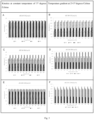
- dECM powder In order to characterise the product obtained, i.e. dECM powder abbreviated as "dECM(p)", powder grain size distribution in flow gradient was tested using a laser diffraction spectrometer Spraytec (Malvern, UK) equipped with an accessory inhalation chamber for studying inhalation sprays. In all the cases studied, the values of the parameters describing the analysed powder following aerosolization were comparable, indicating that there was no need to provide additional energy in the form of an increased air stream to break down the powder into individual particles.
- dECM(p) powder grain size distribution in flow gradient was tested using a laser diffraction spectrometer Spraytec (Malvern, UK) equipped with an accessory inhalation chamber for studying inhalation sprays. In all the cases studied, the values of the parameters describing the analysed powder following aerosolization were comparable, indicating that there was no need to provide additional energy in the form of an increased air stream to break down the powder into individual particles.
- Table 1 presents the values of parameters describing the diameters of powder particles, where: Dv(50) - median of the volume particle size distribution: the diameter of the particles that divides the cumulative volume distribution in half, in other words, all particles both smaller and larger than the median have the same volume (the particles below this diameter constitute 50% of the sample volume). Dv(10) - the particles below this diameter constitute 10% of the sample volume. Dv(90) - the particles below this diameter constitute 90% of the sample volume. D[3][2] - the Sauter diameter is the diameter of a particle whose volume to surface ratio is the same as the ratio of the volume of all analysed particles to the surface of the total of all such particles.
- D[4][3] - a diameter defined as the ratio of the sum of the fourth power of particle diameters to the sum of the third power of particle diameters.
- Table 1 Values of parameters describing the diameters of powder particles Flow [l/min] Parameter Diameter [ ⁇ m] SD [ ⁇ m] 100 Dv(10) 28.23 1.48 Dv(50) 148.43 10.14 Dv(90) 410.1 29.41 D[3][2] 54.19 4.54 D[4][3] 189.6 12.4 200 Dv(10) 27.7 5.7 Dv(50) 139.7 27.2 Dv(90) 474.4 123.8 D[3][2] 43.4 5.2 D[4][3] 202.1 40.1 270 Dv(10) 25.3 2.2 Dv(50) 146.7 25.6 Dv(90) 498.3 62.7 D[3][2] 46.3 4.6 D[4][3] 209.5 27.5
- the results of the measurements following the aerosolization of the dECM powder indicate that the powder was polydispersible.
- the median of the volume particle diameter distribution - Dv(50) at an air flow nominal for Cyclohaler type inhaler equaled 148.43 ⁇ 10.14 ⁇ m.
- the smallest particles of the total volume not exceeding 10% of the total volume of the sample had a diameter of less than 28.23 ⁇ 1.48 ⁇ m (Dv(10)), while the diameter distinguishing the particles with total volume less than 90% of the total volume of the sample was 410.10 ⁇ 29.41 ⁇ m.
- Increasing the air flow rate fed to the inhaler to 200 and 270dm 3 /min did not significantly affect the value of the median of the volume particle size distribution or the Dv(10) value.
- Type IV and type VI collagen are the most effective in supporting the functionality and viability of pancreatic islets and they are commonly used as supplements in biomedical applications that are based on the functioning of pancreatic islet cells.
- collagen VI and IV are present on the extra secretory surface and basement membrane of pancreatic islets and they regulate fibronectin activity. The percentage content of any of other collagen types analysed (COL1A2, COL3A1, COL4A2, COL6A1, COL6A2, COL6A3 COL14A1) did not exceed 3,5% in all examined samples.
- the concentration of residual DNA in the dry matter was on average 0.077ng/mg. In all examined samples the residual DNA content was lower than 0.15 ng/mg.
- the analysis was carried out using DNeasy Blood & Tissue Kit kits used to isolate residual DNA, and Quant-iT PicoGreen dsDNA Reagent and Kits to determine the concentration of the isolated genetic material.
- Collagenase was prepared in a special solution containing 150 mL of Ringer's solution pH 7.2 to 7.4, 2.72 mL Hepes (1M), 1.125 mL NaBicarbonate (7.5%) and 1.05 mL CaCl 2 (1M).
- Sample A was the result of treating dECM with a single collagenase concentration.
- Sample B was treated with 10-fold collagenase concentration.
- the highest concentration of the remaining Triton X-100 was found in the dECM solution treated with collagenase at a concentration of 43,953 PZ/g dECM.
- An increase in the concentration of collagenase did not result in a higher amount of Triton X-100 obtained, which indicated that the concentration of 43,953 PZ/g of dECM was sufficient for extraction of all the remaining Triton X-100 from the sample.
- the differences in fat composition in decellularized matrix in the function of the preparation of pancreas for decellularization were analysed.
- the content of residual DNA depending on the method of pancreatic preparation, collagen content and the content of residual detergent Triton X-100 were analysed.
- the use of the mechanical extrusion grinding method allowed for significantly reducing the fat content in the extracellular matrix obtained.
- the fat content was 6.24+/-0.07% (w/w) compared to 21.47+/-0.07% (w/w) of the fat content in the cutting method. The difference was statistically significant (p ⁇ 0.001).
- Low fat content of obtained dECM significantly increased the viability of cells and pancreatic islets.
- the use of the mechanical extrusion grinding method allowed for significantly reducing Triton X-100 content in the extracellular matrix obtained.
- the content of collagens in the tested material resulting from the preparation method did not differ depending on the use of the cutting method and mechanical extrusion grinding.
- dECM(r) a dECM powder (dECM(p)) dissolution procedure has been established that used pepsin and hydrochloric acid (HCl).
- HCl hydrochloric acid
- Pepsin (at a concentration of 0-10 mg/mL, preferably 1 mg/mL) was dissolved in 50 ml of 0.01 M HCl, after which dECM(p) (0.5 -5 g) was added. This method resulted in a dECM(r) concentration in the range of 1-10% (w/v).
- the prepared solution was placed on a magnetic stirrer, using the following stirring conditions: ambient temperature of approx. 25 °C, dissolution time of 72h, wherein the solution was agitated every hour for the first 8h of stirring.
- dECM solutions with varying pepsin content were prepared.
- the solution containing 1mg/mL pepsin had relatively high homogeneity: a small span of viscosity values. A slight change in turbidity was observed with the temperature change. All analysed dissolution methods with varying pepsin content were used for the preparation of dECM(r), however, it was demonstrated that the amount of 1 mg/mL used was optimal.
- a paste was prepared containing an appropriate amount of neutralised dECM(r) and dECM(p) by thorough mixing with a sterile metal spatula. Since the dECM(p) was prepared by freeze-drying and was not dissolved afterwards, it retained the quaternary structure of ECM. The paste obtained was left at a temperature of 7-10 °C for at least 24h. Directly before using the paste for bioink production, it was placed in a sterile syringe and mixed between syringes. At the same time, GelMa (10-20% (w/v)) and HAMA (1-3% (w/v)) solutions were prepared with LAP according to a commonly available procedure.
- the syringe containing the paste was attached with a connector to another syringe without the piston, which was moved upside down and stably arranged in the vertical position. Glycerol, culture medium, growth factors, vitamins, GelMa and HAMA solutions were successively added. The piston was then gently inserted, and the paste was mixed with the other reagents. After mixing, the prepared bioink was placed in the incubator for 5 minutes, islets and cells were added, then mixed again and introduced into a cartridge. In the next step, the filled cartridge was centrifuged for 2 minutes in 1500 rpm and reintroduced before printing for approx. 5 minutes to the incubator.
- compositions of the obtained primary bioinks were the following: 40-50 % (v/v) of dECM(r), 2.763 - 27.692 % (w/v) of dECM(p), 1.464-7.320 % (w/v) of GelMa, 0.146-1.098 % (w/v) of HAMA, 5.0-10.0 % (w/v) of glycerol, 0.03- 0.17% (w/v) of LAP, VEGF - 30 ng/mL, FGF - 20ng/mL, TGF- ⁇ - 10ng/mL, IL-8 - 10ng/mL, IL-17A - 20ng/mL, vitamin A - 100 ⁇ M, vitamin B1 - 100 ⁇ M, vitamin B3 - 10 ⁇ M, vitamin D3 - 10nM, pancreatic islets - 20000 iEq/mL, endothelial cells - 1 ⁇ 10 5 /mL, primary microvascular endot
- composition of the obtained sonicated vascular bioink was as follows: 5-10 % (w/v), preferably 7.5 % (w/v) of dECM(p), 0.2-1 % (v/v) of CMC, 1 - 2 % (w/v), preferably 1 % (w/v) of microbiological gelatin, fibronectin - 100 ⁇ g/mL, VEGF - 30 ng/mL, FGF -20ng/mL, PGE2 - 100nM, 1.5 ⁇ 10 7 /mL of endothelial cells and 3 ⁇ 10 6 /mL of fibroblasts.
- composition of the obtained boiled vascular bioink was as follows: 2-10 % (w/v), preferably 5 % (w/v) of dECM(p), 0.2-2 % (v/v) of CMC, 1 - 5 % (w/v), preferably 1 % (w/v) of microbiological gelatin, fibronectin - 100 ⁇ g/mL, VEGF - 30 ng/mL, FGF - 20ng/mL, PGE2 - 100nM, 1.5 ⁇ 10 7 /mL of endothelial cells and 3 ⁇ 10 6 /mL of fibroblasts.
- the vascular bioink consisted of 5-10% (w/v), preferably 5% (w/v) dECM(p) in a buffer solution or a cell medium.
- pancreatic islets alpha cells of pancreatic islets beta cells of pancreatic islets
- pancreatic islets alpha cells of pancreatic islets beta cells of pancreatic islets 0 sec 1.0% 1.0% 4.0% 4.0% 10 sec 12.0% 2.0% 1.5% 2.0% 2.5% 1.5% 30 sec 14.0% 5.5% 60 sec 45.0% 5.0% 1.5% 2.0% 3.5% 2.0% 90 sec 18.0% 9.5% 120 sec 19.0% 6.0% 2.0% 3.5% 16.0% 5.5% 180 sec 20.0% 5.0% 300 sec 50.0% 7.5% 2.0% 6.0% 9.5% 3.0%
- cross-linking results after the process or during the bioprinting process showed that both the use of 365nm and 405nm wavelength light achieved the intended effect, i.e. the change of hydrogel form from liquid to solid.
- the bioink contains cells and microorganisms, only visible light may be used. Therefore, the most preferable method of cross-linking is to use light with a wavelength of 405 nm.
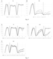
- Boiled dECM(r) has a much higher viscosity value than the sonicated one. However, due to proper stability of the vascular bioink after sonication, this method was determined to be more preferable for the printing of the vessel duct.
- Viability tests were performed on fibroblasts (cell lines 3T3-L1 and HFF-1) and pancreatic islets.
- pancreatic cells/islets were subjected to pressure in the range from 15kPa to 100kPa using a needle with a diameter of 0.2 and 0.6 mm.
- the results of the tests conducted showed that shear forces induced during 3D bioprinting using extrusion method produce significant changes in cell and microorgan viability.
- Table 11 The percentage of living and dead 3T3-L1 cells after applying predetermined pressures.
- the pressure and diameter of the needle needed to be matching the particular cell type. However, pressures of no more than 30 kPa were preferably applied.
- Printouts using primary bioink were made using the following parameters: pressure: 4 - 100 kPa, printing speed: 5 - 40 mm/s, temperature: printhead - 10-37 °C; printbed- 4-37 °C, needle diameter: 100 nm - 1 mm.
- Printouts using vascular bioink were made using the following parameters: pressure: 5 - 100 kPa, printing speed: 5 - 40 mm/s, temperature: printhead - 10-37 °C; printbed- 4-37 °C, needle diameter: 100 nm - 1 mm.
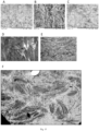
- Figure 6 shows a 3D model of a vascularised lobule and a picture of the printed construct. SEM was used to identify the morphology of the lateral surface and cross-section of the printed lobule. A loose arrangement of bioink filaments was observed that was behind the substantial porosity of the lobule. Also, based on a cross-section analysis, the stratification of the three-dimensional porous structure supplied with patent ducts imitating vessels was identified.
- the printed vascular system was evaluated using nuclear magnetic resonance imaging.
- the 3D reconstructions made show patent ducts with no the tendency to collapse or dissect.
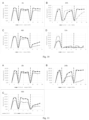
- pancreatic islet functionality Due to its properties, adding 5 % (w/v) and 10% (w/v) glycerol to bioink improved the printability of the primary bioink.
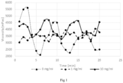
- glycerol was added to culture medium at 5% or 10% concentration and the islets were incubated therein for 24h [ Fig. 8 ]. In both cases, the functionality of pancreatic islets is by far superior compared to pancreatic islets in the culture medium alone.
- pancreatic islets were functional at a level comparable to that of islets untreated with any supplements.
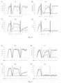
- bioinks were selected to assess the viability of pancreatic islets following 3D bioprinting: methacrylated gelatin, methacrylated hyaluronic acid, a mixture of methacrylated gelatin and methacrylated hyaluronic acid.
- methacrylated gelatin methacrylated hyaluronic acid
- methacrylated hyaluronic acid a mixture of methacrylated gelatin and methacrylated hyaluronic acid.
- 7.8% v/v GelMa or 0.78% v/v HAMA or a mixture of 4.68% v/v GelMa and 0.312% v/v HAMA (MIX) were added to the primary bioink.
- MIX a mixture of 4.68% v/v GelMa and 0.312% v/v HAMA
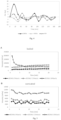
- Both the addition of HAMA and the mixture of GelMa and HAMA to the bioink induced a slight decrease in the levels of insulin produced by the islets compared to the control islets grown in the medium (that were not 3D bioprinted).
- the results for the bioink with the addition of GelMa alone showed the highest activity of the islets to the given glucose concentration, the structures printed were the least stable and they were the fastest to disintegrate in the culture medium.
- Embiodiment 8 Confirmation of the preserved quaternary structure of dECM in the primary bioink.
Landscapes
- Health & Medical Sciences (AREA)
- Chemical & Material Sciences (AREA)
- Life Sciences & Earth Sciences (AREA)
- Engineering & Computer Science (AREA)
- Biomedical Technology (AREA)
- Medicinal Chemistry (AREA)
- Chemical Kinetics & Catalysis (AREA)
- Dermatology (AREA)
- Animal Behavior & Ethology (AREA)
- General Health & Medical Sciences (AREA)
- Veterinary Medicine (AREA)
- Public Health (AREA)
- Epidemiology (AREA)
- Oral & Maxillofacial Surgery (AREA)
- Transplantation (AREA)
- Molecular Biology (AREA)
- Botany (AREA)
- Materials Engineering (AREA)
- Manufacturing & Machinery (AREA)
- Organic Chemistry (AREA)
- Cell Biology (AREA)
- Urology & Nephrology (AREA)
- Zoology (AREA)
- Wood Science & Technology (AREA)
- General Chemical & Material Sciences (AREA)
- Biophysics (AREA)
- Polymers & Plastics (AREA)
- Vascular Medicine (AREA)
- Medicinal Preparation (AREA)
- Materials For Medical Uses (AREA)
- Micro-Organisms Or Cultivation Processes Thereof (AREA)
- Detergent Compositions (AREA)
- Apparatus Associated With Microorganisms And Enzymes (AREA)
- Application Of Or Painting With Fluid Materials (AREA)
Claims (13)
- Ein Verfahren zur Herstellung einer detergenzienfreien dezellularisierten extrazellulären Matrix (dECM), umfassend die folgenden Schritte:- mechanische Fragmentierung, vorzugsweise durch mechanische Extrusion, eines Organs tierischen Ursprungs, ausgewählt aus Pankreas, Leber, Nieren, Herz, Haut, Lunge, Dickdarm, Dünndarm, Blutarterien und -venen, Fettgewebe und Plazenta, wobei das Organ vom Körper des Tieres getrennt ist- Inkubation des fragmentierten Organs in einer gepufferten Detergenzlösung, die vorzugsweise 1XPBS umfasst, wobei die gepufferte Detergenzlösung 0,5 %-1,5 %, vorzugsweise 1 % (Vol.Nol.), Octoxynol-9 umfasst, wobei die Detergenzlösung mit einem antimikrobiellen Mittel, vorzugsweise Streptomycin, vorzugsweise in einer Konzentration von 0. 01 % (Gew./Vol.) ergänzt wird und die Inkubation bei einer Temperatur unterhalb der Raumtemperatur, vorzugsweise bei 4°C, für mindestens 72 Stunden unter Rühren durchgeführt wird, wobei das fragmentierte Organ alle 4 bis 12 Stunden in eine frische Detergenzlösung überführt wird- Inkubation des fragmentierten Organs in einer ersten gepufferten Waschlösung, die vorzugsweise 1XPBS enthält, wobei die erste gepufferte Waschlösung ein antimikrobielles Mittel, vorzugsweise Streptomycin, vorzugsweise in einer Konzentration von 0,01 % (Gew./Vol.), enthält, für mindestens 72 Stunden bei einer Temperatur unterhalb der Raumtemperatur, vorzugsweise bei 4°C, unter Rühren, wobei das fragmentierte Organ alle 4 bis 12 Stunden in eine frische Waschlösung überführt wird- Inkubation des fragmentierten Organs in einer DNAse-haltigen Desoxyribonuklease-Lösung, vorzugsweise in einer Konzentration von 0,0001 bis 0,0003 % (Gew./Vol.), am meisten bevorzugt 0,0002 % (Gew./Vol.), vorzugsweise für mindestens 8 Stunden bei einer für die DNAse-Leistung geeigneten Temperatur- Inkubation des fragmentierten Organs in einer zweiten gepufferten Waschlösung, die vorzugsweise 1XPBS enthält, wobei die zweite gepufferte Waschlösung ein antimikrobielles Mittel, vorzugsweise Streptomycin, vorzugsweise in einer Konzentration von 0,01 % (Gew./Vol.), enthält, für mindestens 72 Stunden bei einer Temperatur unterhalb der Raumtemperatur, vorzugsweise bei 4°C, unter Rühren, wobei das fragmentierte Organ alle 4 bis 12 Stunden in eine frische Waschlösung überführt wird- Einfrieren des fragmentierten Organs und Zerkleinern des gefrorenen fragmentierten Organs in Fragmente- Gefriertrocknung des gefrorenen fragmentierten Organs, vorzugsweise bei -32°C, vorzugsweise unter einem Druck von 0,31 mbar (31 Pa).- optionale Endtrocknung für 5 bis 15 Minuten bei 0,0010 mbar (0,1 Pa) und -76°C- Mahlen des zerkleinerten und getrockneten Produkts zu 25 - 500 µm dECM-Pulver- fakultativ Sterilisierung des Produkts, vorzugsweise durch Bestrahlung und/oder Ethylenoxid.
- Das Verfahren nach Anspruch 1, wobei auf den Mahlschritt ein Schritt zur Überprüfung der Menge an Octoxynol-9 im dECM-Pulver folgt, wobei vorzugsweise vor der Überprüfung des dECM-Pulvers auf das Vorhandensein von Octoxynol-9 dieses mit Kollagenase behandelt wird, vorzugsweise in einer Konzentration von 43,953 PZ/g dECM.
- Das Verfahren nach Anspruch 1 oder 2, wobei sich an den Mahlschritt folgende Schritte anschließen:- Auflösen des dECM-Pulvers in Salzsäurelösung, vorzugsweise 0,01 M, die mit 0-10 mg/ml Pepsin ergänzt ist;- Mischen für 48-72 Stunden, vorzugsweise für 72 Stunden, bei Raumtemperatur;- Neutralisieren auf Eis, vorzugsweise mit 0,1 M Natriumbase und PBS-Lösung.
- Ein Verfahren zur Herstellung einer primären Biotinte, umfassend die folgenden Schritte:- Herstellung einer Paste, umfassend 5-50 % (Gew./Vol.), vorzugsweise 15-25 % (Gew./Vol.), des dECM-Pulvers, erhältlich durch das in einem der Ansprüche 1 oder 2 definierte Verfahren, und 1-10 % (Gew./Vol.), vorzugsweise 8-10 % (Gew./Vol.), der dECM-Lösung, erhältlich durch das in Anspruch 3 definierte Verfahren, durch Mischen- Inkubation der Paste bei einer Temperatur von 7-10°C für mindestens 24 Stunden- Zugabe von 1,46-7,32 % (Gew./Vol.) methacrylierter Gelatine, 0,15-1,10 % (Gew./Vol.) methacrylierter Hyaluronsäure und 5 bis 10 % (Gew./Vol.) Glycerin sowie eines Photoinitiators, vorzugsweise 0,03-0,17 % (Gew./Vol.). /v) Lithiumphenyl-2,4,6-trimethylbenzoylphosphinat, gefolgt von sanftem Mischen.
- Eine primäre Biotinte, umfassend eine dECM-Paste und 1,46-7,32 % (Gew./Vol.) methacrylierte Gelatine, 0,15-1,10 % (Gew./Vol.) methacrylierte Hyaluronsäure und 5 bis 10 % (Gew./Vol.) Glycerin, und einem Photoinitiator, vorzugsweise 0,03-0,17 % (Gew./Vol.) Lithiumphenyl-2,4,6-trimethylbenzoylphosphinat, wobei die dECM-Paste 5-50% (Gew./Vol.), vorzugsweise 15-25% (Gew./Vol.), des dECM-Pulvers, das durch das in einem der Ansprüche 1 oder 2 definierte Verfahren erhältlich ist, und 1-10% (Gew./Vol.), vorzugsweise 8-10% (Gew./Vol.), der dECM-Lösung, die durch das in Anspruch 3 definierte Verfahren erhältlich ist, umfasst, und wobei die Viskosität der primären Biotinte mindestens 5 Pa·s beträgt, gemessen in einem Kegel-Platte-System, bei einer konstanten Scherrate von 21/s und einer Temperaturvon 37°C.
- Die primäre Biotinte nach Anspruch 5, umfassend mindestens einen Zusatzstoff, ausgewählt aus: Hyaluronsäure in einer Konzentration von 0,001 bis 0,100 mg/ml der primären Biotinte, vorzugsweise 0,007 mg/ml, Laminin in einer Konzentration von 0,005 bis 0,100 mg/ml der primären Biotinte, vorzugsweise 0,084 mg/ml, Kollagen I in einer Konzentration von 0,001 bis 0,100 mg/ml der primären Biotinte, vorzugsweise 0,041 mg/ml, Kollagen IV in einer Konzentration von 0,005 bis 0,175 mg/ml der primären Biotinte, vorzugsweise 0,122 mg/ml, Fibronektin in einer Konzentration von 3 bis 300 µg/ml, vorzugsweise 100 µg/ml, menschliches Fibrinogen in einer Konzentration von 10 bis 100 mg/ml der primären Biotinte, Aprotinin in einer Konzentration von 1 bis 2 EPU/ml der primären Biotinte, Polysorbat in einer Konzentration von 0,05 bis 2 mg/ml der primären Biotinte, menschliches Thrombin in einer Konzentration von 5 bis 55 mg/ml derprimären Biotinte, Calciumchlorid in einer Konzentration von 20 bis 60 mM/ml der primären Biotinte; proangiogene Vitamine: Vitamin A in einer Konzentration von 1 nM - 500 µM, vorzugsweise 100 µM, Vitamin B1 in einer Konzentration von 50-100 µM, vorzugsweise 100 µM, Vitamin B3 in einer Konzentration von 1 bis 10 µM, vorzugsweise 10 µM, Vitamin B12 in einer Konzentration von 10 bis 100 mg/ml der primären Biotinte, Vitamin D3 in einer Konzentration von 0,1 bis 10 nM, vorzugsweise 10 nM, Wachstumsfaktoren, die die Angiogenese unterstützen: VEGF in einer Konzentration von 10 bis 30 ng/ml der primären Biotinte, vorzugsweise 30 ng/ ml, FGF in einer Konzentration von 10 bis 20 ng/ml der primären Biotinte, vorzugsweise 20 ng/ml, TGF-β in einer Konzentration von 1 bis 10 ng/ml der primären Biotinte, vorzugsweise 20 ng/ml, Interleukin (IL)-8 in einer Konzentration von 0 bis 100 ng/ml der primären Biotinte, vorzugsweise 10 ng/ml, IL-17A in einer Konzentration von 20 bis 50 ng/ml der primären Biotinte, vorzugsweise 20 ng/ml.
- Die primäre Biotinte nach Anspruch 5 oder 6, umfassend einen oder mehrere tierische oder menschliche Zusatzstoffe, ausgewählt aus Endothelzellen in einer Dichte von 0,1-10 × 105/ml der primären Biotinte, primären mikrovaskuläre Endothelzellen in einer Konzentration von 0,1 bis 10 × 105/ml der primären Biotinte, tierischen oder menschlichen α-Zellen in einer Konzentration von 3 bis 9 × 10⁶/ml der Biotinte, tierischen oder menschlichen β-Zellen in einer Konzentration von 1,1 bis 3,4 × 10⁷/ ml der Biotinte, tierischen oder menschlichen Pankreasinseln, vorzugsweise in einer Menge von 20.000 iEq/ml der primären Biotinte.
- Ein Verfahren zur Herstellung einer vaskulären Biotinte, das die folgenden Schritte umfasst:a) wahlweise Herstellung einer Lösung von mikrobiologischer Gelatine, die mit CMC ergänzt ist, , umfassend die Herstellung einer 1-2 %igen (Gew./Vol.) Lösung von mikrobiologischer Gelatine in einer Pufferlösung, vorzugsweise PBS, durch Suspendieren von mikrobiologischer Gelatine in der Pufferlösung unter Rühren bei einer Temperatur zwischen 50 und 65 °C, vorzugsweise bei 60 °C, Zugabe einer 2-5%igen (Vol./Vol.) wässrigen Lösung von Carboxymethylcellulose (CMC), um eine Endkonzentration von 0, 2-1 % (Vol./Vol.) CMC in der vaskulären Biotinte und Abkühlen der Lösung auf eine Temperatur von 40°C oder darunter;b) Herstellung einer 5-10%igen (Gew./Vol.) dECM-Lösung durch Zugabe des dECM-Pulvers, erhältlich durch das in einem der Ansprüche 1 oder 2 definierte Verfahren, vorzugsweise sterilisiert durch Bestrahlung zu (i) der in Schritt a) erhaltenen Lösung von mikrobiologischer Gelatine, die mit CMC ergänzt wurde, oder (ii) einer Pufferlösung oder (iii) einer Lösung von Zellmedium unter leichtem Rühren;c) Beschallung der erhaltenen Lösung bei einer Temperatur von nicht mehr als 37°C für 0,5-2,0 Stunden;d) wahlweise Zugabe von mindestens einem tierischen oder menschlichen Zusatzstoff, ausgewählt aus: Fibronektin in einer Konzentration von 3 bis 300 µg/ml, vorzugsweise 100 µg/ml, VEGF in einer Konzentration von 10 bis 30 ng/ml, vorzugsweise 30 ng/ml, FGF in einer Konzentration von 10 bis 20 ng/ml, vorzugsweise 20 ng/ml, PGE2 in einer Konzentration zwischen 100 und 300 nM, vorzugsweise 100 nM, Endothelzellen in einer Dichte zwischen 0,1 und 10×107 Zellen/ml der Biotinte, Fibroblasten mit einer Dichte zwischen 0,1 und 10×106 Zellen/ml der Biotinte.
- Ein Verfahren zur Herstellung einer vaskulären Biotinte, das die folgenden Schritte umfasst:a) wahlweise Herstellung einer Lösung von mikrobiologischer Gelatine, die mit CMC ergänzt ist, umfassend die Herstellung einer 1-2%igen (Gew./Vol.) Lösung von mikrobiologischer Gelatine in einer Pufferlösung, vorzugsweise PBS, durch Suspendieren von mikrobiologischer Gelatine in der Pufferlösung unter Rühren bei einer Temperatur zwischen 50 und 65 °C, vorzugsweise bei 60 °C, Zugabe einer 2-5%igen (Vol./Vol.) wässrigen Carboxymethylcellulose (CMC)-Lösung, um eine Endkonzentration von 0,2-1% (Vol./Vol.) CMC in der vaskulären Biotinte und Abkühlen der Lösung auf eine Temperatur von 40°C oder darunter;b) Herstellung einer 5-10%igen (Gew./Vol.) dECM-Lösung durch Zugabe von dECM-Pulver, das nach dem in einem der Ansprüche 1 oder 2 definierten Verfahren erhältlich ist und vorzugsweise durch Bestrahlung sterilisiert wurde, zu (i) der in Schritt a) erhaltenen Lösung von mikrobiologischer Gelatine, die mit CMC ergänzt wurde, oder (ii) einer Pufferlösung oder (iii) einer Lösungvon Zellmedium unter leichtem Rühren;c) Sieden der Mischung bei 100 °C für 15-30 Minuten;d) wahlweise Zugabe von mindestens einem tierischen oder menschlichen Zusatzstoff, ausgewählt aus: Fibronektin in einer Konzentration von 3 bis 300 µg/ml, vorzugsweise 100 µg/ml, VEGF in einer Konzentration von 10 bis 30 ng/ml, vorzugsweise 30 ng/ml, FGF in einer Konzentration von 10 bis 20 ng/ml, vorzugsweise 20 ng/ml, PGE2 in einer Konzentration zwischen 100 und 300 nM, vorzugsweise 100 nM, Endothelzellen in einer Dichte zwischen 0,1 und 10×107 Zellen/ml der Biotinte , Fibroblasten mit einer Dichte zwischen 0,1 und 10×106 Zellen/ml der Biotinte.
- Eine vaskuläre Biotinte, umfassend die beschallte oder gekochte dECM-Lösung, erhältlich durch das in Anspruch 3 definierte Verfahren, in einer Konzentration von 2-10 % (Gew./Vol.), vorzugsweise ergänzt mit mikrobiologischer Gelatine in einer Konzentration von 1 bis 5% (Gew./Vol.) und/oder CMC in einer Konzentration von 0,2 bis 2 % (Vol./Vol.).
- Die vaskuläre Biotinte nach Anspruch 10, umfassend mindestens einen tierischen oder menschlichen Zusatzstoff, ausgewählt aus: Fibronektin in einer Konzentration von 3 bis 300 µg/ml, vorzugsweise 100 µg/ml, VEGF in einer Konzentration von 10 bis 30 ng/ml, vorzugsweise 30 ng/ml, FGF in einer Konzentration von 10 bis 20 ng/ml, vorzugsweise 20 ng/ml, PGE2 in einer Konzentration zwischen 100 und 300 nM, vorzugsweise 100 nM, Endothelzellen in einer Dichte zwischen 0,1 und 10×107 Zellen/ml der vaskulären Biotinte, Fibroblasten mit einer Dichte zwischen 0,1 und 10×106 Zellen/ml der vaskulären Biotinte.
- Eine dreidimensionale Struktur umfassend mindestens drei benachbarte Biotinte-Schichten, wobei eine Schicht der vaskulären Biotinte nach Anspruch 10 oder 11 zwischen zwei Schichten der primären Biotinte nach einem der Ansprüche 5 bis 7 angeordnet ist.
- Ein Verfahren zur Herstellung einer dreidimensionalen Struktur, wobei der in einem der Ansprüche 5 bis 7 definierte primäre Biotinte und der in Anspruch 10 oder 11 definierte vaskuläre Biotinte in einem 3D-Bioprinting-Verfahren bei einer Druckgeschwindigkeit von 5 bis 50 mm/s, einem Druck von 4 bis 300 kPa und einer Temperatur von 4 bis 37°C schichtweise aufgetragen werden und wobei die primäre Biotinte während oder nach der Abscheidung mindestens 5 Sekunden lang UV-Licht und/oder sichtbarem Licht, vorzugsweise mit einer Wellenlänge von 365 bis 405 nm, besonders bevorzugt bei 405 nm, ausgesetzt wird.
Applications Claiming Priority (3)
| Application Number | Priority Date | Filing Date | Title |
|---|---|---|---|
| EP19461559 | 2019-07-22 | ||
| EP19218191.5A EP3769796A1 (de) | 2019-07-22 | 2019-12-19 | Reinigungsmittelfreie, dezellularisierte extrazelluläre matrixzubereitungsmethode und bioinks für den 3d-druck |
| PCT/IB2020/056856 WO2021014359A1 (en) | 2019-07-22 | 2020-07-21 | Detergent-free decellularized extracellular matrix preparation method and bioinks for 3d printing |
Publications (3)
| Publication Number | Publication Date |
|---|---|
| EP4003439A1 EP4003439A1 (de) | 2022-06-01 |
| EP4003439B1 true EP4003439B1 (de) | 2024-06-19 |
| EP4003439C0 EP4003439C0 (de) | 2024-06-19 |
Family
ID=67438624
Family Applications (2)
| Application Number | Title | Priority Date | Filing Date |
|---|---|---|---|
| EP19218191.5A Withdrawn EP3769796A1 (de) | 2019-07-22 | 2019-12-19 | Reinigungsmittelfreie, dezellularisierte extrazelluläre matrixzubereitungsmethode und bioinks für den 3d-druck |
| EP20757665.3A Active EP4003439B1 (de) | 2019-07-22 | 2020-07-21 | Reinigungsmittelfreie, dezellularisierte extrazelluläre matrixzubereitungsmethode und bioinks für den 3d-druck |
Family Applications Before (1)
| Application Number | Title | Priority Date | Filing Date |
|---|---|---|---|
| EP19218191.5A Withdrawn EP3769796A1 (de) | 2019-07-22 | 2019-12-19 | Reinigungsmittelfreie, dezellularisierte extrazelluläre matrixzubereitungsmethode und bioinks für den 3d-druck |
Country Status (22)
| Country | Link |
|---|---|
| US (1) | US12208179B2 (de) |
| EP (2) | EP3769796A1 (de) |
| JP (1) | JP7606236B2 (de) |
| KR (1) | KR20220038705A (de) |
| CN (1) | CN114340687B (de) |
| AU (1) | AU2020316739B2 (de) |
| BR (1) | BR112022001029A2 (de) |
| CA (1) | CA3146781A1 (de) |
| CL (1) | CL2022000153A1 (de) |
| CU (1) | CU20220004A7 (de) |
| DO (1) | DOP2022000009A (de) |
| ES (1) | ES2987779T3 (de) |
| GE (2) | GEP20237562B (de) |
| IL (1) | IL289776A (de) |
| MX (1) | MX2022000942A (de) |
| MY (1) | MY207460A (de) |
| PE (1) | PE20220701A1 (de) |
| PH (1) | PH12022550074A1 (de) |
| PL (1) | PL4003439T3 (de) |
| PT (1) | PT4003439T (de) |
| WO (1) | WO2021014359A1 (de) |
| ZA (1) | ZA202202184B (de) |
Families Citing this family (17)
| Publication number | Priority date | Publication date | Assignee | Title |
|---|---|---|---|---|
| CN113304319A (zh) * | 2021-03-03 | 2021-08-27 | 南京市第一医院 | 一种用于膀胱组织修复的生物材料及其制备方法 |
| BR112023025612A2 (pt) * | 2021-06-30 | 2024-02-27 | Polbionica Sp Z O O | Meio para armazenar ilhotas pancreáticas isoladas, e, método de armazenamento de ilhotas pancreáticas isoladas |
| CN113750292B (zh) * | 2021-09-30 | 2022-10-25 | 华南理工大学 | 一种用于3d打印角膜修复材料的生物墨水及制备方法、角膜修复材料的制备方法 |
| KR102765583B1 (ko) * | 2021-12-10 | 2025-02-07 | 재단법인 대구경북첨단의료산업진흥재단 | 인체지방유래 세포외기질을 이용한 3d 프린팅용 바이오잉크 제작 방법 및 이를 이용한 세포지지체 제작 |
| CN114259604B (zh) * | 2021-12-17 | 2022-12-27 | 上海纳米技术及应用国家工程研究中心有限公司 | 一种3d打印促有序血管化的载药骨修复支架的制备方法及其产品和应用 |
| JP2025509522A (ja) * | 2022-03-17 | 2025-04-11 | ポルビオニカ スポルカ ジー オグラニクゾナ オドパウイエドジアルノシア | 3dバイオプリンティング技術を用いた灌流可能な3次元組織モデルの製造方法、及びこの方法によって製造された組織モデル |
| KR20240165333A (ko) * | 2022-03-18 | 2024-11-22 | 폴바이오니카 에스피. 제트 오.오. | 바이오프린팅된 조직 모델을 위한 보강 및 밀봉 구성, 및 보강 및 밀봉 구성을 조립하기 위한 방법 |
| BR102022006759A2 (pt) * | 2022-04-07 | 2023-10-17 | Tissuelabs Pesquisa E Desenvolvimento Ltda | Processos livres de enzimas para produzir uma solução de matriz extracelular tecido-específica, um hidrogel tecido-específico, um produto derivado da solução de matriz extracelular tecido-específica, um pó solúvel tecido-específico e uma membrana tecido-específica, e seus produtos, processos para fornecer compostos bioativos para regenerar tecido, processo in vitro para fornecer compostos bioativos por meio do hidrogel tecido-específico, usos da solução de matriz extracelular tecido-específica, ingrediente aditivo para meio de cultura de células, processo para cultivar células e agregados celulares dentro do hidrogel tecido-específico, processo para bioimprimir biotintas 3d, processo para bioimprimir 3d e kit |
| KR102820825B1 (ko) * | 2022-04-28 | 2025-06-16 | 아주대학교산학협력단 | 세포외기질로 유도된 자가조립체 기반 3d 프린팅 인공 조직체의 제조방법 및 이로부터 제조된 인공 조직체 |
| KR102820784B1 (ko) | 2023-01-02 | 2025-06-13 | 강원대학교산학협력단 | 간 시누소이드 모사 칩과 이의 제조방법 |
| CN116328040B (zh) * | 2023-03-03 | 2025-08-15 | 南方科技大学 | 一种生物工程支架及其制备方法、再生组织 |
| KR102682842B1 (ko) | 2023-05-30 | 2024-07-08 | 주식회사 에드믹바이오 | 배양액 유동 세포 배양 칩 |
| WO2025141426A1 (en) * | 2023-12-28 | 2025-07-03 | Polbionica Sp. Z O.O. | Method of animal-derived tissue decellularization and bioink comprising animal-derived decellularized tissue |
| KR20250127786A (ko) * | 2024-02-19 | 2025-08-27 | 포항공과대학교 산학협력단 | 탈세포화 세포외기질 기반 하이드로겔과 젤라틴화된 탈세포화 세포외기질 기반 하이드겔이 혼합된 하이브리드 바이오잉크 |
| WO2025188203A1 (en) * | 2024-03-07 | 2025-09-12 | Polbionica S.A. | Biomaterial composition comprising graphene oxide-comprising hydrogel and decm material for 3d (bio)printing |
| WO2025193112A1 (en) * | 2024-03-14 | 2025-09-18 | Polbionica S.A. | Three-dimensional printing composition comprising methacrylated chitosan |
| CN120241793B (zh) * | 2025-04-10 | 2025-12-23 | 上海交通大学医学院附属第九人民医院 | 脱细胞外基质在皮肤抗光老化中的应用及其药械 |
Family Cites Families (15)
| Publication number | Priority date | Publication date | Assignee | Title |
|---|---|---|---|---|
| EP1856246B1 (de) * | 2005-03-07 | 2015-07-15 | Technion Research & Development Foundation Limited | Dezellularisierte gewebestruktur und verfahren zu deren herstellung und verwendung |
| CN102590427A (zh) * | 2012-02-24 | 2012-07-18 | 玉溪九洲生物技术有限责任公司 | 抗毒素/抗血清中Triton X-100残留量的测定方法 |
| KR102284982B1 (ko) | 2013-01-09 | 2021-08-03 | 넥스트젠 바이오로직스, 아이엔씨. | 비-포유류 조직으로부터의 세포제거된 생체재료 |
| GB201409858D0 (en) | 2014-06-03 | 2014-07-16 | Ucl Business Plc | Human liver scaffolds |
| CN104215718B (zh) * | 2014-09-22 | 2016-05-04 | 成都生物制品研究所有限责任公司 | Triton X-100含量的高效液相色谱检测方法 |
| CN104307045B (zh) * | 2014-10-13 | 2016-06-08 | 林贤丰 | 一种天然组织来源的脱细胞骨膜材料的制备方法 |
| CA2964926C (en) | 2014-10-24 | 2023-05-09 | Wake Forest University Health Sciences | Tissue-mimicking hydrogel compositions for biofabrication |
| CN104458969B (zh) * | 2014-12-31 | 2016-05-11 | 深圳康泰生物制品股份有限公司 | 重组酿酒酵母表达的HBsAg原液中Triton X-100残留量的测定方法 |
| WO2016126947A2 (en) * | 2015-02-04 | 2016-08-11 | The Johns Hopkins University | Subcutaneous complex and single-component ecm compositing for induction of hair growth and follicular regeneration |
| KR101628821B1 (ko) * | 2015-03-02 | 2016-06-13 | 강원대학교산학협력단 | 탈세포화된 생체 조직 유래의 생체적합성 가용화 스캐폴드 추출물, 이의 제조방법 및 이의 용도 |
| AU2016235074A1 (en) * | 2015-03-26 | 2017-10-12 | Miromatrix Medical Inc. | Gas filled decellularized extracellular matrix |
| WO2016179242A1 (en) | 2015-05-05 | 2016-11-10 | President And Fellows Of Harvard College | Tubular tissue construct and a method of printing |
| EP3326661B8 (de) | 2015-07-21 | 2020-12-30 | InnoRegen, Inc. | Biotintenzusammensetzung mit verbesserten physikalischen und biologischen eigenschaften |
| KR101848997B1 (ko) | 2016-07-25 | 2018-04-16 | 포항공과대학교 산학협력단 | 간 조직의 탈세포화 방법 |
| KR101954953B1 (ko) | 2017-05-16 | 2019-03-06 | 울산과학기술원 | 3차원 프린팅용 바이오 잉크 조성물 및 이의 제조방법 |
-
2019
- 2019-12-19 EP EP19218191.5A patent/EP3769796A1/de not_active Withdrawn
-
2020
- 2020-07-21 PL PL20757665.3T patent/PL4003439T3/pl unknown
- 2020-07-21 WO PCT/IB2020/056856 patent/WO2021014359A1/en not_active Ceased
- 2020-07-21 MY MYPI2022000081A patent/MY207460A/en unknown
- 2020-07-21 KR KR1020227004559A patent/KR20220038705A/ko active Pending
- 2020-07-21 AU AU2020316739A patent/AU2020316739B2/en active Active
- 2020-07-21 PH PH1/2022/550074A patent/PH12022550074A1/en unknown
- 2020-07-21 PT PT207576653T patent/PT4003439T/pt unknown
- 2020-07-21 JP JP2022504564A patent/JP7606236B2/ja active Active
- 2020-07-21 CN CN202080053399.3A patent/CN114340687B/zh active Active
- 2020-07-21 EP EP20757665.3A patent/EP4003439B1/de active Active
- 2020-07-21 MX MX2022000942A patent/MX2022000942A/es unknown
- 2020-07-21 CU CU2022000004A patent/CU20220004A7/es unknown
- 2020-07-21 BR BR112022001029A patent/BR112022001029A2/pt unknown
- 2020-07-21 ES ES20757665T patent/ES2987779T3/es active Active
- 2020-07-21 US US17/628,429 patent/US12208179B2/en active Active
- 2020-07-21 PE PE2022000112A patent/PE20220701A1/es unknown
- 2020-07-21 GE GEAP202015860A patent/GEP20237562B/en unknown
- 2020-07-21 CA CA3146781A patent/CA3146781A1/en active Pending
- 2020-07-21 GE GEAP202315860A patent/GEAP202315860A/en unknown
-
2022
- 2022-01-12 IL IL289776A patent/IL289776A/en unknown
- 2022-01-19 DO DO2022000009A patent/DOP2022000009A/es unknown
- 2022-01-21 CL CL2022000153A patent/CL2022000153A1/es unknown
- 2022-02-21 ZA ZA2022/02184A patent/ZA202202184B/en unknown
Also Published As
| Publication number | Publication date |
|---|---|
| PL4003439T3 (pl) | 2024-08-19 |
| CU20220004A7 (es) | 2022-09-08 |
| CA3146781A1 (en) | 2021-01-28 |
| AU2020316739A1 (en) | 2022-02-17 |
| PE20220701A1 (es) | 2022-05-04 |
| EP3769796A1 (de) | 2021-01-27 |
| JP2022541636A (ja) | 2022-09-26 |
| MX2022000942A (es) | 2022-07-12 |
| US20220280694A1 (en) | 2022-09-08 |
| KR20220038705A (ko) | 2022-03-29 |
| IL289776A (en) | 2022-03-01 |
| MY207460A (en) | 2025-02-27 |
| DOP2022000009A (es) | 2022-06-15 |
| EP4003439C0 (de) | 2024-06-19 |
| EP4003439A1 (de) | 2022-06-01 |
| ZA202202184B (en) | 2022-09-28 |
| GEP20237562B (en) | 2023-10-25 |
| CN114340687A (zh) | 2022-04-12 |
| CN114340687B (zh) | 2025-05-23 |
| GEAP202315860A (en) | 2023-07-10 |
| PH12022550074A1 (en) | 2022-11-21 |
| JP7606236B2 (ja) | 2024-12-25 |
| BR112022001029A2 (pt) | 2022-03-15 |
| US12208179B2 (en) | 2025-01-28 |
| CL2022000153A1 (es) | 2022-10-14 |
| ES2987779T3 (es) | 2024-11-18 |
| AU2020316739B2 (en) | 2025-03-20 |
| WO2021014359A1 (en) | 2021-01-28 |
| PT4003439T (pt) | 2024-09-05 |
Similar Documents
| Publication | Publication Date | Title |
|---|---|---|
| EP4003439B1 (de) | Reinigungsmittelfreie, dezellularisierte extrazelluläre matrixzubereitungsmethode und bioinks für den 3d-druck | |
| Zhang et al. | Crosslinker-free silk/decellularized extracellular matrix porous bioink for 3D bioprinting-based cartilage tissue engineering | |
| US12338457B2 (en) | Biologically functional soft tissue scaffolds and implants | |
| Yao et al. | Recent development and biomedical applications of decellularized extracellular matrix biomaterials | |
| Jang et al. | ASC/chondrocyte-laden alginate hydrogel/PCL hybrid scaffold fabricated using 3D printing for auricle regeneration | |
| Wang et al. | CaO 2/gelatin oxygen slow-releasing microspheres facilitate tissue engineering efficiency for the osteonecrosis of femoral head by enhancing the angiogenesis and survival of grafted bone marrow mesenchymal stem cells | |
| CN104225667B (zh) | 一种促血管生成的温敏性水凝胶粉及用其制备的温敏性水凝胶 | |
| US20220395612A1 (en) | Ovarian-derived hydrogels for biomedical and biotechnology applications | |
| CN107261216A (zh) | 一种明胶海绵支架的制备方法 | |
| Mahoney et al. | Adipose derived delivery vehicle for encapsulated adipogenic factors | |
| US20150344842A1 (en) | Method for production of decellularized biological material and the decellularized biological material prepared therefrom | |
| Wang et al. | Construction of engineered 3D islet micro-tissue using porcine decellularized ECM for the treatment of diabetes | |
| Zhang et al. | Sodium alginate hydrogels co-encapsulated with cell free fat extract-loaded core-shell nanofibers and menstrual blood stem cells derived exosomes for acceleration of articular cartilage regeneration | |
| Zivari‐Ghader et al. | Recent scaffold‐based tissue engineering approaches in premature ovarian failure treatment | |
| CN110306289A (zh) | 一种含有阿西替尼的纳米纤维电纺膜及其制备方法和应用 | |
| Apinun et al. | Osteogenic differentiation of rat bone marrow-derived mesenchymal stem cells encapsulated in Thai silk fibroin/collagen hydrogel: a pilot study in vitro | |
| CN109289088A (zh) | 一种负载鸡血藤的ⅰ型/ⅲ型胶原复合支架 | |
| EA049075B1 (ru) | Способ получения децеллюляризованного внеклеточного матрикса, не содержащего детергентов, и биочернила для 3d-печати | |
| Kuo et al. | Effect of bovine pituitary extract on the formation of neocartilage in chitosan/gelatin scaffolds | |
| HK40072872A (en) | Detergent-free decellularized extracellular matrix preparation method and bioinks for 3d printing | |
| Chen et al. | Kelp and Cartilage Acellular Matrix Hybrid Microgel Assembly Realizes Articular Cartilage Repair Via ROS Scavenging, Endogenous BMSC Recruitment and Chondrogenic Differentiation | |
| ES2905713A1 (es) | Nuevo biomaterial para ingenieria tisular | |
| TW201545779A (zh) | 製備去細胞化生物材料之方法及由其製備之去細胞化生物材料 | |
| Zou et al. | Immunomodulatory 3D hybrid scaffolds functionalized with cytokine transfected stem cells for bone regeneration | |
| Bi et al. | Injectable Photo-cross-linkable Porous Composite Hydrogels with Exosomes for M2 Macrophage Polarization and Cartilage Defect regeneration |
Legal Events
| Date | Code | Title | Description |
|---|---|---|---|
| STAA | Information on the status of an ep patent application or granted ep patent |
Free format text: STATUS: UNKNOWN |
|
| STAA | Information on the status of an ep patent application or granted ep patent |
Free format text: STATUS: THE INTERNATIONAL PUBLICATION HAS BEEN MADE |
|
| PUAI | Public reference made under article 153(3) epc to a published international application that has entered the european phase |
Free format text: ORIGINAL CODE: 0009012 |
|
| STAA | Information on the status of an ep patent application or granted ep patent |
Free format text: STATUS: REQUEST FOR EXAMINATION WAS MADE |
|
| 17P | Request for examination filed |
Effective date: 20220222 |
|
| AK | Designated contracting states |
Kind code of ref document: A1 Designated state(s): AL AT BE BG CH CY CZ DE DK EE ES FI FR GB GR HR HU IE IS IT LI LT LU LV MC MK MT NL NO PL PT RO RS SE SI SK SM TR |
|
| RAP3 | Party data changed (applicant data changed or rights of an application transferred) |
Owner name: POLBIONICA SP. Z O.O. |
|
| GRAP | Despatch of communication of intention to grant a patent |
Free format text: ORIGINAL CODE: EPIDOSNIGR1 |
|
| STAA | Information on the status of an ep patent application or granted ep patent |
Free format text: STATUS: GRANT OF PATENT IS INTENDED |
|
| INTG | Intention to grant announced |
Effective date: 20240307 |
|
| GRAS | Grant fee paid |
Free format text: ORIGINAL CODE: EPIDOSNIGR3 |
|
| GRAA | (expected) grant |
Free format text: ORIGINAL CODE: 0009210 |
|
| STAA | Information on the status of an ep patent application or granted ep patent |
Free format text: STATUS: THE PATENT HAS BEEN GRANTED |
|
| AK | Designated contracting states |
Kind code of ref document: B1 Designated state(s): AL AT BE BG CH CY CZ DE DK EE ES FI FR GB GR HR HU IE IS IT LI LT LU LV MC MK MT NL NO PL PT RO RS SE SI SK SM TR |
|
| REG | Reference to a national code |
Ref country code: GB Ref legal event code: FG4D |
|
| REG | Reference to a national code |
Ref country code: CH Ref legal event code: EP |
|
| REG | Reference to a national code |
Ref country code: DE Ref legal event code: R096 Ref document number: 602020032661 Country of ref document: DE |
|
| U01 | Request for unitary effect filed |
Effective date: 20240719 |
|
| REG | Reference to a national code |
Ref country code: PT Ref legal event code: SC4A Ref document number: 4003439 Country of ref document: PT Date of ref document: 20240905 Kind code of ref document: T Free format text: AVAILABILITY OF NATIONAL TRANSLATION Effective date: 20240902 |
|
| U07 | Unitary effect registered |
Designated state(s): AT BE BG DE DK EE FI FR IT LT LU LV MT NL PT RO SE SI Effective date: 20240902 |
|
| U20 | Renewal fee for the european patent with unitary effect paid |
Year of fee payment: 5 Effective date: 20240723 |
|
| PG25 | Lapsed in a contracting state [announced via postgrant information from national office to epo] |
Ref country code: HR Free format text: LAPSE BECAUSE OF FAILURE TO SUBMIT A TRANSLATION OF THE DESCRIPTION OR TO PAY THE FEE WITHIN THE PRESCRIBED TIME-LIMIT Effective date: 20240619 |
|
| PG25 | Lapsed in a contracting state [announced via postgrant information from national office to epo] |
Ref country code: GR Free format text: LAPSE BECAUSE OF FAILURE TO SUBMIT A TRANSLATION OF THE DESCRIPTION OR TO PAY THE FEE WITHIN THE PRESCRIBED TIME-LIMIT Effective date: 20240920 |
|
| PGFP | Annual fee paid to national office [announced via postgrant information from national office to epo] |
Ref country code: PT Payment date: 20240902 Year of fee payment: 5 |
|
| PG25 | Lapsed in a contracting state [announced via postgrant information from national office to epo] |
Ref country code: HR Free format text: LAPSE BECAUSE OF FAILURE TO SUBMIT A TRANSLATION OF THE DESCRIPTION OR TO PAY THE FEE WITHIN THE PRESCRIBED TIME-LIMIT Effective date: 20240619 Ref country code: GR Free format text: LAPSE BECAUSE OF FAILURE TO SUBMIT A TRANSLATION OF THE DESCRIPTION OR TO PAY THE FEE WITHIN THE PRESCRIBED TIME-LIMIT Effective date: 20240920 Ref country code: RS Free format text: LAPSE BECAUSE OF FAILURE TO SUBMIT A TRANSLATION OF THE DESCRIPTION OR TO PAY THE FEE WITHIN THE PRESCRIBED TIME-LIMIT Effective date: 20240919 |
|
| REG | Reference to a national code |
Ref country code: ES Ref legal event code: FG2A Ref document number: 2987779 Country of ref document: ES Kind code of ref document: T3 Effective date: 20241118 |
|
| PG25 | Lapsed in a contracting state [announced via postgrant information from national office to epo] |
Ref country code: IS Free format text: LAPSE BECAUSE OF FAILURE TO SUBMIT A TRANSLATION OF THE DESCRIPTION OR TO PAY THE FEE WITHIN THE PRESCRIBED TIME-LIMIT Effective date: 20241019 |
|
| PG25 | Lapsed in a contracting state [announced via postgrant information from national office to epo] |
Ref country code: CZ Free format text: LAPSE BECAUSE OF FAILURE TO SUBMIT A TRANSLATION OF THE DESCRIPTION OR TO PAY THE FEE WITHIN THE PRESCRIBED TIME-LIMIT Effective date: 20240619 |
|
| PG25 | Lapsed in a contracting state [announced via postgrant information from national office to epo] |
Ref country code: SK Free format text: LAPSE BECAUSE OF FAILURE TO SUBMIT A TRANSLATION OF THE DESCRIPTION OR TO PAY THE FEE WITHIN THE PRESCRIBED TIME-LIMIT Effective date: 20240619 |
|
| PG25 | Lapsed in a contracting state [announced via postgrant information from national office to epo] |
Ref country code: SM Free format text: LAPSE BECAUSE OF FAILURE TO SUBMIT A TRANSLATION OF THE DESCRIPTION OR TO PAY THE FEE WITHIN THE PRESCRIBED TIME-LIMIT Effective date: 20240619 |
|
| PG25 | Lapsed in a contracting state [announced via postgrant information from national office to epo] |
Ref country code: SM Free format text: LAPSE BECAUSE OF FAILURE TO SUBMIT A TRANSLATION OF THE DESCRIPTION OR TO PAY THE FEE WITHIN THE PRESCRIBED TIME-LIMIT Effective date: 20240619 Ref country code: SK Free format text: LAPSE BECAUSE OF FAILURE TO SUBMIT A TRANSLATION OF THE DESCRIPTION OR TO PAY THE FEE WITHIN THE PRESCRIBED TIME-LIMIT Effective date: 20240619 Ref country code: IS Free format text: LAPSE BECAUSE OF FAILURE TO SUBMIT A TRANSLATION OF THE DESCRIPTION OR TO PAY THE FEE WITHIN THE PRESCRIBED TIME-LIMIT Effective date: 20241019 Ref country code: CZ Free format text: LAPSE BECAUSE OF FAILURE TO SUBMIT A TRANSLATION OF THE DESCRIPTION OR TO PAY THE FEE WITHIN THE PRESCRIBED TIME-LIMIT Effective date: 20240619 |
|
| PLBE | No opposition filed within time limit |
Free format text: ORIGINAL CODE: 0009261 |
|
| STAA | Information on the status of an ep patent application or granted ep patent |
Free format text: STATUS: NO OPPOSITION FILED WITHIN TIME LIMIT |
|
| 26N | No opposition filed |
Effective date: 20250320 |
|
| VS25 | Lapsed in a validation state [announced via postgrant information from nat. office to epo] |
Ref country code: MD Free format text: LAPSE BECAUSE OF FAILURE TO SUBMIT A TRANSLATION OF THE DESCRIPTION OR TO PAY THE FEE WITHIN THE PRESCRIBED TIME-LIMIT Effective date: 20240619 |
|
| U20 | Renewal fee for the european patent with unitary effect paid |
Year of fee payment: 6 Effective date: 20250724 |
|
| PGFP | Annual fee paid to national office [announced via postgrant information from national office to epo] |
Ref country code: ES Payment date: 20250801 Year of fee payment: 6 |
|
| PGFP | Annual fee paid to national office [announced via postgrant information from national office to epo] |
Ref country code: NO Payment date: 20250725 Year of fee payment: 6 Ref country code: MC Payment date: 20250724 Year of fee payment: 6 |
|
| PGFP | Annual fee paid to national office [announced via postgrant information from national office to epo] |
Ref country code: PL Payment date: 20250715 Year of fee payment: 6 Ref country code: TR Payment date: 20250717 Year of fee payment: 6 |
|
| PGFP | Annual fee paid to national office [announced via postgrant information from national office to epo] |
Ref country code: GB Payment date: 20250724 Year of fee payment: 6 |
|
| PGFP | Annual fee paid to national office [announced via postgrant information from national office to epo] |
Ref country code: CH Payment date: 20250801 Year of fee payment: 6 |
|
| PGFP | Annual fee paid to national office [announced via postgrant information from national office to epo] |
Ref country code: IE Payment date: 20250725 Year of fee payment: 6 |
|
| VS25 | Lapsed in a validation state [announced via postgrant information from nat. office to epo] |
Ref country code: MA Free format text: FAILURE TO ELECT DOMICILE IN THE NATIONAL COUNTRY Effective date: 20240920 |
|
| PG25 | Lapsed in a contracting state [announced via postgrant information from national office to epo] |
Ref country code: CY Free format text: LAPSE BECAUSE OF FAILURE TO SUBMIT A TRANSLATION OF THE DESCRIPTION OR TO PAY THE FEE WITHIN THE PRESCRIBED TIME-LIMIT; INVALID AB INITIO Effective date: 20200721 |