EP3886880B1 - Verfahren und vorrichtungen zur gewinnung von mesenchymalen stammzellen aus fruchtwasser und davon abgeleitete zellen - Google Patents
Verfahren und vorrichtungen zur gewinnung von mesenchymalen stammzellen aus fruchtwasser und davon abgeleitete zellen Download PDFInfo
- Publication number
- EP3886880B1 EP3886880B1 EP20800356.6A EP20800356A EP3886880B1 EP 3886880 B1 EP3886880 B1 EP 3886880B1 EP 20800356 A EP20800356 A EP 20800356A EP 3886880 B1 EP3886880 B1 EP 3886880B1
- Authority
- EP
- European Patent Office
- Prior art keywords
- cells
- taf
- amniotic fluid
- selecting
- markers
- Prior art date
- Legal status (The legal status is an assumption and is not a legal conclusion. Google has not performed a legal analysis and makes no representation as to the accuracy of the status listed.)
- Active
Links
Images






Classifications
-
- C—CHEMISTRY; METALLURGY
- C12—BIOCHEMISTRY; BEER; SPIRITS; WINE; VINEGAR; MICROBIOLOGY; ENZYMOLOGY; MUTATION OR GENETIC ENGINEERING
- C12N—MICROORGANISMS OR ENZYMES; COMPOSITIONS THEREOF; PROPAGATING, PRESERVING, OR MAINTAINING MICROORGANISMS; MUTATION OR GENETIC ENGINEERING; CULTURE MEDIA
- C12N5/00—Undifferentiated human, animal or plant cells, e.g. cell lines; Tissues; Cultivation or maintenance thereof; Culture media therefor
- C12N5/06—Animal cells or tissues; Human cells or tissues
- C12N5/0602—Vertebrate cells
- C12N5/0603—Embryonic cells ; Embryoid bodies
- C12N5/0605—Cells from extra-embryonic tissues, e.g. placenta, amnion, yolk sac, Wharton's jelly
-
- A—HUMAN NECESSITIES
- A61—MEDICAL OR VETERINARY SCIENCE; HYGIENE
- A61K—PREPARATIONS FOR MEDICAL, DENTAL OR TOILETRY PURPOSES
- A61K35/00—Medicinal preparations containing materials or reaction products thereof with undetermined constitution
- A61K35/12—Materials from mammals; Compositions comprising non-specified tissues or cells; Compositions comprising non-embryonic stem cells; Genetically modified cells
- A61K35/28—Bone marrow; Haematopoietic stem cells; Mesenchymal stem cells of any origin, e.g. adipose-derived stem cells
-
- A—HUMAN NECESSITIES
- A61—MEDICAL OR VETERINARY SCIENCE; HYGIENE
- A61K—PREPARATIONS FOR MEDICAL, DENTAL OR TOILETRY PURPOSES
- A61K35/00—Medicinal preparations containing materials or reaction products thereof with undetermined constitution
- A61K35/12—Materials from mammals; Compositions comprising non-specified tissues or cells; Compositions comprising non-embryonic stem cells; Genetically modified cells
- A61K35/48—Reproductive organs
- A61K35/50—Placenta; Placental stem cells; Amniotic fluid; Amnion; Amniotic stem cells
-
- A—HUMAN NECESSITIES
- A61—MEDICAL OR VETERINARY SCIENCE; HYGIENE
- A61P—SPECIFIC THERAPEUTIC ACTIVITY OF CHEMICAL COMPOUNDS OR MEDICINAL PREPARATIONS
- A61P11/00—Drugs for disorders of the respiratory system
-
- C—CHEMISTRY; METALLURGY
- C12—BIOCHEMISTRY; BEER; SPIRITS; WINE; VINEGAR; MICROBIOLOGY; ENZYMOLOGY; MUTATION OR GENETIC ENGINEERING
- C12N—MICROORGANISMS OR ENZYMES; COMPOSITIONS THEREOF; PROPAGATING, PRESERVING, OR MAINTAINING MICROORGANISMS; MUTATION OR GENETIC ENGINEERING; CULTURE MEDIA
- C12N5/00—Undifferentiated human, animal or plant cells, e.g. cell lines; Tissues; Cultivation or maintenance thereof; Culture media therefor
- C12N5/06—Animal cells or tissues; Human cells or tissues
- C12N5/0602—Vertebrate cells
- C12N5/0652—Cells of skeletal and connective tissues; Mesenchyme
- C12N5/0662—Stem cells
- C12N5/0668—Mesenchymal stem cells from other natural sources
-
- C—CHEMISTRY; METALLURGY
- C12—BIOCHEMISTRY; BEER; SPIRITS; WINE; VINEGAR; MICROBIOLOGY; ENZYMOLOGY; MUTATION OR GENETIC ENGINEERING
- C12N—MICROORGANISMS OR ENZYMES; COMPOSITIONS THEREOF; PROPAGATING, PRESERVING, OR MAINTAINING MICROORGANISMS; MUTATION OR GENETIC ENGINEERING; CULTURE MEDIA
- C12N2509/00—Methods for the dissociation of cells, e.g. specific use of enzymes
-
- Y—GENERAL TAGGING OF NEW TECHNOLOGICAL DEVELOPMENTS; GENERAL TAGGING OF CROSS-SECTIONAL TECHNOLOGIES SPANNING OVER SEVERAL SECTIONS OF THE IPC; TECHNICAL SUBJECTS COVERED BY FORMER USPC CROSS-REFERENCE ART COLLECTIONS [XRACs] AND DIGESTS
- Y02—TECHNOLOGIES OR APPLICATIONS FOR MITIGATION OR ADAPTATION AGAINST CLIMATE CHANGE
- Y02A—TECHNOLOGIES FOR ADAPTATION TO CLIMATE CHANGE
- Y02A50/00—TECHNOLOGIES FOR ADAPTATION TO CLIMATE CHANGE in human health protection, e.g. against extreme weather
- Y02A50/30—Against vector-borne diseases, e.g. mosquito-borne, fly-borne, tick-borne or waterborne diseases whose impact is exacerbated by climate change
Definitions
- the present invention relates to methods for purifying, culturing and selecting mesenchymal stem cell (MSC) subpopulations with neonatal quality tissue specificity for use in production of advanced therapeutic medicinal products.
- MSC mesenchymal stem cell
- amniotic fluid is the liquid surrounding and protecting the fetus during pregnancy. During the last trimester, the amniotic fluid is partly secreted by the fetal lung and partly by fetal urine. The amniotic fluid is ingested orally and is absorbed by the gut of the fetus and thus re-enters the fetal circulation.
- Full term amniotic fluid consists of water with electrolytes, but also contains proteins, carbohydrates, lipids, phospholipids, and urea. In addition to metabolic wastes, amniotic fluid also contains fetal cells and other materials chafed off the skin such as hair and vernix, a greasy deposit covering the skin of a baby at birth.
- Tissue interfaces in contact with the amniotic fluid contribute to content of the amniotic fluid including cellular material.
- the lung is the largest of those surfaces, which also secrete lung surfactant into the TAF.
- the oral and nasal mucosa, the eye, and the urinary tract are other such surfaces with a non-keratinized epithelial interface in topological contact with the amniotic fluid.
- mesenchymal stem cells can be found in nearly all tissues and are mostly located in perivascular niches.
- mesenchymal stem cells are multipotent stromal cells capable of differentiating into numerous cell types, and also possessing anti-inflammatory, angiogenic properties for directing tissue repair processes, thereby making mesenchymal stem cells valuable for therapeutic treatments.
- WO 2018/083700 A1 by EXOSTEM BIOTEC LTD is entitled “Mesenchymal stem cells populations, their products, and use thereof.
- Moraghebi et al., 2017 is entitled “Term amniotic fluid: an unexploited reserve of mesenchymal stromal cells for reprogramming and potential cell therapy applications ".
- Spitzhom et al., 2017 is entitled “Isolation and Molecular Characterization of Amniotic Fluid-Derived Mesenchymal Stem Cells Obtained from Caesarean Sections ".
- Bossolasco et al., 2006 is entitled “Molecular and phenotypic characterization of human amniotic fluid cells and their differentiation potential ".
- Term amniotic fluid (TAF) collected during a caesarean section contains a number of valuable cells, including MSCs.
- TAF Term amniotic fluid
- MSCs Term amniotic fluid
- extracting and growing the MSCs has not previously been performed on a large scale due to difficulties associated with sterilely collecting, handling the TAF and identifying and extracting the MSCs.
- specific subpopulations of MSCs are likely to be particularly well suited to use for production of therapeutic drugs.
- MSCs sourced from adult bone marrow, adult adipose tissue or neonatal birth-associated tissues including placenta, umbilical cord and cord blood were extensively used to obtain MSCs. MSCs from these neonatal tissues may have additional capacities in comparison to MSCs derived from adult sources.
- the invention describes a method for obtaining term amniotic fluid cells (TAF cells) from term amniotic fluid, may comprise:
- TAF mesenchymal stem cells TAF MSCs or TAF-MSCs.
- Group B surface markers consists of PCDH19, DDR1, MME, IFITM10, BGN, NOTCH3, SULF1, TNFSF18, BDKRB1, FLT1, PDGFRA, TNFSF4, UNC5B, FAP, CASP1, CD248, DDR2, PCDH18, LRRC38, and CRLF1.
- Selecting TAF cells may further comprise selecting the TAF cells from the population as cells that express at least one marker selected from the group consisting of PCDH19, DDR1, MME, IFITM10, BGN, NOTCH3, SULF1, TNFSF18, BDKRB1, FLT1, PDGFRA, TNFSF4, UNC5B, FAP, CASP1, DDR2, PCDH18, LRRC38, and CRLF1.
- Selecting TAF cells may comprise excluding MSCs that express a marker selected from the group consisting of CD24, ITGB4, TNFSF10, GFRA1, CD74, FGFR4, HAVCR1, and OSCAR.
- the selecting step may comprise selecting TAF cells that express at least two surface markers from the Group B surface markers, wherein one surface marker is CD248.
- the selecting step may comprise selecting TAF cells that express at least three surface markers from the Group B surface markers, wherein one surface marker is CD248.
- the selecting step may comprise selecting TAF cells that express at least four surface markers from the Group B surface markers, wherein one surface marker is CD248.
- the selecting step may comprise selecting TAF cells that express a surface marker selected from the group of CD248, DDR1, and LRRC38.
- the selecting step comprises selecting TAF MSCs that express CD248.
- the selecting step may comprise selecting TAF MSCs that express CD248 in combination with a marker selected from the group of DDR1 and LRRC38.
- the selecting step may comprise selecting TAF MSCs that express CD248, DDR1, and LRRC38.
- the isolated TAF cells further express at least one Group A surface marker.
- the isolated population of TAF cells may further express at least one Group A surface marker selected from the group comprising of TBC1 domain family member 3K, allograft inflammatory factor 1 like, cadherin related family member 1, sodium/potassium transporting ATPase interacting 4, ATP binding cassette subfamily B member 1, plasmalemma vesicle associated protein, mesothelin, L1 cell adhesion molecule, hepatitis A virus cellular receptor 1, mal, T cell differentiation protein 2 (gene/pseudogene), SLAM family member 7, double C2 domain beta, endothelial cell adhesion molecule, gamma-aminobutyric acid type A receptor beta1 subunit, cadherin 16, immunoglobulin superfamily member 3, desmocollin 3, regulator of hemoglobinization and erythroid cell expansion, potassium voltage-gated channel interacting protein 1, CD70 molecule, GDNF family receptor alpha 1, crumbs cell polarity complex component 3, claudin 1, novel transcript
- a composition may comprise the isolated population of TAF cells described above and a pharmaceutically acceptable carrier for the TAF cells.
- the isolated TAF cells obtainable by a method described above express CD248, and may further express at least one Group B surface marker selected from the group consisting of PCDH19, DDR1, MME, IFITM10, BGN, NOTCH3, SULF1, TNFSF18, BDKRB1, FLT1, PDGFRA, TNFSF4, UNC5B, FAP, CASP1, DDR2, PCDH18 and CRLF1.
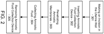
- FIG. 2 is a block diagram of an example of a method 300 of amniotic fluid collection, according to an exemplary example of the invention. It should be appreciated that method 300 may include any number of additional or alternative tasks. The tasks shown in FIG. 3 need not be performed in the illustrated order, and method 300 may be incorporated into a more comprehensive procedure or process having additional functionality not described in detail herein.
- method 300 may include making an incision in the uterine wall 301 of a pregnant mother, for example, during caesarean section. Step 301 may be performed with a standard physician's scalpel. As also shown in FIG. 2 , method 300 may include inserting an amniotic fluid collector 302 through the incision in the uterine wall made in Step 301. Method 300 also includes penetrating the amniotic membrane 303 using the amniotic fluid collector of Step 302. Step 303 may also include penetrating the chorionic membrane. In one aspect, the tip is inserted to a 10 cm depth. In some examples, the tip is inserted to a depth of about 3 cm to about 30 cm.
- the tip is inserted to a depth of about 4 cm, about 5 cm, about 6 cm, about 7 cm, about 8 cm, about 9 cm, about 10 cm, about 11 cm, about 12 cm, about 13 cm, about 14 cm, about 15 cm, about 16 cm, about 17 cm, about 18 cm, about 19 cm, about 20 cm, about 21 cm, about 22 cm, about 23 cm, about 24 cm, about 25 cm, about 26 cm, about 27 cm, about 28 cm, or about 29 cm.
- Method 300 further includes collecting the amniotic fluid 304 from the amniotic sac using the amniotic fluid collector of Step 302.
- Step 304 may include initiating a siphon to transfer the amniotic fluid to a collection chamber of the amniotic fluid collector, such as by opening an inlet valve of the amniotic fluid collector.
- Step 304 may also include positioning a collection chamber of the amniotic fluid collector below an inlet of the amniotic fluid collector.
- Step 304 may also include coupling a negative pressure source to an outlet of the amniotic fluid collector to initiate transfer of the amniotic fluid.
- Step 304 may include relocating an inlet of the amniotic fluid collector to retrieve substantially all of the available amniotic fluid.
- method 300 includes removing the amniotic fluid collector 905 from the amniotic sac.
- Step 905 may include closing an inlet valve of the amniotic fluid collector. In one example, no blood is visible in the collected material.
- Step 905 may also include emptying the collection system for further use/processing and sterilizing the exterior of the entire device. In one example, the exterior is sterilized using 70% ethanol so that the sterility may be maintained in any post-processing steps, such as in a laminar air flow bench setup, e.g., for isolation of cell material according to the present invention, and for fluid storage.
- the amniotic fluid collection procedure is performed in less than one minute. In one example, the amniotic fluid collection procedure is performed in one to two minutes. In one example, the amniotic fluid collection procedure is performed in not more than three minutes. In one example, the method is simplified compared to standard operating procedures for cesarean sections, for example, by preventing spillage of the amniotic fluid into the operating wound, improving visibility and physical access. In one example, fetal skin is unaffected by the device tip.
- Term amniotic fluid is purified by filtering term amniotic fluid to remove vernix.
- term amniotic fluid may be amniotic fluid collected at term caesarean section deliveries using, for example, a closed catheter-based system.
- 'term amniotic fluid' may be amniotic fluid collected at planned cesarean sections after 37 completed weeks of pregnancy or later, or at planned cesarean section close to term, for example after 36 completed weeks of pregnancy.
- term amniotic fluid is taken at planned caesarean sections during week 37 of pregnancy or later .
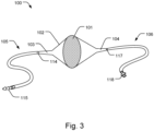
- Fig. 3 is a schematic illustration of an apparatus 100 for filtering amniotic fluid according to one example.
- the amniotic fluid contains amniotic cells originating from the fetus or the amniotic sac such as Mesenchymal stem cells.
- the amniotic fluid also contains other materials chafed off the skin such as hair and vernix.
- Material other than the amniotic cells are here referred to as particulate matter and may also comprise meconium, blood clots, etc.
- Particulate matter may be considered as anything larger than 20 ⁇ m. For the purposes of filtering, it may be particularly advantageous to treat anything larger than 30 ⁇ m or even 50 ⁇ m as particulate matter.
- the amniotic fluid thus generally contains a mixture of amniotic cells and particulate matter.
- the apparatus 100 comprises a filter 101 for filtering the particulate matter from the amniotic fluid, and a chamber 102 enclosing the filter 101.
- the chamber 102 comprises a fluid inlet 103 and a fluid outlet 104.
- the chamber 102 enclosing the filter 101 should be construed as the filter 101 being isolated by the chamber towards the environment surrounding the chamber 102 such that there is no fluid communication between the amniotic fluid in the chamber 102 with said environment.
- Fluid communication through the chamber 102 is thus controlled via the fluid inlet 103 and the fluid outlet 104 in the example of Fig. 3 .
- the filter 101 is attached to the inside of the chamber 102 between the fluid inlet 103 and the fluid outlet 104.
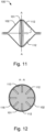
- Fig. 12 shows an example of a cross-section A-A as indicated in Fig. 12 of a circular chamber 102 and filter 101. It should however be understood that the chamber 102 and filter 101 may have varying shapes for optimization to different applications.
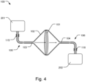
- the apparatus 100 comprises an inlet connector 105 arranged to form a sealing connection between the fluid inlet 103 and an amniotic fluid sample source 201 (shown in Fig. 4).
- Fig. 4 shows a schematic example of such source 201 of amniotic fluid.
- Having an inlet connector 105 connected to the fluid inlet 103 and configured to provide a sealing connection between the fluid inlet 103 directly to a source 201 of amniotic fluid provides for minimizing exposure to contaminants and an efficient aseptic handling of the amniotic fluid. This facilitates obtaining amniotic cells which allows post-filtration processing at an improved quality standard. Hence, an aseptic pharmaceutical production process is facilitated. The preparation of e.g. surfactant molecules may be facilitated.
- the apparatus 100 provides for improving the functioning of the amniotic stem cells, such as an improved engraftment phase following transplantation.
- Such improved processes are enabled by having the filter 101 enclosed in a chamber 102 and an inlet connector 105 arranged to form a sealing connection between the fluid inlet 103 of the chamber 102 and an amniotic fluid sample source 201.
- the risk of exposing the amniotic stem cells to contaminants, such as bacteria and viruses, is thus reduced. Exposure to oxygen is also minimized, which provides for reducing formation of oxygen free radicals which may negatively impact the functioning of the stem cells.
- Fig. 3 shows an example where the inlet connector 105 comprises a tube 105 connected to the fluid inlet 103 at a first sealing connection 114.
- the inlet connector 105 may form a sealing connection with the fluid inlet 103 with a force-fitting connection, an adhesive, a clamp, or other fixation elements.
- the inlet connector 105 is a continuous extension of the fluid inlet 103, without a separate fixation element, e.g. by being formed as a single piece by molding or other material forming techniques.
- Figs. 3 and 4 show a second connector 115 configured to form a sealing connection with a sample source 201, such as a container or bag 201 containing amniotic fluid.
- the second connector 115 may comprise releasable force-fitting connection, a clamp, or a combination thereof, or other releasable fixation elements.
- the chamber 102, filter 101, fluid inlet 103, fluid outlet 104, and inlet connector 105 may be provided as a kit in a sterile packaging, e.g. as a disposable kit.
- kit i.e. apparatus 100, thus provides for a facilitated and improved process of filtering and obtaining amniotic stem cells.
- the amniotic fluid passes the filter 101 when flowing from the fluid inlet 103 to the fluid outlet 104.
- the filter 101 may be connected around its periphery 116 to the inner wall 113 of the chamber 102. This avoids passing of amniotic fluid from the inlet 103 to the outlet 104 without being filtered.
- the filter 101 may be tensioned or otherwise supported so that a folding or curving of the filter 101 in the chamber 102 is avoided. This maintains a defined mesh or pore size across the area of the filter 101 and thus defined filtering characteristics. Maintaining a defined mesh or pore size also reduces the risk of clogging the filter 101. Long-term performance may accordingly be improved.
- the apparatus 100 may comprise an outlet 5 connector 106 to form a sealing connection between the outlet and an amniotic cell-receiving device 202, such as a centrifuge or other amniotic cell-processing equipment downstream of the apparatus 100.
- Fig. 4 shows a schematic example of such device 202. This minimizes exposure to contaminants and allows efficient aseptic handling of the amniotic fluid in post-filtering processing steps.
- Fig. 3 shows an example where the outlet connector 106 comprises a tube 106 connected to the fluid outlet 104 at a first sealing connection 117.
- the outlet connector 106 may form a sealing connection with the fluid outlet 104 with a force-fitting connection, an adhesive, a clamp, or other fixation elements.
- Fig. 1 such as schematically shown in Fig.
- the outlet connector 106 is a continuous extension of the fluid outlet 104, without a separate fixation element, e.g. by being formed as a single piece by molding or other material forming techniques.
- Figs. 3 and 4 show a second connector 118 configured to form a sealing connection with an amniotic cell-processing device downstream of the apparatus 100, such as a centrifuge 202.
- the second connector 118 may comprise a force-fitting connection, a clamp, a combination thereof, or other releasable fixation elements.
- the connection between the second connector 118 and e.g. a centrifuge 202 may thus be repeatedly connected and disconnected, and also re-sealable to maintain a sealing connection in such procedure.
- the chamber 102, filter 101, fluid inlet 103, fluid outlet 104, inlet connector 105, and outlet connector 106 may be provided as a kit in a sterile packaging, e.g. as a disposable kit.
- kit i.e. apparatus 100
- the apparatus 100 may comprise a pump 122, 123, arranged to pressurize the amniotic fluid to flow from the fluid inlet 103 to the fluid outlet 104. This provides for a more effective filtering of the amniotic fluid. Larger volumes may be filtered in less time.
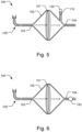
- Fig. 6 shows an example where a pump 122 is connected to the fluid outlet 104 to draw amniotic fluid through the filter 101 in the direction of the indicated arrows.
- the pump 122 may be arranged at the fluid inlet 103 to push the amniotic fluid through the filter 101.
- the pump 122 may be a compact manually operated pump integrated with the fluid inlet 103, fluid outlet 104, inlet connector 105, or outlet connector 106.
- Fig. 7 shows another example, described in more detail below, where a pump 123 is arranged to pressurize the amniotic fluid to flow from the fluid inlet 103 to the fluid outlet 104.
- the chamber 102 may comprise a conduit 119 arranged between the fluid inlet 103 and the fluid outlet 104.
- the pressure in the chamber 102 may be variable in response to fluid and/or gaseous communication through the conduit 119.
- the flow of amniotic fluid through the filter 101 may thus be optimized depending on the application, e.g. the flow rate through the filter 101 may be increased or decreased by varying the pressure in the chamber 102 via conduit 119.
- Fig. 5 shows an example in which a conduit 119 is in communication with the chamber 102.
- An access port 120 such as a connector or valve element, may be actuated to allow a fluid or gas to be expelled from the chamber 102, and/or injected into the chamber 102, to affect the pressure therein.
- the conduit 119 is arranged between the fluid outlet 103 and the filter 101 in Fig. 5 , but the conduit 119 may be arranged between the fluid inlet 103 and the filter 101 in another example.
- Fig. 5 as described below shows a further example of a conduit 119 in communication with the chamber 102.
- a pump 123 may be arranged in communication with the conduit 119, as exemplified in Fig. 7 .
- the conduit 119 is in variable communication with an upstream cavity 108 of the chamber 102 and a downstream cavity 109 of the chamber 102, i.e. the filter 101 may be arranged to divide the chamber 102 into an upstream cavity 108 and a downstream cavity 109.
- the conduit 119 is connected to both the upstream cavity 108 and the downstream cavity 109.
- the pump 123 is arranged to pressurize the amniotic fluid to flow from the upstream cavity 108 to the downstream cavity 109, or to flow from the downstream cavity 109 to the upstream cavity 108.
- valves 120, 120', 121, 121' as schematically indicated in Fig. 7 are operated to provide the desired flow directions.
- valves 120 and 121' may be open and valves 120' and 121 may be closed.
- Valves 121, 121' may be open and ⁇ valves 120, 120', may be closed in a normal filtering mode.
- the upstream cavity 108 may be pressurized by also opening valve 120' in such filtering mode.
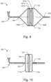
- the filter 101 may comprise a first filter element 101a and a second filter element 101b arranged between the first filter element 101a and the fluid outlet 104, as schematically shown in Fig. 8 .
- the second filter element 101b may have a mesh or pore size which is smaller than a mesh or pore size of the first filter element 101a. This allows effective filtering of particulate matter of gradually smaller dimensions. The risk of filter occlusion is thus reduced. This allows for a more reliable and robust filtering process of the amniotic fluid. An improved filtering of amniotic fluid containing a greater range in the size of particulate matter is also provided. Further, a larger fraction of the stem cells in the amniotic fluid may be obtained since the stem cells are not lost in clogged pores.
- Fig. 8 two filter elements 101a, 101b, it should be understood that any plurality of filter elements may be arranged in sequence in the chamber 102, with gradually decreasing mesh or pore size, in the direction of fluid flow from the fluid inlet 103 to the fluid outlet 104, for an effective filtering of particulate matter of gradually decreasing dimensions.
- the first and second filter elements 101a, 101b may be separated by a distance (d) along a direction amniotic fluid flow from the fluid inlet 103 to the fluid outlet 104, as schematically indicted in the example of Fig. 8 .
- the motion of the amniotic fluid between the first and second filter elements 101a, 101b which in some case may involve turbid flow, may provide for further reducing the risk of unwanted build-up of particles on the first and second filter elements 101a, 101b.
- the filter 101 may comprise a mesh having a mesh size in the range of 20- 2000 ⁇ m. In another example, the filter 101 comprises a mesh having a mesh size in the range of 100 - 500 ⁇ m. This allows particularly effective filtration of particulate matter from the amniotic fluid.
- the first filter element 101a may comprise a mesh having a mesh size in the range of 500 - 1000 ⁇ m
- the second filter element 101b may comprise a mesh having a mesh size in the range of 30 - 150 ⁇ m.
- the first filter element 101a may thus remove larger debris, followed by removal of smaller particles with the second filter element 101b. This allows a particularly effective filtering of particulate matter of varying size and reliable filtering of increased volumes over longer time periods since the risk of clogging is further minimized.
- any plurality of filter elements may be arranged in succession in the chamber 102.
- Fig. 9 shows three filter elements 101a, 101b, 101c, arranged in the chamber 102.
- the filter element having the smallest mesh or pore size, arranged furthest downstream in the chamber 102 may, such as filter element 101b in Fig. 6 and filter element 101c in Fig. 9 , may have a mesh or pore size dimensioned so that only single amniotic cells or amniotic cell clumps smaller than 10 cells pass through the filter 101.
- the smallest mesh or pore size in such an example may be approximately 30 ⁇ m.
- the filter 101 may comprise a mesh such as a nylon mesh.
- the filter 101 may comprise a porous material having a variable pore size through the filter 101 in the direction of flow of the amniotic fluid from the fluid inlet 103 to the fluid outlet 104. I.e. larger debris is removed at the surface of the filter 101 closest to the inlet 103 whereas particles of smaller size are removed deeper into the filter, as the amniotic fluid flows through the filter 101 in a direction towards the outlet 104 and the size of the pores get smaller.
- the chamber 102 may comprise an upstream cavity 108 and a downstream cavity 109.
- the upstream and downstream cavities 108, 109 may be formed as an integrated piece to form the chamber 102, e.g. in a molding process or by other material forming techniques.
- the upstream and downstream cavities 108, 109 may be formed as separate units which are then connected to each other to form a sealing connection, e.g. by an adhesive or by welding.
- the filter 101 may be attached simultaneously or subsequently with such welding process or by the aforementioned adhesive.
- the upstream and downstream cavities 108, 109 may be releasably connectable to each other at a connecting element 110, to form a sealing connection, as schematically shown in Fig. 9 .
- the filter 101 may thus be releasably connectable to the chamber 102, e.g. filter elements 101a, 101b, 101c, may be releasably connectable to the chamber 102 in Fig. 7 .
- This allows facilitated customization to different applications since filter elements 101a, 101b, 101c, of different pore or mesh size, or different number of such filter elements may be mounted in the chamber 102.
- the connecting element 110 is configured to form a sealing connection upstream and downstream cavities 108, 109, and may comprise an annular gasket extending around the periphery of the upstream and downstream cavities 108, 109.
- the filter 101 may comprise a cartridge of different numbers of filter elements 101a, 101b, 101c, with different pore sizes that could be tailored to the particular amniotic fluid sample. For example, evaluation of the amniotic fluid turbidity and degree of milkiness (level of vernix both in particle size and opaqueness) could be an indicator of the appropriate filter cartridge to use. An accompanying chart for which to compare the amniotic fluid sample with could indicate which filter cartridge to use.
- the upstream cavity 108 and/or the downstream cavity 109 may be funnel shaped. Figs.
- FIG. 3-9 show examples where both the upstream and downstream cavities 108, 109, are funnel shaped.
- FIG. 11 shows an example where only the downstream cavity 109 is funnel shaped. Having a funnel shape may be advantageous for directing the flow of amniotic fluid along a desired vector of symmetry through the filter 101 and apparatus 100.
- the upstream cavity 108 and/or the downstream cavity 109 may comprise a chamber wall 111a, 111b being arranged essentially in parallel with the filter 101, i.e. perpendicular to the direction of flow of the amniotic fluid from the fluid inlet 103 to the fluid outlet 104.
- chamber 10 shows an example where chamber walls 111a, 111b, of the upstream and downstream cavities 108, 109 are arranged essentially in parallel with the filter 101. This minimizes the space inside the chamber 102, while maintaining adequate filter area, to minimize the risk of introducing e.g. air that may disturb surfactant molecules, reduce the risk of infection, and reduce detrimental formation of reactive oxygen species in the amniotic cells.
- the chamber 102, and/or the inlet connector 105, and/or the outlet connector 106 may be formed from a phthalate free PVC material. This provides for an apparatus which is suitable to be in contact with pharmaceutical starting materials such as amniotic cells.
- the apparatus 100 may comprise protrusions 112 arranged to extend from an inner wall 113 of the chamber 102.
- Figs. 11 and 12 show examples of such protrusions 112, in a cross-sectional side view and through cross-section A-A respectively.
- the protrusions 112 provides support for the filter 101 in case the filter 101 would start bend and fold towards the inner wall 113.
- a flow through the mesh or pores of the filter 101 is still possible in such case since the filter 101 may be supported by the protrusions 112 at a distance from the inner wall 113, i.e. the protrusions 112 allows for further limiting the risk of flow restriction and provides for an efficient, robust and reliable filtering.
- Fig. 13 is a flow chart of a method 300 of filtering amniotic fluid containing particulate matter and amniotic cells.
- the method 300 comprises forming 301 a sealing connection between a fluid inlet 103 of a chamber 102 and an amniotic fluid sample source 201.
- the method 300 comprises passing 302 the amniotic fluid through a filter 101 enclosed in the chamber 102 by providing a flow of the amniotic fluid from the fluid inlet 103 to a fluid outlet 104 of the chamber 102. Particulate matter is thereby deposited on the filter 101 and the amniotic fluid containing amniotic cells flows through the outlet 104.
- the method 300 thus provides for the advantageous benefits as described in relation to apparatus 100 and Figs. 3-12 above.
- the method 300 provides for effective and sterile filtration of the amniotic fluid to obtain amniotic cell samples of high quality.
- removing particulate material from the TAF to obtain purified TAF cells may be done by applying any known method in the art such as filtration, centrifugation, etc.
- the TAF may be filtered through a filter having a pore size at or above 20 ⁇ m.
- the filter may be made from any synthetic material including but not limited to cellulose acetate, cellulose nitrate (collodion), polyamide (nylon), polycarbonate, polypropylene and polytetrafluoroethylene (Teflon).
- removing particulate material is done by applying apparatus 100.
- the terms "express, expression, and/or expressing" in the context of a cell surface marker are meant to indicate the presence of a particular marker on the surface of a cell, said surface marker having been produced by the cell.
- Surface marker expression may be used to select between different cell populations, for example, positively selecting for surface marker expression indicates the selection of a cell population that more strongly expresses a particular surface marker as compared to another cell population. Conversely, negatively selecting for cell surface marker expression indicates the selection of a cell population that more weakly expresses a particular surface marker as compared to another cell population.
- TAF contains various progenitor cell types.
- particular progenitor cell types may be isolated and propagated via adherence selection.
- a vitronectin substrate Merk, CORNING ® , Synthemax ® , II-SC SUBSTRATE, CLS3535-1EA
- Synthemax is an animal-component free, synthetic, flexible vitronectin-based peptide substrate for serum or serum-free expansion of human progenitor/stem cells and other adult stem cell types.
- vitronectin-based peptide substrate may include a portion of a vitronectin protein, such as a particular peptide sequence of vitronectin. Alternatively, intact vitronectin protein may be used. Synthemax vitronectin substrate offers a synthetic, xeno-free alternative to biological coatings and/or feeder cell layers commonly used in cell culture and known in the art.
- standard tissue-culture treated flasks may be coated with about 0.2 mL Synthemax/cm 2 at 10 ⁇ g/mL giving a surface density of 2 ⁇ g/cm 2 , and incubated at 37°C for about 1h, .5h 2h, 4h, 8h, or more than 8h or at room temperature for about 2h, 1h, 4h, 8h or more than 8h with surplus solution optionally being removed and replaced.
- Synthemax may be coated at a surface density of about: 1 to 5 ⁇ g/cm 2 , such as 2 ⁇ g/cm 2 , .1 to 10 ⁇ g/cm 2 , .5 to 4 ⁇ g/cm 2 , 1 to 3 ⁇ g/cm 2 , or about 1.5 to 2.5 ⁇ g/cm 2 .
- adherence selection can be performed using a surface coated with, for example, Collagen, Fibronectin.
- adherence selection can be performed using an uncoated surface comprising a tissue-culture treated plastic.
- Cells purified from TAF fluid may be gently re-suspended in prewarmed xeno-free cell culture media, with the cell suspension is then added to the Synthemax-coated flasks.
- Media may be changed at various times after addition to the flasks, for example, after about: 2h to 168h, 12h to 96h, 24h to 72h, 36h to 60h, 42h to 56h, or 48h, and then subsequently changed about: every day, every other day, every third day, every fifth day, once a week, once every two weeks or about less than once every two weeks.
- the non-attached cells may be removed, thereby selecting the MSCs by their affinity for attachment to the Synthemax-treated surface.
- the cells may be cultured for a period of time, such as about, for example, 4d, 7d, 10d, 11d, 12d, 13d,14d, 18d, 21d, 28d or longer than 21d.
- the cells may be cultured under hypoxic conditions, hypoxia priming may alter cell metabolism during expansion, increase resistance to oxidative stress, and thereby improve the engraftment, survival in ischemic microenvironments, and angiogenic potential of transplanted MSCs.
- hypoxia priming may alter cell metabolism during expansion, increase resistance to oxidative stress, and thereby improve the engraftment, survival in ischemic microenvironments, and angiogenic potential of transplanted MSCs.
- the P0 colonies Coldy forming Units - CFUs
- the remaining cells may be predominantly non-tissue specific MSCs.
- the pooled P0 cells may be gently re-suspended in pre-warmed xeno-free cell culture media and re-plated on tissue-culture treated flasks without Synthemax for passaging.
- the pooled cells may be seeded at a seeding density of from between about: 100 to 10000 cells / cm 2 , 500 to 8000 cells / cm 2 , 1000 to 5000 cells / cm 2 , or about 2000 to 4000 cells / cm 2 .
- the media may be changed about every 1d, 2d, 4d, or more than four days. After a period of time, such as about 2d, 4d, 7d, or more than 7d, the cells may be dissociated and harvested. Further selective MSC isolation may be achieved as described below.
- TAF-MSCs When comparing the genetic expression profiles of TAF-MSCs and adult-type MSCs derived from adipose tissue or bone marrow by RNAseq, TAF-MSCs tend to express more of some genes present in adult-type MSCs and less of others. Identification of both positive and negative TAF-MSC specific neonatal cell-surface markers can allow for sorting of the MSCs with neonatal quality from those that have differentiated further and are of less importance as progenitor cells using e.g. ligands such as antibodies and aptamers or other selection techniques.
- ligands such as antibodies and aptamers or other selection techniques.
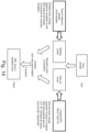
- tissue-specificity score algorithm An example of an MSC tissue-specificity score algorithm is shown in Figure 14 .
- Tissue-specificity may be measured as a combination of two components: a 'tissue transcriptional similarity' also known as a similarity score and a "tissue-specific gene expression program" also known as a gene set score.
- the similarity score may be an Average Spearman correlation to each MSC tissue reference sample (for example a fetal lung MSC sample).
- the gene set score may be the average expression of genes in a tissue-specific gene set.
- the resulting output is an MSC tissue specificity score.
- the MSC tissue-specificity score measures the relative tissue-specificity among the input samples by measuring how many standard deviations a sample is more or less specific to a given tissue compared to the average input sample.
- an MSC tissue-specificity score may indicate how much more a clone sample appears to have a tissue specific phenotype, such as a lung phenotype, as compared to an average clone.
- tissue specific phenotype such as a lung phenotype
- tissue-prioritized clones can be defined as any clone belonging to the top X% percentile score, where X is any percentage within a range having a lower end from about 0.1 to 25, such as about 1, 5, 10, 15 and 20, and an upper end from about 30 to 75, such as about: 35, 40, 45, 50, 55, 60, 65 or 70.
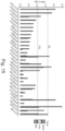
- An example of TAF-MSC tissue-specificity prioritization results is shown in Figure 15 , in which thresholds at 15% and 5% are visible. Having prioritized tissue-specific clones, candidate surface marker genes may then be identified. For each tissue, two groups may be defined: tissue-prioritized and tissue-distal.
- the tissue-prioritized group may include clones with a score in the top 15% percentile.
- the tissue-distal group may include clones in the bottom Y% percentile in which Y is any percentage within the range having a lower end from about 25 to 70, such as about: 30, 35, 40, 45, 50, 55, 60 or 65 and an upper end from 75 to 99.9, such as about: 80, 85, 90, 95 or 99.
- Figure 16 shows an example of such analysis on kidney tissue.
- differentially expressed genes between the tissue-prioritized and tissue-distal groups may be identified.
- the differential expression results may be annotated with surface marker gene information.
- tissue-specific cell surface markers surface marker genes with a more than a Z-fold increase, where Z is at least about: 1.5-fold, 2-fold, 2.5-fold, 3-fold, 3.5-fold, 4-fold, 5-fold, 8-fold, 10-fold, 12-fold, 15-fold or even more-fold increase in expression (log2FoldChange) in prioritized clones compared to an average clone and a Transcripts Per Kilobase Million (TPM) of more than about 500, such as more than about: 1000, 1500, 2000, 2500, 3000, 5000 or even higher may be selected to give the top tissue-specific marker candidates, such as approximately the top: 5, 10, 20, 30, 40, 50, 60, 70, 100 or more, for example such as those shown below in Tables 3-6 and further described in more detail below.
- Suitable log2FoldChange and TPM values may vary even further depending on tissue type specificities depending on the abundance/absence of good markers.
- FACS Fluorescence activated cell sorting
- FACS Fluorescence activated cell sorting
- TAF-MSCs Fluorescence activated cell sorting
- FACS Fluorescence activated cell sorting
- FACS allows for a very high purity of the desired cell population, even when the target cell type expresses very low levels of identifying markers and/or separation is needed based on differences in marker density.
- FACS allows the purification of individual cells based on size, granularity and fluorescence.
- FACS may be used to select for certain cell populations that express one cell surface marker more than another cell population and vice-versa.
- FACS bulk methods of purification such as panning, complement depletion and magnetic bead separation
- FACS may be used in combination with FACS or as an alternative to FACS.
- mAbs fluorescently-tagged monoclonal antibodies
- Negative selection of unstained cells may also allow for separation.
- FACS may be run using a closed system sorting technology such as MACSQuant ® Tyto ® . Samples may be kept contamination-free within the disposable, fully closed MACSQuant Tyto Cartridge. Further, filtered air may drive cells through a microchannel into the microchip at very low pressure ( ⁇ 3 PSI).
- potential cell aggregates may be held back by a filter system guaranteeing a smooth sorting process.
- the fluorescence detection system may detect cells of interest based on predetermined fluorescent parameters of the cells. Based on their fluorescent and scatter light signatures, target cells may be redirected by a sort valve located within the microchannel.
- Sorting parameters may be adjusted depending on the requirement of purity and yield. Unlike on conventional droplet sorters, cells sorted by the MACSQuant Tyto may not experience high pressure or charge, and may not get decompressed. Therefore, such a gentle sorting approach may result in high viability and functionality of cells.
- other marker-based selection techniques may be known to the skilled person and employed here. These include, but are not limited to, Magnetic-activated cell sorting, Microfluidic based sorting, Buoyancy activated cell sorting, mass cytometry etc.
- RNAseq data from TAF-MSC clones adult and neonatal MSC reference material as well as fetal fibroblasts and publicly available expression datasets may be used to identify and characterize TAF-MSC cells.
- sub-populations of TAF-MSCs may be established by clustering their expression data (RNAseq) with neonatal reference samples.
- RNAseq expression data
- Such sub-populations include, but are not limited to, lung MSC, urinary tract MSC (described also as kidney MSCs in the present disclosure), and skin MSC.
- Gene lists of highly and lowly expressed genes for each cluster of expression data may allow for identification of surface maker genes for each cluster.
- tissue specific MSC may be done by firstly characterization, thereafter a stepwise negative selection/sorting of the material by taking into account the combined (multivariate) surface marker profile of the different tissue specific MSC's.
- any such combination of these surface markers may be used for identifying and isolation of lung TAF cells from the general population of TAF-derived cells and/or TAF-MSC cells.
- the below non-exclusive list of surface markers may be more highly expressed on the surface of Lung-TAF cells as compared to other cell types, such as other TAF-derived cells and/or TAF-MSC cells.
- the surface markers identified in Table 1 may have at least a 10-fold increase in expression on prioritized clones compared to the average TAF-MSC clone (optionally with TPM threshold > 2000).
- the below surface markers may be more weakly expressed on lung TAF MSCs as compared to other cell types, such as other TAF-derived cells and/or TAF-MSCs: CD24, ITGB4, TNFSF10, GFRA1, CD74, FGFR4, HAVCR1, and OSCAR.
- other cell types such as other TAF-derived cells and/or TAF-MSCs: CD24, ITGB4, TNFSF10, GFRA1, CD74, FGFR4, HAVCR1, and OSCAR.
- one, two, three, four, or more of the aforementioned more weakly expressed surface markers may be used to separate lung TAF cells from other cell types such as other TAF-derived cells and/or TAF-MSCs.
- At least the cell surface marker CD248 is used to sort lung TAF MSCs from a population of TAF MSCs.
- Further surface markers that may be used to sort lung TAF MSCs include DDR-1 (discoidin domain receptor tyrosine kinase 1) as well as LRRC38 (Leucine Rich Repeat Containing Protein 38), all three of which have been identified via antibodies as useful markers for separation.
- DDR-1 discoidin domain receptor tyrosine kinase 1
- LRRC38 Leucine Rich Repeat Containing Protein 38
- suitable combinations of the markers listed in Table 1 and CD248, DDR-1, and LRR38 may be used to separate lung TAF MSCs from TAF MSCs by selecting for specific markers from Table 1 or combinations of two, three, four, five, six or more markers from Table 1 and CD248, and/or DDR-1 and/or LRR38.
- lung TAF MSCs can be more specifically identified by identifying a combination of stronger expression, such as 10-fold or more stronger expression (optionally with TPM threshold > 2000) of any combination of the foregoing markers with CD248, e.g., PCDH19 and/or DDR1 and/or MME and/or IFITM10 and/or BGN and/or NOTCH3 and/or DDR-1 and/or LRR38 as compared to TAF MSCs.
- identification may be achieved with a lower threshold of stronger expression, such as 4-fold or more, 6-fold or more, or 8-fold or more expression of each of the markers.
- the below surface markers may be more weakly expressed on lung TAF-MSCs as compared to other cell types (negative markers), such as 1/8-fold or less expression (optionally with TPM>500) of any combination of the foregoing markers versus TAF MSCs: CD24, ITGB4, TNFSF10, GFRA1, CD74, FGFR4, HAVCR1, and OSCAR.
- identification may be achieved with a lower threshold of weaker expression, such as 1/2-fold or less, 1/4-fold or less, or 1/6-fold or less expression of each of the markers.
- Combinations of two or more these negative markers can also be used to more specifically isolate lung TAF MSCs.
- combinations including both negative and positive markers, such as at any of the thresholds described above can also be effective to more specifically isolate lung TAF MSCs.
- Figures 17A-17D show an example of the results from a proof-of-principle study on the potential use of Lung TAF MSCs for treatment, performed using neonatally sorted TAF MSCs expressing MSC lung cell surface markers including CD248, DDR1, and LRRC38 (called "LBX-THX-001 cells").
- the purpose of the study was to investigate the effects of LBX-THX-001 cells in a bleomycin induced lung fibrosis model in male rats. Two cell concentrations (2 M cell/kg and 5 M cells/kg) and two types of vehicles for the cells were tested (PBS and CryoStor CS-10).
- LBX-THX-001 cells injected were chosen to be relevant for a possible human therapy. The number of cells were therefore chosen to reflect cell numbers used in previous studies on rats (8-20 M cells/kg) and humans (0.5-2 M cells/kg).
- bleomycin 1000 U/rat
- 34 male SD-rats were used to induce lung fibrosis in the rats.
- the rats were monitored and weighed daily and thereafter twice/week until termination of the study.
- the LBX-THX-001 cells were administered by an intra-venous (i.v.) injection.
- the injection volume was 194-535 ⁇ L (maximal tolerated injection volume 1 mL/kg).
- the response to the intra-tracheal instillation of bleomycin was as expected based on previous experience for the model with weight loss during the first days after instillation and thereafter recovery. There were no significant differences in weight loss between the bleomycin group and the treatment groups.
- bleomycin instillation induced fibrotic change in the lung was assessed for pathological changes in the bleomycin group both with regard to percent of parenchyma affected and after scoring using the modified Ashcroft scale.
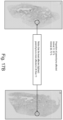
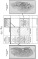
- the group treated with LBX-THX-001 cells (2 million cells/kg) 4 days post Bleomycin showed significantly less fibrosis in their lungs compared to the bleomycin group. This was seen both in the histopathological evaluation using the readout "percent parenchyma affected" ( Figures 17A-B ) and the fibrosis scoring Ashcroft modified scale ( Figures 17A-D ). No human MSCs could be detected in rat lungs at termination (after 28 days).
- the term “or” is used in its inclusive sense (and not in its exclusive sense) so that when used, for example, to connect a list of elements, the term “or” means one, some, or all of the elements in the list.
- the term “and/or” in reference to a list of two or more items covers all of the following interpretations of the word: any one of the items in the list, all of the items in the list, and any combination of the items in the list.
- the term “each,” as used herein, in addition to having its ordinary meaning, can mean any subset of a set of elements to which the term “each” is applied.
- the words “herein,” “above,” “below,” and words of similar import when used in this application, refer to this application as a whole and not to any particular portions of this application.
- the terms “generally parallel” and “substantially parallel” refer to a value, amount, or characteristic that departs from exactly parallel by less than or equal to 15 degrees, 10 degrees, 5 degrees, 3 degrees, 1 degree, or 0.1 degree.
Landscapes
- Health & Medical Sciences (AREA)
- Life Sciences & Earth Sciences (AREA)
- Engineering & Computer Science (AREA)
- Developmental Biology & Embryology (AREA)
- Biomedical Technology (AREA)
- Chemical & Material Sciences (AREA)
- Cell Biology (AREA)
- Biotechnology (AREA)
- Zoology (AREA)
- General Health & Medical Sciences (AREA)
- Bioinformatics & Cheminformatics (AREA)
- Organic Chemistry (AREA)
- Immunology (AREA)
- Genetics & Genomics (AREA)
- Wood Science & Technology (AREA)
- Public Health (AREA)
- Medicinal Chemistry (AREA)
- Veterinary Medicine (AREA)
- Animal Behavior & Ethology (AREA)
- Pharmacology & Pharmacy (AREA)
- Epidemiology (AREA)
- Virology (AREA)
- Reproductive Health (AREA)
- Microbiology (AREA)
- General Engineering & Computer Science (AREA)
- Biochemistry (AREA)
- Pregnancy & Childbirth (AREA)
- Rheumatology (AREA)
- Hematology (AREA)
- Pulmonology (AREA)
- Chemical Kinetics & Catalysis (AREA)
- General Chemical & Material Sciences (AREA)
- Nuclear Medicine, Radiotherapy & Molecular Imaging (AREA)
- Gynecology & Obstetrics (AREA)
- Medicines Containing Material From Animals Or Micro-Organisms (AREA)
- Micro-Organisms Or Cultivation Processes Thereof (AREA)
Claims (12)
- Verfahren zur Gewinnung von Fruchtwasserzellen (TAF-Zellen) aus Fruchtwasser, umfassend:aus bereitgestelltem Fruchtwasser (TAF);Entfernen von partikulärem Material aus dem TAF, um gereinigte TAF-Zellen zu erhalten;Durchführen eines Adhäsionsauswählens an den gereinigten TAF-Zellen, um TAF-Adhäsionszellen zu erhalten;Passagieren der TAF-Adhäsionszellen, um eine Zellpopulation zu erhalten, die die TAF-Zellen umfasst; undAuswählen der TAF-Zellen aus der Population als Zellen, die mindestens einen Oberflächenmarker exprimieren, der CD248 ist.
- Verfahren nach Anspruch 1, wobei das Auswählen von TAF-Zellen ferner das Auswählen der TAF-Zellen aus der Population als Zellen umfasst, die mindestens einen Marker exprimieren, ausgewählt aus der Gruppe bestehend aus PCDH19, DDR1, MME, IFITM10, BGN, NOTCH3, SULF1, TNFSF18, BDKRB1, FLT1, PDGFRA, TNFSF4, UNC5B, FAP, CASP1, DDR2, PCDH18, LRRC38 und CRLF1.
- Verfahren nach Anspruch 1, wobei der Auswahlschritt das Auswählen von TAF-Zellen umfasst, die weiterhin einen Oberflächenmarker exprimieren, ausgewählt aus der Gruppe von DDR1 und LRRC38.
- Verfahren nach Anspruch 1, wobei der Auswahlschritt das Auswählen von TAF-Zellen umfasst, die ferner DDR1 und LRRC38 exprimieren.
- Verfahren nach einem der vorhergehenden Ansprüche, wobei das Auswählen der TAF-Zellen das Ausschließen von Zellen umfasst, die einen Marker exprimieren, der aus der Gruppe ausgewählt ist, die aus CD24, ITGB4, TNFSF10, GFRA1, CD74, FGFR4, HAVCR1 und OSCAR besteht.
- Verfahren nach einem der vorhergehenden Ansprüche, wobei das Entfernen von partikulärem Material das Filtern und Zentrifugieren des TAF umfasst.
- Verfahren nach einem der vorhergehenden Ansprüche, wobei das Durchführen eines Adhäsionsauswählens an den gereinigten TAF-Zellen das Anhaften der gereinigten TAF-Zellen an eine mit einem Substrat auf Vitronectin-Basis beschichtete Oberfläche umfasst.
- Verfahren nach einem der vorhergehenden Ansprüche, wobei der Auswahlschritt unter Verwendung von fluoreszenzaktivierter Zellsortierung (FACS) durchgeführt wird.
- Verfahren nach einem der vorhergehenden Ansprüche, wobei der Auswahlschritt mit Antikörpern durchgeführt wird, die gegen einen der Marker oder Oberflächenmarker gerichtet sind.
- Verfahren nach einem der vorhergehenden Ansprüche, wobei der Auswahlschritt das Auswählen von TAF-Zellen umfasst, die mindestens zwei Marker, mindestens drei Marker oder mindestens vier Marker aus den Oberflächenmarkern nach Anspruch 2 exprimieren.
- Verfahren nach einem der vorhergehenden Ansprüche, wobei der Auswahlschritt mehrere Sortierschritte umfasst, wobei jeder Sortierschritt das Leiten von TAF-Zellen in eine erste Ausgangsgruppe oder eine zweite Ausgangsgruppe in Abhängigkeit von einem Satz von Markern umfasst, die von den jeweiligen TAF-Zellen exprimiert oder nicht exprimiert werden.
- Verfahren nach Anspruch 11, wobei der Auswahlschritt Folgendes umfasst:einen ersten Sortierschritt, um TAF-Zellen, die mindestens einen Oberflächenmarker exprimieren, der CD248 ist, in eine erste Ausgangsgruppe zu leiten, undeinen zweiten Sortierschritt, um TAF-Zellen aus der ersten Ausgangsgruppe, die einen zweiten Satz von Markern exprimieren, in eine zweite Ausgangsgruppe zu leiten.
Priority Applications (1)
| Application Number | Priority Date | Filing Date | Title |
|---|---|---|---|
| EP23173359.3A EP4234019A3 (de) | 2019-10-18 | 2020-10-16 | Verfahren und vorrichtungen zur gewinnung von mesenchymalen stammzellen aus fruchtwasser und davon abgeleitete zellen |
Applications Claiming Priority (5)
| Application Number | Priority Date | Filing Date | Title |
|---|---|---|---|
| US201962923187P | 2019-10-18 | 2019-10-18 | |
| SE1930338 | 2019-10-18 | ||
| SE2030101 | 2020-03-26 | ||
| SE2030100 | 2020-03-26 | ||
| PCT/SE2020/050993 WO2021076042A2 (en) | 2019-10-18 | 2020-10-16 | Processes and apparatuses for obtaining amniotic mesenchymal stem cells from amniotic fluid and cells derived thereof |
Related Child Applications (1)
| Application Number | Title | Priority Date | Filing Date |
|---|---|---|---|
| EP23173359.3A Division EP4234019A3 (de) | 2019-10-18 | 2020-10-16 | Verfahren und vorrichtungen zur gewinnung von mesenchymalen stammzellen aus fruchtwasser und davon abgeleitete zellen |
Publications (3)
| Publication Number | Publication Date |
|---|---|
| EP3886880A2 EP3886880A2 (de) | 2021-10-06 |
| EP3886880B1 true EP3886880B1 (de) | 2023-06-07 |
| EP3886880C0 EP3886880C0 (de) | 2023-06-07 |
Family
ID=73040211
Family Applications (3)
| Application Number | Title | Priority Date | Filing Date |
|---|---|---|---|
| EP20800356.6A Active EP3886880B1 (de) | 2019-10-18 | 2020-10-16 | Verfahren und vorrichtungen zur gewinnung von mesenchymalen stammzellen aus fruchtwasser und davon abgeleitete zellen |
| EP23173359.3A Pending EP4234019A3 (de) | 2019-10-18 | 2020-10-16 | Verfahren und vorrichtungen zur gewinnung von mesenchymalen stammzellen aus fruchtwasser und davon abgeleitete zellen |
| EP20803994.1A Pending EP4045068A1 (de) | 2019-10-18 | 2020-10-16 | Verwendung von fruchtwasserzellen einer ausgetragenen schwangerschaft zur behandlung von akuten und chronischen atemwegserkrankungen |
Family Applications After (2)
| Application Number | Title | Priority Date | Filing Date |
|---|---|---|---|
| EP23173359.3A Pending EP4234019A3 (de) | 2019-10-18 | 2020-10-16 | Verfahren und vorrichtungen zur gewinnung von mesenchymalen stammzellen aus fruchtwasser und davon abgeleitete zellen |
| EP20803994.1A Pending EP4045068A1 (de) | 2019-10-18 | 2020-10-16 | Verwendung von fruchtwasserzellen einer ausgetragenen schwangerschaft zur behandlung von akuten und chronischen atemwegserkrankungen |
Country Status (9)
| Country | Link |
|---|---|
| US (3) | US20220372444A1 (de) |
| EP (3) | EP3886880B1 (de) |
| JP (1) | JP7714535B2 (de) |
| KR (1) | KR20220104682A (de) |
| CN (1) | CN114765957B (de) |
| AU (1) | AU2020368845A1 (de) |
| CA (1) | CA3154422A1 (de) |
| ES (1) | ES2951396T3 (de) |
| WO (2) | WO2021076042A2 (de) |
Families Citing this family (7)
| Publication number | Priority date | Publication date | Assignee | Title |
|---|---|---|---|---|
| US11542328B2 (en) | 2008-11-14 | 2023-01-03 | The Brigham And Women's Hospital, Inc. | Therapeutic and diagnostic methods relating to cancer stem cells |
| EP3784249A4 (de) | 2018-04-25 | 2022-03-02 | Children's Medical Center Corporation | Abcb5-liganden und -substrate |
| WO2020256628A1 (en) | 2019-06-20 | 2020-12-24 | Amniotics Ab | An apparatus for filtering amniotic fluid |
| EP4125960A4 (de) * | 2020-03-30 | 2024-04-03 | Children's Medical Center Corporation | Verwendung von stammzellen zur behandlung von übermässiger entzündung |
| GB202103889D0 (en) * | 2021-03-19 | 2021-05-05 | Amniotics Ab | Stem cells for use in ecmo technology |
| EP4514830A1 (de) | 2022-04-29 | 2025-03-05 | Magle Chemoswed Holding AB | Verfahren zur herstellung von extrazellulären vesikeln |
| SE2515293A1 (en) | 2023-04-21 | 2025-11-06 | Magle Chemoswed Holding Ab | Mesenchymal stem cells and uses thereof |
Family Cites Families (88)
| Publication number | Priority date | Publication date | Assignee | Title |
|---|---|---|---|---|
| US4308875A (en) | 1981-03-11 | 1982-01-05 | Universal Medical Instrument Corporation | Amniocentesis needle |
| US4567882A (en) | 1982-12-06 | 1986-02-04 | Vanderbilt University | Method for locating the illuminated tip of an endotracheal tube |
| US4787894A (en) | 1987-10-26 | 1988-11-29 | Turnbull Christopher J | Meconium aspiration device |
| US5048530A (en) | 1988-08-17 | 1991-09-17 | Robert Hurwitz | Method of using an amniocentesis needle with improved sonographic visibility |
| US5494044A (en) | 1989-05-10 | 1996-02-27 | Amnitec A/S | Method for taking a sample of amniotic fluid |
| US5019039A (en) | 1989-11-15 | 1991-05-28 | Anderson Ronald W | Fluid drainage needle method of use |
| US5100387A (en) | 1990-07-02 | 1992-03-31 | Ng Raymond C | Disposable universal needle guide apparatus (for amniocentesis) |
| US5395379A (en) | 1993-07-22 | 1995-03-07 | Deutchman; Mark E. | Extractor for childbirth and aspirator/injector device |
| US5951497A (en) | 1996-09-03 | 1999-09-14 | Clinical Innovation Associates, Inc. | Pressure catheter device with enhanced positioning features |
| CN1182882C (zh) | 1998-12-24 | 2005-01-05 | 生物安全股份有限公司 | 特别用于浓缩造血干细胞的血液分离系统 |
| AU776137B2 (en) | 1999-01-06 | 2004-08-26 | University Of Southern California | Method and composition for angiogenesis inhibition |
| US6461628B1 (en) | 1999-09-13 | 2002-10-08 | Keraplast Technologies, Ltd. | Non-woven keratin cell scaffold |
| US6479064B1 (en) | 1999-12-29 | 2002-11-12 | Children's Medical Center Corporation | Culturing different cell populations on a decellularized natural biostructure for organ reconstruction |
| US7802574B2 (en) | 2000-01-18 | 2010-09-28 | Schultz Joseph P | Medical component system |
| US6378523B1 (en) | 2000-03-15 | 2002-04-30 | Evergreen Medical Incorporated | Endotracheal tube having a beveled tip and orientation indicator |
| US6458589B1 (en) | 2000-04-27 | 2002-10-01 | Geron Corporation | Hepatocyte lineage cells derived from pluripotent stem cells |
| CA2426819A1 (en) | 2000-10-30 | 2002-05-10 | Children's Medical Center Corporation | Tissue-engineered vascular structures |
| EP2206772A3 (de) | 2000-12-06 | 2010-08-04 | Robert J. Hariri | Verfahren zum Sammeln von Plazentastammzellen |
| US7311905B2 (en) | 2002-02-13 | 2007-12-25 | Anthrogenesis Corporation | Embryonic-like stem cells derived from post-partum mammalian placenta, and uses and methods of treatment using said cells |
| CA2437957C (en) | 2001-02-14 | 2014-04-22 | Robert J. Hariri | Renovation and repopulation of decellularized tissues and cadaveric organs by stem cells |
| EP2336299A1 (de) | 2001-02-14 | 2011-06-22 | Anthrogenesis Corporation | Post-partum Säugetier-Plazenta, deren Verwendung und daraus gewonnene Stammzellen |
| EP1406698B1 (de) | 2001-03-02 | 2008-10-29 | The Brigham and Women's Hospital | Lipoxin analoge als neue inhibitoren der angiogenese |
| CN1195055C (zh) | 2001-09-06 | 2005-03-30 | 周胜利 | 从胎盘组织中提取造血干细胞用于建立造血干细胞库的新方法 |
| WO2003042405A2 (en) | 2001-11-15 | 2003-05-22 | Children's Medical Center Corporation | Methods of isolation, expansion and differentiation of fetal stem cells from chorionic villus, amniotic fluid, and placenta and therapeutic uses thereof |
| AU2003216286B2 (en) | 2002-02-13 | 2008-08-28 | Celularity Inc. | Embryonic-like stem cells derived from post-partum mammalian placenta and uses and methods of treatment using said cells |
| WO2004072273A1 (en) | 2003-02-11 | 2004-08-26 | Davies John E | Progenitor cells from wharton's jelly of human umbilical cord |
| ZA200404101B (en) | 2003-05-26 | 2005-03-07 | Reliance Life Sciences Pvt Ltd | In vitro culture of Mesenchymal Stem Cells (MSC) and a process for the preparation thereof for therapeutic use. |
| US7569385B2 (en) | 2003-08-14 | 2009-08-04 | The Regents Of The University Of California | Multipotent amniotic fetal stem cells |
| CA2554094A1 (en) | 2004-02-09 | 2005-08-25 | Indiana University Research And Technology Corporation | Isolation, expansion and use of clonogenic endothelial progenitor cells |
| JP2005323534A (ja) | 2004-05-13 | 2005-11-24 | Onecell Inc | 細胞分離回収膜及び細胞分離回収方法 |
| WO2006012404A2 (en) | 2004-07-20 | 2006-02-02 | Case Western Reserve University | Novel cell populations and uses thereof |
| DE202004012970U1 (de) | 2004-08-18 | 2005-12-22 | Möller Medical GmbH & Co. KG | Pränatale Biopsievorrichtung für die Entnahme von Flüssigkeits- und/oder Gewebeproben |
| GB2422515B (en) | 2005-01-21 | 2009-05-27 | King S College London | A method of discovering multi-mode mobile terminals |
| AU2006202209B2 (en) * | 2005-05-27 | 2011-04-14 | Lifescan, Inc. | Amniotic fluid derived cells |
| AU2006304274B2 (en) | 2005-10-13 | 2012-08-16 | Celularity Inc. | Production of oligodendrocytes from placenta-derived stem cells |
| CA2624925C (en) | 2005-10-13 | 2014-09-30 | Anthrogenesis Corporation | Immunomodulation using placental stem cells |
| AU2014201181B2 (en) | 2005-12-29 | 2017-07-20 | Celularity Inc. | Placental stem cell populations |
| EP1845154A1 (de) | 2006-04-12 | 2007-10-17 | RNL Bio Co., Ltd. | Multipotente Stammzellen aus Plazentagewebe und diese enthaltende zelluläre therapeutische Wirkstoffe |
| CA2626809A1 (en) | 2006-06-26 | 2008-01-03 | Gambro Bct, Inc. | Method of culturing mesenchymal stem cells |
| WO2008060139A1 (en) | 2006-11-17 | 2008-05-22 | Erasmus University Medical Center Rotterdam | Methods for controlling mineralization of extracellular matrix, therapeutic methods based thereon and medicaments for use therein |
| JP2008307007A (ja) | 2007-06-15 | 2008-12-25 | Bayer Schering Pharma Ag | 出生後のヒト組織由来未分化幹細胞から誘導したヒト多能性幹細胞 |
| WO2009031818A2 (en) * | 2007-09-05 | 2009-03-12 | Genexel-Sein. Inc. | Biomarker for purification or identification of mesenchymal stem cells and methods of purification of mesenchymal stem cells by using it |
| AU2008298816B2 (en) | 2007-09-11 | 2013-09-26 | Sapporo Medical University | Cell growth method and pharmaceutical preparation for tissue repair and regeneration |
| WO2009052132A1 (en) * | 2007-10-15 | 2009-04-23 | Children's Medical Center Corporation | Human amniotic fluid derived mesenchymal stem cells |
| US8513012B2 (en) | 2008-05-02 | 2013-08-20 | Cellular Dynamics International, Inc. | Method for production of mast cells from stem cells |
| US20110256110A1 (en) * | 2008-10-24 | 2011-10-20 | Children's Hospital Los Angeles | Amniotic fluid cells and uses thereof |
| CN102282252B (zh) | 2008-11-19 | 2017-07-04 | 人类起源公司 | 羊膜来源的贴壁细胞 |
| CN101418284A (zh) * | 2008-11-28 | 2009-04-29 | 中国人民解放军军事医学科学院野战输血研究所 | 分离纯化人羊水干细胞的方法 |
| US8309343B2 (en) | 2008-12-01 | 2012-11-13 | Baxter International Inc. | Apparatus and method for processing biological material |
| CN102388130B (zh) | 2009-02-27 | 2014-08-27 | 细胞动力国际有限公司 | 多能细胞的分化 |
| KR20120003470A (ko) | 2009-03-31 | 2012-01-10 | 더 보드 오브 리전츠 오브 더 유니버시티 오브 텍사스 시스템 | 사람 제대혈 유래의 중간엽 줄기 세포의 분리 |
| JP5572996B2 (ja) | 2009-05-15 | 2014-08-20 | 宇部興産株式会社 | ベンズアゼピノン化合物を有効成分として含有する医薬 |
| WO2010141654A1 (en) | 2009-06-05 | 2010-12-09 | Anthrogenesis Corporation | Improved method of collecting placental cells |
| US9089573B2 (en) | 2009-07-22 | 2015-07-28 | University Of Massachusetts | Methods and compositions to reduce oxidative stress |
| US20140038291A1 (en) | 2009-10-31 | 2014-02-06 | New World Laboratories Inc. | Methods for reprogramming cells and uses thereof |
| EP2550357B1 (de) | 2010-05-12 | 2016-09-14 | Xpand Biotechnology BV | Zellkulturbeutel |
| EP2603227B1 (de) | 2010-08-12 | 2017-10-04 | Fate Therapeutics, Inc. | Verbesserte hämatopoetische stamm- und vorläuferzelltherapie |
| US9855300B2 (en) | 2010-11-26 | 2018-01-02 | Crioestaminal, Saúde E Tecnologia, Sa. | Composition and method to improve the therapeutic effect of stem cells |
| WO2013082487A1 (en) | 2011-11-30 | 2013-06-06 | Children's Hospital Los Angeles | Afsc based therapies |
| US9428732B2 (en) | 2011-12-05 | 2016-08-30 | Primorigen Biosciences, Inc. | Compositions and methods for differentiating pluripotent stem cells into primitive blood cells and uses thereof |
| EP4721752A2 (de) | 2011-12-30 | 2026-04-08 | Cellscript, Llc | Making and using in vitro-synthesized ssrna for introducing into mammalian cells to induce a biological or biochemical effect |
| CN202723948U (zh) | 2012-04-19 | 2013-02-13 | 赵艳 | 一种一次性无菌人工破膜采集器 |
| CN202569006U (zh) | 2012-05-16 | 2012-12-05 | 于香玲 | 一次性羊水抽吸护理器 |
| WO2014055121A1 (en) | 2012-10-05 | 2014-04-10 | Life Technologies Corporation | Markers capable of distinguishing cell pluripotency and uses thereof |
| RU2015118459A (ru) | 2012-10-19 | 2016-12-10 | Филлипп БРУННЕР | Медицинское устройство, содержащее криволинейную иглу |
| WO2014089121A2 (en) * | 2012-12-03 | 2014-06-12 | Thomas Ichim | Retrograde delivery of cells and nucleic acids for treatment of cardiovascular diseases |
| CN105473077A (zh) | 2013-03-15 | 2016-04-06 | 马卡斯凯尔·托莱夫·拉尔森 | 用于羊水收集和细胞分离的细胞、方法和装置 |
| JP2016509863A (ja) | 2013-03-15 | 2016-04-04 | ラーソン、マルクス カーレ、トールレイフLARSSON, Marcus Kare, Torleif | 細胞、臍帯血収集のための方法及び装置、並びに細胞の単離のための方法及び装置 |
| US9868939B2 (en) | 2013-06-12 | 2018-01-16 | Wisconsin Alumni Research Foundation | Generating vasculogenic cell populations |
| WO2015016761A2 (en) | 2013-08-01 | 2015-02-05 | Isletone Ab | Mscs in the treatment of inflammatory pulmonary diseases |
| WO2015023720A1 (en) | 2013-08-16 | 2015-02-19 | Yale University | Epithelial cell differentiation of human mesenchymal stromal cells |
| WO2015073786A1 (en) | 2013-11-15 | 2015-05-21 | Women And Infants Hospital Of Rhode Island | Methods of treating or preventing a lung disorder |
| DK3029137T3 (en) | 2014-12-06 | 2019-04-08 | Gemoab Monoclonals Gmbh | GENETICALLY MODIFIED PLURI OR MULTIPOTENT STEM CELLS AND USE THEREOF |
| WO2016120310A1 (en) | 2015-01-27 | 2016-08-04 | Scipharm Sàrl | Composition for use in increasing engraftment efficacy of haematopoetic stem cells after transplantation |
| US20180059109A1 (en) * | 2016-08-31 | 2018-03-01 | Meridigen Biotech Co., Ltd. | Method of distinguishing mesenchymal stem cells and method of determining purity of mesenchymal stem cells |
| WO2018073615A1 (en) | 2016-10-21 | 2018-04-26 | Longboat Explorers Ab | Methods and compositions for generating hematopoietic cells |
| CA3042360A1 (en) * | 2016-11-03 | 2018-05-11 | Exostem Biotec Ltd. | Mesenchymal stem cells populations, their products, and use thereof |
| WO2018169554A1 (en) | 2017-03-17 | 2018-09-20 | Longboat Explorers Ab | Methods, systems, factors, and media for reduction of cellular stress and reactive oxygen species |
| GB2560897A (en) * | 2017-03-23 | 2018-10-03 | Lotvall Jan | Tissue-derived extracellular vesicles and their use as diagnostics |
| EP3608400B1 (de) | 2017-04-03 | 2025-03-26 | Kaneka Corporation | Zellpopulation mit mesenchymalen stammzellen, herstellungsverfahren dafür und pharmazeutische zusammensetzung |
| WO2018186419A1 (ja) | 2017-04-03 | 2018-10-11 | 株式会社カネカ | 間葉系幹細胞を含む細胞集団とその製造方法、及び医薬組成物 |
| WO2018185584A1 (en) * | 2017-04-05 | 2018-10-11 | Pluristem Ltd. | Methods and compositions for treating acute lung injury and respiratory distress syndrome |
| US20220023347A9 (en) * | 2017-08-15 | 2022-01-27 | Children's Medical Center Corporation | Purified mesenchymal stem cell exosomes and uses thereof |
| CN109666645B (zh) * | 2017-10-13 | 2021-08-10 | 原启生物科技(上海)有限责任公司 | 一种诱导干细胞样细胞增殖的方法 |
| US20210186905A1 (en) | 2017-11-29 | 2021-06-24 | EpiAxis Therapeutic Pty Ltd | Enhancing t-cell function and treating a t-cell dysfunctional disorder with a combination of an lsd inhibitor and a pd-1 binding antagonist |
| CN109777773A (zh) | 2019-02-26 | 2019-05-21 | 北京呈诺医学科技有限公司 | 一种从造血干细胞分化产生nk细胞的方法及其培养基 |
| CN109971709A (zh) | 2019-03-26 | 2019-07-05 | 杭州荣泽生物科技有限公司 | 一种iPS细胞分化制备巨噬细胞的方法 |
| WO2021087436A1 (en) | 2019-11-02 | 2021-05-06 | Figene, Llc | Fibroblast-derived universal immunological composition |
-
2020
- 2020-10-16 JP JP2022523150A patent/JP7714535B2/ja active Active
- 2020-10-16 AU AU2020368845A patent/AU2020368845A1/en active Pending
- 2020-10-16 WO PCT/SE2020/050993 patent/WO2021076042A2/en not_active Ceased
- 2020-10-16 EP EP20800356.6A patent/EP3886880B1/de active Active
- 2020-10-16 CA CA3154422A patent/CA3154422A1/en active Pending
- 2020-10-16 US US17/770,018 patent/US20220372444A1/en active Pending
- 2020-10-16 KR KR1020227012874A patent/KR20220104682A/ko active Pending
- 2020-10-16 CN CN202080083341.3A patent/CN114765957B/zh active Active
- 2020-10-16 ES ES20800356T patent/ES2951396T3/es active Active
- 2020-10-16 EP EP23173359.3A patent/EP4234019A3/de active Pending
- 2020-10-16 EP EP20803994.1A patent/EP4045068A1/de active Pending
- 2020-10-16 WO PCT/SE2020/050994 patent/WO2021076043A1/en not_active Ceased
-
2021
- 2021-12-30 US US17/646,539 patent/US11446334B2/en active Active
-
2022
- 2022-09-13 US US17/931,674 patent/US20230000922A1/en active Pending
Non-Patent Citations (2)
| Title |
|---|
| DI BENEDETTO PAOLA ET AL: "Blocking CD248 molecules in perivascular stromal cells of patients with systemic sclerosis strongly inhibits their differentiation toward myofibroblasts and proliferation: a new potential target for antifibrotic therapy", ARTHRITIS RESEARCH & THERAPY, vol. 20, no. 1, 1 December 2018 (2018-12-01), XP055862628, Retrieved from the Internet <URL:https://arthritis-research.biomedcentral.com/track/pdf/10.1186/s13075-018-1719-4.pdf> DOI: 10.1186/s13075-018-1719-4 * |
| TEICHER BEVERLY A.: "CD248: A therapeutic target in cancer and fibrotic diseases", ONCOTARGET, vol. 10, no. 9, 29 January 2019 (2019-01-29), pages 993 - 1009, XP055862631, Retrieved from the Internet <URL:https://www.ncbi.nlm.nih.gov/pmc/articles/PMC6398180/pdf/oncotarget-10-993.pdf> DOI: 10.18632/oncotarget.26590 * |
Also Published As
| Publication number | Publication date |
|---|---|
| ES2951396T3 (es) | 2023-10-20 |
| WO2021076042A2 (en) | 2021-04-22 |
| US11446334B2 (en) | 2022-09-20 |
| CN114765957A (zh) | 2022-07-19 |
| KR20220104682A (ko) | 2022-07-26 |
| EP3886880A2 (de) | 2021-10-06 |
| EP4234019A2 (de) | 2023-08-30 |
| JP7714535B2 (ja) | 2025-07-29 |
| CN114765957B (zh) | 2025-09-12 |
| US20220372444A1 (en) | 2022-11-24 |
| WO2021076043A1 (en) | 2021-04-22 |
| CA3154422A1 (en) | 2021-04-22 |
| EP3886880C0 (de) | 2023-06-07 |
| US20220118023A1 (en) | 2022-04-21 |
| EP4234019A3 (de) | 2023-09-13 |
| WO2021076042A3 (en) | 2021-05-27 |
| EP4045068A1 (de) | 2022-08-24 |
| US20230000922A1 (en) | 2023-01-05 |
| AU2020368845A1 (en) | 2022-05-12 |
| JP2022552573A (ja) | 2022-12-16 |
Similar Documents
| Publication | Publication Date | Title |
|---|---|---|
| CN114765957B (zh) | 从羊水中获得羊膜间充质干细胞的方法和设备及其来源的细胞 | |
| JP5343110B2 (ja) | 接線流れ濾過デバイス、および幹細胞富化のための方法 | |
| JP6528399B2 (ja) | 細胞濃縮液の製造方法 | |
| HUE031517T2 (en) | Cross-flow filtration equipment and procedures for leukocyte enrichment | |
| JP2015506674A (ja) | 試料処理のための方法および装置 | |
| CN104877965B (zh) | 一种制备成熟红细胞的方法 | |
| WO2002101029A1 (en) | Method of separating and concentrating cells for kidney regfneration | |
| EP2013333A2 (de) | Trennung von zellen | |
| CN109415697A (zh) | 分离有核细胞和有核细胞群的方法及其应用 | |
| WO2022195113A1 (en) | Stem cells for use in reducing the immune response following organ transplantation | |
| JP5431786B2 (ja) | 造血幹細胞を単離、生体外(exvivo)増殖および回収するためのシステムおよび方法 | |
| EP4307887A1 (de) | Mesenchymale stammzellen zur erhöhung des lebens von ex-vivo-organen | |
| WO2022195119A1 (en) | Stem cells for use in ecmo technology | |
| KR102196527B1 (ko) | 적혈구계 세포 배양 중 적혈구를 수거하기 위한 시스템 및 방법 | |
| CN109908177A (zh) | 一种用于伤口愈合的干细胞分泌因子精华液的制备方法 | |
| AU2011218747B2 (en) | Tangential flow filtration devices and methods for stem cell enrichment | |
| CN118126943A (zh) | 一种脂肪干细胞来源细胞外囊泡的可扩展制备及精确表征方法 | |
| Poindron et al. | Cell Culture Models for Mononuclear Phagocytes | |
| MXPA06005885A (en) | Tangential flow filtration devices and methods for stem cell enrichment | |
| JP2002080377A (ja) | 生体組織の再生方法 | |
| TW201040258A (en) | Methods and systems for isolating, ex vivo expanding and harvesting hematopoietic stem cells | |
| HK1155683B (en) | Use of a tangential flow filtration device and methods for leukocyte enrichment |
Legal Events
| Date | Code | Title | Description |
|---|---|---|---|
| STAA | Information on the status of an ep patent application or granted ep patent |
Free format text: STATUS: UNKNOWN |
|
| STAA | Information on the status of an ep patent application or granted ep patent |
Free format text: STATUS: THE INTERNATIONAL PUBLICATION HAS BEEN MADE |
|
| PUAI | Public reference made under article 153(3) epc to a published international application that has entered the european phase |
Free format text: ORIGINAL CODE: 0009012 |
|
| STAA | Information on the status of an ep patent application or granted ep patent |
Free format text: STATUS: REQUEST FOR EXAMINATION WAS MADE |
|
| 17P | Request for examination filed |
Effective date: 20210701 |
|
| AK | Designated contracting states |
Kind code of ref document: A2 Designated state(s): AL AT BE BG CH CY CZ DE DK EE ES FI FR GB GR HR HU IE IS IT LI LT LU LV MC MK MT NL NO PL PT RO RS SE SI SK SM TR |
|
| STAA | Information on the status of an ep patent application or granted ep patent |
Free format text: STATUS: EXAMINATION IS IN PROGRESS |
|
| 17Q | First examination report despatched |
Effective date: 20211123 |
|
| RIN1 | Information on inventor provided before grant (corrected) |
Inventor name: LARSSON, MARCUS Inventor name: ENGKILDE, KARE Inventor name: WOODS, NIELS-BJARNE Inventor name: TALTS, JAN |
|
| GRAP | Despatch of communication of intention to grant a patent |
Free format text: ORIGINAL CODE: EPIDOSNIGR1 |
|
| STAA | Information on the status of an ep patent application or granted ep patent |
Free format text: STATUS: GRANT OF PATENT IS INTENDED |
|
| DAV | Request for validation of the european patent (deleted) | ||
| DAX | Request for extension of the european patent (deleted) | ||
| INTG | Intention to grant announced |
Effective date: 20221116 |
|
| GRAS | Grant fee paid |
Free format text: ORIGINAL CODE: EPIDOSNIGR3 |
|
| GRAA | (expected) grant |
Free format text: ORIGINAL CODE: 0009210 |
|
| STAA | Information on the status of an ep patent application or granted ep patent |
Free format text: STATUS: THE PATENT HAS BEEN GRANTED |
|
| AK | Designated contracting states |
Kind code of ref document: B1 Designated state(s): AL AT BE BG CH CY CZ DE DK EE ES FI FR GB GR HR HU IE IS IT LI LT LU LV MC MK MT NL NO PL PT RO RS SE SI SK SM TR |
|
| REG | Reference to a national code |
Ref country code: GB Ref legal event code: FG4D |
|
| REG | Reference to a national code |
Ref country code: CH Ref legal event code: EP Ref country code: AT Ref legal event code: REF Ref document number: 1572863 Country of ref document: AT Kind code of ref document: T Effective date: 20230615 |
|
| REG | Reference to a national code |
Ref country code: DE Ref legal event code: R096 Ref document number: 602020012304 Country of ref document: DE |
|
| U01 | Request for unitary effect filed |
Effective date: 20230705 |
|
| U07 | Unitary effect registered |
Designated state(s): AT BE BG DE DK EE FI FR IT LT LU LV MT NL PT SE SI Effective date: 20230731 |
|
| REG | Reference to a national code |
Ref country code: LT Ref legal event code: MG9D |
|
| REG | Reference to a national code |
Ref country code: ES Ref legal event code: FG2A Ref document number: 2951396 Country of ref document: ES Kind code of ref document: T3 Effective date: 20231020 |
|
| PG25 | Lapsed in a contracting state [announced via postgrant information from national office to epo] |
Ref country code: NO Free format text: LAPSE BECAUSE OF FAILURE TO SUBMIT A TRANSLATION OF THE DESCRIPTION OR TO PAY THE FEE WITHIN THE PRESCRIBED TIME-LIMIT Effective date: 20230907 |
|
| U20 | Renewal fee for the european patent with unitary effect paid |
Year of fee payment: 4 Effective date: 20231011 |
|
| PG25 | Lapsed in a contracting state [announced via postgrant information from national office to epo] |
Ref country code: RS Free format text: LAPSE BECAUSE OF FAILURE TO SUBMIT A TRANSLATION OF THE DESCRIPTION OR TO PAY THE FEE WITHIN THE PRESCRIBED TIME-LIMIT Effective date: 20230607 Ref country code: HR Free format text: LAPSE BECAUSE OF FAILURE TO SUBMIT A TRANSLATION OF THE DESCRIPTION OR TO PAY THE FEE WITHIN THE PRESCRIBED TIME-LIMIT Effective date: 20230607 Ref country code: GR Free format text: LAPSE BECAUSE OF FAILURE TO SUBMIT A TRANSLATION OF THE DESCRIPTION OR TO PAY THE FEE WITHIN THE PRESCRIBED TIME-LIMIT Effective date: 20230908 |
|
| PG25 | Lapsed in a contracting state [announced via postgrant information from national office to epo] |
Ref country code: SK Free format text: LAPSE BECAUSE OF FAILURE TO SUBMIT A TRANSLATION OF THE DESCRIPTION OR TO PAY THE FEE WITHIN THE PRESCRIBED TIME-LIMIT Effective date: 20230607 |
|
| PG25 | Lapsed in a contracting state [announced via postgrant information from national office to epo] |
Ref country code: IS Free format text: LAPSE BECAUSE OF FAILURE TO SUBMIT A TRANSLATION OF THE DESCRIPTION OR TO PAY THE FEE WITHIN THE PRESCRIBED TIME-LIMIT Effective date: 20231007 |
|
| PG25 | Lapsed in a contracting state [announced via postgrant information from national office to epo] |
Ref country code: SM Free format text: LAPSE BECAUSE OF FAILURE TO SUBMIT A TRANSLATION OF THE DESCRIPTION OR TO PAY THE FEE WITHIN THE PRESCRIBED TIME-LIMIT Effective date: 20230607 Ref country code: SK Free format text: LAPSE BECAUSE OF FAILURE TO SUBMIT A TRANSLATION OF THE DESCRIPTION OR TO PAY THE FEE WITHIN THE PRESCRIBED TIME-LIMIT Effective date: 20230607 Ref country code: RO Free format text: LAPSE BECAUSE OF FAILURE TO SUBMIT A TRANSLATION OF THE DESCRIPTION OR TO PAY THE FEE WITHIN THE PRESCRIBED TIME-LIMIT Effective date: 20230607 Ref country code: IS Free format text: LAPSE BECAUSE OF FAILURE TO SUBMIT A TRANSLATION OF THE DESCRIPTION OR TO PAY THE FEE WITHIN THE PRESCRIBED TIME-LIMIT Effective date: 20231007 Ref country code: CZ Free format text: LAPSE BECAUSE OF FAILURE TO SUBMIT A TRANSLATION OF THE DESCRIPTION OR TO PAY THE FEE WITHIN THE PRESCRIBED TIME-LIMIT Effective date: 20230607 |
|
| PG25 | Lapsed in a contracting state [announced via postgrant information from national office to epo] |
Ref country code: PL Free format text: LAPSE BECAUSE OF FAILURE TO SUBMIT A TRANSLATION OF THE DESCRIPTION OR TO PAY THE FEE WITHIN THE PRESCRIBED TIME-LIMIT Effective date: 20230607 |
|
| REG | Reference to a national code |
Ref country code: DE Ref legal event code: R097 Ref document number: 602020012304 Country of ref document: DE |
|
| PLBE | No opposition filed within time limit |
Free format text: ORIGINAL CODE: 0009261 |
|
| STAA | Information on the status of an ep patent application or granted ep patent |
Free format text: STATUS: NO OPPOSITION FILED WITHIN TIME LIMIT |
|
| 26N | No opposition filed |
Effective date: 20240308 |
|
| PG25 | Lapsed in a contracting state [announced via postgrant information from national office to epo] |
Ref country code: MC Free format text: LAPSE BECAUSE OF FAILURE TO SUBMIT A TRANSLATION OF THE DESCRIPTION OR TO PAY THE FEE WITHIN THE PRESCRIBED TIME-LIMIT Effective date: 20230607 |
|
| REG | Reference to a national code |
Ref country code: CH Ref legal event code: PL |
|
| PG25 | Lapsed in a contracting state [announced via postgrant information from national office to epo] |
Ref country code: CH Free format text: LAPSE BECAUSE OF NON-PAYMENT OF DUE FEES Effective date: 20231031 |
|
| PG25 | Lapsed in a contracting state [announced via postgrant information from national office to epo] |
Ref country code: CH Free format text: LAPSE BECAUSE OF NON-PAYMENT OF DUE FEES Effective date: 20231031 |
|
| U1N | Appointed representative for the unitary patent procedure changed after the registration of the unitary effect |
Representative=s name: STROEM & GULLIKSSON AB; SE |
|
| PG25 | Lapsed in a contracting state [announced via postgrant information from national office to epo] |
Ref country code: IE Free format text: LAPSE BECAUSE OF NON-PAYMENT OF DUE FEES Effective date: 20231016 |
|
| PG25 | Lapsed in a contracting state [announced via postgrant information from national office to epo] |
Ref country code: IE Free format text: LAPSE BECAUSE OF NON-PAYMENT OF DUE FEES Effective date: 20231016 |
|
| U20 | Renewal fee for the european patent with unitary effect paid |
Year of fee payment: 5 Effective date: 20241031 |
|
| PG25 | Lapsed in a contracting state [announced via postgrant information from national office to epo] |
Ref country code: CY Free format text: LAPSE BECAUSE OF FAILURE TO SUBMIT A TRANSLATION OF THE DESCRIPTION OR TO PAY THE FEE WITHIN THE PRESCRIBED TIME-LIMIT; INVALID AB INITIO Effective date: 20201016 |
|
| PG25 | Lapsed in a contracting state [announced via postgrant information from national office to epo] |
Ref country code: HU Free format text: LAPSE BECAUSE OF FAILURE TO SUBMIT A TRANSLATION OF THE DESCRIPTION OR TO PAY THE FEE WITHIN THE PRESCRIBED TIME-LIMIT; INVALID AB INITIO Effective date: 20201016 |
|
| U20 | Renewal fee for the european patent with unitary effect paid |
Year of fee payment: 6 Effective date: 20251016 |
|
| PG25 | Lapsed in a contracting state [announced via postgrant information from national office to epo] |
Ref country code: TR Free format text: LAPSE BECAUSE OF FAILURE TO SUBMIT A TRANSLATION OF THE DESCRIPTION OR TO PAY THE FEE WITHIN THE PRESCRIBED TIME-LIMIT Effective date: 20230607 |
|
| PGFP | Annual fee paid to national office [announced via postgrant information from national office to epo] |
Ref country code: GB Payment date: 20251013 Year of fee payment: 6 |
|
| PGFP | Annual fee paid to national office [announced via postgrant information from national office to epo] |
Ref country code: ES Payment date: 20251105 Year of fee payment: 6 |