EP3804765A1 - Conjugués anticorps-médicament anti-cmet et procédés d'utilisation de ces conjugués - Google Patents
Conjugués anticorps-médicament anti-cmet et procédés d'utilisation de ces conjugués Download PDFInfo
- Publication number
- EP3804765A1 EP3804765A1 EP20208371.3A EP20208371A EP3804765A1 EP 3804765 A1 EP3804765 A1 EP 3804765A1 EP 20208371 A EP20208371 A EP 20208371A EP 3804765 A1 EP3804765 A1 EP 3804765A1
- Authority
- EP
- European Patent Office
- Prior art keywords
- cmet
- adc
- antibody
- antibodies
- cancer
- Prior art date
- Legal status (The legal status is an assumption and is not a legal conclusion. Google has not performed a legal analysis and makes no representation as to the accuracy of the status listed.)
- Withdrawn
Links
- 229940049595 antibody-drug conjugate Drugs 0.000 title claims abstract description 421
- 239000000611 antibody drug conjugate Substances 0.000 title claims abstract description 395
- 238000000034 method Methods 0.000 title abstract description 72
- 206010028980 Neoplasm Diseases 0.000 claims abstract description 105
- 201000011510 cancer Diseases 0.000 claims abstract description 38
- 239000003814 drug Substances 0.000 claims description 64
- 239000000203 mixture Substances 0.000 claims description 60
- 229940079593 drug Drugs 0.000 claims description 53
- 208000002154 non-small cell lung carcinoma Diseases 0.000 claims description 47
- 208000029729 tumor suppressor gene on chromosome 11 Diseases 0.000 claims description 45
- 239000003112 inhibitor Substances 0.000 claims description 40
- 125000003275 alpha amino acid group Chemical group 0.000 claims description 33
- 238000011282 treatment Methods 0.000 claims description 26
- AAKJLRGGTJKAMG-UHFFFAOYSA-N erlotinib Chemical compound C=12C=C(OCCOC)C(OCCOC)=CC2=NC=NC=1NC1=CC=CC(C#C)=C1 AAKJLRGGTJKAMG-UHFFFAOYSA-N 0.000 claims description 21
- 239000005551 L01XE03 - Erlotinib Substances 0.000 claims description 20
- IEDXPSOJFSVCKU-HOKPPMCLSA-N [4-[[(2S)-5-(carbamoylamino)-2-[[(2S)-2-[6-(2,5-dioxopyrrolidin-1-yl)hexanoylamino]-3-methylbutanoyl]amino]pentanoyl]amino]phenyl]methyl N-[(2S)-1-[[(2S)-1-[[(3R,4S,5S)-1-[(2S)-2-[(1R,2R)-3-[[(1S,2R)-1-hydroxy-1-phenylpropan-2-yl]amino]-1-methoxy-2-methyl-3-oxopropyl]pyrrolidin-1-yl]-3-methoxy-5-methyl-1-oxoheptan-4-yl]-methylamino]-3-methyl-1-oxobutan-2-yl]amino]-3-methyl-1-oxobutan-2-yl]-N-methylcarbamate Chemical compound CC[C@H](C)[C@@H]([C@@H](CC(=O)N1CCC[C@H]1[C@H](OC)[C@@H](C)C(=O)N[C@H](C)[C@@H](O)c1ccccc1)OC)N(C)C(=O)[C@@H](NC(=O)[C@H](C(C)C)N(C)C(=O)OCc1ccc(NC(=O)[C@H](CCCNC(N)=O)NC(=O)[C@@H](NC(=O)CCCCCN2C(=O)CCC2=O)C(C)C)cc1)C(C)C IEDXPSOJFSVCKU-HOKPPMCLSA-N 0.000 claims description 18
- 229960001433 erlotinib Drugs 0.000 claims description 17
- 108010093470 monomethyl auristatin E Proteins 0.000 claims description 16
- 238000009097 single-agent therapy Methods 0.000 claims description 14
- 210000001519 tissue Anatomy 0.000 claims description 11
- 206010009944 Colon cancer Diseases 0.000 claims description 10
- 208000001333 Colorectal Neoplasms Diseases 0.000 claims description 10
- HNDVDQJCIGZPNO-UHFFFAOYSA-N histidine Natural products OC(=O)C(N)CC1=CN=CN1 HNDVDQJCIGZPNO-UHFFFAOYSA-N 0.000 claims description 10
- 206010006187 Breast cancer Diseases 0.000 claims description 9
- 208000026310 Breast neoplasm Diseases 0.000 claims description 9
- 239000002246 antineoplastic agent Substances 0.000 claims description 9
- 208000020816 lung neoplasm Diseases 0.000 claims description 9
- 102000001301 EGF receptor Human genes 0.000 claims description 8
- 108060006698 EGF receptor Proteins 0.000 claims description 8
- 206010058467 Lung neoplasm malignant Diseases 0.000 claims description 8
- 206010061902 Pancreatic neoplasm Diseases 0.000 claims description 8
- 201000005202 lung cancer Diseases 0.000 claims description 8
- 208000015486 malignant pancreatic neoplasm Diseases 0.000 claims description 7
- 201000002528 pancreatic cancer Diseases 0.000 claims description 7
- 208000008443 pancreatic carcinoma Diseases 0.000 claims description 7
- 206010061535 Ovarian neoplasm Diseases 0.000 claims description 5
- 206010033128 Ovarian cancer Diseases 0.000 claims description 4
- CZMRCDWAGMRECN-UGDNZRGBSA-N Sucrose Chemical compound O[C@H]1[C@H](O)[C@@H](CO)O[C@@]1(CO)O[C@@H]1[C@H](O)[C@@H](O)[C@H](O)[C@@H](CO)O1 CZMRCDWAGMRECN-UGDNZRGBSA-N 0.000 claims description 4
- 229930006000 Sucrose Natural products 0.000 claims description 4
- 238000001574 biopsy Methods 0.000 claims description 4
- 210000000481 breast Anatomy 0.000 claims description 4
- 206010017758 gastric cancer Diseases 0.000 claims description 4
- 238000003364 immunohistochemistry Methods 0.000 claims description 4
- 230000002611 ovarian Effects 0.000 claims description 4
- 239000005720 sucrose Substances 0.000 claims description 4
- 206010008342 Cervix carcinoma Diseases 0.000 claims description 3
- 206010030155 Oesophageal carcinoma Diseases 0.000 claims description 3
- 206010060862 Prostate cancer Diseases 0.000 claims description 3
- 208000000236 Prostatic Neoplasms Diseases 0.000 claims description 3
- 208000005718 Stomach Neoplasms Diseases 0.000 claims description 3
- 208000006105 Uterine Cervical Neoplasms Diseases 0.000 claims description 3
- 201000010881 cervical cancer Diseases 0.000 claims description 3
- 201000010536 head and neck cancer Diseases 0.000 claims description 3
- 208000014829 head and neck neoplasm Diseases 0.000 claims description 3
- 239000000244 polyoxyethylene sorbitan monooleate Substances 0.000 claims description 3
- 235000010482 polyoxyethylene sorbitan monooleate Nutrition 0.000 claims description 3
- 229940068968 polysorbate 80 Drugs 0.000 claims description 3
- 229920000053 polysorbate 80 Polymers 0.000 claims description 3
- 210000002307 prostate Anatomy 0.000 claims description 3
- 208000000461 Esophageal Neoplasms Diseases 0.000 claims description 2
- 201000004101 esophageal cancer Diseases 0.000 claims description 2
- 208000005017 glioblastoma Diseases 0.000 claims description 2
- 201000008443 lung non-squamous non-small cell carcinoma Diseases 0.000 claims description 2
- 201000011549 stomach cancer Diseases 0.000 claims description 2
- 125000005647 linker group Chemical group 0.000 description 181
- 210000004027 cell Anatomy 0.000 description 94
- 239000000824 cytostatic agent Substances 0.000 description 71
- 239000002254 cytotoxic agent Substances 0.000 description 70
- 239000012634 fragment Substances 0.000 description 69
- 231100000433 cytotoxic Toxicity 0.000 description 65
- 230000001472 cytotoxic effect Effects 0.000 description 65
- 238000001802 infusion Methods 0.000 description 62
- 108090000623 proteins and genes Proteins 0.000 description 62
- 239000000427 antigen Substances 0.000 description 57
- 108091007433 antigens Proteins 0.000 description 57
- 102000036639 antigens Human genes 0.000 description 57
- 238000001990 intravenous administration Methods 0.000 description 51
- 239000003795 chemical substances by application Substances 0.000 description 46
- -1 IgG1 Chemical compound 0.000 description 45
- 206010061818 Disease progression Diseases 0.000 description 39
- 230000005750 disease progression Effects 0.000 description 39
- 108010047041 Complementarity Determining Regions Proteins 0.000 description 38
- 238000002560 therapeutic procedure Methods 0.000 description 38
- 108020004414 DNA Proteins 0.000 description 36
- 230000008901 benefit Effects 0.000 description 35
- YUOCYTRGANSSRY-UHFFFAOYSA-N pyrrolo[2,3-i][1,2]benzodiazepine Chemical class C1=CN=NC2=C3C=CN=C3C=CC2=C1 YUOCYTRGANSSRY-UHFFFAOYSA-N 0.000 description 35
- 230000004044 response Effects 0.000 description 32
- 230000001225 therapeutic effect Effects 0.000 description 31
- 230000000694 effects Effects 0.000 description 30
- 108090000765 processed proteins & peptides Proteins 0.000 description 29
- 238000010186 staining Methods 0.000 description 28
- 102100024952 Protein CBFA2T1 Human genes 0.000 description 26
- 102000004169 proteins and genes Human genes 0.000 description 24
- 230000014509 gene expression Effects 0.000 description 23
- 238000002360 preparation method Methods 0.000 description 23
- 108060003951 Immunoglobulin Proteins 0.000 description 22
- 238000003556 assay Methods 0.000 description 22
- 102000018358 immunoglobulin Human genes 0.000 description 22
- SDUQYLNIPVEERB-QPPQHZFASA-N gemcitabine Chemical compound O=C1N=C(N)C=CN1[C@H]1C(F)(F)[C@H](O)[C@@H](CO)O1 SDUQYLNIPVEERB-QPPQHZFASA-N 0.000 description 21
- 235000018102 proteins Nutrition 0.000 description 21
- 235000001014 amino acid Nutrition 0.000 description 20
- KDXKERNSBIXSRK-UHFFFAOYSA-N Lysine Natural products NCCCCC(N)C(O)=O KDXKERNSBIXSRK-UHFFFAOYSA-N 0.000 description 19
- 229960005277 gemcitabine Drugs 0.000 description 19
- 102000004196 processed proteins & peptides Human genes 0.000 description 19
- 238000012360 testing method Methods 0.000 description 19
- AOJJSUZBOXZQNB-TZSSRYMLSA-N Doxorubicin Chemical compound O([C@H]1C[C@@](O)(CC=2C(O)=C3C(=O)C=4C=CC=C(C=4C(=O)C3=C(O)C=21)OC)C(=O)CO)[C@H]1C[C@H](N)[C@H](O)[C@H](C)O1 AOJJSUZBOXZQNB-TZSSRYMLSA-N 0.000 description 18
- KDXKERNSBIXSRK-YFKPBYRVSA-N L-lysine Chemical compound NCCCC[C@H](N)C(O)=O KDXKERNSBIXSRK-YFKPBYRVSA-N 0.000 description 18
- 229940024606 amino acid Drugs 0.000 description 18
- 150000001413 amino acids Chemical class 0.000 description 18
- 229920001184 polypeptide Polymers 0.000 description 17
- 125000000539 amino acid group Chemical group 0.000 description 16
- 230000007017 scission Effects 0.000 description 16
- 238000003776 cleavage reaction Methods 0.000 description 15
- 239000013604 expression vector Substances 0.000 description 15
- 125000000524 functional group Chemical group 0.000 description 15
- 239000013598 vector Substances 0.000 description 15
- 230000001085 cytostatic effect Effects 0.000 description 14
- RWSXRVCMGQZWBV-WDSKDSINSA-N glutathione Chemical compound OC(=O)[C@@H](N)CCC(=O)N[C@@H](CS)C(=O)NCC(O)=O RWSXRVCMGQZWBV-WDSKDSINSA-N 0.000 description 14
- 229960003301 nivolumab Drugs 0.000 description 14
- 150000003839 salts Chemical class 0.000 description 14
- 229940127089 cytotoxic agent Drugs 0.000 description 13
- 231100000599 cytotoxic agent Toxicity 0.000 description 13
- 230000003902 lesion Effects 0.000 description 13
- 229960001592 paclitaxel Drugs 0.000 description 13
- RCINICONZNJXQF-MZXODVADSA-N taxol Chemical compound O([C@@H]1[C@@]2(C[C@@H](C(C)=C(C2(C)C)[C@H](C([C@]2(C)[C@@H](O)C[C@H]3OC[C@]3([C@H]21)OC(C)=O)=O)OC(=O)C)OC(=O)[C@H](O)[C@@H](NC(=O)C=1C=CC=CC=1)C=1C=CC=CC=1)O)C(=O)C1=CC=CC=C1 RCINICONZNJXQF-MZXODVADSA-N 0.000 description 13
- 229930012538 Paclitaxel Natural products 0.000 description 12
- 229940043355 kinase inhibitor Drugs 0.000 description 12
- 230000035772 mutation Effects 0.000 description 12
- 239000003757 phosphotransferase inhibitor Substances 0.000 description 12
- 230000002829 reductive effect Effects 0.000 description 12
- 102000004190 Enzymes Human genes 0.000 description 11
- 108090000790 Enzymes Proteins 0.000 description 11
- GHASVSINZRGABV-UHFFFAOYSA-N Fluorouracil Chemical compound FC1=CNC(=O)NC1=O GHASVSINZRGABV-UHFFFAOYSA-N 0.000 description 11
- DHMQDGOQFOQNFH-UHFFFAOYSA-N Glycine Chemical compound NCC(O)=O DHMQDGOQFOQNFH-UHFFFAOYSA-N 0.000 description 11
- 230000002776 aggregation Effects 0.000 description 11
- 238000004220 aggregation Methods 0.000 description 11
- 229960000397 bevacizumab Drugs 0.000 description 11
- 201000010099 disease Diseases 0.000 description 11
- 208000037265 diseases, disorders, signs and symptoms Diseases 0.000 description 11
- 238000005516 engineering process Methods 0.000 description 11
- 229940088598 enzyme Drugs 0.000 description 11
- 229960002949 fluorouracil Drugs 0.000 description 11
- 125000003396 thiol group Chemical group [H]S* 0.000 description 11
- 102100021866 Hepatocyte growth factor Human genes 0.000 description 10
- VSRXQHXAPYXROS-UHFFFAOYSA-N azanide;cyclobutane-1,1-dicarboxylic acid;platinum(2+) Chemical compound [NH2-].[NH2-].[Pt+2].OC(=O)C1(C(O)=O)CCC1 VSRXQHXAPYXROS-UHFFFAOYSA-N 0.000 description 10
- 239000012528 membrane Substances 0.000 description 10
- 230000008569 process Effects 0.000 description 10
- 230000004083 survival effect Effects 0.000 description 10
- 102000014914 Carrier Proteins Human genes 0.000 description 9
- 108010016626 Dipeptides Proteins 0.000 description 9
- XUJNEKJLAYXESH-REOHCLBHSA-N L-Cysteine Chemical compound SC[C@H](N)C(O)=O XUJNEKJLAYXESH-REOHCLBHSA-N 0.000 description 9
- 230000002378 acidificating effect Effects 0.000 description 9
- 108091008324 binding proteins Proteins 0.000 description 9
- 150000001875 compounds Chemical class 0.000 description 9
- 235000018417 cysteine Nutrition 0.000 description 9
- 230000002354 daily effect Effects 0.000 description 9
- XGALLCVXEZPNRQ-UHFFFAOYSA-N gefitinib Chemical compound C=12C=C(OCCCN3CCOCC3)C(OC)=CC2=NC=NC=1NC1=CC=C(F)C(Cl)=C1 XGALLCVXEZPNRQ-UHFFFAOYSA-N 0.000 description 9
- 230000002132 lysosomal effect Effects 0.000 description 9
- 230000004048 modification Effects 0.000 description 9
- 238000012986 modification Methods 0.000 description 9
- 230000002018 overexpression Effects 0.000 description 9
- 229960002621 pembrolizumab Drugs 0.000 description 9
- 229920000642 polymer Polymers 0.000 description 9
- 238000003259 recombinant expression Methods 0.000 description 9
- 230000009467 reduction Effects 0.000 description 9
- 210000004881 tumor cell Anatomy 0.000 description 9
- MLDQJTXFUGDVEO-UHFFFAOYSA-N BAY-43-9006 Chemical compound C1=NC(C(=O)NC)=CC(OC=2C=CC(NC(=O)NC=3C=C(C(Cl)=CC=3)C(F)(F)F)=CC=2)=C1 MLDQJTXFUGDVEO-UHFFFAOYSA-N 0.000 description 8
- 230000000970 DNA cross-linking effect Effects 0.000 description 8
- 108010076504 Protein Sorting Signals Proteins 0.000 description 8
- 208000009956 adenocarcinoma Diseases 0.000 description 8
- 239000003080 antimitotic agent Substances 0.000 description 8
- 229960004316 cisplatin Drugs 0.000 description 8
- DQLATGHUWYMOKM-UHFFFAOYSA-L cisplatin Chemical compound N[Pt](N)(Cl)Cl DQLATGHUWYMOKM-UHFFFAOYSA-L 0.000 description 8
- 230000021615 conjugation Effects 0.000 description 8
- 239000003431 cross linking reagent Substances 0.000 description 8
- 238000009472 formulation Methods 0.000 description 8
- 230000006870 function Effects 0.000 description 8
- 229940020967 gemzar Drugs 0.000 description 8
- 229940127121 immunoconjugate Drugs 0.000 description 8
- GURKHSYORGJETM-WAQYZQTGSA-N irinotecan hydrochloride (anhydrous) Chemical compound Cl.C1=C2C(CC)=C3CN(C(C4=C([C@@](C(=O)OC4)(O)CC)C=4)=O)C=4C3=NC2=CC=C1OC(=O)N(CC1)CCC1N1CCCCC1 GURKHSYORGJETM-WAQYZQTGSA-N 0.000 description 8
- KLEAIHJJLUAXIQ-JDRGBKBRSA-N irinotecan hydrochloride hydrate Chemical compound O.O.O.Cl.C1=C2C(CC)=C3CN(C(C4=C([C@@](C(=O)OC4)(O)CC)C=4)=O)C=4C3=NC2=CC=C1OC(=O)N(CC1)CCC1N1CCCCC1 KLEAIHJJLUAXIQ-JDRGBKBRSA-N 0.000 description 8
- BCFGMOOMADDAQU-UHFFFAOYSA-N lapatinib Chemical compound O1C(CNCCS(=O)(=O)C)=CC=C1C1=CC=C(N=CN=C2NC=3C=C(Cl)C(OCC=4C=C(F)C=CC=4)=CC=3)C2=C1 BCFGMOOMADDAQU-UHFFFAOYSA-N 0.000 description 8
- 210000004379 membrane Anatomy 0.000 description 8
- 229940048191 onivyde Drugs 0.000 description 8
- 239000008194 pharmaceutical composition Substances 0.000 description 8
- 230000001105 regulatory effect Effects 0.000 description 8
- KVJXBPDAXMEYOA-CXANFOAXSA-N trilostane Chemical compound OC1=C(C#N)C[C@]2(C)[C@H]3CC[C@](C)([C@H](CC4)O)[C@@H]4[C@@H]3CC[C@@]32O[C@@H]31 KVJXBPDAXMEYOA-CXANFOAXSA-N 0.000 description 8
- 230000003442 weekly effect Effects 0.000 description 8
- NFGXHKASABOEEW-UHFFFAOYSA-N 1-methylethyl 11-methoxy-3,7,11-trimethyl-2,4-dodecadienoate Chemical compound COC(C)(C)CCCC(C)CC=CC(C)=CC(=O)OC(C)C NFGXHKASABOEEW-UHFFFAOYSA-N 0.000 description 7
- 108010050904 Interferons Proteins 0.000 description 7
- 102000014150 Interferons Human genes 0.000 description 7
- CKLJMWTZIZZHCS-REOHCLBHSA-N L-aspartic acid Chemical compound OC(=O)[C@@H](N)CC(O)=O CKLJMWTZIZZHCS-REOHCLBHSA-N 0.000 description 7
- HNDVDQJCIGZPNO-YFKPBYRVSA-N L-histidine Chemical compound OC(=O)[C@@H](N)CC1=CN=CN1 HNDVDQJCIGZPNO-YFKPBYRVSA-N 0.000 description 7
- 239000002136 L01XE07 - Lapatinib Substances 0.000 description 7
- 239000004472 Lysine Substances 0.000 description 7
- 239000002202 Polyethylene glycol Substances 0.000 description 7
- 229960001686 afatinib Drugs 0.000 description 7
- ULXXDDBFHOBEHA-CWDCEQMOSA-N afatinib Chemical compound N1=CN=C2C=C(O[C@@H]3COCC3)C(NC(=O)/C=C/CN(C)C)=CC2=C1NC1=CC=C(F)C(Cl)=C1 ULXXDDBFHOBEHA-CWDCEQMOSA-N 0.000 description 7
- 230000000259 anti-tumor effect Effects 0.000 description 7
- 230000006907 apoptotic process Effects 0.000 description 7
- 239000000872 buffer Substances 0.000 description 7
- 230000000981 bystander Effects 0.000 description 7
- 229960004562 carboplatin Drugs 0.000 description 7
- 238000006243 chemical reaction Methods 0.000 description 7
- 230000007423 decrease Effects 0.000 description 7
- 238000000684 flow cytometry Methods 0.000 description 7
- 229960003180 glutathione Drugs 0.000 description 7
- 230000012010 growth Effects 0.000 description 7
- 229960002885 histidine Drugs 0.000 description 7
- 235000014304 histidine Nutrition 0.000 description 7
- 150000007857 hydrazones Chemical class 0.000 description 7
- 230000002209 hydrophobic effect Effects 0.000 description 7
- 230000001900 immune effect Effects 0.000 description 7
- 238000001727 in vivo Methods 0.000 description 7
- 229960005386 ipilimumab Drugs 0.000 description 7
- 229960003646 lysine Drugs 0.000 description 7
- BASFCYQUMIYNBI-UHFFFAOYSA-N platinum Chemical compound [Pt] BASFCYQUMIYNBI-UHFFFAOYSA-N 0.000 description 7
- 229920001223 polyethylene glycol Polymers 0.000 description 7
- 102000005962 receptors Human genes 0.000 description 7
- 108020003175 receptors Proteins 0.000 description 7
- 238000003860 storage Methods 0.000 description 7
- 229960001670 trilostane Drugs 0.000 description 7
- YCOYDOIWSSHVCK-UHFFFAOYSA-N vatalanib Chemical compound C1=CC(Cl)=CC=C1NC(C1=CC=CC=C11)=NN=C1CC1=CC=NC=C1 YCOYDOIWSSHVCK-UHFFFAOYSA-N 0.000 description 7
- MTCFGRXMJLQNBG-REOHCLBHSA-N (2S)-2-Amino-3-hydroxypropansäure Chemical compound OC[C@H](N)C(O)=O MTCFGRXMJLQNBG-REOHCLBHSA-N 0.000 description 6
- 108091032973 (ribonucleotides)n+m Proteins 0.000 description 6
- VVIAGPKUTFNRDU-UHFFFAOYSA-N 6S-folinic acid Natural products C1NC=2NC(N)=NC(=O)C=2N(C=O)C1CNC1=CC=C(C(=O)NC(CCC(O)=O)C(O)=O)C=C1 VVIAGPKUTFNRDU-UHFFFAOYSA-N 0.000 description 6
- UHDGCWIWMRVCDJ-CCXZUQQUSA-N Cytarabine Chemical compound O=C1N=C(N)C=CN1[C@H]1[C@@H](O)[C@H](O)[C@@H](CO)O1 UHDGCWIWMRVCDJ-CCXZUQQUSA-N 0.000 description 6
- BWGNESOTFCXPMA-UHFFFAOYSA-N Dihydrogen disulfide Chemical compound SS BWGNESOTFCXPMA-UHFFFAOYSA-N 0.000 description 6
- IAJILQKETJEXLJ-UHFFFAOYSA-N Galacturonsaeure Natural products O=CC(O)C(O)C(O)C(O)C(O)=O IAJILQKETJEXLJ-UHFFFAOYSA-N 0.000 description 6
- WHUUTDBJXJRKMK-UHFFFAOYSA-N Glutamic acid Natural products OC(=O)C(N)CCC(O)=O WHUUTDBJXJRKMK-UHFFFAOYSA-N 0.000 description 6
- DCXYFEDJOCDNAF-REOHCLBHSA-N L-asparagine Chemical compound OC(=O)[C@@H](N)CC(N)=O DCXYFEDJOCDNAF-REOHCLBHSA-N 0.000 description 6
- RHGKLRLOHDJJDR-BYPYZUCNSA-N L-citrulline Chemical compound NC(=O)NCCC[C@H]([NH3+])C([O-])=O RHGKLRLOHDJJDR-BYPYZUCNSA-N 0.000 description 6
- WHUUTDBJXJRKMK-VKHMYHEASA-N L-glutamic acid Chemical compound OC(=O)[C@@H](N)CCC(O)=O WHUUTDBJXJRKMK-VKHMYHEASA-N 0.000 description 6
- ZDXPYRJPNDTMRX-VKHMYHEASA-N L-glutamine Chemical compound OC(=O)[C@@H](N)CCC(N)=O ZDXPYRJPNDTMRX-VKHMYHEASA-N 0.000 description 6
- ROHFNLRQFUQHCH-YFKPBYRVSA-N L-leucine Chemical compound CC(C)C[C@H](N)C(O)=O ROHFNLRQFUQHCH-YFKPBYRVSA-N 0.000 description 6
- AYFVYJQAPQTCCC-GBXIJSLDSA-N L-threonine Chemical compound C[C@@H](O)[C@H](N)C(O)=O AYFVYJQAPQTCCC-GBXIJSLDSA-N 0.000 description 6
- KZSNJWFQEVHDMF-BYPYZUCNSA-N L-valine Chemical compound CC(C)[C@H](N)C(O)=O KZSNJWFQEVHDMF-BYPYZUCNSA-N 0.000 description 6
- 239000005411 L01XE02 - Gefitinib Substances 0.000 description 6
- 239000002147 L01XE04 - Sunitinib Substances 0.000 description 6
- 239000005511 L01XE05 - Sorafenib Substances 0.000 description 6
- 241001529936 Murinae Species 0.000 description 6
- NWIBSHFKIJFRCO-WUDYKRTCSA-N Mytomycin Chemical compound C1N2C(C(C(C)=C(N)C3=O)=O)=C3[C@@H](COC(N)=O)[C@@]2(OC)[C@@H]2[C@H]1N2 NWIBSHFKIJFRCO-WUDYKRTCSA-N 0.000 description 6
- KZSNJWFQEVHDMF-UHFFFAOYSA-N Valine Natural products CC(C)C(N)C(O)=O KZSNJWFQEVHDMF-UHFFFAOYSA-N 0.000 description 6
- RJURFGZVJUQBHK-UHFFFAOYSA-N actinomycin D Natural products CC1OC(=O)C(C(C)C)N(C)C(=O)CN(C)C(=O)C2CCCN2C(=O)C(C(C)C)NC(=O)C1NC(=O)C1=C(N)C(=O)C(C)=C2OC(C(C)=CC=C3C(=O)NC4C(=O)NC(C(N5CCCC5C(=O)N(C)CC(=O)N(C)C(C(C)C)C(=O)OC4C)=O)C(C)C)=C3N=C21 RJURFGZVJUQBHK-UHFFFAOYSA-N 0.000 description 6
- 125000000217 alkyl group Chemical group 0.000 description 6
- 229940100198 alkylating agent Drugs 0.000 description 6
- 239000002168 alkylating agent Substances 0.000 description 6
- 230000000340 anti-metabolite Effects 0.000 description 6
- 239000002256 antimetabolite Substances 0.000 description 6
- 229940100197 antimetabolite Drugs 0.000 description 6
- 238000013459 approach Methods 0.000 description 6
- 108010044540 auristatin Proteins 0.000 description 6
- AEMOLEFTQBMNLQ-QIUUJYRFSA-N beta-D-glucuronic acid Chemical group O[C@@H]1O[C@H](C(O)=O)[C@@H](O)[C@H](O)[C@H]1O AEMOLEFTQBMNLQ-QIUUJYRFSA-N 0.000 description 6
- 210000004369 blood Anatomy 0.000 description 6
- 239000008280 blood Substances 0.000 description 6
- KVUAALJSMIVURS-ZEDZUCNESA-L calcium folinate Chemical compound [Ca+2].C1NC=2NC(N)=NC(=O)C=2N(C=O)C1CNC1=CC=C(C(=O)N[C@@H](CCC([O-])=O)C([O-])=O)C=C1 KVUAALJSMIVURS-ZEDZUCNESA-L 0.000 description 6
- 210000000170 cell membrane Anatomy 0.000 description 6
- 230000000875 corresponding effect Effects 0.000 description 6
- 150000002019 disulfides Chemical class 0.000 description 6
- 229960004679 doxorubicin Drugs 0.000 description 6
- 235000008191 folinic acid Nutrition 0.000 description 6
- 239000011672 folinic acid Substances 0.000 description 6
- 229960002584 gefitinib Drugs 0.000 description 6
- 210000004408 hybridoma Anatomy 0.000 description 6
- 230000005764 inhibitory process Effects 0.000 description 6
- 229960004768 irinotecan Drugs 0.000 description 6
- 229960001691 leucovorin Drugs 0.000 description 6
- 239000003550 marker Substances 0.000 description 6
- 239000002609 medium Substances 0.000 description 6
- 125000002496 methyl group Chemical group [H]C([H])([H])* 0.000 description 6
- 125000003729 nucleotide group Chemical group 0.000 description 6
- 229960000435 oblimersen Drugs 0.000 description 6
- MIMNFCVQODTQDP-NDLVEFNKSA-N oblimersen Chemical compound O=C1NC(=O)C(C)=CN1[C@@H]1O[C@H](COP(S)(=O)O[C@@H]2[C@H](O[C@H](C2)N2C3=NC=NC(N)=C3N=C2)COP(O)(=S)O[C@@H]2[C@H](O[C@H](C2)N2C(N=C(N)C=C2)=O)COP(O)(=S)O[C@@H]2[C@H](O[C@H](C2)N2C(N=C(N)C=C2)=O)COP(O)(=S)O[C@@H]2[C@H](O[C@H](C2)N2C3=C(C(NC(N)=N3)=O)N=C2)COP(O)(=S)O[C@@H]2[C@H](O[C@H](C2)N2C(N=C(N)C=C2)=O)COP(O)(=S)O[C@@H]2[C@H](O[C@H](C2)N2C3=C(C(NC(N)=N3)=O)N=C2)COP(O)(=S)O[C@@H]2[C@H](O[C@H](C2)N2C(NC(=O)C(C)=C2)=O)COP(O)(=S)O[C@@H]2[C@H](O[C@H](C2)N2C3=C(C(NC(N)=N3)=O)N=C2)COP(O)(=S)O[C@@H]2[C@H](O[C@H](C2)N2C(N=C(N)C=C2)=O)COP(O)(=S)O[C@@H]2[C@H](O[C@H](C2)N2C3=C(C(NC(N)=N3)=O)N=C2)COP(O)(=S)O[C@@H]2[C@H](O[C@H](C2)N2C3=NC=NC(N)=C3N=C2)COP(O)(=S)O[C@@H]2[C@H](O[C@H](C2)N2C(N=C(N)C=C2)=O)COP(O)(=S)O[C@@H]2[C@H](O[C@H](C2)N2C(N=C(N)C=C2)=O)COP(O)(=S)O[C@@H]2[C@H](O[C@H](C2)N2C(N=C(N)C=C2)=O)COP(O)(=S)O[C@@H]2[C@H](O[C@H](C2)N2C(NC(=O)C(C)=C2)=O)COP(O)(=S)O[C@@H]2[C@H](O[C@H](C2)N2C(N=C(N)C=C2)=O)COP(O)(=S)O[C@@H]2[C@H](O[C@H](C2)N2C(NC(=O)C(C)=C2)=O)CO)[C@@H](O)C1 MIMNFCVQODTQDP-NDLVEFNKSA-N 0.000 description 6
- FDLYAMZZIXQODN-UHFFFAOYSA-N olaparib Chemical compound FC1=CC=C(CC=2C3=CC=CC=C3C(=O)NN=2)C=C1C(=O)N(CC1)CCN1C(=O)C1CC1 FDLYAMZZIXQODN-UHFFFAOYSA-N 0.000 description 6
- 230000036961 partial effect Effects 0.000 description 6
- 208000037821 progressive disease Diseases 0.000 description 6
- BTIHMVBBUGXLCJ-OAHLLOKOSA-N seliciclib Chemical compound C=12N=CN(C(C)C)C2=NC(N[C@@H](CO)CC)=NC=1NCC1=CC=CC=C1 BTIHMVBBUGXLCJ-OAHLLOKOSA-N 0.000 description 6
- 229960003787 sorafenib Drugs 0.000 description 6
- 239000003381 stabilizer Substances 0.000 description 6
- 239000000126 substance Substances 0.000 description 6
- 231100000402 unacceptable toxicity Toxicity 0.000 description 6
- 229960005486 vaccine Drugs 0.000 description 6
- 239000004474 valine Substances 0.000 description 6
- 229960004295 valine Drugs 0.000 description 6
- 229950011257 veliparib Drugs 0.000 description 6
- JNAHVYVRKWKWKQ-CYBMUJFWSA-N veliparib Chemical compound N=1C2=CC=CC(C(N)=O)=C2NC=1[C@@]1(C)CCCN1 JNAHVYVRKWKWKQ-CYBMUJFWSA-N 0.000 description 6
- FDKXTQMXEQVLRF-ZHACJKMWSA-N (E)-dacarbazine Chemical compound CN(C)\N=N\c1[nH]cnc1C(N)=O FDKXTQMXEQVLRF-ZHACJKMWSA-N 0.000 description 5
- ULXXDDBFHOBEHA-ONEGZZNKSA-N Afatinib Chemical compound N1=CN=C2C=C(OC3COCC3)C(NC(=O)/C=C/CN(C)C)=CC2=C1NC1=CC=C(F)C(Cl)=C1 ULXXDDBFHOBEHA-ONEGZZNKSA-N 0.000 description 5
- 239000004475 Arginine Substances 0.000 description 5
- DCXYFEDJOCDNAF-UHFFFAOYSA-N Asparagine Natural products OC(=O)C(N)CC(N)=O DCXYFEDJOCDNAF-UHFFFAOYSA-N 0.000 description 5
- DLGOEMSEDOSKAD-UHFFFAOYSA-N Carmustine Chemical compound ClCCNC(=O)N(N=O)CCCl DLGOEMSEDOSKAD-UHFFFAOYSA-N 0.000 description 5
- OFDNQWIFNXBECV-UHFFFAOYSA-N Dolastatin 10 Natural products CC(C)C(N(C)C)C(=O)NC(C(C)C)C(=O)N(C)C(C(C)CC)C(OC)CC(=O)N1CCCC1C(OC)C(C)C(=O)NC(C=1SC=CN=1)CC1=CC=CC=C1 OFDNQWIFNXBECV-UHFFFAOYSA-N 0.000 description 5
- PEDCQBHIVMGVHV-UHFFFAOYSA-N Glycerine Chemical compound OCC(O)CO PEDCQBHIVMGVHV-UHFFFAOYSA-N 0.000 description 5
- 239000004471 Glycine Substances 0.000 description 5
- 241000282412 Homo Species 0.000 description 5
- 108010054477 Immunoglobulin Fab Fragments Proteins 0.000 description 5
- QNAYBMKLOCPYGJ-REOHCLBHSA-N L-alanine Chemical compound C[C@H](N)C(O)=O QNAYBMKLOCPYGJ-REOHCLBHSA-N 0.000 description 5
- ODKSFYDXXFIFQN-BYPYZUCNSA-P L-argininium(2+) Chemical compound NC(=[NH2+])NCCC[C@H]([NH3+])C(O)=O ODKSFYDXXFIFQN-BYPYZUCNSA-P 0.000 description 5
- FFEARJCKVFRZRR-BYPYZUCNSA-N L-methionine Chemical compound CSCC[C@H](N)C(O)=O FFEARJCKVFRZRR-BYPYZUCNSA-N 0.000 description 5
- FBOZXECLQNJBKD-ZDUSSCGKSA-N L-methotrexate Chemical compound C=1N=C2N=C(N)N=C(N)C2=NC=1CN(C)C1=CC=C(C(=O)N[C@@H](CCC(O)=O)C(O)=O)C=C1 FBOZXECLQNJBKD-ZDUSSCGKSA-N 0.000 description 5
- 239000005517 L01XE01 - Imatinib Substances 0.000 description 5
- 239000005536 L01XE08 - Nilotinib Substances 0.000 description 5
- 239000002146 L01XE16 - Crizotinib Substances 0.000 description 5
- GQYIWUVLTXOXAJ-UHFFFAOYSA-N Lomustine Chemical compound ClCCN(N=O)C(=O)NC1CCCCC1 GQYIWUVLTXOXAJ-UHFFFAOYSA-N 0.000 description 5
- 241000699666 Mus <mouse, genus> Species 0.000 description 5
- ZDZOTLJHXYCWBA-VCVYQWHSSA-N N-debenzoyl-N-(tert-butoxycarbonyl)-10-deacetyltaxol Chemical compound O([C@H]1[C@H]2[C@@](C([C@H](O)C3=C(C)[C@@H](OC(=O)[C@H](O)[C@@H](NC(=O)OC(C)(C)C)C=4C=CC=CC=4)C[C@]1(O)C3(C)C)=O)(C)[C@@H](O)C[C@H]1OC[C@]12OC(=O)C)C(=O)C1=CC=CC=C1 ZDZOTLJHXYCWBA-VCVYQWHSSA-N 0.000 description 5
- 102000035195 Peptidases Human genes 0.000 description 5
- 108091005804 Peptidases Proteins 0.000 description 5
- ONIBWKKTOPOVIA-UHFFFAOYSA-N Proline Natural products OC(=O)C1CCCN1 ONIBWKKTOPOVIA-UHFFFAOYSA-N 0.000 description 5
- 102100030086 Receptor tyrosine-protein kinase erbB-2 Human genes 0.000 description 5
- BPEGJWRSRHCHSN-UHFFFAOYSA-N Temozolomide Chemical compound O=C1N(C)N=NC2=C(C(N)=O)N=CN21 BPEGJWRSRHCHSN-UHFFFAOYSA-N 0.000 description 5
- AYFVYJQAPQTCCC-UHFFFAOYSA-N Threonine Natural products CC(O)C(N)C(O)=O AYFVYJQAPQTCCC-UHFFFAOYSA-N 0.000 description 5
- 239000004473 Threonine Substances 0.000 description 5
- 229960003767 alanine Drugs 0.000 description 5
- 235000004279 alanine Nutrition 0.000 description 5
- 150000001412 amines Chemical class 0.000 description 5
- ODKSFYDXXFIFQN-UHFFFAOYSA-N arginine Natural products OC(=O)C(N)CCCNC(N)=N ODKSFYDXXFIFQN-UHFFFAOYSA-N 0.000 description 5
- 229960003121 arginine Drugs 0.000 description 5
- 229960001230 asparagine Drugs 0.000 description 5
- 235000009582 asparagine Nutrition 0.000 description 5
- 229940120638 avastin Drugs 0.000 description 5
- RITAVMQDGBJQJZ-FMIVXFBMSA-N axitinib Chemical compound CNC(=O)C1=CC=CC=C1SC1=CC=C(C(\C=C\C=2N=CC=CC=2)=NN2)C2=C1 RITAVMQDGBJQJZ-FMIVXFBMSA-N 0.000 description 5
- 239000011230 binding agent Substances 0.000 description 5
- VSJKWCGYPAHWDS-FQEVSTJZSA-N camptothecin Chemical compound C1=CC=C2C=C(CN3C4=CC5=C(C3=O)COC(=O)[C@]5(O)CC)C4=NC2=C1 VSJKWCGYPAHWDS-FQEVSTJZSA-N 0.000 description 5
- 230000001413 cellular effect Effects 0.000 description 5
- 229960005395 cetuximab Drugs 0.000 description 5
- 230000008859 change Effects 0.000 description 5
- 230000004087 circulation Effects 0.000 description 5
- KTEIFNKAUNYNJU-GFCCVEGCSA-N crizotinib Chemical compound O([C@H](C)C=1C(=C(F)C=CC=1Cl)Cl)C(C(=NC=1)N)=CC=1C(=C1)C=NN1C1CCNCC1 KTEIFNKAUNYNJU-GFCCVEGCSA-N 0.000 description 5
- XUJNEKJLAYXESH-UHFFFAOYSA-N cysteine Natural products SCC(N)C(O)=O XUJNEKJLAYXESH-UHFFFAOYSA-N 0.000 description 5
- 229960002433 cysteine Drugs 0.000 description 5
- 125000000151 cysteine group Chemical group N[C@@H](CS)C(=O)* 0.000 description 5
- 210000000805 cytoplasm Anatomy 0.000 description 5
- 230000001086 cytosolic effect Effects 0.000 description 5
- 229960003901 dacarbazine Drugs 0.000 description 5
- OFDNQWIFNXBECV-VFSYNPLYSA-N dolastatin 10 Chemical compound CC(C)[C@H](N(C)C)C(=O)N[C@@H](C(C)C)C(=O)N(C)[C@@H]([C@@H](C)CC)[C@H](OC)CC(=O)N1CCC[C@H]1[C@H](OC)[C@@H](C)C(=O)N[C@H](C=1SC=CN=1)CC1=CC=CC=C1 OFDNQWIFNXBECV-VFSYNPLYSA-N 0.000 description 5
- 108010045524 dolastatin 10 Proteins 0.000 description 5
- 229960002989 glutamic acid Drugs 0.000 description 5
- 235000013922 glutamic acid Nutrition 0.000 description 5
- 239000004220 glutamic acid Substances 0.000 description 5
- ZDXPYRJPNDTMRX-UHFFFAOYSA-N glutamine Natural products OC(=O)C(N)CCC(N)=O ZDXPYRJPNDTMRX-UHFFFAOYSA-N 0.000 description 5
- 235000004554 glutamine Nutrition 0.000 description 5
- 229960002743 glutamine Drugs 0.000 description 5
- 229960002449 glycine Drugs 0.000 description 5
- 230000036541 health Effects 0.000 description 5
- KTUFNOKKBVMGRW-UHFFFAOYSA-N imatinib Chemical compound C1CN(C)CCN1CC1=CC=C(C(=O)NC=2C=C(NC=3N=C(C=CN=3)C=3C=NC=CC=3)C(C)=CC=2)C=C1 KTUFNOKKBVMGRW-UHFFFAOYSA-N 0.000 description 5
- 229940079322 interferon Drugs 0.000 description 5
- 229960004891 lapatinib Drugs 0.000 description 5
- 229960003136 leucine Drugs 0.000 description 5
- 210000003712 lysosome Anatomy 0.000 description 5
- 230000001868 lysosomic effect Effects 0.000 description 5
- 230000007246 mechanism Effects 0.000 description 5
- 229930182817 methionine Natural products 0.000 description 5
- 235000006109 methionine Nutrition 0.000 description 5
- HHZIURLSWUIHRB-UHFFFAOYSA-N nilotinib Chemical compound C1=NC(C)=CN1C1=CC(NC(=O)C=2C=C(NC=3N=C(C=CN=3)C=3C=NC=CC=3)C(C)=CC=2)=CC(C(F)(F)F)=C1 HHZIURLSWUIHRB-UHFFFAOYSA-N 0.000 description 5
- 239000002773 nucleotide Substances 0.000 description 5
- 229950000846 onartuzumab Drugs 0.000 description 5
- 238000011275 oncology therapy Methods 0.000 description 5
- CUIHSIWYWATEQL-UHFFFAOYSA-N pazopanib Chemical compound C1=CC2=C(C)N(C)N=C2C=C1N(C)C(N=1)=CC=NC=1NC1=CC=C(C)C(S(N)(=O)=O)=C1 CUIHSIWYWATEQL-UHFFFAOYSA-N 0.000 description 5
- WBXPDJSOTKVWSJ-ZDUSSCGKSA-N pemetrexed Chemical compound C=1NC=2NC(N)=NC(=O)C=2C=1CCC1=CC=C(C(=O)N[C@@H](CCC(O)=O)C(O)=O)C=C1 WBXPDJSOTKVWSJ-ZDUSSCGKSA-N 0.000 description 5
- 239000000546 pharmaceutical excipient Substances 0.000 description 5
- ISWSIDIOOBJBQZ-UHFFFAOYSA-N phenol group Chemical group C1(=CC=CC=C1)O ISWSIDIOOBJBQZ-UHFFFAOYSA-N 0.000 description 5
- 229960002429 proline Drugs 0.000 description 5
- 238000001959 radiotherapy Methods 0.000 description 5
- 230000028327 secretion Effects 0.000 description 5
- 239000000243 solution Substances 0.000 description 5
- 230000008685 targeting Effects 0.000 description 5
- 229940124597 therapeutic agent Drugs 0.000 description 5
- 229960002898 threonine Drugs 0.000 description 5
- WYWHKKSPHMUBEB-UHFFFAOYSA-N tioguanine Chemical compound N1C(N)=NC(=S)C2=C1N=CN2 WYWHKKSPHMUBEB-UHFFFAOYSA-N 0.000 description 5
- 238000013518 transcription Methods 0.000 description 5
- 229960000241 vandetanib Drugs 0.000 description 5
- UHTHHESEBZOYNR-UHFFFAOYSA-N vandetanib Chemical compound COC1=CC(C(/N=CN2)=N/C=3C(=CC(Br)=CC=3)F)=C2C=C1OCC1CCN(C)CC1 UHTHHESEBZOYNR-UHFFFAOYSA-N 0.000 description 5
- 229950000578 vatalanib Drugs 0.000 description 5
- XLYOFNOQVPJJNP-UHFFFAOYSA-N water Substances O XLYOFNOQVPJJNP-UHFFFAOYSA-N 0.000 description 5
- IAKHMKGGTNLKSZ-INIZCTEOSA-N (S)-colchicine Chemical compound C1([C@@H](NC(C)=O)CC2)=CC(=O)C(OC)=CC=C1C1=C2C=C(OC)C(OC)=C1OC IAKHMKGGTNLKSZ-INIZCTEOSA-N 0.000 description 4
- 102000009058 Death Domain Receptors Human genes 0.000 description 4
- 108010049207 Death Domain Receptors Proteins 0.000 description 4
- 238000002965 ELISA Methods 0.000 description 4
- HKVAMNSJSFKALM-GKUWKFKPSA-N Everolimus Chemical compound C1C[C@@H](OCCO)[C@H](OC)C[C@@H]1C[C@@H](C)[C@H]1OC(=O)[C@@H]2CCCCN2C(=O)C(=O)[C@](O)(O2)[C@H](C)CC[C@H]2C[C@H](OC)/C(C)=C/C=C/C=C/[C@@H](C)C[C@@H](C)C(=O)[C@H](OC)[C@H](O)/C(C)=C/[C@@H](C)C(=O)C1 HKVAMNSJSFKALM-GKUWKFKPSA-N 0.000 description 4
- 108700012941 GNRH1 Proteins 0.000 description 4
- 108010024636 Glutathione Proteins 0.000 description 4
- 239000000579 Gonadotropin-Releasing Hormone Substances 0.000 description 4
- 101001012157 Homo sapiens Receptor tyrosine-protein kinase erbB-2 Proteins 0.000 description 4
- 102100026120 IgG receptor FcRn large subunit p51 Human genes 0.000 description 4
- 102000009786 Immunoglobulin Constant Regions Human genes 0.000 description 4
- 108010009817 Immunoglobulin Constant Regions Proteins 0.000 description 4
- 102000001706 Immunoglobulin Fab Fragments Human genes 0.000 description 4
- SIKJAQJRHWYJAI-UHFFFAOYSA-N Indole Chemical compound C1=CC=C2NC=CC2=C1 SIKJAQJRHWYJAI-UHFFFAOYSA-N 0.000 description 4
- ONIBWKKTOPOVIA-BYPYZUCNSA-N L-Proline Chemical compound OC(=O)[C@@H]1CCCN1 ONIBWKKTOPOVIA-BYPYZUCNSA-N 0.000 description 4
- AGPKZVBTJJNPAG-WHFBIAKZSA-N L-isoleucine Chemical compound CC[C@H](C)[C@H](N)C(O)=O AGPKZVBTJJNPAG-WHFBIAKZSA-N 0.000 description 4
- COLNVLDHVKWLRT-QMMMGPOBSA-N L-phenylalanine Chemical compound OC(=O)[C@@H](N)CC1=CC=CC=C1 COLNVLDHVKWLRT-QMMMGPOBSA-N 0.000 description 4
- OUYCCCASQSFEME-QMMMGPOBSA-N L-tyrosine Chemical compound OC(=O)[C@@H](N)CC1=CC=C(O)C=C1 OUYCCCASQSFEME-QMMMGPOBSA-N 0.000 description 4
- ROHFNLRQFUQHCH-UHFFFAOYSA-N Leucine Natural products CC(C)CC(N)C(O)=O ROHFNLRQFUQHCH-UHFFFAOYSA-N 0.000 description 4
- 108091034117 Oligonucleotide Proteins 0.000 description 4
- MUBZPKHOEPUJKR-UHFFFAOYSA-N Oxalic acid Chemical compound OC(=O)C(O)=O MUBZPKHOEPUJKR-UHFFFAOYSA-N 0.000 description 4
- 108091007960 PI3Ks Proteins 0.000 description 4
- 108090000430 Phosphatidylinositol 3-kinases Proteins 0.000 description 4
- 102000003993 Phosphatidylinositol 3-kinases Human genes 0.000 description 4
- 239000004365 Protease Substances 0.000 description 4
- MTCFGRXMJLQNBG-UHFFFAOYSA-N Serine Natural products OCC(N)C(O)=O MTCFGRXMJLQNBG-UHFFFAOYSA-N 0.000 description 4
- 108010003723 Single-Domain Antibodies Proteins 0.000 description 4
- 206010041067 Small cell lung cancer Diseases 0.000 description 4
- 210000001744 T-lymphocyte Anatomy 0.000 description 4
- NAVMQTYZDKMPEU-UHFFFAOYSA-N Targretin Chemical compound CC1=CC(C(CCC2(C)C)(C)C)=C2C=C1C(=C)C1=CC=C(C(O)=O)C=C1 NAVMQTYZDKMPEU-UHFFFAOYSA-N 0.000 description 4
- CBPNZQVSJQDFBE-FUXHJELOSA-N Temsirolimus Chemical compound C1C[C@@H](OC(=O)C(C)(CO)CO)[C@H](OC)C[C@@H]1C[C@@H](C)[C@H]1OC(=O)[C@@H]2CCCCN2C(=O)C(=O)[C@](O)(O2)[C@H](C)CC[C@H]2C[C@H](OC)/C(C)=C/C=C/C=C/[C@@H](C)C[C@@H](C)C(=O)[C@H](OC)[C@H](O)/C(C)=C/[C@@H](C)C(=O)C1 CBPNZQVSJQDFBE-FUXHJELOSA-N 0.000 description 4
- QIVBCDIJIAJPQS-UHFFFAOYSA-N Tryptophan Natural products C1=CC=C2C(CC(N)C(O)=O)=CNC2=C1 QIVBCDIJIAJPQS-UHFFFAOYSA-N 0.000 description 4
- JXLYSJRDGCGARV-WWYNWVTFSA-N Vinblastine Natural products O=C(O[C@H]1[C@](O)(C(=O)OC)[C@@H]2N(C)c3c(cc(c(OC)c3)[C@]3(C(=O)OC)c4[nH]c5c(c4CCN4C[C@](O)(CC)C[C@H](C3)C4)cccc5)[C@@]32[C@H]2[C@@]1(CC)C=CCN2CC3)C JXLYSJRDGCGARV-WWYNWVTFSA-N 0.000 description 4
- 229940028652 abraxane Drugs 0.000 description 4
- 239000002253 acid Substances 0.000 description 4
- 230000010056 antibody-dependent cellular cytotoxicity Effects 0.000 description 4
- 229960005261 aspartic acid Drugs 0.000 description 4
- 235000003704 aspartic acid Nutrition 0.000 description 4
- 239000003719 aurora kinase inhibitor Substances 0.000 description 4
- 229960003005 axitinib Drugs 0.000 description 4
- OQFSQFPPLPISGP-UHFFFAOYSA-N beta-carboxyaspartic acid Natural products OC(=O)C(N)C(C(O)=O)C(O)=O OQFSQFPPLPISGP-UHFFFAOYSA-N 0.000 description 4
- 239000000969 carrier Substances 0.000 description 4
- 230000015556 catabolic process Effects 0.000 description 4
- 238000007385 chemical modification Methods 0.000 description 4
- 230000000295 complement effect Effects 0.000 description 4
- 239000000562 conjugate Substances 0.000 description 4
- 150000001945 cysteines Chemical class 0.000 description 4
- 238000001514 detection method Methods 0.000 description 4
- 239000000539 dimer Substances 0.000 description 4
- VSJKWCGYPAHWDS-UHFFFAOYSA-N dl-camptothecin Natural products C1=CC=C2C=C(CN3C4=CC5=C(C3=O)COC(=O)C5(O)CC)C4=NC2=C1 VSJKWCGYPAHWDS-UHFFFAOYSA-N 0.000 description 4
- 229960003668 docetaxel Drugs 0.000 description 4
- 239000003623 enhancer Substances 0.000 description 4
- 150000002148 esters Chemical class 0.000 description 4
- MNJVRJDLRVPLFE-UHFFFAOYSA-N etoricoxib Chemical compound C1=NC(C)=CC=C1C1=NC=C(Cl)C=C1C1=CC=C(S(C)(=O)=O)C=C1 MNJVRJDLRVPLFE-UHFFFAOYSA-N 0.000 description 4
- PJZDLZXMGBOJRF-CXOZILEQSA-L folfirinox Chemical compound [Pt+4].[O-]C(=O)C([O-])=O.[NH-][C@H]1CCCC[C@@H]1[NH-].FC1=CNC(=O)NC1=O.C1NC=2NC(N)=NC(=O)C=2N(C=O)C1CNC1=CC=C(C(=O)N[C@@H](CCC(O)=O)C(O)=O)C=C1.C1=C2C(CC)=C3CN(C(C4=C([C@@](C(=O)OC4)(O)CC)C=4)=O)C=4C3=NC2=CC=C1OC(=O)N(CC1)CCC1N1CCCCC1 PJZDLZXMGBOJRF-CXOZILEQSA-L 0.000 description 4
- 229940087158 gilotrif Drugs 0.000 description 4
- HHXHVIJIIXKSOE-QILQGKCVSA-N histrelin Chemical compound CCNC(=O)[C@@H]1CCCN1C(=O)[C@H](CCCNC(N)=N)NC(=O)[C@H](CC(C)C)NC(=O)[C@H](NC(=O)[C@H](CC=1C=CC(O)=CC=1)NC(=O)[C@H](CO)NC(=O)[C@H](CC=1C2=CC=CC=C2NC=1)NC(=O)[C@H](CC=1N=CNC=1)NC(=O)[C@H]1NC(=O)CC1)CC(N=C1)=CN1CC1=CC=CC=C1 HHXHVIJIIXKSOE-QILQGKCVSA-N 0.000 description 4
- 229910052739 hydrogen Inorganic materials 0.000 description 4
- 229940072221 immunoglobulins Drugs 0.000 description 4
- 238000002347 injection Methods 0.000 description 4
- 239000007924 injection Substances 0.000 description 4
- 230000003834 intracellular effect Effects 0.000 description 4
- AGPKZVBTJJNPAG-UHFFFAOYSA-N isoleucine Natural products CCC(C)C(N)C(O)=O AGPKZVBTJJNPAG-UHFFFAOYSA-N 0.000 description 4
- 229960000310 isoleucine Drugs 0.000 description 4
- 238000000111 isothermal titration calorimetry Methods 0.000 description 4
- 230000000670 limiting effect Effects 0.000 description 4
- 238000005259 measurement Methods 0.000 description 4
- 230000001404 mediated effect Effects 0.000 description 4
- SGDBTWWWUNNDEQ-LBPRGKRZSA-N melphalan Chemical compound OC(=O)[C@@H](N)CC1=CC=C(N(CCCl)CCCl)C=C1 SGDBTWWWUNNDEQ-LBPRGKRZSA-N 0.000 description 4
- HEBKCHPVOIAQTA-UHFFFAOYSA-N meso ribitol Natural products OCC(O)C(O)C(O)CO HEBKCHPVOIAQTA-UHFFFAOYSA-N 0.000 description 4
- 230000001394 metastastic effect Effects 0.000 description 4
- 206010061289 metastatic neoplasm Diseases 0.000 description 4
- 229960000485 methotrexate Drugs 0.000 description 4
- 229960004857 mitomycin Drugs 0.000 description 4
- LBWFXVZLPYTWQI-IPOVEDGCSA-N n-[2-(diethylamino)ethyl]-5-[(z)-(5-fluoro-2-oxo-1h-indol-3-ylidene)methyl]-2,4-dimethyl-1h-pyrrole-3-carboxamide;(2s)-2-hydroxybutanedioic acid Chemical compound OC(=O)[C@@H](O)CC(O)=O.CCN(CC)CCNC(=O)C1=C(C)NC(\C=C/2C3=CC(F)=CC=C3NC\2=O)=C1C LBWFXVZLPYTWQI-IPOVEDGCSA-N 0.000 description 4
- 239000002105 nanoparticle Substances 0.000 description 4
- CMWTZPSULFXXJA-VIFPVBQESA-N naproxen Chemical compound C1=C([C@H](C)C(O)=O)C=CC2=CC(OC)=CC=C21 CMWTZPSULFXXJA-VIFPVBQESA-N 0.000 description 4
- 229960003278 osimertinib Drugs 0.000 description 4
- DUYJMQONPNNFPI-UHFFFAOYSA-N osimertinib Chemical compound COC1=CC(N(C)CCN(C)C)=C(NC(=O)C=C)C=C1NC1=NC=CC(C=2C3=CC=CC=C3N(C)C=2)=N1 DUYJMQONPNNFPI-UHFFFAOYSA-N 0.000 description 4
- 229960001756 oxaliplatin Drugs 0.000 description 4
- DWAFYCQODLXJNR-BNTLRKBRSA-L oxaliplatin Chemical compound O1C(=O)C(=O)O[Pt]11N[C@@H]2CCCC[C@H]2N1 DWAFYCQODLXJNR-BNTLRKBRSA-L 0.000 description 4
- 229960001972 panitumumab Drugs 0.000 description 4
- 230000037361 pathway Effects 0.000 description 4
- 239000012071 phase Substances 0.000 description 4
- COLNVLDHVKWLRT-UHFFFAOYSA-N phenylalanine Natural products OC(=O)C(N)CC1=CC=CC=C1 COLNVLDHVKWLRT-UHFFFAOYSA-N 0.000 description 4
- 229960005190 phenylalanine Drugs 0.000 description 4
- 235000008729 phenylalanine Nutrition 0.000 description 4
- 229910052697 platinum Inorganic materials 0.000 description 4
- 102000040430 polynucleotide Human genes 0.000 description 4
- 108091033319 polynucleotide Proteins 0.000 description 4
- 239000002157 polynucleotide Substances 0.000 description 4
- 230000003389 potentiating effect Effects 0.000 description 4
- QELSKZZBTMNZEB-UHFFFAOYSA-N propylparaben Chemical compound CCCOC(=O)C1=CC=C(O)C=C1 QELSKZZBTMNZEB-UHFFFAOYSA-N 0.000 description 4
- 238000000746 purification Methods 0.000 description 4
- 230000010076 replication Effects 0.000 description 4
- 239000000523 sample Substances 0.000 description 4
- 229960001153 serine Drugs 0.000 description 4
- 238000001542 size-exclusion chromatography Methods 0.000 description 4
- 238000006467 substitution reaction Methods 0.000 description 4
- 150000005846 sugar alcohols Chemical class 0.000 description 4
- WINHZLLDWRZWRT-ATVHPVEESA-N sunitinib Chemical compound CCN(CC)CCNC(=O)C1=C(C)NC(\C=C/2C3=CC(F)=CC=C3NC\2=O)=C1C WINHZLLDWRZWRT-ATVHPVEESA-N 0.000 description 4
- 229960001796 sunitinib Drugs 0.000 description 4
- AYUNIORJHRXIBJ-TXHRRWQRSA-N tanespimycin Chemical compound N1C(=O)\C(C)=C\C=C/[C@H](OC)[C@@H](OC(N)=O)\C(C)=C\[C@H](C)[C@@H](O)[C@@H](OC)C[C@H](C)CC2=C(NCC=C)C(=O)C=C1C2=O AYUNIORJHRXIBJ-TXHRRWQRSA-N 0.000 description 4
- 229940120982 tarceva Drugs 0.000 description 4
- 229960004964 temozolomide Drugs 0.000 description 4
- 229960000235 temsirolimus Drugs 0.000 description 4
- 230000001988 toxicity Effects 0.000 description 4
- 231100000419 toxicity Toxicity 0.000 description 4
- OUYCCCASQSFEME-UHFFFAOYSA-N tyrosine Natural products OC(=O)C(N)CC1=CC=C(O)C=C1 OUYCCCASQSFEME-UHFFFAOYSA-N 0.000 description 4
- 229960004441 tyrosine Drugs 0.000 description 4
- 229960004528 vincristine Drugs 0.000 description 4
- OGWKCGZFUXNPDA-UHFFFAOYSA-N vincristine Natural products C1C(CC)(O)CC(CC2(C(=O)OC)C=3C(=CC4=C(C56C(C(C(OC(C)=O)C7(CC)C=CCN(C67)CC5)(O)C(=O)OC)N4C=O)C=3)OC)CN1CCC1=C2NC2=CC=CC=C12 OGWKCGZFUXNPDA-UHFFFAOYSA-N 0.000 description 4
- BMKDZUISNHGIBY-ZETCQYMHSA-N (+)-dexrazoxane Chemical compound C([C@H](C)N1CC(=O)NC(=O)C1)N1CC(=O)NC(=O)C1 BMKDZUISNHGIBY-ZETCQYMHSA-N 0.000 description 3
- AGGWFDNPHKLBBV-YUMQZZPRSA-N (2s)-2-[[(2s)-2-amino-3-methylbutanoyl]amino]-5-(carbamoylamino)pentanoic acid Chemical compound CC(C)[C@H](N)C(=O)N[C@H](C(O)=O)CCCNC(N)=O AGGWFDNPHKLBBV-YUMQZZPRSA-N 0.000 description 3
- CVCLJVVBHYOXDC-IAZSKANUSA-N (2z)-2-[(5z)-5-[(3,5-dimethyl-1h-pyrrol-2-yl)methylidene]-4-methoxypyrrol-2-ylidene]indole Chemical compound COC1=C\C(=C/2N=C3C=CC=CC3=C\2)N\C1=C/C=1NC(C)=CC=1C CVCLJVVBHYOXDC-IAZSKANUSA-N 0.000 description 3
- OMRPLUKQNWNZAV-CONSDPRKSA-N (6as)-3-[3-[[(6as)-2-methoxy-8-(4-methoxyphenyl)-11-oxo-6a,7-dihydropyrrolo[2,1-c][1,4]benzodiazepin-3-yl]oxy]propoxy]-8-(4-aminophenyl)-2-methoxy-6a,7-dihydropyrrolo[2,1-c][1,4]benzodiazepin-11-one Chemical compound C1=CC(OC)=CC=C1C1=CN2C(=O)C3=CC(OC)=C(OCCCOC=4C(=CC=5C(=O)N6C=C(C[C@H]6C=NC=5C=4)C=4C=CC(N)=CC=4)OC)C=C3N=C[C@@H]2C1 OMRPLUKQNWNZAV-CONSDPRKSA-N 0.000 description 3
- MYBLAOJMRYYKMS-RTRLPJTCSA-N 1-(2-chloroethyl)-1-nitroso-3-[(3r,4r,5s,6r)-2,4,5-trihydroxy-6-(hydroxymethyl)oxan-3-yl]urea Chemical compound OC[C@H]1OC(O)[C@H](NC(=O)N(CCCl)N=O)[C@@H](O)[C@@H]1O MYBLAOJMRYYKMS-RTRLPJTCSA-N 0.000 description 3
- PVCULFYROUOVGJ-UHFFFAOYSA-N 1-[2-chloroethyl(methylsulfonyl)amino]-3-methyl-1-methylsulfonylurea Chemical compound CNC(=O)N(S(C)(=O)=O)N(S(C)(=O)=O)CCCl PVCULFYROUOVGJ-UHFFFAOYSA-N 0.000 description 3
- 150000003923 2,5-pyrrolediones Chemical class 0.000 description 3
- UPALIKSFLSVKIS-UHFFFAOYSA-N 5-amino-2-[2-(dimethylamino)ethyl]benzo[de]isoquinoline-1,3-dione Chemical compound NC1=CC(C(N(CCN(C)C)C2=O)=O)=C3C2=CC=CC3=C1 UPALIKSFLSVKIS-UHFFFAOYSA-N 0.000 description 3
- STQGQHZAVUOBTE-UHFFFAOYSA-N 7-Cyan-hept-2t-en-4,6-diinsaeure Natural products C1=2C(O)=C3C(=O)C=4C(OC)=CC=CC=4C(=O)C3=C(O)C=2CC(O)(C(C)=O)CC1OC1CC(N)C(O)C(C)O1 STQGQHZAVUOBTE-UHFFFAOYSA-N 0.000 description 3
- BFYIZQONLCFLEV-DAELLWKTSA-N Aromasine Chemical compound O=C1C=C[C@]2(C)[C@H]3CC[C@](C)(C(CC4)=O)[C@@H]4[C@@H]3CC(=C)C2=C1 BFYIZQONLCFLEV-DAELLWKTSA-N 0.000 description 3
- 108010006654 Bleomycin Proteins 0.000 description 3
- 108010037003 Buserelin Proteins 0.000 description 3
- COVZYZSDYWQREU-UHFFFAOYSA-N Busulfan Chemical compound CS(=O)(=O)OCCCCOS(C)(=O)=O COVZYZSDYWQREU-UHFFFAOYSA-N 0.000 description 3
- 102000017420 CD3 protein, epsilon/gamma/delta subunit Human genes 0.000 description 3
- 108050005493 CD3 protein, epsilon/gamma/delta subunit Proteins 0.000 description 3
- 101150013553 CD40 gene Proteins 0.000 description 3
- KLWPJMFMVPTNCC-UHFFFAOYSA-N Camptothecin Natural products CCC1(O)C(=O)OCC2=C1C=C3C4Nc5ccccc5C=C4CN3C2=O KLWPJMFMVPTNCC-UHFFFAOYSA-N 0.000 description 3
- GAGWJHPBXLXJQN-UORFTKCHSA-N Capecitabine Chemical compound C1=C(F)C(NC(=O)OCCCCC)=NC(=O)N1[C@H]1[C@H](O)[C@H](O)[C@@H](C)O1 GAGWJHPBXLXJQN-UORFTKCHSA-N 0.000 description 3
- 102000000844 Cell Surface Receptors Human genes 0.000 description 3
- 108010001857 Cell Surface Receptors Proteins 0.000 description 3
- 108020004705 Codon Proteins 0.000 description 3
- 102100021906 Cyclin-O Human genes 0.000 description 3
- CMSMOCZEIVJLDB-UHFFFAOYSA-N Cyclophosphamide Chemical compound ClCCN(CCCl)P1(=O)NCCCO1 CMSMOCZEIVJLDB-UHFFFAOYSA-N 0.000 description 3
- 241000701022 Cytomegalovirus Species 0.000 description 3
- 102000053602 DNA Human genes 0.000 description 3
- 229940123780 DNA topoisomerase I inhibitor Drugs 0.000 description 3
- 108010092160 Dactinomycin Proteins 0.000 description 3
- ZBNZXTGUTAYRHI-UHFFFAOYSA-N Dasatinib Chemical compound C=1C(N2CCN(CCO)CC2)=NC(C)=NC=1NC(S1)=NC=C1C(=O)NC1=C(C)C=CC=C1Cl ZBNZXTGUTAYRHI-UHFFFAOYSA-N 0.000 description 3
- LFQSCWFLJHTTHZ-UHFFFAOYSA-N Ethanol Chemical compound CCO LFQSCWFLJHTTHZ-UHFFFAOYSA-N 0.000 description 3
- VWUXBMIQPBEWFH-WCCTWKNTSA-N Fulvestrant Chemical compound OC1=CC=C2[C@H]3CC[C@](C)([C@H](CC4)O)[C@@H]4[C@@H]3[C@H](CCCCCCCCCS(=O)CCCC(F)(F)C(F)(F)F)CC2=C1 VWUXBMIQPBEWFH-WCCTWKNTSA-N 0.000 description 3
- BLCLNMBMMGCOAS-URPVMXJPSA-N Goserelin Chemical compound C([C@@H](C(=O)N[C@H](COC(C)(C)C)C(=O)N[C@@H](CC(C)C)C(=O)N[C@@H](CCCN=C(N)N)C(=O)N1[C@@H](CCC1)C(=O)NNC(N)=O)NC(=O)[C@H](CO)NC(=O)[C@H](CC=1C2=CC=CC=C2NC=1)NC(=O)[C@H](CC=1NC=NC=1)NC(=O)[C@H]1NC(=O)CC1)C1=CC=C(O)C=C1 BLCLNMBMMGCOAS-URPVMXJPSA-N 0.000 description 3
- 108010069236 Goserelin Proteins 0.000 description 3
- 101000897441 Homo sapiens Cyclin-O Proteins 0.000 description 3
- HEFNNWSXXWATRW-UHFFFAOYSA-N Ibuprofen Chemical compound CC(C)CC1=CC=C(C(C)C(O)=O)C=C1 HEFNNWSXXWATRW-UHFFFAOYSA-N 0.000 description 3
- 101710177940 IgG receptor FcRn large subunit p51 Proteins 0.000 description 3
- UGQMRVRMYYASKQ-KQYNXXCUSA-N Inosine Chemical compound O[C@@H]1[C@H](O)[C@@H](CO)O[C@H]1N1C2=NC=NC(O)=C2N=C1 UGQMRVRMYYASKQ-KQYNXXCUSA-N 0.000 description 3
- 108010047761 Interferon-alpha Proteins 0.000 description 3
- 102000006992 Interferon-alpha Human genes 0.000 description 3
- QIVBCDIJIAJPQS-VIFPVBQESA-N L-tryptophane Chemical compound C1=CC=C2C(C[C@H](N)C(O)=O)=CNC2=C1 QIVBCDIJIAJPQS-VIFPVBQESA-N 0.000 description 3
- 239000002067 L01XE06 - Dasatinib Substances 0.000 description 3
- 239000003798 L01XE11 - Pazopanib Substances 0.000 description 3
- 239000002118 L01XE12 - Vandetanib Substances 0.000 description 3
- 239000002145 L01XE14 - Bosutinib Substances 0.000 description 3
- 239000002144 L01XE18 - Ruxolitinib Substances 0.000 description 3
- 241000829100 Macaca mulatta polyomavirus 1 Species 0.000 description 3
- 108091007491 NSP3 Papain-like protease domains Proteins 0.000 description 3
- RHGKLRLOHDJJDR-UHFFFAOYSA-N Ndelta-carbamoyl-DL-ornithine Natural products OC(=O)C(N)CCCNC(N)=O RHGKLRLOHDJJDR-UHFFFAOYSA-N 0.000 description 3
- 108091008606 PDGF receptors Proteins 0.000 description 3
- 206010035226 Plasma cell myeloma Diseases 0.000 description 3
- 102000011653 Platelet-Derived Growth Factor Receptors Human genes 0.000 description 3
- 229940079156 Proteasome inhibitor Drugs 0.000 description 3
- 229940123573 Protein synthesis inhibitor Drugs 0.000 description 3
- 108020004511 Recombinant DNA Proteins 0.000 description 3
- 102000009203 Sema domains Human genes 0.000 description 3
- 108050000099 Sema domains Proteins 0.000 description 3
- 108020004459 Small interfering RNA Proteins 0.000 description 3
- FOCVUCIESVLUNU-UHFFFAOYSA-N Thiotepa Chemical compound C1CN1P(N1CC1)(=S)N1CC1 FOCVUCIESVLUNU-UHFFFAOYSA-N 0.000 description 3
- 108010022394 Threonine synthase Proteins 0.000 description 3
- 239000000365 Topoisomerase I Inhibitor Substances 0.000 description 3
- 108060008682 Tumor Necrosis Factor Proteins 0.000 description 3
- 102100040247 Tumor necrosis factor Human genes 0.000 description 3
- 102100040245 Tumor necrosis factor receptor superfamily member 5 Human genes 0.000 description 3
- HSRXSKHRSXRCFC-WDSKDSINSA-N Val-Ala Chemical compound CC(C)[C@H](N)C(=O)N[C@@H](C)C(O)=O HSRXSKHRSXRCFC-WDSKDSINSA-N 0.000 description 3
- RJURFGZVJUQBHK-IIXSONLDSA-N actinomycin D Chemical compound C[C@H]1OC(=O)[C@H](C(C)C)N(C)C(=O)CN(C)C(=O)[C@@H]2CCCN2C(=O)[C@@H](C(C)C)NC(=O)[C@H]1NC(=O)C1=C(N)C(=O)C(C)=C2OC(C(C)=CC=C3C(=O)N[C@@H]4C(=O)N[C@@H](C(N5CCC[C@H]5C(=O)N(C)CC(=O)N(C)[C@@H](C(C)C)C(=O)O[C@@H]4C)=O)C(C)C)=C3N=C21 RJURFGZVJUQBHK-IIXSONLDSA-N 0.000 description 3
- 239000000654 additive Substances 0.000 description 3
- 238000011360 adjunctive therapy Methods 0.000 description 3
- 229930013930 alkaloid Natural products 0.000 description 3
- 125000002947 alkylene group Chemical group 0.000 description 3
- SHGAZHPCJJPHSC-YCNIQYBTSA-N all-trans-retinoic acid Chemical compound OC(=O)\C=C(/C)\C=C\C=C(/C)\C=C\C1=C(C)CCCC1(C)C SHGAZHPCJJPHSC-YCNIQYBTSA-N 0.000 description 3
- 229960000473 altretamine Drugs 0.000 description 3
- 150000001408 amides Chemical class 0.000 description 3
- 230000003321 amplification Effects 0.000 description 3
- XCPGHVQEEXUHNC-UHFFFAOYSA-N amsacrine Chemical compound COC1=CC(NS(C)(=O)=O)=CC=C1NC1=C(C=CC=C2)C2=NC2=CC=CC=C12 XCPGHVQEEXUHNC-UHFFFAOYSA-N 0.000 description 3
- YBBLVLTVTVSKRW-UHFFFAOYSA-N anastrozole Chemical compound N#CC(C)(C)C1=CC(C(C)(C#N)C)=CC(CN2N=CN=C2)=C1 YBBLVLTVTVSKRW-UHFFFAOYSA-N 0.000 description 3
- 239000004037 angiogenesis inhibitor Substances 0.000 description 3
- 229940121369 angiogenesis inhibitor Drugs 0.000 description 3
- PYMYPHUHKUWMLA-UHFFFAOYSA-N arabinose Natural products OCC(O)C(O)C(O)C=O PYMYPHUHKUWMLA-UHFFFAOYSA-N 0.000 description 3
- 230000009286 beneficial effect Effects 0.000 description 3
- SRBFZHDQGSBBOR-UHFFFAOYSA-N beta-D-Pyranose-Lyxose Natural products OC1COC(O)C(O)C1O SRBFZHDQGSBBOR-UHFFFAOYSA-N 0.000 description 3
- ACWZRVQXLIRSDF-UHFFFAOYSA-N binimetinib Chemical compound OCCONC(=O)C=1C=C2N(C)C=NC2=C(F)C=1NC1=CC=C(Br)C=C1F ACWZRVQXLIRSDF-UHFFFAOYSA-N 0.000 description 3
- GXJABQQUPOEUTA-RDJZCZTQSA-N bortezomib Chemical compound C([C@@H](C(=O)N[C@@H](CC(C)C)B(O)O)NC(=O)C=1N=CC=NC=1)C1=CC=CC=C1 GXJABQQUPOEUTA-RDJZCZTQSA-N 0.000 description 3
- UBPYILGKFZZVDX-UHFFFAOYSA-N bosutinib Chemical compound C1=C(Cl)C(OC)=CC(NC=2C3=CC(OC)=C(OCCCN4CCN(C)CC4)C=C3N=CC=2C#N)=C1Cl UBPYILGKFZZVDX-UHFFFAOYSA-N 0.000 description 3
- 229960000455 brentuximab vedotin Drugs 0.000 description 3
- 239000006172 buffering agent Substances 0.000 description 3
- CUWODFFVMXJOKD-UVLQAERKSA-N buserelin Chemical compound CCNC(=O)[C@@H]1CCCN1C(=O)[C@H](CCCN=C(N)N)NC(=O)[C@H](CC(C)C)NC(=O)[C@@H](COC(C)(C)C)NC(=O)[C@@H](NC(=O)[C@H](CO)NC(=O)[C@H](CC=1C2=CC=CC=C2NC=1)NC(=O)[C@H](CC=1NC=NC=1)NC(=O)[C@H]1NC(=O)CC1)CC1=CC=C(O)C=C1 CUWODFFVMXJOKD-UVLQAERKSA-N 0.000 description 3
- 229940127093 camptothecin Drugs 0.000 description 3
- BLMPQMFVWMYDKT-NZTKNTHTSA-N carfilzomib Chemical compound C([C@@H](C(=O)N[C@@H](CC(C)C)C(=O)N[C@@H](CC=1C=CC=CC=1)C(=O)N[C@@H](CC(C)C)C(=O)[C@]1(C)OC1)NC(=O)CN1CCOCC1)CC1=CC=CC=C1 BLMPQMFVWMYDKT-NZTKNTHTSA-N 0.000 description 3
- 229960002438 carfilzomib Drugs 0.000 description 3
- 229940076006 cell cycle modulator Drugs 0.000 description 3
- 108700008462 cetrorelix Proteins 0.000 description 3
- SBNPWPIBESPSIF-MHWMIDJBSA-N cetrorelix Chemical compound C([C@@H](C(=O)N[C@H](CCCNC(N)=O)C(=O)N[C@@H](CC(C)C)C(=O)N[C@@H](CCCNC(N)=N)C(=O)N1[C@@H](CCC1)C(=O)N[C@H](C)C(N)=O)NC(=O)[C@H](CO)NC(=O)[C@@H](CC=1C=NC=CC=1)NC(=O)[C@@H](CC=1C=CC(Cl)=CC=1)NC(=O)[C@@H](CC=1C=C2C=CC=CC2=CC=1)NC(C)=O)C1=CC=C(O)C=C1 SBNPWPIBESPSIF-MHWMIDJBSA-N 0.000 description 3
- 238000004587 chromatography analysis Methods 0.000 description 3
- KRKNYBCHXYNGOX-UHFFFAOYSA-N citric acid Chemical compound OC(=O)CC(O)(C(O)=O)CC(O)=O KRKNYBCHXYNGOX-UHFFFAOYSA-N 0.000 description 3
- 229960002173 citrulline Drugs 0.000 description 3
- 235000013477 citrulline Nutrition 0.000 description 3
- 230000001268 conjugating effect Effects 0.000 description 3
- 229960005061 crizotinib Drugs 0.000 description 3
- 229960000684 cytarabine Drugs 0.000 description 3
- 210000000172 cytosol Anatomy 0.000 description 3
- GVJHHUAWPYXKBD-UHFFFAOYSA-N d-alpha-tocopherol Natural products OC1=C(C)C(C)=C2OC(CCCC(C)CCCC(C)CCCC(C)C)(C)CCC2=C1C GVJHHUAWPYXKBD-UHFFFAOYSA-N 0.000 description 3
- 229960000640 dactinomycin Drugs 0.000 description 3
- 229960002448 dasatinib Drugs 0.000 description 3
- 230000001419 dependent effect Effects 0.000 description 3
- 102000004419 dihydrofolate reductase Human genes 0.000 description 3
- 239000003085 diluting agent Substances 0.000 description 3
- HKXBNHCUPKIYDM-CGMHZMFXSA-N doxercalciferol Chemical compound C1(/[C@@H]2CC[C@@H]([C@]2(CCC1)C)[C@H](C)/C=C/[C@H](C)C(C)C)=C\C=C1\C[C@@H](O)C[C@H](O)C1=C HKXBNHCUPKIYDM-CGMHZMFXSA-N 0.000 description 3
- 230000009977 dual effect Effects 0.000 description 3
- 239000012636 effector Substances 0.000 description 3
- 229950004255 emibetuzumab Drugs 0.000 description 3
- 229960004945 etoricoxib Drugs 0.000 description 3
- 238000011156 evaluation Methods 0.000 description 3
- ZVYVPGLRVWUPMP-FYSMJZIKSA-N exatecan Chemical compound C1C[C@H](N)C2=C(CN3C4=CC5=C(C3=O)COC(=O)[C@]5(O)CC)C4=NC3=CC(F)=C(C)C1=C32 ZVYVPGLRVWUPMP-FYSMJZIKSA-N 0.000 description 3
- 229950009429 exatecan Drugs 0.000 description 3
- 238000002710 external beam radiation therapy Methods 0.000 description 3
- DBEPLOCGEIEOCV-WSBQPABSSA-N finasteride Chemical compound N([C@@H]1CC2)C(=O)C=C[C@]1(C)[C@@H]1[C@@H]2[C@@H]2CC[C@H](C(=O)NC(C)(C)C)[C@@]2(C)CC1 DBEPLOCGEIEOCV-WSBQPABSSA-N 0.000 description 3
- GIUYCYHIANZCFB-FJFJXFQQSA-N fludarabine phosphate Chemical compound C1=NC=2C(N)=NC(F)=NC=2N1[C@@H]1O[C@H](COP(O)(O)=O)[C@@H](O)[C@@H]1O GIUYCYHIANZCFB-FJFJXFQQSA-N 0.000 description 3
- MKXKFYHWDHIYRV-UHFFFAOYSA-N flutamide Chemical compound CC(C)C(=O)NC1=CC=C([N+]([O-])=O)C(C(F)(F)F)=C1 MKXKFYHWDHIYRV-UHFFFAOYSA-N 0.000 description 3
- 210000004602 germ cell Anatomy 0.000 description 3
- 150000004820 halides Chemical class 0.000 description 3
- 239000008241 heterogeneous mixture Substances 0.000 description 3
- UUVWYPNAQBNQJQ-UHFFFAOYSA-N hexamethylmelamine Chemical compound CN(C)C1=NC(N(C)C)=NC(N(C)C)=N1 UUVWYPNAQBNQJQ-UHFFFAOYSA-N 0.000 description 3
- 229960002193 histrelin Drugs 0.000 description 3
- 108700020746 histrelin Proteins 0.000 description 3
- 238000001794 hormone therapy Methods 0.000 description 3
- 239000001257 hydrogen Substances 0.000 description 3
- 230000007062 hydrolysis Effects 0.000 description 3
- 238000006460 hydrolysis reaction Methods 0.000 description 3
- 125000002887 hydroxy group Chemical group [H]O* 0.000 description 3
- 229960002411 imatinib Drugs 0.000 description 3
- CGIGDMFJXJATDK-UHFFFAOYSA-N indomethacin Chemical compound CC1=C(CC(O)=O)C2=CC(OC)=CC=C2N1C(=O)C1=CC=C(Cl)C=C1 CGIGDMFJXJATDK-UHFFFAOYSA-N 0.000 description 3
- 230000003993 interaction Effects 0.000 description 3
- 239000000138 intercalating agent Substances 0.000 description 3
- 229940084651 iressa Drugs 0.000 description 3
- 230000002147 killing effect Effects 0.000 description 3
- HPJKCIUCZWXJDR-UHFFFAOYSA-N letrozole Chemical compound C1=CC(C#N)=CC=C1C(N1N=CN=C1)C1=CC=C(C#N)C=C1 HPJKCIUCZWXJDR-UHFFFAOYSA-N 0.000 description 3
- 229950002884 lexatumumab Drugs 0.000 description 3
- 239000007788 liquid Substances 0.000 description 3
- PCZOHLXUXFIOCF-BXMDZJJMSA-N lovastatin Chemical compound C([C@H]1[C@@H](C)C=CC2=C[C@H](C)C[C@@H]([C@H]12)OC(=O)[C@@H](C)CC)C[C@@H]1C[C@@H](O)CC(=O)O1 PCZOHLXUXFIOCF-BXMDZJJMSA-N 0.000 description 3
- 239000008176 lyophilized powder Substances 0.000 description 3
- 229960001924 melphalan Drugs 0.000 description 3
- GLVAUDGFNGKCSF-UHFFFAOYSA-N mercaptopurine Chemical compound S=C1NC=NC2=C1NC=N2 GLVAUDGFNGKCSF-UHFFFAOYSA-N 0.000 description 3
- 239000002207 metabolite Substances 0.000 description 3
- 229960004452 methionine Drugs 0.000 description 3
- LXCFILQKKLGQFO-UHFFFAOYSA-N methylparaben Chemical compound COC(=O)C1=CC=C(O)C=C1 LXCFILQKKLGQFO-UHFFFAOYSA-N 0.000 description 3
- 229960003248 mifepristone Drugs 0.000 description 3
- VKHAHZOOUSRJNA-GCNJZUOMSA-N mifepristone Chemical compound C1([C@@H]2C3=C4CCC(=O)C=C4CC[C@H]3[C@@H]3CC[C@@]([C@]3(C2)C)(O)C#CC)=CC=C(N(C)C)C=C1 VKHAHZOOUSRJNA-GCNJZUOMSA-N 0.000 description 3
- 150000007522 mineralic acids Chemical class 0.000 description 3
- 210000003470 mitochondria Anatomy 0.000 description 3
- 229950003063 mitumomab Drugs 0.000 description 3
- VOWOEBADKMXUBU-UHFFFAOYSA-J molecular oxygen;tetrachlorite;hydrate Chemical compound O.O=O.[O-]Cl=O.[O-]Cl=O.[O-]Cl=O.[O-]Cl=O VOWOEBADKMXUBU-UHFFFAOYSA-J 0.000 description 3
- 201000000050 myeloid neoplasm Diseases 0.000 description 3
- LWGJTAZLEJHCPA-UHFFFAOYSA-N n-(2-chloroethyl)-n-nitrosomorpholine-4-carboxamide Chemical compound ClCCN(N=O)C(=O)N1CCOCC1 LWGJTAZLEJHCPA-UHFFFAOYSA-N 0.000 description 3
- 229960001346 nilotinib Drugs 0.000 description 3
- 229940021182 non-steroidal anti-inflammatory drug Drugs 0.000 description 3
- 239000002736 nonionic surfactant Substances 0.000 description 3
- 230000030147 nuclear export Effects 0.000 description 3
- 238000003199 nucleic acid amplification method Methods 0.000 description 3
- 102000039446 nucleic acids Human genes 0.000 description 3
- 108020004707 nucleic acids Proteins 0.000 description 3
- 150000007523 nucleic acids Chemical class 0.000 description 3
- 229950006584 obatoclax Drugs 0.000 description 3
- 150000007524 organic acids Chemical class 0.000 description 3
- 238000005192 partition Methods 0.000 description 3
- 229960005079 pemetrexed Drugs 0.000 description 3
- FPVKHBSQESCIEP-JQCXWYLXSA-N pentostatin Chemical compound C1[C@H](O)[C@@H](CO)O[C@H]1N1C(N=CNC[C@H]2O)=C2N=C1 FPVKHBSQESCIEP-JQCXWYLXSA-N 0.000 description 3
- 230000035699 permeability Effects 0.000 description 3
- 229960002087 pertuzumab Drugs 0.000 description 3
- 125000004194 piperazin-1-yl group Chemical group [H]N1C([H])([H])C([H])([H])N(*)C([H])([H])C1([H])[H] 0.000 description 3
- NJBFOOCLYDNZJN-UHFFFAOYSA-N pipobroman Chemical compound BrCCC(=O)N1CCN(C(=O)CCBr)CC1 NJBFOOCLYDNZJN-UHFFFAOYSA-N 0.000 description 3
- 238000003752 polymerase chain reaction Methods 0.000 description 3
- 239000000843 powder Substances 0.000 description 3
- XOFYZVNMUHMLCC-ZPOLXVRWSA-N prednisone Chemical compound O=C1C=C[C@]2(C)[C@H]3C(=O)C[C@](C)([C@@](CC4)(O)C(=O)CO)[C@@H]4[C@@H]3CCC2=C1 XOFYZVNMUHMLCC-ZPOLXVRWSA-N 0.000 description 3
- 229960004618 prednisone Drugs 0.000 description 3
- 239000003755 preservative agent Substances 0.000 description 3
- 125000002924 primary amino group Chemical group [H]N([H])* 0.000 description 3
- CPTBDICYNRMXFX-UHFFFAOYSA-N procarbazine Chemical compound CNNCC1=CC=C(C(=O)NC(C)C)C=C1 CPTBDICYNRMXFX-UHFFFAOYSA-N 0.000 description 3
- 229960000624 procarbazine Drugs 0.000 description 3
- 239000000047 product Substances 0.000 description 3
- 125000001436 propyl group Chemical group [H]C([*])([H])C([H])([H])C([H])([H])[H] 0.000 description 3
- 239000003207 proteasome inhibitor Substances 0.000 description 3
- 239000000007 protein synthesis inhibitor Substances 0.000 description 3
- GZUITABIAKMVPG-UHFFFAOYSA-N raloxifene Chemical compound C1=CC(O)=CC=C1C1=C(C(=O)C=2C=CC(OCCN3CCCCC3)=CC=2)C2=CC=C(O)C=C2S1 GZUITABIAKMVPG-UHFFFAOYSA-N 0.000 description 3
- 239000002336 ribonucleotide Substances 0.000 description 3
- 229950009213 rubitecan Drugs 0.000 description 3
- VHXNKPBCCMUMSW-FQEVSTJZSA-N rubitecan Chemical compound C1=CC([N+]([O-])=O)=C2C=C(CN3C4=CC5=C(C3=O)COC(=O)[C@]5(O)CC)C4=NC2=C1 VHXNKPBCCMUMSW-FQEVSTJZSA-N 0.000 description 3
- HFNKQEVNSGCOJV-OAHLLOKOSA-N ruxolitinib Chemical compound C1([C@@H](CC#N)N2N=CC(=C2)C=2C=3C=CNC=3N=CN=2)CCCC1 HFNKQEVNSGCOJV-OAHLLOKOSA-N 0.000 description 3
- 229960000215 ruxolitinib Drugs 0.000 description 3
- 208000000587 small cell lung carcinoma Diseases 0.000 description 3
- 125000006850 spacer group Chemical group 0.000 description 3
- 206010041823 squamous cell carcinoma Diseases 0.000 description 3
- 238000010561 standard procedure Methods 0.000 description 3
- 238000002198 surface plasmon resonance spectroscopy Methods 0.000 description 3
- FQZYTYWMLGAPFJ-OQKDUQJOSA-N tamoxifen citrate Chemical compound [H+].[H+].[H+].[O-]C(=O)CC(O)(CC([O-])=O)C([O-])=O.C=1C=CC=CC=1C(/CC)=C(C=1C=CC(OCCN(C)C)=CC=1)/C1=CC=CC=C1 FQZYTYWMLGAPFJ-OQKDUQJOSA-N 0.000 description 3
- 150000003573 thiols Chemical class 0.000 description 3
- 229960001196 thiotepa Drugs 0.000 description 3
- 229960000575 trastuzumab Drugs 0.000 description 3
- 229960004799 tryptophan Drugs 0.000 description 3
- 229960003048 vinblastine Drugs 0.000 description 3
- JXLYSJRDGCGARV-XQKSVPLYSA-N vincaleukoblastine Chemical compound C([C@@H](C[C@]1(C(=O)OC)C=2C(=CC3=C([C@]45[C@H]([C@@]([C@H](OC(C)=O)[C@]6(CC)C=CCN([C@H]56)CC4)(O)C(=O)OC)N3C)C=2)OC)C[C@@](C2)(O)CC)N2CCC2=C1NC1=CC=CC=C21 JXLYSJRDGCGARV-XQKSVPLYSA-N 0.000 description 3
- OGWKCGZFUXNPDA-XQKSVPLYSA-N vincristine Chemical compound C([N@]1C[C@@H](C[C@]2(C(=O)OC)C=3C(=CC4=C([C@]56[C@H]([C@@]([C@H](OC(C)=O)[C@]7(CC)C=CCN([C@H]67)CC5)(O)C(=O)OC)N4C=O)C=3)OC)C[C@@](C1)(O)CC)CC1=C2NC2=CC=CC=C12 OGWKCGZFUXNPDA-XQKSVPLYSA-N 0.000 description 3
- 230000003612 virological effect Effects 0.000 description 3
- 229960000237 vorinostat Drugs 0.000 description 3
- WAEXFXRVDQXREF-UHFFFAOYSA-N vorinostat Chemical compound ONC(=O)CCCCCCC(=O)NC1=CC=CC=C1 WAEXFXRVDQXREF-UHFFFAOYSA-N 0.000 description 3
- HDTRYLNUVZCQOY-UHFFFAOYSA-N α-D-glucopyranosyl-α-D-glucopyranoside Natural products OC1C(O)C(O)C(CO)OC1OC1C(O)C(O)C(O)C(CO)O1 HDTRYLNUVZCQOY-UHFFFAOYSA-N 0.000 description 2
- YBJHBAHKTGYVGT-ZKWXMUAHSA-N (+)-Biotin Chemical compound N1C(=O)N[C@@H]2[C@H](CCCCC(=O)O)SC[C@@H]21 YBJHBAHKTGYVGT-ZKWXMUAHSA-N 0.000 description 2
- WVTKBKWTSCPRNU-KYJUHHDHSA-N (+)-Tetrandrine Chemical compound C([C@H]1C=2C=C(C(=CC=2CCN1C)OC)O1)C(C=C2)=CC=C2OC(=C2)C(OC)=CC=C2C[C@@H]2N(C)CCC3=CC(OC)=C(OC)C1=C23 WVTKBKWTSCPRNU-KYJUHHDHSA-N 0.000 description 2
- LVLLALCJVJNGQQ-SEODYNFXSA-N (1r,3s,5z)-5-[(2e)-2-[(1r,3as,7ar)-1-[(2r,3e,5e)-7-ethyl-7-hydroxynona-3,5-dien-2-yl]-7a-methyl-2,3,3a,5,6,7-hexahydro-1h-inden-4-ylidene]ethylidene]-4-methylidenecyclohexane-1,3-diol Chemical compound C1(/[C@@H]2CC[C@@H]([C@]2(CCC1)C)[C@H](C)/C=C/C=C/C(O)(CC)CC)=C\C=C1\C[C@@H](O)C[C@H](O)C1=C LVLLALCJVJNGQQ-SEODYNFXSA-N 0.000 description 2
- WZRFLSDVFPIXOV-LRQRDZAKSA-N (2s)-1-[(2s)-2-cyclohexyl-2-[[(2s)-2-(methylamino)propanoyl]amino]acetyl]-n-(4-phenylthiadiazol-5-yl)pyrrolidine-2-carboxamide Chemical compound C1([C@H](NC(=O)[C@H](C)NC)C(=O)N2[C@@H](CCC2)C(=O)NC2=C(N=NS2)C=2C=CC=CC=2)CCCCC1 WZRFLSDVFPIXOV-LRQRDZAKSA-N 0.000 description 2
- MHFUWOIXNMZFIW-WNQIDUERSA-N (2s)-2-hydroxypropanoic acid;n-[4-[4-(4-methylpiperazin-1-yl)-6-[(5-methyl-1h-pyrazol-3-yl)amino]pyrimidin-2-yl]sulfanylphenyl]cyclopropanecarboxamide Chemical compound C[C@H](O)C(O)=O.C1CN(C)CCN1C1=CC(NC2=NNC(C)=C2)=NC(SC=2C=CC(NC(=O)C3CC3)=CC=2)=N1 MHFUWOIXNMZFIW-WNQIDUERSA-N 0.000 description 2
- UFPFGVNKHCLJJO-SSKFGXFMSA-N (2s)-n-[(1s)-1-cyclohexyl-2-[(2s)-2-[4-(4-fluorobenzoyl)-1,3-thiazol-2-yl]pyrrolidin-1-yl]-2-oxoethyl]-2-(methylamino)propanamide Chemical compound C1([C@H](NC(=O)[C@H](C)NC)C(=O)N2[C@@H](CCC2)C=2SC=C(N=2)C(=O)C=2C=CC(F)=CC=2)CCCCC1 UFPFGVNKHCLJJO-SSKFGXFMSA-N 0.000 description 2
- HCSMRSHIIKPNAK-LSAVBLLPSA-N (2s)-n-[(1s)-2-[(3ar,7as)-6-(2-phenylethyl)-3,3a,4,5,7,7a-hexahydro-2h-pyrrolo[2,3-c]pyridin-1-yl]-1-cyclohexyl-2-oxoethyl]-2-(methylamino)propanamide Chemical compound C1([C@H](NC(=O)[C@H](C)NC)C(=O)N2[C@@H]3CN(CCC=4C=CC=CC=4)CC[C@@H]3CC2)CCCCC1 HCSMRSHIIKPNAK-LSAVBLLPSA-N 0.000 description 2
- PSVUJBVBCOISSP-SPFKKGSWSA-N (2s,3r,4s,5s,6r)-2-bis(2-chloroethylamino)phosphoryloxy-6-(hydroxymethyl)oxane-3,4,5-triol Chemical compound OC[C@H]1O[C@@H](OP(=O)(NCCCl)NCCCl)[C@H](O)[C@@H](O)[C@@H]1O PSVUJBVBCOISSP-SPFKKGSWSA-N 0.000 description 2
- FPVKHBSQESCIEP-UHFFFAOYSA-N (8S)-3-(2-deoxy-beta-D-erythro-pentofuranosyl)-3,6,7,8-tetrahydroimidazo[4,5-d][1,3]diazepin-8-ol Natural products C1C(O)C(CO)OC1N1C(NC=NCC2O)=C2N=C1 FPVKHBSQESCIEP-UHFFFAOYSA-N 0.000 description 2
- LKJPYSCBVHEWIU-KRWDZBQOSA-N (R)-bicalutamide Chemical compound C([C@@](O)(C)C(=O)NC=1C=C(C(C#N)=CC=1)C(F)(F)F)S(=O)(=O)C1=CC=C(F)C=C1 LKJPYSCBVHEWIU-KRWDZBQOSA-N 0.000 description 2
- OPAKEJZFFCECPN-XQRVVYSFSA-N (z)-3-[3-[3,5-bis(trifluoromethyl)phenyl]-1,2,4-triazol-1-yl]-n'-pyridin-2-ylprop-2-enehydrazide Chemical compound FC(F)(F)C1=CC(C(F)(F)F)=CC(C2=NN(\C=C/C(=O)NNC=3N=CC=CC=3)C=N2)=C1 OPAKEJZFFCECPN-XQRVVYSFSA-N 0.000 description 2
- KHWIRCOLWPNBJP-UHFFFAOYSA-N 1-(2-chloroethyl)-3-(2,6-dioxopiperidin-3-yl)-1-nitrosourea Chemical compound ClCCN(N=O)C(=O)NC1CCC(=O)NC1=O KHWIRCOLWPNBJP-UHFFFAOYSA-N 0.000 description 2
- SWQQELWGJDXCFT-PNHWDRBUSA-N 1-[(2r,3r,4s,5r)-3,4-dihydroxy-5-(hydroxymethyl)oxolan-2-yl]-5-ethynylimidazole-4-carboxamide Chemical compound C#CC1=C(C(=O)N)N=CN1[C@H]1[C@H](O)[C@H](O)[C@@H](CO)O1 SWQQELWGJDXCFT-PNHWDRBUSA-N 0.000 description 2
- WPHKIQPVPYJNAX-UHFFFAOYSA-N 1-[4-[4-amino-7-[1-(2-hydroxyethyl)pyrazol-4-yl]thieno[3,2-c]pyridin-3-yl]phenyl]-3-(3-fluorophenyl)urea Chemical compound C1=2SC=C(C=3C=CC(NC(=O)NC=4C=C(F)C=CC=4)=CC=3)C=2C(N)=NC=C1C=1C=NN(CCO)C=1 WPHKIQPVPYJNAX-UHFFFAOYSA-N 0.000 description 2
- CZDWSKBKCZWXFI-UHFFFAOYSA-N 1-morpholin-4-yl-3-[4-oxo-3-[4-[2-oxo-2-(4-propan-2-ylpiperazin-1-yl)ethoxy]phenyl]-1h-indeno[1,2-c]pyrazol-5-yl]urea Chemical compound C1CN(C(C)C)CCN1C(=O)COC1=CC=C(C=2C=3C(=O)C4=C(NC(=O)NN5CCOCC5)C=CC=C4C=3NN=2)C=C1 CZDWSKBKCZWXFI-UHFFFAOYSA-N 0.000 description 2
- HAWSQZCWOQZXHI-FQEVSTJZSA-N 10-Hydroxycamptothecin Chemical compound C1=C(O)C=C2C=C(CN3C4=CC5=C(C3=O)COC(=O)[C@]5(O)CC)C4=NC2=C1 HAWSQZCWOQZXHI-FQEVSTJZSA-N 0.000 description 2
- QFWCYNPOPKQOKV-UHFFFAOYSA-N 2-(2-amino-3-methoxyphenyl)chromen-4-one Chemical compound COC1=CC=CC(C=2OC3=CC=CC=C3C(=O)C=2)=C1N QFWCYNPOPKQOKV-UHFFFAOYSA-N 0.000 description 2
- JNAHVYVRKWKWKQ-UHFFFAOYSA-N 2-(2-methyl-2-pyrrolidinyl)-1H-benzimidazole-4-carboxamide Chemical compound N=1C2=C(C(N)=O)C=CC=C2NC=1C1(C)CCCN1 JNAHVYVRKWKWKQ-UHFFFAOYSA-N 0.000 description 2
- PADGNZFOVSZIKZ-UHFFFAOYSA-N 2-(chloromethyl)oxirane;hydrogen carbonate;prop-2-enylazanium Chemical compound NCC=C.OC(O)=O.ClCC1CO1 PADGNZFOVSZIKZ-UHFFFAOYSA-N 0.000 description 2
- PJKVJJYMWOCLIJ-UHFFFAOYSA-N 2-amino-6-methyl-5-pyridin-4-ylsulfanyl-1h-quinazolin-4-one;hydron;dichloride Chemical compound Cl.Cl.CC1=CC=C2NC(N)=NC(=O)C2=C1SC1=CC=NC=C1 PJKVJJYMWOCLIJ-UHFFFAOYSA-N 0.000 description 2
- QINPEPAQOBZPOF-UHFFFAOYSA-N 2-amino-n-[3-[[3-(2-chloro-5-methoxyanilino)quinoxalin-2-yl]sulfamoyl]phenyl]-2-methylpropanamide Chemical compound COC1=CC=C(Cl)C(NC=2C(=NC3=CC=CC=C3N=2)NS(=O)(=O)C=2C=C(NC(=O)C(C)(C)N)C=CC=2)=C1 QINPEPAQOBZPOF-UHFFFAOYSA-N 0.000 description 2
- BJVRNXSHJLDZJR-UHFFFAOYSA-N 3-(1-methyl-4-morpholin-4-ylpyrazolo[3,4-d]pyrimidin-6-yl)phenol Chemical compound N1=C2N(C)N=CC2=C(N2CCOCC2)N=C1C1=CC=CC(O)=C1 BJVRNXSHJLDZJR-UHFFFAOYSA-N 0.000 description 2
- ZZUBHVMHNVYXRR-UHFFFAOYSA-N 3-(4-hydroxyphenyl)-2h-chromen-7-ol Chemical compound C1=CC(O)=CC=C1C1=CC2=CC=C(O)C=C2OC1 ZZUBHVMHNVYXRR-UHFFFAOYSA-N 0.000 description 2
- HXHAJRMTJXHJJZ-UHFFFAOYSA-N 3-[(4-bromo-2,6-difluorophenyl)methoxy]-5-(4-pyrrolidin-1-ylbutylcarbamoylamino)-1,2-thiazole-4-carboxamide Chemical compound S1N=C(OCC=2C(=CC(Br)=CC=2F)F)C(C(=O)N)=C1NC(=O)NCCCCN1CCCC1 HXHAJRMTJXHJJZ-UHFFFAOYSA-N 0.000 description 2
- WEVYNIUIFUYDGI-UHFFFAOYSA-N 3-[6-[4-(trifluoromethoxy)anilino]-4-pyrimidinyl]benzamide Chemical compound NC(=O)C1=CC=CC(C=2N=CN=C(NC=3C=CC(OC(F)(F)F)=CC=3)C=2)=C1 WEVYNIUIFUYDGI-UHFFFAOYSA-N 0.000 description 2
- AOJJSUZBOXZQNB-VTZDEGQISA-N 4'-epidoxorubicin Chemical compound O([C@H]1C[C@@](O)(CC=2C(O)=C3C(=O)C=4C=CC=C(C=4C(=O)C3=C(O)C=21)OC)C(=O)CO)[C@H]1C[C@H](N)[C@@H](O)[C@H](C)O1 AOJJSUZBOXZQNB-VTZDEGQISA-N 0.000 description 2
- WJRRGYBTGDJBFX-UHFFFAOYSA-N 4-(2-methyl-3-propan-2-yl-4-imidazolyl)-N-(4-methylsulfonylphenyl)-2-pyrimidinamine Chemical compound CC(C)N1C(C)=NC=C1C1=CC=NC(NC=2C=CC(=CC=2)S(C)(=O)=O)=N1 WJRRGYBTGDJBFX-UHFFFAOYSA-N 0.000 description 2
- CLPFFLWZZBQMAO-UHFFFAOYSA-N 4-(5,6,7,8-tetrahydroimidazo[1,5-a]pyridin-5-yl)benzonitrile Chemical compound C1=CC(C#N)=CC=C1C1N2C=NC=C2CCC1 CLPFFLWZZBQMAO-UHFFFAOYSA-N 0.000 description 2
- XXJWYDDUDKYVKI-UHFFFAOYSA-N 4-[(4-fluoro-2-methyl-1H-indol-5-yl)oxy]-6-methoxy-7-[3-(1-pyrrolidinyl)propoxy]quinazoline Chemical compound COC1=CC2=C(OC=3C(=C4C=C(C)NC4=CC=3)F)N=CN=C2C=C1OCCCN1CCCC1 XXJWYDDUDKYVKI-UHFFFAOYSA-N 0.000 description 2
- HHFBDROWDBDFBR-UHFFFAOYSA-N 4-[[9-chloro-7-(2,6-difluorophenyl)-5H-pyrimido[5,4-d][2]benzazepin-2-yl]amino]benzoic acid Chemical compound C1=CC(C(=O)O)=CC=C1NC1=NC=C(CN=C(C=2C3=CC=C(Cl)C=2)C=2C(=CC=CC=2F)F)C3=N1 HHFBDROWDBDFBR-UHFFFAOYSA-N 0.000 description 2
- TVZGACDUOSZQKY-LBPRGKRZSA-N 4-aminofolic acid Chemical class C1=NC2=NC(N)=NC(N)=C2N=C1CNC1=CC=C(C(=O)N[C@@H](CCC(O)=O)C(O)=O)C=C1 TVZGACDUOSZQKY-LBPRGKRZSA-N 0.000 description 2
- MDOJTZQKHMAPBK-UHFFFAOYSA-N 4-iodo-3-nitrobenzamide Chemical compound NC(=O)C1=CC=C(I)C([N+]([O-])=O)=C1 MDOJTZQKHMAPBK-UHFFFAOYSA-N 0.000 description 2
- XTWYTFMLZFPYCI-KQYNXXCUSA-N 5'-adenylphosphoric acid Chemical compound C1=NC=2C(N)=NC=NC=2N1[C@@H]1O[C@H](COP(O)(=O)OP(O)(O)=O)[C@@H](O)[C@H]1O XTWYTFMLZFPYCI-KQYNXXCUSA-N 0.000 description 2
- SRSGVKWWVXWSJT-ATVHPVEESA-N 5-[(z)-(5-fluoro-2-oxo-1h-indol-3-ylidene)methyl]-2,4-dimethyl-n-(2-pyrrolidin-1-ylethyl)-1h-pyrrole-3-carboxamide Chemical compound CC=1NC(\C=C/2C3=CC(F)=CC=C3NC\2=O)=C(C)C=1C(=O)NCCN1CCCC1 SRSGVKWWVXWSJT-ATVHPVEESA-N 0.000 description 2
- ODACNRQBNVVGAI-UHFFFAOYSA-N 5-[2-chloroethyl(2-fluoroethyl)amino]-6-methyl-1h-pyrimidine-2,4-dione Chemical compound CC=1NC(=O)NC(=O)C=1N(CCF)CCCl ODACNRQBNVVGAI-UHFFFAOYSA-N 0.000 description 2
- IDPUKCWIGUEADI-UHFFFAOYSA-N 5-[bis(2-chloroethyl)amino]uracil Chemical compound ClCCN(CCCl)C1=CNC(=O)NC1=O IDPUKCWIGUEADI-UHFFFAOYSA-N 0.000 description 2
- XAUDJQYHKZQPEU-KVQBGUIXSA-N 5-aza-2'-deoxycytidine Chemical compound O=C1N=C(N)N=CN1[C@@H]1O[C@H](CO)[C@@H](O)C1 XAUDJQYHKZQPEU-KVQBGUIXSA-N 0.000 description 2
- NMUSYJAQQFHJEW-KVTDHHQDSA-N 5-azacytidine Chemical compound O=C1N=C(N)N=CN1[C@H]1[C@H](O)[C@H](O)[C@@H](CO)O1 NMUSYJAQQFHJEW-KVTDHHQDSA-N 0.000 description 2
- DUUGKQCEGZLZNO-UHFFFAOYSA-N 5-hydroxyindoleacetic acid Chemical compound C1=C(O)C=C2C(CC(=O)O)=CNC2=C1 DUUGKQCEGZLZNO-UHFFFAOYSA-N 0.000 description 2
- DQOGWKZQQBYYMW-LQGIGNHCSA-N 5-methyl-6-[(3,4,5-trimethoxyanilino)methyl]quinazoline-2,4-diamine;(2s,3s,4s,5r,6s)-3,4,5,6-tetrahydroxyoxane-2-carboxylic acid Chemical compound O[C@H]1O[C@H](C(O)=O)[C@@H](O)[C@H](O)[C@H]1O.COC1=C(OC)C(OC)=CC(NCC=2C(=C3C(N)=NC(N)=NC3=CC=2)C)=C1 DQOGWKZQQBYYMW-LQGIGNHCSA-N 0.000 description 2
- DOCINCLJNAXZQF-LBPRGKRZSA-N 6-fluoro-3-phenyl-2-[(1s)-1-(7h-purin-6-ylamino)ethyl]quinazolin-4-one Chemical compound C1([C@@H](NC=2C=3N=CNC=3N=CN=2)C)=NC2=CC=C(F)C=C2C(=O)N1C1=CC=CC=C1 DOCINCLJNAXZQF-LBPRGKRZSA-N 0.000 description 2
- FJHBVJOVLFPMQE-QFIPXVFZSA-N 7-Ethyl-10-Hydroxy-Camptothecin Chemical compound C1=C(O)C=C2C(CC)=C(CN3C(C4=C([C@@](C(=O)OC4)(O)CC)C=C33)=O)C3=NC2=C1 FJHBVJOVLFPMQE-QFIPXVFZSA-N 0.000 description 2
- WLCZTRVUXYALDD-IBGZPJMESA-N 7-[[(2s)-2,6-bis(2-methoxyethoxycarbonylamino)hexanoyl]amino]heptoxy-methylphosphinic acid Chemical compound COCCOC(=O)NCCCC[C@H](NC(=O)OCCOC)C(=O)NCCCCCCCOP(C)(O)=O WLCZTRVUXYALDD-IBGZPJMESA-N 0.000 description 2
- 102100026802 72 kDa type IV collagenase Human genes 0.000 description 2
- FUXVKZWTXQUGMW-FQEVSTJZSA-N 9-Aminocamptothecin Chemical compound C1=CC(N)=C2C=C(CN3C4=CC5=C(C3=O)COC(=O)[C@]5(O)CC)C4=NC2=C1 FUXVKZWTXQUGMW-FQEVSTJZSA-N 0.000 description 2
- OONFNUWBHFSNBT-HXUWFJFHSA-N AEE788 Chemical compound C1CN(CC)CCN1CC1=CC=C(C=2NC3=NC=NC(N[C@H](C)C=4C=CC=CC=4)=C3C=2)C=C1 OONFNUWBHFSNBT-HXUWFJFHSA-N 0.000 description 2
- 101150054149 ANGPTL4 gene Proteins 0.000 description 2
- BUROJSBIWGDYCN-GAUTUEMISA-N AP 23573 Chemical compound C1C[C@@H](OP(C)(C)=O)[C@H](OC)C[C@@H]1C[C@@H](C)[C@H]1OC(=O)[C@@H]2CCCCN2C(=O)C(=O)[C@](O)(O2)[C@H](C)CC[C@H]2C[C@H](OC)/C(C)=C/C=C/C=C/[C@@H](C)C[C@@H](C)C(=O)[C@H](OC)[C@H](O)/C(C)=C/[C@@H](C)C(=O)C1 BUROJSBIWGDYCN-GAUTUEMISA-N 0.000 description 2
- GBJVVSCPOBPEIT-UHFFFAOYSA-N AZT-1152 Chemical compound N=1C=NC2=CC(OCCCN(CC)CCOP(O)(O)=O)=CC=C2C=1NC(=NN1)C=C1CC(=O)NC1=CC=CC(F)=C1 GBJVVSCPOBPEIT-UHFFFAOYSA-N 0.000 description 2
- XTWYTFMLZFPYCI-UHFFFAOYSA-N Adenosine diphosphate Natural products C1=NC=2C(N)=NC=NC=2N1C1OC(COP(O)(=O)OP(O)(O)=O)C(O)C1O XTWYTFMLZFPYCI-UHFFFAOYSA-N 0.000 description 2
- 102100026439 Adhesion G protein-coupled receptor E1 Human genes 0.000 description 2
- LIWMQSWFLXEGMA-WDSKDSINSA-N Ala-Val Chemical compound CC(C)[C@@H](C(O)=O)NC(=O)[C@H](C)N LIWMQSWFLXEGMA-WDSKDSINSA-N 0.000 description 2
- GUBGYTABKSRVRQ-XLOQQCSPSA-N Alpha-Lactose Chemical compound O[C@@H]1[C@@H](O)[C@@H](O)[C@@H](CO)O[C@H]1O[C@@H]1[C@@H](CO)O[C@H](O)[C@H](O)[C@H]1O GUBGYTABKSRVRQ-XLOQQCSPSA-N 0.000 description 2
- 206010073478 Anaplastic large-cell lymphoma Diseases 0.000 description 2
- 102000045205 Angiopoietin-Like Protein 4 Human genes 0.000 description 2
- 108700042530 Angiopoietin-Like Protein 4 Proteins 0.000 description 2
- 101100330725 Arabidopsis thaliana DAR4 gene Proteins 0.000 description 2
- GOLCXWYRSKYTSP-UHFFFAOYSA-N Arsenious Acid Chemical compound O1[As]2O[As]1O2 GOLCXWYRSKYTSP-UHFFFAOYSA-N 0.000 description 2
- CIWBSHSKHKDKBQ-JLAZNSOCSA-N Ascorbic acid Chemical compound OC[C@H](O)[C@H]1OC(=O)C(O)=C1O CIWBSHSKHKDKBQ-JLAZNSOCSA-N 0.000 description 2
- 102100028820 Aspartate-tRNA ligase, cytoplasmic Human genes 0.000 description 2
- 241000271566 Aves Species 0.000 description 2
- YUXMAKUNSXIEKN-BTJKTKAUSA-N BGT226 Chemical compound OC(=O)\C=C/C(O)=O.C1=NC(OC)=CC=C1C1=CC=C(N=CC2=C3N(C=4C=C(C(N5CCNCC5)=CC=4)C(F)(F)F)C(=O)N2C)C3=C1 YUXMAKUNSXIEKN-BTJKTKAUSA-N 0.000 description 2
- 102000051485 Bcl-2 family Human genes 0.000 description 2
- 108700038897 Bcl-2 family Proteins 0.000 description 2
- KXDAEFPNCMNJSK-UHFFFAOYSA-N Benzamide Chemical compound NC(=O)C1=CC=CC=C1 KXDAEFPNCMNJSK-UHFFFAOYSA-N 0.000 description 2
- KHBQMWCZKVMBLN-UHFFFAOYSA-N Benzenesulfonamide Chemical compound NS(=O)(=O)C1=CC=CC=C1 KHBQMWCZKVMBLN-UHFFFAOYSA-N 0.000 description 2
- VGGGPCQERPFHOB-MCIONIFRSA-N Bestatin Chemical compound CC(C)C[C@H](C(O)=O)NC(=O)[C@@H](O)[C@H](N)CC1=CC=CC=C1 VGGGPCQERPFHOB-MCIONIFRSA-N 0.000 description 2
- 206010065553 Bone marrow failure Diseases 0.000 description 2
- CPELXLSAUQHCOX-UHFFFAOYSA-M Bromide Chemical compound [Br-] CPELXLSAUQHCOX-UHFFFAOYSA-M 0.000 description 2
- 102100032367 C-C motif chemokine 5 Human genes 0.000 description 2
- 102100025248 C-X-C motif chemokine 10 Human genes 0.000 description 2
- GAGWJHPBXLXJQN-UHFFFAOYSA-N Capecitabine Natural products C1=C(F)C(NC(=O)OCCCCC)=NC(=O)N1C1C(O)C(O)C(C)O1 GAGWJHPBXLXJQN-UHFFFAOYSA-N 0.000 description 2
- 241000283707 Capra Species 0.000 description 2
- CURLTUGMZLYLDI-UHFFFAOYSA-N Carbon dioxide Chemical compound O=C=O CURLTUGMZLYLDI-UHFFFAOYSA-N 0.000 description 2
- SHHKQEUPHAENFK-UHFFFAOYSA-N Carboquone Chemical compound O=C1C(C)=C(N2CC2)C(=O)C(C(COC(N)=O)OC)=C1N1CC1 SHHKQEUPHAENFK-UHFFFAOYSA-N 0.000 description 2
- 102000053642 Catalytic RNA Human genes 0.000 description 2
- 108090000994 Catalytic RNA Proteins 0.000 description 2
- VEXZGXHMUGYJMC-UHFFFAOYSA-M Chloride anion Chemical compound [Cl-] VEXZGXHMUGYJMC-UHFFFAOYSA-M 0.000 description 2
- PTOAARAWEBMLNO-KVQBGUIXSA-N Cladribine Chemical compound C1=NC=2C(N)=NC(Cl)=NC=2N1[C@H]1C[C@H](O)[C@@H](CO)O1 PTOAARAWEBMLNO-KVQBGUIXSA-N 0.000 description 2
- 101710112752 Cytotoxin Proteins 0.000 description 2
- FBPFZTCFMRRESA-FSIIMWSLSA-N D-Glucitol Natural products OC[C@H](O)[C@H](O)[C@@H](O)[C@H](O)CO FBPFZTCFMRRESA-FSIIMWSLSA-N 0.000 description 2
- FBPFZTCFMRRESA-KVTDHHQDSA-N D-Mannitol Chemical compound OC[C@@H](O)[C@@H](O)[C@H](O)[C@H](O)CO FBPFZTCFMRRESA-KVTDHHQDSA-N 0.000 description 2
- FBPFZTCFMRRESA-JGWLITMVSA-N D-glucitol Chemical compound OC[C@H](O)[C@@H](O)[C@H](O)[C@H](O)CO FBPFZTCFMRRESA-JGWLITMVSA-N 0.000 description 2
- SHZGCJCMOBCMKK-UHFFFAOYSA-N D-mannomethylose Natural products CC1OC(O)C(O)C(O)C1O SHZGCJCMOBCMKK-UHFFFAOYSA-N 0.000 description 2
- SRBFZHDQGSBBOR-IOVATXLUSA-N D-xylopyranose Chemical compound O[C@@H]1COC(O)[C@H](O)[C@H]1O SRBFZHDQGSBBOR-IOVATXLUSA-N 0.000 description 2
- 229960005500 DHA-paclitaxel Drugs 0.000 description 2
- 229940124087 DNA topoisomerase II inhibitor Drugs 0.000 description 2
- GJKXGJCSJWBJEZ-XRSSZCMZSA-N Deslorelin Chemical compound CCNC(=O)[C@@H]1CCCN1C(=O)[C@H](CCCN=C(N)N)NC(=O)[C@H](CC(C)C)NC(=O)[C@H](NC(=O)[C@H](CC=1C=CC(O)=CC=1)NC(=O)[C@H](CO)NC(=O)[C@H](CC=1C2=CC=CC=C2NC=1)NC(=O)[C@H](CC=1NC=NC=1)NC(=O)[C@H]1NC(=O)CC1)CC1=CNC2=CC=CC=C12 GJKXGJCSJWBJEZ-XRSSZCMZSA-N 0.000 description 2
- 229920002307 Dextran Polymers 0.000 description 2
- RTZKZFJDLAIYFH-UHFFFAOYSA-N Diethyl ether Chemical compound CCOCC RTZKZFJDLAIYFH-UHFFFAOYSA-N 0.000 description 2
- LQKSHSFQQRCAFW-UHFFFAOYSA-N Dolastatin 15 Natural products COC1=CC(=O)N(C(=O)C(OC(=O)C2N(CCC2)C(=O)C2N(CCC2)C(=O)C(C(C)C)N(C)C(=O)C(NC(=O)C(C(C)C)N(C)C)C(C)C)C(C)C)C1CC1=CC=CC=C1 LQKSHSFQQRCAFW-UHFFFAOYSA-N 0.000 description 2
- XXPXYPLPSDPERN-UHFFFAOYSA-N Ecteinascidin 743 Natural products COc1cc2C(NCCc2cc1O)C(=O)OCC3N4C(O)C5Cc6cc(C)c(OC)c(O)c6C(C4C(S)c7c(OC(=O)C)c(C)c8OCOc8c37)N5C XXPXYPLPSDPERN-UHFFFAOYSA-N 0.000 description 2
- 241000196324 Embryophyta Species 0.000 description 2
- ULGZDMOVFRHVEP-RWJQBGPGSA-N Erythromycin Chemical compound O([C@@H]1[C@@H](C)C(=O)O[C@@H]([C@@]([C@H](O)[C@@H](C)C(=O)[C@H](C)C[C@@](C)(O)[C@H](O[C@H]2[C@@H]([C@H](C[C@@H](C)O2)N(C)C)O)[C@H]1C)(C)O)CC)[C@H]1C[C@@](C)(OC)[C@@H](O)[C@H](C)O1 ULGZDMOVFRHVEP-RWJQBGPGSA-N 0.000 description 2
- 102100038595 Estrogen receptor Human genes 0.000 description 2
- 108010087819 Fc receptors Proteins 0.000 description 2
- 102000009109 Fc receptors Human genes 0.000 description 2
- 108010029961 Filgrastim Proteins 0.000 description 2
- PNNNRSAQSRJVSB-SLPGGIOYSA-N Fucose Natural products C[C@H](O)[C@@H](O)[C@H](O)[C@H](O)C=O PNNNRSAQSRJVSB-SLPGGIOYSA-N 0.000 description 2
- 108700000266 GSK923295 Proteins 0.000 description 2
- 241000287828 Gallus gallus Species 0.000 description 2
- SITLTJHOQZFJGG-XPUUQOCRSA-N Glu-Val Chemical compound CC(C)[C@@H](C(O)=O)NC(=O)[C@@H](N)CCC(O)=O SITLTJHOQZFJGG-XPUUQOCRSA-N 0.000 description 2
- 102000003886 Glycoproteins Human genes 0.000 description 2
- 108090000288 Glycoproteins Proteins 0.000 description 2
- 102100039619 Granulocyte colony-stimulating factor Human genes 0.000 description 2
- 102000006354 HLA-DR Antigens Human genes 0.000 description 2
- 108010058597 HLA-DR Antigens Proteins 0.000 description 2
- 108010004889 Heat-Shock Proteins Proteins 0.000 description 2
- 102000002812 Heat-Shock Proteins Human genes 0.000 description 2
- 102000003745 Hepatocyte Growth Factor Human genes 0.000 description 2
- 108090000100 Hepatocyte Growth Factor Proteins 0.000 description 2
- 108090000353 Histone deacetylase Proteins 0.000 description 2
- 102000003964 Histone deacetylase Human genes 0.000 description 2
- 101000718225 Homo sapiens Adhesion G protein-coupled receptor E1 Proteins 0.000 description 2
- 101000696909 Homo sapiens Aspartate-tRNA ligase, cytoplasmic Proteins 0.000 description 2
- 101000797762 Homo sapiens C-C motif chemokine 5 Proteins 0.000 description 2
- 101000858088 Homo sapiens C-X-C motif chemokine 10 Proteins 0.000 description 2
- 101000994375 Homo sapiens Integrin alpha-4 Proteins 0.000 description 2
- 101000916644 Homo sapiens Macrophage colony-stimulating factor 1 receptor Proteins 0.000 description 2
- 101000738771 Homo sapiens Receptor-type tyrosine-protein phosphatase C Proteins 0.000 description 2
- 101000914484 Homo sapiens T-lymphocyte activation antigen CD80 Proteins 0.000 description 2
- 101000666896 Homo sapiens V-type immunoglobulin domain-containing suppressor of T-cell activation Proteins 0.000 description 2
- VEXZGXHMUGYJMC-UHFFFAOYSA-N Hydrochloric acid Chemical compound Cl VEXZGXHMUGYJMC-UHFFFAOYSA-N 0.000 description 2
- VSNHCAURESNICA-UHFFFAOYSA-N Hydroxyurea Chemical compound NC(=O)NO VSNHCAURESNICA-UHFFFAOYSA-N 0.000 description 2
- 108010023610 IL13-PE38 Proteins 0.000 description 2
- XDXDZDZNSLXDNA-TZNDIEGXSA-N Idarubicin Chemical compound C1[C@H](N)[C@H](O)[C@H](C)O[C@H]1O[C@@H]1C2=C(O)C(C(=O)C3=CC=CC=C3C3=O)=C3C(O)=C2C[C@@](O)(C(C)=O)C1 XDXDZDZNSLXDNA-TZNDIEGXSA-N 0.000 description 2
- 229940076838 Immune checkpoint inhibitor Drugs 0.000 description 2
- 108010067060 Immunoglobulin Variable Region Proteins 0.000 description 2
- 102000017727 Immunoglobulin Variable Region Human genes 0.000 description 2
- 102000037984 Inhibitory immune checkpoint proteins Human genes 0.000 description 2
- 108091008026 Inhibitory immune checkpoint proteins Proteins 0.000 description 2
- 102100032818 Integrin alpha-4 Human genes 0.000 description 2
- 229940121730 Janus kinase 2 inhibitor Drugs 0.000 description 2
- 102000010638 Kinesin Human genes 0.000 description 2
- 108010063296 Kinesin Proteins 0.000 description 2
- FADYJNXDPBKVCA-UHFFFAOYSA-N L-Phenylalanyl-L-lysin Natural products NCCCCC(C(O)=O)NC(=O)C(N)CC1=CC=CC=C1 FADYJNXDPBKVCA-UHFFFAOYSA-N 0.000 description 2
- DEFJQIDDEAULHB-IMJSIDKUSA-N L-alanyl-L-alanine Chemical compound C[C@H](N)C(=O)N[C@@H](C)C(O)=O DEFJQIDDEAULHB-IMJSIDKUSA-N 0.000 description 2
- SHZGCJCMOBCMKK-DHVFOXMCSA-N L-fucopyranose Chemical compound C[C@@H]1OC(O)[C@@H](O)[C@H](O)[C@@H]1O SHZGCJCMOBCMKK-DHVFOXMCSA-N 0.000 description 2
- 239000002137 L01XE24 - Ponatinib Substances 0.000 description 2
- UIARLYUEJFELEN-LROUJFHJSA-N LSM-1231 Chemical compound C12=C3N4C5=CC=CC=C5C3=C3C(=O)NCC3=C2C2=CC=CC=C2N1[C@]1(C)[C@](CO)(O)C[C@H]4O1 UIARLYUEJFELEN-LROUJFHJSA-N 0.000 description 2
- CZQHHVNHHHRRDU-UHFFFAOYSA-N LY294002 Chemical compound C1=CC=C2C(=O)C=C(N3CCOCC3)OC2=C1C1=CC=CC=C1 CZQHHVNHHHRRDU-UHFFFAOYSA-N 0.000 description 2
- GUBGYTABKSRVRQ-QKKXKWKRSA-N Lactose Natural products OC[C@H]1O[C@@H](O[C@H]2[C@H](O)[C@@H](O)C(O)O[C@@H]2CO)[C@H](O)[C@@H](O)[C@H]1O GUBGYTABKSRVRQ-QKKXKWKRSA-N 0.000 description 2
- 208000032004 Large-Cell Anaplastic Lymphoma Diseases 0.000 description 2
- 108010062867 Lenograstim Proteins 0.000 description 2
- 229920001491 Lentinan Polymers 0.000 description 2
- 108010000817 Leuprolide Proteins 0.000 description 2
- 102100028198 Macrophage colony-stimulating factor 1 receptor Human genes 0.000 description 2
- PEEHTFAAVSWFBL-UHFFFAOYSA-N Maleimide Chemical compound O=C1NC(=O)C=C1 PEEHTFAAVSWFBL-UHFFFAOYSA-N 0.000 description 2
- 229930195725 Mannitol Natural products 0.000 description 2
- 108010016165 Matrix Metalloproteinase 2 Proteins 0.000 description 2
- 102000001776 Matrix metalloproteinase-9 Human genes 0.000 description 2
- 108010015302 Matrix metalloproteinase-9 Proteins 0.000 description 2
- 102000008135 Mechanistic Target of Rapamycin Complex 1 Human genes 0.000 description 2
- 108010035196 Mechanistic Target of Rapamycin Complex 1 Proteins 0.000 description 2
- 102000009308 Mechanistic Target of Rapamycin Complex 2 Human genes 0.000 description 2
- 108010034057 Mechanistic Target of Rapamycin Complex 2 Proteins 0.000 description 2
- 102000018697 Membrane Proteins Human genes 0.000 description 2
- 108010052285 Membrane Proteins Proteins 0.000 description 2
- 241001465754 Metazoa Species 0.000 description 2
- AFVFQIVMOAPDHO-UHFFFAOYSA-M Methanesulfonate Chemical compound CS([O-])(=O)=O AFVFQIVMOAPDHO-UHFFFAOYSA-M 0.000 description 2
- AFVFQIVMOAPDHO-UHFFFAOYSA-N Methanesulfonic acid Chemical compound CS(O)(=O)=O AFVFQIVMOAPDHO-UHFFFAOYSA-N 0.000 description 2
- VFKZTMPDYBFSTM-KVTDHHQDSA-N Mitobronitol Chemical compound BrC[C@@H](O)[C@@H](O)[C@H](O)[C@H](O)CBr VFKZTMPDYBFSTM-KVTDHHQDSA-N 0.000 description 2
- 229930192392 Mitomycin Natural products 0.000 description 2
- HRHKSTOGXBBQCB-UHFFFAOYSA-N Mitomycin E Natural products O=C1C(N)=C(C)C(=O)C2=C1C(COC(N)=O)C1(OC)C3N(C)C3CN12 HRHKSTOGXBBQCB-UHFFFAOYSA-N 0.000 description 2
- PCZOHLXUXFIOCF-UHFFFAOYSA-N Monacolin X Natural products C12C(OC(=O)C(C)CC)CC(C)C=C2C=CC(C)C1CCC1CC(O)CC(=O)O1 PCZOHLXUXFIOCF-UHFFFAOYSA-N 0.000 description 2
- SITLTJHOQZFJGG-UHFFFAOYSA-N N-L-alpha-glutamyl-L-valine Natural products CC(C)C(C(O)=O)NC(=O)C(N)CCC(O)=O SITLTJHOQZFJGG-UHFFFAOYSA-N 0.000 description 2
- MVTQIFVKRXBCHS-SMMNFGSLSA-N N-[(3S,6S,12R,15S,16R,19S,22S)-3-benzyl-12-ethyl-4,16-dimethyl-2,5,11,14,18,21,24-heptaoxo-19-phenyl-17-oxa-1,4,10,13,20-pentazatricyclo[20.4.0.06,10]hexacosan-15-yl]-3-hydroxypyridine-2-carboxamide (10R,11R,12E,17E,19E,21S)-21-hydroxy-11,19-dimethyl-10-propan-2-yl-9,26-dioxa-3,15,28-triazatricyclo[23.2.1.03,7]octacosa-1(27),6,12,17,19,25(28)-hexaene-2,8,14,23-tetrone Chemical compound CC(C)[C@H]1OC(=O)C2=CCCN2C(=O)c2coc(CC(=O)C[C@H](O)\C=C(/C)\C=C\CNC(=O)\C=C\[C@H]1C)n2.CC[C@H]1NC(=O)[C@@H](NC(=O)c2ncccc2O)[C@@H](C)OC(=O)[C@@H](NC(=O)[C@@H]2CC(=O)CCN2C(=O)[C@H](Cc2ccccc2)N(C)C(=O)[C@@H]2CCCN2C1=O)c1ccccc1 MVTQIFVKRXBCHS-SMMNFGSLSA-N 0.000 description 2
- QGZYDVAGYRLSKP-UHFFFAOYSA-N N-[7-(hydroxyamino)-7-oxoheptyl]-2-(N-phenylanilino)-5-pyrimidinecarboxamide Chemical compound N1=CC(C(=O)NCCCCCCC(=O)NO)=CN=C1N(C=1C=CC=CC=1)C1=CC=CC=C1 QGZYDVAGYRLSKP-UHFFFAOYSA-N 0.000 description 2
- BLXXJMDCKKHMKV-UHFFFAOYSA-N Nabumetone Chemical compound C1=C(CCC(C)=O)C=CC2=CC(OC)=CC=C21 BLXXJMDCKKHMKV-UHFFFAOYSA-N 0.000 description 2
- CMWTZPSULFXXJA-UHFFFAOYSA-N Naproxen Natural products C1=C(C(C)C(O)=O)C=CC2=CC(OC)=CC=C21 CMWTZPSULFXXJA-UHFFFAOYSA-N 0.000 description 2
- 241000283973 Oryctolagus cuniculus Species 0.000 description 2
- SUDAHWBOROXANE-SECBINFHSA-N PD 0325901 Chemical compound OC[C@@H](O)CONC(=O)C1=CC=C(F)C(F)=C1NC1=CC=C(I)C=C1F SUDAHWBOROXANE-SECBINFHSA-N 0.000 description 2
- QIUASFSNWYMDFS-NILGECQDSA-N PX-866 Chemical compound CC(=O)O[C@@H]1C[C@]2(C)C(=O)CC[C@H]2C2=C1[C@@]1(C)[C@@H](COC)OC(=O)\C(=C\N(CC=C)CC=C)C1=C(O)C2=O QIUASFSNWYMDFS-NILGECQDSA-N 0.000 description 2
- SHGAZHPCJJPHSC-UHFFFAOYSA-N Panrexin Chemical compound OC(=O)C=C(C)C=CC=C(C)C=CC1=C(C)CCCC1(C)C SHGAZHPCJJPHSC-UHFFFAOYSA-N 0.000 description 2
- UOZODPSAJZTQNH-UHFFFAOYSA-N Paromomycin II Natural products NC1C(O)C(O)C(CN)OC1OC1C(O)C(OC2C(C(N)CC(N)C2O)OC2C(C(O)C(O)C(CO)O2)N)OC1CO UOZODPSAJZTQNH-UHFFFAOYSA-N 0.000 description 2
- FADYJNXDPBKVCA-STQMWFEESA-N Phe-Lys Chemical compound NCCCC[C@@H](C(O)=O)NC(=O)[C@@H](N)CC1=CC=CC=C1 FADYJNXDPBKVCA-STQMWFEESA-N 0.000 description 2
- 102000012288 Phosphopyruvate Hydratase Human genes 0.000 description 2
- 108010022181 Phosphopyruvate Hydratase Proteins 0.000 description 2
- NBIIXXVUZAFLBC-UHFFFAOYSA-N Phosphoric acid Chemical compound OP(O)(O)=O NBIIXXVUZAFLBC-UHFFFAOYSA-N 0.000 description 2
- GLUUGHFHXGJENI-UHFFFAOYSA-N Piperazine Chemical compound C1CNCCN1 GLUUGHFHXGJENI-UHFFFAOYSA-N 0.000 description 2
- 101100528525 Prochlorococcus marinus (strain SARG / CCMP1375 / SS120) rnc gene Proteins 0.000 description 2
- 102100024216 Programmed cell death 1 ligand 1 Human genes 0.000 description 2
- 101710089372 Programmed cell death protein 1 Proteins 0.000 description 2
- 239000005464 Radotinib Substances 0.000 description 2
- AHHFEZNOXOZZQA-ZEBDFXRSSA-N Ranimustine Chemical compound CO[C@H]1O[C@H](CNC(=O)N(CCCl)N=O)[C@@H](O)[C@H](O)[C@H]1O AHHFEZNOXOZZQA-ZEBDFXRSSA-N 0.000 description 2
- 102100037422 Receptor-type tyrosine-protein phosphatase C Human genes 0.000 description 2
- 208000006265 Renal cell carcinoma Diseases 0.000 description 2
- IWUCXVSUMQZMFG-AFCXAGJDSA-N Ribavirin Chemical compound N1=C(C(=O)N)N=CN1[C@H]1[C@H](O)[C@H](O)[C@@H](CO)O1 IWUCXVSUMQZMFG-AFCXAGJDSA-N 0.000 description 2
- 108091028664 Ribonucleotide Proteins 0.000 description 2
- 241000283984 Rodentia Species 0.000 description 2
- 190014017285 Satraplatin Chemical compound 0.000 description 2
- FAPWRFPIFSIZLT-UHFFFAOYSA-M Sodium chloride Chemical compound [Na+].[Cl-] FAPWRFPIFSIZLT-UHFFFAOYSA-M 0.000 description 2
- QAOWNCQODCNURD-UHFFFAOYSA-N Sulfuric acid Chemical compound OS(O)(=O)=O QAOWNCQODCNURD-UHFFFAOYSA-N 0.000 description 2
- 102000002663 Surrogate Immunoglobulin Light Chains Human genes 0.000 description 2
- 108010018324 Surrogate Immunoglobulin Light Chains Proteins 0.000 description 2
- 102100024834 T-cell immunoreceptor with Ig and ITIM domains Human genes 0.000 description 2
- 102100027222 T-lymphocyte activation antigen CD80 Human genes 0.000 description 2
- 108700011582 TER 286 Proteins 0.000 description 2
- 108010000449 TNF-Related Apoptosis-Inducing Ligand Receptors Proteins 0.000 description 2
- 102000002259 TNF-Related Apoptosis-Inducing Ligand Receptors Human genes 0.000 description 2
- 239000004098 Tetracycline Substances 0.000 description 2
- 102000002938 Thrombospondin Human genes 0.000 description 2
- 108060008245 Thrombospondin Proteins 0.000 description 2
- 108010078233 Thymalfasin Proteins 0.000 description 2
- 239000003819 Toceranib Substances 0.000 description 2
- IVTVGDXNLFLDRM-HNNXBMFYSA-N Tomudex Chemical compound C=1C=C2NC(C)=NC(=O)C2=CC=1CN(C)C1=CC=C(C(=O)N[C@@H](CCC(O)=O)C(O)=O)S1 IVTVGDXNLFLDRM-HNNXBMFYSA-N 0.000 description 2
- 239000000317 Topoisomerase II Inhibitor Substances 0.000 description 2
- HDTRYLNUVZCQOY-WSWWMNSNSA-N Trehalose Natural products O[C@@H]1[C@@H](O)[C@@H](O)[C@@H](CO)O[C@@H]1O[C@@H]1[C@H](O)[C@@H](O)[C@@H](O)[C@@H](CO)O1 HDTRYLNUVZCQOY-WSWWMNSNSA-N 0.000 description 2
- YCPOZVAOBBQLRI-WDSKDSINSA-N Treosulfan Chemical compound CS(=O)(=O)OC[C@H](O)[C@@H](O)COS(C)(=O)=O YCPOZVAOBBQLRI-WDSKDSINSA-N 0.000 description 2
- 190014017283 Triplatin tetranitrate Chemical compound 0.000 description 2
- 229940127507 Ubiquitin Ligase Inhibitors Drugs 0.000 description 2
- XSQUKJJJFZCRTK-UHFFFAOYSA-N Urea Chemical compound NC(N)=O XSQUKJJJFZCRTK-UHFFFAOYSA-N 0.000 description 2
- 102100038282 V-type immunoglobulin domain-containing suppressor of T-cell activation Human genes 0.000 description 2
- 108091008605 VEGF receptors Proteins 0.000 description 2
- UPJONISHZRADBH-XPUUQOCRSA-N Val-Glu Chemical compound CC(C)[C@H](N)C(=O)N[C@H](C(O)=O)CCC(O)=O UPJONISHZRADBH-XPUUQOCRSA-N 0.000 description 2
- 102000005789 Vascular Endothelial Growth Factors Human genes 0.000 description 2
- 108010019530 Vascular Endothelial Growth Factors Proteins 0.000 description 2
- TVXBFESIOXBWNM-UHFFFAOYSA-N Xylitol Natural products OCCC(O)C(O)C(O)CCO TVXBFESIOXBWNM-UHFFFAOYSA-N 0.000 description 2
- OGQICQVSFDPSEI-UHFFFAOYSA-N Zorac Chemical compound N1=CC(C(=O)OCC)=CC=C1C#CC1=CC=C(SCCC2(C)C)C2=C1 OGQICQVSFDPSEI-UHFFFAOYSA-N 0.000 description 2
- 108010023617 abarelix Proteins 0.000 description 2
- AIWRTTMUVOZGPW-HSPKUQOVSA-N abarelix Chemical compound C([C@@H](C(=O)N[C@H](CC(N)=O)C(=O)N[C@@H](CC(C)C)C(=O)N[C@@H](CCCCNC(C)C)C(=O)N1[C@@H](CCC1)C(=O)N[C@H](C)C(N)=O)N(C)C(=O)[C@H](CO)NC(=O)[C@@H](CC=1C=NC=CC=1)NC(=O)[C@@H](CC=1C=CC(Cl)=CC=1)NC(=O)[C@@H](CC=1C=C2C=CC=CC2=CC=1)NC(C)=O)C1=CC=C(O)C=C1 AIWRTTMUVOZGPW-HSPKUQOVSA-N 0.000 description 2
- 229960002184 abarelix Drugs 0.000 description 2
- QTBSBXVTEAMEQO-UHFFFAOYSA-N acetic acid Substances CC(O)=O QTBSBXVTEAMEQO-UHFFFAOYSA-N 0.000 description 2
- 108010052004 acetyl-2-naphthylalanyl-3-chlorophenylalanyl-1-oxohexadecyl-seryl-4-aminophenylalanyl(hydroorotyl)-4-aminophenylalanyl(carbamoyl)-leucyl-ILys-prolyl-alaninamide Proteins 0.000 description 2
- 230000021736 acetylation Effects 0.000 description 2
- 238000006640 acetylation reaction Methods 0.000 description 2
- USZYSDMBJDPRIF-SVEJIMAYSA-N aclacinomycin A Chemical compound O([C@H]1[C@@H](O)C[C@@H](O[C@H]1C)O[C@H]1[C@H](C[C@@H](O[C@H]1C)O[C@H]1C[C@]([C@@H](C2=CC=3C(=O)C4=CC=CC(O)=C4C(=O)C=3C(O)=C21)C(=O)OC)(O)CC)N(C)C)[C@H]1CCC(=O)[C@H](C)O1 USZYSDMBJDPRIF-SVEJIMAYSA-N 0.000 description 2
- 229960004176 aclarubicin Drugs 0.000 description 2
- 230000009471 action Effects 0.000 description 2
- 239000012190 activator Substances 0.000 description 2
- 239000013543 active substance Substances 0.000 description 2
- 229950009084 adecatumumab Drugs 0.000 description 2
- 201000008395 adenosquamous carcinoma Diseases 0.000 description 2
- 229940009456 adriamycin Drugs 0.000 description 2
- 239000000556 agonist Substances 0.000 description 2
- 108010056243 alanylalanine Proteins 0.000 description 2
- 150000001299 aldehydes Chemical class 0.000 description 2
- 108700025316 aldesleukin Proteins 0.000 description 2
- KDGFLJKFZUIJMX-UHFFFAOYSA-N alectinib Chemical compound CCC1=CC=2C(=O)C(C3=CC=C(C=C3N3)C#N)=C3C(C)(C)C=2C=C1N(CC1)CCC1N1CCOCC1 KDGFLJKFZUIJMX-UHFFFAOYSA-N 0.000 description 2
- 125000001931 aliphatic group Chemical group 0.000 description 2
- IHUNBGSDBOWDMA-AQFIFDHZSA-N all-trans-acitretin Chemical compound COC1=CC(C)=C(\C=C\C(\C)=C\C=C\C(\C)=C\C(O)=O)C(C)=C1C IHUNBGSDBOWDMA-AQFIFDHZSA-N 0.000 description 2
- HDTRYLNUVZCQOY-LIZSDCNHSA-N alpha,alpha-trehalose Chemical compound O[C@@H]1[C@@H](O)[C@H](O)[C@@H](CO)O[C@@H]1O[C@@H]1[C@H](O)[C@@H](O)[C@H](O)[C@@H](CO)O1 HDTRYLNUVZCQOY-LIZSDCNHSA-N 0.000 description 2
- 229960004701 amonafide Drugs 0.000 description 2
- 238000004458 analytical method Methods 0.000 description 2
- 229960002932 anastrozole Drugs 0.000 description 2
- 238000010171 animal model Methods 0.000 description 2
- 239000005557 antagonist Substances 0.000 description 2
- 229940124650 anti-cancer therapies Drugs 0.000 description 2
- 238000011319 anticancer therapy Methods 0.000 description 2
- 239000003963 antioxidant agent Substances 0.000 description 2
- 235000006708 antioxidants Nutrition 0.000 description 2
- 239000000074 antisense oligonucleotide Substances 0.000 description 2
- 238000012230 antisense oligonucleotides Methods 0.000 description 2
- 239000003443 antiviral agent Substances 0.000 description 2
- 229940121357 antivirals Drugs 0.000 description 2
- MCGDSOGUHLTADD-UHFFFAOYSA-N arzoxifene Chemical compound C1=CC(OC)=CC=C1C1=C(OC=2C=CC(OCCN3CCCCC3)=CC=2)C2=CC=C(O)C=C2S1 MCGDSOGUHLTADD-UHFFFAOYSA-N 0.000 description 2
- 229950005529 arzoxifene Drugs 0.000 description 2
- QVGXLLKOCUKJST-UHFFFAOYSA-N atomic oxygen Chemical compound [O] QVGXLLKOCUKJST-UHFFFAOYSA-N 0.000 description 2
- 239000005441 aurora Substances 0.000 description 2
- KLNFSAOEKUDMFA-UHFFFAOYSA-N azanide;2-hydroxyacetic acid;platinum(2+) Chemical compound [NH2-].[NH2-].[Pt+2].OCC(O)=O KLNFSAOEKUDMFA-UHFFFAOYSA-N 0.000 description 2
- 239000002585 base Substances 0.000 description 2
- YTKUWDBFDASYHO-UHFFFAOYSA-N bendamustine Chemical compound ClCCN(CCCl)C1=CC=C2N(C)C(CCCC(O)=O)=NC2=C1 YTKUWDBFDASYHO-UHFFFAOYSA-N 0.000 description 2
- 229960002707 bendamustine Drugs 0.000 description 2
- PYXFVCFISTUSOO-UHFFFAOYSA-N betulafolienetriol Natural products C1CC(O)C(C)(C)C2CCC3(C)C4(C)CCC(C(C)(O)CCC=C(C)C)C4C(O)CC3C21C PYXFVCFISTUSOO-UHFFFAOYSA-N 0.000 description 2
- 229960002938 bexarotene Drugs 0.000 description 2
- 229960000997 bicalutamide Drugs 0.000 description 2
- 230000008512 biological response Effects 0.000 description 2
- 230000015572 biosynthetic process Effects 0.000 description 2
- YWCASUPWYFFUHE-UHFFFAOYSA-N bis(3-methylsulfonyloxypropyl)azanium;chloride Chemical compound [Cl-].CS(=O)(=O)OCCC[NH2+]CCCOS(C)(=O)=O YWCASUPWYFFUHE-UHFFFAOYSA-N 0.000 description 2
- 229960001561 bleomycin Drugs 0.000 description 2
- OYVAGSVQBOHSSS-UAPAGMARSA-O bleomycin A2 Chemical compound N([C@H](C(=O)N[C@H](C)[C@@H](O)[C@H](C)C(=O)N[C@@H]([C@H](O)C)C(=O)NCCC=1SC=C(N=1)C=1SC=C(N=1)C(=O)NCCC[S+](C)C)[C@@H](O[C@H]1[C@H]([C@@H](O)[C@H](O)[C@H](CO)O1)O[C@@H]1[C@H]([C@@H](OC(N)=O)[C@H](O)[C@@H](CO)O1)O)C=1N=CNC=1)C(=O)C1=NC([C@H](CC(N)=O)NC[C@H](N)C(N)=O)=NC(N)=C1C OYVAGSVQBOHSSS-UAPAGMARSA-O 0.000 description 2
- 230000037396 body weight Effects 0.000 description 2
- 229960001467 bortezomib Drugs 0.000 description 2
- GJPICJJJRGTNOD-UHFFFAOYSA-N bosentan Chemical compound COC1=CC=CC=C1OC(C(=NC(=N1)C=2N=CC=CN=2)OCCO)=C1NS(=O)(=O)C1=CC=C(C(C)(C)C)C=C1 GJPICJJJRGTNOD-UHFFFAOYSA-N 0.000 description 2
- 229960003736 bosutinib Drugs 0.000 description 2
- 239000004067 bulking agent Substances 0.000 description 2
- 229960002719 buserelin Drugs 0.000 description 2
- 229960002092 busulfan Drugs 0.000 description 2
- 239000012830 cancer therapeutic Substances 0.000 description 2
- 229940022399 cancer vaccine Drugs 0.000 description 2
- 238000009566 cancer vaccine Methods 0.000 description 2
- OJLHWPALWODJPQ-QNWVGRARSA-N canfosfamide Chemical compound ClCCN(CCCl)P(=O)(N(CCCl)CCCl)OCCS(=O)(=O)C[C@H](NC(=O)CC[C@H](N)C(O)=O)C(=O)N[C@@H](C(O)=O)C1=CC=CC=C1 OJLHWPALWODJPQ-QNWVGRARSA-N 0.000 description 2
- 229950000772 canfosfamide Drugs 0.000 description 2
- 229960004117 capecitabine Drugs 0.000 description 2
- 229960002115 carboquone Drugs 0.000 description 2
- 125000003178 carboxy group Chemical group [H]OC(*)=O 0.000 description 2
- 150000001735 carboxylic acids Chemical class 0.000 description 2
- 108010021331 carfilzomib Proteins 0.000 description 2
- 229960005243 carmustine Drugs 0.000 description 2
- YCIMNLLNPGFGHC-UHFFFAOYSA-N catechol Chemical compound OC1=CC=CC=C1O YCIMNLLNPGFGHC-UHFFFAOYSA-N 0.000 description 2
- 229960002412 cediranib Drugs 0.000 description 2
- KQJSQWZMSAGSHN-JJWQIEBTSA-N celastrol Chemical compound C([C@H]1[C@]2(C)CC[C@@]34C)[C@](C)(C(O)=O)CC[C@]1(C)CC[C@]2(C)C4=CC=C1C3=CC(=O)C(O)=C1C KQJSQWZMSAGSHN-JJWQIEBTSA-N 0.000 description 2
- RZEKVGVHFLEQIL-UHFFFAOYSA-N celecoxib Chemical compound C1=CC(C)=CC=C1C1=CC(C(F)(F)F)=NN1C1=CC=C(S(N)(=O)=O)C=C1 RZEKVGVHFLEQIL-UHFFFAOYSA-N 0.000 description 2
- 230000022534 cell killing Effects 0.000 description 2
- VERWOWGGCGHDQE-UHFFFAOYSA-N ceritinib Chemical compound CC=1C=C(NC=2N=C(NC=3C(=CC=CC=3)S(=O)(=O)C(C)C)C(Cl)=CN=2)C(OC(C)C)=CC=1C1CCNCC1 VERWOWGGCGHDQE-UHFFFAOYSA-N 0.000 description 2
- 229960003230 cetrorelix Drugs 0.000 description 2
- 239000007795 chemical reaction product Substances 0.000 description 2
- 238000002512 chemotherapy Methods 0.000 description 2
- 235000013330 chicken meat Nutrition 0.000 description 2
- 210000004978 chinese hamster ovary cell Anatomy 0.000 description 2
- JCKYGMPEJWAADB-UHFFFAOYSA-N chlorambucil Chemical compound OC(=O)CCCC1=CC=C(N(CCCl)CCCl)C=C1 JCKYGMPEJWAADB-UHFFFAOYSA-N 0.000 description 2
- 229960004630 chlorambucil Drugs 0.000 description 2
- 229960001480 chlorozotocin Drugs 0.000 description 2
- 229960001338 colchicine Drugs 0.000 description 2
- 238000004440 column chromatography Methods 0.000 description 2
- 229940043378 cyclin-dependent kinase inhibitor Drugs 0.000 description 2
- 239000003255 cyclooxygenase 2 inhibitor Substances 0.000 description 2
- GLNWREBYRLDPQP-MHZLTWQESA-N cyclopentyl (2s)-2-[[4-[[8-(hydroxyamino)-8-oxooctanoyl]amino]phenyl]methylamino]-2-phenylacetate Chemical compound C1=CC(NC(=O)CCCCCCC(=O)NO)=CC=C1CN[C@@H](C=1C=CC=CC=1)C(=O)OC1CCCC1 GLNWREBYRLDPQP-MHZLTWQESA-N 0.000 description 2
- 229960004397 cyclophosphamide Drugs 0.000 description 2
- 239000002619 cytotoxin Substances 0.000 description 2
- 229950006418 dactolisib Drugs 0.000 description 2
- JOGKUKXHTYWRGZ-UHFFFAOYSA-N dactolisib Chemical compound O=C1N(C)C2=CN=C3C=CC(C=4C=C5C=CC=CC5=NC=4)=CC3=C2N1C1=CC=C(C(C)(C)C#N)C=C1 JOGKUKXHTYWRGZ-UHFFFAOYSA-N 0.000 description 2
- STQGQHZAVUOBTE-VGBVRHCVSA-N daunorubicin Chemical compound O([C@H]1C[C@@](O)(CC=2C(O)=C3C(=O)C=4C=CC=C(C=4C(=O)C3=C(O)C=21)OC)C(C)=O)[C@H]1C[C@H](N)[C@H](O)[C@H](C)O1 STQGQHZAVUOBTE-VGBVRHCVSA-N 0.000 description 2
- 229960000975 daunorubicin Drugs 0.000 description 2
- 231100000517 death Toxicity 0.000 description 2
- 230000003247 decreasing effect Effects 0.000 description 2
- 229960002272 degarelix Drugs 0.000 description 2
- MEUCPCLKGZSHTA-XYAYPHGZSA-N degarelix Chemical compound C([C@H](C(=O)N[C@@H](CC(C)C)C(=O)N[C@@H](CCCCNC(C)C)C(=O)N1[C@@H](CCC1)C(=O)N[C@H](C)C(N)=O)NC(=O)[C@H](CC=1C=CC(NC(=O)[C@H]2NC(=O)NC(=O)C2)=CC=1)NC(=O)[C@H](CO)NC(=O)[C@@H](CC=1C=NC=CC=1)NC(=O)[C@@H](CC=1C=CC(Cl)=CC=1)NC(=O)[C@@H](CC=1C=C2C=CC=CC2=CC=1)NC(C)=O)C1=CC=C(NC(N)=O)C=C1 MEUCPCLKGZSHTA-XYAYPHGZSA-N 0.000 description 2
- 238000006731 degradation reaction Methods 0.000 description 2
- 238000012217 deletion Methods 0.000 description 2
- 230000037430 deletion Effects 0.000 description 2
- 238000004925 denaturation Methods 0.000 description 2
- 230000036425 denaturation Effects 0.000 description 2
- WAZQAZKAZLXFMK-UHFFFAOYSA-N deracoxib Chemical compound C1=C(F)C(OC)=CC=C1C1=CC(C(F)F)=NN1C1=CC=C(S(N)(=O)=O)C=C1 WAZQAZKAZLXFMK-UHFFFAOYSA-N 0.000 description 2
- 229960005408 deslorelin Drugs 0.000 description 2
- 108700025485 deslorelin Proteins 0.000 description 2
- 239000003599 detergent Substances 0.000 description 2
- 229960003957 dexamethasone Drugs 0.000 description 2
- UREBDLICKHMUKA-CXSFZGCWSA-N dexamethasone Chemical compound C1CC2=CC(=O)C=C[C@]2(C)[C@]2(F)[C@@H]1[C@@H]1C[C@@H](C)[C@@](C(=O)CO)(O)[C@@]1(C)C[C@@H]2O UREBDLICKHMUKA-CXSFZGCWSA-N 0.000 description 2
- 229960000605 dexrazoxane Drugs 0.000 description 2
- LRCZQSDQZJBHAF-PUBGEWHCSA-N dha-paclitaxel Chemical compound N([C@H]([C@@H](OC(=O)CC\C=C/C\C=C/C\C=C/C\C=C/C\C=C/C\C=C/CC)C(=O)O[C@@H]1C(=C2[C@@H](OC(C)=O)C(=O)[C@]3(C)[C@@H](O)C[C@H]4OC[C@]4([C@H]3[C@H](OC(=O)C=3C=CC=CC=3)[C@](C2(C)C)(O)C1)OC(C)=O)C)C=1C=CC=CC=1)C(=O)C1=CC=CC=C1 LRCZQSDQZJBHAF-PUBGEWHCSA-N 0.000 description 2
- NIJJYAXOARWZEE-UHFFFAOYSA-N di-n-propyl-acetic acid Natural products CCCC(C(O)=O)CCC NIJJYAXOARWZEE-UHFFFAOYSA-N 0.000 description 2
- 238000002405 diagnostic procedure Methods 0.000 description 2
- WVYXNIXAMZOZFK-UHFFFAOYSA-N diaziquone Chemical compound O=C1C(NC(=O)OCC)=C(N2CC2)C(=O)C(NC(=O)OCC)=C1N1CC1 WVYXNIXAMZOZFK-UHFFFAOYSA-N 0.000 description 2
- DCOPUUMXTXDBNB-UHFFFAOYSA-N diclofenac Chemical compound OC(=O)CC1=CC=CC=C1NC1=C(Cl)C=CC=C1Cl DCOPUUMXTXDBNB-UHFFFAOYSA-N 0.000 description 2
- 230000009699 differential effect Effects 0.000 description 2
- 238000009792 diffusion process Methods 0.000 description 2
- LFQCJSBXBZRMTN-OAQYLSRUSA-N diflomotecan Chemical compound CC[C@@]1(O)CC(=O)OCC(C2=O)=C1C=C1N2CC2=CC3=CC(F)=C(F)C=C3N=C21 LFQCJSBXBZRMTN-OAQYLSRUSA-N 0.000 description 2
- HUPFGZXOMWLGNK-UHFFFAOYSA-N diflunisal Chemical compound C1=C(O)C(C(=O)O)=CC(C=2C(=CC(F)=CC=2)F)=C1 HUPFGZXOMWLGNK-UHFFFAOYSA-N 0.000 description 2
- 230000008034 disappearance Effects 0.000 description 2
- GDLPAGOVHZLZEK-JBUFHSOLSA-L disodium;(4s)-4-amino-5-[[(1s)-1-carboxylato-2-(1h-indol-3-yl)ethyl]amino]-5-oxopentanoate Chemical compound [Na+].[Na+].C1=CC=C2C(C[C@H](NC(=O)[C@H](CCC([O-])=O)N)C([O-])=O)=CNC2=C1 GDLPAGOVHZLZEK-JBUFHSOLSA-L 0.000 description 2
- 125000002228 disulfide group Chemical group 0.000 description 2
- 239000003534 dna topoisomerase inhibitor Substances 0.000 description 2
- 239000002552 dosage form Substances 0.000 description 2
- 229960000413 doxercalciferol Drugs 0.000 description 2
- ZWAOHEXOSAUJHY-ZIYNGMLESA-N doxifluridine Chemical compound O[C@@H]1[C@H](O)[C@@H](C)O[C@H]1N1C(=O)NC(=O)C(F)=C1 ZWAOHEXOSAUJHY-ZIYNGMLESA-N 0.000 description 2
- 229950005454 doxifluridine Drugs 0.000 description 2
- 239000003937 drug carrier Substances 0.000 description 2
- 238000012377 drug delivery Methods 0.000 description 2
- 229950009791 durvalumab Drugs 0.000 description 2
- BNFRJXLZYUTIII-UHFFFAOYSA-N efaproxiral Chemical compound CC1=CC(C)=CC(NC(=O)CC=2C=CC(OC(C)(C)C(O)=O)=CC=2)=C1 BNFRJXLZYUTIII-UHFFFAOYSA-N 0.000 description 2
- 238000003379 elimination reaction Methods 0.000 description 2
- CTSPAMFJBXKSOY-UHFFFAOYSA-N ellipticine Chemical compound N1=CC=C2C(C)=C(NC=3C4=CC=CC=3)C4=C(C)C2=C1 CTSPAMFJBXKSOY-UHFFFAOYSA-N 0.000 description 2
- INVTYAOGFAGBOE-UHFFFAOYSA-N entinostat Chemical compound NC1=CC=CC=C1NC(=O)C(C=C1)=CC=C1CNC(=O)OCC1=CC=CN=C1 INVTYAOGFAGBOE-UHFFFAOYSA-N 0.000 description 2
- WXCXUHSOUPDCQV-UHFFFAOYSA-N enzalutamide Chemical compound C1=C(F)C(C(=O)NC)=CC=C1N1C(C)(C)C(=O)N(C=2C=C(C(C#N)=CC=2)C(F)(F)F)C1=S WXCXUHSOUPDCQV-UHFFFAOYSA-N 0.000 description 2
- 230000002255 enzymatic effect Effects 0.000 description 2
- QXRSDHAAWVKZLJ-PVYNADRNSA-N epothilone B Chemical compound C/C([C@@H]1C[C@@H]2O[C@]2(C)CCC[C@@H]([C@@H]([C@@H](C)C(=O)C(C)(C)[C@@H](O)CC(=O)O1)O)C)=C\C1=CSC(C)=N1 QXRSDHAAWVKZLJ-PVYNADRNSA-N 0.000 description 2
- XOZIUKBZLSUILX-GIQCAXHBSA-N epothilone D Chemical compound O1C(=O)C[C@H](O)C(C)(C)C(=O)[C@H](C)[C@@H](O)[C@@H](C)CCC\C(C)=C/C[C@H]1C(\C)=C\C1=CSC(C)=N1 XOZIUKBZLSUILX-GIQCAXHBSA-N 0.000 description 2
- 229940082789 erbitux Drugs 0.000 description 2
- NNYBQONXHNTVIJ-UHFFFAOYSA-N etodolac Chemical compound C1COC(CC)(CC(O)=O)C2=C1C(C=CC=C1CC)=C1N2 NNYBQONXHNTVIJ-UHFFFAOYSA-N 0.000 description 2
- VJJPUSNTGOMMGY-MRVIYFEKSA-N etoposide Chemical compound COC1=C(O)C(OC)=CC([C@@H]2C3=CC=4OCOC=4C=C3[C@@H](O[C@H]3[C@@H]([C@@H](O)[C@@H]4O[C@H](C)OC[C@H]4O3)O)[C@@H]3[C@@H]2C(OC3)=O)=C1 VJJPUSNTGOMMGY-MRVIYFEKSA-N 0.000 description 2
- 229960005167 everolimus Drugs 0.000 description 2
- 229960000255 exemestane Drugs 0.000 description 2
- 229950000484 exisulind Drugs 0.000 description 2
- 229950011548 fadrozole Drugs 0.000 description 2
- 229950000133 filanesib Drugs 0.000 description 2
- LLXISKGBWFTGEI-FQEVSTJZSA-N filanesib Chemical compound C1([C@]2(CCCN)SC(=NN2C(=O)N(C)OC)C=2C(=CC=C(F)C=2)F)=CC=CC=C1 LLXISKGBWFTGEI-FQEVSTJZSA-N 0.000 description 2
- 229960004039 finasteride Drugs 0.000 description 2
- ODKNJVUHOIMIIZ-RRKCRQDMSA-N floxuridine Chemical compound C1[C@H](O)[C@@H](CO)O[C@H]1N1C(=O)NC(=O)C(F)=C1 ODKNJVUHOIMIIZ-RRKCRQDMSA-N 0.000 description 2
- 229960000390 fludarabine Drugs 0.000 description 2
- 229960002074 flutamide Drugs 0.000 description 2
- JYEFSHLLTQIXIO-SMNQTINBSA-N folfiri regimen Chemical compound FC1=CNC(=O)NC1=O.C1NC=2NC(N)=NC(=O)C=2N(C=O)C1CNC1=CC=C(C(=O)N[C@@H](CCC(O)=O)C(O)=O)C=C1.C1=C2C(CC)=C3CN(C(C4=C([C@@](C(=O)OC4)(O)CC)C=4)=O)C=4C3=NC2=CC=C1OC(=O)N(CC1)CCC1N1CCCCC1 JYEFSHLLTQIXIO-SMNQTINBSA-N 0.000 description 2
- 229960004421 formestane Drugs 0.000 description 2
- OSVMTWJCGUFAOD-KZQROQTASA-N formestane Chemical compound O=C1CC[C@]2(C)[C@H]3CC[C@](C)(C(CC4)=O)[C@@H]4[C@@H]3CCC2=C1O OSVMTWJCGUFAOD-KZQROQTASA-N 0.000 description 2
- YAKWPXVTIGTRJH-UHFFFAOYSA-N fotemustine Chemical compound CCOP(=O)(OCC)C(C)NC(=O)N(CCCl)N=O YAKWPXVTIGTRJH-UHFFFAOYSA-N 0.000 description 2
- 229960004783 fotemustine Drugs 0.000 description 2
- 229960002258 fulvestrant Drugs 0.000 description 2
- 208000010749 gastric carcinoma Diseases 0.000 description 2
- 229940080856 gleevec Drugs 0.000 description 2
- 239000003862 glucocorticoid Substances 0.000 description 2
- 229950011595 glufosfamide Drugs 0.000 description 2
- 235000011187 glycerol Nutrition 0.000 description 2
- 229960002913 goserelin Drugs 0.000 description 2
- QBKSWRVVCFFDOT-UHFFFAOYSA-N gossypol Chemical compound CC(C)C1=C(O)C(O)=C(C=O)C2=C(O)C(C=3C(O)=C4C(C=O)=C(O)C(O)=C(C4=CC=3C)C(C)C)=C(C)C=C21 QBKSWRVVCFFDOT-UHFFFAOYSA-N 0.000 description 2
- 239000001963 growth medium Substances 0.000 description 2
- 229940093915 gynecological organic acid Drugs 0.000 description 2
- 206010073071 hepatocellular carcinoma Diseases 0.000 description 2
- PPZMYIBUHIPZOS-UHFFFAOYSA-N histamine dihydrochloride Chemical compound Cl.Cl.NCCC1=CN=CN1 PPZMYIBUHIPZOS-UHFFFAOYSA-N 0.000 description 2
- 229940121372 histone deacetylase inhibitor Drugs 0.000 description 2
- 239000003276 histone deacetylase inhibitor Substances 0.000 description 2
- 125000005597 hydrazone group Chemical group 0.000 description 2
- 150000002431 hydrogen Chemical class 0.000 description 2
- XMBWDFGMSWQBCA-UHFFFAOYSA-N hydrogen iodide Chemical compound I XMBWDFGMSWQBCA-UHFFFAOYSA-N 0.000 description 2
- 238000004191 hydrophobic interaction chromatography Methods 0.000 description 2
- 229960001330 hydroxycarbamide Drugs 0.000 description 2
- 229960001001 ibritumomab tiuxetan Drugs 0.000 description 2
- 229960001680 ibuprofen Drugs 0.000 description 2
- 239000012274 immune-checkpoint protein inhibitor Substances 0.000 description 2
- 229940124541 immunological agent Drugs 0.000 description 2
- 239000002955 immunomodulating agent Substances 0.000 description 2
- 230000006872 improvement Effects 0.000 description 2
- 238000000338 in vitro Methods 0.000 description 2
- 230000005917 in vivo anti-tumor Effects 0.000 description 2
- 230000002401 inhibitory effect Effects 0.000 description 2
- 150000007529 inorganic bases Chemical class 0.000 description 2
- 229960000367 inositol Drugs 0.000 description 2
- CDAISMWEOUEBRE-GPIVLXJGSA-N inositol Chemical compound O[C@H]1[C@H](O)[C@@H](O)[C@H](O)[C@H](O)[C@@H]1O CDAISMWEOUEBRE-GPIVLXJGSA-N 0.000 description 2
- NOESYZHRGYRDHS-UHFFFAOYSA-N insulin Chemical compound N1C(=O)C(NC(=O)C(CCC(N)=O)NC(=O)C(CCC(O)=O)NC(=O)C(C(C)C)NC(=O)C(NC(=O)CN)C(C)CC)CSSCC(C(NC(CO)C(=O)NC(CC(C)C)C(=O)NC(CC=2C=CC(O)=CC=2)C(=O)NC(CCC(N)=O)C(=O)NC(CC(C)C)C(=O)NC(CCC(O)=O)C(=O)NC(CC(N)=O)C(=O)NC(CC=2C=CC(O)=CC=2)C(=O)NC(CSSCC(NC(=O)C(C(C)C)NC(=O)C(CC(C)C)NC(=O)C(CC=2C=CC(O)=CC=2)NC(=O)C(CC(C)C)NC(=O)C(C)NC(=O)C(CCC(O)=O)NC(=O)C(C(C)C)NC(=O)C(CC(C)C)NC(=O)C(CC=2NC=NC=2)NC(=O)C(CO)NC(=O)CNC2=O)C(=O)NCC(=O)NC(CCC(O)=O)C(=O)NC(CCCNC(N)=N)C(=O)NCC(=O)NC(CC=3C=CC=CC=3)C(=O)NC(CC=3C=CC=CC=3)C(=O)NC(CC=3C=CC(O)=CC=3)C(=O)NC(C(C)O)C(=O)N3C(CCC3)C(=O)NC(CCCCN)C(=O)NC(C)C(O)=O)C(=O)NC(CC(N)=O)C(O)=O)=O)NC(=O)C(C(C)CC)NC(=O)C(CO)NC(=O)C(C(C)O)NC(=O)C1CSSCC2NC(=O)C(CC(C)C)NC(=O)C(NC(=O)C(CCC(N)=O)NC(=O)C(CC(N)=O)NC(=O)C(NC(=O)C(N)CC=1C=CC=CC=1)C(C)C)CC1=CN=CN1 NOESYZHRGYRDHS-UHFFFAOYSA-N 0.000 description 2
- 239000012444 intercalating antibiotic Substances 0.000 description 2
- 229940047124 interferons Drugs 0.000 description 2
- DKYWVDODHFEZIM-UHFFFAOYSA-N ketoprofen Chemical compound OC(=O)C(C)C1=CC=CC(C(=O)C=2C=CC=CC=2)=C1 DKYWVDODHFEZIM-UHFFFAOYSA-N 0.000 description 2
- 239000008101 lactose Substances 0.000 description 2
- 108010021336 lanreotide Proteins 0.000 description 2
- 201000003445 large cell neuroendocrine carcinoma Diseases 0.000 description 2
- DXOJIXGRFSHVKA-BZVZGCBYSA-N larotaxel Chemical compound O([C@@H]1[C@@]2(C[C@@H](C(C)=C(C2(C)C)[C@H](C([C@@]23[C@H]1[C@@]1(CO[C@@H]1C[C@@H]2C3)OC(C)=O)=O)OC(=O)C)OC(=O)[C@H](O)[C@@H](NC(=O)OC(C)(C)C)C=1C=CC=CC=1)O)C(=O)C1=CC=CC=C1 DXOJIXGRFSHVKA-BZVZGCBYSA-N 0.000 description 2
- 229960002367 lasofoxifene Drugs 0.000 description 2
- GXESHMAMLJKROZ-IAPPQJPRSA-N lasofoxifene Chemical compound C1([C@@H]2[C@@H](C3=CC=C(C=C3CC2)O)C=2C=CC(OCCN3CCCC3)=CC=2)=CC=CC=C1 GXESHMAMLJKROZ-IAPPQJPRSA-N 0.000 description 2
- GOTYRUGSSMKFNF-UHFFFAOYSA-N lenalidomide Chemical compound C1C=2C(N)=CC=CC=2C(=O)N1C1CCC(=O)NC1=O GOTYRUGSSMKFNF-UHFFFAOYSA-N 0.000 description 2
- 229940115286 lentinan Drugs 0.000 description 2
- 229960003881 letrozole Drugs 0.000 description 2
- GFIJNRVAKGFPGQ-LIJARHBVSA-N leuprolide Chemical compound CCNC(=O)[C@@H]1CCCN1C(=O)[C@H](CCCNC(N)=N)NC(=O)[C@H](CC(C)C)NC(=O)[C@@H](CC(C)C)NC(=O)[C@@H](NC(=O)[C@H](CO)NC(=O)[C@H](CC=1C2=CC=CC=C2NC=1)NC(=O)[C@H](CC=1N=CNC=1)NC(=O)[C@H]1NC(=O)CC1)CC1=CC=C(O)C=C1 GFIJNRVAKGFPGQ-LIJARHBVSA-N 0.000 description 2
- 229960004338 leuprorelin Drugs 0.000 description 2
- 239000003446 ligand Substances 0.000 description 2
- MPVGZUGXCQEXTM-UHFFFAOYSA-N linifanib Chemical compound CC1=CC=C(F)C(NC(=O)NC=2C=CC(=CC=2)C=2C=3C(N)=NNC=3C=CC=2)=C1 MPVGZUGXCQEXTM-UHFFFAOYSA-N 0.000 description 2
- 150000002632 lipids Chemical class 0.000 description 2
- 239000012669 liquid formulation Substances 0.000 description 2
- 229960002247 lomustine Drugs 0.000 description 2
- KHPKQFYUPIUARC-UHFFFAOYSA-N lumiracoxib Chemical compound OC(=O)CC1=CC(C)=CC=C1NC1=C(F)C=CC=C1Cl KHPKQFYUPIUARC-UHFFFAOYSA-N 0.000 description 2
- 125000003588 lysine group Chemical group [H]N([H])C([H])([H])C([H])([H])C([H])([H])C([H])([H])C([H])(N([H])[H])C(*)=O 0.000 description 2
- 125000005439 maleimidyl group Chemical group C1(C=CC(N1*)=O)=O 0.000 description 2
- 239000003628 mammalian target of rapamycin inhibitor Substances 0.000 description 2
- 239000000594 mannitol Substances 0.000 description 2
- 235000010355 mannitol Nutrition 0.000 description 2
- 229950002736 marizomib Drugs 0.000 description 2
- 229940115256 melanoma vaccine Drugs 0.000 description 2
- 229960001428 mercaptopurine Drugs 0.000 description 2
- ANZJBCHSOXCCRQ-FKUXLPTCSA-N mertansine Chemical compound CO[C@@H]([C@@]1(O)C[C@H](OC(=O)N1)[C@@H](C)[C@@H]1O[C@@]1(C)[C@@H](OC(=O)[C@H](C)N(C)C(=O)CCS)CC(=O)N1C)\C=C\C=C(C)\CC2=CC(OC)=C(Cl)C1=C2 ANZJBCHSOXCCRQ-FKUXLPTCSA-N 0.000 description 2
- 230000002503 metabolic effect Effects 0.000 description 2
- 239000004292 methyl p-hydroxybenzoate Substances 0.000 description 2
- 235000010270 methyl p-hydroxybenzoate Nutrition 0.000 description 2
- HRHKSTOGXBBQCB-VFWICMBZSA-N methylmitomycin Chemical compound O=C1C(N)=C(C)C(=O)C2=C1[C@@H](COC(N)=O)[C@@]1(OC)[C@H]3N(C)[C@H]3CN12 HRHKSTOGXBBQCB-VFWICMBZSA-N 0.000 description 2
- 229960002216 methylparaben Drugs 0.000 description 2
- 238000002493 microarray Methods 0.000 description 2
- 244000005700 microbiome Species 0.000 description 2
- 229960005485 mitobronitol Drugs 0.000 description 2
- KKZJGLLVHKMTCM-UHFFFAOYSA-N mitoxantrone Chemical compound O=C1C2=C(O)C=CC(O)=C2C(=O)C2=C1C(NCCNCCO)=CC=C2NCCNCCO KKZJGLLVHKMTCM-UHFFFAOYSA-N 0.000 description 2
- 229960001156 mitoxantrone Drugs 0.000 description 2
- QXYYYPFGTSJXNS-UHFFFAOYSA-N mitozolomide Chemical compound N1=NN(CCCl)C(=O)N2C1=C(C(=O)N)N=C2 QXYYYPFGTSJXNS-UHFFFAOYSA-N 0.000 description 2
- PJUIMOJAAPLTRJ-UHFFFAOYSA-N monothioglycerol Chemical compound OCC(O)CS PJUIMOJAAPLTRJ-UHFFFAOYSA-N 0.000 description 2
- AARXZCZYLAFQQU-UHFFFAOYSA-N motexafin gadolinium Chemical compound [Gd].CC(O)=O.CC(O)=O.C1=C([N-]2)C(CC)=C(CC)C2=CC(C(=C2C)CCCO)=NC2=CN=C2C=C(OCCOCCOCCOC)C(OCCOCCOCCOC)=CC2=NC=C2C(C)=C(CCCO)C1=N2 AARXZCZYLAFQQU-UHFFFAOYSA-N 0.000 description 2
- ZTFBIUXIQYRUNT-MDWZMJQESA-N mubritinib Chemical compound C1=CC(C(F)(F)F)=CC=C1\C=C\C1=NC(COC=2C=CC(CCCCN3N=NC=C3)=CC=2)=CO1 ZTFBIUXIQYRUNT-MDWZMJQESA-N 0.000 description 2
- ISGGVCWFTPTHIX-UHFFFAOYSA-N n'-(2-hydroxy-3-piperidin-1-ylpropoxy)pyridine-3-carboximidamide;dihydrochloride Chemical compound Cl.Cl.C1CCCCN1CC(O)CONC(=N)C1=CC=CN=C1 ISGGVCWFTPTHIX-UHFFFAOYSA-N 0.000 description 2
- SMFXSYMLJDHGIE-UHFFFAOYSA-N n-(3-aminopropyl)-n-[1-(5-benzyl-3-methyl-4-oxo-[1,2]thiazolo[5,4-d]pyrimidin-6-yl)-2-methylpropyl]-4-methylbenzamide Chemical compound N=1C=2SN=C(C)C=2C(=O)N(CC=2C=CC=CC=2)C=1C(C(C)C)N(CCCN)C(=O)C1=CC=C(C)C=C1 SMFXSYMLJDHGIE-UHFFFAOYSA-N 0.000 description 2
- 229960002009 naproxen Drugs 0.000 description 2
- 229960000513 necitumumab Drugs 0.000 description 2
- 229950007221 nedaplatin Drugs 0.000 description 2
- QZGIWPZCWHMVQL-UIYAJPBUSA-N neocarzinostatin chromophore Chemical compound O1[C@H](C)[C@H](O)[C@H](O)[C@@H](NC)[C@H]1O[C@@H]1C/2=C/C#C[C@H]3O[C@@]3([C@@H]3OC(=O)OC3)C#CC\2=C[C@H]1OC(=O)C1=C(O)C=CC2=C(C)C=C(OC)C=C12 QZGIWPZCWHMVQL-UIYAJPBUSA-N 0.000 description 2
- 229940080607 nexavar Drugs 0.000 description 2
- 229960002653 nilutamide Drugs 0.000 description 2
- XWXYUMMDTVBTOU-UHFFFAOYSA-N nilutamide Chemical compound O=C1C(C)(C)NC(=O)N1C1=CC=C([N+]([O-])=O)C(C(F)(F)F)=C1 XWXYUMMDTVBTOU-UHFFFAOYSA-N 0.000 description 2
- 229960001420 nimustine Drugs 0.000 description 2
- VFEDRRNHLBGPNN-UHFFFAOYSA-N nimustine Chemical compound CC1=NC=C(CNC(=O)N(CCCl)N=O)C(N)=N1 VFEDRRNHLBGPNN-UHFFFAOYSA-N 0.000 description 2
- 229910052757 nitrogen Inorganic materials 0.000 description 2
- 229960000572 olaparib Drugs 0.000 description 2
- 235000005985 organic acids Nutrition 0.000 description 2
- 229960003104 ornithine Drugs 0.000 description 2
- OFPXSFXSNFPTHF-UHFFFAOYSA-N oxaprozin Chemical compound O1C(CCC(=O)O)=NC(C=2C=CC=CC=2)=C1C1=CC=CC=C1 OFPXSFXSNFPTHF-UHFFFAOYSA-N 0.000 description 2
- 239000001301 oxygen Substances 0.000 description 2
- 229910052760 oxygen Inorganic materials 0.000 description 2
- WRUUGTRCQOWXEG-UHFFFAOYSA-N pamidronate Chemical compound NCCC(O)(P(O)(O)=O)P(O)(O)=O WRUUGTRCQOWXEG-UHFFFAOYSA-N 0.000 description 2
- UOZODPSAJZTQNH-LSWIJEOBSA-N paromomycin Chemical compound N[C@@H]1[C@@H](O)[C@H](O)[C@H](CN)O[C@@H]1O[C@H]1[C@@H](O)[C@H](O[C@H]2[C@@H]([C@@H](N)C[C@@H](N)[C@@H]2O)O[C@@H]2[C@@H]([C@@H](O)[C@H](O)[C@@H](CO)O2)N)O[C@@H]1CO UOZODPSAJZTQNH-LSWIJEOBSA-N 0.000 description 2
- 229960001914 paromomycin Drugs 0.000 description 2
- 229960000639 pazopanib Drugs 0.000 description 2
- AQIXEPGDORPWBJ-UHFFFAOYSA-N pentan-3-ol Chemical compound CCC(O)CC AQIXEPGDORPWBJ-UHFFFAOYSA-N 0.000 description 2
- 229960002340 pentostatin Drugs 0.000 description 2
- 238000003359 percent control normalization Methods 0.000 description 2
- 238000002823 phage display Methods 0.000 description 2
- ZJAOAACCNHFJAH-UHFFFAOYSA-N phosphonoformic acid Chemical compound OC(=O)P(O)(O)=O ZJAOAACCNHFJAH-UHFFFAOYSA-N 0.000 description 2
- IIMIOEBMYPRQGU-UHFFFAOYSA-L picoplatin Chemical compound N.[Cl-].[Cl-].[Pt+2].CC1=CC=CC=N1 IIMIOEBMYPRQGU-UHFFFAOYSA-L 0.000 description 2
- LHNIIDJUOCFXAP-UHFFFAOYSA-N pictrelisib Chemical compound C1CN(S(=O)(=O)C)CCN1CC1=CC2=NC(C=3C=4C=NNC=4C=CC=3)=NC(N3CCOCC3)=C2S1 LHNIIDJUOCFXAP-UHFFFAOYSA-N 0.000 description 2
- 229960000952 pipobroman Drugs 0.000 description 2
- QYSPLQLAKJAUJT-UHFFFAOYSA-N piroxicam Chemical compound OC=1C2=CC=CC=C2S(=O)(=O)N(C)C=1C(=O)NC1=CC=CC=N1 QYSPLQLAKJAUJT-UHFFFAOYSA-N 0.000 description 2
- 230000010287 polarization Effects 0.000 description 2
- 229920001983 poloxamer Polymers 0.000 description 2
- 229920001515 polyalkylene glycol Polymers 0.000 description 2
- PHXJVRSECIGDHY-UHFFFAOYSA-N ponatinib Chemical compound C1CN(C)CCN1CC(C(=C1)C(F)(F)F)=CC=C1NC(=O)C1=CC=C(C)C(C#CC=2N3N=CC=CC3=NC=2)=C1 PHXJVRSECIGDHY-UHFFFAOYSA-N 0.000 description 2
- 229940002612 prodrug Drugs 0.000 description 2
- 239000000651 prodrug Substances 0.000 description 2
- 108010090388 progesterone receptor A Proteins 0.000 description 2
- NLNGWFLRRRYNIL-PLNGDYQASA-N propan-2-yl (z)-3-[3-[3-methoxy-5-(trifluoromethyl)phenyl]-1,2,4-triazol-1-yl]prop-2-enoate Chemical compound FC(F)(F)C1=CC(OC)=CC(C2=NN(\C=C/C(=O)OC(C)C)C=N2)=C1 NLNGWFLRRRYNIL-PLNGDYQASA-N 0.000 description 2
- 239000004405 propyl p-hydroxybenzoate Substances 0.000 description 2
- 235000010232 propyl p-hydroxybenzoate Nutrition 0.000 description 2
- 229960003415 propylparaben Drugs 0.000 description 2
- 150000003212 purines Chemical class 0.000 description 2
- 150000003230 pyrimidines Chemical class 0.000 description 2
- 229950004043 radotinib Drugs 0.000 description 2
- DUPWHXBITIZIKZ-UHFFFAOYSA-N radotinib Chemical compound C1=NC(C)=CN1C1=CC(NC(=O)C=2C=C(NC=3N=C(C=CN=3)C=3N=CC=NC=3)C(C)=CC=2)=CC(C(F)(F)F)=C1 DUPWHXBITIZIKZ-UHFFFAOYSA-N 0.000 description 2
- 229960004622 raloxifene Drugs 0.000 description 2
- 229960004432 raltitrexed Drugs 0.000 description 2
- 229960002185 ranimustine Drugs 0.000 description 2
- ZAHRKKWIAAJSAO-UHFFFAOYSA-N rapamycin Natural products COCC(O)C(=C/C(C)C(=O)CC(OC(=O)C1CCCCN1C(=O)C(=O)C2(O)OC(CC(OC)C(=CC=CC=CC(C)CC(C)C(=O)C)C)CCC2C)C(C)CC3CCC(O)C(C3)OC)C ZAHRKKWIAAJSAO-UHFFFAOYSA-N 0.000 description 2
- 102000027426 receptor tyrosine kinases Human genes 0.000 description 2
- 108091008598 receptor tyrosine kinases Proteins 0.000 description 2
- FNHKPVJBJVTLMP-UHFFFAOYSA-N regorafenib Chemical compound C1=NC(C(=O)NC)=CC(OC=2C=C(F)C(NC(=O)NC=3C=C(C(Cl)=CC=3)C(F)(F)F)=CC=2)=C1 FNHKPVJBJVTLMP-UHFFFAOYSA-N 0.000 description 2
- GHMLBKRAJCXXBS-UHFFFAOYSA-N resorcinol Chemical compound OC1=CC=CC(O)=C1 GHMLBKRAJCXXBS-UHFFFAOYSA-N 0.000 description 2
- 238000012552 review Methods 0.000 description 2
- 229960000329 ribavirin Drugs 0.000 description 2
- HZCAHMRRMINHDJ-DBRKOABJSA-N ribavirin Natural products O[C@@H]1[C@H](O)[C@@H](CO)O[C@H]1N1N=CN=C1 HZCAHMRRMINHDJ-DBRKOABJSA-N 0.000 description 2
- 125000002652 ribonucleotide group Chemical group 0.000 description 2
- 108091092562 ribozyme Proteins 0.000 description 2
- 229960004641 rituximab Drugs 0.000 description 2
- RZJQGNCSTQAWON-UHFFFAOYSA-N rofecoxib Chemical compound C1=CC(S(=O)(=O)C)=CC=C1C1=C(C=2C=CC=CC=2)C(=O)OC1 RZJQGNCSTQAWON-UHFFFAOYSA-N 0.000 description 2
- OHRURASPPZQGQM-GCCNXGTGSA-N romidepsin Chemical compound O1C(=O)[C@H](C(C)C)NC(=O)C(=C/C)/NC(=O)[C@H]2CSSCC\C=C\[C@@H]1CC(=O)N[C@H](C(C)C)C(=O)N2 OHRURASPPZQGQM-GCCNXGTGSA-N 0.000 description 2
- NGWSFRIPKNWYAO-SHTIJGAHSA-N salinosporamide A Chemical compound C([C@@H]1[C@H](O)[C@]23C(=O)O[C@]2([C@H](C(=O)N3)CCCl)C)CCC=C1 NGWSFRIPKNWYAO-SHTIJGAHSA-N 0.000 description 2
- WVYADZUPLLSGPU-UHFFFAOYSA-N salsalate Chemical compound OC(=O)C1=CC=CC=C1OC(=O)C1=CC=CC=C1O WVYADZUPLLSGPU-UHFFFAOYSA-N 0.000 description 2
- 229960005399 satraplatin Drugs 0.000 description 2
- CDAISMWEOUEBRE-UHFFFAOYSA-N scyllo-inosotol Natural products OC1C(O)C(O)C(O)C(O)C1O CDAISMWEOUEBRE-UHFFFAOYSA-N 0.000 description 2
- 229950000055 seliciclib Drugs 0.000 description 2
- CYOHGALHFOKKQC-UHFFFAOYSA-N selumetinib Chemical compound OCCONC(=O)C=1C=C2N(C)C=NC2=C(F)C=1NC1=CC=C(Br)C=C1Cl CYOHGALHFOKKQC-UHFFFAOYSA-N 0.000 description 2
- 230000035945 sensitivity Effects 0.000 description 2
- 229960005441 sevelamer carbonate Drugs 0.000 description 2
- 230000011664 signaling Effects 0.000 description 2
- 229960002930 sirolimus Drugs 0.000 description 2
- QFJCIRLUMZQUOT-HPLJOQBZSA-N sirolimus Chemical compound C1C[C@@H](O)[C@H](OC)C[C@@H]1C[C@@H](C)[C@H]1OC(=O)[C@@H]2CCCCN2C(=O)C(=O)[C@](O)(O2)[C@H](C)CC[C@H]2C[C@H](OC)/C(C)=C/C=C/C=C/[C@@H](C)C[C@@H](C)C(=O)[C@H](OC)[C@H](O)/C(C)=C/[C@@H](C)C(=O)C1 QFJCIRLUMZQUOT-HPLJOQBZSA-N 0.000 description 2
- 150000003384 small molecules Chemical class 0.000 description 2
- UPMFZISCCZSDND-JJKGCWMISA-M sodium gluconate Chemical compound [Na+].OC[C@@H](O)[C@@H](O)[C@H](O)[C@@H](O)C([O-])=O UPMFZISCCZSDND-JJKGCWMISA-M 0.000 description 2
- HEMHJVSKTPXQMS-UHFFFAOYSA-M sodium hydroxide Inorganic materials [OH-].[Na+] HEMHJVSKTPXQMS-UHFFFAOYSA-M 0.000 description 2
- 239000007790 solid phase Substances 0.000 description 2
- 239000000600 sorbitol Substances 0.000 description 2
- 235000010356 sorbitol Nutrition 0.000 description 2
- 241000894007 species Species 0.000 description 2
- 238000012409 standard PCR amplification Methods 0.000 description 2
- 239000008227 sterile water for injection Substances 0.000 description 2
- 201000000498 stomach carcinoma Diseases 0.000 description 2
- UCSJYZPVAKXKNQ-HZYVHMACSA-N streptomycin Chemical compound CN[C@H]1[C@H](O)[C@@H](O)[C@H](CO)O[C@H]1O[C@@H]1[C@](C=O)(O)[C@H](C)O[C@H]1O[C@@H]1[C@@H](NC(N)=N)[C@H](O)[C@@H](NC(N)=N)[C@H](O)[C@H]1O UCSJYZPVAKXKNQ-HZYVHMACSA-N 0.000 description 2
- 229960001052 streptozocin Drugs 0.000 description 2
- ZSJLQEPLLKMAKR-GKHCUFPYSA-N streptozocin Chemical compound O=NN(C)C(=O)N[C@H]1[C@@H](O)O[C@H](CO)[C@@H](O)[C@@H]1O ZSJLQEPLLKMAKR-GKHCUFPYSA-N 0.000 description 2
- BDHFUVZGWQCTTF-UHFFFAOYSA-M sulfonate Chemical compound [O-]S(=O)=O BDHFUVZGWQCTTF-UHFFFAOYSA-M 0.000 description 2
- SUVMJBTUFCVSAD-UHFFFAOYSA-N sulforaphane Chemical compound CS(=O)CCCCN=C=S SUVMJBTUFCVSAD-UHFFFAOYSA-N 0.000 description 2
- 229960000894 sulindac Drugs 0.000 description 2
- MLKXDPUZXIRXEP-MFOYZWKCSA-N sulindac Chemical compound CC1=C(CC(O)=O)C2=CC(F)=CC=C2\C1=C/C1=CC=C(S(C)=O)C=C1 MLKXDPUZXIRXEP-MFOYZWKCSA-N 0.000 description 2
- MVGSNCBCUWPVDA-MFOYZWKCSA-N sulindac sulfone Chemical compound CC1=C(CC(O)=O)C2=CC(F)=CC=C2\C1=C/C1=CC=C(S(C)(=O)=O)C=C1 MVGSNCBCUWPVDA-MFOYZWKCSA-N 0.000 description 2
- 229940034785 sutent Drugs 0.000 description 2
- 208000024891 symptom Diseases 0.000 description 2
- 238000003786 synthesis reaction Methods 0.000 description 2
- 229940037128 systemic glucocorticoids Drugs 0.000 description 2
- 229960003454 tamoxifen citrate Drugs 0.000 description 2
- 229940099419 targretin Drugs 0.000 description 2
- 229940069905 tasigna Drugs 0.000 description 2
- 229960001674 tegafur Drugs 0.000 description 2
- WFWLQNSHRPWKFK-ZCFIWIBFSA-N tegafur Chemical compound O=C1NC(=O)C(F)=CN1[C@@H]1OCCC1 WFWLQNSHRPWKFK-ZCFIWIBFSA-N 0.000 description 2
- QFJCIRLUMZQUOT-UHFFFAOYSA-N temsirolimus Natural products C1CC(O)C(OC)CC1CC(C)C1OC(=O)C2CCCCN2C(=O)C(=O)C(O)(O2)C(C)CCC2CC(OC)C(C)=CC=CC=CC(C)CC(C)C(=O)C(OC)C(O)C(C)=CC(C)C(=O)C1 QFJCIRLUMZQUOT-UHFFFAOYSA-N 0.000 description 2
- 229960002180 tetracycline Drugs 0.000 description 2
- 229930101283 tetracycline Natural products 0.000 description 2
- 235000019364 tetracycline Nutrition 0.000 description 2
- 150000003522 tetracyclines Chemical class 0.000 description 2
- NZVYCXVTEHPMHE-ZSUJOUNUSA-N thymalfasin Chemical compound CC(=O)N[C@@H](CO)C(=O)N[C@@H](CC(O)=O)C(=O)N[C@@H](C)C(=O)N[C@@H](C)C(=O)N[C@@H](C(C)C)C(=O)N[C@@H](CC(O)=O)C(=O)N[C@@H]([C@@H](C)O)C(=O)N[C@@H](CO)C(=O)N[C@@H](CO)C(=O)N[C@@H](CCC(O)=O)C(=O)N[C@@H]([C@@H](C)CC)C(=O)N[C@@H]([C@@H](C)O)C(=O)N[C@@H]([C@@H](C)O)C(=O)N[C@@H](CCCCN)C(=O)N[C@@H](CC(O)=O)C(=O)N[C@@H](CC(C)C)C(=O)N[C@@H](CCCCN)C(=O)N[C@@H](CCC(O)=O)C(=O)N[C@@H](CCCCN)C(=O)N[C@@H](CCCCN)C(=O)N[C@@H](CCC(O)=O)C(=O)N[C@@H](C(C)C)C(=O)N[C@@H](C(C)C)C(=O)N[C@@H](CCC(O)=O)C(=O)N[C@@H](CCC(O)=O)C(=O)N[C@@H](C)C(=O)N[C@@H](CCC(O)=O)C(=O)N[C@@H](CC(N)=O)C(O)=O NZVYCXVTEHPMHE-ZSUJOUNUSA-N 0.000 description 2
- 229960004231 thymalfasin Drugs 0.000 description 2
- 229960004089 tigecycline Drugs 0.000 description 2
- FPZLLRFZJZRHSY-HJYUBDRYSA-N tigecycline Chemical class C([C@H]1C2)C3=C(N(C)C)C=C(NC(=O)CNC(C)(C)C)C(O)=C3C(=O)C1=C(O)[C@@]1(O)[C@@H]2[C@H](N(C)C)C(O)=C(C(N)=O)C1=O FPZLLRFZJZRHSY-HJYUBDRYSA-N 0.000 description 2
- 229960003087 tioguanine Drugs 0.000 description 2
- UPSPUYADGBWSHF-UHFFFAOYSA-N tolmetin Chemical compound C1=CC(C)=CC=C1C(=O)C1=CC=C(CC(O)=O)N1C UPSPUYADGBWSHF-UHFFFAOYSA-N 0.000 description 2
- JOXIMZWYDAKGHI-UHFFFAOYSA-N toluene-4-sulfonic acid Chemical compound CC1=CC=C(S(O)(=O)=O)C=C1 JOXIMZWYDAKGHI-UHFFFAOYSA-N 0.000 description 2
- 229940044693 topoisomerase inhibitor Drugs 0.000 description 2
- 229960005026 toremifene Drugs 0.000 description 2
- XFCLJVABOIYOMF-QPLCGJKRSA-N toremifene Chemical compound C1=CC(OCCN(C)C)=CC=C1C(\C=1C=CC=CC=1)=C(\CCCl)C1=CC=CC=C1 XFCLJVABOIYOMF-QPLCGJKRSA-N 0.000 description 2
- AKCRNFFTGXBONI-UHFFFAOYSA-N torin 1 Chemical compound C1CN(C(=O)CC)CCN1C1=CC=C(N2C(C=CC3=C2C2=CC(=CC=C2N=C3)C=2C=C3C=CC=CC3=NC=2)=O)C=C1C(F)(F)F AKCRNFFTGXBONI-UHFFFAOYSA-N 0.000 description 2
- 229960005267 tositumomab Drugs 0.000 description 2
- PKVRCIRHQMSYJX-AIFWHQITSA-N trabectedin Chemical compound C([C@@]1(C(OC2)=O)NCCC3=C1C=C(C(=C3)O)OC)S[C@@H]1C3=C(OC(C)=O)C(C)=C4OCOC4=C3[C@H]2N2[C@@H](O)[C@H](CC=3C4=C(O)C(OC)=C(C)C=3)N(C)[C@H]4[C@@H]21 PKVRCIRHQMSYJX-AIFWHQITSA-N 0.000 description 2
- 230000035897 transcription Effects 0.000 description 2
- 230000002103 transcriptional effect Effects 0.000 description 2
- 238000001890 transfection Methods 0.000 description 2
- 230000014616 translation Effects 0.000 description 2
- 238000013519 translation Methods 0.000 description 2
- 229960003181 treosulfan Drugs 0.000 description 2
- IUCJMVBFZDHPDX-UHFFFAOYSA-N tretamine Chemical compound C1CN1C1=NC(N2CC2)=NC(N2CC2)=N1 IUCJMVBFZDHPDX-UHFFFAOYSA-N 0.000 description 2
- 229950001353 tretamine Drugs 0.000 description 2
- 229960001727 tretinoin Drugs 0.000 description 2
- 229960001099 trimetrexate Drugs 0.000 description 2
- NOYPYLRCIDNJJB-UHFFFAOYSA-N trimetrexate Chemical compound COC1=C(OC)C(OC)=CC(NCC=2C(=C3C(N)=NC(N)=NC3=CC=2)C)=C1 NOYPYLRCIDNJJB-UHFFFAOYSA-N 0.000 description 2
- 229950002860 triplatin tetranitrate Drugs 0.000 description 2
- XPFJYKARVSSRHE-UHFFFAOYSA-K trisodium;2-hydroxypropane-1,2,3-tricarboxylate;2-hydroxypropane-1,2,3-tricarboxylic acid Chemical compound [Na+].[Na+].[Na+].OC(=O)CC(O)(C(O)=O)CC(O)=O.[O-]C(=O)CC(O)(CC([O-])=O)C([O-])=O XPFJYKARVSSRHE-UHFFFAOYSA-K 0.000 description 2
- 229940094060 tykerb Drugs 0.000 description 2
- 229950009811 ubenimex Drugs 0.000 description 2
- 241000701161 unidentified adenovirus Species 0.000 description 2
- 229960001055 uracil mustard Drugs 0.000 description 2
- LNPDTQAFDNKSHK-UHFFFAOYSA-N valdecoxib Chemical compound CC=1ON=C(C=2C=CC=CC=2)C=1C1=CC=C(S(N)(=O)=O)C=C1 LNPDTQAFDNKSHK-UHFFFAOYSA-N 0.000 description 2
- MSRILKIQRXUYCT-UHFFFAOYSA-M valproate semisodium Chemical compound [Na+].CCCC(C(O)=O)CCC.CCCC(C([O-])=O)CCC MSRILKIQRXUYCT-UHFFFAOYSA-M 0.000 description 2
- 229960000604 valproic acid Drugs 0.000 description 2
- ZOCKGBMQLCSHFP-KQRAQHLDSA-N valrubicin Chemical compound O([C@H]1C[C@](CC2=C(O)C=3C(=O)C4=CC=CC(OC)=C4C(=O)C=3C(O)=C21)(O)C(=O)COC(=O)CCCC)[C@H]1C[C@H](NC(=O)C(F)(F)F)[C@H](O)[C@H](C)O1 ZOCKGBMQLCSHFP-KQRAQHLDSA-N 0.000 description 2
- 229960003862 vemurafenib Drugs 0.000 description 2
- GPXBXXGIAQBQNI-UHFFFAOYSA-N vemurafenib Chemical compound CCCS(=O)(=O)NC1=CC=C(F)C(C(=O)C=2C3=CC(=CN=C3NC=2)C=2C=CC(Cl)=CC=2)=C1F GPXBXXGIAQBQNI-UHFFFAOYSA-N 0.000 description 2
- LQBVNQSMGBZMKD-UHFFFAOYSA-N venetoclax Chemical compound C=1C=C(Cl)C=CC=1C=1CC(C)(C)CCC=1CN(CC1)CCN1C(C=C1OC=2C=C3C=CNC3=NC=2)=CC=C1C(=O)NS(=O)(=O)C(C=C1[N+]([O-])=O)=CC=C1NCC1CCOCC1 LQBVNQSMGBZMKD-UHFFFAOYSA-N 0.000 description 2
- 229960001183 venetoclax Drugs 0.000 description 2
- AQTQHPDCURKLKT-JKDPCDLQSA-N vincristine sulfate Chemical compound OS(O)(=O)=O.C([C@@H](C[C@]1(C(=O)OC)C=2C(=CC3=C([C@]45[C@H]([C@@]([C@H](OC(C)=O)[C@]6(CC)C=CCN([C@H]56)CC4)(O)C(=O)OC)N3C=O)C=2)OC)C[C@@](C2)(O)CC)N2CCC2=C1NC1=CC=CC=C21 AQTQHPDCURKLKT-JKDPCDLQSA-N 0.000 description 2
- 230000002618 waking effect Effects 0.000 description 2
- 229940049068 xalkori Drugs 0.000 description 2
- 239000000811 xylitol Substances 0.000 description 2
- 235000010447 xylitol Nutrition 0.000 description 2
- HEBKCHPVOIAQTA-SCDXWVJYSA-N xylitol Chemical compound OC[C@H](O)[C@@H](O)[C@H](O)CO HEBKCHPVOIAQTA-SCDXWVJYSA-N 0.000 description 2
- 229960002675 xylitol Drugs 0.000 description 2
- 229940055760 yervoy Drugs 0.000 description 2
- 229950009268 zinostatin Drugs 0.000 description 2
- 229960000641 zorubicin Drugs 0.000 description 2
- FBTUMDXHSRTGRV-ALTNURHMSA-N zorubicin Chemical compound O([C@H]1C[C@@](O)(CC=2C(O)=C3C(=O)C=4C=CC=C(C=4C(=O)C3=C(O)C=21)OC)C(\C)=N\NC(=O)C=1C=CC=CC=1)[C@H]1C[C@H](N)[C@H](O)[C@H](C)O1 FBTUMDXHSRTGRV-ALTNURHMSA-N 0.000 description 2
- AADVCYNFEREWOS-UHFFFAOYSA-N (+)-DDM Natural products C=CC=CC(C)C(OC(N)=O)C(C)C(O)C(C)CC(C)=CC(C)C(O)C(C)C=CC(O)CC1OC(=O)C(C)C(O)C1C AADVCYNFEREWOS-UHFFFAOYSA-N 0.000 description 1
- AYIRNRDRBQJXIF-NXEZZACHSA-N (-)-Florfenicol Chemical compound CS(=O)(=O)C1=CC=C([C@@H](O)[C@@H](CF)NC(=O)C(Cl)Cl)C=C1 AYIRNRDRBQJXIF-NXEZZACHSA-N 0.000 description 1
- NNJPGOLRFBJNIW-HNNXBMFYSA-N (-)-demecolcine Chemical compound C1=C(OC)C(=O)C=C2[C@@H](NC)CCC3=CC(OC)=C(OC)C(OC)=C3C2=C1 NNJPGOLRFBJNIW-HNNXBMFYSA-N 0.000 description 1
- DOKWDOLZCMETIJ-ZAQUEYBZSA-N (12as)-9-[[3-[[(12as)-8-methoxy-6-oxo-11,12,12a,13-tetrahydroindolo[2,1-c][1,4]benzodiazepin-9-yl]oxymethyl]-5-[2-[2-(2-methoxyethoxy)ethoxy]ethyl-(2-methyl-2-sulfanylpropyl)amino]phenyl]methoxy]-8-methoxy-12a,13-dihydroindolo[2,1-c][1,4]benzodiazepin-6-o Chemical compound N1=C[C@@H]2CC3=CC=CC=C3N2C(=O)C(C=C2OC)=C1C=C2OCC1=CC(N(CC(C)(C)S)CCOCCOCCOC)=CC(COC=2C(=CC=3C(=O)N4C5=CC=CC=C5C[C@H]4CNC=3C=2)OC)=C1 DOKWDOLZCMETIJ-ZAQUEYBZSA-N 0.000 description 1
- IXXFZUPTQVDPPK-ZAWHAJPISA-N (1r,2r,4r,6r,7r,8r,10s,13r,14s)-17-[4-[4-(3-aminophenyl)triazol-1-yl]butyl]-7-[(2s,3r,4s,6r)-4-(dimethylamino)-3-hydroxy-6-methyloxan-2-yl]oxy-13-ethyl-10-fluoro-6-methoxy-2,4,6,8,10,14-hexamethyl-12,15-dioxa-17-azabicyclo[12.3.0]heptadecane-3,9,11,16-tet Chemical compound O([C@@H]1[C@@H](C)C(=O)[C@](C)(F)C(=O)O[C@@H]([C@]2(OC(=O)N(CCCCN3N=NC(=C3)C=3C=C(N)C=CC=3)[C@@H]2[C@@H](C)C(=O)[C@H](C)C[C@@]1(C)OC)C)CC)[C@@H]1O[C@H](C)C[C@H](N(C)C)[C@H]1O IXXFZUPTQVDPPK-ZAWHAJPISA-N 0.000 description 1
- AAFJXZWCNVJTMK-GUCUJZIJSA-N (1s,2r)-1-[(2s)-oxiran-2-yl]-2-[(2r)-oxiran-2-yl]ethane-1,2-diol Chemical compound C([C@@H]1[C@H](O)[C@H](O)[C@H]2OC2)O1 AAFJXZWCNVJTMK-GUCUJZIJSA-N 0.000 description 1
- PYXFVCFISTUSOO-HKUCOEKDSA-N (20S)-protopanaxadiol Chemical compound C1C[C@H](O)C(C)(C)[C@@H]2CC[C@@]3(C)[C@]4(C)CC[C@H]([C@@](C)(O)CCC=C(C)C)[C@H]4[C@H](O)C[C@@H]3[C@]21C PYXFVCFISTUSOO-HKUCOEKDSA-N 0.000 description 1
- DLMYFMLKORXJPO-FQEVSTJZSA-N (2R)-2-amino-3-[(triphenylmethyl)thio]propanoic acid Chemical compound C=1C=CC=CC=1C(C=1C=CC=CC=1)(SC[C@H](N)C(O)=O)C1=CC=CC=C1 DLMYFMLKORXJPO-FQEVSTJZSA-N 0.000 description 1
- JMPZTWDLOGTBPM-OUQSKUGOSA-N (2e,4e,6e)-7-(3,5-ditert-butylphenyl)-3-methylocta-2,4,6-trienoic acid Chemical compound OC(=O)\C=C(/C)\C=C\C=C(/C)C1=CC(C(C)(C)C)=CC(C(C)(C)C)=C1 JMPZTWDLOGTBPM-OUQSKUGOSA-N 0.000 description 1
- KZMHNEBMQDBQND-LBNZKSCFSA-N (2e,5s,6r,7s,9s,10e,12e,15r,16z,18e)-17-ethyl-6-hydroxy-9-(hydroxymethyl)-3,5,7,11,15-pentamethyl-19-[(2s,3s)-3-methyl-6-oxo-2,3-dihydropyran-2-yl]-8-oxononadeca-2,10,12,16,18-pentaenoic acid Chemical compound OC(=O)/C=C(C)/C[C@H](C)[C@@H](O)[C@H](C)C(=O)[C@H](CO)/C=C(\C)/C=C/C[C@@H](C)/C=C(/CC)\C=C\[C@@H]1OC(=O)C=C[C@@H]1C KZMHNEBMQDBQND-LBNZKSCFSA-N 0.000 description 1
- XUSXOPRDIDWMFO-CTMSJIKGSA-N (2r,3r,4r,5r)-2-[(1s,2s,3r,4s,6r)-4,6-diamino-3-[[(2s,3r)-3-amino-6-[(1s)-1-aminoethyl]-3,4-dihydro-2h-pyran-2-yl]oxy]-2-hydroxycyclohexyl]oxy-5-methyl-4-(methylamino)oxane-3,5-diol Chemical compound O1C[C@@](O)(C)[C@H](NC)[C@@H](O)[C@H]1O[C@@H]1[C@@H](O)[C@H](O[C@@H]2[C@@H](CC=C(O2)[C@H](C)N)N)[C@@H](N)C[C@H]1N XUSXOPRDIDWMFO-CTMSJIKGSA-N 0.000 description 1
- WDQLRUYAYXDIFW-RWKIJVEZSA-N (2r,3r,4s,5r,6r)-4-[(2s,3r,4s,5r,6r)-3,5-dihydroxy-4-[(2r,3r,4s,5s,6r)-3,4,5-trihydroxy-6-(hydroxymethyl)oxan-2-yl]oxy-6-[[(2r,3r,4s,5s,6r)-3,4,5-trihydroxy-6-(hydroxymethyl)oxan-2-yl]oxymethyl]oxan-2-yl]oxy-6-(hydroxymethyl)oxane-2,3,5-triol Chemical compound O[C@@H]1[C@@H](CO)O[C@@H](O)[C@H](O)[C@H]1O[C@H]1[C@H](O)[C@@H](O[C@H]2[C@@H]([C@@H](O)[C@H](O)[C@@H](CO)O2)O)[C@H](O)[C@@H](CO[C@H]2[C@@H]([C@@H](O)[C@H](O)[C@@H](CO)O2)O)O1 WDQLRUYAYXDIFW-RWKIJVEZSA-N 0.000 description 1
- RIWLPSIAFBLILR-WVNGMBSFSA-N (2s)-1-[(2s)-2-[[(2s,3s)-2-[[(2s)-2-[[(2s,3r)-2-[[(2r,3s)-2-[[(2s)-2-[[2-[[2-[acetyl(methyl)amino]acetyl]amino]acetyl]amino]-3-methylbutanoyl]amino]-3-methylpentanoyl]amino]-3-hydroxybutanoyl]amino]pentanoyl]amino]-3-methylpentanoyl]amino]-5-(diaminomethy Chemical compound CC(=O)N(C)CC(=O)NCC(=O)N[C@@H](C(C)C)C(=O)N[C@H]([C@@H](C)CC)C(=O)N[C@@H]([C@@H](C)O)C(=O)N[C@@H](CCC)C(=O)N[C@@H]([C@@H](C)CC)C(=O)N[C@@H](CCCN=C(N)N)C(=O)N1CCC[C@H]1C(=O)NCC RIWLPSIAFBLILR-WVNGMBSFSA-N 0.000 description 1
- ZZKNRXZVGOYGJT-VKHMYHEASA-N (2s)-2-[(2-phosphonoacetyl)amino]butanedioic acid Chemical compound OC(=O)C[C@@H](C(O)=O)NC(=O)CP(O)(O)=O ZZKNRXZVGOYGJT-VKHMYHEASA-N 0.000 description 1
- BXTJCSYMGFJEID-XMTADJHZSA-N (2s)-2-[[(2r,3r)-3-[(2s)-1-[(3r,4s,5s)-4-[[(2s)-2-[[(2s)-2-[6-[3-[(2r)-2-amino-2-carboxyethyl]sulfanyl-2,5-dioxopyrrolidin-1-yl]hexanoyl-methylamino]-3-methylbutanoyl]amino]-3-methylbutanoyl]-methylamino]-3-methoxy-5-methylheptanoyl]pyrrolidin-2-yl]-3-met Chemical compound C([C@H](NC(=O)[C@H](C)[C@@H](OC)[C@@H]1CCCN1C(=O)C[C@H]([C@H]([C@@H](C)CC)N(C)C(=O)[C@@H](NC(=O)[C@H](C(C)C)N(C)C(=O)CCCCCN1C(C(SC[C@H](N)C(O)=O)CC1=O)=O)C(C)C)OC)C(O)=O)C1=CC=CC=C1 BXTJCSYMGFJEID-XMTADJHZSA-N 0.000 description 1
- KQRHTCDQWJLLME-XUXIUFHCSA-N (2s)-2-[[(2s)-2-[[(2s)-2-[[(2s)-2-aminopropanoyl]amino]-4-methylpentanoyl]amino]propanoyl]amino]-4-methylpentanoic acid Chemical compound CC(C)C[C@@H](C(O)=O)NC(=O)[C@H](C)NC(=O)[C@H](CC(C)C)NC(=O)[C@H](C)N KQRHTCDQWJLLME-XUXIUFHCSA-N 0.000 description 1
- MALRUKQRDHTJPO-INIZCTEOSA-N (2s)-2-[[4-[(2,4-diamino-5-ethylquinazolin-6-yl)methylamino]benzoyl]amino]butanedioic acid Chemical compound C1=CC2=NC(N)=NC(N)=C2C(CC)=C1CNC1=CC=C(C(=O)N[C@@H](CC(O)=O)C(O)=O)C=C1 MALRUKQRDHTJPO-INIZCTEOSA-N 0.000 description 1
- XNZUKUJONMYGIN-INIZCTEOSA-N (2s)-2-[[4-[(2,4-diaminopteridin-6-yl)methyl-methylamino]naphthalene-1-carbonyl]amino]pentanedioic acid Chemical compound C1=CC=C2C(N(CC=3N=C4C(N)=NC(N)=NC4=NC=3)C)=CC=C(C(=O)N[C@@H](CCC(O)=O)C(O)=O)C2=C1 XNZUKUJONMYGIN-INIZCTEOSA-N 0.000 description 1
- YXTKHLHCVFUPPT-YYFJYKOTSA-N (2s)-2-[[4-[(2-amino-5-formyl-4-oxo-1,6,7,8-tetrahydropteridin-6-yl)methylamino]benzoyl]amino]pentanedioic acid;(1r,2r)-1,2-dimethanidylcyclohexane;5-fluoro-1h-pyrimidine-2,4-dione;oxalic acid;platinum(2+) Chemical compound [Pt+2].OC(=O)C(O)=O.[CH2-][C@@H]1CCCC[C@H]1[CH2-].FC1=CNC(=O)NC1=O.C1NC=2NC(N)=NC(=O)C=2N(C=O)C1CNC1=CC=C(C(=O)N[C@@H](CCC(O)=O)C(O)=O)C=C1 YXTKHLHCVFUPPT-YYFJYKOTSA-N 0.000 description 1
- QXOPTIPQEVJERB-JQWIXIFHSA-N (2s)-2-[[5-[2-[(6s)-2-amino-4-oxo-5,6,7,8-tetrahydro-1h-pyrido[2,3-d]pyrimidin-6-yl]ethyl]-4-methylthiophene-2-carbonyl]amino]pentanedioic acid Chemical compound C1=C(C(=O)N[C@@H](CCC(O)=O)C(O)=O)SC(CC[C@H]2CC=3C(=O)N=C(N)NC=3NC2)=C1C QXOPTIPQEVJERB-JQWIXIFHSA-N 0.000 description 1
- NECZZOFFLFZNHL-XVGZVFJZSA-N (2s)-2-amino-5-[[(2r)-3-[2-[bis[bis(2-chloroethyl)amino]-oxidophosphaniumyl]oxyethylsulfonyl]-1-[[(r)-carboxy(phenyl)methyl]amino]-1-oxopropan-2-yl]amino]-5-oxopentanoic acid;hydron;chloride Chemical compound Cl.ClCCN(CCCl)P(=O)(N(CCCl)CCCl)OCCS(=O)(=O)C[C@H](NC(=O)CC[C@H](N)C(O)=O)C(=O)N[C@@H](C(O)=O)C1=CC=CC=C1 NECZZOFFLFZNHL-XVGZVFJZSA-N 0.000 description 1
- ZFKBWSREWJOSSJ-VIFPVBQESA-N (2s)-6,8-dichloro-2-(trifluoromethyl)-2h-chromene-3-carboxylic acid Chemical compound ClC1=CC(Cl)=C2O[C@H](C(F)(F)F)C(C(=O)O)=CC2=C1 ZFKBWSREWJOSSJ-VIFPVBQESA-N 0.000 description 1
- ZBVJFYPGLGEMIN-OYLNGHKZSA-N (2s)-n-[(2s)-1-[[(2s)-1-[[(2s)-1-[[(2s)-1-[[(2r)-1-[[(2s)-1-[[(2s)-1-[(2s)-2-[(2-amino-2-oxoethyl)carbamoyl]pyrrolidin-1-yl]-5-(diaminomethylideneamino)-1-oxopentan-2-yl]amino]-4-methyl-1-oxopentan-2-yl]amino]-3-(1h-indol-3-yl)-1-oxopropan-2-yl]amino]-3-( Chemical compound C1=CC=C2C(CC=3C4=CC=CC=C4C=C(C=3O)C(=O)O)=C(O)C(C(O)=O)=CC2=C1.C([C@@H](C(=O)N[C@H](CC=1C2=CC=CC=C2NC=1)C(=O)N[C@@H](CC(C)C)C(=O)N[C@@H](CCCNC(N)=N)C(=O)N1[C@@H](CCC1)C(=O)NCC(N)=O)NC(=O)[C@H](CO)NC(=O)[C@H](CC=1C2=CC=CC=C2NC=1)NC(=O)[C@H](CC=1N=CNC=1)NC(=O)[C@H]1NC(=O)CC1)C1=CC=C(O)C=C1 ZBVJFYPGLGEMIN-OYLNGHKZSA-N 0.000 description 1
- GTXSRFUZSLTDFX-HRCADAONSA-N (2s)-n-[(2s)-3,3-dimethyl-1-(methylamino)-1-oxobutan-2-yl]-4-methyl-2-[[(2s)-2-sulfanyl-4-(3,4,4-trimethyl-2,5-dioxoimidazolidin-1-yl)butanoyl]amino]pentanamide Chemical compound CNC(=O)[C@H](C(C)(C)C)NC(=O)[C@H](CC(C)C)NC(=O)[C@@H](S)CCN1C(=O)N(C)C(C)(C)C1=O GTXSRFUZSLTDFX-HRCADAONSA-N 0.000 description 1
- XSAKVDNHFRWJKS-IIZANFQQSA-N (2s)-n-benzyl-1-[(2s)-1-[(2s)-2-[[(2s)-2-[[(2s)-2-(dimethylamino)-3-methylbutanoyl]amino]-3-methylbutanoyl]-methylamino]-3-methylbutanoyl]pyrrolidine-2-carbonyl]pyrrolidine-2-carboxamide Chemical compound CC(C)[C@H](N(C)C)C(=O)N[C@@H](C(C)C)C(=O)N(C)[C@@H](C(C)C)C(=O)N1CCC[C@H]1C(=O)N1[C@H](C(=O)NCC=2C=CC=CC=2)CCC1 XSAKVDNHFRWJKS-IIZANFQQSA-N 0.000 description 1
- HBJOXQRURQPDEX-MHXMMLMNSA-N (2s,4r)-n-[(1s,2s)-2-chloro-1-[(2r,3r,4s,5r,6r)-3,4,5-trihydroxy-6-methylsulfanyloxan-2-yl]propyl]-4-ethylpiperidine-2-carboxamide Chemical compound C1[C@H](CC)CCN[C@@H]1C(=O)N[C@H]([C@H](C)Cl)[C@@H]1[C@H](O)[C@H](O)[C@@H](O)[C@@H](SC)O1 HBJOXQRURQPDEX-MHXMMLMNSA-N 0.000 description 1
- PUDHBTGHUJUUFI-SCTWWAJVSA-N (4r,7s,10s,13r,16s,19r)-10-(4-aminobutyl)-n-[(2s,3r)-1-amino-3-hydroxy-1-oxobutan-2-yl]-19-[[(2r)-2-amino-3-naphthalen-2-ylpropanoyl]amino]-16-[(4-hydroxyphenyl)methyl]-13-(1h-indol-3-ylmethyl)-6,9,12,15,18-pentaoxo-7-propan-2-yl-1,2-dithia-5,8,11,14,17-p Chemical compound C([C@H]1C(=O)N[C@H](CC=2C3=CC=CC=C3NC=2)C(=O)N[C@@H](CCCCN)C(=O)N[C@H](C(N[C@@H](CSSC[C@@H](C(=O)N1)NC(=O)[C@H](N)CC=1C=C2C=CC=CC2=CC=1)C(=O)N[C@@H]([C@@H](C)O)C(N)=O)=O)C(C)C)C1=CC=C(O)C=C1 PUDHBTGHUJUUFI-SCTWWAJVSA-N 0.000 description 1
- XIYOPDCBBDCGOE-IWVLMIASSA-N (4s,4ar,5s,5ar,12ar)-4-(dimethylamino)-1,5,10,11,12a-pentahydroxy-6-methylidene-3,12-dioxo-4,4a,5,5a-tetrahydrotetracene-2-carboxamide Chemical compound C=C1C2=CC=CC(O)=C2C(O)=C2[C@@H]1[C@H](O)[C@H]1[C@H](N(C)C)C(=O)C(C(N)=O)=C(O)[C@@]1(O)C2=O XIYOPDCBBDCGOE-IWVLMIASSA-N 0.000 description 1
- RNIADBXQDMCFEN-IWVLMIASSA-N (4s,4ar,5s,5ar,12ar)-7-chloro-4-(dimethylamino)-1,5,10,11,12a-pentahydroxy-6-methylidene-3,12-dioxo-4,4a,5,5a-tetrahydrotetracene-2-carboxamide Chemical compound C=C1C2=C(Cl)C=CC(O)=C2C(O)=C2[C@@H]1[C@H](O)[C@H]1[C@H](N(C)C)C(=O)C(C(N)=O)=C(O)[C@@]1(O)C2=O RNIADBXQDMCFEN-IWVLMIASSA-N 0.000 description 1
- SGKRLCUYIXIAHR-AKNGSSGZSA-N (4s,4ar,5s,5ar,6r,12ar)-4-(dimethylamino)-1,5,10,11,12a-pentahydroxy-6-methyl-3,12-dioxo-4a,5,5a,6-tetrahydro-4h-tetracene-2-carboxamide Chemical compound C1=CC=C2[C@H](C)[C@@H]([C@H](O)[C@@H]3[C@](C(O)=C(C(N)=O)C(=O)[C@H]3N(C)C)(O)C3=O)C3=C(O)C2=C1O SGKRLCUYIXIAHR-AKNGSSGZSA-N 0.000 description 1
- FFTVPQUHLQBXQZ-KVUCHLLUSA-N (4s,4as,5ar,12ar)-4,7-bis(dimethylamino)-1,10,11,12a-tetrahydroxy-3,12-dioxo-4a,5,5a,6-tetrahydro-4h-tetracene-2-carboxamide Chemical compound C1C2=C(N(C)C)C=CC(O)=C2C(O)=C2[C@@H]1C[C@H]1[C@H](N(C)C)C(=O)C(C(N)=O)=C(O)[C@@]1(O)C2=O FFTVPQUHLQBXQZ-KVUCHLLUSA-N 0.000 description 1
- GUXHBMASAHGULD-SEYHBJAFSA-N (4s,4as,5as,6s,12ar)-7-chloro-4-(dimethylamino)-1,6,10,11,12a-pentahydroxy-3,12-dioxo-4a,5,5a,6-tetrahydro-4h-tetracene-2-carboxamide Chemical compound C1([C@H]2O)=C(Cl)C=CC(O)=C1C(O)=C1[C@@H]2C[C@H]2[C@H](N(C)C)C(=O)C(C(N)=O)=C(O)[C@@]2(O)C1=O GUXHBMASAHGULD-SEYHBJAFSA-N 0.000 description 1
- HBUJYEUPIIJJOS-PBHICJAKSA-N (5r)-3-[4-[1-[(2s)-2,3-dihydroxypropanoyl]-3,6-dihydro-2h-pyridin-4-yl]-3,5-difluorophenyl]-5-(1,2-oxazol-3-yloxymethyl)-1,3-oxazolidin-2-one Chemical compound C1N(C(=O)[C@@H](O)CO)CCC(C=2C(=CC(=CC=2F)N2C(O[C@@H](COC3=NOC=C3)C2)=O)F)=C1 HBUJYEUPIIJJOS-PBHICJAKSA-N 0.000 description 1
- QYNUQALWYRSVHF-OLZOCXBDSA-N (6R)-5,10-methylenetetrahydrofolic acid Chemical compound C([C@H]1CNC=2N=C(NC(=O)C=2N1C1)N)N1C1=CC=C(C(=O)N[C@@H](CCC(O)=O)C(O)=O)C=C1 QYNUQALWYRSVHF-OLZOCXBDSA-N 0.000 description 1
- RWZVMMQNDHPRQD-SFTDATJTSA-N (6as)-3-[3-[[(6as)-2-methoxy-8-methylidene-11-oxo-7,9-dihydro-6ah-pyrrolo[2,1-c][1,4]benzodiazepin-3-yl]oxy]propoxy]-2-methoxy-8-methylidene-7,9-dihydro-6ah-pyrrolo[2,1-c][1,4]benzodiazepin-11-one Chemical compound N1=C[C@@H]2CC(=C)CN2C(=O)C(C=C2OC)=C1C=C2OCCCOC1=CC(N=C[C@H]2N(CC(=C)C2)C2=O)=C2C=C1OC RWZVMMQNDHPRQD-SFTDATJTSA-N 0.000 description 1
- FDZKMDGYTVIWIB-LCWDIZFYSA-N (6r,6as,8s)-3,8-dihydroxy-2,6-dimethoxy-5,6,6a,7,8,9-hexahydropyrrolo[2,1-c][1,4]benzodiazepin-11-one Chemical compound CO[C@H]1NC2=CC(O)=C(OC)C=C2C(=O)N2C[C@@H](O)C[C@@H]12 FDZKMDGYTVIWIB-LCWDIZFYSA-N 0.000 description 1
- RCFNNLSZHVHCEK-IMHLAKCZSA-N (7s,9s)-7-(4-amino-6-methyloxan-2-yl)oxy-6,9,11-trihydroxy-9-(2-hydroxyacetyl)-4-methoxy-8,10-dihydro-7h-tetracene-5,12-dione;hydrochloride Chemical compound [Cl-].O([C@H]1C[C@@](O)(CC=2C(O)=C3C(=O)C=4C=CC=C(C=4C(=O)C3=C(O)C=21)OC)C(=O)CO)C1CC([NH3+])CC(C)O1 RCFNNLSZHVHCEK-IMHLAKCZSA-N 0.000 description 1
- MWWSFMDVAYGXBV-FGBSZODSSA-N (7s,9s)-7-[(2r,4s,5r,6s)-4-amino-5-hydroxy-6-methyloxan-2-yl]oxy-6,9,11-trihydroxy-9-(2-hydroxyacetyl)-4-methoxy-8,10-dihydro-7h-tetracene-5,12-dione;hydron;chloride Chemical compound Cl.O([C@H]1C[C@@](O)(CC=2C(O)=C3C(=O)C=4C=CC=C(C=4C(=O)C3=C(O)C=21)OC)C(=O)CO)[C@H]1C[C@H](N)[C@@H](O)[C@H](C)O1 MWWSFMDVAYGXBV-FGBSZODSSA-N 0.000 description 1
- HEQRYQONNHFDHG-TZSSRYMLSA-N (7s,9s)-7-[(2r,4s,5s,6s)-4,5-dihydroxy-6-methyloxan-2-yl]oxy-6,9,11-trihydroxy-9-(2-hydroxyacetyl)-4-methoxy-8,10-dihydro-7h-tetracene-5,12-dione Chemical compound O([C@H]1C[C@@](O)(CC=2C(O)=C3C(=O)C=4C=CC=C(C=4C(=O)C3=C(O)C=21)OC)C(=O)CO)[C@H]1C[C@H](O)[C@H](O)[C@H](C)O1 HEQRYQONNHFDHG-TZSSRYMLSA-N 0.000 description 1
- KQJSQWZMSAGSHN-UHFFFAOYSA-N (9beta,13alpha,14beta,20alpha)-3-hydroxy-9,13-dimethyl-2-oxo-24,25,26-trinoroleana-1(10),3,5,7-tetraen-29-oic acid Natural products CC12CCC3(C)C4CC(C)(C(O)=O)CCC4(C)CCC3(C)C2=CC=C2C1=CC(=O)C(O)=C2C KQJSQWZMSAGSHN-UHFFFAOYSA-N 0.000 description 1
- RXZBMPWDPOLZGW-XMRMVWPWSA-N (E)-roxithromycin Chemical compound O([C@@H]1[C@@H](C)C(=O)O[C@@H]([C@@]([C@H](O)[C@@H](C)C(=N/OCOCCOC)/[C@H](C)C[C@@](C)(O)[C@H](O[C@H]2[C@@H]([C@H](C[C@@H](C)O2)N(C)C)O)[C@H]1C)(C)O)CC)[C@H]1C[C@@](C)(OC)[C@@H](O)[C@H](C)O1 RXZBMPWDPOLZGW-XMRMVWPWSA-N 0.000 description 1
- UQVNRKBFAXNOGA-LWTNMJDUSA-N (E)-tomaymycin Chemical compound CO[C@H]1NC2=CC(O)=C(OC)C=C2C(=O)N2C\C(=C\C)C[C@@H]12 UQVNRKBFAXNOGA-LWTNMJDUSA-N 0.000 description 1
- MHFRGQHAERHWKZ-HHHXNRCGSA-N (R)-edelfosine Chemical compound CCCCCCCCCCCCCCCCCCOC[C@@H](OC)COP([O-])(=O)OCC[N+](C)(C)C MHFRGQHAERHWKZ-HHHXNRCGSA-N 0.000 description 1
- AGBQKNBQESQNJD-SSDOTTSWSA-N (R)-lipoic acid Chemical compound OC(=O)CCCC[C@@H]1CCSS1 AGBQKNBQESQNJD-SSDOTTSWSA-N 0.000 description 1
- ZGNLFUXWZJGETL-YUSKDDKASA-N (Z)-[(2S)-2-amino-2-carboxyethyl]-hydroxyimino-oxidoazanium Chemical compound N[C@@H](C\[N+]([O-])=N\O)C(O)=O ZGNLFUXWZJGETL-YUSKDDKASA-N 0.000 description 1
- VYEWZWBILJHHCU-OMQUDAQFSA-N (e)-n-[(2s,3r,4r,5r,6r)-2-[(2r,3r,4s,5s,6s)-3-acetamido-5-amino-4-hydroxy-6-(hydroxymethyl)oxan-2-yl]oxy-6-[2-[(2r,3s,4r,5r)-5-(2,4-dioxopyrimidin-1-yl)-3,4-dihydroxyoxolan-2-yl]-2-hydroxyethyl]-4,5-dihydroxyoxan-3-yl]-5-methylhex-2-enamide Chemical compound N1([C@@H]2O[C@@H]([C@H]([C@H]2O)O)C(O)C[C@@H]2[C@H](O)[C@H](O)[C@H]([C@@H](O2)O[C@@H]2[C@@H]([C@@H](O)[C@H](N)[C@@H](CO)O2)NC(C)=O)NC(=O)/C=C/CC(C)C)C=CC(=O)NC1=O VYEWZWBILJHHCU-OMQUDAQFSA-N 0.000 description 1
- BWDQBBCUWLSASG-MDZDMXLPSA-N (e)-n-hydroxy-3-[4-[[2-hydroxyethyl-[2-(1h-indol-3-yl)ethyl]amino]methyl]phenyl]prop-2-enamide Chemical compound C=1NC2=CC=CC=C2C=1CCN(CCO)CC1=CC=C(\C=C\C(=O)NO)C=C1 BWDQBBCUWLSASG-MDZDMXLPSA-N 0.000 description 1
- AAFJXZWCNVJTMK-UHFFFAOYSA-N 1,2-bis(oxiran-2-yl)ethane-1,2-diol Chemical compound C1OC1C(O)C(O)C1CO1 AAFJXZWCNVJTMK-UHFFFAOYSA-N 0.000 description 1
- OUPZKGBUJRBPGC-UHFFFAOYSA-N 1,3,5-tris(oxiran-2-ylmethyl)-1,3,5-triazinane-2,4,6-trione Chemical compound O=C1N(CC2OC2)C(=O)N(CC2OC2)C(=O)N1CC1CO1 OUPZKGBUJRBPGC-UHFFFAOYSA-N 0.000 description 1
- RYIRMSRYCSMGJA-UHFFFAOYSA-N 1,5,2,4-dioxadithiepane 2,2,4,4-tetraoxide Chemical compound O=S1(=O)CS(=O)(=O)OCCO1 RYIRMSRYCSMGJA-UHFFFAOYSA-N 0.000 description 1
- HJTAZXHBEBIQQX-UHFFFAOYSA-N 1,5-bis(chloromethyl)naphthalene Chemical compound C1=CC=C2C(CCl)=CC=CC2=C1CCl HJTAZXHBEBIQQX-UHFFFAOYSA-N 0.000 description 1
- OUPZKGBUJRBPGC-HLTSFMKQSA-N 1,5-bis[[(2r)-oxiran-2-yl]methyl]-3-[[(2s)-oxiran-2-yl]methyl]-1,3,5-triazinane-2,4,6-trione Chemical compound O=C1N(C[C@H]2OC2)C(=O)N(C[C@H]2OC2)C(=O)N1C[C@H]1CO1 OUPZKGBUJRBPGC-HLTSFMKQSA-N 0.000 description 1
- VENAWWNQXLXMHX-UHFFFAOYSA-N 1-(2-chloroethyl)-4-(3-chloropropyl)piperazine;hydrochloride Chemical compound Cl.ClCCCN1CCN(CCCl)CC1 VENAWWNQXLXMHX-UHFFFAOYSA-N 0.000 description 1
- QBPISWMTLKTKMK-UHFFFAOYSA-N 1-[(4-aminophenyl)methyl]pyridin-2-one Chemical compound C1=CC(N)=CC=C1CN1C(=O)C=CC=C1 QBPISWMTLKTKMK-UHFFFAOYSA-N 0.000 description 1
- 102100025573 1-alkyl-2-acetylglycerophosphocholine esterase Human genes 0.000 description 1
- VSNHCAURESNICA-NJFSPNSNSA-N 1-oxidanylurea Chemical compound N[14C](=O)NO VSNHCAURESNICA-NJFSPNSNSA-N 0.000 description 1
- GZCWLCBFPRFLKL-UHFFFAOYSA-N 1-prop-2-ynoxypropan-2-ol Chemical compound CC(O)COCC#C GZCWLCBFPRFLKL-UHFFFAOYSA-N 0.000 description 1
- OWEGMIWEEQEYGQ-UHFFFAOYSA-N 100676-05-9 Natural products OC1C(O)C(O)C(CO)OC1OCC1C(O)C(O)C(O)C(OC2C(OC(O)C(O)C2O)CO)O1 OWEGMIWEEQEYGQ-UHFFFAOYSA-N 0.000 description 1
- AMDHQGZVCWWZCS-UHFFFAOYSA-N 109466-93-5 Chemical compound O=CC1=CC=C2C=C(CN3C4=CC5=C(C3=O)COC(=O)C5(O)CC)C4=NC2=C1 AMDHQGZVCWWZCS-UHFFFAOYSA-N 0.000 description 1
- 108010058566 130-nm albumin-bound paclitaxel Proteins 0.000 description 1
- GVEZIHKRYBHEFX-MNOVXSKESA-N 13C-Cerulenin Natural products CC=CCC=CCCC(=O)[C@H]1O[C@@H]1C(N)=O GVEZIHKRYBHEFX-MNOVXSKESA-N 0.000 description 1
- KUFRQPKVAWMTJO-QSTRRNJOSA-N 17-dmag Chemical compound N1C(=O)\C(C)=C/C=C\[C@@H](OC)[C@H](OC(N)=O)\C(C)=C/[C@@H](C)[C@@H](O)[C@H](OC)C[C@@H](C)CC2=C(NCCN(C)C)C(=O)C=C1C2=O KUFRQPKVAWMTJO-QSTRRNJOSA-N 0.000 description 1
- QMVPQBFHUJZJCS-NTKFZFFISA-N 1v8x590xdp Chemical compound O=C1N(NC(CO)CO)C(=O)C(C2=C3[CH]C=C(O)C=C3NC2=C23)=C1C2=C1C=CC(O)=C[C]1N3[C@@H]1O[C@H](CO)[C@@H](O)[C@H](O)[C@H]1O QMVPQBFHUJZJCS-NTKFZFFISA-N 0.000 description 1
- ROZCIVXTLACYNY-UHFFFAOYSA-N 2,3,4,5,6-pentafluoro-n-(3-fluoro-4-methoxyphenyl)benzenesulfonamide Chemical compound C1=C(F)C(OC)=CC=C1NS(=O)(=O)C1=C(F)C(F)=C(F)C(F)=C1F ROZCIVXTLACYNY-UHFFFAOYSA-N 0.000 description 1
- UEJJHQNACJXSKW-UHFFFAOYSA-N 2-(2,6-dioxopiperidin-3-yl)-1H-isoindole-1,3(2H)-dione Chemical compound O=C1C2=CC=CC=C2C(=O)N1C1CCC(=O)NC1=O UEJJHQNACJXSKW-UHFFFAOYSA-N 0.000 description 1
- XNTLXAUHLBBEKP-UHFFFAOYSA-N 2-(3,4-difluorophenyl)-4-(3-hydroxy-3-methylbutoxy)-5-(4-methylsulfonylphenyl)pyridazin-3-one Chemical compound O=C1C(OCCC(C)(O)C)=C(C=2C=CC(=CC=2)S(C)(=O)=O)C=NN1C1=CC=C(F)C(F)=C1 XNTLXAUHLBBEKP-UHFFFAOYSA-N 0.000 description 1
- IZXIZTKNFFYFOF-UHFFFAOYSA-N 2-Oxazolidone Chemical compound O=C1NCCO1 IZXIZTKNFFYFOF-UHFFFAOYSA-N 0.000 description 1
- ACTOXUHEUCPTEW-BWHGAVFKSA-N 2-[(4r,5s,6s,7r,9r,10r,11e,13e,16r)-6-[(2s,3r,4r,5s,6r)-5-[(2s,4r,5s,6s)-4,5-dihydroxy-4,6-dimethyloxan-2-yl]oxy-4-(dimethylamino)-3-hydroxy-6-methyloxan-2-yl]oxy-10-[(2s,5s,6r)-5-(dimethylamino)-6-methyloxan-2-yl]oxy-4-hydroxy-5-methoxy-9,16-dimethyl-2-o Chemical compound O([C@H]1/C=C/C=C/C[C@@H](C)OC(=O)C[C@@H](O)[C@@H]([C@H]([C@@H](CC=O)C[C@H]1C)O[C@H]1[C@@H]([C@H]([C@H](O[C@@H]2O[C@@H](C)[C@H](O)[C@](C)(O)C2)[C@@H](C)O1)N(C)C)O)OC)[C@@H]1CC[C@H](N(C)C)[C@@H](C)O1 ACTOXUHEUCPTEW-BWHGAVFKSA-N 0.000 description 1
- QXLQZLBNPTZMRK-UHFFFAOYSA-N 2-[(dimethylamino)methyl]-1-(2,4-dimethylphenyl)prop-2-en-1-one Chemical compound CN(C)CC(=C)C(=O)C1=CC=C(C)C=C1C QXLQZLBNPTZMRK-UHFFFAOYSA-N 0.000 description 1
- AWVHFDOHKFRHKQ-UHFFFAOYSA-N 2-[10-(3-aminopropylimino)-6,8-dihydroxy-3-oxo-14,15-diazatetracyclo[7.6.1.02,7.013,16]hexadeca-1,4,6,8,11,13(16)-hexaen-14-yl]ethyl-(2-hydroxyethyl)azanium chloride Chemical compound C1=CC(=NCCCN)C2=C(C3=C(C=CC(=O)C3=C4C2=C1N(N4)CC[NH2+]CCO)O)O.[Cl-] AWVHFDOHKFRHKQ-UHFFFAOYSA-N 0.000 description 1
- OTLLEIBWKHEHGU-UHFFFAOYSA-N 2-[5-[[5-(6-aminopurin-9-yl)-3,4-dihydroxyoxolan-2-yl]methoxy]-3,4-dihydroxy-6-(hydroxymethyl)oxan-2-yl]oxy-3,5-dihydroxy-4-phosphonooxyhexanedioic acid Chemical compound C1=NC=2C(N)=NC=NC=2N1C(C(C1O)O)OC1COC1C(CO)OC(OC(C(O)C(OP(O)(O)=O)C(O)C(O)=O)C(O)=O)C(O)C1O OTLLEIBWKHEHGU-UHFFFAOYSA-N 0.000 description 1
- PBUUPFTVAPUWDE-UGZDLDLSSA-N 2-[[(2S,4S)-2-[bis(2-chloroethyl)amino]-2-oxo-1,3,2lambda5-oxazaphosphinan-4-yl]sulfanyl]ethanesulfonic acid Chemical compound OS(=O)(=O)CCS[C@H]1CCO[P@](=O)(N(CCCl)CCCl)N1 PBUUPFTVAPUWDE-UGZDLDLSSA-N 0.000 description 1
- WEZDRVHTDXTVLT-GJZGRUSLSA-N 2-[[(2s)-2-[[(2s)-2-[(2-aminoacetyl)amino]-3-phenylpropanoyl]amino]-4-methylpentanoyl]amino]acetic acid Chemical compound OC(=O)CNC(=O)[C@H](CC(C)C)NC(=O)[C@@H](NC(=O)CN)CC1=CC=CC=C1 WEZDRVHTDXTVLT-GJZGRUSLSA-N 0.000 description 1
- RTQWWZBSTRGEAV-PKHIMPSTSA-N 2-[[(2s)-2-[bis(carboxymethyl)amino]-3-[4-(methylcarbamoylamino)phenyl]propyl]-[2-[bis(carboxymethyl)amino]propyl]amino]acetic acid Chemical compound CNC(=O)NC1=CC=C(C[C@@H](CN(CC(C)N(CC(O)=O)CC(O)=O)CC(O)=O)N(CC(O)=O)CC(O)=O)C=C1 RTQWWZBSTRGEAV-PKHIMPSTSA-N 0.000 description 1
- SGUMXALIXOMJSH-UHFFFAOYSA-N 2-[[2-chloro-5-[(2,4-diamino-5-methylquinazolin-6-yl)methylamino]benzoyl]amino]butanedioic acid Chemical compound C1=CC2=NC(N)=NC(N)=C2C(C)=C1CNC1=CC=C(Cl)C(C(=O)NC(CC(O)=O)C(O)=O)=C1 SGUMXALIXOMJSH-UHFFFAOYSA-N 0.000 description 1
- RGHYDLZMTYDBDT-UHFFFAOYSA-N 2-amino-8-ethyl-4-methyl-6-(1H-pyrazol-5-yl)-7-pyrido[2,3-d]pyrimidinone Chemical compound O=C1N(CC)C2=NC(N)=NC(C)=C2C=C1C=1C=CNN=1 RGHYDLZMTYDBDT-UHFFFAOYSA-N 0.000 description 1
- SCVJRXQHFJXZFZ-JKUQZMGJSA-N 2-amino-9-[(2s,4s,5r)-4-hydroxy-5-(hydroxymethyl)oxolan-2-yl]-3h-purine-6-thione Chemical compound C1=2NC(N)=NC(=S)C=2N=CN1[C@@H]1C[C@H](O)[C@@H](CO)O1 SCVJRXQHFJXZFZ-JKUQZMGJSA-N 0.000 description 1
- HTCSFFGLRQDZDE-UHFFFAOYSA-N 2-azaniumyl-2-phenylpropanoate Chemical compound OC(=O)C(N)(C)C1=CC=CC=C1 HTCSFFGLRQDZDE-UHFFFAOYSA-N 0.000 description 1
- SEHSPJCWCBQHPF-UHFFFAOYSA-N 2-chloroethyl methylsulfonylmethanesulfonate Chemical compound CS(=O)(=O)CS(=O)(=O)OCCCl SEHSPJCWCBQHPF-UHFFFAOYSA-N 0.000 description 1
- WVCHIGAIXREVNS-UHFFFAOYSA-N 2-hydroxy-1,4-naphthoquinone Chemical compound C1=CC=C2C(O)=CC(=O)C(=O)C2=C1 WVCHIGAIXREVNS-UHFFFAOYSA-N 0.000 description 1
- GHWSMQHJFMAATF-DSHMRAQASA-N 20(S)-protopanaxadiol Natural products CC(=CCC[C@@](O)(CO)[C@H]1CC[C@]2(C)[C@@H]1CC[C@H]3[C@@]2(C)CC[C@H]4C(C)(C)[C@@H](O)CC[C@]34CO)C GHWSMQHJFMAATF-DSHMRAQASA-N 0.000 description 1
- YIMDLWDNDGKDTJ-QLKYHASDSA-N 3'-deamino-3'-(3-cyanomorpholin-4-yl)doxorubicin Chemical compound N1([C@H]2C[C@@H](O[C@@H](C)[C@H]2O)O[C@H]2C[C@@](O)(CC=3C(O)=C4C(=O)C=5C=CC=C(C=5C(=O)C4=C(O)C=32)OC)C(=O)CO)CCOCC1C#N YIMDLWDNDGKDTJ-QLKYHASDSA-N 0.000 description 1
- NDMPLJNOPCLANR-UHFFFAOYSA-N 3,4-dihydroxy-15-(4-hydroxy-18-methoxycarbonyl-5,18-seco-ibogamin-18-yl)-16-methoxy-1-methyl-6,7-didehydro-aspidospermidine-3-carboxylic acid methyl ester Natural products C1C(CC)(O)CC(CC2(C(=O)OC)C=3C(=CC4=C(C56C(C(C(O)C7(CC)C=CCN(C67)CC5)(O)C(=O)OC)N4C)C=3)OC)CN1CCC1=C2NC2=CC=CC=C12 NDMPLJNOPCLANR-UHFFFAOYSA-N 0.000 description 1
- HDBQZGJWHMCXIL-UHFFFAOYSA-N 3,7-dihydropurine-2-thione Chemical compound SC1=NC=C2NC=NC2=N1 HDBQZGJWHMCXIL-UHFFFAOYSA-N 0.000 description 1
- AXRCEOKUDYDWLF-UHFFFAOYSA-N 3-(1-methyl-3-indolyl)-4-[1-[1-(2-pyridinylmethyl)-4-piperidinyl]-3-indolyl]pyrrole-2,5-dione Chemical compound C12=CC=CC=C2N(C)C=C1C(C(NC1=O)=O)=C1C(C1=CC=CC=C11)=CN1C(CC1)CCN1CC1=CC=CC=N1 AXRCEOKUDYDWLF-UHFFFAOYSA-N 0.000 description 1
- MAUCONCHVWBMHK-UHFFFAOYSA-N 3-[(dimethylamino)methyl]-N-[2-[4-[(hydroxyamino)-oxomethyl]phenoxy]ethyl]-2-benzofurancarboxamide Chemical compound O1C2=CC=CC=C2C(CN(C)C)=C1C(=O)NCCOC1=CC=C(C(=O)NO)C=C1 MAUCONCHVWBMHK-UHFFFAOYSA-N 0.000 description 1
- QNKJFXARIMSDBR-UHFFFAOYSA-N 3-[2-[bis(2-chloroethyl)amino]ethyl]-1,3-diazaspiro[4.5]decane-2,4-dione Chemical compound O=C1N(CCN(CCCl)CCCl)C(=O)NC11CCCCC1 QNKJFXARIMSDBR-UHFFFAOYSA-N 0.000 description 1
- WUIABRMSWOKTOF-OYALTWQYSA-N 3-[[2-[2-[2-[[(2s,3r)-2-[[(2s,3s,4r)-4-[[(2s,3r)-2-[[6-amino-2-[(1s)-3-amino-1-[[(2s)-2,3-diamino-3-oxopropyl]amino]-3-oxopropyl]-5-methylpyrimidine-4-carbonyl]amino]-3-[(2r,3s,4s,5s,6s)-3-[(2r,3s,4s,5r,6r)-4-carbamoyloxy-3,5-dihydroxy-6-(hydroxymethyl)ox Chemical compound OS([O-])(=O)=O.N([C@H](C(=O)N[C@H](C)[C@@H](O)[C@H](C)C(=O)N[C@@H]([C@H](O)C)C(=O)NCCC=1SC=C(N=1)C=1SC=C(N=1)C(=O)NCCC[S+](C)C)[C@@H](O[C@H]1[C@H]([C@@H](O)[C@H](O)[C@H](CO)O1)O[C@@H]1[C@H]([C@@H](OC(N)=O)[C@H](O)[C@@H](CO)O1)O)C=1NC=NC=1)C(=O)C1=NC([C@H](CC(N)=O)NC[C@H](N)C(N)=O)=NC(N)=C1C WUIABRMSWOKTOF-OYALTWQYSA-N 0.000 description 1
- HKPVIFTWECXNPY-UHFFFAOYSA-N 3-[[2-chloro-4-(4,6-diamino-2,2-dimethyl-1,3,5-triazin-1-yl)phenoxy]methyl]-n,n-dimethylbenzamide;ethanesulfonic acid Chemical compound CCS(O)(=O)=O.CN(C)C(=O)C1=CC=CC(COC=2C(=CC(=CC=2)N2C(N=C(N)N=C2N)(C)C)Cl)=C1 HKPVIFTWECXNPY-UHFFFAOYSA-N 0.000 description 1
- RQUCIYUYJHVVIL-UHFFFAOYSA-N 3-[[5-(4-chlorobenzoyl)-1,4-dimethylpyrrol-2-yl]methyl]-1h-pyridazin-6-one Chemical compound CN1C(C(=O)C=2C=CC(Cl)=CC=2)=C(C)C=C1CC=1C=CC(=O)NN=1 RQUCIYUYJHVVIL-UHFFFAOYSA-N 0.000 description 1
- BMYNFMYTOJXKLE-UHFFFAOYSA-N 3-azaniumyl-2-hydroxypropanoate Chemical compound NCC(O)C(O)=O BMYNFMYTOJXKLE-UHFFFAOYSA-N 0.000 description 1
- PRDJGNVQBVXXEO-UHFFFAOYSA-N 3-cyanopropyl carbamimidothioate Chemical compound NC(=N)SCCCC#N PRDJGNVQBVXXEO-UHFFFAOYSA-N 0.000 description 1
- INZOTETZQBPBCE-NYLDSJSYSA-N 3-sialyl lewis Chemical compound O[C@H]1[C@H](O)[C@H](O)[C@H](C)O[C@H]1O[C@H]([C@H](O)CO)[C@@H]([C@@H](NC(C)=O)C=O)O[C@H]1[C@H](O)[C@@H](O[C@]2(O[C@H]([C@H](NC(C)=O)[C@@H](O)C2)[C@H](O)[C@H](O)CO)C(O)=O)[C@@H](O)[C@@H](CO)O1 INZOTETZQBPBCE-NYLDSJSYSA-N 0.000 description 1
- VOUAQYXWVJDEQY-QENPJCQMSA-N 33017-11-7 Chemical compound OC(=O)CC[C@H](N)C(=O)N[C@@H](C)C(=O)N[C@@H](CCC(O)=O)C(=O)N[C@@H](CC(O)=O)C(=O)N[C@@H](CC(C)C)C(=O)N[C@@H](CCC(N)=O)C(=O)N[C@@H](C(C)C)C(=O)NCC(=O)N[C@@H](CCC(N)=O)C(=O)N[C@@H](C(C)C)C(=O)N[C@@H](CCC(O)=O)C(=O)N[C@@H](CC(C)C)C(=O)NCC(=O)NCC(=O)NCC(=O)N1CCC[C@H]1C(=O)NCC(=O)N[C@@H](C)C(=O)NCC(=O)N[C@@H](CO)C(=O)N[C@@H](CC(C)C)C(=O)N[C@@H](CCC(N)=O)C(=O)N1[C@H](C(=O)N[C@@H](CC(C)C)C(=O)N[C@@H](C)C(=O)N[C@@H](CC(C)C)C(=O)N[C@@H](CCC(O)=O)C(=O)NCC(=O)N[C@@H](CO)C(=O)N[C@@H](CC(C)C)C(=O)N[C@@H](CCC(N)=O)C(O)=O)CCC1 VOUAQYXWVJDEQY-QENPJCQMSA-N 0.000 description 1
- BTQAFTBKHVLPEV-UHFFFAOYSA-N 3h-naphtho[2,3-e]indazole Chemical class C1=CC=CC2=CC3=C4C=NNC4=CC=C3C=C21 BTQAFTBKHVLPEV-UHFFFAOYSA-N 0.000 description 1
- AKJHMTWEGVYYSE-AIRMAKDCSA-N 4-HPR Chemical compound C=1C=C(O)C=CC=1NC(=O)/C=C(\C)/C=C/C=C(C)C=CC1=C(C)CCCC1(C)C AKJHMTWEGVYYSE-AIRMAKDCSA-N 0.000 description 1
- SUVMJBTUFCVSAD-JTQLQIEISA-N 4-Methylsulfinylbutyl isothiocyanate Natural products C[S@](=O)CCCCN=C=S SUVMJBTUFCVSAD-JTQLQIEISA-N 0.000 description 1
- HEUVRFNVTLGKMZ-SANMLTNESA-N 4-[(2s)-2-(4-cyanophenyl)-2-hydroxy-2-(3-methylimidazol-4-yl)ethoxy]-3-[4-(trifluoromethoxy)phenyl]benzonitrile Chemical compound CN1C=NC=C1[C@@](O)(C=1C=CC(=CC=1)C#N)COC1=CC=C(C#N)C=C1C1=CC=C(OC(F)(F)F)C=C1 HEUVRFNVTLGKMZ-SANMLTNESA-N 0.000 description 1
- ZHSKUOZOLHMKEA-UHFFFAOYSA-N 4-[5-[bis(2-chloroethyl)amino]-1-methylbenzimidazol-2-yl]butanoic acid;hydron;chloride Chemical compound Cl.ClCCN(CCCl)C1=CC=C2N(C)C(CCCC(O)=O)=NC2=C1 ZHSKUOZOLHMKEA-UHFFFAOYSA-N 0.000 description 1
- PXACTUVBBMDKRW-UHFFFAOYSA-N 4-bromobenzenesulfonic acid Chemical compound OS(=O)(=O)C1=CC=C(Br)C=C1 PXACTUVBBMDKRW-UHFFFAOYSA-N 0.000 description 1
- WUUGFSXJNOTRMR-IOSLPCCCSA-N 5'-S-methyl-5'-thioadenosine Chemical compound O[C@@H]1[C@H](O)[C@@H](CSC)O[C@H]1N1C2=NC=NC(N)=C2N=C1 WUUGFSXJNOTRMR-IOSLPCCCSA-N 0.000 description 1
- 102100022464 5'-nucleotidase Human genes 0.000 description 1
- HUNAOTXNHVALTN-UHFFFAOYSA-N 5-(5-chloro-2,4-dihydroxyphenyl)-N-ethyl-4-(4-methoxyphenyl)pyrazole-3-carboxamide Chemical compound CCNC(=O)C1=NNC(C=2C(=CC(O)=C(Cl)C=2)O)=C1C1=CC=C(OC)C=C1 HUNAOTXNHVALTN-UHFFFAOYSA-N 0.000 description 1
- NMUSYJAQQFHJEW-UHFFFAOYSA-N 5-Azacytidine Natural products O=C1N=C(N)N=CN1C1C(O)C(O)C(CO)O1 NMUSYJAQQFHJEW-UHFFFAOYSA-N 0.000 description 1
- ODHCTXKNWHHXJC-VKHMYHEASA-N 5-oxo-L-proline Chemical group OC(=O)[C@@H]1CCC(=O)N1 ODHCTXKNWHHXJC-VKHMYHEASA-N 0.000 description 1
- LJIRBXZDQGQUOO-KVTDHHQDSA-N 6-amino-3-[(2r,3r,4s,5r)-3,4-dihydroxy-5-(hydroxymethyl)oxolan-2-yl]-1,4-dihydro-1,3,5-triazin-2-one Chemical compound C1NC(N)=NC(=O)N1[C@H]1[C@H](O)[C@H](O)[C@@H](CO)O1 LJIRBXZDQGQUOO-KVTDHHQDSA-N 0.000 description 1
- KUAKYFCHUDSMNU-UHFFFAOYSA-N 7-chlorocamptothecin Chemical compound C1=CC=C2C(Cl)=C(CN3C4=CC5=C(C3=O)COC(=O)C5(O)CC)C4=NC2=C1 KUAKYFCHUDSMNU-UHFFFAOYSA-N 0.000 description 1
- GOJJWDOZNKBUSR-UHFFFAOYSA-N 7-sulfamoyloxyheptyl sulfamate Chemical compound NS(=O)(=O)OCCCCCCCOS(N)(=O)=O GOJJWDOZNKBUSR-UHFFFAOYSA-N 0.000 description 1
- HFDKKNHCYWNNNQ-YOGANYHLSA-N 75976-10-2 Chemical compound C([C@@H](C(=O)N[C@@H]([C@@H](C)CC)C(=O)N[C@@H](CC(N)=O)C(=O)N[C@@H](CCSC)C(=O)N[C@@H](CC(C)C)C(=O)N[C@@H]([C@@H](C)O)C(=O)N[C@@H](CCCNC(N)=N)C(=O)N1[C@@H](CCC1)C(=O)N[C@@H](CCCNC(N)=N)C(=O)N[C@@H](CC=1C=CC(O)=CC=1)C(N)=O)NC(=O)[C@H](CCCNC(N)=N)NC(=O)[C@H](CCCNC(N)=N)NC(=O)[C@H](CC(C)C)NC(=O)[C@H](CC(O)=O)NC(=O)[C@H](C)NC(=O)[C@H](C)NC(=O)[C@H](CC=1C=CC(O)=CC=1)NC(=O)[C@H](CCC(N)=O)NC(=O)[C@H](C)NC(=O)[C@H](CCSC)NC(=O)[C@H](CCC(N)=O)NC(=O)[C@H](CCC(O)=O)NC(=O)[C@H]1N(CCC1)C(=O)[C@@H](NC(=O)[C@H](C)NC(=O)[C@H](CC(N)=O)NC(=O)[C@H](CC(O)=O)NC(=O)CNC(=O)[C@H]1N(CCC1)C(=O)[C@H](CC=1C=CC(O)=CC=1)NC(=O)[C@@H](NC(=O)[C@H]1N(CCC1)C(=O)[C@H](CCC(O)=O)NC(=O)[C@H](CC(C)C)NC(=O)[C@H]1N(CCC1)C(=O)[C@H](C)N)C(C)C)[C@@H](C)O)C1=CC=C(O)C=C1 HFDKKNHCYWNNNQ-YOGANYHLSA-N 0.000 description 1
- NKGPJODWTZCHGF-UHFFFAOYSA-N 9-[3,4-dihydroxy-5-(hydroxymethyl)oxolan-2-yl]-3h-purine-6-thione Chemical compound OC1C(O)C(CO)OC1N1C(NC=NC2=S)=C2N=C1 NKGPJODWTZCHGF-UHFFFAOYSA-N 0.000 description 1
- FUXVKZWTXQUGMW-UHFFFAOYSA-N 9-amino-20-(r,s)-camptothecin Chemical compound C1=CC(N)=C2C=C(CN3C4=CC5=C(C3=O)COC(=O)C5(O)CC)C4=NC2=C1 FUXVKZWTXQUGMW-UHFFFAOYSA-N 0.000 description 1
- TUOSCZDRWRYPRS-UHFFFAOYSA-N 9-butyl-8-(3,4,5-trimethoxybenzyl)-9h-purin-6-amine Chemical compound N=1C2=C(N)N=CN=C2N(CCCC)C=1CC1=CC(OC)=C(OC)C(OC)=C1 TUOSCZDRWRYPRS-UHFFFAOYSA-N 0.000 description 1
- XVMZDZFTCKLZTF-NRFANRHFSA-N 9-methoxycamptothecin Chemical compound C1=CC(OC)=C2C=C(CN3C4=CC5=C(C3=O)COC(=O)[C@]5(O)CC)C4=NC2=C1 XVMZDZFTCKLZTF-NRFANRHFSA-N 0.000 description 1
- 102100033793 ALK tyrosine kinase receptor Human genes 0.000 description 1
- 101710168331 ALK tyrosine kinase receptor Proteins 0.000 description 1
- 102000013563 Acid Phosphatase Human genes 0.000 description 1
- 108010051457 Acid Phosphatase Proteins 0.000 description 1
- 102100036732 Actin, aortic smooth muscle Human genes 0.000 description 1
- TWCMVXMQHSVIOJ-UHFFFAOYSA-N Aglycone of yadanzioside D Natural products COC(=O)C12OCC34C(CC5C(=CC(O)C(O)C5(C)C3C(O)C1O)C)OC(=O)C(OC(=O)C)C24 TWCMVXMQHSVIOJ-UHFFFAOYSA-N 0.000 description 1
- ZGCSNRKSJLVANE-UHFFFAOYSA-N Aglycone-Rebeccamycin Natural products N1C2=C3NC4=C(Cl)C=CC=C4C3=C(C(=O)NC3=O)C3=C2C2=C1C(Cl)=CC=C2 ZGCSNRKSJLVANE-UHFFFAOYSA-N 0.000 description 1
- QXRNAOYBCYVZCD-BQBZGAKWSA-N Ala-Lys Chemical compound C[C@H](N)C(=O)N[C@H](C(O)=O)CCCCN QXRNAOYBCYVZCD-BQBZGAKWSA-N 0.000 description 1
- 108010003133 Aldo-Keto Reductase Family 1 Member C2 Proteins 0.000 description 1
- 102100026446 Aldo-keto reductase family 1 member C1 Human genes 0.000 description 1
- 102100024089 Aldo-keto reductase family 1 member C2 Human genes 0.000 description 1
- 102000002260 Alkaline Phosphatase Human genes 0.000 description 1
- 108020004774 Alkaline Phosphatase Proteins 0.000 description 1
- NMKUAEKKJQYLHK-UHFFFAOYSA-N Allocolchicine Natural products CC(=O)NC1CCC2=CC(OC)=C(OC)C(OC)=C2C2=CC=C(C(=O)OC)C=C21 NMKUAEKKJQYLHK-UHFFFAOYSA-N 0.000 description 1
- 102100040121 Allograft inflammatory factor 1 Human genes 0.000 description 1
- 102100021266 Alpha-(1,6)-fucosyltransferase Human genes 0.000 description 1
- 102100023635 Alpha-fetoprotein Human genes 0.000 description 1
- QGZKDVFQNNGYKY-UHFFFAOYSA-O Ammonium Chemical compound [NH4+] QGZKDVFQNNGYKY-UHFFFAOYSA-O 0.000 description 1
- MXPOCMVWFLDDLZ-NSCUHMNNSA-N Apaziquone Chemical compound CN1C(\C=C\CO)=C(CO)C(C2=O)=C1C(=O)C=C2N1CC1 MXPOCMVWFLDDLZ-NSCUHMNNSA-N 0.000 description 1
- 102000013918 Apolipoproteins E Human genes 0.000 description 1
- 108010025628 Apolipoproteins E Proteins 0.000 description 1
- 101100330723 Arabidopsis thaliana DAR2 gene Proteins 0.000 description 1
- QJMCHPGWFZZRID-BQBZGAKWSA-N Asn-Lys Chemical compound NCCCC[C@@H](C(O)=O)NC(=O)[C@@H](N)CC(N)=O QJMCHPGWFZZRID-BQBZGAKWSA-N 0.000 description 1
- CPMKYMGGYUFOHS-FSPLSTOPSA-N Asp-Val Chemical compound CC(C)[C@@H](C(O)=O)NC(=O)[C@@H](N)CC(O)=O CPMKYMGGYUFOHS-FSPLSTOPSA-N 0.000 description 1
- 108010024976 Asparaginase Proteins 0.000 description 1
- BSYNRYMUTXBXSQ-UHFFFAOYSA-N Aspirin Chemical compound CC(=O)OC1=CC=CC=C1C(O)=O BSYNRYMUTXBXSQ-UHFFFAOYSA-N 0.000 description 1
- PLMKQQMDOMTZGG-UHFFFAOYSA-N Astrantiagenin E-methylester Natural products CC12CCC(O)C(C)(CO)C1CCC1(C)C2CC=C2C3CC(C)(C)CCC3(C(=O)OC)CCC21C PLMKQQMDOMTZGG-UHFFFAOYSA-N 0.000 description 1
- 102100024222 B-lymphocyte antigen CD19 Human genes 0.000 description 1
- 102100022005 B-lymphocyte antigen CD20 Human genes 0.000 description 1
- 108010074708 B7-H1 Antigen Proteins 0.000 description 1
- 102100035656 BCL2/adenovirus E1B 19 kDa protein-interacting protein 3 Human genes 0.000 description 1
- QULDDKSCVCJTPV-UHFFFAOYSA-N BIIB021 Chemical compound COC1=C(C)C=NC(CN2C3=NC(N)=NC(Cl)=C3N=C2)=C1C QULDDKSCVCJTPV-UHFFFAOYSA-N 0.000 description 1
- BVKZGUZCCUSVTD-UHFFFAOYSA-M Bicarbonate Chemical class OC([O-])=O BVKZGUZCCUSVTD-UHFFFAOYSA-M 0.000 description 1
- 108091003079 Bovine Serum Albumin Proteins 0.000 description 1
- 208000003174 Brain Neoplasms Diseases 0.000 description 1
- 102100035875 C-C chemokine receptor type 5 Human genes 0.000 description 1
- 101710149870 C-C chemokine receptor type 5 Proteins 0.000 description 1
- 108010075254 C-Peptide Proteins 0.000 description 1
- 102100025279 C-X-C motif chemokine 11 Human genes 0.000 description 1
- 102100031168 CCN family member 2 Human genes 0.000 description 1
- 102100027207 CD27 antigen Human genes 0.000 description 1
- 102100038078 CD276 antigen Human genes 0.000 description 1
- 101710185679 CD276 antigen Proteins 0.000 description 1
- 108010029697 CD40 Ligand Proteins 0.000 description 1
- 102100032937 CD40 ligand Human genes 0.000 description 1
- 102100025221 CD70 antigen Human genes 0.000 description 1
- HAWSQZCWOQZXHI-UHFFFAOYSA-N CPT-OH Natural products C1=C(O)C=C2C=C(CN3C4=CC5=C(C3=O)COC(=O)C5(O)CC)C4=NC2=C1 HAWSQZCWOQZXHI-UHFFFAOYSA-N 0.000 description 1
- 108010021064 CTLA-4 Antigen Proteins 0.000 description 1
- 229940045513 CTLA4 antagonist Drugs 0.000 description 1
- 102100035350 CUB domain-containing protein 1 Human genes 0.000 description 1
- FVLVBPDQNARYJU-XAHDHGMMSA-N C[C@H]1CCC(CC1)NC(=O)N(CCCl)N=O Chemical compound C[C@H]1CCC(CC1)NC(=O)N(CCCl)N=O FVLVBPDQNARYJU-XAHDHGMMSA-N 0.000 description 1
- 102100024155 Cadherin-11 Human genes 0.000 description 1
- 101100463133 Caenorhabditis elegans pdl-1 gene Proteins 0.000 description 1
- 102400000113 Calcitonin Human genes 0.000 description 1
- 108060001064 Calcitonin Proteins 0.000 description 1
- QPJTWGLLJWBDQW-KMMMXHTBSA-N Callystatin A Chemical compound CC[C@H](C)[C@@H](O)[C@H](C)C(=O)[C@H](C)\C=C(/C)\C=C\C[C@@H](C)\C=C(\CC)/C=C/[C@H]1CC=CC(=O)O1 QPJTWGLLJWBDQW-KMMMXHTBSA-N 0.000 description 1
- KXDHJXZQYSOELW-UHFFFAOYSA-N Carbamic acid Chemical group NC(O)=O KXDHJXZQYSOELW-UHFFFAOYSA-N 0.000 description 1
- BVKZGUZCCUSVTD-UHFFFAOYSA-L Carbonate Chemical compound [O-]C([O-])=O BVKZGUZCCUSVTD-UHFFFAOYSA-L 0.000 description 1
- AOCCBINRVIKJHY-UHFFFAOYSA-N Carmofur Chemical compound CCCCCCNC(=O)N1C=C(F)C(=O)NC1=O AOCCBINRVIKJHY-UHFFFAOYSA-N 0.000 description 1
- 102000011727 Caspases Human genes 0.000 description 1
- 108010076667 Caspases Proteins 0.000 description 1
- 102000004225 Cathepsin B Human genes 0.000 description 1
- 108090000712 Cathepsin B Proteins 0.000 description 1
- 102000003908 Cathepsin D Human genes 0.000 description 1
- 102100032219 Cathepsin D Human genes 0.000 description 1
- 108090000258 Cathepsin D Proteins 0.000 description 1
- 102100035654 Cathepsin S Human genes 0.000 description 1
- 102000005600 Cathepsins Human genes 0.000 description 1
- 108010084457 Cathepsins Proteins 0.000 description 1
- AQKDBFWJOPNOKZ-UHFFFAOYSA-N Celastrol Natural products CC12CCC3(C)C4CC(C)(C(O)=O)CCC4(C)CCC3(C)C2=CC=C2C1=CC(=O)C(=O)C2C AQKDBFWJOPNOKZ-UHFFFAOYSA-N 0.000 description 1
- 108010072135 Cell Adhesion Molecule-1 Proteins 0.000 description 1
- 102100024649 Cell adhesion molecule 1 Human genes 0.000 description 1
- 229940123587 Cell cycle inhibitor Drugs 0.000 description 1
- 102100025064 Cellular tumor antigen p53 Human genes 0.000 description 1
- 102100025832 Centromere-associated protein E Human genes 0.000 description 1
- 241000282693 Cercopithecidae Species 0.000 description 1
- 229940124957 Cervarix Drugs 0.000 description 1
- 241001233914 Chelidonium majus Species 0.000 description 1
- FDZKMDGYTVIWIB-UHFFFAOYSA-N Chicamycin A Natural products COC1NC2=CC(O)=C(OC)C=C2C(=O)N2CC(O)CC12 FDZKMDGYTVIWIB-UHFFFAOYSA-N 0.000 description 1
- 241001227713 Chiron Species 0.000 description 1
- XCDXSSFOJZZGQC-UHFFFAOYSA-N Chlornaphazine Chemical compound C1=CC=CC2=CC(N(CCCl)CCCl)=CC=C21 XCDXSSFOJZZGQC-UHFFFAOYSA-N 0.000 description 1
- 239000004099 Chlortetracycline Substances 0.000 description 1
- 208000017667 Chronic Disease Diseases 0.000 description 1
- VWFCHDSQECPREK-LURJTMIESA-N Cidofovir Chemical compound NC=1C=CN(C[C@@H](CO)OCP(O)(O)=O)C(=O)N=1 VWFCHDSQECPREK-LURJTMIESA-N 0.000 description 1
- 108091026890 Coding region Proteins 0.000 description 1
- 102100024335 Collagen alpha-1(VII) chain Human genes 0.000 description 1
- 102100024338 Collagen alpha-3(VI) chain Human genes 0.000 description 1
- 102100037077 Complement C1q subcomponent subunit A Human genes 0.000 description 1
- 102100037085 Complement C1q subcomponent subunit B Human genes 0.000 description 1
- 108010060123 Conjugate Vaccines Proteins 0.000 description 1
- 108091035707 Consensus sequence Proteins 0.000 description 1
- 101800000414 Corticotropin Proteins 0.000 description 1
- 239000000055 Corticotropin-Releasing Hormone Substances 0.000 description 1
- 241000699802 Cricetulus griseus Species 0.000 description 1
- 229930188224 Cryptophycin Natural products 0.000 description 1
- 102100039498 Cytotoxic T-lymphocyte protein 4 Human genes 0.000 description 1
- IELOKBJPULMYRW-NJQVLOCASA-N D-alpha-Tocopheryl Acid Succinate Chemical compound OC(=O)CCC(=O)OC1=C(C)C(C)=C2O[C@@](CCC[C@H](C)CCC[C@H](C)CCCC(C)C)(C)CCC2=C1C IELOKBJPULMYRW-NJQVLOCASA-N 0.000 description 1
- HEBKCHPVOIAQTA-QWWZWVQMSA-N D-arabinitol Chemical compound OC[C@@H](O)C(O)[C@H](O)CO HEBKCHPVOIAQTA-QWWZWVQMSA-N 0.000 description 1
- RGHNJXZEOKUKBD-SQOUGZDYSA-M D-gluconate Chemical compound OC[C@@H](O)[C@@H](O)[C@H](O)[C@@H](O)C([O-])=O RGHNJXZEOKUKBD-SQOUGZDYSA-M 0.000 description 1
- WQZGKKKJIJFFOK-QTVWNMPRSA-N D-mannopyranose Chemical compound OC[C@H]1OC(O)[C@@H](O)[C@@H](O)[C@@H]1O WQZGKKKJIJFFOK-QTVWNMPRSA-N 0.000 description 1
- 102100026139 DNA damage-inducible transcript 4 protein Human genes 0.000 description 1
- 230000004543 DNA replication Effects 0.000 description 1
- 238000001712 DNA sequencing Methods 0.000 description 1
- 101100481408 Danio rerio tie2 gene Proteins 0.000 description 1
- WEAHRLBPCANXCN-UHFFFAOYSA-N Daunomycin Natural products CCC1(O)CC(OC2CC(N)C(O)C(C)O2)c3cc4C(=O)c5c(OC)cccc5C(=O)c4c(O)c3C1 WEAHRLBPCANXCN-UHFFFAOYSA-N 0.000 description 1
- 102000010170 Death domains Human genes 0.000 description 1
- 108050001718 Death domains Proteins 0.000 description 1
- 102100033553 Delta-like protein 4 Human genes 0.000 description 1
- NNJPGOLRFBJNIW-UHFFFAOYSA-N Demecolcine Natural products C1=C(OC)C(=O)C=C2C(NC)CCC3=CC(OC)=C(OC)C(OC)=C3C2=C1 NNJPGOLRFBJNIW-UHFFFAOYSA-N 0.000 description 1
- FMTDIUIBLCQGJB-UHFFFAOYSA-N Demethylchlortetracyclin Natural products C1C2C(O)C3=C(Cl)C=CC(O)=C3C(=O)C2=C(O)C2(O)C1C(N(C)C)C(O)=C(C(N)=O)C2=O FMTDIUIBLCQGJB-UHFFFAOYSA-N 0.000 description 1
- 108010002156 Depsipeptides Proteins 0.000 description 1
- FEWJPZIEWOKRBE-JCYAYHJZSA-N Dextrotartaric acid Chemical compound OC(=O)[C@H](O)[C@@H](O)C(O)=O FEWJPZIEWOKRBE-JCYAYHJZSA-N 0.000 description 1
- 101000582926 Dictyostelium discoideum Probable serine/threonine-protein kinase PLK Proteins 0.000 description 1
- ASXBYYWOLISCLQ-UHFFFAOYSA-N Dihydrostreptomycin Natural products O1C(CO)C(O)C(O)C(NC)C1OC1C(CO)(O)C(C)OC1OC1C(N=C(N)N)C(O)C(N=C(N)N)C(O)C1O ASXBYYWOLISCLQ-UHFFFAOYSA-N 0.000 description 1
- MYMOFIZGZYHOMD-UHFFFAOYSA-N Dioxygen Chemical compound O=O MYMOFIZGZYHOMD-UHFFFAOYSA-N 0.000 description 1
- 102000016607 Diphtheria Toxin Human genes 0.000 description 1
- 108010053187 Diphtheria Toxin Proteins 0.000 description 1
- AADVCYNFEREWOS-OBRABYBLSA-N Discodermolide Chemical compound C=C\C=C/[C@H](C)[C@H](OC(N)=O)[C@@H](C)[C@H](O)[C@@H](C)C\C(C)=C/[C@H](C)[C@@H](O)[C@@H](C)\C=C/[C@@H](O)C[C@@H]1OC(=O)[C@H](C)[C@@H](O)[C@H]1C AADVCYNFEREWOS-OBRABYBLSA-N 0.000 description 1
- 230000010777 Disulfide Reduction Effects 0.000 description 1
- KCXVZYZYPLLWCC-UHFFFAOYSA-N EDTA Chemical compound OC(=O)CN(CC(O)=O)CCN(CC(O)=O)CC(O)=O KCXVZYZYPLLWCC-UHFFFAOYSA-N 0.000 description 1
- 101150029707 ERBB2 gene Proteins 0.000 description 1
- 102100029722 Ectonucleoside triphosphate diphosphohydrolase 1 Human genes 0.000 description 1
- 102100038132 Endogenous retrovirus group K member 6 Pro protein Human genes 0.000 description 1
- 206010014759 Endometrial neoplasm Diseases 0.000 description 1
- SAMRUMKYXPVKPA-VFKOLLTISA-N Enocitabine Chemical compound O=C1N=C(NC(=O)CCCCCCCCCCCCCCCCCCCCC)C=CN1[C@H]1[C@@H](O)[C@H](O)[C@@H](CO)O1 SAMRUMKYXPVKPA-VFKOLLTISA-N 0.000 description 1
- 101000877447 Enterobacteria phage T4 Endonuclease V Proteins 0.000 description 1
- 101710091045 Envelope protein Proteins 0.000 description 1
- YQYJSBFKSSDGFO-UHFFFAOYSA-N Epihygromycin Natural products OC1C(O)C(C(=O)C)OC1OC(C(=C1)O)=CC=C1C=C(C)C(=O)NC1C(O)C(O)C2OCOC2C1O YQYJSBFKSSDGFO-UHFFFAOYSA-N 0.000 description 1
- HTIJFSOGRVMCQR-UHFFFAOYSA-N Epirubicin Natural products COc1cccc2C(=O)c3c(O)c4CC(O)(CC(OC5CC(N)C(=O)C(C)O5)c4c(O)c3C(=O)c12)C(=O)CO HTIJFSOGRVMCQR-UHFFFAOYSA-N 0.000 description 1
- 206010073064 Epithelioid mesothelioma Diseases 0.000 description 1
- XOZIUKBZLSUILX-SDMHVBBESA-N Epothilone D Natural products O=C1[C@H](C)[C@@H](O)[C@@H](C)CCC/C(/C)=C/C[C@@H](/C(=C\c2nc(C)sc2)/C)OC(=O)C[C@H](O)C1(C)C XOZIUKBZLSUILX-SDMHVBBESA-N 0.000 description 1
- 239000004386 Erythritol Substances 0.000 description 1
- UNXHWFMMPAWVPI-UHFFFAOYSA-N Erythritol Natural products OCC(O)C(O)CO UNXHWFMMPAWVPI-UHFFFAOYSA-N 0.000 description 1
- LYCAIKOWRPUZTN-UHFFFAOYSA-N Ethylene glycol Chemical group OCCO LYCAIKOWRPUZTN-UHFFFAOYSA-N 0.000 description 1
- PIICEJLVQHRZGT-UHFFFAOYSA-N Ethylenediamine Chemical compound NCCN PIICEJLVQHRZGT-UHFFFAOYSA-N 0.000 description 1
- 108091029865 Exogenous DNA Proteins 0.000 description 1
- 108010037362 Extracellular Matrix Proteins Proteins 0.000 description 1
- 102000010834 Extracellular Matrix Proteins Human genes 0.000 description 1
- 102000007665 Extracellular Signal-Regulated MAP Kinases Human genes 0.000 description 1
- 108010007457 Extracellular Signal-Regulated MAP Kinases Proteins 0.000 description 1
- 229940124226 Farnesyltransferase inhibitor Drugs 0.000 description 1
- 102000008857 Ferritin Human genes 0.000 description 1
- 108050000784 Ferritin Proteins 0.000 description 1
- 238000008416 Ferritin Methods 0.000 description 1
- KRHYYFGTRYWZRS-UHFFFAOYSA-M Fluoride anion Chemical compound [F-] KRHYYFGTRYWZRS-UHFFFAOYSA-M 0.000 description 1
- BDAGIHXWWSANSR-UHFFFAOYSA-M Formate Chemical compound [O-]C=O BDAGIHXWWSANSR-UHFFFAOYSA-M 0.000 description 1
- 229930091371 Fructose Natural products 0.000 description 1
- 239000005715 Fructose Substances 0.000 description 1
- RFSUNEUAIZKAJO-ARQDHWQXSA-N Fructose Chemical compound OC[C@H]1O[C@](O)(CO)[C@@H](O)[C@@H]1O RFSUNEUAIZKAJO-ARQDHWQXSA-N 0.000 description 1
- 102100022629 Fructose-2,6-bisphosphatase Human genes 0.000 description 1
- VZCYOOQTPOCHFL-OWOJBTEDSA-N Fumaric acid Chemical compound OC(=O)\C=C\C(O)=O VZCYOOQTPOCHFL-OWOJBTEDSA-N 0.000 description 1
- 229940032072 GVAX vaccine Drugs 0.000 description 1
- RVAQIUULWULRNW-UHFFFAOYSA-N Ganetespib Chemical compound C1=C(O)C(C(C)C)=CC(C=2N(C(O)=NN=2)C=2C=C3C=CN(C)C3=CC=2)=C1O RVAQIUULWULRNW-UHFFFAOYSA-N 0.000 description 1
- 229940124897 Gardasil Drugs 0.000 description 1
- 102400000921 Gastrin Human genes 0.000 description 1
- 108010052343 Gastrins Proteins 0.000 description 1
- 206010062878 Gastrooesophageal cancer Diseases 0.000 description 1
- 108010010803 Gelatin Proteins 0.000 description 1
- JRZJKWGQFNTSRN-UHFFFAOYSA-N Geldanamycin Natural products C1C(C)CC(OC)C(O)C(C)C=C(C)C(OC(N)=O)C(OC)CCC=C(C)C(=O)NC2=CC(=O)C(OC)=C1C2=O JRZJKWGQFNTSRN-UHFFFAOYSA-N 0.000 description 1
- 229930182566 Gentamicin Natural products 0.000 description 1
- CEAZRRDELHUEMR-URQXQFDESA-N Gentamicin Chemical compound O1[C@H](C(C)NC)CC[C@@H](N)[C@H]1O[C@H]1[C@H](O)[C@@H](O[C@@H]2[C@@H]([C@@H](NC)[C@@](C)(O)CO2)O)[C@H](N)C[C@@H]1N CEAZRRDELHUEMR-URQXQFDESA-N 0.000 description 1
- 206010018338 Glioma Diseases 0.000 description 1
- WQZGKKKJIJFFOK-GASJEMHNSA-N Glucose Natural products OC[C@H]1OC(O)[C@H](O)[C@@H](O)[C@@H]1O WQZGKKKJIJFFOK-GASJEMHNSA-N 0.000 description 1
- 102000053187 Glucuronidase Human genes 0.000 description 1
- 108010060309 Glucuronidase Proteins 0.000 description 1
- 108010053070 Glutathione Disulfide Proteins 0.000 description 1
- 108010009504 Gly-Phe-Leu-Gly Proteins 0.000 description 1
- 102100039620 Granulocyte-macrophage colony-stimulating factor Human genes 0.000 description 1
- 102000001398 Granzyme Human genes 0.000 description 1
- 108060005986 Granzyme Proteins 0.000 description 1
- 102100030385 Granzyme B Human genes 0.000 description 1
- 244000041633 Grewia tenax Species 0.000 description 1
- 235000005612 Grewia tenax Nutrition 0.000 description 1
- ZIXGXMMUKPLXBB-UHFFFAOYSA-N Guatambuinine Natural products N1C2=CC=CC=C2C2=C1C(C)=C1C=CN=C(C)C1=C2 ZIXGXMMUKPLXBB-UHFFFAOYSA-N 0.000 description 1
- 102100030595 HLA class II histocompatibility antigen gamma chain Human genes 0.000 description 1
- 102100031547 HLA class II histocompatibility antigen, DO alpha chain Human genes 0.000 description 1
- ZBLLGPUWGCOJNG-UHFFFAOYSA-N Halichondrin B Natural products CC1CC2(CC(C)C3OC4(CC5OC6C(CC5O4)OC7CC8OC9CCC%10OC(CC(C(C9)C8=C)C%11%12CC%13OC%14C(OC%15CCC(CC(=O)OC7C6C)OC%15C%14O%11)C%13O%12)CC%10=C)CC3O2)OC%16OC(CC1%16)C(O)CC(O)CO ZBLLGPUWGCOJNG-UHFFFAOYSA-N 0.000 description 1
- 102100032510 Heat shock protein HSP 90-beta Human genes 0.000 description 1
- 102100028006 Heme oxygenase 1 Human genes 0.000 description 1
- 102100034458 Hepatitis A virus cellular receptor 2 Human genes 0.000 description 1
- 102100022623 Hepatocyte growth factor receptor Human genes 0.000 description 1
- 208000027927 Hereditary papillary renal cell carcinoma Diseases 0.000 description 1
- SQUHHTBVTRBESD-UHFFFAOYSA-N Hexa-Ac-myo-Inositol Natural products CC(=O)OC1C(OC(C)=O)C(OC(C)=O)C(OC(C)=O)C(OC(C)=O)C1OC(C)=O SQUHHTBVTRBESD-UHFFFAOYSA-N 0.000 description 1
- VLDVBZICYBVQHB-IUCAKERBSA-N His-Val Chemical compound CC(C)[C@@H](C([O-])=O)NC(=O)[C@@H]([NH3+])CC1=CN=CN1 VLDVBZICYBVQHB-IUCAKERBSA-N 0.000 description 1
- 101000600756 Homo sapiens 3-phosphoinositide-dependent protein kinase 1 Proteins 0.000 description 1
- 101000678236 Homo sapiens 5'-nucleotidase Proteins 0.000 description 1
- 101000929319 Homo sapiens Actin, aortic smooth muscle Proteins 0.000 description 1
- 101000718028 Homo sapiens Aldo-keto reductase family 1 member C1 Proteins 0.000 description 1
- 101000890626 Homo sapiens Allograft inflammatory factor 1 Proteins 0.000 description 1
- 101000819490 Homo sapiens Alpha-(1,6)-fucosyltransferase Proteins 0.000 description 1
- 101000980825 Homo sapiens B-lymphocyte antigen CD19 Proteins 0.000 description 1
- 101000897405 Homo sapiens B-lymphocyte antigen CD20 Proteins 0.000 description 1
- 101000803294 Homo sapiens BCL2/adenovirus E1B 19 kDa protein-interacting protein 3 Proteins 0.000 description 1
- 101000858060 Homo sapiens C-X-C motif chemokine 11 Proteins 0.000 description 1
- 101000777550 Homo sapiens CCN family member 2 Proteins 0.000 description 1
- 101000914511 Homo sapiens CD27 antigen Proteins 0.000 description 1
- 101000934356 Homo sapiens CD70 antigen Proteins 0.000 description 1
- 101000737742 Homo sapiens CUB domain-containing protein 1 Proteins 0.000 description 1
- 101000762236 Homo sapiens Cadherin-11 Proteins 0.000 description 1
- 101000914324 Homo sapiens Carcinoembryonic antigen-related cell adhesion molecule 5 Proteins 0.000 description 1
- 101000914321 Homo sapiens Carcinoembryonic antigen-related cell adhesion molecule 7 Proteins 0.000 description 1
- 101000869010 Homo sapiens Cathepsin D Proteins 0.000 description 1
- 101000947086 Homo sapiens Cathepsin S Proteins 0.000 description 1
- 101000721661 Homo sapiens Cellular tumor antigen p53 Proteins 0.000 description 1
- 101000914247 Homo sapiens Centromere-associated protein E Proteins 0.000 description 1
- 101000909498 Homo sapiens Collagen alpha-1(VII) chain Proteins 0.000 description 1
- 101000909506 Homo sapiens Collagen alpha-3(VI) chain Proteins 0.000 description 1
- 101000740726 Homo sapiens Complement C1q subcomponent subunit A Proteins 0.000 description 1
- 101000740680 Homo sapiens Complement C1q subcomponent subunit B Proteins 0.000 description 1
- 101000912753 Homo sapiens DNA damage-inducible transcript 4 protein Proteins 0.000 description 1
- 101000872077 Homo sapiens Delta-like protein 4 Proteins 0.000 description 1
- 101001012447 Homo sapiens Ectonucleoside triphosphate diphosphohydrolase 1 Proteins 0.000 description 1
- 101500025419 Homo sapiens Epidermal growth factor Proteins 0.000 description 1
- 101000823463 Homo sapiens Fructose-2,6-bisphosphatase Proteins 0.000 description 1
- 101001009603 Homo sapiens Granzyme B Proteins 0.000 description 1
- 101001082627 Homo sapiens HLA class II histocompatibility antigen gamma chain Proteins 0.000 description 1
- 101000866278 Homo sapiens HLA class II histocompatibility antigen, DO alpha chain Proteins 0.000 description 1
- 101001016856 Homo sapiens Heat shock protein HSP 90-beta Proteins 0.000 description 1
- 101001079623 Homo sapiens Heme oxygenase 1 Proteins 0.000 description 1
- 101001068133 Homo sapiens Hepatitis A virus cellular receptor 2 Proteins 0.000 description 1
- 101000972946 Homo sapiens Hepatocyte growth factor receptor Proteins 0.000 description 1
- 101001034652 Homo sapiens Insulin-like growth factor 1 receptor Proteins 0.000 description 1
- 101001044927 Homo sapiens Insulin-like growth factor-binding protein 3 Proteins 0.000 description 1
- 101001046686 Homo sapiens Integrin alpha-M Proteins 0.000 description 1
- 101001082070 Homo sapiens Interferon alpha-inducible protein 6 Proteins 0.000 description 1
- 101000599940 Homo sapiens Interferon gamma Proteins 0.000 description 1
- 101000604590 Homo sapiens Keratin, type I cuticular Ha3-I Proteins 0.000 description 1
- 101001039113 Homo sapiens Leucine-rich repeat-containing protein 15 Proteins 0.000 description 1
- 101000868279 Homo sapiens Leukocyte surface antigen CD47 Proteins 0.000 description 1
- 101000917858 Homo sapiens Low affinity immunoglobulin gamma Fc region receptor III-A Proteins 0.000 description 1
- 101000917839 Homo sapiens Low affinity immunoglobulin gamma Fc region receptor III-B Proteins 0.000 description 1
- 101001065609 Homo sapiens Lumican Proteins 0.000 description 1
- 101000934372 Homo sapiens Macrosialin Proteins 0.000 description 1
- 101001011906 Homo sapiens Matrix metalloproteinase-14 Proteins 0.000 description 1
- 101000956317 Homo sapiens Membrane-spanning 4-domains subfamily A member 4A Proteins 0.000 description 1
- 101001014567 Homo sapiens Membrane-spanning 4-domains subfamily A member 7 Proteins 0.000 description 1
- 101000623901 Homo sapiens Mucin-16 Proteins 0.000 description 1
- 101000604123 Homo sapiens Noggin Proteins 0.000 description 1
- 101000595746 Homo sapiens Phosphatidylinositol 4,5-bisphosphate 3-kinase catalytic subunit delta isoform Proteins 0.000 description 1
- 101000579123 Homo sapiens Phosphoglycerate kinase 1 Proteins 0.000 description 1
- 101000935642 Homo sapiens Phosphoinositide 3-kinase adapter protein 1 Proteins 0.000 description 1
- 101000595923 Homo sapiens Placenta growth factor Proteins 0.000 description 1
- 101001126417 Homo sapiens Platelet-derived growth factor receptor alpha Proteins 0.000 description 1
- 101000617725 Homo sapiens Pregnancy-specific beta-1-glycoprotein 2 Proteins 0.000 description 1
- 101000595907 Homo sapiens Procollagen-lysine,2-oxoglutarate 5-dioxygenase 2 Proteins 0.000 description 1
- 101001117317 Homo sapiens Programmed cell death 1 ligand 1 Proteins 0.000 description 1
- 101000881678 Homo sapiens Prolyl hydroxylase EGLN3 Proteins 0.000 description 1
- 101000804792 Homo sapiens Protein Wnt-5a Proteins 0.000 description 1
- 101000654674 Homo sapiens Semaphorin-6A Proteins 0.000 description 1
- 101000984753 Homo sapiens Serine/threonine-protein kinase B-raf Proteins 0.000 description 1
- 101000701446 Homo sapiens Stanniocalcin-2 Proteins 0.000 description 1
- 101000577874 Homo sapiens Stromelysin-2 Proteins 0.000 description 1
- 101000831007 Homo sapiens T-cell immunoreceptor with Ig and ITIM domains Proteins 0.000 description 1
- 101000914514 Homo sapiens T-cell-specific surface glycoprotein CD28 Proteins 0.000 description 1
- 101000976959 Homo sapiens Transcription factor 4 Proteins 0.000 description 1
- 101000596771 Homo sapiens Transcription factor 7-like 2 Proteins 0.000 description 1
- 101000635938 Homo sapiens Transforming growth factor beta-1 proprotein Proteins 0.000 description 1
- 101000635958 Homo sapiens Transforming growth factor beta-2 proprotein Proteins 0.000 description 1
- 101000801234 Homo sapiens Tumor necrosis factor receptor superfamily member 18 Proteins 0.000 description 1
- 101000851370 Homo sapiens Tumor necrosis factor receptor superfamily member 9 Proteins 0.000 description 1
- 101000808011 Homo sapiens Vascular endothelial growth factor A Proteins 0.000 description 1
- 101000851007 Homo sapiens Vascular endothelial growth factor receptor 2 Proteins 0.000 description 1
- 101001117146 Homo sapiens [Pyruvate dehydrogenase (acetyl-transferring)] kinase isozyme 1, mitochondrial Proteins 0.000 description 1
- 108091006905 Human Serum Albumin Proteins 0.000 description 1
- 102000008100 Human Serum Albumin Human genes 0.000 description 1
- 241000701806 Human papillomavirus Species 0.000 description 1
- UFHFLCQGNIYNRP-UHFFFAOYSA-N Hydrogen Chemical compound [H][H] UFHFLCQGNIYNRP-UHFFFAOYSA-N 0.000 description 1
- GRRNUXAQVGOGFE-UHFFFAOYSA-N Hygromycin-B Natural products OC1C(NC)CC(N)C(O)C1OC1C2OC3(C(C(O)C(O)C(C(N)CO)O3)O)OC2C(O)C(CO)O1 GRRNUXAQVGOGFE-UHFFFAOYSA-N 0.000 description 1
- 206010021143 Hypoxia Diseases 0.000 description 1
- 102000009161 IPT domains Human genes 0.000 description 1
- 108050000019 IPT domains Proteins 0.000 description 1
- MPBVHIBUJCELCL-UHFFFAOYSA-N Ibandronate Chemical compound CCCCCN(C)CCC(O)(P(O)(O)=O)P(O)(O)=O MPBVHIBUJCELCL-UHFFFAOYSA-N 0.000 description 1
- XDXDZDZNSLXDNA-UHFFFAOYSA-N Idarubicin Natural products C1C(N)C(O)C(C)OC1OC1C2=C(O)C(C(=O)C3=CC=CC=C3C3=O)=C3C(O)=C2CC(O)(C(C)=O)C1 XDXDZDZNSLXDNA-UHFFFAOYSA-N 0.000 description 1
- BBIXOODYWPFNDT-CIUDSAMLSA-N Ile-Pro Chemical compound CC[C@H](C)[C@H](N)C(=O)N1CCC[C@H]1C(O)=O BBIXOODYWPFNDT-CIUDSAMLSA-N 0.000 description 1
- BCXBIONYYJCSDF-CIUDSAMLSA-N Ile-Val Chemical compound CC[C@H](C)[C@H](N)C(=O)N[C@@H](C(C)C)C(O)=O BCXBIONYYJCSDF-CIUDSAMLSA-N 0.000 description 1
- 102000018071 Immunoglobulin Fc Fragments Human genes 0.000 description 1
- 108010091135 Immunoglobulin Fc Fragments Proteins 0.000 description 1
- 102000008394 Immunoglobulin Fragments Human genes 0.000 description 1
- 108010021625 Immunoglobulin Fragments Proteins 0.000 description 1
- 108700005091 Immunoglobulin Genes Proteins 0.000 description 1
- 229930010555 Inosine Natural products 0.000 description 1
- 102100023915 Insulin Human genes 0.000 description 1
- 108090001061 Insulin Proteins 0.000 description 1
- 102100039688 Insulin-like growth factor 1 receptor Human genes 0.000 description 1
- 102100022708 Insulin-like growth factor-binding protein 3 Human genes 0.000 description 1
- 102100022338 Integrin alpha-M Human genes 0.000 description 1
- 102100022297 Integrin alpha-X Human genes 0.000 description 1
- 102100040018 Interferon alpha-2 Human genes 0.000 description 1
- 102100027354 Interferon alpha-inducible protein 6 Human genes 0.000 description 1
- 102100037850 Interferon gamma Human genes 0.000 description 1
- 108010079944 Interferon-alpha2b Proteins 0.000 description 1
- 108090000467 Interferon-beta Proteins 0.000 description 1
- 102000003996 Interferon-beta Human genes 0.000 description 1
- 108010002350 Interleukin-2 Proteins 0.000 description 1
- 108090001005 Interleukin-6 Proteins 0.000 description 1
- 102000004889 Interleukin-6 Human genes 0.000 description 1
- 102000002698 KIR Receptors Human genes 0.000 description 1
- 108010043610 KIR Receptors Proteins 0.000 description 1
- 101150069255 KLRC1 gene Proteins 0.000 description 1
- 102100038185 Keratin, type I cuticular Ha3-I Human genes 0.000 description 1
- 208000008839 Kidney Neoplasms Diseases 0.000 description 1
- 102000011743 Kisspeptin-1 Receptors Human genes 0.000 description 1
- 108010076800 Kisspeptin-1 Receptors Proteins 0.000 description 1
- AHLPHDHHMVZTML-BYPYZUCNSA-N L-Ornithine Chemical compound NCCC[C@H](N)C(O)=O AHLPHDHHMVZTML-BYPYZUCNSA-N 0.000 description 1
- MLFKVJCWGUZWNV-UHFFFAOYSA-N L-alanosine Natural products OC(=O)C(N)CN(O)N=O MLFKVJCWGUZWNV-UHFFFAOYSA-N 0.000 description 1
- 101710159002 L-lactate oxidase Proteins 0.000 description 1
- 239000004395 L-leucine Substances 0.000 description 1
- 235000019454 L-leucine Nutrition 0.000 description 1
- 239000002138 L01XE21 - Regorafenib Substances 0.000 description 1
- 239000002177 L01XE27 - Ibrutinib Substances 0.000 description 1
- JVTAAEKCZFNVCJ-UHFFFAOYSA-M Lactate Chemical compound CC(O)C([O-])=O JVTAAEKCZFNVCJ-UHFFFAOYSA-M 0.000 description 1
- 101000988090 Leishmania donovani Heat shock protein 83 Proteins 0.000 description 1
- LMVRPBWWHMVLPC-KBPJCXPTSA-N Leptolstatin Natural products CC(CC=CC(=CC(C)C(=O)C(C)C(O)C(C)CC(=CCO)C)C)C=C(C)/C=C/C1CC=CC(=O)O1 LMVRPBWWHMVLPC-KBPJCXPTSA-N 0.000 description 1
- YACHGFWEQXFSBS-UHFFFAOYSA-N Leptomycin B Natural products OC(=O)C=C(C)CC(C)C(O)C(C)C(=O)C(C)C=C(C)C=CCC(C)C=C(CC)C=CC1OC(=O)C=CC1C YACHGFWEQXFSBS-UHFFFAOYSA-N 0.000 description 1
- 102100040645 Leucine-rich repeat-containing protein 15 Human genes 0.000 description 1
- 102100032913 Leukocyte surface antigen CD47 Human genes 0.000 description 1
- OJMMVQQUTAEWLP-UHFFFAOYSA-N Lincomycin Natural products CN1CC(CCC)CC1C(=O)NC(C(C)O)C1C(O)C(O)C(O)C(SC)O1 OJMMVQQUTAEWLP-UHFFFAOYSA-N 0.000 description 1
- 239000000232 Lipid Bilayer Substances 0.000 description 1
- 102100029185 Low affinity immunoglobulin gamma Fc region receptor III-B Human genes 0.000 description 1
- 102100032114 Lumican Human genes 0.000 description 1
- 239000012819 MDM2-Inhibitor Substances 0.000 description 1
- 101100404845 Macaca mulatta NKG2A gene Proteins 0.000 description 1
- SVSFCSOFEPJFSF-UHFFFAOYSA-N Macbecin II Natural products N1C(=O)C(C)=CC=CC(C)C(OC(N)=O)C(C)=CC(C)C(OC)C(OC)CC(C)C(OC)C2=CC(O)=CC1=C2O SVSFCSOFEPJFSF-UHFFFAOYSA-N 0.000 description 1
- 102100025136 Macrosialin Human genes 0.000 description 1
- GUBGYTABKSRVRQ-PICCSMPSSA-N Maltose Natural products O[C@@H]1[C@@H](O)[C@H](O)[C@@H](CO)O[C@@H]1O[C@@H]1[C@@H](CO)OC(O)[C@H](O)[C@H]1O GUBGYTABKSRVRQ-PICCSMPSSA-N 0.000 description 1
- 102100030216 Matrix metalloproteinase-14 Human genes 0.000 description 1
- 229930126263 Maytansine Natural products 0.000 description 1
- 101150031009 Mcpt8 gene Proteins 0.000 description 1
- 208000000172 Medulloblastoma Diseases 0.000 description 1
- 102100032512 Membrane-spanning 4-domains subfamily A member 7 Human genes 0.000 description 1
- IMTUWVJPCQPJEE-IUCAKERBSA-N Met-Lys Chemical compound CSCC[C@H](N)C(=O)N[C@H](C(O)=O)CCCCN IMTUWVJPCQPJEE-IUCAKERBSA-N 0.000 description 1
- 206010027476 Metastases Diseases 0.000 description 1
- 206010027458 Metastases to lung Diseases 0.000 description 1
- 108700011259 MicroRNAs Proteins 0.000 description 1
- 102000029749 Microtubule Human genes 0.000 description 1
- 108091022875 Microtubule Proteins 0.000 description 1
- DMUAPQTXSSNEDD-QALJCMCCSA-N Midecamycin Chemical compound C1[C@](O)(C)[C@@H](OC(=O)CC)[C@H](C)O[C@H]1O[C@H]1[C@H](N(C)C)[C@@H](O)[C@H](O[C@@H]2[C@H]([C@H](OC(=O)CC)CC(=O)O[C@H](C)C/C=C/C=C/[C@H](O)[C@H](C)C[C@@H]2CC=O)OC)O[C@@H]1C DMUAPQTXSSNEDD-QALJCMCCSA-N 0.000 description 1
- 239000005462 Mubritinib Substances 0.000 description 1
- 102100023123 Mucin-16 Human genes 0.000 description 1
- 241000699660 Mus musculus Species 0.000 description 1
- 101100481410 Mus musculus Tek gene Proteins 0.000 description 1
- 102100038895 Myc proto-oncogene protein Human genes 0.000 description 1
- 101710135898 Myc proto-oncogene protein Proteins 0.000 description 1
- 241001467552 Mycobacterium bovis BCG Species 0.000 description 1
- 206010073137 Myxoid liposarcoma Diseases 0.000 description 1
- NFIXBCVWIPOYCD-UHFFFAOYSA-N N,N-diethyl-2-[4-(phenylmethyl)phenoxy]ethanamine Chemical compound C1=CC(OCCN(CC)CC)=CC=C1CC1=CC=CC=C1 NFIXBCVWIPOYCD-UHFFFAOYSA-N 0.000 description 1
- HRNLUBSXIHFDHP-UHFFFAOYSA-N N-(2-aminophenyl)-4-[[[4-(3-pyridinyl)-2-pyrimidinyl]amino]methyl]benzamide Chemical compound NC1=CC=CC=C1NC(=O)C(C=C1)=CC=C1CNC1=NC=CC(C=2C=NC=CC=2)=N1 HRNLUBSXIHFDHP-UHFFFAOYSA-N 0.000 description 1
- URCVCIZFVQDVPM-UHFFFAOYSA-N N-[2-(4-hydroxyanilino)-3-pyridinyl]-4-methoxybenzenesulfonamide Chemical compound C1=CC(OC)=CC=C1S(=O)(=O)NC1=CC=CN=C1NC1=CC=C(O)C=C1 URCVCIZFVQDVPM-UHFFFAOYSA-N 0.000 description 1
- YALNUENQHAQXEA-UHFFFAOYSA-N N-[4-[(hydroxyamino)-oxomethyl]phenyl]carbamic acid [6-(diethylaminomethyl)-2-naphthalenyl]methyl ester Chemical compound C1=CC2=CC(CN(CC)CC)=CC=C2C=C1COC(=O)NC1=CC=C(C(=O)NO)C=C1 YALNUENQHAQXEA-UHFFFAOYSA-N 0.000 description 1
- LKJPYSCBVHEWIU-UHFFFAOYSA-N N-[4-cyano-3-(trifluoromethyl)phenyl]-3-[(4-fluorophenyl)sulfonyl]-2-hydroxy-2-methylpropanamide Chemical compound C=1C=C(C#N)C(C(F)(F)F)=CC=1NC(=O)C(O)(C)CS(=O)(=O)C1=CC=C(F)C=C1 LKJPYSCBVHEWIU-UHFFFAOYSA-N 0.000 description 1
- UBQYURCVBFRUQT-UHFFFAOYSA-N N-benzoyl-Ferrioxamine B Chemical compound CC(=O)N(O)CCCCCNC(=O)CCC(=O)N(O)CCCCCNC(=O)CCC(=O)N(O)CCCCCN UBQYURCVBFRUQT-UHFFFAOYSA-N 0.000 description 1
- TZYWCYJVHRLUCT-VABKMULXSA-N N-benzyloxycarbonyl-L-leucyl-L-leucyl-L-leucinal Chemical compound CC(C)C[C@@H](C=O)NC(=O)[C@H](CC(C)C)NC(=O)[C@H](CC(C)C)NC(=O)OCC1=CC=CC=C1 TZYWCYJVHRLUCT-VABKMULXSA-N 0.000 description 1
- PAWIYAYFNXQGAP-UHFFFAOYSA-N N-hydroxy-2-[4-[[(1-methyl-3-indolyl)methylamino]methyl]-1-piperidinyl]-5-pyrimidinecarboxamide Chemical compound C12=CC=CC=C2N(C)C=C1CNCC(CC1)CCN1C1=NC=C(C(=O)NO)C=N1 PAWIYAYFNXQGAP-UHFFFAOYSA-N 0.000 description 1
- DWIVQQQRVKFBHN-UHFFFAOYSA-N N1=CC2CC(=C)CN2C(=O)C2=CC(OC)=CC=C21 Chemical compound N1=CC2CC(=C)CN2C(=O)C2=CC(OC)=CC=C21 DWIVQQQRVKFBHN-UHFFFAOYSA-N 0.000 description 1
- 108010072915 NAc-Sar-Gly-Val-(d-allo-Ile)-Thr-Nva-Ile-Arg-ProNEt Proteins 0.000 description 1
- 102100031911 NEDD8 Human genes 0.000 description 1
- 108700004934 NEDD8 Proteins 0.000 description 1
- 101150107958 NEDD8 gene Proteins 0.000 description 1
- 108010057466 NF-kappa B Proteins 0.000 description 1
- 102000003945 NF-kappa B Human genes 0.000 description 1
- 102100022682 NKG2-A/NKG2-B type II integral membrane protein Human genes 0.000 description 1
- KTDZCOWXCWUPEO-UHFFFAOYSA-N NS-398 Chemical compound CS(=O)(=O)NC1=CC=C([N+]([O-])=O)C=C1OC1CCCCC1 KTDZCOWXCWUPEO-UHFFFAOYSA-N 0.000 description 1
- 101710204212 Neocarzinostatin Proteins 0.000 description 1
- 241001045988 Neogene Species 0.000 description 1
- 229930193140 Neomycin Natural products 0.000 description 1
- 206010061309 Neoplasm progression Diseases 0.000 description 1
- AFLXUQUGROGEFA-UHFFFAOYSA-N Nitrogen mustard N-oxide Chemical compound ClCC[N+]([O-])(C)CCCl AFLXUQUGROGEFA-UHFFFAOYSA-N 0.000 description 1
- KYRVNWMVYQXFEU-UHFFFAOYSA-N Nocodazole Chemical compound C1=C2NC(NC(=O)OC)=NC2=CC=C1C(=O)C1=CC=CS1 KYRVNWMVYQXFEU-UHFFFAOYSA-N 0.000 description 1
- 102100038454 Noggin Human genes 0.000 description 1
- 108091028043 Nucleic acid sequence Proteins 0.000 description 1
- 102000004473 OX40 Ligand Human genes 0.000 description 1
- 108010042215 OX40 Ligand Proteins 0.000 description 1
- 239000004104 Oleandomycin Substances 0.000 description 1
- RZPAKFUAFGMUPI-UHFFFAOYSA-N Oleandomycin Natural products O1C(C)C(O)C(OC)CC1OC1C(C)C(=O)OC(C)C(C)C(O)C(C)C(=O)C2(OC2)CC(C)C(OC2C(C(CC(C)O2)N(C)C)O)C1C RZPAKFUAFGMUPI-UHFFFAOYSA-N 0.000 description 1
- 108700020796 Oncogene Proteins 0.000 description 1
- AHLPHDHHMVZTML-UHFFFAOYSA-N Orn-delta-NH2 Natural products NCCCC(N)C(O)=O AHLPHDHHMVZTML-UHFFFAOYSA-N 0.000 description 1
- UTJLXEIPEHZYQJ-UHFFFAOYSA-N Ornithine Natural products OC(=O)C(C)CCCN UTJLXEIPEHZYQJ-UHFFFAOYSA-N 0.000 description 1
- 101100532088 Oryza sativa subsp. japonica RUB2 gene Proteins 0.000 description 1
- 101100532090 Oryza sativa subsp. japonica RUB3 gene Proteins 0.000 description 1
- 239000004100 Oxytetracycline Substances 0.000 description 1
- 229940029536 PANVAC Drugs 0.000 description 1
- 239000012661 PARP inhibitor Substances 0.000 description 1
- KJWZYMMLVHIVSU-IYCNHOCDSA-N PGK1 Chemical compound CCCCC[C@H](O)\C=C\[C@@H]1[C@@H](CCCCCCC(O)=O)C(=O)CC1=O KJWZYMMLVHIVSU-IYCNHOCDSA-N 0.000 description 1
- 229910019142 PO4 Inorganic materials 0.000 description 1
- 108010025700 PR-171 Proteins 0.000 description 1
- 244000131316 Panax pseudoginseng Species 0.000 description 1
- 235000005035 Panax pseudoginseng ssp. pseudoginseng Nutrition 0.000 description 1
- 235000003140 Panax quinquefolius Nutrition 0.000 description 1
- VREZDOWOLGNDPW-ALTGWBOUSA-N Pancratistatin Chemical compound C1=C2[C@H]3[C@@H](O)[C@H](O)[C@@H](O)[C@@H](O)[C@@H]3NC(=O)C2=C(O)C2=C1OCO2 VREZDOWOLGNDPW-ALTGWBOUSA-N 0.000 description 1
- VREZDOWOLGNDPW-MYVCAWNPSA-N Pancratistatin Natural products O=C1N[C@H]2[C@H](O)[C@H](O)[C@H](O)[C@H](O)[C@@H]2c2c1c(O)c1OCOc1c2 VREZDOWOLGNDPW-MYVCAWNPSA-N 0.000 description 1
- 102000018886 Pancreatic Polypeptide Human genes 0.000 description 1
- 208000037273 Pathologic Processes Diseases 0.000 description 1
- 108010057150 Peplomycin Proteins 0.000 description 1
- 102000057297 Pepsin A Human genes 0.000 description 1
- 108090000284 Pepsin A Proteins 0.000 description 1
- 108090000279 Peptidyltransferases Proteins 0.000 description 1
- KHGNFPUMBJSZSM-UHFFFAOYSA-N Perforine Natural products COC1=C2CCC(O)C(CCC(C)(C)O)(OC)C2=NC2=C1C=CO2 KHGNFPUMBJSZSM-UHFFFAOYSA-N 0.000 description 1
- 206010057249 Phagocytosis Diseases 0.000 description 1
- OZILORBBPKKGRI-RYUDHWBXSA-N Phe-Arg Chemical compound NC(N)=NCCC[C@@H](C(O)=O)NC(=O)[C@@H](N)CC1=CC=CC=C1 OZILORBBPKKGRI-RYUDHWBXSA-N 0.000 description 1
- 102100036056 Phosphatidylinositol 4,5-bisphosphate 3-kinase catalytic subunit delta isoform Human genes 0.000 description 1
- 102100028251 Phosphoglycerate kinase 1 Human genes 0.000 description 1
- 102100028238 Phosphoinositide 3-kinase adapter protein 1 Human genes 0.000 description 1
- 108091000080 Phosphotransferase Proteins 0.000 description 1
- KMSKQZKKOZQFFG-HSUXVGOQSA-N Pirarubicin Chemical compound O([C@H]1[C@@H](N)C[C@@H](O[C@H]1C)O[C@H]1C[C@@](O)(CC=2C(O)=C3C(=O)C=4C=CC=C(C=4C(=O)C3=C(O)C=21)OC)C(=O)CO)[C@H]1CCCCO1 KMSKQZKKOZQFFG-HSUXVGOQSA-N 0.000 description 1
- 102100035194 Placenta growth factor Human genes 0.000 description 1
- 108010022233 Plasminogen Activator Inhibitor 1 Proteins 0.000 description 1
- 102100039418 Plasminogen activator inhibitor 1 Human genes 0.000 description 1
- 102100030485 Platelet-derived growth factor receptor alpha Human genes 0.000 description 1
- 229940121906 Poly ADP ribose polymerase inhibitor Drugs 0.000 description 1
- 229930182556 Polyacetal Natural products 0.000 description 1
- 102100025067 Potassium voltage-gated channel subfamily H member 4 Human genes 0.000 description 1
- 101710163352 Potassium voltage-gated channel subfamily H member 4 Proteins 0.000 description 1
- 102100022019 Pregnancy-specific beta-1-glycoprotein 2 Human genes 0.000 description 1
- 108010079780 Pristinamycin Proteins 0.000 description 1
- RLNUPSVMIYRZSM-UHFFFAOYSA-N Pristinamycin Natural products CC1OC(=O)C(C=2C=CC=CC=2)NC(=O)C2CC(=O)CCN2C(=O)C(CC=2C=CC(=CC=2)N(C)C)CCN(C)C(=O)C2CCCN2C(=O)C(CC)NC(=O)C1NC(=O)C1=NC=CC=C1O RLNUPSVMIYRZSM-UHFFFAOYSA-N 0.000 description 1
- 102100033237 Pro-epidermal growth factor Human genes 0.000 description 1
- 102100027467 Pro-opiomelanocortin Human genes 0.000 description 1
- 102100035198 Procollagen-lysine,2-oxoglutarate 5-dioxygenase 2 Human genes 0.000 description 1
- 102100037247 Prolyl hydroxylase EGLN3 Human genes 0.000 description 1
- 102000006010 Protein Disulfide-Isomerase Human genes 0.000 description 1
- 101710188315 Protein X Proteins 0.000 description 1
- 102000004022 Protein-Tyrosine Kinases Human genes 0.000 description 1
- 108090000412 Protein-Tyrosine Kinases Proteins 0.000 description 1
- 102100026858 Protein-lysine 6-oxidase Human genes 0.000 description 1
- 108010090931 Proto-Oncogene Proteins c-bcl-2 Proteins 0.000 description 1
- 102000013535 Proto-Oncogene Proteins c-bcl-2 Human genes 0.000 description 1
- BNNIEBYABSNREN-CYRUSRGFSA-N Psymberin Chemical compound O1[C@H]([C@H](OC)NC(=O)[C@@H](O)[C@H](CC(C)=C)OC)C[C@@H](O)C(C)(C)[C@H]1C[C@H](O)[C@@H](C)[C@@H]1OC(=O)C2=C(O)C=C(O)C(C)=C2C1 BNNIEBYABSNREN-CYRUSRGFSA-N 0.000 description 1
- XESARGFCSKSFID-UHFFFAOYSA-N Pyrazofurin Natural products OC1=C(C(=O)N)NN=C1C1C(O)C(O)C(CO)O1 XESARGFCSKSFID-UHFFFAOYSA-N 0.000 description 1
- 238000003559 RNA-seq method Methods 0.000 description 1
- MUPFEKGTMRGPLJ-RMMQSMQOSA-N Raffinose Natural products O(C[C@H]1[C@@H](O)[C@H](O)[C@@H](O)[C@@H](O[C@@]2(CO)[C@H](O)[C@@H](O)[C@@H](CO)O2)O1)[C@@H]1[C@H](O)[C@@H](O)[C@@H](O)[C@@H](CO)O1 MUPFEKGTMRGPLJ-RMMQSMQOSA-N 0.000 description 1
- CAYGMWMWJUFODP-UWQYKGISSA-N Ratjadone Natural products CC=CC1OC(CC(O)C1C)C(O)C=CC=C(/C)CC(C)C=C(C)/C=C/C2CC=CC(=O)O2 CAYGMWMWJUFODP-UWQYKGISSA-N 0.000 description 1
- QEHOIJJIZXRMAN-UHFFFAOYSA-N Rebeccamycin Natural products OC1C(O)C(OC)C(CO)OC1N1C2=C3NC4=C(Cl)C=CC=C4C3=C3C(=O)NC(=O)C3=C2C2=CC=CC(Cl)=C21 QEHOIJJIZXRMAN-UHFFFAOYSA-N 0.000 description 1
- 229940127361 Receptor Tyrosine Kinase Inhibitors Drugs 0.000 description 1
- 101710100968 Receptor tyrosine-protein kinase erbB-2 Proteins 0.000 description 1
- JVWLUVNSQYXYBE-UHFFFAOYSA-N Ribitol Natural products OCC(C)C(O)C(O)CO JVWLUVNSQYXYBE-UHFFFAOYSA-N 0.000 description 1
- 108010083644 Ribonucleases Proteins 0.000 description 1
- 102000006382 Ribonucleases Human genes 0.000 description 1
- URWAJWIAIPFPJE-UHFFFAOYSA-N Rickamicin Natural products O1CC(O)(C)C(NC)C(O)C1OC1C(O)C(OC2C(CC=C(CN)O2)N)C(N)CC1N URWAJWIAIPFPJE-UHFFFAOYSA-N 0.000 description 1
- NCDNCNXCDXHOMX-UHFFFAOYSA-N Ritonavir Natural products C=1C=CC=CC=1CC(NC(=O)OCC=1SC=NC=1)C(O)CC(CC=1C=CC=CC=1)NC(=O)C(C(C)C)NC(=O)N(C)CC1=CSC(C(C)C)=N1 NCDNCNXCDXHOMX-UHFFFAOYSA-N 0.000 description 1
- VYWWNRMSAPEJLS-MDWYKHENSA-N Rokitamycin Chemical compound C1[C@](OC(=O)CC)(C)[C@@H](OC(=O)CCC)[C@H](C)O[C@H]1O[C@H]1[C@H](N(C)C)[C@@H](O)[C@H](O[C@@H]2[C@H]([C@H](O)CC(=O)O[C@H](C)C/C=C/C=C/[C@H](O)[C@H](C)C[C@@H]2CC=O)OC)O[C@@H]1C VYWWNRMSAPEJLS-MDWYKHENSA-N 0.000 description 1
- JHBIMJKLBUMNAU-UHFFFAOYSA-N SC-58125 Chemical compound C1=CC(S(=O)(=O)C)=CC=C1N1C(C=2C=CC(F)=CC=2)=CC(C(F)(F)F)=N1 JHBIMJKLBUMNAU-UHFFFAOYSA-N 0.000 description 1
- SUYXJDLXGFPMCQ-INIZCTEOSA-N SJ000287331 Natural products CC1=c2cnccc2=C(C)C2=Nc3ccccc3[C@H]12 SUYXJDLXGFPMCQ-INIZCTEOSA-N 0.000 description 1
- 108010044012 STAT1 Transcription Factor Proteins 0.000 description 1
- LOGJQOUIVKBFGH-UHFFFAOYSA-N SU6656 Chemical compound C1CCCC(N2)=C1C=C2C=C1C(=O)NC2=CC=C(S(=O)(=O)N(C)C)C=C21 LOGJQOUIVKBFGH-UHFFFAOYSA-N 0.000 description 1
- BFZKMNSQCNVFGM-UCEYFQQTSA-N Sagopilone Chemical compound O1C(=O)C[C@H](O)C(C)(C)C(=O)[C@H](CC=C)[C@@H](O)[C@@H](C)CCC[C@@]2(C)O[C@H]2C[C@H]1C1=CC=C(SC(C)=N2)C2=C1 BFZKMNSQCNVFGM-UCEYFQQTSA-N 0.000 description 1
- 206010039491 Sarcoma Diseases 0.000 description 1
- 102100032795 Semaphorin-6A Human genes 0.000 description 1
- 102100027103 Serine/threonine-protein kinase B-raf Human genes 0.000 description 1
- 102100029904 Signal transducer and activator of transcription 1-alpha/beta Human genes 0.000 description 1
- 229930192786 Sisomicin Natural products 0.000 description 1
- 229920000519 Sizofiran Polymers 0.000 description 1
- OCOKWVBYZHBHLU-UHFFFAOYSA-N Sobuzoxane Chemical compound C1C(=O)N(COC(=O)OCC(C)C)C(=O)CN1CCN1CC(=O)N(COC(=O)OCC(C)C)C(=O)C1 OCOKWVBYZHBHLU-UHFFFAOYSA-N 0.000 description 1
- 239000004187 Spiramycin Substances 0.000 description 1
- UQZIYBXSHAGNOE-USOSMYMVSA-N Stachyose Natural products O(C[C@H]1[C@@H](O)[C@H](O)[C@H](O)[C@@H](O[C@@]2(CO)[C@H](O)[C@@H](O)[C@@H](CO)O2)O1)[C@@H]1[C@H](O)[C@@H](O)[C@@H](O)[C@H](CO[C@@H]2[C@@H](O)[C@@H](O)[C@@H](O)[C@H](CO)O2)O1 UQZIYBXSHAGNOE-USOSMYMVSA-N 0.000 description 1
- 102100030510 Stanniocalcin-2 Human genes 0.000 description 1
- 229920002472 Starch Polymers 0.000 description 1
- 108010034396 Streptogramins Proteins 0.000 description 1
- 241000187747 Streptomyces Species 0.000 description 1
- ZSJLQEPLLKMAKR-UHFFFAOYSA-N Streptozotocin Natural products O=NN(C)C(=O)NC1C(O)OC(CO)C(O)C1O ZSJLQEPLLKMAKR-UHFFFAOYSA-N 0.000 description 1
- 102100028848 Stromelysin-2 Human genes 0.000 description 1
- KDYFGRWQOYBRFD-UHFFFAOYSA-N Succinic acid Natural products OC(=O)CCC(O)=O KDYFGRWQOYBRFD-UHFFFAOYSA-N 0.000 description 1
- NINIDFKCEFEMDL-UHFFFAOYSA-N Sulfur Chemical compound [S] NINIDFKCEFEMDL-UHFFFAOYSA-N 0.000 description 1
- 101100215487 Sus scrofa ADRA2A gene Proteins 0.000 description 1
- 101000983124 Sus scrofa Pancreatic prohormone precursor Proteins 0.000 description 1
- 108010008038 Synthetic Vaccines Proteins 0.000 description 1
- 101710090983 T-cell immunoreceptor with Ig and ITIM domains Proteins 0.000 description 1
- 102100027213 T-cell-specific surface glycoprotein CD28 Human genes 0.000 description 1
- 102100033456 TGF-beta receptor type-1 Human genes 0.000 description 1
- 229940123237 Taxane Drugs 0.000 description 1
- WFWLQNSHRPWKFK-UHFFFAOYSA-N Tegafur Chemical compound O=C1NC(=O)C(F)=CN1C1OCCC1 WFWLQNSHRPWKFK-UHFFFAOYSA-N 0.000 description 1
- RYYWUUFWQRZTIU-UHFFFAOYSA-N Thiophosphoric acid Chemical group OP(O)(S)=O RYYWUUFWQRZTIU-UHFFFAOYSA-N 0.000 description 1
- 108010034949 Thyroglobulin Proteins 0.000 description 1
- 102100033504 Thyroglobulin Human genes 0.000 description 1
- UQVNRKBFAXNOGA-IUODEOHRSA-N Tomaymycin Natural products CO[C@H]1Nc2cc(O)c(OC)cc2C(=O)N3CC(=CC)C[C@H]13 UQVNRKBFAXNOGA-IUODEOHRSA-N 0.000 description 1
- IWEQQRMGNVVKQW-OQKDUQJOSA-N Toremifene citrate Chemical compound OC(=O)CC(O)(C(O)=O)CC(O)=O.C1=CC(OCCN(C)C)=CC=C1C(\C=1C=CC=CC=1)=C(\CCCl)C1=CC=CC=C1 IWEQQRMGNVVKQW-OQKDUQJOSA-N 0.000 description 1
- 102100023489 Transcription factor 4 Human genes 0.000 description 1
- 101710150448 Transcriptional regulator Myc Proteins 0.000 description 1
- 102000004887 Transforming Growth Factor beta Human genes 0.000 description 1
- 108090001012 Transforming Growth Factor beta Proteins 0.000 description 1
- 108010011702 Transforming Growth Factor-beta Type I Receptor Proteins 0.000 description 1
- 102000009618 Transforming Growth Factors Human genes 0.000 description 1
- 108010009583 Transforming Growth Factors Proteins 0.000 description 1
- 102100030742 Transforming growth factor beta-1 proprotein Human genes 0.000 description 1
- 102100030737 Transforming growth factor beta-2 proprotein Human genes 0.000 description 1
- 229930189037 Trapoxin Natural products 0.000 description 1
- 108010050144 Triptorelin Pamoate Proteins 0.000 description 1
- 239000007983 Tris buffer Substances 0.000 description 1
- 102100033728 Tumor necrosis factor receptor superfamily member 18 Human genes 0.000 description 1
- 102100022153 Tumor necrosis factor receptor superfamily member 4 Human genes 0.000 description 1
- 101710165473 Tumor necrosis factor receptor superfamily member 4 Proteins 0.000 description 1
- 102100036856 Tumor necrosis factor receptor superfamily member 9 Human genes 0.000 description 1
- 206010054094 Tumour necrosis Diseases 0.000 description 1
- YJQCOFNZVFGCAF-UHFFFAOYSA-N Tunicamycin II Natural products O1C(CC(O)C2C(C(O)C(O2)N2C(NC(=O)C=C2)=O)O)C(O)C(O)C(NC(=O)C=CCCCCCCCCC(C)C)C1OC1OC(CO)C(O)C(O)C1NC(C)=O YJQCOFNZVFGCAF-UHFFFAOYSA-N 0.000 description 1
- 239000004182 Tylosin Substances 0.000 description 1
- 229930194936 Tylosin Natural products 0.000 description 1
- 102000007537 Type II DNA Topoisomerases Human genes 0.000 description 1
- 108010046308 Type II DNA Topoisomerases Proteins 0.000 description 1
- MUPFEKGTMRGPLJ-UHFFFAOYSA-N UNPD196149 Natural products OC1C(O)C(CO)OC1(CO)OC1C(O)C(O)C(O)C(COC2C(C(O)C(O)C(CO)O2)O)O1 MUPFEKGTMRGPLJ-UHFFFAOYSA-N 0.000 description 1
- VGQOVCHZGQWAOI-UHFFFAOYSA-N UNPD55612 Natural products N1C(O)C2CC(C=CC(N)=O)=CN2C(=O)C2=CC=C(C)C(O)=C12 VGQOVCHZGQWAOI-UHFFFAOYSA-N 0.000 description 1
- 108010079206 V-Set Domain-Containing T-Cell Activation Inhibitor 1 Proteins 0.000 description 1
- 102100038929 V-set domain-containing T-cell activation inhibitor 1 Human genes 0.000 description 1
- JKHXYJKMNSSFFL-IUCAKERBSA-N Val-Lys Chemical compound CC(C)[C@H](N)C(=O)N[C@H](C(O)=O)CCCCN JKHXYJKMNSSFFL-IUCAKERBSA-N 0.000 description 1
- 102000009484 Vascular Endothelial Growth Factor Receptors Human genes 0.000 description 1
- 102100039037 Vascular endothelial growth factor A Human genes 0.000 description 1
- 102100033177 Vascular endothelial growth factor receptor 2 Human genes 0.000 description 1
- XUSXOPRDIDWMFO-UHFFFAOYSA-N Verdamicin Natural products O1CC(O)(C)C(NC)C(O)C1OC1C(O)C(OC2C(CC=C(O2)C(C)N)N)C(N)CC1N XUSXOPRDIDWMFO-UHFFFAOYSA-N 0.000 description 1
- OIRDTQYFTABQOQ-UHTZMRCNSA-N Vidarabine Chemical compound C1=NC=2C(N)=NC=NC=2N1[C@@H]1O[C@H](CO)[C@@H](O)[C@@H]1O OIRDTQYFTABQOQ-UHTZMRCNSA-N 0.000 description 1
- 108010080702 Virginiamycin Proteins 0.000 description 1
- 239000004188 Virginiamycin Substances 0.000 description 1
- 229930003427 Vitamin E Natural products 0.000 description 1
- 102000043366 Wnt-5a Human genes 0.000 description 1
- DJJSHXTUGRWWMI-QUVFKJLXSA-N [(1r,2r,3s,4s,4ar,11br)-2,3,4,7-tetrahydroxy-6-oxo-2,3,4,4a,5,11b-hexahydro-1h-[1,3]dioxolo[4,5-j]phenanthridin-1-yl] benzoate Chemical compound O([C@@H]1[C@@H]2C3=CC=4OCOC=4C(O)=C3C(=O)N[C@H]2[C@H](O)[C@@H]([C@H]1O)O)C(=O)C1=CC=CC=C1 DJJSHXTUGRWWMI-QUVFKJLXSA-N 0.000 description 1
- FKCMADOPPWWGNZ-YUMQZZPRSA-N [(2r)-1-[(2s)-2-amino-3-methylbutanoyl]pyrrolidin-2-yl]boronic acid Chemical compound CC(C)[C@H](N)C(=O)N1CCC[C@H]1B(O)O FKCMADOPPWWGNZ-YUMQZZPRSA-N 0.000 description 1
- SVSFCSOFEPJFSF-OEPVMNMSSA-N [(2r,3s,5r,6s,7r,8e,11s,12z,14e)-20,22-dihydroxy-2,5,6-trimethoxy-3,7,9,11,15-pentamethyl-16-oxo-17-azabicyclo[16.3.1]docosa-1(21),8,12,14,18(22),19-hexaen-10-yl] carbamate Chemical compound N1C(=O)\C(C)=C\C=C/[C@H](C)C(OC(N)=O)\C(C)=C\[C@@H](C)[C@H](OC)[C@H](OC)C[C@H](C)[C@@H](OC)C2=CC(O)=CC1=C2O SVSFCSOFEPJFSF-OEPVMNMSSA-N 0.000 description 1
- RTJVUHUGTUDWRK-CSLCKUBZSA-N [(2r,4ar,6r,7r,8s,8ar)-6-[[(5s,5ar,8ar,9r)-9-(3,5-dimethoxy-4-phosphonooxyphenyl)-8-oxo-5a,6,8a,9-tetrahydro-5h-[2]benzofuro[6,5-f][1,3]benzodioxol-5-yl]oxy]-2-methyl-7-[2-(2,3,4,5,6-pentafluorophenoxy)acetyl]oxy-4,4a,6,7,8,8a-hexahydropyrano[3,2-d][1,3]d Chemical compound COC1=C(OP(O)(O)=O)C(OC)=CC([C@@H]2C3=CC=4OCOC=4C=C3[C@@H](O[C@H]3[C@@H]([C@@H](OC(=O)COC=4C(=C(F)C(F)=C(F)C=4F)F)[C@@H]4O[C@H](C)OC[C@H]4O3)OC(=O)COC=3C(=C(F)C(F)=C(F)C=3F)F)[C@@H]3[C@@H]2C(OC3)=O)=C1 RTJVUHUGTUDWRK-CSLCKUBZSA-N 0.000 description 1
- LQKSHSFQQRCAFW-CCVNJFHASA-N [(2s)-1-[(2s)-2-benzyl-3-methoxy-5-oxo-2h-pyrrol-1-yl]-3-methyl-1-oxobutan-2-yl] (2s)-1-[(2s)-1-[(2s)-2-[[(2s)-2-[[(2s)-2-(dimethylamino)-3-methylbutanoyl]amino]-3-methylbutanoyl]-methylamino]-3-methylbutanoyl]pyrrolidine-2-carbonyl]pyrrolidine-2-carboxyl Chemical compound C([C@@H]1N(C(=O)C=C1OC)C(=O)[C@@H](OC(=O)[C@H]1N(CCC1)C(=O)[C@H]1N(CCC1)C(=O)[C@H](C(C)C)N(C)C(=O)[C@@H](NC(=O)[C@H](C(C)C)N(C)C)C(C)C)C(C)C)C1=CC=CC=C1 LQKSHSFQQRCAFW-CCVNJFHASA-N 0.000 description 1
- JMNXSNUXDHHTKQ-QVMSTPCGSA-N [(3r,6r)-6-[(3s,5r,7r,8r,9s,10s,13r,14s,17r)-3-[3-(4-aminobutylamino)propylamino]-7-hydroxy-10,13-dimethyl-2,3,4,5,6,7,8,9,11,12,14,15,16,17-tetradecahydro-1h-cyclopenta[a]phenanthren-17-yl]-2-methylheptan-3-yl] hydrogen sulfate;(2s)-2-hydroxypropanoic ac Chemical compound C[C@H](O)C(O)=O.C([C@@H]1C[C@H]2O)[C@@H](NCCCNCCCCN)CC[C@]1(C)[C@@H]1[C@@H]2[C@@H]2CC[C@H]([C@H](C)CC[C@H](C(C)C)OS(O)(=O)=O)[C@@]2(C)CC1 JMNXSNUXDHHTKQ-QVMSTPCGSA-N 0.000 description 1
- XJXKGUZINMNEDK-GPJOBVNKSA-L [(4r,5r)-5-(aminomethyl)-2-propan-2-yl-1,3-dioxolan-4-yl]methanamine;platinum(2+);propanedioate Chemical compound [Pt+2].[O-]C(=O)CC([O-])=O.CC(C)C1O[C@H](CN)[C@@H](CN)O1 XJXKGUZINMNEDK-GPJOBVNKSA-L 0.000 description 1
- XMYKNCNAZKMVQN-NYYWCZLTSA-N [(e)-(3-aminopyridin-2-yl)methylideneamino]thiourea Chemical compound NC(=S)N\N=C\C1=NC=CC=C1N XMYKNCNAZKMVQN-NYYWCZLTSA-N 0.000 description 1
- XSMVECZRZBFTIZ-UHFFFAOYSA-M [2-(aminomethyl)cyclobutyl]methanamine;2-oxidopropanoate;platinum(4+) Chemical compound [Pt+4].CC([O-])C([O-])=O.NCC1CCC1CN XSMVECZRZBFTIZ-UHFFFAOYSA-M 0.000 description 1
- GZLGNNHEHXBCBI-UHFFFAOYSA-L [Na+].[Na+].OC(=O)C(O)C(O)C(O)=O.[O-]C(=O)C(O)C(O)C([O-])=O Chemical compound [Na+].[Na+].OC(=O)C(O)C(O)C(O)=O.[O-]C(=O)C(O)C(O)C([O-])=O GZLGNNHEHXBCBI-UHFFFAOYSA-L 0.000 description 1
- 102100024148 [Pyruvate dehydrogenase (acetyl-transferring)] kinase isozyme 1, mitochondrial Human genes 0.000 description 1
- 229950008805 abexinostat Drugs 0.000 description 1
- GZOSMCIZMLWJML-VJLLXTKPSA-N abiraterone Chemical compound C([C@H]1[C@H]2[C@@H]([C@]3(CC[C@H](O)CC3=CC2)C)CC[C@@]11C)C=C1C1=CC=CN=C1 GZOSMCIZMLWJML-VJLLXTKPSA-N 0.000 description 1
- 230000002159 abnormal effect Effects 0.000 description 1
- PENDGIOBPJLVBT-HMMOOPTJSA-N abt-773 Chemical compound O([C@@H]1[C@@H](C)C(=O)[C@@H](C)C(=O)O[C@@H]([C@]2(OC(=O)N[C@@H]2[C@@H](C)C(=O)[C@H](C)C[C@]1(C)OC\C=C\C=1C=C2C=CC=CC2=NC=1)C)CC)[C@@H]1O[C@H](C)C[C@H](N(C)C)[C@H]1O PENDGIOBPJLVBT-HMMOOPTJSA-N 0.000 description 1
- DHKHKXVYLBGOIT-UHFFFAOYSA-N acetaldehyde Diethyl Acetal Natural products CCOC(C)OCC DHKHKXVYLBGOIT-UHFFFAOYSA-N 0.000 description 1
- 150000001241 acetals Chemical class 0.000 description 1
- 239000008351 acetate buffer Substances 0.000 description 1
- DEXPIBGCLCPUHE-UISHROKMSA-N acetic acid;(4r,7s,10s,13r,16s,19r)-10-(4-aminobutyl)-n-[(2s,3r)-1-amino-3-hydroxy-1-oxobutan-2-yl]-19-[[(2r)-2-amino-3-naphthalen-2-ylpropanoyl]amino]-16-[(4-hydroxyphenyl)methyl]-13-(1h-indol-3-ylmethyl)-6,9,12,15,18-pentaoxo-7-propan-2-yl-1,2-dithia-5, Chemical compound CC(O)=O.C([C@H]1C(=O)N[C@H](CC=2C3=CC=CC=C3NC=2)C(=O)N[C@@H](CCCCN)C(=O)N[C@H](C(N[C@@H](CSSC[C@@H](C(=O)N1)NC(=O)[C@H](N)CC=1C=C2C=CC=CC2=CC=1)C(=O)N[C@@H]([C@@H](C)O)C(N)=O)=O)C(C)C)C1=CC=C(O)C=C1 DEXPIBGCLCPUHE-UISHROKMSA-N 0.000 description 1
- 108010014659 acetyl-glycyl-valyl-allo-isoleucyl-seryl-glutaminyl-isoleucyl-arginyl-prolyl-cysteinamide Proteins 0.000 description 1
- 229960001138 acetylsalicylic acid Drugs 0.000 description 1
- 229960004150 aciclovir Drugs 0.000 description 1
- MKUXAQIIEYXACX-UHFFFAOYSA-N aciclovir Chemical compound N1C(N)=NC(=O)C2=C1N(COCCO)C=N2 MKUXAQIIEYXACX-UHFFFAOYSA-N 0.000 description 1
- 150000007513 acids Chemical class 0.000 description 1
- 229960005339 acitretin Drugs 0.000 description 1
- QAWIHIJWNYOLBE-OKKQSCSOSA-N acivicin Chemical compound OC(=O)[C@@H](N)[C@@H]1CC(Cl)=NO1 QAWIHIJWNYOLBE-OKKQSCSOSA-N 0.000 description 1
- 229950008427 acivicin Drugs 0.000 description 1
- 229940099550 actimmune Drugs 0.000 description 1
- 230000004913 activation Effects 0.000 description 1
- 230000000996 additive effect Effects 0.000 description 1
- 208000024447 adrenal gland neoplasm Diseases 0.000 description 1
- 230000002411 adverse Effects 0.000 description 1
- 238000013019 agitation Methods 0.000 description 1
- 108010054982 alanyl-leucyl-alanyl-leucine Proteins 0.000 description 1
- 125000003158 alcohol group Chemical group 0.000 description 1
- 229960005310 aldesleukin Drugs 0.000 description 1
- 229940083773 alecensa Drugs 0.000 description 1
- 229960001611 alectinib Drugs 0.000 description 1
- 229960000548 alemtuzumab Drugs 0.000 description 1
- 229940060515 aleve Drugs 0.000 description 1
- 229940110282 alimta Drugs 0.000 description 1
- 239000003513 alkali Substances 0.000 description 1
- 229910001854 alkali hydroxide Inorganic materials 0.000 description 1
- 229910001860 alkaline earth metal hydroxide Inorganic materials 0.000 description 1
- 229940098174 alkeran Drugs 0.000 description 1
- 239000002295 alkylating antineoplastic agent Substances 0.000 description 1
- 108010026331 alpha-Fetoproteins Proteins 0.000 description 1
- AGBQKNBQESQNJD-UHFFFAOYSA-N alpha-Lipoic acid Natural products OC(=O)CCCCC1CCSS1 AGBQKNBQESQNJD-UHFFFAOYSA-N 0.000 description 1
- 230000004075 alteration Effects 0.000 description 1
- 229910000147 aluminium phosphate Inorganic materials 0.000 description 1
- 230000009435 amidation Effects 0.000 description 1
- 238000007112 amidation reaction Methods 0.000 description 1
- 229960004821 amikacin Drugs 0.000 description 1
- LKCWBDHBTVXHDL-RMDFUYIESA-N amikacin Chemical compound O([C@@H]1[C@@H](N)C[C@H]([C@@H]([C@H]1O)O[C@@H]1[C@@H]([C@@H](N)[C@H](O)[C@@H](CO)O1)O)NC(=O)[C@@H](O)CCN)[C@H]1O[C@H](CN)[C@@H](O)[C@H](O)[C@H]1O LKCWBDHBTVXHDL-RMDFUYIESA-N 0.000 description 1
- 125000003277 amino group Chemical group 0.000 description 1
- 229960002550 amrubicin Drugs 0.000 description 1
- VJZITPJGSQKZMX-XDPRQOKASA-N amrubicin Chemical compound O([C@H]1C[C@](CC2=C(O)C=3C(=O)C4=CC=CC=C4C(=O)C=3C(O)=C21)(N)C(=O)C)[C@H]1C[C@H](O)[C@H](O)CO1 VJZITPJGSQKZMX-XDPRQOKASA-N 0.000 description 1
- 229960001220 amsacrine Drugs 0.000 description 1
- 229950000242 ancitabine Drugs 0.000 description 1
- KZOWNALBTMILAP-JBMRGDGGSA-N ancitabine hydrochloride Chemical compound Cl.N=C1C=CN2[C@@H]3O[C@H](CO)[C@@H](O)[C@@H]3OC2=N1 KZOWNALBTMILAP-JBMRGDGGSA-N 0.000 description 1
- 230000033115 angiogenesis Effects 0.000 description 1
- CIDNKDMVSINJCG-GKXONYSUSA-N annamycin Chemical compound I[C@@H]1[C@H](O)[C@@H](O)[C@H](C)O[C@H]1O[C@@H]1C2=C(O)C(C(=O)C3=CC=CC=C3C3=O)=C3C(O)=C2C[C@@](O)(C(=O)CO)C1 CIDNKDMVSINJCG-GKXONYSUSA-N 0.000 description 1
- VGQOVCHZGQWAOI-HYUHUPJXSA-N anthramycin Chemical compound N1[C@@H](O)[C@@H]2CC(\C=C\C(N)=O)=CN2C(=O)C2=CC=C(C)C(O)=C12 VGQOVCHZGQWAOI-HYUHUPJXSA-N 0.000 description 1
- 239000003242 anti bacterial agent Substances 0.000 description 1
- 230000001093 anti-cancer Effects 0.000 description 1
- 230000003432 anti-folate effect Effects 0.000 description 1
- 230000002927 anti-mitotic effect Effects 0.000 description 1
- 230000001028 anti-proliverative effect Effects 0.000 description 1
- 230000000692 anti-sense effect Effects 0.000 description 1
- 229940088710 antibiotic agent Drugs 0.000 description 1
- 238000011394 anticancer treatment Methods 0.000 description 1
- 229940127074 antifolate Drugs 0.000 description 1
- 229950002465 apaziquone Drugs 0.000 description 1
- IOASYARYEYRREA-LQAJYKIKSA-N aphidicolin glycinate Chemical compound C1[C@]23[C@]4(C)CC[C@H](O)[C@](C)(CO)[C@H]4CC[C@@H]3C[C@@H]1[C@@](COC(=O)CN)(O)CC2 IOASYARYEYRREA-LQAJYKIKSA-N 0.000 description 1
- 201000007580 appendix carcinoma Diseases 0.000 description 1
- 239000007864 aqueous solution Substances 0.000 description 1
- 229960005397 arbekacin Drugs 0.000 description 1
- MKKYBZZTJQGVCD-XTCKQBCOSA-N arbekacin Chemical compound O([C@@H]1[C@@H](N)C[C@H]([C@@H]([C@H]1O)O[C@@H]1[C@@H]([C@@H](N)[C@H](O)[C@@H](CO)O1)O)NC(=O)[C@@H](O)CCN)[C@H]1O[C@H](CN)CC[C@H]1N MKKYBZZTJQGVCD-XTCKQBCOSA-N 0.000 description 1
- UVJYAKBJSGRTHA-ZCRGAIPPSA-N arglabin Chemical compound C1C[C@H]2C(=C)C(=O)O[C@@H]2[C@@H]2C(C)=CC[C@]32O[C@]31C UVJYAKBJSGRTHA-ZCRGAIPPSA-N 0.000 description 1
- UVJYAKBJSGRTHA-UHFFFAOYSA-N arglabin Natural products C1CC2C(=C)C(=O)OC2C2C(C)=CCC32OC31C UVJYAKBJSGRTHA-UHFFFAOYSA-N 0.000 description 1
- 229940078010 arimidex Drugs 0.000 description 1
- 229940087620 aromasin Drugs 0.000 description 1
- 210000004436 artificial bacterial chromosome Anatomy 0.000 description 1
- 125000003118 aryl group Chemical group 0.000 description 1
- 235000010323 ascorbic acid Nutrition 0.000 description 1
- 229960005070 ascorbic acid Drugs 0.000 description 1
- 239000011668 ascorbic acid Substances 0.000 description 1
- BIDUPMYXGFNAEJ-APGVDKLISA-N astromicin Chemical compound O[C@@H]1[C@H](N(C)C(=O)CN)[C@@H](OC)[C@@H](O)[C@H](N)[C@H]1O[C@@H]1[C@H](N)CC[C@@H]([C@H](C)N)O1 BIDUPMYXGFNAEJ-APGVDKLISA-N 0.000 description 1
- 229950004074 astromicin Drugs 0.000 description 1
- 229950004810 atamestane Drugs 0.000 description 1
- PEPMWUSGRKINHX-TXTPUJOMSA-N atamestane Chemical compound C1C[C@@H]2[C@@]3(C)C(C)=CC(=O)C=C3CC[C@H]2[C@@H]2CCC(=O)[C@]21C PEPMWUSGRKINHX-TXTPUJOMSA-N 0.000 description 1
- 229960003852 atezolizumab Drugs 0.000 description 1
- 229950010993 atrasentan Drugs 0.000 description 1
- MOTJMGVDPWRKOC-QPVYNBJUSA-N atrasentan Chemical compound C1([C@H]2[C@@H]([C@H](CN2CC(=O)N(CCCC)CCCC)C=2C=C3OCOC3=CC=2)C(O)=O)=CC=C(OC)C=C1 MOTJMGVDPWRKOC-QPVYNBJUSA-N 0.000 description 1
- 229940054720 avage Drugs 0.000 description 1
- 229950002916 avelumab Drugs 0.000 description 1
- 229960002756 azacitidine Drugs 0.000 description 1
- 229960002278 azidamfenicol Drugs 0.000 description 1
- SGRUZFCHLOFYHZ-MWLCHTKSSA-N azidamfenicol Chemical compound [N-]=[N+]=NCC(=O)N[C@H](CO)[C@H](O)C1=CC=C([N+]([O-])=O)C=C1 SGRUZFCHLOFYHZ-MWLCHTKSSA-N 0.000 description 1
- 229960004099 azithromycin Drugs 0.000 description 1
- MQTOSJVFKKJCRP-BICOPXKESA-N azithromycin Chemical compound O([C@@H]1[C@@H](C)C(=O)O[C@@H]([C@@]([C@H](O)[C@@H](C)N(C)C[C@H](C)C[C@@](C)(O)[C@H](O[C@H]2[C@@H]([C@H](C[C@@H](C)O2)N(C)C)O)[C@H]1C)(C)O)CC)[C@H]1C[C@@](C)(OC)[C@@H](O)[C@H](C)O1 MQTOSJVFKKJCRP-BICOPXKESA-N 0.000 description 1
- 229960000190 bacillus calmette–guérin vaccine Drugs 0.000 description 1
- 230000001580 bacterial effect Effects 0.000 description 1
- 229950001429 batabulin Drugs 0.000 description 1
- 229950008356 becatecarin Drugs 0.000 description 1
- 229960001192 bekanamycin Drugs 0.000 description 1
- 229960003094 belinostat Drugs 0.000 description 1
- NCNRHFGMJRPRSK-MDZDMXLPSA-N belinostat Chemical compound ONC(=O)\C=C\C1=CC=CC(S(=O)(=O)NC=2C=CC=CC=2)=C1 NCNRHFGMJRPRSK-MDZDMXLPSA-N 0.000 description 1
- LNHWXBUNXOXMRL-VWLOTQADSA-N belotecan Chemical compound C1=CC=C2C(CCNC(C)C)=C(CN3C4=CC5=C(C3=O)COC(=O)[C@]5(O)CC)C4=NC2=C1 LNHWXBUNXOXMRL-VWLOTQADSA-N 0.000 description 1
- 229950011276 belotecan Drugs 0.000 description 1
- FFBHFFJDDLITSX-UHFFFAOYSA-N benzyl N-[2-hydroxy-4-(3-oxomorpholin-4-yl)phenyl]carbamate Chemical compound OC1=C(NC(=O)OCC2=CC=CC=C2)C=CC(=C1)N1CCOCC1=O FFBHFFJDDLITSX-UHFFFAOYSA-N 0.000 description 1
- 125000001743 benzylic group Chemical group 0.000 description 1
- YBHILYKTIRIUTE-UHFFFAOYSA-N berberine Chemical compound C1=C2CC[N+]3=CC4=C(OC)C(OC)=CC=C4C=C3C2=CC2=C1OCO2 YBHILYKTIRIUTE-UHFFFAOYSA-N 0.000 description 1
- 229940093265 berberine Drugs 0.000 description 1
- QISXPYZVZJBNDM-UHFFFAOYSA-N berberine Natural products COc1ccc2C=C3N(Cc2c1OC)C=Cc4cc5OCOc5cc34 QISXPYZVZJBNDM-UHFFFAOYSA-N 0.000 description 1
- WQZGKKKJIJFFOK-VFUOTHLCSA-N beta-D-glucose Chemical compound OC[C@H]1O[C@@H](O)[C@H](O)[C@@H](O)[C@@H]1O WQZGKKKJIJFFOK-VFUOTHLCSA-N 0.000 description 1
- GUBGYTABKSRVRQ-QUYVBRFLSA-N beta-maltose Chemical compound OC[C@H]1O[C@H](O[C@H]2[C@H](O)[C@@H](O)[C@H](O)O[C@@H]2CO)[C@H](O)[C@@H](O)[C@@H]1O GUBGYTABKSRVRQ-QUYVBRFLSA-N 0.000 description 1
- 229940110331 bextra Drugs 0.000 description 1
- 230000001588 bifunctional effect Effects 0.000 description 1
- 229950003054 binimetinib Drugs 0.000 description 1
- 230000004071 biological effect Effects 0.000 description 1
- 239000012472 biological sample Substances 0.000 description 1
- 229960000074 biopharmaceutical Drugs 0.000 description 1
- 229960002685 biotin Drugs 0.000 description 1
- 235000020958 biotin Nutrition 0.000 description 1
- 239000011616 biotin Substances 0.000 description 1
- HUTDDBSSHVOYJR-UHFFFAOYSA-H bis[(2-oxo-1,3,2$l^{5},4$l^{2}-dioxaphosphaplumbetan-2-yl)oxy]lead Chemical compound [Pb+2].[Pb+2].[Pb+2].[O-]P([O-])([O-])=O.[O-]P([O-])([O-])=O HUTDDBSSHVOYJR-UHFFFAOYSA-H 0.000 description 1
- 229950008548 bisantrene Drugs 0.000 description 1
- 108700004675 bleomycetin Proteins 0.000 description 1
- QYOAUOAXCQAEMW-UTXKDXHTSA-N bleomycin A5 Chemical compound N([C@H](C(=O)N[C@H](C)[C@@H](O)[C@H](C)C(=O)N[C@@H]([C@H](O)C)C(=O)NCCC=1SC=C(N=1)C=1SC=C(N=1)C(=O)NCCCNCCCCN)[C@@H](O[C@H]1[C@H]([C@@H](O)[C@H](O)[C@H](CO)O1)O[C@@H]1[C@H]([C@@H](OC(N)=O)[C@H](O)[C@@H](CO)O1)O)C=1N=CNC=1)C(=O)C1=NC([C@H](CC(N)=O)NC[C@H](N)C(N)=O)=NC(N)=C1C QYOAUOAXCQAEMW-UTXKDXHTSA-N 0.000 description 1
- 229960003008 blinatumomab Drugs 0.000 description 1
- 229940101815 blincyto Drugs 0.000 description 1
- 230000000903 blocking effect Effects 0.000 description 1
- 230000017531 blood circulation Effects 0.000 description 1
- JSKFWUPVIZYJMR-UDOAKELVSA-N bmy-27557 Chemical compound O=C1N(CCN(CC)CC)C(=O)C(C2=C3[CH]C=CC(Cl)=C3NC2=C23)=C1C2=C1C=CC=C(Cl)[C]1N3[C@@H]1O[C@H](CO)[C@@H](OC)[C@H](O)[C@H]1O JSKFWUPVIZYJMR-UDOAKELVSA-N 0.000 description 1
- 210000001124 body fluid Anatomy 0.000 description 1
- 239000010839 body fluid Substances 0.000 description 1
- 238000010504 bond cleavage reaction Methods 0.000 description 1
- 210000001185 bone marrow Anatomy 0.000 description 1
- 229960003065 bosentan Drugs 0.000 description 1
- 229940083476 bosulif Drugs 0.000 description 1
- 229940098773 bovine serum albumin Drugs 0.000 description 1
- 238000002725 brachytherapy Methods 0.000 description 1
- PHEZJEYUWHETKO-UHFFFAOYSA-N brequinar Chemical compound N1=C2C=CC(F)=CC2=C(C(O)=O)C(C)=C1C(C=C1)=CC=C1C1=CC=CC=C1F PHEZJEYUWHETKO-UHFFFAOYSA-N 0.000 description 1
- 229950010231 brequinar Drugs 0.000 description 1
- 229960001506 brilliant green Drugs 0.000 description 1
- HXCILVUBKWANLN-UHFFFAOYSA-N brilliant green cation Chemical compound C1=CC(N(CC)CC)=CC=C1C(C=1C=CC=CC=1)=C1C=CC(=[N+](CC)CC)C=C1 HXCILVUBKWANLN-UHFFFAOYSA-N 0.000 description 1
- 229950004271 brostallicin Drugs 0.000 description 1
- RXOVOXFAAGIKDQ-UHFFFAOYSA-N brostallicin Chemical compound C1=C(C(=O)NCCN=C(N)N)N(C)C=C1NC(=O)C1=CC(NC(=O)C=2N(C=C(NC(=O)C=3N(C=C(NC(=O)C(Br)=C)C=3)C)C=2)C)=CN1C RXOVOXFAAGIKDQ-UHFFFAOYSA-N 0.000 description 1
- KDYFGRWQOYBRFD-NUQCWPJISA-N butanedioic acid Chemical compound O[14C](=O)CC[14C](O)=O KDYFGRWQOYBRFD-NUQCWPJISA-N 0.000 description 1
- 239000006227 byproduct Substances 0.000 description 1
- 210000004899 c-terminal region Anatomy 0.000 description 1
- PPKJUHVNTMYXOD-PZGPJMECSA-N c49ws9n75l Chemical compound O=C([C@@H]1N(C2=O)CC[C@H]1S(=O)(=O)CCN(CC)CC)O[C@H](C(C)C)[C@H](C)\C=C\C(=O)NC\C=C\C(\C)=C\[C@@H](O)CC(=O)CC1=NC2=CO1.N([C@@H]1C(=O)N[C@@H](C(N2CCC[C@H]2C(=O)N(C)[C@@H](CC=2C=CC(=CC=2)N(C)C)C(=O)N2C[C@@H](CS[C@H]3C4CCN(CC4)C3)C(=O)C[C@H]2C(=O)N[C@H](C(=O)O[C@@H]1C)C=1C=CC=CC=1)=O)CC)C(=O)C1=NC=CC=C1O PPKJUHVNTMYXOD-PZGPJMECSA-N 0.000 description 1
- BMQGVNUXMIRLCK-OAGWZNDDSA-N cabazitaxel Chemical compound O([C@H]1[C@@H]2[C@]3(OC(C)=O)CO[C@@H]3C[C@@H]([C@]2(C(=O)[C@H](OC)C2=C(C)[C@@H](OC(=O)[C@H](O)[C@@H](NC(=O)OC(C)(C)C)C=3C=CC=CC=3)C[C@]1(O)C2(C)C)C)OC)C(=O)C1=CC=CC=C1 BMQGVNUXMIRLCK-OAGWZNDDSA-N 0.000 description 1
- 229960001573 cabazitaxel Drugs 0.000 description 1
- GVEZIHKRYBHEFX-UHFFFAOYSA-N caerulein A Natural products CC=CCC=CCCC(=O)C1OC1C(N)=O GVEZIHKRYBHEFX-UHFFFAOYSA-N 0.000 description 1
- BBBFJLBPOGFECG-VJVYQDLKSA-N calcitonin Chemical compound N([C@H](C(=O)N[C@@H](CC(C)C)C(=O)NCC(=O)N[C@@H](CCCCN)C(=O)N[C@@H](CC(C)C)C(=O)N[C@@H](CO)C(=O)N[C@@H](CCC(N)=O)C(=O)N[C@@H](CCC(O)=O)C(=O)N[C@@H](CC(C)C)C(=O)N[C@@H](CC=1NC=NC=1)C(=O)N[C@@H](CCCCN)C(=O)N[C@@H](CC(C)C)C(=O)N[C@@H](CCC(N)=O)C(=O)N[C@@H]([C@@H](C)O)C(=O)N[C@@H](CC=1C=CC(O)=CC=1)C(=O)N1[C@@H](CCC1)C(=O)N[C@@H](CCCNC(N)=N)C(=O)N[C@@H]([C@@H](C)O)C(=O)N[C@@H](CC(N)=O)C(=O)N[C@@H]([C@@H](C)O)C(=O)NCC(=O)N[C@@H](CO)C(=O)NCC(=O)N[C@@H]([C@@H](C)O)C(=O)N1[C@@H](CCC1)C(N)=O)C(C)C)C(=O)[C@@H]1CSSC[C@H](N)C(=O)N[C@@H](CO)C(=O)N[C@@H](CC(N)=O)C(=O)N[C@@H](CC(C)C)C(=O)N[C@@H](CO)C(=O)N[C@@H]([C@@H](C)O)C(=O)N1 BBBFJLBPOGFECG-VJVYQDLKSA-N 0.000 description 1
- 229960004015 calcitonin Drugs 0.000 description 1
- 239000001506 calcium phosphate Substances 0.000 description 1
- 229960001714 calcium phosphate Drugs 0.000 description 1
- 229910000389 calcium phosphate Inorganic materials 0.000 description 1
- 235000011010 calcium phosphates Nutrition 0.000 description 1
- BPKIGYQJPYCAOW-FFJTTWKXSA-I calcium;potassium;disodium;(2s)-2-hydroxypropanoate;dichloride;dihydroxide;hydrate Chemical compound O.[OH-].[OH-].[Na+].[Na+].[Cl-].[Cl-].[K+].[Ca+2].C[C@H](O)C([O-])=O BPKIGYQJPYCAOW-FFJTTWKXSA-I 0.000 description 1
- 238000004364 calculation method Methods 0.000 description 1
- 229940112129 campath Drugs 0.000 description 1
- 229940088954 camptosar Drugs 0.000 description 1
- OMZCMEYTWSXEPZ-UHFFFAOYSA-N canertinib Chemical compound C1=C(Cl)C(F)=CC=C1NC1=NC=NC2=CC(OCCCN3CCOCC3)=C(NC(=O)C=C)C=C12 OMZCMEYTWSXEPZ-UHFFFAOYSA-N 0.000 description 1
- 239000004202 carbamide Substances 0.000 description 1
- 239000001569 carbon dioxide Substances 0.000 description 1
- 229910002092 carbon dioxide Inorganic materials 0.000 description 1
- 125000005587 carbonate group Chemical group 0.000 description 1
- BVKZGUZCCUSVTD-UHFFFAOYSA-N carbonic acid Chemical compound OC(O)=O BVKZGUZCCUSVTD-UHFFFAOYSA-N 0.000 description 1
- 150000004649 carbonic acid derivatives Chemical class 0.000 description 1
- 150000007942 carboxylates Chemical class 0.000 description 1
- 150000001732 carboxylic acid derivatives Chemical class 0.000 description 1
- 229960003261 carmofur Drugs 0.000 description 1
- 229940097647 casodex Drugs 0.000 description 1
- 230000003197 catalytic effect Effects 0.000 description 1
- 150000003943 catecholamines Chemical class 0.000 description 1
- 229960000419 catumaxomab Drugs 0.000 description 1
- 229940047495 celebrex Drugs 0.000 description 1
- 229960000590 celecoxib Drugs 0.000 description 1
- 230000030833 cell death Effects 0.000 description 1
- 230000004663 cell proliferation Effects 0.000 description 1
- 230000005889 cellular cytotoxicity Effects 0.000 description 1
- 230000033077 cellular process Effects 0.000 description 1
- 229950001357 celmoleukin Drugs 0.000 description 1
- 108010046713 cemadotin Proteins 0.000 description 1
- 229950009017 cemadotin Drugs 0.000 description 1
- 238000005119 centrifugation Methods 0.000 description 1
- 229960001602 ceritinib Drugs 0.000 description 1
- GVEZIHKRYBHEFX-NQQPLRFYSA-N cerulenin Chemical compound C\C=C\C\C=C\CCC(=O)[C@H]1O[C@H]1C(N)=O GVEZIHKRYBHEFX-NQQPLRFYSA-N 0.000 description 1
- 229950005984 cerulenin Drugs 0.000 description 1
- 229950010329 cethromycin Drugs 0.000 description 1
- 229940112106 cetrotide Drugs 0.000 description 1
- 238000012512 characterization method Methods 0.000 description 1
- 239000002738 chelating agent Substances 0.000 description 1
- HZCWPKGYTCJSEB-UHFFFAOYSA-N chembl118841 Chemical compound C12=CC(OC)=CC=C2NC2=C([N+]([O-])=O)C=CC3=C2C1=NN3CCCN(C)C HZCWPKGYTCJSEB-UHFFFAOYSA-N 0.000 description 1
- OKVKOKPUEQTYKI-UHFFFAOYSA-N chembl141769 Chemical compound CS(O)(=O)=O.C12=CC(OC)=CC=C2NC2=C([N+]([O-])=O)C=CC3=C2C1=NN3CCCN(C)C OKVKOKPUEQTYKI-UHFFFAOYSA-N 0.000 description 1
- IQCIQDNWBGEGRL-UHFFFAOYSA-N chembl1614651 Chemical compound O=C1C2=C(O)C=CC(O)=C2N2N=C(CNCCO)C3=CC=C(NCCCN)C1=C32 IQCIQDNWBGEGRL-UHFFFAOYSA-N 0.000 description 1
- KPMVHELZNRNSMN-UHFFFAOYSA-N chembl1985849 Chemical compound N1=CC=C2NCCN21 KPMVHELZNRNSMN-UHFFFAOYSA-N 0.000 description 1
- AOXOCDRNSPFDPE-UKEONUMOSA-N chembl413654 Chemical compound C([C@H](C(=O)NCC(=O)N[C@H](CC=1C2=CC=CC=C2NC=1)C(=O)N[C@H](CCSC)C(=O)N[C@H](CC(O)=O)C(=O)N[C@H](CC=1C=CC=CC=1)C(N)=O)NC(=O)[C@@H](C)NC(=O)[C@@H](CCC(O)=O)NC(=O)[C@@H](CCC(O)=O)NC(=O)[C@@H](CCC(O)=O)NC(=O)[C@H](CCC(O)=O)NC(=O)[C@H](CCC(O)=O)NC(=O)[C@H](CC(C)C)NC(=O)[C@H](CC=1C2=CC=CC=C2NC=1)NC(=O)[C@H]1N(CCC1)C(=O)CNC(=O)[C@@H](N)CCC(O)=O)C1=CC=C(O)C=C1 AOXOCDRNSPFDPE-UKEONUMOSA-N 0.000 description 1
- ZFVRYNYOPQZKDG-MQMHXKEQSA-N chembl560895 Chemical compound O=C1CC(C)(C)CC2=C1C(C(F)(F)F)=NN2C(C=1)=CC=C(C(N)=O)C=1N[C@H]1CC[C@H](O)CC1 ZFVRYNYOPQZKDG-MQMHXKEQSA-N 0.000 description 1
- 238000012412 chemical coupling Methods 0.000 description 1
- 239000003153 chemical reaction reagent Substances 0.000 description 1
- 239000003638 chemical reducing agent Substances 0.000 description 1
- 239000012829 chemotherapy agent Substances 0.000 description 1
- 229950009221 chidamide Drugs 0.000 description 1
- 229960005091 chloramphenicol Drugs 0.000 description 1
- WIIZWVCIJKGZOK-RKDXNWHRSA-N chloramphenicol Chemical compound ClC(Cl)C(=O)N[C@H](CO)[C@H](O)C1=CC=C([N+]([O-])=O)C=C1 WIIZWVCIJKGZOK-RKDXNWHRSA-N 0.000 description 1
- 229950008249 chlornaphazine Drugs 0.000 description 1
- CYDMQBQPVICBEU-UHFFFAOYSA-N chlorotetracycline Natural products C1=CC(Cl)=C2C(O)(C)C3CC4C(N(C)C)C(O)=C(C(N)=O)C(=O)C4(O)C(O)=C3C(=O)C2=C1O CYDMQBQPVICBEU-UHFFFAOYSA-N 0.000 description 1
- 229960004475 chlortetracycline Drugs 0.000 description 1
- 235000019365 chlortetracycline Nutrition 0.000 description 1
- CYDMQBQPVICBEU-XRNKAMNCSA-N chlortetracycline Chemical compound C1=CC(Cl)=C2[C@](O)(C)[C@H]3C[C@H]4[C@H](N(C)C)C(O)=C(C(N)=O)C(=O)[C@@]4(O)C(O)=C3C(=O)C2=C1O CYDMQBQPVICBEU-XRNKAMNCSA-N 0.000 description 1
- 229960000724 cidofovir Drugs 0.000 description 1
- 229950010810 cintredekin besudotox Drugs 0.000 description 1
- 239000007979 citrate buffer Substances 0.000 description 1
- 229960002436 cladribine Drugs 0.000 description 1
- 229960002626 clarithromycin Drugs 0.000 description 1
- AGOYDEPGAOXOCK-KCBOHYOISA-N clarithromycin Chemical compound O([C@@H]1[C@@H](C)C(=O)O[C@@H]([C@@]([C@H](O)[C@@H](C)C(=O)[C@H](C)C[C@](C)([C@H](O[C@H]2[C@@H]([C@H](C[C@@H](C)O2)N(C)C)O)[C@H]1C)OC)(C)O)CC)[C@H]1C[C@@](C)(OC)[C@@H](O)[C@H](C)O1 AGOYDEPGAOXOCK-KCBOHYOISA-N 0.000 description 1
- 229960002227 clindamycin Drugs 0.000 description 1
- KDLRVYVGXIQJDK-AWPVFWJPSA-N clindamycin Chemical compound CN1C[C@H](CCC)C[C@H]1C(=O)N[C@H]([C@H](C)Cl)[C@@H]1[C@H](O)[C@H](O)[C@@H](O)[C@@H](SC)O1 KDLRVYVGXIQJDK-AWPVFWJPSA-N 0.000 description 1
- WDDPHFBMKLOVOX-AYQXTPAHSA-N clofarabine Chemical compound C1=NC=2C(N)=NC(Cl)=NC=2N1[C@@H]1O[C@H](CO)[C@@H](O)[C@@H]1F WDDPHFBMKLOVOX-AYQXTPAHSA-N 0.000 description 1
- 229960000928 clofarabine Drugs 0.000 description 1
- 229960004094 clomocycline Drugs 0.000 description 1
- BXVOHUQQUBSHLD-XCTBDMBQSA-N clomocycline Chemical compound C1=CC(Cl)=C2[C@](O)(C)[C@H]3C[C@H]4[C@H](N(C)C)C(=O)C(=C(/O)NCO)/C(=O)[C@@]4(O)C(=O)C3=C(O)C2=C1O BXVOHUQQUBSHLD-XCTBDMBQSA-N 0.000 description 1
- 101150045189 cmo gene Proteins 0.000 description 1
- 229960002271 cobimetinib Drugs 0.000 description 1
- RESIMIUSNACMNW-BXRWSSRYSA-N cobimetinib fumarate Chemical compound OC(=O)\C=C\C(O)=O.C1C(O)([C@H]2NCCCC2)CN1C(=O)C1=CC=C(F)C(F)=C1NC1=CC=C(I)C=C1F.C1C(O)([C@H]2NCCCC2)CN1C(=O)C1=CC=C(F)C(F)=C1NC1=CC=C(I)C=C1F RESIMIUSNACMNW-BXRWSSRYSA-N 0.000 description 1
- XQRJFEVDQXEIAX-JFYQDRLCSA-M cobinamide Chemical compound [Co]N([C@@H]1[C@H](CC(N)=O)[C@@]2(C)CCC(=O)NC[C@H](O)C)\C2=C(C)/C([C@H](C\2(C)C)CCC(N)=O)=N/C/2=C\C([C@H]([C@@]/2(CC(N)=O)C)CCC(N)=O)=N\C\2=C(C)/C2=N[C@]1(C)[C@@](C)(CC(N)=O)[C@@H]2CCC(N)=O XQRJFEVDQXEIAX-JFYQDRLCSA-M 0.000 description 1
- 239000000084 colloidal system Substances 0.000 description 1
- 208000029742 colonic neoplasm Diseases 0.000 description 1
- 239000002299 complementary DNA Substances 0.000 description 1
- 229950007276 conatumumab Drugs 0.000 description 1
- 229940031670 conjugate vaccine Drugs 0.000 description 1
- 239000013068 control sample Substances 0.000 description 1
- 239000000599 controlled substance Substances 0.000 description 1
- 230000002596 correlated effect Effects 0.000 description 1
- IDLFZVILOHSSID-OVLDLUHVSA-N corticotropin Chemical compound C([C@@H](C(=O)N[C@@H](CO)C(=O)N[C@@H](CCSC)C(=O)N[C@@H](CCC(O)=O)C(=O)N[C@@H](CC=1NC=NC=1)C(=O)N[C@@H](CC=1C=CC=CC=1)C(=O)N[C@@H](CCCNC(N)=N)C(=O)N[C@@H](CC=1C2=CC=CC=C2NC=1)C(=O)NCC(=O)N[C@@H](CCCCN)C(=O)N1[C@@H](CCC1)C(=O)N[C@@H](C(C)C)C(=O)NCC(=O)N[C@@H](CCCCN)C(=O)N[C@@H](CCCCN)C(=O)N[C@@H](CCCNC(N)=N)C(=O)N[C@@H](CCCNC(N)=N)C(=O)N1[C@@H](CCC1)C(=O)N[C@@H](C(C)C)C(=O)N[C@@H](CCCCN)C(=O)N[C@@H](C(C)C)C(=O)N[C@@H](CC=1C=CC(O)=CC=1)C(=O)N1[C@@H](CCC1)C(=O)N[C@@H](CC(N)=O)C(=O)NCC(=O)N[C@@H](C)C(=O)N[C@@H](CCC(O)=O)C(=O)N[C@@H](CC(O)=O)C(=O)N[C@@H](CCC(O)=O)C(=O)N[C@@H](CO)C(=O)N[C@@H](C)C(=O)N[C@@H](CCC(O)=O)C(=O)N[C@@H](C)C(=O)N[C@@H](CC=1C=CC=CC=1)C(=O)N1[C@@H](CCC1)C(=O)N[C@@H](CC(C)C)C(=O)N[C@@H](CCC(O)=O)C(=O)N[C@@H](CC=1C=CC=CC=1)C(O)=O)NC(=O)[C@@H](N)CO)C1=CC=C(O)C=C1 IDLFZVILOHSSID-OVLDLUHVSA-N 0.000 description 1
- 229960000258 corticotropin Drugs 0.000 description 1
- 239000006184 cosolvent Substances 0.000 description 1
- 229940111134 coxibs Drugs 0.000 description 1
- CZZLLDDFQKSALH-UHFFFAOYSA-N cpg 8954 Chemical compound O=C1NC(=O)C(C)=CN1C1OC(COP(O)(=O)OC2C(OC(C2)N2C3=C(C(NC(N)=N3)=O)N=C2)COP(O)(=O)OC2C(OC(C2)N2C(N=C(N)C=C2)=O)COP(O)(=O)OC2C(OC(C2)N2C(NC(=O)C(C)=C2)=O)COP(O)(=O)OC2C(OC(C2)N2C3=C(C(NC(N)=N3)=O)N=C2)COP(O)(=O)OC2C(OC(C2)N2C(N=C(N)C=C2)=O)COP(O)(=O)OC2C(OC(C2)N2C3=NC=NC(N)=C3N=C2)COP(O)(=O)OC2C(OC(C2)N2C3=C(C(NC(N)=N3)=O)N=C2)COP(O)(=O)OC2C(OC(C2)N2C(N=C(N)C=C2)=O)COP(O)(=O)OC2C(OC(C2)N2C3=NC=NC(N)=C3N=C2)COP(O)(=O)OC2C(OC(C2)N2C3=C(C(NC(N)=N3)=O)N=C2)COP(O)(=O)OC2C(OC(C2)N2C3=C(C(NC(N)=N3)=O)N=C2)COP(O)(=O)OC2C(OC(C2)N2C3=C(C(NC(N)=N3)=O)N=C2)COP(O)(=O)OC2C(OC(C2)N2C3=C(C(NC(N)=N3)=O)N=C2)CO)C(OP(O)(=O)OCC2C(CC(O2)N2C3=C(C(NC(N)=N3)=O)N=C2)OP(O)(=O)OCC2C(CC(O2)N2C3=C(C(NC(N)=N3)=O)N=C2)OP(O)(=O)OCC2C(CC(O2)N2C3=C(C(NC(N)=N3)=O)N=C2)OP(O)(=O)OCC2C(CC(O2)N2C3=C(C(NC(N)=N3)=O)N=C2)OP(O)(=O)OCC2C(CC(O2)N2C3=C(C(NC(N)=N3)=O)N=C2)OP(O)(=O)OCC2C(CC(O2)N2C3=C(C(NC(N)=N3)=O)N=C2)OP(O)(=O)OCC2C(CC(O2)N2C3=C(C(NC(N)=N3)=O)N=C2)O)C1 CZZLLDDFQKSALH-UHFFFAOYSA-N 0.000 description 1
- 239000006071 cream Substances 0.000 description 1
- 101150054175 cro gene Proteins 0.000 description 1
- 108010006226 cryptophycin Proteins 0.000 description 1
- PSNOPSMXOBPNNV-VVCTWANISA-N cryptophycin 1 Chemical compound C1=C(Cl)C(OC)=CC=C1C[C@@H]1C(=O)NC[C@@H](C)C(=O)O[C@@H](CC(C)C)C(=O)O[C@H]([C@H](C)[C@@H]2[C@H](O2)C=2C=CC=CC=2)C/C=C/C(=O)N1 PSNOPSMXOBPNNV-VVCTWANISA-N 0.000 description 1
- PSNOPSMXOBPNNV-UHFFFAOYSA-N cryptophycin-327 Natural products C1=C(Cl)C(OC)=CC=C1CC1C(=O)NCC(C)C(=O)OC(CC(C)C)C(=O)OC(C(C)C2C(O2)C=2C=CC=CC=2)CC=CC(=O)N1 PSNOPSMXOBPNNV-UHFFFAOYSA-N 0.000 description 1
- DMSZORWOGDLWGN-UHFFFAOYSA-N ctk1a3526 Chemical compound NP(N)(N)=O DMSZORWOGDLWGN-UHFFFAOYSA-N 0.000 description 1
- 238000012258 culturing Methods 0.000 description 1
- 239000002875 cyclin dependent kinase inhibitor Substances 0.000 description 1
- 150000003999 cyclitols Chemical class 0.000 description 1
- HPXRVTGHNJAIIH-UHFFFAOYSA-N cyclohexanol Chemical compound OC1CCCCC1 HPXRVTGHNJAIIH-UHFFFAOYSA-N 0.000 description 1
- 229960000978 cyproterone acetate Drugs 0.000 description 1
- UWFYSQMTEOIJJG-FDTZYFLXSA-N cyproterone acetate Chemical compound C1=C(Cl)C2=CC(=O)[C@@H]3C[C@@H]3[C@]2(C)[C@@H]2[C@@H]1[C@@H]1CC[C@@](C(C)=O)(OC(=O)C)[C@@]1(C)CC2 UWFYSQMTEOIJJG-FDTZYFLXSA-N 0.000 description 1
- 229950006614 cytarabine ocfosfate Drugs 0.000 description 1
- YJTVZHOYBAOUTO-URBBEOKESA-N cytarabine ocfosfate Chemical compound O[C@H]1[C@H](O)[C@@H](COP(O)(=O)OCCCCCCCCCCCCCCCCCC)O[C@H]1N1C(=O)N=C(N)C=C1 YJTVZHOYBAOUTO-URBBEOKESA-N 0.000 description 1
- 230000001461 cytolytic effect Effects 0.000 description 1
- 230000003013 cytotoxicity Effects 0.000 description 1
- 231100000135 cytotoxicity Toxicity 0.000 description 1
- 229940099418 d- alpha-tocopherol succinate Drugs 0.000 description 1
- 229960002465 dabrafenib Drugs 0.000 description 1
- BFSMGDJOXZAERB-UHFFFAOYSA-N dabrafenib Chemical compound S1C(C(C)(C)C)=NC(C=2C(=C(NS(=O)(=O)C=3C(=CC=CC=3F)F)C=CC=2)F)=C1C1=CC=NC(N)=N1 BFSMGDJOXZAERB-UHFFFAOYSA-N 0.000 description 1
- 229960002806 daclizumab Drugs 0.000 description 1
- 229940070230 daypro Drugs 0.000 description 1
- 230000034994 death Effects 0.000 description 1
- 229960003603 decitabine Drugs 0.000 description 1
- 230000008260 defense mechanism Effects 0.000 description 1
- 229960000958 deferoxamine Drugs 0.000 description 1
- 229960002398 demeclocycline Drugs 0.000 description 1
- 229960001251 denosumab Drugs 0.000 description 1
- 229960003314 deracoxib Drugs 0.000 description 1
- 229940051427 deramaxx Drugs 0.000 description 1
- 238000001212 derivatisation Methods 0.000 description 1
- 238000013461 design Methods 0.000 description 1
- XOZIUKBZLSUILX-UHFFFAOYSA-N desoxyepothilone B Natural products O1C(=O)CC(O)C(C)(C)C(=O)C(C)C(O)C(C)CCCC(C)=CCC1C(C)=CC1=CSC(C)=N1 XOZIUKBZLSUILX-UHFFFAOYSA-N 0.000 description 1
- 238000011161 development Methods 0.000 description 1
- 230000018109 developmental process Effects 0.000 description 1
- 229910003460 diamond Inorganic materials 0.000 description 1
- 239000010432 diamond Substances 0.000 description 1
- 229950000758 dianhydrogalactitol Drugs 0.000 description 1
- 229960003807 dibekacin Drugs 0.000 description 1
- JJCQSGDBDPYCEO-XVZSLQNASA-N dibekacin Chemical compound O1[C@H](CN)CC[C@@H](N)[C@H]1O[C@H]1[C@H](O)[C@@H](O[C@@H]2[C@@H]([C@@H](N)[C@H](O)[C@@H](CO)O2)O)[C@H](N)C[C@@H]1N JJCQSGDBDPYCEO-XVZSLQNASA-N 0.000 description 1
- 229960001259 diclofenac Drugs 0.000 description 1
- 230000004069 differentiation Effects 0.000 description 1
- 229960000616 diflunisal Drugs 0.000 description 1
- 230000029087 digestion Effects 0.000 description 1
- 229960002222 dihydrostreptomycin Drugs 0.000 description 1
- ASXBYYWOLISCLQ-HZYVHMACSA-N dihydrostreptomycin Chemical compound CN[C@H]1[C@H](O)[C@@H](O)[C@H](CO)O[C@H]1O[C@@H]1[C@](CO)(O)[C@H](C)O[C@H]1O[C@@H]1[C@@H](NC(N)=N)[C@H](O)[C@@H](NC(N)=N)[C@H](O)[C@H]1O ASXBYYWOLISCLQ-HZYVHMACSA-N 0.000 description 1
- CITHEXJVPOWHKC-UHFFFAOYSA-N dimyristoyl phosphatidylcholine Chemical compound CCCCCCCCCCCCCC(=O)OCC(COP([O-])(=O)OCC[N+](C)(C)C)OC(=O)CCCCCCCCCCCCC CITHEXJVPOWHKC-UHFFFAOYSA-N 0.000 description 1
- 229960003724 dimyristoylphosphatidylcholine Drugs 0.000 description 1
- 206010013023 diphtheria Diseases 0.000 description 1
- KNKDZWFHOIKECV-UHFFFAOYSA-L dipotassium 2,3,4-trihydroxy-4-oxobutanoate Chemical compound [K+].[K+].OC(=O)C(O)C(O)C(O)=O.[O-]C(=O)C(O)C(O)C([O-])=O KNKDZWFHOIKECV-UHFFFAOYSA-L 0.000 description 1
- OQOQSRMIBLJVHE-UHFFFAOYSA-L dipotassium 2-hydroxy-2-oxoacetate Chemical compound [K+].[K+].OC(=O)C(O)=O.[O-]C(=O)C([O-])=O OQOQSRMIBLJVHE-UHFFFAOYSA-L 0.000 description 1
- 229960004100 dirithromycin Drugs 0.000 description 1
- WLOHNSSYAXHWNR-NXPDYKKBSA-N dirithromycin Chemical compound O([C@@H]1[C@@H](C)C(=O)O[C@@H]([C@@]([C@H]2O[C@H](COCCOC)N[C@H]([C@@H]2C)[C@H](C)C[C@@](C)(O)[C@H](O[C@H]2[C@@H]([C@H](C[C@@H](C)O2)N(C)C)O)[C@H]1C)(C)O)CC)[C@H]1C[C@@](C)(OC)[C@@H](O)[C@H](C)O1 WLOHNSSYAXHWNR-NXPDYKKBSA-N 0.000 description 1
- 150000002016 disaccharides Chemical class 0.000 description 1
- WGFMTHGYKYEDHF-UHFFFAOYSA-L disodium 2-hydroxy-2-oxoacetate Chemical compound [Na+].[Na+].OC(=O)C(O)=O.[O-]C(=O)C([O-])=O WGFMTHGYKYEDHF-UHFFFAOYSA-L 0.000 description 1
- SILCDLWESNHZKB-UHFFFAOYSA-L disodium 4-hydroxy-4-oxobutanoate Chemical compound [Na+].[Na+].OC(=O)CCC([O-])=O.OC(=O)CCC([O-])=O SILCDLWESNHZKB-UHFFFAOYSA-L 0.000 description 1
- MYSDBRXBYJKGLB-WOGKQDBSSA-L disodium;(e)-but-2-enedioate;(e)-but-2-enedioic acid Chemical compound [Na+].[Na+].OC(=O)\C=C\C(O)=O.[O-]C(=O)\C=C\C([O-])=O MYSDBRXBYJKGLB-WOGKQDBSSA-L 0.000 description 1
- NYDXNILOWQXUOF-UHFFFAOYSA-L disodium;2-[[4-[2-(2-amino-4-oxo-1,7-dihydropyrrolo[2,3-d]pyrimidin-5-yl)ethyl]benzoyl]amino]pentanedioate Chemical compound [Na+].[Na+].C=1NC=2NC(N)=NC(=O)C=2C=1CCC1=CC=C(C(=O)NC(CCC([O-])=O)C([O-])=O)C=C1 NYDXNILOWQXUOF-UHFFFAOYSA-L 0.000 description 1
- 238000010494 dissociation reaction Methods 0.000 description 1
- 230000005593 dissociations Effects 0.000 description 1
- 108010045552 dolastatin 15 Proteins 0.000 description 1
- 229940072701 dolobid Drugs 0.000 description 1
- 229960003722 doxycycline Drugs 0.000 description 1
- 239000000890 drug combination Substances 0.000 description 1
- 239000003118 drug derivative Substances 0.000 description 1
- 229940126534 drug product Drugs 0.000 description 1
- 229960005501 duocarmycin Drugs 0.000 description 1
- VQNATVDKACXKTF-XELLLNAOSA-N duocarmycin Chemical compound COC1=C(OC)C(OC)=C2NC(C(=O)N3C4=CC(=O)C5=C([C@@]64C[C@@H]6C3)C=C(N5)C(=O)OC)=CC2=C1 VQNATVDKACXKTF-XELLLNAOSA-N 0.000 description 1
- 229930184221 duocarmycin Natural products 0.000 description 1
- GVGYEFKIHJTNQZ-RFQIPJPRSA-N ecgonine benzoate Chemical compound O([C@@H]1[C@@H]([C@H]2CC[C@@H](C1)N2C)C(O)=O)C(=O)C1=CC=CC=C1 GVGYEFKIHJTNQZ-RFQIPJPRSA-N 0.000 description 1
- 229950011461 edelfosine Drugs 0.000 description 1
- 229950001287 edotecarin Drugs 0.000 description 1
- 229960001776 edrecolomab Drugs 0.000 description 1
- 229960000925 efaproxiral Drugs 0.000 description 1
- VLCYCQAOQCDTCN-UHFFFAOYSA-N eflornithine Chemical compound NCCCC(N)(C(F)F)C(O)=O VLCYCQAOQCDTCN-UHFFFAOYSA-N 0.000 description 1
- 229960002759 eflornithine Drugs 0.000 description 1
- 229940121647 egfr inhibitor Drugs 0.000 description 1
- 238000004520 electroporation Methods 0.000 description 1
- 230000008030 elimination Effects 0.000 description 1
- 229940087477 ellence Drugs 0.000 description 1
- 229940120655 eloxatin Drugs 0.000 description 1
- MGQRRMONVLMKJL-KWJIQSIXSA-N elsamitrucin Chemical compound O1[C@H](C)[C@H](O)[C@H](OC)[C@@H](N)[C@H]1O[C@@H]1[C@](O)(C)[C@@H](O)[C@@H](C)O[C@H]1OC1=CC=CC2=C(O)C(C(O3)=O)=C4C5=C3C=CC(C)=C5C(=O)OC4=C12 MGQRRMONVLMKJL-KWJIQSIXSA-N 0.000 description 1
- 229950002339 elsamitrucin Drugs 0.000 description 1
- 230000013020 embryo development Effects 0.000 description 1
- 230000012202 endocytosis Effects 0.000 description 1
- 210000001163 endosome Anatomy 0.000 description 1
- 230000003511 endothelial effect Effects 0.000 description 1
- JOZGNYDSEBIJDH-UHFFFAOYSA-N eniluracil Chemical compound O=C1NC=C(C#C)C(=O)N1 JOZGNYDSEBIJDH-UHFFFAOYSA-N 0.000 description 1
- 229950010213 eniluracil Drugs 0.000 description 1
- 229950011487 enocitabine Drugs 0.000 description 1
- HKSZLNNOFSGOKW-UHFFFAOYSA-N ent-staurosporine Natural products C12=C3N4C5=CC=CC=C5C3=C3CNC(=O)C3=C2C2=CC=CC=C2N1C1CC(NC)C(OC)C4(C)O1 HKSZLNNOFSGOKW-UHFFFAOYSA-N 0.000 description 1
- 229950005837 entinostat Drugs 0.000 description 1
- 229960004671 enzalutamide Drugs 0.000 description 1
- 229950002189 enzastaurin Drugs 0.000 description 1
- 229950008631 eperezolid Drugs 0.000 description 1
- 229960001904 epirubicin Drugs 0.000 description 1
- 210000002919 epithelial cell Anatomy 0.000 description 1
- 229930013356 epothilone Natural products 0.000 description 1
- HESCAJZNRMSMJG-HGYUPSKWSA-N epothilone A Natural products O=C1[C@H](C)[C@H](O)[C@H](C)CCC[C@H]2O[C@H]2C[C@@H](/C(=C\c2nc(C)sc2)/C)OC(=O)C[C@H](O)C1(C)C HESCAJZNRMSMJG-HGYUPSKWSA-N 0.000 description 1
- 150000003883 epothilone derivatives Chemical class 0.000 description 1
- 108700002672 epoxomicin Proteins 0.000 description 1
- DOGIDQKFVLKMLQ-JTHVHQAWSA-N epoxomicin Chemical compound CC[C@H](C)[C@H](N(C)C(C)=O)C(=O)N[C@@H]([C@@H](C)CC)C(=O)N[C@@H]([C@@H](C)O)C(=O)N[C@@H](CC(C)C)C(=O)[C@@]1(C)CO1 DOGIDQKFVLKMLQ-JTHVHQAWSA-N 0.000 description 1
- 229950009760 epratuzumab Drugs 0.000 description 1
- 229950006835 eptaplatin Drugs 0.000 description 1
- UNXHWFMMPAWVPI-ZXZARUISSA-N erythritol Chemical compound OC[C@H](O)[C@H](O)CO UNXHWFMMPAWVPI-ZXZARUISSA-N 0.000 description 1
- 229940009714 erythritol Drugs 0.000 description 1
- 235000019414 erythritol Nutrition 0.000 description 1
- 229960003276 erythromycin Drugs 0.000 description 1
- 125000004185 ester group Chemical group 0.000 description 1
- 229960001842 estramustine Drugs 0.000 description 1
- FRPJXPJMRWBBIH-RBRWEJTLSA-N estramustine Chemical compound ClCCN(CCCl)C(=O)OC1=CC=C2[C@H]3CC[C@](C)([C@H](CC4)O)[C@@H]4[C@@H]3CCC2=C1 FRPJXPJMRWBBIH-RBRWEJTLSA-N 0.000 description 1
- 108010038795 estrogen receptors Proteins 0.000 description 1
- GBPZYMBDOBODNK-SFTDATJTSA-N ethyl (2s)-2-[[(2s)-2-acetamido-3-[4-[bis(2-chloroethyl)amino]phenyl]propanoyl]amino]-4-methylpentanoate Chemical compound CCOC(=O)[C@H](CC(C)C)NC(=O)[C@@H](NC(C)=O)CC1=CC=C(N(CCCl)CCCl)C=C1 GBPZYMBDOBODNK-SFTDATJTSA-N 0.000 description 1
- GBPZYMBDOBODNK-LBAQZLPGSA-N ethyl (2s)-2-[[2-acetamido-3-[4-[bis(2-chloroethyl)amino]phenyl]propanoyl]amino]-4-methylpentanoate Chemical compound CCOC(=O)[C@H](CC(C)C)NC(=O)C(NC(C)=O)CC1=CC=C(N(CCCl)CCCl)C=C1 GBPZYMBDOBODNK-LBAQZLPGSA-N 0.000 description 1
- 229960005293 etodolac Drugs 0.000 description 1
- 229960005420 etoposide Drugs 0.000 description 1
- 210000003527 eukaryotic cell Anatomy 0.000 description 1
- 229940085363 evista Drugs 0.000 description 1
- 229950009988 evofosfamide Drugs 0.000 description 1
- UGJWRPJDTDGERK-UHFFFAOYSA-N evofosfamide Chemical compound CN1C(COP(=O)(NCCBr)NCCBr)=CN=C1[N+]([O-])=O UGJWRPJDTDGERK-UHFFFAOYSA-N 0.000 description 1
- 230000028023 exocytosis Effects 0.000 description 1
- 231100000776 exotoxin Toxicity 0.000 description 1
- 239000002095 exotoxin Substances 0.000 description 1
- 210000002744 extracellular matrix Anatomy 0.000 description 1
- 229940043168 fareston Drugs 0.000 description 1
- 229940087861 faslodex Drugs 0.000 description 1
- 229940065410 feldene Drugs 0.000 description 1
- 229940087476 femara Drugs 0.000 description 1
- 229950003662 fenretinide Drugs 0.000 description 1
- 230000001605 fetal effect Effects 0.000 description 1
- 229960004177 filgrastim Drugs 0.000 description 1
- 229960003760 florfenicol Drugs 0.000 description 1
- 229960000961 floxuridine Drugs 0.000 description 1
- 229960005304 fludarabine phosphate Drugs 0.000 description 1
- 229960001398 flurithromycin Drugs 0.000 description 1
- XOEUHCONYHZURQ-HNUBZJOYSA-N flurithromycin Chemical compound O([C@@H]1[C@@H](C)C(=O)O[C@@H]([C@@]([C@H](O)[C@@H](C)C(=O)[C@@](C)(F)C[C@@](C)(O)[C@H](O[C@H]2[C@@H]([C@H](C[C@@H](C)O2)N(C)C)O)[C@H]1C)(C)O)CC)[C@H]1C[C@@](C)(OC)[C@@H](O)[C@H](C)O1 XOEUHCONYHZURQ-HNUBZJOYSA-N 0.000 description 1
- 239000004052 folic acid antagonist Substances 0.000 description 1
- 230000022244 formylation Effects 0.000 description 1
- 238000006170 formylation reaction Methods 0.000 description 1
- 229960005102 foscarnet Drugs 0.000 description 1
- 229950005309 fostamatinib Drugs 0.000 description 1
- GKDRMWXFWHEQQT-UHFFFAOYSA-N fostamatinib Chemical compound COC1=C(OC)C(OC)=CC(NC=2N=C(NC=3N=C4N(COP(O)(O)=O)C(=O)C(C)(C)OC4=CC=3)C(F)=CN=2)=C1 GKDRMWXFWHEQQT-UHFFFAOYSA-N 0.000 description 1
- 238000013467 fragmentation Methods 0.000 description 1
- 238000006062 fragmentation reaction Methods 0.000 description 1
- 229960003704 framycetin Drugs 0.000 description 1
- PGBHMTALBVVCIT-VCIWKGPPSA-N framycetin Chemical compound N[C@@H]1[C@@H](O)[C@H](O)[C@H](CN)O[C@@H]1O[C@H]1[C@@H](O)[C@H](O[C@H]2[C@@H]([C@@H](N)C[C@@H](N)[C@@H]2O)O[C@@H]2[C@@H]([C@@H](O)[C@H](O)[C@@H](CN)O2)N)O[C@@H]1CO PGBHMTALBVVCIT-VCIWKGPPSA-N 0.000 description 1
- 229950004003 fresolimumab Drugs 0.000 description 1
- 230000033581 fucosylation Effects 0.000 description 1
- 230000004927 fusion Effects 0.000 description 1
- 108020001507 fusion proteins Proteins 0.000 description 1
- 102000037865 fusion proteins Human genes 0.000 description 1
- FBPFZTCFMRRESA-GUCUJZIJSA-N galactitol Chemical compound OC[C@H](O)[C@@H](O)[C@@H](O)[C@H](O)CO FBPFZTCFMRRESA-GUCUJZIJSA-N 0.000 description 1
- WIGCFUFOHFEKBI-UHFFFAOYSA-N gamma-tocopherol Natural products CC(C)CCCC(C)CCCC(C)CCCC1CCC2C(C)C(O)C(C)C(C)C2O1 WIGCFUFOHFEKBI-UHFFFAOYSA-N 0.000 description 1
- 229960002963 ganciclovir Drugs 0.000 description 1
- IRSCQMHQWWYFCW-UHFFFAOYSA-N ganciclovir Chemical compound O=C1NC(N)=NC2=C1N=CN2COC(CO)CO IRSCQMHQWWYFCW-UHFFFAOYSA-N 0.000 description 1
- 150000002270 gangliosides Chemical class 0.000 description 1
- 201000006585 gastric adenocarcinoma Diseases 0.000 description 1
- 108010063604 gastrin immunogen Proteins 0.000 description 1
- 201000006974 gastroesophageal cancer Diseases 0.000 description 1
- 238000001641 gel filtration chromatography Methods 0.000 description 1
- 239000008273 gelatin Substances 0.000 description 1
- 229920000159 gelatin Polymers 0.000 description 1
- 235000019322 gelatine Nutrition 0.000 description 1
- 235000011852 gelatine desserts Nutrition 0.000 description 1
- QTQAWLPCGQOSGP-GBTDJJJQSA-N geldanamycin Chemical compound N1C(=O)\C(C)=C/C=C\[C@@H](OC)[C@H](OC(N)=O)\C(C)=C/[C@@H](C)[C@@H](O)[C@H](OC)C[C@@H](C)CC2=C(OC)C(=O)C=C1C2=O QTQAWLPCGQOSGP-GBTDJJJQSA-N 0.000 description 1
- 229960003297 gemtuzumab ozogamicin Drugs 0.000 description 1
- 230000030279 gene silencing Effects 0.000 description 1
- 238000012226 gene silencing method Methods 0.000 description 1
- 229960002518 gentamicin Drugs 0.000 description 1
- UIVFUQKYVFCEKJ-OPTOVBNMSA-N gimatecan Chemical compound C1=CC=C2C(\C=N\OC(C)(C)C)=C(CN3C4=CC5=C(C3=O)COC(=O)[C@]5(O)CC)C4=NC2=C1 UIVFUQKYVFCEKJ-OPTOVBNMSA-N 0.000 description 1
- 229950009073 gimatecan Drugs 0.000 description 1
- 235000008434 ginseng Nutrition 0.000 description 1
- 229950010415 givinostat Drugs 0.000 description 1
- 210000004907 gland Anatomy 0.000 description 1
- 229950000918 glembatumumab Drugs 0.000 description 1
- 229940050410 gluconate Drugs 0.000 description 1
- 239000008103 glucose Substances 0.000 description 1
- 125000000404 glutamine group Chemical group N[C@@H](CCC(N)=O)C(=O)* 0.000 description 1
- YPZRWBKMTBYPTK-BJDJZHNGSA-N glutathione disulfide Chemical compound OC(=O)[C@@H](N)CCC(=O)N[C@H](C(=O)NCC(O)=O)CSSC[C@@H](C(=O)NCC(O)=O)NC(=O)CC[C@H](N)C(O)=O YPZRWBKMTBYPTK-BJDJZHNGSA-N 0.000 description 1
- 150000004676 glycans Chemical class 0.000 description 1
- 230000013595 glycosylation Effects 0.000 description 1
- 238000006206 glycosylation reaction Methods 0.000 description 1
- 208000027124 goblet cell carcinoma Diseases 0.000 description 1
- 239000008187 granular material Substances 0.000 description 1
- 239000003102 growth factor Substances 0.000 description 1
- PKWIYNIDEDLDCJ-UHFFFAOYSA-N guanazole Chemical compound NC1=NNC(N)=N1 PKWIYNIDEDLDCJ-UHFFFAOYSA-N 0.000 description 1
- FXNFULJVOQMBCW-VZBLNRDYSA-N halichondrin b Chemical compound O([C@@H]1[C@@H](C)[C@@H]2O[C@@H]3C[C@@]4(O[C@H]5[C@@H](C)C[C@@]6(C[C@@H]([C@@H]7O[C@@H](C[C@@H]7O6)[C@@H](O)C[C@@H](O)CO)C)O[C@H]5C4)O[C@@H]3C[C@@H]2O[C@H]1C[C@@H]1C(=C)[C@H](C)C[C@@H](O1)CC[C@H]1C(=C)C[C@@H](O1)CC1)C(=O)C[C@H](O2)CC[C@H]3[C@H]2[C@H](O2)[C@@H]4O[C@@H]5C[C@@]21O[C@@H]5[C@@H]4O3 FXNFULJVOQMBCW-VZBLNRDYSA-N 0.000 description 1
- 125000005179 haloacetyl group Chemical group 0.000 description 1
- 229950010152 halofuginone Drugs 0.000 description 1
- LVASCWIMLIKXLA-LSDHHAIUSA-N halofuginone Chemical compound O[C@@H]1CCCN[C@H]1CC(=O)CN1C(=O)C2=CC(Cl)=C(Br)C=C2N=C1 LVASCWIMLIKXLA-LSDHHAIUSA-N 0.000 description 1
- 239000003481 heat shock protein 90 inhibitor Substances 0.000 description 1
- 229940062743 hectorol Drugs 0.000 description 1
- 231100000844 hepatocellular carcinoma Toxicity 0.000 description 1
- 229940022353 herceptin Drugs 0.000 description 1
- 125000000623 heterocyclic group Chemical group 0.000 description 1
- 238000004128 high performance liquid chromatography Methods 0.000 description 1
- 229960001340 histamine Drugs 0.000 description 1
- 229960004931 histamine dihydrochloride Drugs 0.000 description 1
- 108010040030 histidinoalanine Proteins 0.000 description 1
- PFOARMALXZGCHY-UHFFFAOYSA-N homoegonol Natural products C1=C(OC)C(OC)=CC=C1C1=CC2=CC(CCCO)=CC(OC)=C2O1 PFOARMALXZGCHY-UHFFFAOYSA-N 0.000 description 1
- 229940116978 human epidermal growth factor Drugs 0.000 description 1
- 229940124866 human papillomavirus vaccine Drugs 0.000 description 1
- MFZWMTSUNYWVBU-UHFFFAOYSA-N hycanthone Chemical compound S1C2=CC=CC=C2C(=O)C2=C1C(CO)=CC=C2NCCN(CC)CC MFZWMTSUNYWVBU-UHFFFAOYSA-N 0.000 description 1
- 229950000216 hycanthone Drugs 0.000 description 1
- 229940071870 hydroiodic acid Drugs 0.000 description 1
- 229920001477 hydrophilic polymer Polymers 0.000 description 1
- NPZTUJOABDZTLV-UHFFFAOYSA-N hydroxybenzotriazole Chemical class O=C1C=CC=C2NNN=C12 NPZTUJOABDZTLV-UHFFFAOYSA-N 0.000 description 1
- 229960004171 hydroxychloroquine Drugs 0.000 description 1
- XXSMGPRMXLTPCZ-UHFFFAOYSA-N hydroxychloroquine Chemical compound ClC1=CC=C2C(NC(C)CCCN(CCO)CC)=CC=NC2=C1 XXSMGPRMXLTPCZ-UHFFFAOYSA-N 0.000 description 1
- CSFWPUWCSPOLJW-UHFFFAOYSA-N hydroxynaphthoquinone Natural products C1=CC=C2C(=O)C(O)=CC(=O)C2=C1 CSFWPUWCSPOLJW-UHFFFAOYSA-N 0.000 description 1
- GRRNUXAQVGOGFE-NZSRVPFOSA-N hygromycin B Chemical compound O[C@@H]1[C@@H](NC)C[C@@H](N)[C@H](O)[C@H]1O[C@H]1[C@H]2O[C@@]3([C@@H]([C@@H](O)[C@@H](O)[C@@H](C(N)CO)O3)O)O[C@H]2[C@@H](O)[C@@H](CO)O1 GRRNUXAQVGOGFE-NZSRVPFOSA-N 0.000 description 1
- 229940097277 hygromycin b Drugs 0.000 description 1
- 230000001146 hypoxic effect Effects 0.000 description 1
- 229960005236 ibandronic acid Drugs 0.000 description 1
- 229960001507 ibrutinib Drugs 0.000 description 1
- XYFPWWZEPKGCCK-GOSISDBHSA-N ibrutinib Chemical compound C1=2C(N)=NC=NC=2N([C@H]2CN(CCC2)C(=O)C=C)N=C1C(C=C1)=CC=C1OC1=CC=CC=C1 XYFPWWZEPKGCCK-GOSISDBHSA-N 0.000 description 1
- 229940049235 iclusig Drugs 0.000 description 1
- 229960000908 idarubicin Drugs 0.000 description 1
- 229960001101 ifosfamide Drugs 0.000 description 1
- HOMGKSMUEGBAAB-UHFFFAOYSA-N ifosfamide Chemical compound ClCCNP1(=O)OCCCN1CCCl HOMGKSMUEGBAAB-UHFFFAOYSA-N 0.000 description 1
- 150000002466 imines Chemical class 0.000 description 1
- 229960002751 imiquimod Drugs 0.000 description 1
- DOUYETYNHWVLEO-UHFFFAOYSA-N imiquimod Chemical compound C1=CC=CC2=C3N(CC(C)C)C=NC3=C(N)N=C21 DOUYETYNHWVLEO-UHFFFAOYSA-N 0.000 description 1
- 238000003018 immunoassay Methods 0.000 description 1
- 238000011532 immunohistochemical staining Methods 0.000 description 1
- 238000012308 immunohistochemistry method Methods 0.000 description 1
- 239000000367 immunologic factor Substances 0.000 description 1
- 230000001024 immunotherapeutic effect Effects 0.000 description 1
- 239000007943 implant Substances 0.000 description 1
- 238000000099 in vitro assay Methods 0.000 description 1
- 229940089536 indocin Drugs 0.000 description 1
- 229960000905 indomethacin Drugs 0.000 description 1
- 229940005319 inlyta Drugs 0.000 description 1
- 229960003786 inosine Drugs 0.000 description 1
- 238000003780 insertion Methods 0.000 description 1
- 230000037431 insertion Effects 0.000 description 1
- 229940125396 insulin Drugs 0.000 description 1
- 108010042414 interferon gamma-1b Proteins 0.000 description 1
- 229960001388 interferon-beta Drugs 0.000 description 1
- 238000007918 intramuscular administration Methods 0.000 description 1
- 238000007913 intrathecal administration Methods 0.000 description 1
- 238000010253 intravenous injection Methods 0.000 description 1
- 206010073095 invasive ductal breast carcinoma Diseases 0.000 description 1
- 201000010985 invasive ductal carcinoma Diseases 0.000 description 1
- 238000005342 ion exchange Methods 0.000 description 1
- 150000002500 ions Chemical class 0.000 description 1
- 229950010897 iproplatin Drugs 0.000 description 1
- MKFHUMRNGHHQKJ-UHFFFAOYSA-N irciniastatin A Natural products COC(NC(=O)C(O)C(CO)CC(=C)C)C1CC(O)C(C)(C)C(CC(O)C(C)C2Cc3c(C)c(O)cc(O)c3C(=O)O2)O1 MKFHUMRNGHHQKJ-UHFFFAOYSA-N 0.000 description 1
- 229960000779 irinotecan hydrochloride Drugs 0.000 description 1
- 230000001788 irregular Effects 0.000 description 1
- UDIIBEDMEYAVNG-ZKFPOVNWSA-N isepamicin Chemical compound O1C[C@@](O)(C)[C@H](NC)[C@@H](O)[C@H]1O[C@@H]1[C@@H](O)[C@H](O[C@@H]2[C@@H]([C@@H](O)[C@H](O)[C@@H](CN)O2)O)[C@@H](N)C[C@H]1NC(=O)[C@@H](O)CN UDIIBEDMEYAVNG-ZKFPOVNWSA-N 0.000 description 1
- 229960000798 isepamicin Drugs 0.000 description 1
- 108010078274 isoleucylvaline Proteins 0.000 description 1
- 150000002540 isothiocyanates Chemical class 0.000 description 1
- FABUFPQFXZVHFB-PVYNADRNSA-N ixabepilone Chemical compound C/C([C@@H]1C[C@@H]2O[C@]2(C)CCC[C@@H]([C@@H]([C@@H](C)C(=O)C(C)(C)[C@@H](O)CC(=O)N1)O)C)=C\C1=CSC(C)=N1 FABUFPQFXZVHFB-PVYNADRNSA-N 0.000 description 1
- 229960003648 ixazomib Drugs 0.000 description 1
- MXAYKZJJDUDWDS-LBPRGKRZSA-N ixazomib Chemical compound CC(C)C[C@@H](B(O)O)NC(=O)CNC(=O)C1=CC(Cl)=CC=C1Cl MXAYKZJJDUDWDS-LBPRGKRZSA-N 0.000 description 1
- 229960004144 josamycin Drugs 0.000 description 1
- XJSFLOJWULLJQS-NGVXBBESSA-N josamycin Chemical compound CO[C@H]1[C@H](OC(C)=O)CC(=O)O[C@H](C)C\C=C\C=C\[C@H](O)[C@H](C)C[C@H](CC=O)[C@@H]1O[C@H]1[C@H](O)[C@@H](N(C)C)[C@H](O[C@@H]2O[C@@H](C)[C@H](OC(=O)CC(C)C)[C@](C)(O)C2)[C@@H](C)O1 XJSFLOJWULLJQS-NGVXBBESSA-N 0.000 description 1
- 229960000318 kanamycin Drugs 0.000 description 1
- 229930027917 kanamycin Natural products 0.000 description 1
- SBUJHOSQTJFQJX-NOAMYHISSA-N kanamycin Chemical compound O[C@@H]1[C@@H](O)[C@H](O)[C@@H](CN)O[C@@H]1O[C@H]1[C@H](O)[C@@H](O[C@@H]2[C@@H]([C@@H](N)[C@H](O)[C@@H](CO)O2)O)[C@H](N)C[C@@H]1N SBUJHOSQTJFQJX-NOAMYHISSA-N 0.000 description 1
- 229930182823 kanamycin A Natural products 0.000 description 1
- SKKLOUVUUNMCJE-FQSMHNGLSA-N kanamycin B Chemical compound N[C@@H]1[C@@H](O)[C@H](O)[C@@H](CN)O[C@@H]1O[C@H]1[C@H](O)[C@@H](O[C@@H]2[C@@H]([C@@H](N)[C@H](O)[C@@H](CO)O2)O)[C@H](N)C[C@@H]1N SKKLOUVUUNMCJE-FQSMHNGLSA-N 0.000 description 1
- 229930182824 kanamycin B Natural products 0.000 description 1
- SKKLOUVUUNMCJE-UHFFFAOYSA-N kanendomycin Natural products NC1C(O)C(O)C(CN)OC1OC1C(O)C(OC2C(C(N)C(O)C(CO)O2)O)C(N)CC1N SKKLOUVUUNMCJE-UHFFFAOYSA-N 0.000 description 1
- 239000003835 ketolide antibiotic agent Substances 0.000 description 1
- 229960000991 ketoprofen Drugs 0.000 description 1
- 229960004752 ketorolac Drugs 0.000 description 1
- OZWKMVRBQXNZKK-UHFFFAOYSA-N ketorolac Chemical compound OC(=O)C1CCN2C1=CC=C2C(=O)C1=CC=CC=C1 OZWKMVRBQXNZKK-UHFFFAOYSA-N 0.000 description 1
- BWHLPLXXIDYSNW-UHFFFAOYSA-N ketorolac tromethamine Chemical compound OCC(N)(CO)CO.OC(=O)C1CCN2C1=CC=C2C(=O)C1=CC=CC=C1 BWHLPLXXIDYSNW-UHFFFAOYSA-N 0.000 description 1
- 210000003734 kidney Anatomy 0.000 description 1
- 229940000764 kyprolis Drugs 0.000 description 1
- 238000011005 laboratory method Methods 0.000 description 1
- 229940001447 lactate Drugs 0.000 description 1
- 229960002437 lanreotide Drugs 0.000 description 1
- 208000003849 large cell carcinoma Diseases 0.000 description 1
- 229950000340 laromustine Drugs 0.000 description 1
- 229950005692 larotaxel Drugs 0.000 description 1
- 229960004942 lenalidomide Drugs 0.000 description 1
- 229960002618 lenograstim Drugs 0.000 description 1
- 229960003784 lenvatinib Drugs 0.000 description 1
- WOSKHXYHFSIKNG-UHFFFAOYSA-N lenvatinib Chemical compound C=12C=C(C(N)=O)C(OC)=CC2=NC=CC=1OC(C=C1Cl)=CC=C1NC(=O)NC1CC1 WOSKHXYHFSIKNG-UHFFFAOYSA-N 0.000 description 1
- YACHGFWEQXFSBS-XYERBDPFSA-N leptomycin B Chemical compound OC(=O)/C=C(C)/C[C@H](C)[C@@H](O)[C@H](C)C(=O)[C@H](C)/C=C(\C)/C=C/C[C@@H](C)/C=C(/CC)\C=C\[C@@H]1OC(=O)C=C[C@@H]1C YACHGFWEQXFSBS-XYERBDPFSA-N 0.000 description 1
- 208000032839 leukemia Diseases 0.000 description 1
- 210000000265 leukocyte Anatomy 0.000 description 1
- OJMMVQQUTAEWLP-KIDUDLJLSA-N lincomycin Chemical compound CN1C[C@H](CCC)C[C@H]1C(=O)N[C@H]([C@@H](C)O)[C@@H]1[C@H](O)[C@H](O)[C@@H](O)[C@@H](SC)O1 OJMMVQQUTAEWLP-KIDUDLJLSA-N 0.000 description 1
- 229960005287 lincomycin Drugs 0.000 description 1
- 229960003907 linezolid Drugs 0.000 description 1
- TYZROVQLWOKYKF-ZDUSSCGKSA-N linezolid Chemical compound O=C1O[C@@H](CNC(=O)C)CN1C(C=C1F)=CC=C1N1CCOCC1 TYZROVQLWOKYKF-ZDUSSCGKSA-N 0.000 description 1
- 229950002950 lintuzumab Drugs 0.000 description 1
- 238000001638 lipofection Methods 0.000 description 1
- 235000019136 lipoic acid Nutrition 0.000 description 1
- 239000002502 liposome Substances 0.000 description 1
- 229950011263 lirilumab Drugs 0.000 description 1
- 210000004185 liver Anatomy 0.000 description 1
- 208000014018 liver neoplasm Diseases 0.000 description 1
- 238000011068 loading method Methods 0.000 description 1
- 229950008991 lobaplatin Drugs 0.000 description 1
- 229940063718 lodine Drugs 0.000 description 1
- DHMTURDWPRKSOA-RUZDIDTESA-N lonafarnib Chemical compound C1CN(C(=O)N)CCC1CC(=O)N1CCC([C@@H]2C3=C(Br)C=C(Cl)C=C3CCC3=CC(Br)=CN=C32)CC1 DHMTURDWPRKSOA-RUZDIDTESA-N 0.000 description 1
- 229950001750 lonafarnib Drugs 0.000 description 1
- 239000006210 lotion Substances 0.000 description 1
- 229960004844 lovastatin Drugs 0.000 description 1
- QLJODMDSTUBWDW-UHFFFAOYSA-N lovastatin hydroxy acid Natural products C1=CC(C)C(CCC(O)CC(O)CC(O)=O)C2C(OC(=O)C(C)CC)CC(C)C=C21 QLJODMDSTUBWDW-UHFFFAOYSA-N 0.000 description 1
- 210000003141 lower extremity Anatomy 0.000 description 1
- 229960000994 lumiracoxib Drugs 0.000 description 1
- 210000004072 lung Anatomy 0.000 description 1
- RVFGKBWWUQOIOU-NDEPHWFRSA-N lurtotecan Chemical compound O=C([C@]1(O)CC)OCC(C(N2CC3=4)=O)=C1C=C2C3=NC1=CC=2OCCOC=2C=C1C=4CN1CCN(C)CC1 RVFGKBWWUQOIOU-NDEPHWFRSA-N 0.000 description 1
- 229950002654 lurtotecan Drugs 0.000 description 1
- 229960004196 lymecycline Drugs 0.000 description 1
- AHEVKYYGXVEWNO-UEPZRUIBSA-N lymecycline Chemical compound C1=CC=C2[C@](O)(C)[C@H]3C[C@H]4[C@H](N(C)C)C(O)=C(C(=O)NCNCCCC[C@H](N)C(O)=O)C(=O)[C@@]4(O)C(O)=C3C(=O)C2=C1O AHEVKYYGXVEWNO-UEPZRUIBSA-N 0.000 description 1
- 210000001165 lymph node Anatomy 0.000 description 1
- RLSSMJSEOOYNOY-UHFFFAOYSA-N m-cresol Chemical compound CC1=CC=CC(O)=C1 RLSSMJSEOOYNOY-UHFFFAOYSA-N 0.000 description 1
- 229940124302 mTOR inhibitor Drugs 0.000 description 1
- 229940092110 macugen Drugs 0.000 description 1
- 229950000547 mafosfamide Drugs 0.000 description 1
- 230000036210 malignancy Effects 0.000 description 1
- 208000014699 malignant epithelioid mesothelioma Diseases 0.000 description 1
- 210000004962 mammalian cell Anatomy 0.000 description 1
- 229960000733 mannosulfan Drugs 0.000 description 1
- UUVIQYKKKBJYJT-ZYUZMQFOSA-N mannosulfan Chemical compound CS(=O)(=O)OC[C@@H](OS(C)(=O)=O)[C@@H](O)[C@H](O)[C@H](OS(C)(=O)=O)COS(C)(=O)=O UUVIQYKKKBJYJT-ZYUZMQFOSA-N 0.000 description 1
- WKPWGQKGSOKKOO-RSFHAFMBSA-N maytansine Chemical compound CO[C@@H]([C@@]1(O)C[C@](OC(=O)N1)([C@H]([C@@H]1O[C@@]1(C)[C@@H](OC(=O)[C@H](C)N(C)C(C)=O)CC(=O)N1C)C)[H])\C=C\C=C(C)\CC2=CC(OC)=C(Cl)C1=C2 WKPWGQKGSOKKOO-RSFHAFMBSA-N 0.000 description 1
- 230000010534 mechanism of action Effects 0.000 description 1
- 229960004961 mechlorethamine Drugs 0.000 description 1
- HAWPXGHAZFHHAD-UHFFFAOYSA-N mechlorethamine Chemical class ClCCN(C)CCCl HAWPXGHAZFHHAD-UHFFFAOYSA-N 0.000 description 1
- 229960000826 meclocycline Drugs 0.000 description 1
- 229940090004 megace Drugs 0.000 description 1
- RQZAXGRLVPAYTJ-GQFGMJRRSA-N megestrol acetate Chemical compound C1=C(C)C2=CC(=O)CC[C@]2(C)[C@@H]2[C@@H]1[C@@H]1CC[C@@](C(C)=O)(OC(=O)C)[C@@]1(C)CC2 RQZAXGRLVPAYTJ-GQFGMJRRSA-N 0.000 description 1
- 201000001441 melanoma Diseases 0.000 description 1
- LWYJUZBXGAFFLP-OCNCTQISSA-N menogaril Chemical compound O1[C@@]2(C)[C@H](O)[C@@H](N(C)C)[C@H](O)[C@@H]1OC1=C3C(=O)C(C=C4C[C@@](C)(O)C[C@H](C4=C4O)OC)=C4C(=O)C3=C(O)C=C12 LWYJUZBXGAFFLP-OCNCTQISSA-N 0.000 description 1
- 229950002676 menogaril Drugs 0.000 description 1
- 108020004999 messenger RNA Proteins 0.000 description 1
- 230000004060 metabolic process Effects 0.000 description 1
- 229940100630 metacresol Drugs 0.000 description 1
- 230000009401 metastasis Effects 0.000 description 1
- XZWYZXLIPXDOLR-UHFFFAOYSA-N metformin Chemical compound CN(C)C(=N)NC(N)=N XZWYZXLIPXDOLR-UHFFFAOYSA-N 0.000 description 1
- 229960003105 metformin Drugs 0.000 description 1
- 229940042016 methacycline Drugs 0.000 description 1
- 229940098779 methanesulfonic acid Drugs 0.000 description 1
- 125000000956 methoxy group Chemical group [H]C([H])([H])O* 0.000 description 1
- NMKUAEKKJQYLHK-KRWDZBQOSA-N methyl (7s)-7-acetamido-1,2,3-trimethoxy-6,7-dihydro-5h-dibenzo[5,3-b:1',2'-e][7]annulene-9-carboxylate Chemical compound CC(=O)N[C@H]1CCC2=CC(OC)=C(OC)C(OC)=C2C2=CC=C(C(=O)OC)C=C21 NMKUAEKKJQYLHK-KRWDZBQOSA-N 0.000 description 1
- 229940099246 mevacor Drugs 0.000 description 1
- HPNSFSBZBAHARI-UHFFFAOYSA-N micophenolic acid Natural products OC1=C(CC=C(C)CCC(O)=O)C(OC)=C(C)C2=C1C(=O)OC2 HPNSFSBZBAHARI-UHFFFAOYSA-N 0.000 description 1
- 230000000813 microbial effect Effects 0.000 description 1
- 210000004688 microtubule Anatomy 0.000 description 1
- 229960002757 midecamycin Drugs 0.000 description 1
- 229960005225 mifamurtide Drugs 0.000 description 1
- 108700007621 mifamurtide Proteins 0.000 description 1
- UQRORFVVSGFNRO-UTINFBMNSA-N miglustat Chemical compound CCCCN1C[C@H](O)[C@@H](O)[C@H](O)[C@H]1CO UQRORFVVSGFNRO-UTINFBMNSA-N 0.000 description 1
- 230000005012 migration Effects 0.000 description 1
- 238000013508 migration Methods 0.000 description 1
- PQLXHQMOHUQAKB-UHFFFAOYSA-N miltefosine Chemical compound CCCCCCCCCCCCCCCCOP([O-])(=O)OCC[N+](C)(C)C PQLXHQMOHUQAKB-UHFFFAOYSA-N 0.000 description 1
- 229960004023 minocycline Drugs 0.000 description 1
- 229960000931 miocamycin Drugs 0.000 description 1
- GQNZGCARKRHPOH-RQIKCTSVSA-N miocamycin Chemical compound C1[C@](OC(C)=O)(C)[C@@H](OC(=O)CC)[C@H](C)O[C@H]1O[C@H]1[C@H](N(C)C)[C@@H](O)[C@H](O[C@@H]2[C@H]([C@H](OC(=O)CC)CC(=O)O[C@H](C)C/C=C/C=C/[C@H](OC(C)=O)[C@H](C)C[C@@H]2CC=O)OC)O[C@@H]1C GQNZGCARKRHPOH-RQIKCTSVSA-N 0.000 description 1
- CFCUWKMKBJTWLW-BKHRDMLASA-N mithramycin Chemical compound O([C@@H]1C[C@@H](O[C@H](C)[C@H]1O)OC=1C=C2C=C3C[C@H]([C@@H](C(=O)C3=C(O)C2=C(O)C=1C)O[C@@H]1O[C@H](C)[C@@H](O)[C@H](O[C@@H]2O[C@H](C)[C@H](O)[C@H](O[C@@H]3O[C@H](C)[C@@H](O)[C@@](C)(O)C3)C2)C1)[C@H](OC)C(=O)[C@@H](O)[C@@H](C)O)[C@H]1C[C@@H](O)[C@H](O)[C@@H](C)O1 CFCUWKMKBJTWLW-BKHRDMLASA-N 0.000 description 1
- 239000003226 mitogen Substances 0.000 description 1
- 239000002829 mitogen activated protein kinase inhibitor Substances 0.000 description 1
- VFKZTMPDYBFSTM-GUCUJZIJSA-N mitolactol Chemical compound BrC[C@H](O)[C@@H](O)[C@@H](O)[C@H](O)CBr VFKZTMPDYBFSTM-GUCUJZIJSA-N 0.000 description 1
- 229950010913 mitolactol Drugs 0.000 description 1
- 229950005967 mitozolomide Drugs 0.000 description 1
- 238000002156 mixing Methods 0.000 description 1
- 229950007812 mocetinostat Drugs 0.000 description 1
- 239000003607 modifier Substances 0.000 description 1
- 238000010369 molecular cloning Methods 0.000 description 1
- 229960003063 molgramostim Drugs 0.000 description 1
- 108010032806 molgramostim Proteins 0.000 description 1
- DASWEROEPLKSEI-UIJRFTGLSA-N monomethyl auristatin e Chemical compound CN[C@@H](C(C)C)C(=O)N[C@@H](C(C)C)C(=O)N(C)[C@@H]([C@@H](C)CC)[C@H](OC)CC(=O)N1CCC[C@H]1[C@H](OC)[C@@H](C)C(=O)N[C@H](C)[C@@H](O)C1=CC=CC=C1 DASWEROEPLKSEI-UIJRFTGLSA-N 0.000 description 1
- MFRNYXJJRJQHNW-NARUGQRUSA-N monomethyl auristatin f Chemical compound CN[C@@H](C(C)C)C(=O)N[C@@H](C(C)C)C(=O)N(C)C([C@@H](C)CC)[C@H](OC)CC(=O)N1CCC[C@H]1[C@H](OC)[C@@H](C)C(=O)N[C@H](C(O)=O)CC1=CC=CC=C1 MFRNYXJJRJQHNW-NARUGQRUSA-N 0.000 description 1
- VYGYNVZNSSTDLJ-HKCOAVLJSA-N monorden Natural products CC1CC2OC2C=C/C=C/C(=O)CC3C(C(=CC(=C3Cl)O)O)C(=O)O1 VYGYNVZNSSTDLJ-HKCOAVLJSA-N 0.000 description 1
- 150000002772 monosaccharides Chemical class 0.000 description 1
- 229940072709 motrin Drugs 0.000 description 1
- 229940126619 mouse monoclonal antibody Drugs 0.000 description 1
- 229950002212 mubritinib Drugs 0.000 description 1
- 201000010879 mucinous adenocarcinoma Diseases 0.000 description 1
- 210000003205 muscle Anatomy 0.000 description 1
- 229960000951 mycophenolic acid Drugs 0.000 description 1
- HPNSFSBZBAHARI-RUDMXATFSA-N mycophenolic acid Chemical compound OC1=C(C\C=C(/C)CCC(O)=O)C(OC)=C(C)C2=C1C(=O)OC2 HPNSFSBZBAHARI-RUDMXATFSA-N 0.000 description 1
- WXHHICFWKXDFOW-BJMVGYQFSA-N n-(2-amino-5-fluorophenyl)-4-[[[(e)-3-pyridin-3-ylprop-2-enoyl]amino]methyl]benzamide Chemical compound NC1=CC=C(F)C=C1NC(=O)C(C=C1)=CC=C1CNC(=O)\C=C\C1=CC=CN=C1 WXHHICFWKXDFOW-BJMVGYQFSA-N 0.000 description 1
- CMEGANPVAXDBPL-INIZCTEOSA-N n-[(7s)-1,2,3-trimethoxy-10-methylsulfanyl-9-oxo-6,7-dihydro-5h-benzo[a]heptalen-7-yl]acetamide Chemical compound C1([C@@H](NC(C)=O)CC2)=CC(=O)C(SC)=CC=C1C1=C2C=C(OC)C(OC)=C1OC CMEGANPVAXDBPL-INIZCTEOSA-N 0.000 description 1
- NJSMWLQOCQIOPE-OCHFTUDZSA-N n-[(e)-[10-[(e)-(4,5-dihydro-1h-imidazol-2-ylhydrazinylidene)methyl]anthracen-9-yl]methylideneamino]-4,5-dihydro-1h-imidazol-2-amine Chemical compound N1CCN=C1N\N=C\C(C1=CC=CC=C11)=C(C=CC=C2)C2=C1\C=N\NC1=NCCN1 NJSMWLQOCQIOPE-OCHFTUDZSA-N 0.000 description 1
- SIMWTRCFFSTNMG-AWEZNQCLSA-N n-[[(5s)-3-[3-fluoro-4-[4-(2-hydroxyacetyl)piperazin-1-yl]phenyl]-2-oxo-1,3-oxazolidin-5-yl]methyl]acetamide Chemical compound O=C1O[C@@H](CNC(=O)C)CN1C(C=C1F)=CC=C1N1CCN(C(=O)CO)CC1 SIMWTRCFFSTNMG-AWEZNQCLSA-N 0.000 description 1
- OHDXDNUPVVYWOV-UHFFFAOYSA-N n-methyl-1-(2-naphthalen-1-ylsulfanylphenyl)methanamine Chemical compound CNCC1=CC=CC=C1SC1=CC=CC2=CC=CC=C12 OHDXDNUPVVYWOV-UHFFFAOYSA-N 0.000 description 1
- 229960004270 nabumetone Drugs 0.000 description 1
- QRGHOAATPOLDPF-VQFNDLOPSA-N nanatinostat Chemical compound N1=CC(C(=O)NO)=CN=C1N1C[C@@H]([C@@H]2NCC=3N=C4C=CC(F)=CC4=CC=3)[C@@H]2C1 QRGHOAATPOLDPF-VQFNDLOPSA-N 0.000 description 1
- 229940090008 naprosyn Drugs 0.000 description 1
- IXOXBSCIXZEQEQ-UHTZMRCNSA-N nelarabine Chemical compound C1=NC=2C(OC)=NC(N)=NC=2N1[C@@H]1O[C@H](CO)[C@@H](O)[C@@H]1O IXOXBSCIXZEQEQ-UHTZMRCNSA-N 0.000 description 1
- 229960000801 nelarabine Drugs 0.000 description 1
- CTMCWCONSULRHO-UHQPFXKFSA-N nemorubicin Chemical compound C1CO[C@H](OC)CN1[C@@H]1[C@H](O)[C@H](C)O[C@@H](O[C@@H]2C3=C(O)C=4C(=O)C5=C(OC)C=CC=C5C(=O)C=4C(O)=C3C[C@](O)(C2)C(=O)CO)C1 CTMCWCONSULRHO-UHQPFXKFSA-N 0.000 description 1
- 229950010159 nemorubicin Drugs 0.000 description 1
- 101150091879 neo gene Proteins 0.000 description 1
- 229960004927 neomycin Drugs 0.000 description 1
- 108010068617 neonatal Fc receptor Proteins 0.000 description 1
- GVUGOAYIVIDWIO-UFWWTJHBSA-N nepidermin Chemical compound C([C@@H](C(=O)N[C@@H]([C@@H](C)CC)C(=O)NCC(=O)N[C@@H](CCC(O)=O)C(=O)N[C@@H](CCCNC(N)=N)C(=O)N[C@@H](CS)C(=O)N[C@@H](CCC(N)=O)C(=O)N[C@@H](CC=1C=CC(O)=CC=1)C(=O)N[C@@H](CCCNC(N)=N)C(=O)N[C@@H](CC(O)=O)C(=O)N[C@@H](CC(C)C)C(=O)N[C@@H](CCCCN)C(=O)N[C@@H](CC=1C2=CC=CC=C2NC=1)C(=O)N[C@@H](CC=1C2=CC=CC=C2NC=1)C(=O)N[C@@H](CCC(O)=O)C(=O)N[C@@H](CC(C)C)C(=O)N[C@@H](CCCNC(N)=N)C(O)=O)NC(=O)CNC(=O)[C@@H](NC(=O)[C@@H](NC(=O)[C@H](CS)NC(=O)[C@H](CC(N)=O)NC(=O)[C@H](CS)NC(=O)[C@H](C)NC(=O)[C@H](CC=1C=CC(O)=CC=1)NC(=O)[C@H](CCCCN)NC(=O)[C@H](CC(O)=O)NC(=O)[C@H](CC(C)C)NC(=O)[C@H](C)NC(=O)[C@H](CCC(O)=O)NC(=O)[C@@H](NC(=O)[C@H](CC=1C=CC(O)=CC=1)NC(=O)[C@H](CCSC)NC(=O)[C@H](CS)NC(=O)[C@@H](NC(=O)CNC(=O)[C@H](CC(O)=O)NC(=O)[C@H](CC=1NC=NC=1)NC(=O)[C@H](CC(C)C)NC(=O)[C@H](CS)NC(=O)[C@H](CC=1C=CC(O)=CC=1)NC(=O)CNC(=O)[C@H](CC(O)=O)NC(=O)[C@H](CC=1NC=NC=1)NC(=O)[C@H](CO)NC(=O)[C@H](CC(C)C)NC(=O)[C@H]1N(CCC1)C(=O)[C@H](CS)NC(=O)[C@H](CCC(O)=O)NC(=O)[C@H](CO)NC(=O)[C@H](CC(O)=O)NC(=O)[C@H](CO)NC(=O)[C@@H](N)CC(N)=O)C(C)C)[C@@H](C)CC)C(C)C)C(C)C)C1=CC=C(O)C=C1 GVUGOAYIVIDWIO-UFWWTJHBSA-N 0.000 description 1
- 229960000808 netilmicin Drugs 0.000 description 1
- ZBGPYVZLYBDXKO-HILBYHGXSA-N netilmycin Chemical compound O([C@@H]1[C@@H](N)C[C@H]([C@@H]([C@H]1O)O[C@@H]1[C@]([C@H](NC)[C@@H](O)CO1)(C)O)NCC)[C@H]1OC(CN)=CC[C@H]1N ZBGPYVZLYBDXKO-HILBYHGXSA-N 0.000 description 1
- 229940029345 neupogen Drugs 0.000 description 1
- 230000007935 neutral effect Effects 0.000 description 1
- 229940063708 neutrexin Drugs 0.000 description 1
- 229940109551 nipent Drugs 0.000 description 1
- IJGRMHOSHXDMSA-UHFFFAOYSA-N nitrogen Substances N#N IJGRMHOSHXDMSA-UHFFFAOYSA-N 0.000 description 1
- QJGQUHMNIGDVPM-UHFFFAOYSA-N nitrogen group Chemical group [N] QJGQUHMNIGDVPM-UHFFFAOYSA-N 0.000 description 1
- 229950006344 nocodazole Drugs 0.000 description 1
- XHWRWCSCBDLOLM-UHFFFAOYSA-N nolatrexed Chemical compound CC1=CC=C2NC(N)=NC(=O)C2=C1SC1=CC=NC=C1 XHWRWCSCBDLOLM-UHFFFAOYSA-N 0.000 description 1
- 229950000891 nolatrexed Drugs 0.000 description 1
- 229940085033 nolvadex Drugs 0.000 description 1
- 230000000683 nonmetastatic effect Effects 0.000 description 1
- 231100000252 nontoxic Toxicity 0.000 description 1
- 230000003000 nontoxic effect Effects 0.000 description 1
- 229960002351 oleandomycin Drugs 0.000 description 1
- 235000019367 oleandomycin Nutrition 0.000 description 1
- RZPAKFUAFGMUPI-KGIGTXTPSA-N oleandomycin Chemical compound O1[C@@H](C)[C@H](O)[C@@H](OC)C[C@@H]1O[C@@H]1[C@@H](C)C(=O)O[C@H](C)[C@H](C)[C@H](O)[C@@H](C)C(=O)[C@]2(OC2)C[C@H](C)[C@H](O[C@H]2[C@@H]([C@H](C[C@@H](C)O2)N(C)C)O)[C@H]1C RZPAKFUAFGMUPI-KGIGTXTPSA-N 0.000 description 1
- 229920001542 oligosaccharide Polymers 0.000 description 1
- 150000002482 oligosaccharides Chemical class 0.000 description 1
- 229950003600 ombrabulin Drugs 0.000 description 1
- IXWNTLSTOZFSCM-YVACAVLKSA-N ombrabulin Chemical compound C1=C(NC(=O)[C@@H](N)CO)C(OC)=CC=C1\C=C/C1=CC(OC)=C(OC)C(OC)=C1 IXWNTLSTOZFSCM-YVACAVLKSA-N 0.000 description 1
- 102000027450 oncoproteins Human genes 0.000 description 1
- 108091008819 oncoproteins Proteins 0.000 description 1
- 230000014207 opsonization Effects 0.000 description 1
- 229950007283 oregovomab Drugs 0.000 description 1
- 210000000056 organ Anatomy 0.000 description 1
- 230000033667 organ regeneration Effects 0.000 description 1
- 150000002905 orthoesters Chemical class 0.000 description 1
- 210000001672 ovary Anatomy 0.000 description 1
- 229940039748 oxalate Drugs 0.000 description 1
- 235000006408 oxalic acid Nutrition 0.000 description 1
- 229960002739 oxaprozin Drugs 0.000 description 1
- 230000003647 oxidation Effects 0.000 description 1
- 238000007254 oxidation reaction Methods 0.000 description 1
- YPZRWBKMTBYPTK-UHFFFAOYSA-N oxidized gamma-L-glutamyl-L-cysteinylglycine Natural products OC(=O)C(N)CCC(=O)NC(C(=O)NCC(O)=O)CSSCC(C(=O)NCC(O)=O)NC(=O)CCC(N)C(O)=O YPZRWBKMTBYPTK-UHFFFAOYSA-N 0.000 description 1
- 229960000625 oxytetracycline Drugs 0.000 description 1
- IWVCMVBTMGNXQD-PXOLEDIWSA-N oxytetracycline Chemical compound C1=CC=C2[C@](O)(C)[C@H]3[C@H](O)[C@H]4[C@H](N(C)C)C(O)=C(C(N)=O)C(=O)[C@@]4(O)C(O)=C3C(=O)C2=C1O IWVCMVBTMGNXQD-PXOLEDIWSA-N 0.000 description 1
- 235000019366 oxytetracycline Nutrition 0.000 description 1
- 230000020477 pH reduction Effects 0.000 description 1
- 229960002239 paclitaxel poliglumex Drugs 0.000 description 1
- 108700027936 paclitaxel poliglumex Proteins 0.000 description 1
- 229940090244 palladia Drugs 0.000 description 1
- 229960003978 pamidronic acid Drugs 0.000 description 1
- VREZDOWOLGNDPW-UHFFFAOYSA-N pancratistatine Natural products C1=C2C3C(O)C(O)C(O)C(O)C3NC(=O)C2=C(O)C2=C1OCO2 VREZDOWOLGNDPW-UHFFFAOYSA-N 0.000 description 1
- 210000000496 pancreas Anatomy 0.000 description 1
- 229960005184 panobinostat Drugs 0.000 description 1
- FPOHNWQLNRZRFC-ZHACJKMWSA-N panobinostat Chemical compound CC=1NC2=CC=CC=C2C=1CCNCC1=CC=C(\C=C\C(=O)NO)C=C1 FPOHNWQLNRZRFC-ZHACJKMWSA-N 0.000 description 1
- 229940096763 panretin Drugs 0.000 description 1
- 201000010279 papillary renal cell carcinoma Diseases 0.000 description 1
- 229960004662 parecoxib Drugs 0.000 description 1
- TZRHLKRLEZJVIJ-UHFFFAOYSA-N parecoxib Chemical compound C1=CC(S(=O)(=O)NC(=O)CC)=CC=C1C1=C(C)ON=C1C1=CC=CC=C1 TZRHLKRLEZJVIJ-UHFFFAOYSA-N 0.000 description 1
- 230000001575 pathological effect Effects 0.000 description 1
- 230000009054 pathological process Effects 0.000 description 1
- 229950007460 patupilone Drugs 0.000 description 1
- 229960003407 pegaptanib Drugs 0.000 description 1
- 229960001744 pegaspargase Drugs 0.000 description 1
- 108010001564 pegaspargase Proteins 0.000 description 1
- 230000006320 pegylation Effects 0.000 description 1
- WVUNYSQLFKLYNI-AATRIKPKSA-N pelitinib Chemical compound C=12C=C(NC(=O)\C=C\CN(C)C)C(OCC)=CC2=NC=C(C#N)C=1NC1=CC=C(F)C(Cl)=C1 WVUNYSQLFKLYNI-AATRIKPKSA-N 0.000 description 1
- 229950003819 pelitrexol Drugs 0.000 description 1
- 229960003349 pemetrexed disodium Drugs 0.000 description 1
- 229960005570 pemtumomab Drugs 0.000 description 1
- MEGKRPMNPGTIIG-VNYBMUHKSA-N penimepicycline Chemical compound N([C@H]1[C@H]2SC([C@@H](N2C1=O)C(O)=O)(C)C)C(=O)COC1=CC=CC=C1.O=C([C@@]1(O)C(O)=C2[C@@H]([C@](C3=CC=CC(O)=C3C2=O)(C)O)C[C@H]1[C@@H](C=1O)N(C)C)C=1C(=O)NCN1CCN(CCO)CC1 MEGKRPMNPGTIIG-VNYBMUHKSA-N 0.000 description 1
- 229960003187 penimepicycline Drugs 0.000 description 1
- QIMGFXOHTOXMQP-GFAGFCTOSA-N peplomycin Chemical compound N([C@H](C(=O)N[C@H](C)[C@@H](O)[C@H](C)C(=O)N[C@@H]([C@H](O)C)C(=O)NCCC=1SC=C(N=1)C=1SC=C(N=1)C(=O)NCCCN[C@@H](C)C=1C=CC=CC=1)[C@@H](O[C@H]1[C@H]([C@@H](O)[C@H](O)[C@H](CO)O1)O[C@@H]1[C@H]([C@@H](OC(N)=O)[C@H](O)[C@@H](CO)O1)O)C=1NC=NC=1)C(=O)C1=NC([C@H](CC(N)=O)NC[C@H](N)C(N)=O)=NC(N)=C1C QIMGFXOHTOXMQP-GFAGFCTOSA-N 0.000 description 1
- 229950003180 peplomycin Drugs 0.000 description 1
- 229940111202 pepsin Drugs 0.000 description 1
- 238000010647 peptide synthesis reaction Methods 0.000 description 1
- 229930192851 perforin Natural products 0.000 description 1
- 230000008782 phagocytosis Effects 0.000 description 1
- 239000000825 pharmaceutical preparation Substances 0.000 description 1
- 229960003742 phenol Drugs 0.000 description 1
- 150000002989 phenols Chemical class 0.000 description 1
- WVDDGKGOMKODPV-ZQBYOMGUSA-N phenyl(114C)methanol Chemical compound O[14CH2]C1=CC=CC=C1 WVDDGKGOMKODPV-ZQBYOMGUSA-N 0.000 description 1
- 108010018625 phenylalanylarginine Proteins 0.000 description 1
- 239000010452 phosphate Substances 0.000 description 1
- 239000008363 phosphate buffer Substances 0.000 description 1
- 125000002467 phosphate group Chemical group [H]OP(=O)(O[H])O[*] 0.000 description 1
- SXADIBFZNXBEGI-UHFFFAOYSA-N phosphoramidous acid Chemical group NP(O)O SXADIBFZNXBEGI-UHFFFAOYSA-N 0.000 description 1
- 230000026731 phosphorylation Effects 0.000 description 1
- 238000006366 phosphorylation reaction Methods 0.000 description 1
- 102000020233 phosphotransferase Human genes 0.000 description 1
- 230000004962 physiological condition Effects 0.000 description 1
- 230000035790 physiological processes and functions Effects 0.000 description 1
- 229950005566 picoplatin Drugs 0.000 description 1
- 229950010773 pidilizumab Drugs 0.000 description 1
- JTHRRMFZHSDGNJ-UHFFFAOYSA-N piperazine-2,3-dione Chemical compound O=C1NCCNC1=O JTHRRMFZHSDGNJ-UHFFFAOYSA-N 0.000 description 1
- 229960001221 pirarubicin Drugs 0.000 description 1
- XESARGFCSKSFID-FLLFQEBCSA-N pirazofurin Chemical compound OC1=C(C(=O)N)NN=C1[C@H]1[C@H](O)[C@H](O)[C@@H](CO)O1 XESARGFCSKSFID-FLLFQEBCSA-N 0.000 description 1
- 229960001635 pirlimycin Drugs 0.000 description 1
- 229960002702 piroxicam Drugs 0.000 description 1
- 229960004403 pixantrone Drugs 0.000 description 1
- PEZPMAYDXJQYRV-UHFFFAOYSA-N pixantrone Chemical compound O=C1C2=CN=CC=C2C(=O)C2=C1C(NCCN)=CC=C2NCCN PEZPMAYDXJQYRV-UHFFFAOYSA-N 0.000 description 1
- BLFWHYXWBKKRHI-JYBILGDPSA-N plap Chemical compound N([C@@H](CC(C)C)C(=O)N[C@@H]([C@@H](C)CC)C(=O)N[C@@H](C)C(=O)N[C@@H](CCCCN)C(=O)N[C@@H](C(C)C)C(=O)N[C@@H](CC(C)C)C(=O)N[C@@H]([C@@H](C)O)C(=O)N[C@@H]([C@@H](C)O)C(=O)N[C@@H](CCC(O)=O)C(=O)N1[C@@H](CCC1)C(=O)N1[C@@H](CCC1)C(=O)N[C@@H]([C@@H](C)CC)C(=O)N[C@@H]([C@@H](C)CC)C(=O)N[C@@H]([C@@H](C)O)C(=O)N1[C@@H](CCC1)C(=O)N[C@@H](C(C)C)C(=O)N[C@@H](CCCNC(N)=N)C(=O)N[C@@H](CCCNC(N)=N)C(O)=O)C(=O)[C@@H]1CCCN1C(=O)[C@H](CO)NC(=O)[C@@H](N)CCC(O)=O BLFWHYXWBKKRHI-JYBILGDPSA-N 0.000 description 1
- 229940012957 plasmin Drugs 0.000 description 1
- 229960003171 plicamycin Drugs 0.000 description 1
- 239000002770 polo like kinase inhibitor Substances 0.000 description 1
- 229920001481 poly(stearyl methacrylate) Polymers 0.000 description 1
- 230000008488 polyadenylation Effects 0.000 description 1
- 229920005862 polyol Polymers 0.000 description 1
- 150000003077 polyols Chemical class 0.000 description 1
- 229920001282 polysaccharide Polymers 0.000 description 1
- 239000005017 polysaccharide Substances 0.000 description 1
- 108010001062 polysaccharide-K Proteins 0.000 description 1
- 229920000136 polysorbate Polymers 0.000 description 1
- 229940068965 polysorbates Drugs 0.000 description 1
- 239000001267 polyvinylpyrrolidone Substances 0.000 description 1
- 229920000036 polyvinylpyrrolidone Polymers 0.000 description 1
- 235000013855 polyvinylpyrrolidone Nutrition 0.000 description 1
- 229960001131 ponatinib Drugs 0.000 description 1
- 229950004406 porfiromycin Drugs 0.000 description 1
- 229950004447 posizolid Drugs 0.000 description 1
- 231100000683 possible toxicity Toxicity 0.000 description 1
- 229910052700 potassium Inorganic materials 0.000 description 1
- 239000004224 potassium gluconate Substances 0.000 description 1
- 229960003189 potassium gluconate Drugs 0.000 description 1
- LCPMNMXCIHBTEX-UHFFFAOYSA-M potassium;2-hydroxypropanoate;2-hydroxypropanoic acid Chemical compound [K+].CC(O)C(O)=O.CC(O)C([O-])=O LCPMNMXCIHBTEX-UHFFFAOYSA-M 0.000 description 1
- MREOOEFUTWFQOC-UHFFFAOYSA-M potassium;5-chloro-4-hydroxy-1h-pyridin-2-one;4,6-dioxo-1h-1,3,5-triazine-2-carboxylate;5-fluoro-1-(oxolan-2-yl)pyrimidine-2,4-dione Chemical compound [K+].OC1=CC(=O)NC=C1Cl.[O-]C(=O)C1=NC(=O)NC(=O)N1.O=C1NC(=O)C(F)=CN1C1OCCC1 MREOOEFUTWFQOC-UHFFFAOYSA-M 0.000 description 1
- JHDKZFFAIZKUCU-ZRDIBKRKSA-N pracinostat Chemical compound ONC(=O)/C=C/C1=CC=C2N(CCN(CC)CC)C(CCCC)=NC2=C1 JHDKZFFAIZKUCU-ZRDIBKRKSA-N 0.000 description 1
- 238000001556 precipitation Methods 0.000 description 1
- 229960003961 pristinamycin Drugs 0.000 description 1
- DAIKHDNSXMZDCU-OUDXUNEISA-N pristinamycin-IIA Natural products CC(C)[C@H]1OC(=O)C2=CCCN2C(=O)c3coc(CC(=O)C[C@H](O)C=C(C)C=CCNC(=O)C=C[C@@H]1C)n3 DAIKHDNSXMZDCU-OUDXUNEISA-N 0.000 description 1
- JOOMGSFOCRDAHL-XKCHLWDXSA-N pristinamycin-IIB Natural products CC(C)[C@@H]1OC(=O)[C@H]2CCCN2C(=O)c3coc(CC(=O)C[C@@H](O)C=C(C)C=CCNC(=O)C=C[C@H]1C)n3 JOOMGSFOCRDAHL-XKCHLWDXSA-N 0.000 description 1
- 238000012545 processing Methods 0.000 description 1
- 229940087463 proleukin Drugs 0.000 description 1
- 230000035755 proliferation Effects 0.000 description 1
- 229940117382 propecia Drugs 0.000 description 1
- 229940030749 prostate cancer vaccine Drugs 0.000 description 1
- 235000019419 proteases Nutrition 0.000 description 1
- 108020003519 protein disulfide isomerase Proteins 0.000 description 1
- 239000003528 protein farnesyltransferase inhibitor Substances 0.000 description 1
- 229940121649 protein inhibitor Drugs 0.000 description 1
- 239000012268 protein inhibitor Substances 0.000 description 1
- 238000001742 protein purification Methods 0.000 description 1
- 230000017854 proteolysis Effects 0.000 description 1
- 230000002797 proteolythic effect Effects 0.000 description 1
- 230000006337 proteolytic cleavage Effects 0.000 description 1
- 229940024999 proteolytic enzymes for treatment of wounds and ulcers Drugs 0.000 description 1
- SHCBCKBYTHZQGZ-CJPZEJHVSA-N protopanaxatriol Chemical compound C1C[C@H](O)C(C)(C)[C@@H]2[C@@H](O)C[C@@]3(C)[C@]4(C)CC[C@H]([C@@](C)(O)CCC=C(C)C)[C@H]4[C@H](O)C[C@@H]3[C@]21C SHCBCKBYTHZQGZ-CJPZEJHVSA-N 0.000 description 1
- BBEUDPAEKGPXDG-UHFFFAOYSA-N protopanaxatriol Natural products CC(CCC=C(C)C)C1CCC2(C)C1C(O)CC3C4(C)CCC(O)C(C)(C)C4C(O)CC23C BBEUDPAEKGPXDG-UHFFFAOYSA-N 0.000 description 1
- 229940034080 provenge Drugs 0.000 description 1
- 229940117820 purinethol Drugs 0.000 description 1
- NGVDGCNFYWLIFO-UHFFFAOYSA-N pyridoxal 5'-phosphate Chemical compound CC1=NC=C(COP(O)(O)=O)C(C=O)=C1O NGVDGCNFYWLIFO-UHFFFAOYSA-N 0.000 description 1
- 238000010791 quenching Methods 0.000 description 1
- 230000000171 quenching effect Effects 0.000 description 1
- 229940052337 quinupristin/dalfopristin Drugs 0.000 description 1
- 229950010654 quisinostat Drugs 0.000 description 1
- WVTKBKWTSCPRNU-UHFFFAOYSA-N rac-Tetrandrin Natural products O1C(C(=CC=2CCN3C)OC)=CC=2C3CC(C=C2)=CC=C2OC(=C2)C(OC)=CC=C2CC2N(C)CCC3=CC(OC)=C(OC)C1=C23 WVTKBKWTSCPRNU-UHFFFAOYSA-N 0.000 description 1
- BTTNOGHPGJANSW-IBGZPJMESA-N radezolid Chemical compound O=C1O[C@@H](CNC(=O)C)CN1C1=CC=C(C=2C=CC(CNCC=3NN=NC=3)=CC=2)C(F)=C1 BTTNOGHPGJANSW-IBGZPJMESA-N 0.000 description 1
- 229950009965 radezolid Drugs 0.000 description 1
- 230000005855 radiation Effects 0.000 description 1
- AECPBJMOGBFQDN-YMYQVXQQSA-N radicicol Chemical compound C1CCCC(=O)C[C@H]2[C@H](Cl)C(=O)CC(=O)[C@H]2C(=O)O[C@H](C)C[C@H]2O[C@@H]21 AECPBJMOGBFQDN-YMYQVXQQSA-N 0.000 description 1
- 229930192524 radicicol Natural products 0.000 description 1
- 230000002285 radioactive effect Effects 0.000 description 1
- 239000012857 radioactive material Substances 0.000 description 1
- MUPFEKGTMRGPLJ-ZQSKZDJDSA-N raffinose Chemical compound O[C@H]1[C@H](O)[C@@H](CO)O[C@@]1(CO)O[C@@H]1[C@H](O)[C@@H](O)[C@H](O)[C@@H](CO[C@@H]2[C@@H]([C@@H](O)[C@@H](O)[C@@H](CO)O2)O)O1 MUPFEKGTMRGPLJ-ZQSKZDJDSA-N 0.000 description 1
- 229960002633 ramucirumab Drugs 0.000 description 1
- PWHNTOQANLCTHN-KRWDZBQOSA-N ranbezolid Chemical compound O=C1O[C@@H](CNC(=O)C)CN1C(C=C1F)=CC=C1N1CCN(CC=2OC(=CC=2)[N+]([O-])=O)CC1 PWHNTOQANLCTHN-KRWDZBQOSA-N 0.000 description 1
- 108010061338 ranpirnase Proteins 0.000 description 1
- 239000011541 reaction mixture Substances 0.000 description 1
- 229960005567 rebeccamycin Drugs 0.000 description 1
- INSACQSBHKIWNS-QZQSLCQPSA-N rebeccamycin Chemical compound O[C@@H]1[C@@H](O)[C@H](OC)[C@@H](CO)O[C@H]1N1C2=C3N=C4[C](Cl)C=CC=C4C3=C3C(=O)NC(=O)C3=C2C2=CC=CC(Cl)=C21 INSACQSBHKIWNS-QZQSLCQPSA-N 0.000 description 1
- 229950005950 rebimastat Drugs 0.000 description 1
- 108091006082 receptor inhibitors Proteins 0.000 description 1
- 229940124551 recombinant vaccine Drugs 0.000 description 1
- 229960004836 regorafenib Drugs 0.000 description 1
- 229940087462 relafen Drugs 0.000 description 1
- 229940020428 renagel Drugs 0.000 description 1
- 208000015347 renal cell adenocarcinoma Diseases 0.000 description 1
- 230000008439 repair process Effects 0.000 description 1
- 238000011160 research Methods 0.000 description 1
- FECGNJPYVFEKOD-VMPITWQZSA-N resminostat Chemical compound C1=CC(CN(C)C)=CC=C1S(=O)(=O)N1C=C(\C=C\C(=O)NO)C=C1 FECGNJPYVFEKOD-VMPITWQZSA-N 0.000 description 1
- 229950002821 resminostat Drugs 0.000 description 1
- 230000000717 retained effect Effects 0.000 description 1
- 229960002771 retapamulin Drugs 0.000 description 1
- STZYTFJPGGDRJD-NHUWBDDWSA-N retapamulin Chemical compound C([C@H]([C@@]1(C)[C@@H](C[C@@](C)(C=C)[C@@H](O)[C@@H]2C)OC(=O)CS[C@@H]3C[C@H]4CC[C@H](N4C)C3)C)C[C@]32[C@H]1C(=O)CC3 STZYTFJPGGDRJD-NHUWBDDWSA-N 0.000 description 1
- OAKGNIRUXAZDQF-TXHRRWQRSA-N retaspimycin Chemical compound N1C(=O)\C(C)=C\C=C/[C@H](OC)[C@@H](OC(N)=O)\C(C)=C\[C@H](C)[C@@H](O)[C@@H](OC)C[C@H](C)CC2=C(O)C1=CC(O)=C2NCC=C OAKGNIRUXAZDQF-TXHRRWQRSA-N 0.000 description 1
- 229940002683 retin-a Drugs 0.000 description 1
- 238000011268 retreatment Methods 0.000 description 1
- 229940120975 revlimid Drugs 0.000 description 1
- MYFATKRONKHHQL-UHFFFAOYSA-N rhodamine 123 Chemical compound [Cl-].COC(=O)C1=CC=CC=C1C1=C2C=CC(=[NH2+])C=C2OC2=CC(N)=CC=C21 MYFATKRONKHHQL-UHFFFAOYSA-N 0.000 description 1
- HEBKCHPVOIAQTA-ZXFHETKHSA-N ribitol Chemical compound OC[C@H](O)[C@H](O)[C@H](O)CO HEBKCHPVOIAQTA-ZXFHETKHSA-N 0.000 description 1
- 229960003485 ribostamycin Drugs 0.000 description 1
- NSKGQURZWSPSBC-NLZFXWNVSA-N ribostamycin Chemical compound N[C@H]1[C@H](O)[C@@H](O)[C@H](CN)O[C@@H]1O[C@@H]1[C@@H](O[C@H]2[C@@H]([C@@H](O)[C@H](CO)O2)O)[C@H](O)[C@@H](N)C[C@H]1N NSKGQURZWSPSBC-NLZFXWNVSA-N 0.000 description 1
- 229930190553 ribostamycin Natural products 0.000 description 1
- NSKGQURZWSPSBC-UHFFFAOYSA-N ribostamycin A Natural products NC1C(O)C(O)C(CN)OC1OC1C(OC2C(C(O)C(CO)O2)O)C(O)C(N)CC1N NSKGQURZWSPSBC-UHFFFAOYSA-N 0.000 description 1
- 238000007363 ring formation reaction Methods 0.000 description 1
- 229960000311 ritonavir Drugs 0.000 description 1
- NCDNCNXCDXHOMX-XGKFQTDJSA-N ritonavir Chemical compound N([C@@H](C(C)C)C(=O)N[C@H](C[C@H](O)[C@H](CC=1C=CC=CC=1)NC(=O)OCC=1SC=NC=1)CC=1C=CC=CC=1)C(=O)N(C)CC1=CSC(C(C)C)=N1 NCDNCNXCDXHOMX-XGKFQTDJSA-N 0.000 description 1
- 229920002477 rna polymer Polymers 0.000 description 1
- 229960000371 rofecoxib Drugs 0.000 description 1
- 229960001170 rokitamycin Drugs 0.000 description 1
- 229960005009 rolitetracycline Drugs 0.000 description 1
- HMEYVGGHISAPJR-IAHYZSEUSA-N rolitetracycline Chemical compound O=C([C@@]1(O)C(O)=C2[C@@H]([C@](C3=CC=CC(O)=C3C2=O)(C)O)C[C@H]1[C@@H](C=1O)N(C)C)C=1C(=O)NCN1CCCC1 HMEYVGGHISAPJR-IAHYZSEUSA-N 0.000 description 1
- 229960003452 romidepsin Drugs 0.000 description 1
- 108010091666 romidepsin Proteins 0.000 description 1
- OHRURASPPZQGQM-UHFFFAOYSA-N romidepsin Natural products O1C(=O)C(C(C)C)NC(=O)C(=CC)NC(=O)C2CSSCCC=CC1CC(=O)NC(C(C)C)C(=O)N2 OHRURASPPZQGQM-UHFFFAOYSA-N 0.000 description 1
- 229960005224 roxithromycin Drugs 0.000 description 1
- 101150024074 rub1 gene Proteins 0.000 description 1
- HMABYWSNWIZPAG-UHFFFAOYSA-N rucaparib Chemical compound C1=CC(CNC)=CC=C1C(N1)=C2CCNC(=O)C3=C2C1=CC(F)=C3 HMABYWSNWIZPAG-UHFFFAOYSA-N 0.000 description 1
- FCCGJTKEKXUBFZ-UHFFFAOYSA-N rucaparib phosphate Chemical compound OP(O)(O)=O.C1=CC(CNC)=CC=C1C(N1)=C2CCNC(=O)C3=C2C1=CC(F)=C3 FCCGJTKEKXUBFZ-UHFFFAOYSA-N 0.000 description 1
- NGWSFRIPKNWYAO-UHFFFAOYSA-N salinosporamide A Natural products N1C(=O)C(CCCl)C2(C)OC(=O)C21C(O)C1CCCC=C1 NGWSFRIPKNWYAO-UHFFFAOYSA-N 0.000 description 1
- 229960000953 salsalate Drugs 0.000 description 1
- 238000003118 sandwich ELISA Methods 0.000 description 1
- 229930182490 saponin Natural products 0.000 description 1
- 235000017709 saponins Nutrition 0.000 description 1
- 150000007949 saponins Chemical class 0.000 description 1
- 208000014212 sarcomatoid carcinoma Diseases 0.000 description 1
- 230000000276 sedentary effect Effects 0.000 description 1
- FVLVBPDQNARYJU-UHFFFAOYSA-N semustine Chemical compound CC1CCC(NC(=O)N(CCCl)N=O)CC1 FVLVBPDQNARYJU-UHFFFAOYSA-N 0.000 description 1
- 229960003440 semustine Drugs 0.000 description 1
- 229950009921 seocalcitol Drugs 0.000 description 1
- 208000019694 serous adenocarcinoma Diseases 0.000 description 1
- 208000004548 serous cystadenocarcinoma Diseases 0.000 description 1
- 210000002966 serum Anatomy 0.000 description 1
- ZNSIZMQNQCNRBW-UHFFFAOYSA-N sevelamer Chemical compound NCC=C.ClCC1CO1 ZNSIZMQNQCNRBW-UHFFFAOYSA-N 0.000 description 1
- RAGFPHFDFVNLCG-INYQBOQCSA-N sibiromycin Chemical compound O[C@@H]1[C@@](O)(C)[C@@H](NC)[C@H](C)O[C@H]1OC(C(=C1O)C)=CC(C2=O)=C1N[C@H](O)[C@H]1N2C=C(\C=C\C)C1 RAGFPHFDFVNLCG-INYQBOQCSA-N 0.000 description 1
- RAGFPHFDFVNLCG-UHFFFAOYSA-N sibiromycin Natural products OC1C(O)(C)C(NC)C(C)OC1OC(C(=C1O)C)=CC(C2=O)=C1NC(O)C1N2C=C(C=CC)C1 RAGFPHFDFVNLCG-UHFFFAOYSA-N 0.000 description 1
- 229960000714 sipuleucel-t Drugs 0.000 description 1
- 229960005456 sisomicin Drugs 0.000 description 1
- URWAJWIAIPFPJE-YFMIWBNJSA-N sisomycin Chemical compound O1C[C@@](O)(C)[C@H](NC)[C@@H](O)[C@H]1O[C@@H]1[C@@H](O)[C@H](O[C@@H]2[C@@H](CC=C(CN)O2)N)[C@@H](N)C[C@H]1N URWAJWIAIPFPJE-YFMIWBNJSA-N 0.000 description 1
- 238000004513 sizing Methods 0.000 description 1
- 229950001403 sizofiran Drugs 0.000 description 1
- 229950010372 sobuzoxane Drugs 0.000 description 1
- KYOYLUVYCHVYGC-BUOKYLHBSA-M sodium (E)-but-2-enedioic acid (E)-4-hydroxy-4-oxobut-2-enoate Chemical compound [Na+].OC(=O)\C=C\C(O)=O.OC(=O)\C=C\C([O-])=O KYOYLUVYCHVYGC-BUOKYLHBSA-M 0.000 description 1
- NGIYLSFJGRLEMI-MHTUOZSYSA-M sodium 2-[[(2S)-2-[[(4R)-4-[[(2S)-2-[[(2R)-2-[(2R,3R,4R,5R)-2-acetamido-4,5,6-trihydroxy-1-oxohexan-3-yl]oxypropanoyl]amino]propanoyl]amino]-5-amino-5-oxopentanoyl]amino]propanoyl]amino]ethyl [(2R)-2,3-di(hexadecanoyloxy)propyl] phosphate hydrate Chemical compound O.[Na+].CCCCCCCCCCCCCCCC(=O)OC[C@H](COP([O-])(=O)OCCNC(=O)[C@H](C)NC(=O)CC[C@@H](NC(=O)[C@H](C)NC(=O)[C@@H](C)O[C@@H]([C@H](O)[C@H](O)CO)[C@@H](NC(C)=O)C=O)C(N)=O)OC(=O)CCCCCCCCCCCCCCC NGIYLSFJGRLEMI-MHTUOZSYSA-M 0.000 description 1
- BHZOKUMUHVTPBX-UHFFFAOYSA-M sodium acetic acid acetate Chemical compound [Na+].CC(O)=O.CC([O-])=O BHZOKUMUHVTPBX-UHFFFAOYSA-M 0.000 description 1
- 239000011780 sodium chloride Substances 0.000 description 1
- 239000000176 sodium gluconate Substances 0.000 description 1
- 229940005574 sodium gluconate Drugs 0.000 description 1
- GNBVPFITFYNRCN-UHFFFAOYSA-M sodium thioglycolate Chemical compound [Na+].[O-]C(=O)CS GNBVPFITFYNRCN-UHFFFAOYSA-M 0.000 description 1
- 229940046307 sodium thioglycolate Drugs 0.000 description 1
- AKHNMLFCWUSKQB-UHFFFAOYSA-L sodium thiosulfate Chemical compound [Na+].[Na+].[O-]S([O-])(=O)=S AKHNMLFCWUSKQB-UHFFFAOYSA-L 0.000 description 1
- 235000019345 sodium thiosulphate Nutrition 0.000 description 1
- LLVQEXSQFBTIRD-UHFFFAOYSA-M sodium;2,3,4-trihydroxy-4-oxobutanoate;hydrate Chemical compound O.[Na+].OC(=O)C(O)C(O)C([O-])=O LLVQEXSQFBTIRD-UHFFFAOYSA-M 0.000 description 1
- KMPHTYSTEHXSTL-UHFFFAOYSA-M sodium;2-hydroxypropanoate;2-hydroxypropanoic acid Chemical compound [Na+].CC(O)C(O)=O.CC(O)C([O-])=O KMPHTYSTEHXSTL-UHFFFAOYSA-M 0.000 description 1
- VDZDAHYKYRVHJR-UHFFFAOYSA-M sodium;2-hydroxypropanoate;hydrate Chemical compound [OH-].[Na+].CC(O)C(O)=O VDZDAHYKYRVHJR-UHFFFAOYSA-M 0.000 description 1
- OESFSXYRSCBAQJ-UHFFFAOYSA-M sodium;3-carboxy-3,5-dihydroxy-5-oxopentanoate;2-hydroxypropane-1,2,3-tricarboxylic acid Chemical compound [Na+].OC(=O)CC(O)(C(O)=O)CC(O)=O.OC(=O)CC(O)(C(O)=O)CC([O-])=O OESFSXYRSCBAQJ-UHFFFAOYSA-M 0.000 description 1
- DGPIGKCOQYBCJH-UHFFFAOYSA-M sodium;acetic acid;hydroxide Chemical compound O.[Na+].CC([O-])=O DGPIGKCOQYBCJH-UHFFFAOYSA-M 0.000 description 1
- VBGUQBPWJMPQBI-UHFFFAOYSA-M sodium;butanedioic acid;4-hydroxy-4-oxobutanoate Chemical compound [Na+].OC(=O)CCC(O)=O.OC(=O)CCC([O-])=O VBGUQBPWJMPQBI-UHFFFAOYSA-M 0.000 description 1
- JISIBLCXFLGVJX-UHFFFAOYSA-M sodium;butanedioic acid;hydroxide Chemical compound [OH-].[Na+].OC(=O)CCC(O)=O JISIBLCXFLGVJX-UHFFFAOYSA-M 0.000 description 1
- KIJIBEBWNNLSKE-UHFFFAOYSA-M sodium;oxalic acid;hydroxide Chemical compound [OH-].[Na+].OC(=O)C(O)=O KIJIBEBWNNLSKE-UHFFFAOYSA-M 0.000 description 1
- 239000007787 solid Substances 0.000 description 1
- 229950008588 solithromycin Drugs 0.000 description 1
- 230000003381 solubilizing effect Effects 0.000 description 1
- 229940078986 somatuline Drugs 0.000 description 1
- 229940072291 soriatane Drugs 0.000 description 1
- 238000001179 sorption measurement Methods 0.000 description 1
- 229960000268 spectinomycin Drugs 0.000 description 1
- UNFWWIHTNXNPBV-WXKVUWSESA-N spectinomycin Chemical compound O([C@@H]1[C@@H](NC)[C@@H](O)[C@H]([C@@H]([C@H]1O1)O)NC)[C@]2(O)[C@H]1O[C@H](C)CC2=O UNFWWIHTNXNPBV-WXKVUWSESA-N 0.000 description 1
- 229960001294 spiramycin Drugs 0.000 description 1
- 235000019372 spiramycin Nutrition 0.000 description 1
- 229930191512 spiramycin Natural products 0.000 description 1
- 210000004988 splenocyte Anatomy 0.000 description 1
- 230000002269 spontaneous effect Effects 0.000 description 1
- UQZIYBXSHAGNOE-XNSRJBNMSA-N stachyose Chemical compound O[C@H]1[C@H](O)[C@@H](CO)O[C@@]1(CO)O[C@@H]1[C@H](O)[C@@H](O)[C@H](O)[C@@H](CO[C@@H]2[C@@H]([C@@H](O)[C@@H](O)[C@@H](CO[C@@H]3[C@@H]([C@@H](O)[C@@H](O)[C@@H](CO)O3)O)O2)O)O1 UQZIYBXSHAGNOE-XNSRJBNMSA-N 0.000 description 1
- 238000011301 standard therapy Methods 0.000 description 1
- 239000008107 starch Substances 0.000 description 1
- 235000019698 starch Nutrition 0.000 description 1
- HKSZLNNOFSGOKW-FYTWVXJKSA-N staurosporine Chemical compound C12=C3N4C5=CC=CC=C5C3=C3CNC(=O)C3=C2C2=CC=CC=C2N1[C@H]1C[C@@H](NC)[C@@H](OC)[C@]4(C)O1 HKSZLNNOFSGOKW-FYTWVXJKSA-N 0.000 description 1
- CGPUWJWCVCFERF-UHFFFAOYSA-N staurosporine Natural products C12=C3N4C5=CC=CC=C5C3=C3CNC(=O)C3=C2C2=CC=CC=C2N1C1CC(NC)C(OC)C4(OC)O1 CGPUWJWCVCFERF-UHFFFAOYSA-N 0.000 description 1
- SFVFIFLLYFPGHH-UHFFFAOYSA-M stearalkonium chloride Chemical compound [Cl-].CCCCCCCCCCCCCCCCCC[N+](C)(C)CC1=CC=CC=C1 SFVFIFLLYFPGHH-UHFFFAOYSA-M 0.000 description 1
- 239000008223 sterile water Substances 0.000 description 1
- 229940090374 stivarga Drugs 0.000 description 1
- 229940041030 streptogramins Drugs 0.000 description 1
- 229960005322 streptomycin Drugs 0.000 description 1
- 238000007920 subcutaneous administration Methods 0.000 description 1
- 238000010254 subcutaneous injection Methods 0.000 description 1
- 239000007929 subcutaneous injection Substances 0.000 description 1
- 125000001424 substituent group Chemical group 0.000 description 1
- 239000000758 substrate Substances 0.000 description 1
- 239000008362 succinate buffer Substances 0.000 description 1
- 235000000346 sugar Nutrition 0.000 description 1
- 150000008163 sugars Chemical class 0.000 description 1
- MPUQHZXIXSTTDU-QXGSTGNESA-N sulfamic acid [(1S,2S,4R)-4-[4-[[(1S)-2,3-dihydro-1H-inden-1-yl]amino]-7-pyrrolo[2,3-d]pyrimidinyl]-2-hydroxycyclopentyl]methyl ester Chemical compound C1[C@H](O)[C@H](COS(=O)(=O)N)C[C@H]1N1C2=NC=NC(N[C@@H]3C4=CC=CC=C4CC3)=C2C=C1 MPUQHZXIXSTTDU-QXGSTGNESA-N 0.000 description 1
- 229960005559 sulforaphane Drugs 0.000 description 1
- 235000015487 sulforaphane Nutrition 0.000 description 1
- 229910052717 sulfur Inorganic materials 0.000 description 1
- 239000011593 sulfur Substances 0.000 description 1
- FNDDDNOJWPQCBZ-ZDUSSCGKSA-N sutezolid Chemical compound O=C1O[C@@H](CNC(=O)C)CN1C(C=C1F)=CC=C1N1CCSCC1 FNDDDNOJWPQCBZ-ZDUSSCGKSA-N 0.000 description 1
- 229950000448 sutezolid Drugs 0.000 description 1
- 230000001839 systemic circulation Effects 0.000 description 1
- 230000009885 systemic effect Effects 0.000 description 1
- 229930194539 taccalonolide Natural products 0.000 description 1
- 229950003999 tafluposide Drugs 0.000 description 1
- 108010009573 talabostat Proteins 0.000 description 1
- 229950010637 talabostat Drugs 0.000 description 1
- 108700003774 talisomycin Proteins 0.000 description 1
- 229950007866 tanespimycin Drugs 0.000 description 1
- 229940095064 tartrate Drugs 0.000 description 1
- 229960003102 tasonermin Drugs 0.000 description 1
- DKPFODGZWDEEBT-QFIAKTPHSA-N taxane Chemical class C([C@]1(C)CCC[C@@H](C)[C@H]1C1)C[C@H]2[C@H](C)CC[C@@H]1C2(C)C DKPFODGZWDEEBT-QFIAKTPHSA-N 0.000 description 1
- 150000004579 taxol derivatives Chemical class 0.000 description 1
- RCINICONZNJXQF-XAZOAEDWSA-N taxol® Chemical compound O([C@@H]1[C@@]2(CC(C(C)=C(C2(C)C)[C@H](C([C@]2(C)[C@@H](O)C[C@H]3OC[C@]3(C21)OC(C)=O)=O)OC(=O)C)OC(=O)[C@H](O)[C@@H](NC(=O)C=1C=CC=CC=1)C=1C=CC=CC=1)O)C(=O)C1=CC=CC=C1 RCINICONZNJXQF-XAZOAEDWSA-N 0.000 description 1
- 229940063683 taxotere Drugs 0.000 description 1
- 229960000565 tazarotene Drugs 0.000 description 1
- 229950001699 teceleukin Drugs 0.000 description 1
- 229960003879 tedizolid Drugs 0.000 description 1
- XFALPSLJIHVRKE-GFCCVEGCSA-N tedizolid Chemical compound CN1N=NC(C=2N=CC(=CC=2)C=2C(=CC(=CC=2)N2C(O[C@@H](CO)C2)=O)F)=N1 XFALPSLJIHVRKE-GFCCVEGCSA-N 0.000 description 1
- 229950009112 tefinostat Drugs 0.000 description 1
- 229960003250 telithromycin Drugs 0.000 description 1
- LJVAJPDWBABPEJ-PNUFFHFMSA-N telithromycin Chemical compound O([C@@H]1[C@@H](C)C(=O)[C@@H](C)C(=O)O[C@@H]([C@]2(OC(=O)N(CCCCN3C=C(N=C3)C=3C=NC=CC=3)[C@@H]2[C@@H](C)C(=O)[C@H](C)C[C@@]1(C)OC)C)CC)[C@@H]1O[C@H](C)C[C@H](N(C)C)[C@H]1O LJVAJPDWBABPEJ-PNUFFHFMSA-N 0.000 description 1
- 229940061353 temodar Drugs 0.000 description 1
- NRUKOCRGYNPUPR-QBPJDGROSA-N teniposide Chemical compound COC1=C(O)C(OC)=CC([C@@H]2C3=CC=4OCOC=4C=C3[C@@H](O[C@H]3[C@@H]([C@@H](O)[C@@H]4O[C@@H](OC[C@H]4O3)C=3SC=CC=3)O)[C@@H]3[C@@H]2C(OC3)=O)=C1 NRUKOCRGYNPUPR-QBPJDGROSA-N 0.000 description 1
- 229960001278 teniposide Drugs 0.000 description 1
- 229950008703 teroxirone Drugs 0.000 description 1
- IWVCMVBTMGNXQD-UHFFFAOYSA-N terramycin dehydrate Natural products C1=CC=C2C(O)(C)C3C(O)C4C(N(C)C)C(O)=C(C(N)=O)C(=O)C4(O)C(O)=C3C(=O)C2=C1O IWVCMVBTMGNXQD-UHFFFAOYSA-N 0.000 description 1
- 229950007967 tesmilifene Drugs 0.000 description 1
- ZRKFYGHZFMAOKI-QMGMOQQFSA-N tgfbeta Chemical compound C([C@H](NC(=O)[C@H](C(C)C)NC(=O)CNC(=O)[C@H](CCC(O)=O)NC(=O)[C@H](CCCNC(N)=N)NC(=O)[C@H](CC(N)=O)NC(=O)[C@H](CC(C)C)NC(=O)[C@H]([C@@H](C)O)NC(=O)[C@H](CCC(O)=O)NC(=O)[C@H]([C@@H](C)O)NC(=O)[C@H](CC(C)C)NC(=O)CNC(=O)[C@H](C)NC(=O)[C@H](CO)NC(=O)[C@H](CCC(N)=O)NC(=O)[C@@H](NC(=O)[C@H](C)NC(=O)[C@H](C)NC(=O)[C@@H](NC(=O)[C@H](CC(C)C)NC(=O)[C@@H](N)CCSC)C(C)C)[C@@H](C)CC)C(=O)N[C@@H]([C@@H](C)O)C(=O)N[C@@H](C(C)C)C(=O)N[C@@H](CC=1C=CC=CC=1)C(=O)N[C@@H](C)C(=O)N1[C@@H](CCC1)C(=O)N[C@@H]([C@@H](C)O)C(=O)N[C@@H](CC(N)=O)C(=O)N[C@@H](CCC(O)=O)C(=O)N[C@@H](C)C(=O)N[C@@H](CC=1C=CC=CC=1)C(=O)N[C@@H](CCCNC(N)=N)C(=O)N[C@@H](C)C(=O)N[C@@H](CC(C)C)C(=O)N1[C@@H](CCC1)C(=O)N1[C@@H](CCC1)C(=O)N[C@@H](CCCNC(N)=N)C(=O)N[C@@H](CCC(O)=O)C(=O)N[C@@H](CCCNC(N)=N)C(=O)N[C@@H](CO)C(=O)N[C@@H](CCCNC(N)=N)C(=O)N[C@@H](CC(C)C)C(=O)N[C@@H](CC(C)C)C(O)=O)C1=CC=C(O)C=C1 ZRKFYGHZFMAOKI-QMGMOQQFSA-N 0.000 description 1
- 229960003433 thalidomide Drugs 0.000 description 1
- 229940110675 theracys Drugs 0.000 description 1
- OTVAEFIXJLOWRX-NXEZZACHSA-N thiamphenicol Chemical compound CS(=O)(=O)C1=CC=C([C@@H](O)[C@@H](CO)NC(=O)C(Cl)Cl)C=C1 OTVAEFIXJLOWRX-NXEZZACHSA-N 0.000 description 1
- 229960003053 thiamphenicol Drugs 0.000 description 1
- 229960002663 thioctic acid Drugs 0.000 description 1
- 229940035024 thioglycerol Drugs 0.000 description 1
- 229960002175 thyroglobulin Drugs 0.000 description 1
- UURAUHCOJAIIRQ-QGLSALSOSA-N tiamulin Chemical compound CCN(CC)CCSCC(=O)O[C@@H]1C[C@@](C)(C=C)[C@@H](O)[C@H](C)[C@@]23CC[C@@H](C)[C@]1(C)[C@@H]2C(=O)CC3 UURAUHCOJAIIRQ-QGLSALSOSA-N 0.000 description 1
- 229960004885 tiamulin Drugs 0.000 description 1
- 229960003723 tiazofurine Drugs 0.000 description 1
- FVRDYQYEVDDKCR-DBRKOABJSA-N tiazofurine Chemical compound NC(=O)C1=CSC([C@H]2[C@@H]([C@H](O)[C@@H](CO)O2)O)=N1 FVRDYQYEVDDKCR-DBRKOABJSA-N 0.000 description 1
- MIMJSJSRRDZIPW-UHFFFAOYSA-N tilmacoxib Chemical compound C=1C=C(S(N)(=O)=O)C(F)=CC=1C=1OC(C)=NC=1C1CCCCC1 MIMJSJSRRDZIPW-UHFFFAOYSA-N 0.000 description 1
- 230000025934 tissue morphogenesis Effects 0.000 description 1
- 230000007838 tissue remodeling Effects 0.000 description 1
- 238000004448 titration Methods 0.000 description 1
- 229960000707 tobramycin Drugs 0.000 description 1
- NLVFBUXFDBBNBW-PBSUHMDJSA-N tobramycin Chemical compound N[C@@H]1C[C@H](O)[C@@H](CN)O[C@@H]1O[C@H]1[C@H](O)[C@@H](O[C@@H]2[C@@H]([C@@H](N)[C@H](O)[C@@H](CO)O2)O)[C@H](N)C[C@@H]1N NLVFBUXFDBBNBW-PBSUHMDJSA-N 0.000 description 1
- 229960005048 toceranib Drugs 0.000 description 1
- 229960001017 tolmetin Drugs 0.000 description 1
- 229960000303 topotecan Drugs 0.000 description 1
- UCFGDBYHRUNTLO-QHCPKHFHSA-N topotecan Chemical compound C1=C(O)C(CN(C)C)=C2C=C(CN3C4=CC5=C(C3=O)COC(=O)[C@]5(O)CC)C4=NC2=C1 UCFGDBYHRUNTLO-QHCPKHFHSA-N 0.000 description 1
- 229940019127 toradol Drugs 0.000 description 1
- 239000003053 toxin Substances 0.000 description 1
- 231100000765 toxin Toxicity 0.000 description 1
- 108700012359 toxins Proteins 0.000 description 1
- 229960000977 trabectedin Drugs 0.000 description 1
- 229940118436 tracleer Drugs 0.000 description 1
- 229960004066 trametinib Drugs 0.000 description 1
- LIRYPHYGHXZJBZ-UHFFFAOYSA-N trametinib Chemical compound CC(=O)NC1=CC=CC(N2C(N(C3CC3)C(=O)C3=C(NC=4C(=CC(I)=CC=4)F)N(C)C(=O)C(C)=C32)=O)=C1 LIRYPHYGHXZJBZ-UHFFFAOYSA-N 0.000 description 1
- VZCYOOQTPOCHFL-UHFFFAOYSA-N trans-butenedioic acid Natural products OC(=O)C=CC(O)=O VZCYOOQTPOCHFL-UHFFFAOYSA-N 0.000 description 1
- 239000003558 transferase inhibitor Substances 0.000 description 1
- 230000009261 transgenic effect Effects 0.000 description 1
- 238000011830 transgenic mouse model Methods 0.000 description 1
- 230000007704 transition Effects 0.000 description 1
- 230000005945 translocation Effects 0.000 description 1
- 230000032258 transport Effects 0.000 description 1
- 108010060597 trapoxin A Proteins 0.000 description 1
- 229940066958 treanda Drugs 0.000 description 1
- 238000011269 treatment regimen Methods 0.000 description 1
- 229940032510 trelstar Drugs 0.000 description 1
- 229950007217 tremelimumab Drugs 0.000 description 1
- 229960005526 triapine Drugs 0.000 description 1
- 229960004560 triaziquone Drugs 0.000 description 1
- PXSOHRWMIRDKMP-UHFFFAOYSA-N triaziquone Chemical compound O=C1C(N2CC2)=C(N2CC2)C(=O)C=C1N1CC1 PXSOHRWMIRDKMP-UHFFFAOYSA-N 0.000 description 1
- QORWJWZARLRLPR-UHFFFAOYSA-H tricalcium bis(phosphate) Chemical compound [Ca+2].[Ca+2].[Ca+2].[O-]P([O-])([O-])=O.[O-]P([O-])([O-])=O QORWJWZARLRLPR-UHFFFAOYSA-H 0.000 description 1
- GETQZCLCWQTVFV-UHFFFAOYSA-N trimethylamine Chemical class CN(C)C GETQZCLCWQTVFV-UHFFFAOYSA-N 0.000 description 1
- 229960000538 trimetrexate glucuronate Drugs 0.000 description 1
- LENZDBCJOHFCAS-UHFFFAOYSA-N tris Chemical compound OCC(N)(CO)CO LENZDBCJOHFCAS-UHFFFAOYSA-N 0.000 description 1
- 229940086984 trisenox Drugs 0.000 description 1
- JYXKLAOSCQDVIX-NFMYELBMSA-K trisodium (E)-but-2-enedioate (E)-4-hydroxy-4-oxobut-2-enoate Chemical compound [Na+].[Na+].[Na+].OC(=O)\C=C\C([O-])=O.[O-]C(=O)\C=C\C([O-])=O JYXKLAOSCQDVIX-NFMYELBMSA-K 0.000 description 1
- 229960000875 trofosfamide Drugs 0.000 description 1
- UMKFEPPTGMDVMI-UHFFFAOYSA-N trofosfamide Chemical compound ClCCN(CCCl)P1(=O)OCCCN1CCCl UMKFEPPTGMDVMI-UHFFFAOYSA-N 0.000 description 1
- 229960005041 troleandomycin Drugs 0.000 description 1
- LQCLVBQBTUVCEQ-QTFUVMRISA-N troleandomycin Chemical compound O1[C@@H](C)[C@H](OC(C)=O)[C@@H](OC)C[C@@H]1O[C@@H]1[C@@H](C)C(=O)O[C@H](C)[C@H](C)[C@H](OC(C)=O)[C@@H](C)C(=O)[C@@]2(OC2)C[C@H](C)[C@H](O[C@H]2[C@@H]([C@H](C[C@@H](C)O2)N(C)C)OC(C)=O)[C@H]1C LQCLVBQBTUVCEQ-QTFUVMRISA-N 0.000 description 1
- 229950010147 troxacitabine Drugs 0.000 description 1
- RXRGZNYSEHTMHC-BQBZGAKWSA-N troxacitabine Chemical compound O=C1N=C(N)C=CN1[C@H]1O[C@@H](CO)OC1 RXRGZNYSEHTMHC-BQBZGAKWSA-N 0.000 description 1
- 230000004614 tumor growth Effects 0.000 description 1
- 208000025444 tumor of salivary gland Diseases 0.000 description 1
- 230000005751 tumor progression Effects 0.000 description 1
- 229910052721 tungsten Inorganic materials 0.000 description 1
- MEYZYGMYMLNUHJ-UHFFFAOYSA-N tunicamycin Natural products CC(C)CCCCCCCCCC=CC(=O)NC1C(O)C(O)C(CC(O)C2OC(C(O)C2O)N3C=CC(=O)NC3=O)OC1OC4OC(CO)C(O)C(O)C4NC(=O)C MEYZYGMYMLNUHJ-UHFFFAOYSA-N 0.000 description 1
- 229960004059 tylosin Drugs 0.000 description 1
- WBPYTXDJUQJLPQ-VMXQISHHSA-N tylosin Chemical compound O([C@@H]1[C@@H](C)O[C@H]([C@@H]([C@H]1N(C)C)O)O[C@@H]1[C@@H](C)[C@H](O)CC(=O)O[C@@H]([C@H](/C=C(\C)/C=C/C(=O)[C@H](C)C[C@@H]1CC=O)CO[C@H]1[C@@H]([C@H](OC)[C@H](O)[C@@H](C)O1)OC)CC)[C@H]1C[C@@](C)(O)[C@@H](O)[C@H](C)O1 WBPYTXDJUQJLPQ-VMXQISHHSA-N 0.000 description 1
- 235000019375 tylosin Nutrition 0.000 description 1
- 229940121358 tyrosine kinase inhibitor Drugs 0.000 description 1
- 239000005483 tyrosine kinase inhibitor Substances 0.000 description 1
- 210000001364 upper extremity Anatomy 0.000 description 1
- 229940045136 urea Drugs 0.000 description 1
- 229950005972 urelumab Drugs 0.000 description 1
- AUFUWRKPQLGTGF-FMKGYKFTSA-N uridine triacetate Chemical compound CC(=O)O[C@@H]1[C@H](OC(C)=O)[C@@H](COC(=O)C)O[C@H]1N1C(=O)NC(=O)C=C1 AUFUWRKPQLGTGF-FMKGYKFTSA-N 0.000 description 1
- XGOYIMQSIKSOBS-UHFFFAOYSA-N vadimezan Chemical compound C1=CC=C2C(=O)C3=CC=C(C)C(C)=C3OC2=C1CC(O)=O XGOYIMQSIKSOBS-UHFFFAOYSA-N 0.000 description 1
- 229960002004 valdecoxib Drugs 0.000 description 1
- LLYYNOVSVPBRGV-MVNKZKPCSA-N valnemulin Chemical compound CC(C)[C@@H](N)C(=O)NCC(C)(C)SCC(=O)O[C@@H]1C[C@@](C)(C=C)[C@@H](O)[C@H](C)[C@@]23CC[C@@H](C)[C@]1(C)[C@@H]2C(=O)CC3 LLYYNOVSVPBRGV-MVNKZKPCSA-N 0.000 description 1
- 229950008166 valnemulin Drugs 0.000 description 1
- 229960000653 valrubicin Drugs 0.000 description 1
- 229940054937 valstar Drugs 0.000 description 1
- 108010073969 valyllysine Proteins 0.000 description 1
- 229940097704 vantas Drugs 0.000 description 1
- 229940099039 velcade Drugs 0.000 description 1
- 229950001544 verdinexor Drugs 0.000 description 1
- 229940094450 vetoryl Drugs 0.000 description 1
- 229960003636 vidarabine Drugs 0.000 description 1
- 229960004982 vinblastine sulfate Drugs 0.000 description 1
- KDQAABAKXDWYSZ-PNYVAJAMSA-N vinblastine sulfate Chemical compound OS(O)(=O)=O.C([C@H](C[C@]1(C(=O)OC)C=2C(=CC3=C([C@]45[C@H]([C@@]([C@H](OC(C)=O)[C@]6(CC)C=CCN([C@H]56)CC4)(O)C(=O)OC)N3C)C=2)OC)C[C@@](C2)(O)CC)N2CCC2=C1NC1=CC=CC=C21 KDQAABAKXDWYSZ-PNYVAJAMSA-N 0.000 description 1
- 229960002110 vincristine sulfate Drugs 0.000 description 1
- 229960004355 vindesine Drugs 0.000 description 1
- UGGWPQSBPIFKDZ-KOTLKJBCSA-N vindesine Chemical compound C([C@@H](C[C@]1(C(=O)OC)C=2C(=CC3=C([C@]45[C@H]([C@@]([C@H](O)[C@]6(CC)C=CCN([C@H]56)CC4)(O)C(N)=O)N3C)C=2)OC)C[C@@](C2)(O)CC)N2CCC2=C1N=C1[C]2C=CC=C1 UGGWPQSBPIFKDZ-KOTLKJBCSA-N 0.000 description 1
- 229960000922 vinflunine Drugs 0.000 description 1
- NMDYYWFGPIMTKO-HBVLKOHWSA-N vinflunine Chemical compound C([C@@](C1=C(C2=CC=CC=C2N1)C1)(C2=C(OC)C=C3N(C)[C@@H]4[C@@]5(C3=C2)CCN2CC=C[C@]([C@@H]52)([C@H]([C@]4(O)C(=O)OC)OC(C)=O)CC)C(=O)OC)[C@H]2C[C@@H](C(C)(F)F)CN1C2 NMDYYWFGPIMTKO-HBVLKOHWSA-N 0.000 description 1
- 229960002066 vinorelbine Drugs 0.000 description 1
- GBABOYUKABKIAF-GHYRFKGUSA-N vinorelbine Chemical compound C1N(CC=2C3=CC=CC=C3NC=22)CC(CC)=C[C@H]1C[C@]2(C(=O)OC)C1=CC([C@]23[C@H]([C@]([C@H](OC(C)=O)[C@]4(CC)C=CCN([C@H]34)CC2)(O)C(=O)OC)N2C)=C2C=C1OC GBABOYUKABKIAF-GHYRFKGUSA-N 0.000 description 1
- 229940087652 vioxx Drugs 0.000 description 1
- 229960003842 virginiamycin Drugs 0.000 description 1
- 235000019373 virginiamycin Nutrition 0.000 description 1
- 229940046009 vitamin E Drugs 0.000 description 1
- 235000019165 vitamin E Nutrition 0.000 description 1
- 239000011709 vitamin E Substances 0.000 description 1
- 108010069784 vitespin Proteins 0.000 description 1
- 229940063674 voltaren Drugs 0.000 description 1
- 229940069559 votrient Drugs 0.000 description 1
- 239000000080 wetting agent Substances 0.000 description 1
- QDLHCMPXEPAAMD-QAIWCSMKSA-N wortmannin Chemical compound C1([C@]2(C)C3=C(C4=O)OC=C3C(=O)O[C@@H]2COC)=C4[C@@H]2CCC(=O)[C@@]2(C)C[C@H]1OC(C)=O QDLHCMPXEPAAMD-QAIWCSMKSA-N 0.000 description 1
- QDLHCMPXEPAAMD-UHFFFAOYSA-N wortmannin Natural products COCC1OC(=O)C2=COC(C3=O)=C2C1(C)C1=C3C2CCC(=O)C2(C)CC1OC(C)=O QDLHCMPXEPAAMD-UHFFFAOYSA-N 0.000 description 1
- 229940053867 xeloda Drugs 0.000 description 1
- 229940085728 xtandi Drugs 0.000 description 1
- ZPUHVPYXSITYDI-HEUWMMRCSA-N xyotax Chemical compound OC(=O)[C@@H](N)CCC(O)=O.O([C@@H]1[C@@]2(C[C@@H](C(C)=C(C2(C)C)[C@H](C([C@]2(C)[C@@H](O)C[C@H]3OC[C@]3([C@H]21)OC(C)=O)=O)OC(=O)C)OC(=O)[C@H](O)[C@@H](NC(=O)C=1C=CC=CC=1)C=1C=CC=CC=1)O)C(=O)C1=CC=CC=C1 ZPUHVPYXSITYDI-HEUWMMRCSA-N 0.000 description 1
- 229940004212 yondelis Drugs 0.000 description 1
- 229950009002 zanolimumab Drugs 0.000 description 1
- 229940099072 zavesca Drugs 0.000 description 1
- HBOMLICNUCNMMY-XLPZGREQSA-N zidovudine Chemical compound O=C1NC(=O)C(C)=CN1[C@@H]1O[C@H](CO)[C@@H](N=[N+]=[N-])C1 HBOMLICNUCNMMY-XLPZGREQSA-N 0.000 description 1
- 229960002555 zidovudine Drugs 0.000 description 1
- 229940106067 zinbryta Drugs 0.000 description 1
- 229940033942 zoladex Drugs 0.000 description 1
- XRASPMIURGNCCH-UHFFFAOYSA-N zoledronic acid Chemical compound OP(=O)(O)C(P(O)(O)=O)(O)CN1C=CN=C1 XRASPMIURGNCCH-UHFFFAOYSA-N 0.000 description 1
- 229940002005 zometa Drugs 0.000 description 1
- 229940052129 zykadia Drugs 0.000 description 1
- 229940051084 zytiga Drugs 0.000 description 1
Images


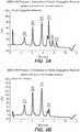

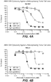


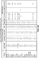
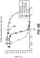


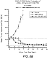


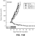
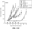

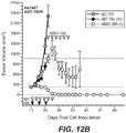


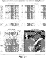
Classifications
-
- A—HUMAN NECESSITIES
- A61—MEDICAL OR VETERINARY SCIENCE; HYGIENE
- A61K—PREPARATIONS FOR MEDICAL, DENTAL OR TOILETRY PURPOSES
- A61K47/00—Medicinal preparations characterised by the non-active ingredients used, e.g. carriers or inert additives; Targeting or modifying agents chemically bound to the active ingredient
- A61K47/50—Medicinal preparations characterised by the non-active ingredients used, e.g. carriers or inert additives; Targeting or modifying agents chemically bound to the active ingredient the non-active ingredient being chemically bound to the active ingredient, e.g. polymer-drug conjugates
- A61K47/51—Medicinal preparations characterised by the non-active ingredients used, e.g. carriers or inert additives; Targeting or modifying agents chemically bound to the active ingredient the non-active ingredient being chemically bound to the active ingredient, e.g. polymer-drug conjugates the non-active ingredient being a modifying agent
- A61K47/68—Medicinal preparations characterised by the non-active ingredients used, e.g. carriers or inert additives; Targeting or modifying agents chemically bound to the active ingredient the non-active ingredient being chemically bound to the active ingredient, e.g. polymer-drug conjugates the non-active ingredient being a modifying agent the modifying agent being an antibody, an immunoglobulin or a fragment thereof, e.g. an Fc-fragment
- A61K47/6835—Medicinal preparations characterised by the non-active ingredients used, e.g. carriers or inert additives; Targeting or modifying agents chemically bound to the active ingredient the non-active ingredient being chemically bound to the active ingredient, e.g. polymer-drug conjugates the non-active ingredient being a modifying agent the modifying agent being an antibody, an immunoglobulin or a fragment thereof, e.g. an Fc-fragment the modifying agent being an antibody or an immunoglobulin bearing at least one antigen-binding site
- A61K47/6851—Medicinal preparations characterised by the non-active ingredients used, e.g. carriers or inert additives; Targeting or modifying agents chemically bound to the active ingredient the non-active ingredient being chemically bound to the active ingredient, e.g. polymer-drug conjugates the non-active ingredient being a modifying agent the modifying agent being an antibody, an immunoglobulin or a fragment thereof, e.g. an Fc-fragment the modifying agent being an antibody or an immunoglobulin bearing at least one antigen-binding site the antibody targeting a determinant of a tumour cell
-
- A—HUMAN NECESSITIES
- A61—MEDICAL OR VETERINARY SCIENCE; HYGIENE
- A61K—PREPARATIONS FOR MEDICAL, DENTAL OR TOILETRY PURPOSES
- A61K47/00—Medicinal preparations characterised by the non-active ingredients used, e.g. carriers or inert additives; Targeting or modifying agents chemically bound to the active ingredient
- A61K47/50—Medicinal preparations characterised by the non-active ingredients used, e.g. carriers or inert additives; Targeting or modifying agents chemically bound to the active ingredient the non-active ingredient being chemically bound to the active ingredient, e.g. polymer-drug conjugates
- A61K47/51—Medicinal preparations characterised by the non-active ingredients used, e.g. carriers or inert additives; Targeting or modifying agents chemically bound to the active ingredient the non-active ingredient being chemically bound to the active ingredient, e.g. polymer-drug conjugates the non-active ingredient being a modifying agent
- A61K47/68—Medicinal preparations characterised by the non-active ingredients used, e.g. carriers or inert additives; Targeting or modifying agents chemically bound to the active ingredient the non-active ingredient being chemically bound to the active ingredient, e.g. polymer-drug conjugates the non-active ingredient being a modifying agent the modifying agent being an antibody, an immunoglobulin or a fragment thereof, e.g. an Fc-fragment
- A61K47/6835—Medicinal preparations characterised by the non-active ingredients used, e.g. carriers or inert additives; Targeting or modifying agents chemically bound to the active ingredient the non-active ingredient being chemically bound to the active ingredient, e.g. polymer-drug conjugates the non-active ingredient being a modifying agent the modifying agent being an antibody, an immunoglobulin or a fragment thereof, e.g. an Fc-fragment the modifying agent being an antibody or an immunoglobulin bearing at least one antigen-binding site
- A61K47/6849—Medicinal preparations characterised by the non-active ingredients used, e.g. carriers or inert additives; Targeting or modifying agents chemically bound to the active ingredient the non-active ingredient being chemically bound to the active ingredient, e.g. polymer-drug conjugates the non-active ingredient being a modifying agent the modifying agent being an antibody, an immunoglobulin or a fragment thereof, e.g. an Fc-fragment the modifying agent being an antibody or an immunoglobulin bearing at least one antigen-binding site the antibody targeting a receptor, a cell surface antigen or a cell surface determinant
-
- A—HUMAN NECESSITIES
- A61—MEDICAL OR VETERINARY SCIENCE; HYGIENE
- A61K—PREPARATIONS FOR MEDICAL, DENTAL OR TOILETRY PURPOSES
- A61K47/00—Medicinal preparations characterised by the non-active ingredients used, e.g. carriers or inert additives; Targeting or modifying agents chemically bound to the active ingredient
- A61K47/50—Medicinal preparations characterised by the non-active ingredients used, e.g. carriers or inert additives; Targeting or modifying agents chemically bound to the active ingredient the non-active ingredient being chemically bound to the active ingredient, e.g. polymer-drug conjugates
- A61K47/51—Medicinal preparations characterised by the non-active ingredients used, e.g. carriers or inert additives; Targeting or modifying agents chemically bound to the active ingredient the non-active ingredient being chemically bound to the active ingredient, e.g. polymer-drug conjugates the non-active ingredient being a modifying agent
- A61K47/68—Medicinal preparations characterised by the non-active ingredients used, e.g. carriers or inert additives; Targeting or modifying agents chemically bound to the active ingredient the non-active ingredient being chemically bound to the active ingredient, e.g. polymer-drug conjugates the non-active ingredient being a modifying agent the modifying agent being an antibody, an immunoglobulin or a fragment thereof, e.g. an Fc-fragment
- A61K47/6835—Medicinal preparations characterised by the non-active ingredients used, e.g. carriers or inert additives; Targeting or modifying agents chemically bound to the active ingredient the non-active ingredient being chemically bound to the active ingredient, e.g. polymer-drug conjugates the non-active ingredient being a modifying agent the modifying agent being an antibody, an immunoglobulin or a fragment thereof, e.g. an Fc-fragment the modifying agent being an antibody or an immunoglobulin bearing at least one antigen-binding site
- A61K47/6871—Medicinal preparations characterised by the non-active ingredients used, e.g. carriers or inert additives; Targeting or modifying agents chemically bound to the active ingredient the non-active ingredient being chemically bound to the active ingredient, e.g. polymer-drug conjugates the non-active ingredient being a modifying agent the modifying agent being an antibody, an immunoglobulin or a fragment thereof, e.g. an Fc-fragment the modifying agent being an antibody or an immunoglobulin bearing at least one antigen-binding site the antibody targeting an enzyme
-
- A—HUMAN NECESSITIES
- A61—MEDICAL OR VETERINARY SCIENCE; HYGIENE
- A61K—PREPARATIONS FOR MEDICAL, DENTAL OR TOILETRY PURPOSES
- A61K31/00—Medicinal preparations containing organic active ingredients
- A61K31/33—Heterocyclic compounds
- A61K31/395—Heterocyclic compounds having nitrogen as a ring hetero atom, e.g. guanethidine or rifamycins
- A61K31/495—Heterocyclic compounds having nitrogen as a ring hetero atom, e.g. guanethidine or rifamycins having six-membered rings with two or more nitrogen atoms as the only ring heteroatoms, e.g. piperazine or tetrazines
- A61K31/505—Pyrimidines; Hydrogenated pyrimidines, e.g. trimethoprim
- A61K31/517—Pyrimidines; Hydrogenated pyrimidines, e.g. trimethoprim ortho- or peri-condensed with carbocyclic ring systems, e.g. quinazoline, perimidine
-
- A—HUMAN NECESSITIES
- A61—MEDICAL OR VETERINARY SCIENCE; HYGIENE
- A61K—PREPARATIONS FOR MEDICAL, DENTAL OR TOILETRY PURPOSES
- A61K45/00—Medicinal preparations containing active ingredients not provided for in groups A61K31/00 - A61K41/00
-
- A—HUMAN NECESSITIES
- A61—MEDICAL OR VETERINARY SCIENCE; HYGIENE
- A61K—PREPARATIONS FOR MEDICAL, DENTAL OR TOILETRY PURPOSES
- A61K45/00—Medicinal preparations containing active ingredients not provided for in groups A61K31/00 - A61K41/00
- A61K45/06—Mixtures of active ingredients without chemical characterisation, e.g. antiphlogistics and cardiaca
-
- A—HUMAN NECESSITIES
- A61—MEDICAL OR VETERINARY SCIENCE; HYGIENE
- A61K—PREPARATIONS FOR MEDICAL, DENTAL OR TOILETRY PURPOSES
- A61K47/00—Medicinal preparations characterised by the non-active ingredients used, e.g. carriers or inert additives; Targeting or modifying agents chemically bound to the active ingredient
- A61K47/50—Medicinal preparations characterised by the non-active ingredients used, e.g. carriers or inert additives; Targeting or modifying agents chemically bound to the active ingredient the non-active ingredient being chemically bound to the active ingredient, e.g. polymer-drug conjugates
- A61K47/51—Medicinal preparations characterised by the non-active ingredients used, e.g. carriers or inert additives; Targeting or modifying agents chemically bound to the active ingredient the non-active ingredient being chemically bound to the active ingredient, e.g. polymer-drug conjugates the non-active ingredient being a modifying agent
- A61K47/68—Medicinal preparations characterised by the non-active ingredients used, e.g. carriers or inert additives; Targeting or modifying agents chemically bound to the active ingredient the non-active ingredient being chemically bound to the active ingredient, e.g. polymer-drug conjugates the non-active ingredient being a modifying agent the modifying agent being an antibody, an immunoglobulin or a fragment thereof, e.g. an Fc-fragment
- A61K47/6801—Drug-antibody or immunoglobulin conjugates defined by the pharmacologically or therapeutically active agent
- A61K47/6803—Drugs conjugated to an antibody or immunoglobulin, e.g. cisplatin-antibody conjugates
- A61K47/68031—Drugs conjugated to an antibody or immunoglobulin, e.g. cisplatin-antibody conjugates the drug being an auristatin
-
- A—HUMAN NECESSITIES
- A61—MEDICAL OR VETERINARY SCIENCE; HYGIENE
- A61K—PREPARATIONS FOR MEDICAL, DENTAL OR TOILETRY PURPOSES
- A61K47/00—Medicinal preparations characterised by the non-active ingredients used, e.g. carriers or inert additives; Targeting or modifying agents chemically bound to the active ingredient
- A61K47/50—Medicinal preparations characterised by the non-active ingredients used, e.g. carriers or inert additives; Targeting or modifying agents chemically bound to the active ingredient the non-active ingredient being chemically bound to the active ingredient, e.g. polymer-drug conjugates
- A61K47/51—Medicinal preparations characterised by the non-active ingredients used, e.g. carriers or inert additives; Targeting or modifying agents chemically bound to the active ingredient the non-active ingredient being chemically bound to the active ingredient, e.g. polymer-drug conjugates the non-active ingredient being a modifying agent
- A61K47/68—Medicinal preparations characterised by the non-active ingredients used, e.g. carriers or inert additives; Targeting or modifying agents chemically bound to the active ingredient the non-active ingredient being chemically bound to the active ingredient, e.g. polymer-drug conjugates the non-active ingredient being a modifying agent the modifying agent being an antibody, an immunoglobulin or a fragment thereof, e.g. an Fc-fragment
- A61K47/6801—Drug-antibody or immunoglobulin conjugates defined by the pharmacologically or therapeutically active agent
- A61K47/6803—Drugs conjugated to an antibody or immunoglobulin, e.g. cisplatin-antibody conjugates
- A61K47/68035—Drugs conjugated to an antibody or immunoglobulin, e.g. cisplatin-antibody conjugates the drug being a pyrrolobenzodiazepine
-
- A—HUMAN NECESSITIES
- A61—MEDICAL OR VETERINARY SCIENCE; HYGIENE
- A61K—PREPARATIONS FOR MEDICAL, DENTAL OR TOILETRY PURPOSES
- A61K47/00—Medicinal preparations characterised by the non-active ingredients used, e.g. carriers or inert additives; Targeting or modifying agents chemically bound to the active ingredient
- A61K47/50—Medicinal preparations characterised by the non-active ingredients used, e.g. carriers or inert additives; Targeting or modifying agents chemically bound to the active ingredient the non-active ingredient being chemically bound to the active ingredient, e.g. polymer-drug conjugates
- A61K47/51—Medicinal preparations characterised by the non-active ingredients used, e.g. carriers or inert additives; Targeting or modifying agents chemically bound to the active ingredient the non-active ingredient being chemically bound to the active ingredient, e.g. polymer-drug conjugates the non-active ingredient being a modifying agent
- A61K47/68—Medicinal preparations characterised by the non-active ingredients used, e.g. carriers or inert additives; Targeting or modifying agents chemically bound to the active ingredient the non-active ingredient being chemically bound to the active ingredient, e.g. polymer-drug conjugates the non-active ingredient being a modifying agent the modifying agent being an antibody, an immunoglobulin or a fragment thereof, e.g. an Fc-fragment
- A61K47/6801—Drug-antibody or immunoglobulin conjugates defined by the pharmacologically or therapeutically active agent
- A61K47/6803—Drugs conjugated to an antibody or immunoglobulin, e.g. cisplatin-antibody conjugates
- A61K47/6811—Drugs conjugated to an antibody or immunoglobulin, e.g. cisplatin-antibody conjugates the drug being a protein or peptide, e.g. transferrin or bleomycin
-
- A—HUMAN NECESSITIES
- A61—MEDICAL OR VETERINARY SCIENCE; HYGIENE
- A61K—PREPARATIONS FOR MEDICAL, DENTAL OR TOILETRY PURPOSES
- A61K47/00—Medicinal preparations characterised by the non-active ingredients used, e.g. carriers or inert additives; Targeting or modifying agents chemically bound to the active ingredient
- A61K47/50—Medicinal preparations characterised by the non-active ingredients used, e.g. carriers or inert additives; Targeting or modifying agents chemically bound to the active ingredient the non-active ingredient being chemically bound to the active ingredient, e.g. polymer-drug conjugates
- A61K47/51—Medicinal preparations characterised by the non-active ingredients used, e.g. carriers or inert additives; Targeting or modifying agents chemically bound to the active ingredient the non-active ingredient being chemically bound to the active ingredient, e.g. polymer-drug conjugates the non-active ingredient being a modifying agent
- A61K47/68—Medicinal preparations characterised by the non-active ingredients used, e.g. carriers or inert additives; Targeting or modifying agents chemically bound to the active ingredient the non-active ingredient being chemically bound to the active ingredient, e.g. polymer-drug conjugates the non-active ingredient being a modifying agent the modifying agent being an antibody, an immunoglobulin or a fragment thereof, e.g. an Fc-fragment
- A61K47/6835—Medicinal preparations characterised by the non-active ingredients used, e.g. carriers or inert additives; Targeting or modifying agents chemically bound to the active ingredient the non-active ingredient being chemically bound to the active ingredient, e.g. polymer-drug conjugates the non-active ingredient being a modifying agent the modifying agent being an antibody, an immunoglobulin or a fragment thereof, e.g. an Fc-fragment the modifying agent being an antibody or an immunoglobulin bearing at least one antigen-binding site
- A61K47/6851—Medicinal preparations characterised by the non-active ingredients used, e.g. carriers or inert additives; Targeting or modifying agents chemically bound to the active ingredient the non-active ingredient being chemically bound to the active ingredient, e.g. polymer-drug conjugates the non-active ingredient being a modifying agent the modifying agent being an antibody, an immunoglobulin or a fragment thereof, e.g. an Fc-fragment the modifying agent being an antibody or an immunoglobulin bearing at least one antigen-binding site the antibody targeting a determinant of a tumour cell
- A61K47/6857—Medicinal preparations characterised by the non-active ingredients used, e.g. carriers or inert additives; Targeting or modifying agents chemically bound to the active ingredient the non-active ingredient being chemically bound to the active ingredient, e.g. polymer-drug conjugates the non-active ingredient being a modifying agent the modifying agent being an antibody, an immunoglobulin or a fragment thereof, e.g. an Fc-fragment the modifying agent being an antibody or an immunoglobulin bearing at least one antigen-binding site the antibody targeting a determinant of a tumour cell the tumour determinant being from lung cancer cell
-
- A—HUMAN NECESSITIES
- A61—MEDICAL OR VETERINARY SCIENCE; HYGIENE
- A61P—SPECIFIC THERAPEUTIC ACTIVITY OF CHEMICAL COMPOUNDS OR MEDICINAL PREPARATIONS
- A61P35/00—Antineoplastic agents
-
- C—CHEMISTRY; METALLURGY
- C07—ORGANIC CHEMISTRY
- C07K—PEPTIDES
- C07K16/00—Immunoglobulins [IGs], e.g. monoclonal or polyclonal antibodies
- C07K16/18—Immunoglobulins [IGs], e.g. monoclonal or polyclonal antibodies against material from animals or humans
- C07K16/28—Immunoglobulins [IGs], e.g. monoclonal or polyclonal antibodies against material from animals or humans against receptors, cell surface antigens or cell surface determinants
- C07K16/2863—Immunoglobulins [IGs], e.g. monoclonal or polyclonal antibodies against material from animals or humans against receptors, cell surface antigens or cell surface determinants against receptors for growth factors, growth regulators
-
- A—HUMAN NECESSITIES
- A61—MEDICAL OR VETERINARY SCIENCE; HYGIENE
- A61K—PREPARATIONS FOR MEDICAL, DENTAL OR TOILETRY PURPOSES
- A61K2300/00—Mixtures or combinations of active ingredients, wherein at least one active ingredient is fully defined in groups A61K31/00 - A61K41/00
Definitions
- ADCs anti-cMet antibody drug conjugates
- compositions including the ADCs methods of making the ADCs, methods of selecting specific patient populations for cancer treatment with a anti-cMet ADC, and methods of using the ADCs to treat cancers.
- Oncogenic protein kinases such as cMet represent a class of biologically important targets for cancer intervention.
- cMet a well characterized receptor tyrosine kinase encoded by the MET proto-oncogene, is the cell surface receptor for hepatocyte growth factor (HGF; Gherardi E, Birchmeier W, Birchmeier C et al. Targeting MET in cancer: rationale and progress. Nat Rev Can. 2012;12:89-103 ).
- HGF hepatocyte growth factor
- cMet overexpression occurs in approximately 30% - 50% of solid tumors including non-small cell lung cancer (NSCLC), colorectal cancer (CRC), and advanced gastroesophageal cancer (AGEC)
- NSCLC non-small cell lung cancer
- CRC colorectal cancer
- AGEC advanced gastroesophageal cancer
- Spigel DR Ervin TJ, Ramlau RA, et al. Randomized Phase II trial of onartuzumab in combination with erlotinib in patients with advanced non-small-cell lung cancer. J Clin Oncol. 2013;31(32):41054114 ; Resnick MB, Routhier J, Konkin T et al. Epidermal growth factor receptor, cMET, B-catenin, and p53 expression as prognostic indicators in stage II colon cancer: a tissue microarray study. Clin Can Res. 2004;10:3069-3075 ; Lee HE, Kim MA, Lee HS, et al. MET in gastric carcinomas:
- cMet cellular mesenchymal-epithelial transition factor
- HGF hepatocyte growth factor/HGF ligand. This cell surface receptor is expressed in epithelial cells of many organs, including the liver, pancreas, prostate, kidney, muscle and bone marrow, during both embryogenesis and adulthood.
- cMet regulates many physiological processes including cell proliferation and survival, migration and scattering (cell-cell repulsion), tissue morphogenesis, organ regeneration, and tissue remodeling. In cancer and other pathological processes, cMet is often aberrantly activated via mutation, amplification, or protein overexpression.
- Solid tumor cancers in which cMet is overexpressed in at least 10% of the patient population include lung cancer, colorectal cancer, head and neck cancer, pancreatic cancer, gastric cancer, glioblastoma, ovarian, breast, prostate, cervical, and esophageal cancer.
- ADCs antibody drug conjugates
- Data presented herein demonstrate, for the first time, that antibody drug conjugates (“ADCs") that specifically target cMet overexpression have demonstrated anti-tumor activity in patients diagnosed with non-small cell lung cancer.
- ADCs antibody drug conjugates
- Data demonstrating in vivo anti-tumor efficacy of anti-cMet ADCs administered as monotherapy or combination are provided in Examples 10-14 and 16, and FIGS. 8-12 and 14-18 .
- cMet overexpression can be defined by an immunohistorychemistry (IHC) H-score of greater than or equal to 150 when measured according to the assay of Example 17.
- IHC staining protocol for cMet overexpression has been developed using the Ventana cMet CONFIRM (SP44) kit. Tissue samples are stained with the Ventana antibody and then scored by determining the percentages of target tissue cells staining at various intensity levels of low to high.
- FIG. 20 depicts representative H-scores using the assay described in Example 17.
- FIGS. 19 and 21 depict representative IHC scores using the assay described in Example 17.
- the anti-cMet ADCs may be administered as single therapeutic agents (monotherapy) or adjunctively with or to other anti-cancer treatments and/or therapeutic agents, typically but not necessarily those used to treat the type of cancers being treated. Indeed, data presented herein demonstrate that tumors that exhibit resistance to other targeted or non-targeted chemotherapies retain sensitivity to anti-cMet ADCs (see, e.g., Example 14 and FIGS. 12A-C ). Accordingly, the anti-cMet ADCs described herein provide significant benefits over current targeted and non-targeted approaches toward the treatment of solid tumor cancers that overexpress cMet.
- Adjunctive therapies and/or therapeutic agents typically will be used at their approved dose, route of administration, and frequency of administration, but may be used at lower dosages and/or less frequently.
- the anti-cMet ADC When administered as monotherapy, the anti-cMet ADC will typically be administered on a schedule that provides therapeutic benefit. It is contemplated that anti-cMet ADCs administered once a week, once every two weeks, once every three weeks, once every four weeks, once every five weeks, once every six weeks, once every seven weeks or once every eight weeks will provide therapeutic benefit, although more or less frequent administration may be beneficial.
- the anti-cMet ADC may be administered before, after or concurrently with the other therapy or agent.
- the anti-cMet ADCs may be administered via a variety of routes or modes of administration, including but not limited to, intravenous infusion and/or injection and subcutaneous injection.
- the amount administered will depend upon the route of administration, the dosing schedule, the type of cancer being treated, the stage of the cancer being treated, and other parameters such as the age and weight of the patient, as is well known in the art. Specific exemplary dosing schedules expected to provide therapeutic benefit are provided in the Detailed Description.
- an amount of anti-cMet ADC in the range of about 0.005 to 15 mg/kg when administered intravenously on a weekly basis from once weekly to and including once every eight weeks is expected to provide therapeutic benefit.
- the present disclosure provides ADCs that specifically bind cMet ("anti-cMet ADCs").
- the anti-cMet ADCs comprise cytotoxic and/or cytostatic agents linked by way of linkers to an antigen binding moiety that specifically binds cMet.
- the antigen binding moiety is an antibody and/or an antigen binding fragment.
- Antibodies and/or binding fragments composing the anti-cMet ADCs generally comprise a heavy chain comprising a variable region (V H ) having three complementarity determining regions ("CDRs") referred to herein (in N ⁇ C order) as V H CDR#1, V H CDR#2, and V H CDR#3, and a light chain comprising a variable region (V L ) having three complementarity determining regions referred to herein (in N ⁇ C order) as V L CDR#1, V L CDR#2, and V L CDR#3.
- V H variable region
- CDRs complementarity determining regions
- anti-cMet ADCs include, but are not limited to, ABT-700 and STI-0602.
- the anti- cMet ADCs comprise an anti- cMet and/or anti- cMet binding fragment that binds cMet with an affinity of at least about 100 nM, or even higher, for example, at least about 90 nM, 80 nM, 70 nM, 60 nM, 50 nM, 40 nM, 30 nM, 25 nM, 20 nM, 15 nM, 10 nM, 7 nM, 6 nM, 5 nM, 4 nM, 3 nM, 2 nM, 1 nM, 0.1 nM, 0.01 nM, or greater.
- Affinity of anti- cMet antibodies and/or binding fragments can be determined using techniques well known in the art or described herein, such as for example, ELISA, isothermal titration calorimetry (ITC), surface plasmon resonance, flow cytometry, or fluorescent polarization assay.
- the affinity refers to apparent affinity EC 50 values, measured according to Example 5.
- the antibody has an apparent affinity EC 50 value from lower than about 10 nanomol/L, preferably from about 1 picomol/L to 10 nanomol/L, preferably about 0.3 nanomol/L, as determined according to Example 5.
- Antibodies may be in the form of full-length antibodies, bispecific antibodies, dual variable domain antibodies, multiple chain or single chain antibodies, surrobodies (including surrogate light chain construct), single domain antibodies, camelized antibodies, scFv-Fc antibodies, and the like. They may be of, or derived from, any isotype, including, for example, IgA (e.g., IgA 1 or IgA 2 ), IgD, IgE, IgG ( e.g., IgG 1 , IgG 2 , IgG 3 or IgG 4 ), IgM, or IgY.
- IgA e.g., IgA 1 or IgA 2
- IgD IgD
- IgE IgG
- IgM IgM
- IgY IgY
- the anti-cMet antibody is an IgG (e.g., IgG 1 , IgG 2 , IgG 3 or IgG 4 ).
- Antibodies may be of human or non-human origin. Examples of non-human origin include, but are not limited to, mammalian origin (e.g ., simians, rodents, goats, and rabbits) or avian origin ( e.g., chickens).
- antibodies composing the anti- cMet ADCs are suitable for administration to humans, such as, for example, humanized antibodies and/or fully human antibodies.
- Antigen binding fragments composing the anti- cMet ADCs may include any fragment of an antibody capable of specifically binding cMet.
- Specific examples of antibody binding fragments that may be included in the anti- cMet ADCs include, but are not limited to, Fab, Fab', (Fab') 2 , Fv and scFv.
- Antibodies and/or binding fragments composing the anti-cMet ADCs may include modifications and/or mutations that alter the properties of the antibodies and/or fragments, such as those that increase half-life, increase or decrease ADCC, etc., as is known in the art.
- the cytotoxic and/or cytostatic agents composing the anti-cMet ADCs may be any agents known to inhibit the growth and/or replication of, and/or kill cells. Numerous agents having cytotoxic and/or cytostatic properties are known in the literature. Non-limiting examples of classes of cytotoxic and/or cytostatic agents include, by way of example and not limitation, cell cycle modulators, apoptosis regulators, kinase inhibitors, protein synthesis inhibitors, alkylating agents, DNA cross-linking agents, intercalating agents, mitochondria inhibitors, nuclear export inhibitors, topoisomerase I inhibitors, topoisomerase II inhibitors, RNA/DNA antimetabolites and antimitotic agents.
- a cytotoxic and/or cytostatic agent composing an anti-cMet ADC is a cell-permeating antimitotic agent, such as, for example, an auristatin.
- a cytotoxic and/or cytostatic agent composing an anti-cMet ADC is a cell-permeating DNA cross-linking agent, such as a cell-permeating minor groove-binding DNA cross-linking agent.
- cell-permeating DNA minor groove-binding agents include, but are not limited to, pyrrolobenzodiazepines ("PBD") and PBD dimers.
- the linkers linking the cytotoxic and/or cytostatic agents to the antigen binding moiety of an anti-cMet ADC may be long, short, flexible, rigid, hydrophilic or hydrophobic in nature, or may comprise segments that have different characteristics, such as segments of flexibility, segments of rigidity, etc.
- the linker may be chemically stable to extracellular environments, for example, chemically stable in the blood stream, or may include linkages that are not stable and release the cytotoxic and/or cytostatic agents in the extracellular milieu.
- the linkers include linkages that are designed to release the cytotoxic and/or cytostatic agents upon internalization of the anti- cMet ADC within the cell.
- the linkers includes linkages designed to cleave and/or immolate or otherwise breakdown specifically or non-specifically inside cells.
- linkers useful for linking drugs to antigen binding moieties such as antibodies in the context of ADCs are known in the art. Any of these linkers, as well as other linkers, may be used to link the cytotoxic and/or cytostatic agents to the antigen binding moiety of the anti- cMet ADCs described herein.
- the number of cytotoxic and/or cytostatic agents linked to the antigen binding moiety of an anti- cMet ADC can vary (called the "drug-to-antibody ratio,” or “DAR”), and will be limited only by the number of available attachments sites on the antigen binding moiety and the number of agents linked to a single linker.
- DAR drug-to-antibody ratio
- a linker will link a single cytotoxic and/or cytostatic agent to the antigen binding moiety of an anti- cMet ADC.
- each agent may be the same or different.
- anti-cMet ADCs with DARs of twenty, or even higher, are contemplated.
- the anti-cMet ADCs described herein may have a DAR in the range of about 1-10, 1-8, 1-6, or 1-4.
- the anti-cMet ADCs may have a DAR of 2, 3, or 4.
- the anti-cMet ADCs may have an average DAR of 3.1.
- polypeptide sequences described herein are, in many embodiments, described by way of their respective polypeptide or polynucleotide sequences. Unless indicated otherwise, polypeptide sequences are provided in N ⁇ C orientation; polynucleotide sequences in 5' ⁇ 3' orientation. For polypeptide sequences, the conventional three or one-letter abbreviations for the genetically encoded amino acids may be used, as noted in TABLE 1, below.
- Certain sequences are defined by structural formulae specifying amino acid residues belonging to certain classes (e.g., aliphatic, hydrophobic, etc .).
- the various classes to which the genetically encoded amino acids belong as used herein are noted in TABLE 2, below. Some amino acids may belong to more than one class. Cysteine, which contains a sulfhydryl group, and proline, which is conformationally constrained, are not assigned classes.
- TABLE 2 Encoded Amino Acid Classes Class Amino Acids Aliphatic A, I, L, V Aromatic F, Y, W Non-Polar M, A, I, L, V Polar N, Q, S, T Basic H, K, R Acidic D, E Small A, G
- the present disclosure concerns antibody drug conjugates that specifically bind human cMet, compositions comprising the ADCs, anti-cMet antibodies and/or binding fragments that can comprise the ADCs, polynucleotides encoding anti-cMet antibodies and/or binding fragments that comprise the ADCs, host cells capable of producing the antibodies and/or binding fragments, methods and compositions useful for making the antibodies, binding fragments and ADCs, and various methods of using the ADCs in cancer treatment.
- ADCs antibody drug conjugates
- cMet ABBV-ADC staining protocol For purposes of this application, including the claims, the particular assay used in the study described herein is referred to as the "cMet ABBV-ADC staining protocol.” This protocol is described in detail in Example 17 and the results are expressed in terms of H-score and may also be expressed in terms of IHC score or other scoring system well known in the art.
- the H-score approach provides optimal data resolution for determining variation in intensity and tumor percentage of staining within and among tumor types. It also provides a good tool for determining thresholds for positive staining.
- the percentage of cells (0-100) within a tumor with staining intensities ranging from 0-3+ are provided. This protocol results in staining of the cMet protein both in the cytoplasm and in the cell surface/membrane.
- the staining intensity for each cell in a fixed field (typically, 100 cells) of the processed tumor biopsy is determined, and an individual value is attributed to each cell as follows, depending on the cell surface/membrane staining:
- H-score the percentage of tumor cells are multiplied by each intensity and added together.
- the maximum H-score is 300 if 100% of tumor cells label with 3+ intensity.
- FIG. 20 shows exemplary staining results for various tumor H-scores (15, 90, 180, and 290) obtained with the "cMet ABBV-ADC staining protocol.”
- Each tumor can also be given an IHC score of IHC 0, IHC 1+, IHC 2+, or IHC 3+. While both the IHC and H scores involve 0, 1+, 2+, and 3+ values they are not to be confused.
- H-score 0, 1+, 2+, and 3+ values refer to the intensity of staining of an individual cell.
- IHC score 0, 1+, 2+, and 3+ values refer to the overall staining of a particular area of the tumor sample.
- FIG. 21 shows exemplary staining results for various tumor IHC0/1+/2+/3+ scores obtained with the "cMet ABBV-ADC staining protocol.”
- the value attributed to the tumor is IHC 0. If the overall level of staining in a fixed field is low, the value attributed is IHC 1+. If most of the cells in a fixed field exhibit moderate staining, the value attributed is IHC 2+. If most of the cells in a fixed field exhibit strong staining, the value attributed is IHC 3+.
- the value attributed to the tumor is IHC 0. If the overall level of staining in a fixed field is low, the value attributed is IHC 1+. If at least 15% of the cells in a fixed field exhibit moderate staining, the value attributed is IHC 2+. If at least 15% of the cells in a fixed field exhibit strong staining, the value attributed is IHC 3+.
- an H-score between 150 and 224 is equivalent to an IHC score of 2+ and an H-score of 225 and above is equivalent to an IHC score of 3+.
- the present disclosure provides ADCs that specifically bind cMet ("anti-cMet ADCs").
- the anti-cMet ADCs comprise cytotoxic and/or cytostatic agents linked by way of linkers to an antigen binding moiety that specifically binds cMet.
- the antigen binding moiety (ABT-700) binds cMet at IPT domain 1 of human cMet.
- the antigen binding moiety may be any moiety capable of specifically binding cMet.
- the antigen binding moiety is an antibody and/or an antibody binding fragment.
- a cytotoxic and/or cytostatic agent composing an anti-cMet ADC is a cell-permeating antimitotic agent, such as, for example, an auristatin.
- a cytotoxic and/or cytostatic agent composing an anti-cMet ADC is a cell-permeating DNA cross-linking agent, such as a cell-permeating minor groove-binding DNA cross-linking agent.
- cell-permeating DNA minor groove-binding agents include, but are not limited to, pyrrolobenzodiazepines ("PBD") and PBD dimers.
- antibodies and/or binding fragments are “modular” in nature.
- various specific embodiments of the various “modules” composing the antibodies and/or binding fragments are described.
- various specific embodiments of V H CDRs, V H chains, V L CDRs and V L chains are described. It is intended that all of the specific embodiments may be combined with each other as though each specific combination were explicitly described individually.
- the ADCs disclosed herein are also "modular" in nature. Throughout the disclosure, various specific embodiments of the “modules” composing the ADCs are described. As non-limiting examples, specific embodiments of antibodies, linkers, and cytotoxic and/or cytostatic agents that may compose the ADCs are described. It is intended that all of the specific embodiments described may be combined with each other as though each specific combination were explicitly described individually.
- ADCs described herein may be in the form of salts, and in some specific embodiments, pharmaceutically acceptable salts.
- the ADCs of the disclosure that possess a sufficiently acidic, a sufficiently basic, or both functional groups can react with any of a number of inorganic bases, and inorganic and organic acids, to form a salt.
- compounds that are inherently charged, such as those with a quaternary nitrogen can form a salt with an appropriate counter ion, e.g., a halide such as a bromide, chloride, or fluoride.
- Acids commonly employed to form acid addition salts are inorganic acids such as hydrochloric acid, hydrobromic acid, hydroiodic acid, sulfuric acid, phosphoric acid, and the like, and organic acids such as p-toluenesulfonic acid, methanesulfonic acid, oxalic acid, p -bromophenylsulfonic acid, carbonic acid, succinic acid, citric acid, etc.
- Base addition salts include those derived from inorganic bases, such as ammonium and alkali or alkaline earth metal hydroxides, carbonates, bicarbonates, and the like.
- the antigen binding moiety is an antibody or an antigen binding fragment.
- antibody refers to an immunoglobulin molecule that specifically binds to, or is immunologically reactive with, a particular antigen- here, cMet.
- Antibodies comprise complementarity determining regions (CDRs), also known as hypervariable regions, in both the light chain and heavy chain variable domains. The more highly conserved portions of the variable domains are called the framework (FR).
- CDRs complementarity determining regions
- FR framework
- amino acid position/boundary delineating a hypervariable region of an antibody can vary, depending on the context and the various definitions known in the art.
- variable domains within a variable domain may be viewed as hybrid hypervariable positions in that these positions can be deemed to be within a hypervariable region under one set of criteria, while being deemed to be outside a hypervariable region under a different set of criteria.
- One or more of these positions can also be found in extended hypervariable regions.
- the variable domains of native heavy and light chains each comprise four FR regions, largely by adopting a ⁇ -sheet configuration, connected by three CDRs, which form loops connecting, and in some cases forming part of, the ⁇ -sheet structure.
- the CDRs in each chain are held together in close proximity by the FR regions and, with the CDRs from the other chain, contribute to the formation of the antigen binding site of antibodies.
- Antibodies and/or binding fragments composing the anti-cMet ADCs generally comprise a heavy chain comprising a variable region (V H ) having three complementarity determining regions ("CDRs") referred to herein (in N ⁇ C order) as V H CDR#1, V H CDR#2, and V H CDR#3, and a light chain comprising a variable region (V L ) having three complementarity determining regions referred to herein (in N ⁇ C order) as V L CDR#1, V L CDR#2, and V L CDR#3.
- V H variable region
- CDRs complementarity determining regions
- anti-cMet ADCs include, but are not limited to, those that comprise antibodies and/or binding fragments that include these exemplary CDRs and/or V H and/or V L sequences, as well as antibodies and/or binding fragments that compete for binding cMet with such antibodies and/or binding fragments.
- Antibodies may be in the form of full-length antibodies, bispecific antibodies, dual variable domain antibodies, multiple chain or single chain antibodies, surrobodies (including surrogate light chain construct), single domain antibodies, camelized antibodies, scFv-Fc antibodies, and the like. They may be of, or derived from, any isotype, including, for example, IgA (e.g., IgA 1 or IgA 2 ), IgD, IgE, IgG ( e.g., IgG 1 , IgG 2 , IgG 3 or IgG 4 ), IgM, or IgY.
- IgA e.g., IgA 1 or IgA 2
- IgD IgD
- IgE IgG
- IgM IgM
- IgY IgY
- the anti-cMet antibody is an IgG (e.g., IgG 1 , IgG 2 , IgG 3 or IgG 4 ).
- Antibodies may be of human or non-human origin. Examples of non-human origin include, but are not limited to, mammalian origin (e.g., simians, rodents, goats, and rabbits) or avian origin ( e.g., chickens).
- antibodies composing the anti-cMet ADCs are suitable for administration to humans, such as, for example, humanized antibodies and/or fully human antibodies.
- Antibodies composing anti-cMet ADCs may be polyclonal, monoclonal, genetically engineered, and/or otherwise modified in nature, including but not limited to, chimeric antibodies, humanized antibodies, human antibodies, primatized antibodies, single chain antibodies, bispecific antibodies, dual-variable domain antibodies, etc.
- the antibodies comprise all or a portion of a constant region of an antibody.
- the constant region is an isotype selected from: IgA (e.g., IgA 1 or IgA 2 ), IgD, IgE, IgG ( e.g., IgG 1 , IgG 2 , IgG 3 or IgG 4 ), IgM, and IgY.
- antibodies composing an anti-cMet ADC comprise an IgG 1 constant region isotype.
- monoclonal antibody as used herein is not limited to antibodies produced through hybridoma technology.
- a monoclonal antibody is derived from a single clone, including any eukaryotic, prokaryotic, or phage clone, by any means available or known in the art.
- Monoclonal antibodies useful with the present disclosure can be prepared using a wide variety of techniques known in the art including the use of hybridoma, recombinant, and phage display technologies, or a combination thereof. In many uses of the present disclosure, including in vivo use of ADCs including anti-cMet antibodies in humans, chimeric, primatized, humanized, or human antibodies can suitably be used.
- chimeric antibody refers to an antibody having variable sequences derived from a non-human immunoglobulin, such as a rat or a mouse antibody, and human immunoglobulin constant regions, typically chosen from a human immunoglobulin template.
- Methods for producing chimeric antibodies are known in the art. See, e.g., Morrison, 1985, Science 229(4719):1202-7 ; Oi et al., 1986, BioTechniques 4:214-221 ; Gillies et al., 1985, J. Immunol. Methods 125:191-202 ; U.S. Pat. Nos. 5,807,715 ; 4,816,567 ; and 4,816,397 , which are incorporated herein by reference in their entireties.
- Humanized forms of non-human (e.g., murine) antibodies are chimeric immunoglobulins that contain minimal sequences derived from non-human immunoglobulin.
- a humanized antibody will comprise substantially all of at least one, and typically two, variable domains, in which all or substantially all of the CDR regions correspond to those of a non-human immunoglobulin and all or substantially all of the FR regions are those of a human immunoglobulin sequence.
- the humanized antibody can also comprise at least a portion of an immunoglobulin constant region (Fc), typically that of a human immunoglobulin consensus sequence.
- Fc immunoglobulin constant region
- Human antibodies are antibodies having the amino acid sequence of a human immunoglobulin and include antibodies isolated from human immunoglobulin libraries or from animals transgenic for one or more human immunoglobulin and that do not express endogenous immunoglobulins. Human antibodies can be made by a variety of methods known in the art including phage display methods using antibody libraries derived from human immunoglobulin sequences. See U.S. Patent Nos.
- Fully human antibodies that recognize a selected epitope can be generated using a technique referred to as "guided selection.”
- a selected non-human monoclonal antibody e.g ., a mouse antibody
- is used to guide the selection of a completely human antibody recognizing the same epitope see, Jespers et al., 1988, Biotechnology 12:899-903 ).
- Primary antibodies comprise monkey variable regions and human constant regions. Methods for producing primatized antibodies are known in the art. See, e.g., U.S. Patent Nos. 5,658,570 ; 5,681,722 ; and 5,693,780 , which are incorporated herein by reference in their entireties.
- Anti-cMet ADCs may comprise full-length (intact) antibody molecules, as well as antigen binding fragments that are capable of specifically binding cMet.
- antibody binding fragments include by way of example and not limitation, Fab, Fab', F(ab') 2 , Fv fragments, single chain Fv fragments and single domain fragments.
- a Fab fragment contains the constant domain of the light chain and the first constant domain (CH 2 ) of the heavy chain.
- Fab' fragments differ from Fab fragments by the addition of a few residues at the carboxyl terminus of the heavy chain CH 2 domain including one or more cysteines from the antibody hinge region.
- F(ab') fragments are produced by cleavage of the disulfide bond at the hinge cysteines of the F(ab') 2 pepsin digestion product. Additional chemical couplings of antibody fragments are known to those of ordinary skill in the art.
- Fab and F(ab') 2 fragments lack the Fc fragment of intact antibody, clear more rapidly from the circulation of animals, and may have less non-specific tissue binding than an intact antibody ( see, e.g., Wahl et al., 1983, J. Nucl. Med. 24:316 ).
- an “Fv” fragment is the minimum fragment of an antibody that contains a complete target recognition and binding site. This region consists of a dimer of one heavy and one light chain variable domain in a tight, non-covalent association (V H -V L dimer). It is in this configuration that the three CDRs of each variable domain interact to define an antigen binding site on the surface of the V H -V L dimer. Often, the six CDRs confer antigen binding specificity upon the antibody. However, in some instances even a single variable domain (or half of an Fv comprising only three CDRs specific for a target) may have the ability to recognize and bind antigen, although at a lower affinity than the entire binding site.
- Single-chain Fv or “scFv” antibody binding fragments comprise the V H and V L domains of an antibody, where these domains are present in a single polypeptide chain.
- the Fv polypeptide further comprises a polypeptide linker between the V H and V L domains which enables the scFv to form the desired structure for antigen binding.
- Antibodies and/or binding fragments composing the anti-cMet ADCs may include modifications and/or mutations that alter the properties of the antibodies and/or fragments, such as those that increase half-life, increase or decrease ADCC, etc., as is known in the art.
- Single domain antibodies are composed of a single V H or V L domains which exhibit sufficient affinity to cMet.
- the single domain antibody is a camelized antibody ( See, e.g., Riechmann, 1999, Journal of Immunological Methods 231:25-38 ).
- Antibodies composing the anti-cMet ADCs may also be bispecific antibodies.
- Bispecific antibodies comprised of monoclonal, often human or humanized, antibodies that have binding specificities for two different epitopes on the same or different antigens.
- one of the binding specificities can be directed towards cMet, the other can be for any other antigen, e.g ., for a cell-surface protein, receptor, receptor subunit, tissue-specific antigen, virally derived protein, virally encoded envelope protein, bacterially derived protein, or bacterial surface protein, etc.
- Antibodies composing anti-cMet ADCs may be derivatized. Derivatized antibodies are typically modified by glycosylation, acetylation, pegylation, phosphorylation, amidation, derivatization by known protecting/blocking groups, proteolytic cleavage, linkage to a cellular ligand or other protein. Any of numerous chemical modifications may be carried out by known techniques, including, but not limited to, specific chemical cleavage, acetylation, formylation, metabolic synthesis of tunicamycin, etc. Additionally, the derivative may contain one or more non-natural amino acids, e.g., using ambrx technology. See, e.g., Wolfson, 2006, Chem. Biol. 13(10):1011-2 .
- Antibodies or binding fragments composing anti-cMet ADCs may be antibodies or fragments whose sequences have been modified to alter at least one constant region-mediated biological effector function.
- an anti-cMet antibody may be modified to reduce at least one constant region-mediated biological effector function relative to the unmodified antibody, e.g ., reduced binding to the Fc receptor (Fc ⁇ R).
- Fc ⁇ R binding may be reduced by mutating the immunoglobulin constant region segment of the antibody at particular regions necessary for Fc ⁇ R interactions ( See, e.g., Canfield and Morrison, 1991, J. Exp. Med. 173:1483-1491 ; and Lund et al., 1991, J. Immunol. 147:2657-2662 ).
- Reducing Fc ⁇ R binding may also reduce other effector functions which rely on FcyR interactions, such as opsonization, phagocytosis and antigen-dependent cellular cytotoxicity ("ADCC").
- ADCC antigen-dependent
- Antibodies included in anti-cMet ADCs may have low levels of, or lack, fucose. Antibodies lacking fucose have been correlated with enhanced ADCC activity, especially at low doses of antibody. See Shields et al., 2002, J. Biol. Chem. 277:26733-26740 ; Shinkawa et al., 2003, J. Biol. Chem. 278:3466-73 .
- Methods of preparing fucose-less antibodies include growth in rat myeloma YB2/0 cells (ATCC CRL 1662). YB2/0 cells express low levels of FUT8 mRNA, which encodes ⁇ -1,6-fucosyltransferase, an enzyme necessary for fucosylation of polypeptides.
- Antibodies or binding fragments composing anti-cMet ADCs may include modifications that increase or decrease their binding affinities to the neonatal Fc receptor, FcRn, for example, by mutating the immunoglobulin constant region segment at particular regions involved in FcRn interactions ( see, e.g., WO 2005/123780 ).
- an anti-cMet antibody of the IgG class is mutated such that at least one of amino acid residues 250, 314, and 428 of the heavy chain constant region is substituted alone, or in any combinations thereof, such as at positions 250 and 428, or at positions 250 and 314, or at positions 314 and 428, or at positions 250, 314, and 428, with substitution at positions 250 and 428 being a specific combination.
- the substituting amino acid residue may be any amino acid residue other than threonine, including, but not limited to, alanine, cysteine, aspartic acid, glutamic acid, phenylalanine, glycine, histidine, isoleucine, lysine, leucine, methionine, asparagine, proline, glutamine, arginine, serine, valine, tryptophan, or tyrosine.
- the substituting amino acid residue may be any amino acid residue other than leucine, including, but not limited to, alanine, cysteine, aspartic acid, glutamic acid, phenylalanine, glycine, histidine, isoleucine, lysine, methionine, asparagine, proline, glutamine, arginine, serine, threonine, valine, tryptophan, or tyrosine.
- the substituting amino acid residues may be any amino acid residue other than methionine, including, but not limited to, alanine, cysteine, aspartic acid, glutamic acid, phenylalanine, glycine, histidine, isoleucine, lysine, leucine, asparagine, proline, glutamine, arginine, serine, threonine, valine, tryptophan, or tyrosine.
- Specific combinations of suitable amino acid substitutions are identified in TABLE 1 of U.S. Patent No. 7,217,797 , which is incorporated herein by reference. Such mutations increase binding to FcRn, which protects the antibody from degradation and increases its half-life.
- An anti-cMet antibody and/or binding fragment may have one or more amino acids inserted into one or more of its hypervariable regions, for example as described in Jung & Plückthun, 1997, Protein Engineering 10:9, 959-966 ; Yazaki et al., 2004, Protein Eng. Des Sel. 17(5):481-9 ; and U.S. Pat. App. No. 2007/0280931 .
- Anti-cMet antibodies and/or binding fragments with high affinity for cMet may be desirable for therapeutic uses. Accordingly, the present disclosure contemplates ADCs comprising anti-cMet antibodies and/or binding fragments having a high binding affinity to cMet.
- the antibodies and/or binding fragments bind cMet with an affinity of at least about 100 nM, but may exhibit higher affinity, for example, at least about 90 nM, 80 nM, 70 nM, 60 nM, 50 nM, 40 nM, 30 nM, 25 nM, 20 nM, 15 nM, 10 nM, 7 nM, 6 nM, 5 nM, 4 nM, 3 nM, 2 nM, 1 nM, 0.1 nM, 0.01 nM, or even higher.
- the antibodies bind cMet with an affinity in the range of about 1 pM to about 100 nM, or an affinity ranging between any of the foregoing values.
- affinity refers to apparent affinity EC50 values measured according to Example 5.
- anti-cMet antibodies can serve at least two different purposes.
- the anti-cMet antibodies are used for diagnostic purposes, assisting in and guiding patient selection.
- these anti-cMet antibodies can be used for immunohistochemistry assays of tumor biopsies obtained from the patients to be treated or under treatment.
- One of ordinary skill in the art is familiar with the techniques for selecting a particular antibody for diagnostic purposes to assay for the levels of cMet protein expression in tumor biopsies.
- the samples are scored under one or more scoring guides, including IHC scores of 0/1+/2+/3+ or H-scores.
- the disclosure details one example of such a diagnostic assay that is commercially available from Ventana.
- the Ventana antibody SP44, and antibodies with similar properties can be made or acquired from other vendors and the protocol adjusted so that the method has the same or better diagnostic power as the Ventana assay.
- anti-cMet antibodies other than SP44 can also be used for this purpose.
- One of ordinary skill in the art would know how to properly adjust the protocol to a new antibody in order to obtain a diagnostic test for cMet expression levels.
- Companion diagnostics exist for a variety of other FDA approved cancer treatments and are within the level of ordinary skill. The FDA maintains a list of FDA-approved companion diagnostic tests at, for example, www.fda.gov/.
- anti-cMet antibodies examples include, for example, the diagnostic antibodies disclosed in U.S. Patent No. 8,673,302 (224D10 and 221C9) and U.S. Patent No. 9,120,852 (227D3 and 205A5).
- the disclosures of each of these patents are fully incorporated herein by reference, including the amino acid sequences for the CDRs, heavy chains (full and variable regions), and light chains (full and variable regions).
- the antibody is 227D3.
- 227D3 is secreted by the hybridoma deposited at the CNCM on November 18, 2009, under number 1-4247.
- Antibody CDR numbering Heavy chain Light chain SEQ ID NO. 227D3 IMGT CDR-L1 159 CDR-L2 160 CDR-L3 161 CDR-H1 162 CDR-H2 163 CDR-H3 164 227D3 Kabat CDR-L1 165 CDR-L2 166 CDR-L3 161 CDR-H1 167 CDR-H2 168 CDR-H3 169
- the anti-cMet antibodies are administered for treatment purposes, either as components of antibody drug conjugates (ADCs), or before/after/concurrently with administration of the ADCs.
- ADCs antibody drug conjugates
- the CDRs have been identified according to the IMGT numbering system.
- ABBV-399 is an ADC comprised of the cMet targeting antibody ABT-700 (PR-1266688, h224G11) conjugated to the potent cytotoxin MMAE through a valine citrulline (vc) linker.
- the ADC binds to cMet on the surface of tumor cells, is internalized, and then releases MMAE leading to the inhibition of microtubule function and the disruption of critical cellular processes and death.
- ABBV-399 is potently cytotoxic to cancer cells with overexpress cMet or amplified MET and demonstrates antitumor activity in human tumor xenografts. Activity of ABBV-399 against ABT-700-refractory tumors has also been demonstrated (see e.g., Example 14).
- ABT-700 is a humanized version of mouse monoclonal antibody 224G11, which was first disclosed and embodimented in U.S. Patent No. 8,329,173 .
- ABT-700 is a "humanized" recombinant IgG1 ⁇ (disclosed as 224G11 [TH7 Hz3] in U.S. Patent No. 8,741,290 ) that targets a unique epitope of cMet located within the immunoglobulin-plexin-transcription factor homology (IPT) domain 1, resulting in blockade of both HGF-dependent and HGF-independent cMet signaling.
- IPT immunoglobulin-plexin-transcription factor homology
- ABT-700 competes for binding to cMet with antibodies directed against SEMA blade 5 (and vice versa), but not with antibodies directed against blades 1-3 or IPT 2-3.
- 5D5 the bivalent progenitor of one armed onartuzumab, discussed below
- the cMet-ADCs of this disclosure encompass any antibody that comprises a heavy chain comprising CDR-H1, CDR-H2 and CDR-H3 comprising respectively the amino acid sequence SEQ ID Nos. 1, 2 and 3; and a light chain comprising CDR-L1, CDR-L2 and CDR-L3 comprising respectively the amino acid sequence SEQ ID Nos. 5, 6, and 7, according to U.S. Patent No. 8,741,290 .
- the CDR sequences of ABT-700 comprise the following sequences: CDR-H1: GYIFTAYT (SEQ ID NO: 72) CDR-H2: IKPNNGLA (SEQ ID NO: 73) CDR-H3: ARSEITTEFDY (SEQ ID NO: 74) CDR-L1: ESVDSYANSF (SEQ ID NO: 75) CDR-L2: RAS (SEQ ID NO: 76) CDR-L3: QQSKEDPLT (SEQ ID NO: 77)
- the heavy chain variable region of 224G11 [TH7 Hz3] comprises SEQ ID No. 4 of U.S. Patent No. 8,741,290 : and the light chain variable region comprises SEQ ID No. 10 of U.S. Patent No. 8,741,290 : DIVMTQSPDSLAVSLGERATINCKSSESVDSYANSFLHWYQQKPGQPPK LLIYRASTRESGVPDRFSGSGSGTDFTLTISSLQAEDVAVYYCQQSKED PLTFGGGTKVEIKR (SEQ ID NO: 79)
- the heavy chain variable region of 224G11 [TH7 Hz3] comprises: and the light chain variable region comprises:
- the antibody [224G11] [TH7 Hz3] comprises a complete heavy chain comprising the amino acid sequence SEQ ID No. 37 of U.S. Patent No. 8,741,290 and a complete light chain comprising the amino acid sequence SEQ ID No. 40 of U.S. Patent No. 8,741,290 .
- the modified hinge region has the sequence of SEQ ID NO:170.
- the anti-cMet antibody comprises a heavy chain variable region comprising SEQ ID No. 4 of U.S. Patent No. 8,741,290 :
- the anti-cMet antibody comprises a heavy chain variable region comprising SEQ ID No. 4 of U.S. Patent No. 8,741,290 :
- an anti-cMet antibody and/or binding fragment composing an anti-cMet ADC is an IgG 1 .
- an anti-cMet antibody composing an anti-cMet ADC comprises a heavy chain having a constant region comprising or consisting of:
- an anti-cMet antibody composing an anti-cMet ADC comprises a light chain having a constant region comprising or consisting of:
- an anti-cMet antibody composing an anti-cMet ADC comprises a heavy chain having a constant region comprising or consisting of: and a light chain having a constant region comprising or consisting of:
- the heavy chain of an anti-cMet antibody (ABT-700) composing an anti-cMet ADC comprises or consists of (constant regions are bold; CDRs are underlined (Kabat-numbered CDR sequences disclosed as SEQ ID NOS 112-114, respectively, in order of appearance)): (full-length sequence disclosed as SEQ ID NO: 86) and the light chain comprises or consists of (CDR sequences disclosed as SEQ ID NOS 115-117, respectively, in order of appearance): (full-length sequence disclosed as SEQ ID NO: 87)
- the heavy chain of an anti-cMet antibody composing an anti-cMet ADC comprises or consists of a variable region (amino acids 1 -118 of SEQ ID NO: 88), a constant region (shown in bold) and CDRs (underlined; CDR sequences disclosed as SEQ ID NOS 118-120, respectively, in order of appearance): (full-length heavy chain sequence disclosed as SEQ ID NO: 88) and the light chain comprises or consists of a variable region (amino acids 1-110 in SEQ ID NO: 89), a constant region (shown in bold), and CDR sequences (underlined and disclosed as SEQ ID NOS 121-123, respectively, in order of appearance): (full-length light chain sequence disclosed as SEQ ID NO: 89)
- the antibody is ABT-700 and the heavy chain is encoded by the following nucleotide sequence (full-length sequence disclosed as SEQ ID NO: 90):
- the antibody is ABT-700 and the light chain is encoded by the following nucleotide sequence (full-length sequence disclosed as SEQ ID NO: 91):
- ABBV399 the antibody heavy chain sequence is represented by SEQ ID NO:88
- the light chain sequence is represented by SEQ ID NO:89 conjugated to monomethyl auristatin E (MMAE) through a valine citrulline (vc) linker.
- MMAE monomethyl auristatin E
- vc valine citrulline
- ABT-700 PBD comprising the sequence of ABT-700 carrying a S238C mutation (also referred to herein as ABT-700 (S238C)-PBD) according to Kabat numbering, is as follows (CDRs are underlined; the numbering system is Kabat; and the S238C mutation is represented by C (bold, italics, and underlined):
- the antibody ABT-700 PBD comprises two PBD drug-linker molecules conjugated to a cys engineered mAb ABT-700 (S238C), and has a heavy chain of SEQ ID NO: 171 and a light chain of SEQ ID NO:172.
- the C-terminal lysine amino acid on the heavy chain of 224G11 [TH7 Hz3] was engineered out to eliminate heterogeneity at the C-terminus due to incomplete cleavage of the lysine.
- the heavy chain is post-translationally modified by addition of N-linked glycans to asparagine-296.
- the major glycans are fucosylated biantennary oligosaccharides containing zero, one, or two galactose residues.
- at the N-terminus of the heavy chain is a glutamine residue, which can undergo spontaneous cyclization to form a pyroglutamate residue.
- the original murine 224G11 antibody has been further chimerized and humanized.
- the chimerization and humanization processes are described in detail in U.S. Patent No. 8,741,290 and those process are incorporated herein by reference in their entirety, as are the descriptions of the biological and structural properties of all of the antibodies described therein.
- variable domain (VH+VL) from m224G11 combined with human constant domain IgG1/kappa yielded strong (17 % of maximal HGF effect) agonist activity associated with a reduced antagonist efficacy (54 % inhibition of HGF maximal effect compared to the m224G11 that yields 75% inhibition of HGF maximum effect).
- 224G11 Mab Three humanized forms of 224G11 Mab, [224G11]Hz1/IgG1, [224G11]Hz2/IgG1 and [224G11]Hz3/IgG1, also constructed on a human IgG1/kappa backbone, yielded also decreased antagonist efficacy and significant agonist activity (11 to 24 % of maximal HGF level) as compared to mouse 224G11.
- the antibody h224G11/ABT-700 refers to the humanized form 224G11 [TH7 Hz3]. This antibody represents the ABT-700 antibody that is part of the ABBV-399 of this disclosure.
- the biological activities of the antibody ABT-700, or h224G11, were extensively characterized in U.S. Patent No. 8,741,290 . Its biological characterizations therein are incorporated herein by reference in their entireties. The entire description of U.S. Patent No. 8,741,290 is incorporated herein by reference.
- Exemplary versions of other chimerized and humanized versions of 224G11 antibody drug conjugates that fall within the scope of this disclosure are those referred to in the U.S. Patent No. 8,741,290 as the antibodies [224G11] [IgG2Hz1], [224G11] [IgG2Hz2]; [224G11] [IgG2Hz3]; [224G11] [TH7Hz1]; [224G11] [TH7z2]; [224G11] [TH7Hz3]; [224G11] [IgG2chim]; [224G11] [TH7chim]; [224G11] [C1]; [224G11] [C2]; [224G11] [C3]; [224G11] [C5]; [224G11] [C6]; [224G11] [C7]; [224G11] [C8]; and [224G11] [C9].
- the first bracket refers to the name of the antibody that is modified (i.e., 224G11) and the second bracket identifies the specific modification of the antibody, most of which correspond to changes to the hinge region, according to the IMGT unique numbering for C-domains.
- the symbol ⁇ means deletion. The specific details of each modification can be found in U.S. Patent No. 8,741,290 .
- an anti-cMet antibody and/or binding fragment comprising an anti-cMet ADC is suitable for administration to humans.
- the anti-cMet antibody is humanized.
- anti-cMet antibodies and/or binding fragments comprising an anti-anti-cMet ADC compete for binding cMet on cells expressing cMet, or the immunoglobulin-plexin-transcription factor homology (IPT) of human cMet, or to Met-Fc or engineered/recombinant cMet in solid phase, in in vitro assays with a reference antibody.
- the reference antibody may be any antibody that specifically binds the immunoglobulin-plexin-transcription factor homology (IPT) of human cMet.
- the reference antibody is mouse 224G11.
- the reference antibody is ABT-700.
- Assays for competition include, but are not limited to, a radioactive material labeled immunoassay (RIA), an enzyme-linked immunosorbent assay (ELISA), a sandwich ELISA, flow cytometry assays and surface plasmon resonance assays.
- RIA radioactive material labeled immunoassay
- ELISA enzyme-linked immunosorbent assay
- sandwich ELISA sandwich ELISA
- flow cytometry assays and surface plasmon resonance assays.
- a preferred method is that described in Basilico C, Hultberg A, Blanchetot C, de Jonge N, Festjens E, Hanssens V, Osepa SI, De Boeck G, Mira A, Cazzanti M, Morello V, Dreier T, Saunders M, de Haard H, Michieli P.
- Four individually druggable MET hotspots mediate HGF-driven tumor progression. J Clin Invest. 2014 Jul;124(7):3172-86. doi: 10.1172/JCI
- a detectable label such as a fluorophore, biotin or an enzymatic, or radioactive label
- cells expressing cMet or the extracellular domain of cMet are incubated with unlabeled test antibody, labeled reference antibody is added, and the intensity of the bound label is measured. If the test antibody competes with the labeled reference antibody by binding to the same, proximal or overlapping epitope, the intensity of the detection signal will be decreased relative to a control reaction carried out without test antibody.
- the concentration of labeled reference antibody that yields 80% of maximal binding (“conc 80% ”) under the assay conditions e.g., a specified density of cells or a specified concentration of cMet/cMet extracellular domain or subpart thereof
- a competition assay is carried out with 10X concentration 80% of unlabeled test antibody and conc 80% of labeled reference antibody.
- cells expressing cMet are incubated with a titration series of antibodies comprising increasing concentrations of unlabeled test antibody versus fluorescently labeled anti- cMet reference antibody.
- Cells or cMet/cMet extracellular domain or subpart thereof is incubated with both unlabeled test antibody and labeled reference antibody concurrently.
- Flow cytometry data is normalized relative to fluorescently labeled reference antibody alone, where the fluorescence intensity of a sample carried out without unlabeled test antibody is assigned 100% binding. If a test antibody competes for binding cMet with the labeled reference antibody, an assay carried out with equal concentration of each (for example, 1 ⁇ g/mL of unlabeled test antibody and 1 ⁇ g/mL of labeled reference antibody) will yield an approximately 50% reduction in fluorescence intensity as compared to the 100% control, indicating approxmately 50% binding.
- Use of a labeled reference antibody at a concentration of X and unlabeled test antibody that competes for binding cMet at a concentration of 10X would yield an approximately 90% reduction in binding as compared to the 100% control, indicating approxmately 10% binding.
- K i IC 50 / 1 + reference Ab concentration / K d , where IC 50 is the concentration of test antibody that yields a 50% reduction in binding of the reference antibody and K d is the dissociation constant of the reference antibody, a measure of its affinity for cMet.
- Antibodies that compete with reference cMet antibodies can have a K i from 10 pM to 100 nM under assay conditions described herein.
- a test antibody is considered to compete with a reference antibody if it decreases binding of the reference antibody to cells expressing cMet or cMet/cMet extracellular domain or subpart thereof by at least about 20% or more, for example, by at least about 20%, 30%, 40%, 50%, 60%, 70%, 80%, 90%, 95% or even more, or by a percentage ranging between any of the foregoing values, at a reference antibody concentration that is 80% of maximal binding under the specific assay conditions used, and a test antibody concentration that is 10-fold higher than the reference antibody concentration.
- a test antibody is considered to compete with a reference antibody if it decreases binding of the reference antibody to cells expressing cMet by at least about 20% or more, for example, by at least about 20%, 30%, 40%, 50%, 60%, 70%, 80%, 90%, 95% or even more, or by a percentage ranging between any of the foregoing values, at a concentration of test antibody that is 10X greater than that of the reference antibody.
- Detection of expression of cMet generally involves contacting a biological sample (cells, tissue, or body fluid of an individual) with one or more anti-cMet antibodies (optionally conjugated to detectable moiety), and detecting whether or not the sample is positive for cMet expression, or whether the sample has altered (e.g., reduced or increased) expression as compared to a control sample. Methods for doing so are well known to one of ordinary skill in the art, including those described in the Examples.
- Another anti-cMet antibody that can be used according to this disclosure has been named 227H1, comprises a heavy chain comprising CDR-H1, CDR-H2 and CDR-H3 comprising respectively the amino acid sequence SEQ ID Nos. 4, 5 and 6; and a light chain comprising CDR-L1, CDR-L2 and CDR-L3 comprising respectively the amino acid sequence SEQ ID Nos. 13, 11 and 14 of U.S. Patent No. 8,329,173 (SEQ ID NOS 4, 5, 6, 13, 11 and 14, respectively, of this application).
- These antibodies have been described in detail in U.S. Patent No. 8,329,173 , and their descriptions are incorporated herein by reference in their entireties.
- the Sequence Listing submitted concurrently with this application includes SEQ ID NOS 1-71 from U.S. Patent 8,329,173 as SEQ ID NOS 1-71.
- the antibody 227H1 comprises a heavy chain comprising the amino acid sequence SEQ ID No. 19 and a light chain comprising the amino acid sequence SEQ ID No. 22 of U.S. Patent No. 8,329,173 (SEQ ID NOS 19 and 20, respectively, of this application). These antibodies have been described in detail in U.S. Patent No. 8,329,173 , and their descriptions are incorporated herein by reference in their entireties.
- Another anti-cMet antibody that can be used according to this disclosure has been named 223C4, comprises a heavy chain comprising CDR-H1, CDR-H2 and CDR-H3 comprising respectively the amino acid sequence SEQ ID Nos. 7, 8 and 9; and a light chain comprising CDR-L1, CDR-L2 and CDR-L3 comprising respectively the amino acid sequence SEQ ID Nos. 15, 16 and 17 of U.S. Patent No. 8,329,173 (SEQ ID NOS 7, 8, 9, 15, 16 and 17, respectively, of this application).
- These antibodies have been described in detail in U.S. Patent No. 8,329,173 , and their descriptions are incorporated herein by reference in their entireties.
- the antibody 223C4 comprises a heavy chain comprising the amino acid sequence SEQ ID No. 20 and a light chain comprising the amino acid sequence SEQ ID No. 23 of U.S. Patent No. 8,329,173 (SEQ ID NOS 20 and 23, respectively). These antibodies have been described in detail in U.S. Patent No. 8,329,173 , and their descriptions are incorporated herein by reference in their entireties.
- Another anti-cMet antibody that can be used according to this disclosure has been named 11E1, comprises a heavy chain comprising CDR-H1, CDR-H2 and CDR-H3 comprising respectively the amino acid sequence SEQ ID Nos. 56, 57 and 58; and a light chain comprising CDR-L1, CDR-L2 and CDR-L3 comprising respectively the amino acid sequence SEQ ID Nos. 59, 60 and 61 of U.S. Patent No. 8,329,173 (SEQ ID NOS 56, 57, 58, 59, 60 and 61, respectively).
- These antibodies have been described in detail in U.S. Patent No. 8,329,173 , and their descriptions are incorporated herein by reference in their entireties.
- the antibody 11E1 comprises a heavy chain comprising the amino acid sequence SEQ ID No. 62 and a light chain comprising the amino acid sequence SEQ ID No. 63 of U.S. Patent No. 8,329,173 (SEQ ID NOS 62 and 63, respectively). These antibodies have been described in detail in U.S. Patent No. 8,329,173 , and their descriptions are incorporated herein by reference in their entireties.
- These first monoclonal antibodies disclosed above, or one of their functional fragments or derivatives, are characterized in that said antibodies are secreted by the hybridoma deposited at the Collection Nationale de Cultures de Microorganismes (CNCM, National Collection of Microorganism Cultures) (Institut Pasteur, Paris, France) on 03/14/2007 under the numbers CNCM 1-3724 (corresponding to 11E1), 1-3731 (corresponding to 224G11), 1-3732 (corresponding to 227H1) and on 07/06/2007 under the number 1-3786 (corresponding to 223C4).
- These hybridomas consist of murine hybridomas resulting in the cellular fusion of immunized mouse splenocytes with a myeloma cell line (Sp20 Ag14).
- the antibodies 224G11, 227H1, and 223C4 do not bind the SEMA domain of the cMet receptor. 11E1 is able to bind the SEMA domain.
- the anti-cMet antibody comprises the CDRs of the antibody STI-D0602 or STI-0602 (Sorrento Therapeutics).
- the anti-cMet antibody is STI-D0602 or STI-0602, as described in Lingna Li, Cathrine Fells, Julia Guo, Pia Muyot, Edwige Gros, Yanliang Zhang, Yingqing Sun, Hong, Zhang, Yanwen Fu, Tong Zhu, Jian Cao, Gunnar Kaufmann, Gang Chen, Zhenwei Miao, A novel cMet targeting antibody drug conjugate for NSCLC, Abstract No. 3897, AACR Annual Meeting, April 16-20, New La, USA .
- the anti-cMet antibody comprises the CDRs of the antibody 5D5 (Genentech) or the one-armed (monovalent) derivative onartuzumab. In one embodiment, the anti-cMet antibody is the antibody 5D5 (Genentech) or the one-armed (monovalent) derivative onartuzumab (FIG. IB). Additional information for onartuzumab is as follows:
- the anti-cMet antibody comprises the CDRs of the antibody emibetuzumab/LY2875358. In one embodiment, the anti-cMet antibody is emibetuzumab/ LY2875358 (Eli Lilly and Company, CAS Number 1365287-97-3) ( FIG. 1A ). Additional information for emibetuzumab is as follows:
- the anti-cMet antibody comprises the CDRs of the antibody AbF46 or SAIT301 (Samsung Electronics). In one embodiment, the antibody is AbF46 ( FIG. 1C ). In another embodiment, the anti-cMet antibody is SAIT301 (FIG. IE).
- the anti-cMet antibody comprises the CDRs of the antibody ARGX-111 (36C4) (arGEN-X BV). In another embodiment, the anti-cMet antibody is ARGX-111 (FIG. ID).
- the anti-cMet antibody comprises the CDRs of one of the antibodies in Sym015 (Hu9006, Hu9338) (Symphogen A/S). In another embodiment, the anti-cMet antibody is Hu9006. In another embodiment, the anti-cMet antibody is Hu9338.
- the amino acid sequences of these antibodies, including their CDRs, are disclosed in WO2016042412 .
- Anti-cMet antibodies can be prepared by recombinant expression of immunoglobulin light and heavy chain genes in a host cell through methods well known to those of ordinary skill in the art.
- a host cell is transfected with one or more recombinant expression vectors carrying DNA fragments encoding the immunoglobulin light and heavy chains of the antibody such that the light and heavy chains are expressed in the host cell and, optionally, secreted into the medium in which the host cells are cultured, from which medium the antibodies can be recovered.
- Standard recombinant DNA methodologies are used to obtain antibody heavy and light chain genes, incorporate these genes into recombinant expression vectors and introduce the vectors into host cells, such as those described in Molecular Cloning; A Laboratory Manual, Second Edition (Sambrook, Fritsch and Maniatis (eds), Cold Spring Harbor, N. Y., 1989 ), Current Protocols in Molecular Biology (Ausubel, F.M. et al., eds., Greene Publishing Associates, 1989 ) and in U.S. Patent No. 4,816,397 .
- DNA fragments encoding the light and heavy chain variable regions are first obtained. These DNAs can be obtained by amplification and modification of germline DNA or cDNA encoding light and heavy chain variable sequences, for example using the polymerase chain reaction (PCR).
- PCR polymerase chain reaction
- Germline DNA sequences for human heavy and light chain variable region genes are known in the art (see, e.g., the "VBASE” human germline sequence database; see also Kabat et al., 1991, Sequences of Proteins of Immunological Interest, Fifth Edition, U.S. Department of Health and Human Services, NIH Publication No. 91-3242 ; Tomlinson et al., 1992, J. Mol. Biol.
- V L - or V H -encoding DNA fragment is operatively linked to another DNA fragment encoding another protein, such as an antibody constant region or a flexible linker.
- operatively linked is intended to mean that the two DNA fragments are joined such that the amino acid sequences encoded by the two DNA fragments remain in-frame.
- the isolated DNA encoding the V H region can be converted to a full-length heavy chain gene by operatively linking the V H -encoding DNA to another DNA molecule encoding heavy chain constant regions (CH 1 , CH 2 , CH 3 and, optionally, CH 4 ).
- heavy chain constant regions CH 1 , CH 2 , CH 3 and, optionally, CH 4 .
- the sequences of human heavy chain constant region genes are known in the art ( See, e.g., Kabat et al., 1991, Sequences of Proteins of Immunological Interest, Fifth Edition, U.S. Department of Health and Human Services, NIH Publication No. 91-3242 ) and DNA fragments encompassing these regions can be obtained by standard PCR amplification.
- the heavy chain constant region can be an IgG 1 , IgG 2 , IgG 3 , IgG 4 , IgA, IgE, IgM or IgD constant region, but in certain embodiments is an IgG 1 or IgG 4 constant region.
- the V H -encoding DNA can be operatively linked to another DNA molecule encoding only the heavy chain CH 1 constant region.
- the isolated DNA encoding the V L region can be converted to a full-length light chain gene (as well as a Fab light chain gene) by operatively linking the V L -encoding DNA to another DNA molecule encoding the light chain constant region, CL.
- the sequences of human light chain constant region genes are known in the art ( See, e.g., Kabat et al., 1991, Sequences of Proteins of Immunological Interest, Fifth Edition, U.S. Department of Health and Human Services, NIH Publication No. 91-3242 ) and DNA fragments encompassing these regions can be obtained by standard PCR amplification.
- the light chain constant region can be a kappa or lambda constant region, but in certain embodiments is a kappa constant region.
- the V H - and V L -encoding DNA fragments are operatively linked to another fragment encoding a flexible linker, e.g., encoding the amino acid sequence (Gly 4 ⁇ Ser) 3 (SEQ ID NO:97), such that the V H and V L sequences can be expressed as a contiguous single-chain protein, with the V L and V H regions joined by the flexible linker ( See, e.g., Bird et al., 1988, Science 242:423-426 ; Huston et al., 1988, Proc. Natl. Acad. Sci. USA 85:5879-5883 ; McCafferty et al., 1990, Nature 348:552-554 ).
- a flexible linker e.g., encoding the amino acid sequence (Gly 4 ⁇ Ser) 3 (SEQ ID NO:97)
- DNAs encoding partial or full-length light and heavy chains, obtained as described above, are inserted into expression vectors such that the genes are operatively linked to transcriptional and translational control sequences.
- operatively linked is intended to mean that an antibody gene is ligated into a vector such that transcriptional and translational control sequences within the vector serve their intended function of regulating the transcription and translation of the antibody gene.
- the expression vector and expression control sequences are chosen to be compatible with the expression host cell used.
- the antibody light chain gene and the antibody heavy chain gene can be inserted into separate vectors or, more typically, both genes are inserted into the same expression vector.
- the antibody genes are inserted into the expression vector by standard methods (e. g., ligation of complementary restriction sites on the antibody gene fragment and vector, or blunt end ligation if no restriction sites are present).
- the expression vector Prior to insertion of the anti-cMet antibody-related light or heavy chain sequences, the expression vector can already carry antibody constant region sequences.
- one approach to converting the anti-cMet monoclonal antibody-related V H and V L sequences to full-length antibody genes is to insert them into expression vectors already encoding heavy chain constant and light chain constant regions, respectively, such that the V H segment is operatively linked to the CH segment(s) within the vector and the V L segment is operatively linked to the CL segment within the vector.
- the recombinant expression vector can encode a signal peptide that facilitates secretion of the antibody chain from a host cell.
- the antibody chain gene can be cloned into the vector such that the signal peptide is linked in-frame to the amino terminus of the antibody chain gene.
- the signal peptide can be an immunoglobulin signal peptide or a heterologous signal peptide ( i.e., a signal peptide from a non-immunoglobulin protein).
- the recombinant expression vectors carry regulatory sequences that control the expression of the antibody chain genes in a host cell.
- the term "regulatory sequence” is intended to include promoters, enhancers and other expression control elements (e.g., polyadenylation signals) that control the transcription or translation of the antibody chain genes.
- Such regulatory sequences are described, for example, in Goeddel, Gene Expression Technology: Methods in Enzymology 185, Academic Press, San Diego, CA, 1990 . It will be appreciated by those skilled in the art that the design of the expression vector, including the selection of regulatory sequences may depend on such factors as the choice of the host cell to be transformed, the level of expression of protein desired, etc.
- Suitable regulatory sequences for mammalian host cell expression include viral elements that direct high levels of protein expression in mammalian cells, such as promoters and/or enhancers derived from cytomegalovirus (CMV) (such as the CMV promoter/enhancer), Simian Virus 40 (SV40) (such as the SV40 promoter/enhancer), adenovirus, ( e.g., the adenovirus major late promoter (AdMLP)) and polyoma.
- CMV cytomegalovirus
- SV40 Simian Virus 40
- AdMLP adenovirus major late promoter
- Recombinant expression vectors of the disclosure can carry sequences in addition to the antibody chain genes and regulatory sequences, such as sequences that regulate replication of the vector in host cells (e.g., origins of replication) and selectable marker genes.
- Selectable marker genes facilitate selection of host cells into which the vector has been introduced (see, e.g., U.S. Patents Nos. 4,399,216 , 4,634,665 and 5,179,017, all by Axel et al .).
- a selectable marker gene confers resistance to drugs, such as G418, hygromycin or methotrexate, on a host cell into which the vector has been introduced.
- Suitable selectable marker genes include the dihydrofolate reductase (DHFR) gene (for use in DHFR - host cells with methotrexate selection/amplification) and the neo gene (for G418 selection).
- DHFR dihydrofolate reductase
- neo gene for G418 selection.
- the expression vector(s) encoding the heavy and light chains is transfected into a host cell by standard techniques.
- the various forms of the term "transfection" are intended to encompass a wide variety of techniques commonly used for the introduction of exogenous DNA into a prokaryotic or eukaryotic host cell, e.g., electroporation, lipofection, calcium-phosphate precipitation, DEAE- dextran transfection and the like.
- anti-cMet antibodies composing anti- cMet ADCs in either prokaryotic or eukaryotic host cells.
- expression of antibodies is performed in eukaryotic cells, e.g ., mammalian host cells, of optimal secretion of a properly folded and immunologically active antibody.
- eukaryotic cells e.g ., mammalian host cells
- Exemplary mammalian host cells for expressing the recombinant antibodies of the disclosure include Chinese Hamster Ovary (CHO cells) (including DHFR - CHO cells, described in Urlaub and Chasin, 1980, Proc. Natl. Acad. Sci. USA 77:4216-4220 , used with a DHFR selectable marker, e.g ., as described in Kaufman and Sharp, 1982, Mol.
- NS0 myeloma cells NS0 myeloma cells
- COS cells COS cells and SP2 cells.
- the antibodies are produced by culturing the host cells for a period of time sufficient to allow for expression of the antibody in the host cells or secretion of the antibody into the culture medium in which the host cells are grown.
- Antibodies can be recovered from the culture medium using standard protein purification methods.
- Host cells can also be used to produce portions of intact antibodies, such as Fab fragments or scFv molecules. It is understood that variations on the above procedure are within the scope of the present disclosure. For example, it can be desirable to transfect a host cell with DNA encoding either the light chain or the heavy chain (but not both) of an anti-cMet antibody.
- Recombinant DNA technology can also be used to remove some or all of the DNA encoding either or both of the light and heavy chains that is not necessary for binding to cMet.
- the molecules expressed from such truncated DNA molecules are also encompassed by the antibodies of the disclosure.
- the host cell can be co-transfected with two expression vectors, the first vector encoding a heavy chain derived polypeptide and the second vector encoding a light chain derived polypeptide.
- the two vectors can contain identical selectable markers, or they can each contain a separate selectable marker. Alternatively, a single vector can be used which encodes both heavy and light chain polypeptides.
- nucleic acid encoding one or more portions of an anti-cMet antibody is obtained, further alterations or mutations can be introduced into the coding sequence, for example to generate nucleic acids encoding antibodies with different CDR sequences, antibodies with reduced affinity to the Fc receptor, or antibodies of different subclasses.
- Antibodies and/or binding fragments composing anti- cMet ADCs can also be produced by chemical synthesis ( e.g., by the methods described in Solid Phase Peptide Synthesis, 2nd ed., 1984 The Pierce Chemical Co., Rockford, 111 .). Variant antibodies can also be generated using a cell-free platform, See, e.g., Chu et al., Biochemia No. 2, 2001 (Roche Molecular Biologicals) and Murray et al., 2013, Current Opinion in Chemical Biology, 17:420-426 .
- an anti-cMet antibody and/or binding fragment Once an anti-cMet antibody and/or binding fragment has been produced by recombinant expression, it can be purified by any method known in the art for purification of an immunoglobulin molecule, for example, by chromatography (e.g., ion exchange, affinity, and sizing column chromatography), centrifugation, differential solubility, or by any other standard technique for the purification of proteins. Further, the anti-cMet antibodies and/or binding fragments can be fused to heterologous polypeptide sequences described herein or otherwise known in the art to facilitate purification.
- the anti-cMet antibody and/or binding fragment can, if desired, be further purified, e.g., by column chromatography. (see, e.g., Fisher, Laboratory Techniques In Biochemistry And Molecular Biology, Work and Burdon, eds., Elsevier, 1980 ), or by gel filtration chromatography on a SuperdexTM 75 column (Pharmacia Biotech AB, Uppsala, Sweden).
- anti-cMet ADCs generally comprise an anti-cMet antigen binding moiety, such as an anti-cMet antibody and/or binding fragment, having one or more cytotoxic and/or cytostatic agents, which may be the same or different, linked thereto by way of one or more linkers, which may also be the same or different.
- cytotoxic/cytostatic agents can be attached to each Ab to make an ADC. These agents may target two or more pathways to kill or arrest the growth of tumor cells, target multiple nodes of the same pathway, or double up on same target (i.e., inhibit growth and/or kill cells through two or more different mechanisms).
- the anti-cMet ADCs are compounds according to structural formula (I): (I) [D-L-XY] n -Ab or salts thereof, where each "D” represents, independently of the others, a cytotoxic and/or cytostatic agent ("drug"); each "L” represents, independently of the others, a linker; “Ab” represents an anti-cMet antigen binding moiety, such as an anti-cMet antibody or binding fragment; each "XY” represents a linkage formed between a functional group R x on the linker and a "complementary” functional group R y on the antigen binding moiety; and n represents the number of drugs linked to Ab of the ADC.
- Specific embodiments of various antibodies or binding fragments (Ab) that may compose ADCs according to structural formula (I) include the various embodiments of anti-cMet antibodies and/or binding fragments described above.
- each D is the same and/or each L is the same.
- cytotoxic and/or cytostatic agents (D) and linkers (L) that may compose the anti-cMet ADCs, as well as the number of cytotoxic and/or cytostatic agents linked to the anti-cMet ADCs, are described in more detail below.
- the anti-cMet ADCs are compounds according to structural formula (I) in which each "D” is the same and is either a cell-permeating auristatin (for example, dolastatin-10 or MMAE) or a cell-permeating minor groove-binding DNA cross-linking agent (for example, a PBD or a PBD dimer); each "L” is the same and is a linker cleavable by a lysosomal enzyme; each "XY” is a linkage formed between a maleimide and a sulfydryl group; "Ab” is an antibody comprising six CDRs corresponding to the six CDRs of antibody ABT-700 (224G11), or an antibody that competes for binding cMet with such an antibody; and n is 2, 3 or 4.
- each "D" is the same and is either a cell-permeating auristatin (for example, dolastatin-10 or MMAE) or a cell-permeating minor groove-binding
- “Ab” is a humanized antibody, for example, a humanized antibody comprising V H and V L chains corresponding to the V H and V L chains of antibody 5D5.
- the Ab is the antibody STI-D0602 (Sorrento).
- the compound according to structural formula (I) has the structure of formula (IIa):
- the Ab in the compound of formula (IIa) is ABT-700.
- the compound of structural formula (I) has the following structure: or a pharmaceutically acceptable salt thereof, wherein n has an average value ranging from 2-4, and the Ab is a full length anti-cMet antibody.
- the Ab in the compound of this particular formula is ABT-700.
- n has an average value ranging from 2-4 and Ab is a full-length anti-cMet antibody.
- the cytotoxic and/or cytostatic agents may be any agents known to inhibit the growth and/or replication of and/or kill cells, and in particular cancer and/or tumor cells. Numerous agents having cytotoxic and/or cytostatic properties are known in the literature. Non-limiting examples of classes of cytotoxic and/or cytostatic agents include, by way of example and not limitation, radionuclides, alkylating agents, DNA cross-linking agents, DNA intercalating agents (e.g ., groove binding agents such as minor groove binders), cell cycle modulators, apoptosis regulators, kinase inhibitors, protein synthesis inhibitors, mitochondria inhibitors, nuclear export inhibitors, topoisomerase I inhibitors, topoisomerase II inhibitors, RNA/DNA antimetabolites and antimitotic agents.
- radionuclides include, by way of example and not limitation, radionuclides, alkylating agents, DNA cross-linking agents, DNA intercalating agents (e.g ., groove binding agents such as minor groove
- any of these agents that include, or that may be modified to include, a site of attachment to an antibody and/or binding fragment may be included in an anti-cMet ADC.
- anti-cMet ADCs capable of exerting antitumor activity against cMet-expressing (herein referred to as cMet+ tumors) or cMet-overexpressing tumors via more than one mechanism of action.
- an anti-cMet ADC may include a cell-permeating cytotoxic and/or cytostatic agent that is cytotoxic and/or cytostatic to both cMet+/overexpressing tumors and cMet-negative tumor cells linked to an anti-cMet antibody by way of a cleavable linker.
- the cytotoxic and/or cytostatic agents included in an anti-cMet ADC will, upon cleavage of the ADC, be able to traverse cell membranes ("cell permeable cytostatic and/or cytotoxic agents").
- cell permeable cytostatic and/or cytotoxic agents Specific cytotoxic and/or cytostatic agents of interest, and/or cleavage products of ADCs including such agents, may be tested for the ability to traverse cell membranes using routine methods known to those of skill in the art.
- the diffusion coefficient (D) is a measure of the rate of entry into the cytoplasm depending on the molecular weight or size of a molecule.
- K is a measure of the solubility of the substance in lipids. A low value of K describes a molecule like water that is not soluble in lipid. Graphically, it is expected that permeability (P) as a function of the partition coefficient (K) will increase linearly when D and ⁇ x are constants.
- the cytotoxic and/or cytostatic agent is a cell-permeable antimitotic agent.
- the cytotoxic and/or cytostatic agent is a cell-permeable auristatin, such as, for example, dolastatin-10 or MMAE.
- the cytotoxic and/or cytostatic agent is a cell-permeable minor groove-binding DNA cross-linking agent, such as, for example, a pyrrolobenzodiazepine (“PBD”) dimer.
- PBD pyrrolobenzodiazepine
- the cytotoxic and/or cytostatic agents are linked to the antigen binding moiety by way of linkers.
- the linkers may be short, long, hydrophobic, hydrophilic, flexible or rigid, or may be composed of segments that each independently have one or more of the above-mentioned properties such that the linker may include segments having different properties.
- the linkers may be polyvalent such that they covalently link more than one agent to a single site on the antibody, or monovalent such that covalently they link a single agent to a single site on the antibody.
- the linkers link the cytotoxic and/or cytostatic agents to the antigen binding moiety by forming a covalent linkage to the cytotoxic and/or cytostatic agent at one location and a covalent linkage to the antigen binding moiety at another.
- the covalent linkages are formed by reaction between functional groups on the linker and functional groups on the agents and the antigen binding moiety.
- linker is intended to include (i) unconjugated forms of the linker that include a functional group capable of covalently linking the linker to a cytotoxic and/or cytostatic agent and a functional group capable of covalently linking the linker to the antigen binding moiety such as an antibody; (ii) partially conjugated forms of the linker that includes a functional group capable of covalently linking the linker to an antigen binding moiety such as an antibody and that is covalently linked to a cytotoxic and/or cytostatic agent, or vice versa; and (iii) fully conjugated forms of the linker that is covalently linked to both a cytotoxic and/or cytostatic agent and an antigen binding moiety such as an antibody.
- linkers and ADCs described herein as well as synthons used to conjugate linker-agents to antibodies, moieties comprising the functional groups on the linker and covalent linkages formed between the linker and antibody are specifically illustrated as R x and XY, respectively.
- the linkers linking the cytotoxic and/or cytostatic agents to the antigen binding moiety of an anti-cMet ADC may be long, short, flexible, rigid, hydrophilic or hydrophobic in nature, or may comprise segments that have different characteristics, such as segments of flexibility, segments of rigidity, etc.
- the linker may be chemically stable to extracellular environments, for example, chemically stable in the blood stream, or may include linkages that are not stable and release the cytotoxic and/or cytostatic agents in the extracellular milieu.
- the linkers include linkages that are designed to release the cytotoxic and/or cytostatic agents upon internalization of the anti-cMet ADC within the cell.
- the linkers includes linkages designed to cleave and/or immolate or otherwise breakdown specifically or non-specifically inside cells.
- linkers useful for linking drugs to antigen binding moieties such as antibodies in the context of ADCs are known in the art. Any of these linkers, as well as other linkers, may be used to link the cytotoxic and/or cytostatic agents to the antigen binding moiety of the anti-cMet ADCs described herein.
- the number of cytotoxic and/or cytostatic agents linked to the antigen binding moiety of an anti-cMet ADC can vary (called the "drug-to-antibody ratio,” or “DAR"), and will be limited only by the number of available attachments sites on the antigen binding moiety and the number of agents linked to a single linker.
- DAR drug-to-antibody ratio
- a linker will link a single cytotoxic and/or cytostatic agent to the antigen binding moiety of an anti-cMet ADC.
- each agent may be the same or different.
- anti-cMet ADCs with DARs of twenty, or even higher, are contemplated.
- the anti-cMet ADCs described herein may have a DAR in the range of about 1-10, 1-8, 1-6, or 1-4.
- the anti-cMet ADCs may have a DAR of 2, 3 or 4.
- the anti-Cmet ADC has an average DAR of 3.1.
- the linkers are preferably, but need not be, chemically stable to conditions outside the cell, and may be designed to cleave, immolate and/or otherwise specifically degrade inside the cell. Alternatively, linkers that are not designed to specifically cleave or degrade inside the cell may be used. Choice of stable versus unstable linker may depend upon the toxicity of the cytotoxic and/or cytostatic agent.
- linkers useful for linking drugs to antibodies in the context of ADCs are known in the art. Any of these linkers, as well as other linkers, may be used to link the cytotoxic and/or cytostatic agents to the antibody of the ADCs described herein.
- Exemplary polyvalent linkers that may be used to link many cytotoxic and/or cytostatic agents to a single antibody molecule are described, for example, in WO 2009/073445 ; WO 2010/068795 ; WO 2010/138719 ; WO 2011/120053 ; WO 2011/171020 ; WO 2013/096901 ; WO 2014/008375 ; WO 2014/093379 ; WO 2014/093394 ; WO 2014/093640 , the contents of which are incorporated herein by reference in their entireties.
- the Fleximer linker technology developed by Mersana et al. has the potential to enable high-DAR ADCs with good physicochemical properties.
- the Mersana technology is based on incorporating drug molecules into a solubilizing poly-acetal backbone via a sequence of ester bonds.
- the methodology renders highly-loaded ADCs (DAR up to 20) while maintaining good physicochemical properties.
- dendritic type linkers can be found in US 2006/116422 ; US 2005/271615 ; de Groot et al (2003) Angew. Chem. Int. Ed. 42:4490-4494 ; Amir et al (2003) Angew. Chem. Int. Ed. 42:4494-4499 ; Shamis et al (2004) J. Am. Chem. Soc.
- Exemplary monovalent linkers that may be used are described, for example, in Nolting, 2013, Antibody-Drug Conjugates, Methods in Molecular Biology 1045:71-100 ; Kitson et al., 2013, CROs/CMOs - Chemica Oggi - Chemistry Today 31(4):30-38 ; Ducry et al., 2010, Bioconjugate Chem. 21:5-13 ; Zhao et al., 2011, J. Med. Chem. 54:3606-3623 ; U.S. Patent No. 7,223,837 ; U.S. Patent No. 8,568,728 ; U.S. Patent No. 8,535,678 ; and WO2004010957 , each of which is incorporated herein by reference.
- the linker selected is cleavable in vivo.
- Cleavable linkers may include chemically or enzymatically unstable or degradable linkages.
- Cleavable linkers generally rely on processes inside the cell to liberate the drug, such as reduction in the cytoplasm, exposure to acidic conditions in the lysosome, or cleavage by specific proteases or other enzymes within the cell.
- Cleavable linkers generally incorporate one or more chemical bonds that are either chemically or enzymatically cleavable while the remainder of the linker is noncleavable.
- a linker comprises a chemically labile group such as hydrazone and/or disulfide groups.
- Linkers comprising chemically labile groups exploit differential properties between the plasma and some cytoplasmic compartments.
- the intracellular conditions to facilitate drug release for hydrazone containing linkers are the acidic environment of endosomes and lysosomes, while the disulfide containing linkers are reduced in the cytosol, which contains high thiol concentrations, e.g ., glutathione.
- the plasma stability of a linker comprising a chemically labile group may be increased by introducing steric hindrance using substituents near the chemically labile group.
- Acid-labile groups such as hydrazone, remain intact during systemic circulation in the blood's neutral pH environment (pH 7.3-7.5) and undergo hydrolysis and release the drug once the ADC is internalized into mildly acidic endosomal (pH 5.0-6.5) and lysosomal (pH 4.5-5.0) compartments of the cell.
- This pH dependent release mechanism has been associated with nonspecific release of the drug.
- the linker may be varied by chemical modification, e.g ., substitution, allowing tuning to achieve more efficient release in the lysosome with a minimized loss in circulation.
- Hydrazone-containing linkers may contain additional cleavage sites, such as additional acid-labile cleavage sites and/or enzymatically labile cleavage sites.
- ADCs including exemplary hydrazone-containing linkers include the following structures: wherein D and Ab represent the cytotoxic and/or cytostatic agent (drug) and antibody, respectively, and n represents the number of drug-linkers linked to the antibody.
- linkers such as linker (Ig)
- the linker comprises two cleavable groups - a disulfide and a hydrazone moiety.
- effective release of the unmodified free drug requires acidic pH or disulfide reduction and acidic pH.
- Linkers such as (Ih) and (Ii) have been shown to be effective with a single hydrazone cleavage site.
- linkers include cis-aconityl-containing linkers.
- cis-Aconityl chemistry uses a carboxylic acid juxtaposed to an amide bond to accelerate amide hydrolysis under acidic conditions.
- Cleavable linkers may also include a disulfide group.
- Disulfides are thermodynamically stable at physiological pH and are designed to release the drug upon internalization inside cells, wherein the cytosol provides a significantly more reducing environment compared to the extracellular environment. Scission of disulfide bonds generally requires the presence of a cytoplasmic thiol cofactor, such as (reduced) glutathione (GSH), such that disulfide-containing linkers are reasonably stable in circulation, selectively releasing the drug in the cytosol.
- GSH cytoplasmic thiol cofactor
- the intracellular enzyme protein disulfide isomerase or similar enzymes capable of cleaving disulfide bonds, may also contribute to the preferential cleavage of disulfide bonds inside cells.
- GSH is reported to be present in cells in the concentration range of 0.5-10 mM compared with a significantly lower concentration of GSH or cysteine, the most abundant low-molecular weight thiol, in circulation at approximately 5 ⁇ M.
- Tumor cells where irregular blood flow leads to a hypoxic state, result in enhanced activity of reductive enzymes and therefore even higher glutathione concentrations.
- the in vivo stability of a disulfide-containing linker may be enhanced by chemical modification of the linker, e.g ., use of steric hinderance adjacent to the disulfide bond.
- ADCs including exemplary disulfide-containing linkers include the following structures: wherein D and Ab represent the drug and antibody, respectively, n represents the number of drug-linkers linked to the antibody, and R is independently selected at each occurrence from hydrogen or alkyl, for example.
- R is independently selected at each occurrence from hydrogen or alkyl, for example.
- increasing steric hinderance adjacent to the disulfide bond increases the stability of the linker.
- Structures such as (Ij) and (Il) show increased in vivo stability when one or more R groups is selected from a lower alkyl such as methyl.
- cleavable linker Another type of cleavable linker that may be used is a linker that is specifically cleaved by an enzyme.
- linkers are typically peptide-based or include peptidic regions that act as substrates for enzymes.
- Peptide based linkers tend to be more stable in plasma and extracellular milieu than chemically labile linkers.
- Peptide bonds generally have good serum stability, as lysosomal proteolytic enzymes have very low activity in blood due to endogenous inhibitors and the unfavorably high pH value of blood compared to lysosomes. Release of a drug from an antibody occurs specifically due to the action of lysosomal proteases, e.g ., cathepsin and plasmin. These proteases may be present at elevated levels in certain tumor cells.
- the cleavable peptide is selected from tetrapeptides such as Gly-Phe-Leu-Gly (SEQ ID NO:98), Ala-Leu-Ala-Leu (SEQ ID NO:99) or dipeptides such as Val-Cit, Val-Ala, Met-(D)Lys, Asn-(D)Lys, Val-(D)Asp, Phe-Lys, Ile-Val, Asp-Val, His-Val, NorVal-(D)Asp, Ala-(D)Asp, Met-Lys, Asn-Lys, Ile-Pro, Me3Lys-Pro, PhenylGly-(D)Lys, Met-(D)Lys, Asn-(D)Lys, Pro-(D)Lys, Met-(D)Lys, Asn-(D)Lys, Met-(D)Lys, Asn-(D)Lys, Met-(
- dipeptide linkers that may be used include those found in ADCs such as Seattle Genetics' Brentuximab Vendotin SGN-35 (AdcetrisTM), Seattle Genetics SGN-75 (anti-CD-70, Val-Cit-MMAF), Celldex Therapeutics glembatumumab (CDX-011) (anti-NMB, Val-Cit-MMAE), and Cytogen PSMA-ADC (PSMA-ADC-1301) (anti-PSMA, Val-Cit-MMAE).
- ADCs such as Seattle Genetics' Brentuximab Vendotin SGN-35 (AdcetrisTM), Seattle Genetics SGN-75 (anti-CD-70, Val-Cit-MMAF), Celldex Therapeutics glembatumumab (CDX-011) (anti-NMB, Val-Cit-MMAE), and Cytogen PSMA-ADC (PSMA-ADC-1301) (anti-PSMA, Val-Cit-MMAE).
- Enzymatically cleavable linkers may include a self-immolative spacer to spatially separate the drug from the site of enzymatic cleavage.
- the direct attachment of a drug to a peptide linker can result in proteolytic release of an amino acid adduct of the drug, thereby impairing its activity.
- the use of a self-immolative spacer allows for the elimination of the fully active, chemically unmodified drug upon amide bond hydrolysis.
- One self-immolative spacer is the bifunctional para -aminobenzyl alcohol group, which is linked to the peptide through the amino group, forming an amide bond, while amine containing drugs may be attached through carbamate functionalities to the benzylic hydroxyl group of the linker (PABC).
- PABC benzylic hydroxyl group of the linker
- the resulting prodrugs are activated upon protease-mediated cleavage, leading to a 1,6-elimination reaction releasing the unmodified drug, carbon dioxide, and remnants of the linker group.
- the following scheme depicts the fragmentation of p -amidobenzyl ether and release of the drug: wherein X-D represents the unmodified drug.
- the enzymatically cleavable linker is a ⁇ -glucuronic acid-based linker. Facile release of the drug may be realized through cleavage of the ⁇ -glucuronide glycosidic bond by the lysosomal enzyme ⁇ -glucuronidase. This enzyme is present abundantly within lysosomes and is overexpressed in some tumor types, while the enzyme activity outside cells is low.
- ⁇ -Glucuronic acid-based linkers may be used to circumvent the tendency of an ADC to undergo aggregation due to the hydrophilic nature of ⁇ -glucuronides.
- ⁇ -glucuronic acid-based linkers are preferred as linkers for ADCs linked to hydrophobic drugs. The following scheme depicts the release of the drug from and ADC containing a ⁇ -glucuronic acid-based linker:
- cytotoxic and/or cytostatic agents containing a phenol group can be covalently bonded to a linker through the phenolic oxygen.
- a linker described in WO 2007/089149 , relies on a methodogy in which a diamino-ethane "SpaceLink" is used in conjunction with traditional "PABO"-based self-immolative groups to deliver phenols.
- the cleavage of the linker is depicted schematically below, where D represents a cytotoxic and/or cytostatic agent having a phenolic hydroxyl group.
- Cleavable linkers may include noncleavable portions or segments, and/or cleavable segments or portions may be included in an otherwise non-cleavable linker to render it cleavable.
- polyethylene glycol (PEG) and related polymers may include cleavable groups in the polymer backbone.
- a polyethylene glycol or polymer linker may include one or more cleavable groups such as a disulfide, a hydrazone or a dipeptide.
- degradable linkages that may be employed in linkers include, but are not limited to, ester linkages formed by the reaction of PEG carboxylic acids or activated PEG carboxylic acids with alcohol groups on a biologically active agent, wherein such ester groups generally hydrolyze under physiological conditions to release the biologically active agent.
- Hydrolytically degradable linkages include, but are not limited to, carbonate linkages; imine linkages resulting from reaction of an amine and an aldehyde; phosphate ester linkages formed by reacting an alcohol with a phosphate group; acetal linkages that are the reaction product of an aldehyde and an alcohol; orthoester linkages that are the reaction product of a formate and an alcohol; and oligonucleotide linkages formed by a phosphoramidite group, including but not limited to, at the end of a polymer, and a 5'-hydroxyl group of an oligonucleotide.
- the linker comprises an enzymatically cleavable peptide moiety, for example, a linker comprising structural formula (IVa), (IVb), (IVc), or (IVd): or a salt thereof, wherein:
- the lysosomal enzyme is selected from Cathepsin B and ⁇ -glucoronidase.
- the peptide is selected from a tripeptide or a dipeptide.
- the dipeptide is selected from: Val-Cit; Cit-Val; Ala-Ala; Ala-Cit; Cit-Ala; Asn-Cit; Cit-Asn; Cit-Cit; Val-Glu; Glu-Val; Ser-Cit; Cit-Ser; Lys-Cit; Cit-Lys; Asp-Cit; Cit-Asp; Ala-Val; Val-Ala; Phe-Lys; Val-Lys; Ala-Lys; Phe-Cit; Leu-Cit; Ile-Cit; Phe-Arg; and Trp-Cit.
- the peptide is selected from: Val-Cit; Cit-Val; Ala-Ala; Ala-Cit; Cit-Ala; Asn-Cit; Cit-Asn; Cit-Cit; Val-Glu; Glu-Val; Ser-Cit; Cit-Ser; Lys-Cit; Cit-Lys; Asp-Cit; Cit-Asp; Ala-Val; and Val-Ala and salts thereof.
- linkers according to structural formula (IVa) that may be included in the ADCs described herein include the linkers illustrated below (as illustrated, the linkers include a group suitable for covalently linking the linker to an antibody):
- linkers according to structural formula (IVb) that may be included in the ADCs described herein include the linkers illustrated below (as illustrated, the linkers include a group suitable for covalently linking the linker to an antibody):
- linkers according to structural formula (IVc) that may be included in the ADCs described herein include the linkers illustrated below (as illustrated, the linkers include a group suitable for covalently linking the linker to an antibody):
- linkers according to structural formula (IVd) that may be included in the ADCs described herein include the linkers illustrated below (as illustrated, the linkers include a group suitable for covalently linking the linker to an antibody):
- the linker comprising structural formula (IVa), (IVb), (IVc), or (IVd) further comprises a carbonate moiety cleavable by exposure to an acidic medium.
- the linker is attached through an oxygen to a cytotoxic and/or cytostatic agent.
- the linkers composing the ADC described herein need not be cleavable.
- the release of drug does not depend on the differential properties between the plasma and some cytoplasmic compartments.
- the release of the drug is postulated to occur after internalization of the ADC via antigen-mediated endocytosis and delivery to lysosomal compartment, where the antibody is degraded to the level of amino acids through intracellular proteolytic degradation. This process releases a drug derivative, which is formed by the drug, the linker, and the amino acid residue to which the linker was covalently attached.
- Non-cleavable linkers may be alkylene chains, or may be polymeric in nature, such as, for example, those based upon polyalkylene glycol polymers, amide polymers, or may include segments of alkylene chains, polyalkylene glycols and/or amide polymers.
- the linker is non-cleavable in vivo, for example a linker according to structural formula (VIa), (VIb), (VIc) or (VId) (as illustrated, the linkers include a group suitable for covalently linking the linker to an antibody: or salts thereof, wherein:
- linkers according to structural formula (VIa)-(VId) that may be included in the ADCs described herein include the linkers illustrated below (as illustrated, the linkers include a group suitable for covalently linking the linker to an antibody, and " " represents the point of attachment to a cytotoxic and/or cytostatic agent):
- Attachment groups can be electrophilic in nature and include: maleimide groups, activiated disulfides, active esters such as NHS esters and HOBt esters, haloformates, acid halides, alkyl and benzyl halides such as haloacetamides.
- maleimide groups activiated disulfides
- active esters such as NHS esters and HOBt esters
- haloformates acid halides
- alkyl and benzyl halides such as haloacetamides
- Polytherics has disclosed a method for bridging a pair of sulfhydryl groups derived from reduction of a native hinge disulfide bond. See, Badescu et al., 2014, Bioconjugate Chem. 25:1124-1136 . The reaction is depicted in the schematic below.
- An advantage of this methodology is the ability to synthesize homogeneous DAR4 ADCs by full reduction of IgGs (to give 4 pairs of sulfhydryls) followed by reaction with 4 equivalents of the alkylating agent.
- ADCs containing "bridged disulfides" are also embodimented to have increased stability.
- the linker selected for a particular ADC may be influenced by a variety of factors, including but not limited to, the site of attachment to the antibody (e.g., Lys, Cys or other amino acid residues), structural constraints of the drug pharmacophore and the lipophilicity of the drug.
- the specific linker selected for an ADC should seek to balance these different factors for the specific antibody/drug combination.
- linkers in ADCs see Nolting, Chapter 5 "Linker Technology in Antibody-Drug Conjugates," In: Antibody-Drug Conjugates: Methods in Molecular Biology, vol. 1045, pp. 71-100, Laurent Ducry (Ed.), Springer Science & Business Medica, LLC, 2013 .
- anti-cMet ADCs can effect killing of bystander cMet-negative tumor cells present in the vicinity of cMet-expressing cancer cells.
- the mechanism of bystander cell killing by ADCs has indicated that metabolic products formed during intracellular processing of the ADCs may play a role.
- Cell-permeable cytotoxic and/or cytostatic metabolites generated by metabolism of the ADCs in cMet-expressing cells appear to play a role in bystander cell killing, while non-cell-permeable metabolites, which are incapable of traversing the cell membrane and diffusing into the medium cannot effect bystander killing.
- the linker is selected to effect, enhance or increase the bystander killing effect of the anti-cMet ADCs.
- the properties of the linker may also impact aggregation of the ADC under conditions of use and/or storage.
- ADCs reported in the literature contain no more than 3-4 drug molecules per antigen-binding moiety, for example, per antibody molecule ( see, e.g., Chari, 2008, Acc Chem Res 41:98-107 ).
- DAR drug-to-antibody ratios
- the linker incorporates chemical moieties that reduce aggregation of the ADCs during storage and/or use.
- a linker may incorporate polar or hydrophilic groups such as charged groups or groups that become charged under physiological pH to reduce the aggregation of the ADCs.
- a linker may incorporate charged groups such as salts or groups that deprotonate, e.g., carboxylates, or protonate, e.g., amines, at physiological pH.
- Exemplary polyvalent linkers that have been reported to yield DARs as high as 20 that may be used to link numerous cytotoxic and/or cytostatic agents to an antibody are described in WO 2009/073445 ; WO 2010/068795 ; WO 2010/138719 ; WO 2011/120053 ; WO 2011/171020 ; WO 2013/096901 ; WO 2014/008375 ; WO 2014/093379 ; WO 2014/093394 ; WO 2014/093640 , the contents of which are incorporated herein by reference in their entireties.
- the aggregation of the ADCs during storage or use is less than about 10% as determined by size-exclusion chromatography (SEC). In particular embodiments, the aggregation of the ADCs during storage or use is less than 10%, such as less than about 5%, less than about 4%, less than about 3%, less than about 2%, less than about 1%, less than about 0.5%, less than about 0.1%, or even lower, as determined by size-exclusion chromatography (SEC).
- SEC size-exclusion chromatography
- ABBV-399 is an ADC comprised of the cMet targeting antibody ABT-700 (PR-1266688, h224G11) conjugated to the potent cytotoxin monomethyl auristatin E (MMAE) through a valine citrulline (vc) linker.
- MMAE potent cytotoxin monomethyl auristatin E
- vc valine citrulline linker
- ABBV-399 can be used at a 1:1 E2/E4 ratio, which corresponds to an average DAR of 3.0.
- the ABBV-399 is used as a composition comprising a 1:1 ratio of the E2 and E4 purified fractions of antibody-drug conjugate.
- ABT-700 (S238C)-PBD Kabat numbering
- ABT-700 (S239C)-PBD Eu numbering
- the conjugation process consists of a quantitative reduction of the engineered and interchain disulfides.
- the reduction mixture is then purified to remove the excess reagent and its byproducts, followed by quantitative oxidation of the interchain disulfides and then conjugation with excess PBD drug-linker. After quenching, the reaction mixture is purified and buffer- exchanged to yield ABT-700 (S238C)-PBD. Reaction parameters have been identified to provide a conjugate with >80% DAR2 drug loading.
- ABT-700 PBD which carries a S238C mutation (Kabat numbering) (equivalent to S239C mutation in Eu numbering), is as follows (CDRs are underlined; the numbering system is Kabat; and the S238C mutation is represented by C (bold, underlined, and italics):
- ADCs described herein may be synthesized using chemistries that are well-known. The chemistries selected will depend upon, among other things, the identity of the cytotoxic and/or cytostatic agent(s), the linker and the groups used to attach linker to the antibody.
- ADCs according to formula (I) may be prepared according to the following scheme: D-L-R x +Ab-R y ⁇ (I) [D-L-XY] n -Ab where D, L, Ab, XY and n are as previously defined, and R x and R y represent complementary groups capable of forming covalent linkages with one another, as discussed above.
- groups R x and R y will depend upon the chemistry used to link synthon D-L-R x to the antibody. Generally, the chemistry used should not alter the integrity of the antibody, for example its ability to bind its target. Preferably, the binding properties of the conjugated antibody will closely resemble those of the unconjugated antibody.
- a variety of chemistries and techniques for conjugating molecules to biological molecules such as antibodies are known in the art and in particular to antibodies, are well-known. See, e.g., Amon et al., "Monoclonal Antibodies For Immunotargeting Of Drugs In Cancer Therapy," in: Monoclonal Antibodies And Cancer Therapy, Reisfeld et al. Eds., Alan R.
- R x and chemistries useful for linking synthons to accessible lysine residues are known, and include by way of example and not limitation NHS-esters and isothiocyanates.
- a number of functional groups R x and chemistries useful for linking synthons to accessible free sulfhydryl groups of cysteine residues are known, and include by way of example and not limitation haloacetyls and maleimides.
- conjugation chemistries are not limited to available side chain groups.
- Side chains such as amines may be converted to other useful groups, such as hydroxyls, by linking an appropriate small molecule to the amine.
- This strategy can be used to increase the number of available linking sites on the antibody by conjugating multifunctional small molecules to side chains of accessible amino acid residues of the antibody.
- Functional groups R x suitable for covalently linking the synthons to these "converted" functional groups are then included in the synthons.
- An antibody may also be engineered to include amino acid residues for conjugation.
- An approach for engineering antibodies to include non-genetically encoded amino acid residues useful for conjugating drugs in the context of ADCs is described by Axup et al., 2012, Proc Natl Acad Sci USA. 09(40): 16101-16106 , as are chemistries and functional groups useful for linking synthons to the non-encoded amino acids.
- the synthons are linked to the side chains of amino acid residues of the antibody, including, for example, the primary amino group of accessible lysine residues or the sulfhydryl group of accessible cysteine residues.
- Free sulfhydryl groups may be obtained by reducing interchain disulfide bonds.
- the antibody is generally first fully or partially reduced to disrupt interchain disulfide bridges between cysteine residues.
- Specific cysteine residues and interchain disulfide bridges that may be reduced for attachment of drug-linker synthons including a group suitable for conjugation to a sulfhydryl group for exemplary antibody ABT-700 include by way of example and not limitation, residues C221, C223, C225, and C228 on the human IgG 1 heavy chain, and residue C218 on the human Ig kappa light chain of the ABT-700 disclosed herein .
- Cysteine residues for synthon attachment that do not participate in disulfide bridges may be engineered into an antibody by mutation of one or more codons. These unpaired cysteines provide a sulfhydryl group suitable for conjugation. Preferred positions for incorporating engineered cysteines include, by way of example and not limitation, positions S112C, S113C, A114C, S115C, A176C, S180C, S252C, V286C, V292C, S357C, A359C, S398C, S428C (Kabat numbering) on the human IgG 1 heavy chain and positions V110C, S114C, S121C, S127C, S168C, V205C (Kabat numbering) on the human Ig kappa light chain ( see, e.g., U.S. Patent No. 7,521,541 , U.S. Patent No. 7,855,275 and U.S. Patent No. 8,455,622 ).
- the number of cytotoxic and/or cytostatic agents linked to an antibody molecule may vary, such that an ADC preparation may be heterogeneous in nature, where some antibodies in the preparation contain one linked agent, some two, some three, etc. (and some none).
- the degree of heterogeneity will depend upon, among other things, the chemistries used for linking the cytotoxic and/or cytostatic agents. For example, where the antibodies are reduced to yield sulfhydryl groups for attachment, heterogenous mixtures of antibodies having zero, 2, 4, 6 or 8 linked agents per molecule are often produced. Furthermore, by limiting the molar ratio of attachment compound, antibodies having zero, 1, 2, 3, 4, 5, 6, 7 or 8 linked agents per molecule are often produced.
- DARs drug antibody ratios
- DAR4 refers to an ADC preparation that has not been subjected to purification to isolate specific DAR peaks and comprises a heterogeneous mixture of ADC molecules having different numbers of cytostatic and/or cytotoxic agents attached per antibody (e.g ., 0, 2, 4, 6, 8 agents per antibody), but has an average drug-to-antibody ratio of 4.
- DAR8 refers to a heterogeneous ADC preparation in which the average drug-to-antibody ratio is 8.
- Heterogeneous ADC preparations may be processed, for example, by hydrophobic interaction chromatography ("HIC") to yield preparations enriched in an ADC having a specified DAR of interest (or a mixture of two or more specified DARS).
- HIC hydrophobic interaction chromatography
- Such enriched preparations are designed herein as "EX,” where “E” indicates the ADC preparation has been processed and is enriched in an ADC having a specific DAR and "X" represents the number of cytostatic and/or cytotoxic agents linked per ADC molecule.
- Preparations enriched in a mixture of ADCs having two specific DARs are designated “EX/EY,” three specific DARs “EX/EY/EZ” etc., where “E” indicates the ADC preparation has been processed to enrich the specified DARs and "X,” “Y” and “Z” represent the DARs enriched.
- E2 refers to an ADC preparation that has been enriched to contain primarily ADCs having two cytostatic and/or cytotoxic agents linked per ADC molecule.
- E4 refers to an ADC preparation that has been enriched to contain primarily ADCs having four cytostatic and/or cytotoxic agents linked per ADC molecule.
- E2/E4 refers to an ADC preparation that has been enriched to contain primarily two ADC populations, one having two cytostatic and/or cytotoxic agents linked per ADC molecule and another having four cytostatic and/or cytotoxic agents linked per ADC molecule.
- enriched “E” preparations will generally be at least about 80% pure in the stated DAR ADCs, although higher levels of purity, such as purities of at least about 85%, 90%, 95%, 98%, or even higher, may be obtainable and desirable.
- an "EX" preparation will generally be at least about 80% pure in ADCs having X cytostatic and/or cytotoxic agents linked per ADC molecule.
- the sum total of ADCs having X and Y cytostatic and/or cytotoxic agents linked per ADC molecule will generally comprise at least about 80% of the total ADCs in the preparation.
- the sum total of ADCs having X, Y and Z cytostatic and/or cytotoxic agents linked per ADC molecule will comprise at least about 80% of the total ADCs in the preparation.
- Purity may be assessed by a variety of methods, as is known in the art.
- an ADC preparation may be analyzed via HPLC or other chromatography and the purity assessed by analyzing areas under the curves of the resultant peaks. Specific chromatography methods that may be employed to assess purity of ADC preparations are provided in Example 6.
- FIG. 2 illustrates Process I, which is used to obtain a DAR of 3.1.
- FIG. 3 illustrates Process II, which was used to obtain a 1:1 E2/E4 ratio.
- heterogenous mixtures of ADCs comprising humanized antibody huM25 having an average DAR of 4, as well as highly purified preparations containing 2 and 4 linked agents are provided in the Examples section. These specific methods may be modified using routine skill to obtain heterogeous and/or homogeneous ADCs comprising other anti-cMet antibodies, linkers and/or cytotoxic and/or cytostatic agents.
- vcMMAE After conjugation of vcMMAE to ABT-700, an additional process step is used to reduce the average drug-to-antibody ratio (DAR) from approximately 5 to approximately 3, which results in a more homogeneous drug product with fewer MMAE molecules conjugated to the antibody.
- DAR drug-to-antibody ratio
- the ADCs described herein may be in the form of compositions comprising the ADC and one or more carriers, excipients and/or diluents.
- the compositions may be formulated for specific uses, such as for veterinary uses or pharmaceutical uses in humans.
- the form of the composition e.g., dry powder, liquid formulation, etc.
- the excipients, diluents and/or carriers used will depend upon the intended uses of the antibody and/or ADC and, for therapeutic uses, the mode of administration.
- the compositions may be supplied as part of a sterile, pharmaceutical composition that includes a pharmaceutically acceptable carrier.
- This composition can be in any suitable form (depending upon the desired method of administering it to a patient).
- the pharmaceutical composition can be administered to a patient by a variety of routes such as orally, transdermally, subcutaneously, intranasally, intravenously, intramuscularly, intratumorally, intrathecally, topically or locally.
- routes for administration in any given case will depend on the particular antibody and/or ADC, the subject, and the nature and severity of the disease and the physical condition of the subject.
- the pharmaceutical composition will be administered intravenously or subcutaneously.
- compositions can be conveniently presented in unit dosage forms containing a predetermined amount of an antibody and/or ADC described herein per dose.
- the quantity of antibody and/or ADC included in a unit dose will depend on the disease being treated, as well as other factors as are well known in the art.
- Such unit dosages may be in the form of a lyophilized dry powder containing an amount of antibody and/or ADC suitable for a single administration, or in the form of a liquid.
- Dry powder unit dosage forms may be packaged in a kit with a syringe, a suitable quantity of diluent and/or other components useful for administration.
- Unit dosages in liquid form may be conveniently supplied in the form of a syringe pre-filled with a quantity of antibody and/or ADC suitable for a single administration.
- compositions may also be supplied in bulk form containing quantities of ADC suitable for multiple administrations.
- compositions may be prepared for storage as lyophilized formulations or aqueous solutions by mixing an antibody and/or ADC having the desired degree of purity with optional pharmaceutically-acceptable carriers, excipients or stabilizers typically employed in the art (all of which are referred to herein as "carriers"), i.e., buffering agents, stabilizing agents, preservatives, isotonifiers, non-ionic detergents, antioxidants, and other miscellaneous additives.
- carriers i.e., buffering agents, stabilizing agents, preservatives, isotonifiers, non-ionic detergents, antioxidants, and other miscellaneous additives.
- Buffering agents help to maintain the pH in the range which stabilizes the protein. They may be present at a wide variety of concentrations, but will typically be present in concentrations ranging from about 2 mM to about 50 mM.
- Suitable buffering agents for use with the present disclosure include both organic and inorganic acids and salts thereof such as citrate buffers (e.g., monosodium citrate-disodium citrate mixture, citric acid-trisodium citrate mixture, citric acid-monosodium citrate mixture, etc. ) , succinate buffers ( e.g., succinic acid-monosodium succinate mixture, succinic acid-sodium hydroxide mixture, succinic acid-disodium succinate mixture, etc.
- citrate buffers e.g., monosodium citrate-disodium citrate mixture, citric acid-trisodium citrate mixture, citric acid-monosodium citrate mixture, etc.
- succinate buffers e.g
- tartrate buffers e.g ., tartaric acid-sodium tartrate mixture, tartaric acid-potassium tartrate mixture, tartaric acid-sodium hydroxide mixture, etc.
- fumarate buffers e.g., fumaric acid-monosodium fumarate mixture, fumaric acid-disodium fumarate mixture, monosodium fumarate-disodium fumarate mixture, etc.
- gluconate buffers e.g., gluconic acid-sodium gluconate mixture, gluconic acid-sodium hydroxide mixture, gluconic acid-potassium gluconate mixture, etc.
- oxalate buffer e.g., oxalic acid-sodium oxalate mixture, oxalic acid-sodium hydroxide mixture, oxalic acid-potassium oxalate mixture, etc.
- lactate buffers e.g., lactic acid-sodium lactate mixture, lactic acid-sodium hydroxide mixture, lactic acid-potassium lactate mixture, etc.
- acetate buffers e.g., acetic acid-sodium acetate mixture, acetic acid-sodium hydroxide mixture, etc.
- phosphate buffers, histidine buffers and trimethylamine salts such as Tris can be used.
- Preservatives may be added to retard microbial growth, and can be added in amounts ranging from about 0.2%-1% (w/v).
- Suitable preservatives for use with the present disclosure include phenol, benzyl alcohol, meta-cresol, methyl paraben, propyl paraben, octadecyldimethylbenzyl ammonium chloride, benzalconium halides ( e.g ., chloride, bromide, and iodide), hexamethonium chloride, and alkyl parabens such as methyl or propyl paraben, catechol, resorcinol, cyclohexanol, and 3-pentanol.
- Isotonicifiers sometimes known as “stabilizers” can be added to ensure isotonicity of liquid compositions of the present disclosure and include polyhydric sugar alcohols, for example trihydric or higher sugar alcohols, such as glycerin, erythritol, arabitol, xylitol, sorbitol and mannitol.
- Stabilizers refer to a broad category of excipients which can range in function from a bulking agent to an additive which solubilizes the therapeutic agent or helps to prevent denaturation or adherence to the container wall.
- Typical stabilizers can be polyhydric sugar alcohols (enumerated above); amino acids such as arginine, lysine, glycine, glutamine, asparagine, histidine, alanine, ornithine, L-leucine, 2-phenylalanine, glutamic acid, threonine, etc., organic sugars or sugar alcohols, such as lactose, trehalose, stachyose, mannitol, sorbitol, xylitol, ribitol, myoinositol, galactitol, glycerol and the like, including cyclitols such as inositol; polyethylene glycol; amino acid polymers; sulfur containing reducing agents, such as urea, glutathione, thioctic acid, sodium thioglycolate, thioglycerol, ⁇ -monothioglycerol and sodium thiosulfate; low mo
- Non-ionic surfactants or detergents may be added to reduce adsorption to surfaces and to help solubilize the glycoprotein as well as to protect the glycoprotein against agitation-induced aggregation, which also permits the formulation to be exposed to shear surface stress without causing denaturation of the protein.
- Suitable non-ionic surfactants include polysorbates (20, 80, etc. ) , poloxamers (184, 188 etc. ) , and pluronic polyols.
- Non-ionic surfactants may be present in a range of about 0.05 mg/mL to about 1.0 mg/mL, for example about 0.07 mg/mL to about 0.2 mg/mL.
- Additional miscellaneous excipients include bulking agents (e.g ., starch), chelating agents (e.g., EDTA), antioxidants (e.g., ascorbic acid, methionine, vitamin E), and cosolvents.
- bulking agents e.g ., starch
- chelating agents e.g., EDTA
- antioxidants e.g., ascorbic acid, methionine, vitamin E
- cosolvents e.g., ascorbic acid, methionine, vitamin E
- a specific exemplary embodiment of an aqueous composition suitable for administration via intravenous infusion comprises 20 mg/mL anti-cMet ADC, 10 mM histidine buffer, pH 6.0, 7% (w/v) sucrose, 0.03% (w/v) polysorbate 80.
- the composition may be in the form of a lyophilized powder that, upon reconstitution with 5.2 mL sterile water or other solution suitable for injection or infusion (for example, 0.9% saline, Ringer's solution, lactated Ringer's solution, etc. ) provides the above aqueous composition. It, or other embodiments of compositions, may also be in the form of a syringe or other device suitable for injection and/or infusion pre-filled with a quantity of composition suitable for a single administration of anti-cMet ADC.
- the composition comprises ABBV-399 in a 1:1 ratio of purified E1 and E4 fractions. Such fractions can be obtained by any method known in the art for purifying ADCs, including the method of Examples 2 and 3.
- the composition comprises ABBV-399 with a DAR in the range of 0-10.
- the composition comprises ABBV-399 with a DAR in the range of 1-4.
- the composition comprises ABBV-399 with a DAR in the range of 2-4.
- the composition comprises ABBV-399 with a DAR of about 3.1.
- the composition comprises ABBV-399 with a DAR of about 3.0.
- the ADCs and/or pharmaceutical compositions comprising the ADCs may be used therapeutically to treat cMet-expressing (i.e., cMet+ tumors) and cMet-overexpressing tumors (i.e., cMet+/overexpressing tumors).
- the methods involve administering to a human patient having a cMet-expressing or cMet-overexpressing tumor an amount of an anti-cMet ADC effective to provide therapeutic benefit.
- an anti-cMet ADC Any method known to one of ordinary skill in the art for assessing the presence and/or expression level of the cMet receptor protein in a cell can be used.
- the cMet levels are membranous.
- the cMet levels are cytoplasmic.
- the overall cMet expression level is measured.
- cMet ABBV-ADC staining protocol A preferred method for determining cMet expression levels is described in detail in Example 17 and is referred to herein as the "cMet ABBV-ADC staining protocol.”
- the H-scores (0-300) and the IHC score (0, 1+, 2+, and 3+) are assessed based on methods known to a pathologist of ordinary skill in the art.
- patients with H-scores ⁇ 150 and/or IHC scores 0 and 1+ are selected for treatment.
- patients with H-scores ⁇ 150 and/or IHC scores 2+ and 3+ are selected for treatment.
- Patients selected for the ADC treatments of this disclosure include those with cMet-expressing and those with cMet-overexpressing tumors, which include, but are not limited to, any solid tumor (including also those that overexpress HGF and/or have abnormal activation of HGF/cMet signaling or expression).
- lung cancers More specific examples include: lung cancers; breast cancers (e.g., invasive ductal carcinoma); head and neck cancers; pancreatic cancers; gastric carcinomas; colorectal cancers (including colorectal cancer lung metastases); ovarian cancers (e.g., serous adenocarcinoma); stomach cancers; kidney cancers (e.g., renal cell cancer such as papillary renal cell carcinoma, clear cell cancers, hereditary papillary renal cell carcinomas); adrenal cancers; gastro/oesophageal cancers; medulloblastomas; gliomas; liver cancers (e.g., hepatocellular carcinomas (including advanced, unresectable HCC)); prostate cancer (metastatic or nonmetastatic); melanomas; salivary gland tumors; sarcomas; cervical cancers; myxoid liposarcomas; adenocarcinomas of the paratyroid gland; endometrial cancers; epitheli
- lung cancer can be classified using different systems.
- lung cancer includes adenocarcinoma (mixed, acinar, papillary, solid, micropapillary, lepidic nonmucinous and lepidic mucinous), squamous cell carcinoma, large cell carcinoma (e.g, non-small cell lung cancers or NSCLC (e.g., advanced or non-advanced, LCNEC, LCNEM, NSCLC-not otherwise specified (NOS)/adenosquamous carcinoma, sarcomatoid carcinoma, adenosquamous carcinoma, and large-cell neuroendocrine carcinoma); and small cell lung cancer/carcinoma or SCLC)).
- NSCLC non-small cell lung cancers or NSCLC
- NOS NSCLC-not otherwise specified
- SCLC small cell lung cancer/carcinoma or SCLC
- lung cancer can be classified into preinvasive lesions, minimally invasive adenocarcinoma, and invasive adenocarcinoma (invasive mucinous adenocarcinoma, mucinous BAC, colloid, fetal (low and high grade), and enteric).
- preinvasive lesions minimally invasive adenocarcinoma
- invasive adenocarcinoma invasive mucinous adenocarcinoma, mucinous BAC, colloid, fetal (low and high grade), and enteric.
- lung cancer may be categorized as either small cell lung cancer (“SCLC”) or non-small cell lung cancer (“NSCLC”).
- SCLCs may be further categorized as squamous or non-squamous.
- An example of a non-squamous NSCLC is adenocarcinoma.
- the cancer may be newly diagnosed and naive to treatment, or may be relapsed, refractory, or relapsed and refractory, or a metastasis or metastatic form of a cMet-expressing or of a cMet-overexpressing tumors.
- cMet-overexpressing tumors that exhibit resistance to other targeted or non-targeted chemotherapies, retain sensitivity to ABBV-399.
- Anti-cMet ADCs may be administered alone (monotherapy) or adjunctive to, or with, other anti-cancer therapies and/or targeted or non-targeted anti-cancer agents. When administered as anti-cMet ADC monotherapy, one or more anti-cMet ADCs may be used. In certain embodiments, an anti-cMet ADC is administered in conjunction with an anti-cMet antibody that recognizes a different epitope on cMet than that recognized by the ADC. This could be done, for example, to stimulate internalization of the cMet receptor.
- ABT-700 can be given prior to ABBV-399 (or another anti-cMet ADC) in order to "block" endogenous cMet on normal tissues in an effort to reduce possible toxicity associated with the activity of ABBV-399 on normal tissues.
- the anti-cMet ADC recognizes two different non-overlaping epitopes within cMet.
- Such ADCs also known as ADCs carrying a bivalent biparatopic antibody, can have several advantages over monovalent antibodies. For example, they can induce cMet receptor clustering, which in turn could promote robust internalization, lysosomal trafficking, and degradation, thereby improving the release of the drug portion of the ADC into the cytoplasm as well as its availability for bystander effect.
- an amount of anti-cMet ADC is administered such that the overall treatment regimen provides therapeutic benefit.
- therapeutic benefit is meant that the use of anti-cMet ADCs to treat cancer in a patient results in any demonstrated clinical benefit compared with no therapy (when appropriate) or to a known standard of care.
- Clinical benefit can be assessed by any method known to one of ordinary skill in the art. In one embodiment, clinical benefit is assessed based on objective response rate (ORR) (determined using RECIST version 1.1), duration of response (DOR), progression-free survival (PFS), and/or overall survival (OS).
- ORR objective response rate
- DOR duration of response
- PFS progression-free survival
- OS overall survival
- a complete response indicates therapeutic benefit.
- a partial response indicates therapeutic benefit.
- stable disease indicates therapeutic benefit.
- an increase in overall survival indicates therapeutic benefit.
- therapeutic benefit may constitute an improvement in time to disease progression and/or an improvement in symptoms or quality of life.
- therapeutic benefit may not translate to an increased period of disease control, but rather a markedly reduced symptom burden resulting in improved quality of life.
- a therapeutic benefit may be observed using the anti-cMet ADCs alone (monotherapy) or adjunctive to, or with, other anti-cancer therapies and/or targeted or non-targeted anti-cancer agents. Preferential methods for assessing therapeutic benefit are described in detail in the Examples, as used in a Phase 1 clinical trial with ABBV-399.
- therapeutic benefit is assessed using standard clinical tests designed to measure the response to a new treatment for cancer.
- RECIST Response Evaluation Criteria In Solid Tumors
- ECOG Eastern Cooperative Oncology Group
- irRC immune-related response criteria
- disease evaluable by assessment of tumor antigens (5) validated patient reported outcome scales, and/or (6) Kaplan-Meier estimates for overall survival and progression free survival.
- the ECOG Scale of Performance Status shown in TABLE 3 is used to describe a patient's level of functioning in terms of their ability to care for themselves, daily activity, and physical ability.
- the scale was developed by the Eastern Cooperative Oncology Group (ECOG), now part of the ECOG-ACRIN Cancer Research Group, and published in 1982 .
- CR Response Criteria Complete Response
- Any pathological lymph nodes (whether target or non-target) must have reduction in short axis to ⁇ 10 mm.
- Partial Response (PR) At least a 30% decrease in the sum of diameters of target lesions, taking as reference the baseline sum diameters.
- Progressive Disease (PD) At least a 20% increase in the sum of diameters of target lesions, taking as reference the smallest sum on study (this includes the baseline sum if that is the smallest on study). In addition to the relative increase of 20%, the sum must also demonstrate an absolute increase of at least 5 mm. (Note: the appearance of one or more new lesions is also considered progression).
- Stable Disease (SD) Neither sufficient shrinkage to qualify for PR nor sufficient increase to qualify for PD, taking as reference the smallest sum diameters while on study.
- ORR Objective Response Rate
- PFS Progression Free Survival
- DOR Duration of Overall Response
- DpR Depth of Response
- ORR is defined as the proportion of the participants who achieve a complete response (CR) or partial response (PR).
- PFS is defined as the time from the first dose date of an anti-cMet ADCs to either disease progression or death, whichever occurs first.
- DOR is defined as the time from the participant's initial CR or PR to the time of disease progression.
- DpR is defined as the percentage of tumor shrinkage observed at the maximal response point compared to baseline tumor load.
- Clinical endpoints for both ORR and PFS can be determined based on RECIST 1.1 criteria described above.
- irRC immune-related response criteria
- Partial Response Disappearance of all target lesions in two consecutive observations not less than 4 weeks apart
- Partial Response At least a 30% decrease in the sum of the longest diameters of target lesions, taking as reference the baseline sum diameters.
- Progressive Disease At least a 20% increase in the sum of diameters of target lesions, taking as reference the smallest sum on study (this includes the baseline sum if that is the smallest on study). (Note: the appearance of one or more new lesions is not considered progression. The measurement of new lesions is included in the sum of the measurements).
- Stable Disease SD Neither sufficient shrinkage to qualify for PR nor sufficient increase to qualify for PD, taking as reference the smallest sum diameters while on study.
- Tumor antigens that can be used to evaluate the therapeutic benefit of the anti-cMet ADCs described herein include ApoE, CD11c, CD40, CD45 (PTPRC), CD49D (ITGA4), CD80, CSF1R, CTSD, GZMB, Ly86, MS4A7, PIK3AP1, PIK3CD, CD74, CCL5, CCR5, CXCL10, IFNG, IL10RA1, IL-6, ACTA2, COL7A1, LOX, LRRC15, MCPT8, MMP10, NOG, SERPINE1, STAT1, TGFBR1, CTSS, PGF, VEGFA, C1QA, C1QB, ANGPTL4, EGLN, ANGPTL4, EGLN3, BNIP3, AIF1, CCL5, CXCL10, CXCL11, IFI6, PLOD2, KISS1R, STC2, DDIT4, PFKFB3, PGK1, PDK1, AKR1C1, AKR1
- One exemplary therapeutic benefit resulting from the use of anti-cMet ADCs described herein to treat cMet-expressing and cMet-overexpressing tumors, whether administered as monotherapy or adjunctive to, or with, other therapies or agents, is a complete response.
- Another exemplary therapeutic benefit resulting from the use of anti-cMet ADCs described herein to cMet-overexpressing tumors, whether administered as monotherapy or adjunctive to, or with, other therapies or agents, is a partial response.
- Validated patient reported outcome scales can also be used to denote response provided by each patient through a specific reporting system. Rather than being disease focused, such outcome scales are concerned with retained function while managing a chronic condition.
- One nonlimiting example of a validated patient reported outcome scale is PROMIS® (Patient Reported Outcomes Measurement Information System) from the United States National Institutes of Health.
- PROMIS® Physical Function Instrument for adult cancer patients can evaluate self-reported capabilities for the functioning of upper extremities (e.g., dexterity), lower extremities (e.g., walking or mobility), and central regions (e.g., neck, back mobility), and also includes routine daily activities, such as running errands.
- Kaplan-Meier curves ( Kaplan and Meier, J. Am. Stat. Assoc. 1958; 53(282): 457-481 ) can also be used to estimate overall survival and progression free survival for cancer patients undergoing anti-cMet antibody or ADC therapy in comparison to standard of care.
- Anti-cMet ADCs may be used adjunctive to, or with, other agents or treatments having anti-cancer properties.
- the anti-cMet and other agent(s) may be formulated together in a single pharmaceutical formulation, or may be formulated and administered separately, either on a single coordinated dosing regimen or on different dosing regimens.
- Agents administered adjunctively with anti-cMet ADCs will typically have complementary activities to the anti-cMet ADCs such that the ADCs and other agents do not adversely affect each other.
- Agents that may be used adjunctively with anti-cMet ADCs include, but are not limited to, alkylating agents, angiogenesis inhibitors, antibodies, antimetabolites, antimitotics, antiproliferatives, antivirals, aurora kinase inhibitors, ALK kinase inhibitors (for example, crizotinib (XALKORI®), ceritinib (ZYKADIA®), and alectinib (ALECENSA®), apoptosis promoters (for example, Bcl-2 family inhibitors), activators of death receptor pathway, Bcr-Abl kinase inhibitors, BiTE (Bi-Specific T cell Engager) antibodies, antibody drug conjugates, biologic response modifiers, cyclin-dependent kinase inhibitors, cell cycle inhibitors, cyclooxygenase-2 inhibitors, DVDs, leukemia viral oncogene homolog (ErbB2) receptor inhibitors, growth factor inhibitors, heat shock protein (HSP)-90 inhibitors
- BiTE antibodies are bispecific antibodies that direct T-cells to attack cancer cells by simultaneously binding the two cells. The T-cell then attacks the target cancer cell.
- Examples of BiTE antibodies include adecatumumab (Micromet MT201), blinatumomab (BLINCYTO®, Amgen and Onyx Pharmaceuticals) and the like.
- adecatumumab Merix MT201
- blinatumomab BLINCYTO®, Amgen and Onyx Pharmaceuticals
- one of the mechanisms by which T-cells elicit apoptosis of the target cancer cell is by exocytosis of cytolytic granule components, which include perforin and granzyme B.
- SiRNAs are molecules having endogenous RNA bases or chemically modified nucleotides. The modifications do not abolish cellular activity, but rather impart increased stability and/or increased cellular potency. Examples of chemical modifications include phosphorothioate groups, 2'-deoxynucleotide, 2'-OCH 3 -containing ribonucleotides, 2'-F-ribonucleotides, 2'-methoxyethyl ribonucleotides, combinations thereof and the like.
- the siRNA can have varying lengths (e.g., 10-200 bps) and structures (e.g., hairpins, single/double strands, bulges, nicks/gaps, mismatches) and are processed in cells to provide active gene silencing.
- a double-stranded siRNA (dsRNA) can have the same number of nucleotides on each strand (blunt ends) or asymmetric ends (overhangs). The overhang of 1-2 nucleotides can be present on the sense and/or the antisense strand, as well as present on the 5'- and/ or the 3'-ends of a given strand.
- Multivalent binding proteins are binding proteins comprising two or more antigen binding sites. Multivalent binding proteins are engineered to have the three or more antigen binding sites and are generally not naturally occurring antibodies.
- the term "multispecific binding protein” means a binding protein capable of binding two or more related or unrelated targets.
- Dual variable domain (DVD) binding proteins are tetravalent or multivalent binding proteins binding proteins comprising two or more antigen binding sites. Such DVDs may be monospecific ( i.e., capable of binding one antigen) or multispecific ( i.e., capable of binding two or more antigens). DVD binding proteins comprising two heavy chain DVD polypeptides and two light chain DVD polypeptides are referred to as DVD Ig's.
- Each half of a DVD Ig comprises a heavy chain DVD polypeptide, a light chain DVD polypeptide, and two antigen binding sites.
- Each binding site comprises a heavy chain variable domain and a light chain variable domain with a total of 6 CDRs involved in antigen binding per antigen binding site.
- Alkylating agents include, but are not limited to, altretamine, AMD-473, AP-5280, apaziquone, bendamustine, brostallicin, busulfan, carboquone, carmustine (BCNU), chlorambucil, CLORETAZINE® (laromustine, VNP 40101M), cyclophosphamide, dacarbazine, estramustine, fotemustine, glufosfamide, ifosfamide, KW-2170, lomustine (CCNU), mafosfamide, melphalan, mitobronitol, mitolactol, nimustine, nitrogen mustard N-oxide, ranimustine, temozolomide, thiotepa, TREANDA® (bendamustine), treosulfan, and trofosfamide.
- Angiogenesis inhibitors include, but are not limited to, endothelial-specific receptor tyrosine kinase (Tie-2) inhibitors, epidermal growth factor receptor (EGFR) inhibitors, insulin growth factor-2 receptor (IGFR-2) inhibitors, matrix metalloproteinase-2 (MMP-2) inhibitors, matrix metalloproteinase-9 (MMP-9) inhibitors, platelet-derived growth factor receptor (PDGFR) inhibitors, thrombospondin analogs, and vascular endothelial growth factor receptor tyrosine kinase (VEGFR) inhibitors.
- Tie-2 endothelial-specific receptor tyrosine kinase
- EGFR epidermal growth factor receptor
- IGFR-2 insulin growth factor-2 receptor
- MMP-2 matrix metalloproteinase-2
- MMP-9 matrix metalloproteinase-9
- PDGFR platelet-derived growth factor receptor
- VEGFR vascular endothelial growth factor receptor
- Antimetabolites include, but are not limited to, ALIMTA® (pemetrexed disodium, LY231514, MTA), 5-azacitidine, XELODA® (capecitabine), carmofur, LEUSTAT® (cladribine), clofarabine, cytarabine, cytarabine ocfosfate, cytosine arabinoside, decitabine, deferoxamine, doxifluridine, eflornithine, EICAR (5-ethynyl-1- ⁇ -D-ribofuranosylimidazole-4-carboxamide), enocitabine, ethnylcytidine, fludarabine, 5-fluorouracil alone or in combination with leucovorin, GEMZAR® (gemcitabine), hydroxyurea, ALKERAN® (melphalan), mercaptopurine, 6-mercaptopurine riboside, methotrexate, mycophenol
- Antivirals include, but are not limited to, ritonavir, acyclovir, cidofovir, ganciclovir, foscarnet, zidovudine, ribavirin, and hydroxychloroquine.
- Aurora kinase inhibitors include, but are not limited to, ABT-348, AZD-1152, MLN-8054, VX-680, Aurora A-specific kinase inhibitors, Aurora ⁇ -specific kinase inhibitors and pan-Aurora kinase inhibitors.
- Bcl-2 protein inhibitors include, but are not limited to, AT-101 ((-)gossypol), GENASENSE® (G3139 or oblimersen (Bcl-2-targeting antisense oligonucleotide)), IPI-194, IPI-565, N-(4-(4-((4'-chloro(1,1'-bipheny1)-2-yl)methyl)piperazin-1-yl)benzoy1)-4-((1R)-3-(dimethylamino)-1-((phenylsulfanyl)methyl)propyl)amino)-3-nitrobenzenesulfonamide), N-(4-(4-((2-(4-chlorophenyl)-5,5-dimethyl-1-cyclohex-1-en-1-yl)methyl)piperazin-1-yl)benzoyl)-4-(((1R)-3-(morpholin-4-yl)-1-((phenyls
- Bcr-Abl kinase inhibitors include, but are not limited to, DASATINIB® (BMS-354825) and GLEEVEC® (imatinib).
- CDK inhibitors include, but are not limited to, AZD-5438, BMI-1040, BMS-032, BMS-387, CVT-2584, flavopyridol, GPC-286199, MCS-5A, PD0332991, PHA-690509, seliciclib (CYC-202, R-roscovitine), and ZK-304709.
- COX-2 inhibitors include, but are not limited to, ABT-963, ARCOXIA® (etoricoxib), BEXTRA® (valdecoxib), BMS347070, CELEBREX® (celecoxib), COX-189 (lumiracoxib), CT-3, DERAMAXX® (deracoxib), JTE-522, 4-methyl-2-(3,4-dimethylphenyl)-1-(4-sulfamoylphenyl-1H-pyrrole), MK-663 (etoricoxib), NS-398, parecoxib, RS-57067, SC-58125, SD-8381, SVT-2016, S-2474, T-614, and VIOXX® (rofecoxib).
- EGFR inhibitors include, but are not limited to, afatinib (GILOTRIF®), ABX-EGF, anti-EGFR immunoliposomes, EGF-vaccine, EMD-7200, ERBITUX® (cetuximab), HR3, IgA antibodies, IRESSA® (gefitinib), TARCEVA® (erlotinib or OSI-774), TP-38, EGFR fusion protein, PORTRAZZA® (necitumumab), TAGRISSO® (osimertinib), TYKERB® (lapatinib), TARCEVA® (erlotinib), and TAGRISSO® (osimertinib).
- afatinib GILOTRIF®
- ABX-EGF anti-EGFR immunoliposomes
- EGF-vaccine EMD-7200
- ERBITUX® cetuximab
- HR3 IgA antibodies
- IRESSA®
- ErbB2 receptor inhibitors include, but are not limited to, CP-724-714, CI-1033 (canertinib), HERCEPTIN® (trastuzumab), TYKERB® (lapatinib), OMNITARG® (2C4, pertuzumab), TAK-165, GW-572016 (ionafarnib), GW-282974, EKB-569, PI-166, dHER2 (HER2 vaccine), APC-8024 (HER-2 vaccine), anti-HER/2neu bispecific antibody, B7.her2IgG3, AS HER2 trifunctional bispecific antibodies, mAB AR-209, and mAB 2B-1.
- Histone deacetylase inhibitors include, but are not limited to, depsipeptide, LAQ-824, MS-275, trapoxin, suberoylanilide hydroxamic acid (SAHA), TSA, and valproic acid.
- HSP-90 inhibitors include, but are not limited to, 17-AAG-nab, 17-AAG, CNF-101, CNF-1010, CNF-2024, 17-DMAG, geldanamycin, IPI-504, KOS-953, MYCOGRAB® (human recombinant antibody to HSP-90), NCS-683664, PU24FC1, PU-3, radicicol, SNX-2112, STA-9090, and VER49009.
- Inhibitors of apoptosis proteins include, but are not limited to, HGS1029, GDC-0145, GDC-0152, LCL-161, and LBW-242.
- Activators of death receptor pathway include, but are not limited to, TRAIL, antibodies or other agents that target TRAIL or death receptors (e.g ., DR4 and DR5) such as Apomab, conatumumab, ETR2-ST01, GDC0145 (lexatumumab), HGS-1029, LBY-135, PRO-1762 and trastuzumab.
- Kinesin inhibitors include, but are not limited to, Eg5 inhibitors such as AZD4877, ARRY-520; and CENPE inhibitors such as GSK923295A.
- JAK-2 inhibitors include, but are not limited to, CEP-701 (lesaurtinib), XL019 and INCB018424.
- MEK inhibitors include, but are not limited to, ARRY-142886, ARRY-438162, PD-325901, PD-98059, and trametinib.
- mTOR inhibitors include, but are not limited to, AP-23573, CCI-779, everolimus, RAD-001, rapamycin, temsirolimus, ATP-competitive TORC1/TORC2 inhibitors, including PI-103, PP242, PP30, and Torin 1.
- Non-steroidal anti-inflammatory drugs include, but are not limited to, AMIGESIC® (salsalate), DOLOBID® (diflunisal), MOTRIN® (ibuprofen), ORUDIS® (ketoprofen), RELAFEN® (nabumetone), FELDENE® (piroxicam), ibuprofen cream, ALEVE® (naproxen) and NAPROSYN® (naproxen), VOLTAREN® (diclofenac), INDOCIN® (indomethacin), CLINORIL® (sulindac), TOLECTIN® (tolmetin), LODINE® (etodolac), TORADOL® (ketorolac), and DAYPRO® (oxaprozin).
- PDGFR inhibitors include, but are not limited to, C-451, CP-673 and CP-868596.
- Platinum chemotherapeutics include, but are not limited to, cisplatin, ELOXATIN® (oxaliplatin) eptaplatin, lobaplatin, nedaplatin, PARAPLATIN® (carboplatin), satraplatin, and picoplatin.
- Polo-like kinase inhibitors include, but are not limited to, BI-2536.
- BRAF inhibitors vemurafenib, dabrafenib, cobimetinib.
- Phosphoinositide-3 kinase (PI3K) inhibitors include, but are not limited to, wortmannin, LY294002, XL-147, CAL-120, ONC-21, AEZS-127, ETP-45658, PX-866, GDC-0941, BGT226, BEZ235, and XL765.
- Thrombospondin analogs include, but are not limited to, ABT-510, ABT-567, ABT-898, and TSP-1.
- VEGFR inhibitors include, but are not limited to, AVASTIN® (bevacizumab), ABT-869, AEE-788, ANGIOZYMETM (a ribozyme that inhibits angiogenesis (Ribozyme Pharmaceuticals (Boulder, CO) and Chiron (Emeryville, CA)), axitinib (AG-13736), AZD-2171, CP-547,632, IM-862, MACUGEN® (pegaptamib), NEXAVAR® (sorafenib, BAY43-9006), pazopanib (GW-786034), vatalanib (PTK-787, ZK-222584), SUTENT® (sunitinib, SU-11248), VEGF trap, and ZACTIMATM (vandetanib, ZD-6474), cabozantanib (VEGFR2 and cMet inhibitor), ramucirumab (anti-VEGFR2 inhibitory mAb).
- Antibiotics include, but are not limited to, intercalating antibiotics aclarubicin, actinomycin D, amrubicin, annamycin, adriamycin, BLENOXANE® (bleomycin), daunorubicin, CAELYX® or MYOCET® (liposomal doxorubicin), elsamitrucin, epirbucin, glarbuicin, ZAVEDOS® (idarubicin), mitomycin C, nemorubicin, neocarzinostatin, peplomycin, pirarubicin, rebeccamycin, stimalamer, streptozocin, VALSTAR® (valrubicin), and zinostatin.
- intercalating antibiotics aclarubicin, actinomycin D, amrubicin, annamycin, adriamycin, BLENOXANE® (bleomycin), daunorubicin, CAELYX® or MYOC
- Topoisomerase inhibitors include, but are not limited to, aclarubicin, 9-aminocamptothecin, amonafide, amsacrine, becatecarin, belotecan, BN-80915, CAMPTOSAR® (irinotecan hydrochloride), camptothecin, CARDIOXANE® (dexrazoxine), diflomotecan, edotecarin, ELLENCE® or PHARMORUBICIN® (epirubicin), etoposide, exatecan, 10-hydroxycamptothecin, gimatecan, lurtotecan, mitoxantrone, OnivydeTM (liposomal irinotecan), orathecin, pirarbucin, pixantrone, rubitecan, sobuzoxane, SN-38, tafluposide, and topotecan.
- Antibodies include, but are not limited to, AVASTIN® (bevacizumab), CD40-specific antibodies, chTNT-1/B, denosumab, ERBITUX® (cetuximab), HUMAX-CD4® (zanolimumab), IGF1R-specific antibodies, lintuzumab, PANOREX® (edrecolomab), RENCAREX® (WX G250), RITUXAN® (rituximab), ticilimumab, trastuzumab, pertuzumab, VECTIBIX® (panitumumab) and CD20 antibodies types I and II.
- Hormonal therapies include, but are not limited to, ARIMIDEX® (anastrozole), AROMASIN® (exemestane), arzoxifene, CASODEX® (bicalutamide), CETROTIDE® (cetrorelix), degarelix, deslorelin, DESOPAN® (trilostane), dexamethasone, DROGENIL® (flutamide), EVISTA® (raloxifene), AFEVATM (fadrozole), FARESTON® (toremifene), FASLODEX® (fulvestrant), FEMARA® (letrozole), formestane, glucocorticoids, HECTOROL® (doxercalciferol), RENAGEL® (sevelamer carbonate), lasofoxifene, leuprolide acetate, MEGACE® (megesterol), MIFEPREX® (mifepristone), NILANDRONTM (nilutamide), NOLVADEX®
- Deltoids and retinoids include, but are not limited to, seocalcitol (EB1089, CB1093), lexacalcitrol (KH1060), fenretinide, PANRETIN® (aliretinoin), ATRAGEN® (liposomal tretinoin), TARGRETIN® (bexarotene), and LGD-1550.
- PARP inhibitors include, but are not limited to, ABT-888 (veliparib), olaparib, KU-59436, AZD-2281, AG-014699, BSI-201, BGP-15, INO-1001, and ONO-2231.
- Plant alkaloids include, but are not limited to, vincristine, vinblastine, vindesine, and vinorelbine.
- Proteasome inhibitors include, but are not limited to, VELCADE® (bortezomib), KYPROLIS® (carfilzomib), MG132, NPI-0052, and PR-171.
- immunologicals include, but are not limited to, interferons, immune checkpoint inhibitors, co-stimulatory agents, and other immune-enhancing agents.
- Interferons include interferon alpha, interferon alpha-2a, interferon alpha-2b, interferon beta, interferon gamma-la, ACTIMMUNE® (interferon gamma-lb) or interferon gamma-nl, combinations thereof and the like.
- Immune check point inhibitors include antibodies that target PD-1 (e.g ., pembrolizumab, nivolumab and pidilizumab), PD-L1 (e.g ., durvalumab, atezolizumab, avelumab, MEDI4736, MSB0010718C and MPDL3280A), and CTLA4 (cytotoxic lymphocyte antigen 4; e.g ., ipilimumab, tremelimumab).
- PD-1 e.g ., pembrolizumab, nivolumab and pidilizumab
- PD-L1 e.g ., durvalumab, atezolizumab, avelumab, MEDI4736, MSB0010718C and MPDL3280A
- CTLA4 cytotoxic lymphocyte antigen 4; e.g ., ipilimumab, tremelimumab
- Co-stimulatory agents include, but are not limited to, antibodies against CD3, CD40, CD40L, CD27, CD28, CSF1R, CD137 ( e.g ., urelumab), B7H1, GITR, ICOS, CD80, CD86, OX40, OX40L, CD70, HLA-DR, LIGHT, LIGHT-R, TIM3, A2AR, NKG2A, TIGIT (T cell immunoreceptor with Ig and ITIM domains), VISTA (V-domain Ig suppressor of T cell activation), B7-H3, B7-H4, CD47, CD73, CD39, KIR ( e.g., lirilumab), TGF- ⁇ ( e.g., fresolimumab) and combinations thereof.
- CD3, CD40, CD40L, CD27, CD28, CSF1R, CD137 e.g ., urelumab
- B7H1, GITR, ICOS CD80
- agents include, but are not limited to, ALFAFERONE® (IFN- ⁇ ), BAM-002 (oxidized glutathione), BEROMUN® (tasonermin), BEXXAR® (tositumomab), CAMPATH® (alemtuzumab), dacarbazine, denileukin, epratuzumab, GRANOCYTE® (lenograstim), lentinan, leukocyte alpha interferon, imiquimod, melanoma vaccine, mitumomab, molgramostim, MYLOTARGTM (gemtuzumab ozogamicin), NEUPOGEN® (filgrastim), OncoVAC-CL, OVAREX® (oregovomab), pemtumomab (Y-muHMFG1), PROVENGE® (sipuleucel-T), sargaramostim, sizofilan, teceleukin, THERACYS® (B
- Biological response modifiers are agents that modify defense mechanisms of living organisms or biological responses, such as survival, growth or differentiation of tissue cells to direct them to have anti-tumor activity and include, but are not limited to, krestin, lentinan, sizofiran, picibanil PF-3512676 (CpG-8954), and ubenimex.
- Pyrimidine analogs include, but are not limited to, cytarabine (ara C or Arabinoside C), cytosine arabinoside, doxifluridine, FLUDARA® (fludarabine), 5-FU (5-fluorouracil), floxuridine, GEMZAR® (gemcitabine), TOMUDEX® (ratitrexed), and TROXATYLTM (triacetyluridine troxacitabine).
- Purine analogs include, but are not limited to, LANVIS® (thioguanine) and PURI-NETHOL® (mercaptopurine).
- Antimitotic agents include, but are not limited to, batabulin, epothilone D (KOS-862), N-(2-((4-hydroxyphenyl)amino)pyridin-3-yl)-4-methoxybenzenesulfonamide, ixabepilone (BMS 247550), TAXOL® (paclitaxel), TAXOTERE® (docetaxel), PNU100940 (109881), patupilone, XRP-9881 (larotaxel), vinflunine, and ZK-EPO (synthetic epothilone).
- Ubiquitin ligase inhibitors include, but are not limited to, MDM2 inhibitors, such as nutlins, and NEDD8 inhibitors such as MLN4924.
- Tyrosine Kinase inhibitors include imatinib (GLEEVEC®), dasatinib (SPRYCE®), nilotinib (TASIGNA®), bosutinib (BOSULIF®), ponatinib (ICLUSIG®), Afatinib (GIOTRIF®), Axitinib (INLYTA®), Crizotinib (XALKORI(®), Erlotinib (TARCEVA®), Gefitinib (IRESSA®), Lapatinib (TYVERB®), Nilotinib (TASIGNA®), Pazopanib (VOTRIENT®), Regorafenib (STIVARGA®), Sorafenib (NEXAVAR®), Sunitinib (SUTENT®), toceranib (PALLADIA®), vatalanib, and radotinib (SUPECT®).
- imatinib GLEEVEC®
- SPRYCE® dasat
- Anti-cMet ADCs may also be used to enhance the efficacy of radiation therapy.
- radiation therapy include external beam radiation therapy, internal radiation therapy (i.e., brachytherapy) and systemic radiation therapy.
- Anti-cMet ADCs may be administered adjunctive to or with other chemotherapeutic agents such as ABRAXANETM (ABI-007), ABT-100 (farnesyl transferase inhibitor), ADVEXIN® (Ad5CMV-p53 vaccine), ALTOCOR® or MEVACOR® (lovastatin), AMPLIGEN® (poly I:poly C12U, a synthetic RNA), APTOSYN® (exisulind), AREDIA® (pamidronic acid), arglabin, L-asparaginase, atamestane (1-methyl-3,17-dione-androsta-1,4-diene), AVAGE® (tazarotene), AVE-8062 (combreastatin derivative) BEC2 (mitumomab), cachectin or cachexin (tumor necrosis factor), canvaxin (vaccine), CEAVAC® (cancer vaccine), CELEUK® (celmoleukin), CEPLENE® (
- Adjunctive therapies and/or therapeutic agents typically will be used at their approved dose, route of administration, and frequency of administration, but may be used at lower dosages and/or less frequently.
- the anti-cMet ADC When administered as monotherapy, the anti-cMet ADC will typically be administered on a schedule that generates therapeutic benefit. It is contemplated that anti-cMet ADCs administered once a week, once every two weeks, once every three weeks, once every four weeks, once every five weeks, once every six weeks, once every seven weeks or once every eight weeks will provide therapeutic benefit, although more or less frequent administration may be beneficial.
- the anti-cMet ADC may be administered before treatment, after treatment or concurrently with the treatment with the other therapy or agent.
- the amount of anti-cMet ADC administered will depend upon a variety of factors, including but not limited to, the particular type of cMet+/overexpressing tumors treated, the stage of the cMet+/overexpressing tumors being treated, the mode of administration, the frequency of administration, the desired therapeutic benefit, the drug component of the ADC (e.g., MMAE versus PBD) and other parameters such as the age, weight and other characteristics of the patient, etc. Determination of dosages effective to provide therapeutic benefit for specific modes and frequency of administration is within the capabilities of those skilled in the art.
- Dosages effective to provide therapeutic benefit may be estimated initially from in vivo animal models or clinical. Suitable animal models for a wide variety of diseases are known in the art.
- the anti-cMet ADCs may be administered by any route appropriate to the condition to be treated.
- An anti-cMet ADC will typically be administered parenterally, i.e., infusion, subcutaneous, intramuscular, intravenous (IV), intradermal, intrathecal, bolus, intratumor injection or epidural (( Shire et al., 2004, J. Pharm. Sciences 93(6): 1390-1402 )).
- an anti-cMet ADC is provided as a lyophilized powder in a vial.
- the vials may contain, for example, 0.5 mg, 1 mg, 5 mg, 10 mg, 50 mg, 100 mg, or 200 mg of anti-cMet ADC.
- the lyophilized powder prior to administration, is reconstituted with sterile water for injection (SWFI) or other suitable medium to provide a solution containing 20 mg/mL anti-cMet ADC.
- SWFI sterile water for injection
- the resulting reconstituted solution is further diluted with saline or other suitable medium and administered via an IV infusion once every 7 days, once every 14 days, once every 21 days, or once every 28 days.
- the infusion occurs over 180 minutes, subsequent infusions are over 90 minutes.
- the infusion occurs over 60 minutes.
- all infusions for every cycle occur over 30 minutes.
- an anti-cMet ADC is administered once every 14 days at 0.15 mg/kg, 0.3 mg/kg, 0.6 mg/kg, 0.9 mg/kg, 1.2 mg/kg, 1.5 mg/kg, 1.6 mg/kg, 1.8 mg/kg, 1.9 mg/kg, 2.1 mg/kg, 2.2 mg/kg, 2.4 mg/kg, 2.7 mg/kg, 3.0 mg/kg, 3.3 mg/kg, 3.6 mg/kg, 3.9 mg/kg, 4.2 mg/kg, 4.5 mg/kg, 4.8 mg/kg, 5.1 mg/kg, 5.4 mg/kg, or 6.0 mg/kg of the subject's body weight.
- the anti-cMet ADC is administered once every 14 days at 1.6 mg/kg.
- the anti-cMet ADC is administered once every 14 days at 1.9 mg/kg. In one embodiment, the anti-cMet ADC is administered once every 14 days at 2.2 mg/Kg. In one embodiment, the anti-cMet ADC is administered once every 14 days at 2.5 mg/Kg. In one embodiment, administration proceeds until disease progression or unacceptable toxicity.
- the cancer is a NSCLC adenocarcinoma
- the anti-cMet ADC is ABBV-399, administered at 1.6 or 1.9 mg/kg every 14 days, and the patient has an H-score of 225 and above or an IHC score of 3+.
- the cancer is a NSCLC squamous cell carcinoma
- the anti-cMet ADC is ABBV-399, administered at 1.6 or 1.9 mg/kg every 14 days, and the patient has an H-score between 150 to 224 or an IHCscore of 2+.
- an anti-cMet ADC is administered once every 7 days at 0.15 mg/kg, 0.3 mg/kg, 0.45 mg/kg, 0.6 mg/kg, 0.9 mg/kg, 1.2 mg/kg, 1.5 mg/kg, 1.8 mg/kg, 2.1 mg/kg, 2.4 mg/kg, 2.7 mg/kg, or 3.0 mg/kg. In one embodiment, administration proceeds until disease progression or unacceptable toxicity.
- an anti-cMet ADC is administered once every 28 days at 0.15 mg/kg, 0.3 mg/kg, 0.6 mg/kg, 0.9 mg/kg, 1.2 mg/kg, 1.5 mg/kg, 1.8 mg/kg, 2.1 mg/kg, 2.4 mg/kg, 2.7 mg/kg, 3.0 mg/kg, 3.3 mg/kg, 3.6 mg/kg, 3.9 mg/kg, 4.2 mg/kg, 4.5 mg/kg, 4.8 mg/kg, 5.1 mg/kg, 5.4 mg/kg, 5.7 mg/kg or 6.0 mg/kg. In one embodiment, administration proceeds until disease progression or unacceptable toxicity.
- an anti-cMet ADC is administered once every 28 days at 2.7 mg/kg. In one embodiment, administration proceeds until disease progression or unacceptable toxicity.
- an anti-cMet ADC is administered once every 21 days at 0.15 mg/kg, 0.3 mg/kg, 0.6 mg/kg, 0.9 mg/kg, 1.2 mg/kg, 1.5 mg/kg, 1.8 mg/kg, 2.1 mg/kg, 2.4 mg/kg, 2.7 mg/kg, 3.0 mg/kg, 3.3 mg/kg, 3.6 mg/kg, 3.9 mg/kg, 4.2 mg/kg, 4.5 mg/kg, 4.8 mg/kg, 5.1 mg/kg, 5.4 mg/kg, 5.7 mg/kg or 6.0 mg/kg.
- administration proceeds until disease progression or unacceptable toxicity.
- an anti-cMet ADC (e.g., ABBV-399) is administered once every 21 days at 2.7 mg/kg. In one embodiment, administration proceeds until disease progression or unacceptable toxicity.
- the cancer is a NSCLC adenocarcinoma
- the anti-cMet ADC is ABBV-399, administered at 2.7 mg/kg every 21 days, and the patient has an H-score of 225 and above or an IHC score of 3+.
- the cancer is a NSCLC squamous cell carcinoma
- the anti-cMet ADC is ABBV-399, administered at 2.7 mg/kg every 21 days, and the patient has an H-score of at least 150 or greater and at least an IHCscore of 2+.
- an anti-cMet PBD ADC (e.g., ABT-700 PBD) is administered once every 14 days, once every 21 days, or once every 28 days, at a dose between 1.0 ⁇ g/kg to 1.0 mg/kg, 1.0 ⁇ g/kg to 500.0 ⁇ g/kg, or 5.0 ⁇ g/kg to 200.0 ⁇ g/kg of the subject's body weight.
- the dosage depends, for example, on the frequency of administration, condition of the patient and response to prior treatment, if any.
- the concentration of the ADC in a liquid formulation can be e.g., 0.01- 10 mg/ml, such as 1.0 mg/ml.
- an anti-cMet PBD ADC (e.g., ABT-700 PBD) is administered once every 14 days, once every 21 days, or once every 28 days at 10 ⁇ g/kg, 50 ⁇ g/kg, 75 ⁇ g/kg, 100 ⁇ g/kg, 110 ⁇ g/kg, 120 ⁇ g/kg, 130 ⁇ g/kg, 140 ⁇ g/kg, 150 ⁇ g/kg, 160 ⁇ g/kg, 170 ⁇ g/kg, 180 ⁇ g/kg, 190 ⁇ g/kg, 200 ⁇ g/kg, 250 ⁇ g/kg, 300 ⁇ g/kg, 350 ⁇ g/kg, 400 ⁇ g/kg, 450 ⁇ g/kg, or 500 ⁇ g/kg.
- ABT-700 PBD is administered once every 14 days, once every 21 days, or once every 28 days at 10 ⁇ g/kg, 50 ⁇ g/kg, 75 ⁇ g/kg, 100 ⁇ g/kg, 110 ⁇ g/kg, 120 ⁇ g/kg, 130
- the anti-cMet PBD ADC (e.g., ABT-700 PBD) is administered at 100 ⁇ g/kg. In one embodiment, the anti-cMet PBD ADC (e.g., ABT-700 PBD) is administered at 200 ⁇ g/kg. In one embodiment, the anti-cMet PBD ADC (e.g., ABT-700 PBD) is administered at 300 ⁇ g/kg. In one embodiment, the anti-cMet PBD ADC (e.g., ABT-700 PBD) is administered at 400 ⁇ g/kg.
- the ADCs When administered adjunctive to, or with, other agents, such as other chemotherapeutic agents, the ADCs may be administered on the same schedule as the other agent(s), or on a different schedule. When administered on the same schedule, the ADC may be administered before, after, or concurrently with the other agent. In some embodiments where an ADC is administered adjunctive to, or with, standards of care, the ADC may be initiated prior to commencement of the standard therapy, for example a day, several days, a week, several weeks, a month, or even several months before commencement of standard of care therapy.
- the additional anti-cancer agent is selected from the group consisting of cabazitaxel, colcemid, colchicine, cryptophycin, democolcine, docetaxel, nocodazole, paclitaxel, taccalonolide, taxane and vinblastine.
- an anti-cMet ADC is used adjunctive to afatinib (GILOTRIF®) to treat NSCLC.
- the anti-cMet ADC e.g., ABBV-399
- the anti-cMet ADC is administered via IV infusion once every 14 days or every 21 days at 0.15 mg/kg, 0.3 mg/kg, 0.6 mg/kg, 0.9 mg/kg, 1.2 mg/kg, 1.5 mg/kg, 1.8 mg/kg, 2.1 mg/kg, 2.4 mg/kg, 2.7 mg/kg, 3.0 mg/kg, 3.3 mg/kg, 3.6 mg/kg, 3.9 mg/kg, 4.2 mg/kg, 4.5 mg/kg, 4.8 mg/kg, 5.1 mg/kg, 5.4 mg/kg, 5.7 mg/kg or 6.0 mg/kg, preferably once every 21 days at 2.7 mg/kg.
- GILOTRIF® is administered at 40 mg orally once daily until disease progression or no longer tolerated by the patient.
- the patients are selected for for the first-line treatment of metastatic NSCLC with GILOTRIF® based on the presence of EGFR exon 19 deletions or exon 21 (L858R) substitution mutations in tumor specimens.
- an anti-cMet ADC is used adjunctive to TARCEVA® (erlotinib) to treat non small cell lung cancer (NSCLC).
- the anti-cMet ADC e.g., ABBV-399
- the anti-cMet ADC is administered via IV infusion once every 14 days or every 21 days at 0.15 mg/kg, 0.3 mg/kg, 0.6 mg/kg, 0.9 mg/kg, 1.2 mg/kg, 1.5 mg/kg, 1.8 mg/kg, 2.1 mg/kg, 2.4 mg/kg, 2.7 mg/kg, 3.0 mg/kg, 3.3 mg/kg, 3.6 mg/kg, 3.9 mg/kg, 4.2 mg/kg, 4.5 mg/kg, 4.8 mg/kg, 5.1 mg/kg, 5.4 mg/kg, 5.7 mg/kg or 6.0 mg/kg, preferably once every 21 days at 2.7 mg/kg.
- the recommended dose and schedule for erlotinib is 150 mg orally, once daily.
- the cancer is NSCLC
- the anti-cMet ADC is ABBV-399, administered at 2.7 mg/kg every 21 days
- the erlotinib is administered at 150 mg orally, once daily.
- the adjunctive anti-cMet ADC/erlotinib therapy is continued until disease progression or no longer tolerated by the patient.
- the cancer is a NSCLC EGFR-mutated adenocarcinoma
- the anti-cMet ADC is ABBV-399, administered at 2.7 mg/kg every 21 days
- the erlotinib is administered at 150 mg orally, once daily
- the patient has an H-score of 225 and above or an IHC score of 3+.
- an anti-cMet ADC is used adjunctive to IRESSA® (gefitinib) to treat non small cell lung cancer (NSCLC).
- the anti-cMet ADC e.g., ABBV-399
- the anti-cMet ADC is administered via IV infusion once every 14 days or every 21 days at 0.15 mg/kg, 0.3 mg/kg, 0.6 mg/kg, 0.9 mg/kg, 1.2 mg/kg, 1.5 mg/kg, 1.8 mg/kg, 2.1 mg/kg, 2.4 mg/kg, 2.7 mg/kg, 3.0 mg/kg, 3.3 mg/kg, 3.6 mg/kg, 3.9 mg/kg, 4.2 mg/kg, 4.5 mg/kg, 4.8 mg/kg, 5.1 mg/kg, 5.4 mg/kg, 5.7 mg/kg or 6.0 mg/kg, preferably once every 21 days at 2.7 mg/kg.
- the recommended dose and schedule for gefitinib is 250 mg orally, once daily.
- an anti-cMet ADC is used adjunctive to afatinib to treat non small cell lung cancer (NSCLC).
- the anti-cMet ADC e.g., ABBV-399
- the anti-cMet ADC is administered via IV infusion once every 14 days or every 21 days at 0.15 mg/kg, 0.3 mg/kg, 0.6 mg/kg, 0.9 mg/kg, 1.2 mg/kg, 1.5 mg/kg, 1.8 mg/kg, 2.1 mg/kg, 2.4 mg/kg, 2.7 mg/kg, 3.0 mg/kg, 3.3 mg/kg, 3.6 mg/kg, 3.9 mg/kg, 4.2 mg/kg, 4.5 mg/kg, 4.8 mg/kg, 5.1 mg/kg, 5.4 mg/kg, 5.7 mg/kg or 6.0 mg/kg, preferably once every 21 days at 2.7 mg/kg.
- the recommended dose and schedule for afatinib is 40 mg orally, once daily.
- the adjunctive anti-cMet ADC/afatinib therapy
- an anti-cMet ADC is used adjunctive to OPDIVO® (nivolumab) to treat non small cell lung cancer (NSCLC).
- the anti-cMet ADC e.g., ABBV-399
- the anti-cMet ADC is administered via IV infusion once every 14 days or every 21 days at 0.15 mg/kg, 0.3 mg/kg, 0.6 mg/kg, 0.9 mg/kg, 1.2 mg/kg, 1.5 mg/kg, 1.8 mg/kg, 2.1 mg/kg, 2.4 mg/kg, 2.7 mg/kg, 3.0 mg/kg, 3.3 mg/kg, 3.6 mg/kg, 3.9 mg/kg, 4.2 mg/kg, 4.5 mg/kg, 4.8 mg/kg, 5.1 mg/kg, 5.4 mg/kg, 5.7 mg/kg or 6.0 mg/kg, preferably once every 21 days at 2.7 mg/kg.
- Nivolumab is administered an intravenous infusion at 3 mg/kg over 60 minutes every two weeks.
- an anti-cMet ADC is used adjunctive to OPDIVO® (nivolumab) and YERVOY® (ipilimumab) to treat non small cell lung cancer (NSCLC).
- the anti-cMet ADC e.g., ABBV-399
- the anti-cMet ADC is administered via IV infusion once every 14 days or every 21 days at 0.15 mg/kg, 0.3 mg/kg, 0.6 mg/kg, 0.9 mg/kg, 1.2 mg/kg, 1.5 mg/kg, 1.8 mg/kg, 2.1 mg/kg, 2.4 mg/kg, 2.7 mg/kg, 3.0 mg/kg, 3.3 mg/kg, 3.6 mg/kg, 3.9 mg/kg, 4.2 mg/kg, 4.5 mg/kg, 4.8 mg/kg, 5.1 mg/kg, 5.4 mg/kg, 5.7 mg/kg or 6.0 mg/kg, preferably once every 21 days at 2.7 mg/kg, for four doses with ipilimumab, then every 14 days
- Nivolumab is administered as an intravenous infusion at 3 mg/kg over 60 minutes every two weeks. Ipilimumab is administered intravenously at 3 mg/kg over 90 minutes every three weeks in the first four doses. The adjunctive anti-cMet ADC/nivolumab treatment is continued until disease progression or no longer tolerated by the patient.
- an anti-cMet ADC can be used adjunctive to pembrolizumab (KEYTRUDA®) to treat NSCLC.
- the anti-cMet ADC e.g., ABBV-399
- the anti-cMet ADC is administered via IV infusion once every 14 days or every 21 days at 0.15 mg/kg, 0.3 mg/kg, 0.6 mg/kg, 0.9 mg/kg, 1.2 mg/kg, 1.5 mg/kg, 1.8 mg/kg, 2.1 mg/kg, 2.4 mg/kg, 2.7 mg/kg, 3.0 mg/kg, 3.3 mg/kg, 3.6 mg/kg, 3.9 mg/kg, 4.2 mg/kg, 4.5 mg/kg, 4.8 mg/kg, 5.1 mg/kg, 5.4 mg/kg, 5.7 mg/kg or 6.0 mg/kg, preferably once every 21 days at 2.7 mg/kg.
- Pembrolizumab is administered as an intravenous infusion at 2 mg/kg over 30 minutes every 3 weeks.
- an anti-cMet ADC is used adjunctive to cisplatin to treat NSCLC.
- the anti-cMet ADC e.g., ABBV-399
- the anti-cMet ADC is administered via IV infusion once every 14 days or every 21 days at 0.15 mg/kg, 0.3 mg/kg, 0.6 mg/kg, 0.9 mg/kg, 1.2 mg/kg, 1.5 mg/kg, 1.8 mg/kg, 2.1 mg/kg, 2.4 mg/kg, 2.7 mg/kg, 3.0 mg/kg, 3.3 mg/kg, 3.6 mg/kg, 3.9 mg/kg, 4.2 mg/kg, 4.5 mg/kg, 4.8 mg/kg, 5.1 mg/kg, 5.4 mg/kg, 5.7 mg/kg or 6.0 mg/kg, preferably once every 21 days at 2.7 mg/kg.
- Cisplatin is administered at 20 mg/m 2 or more, once every 3 to 4 weeks.
- the adjunctive anti-cMet ADC/cisplatin therapy is continued until disease progression or no
- an anti-cMet ADC is used adjunctive to carboplatin to treat NSCLC.
- the anti-cMet ADC is administered via IV infusion once every 14 days at 0.15 mg/kg, 0.3 mg/kg, 0.6 mg/kg, 0.9 mg/kg, 1.2 mg/kg, 1.5 mg/kg, 1.8 mg/kg, 2.1 mg/kg, 2.4 mg/kg, 2.7 mg/kg, 3.0 mg/kg, 3.3 mg/kg, 3.6 mg/kg, 3.9 mg/kg, 4.2 mg/kg, 4.5 mg/kg, 4.8 mg/kg, 5.1 mg/kg, 5.4 mg/kg, 5.7 mg/kg or 6.0 mg/kg.
- Carboplatin is administered at 300 mg/m 2 or more, once every 4 weeks. The adjunctive anti-cMet ADC/carboplatin therapy is continued until disease progression or no longer tolerated by the patient.
- an anti-cMet ADC is used adjunctive to veliparib to treat NSCLC.
- the anti-cMet ADC e.g., ABBV-399
- the anti-cMet ADC is administered via IV infusion once every 14 days or every 21 days at 0.15 mg/kg, 0.3 mg/kg, 0.6 mg/kg, 0.9 mg/kg, 1.2 mg/kg, 1.5 mg/kg, 1.8 mg/kg, 2.1 mg/kg, 2.4 mg/kg, 2.7 mg/kg, 3.0 mg/kg, 3.3 mg/kg, 3.6 mg/kg, 3.9 mg/kg, 4.2 mg/kg, 4.5 mg/kg, 4.8 mg/kg, 5.1 mg/kg, 5.4 mg/kg, 5.7 mg/kg or 6.0 mg/kg, preferably once every 21 days at 2.7 mg/kg.
- Veliparib is administered orally, twice a day.
- the adjunctive anti-cMet ADC/veliparib therapy is continued until disease progression or no longer tolerated by the patient.
- an anti-cMet ADC is used adjunctive to veliparib and pemetrexed to treat NSCLC.
- the anti-cMet ADC e.g., ABBV-399
- the anti-cMet ADC is administered via IV infusion once every 14 days or every 21 days at 0.15 mg/kg, 0.3 mg/kg, 0.6 mg/kg, 0.9 mg/kg, 1.2 mg/kg, 1.5 mg/kg, 1.8 mg/kg, 2.1 mg/kg, 2.4 mg/kg, 2.7 mg/kg, 3.0 mg/kg, 3.3 mg/kg, 3.6 mg/kg, 3.9 mg/kg, 4.2 mg/kg, 4.5 mg/kg, 4.8 mg/kg, 5.1 mg/kg, 5.4 mg/kg, 5.7 mg/kg or 6.0 mg/kg, preferably once every 21 days at 2.7 mg/kg.
- Veliparib is administered orally, twice a day.
- Pemetrexed is administered at 500 mg/m 2 intravenously every 21 days.
- the adjunctive anti-cMet ADC/veliparib/pemetrexed therapy is continued until disease progression or no longer tolerated by the patient.
- an anti-cMet ADC is used adjunctive to cetuximab to treat NSCLC.
- the anti-cMet ADC e.g., ABBV-399
- the anti-cMet ADC is administered via IV infusion once every 14 days or every 21 days at 0.15 mg/kg, 0.3 mg/kg, 0.6 mg/kg, 0.9 mg/kg, 1.2 mg/kg, 1.5 mg/kg, 1.8 mg/kg, 2.1 mg/kg, 2.4 mg/kg, 2.7 mg/kg, 3.0 mg/kg, 3.3 mg/kg, 3.6 mg/kg, 3.9 mg/kg, 4.2 mg/kg, 4.5 mg/kg, 4.8 mg/kg, 5.1 mg/kg, 5.4 mg/kg, 5.7 mg/kg or 6.0 mg/kg, preferably once every 21 days at 2.7 mg/kg.
- Cetuximab is administered at an initial dose of 400 mg/m 2 over a 120-minute intravenous infusion followed by 250 mg/m 2 weekly infusion over 60 minutes.
- the adjunctive anti-cMet ADC/cetuximab therapy is continued until disease progression or no longer tolerated by the patient.
- an anti-cMet ADC is used adjunctive to ipilimumab (YERVOY®) to treat NSCLC.
- the anti-cMet ADC e.g., ABBV-399
- the anti-cMet ADC is administered via IV infusion once every 14 days or every 21 days at 0.15 mg/kg, 0.3 mg/kg, 0.6 mg/kg, 0.9 mg/kg, 1.2 mg/kg, 1.5 mg/kg, 1.8 mg/kg, 2.1 mg/kg, 2.4 mg/kg, 2.7 mg/kg, 3.0 mg/kg, 3.3 mg/kg, 3.6 mg/kg, 3.9 mg/kg, 4.2 mg/kg, 4.5 mg/kg, 4.8 mg/kg, 5.1 mg/kg, 5.4 mg/kg, 5.7 mg/kg or 6.0 mg/kg, preferably once every 21 days at 2.7 mg/kg.
- Ipilimumab is administered at 3 mg/kg intravenously over 90 minutes every 3 weeks for 3 months.
- an anti-cMet ADC is used adjunctive to radiation to treat NSCLC.
- the anti-cMet ADC e.g., ABBV-399
- the anti-cMet ADC is administered via IV infusion once every 14 days or every 21 days at 0.15 mg/kg, 0.3 mg/kg, 0.6 mg/kg, 0.9 mg/kg, 1.2 mg/kg, 1.5 mg/kg, 1.8 mg/kg, 2.1 mg/kg, 2.4 mg/kg, 2.7 mg/kg, 3.0 mg/kg, 3.3 mg/kg, 3.6 mg/kg, 3.9 mg/kg, 4.2 mg/kg, 4.5 mg/kg, 4.8 mg/kg, 5.1 mg/kg, 5.4 mg/kg, 5.7 mg/kg or 6.0 mg/kg, preferably once every 21 days at 2.7 mg/kg.
- external beam radiation therapy is applied for a few minutes up to 5 days a week for 5 to 7 weeks, but this will vary depending on the type of external beam radiation therapy that is used.
- the adjunctive anti-cMet ADC/radiation therapy is continued until disease progression or no longer tolerated by the patient.
- an anti-cMet ADC is used adjunctive to AVASTIN® (bevacizumab) to treat NSCLC.
- the anti-cMet ADC e.g., ABBV-399
- the anti-cMet ADC is administered via IV infusion once every 14 days or every 21 days at 0.15 mg/kg, 0.3 mg/kg, 0.6 mg/kg, 0.9 mg/kg, 1.2 mg/kg, 1.5 mg/kg, 1.8 mg/kg, 2.1 mg/kg, 2.4 mg/kg, 2.7 mg/kg, 3.0 mg/kg, 3.3 mg/kg, 3.6 mg/kg, 3.9 mg/kg, 4.2 mg/kg, 4.5 mg/kg, 4.8 mg/kg, 5.1 mg/kg, 5.4 mg/kg, 5.7 mg/kg or 6.0 mg/kg, preferably once every 21 days at 2.7 mg/kg.
- the recommended dose and schedule for bevacizumab is 10 mg/kg every 14 days or 15 mg/kg every 21 days.
- an anti-cMet ADC is used adjunctive to gemcitabine (GEMZAR®) to NSCLC cancer.
- the anti-cMet ADC e.g., ABBV-399
- the anti-cMet ADC is administered via IV infusion once every 14 days or every 21 days at 0.15 mg/kg, 0.3 mg/kg, 0.6 mg/kg, 0.9 mg/kg, 1.2 mg/kg, 1.5 mg/kg, 1.8 mg/kg, 2.1 mg/kg, 2.4 mg/kg, 2.7 mg/kg, 3.0 mg/kg, 3.3 mg/kg, 3.6 mg/kg, 3.9 mg/kg, 4.2 mg/kg, 4.5 mg/kg, 4.8 mg/kg, 5.1 mg/kg, 5.4 mg/kg, 5.7 mg/kg or 6.0 mg/kg, preferably once every 21 days at 2.7 mg/kg.
- Gemcitabine is administered by intravenous infusion at a dose of 1000 mg/m 2 over 30 minutes on days 1, 8, and 15 over an every 4-week schedule. Administer cisplatin intravenously at 100 mg/m 2 on day 1 after the infusion of gemcitabine. In another embodiment, gemcitabine is administered by intravenous infusion at a dose of 1250 mg/m 2 over 30 minutes on days 1 and 8 over an every 3-week schedule. Administer cisplatin intravenously at 100 mg/m 2 on day 1 after the infusion of gemcitabine. If myelosuppression is observed, dose modifications as provided in the prescribing information for gemcitabine may be used. The adjunctive anti-cMet ADC/gemcitabine therapy is continued until disease progression or no longer tolerated by the patient.
- an anti-cMet ADC is used adjunctive to gemcitabine (GEMZAR®) to treat pancreatic, ovarian, breast, or NSCLC cancer.
- the anti-cMet ADC e.g., ABBV-399
- the anti-cMet ADC is administered via IV infusion once every 14 days or every 21 days at 0.15 mg/kg, 0.3 mg/kg, 0.6 mg/kg, 0.9 mg/kg, 1.2 mg/kg, 1.5 mg/kg, 1.8 mg/kg, 2.1 mg/kg, 2.4 mg/kg, 2.7 mg/kg, 3.0 mg/kg, 3.3 mg/kg, 3.6 mg/kg, 3.9 mg/kg, 4.2 mg/kg, 4.5 mg/kg, 4.8 mg/kg, 5.1 mg/kg, 5.4 mg/kg, 5.7 mg/kg or 6.0 mg/kg, preferably once every 21 days at 2.7 mg/kg.
- gemcitabine is administered by intravenous infusion at a dose of 1000 mg/m 2 over 30 minutes once weekly for up to 7 weeks, followed by a week of rest from treatment. After week 8: weekly dosing on days 1, 8, and 15 of 28-day cycles.
- gemcitabine is administered by intravenous infusion at a dose of 1000 mg/m 2 over 30 minutes on days 1 and 8 of each 21-day cycle, in combination with carboplatin AUC 4 intravenously after Gemzar administration on day 1 of each 21-day cycle.
- carboplatin prescribing information for additional information.
- gemcitabine is administered by intravenous infusion at a dose of 1250 mg/m 2 intravenously over 30 minutes on days 1 and 8 of each 21-day cycle that includes paclitaxel.
- Paclitaxel should be administered at 175 mg/m2 on day 1 as a 3 hour intravenous infusion before Gemzar administration. If myelosuppression is observed, dose modifications as provided in the prescribing information for gemcitabine may be used. Subsequent cycles should consist of infusions once weekly for 3 consecutive weeks out of every 4 weeks. The adjunctive anti-cMet ADC/gemcitabine therapy is continued until disease progression or no longer tolerated by the patient.
- an anti-cMet ADC is used adjunctive to paclitaxel albumin-stabilized nanoparticle formulation (ABRAXANE®) to treat breast or lung cancer.
- the anti-cMet ADC e.g., ABBV-399
- the anti-cMet ADC is administered via IV infusion once every 14 days or every 21 days at 0.15 mg/kg, 0.3 mg/kg, 0.6 mg/kg, 0.9 mg/kg, 1.2 mg/kg, 1.5 mg/kg, 1.8 mg/kg, 2.1 mg/kg, 2.4 mg/kg, 2.7 mg/kg, 3.0 mg/kg, 3.3 mg/kg, 3.6 mg/kg, 3.9 mg/kg, 4.2 mg/kg, 4.5 mg/kg, 4.8 mg/kg, 5.1 mg/kg, 5.4 mg/kg, 5.7 mg/kg or 6.0 mg/kg, preferably once every 21 days at 2.7 mg/kg.
- the recommended dose and schedule for paclitaxel albumin-stabilized nanoparticle formulation is 125 mg/m 2 administered as an intravenous infusion over 30-40 minutes on days 1, 8, and 15 of each 28-day cycle.
- the adjunctive anti-cMet ADC/ABRAXANE® therapy is continued until disease progression or no longer tolerated by the patient.
- an anti-cMet ADC is used adjunctive to paclitaxel albumin-stabilized nanoparticle formulation (ABRAXANE®) plus gemcitabine (GEMZAR®) to treat pancreatic cancer.
- the anti-cMet ADC e.g., ABBV-399
- the anti-cMet ADC is administered via IV infusion once every 14 days or every 21 days at 0.15 mg/kg, 0.3 mg/kg, 0.6 mg/kg, 0.9 mg/kg, 1.2 mg/kg, 1.5 mg/kg, 1.8 mg/kg, 2.1 mg/kg, 2.4 mg/kg, 2.7 mg/kg, 3.0 mg/kg, 3.3 mg/kg, 3.6 mg/kg, 3.9 mg/kg, 4.2 mg/kg, 4.5 mg/kg, 4.8 mg/kg, 5.1 mg/kg, 5.4 mg/kg, 5.7 mg/kg or 6.0 mg/kg, preferably once every 21 days at 2.7 mg/kg.
- the recommended dose and schedule for paclitaxel albumin-stabilized nanoparticle formulation is 125 mg/m 2 administered as an intravenous infusion over 30-40 minutes on days 1, 8, and 15 of each 28-day cycle.
- Gemcitabine is administered by intravenous infusion at a dose of 1000 mg/m 2 over 30 minutes once weekly for up to 7 weeks (or until toxicity reducing or holding a dose), followed by a week of rest from treatment. Subsequent cycles should consist of infusions once weekly for 3 consecutive weeks out of every 4 weeks.
- the adjunctive anti-cMet ADC/ABRAXANE®/GEMZAR® therapy is continued until disease progression or no longer tolerated by the patient.
- an anti-cMet ADC is used adjunctive to AVASTIN® (bevacizumab) to treat colorectal cancer or lung or ovarian.
- the anti-cMet ADC e.g., ABBV-399
- the anti-cMet ADC is administered via IV infusion once every 14 days or every 21 days at 0.15 mg/kg, 0.3 mg/kg, 0.6 mg/kg, 0.9 mg/kg, 1.2 mg/kg, 1.5 mg/kg, 1.8 mg/kg, 2.1 mg/kg, 2.4 mg/kg, 2.7 mg/kg, 3.0 mg/kg, 3.3 mg/kg, 3.6 mg/kg, 3.9 mg/kg, 4.2 mg/kg, 4.5 mg/kg, 4.8 mg/kg, 5.1 mg/kg, 5.4 mg/kg, 5.7 mg/kg or 6.0 mg/kg, preferably once every 21 days at 2.7 mg/kg.
- the recommended dose and schedule for bevacizumab is 10 mg/kg every 14 days or 15 mg/kg every 21 days.
- an anti-cMet ADC is used adjunctive to FOLFIRINOX (or FOLFIRI or FOLFOX or irinotecan or 5-FU or capecitabine) to treat colorectal cancer.
- the anti-cMet ADC e.g., ABBV-399
- the anti-cMet ADC is administered via IV infusion once every 14 days or every 21 days at 0.15 mg/kg, 0.3 mg/kg, 0.6 mg/kg, 0.9 mg/kg, 1.2 mg/kg, 1.5 mg/kg, 1.8 mg/kg, 2.1 mg/kg, 2.4 mg/kg, 2.7 mg/kg, 3.0 mg/kg, 3.3 mg/kg, 3.6 mg/kg, 3.9 mg/kg, 4.2 mg/kg, 4.5 mg/kg, 4.8 mg/kg, 5.1 mg/kg, 5.4 mg/kg, 5.7 mg/kg or 6.0 mg/kg, preferably once every 21 days at 2.7 mg/kg.
- FOLFIRINOX is a combination of four chemotherapy agents: fluorouracil [5-FU], leucovorin, irinotecan and oxaliplatin.
- FOLFIRINOX is administered as follows: oxaliplatin, 85 mg/m 2 ; irinotecan, 180 mg/m 2 ; leucovorin, 400 mg/m 2 ; and fluorouracil, 400 mg/m 2 given as a bolus followed by 2400 mg/m 2 given as a 46-hour continuous infusion, every 2 weeks.
- the adjunctive anti-cMet ADC/FOLFIRINOX therapy is continued until disease progression or no longer tolerated by the patient.
- an anti-cMet ADC is used adjunctive to Onivyde® to treat pancreatic cancer.
- the anti-cMet ADC e.g., ABBV-399
- the anti-cMet ADC is administered via IV infusion once every 14 days or every 21 days at 0.15 mg/kg, 0.3 mg/kg, 0.6 mg/kg, 0.9 mg/kg, 1.2 mg/kg, 1.5 mg/kg, 1.8 mg/kg, 2.1 mg/kg, 2.4 mg/kg, 2.7 mg/kg, 3.0 mg/kg, 3.3 mg/kg, 3.6 mg/kg, 3.9 mg/kg, 4.2 mg/kg, 4.5 mg/kg, 4.8 mg/kg, 5.1 mg/kg, 5.4 mg/kg, 5.7 mg/kg or 6.0 mg/kg, preferably once every 21 days at 2.7 mg/kg.
- Onivyde® is a liposomal irinotecan formulation. In some embodiments, Onivyde® is administered at 70 mg/m 2 by intravenous infusion over 90 minutes every 2 weeks. The adjunctive anti-cMet ADC/Onivyde® therapy is continued until disease progression or no longer tolerated by the patient.
- an anti-cMet ADC is used adjunctive to Onivyde®, fluorouracil, and leucovorin to treat pancreatic.
- the anti-cMet ADC e.g., ABBV-399
- the anti-cMet ADC is administered via IV infusion once every 14 days or every 21 days at 0.15 mg/kg, 0.3 mg/kg, 0.6 mg/kg, 0.9 mg/kg, 1.2 mg/kg, 1.5 mg/kg, 1.8 mg/kg, 2.1 mg/kg, 2.4 mg/kg, 2.7 mg/kg, 3.0 mg/kg, 3.3 mg/kg, 3.6 mg/kg, 3.9 mg/kg, 4.2 mg/kg, 4.5 mg/kg, 4.8 mg/kg, 5.1 mg/kg, 5.4 mg/kg, 5.7 mg/kg or 6.0 mg/kg, preferably once every 21 days at 2.7 mg/kg.
- Onivyde® is a liposomal irinotecan formulation.
- Onivyde® is administered at 70 mg/m 2 by intravenous infusion over 90 minutes every 2 weeks, with leucovorin 400 mg/m 2 and fluorouracil 2400 mg/m 2 over 46 hours every 2 weeks.
- the adjunctive anti-cMet ADC/Onivyde®/leucovorin/fluorouracil therapy is continued until disease progression or no longer tolerated by the patient.
- an anti-cMet ADC is used adjunctive to nivolumab (OPDIVO®) to treat lung cancer and other cancers where nivolumab is utilized.
- the anti-cMet ADC e.g., ABBV-399
- the anti-cMet ADC is administered via IV infusion once every 14 days or every 21 days at 0.15 mg/kg, 0.3 mg/kg, 0.6 mg/kg, 0.9 mg/kg, 1.2 mg/kg, 1.5 mg/kg, 1.8 mg/kg, 2.1 mg/kg, 2.4 mg/kg, 2.7 mg/kg, 3.0 mg/kg, 3.3 mg/kg, 3.6 mg/kg, 3.9 mg/kg, 4.2 mg/kg, 4.5 mg/kg, 4.8 mg/kg, 5.1 mg/kg, 5.4 mg/kg, 5.7 mg/kg or 6.0 mg/kg, preferably once every 21 days at 2.7 mg/kg.
- Nivolumab is administered an intravenous infusion at 3 mg/kg over 60 minutes every two weeks
- an anti-cMet ADC can be used adjunctive to pembrolizumab (KEYTRUDA®) to treat colorectal cancer.
- the anti-cMet ADC e.g., ABBV-399
- the anti-cMet ADC is administered via IV infusion once every 14 days or every 21 days at 0.15 mg/kg, 0.3 mg/kg, 0.6 mg/kg, 0.9 mg/kg, 1.2 mg/kg, 1.5 mg/kg, 1.8 mg/kg, 2.1 mg/kg, 2.4 mg/kg, 2.7 mg/kg, 3.0 mg/kg, 3.3 mg/kg, 3.6 mg/kg, 3.9 mg/kg, 4.2 mg/kg, 4.5 mg/kg, 4.8 mg/kg, 5.1 mg/kg, 5.4 mg/kg, 5.7 mg/kg or 6.0 mg/kg, preferably once every 21 days at 2.7 mg/kg.
- Pembrolizumab is administered as an intravenous infusion at 2 mg/kg over 30 minutes every 3 weeks.
- the cancer is pancreatic cancer
- the anti-cMet ADC is ABBV-399, administered at 2.7 mg/kg every 21 days
- the erlotinib is administered at 150 mg orally, once daily.
- the adjunctive anti-cMet ADC/erlotinib therapy is continued until disease progression or no longer tolerated by the patient.
- an anti-cMet ADC is used adjunctive to doxorubicin to treat breast cancer.
- the anti-cMet ADC e.g., ABBV-399
- the anti-cMet ADC is administered via IV infusion once every 14 days or every 21 days at 0.15 mg/kg, 0.3 mg/kg, 0.6 mg/kg, 0.9 mg/kg, 1.2 mg/kg, 1.5 mg/kg, 1.8 mg/kg, 2.1 mg/kg, 2.4 mg/kg, 2.7 mg/kg, 3.0 mg/kg, 3.3 mg/kg, 3.6 mg/kg, 3.9 mg/kg, 4.2 mg/kg, 4.5 mg/kg, 4.8 mg/kg, 5.1 mg/kg, 5.4 mg/kg, 5.7 mg/kg or 6.0 mg/kg, preferably once every 21 days at 2.7 mg/kg.
- the most commonly used dosage of doxorubicin is 40 to 60 mg/m 2 given as a single intravenous injection every 21 to 28 days.
- the adjunctive anti-cMet ADC/doxorubicin therapy is continued until disease progression or no longer tolerated by the patient.
- an anti-cMet ADC is used adjunctive to AVASTIN® (bevacizumab) to treat breast cancer.
- the anti-cMet ADC e.g., ABBV-399
- the anti-cMet ADC is administered via IV infusion once every 14 days or every 21 days at 0.15 mg/kg, 0.3 mg/kg, 0.6 mg/kg, 0.9 mg/kg, 1.2 mg/kg, 1.5 mg/kg, 1.8 mg/kg, 2.1 mg/kg, 2.4 mg/kg, 2.7 mg/kg, 3.0 mg/kg, 3.3 mg/kg, 3.6 mg/kg, 3.9 mg/kg, 4.2 mg/kg, 4.5 mg/kg, 4.8 mg/kg, 5.1 mg/kg, 5.4 mg/kg, 5.7 mg/kg or 6.0 mg/kg, preferably once every 21 days at 2.7 mg/kg.
- the recommended dose and schedule for bevacizumab is 10 mg/kg every 14 days or 15 mg/kg every 21 days.
- an anti-cMet ADC is used adjunctive to gemcitabine to treat breast cancer.
- the anti-cMet ADC e.g., ABBV-399
- the anti-cMet ADC is administered via IV infusion once every 14 days or every 21 days at 0.15 mg/kg, 0.3 mg/kg, 0.6 mg/kg, 0.9 mg/kg, 1.2 mg/kg, 1.5 mg/kg, 1.8 mg/kg, 2.1 mg/kg, 2.4 mg/kg, 2.7 mg/kg, 3.0 mg/kg, 3.3 mg/kg, 3.6 mg/kg, 3.9 mg/kg, 4.2 mg/kg, 4.5 mg/kg, 4.8 mg/kg, 5.1 mg/kg, 5.4 mg/kg, 5.7 mg/kg or 6.0 mg/kg, preferably once every 21 days at 2.7 mg/kg.
- Gemcitabine is administered by intravenous infusion at a dose of 1000 mg/m 2 over 30 minutes once weekly for up to 7 weeks (or until toxicity reducing or holding a dose), followed by a week of rest from treatment. Subsequent cycles should consist of infusions once weekly for 3 consecutive weeks out of every 4 weeks. The adjunctive anti-cMet ADC/gemcitabine therapy is continued until disease progression or no longer tolerated by the patient.
- an anti-cMet ADC is used adjunctive to trastuzumab (HERCEPTIN®) to treat breast cancer.
- the anti-cMet ADC e.g., ABBV-399
- the anti-cMet ADC is administered via IV infusion once every 14 days or every 21 days at 0.15 mg/kg, 0.3 mg/kg, 0.6 mg/kg, 0.9 mg/kg, 1.2 mg/kg, 1.5 mg/kg, 1.8 mg/kg, 2.1 mg/kg, 2.4 mg/kg, 2.7 mg/kg, 3.0 mg/kg, 3.3 mg/kg, 3.6 mg/kg, 3.9 mg/kg, 4.2 mg/kg, 4.5 mg/kg, 4.8 mg/kg, 5.1 mg/kg, 5.4 mg/kg, 5.7 mg/kg or 6.0 mg/kg, preferably once every 21 days at 2.7 mg/kg.
- the recommended initial loading dose for trastuzumab is 4 mg/kg administered as a 90-minute infusion.
- the recommended weekly maintenance dose for trastuzumab is 2 mg/kg which can be administered as a 30 minute infusion if the initial loading dose was well tolerated.
- the adjunctive anti-cMet ADC/trastuzumab therapy is continued until disease progression or no longer tolerated by the patient.
- an anti-cMet ADC is used adjunctive to capecitabine (XELODA®) to treat breast cancer.
- the anti-cMet ADC e.g., ABBV-399
- the anti-cMet ADC is administered via IV infusion once every 14 days or every 21 days at 0.15 mg/kg, 0.3 mg/kg, 0.6 mg/kg, 0.9 mg/kg, 1.2 mg/kg, 1.5 mg/kg, 1.8 mg/kg, 2.1 mg/kg, 2.4 mg/kg, 2.7 mg/kg, 3.0 mg/kg, 3.3 mg/kg, 3.6 mg/kg, 3.9 mg/kg, 4.2 mg/kg, 4.5 mg/kg, 4.8 mg/kg, 5.1 mg/kg, 5.4 mg/kg, 5.7 mg/kg or 6.0 mg/kg, preferably once every 21 days at 2.7 mg/kg.
- Capecitabine can be administered at 1250 mg/m 2 twice daily for 2 weeks followed by a one week rest period in 3 week cycles. The adjunctive anti-cMet ADC/
- an anti-cMet ADC is used adjunctive to nivolumab (OPDIVO®) to treat breast cancer.
- the anti-cMet ADC e.g., ABBV-399
- the anti-cMet ADC is administered via IV infusion once every 14 days or every 21 days at 0.15 mg/kg, 0.3 mg/kg, 0.6 mg/kg, 0.9 mg/kg, 1.2 mg/kg, 1.5 mg/kg, 1.8 mg/kg, 2.1 mg/kg, 2.4 mg/kg, 2.7 mg/kg, 3.0 mg/kg, 3.3 mg/kg, 3.6 mg/kg, 3.9 mg/kg, 4.2 mg/kg, 4.5 mg/kg, 4.8 mg/kg, 5.1 mg/kg, 5.4 mg/kg, 5.7 mg/kg or 6.0 mg/kg, preferably once every 21 days at 2.7 mg/kg.
- Nivolumab is administered an intravenous infusion at 3 mg/kg over 60 minutes every two weeks. The adjunctive anti-cMet ADC/nivolu
- an anti-cMet ADC can be used adjunctive to pembrolizumab (KEYTRUDA®) to treat breast cancer.
- the anti-cMet ADC e.g., ABBV-399
- the anti-cMet ADC is administered via IV infusion once every 14 days or every 21 days at 0.15 mg/kg, 0.3 mg/kg, 0.6 mg/kg, 0.9 mg/kg, 1.2 mg/kg, 1.5 mg/kg, 1.8 mg/kg, 2.1 mg/kg, 2.4 mg/kg, 2.7 mg/kg, 3.0 mg/kg, 3.3 mg/kg, 3.6 mg/kg, 3.9 mg/kg, 4.2 mg/kg, 4.5 mg/kg, 4.8 mg/kg, 5.1 mg/kg, 5.4 mg/kg, 5.7 mg/kg or 6.0 mg/kg, preferably once every 21 days at 2.7 mg/kg.
- Pembrolizumab is administered as an intravenous infusion at 2 mg/kg over 30 minutes every 3 weeks.
- an anti-cMet ADC is used adjunctive to TARCEVA® (erlotinib) to treat Head and Neck cancer.
- the anti-cMet ADC e.g., ABBV-399
- the anti-cMet ADC is administered via IV infusion once every 14 days or every 21 days at 0.15 mg/kg, 0.3 mg/kg, 0.6 mg/kg, 0.9 mg/kg, 1.2 mg/kg, 1.5 mg/kg, 1.8 mg/kg, 2.1 mg/kg, 2.4 mg/kg, 2.7 mg/kg, 3.0 mg/kg, 3.3 mg/kg, 3.6 mg/kg, 3.9 mg/kg, 4.2 mg/kg, 4.5 mg/kg, 4.8 mg/kg, 5.1 mg/kg, 5.4 mg/kg, 5.7 mg/kg or 6.0 mg/kg, preferably once every 21 days at 2.7 mg/kg.
- the recommended dose and schedule for erlotinib is 150 mg orally, once daily.
- the cancer is Head and Neck cancer
- the anti-cMet ADC is ABBV-399, administered at 2.7 mg/kg every 21 days
- the erlotinib is administered at 150 mg orally, once daily.
- the adjunctive anti-cMet ADC/erlotinib therapy is continued until disease progression or no longer tolerated by the patient.
- an anti-cMet ADC is used adjunctively to radiation to treat Head and Neck cancer.
- the anti-cMet ADC e.g., ABBV-399
- the anti-cMet ADC is administered via IV infusion once every 14 days or every 21 days at 0.15 mg/kg, 0.3 mg/kg, 0.6 mg/kg, 0.9 mg/kg, 1.2 mg/kg, 1.5 mg/kg, 1.8 mg/kg, 2.1 mg/kg, 2.4 mg/kg, 2.7 mg/kg, 3.0 mg/kg, 3.3 mg/kg, 3.6 mg/kg, 3.9 mg/kg, 4.2 mg/kg, 4.5 mg/kg, 4.8 mg/kg, 5.1 mg/kg, 5.4 mg/kg, 5.7 mg/kg or 6.0 mg/kg, preferably once every 21 days at 2.7 mg/kg.
- external beam radiation therapy is applied for a few minutes up to 5 days a week for 5 to 7 weeks, but this will vary depending on the type of external beam radiation therapy that is used.
- the adjunctive anti-cMet ADC/radiation therapy is continued until disease progression or no longer tolerated by the patient.
- an anti-cMet ADC is used adjunctive to AVASTIN® (bevacizumab) to treat Head and Neck cancer.
- the anti-cMet ADC e.g., ABBV-399
- the anti-cMet ADC is administered via IV infusion once every 14 days or every 21 days at 0.15 mg/kg, 0.3 mg/kg, 0.6 mg/kg, 0.9 mg/kg, 1.2 mg/kg, 1.5 mg/kg, 1.8 mg/kg, 2.1 mg/kg, 2.4 mg/kg, 2.7 mg/kg, 3.0 mg/kg, 3.3 mg/kg, 3.6 mg/kg, 3.9 mg/kg, 4.2 mg/kg, 4.5 mg/kg, 4.8 mg/kg, 5.1 mg/kg, 5.4 mg/kg, 5.7 mg/kg or 6.0 mg/kg, preferably once every 21 days at 2.7 mg/kg.
- the recommended dose and schedule for bevacizumab is 10 mg/kg every 14 days or 15 mg/kg every 21 days.
- an anti-cMet ADC is used adjunctive to cetuximab to treat Head and Neck cancer.
- the anti-cMet ADC e.g., ABBV-399
- the anti-cMet ADC is administered via IV infusion once every 14 days or every 21 days at 0.15 mg/kg, 0.3 mg/kg, 0.6 mg/kg, 0.9 mg/kg, 1.2 mg/kg, 1.5 mg/kg, 1.8 mg/kg, 2.1 mg/kg, 2.4 mg/kg, 2.7 mg/kg, 3.0 mg/kg, 3.3 mg/kg, 3.6 mg/kg, 3.9 mg/kg, 4.2 mg/kg, 4.5 mg/kg, 4.8 mg/kg, 5.1 mg/kg, 5.4 mg/kg, 5.7 mg/kg or 6.0 mg/kg, preferably once every 21 days at 2.7 mg/kg.
- Cetuximab is administered at an initial dose of 400 mg/m 2 over a 120-minute intravenous infusion followed by 250 mg/m 2 weekly infusion over 60 minutes.
- the adjunctive anti-cMet ADC/cetuximab therapy is continued until disease progression or no longer tolerated by the patient.
- an anti-cMet ADC is used adjunctive to carboplatin to treat Head and Neck cancer.
- the anti-cMet ADC e.g., ABBV-399
- the anti-cMet ADC is administered via IV infusion once every 14 days or every 21 days at 0.15 mg/kg, 0.3 mg/kg, 0.6 mg/kg, 0.9 mg/kg, 1.2 mg/kg, 1.5 mg/kg, 1.8 mg/kg, 2.1 mg/kg, 2.4 mg/kg, 2.7 mg/kg, 3.0 mg/kg, 3.3 mg/kg, 3.6 mg/kg, 3.9 mg/kg, 4.2 mg/kg, 4.5 mg/kg, 4.8 mg/kg, 5.1 mg/kg, 5.4 mg/kg, 5.7 mg/kg or 6.0 mg/kg, preferably once every 21 days at 2.7 mg/kg.
- Carboplatin is administered at 300 mg/m 2 or more, once every 4 weeks.
- the adjunctive anti-cMet ADC/carboplatin therapy is continued until disease progression or no
- an anti-cMet ADC is used adjunctive to nivolumab (OPDIVO®) to treat Head and Neck cancer.
- the anti-cMet ADC e.g., ABBV-399
- the anti-cMet ADC is administered via IV infusion once every 14 days or every 21 days at 0.15 mg/kg, 0.3 mg/kg, 0.6 mg/kg, 0.9 mg/kg, 1.2 mg/kg, 1.5 mg/kg, 1.8 mg/kg, 2.1 mg/kg, 2.4 mg/kg, 2.7 mg/kg, 3.0 mg/kg, 3.3 mg/kg, 3.6 mg/kg, 3.9 mg/kg, 4.2 mg/kg, 4.5 mg/kg, 4.8 mg/kg, 5.1 mg/kg, 5.4 mg/kg, 5.7 mg/kg or 6.0 mg/kg, preferably once every 21 days at 2.7 mg/kg.
- Nivolumab is administered an intravenous infusion at 3 mg/kg over 60 minutes every two weeks.
- an anti-cMet ADC can be used adjunctive to pembrolizumab (KEYTRUDA®) to treat Head and Neck cancer.
- the anti-cMet ADC e.g., ABBV-399
- the anti-cMet ADC is administered via IV infusion once every 14 days or every 21 days at 0.15 mg/kg, 0.3 mg/kg, 0.6 mg/kg, 0.9 mg/kg, 1.2 mg/kg, 1.5 mg/kg, 1.8 mg/kg, 2.1 mg/kg, 2.4 mg/kg, 2.7 mg/kg, 3.0 mg/kg, 3.3 mg/kg, 3.6 mg/kg, 3.9 mg/kg, 4.2 mg/kg, 4.5 mg/kg, 4.8 mg/kg, 5.1 mg/kg, 5.4 mg/kg, 5.7 mg/kg or 6.0 mg/kg, preferably once every 21 days at 2.7 mg/kg.
- Pembrolizumab is administered as an intravenous infusion at 2 mg/kg over 30 minutes every 3 weeks.
- an anti-cMet ADC is used adjunctive to cisplatin to treat Head and Neck cancer.
- the anti-cMet ADC e.g., ABBV-399
- the anti-cMet ADC is administered via IV infusion once every 14 days or every 21 days at 0.15 mg/kg, 0.3 mg/kg, 0.6 mg/kg, 0.9 mg/kg, 1.2 mg/kg, 1.5 mg/kg, 1.8 mg/kg, 2.1 mg/kg, 2.4 mg/kg, 2.7 mg/kg, 3.0 mg/kg, 3.3 mg/kg, 3.6 mg/kg, 3.9 mg/kg, 4.2 mg/kg, 4.5 mg/kg, 4.8 mg/kg, 5.1 mg/kg, 5.4 mg/kg, 5.7 mg/kg or 6.0 mg/kg, preferably once every 21 days at 2.7 mg/kg.
- the adjunctive anti-LRRC15 ADC/cisplatin therapy is continued until disease progression or no longer tolerated by the patient.
- an anti-cMet ADC is used adjunctive to TARCEVA® (erlotinib) to treat Head and Neck cancer.
- the anti-cMet ADC e.g., ABBV-399
- the anti-cMet ADC is administered via IV infusion once every 14 days or every 21 days at 0.15 mg/kg, 0.3 mg/kg, 0.6 mg/kg, 0.9 mg/kg, 1.2 mg/kg, 1.5 mg/kg, 1.8 mg/kg, 2.1 mg/kg, 2.4 mg/kg, 2.7 mg/kg, 3.0 mg/kg, 3.3 mg/kg, 3.6 mg/kg, 3.9 mg/kg, 4.2 mg/kg, 4.5 mg/kg, 4.8 mg/kg, 5.1 mg/kg, 5.4 mg/kg, 5.7 mg/kg or 6.0 mg/kg, preferably once every 21 days at 2.7 mg/kg.
- the recommended dose and schedule for erlotinib is 150 mg orally, once daily.
- the cancer is Head and Neck cancer
- the anti-cMet ADC is ABBV-399, administered at 2.7 mg/kg every 21 days
- the erlotinib is administered at 150 mg orally, once daily.
- the adjunctive anti-cMet ADC/erlotinib therapy is continued until disease progression or no longer tolerated by the patient.
- the recommended dosages for the various agents described above may need to be adjusted to optimize patient response and maximize therapeutic benefit.
- Patients selected for the ADC treatments of this disclosure include those with cMet-expressing tumors and those with cMet-overexpressing tumors, which include, but are not limited to, any solid tumor (including also those that overexpress HGF and/or have abnormal activation of HGF/cMet signaling or expression). Patients can be selected for treatment with the ADC treatments of this disclosure on the basis of their level of cMet, which is classified in terms of an immunohistochemistry (IHC) H-score. Details on how to quantify and qualify the level of cMet overexpression are presented in the Detailed Description (section 5.3.) and in Example 17.
- IHC immunohistochemistry
- cMet overexpression can be defined by an IHC H-score of greater than or equal to 150 when measured according to the assay of Example 17 "cMet ABBV-ADC staining protocol.” Briefly, an IHC staining protocol for cMet overexpression has been developed using the Ventana cMet CONFIRM (SP44) kit. Tissue samples are stained with the Ventana antibody and then scored by determining the percentages of target tissue cells staining at various intensity levels of low to high.
- FIG. 20 depicts representative H-scores using the assay described in Example 17. Alternatively, cMet overexpressing tumor tissue using an IHC score from 0 to 3+ is described in Example 21.
- FIG. 19 depicts representative IHC scores using the assay described in Example 21.
- an H-score between 150 and 224 is equivalant to an IHC score of 2+ and an H-score of 225 and above is equivalent to an IHC score of 3+.
- NSCLC squamous cell carcinoma patients can be selected for treatment when their cancer has an H-score of at least between 150 and 224, or an IHC score of 2+.
- NSCLC adenocarcinoma patients can be selected for treatment when their cancer has an H-score of 225 and above, or an IHC score of 3+.
- the cancer may be newly diagnosed and naive to treatment, or may be relapsed, refractory, or relapsed and refractory, or a metastasis or metastatic form of a cMet-expressing (herein referred to as cMet+ tumors) or cMet-overexpressing tumor, i.e., cMet+/overexpressing tumors.
- cMet+ tumors a cMet-expressing
- cMet+/overexpressing tumors i.e., cMet+/overexpressing tumors.
- cMet+/overexpressing tumors that exhibit resistance to other targeted or non-targeted chemotherapies retain sensitivity to ABBV-399.
- the anti-cMet ADCs have myriad uses, and in one embodiment are useful therapeutically for the treatment of cMet overexpressing tumors in humans, tumors where the MET gene has been amplified; and tumors carrying mutations in or around Exon 14 of the MET gene, among others. In another embodiment, the anti-cMet ADCs are useful therapeutically for the treatment of cMet expressing tumors in humans, where cMet is not overexpressed but still expressed.
- Tumors carrying EGFR Exon 19 deletions or EGFR Exon 21 mutations are also within the scope of this disclosure.
- Amplification of the MET gene is considered one of the more common causes of acquired resistance in EGFR -mutant NSCLC.
- cMet-ADCs can correlate with expression of cMet at both the protein and genomic level (e.g., amplification, Exon 14 mutations). Preferential methods for measuring both of these biomarkers are described in detail in the Examples. However, one of ordinary skill in the art would know how to use other methods to assess the same and those methods are within the scope of this disclosure.
- results obtained with the methods described in the Examples are those to be used in determining whether a particular embodiment falls within the scope of the embodiments.
- cMet ABBV-ADC staining protocol For evaluating expression of the cMet protein one would use the "cMet ABBV-ADC staining protocol.” If the Ventana reagents used in this protocol are no longer available, another FDA-approved protocol for assessment of cMet expression levels by IHC can be used. For evaluating MET gene copy number one would use the "MET/CEP7 cMET amplification method.”
- MET is subject to alternative splicing. Multiple MET transcripts of different size have been identified in human cell lines and tissues. At least three 8-kb variants have been described and presumed to be generated by alternative splicing. A cMet isoform was described that lacks 18 amino acids in the extracellular region (exon 10) and is the most abundant form in a variety of tissues and cell lines . Alternative splicing of exon 14 generates another variant that has an in-frame deletion of 47 amino acids in the juxtamembrane cytoplasmic domain of the receptor.
- a possible mechanism of alternative splicing could be at the origin of a 85 kDa, N-terminally truncated form of MET found in malignant musculo-skeletal tumors, although this short form could also originate from alternative transcription start or proteolitic cleavage.
- MET Exon 14 contains the Cbl ubiquitin ligases site on tyrosine residue 1003 (Y1003) where ubiquitin is otherwise normally attached to the tyrosine residue and leads to the lysosomal degradation of the cMet protein.
- missense mutation of Y1003 residue or "skipping" of the protein region that is encoded by MET Exon 14 results in a relative over-expression of MET protein, enhanced cMet activation and subsequent oncogenesis.
- Inhibition by MET Tyrosine Kinase Inhibitors (TKIs) can result in clinical benefit in at least NSCLC patients harboring these MET Exon 14 alterations. Patients carrying any of these mutations can benefit from the treatments disclosed herein.
- patients may also be selected for treatment if they carry cells with a mutation in Exon 14 of the MET gene, the result of which is an increased level of cMet protein in those cancer cells.
- the Examples also provide various methods for assessing this biomarker.
- MET amplification is recognized as one of the potential molecular mechanisms of acquired resistance in EGFR -mutated NSCLC to EGFR-TKIs.
- the decision on whether or not to select a particular patient for treatment with the ADCs disclosed herein may also encompass determining whether the patient's cancer carries a deletion in Exon 19 of the Epidermal Growth Factor Receptor (EGFR), a substitution in Exon 21 (L858R), or both. Patients whose cancer carries one or both of these genomic alterations in at least some of its cells are preferentially selected for treatment with ABBV-399 or any other ADC disclosed herein. Methods for assessing these two biomarkers are provided in the Examples below.
- ABT-700-vcMMAE is an antibody drug conjugate (ADC) comprised of the antibody ABT-700 conjugated to the cytotoxic microtubule inhibitor monomethylauristatin E (MMAE) via a cleavable valine-citrulline (vc) linker.
- ABT-700 is a "humanized" recombinant immunoglobulin G kappa (IgG 1 ⁇ ) that targets a unique epitope of cMet resulting in blockade of both HGF-dependent and HGF-independent cMet signaling.
- ABT-700 is a humanized recombinant monoclonal antibody directed against cMet.
- the antibody consists of 2 identical IgG1 heavy chains of 445 amino acids paired with 2 identical light chains of 218 amino acids.
- the heavy chain was engineered to introduce an extra cysteine at position 223 as well as deletion of a lysine residue preceding Cys-223 and deletion of 2 threonine residues flanking His-224.
- the C-terminal lysine amino acid on the heavy chain was engineered to eliminate heterogeneity at the C terminus due to incomplete cleavage of the lysine.
- the antibody is glycosylated at asparagine 296 on each heavy chain.
- the heavy chain contains 12 cysteine residues and the light chain contains 5 cysteine residues.
- Each heavy chain contains 4 intra-chain disulfide bridges and each light chain contains 2 intra-chain disulfide bridges.
- the 2 heavy chains are covalently linked by 3 inter-chain disulfide bridges.
- Each light chain participates in 1 disulfide bond with a heavy chain.
- ABT-700 for the in vitro studies described below was prepared by routine techniques, essentially as described in U.S. Patent No. 8,741,290 . Briefly, suspension-adapted HEK293 EBNA cells (InVitrogen, US) were routinely grown in 250 ml flasks in 50 ml of serum-free medium Excell 293 (SAFC Biosciences) supplemented with 6 mM glutamine on an orbital shaker (110 rpm rotation speed).
- Transient transfection was performed with 2 x 10 6 cells/ml using linear 25 kDa polyethyleneimine (PEI) (Polysciences) prepared in water at a final concentration of 1 mg/ml mixed and plasmid DNA (final concentration of 1.25 ⁇ g/ml for heavy to light chain plasmid ratio of 1:1).
- PEI polyethyleneimine
- the culture was diluted with one volume of fresh culture medium to achieve a final cell density of 10 6 cells/ml.
- the cultivation process was monitored on the basis of cell viability and Mab production. Typically, cultures were maintained for 4 to 5 days.
- ABT-700 was purified using a conventional chromatography approach on a Protein A resin (GE Healthcare, US).
- ABT-700 for the clinical studies described below was prepared essentially as described next.
- the plasmid pConPlus ⁇ 1f ⁇ K/ ⁇ -hz224G11 [TH7] was constructed for high-level expression of ABT-700 monoclonal antibody in CHO cells using the Glutamine Synthetase GS-CHO technology.
- the heavy and light chain sequences were cloned into vectors pConPlus ⁇ 1f ⁇ K-hz224G11/TH7VH0 and pConPlus ⁇ 2-hz224G11VL4(4-39-84) respectively, creating single-gene vectors (SGVs).
- the SGVs containing the heavy chain and the light chain genes were then combined, together with the Glutamine Synthetase (GS) selection gene, to generate the final double-gene vector (DGV): pConPlus ⁇ 1f ⁇ K/ ⁇ -
- hz224G11[TH7] The major components of pConPlus ⁇ 1f ⁇ K/ ⁇ -hz224G11[TH7] include the following genes or regulatory elements in the following order: hCMV-MIE promoter, 5' UTR with intron, ABT-700 light chain coding sequence [224G11 (HzVL)], SV40 polyadenylation sequence, hCMV-MIE promoter, 5' UTR with intron, ABT-700 heavy chain coding sequence [224G11 (HzVH)], SV40 polyadenylation sequence, plasmid origin of replication, beta-lactamase, and Glutamine Synthetase cDNA with its regulatory sequences.
- the expression system used for production of ABT-700 drug substance was Lonza Biologies' proprietary Glutamine Synthetase (GS) Gene Expression System in Chinese Hamster Ovary (CHO) cells.
- the host cell line was derived from CHO-K1SV host working cell bank designated 269-W3 (prepared from the host master cell bank 269-M).
- the double-gene vector pConPlus ⁇ 1f ⁇ K/ ⁇ -hz224G11[TH7] was transfected into CHO-K1SV cells by electroporation and then distributed into 96-well plates.
- Cells expressing GS, and hence those containing the expression vector were selected by growth in protein-free and glutamine-free medium. The plates were incubated until foci of transfected cells began to appear. Only cell lines that came from wells containing single colonies (as determined by visual assessment) were progressed. Culture supernatants from wells containing single colonies were screened for antibody production using an ELISA for assembled antibody. Several clonal cell lines were established and the one showing the most consistent performance was selected for ABT-700 production. The cells were tested to confirm the quality of the mRNA and the fidelity of the coding transcripts.
- a single frozen vial of cells is expanded by either shaker culture or cell bags.
- a larger volume of culture medium is inoculated with the expanded cultures and the cultures expanded further in a bioreactor (comprising growth medium supplemented with methionine sulphoximide) in a 5% CO 2 , 36°C incubator.
- the cultures are harvested and filtered for the removal of cells and debris.
- the ABT-700 is purified through a Protein A column, followed by anion exchange membrane chromatography, cation exchange column chromatography, viral filtration, ultrafiltration, and final bulk filtration. All solutions are prepared according to cGMP.
- ABBV-399 is an ADC comprised of ABT-700 (an anti-cMet IgG1 antibody) conjugated to MMAE via a vc linker.
- ABBV399 is derived from the conjugation of vcMMAE to inter-chain disulfide bonds in ABT-700 after mild reduction to the sulfhydryl groups. After an additional process step to remove higher order DAR species, the average DAR for ABBV-399 is approximately 3.
- Process I FIG. 2A and 2B
- Process II FIG. 3A and 3B
- ABBV399 composition heterogeneous in DAR was prepared by a two-step chemical process: disulfide reduction of ABT-700 followed by alkylation (conjugation) with maleimidocaproyl valine-citrulline (“val-cit”) para-aminobenzyl alcohol (“PABA”) monomethyl auristatin E (referred to herein as "vcMMAE”), illustrated below:
- TCEP tris(2-carboxyethyl) phosphine
- FIGS 2A and 3A show chromatographic resolutions of the resultant crude ADC preparations obtained from Process I ( FIG 2A ) or Process II ( FIG 3A ).
- the resultant ADC preparation is a heterogeneous mixture containing antibodies having zero MMAE molecules attached ("E0" peak), two MMAE molecules attached (“E2” peak), four MMAE molecules attached (“E4" peak), six MMAE molecules attached (“E6” peak), eight MMAE molecules attached (“E8” peak), and ten MMAE molecules attached (“E10" peak).
- the average DAR of the crude product preparation is approximately 4.3.
- the average DAR of the crude product preparation is approximately 3.2.
- the ABBV-399 crude product solution ( Fig 2A ) is diluted with a potassium phosphate buffer and treated with a HIC resin to reduce the DAR to approximately 3.
- the HIC resin is removed by filtration, washed with a phosphate-buffered saline solution and the wash is optionally combined with the ABBV-399 DAR 3.1 product solution.
- FIG 2B shows an analytical HIC chromatogram of the final product from process I after treatment with the HIC resin
- the resultant ADC preparation is a heterogeneous mixture containing antibodies having zero MMAE molecules attached ("E0" peak), two MMAE molecules attached (“E2” peak), four MMAE molecules attached (“E4" peak), and six MMAE molecules attached (“E6” peak), and has an average DAR of 3.1.
- a column chromatographic process was used.
- the ABBV-399 crude product solution ( Fig 3A ) is diluted with an ammonium sulfate/sodium phosphate solution to the target binding concentration. This material is loaded on the column and binds to the HIC resin.
- a step gradient elution using an ammonium sulfate/sodium phosphate buffer is used to enrich the antibody drug conjugates and isolate the ADC species with two or four vcMMAE molecules attached. These are eluted off the column in one peak.
- FIG 3B shows an analytical HIC chromatogram of the final product from Process II after enrichment using the HIC chromatography column.
- the resultant ADC preparation is a heterogeneous mixture containing antibodies having zero MMAE molecules attached ("E0" peak), two MMAE molecules attached (“E2" peak), and four MMAE molecules attached (“E4" peak), and has an average DAR of 3.0.
- ABBV-399 has shown anti-cancer effects in a Phase I clinical trial at a dose of 2.7 mg/kg Q3W.
- a dose escalation to 3 mg/kg is also proposed herein to identify the maximum tolerated dose for Phase II studies keeping in mind that brentuximab vedotin and DCDT2980S (an MMAE ADC targeting CD22) have been tolerated at 1.8 and 2.4 mg/kg but not 2.7 or 3.2 mg/kg, respectively.
- DAR drug antibody ratio
- ABBV-399 with a DAR of 3.1 may be potentially more tolerable than brentuximab vedotin which has an approximate DAR of 4.
- ABT-700 (S238C)-PBD is comprised of two PBD drug-linker molecules conjugated to cys engineered mAb ABT-700.
- the PBD synthon vaPBD was conjugated to the ABT-700 (S238C if using Kabat, S239C if using the EU numbering system) antibody.
- the conjugation process consists of a quantitative reduction of the engineered and interchain disulfides. This takes place through reduction of the interchain disulfides, quantitative oxidation, and conjugation with excess PBD drug linker.
- the reduction mixture is then purified to remove the excess reagent and its byproducts, followed by quantitative oxidation of the interchain disulfides and then conjugation with excess PBD drug-linker. After quenching, the reaction mixture is purified and buffer- exchanged to yield ABT-700 (S238C)-PBD. Reaction parameters have been identified to provide a conjugate with >80% DAR2 drug loading.
- 96-well plates (Costar #3369) were coated with 100 ⁇ L/well of mouse anti-His antibody (Invitrogen #37-2900) at 1 ⁇ g/mL in PBS pH7.4 at 4 °C overnight, and then blocked using Superblock (Pierce, #37535) for one hour at room temperature. Plates were washed 4 times with PBST and then incubated with 100 ⁇ L of recombinant human cMet extracellular domain (rh-cMet ECD-6His) ("6His" disclosed as SEQ ID NO:100) at 2 ⁇ g/mL in 10 % Superblock in PBST for 1 h at room temperature.
- rh-cMet ECD-6His recombinant human cMet extracellular domain
- Plates were washed 4 times with PBST and then incubated with ABT-700 or control human IgG in serial dilutions in 10 % Superblock in triplicate wells at room temperature for 1 h. Plates were washed 4 times with PBST and then incubated with 100 ⁇ L of 1:15,000 goat anti-human IgG-HRP (Thermo-scientific Pierce, Cat#31412) at room temperature for 1 h. Plates were washed 4 times in PBST and 100 ⁇ L of TMB (Pierce, #34028) was added to each well and incubated at room temperature until color developed (approximately 10 min). Reactions were stopped by addition of 2N sulfuric acid (Mallinckrodt chemicals, Cat#H381-05) and optical density (OD) was read at 450 nm.
- 2N sulfuric acid Merallinckrodt chemicals, Cat#H381-05
- OD optical density
- FACS fluorescence-assisted cell sorting
- ABBV-399 is reactive with the recombinant form of the human cMet extracellular domain (ECD, residues 25 - 932) as determined by enzyme-linked immunosorbent assay (ELISA), using a routine method for apparent affinity measurement, as known and available to one of ordinary skill in the art.
- ABBV-399 binds the human cMet ECD with an apparent affinity (EC 50 ) of 0.30 nM (TABLE 6), similar to ABT-700 (EC 50 of 0.22 nM) (TABLE 6).
- ABBV-399 displayed binding affinity of 0.2 to 1.5 nM (TABLE 6) to tumor cells including NCI-H441, NCI-H292, and NCI-H1650 lung cancer cells and Hs746T, IM-95, and SNU-5 gastric cancer lines. This assay was conducted by a routine for apparent affinity measurement, as known and available to one of ordinary skill in the art. TABLE 6.
- Tumor cells were plated at 2000-5000 cells/well in 180 ⁇ L growth medium containing 10% FBS in 96-well plates, and cultured at 37°C in a humidified incubator with 5% CO2. The following day, titrations of antibodies or ADCs in 20 ⁇ L were added and cells were incubated for 6 days. Cell viability was determined using a CellTiter-Glo Luminescent Cell Viability Assay (Promega) according to the manufacturer's instructions. A non-binding, irrelevant negative control ADC conjugated to MMAE was also included in all assays to confirm that cell killing was antigen dependent.
- ABBV-399 inhibited proliferation of cancer cells that over-express cMet, including the MET-amplified cell lines Hs746T and SNU-5 gastric cancer cells ( FIG. 4 ).
- ABT-700 inhibited proliferation of cells with MET amplification ( FIG. 4A and FIG. 4B ) but not cell lines without MET amplification, i.e., the NCI-H820 and NCI-H441 ( FIG. 4C and FIG. 4D ).
- cMet cell surface density (antigen binding capacity per cell) was determined by indirect immunofluorescence staining of cell surface antigens on cultured cells using QIFIKIT (Dako). Briefly, cells were harvested from a culture flask as described above for FACS analysis, added to a round bottom 96-well plate at 100 ⁇ L/well and incubated at 4°C with 3 ⁇ g/mL cMet antibody m224G11. Wells, treated with an irrelevant mouse monocloncal antibody of the same isotype mIgG1 at 3 ⁇ g/mL, were included as controls.
- ABBV-399 is cytotoxic to cancer cells that over-express cMet.
- the in vitro analysis was expanded to include a panel of 16 cell lines. These included 6 NSCLC lines (A549, NCI-H1573, NCI-H820, NCI-H441, and NCI-H1650, 4 gastro esophageal cancer lines (Hs746T, SNU-5, SNU-620, and IM-95), 2 CRC lines (SW-48 and HT-29), 2 breast cancer lines (MDA-MB-231 and MCF-7), the KP4 pancreatic cancer line, and the U-87 MG glioblastoma cancer line.
- NSCLC cell lines EBC-1, NCI-H226, SW900, HCC15, SK-MES-1, and NCI-H1702 were also tested and are shown in Table 7A.
- Table 7A Cytotoxicity IC 50 (nM) NSCLC cell line cMet receptors/cell ABBV-399 ABT 700-PBD MMAE/PBD H820 (Adeno) 320,000 0.1 0.02 5 H441 (Adeno) 197,000 0.06 0.003 20 H1573 (Adeno) 116,000 18.3 0.07 261 H1650 (Adeno) 55,000 47.9 0.4 120 A549 (Adeno) 43,000 1.6 0.1 16 EBC-1 (Squamous, amp) 233,000 0.06 0.095 0.6 H226 (Squamous) 114,000 same as control 0.04 n/a SW900 (Squamous) 63,000 7.5 0.02 375 HCC15) (Squamous) 59,000 same as control 0.003 n/a SK-MES-1(Squa
- Example 7 ABT700-PBD ADC Inhibits Tumor Cell Proliferation in a Broad Panel of Cell lines
- Tumor cells were plated at 2000-5000 cells/well in 180 ⁇ L growth medium containing 10% FBS in 96-well plates, and cultured at 37°C in a humidified incubator with 5% CO2. The following day, titrations of ADCs in 20 ⁇ L were added and cells were incubated for 6 days. Cell viability was determined using a CellTiter-Glo Luminescent Cell Viability Assay (Promega) according to the manufacturer's instructions. A non-binding, irrelevant negative control ADC conjugated to MMAE was also included in all assays to confirm that cell killing was antigen-dependent.
- Example 8 ABT700-PBD ADC is Active in vitro against Human Colorectal Cancer Cell lines
- Tumor cells were plated at 2000-5000 cells/well in 180 ⁇ L growth medium containing 10% FBS in 96-well plates, and cultured at 37°C in a humidified incubator with 5% CO2. The following day, titrations of ADCs and free drug (PBD and MMAE) in 20 ⁇ L were added and cells were incubated for 6 days. Cell viability was determined using a CellTiter-Glo Luminescent Cell Viability Assay (Promega) according to the manufacturer's instructions. A non-binding, irrelevant negative control ADC conjugated to MMAF (Ab095 MMAF) was also included in all assays to confirm that cell killing was antigen-dependent. The cetuximab-MMAE ADC is a positive control. Receptor density levels were calculated as described in Example 6.
- ABT700-PBD is active against a variety of colorectal cancer cell lines, including those with low levels of cMet receptors on the cell surface (e.g., SW48, FIG. 6B ).
- a cMet gene-amplified cell line on average has 200-300K receptors per cell.
- the activity of the ABBV-399 ADC is also shown for comparative purposes. Where no results are entered, no activity was observed.
- ABT700-PBD is more active than ABBV-300 in colorectal cancer cell lines.
- ABT700-PBD ADC is Active in vitro against Human Brain Cancer Cell lines
- Tumor cells were plated at 2000-5000 cells/well in 180 ⁇ L growth medium containing 10% FBS in 96-well plates, and cultured at 37°C in a humidified incubator with 5% CO2. The following day, titrations of ADCs and free drug (PBD and MMAE) in 20 ⁇ L were added and cells were incubated for 6 days. Cell viability was determined using a CellTiter-Glo Luminescent Cell Viability Assay (Promega) according to the manufacturer's instructions. A non-binding, irrelevant negative control ADC conjugated to MMAF (Ab095 MMAF) was also included in all assays to confirm that cell killing was antigen-dependent. Receptor density levels were calculated as described in Example 6. A cMet gene-amplified cell line on average has 200-300K receptors per cell.
- ABT700-PBD is active against a variety of brain cancer cell lines, including those with low levels of cMet receptors on the cell surface (e.g., SW48, FIG. 6B ).
- the activity of the ABBV-399 ADC is also shown for comparative purposes. Where no results are entered, no activity was observed.
- ABT700-PBD is more active than ABBV-300 in brain cancer cell lines.
- Example 10 ABT700-PBD ADC is Active in vivo against Human Colorectal Tumor Xenografts
- ADCs or antibodies were administered every seven days at the doses shown (mg/kg).
- ABT-700 PBD is superior to ABBV-399 in low cMet expressor SW-48 xenografts. See FIG. 8 .
- Efficacy of ABBV-399 ABT700-PBD ADCs was determined in xenografts derived from non-small cell lung and colorectal cancer patients. Tumor fragments of 3 to 5 mm 3 at passage 3 (P3) were implanted subcutaneously in the right rear flank of NSG mice (The Jackson Laboratory) with a trochar. ABBV-399 and ABT-700 PBD were administered every seven days for a total of six doses. Numbers in parentheses represent dose administered in mg/kg. For all groups, tumor volumes were plotted only for the duration that allowed the full set of animal to remain on study. If animals had to be taken off study, the remaining animals were monitored for tumor growth until they reached defined end-points.
- TGD Tumor growth delay results are shown in Table 8.
- Graphs are shown for three different human tumor xenografts with relatively low (CTG-0363), intermediate (CTG-0159), and high (CTG-0170) levels of expression of cMet mRNA, a surrogate for cMet protein levels on the cell surface ( FIGs 9A , 9B , and 9C , respectively).
- CTG-0363 relatively low
- CCG-0159 intermediate
- CCG-0170 high
- the tumor response to each ADC is dependent on the cMet levels.
- the ABT700-PBD ADC is more active than ABBV-399, at about 1/10 of the dose.
- Efficacy is depicted on a Kaplan-Meier plot for (A) LG0703 and (B) LG1049 models as fractions reaching the indicated tumors volumes following therapy.
- ABBV-399 and control agents were administered every four days for a total of six doses.
- ABT-700 was administered every seven days for a total of six doses. Numbers in parentheses represent dose administered in mg/kg.
- the ABBV-399 ADC is more active than ABT-700 alone. See FIG. 10 .
- ABBV-399 has shown robust and reproducible antitumor effects in a variety of xenograft models including gastric cancer, NSCLC and glioblastoma multiforme models.
- the activity in tumors is in part based on delivery of the MMAE cytotoxic payload.
- ABBV-399 may also have antitumor activity through inhibition of both HGF-dependent and -independent cMet signaling and antibody mediated effector function.
- mice transplanted with ( FIG. 11A ) Hs746T gastric cancer, ( FIG. 11B ) NCI-H441 lung cancer cells, and ( FIG. 11C ) SW-40 colorectal cancer cells.
- Female SCID, SCID-Beige and nude mice were obtained from Charles River (Wilmington, MA) and housed at ten mice per cage. The body weight upon arrival was 20-22 g. Food and water were available ad libitum. Mice were acclimated to the animal facilities for a period of at least one week prior to commencement of experiments. Animals were tested in the light phase of a 12-hr light: 12-hr dark schedule (lights on at 06:00 hours). All experiments were conducted in compliance with AbbVie's Institutional Animal Care and Use Committee and the National Institutes of Health Guide for Care and Use of Laboratory Animals Guidelines in a facility accredited by the Association for the Assessment and Accreditation of Laboratory Animal Care.
- a suspension of viable tumors cells mixed with an equal amount of Matrigel was injected subcutaneously into the flank of 6- to 8-week old mice.
- the injection volume was 0.2 mL composed of a 1:1 mixture of S-MEM and Matrigel (BD Biosciences).
- Tumors were size matched at approximately 200-250 mm3 unless otherwise indicated. Therapy began the day of or 24 h after size matching the tumors. Mice weighed approximately 25 g at the onset of therapy. Each experimental group included 8-10 animals. Tumors were measured two to three times weekly.
- FIG. 11C Combination efficacy of ABBV-399 and FOLFIRI was determined using SW-48 human colorectal cancer xenografts.
- 5-Fluorouracil APP Pharmaceuticals, Schaumburg, IL
- irinotecan Hospira, Lake Forest, IL
- USP 0.9% Sodium Chloride for Injection
- leucovorin calcium Fluka Chemical Corp., Milwaukee, WI
- the ABBV-399 + FOLFIRI combination is effective in SW-48 colon cancer xenografts.
- Efficacy of ABBV-399 was evaluated in a gastric carcinoma model (Hs746T) and a lung squamous cell carcinoma model (EBC-1) that were made refractory to ABT-700 by repeated exposure to the antibody in vivo (Hs746T ABT-700R and EBC-1 ABT-700R).
- Hs746T ABT-700R and EBC-1 ABT-700R a gastric carcinoma model
- EBC-1 ABT-700R lung squamous cell carcinoma model
- Hs746T ABT-700R xenografts were refractory to ABT-700 treatment with quick tumor outgrowth on therapy ( FIG. 12B ; blue line).
- FIG. 12B blue line
- treatment with ABBV-399 resulted in tumor regression ( FIG. 12B ; red line) followed by eventual outgrowth.
- Treatment of Hs746T ABT-700R of approximately 300 mm 3 with ABBV-399 resulted in complete tumor regression ( FIG. 12B ).
- Similar results were observed subsequent to treatment of the ABT-700-resistant cell line EBC-1 with ABT-700 followed by ABBV-399.
- ABBV-399 Drug Product is provided as a sterile lyophilized powder for reconstitution. Each vial contains 100mg of ABBV-399. After reconstitution with 5.0 mL of sterile water for injection, the final concentration of ABBV-399 is 20 mg/mL. In addition to ABBV-399, the formulation contains sucrose, polysorbate 80, and is in a histidine buffer. Prior to administration, ABBV-399 is further diluted in normal saline to a concentration range between 1-10 mg/mL, depending on the weight of the subject.
- An ongoing Phase 1/1b open-label study is evaluating the safety, pharmacokinetics (PK), and preliminary efficacy of ABBV-399 in subjects with advanced solid tumors.
- the study consists of two phases: (1) a Dose-Escalation/Expansion Phase (Monotherapy) and a Combination Therapy Phase.
- Subjects with advanced solid tumors with cMet overexpression, MET exon 14 mutation or MET amplification possibly including, but not limited to NSCLC, esophageal/gastric, CRC or head and neck cancer may be enrolled in the dose expansion and combination therapy phases of the study.
- the monotherapy phase of the study evaluated the safety and pharmacokinetic profile of ABBV-399 when administered intravenously in approximately 24 to 42 subjects following the dose-escalation scheme depicted in FIG. 13 .
- ABBV-399 was administered at escalating dose levels starting from 0.15 mg/kg in 21-day dosing cycles. Based on safety and PK data from dosing every 21 days, ABBV-399 will also be administered every 14 days on a 28-day schedule. Three to 6 subjects will be enrolled in each cohort and dosed once every 21 (one dose per 21-day Cycle) or 14 (2 doses per 28-day Cycle) days until disease progression or unacceptable toxicity to determine the maximum tolerated dose (MTD) or maximally administered dose (MAD).
- MTD maximum tolerated dose
- MAD maximally administered dose
- Dose limiting toxicity (DLT) definitions will be used to make decisions regarding dose-escalation. Based on available safety, PK, and pharmacodynamic (PDx) data, up to 40 subjects will be enrolled in an expansion cohort that will further evaluate ABBV-399 at a dose level which is at or below the MTD or MAD. On the dose-expansion, subjects with advanced solid tumors with cMet overexpression, MET exon 14 mutation or MET amplification will be enrolled.
- DLT Dose limiting toxicity
- Tumor tissue will be analyzed for cMet protein, MET copy number and other biomarkers. Expression of cMet will be determined by an immunohistochemistry assay; amplification of MET will be determined by fluorescence in situ hybridization (FISH) or DNA sequencing of tumor or circulating tumor DNA.
- FISH fluorescence in situ hybridization
- ABBV-399 was administered as an intravenous infusion once every 21 days until disease progression or intolerable toxicity. Dosing began at 0.15 mg/kg and escalated to 0.3, 0.6, 1.2, 1.8, 2.4, 3.0 and 3.3 mg/kg in subsequent cohorts as tolerated. Alternative doses (intermediate or higher) or dosing schedules may be employed based on clinical safety and PK data. A dose of 2.7 mg/kg was also utilized based on the clinical safety and PK data. Based on safety and PK data from dosing every 21 days, ABBV-399 will also be administered every 14-days on a 28-day schedule (starting dose of 1.6 mg/kg). ABBV-399 has been given over 30 ⁇ 10 minutes. It is not administered as an intravenous push or bolus.
- ABBV-399 will be combined with standard doses of erlotinib, cetuximab, bevacizumab or nivolumab starting at an ABBV-399 dose level below the MTD or MAD and then escalated no higher than MTD or MAD determined in the monotherapy dose-escalation/expansion. Dose limiting toxicity definitions will apply to the dose-escalation portion of each Combination.
- Study visits and evaluations will be performed at Screening, and at least weekly during the first cycle and on Day 1 of each subsequent cycle. Assessments will include limited physical examination, hematology, and chemistry tests prior to all study drug dosing and at Final Visit. ECGs will be collected at Screening, Cycle 1 Day 1, Cycle 2 Day 1 and at the Final Visit. Adverse events, laboratory data and vital signs will be assessed throughout the study.
- Radiographic tumor assessments with CT will be obtained no more than 28 days prior to Cycle 1 Day 1.
- CT scan or MRI will then be repeated approximately every 6 weeks after start of therapy to evaluate the extent of tumor burden.
- Radiographic tumor assessments will continue until disease progression documented by imaging, start of a new anti cancer therapy, death or withdrawal of consent. Response evaluation will be based on RECIST version 1.1. In addition, the Investigator will evaluate the subject for evidence of clinical disease progression at each visit.
- Archival tumor tissue (most recent sample is preferred) is required for enrollment on this study. If a subject has local or central lab data showing cMet overexpression, MET exon 14 mutation or MET amplification and no archival tumor tissue available, the subject may be eligible after discussion with the Medical Monitor. An optional pre- and on-treatment biopsy (any time after the start of therapy) may be obtained from subjects who consent voluntarily if it is safe to do so in the judgment of the Investigator. Institutional procedures should be followed to fix and embed freshly collected tissue in paraffin. Tumor tissue will be analyzed for cMet protein, MET copy number and other biomarkers.
- cMet will be determined by an immunohistochemistry assay (see Example 17); amplification of MET will be determined by fluorescence in situ hybridization (FISH) or DNA sequencing of tumor or circulating tumor DNA (see Example 18). Biospecimens will be collected at designated time points throughout the study to conduct research with the intent of identifying biomarkers associated with subject outcome or to better characterize the disease.
- the efficacy endpoints include objective response rate (ORR) (determined using RECIST version 1.1), progression-free survival (PFS), and duration of overall response (DOR).
- Radiologic assessments will consist of CT scans (or MRI in subjects who cannot tolerate contrast) and be performed approximately every 6 weeks after start of therapy to evaluate the extent of tumor burden. Radiographic tumor assessments will continue until disease progression documented by imaging, start of a new anti-cancer therapy, death or withdrawal of consentResponse evaluations will be based on Response Evaluation Criteria in Solid Tumors (RECIST) 1.1. Eisenhauer EA, Therasse P, Bogaerts B, et al. New response evaluation criteria in solid tumors: Revised RECIST guideline (version 1.1). Eur J Cancer.
- Pharmacokinetic Blood samples for assay of ABBV-399, Total ABT-700 and free MMAE drug levels will be used to evaluate PK parameters. Blood samples for antidrug antibody (ADA) and neutralizing ADA (nADA) will be collected at designated time points throughout the study and ADA/nADA will be correlated with PK and safety outcomes. Safety: Adverse events, laboratory profiles, physical exams, and vital signs will be assessed throughout the study. Adverse events will be graded according the National Cancer Institute Common Terminology Criteria for Adverse Events (NCI CTCAE), version 4.03. Statistical Methods: Efficacy: Analyses of ORR, PFS, and DOR will be performed for all evaluable dosed subjects.
- ABBV-399 Serum concentrations of ABBV-399 and PK parameter values will be tabulated for each subject and each regimen, and summary statistics will be computed for each sampling time and each parameter.
- Safety The safety of ABBV-399 will be assessed by evaluating the study drug exposure, adverse events, serious adverse events, all deaths, as well as changes in laboratory determinations and vital sign parameters.
- the exploratory efficacy endpoints include objective response rate (ORR) (determined using RECIST version 1.1) progression-free survival (PFS), and duration of response (DOR).
- ORR objective response rate
- PFS progression-free survival
- DOR duration of response
- ORR Objective response rate
- the PFS time is defined as the time from the subject's first dose of ABBV-399 to either the subject's disease progression or death, whichever occurs first. Under the situation that neither event occurs, the PFS time will be censored at the date of last disease assessment. All subjects will be followed to disease progression or up to 24 months for those who continue study drug.
- the PFS time for the treatment cohorts will be summarized by Kaplan-Meier estimates.
- the mean and median time with 2-sided 80% confidence intervals will be calculated to describe the time-to-event distributions.
- the duration of response (DOR) for a subject is defined as the time from the subject's initial objective response to study drug therapy to disease progression or death, whichever occurs first. If the dates of disease progression or death are not available, the DOR will be censored at the date of last tumor assessment. The DOR will be analyzed in the same fashion as for PFS.
- Baseline radiographic tumor assessment must be performed within 28 days prior to Cycle 1 Day 1 and will consist of CT (or MRI or non-contrast CT in subjects who cannot tolerate contrast) of the head, chest, abdomen, and pelvis (and other tumor involved regions as clinically indicated).
- CT or MRI or non-contrast CT in subjects who cannot tolerate contrast
- imaging while on therapy with ABBV-399 will occur approximately every 6 weeks (imaging may be obtained up to 7 days prior to the next dose of drug).
- the first planned on-therapy imaging will occur at approximately 9 weeks with subsequent imaging approximately every 6 weeks. Imaging must be done prior to administering the next scheduled dose of ABBV-399.
- Imaging will also be performed at the Final Visit for subjects who have not had documented radiographic progression by RECIST 1.1 criteria if clinically warranted. Imaging may also be performed at other times if the Investigator suspects tumor progression. Imaging of the brain for metastatic disease will only be repeated if clinically indicated. The same imaging technique should be used throughout the study if possible.
- the tumor assessment performed at Screening will serve as the baseline for clinical assessment. Changes in measurable lesions over the course of therapy will be assessed using RECIST version 1.1, as described below.
- Subjects with measurable disease at Baseline can have objective tumor response evaluated by RECIST criteria.
- Measurable disease is defined by the presence of at least one measurable lesion. If the measurable disease is restricted to a solitary lesion, its neoplastic nature should be confirmed by cytology/histology if possible.
- Measurable Lesions Lesions accurately measured in at least one dimension with a minimum size of: • longest diameter ⁇ 10 mm (CT scan slice thickness no greater than 5 mm)
- Non-Measurable Lesions • 10 mm caliper measurement by clinical exam All other lesions, including small lesions (longest diameter ⁇ 10 mm) as well as truly non-measurable lesions. Lesions considered truly non-measurable include: leptomeningeal disease, ascites, pleural/pericardial effusion, inflammatory breast disease, lymphangitic involvement of skin or lung and also abdominal masses that are not confirmed and followed by imaging techniques.
- a lymph node To be considered pathologically enlarged and measurable, a lymph node must be ⁇ 15 mm in short axis when assessed by CT scan (CT scan slice thickness recommended to be no greater than 5 mm). At baseline and in follow-up, only the short axis will be measured and followed.
- CT scan CT scan slice thickness recommended to be no greater than 5 mm.
- Non-Measurable Malignant Lymph Nodes Pathological lymph nodes with ⁇ 10 to ⁇ 15 mm short axis.
- Clinical lesions will only be considered measurable when they are superficial (e.g., skin nodules and palpable lymph nodes) and ⁇ 10 mm diameter as assessed using calipers.
- superficial e.g., skin nodules and palpable lymph nodes
- ⁇ 10 mm diameter as assessed using calipers.
- documentation by color photography including a ruler to estimate the size of the lesion is recommended.
- Cytology and histology can be used to differentiate between partial response (PR) and complete response (CR) in rare cases.
- Lymph nodes merit special mention since they are normal anatomical structures which may be visible by imaging even if not involved by tumor.
- Pathological nodes which are defined as measurable and may be identified as target lesions must meet the criterion of a short axis of ⁇ 15 mm by CT scan. Only the short axis of these nodes will contribute to the baseline sum.
- the short axis of the node is the diameter normally used by radiologists to judge if a node is involved by solid tumor. Nodal size is normally reported as two dimensions in the plane in which the image is obtained (for CT scan this is almost always the axial plane). The smaller of these measures is the short axis.
- an abdominal node which is reported as being 20 mm ⁇ 30 mm has a short axis of 20 mm and qualifies as a malignant, measurable node.
- 20 mm should be recorded as the node measurement.
- All other pathological nodes (those with short axis ⁇ 10 mm but ⁇ 15 mm) should be considered non-target lesions. Nodes that have a short axis ⁇ 10 mm are considered non-pathological and should not be recorded or followed.
- a sum of diameters for all target lesions will be calculated and reported as the baseline sum of diameters. If lymph nodes are to be included in the sum, then as noted above, only the short axis is added into the sum.
- the baseline sum diameters will be used as reference by which to characterize the objective tumor response.
- All other lesions (or sites of disease) including pathological lymph nodes should be identified as non-target lesions and should also be recorded at Baseline. Measurements of these lesions are not required, but the presence (stable, increasing or decreasing) or absence of each should be noted throughout follow-up.
- At least a 20% increase in the sum of the diameters of target lesions taking as reference the smallest sum of diameters recorded since the treatment started (baseline or after) or the appearance of one or more new lesions.
- the sum must also demonstrate an absolute increase of at least 5 mm.
- Lymph nodes identified as target lesions should always have the actual short axis measurement recorded (measured in the same anatomical plane as the baseline examination), even if the nodes regress to below 10 mm on study. This means that when lymph nodes are included as target lesions, the 'sum' of lesions may not be zero even if complete response criteria are met, since a normal lymph node is defined as having a short axis of ⁇ 10 mm. For PR, SD and PD, the actual short axis measurement of the nodes is to be included in the sum of target lesions.
- All lesions (nodal and non-nodal) recorded at Baseline should have their actual measurements recorded at each subsequent evaluation, even when very small ( ⁇ 5 mm). However, sometimes target lesions or lymph nodes become too small to measure. If it is in the opinion of the radiologist that the lesion has likely disappeared, the measurement should be recorded as 0 mm. If the lesion is believed to be present, but too small to measure, a default value of 5 mm should be assigned (as derived from the 5 mm CT slice thickness). The measurement of these lesions is potentially non-reproducible; therefore providing this default value will prevent false responses or progression based upon measurement error.
- lymph nodes must be non-pathological in size ( ⁇ 10 mm short axis).
- a lesion identified on a follow-up study in an anatomical location that was not scanned at Baseline is considered a new lesion and will indicate disease progression.
- An example of this is the subject who has visceral disease at Baseline and while on study has a CT or MRI brain ordered which reveals metastases. The subject's brain metastases are considered evidence of progressive disease even if he/she did not have brain imaging at Baseline.
- ABBV-399 was administered at doses ranging from 0.15 to 3.3 mg/kg once every 21 days to pts with metastatic solid tumors (NCT02099058). As depicted in FIG. 13 , ABBV-399 was administered as an intravenous infusion once every 21 days until disease progression or intolerable toxicity. Dosing began at 0.15 mg/kg and escalated to 0.3, 0.6, 1.2, 1.8, 2.4, 3.0 and 3.3 mg/kg in subsequent cohorts as tolerated. A dose of 2.7 mg/kg of ABBV-399 given every 21 days was also evaluated and based on safety and PK, was chosen as the dose for the expansion cohort.
- ABBV-399 will also be administered every 14-days on a 28-day schedule (starting dose of 1.6 mg/kg to 2.5 mg/kg in 0.3 mg/kg incremental increases, i.e., 1.6, 1.9, 2.2, and 2.5 mg/kg). For administration at 14 or 21 days, ABBV-399 will be given over 30 ⁇ 10 minutes. It is not administered as an intravenous push or bolus.
- a dose of 2.7 mg/kg was chosen for dose-expansion based primarily on safety and tolerability.
- NSCLC subjects were screened for cMet overexpression using an IHC assay utilizing the CONFIRM anti-total cMet (SP44) Rabbit Monoclonal Primary Antibody kit purchased from Ventana (REF # 790-4430).
- Tissue samples were scored by determining the percentages of target tissue cells staining at various intensity levels of low to high, i.e. IHC score of 0, 1+, 2+ or 3+ or an H-score of 0 to 149, 150-224, or 225-300. The scoring can be done either manually or via the aid of a computer.
- Example 17 Details of the IHC assay and scoring are described in Example 17. The following table shows the number of NSCLC patients prospectively screened and the H-score used to assess cMet overexpression: Screened H-score 0-149 N (%) H-score 150-224 N (%) H-score 225-300 N (%) Total cMet H-score ⁇ 150 N (%) 91 39 (43%) 35 (38%) 17 (19%) 52 (57%)
- FIG. 15 is a waterfall plot showing the best percent change in target lesion in response to ABBV-399 monotherapy based on radiographic data. As shown in FIG.
- FIG. 16 shows the number of weeks that the 16 patients were on study before clinical progression.
- FIGS 17 and 18 Results from a NSCLC combination therapy trial using ABBV-399 at 2.7 mg/kg once every 21 days and erlotinib 150 mg administered orally every day are shown in FIGS 17 and 18 .
- FIG. 17 is a waterfall plot showing the best percent change in target lesions for 6 patients treated with ABBV-399 and erlotinib. As shown in FIG. 17 , 2 / 6 patients achieved a partial response, 1/6 with progressive disease as evidenced by new lesions.
- FIG. 18 shows the number of weeks the 6 patients were on study before clinical progression.
- cMet+ encompasses all tumors that express cMet, regardless of whether or not the cMet is overexpressed. In some cMet+ embodiments, the cMet is overexpressed. In some cMet+ embodiments, the cMet is not overexpressed.
- ABBV-399 tumor heterogeneity may be a limiting factor in the efficacy of ABBV-399.
- those cMet+ tumors with IHC2+ and IHC3+ scores there are cancer cells that show none to low cMet expression. Of those, at least some of the cells that are not killed by a "bystander effect" could repopulate the tumor and impede tumor response.
- ABBV-399 could be combined with standard of care treatments that inhibit or kill low cMet expressing tumor cells, not limited to targeted agents like erlotinib and immunotherapies like nivolumab but also standard of care chemotherapy, preferably with nonoverlapping toxicity.
- Table 9 provides clinical results from the ongoing phase 1 trial correlating overall response with H score in NSCLC patients treated with ABBV-399 as a monotherapy once every two (Q2W) or three (Q3W) weeks or with ABBV-399 in combination with erlotinib.
- the IHC score was obtained using the protocol described in Example 17.
- One 5 ml dispenser of CONFIRM® anti-Total cMet contains approximately 48.75 ⁇ g of the recombinant rabbit monoclonal antibody SP44 (also available from other commercial vendors).
- the antibody is diluted in 0.05 M Tris-HCl with 1% carrier protein and 0.10% ProClin 300® (preservative).
- Total protein concentration of the reagent is approximately 10 mg/mL.
- Specific antibody concentration is approximately 9.75 ⁇ g/mL. There is no known non specific antibody reactivity observed in this product. Ventana Ultra CC1 buffer cat. no. 950-224
- the processed slides were analysed by a board-certified MD pathologist. A scoring guide was used, as provided by the manufacturer (see, e.g., FIG. 19 ). 10-12 representative areas of each slide were used to deduce the score.
- an H-score approach would be the best approach for quantitating cMet expression.
- the H-score approach provides optimal data resolution for determining variation in intensity and tumor percentage of staining within and among tumor types. It also provides a good tool for determining thresholds for positive staining. In this method, the percentage of cells (0-100) within a tumor with staining intensities ranging from 0-3+ are provided.
- This protocol results in staining of the cMet protein both in the cytoplasm and in the cell surface/membrane.
- the staining intensity for each cell in a fixed field of the processed tumor biopsy is determined, and an individual value is attributed to each cell as follows, depending on the cell surface/membrane staining:
- H-score the percentage of tumor cells are multiplied by each intensity and added together.
- the maximum H-score is 300 if 100% of tumor cells label with 3+ intensity.
- FIG. 20 shows exemplary staining results for various tumor H-scores (15, 90, 180, and 290) obtained with the "cMet ABBV-ADC staining protocol.”
- Each tumor can also be given an IHC score of IHC 0, IHC 1+, IHC 2+, or IHC 3+. While both IHC scores involve 0, 1+, 2+, and 3+ values they are not to be confused.
- IHC scores involve 0, 1+, 2+, and 3+ values they are not to be confused.
- H-score 0, 1+, 2+, and 3+ values refer to the intensity of staining of a particular individual cell.
- IHC score 0, 1+, 2+, and 3+ values refer to the overall staining of a particular area of the tumor sample.
- FIG. 21 shows exemplary staining results for various tumor IHC0/1+/2+/3+ scores obtained with the "cMet ABBV-ADC staining protocol.”
- the value attributed to the tumor is IHC 0. If the overall level of staining in a fixed field is low, the value attributed is IHC 1+. If most of the cells in a fixed field exhibit moderate staining, the value attributed is IHC 2+. If most of the cells in a fixed field exhibit strong staining, the value attributed is IHC 3+.
- the value attributed to the tumor is IHC 0. If the overall level of staining in a fixed field is low, the value attributed is IHC 1+. If at least 15% of the cells in a fixed field exhibit moderate staining, the value attributed is IHC 2+. If at least 15% of the cells in a fixed field exhibit strong staining, the value attributed is IHC 3+.
- Amplification of the MET gene can improve patient response to cMet inhibitors, including the treatments disclosed herein.
- a variety of methods for measuring MET Gene Amplification have been described in the art. See, e.g., Cappuzzo F, Marchetti A, Skokan M, Rossi E, Gajapathy S, Felicioni L, et al. Increased MET gene copy number negatively affects survival of surgically resected non-small-cell lung cancer patients. J Clin Oncol 2009;27:1667-74 ; Koeppen H, Yu W, Zha J, Pandita A, Penuel E, Rangell L, et al.
- MET/CEP7 cMET amplification method formalin-fixed, paraffin-embedded tissue blocks, can be submitted to dual-color FISH assays using a MET/CEP7 probe cocktail prepared with a MET DNA (RP 11-95120 BAC clone) probe, or using a 319 kb probe constructed from 3 bacterial artificial chromosome (BAC) clones that spans the entire MET gene on 7q31.1, labeled with SpectrumRed and the SpectrumGreen CEP7 (Abbott Molecular).
- the FISH assays can be performed, for example, according a protocol previously described ( Cappuzzo F, Hirsch FR, Rossi E, et al.
- Signals are enumerated in at least 50 tumor nuclei per core, using epifluorescence microscope with single interference filters sets for green (FITC), red (Texas red) and blue (DAPI) as well as dual (red/green) and triple (blue, red, green) band pass filters.
- FITC green
- Texas red red
- DAPI blue
- red/green red/green
- blue blue
- blue blue
- blue blue
- MET can be considered amplified when the MET:CEP7 signal ratio is ⁇ 2.0 or when this ratio is ⁇ 2.0 but there are > 20 copies of MET signals in more than 10% of the tumor nuclei counted, according to the criteria established by MD Anderson Pathology Department based on prior studies. Zeng, ZS, Weiser MR, Kuntz E, Chen CT, Khan SA, Forslund A, et al. cMet gene amplification is associated with advanced stage colorectal cancer and liver metastases. Cancer Lett 2008;265:258-69 . In some studies, it has been reported that the copy number of the MET gene in relation to CEP7 ranged from 2.05 to 16.14 (median 3.48).
- Another cMET amplication test is a blood-based test. This can be done by any one of a variety of commercially available reagents such as, for example, Biocept Liquid Biopsy MET Amplication Test (Biocept), MET Detect-R ® (Personal Genome Diagnostics), and Guardant360® (Guardant Health®).
- Biocept Liquid Biopsy MET Amplication Test Biocept
- MET Detect-R ® Personal Genome Diagnostics
- Guardant360® Guardant Health®
- MET Exon 14 contains the Cbl ubiquitin ligases site on tyrosine residue 1003 (Y1003) where ubiquitin is otherwise normally attached to the tyrosine residue and leads to the lysosomal degradation of the cMet protein.
- missense mutation of Y1003 residue or "skipping" of the protein region that is encoded by MET Exon 14 results in a relative over-expression of MET protein, enhanced cMet activation and subsequent oncogenesis.
- Inhibition by MET Tyrosine Kinase Inhibitors (TKIs) can result in clinical benefit in at least NSCLC patients harboring these MET Exon 14 alterations. Patients carrying any of these mutations can benefit from the treatments disclosed herein.
- Exon 14 mutations in NSCLC can be detected by the methods described in the following references: Next-Generation Sequencing of Pulmonary Sarcomatoid Carcinoma Reveals High Frequency of Actionable MET Gene Mutations Exon 14 Xuewen Liu, Yuxia Jia, Mark B. Stoopler, Yufeng Shen, Haiying Cheng, Jinli Chen, Mahesh Mansukhani, Sanjay Koul, Balazs Halmos, and Alain C. Borczuk, JCO Mar 10, 2016:794-802 ; published online on July 27, 2015.
- This method and the methods used in references cited therein for identifying additional mutations in Exon 14 of cancer samples are incorporated herein by reference in their entireties. These methods can be used for identifying those particular mutations in cancer samples.
- EGFR-TKI EGFR tyrosine kinase inhibitors
Landscapes
- Health & Medical Sciences (AREA)
- Engineering & Computer Science (AREA)
- Bioinformatics & Cheminformatics (AREA)
- Life Sciences & Earth Sciences (AREA)
- Chemical & Material Sciences (AREA)
- Medicinal Chemistry (AREA)
- General Health & Medical Sciences (AREA)
- Veterinary Medicine (AREA)
- Pharmacology & Pharmacy (AREA)
- Animal Behavior & Ethology (AREA)
- Public Health (AREA)
- Epidemiology (AREA)
- Immunology (AREA)
- Cell Biology (AREA)
- Organic Chemistry (AREA)
- Molecular Biology (AREA)
- General Chemical & Material Sciences (AREA)
- Chemical Kinetics & Catalysis (AREA)
- Nuclear Medicine, Radiotherapy & Molecular Imaging (AREA)
- Biochemistry (AREA)
- Genetics & Genomics (AREA)
- Biophysics (AREA)
- Proteomics, Peptides & Aminoacids (AREA)
- Oncology (AREA)
- Pulmonology (AREA)
- Peptides Or Proteins (AREA)
- Medicines Containing Antibodies Or Antigens For Use As Internal Diagnostic Agents (AREA)
- Medicinal Preparation (AREA)
- Medicines That Contain Protein Lipid Enzymes And Other Medicines (AREA)
- Pharmaceuticals Containing Other Organic And Inorganic Compounds (AREA)
- Oil, Petroleum & Natural Gas (AREA)
Priority Applications (1)
| Application Number | Priority Date | Filing Date | Title |
|---|---|---|---|
| EP23163423.9A EP4233909A3 (fr) | 2016-05-17 | 2017-05-17 | Conjugués anticorps-médicament anti-cmet et procédés d'utilisation de ces conjugués |
Applications Claiming Priority (4)
| Application Number | Priority Date | Filing Date | Title |
|---|---|---|---|
| US201662337796P | 2016-05-17 | 2016-05-17 | |
| EP17728311.6A EP3458102B1 (fr) | 2016-05-17 | 2017-05-17 | Conjugués anticorps-médicament anti-cmet et méthodes d'utilisation de ces conjugués |
| PCT/US2017/033176 WO2017201204A1 (fr) | 2016-05-17 | 2017-05-17 | Conjugués anticorps-médicament anti-cmet et méthodes d'utilisation de ces conjugués |
| EP19197371.8A EP3626273B1 (fr) | 2016-05-17 | 2017-05-17 | Conjugués anticorps anti-cmet-médicament et procédés d'utilisation de ces conjugués |
Related Parent Applications (3)
| Application Number | Title | Priority Date | Filing Date |
|---|---|---|---|
| EP17728311.6A Division EP3458102B1 (fr) | 2016-05-17 | 2017-05-17 | Conjugués anticorps-médicament anti-cmet et méthodes d'utilisation de ces conjugués |
| EP19197371.8A Division EP3626273B1 (fr) | 2016-05-17 | 2017-05-17 | Conjugués anticorps anti-cmet-médicament et procédés d'utilisation de ces conjugués |
| EP19197371.8A Division-Into EP3626273B1 (fr) | 2016-05-17 | 2017-05-17 | Conjugués anticorps anti-cmet-médicament et procédés d'utilisation de ces conjugués |
Related Child Applications (1)
| Application Number | Title | Priority Date | Filing Date |
|---|---|---|---|
| EP23163423.9A Division EP4233909A3 (fr) | 2016-05-17 | 2017-05-17 | Conjugués anticorps-médicament anti-cmet et procédés d'utilisation de ces conjugués |
Publications (1)
| Publication Number | Publication Date |
|---|---|
| EP3804765A1 true EP3804765A1 (fr) | 2021-04-14 |
Family
ID=59014737
Family Applications (4)
| Application Number | Title | Priority Date | Filing Date |
|---|---|---|---|
| EP20208371.3A Withdrawn EP3804765A1 (fr) | 2016-05-17 | 2017-05-17 | Conjugués anticorps-médicament anti-cmet et procédés d'utilisation de ces conjugués |
| EP23163423.9A Pending EP4233909A3 (fr) | 2016-05-17 | 2017-05-17 | Conjugués anticorps-médicament anti-cmet et procédés d'utilisation de ces conjugués |
| EP17728311.6A Active EP3458102B1 (fr) | 2016-05-17 | 2017-05-17 | Conjugués anticorps-médicament anti-cmet et méthodes d'utilisation de ces conjugués |
| EP19197371.8A Active EP3626273B1 (fr) | 2016-05-17 | 2017-05-17 | Conjugués anticorps anti-cmet-médicament et procédés d'utilisation de ces conjugués |
Family Applications After (3)
| Application Number | Title | Priority Date | Filing Date |
|---|---|---|---|
| EP23163423.9A Pending EP4233909A3 (fr) | 2016-05-17 | 2017-05-17 | Conjugués anticorps-médicament anti-cmet et procédés d'utilisation de ces conjugués |
| EP17728311.6A Active EP3458102B1 (fr) | 2016-05-17 | 2017-05-17 | Conjugués anticorps-médicament anti-cmet et méthodes d'utilisation de ces conjugués |
| EP19197371.8A Active EP3626273B1 (fr) | 2016-05-17 | 2017-05-17 | Conjugués anticorps anti-cmet-médicament et procédés d'utilisation de ces conjugués |
Country Status (25)
| Country | Link |
|---|---|
| US (5) | US20170348429A1 (fr) |
| EP (4) | EP3804765A1 (fr) |
| JP (4) | JP2019521087A (fr) |
| KR (3) | KR102449294B1 (fr) |
| CN (3) | CN114246953B (fr) |
| AU (1) | AU2017268342B2 (fr) |
| BR (1) | BR112018073574A2 (fr) |
| CA (1) | CA3024386C (fr) |
| CY (2) | CY1123256T1 (fr) |
| DK (2) | DK3458102T3 (fr) |
| ES (2) | ES2811351T3 (fr) |
| HR (2) | HRP20201186T1 (fr) |
| HU (2) | HUE050398T2 (fr) |
| IL (3) | IL262869B (fr) |
| LT (2) | LT3458102T (fr) |
| MX (1) | MX2018014175A (fr) |
| NZ (1) | NZ748314A (fr) |
| PL (2) | PL3458102T3 (fr) |
| PT (2) | PT3626273T (fr) |
| RS (2) | RS61659B1 (fr) |
| RU (1) | RU2740996C2 (fr) |
| SG (2) | SG11201810034XA (fr) |
| SI (2) | SI3458102T1 (fr) |
| WO (1) | WO2017201204A1 (fr) |
| ZA (1) | ZA201808386B (fr) |
Families Citing this family (26)
| Publication number | Priority date | Publication date | Assignee | Title |
|---|---|---|---|---|
| RS61659B1 (sr) * | 2016-05-17 | 2021-04-29 | Abbvie Biotherapeutics Inc | Konjugati antitelo lek anti-cmet i metode za njihovu upotrebu |
| TWI782930B (zh) | 2016-11-16 | 2022-11-11 | 美商再生元醫藥公司 | 抗met抗體,結合met之雙特異性抗原結合分子及其使用方法 |
| LT3544634T (lt) * | 2016-11-23 | 2021-06-10 | Eli Lilly And Company | Met antikūno konjugatai su vaistine medžiaga |
| CN110144325A (zh) * | 2018-02-12 | 2019-08-20 | 深圳宾德生物技术有限公司 | 一种靶向性t淋巴细胞及其制备方法和应用 |
| RS64690B1 (sr) | 2018-03-13 | 2023-11-30 | Zymeworks Bc Inc | Konjugati biparatopskog anti-her2 antitela i leka i postupci za njihovu upotrebu |
| WO2019183633A1 (fr) | 2018-03-23 | 2019-09-26 | Case Western Reserve Univeristy | Composés conjugués ciblés sur le psma et leurs utilisations |
| US12060427B2 (en) * | 2018-03-28 | 2024-08-13 | Mitsubishi Tanabe Pharma Corporat | Drug conjugates of cMET monoclonal binding agents, and uses thereof |
| EP3854816A4 (fr) * | 2018-09-30 | 2022-09-07 | Jiangsu Hengrui Medicine Co., Ltd. | Conjugué anticorps anti-b7h3-analogue d'exatecan et utilisation médicale associée |
| KR102353568B1 (ko) * | 2018-11-14 | 2022-01-20 | 주식회사 헬릭스미스 | 안정성이 향상된 항 c-Met 항체 또는 그의 항원 결합 단편 |
| JP2022547274A (ja) | 2019-09-16 | 2022-11-11 | リジェネロン・ファーマシューティカルズ・インコーポレイテッド | 免疫pet撮像のための放射標識されたmet結合タンパク質 |
| CN112604004B (zh) * | 2019-09-19 | 2022-05-10 | 中国医学科学院医药生物技术研究所 | 一类抗人egfr抗体药物偶联物及其制备方法与应用 |
| CN110893236A (zh) * | 2019-10-09 | 2020-03-20 | 中山大学 | 溶酶体靶向的抗体药物偶联物及其应用 |
| WO2022048521A1 (fr) * | 2020-09-01 | 2022-03-10 | 荣昌生物制药(烟台)股份有限公司 | Conjugué médicament-anticorps anti c-met et ses applications |
| CN116472064A (zh) * | 2020-10-12 | 2023-07-21 | 昆山新蕴达生物科技有限公司 | 抗体-药物偶联物及其应用 |
| CN114533891B (zh) * | 2020-11-24 | 2023-04-18 | 深圳先进技术研究院 | 一种靶向无痕释放药物缀合物及其制备方法与应用 |
| CA3209786A1 (fr) * | 2021-02-03 | 2022-08-11 | Mythic Therapeutics, Inc. | Anticorps et leurs utilisations |
| KR20230158005A (ko) | 2021-03-18 | 2023-11-17 | 씨젠 인크. | 생물학적 활성 화합물의 내재화된 접합체로부터의 선택적 약물 방출 |
| MX2023011733A (es) * | 2021-04-06 | 2023-10-13 | Abbvie Biotherapeutics Inc | Metodos para el tratamiento del carcinoma de pulmon de celulas no peque?as usando telisotuzumab vedotin. |
| CA3215265A1 (fr) | 2021-04-07 | 2022-10-13 | Mythic Therapeutics, Inc. | Constructions proteiques de liaison a l'antigene et anticorps et leurs utilisations |
| AU2022254291A1 (en) | 2021-04-08 | 2023-10-12 | Byondis B.V. | Anti-c-met antibodies and antibody-drug conjugates |
| AR125473A1 (es) | 2021-04-29 | 2023-07-19 | Abbvie Inc | Conjugados de anticuerpo anti-c-met y fármaco |
| EP4429654A1 (fr) | 2021-11-09 | 2024-09-18 | Case Western Reserve University | Composés conjugués ciblés sur psma et leurs utilisations |
| US20230285394A1 (en) | 2022-03-11 | 2023-09-14 | Abbvie Biotherapeutics Inc. | Methods of treatment of non-small-cell lung carcinoma using telisotuzummab vedotin and osimertinib |
| WO2024065056A1 (fr) * | 2022-09-28 | 2024-04-04 | Zymeworks Bc Inc. | Conjugués anticorps-médicament ciblant c-met et procédés d'utilisation |
| WO2024081729A2 (fr) | 2022-10-12 | 2024-04-18 | Mythic Therapeutics, Inc. | Constructions de protéines de liaison lrrc-15 et leurs utilisations |
| US20240197906A1 (en) * | 2022-12-11 | 2024-06-20 | Mythic Therapeutics, Inc. | ANTI-cMET ANTIBODY DRUG CONJUGATES AND THERAPEUTIC USES THEREOF |
Citations (67)
| Publication number | Priority date | Publication date | Assignee | Title |
|---|---|---|---|---|
| US4399216A (en) | 1980-02-25 | 1983-08-16 | The Trustees Of Columbia University | Processes for inserting DNA into eucaryotic cells and for producing proteinaceous materials |
| US4444887A (en) | 1979-12-10 | 1984-04-24 | Sloan-Kettering Institute | Process for making human antibody producing B-lymphocytes |
| US4510245A (en) | 1982-11-18 | 1985-04-09 | Chiron Corporation | Adenovirus promoter system |
| US4634665A (en) | 1980-02-25 | 1987-01-06 | The Trustees Of Columbia University In The City Of New York | Processes for inserting DNA into eucaryotic cells and for producing proteinaceous materials |
| EP0239400A2 (fr) | 1986-03-27 | 1987-09-30 | Medical Research Council | Anticorps recombinants et leurs procédés de production |
| US4716111A (en) | 1982-08-11 | 1987-12-29 | Trustees Of Boston University | Process for producing human antibodies |
| US4816567A (en) | 1983-04-08 | 1989-03-28 | Genentech, Inc. | Recombinant immunoglobin preparations |
| US4816397A (en) | 1983-03-25 | 1989-03-28 | Celltech, Limited | Multichain polypeptides or proteins and processes for their production |
| WO1989012624A2 (fr) | 1988-06-14 | 1989-12-28 | Cetus Corporation | Agents de couplage et conjugues lies a des disulfures a empechement sterique prepares a partir de tels agents |
| US4968615A (en) | 1985-12-18 | 1990-11-06 | Ciba-Geigy Corporation | Deoxyribonucleic acid segment from a virus |
| WO1991009967A1 (fr) | 1989-12-21 | 1991-07-11 | Celltech Limited | Anticorps humanises |
| WO1991010741A1 (fr) | 1990-01-12 | 1991-07-25 | Cell Genesys, Inc. | Generation d'anticorps xenogeniques |
| WO1992001047A1 (fr) | 1990-07-10 | 1992-01-23 | Cambridge Antibody Technology Limited | Procede de production de chainon de paires a liaison specifique |
| US5168062A (en) | 1985-01-30 | 1992-12-01 | University Of Iowa Research Foundation | Transfer vectors and microorganisms containing human cytomegalovirus immediate-early promoter-regulatory DNA sequence |
| EP0519596A1 (fr) | 1991-05-17 | 1992-12-23 | Merck & Co. Inc. | Procédé pour réduire l'immunogénécité des domaines variables d'anticorps |
| US5179017A (en) | 1980-02-25 | 1993-01-12 | The Trustees Of Columbia University In The City Of New York | Processes for inserting DNA into eucaryotic cells and for producing proteinaceous materials |
| US5225539A (en) | 1986-03-27 | 1993-07-06 | Medical Research Council | Recombinant altered antibodies and methods of making altered antibodies |
| EP0592106A1 (fr) | 1992-09-09 | 1994-04-13 | Immunogen Inc | Remodelage d'anticorps des rongeurs |
| US5413923A (en) | 1989-07-25 | 1995-05-09 | Cell Genesys, Inc. | Homologous recombination for universal donor cells and chimeric mammalian hosts |
| US5530101A (en) | 1988-12-28 | 1996-06-25 | Protein Design Labs, Inc. | Humanized immunoglobulins |
| US5545806A (en) | 1990-08-29 | 1996-08-13 | Genpharm International, Inc. | Ransgenic non-human animals for producing heterologous antibodies |
| US5565332A (en) | 1991-09-23 | 1996-10-15 | Medical Research Council | Production of chimeric antibodies - a combinatorial approach |
| US5569825A (en) | 1990-08-29 | 1996-10-29 | Genpharm International | Transgenic non-human animals capable of producing heterologous antibodies of various isotypes |
| WO1996034096A1 (fr) | 1995-04-28 | 1996-10-31 | Abgenix, Inc. | Anticorps humains derives de xeno-souris immunisees |
| WO1996033735A1 (fr) | 1995-04-27 | 1996-10-31 | Abgenix, Inc. | Anticorps humains derives d'une xenosouris immunisee |
| US5625126A (en) | 1990-08-29 | 1997-04-29 | Genpharm International, Inc. | Transgenic non-human animals for producing heterologous antibodies |
| US5633425A (en) | 1990-08-29 | 1997-05-27 | Genpharm International, Inc. | Transgenic non-human animals capable of producing heterologous antibodies |
| US5658570A (en) | 1991-07-25 | 1997-08-19 | Idec Pharmaceuticals Corporation | Recombinant antibodies for human therapy |
| US5661016A (en) | 1990-08-29 | 1997-08-26 | Genpharm International Inc. | Transgenic non-human animals capable of producing heterologous antibodies of various isotypes |
| WO1998016654A1 (fr) | 1996-10-11 | 1998-04-23 | Japan Tobacco, Inc. | Production de proteine multimere par procede de fusion cellulaire |
| WO1998024893A2 (fr) | 1996-12-03 | 1998-06-11 | Abgenix, Inc. | MAMMIFERES TRANSGENIQUES POSSEDANT DES LOCI DE GENES D'IMMUNOGLOBULINE D'ORIGINE HUMAINE, DOTES DE REGIONS VH ET Vλ, ET ANTICORPS PRODUITS A PARTIR DE TELS MAMMIFERES |
| US5807715A (en) | 1984-08-27 | 1998-09-15 | The Board Of Trustees Of The Leland Stanford Junior University | Methods and transformed mammalian lymphocyte cells for producing functional antigen-binding protein including chimeric immunoglobulin |
| US5814318A (en) | 1990-08-29 | 1998-09-29 | Genpharm International Inc. | Transgenic non-human animals for producing heterologous antibodies |
| WO1998046645A2 (fr) | 1997-04-14 | 1998-10-22 | Micromet Gesellschaft Für Biomedizinische Forschung Mbh | Nouveau procede de production de recepteurs d'anti-antigenes humains et leur utilisation |
| WO1998050433A2 (fr) | 1997-05-05 | 1998-11-12 | Abgenix, Inc. | Anticorps monoclonaux humains contre le recepteur du facteur de croissance epidermique |
| US5885793A (en) | 1991-12-02 | 1999-03-23 | Medical Research Council | Production of anti-self antibodies from antibody segment repertoires and displayed on phage |
| WO2004010957A2 (fr) | 2002-07-31 | 2004-02-05 | Seattle Genetics, Inc. | Conjugues de medicaments et leur utilisation dans le traitement du cancer, d'une maladie auto-immune ou d'une maladie infectieuse |
| US20050271615A1 (en) | 2002-08-30 | 2005-12-08 | Doron Shabat | Self-immolative dendrimers releasing many active moieties upon a single activating event |
| WO2005123780A2 (fr) | 2004-04-09 | 2005-12-29 | Protein Design Labs, Inc. | Modification des affinites de liaison pour le fcrn ou de la demi-vie serique d'anticorps par mutagenese |
| US20060116422A1 (en) | 2002-11-14 | 2006-06-01 | De Groot Franciscus Marinus H | Prodrugs built as multiple self-elimination-release spacers |
| US7217797B2 (en) | 2002-10-15 | 2007-05-15 | Pdl Biopharma, Inc. | Alteration of FcRn binding affinities or serum half-lives of antibodies by mutagenesis |
| US7223837B2 (en) | 2001-03-23 | 2007-05-29 | Syntarga B.V. | Elongated and multiple spacers in activatible prodrugs |
| WO2007089149A2 (fr) | 2006-02-02 | 2007-08-09 | Syntarga B.V. | Analogues de cc-1065 hydrosolubles et leurs conjugués |
| US20070280931A1 (en) | 1998-11-18 | 2007-12-06 | Chen Yvonne M | Antibody variants |
| US7521541B2 (en) | 2004-09-23 | 2009-04-21 | Genetech Inc. | Cysteine engineered antibodies and conjugates |
| WO2009073445A2 (fr) | 2007-11-28 | 2009-06-11 | Mersana Therapeutics, Inc. | Conjugués d'analogues de fumagilline biocompatibles et biodégradables |
| WO2010068795A2 (fr) | 2008-12-12 | 2010-06-17 | Pearson Robert M | Procédé et système pour traitement de tissus par électroporation irréversible et coagulation thermique du passage de sonde |
| WO2010138719A1 (fr) | 2009-05-28 | 2010-12-02 | Mersana Therapeutics, Inc. | Conjugués de médicament/polyal comprenant des liants à taux de libération variable |
| US7989434B2 (en) | 2004-02-23 | 2011-08-02 | Seattle Genetics, Inc. | Heterocyclic self-immolative linkers and conjugates |
| WO2011120053A1 (fr) | 2010-03-26 | 2011-09-29 | Mersana Therapeutics, Inc. | Polymères modifiés pour l'administration de polynucléotides, procédé de fabrication, et procédés d'utilisation de ceux-ci |
| WO2011117020A1 (fr) | 2010-03-26 | 2011-09-29 | Schaeffler Technologies Gmbh & Co. Kg | Dispositif et procédé pour déterminer un état d'endommagement d'un palier de roue |
| US8329173B2 (en) | 2007-07-12 | 2012-12-11 | Pierre Fabre Medicament | Antibodies inhibiting c-Met dimerization and uses thereof |
| US8455622B2 (en) | 2006-12-01 | 2013-06-04 | Seattle Genetics, Inc. | Variant target binding agents and uses thereof |
| WO2013085925A1 (fr) | 2011-12-05 | 2013-06-13 | Igenica, Inc. | Conjugués anticorps-médicament et composés, compositions et méthodes connexes |
| WO2013096901A1 (fr) | 2011-12-23 | 2013-06-27 | Mersana Therapeutics, Inc. | Formulations pharmaceutiques pour conjugués de dérivé de fumagilline-phf |
| US8535678B2 (en) | 2003-02-20 | 2013-09-17 | Seattle Genetics, Inc. | Anti-CD70 antibody-drug conjugates and their use for the treatment of cancer and immune disorders |
| US8568728B2 (en) | 2005-07-18 | 2013-10-29 | Seattle Genetics, Inc. | Beta-glucuronide-linker drug conjugates |
| US20130309256A1 (en) | 2012-05-15 | 2013-11-21 | Seattle Genetics, Inc. | Self-stabilizing linker conjugates |
| WO2014008375A1 (fr) | 2012-07-05 | 2014-01-09 | Mersana Therapeutics, Inc. | Polymères modifiés en extrémité et leurs conjugués |
| US8673302B2 (en) | 2009-08-21 | 2014-03-18 | Pierre Fabre Medicament | Anti-cMet antibody and its use for the detection and the diagnosis of cancer |
| US8741290B2 (en) | 2008-12-02 | 2014-06-03 | Pierre Fabre Medicament | Anti-cMet antibody |
| WO2014093379A1 (fr) | 2012-12-10 | 2014-06-19 | Mersana Therapeutics, Inc. | Composés auristatine et leurs conjugués |
| WO2014093394A1 (fr) | 2012-12-10 | 2014-06-19 | Mersana Therapeutics, Inc. | Conjugués protéine-polymère-médicament |
| WO2014093640A1 (fr) | 2012-12-12 | 2014-06-19 | Mersana Therapeutics,Inc. | Conjugués hydroxy-polymère-médicament-protéine |
| US9120852B2 (en) | 2010-06-29 | 2015-09-01 | Pierre Fabre Medicament | Antibody for the diagnosis and/or prognosis of cancer |
| WO2016042412A1 (fr) | 2014-09-16 | 2016-03-24 | Symphogen A/S | Anticorps anti-met et compositions associées |
| WO2016165580A1 (fr) * | 2015-04-17 | 2016-10-20 | 江苏恒瑞医药股份有限公司 | Anticorps anti-c-met, conjugué d'anticorps anti-c-met-médicament cytotoxique et leur utilisation pharmaceutique |
Family Cites Families (17)
| Publication number | Priority date | Publication date | Assignee | Title |
|---|---|---|---|---|
| AU2004260936B2 (en) * | 2003-06-27 | 2010-06-10 | Amgen Fremont Inc. | Antibodies directed to the deletion mutants of epidermal growth factor receptor and uses thereof |
| DK2489364T3 (en) * | 2003-11-06 | 2015-03-02 | Seattle Genetics Inc | Monomethylvaline compounds conjugated to antibodies |
| WO2005084390A2 (fr) * | 2004-03-02 | 2005-09-15 | Seattle Genetics, Inc. | Anticorps partiellement charges et procedes de conjugaison desdits anticorps |
| US9469691B2 (en) | 2008-12-02 | 2016-10-18 | Pierre Fabre Medicament | Anti-cMET antibody |
| WO2010064089A1 (fr) | 2008-12-02 | 2010-06-10 | Pierre Fabre Medicament | Nouvel anticorps anti-cmet |
| US8545839B2 (en) | 2008-12-02 | 2013-10-01 | Pierre Fabre Medicament | Anti-c-Met antibody |
| US20140112911A9 (en) | 2008-12-02 | 2014-04-24 | Liliane Goetsch | Novel anti-cmet antibody |
| EP2511718B1 (fr) | 2009-12-07 | 2017-07-05 | Ntt Docomo, Inc. | Procédé d'estimation d'un trajet de propagation, programme et dispositif |
| WO2011111020A1 (fr) | 2010-03-10 | 2011-09-15 | Novartis Ag | Bactérie streptococcus thermophilus |
| ES2744226T3 (es) * | 2011-05-08 | 2020-02-24 | Legochem Biosciences Inc | Conjugados de proteína-agente activo y método para preparar los mismos |
| CN106110332B (zh) | 2011-06-10 | 2018-11-30 | 梅尔莎纳医疗公司 | 蛋白质-聚合物-药物共轭物 |
| KR101463098B1 (ko) * | 2011-11-28 | 2014-11-27 | 한국생명공학연구원 | c-Met에 대한 인간항체에 약물이 접합된 약물 복합체 및 이의 용도 |
| JP6067754B2 (ja) * | 2012-02-24 | 2017-01-25 | アルテオジェン インコーポレイテッドAlteogen Inc. | システイン残基を含むモチーフが結合した修飾抗体、前記修飾抗体を含む修飾抗体−薬物複合体及びその製造方法 |
| SI2968588T1 (sl) * | 2013-03-15 | 2019-05-31 | AbbVie Deutschland GmbH & Co. KG | Formulacije konjugatov protitelo proti-EGFR zdravilo |
| KR102150616B1 (ko) * | 2013-09-12 | 2020-09-03 | 삼성전자주식회사 | c-Met 표적 화합물-생체활성 물질 접합체 및 그 용도 |
| CN106188293A (zh) * | 2015-04-17 | 2016-12-07 | 江苏恒瑞医药股份有限公司 | 抗c-Met抗体和抗c-Met抗体-细胞毒性药物偶联物及其医药用途 |
| RS61659B1 (sr) | 2016-05-17 | 2021-04-29 | Abbvie Biotherapeutics Inc | Konjugati antitelo lek anti-cmet i metode za njihovu upotrebu |
-
2017
- 2017-05-17 RS RS20210246A patent/RS61659B1/sr unknown
- 2017-05-17 AU AU2017268342A patent/AU2017268342B2/en active Active
- 2017-05-17 PL PL17728311T patent/PL3458102T3/pl unknown
- 2017-05-17 NZ NZ748314A patent/NZ748314A/en unknown
- 2017-05-17 CA CA3024386A patent/CA3024386C/fr active Active
- 2017-05-17 DK DK17728311.6T patent/DK3458102T3/da active
- 2017-05-17 SI SI201730337T patent/SI3458102T1/sl unknown
- 2017-05-17 WO PCT/US2017/033176 patent/WO2017201204A1/fr unknown
- 2017-05-17 EP EP20208371.3A patent/EP3804765A1/fr not_active Withdrawn
- 2017-05-17 PL PL19197371T patent/PL3626273T3/pl unknown
- 2017-05-17 IL IL262869A patent/IL262869B/en unknown
- 2017-05-17 EP EP23163423.9A patent/EP4233909A3/fr active Pending
- 2017-05-17 SI SI201730649T patent/SI3626273T1/sl unknown
- 2017-05-17 RU RU2018144315A patent/RU2740996C2/ru active
- 2017-05-17 MX MX2018014175A patent/MX2018014175A/es unknown
- 2017-05-17 CN CN202111593727.1A patent/CN114246953B/zh active Active
- 2017-05-17 ES ES17728311T patent/ES2811351T3/es active Active
- 2017-05-17 DK DK19197371.8T patent/DK3626273T3/da active
- 2017-05-17 KR KR1020187036427A patent/KR102449294B1/ko active IP Right Grant
- 2017-05-17 KR KR1020237005212A patent/KR20230028574A/ko unknown
- 2017-05-17 CN CN201780044009.4A patent/CN109562189B/zh active Active
- 2017-05-17 US US15/597,624 patent/US20170348429A1/en not_active Abandoned
- 2017-05-17 KR KR1020227033500A patent/KR102512597B1/ko active IP Right Grant
- 2017-05-17 EP EP17728311.6A patent/EP3458102B1/fr active Active
- 2017-05-17 JP JP2018559845A patent/JP2019521087A/ja active Pending
- 2017-05-17 EP EP19197371.8A patent/EP3626273B1/fr active Active
- 2017-05-17 SG SG11201810034XA patent/SG11201810034XA/en unknown
- 2017-05-17 PT PT191973718T patent/PT3626273T/pt unknown
- 2017-05-17 LT LTEP17728311.6T patent/LT3458102T/lt unknown
- 2017-05-17 CN CN202111566314.4A patent/CN114177308B/zh active Active
- 2017-05-17 HU HUE17728311A patent/HUE050398T2/hu unknown
- 2017-05-17 LT LTEP19197371.8T patent/LT3626273T/lt unknown
- 2017-05-17 PT PT177283116T patent/PT3458102T/pt unknown
- 2017-05-17 IL IL294146A patent/IL294146B2/en unknown
- 2017-05-17 SG SG10201914095SA patent/SG10201914095SA/en unknown
- 2017-05-17 BR BR112018073574-4A patent/BR112018073574A2/pt unknown
- 2017-05-17 HU HUE19197371A patent/HUE053332T2/hu unknown
- 2017-05-17 ES ES19197371T patent/ES2864150T3/es active Active
- 2017-05-17 RS RS20200927A patent/RS60663B1/sr unknown
-
2018
- 2018-03-02 US US15/910,788 patent/US10383948B2/en active Active
- 2018-12-12 ZA ZA2018/08386A patent/ZA201808386B/en unknown
-
2019
- 2019-05-28 US US16/424,324 patent/US10603389B2/en active Active
-
2020
- 2020-01-17 US US16/746,497 patent/US20200215200A1/en active Pending
- 2020-07-29 HR HRP20201186TT patent/HRP20201186T1/hr unknown
- 2020-08-10 CY CY20201100742T patent/CY1123256T1/el unknown
-
2021
- 2021-01-25 JP JP2021009320A patent/JP6870161B1/ja active Active
- 2021-02-25 HR HRP20210326TT patent/HRP20210326T1/hr unknown
- 2021-03-01 CY CY20211100168T patent/CY1123858T1/el unknown
- 2021-04-14 JP JP2021068144A patent/JP7150087B2/ja active Active
-
2022
- 2022-09-26 JP JP2022152185A patent/JP2022185003A/ja active Pending
-
2023
- 2023-02-22 US US18/172,769 patent/US20230321271A1/en active Pending
- 2023-07-25 IL IL304717A patent/IL304717A/en unknown
Patent Citations (76)
| Publication number | Priority date | Publication date | Assignee | Title |
|---|---|---|---|---|
| US4444887A (en) | 1979-12-10 | 1984-04-24 | Sloan-Kettering Institute | Process for making human antibody producing B-lymphocytes |
| US4634665A (en) | 1980-02-25 | 1987-01-06 | The Trustees Of Columbia University In The City Of New York | Processes for inserting DNA into eucaryotic cells and for producing proteinaceous materials |
| US4399216A (en) | 1980-02-25 | 1983-08-16 | The Trustees Of Columbia University | Processes for inserting DNA into eucaryotic cells and for producing proteinaceous materials |
| US5179017A (en) | 1980-02-25 | 1993-01-12 | The Trustees Of Columbia University In The City Of New York | Processes for inserting DNA into eucaryotic cells and for producing proteinaceous materials |
| US4716111A (en) | 1982-08-11 | 1987-12-29 | Trustees Of Boston University | Process for producing human antibodies |
| US4510245A (en) | 1982-11-18 | 1985-04-09 | Chiron Corporation | Adenovirus promoter system |
| US4816397A (en) | 1983-03-25 | 1989-03-28 | Celltech, Limited | Multichain polypeptides or proteins and processes for their production |
| US4816567A (en) | 1983-04-08 | 1989-03-28 | Genentech, Inc. | Recombinant immunoglobin preparations |
| US5807715A (en) | 1984-08-27 | 1998-09-15 | The Board Of Trustees Of The Leland Stanford Junior University | Methods and transformed mammalian lymphocyte cells for producing functional antigen-binding protein including chimeric immunoglobulin |
| US5168062A (en) | 1985-01-30 | 1992-12-01 | University Of Iowa Research Foundation | Transfer vectors and microorganisms containing human cytomegalovirus immediate-early promoter-regulatory DNA sequence |
| US4968615A (en) | 1985-12-18 | 1990-11-06 | Ciba-Geigy Corporation | Deoxyribonucleic acid segment from a virus |
| EP0239400A2 (fr) | 1986-03-27 | 1987-09-30 | Medical Research Council | Anticorps recombinants et leurs procédés de production |
| US5225539A (en) | 1986-03-27 | 1993-07-06 | Medical Research Council | Recombinant altered antibodies and methods of making altered antibodies |
| WO1989012624A2 (fr) | 1988-06-14 | 1989-12-28 | Cetus Corporation | Agents de couplage et conjugues lies a des disulfures a empechement sterique prepares a partir de tels agents |
| US5693761A (en) | 1988-12-28 | 1997-12-02 | Protein Design Labs, Inc. | Polynucleotides encoding improved humanized immunoglobulins |
| US5693762A (en) | 1988-12-28 | 1997-12-02 | Protein Design Labs, Inc. | Humanized immunoglobulins |
| US6180370B1 (en) | 1988-12-28 | 2001-01-30 | Protein Design Labs, Inc. | Humanized immunoglobulins and methods of making the same |
| US5530101A (en) | 1988-12-28 | 1996-06-25 | Protein Design Labs, Inc. | Humanized immunoglobulins |
| US5585089A (en) | 1988-12-28 | 1996-12-17 | Protein Design Labs, Inc. | Humanized immunoglobulins |
| US5413923A (en) | 1989-07-25 | 1995-05-09 | Cell Genesys, Inc. | Homologous recombination for universal donor cells and chimeric mammalian hosts |
| WO1991009967A1 (fr) | 1989-12-21 | 1991-07-11 | Celltech Limited | Anticorps humanises |
| US5939598A (en) | 1990-01-12 | 1999-08-17 | Abgenix, Inc. | Method of making transgenic mice lacking endogenous heavy chains |
| WO1991010741A1 (fr) | 1990-01-12 | 1991-07-25 | Cell Genesys, Inc. | Generation d'anticorps xenogeniques |
| WO1992001047A1 (fr) | 1990-07-10 | 1992-01-23 | Cambridge Antibody Technology Limited | Procede de production de chainon de paires a liaison specifique |
| US5545806A (en) | 1990-08-29 | 1996-08-13 | Genpharm International, Inc. | Ransgenic non-human animals for producing heterologous antibodies |
| US5569825A (en) | 1990-08-29 | 1996-10-29 | Genpharm International | Transgenic non-human animals capable of producing heterologous antibodies of various isotypes |
| US5625126A (en) | 1990-08-29 | 1997-04-29 | Genpharm International, Inc. | Transgenic non-human animals for producing heterologous antibodies |
| US5633425A (en) | 1990-08-29 | 1997-05-27 | Genpharm International, Inc. | Transgenic non-human animals capable of producing heterologous antibodies |
| US5814318A (en) | 1990-08-29 | 1998-09-29 | Genpharm International Inc. | Transgenic non-human animals for producing heterologous antibodies |
| US5661016A (en) | 1990-08-29 | 1997-08-26 | Genpharm International Inc. | Transgenic non-human animals capable of producing heterologous antibodies of various isotypes |
| EP0519596A1 (fr) | 1991-05-17 | 1992-12-23 | Merck & Co. Inc. | Procédé pour réduire l'immunogénécité des domaines variables d'anticorps |
| US5681722A (en) | 1991-07-25 | 1997-10-28 | Idec Pharmaceuticals Corporation | Recombinant antibodies for human therapy |
| US5693780A (en) | 1991-07-25 | 1997-12-02 | Idec Pharmaceuticals Corporation | Recombinant antibodies for human therapy |
| US5658570A (en) | 1991-07-25 | 1997-08-19 | Idec Pharmaceuticals Corporation | Recombinant antibodies for human therapy |
| US5565332A (en) | 1991-09-23 | 1996-10-15 | Medical Research Council | Production of chimeric antibodies - a combinatorial approach |
| US5885793A (en) | 1991-12-02 | 1999-03-23 | Medical Research Council | Production of anti-self antibodies from antibody segment repertoires and displayed on phage |
| EP0592106A1 (fr) | 1992-09-09 | 1994-04-13 | Immunogen Inc | Remodelage d'anticorps des rongeurs |
| WO1996033735A1 (fr) | 1995-04-27 | 1996-10-31 | Abgenix, Inc. | Anticorps humains derives d'une xenosouris immunisee |
| WO1996034096A1 (fr) | 1995-04-28 | 1996-10-31 | Abgenix, Inc. | Anticorps humains derives de xeno-souris immunisees |
| WO1998016654A1 (fr) | 1996-10-11 | 1998-04-23 | Japan Tobacco, Inc. | Production de proteine multimere par procede de fusion cellulaire |
| US5916771A (en) | 1996-10-11 | 1999-06-29 | Abgenix, Inc. | Production of a multimeric protein by cell fusion method |
| WO1998024893A2 (fr) | 1996-12-03 | 1998-06-11 | Abgenix, Inc. | MAMMIFERES TRANSGENIQUES POSSEDANT DES LOCI DE GENES D'IMMUNOGLOBULINE D'ORIGINE HUMAINE, DOTES DE REGIONS VH ET Vλ, ET ANTICORPS PRODUITS A PARTIR DE TELS MAMMIFERES |
| WO1998046645A2 (fr) | 1997-04-14 | 1998-10-22 | Micromet Gesellschaft Für Biomedizinische Forschung Mbh | Nouveau procede de production de recepteurs d'anti-antigenes humains et leur utilisation |
| WO1998050433A2 (fr) | 1997-05-05 | 1998-11-12 | Abgenix, Inc. | Anticorps monoclonaux humains contre le recepteur du facteur de croissance epidermique |
| US20070280931A1 (en) | 1998-11-18 | 2007-12-06 | Chen Yvonne M | Antibody variants |
| US7223837B2 (en) | 2001-03-23 | 2007-05-29 | Syntarga B.V. | Elongated and multiple spacers in activatible prodrugs |
| WO2004010957A2 (fr) | 2002-07-31 | 2004-02-05 | Seattle Genetics, Inc. | Conjugues de medicaments et leur utilisation dans le traitement du cancer, d'une maladie auto-immune ou d'une maladie infectieuse |
| US20050271615A1 (en) | 2002-08-30 | 2005-12-08 | Doron Shabat | Self-immolative dendrimers releasing many active moieties upon a single activating event |
| US7217797B2 (en) | 2002-10-15 | 2007-05-15 | Pdl Biopharma, Inc. | Alteration of FcRn binding affinities or serum half-lives of antibodies by mutagenesis |
| US20060116422A1 (en) | 2002-11-14 | 2006-06-01 | De Groot Franciscus Marinus H | Prodrugs built as multiple self-elimination-release spacers |
| US8535678B2 (en) | 2003-02-20 | 2013-09-17 | Seattle Genetics, Inc. | Anti-CD70 antibody-drug conjugates and their use for the treatment of cancer and immune disorders |
| US7989434B2 (en) | 2004-02-23 | 2011-08-02 | Seattle Genetics, Inc. | Heterocyclic self-immolative linkers and conjugates |
| WO2005123780A2 (fr) | 2004-04-09 | 2005-12-29 | Protein Design Labs, Inc. | Modification des affinites de liaison pour le fcrn ou de la demi-vie serique d'anticorps par mutagenese |
| US7521541B2 (en) | 2004-09-23 | 2009-04-21 | Genetech Inc. | Cysteine engineered antibodies and conjugates |
| US7855275B2 (en) | 2004-09-23 | 2010-12-21 | Genentech, Inc. | Cysteine engineered antibodies and conjugates |
| US8568728B2 (en) | 2005-07-18 | 2013-10-29 | Seattle Genetics, Inc. | Beta-glucuronide-linker drug conjugates |
| WO2007089149A2 (fr) | 2006-02-02 | 2007-08-09 | Syntarga B.V. | Analogues de cc-1065 hydrosolubles et leurs conjugués |
| US8455622B2 (en) | 2006-12-01 | 2013-06-04 | Seattle Genetics, Inc. | Variant target binding agents and uses thereof |
| US8329173B2 (en) | 2007-07-12 | 2012-12-11 | Pierre Fabre Medicament | Antibodies inhibiting c-Met dimerization and uses thereof |
| WO2009073445A2 (fr) | 2007-11-28 | 2009-06-11 | Mersana Therapeutics, Inc. | Conjugués d'analogues de fumagilline biocompatibles et biodégradables |
| US8741290B2 (en) | 2008-12-02 | 2014-06-03 | Pierre Fabre Medicament | Anti-cMet antibody |
| WO2010068795A2 (fr) | 2008-12-12 | 2010-06-17 | Pearson Robert M | Procédé et système pour traitement de tissus par électroporation irréversible et coagulation thermique du passage de sonde |
| WO2010138719A1 (fr) | 2009-05-28 | 2010-12-02 | Mersana Therapeutics, Inc. | Conjugués de médicament/polyal comprenant des liants à taux de libération variable |
| US8673302B2 (en) | 2009-08-21 | 2014-03-18 | Pierre Fabre Medicament | Anti-cMet antibody and its use for the detection and the diagnosis of cancer |
| WO2011117020A1 (fr) | 2010-03-26 | 2011-09-29 | Schaeffler Technologies Gmbh & Co. Kg | Dispositif et procédé pour déterminer un état d'endommagement d'un palier de roue |
| WO2011120053A1 (fr) | 2010-03-26 | 2011-09-29 | Mersana Therapeutics, Inc. | Polymères modifiés pour l'administration de polynucléotides, procédé de fabrication, et procédés d'utilisation de ceux-ci |
| US9120852B2 (en) | 2010-06-29 | 2015-09-01 | Pierre Fabre Medicament | Antibody for the diagnosis and/or prognosis of cancer |
| WO2013085925A1 (fr) | 2011-12-05 | 2013-06-13 | Igenica, Inc. | Conjugués anticorps-médicament et composés, compositions et méthodes connexes |
| WO2013096901A1 (fr) | 2011-12-23 | 2013-06-27 | Mersana Therapeutics, Inc. | Formulations pharmaceutiques pour conjugués de dérivé de fumagilline-phf |
| US20130309256A1 (en) | 2012-05-15 | 2013-11-21 | Seattle Genetics, Inc. | Self-stabilizing linker conjugates |
| WO2014008375A1 (fr) | 2012-07-05 | 2014-01-09 | Mersana Therapeutics, Inc. | Polymères modifiés en extrémité et leurs conjugués |
| WO2014093379A1 (fr) | 2012-12-10 | 2014-06-19 | Mersana Therapeutics, Inc. | Composés auristatine et leurs conjugués |
| WO2014093394A1 (fr) | 2012-12-10 | 2014-06-19 | Mersana Therapeutics, Inc. | Conjugués protéine-polymère-médicament |
| WO2014093640A1 (fr) | 2012-12-12 | 2014-06-19 | Mersana Therapeutics,Inc. | Conjugués hydroxy-polymère-médicament-protéine |
| WO2016042412A1 (fr) | 2014-09-16 | 2016-03-24 | Symphogen A/S | Anticorps anti-met et compositions associées |
| WO2016165580A1 (fr) * | 2015-04-17 | 2016-10-20 | 江苏恒瑞医药股份有限公司 | Anticorps anti-c-met, conjugué d'anticorps anti-c-met-médicament cytotoxique et leur utilisation pharmaceutique |
Non-Patent Citations (90)
| Title |
|---|
| "Another MET exon 14 deletion has been reported in gastrointestinal malignancies", ONCOTARGET, vol. 6, no. 29, 29 September 2015 (2015-09-29), pages 28211 - 22 |
| "Current Protocols in Molecular Biology", 1989, GREENE PUBLISHING ASSOCIATES |
| "Monoclonal Antibodies For Cancer Detection And Therapy", 1985, ACADEMIC PRESS, article "Analysis, Results, and Future Prospective of the Therapeutic Use of Radiolabeled Antibody In Cancer Therapy" |
| "Remington: The Science and Practice of Pharmacy", 2012 |
| "Solid Phase Peptide Synthesis", vol. 111, 1984, THE PIERCE CHEMICAL CO. |
| AMON ET AL.: "Monoclonal Antibodies And Cancer Therapy", 1985, ALAN R. LISS, INC., article "Monoclonal Antibodies For Immunotargeting Of Drugs In Cancer Therapy" |
| AWAD MMOXNARD GRJACKMAN DMSAVUKOSKI DOHALL DSHIVDASANI PHENG JCDAHLBERG SEJANNE PAVERMA S: "MET Exon 14 Mutations in Non-Small-Cell Lung Cancer Are Associated With Advanced Age and Stage-Dependent MET Genomic Amplification and cMet Overexpression", J CLIN ONCOL., vol. 34, no. 7, 4 January 2016 (2016-01-04), pages 72l - 30 |
| AXUP ET AL., PROC NATL ACAD SCI USA., vol. 109, no. 40, 2012, pages 16101 - 16106 |
| BADESCU ET AL., BIOCONJUGATE CHEM, vol. 25, 2014, pages 1124 - 1136 |
| BASILICO CHULTBERG ABLANCHETOT CDE JONGE NFESTJENS EHANSSENS VOSEPA SIDE BOECK GMIRA ACAZZANTI M: "Four individually druggable MET hotspots mediate HGF-driven tumor progression", J CLIN INVEST., vol. 124, no. 7, 27 May 2014 (2014-05-27), pages 3172 - 86, XP055425192, DOI: 10.1172/JCI72316 |
| BIRD ET AL., SCIENCE, vol. 242, 1988, pages 423 - 426 |
| BURKE ET AL., BIOCONJUGATE CHEM, vol. 20, 2009, pages 1242 - 1250 |
| CANCER LETT, vol. 265, 2008, pages 258 - 69 |
| CANFIELDMORRISON, J. EXP. MED., vol. 173, 1991, pages 1483 - 1491 |
| CAPPUZZO FHIRSCH FRROSSI E ET AL.: "Epidermal growth factor receptor gene and protein and gefitinib sensitivity in non-small cell lung cancer", J NATL CANCER INST, vol. 97, 2005, pages 643 - 655, XP009068788 |
| CAPPUZZO FMARCHETTI ASKOKAN MROSSI EGAJAPATHY SFELICIONI L ET AL.: "Increased MET gene copy number negatively affects survival of surgically resected non-small-cell lung cancer patients", J CLIN ONCOL, vol. 27, 2009, pages 1667 - 74, XP002619177, DOI: 10.1200/JCO.2008.19.1635 |
| CAS , no. 1365287-97-3 |
| CHARI, ACC CHEM RES, vol. 41, 2008, pages 98 - 107 |
| CHU ET AL., BIOCHEMIA, no. 2, 2001 |
| COX, EUR. J. IMMUNOL., vol. 24, 1994, pages 827 - 836 |
| DIAMONDKATZ: "Interpretation of nonelectrolyte partition coefficients between dimyristoyl lecithin and water", JOURNAL OF MEMBRANE BIOLOGY, vol. 17, 1974, pages 121 - 154 |
| DORNINA ET AL., BIOCONJUGATE CHEMISTRY, vol. 19, 2008, pages 1960 - 1963 |
| DORONINA S O ET AL: "Development of potent monoclonal antibody auristatin conjugates for cancer therapy", NATURE BIOTECHNOLOGY, GALE GROUP INC, US, vol. 21, no. 7, 1 July 2003 (2003-07-01), pages 778 - 784, XP002280966, ISSN: 1087-0156, DOI: 10.1038/NBT832 * |
| DUBOWCHIK, BIOORG. MED. CHEM. LETT., vol. 8, no. 21, 1998, pages 3341 - 3346 |
| DUBOWCHIK, J. ORG. CHEM., vol. 67, 1998, pages 1866 - 1872 |
| DUCRY ET AL., BIOCONJUGATE CHEM, vol. 21, 2010, pages 5 - 13 |
| FISHER: "Laboratory Techniques In Biochemistry And Molecular Biology", 1980, ELSEVIER |
| FRANCISCO ET AL., BLOOD, vol. 102, 2003, pages 1458 - 1465 |
| GHERARDI EBIRCHMEIER WBIRCHMEIER C ET AL.: "Targeting MET in cancer: rationale and progress", NAT REV CAN, vol. 12, 2012, pages 89 - 103, XP055044370, DOI: 10.1038/nrc3205 |
| GILLIES ET AL., J. IMMUNOL. METHODS, vol. 125, 1985, pages 191 - 202 |
| HELLSTROM ET AL.: "Controlled Drug Delivery", 1987, MARCEL DEKKER, INC., article "Antibodies For Drug Delivery" |
| HOLLANDER ET AL., BIOCONJUGATE CHEM, vol. 19, 2008, pages 358 - 361 |
| HUSTON ET AL., PROC. NATL. ACAD. SCI. USA, vol. 85, 1988, pages 5879 - 5883 |
| JACKMAN DMYEAP BYSEQUIST LV ET AL.: "Exon 19 deletion mutations of epidermal growth factor receptor are associated with prolonged survival in non-small cell lung cancer patients treated with gefitinib or erlotinib", CLIN CANCER RES, vol. 12, 2006, pages 3908 - 3914, XP055033590, DOI: 10.1158/1078-0432.CCR-06-0462 |
| JEFFREY ET AL., BIOCONJUG. CHEM., vol. 17, 2006, pages 831 - 840 |
| JEFFREY ET AL., BIOORG. MED. CHEM. LETT., vol. 17, 2007, pages 2278 - 2280 |
| JESPERS ET AL., BIOTECHNOLOGY, vol. 12, 1988, pages 899 - 903 |
| JIANG ET AL., J. AM .CHEM. SOC., vol. 127, 2005, pages 11254 - 11255 |
| JIANG ET AL., J. AM. CHEM. SOC., vol. 127, 2005, pages 11254 - 11255 |
| JIEYI WANG ET AL: "Anti-c-Met monoclonal antibody ABT-700 breaks oncogene addiction in tumors with MET amplification", BMC CANCER, vol. 16, no. 1, 16 February 2016 (2016-02-16), XP055394355, DOI: 10.1186/s12885-016-2138-z * |
| JORGE SESCHULMAN SFREED JAVANDERLAAN PARANGACHARI DKOBAYASHI SSHUBERMAN MSCOSTA DB, LUNG CANCER, vol. 90, no. 3, 31 October 2015 (2015-10-31), pages 369 - 74 |
| JUNGPLUCKTHUN, PROTEIN ENGINEERING, vol. 10, 1997, pages 9,959 - 966 |
| KABAT ET AL.: "Sequences of Proteins of Immunological Interest", 1991, U.S. DEPARTMENT OF HEALTH AND HUMAN SERVICES, article "NIH Publication" |
| KAPLANMEIER, J. AM. STAT. ASSOC., vol. 53, no. 282, 1958, pages 457 - 481 |
| KAUFMANSHARP, MOL. BIOL., vol. 159, 1982, pages 601 - 621 |
| KING ET AL., J MED CHEM, vol. 45, 2002, pages 4336 - 4343 |
| KING ET AL., TETRAHEDRON LETTERS, vol. 43, 2002, pages 1987 - 1990 |
| KITSON ET AL., CROS/CMOS - CHEMICA OGGI - CHEMISTRY TODAY, vol. 31, no. 4, 2013, pages 30 - 38 |
| KOEPPEN HYU WZHA JPANDITA APENUEL ERANGELL L ET AL.: "Biomarker analyses from a placebo-controlled phase II study evaluating erlotinib {+/-} onartuzumab in advanced non-small-cell lung cancer: MET expression levels are predictive of patient benefit", CLIN CANCER RES, vol. 20, 2014, pages 4488 - 98 |
| LEE HEKIM MALEE HS ET AL.: "MET in gastric carcinomas: comparison between protein express and gene copy number and impact on outcome", BR J CAN, vol. 107, no. 2, 2012, pages 325 - 333 |
| LEE JOU SH3LEE JMKIM HC5HONG M6KIM SY1JANG JLAHN S6KANG SY6KIM ST1, GASTROINTESTINAL MALIGNANCIES HARBOR ACTIONABLE MET EXON 14 DELETIONS |
| LINGNA LICATHRINE FELLSJULIA GUOPIA MUYOTEDWIGE GROSYANLIANG ZHANGYINGQING SUNHONG, ZHANGYANWEN FUTONG ZHU: "A novel cMet targeting antibody drug conjugate for NSCLC, Abstract No. 3897", AACR ANNUAL MEETING |
| LUND ET AL., J. IMMUNOL., vol. 147, 1991, pages 2657 - 2662 |
| LYON ET AL., NATURE BIOTECH |
| MARK M. AWAD JCO: "MET Exon 14 Mutations", NON-SMALL-CELL LUNG CANCER, 19 January 2016 (2016-01-19), pages 879 - 881 |
| MCCAFFERTY ET AL., NATURE, vol. 348, 1990, pages 552 - 554 |
| MORRISON, SCIENCE, vol. 229, no. 4719, 1985, pages 1202 - 7 |
| MURRAY ET AL., CURRENT OPINION IN CHEMICAL BIOLOGY, vol. 17, 2013, pages 420 - 426 |
| NISHINO ET AL., CLIN. CANCER RES., vol. 19, no. 14, 2013, pages 3936 - 3943 |
| NOLTING, ANTIBODY-DRUG CONJUGATES, METHODS IN MOLECULAR BIOLOGY, vol. 1045, 2013, pages 71 - 100 |
| NOLTING: "Antibody-Drug Conjugates: Methods in Molecular Biology", vol. 1045, 2013, SPRINGER SCIENCE & BUSINESS MEDICA, article "Linker Technology in Antibody-Drug Conjugates", pages: 71 - 100 |
| OI ET AL., BIOTECHNIQUES, vol. 4, 1986, pages 214 - 221 |
| PADLAN, MOL. IMMUNOL., vol. 28, 1991, pages 489 - 498 |
| PARK SKOH JKIM DWKIM MKEAM BKIM TMJEON YKCHUNG DHHEO DS, LUNG CANCER, vol. 90, no. 3, 27 October 2015 (2015-10-27), pages 381 - 7 |
| RESNICK MBROUTHIER JKONKIN T ET AL.: "Epidermal growth factor receptor, cMET, B-catenin, and p53 expression as prognostic indicators in stage II colon cancer: a tissue microarray study", CLIN CAN RES, vol. 10, 2004, pages 3069 - 3075, XP002511853, DOI: 10.1158/1078-0432.CCR-03-0462 |
| RIECHMANN ET AL., NATURE, vol. 332, 1988, pages 323 - 7 |
| RIECHMANN, JOURNAL OF IMMUNOLOGICAL METHODS, vol. 231, 1999, pages 25 - 38 |
| ROGUSKA ET AL., PROC. NATL. ACAD. SCI., vol. 91, pages 969 - 973 |
| SHAMIS ET AL., J. AM. CHEM. SOC., vol. 126, 2004, pages 1726 - 1731 |
| SHIELDS ET AL., J. BIOL. CHEM., vol. 277, 2002, pages 26733 - 26740 |
| SHINKAWA ET AL., J. BIOL. CHEM., vol. 278, 2003, pages 3466 - 73 |
| SHIRE ET AL., J. PHARM. SCIENCES, vol. 93, no. 6, 2004, pages 1390 - 1402 |
| SPIGEL DRERVIN TJRAMLAU RA ET AL.: "Randomized Phase II trial of onartuzumab in combination with erlotinib in patients with advanced non-small-cell lung cancer", J CLIN ONCOL, vol. 31, no. 32, 2013, pages 41054114 |
| STUDNICKA, PROT. ENG., vol. 7, 1994, pages 805 - 814 |
| SUN ET AL., BIOORGANIC & MEDICINAL CHEMISTRY LETTERS, vol. 12, 2002, pages 2213 - 2215 |
| SUN ET AL., BIOORGANIC & MEDICINAL CHEMISTRY, vol. 11, 2003, pages 1761 - 1768 |
| THORPE ET AL., IMMUNOL. REV, vol. 62, 1982, pages 119 - 58 |
| THORPE ET AL.: "Monoclonal Antibodies '84: Biological And Clinical Applications", 1985, article "Antibody Carriers Of Cytotoxic Agents In Cancer Therapy: A Review" |
| TOMLINSON, J. MOL. BIOL., vol. 22T, 1992, pages 116 - 198 |
| URLAUBCHASIN, PROC. NATL. ACAD. SCI. USA, vol. 185, 1980, pages 4216 - 4220 |
| WAHL ET AL., J. NUCL. MED., vol. 24, 1983, pages 316 |
| WALKER ET AL., BIOORG. MED. CHEM. LETT., vol. 14, 2004, pages 4323 - 4327 |
| WALKER, BIOORG. MED. CHEM. LETT., vol. 12, 2002, pages 217 - 219 |
| WALTERGUTKNECHT: "Permeability of small nonelectrolytes through lipid bilayer membranes", JOURNAL OF MEMBRANE BIOLOGY, vol. 90, 1986, pages 207 - 217 |
| WOLCHOK ET AL., CLIN. CANCER RES., vol. 15, no. 23, 2009, pages 7412 - 7420 |
| WOLFSON, CHEM. BIOL., vol. 13, no. 10, 2006, pages 1011 - 2 |
| XUEWEN LIUYUXIA JIAMARK BSTOOPLER, YUFENG SHENHAIYING CHENGJINLI CHENMAHESH MANSUKHANISANJAY KOULBALAZS HALMOSALAIN C. BORCZUK: "Next-Generation Sequencing of Pulmonary Sarcomatoid Carcinoma Reveals High Frequency of Actionable MET Gene Mutations Exon 14", JCO, 27 July 2015 (2015-07-27) |
| YANG YZHANG BLI RLIU B, WANG L. ONCOL LETT., vol. 11, no. 5, 5 April 2016 (2016-04-05), pages 3546 - 3550 |
| YAZAKI ET AL., PROTEIN ENG. DES SEL., vol. 17, no. 5, 2004, pages 481 - 9 |
| ZHAO ET AL., J. MED. CHEM., vol. 54, pages 3606 - 3623 |
Also Published As
Similar Documents
| Publication | Publication Date | Title |
|---|---|---|
| US20230321271A1 (en) | ANTI-cMet ANTIBODY DRUG CONJUGATES AND METHODS FOR THEIR USE | |
| US11045480B2 (en) | Anti-huLRRC15 antibody drug conjugates and methods for their use | |
| JP2019501124A (ja) | 抗huLRRC15抗体薬物コンジュゲート及びその使用方法 |
Legal Events
| Date | Code | Title | Description |
|---|---|---|---|
| PUAI | Public reference made under article 153(3) epc to a published international application that has entered the european phase |
Free format text: ORIGINAL CODE: 0009012 |
|
| STAA | Information on the status of an ep patent application or granted ep patent |
Free format text: STATUS: THE APPLICATION HAS BEEN PUBLISHED |
|
| AC | Divisional application: reference to earlier application |
Ref document number: 3626273 Country of ref document: EP Kind code of ref document: P Ref document number: 3458102 Country of ref document: EP Kind code of ref document: P |
|
| AK | Designated contracting states |
Kind code of ref document: A1 Designated state(s): AL AT BE BG CH CY CZ DE DK EE ES FI FR GB GR HR HU IE IS IT LI LT LU LV MC MK MT NL NO PL PT RO RS SE SI SK SM TR |
|
| AX | Request for extension of the european patent |
Extension state: BA ME |
|
| STAA | Information on the status of an ep patent application or granted ep patent |
Free format text: STATUS: REQUEST FOR EXAMINATION WAS MADE |
|
| 17P | Request for examination filed |
Effective date: 20211008 |
|
| RAX | Requested extension states of the european patent have changed |
Extension state: ME Payment date: 20211008 Extension state: BA Payment date: 20211008 |
|
| RBV | Designated contracting states (corrected) |
Designated state(s): AL AT BE BG CH CY CZ DE DK EE ES FI FR GB GR HR HU IE IS IT LI LT LU LV MC MK MT NL NO PL PT RO RS SE SI SK SM TR |
|
| STAA | Information on the status of an ep patent application or granted ep patent |
Free format text: STATUS: EXAMINATION IS IN PROGRESS |
|
| REG | Reference to a national code |
Ref country code: HK Ref legal event code: DE Ref document number: 40051595 Country of ref document: HK |
|
| 17Q | First examination report despatched |
Effective date: 20211221 |
|
| GRAP | Despatch of communication of intention to grant a patent |
Free format text: ORIGINAL CODE: EPIDOSNIGR1 |
|
| STAA | Information on the status of an ep patent application or granted ep patent |
Free format text: STATUS: GRANT OF PATENT IS INTENDED |
|
| INTG | Intention to grant announced |
Effective date: 20221125 |
|
| P01 | Opt-out of the competence of the unified patent court (upc) registered |
Effective date: 20230331 |
|
| STAA | Information on the status of an ep patent application or granted ep patent |
Free format text: STATUS: THE APPLICATION IS DEEMED TO BE WITHDRAWN |
|
| 18D | Application deemed to be withdrawn |
Effective date: 20230406 |