EP3757131A1 - Anticorps anti-bcma, molécules de liaison d'antigène bispécifiques qui se lient au bcma et cd3 et leurs utilisations - Google Patents
Anticorps anti-bcma, molécules de liaison d'antigène bispécifiques qui se lient au bcma et cd3 et leurs utilisations Download PDFInfo
- Publication number
- EP3757131A1 EP3757131A1 EP20177664.8A EP20177664A EP3757131A1 EP 3757131 A1 EP3757131 A1 EP 3757131A1 EP 20177664 A EP20177664 A EP 20177664A EP 3757131 A1 EP3757131 A1 EP 3757131A1
- Authority
- EP
- European Patent Office
- Prior art keywords
- bcma
- antibody
- antigen
- seq
- cells
- Prior art date
- Legal status (The legal status is an assumption and is not a legal conclusion. Google has not performed a legal analysis and makes no representation as to the accuracy of the status listed.)
- Pending
Links
Images


Classifications
-
- C—CHEMISTRY; METALLURGY
- C07—ORGANIC CHEMISTRY
- C07K—PEPTIDES
- C07K16/00—Immunoglobulins [IGs], e.g. monoclonal or polyclonal antibodies
- C07K16/18—Immunoglobulins [IGs], e.g. monoclonal or polyclonal antibodies against material from animals or humans
- C07K16/28—Immunoglobulins [IGs], e.g. monoclonal or polyclonal antibodies against material from animals or humans against receptors, cell surface antigens or cell surface determinants
- C07K16/30—Immunoglobulins [IGs], e.g. monoclonal or polyclonal antibodies against material from animals or humans against receptors, cell surface antigens or cell surface determinants from tumour cells
-
- C—CHEMISTRY; METALLURGY
- C07—ORGANIC CHEMISTRY
- C07K—PEPTIDES
- C07K16/00—Immunoglobulins [IGs], e.g. monoclonal or polyclonal antibodies
- C07K16/18—Immunoglobulins [IGs], e.g. monoclonal or polyclonal antibodies against material from animals or humans
- C07K16/28—Immunoglobulins [IGs], e.g. monoclonal or polyclonal antibodies against material from animals or humans against receptors, cell surface antigens or cell surface determinants
- C07K16/2878—Immunoglobulins [IGs], e.g. monoclonal or polyclonal antibodies against material from animals or humans against receptors, cell surface antigens or cell surface determinants against the NGF-receptor/TNF-receptor superfamily, e.g. CD27, CD30, CD40, CD95
-
- A—HUMAN NECESSITIES
- A61—MEDICAL OR VETERINARY SCIENCE; HYGIENE
- A61K—PREPARATIONS FOR MEDICAL, DENTAL OR TOILETRY PURPOSES
- A61K39/00—Medicinal preparations containing antigens or antibodies
- A61K39/395—Antibodies; Immunoglobulins; Immune serum, e.g. antilymphocytic serum
- A61K39/39533—Antibodies; Immunoglobulins; Immune serum, e.g. antilymphocytic serum against materials from animals
- A61K39/39558—Antibodies; Immunoglobulins; Immune serum, e.g. antilymphocytic serum against materials from animals against tumor tissues, cells, antigens
-
- A—HUMAN NECESSITIES
- A61—MEDICAL OR VETERINARY SCIENCE; HYGIENE
- A61K—PREPARATIONS FOR MEDICAL, DENTAL OR TOILETRY PURPOSES
- A61K45/00—Medicinal preparations containing active ingredients not provided for in groups A61K31/00 - A61K41/00
- A61K45/06—Mixtures of active ingredients without chemical characterisation, e.g. antiphlogistics and cardiaca
-
- A—HUMAN NECESSITIES
- A61—MEDICAL OR VETERINARY SCIENCE; HYGIENE
- A61P—SPECIFIC THERAPEUTIC ACTIVITY OF CHEMICAL COMPOUNDS OR MEDICINAL PREPARATIONS
- A61P35/00—Antineoplastic agents
-
- A—HUMAN NECESSITIES
- A61—MEDICAL OR VETERINARY SCIENCE; HYGIENE
- A61P—SPECIFIC THERAPEUTIC ACTIVITY OF CHEMICAL COMPOUNDS OR MEDICINAL PREPARATIONS
- A61P35/00—Antineoplastic agents
- A61P35/02—Antineoplastic agents specific for leukemia
-
- C—CHEMISTRY; METALLURGY
- C07—ORGANIC CHEMISTRY
- C07K—PEPTIDES
- C07K16/00—Immunoglobulins [IGs], e.g. monoclonal or polyclonal antibodies
- C07K16/18—Immunoglobulins [IGs], e.g. monoclonal or polyclonal antibodies against material from animals or humans
- C07K16/28—Immunoglobulins [IGs], e.g. monoclonal or polyclonal antibodies against material from animals or humans against receptors, cell surface antigens or cell surface determinants
- C07K16/2803—Immunoglobulins [IGs], e.g. monoclonal or polyclonal antibodies against material from animals or humans against receptors, cell surface antigens or cell surface determinants against the immunoglobulin superfamily
- C07K16/2809—Immunoglobulins [IGs], e.g. monoclonal or polyclonal antibodies against material from animals or humans against receptors, cell surface antigens or cell surface determinants against the immunoglobulin superfamily against the T-cell receptor (TcR)-CD3 complex
-
- C—CHEMISTRY; METALLURGY
- C07—ORGANIC CHEMISTRY
- C07K—PEPTIDES
- C07K16/00—Immunoglobulins [IGs], e.g. monoclonal or polyclonal antibodies
- C07K16/18—Immunoglobulins [IGs], e.g. monoclonal or polyclonal antibodies against material from animals or humans
- C07K16/28—Immunoglobulins [IGs], e.g. monoclonal or polyclonal antibodies against material from animals or humans against receptors, cell surface antigens or cell surface determinants
- C07K16/30—Immunoglobulins [IGs], e.g. monoclonal or polyclonal antibodies against material from animals or humans against receptors, cell surface antigens or cell surface determinants from tumour cells
- C07K16/3061—Blood cells
-
- A—HUMAN NECESSITIES
- A61—MEDICAL OR VETERINARY SCIENCE; HYGIENE
- A61K—PREPARATIONS FOR MEDICAL, DENTAL OR TOILETRY PURPOSES
- A61K39/00—Medicinal preparations containing antigens or antibodies
- A61K2039/505—Medicinal preparations containing antigens or antibodies comprising antibodies
-
- C—CHEMISTRY; METALLURGY
- C07—ORGANIC CHEMISTRY
- C07K—PEPTIDES
- C07K2317/00—Immunoglobulins specific features
- C07K2317/20—Immunoglobulins specific features characterized by taxonomic origin
- C07K2317/21—Immunoglobulins specific features characterized by taxonomic origin from primates, e.g. man
-
- C—CHEMISTRY; METALLURGY
- C07—ORGANIC CHEMISTRY
- C07K—PEPTIDES
- C07K2317/00—Immunoglobulins specific features
- C07K2317/30—Immunoglobulins specific features characterized by aspects of specificity or valency
- C07K2317/31—Immunoglobulins specific features characterized by aspects of specificity or valency multispecific
-
- C—CHEMISTRY; METALLURGY
- C07—ORGANIC CHEMISTRY
- C07K—PEPTIDES
- C07K2317/00—Immunoglobulins specific features
- C07K2317/30—Immunoglobulins specific features characterized by aspects of specificity or valency
- C07K2317/33—Crossreactivity, e.g. for species or epitope, or lack of said crossreactivity
-
- C—CHEMISTRY; METALLURGY
- C07—ORGANIC CHEMISTRY
- C07K—PEPTIDES
- C07K2317/00—Immunoglobulins specific features
- C07K2317/30—Immunoglobulins specific features characterized by aspects of specificity or valency
- C07K2317/34—Identification of a linear epitope shorter than 20 amino acid residues or of a conformational epitope defined by amino acid residues
-
- C—CHEMISTRY; METALLURGY
- C07—ORGANIC CHEMISTRY
- C07K—PEPTIDES
- C07K2317/00—Immunoglobulins specific features
- C07K2317/50—Immunoglobulins specific features characterized by immunoglobulin fragments
- C07K2317/515—Complete light chain, i.e. VL + CL
-
- C—CHEMISTRY; METALLURGY
- C07—ORGANIC CHEMISTRY
- C07K—PEPTIDES
- C07K2317/00—Immunoglobulins specific features
- C07K2317/50—Immunoglobulins specific features characterized by immunoglobulin fragments
- C07K2317/56—Immunoglobulins specific features characterized by immunoglobulin fragments variable (Fv) region, i.e. VH and/or VL
- C07K2317/565—Complementarity determining region [CDR]
-
- C—CHEMISTRY; METALLURGY
- C07—ORGANIC CHEMISTRY
- C07K—PEPTIDES
- C07K2317/00—Immunoglobulins specific features
- C07K2317/70—Immunoglobulins specific features characterized by effect upon binding to a cell or to an antigen
- C07K2317/76—Antagonist effect on antigen, e.g. neutralization or inhibition of binding
-
- C—CHEMISTRY; METALLURGY
- C07—ORGANIC CHEMISTRY
- C07K—PEPTIDES
- C07K2317/00—Immunoglobulins specific features
- C07K2317/90—Immunoglobulins specific features characterized by (pharmaco)kinetic aspects or by stability of the immunoglobulin
- C07K2317/92—Affinity (KD), association rate (Ka), dissociation rate (Kd) or EC50 value
Definitions
- the disclosure provided herein relates to monoclonal antibodies that immunospecifically bind B-cell maturation antigen (BCMA), multispecific antibodies that immunospecifically bind BCMA and cluster determinant 3 (CD3), and methods of producing and using the described antibodies.
- BCMA B-cell maturation antigen
- CD3 cluster determinant 3
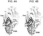
- B-cell maturation antigen also known as BCMA, CD269, TNFRSF17 (UniProt Q02223), is a member of the tumor necrosis receptor superfamily that is preferentially expressed in differentiated plasma cells [ Laabi et al. (1992) EMBO J 11(11):3897-3904 ; Madry et al. (1998) Int Immunol 10(11): 1693-1702 ].
- BCMA is a non-glycosylated type I transmembrane protein, which is involved in B cell maturation, growth and survival.
- BCMA is a receptor for two ligands of the TNF superfamily: APRIL (a proliferation-inducing ligand, CD256, TNFSF13), the high-affinity ligand to BCMA and the B cell activation factor BAFF (THANK, BlyS, B lymphocyte stimulator, TALL-1 and zTNF4), the low-affinity ligand to BCMA.
- APRIL and BAFF show structural similarity and overlapping yet distinct receptor binding specificity.
- the negative regulator TACI also binds to both BAFF and APRIL.
- the coordinate binding of APRIL and BAFF to BCMA and/or TACI activates transcription factor NF- ⁇ B and increases the expression of pro-survival Bcl-2 family members (e.g.
- Bcl-2, Bcl-xL, Bcl-w, Mcl-1, A1 down regulates expression of pro-apoptotic factors (e.g. Bid, Bad, Bik, Bim, etc.), thus inhibiting apoptosis and promoting survival.
- pro-apoptotic factors e.g. Bid, Bad, Bik, Bim, etc.
- This combined action promotes B cell differentiation, proliferation, survival and antibody production (as reviewed in Rickert RC et al., Immunol Rev (2011) 244 (1): 115-133 ).
- BCMA also supports growth and survival of malignant human B cells, including multiple myeloma (MM) cells.
- MM multiple myeloma
- MM Multiple myeloma
- MM is the second most common hematological malignancy and constitutes 2% of all cancer deaths.
- MM is a heterogeneous disease and caused by mostly by chromosome translocations inter alia t(11 ; 14),t(4; 14),t(8;14),del(13),del(17) ( Drach et al., (1998) Blood 92(3):802-809 ; Gertz et al., (2005) Blood 106(8):2837-2840 ; Facon et al., (2001) Blood 97(6): 1566-1571 ).
- MM-affected patients may experience a variety of disease-related symptoms due to, bone marrow infiltration, bone destruction, renal failure, immunodeficiency, and the psychosocial burden of a cancer diagnosis.
- the 5-year relative survival rate for MM was approximately 34% highlighting that MM is a difficult-to-treat disease where there are currently no curative options.
- anti-BCMA antibodies for the treatment of lymphomas and multiple myeloma are mentioned in WO2002066516 and WO2010104949 .
- Antibodies against BCMA are described e.g. in Gras M-P. et al. Int Immunol. 7 (1995) 1093-1106 , WO200124811 , and WO200124812 .
- BCMA, BAFF-R and TACI i.e., B cell receptors belonging to the TNF receptor superfamily, and their ligands BAFF and APRIL are subject to therapies in fighting against cancer, there is still a need for having available further options for the treatment of such medical conditions.
- antibodies that immunospecifically bind to BCMA and antigen-binding fragments thereof are also described. Also described are related polynucleotides capable of encoding the provided BCMA-specific antibodies and antigen-binding fragments, cells expressing the provided antibodies and antigen-binding fragments, as well as associated vectors and detectably labeled antibodies and antigen-binding fragments. In addition, methods of using the provided antibodies and antigen-binding fragments are described.
- the BCMA-specific antibodies and antigen-binding fragments may be used to diagnose or monitor BCMA-expressing cancer progression, regression, or stability; to determine whether or not a patient should be treated for cancer; or to determine whether or not a subject is afflicted with BCMA-expressing cancer and thus may be amenable to treatment with a BCMA-specific anti-cancer therapeutic, such as the multispecific antibodies against BCMA and CD3 described herein.
- multispecific antibodies that immunospecifically bind to BCMA and CD3 and multispecific antigen-binding fragments thereof. Also described are related polynucleotides capable of encoding the provided BCMA x CD3-multispecific antibodies, cells expressing the provided antibodies, as well as associated vectors and detectably labeled multispecific antibodies. In addition, methods of using the provided multispecific antibodies are described.
- the BCMA x CD3-multispecific antibodies may be used to diagnose or monitor BCMA-expressing cancer progression, regression, or stability; to determine whether or not a patient should be treated for cancer; or to determine whether or not a subject is afflicted with BCMA-expressing cancer and thus may be amenable to treatment with a BCMA-specific anti-cancer therapeutic, such as the BCMA x CD3-multispecific antibodies described herein.
- the BCMA-specific antibodies and antigen-binding fragments bind human BCMA. In some embodiments, the BCMA-specific antibodies and antigen-binding fragments bind human BCMA and cynomolgus monkey BCMA. In some embodiments, the BCMA-specific antibodies and antigen-binding fragments bind to an epitope including one or more residues from the BCMA extracellular domain (ECD). This BCMA-specific antibody or antigen-binding fragment may block APRIL-binding with an IC 50 of at least 5.9 nM as measured by ELISA.
- ECD extracellular domain
- Table 1 provides a summary of examples of some BCMA-specific antibodies described herein: Table 1.
- CDR sequences of mAbs generated against human BCMA SEQ ID NOs for each listed sequence are provided in parenthesis) ID HC-CDR1 HC-CDR2 HC-CDR3 LC-CDR1 LC-CDR2 LC-CDR3 BCMB69 SGSYFWG (4) SIYYSGITYYNPSLKS (5) HDGAVAGLFDY (6) GGNNIGSKSVH (24) DDSDRPS (25) QVWDSSSDHVV (26) BCMB117 SGSYFWG (4) SIYYSGITYYNPSLKS (5) HDGAVAGLFDY (6) GGNNIGSKSVH (24) DDSDRPS (25) QVWDSSSDHVV (26) BCMB123 SSSYYWG (7) SIYYSGITYYNPSLKS (5) HDGAVAGLFDY (6) GGNNIGSKSVH (24) DDSDRPS (25) QVWDSSSDHVV (26) BCMB
- a BCMA-specific antibody, or an antigen-binding fragment thereof comprising a heavy chain comprising a CDR1, a CDR2, and a CDR3 of any one of the antibodies described in Table 1.
- a BCMA-specific antibody, or an antigen-binding fragment thereof comprising a heavy chain comprising a CDR1, a CDR2, and a CDR3 of any one of the antibodies described in Table 1 and a light chain comprising a CDR1, a CDR2, and a CDR3 of any one of the antibodies described in Table 1.
- the IgG class is divided in four isotypes: IgG1, IgG2, IgG3 and IgG4 in humans. They share more than 95% homology in the amino acid sequences of the Fc regions but show major differences in the amino acid composition and structure of the hinge region.
- the Fc region mediates effector functions, such as antibody-dependent cellular cytotoxicity (ADCC) and complement-dependent cytotoxicity (CDC).
- ADCC antibody-dependent cellular cytotoxicity
- CDC complement-dependent cytotoxicity
- the Fc region of an antibody binds to Fc receptors (FcgRs) on the surface of immune effector cells such as natural killers and macrophages, leading to the phagocytosis or lysis of the targeted cells.
- the antibodies kill the targeted cells by triggering the complement cascade at the cell surface.
- the antibodies described herein include antibodies with the described features of the variable domains in combination with any of the IgG isotypes, including modified versions in which the Fc sequence has been modified to effect different
- Fc-mediated effector functions are not part of the mechanism of action. These Fc-mediated effector functions can be detrimental and potentially pose a safety risk by causing off-mechanism toxicity.
- Modifying effector functions can be achieved by engineering the Fc regions to reduce their binding to FcgRs or the complement factors.
- the binding of IgG to the activating (FcgRI, FcgRIIa, FcgRIIIa and FcgRIIIb) and inhibitory (FcgRIIb) FcgRs or the first component of complement (C1q) depends on residues located in the hinge region and the CH2 domain. Mutations have been introduced in IgG1, IgG2 and IgG4 to reduce or silence Fc functionalities.
- the antibodies described herein may include these modifications.
- the antibody comprises an Fc region with one or more of the following properties: (a) reduced effector function when compared to the parent Fc; (b) reduced affinity to Fcg RI, Fcg RIIa, Fcg RIIb, Fcg RIIIb and/or Fcg RIIIa, (c) reduced affinity to FcgRI (d) reduced affinity to FcgRIIa (e) reduced affinity to FcgRIIb, (f) reduced affinity to Fcg RIIIb or (g) reduced affinity to FcgRIIIa.
- the antibodies or antigen-binding fragments are IgG, or derivatives thereof, e.g., IgG1, IgG2, IgG3, and IgG4 isotypes.
- the antibody has an IgG4 isotype, the antibody contains K409R, S228P, L234A, and L235A substitutions in its Fc region.
- the antibodies described herein may include these modifications.
- the described antibodies are capable of inhibiting APRIL binding with a IC 50 of 5.9 nM as measured by ELISA.
- the described antibodies bind to BCMA-positive multiple myeloma cell lines.
- polynucleotide sequences capable of encoding the described antibodies and antigen-binding fragments.
- Vectors comprising the described polynucleotides are also provided, as are cells expressing the BCMA-specific antibodies or antigen-binding fragments provided herein.
- cells capable of expressing the disclosed vectors may be mammalian cells (such as 293F cells, CHO cells), insect cells (such as Sf7 cells), yeast cells, plant cells, or bacteria cells (such as E. coli).
- the described antibodies may also be produced by hybridoma cells.
- BCMA-specific antibodies or antigen-binding fragments are also disclosed.
- Particular antibodies for use in the methods discussed in this section include those with the set of CDRs described for antibodies in Table 1.
- these antibodies or antigen-binding fragments may be useful in treating cancer, by interfering with BCMA-receptor interactions or where the antibody is conjugated to a toxin, so targeting the toxin to the BCMA-expressing cancer.
- BCMA-expressing cancer may be a lymphoma, such as multiple myeloma (MM).
- the described methods may be carried out before the subject receives treatment for BCMA-expressing cancer, such as treatment with a multispecific antibody against BCMA and CD3.
- the described methods may be carried out after the subject receives treatment for BCMA-expressing cancer, such as treatment with a multispecific antibody against BCMA and CD3 described herein.
- the described methods of detecting BCMA in a biological sample include exposing the biological sample to one or more of the BCMA-specific antibodies or antigen-binding fragments described herein.
- the described methods of diagnosing BCMA-expressing cancer in a subject also involve exposing the biological sample to one or more of the BCMA-specific antibodies or antigen-binding fragments described herein; however, the methods also include quantifying the amount of BCMA present in the sample; comparing the amount of BCMA present in the sample to a known standard or reference sample; and determining whether the subject's BCMA levels fall within the levels of BCMA associated with cancer.
- the described methods include exposing the biological sample to one or more of the BCMA-specific antibodies or antigen-binding fragments described herein; quantifying the amount of BCMA present in the sample that is bound by the antibody, or antigen-binding fragment thereof; comparing the amount of BCMA present in the sample to either a known standard or reference sample or the amount of BCMA in a similar sample previously obtained from the subject; and determining whether the subject's BCMA levels are indicative of cancer progression, regression or stable disease based on the difference in the amount of BCMA in the compared samples.
- the samples obtained, or derived from, subjects are biological samples such as urine, blood, serum, plasma, saliva, ascites, circulating cells, circulating tumor cells, cells that are not tissue associated, tissues, surgically resected tumor tissue, biopsies, fine needle aspiration samples, or histological preparations.
- the described BCMA-specific antibodies or antigen-binding fragments may be labeled for use with the described methods, or other methods known to those skilled in the art.
- the antibodies described herein, or antigen-binding fragments thereof may be labeled with a radiolabel, a fluorescent label, an epitope tag, biotin, a chromophore label, an ECL label, an enzyme, ruthenium, 111 In-DOTA, 111 In- diethylenetriaminepentaacetic acid (DTPA), horseradish peroxidase, alkaline phosphatase and beta-galactosidase, or poly-histidine or similar such labels known in the art.
- DTPA 111 In- diethylenetriaminepentaacetic acid
- kits including the disclosed BCMA-specific antibodies or antigen-binding fragments thereof.
- the described kits may be used to carry out the methods of using the BCMA-specific antibodies or antigen-binding fragments provided herein, or other methods known to those skilled in the art.
- the described kits may include the antibodies or antigen-binding fragments described herein and reagents for use in detecting the presence of BCMA in a biological sample.
- kits may include one or more of the antibodies, or an antigen-binding fragment(s) thereof, described herein and a vessel for containing the antibody or fragment when not in use, instructions for use of the antibody or fragment, the antibody or fragment affixed to a solid support, and/or detectably labeled forms of the antibody or fragment, as described herein.
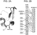
- the redirection of T-lymphocytes to MM cells expressing BCMA via the TCR/CD3 complex represents an attractive alternative approach.
- the TCR/CD3 complex of T-lymphocytes consists of either a TCR alpha ( ⁇ )/beta (( ⁇ ) or TCR gamma ( ⁇ )/delta ( ⁇ ) heterodimer coexpressed at the cell surface with the invariant subunits of CD3 labeled gamma ( ⁇ ), delta ( ⁇ ), epsilon ( ⁇ ), zeta ( ⁇ ), and eta ( ⁇ ).
- Human CD3 ⁇ is described under UniProt P07766 (CD3E_HUMAN).
- An anti CD3 ⁇ antibody described in the state of the art is SP34 ( Yang SJ, The Journal of Immunology (1986) 137; 1097-1100 ). SP34 reacts with both primate and human CD3. SP34 is available from Pharmingen.
- a further anti CD3 antibody described in the state of the art is UCHT-1 (see WO2000041474 ).
- a further anti CDS antibody described in the state of the art is BC-3 (Fred Hutchinson Cancer Research Institute; used in Phase I/II trials of GvHD, Anasetti et al., Transplantation 54: 844 (1992 )).
- SP34 differs from UCHT-1 and BC-3 in that SP-34 recognizes an epitope present on solely the ⁇ chain of CD3 (see Salmeron et al., (1991) J. Immunol. 147: 3047 ) whereas UCHT-1 and BC-3 recognize an epitope contributed by both the ⁇ and ⁇ chains.
- the sequence of an antibody with the same sequence as of antibody SP34 is mentioned in WO2008119565 , WO2008119566 , WO2008119567 , WO2010037836 , WO2010037837 and WO2010037838 .
- a sequence which is 96% identical to the heavy chain variable domain (VH) of antibody SP34 is mentioned in US8236308 ( WO2007042261 ).
- BCMA x CD3 multispecific antibodies recombinant multispecific antibodies that bind BCMA and CD3
- BCMA x CD3 multispecific antibodies multispecific antigen-binding fragments thereof.
- a recombinant antibody, or an antigen-binding fragment thereof, that binds immunospecifically to BCMA is provided.
- the BCMA-specific arm of the multispecific antibody binds human BCMA and cynomolgus monkey BCMA. In some embodiments, the BCMA-specific arm of the BCMA x CD3-multispecific antibodies or antigen-binding fragments binds the extracellular domain of human BCMA. In preferred embodiments, the BCMA x CD3 multispecific antibody or antigen-binding fragment is a bispecific antibody or antigen-binding fragment.
- a recombinant BCMA x CD3 bispecific antibody comprising: a) a first heavy chain (HC1); b) a second heavy chain (HC2); c) a first light chain (LC1); and d) a second light chain (LC2), wherein the HC1 and the LC1 pair to form a first antigen-binding site that immunospecifically binds BCMA, and the HC2 and the LC2 pair to form a second antigen-binding site that immunospecifically binds CD3, or a BCMA x CD3-bispecific binding fragment thereof is provided.
- a recombinant cell expressing the antibody or bispecific binding fragment is provided.
- the BCMA-binding arm (or "BCMA-specific arm" of the BCMA x CD3 multispecific antibody is derived from a BCMA antibody described herein (for example, from an antibody having the CDR sequences listed in Table 1).
- the BCMA-specific arm of the BCMA x CD3-multispecific antibodies or antigen-binding fragments are IgG, or derivatives thereof.
- the described BCMA x CD3-multispecific antibodies are capable of binding to BCMA with a dissociation constant of at least 0.18 nM as measured by surface plasmon resonance.
- the described BCMA x CD3-multispecific antibody is not an agonist.
- the described BCMA x CD3-multispecific antibody does not alter NF- ⁇ B activation at concentrations below 10 nM.
- the CD3-binding arm (or "CD3-specific arm") of the BCMA x CD3 multispecific antibody is derived from the mouse monoclonal antibody SP34, a mouse IgG3/lambda isotype. ( K.R. Abhinandan and A. C. Martin, 2008. Mol. Immunol. 45, 3832-3839 ).
- the CD3-binding arm of the BCMA x CD3 multispecific antibody comprises one heavy chain and one light chain selected from Table 2. Table 2. Heavy chains and light chains of the CD3-specific antibodies and antigen-binding fragments.
- CD3B219 (SEQ ID NO:55): CD3B219 (SEQ ID NO:56): CDR 1: TYAMN (SEQ ID NO: 59) CDR 1: RSSTGAVTTSNYAN (SEQ ID NO: 62) CDR 2: RIRSKYNNYATYYAASVKG (SEQ ID NO: 60) CDR 2: GTNKRAP (SEQ ID NO: 63) CDR 3: HGNFGNSYVSWFAY (SEQ ID NO: 61) CDR 3: ALWYSNLWV (SEQ ID NO: 64)
- the IgG class is divided in four isotypes: IgG1, IgG2, IgG3 and IgG4 in humans. They share more than 95% homology in the amino acid sequences of the Fc regions but show major differences in the amino acid composition and structure of the hinge region.
- the Fc region mediates effector functions, such as antibody-dependent cellular cytotoxicity (ADCC) and complement-dependent cytotoxicity (CDC).
- ADCC antibody-dependent cellular cytotoxicity
- CDC complement-dependent cytotoxicity
- the Fc region of an antibody binds to Fc receptors (FcgRs) on the surface of immune effector cells such as natural killers and macrophages, leading to the phagocytosis or lysis of the targeted cells.
- FcgRs Fc receptors
- the antibodies kill the targeted cells by triggering the complement cascade at the cell surface.
- Fc-mediated effector functions are not part of the mechanism of action. These Fc-mediated effector functions can be detrimental and potentially pose a safety risk by causing off-mechanism toxicity.
- Modifying effector functions can be achieved by engineering the Fc regions to reduce their binding to FcgRs or the complement factors.
- the binding of IgG to the activating (FcgRI, FcgRIIa, FcgRIIIa and FcgRIIIb) and inhibitory (FcgRIIb) FcgRs or the first component of complement (C1q) depends on residues located in the hinge region and the CH2 domain. Mutations have been introduced in IgG1, IgG2 and IgG4 to reduce or silence Fc functionalities.
- the antibody comprises an Fc region with one or more of the following properties: (a) reduced effector function when compared to the parent Fc; (b) reduced affinity to Fcg RI, Fcg RIIa, Fcg RIIb, Fcg RIIIb and/or Fcg RIIIa, (c) reduced affinity to FcgRI (d) reduced affinity to FcgRIIa (e) reduced affinity to FcgRIIb, (f) reduced affinity to Fcg RIIIb or (g) reduced affinity to FcgRIIIa.
- the CD3-specific antibody or antigen-binding fragment from which the CD3-specific arm of the multispecific antibody is derived is IgG, or a derivative thereof. In some embodiments, the CD3-specific antibody or antigen-binding fragment from which the CD3-specific arm of the multispecific antibody is derived is IgG1, or a derivative thereof. In some embodiments, for example, the Fc region of the CD3-specific IgG1 antibody from which the CD3-binding arm is derived comprises L234A, L235A, and F405L substitutions in its Fc region.
- the CD3-specific antibody or antigen-binding fragment from which the CD3-specific arm of the multispecific antibody is derived is IgG4, or a derivative thereof.
- the Fc region of the CD3-specific IgG4 antibody from which the CD3-binding arm is derived comprises S228P, L234A, L235A, F405L, and R409K substitutions in its Fc region.
- the CD3-specific antibody or antigen-binding fragment from which the CD3-specific arm of the multispecific antibody is derived binds CD3 ⁇ on primary human T cells and/or primary cynomolgus T cells.
- the CD3-specific antibody or antigen-binding fragment from which the CD3-specific arm of the multispecific antibody is derived activates primary human CD4+ T cells and/or primary cynomolgus CD4+ T cells.
- polynucleotide sequences capable of encoding the described BCMA x CD3-multispecific antibodies.
- an isolated synthetic polynucleotide encoding the HC1, the HC2, the LC1 or the LC2 of the BCMA x CD3 bispecific antibody or bispecific binding fragment is provided.
- Vectors comprising the described polynucleotides are also provided, as are cells expressing the BCMA x CD3-multispecific antibodies provided herein. Also described are cells capable of expressing the disclosed vectors.
- These cells may be mammalian cells (such as 293F cells, CHO cells), insect cells (such as Sf7 cells), yeast cells, plant cells, or bacteria cells (such as E. coli).
- the described antibodies may also be produced by hybridoma cells.
- methods for generating the BCMA x CD3 bispecific antibody or bispecific binding fragment by culturing cells is provided.
- compositions comprising the BCMA x CD3 multispecific antibodies or antigen-binding fragments and a pharmaceutically acceptable carrier.
- the BCMA x CD3-multispecific antibodies and multispecific antigen-binding fragments thereof may be useful in the treatment of a BCMA-expressing cancer in a subject in need thereof.
- the BCMA-expressing cancer is a lymphoma, such as multiple myeloma.
- the described methods of treating BCMA-expressing cancer in a subject in need thereof include administering to the subject a therapeutically effective amount of a described BCMA x CD3-multispecific antibody or multispecific antigen-binding fragment thereof.
- the subject is a mammal, preferably a human.
- methods for treating a subject having cancer by administering a therapeutically effective amount of the BCMA x CD3 bispecific antibody or bispecific antigen-binding fragment to a patient in need thereof for a time sufficient to treat the cancer.
- kits including the disclosed BCMA x CD3-multispecific antibodies.
- the described kits may be used to carry out the methods of using the BCMA x CD3-multispecific antibodies provided herein, or other methods known to those skilled in the art.
- the described kits may include the antibodies described herein and reagents for use in treating a BCMA-expressing cancer.
- the described kits may include one or more of the multispecific antibodies, or a multispecific antigen-binding fragment(s) thereof, described herein and a vessel for containing the antibody or fragment when not in use, and/or instructions for use of the antibody or fragment, the antibody or fragment affixed to a solid support, and/or detectably labeled forms of the antibody or fragment, as described herein.
- Isolated means a biological component (such as a nucleic acid, peptide or protein) has been substantially separated, produced apart from, or purified away from other biological components of the organism in which the component naturally occurs, i.e., other chromosomal and extrachromosomal DNA and RNA, and proteins. Nucleic acids, peptides and proteins that have been “isolated” thus include nucleic acids and proteins purified by standard purification methods. "Isolated" nucleic acids, peptides and proteins can be part of a composition and still be isolated if such composition is not part of the native environment of the nucleic acid, peptide, or protein.
- nucleic acids, peptides and proteins prepared by recombinant expression in a host cell as well as chemically synthesized nucleic acids.
- An "isolated" antibody or antigen-binding fragment is intended to refer to an antibody or antigen-binding fragment which is substantially free of other antibodies or antigen-binding fragments having different antigenic specificities (for instance, an isolated antibody that specifically binds to BCMA is substantially free of antibodies that specifically bind antigens other than BCMA).
- An isolated antibody that specifically binds to an epitope, isoform or variant of BCMA may, however, have cross-reactivity to other related antigens, for instance from other species (such as BCMA species homologs).
- Polynucleotide synonymously referred to as “nucleic acid molecule,” “nucleotides” or “nucleic acids,” refers to any polyribonucleotide or polydeoxyribonucleotide, which may be unmodified RNA or DNA or modified RNA or DNA.
- Polynucleotides include, without limitation single- and double-stranded DNA, DNA that is a mixture of single- and double-stranded regions, single- and double-stranded RNA, and RNA that is mixture of single- and double-stranded regions, hybrid molecules comprising DNA and RNA that may be single-stranded or, more typically, double-stranded or a mixture of single- and double-stranded regions.
- polynucleotide refers to triple-stranded regions comprising RNA or DNA or both RNA and DNA.
- the term polynucleotide also includes DNAs or RNAs containing one or more modified bases and DNAs or RNAs with backbones modified for stability or for other reasons.
- Modified bases include, for example, tritylated bases and unusual bases such as inosine.
- polynucleotide embraces chemically, enzymatically or metabolically modified forms of polynucleotides as typically found in nature, as well as the chemical forms of DNA and RNA characteristic of viruses and cells.
- Polynucleotide also embraces relatively short nucleic acid chains, often referred to as oligonucleotides.
- the term refers to at least 70% identity between two or more sequences, more preferably at least 75% identity, more preferably at least 80% identity, more preferably at least 85% identity, more preferably at least 90% identity, more preferably at least 91% identity, more preferably at least 92% identity, more preferably at least 93% identity, more preferably at least 94% identity, more preferably at least 95% identity, more preferably at least 96% identity, more preferably at least 97% identity, more preferably at least 98% identity, and more preferably at least 99% or greater identity.
- the percent identity between two nucleotide or amino acid sequences may e.g. be determined using the algorithm of E. Meyers and W. Miller, Comput. Appl. Biosci 4, 11-17 (1988 ) which has been incorporated into the ALIGN program (version 2.0), using a PAM120 weight residue table, a gap length penalty of 12 and a gap penalty of 4.
- the percent identity between two amino acid sequences may be determined using the Needleman and Wunsch, J. Mol. Biol. 48, 444-453 (1970 ) algorithm.
- BCMA specific antibodies, or antigen-binding fragments that have framework, scaffold, or other non-binding regions that do not share significant identity with the antibodies and antigen-binding fragments described herein, but do incorporate one or more CDRs or other sequences needed to confer binding that are 90%, 91%, 92%, 93%, 94%, 95%, 96%, 97%, 98%, or 99% identical to such sequences described herein.
- a “vector” is a replicon, such as plasmid, phage, cosmid, or virus in which another nucleic acid segment may be operably inserted so as to bring about the replication or expression of the segment.
- a “clone” is a population of cells derived from a single cell or common ancestor by mitosis.
- a “cell line” is a clone of a primary cell that is capable of stable growth in vitro for many generations. In some examples provided herein, cells are transformed by transfecting the cells with DNA.
- express and produce are used synonymously herein, and refer to the biosynthesis of a gene product. These terms encompass the transcription of a gene into RNA. These terms also encompass translation of RNA into one or more polypeptides, and further encompass all naturally occurring post-transcriptional and post-translational modifications.
- the expression or production of an antibody or antigen-binding fragment thereof may be within the cytoplasm of the cell, or into the extracellular milieu such as the growth medium of a cell culture.
- treating refers to any success or indicia of success in the attenuation or amelioration of an injury, pathology or condition, including any objective or subjective parameter such as abatement, remission, diminishing of symptoms or making the condition more tolerable to the patient, slowing in the rate of degeneration or decline, making the final point of degeneration less debilitating, improving a subject's physical or mental well-being, or prolonging the length of survival.
- the treatment may be assessed by objective or subjective parameters; including the results of a physical examination, neurological examination, or psychiatric evaluations.
- an “effective amount” or “therapeutically effective amount” refers to an amount effective, at dosages and for periods of time necessary, to achieve a desired therapeutic result.
- a therapeutically effective amount of a BCMA x CD3 antibody may vary according to factors such as the disease state, age, sex, and weight of the individual, and the ability of the antibody to elicit a desired response in the individual.
- a therapeutically effective amount is also one in which any toxic or detrimental effects of the antibody or antibody portion are outweighed by the therapeutically beneficial effects.
- Antibody refers to all isotypes of immunoglobulins (IgG, IgA, IgE, IgM, IgD, and IgY) including various monomeric, polymeric and chimeric forms, unless otherwise specified. Specifically encompassed by the term “antibody” are polyclonal antibodies, monoclonal antibodies (mAbs), and antibody-like polypeptides, such as chimeric antibodies and humanized antibodies.
- Antigen-binding fragments are any proteinaceous structure that may exhibit binding affinity for a particular antigen.
- Antigen-binding fragments include those provided by any known technique, such as enzymatic cleavage, peptide synthesis, and recombinant techniques. Some antigen-binding fragments are composed of portions of intact antibodies that retain antigen-binding specificity of the parent antibody molecule.
- antigen-binding fragments may comprise at least one variable region (either a heavy chain or light chain variable region) or one or more CDRs of an antibody known to bind a particular antigen.
- Suitable antigen-binding fragments include, without limitation diabodies and single-chain molecules as well as Fab, F(ab')2, Fc, Fabc, and Fv molecules, single chain (Sc) antibodies, individual antibody light chains, individual antibody heavy chains, chimeric fusions between antibody chains or CDRs and other proteins, protein scaffolds, heavy chain monomers or dimers, light chain monomers or dimers, dimers consisting of one heavy and one light chain, a monovalent fragment consisting of the VL, VH, CL and CH1 domains, or a monovalent antibody as described in WO2007059782 , bivalent fragments comprising two Fab fragments linked by a disulfide bridge at the hinge region, a Fd fragment consisting essentially of the V.sub.H and C.sub.H1 domains; a Fv fragment consisting essentially of the VL and VH domains of a single arm of an antibody, a dAb fragment ( Ward et al., Nature 341, 544-546 (1989 )
- antigen-binding fragments may include non-antibody proteinaceous frameworks that may successfully incorporate polypeptide segments in an orientation that confers affinity for a given antigen of interest, such as protein scaffolds.
- Antigen-binding fragments may be recombinantly produced or produced by enzymatic or chemical cleavage of intact antibodies.
- the phrase "an antibody or antigen-binding fragment thereof" may be used to denote that a given antigen-binding fragment incorporates one or more amino acid segments of the antibody referred to in the phrase.
- epitope means a protein determinant capable of specific binding to an antibody.
- Epitopes usually consist of surface groupings of molecules such as amino acids or sugar side chains and usually have specific three dimensional structural characteristics, as well as specific charge characteristics. Conformational and nonconformational epitopes are distinguished in that the binding to the former but not the latter is lost in the presence of denaturing solvents.
- the epitope may comprise amino acid residues directly involved in the binding and other amino acid residues, which are not directly involved in the binding, such as amino acid residues which are effectively blocked or covered by the specifically antigen binding peptide (in other words, the amino acid residue is within the footprint of the specifically antigen binding peptide).
- Specific binding or “immunospecific binding” or derivatives thereof when used in the context of antibodies, or antibody fragments, represents binding via domains encoded by immunoglobulin genes or fragments of immunoglobulin genes to one or more epitopes of a protein of interest, without preferentially binding other molecules in a sample containing a mixed population of molecules.
- an antibody binds to a cognate antigen with a K d of less than about 1x10 -8 M, as measured by a surface plasmon resonance assay or a cell binding assay.
- Phrases such as "[antigen]-specific" antibody e.g., BCMA-specific antibody
- K D refers to the dissociation equilibrium constant of a particular antibody-antigen interaction.
- subject refers to human and non-human animals, including all vertebrates, e.g., mammals and non-mammals, such as non-human primates, mice, rabbits, sheep, dogs, cats, horses, cows, chickens, amphibians, and reptiles. In many embodiments of the described methods, the subject is a human.
- sample refers to a collection of similar fluids, cells, or tissues (e.g., surgically resected tumor tissue, biopsies, including fine needle aspiration), isolated from a subject, as well as fluids, cells, or tissues present within a subject.
- the sample is a biological fluid.
- Biological fluids are typically liquids at physiological temperatures and may include naturally occurring fluids present in, withdrawn from, expressed or otherwise extracted from a subject or biological source. Certain biological fluids derive from particular tissues, organs or localized regions and certain other biological fluids may be more globally or systemically situated in a subject or biological source.
- biological fluids examples include blood, serum and serosal fluids, plasma, lymph, urine, saliva, cystic fluid, tear drops, feces, sputum, mucosal secretions of the secretory tissues and organs, vaginal secretions, ascites fluids such as those associated with non-solid tumors, fluids of the pleural, pericardial, peritoneal, abdominal and other body cavities, fluids collected by bronchial lavage and the like.
- Biological fluids may also include liquid solutions contacted with a subject or biological source, for example, cell and organ culture medium including cell or organ conditioned medium, lavage fluids and the like.
- sample encompasses materials removed from a subject or materials present in a subject.
- a "known standard” may be a solution having a known amount or concentration of BCMA, where the solution may be a naturally occurring solution, such as a sample from a patient known to have early, moderate, late, progressive, or static cancer, or the solution may be a synthetic solution such as buffered water having a known amount of BCMA diluted therein.
- the known standards, described herein may include BCMA isolated from a subject, recombinant or purified BCMA protein, or a value of BCMA concentration associated with a disease condition.
- BCMA human B cell maturation antigen
- CD269 CD269
- TNFRSF17 TNFRSF17
- the extracellular domain of human BCMA consists, according to UniProt of amino acids 1 - 54 (or 5-51).
- antibody against BCMA, anti BCMA antibody as used herein relates to an antibody immunospecifically binding to BCMA.
- CD3 refers to the human CD3 protein multi-subunit complex.
- the CD3 protein multi-subunit complex is composed to 6 distinctive polypeptide chains. These include a CD3y chain (SwissProt P09693), a CD3 ⁇ chain (SwissProt P04234), two CD3 ⁇ chains (SwissProt P07766), and one CD3 ⁇ chain homodimer (SwissProt 20963), and which is associated with the T cell receptor ⁇ and ⁇ chain.
- CD3 includes any CD3 variant, isoform and species homolog which is naturally expressed by cells (including T cells) or can be expressed on cells transfected with genes or cDNA encoding those polypeptides, unless noted.
- a "BCMA x CD3 antibody” is a multispecific antibody, optionally a bispecific antibody, which comprises two different antigen-binding regions, one of which binds specifically to the antigen BCMA and one of which binds specifically to CD3.
- a multispecific antibody can be a bispecific antibody, diabody, or similar molecule (see for instance PNAS USA 90(14), 6444-8 (1993 ) for a description of diabodies).
- the bispecific antibodies, diabodies, and the like, provided herein may bind any suitable target in addition to a portion of BCMA.
- the term "bispecific antibody” is to be understood as an antibody having two different antigen-binding regions defined by different antibody sequences. This can be understood as different target binding but includes as well binding to different epitopes in one target.
- a "reference sample” is a sample that may be compared against another sample, such as a test sample, to allow for characterization of the compared sample.
- the reference sample will have some characterized property that serves as the basis for comparison with the test sample.
- a reference sample may be used as a benchmark for BCMA levels that are indicative of a subject having cancer.
- the reference sample does not necessarily have to be analyzed in parallel with the test sample, thus in some instances the reference sample may be a numerical value or range previously determined to characterize a given condition, such as BCMA levels that are indicative of cancer in a subject.
- the term also includes samples used for comparative purposes that are known to be associated with a physiologic state or disease condition, such as BCMA-expressing cancer, but that have an unknown amount of BCMA.
- progression includes the change of a cancer from a less severe to a more severe state. This may include an increase in the number or severity of tumors, the degree of metastasis, the speed with which the cancer is growing or spreading, and the like.
- the progression of colon cancer includes the progression of such a cancer from a less severe to a more severe state, such as the progression from stage I to stage II, from stage II to stage III, etc.
- regression includes the change of a cancer from a more severe to a less severe state. This could include a decrease in the number or severity of tumors, the degree of metastasis, the speed with which the cancer is growing or spreading, and the like.
- the regression of colon cancer includes the regression of such a cancer from a more severe to a less severe state, such as the progression from stage III to stage II, from stage II to stage I, etc.
- stable as used in the context of stable BCMA-expressing cancer, is intended to describe a disease condition that is not, or has not, changed significantly enough over a clinically relevant period of time to be considered a progressing cancer or a regressing cancer.
- Described herein are recombinant monoclonal antibodies or antigen-binding fragments that specifically bind BCMA
- the general structure of an antibody molecule comprises an antigen binding domain, which includes heavy and light chains, and the Fc domain, which serves a variety of functions, including complement fixation and binding antibody receptors.
- the described BCMA-specific antibodies or antigen-binding fragments include all isotypes, IgA, IgD, IgE, IgG and IgM, and synthetic multimers of the four-chain immunoglobulin structure.
- the described antibodies or antigen-binding fragments also include the IgY isotype generally found in hen or turkey serum and hen or turkey egg yolk.
- the BCMA-specific antibodies and antigen-binding fragments may be derived from any species by recombinant means.
- the antibodies or antigen-binding fragments may be mouse, rat, goat, horse, swine, bovine, chicken, rabbit, camelid, donkey, human, or chimeric versions thereof.
- non-human derived antibodies or antigen-binding fragments may be genetically or structurally altered to be less antigenic upon administration to a human patient.
- the antibodies or antigen-binding fragments are chimeric.
- the term “chimeric” refers to an antibody, or antigen-binding fragment thereof, having at least some portion of at least one variable domain derived from the antibody ammo acid sequence of a non-human mammal, a rodent, or a reptile, while the remaining portions of the antibody, or antigen-binding fragment thereof, are derived from a human.
- the antibodies are humanized antibodies.
- Humanized antibodies may be chimeric immunoglobulins, immunoglobulin chains or fragments thereof (such as Fv, Fab, Fab', F(ab')2 or other antigen-binding subsequences of antibodies) that contain minimal sequence derived from non-human immunoglobulin.
- humanized antibodies are human immunoglobulins (recipient antibody) in which residues from a complementary-determining region (CDR) of the recipient are replaced by residues from a CDR of a non-human species (donor antibody) such as mouse, rat or rabbit having the desired specificity, affinity, and capacity.
- CDR complementary-determining region
- the humanized antibody will comprise substantially all of at least one, and typically two, variable domains, in which all or substantially all of the CDR regions correspond to those of a non-human immunoglobulin and all or substantially all of the framework regions are those of a human immunoglobulin sequence.
- the humanized antibody may include at least a portion of an immunoglobulin constant region (Fc), typically that of a human immunoglobulin.
- the antibodies or antigen-binding fragments described herein can occur in a variety of forms, but will include one or more of the antibody CDRs shown in Table 1.
- the BCMA-specific antibodies or antigen-binding fragments are human IgG, or derivatives thereof. While the BCMA-specific antibodies or antigen-binding fragments exemplified herein are human, the antibodies or antigen-binding fragments exemplified may be chimerized.
- a BCMA-specific antibody, or an antigen-binding fragment thereof comprising a heavy chain comprising a CDR1, a CDR2, and a CDR3 of any one of the antibodies described in Table 1.
- a BCMA-specific antibody, or an antigen-binding fragment thereof comprising a heavy chain comprising a CDR1, a CDR2, and a CDR3 of any one of the antibodies described in Table 1 and a light chain comprising a CDR1, a CDR2, and a CDR3 of any one of the antibodies described in Table 1.
- the BCMA-specific antibodies and antigen-binding fragments comprise a heavy chain CDR1 comprising SEQ ID NO: 4, a heavy chain CDR2 comprising SEQ ID NO: 5, and a heavy chain CDR3 comprising SEQ ID NO: 6.
- the BCMA-specific antibodies and antigen-binding fragments comprise a heavy chain CDR1 comprising SEQ ID NO: 4, a heavy chain CDR2 comprising SEQ ID NO: 5, a heavy chain CDR3 comprising SEQ ID NO: 6, a light chain CDR1 comprising SEQ ID NO: 7, a light chain CDR2 comprising SEQ ID NO: 8, and a light chain CDR3 comprising SEQ ID NO: 9.
- This BCMA-specific antibody or antigen-binding fragment may comprise human framework sequences. This BCMA-specific antibody or antigen-binding fragment may block APRIL binding with an IC 50 of at least 5.9 nM.
- the BCMA-specific antibodies and antigen-binding fragments comprise a heavy chain variable domain substantially the same as, or identical to, SEQ ID NO: 10.
- the BCMA-specific antibodies and antigen-binding fragments comprise a heavy chain variable domain substantially the same as, or identical to, SEQ ID NO: 10 and a light chain variable domain substantially the same as, or identical to, SEQ ID NO: 11.
- the heavy chain variable domain and light chain variable domain of antibodies discussed in this paragraph are suitable for inclusion in bispecific constructs in which one arm is an anti-BCMA arm.
- the BCMA-specific antibodies and antigen-binding fragments comprise a heavy chain CDR1 comprising SEQ ID NO: 4, a heavy chain CDR2 comprising SEQ ID NO: 5, and a heavy chain CDR3 comprising SEQ ID NO: 6.
- the BCMA-specific antibodies and antigen-binding fragments comprise a heavy chain CDR1 comprising SEQ ID NO: 7, a heavy chain CDR2 comprising SEQ ID NO: 5, a heavy chain CDR3 comprising SEQ ID NO: 6, a light chain CDR1 comprising SEQ ID NO: 24, a light chain CDR2 comprising SEQ ID NO: 25, and a light chain CDR3 comprising SEQ ID NO: 26.
- This BCMA-specific antibody or antigen-binding fragment may comprise human framework sequences.
- the BCMA-specific antibodies and antigen-binding fragments comprise a heavy chain variable domain substantially the same as, or identical to, SEQ ID NO: 57.
- the BCMA-specific antibodies and antigen-binding fragments comprise a heavy chain variable domain substantially the same as, or identical to, SEQ ID NO: 57 and a light chain variable domain substantially the same as, or identical to, SEQ ID NO: 28.
- the heavy chain variable domain and light chain variable domain of antibodies discussed in this paragraph are suitable for inclusion in bispecific constructs in which one arm is an anti-BCMA arm.
- the BCMA-specific antibodies and antigen-binding fragments comprise a heavy chain CDR1 comprising SEQ ID NO: 7, a heavy chain CDR2 comprising SEQ ID NO: 5, and a heavy chain CDR3 comprising SEQ ID NO: 6.
- the BCMA-specific antibodies and antigen-binding fragments comprise a heavy chain CDR1 comprising SEQ ID NO: 7, a heavy chain CDR2 comprising SEQ ID NO: 5, a heavy chain CDR3 comprising SEQ ID NO: 6, a light chain CDR1 comprising SEQ ID NO: 24, a light chain CDR2 comprising SEQ ID NO: 25, and a light chain CDR3 comprising SEQ ID NO: 26.
- This BCMA-specific antibody or antigen-binding fragment may comprise human framework sequences.
- the BCMA-specific antibodies and antigen-binding fragments comprise a heavy chain variable domain substantially the same as, or identical to, SEQ ID NO: 34.
- the BCMA-specific antibodies and antigen-binding fragments comprise a heavy chain variable domain substantially the same as, or identical to, SEQ ID NO: 34 and a light chain variable domain substantially the same as, or identical to, SEQ ID NO: 28.
- the heavy chain variable domain and light chain variable domain of antibodies discussed in this paragraph are suitable for inclusion in bispecific constructs in which one arm is an anti-BCMA arm.
- the BCMA-specific antibodies and antigen-binding fragments comprise a heavy chain CDR1 comprising SEQ ID NO: 4, a heavy chain CDR2 comprising SEQ ID NO: 5, and a heavy chain CDR3 comprising SEQ ID NO: 19.
- the BCMA-specific antibodies and antigen-binding fragments comprise a heavy chain CDR1 comprising SEQ ID NO: 4, a heavy chain CDR2 comprising SEQ ID NO: 5, a heavy chain CDR3 comprising SEQ ID NO: 19, a light chain CDR1 comprising SEQ ID NO: 24, a light chain CDR2 comprising SEQ ID NO: 25, and a light chain CDR3 comprising SEQ ID NO: 26.
- This BCMA-specific antibody or antigen-binding fragment may comprise human framework sequences.
- the BCMA-specific antibodies and antigen-binding fragments comprise a heavy chain variable domain substantially the same as, or identical to, SEQ ID NO: 39.
- the BCMA-specific antibodies and antigen-binding fragments comprise a heavy chain variable domain substantially the same as, or identical to, SEQ ID NO: 39 and a light chain variable domain substantially the same as, or identical to, SEQ ID NO: 28.
- the heavy chain variable domain and light chain variable domain of antibodies discussed in this paragraph are suitable for inclusion in bispecific constructs in which one arm is an anti-BCMA arm.
- the BCMA-specific antibodies and antigen-binding fragments comprise a heavy chain CDR1 comprising SEQ ID NO: 4, a heavy chain CDR2 comprising SEQ ID NO: 8, and a heavy chain CDR3 comprising SEQ ID NO: 6.
- the BCMA-specific antibodies and antigen-binding fragments comprise a heavy chain CDR1 comprising SEQ ID NO: 4, a heavy chain CDR2 comprising SEQ ID NO: 8, a heavy chain CDR3 comprising SEQ ID NO: 6, a light chain CDR1 comprising SEQ ID NO: 24, a light chain CDR2 comprising SEQ ID NO: 25, and a light chain CDR3 comprising SEQ ID NO: 26.
- This BCMA-specific antibody or antigen-binding fragment may comprise human framework sequences.
- the BCMA-specific antibodies and antigen-binding fragments comprise a heavy chain variable domain substantially the same as, or identical to, SEQ ID NO: 40.
- the BCMA-specific antibodies and antigen-binding fragments comprise a heavy chain variable domain substantially the same as, or identical to, SEQ ID NO: 40 and a light chain variable domain substantially the same as, or identical to, SEQ ID NO: 28.
- the heavy chain variable domain and light chain variable domain of antibodies discussed in this paragraph are suitable for inclusion in bispecific constructs in which one arm is an anti-BCMA arm.
- the BCMA-specific antibodies and antigen-binding fragments comprise a heavy chain CDR1 comprising SEQ ID NO: 13, a heavy chain CDR2 comprising SEQ ID NO: 5, and a heavy chain CDR3 comprising SEQ ID NO: 19.
- the BCMA-specific antibodies and antigen-binding fragments comprise a heavy chain CDR1 comprising SEQ ID NO: 13, a heavy chain CDR2 comprising SEQ ID NO: 5, a heavy chain CDR3 comprising SEQ ID NO: 19, a light chain CDR1 comprising SEQ ID NO: 24, a light chain CDR2 comprising SEQ ID NO: 25, and a light chain CDR3 comprising SEQ ID NO: 26.
- This BCMA-specific antibody or antigen-binding fragment may comprise human framework sequences.
- the BCMA-specific antibodies and antigen-binding fragments comprise a heavy chain variable domain substantially the same as, or identical to, SEQ ID NO: 58.
- the BCMA-specific antibodies and antigen-binding fragments comprise a heavy chain variable domain substantially the same as, or identical to, SEQ ID NO: 58 and a light chain variable domain substantially the same as, or identical to, SEQ ID NO: 28.
- the heavy chain variable domain and light chain variable domain of antibodies discussed in this paragraph are suitable for inclusion in bispecific constructs in which one arm is an anti-BCMA arm.
- the BCMA-specific antibodies and antigen-binding fragments comprise a heavy chain CDR1 comprising SEQ ID NO: 13, a heavy chain CDR2 comprising SEQ ID NO: 8, and a heavy chain CDR3 comprising SEQ ID NO: 19.
- the BCMA-specific antibodies and antigen-binding fragments comprise a heavy chain CDR1 comprising SEQ ID NO: 13, a heavy chain CDR2 comprising SEQ ID NO: 8, a heavy chain CDR3 comprising SEQ ID NO: 19, a light chain CDR1 comprising SEQ ID NO: 24, a light chain CDR2 comprising SEQ ID NO: 25, and a light chain CDR3 comprising SEQ ID NO: 26.
- This BCMA-specific antibody or antigen-binding fragment may comprise human framework sequences.
- the BCMA-specific antibodies and antigen-binding fragments comprise a heavy chain variable domain substantially the same as, or identical to, SEQ ID NO: 43.
- the BCMA-specific antibodies and antigen-binding fragments comprise a heavy chain variable domain substantially the same as, or identical to, SEQ ID NO: 43 and a light chain variable domain substantially the same as, or identical to, SEQ ID NO: 28.
- the heavy chain variable domain and light chain variable domain of antibodies discussed in this paragraph are suitable for inclusion in bispecific constructs in which one arm is an anti-BCMA arm.
- the antibodies or antigen-binding fragments are IgG, or derivatives thereof, e.g., IgG1, IgG2, IgG3, and IgG4 isotypes.
- the antibody comprises an IgG1 Fc region (SEQ ID NO. 74).
- the antibody contains S228P, L234A, and L235A substitutions in its Fc region (SEQ ID NO. 73).
- the specific antibodies defined by CDR and/or variable domain sequence discussed in the above paragraphs may include these IgG Fc regions.
- isolated synthetic polynucleotides that encode the antibodies or antigen-binding fragments that immunospecifically bind to BCMA.
- the isolated polynucleotides capable of encoding the variable domain segments provided herein may be included on the same, or different, vectors to produce antibodies or antigen-binding fragments.
- polynucleotides encoding recombinant antigen-binding proteins also are within the scope of the disclosure.
- the polynucleotides described (and the peptides they encode) include a leader sequence. Any leader sequence known in the art may be employed.
- the leader sequence may include, but is not limited to, a restriction site or a translation start site.
- the BCMA-specific antibodies or antigen-binding fragments described herein include variants having single or multiple amino acid substitutions, deletions, or additions that retain the biological properties (e.g., binding affinity or immune effector activity) of the described BCMA-specific antibodies or antigen-binding fragments.
- substitution of an amino acid in a given position is written as e.g. K409R which means a substitution of a Lysine in position 409 with an Arginine; and ii) for specific variants the specific three or one letter codes are used, including the codes Xaa and X to indicate any amino acid residue.
- substitution of Arginine for Lysine in position 409 is designated as: K409R, or the substitution of any amino acid residue for Lysine in position 409 is designated as K409X.
- deletion of Lysine in position 409 it is indicated by K409*.
- the skilled person may produce variants having single or multiple amino acid substitutions, deletions, or additions.
- variants may include: (a) variants in which one or more amino acid residues are substituted with conservative or nonconservative amino acids, (b) variants in which one or more amino acids are added to or deleted from the polypeptide, (c) variants in which one or more amino acids include a substituent group, and (d) variants in which the polypeptide is fused with another peptide or polypeptide such as a fusion partner, a protein tag or other chemical moiety, that may confer useful properties to the polypeptide, such as, for example, an epitope for an antibody, a polyhistidine sequence, a biotin moiety and the like.
- Antibodies or antigen-binding fragments described herein may include variants in which amino acid residues from one species are substituted for the corresponding residue in another species, either at the conserved or nonconserved positions. In other embodiments, amino acid residues at nonconserved positions are substituted with conservative or nonconservative residues.
- the techniques for obtaining these variants, including genetic (deletions, mutations, etc.), chemical, and enzymatic techniques, are known to persons having ordinary skill in the art.
- the BCMA-specific antibodies or antigen-binding fragments described herein may embody several antibody isotypes, such as IgM, IgD, IgG, IgA and IgE.
- the antibody isotype is IgG1, IgG2, IgG3, or IgG4 isotype, preferably IgG1 or IgG4 isotype.
- Antibody or antigen-binding fragment thereof specificity is largely determined by the amino acid sequence, and arrangement, of the CDRs. Therefore, the CDRs of one isotype may be transferred to another isotype without altering antigen specificity.
- techniques have been established to cause hybridomas to switch from producing one antibody isotype to another (isotype switching) without altering antigen specificity. Accordingly, such antibody isotypes are within the scope of the described antibodies or antigen-binding fragments.
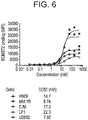
- the BCMA-specific antibodies or antigen-binding fragments described herein have IC 50 values of at least 5.9 nM for APRIL binding.
- the IC 50 of the described BCMA-specific antibodies, or antigen-binding fragments may be determined by a variety of methods known in the art, such as ELISA-based methods or flow cytometry (FACS). Assays for measuring IC 50 by ELISA have plate-bound BCMA in the presence and absence of a BCMA specific antibody and varying concentrations of the APRIL are used.
- a BCMA antibody that blocks the binding of APRIL to BCMA is to "block APRIL as measured by ELISA.”
- vectors comprising the polynucleotides described herein.
- the vectors can be expression vectors. Recombinant expression vectors containing a sequence encoding a polypeptide of interest are thus contemplated as within the scope of this disclosure.
- the expression vector may contain one or more additional sequences such as but not limited to regulatory sequences (e.g., promoter, enhancer), a selection marker, and a polyadenylation signal.
- Vectors for transforming a wide variety of host cells include, but are not limited to, plasmids, phagemids, cosmids, baculoviruses, bacmids, bacterial artificial chromosomes (BACs), yeast artificial chromosomes (YACs), as well as other bacterial, yeast and viral vectors.
- Recombinant expression vectors within the scope of the description include synthetic, genomic, or cDNA-derived nucleic acid fragments that encode at least one recombinant protein which may be operably linked to suitable regulatory elements.
- suitable regulatory elements may include a transcriptional promoter, sequences encoding suitable mRNA ribosomal binding sites, and sequences that control the termination of transcription and translation.
- Expression vectors may also include one or more nontranscribed elements such as an origin of replication, a suitable promoter and enhancer linked to the gene to be expressed, other 5' or 3' flanking nontranscribed sequences, 5' or 3' nontranslated sequences (such as necessary ribosome binding sites), a polyadenylation site, splice donor and acceptor sites, or transcriptional termination sequences.
- an origin of replication that confers the ability to replicate in a host may also be incorporated.
- transcriptional and translational control sequences in expression vectors to be used in transforming vertebrate cells may be provided by viral sources.
- Exemplary vectors may be constructed as described by Okayama and Berg, 3 Mol. Cell. Biol. 280 (1983 ).
- the antibody- or antigen-binding fragment-coding sequence is placed under control of a powerful constitutive promoter, such as the promoters for the following genes: hypoxanthine phosphoribosyl transferase (HPRT), adenosine deaminase, pyruvate kinase, beta-actin, human myosin, human hemoglobin, human muscle creatine, and others.
- a powerful constitutive promoter such as the promoters for the following genes: hypoxanthine phosphoribosyl transferase (HPRT), adenosine deaminase, pyruvate kinase, beta-actin, human myosin, human hemoglobin, human muscle creatine, and others.
- HPRT hypoxanthine phosphoribosyl transferase
- adenosine deaminase pyruvate kinase
- beta-actin beta-actin
- human myosin
- Such viral promoters include without limitation, Cytomegalovirus (CMV) immediate early promoter, the early and late promoters of SV40, the Mouse Mammary Tumor Virus (MMTV) promoter, the long terminal repeats (LTRs) of Maloney leukemia virus, Human Immunodeficiency Virus (HIV), Epstein Barr Virus (EBV), Rous Sarcoma Virus (RSV), and other retroviruses, and the thymidine kinase promoter of Herpes Simplex Virus.
- CMV Cytomegalovirus
- MMTV Mouse Mammary Tumor Virus
- LTRs long terminal repeats
- HCV Human Immunodeficiency Virus
- EBV Epstein Barr Virus
- RSV Rous Sarcoma Virus
- thymidine kinase promoter Herpes Simplex Virus
- the BCMA-specific antibody or antigen-binding fragment thereof coding sequence is placed under control of an inducible promoter such as the metallothionein promoter, tetracycline-inducible promoter, doxycycline-inducible promoter, promoters that contain one or more interferon-stimulated response elements (ISRE) such as protein kinase R 2',5'-oligoadenylate synthetases, Mx genes, ADAR1, and the like.
- ISRE interferon-stimulated response elements
- Vectors described herein may contain one or more Internal Ribosome Entry Site(s) (IRES). Inclusion of an IRES sequence into fusion vectors may be beneficial for enhancing expression of some proteins.
- the vector system will include one or more polyadenylation sites (e.g., SV40), which may be upstream or downstream of any of the aforementioned nucleic acid sequences.
- Vector components may be contiguously linked, or arranged in a manner that provides optimal spacing for expressing the gene products (i.e., by the introduction of "spacer" nucleotides between the ORFs), or positioned in another way. Regulatory elements, such as the IRES motif, may also be arranged to provide optimal spacing for expression.
- the vectors may comprise selection markers, which are well known in the art.
- Selection markers include positive and negative selection markers, for example, antibiotic resistance genes (e.g., neomycin resistance gene, a hygromycin resistance gene, a kanamycin resistance gene, a tetracycline resistance gene, a penicillin resistance gene, a puromycin resistance gene, a blasticidin resistance gene), glutamate synthase genes, HSV-TK, HSV-TK derivatives for ganciclovir selection, or bacterial purine nucleoside phosphorylase gene for 6-methylpurine selection ( Gadi et al., 7 Gene Ther. 1738-1743 (2000 )).
- a nucleic acid sequence encoding a selection marker or the cloning site may be upstream or downstream of a nucleic acid sequence encoding a polypeptide of interest or cloning site.
- the vectors described herein may be used to transform various cells with the genes encoding the described antibodies or antigen-binding fragments.
- the vectors may be used to generate BCMA-specific antibody or antigen-binding fragment-producing cells.
- another aspect features host cells transformed with vectors comprising a nucleic acid sequence encoding an antibody or antigen-binding fragment thereof that specifically binds BCMA, such as the antibodies or antigen-binding fragments described and exemplified herein.
- chromosome transfer e.g., cell fusion, chromosome mediated gene transfer, micro cell mediated gene transfer
- physical methods e.g., transfection, spheroplast fusion, microinjection, electroporation, liposome carrier
- viral vector transfer e.g., recombinant DNA viruses, recombinant RNA viruses
- calcium phosphate precipitation and polyethylene glycol (PEG)-induced fusion of bacterial protoplasts with mammalian cells may also be used to transform cells.
- Cells suitable for use in the expression of the BCMA-specific antibodies or antigen-binding fragments described herein are preferably eukaryotic cells, more preferably cells of plant, rodent, or human origin, for example but not limited to NSO, CHO, CHOK1, perC.6, Tk-ts13, BHK, HEK293 cells, COS-7, T98G, CV-1/EBNA, L cells, C127, 3T3, HeLa, NS1, Sp2/0 myeloma cells, and BHK cell lines, among others.
- expression of antibodies may be accomplished using hybridoma cells. Methods for producing hybridomas are well established in the art.
- Cells transformed with expression vectors described herein may be selected or screened for recombinant expression of the antibodies or antigen-binding fragments described herein.
- Recombinant-positive cells are expanded and screened for subclones exhibiting a desired phenotype, such as high level expression, enhanced growth properties, or the ability to yield proteins with desired biochemical characteristics, for example, due to protein modification or altered post-translational modifications. These phenotypes may be due to inherent properties of a given subclone or to mutation. Mutations may be effected through the use of chemicals, UV-wavelength light, radiation, viruses, insertional mutagens, inhibition of DNA mismatch repair, or a combination of such methods.
- BCMA-specific antibodies or antigen-binding fragments thereof for use in therapy.
- these antibodies or antigen-binding fragments may be useful in treating cancer, such as BCMA-expressing cancer.
- the invention provides a method of treating cancer comprising administering an antibody as described herein, such as BCMA-specific antibodies or antigen-binding fragments.
- the use may be by interfering with BCMA-receptor interactions or where the antibody is conjugated to a toxin, so targeting the toxin to the BCMA-expressing cancer.
- BCMA-expressing cancer includes lymphoma, such as multiple myeloma (MM).
- the antibodies for use in these methods include those described herein above, for example a BCMA-specific antibody or antigen-binding fragment with the features set out in Table 1, for example the CDRs or variable domain sequences, and in the further discussion of these antibodies.
- immune effector properties of the BCMA-specific antibodies may be enhanced or silenced through Fc modifications by techniques known to those skilled in the art.
- Fc effector functions such as C1q binding, complement dependent cytotoxicity (CDC), antibody-dependent cell-mediated cytotoxicity (ADCC), antibody-dependent cell-mediated phagocytosis (ADCP), down regulation of cell surface receptors (e.g., B cell receptor; BCR), etc. may be provided and/or controlled by modifying residues in the Fc responsible for these activities.
- Antibody-dependent cell-mediated cytotoxicity refers to a cell-mediated reaction in which non-specific cytotoxic cells that express Fc receptors (FcRs) (e.g. Natural Killer (NK) cells, neutrophils, and macrophages) recognize bound antibody on a target cell and subsequently cause lysis of the target cell.
- FcRs Fc receptors
- NK Natural Killer
- the ability of monoclonal antibodies to induce ADCC can be enhanced by engineering their oligosaccharide component.
- Human IgG1 or IgG3 are N-glycosylated at Asn297 with the majority of the glycans in the well-known biantennary G0, G0F, G1, G1F, G2 or G2F forms.
- Antibodies produced by non-engineered CHO cells typically have a glycan fucose content of about at least 85%. The removal of the core fucose from the biantennary complex-type oligosaccharides attached to the Fc regions enhances the ADCC of antibodies via improved Fc.gamma.RIIIa binding without altering antigen binding or CDC activity.
- Such mAbs can be achieved using different methods reported to lead to the successful expression of relatively high defucosylated antibodies bearing the biantennary complex-type of Fc oligosaccharides such as control of culture osmolality ( Konno et al., Cytotechnology 64:249-65, 2012 ), application of a variant CHO line Lec13 as the host cell line ( Shields et al., J Biol Chem 277:26733-26740, 2002 ), application of a variant CHO line EB66 as the host cell line ( Olivier et al., MAbs; 2(4), 2010 ; Epub ahead of print; PMTD:20562582), application of a rat hybridoma cell line YB2/0 as the host cell line ( Shinkawa et al., J Biol Chem 278:3466-3473, 2003 ), introduction of small interfering RNA specifically against the .alpha.
- 1,6-fucosyltrasferase (FUT8) gene Mori et al., Biotechnol Bioeng 88:901-908, 2004 ), or coexpression of ⁇ -1,4-N-acetylglucosaminyltransferase III and golgi ⁇ -mannosidase II or a potent alpha-mannosidase I inhibitor, kifunensine ( Ferrara et al., J Biol Chem 281:5032-5036, 2006 , Ferrara. et al., Biotechnol Bioeng 93:851-861, 2006 ; Xhou et al., Biotechnol Bioeng 99:652-65, 2008 ).
- ADCC elicited by the BCMA antibodies may also be enhanced by certain substitutions in the antibody Fc.
- Exemplary substitutions are for example substitutions at amino acid positions 256, 290, 298, 312, 356, 330, 333, 334, 360, 378 or 430 (residue numbering according to the EU index) as described in U.S. Pat. No. 6,737,056 .
- the sample may be derived from urine, blood, serum, plasma, saliva, ascites, circulating cells, circulating tumor cells, cells that are not tissue associated ( i.e ., free cells), tissues (e.g., surgically resected tumor tissue, biopsies, including fine needle aspiration), histological preparations, and the like.
- the described methods include detecting BCMA in a biological sample by contacting the sample with any of the BCMA-specific antibodies or antigen-binding fragments thereof described herein.
- the sample may be contacted with more than one of the BCMA-specific antibodies or antigen-binding fragments described herein.
- a sample may be contacted with a first BCMA-specific antibody, or antigen-binding fragment thereof, and then contacted with a second BCMA-specific antibody, or antigen-binding fragment thereof, wherein the first antibody or antigen-binding fragment and the second antibody or antigen-binding fragment are not the same antibody or antigen-binding fragment.
- the first antibody, or antigen-binding fragment thereof may be affixed to a surface, such as a multiwell plate, chip, or similar substrate prior to contacting the sample. In other embodiments the first antibody, or antigen-binding fragment thereof, may not be affixed, or attached, to anything at all prior to contacting the sample.
- the described BCMA-specific antibodies and antigen-binding fragments may be detectably labeled.
- labeled antibodies and antigen-binding fragments may facilitate the detection BCMA via the methods described herein.
- suitable labels include, but should not be considered limited to, radiolabels, fluorescent labels, epitope tags, biotin, chromophore labels, ECL labels, or enzymes.
- the described labels include ruthenium, 111 In-DOTA, 111 In- diethylenetriaminepentaacetic acid (DTPA), horseradish peroxidase, alkaline phosphatase and beta-galactosidase, poly-histidine (HIS tag), acridine dyes, cyanine dyes, fluorone dyes, oxazin dyes, phenanthridine dyes, rhodamine dyes, Alexafluor® dyes, and the like.
- ruthenium 111 In-DOTA
- DTPA 111 In- diethylenetriaminepentaacetic acid
- HIS tag poly-histidine
- acridine dyes cyanine dyes
- fluorone dyes oxazin dyes
- phenanthridine dyes phenanthridine dyes
- rhodamine dyes Alexafluor® dyes, and the like.
- BCMA-specific antibodies and antigen-binding fragments may be used in a variety of assays to detect BCMA in a biological sample.
- suitable assays include, but should not be considered limited to, western blot analysis, radioimmunoassay, surface plasmon resonance, immunofluorimetry, immunoprecipitation, equilibrium dialysis, immunodiffusion, electrochemiluminescence (ECL) immunoassay, immunohistochemistry, fluorescence-activated cell sorting (FACS) or ELISA assay.
- detection of BCMA-expressing cancer cells in a subject may be used to determine that the subject may be treated with a therapeutic agent directed against BCMA.
- BCMA is present at detectable levels in blood and serum samples.
- methods for detecting BCMA in a sample derived from blood such as a serum sample, by contacting the sample with an antibody, or antigen-binding fragment thereof, that specifically binds BCMA.
- the blood sample, or a derivative thereof may be diluted, fractionated, or otherwise processed to yield a sample upon which the described method may be performed.
- BCMA may be detected in a blood sample, or a derivative thereof, by any number of assays known in the art, such as, but not limited to, western blot analysis, radioimmunoassay, surface plasmon resonance, immunofluorimetry, immunoprecipitation, equilibrium dialysis, immunodiffusion, electrochemiluminescence (ECL) immunoassay, immunohistochemistry, fluorescence-activated cell sorting (FACS) or ELISA assay.
- assays known in the art, such as, but not limited to, western blot analysis, radioimmunoassay, surface plasmon resonance, immunofluorimetry, immunoprecipitation, equilibrium dialysis, immunodiffusion, electrochemiluminescence (ECL) immunoassay, immunohistochemistry, fluorescence-activated cell sorting (FACS) or ELISA assay.
- BCMA-expressing cancer include lymphomas, such as multiple myeloma (MM).
- MM multiple myeloma
- detecting BCMA in a biological sample provides the ability to diagnose cancer in the subject from whom the sample was obtained.
- other samples such as a histological sample, a fine needle aspirate sample, resected tumor tissue, circulating cells, circulating tumor cells, and the like, may also be used to assess whether the subject from whom the sample was obtained has cancer.
- detecting BCMA in a biological sample obtained from the subject can allow for, or clarify, diagnosis of the cancer.
- a subject may be known to have cancer, but it may not be known, or may be unclear, whether the subject's cancer is BCMA-expressing.
- the described methods involve assessing whether a subject is afflicted with BCMA-expressing cancer by determining the amount of BCMA that is present in a biological sample derived from the subject; and comparing the observed amount of BCMA with the amount of BCMA in a control, or reference, sample, wherein a difference between the amount of BCMA in the sample derived from the subject and the amount of BCMA in the control, or reference, sample is an indication that the subject is afflicted with a BCMA-expressing cancer.
- the amount of BCMA observed in a biological sample obtained from a subject may be compared to levels of BCMA known to be associated with certain forms or stages of cancer, to determine the form or stage of the subject's cancer.
- the amount of BCMA in the sample derived from the subject is assessed by contacting the sample with an antibody, or an antigen-binding fragment thereof, that immunospecifically binds BCMA, such as the BCMA-specific antibodies described herein.
- the sample assessed for the presence of BCMA may be derived from urine, blood, serum, plasma, saliva, ascites, circulating cells, circulating tumor cells, cells that are not tissue associated ( i.e., free cells), tissues ( e.g., surgically resected tumor tissue, biopsies, including fine needle aspiration), histological preparations, and the like.
- BCMA-expressing cancer includes hematological cancer, such as mutilple myeloma (MM).
- the subject is a human.
- the method of diagnosing a BCMA-expressing cancer will involve: contacting a biological sample of a subject with a BCMA-specific antibody, or an antigen-binding fragment thereof (such as those derivable from the antibodies and fragments provided in Table 1), quantifying the amount of BCMA present in the sample that is bound by the antibody or antigen-binding fragment thereof, comparing the amount of BCMA present in the sample to a known standard or reference sample; and determining whether the subject's BCMA levels fall within the levels of BCMA associated with cancer.
- the diagnostic method can be followed with an additional step of administering or prescribing a cancer-specific treatment.
- the diagnostic method can be followed with an additional step of transmitting the results of the determination to facilitate treatment of the cancer.
- the cancer-specific treatment may be directed against BCMA-expressing cancers, such as the BCMA x CDS multispecific antibodies described herein.
- the described methods involve assessing whether a subject is afflicted with BCMA-expressing cancer by determining the amount of BCMA present in a blood or serum sample obtained from the subject; and comparing the observed amount of BCMA with the amount of BCMA in a control, or reference, sample, wherein a difference between the amount of BCMA in the sample derived from the subject and the amount of BCMA in the control, or reference, sample is an indication that the subject is afflicted with a BCMA-expressing cancer.
- control, or reference, sample may be derived from a subject that is not afflicted with BCMA-expressing cancer. In some embodiments the control, or reference, sample may be derived from a subject that is afflicted with BCMA-expressing cancer. In some embodiments where the control, or reference, sample is derived from a subject that is not afflicted with BCMA-expressing cancer, an observed increase in the amount of BCMA present in the test sample, relative to that observed for the control or reference sample, is an indication that the subject being assessed is afflicted with BCMA-expressing cancer.
- control sample is derived from a subject that is not afflicted with BCMA-expressing cancer
- an observed decrease or similarity in the amount of BCMA present in the test sample, relative to that observed for the control or reference sample is an indication that the subject being assessed is not afflicted with BCMA-expressing cancer.
- an observed similarity in the amount of BCMA present in the test sample, relative to that observed for the control or reference sample is an indication that the subject being assessed is afflicted with BCMA-expressing cancer.
- control or reference sample is derived from a subject that is afflicted with BCMA-expressing cancer
- an observed decrease in the amount of BCMA present in the test sample, relative to that observed for the control or reference sample is an indication that the subject being assessed is not afflicted with BCMA-expressing cancer.
- the amount of BCMA in the sample derived from the subject is assessed by contacting the sample with an antibody, or an antigen-binding fragment thereof, that specifically binds BCMA, such as the antibodies described herein.
- the sample assessed for the presence of BCMA may be derived from a blood sample, a serum sample, circulating cells, circulating tumor cells, cells that are not tissue associated ( i.e ., free cells), tissues (e.g ., surgically resected tumor tissue, biopsies, including fine needle aspiration), histological preparations, and the like.
- the amount of BCMA is determined by contacting the sample with an antibody, or antigen-binding fragment thereof, that specifically binds BCMA.
- the sample may be contacted by more than one type of antibody, or antigen-binding fragment thereof, that specifically binds BCMA.
- the sample may be contacted by a first antibody, or antigen-binding fragment thereof, that specifically binds BCMA and then contacted by a second antibody, or antigen-binding fragment thereof, that specifically binds BCMA.
- BCMA-specific antibodies or antigen-binding fragments such as those described herein may be used in this capacity.
- BCMA-specific antibodies and antigen-binding fragments can be used to provide a "first" and “second” antibody or antigen-binding fragment to carry out the described diagnostic methods.
- BCMA-expressing cancer includes lymphomas, such as multiple myeloma (MM).
- the amount of BCMA is determined by western blot analysis, radioimmunoassay, immunofluorimetry, immunoprecipitation, equilibrium dialysis, immunodiffusion, electrochemiluminescence (ECL) immunoassay, immunohistochemistry, fluorescence-activated cell sorting (FACS) or ELISA assay.
- a control or reference sample is used.
- This sample may be a positive or negative assay control that ensures the assay used is working properly; for example, an assay control of this nature might be commonly used for immunohistochemistry assays.
- the sample may be a standardized reference for the amount of BCMA in a biological sample from a healthy subject.
- the observed BCMA levels of the tested subject may be compared with BCMA levels observed in samples from subjects known to have BCMA-expressing cancer.
- the control subject may be afflicted with a particular cancer of interest.
- the control subject is known to have early stage cancer, which may or may not be BCMA-expressing cancer.
- the control subject is known to have intermediate stage cancer, which may or may not be BCMA-expressing cancer.
- the control subject is known to have late stage, which may or may not be BCMA-expressing cancer.
- BCMA-expressing cancer includes lymphomas, such as multiple myeloma (MM).
- the described methods involve assessing whether BCMA-expressing cancer is progressing, regressing, or remaining stable by determining the amount of BCMA that is present in a test sample derived from the subject; and comparing the observed amount of BCMA with the amount of BCMA in a biological sample obtained, in a similar manner, from the subject at an earlier point in time, wherein a difference between the amount of BCMA in the test sample and the earlier sample provides an indication of whether the cancer is progressing, regressing, or remaining stable.
- lymphomas such as multiple myeloma
- a test sample with an increased amount of BCMA, relative to the amount observed for the earlier sample may indicate progression of a BCMA-expressing cancer.
- a test sample with a decreased amount of BCMA, relative to the amount observed for the earlier sample may indicate regression of a BCMA-expressing cancer.
- a test sample with an insignificant difference in the amount of BCMA, relative to the amount observed for the earlier sample, may indicate a state of stable disease for a BCMA-expressing cancer.
- the amount of BCMA in a biological sample derived from the subject is assessed by contacting the sample with an antibody, or an antibody fragment thereof, that specifically binds BCMA, such as the antibodies described herein.
- the sample assessed for the presence of BCMA may be derived from urine, blood, serum, plasma, saliva, ascites, circulating cells, circulating tumor cells, cells that are not tissue associated (i.e., free cells), tissues (e.g., surgically resected tumor tissue, biopsies, including fine needle aspiration), histological preparations, and the like.
- the subject is a human.
- the methods of monitoring a BCMA-expressing cancer will involve: contacting a biological sample of a subject with a BCMA-specific antibody, or antigen-binding fragment thereof (such as those derivable from the antibodies and fragments provided in Table 1), quantifying the amount of BCMA present in the sample, comparing the amount of BCMA present in the sample to the amount of BCMA determined to be in a biological sample obtained, in a similar manner, from the same subject at an earlier point in time; and determining whether the subject's BCMA level has changed over time.
- a test sample with an increased amount of BCMA, relative to the amount observed for the earlier sample may indicate progression of cancer.
- a test sample with a decreased amount of BCMA, relative to the amount observed for the earlier sample may indicate regression of a BCMA-expressing cancer. Accordingly, a test sample with an insignificant difference in the amount of BCMA, relative to the amount observed for the earlier sample, may indicate a state of stable disease for a BCMA-expressing cancer.
- the BCMA levels of the sample may be compared to a known standard or a reference sample, alone or in addition to the BCMA levels observed for a sample assessed at an earlier point in time.
- the diagnostic method can be followed with an additional step of administering a cancer-specific treatment.
- the cancer-specific treatment may be directed against BCMA-expressing cancers, such as the BCMA x CD3 multispecific antibodies described herein.
- the amount of BCMA is determined by contacting the sample with an antibody, or antigen-binding fragment thereof, that specifically binds BCMA.
- the sample may be contacted by more than one type of antibody, or antigen-binding fragment thereof, that specifically binds BCMA.
- the sample may be contacted by a first antibody, or antigen-binding fragment thereof, that specifically binds BCMA and then contacted by a second antibody, or antigen-binding fragment thereof, that specifically binds BCMA.
- Antibodies such as those described herein may be used in this capacity.
- BCMA-expressing cancer includes a hematological cancer, such as acute myeloid leukemia (AML).
- AML acute myeloid leukemia
- the amount of BCMA is determined by western blot analysis, radioimmunoassay, immunofluorimetry, immunoprecipitation, equilibrium dialysis, immunodiffusion, electrochemiluminescence (ECL) immunoassay, immunohistochemistry, fluorescence-activated cell sorting (FACS) or ELISA assay.
- kits for detecting BCMA in a biological sample include one or more of the BCMA-specific antibodies described herein, or an antigen-binding fragment thereof, and instructions for use of the kit.
- the provided BCMA-specific antibody, or antigen-binding fragment may be in solution; lyophilized; affixed to a substrate, carrier, or plate; or detectably labeled.
- kits may also include additional components useful for performing the methods described herein.
- the kits may comprise means for obtaining a sample from a subject, a control or reference sample, e.g ., a sample from a subject having slowly progressing cancer and/or a subject not having cancer, one or more sample compartments, and/or instructional material which describes performance of a method of the invention and tissue specific controls or standards.
- the means for determining the level of BCMA can further include, for example, buffers or other reagents for use in an assay for determining the level of BCMA.
- the instructions can be, for example, printed instructions for performing the assay and/or instructions for evaluating the level of expression of BCMA
- kits may also include means for isolating a sample from a subject.
- These means can comprise one or more items of equipment or reagents that can be used to obtain a fluid or tissue from a subject.
- the means for obtaining a sample from a subject may also comprise means for isolating blood components, such as serum, from a blood sample.
- the kit is designed for use with a human subject.
- the binding domains of the anti- BCMA antibodies described herein recognize cells expressing BCMA on their surface.
- BCMA expression can be indicative of a cancerous cell.
- More specific targeting to particular subsets of cells can be achieved by making bispecific molecules, such as antibodies or antibody fragments, which bind to BCMA and to another target, such as CD3. This is achieved by making a molecule which comprises a first region binding to BCMA and a second binding region binding to the other target antigen.
- the antigen-binding regions can take any form that allows specific recognition of the target, for example the binding region may be or may include a heavy chain variable domain, an Fv (combination of a heavy chain variable domain and a light chain variable domain), a binding domain based on a fibronectin type III domain (such as from fibronectin, or based on a consensus of the type III domains from fibronectin, or from tenascin or based on a consensus of the type III domains from tenascin, such as the Centyrin molecules from Janssen Biotech, Inc., see e.g. WO2010/051274 and WO2010/093627 ). Accordingly, bispecific molecules comprising two different antigen-binding regions which bind BCMA and another antigen, respectively, are provided.
- a fibronectin type III domain such as from fibronectin, or based on a consensus of the type III domains from fibronectin, or from tenascin or based on a
- the multispecific antibodies described herein comprise two different antigen-binding regions which bind BCMA and CD3, respectively.
- multispecific antibodies that bind BCMA and CD3 BCMA x CD3-multispecific antibodies
- the BCMA x CD3-multispecific antibody comprises a first heavy chain (HC1) and a first light chain (LC1) that pair to form a first antigen-binding site that immunospecifically binds BCMA and a second heavy chain (HC2) and a second light chain (LC2) that pair to form a second antigen-binding site that immunospecifically binds CD3.
- the BCMA x CD3-multispecific antibody is a bispecific antibody comprising a BCMA-specific arm comprising a first heavy chain (HC1) and a first light chain (LC1) that pair to form a first antigen-binding site that immunospecifically binds CD3 and a CD3-specific arm comprising second heavy chain (HC2) and a second light chain (LC2) that pair to form a second antigen-binding site that immunospecifically binds BCMA.
- the bispecific antibodies of the invention include antibodies having a full length antibody structure. "Full length antibody” as used herein refers to an antibody having two full length antibody heavy chains and two full length antibody light chains.
- a full length antibody heavy chain includes heavy chain variable and constant domains VH, CH1, CH2, and CH3.
- a full length antibody light chain includes light chain variable and constant domains VL and CL.
- the full length antibody may be lacking the C-terminal lysine (K) in either one or both heavy chains.
- the term "Fab-arm" or "half molecule” refers to one heavy chain-light chain pair that specifically binds an antigen.
- one of the antigen-binding domains is a non-antibody based binding domain, e.g. a binding domain of based on a fibronectin type 3 domain, e.g. Centyrin.
- the BCMA-binding arm of the multispecific antibodies provided herein may be derived from any of the BCMA-specific antibodies described above.
- the first antigen-binding region which binds BCMA comprises a heavy chain CDR1, CDR2, and CDR3 derived from an antibody clone as described in Table 1.
- the first antigen-binding region which binds BCMA comprises heavy chain CDR1, CDR2, and CDR3 and light chain CDR1, CDR2, and CDR3 derived from an antibody clone as described in Table 1.
- the first antigen-binding region which binds BCMA comprises heavy chain CDR1, CDR2, and CDR3 of clone BCMB69, BCMB117, BCMB123, BCMB128, BCMB129, BCMB176, or BCMB177.
- the first antigen-binding region which binds BCMA comprises heavy chain CDR1, CDR2, and CDR3 and light chain CDR1, CDR2, and CDR3 of clone BCMB69, BCMB117, BCMB123, BCMB128, BCMB129, BCMB176, or BCMB177.
- the first antigen-binding region which binds BCMA comprises a heavy chain variable domain derived from an antibody clone as described in Table 1. In some exemplary embodiments of such BCMA-binding arms, the first antigen-binding region which binds BCMA comprises heavy chain variable domain and light chain variable domain derived from an antibody clone as described in Table 1. In some exemplary embodiments of such BCMA-binding arms, the first antigen-binding region which binds BCMA comprises heavy chain variable domain of clone BCMB69, BCMB117, BCMB123, BCMB128, BCMB129, BCMB176, or BCMB177.
- the first antigen-binding region which binds BCMA comprises heavy chain variable domain and light chain variable domain of clone BCMB69, BCMB117, BCMB123, BCMB128, BCMB129, BCMB176, or BCMB177.
- Table 3 provides a listing of BCMA x CD3 bispecific antibodies having one heavy and light chain pair specific for BCMA and another heavy and light chain pair specific for CD3, where the particular antibody ID is listed to describe the antigen-specific antibody arms used to produce the described embodiment.
- the BCMA-binding arm binds also binds cynomolgus BCMA, preferably the extracellular domain thereof.
- the BCMA-binding arm of the multispecific antibody is IgG, or a derivative thereof, e.g., IgG1, IgG2, IgG3, and IgG4 isotypes.
- the BCMA-binding arm has an IgG4 isotype, it contains S228P, L234A, and L235A substitution(s) in its Fc region.
- the second antigen-binding arm binds human CD3.
- the CD3-specific arm of the BCMA x CD3 bispecific antibody is derived from a CD3-specific antibody that binds and activates human primary T cells and/or cynomolgus monkey primary T cells.
- the CD3-binding arm binds to an epitope at the N-terminus of CD3 ⁇ . In some embodiments, the CD3-binding arm contacts an epitope including the six N-terminal amino acids of CD3 ⁇ .
- the CD3-specific binding arm of the bispecific antibody is derived from the mouse monoclonal antibody SP34, a mouse IgG3/lambda isotype.
- the CD3-binding arm comprises the CDRs of antibody SP34.
- Such CD3-binding arms may bind to CD3 with an affinity of 5x10 -7 M or less, such as 1x10 -7 M or less, 5x10 -8 M or less, 1x10 -8 M or less, 5x10 -9 M or less, or 1x10 -9 M or less.
- the CD3-specific binding arm may be a humanized version of an arm of mouse monoclonal antibody SP34.
- HFA Human framework adaptation
- the CD3-binding arm comprises a heavy chain and light chain pair selected from Table 2.
- the CD3-binding arm comprises heavy chain CDR1, CDR2, and CDR3 and light chain CDR1, CDR2, and CDR3 sequences set forth in Table 2.
- the heavy chain and light chain CDR sequences of some embodiments of the CD3-binding arm of the bispecific antibodies described herein can include the following amino acid sequences: Hc CDR1, SEQ ID NO: 59; Hc CDR2: SEQ ID NO: 60; Hc CDR3, SEQ ID NO: 61; Lc CDR1, SEQ ID NO: 62; Lc CDR2: SEQ ID NO: 63; and Lc CDR3, SEQ ID NO: 64.
- the CD3-binding arm is IgG, or a derivative thereof. In some embodiments, the CD3-binding arm is IgG1, IgG2, IgG3, or IgG4. In some embodiments wherein the CD3-binding arm has an IgG4 isotype, it contains S228P, L234A, L235A, F405L, and R409K substitution(s) in its Fc region. In some embodiments, the antibodies or antigen-binding fragments bind CD3 ⁇ on primary human T cells. In some embodiments, the antibodies or antigen-binding fragments bind CD3 ⁇ on primary cynomolgus T cells.
- the antibodies or antigen-binding fragments bind CD3 ⁇ on primary human and cynomolgus T cells. In some embodiments, the antibodies or antigen-binding fragments activate primary human CD4+ T cells. In some embodiments, the antibodies or antigen-binding fragments activate primary cynomolgus CD4+ T cells.
- a BCMA x CD3 bispecific antibody having a BCMA-binding arm comprising a heavy chain of antibody clone BCMB69, BCMB117, BCMB123, BCMB128, BCMB129, BCMB176, or BCMB177.
- a BCMA x CD3 bispecific antibody having a BCMA-binding arm comprising a heavy chain and light chain of antibody clone BCMB69, BCMB117, BCMB123, BCMB128, BCMB129, BCMB176, or BCMB177.
- a BCMA x CD3 bispecific antibody having a CD3-binding arm comprising a heavy chain of antibody clone CD3B219.
- a BCMA x CD3 bispecific antibody having a CD3-binding arm comprising a heavy chain and light chain of antibody clone CD3B219.
- a BCMA x CD3 bispecific antibody having a BCMA-binding arm comprising a heavy chain of antibody clone BCMB69, BCMB117, BCMB123, BCMB128, BCMB129, BCMB176, or BCMB177 and a CD3-binding arm comprising a heavy chain of antibody clone CD3B219.
- a BCMA x CD3 bispecific antibody having a BCMA-binding arm comprising a heavy chain and light chain of antibody clone BCMB69, BCMB117, BCMB123, BCMB128, BCMB129, BCMB176, or BCMB177 and a CD3-binding arm comprising a heavy chain and light chain of antibody clone CD3B219.
- the bispecific antibody of the present invention is a diabody, a cross-body, or a bispecific antibody obtained via a controlled Fab arm exchange as those described in the present invention.
- the bispecific antibodies include IgG-like molecules with complementary CH3 domains to force heterodimerisation; recombinant IgG-like dual targeting molecules, wherein the two sides of the molecule each contain the Fab fragment or part of the Fab fragment of at least two different antibodies; IgG fusion molecules, wherein full length IgG antibodies are fused to an extra Fab fragment or parts of Fab fragment; Fc fusion molecules, wherein single chain Fv molecules or stabilized diabodies are fused to heavy-chain constant-domains, Fc-regions or parts thereof; Fab fusion molecules, wherein different Fab-fragments are fused together; ScFv- and diabody-based and heavy chain antibodies (e.g., domain antibodies, nanobodies) wherein different single chain Fv molecules or different diabodies or different heavy-chain antibodies (e.g. domain antibodies, nanobodies) are fused to each other or to another protein or carrier molecule.
- IgG fusion molecules wherein full length IgG antibodies are fused to an extra
- IgG-like molecules with complementary CH3 domains molecules include the Triomab/Quadroma (Trion Pharma/Fresenius Biotech), the Knobs-into-Holes (Genentech), CrossMAbs (Roche) and the electrostatically-matched (Amgen), the LUZ-Y (Genentech), the Strand Exchange Engineered Domain body (SEEDbody)(EMD Serono), the Biclonic (Merus) and the DuoBody® (Genmab A/S).
- recombinant IgG-like dual targeting molecules include Dual Targeting (DT)-Ig (GSK/Domantis), Two-in-one Antibody (Genentech), Cross-linked Mabs (Karmanos Cancer Center), mAb2 (F-Star) and CovX-body (CovX/Pfizer).
- DT Dual Targeting
- Genentech Two-in-one Antibody
- Cross-linked Mabs Karmanos Cancer Center
- mAb2 F-Star
- CovX-body CovX/Pfizer
- IgG fusion molecules include Dual Variable Domain (DVD)-Ig (Abbott), IgG-like Bispecific (InnClone/Eli Lilly), Ts2Ab (MedImmune/AZ) and BsAb (Zymogenetics), HERCULES (Biogen Idec) and TvAb (Roche).
- DVD Dual Variable Domain
- IgG-like Bispecific InnClone/Eli Lilly
- Ts2Ab MedImmune/AZ
- BsAb Zymogenetics
- HERCULES Biogen Idec
- TvAb Roche
- Fc fusion molecules include to ScFv/Fc Fusions (Academic Institution), SCORPION (Emergent BioSolutions/Trubion, Zymogenetics/BMS), Dual Affinity Retargeting Technology (Fc-DART) (MacroGenics) and Dual(ScFv).sub.2-Fab (National Research Center for Antibody Medicine--China).
- Fab fusion bispecific antibodies include F(ab)2 (Medarex/AMGEN), Dual-Action or Bis-Fab (Genentech), Dock-and-Lock (DNL) (ImmunoMedics), Bivalent Bispecific (Biotecnol) and Fab-Fv (UCB-Celltech).
- ScFv-, diabody-based and domain antibodies include but are not limited to Bispecific T Cell Engager (BITE) (Micromet), Tandem Diabody (Tandab) (Affimed), Dual Affinity Retargeting Technology (DART) (MacroGenics), Single-chain Diabody (Academic), TCR-like Antibodies (AIT, ReceptorLogics), Human Serum Albumin ScFv Fusion (Merrimack) and COMBODY (Epigen Biotech), dual targeting nanobodies (Ablynx), dual targeting heavy chain only domain antibodies.
- BITE Bispecific T Cell Engager
- Tandab Tandem Diabody
- DART Dual Affinity Retargeting Technology
- AIT TCR-like Antibodies
- AIT ReceptorLogics
- Human Serum Albumin ScFv Fusion Merrimack
- COMBODY Epigen Biotech
- dual targeting nanobodies Ablynx
- dual targeting heavy chain only domain antibodies dual targeting heavy chain only domain antibodies.
- Full length bispecific antibodies of the invention may be generated for example using Fab arm exchange (or half molecule exchange) between two mono specific bivalent antibodies by introducing substitutions at the heavy chain CH3 interface in each half molecule to favor heterodimer formation of two antibody half molecules having distinct specificity either in vitro in cell-free environment or using co-expression.
- the Fab arm exchange reaction is the result of a disulfide-bond isomerization reaction and dissociation-association of CH3 domains. The heavy-chain disulfide bonds in the hinge regions of the parent mono specific antibodies are reduced.
- the resulting free cysteines of one of the parent monospecific antibodies form an inter heavy-chain disulfide bond with cysteine residues of a second parent mono specific antibody molecule and simultaneously CH3 domains of the parent antibodies release and reform by dissociation-association.
- the CH3 domains of the Fab arms may be engineered to favor heterodimerization over homodimerization.
- the resulting product is a bispecific antibody having two Fab arms or half molecules which each bind a distinct epitope, i.e. an epitope on BCMA and an epitope on CD3.
- Homodimerization refers to an interaction of two heavy chains having identical CH3 amin acid sequences.
- Homodimer refers to an antibody having two heavy chains with identical CH3 amino acid sequences.
- Heterodimerization refers to an interaction of two heavy chains having non-identical CH3 amino acid sequences.
- Heterodimer as used herein refers to an antibody having two heavy chains with non-identical CH3 amino acid sequences.
- the "knob-in-hole” strategy may be used to generate full length bispecific antibodies. Briefly, selected amino acids forming the interface of the CH3 domains in human IgG can be mutated at positions affecting CH3 domain interactions to promote heterodimer formation. An amino acid with a small side chain (hole) is introduced into a heavy chain of an antibody specifically binding a first antigen and an amino acid with a large side chain (knob) is introduced into a heavy chain of an antibody specifically binding a second antigen.
- a heterodimer is formed as a result of the preferential interaction of the heavy chain with a "hole” with the heavy chain with a "knob".
- Exemplary CH3 substitution pairs forming a knob and a hole are (expressed as modified position in the first CH3 domain of the first heavy chain/modified position in the second CH3 domain of the second heavy chain): T366Y/F405A, T366W/F405W, F405W/Y407A, T394W/Y407T, T394S/Y407A, T366W/T394S, F405W/T394S and T366W/T366S_L368A_Y407V.
- heterodimerization may be promoted by the following substitutions (expressed as modified position in the first CH3 domain of the first heavy chain/modified position in the second CH3 domain of the second heavy chain): L351Y_F405AY407V/T394W, T366I_K392M_T394W/F405A_Y407V, T366L_K392M_T394W/F405A_Y407V, L351Y_Y407A/T366A_K409F, L351Y_Y407A/T366V K409F Y407A/T366A_K409F, or T350V_L351Y_F405A Y407V/T350V_T366L_K392L_T394W as described in U.S. Pat. Publ. No. US2012/0149876 or U.S. Pat. Publ. No. US2013/0195849 .
- bispecific antibodies of the invention may be generated in vitro in a cell-free environment by introducing asymmetrical mutations in the CH3 regions of two mono specific homodimeric antibodies and forming the bispecific heterodimeric antibody from two parent monospecific homodimeric antibodies in reducing conditions to allow disulfide bond isomerization according to methods described in Inti. Pat. Publ. No. WO2011/131746 .
- the first monospecific bivalent antibody e.g., anti- BCMA antibody
- the second monospecific bivalent antibody e.g., anti-CD3 antibody
- the antibodies are incubated together under reducing conditions sufficient to allow the cysteines in the hinge region to undergo disulfide bond isomerization; thereby generating the bispecific antibody by Fab arm exchange.
- the incubation conditions may optimally be restored to nonreducing conditions.
- Exemplary reducing agents that may be used are 2-mercaptoethylamine (2-MEA), dithiothreitol (DTT), dithioerythritol (DTE), glutathione, tris (2-carboxyethyl)phosphine (TCEP), L-cysteine and beta-mercaptoethanol, preferably a reducing agent selected from the group consisting of: 2-mercaptoethylamine, dithiothreitol and tris (2-carboxyethyl)phosphine.
- incubation for at least 90 min at a temperature of at least 20° C in the presence of at least 25 mM 2-MEA or in the presence of at least 0.5 mM dithiothreitol at a pH from 5-8, for example at pH of 7.0 or at pH of 7.4 may be used.
- polynucleotide sequences capable of encoding the described BCMA x CD3-multispecific antibodies.
- Vectors comprising the described polynucleotides are also provided, as are cells expressing the BCMA x CD3-multispecific antibodies provided herein.
- cells capable of expressing the disclosed vectors may be mammalian cells (such as 293F cells, CHO cells), insect cells (such as Sf7 cells), yeast cells, plant cells, or bacteria cells (such as E. coli ) .
- the described antibodies may also be produced by hybridoma cells.
- compositions and methods of treatment using multispecific antibodies and multispecific antigen-binding fragments thereof are provided.
- the BCMA bispecific antibodies discussed above are useful in therapy.
- the BCMA bispecific antibodies are useful in treating cancer.
- therapeutic compositions for the treatment of a hyperproliferative disorder in a mammal which comprises a therapeutically effective amount of a multispecific antibody or multispecific antigen-binding fragment described herein and a pharmaceutically acceptable carrier.
- the multispecific antibody is a BCMA x CD3-multispecific antibody as described herein, or a multispecific antigen-binding fragment thereof, and more preferably a BCMA x CD3-bispecific antibody as described herein, or a BCMA x CD3-bispecific antigen-binding fragment thereof.
- said pharmaceutical composition is for the treatment of a BCMA-expressing cancer, including (but not limited to) the following: BCMA-expressing B cell cancers, such as multiple myeloma (MM); and other cancers yet to be determined in which BCMA is expressed.
- BCMA-expressing B cell cancers such as multiple myeloma (MM)
- MM multiple myeloma
- Particular bispecific antibodies that may be used to treat cancer, such as hematological cancer, including the specific cancers discussed above include antibodies BCMB69, BCMB117, BCMB123, BCMB128, BCMB129, BCMB176, or BCMB177 or CD3B219.
- a useful bispecific antibody for treating cancer, such as hematological cancer, including these specific cancers is BCMB72.
- compositions provided herein comprise: a) an effective amount of a multispecific antibody or antibody fragment of the present invention, and b) a pharmaceutically acceptable carrier, which may be inert or physiologically active.
- the multispecific antibody is a BCMA x CD3-multispecific antibody as described herein, or a multispecific antigen-binding fragment thereof, and more preferably a BCMA x CD3-bispecific antibody as described herein, or a BCMA x CD3-bispecific antigen-binding fragment thereof.
- pharmaceutically acceptable carriers includes any and all solvents, dispersion media, coatings, antibacterial and antifungal agents, and the like that are physiologically compatible.
- Suitable carriers, diluents and/or excipients include one or more of water, saline, phosphate buffered saline, dextrose, glycerol, ethanol, and the like, as well as any combination thereof.
- isotonic agents such as sugars, polyalcohols, or sodium chloride in the composition.
- suitable carrier include: (1) Dulbecco's phosphate buffered saline, pH.about.7.4, containing or not containing about 1 mg/mL to 25 mg/mL human serum albumin, (2) 0.9% saline (0.9% w/v sodium chloride (NaCl)), and (3) 5% (w/v) dextrose; and may also contain an antioxidant such as tryptamine and a stabilizing agent such as Tween 20®.
- compositions herein may also contain a further therapeutic agent, as necessary for the particular disorder being treated.
- a further therapeutic agent for the particular disorder being treated.
- the multispecific antibody or antibody fragment and the supplementary active compound will have complementary activities that do not adversely affect each other.
- the further therapeutic agent is cytarabine, an anthracycline, histamine dihydrochloride, or interleukin 2.
- the further therapeutic agent is a chemotherapeutic agent.
- compositions of the invention may be in a variety of forms. These include for example liquid, semi-solid, and solid dosage forms, but the preferred form depends on the intended mode of administration and therapeutic application. Typical preferred compositions are in the form of injectable or infusible solutions.
- the preferred mode of administration is parenteral (e.g. intravenous, intramuscular, intraperinoneal, subcutaneous).
- the compositions of the invention are administered intravenously as a bolus or by continuous infusion over a period of time.
- they are injected by intramuscular, subcutaneous, intra-articular, intrasynovial, intratumoral, peritumoral, intralesional, or perilesional routes, to exert local as well as systemic therapeutic effects.
- Sterile compositions for parenteral administration can be prepared by incorporating the antibody, antibody fragment or antibody conjugate of the present invention in the required amount in the appropriate solvent, followed by sterilization by microfiltration.
- solvent or vehicle there may be used water, saline, phosphate buffered saline, dextrose, glycerol, ethanol, and the like, as well as combination thereof.
- isotonic agents such as sugars, polyalcohols, or sodium chloride in the composition.
- These compositions may also contain adjuvants, in particular wetting, isotonizing, emulsifying, dispersing and stabilizing agents.
- Sterile compositions for parenteral administration may also be prepared in the form of sterile solid compositions which may be dissolved at the time of use in sterile water or any other injectable sterile medium.
- the multispecific antibody or antibody fragment may also be orally administered.
- solid compositions for oral administration tablets, pills, powders (gelatine capsules, sachets) or granules may be used.
- the active ingredient according to the invention is mixed with one or more inert diluents, such as starch, cellulose, sucrose, lactose or silica, under an argon stream.
- inert diluents such as starch, cellulose, sucrose, lactose or silica
- These compositions may also comprise substances other than diluents, for example one or more lubricants such as magnesium stearate or talc, a coloring, a coating (sugar-coated tablet) or a glaze.
- compositions for oral administration there may be used pharmaceutically acceptable solutions, suspensions, emulsions, syrups and elixirs containing inert diluents such as water, ethanol, glycerol, vegetable oils or paraffin oil.
- inert diluents such as water, ethanol, glycerol, vegetable oils or paraffin oil.
- These compositions may comprise substances other than diluents, for example wetting, sweetening, thickening, flavoring or stabilizing products.
- the doses depend on the desired effect, the duration of the treatment and the route of administration used; they are generally between 5 mg and 1000 mg per day orally for an adult with unit doses ranging from 1 mg to 250 mg of active substance. In general, the doctor will determine the appropriate dosage depending on the age, weight and any other factors specific to the subject to be treated.
- a multispecific antibody which binds said BCMA and is able to recruit T cells to kill said BCMA + cell i.e., T cell redirection.
- Any of the multispecific antibodies or antibody fragments of the invention may be used therapeutically.
- the BCMA x CD3-multispecific antibody BCMB72 may be used therapeutically to treat cancer in a subject.
- multispecific antibodies or antibody fragments of the invention are used for the treatment of a hyperproliferative disorder in a mammal.
- one of the pharmaceutical compositions disclosed above, and which contains a multispecific antibody or antibody fragment of the invention is used for the treatment of a hyperproliferative disorder in a mammal.
- the disorder is a cancer.
- the cancer is a BCMA-expressing cancer, including (but not limited to) the following: BCMA-expressing B-cell cancers, such as multiple myeloma (MM); and other cancers yet to be determined in which BCMA is expressed.
- the multispecific antibody is a BCMA x CD3-multispecific antibody as described herein, or a multispecific antigen-binding fragment thereof, and more preferably a BCMA x CD3-bispecific antibody as described herein, or a BCMA x CD3-bispecific antigen-binding fragment thereof.
- compositions of the invention are useful in the treatment or prevention of a variety of cancers, including (but not limited to) the following: a BCMA-expressing cancer, including (but not limited to) the following: BCMA-expressing B cell cancers, such as acute multiple myeloma (MM); and other cancers yet to be determined in which BCMA is expressed.
- a BCMA-expressing cancer including (but not limited to) the following: BCMA-expressing B cell cancers, such as acute multiple myeloma (MM); and other cancers yet to be determined in which BCMA is expressed.
- a method for inhibiting the growth of selected cell populations comprising contacting BCMA-expressing target cells, or tissue containing such target cells, with an effective amount of a multispecific antibody or antibody fragment of the present invention, either alone or in combination with other cytotoxic or therapeutic agents, in the presence of a peripheral blood mononuclear cell (PBMC).
- the multispecific antibody is a BCMA x CD3-multispecific antibody as described herein, or a multispecific antigen-binding fragment thereof, and more preferably a BCMA x CD3-bispecific antibody as described herein, or a BCMA x CD3-bispecific antigen-binding fragment thereof.
- the further therapeutic agent is cytarabine, an anthracycline, histamine dihydrochloride, or interleukin 2.
- the further therapeutic agent is a chemotherapeutic agent.
- Examples of in vitro uses include treatments of autologous bone marrow prior to their transplant into the same patient in order to kill diseased or malignant cells; treatments of bone marrow prior to its transplantation in order to kill competent T cells and prevent graft-versus-host-disease (GVHD); treatments of cell cultures in order to kill all cells except for desired variants that do not express the target antigen; or to kill variants that express undesired antigen.
- GVHD graft-versus-host-disease
- the conditions of non-clinical in vitro use are readily determined by one of ordinary skill in the art.
- Examples of clinical ex vivo use are to remove tumor cells from bone marrow prior to autologous transplantation in cancer treatment.
- Treatment can be carried out as follows. Bone marrow is harvested from the patient or other individual and then incubated in medium containing serum to which is added the cytotoxic agent of the invention. Concentrations range from about 10 uM to 1 uM, for about 30 min to about 48 hr at about 37 °C. The exact conditions of concentration and time of incubation, i.e., the dose, are readily determined by one of ordinary skill in the art. After incubation the bone marrow cells are washed with medium containing serum and returned to the patient by i.v. infusion according to known methods. In circumstances where the patient receives other treatment such as a course of ablative chemotherapy or total-body irradiation between the time of harvest of the marrow and reinfusion of the treated cells, the treated marrow cells are stored frozen in liquid nitrogen using standard medical equipment.
- the BCMA x CD3-multispecific antibodies and multispecific antigen-binding fragments thereof may be useful in the treatment of a BCMA-expressing cancer in a subject in need thereof.
- the BCMA-expressing cancer is a B-cell cancer, such as multiple myeloma (MM).
- the multispecific antibody is a BCMA x CD3-multispecific antibody as described herein, or a multispecific antigen-binding fragment thereof, and more preferably a BCMA x CD3-bispecific antibody as described herein, or a BCMA x CD3-bispecific antigen-binding fragment thereof.
- the subject is a mammal, preferably a human.
- the multispecific antibody or antigen-binding fragment will be administered as a solution that has been tested for sterility.
- Dosage regimens in the above methods of treatment and uses are adjusted to provide the optimum desired response (e.g., a therapeutic response). For example, a single bolus may be administered, several divided doses may be administered over time or the dose may be proportionally reduced or increased as indicated by the exigencies of the therapeutic situation. Parenteral compositions may be formulated in dosage unit form for ease of administration and uniformity of dosage.
- the efficient dosages and the dosage regimens for the multispecific antibodies and fragments depend on the disease or condition to be treated and may be determined by one skilled in the art.
- An exemplary, non-limiting range for a therapeutically effective amount of a compound of the present invention is about 0.001-10 mg/kg, such as about 0.001-5 mg/kg, for example about 0.001-2 mg/kg, such as about 0.001-1 mg/kg, for instance about 0.001, about 0.01, about 0. 1, about 1 or about 10 mg/kg.
- a physician or veterinarian having ordinary skill in the art may readily determine and prescribe the effective amount of the pharmaceutical composition required.
- the physician or veterinarian could start doses of the multispecific antibody or fragment employed in the pharmaceutical composition at levels lower than that required in order to achieve the desired therapeutic effect and gradually increase the dosage until the desired effect is achieved.
- a suitable daily dose of a bispecific antibody of the present invention will be that amount of the compound which is the lowest dose effective to produce a therapeutic effect.
- Administration may e.g. be parenteral, such as intravenous, intramuscular or subcutaneous.
- the multispecific antibody or fragment may be administered by infusion in a weekly dosage of calculated by mg/m 2 .
- Such dosages can, for example, be based on the mg/kg dosages provided above according to the following: dose (mg/kg)x70: 1.8.
- Such administration may be repeated, e.g., 1 to 8 times, such as 3 to 5 times.
- the administration may be performed by continuous infusion over a period of from 2 to 24 hr, such as of from 2 to 12 hr.
- the multispecific antibody or fragment may be administered by slow continuous infusion over a long period, such as more than 24 hours, in order to reduce toxic side effects.
- the multispecific antibody or fragment may be administered in a weekly dosage of calculated as a fixed dose for up to eight times, such as from four to six times when given once a week. Such regimen may be repeated one or more times as necessary, for example, after six months or twelve months.
- Such fixed dosages can, for example, be based on the mg/kg dosages provided above, with a body weight estimate of 70 kg.
- the dosage may be determined or adjusted by measuring the amount of bispecific antibody of the present invention in the blood upon administration by for instance taking out a biological sample and using anti-idiotypic antibodies which target the BCMA antigen binding region of the multispecific antibodies of the present invention.
- the multispecific antibody or fragment may be administered by maintenance therapy, such as, e.g., once a week for a period of six months or more.
- a multispecific antibody or fragment may also be administered prophylactically in order to reduce the risk of developing cancer, delay the onset of the occurrence of an event in cancer progression, and/or reduce the risk of recurrence when a cancer is in remission.
- the multispecific antibodies and fragments thereof as described herein may also be administered in combination therapy, i.e., combined with other therapeutic agents relevant for the disease or condition to be treated.
- the antibody-containing medicament is for combination with one or more further therapeutic agent, such as a chemotherapeutic agent.
- the other therapeutic agent is cytarabine, an anthracycline, histamine dihydrochloride, or interleukin 2.
- Such combined administration may be simultaneous, separate or sequential, in any order.
- the agents may be administered as one composition or as separate compositions, as appropriate.
- a method for treating a disorder involving cells expressing BCMA in a subject which method comprises administration of a therapeutically effective amount of a multispecific antibody or fragment, such as a BCMA x CD3 bispecific antibody described herein, and radiotherapy to a subject in need thereof is provided.
- a method for treating or preventing cancer which method comprises administration of a therapeutically effective amount of a multispecific antibody or fragment, such as a BCMA x CD3 antibody described herein, and radiotherapy to a subject in need thereof.
- Radiotherapy may comprise radiation or associated administration of radiopharmaceuticals to a patient is provided.
- the source of radiation may be either external or internal to the patient being treated (radiation treatment may, for example, be in the form of external beam radiation therapy (EBRT) or brachytherapy (BT)).
- Radioactive elements that may be used in practicing such methods include, e.g., radium, cesium-137, iridium-192, americium-241, gold-198, cobalt-57, copper-67, technetium-99, iodide-123, iodide-131, and indium-111.
- kits e.g., comprising a described multispecific antibody or antigen-binding fragment thereof and instructions for the use of the antibody or fragemtn for killing of particular cell types.
- the multispecific antibody is a BCMA x CD3-multispecific antibody as described herein, or a multispecific antigen-binding fragment thereof, and more preferably a BCMA x CD3-bispecific antibody as described herein, or a BCMA x CD3-bispecific antigen-binding fragment thereof.
- the instructions may include directions for using the multispecific antibody or antigen-binding fragment thereof in vitro, in vivo or ex vivo.
- the kit will have a compartment containing the multispecific antibody or antigen-binding fragment thereof.
- the multispecific antibody or antigen-binding fragment thereof may be in a lyophilized form, liquid form, or other form amendable to being included in a kit.
- the kit may also contain additional elements needed to practice the method described on the instructions in the kit, such a sterilized solution for reconstituting a lyophilized powder, additional agents for combining with the multispecific antibody or antigen-binding fragment thereof prior to administering to a patient, and tools that aid in administering the multispecific antibody or antigen-binding fragment thereof to a patient.
- the multispecific antibodies and fragments described herein may also be used for diagnostic purposes.
- diagnostic compositions comprising a multispecific antibody or fragments as defined herein, and to its use.
- the multispecific antibody is a BCMA x CD3-multispecific antibody as described herein, or a multispecific antigen-binding fragment thereof, and more preferably a BCMA x CD3-bispecific antibody as described herein, or a BCMA x CD3-bispecific antigen-binding fragment thereof.
- the present invention provides a kit for diagnosis of cancer comprising a container comprising a bispecific BCMA x CD3 antibody, and one or more reagents for detecting binding of the antibody to BCMA.
- Reagents may include, for example, fluorescent tags, enzymatic tags, or other detectable tags.
- the reagents may also include secondary or tertiary antibodies or reagents for enzymatic reactions, wherein the enzymatic reactions produce a product that may be visualized.
- the multispecific antibodies described herein, or antigen-binding fragments thereof may be labeled with a radiolabel, a fluorescent label, an epitope tag, biotin, a chromophore label, an ECL label, an enzyme, ruthenium, 111 In-DOTA, 111 In- diethylenetriaminepentaacetic acid (DTPA), horseradish peroxidase, alkaline phosphatase and beta-galactosidase, or poly-histidine or similar such labels known in the art.
- a radiolabel e.g., a fluorescent label, an epitope tag, biotin, a chromophore label, an ECL label, an enzyme, ruthenium, 111 In-DOTA, 111 In- diethylenetriaminepentaacetic acid (DTPA), horseradish peroxidase, alkaline phosphatase and beta-galactosidase, or poly-histidine or similar such labels known in the art.
- Recombinant human (h) BCMA-Fc fusion protein (catalog # 193-BC-050), corresponding to amino acid 1 to 54 of hBCMA (SEQ ID NO:1) and recombinant mouse (m) BCMA-Fc fusion protein (catalog# 593-BC-050) corresponding to amino acid 1 to 49 of mBCMA (SEQ ID NO:2)was obtained from R&D Systems.
- Recombinant cyno BCMA protein prepared from cDNA obtained from gene synthesis techniques ( U.S. Pat. No. 6,670,127 ; U.S. Pat. No. 6,521,427 ) All proteins were tested for endotoxin prior to use and were biotinylated for phage panning studies. These materials were also used for binding and affinity measurements.
- Soluble human BCMA was obtained from AB Biosciences (Catalog no. P011Xp, lot no. 033-013) and was used for characterization studies.
- Soluble hAPRIL (catalog #DY884), hBAFF (catalog #2149-BF), hBAFF-R (catalog #1162-BR), corresponding to amino acids 7 to 71 of hBAFF-R, and hTAC1, corresponding to amino acids 2 to 166 of TACI were obtained from R&D Systems.
- BAFF-R and TAC1 were biotinylated for SPR studies.
- Vectors presenting human BCMA ( Figure 1A ) and cyno BCMA ( Figure 1B ) were transiently transfected into HEK293 expi cells using standard methods. Transfected 293F adherent cells were selected for stable plasmid integration, then single cell sorted and the BCMA surface receptor expression was quantified by FACS using an anti-human BCMA-PE labeled antibody (R&D Systems FAB193P).
- Example 2 Isolation of human BCMA monoclonal antibody expressing hybridomas
- a human immunoglobulin transgenic rat strain (OmniRat ®; OMT, Inc.) was used to develop human BCMA monoclonal antibody expressing hybridoma cells.
- the OmniRat® contains a chimeric human/rat IgH locus (comprising 22 human V H S, all human D and J H segments in natural configuration linked to the rat C H locus) together with fully human IgL loci (12 V ⁇ s linked to J ⁇ -C ⁇ and 16 V ⁇ s linked to J ⁇ -C ⁇ ). (see e.g., Osborn, et al. (2013) J Immunol 190(4): 1481 - 1490 ).
- the rats exhibit reduced expression of rat IgM or ⁇ , and in response to immunization, the introduced human heavy and light chain transgenes undergo class switching and somatic mutation to generate high affinity human IgG monoclonal antibodies.
- OmniRat® and the genomic modifications carried by such rats, is described in PCT Publication WO 14/093908 to Bruggemann et al.
- this transgenic rat When immunized with recombinant human BCMA (rhBCMA), this transgenic rat produces human IgG antibodies specific to human BCMA.
- the immunization scheme was performed as follows: six rats were immunized with hBCMA-Fc fusion. Following a 21 day immunization regimen, spleens and lymph nodes from the immunized rats were harvested and used to generate four total hybridomal libraries. The libraries were titrated and assayed by ELISA to identify mAbs which exhibited binding to biotinylated hBCMA. The mAbs were captured on an MSD Streptavidin plate. After further confirmatory screenings, hybridoma supernatants that exhibited binding specific to human BCMA and cyno BCMA were sequenced, cloned and expressed and converted to both human IgG1 and IgG4.
- the BCMA antibodies in the clarified culture supernatants were captured by MabSelect SuRe Protein A resin and eluted with 100 mM sodium acetate (pH 3.5).
- the fractions containing the antibodies were pooled and promptly neutralized with 2.5 M Tris HC1 (pH 7.2), then buffer exchanged into 1xD-PBS or other desired buffers if specified.
- the protein concentration was determined by measurement of OD280 on a NanoDrop spectrophotometer and calculated using its absorbance coefficient.
- the purity and homogeneity of the antibody was assessed by SDS-PAGE and SE-HPLC. An SEC polishing step using Superdex 200 was performed if the monomer falls below 95% per SE-HPLC.
- Binding of BCMA antibodies to engineered BCMA expressing cells and the cancer cell lines U2392, EJM, MM1R, U266, OPM2, and RPMI-18226 was assessed using a MSD (Mesoscale) cell binding assay and flow cytometry.
- MSD Mesoscale
- the object of the screening assay was to identify antibodies that bound to cells expressing BCMA as well as cross reactivity with cells expressing cyno BCMA.
- BCMA antibody samples were immobilized and BCMA antibody samples were assayed in triplicate. Briefly, expression supernatants of purified BCMA antibodies were normalized to 10 ⁇ g/mL. 5000 cells per well were plated into a 384 well plate (MA6000, cat. L21XB, MSD) and allowed to adhere for 2 hr. Cells were then blocked with 20% FBS in PBS (Gibco) for 15 mins. Antibody supernatants were then added and left at RT for 1 hr. Cells were washed 3 times with PBS and a ruthenium labeled secondary antibody (Jackson Immuno Research) was then added at 1 ⁇ g/mL and incubated for 1 hr at room temperature.
- MA6000 cat. L21XB, MSD
- MSD Read buffer T surfactant free
- MSD Sector 6000 Data were normalized to controls and graphed using GraphPad Prism Version 5.
- a positive binder was determined to be a hit with a signal 3x greater than background. The assay was repeated for data consistency and top binders were selected for further development.
- the BCMA antibody panel was screened in an APRIL binding competition ELISA. Soluble human April was purchased from R&D systems Catalog # DY884) the ability of anti-BCMA antibodies to block the binding of April to immobilized BCMA was evaluated.
- 96-well clear maxisorb plates were treated with 100 ⁇ L of 0.5 ⁇ g/mL of BCMA-ECD made in PBS and incubated at room temperature overnight. The plates were then washed three times with ELISA wash buffer containing 0.05% Tween-20 n PBS (R&D Systems Catalog # WA126), and then blocked with 300 ⁇ L/well of Reagent Diluent containing 1% BSA5 in PBS (R&D Systems catalog # DY995).).
- BCMA antibodies were added to the plate in 100 ⁇ L volumes and were incubated for 30 minutes before APRIL addition. After 30 minutes, 1 ng of APRIL was added per well and the plates were incubated overnight at 4 °C. Unbound APRIL was washed with ELISA wash buffer and bound biotinylated APRIL was detected using SA-HRP conjugate at an optical density of 450 nm.
- the antibody derived from the M2 hybridoma-named BCMB69- was determined to have the following characteristics:
- BCMB69 The crystal structure of one anti-BCMA antibody (BCMB69) was determined in free Fab form, as well as when bound to human BCMA, to characterize the antibody/antigen interactions in atomic details, increase our understanding of the antibody mechanism of action, and support any required antibody engineering efforts.
- BCMB69 Fab His-tagged BCMA Fab (SEQ ID NOs: 75 and 76; hereafter simply BCMB69 Fab) was expressed in HEK293 cells and purified using affinity and size-exclusion chromatographies. The Fab was received in 130 mM NaCl, 20 mM MES, pH 7.4.
- BCMA extracellular region (residues 5-51 of SEQ ID NO:1; hereafter simply BCMA) with a C-terminal His tag was expressed using the baculovirus system and purified by affinity and size-exclusion chromatography. The protein was received in 50 mM NaCl, 20 mM Tris pH 8.
- the Fab/antigen complex was prepared by mixing BCMA with BCMB69 Fab at a molar ratio of 3.8 : 1 (excess BCMA) for about 16 h at 4°C while buffer exchanging to 20 mM Hepes pH 7.5.
- the complex was then eluted from a monoS 5/50 column with a gradient of 51-63 mM NaCl in 20 mM Hepes pH 7.5 and concentrated to 17 mg/mL. Crystals suitable for X-ray diffraction were obtained from 25% PEG 3kDa, 0.2M MgCl 2 , 0.1M Mes pH 6.5 using the sitting drop vapor-diffusion method at 20°C with micro-seeding.
- the BCMB69 Fab was concentrated to 9 mg/mL without further purification. Crystals suitable for X-ray diffraction were obtained from 2M (NH 4 ) 2 SO 4 , 5% MPD, 0.1M Mes pH 6.5 using the sitting drop vapor-diffusion method at 20°C.
- X-ray diffraction data for the BCMA/BCMB69 complex was collected with a Rayonix 300HS CCD detector at beamline CMCF-08ID of the Canadian Light Source (CLS), while X-ray data for the free BCMB69 Fab was collected with a Dectris Pilatus 6M Pixel Array detector at beamline 17-ID of the Advanced Photon Source (APS) at Argonne National Laboratory. Diffraction data were processed with the program HKL ( Otwinowski, Z. & Minor, W. (1997). Processing of X-ray diffraction data collected in oscillation mode. Methods in Enzymology 276: 307-326 .).
- the structures were solved by molecular replacement (MR) with Phaser ( Read, R. J. (2001). Pushing the boundaries of molecular replacement with maximum likelihood. Acta Crystallogr D Biol Crystallogr 57: 1373-82 ).
- the search model for MR was the anti-influenza hemagglutinin 5j8 Fab (PDB code: 4M5Y).
- the search models for MR were the crystal structures of BCMA (PDB code: 1XU2) and the BCMB69 free Fab structure.
- the structures were refined with PHENIX ( Adams, P. D., Gopal, K., Grosse-Kunstleve, R. W., Hung, L.
- BCMB69 recognizes a conformational epitope composed of residues in the ⁇ -hairpin (residues Y13-H19) and helix-loop-helix (residues L26, R27, and N31-L35) regions of BCMA ( Figures 3 and 4 ).
- the BCMB69 epitope comprises an area of about 830 ⁇ 2 on BCMA and contains the ligand-binding DXL motif (residues D15-L18 in the type I turn of the ⁇ -hairpin), which protrudes into a shallow cavity lined by the antibody complementarity determining regions (CDRs).
- Leucine 17 at the tip of the DXL turn is completely buried in the antibody cavity and has extensive interactions with BCMB69.
- Another prevalent epitope residue is Arg27, which is on the 3 10 -helix h1 and makes several hydrogen bond contacts with the heavy chain CDRs.
- the BCMB69 paratope is composed of residues from all CDRs except CDR-L1 ( Figures 2 and 3 ).
- the heavy chain has twice the number of contacts with BCMA compared to the light chain.
- Small side chains in the CDR-H3 loop tip (102-GAVAG-106) (SEQ ID NO: 77) facilitate CDR-H3 insertion into BCMA and establishment of extensive antibody/antigen contacts (40% of total contacts are made by CDR-H3).
- BCMA has a small (about 50 residues) and compact extracellular domain. There is limited surface available for binding of non-competing antibodies or ligands to BCMA. Most of the BCMB69 epitope residues are also the binding residues for APRIL (12 out of 14 epitope residues) and BAFF (9 out of 14 residues). In the case of APRIL, which is BCMA highest affinity ligand, the only epitope residues not shared are F14 and S16 ( Figure 2B ), while for BAFF the not-shared residues are F14, L26, T32, P33, and L35. The DXL loop is buried by both ligands and BCMB69.
- BCMB69 is a candidate for redirection of T-cells to MM cancer cells. Killing of cancer cells mediated by a BCMB69 x anti-CD3 bispecific antibody is not expected to be impaired by the structure and location of the BCMB69 epitope. The accessible location of the epitope allows binding of the BCMB69 Fab arm to the membrane-bound BCMA, while the other Fab arm is still bound to CD3 in the T-cell membrane.
- BCMB69 can also disrupt the APRIL and BAFF signaling pathways in plasma cells through steric occlusion and direct competition for the BCMA binding site.
- the overlay of the BCMA/BCMB69 structure onto the BCMA/APRIL and BCMA/BAFF structures Liu, Y., Hong, X., Kappler, J., Jiang, L., Zhang, R., Xu, L., Pan, C.H., Martin, W.E., Murphy, R.C., Shu, H.B., Dai, S. & Zhang, G. (2003).
- Table 8 CDR Sequences of BCMB69 mutants (relevant SEQ ID NO provided in parenthesis) ID HC-CDR1 HC-CDR2 HC-CDR3 LC-CDR1 LC-CDR2 LC-CDR3 BCMB117 SGSYFWG (4) SIYYSGITYYNPSLKS (5) H DG AVAGLFDY (6) GGNNIGSKSVH (24) DDSDRPS (25) QVWDSSSDHVV (26) BCMB118 SGSYFWG (4) SIYYSGITYYNPSLKS (5) HDAATAGLFDY (9) GGNNIGSKSVH (24) DDSDRPS (25) QVWDSSSDHVV (26) BCMB119 SGSYFWG (4) SIYYSGITYYNPSLKS (5) HEGATAGLFDY (12) GGNNIGSKSVH (24) DDSDRPS (25) QVWDSSSDHVH (25) QVYFWG (4) SIYYSGITYYNPSLKS (5) HEGATAGLFDY (12) GG
- Example 8 Preparation of BCMA and CD3 Antibodies in a Bispecific Format in IgG4 S228P, L234A, L235A
- BCMA antibodies were expressed as IgG4, having Fc substitutions S228P, L234A, and L235A (numbering according to EU index).
- a monospecific anti-CD3 antibody CD3B19 was also generated comprising the heavy and light chains having the sequences of SEQ ID NO: 55 and SEQ ID NO: 56, respectively.
- the monospecific antibodies were purified using standard methods using a Protein A column (HiTrap MabSelect SuRe column). After elution, the pools were dialyzed into D-PBS, pH 7.2.
- Bispecific BCMA x CD3 antibodies were generated by combining a monospecific CD3 mAb and a monospecific BCMA mAb in in-vitro Fab arm exchange (as described in WO2011/131746 ). Briefly, at about 1-20 mg/mL at a molar ratio of 1:1 of anti-BCMA/anti-CD3 antibody (or in some cases 6% extra of one parental antibody to deplete another) in PBS, pH 7-7.4 and 75 mM 2-mercaptoethanolamine (2-MEA) was mixed together and incubated at 31 °C. for 5 hours, followed by removal of the 2-MEA via dialysis, diafiltration, tangential flow filtration and/or spinned cell filtration using standard methods.
- 2-MEA 2-mercaptoethanolamine
- bispecific BCMA x CD3 antibodies The formation of the bispecific BCMA x CD3 antibodies is analyzed by either cation exchange (CEX) HPLC or hydrophobic interaction chromatography (HIC) HPLC. If desired, the bispecific BCMA x CD3 antibody was polished by preparative CEX or HIC to remove the residual parental(s)
- Heavy and Light chains for representative BCMA x CD3 bispecific antibodies are shown below in Table 10.
- BCMB178 had poor expression when combined with the CD3 arm, and as a result, was not further characterized.
- Table 10. Heavy and Light Chain Sequences for Bispecific Antibodies Ab Amino Acid Sequence BCMB72 Heavy chain 1 CD3B219 (SEQ ID NO:55) Light chain 1 CD3B219 (SEQ ID NO:56) Heavy chain 2 BCMB69 (SEQ ID NO:65) Light chain 2 BCMB69 (SEQ ID NO:76) BC3B7 Heavy chain 1 CD3B219 (SEQ ID NO:55) Light chain 1 CD3B219 (SEQ ID NO:56) Heavy chain 2 BCMB117 (SEQ ID NO:67) Light chain 2 BCMB117 (SEQ ID NO:66) BC3B8 Heavy chain 1 CD3B219 (SEQ ID NO:55) Light chain 1 CD3B219 (SEQ ID NO:56) Heavy chain 2 BCMB123 (SEQ ID NO:68) Light chain 2 BC
- SPR Surface Plasmon Resonance
- BCMB69 2.74 ⁇ 0.02 3.95 ⁇ 0.19 1.44 ⁇ 0.05 BCMB117 2.57 ⁇ 0.21 3.42 ⁇ 0.25 1.34 ⁇ 0.20 BCMB123 2.14 ⁇ 0.04 11.0 ⁇ 1.33 5.12 ⁇ 0.69 BCMB128 4.20 ⁇ 0.13 8.70 ⁇ 0.61 2.07 ⁇ 0.21 BCMB 129 1.54 ⁇ 0.06 8.43 ⁇ 0.44 5.47 ⁇ 0.13 BCMB176 4.00 ⁇ 0.05 28.8 ⁇ 1.25 7.18 ⁇ 0.22 BCMB177 2.80 ⁇ 0.22 56.6 ⁇ 5.54 20.2 ⁇ 1.57
- the binding affinities of anti-BCMA x CD3 bispecific antibody (BCMB72) with Fc-fusion BCMA proteins (human, cyno and mouse) were measured by Surface Plasmon Resonance (SPR) using a Biacore T200 system (GE Healthcare, NJ).
- the flow-cells 2, 3 and 4 of a streptavidin-derivatized sensor chip were immobilized with biotinylated Fc-fusion human, cyno or mouse BCMA, respectively (BCMA immobilized levels between 12-16 response units (RU); Fc-BCMA proteins: human (R&D Systems; Prod# 193-FC), cyno (in-house; Cat# BCMW6.001) and mouse (R&D Systems; Prod# 593-BC) were biotinylated in-house). No protein was immobilized on flow-cell 1 and was used as a reference surface.
- Binding kinetics experiments were performed at 25 °C in running buffer (PBS pH 7.4, 0.005% P20, 3 mM EDTA).
- BCMB72 was prepared in running buffer starting from 100 nM to 0.16 nM at 5-fold dilutions. These solutions were injected for 5 min (association phase) at 50 ⁇ L/min and the dissociation was monitored for 15 min by flowing running buffer. The chip surface was regenerated by short injections of glycine (pH 1.5) and running buffer at 100 ⁇ L/min. Binding kinetics analysis of BCMB72 interactions with Fc-BCMA was performed by double referencing of the data by subtracting the curves generated by buffer injection from the reference-subtracted curves for analyte injections.
- BCMA x CD3 The activation of T-cells mediated by BCMA x CD3 antibodies was evaluated. Briefly, BCMB72 (BCMA x CD3) and control antibodies (BCMA x null and null x CD3) were diluted to 800 ⁇ g/ml in PBS. The titration was prepared in 4-fold serial dilutions in PBS in a 96-well U-bottom plate. The last column was left as PBS alone (vehicle control).
- Target cells were cultured in antibiotic-free RPMI 1640 medium supplemented with GlutaMAX, 10% FBS and 25mM HEPES (culture medium). On the set-up day (Day 1), target cells were counted and 10 million cells were centrifuged at 1350 rpm for 3 minutes after which, the supernatants were discarded.
- CellTrace FCSE proliferation stain was reconstituted in 18 ⁇ l of sterile DMSO and 1 ⁇ l of the solution was diluted in 10 ml of sterile PBS. Cell pellets were resuspended in 1 ml of CFSE dilution and incubated at room temperature for 8 minutes hidden from direct light.
- Pan T cells from normal donors were thawed in 37°C water bath, after which the contents of the freeze vials were transferred to 50-ml conical vials and reconstituted in 15 ml of cold culture medium. Cells were then centrifuged at 1350 rpm at 4°C for 3 minutes. The supernatants were discarded and cell pellets were reconstituted in 5 to 10 ml of culture medium. T cells were counted and the viability was recorded. Cells were then reconstituted in culture medium to 1.1x10 ⁇ 6/ml.
- 2x10 ⁇ 5 target cells were added to wells of a 96-well U-bottom plate, followed by Fc blocker (to final concentration of 2mg/ml). All cell lines were incubated at room temperature for 10 minutes to block Fc receptor activity. 1x10 ⁇ 5 T cells were added to the wells (5:1 effector:target ratio). After target and T cells were mixed, 20 ⁇ l of BCMA x CD3 antibodies dilutions were added to each well. The plates were incubated at 37°C with 5% CO 2 for 48 hours.
- the plates were centrifuged at 1350 rpm for 3 minutes at 4°C and 100 ⁇ l of supernatants were transferred to a separate plate and stored at -80°C for cytokine release assay.
- Cells were washed in 200 ⁇ l of PBS and incubated in 50 ⁇ l of near-IR Live/Dead stain (1:200 dilution) and anti-CD25 PE antibody (1:50 dilution) for 20 minutes at room temperature. Then, the cells were washed once in 200 ⁇ l of FACS buffer and finally reconstituted in 150 ⁇ l of FACS buffer.
- FIG 8 shows that BCMB72 promotes consistent target-specific T cell activation, as assessed by CD25 upregulation on T cell surface.
- Fc blocker was used to prevent Fc receptor-dependent binding of antibodies to target cells.
- Maximal activation of 45 - 85% was achieved for BCMA + cells and 4 - 10 % (equivalent to background levels) for BCMA - cells.
- the summary of the EC 50 and maximum T cell activation values from two independent experiments using T cells from multiple normal donors is shown in Figure 9 .
- FIG. 10 shows that BCMB72 had consistently strong cytotoxicity against BCMA + cell lines. Fc blocker was used to prevent Fc receptor-dependent binding of BCMB72 to target cells. In general, data points aligned tightly along the generated fit curve and there was little variability between T cell donors. Maximal lysis of 62 - 97% was achieved for BCMA cells and 4 - 18 % for BCMA - cells. The summary of the EC 50 and maximum lysis values from two independent experiments using T cells from multiple normal donors is shown in Figure 11 .
- BCMA X CD3 antibodies showed maximal cytotoxicity of 83 to 93% ( Figure 12 A) and T cell activation in the range of 74 to 83% for BCMA + H929 cells using two different donor T' cells ( Figure 12B ). These six BCMA x CD3 antibody molecules are potent in killing the BCMA+ target cell at an EC 50 value ranging from 0.04 to 0.09 nM.
- the EC 50 values for BCMB72 binding to various BCMA+ cell lines of malignant background was assessed. Briefly, the bispecific antibody BCMB72 (BCMA x CD3) was diluted to 750 ⁇ g/ml in PBS. The titration was prepared in 3-fold serial dilutions in PBS in a 96-well U-bottom plate. The last column was left as PBS alone (vehicle control). H929 target cells were cultured in antibiotic-free RPMI 1640 medium supplemented with GlutaMAX, 10% FBS and 25mM HEPES (culture medium). For the assay, target cell density and viability were measured and cells were then centrifuged at 1000 rpm for 5 minutes at 4°C.
- Cell pellets were then washed in 10 ml of PBS and centrifuged again at 1 000 rpm for 5 minutes. Cells were resuspended in PBS at 5.5x10 5 cells/ml and 90 ⁇ l of cell suspension was aliquoted per well of a 96-well U-Bottom plate, followed by 10 ⁇ l/well of BCMB72 dilutions. The plates were incubated at 4°C for 1 hour in the dark, then centrifuged at 1000 rpm for 5 minutes and supernatants were discarded. Cell pellets were washed twice in 200 ⁇ l of FACS buffer.
- PE labeled secondary antibody against human IgG4 Fc was dissolved in FACS buffer at 1:25 and 50 ⁇ l of the mix was added to the corresponding wells. Samples were incubated for 20 minutes at 4°C, washed in FACS buffer as described above, and reconstituted in 150 ⁇ l of FACS buffer for analysis on FACSCanto II. Data were analyzed using FlowJo 7.6 for BCMB72 binding and graphing and fitting of data were done in GraphPad Prism 6 using nonlinear regression with variable slope function using least squares method.
- BCMB72 is able to bind to all of the BCMA+ cell lines that were examined.
- the EC 50 for binding to H929 cells was 14.7 nM
- to MM.1R cells was 9.74 nM
- to EJM cells was 17.5 nM
- to LP1 cells was 22.3 nM
- to U-2932 cells was 7.92 nM.
- Example 12 Analysis of BCMA expression and BCMB72 binding in ex vivo whole blood from normal human donors
- BCMA and BCMB72 binding on leukocytes was assessed in ex vivo whole blood from three normal human donors. Briefly, fresh peripheral blood from normal human donors was stored in heparin-coated tubes prior to the experiment. The blood was pipetted into 96-well U-Bottom plate in 100 ⁇ l aliquots. Staining antibodies were prepared in a master mix, as indicated in the experimental spreadsheet. Master mix was added directly to blood, along with antibodies against BCMA or BCMB72. After 30 minute incubation at room temperature, the plate with the blood was centrifuged at 1350 rpm for 3 minutes at 4°C.
- pellets were subjected to four consecutive rounds of RBC lysis, with 5 minute incubations between each wash. After lysis was complete, pellets were washed once with PBS and then stained in PBS with 1:200 Live/Dead near-IR stain and 1:50 anti-IgG4 PE (only for wells with BCMB72). The plates were further incubated for 15 minutes at room temperature. Later the samples were washed with 200 ⁇ l of FACS buffer and finally reconstituted in 150 ⁇ l of FACS buffer for analysis on LSRFortessa. Approximately 100,000 events were collected from each well. Analysis was done in FlowJo 7.6.
- BCMB72 showed binding to CD3+ T cells in all three donors with varying intensity between donors. BCMB72 did not bind to any other cell type tested in this assay.
- the cytokine profile in the supernatant from the T cell mediated killing assays was assessed using BCMB72 and the control antibodies.
- T cells and antibodies were plated as in the T-cell mediated cytotoxicity assay (see Example 10). After 48 hours incubation, cell supernatants were harvested and different (10/30 Plex) cytokines were measured using an MSD based ELISA. Cytokine levels were expressed as pg/mL and graphing and fitting of data were done in GraphPad Prism 6 using nonlinear regression with variable slope (four parameters) function.
- the EC 50 values of six cytokines from PPMI8226 cell line using six T cell donors are shown in Figure 13 . The data show significant cytokine release resulting from T cell activation. Low/no cytokine release was observed with control antibodies (data not shown).
- Example 14 Functional comparison of HEK- and CHO-produced (transient & stable cell lines) BCMB72 in T-cell activation and T-cell mediated target cell killing
- Bispecific antibodies produced in different cells and under different modes of expression may vary in activity.
- the in vitro efficacy of BCMB72 produced in HEK (transient expression) or CHO cells (transient or stable expression) was evaluated.
- BCMB72 was diluted to 800 ⁇ g/ml in PBS. As indicated in each experiment, the titration was prepared either in 3-fold or 4-fold serial dilutions in PBS in a 96-well U-bottom plate. The last column was left as PBS alone (vehicle control).
- H929 target cells were cultured in antibiotic-free RPMI 1640 medium supplemented with GlutaMAX, 10% FBS and 25mM HEPES (culture medium). On the set-up day (Day 1), cells were counted and 10 million cells were centrifuged at 1350 rpm for 3 minutes and the supernatants were discarded. CellTrace FCSE proliferation stain was reconstituted in 18 ⁇ l of sterile DMSO and 1 ⁇ l of the solution was diluted in 10 ml of sterile PBS. H929 cell pellet was resuspended in 1 ml of CFSE dilution and incubated at room temperature for 8 minutes hidden from direct light.
- T cells from normal donors were thawed in 37°C water bath, after which the contents of the vial were transferred to a 50-ml conical vial and reconstituted in 15 ml of cold culture medium. Cells were then centrifuged at 1350 rpm at 4°C for 3 minutes. The supernatants were discarded and cell pellets were reconstituted in 5 to 10 ml of culture medium. T cells were counted and reconstituted in culture medium to the appropriate concentration (see spreadsheet for each experiment).
- H929 cells were added to wells, followed by T cells (5:1 Effector:Target ratio). In this set of studies no Fc blocker was used. After target and T cells were mixed, 20 ⁇ l of BCMB72 dilutions was added to each well. The plates were incubated at 37°C with 5% CO 2 for 48 hours. After 2 days the plates containing cells were centrifuged and the supernatants were either discarded or stored for cytokine release assay. Cells were washed in 200 ⁇ l of PBS and incubated in 50 ⁇ l of near-IR Live/Dead stain (1:200 dilution) and anti-CD25 PE antibody (1:50 dilution) for 20 minutes at room temperature.
- the cells were washed once in 200 ⁇ l of FACS buffer and finally reconstituted in 150 ⁇ l of FACS buffer.
- Cells were run by flow cytometry on the same day using FACSCanto II and analyzed in FlowJo 7.6 for target cytotoxicity (% target) and T cell activation CD25+ (% live T cells). Graphing and fitting of data were done in GraphPad Prism 6 using nonlinear regression with variable slope (four parameters) function and least squares method.
- BCMB72 produced in HEK cells and those produced in CHO cells perform virtually identically in T cell redirection assay in terms of cytotoxicity to target cells and stimulation to T cells. Maximal killing of 85% and T cell activation of 80% were generally achieved. Average EC 50 values for cytotoxicity were 0.29 nM for BCMB72 produced in HEK cells and 0.42- 0.47 nM for BCMB72 produced in CHO cells. Average EC 50 values for T cell activation were 0.28 nM for BCMB72 produced in HEK cells and 0.37-0.41 nM for BCMB72 produced in CHO cells. Comparative analysis using Student's T-test showed no statistical significance between EC 50 values.
- BCMA B cell maturation antigen, TNFRSF 17
- TACT transmembrane activator and CAML interactor, TNFRSF 13b
- BCMA X CD3 bispecific antibody, BCMB72 may exert an agonistic effect toward BCMA. This study included two parts. 1. Developing a simple western analysis assay to monitor the P38a MAPK changes in H929 or MM1.R cells after APRIL or BAFF treatment. 2. Using the newly developed assay to check whether BCMB72 has any agonistic effect toward BCMA.
- H929 or MM1.R cells were seeded at 1.5e6/ml in serum free RPMI medium for 24 hr at 37°C in the presence of 5% CO 2 prior to the treatment. On the day of the treatment, cells were spun down and resuspended in serum free RPMI at 1.5e6/ml. For time course assay, cells were aliquotted into 5 ml per tube for 10 tubes. Each tube of cells was treated with 1000 ng/ml of APRIL (R&D Systems cat#5860-AP-010) or 1000ng/ml of BAFF (R&D Systems cat#2149-BF-010) for 0, 5, 15, 30 and 60 min, respectively at 37°C in the presence of 5% CO 2 .
- APRIL R&D Systems cat#5860-AP-010
- BAFF R&D Systems cat#2149-BF-010
- BCMB72 agonist effect assay the H929 cell treatment groups were listed in Table 13. The BCMB72 agonist effect assay was conducted twice. Table 13. Treatment groups for BCMB72 agonist effect assay Sample Treatment (15 min) 1 APRIL 0 ng/ml 2 APRIL 1000 ng/ml 3 BAFF 0 ng/ml 4 BAFF 1000 ng/ml 5 BCMB72 0 ng/ml 6 BCMB72 10 ng/ml 7 BCMB72 100 ng/ml 8 BCMB72 1000 ng/ml 9 BCMB72 10000 ng/ml
- Cells were lysed with RIPA buffer, containing phosphatase and protease inhibitors. Protein concentration was measured on a SpectraMax Plus 384 microplate reader (Molecular Devices, Sunnyvale, CA, USA) using BioRad DC Protein Assay (BioRad # 500-0112) and bovine serum albumin standards.
- the samples, blocking reagent, primary antibodies phosphor-p38 MAPK (ThermoFisher: VWR# MA5-15182) or Actin-beta (Cell Signaling, # 8457S), HRP-conjugated secondary antibodies, chemiluminescent substrate, and separation and stacking matrices were also dispensed to designated wells in a Simple Wes microplates, After plate loading, the separation electrophoresis and immunodetection steps took place in the capillary system and were fully automated. During electrophoresis, proteins were separated on the basis of molecular weight through the stacking and separation matrices and immobilized on the capillary wall using proprietary photoactivated capture chemistry.
- a BCMB72 agonist assay was performed with H929 cells using 15 min incubation end point.
- p38 MAPK signals were normalized by human beta Actin signals.
- the mean of normalized p38 MAPK signals from two assays are shown in Figure 15 .
- the BCMB72 agonist assay demonstrated that BCMB72 has no agonistic effect toward BCMA in H929 cells.
- BCMA is a surface receptor that can elicit NF-KB signaling in response to endogenous ligands.
- the effect of BCMB72 binding to BCMA on NF- ⁇ B pathway activation was evaluated using BCMA-expressing reporter cell line that expresses alkaline phosphatase (SEAP) under NF ⁇ B promoter.
- SEAP alkaline phosphatase
- Cells were cultured in DMEM medium supplemented with GlutaMAX and 10% FBS (culture media). In the evening prior to experiment cells were harvested by trypsinization (5 minutes in pre-warmed 0.25% Trypsin at 37°C) and washed in 30 ml of culture media. Cells were then centrifuged at 1,000 rpm for 5 minutes at 4°C and reconstituted in serum-free DMEM (with GlutaMax) at 2.5x10 ⁇ 5 cells/ml. 5x10 ⁇ 4 cells were added to wells of a 96-well flat bottom plate and incubated at 37°C for 16 hours.
- SEAP standards were prepared by diluting bulk standard (5 U/ml) 1:10 in serum-free DMEM (with GlutaMax) and then preparing 1:2 serial dilutions; the dilution range is 50-0.78 mU/ml.
- the plate with the samples was incubated at 65°C for 30 minutes to inactivate endogenous alkaline phosphatase; SEAP expressed in this assay is stable under these incubation conditions.
- 10 ⁇ l of standard dilutions were added to the appropriate wells after the plates were incubated at room temperature. 50 ⁇ l of substrate solution was added to all wells and the samples were briefly agitated to distribute the solution in the wells.
- Figure 16 demonstrates that whereas APRIL was able to stimulate BCMA at concentrations as low as 0.46 nM, in general, BCMB72 did not activate NF- ⁇ B pathway in BCMA-transduced cells at concentrations below 10 nM. Modest BCMB72-dependent activation was observed at high (44-133 nM) BCMB72 concentrations.
- BCMA extracellular domain can form trimers in solution. Therefore, the possibility exists that multiple bispecific antibodies can bind to BCMA ECD trimers and crosslink TCR complexes in the absence of target cells. This could in turn activate T cells in a target-independent fashion.
- BCMB72 BCMA x CD3 and a control (null x CD3) were diluted to 800 ⁇ g/ml in PBS.
- the titration was prepared in 3-fold serial dilutions in PBS in a 96-well U-bottom plate. The last column was left as PBS alone (vehicle control).
- Soluble BCMA ECD Soluble BCMA ECD was diluted to 36 ⁇ g/ml (6.67 ⁇ M) in PBS. The titration was prepared in 3-fold serial dilutions in PBS in a 96-well U-bottom plate. The top well was left as PBS alone (vehicle control).
- Pan T cells from normal donors were thawed in 37°C water bath, after which the contents of the freeze vials were transferred to 50-ml conical vials and reconstituted in 30 ml of cold culture medium. Cells were then centrifuged at 1350 rpm at 4°C for 3 minutes. The supernatants were discarded and cell pellets were reconstituted in 10 ml of culture medium. T cells were counted and the viability was recorded. Cells were then reconstituted in culture medium to 0.525x10 ⁇ 6/ml.
- 1x10 ⁇ 5 T cells (190 ⁇ l) were added to the wells, followed by 5 ⁇ l of sBCMA dilutions and 5 ⁇ l of BCMB72 dilutions. Plates were incubated at 37°C with 5% CO 2 for 48 hours.
- T cells from normal donors did not exhibit sBCMA ECD-mediated activation in the presence of BCMB72.
- Weak activation of a small percentage of T cells (10-15%) was observed at high concentrations (>40 nM) of BCMB72 in a sBCMA-independent fashion ( Figure 17 ).
- Soluble BCMA ECD can serve as a sink for BCMA x CD3 antibodies, while APRIL and BAFF can be competitive inhibitors of interaction between surface receptor and BCMA x CD3 antibodies.
- the effects of soluble BCMA ECD and endogenous liganda APRIL and BAFF on in vitro cytotoxic potency of BCMB72-dependent cell killing were assessed in T cell redirection assays using immortalized cell line H929 and pan T cells from normal donor M7077.
- BCMB72 was diluted to 800 ⁇ g/ml in PBS. The titration was prepared in 3-fold serial dilutions in PBS in a 96-well U-bottom plate. The last column was left as PBS alone (vehicle control). Soluble BCMA ECD was diluted to 9 ⁇ g/ml and APRIL and BAFF were diluted to 10 ⁇ g/ml. The titrations for both reagents were prepared in 3-fold serial dilutions in PBS in a 96-well U-bottom plate.
- H929 target cells were cultured in antibiotic-free RPMI 1640 medium supplemented with GlutaMAX, 10% FBS and 25mM HEPES (culture medium). On the set-up day (Day 1), target cells were counted and 10 million cells were centrifuged at 13 50 rpm for 3 minutes after which, the supernatants were discarded.
- CellTrace FCSE proliferation stain was reconstituted in 18 ⁇ l of sterile DMSO and 1 ⁇ l of the solution was diluted in 10 ml of sterile PBS. Cell pellets were resuspended in 1 ml of CFSE dilution and incubated at room temperature for 8 minutes hidden from direct light.
- Pan T cells from normal donor were thawed in 37°C water bath, after which the contents of the freeze vials were transferred to 50-ml conical vials and reconstituted in 30 ml of cold culture medium. Cells were then centrifuged at 1350 rpm at 4°C for 3 minutes. The supernatants were discarded and cell pellets were reconstituted in 10 ml of culture medium. T cells were counted and the viability was recorded. Cells were then reconstituted in culture medium to 1.1x10 ⁇ 6/ml.
- 2x10 ⁇ 5 of H929 cells were added to wells of a 96-well U-bottom plate; no incubation with Fc blocker was necessary in this study.
- 1x10 ⁇ 5 T cells were added to the wells (5:1 Effector: Target ratio). After target and T cells were mixed, 20 ⁇ l of either sBCMA, APRIL or BAFF were added to the wells followed by 5 ⁇ l of antibody dilutions. Plates were incubated at 37°C with 5% CO 2 for 48 hours.
- BCMB72 was able to exert cytotoxicity on H929 cells in the presence of soluble BCMA ECD, with only minor effect (2-fold increase) on EC 50 at high doses (>160 nM) of sBCMA ECD; T cell activation was similarly affected (see Figure 18A and 18D ).
- APRIL increased the EC 50 values for cell cytotoxicity and T cell activation six-fold at high doses (46 nM), while minimally affecting the assay at lower doses (see Figure 18B and 18E ). Maximal killing was not affected by sBCMA or APRIL.
- exogenous BAFF had no impact on BCMB72-mediated cytotoxicity at concentrations up to 51 nM (see Figure 18C ).
- the T cell activation potential in all cases correlated well with the killing data, as expected (see Figure 18F ).
- the two TNF ligands, APRIL and BAFF can bind to BCMA and induce a signaling cascade leading to cell survival and proliferation.
- the extracellular domain of BCMA is a short 54 amino acid fragment that binds to these two ligands as well as the antibodies raised against this motif.
- the competitive nature of these ligands against BCMB72 was assessed.
- the assay was setup in an ELISA based format.
- BCMA-Fc was to be labeled with MSD SulfoTag.
- 50ug vial of BCMA-Fc was reconstituted in 500uL PBS to yield 0.1mg/mL (3.125uM monomer).
- 150nmol NHS-sulfoTag was dissolved in 50uL water to yield 3mM solution.
- 5.2uL 3mM NHS-SulfoTag (15.6nmol) was added to 500uL BCMA-Fc (1.56nmol monomer) for a 10x excess labeling reaction. Reaction was left for 2hr at RT in the dark. 50uL 1M tris was added to quench the unreacted NHS.
- anti-BAFF 100ug
- anti-APRIL 100ug
- 30uL (6ug) of anti-APRIL and anti-BAFF were each diluted in 2.97mL PBS to yield 2ug/mL solutions.
- 25uL 2ug/mL anti-APRIL was added to every well of a 96 well MSD high bind plate.
- 25uL 2ug/mL anti-BAFF was added to every well of a second 96 well MSD high bind plate. Plates were kept at 4C overnight to immobilize antibodies.
- Plates coated with anti-APRIL and anti-BAFF were dumped, and 300uL/well SuperBlock added. After 1hr at RT of blocking, plates were washed 3x with PBS-T. 10ug of each recombinant APRIL and BAFF were resuspended in 100uL PBS to yield 0. 1mg/mL solutions. 3mL 2ug/mL solutions of each APRIL and BAFF were made by diluting 60uL freshly reconstituted protein in 2.94mL SuperBlock. 25uL 2ug/mL APRIL was added to each well of anti-APRIL coated plate, and 25uL 2ug/mL BAFF was added to each well of anti-BAFF coated plate.
- Example 20 BCMB72 binding and cytotoxicity of multiple myeloma patient bone marrow CD138 positive cells.
- 1x10 ⁇ 5 target cells were added to wells of a 96-well U-bottom plate, followed by 1x10 ⁇ 5 T cells (5:1 Effector:Target approximate ratio, provided average 20% plasma cell count in bone marrow-derived mast cells). After target and T cells were mixed, 5 ⁇ l of BCMB72 dilutions were added to each well. The plates were incubated at 37°C with 5% CO 2 for 48 hours.
- FIG. 20 shows that BCMB72 binds and induces killing of all patient samples in a dose dependent manner after 48 h as evidenced by the loss of CD138 + plasma cells.
- T cell activation data correlates well with the killing data as expected. Average EC 50 for T cell activation was in the 1 nM range.
- Example 21 Anti-Tumor Efficacy of BCMB72 in Tumorigenesis Prevention of H929 Human Multiple Myeloma Xenografts in PBMC-Humanized NSG Mice
- PBMC peripheral blood mononuclear cells
- NSG humanized NSG
- the NSG mouse is an immunecompromised strain lacking mature functional T, B and natural killer (NK) cells.
- each mouse was subcutaneously (sc) implanted with H929 human MM cells (5 x 10 6 cells in 200 ⁇ L PBS) on the right hind dorsal flank, followed by intravenous (IV) administration of PBS and BCMB72 0.1 ⁇ g (0.005 mg/kg), 0.5 ⁇ g (0.025 mg/kg) and 1 ⁇ g (0.05 mg/kg) per animal.
- the PBS control and BCMB72 were administered every other day or every three days for a total of five treatments.
- H929 sc tumors were detectable in the PBS and 0. 1 ⁇ g BCMB72 treated groups as early as day 8 post tumor cell implant.
- Example 22 Soluble BCMA quantitation in mouse serum from H929 (human multiple myeloma cells) xenografts in PBMC-Humanized NSG Mice treated with BCMB72
- This study was designed to quantify soluble BMCA levels in serum form H929 xenograft mice and to correlate the soluble BCMA levels to tumor burden in these animals.
- ELISA enzyme-linked immunosorbent assay
Applications Claiming Priority (3)
| Application Number | Priority Date | Filing Date | Title |
|---|---|---|---|
| US201562206246P | 2015-08-17 | 2015-08-17 | |
| PCT/US2016/047146 WO2017031104A1 (fr) | 2015-08-17 | 2016-08-16 | Anticorps anti-bcma, molécules bispécifiques de liaison à un antigène liant bcma et cd3 et leurs utilisations |
| EP16754148.1A EP3337824B1 (fr) | 2015-08-17 | 2016-08-16 | Anticorps anti-bcma, molécules bispécifiques de liaison à un antigène liant bcma et cd3 et leurs utilisations |
Related Parent Applications (1)
| Application Number | Title | Priority Date | Filing Date |
|---|---|---|---|
| EP16754148.1A Division EP3337824B1 (fr) | 2015-08-17 | 2016-08-16 | Anticorps anti-bcma, molécules bispécifiques de liaison à un antigène liant bcma et cd3 et leurs utilisations |
Publications (1)
| Publication Number | Publication Date |
|---|---|
| EP3757131A1 true EP3757131A1 (fr) | 2020-12-30 |
Family
ID=56740579
Family Applications (2)
| Application Number | Title | Priority Date | Filing Date |
|---|---|---|---|
| EP16754148.1A Active EP3337824B1 (fr) | 2015-08-17 | 2016-08-16 | Anticorps anti-bcma, molécules bispécifiques de liaison à un antigène liant bcma et cd3 et leurs utilisations |
| EP20177664.8A Pending EP3757131A1 (fr) | 2015-08-17 | 2016-08-16 | Anticorps anti-bcma, molécules de liaison d'antigène bispécifiques qui se lient au bcma et cd3 et leurs utilisations |
Family Applications Before (1)
| Application Number | Title | Priority Date | Filing Date |
|---|---|---|---|
| EP16754148.1A Active EP3337824B1 (fr) | 2015-08-17 | 2016-08-16 | Anticorps anti-bcma, molécules bispécifiques de liaison à un antigène liant bcma et cd3 et leurs utilisations |
Country Status (40)
| Country | Link |
|---|---|
| US (1) | US10072088B2 (fr) |
| EP (2) | EP3337824B1 (fr) |
| JP (3) | JP2018525005A (fr) |
| KR (1) | KR20180040671A (fr) |
| CN (1) | CN108350076B (fr) |
| AR (1) | AR105724A1 (fr) |
| AU (1) | AU2016308567B2 (fr) |
| BR (1) | BR112018003017A2 (fr) |
| CA (1) | CA2995754A1 (fr) |
| CL (1) | CL2018000431A1 (fr) |
| CO (1) | CO2018001524A2 (fr) |
| CY (1) | CY1123297T1 (fr) |
| DK (1) | DK3337824T3 (fr) |
| EA (1) | EA201890513A1 (fr) |
| EC (1) | ECSP18019568A (fr) |
| ES (1) | ES2814550T3 (fr) |
| FI (1) | FIC20230012I1 (fr) |
| FR (2) | FR23C1011I1 (fr) |
| HR (1) | HRP20201375T1 (fr) |
| HU (2) | HUE050556T2 (fr) |
| IL (2) | IL298041A (fr) |
| JO (1) | JO3799B1 (fr) |
| LT (2) | LT3337824T (fr) |
| MA (1) | MA53750A (fr) |
| MX (2) | MX2018002043A (fr) |
| MY (1) | MY191325A (fr) |
| NI (1) | NI201800027A (fr) |
| NL (1) | NL301219I2 (fr) |
| PE (1) | PE20180795A1 (fr) |
| PH (1) | PH12018500361A1 (fr) |
| PL (1) | PL3337824T3 (fr) |
| PT (1) | PT3337824T (fr) |
| RS (1) | RS60755B1 (fr) |
| SI (1) | SI3337824T1 (fr) |
| SV (1) | SV2018005634A (fr) |
| TW (3) | TWI811023B (fr) |
| UA (1) | UA127515C2 (fr) |
| UY (1) | UY36859A (fr) |
| WO (1) | WO2017031104A1 (fr) |
| ZA (1) | ZA201801789B (fr) |
Families Citing this family (103)
| Publication number | Priority date | Publication date | Assignee | Title |
|---|---|---|---|---|
| GB201317928D0 (en) * | 2013-10-10 | 2013-11-27 | Ucl Business Plc | Molecule |
| TWI796283B (zh) | 2015-07-31 | 2023-03-21 | 德商安美基研究(慕尼黑)公司 | Msln及cd3抗體構築體 |
| TWI829617B (zh) | 2015-07-31 | 2024-01-21 | 德商安美基研究(慕尼黑)公司 | Flt3及cd3抗體構築體 |
| ES2898329T3 (es) | 2016-01-12 | 2022-03-07 | Oncotracker Inc | Métodos mejorados para supervisar el estado inmunitario de un sujeto |
| EA039859B1 (ru) | 2016-02-03 | 2022-03-21 | Эмджен Рисерч (Мюник) Гмбх | Биспецифические конструкты антител, связывающие egfrviii и cd3 |
| WO2017143069A1 (fr) | 2016-02-17 | 2017-08-24 | Seattle Genetics, Inc. | Anticorps bcma et leur utilisation pour traiter le cancer et les troubles immunologiques |
| MX2018013072A (es) * | 2016-05-09 | 2019-03-21 | Squibb Bristol Myers Co | Anticuerpos del ligando similar al factor de necrosis tumoral 1a (tl1a) y usos de los mismos. |
| US11623958B2 (en) | 2016-05-20 | 2023-04-11 | Harpoon Therapeutics, Inc. | Single chain variable fragment CD3 binding proteins |
| WO2017201488A1 (fr) | 2016-05-20 | 2017-11-23 | Harpoon Therapeutics, Inc. | Protéine de liaison à l'albumine sérique à domaine unique |
| TWI781108B (zh) | 2016-07-20 | 2022-10-21 | 比利時商健生藥品公司 | 抗gprc5d抗體、結合gprc5d與cd3之雙特異性抗原結合分子及其用途 |
| KR20230035706A (ko) | 2016-09-14 | 2023-03-14 | 테네오원, 인코포레이티드 | Cd3 결합 항체 |
| KR20230052309A (ko) | 2016-12-21 | 2023-04-19 | 테네오바이오, 인코포레이티드 | 항-bcma 중쇄-단독 항체 |
| EP3577134A1 (fr) | 2017-01-31 | 2019-12-11 | Novartis AG | Traitement du cancer à l'aide de protéines chimères du récepteur de lymphocytes t ayant de multiples spécificités |
| US11535668B2 (en) | 2017-02-28 | 2022-12-27 | Harpoon Therapeutics, Inc. | Inducible monovalent antigen binding protein |
| WO2018201051A1 (fr) | 2017-04-28 | 2018-11-01 | Novartis Ag | Agent ciblant le bcma et polythérapie incluant un inhibiteur de gamma-sécrétase |
| US20200055948A1 (en) | 2017-04-28 | 2020-02-20 | Novartis Ag | Cells expressing a bcma-targeting chimeric antigen receptor, and combination therapy with a gamma secretase inhibitor |
| CN113896792A (zh) | 2017-05-12 | 2022-01-07 | 哈普恩治疗公司 | 间皮素结合蛋白质 |
| US11635435B2 (en) | 2017-06-13 | 2023-04-25 | Oncotracker, Inc. | Diagnostic, prognostic, and monitoring methods for solid tumor cancers |
| CN117567624A (zh) | 2017-06-20 | 2024-02-20 | 特纳奥尼股份有限公司 | 仅有重链的抗bcma抗体 |
| SG11201912774RA (en) | 2017-06-20 | 2020-01-30 | Teneobio Inc | Anti-bcma heavy chain-only antibodies |
| WO2019000223A1 (fr) * | 2017-06-27 | 2019-01-03 | Nanjing Legend Biotech Co., Ltd. | Activateurs de cellules effectrices immunitaires d'anticorps chimériques et leurs procédés d'utilisation |
| SG11202000499RA (en) * | 2017-08-01 | 2020-02-27 | Medimmune Llc | Bcma monoclonal antibody-drug conjugate |
| WO2019035938A1 (fr) * | 2017-08-16 | 2019-02-21 | Elstar Therapeutics, Inc. | Molécules multispécifiques se liant à bcma et leurs utilisations |
| AU2018326805B2 (en) * | 2017-09-01 | 2023-11-30 | Dana-Farber Cancer Institute, Inc. | Immunogenic peptides specific to BCMA and TACI antigens for treatment of cancer |
| MX2020003915A (es) * | 2017-10-13 | 2020-10-08 | Harpoon Therapeutics Inc | Proteinas trispecificas y metodos de uso. |
| MX2020003856A (es) | 2017-10-13 | 2020-08-13 | Harpoon Therapeutics Inc | Proteinas de union a antigenos de maduracion de celulas b. |
| WO2019079569A1 (fr) | 2017-10-18 | 2019-04-25 | Novartis Ag | Compositions et méthodes pour la dégradation sélective d'une protéine |
| MA49911A (fr) * | 2017-11-01 | 2020-06-24 | Juno Therapeutics Inc | Anticorps et récepteurs antigéniques chimériques spécifiques de l'antigene de maturation des lymphocytes b |
| CA3088095A1 (fr) | 2017-11-15 | 2019-05-23 | Novartis Ag | Recepteur d'antigene chimerique ciblant bcma, recepteur d'antigene chimerique ciblant cd19, et polytherapies |
| JP2021509009A (ja) | 2017-11-30 | 2021-03-18 | ノバルティス アーゲー | Bcmaターゲティングキメラ抗原受容体及びその使用 |
| CN112218651A (zh) | 2018-01-08 | 2021-01-12 | 诺华公司 | 用于与嵌合抗原受体疗法组合的免疫增强rna |
| WO2019152660A1 (fr) | 2018-01-31 | 2019-08-08 | Novartis Ag | Polythérapie utilisant un récepteur antigénique chimérique |
| WO2019160956A1 (fr) | 2018-02-13 | 2019-08-22 | Novartis Ag | Thérapie par récepteur antigénique chimérique en combinaison avec il-15 r et il15 |
| EP3774901A1 (fr) * | 2018-03-26 | 2021-02-17 | Sutro Biopharma, Inc. | Anticorps anti-récepteur bcma, compositions comprenant des anticorps anti-récepteur bcma et procédés de fabrication et d'utilisation d'anticorps anti-bcma |
| WO2019198051A2 (fr) * | 2018-04-13 | 2019-10-17 | Affimed Gmbh | Constructions de fusion d'anticorps entrant en contact avec des cellules nk |
| JOP20200292A1 (ar) * | 2018-05-16 | 2020-11-15 | Stichting Vumc | Bcma / cd3 و gprdc5d / cd3 مضادات غير محددة للاستخدام في علاج السرطان |
| CR20200571A (es) * | 2018-06-01 | 2021-01-18 | Novartis Ag | Moléculas de únion contra bcma y usos de las mismas |
| BR112020025048A2 (pt) | 2018-06-13 | 2021-04-06 | Novartis Ag | Receptores de antígeno quimérico de bcma e usos dos mesmos |
| US20210261689A1 (en) * | 2018-06-14 | 2021-08-26 | Bluebird Bio, Inc. | Anti-bcma car antibodies, conjugates, and methods of use |
| KR102597053B1 (ko) * | 2018-06-26 | 2023-11-02 | 에이비엘바이오 주식회사 | 항-bcma 항체 및 그 용도 |
| AU2019297451A1 (en) | 2018-07-03 | 2021-01-28 | Marengo Therapeutics, Inc. | Anti-TCR antibody molecules and uses thereof |
| EP3823664A1 (fr) * | 2018-07-19 | 2021-05-26 | Regeneron Pharmaceuticals, Inc. | Anticorps anti-bcma x anti-cd3 bispécifiques et leurs utilisations |
| EP3830122A1 (fr) * | 2018-07-31 | 2021-06-09 | Amgen Research (Munich) GmbH | Régime posologique pour anticorps bispécifiques bcma-cd3 |
| SG11202101825QA (en) | 2018-08-31 | 2021-03-30 | Novartis Ag | Methods of making chimeric antigen receptor-expressing cells |
| EP3844265A2 (fr) | 2018-08-31 | 2021-07-07 | Novartis AG | Procédés de fabrication de cellules exprimant un récepteur d'antigène chimère |
| CA3114038A1 (fr) | 2018-09-25 | 2020-04-02 | Harpoon Therapeutics, Inc. | Proteines de liaison a dll3 et methodes d'utilisation |
| BR112021008486A2 (pt) * | 2018-11-01 | 2021-10-26 | Shandong New Time Pharmaceutical Co., Ltd | Anticorpo biespecífico e seu uso |
| CN113412124A (zh) * | 2018-12-19 | 2021-09-17 | 希望之城 | Baff-r双特异性t细胞衔接子抗体 |
| JP2022523100A (ja) * | 2019-02-01 | 2022-04-21 | グラクソスミスクライン、インテレクチュアル、プロパティー、ディベロップメント、リミテッド | ベランタマブマフォドチンおよび抗ox40抗体を含むがんの併用治療ならびにその使用および方法 |
| US20220152150A1 (en) | 2019-02-25 | 2022-05-19 | Novartis Ag | Mesoporous silica particles compositions for viral delivery |
| CA3130765A1 (fr) * | 2019-02-26 | 2020-09-03 | Sorrento Therapeutics, Inc. | Proteines de liaison a l'antigene se liant a bcma |
| WO2020191316A1 (fr) | 2019-03-21 | 2020-09-24 | Novartis Ag | Thérapies par cellules car-t à efficacité améliorée |
| MX2021011141A (es) | 2019-03-21 | 2022-01-19 | Regeneron Pharma | Combinacion de inhibidores de la via de il-4/il-13 y ablacion de celulas plasmaticas para el tratamiento de alergia. |
| US20220168389A1 (en) | 2019-04-12 | 2022-06-02 | Novartis Ag | Methods of making chimeric antigen receptor-expressing cells |
| US20220251152A1 (en) | 2019-04-24 | 2022-08-11 | Novartis Ag | Compositions and methods for selective protein degradation |
| EP3733707A1 (fr) * | 2019-04-30 | 2020-11-04 | Celyad S.A. | Cellules car-t de ciblage de bcma et leurs utilisations |
| AU2020270407A1 (en) * | 2019-05-03 | 2021-12-02 | Celgene Corporation | Anti-BCMA antibody conjugate, compositions comprising the same, and methods of making and using the same |
| UY38748A (es) | 2019-06-14 | 2021-01-29 | Teneobio Inc | Anticuerpos multiespecíficos de cadena pesada que se unen a cd22 y cd3 |
| CN110229232B (zh) * | 2019-06-19 | 2020-05-19 | 北京智仁美博生物科技有限公司 | 双特异性抗体及其用途 |
| CN112585168B (zh) * | 2019-07-30 | 2022-04-15 | 上海翰森生物医药科技有限公司 | 抗bcma抗体、其抗原结合片段及其医药用途 |
| WO2021092060A1 (fr) * | 2019-11-05 | 2021-05-14 | Engmab Sarl | Procédés de traitement |
| KR20220105664A (ko) | 2019-11-26 | 2022-07-27 | 노파르티스 아게 | Bcma 및 cd19에 결합하는 키메라 항원 수용체 및 이의 용도 |
| CA3160352A1 (fr) * | 2019-12-06 | 2021-06-10 | Israel Lowy | Methodes de traitement du myelome multiple avec des anticorps bispecifiques anti-bcma x anti-cd3 |
| WO2021132746A1 (fr) * | 2019-12-24 | 2021-07-01 | Abl Bio, Inc. | Anticorps bispécifiques anti-bcma/anti-4-1bb et leurs utilisations |
| JP2023506162A (ja) * | 2019-12-10 | 2023-02-15 | エービーエル バイオ インコーポレイテッド | 抗bcma/抗4-1bb二重特異性抗体及びその用途 |
| US20230030085A1 (en) * | 2019-12-16 | 2023-02-02 | 2Seventy Bio, Inc. | Anti-bcma car antibodies, conjugates, and methods of use |
| CN115175695A (zh) | 2020-02-27 | 2022-10-11 | 诺华股份有限公司 | 制备表达嵌合抗原受体的细胞的方法 |
| MX2022010685A (es) | 2020-02-27 | 2022-09-23 | Novartis Ag | Metodos de produccion de celulas que expresan receptores de antigeno quimericos. |
| KR20220154190A (ko) * | 2020-03-13 | 2022-11-21 | 얀센 바이오테크 인코포레이티드 | 델타 사슬 매개 면역을 조절하기 위한 물질 및 방법 |
| TW202144425A (zh) * | 2020-04-17 | 2021-12-01 | 大陸商上海翰森生物醫藥科技有限公司 | 特異性抗原結合分子,其製備方法及醫藥用途 |
| UY39191A (es) * | 2020-04-29 | 2021-11-30 | Teneobio Inc | Anticuerpos de cadena pesada multiespecíficos con regiones constantes de cadena pesada modificadas |
| EP4186564A1 (fr) | 2020-04-29 | 2023-05-31 | Teneoone, Inc. | Anticorps multispécifiques à chaîne lourde avec des régions constantes modifiées de chaîne lourde |
| CN113637073B (zh) * | 2020-05-11 | 2024-04-12 | 上海赛比曼生物科技有限公司 | Bcma抗体及其制备和应用 |
| WO2021228783A1 (fr) | 2020-05-11 | 2021-11-18 | Janssen Pharmaceutica Nv | Méthodes de traitement d'un myélome multiple |
| EP4153317A1 (fr) | 2020-05-19 | 2023-03-29 | Janssen Biotech, Inc. | Compositions comprenant un agent thérapeutique de redirection des lymphocytes t et un inhibiteur de la voie d'adhésion vla 4 |
| CA3185455A1 (fr) | 2020-06-11 | 2021-12-16 | Novartis Ag | Inhibiteurs de zbtb32 et leurs utilisations |
| US20230257473A1 (en) | 2020-06-30 | 2023-08-17 | Teneobio, Inc. | Multi-specific antibodies binding to bcma |
| CN114075287B (zh) * | 2020-08-18 | 2023-07-21 | 湖南远泰生物技术有限公司 | 人源化bcma抗体和bcma-car-t细胞 |
| KR20230058427A (ko) | 2020-08-21 | 2023-05-03 | 노파르티스 아게 | Car 발현 세포의 생체내 생성을 위한 조성물 및 방법 |
| CA3194771A1 (fr) | 2020-09-16 | 2022-03-24 | Amgen Inc. | Methodes d'administration de doses therapeutiques de molecules bispecifiques activant les lymphocytes t pour le traitement du cancer |
| WO2022098771A1 (fr) * | 2020-11-03 | 2022-05-12 | Ab Studio Inc. | Anticorps multispécifiques et leurs utilisations |
| EP4240756A1 (fr) | 2020-11-04 | 2023-09-13 | Juno Therapeutics, Inc. | Cellules exprimant un récepteur chimérique à partir d'un locus de chaîne de la superfamille des immunoglobines cd3 invariable modifié, polynucléotides et procédés associés |
| WO2022135468A1 (fr) * | 2020-12-23 | 2022-06-30 | 信达生物制药(苏州)有限公司 | Anticorps bispécifique anti-bcma×cd3 et son utilisation |
| CA3202891A1 (fr) | 2021-01-28 | 2022-08-04 | Kara Olson | Compositions et methodes de traitement du syndrome de liberation de cytokines |
| BR112023016121A2 (pt) | 2021-02-16 | 2023-11-28 | Janssen Pharmaceutica Nv | Anticorpo triespecífico que direciona bcma, gprc5d e cd3 |
| WO2022187710A1 (fr) * | 2021-03-05 | 2022-09-09 | Atreca, Inc. | Anticorps anti-epha2 |
| TW202304988A (zh) | 2021-03-24 | 2023-02-01 | 美商健生生物科技公司 | 靶向CD79b、CD20、及CD3之三特異性抗體 |
| AU2022255506A1 (en) | 2021-04-08 | 2023-11-09 | Marengo Therapeutics, Inc. | Multifunctional molecules binding to tcr and uses thereof |
| WO2022229853A1 (fr) | 2021-04-27 | 2022-11-03 | Novartis Ag | Système de production de vecteurs viraux |
| KR20240004659A (ko) | 2021-04-30 | 2024-01-11 | 셀진 코포레이션 | 감마 세크레타제 억제제(gsi)와 병용하여 항-bcma 항체-약물 접합체(adc)를 사용하는 병용 요법 |
| TW202309522A (zh) | 2021-05-11 | 2023-03-01 | 美商健生生物科技公司 | 用於監測復發性及/或難治性多發性骨髓瘤之治療的方法及組成物 |
| EP4347039A1 (fr) | 2021-05-28 | 2024-04-10 | Janssen Biotech, Inc. | Bcma en tant que cible pour des anticorps de redirection de lymphocytes t dans des lymphomes b |
| CN115521381A (zh) * | 2021-06-24 | 2022-12-27 | 益科思特(北京)医药科技发展有限公司 | 结合bcma和cd3的双特异性抗体及其制备方法与应用 |
| CN115569191A (zh) * | 2021-07-05 | 2023-01-06 | 山东新时代药业有限公司 | 一种重组人源化抗bcma/cd3双特异性抗体冻干制剂 |
| AU2022330406A1 (en) | 2021-08-20 | 2024-03-07 | Novartis Ag | Methods of making chimeric antigen receptor–expressing cells |
| WO2023076921A1 (fr) * | 2021-10-27 | 2023-05-04 | Janssen Biotech, Inc. | Méthodes pour la détection améliorée d'immunohistochimie bcma dans un tissu humain et un tissu de singe |
| CA3237175A1 (fr) | 2021-11-03 | 2023-05-11 | Janssen Biotech, Inc. | Reduction de corticosteroide dans un traitement avec des anticorps anti-cd38 |
| TW202328209A (zh) * | 2021-11-10 | 2023-07-16 | 美商健生生物科技公司 | 包含雙特異性bcma/cd3抗體之穩定配方 |
| WO2023098846A1 (fr) * | 2021-12-03 | 2023-06-08 | 江苏先声药业有限公司 | Nanocorps anti-bcma et son utilisation |
| US20230357446A1 (en) | 2022-04-11 | 2023-11-09 | Regeneron Pharmaceuticals, Inc. | Compositions and methods for universal tumor cell killing |
| CN117003871A (zh) * | 2022-04-28 | 2023-11-07 | 北京天广实生物技术股份有限公司 | 结合bcma和cd3的抗体及其用途 |
| WO2024089639A1 (fr) | 2022-10-26 | 2024-05-02 | Novartis Ag | Formulations lentivirales |
| CN117186228B (zh) * | 2022-12-06 | 2024-03-22 | 成都赛恩吉诺生物科技有限公司 | 包含长cdr3序列的抗人bcma纳米抗体的双特异性抗体及应用 |
Citations (32)
| Publication number | Priority date | Publication date | Assignee | Title |
|---|---|---|---|---|
| WO2001024812A1 (fr) | 1999-10-06 | 2001-04-12 | N.V. Nutricia | UTILISATION DU FACTEUR DE CROISSANCE TRANSFORMANT β ET DE FACTEURS DE CROISSANCE POUR LE TRAITEMENT ET LA PREVENTION D'UNE MALADIE DE LA MUQUEUSE INTESTINALE |
| WO2001024811A1 (fr) | 1999-10-06 | 2001-04-12 | Biogen, Inc. | Recepteur (bcma) de la proteine april, et ses utilisations |
| WO2002066516A2 (fr) | 2001-02-20 | 2002-08-29 | Zymogenetics, Inc. | Anticorps liant a la fois bcma et taci |
| US6521427B1 (en) | 1997-09-16 | 2003-02-18 | Egea Biosciences, Inc. | Method for the complete chemical synthesis and assembly of genes and genomes |
| US6670127B2 (en) | 1997-09-16 | 2003-12-30 | Egea Biosciences, Inc. | Method for assembly of a polynucleotide encoding a target polypeptide |
| US6737056B1 (en) | 1999-01-15 | 2004-05-18 | Genentech, Inc. | Polypeptide variants with altered effector function |
| WO2006028936A2 (fr) | 2004-09-02 | 2006-03-16 | Genentech, Inc. | Molecules heteromultimeriques |
| WO2007042261A2 (fr) | 2005-10-11 | 2007-04-19 | Micromet Ag | Compositions comportant des anticorps specifiques d'especes croisees et leurs utilisations |
| WO2007059782A1 (fr) | 2005-11-28 | 2007-05-31 | Genmab A/S | Anticorps monovalents recombines et leurs procedes de production |
| WO2007117600A2 (fr) | 2006-04-07 | 2007-10-18 | Macrogenics, Inc. | Polythérapie pour le traitement de maladies autoimmunes |
| WO2008119567A2 (fr) | 2007-04-03 | 2008-10-09 | Micromet Ag | Domaine de liaison spécifique d'espèces croisées |
| WO2008119566A2 (fr) | 2007-04-03 | 2008-10-09 | Micromet Ag | Éléments de liaison bispécifiques spécifiques d'espèces croisées |
| WO2008119565A2 (fr) | 2007-04-03 | 2008-10-09 | Micromet Ag | Domaine de liaison spécifique d'espèces croisées |
| US20090182127A1 (en) | 2006-06-22 | 2009-07-16 | Novo Nordisk A/S | Production of Bispecific Antibodies |
| WO2009132058A2 (fr) | 2008-04-25 | 2009-10-29 | Zymogenetics, Inc. | Niveaux d’expression de la protéine bcma sur les lymphocytes b et utilisation dans de méthodes diagnostiques |
| US20100015133A1 (en) | 2005-03-31 | 2010-01-21 | Chugai Seiyaku Kabushiki Kaisha | Methods for Producing Polypeptides by Regulating Polypeptide Association |
| US20100028637A1 (en) | 2005-06-22 | 2010-02-04 | Sunjuet Deutschland Gmbh | Multi-Layer Film Comprising a Barrier Layer and an Antistatic Layer |
| WO2010037837A2 (fr) | 2008-10-01 | 2010-04-08 | Micromet Ag | Anticorps monocaténaires bispécifiques spécifiques d'antigènes cibles de masse moléculaire élevée |
| WO2010037838A2 (fr) | 2008-10-01 | 2010-04-08 | Micromet Ag | Anticorps monocaténaire bispécifique à domaine unique, spécifique d'espèces croisées |
| WO2010037836A2 (fr) | 2008-10-01 | 2010-04-08 | Micromet Ag | Anticorps monocaténaire bispécifique psmaxcd3, spécifique d'espèces croisées |
| WO2010051274A2 (fr) | 2008-10-31 | 2010-05-06 | Centocor Ortho Biotech Inc. | Compositions d'échafaudage à base de domaines de la fibronectine de type iii, procédés et utilisations |
| WO2010093627A2 (fr) | 2009-02-12 | 2010-08-19 | Centocor Ortho Biotech Inc. | Compositions supports à base du domaine de la fibronectine de type iii, procédés et utilisations |
| WO2010104949A2 (fr) | 2009-03-10 | 2010-09-16 | Biogen Idec Ma Inc. | Anticorps anti-bcma |
| US20110123532A1 (en) | 2009-04-27 | 2011-05-26 | Oncomed Pharmaceuticals, Inc. | Method for Making Heteromultimeric Molecules |
| WO2011131746A2 (fr) | 2010-04-20 | 2011-10-27 | Genmab A/S | Protéines contenant des anticorps fc hétérodimères et leurs procédés de production |
| WO2012066058A1 (fr) | 2010-11-16 | 2012-05-24 | Boehringer Ingelheim International Gmbh | Agents et méthodes de traitement de maladies qui sont corrélées à une expression de bcma |
| US20120149876A1 (en) | 2010-11-05 | 2012-06-14 | Zymeworks Inc. | Stable Heterodimeric Antibody Design with Mutations in the Fc Domain |
| WO2012143498A1 (fr) | 2011-04-21 | 2012-10-26 | Boehringer Ingelheim International Gmbh | Stratification basée sur bcma et thérapie pour les patients atteints de myélome multiple |
| WO2013072406A1 (fr) | 2011-11-15 | 2013-05-23 | Amgen Research (Munich) Gmbh | Molécules de liaison pour bcma et cd3 |
| US20130195849A1 (en) | 2011-11-04 | 2013-08-01 | Zymeworks Inc. | Stable Heterodimeric Antibody Design with Mutations in the Fc Domain |
| WO2014093908A2 (fr) | 2012-12-14 | 2014-06-19 | Omt, Inc. | Polynucléotides codant pour des anticorps de rongeur ayant des idiotypes humains, et animaux les comprenant |
| WO2014122144A1 (fr) | 2013-02-05 | 2014-08-14 | Engmab Ag | Anticorps bispécifiques anti-cd3ɛ et bcma |
Family Cites Families (4)
| Publication number | Priority date | Publication date | Assignee | Title |
|---|---|---|---|---|
| US20020142000A1 (en) | 1999-01-15 | 2002-10-03 | Digan Mary Ellen | Anti-CD3 immunotoxins and therapeutic uses therefor |
| CN107936121B (zh) * | 2011-05-16 | 2022-01-14 | 埃泰美德(香港)有限公司 | 多特异性fab融合蛋白及其使用方法 |
| WO2013020074A2 (fr) * | 2011-08-03 | 2013-02-07 | Children's Medical Center Corporation | Anticorps humain de neutralisation générale qui reconnaît la poche de liaison au récepteur de l'hémagglutinine de la grippe |
| EP2762497A1 (fr) * | 2013-02-05 | 2014-08-06 | EngMab AG | Anticorps bispécifiques contre la CD3epsilon et BCMA |
-
2016
- 2016-08-16 EP EP16754148.1A patent/EP3337824B1/fr active Active
- 2016-08-16 CN CN201680060812.2A patent/CN108350076B/zh active Active
- 2016-08-16 PT PT167541481T patent/PT3337824T/pt unknown
- 2016-08-16 HU HUE16754148A patent/HUE050556T2/hu unknown
- 2016-08-16 PL PL16754148T patent/PL3337824T3/pl unknown
- 2016-08-16 JO JOP/2016/0184A patent/JO3799B1/ar active
- 2016-08-16 AU AU2016308567A patent/AU2016308567B2/en active Active
- 2016-08-16 TW TW111126438A patent/TWI811023B/zh active
- 2016-08-16 RS RS20201050A patent/RS60755B1/sr unknown
- 2016-08-16 EA EA201890513A patent/EA201890513A1/ru unknown
- 2016-08-16 TW TW112124028A patent/TW202406935A/zh unknown
- 2016-08-16 UA UAA201802674A patent/UA127515C2/uk unknown
- 2016-08-16 MX MX2018002043A patent/MX2018002043A/es unknown
- 2016-08-16 SI SI201630895T patent/SI3337824T1/sl unknown
- 2016-08-16 EP EP20177664.8A patent/EP3757131A1/fr active Pending
- 2016-08-16 MY MYPI2018700579A patent/MY191325A/en unknown
- 2016-08-16 LT LTEP16754148.1T patent/LT3337824T/lt unknown
- 2016-08-16 DK DK16754148.1T patent/DK3337824T3/da active
- 2016-08-16 BR BR112018003017-1A patent/BR112018003017A2/pt active Search and Examination
- 2016-08-16 WO PCT/US2016/047146 patent/WO2017031104A1/fr active Application Filing
- 2016-08-16 CA CA2995754A patent/CA2995754A1/fr active Pending
- 2016-08-16 KR KR1020187007297A patent/KR20180040671A/ko not_active Application Discontinuation
- 2016-08-16 US US15/237,889 patent/US10072088B2/en active Active
- 2016-08-16 ES ES16754148T patent/ES2814550T3/es active Active
- 2016-08-16 TW TW105126031A patent/TWI777924B/zh active
- 2016-08-16 PE PE2018000262A patent/PE20180795A1/es unknown
- 2016-08-16 JP JP2018508695A patent/JP2018525005A/ja active Pending
- 2016-08-16 MA MA053750A patent/MA53750A/fr unknown
- 2016-08-16 IL IL298041A patent/IL298041A/en unknown
- 2016-08-17 UY UY0001036859A patent/UY36859A/es active IP Right Grant
- 2016-08-17 AR ARP160102519A patent/AR105724A1/es unknown
-
2018
- 2018-02-11 IL IL257468A patent/IL257468B2/en unknown
- 2018-02-15 PH PH12018500361A patent/PH12018500361A1/en unknown
- 2018-02-16 CO CONC2018/0001524A patent/CO2018001524A2/es unknown
- 2018-02-16 MX MX2022006650A patent/MX2022006650A/es unknown
- 2018-02-16 CL CL2018000431A patent/CL2018000431A1/es unknown
- 2018-02-16 NI NI201800027A patent/NI201800027A/es unknown
- 2018-02-16 SV SV2018005634A patent/SV2018005634A/es unknown
- 2018-03-14 EC ECIEPI201819568A patent/ECSP18019568A/es unknown
- 2018-03-16 ZA ZA201801789A patent/ZA201801789B/en unknown
-
2020
- 2020-08-28 CY CY20201100810T patent/CY1123297T1/el unknown
- 2020-08-28 HR HRP20201375TT patent/HRP20201375T1/hr unknown
-
2021
- 2021-07-30 JP JP2021126285A patent/JP7194240B2/ja active Active
-
2022
- 2022-12-08 JP JP2022196582A patent/JP2023027228A/ja active Pending
-
2023
- 2023-02-22 FR FR23C1011C patent/FR23C1011I1/fr active Active
- 2023-02-22 NL NL301219C patent/NL301219I2/nl unknown
- 2023-02-22 HU HUS2300011C patent/HUS2300011I1/hu unknown
- 2023-02-22 FR FR23C1012C patent/FR23C1012I1/fr active Active
- 2023-02-22 FI FIC20230012C patent/FIC20230012I1/fi unknown
- 2023-02-23 LT LTPA2023509C patent/LTPA2023509I1/lt unknown
Patent Citations (34)
| Publication number | Priority date | Publication date | Assignee | Title |
|---|---|---|---|---|
| US6521427B1 (en) | 1997-09-16 | 2003-02-18 | Egea Biosciences, Inc. | Method for the complete chemical synthesis and assembly of genes and genomes |
| US6670127B2 (en) | 1997-09-16 | 2003-12-30 | Egea Biosciences, Inc. | Method for assembly of a polynucleotide encoding a target polypeptide |
| US6737056B1 (en) | 1999-01-15 | 2004-05-18 | Genentech, Inc. | Polypeptide variants with altered effector function |
| WO2001024811A1 (fr) | 1999-10-06 | 2001-04-12 | Biogen, Inc. | Recepteur (bcma) de la proteine april, et ses utilisations |
| WO2001024812A1 (fr) | 1999-10-06 | 2001-04-12 | N.V. Nutricia | UTILISATION DU FACTEUR DE CROISSANCE TRANSFORMANT β ET DE FACTEURS DE CROISSANCE POUR LE TRAITEMENT ET LA PREVENTION D'UNE MALADIE DE LA MUQUEUSE INTESTINALE |
| WO2002066516A2 (fr) | 2001-02-20 | 2002-08-29 | Zymogenetics, Inc. | Anticorps liant a la fois bcma et taci |
| WO2006028936A2 (fr) | 2004-09-02 | 2006-03-16 | Genentech, Inc. | Molecules heteromultimeriques |
| US20100015133A1 (en) | 2005-03-31 | 2010-01-21 | Chugai Seiyaku Kabushiki Kaisha | Methods for Producing Polypeptides by Regulating Polypeptide Association |
| US20100028637A1 (en) | 2005-06-22 | 2010-02-04 | Sunjuet Deutschland Gmbh | Multi-Layer Film Comprising a Barrier Layer and an Antistatic Layer |
| WO2007042261A2 (fr) | 2005-10-11 | 2007-04-19 | Micromet Ag | Compositions comportant des anticorps specifiques d'especes croisees et leurs utilisations |
| US8236308B2 (en) | 2005-10-11 | 2012-08-07 | Micromet Ag | Composition comprising cross-species-specific antibodies and uses thereof |
| WO2007059782A1 (fr) | 2005-11-28 | 2007-05-31 | Genmab A/S | Anticorps monovalents recombines et leurs procedes de production |
| WO2007117600A2 (fr) | 2006-04-07 | 2007-10-18 | Macrogenics, Inc. | Polythérapie pour le traitement de maladies autoimmunes |
| US20090182127A1 (en) | 2006-06-22 | 2009-07-16 | Novo Nordisk A/S | Production of Bispecific Antibodies |
| WO2008119566A2 (fr) | 2007-04-03 | 2008-10-09 | Micromet Ag | Éléments de liaison bispécifiques spécifiques d'espèces croisées |
| WO2008119567A2 (fr) | 2007-04-03 | 2008-10-09 | Micromet Ag | Domaine de liaison spécifique d'espèces croisées |
| WO2008119565A2 (fr) | 2007-04-03 | 2008-10-09 | Micromet Ag | Domaine de liaison spécifique d'espèces croisées |
| WO2009132058A2 (fr) | 2008-04-25 | 2009-10-29 | Zymogenetics, Inc. | Niveaux d’expression de la protéine bcma sur les lymphocytes b et utilisation dans de méthodes diagnostiques |
| WO2010037837A2 (fr) | 2008-10-01 | 2010-04-08 | Micromet Ag | Anticorps monocaténaires bispécifiques spécifiques d'antigènes cibles de masse moléculaire élevée |
| WO2010037838A2 (fr) | 2008-10-01 | 2010-04-08 | Micromet Ag | Anticorps monocaténaire bispécifique à domaine unique, spécifique d'espèces croisées |
| WO2010037836A2 (fr) | 2008-10-01 | 2010-04-08 | Micromet Ag | Anticorps monocaténaire bispécifique psmaxcd3, spécifique d'espèces croisées |
| WO2010051274A2 (fr) | 2008-10-31 | 2010-05-06 | Centocor Ortho Biotech Inc. | Compositions d'échafaudage à base de domaines de la fibronectine de type iii, procédés et utilisations |
| WO2010093627A2 (fr) | 2009-02-12 | 2010-08-19 | Centocor Ortho Biotech Inc. | Compositions supports à base du domaine de la fibronectine de type iii, procédés et utilisations |
| WO2010104949A2 (fr) | 2009-03-10 | 2010-09-16 | Biogen Idec Ma Inc. | Anticorps anti-bcma |
| US20110123532A1 (en) | 2009-04-27 | 2011-05-26 | Oncomed Pharmaceuticals, Inc. | Method for Making Heteromultimeric Molecules |
| WO2011131746A2 (fr) | 2010-04-20 | 2011-10-27 | Genmab A/S | Protéines contenant des anticorps fc hétérodimères et leurs procédés de production |
| US20120149876A1 (en) | 2010-11-05 | 2012-06-14 | Zymeworks Inc. | Stable Heterodimeric Antibody Design with Mutations in the Fc Domain |
| WO2012066058A1 (fr) | 2010-11-16 | 2012-05-24 | Boehringer Ingelheim International Gmbh | Agents et méthodes de traitement de maladies qui sont corrélées à une expression de bcma |
| WO2012143498A1 (fr) | 2011-04-21 | 2012-10-26 | Boehringer Ingelheim International Gmbh | Stratification basée sur bcma et thérapie pour les patients atteints de myélome multiple |
| US20130195849A1 (en) | 2011-11-04 | 2013-08-01 | Zymeworks Inc. | Stable Heterodimeric Antibody Design with Mutations in the Fc Domain |
| WO2013072406A1 (fr) | 2011-11-15 | 2013-05-23 | Amgen Research (Munich) Gmbh | Molécules de liaison pour bcma et cd3 |
| WO2013072415A1 (fr) | 2011-11-15 | 2013-05-23 | Amgen Research (Munich) Gmbh | Molécules de liaison pour bcma et cd3 |
| WO2014093908A2 (fr) | 2012-12-14 | 2014-06-19 | Omt, Inc. | Polynucléotides codant pour des anticorps de rongeur ayant des idiotypes humains, et animaux les comprenant |
| WO2014122144A1 (fr) | 2013-02-05 | 2014-08-14 | Engmab Ag | Anticorps bispécifiques anti-cd3ɛ et bcma |
Non-Patent Citations (37)
| Title |
|---|
| "UniProt", Database accession no. P07766 |
| ADAMS, P. D.GOPAL, K.GROSSE-KUNSTLEVE, R. W.HUNG, L. W.IOERGER, T. R.MCCOY, A. J.MORIARTY, N. W.PAI, R. K.READ, R. J.ROMO, T. D.: "Recent developments in the PHENIX software for automated crystallographic structure determination", J SYNCHROTRON RADIAT, vol. 11, 2004, pages 53 - 5 |
| ANASETTI ET AL., TRANSPLANTATION, vol. 54, 1992, pages 844 |
| CLINE, PHARMAC. IHER., vol. 29, 1985, pages 69 - 92 |
| DRACH ET AL., BLOOD, vol. 92, no. 3, 1998, pages 802 - 809 |
| E. MEYERSW. MILLER, COMPUT. APPL. BIOSCI, vol. 4, 1988, pages 11 - 17 |
| EMSLEY P.COWTAN, K.: "Coot: Model building tools for molecular graphics", ACTA CRYSTALLOGR., vol. D60, 2004, pages 2126 - 2132 |
| FACON ET AL., BLOOD, vol. 97, no. 6, 2001, pages 1566 - 1571 |
| FERRARA ET AL., BIOTECHNOL BIOENG, vol. 93, 2006, pages 851 - 861 |
| FERRARA ET AL., J BIOL CHEM, vol. 281, 2006, pages 5032 - 5036 |
| GADI ET AL., GENE I'HER., vol. 7, 2000, pages 1738 - 1743 |
| GERTZ ET AL., BLOOD, vol. 106, no. 8, 2005, pages 2837 - 2840 |
| GRAS M-P. ET AL., INT LMMUNOL., vol. 7, 1995, pages 1093 - 1106 |
| HOLT ET AL., TRENDS BIOTECHNOL., vol. 21, no. 11, November 2003 (2003-11-01), pages 484 - 90 |
| HYMOWITZ, S.G.PATEL, D.RWALLWEBER, H.J.A.RUNYON, S.YAN, M.YIN, J.SHRIVER, S.K.GORDON, N.C.PAN, B.SKELTON, N.J., J. BIOL. CHEM., vol. 280, 2005, pages 7218 - 7227 |
| K.R. ABHINANDANA. C. MARTIN, MOL. IMMUNE!., vol. 45, 2008, pages 3832 - 3839 |
| KONNO ET AL., CYTOTECHNOLOGY, vol. 64, 2012, pages 249 - 65 |
| LAABI ET AL., EN/1130 J, vol. 11, no. 11, 1992, pages 3897 - 3904 |
| LIU, Y.HONG, X.KAPPLER, J.JIANG, L.ZHANG, R.XU, L.PAN, C.H.MARTIN, W.E.MURPHY, R.C.SHU, H.B., NATURE, vol. 423, 2003, pages 49 - 56 |
| MADRY ET AL., INT IMMUNOL, vol. 10, no. 1 1, 1998, pages 1693 - 1702 |
| MORI ET AL., BIOTECHNOL BIOENG, vol. 88, 2004, pages 901 - 908 |
| NEEDLEMANWUNSCH, J. MOL. BIOL., vol. 48, 1970, pages 444 - 453 |
| NOVAK ET AL., BLOOD, vol. 103, no. 2, 2004, pages 689 - 694 |
| OKAYAMABERG, MOL. CELL. BIOL., vol. 3, 1983, pages 280 |
| OLIVIER ET AL., MABS, vol. 2, 2010, pages 4 |
| OSBORN ET AL., J IMMUNOL, vol. 190, no. 4, 2013, pages 1481 - 1490 |
| OTWINOWSKI, Z.MINOR, W.: "Processing of X-ray diffraction data collected in oscillation mode", METHODS IN ENZYMOLOGY, vol. 276, 1997, pages 307 - 326 |
| PNAS USA, vol. 90, no. 14, 1993, pages 6444 - 8 |
| READ, R. J.: "Pushing the boundaries of molecular replacement with maximum likelihood", ACTA CRYSTALLOGR D BIOL CRYSTALLOGR, vol. 57, 2001, pages 1373 - 82 |
| REVETS ET AL., EXPERT OPIN BIOL THER., vol. 5, no. 1, January 2005 (2005-01-01), pages 111 - 24 |
| RICKERT RC ET AL., IMMUNOL REV, vol. 244, no. 1, 2011, pages 115 - 133 |
| SALMERON ET AL., J. IMMUNOL., vol. 147, 1991, pages 3047 |
| SHIELDS ET AL., J BIOL CHEM, vol. 277, 2002, pages 26733 - 26740 |
| SHINKAWA ET AL., J BIOL CHEM, vol. 278, 2003, pages 3466 - 3473 |
| WARD, NATURE, vol. 341, 1989, pages 544 - 546 |
| XHOU ET AL., BIOTECHNOL BIOENG, vol. 99, 2008, pages 652 - 65 |
| YANG SJ, THE JOURNAL OF IMMUNOLOGY, vol. 137, 1986, pages 1097 - 1100 |
Also Published As
Similar Documents
| Publication | Publication Date | Title |
|---|---|---|
| EP3337824B1 (fr) | Anticorps anti-bcma, molécules bispécifiques de liaison à un antigène liant bcma et cd3 et leurs utilisations | |
| US11884722B2 (en) | Anti-GPRC5D antibodies, bispecific antigen binding molecules that bind GPRC5D and CD3, and uses thereof | |
| JP2024507180A (ja) | Bcma、gprc5d、及びcd3を標的とする三重特異的抗体 | |
| TW202304988A (zh) | 靶向CD79b、CD20、及CD3之三特異性抗體 | |
| EA046105B1 (ru) | Антитела к bcma, биспецифические антигенсвязывающие молекулы, которые связываются с bcma и cd3, и их применение | |
| EA044685B1 (ru) | Антитела к gprc5d, биспецифические антигенсвязывающие молекулы, которые связывают gprc5d и cd3, и их применение |
Legal Events
| Date | Code | Title | Description |
|---|---|---|---|
| PUAI | Public reference made under article 153(3) epc to a published international application that has entered the european phase |
Free format text: ORIGINAL CODE: 0009012 |
|
| STAA | Information on the status of an ep patent application or granted ep patent |
Free format text: STATUS: THE APPLICATION HAS BEEN PUBLISHED |
|
| AC | Divisional application: reference to earlier application |
Ref document number: 3337824 Country of ref document: EP Kind code of ref document: P |
|
| AK | Designated contracting states |
Kind code of ref document: A1 Designated state(s): AL AT BE BG CH CY CZ DE DK EE ES FI FR GB GR HR HU IE IS IT LI LT LU LV MC MK MT NL NO PL PT RO RS SE SI SK SM TR |
|
| AX | Request for extension of the european patent |
Extension state: BA ME |
|
| STAA | Information on the status of an ep patent application or granted ep patent |
Free format text: STATUS: REQUEST FOR EXAMINATION WAS MADE |
|
| 17P | Request for examination filed |
Effective date: 20210630 |
|
| RAV | Requested validation state of the european patent: fee paid |
Extension state: MA Effective date: 20210630 Extension state: MD Effective date: 20210630 |
|
| RAX | Requested extension states of the european patent have changed |
Extension state: BA Payment date: 20210630 Extension state: ME Payment date: 20210630 |
|
| RBV | Designated contracting states (corrected) |
Designated state(s): AL AT BE BG CH CY CZ DE DK EE ES FI FR GB GR HR HU IE IS IT LI LT LU LV MC MK MT NL NO PL PT RO RS SE SI SK SM TR |
|
| RAP1 | Party data changed (applicant data changed or rights of an application transferred) |
Owner name: JANSSEN BIOTECH, INC. |
|
| P01 | Opt-out of the competence of the unified patent court (upc) registered |
Effective date: 20230530 |