EP3693389B1 - Immune-stimulating monoclonal antibodies against human interleukin-2 - Google Patents
Immune-stimulating monoclonal antibodies against human interleukin-2 Download PDFInfo
- Publication number
- EP3693389B1 EP3693389B1 EP20158066.9A EP20158066A EP3693389B1 EP 3693389 B1 EP3693389 B1 EP 3693389B1 EP 20158066 A EP20158066 A EP 20158066A EP 3693389 B1 EP3693389 B1 EP 3693389B1
- Authority
- EP
- European Patent Office
- Prior art keywords
- seq
- antibody
- hil
- antigen binding
- compared
- Prior art date
- Legal status (The legal status is an assumption and is not a legal conclusion. Google has not performed a legal analysis and makes no representation as to the accuracy of the status listed.)
- Active
Links
Images


Classifications
-
- C—CHEMISTRY; METALLURGY
- C07—ORGANIC CHEMISTRY
- C07K—PEPTIDES
- C07K16/00—Immunoglobulins [IG], e.g. monoclonal or polyclonal antibodies
- C07K16/18—Immunoglobulins [IG], e.g. monoclonal or polyclonal antibodies against material from animals or humans
- C07K16/24—Immunoglobulins [IG], e.g. monoclonal or polyclonal antibodies against material from animals or humans against cytokines, lymphokines or interferons
- C07K16/244—Interleukins [IL]
- C07K16/246—IL-2
-
- A—HUMAN NECESSITIES
- A61—MEDICAL OR VETERINARY SCIENCE; HYGIENE
- A61K—PREPARATIONS FOR MEDICAL, DENTAL OR TOILETRY PURPOSES
- A61K38/00—Medicinal preparations containing peptides
- A61K38/16—Peptides having more than 20 amino acids; Gastrins; Somatostatins; Melanotropins; Derivatives thereof
- A61K38/17—Peptides having more than 20 amino acids; Gastrins; Somatostatins; Melanotropins; Derivatives thereof from animals; from humans
- A61K38/19—Cytokines; Lymphokines; Interferons
- A61K38/20—Interleukins [IL]
- A61K38/2013—IL-2
-
- A—HUMAN NECESSITIES
- A61—MEDICAL OR VETERINARY SCIENCE; HYGIENE
- A61K—PREPARATIONS FOR MEDICAL, DENTAL OR TOILETRY PURPOSES
- A61K39/00—Medicinal preparations containing antigens or antibodies
- A61K39/395—Antibodies; Immunoglobulins; Immune serum, e.g. antilymphocytic serum
- A61K39/39533—Antibodies; Immunoglobulins; Immune serum, e.g. antilymphocytic serum against materials from animals
- A61K39/3955—Antibodies; Immunoglobulins; Immune serum, e.g. antilymphocytic serum against materials from animals against proteinaceous materials, e.g. enzymes, hormones, lymphokines
-
- A—HUMAN NECESSITIES
- A61—MEDICAL OR VETERINARY SCIENCE; HYGIENE
- A61P—SPECIFIC THERAPEUTIC ACTIVITY OF CHEMICAL COMPOUNDS OR MEDICINAL PREPARATIONS
- A61P31/00—Antiinfectives, i.e. antibiotics, antiseptics, chemotherapeutics
- A61P31/12—Antivirals
-
- A—HUMAN NECESSITIES
- A61—MEDICAL OR VETERINARY SCIENCE; HYGIENE
- A61P—SPECIFIC THERAPEUTIC ACTIVITY OF CHEMICAL COMPOUNDS OR MEDICINAL PREPARATIONS
- A61P35/00—Antineoplastic agents
-
- C—CHEMISTRY; METALLURGY
- C07—ORGANIC CHEMISTRY
- C07K—PEPTIDES
- C07K14/00—Peptides having more than 20 amino acids; Gastrins; Somatostatins; Melanotropins; Derivatives thereof
- C07K14/435—Peptides having more than 20 amino acids; Gastrins; Somatostatins; Melanotropins; Derivatives thereof from animals; from humans
- C07K14/52—Cytokines; Lymphokines; Interferons
- C07K14/54—Interleukins [IL]
- C07K14/55—IL-2
-
- A—HUMAN NECESSITIES
- A61—MEDICAL OR VETERINARY SCIENCE; HYGIENE
- A61K—PREPARATIONS FOR MEDICAL, DENTAL OR TOILETRY PURPOSES
- A61K39/00—Medicinal preparations containing antigens or antibodies
- A61K2039/505—Medicinal preparations containing antigens or antibodies comprising antibodies
-
- C—CHEMISTRY; METALLURGY
- C07—ORGANIC CHEMISTRY
- C07K—PEPTIDES
- C07K2317/00—Immunoglobulins specific features
- C07K2317/30—Immunoglobulins specific features characterized by aspects of specificity or valency
- C07K2317/34—Identification of a linear epitope shorter than 20 amino acid residues or of a conformational epitope defined by amino acid residues
-
- C—CHEMISTRY; METALLURGY
- C07—ORGANIC CHEMISTRY
- C07K—PEPTIDES
- C07K2317/00—Immunoglobulins specific features
- C07K2317/50—Immunoglobulins specific features characterized by immunoglobulin fragments
- C07K2317/55—Fab or Fab'
-
- C—CHEMISTRY; METALLURGY
- C07—ORGANIC CHEMISTRY
- C07K—PEPTIDES
- C07K2317/00—Immunoglobulins specific features
- C07K2317/50—Immunoglobulins specific features characterized by immunoglobulin fragments
- C07K2317/56—Immunoglobulins specific features characterized by immunoglobulin fragments variable (Fv) region, i.e. VH and/or VL
- C07K2317/565—Complementarity determining region [CDR]
-
- C—CHEMISTRY; METALLURGY
- C07—ORGANIC CHEMISTRY
- C07K—PEPTIDES
- C07K2317/00—Immunoglobulins specific features
- C07K2317/70—Immunoglobulins specific features characterized by effect upon binding to a cell or to an antigen
- C07K2317/74—Inducing cell proliferation
-
- C—CHEMISTRY; METALLURGY
- C07—ORGANIC CHEMISTRY
- C07K—PEPTIDES
- C07K2317/00—Immunoglobulins specific features
- C07K2317/70—Immunoglobulins specific features characterized by effect upon binding to a cell or to an antigen
- C07K2317/76—Antagonist effect on antigen, e.g. neutralization or inhibition of binding
-
- C—CHEMISTRY; METALLURGY
- C07—ORGANIC CHEMISTRY
- C07K—PEPTIDES
- C07K2317/00—Immunoglobulins specific features
- C07K2317/90—Immunoglobulins specific features characterized by (pharmaco)kinetic aspects or by stability of the immunoglobulin
- C07K2317/92—Affinity (KD), association rate (Ka), dissociation rate (Kd) or EC50 value
Definitions
- the present invention relates to antibodies binding to human interleukin-2 (hIL-2).
- hIL-2 human interleukin-2
- the invention more specifically relates to antibodies specifically binding a particular epitope of hIL-2 and when bound to this epitope are capable of inhibit binding of hIL-2 to CD25.
- the invention relates to in vitro and in vivo therapeutic applications of the antibodies.
- Malignant melanoma is a frequent cancer type in men and women. Once melanoma becomes metastatic and spreads to distant sites, the 5-year survival rate is quite poor, calculated at about 15%. Currently available treatment strategies for metastatic melanoma barely improve this survival rate.
- Interleukin-2 is a cytokine able to potently stimulate cytotoxic lymphocytes against metastatic tumours.
- IL-2 is also able to stimulate so-called CD25 + CD4 + regulatory T cells (Treg cells) that are crucial for prevention of autoimmune disease.
- Treg cells can significantly dampen anti-tumour responses by cytotoxic lymphocytes, thus somewhat antagonizing the beneficial anti-tumour effects of IL-2.
- Treg cells can significantly dampen anti-tumour responses by cytotoxic lymphocytes, thus somewhat antagonizing the beneficial anti-tumour effects of IL-2.
- IL-2 can exert toxic adverse effects.
- Standard IL-2 immunotherapy has been used since the early 1980's for the immunotherapy of metastatic melanoma and metastatic renal cell carcinoma, leading to the approval by the FDA for these indications in 1996 and 1992, respectively.
- IL-2 given at high doses has shown objective response rates in about 17% and complete regression in about 6-9% of patients suffering from these deadly metastatic cancers, IL-2 given at these doses frequently led to toxic adverse effects, such as hypotension, pulmonary edema, liver cell damage, gastrointestinal toxicity, and general edema.
- IL-2 is able to stimulate Treg cells, which in turn are able to dampen the activity of anti-tumour CD8 + T cells and NK cells.
- This approach has the advantage that unmutated, natural IL-2 is delivered via anti-IL-2 mAb to CD8 + T cells and NK cells, which subsequently exert potent anti-tumour properties, while IL-2 complexed to this kind of anti-IL-2 mAb barely activates Treg cells. Moreover, IL-2 complexed to this kind of anti-IL-2 mAb is much less toxic than standard IL-2 immuno therapy in mice. However, this therapy has up to date not been available for use in patients due to the lack of appropriate anti-human IL-2 mAbs.
- the problem addressed by the present invention is to provide an anti-human IL-2 monoclonal antibody able to recognize and bind a specific epitope of human IL-2, thereby favoring the stimulation of cytotoxic T cells and NK cells compared to Treg cells, for use in in vitro and in vivo therapeutic applications.
- This problem is solved by the subject-matter of the independent claims.
- a human interleukin-2 (hlL-2) specific monoclonal antibody (mAb), or antigen binding fragment thereof is provided, wherein binding of said antibody, or binding of said antigen binding fragment thereof, to hIL-2 inhibits binding of hIL-2 to CD25, and the antibody, or the antigen binding fragment thereof, is characterized by the parameters:
- the antibody according to the present invention is able to bind to a particular epitope in hIL-2 thereby inhibiting the binding to CD25, thus modulating the immunological effects of hIL-2/IL-2R interaction.
- the antibody of the invention is further characterized by at least one of the parameters:
- a lack of cross-reactivity with murine IL-2 is advantageous for preclinical studies, which usually involve mouse models, such as the use of mAb*hIL-2 complexes for the treatment of murine tumour models where a cross-reactive anti-IL-2 mAb might bind and seclude endogenous murine IL-2 from endogenous murine Treg cells, thus enhancing the anti-tumour response.
- a lack of cross-reactivity with murine IL-2 is also advantageous for preclinical safety and efficacy studies conducted prior to development of a candidate mAb in human patients.
- the hIL-2 mAb comprises a V H sequence having a sequence identity of ⁇ 80%, ⁇ 85%, ⁇ 90%, ⁇ 92%, ⁇ 93%, ⁇ 94%, ⁇ 95%, ⁇ 96%, ⁇ 97% or ⁇ 98% compared to SEQ ID NO: 019 and a V L sequence having a sequence identity of ⁇ 80%, ⁇ 85%, ⁇ 90%, ⁇ 92%, ⁇ 93%, ⁇ 94%, ⁇ 95%, ⁇ 96%, ⁇ 97% or ⁇ 98% compared to SEQ ID NO: 020.
- variable chain of the hIL-2 mAb comprises an amino acid sequence having an identity of ⁇ 85%, ⁇ 90%, ⁇ 95%, or ⁇ 99% compared to SEQ ID NOs 003, 004, 005 or 006 and the hIL-2 mAb is characterized by a dissociation constant ⁇ 7,5 nmol/L, ⁇ 5 nmol/L, ⁇ 3 nmol/L, ⁇ 2 nmol/L or ⁇ 1,5 nmol/L.
- variable chain of the hIL-2 mAb comprises an amino acid sequence having an identity of ⁇ 85%, ⁇ 90%, ⁇ 92%, ⁇ 93%, ⁇ 94%, ⁇ 95%, ⁇ 96%, ⁇ 97%, ⁇ 98% or ⁇ 99% compared to SEQ ID NO 005 or 006 and the hIL-2 mAb is characterized by an off-rate ⁇ 1 ⁇ 10 -4 s -1 , ⁇ 8 ⁇ 10 -5 s -1 , ⁇ 6 ⁇ 10 -5 s -1 , ⁇ 4 ⁇ 10 -5 s -1 , ⁇ 3 ⁇ 10 -5 s -1 or ⁇ 2,1 ⁇ 10 -5 s -1 .
- variable chain of the hIL-2 mAb comprises an amino acid sequence having an identity of ⁇ 85%, ⁇ 90%, ⁇ 92%, ⁇ 93%, ⁇ 94%, ⁇ 95%, ⁇ 96%, ⁇ 97%, ⁇ 98% or ⁇ 99% compared to SEQ ID NO 005 or 006 and the hIL-2 mAb displays no measurable cross-reactivity to murine IL-2.
- sequence of the hIL-2 mAb is humanized for administration to human patients to prevent adverse reactions.
- the hIL-2 mAb is provided as fragment antigen-binding (Fab) or single-chain variable fragment (scFv).
- nucleic acid molecule encoding the monoclonal antibody, or antigen binding fragment thereof, able to bind to human interleukin-2 according to the first aspect of the invention is provided.
- nucleic acid molecule according to the second aspect of the invention has ⁇ 60%, ⁇ 70%, ⁇ 80%, ⁇ 90%, ⁇ 95%, or ⁇ 99% sequence identity compared to SEQ ID NOs 003 to 004.
- a vector comprising the nucleic acid molecule according to the invention is provided.
- an isolated cell comprising or expressing the nucleic acid molecule according to the invention.
- an isolated cell able to produce the antibodies according to the first aspect of the invention is provided.
- a monoclonal antibody-producing hybridoma cell line is provided, characterized in that the antibodies produced are those of the first aspect of the invention.
- a therapeutic formulation for use in the treatment of cancer or other diseases benefiting from immune stimulatory therapy, such as viral infections comprising
- fusion protein comprises:
- the fusion protein according to the eighth aspect of the invention is further characterized by any one of the parameters:
- the fusion protein retains the ability of the antibody to bind and direct human interleukin-2 to stimulate selected immune cells, such as CD8 + T cells and NK cells.
- the hIL-2 specific antibody, or antigen binding fragment thereof binds to an epitope further comprising any one or more of the amino acids N50, N53, N91, L92, A93, and N97.
- an epitope further comprising any one or more of the amino acids N50, N53, N91, L92, A93, and N97.
- antigen binding portion or antigen binding fragment is used in its meaning known in the art of cell biology and immunology; it refers to one or more fragments of an intact antibody that retain the ability to specifically bind to a given antigen (e.g., interleukin-2).
- Antigen binding functions of an antibody can be performed by fragments of an intact antibody.
- binding fragments encompassed within the term antigen binding portion or antigen binding fragment of an antibody include a Fab fragment, a monovalent fragment consisting of the V L , V H , C L and C H domains; a F(ab) 2 fragment, a bivalent fragment comprising two Fab fragments linked by a disulfide bridge at the hinge region; an Fd fragment consisting of the V H and C H domains; an Fv fragment consisting of the V L and V H domains of a single arm of an antibody; a single domain antibody (dAb) fragment, which consists of a V H domain or a V L domain; and an isolated complementarity determining region (CDR).
- HCDR means a CDR of the heavy chain
- LCDR means a CDR of the light chain.
- chimeric antibody is used in its meaning known in the art of cell biology and immunology; it refers to an antibody molecule in which the constant region, or a portion thereof, is altered, replaced or exchanged so that the antigen binding site (variable region) is linked to a constant region of a different or altered class, effector function and/or species, or an entirely different molecule which confers new properties to the chimeric antibody, e.g., an enzyme, cytokine, toxin, hormone, growth factor, drug, etc.
- an antibody can be modified by replacing its constant region with a cytokine. Due to the replacement with a cytokine, the chimeric antibody can retain its specificity in recognizing the antigen while having also the function, or part thereof, of the original cytokine molecule.
- hybridoma is used in its meaning known in the art of cell biology and biochemistry; it refers to a hybrid cell created by fusion of a specific antibody-producing B-cell with a myeloma (B-cell cancer) cell.
- Hybridoma cells can be grown in tissue culture and produce antibodies of a single specificity (monoclonal antibodies).
- single-chain variable fragment in its meaning known in the art of cell biology and biochemistry; it refers to a fusion protein of the variable regions of the heavy (V H ) and light chains (V L ) of immunoglobulins, connected with a short linker peptide of ten to about 25 amino acids.
- the scFv retains the specificity of the original immunoglobulin, despite removal of the constant regions and the introduction of the linker.
- fragment antigen-binding is used in its meaning known in the art of cell biology and immunology; it refers to a region on an antibody that binds to antigens. It is composed of one constant and one variable domain of each of the heavy (V H ) and light chains (V L ) of immunoglobulins. These domains shape the antigen-binding site at the amino terminal end of the monomer.
- dissociation constant (K D ) is used in its meaning known in the art of chemistry and physics; it refers to an equilibrium constant that measures the propensity of a larger object to dissociate reversibly into smaller components, as when a complex falls apart into its component molecules.
- K D is expressed in molar units [M] and corresponds to the concentration of [Ab] at which the binding sites of [Ag] are half occupied. In other words the concentration of unbound [Ab] equals the concentration of the [AbAg] complex.
- Koff and Kon can be experimentally determined using methods well established in the art.
- humanized antibodies is used in its meaning known in the art of cell biology and biochemistry; it refers to antibodies originally produced by immune cells of a non-human species, whose protein sequences have been modified to increase their similarity to antibody variants produced naturally in humans.
- no measurable cross-reactivity refers to the lacking capability of an antibody to recognize and bind to orthologous proteins from other species.
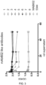
- an antibody directed against human interleukin-2 would have no measurable cross-reactivity to murine interleukin-2 if, under suitable conditions, binding of the antibody to murine interleukin-2 could not be detected with sufficiently sensitive methods such as surface plasmon resonance.
- Fig. 9 One such example of no measurable cross-reactivity is shown in Fig. 9 for the antibody in the lower panel (NARA1).
- an antibody or a protein that "specifically binds to hIL-2" is intended to refer to an antibody or protein that binds to human IL-2 polypeptide with a K D of 100nM or less, 10nM or less, 1nM or less, 100pM or less, or 10pM or less.
- An antibody that "cross-reacts with an antigen other than human IL-2" is intended to refer to an antibody that binds that antigen with a K D of 10nM or less, 1 nM or less, or 100 pM or less.
- an antibody that "does not cross-react with a particular antigen” is intended to refer to an antibody that binds to that antigen, with a K D of 100 nM or greater, or a K D of 1 ⁇ M or greater, or a K D of 10 ⁇ M or greater.
- such antibodies that do not cross-react with the antigen exhibit essentially undetectable binding against these proteins in standard binding assays.
- epitope means a protein determinant capable of specific binding to an antibody.
- Epitopes usually consist of chemically active surface groupings of molecules such as amino acids or sugar side chains and usually have specific three dimensional structural characteristics, as well as specific charge characteristics. Conformational and nonconformational epitopes are distinguished in that the binding to the former but not the latter is lost in the presence of denaturing solvents.
- epitope binding domain refers to portions of a binding molecule (e.g., an antibody or epitope-binding fragment or derivative thereof), that specifically interacts with (e.g., by binding, steric hindrance, stabilizing/destabilizing, spatial distribution) a binding site on a target epitope.
- EBD also refers to one or more fragments of an antibody that retain the ability to specifically interact with (e.g., by binding, steric hindrance, stabilizing/destabilizing, spatial distribution) a IL-2 epitope and inhibit signal transduction.
- antibody fragments include, but are not limited to, an scFv, a Fab fragment, a monovalent fragment consisting of the VL, VH, CL and CH1 domains; a F(ab) 2 fragment, a bivalent fragment comprising two Fab fragments linked by a disulfide bridge at the hinge region; a Fd fragment consisting of the VH and CH1 domains; a Fv fragment consisting of the VL and VH domains of a single arm of an antibody; a dAb fragment ( Ward et al., (1989) Nature 341:544-546 ), which consists of a VH domain; and an isolated complementarity determining region (CDR).
- an scFv a Fab fragment, a monovalent fragment consisting of the VL, VH, CL and CH1 domains
- F(ab) 2 fragment a bivalent fragment comprising two Fab fragments linked by a disulfide bridge at the hinge region
- a Fd fragment consisting of the V
- EBDs also include single domain antibodies, maxibodies, unibodies, minibodies, triabodies, tetrabodies, v-NAR and bis-scFv, as is known in the art (see, e.g., Hollinger and Hudson, (2005) Nature Biotechnology 23: 1126-1136 ), bispecific single chain diabodies, or single chain diabodies designed to bind two distinct epitopes.
- EBDs also include antibody-like molecules or antibody mimetics, which include, but not limited to minibodies, maxybodies, Fn3 based protein scaffolds, Ankyrin repeats (also known as DARpins), VASP polypeptides, Avian pancreatic polypeptide (aPP), Tetranectin, Affilin, Knottins, SH3 domains, PDZ domains, Tendamistat, Neocarzinostatin, Protein A domains, Lipocalins, Transferrin, and Kunitz domains that specifically bind epitopes, which are within the scope of the invention.
- Antibody fragments can be grafted into scaffolds based on polypeptides such as Fibronectin type III (Fn3) (see U.S. Pat. No. 6,703,199 , which describes fibronectin polypeptide monobodies).
- Fn3 Fibronectin type III
- the present invention also encompasses an antibody to human IL-2, which is an isolated antibody, as defined by the claims.
- isolated antibody refers to antibody that is substantially free of other antibodies having different antigenic specificities (e.g., an isolated antibody that specifically binds hIL-2 is substantially free of antibodies that specifically bind antigens other than hIL-2).
- An isolated antibody that specifically binds hIL-2 may, however, have cross-reactivity to other antigens, such as IL-2 molecules from other species.
- an isolated antibody may be substantially free of other cellular material and/or chemicals.
- nucleic acid and “polynucleotide” or “nucleotide coding sequences” are used interchangeably and refer to a polymeric form of nucleotides of any length, either deoxyribonucleotides or ribonucleotides or analogs thereof. Polynucleotides can have any three-dimensional structure and can perform any function.
- polynucleotides a gene or gene fragment (for example, a probe, primer, EST or SAGE tag), exons, introns, messenger RNA (mRNA), transfer RNA, ribosomal RNA, ribozymes, cDNA, recombinant polynucleotides, branched polynucleotides, plasmids, vectors, isolated DNA of any sequence, isolated RNA of any sequence, nucleic acid probes, siRNAs, shRNAs, RNAi agents, and primers.
- a polynucleotide can be modified or substituted at one or more base, sugar and/or phosphate, with any of various modifications or substitutions described herein or known in the art.
- a polynucleotide can comprise modified nucleotides, such as methylated nucleotides and nucleotide analogs. If present, modifications to the nucleotide structure can be imparted before or after assembly of the polymer.
- the sequence of nucleotides can be interrupted by non-nucleotide components.
- a polynucleotide can be further modified after polymerization, such as by conjugation with a labeling component.
- the term also refers to both double- and single-stranded molecules. Unless otherwise specified or required, any embodiment of this invention that is a polynucleotide encompasses both the double-stranded form and each of two complementary single-stranded forms known or predicted to make up the double-stranded form.
- polypeptide is used interchangeably with the term “protein” and in its broadest sense refers to a compound of two or more subunit amino acids, amino acid analogs, or peptidomimetics.
- the subunits can be linked by peptide bonds. In another embodiment, the subunit may be linked by other bonds, e.g., ester, ether, etc.
- treating refers in one embodiment, to ameliorating the disease or disorder (e.g. slowing or arresting or reducing the development of the disease or at least one of the clinical symptoms thereof).
- treating refers to alleviating or ameliorating at least one physical parameter including those which may not be discernible by the patient.
- treating or “treatment” refers to modulating the disease or disorder, either physically, (e.g., stabilization of a discernible symptom), physiologically, (e.g., stabilization of a physical parameter), or both.
- the inventors have generated and characterized specific anti-human IL-2 mAbs that are able to bind human IL-2 and, when tested in mice, are able to exert specific and potent stimulation of cytotoxic lymphocytes, including CD8 + T cells and natural killer (NK) cells. Towards these ends several difficulties had to be overcome.
- the inventors have developed specific screening assays that allow detection of specific antihuman IL-2 antibodies (so-called "binders”) in the serum of immunized animals and in the supernatant of the B cell clones obtained after B cell hybridoma fusion. In a second step it was discriminated between standard binders and those targeting a presumed specific epitope of the human IL-2 molecule.
- ELISA enzyme-linked immunosorbent assay
- Antibodies of the invention include the antibody NARA1, which was derived, isolated and structurally characterized by its full length heavy chain according to SEQ ID NO: 5 and its full length light chain amino acid sequences according to SEQ ID NO: 6.
- variable regions V H and V L amino acid sequences of NARA1 are.
- SEQ ID NO: 19 variable heavy
- SEQ ID NO: 20 variable light
- nucleotide coding sequences of NARA1 are SEQ ID NO: 3 (heavy chain coding sequence, including leader sequence) and SEQ ID NO: 4 (light chain coding sequence, including leader sequence).
- Variable light and heavy chains nucleotide coding sequences of NARA1 are SEQ ID NO: 21 (variable heavy coding sequence) and SEQ ID NO: 22 (variable light coding sequence).
- NARA1 The CDR regions of NARA1 are delineated using the Kabat system ( Kabat, E. A., et al. 1991, Sequences of Proteins of Immunological Interest, Fifth Edition, U.S. Department of Health and Human Services, NIH Publication No. 91-3242 , see also Zhao&Lu 2009, Molecular Immunology 47:694-700 ).
- Kabat Kabat, E. A., et al. 1991, Sequences of Proteins of Immunological Interest, Fifth Edition, U.S. Department of Health and Human Services, NIH Publication No. 91-3242 , see also Zhao&Lu 2009, Molecular Immunology 47:694-700 .
- HCDR1, HCDR2, HCDR3, LCDR1, LCDR2, LCDR3 are called hereafter HCDR1, HCDR2, HCDR3, LCDR1, LCDR2, LCDR3 respectively.
- the CDR regions of NARA1 are: HCDR1 according to SEQ ID NO: 7, HCDR2 according to SEQ ID NO: 8, HCDR3 according to SEQ ID NO: 9, LCDR1 according to SEQ ID NO: 10, LCDR2 according to SEQ ID NO: 11, LCDR3 according to SEQ ID NO: 12.
- Nucleotide coding sequences for the CDR regions of NARA1 are: HCDR1 coding sequence according to SEQ ID NO: 13, HCDR2 coding sequence according to SEQ ID NO: 14, HCDR3 coding sequence according to SEQ ID NO: 15, LCDR1 coding sequence according to SEQ ID NO: 16, LCDR2 coding sequence according to SEQ ID NO: 17, LCDR3 coding sequence according to SEQ ID NO: 18.
- Fusion proteins are also provided according to SEQ ID NO: 23 and SEQ ID NO: 24.
- SEQ ID NO: 23 is a fusion protein comprising the variable heavy chain of NARA1 with its N-terminus fused to the C-terminus of hIL-2 via a GxS linker.
- SEQ ID NO: 24 is a fusion protein comprising the variable light chain of NARA1 with its N-terminus fused to the C-terminus of hIL-2 via a GxS linker.
- Proleukin As will be discussed in detail below, the differences in sequence between Proleukin and wt hIL-2 are irrelevant and Proleukin is a valid model for structural analysis of hIL-2.
- X-ray crystallography was ued to solve the atomic-resolution structure of the complex mentioned above.
- X-ray crystallography is a technology that has become routinely and widely used to generate structural data for biomolecules including antibodies and their complexes with antigens ( Adms et al, (2013) Annual Review Biophysics 42:265-287 ; Garman, (2014) Science 343:1102-1108 ; Joachimiak, (2009) Current Opinio Structural Biology 19:573-584 .)
- Proleukin The antigen, Proleukin, is commercially available as lyophilyzed powder together with excipients (every 1 mg Proleukin is mixed with approximately 50mg mannitol, 0.18mg sodium dodecyl sulfate, 0.173mg sodium dihydrogen phosphate, and 0.89mg disodium hydrogen phosphate). Before used for complex formation, Proleukin was purified by reverse-phase HPLC to remove the excipients.
- NARA1-Fab The Fab fragment of NARA1 (NARA1-Fab) was generated by papain cleavage of the full-length antibody followed by Protein A chromatography. Briefly, 6.5ml full-length NARA1 (9mg/ml in 50mM citrate buffer with 90mM sodium chloride at pH 7.0) was mixed with 5mM DTT and 590ug Papain (Roche). The cleavage reaction was kept at room temperature for 16h and stopped by addition of 15ul 56mM E64 solution (Roche).
- the cleavage solution was then diluted 10 times with 25mM Tris, 25mM NaCl, pH 8.0 and loaded onto a 5ml Protein A column (GE Healthcare) equilibrate with 5 column volune of 25mM Tris, 25mM NaCl, pH 8.0 and Fab fragment was in the loading-through fraction and Fc fragment was bound to the Protein A column.
- Proleukin powder after HPLC was dissolved in H 2 O at the concentration of 5.5 mg/ml. 6.6mg Proleukin, in excess, was added to 11.5mg NARA1 Fab fragment solution drop by drop. Centrifugation was used to remove the excess Proleukin that was precipitated under current condition.
- the complex was then purified by gel filtration with Superdex 200 10x300 (GE Healthcare) with running buffer of 25mM Tris, 25mM NaCl, pH 7.4.
- Proleukin/NARA1-Fab complex after gel filtration was concentrated to 14mg/ml and was screened by vapour diffusion method as sitting drops.
- the protein solution was mixed 1:1 with reservoir buffer to a total size of 0.4ul.
- the experiments were set up with Phoenix robotic system (Art Robbins Instruments), stored in a Rocklmager hotel (Formulatrix) at 19°C, and imaged automatically. Crystals were harvested 4 days after screening under condition of 20% w/v polyethylene Glycol 3350 and 0.2M sodium nitrate. Crystals were cryo-protected with reservoir buffer containing 10% glycerol and flashed frozen in liquid nitrogen prior to data collection. Diffraction data were collected at the Swiss Light Source (Villigen, Switzerland) at beam-line PX-II with a Pilatus pixel detector using x-ray radiation wavelength of 0.99998 ⁇ .
- the dataset was processed with XDS and XSCALE (version Dec. 6 th , 2010) and the structure was resolved with molecular replacement method with the program PHASERby using Protein Data Bank entry "3INK” as search model for IL-2 and Protein Data Bank entry "3TTI” as search model for Fab fragment. Iterative model building and refinement were performed with the programs Coot (Crystallographic Object-Oriented Toolkit) and AUTOBUSTER (Bricogne et al., 2011). All figures were generated with the program PyMOL (Molecular Graphics System; DeLano Scientific: Palo Alto, CA; http://www.pymol.org).
- Epitope residues are defined as those residues from Proleukin that are within 4 ⁇ distance from any atom in Fab fragment of NARA1 and are further confirmed by CCP4 program CONTACT and AREAIMOL (Collaborative Computational Project, Number 4, version 6.4.0). Similarly paratope residues are defined as those residues from NARA1-Fab that are within 4 ⁇ distance from any atom in Proleukin.
- Figure 10 provides the overview of the three-dimensional structure of Proleukin/Fab-NARA1 complex as obtained in Example 1.
- Light chain of Fab fragment of NARA1 is designated A
- heavy chain of Fab fragment of NARA1 is shown as B
- epitope residues recognized by NARA1-Fab are designated D
- Proleukin is designated C and the mutation, C145S, is highlighted.
- Figure 11 provides further analysis of epitope residues.
- the X-axis lists the amino acid sequence and numbering according to SEQ ID No 1.
- the upper side of Y-axis demonstrates the total number of atoms of NARA1-Fab that are within 4 ⁇ from corresponding residue from Proleukin and the lower side of Y-axis demonstrates the reduced solvent-accessible area ( ⁇ 2 ) after binding to NARA1-Fab.
- Proleukin used in Example 1 contains mutation of C145S. As shown in Figure 10 , C145S is far away from the epitope region. In addition the superposition of C ⁇ atoms between Proleukin in Example 1 with C ⁇ atoms from wt hIL-2 in complex with CD25, CD122, and CD132 (PDB: 2B5I) shows r.m.s.d of 0.447 ⁇ , which indicates that the mutation does not disturb the over-all structure. Hence Proleukin with C145S mutation is a valid model for structural analysis for wt hIL-2.
- hIL-2 is 4-helix bundle protein and the 4 helices are named from N-terminus to C-terminus as A, B, C, and D, respectively.
- the epitope recognized by NARA1-Fab as shown in Figure 10 is a conformational epitope and spans two regions as shown in Figure 11 : one region (N50-K63) comprises a loop and a short helix and connects helix A and B, and the other region (N91-N97) comprises a loop and connects helix B and C.
- Arg58 as shown in Figure 11 is the most critical epitope residue for binding with NARA1-Fab, as this residue alone has 42 interacting atoms from NARA1-Fab and accounts for 17.7% of total reduced solvent-accessible surface area as a consequence of binding to NARA1-Fab.
- Arg58 as shown in Figure 12 , forms two strong salt-bridges with Glu35 in HCDR1 and with Asp100 from LCDR3, respectively. Arg58 also makes ⁇ -action interaction with the aromatic ring of Try100 from LCDR3.
- Residues K52, P54, K55, T57, T61, F62, K63, Q94, and K96 are also considered important for the binding to NARA1-Fab, since they all show equal to/more than 5 interacting atoms from NARA1-Fab and larger than 30 ⁇ 2 reduced solvent-accessible area as shown in Figure 11 . Table 2.
- Figure 12 illustrates Arg58 as the most critical epitope residue recognized by the NARA1-Fab.
- A represents Proleukin
- B represents heavy chain
- C represents light chain. The involved residues are shown as sticks.
- Figure 13 shows the overlay of Proleukin/NARA1-Fab complex with IL-2/CD25/CD122/CD 132 quaternary complex.
- the quaternary complex structure comes from PDB entry "2B5I” with cartoon D in pale cyan representing wt hIL-2, cartoon B in red representing CD122, cartoon C in blue representing CD132, and surface A in green representing CD25.
- cyan cartoon D overlayed with wt hIL-2 represents Proleukin
- cartoon E in magenta represents heavy chain
- cartoon F in yellow represents the light chain.
- Figure 14 displays the overlay of C helices from IL-2_C145A (PDB: 3INK), Superkine (PDB: 3QB1), IL-2/CD25/CD122/CD132 (PDB: 2B5I), and Proleukin/NARA1-Fab.
- a first library of 15-mer peptides was generated based on the sequence of human IL2.
- a second library of selected 15-mer peptides was also generated based on the mutation of 3 specific residues F(62), Y(65) and L(92). The latter mutations were done based on the Roche/Glycart IL2 mutein, as disclosed in WO2012/107417A1 which has these 3 mutations.
- Previous work done in lab Boyman (unpublished) showed that the commercial mouse anti-human IL2 mAb 602 with analogous function as A1 has strongly reduced binding to the F42A mutant of IL2 (one of the IL2 docking sites to CD25).
- each peptide in the first library has 15 amino acids and the sequence is derived by scanning the sequence of interest (see Table 4, reference peptides 1 to 41) with a step of 3 residues, starting from the N-terminus. Therefore a ladder is generated and each peptide contains 12 overlapping residues with the previous peptide and 12 overlapping residues with the following peptide in the ladder. In total, 41 peptides were generated from the expressed human IL2 sequence.
- a second library of peptides was generated by mutating F(62), Y(65) and L(92) to alanine in all corresponding peptides in the first library generated as described above (see Table 4, reference peptides no 42 to 60).
- Residue are the serine (S) replacing cysteines (C) 1 APTSSSTKKTQLQLE 2 SSSTKKTQLQLEHLL 3 TKKTQLQLEHLLLDL 4 TQLQLEHLLLDLQMI 5 QLEHLLLDLQMILNG 6 HLLLDLQMILNGINN 7 LDLQMILNGINNYKN 8 QMILNGINNYKNPKL 9 LNGINNYKNPKLTRM 10 INNYKNPKLTRMLTF 11 YKNPKLTRMLTFKFY 12 PKLTRMLTFKFYMPK 13 TRMLTFKFYMPKKAT 14 LTFKFYMPKKATELK 15 KFYMPKKATELKHLQ 16 MPKKATELKHLQSLE 17 KATELKHLQSLEEEL 18 ELKHLQSLEEELKPL 19 HLQSLEEELKPLEEV 20 SLEEELKPLEEVLNL 21 EELKPLEEVLNLAQS 22 KPLEEVLNLAQSKNF 23 EEVLNLAQSKNFHL
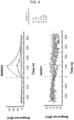
- Both set of peptides were printed on microarray slides in triplicate, incubated with the antibodies of interest (MAb602 and NARA1) and control antibodies. Additional incubations are with unrelated antibodies from the same isotype (mouse control IgG2a/lambda and mouse control lgG2a/kappa), and secondary antibodies (anti-mouse IgG (Thermo 84545, label DL650) or anti-mouse IgG (JIR 115-175-072, Label Cy5)) to assess unspecific binding due to the detection antibody.
- the experiments are performed essentially as described in Maksimov P, et al. 2012, PLoS One 7:e34212. doi:10.1371/journal. pone. 0034212 .
- peptide-antibody binding was performed by RepliTope-analysis where the peptide microarray (triplicate) was incubated with the primary antibody followed by a fluorescently labelled secondary antibody directed against the Fc-part of the primary one. All steps were performed on a TECAN microarray processing station enabling highly reliable and reproducible washing and incubation steps. After performing the incubation steps and subsequent to the final washing steps (to remove the unbound secondary antibodies) the microarrays were dried using a nitrogen stream and scanned in a high resolution microarray scanning system with appropriate wavelength settings. Control incubations were performed with an unrelated antibody having the same isotype to exclude false positive signals.
- the resulting images were analyzed und quantified using spot-recognition software GenePix (Molecular Devices). For each spot, the mean signal intensity was extracted (between 0 and 65535 arbitrary units). For further data evaluation, the MMC2 values were determined. The MMC2 equals the mean value of all three instances on the microarray. Except the coefficient of variation (CV) - standard-deviation divided by the mean value - is larger 0.5, in this case the mean of the two closest values (MC2) is assigned to MMC2.
- CV coefficient of variation
- the anti-IL2 (NARA1) antibody did not show any significant reactivity towards the immobilized peptides. Only peptide 10 exhibited a weak response, however, this peptide was also weakly recognized by the mouse control antibodies.
- the commercial antibody MAB602 (mlgG2a) provided some weak signals on peptide 22 to 26 and some strong for peptides 10 to 13. Table 5. Result of Linear Epitope Mapping Reference peptide no. Sequence Signal intensity for MAB602 after subtraction of control signal (AU) Signal intensity for NARA1 after subtraction of control signal (AU) 10 INNYKNPKLTRMLTF 45954 20883 11 YKNPKLTRMLTFKFY 49726 1189 12 PKLTRMLTFKFYMPK 28849 1127 13 TRMLTFKFYMPKKAT 5250 224 22 KPLEEVLNLAQSKNF 4998 0 23 EEVLNLAQSKNFHLR 13287 32 24 LNLAQSKNFHLRPRD 3289 282 25 AQSKNFHLRPRDLIS 5220 0 26 KNFHLRPRDLISNIN 7509 0
Landscapes
- Health & Medical Sciences (AREA)
- Chemical & Material Sciences (AREA)
- Life Sciences & Earth Sciences (AREA)
- Medicinal Chemistry (AREA)
- General Health & Medical Sciences (AREA)
- Organic Chemistry (AREA)
- Immunology (AREA)
- Proteomics, Peptides & Aminoacids (AREA)
- Public Health (AREA)
- Animal Behavior & Ethology (AREA)
- Veterinary Medicine (AREA)
- Pharmacology & Pharmacy (AREA)
- Epidemiology (AREA)
- Engineering & Computer Science (AREA)
- Bioinformatics & Cheminformatics (AREA)
- Molecular Biology (AREA)
- Biochemistry (AREA)
- Gastroenterology & Hepatology (AREA)
- Zoology (AREA)
- Genetics & Genomics (AREA)
- Biophysics (AREA)
- Nuclear Medicine, Radiotherapy & Molecular Imaging (AREA)
- Chemical Kinetics & Catalysis (AREA)
- General Chemical & Material Sciences (AREA)
- Endocrinology (AREA)
- Microbiology (AREA)
- Mycology (AREA)
- Toxicology (AREA)
- Communicable Diseases (AREA)
- Oncology (AREA)
- Virology (AREA)
- Peptides Or Proteins (AREA)
- Medicines Containing Antibodies Or Antigens For Use As Internal Diagnostic Agents (AREA)
- Preparation Of Compounds By Using Micro-Organisms (AREA)
- Micro-Organisms Or Cultivation Processes Thereof (AREA)
- Medicines That Contain Protein Lipid Enzymes And Other Medicines (AREA)
Applications Claiming Priority (3)
| Application Number | Priority Date | Filing Date | Title |
|---|---|---|---|
| EP14176619 | 2014-07-10 | ||
| EP15759938.2A EP3166973B1 (en) | 2014-07-10 | 2015-07-10 | Immune-stimulating monoclonal antibodies against human interleukin-2 |
| PCT/IB2015/055226 WO2016005950A1 (en) | 2014-07-10 | 2015-07-10 | Immune-stimulating monoclonal antibodies against human interleukin-2 |
Related Parent Applications (1)
| Application Number | Title | Priority Date | Filing Date |
|---|---|---|---|
| EP15759938.2A Division EP3166973B1 (en) | 2014-07-10 | 2015-07-10 | Immune-stimulating monoclonal antibodies against human interleukin-2 |
Publications (4)
| Publication Number | Publication Date |
|---|---|
| EP3693389A1 EP3693389A1 (en) | 2020-08-12 |
| EP3693389A9 EP3693389A9 (en) | 2021-04-07 |
| EP3693389B1 true EP3693389B1 (en) | 2024-09-04 |
| EP3693389C0 EP3693389C0 (en) | 2024-09-04 |
Family
ID=51162588
Family Applications (2)
| Application Number | Title | Priority Date | Filing Date |
|---|---|---|---|
| EP20158066.9A Active EP3693389B1 (en) | 2014-07-10 | 2015-07-10 | Immune-stimulating monoclonal antibodies against human interleukin-2 |
| EP15759938.2A Active EP3166973B1 (en) | 2014-07-10 | 2015-07-10 | Immune-stimulating monoclonal antibodies against human interleukin-2 |
Family Applications After (1)
| Application Number | Title | Priority Date | Filing Date |
|---|---|---|---|
| EP15759938.2A Active EP3166973B1 (en) | 2014-07-10 | 2015-07-10 | Immune-stimulating monoclonal antibodies against human interleukin-2 |
Country Status (9)
| Country | Link |
|---|---|
| US (2) | US10894828B2 (enExample) |
| EP (2) | EP3693389B1 (enExample) |
| JP (1) | JP7209464B2 (enExample) |
| CN (1) | CN106604932B (enExample) |
| AU (1) | AU2015287227B2 (enExample) |
| CA (1) | CA2954476C (enExample) |
| DK (1) | DK3166973T3 (enExample) |
| ES (2) | ES2779977T3 (enExample) |
| WO (1) | WO2016005950A1 (enExample) |
Families Citing this family (18)
| Publication number | Priority date | Publication date | Assignee | Title |
|---|---|---|---|---|
| WO2017122130A1 (en) | 2016-01-11 | 2017-07-20 | Novartis Ag | Immune-stimulating humanized monoclonal antibodies against human interleukin-2, and fusion proteins thereof |
| AU2017335771A1 (en) | 2016-09-28 | 2019-02-28 | Musc Foundation For Research Development | Antibodies that bind interleukin-2 and uses thereof |
| GB201621806D0 (en) * | 2016-12-21 | 2017-02-01 | Philogen Spa | Immunocytokines with progressive activation mechanism |
| CN110913873A (zh) * | 2017-02-23 | 2020-03-24 | 希望之城 | 用于cd8+t细胞的体内扩增并预防或治疗gvhd的方法 |
| SG11201908744PA (en) * | 2017-03-23 | 2019-10-30 | Univ Pennsylvania | Anti-c5a antibodies and uses thereof |
| KR102099593B1 (ko) * | 2017-05-25 | 2020-04-13 | 기초과학연구원 | 항-인간 인터루킨-2 항체 및 이의 용도 |
| WO2020036635A2 (en) | 2018-03-19 | 2020-02-20 | Multivir Inc. | Methods and compositions comprising tumor suppressor gene therapy and cd122/cd132 agonists for the treatment of cancer |
| WO2021113644A1 (en) | 2019-12-05 | 2021-06-10 | Multivir Inc. | Combinations comprising a cd8+ t cell enhancer, an immune checkpoint inhibitor and radiotherapy for targeted and abscopal effects for the treatment of cancer |
| WO2021144315A1 (en) | 2020-01-13 | 2021-07-22 | Synaffix B.V. | Conjugates of antibodies an immune cell engagers |
| IL308185B2 (en) * | 2020-02-16 | 2026-03-01 | Aulos Bioscience Inc | Engineered antibodies against IL-2 |
| PE20231648A1 (es) * | 2020-04-22 | 2023-10-17 | Merck Sharp And Dohme Llc | CONJUGADOS DE INTERLEUCINA 2 HUMANA SESGADOS AL DIMERO DEL RECEPTOR DE INTERLEUCINA 2 byc Y CONJUGADOS CON UN POLIMERO HIDROSOLUBLE NO PEPTIDICO |
| CN116783213A (zh) * | 2020-08-18 | 2023-09-19 | 苏黎世大学 | Cd25偏向的抗il‐2抗体 |
| IL300641A (en) * | 2020-08-18 | 2023-04-01 | Univ Zuerich | CD25 biased anti-IL-2 antibody |
| JP2024506022A (ja) | 2021-02-08 | 2024-02-08 | シンアフィックス ビー.ブイ. | 多機能性抗体 |
| AU2022364888A1 (en) | 2021-10-14 | 2024-05-30 | Latticon (Suzhou) Biopharmaceuticals Co., Ltd. | Novel antibody-cytokine fusion protein, preparation method therefor and use thereof |
| US11773160B1 (en) | 2022-08-05 | 2023-10-03 | Anaveon AG | Immune-stimulating IL-2 fusion proteins |
| CN119912559B (zh) * | 2025-01-22 | 2025-12-26 | 贵州中医药大学 | 一种新冠病毒变异株jn.1刺突s蛋白的单链抗体及其应用 |
| CN119775409B (zh) * | 2025-03-05 | 2025-07-11 | 深圳真实生物医药科技有限公司 | 靶向sost的抗体及其用途 |
Family Cites Families (17)
| Publication number | Priority date | Publication date | Assignee | Title |
|---|---|---|---|---|
| ZA849910B (en) | 1983-12-23 | 1985-09-25 | Hoffmann La Roche | Purification of recombinant interleukin-2 |
| DK134487A (da) * | 1986-03-17 | 1987-09-18 | Hoffmann La Roche | Monoklonalt antistof med affinitet for humant interleukin-2 |
| US6111090A (en) | 1996-08-16 | 2000-08-29 | Schering Corporation | Mammalian cell surface antigens; related reagents |
| ES2308787T3 (es) | 1996-08-16 | 2008-12-01 | Schering Corporation | Antigenos de superficie de celular de mamiferos; reactivos relacionados. |
| EP1958962A3 (en) | 1997-06-12 | 2013-05-01 | Novartis International Pharmaceutical Ltd. | Artificial antibody polypeptides |
| EP2572714A1 (en) | 2002-12-30 | 2013-03-27 | 3M Innovative Properties Company | Immunostimulatory Combinations |
| US20060002932A1 (en) | 2004-06-04 | 2006-01-05 | Duke University | Methods and compositions for enhancement of immunity by in vivo depletion of immunosuppressive cell activity |
| MX2007014565A (es) | 2005-06-01 | 2008-02-11 | Micromet Ag | Anticuerpos anti-il2. |
| CN104072614B (zh) | 2005-07-08 | 2017-04-26 | 生物基因Ma公司 | 抗-αvβ6 抗体及其用途 |
| CN101573139A (zh) * | 2006-02-16 | 2009-11-04 | 纳森特生物制剂公司 | 在哺乳动物个体中改善免疫功能的方法以及预防或治疗疾病的方法 |
| CN101045923B (zh) * | 2006-03-31 | 2011-11-23 | 沈阳三生制药有限责任公司 | 生产一种白细胞介素类似物的方法 |
| CN101970499B (zh) | 2008-02-11 | 2014-12-31 | 治疗科技公司 | 用于肿瘤治疗的单克隆抗体 |
| PL3042917T3 (pl) | 2010-08-12 | 2018-07-31 | Eli Lilly And Company | Przeciwciała przeciwko peptydowi beta amyloidu N3pGlu i ich zastosowanie |
| SG192673A1 (en) | 2011-02-10 | 2013-09-30 | Roche Glycart Ag | Mutant interleukin-2 polypeptides |
| WO2013157105A1 (ja) * | 2012-04-18 | 2013-10-24 | 公立大学法人大阪市立大学 | ムチンサブタイプ5ac特異的ヒト化抗体およびその利用 |
| CN112587671A (zh) | 2012-07-18 | 2021-04-02 | 博笛生物科技有限公司 | 癌症的靶向免疫治疗 |
| WO2015109212A1 (en) * | 2014-01-17 | 2015-07-23 | Pfizer Inc. | Anti-il-2 antibodies and compositions and uses thereof |
-
2015
- 2015-07-10 DK DK15759938.2T patent/DK3166973T3/da active
- 2015-07-10 CA CA2954476A patent/CA2954476C/en active Active
- 2015-07-10 AU AU2015287227A patent/AU2015287227B2/en not_active Ceased
- 2015-07-10 JP JP2017501280A patent/JP7209464B2/ja active Active
- 2015-07-10 WO PCT/IB2015/055226 patent/WO2016005950A1/en not_active Ceased
- 2015-07-10 EP EP20158066.9A patent/EP3693389B1/en active Active
- 2015-07-10 EP EP15759938.2A patent/EP3166973B1/en active Active
- 2015-07-10 ES ES15759938T patent/ES2779977T3/es active Active
- 2015-07-10 US US15/324,468 patent/US10894828B2/en active Active
- 2015-07-10 CN CN201580048027.0A patent/CN106604932B/zh active Active
- 2015-07-10 ES ES20158066T patent/ES2989669T3/es active Active
-
2020
- 2020-12-22 US US17/131,221 patent/US20210230269A1/en not_active Abandoned
Also Published As
| Publication number | Publication date |
|---|---|
| WO2016005950A1 (en) | 2016-01-14 |
| AU2015287227A8 (en) | 2017-02-16 |
| US20210230269A1 (en) | 2021-07-29 |
| DK3166973T3 (da) | 2020-04-06 |
| JP7209464B2 (ja) | 2023-01-20 |
| US10894828B2 (en) | 2021-01-19 |
| EP3166973A1 (en) | 2017-05-17 |
| EP3166973B1 (en) | 2020-02-19 |
| EP3693389C0 (en) | 2024-09-04 |
| EP3693389A1 (en) | 2020-08-12 |
| CA2954476C (en) | 2023-09-19 |
| CA2954476A1 (en) | 2016-01-14 |
| CN106604932A (zh) | 2017-04-26 |
| CN106604932B (zh) | 2024-12-10 |
| ES2779977T3 (es) | 2020-08-21 |
| EP3693389A9 (en) | 2021-04-07 |
| US20170183403A1 (en) | 2017-06-29 |
| AU2015287227A1 (en) | 2017-02-09 |
| AU2015287227B2 (en) | 2021-02-18 |
| JP2017525343A (ja) | 2017-09-07 |
| ES2989669T3 (es) | 2024-11-27 |
Similar Documents
| Publication | Publication Date | Title |
|---|---|---|
| EP3693389B1 (en) | Immune-stimulating monoclonal antibodies against human interleukin-2 | |
| AU2019349651B2 (en) | SIRP alpha binding proteins and methods of use thereof | |
| CN113166246B (zh) | 一种抗体及其用途 | |
| JP2020531043A (ja) | 抗4−1bb抗体とその作製及び使用方法 | |
| KR20190134614A (ko) | B7-h3 항체, 이의 항원-결합 단편 및 이의 의학적 용도 | |
| CN113227148B (zh) | 抗gpc3抗体、其抗原结合片段及其医药用途 | |
| KR20190112299A (ko) | 결합제 | |
| JP2020533970A (ja) | 抗cd3抗体とその作製及び使用方法 | |
| CN111225926A (zh) | 靶向pdl1的抗体及其使用方法 | |
| CN117279950A (zh) | 一种靶向il-18bp的抗体及其应用 | |
| US20260098093A1 (en) | Treatment and prevention of cancer using vista antigen-binding molecules | |
| CN115298216A (zh) | 抗体或其抗原结合片段、其制备方法及医药用途 | |
| CN111788229A (zh) | Csf1r结合剂 | |
| CN116847863A (zh) | 抗人cd22的单克隆抗体及其用途 | |
| TWI889766B (zh) | 抗cd19抗體以及使用與製造其的方法 | |
| US20210301012A1 (en) | Proteins and Uses | |
| HK40049006B (en) | Anti-gpc3 antibody, antigen-binding fragment thereof, and medical use thereof | |
| HK40049006A (en) | Anti-gpc3 antibody, antigen-binding fragment thereof, and medical use thereof | |
| WO2019137552A1 (en) | MODIFIED Cκ AND CH1 DOMAINS | |
| HK40089051A (zh) | Egfr结合复合物及其制备和使用方法 |
Legal Events
| Date | Code | Title | Description |
|---|---|---|---|
| PUAI | Public reference made under article 153(3) epc to a published international application that has entered the european phase |
Free format text: ORIGINAL CODE: 0009012 |
|
| STAA | Information on the status of an ep patent application or granted ep patent |
Free format text: STATUS: REQUEST FOR EXAMINATION WAS MADE |
|
| 17P | Request for examination filed |
Effective date: 20200218 |
|
| AC | Divisional application: reference to earlier application |
Ref document number: 3166973 Country of ref document: EP Kind code of ref document: P |
|
| AK | Designated contracting states |
Kind code of ref document: A1 Designated state(s): AL AT BE BG CH CY CZ DE DK EE ES FI FR GB GR HR HU IE IS IT LI LT LU LV MC MK MT NL NO PL PT RO RS SE SI SK SM TR |
|
| STAA | Information on the status of an ep patent application or granted ep patent |
Free format text: STATUS: EXAMINATION IS IN PROGRESS |
|
| 17Q | First examination report despatched |
Effective date: 20220520 |
|
| RIC1 | Information provided on ipc code assigned before grant |
Ipc: A61P 31/00 20060101ALI20230714BHEP Ipc: A61P 35/00 20060101ALI20230714BHEP Ipc: A61K 38/20 20060101ALI20230714BHEP Ipc: A61K 39/395 20060101ALI20230714BHEP Ipc: C07K 16/24 20060101AFI20230714BHEP |
|
| GRAP | Despatch of communication of intention to grant a patent |
Free format text: ORIGINAL CODE: EPIDOSNIGR1 |
|
| STAA | Information on the status of an ep patent application or granted ep patent |
Free format text: STATUS: GRANT OF PATENT IS INTENDED |
|
| INTG | Intention to grant announced |
Effective date: 20230825 |
|
| GRAJ | Information related to disapproval of communication of intention to grant by the applicant or resumption of examination proceedings by the epo deleted |
Free format text: ORIGINAL CODE: EPIDOSDIGR1 |
|
| STAA | Information on the status of an ep patent application or granted ep patent |
Free format text: STATUS: EXAMINATION IS IN PROGRESS |
|
| GRAP | Despatch of communication of intention to grant a patent |
Free format text: ORIGINAL CODE: EPIDOSNIGR1 |
|
| STAA | Information on the status of an ep patent application or granted ep patent |
Free format text: STATUS: GRANT OF PATENT IS INTENDED |
|
| INTC | Intention to grant announced (deleted) | ||
| RIN1 | Information on inventor provided before grant (corrected) |
Inventor name: ZOU, CHAO Inventor name: ARENAS-RAMIREZ, NATALIA Inventor name: BOYMAN, ONUR |
|
| RAP3 | Party data changed (applicant data changed or rights of an application transferred) |
Owner name: UNIVERSITAET ZUERICH |
|
| INTG | Intention to grant announced |
Effective date: 20231128 |
|
| GRAS | Grant fee paid |
Free format text: ORIGINAL CODE: EPIDOSNIGR3 |
|
| GRAA | (expected) grant |
Free format text: ORIGINAL CODE: 0009210 |
|
| STAA | Information on the status of an ep patent application or granted ep patent |
Free format text: STATUS: THE PATENT HAS BEEN GRANTED |
|
| AC | Divisional application: reference to earlier application |
Ref document number: 3166973 Country of ref document: EP Kind code of ref document: P |
|
| AK | Designated contracting states |
Kind code of ref document: B1 Designated state(s): AL AT BE BG CH CY CZ DE DK EE ES FI FR GB GR HR HU IE IS IT LI LT LU LV MC MK MT NL NO PL PT RO RS SE SI SK SM TR |
|
| REG | Reference to a national code |
Ref country code: GB Ref legal event code: FG4D |
|
| REG | Reference to a national code |
Ref country code: CH Ref legal event code: EP |
|
| REG | Reference to a national code |
Ref country code: IE Ref legal event code: FG4D |
|
| REG | Reference to a national code |
Ref country code: DE Ref legal event code: R096 Ref document number: 602015089826 Country of ref document: DE |
|
| U01 | Request for unitary effect filed |
Effective date: 20241003 |
|
| REG | Reference to a national code |
Ref country code: ES Ref legal event code: FG2A Ref document number: 2989669 Country of ref document: ES Kind code of ref document: T3 Effective date: 20241127 |
|
| U07 | Unitary effect registered |
Designated state(s): AT BE BG DE DK EE FI FR IT LT LU LV MT NL PT RO SE SI Effective date: 20241025 |
|
| PG25 | Lapsed in a contracting state [announced via postgrant information from national office to epo] |
Ref country code: NO Free format text: LAPSE BECAUSE OF FAILURE TO SUBMIT A TRANSLATION OF THE DESCRIPTION OR TO PAY THE FEE WITHIN THE PRESCRIBED TIME-LIMIT Effective date: 20241204 |
|
| PG25 | Lapsed in a contracting state [announced via postgrant information from national office to epo] |
Ref country code: PL Free format text: LAPSE BECAUSE OF FAILURE TO SUBMIT A TRANSLATION OF THE DESCRIPTION OR TO PAY THE FEE WITHIN THE PRESCRIBED TIME-LIMIT Effective date: 20240904 Ref country code: GR Free format text: LAPSE BECAUSE OF FAILURE TO SUBMIT A TRANSLATION OF THE DESCRIPTION OR TO PAY THE FEE WITHIN THE PRESCRIBED TIME-LIMIT Effective date: 20241205 |
|
| PG25 | Lapsed in a contracting state [announced via postgrant information from national office to epo] |
Ref country code: HR Free format text: LAPSE BECAUSE OF FAILURE TO SUBMIT A TRANSLATION OF THE DESCRIPTION OR TO PAY THE FEE WITHIN THE PRESCRIBED TIME-LIMIT Effective date: 20240904 |
|
| PG25 | Lapsed in a contracting state [announced via postgrant information from national office to epo] |
Ref country code: RS Free format text: LAPSE BECAUSE OF FAILURE TO SUBMIT A TRANSLATION OF THE DESCRIPTION OR TO PAY THE FEE WITHIN THE PRESCRIBED TIME-LIMIT Effective date: 20241204 |
|
| PG25 | Lapsed in a contracting state [announced via postgrant information from national office to epo] |
Ref country code: RS Free format text: LAPSE BECAUSE OF FAILURE TO SUBMIT A TRANSLATION OF THE DESCRIPTION OR TO PAY THE FEE WITHIN THE PRESCRIBED TIME-LIMIT Effective date: 20241204 Ref country code: PL Free format text: LAPSE BECAUSE OF FAILURE TO SUBMIT A TRANSLATION OF THE DESCRIPTION OR TO PAY THE FEE WITHIN THE PRESCRIBED TIME-LIMIT Effective date: 20240904 Ref country code: NO Free format text: LAPSE BECAUSE OF FAILURE TO SUBMIT A TRANSLATION OF THE DESCRIPTION OR TO PAY THE FEE WITHIN THE PRESCRIBED TIME-LIMIT Effective date: 20241204 Ref country code: HR Free format text: LAPSE BECAUSE OF FAILURE TO SUBMIT A TRANSLATION OF THE DESCRIPTION OR TO PAY THE FEE WITHIN THE PRESCRIBED TIME-LIMIT Effective date: 20240904 Ref country code: GR Free format text: LAPSE BECAUSE OF FAILURE TO SUBMIT A TRANSLATION OF THE DESCRIPTION OR TO PAY THE FEE WITHIN THE PRESCRIBED TIME-LIMIT Effective date: 20241205 |
|
| PG25 | Lapsed in a contracting state [announced via postgrant information from national office to epo] |
Ref country code: IS Free format text: LAPSE BECAUSE OF FAILURE TO SUBMIT A TRANSLATION OF THE DESCRIPTION OR TO PAY THE FEE WITHIN THE PRESCRIBED TIME-LIMIT Effective date: 20250104 |
|
| PG25 | Lapsed in a contracting state [announced via postgrant information from national office to epo] |
Ref country code: SM Free format text: LAPSE BECAUSE OF FAILURE TO SUBMIT A TRANSLATION OF THE DESCRIPTION OR TO PAY THE FEE WITHIN THE PRESCRIBED TIME-LIMIT Effective date: 20240904 |
|
| PG25 | Lapsed in a contracting state [announced via postgrant information from national office to epo] |
Ref country code: CZ Free format text: LAPSE BECAUSE OF FAILURE TO SUBMIT A TRANSLATION OF THE DESCRIPTION OR TO PAY THE FEE WITHIN THE PRESCRIBED TIME-LIMIT Effective date: 20240904 |
|
| PG25 | Lapsed in a contracting state [announced via postgrant information from national office to epo] |
Ref country code: SK Free format text: LAPSE BECAUSE OF FAILURE TO SUBMIT A TRANSLATION OF THE DESCRIPTION OR TO PAY THE FEE WITHIN THE PRESCRIBED TIME-LIMIT Effective date: 20240904 |
|
| PLBE | No opposition filed within time limit |
Free format text: ORIGINAL CODE: 0009261 |
|
| STAA | Information on the status of an ep patent application or granted ep patent |
Free format text: STATUS: NO OPPOSITION FILED WITHIN TIME LIMIT |
|
| 26N | No opposition filed |
Effective date: 20250605 |
|
| REG | Reference to a national code |
Ref country code: CH Ref legal event code: U11 Free format text: ST27 STATUS EVENT CODE: U-0-0-U10-U11 (AS PROVIDED BY THE NATIONAL OFFICE) Effective date: 20251222 |
|
| PGFP | Annual fee paid to national office [announced via postgrant information from national office to epo] |
Ref country code: GB Payment date: 20251219 Year of fee payment: 11 |
|
| PGFP | Annual fee paid to national office [announced via postgrant information from national office to epo] |
Ref country code: CH Payment date: 20251222 Year of fee payment: 11 |
|
| PGFP | Annual fee paid to national office [announced via postgrant information from national office to epo] |
Ref country code: ES Payment date: 20251229 Year of fee payment: 11 |
|
| U21 | Renewal fee for the european patent with unitary effect paid with additional fee |
Year of fee payment: 11 Effective date: 20251229 |