EP3166973B1 - Immune-stimulating monoclonal antibodies against human interleukin-2 - Google Patents
Immune-stimulating monoclonal antibodies against human interleukin-2 Download PDFInfo
- Publication number
- EP3166973B1 EP3166973B1 EP15759938.2A EP15759938A EP3166973B1 EP 3166973 B1 EP3166973 B1 EP 3166973B1 EP 15759938 A EP15759938 A EP 15759938A EP 3166973 B1 EP3166973 B1 EP 3166973B1
- Authority
- EP
- European Patent Office
- Prior art keywords
- seq
- hil
- nmol
- antibody
- fragment
- Prior art date
- Legal status (The legal status is an assumption and is not a legal conclusion. Google has not performed a legal analysis and makes no representation as to the accuracy of the status listed.)
- Active
Links
- 101001002657 Homo sapiens Interleukin-2 Proteins 0.000 title claims description 166
- 102000055277 human IL2 Human genes 0.000 title claims description 134
- 230000027455 binding Effects 0.000 claims description 120
- 102000000588 Interleukin-2 Human genes 0.000 claims description 90
- 108010002350 Interleukin-2 Proteins 0.000 claims description 90
- 239000012634 fragment Substances 0.000 claims description 76
- 239000000427 antigen Substances 0.000 claims description 59
- 108091007433 antigens Proteins 0.000 claims description 59
- 102000036639 antigens Human genes 0.000 claims description 59
- 108090000765 processed proteins & peptides Proteins 0.000 claims description 45
- 102000004196 processed proteins & peptides Human genes 0.000 claims description 33
- 150000001413 amino acids Chemical class 0.000 claims description 26
- 210000004027 cell Anatomy 0.000 claims description 24
- 229920001184 polypeptide Polymers 0.000 claims description 21
- 101001057504 Homo sapiens Interferon-stimulated gene 20 kDa protein Proteins 0.000 claims description 20
- 101001055144 Homo sapiens Interleukin-2 receptor subunit alpha Proteins 0.000 claims description 20
- 102100026878 Interleukin-2 receptor subunit alpha Human genes 0.000 claims description 20
- 150000007523 nucleic acids Chemical class 0.000 claims description 20
- 102000039446 nucleic acids Human genes 0.000 claims description 19
- 108020004707 nucleic acids Proteins 0.000 claims description 19
- 238000010494 dissociation reaction Methods 0.000 claims description 17
- 230000005593 dissociations Effects 0.000 claims description 17
- 241001529936 Murinae Species 0.000 claims description 15
- 210000004408 hybridoma Anatomy 0.000 claims description 15
- 238000011282 treatment Methods 0.000 claims description 14
- 230000009260 cross reactivity Effects 0.000 claims description 13
- 206010028980 Neoplasm Diseases 0.000 claims description 12
- 108020001507 fusion proteins Proteins 0.000 claims description 12
- 102000037865 fusion proteins Human genes 0.000 claims description 12
- 238000002198 surface plasmon resonance spectroscopy Methods 0.000 claims description 12
- 201000011510 cancer Diseases 0.000 claims description 10
- 230000001225 therapeutic effect Effects 0.000 claims description 9
- 239000000203 mixture Substances 0.000 claims description 8
- 208000036142 Viral infection Diseases 0.000 claims description 7
- 238000009472 formulation Methods 0.000 claims description 7
- 238000000159 protein binding assay Methods 0.000 claims description 7
- 230000009385 viral infection Effects 0.000 claims description 7
- 239000003814 drug Substances 0.000 claims description 5
- 239000013598 vector Substances 0.000 claims description 4
- 108700025316 aldesleukin Proteins 0.000 description 35
- 229940087463 proleukin Drugs 0.000 description 35
- 108090000623 proteins and genes Proteins 0.000 description 17
- 235000018102 proteins Nutrition 0.000 description 16
- 235000001014 amino acid Nutrition 0.000 description 15
- 102000004169 proteins and genes Human genes 0.000 description 15
- 108010054477 Immunoglobulin Fab Fragments Proteins 0.000 description 12
- 102000001706 Immunoglobulin Fab Fragments Human genes 0.000 description 12
- 210000003719 b-lymphocyte Anatomy 0.000 description 12
- 108091026890 Coding region Proteins 0.000 description 11
- 108020004414 DNA Proteins 0.000 description 11
- 125000003729 nucleotide group Chemical group 0.000 description 11
- 210000000822 natural killer cell Anatomy 0.000 description 10
- 108091033319 polynucleotide Proteins 0.000 description 9
- 239000002157 polynucleotide Substances 0.000 description 9
- 102000040430 polynucleotide Human genes 0.000 description 9
- NFGXHKASABOEEW-UHFFFAOYSA-N 1-methylethyl 11-methoxy-3,7,11-trimethyl-2,4-dodecadienoate Chemical compound COC(C)(C)CCCC(C)CC=CC(C)=CC(=O)OC(C)C NFGXHKASABOEEW-UHFFFAOYSA-N 0.000 description 8
- 102000004127 Cytokines Human genes 0.000 description 8
- 108090000695 Cytokines Proteins 0.000 description 8
- FAPWRFPIFSIZLT-UHFFFAOYSA-M Sodium chloride Chemical compound [Na+].[Cl-] FAPWRFPIFSIZLT-UHFFFAOYSA-M 0.000 description 8
- 208000037265 diseases, disorders, signs and symptoms Diseases 0.000 description 8
- 238000000034 method Methods 0.000 description 8
- 230000035772 mutation Effects 0.000 description 8
- 239000002773 nucleotide Substances 0.000 description 8
- 210000003289 regulatory T cell Anatomy 0.000 description 8
- 239000006228 supernatant Substances 0.000 description 8
- 210000001266 CD8-positive T-lymphocyte Anatomy 0.000 description 7
- 241000699670 Mus sp. Species 0.000 description 7
- 230000000694 effects Effects 0.000 description 7
- 238000001727 in vivo Methods 0.000 description 7
- 210000001165 lymph node Anatomy 0.000 description 7
- 102000010789 Interleukin-2 Receptors Human genes 0.000 description 6
- 241000699666 Mus <mouse, genus> Species 0.000 description 6
- 230000000259 anti-tumor effect Effects 0.000 description 6
- 239000011230 binding agent Substances 0.000 description 6
- 230000004927 fusion Effects 0.000 description 6
- 210000004698 lymphocyte Anatomy 0.000 description 6
- 238000002493 microarray Methods 0.000 description 6
- 230000004044 response Effects 0.000 description 6
- 108010038453 Interleukin-2 Receptors Proteins 0.000 description 5
- 210000001744 T-lymphocyte Anatomy 0.000 description 5
- 201000010099 disease Diseases 0.000 description 5
- 230000006870 function Effects 0.000 description 5
- 238000000338 in vitro Methods 0.000 description 5
- 238000012216 screening Methods 0.000 description 5
- 210000000952 spleen Anatomy 0.000 description 5
- 108010032595 Antibody Binding Sites Proteins 0.000 description 4
- IJGRMHOSHXDMSA-UHFFFAOYSA-N Atomic nitrogen Chemical compound N#N IJGRMHOSHXDMSA-UHFFFAOYSA-N 0.000 description 4
- DHMQDGOQFOQNFH-UHFFFAOYSA-N Glycine Chemical compound NCC(O)=O DHMQDGOQFOQNFH-UHFFFAOYSA-N 0.000 description 4
- 108060003951 Immunoglobulin Proteins 0.000 description 4
- 101001043827 Mus musculus Interleukin-2 Proteins 0.000 description 4
- 238000004458 analytical method Methods 0.000 description 4
- 230000008901 benefit Effects 0.000 description 4
- 231100000433 cytotoxic Toxicity 0.000 description 4
- 230000001472 cytotoxic effect Effects 0.000 description 4
- 238000000313 electron-beam-induced deposition Methods 0.000 description 4
- 102000018358 immunoglobulin Human genes 0.000 description 4
- 238000009169 immunotherapy Methods 0.000 description 4
- 238000011534 incubation Methods 0.000 description 4
- 230000003993 interaction Effects 0.000 description 4
- 230000003389 potentiating effect Effects 0.000 description 4
- 239000011780 sodium chloride Substances 0.000 description 4
- 230000000638 stimulation Effects 0.000 description 4
- 238000002560 therapeutic procedure Methods 0.000 description 4
- VDABVNMGKGUPEY-UHFFFAOYSA-N 6-carboxyfluorescein succinimidyl ester Chemical compound C=1C(O)=CC=C2C=1OC1=CC(O)=CC=C1C2(C1=C2)OC(=O)C1=CC=C2C(=O)ON1C(=O)CCC1=O VDABVNMGKGUPEY-UHFFFAOYSA-N 0.000 description 3
- 108010047041 Complementarity Determining Regions Proteins 0.000 description 3
- 238000002965 ELISA Methods 0.000 description 3
- PEDCQBHIVMGVHV-UHFFFAOYSA-N Glycerine Chemical compound OCC(O)CO PEDCQBHIVMGVHV-UHFFFAOYSA-N 0.000 description 3
- QNAYBMKLOCPYGJ-REOHCLBHSA-N L-alanine Chemical compound C[C@H](N)C(O)=O QNAYBMKLOCPYGJ-REOHCLBHSA-N 0.000 description 3
- 239000007983 Tris buffer Substances 0.000 description 3
- 235000004279 alanine Nutrition 0.000 description 3
- 230000004071 biological effect Effects 0.000 description 3
- 238000012512 characterization method Methods 0.000 description 3
- 238000003776 cleavage reaction Methods 0.000 description 3
- 239000013078 crystal Substances 0.000 description 3
- 208000035475 disorder Diseases 0.000 description 3
- 238000002474 experimental method Methods 0.000 description 3
- 239000000463 material Substances 0.000 description 3
- 239000000243 solution Substances 0.000 description 3
- 241000894007 species Species 0.000 description 3
- 230000006641 stabilisation Effects 0.000 description 3
- 238000011105 stabilization Methods 0.000 description 3
- LENZDBCJOHFCAS-UHFFFAOYSA-N tris Chemical compound OCC(N)(CO)CO LENZDBCJOHFCAS-UHFFFAOYSA-N 0.000 description 3
- 102100032912 CD44 antigen Human genes 0.000 description 2
- RTZKZFJDLAIYFH-UHFFFAOYSA-N Diethyl ether Chemical compound CCOCC RTZKZFJDLAIYFH-UHFFFAOYSA-N 0.000 description 2
- 239000004471 Glycine Substances 0.000 description 2
- 101000868273 Homo sapiens CD44 antigen Proteins 0.000 description 2
- 102000008394 Immunoglobulin Fragments Human genes 0.000 description 2
- 108010021625 Immunoglobulin Fragments Proteins 0.000 description 2
- 108010067060 Immunoglobulin Variable Region Proteins 0.000 description 2
- 102000017727 Immunoglobulin Variable Region Human genes 0.000 description 2
- 206010027480 Metastatic malignant melanoma Diseases 0.000 description 2
- 241001465754 Metazoa Species 0.000 description 2
- 108090000526 Papain Proteins 0.000 description 2
- 239000004365 Protease Substances 0.000 description 2
- MTCFGRXMJLQNBG-UHFFFAOYSA-N Serine Natural products OCC(N)C(O)=O MTCFGRXMJLQNBG-UHFFFAOYSA-N 0.000 description 2
- 108010003723 Single-Domain Antibodies Proteins 0.000 description 2
- 210000000662 T-lymphocyte subset Anatomy 0.000 description 2
- 238000002835 absorbance Methods 0.000 description 2
- 230000002411 adverse Effects 0.000 description 2
- 230000005875 antibody response Effects 0.000 description 2
- 108010086186 avian pancreatic polypeptide Proteins 0.000 description 2
- 239000000872 buffer Substances 0.000 description 2
- 235000018417 cysteine Nutrition 0.000 description 2
- 150000001945 cysteines Chemical class 0.000 description 2
- 238000013480 data collection Methods 0.000 description 2
- 230000000368 destabilizing effect Effects 0.000 description 2
- 238000001514 detection method Methods 0.000 description 2
- 238000011161 development Methods 0.000 description 2
- 230000018109 developmental process Effects 0.000 description 2
- 238000010790 dilution Methods 0.000 description 2
- 239000012895 dilution Substances 0.000 description 2
- LOKCTEFSRHRXRJ-UHFFFAOYSA-I dipotassium trisodium dihydrogen phosphate hydrogen phosphate dichloride Chemical compound P(=O)(O)(O)[O-].[K+].P(=O)(O)([O-])[O-].[Na+].[Na+].[Cl-].[K+].[Cl-].[Na+] LOKCTEFSRHRXRJ-UHFFFAOYSA-I 0.000 description 2
- 239000012636 effector Substances 0.000 description 2
- 238000005516 engineering process Methods 0.000 description 2
- 238000002523 gelfiltration Methods 0.000 description 2
- 210000002865 immune cell Anatomy 0.000 description 2
- 230000001900 immune effect Effects 0.000 description 2
- 229940072221 immunoglobulins Drugs 0.000 description 2
- 230000001965 increasing effect Effects 0.000 description 2
- 231100001231 less toxic Toxicity 0.000 description 2
- 201000001441 melanoma Diseases 0.000 description 2
- 210000003071 memory t lymphocyte Anatomy 0.000 description 2
- 108020004999 messenger RNA Proteins 0.000 description 2
- 230000001394 metastastic effect Effects 0.000 description 2
- 208000021039 metastatic melanoma Diseases 0.000 description 2
- 206010061289 metastatic neoplasm Diseases 0.000 description 2
- 238000012986 modification Methods 0.000 description 2
- 230000004048 modification Effects 0.000 description 2
- 238000010172 mouse model Methods 0.000 description 2
- 229910052757 nitrogen Inorganic materials 0.000 description 2
- 229940055729 papain Drugs 0.000 description 2
- 235000019834 papain Nutrition 0.000 description 2
- 239000000546 pharmaceutical excipient Substances 0.000 description 2
- 239000002953 phosphate buffered saline Substances 0.000 description 2
- 239000013641 positive control Substances 0.000 description 2
- 239000000843 powder Substances 0.000 description 2
- 230000035755 proliferation Effects 0.000 description 2
- 230000008929 regeneration Effects 0.000 description 2
- 238000011069 regeneration method Methods 0.000 description 2
- 230000007017 scission Effects 0.000 description 2
- VWDWKYIASSYTQR-UHFFFAOYSA-N sodium nitrate Chemical compound [Na+].[O-][N+]([O-])=O VWDWKYIASSYTQR-UHFFFAOYSA-N 0.000 description 2
- 230000000087 stabilizing effect Effects 0.000 description 2
- 238000012916 structural analysis Methods 0.000 description 2
- 230000004083 survival effect Effects 0.000 description 2
- 208000024891 symptom Diseases 0.000 description 2
- 210000001519 tissue Anatomy 0.000 description 2
- 231100000331 toxic Toxicity 0.000 description 2
- 230000002588 toxic effect Effects 0.000 description 2
- 238000005406 washing Methods 0.000 description 2
- 238000002424 x-ray crystallography Methods 0.000 description 2
- 108091032973 (ribonucleotides)n+m Proteins 0.000 description 1
- PMUNIMVZCACZBB-UHFFFAOYSA-N 2-hydroxyethylazanium;chloride Chemical compound Cl.NCCO PMUNIMVZCACZBB-UHFFFAOYSA-N 0.000 description 1
- FWMNVWWHGCHHJJ-SKKKGAJSSA-N 4-amino-1-[(2r)-6-amino-2-[[(2r)-2-[[(2r)-2-[[(2r)-2-amino-3-phenylpropanoyl]amino]-3-phenylpropanoyl]amino]-4-methylpentanoyl]amino]hexanoyl]piperidine-4-carboxylic acid Chemical compound C([C@H](C(=O)N[C@H](CC(C)C)C(=O)N[C@H](CCCCN)C(=O)N1CCC(N)(CC1)C(O)=O)NC(=O)[C@H](N)CC=1C=CC=CC=1)C1=CC=CC=C1 FWMNVWWHGCHHJJ-SKKKGAJSSA-N 0.000 description 1
- 206010067484 Adverse reaction Diseases 0.000 description 1
- 102000008102 Ankyrins Human genes 0.000 description 1
- 108010049777 Ankyrins Proteins 0.000 description 1
- 208000023275 Autoimmune disease Diseases 0.000 description 1
- 238000011740 C57BL/6 mouse Methods 0.000 description 1
- 108090000994 Catalytic RNA Proteins 0.000 description 1
- 102000053642 Catalytic RNA Human genes 0.000 description 1
- 108010025905 Cystine-Knot Miniproteins Proteins 0.000 description 1
- 102100025698 Cytosolic carboxypeptidase 4 Human genes 0.000 description 1
- FBPFZTCFMRRESA-KVTDHHQDSA-N D-Mannitol Chemical compound OC[C@@H](O)[C@@H](O)[C@H](O)[C@H](O)CO FBPFZTCFMRRESA-KVTDHHQDSA-N 0.000 description 1
- 102000004190 Enzymes Human genes 0.000 description 1
- 108090000790 Enzymes Proteins 0.000 description 1
- 108700024394 Exon Proteins 0.000 description 1
- 108700004922 F42A Proteins 0.000 description 1
- 102100037362 Fibronectin Human genes 0.000 description 1
- 102000002090 Fibronectin type III Human genes 0.000 description 1
- 108050009401 Fibronectin type III Proteins 0.000 description 1
- 108010067306 Fibronectins Proteins 0.000 description 1
- 206010059024 Gastrointestinal toxicity Diseases 0.000 description 1
- 108090000288 Glycoproteins Proteins 0.000 description 1
- 102000003886 Glycoproteins Human genes 0.000 description 1
- 206010019837 Hepatocellular injury Diseases 0.000 description 1
- 241000282412 Homo Species 0.000 description 1
- VEXZGXHMUGYJMC-UHFFFAOYSA-N Hydrochloric acid Chemical compound Cl VEXZGXHMUGYJMC-UHFFFAOYSA-N 0.000 description 1
- 208000001953 Hypotension Diseases 0.000 description 1
- 102000018071 Immunoglobulin Fc Fragments Human genes 0.000 description 1
- 108010091135 Immunoglobulin Fc Fragments Proteins 0.000 description 1
- 238000012404 In vitro experiment Methods 0.000 description 1
- 108010002586 Interleukin-7 Proteins 0.000 description 1
- 102000015696 Interleukins Human genes 0.000 description 1
- 108010063738 Interleukins Proteins 0.000 description 1
- 108091092195 Intron Proteins 0.000 description 1
- 102000019298 Lipocalin Human genes 0.000 description 1
- 108050006654 Lipocalin Proteins 0.000 description 1
- 229930195725 Mannitol Natural products 0.000 description 1
- 206010050513 Metastatic renal cell carcinoma Diseases 0.000 description 1
- 241000699660 Mus musculus Species 0.000 description 1
- 101710204212 Neocarzinostatin Proteins 0.000 description 1
- 108020004711 Nucleic Acid Probes Proteins 0.000 description 1
- 108091028043 Nucleic acid sequence Proteins 0.000 description 1
- 206010030113 Oedema Diseases 0.000 description 1
- 108050008994 PDZ domains Proteins 0.000 description 1
- 102000000470 PDZ domains Human genes 0.000 description 1
- 229910019142 PO4 Inorganic materials 0.000 description 1
- 241000233805 Phoenix Species 0.000 description 1
- 206010035226 Plasma cell myeloma Diseases 0.000 description 1
- 206010037423 Pulmonary oedema Diseases 0.000 description 1
- 102220495631 Putative uncharacterized protein LOC645739_F42A_mutation Human genes 0.000 description 1
- 108091030071 RNAI Proteins 0.000 description 1
- 101001043830 Rattus norvegicus Interleukin-2 Proteins 0.000 description 1
- 108091028664 Ribonucleotide Proteins 0.000 description 1
- 108050008861 SH3 domains Proteins 0.000 description 1
- 102000000395 SH3 domains Human genes 0.000 description 1
- 108091027967 Small hairpin RNA Proteins 0.000 description 1
- 108020004459 Small interfering RNA Proteins 0.000 description 1
- DBMJMQXJHONAFJ-UHFFFAOYSA-M Sodium laurylsulphate Chemical compound [Na+].CCCCCCCCCCCCOS([O-])(=O)=O DBMJMQXJHONAFJ-UHFFFAOYSA-M 0.000 description 1
- 239000012505 Superdex™ Substances 0.000 description 1
- 108020004566 Transfer RNA Proteins 0.000 description 1
- 102000004338 Transferrin Human genes 0.000 description 1
- 108090000901 Transferrin Proteins 0.000 description 1
- 102100021164 Vasodilator-stimulated phosphoprotein Human genes 0.000 description 1
- 230000004913 activation Effects 0.000 description 1
- 239000002671 adjuvant Substances 0.000 description 1
- 230000006838 adverse reaction Effects 0.000 description 1
- 150000001412 amines Chemical class 0.000 description 1
- 230000003042 antagnostic effect Effects 0.000 description 1
- 230000000890 antigenic effect Effects 0.000 description 1
- 238000013459 approach Methods 0.000 description 1
- 125000003118 aryl group Chemical group 0.000 description 1
- 238000003556 assay Methods 0.000 description 1
- 230000006399 behavior Effects 0.000 description 1
- 230000009286 beneficial effect Effects 0.000 description 1
- 210000001185 bone marrow Anatomy 0.000 description 1
- 210000004899 c-terminal region Anatomy 0.000 description 1
- 238000002619 cancer immunotherapy Methods 0.000 description 1
- 125000002843 carboxylic acid group Chemical group 0.000 description 1
- 230000001413 cellular effect Effects 0.000 description 1
- 230000036755 cellular response Effects 0.000 description 1
- 238000005119 centrifugation Methods 0.000 description 1
- 230000008859 change Effects 0.000 description 1
- 238000006243 chemical reaction Methods 0.000 description 1
- 239000003795 chemical substances by application Substances 0.000 description 1
- 238000004587 chromatography analysis Methods 0.000 description 1
- 239000007979 citrate buffer Substances 0.000 description 1
- 230000000295 complement effect Effects 0.000 description 1
- 230000004154 complement system Effects 0.000 description 1
- 239000002299 complementary DNA Substances 0.000 description 1
- 230000009918 complex formation Effects 0.000 description 1
- 150000001875 compounds Chemical class 0.000 description 1
- 238000012790 confirmation Methods 0.000 description 1
- 230000021615 conjugation Effects 0.000 description 1
- 230000008878 coupling Effects 0.000 description 1
- 238000010168 coupling process Methods 0.000 description 1
- 238000005859 coupling reaction Methods 0.000 description 1
- 238000002447 crystallographic data Methods 0.000 description 1
- 208000035250 cutaneous malignant susceptibility to 1 melanoma Diseases 0.000 description 1
- 210000001151 cytotoxic T lymphocyte Anatomy 0.000 description 1
- 238000011157 data evaluation Methods 0.000 description 1
- 230000009849 deactivation Effects 0.000 description 1
- 230000002498 deadly effect Effects 0.000 description 1
- 239000005547 deoxyribonucleotide Substances 0.000 description 1
- 125000002637 deoxyribonucleotide group Chemical group 0.000 description 1
- 230000001419 dependent effect Effects 0.000 description 1
- 238000009792 diffusion process Methods 0.000 description 1
- 230000006806 disease prevention Effects 0.000 description 1
- BNIILDVGGAEEIG-UHFFFAOYSA-L disodium hydrogen phosphate Chemical compound [Na+].[Na+].OP([O-])([O-])=O BNIILDVGGAEEIG-UHFFFAOYSA-L 0.000 description 1
- 229940079593 drug Drugs 0.000 description 1
- 210000002889 endothelial cell Anatomy 0.000 description 1
- 230000002708 enhancing effect Effects 0.000 description 1
- 229940088598 enzyme Drugs 0.000 description 1
- 150000002148 esters Chemical class 0.000 description 1
- 238000000684 flow cytometry Methods 0.000 description 1
- 231100000414 gastrointestinal toxicity Toxicity 0.000 description 1
- 230000009368 gene silencing by RNA Effects 0.000 description 1
- 125000003712 glycosamine group Chemical group 0.000 description 1
- 239000003102 growth factor Substances 0.000 description 1
- 230000036541 health Effects 0.000 description 1
- 238000004128 high performance liquid chromatography Methods 0.000 description 1
- 239000005556 hormone Substances 0.000 description 1
- 229940088597 hormone Drugs 0.000 description 1
- 210000004754 hybrid cell Anatomy 0.000 description 1
- 230000036543 hypotension Effects 0.000 description 1
- 210000000987 immune system Anatomy 0.000 description 1
- 230000003053 immunization Effects 0.000 description 1
- 238000002649 immunization Methods 0.000 description 1
- 230000005847 immunogenicity Effects 0.000 description 1
- 238000005462 in vivo assay Methods 0.000 description 1
- 230000002401 inhibitory effect Effects 0.000 description 1
- 239000007924 injection Substances 0.000 description 1
- 238000002347 injection Methods 0.000 description 1
- 108040006849 interleukin-2 receptor activity proteins Proteins 0.000 description 1
- 238000002372 labelling Methods 0.000 description 1
- 239000007788 liquid Substances 0.000 description 1
- 231100000849 liver cell damage Toxicity 0.000 description 1
- 239000000594 mannitol Substances 0.000 description 1
- 235000010355 mannitol Nutrition 0.000 description 1
- 238000004519 manufacturing process Methods 0.000 description 1
- 238000013507 mapping Methods 0.000 description 1
- 238000005259 measurement Methods 0.000 description 1
- 208000037819 metastatic cancer Diseases 0.000 description 1
- 208000011575 metastatic malignant neoplasm Diseases 0.000 description 1
- 238000003032 molecular docking Methods 0.000 description 1
- 239000000178 monomer Substances 0.000 description 1
- 229910000403 monosodium phosphate Inorganic materials 0.000 description 1
- 235000019799 monosodium phosphate Nutrition 0.000 description 1
- 238000002703 mutagenesis Methods 0.000 description 1
- 231100000350 mutagenesis Toxicity 0.000 description 1
- 201000000050 myeloid neoplasm Diseases 0.000 description 1
- QZGIWPZCWHMVQL-UIYAJPBUSA-N neocarzinostatin chromophore Chemical compound O1[C@H](C)[C@H](O)[C@H](O)[C@@H](NC)[C@H]1O[C@@H]1C/2=C/C#C[C@H]3O[C@@]3([C@@H]3OC(=O)OC3)C#CC\2=C[C@H]1OC(=O)C1=C(O)C=CC2=C(C)C=C(OC)C=C12 QZGIWPZCWHMVQL-UIYAJPBUSA-N 0.000 description 1
- 230000003472 neutralizing effect Effects 0.000 description 1
- 239000002853 nucleic acid probe Substances 0.000 description 1
- 210000000056 organ Anatomy 0.000 description 1
- 238000012510 peptide mapping method Methods 0.000 description 1
- 239000000816 peptidomimetic Substances 0.000 description 1
- NBIIXXVUZAFLBC-UHFFFAOYSA-K phosphate Chemical compound [O-]P([O-])([O-])=O NBIIXXVUZAFLBC-UHFFFAOYSA-K 0.000 description 1
- 239000010452 phosphate Substances 0.000 description 1
- 239000013612 plasmid Substances 0.000 description 1
- 229940050929 polyethylene glycol 3350 Drugs 0.000 description 1
- 229920000642 polymer Polymers 0.000 description 1
- 238000006116 polymerization reaction Methods 0.000 description 1
- 230000002265 prevention Effects 0.000 description 1
- 125000002924 primary amino group Chemical group [H]N([H])* 0.000 description 1
- 238000012545 processing Methods 0.000 description 1
- 239000000047 product Substances 0.000 description 1
- 239000012460 protein solution Substances 0.000 description 1
- 208000005333 pulmonary edema Diseases 0.000 description 1
- 230000005855 radiation Effects 0.000 description 1
- 230000009257 reactivity Effects 0.000 description 1
- 230000001105 regulatory effect Effects 0.000 description 1
- 238000009877 rendering Methods 0.000 description 1
- 238000004007 reversed phase HPLC Methods 0.000 description 1
- 238000012552 review Methods 0.000 description 1
- 239000002336 ribonucleotide Substances 0.000 description 1
- 125000002652 ribonucleotide group Chemical group 0.000 description 1
- 108020004418 ribosomal RNA Proteins 0.000 description 1
- 108091092562 ribozyme Proteins 0.000 description 1
- 239000012146 running buffer Substances 0.000 description 1
- 235000002020 sage Nutrition 0.000 description 1
- 239000000523 sample Substances 0.000 description 1
- 238000009738 saturating Methods 0.000 description 1
- 238000007423 screening assay Methods 0.000 description 1
- 238000012163 sequencing technique Methods 0.000 description 1
- 210000002966 serum Anatomy 0.000 description 1
- 230000019491 signal transduction Effects 0.000 description 1
- 239000007974 sodium acetate buffer Substances 0.000 description 1
- AJPJDKMHJJGVTQ-UHFFFAOYSA-M sodium dihydrogen phosphate Chemical compound [Na+].OP(O)([O-])=O AJPJDKMHJJGVTQ-UHFFFAOYSA-M 0.000 description 1
- 239000004317 sodium nitrate Substances 0.000 description 1
- 235000010344 sodium nitrate Nutrition 0.000 description 1
- 239000002904 solvent Substances 0.000 description 1
- 230000009870 specific binding Effects 0.000 description 1
- 210000004989 spleen cell Anatomy 0.000 description 1
- 230000004936 stimulating effect Effects 0.000 description 1
- 239000000126 substance Substances 0.000 description 1
- 238000006467 substitution reaction Methods 0.000 description 1
- 125000000020 sulfo group Chemical group O=S(=O)([*])O[H] 0.000 description 1
- 230000002483 superagonistic effect Effects 0.000 description 1
- 230000008685 targeting Effects 0.000 description 1
- 229950001790 tendamistat Drugs 0.000 description 1
- 108010037401 tendamistate Proteins 0.000 description 1
- 239000003053 toxin Substances 0.000 description 1
- 231100000765 toxin Toxicity 0.000 description 1
- 108700012359 toxins Proteins 0.000 description 1
- 239000012581 transferrin Substances 0.000 description 1
- 238000011830 transgenic mouse model Methods 0.000 description 1
- 238000011269 treatment regimen Methods 0.000 description 1
- 108010054220 vasodilator-stimulated phosphoprotein Proteins 0.000 description 1
- 229950009268 zinostatin Drugs 0.000 description 1
Images


Classifications
-
- C—CHEMISTRY; METALLURGY
- C07—ORGANIC CHEMISTRY
- C07K—PEPTIDES
- C07K16/00—Immunoglobulins [IGs], e.g. monoclonal or polyclonal antibodies
- C07K16/18—Immunoglobulins [IGs], e.g. monoclonal or polyclonal antibodies against material from animals or humans
- C07K16/24—Immunoglobulins [IGs], e.g. monoclonal or polyclonal antibodies against material from animals or humans against cytokines, lymphokines or interferons
- C07K16/244—Interleukins [IL]
- C07K16/246—IL-2
-
- A—HUMAN NECESSITIES
- A61—MEDICAL OR VETERINARY SCIENCE; HYGIENE
- A61K—PREPARATIONS FOR MEDICAL, DENTAL OR TOILETRY PURPOSES
- A61K38/00—Medicinal preparations containing peptides
- A61K38/16—Peptides having more than 20 amino acids; Gastrins; Somatostatins; Melanotropins; Derivatives thereof
- A61K38/17—Peptides having more than 20 amino acids; Gastrins; Somatostatins; Melanotropins; Derivatives thereof from animals; from humans
- A61K38/19—Cytokines; Lymphokines; Interferons
- A61K38/20—Interleukins [IL]
- A61K38/2013—IL-2
-
- A—HUMAN NECESSITIES
- A61—MEDICAL OR VETERINARY SCIENCE; HYGIENE
- A61K—PREPARATIONS FOR MEDICAL, DENTAL OR TOILETRY PURPOSES
- A61K39/00—Medicinal preparations containing antigens or antibodies
- A61K39/395—Antibodies; Immunoglobulins; Immune serum, e.g. antilymphocytic serum
- A61K39/39533—Antibodies; Immunoglobulins; Immune serum, e.g. antilymphocytic serum against materials from animals
- A61K39/3955—Antibodies; Immunoglobulins; Immune serum, e.g. antilymphocytic serum against materials from animals against proteinaceous materials, e.g. enzymes, hormones, lymphokines
-
- A—HUMAN NECESSITIES
- A61—MEDICAL OR VETERINARY SCIENCE; HYGIENE
- A61P—SPECIFIC THERAPEUTIC ACTIVITY OF CHEMICAL COMPOUNDS OR MEDICINAL PREPARATIONS
- A61P31/00—Antiinfectives, i.e. antibiotics, antiseptics, chemotherapeutics
- A61P31/12—Antivirals
-
- A—HUMAN NECESSITIES
- A61—MEDICAL OR VETERINARY SCIENCE; HYGIENE
- A61P—SPECIFIC THERAPEUTIC ACTIVITY OF CHEMICAL COMPOUNDS OR MEDICINAL PREPARATIONS
- A61P35/00—Antineoplastic agents
-
- C—CHEMISTRY; METALLURGY
- C07—ORGANIC CHEMISTRY
- C07K—PEPTIDES
- C07K14/00—Peptides having more than 20 amino acids; Gastrins; Somatostatins; Melanotropins; Derivatives thereof
- C07K14/435—Peptides having more than 20 amino acids; Gastrins; Somatostatins; Melanotropins; Derivatives thereof from animals; from humans
- C07K14/52—Cytokines; Lymphokines; Interferons
- C07K14/54—Interleukins [IL]
- C07K14/55—IL-2
-
- A—HUMAN NECESSITIES
- A61—MEDICAL OR VETERINARY SCIENCE; HYGIENE
- A61K—PREPARATIONS FOR MEDICAL, DENTAL OR TOILETRY PURPOSES
- A61K39/00—Medicinal preparations containing antigens or antibodies
- A61K2039/505—Medicinal preparations containing antigens or antibodies comprising antibodies
-
- C—CHEMISTRY; METALLURGY
- C07—ORGANIC CHEMISTRY
- C07K—PEPTIDES
- C07K2317/00—Immunoglobulins specific features
- C07K2317/30—Immunoglobulins specific features characterized by aspects of specificity or valency
- C07K2317/34—Identification of a linear epitope shorter than 20 amino acid residues or of a conformational epitope defined by amino acid residues
-
- C—CHEMISTRY; METALLURGY
- C07—ORGANIC CHEMISTRY
- C07K—PEPTIDES
- C07K2317/00—Immunoglobulins specific features
- C07K2317/50—Immunoglobulins specific features characterized by immunoglobulin fragments
- C07K2317/55—Fab or Fab'
-
- C—CHEMISTRY; METALLURGY
- C07—ORGANIC CHEMISTRY
- C07K—PEPTIDES
- C07K2317/00—Immunoglobulins specific features
- C07K2317/50—Immunoglobulins specific features characterized by immunoglobulin fragments
- C07K2317/56—Immunoglobulins specific features characterized by immunoglobulin fragments variable (Fv) region, i.e. VH and/or VL
- C07K2317/565—Complementarity determining region [CDR]
-
- C—CHEMISTRY; METALLURGY
- C07—ORGANIC CHEMISTRY
- C07K—PEPTIDES
- C07K2317/00—Immunoglobulins specific features
- C07K2317/70—Immunoglobulins specific features characterized by effect upon binding to a cell or to an antigen
- C07K2317/74—Inducing cell proliferation
-
- C—CHEMISTRY; METALLURGY
- C07—ORGANIC CHEMISTRY
- C07K—PEPTIDES
- C07K2317/00—Immunoglobulins specific features
- C07K2317/70—Immunoglobulins specific features characterized by effect upon binding to a cell or to an antigen
- C07K2317/76—Antagonist effect on antigen, e.g. neutralization or inhibition of binding
-
- C—CHEMISTRY; METALLURGY
- C07—ORGANIC CHEMISTRY
- C07K—PEPTIDES
- C07K2317/00—Immunoglobulins specific features
- C07K2317/90—Immunoglobulins specific features characterized by (pharmaco)kinetic aspects or by stability of the immunoglobulin
- C07K2317/92—Affinity (KD), association rate (Ka), dissociation rate (Kd) or EC50 value
Definitions
- the present invention relates to antibodies binding to human interleukin-2 (hIL-2).
- hIL-2 human interleukin-2
- the invention more specifically relates to antibodies specifically binding a particular epitope of hIL-2 and when bound to this epitope are capable of inhibit binding of hIL-2 to CD25.
- the invention relates to in vitro and in vivo therapeutic applications of the antibodies.
- Malignant melanoma is a frequent cancer type in men and women. Once melanoma becomes metastatic and spreads to distant sites, the 5-year survival rate is quite poor, calculated at about 15%. Currently available treatment strategies for metastatic melanoma barely improve this survival rate.
- Interleukin-2 is a cytokine able to potently stimulate cytotoxic lymphocytes against metastatic tumours.
- IL-2 is also able to stimulate so-called CD25 + CD4 + regulatory T cells (Treg cells) that are crucial for prevention of autoimmune disease.
- Treg cells can significantly dampen anti-tumour responses by cytotoxic lymphocytes, thus somewhat antagonizing the beneficial anti-tumour effects of IL-2.
- Treg cells can significantly dampen anti-tumour responses by cytotoxic lymphocytes, thus somewhat antagonizing the beneficial anti-tumour effects of IL-2.
- IL-2 can exert toxic adverse effects.
- Standard IL-2 immunotherapy has been used since the early 1980's for the immunotherapy of metastatic melanoma and metastatic renal cell carcinoma, leading to the approval by the FDA for these indications in 1996 and 1992, respectively.
- IL-2 given at high doses has shown objective response rates in about 17% and complete regression in about 6-9% of patients suffering from these deadly metastatic cancers, IL-2 given at these doses frequently led to toxic adverse effects, such as hypotension, pulmonary edema, liver cell damage, gastrointestinal toxicity, and general edema.
- IL-2 is able to stimulate Treg cells, which in turn are able to dampen the activity of anti-tumour CD8 + T cells and NK cells.
- IL-2/anti-IL-2 antibody complexes show strong biological activity by avoiding interaction with IL-2 receptor ⁇ subunit CD25. Proceedings of the National Academy of Sciences USA (2010) 107:2171-2176 ), thereby directing IL-2 preferentially to cytotoxic lymphocytes, but not Treg cells.
- This approach has the advantage that unmutated, natural IL-2 is delivered via anti-IL-2 mAb to CD8 + T cells and NK cells, which subsequently exert potent anti-tumour properties, while IL-2 complexed to this kind of anti-IL-2 mAb barely activates Treg cells. Moreover, IL-2 complexed to this kind of anti-IL-2 mAb is much less toxic than standard IL-2 immuno therapy in mice. However, this therapy has up to date not been available for use in patients due to the lack of appropriate anti-human IL-2 mAbs.
- the problem addressed by the present invention is to provide an anti-human IL-2 monoclonal antibody able to recognize and bind a specific epitope of human IL-2, thereby favoring the stimulation of cytotoxic T cells and NK cells compared to Treg cells, for use in in vitro and in vivo therapeutic applications.
- the invention relates to the embodiments as defined in the claims, thus it relates to the following items.
- a human interleukin-2 (hIL-2) specific monoclonal antibody (mAb), or a human interleukin-2 (hIL-2) specific antigen binding fragment thereof comprising HCDR1 according to SEQ ID NO: 7, HCDR2 according to SEQ ID NO: 8, HCDR3 according to SEQ ID NO: 9, LCDR1 according to SEQ ID NO: 10, LCDR2 according to SEQ ID NO: 11, LCDR3 according to SEQ ID NO: 12 is provided, wherein binding of said antibody, or antigen binding fragment thereof, to hIL-2 inhibits binding of hIL-2 to CD25, and the binding to hIL-2 is characterized by a dissociation constant (K D ) ⁇ 7,5 nmol/L, ⁇ 5 nmol/L, ⁇ 3 nmol/L, ⁇ 2 nmol/L or ⁇ 1,5 nmol/L determined in a surface plasmon resonance binding assay.
- K D dissociation constant
- hIL-2 human interleukin-2
- hIL-2 human interleukin-2
- the binding to hIL-2 is characterized by an off-rate (K off ) ⁇ 1x10 -4 s -1 , ⁇ 8x10 -5 s -1 , ⁇ 6x10 -5 s -1 , ⁇ 4x10 -5 s -1 , ⁇ 3x10 -5 s -1 or ⁇ 2,1x10 -5 s -1 .
- hIL-2 human interleukin-2
- hIL-2 human interleukin-2
- the antibody, or antigen binding fragment thereof displays no measurable cross-reactivity to murine IL-2.
- human interleukin-2 (hIL-2) specific monoclonal antibodies, or antigen binding fragments thereof are provided, characterized in that they comprise at least one VH and/or one VL sequence having an identity of ⁇ 80%, ⁇ 85%, ⁇ 90%, ⁇ 92%, ⁇ 93%, ⁇ 94%, ⁇ 95%, ⁇ 96%, ⁇ 97% or ⁇ 98% compared to SEQ ID NOs 019 or 20.
- a nucleic acid molecule encoding the human interleukin-2 (hIL-2) specific monoclonal antibody, or an antigen binding fragment thereof, according to the first aspect of the invention is provided.
- nucleic acid molecule according to the second aspect of the invention is provided, wherein the nucleic acid molecule has ⁇ 60%, ⁇ 70%, ⁇ 80%, ⁇ 85%, ⁇ 90%, ⁇ 92%, ⁇ 93%, ⁇ 94%, ⁇ 95%, ⁇ 96%, ⁇ 97% or ⁇ 98% sequence identity compared to SEQ ID NOs 003 or 004.
- nucleic acid molecule comprising the sequence of SEQ ID NOs 13, 14, 15, 16,17,18, 21 or 22, or a sequence having an identity of ⁇ 80%, ⁇ 85%, ⁇ 90%, ⁇ 92%, ⁇ 93%, ⁇ 94%, ⁇ 95%, ⁇ 96%, ⁇ 97% or ⁇ 98% compared to any one of SEQ ID NOs 13, 14, 15, 16, 17, 18, 21 or 22 is provided.
- a vector comprising the nucleic acid molecule according to the invention is provided.
- a cell comprising the nucleic acid molecule according to the invention, or expressing the nucleic acid molecule according to the invention is provided.
- a cell able to produce a human interleukin-2 (hIL-2) specific monoclonal antibody (mAb), or antigen binding fragment thereof, according to the first aspect of the invention is provided.
- hIL-2 human interleukin-2
- mAb monoclonal antibody
- a monoclonal antibody-producing hybridoma cell line is provided, characterized in that the produced antibodies are those of the first aspect of invention.
- a therapeutic formulation for use as a medicament particularly for use in the treatment of cancer or viral infections, comprising:
- the human interleukin-2 (hIL-2) specific monoclonal antibody, or antigen binding fragment thereof is provided for use in the treatment of cancer or viral infections, wherein the antibody, or antigen binding fragment thereof is administered together with human interleukin-2.
- a therapeutic formulation for use as a medicament is provided, particularly for use in the treatment of cancer or viral infections, containing a fusion protein that comprises:
- the antibody or antigen binding fragment thereof which binds to a human interleukin-2 (hIL-2) epitope comprising the amino acids K52, P54, K55, T57, R58, T61, F62, K63, Q94, and K96 is provided.
- hIL-2 human interleukin-2
- the antibody or antigen binding fragment thereof according to the tenth aspect of the invention is provided, wherein the epitope further comprises any one or more of the amino acids N50, N53, N91, L92, A93, N97.
- a human interleukin-2 (hIL-2) specific monoclonal antibody (mAb), or antigen binding fragment thereof is provided, wherein the antibody is able to bind to a particular epitope in hIL-2 thereby inhibiting the binding to CD25, thus modulating the immunological effects of hIL-2/IL-2R interaction.
- the antibody of the disclosure is further characterized by at least one of the parameters:
- a lack of cross-reactivity with murine IL-2 is advantageous for preclinical studies, which usually involve mouse models, such as the use of mAb ⁇ hIL-2 complexes for the treatment of murine tumour models where a cross-reactive anti-IL-2 mAb might bind and seclude endogenous murine IL-2 from endogenous murine Treg cells, thus enhancing the anti-tumour response.
- a lack of cross-reactivity with murine IL-2 is also advantageous for preclinical safety and efficacy studies conducted prior to development of a candidate mAb in human patients.
- the hIL-2 mAb comprises at least one V H and/or V L sequence having an identity of ⁇ 80%, ⁇ 85%, ⁇ 90%, ⁇ 92%, ⁇ 93%, ⁇ 94%, ⁇ 95%, ⁇ 96%, ⁇ 97% or ⁇ 98% compared to SEQ ID NOs 019 or SEQ ID NO 020.
- variable chain of the hIL-2 mAb comprises an amino acid sequence having an identity of ⁇ 85%, ⁇ 90%, ⁇ 95%, or ⁇ 99% compared to SEQ ID NOs 003, 004, 005 or 006 and the hIL-2 mAb is characterized by a dissociation constant ⁇ 7,5 nmol/L, ⁇ 5 nmol/L, ⁇ 3 nmol/L, ⁇ 2 nmol/L or ⁇ 1,5 nmol/L.
- variable chain of the hIL-2 mAb comprises an amino acid sequence having an identity of ⁇ 85%, ⁇ 90%, ⁇ 92%, ⁇ 93%, ⁇ 94%, ⁇ 95%, ⁇ 96%, ⁇ 97%, ⁇ 98% or ⁇ 99% compared to SEQ ID NO 005 or 006 and the hIL-2 mAb is characterized by an off-rate ⁇ 1x10 -4 s -1 , ⁇ 8x10 -5 s -1 , ⁇ 6x10 -5 s -1 , ⁇ 4x10 -5 s -1 , ⁇ 3x10 -5 s -1 or ⁇ 2,1x10 -5 s -1 .
- variable chain of the hIL-2 mAb comprises an amino acid sequence having an identity of ⁇ 85%, ⁇ 90%, ⁇ 92%, ⁇ 93%, ⁇ 94%, ⁇ 95%, ⁇ 96%, ⁇ 97%, ⁇ 98% or ⁇ 99% compared to SEQ ID NO 005 or 006 and the hIL-2 mAb displays no measurable cross-reactivity to murine IL-2.
- sequence of the hIL-2 mAb is humanized for administration to human patients to prevent adverse reactions.
- the hIL-2 mAb is provided as fragment antigen-binding (Fab) or single-chain variable fragment (scFv).
- the hIL-2 mAb comprises at least one complementarity determining (CDR) sequence having an identity of ⁇ 80%, ⁇ 85%, ⁇ 90%, ⁇ 92%, ⁇ 93%, ⁇ 94%, ⁇ 95%, ⁇ 96%, ⁇ 97% or ⁇ 98% compared to SEQ ID NOs 007, 008, 009, 010, 011 or 012.
- CDR complementarity determining
- nucleic acid molecule encoding the monoclonal antibody, or antigen binding fragment thereof, able to bind to human interleukin-2 according to the first aspect of the disclosure is provided.
- nucleic acid molecule according to the second aspect of the disclosure has ⁇ 60%, ⁇ 70%, ⁇ 80%, ⁇ 90%, ⁇ 95%, or ⁇ 99% sequence identity compared to SEQ ID NOs 003 to 004.
- a vector comprising the nucleic acid molecule according to the disclosure is provided.
- a cell comprising or expressing the nucleic acid molecule according to the disclosure.
- a cell able to produce the antibodies according to the first aspect of the disclosure is provided.
- a monoclonal antibody-producing hybridoma cell line is provided, characterized in that the antibodies produced are those of the first aspect of the disclosure.
- a therapeutic formulation for use in the treatment of cancer or other diseases benefiting from immune stimulatory therapy, such as viral infections comprising
- fusion protein comprises:
- the fusion protein retains the ability of the antibody to bind and direct human interleukin-2 to stimulate selected immune cells, such as CD8 + T cells and NK cells.
- an isolated antibody or antigen binding fragment thereof binding a specific epitope is provided.
- Said epitope can be the epitope to which an isolated antibody or antigen binding fragment thereof according to other aspects of the disclosure binds.
- the isolated antibody or molecule binds to a human interleukin-2 (hIL-2) epitope which comprises the amino acids K52, P54, K55, T57, R58, T61, F62, K63, Q94, and K96.
- the isolated antibody or molecule binds to an epitope further comprising any one or more of the amino acids N50, N53, N91, L92, A93, and N97.
- An isolated antibody or molecule, which comprises an antigen recognition surface having epitope recognition characteristics equivalent to an antibody or antigen binding fragment thereof according to other aspects is also provided.
- human interleukin-2 or "hIL-2” is meant the protein designated UniProt ID P60568 and is reproduced as SEQ ID NO: 1.
- Identity in the context of the present specification is a single quantitative parameter representing the result of a sequence comparison position by position.
- Methods of sequence comparison are known in the art; the BLAST algorithm available publicly is an example.
- One such example for comparison of nucleic acid sequences is the BLASTN algorithm that uses the default settings: Expect threshold: 10; Word size: 28; Max matches in a query range: 0; Match/Mismatch Scores: 1.-2; Gap costs: Linear.
- identity shall be measured according to the specification above.
- a whole antibody is a glycoprotein comprising at least two heavy (H) chains and two light (L) chains interconnected by disulfide bonds.
- Each heavy chain is comprised of a heavy chain variable region (V H ) and a heavy chain constant region (C H ).
- the heavy chain constant region is comprised of three domains, C H 1, C H 2 and C H 3.
- Each light chain is comprised of a light chain variable region (abbreviated herein as V L ) and a light chain constant region (C L ).
- the light chain constant region is comprised of one domain, C L .
- V H and V L regions can be further subdivided into regions of hypervariability, termed complementarity determining regions 20 (CDR), interspersed with regions that are more conserved, termed framework regions (FR).
- CDR complementarity determining regions 20
- FR framework regions
- Each V H and V L is composed of three CDRs and four FRs arranged from amino-terminus to carboxyterminus in the following order: FR1, CDR1, FR2, CDR2, FR3, CDR3, FR4.
- the variable regions of the heavy and light chains contain a binding domain that interacts with an antigen.
- the constant regions of the antibodies may mediate the binding of the immunoglobulin to host tissues or factors, including various cells of the immune system (e.g., effector cells) and the first component of the classical complement system.
- antigen binding portion or antigen binding fragment is used in its meaning known in the art of cell biology and immunology; it refers to one or more fragments of an intact antibody that retain the ability to specifically bind to a given antigen (e.g., interleukin-2).
- Antigen binding functions of an antibody can be performed by fragments of an intact antibody.
- binding fragments encompassed within the term antigen binding portion or antigen binding fragment of an antibody include a Fab fragment, a monovalent fragment consisting of the V L , V H , C L and C H domains; a F(ab) 2 fragment, a bivalent fragment comprising two Fab fragments linked by a disulfide bridge at the hinge region; an Fd fragment consisting of the V H and C H domains; an Fv fragment consisting of the V L and V H domains of a single arm of an antibody; a single domain antibody (dAb) fragment, which consists of a V H domain or a V L domain; and an isolated complementarity determining region (CDR).
- HCDR means a CDR of the heavy chain
- LCDR means a CDR of the light chain.
- chimeric antibody is used in its meaning known in the art of cell biology and immunology; it refers to an antibody molecule in which the constant region, or a portion thereof, is altered, replaced or exchanged so that the antigen binding site (variable region) is linked to a constant region of a different or altered class, effector function and/or species, or an entirely different molecule which confers new properties to the chimeric antibody, e.g., an enzyme, cytokine, toxin, hormone, growth factor, drug, etc.
- an antibody can be modified by replacing its constant region with a cytokine. Due to the replacement with a cytokine, the chimeric antibody can retain its specificity in recognizing the antigen while having also the function, or part thereof, of the original cytokine molecule.
- hybridoma is used in its meaning known in the art of cell biology and biochemistry; it refers to a hybrid cell created by fusion of a specific antibody-producing B-cell with a myeloma (B-cell cancer) cell.
- Hybridoma cells can be grown in tissue culture and produce antibodies of a single specificity (monoclonal antibodies).
- scFv single-chain variable fragment
- V H variable regions of the heavy
- V L variable regions of the heavy
- the scFv retains the specificity of the original immunoglobulin, despite removal of the constant regions and the introduction of the linker.
- fragment antigen-binding is used in its meaning known in the art of cell biology and immunology; it refers to a region on an antibody that binds to antigens. It is composed of one constant and one variable domain of each of the heavy (V H ) and light chains (V L ) of immunoglobulins. These domains shape the antigen-binding site at the amino terminal end of the monomer.
- dissociation constant (K D ) is used in its meaning known in the art of chemistry and physics; it refers to an equilibrium constant that measures the propensity of a larger object to dissociate reversibly into smaller components, as when a complex falls apart into its component molecules.
- K D is expressed in molar units [M] and corresponds to the concentration of [Ab] at which the binding sites of [Ag] are half occupied. In other words the concentration of unbound [Ab] equals the concentration of the [AbAg] complex.
- Koff and Kon can be experimentally determined using methods well established in the art.
- humanized antibodies is used in its meaning known in the art of cell biology and biochemistry; it refers to antibodies originally produced by immune cells of a non-human species, whose protein sequences have been modified to increase their similarity to antibody variants produced naturally in humans.
- no measurable cross-reactivity refers to the lacking capability of an antibody to recognize and bind to orthologous proteins from other species.
- an antibody directed against human interleukin-2 would have no measurable cross-reactivity to murine interleukin-2 if, under suitable conditions, binding of the antibody to murine interleukin-2 could not be detected with sufficiently sensitive methods such as surface plasmon resonance.
- Fig. 9 One such example of no measurable cross-reactivity is shown in Fig. 9 for the antibody in the lower panel (NARA1).
- an antibody or a protein that "specifically binds to hIL-2" is intended to refer to an antibody or protein that binds to human IL-2 polypeptide with a K D of 100nM or less, 10nM or less, 1nM or less, 100pM or less, or 10pM or less.
- An antibody that "cross-reacts with an antigen other than human IL-2” is intended to refer to an antibody that binds that antigen with a K D of 10nM or less, 1 nM or less, or 100 pM or less.
- an antibody that "does not cross-react with a particular antigen” is intended to refer to an antibody that binds to that antigen, with a K D of 100 nM or greater, or a K D of 1 ⁇ M or greater, or a K D of 10 ⁇ M or greater.
- such antibodies that do not cross-react with the antigen exhibit essentially undetectable binding against these proteins in standard binding assays.
- epitope means a protein determinant capable of specific binding to an antibody.
- Epitopes usually consist of chemically active surface groupings of molecules such as amino acids or sugar side chains and usually have specific three dimensional structural characteristics, as well as specific charge characteristics. Conformational and nonconformational epitopes are distinguished in that the binding to the former but not the latter is lost in the presence of denaturing solvents.
- epitope binding domain refers to portions of a binding molecule (e.g., an antibody or epitope-binding fragment or derivative thereof), that specifically interacts with (e.g., by binding, steric hindrance, stabilizing/destabilizing, spatial distribution) a binding site on a target epitope.
- EBD also refers to one or more fragments of an antibody that retain the ability to specifically interact with (e.g., by binding, steric hindrance, stabilizing/destabilizing, spatial distribution) a IL-2 epitope and inhibit signal transduction.
- antibody fragments include, but are not limited to, an scFv, a Fab fragment, a monovalent fragment consisting of the VL, VH, CL and CH1 domains; a F(ab) 2 fragment, a bivalent fragment comprising two Fab fragments linked by a disulfide bridge at the hinge region; a Fd fragment consisting of the VH and CH1 domains; a Fv fragment consisting of the VL and VH domains of a single arm of an antibody; a dAb fragment ( Ward et at., (1989) Nature 341:544-546 ), which consists of a VH domain; and an isolated complementarity determining region (CDR).
- an scFv a Fab fragment, a monovalent fragment consisting of the VL, VH, CL and CH1 domains
- F(ab) 2 fragment a bivalent fragment comprising two Fab fragments linked by a disulfide bridge at the hinge region
- a Fd fragment consisting of the V
- EBDs also include single domain antibodies, maxibodies, unibodies, minibodies, triabodies, tetrabodies, v-NAR and bis-scFv, as is known in the art (see, e.g., Hollinger and Hudson, (2005) Nature Biotechnology 23: 1126-1136 ), bispecific single chain diabodies, or single chain diabodies designed to bind two distinct epitopes.
- EBDs also include antibody-like molecules or antibody mimetics, which include, but not limited to minibodies, maxybodies, Fn3 based protein scaffolds, Ankyrin repeats (also known as DARpins), VASP polypeptides, Avian pancreatic polypeptide (aPP), Affilin, Knottins, SH3 domains, PDZ domains, Tendamistat, Neocarzinostatin, Protein A domains, Lipocalins, Transferrin, and Kunitz domains that specifically bind epitopes, which are within the scope of the disclosure.
- Antibody fragments can be grafted into scaffolds based on polypeptides such as Fibronectin type III (Fn3) (see U.S. Pat. No. 6,703,199 , which describes fibronectin polypeptide monobodies).
- Fn3 Fibronectin type III
- the present invention also encompasses an antibody to human IL-2, which is an isolated antibody.
- isolated antibody refers to antibody that is substantially free of other antibodies having different antigenic specificities (e.g., an isolated antibody that specifically binds hIL-2 is substantially free of antibodies that specifically bind antigens other than hIL-2).
- An isolated antibody that specifically binds hIL-2 may, however, have cross-reactivity to other antigens, such as IL-2 molecules from other species.
- an isolated antibody may be substantially free of other cellular material and/or chemicals.
- nucleic acid and “polynucleotide” or “nucleotide coding sequences” are used interchangeably and refer to a polymeric form of nucleotides of any length, either deoxyribonucleotides or ribonucleotides or analogs thereof. Polynucleotides can have any three-dimensional structure and can perform any function.
- polynucleotides a gene or gene fragment (for example, a probe, primer, EST or SAGE tag), exons, introns, messenger RNA (mRNA), transfer RNA, ribosomal RNA, ribozymes, cDNA, recombinant polynucleotides, branched polynucleotides, plasmids, vectors, isolated DNA of any sequence, isolated RNA of any sequence, nucleic acid probes, siRNAs, shRNAs, RNAi agents, and primers.
- a polynucleotide can be modified or substituted at one or more base, sugar and/or phosphate, with any of various modifications or substitutions described herein or known in the art.
- a polynucleotide can comprise modified nucleotides, such as methylated nucleotides and nucleotide analogs. If present, modifications to the nucleotide structure can be imparted before or after assembly of the polymer.
- the sequence of nucleotides can be interrupted by non-nucleotide components.
- a polynucleotide can be further modified after polymerization, such as by conjugation with a labeling component.
- the term also refers to both double- and single-stranded molecules. Unless otherwise specified or required, any embodiment of this invention that is a polynucleotide encompasses both the double-stranded form and each of two complementary single-stranded forms known or predicted to make up the double-stranded form.
- polypeptide is used interchangeably with the term “protein” and in its broadest sense refers to a compound of two or more subunit amino acids, amino acid analogs, or peptidomimetics.
- the subunits can be linked by peptide bonds. In another embodiment, the subunit may be linked by other bonds, e.g., ester, ether, etc.
- treating refers in one embodiment, to ameliorating the disease or disorder (e.g. slowing or arresting or reducing the development of the disease or at least one of the clinical symptoms thereof).
- treating refers to alleviating or ameliorating at least one physical parameter including those which may not be discernible by the patient.
- treating or “treatment” refers to modulating the disease or disorder, either physically, (e.g., stabilization of a discernible symptom), physiologically, (e.g., stabilization of a physical parameter), or both.
- the inventors have generated and characterized specific anti-human IL-2 mAbs that are able to bind human IL-2 and, when tested in mice, are able to exert specific and potent stimulation of cytotoxic lymphocytes, including CD8 + T cells and natural killer (NK) cells. Towards these ends several difficulties had to be overcome.
- the inventors have developed specific screening assays that allow detection of specific antihuman IL-2 antibodies (so-called "binders”) in the serum of immunized animals and in the supernatant of the B cell clones obtained after B cell hybridoma fusion. In a second step it was discriminated between standard binders and those targeting a presumed specific epitope of the human IL-2 molecule.
- ELISA enzyme-linked immunosorbent assay
- Antibodies of the invention include the antibody NARA1, which was derived, isolated and structurally characterized by its full length heavy chain according to SEQ ID NO: 5 and its full length light chain amino acid sequences according to SEQ ID NO: 6.
- variable regions V H and V L amino acid sequences of NARA1 are.
- SEQ ID NO: 19 variable heavy
- SEQ ID NO: 20 variable light
- nucleotide coding sequences of NARA1 are SEQ ID NO: 3 (heavy chain coding sequence, including leader sequence) and SEQ ID NO: 4 (light chain coding sequence, including leader sequence).
- Variable light and heavy chains nucleotide coding sequences of NARA1 are SEQ ID NO: 21 (variable heavy coding sequence) and SEQ ID NO: 22 (variable light coding sequence).
- NARA1 The CDR regions of NARA1 are delineated using the Kabat system ( Kabat, E. A., et al. 1991, Sequences of Proteins of Immunological Interest, Fifth Edition, U.S. Department of Health and Human Services, NIH Publication No. 91-3242 , see also Zhao&Lu 2009, Molecular Immunology 47:694-700 ).
- Kabat Kabat, E. A., et al. 1991, Sequences of Proteins of Immunological Interest, Fifth Edition, U.S. Department of Health and Human Services, NIH Publication No. 91-3242 , see also Zhao&Lu 2009, Molecular Immunology 47:694-700 .
- HCDR1, HCDR2, HCDR3, LCDR1, LCDR2, LCDR3 are called hereafter HCDR1, HCDR2, HCDR3, LCDR1, LCDR2, LCDR3 respectively.
- the CDR regions of NARA1 are: HCDR1 according to SEQ ID NO: 7, HCDR2 according to SEQ ID NO: 8, HCDR3 according to SEQ ID NO: 9, LCDR1 according to SEQ ID NO: 10, LCDR2 according to SEQ ID NO: 11, LCDR3 according to SEQ ID NO: 12.
- Nucleotide coding sequences for the CDR regions of NARA1 are: HCDR1 coding sequence according to SEQ ID NO: 13, HCDR2 coding sequence according to SEQ ID NO: 14, HCDR3 coding sequence according to SEQ ID NO: 15, LCDR1 coding sequence according to SEQ ID NO: 16, LCDR2 coding sequence according to SEQ ID NO: 17, LCDR3 coding sequence according to SEQ ID NO: 18.
- Fusion proteins are also provided according to SEQ ID NO: 23 and SEQ ID NO: 24.
- SEQ ID NO: 23 is a fusion protein comprising the variable heavy chain of NARA1 with its N-terminus fused to the C-terminus of hIL-2 via a GxS linker.
- SEQ ID NO: 24 is a fusion protein comprising the variable light chain of NARA1 with its N-terminus fused to the C-terminus of hIL-2 via a GxS linker.
- Proleukin As will be discussed in detail below, the differences in sequence between Proleukin and wt hIL-2 are irrelevant and Proleukin is a valid model for structural analysis of hIL-2.
- X-ray crystallography was ued to solve the atomic-resolution structure of the complex mentioned above.
- X-ray crystallography is a technology that has become routinely and widely used to generate structural data for biomolecules including antibodies and their complexes with antigens ( Adms et al, (2013) Annual Review Biophysics 42:265-287 ; Garman, (2014) Science 343:1102-1108 ; Joachimiak, (2009) Current Opinio Structural Biology 19:573-584 .)
- Proleukin The antigen, Proleukin, is commercially available as lyophilyzed powder together with excipients (every 1 mg Proleukin is mixed with approximately 50mg mannitol, 0.18mg sodium dodecyl sulfate, 0.173mg sodium dihydrogen phosphate, and 0.89mg disodium hydrogen phosphate). Before used for complex formation, Proleukin was purified by reverse-phase HPLC to remove the excipients.
- NARA1-Fab The Fab fragment of NARA1 (NARA1-Fab) was generated by papain cleavage of the full-length antibody followed by Protein A chromatography. Briefly, 6.5ml full-length NARA1 (9mg/ml in 50mM citrate buffer with 90mM sodium chloride at pH 7.0) was mixed with 5mM DTT and 590ug Papain (Roche). The cleavage reaction was kept at room temperature for 16h and stopped by addition of 15ul 56mM E64 solution (Roche).
- the cleavage solution was then diluted 10 times with 25mM Tris, 25mM NaCl, pH 8.0 and loaded onto a 5ml Protein A column (GE Healthcare) equilibrate with 5 column volune of 25mM Tris, 25mM NaCl, pH 8.0 and Fab fragment was in the loading-through fraction and Fc fragment was bound to the Protein A column.
- Proleukin powder after HPLC was dissolved in H 2 O at the concentration of 5.5 mg/ml. 6.6mg Proleukin, in excess, was added to 11.5mg NARA1 Fab fragment solution drop by drop. Centrifugation was used to remove the excess Proleukin that was precipitated under current condition.
- the complex was then purified by gel filtration with Superdex 200 10x300 (GE Healthcare) with running buffer of 25mM Tris, 25mM NaCl, pH 7.4.
- Proleukin/NARA1-Fab complex after gel filtration was concentrated to 14mg/ml and was screened by vapour diffusion method as sitting drops.
- the protein solution was mixed 1:1 with reservoir buffer to a total size of 0.4ul.
- the experiments were set up with Phoenix robotic system (Art Robbins Instruments), stored in a Rocklmager hotel (Formulatrix) at 19°C, and imaged automatically. Crystals were harvested 4 days after screening under condition of 20% w/v polyethylene Glycol 3350 and 0.2M sodium nitrate. Crystals were cryo-protected with reservoir buffer containing 10% glycerol and flashed frozen in liquid nitrogen prior to data collection. Diffraction data were collected at the Swiss Light Source (Villigen, Switzerland) at beam-line PX-II with a Pilatus pixel detector using x-ray radiation wavelength of 0.99998 ⁇ .
- the dataset was processed with XDS and XSCALE (version Dec. 6 th , 2010) and the structure was resolved with molecular replacement method with the program PHASERby using Protein Data Bank entry "3INK” as search model for IL-2 and Protein Data Bank entry "3TTI” as search model for Fab fragment. Iterative model building and refinement were performed with the programs Coot (Crystallographic Object-Oriented Toolkit) and AUTOBUSTER (Bricogne et al., 2011). All figures were generated with the program PyMOL (Molecular Graphics System; DeLano Scientific: Palo Alto, CA; http://www.pymol.org).
- Epitope residues are defined as those residues from Proleukin that are within 4 ⁇ distance from any atom in Fab fragment of NARA1 and are further confirmed by CCP4 program CONTACT and AREAIMOL (Collaborative Computational Project, Number 4, version 6.4.0). Similarly paratope residues are defined as those residues from NARA1-Fab that are within 4 ⁇ distance from any atom in Proleukin.
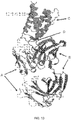
- Figure 10 provides the overview of the three-dimension structure of Proleukin/Fab-NARA1 complex as obtained in Example 1.
- Light chain of Fab fragment of NARA1 is designated A
- heavy chain of Fab fragment of NARA1 is shown as B
- epitope residues recognized by NARA1-Fab are designated D
- Proleukin is designated C and the mutation, C145S, is highlighted.
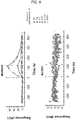
- Figure 11 provides further analysis of epitope residues.
- the X-axis lists the amino acid sequence and numbering according to SEQ ID No 1.
- the upper side of Y-axis demonstrates the total number of atoms of NARA1 -Fab that are within 4 ⁇ from corresponding residue from Proleukin and the lower side of Y-axis demonstrates the reduced solvent-accessible area ( ⁇ 2 ) after binding to NARA1-Fab.
- Proleukin used in Example 1 contains mutation of C145S. As shown in Figure 10 , C145S is far away from the epitope region. In addition the superposition of C ⁇ atoms between Proleukin in Example 1 with Ca atoms from wt hIL-2 in complex with CD25, CD122, and CD132 (PDB: 2B5I) shows r.m.s.d of 0.447 ⁇ , which indicates that the mutation does not disturb the over-all structure. Hence Proleukin with C145S mutation is a valid model for structural analysis for wt hIL-2.
- hIL-2 is 4-helix bundle protein and the 4 helices are named from N-terminus to C-terminus as A, B, C, and D, respectively.
- the epitope recognized by NARA1-Fab as shown in Figure 10 is a conformational epitope and spans two regions as shown in Figure 11 : one region (N50-K63) comprises a loop and a short helix and connects helix A and B, and the other region (N91-N97) comprises a loop and connects helix B and C.
- Arg58 as shown in Figure 11 is the most critical epitope residue for binding with NARA1-Fab, as this residue alone has 42 interacting atoms from NARA1-Fab and accounts for 17.7% of total reduced solvent-accessible surface area as a consequence of binding to NARA1-Fab.
- Arg58 as shown in Figure 12 , forms two strong salt-bridges with Glu35 in HCDR1 and with Asp100 from LCDR3, respectively. Arg58 also makes ⁇ -action interaction with the aromatic ring of Try100 from LCDR3.
- Residues K52, P54, K55, T57, T61, F62, K63, Q94, and K96 are also considered important for the binding to NARA1-Fab, since they all show equal to/more than 5 interacting atoms from NARA1-Fab and larger than 30 ⁇ 2 reduced solvent-accessible area as shown in Figure 11 .
- Table 3 shows that
- Figure 12 illustrates Arg58 as the most critical epitope residue recognized by the NARA1-Fab.
- A represents Proleukin
- B represents heavy chain
- C represents light chain. The involved residues are shown as sticks.
- Figure 13 shows the overlay of Proleukin/NARA1-Fab complex with IL-2/CD25/CD122/CD132 quaternary complex.
- the quaternary complex structure comes from PDB entry "2B5I” with cartoon D in pale cyan representing wt hIL-2, cartoon B in red representing CD122, cartoon C in blue representing CD132, and surface A in green representing CD25.
- cyan cartoon D overlayed with wt hIL-2 represents Proleukin
- cartoon E in magenta represents heavy chain
- cartoon F in yellow represents the light chain.
- Figure 14 displays the overlay of C helices from IL-2_C145A (PDB: 3INK), Superkine (PDB: 3Q81), IL-2/CD25/CD122/CD132 (PDB: 2B5I), and Proleukin/NARA1-Fab.
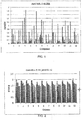
- a first library of 15-mer peptides was generated based on the sequence of human IL2.
- a second library of selected 15-mer peptides was also generated based on the mutation of 3 specific residues F(62), Y(65) and L(92). The latter mutations were done based on the Roche/Glycart IL2 mutein, as disclosed in WO2012/107417A1 which has these 3 mutations.
- Previous work done in lab Boyman (unpublished) showed that the commercial mouse anti-human IL2 mAb 602 with analogous function as A1 has strongly reduced binding to the F42A mutant of IL2 (one of the IL2 docking sites to CD25).
- each peptide in the first library has 15 amino acids and the sequence is derived by scanning the sequence of interest (see Table 4, reference peptides 1 to 41) with a step of 3 residues, starting from the N-terminus. Therefore a ladder is generated and each peptide contains 12 overlapping residues with the previous peptide and 12 overlapping residues with the following peptide in the ladder. In total, 41 peptides were generated from the expressed human IL2 sequence. A second library of peptides was generated by mutating F(62), Y(65) and L(92) to alanine in all corresponding peptides in the first library generated as described above (see Table 4, reference peptides no 42 to 60).
- Residue are the serine (S) replacing cysteines (C) 1 APTSSSTKKTQLQLE 2 SSSTKKTQLQLEHLL 3 TKKTQLQLEHLLLDL 4 TQLQLEHLLLDLQMI 5 QLEHLLLDLQMILNG 6 HLLLDLQMILNGINN 7 LDLQMILNGINNYKN 8 QMILNGINNYKNPKL 9 LNGINNYKNPKLTRM 10 INNYKNPKLTRMLTF 11 YKNPKLTRMLTFKFY 12 PKLTRMLTFKFYMPK 13 TRMLTFKFYMPKKAT 14 LTFKFYMPKKATELK 15 KFYMPKKATELKHLQ 16 MPKKATELKHLQSLE 17 KATELKHLQSLEEEL 18 ELKHLQSLEEELKPL 19 HLQSLEEELKPLEEV 20 SLEEELKPLEEVLNL 21 EELKPLEEVLNLAQS 22 KPLEEVLNLAQSKNF 23 EEVLNLAQSKNFHL
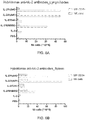
- Both set of peptides were printed on microarray slides in triplicate, incubated with the antibodies of interest (MAb602 and NARA1) and control antibodies. Additional incubations are with unrelated antibodies from the same isotype (mouse control IgG2a/lambda and mouse control IgG2a/kappa), and secondary antibodies (anti-mouse IgG (Thermo 84545, label DL650) or anti-mouse IgG (JIR 115-175-072, Label Cy5)) to assess unspecific binding due to the detection antibody.
- the experiments are performed essentially as described in Maksimov P, et al. 2012, PLoS One 7:e34212. doi:10.13711journal.pone. 0034212 .
- peptide-antibody binding was performed by RepliTope-analysis where the peptide microarray (triplicate) was incubated with the primary antibody followed by a fluorescently labelled secondary antibody directed against the Fc-part of the primary one. All steps were performed on a TECAN microarray processing station enabling highly reliable and reproducible washing and incubation steps. After performing the incubation steps and subsequent to the final washing steps (to remove the unbound secondary antibodies) the microarrays were dried using a nitrogen stream and scanned in a high resolution microarray scanning system with appropriate wavelength settings. Control incubations were performed with an unrelated antibody having the same isotype to exclude false positive signals. The resulting images were analyzed und quantified using spot-recognition software GenePix (Molecular Devices).
- the mean signal intensity was extracted (between 0 and 65535 arbitrary units).
- the MMC2 values were determined.
- the MMC2 equals the mean value of all three instances on the microarray. Except the coefficient of variation (CV) - standard-deviation divided by the mean value - is larger 0.5, in this case the mean of the two closest values (MC2) is assigned to MMC2.
- the data are summarized in Table 5.
- the anti-IL2 (NARA1) antibody did not show any significant reactivity towards the immobilized peptides. Only peptide 10 exhibited a weak response, however, this peptide was also weakly recognized by the mouse control antibodies.
- the commercial antibody MAB602 (mlgG2a) provided some weak signals on peptide 22 to 26 and some strong for peptides 10 to 13. Table 5. Result of Linear Epitope Mapping Reference peptide no.
Description
- The present invention relates to antibodies binding to human interleukin-2 (hIL-2). The invention more specifically relates to antibodies specifically binding a particular epitope of hIL-2 and when bound to this epitope are capable of inhibit binding of hIL-2 to CD25. Furthermore, the invention relates to in vitro and in vivo therapeutic applications of the antibodies.
- Malignant melanoma is a frequent cancer type in men and women. Once melanoma becomes metastatic and spreads to distant sites, the 5-year survival rate is quite poor, calculated at about 15%. Currently available treatment strategies for metastatic melanoma barely improve this survival rate.
- Interleukin-2 (IL-2) is a cytokine able to potently stimulate cytotoxic lymphocytes against metastatic tumours. However, IL-2 is also able to stimulate so-called CD25+ CD4+ regulatory T cells (Treg cells) that are crucial for prevention of autoimmune disease. Importantly, Treg cells can significantly dampen anti-tumour responses by cytotoxic lymphocytes, thus somewhat antagonizing the beneficial anti-tumour effects of IL-2. Moreover, at doses required to achieve a clinical anti-tumour response, IL-2 can exert toxic adverse effects.
- Standard IL-2 immunotherapy has been used since the early 1980's for the immunotherapy of metastatic melanoma and metastatic renal cell carcinoma, leading to the approval by the FDA for these indications in 1996 and 1992, respectively. While IL-2 given at high doses has shown objective response rates in about 17% and complete regression in about 6-9% of patients suffering from these deadly metastatic cancers, IL-2 given at these doses frequently led to toxic adverse effects, such as hypotension, pulmonary edema, liver cell damage, gastrointestinal toxicity, and general edema. Moreover, as mentioned above, IL-2 is able to stimulate Treg cells, which in turn are able to dampen the activity of anti-tumour CD8+ T cells and NK cells.
- It was previously disclosed that the biological activity of cytokines in a mammalian subject can be increased by administering an antibody capable of binding the cytokine (
WO 2007/095643 A2 ). Specifically, the combination of IL-2 with a particular anti-IL-2 monoclonal antibody (mAb) has been shown to improve IL-2 therapy in experimental murine models of cancer immunotherapy. It was disclosed that IL-2/anti-IL-2 antibody complexes show strong biological activity by avoiding interaction with the IL-2 receptor α subunit CD25 (Letourneau S, van Leeuwen EMM, Krieg C, Martin C, Pantaleo, G, Sprent J, Surh CD, and Boyman O. IL-2/anti-IL-2 antibody complexes show strong biological activity by avoiding interaction with IL-2 receptor α subunit CD25. Proceedings of the National Academy of Sciences USA (2010) 107:2171-2176), thereby directing IL-2 preferentially to cytotoxic lymphocytes, but not Treg cells. - It was further disclosed that the combination of IL-2 with an anti-IL-2 monoclonal antibody (mAb) renders IL-2 more potent but less toxic (Boyman O, Kovar M, Rubinstein MP, Surh CD, and Sprent J. Selective stimulation of T cell subsets with antibody-cytokine immune complexes. Science (2006) 311:1924-1927; Krieg C, Letourneau S, Pantaleo G, and Boyman O. Improved IL-2 immunotherapy by selective stimulation of IL-2 receptors on lymphocytes and endothelial cells. Proceedings of the National Academy of Sciences USA (2010) 107:11906-11911).
- This approach has the advantage that unmutated, natural IL-2 is delivered via anti-IL-2 mAb to CD8+ T cells and NK cells, which subsequently exert potent anti-tumour properties, while IL-2 complexed to this kind of anti-IL-2 mAb barely activates Treg cells. Moreover, IL-2 complexed to this kind of anti-IL-2 mAb is much less toxic than standard IL-2 immuno therapy in mice. However, this therapy has up to date not been available for use in patients due to the lack of appropriate anti-human IL-2 mAbs.
- The problem addressed by the present invention is to provide an anti-human IL-2 monoclonal antibody able to recognize and bind a specific epitope of human IL-2, thereby favoring the stimulation of cytotoxic T cells and NK cells compared to Treg cells, for use in in vitro and in vivo therapeutic applications. The invention relates to the embodiments as defined in the claims, thus it relates to the following items.
- According to a first aspect of the invention a human interleukin-2 (hIL-2) specific monoclonal antibody (mAb), or a human interleukin-2 (hIL-2) specific antigen binding fragment thereof comprising HCDR1 according to SEQ ID NO: 7, HCDR2 according to SEQ ID NO: 8, HCDR3 according to SEQ ID NO: 9, LCDR1 according to SEQ ID NO: 10, LCDR2 according to SEQ ID NO: 11, LCDR3 according to SEQ ID NO: 12 is provided, wherein binding of said antibody, or antigen binding fragment thereof, to hIL-2 inhibits binding of hIL-2 to CD25, and the binding to hIL-2 is characterized by a dissociation constant (KD) ≤ 7,5 nmol/L, ≤ 5 nmol/L, ≤ 3 nmol/L, ≤ 2 nmol/L or ≤ 1,5 nmol/L determined in a surface plasmon resonance binding assay.
- In certain parts of the invention a human interleukin-2 (hIL-2) specific monoclonal antibody, or antigen binding fragment thereof, according to the first aspect of the invention is provided, wherein the binding to hIL-2 is characterized by an off-rate (Koff) ≤ 1x10⁻⁴ s-1, ≤ 8x10⁻⁵ s-1, ≤ 6x10⁻⁵ s-1, ≤ 4x10⁻⁵ s-1, ≤ 3x10⁻⁵ s-1 or ≤ 2,1x10⁻⁵ s-1.
- In certain parts of the invention a human interleukin-2 (hIL-2) specific monoclonal antibody, or an antigen binding fragment thereof, according to the first aspect of the invention is provided, wherein the antibody, or antigen binding fragment thereof, displays no measurable cross-reactivity to murine IL-2.
- In certain parts of the invention human interleukin-2 (hIL-2) specific monoclonal antibodies, or antigen binding fragments thereof, according to the first aspect of the invention are provided, characterized in that they comprise at least one VH and/or one VL sequence having an identity of ≥ 80%, ≥ 85%, ≥ 90%, ≥ 92%, ≥ 93%, ≥ 94%, ≥ 95%, ≥ 96%, ≥ 97% or ≥ 98% compared to
SEQ ID NOs 019 or 20. - According to a second aspect of the invention a nucleic acid molecule encoding the human interleukin-2 (hIL-2) specific monoclonal antibody, or an antigen binding fragment thereof, according to the first aspect of the invention is provided.
- In certain parts of the invention a nucleic acid molecule according to the second aspect of the invention is provided, wherein the nucleic acid molecule has ≥60%, ≥70%, ≥ 80%, ≥ 85%, ≥ 90%, ≥ 92%, ≥ 93%, ≥ 94%, ≥ 95%, ≥ 96%, ≥ 97% or ≥ 98% sequence identity compared to
SEQ ID NOs 003 or 004. - In certain parts of the invention a nucleic acid molecule according to the second aspect, comprising the sequence of
SEQ ID NOs SEQ ID NOs - According to a third aspect of the invention a vector comprising the nucleic acid molecule according to the invention is provided.
- According to a fourth aspect of the invention a cell comprising the nucleic acid molecule according to the invention, or expressing the nucleic acid molecule according to the invention is provided.
- According to a fifth aspect of the invention a cell able to produce a human interleukin-2 (hIL-2) specific monoclonal antibody (mAb), or antigen binding fragment thereof, according to the first aspect of the invention is provided.
- According to a sixth aspect of the invention a monoclonal antibody-producing hybridoma cell line is provided, characterized in that the produced antibodies are those of the first aspect of invention.
- According to a seventh aspect of the invention a therapeutic formulation for use as a medicament, particularly for use in the treatment of cancer or viral infections, comprising:
- a. the hIL-2 monoclonal antibody (mAb), or the antigen binding fragment thereof, according to the first aspect of the invention, and
- b. human interleukin-2
- According to an eigth aspect of the invention the human interleukin-2 (hIL-2) specific monoclonal antibody, or antigen binding fragment thereof, according to the first aspect of the invention, is provided for use in the treatment of cancer or viral infections, wherein the antibody, or antigen binding fragment thereof is administered together with human interleukin-2.
- According to a ninth aspect of the invention a therapeutic formulation for use as a medicament is provided, particularly for use in the treatment of cancer or viral infections, containing a fusion protein that comprises:
- a. an hIL-2 binding polypeptide fragment comprising HCDR1 according to SEQ ID NO: 7, HCDR2 according to SEQ ID NO: 8, HCDR3 according to SEQ ID NO: 9, LCDR1 according to SEQ ID NO: 10, LCDR2 according to SEQ ID NO: 11, LCDR3 according to SEQ ID NO: 12, wherein the binding to hIL-2 is characterized by a dissociation constant (KD) ≤ 7,5 nmol/L, ≤ 5 nmol/L, ≤ 3 nmol/L, ≤ 2 nmol/L or ≤ 1,5 nmol/L determined in a surface plasmon resonance binding assay,
- b. a human IL-2 polypeptide fragment having an identity of ≥ 85%, ≥ 90%, ≥ 92%, ≥ 93%, ≥ 94%, ≥ 95%, ≥ 96%, ≥ 97% or ≥ 98% compared to SEQ ID NO 001, and, optionally,
- c. an amino acid linker of 1 to 50, particularly of 5 to 40, more particularly of 10 to 30, even more particularly of approx. 15 to 25 amino acids, linking the hIL-2 binding polypeptide fragment to the human IL-2 polypeptide fragment as one single polypeptide chain.
- According to a tenth aspect of the invention the antibody or antigen binding fragment thereof, according to the first aspect of the invention, which binds to a human interleukin-2 (hIL-2) epitope comprising the amino acids K52, P54, K55, T57, R58, T61, F62, K63, Q94, and K96 is provided.
- In certain parts of the invention the antibody or antigen binding fragment thereof according to the tenth aspect of the invention is provided, wherein the epitope further comprises any one or more of the amino acids N50, N53, N91, L92, A93, N97.
- According to a first aspect of the disclosure a human interleukin-2 (hIL-2) specific monoclonal antibody (mAb), or antigen binding fragment thereof, is provided, wherein the antibody is able to bind to a particular epitope in hIL-2 thereby inhibiting the binding to CD25, thus modulating the immunological effects of hIL-2/IL-2R interaction. The antibody of the disclosure is further characterized by at least one of the parameters:
- a) the variable chain of the mAb comprises an amino acid sequence having an identity of ≥85%, ≥90%, ≥95%, or ≥99% compared to SEQ ID NO 005 or SEQ ID NO 006;
- b) the antibody binding to hIL-2 is, i.e. the reaction mAb + hIL-2 ⇔ mAb∗hIL-2, wherein mAb∗hIL-2 symbolizes the bound complex of antibody and interleukin, is characterized by a dissociation constant (KD) ≤7,5 nmol/L, ≤5 nmol/L, ≤3 nmol/L, ≤ 2 nmol/L or ≤ 1,5 nmol/L;
- c) the antibody binding to hIL-2 is characterized by an off-rate(Koff) ≤ 1x10⁻⁴ s-1, ≤ 8x10⁻⁵ s-1, ≤ 6x10⁻⁵ s-1, ≤ 4x10⁻⁵ s-1, ≤ 3x10⁻⁵ s-1 or ≤ 2,1x10⁻⁵ s-1;
- d) upon mAb binding to hIL-2, the resulting mAb∗hIL-2 complex cannot efficiently bind human IL-2 receptor α (also known as CD25) anymore, effectively rendering the binding of human CD25 to mAb∗hIL-2 to background levels as compared to the binding of human CD25 to free (non-complexed) hIL-2 when measured by surface plasmon resonance; and/or
- e) the antibody displays no measurable cross-reactivity to murine IL-2.
- A lack of cross-reactivity with murine IL-2 is advantageous for preclinical studies, which usually involve mouse models, such as the use of mAb∗hIL-2 complexes for the treatment of murine tumour models where a cross-reactive anti-IL-2 mAb might bind and seclude endogenous murine IL-2 from endogenous murine Treg cells, thus enhancing the anti-tumour response.
- A lack of cross-reactivity with murine IL-2 is also advantageous for preclinical safety and efficacy studies conducted prior to development of a candidate mAb in human patients.
- In certain embodiments the hIL-2 mAb comprises at least one VH and/or VL sequence having an identity of ≥ 80%, ≥ 85%, ≥ 90%, ≥ 92%, ≥ 93%, ≥ 94%, ≥ 95%, ≥ 96%, ≥ 97% or ≥ 98% compared to SEQ ID NOs 019 or SEQ ID NO 020.
- In certain embodiments the variable chain of the hIL-2 mAb comprises an amino acid sequence having an identity of ≥85%, ≥90%, ≥95%, or ≥99% compared to
SEQ ID NOs 003, 004, 005 or 006 and the hIL-2 mAb is characterized by a dissociation constant ≤7,5 nmol/L, ≤5 nmol/L, ≤3 nmol/L, ≤ 2 nmol/L or ≤ 1,5 nmol/L. - In certain embodiments the variable chain of the hIL-2 mAb comprises an amino acid sequence having an identity of ≥ 85%, ≥ 90%, ≥ 92%, ≥ 93%, ≥ 94%, ≥ 95%, ≥ 96%, ≥ 97%, ≥ 98% or ≥ 99% compared to SEQ ID NO 005 or 006 and the hIL-2 mAb is characterized by an off-rate ≤ 1x10⁻⁴ s-1, ≤ 8x10⁻⁵ s-1, ≤ 6x10⁻⁵ s-1, ≤ 4x10⁻⁵ s-1, ≤ 3x10⁻⁵ s-1 or ≤ 2,1x10⁻⁵ s-1.
- In certain embodiments the variable chain of the hIL-2 mAb comprises an amino acid sequence having an identity of ≥ 85%, ≥ 90%, ≥ 92%, ≥ 93%, ≥ 94%, ≥ 95%, ≥ 96%, ≥ 97%, ≥ 98% or ≥ 99% compared to SEQ ID NO 005 or 006 and the hIL-2 mAb displays no measurable cross-reactivity to murine IL-2.
- In certain embodiments the sequence of the hIL-2 mAb is humanized for administration to human patients to prevent adverse reactions.
- In certain embodiments the hIL-2 mAb is provided as fragment antigen-binding (Fab) or single-chain variable fragment (scFv).
- In certain embodiments the hIL-2 mAb comprises at least one complementarity determining (CDR) sequence having an identity of ≥ 80%, ≥ 85%, ≥ 90%, ≥ 92%, ≥ 93%, ≥ 94%, ≥ 95%, ≥ 96%, ≥ 97% or ≥ 98% compared to SEQ ID NOs 007, 008, 009, 010, 011 or 012.
- According to a second aspect of the disclosure, a nucleic acid molecule encoding the monoclonal antibody, or antigen binding fragment thereof, able to bind to human interleukin-2 according to the first aspect of the disclosure is provided.
- In certain embodiments the nucleic acid molecule according to the second aspect of the disclosure has ≥60%, ≥70%, ≥80%, ≥90%, ≥95%, or ≥99% sequence identity compared to
SEQ ID NOs 003 to 004. - According to a third aspect of the disclosure a vector comprising the nucleic acid molecule according to the disclosure is provided.
- According to a fourth aspect of the disclosure, a cell is provided, comprising or expressing the nucleic acid molecule according to the disclosure.
- According to a fifth aspect of the disclosure a cell able to produce the antibodies according to the first aspect of the disclosure is provided.
- According to a sixth aspect of the disclosure a monoclonal antibody-producing hybridoma cell line is provided, characterized in that the antibodies produced are those of the first aspect of the disclosure.
- According to a seventh aspect of the disclosure a therapeutic formulation for use in the treatment of cancer or other diseases benefiting from immune stimulatory therapy, such as viral infections, comprising
- i. the monoclonal antibody (mAb) according to the first aspect of the disclosure, and/or
- ii. human interleukin-2 or human IL-2 mutants, administered to the subject either contemporaneously or at different time points.
- According to an eighth aspect of the disclosure a fusion protein is provided. The fusion protein comprises:
- a. an hIL-2 binding polypeptide fragment, wherein said polypeptide is characterized by any one of the parameters:
- i. the hIL-2 binding polypeptide fragment comprises an amino acid sequence having an identity of ≥ 85%, ≥ 90%, ≥ 92%, ≥ 93%, ≥ 94%, ≥ 95%, ≥ 96%, ≥ 97% or ≥ 98% compared to SEQ ID NO 021 or SEQ ID NO 022;
- ii. the hIL-2 binding binding of said polypeptide fragment to hIL-2 is characterized by a dissociation constant (KD) ≤ 7,5 nmol/L, ≤ 5 nmol/L, ≤ 3 nmol/L, ≤ 2 nmol/L or ≤ 1,5 nmol/L;
- iii. the binding of said hIL-2 binding polypeptide fragment to hIL-2 is characterized by an off-rate (Koff) ≤ 1x10⁻⁴ s-1, ≤ 8x10⁻⁵ s-1, ≤ 6x10⁻⁵ s-1, ≤ 4x10⁻⁵ s-1, ≤ 3x10⁻⁵ s-1 or ≤ 2,1x10⁻⁵ s-1;
and/or - iv. the hIL-2 binding polypeptide fragment displays no measurable crossreactivity to murine IL-2.
- b. a human IL-2 polypeptide fragment having an identity of ≥ 85%, ≥ 90%, ≥ 92%, ≥ 93%, ≥ 94%, ≥ 95%, ≥ 96%, ≥ 97% or ≥ 98% compared to SEQ ID NO 001, and, optionally,
- c. an amino acid linker of 1 to 50, particularly of 5 to 40, more particularly of 10 to 30, even more particularly of approx. 15 to 25 amino acids, linking the hIL-2 binding polypeptide fragment to the human IL-2 polypeptide fragment as one single polypeptide chain.
- In other words the fusion protein retains the ability of the antibody to bind and direct human interleukin-2 to stimulate selected immune cells, such as CD8+ T cells and NK cells.
- The advantage of using such fusion protein is that human IL-2 will not be able to dissociate from the antibody and that the therapy will be composed of one single product instead of two, facilitating various aspects of manufacture, dosing and regulatory compliance.
- According to a ninth aspect of the disclosure, an isolated antibody or antigen binding fragment thereof binding a specific epitope is provided. Said epitope can be the epitope to which an isolated antibody or antigen binding fragment thereof according to other aspects of the disclosure binds. In an embodiment, the isolated antibody or molecule binds to a human interleukin-2 (hIL-2) epitope which comprises the amino acids K52, P54, K55, T57, R58, T61, F62, K63, Q94, and K96. In another embodiment, the isolated antibody or molecule binds to an epitope further comprising any one or more of the amino acids N50, N53, N91, L92, A93, and N97. An isolated antibody or molecule, which comprises an antigen recognition surface having epitope recognition characteristics equivalent to an antibody or antigen binding fragment thereof according to other aspects is also provided.
- Wherever alternatives for single separable features such as, for example, a coding sequence or binding epitope are laid out herein as "embodiments", it is to be understood that such alternatives may be combined freely to form discrete embodiments of the invention disclosed herein.
- The invention is further illustrated by the following examples and figures, from which further embodiments and advantages can be drawn. These examples are meant to illustrate the invention as defined in the claims but not to limit its scope.
- By "human interleukin-2" or "hIL-2" is meant the protein designated UniProt ID P60568 and is reproduced as SEQ ID NO: 1.
- Identity in the context of the present specification is a single quantitative parameter representing the result of a sequence comparison position by position. Methods of sequence comparison are known in the art; the BLAST algorithm available publicly is an example. One such example for comparison of nucleic acid sequences is the BLASTN algorithm that uses the default settings: Expect threshold: 10; Word size: 28; Max matches in a query range: 0; Match/Mismatch Scores: 1.-2; Gap costs: Linear. In the absence of other measurement variables, identity shall be measured according to the specification above.
- In the context of the present specification, the term antibody is used in its meaning known in the art of cell biology and immunology; it refers to whole antibodies, any antigen binding fragment or single chains thereof and related or derived constructs. A whole antibody is a glycoprotein comprising at least two heavy (H) chains and two light (L) chains interconnected by disulfide bonds. Each heavy chain is comprised of a heavy chain variable region (VH) and a heavy chain constant region (CH). The heavy chain constant region is comprised of three domains,
C H1,C H2 andC H3. Each light chain is comprised of a light chain variable region (abbreviated herein as VL) and a light chain constant region (CL). The light chain constant region is comprised of one domain, CL. The VH and VL regions can be further subdivided into regions of hypervariability, termed complementarity determining regions 20 (CDR), interspersed with regions that are more conserved, termed framework regions (FR). Each VH and VL is composed of three CDRs and four FRs arranged from amino-terminus to carboxyterminus in the following order: FR1, CDR1, FR2, CDR2, FR3, CDR3, FR4. The variable regions of the heavy and light chains contain a binding domain that interacts with an antigen. The constant regions of the antibodies may mediate the binding of the immunoglobulin to host tissues or factors, including various cells of the immune system (e.g., effector cells) and the first component of the classical complement system. - In the context of the present specification, the term antigen binding portion or antigen binding fragment is used in its meaning known in the art of cell biology and immunology; it refers to one or more fragments of an intact antibody that retain the ability to specifically bind to a given antigen (e.g., interleukin-2). Antigen binding functions of an antibody can be performed by fragments of an intact antibody. Examples of binding fragments encompassed within the term antigen binding portion or antigen binding fragment of an antibody include a Fab fragment, a monovalent fragment consisting of the VL, VH, CL and CH domains; a F(ab)2 fragment, a bivalent fragment comprising two Fab fragments linked by a disulfide bridge at the hinge region; an Fd fragment consisting of the VH and CH domains; an Fv fragment consisting of the VL and VH domains of a single arm of an antibody; a single domain antibody (dAb) fragment, which consists of a VH domain or a VL domain; and an isolated complementarity determining region (CDR). HCDR means a CDR of the heavy chain and LCDR means a CDR of the light chain.
- In the context of the present specification, the term chimeric antibody is used in its meaning known in the art of cell biology and immunology; it refers to an antibody molecule in which the constant region, or a portion thereof, is altered, replaced or exchanged so that the antigen binding site (variable region) is linked to a constant region of a different or altered class, effector function and/or species, or an entirely different molecule which confers new properties to the chimeric antibody, e.g., an enzyme, cytokine, toxin, hormone, growth factor, drug, etc. For example, an antibody can be modified by replacing its constant region with a cytokine. Due to the replacement with a cytokine, the chimeric antibody can retain its specificity in recognizing the antigen while having also the function, or part thereof, of the original cytokine molecule.
- In the context of the present specification, the term hybridoma is used in its meaning known in the art of cell biology and biochemistry; it refers to a hybrid cell created by fusion of a specific antibody-producing B-cell with a myeloma (B-cell cancer) cell. Hybridoma cells can be grown in tissue culture and produce antibodies of a single specificity (monoclonal antibodies).
- In the context of the present specification, the term single-chain variable fragment (scFv) is used in its meaning known in the art of cell biology and biochemistry; it refers to a fusion protein of the variable regions of the heavy (VH) and light chains (VL) of immunoglobulins, connected with a short linker peptide of ten to about 25 amino acids. The scFv retains the specificity of the original immunoglobulin, despite removal of the constant regions and the introduction of the linker.
- In the context of the present specification, the term fragment antigen-binding (Fab) is used in its meaning known in the art of cell biology and immunology; it refers to a region on an antibody that binds to antigens. It is composed of one constant and one variable domain of each of the heavy (VH) and light chains (VL) of immunoglobulins. These domains shape the antigen-binding site at the amino terminal end of the monomer.
- In the context of the present specification, the term dissociation constant (KD) is used in its meaning known in the art of chemistry and physics; it refers to an equilibrium constant that measures the propensity of a larger object to dissociate reversibly into smaller components, as when a complex falls apart into its component molecules. KD is expressed in molar units [M] and corresponds to the concentration of [Ab] at which the binding sites of [Ag] are half occupied. In other words the concentration of unbound [Ab] equals the concentration of the [AbAg] complex. The dissociation constant can be calculated according to the following formula:

- In the context of the present specification, the terms off-rate (Koff;[1/sec]) and on-rate (Kon; [1/sec*M]) are used in their meaning known in the art of chemistry and physics; they refer to a rate constant that measures the dissociation (Koff) or association (Kon) of 5 an antibody with its target antigen. Koff and Kon can be experimentally determined using methods well established in the art. A method for determining the Koff and Kon of an antibody employs surface plasmon resonance. This is the principle behind biosensor systems such as the Biacore® or the ProteOn® system. They can also be used to determine the dissociation constant KD by using the following formula:

- In the context of the present specification, the term humanized antibodies is used in its meaning known in the art of cell biology and biochemistry; it refers to antibodies originally produced by immune cells of a non-human species, whose protein sequences have been modified to increase their similarity to antibody variants produced naturally in humans.
- In the context of the present specification, the term no measurable cross-reactivity refers to the lacking capability of an antibody to recognize and bind to orthologous proteins from other species. For example, an antibody directed against human interleukin-2 would have no measurable cross-reactivity to murine interleukin-2 if, under suitable conditions, binding of the antibody to murine interleukin-2 could not be detected with sufficiently sensitive methods such as surface plasmon resonance. One such example of no measurable cross-reactivity is shown in
Fig. 9 for the antibody in the lower panel (NARA1). - As used herein, an antibody or a protein that "specifically binds to hIL-2" is intended to refer to an antibody or protein that binds to human IL-2 polypeptide with a KD of 100nM or less, 10nM or less, 1nM or less, 100pM or less, or 10pM or less. An antibody that "cross-reacts with an antigen other than human IL-2" is intended to refer to an antibody that binds that antigen with a KD of 10nM or less, 1 nM or less, or 100 pM or less. An antibody that "does not cross-react with a particular antigen" is intended to refer to an antibody that binds to that antigen, with a KD of 100 nM or greater, or a KD of 1 µM or greater, or a KD of 10 µM or greater. In certain embodiments, such antibodies that do not cross-react with the antigen exhibit essentially undetectable binding against these proteins in standard binding assays.
- The term "epitope" means a protein determinant capable of specific binding to an antibody. Epitopes usually consist of chemically active surface groupings of molecules such as amino acids or sugar side chains and usually have specific three dimensional structural characteristics, as well as specific charge characteristics. Conformational and nonconformational epitopes are distinguished in that the binding to the former but not the latter is lost in the presence of denaturing solvents.
- The term "epitope binding domain" or "EBD" refers to portions of a binding molecule (e.g., an antibody or epitope-binding fragment or derivative thereof), that specifically interacts with (e.g., by binding, steric hindrance, stabilizing/destabilizing, spatial distribution) a binding site on a target epitope. EBD also refers to one or more fragments of an antibody that retain the ability to specifically interact with (e.g., by binding, steric hindrance, stabilizing/destabilizing, spatial distribution) a IL-2 epitope and inhibit signal transduction. Examples of antibody fragments include, but are not limited to, an scFv, a Fab fragment, a monovalent fragment consisting of the VL, VH, CL and CH1 domains; a F(ab)2 fragment, a bivalent fragment comprising two Fab fragments linked by a disulfide bridge at the hinge region; a Fd fragment consisting of the VH and CH1 domains; a Fv fragment consisting of the VL and VH domains of a single arm of an antibody; a dAb fragment (Ward et at., (1989) Nature 341:544-546), which consists of a VH domain; and an isolated complementarity determining region (CDR).
- EBDs also include single domain antibodies, maxibodies, unibodies, minibodies, triabodies, tetrabodies, v-NAR and bis-scFv, as is known in the art (see, e.g., Hollinger and Hudson, (2005) Nature Biotechnology 23: 1126-1136), bispecific single chain diabodies, or single chain diabodies designed to bind two distinct epitopes. EBDs also include antibody-like molecules or antibody mimetics, which include, but not limited to minibodies, maxybodies, Fn3 based protein scaffolds, Ankyrin repeats (also known as DARpins), VASP polypeptides, Avian pancreatic polypeptide (aPP), Affilin, Knottins, SH3 domains, PDZ domains, Tendamistat, Neocarzinostatin, Protein A domains, Lipocalins, Transferrin, and Kunitz domains that specifically bind epitopes, which are within the scope of the disclosure. Antibody fragments can be grafted into scaffolds based on polypeptides such as Fibronectin type III (Fn3) (see
U.S. Pat. No. 6,703,199 , which describes fibronectin polypeptide monobodies). - The present invention also encompasses an antibody to human IL-2, which is an isolated antibody.
- The phrase "isolated antibody", as used herein, refers to antibody that is substantially free of other antibodies having different antigenic specificities (e.g., an isolated antibody that specifically binds hIL-2 is substantially free of antibodies that specifically bind antigens other than hIL-2). An isolated antibody that specifically binds hIL-2 may, however, have cross-reactivity to other antigens, such as IL-2 molecules from other species. Moreover, an isolated antibody may be substantially free of other cellular material and/or chemicals.
- The terms "nucleic acid" and "polynucleotide" or "nucleotide coding sequences" are used interchangeably and refer to a polymeric form of nucleotides of any length, either deoxyribonucleotides or ribonucleotides or analogs thereof. Polynucleotides can have any three-dimensional structure and can perform any function. The following are nonlimiting examples of polynucleotides: a gene or gene fragment (for example, a probe, primer, EST or SAGE tag), exons, introns, messenger RNA (mRNA), transfer RNA, ribosomal RNA, ribozymes, cDNA, recombinant polynucleotides, branched polynucleotides, plasmids, vectors, isolated DNA of any sequence, isolated RNA of any sequence, nucleic acid probes, siRNAs, shRNAs, RNAi agents, and primers. A polynucleotide can be modified or substituted at one or more base, sugar and/or phosphate, with any of various modifications or substitutions described herein or known in the art. A polynucleotide can comprise modified nucleotides, such as methylated nucleotides and nucleotide analogs. If present, modifications to the nucleotide structure can be imparted before or after assembly of the polymer. The sequence of nucleotides can be interrupted by non-nucleotide components. A polynucleotide can be further modified after polymerization, such as by conjugation with a labeling component. The term also refers to both double- and single-stranded molecules. Unless otherwise specified or required, any embodiment of this invention that is a polynucleotide encompasses both the double-stranded form and each of two complementary single-stranded forms known or predicted to make up the double-stranded form.
- The term "polypeptide" is used interchangeably with the term "protein" and in its broadest sense refers to a compound of two or more subunit amino acids, amino acid analogs, or peptidomimetics. The subunits can be linked by peptide bonds. In another embodiment, the subunit may be linked by other bonds, e.g., ester, ether, etc.
- As used herein, the term "treating" or "treatment" of any disease or disorder (e.g. cancer) refers in one embodiment, to ameliorating the disease or disorder (e.g. slowing or arresting or reducing the development of the disease or at least one of the clinical symptoms thereof). In another embodiment "treating" or "treatment" refers to alleviating or ameliorating at least one physical parameter including those which may not be discernible by the patient. In yet another embodiment, "treating" or "treatment" refers to modulating the disease or disorder, either physically, (e.g., stabilization of a discernible symptom), physiologically, (e.g., stabilization of a physical parameter), or both. Methods for assessing treatment and/or prevention of disease are generally known in the art, unless specifically described hereinbelow.
-
-
Figure 1 shows anti-human IL-2 binders. Supernatants of B cell clones obtained after B cell hybridoma fusion were added to a plate previously coated with human IL-2. The anti-human IL-2 mAbs were detected using a biotinylated anti-mouse IgG antibody. -
Figure 2 shows screening of anti-human IL-2 mAbs for binding to presumed specific human IL-2 epitope. Plates were coated with 5344 (a hIL-2 mAb without the herein targeted superagonistic behaviour) and blocked, followed by addition of human IL-2 in order to allow the cytokine to bind to 5344, thus covering a specific epitope of the IL-2. Then the supernatants giving a positive signal in the first screening (seeFigure 1 ) were added. After allowing the mAbs in the supernatants to bind to the IL-2-5344 complex, a biotinylated MAB602 antibody was added to the plate in order to assess whether the tested mAbs of the supernatants bound to the same (so-called "competitors") or to a different region than MAB602. The competitor mAbs led to an absorbance (OD450) that is two-fold lower than the absorbance obtained with MAB602 alone (in this case OD = 1.1, as shown in H11). -
Figure 3 shows concentration-dependent competition of B cell hybridomas. The supernatants of 8 competitor B cell hybridoma clones of the first screening (seeFigure 2 ) were expanded and concentrated before use in this assay. The supernatants of these 8 competitor B cell hybridoma clones (labeled 1 to 8) were added in increasing quantities. Competent competitor B cell hybridoma clones reduced the OD450 as much as MAB602 or even more, which is evident forclones -
Figure 4 shows in vivo proliferation of CD8+ T cells. Carboxyfluorescein succinimidyl ester (CFSE)-labeled CD8+ T cells of CD45.1-congenic IL-7 transgenic mice were transferred to CD45.2-congenic WT recipient mice, followed by daily injections of phosphate-buffered saline (PBS), IL-2, IL-2 plus MAB602 (IL-2/MAB602), IL-2 plus 5344 (IL-2/5344), IL-2 plus hybridoma 1 (IL-2/Hyb#1), or IL-2 plus hybridoma 2 (IL-2/Hyb#2) for 4 days. Onday 5, lymph nodes and spleens were analyzed for CFSE profiles of donor CD45.1+ CD8+ T cells. Shown are the results obtained with the lymph nodes, similar results were obtained in the spleens. -
Figure 5 shows phenotypic characterisation of endogenous CD8+ T cells and NK cells following in vivo treatment using IL-2 plushybridoma Figure 4 , followed by assessment by flow cytometry of endogenous CD8+ T cell subsets and NK cells in the lymph nodes and spleen. Shown are (A) CD8 vs. CD3 profiles of total lymph node cells (left graphs) and CD44 (activated or memory T cells) vs. CD122 (IL-2 receptor β-subunit, present on activated or memory T cells) profiles of CD3+ CD8+ lymph node cells, or (B) NK1.1 vs. CD3 profiles of mice receiving the indicated treatment. Activated/memory CD8+ T cells are high for CD44 and intermediate to high for CD122. NK cells are CD3 negative and NK1.1 positive. Similar results were obtained using spleen cells. -
Figure 6 shows total cell counts of activated/memory CD8+ T cells and NK cells in lymph nodes and spleens. Animals were treated and analyzed as inFigure 5 . Shown are absolute cell counts of CD44high CD8+ T cells (so-called memory phenotype, MP CD8+) and of CD3 negative NK1.1 + NK cells in lymph nodes (top panel) and spleen (lower panel). -
Figure 7 shows surface plasmon resonance binding curves of the commercially available monoclonal antibody MAB602 (left graph) and the monoclonal antibody NARA1 (right graph), which is the subject of this invention, to human IL-2. For this experiment an amine coupling GLM chip was used. The activation of the carboxylic acid groups in the chip was done using a mix of 1-ethyl-3-3-dimethylaminopropyl carbodiimide hydrochlorid (EDC at 0.2 M) and sulfo N-hydroxysulfosuccinimids (s_NHS at 0.05M) at 30 ml/min for 420 seconds (s). The antibodies NARA1 and MAB602 were coated in the chip at 100 mg/ml in a sodium acetate buffer (10 mM pH 4.5). Deactivation was followed adding ethanolamine HCl at 30 ml/min for 300 s. Finally human IL-2 was added at different concentrations (starting from 100 nM and followed by three-fold dilutions) at 100 ml/min, 600 s association, and 240 s dissociation. -
Figure 8 shows surface plasmon resonance binding curves of human IL-2 bound to the monoclonal antibody NARA1 with the IL-2 receptors subunits CD25 (used here as an Fc fusion of CD25-Fc), CD122, the monoclonal antibody MAB602 or an anti-hIL-2 antibody binding to a different human IL-2 epitope than NARA1 and MAB602. The chip described inFigure 7 coated with NARA1 and MAB602 was re-used. Regeneration of the chip was done using 10 mM glycine, pH 2.5, 30 ml/min, 60 s. Human IL-2 was added at saturating concentration (1 mM), at 100 ml/min, 120 s association, and 0 s dissociation. Immediately after IL-2 association to the antibodies, the second analytes were added at 100 ml/min, 120 s association, and 240s dissociation. The concentration used for the cross-binding were: MAB602: 50 nM; NARA1 : 50 nM; positive control: 50 nM; CD25-Fc: 500 nM; CD122: 138 nM. When hIL-2 is bound to NARA1, an anti-hIL-2 antibody that recognizes a different hIL-2 epitope (here termed 'positive control') binds strongly to the hIL-2/NARA1 complex as expected (green line inFigure 8 ). Alternatively, IL-2Ra (in the form of CD25-Fc) cannot bind to hIL-2 when hIL-2 is already bound to NARA1 (pink line,Figure 8 ), however, IL-2Rβ (CD122) still binds to hIL-2 when hIL-2 is already bound to NARA1 (orange line,Figure 8 ). -
Figure 9 shows surface plasmon resonance binding curves of the monoclonal antibodies MAB602 (top graph) and NARA1 (lower graph) to murine IL-2. The same chip used for the generation of the data inFigures 7 and8 was re-used. Regeneration of the chip was done with 10 mM glycine, pH 2.5, 30 ml/min, 60 s. Mouse IL-2 (mIL-2) or human IL-2 (hIL-2) starting at 10 nM and then doing a three-fold dilution was injected at 100 ml/min, 120 s association, 5 and 240 s dissociation. In the top graph MAB602 shows cross-reactivity by binding to mouse IL-2. Especially, with higher concentrations of murine interleukin-2 (>1 nM) the binding curves differ significantly from background levels with response units 10 (RU) well above 10. Whereas NARA1 (lower graph) displays no measurable cross-reactivity to murine IL-2 at all concentrations tested. -
Figure 10 provides the overview of the three-dimensional structure of Proleukin/Fab-NARA1 complex as obtained in Example 1. -
Figure 11 provides further analysis of epitope residues. The X-axis lists the amino acid sequence and numbering according toSEQ ID No 1. The upper side of Y-axis shows the total number of atoms of NARA1-Fab that are within 4 Å from corresponding residue from Proleukin and the lower side of Y-axis shows the reduced solvent-accessible area (Å2) of corresponding residue from Proleukin as a consequence of binding to NARA1-Fab. -
Figure 12 illustrates the most critical epitope residue recognized by the NARA1-Fab. -
Figure 13 shows the overlay of Proleukin/NARA1-Fab complex with IL-2/CD25/CD122/CD132 quaternary complex. -
Figure 14 displays the overlay of C helices from IL-2_C145A (PDB: 3INK), Superkine (PDB: 3QB1), IL-2/CD25/CD122/CD132 (PDB: 2B5I), and Proleukin/NARA1-Fab. - Until now, no monoclonal antibodies suitable for the disclosed invention have been available. The inventors disclose their anti-human IL-2 mAbs that allow the following crucial steps towards the use and commercialization of this technology in clinical applications:
- Further sequencing and fine characterization of the anti-human IL-2 mAbs.
- Humanization of the anti-human IL-2 mAbs, which is essential to avoid (or minimize) immunogenicity in patients.
- Generation of different formats of anti-human IL-2 mAbs, such as IgG, IgG1, IgG4, Fab, and single-chain Fv (scFv).
- Generation of a fusion protein consisting of human IL-2 and an anti-human IL-2 mAb (or a fragment of the anti-human IL-2 mAb): such a construct has the advantage of consisting of one component only, instead of two as in IL-2 bound to an anti-human IL-2 mAb.
- The inventors have generated and characterized specific anti-human IL-2 mAbs that are able to bind human IL-2 and, when tested in mice, are able to exert specific and potent stimulation of cytotoxic lymphocytes, including CD8+ T cells and natural killer (NK) cells. Towards these ends several difficulties had to be overcome.
- Human IL-2 shows high similarity with mouse and rat IL-2, thus human IL-2 is able to stimulate mouse lymphocytes in vitro and in vivo. Moreover, IL-2 is present at high concentrations in the primary immune organs (such as the bone marrow), which is the reason why IL-2 is somewhat a "forbidden" antigen, meaning it is very difficult to generate B cell responses leading to neutralizing antibodies against IL-2. Nevertheless, the inventors were able to elicit polyclonal anti-human IL-2 antibody responses, following immunization of C57BL/6 mice using purified recombinant human IL-2 plus adjuvant.
- Of the generated antibody responses, only some mAbs efficiently bound to IL-2 (socalled "binders") and of those only about 0.35 % interacted with the presumed active site of IL-2.
- Finally, of these anti-human IL-2 mAbs some showed the desired specific and potent in vivo activity as assessed by specialized in vivo assays in mice that are not replaceable by in vitro experiments.
- The inventors have developed specific screening assays that allow detection of specific antihuman IL-2 antibodies (so-called "binders") in the serum of immunized animals and in the supernatant of the B cell clones obtained after B cell hybridoma fusion. In a second step it was discriminated between standard binders and those targeting a presumed specific epitope of the human IL-2 molecule. One example of such an in vitro enzyme-linked immunosorbent assay (ELISA) performed with different B cell clones, is shown in
Figures 1 to 3 . - After the in vitro screening of the anti-human IL-2 mAbs, these mAbs were characterised in vivo. To this end and in order to obtain sufficient amounts of mAbs, the mAbs were concentrated from the supernatant of the hybridomas, the amount was estimated using an ELISA and finally the anti-human IL-2 mAbs was tested in mice. The results obtained on proliferation and expansion of CD8+ T cells and NK cells is shown in
Figures 4 to 6 . - In order to characterize the binding properties of the anti-human IL-2 mAbs the binding to human interleukin-2 was tested with surface plasmon resonance binding assays. The commercially available anti-human IL-2 mAb MAB602 was measured as a comparison. In
Figure 7 binding curves of MAB602 (left graph) and NARA1 (an antibody according to this invention; right graph) to human interleukin-2 at varying concentrations are shown. The dissociation constant (KD) as well as the rate constants Kon and Koff measured for MAB602 and NARA1 are shown in Table 1. -
Table 1: Binding properties of anti-human IL-2 mAbs to human IL-2 Kon (M*s-1) Koff (s-1) KD (nM) MAB602 58 x 104 4.94 x 10⁻⁴ 9.7 NARA1 1.78 x 10⁴ 2.08 x 10⁻⁵ 1.2 - Antibodies of the invention include the antibody NARA1, which was derived, isolated and structurally characterized by its full length heavy chain according to SEQ ID NO: 5 and its full length light chain amino acid sequences according to SEQ ID NO: 6.
- The corresponding variable regions, VH and VL amino acid sequences of NARA1 are. SEQ ID NO: 19 (variable heavy) and SEQ ID NO: 20 (variable light).
- Full length light and heavy chains nucleotide coding sequences of NARA1 are SEQ ID NO: 3 (heavy chain coding sequence, including leader sequence) and SEQ ID NO: 4 (light chain coding sequence, including leader sequence).
- Variable light and heavy chains nucleotide coding sequences of NARA1 are SEQ ID NO: 21 (variable heavy coding sequence) and SEQ ID NO: 22 (variable light coding sequence).
- The CDR regions of NARA1 are delineated using the Kabat system (Kabat, E. A., et al. 1991, Sequences of Proteins of Immunological Interest, Fifth Edition, U.S. Department of Health and Human Services, NIH Publication No. 91-3242, see also Zhao&Lu 2009, Molecular Immunology 47:694-700).For the ease of reading, when CDR regions are delineated according to Kabat definition, they are called hereafter HCDR1, HCDR2, HCDR3, LCDR1, LCDR2, LCDR3 respectively. The CDR regions of NARA1 are: HCDR1 according to SEQ ID NO: 7, HCDR2 according to SEQ ID NO: 8, HCDR3 according to SEQ ID NO: 9, LCDR1 according to SEQ ID NO: 10, LCDR2 according to SEQ ID NO: 11, LCDR3 according to SEQ ID NO: 12.
- Nucleotide coding sequences for the CDR regions of NARA1 are: HCDR1 coding sequence according to SEQ ID NO: 13, HCDR2 coding sequence according to SEQ ID NO: 14, HCDR3 coding sequence according to SEQ ID NO: 15, LCDR1 coding sequence according to SEQ ID NO: 16, LCDR2 coding sequence according to SEQ ID NO: 17, LCDR3 coding sequence according to SEQ ID NO: 18.
Fusion proteins are also provided according to SEQ ID NO: 23 and SEQ ID NO: 24. SEQ ID NO: 23 is a fusion protein comprising the variable heavy chain of NARA1 with its N-terminus fused to the C-terminus of hIL-2 via a GxS linker. SEQ ID NO: 24 is a fusion protein comprising the variable light chain of NARA1 with its N-terminus fused to the C-terminus of hIL-2 via a GxS linker. - The complex structure of a
human Interleukin 2 mutant (SEQ ID NO:2), called "Proleukin", bound to the Fab fragment of antibody "NARA 1" (SEQ ID NO: 5 and 6) was determined. The resulting numbering of residues on Proleukin is given according to the numbering of wt IL-2. - As will be discussed in detail below, the differences in sequence between Proleukin and wt hIL-2 are irrelevant and Proleukin is a valid model for structural analysis of hIL-2.
- To define the epitope, X-ray crystallography was ued to solve the atomic-resolution structure of the complex mentioned above. X-ray crystallography is a technology that has become routinely and widely used to generate structural data for biomolecules including antibodies and their complexes with antigens (Adms et al, (2013) Annual Review Biophysics 42:265-287; Garman, (2014) Science 343:1102-1108; Joachimiak, (2009) Current Opinio Structural Biology 19:573-584.)
- The antigen, Proleukin, is commercially available as lyophilyzed powder together with excipients (every 1 mg Proleukin is mixed with approximately 50mg mannitol, 0.18mg sodium dodecyl sulfate, 0.173mg sodium dihydrogen phosphate, and 0.89mg disodium hydrogen phosphate). Before used for complex formation, Proleukin was purified by reverse-phase HPLC to remove the excipients.
- The Fab fragment of NARA1 (NARA1-Fab) was generated by papain cleavage of the full-length antibody followed by Protein A chromatography. Briefly, 6.5ml full-length NARA1 (9mg/ml in 50mM citrate buffer with 90mM sodium chloride at pH 7.0) was mixed with 5mM DTT and 590ug Papain (Roche). The cleavage reaction was kept at room temperature for 16h and stopped by addition of 15ul 56mM E64 solution (Roche). The cleavage solution was then diluted 10 times with 25mM Tris, 25mM NaCl, pH 8.0 and loaded onto a 5ml Protein A column (GE Healthcare) equilibrate with 5 column volune of 25mM Tris, 25mM NaCl, pH 8.0 and Fab fragment was in the loading-through fraction and Fc fragment was bound to the Protein A column.
- To form complex, Proleukin powder after HPLC was dissolved in H2O at the concentration of 5.5 mg/ml. 6.6mg Proleukin, in excess, was added to 11.5mg NARA1 Fab fragment solution drop by drop. Centrifugation was used to remove the excess Proleukin that was precipitated under current condition. The complex was then purified by gel filtration with
Superdex 200 10x300 (GE Healthcare) with running buffer of 25mM Tris, 25mM NaCl, pH 7.4. - Proleukin/NARA1-Fab complex after gel filtration was concentrated to 14mg/ml and was screened by vapour diffusion method as sitting drops. The protein solution was mixed 1:1 with reservoir buffer to a total size of 0.4ul. The experiments were set up with Phoenix robotic system (Art Robbins Instruments), stored in a Rocklmager hotel (Formulatrix) at 19°C, and imaged automatically. Crystals were harvested 4 days after screening under condition of 20% w/v polyethylene Glycol 3350 and 0.2M sodium nitrate. Crystals were cryo-protected with reservoir buffer containing 10% glycerol and flashed frozen in liquid nitrogen prior to data collection. Diffraction data were collected at the Swiss Light Source (Villigen, Switzerland) at beam-line PX-II with a Pilatus pixel detector using x-ray radiation wavelength of 0.99998 Å.
- The dataset was processed with XDS and XSCALE (version Dec. 6th, 2010) and the structure was resolved with molecular replacement method with the program PHASERby using Protein Data Bank entry "3INK" as search model for IL-2 and Protein Data Bank entry "3TTI" as search model for Fab fragment. Iterative model building and refinement were performed with the programs Coot (Crystallographic Object-Oriented Toolkit) and AUTOBUSTER (Bricogne et al., 2011). All figures were generated with the program PyMOL (Molecular Graphics System; DeLano Scientific: Palo Alto, CA; http://www.pymol.org).
- Epitope residues are defined as those residues from Proleukin that are within 4Å distance from any atom in Fab fragment of NARA1 and are further confirmed by CCP4 program CONTACT and AREAIMOL (Collaborative Computational Project,
Number 4, version 6.4.0). Similarly paratope residues are defined as those residues from NARA1-Fab that are within 4Å distance from any atom in Proleukin. - The Proleukin/NARA1-Fab complex was solved to 1.95 Å in
space group C 1 2 1 with unit cell dimension a=201.8Å, b= 36.2Å, c= 88.7Å, alpha= 90°, beta= 102.9°, gamma=90 °. Please refer to Table 2 for detailed structure statistics. In each asymmetric unit, there is one complex molecule.Table 2. Structure statistics for Proleukin/NARA1-Fab complex Data collection Space group C1 2 1 Cell dimensions a, b, c (Å) 201.757, 36.233, 88.707 a, b, g (°) 90, 102.93, 90 Resolution (Å) 58.74-1.95 R merge 0.066 (0.472) I /σ/ 14.18 (2.59) Completeness (%) 84.8(96) Redundancy 3.19 Refinement Resolution (Å) 58.74-1.95 No. reflections 34750 R work / R free 0.2052/0.2872 Ramachandran plot Outliners 0.0162 Allowed 0.0378 Favored 0.9459 R.m.s. deviations Bond lengths (Å) 0.01 Bond angles (°) 1.7 -
Figure 10 provides the overview of the three-dimension structure of Proleukin/Fab-NARA1 complex as obtained in Example 1. Light chain of Fab fragment of NARA1 is designated A, heavy chain of Fab fragment of NARA1 is shown as B, epitope residues recognized by NARA1-Fab are designated D, and Proleukin is designated C and the mutation, C145S, is highlighted. -
Figure 11 provides further analysis of epitope residues. The X-axis lists the amino acid sequence and numbering according toSEQ ID No 1. The upper side of Y-axis demonstrates the total number of atoms of NARA1 -Fab that are within 4 Å from corresponding residue from Proleukin and the lower side of Y-axis demonstrates the reduced solvent-accessible area (Å2) after binding to NARA1-Fab. - Proleukin used in Example 1 contains mutation of C145S. As shown in
Figure 10 , C145S is far away from the epitope region. In addition the superposition of Cα atoms between Proleukin in Example 1 with Ca atoms from wt hIL-2 in complex with CD25, CD122, and CD132 (PDB: 2B5I) shows r.m.s.d of 0.447 Å, which indicates that the mutation does not disturb the over-all structure. Hence Proleukin with C145S mutation is a valid model for structural analysis for wt hIL-2. - hIL-2 is 4-helix bundle protein and the 4 helices are named from N-terminus to C-terminus as A, B, C, and D, respectively. The epitope recognized by NARA1-Fab as shown in
Figure 10 is a conformational epitope and spans two regions as shown inFigure 11 : one region (N50-K63) comprises a loop and a short helix and connects helix A and B, and the other region (N91-N97) comprises a loop and connects helix B and C. - The epitope residues together with interacting paratope residues from NARA1-Fab are summarized in Table 3. Among all the epitope residues, Arg58 as shown in
Figure 11 is the most critical epitope residue for binding with NARA1-Fab, as this residue alone has 42 interacting atoms from NARA1-Fab and accounts for 17.7% of total reduced solvent-accessible surface area as a consequence of binding to NARA1-Fab. Furthermore Arg58, as shown inFigure 12 , forms two strong salt-bridges with Glu35 in HCDR1 and with Asp100 from LCDR3, respectively. Arg58 also makes π-action interaction with the aromatic ring of Try100 from LCDR3. Residues K52, P54, K55, T57, T61, F62, K63, Q94, and K96 are also considered important for the binding to NARA1-Fab, since they all show equal to/more than 5 interacting atoms from NARA1-Fab and larger than 30Å2 reduced solvent-accessible area as shown inFigure 11 .Table 3. Epitope and paratope summary Light chain residue Epitope residue Heavy chain residue Y31 N50 Y31 K52 Y31 N53 Y31, Y36, S95, N96 P54 K55 W99, G101, G103, Y105 D98 T57 D98, Y100 R58 L33, E35, W47, W99 T61 N52, S55, N59 F62 L33, N52 K63 S55 N91 G101, D102, G103 L92 W99, G101 A93 G101 Q94 D102, G103, Y104 D32, D34 K96 Y104 D32 N97 -
Figure 12 illustrates Arg58 as the most critical epitope residue recognized by the NARA1-Fab. A represents Proleukin, B represents heavy chain, and C represents light chain. The involved residues are shown as sticks. -
Figure 13 shows the overlay of Proleukin/NARA1-Fab complex with IL-2/CD25/CD122/CD132 quaternary complex. The quaternary complex structure comes from PDB entry "2B5I" with cartoon D in pale cyan representing wt hIL-2, cartoon B in red representing CD122, cartoon C in blue representing CD132, and surface A in green representing CD25. In the Proleukin/NARA1-Fab complex structure, cyan cartoon D overlayed with wt hIL-2 represents Proleukin, cartoon E in magenta represents heavy chain, and cartoon F in yellow represents the light chain.
The structure overlay of the two complexes as shown inFigure 13 clearly shows that NARA1-Fab forms direct competition against CD25 but not against CD122/CD132, which is consistent with the observation that IL-2/NARA1 complex demonstrates mainly pro-Teffector cell activity rather than pro-Treg activity. -
Figure 14 displays the overlay of C helices from IL-2_C145A (PDB: 3INK), Superkine (PDB: 3Q81), IL-2/CD25/CD122/CD132 (PDB: 2B5I), and Proleukin/NARA1-Fab. - The polar interface between helix C in IL-2 and CD122 plays an important role in binding between the two parts(Wang et al (2005) Science 310:1159-1163). In 2012 Levin, et al have demonstrated that superkine, an IL-2 mutant, alone has a Helix C adopting confirmation similar to that in the quaternary complex and superkine showed -215 times higher binding affinity towards CD122 than wtIL-2 (Levin et al, (2012) Nature 484:529-533).It was observed that such a conformational change in helix C is associated with conformational stabilization, which then reduces the energetic penalties for binding to CD122. As shown in
Figure 14 , The conformation of helix C from Proleukin in complex with NARA1-Fab is also similar to that observed in Superkine as well as in IL-2/CD25/CD122/CD132 quaternary complex, therefore it is possible that Proleukin/NARA1-Fab complex may demonstrate higher binding affinity towards CD122 than wt hIL-2 does. - In order to map the epitope of the NARA1 and MAB602 antibodies, a first library of 15-mer peptides was generated based on the sequence of human IL2. A second library of selected 15-mer peptides was also generated based on the mutation of 3 specific residues F(62), Y(65) and L(92). The latter mutations were done based on the Roche/Glycart IL2 mutein, as disclosed in
WO2012/107417A1 which has these 3 mutations. Previous work done in lab Boyman (unpublished) showed that the commercial mouse anti-human IL2 mAb 602 with analogous function as A1 has strongly reduced binding to the F42A mutant of IL2 (one of the IL2 docking sites to CD25). - Accordingly, each peptide in the first library has 15 amino acids and the sequence is derived by scanning the sequence of interest (see Table 4,
reference peptides 1 to 41) with a step of 3 residues, starting from the N-terminus. Therefore a ladder is generated and each peptide contains 12 overlapping residues with the previous peptide and 12 overlapping residues with the following peptide in the ladder. In total, 41 peptides were generated from the expressed human IL2 sequence.
A second library of peptides was generated by mutating F(62), Y(65) and L(92) to alanine in all corresponding peptides in the first library generated as described above (see Table 4, reference peptides no 42 to 60).
For both libraries, the parental cysteines have been replaced by a serine (underlined residues) to avoid unspecific binding.Table 4. Library of reference peptides Reference Peptide No. Sequence Residue in bold are the Alanine (A) replacing specific residues. Residue are the serine (S) replacing cysteines (C) 1 APTSSSTKKTQLQLE 2 SSSTKKTQLQLEHLL 3 TKKTQLQLEHLLLDL 4 TQLQLEHLLLDLQMI 5 QLEHLLLDLQMILNG 6 HLLLDLQMILNGINN 7 LDLQMILNGINNYKN 8 QMILNGINNYKNPKL 9 LNGINNYKNPKLTRM 10 INNYKNPKLTRMLTF 11 YKNPKLTRMLTFKFY 12 PKLTRMLTFKFYMPK 13 TRMLTFKFYMPKKAT 14 LTFKFYMPKKATELK 15 KFYMPKKATELKHLQ 16 MPKKATELKHLQSLE 17 KATELKHLQSLEEEL 18 ELKHLQSLEEELKPL 19 HLQSLEEELKPLEEV 20 SLEEELKPLEEVLNL 21 EELKPLEEVLNLAQS 22 KPLEEVLNLAQSKNF 23 EEVLNLAQSKNFHLR 24 LNLAQSKNFHLRPRD 25 AQSKNFHLRPRDLIS 26 KNFHLRPRDLISNIN 27 HLRPRDLISNINVIV 28 PRDLISNINVIVLEL 29 LISNINVIVLELKGS 30 NINVIVLELKGSETT 31 VIVLELKGSETTFMS 32 LELKGSETTFMSEYA 33 KGSETTFMSEYADET 34 ETTFMSEYADETATI 35 FMSEYADETATIVEF 36 EYADETATIVEFLNR 37 DETATIVEFLNRWIT 38 ATIVEFLNRWITFSQ 39 VEFLNRWITFSQSII 40 LNRWITFSQSIISTL 41 NRWITFSQSIISTLT 42 INNYKNPKLTRMLTA 43 YKNPKLTRMLTAKFY 47 YKNPKLTRMLTFKFA 52 YKNPKLTRMLTAKFA 44 PKLTRMLTAKFYMPK 48 PKLTRMLTFKFAMPK 53 PKLTRMLTAKFAMPK 45 TRMLTAKFYMPKKAT 49 TRMLTFKFAMPKKAT 54 TRMLTAKFAMPKKAT 46 LTAKFYMPKKATELK 50 LTFKFAMPKKATELK 55 LTAKFAMPKKATELK 51 KFAMPKKATELKHLQ 56 SLEEELKPLEEVLNA 57 EELKPLEEVLNAAQS 58 KPLEEVLNAAQSKNF 59 EEVLNAAQSKNFHLR 60 ANLAQSKNFHLRPRD - Both set of peptides were printed on microarray slides in triplicate, incubated with the antibodies of interest (MAb602 and NARA1) and control antibodies. Additional incubations are with unrelated antibodies from the same isotype (mouse control IgG2a/lambda and mouse control IgG2a/kappa), and secondary antibodies (anti-mouse IgG (Thermo 84545, label DL650) or anti-mouse IgG (JIR 115-175-072, Label Cy5)) to assess unspecific binding due to the detection antibody. The experiments are performed essentially as described in Maksimov P, et al. 2012, PLoS One 7:e34212. doi:10.13711journal.pone. 0034212.
- The determination of peptide-antibody binding was performed by RepliTope-analysis where the peptide microarray (triplicate) was incubated with the primary antibody followed by a fluorescently labelled secondary antibody directed against the Fc-part of the primary one. All steps were performed on a TECAN microarray processing station enabling highly reliable and reproducible washing and incubation steps. After performing the incubation steps and subsequent to the final washing steps (to remove the unbound secondary antibodies) the microarrays were dried using a nitrogen stream and scanned in a high resolution microarray scanning system with appropriate wavelength settings. Control incubations were performed with an unrelated antibody having the same isotype to exclude false positive signals.
The resulting images were analyzed und quantified using spot-recognition software GenePix (Molecular Devices). For each spot, the mean signal intensity was extracted (between 0 and 65535 arbitrary units). For further data evaluation, the MMC2 values were determined. The MMC2 equals the mean value of all three instances on the microarray. Except the coefficient of variation (CV) - standard-deviation divided by the mean value - is larger 0.5, in this case the mean of the two closest values (MC2) is assigned to MMC2. - The data are summarized in Table 5.
The anti-IL2 (NARA1) antibody did not show any significant reactivity towards the immobilized peptides.Only peptide 10 exhibited a weak response, however, this peptide was also weakly recognized by the mouse control antibodies.
The commercial antibody MAB602 (mlgG2a) provided some weak signals onpeptide 22 to 26 and some strong forpeptides 10 to 13.Table 5. Result of Linear Epitope Mapping Reference peptide no. Sequence Signal intensity for MAB602 after subtraction of control signal (AU) Signal intensity for NARA1 after subtraction of control signal (AU) 10 INNYKNPKLTRMLTF 45954 20883 11 YKNPKLTRMLTFKFY 49726 1189 12 PKLTRMLTFKFYMPK 28849 1127 13 TRMLTFKFYMPKKAT 5250 224 22 KPLEEVLNLAQSKNF 4998 0 23 EEVLNLAQSKNFHLR 13287 32 24 LNLAQSKNFHLRPRD 3289 282 25 AQSKNFHLRPRDLIS 5220 0 26 KNFHLRPRDLISNIN 7509 0 - The overlapping sequences within both set of peptides are considered as containing the binding amino acid to the target antibody (Table 5). One stretch is a strong binder to MAB602 whereas the other is rather a weak binder to MAB602:
Strong: (57) TRMLTF (62) Weaker: (96) KNF (98) - Ala mutation on specific residues F42(62), Y45(65), L72(92) showed that residue F42(62) is clearly an important residue for the binding to antibody MAB602 (Table 6).
Table 6. Mutagenesis characterization Reference Peptide No. Sequence Residue in bold are the Alanine (A) which are replacing specific residues Signal intensity for MAB602 after subtraction of control signal (AU) Signal intensity for NARA1 after subtraction of control signal (AU) 10 INNYKNPKLTRMLTF 45954 20883 42 INNYKNPKLTRMLTA 246 162 11 YKNPKLTRMLTFKFY 49726 1189 43 YKNPKLTRMLTAKFY 42784 507 47 YKNPKLTRMLTFKFA 21382 251 52 YKNPKLTRMLTAKFA 13089 238 12 PKLTRMLTFKFYMPK 28849 1127 44 PKLTRMLTAKFYMPK 5027 432 48 PKLTRMLTFKFAMPK 13394 6205 53 PKLTRMLTAKFAMPK 0 24 13 TRMLTFKFYMPKKAT 5250 224 45 TRMLTAKFYMPKKAT 0 0 49 TRMLTFKFAMPKKAT 3018 1492 54 TRMLTAKFAMPKKAT 0 0 - Useful amino acids and nucleotide sequences for practicing the invention are found in Table 7.
Table 7. Sequence list SEQ ID NUMBER Ab region Sequence SEQ ID NO: 1 Human IL-2 
SEQ ID NO: 2 Proleukin 
Antibody 1 SEQ ID NO: 3 DNA Heavy Chain 

SEQ ID NO: 4 DNA Light Chain 
SEQ ID NO: 5 Heavy Chain 
SEQ ID NO: 6 Light Chain 

SEQ ID NO: 7 (Kabat) HCDR1 NYLIE SEQ ID NO: 8 (Kabat) HCDR2 VINPGSGGTNYNEKFKG SEQ ID NO: 9 (Kabat) HCDR3 WRGDGYYAYFDV SEQ ID NO: 10 (Kabat) LCDR1 KASQSVDYDGDSYMN SEQ ID NO: 11 (Kabat) LCDR2 AASNLES SEQ ID NO: 12 (Kabat) LCDR3 QQSNEDPYT SEQ ID NO: 13 HCDR1 DNA AATTACTTGATAGAG SEQ ID NO: 14 HCDR2 DNA 
SEQ ID NO: 15 HCDR3 DNA TGGAGGGGGGATGGTTACTACGCGTACTTCGATGTC SEQ ID NO: 16 LCDR1 DNA 
SEQ ID NO: 17 LCDR2 DNA GCTGCATCCAATCTAGAATCT SEQ ID NO: 18 LCDR3 DNA CAGCAAAGTAATGAGGATCCGTACACG SEQ ID NO: 19 VH 
SEQ ID NO: 20 VL 
SEQ ID NO: 21 DNA VH 
SEQ ID NO: 22 DNA VL 
SEQ ID NO: 23 Heavy chain fusion 
SEQ ID NO: 24 Light chain fusion 
Claims (18)
- A human interleukin-2 (hIL-2) specific monoclonal antibody (mAb), or a human interleukin-2 (hIL-2) specific antigen binding fragment thereof comprising HCDR1 according to SEQ ID NO: 7, HCDR2 according to SEQ ID NO: 8, HCDR3 according to SEQ ID NO: 9, LCDR1 according to SEQ ID NO: 10, LCDR2 according to SEQ ID NO: 11, LCDR3 according to SEQ ID NO: 12, wherein binding of said antibody, or antigen binding fragment thereof, to hIL-2 inhibits binding of hIL-2 to CD25, and the binding to hIL-2 is characterized by a dissociation constant (KD) ≤ 7,5 nmol/L, ≤ 5 nmol/L, ≤ 3 nmol/L, ≤ 2 nmol/L or ≤ 1,5 nmol/L determined in a surface plasmon resonance binding assay.
- The human interleukin-2 (hIL-2) specific monoclonal antibody, or antigen binding fragment thereof, according to claim 1, wherein the binding to hIL-2 is characterized by an off-rate (Koff) ≤ 1x10⁻⁴ s-1, ≤ 8x10⁻⁵ s-1, ≤ 6x10⁻⁵ s-1, ≤ 4x10⁻⁵ s-1, ≤ 3x10⁻⁵ s-1 or ≤ 2,1x10⁻⁵ s-1.
- The human interleukin-2 (hIL-2) specific monoclonal antibody, or antigen binding fragment thereof, according to claim 1 or 2, wherein the antibody, or antigen binding fragment thereof, displays no measurable cross-reactivity to murine IL-2.
- The human interleukin-2 (hIL-2) specific monoclonal antibody, or antigen binding fragment thereof, according to any one of claims 1-3, characterized in that it comprises at least one VH and/or one VL sequence having an identity of ≥ 80%, ≥ 85%, ≥ 90%, ≥ 92%, ≥ 93%, ≥ 94%, ≥ 95%, ≥ 96%, ≥ 97% or ≥ 98% compared to SEQ ID NOs 019 or 20.
- The antibody or antigen binding fragment thereof according to any one of claims 1 to 4, which binds to a human interleukin-2 (hIL-2) epitope comprising the amino acids K52, P54, K55, T57, R58, T61, F62, K63, Q94, and K96.
- The antibody or antigen binding fragment thereof according to claim 5, wherein the epitope further comprises any one or more of the amino acids N50, N53, N91, L92, A93, N97.
- A nucleic acid molecule encoding the human interleukin-2 (hIL-2) specific monoclonal antibody, or antigen binding fragment thereof, according to any one of claims 1-4.
- The nucleic acid molecule of claim 7, wherein said nucleic acid molecule has ≥60%, ≥70%, ≥ 80%, ≥ 85%, ≥ 90%, ≥ 92%, ≥ 93%, ≥ 94%, ≥ 95%, ≥ 96%, ≥ 97% or ≥ 98% sequence identity compared to SEQ ID NOs 003 or 004.
- The nucleic acid molecule according to claim 7, comprising the sequence of SEQ ID NOs 13, 14, 15, 16, 17, 18, 21 or 22, or a sequence having an identity of ≥ 80%, ≥ 85%, ≥ 90%, ≥ 92%, ≥ 93%, ≥ 94%, ≥ 95%, ≥ 96%, ≥ 97% or ≥ 98% compared to any one of SEQ ID NOs 13, 14, 15, 16, 17, 18, 21 or 22.
- A vector comprising the nucleic acid molecule of any of claims 7,8 or 9.
- A cell comprising the nucleic acid molecule according to claim 7, 8 or 9, or expressing the nucleic acid molecule according to claim 7,8 or 9.
- A cell able to produce a human interleukin-2 (hIL-2) specific monoclonal antibody (mAb), or antigen binding fragment thereof, according to claims 1 to 4.
- A monoclonal antibody-producing hybridoma cell line characterized in that said produced antibodies are those of claim 1 to 4.
- A therapeutic formulation for use in the treatment of cancer or viral infections, comprising:a. the hIL-2 monoclonal antibody (mAb), or the antigen binding fragment thereof, according to any one of claim 1 to 4, andb. human interleukin-2.
- The human interleukin-2 (hIL-2) specific monoclonal antibody, or antigen binding fragment thereof, according to any one of claims 1 to 4 for use in the treatment of cancer or viral infections, wherein the antibody, or antigen binding fragment thereof is administered together with human interleukin-2.
- A therapeutic formulation for use in the treatment of cancer or viral infections, containing a fusion protein that comprises:a. an hIL-2 binding polypeptide fragment comprising HCDR1 according to SEQ ID NO: 7, HCDR2 according to SEQ ID NO: 8, HCDR3 according to SEQ ID NO: 9, LCDR1 according to SEQ ID NO: 10, LCDR2 according to SEQ ID NO: 11, LCDR3 according to SEQ ID NO: 12, wherein the binding to hIL-2 is characterized by a dissociation constant (KD) ≤ 7,5 nmol/L, ≤ 5 nmol/L, ≤ 3 nmol/L, ≤ 2 nmol/L or ≤ 1,5 nmol/L determined in a surface plasmon resonance binding assay,b. a human IL-2 polypeptide fragment having an identity of ≥ 85%, ≥ 90%, ≥ 92%, ≥ 93%, ≥ 94%, ≥ 95%, ≥ 96%, ≥ 97% or ≥ 98% compared to SEQ ID NO 001, and, optionally,c. an amino acid linker of 1 to 50, particularly of 5 to 40, more particularly of 10 to 30, even more particularly of 15 to 25 amino acids, linking the hIL-2 binding polypeptide fragment to the human IL-2 polypeptide fragment as one single polypeptide chain.
- A therapeutic formulation for use as a medicament, comprising: a. the hIL-2 monoclonal antibody (mAb), or the antigen binding fragment thereof, according to any one of claim 1 to 4, and b. human interleukin-2.
- A therapeutic formulation for use as a medicament, containing a fusion protein that comprises:a. an hIL-2 binding polypeptide fragment comprising HCDR1 according to SEQ ID NO: 7, HCDR2 according to SEQ ID NO: 8, HCDR3 according to SEQ ID NO: 9, LCDR1 according to SEQ ID NO: 10, LCDR2 according to SEQ ID NO: 11, LCDR3 according to SEQ ID NO: 12, wherein the binding to hIL-2 is characterized by a dissociation constant (KD) ≤ 7,5 nmol/L, < 5 nmol/L, ≤ 3 nmol/L, ≤ 2 nmol/L or ≤ 1,5 nmol/L determined in a surface plasmon resonance binding assay,b. a human IL-2 polypeptide fragment having an identity of ≥ 85%, ≥ 90%, ≥ 92%, ≥ 93%, ≥ 94%, ≥ 95%, ≥ 96%, ≥ 97% or ≥ 98% compared to SEQ ID NO 001, and, optionally, c. an amino acid linker of 1 to 50, particularly of 5 to 40, more particularly of 10 to 30, even more particularly of 15 to 25 amino acids, linking the hIL-2 binding polypeptide fragment to the human IL-2 polypeptide fragment as one single polypeptide chain.
Priority Applications (1)
| Application Number | Priority Date | Filing Date | Title |
|---|---|---|---|
| EP20158066.9A EP3693389A1 (en) | 2014-07-10 | 2015-07-10 | Immune-stimulating monoclonal antibodies against human interleukin-2 |
Applications Claiming Priority (2)
| Application Number | Priority Date | Filing Date | Title |
|---|---|---|---|
| EP14176619 | 2014-07-10 | ||
| PCT/IB2015/055226 WO2016005950A1 (en) | 2014-07-10 | 2015-07-10 | Immune-stimulating monoclonal antibodies against human interleukin-2 |
Related Child Applications (1)
| Application Number | Title | Priority Date | Filing Date |
|---|---|---|---|
| EP20158066.9A Division EP3693389A1 (en) | 2014-07-10 | 2015-07-10 | Immune-stimulating monoclonal antibodies against human interleukin-2 |
Publications (2)
| Publication Number | Publication Date |
|---|---|
| EP3166973A1 EP3166973A1 (en) | 2017-05-17 |
| EP3166973B1 true EP3166973B1 (en) | 2020-02-19 |
Family
ID=51162588
Family Applications (2)
| Application Number | Title | Priority Date | Filing Date |
|---|---|---|---|
| EP15759938.2A Active EP3166973B1 (en) | 2014-07-10 | 2015-07-10 | Immune-stimulating monoclonal antibodies against human interleukin-2 |
| EP20158066.9A Pending EP3693389A1 (en) | 2014-07-10 | 2015-07-10 | Immune-stimulating monoclonal antibodies against human interleukin-2 |
Family Applications After (1)
| Application Number | Title | Priority Date | Filing Date |
|---|---|---|---|
| EP20158066.9A Pending EP3693389A1 (en) | 2014-07-10 | 2015-07-10 | Immune-stimulating monoclonal antibodies against human interleukin-2 |
Country Status (9)
| Country | Link |
|---|---|
| US (2) | US10894828B2 (en) |
| EP (2) | EP3166973B1 (en) |
| JP (1) | JP7209464B2 (en) |
| CN (1) | CN106604932A (en) |
| AU (1) | AU2015287227B2 (en) |
| CA (1) | CA2954476C (en) |
| DK (1) | DK3166973T3 (en) |
| ES (1) | ES2779977T3 (en) |
| WO (1) | WO2016005950A1 (en) |
Families Citing this family (12)
| Publication number | Priority date | Publication date | Assignee | Title |
|---|---|---|---|---|
| JP6993699B2 (en) * | 2016-01-11 | 2022-02-03 | ウニヴェルズィテート・ツューリヒ | Immunostimulatory humanized monoclonal antibody against human interleukin-2 and its fusion protein |
| CN110087681A (en) | 2016-09-28 | 2019-08-02 | 佐马美国有限公司 | The antibody of bind interleukin -2 and its purposes |
| GB201621806D0 (en) * | 2016-12-21 | 2017-02-01 | Philogen Spa | Immunocytokines with progressive activation mechanism |
| CN110913873A (en) * | 2017-02-23 | 2020-03-24 | 希望之城 | For CD8+Method for in vivo expansion of T cells and prevention or treatment of GVHD |
| BR112019019595A2 (en) * | 2017-03-23 | 2020-04-14 | Univ Pennsylvania | antibody, methods for treating a complement-mediated disease or disorder in an individual and for reducing the activity of an individual's complement system, and, cell |
| BR112019024745B1 (en) * | 2017-05-25 | 2024-01-16 | Institute For Basic Science | ANTI-HIL-2 ANTIBODY OR ANTIGEN-BINDING FRAGMENT THEREOF, NUCLEIC ACID, RECOMBINANT VECTOR, TRANSFORMED PROKARYOTIC CELL, METHOD FOR PRODUCING AN ANTI-HIL-2 ANTIBODY OR A FRAGMENT THEREOF AND COMPLEX |
| KR20200135986A (en) | 2018-03-19 | 2020-12-04 | 멀티비르 인코포레이티드 | Methods and compositions for cancer treatment comprising tumor suppressor gene therapy and CD122/CD132 agonists |
| WO2021113644A1 (en) | 2019-12-05 | 2021-06-10 | Multivir Inc. | Combinations comprising a cd8+ t cell enhancer, an immune checkpoint inhibitor and radiotherapy for targeted and abscopal effects for the treatment of cancer |
| EP4090377A1 (en) | 2020-01-13 | 2022-11-23 | Synaffix B.V. | Conjugates of antibodies an immune cell engagers |
| JP7292526B2 (en) * | 2020-02-16 | 2023-06-16 | アウロス バイオサイエンス インコーポレイテッド | Engineered anti-IL-2 antibodies |
| JP2024506022A (en) | 2021-02-08 | 2024-02-08 | シンアフィックス ビー.ブイ. | multifunctional antibodies |
| US11773160B1 (en) | 2022-08-05 | 2023-10-03 | Anaveon AG | Immune-stimulating IL-2 fusion proteins |
Family Cites Families (17)
| Publication number | Priority date | Publication date | Assignee | Title |
|---|---|---|---|---|
| NZ210634A (en) * | 1983-12-23 | 1989-05-29 | Hoffmann La Roche | Purification of recombinant human interleukin - 2; pharmaceutical compositions |
| JPS62269698A (en) * | 1986-03-17 | 1987-11-24 | エフ.ホフマン ― ラ ロシュ アーゲー | Monoclonal antibody to il-2 protein |
| DK0920505T3 (en) | 1996-08-16 | 2008-09-08 | Schering Corp | Mammalian cell surface antigens and associated reagents |
| US6111090A (en) | 1996-08-16 | 2000-08-29 | Schering Corporation | Mammalian cell surface antigens; related reagents |
| AU729035B2 (en) | 1997-06-12 | 2001-01-25 | Novartis Ag | Artificial antibody polypeptides |
| EP2572715A1 (en) | 2002-12-30 | 2013-03-27 | 3M Innovative Properties Company | Immunostimulatory Combinations |
| EP1765402A2 (en) | 2004-06-04 | 2007-03-28 | Duke University | Methods and compositions for enhancement of immunity by in vivo depletion of immunosuppressive cell activity |
| CA2609234C (en) | 2005-06-01 | 2014-04-22 | Micromet Ag | Anti-il2 antibodies |
| CN102875681A (en) | 2005-07-08 | 2013-01-16 | 拜奥根Idec马萨诸塞公司 | Anti-alpha v beta 6 antibodies and uses thereof |
| KR20080112232A (en) * | 2006-02-16 | 2008-12-24 | 네이슨트 바이오로직스, 인크. | Methods for improving immune function and methods for prevention or treatment of disease in a mammalian subject |
| CN101045923B (en) * | 2006-03-31 | 2011-11-23 | 沈阳三生制药有限责任公司 | Process of producing interleukin analog |
| CN104548091A (en) | 2008-02-11 | 2015-04-29 | 治疗科技公司 | Monoclonal antibodies for tumor treatment |
| ME02352B (en) | 2010-08-12 | 2016-06-20 | Lilly Co Eli | ANTI-N3pGlu AMYLOID BETA PEPTIDE ANTIBODIES AND USES THEREOF |
| SI3489255T1 (en) | 2011-02-10 | 2021-11-30 | Roche Glycart Ag | Mutant interleukin-2 polypeptides |
| WO2013157105A1 (en) | 2012-04-18 | 2013-10-24 | 公立大学法人大阪市立大学 | Mucin 5ac specific human antibody and use thereof |
| CN112587658A (en) | 2012-07-18 | 2021-04-02 | 博笛生物科技有限公司 | Targeted immunotherapy for cancer |
| WO2015109212A1 (en) * | 2014-01-17 | 2015-07-23 | Pfizer Inc. | Anti-il-2 antibodies and compositions and uses thereof |
-
2015
- 2015-07-10 US US15/324,468 patent/US10894828B2/en active Active
- 2015-07-10 EP EP15759938.2A patent/EP3166973B1/en active Active
- 2015-07-10 AU AU2015287227A patent/AU2015287227B2/en active Active
- 2015-07-10 CN CN201580048027.0A patent/CN106604932A/en active Pending
- 2015-07-10 EP EP20158066.9A patent/EP3693389A1/en active Pending
- 2015-07-10 DK DK15759938.2T patent/DK3166973T3/en active
- 2015-07-10 JP JP2017501280A patent/JP7209464B2/en active Active
- 2015-07-10 WO PCT/IB2015/055226 patent/WO2016005950A1/en active Application Filing
- 2015-07-10 CA CA2954476A patent/CA2954476C/en active Active
- 2015-07-10 ES ES15759938T patent/ES2779977T3/en active Active
-
2020
- 2020-12-22 US US17/131,221 patent/US20210230269A1/en active Pending
Non-Patent Citations (4)
| Title |
|---|
| J. TOMALA ET AL: "In Vivo Expansion of Activated Naive CD8+ T Cells and NK Cells Driven by Complexes of IL-2 and Anti-IL-2 Monoclonal Antibody As Novel Approach of Cancer Immunotherapy", THE JOURNAL OF IMMUNOLOGY, vol. 183, no. 8, 15 October 2009 (2009-10-15), US, pages 4904 - 4912, XP055369385, ISSN: 0022-1767, DOI: 10.4049/jimmunol.0900284 * |
| JAKUB TOMALA ET AL: "Chimera of IL-2 Linked to Light Chain of anti-IL-2 mAb Mimics IL-2/anti-IL-2 mAb Complexes Both Structurally and Functionally", ACS CHEMICAL BIOLOGY, vol. 8, no. 5, 17 May 2013 (2013-05-17), US, pages 871 - 876, XP055369414, ISSN: 1554-8929, DOI: 10.1021/cb3007242 * |
| KRIEG C ET AL: "Improved IL-2 immunotherapy by selective stimulation of IL-2 receptors on lymphocytes and endothelial cells", PROCEEDINGS NATIONAL ACADEMY OF SCIENCES PNAS, NATIONAL ACADEMY OF SCIENCES, US, vol. 107, no. 26, 29 June 2010 (2010-06-29), pages 11906 - 11911, XP002738483, ISSN: 0027-8424, [retrieved on 20100614], DOI: 10.1073/PNAS.1002569107 * |
| SCHWARTZENTRUBER D J ET AL: "In vitro predictors of therapeutic response in melanoma patients receiving tumor-infiltrating lymphocytes and interleukin-2", JOURNAL OF CLINICAL ONCOLOGY, AMERICAN SOCIETY OF CLINICAL ONCOLOGY, US, vol. 12, no. 7, 1 July 1994 (1994-07-01), pages 1475 - 1483, XP009187901, ISSN: 0732-183X * |
Also Published As
| Publication number | Publication date |
|---|---|
| CA2954476C (en) | 2023-09-19 |
| US20170183403A1 (en) | 2017-06-29 |
| US20210230269A1 (en) | 2021-07-29 |
| JP2017525343A (en) | 2017-09-07 |
| WO2016005950A1 (en) | 2016-01-14 |
| AU2015287227A8 (en) | 2017-02-16 |
| AU2015287227B2 (en) | 2021-02-18 |
| JP7209464B2 (en) | 2023-01-20 |
| EP3166973A1 (en) | 2017-05-17 |
| CA2954476A1 (en) | 2016-01-14 |
| US10894828B2 (en) | 2021-01-19 |
| AU2015287227A1 (en) | 2017-02-09 |
| EP3693389A1 (en) | 2020-08-12 |
| EP3693389A9 (en) | 2021-04-07 |
| CN106604932A (en) | 2017-04-26 |
| ES2779977T3 (en) | 2020-08-21 |
| DK3166973T3 (en) | 2020-04-06 |
Similar Documents
| Publication | Publication Date | Title |
|---|---|---|
| US20210230269A1 (en) | Immune-stimulating monoclonal antibodies against human interleukin-2 | |
| KR102662387B1 (en) | B7-H3 antibody, antigen-binding fragment thereof and medical uses thereof | |
| CN109843927B (en) | anti-B7-H3 antibodies, antigen binding fragments thereof, and medical uses thereof | |
| CN110366560B (en) | anti-B7-H4 antibody, antigen binding fragment thereof and medical application thereof | |
| JP2020531043A (en) | Anti-4-1BB antibody and its preparation and usage | |
| CN111492066A (en) | Methods of antibody targeting using transcripts and compositions derived therefrom | |
| KR20190112299A (en) | Binder | |
| CN114502591A (en) | Antibodies targeting BCMA, bispecific antibodies and uses thereof | |
| US20230406922A1 (en) | Humanized cd19 antibody and use thereof | |
| CN113227148B (en) | anti-GPC 3 antibody, antigen-binding fragment thereof, and medical use thereof | |
| CN115298216A (en) | Antibody or antigen binding fragment thereof, preparation method and medical application thereof | |
| JP2020533970A (en) | Anti-CD3 antibody and its preparation and usage | |
| JP2020530776A (en) | Anti-PD-L1 antibody and its preparation and usage | |
| CN111225926A (en) | Antibodies targeting PDL1 and methods of use thereof | |
| US20210301012A1 (en) | Proteins and Uses | |
| US20230086069A1 (en) | Anti-cd19 antibodies and methods of using and making thereof | |
| WO2024017326A1 (en) | Anti-gprc5d nanobody and use thereof | |
| WO2019137552A1 (en) | MODIFIED Cκ AND CH1 DOMAINS | |
| TW202144425A (en) | Specific antigen binding molecule, preparation method and pharmaceutical use thereof | |
| CN117279950A (en) | Antibody targeting IL-18BP and application thereof | |
| CN116847863A (en) | Monoclonal antibodies against human CD22 and uses thereof | |
| KR20240063177A (en) | B7-h3 antibody, antigen-binding fragment thereof and medical use thereof |
Legal Events
| Date | Code | Title | Description |
|---|---|---|---|
| STAA | Information on the status of an ep patent application or granted ep patent |
Free format text: STATUS: THE INTERNATIONAL PUBLICATION HAS BEEN MADE |
|
| PUAI | Public reference made under article 153(3) epc to a published international application that has entered the european phase |
Free format text: ORIGINAL CODE: 0009012 |
|
| STAA | Information on the status of an ep patent application or granted ep patent |
Free format text: STATUS: REQUEST FOR EXAMINATION WAS MADE |
|
| 17P | Request for examination filed |
Effective date: 20170210 |
|
| AK | Designated contracting states |
Kind code of ref document: A1 Designated state(s): AL AT BE BG CH CY CZ DE DK EE ES FI FR GB GR HR HU IE IS IT LI LT LU LV MC MK MT NL NO PL PT RO RS SE SI SK SM TR |
|
| AX | Request for extension of the european patent |
Extension state: BA ME |
|
| DAV | Request for validation of the european patent (deleted) | ||
| DAX | Request for extension of the european patent (deleted) | ||
| STAA | Information on the status of an ep patent application or granted ep patent |
Free format text: STATUS: EXAMINATION IS IN PROGRESS |
|
| 17Q | First examination report despatched |
Effective date: 20171107 |
|
| 17Q | First examination report despatched |
Effective date: 20171114 |
|
| RIC1 | Information provided on ipc code assigned before grant |
Ipc: A61K 38/20 20060101ALI20181122BHEP Ipc: C07K 16/24 20060101AFI20181122BHEP Ipc: C07K 19/00 20060101ALI20181122BHEP |
|
| GRAP | Despatch of communication of intention to grant a patent |
Free format text: ORIGINAL CODE: EPIDOSNIGR1 |
|
| STAA | Information on the status of an ep patent application or granted ep patent |
Free format text: STATUS: GRANT OF PATENT IS INTENDED |
|
| INTG | Intention to grant announced |
Effective date: 20190222 |
|
| GRAJ | Information related to disapproval of communication of intention to grant by the applicant or resumption of examination proceedings by the epo deleted |
Free format text: ORIGINAL CODE: EPIDOSDIGR1 |
|
| STAA | Information on the status of an ep patent application or granted ep patent |
Free format text: STATUS: EXAMINATION IS IN PROGRESS |
|
| GRAP | Despatch of communication of intention to grant a patent |
Free format text: ORIGINAL CODE: EPIDOSNIGR1 |
|
| STAA | Information on the status of an ep patent application or granted ep patent |
Free format text: STATUS: GRANT OF PATENT IS INTENDED |
|
| INTC | Intention to grant announced (deleted) | ||
| INTG | Intention to grant announced |
Effective date: 20190726 |
|
| GRAS | Grant fee paid |
Free format text: ORIGINAL CODE: EPIDOSNIGR3 |
|
| RAP1 | Party data changed (applicant data changed or rights of an application transferred) |
Owner name: UNIVERSITAET ZUERICH |
|
| GRAA | (expected) grant |
Free format text: ORIGINAL CODE: 0009210 |
|
| STAA | Information on the status of an ep patent application or granted ep patent |
Free format text: STATUS: THE PATENT HAS BEEN GRANTED |
|
| AK | Designated contracting states |
Kind code of ref document: B1 Designated state(s): AL AT BE BG CH CY CZ DE DK EE ES FI FR GB GR HR HU IE IS IT LI LT LU LV MC MK MT NL NO PL PT RO RS SE SI SK SM TR |
|
| REG | Reference to a national code |
Ref country code: CH Ref legal event code: EP |
|
| REG | Reference to a national code |
Ref country code: DE Ref legal event code: R096 Ref document number: 602015047317 Country of ref document: DE |
|
| REG | Reference to a national code |
Ref country code: AT Ref legal event code: REF Ref document number: 1234879 Country of ref document: AT Kind code of ref document: T Effective date: 20200315 |
|
| REG | Reference to a national code |
Ref country code: IE Ref legal event code: FG4D |
|
| REG | Reference to a national code |
Ref country code: DK Ref legal event code: T3 Effective date: 20200330 |
|
| REG | Reference to a national code |
Ref country code: CH Ref legal event code: NV Representative=s name: TR-IP CONSULTING LLC, CH Ref country code: NL Ref legal event code: FP |
|
| REG | Reference to a national code |
Ref country code: SE Ref legal event code: TRGR |
|
| PG25 | Lapsed in a contracting state [announced via postgrant information from national office to epo] |
Ref country code: FI Free format text: LAPSE BECAUSE OF FAILURE TO SUBMIT A TRANSLATION OF THE DESCRIPTION OR TO PAY THE FEE WITHIN THE PRESCRIBED TIME-LIMIT Effective date: 20200219 Ref country code: RS Free format text: LAPSE BECAUSE OF FAILURE TO SUBMIT A TRANSLATION OF THE DESCRIPTION OR TO PAY THE FEE WITHIN THE PRESCRIBED TIME-LIMIT Effective date: 20200219 Ref country code: NO Free format text: LAPSE BECAUSE OF FAILURE TO SUBMIT A TRANSLATION OF THE DESCRIPTION OR TO PAY THE FEE WITHIN THE PRESCRIBED TIME-LIMIT Effective date: 20200519 |
|
| REG | Reference to a national code |
Ref country code: ES Ref legal event code: FG2A Ref document number: 2779977 Country of ref document: ES Kind code of ref document: T3 Effective date: 20200821 |
|
| REG | Reference to a national code |
Ref country code: LT Ref legal event code: MG4D |
|
| PG25 | Lapsed in a contracting state [announced via postgrant information from national office to epo] |
Ref country code: BG Free format text: LAPSE BECAUSE OF FAILURE TO SUBMIT A TRANSLATION OF THE DESCRIPTION OR TO PAY THE FEE WITHIN THE PRESCRIBED TIME-LIMIT Effective date: 20200519 Ref country code: GR Free format text: LAPSE BECAUSE OF FAILURE TO SUBMIT A TRANSLATION OF THE DESCRIPTION OR TO PAY THE FEE WITHIN THE PRESCRIBED TIME-LIMIT Effective date: 20200520 Ref country code: IS Free format text: LAPSE BECAUSE OF FAILURE TO SUBMIT A TRANSLATION OF THE DESCRIPTION OR TO PAY THE FEE WITHIN THE PRESCRIBED TIME-LIMIT Effective date: 20200619 Ref country code: LV Free format text: LAPSE BECAUSE OF FAILURE TO SUBMIT A TRANSLATION OF THE DESCRIPTION OR TO PAY THE FEE WITHIN THE PRESCRIBED TIME-LIMIT Effective date: 20200219 Ref country code: HR Free format text: LAPSE BECAUSE OF FAILURE TO SUBMIT A TRANSLATION OF THE DESCRIPTION OR TO PAY THE FEE WITHIN THE PRESCRIBED TIME-LIMIT Effective date: 20200219 |
|
| PG25 | Lapsed in a contracting state [announced via postgrant information from national office to epo] |
Ref country code: SK Free format text: LAPSE BECAUSE OF FAILURE TO SUBMIT A TRANSLATION OF THE DESCRIPTION OR TO PAY THE FEE WITHIN THE PRESCRIBED TIME-LIMIT Effective date: 20200219 Ref country code: LT Free format text: LAPSE BECAUSE OF FAILURE TO SUBMIT A TRANSLATION OF THE DESCRIPTION OR TO PAY THE FEE WITHIN THE PRESCRIBED TIME-LIMIT Effective date: 20200219 Ref country code: EE Free format text: LAPSE BECAUSE OF FAILURE TO SUBMIT A TRANSLATION OF THE DESCRIPTION OR TO PAY THE FEE WITHIN THE PRESCRIBED TIME-LIMIT Effective date: 20200219 Ref country code: SM Free format text: LAPSE BECAUSE OF FAILURE TO SUBMIT A TRANSLATION OF THE DESCRIPTION OR TO PAY THE FEE WITHIN THE PRESCRIBED TIME-LIMIT Effective date: 20200219 Ref country code: PT Free format text: LAPSE BECAUSE OF FAILURE TO SUBMIT A TRANSLATION OF THE DESCRIPTION OR TO PAY THE FEE WITHIN THE PRESCRIBED TIME-LIMIT Effective date: 20200712 Ref country code: RO Free format text: LAPSE BECAUSE OF FAILURE TO SUBMIT A TRANSLATION OF THE DESCRIPTION OR TO PAY THE FEE WITHIN THE PRESCRIBED TIME-LIMIT Effective date: 20200219 Ref country code: CZ Free format text: LAPSE BECAUSE OF FAILURE TO SUBMIT A TRANSLATION OF THE DESCRIPTION OR TO PAY THE FEE WITHIN THE PRESCRIBED TIME-LIMIT Effective date: 20200219 |
|
| REG | Reference to a national code |
Ref country code: AT Ref legal event code: MK05 Ref document number: 1234879 Country of ref document: AT Kind code of ref document: T Effective date: 20200219 |
|
| REG | Reference to a national code |
Ref country code: DE Ref legal event code: R097 Ref document number: 602015047317 Country of ref document: DE |
|
| PLBE | No opposition filed within time limit |
Free format text: ORIGINAL CODE: 0009261 |
|
| STAA | Information on the status of an ep patent application or granted ep patent |
Free format text: STATUS: NO OPPOSITION FILED WITHIN TIME LIMIT |
|
| 26N | No opposition filed |
Effective date: 20201120 |
|
| PG25 | Lapsed in a contracting state [announced via postgrant information from national office to epo] |
Ref country code: AT Free format text: LAPSE BECAUSE OF FAILURE TO SUBMIT A TRANSLATION OF THE DESCRIPTION OR TO PAY THE FEE WITHIN THE PRESCRIBED TIME-LIMIT Effective date: 20200219 |
|
| PG25 | Lapsed in a contracting state [announced via postgrant information from national office to epo] |
Ref country code: SI Free format text: LAPSE BECAUSE OF FAILURE TO SUBMIT A TRANSLATION OF THE DESCRIPTION OR TO PAY THE FEE WITHIN THE PRESCRIBED TIME-LIMIT Effective date: 20200219 Ref country code: PL Free format text: LAPSE BECAUSE OF FAILURE TO SUBMIT A TRANSLATION OF THE DESCRIPTION OR TO PAY THE FEE WITHIN THE PRESCRIBED TIME-LIMIT Effective date: 20200219 Ref country code: MC Free format text: LAPSE BECAUSE OF FAILURE TO SUBMIT A TRANSLATION OF THE DESCRIPTION OR TO PAY THE FEE WITHIN THE PRESCRIBED TIME-LIMIT Effective date: 20200219 |
|
| PG25 | Lapsed in a contracting state [announced via postgrant information from national office to epo] |
Ref country code: LU Free format text: LAPSE BECAUSE OF NON-PAYMENT OF DUE FEES Effective date: 20200710 |
|
| PG25 | Lapsed in a contracting state [announced via postgrant information from national office to epo] |
Ref country code: TR Free format text: LAPSE BECAUSE OF FAILURE TO SUBMIT A TRANSLATION OF THE DESCRIPTION OR TO PAY THE FEE WITHIN THE PRESCRIBED TIME-LIMIT Effective date: 20200219 Ref country code: MT Free format text: LAPSE BECAUSE OF FAILURE TO SUBMIT A TRANSLATION OF THE DESCRIPTION OR TO PAY THE FEE WITHIN THE PRESCRIBED TIME-LIMIT Effective date: 20200219 Ref country code: CY Free format text: LAPSE BECAUSE OF FAILURE TO SUBMIT A TRANSLATION OF THE DESCRIPTION OR TO PAY THE FEE WITHIN THE PRESCRIBED TIME-LIMIT Effective date: 20200219 |
|
| PG25 | Lapsed in a contracting state [announced via postgrant information from national office to epo] |
Ref country code: MK Free format text: LAPSE BECAUSE OF FAILURE TO SUBMIT A TRANSLATION OF THE DESCRIPTION OR TO PAY THE FEE WITHIN THE PRESCRIBED TIME-LIMIT Effective date: 20200219 Ref country code: AL Free format text: LAPSE BECAUSE OF FAILURE TO SUBMIT A TRANSLATION OF THE DESCRIPTION OR TO PAY THE FEE WITHIN THE PRESCRIBED TIME-LIMIT Effective date: 20200219 |
|
| P01 | Opt-out of the competence of the unified patent court (upc) registered |
Effective date: 20230330 |
|
| PGFP | Annual fee paid to national office [announced via postgrant information from national office to epo] |
Ref country code: NL Payment date: 20230719 Year of fee payment: 9 |
|
| PGFP | Annual fee paid to national office [announced via postgrant information from national office to epo] |
Ref country code: IT Payment date: 20230720 Year of fee payment: 9 Ref country code: IE Payment date: 20230719 Year of fee payment: 9 Ref country code: GB Payment date: 20230721 Year of fee payment: 9 Ref country code: ES Payment date: 20230926 Year of fee payment: 9 Ref country code: CH Payment date: 20230801 Year of fee payment: 9 |
|
| PGFP | Annual fee paid to national office [announced via postgrant information from national office to epo] |
Ref country code: SE Payment date: 20230719 Year of fee payment: 9 Ref country code: FR Payment date: 20230726 Year of fee payment: 9 Ref country code: DK Payment date: 20230721 Year of fee payment: 9 Ref country code: DE Payment date: 20230719 Year of fee payment: 9 Ref country code: BE Payment date: 20230719 Year of fee payment: 9 |