EP3507298B1 - Détection d'un carcinome hépatocellulaire - Google Patents
Détection d'un carcinome hépatocellulaire Download PDFInfo
- Publication number
- EP3507298B1 EP3507298B1 EP17847642.0A EP17847642A EP3507298B1 EP 3507298 B1 EP3507298 B1 EP 3507298B1 EP 17847642 A EP17847642 A EP 17847642A EP 3507298 B1 EP3507298 B1 EP 3507298B1
- Authority
- EP
- European Patent Office
- Prior art keywords
- methylation
- seq
- nos
- dna
- sample
- Prior art date
- Legal status (The legal status is an assumption and is not a legal conclusion. Google has not performed a legal analysis and makes no representation as to the accuracy of the status listed.)
- Active
Links
- 206010073071 hepatocellular carcinoma Diseases 0.000 title description 123
- 231100000844 hepatocellular carcinoma Toxicity 0.000 title description 122
- 238000007069 methylation reaction Methods 0.000 claims description 292
- 230000011987 methylation Effects 0.000 claims description 289
- 239000000523 sample Substances 0.000 claims description 172
- 108020004414 DNA Proteins 0.000 claims description 145
- 238000000034 method Methods 0.000 claims description 135
- LSNNMFCWUKXFEE-UHFFFAOYSA-M Bisulfite Chemical compound OS([O-])=O LSNNMFCWUKXFEE-UHFFFAOYSA-M 0.000 claims description 73
- 108090000623 proteins and genes Proteins 0.000 claims description 67
- 102100023823 Homeobox protein EMX1 Human genes 0.000 claims description 32
- 101001048956 Homo sapiens Homeobox protein EMX1 Proteins 0.000 claims description 32
- 238000007855 methylation-specific PCR Methods 0.000 claims description 31
- 239000012472 biological sample Substances 0.000 claims description 30
- 108091029430 CpG site Proteins 0.000 claims description 27
- 238000012163 sequencing technique Methods 0.000 claims description 25
- 102100023721 Ephrin-B2 Human genes 0.000 claims description 24
- 101001049392 Homo sapiens Ephrin-B2 Proteins 0.000 claims description 24
- 101001017833 Homo sapiens Leucine-rich repeat-containing protein 4 Proteins 0.000 claims description 23
- 102100033304 Leucine-rich repeat-containing protein 4 Human genes 0.000 claims description 23
- -1 Chr12.133 Proteins 0.000 claims description 21
- 102100022768 D-beta-hydroxybutyrate dehydrogenase, mitochondrial Human genes 0.000 claims description 21
- 101000903373 Homo sapiens D-beta-hydroxybutyrate dehydrogenase, mitochondrial Proteins 0.000 claims description 21
- 101001083156 Homo sapiens Homeobox protein Hox-A1 Proteins 0.000 claims description 20
- 102100032528 C-type lectin domain family 11 member A Human genes 0.000 claims description 18
- 102100028571 Disabled homolog 2-interacting protein Human genes 0.000 claims description 18
- 102100030309 Homeobox protein Hox-A1 Human genes 0.000 claims description 18
- 101000942297 Homo sapiens C-type lectin domain family 11 member A Proteins 0.000 claims description 18
- 101000915396 Homo sapiens Disabled homolog 2-interacting protein Proteins 0.000 claims description 18
- 101000658628 Homo sapiens Testis-specific Y-encoded-like protein 5 Proteins 0.000 claims description 18
- 102100034914 Testis-specific Y-encoded-like protein 5 Human genes 0.000 claims description 18
- 102100022734 Acyl carrier protein, mitochondrial Human genes 0.000 claims description 17
- 101000678845 Homo sapiens Acyl carrier protein, mitochondrial Proteins 0.000 claims description 17
- 101000663639 Homo sapiens Kunitz-type protease inhibitor 2 Proteins 0.000 claims description 17
- 101000611240 Homo sapiens Low molecular weight phosphotyrosine protein phosphatase Proteins 0.000 claims description 17
- 102100039020 Kunitz-type protease inhibitor 2 Human genes 0.000 claims description 17
- 102100025514 ATP-dependent 6-phosphofructokinase, platelet type Human genes 0.000 claims description 15
- 101000693765 Homo sapiens ATP-dependent 6-phosphofructokinase, platelet type Proteins 0.000 claims description 15
- 102100028570 Drebrin-like protein Human genes 0.000 claims description 14
- 101000915399 Homo sapiens Drebrin-like protein Proteins 0.000 claims description 14
- 101000654335 Homo sapiens Secernin-1 Proteins 0.000 claims description 14
- 102100031312 Secernin-1 Human genes 0.000 claims description 14
- 102100029112 Endothelin-converting enzyme 1 Human genes 0.000 claims description 12
- 101000841259 Homo sapiens Endothelin-converting enzyme 1 Proteins 0.000 claims description 12
- 108091026890 Coding region Proteins 0.000 claims description 10
- 230000001105 regulatory effect Effects 0.000 claims description 9
- 108010076804 DNA Restriction Enzymes Proteins 0.000 claims description 3
- 238000009585 enzyme analysis Methods 0.000 claims description 3
- 238000012175 pyrosequencing Methods 0.000 claims description 3
- 150000007523 nucleic acids Chemical class 0.000 description 175
- 102000039446 nucleic acids Human genes 0.000 description 163
- 108020004707 nucleic acids Proteins 0.000 description 163
- 125000003729 nucleotide group Chemical group 0.000 description 111
- 239000002773 nucleotide Substances 0.000 description 99
- 239000013615 primer Substances 0.000 description 93
- 239000003550 marker Substances 0.000 description 80
- 238000003556 assay Methods 0.000 description 69
- 238000005516 engineering process Methods 0.000 description 59
- 238000003752 polymerase chain reaction Methods 0.000 description 58
- OPTASPLRGRRNAP-UHFFFAOYSA-N cytosine Chemical group NC=1C=CNC(=O)N=1 OPTASPLRGRRNAP-UHFFFAOYSA-N 0.000 description 57
- 206010028980 Neoplasm Diseases 0.000 description 56
- 230000003321 amplification Effects 0.000 description 53
- 238000003199 nucleic acid amplification method Methods 0.000 description 53
- 239000003153 chemical reaction reagent Substances 0.000 description 52
- 210000001519 tissue Anatomy 0.000 description 43
- 238000006243 chemical reaction Methods 0.000 description 39
- 108091034117 Oligonucleotide Proteins 0.000 description 37
- 239000011324 bead Substances 0.000 description 37
- 210000002381 plasma Anatomy 0.000 description 36
- 201000011510 cancer Diseases 0.000 description 35
- 101100495925 Schizosaccharomyces pombe (strain 972 / ATCC 24843) chr3 gene Proteins 0.000 description 34
- 238000004458 analytical method Methods 0.000 description 34
- 239000000203 mixture Substances 0.000 description 34
- 208000037265 diseases, disorders, signs and symptoms Diseases 0.000 description 31
- 208000019425 cirrhosis of liver Diseases 0.000 description 30
- 238000001514 detection method Methods 0.000 description 29
- 239000000090 biomarker Substances 0.000 description 28
- 210000004027 cell Anatomy 0.000 description 28
- 108091029523 CpG island Proteins 0.000 description 26
- 230000007067 DNA methylation Effects 0.000 description 26
- 238000011282 treatment Methods 0.000 description 26
- 108091028043 Nucleic acid sequence Proteins 0.000 description 25
- 230000007882 cirrhosis Effects 0.000 description 25
- 230000035945 sensitivity Effects 0.000 description 25
- 206010016654 Fibrosis Diseases 0.000 description 24
- 239000000872 buffer Substances 0.000 description 24
- 229940104302 cytosine Drugs 0.000 description 24
- 108091008146 restriction endonucleases Proteins 0.000 description 24
- 230000008859 change Effects 0.000 description 23
- 210000004185 liver Anatomy 0.000 description 22
- ISAKRJDGNUQOIC-UHFFFAOYSA-N Uracil Chemical compound O=C1C=CNC(=O)N1 ISAKRJDGNUQOIC-UHFFFAOYSA-N 0.000 description 18
- 230000000295 complement effect Effects 0.000 description 17
- 102000004190 Enzymes Human genes 0.000 description 16
- 108090000790 Enzymes Proteins 0.000 description 16
- LFQSCWFLJHTTHZ-UHFFFAOYSA-N Ethanol Chemical compound CCO LFQSCWFLJHTTHZ-UHFFFAOYSA-N 0.000 description 16
- 238000003745 diagnosis Methods 0.000 description 16
- 201000010099 disease Diseases 0.000 description 16
- 230000008569 process Effects 0.000 description 16
- 239000006228 supernatant Substances 0.000 description 16
- 238000012360 testing method Methods 0.000 description 16
- 208000035475 disorder Diseases 0.000 description 15
- 238000012216 screening Methods 0.000 description 15
- 238000010200 validation analysis Methods 0.000 description 15
- 102100036871 Cyclin-J Human genes 0.000 description 14
- 101000713131 Homo sapiens Cyclin-J Proteins 0.000 description 14
- 210000004369 blood Anatomy 0.000 description 14
- 239000008280 blood Substances 0.000 description 14
- 238000009396 hybridization Methods 0.000 description 14
- 108091033319 polynucleotide Proteins 0.000 description 14
- 102000040430 polynucleotide Human genes 0.000 description 14
- 239000002157 polynucleotide Substances 0.000 description 14
- 238000004393 prognosis Methods 0.000 description 14
- RWQNBRDOKXIBIV-UHFFFAOYSA-N thymine Chemical compound CC1=CNC(=O)NC1=O RWQNBRDOKXIBIV-UHFFFAOYSA-N 0.000 description 14
- 238000002866 fluorescence resonance energy transfer Methods 0.000 description 13
- 239000012634 fragment Substances 0.000 description 13
- 230000005291 magnetic effect Effects 0.000 description 13
- 239000000047 product Substances 0.000 description 13
- 108091032973 (ribonucleotides)n+m Proteins 0.000 description 12
- LRSASMSXMSNRBT-UHFFFAOYSA-N 5-methylcytosine Chemical compound CC1=CNC(=O)N=C1N LRSASMSXMSNRBT-UHFFFAOYSA-N 0.000 description 12
- JLCPHMBAVCMARE-UHFFFAOYSA-N [3-[[3-[[3-[[3-[[3-[[3-[[3-[[3-[[3-[[3-[[3-[[5-(2-amino-6-oxo-1H-purin-9-yl)-3-[[3-[[3-[[3-[[3-[[3-[[5-(2-amino-6-oxo-1H-purin-9-yl)-3-[[5-(2-amino-6-oxo-1H-purin-9-yl)-3-hydroxyoxolan-2-yl]methoxy-hydroxyphosphoryl]oxyoxolan-2-yl]methoxy-hydroxyphosphoryl]oxy-5-(5-methyl-2,4-dioxopyrimidin-1-yl)oxolan-2-yl]methoxy-hydroxyphosphoryl]oxy-5-(6-aminopurin-9-yl)oxolan-2-yl]methoxy-hydroxyphosphoryl]oxy-5-(6-aminopurin-9-yl)oxolan-2-yl]methoxy-hydroxyphosphoryl]oxy-5-(6-aminopurin-9-yl)oxolan-2-yl]methoxy-hydroxyphosphoryl]oxy-5-(6-aminopurin-9-yl)oxolan-2-yl]methoxy-hydroxyphosphoryl]oxyoxolan-2-yl]methoxy-hydroxyphosphoryl]oxy-5-(5-methyl-2,4-dioxopyrimidin-1-yl)oxolan-2-yl]methoxy-hydroxyphosphoryl]oxy-5-(4-amino-2-oxopyrimidin-1-yl)oxolan-2-yl]methoxy-hydroxyphosphoryl]oxy-5-(5-methyl-2,4-dioxopyrimidin-1-yl)oxolan-2-yl]methoxy-hydroxyphosphoryl]oxy-5-(5-methyl-2,4-dioxopyrimidin-1-yl)oxolan-2-yl]methoxy-hydroxyphosphoryl]oxy-5-(6-aminopurin-9-yl)oxolan-2-yl]methoxy-hydroxyphosphoryl]oxy-5-(6-aminopurin-9-yl)oxolan-2-yl]methoxy-hydroxyphosphoryl]oxy-5-(4-amino-2-oxopyrimidin-1-yl)oxolan-2-yl]methoxy-hydroxyphosphoryl]oxy-5-(4-amino-2-oxopyrimidin-1-yl)oxolan-2-yl]methoxy-hydroxyphosphoryl]oxy-5-(4-amino-2-oxopyrimidin-1-yl)oxolan-2-yl]methoxy-hydroxyphosphoryl]oxy-5-(6-aminopurin-9-yl)oxolan-2-yl]methoxy-hydroxyphosphoryl]oxy-5-(4-amino-2-oxopyrimidin-1-yl)oxolan-2-yl]methyl [5-(6-aminopurin-9-yl)-2-(hydroxymethyl)oxolan-3-yl] hydrogen phosphate Polymers Cc1cn(C2CC(OP(O)(=O)OCC3OC(CC3OP(O)(=O)OCC3OC(CC3O)n3cnc4c3nc(N)[nH]c4=O)n3cnc4c3nc(N)[nH]c4=O)C(COP(O)(=O)OC3CC(OC3COP(O)(=O)OC3CC(OC3COP(O)(=O)OC3CC(OC3COP(O)(=O)OC3CC(OC3COP(O)(=O)OC3CC(OC3COP(O)(=O)OC3CC(OC3COP(O)(=O)OC3CC(OC3COP(O)(=O)OC3CC(OC3COP(O)(=O)OC3CC(OC3COP(O)(=O)OC3CC(OC3COP(O)(=O)OC3CC(OC3COP(O)(=O)OC3CC(OC3COP(O)(=O)OC3CC(OC3COP(O)(=O)OC3CC(OC3COP(O)(=O)OC3CC(OC3COP(O)(=O)OC3CC(OC3COP(O)(=O)OC3CC(OC3CO)n3cnc4c(N)ncnc34)n3ccc(N)nc3=O)n3cnc4c(N)ncnc34)n3ccc(N)nc3=O)n3ccc(N)nc3=O)n3ccc(N)nc3=O)n3cnc4c(N)ncnc34)n3cnc4c(N)ncnc34)n3cc(C)c(=O)[nH]c3=O)n3cc(C)c(=O)[nH]c3=O)n3ccc(N)nc3=O)n3cc(C)c(=O)[nH]c3=O)n3cnc4c3nc(N)[nH]c4=O)n3cnc4c(N)ncnc34)n3cnc4c(N)ncnc34)n3cnc4c(N)ncnc34)n3cnc4c(N)ncnc34)O2)c(=O)[nH]c1=O JLCPHMBAVCMARE-UHFFFAOYSA-N 0.000 description 12
- 230000001965 increasing effect Effects 0.000 description 11
- 239000013068 control sample Substances 0.000 description 10
- UYTPUPDQBNUYGX-UHFFFAOYSA-N guanine Chemical compound O=C1NC(N)=NC2=C1N=CN2 UYTPUPDQBNUYGX-UHFFFAOYSA-N 0.000 description 10
- 238000005259 measurement Methods 0.000 description 10
- 230000001613 neoplastic effect Effects 0.000 description 10
- 229920001184 polypeptide Polymers 0.000 description 10
- 102000004196 processed proteins & peptides Human genes 0.000 description 10
- 108090000765 processed proteins & peptides Proteins 0.000 description 10
- 238000002560 therapeutic procedure Methods 0.000 description 10
- HEMHJVSKTPXQMS-UHFFFAOYSA-M Sodium hydroxide Chemical compound [OH-].[Na+] HEMHJVSKTPXQMS-UHFFFAOYSA-M 0.000 description 9
- 230000000694 effects Effects 0.000 description 9
- 108020004999 messenger RNA Proteins 0.000 description 9
- 102000004169 proteins and genes Human genes 0.000 description 9
- 239000011534 wash buffer Substances 0.000 description 9
- 239000002699 waste material Substances 0.000 description 9
- 108700028369 Alleles Proteins 0.000 description 8
- 102100038781 Carbohydrate sulfotransferase 2 Human genes 0.000 description 8
- 101000883009 Homo sapiens Carbohydrate sulfotransferase 2 Proteins 0.000 description 8
- 108010006785 Taq Polymerase Proteins 0.000 description 8
- 230000001419 dependent effect Effects 0.000 description 8
- 238000003753 real-time PCR Methods 0.000 description 8
- 238000011160 research Methods 0.000 description 8
- 229940035893 uracil Drugs 0.000 description 8
- 108091092195 Intron Proteins 0.000 description 7
- 241001465754 Metazoa Species 0.000 description 7
- 238000003776 cleavage reaction Methods 0.000 description 7
- 230000006326 desulfonation Effects 0.000 description 7
- 238000005869 desulfonation reaction Methods 0.000 description 7
- 238000002474 experimental method Methods 0.000 description 7
- 239000012139 lysis buffer Substances 0.000 description 7
- 229940113082 thymine Drugs 0.000 description 7
- 101150104383 ALOX5AP gene Proteins 0.000 description 6
- 108091093088 Amplicon Proteins 0.000 description 6
- 101100236114 Mus musculus Lrrfip1 gene Proteins 0.000 description 6
- DWAQJAXMDSEUJJ-UHFFFAOYSA-M Sodium bisulfite Chemical compound [Na+].OS([O-])=O DWAQJAXMDSEUJJ-UHFFFAOYSA-M 0.000 description 6
- 239000002253 acid Substances 0.000 description 6
- 230000006870 function Effects 0.000 description 6
- 239000011159 matrix material Substances 0.000 description 6
- 125000002496 methyl group Chemical group [H]C([H])([H])* 0.000 description 6
- 239000012071 phase Substances 0.000 description 6
- 230000002829 reductive effect Effects 0.000 description 6
- 230000007017 scission Effects 0.000 description 6
- 235000010267 sodium hydrogen sulphite Nutrition 0.000 description 6
- 239000000243 solution Substances 0.000 description 6
- 238000003786 synthesis reaction Methods 0.000 description 6
- 102100037799 DNA-binding protein Ikaros Human genes 0.000 description 5
- 108010014303 DNA-directed DNA polymerase Proteins 0.000 description 5
- 102000016928 DNA-directed DNA polymerase Human genes 0.000 description 5
- KCXVZYZYPLLWCC-UHFFFAOYSA-N EDTA Chemical compound OC(=O)CN(CC(O)=O)CCN(CC(O)=O)CC(O)=O KCXVZYZYPLLWCC-UHFFFAOYSA-N 0.000 description 5
- 241000282412 Homo Species 0.000 description 5
- 101000599038 Homo sapiens DNA-binding protein Ikaros Proteins 0.000 description 5
- 101000653634 Homo sapiens T-box transcription factor TBX15 Proteins 0.000 description 5
- KFZMGEQAYNKOFK-UHFFFAOYSA-N Isopropanol Chemical compound CC(C)O KFZMGEQAYNKOFK-UHFFFAOYSA-N 0.000 description 5
- 241000124008 Mammalia Species 0.000 description 5
- 102100029853 T-box transcription factor TBX15 Human genes 0.000 description 5
- 238000000137 annealing Methods 0.000 description 5
- 238000001369 bisulfite sequencing Methods 0.000 description 5
- 230000000903 blocking effect Effects 0.000 description 5
- 208000035269 cancer or benign tumor Diseases 0.000 description 5
- 230000002759 chromosomal effect Effects 0.000 description 5
- 238000004925 denaturation Methods 0.000 description 5
- 230000036425 denaturation Effects 0.000 description 5
- 230000029087 digestion Effects 0.000 description 5
- 238000001976 enzyme digestion Methods 0.000 description 5
- 239000012530 fluid Substances 0.000 description 5
- 230000014509 gene expression Effects 0.000 description 5
- 229940079826 hydrogen sulfite Drugs 0.000 description 5
- 210000000056 organ Anatomy 0.000 description 5
- 230000010076 replication Effects 0.000 description 5
- 210000002966 serum Anatomy 0.000 description 5
- 238000003860 storage Methods 0.000 description 5
- 239000000126 substance Substances 0.000 description 5
- QKNYBSVHEMOAJP-UHFFFAOYSA-N 2-amino-2-(hydroxymethyl)propane-1,3-diol;hydron;chloride Chemical compound Cl.OCC(N)(CO)CO QKNYBSVHEMOAJP-UHFFFAOYSA-N 0.000 description 4
- 102100022900 Actin, cytoplasmic 1 Human genes 0.000 description 4
- 108010085238 Actins Proteins 0.000 description 4
- GFFGJBXGBJISGV-UHFFFAOYSA-N Adenine Chemical compound NC1=NC=NC2=C1N=CN2 GFFGJBXGBJISGV-UHFFFAOYSA-N 0.000 description 4
- 229930024421 Adenine Natural products 0.000 description 4
- 102100041021 Coronin-2B Human genes 0.000 description 4
- 102100023693 Delphilin Human genes 0.000 description 4
- 101000748863 Homo sapiens Coronin-2B Proteins 0.000 description 4
- 101000829449 Homo sapiens Delphilin Proteins 0.000 description 4
- 101000697610 Homo sapiens Serine/threonine-protein kinase 32C Proteins 0.000 description 4
- 101800000618 Protein kinase C delta type catalytic subunit Proteins 0.000 description 4
- 102100021004 Protein sidekick-1 Human genes 0.000 description 4
- 238000011529 RT qPCR Methods 0.000 description 4
- 102100027903 Serine/threonine-protein kinase 32C Human genes 0.000 description 4
- VYPSYNLAJGMNEJ-UHFFFAOYSA-N Silicium dioxide Chemical compound O=[Si]=O VYPSYNLAJGMNEJ-UHFFFAOYSA-N 0.000 description 4
- 150000007513 acids Chemical class 0.000 description 4
- 229960000643 adenine Drugs 0.000 description 4
- 230000005540 biological transmission Effects 0.000 description 4
- 230000015572 biosynthetic process Effects 0.000 description 4
- 230000001413 cellular effect Effects 0.000 description 4
- 239000003795 chemical substances by application Substances 0.000 description 4
- 239000000356 contaminant Substances 0.000 description 4
- 230000003247 decreasing effect Effects 0.000 description 4
- 238000002955 isolation Methods 0.000 description 4
- 238000007477 logistic regression Methods 0.000 description 4
- 239000000463 material Substances 0.000 description 4
- 230000003287 optical effect Effects 0.000 description 4
- 210000000496 pancreas Anatomy 0.000 description 4
- 238000012545 processing Methods 0.000 description 4
- 239000000092 prognostic biomarker Substances 0.000 description 4
- 238000000746 purification Methods 0.000 description 4
- 238000011084 recovery Methods 0.000 description 4
- 230000008093 supporting effect Effects 0.000 description 4
- 210000002700 urine Anatomy 0.000 description 4
- 208000003200 Adenoma Diseases 0.000 description 3
- 206010001233 Adenoma benign Diseases 0.000 description 3
- 102100027265 Aldo-keto reductase family 1 member B1 Human genes 0.000 description 3
- 102100022804 BTB/POZ domain-containing protein KCTD12 Human genes 0.000 description 3
- 102100035475 Blood vessel epicardial substance Human genes 0.000 description 3
- 101710174254 Blood vessel epicardial substance Proteins 0.000 description 3
- 102000053602 DNA Human genes 0.000 description 3
- 102100023227 E3 SUMO-protein ligase EGR2 Human genes 0.000 description 3
- 102100032064 EMILIN-2 Human genes 0.000 description 3
- 101150043847 FOXD1 gene Proteins 0.000 description 3
- 102100037057 Forkhead box protein D1 Human genes 0.000 description 3
- 102100036755 Glutathione peroxidase 7 Human genes 0.000 description 3
- 102100039389 Hepatoma-derived growth factor-related protein 3 Human genes 0.000 description 3
- 101000836540 Homo sapiens Aldo-keto reductase family 1 member B1 Proteins 0.000 description 3
- 101000974804 Homo sapiens BTB/POZ domain-containing protein KCTD12 Proteins 0.000 description 3
- 101001049692 Homo sapiens E3 SUMO-protein ligase EGR2 Proteins 0.000 description 3
- 101000921278 Homo sapiens EMILIN-2 Proteins 0.000 description 3
- 101001071391 Homo sapiens Glutathione peroxidase 7 Proteins 0.000 description 3
- 101000721401 Homo sapiens Inactive ubiquitin thioesterase OTULINL Proteins 0.000 description 3
- 101000579356 Homo sapiens PHD finger protein 21B Proteins 0.000 description 3
- 101001055764 Homo sapiens Probable guanine nucleotide exchange factor MCF2L2 Proteins 0.000 description 3
- 101001059443 Homo sapiens Serine/threonine-protein kinase MARK1 Proteins 0.000 description 3
- 101000645402 Homo sapiens Transmembrane protein 163 Proteins 0.000 description 3
- 101000915477 Homo sapiens Zinc finger MIZ domain-containing protein 1 Proteins 0.000 description 3
- 101000964584 Homo sapiens Zinc finger protein 160 Proteins 0.000 description 3
- 102100025186 Inactive ubiquitin thioesterase OTULINL Human genes 0.000 description 3
- 108060004795 Methyltransferase Proteins 0.000 description 3
- 238000012408 PCR amplification Methods 0.000 description 3
- 102100028217 PHD finger protein 21B Human genes 0.000 description 3
- 102100026106 Probable guanine nucleotide exchange factor MCF2L2 Human genes 0.000 description 3
- 102100035773 Regulator of G-protein signaling 10 Human genes 0.000 description 3
- 101710148338 Regulator of G-protein signaling 10 Proteins 0.000 description 3
- 101150042012 SEPTIN9 gene Proteins 0.000 description 3
- 108091006970 SLC35F1 Proteins 0.000 description 3
- 102000012060 Septin 9 Human genes 0.000 description 3
- 102100028921 Serine/threonine-protein kinase MARK1 Human genes 0.000 description 3
- 102100029729 Solute carrier family 35 member F1 Human genes 0.000 description 3
- 241000282898 Sus scrofa Species 0.000 description 3
- 108010001288 T-Lymphoma Invasion and Metastasis-inducing Protein 1 Proteins 0.000 description 3
- 102000002154 T-Lymphoma Invasion and Metastasis-inducing Protein 1 Human genes 0.000 description 3
- 102100025764 Transmembrane protein 163 Human genes 0.000 description 3
- 239000007983 Tris buffer Substances 0.000 description 3
- 241000251539 Vertebrata <Metazoa> Species 0.000 description 3
- 102100028535 Zinc finger MIZ domain-containing protein 1 Human genes 0.000 description 3
- 102100040815 Zinc finger protein 160 Human genes 0.000 description 3
- 230000002411 adverse Effects 0.000 description 3
- 210000000013 bile duct Anatomy 0.000 description 3
- 210000001124 body fluid Anatomy 0.000 description 3
- 239000010839 body fluid Substances 0.000 description 3
- 229910052799 carbon Inorganic materials 0.000 description 3
- 210000000349 chromosome Anatomy 0.000 description 3
- 108010041758 cleavase Proteins 0.000 description 3
- 210000001072 colon Anatomy 0.000 description 3
- 239000005547 deoxyribonucleotide Substances 0.000 description 3
- 125000002637 deoxyribonucleotide group Chemical group 0.000 description 3
- 238000011161 development Methods 0.000 description 3
- 239000000104 diagnostic biomarker Substances 0.000 description 3
- 238000002405 diagnostic procedure Methods 0.000 description 3
- WBZKQQHYRPRKNJ-UHFFFAOYSA-L disulfite Chemical compound [O-]S(=O)S([O-])(=O)=O WBZKQQHYRPRKNJ-UHFFFAOYSA-L 0.000 description 3
- 239000000975 dye Substances 0.000 description 3
- 239000012149 elution buffer Substances 0.000 description 3
- 230000002255 enzymatic effect Effects 0.000 description 3
- 230000001747 exhibiting effect Effects 0.000 description 3
- 238000000605 extraction Methods 0.000 description 3
- 239000007850 fluorescent dye Substances 0.000 description 3
- 239000013315 hypercross-linked polymer Substances 0.000 description 3
- 238000000338 in vitro Methods 0.000 description 3
- 230000000977 initiatory effect Effects 0.000 description 3
- 208000019423 liver disease Diseases 0.000 description 3
- 210000005228 liver tissue Anatomy 0.000 description 3
- 238000004949 mass spectrometry Methods 0.000 description 3
- 238000002156 mixing Methods 0.000 description 3
- 238000001556 precipitation Methods 0.000 description 3
- 239000002987 primer (paints) Substances 0.000 description 3
- 125000000714 pyrimidinyl group Chemical group 0.000 description 3
- 238000011002 quantification Methods 0.000 description 3
- 239000011541 reaction mixture Substances 0.000 description 3
- 230000002441 reversible effect Effects 0.000 description 3
- 230000028327 secretion Effects 0.000 description 3
- 238000000638 solvent extraction Methods 0.000 description 3
- 210000002784 stomach Anatomy 0.000 description 3
- 238000006277 sulfonation reaction Methods 0.000 description 3
- 238000012546 transfer Methods 0.000 description 3
- LENZDBCJOHFCAS-UHFFFAOYSA-N tris Chemical compound OCC(N)(CO)CO LENZDBCJOHFCAS-UHFFFAOYSA-N 0.000 description 3
- 238000000108 ultra-filtration Methods 0.000 description 3
- SGTNSNPWRIOYBX-UHFFFAOYSA-N 2-(3,4-dimethoxyphenyl)-5-{[2-(3,4-dimethoxyphenyl)ethyl](methyl)amino}-2-(propan-2-yl)pentanenitrile Chemical compound C1=C(OC)C(OC)=CC=C1CCN(C)CCCC(C#N)(C(C)C)C1=CC=C(OC)C(OC)=C1 SGTNSNPWRIOYBX-UHFFFAOYSA-N 0.000 description 2
- JYCQQPHGFMYQCF-UHFFFAOYSA-N 4-tert-Octylphenol monoethoxylate Chemical compound CC(C)(C)CC(C)(C)C1=CC=C(OCCO)C=C1 JYCQQPHGFMYQCF-UHFFFAOYSA-N 0.000 description 2
- 102100039075 Aldehyde dehydrogenase family 1 member A3 Human genes 0.000 description 2
- 102100021787 Alpha-2,8-sialyltransferase 8F Human genes 0.000 description 2
- 102100029233 Alpha-N-acetylneuraminide alpha-2,8-sialyltransferase Human genes 0.000 description 2
- 102100036779 Arf-GAP with GTPase, ANK repeat and PH domain-containing protein 3 Human genes 0.000 description 2
- 206010003445 Ascites Diseases 0.000 description 2
- 241000972773 Aulopiformes Species 0.000 description 2
- 102100032441 Beta-1,3-galactosyltransferase 4 Human genes 0.000 description 2
- 102100021277 Beta-secretase 2 Human genes 0.000 description 2
- 102100022525 Bone morphogenetic protein 6 Human genes 0.000 description 2
- 102100033210 CUGBP Elav-like family member 2 Human genes 0.000 description 2
- 102100030250 Cation channel sperm-associated auxiliary subunit gamma Human genes 0.000 description 2
- 108010009392 Cyclin-Dependent Kinase Inhibitor p16 Proteins 0.000 description 2
- 230000030933 DNA methylation on cytosine Effects 0.000 description 2
- LCGLNKUTAGEVQW-UHFFFAOYSA-N Dimethyl ether Chemical compound COC LCGLNKUTAGEVQW-UHFFFAOYSA-N 0.000 description 2
- 102100027085 Dual specificity protein phosphatase 4 Human genes 0.000 description 2
- 102100032038 EH domain-containing protein 3 Human genes 0.000 description 2
- 102100031780 Endonuclease Human genes 0.000 description 2
- 108010042407 Endonucleases Proteins 0.000 description 2
- 108010067770 Endopeptidase K Proteins 0.000 description 2
- 102100029925 Eukaryotic translation initiation factor 4E type 3 Human genes 0.000 description 2
- 102100040651 F-BAR and double SH3 domains protein 1 Human genes 0.000 description 2
- 102100040130 FH1/FH2 domain-containing protein 1 Human genes 0.000 description 2
- 102100040777 Fibrinogen C domain-containing protein 1 Human genes 0.000 description 2
- 102100027944 Flavin reductase (NADPH) Human genes 0.000 description 2
- 102000000802 Galectin 3 Human genes 0.000 description 2
- 108010001517 Galectin 3 Proteins 0.000 description 2
- 108700039691 Genetic Promoter Regions Proteins 0.000 description 2
- 102100035913 Guanine nucleotide-binding protein G(I)/G(S)/G(O) subunit gamma-4 Human genes 0.000 description 2
- 102100039121 Histone-lysine N-methyltransferase MECOM Human genes 0.000 description 2
- 102100030307 Homeobox protein Hox-A13 Human genes 0.000 description 2
- 101000959046 Homo sapiens Aldehyde dehydrogenase family 1 member A3 Proteins 0.000 description 2
- 101000616701 Homo sapiens Alpha-2,8-sialyltransferase 8F Proteins 0.000 description 2
- 101000634075 Homo sapiens Alpha-N-acetylneuraminide alpha-2,8-sialyltransferase Proteins 0.000 description 2
- 101000928222 Homo sapiens Arf-GAP with GTPase, ANK repeat and PH domain-containing protein 3 Proteins 0.000 description 2
- 101000798401 Homo sapiens Beta-1,3-galactosyltransferase 4 Proteins 0.000 description 2
- 101000894883 Homo sapiens Beta-secretase 2 Proteins 0.000 description 2
- 101000899390 Homo sapiens Bone morphogenetic protein 6 Proteins 0.000 description 2
- 101000944442 Homo sapiens CUGBP Elav-like family member 2 Proteins 0.000 description 2
- 101000726694 Homo sapiens Cation channel sperm-associated auxiliary subunit gamma Proteins 0.000 description 2
- 101001057621 Homo sapiens Dual specificity protein phosphatase 4 Proteins 0.000 description 2
- 101000921212 Homo sapiens EH domain-containing protein 3 Proteins 0.000 description 2
- 101001011076 Homo sapiens Eukaryotic translation initiation factor 4E type 3 Proteins 0.000 description 2
- 101000892423 Homo sapiens F-BAR and double SH3 domains protein 1 Proteins 0.000 description 2
- 101000890761 Homo sapiens FH1/FH2 domain-containing protein 1 Proteins 0.000 description 2
- 101000892088 Homo sapiens Fibrinogen C domain-containing protein 1 Proteins 0.000 description 2
- 101000935587 Homo sapiens Flavin reductase (NADPH) Proteins 0.000 description 2
- 101001073261 Homo sapiens Guanine nucleotide-binding protein G(I)/G(S)/G(O) subunit gamma-4 Proteins 0.000 description 2
- 101001005097 Homo sapiens LIM domain-containing protein 2 Proteins 0.000 description 2
- 101000874532 Homo sapiens Lactosylceramide 1,3-N-acetyl-beta-D-glucosaminyltransferase Proteins 0.000 description 2
- 101000941877 Homo sapiens Leucine-rich repeat serine/threonine-protein kinase 1 Proteins 0.000 description 2
- 101000967562 Homo sapiens Leucine-rich repeat-containing protein 10B Proteins 0.000 description 2
- 101000579876 Homo sapiens Leucine-rich repeat-containing protein 34 Proteins 0.000 description 2
- 101001038001 Homo sapiens Lysophosphatidic acid receptor 2 Proteins 0.000 description 2
- 101100076418 Homo sapiens MECOM gene Proteins 0.000 description 2
- 101001059535 Homo sapiens Megakaryocyte-associated tyrosine-protein kinase Proteins 0.000 description 2
- 101000874530 Homo sapiens N-acetyllactosaminide beta-1,3-N-acetylglucosaminyltransferase 4 Proteins 0.000 description 2
- 101000633516 Homo sapiens Nuclear receptor subfamily 2 group F member 6 Proteins 0.000 description 2
- 101000585675 Homo sapiens Obscurin Proteins 0.000 description 2
- 101000733743 Homo sapiens Phorbol-12-myristate-13-acetate-induced protein 1 Proteins 0.000 description 2
- 101000829544 Homo sapiens Polypeptide N-acetylgalactosaminyltransferase 12 Proteins 0.000 description 2
- 101001077448 Homo sapiens Potassium voltage-gated channel subfamily S member 2 Proteins 0.000 description 2
- 101001104570 Homo sapiens Proteasome assembly chaperone 2 Proteins 0.000 description 2
- 101000735417 Homo sapiens Protein PAPPAS Proteins 0.000 description 2
- 101000958299 Homo sapiens Protein lyl-1 Proteins 0.000 description 2
- 101000629813 Homo sapiens RNA-binding protein MEX3B Proteins 0.000 description 2
- 101000987118 Homo sapiens Ran guanine nucleotide release factor Proteins 0.000 description 2
- 101000712956 Homo sapiens Ras association domain-containing protein 2 Proteins 0.000 description 2
- 101000686656 Homo sapiens Ribonuclease P protein subunit p25 Proteins 0.000 description 2
- 101000885387 Homo sapiens Serine/threonine-protein kinase DCLK2 Proteins 0.000 description 2
- 101000783373 Homo sapiens Serine/threonine-protein phosphatase 2A 56 kDa regulatory subunit gamma isoform Proteins 0.000 description 2
- 101000685797 Homo sapiens Sperm acrosome membrane-associated protein 6 Proteins 0.000 description 2
- 101000813777 Homo sapiens Splicing factor ESS-2 homolog Proteins 0.000 description 2
- 101000648077 Homo sapiens Syntaxin-binding protein 1 Proteins 0.000 description 2
- 101000596334 Homo sapiens TSC22 domain family protein 1 Proteins 0.000 description 2
- 101000625299 Homo sapiens Transcription initiation factor TFIID subunit 4B Proteins 0.000 description 2
- 101000976417 Homo sapiens Zinc finger protein 816 Proteins 0.000 description 2
- 101710058900 KIAA1614 Proteins 0.000 description 2
- 102100026030 LIM domain-containing protein 2 Human genes 0.000 description 2
- 102100035655 Lactosylceramide 1,3-N-acetyl-beta-D-glucosaminyltransferase Human genes 0.000 description 2
- 102000052922 Large Neutral Amino Acid-Transporter 1 Human genes 0.000 description 2
- 102100032656 Leucine-rich repeat serine/threonine-protein kinase 1 Human genes 0.000 description 2
- 102100040476 Leucine-rich repeat-containing protein 10B Human genes 0.000 description 2
- 102100027500 Leucine-rich repeat-containing protein 34 Human genes 0.000 description 2
- 102100040387 Lysophosphatidic acid receptor 2 Human genes 0.000 description 2
- 108700024831 MDS1 and EVI1 Complex Locus Proteins 0.000 description 2
- 102100028905 Megakaryocyte-associated tyrosine-protein kinase Human genes 0.000 description 2
- 102000016397 Methyltransferase Human genes 0.000 description 2
- 102100025276 Monocarboxylate transporter 4 Human genes 0.000 description 2
- 102100035668 N-acetyllactosaminide beta-1,3-N-acetylglucosaminyltransferase 4 Human genes 0.000 description 2
- 102100029528 Nuclear receptor subfamily 2 group F member 6 Human genes 0.000 description 2
- 102100030127 Obscurin Human genes 0.000 description 2
- 102100033716 Phorbol-12-myristate-13-acetate-induced protein 1 Human genes 0.000 description 2
- 102100023211 Polypeptide N-acetylgalactosaminyltransferase 12 Human genes 0.000 description 2
- 102100025065 Potassium voltage-gated channel subfamily S member 2 Human genes 0.000 description 2
- 102100041008 Proteasome assembly chaperone 2 Human genes 0.000 description 2
- 102100034919 Protein PAPPAS Human genes 0.000 description 2
- 102100038231 Protein lyl-1 Human genes 0.000 description 2
- 102100026869 RNA-binding protein MEX3B Human genes 0.000 description 2
- 102100027976 Ran guanine nucleotide release factor Human genes 0.000 description 2
- 102100033242 Ras association domain-containing protein 2 Human genes 0.000 description 2
- 102100024752 Ribonuclease P protein subunit p25 Human genes 0.000 description 2
- 108091028664 Ribonucleotide Proteins 0.000 description 2
- 241000282849 Ruminantia Species 0.000 description 2
- 108091006601 SLC16A3 Proteins 0.000 description 2
- 108091006232 SLC7A5 Proteins 0.000 description 2
- 108091081021 Sense strand Proteins 0.000 description 2
- 102100039775 Serine/threonine-protein kinase DCLK2 Human genes 0.000 description 2
- 102100036140 Serine/threonine-protein phosphatase 2A 56 kDa regulatory subunit gamma isoform Human genes 0.000 description 2
- FAPWRFPIFSIZLT-UHFFFAOYSA-M Sodium chloride Chemical compound [Na+].[Cl-] FAPWRFPIFSIZLT-UHFFFAOYSA-M 0.000 description 2
- 102100023081 Sperm acrosome membrane-associated protein 6 Human genes 0.000 description 2
- 102100039575 Splicing factor ESS-2 homolog Human genes 0.000 description 2
- 208000005718 Stomach Neoplasms Diseases 0.000 description 2
- 241000282887 Suidae Species 0.000 description 2
- 102100025293 Syntaxin-binding protein 1 Human genes 0.000 description 2
- 102100035051 TSC22 domain family protein 1 Human genes 0.000 description 2
- 102000044209 Tumor Suppressor Genes Human genes 0.000 description 2
- 108700025716 Tumor Suppressor Genes Proteins 0.000 description 2
- 102100033254 Tumor suppressor ARF Human genes 0.000 description 2
- 102100022863 Uncharacterized protein KIAA1614 Human genes 0.000 description 2
- 102100023597 Zinc finger protein 816 Human genes 0.000 description 2
- 230000001594 aberrant effect Effects 0.000 description 2
- 102000013529 alpha-Fetoproteins Human genes 0.000 description 2
- 108010026331 alpha-Fetoproteins Proteins 0.000 description 2
- 150000001413 amino acids Chemical class 0.000 description 2
- ZETCGWYACBNPIH-UHFFFAOYSA-N azane;sulfurous acid Chemical compound N.OS(O)=O ZETCGWYACBNPIH-UHFFFAOYSA-N 0.000 description 2
- 230000006399 behavior Effects 0.000 description 2
- 239000012148 binding buffer Substances 0.000 description 2
- 238000001574 biopsy Methods 0.000 description 2
- 231100000504 carcinogenesis Toxicity 0.000 description 2
- 230000000052 comparative effect Effects 0.000 description 2
- 150000001875 compounds Chemical class 0.000 description 2
- 230000000875 corresponding effect Effects 0.000 description 2
- 230000001186 cumulative effect Effects 0.000 description 2
- 239000003814 drug Substances 0.000 description 2
- 229940079593 drug Drugs 0.000 description 2
- 230000009977 dual effect Effects 0.000 description 2
- 239000003623 enhancer Substances 0.000 description 2
- 230000007613 environmental effect Effects 0.000 description 2
- 239000000835 fiber Substances 0.000 description 2
- 238000001914 filtration Methods 0.000 description 2
- LNTHITQWFMADLM-UHFFFAOYSA-N gallic acid Chemical compound OC(=O)C1=CC(O)=C(O)C(O)=C1 LNTHITQWFMADLM-UHFFFAOYSA-N 0.000 description 2
- 206010017758 gastric cancer Diseases 0.000 description 2
- 238000001502 gel electrophoresis Methods 0.000 description 2
- 230000012010 growth Effects 0.000 description 2
- ZJYYHGLJYGJLLN-UHFFFAOYSA-N guanidinium thiocyanate Chemical compound SC#N.NC(N)=N ZJYYHGLJYGJLLN-UHFFFAOYSA-N 0.000 description 2
- 108010021685 homeobox protein HOXA13 Proteins 0.000 description 2
- 230000001939 inductive effect Effects 0.000 description 2
- 230000002757 inflammatory effect Effects 0.000 description 2
- 230000003993 interaction Effects 0.000 description 2
- 210000000265 leukocyte Anatomy 0.000 description 2
- 239000003446 ligand Substances 0.000 description 2
- 238000007834 ligase chain reaction Methods 0.000 description 2
- 230000000670 limiting effect Effects 0.000 description 2
- 210000003750 lower gastrointestinal tract Anatomy 0.000 description 2
- 238000004519 manufacturing process Methods 0.000 description 2
- 238000013507 mapping Methods 0.000 description 2
- 239000000155 melt Substances 0.000 description 2
- 108091043220 miR-3132 stem-loop Proteins 0.000 description 2
- 108091038507 miR-92b stem-loop Proteins 0.000 description 2
- 108091081014 miR-92b-1 stem-loop Proteins 0.000 description 2
- 108091032846 miR-92b-2 stem loop Proteins 0.000 description 2
- 239000013642 negative control Substances 0.000 description 2
- 230000009826 neoplastic cell growth Effects 0.000 description 2
- 238000007857 nested PCR Methods 0.000 description 2
- 238000007481 next generation sequencing Methods 0.000 description 2
- 239000013612 plasmid Substances 0.000 description 2
- 239000013641 positive control Substances 0.000 description 2
- 230000001855 preneoplastic effect Effects 0.000 description 2
- 238000011321 prophylaxis Methods 0.000 description 2
- 238000004445 quantitative analysis Methods 0.000 description 2
- 238000012113 quantitative test Methods 0.000 description 2
- 239000002336 ribonucleotide Substances 0.000 description 2
- 125000002652 ribonucleotide group Chemical group 0.000 description 2
- 235000019515 salmon Nutrition 0.000 description 2
- 239000000377 silicon dioxide Substances 0.000 description 2
- 239000007790 solid phase Substances 0.000 description 2
- 239000002904 solvent Substances 0.000 description 2
- 238000010561 standard procedure Methods 0.000 description 2
- 201000011549 stomach cancer Diseases 0.000 description 2
- 230000004083 survival effect Effects 0.000 description 2
- 230000002123 temporal effect Effects 0.000 description 2
- 238000013518 transcription Methods 0.000 description 2
- 230000035897 transcription Effects 0.000 description 2
- 230000032258 transport Effects 0.000 description 2
- 210000002438 upper gastrointestinal tract Anatomy 0.000 description 2
- XLYOFNOQVPJJNP-UHFFFAOYSA-N water Substances O XLYOFNOQVPJJNP-UHFFFAOYSA-N 0.000 description 2
- RYHBNJHYFVUHQT-UHFFFAOYSA-N 1,4-Dioxane Chemical compound C1COCCO1 RYHBNJHYFVUHQT-UHFFFAOYSA-N 0.000 description 1
- AOSFMYBATFLTAQ-UHFFFAOYSA-N 1-amino-3-(benzimidazol-1-yl)propan-2-ol Chemical compound C1=CC=C2N(CC(O)CN)C=NC2=C1 AOSFMYBATFLTAQ-UHFFFAOYSA-N 0.000 description 1
- 108020005065 3' Flanking Region Proteins 0.000 description 1
- FWMNVWWHGCHHJJ-SKKKGAJSSA-N 4-amino-1-[(2r)-6-amino-2-[[(2r)-2-[[(2r)-2-[[(2r)-2-amino-3-phenylpropanoyl]amino]-3-phenylpropanoyl]amino]-4-methylpentanoyl]amino]hexanoyl]piperidine-4-carboxylic acid Chemical compound C([C@H](C(=O)N[C@H](CC(C)C)C(=O)N[C@H](CCCCN)C(=O)N1CCC(N)(CC1)C(O)=O)NC(=O)[C@H](N)CC=1C=CC=CC=1)C1=CC=CC=C1 FWMNVWWHGCHHJJ-SKKKGAJSSA-N 0.000 description 1
- 102100023621 4-hydroxyphenylpyruvate dioxygenase-like protein Human genes 0.000 description 1
- 108020005029 5' Flanking Region Proteins 0.000 description 1
- 102100024626 5'-AMP-activated protein kinase subunit gamma-2 Human genes 0.000 description 1
- 102100025908 5-oxoprolinase Human genes 0.000 description 1
- VHTUHGNVVZPWGO-UHFFFAOYSA-N 7-(2-hydroxyethyl)-1,3-dimethyl-8-(pyridin-3-ylmethyl)purine-2,6-dione Chemical compound OCCN1C=2C(=O)N(C)C(=O)N(C)C=2N=C1CC1=CC=CN=C1 VHTUHGNVVZPWGO-UHFFFAOYSA-N 0.000 description 1
- 102100028324 ADP-ribose glycohydrolase MACROD1 Human genes 0.000 description 1
- 102100022886 ADP-ribosylation factor-like protein 4C Human genes 0.000 description 1
- 206010000087 Abdominal pain upper Diseases 0.000 description 1
- 102100030374 Actin, cytoplasmic 2 Human genes 0.000 description 1
- 241000251468 Actinopterygii Species 0.000 description 1
- 102100039677 Adenylate cyclase type 1 Human genes 0.000 description 1
- 229920000936 Agarose Polymers 0.000 description 1
- 102100035248 Alpha-(1,3)-fucosyltransferase 4 Human genes 0.000 description 1
- 102100031993 Amphoterin-induced protein 3 Human genes 0.000 description 1
- 102100036438 Amyloid beta precursor protein binding family B member 2 Human genes 0.000 description 1
- 102100040432 Ankyrin repeat and BTB/POZ domain-containing protein 1 Human genes 0.000 description 1
- 102100034562 Ankyrin repeat domain-containing protein 33B Human genes 0.000 description 1
- 102100034557 Ankyrin repeat domain-containing protein 34B Human genes 0.000 description 1
- 102100034613 Annexin A2 Human genes 0.000 description 1
- 102100027766 Atlastin-1 Human genes 0.000 description 1
- 102100031403 Beta-1,3-N-acetylglucosaminyltransferase lunatic fringe Human genes 0.000 description 1
- 241000283726 Bison Species 0.000 description 1
- 102100024506 Bone morphogenetic protein 2 Human genes 0.000 description 1
- 102100024504 Bone morphogenetic protein 3 Human genes 0.000 description 1
- 241000283725 Bos Species 0.000 description 1
- 241000283690 Bos taurus Species 0.000 description 1
- 102100027951 Brain and acute leukemia cytoplasmic protein Human genes 0.000 description 1
- 102100028742 CAP-Gly domain-containing linker protein 4 Human genes 0.000 description 1
- 108091005470 CRHR2 Proteins 0.000 description 1
- 102100025669 CUGBP Elav-like family member 6 Human genes 0.000 description 1
- 102100035671 Cadherin EGF LAG seven-pass G-type receptor 3 Human genes 0.000 description 1
- 101100264044 Caenorhabditis elegans cwn-2 gene Proteins 0.000 description 1
- 241000282832 Camelidae Species 0.000 description 1
- 241000282472 Canis lupus familiaris Species 0.000 description 1
- 241000283707 Capra Species 0.000 description 1
- 208000005623 Carcinogenesis Diseases 0.000 description 1
- 201000009030 Carcinoma Diseases 0.000 description 1
- 241001466804 Carnivora Species 0.000 description 1
- 241000282994 Cervidae Species 0.000 description 1
- 108091006146 Channels Proteins 0.000 description 1
- 241001227713 Chiron Species 0.000 description 1
- 102100038135 Cilia- and flagella-associated protein 251 Human genes 0.000 description 1
- 102100031552 Coactosin-like protein Human genes 0.000 description 1
- 108091033380 Coding strand Proteins 0.000 description 1
- 102100029267 Colipase-like protein 2 Human genes 0.000 description 1
- 206010009944 Colon cancer Diseases 0.000 description 1
- 108020004635 Complementary DNA Proteins 0.000 description 1
- 241000777300 Congiopodidae Species 0.000 description 1
- 102000012666 Core Binding Factor Alpha 3 Subunit Human genes 0.000 description 1
- 108010079362 Core Binding Factor Alpha 3 Subunit Proteins 0.000 description 1
- 102100038019 Corticotropin-releasing factor receptor 2 Human genes 0.000 description 1
- 102100036874 Cyclin-I2 Human genes 0.000 description 1
- 108091058554 DKFZp686O24166 Proteins 0.000 description 1
- 102000012410 DNA Ligases Human genes 0.000 description 1
- 108010061982 DNA Ligases Proteins 0.000 description 1
- 108010017826 DNA Polymerase I Proteins 0.000 description 1
- 102000004594 DNA Polymerase I Human genes 0.000 description 1
- 230000004544 DNA amplification Effects 0.000 description 1
- 230000009946 DNA mutation Effects 0.000 description 1
- 239000003155 DNA primer Substances 0.000 description 1
- 230000004543 DNA replication Effects 0.000 description 1
- 238000001712 DNA sequencing Methods 0.000 description 1
- 241000252212 Danio rerio Species 0.000 description 1
- 102100024098 Deleted in lung and esophageal cancer protein 1 Human genes 0.000 description 1
- 108700029231 Developmental Genes Proteins 0.000 description 1
- 102100024364 Disintegrin and metalloproteinase domain-containing protein 8 Human genes 0.000 description 1
- 102100035425 DnaJ homolog subfamily B member 6 Human genes 0.000 description 1
- 102100035316 Doublesex- and mab-3-related transcription factor A2 Human genes 0.000 description 1
- 102100022403 E3 ubiquitin-protein ligase TRIM17 Human genes 0.000 description 1
- 102100040976 EF-hand and coiled-coil domain-containing protein 1 Human genes 0.000 description 1
- 238000002965 ELISA Methods 0.000 description 1
- 102100032155 Ephexin-1 Human genes 0.000 description 1
- 102100035218 Epidermal growth factor receptor kinase substrate 8-like protein 2 Human genes 0.000 description 1
- 108700039887 Essential Genes Proteins 0.000 description 1
- 241000206602 Eukaryota Species 0.000 description 1
- 102100027727 F-box/LRR-repeat protein 19 Human genes 0.000 description 1
- 241000282326 Felis catus Species 0.000 description 1
- 102100037002 Fidgetin-like protein 2 Human genes 0.000 description 1
- 108090000652 Flap endonucleases Proteins 0.000 description 1
- 102000004150 Flap endonucleases Human genes 0.000 description 1
- 102100029590 Fumarylacetoacetate hydrolase domain-containing protein 2B Human genes 0.000 description 1
- 102100028463 Galactose-3-O-sulfotransferase 3 Human genes 0.000 description 1
- 206010064571 Gene mutation Diseases 0.000 description 1
- 241000282818 Giraffidae Species 0.000 description 1
- 102100022626 Glutamate receptor ionotropic, NMDA 2D Human genes 0.000 description 1
- 102100030948 Glutathione S-transferase omega-2 Human genes 0.000 description 1
- 208000005176 Hepatitis C Diseases 0.000 description 1
- 102100035108 High affinity nerve growth factor receptor Human genes 0.000 description 1
- 101001048445 Homo sapiens 4-hydroxyphenylpyruvate dioxygenase-like protein Proteins 0.000 description 1
- 101000760987 Homo sapiens 5'-AMP-activated protein kinase subunit gamma-2 Proteins 0.000 description 1
- 101000720962 Homo sapiens 5-oxoprolinase Proteins 0.000 description 1
- 101000578912 Homo sapiens ADP-ribose glycohydrolase MACROD1 Proteins 0.000 description 1
- 101000974390 Homo sapiens ADP-ribosylation factor-like protein 4C Proteins 0.000 description 1
- 101000773237 Homo sapiens Actin, cytoplasmic 2 Proteins 0.000 description 1
- 101000959343 Homo sapiens Adenylate cyclase type 1 Proteins 0.000 description 1
- 101001022185 Homo sapiens Alpha-(1,3)-fucosyltransferase 4 Proteins 0.000 description 1
- 101000776186 Homo sapiens Amphoterin-induced protein 3 Proteins 0.000 description 1
- 101000928680 Homo sapiens Amyloid beta precursor protein binding family B member 2 Proteins 0.000 description 1
- 101000964352 Homo sapiens Ankyrin repeat and BTB/POZ domain-containing protein 1 Proteins 0.000 description 1
- 101000924356 Homo sapiens Ankyrin repeat domain-containing protein 33B Proteins 0.000 description 1
- 101000924361 Homo sapiens Ankyrin repeat domain-containing protein 34B Proteins 0.000 description 1
- 101000924474 Homo sapiens Annexin A2 Proteins 0.000 description 1
- 101000936983 Homo sapiens Atlastin-1 Proteins 0.000 description 1
- 101001130526 Homo sapiens Beta-1,3-N-acetylglucosaminyltransferase lunatic fringe Proteins 0.000 description 1
- 101000762366 Homo sapiens Bone morphogenetic protein 2 Proteins 0.000 description 1
- 101000762375 Homo sapiens Bone morphogenetic protein 3 Proteins 0.000 description 1
- 101000697853 Homo sapiens Brain and acute leukemia cytoplasmic protein Proteins 0.000 description 1
- 101000767061 Homo sapiens CAP-Gly domain-containing linker protein 4 Proteins 0.000 description 1
- 101000914150 Homo sapiens CUGBP Elav-like family member 6 Proteins 0.000 description 1
- 101000715671 Homo sapiens Cadherin EGF LAG seven-pass G-type receptor 3 Proteins 0.000 description 1
- 101000884598 Homo sapiens Cilia- and flagella-associated protein 251 Proteins 0.000 description 1
- 101000940352 Homo sapiens Coactosin-like protein Proteins 0.000 description 1
- 101000770424 Homo sapiens Colipase-like protein 2 Proteins 0.000 description 1
- 101000713125 Homo sapiens Cyclin-I2 Proteins 0.000 description 1
- 101001053992 Homo sapiens Deleted in lung and esophageal cancer protein 1 Proteins 0.000 description 1
- 101000832767 Homo sapiens Disintegrin and metalloproteinase domain-containing protein 8 Proteins 0.000 description 1
- 101000804112 Homo sapiens DnaJ homolog subfamily B member 6 Proteins 0.000 description 1
- 101000949728 Homo sapiens Doublesex- and mab-3-related transcription factor A2 Proteins 0.000 description 1
- 101000680664 Homo sapiens E3 ubiquitin-protein ligase TRIM17 Proteins 0.000 description 1
- 101000815518 Homo sapiens EF-hand and coiled-coil domain-containing protein 1 Proteins 0.000 description 1
- 101000637325 Homo sapiens Ephexin-1 Proteins 0.000 description 1
- 101000876686 Homo sapiens Epidermal growth factor receptor kinase substrate 8-like protein 2 Proteins 0.000 description 1
- 101000862205 Homo sapiens F-box/LRR-repeat protein 19 Proteins 0.000 description 1
- 101000878277 Homo sapiens Fidgetin-like protein 2 Proteins 0.000 description 1
- 101000917336 Homo sapiens Fumarylacetoacetate hydrolase domain-containing protein 2B Proteins 0.000 description 1
- 101000972840 Homo sapiens Glutamate receptor ionotropic, NMDA 2D Proteins 0.000 description 1
- 101001010149 Homo sapiens Glutathione S-transferase omega-2 Proteins 0.000 description 1
- 101000596894 Homo sapiens High affinity nerve growth factor receptor Proteins 0.000 description 1
- 101000975401 Homo sapiens Inositol 1,4,5-trisphosphate receptor type 3 Proteins 0.000 description 1
- 101001011441 Homo sapiens Interferon regulatory factor 4 Proteins 0.000 description 1
- 101000998178 Homo sapiens Interleukin-17C Proteins 0.000 description 1
- 101001121408 Homo sapiens L-amino-acid oxidase Proteins 0.000 description 1
- 101000978212 Homo sapiens Latent-transforming growth factor beta-binding protein 4 Proteins 0.000 description 1
- 101000619472 Homo sapiens Lateral signaling target protein 2 homolog Proteins 0.000 description 1
- 101000941892 Homo sapiens Leucine-rich repeat and calponin homology domain-containing protein 4 Proteins 0.000 description 1
- 101001017828 Homo sapiens Leucine-rich repeat flightless-interacting protein 1 Proteins 0.000 description 1
- 101000941871 Homo sapiens Leucine-rich repeat neuronal protein 1 Proteins 0.000 description 1
- 101000605074 Homo sapiens Lipocalin-like 1 protein Proteins 0.000 description 1
- 101000614013 Homo sapiens Lysine-specific demethylase 2B Proteins 0.000 description 1
- 101000957316 Homo sapiens Lysophospholipid acyltransferase 2 Proteins 0.000 description 1
- 101001005708 Homo sapiens MARVEL domain-containing protein 1 Proteins 0.000 description 1
- 101000589441 Homo sapiens Membrane progestin receptor beta Proteins 0.000 description 1
- 101000985328 Homo sapiens Methenyltetrahydrofolate cyclohydrolase Proteins 0.000 description 1
- 101000982005 Homo sapiens Myb-related transcription factor, partner of profilin Proteins 0.000 description 1
- 101000594775 Homo sapiens NXPE family member 3 Proteins 0.000 description 1
- 101000721720 Homo sapiens Neuronal tyrosine-phosphorylated phosphoinositide-3-kinase adapter 1 Proteins 0.000 description 1
- 101000693236 Homo sapiens PDZ domain-containing protein 7 Proteins 0.000 description 1
- 101001123300 Homo sapiens PR domain zinc finger protein 13 Proteins 0.000 description 1
- 101001124906 Homo sapiens PR domain zinc finger protein 5 Proteins 0.000 description 1
- 101000728117 Homo sapiens Plasma membrane calcium-transporting ATPase 4 Proteins 0.000 description 1
- 101001001793 Homo sapiens Pleckstrin homology domain-containing family O member 1 Proteins 0.000 description 1
- 101000994648 Homo sapiens Potassium voltage-gated channel subfamily KQT member 4 Proteins 0.000 description 1
- 101000677895 Homo sapiens Protein ABHD8 Proteins 0.000 description 1
- 101000722261 Homo sapiens Protein BCAP Proteins 0.000 description 1
- 101001056567 Homo sapiens Protein Jumonji Proteins 0.000 description 1
- 101000995300 Homo sapiens Protein NDRG2 Proteins 0.000 description 1
- 101000652265 Homo sapiens Protein SOGA3 Proteins 0.000 description 1
- 101000781981 Homo sapiens Protein Wnt-11 Proteins 0.000 description 1
- 101000855004 Homo sapiens Protein Wnt-7a Proteins 0.000 description 1
- 101000983140 Homo sapiens Protein associated with UVRAG as autophagy enhancer Proteins 0.000 description 1
- 101000702138 Homo sapiens Protein spinster homolog 2 Proteins 0.000 description 1
- 101000640056 Homo sapiens Protein strawberry notch homolog 2 Proteins 0.000 description 1
- 101000878540 Homo sapiens Protein-tyrosine kinase 2-beta Proteins 0.000 description 1
- 101000781955 Homo sapiens Proto-oncogene Wnt-1 Proteins 0.000 description 1
- 101001121371 Homo sapiens Putative transcription factor Ovo-like 1 Proteins 0.000 description 1
- 101000712958 Homo sapiens Ras association domain-containing protein 1 Proteins 0.000 description 1
- 101000700393 Homo sapiens Ras-like protein family member 11B Proteins 0.000 description 1
- 101000606535 Homo sapiens Receptor-type tyrosine-protein phosphatase epsilon Proteins 0.000 description 1
- 101000731732 Homo sapiens Rho guanine nucleotide exchange factor 19 Proteins 0.000 description 1
- 101001111742 Homo sapiens Rhombotin-2 Proteins 0.000 description 1
- 101000663831 Homo sapiens SH3 and PX domain-containing protein 2A Proteins 0.000 description 1
- 101000864786 Homo sapiens Secreted frizzled-related protein 2 Proteins 0.000 description 1
- 101000618181 Homo sapiens Speedy protein A Proteins 0.000 description 1
- 101000653757 Homo sapiens Sphingosine 1-phosphate receptor 4 Proteins 0.000 description 1
- 101000879408 Homo sapiens Synaptonemal complex central element protein 1-like Proteins 0.000 description 1
- 101000800542 Homo sapiens Transcription factor 24 Proteins 0.000 description 1
- 101000763469 Homo sapiens Transmembrane protein 143 Proteins 0.000 description 1
- 101000750285 Homo sapiens Tubulinyl-Tyr carboxypeptidase 1 Proteins 0.000 description 1
- 101000750283 Homo sapiens Tubulinyl-Tyr carboxypeptidase 2 Proteins 0.000 description 1
- 101000662004 Homo sapiens UDP-N-acetylhexosamine pyrophosphorylase-like protein 1 Proteins 0.000 description 1
- 101000671855 Homo sapiens Ubiquitin-associated and SH3 domain-containing protein A Proteins 0.000 description 1
- 101000878993 Homo sapiens Uncharacterized protein C17orf64 Proteins 0.000 description 1
- 101000884595 Homo sapiens Uncharacterized protein C6orf223 Proteins 0.000 description 1
- 101000884738 Homo sapiens Uncharacterized protein C7orf57 Proteins 0.000 description 1
- 101000916507 Homo sapiens Zinc finger CCCH-type antiviral protein 1-like Proteins 0.000 description 1
- 101000723833 Homo sapiens Zinc finger E-box-binding homeobox 2 Proteins 0.000 description 1
- 101000964453 Homo sapiens Zinc finger protein 354C Proteins 0.000 description 1
- 101000915641 Homo sapiens Zinc finger protein 468 Proteins 0.000 description 1
- 101000744924 Homo sapiens Zinc finger protein 506 Proteins 0.000 description 1
- 101000743804 Homo sapiens Zinc finger protein 510 Proteins 0.000 description 1
- 101000802322 Homo sapiens Zinc finger protein 549 Proteins 0.000 description 1
- 101000964764 Homo sapiens Zinc finger protein 568 Proteins 0.000 description 1
- 101000818842 Homo sapiens Zinc finger protein 607 Proteins 0.000 description 1
- 101000818717 Homo sapiens Zinc finger protein 611 Proteins 0.000 description 1
- 101000915607 Homo sapiens Zinc finger protein 671 Proteins 0.000 description 1
- 101000723661 Homo sapiens Zinc finger protein 703 Proteins 0.000 description 1
- 101000988419 Homo sapiens cAMP-specific 3',5'-cyclic phosphodiesterase 4D Proteins 0.000 description 1
- 101150028927 Hoxa1 gene Proteins 0.000 description 1
- 102100024035 Inositol 1,4,5-trisphosphate receptor type 3 Human genes 0.000 description 1
- 102100030126 Interferon regulatory factor 4 Human genes 0.000 description 1
- 102100033105 Interleukin-17C Human genes 0.000 description 1
- 108010038888 KCNQ3 Potassium Channel Proteins 0.000 description 1
- 102100026388 L-amino-acid oxidase Human genes 0.000 description 1
- 102100023757 Latent-transforming growth factor beta-binding protein 4 Human genes 0.000 description 1
- 102100022150 Lateral signaling target protein 2 homolog Human genes 0.000 description 1
- 102100033303 Leucine-rich repeat flightless-interacting protein 1 Human genes 0.000 description 1
- 102100032655 Leucine-rich repeat neuronal protein 1 Human genes 0.000 description 1
- 102100038264 Lipocalin-like 1 protein Human genes 0.000 description 1
- 102100040584 Lysine-specific demethylase 2B Human genes 0.000 description 1
- 102100038805 Lysophospholipid acyltransferase 2 Human genes 0.000 description 1
- 102100025069 MARVEL domain-containing protein 1 Human genes 0.000 description 1
- 206010064912 Malignant transformation Diseases 0.000 description 1
- 102100032326 Membrane progestin receptor beta Human genes 0.000 description 1
- 206010054949 Metaplasia Diseases 0.000 description 1
- 206010027476 Metastases Diseases 0.000 description 1
- 102100028687 Methenyltetrahydrofolate cyclohydrolase Human genes 0.000 description 1
- 102100040200 Mitochondrial uncoupling protein 2 Human genes 0.000 description 1
- 102100025394 Monofunctional C1-tetrahydrofolate synthase, mitochondrial Human genes 0.000 description 1
- 241001529936 Murinae Species 0.000 description 1
- 102100026787 Myb-related transcription factor, partner of profilin Human genes 0.000 description 1
- 102100036541 NEDD4-binding protein 3 Human genes 0.000 description 1
- 101710081117 NEDD4-binding protein 3 Proteins 0.000 description 1
- 102100036101 NXPE family member 3 Human genes 0.000 description 1
- 102100029527 Natural cytotoxicity triggering receptor 3 ligand 1 Human genes 0.000 description 1
- 102100024012 Netrin-1 Human genes 0.000 description 1
- 108010074223 Netrin-1 Proteins 0.000 description 1
- 102100025113 Neuronal tyrosine-phosphorylated phosphoinositide-3-kinase adapter 1 Human genes 0.000 description 1
- 101100426589 Neurospora crassa (strain ATCC 24698 / 74-OR23-1A / CBS 708.71 / DSM 1257 / FGSC 987) trp-3 gene Proteins 0.000 description 1
- 108020003217 Nuclear RNA Proteins 0.000 description 1
- 102000043141 Nuclear RNA Human genes 0.000 description 1
- 101710163270 Nuclease Proteins 0.000 description 1
- 108020005187 Oligonucleotide Probes Proteins 0.000 description 1
- 102100025651 PDZ domain-containing protein 7 Human genes 0.000 description 1
- 102100028973 PR domain zinc finger protein 13 Human genes 0.000 description 1
- 102100029132 PR domain zinc finger protein 5 Human genes 0.000 description 1
- 241001278385 Panthera tigris altaica Species 0.000 description 1
- 241001494479 Pecora Species 0.000 description 1
- 108010002747 Pfu DNA polymerase Proteins 0.000 description 1
- 108091000080 Phosphotransferase Proteins 0.000 description 1
- ZYFVNVRFVHJEIU-UHFFFAOYSA-N PicoGreen Chemical compound CN(C)CCCN(CCCN(C)C)C1=CC(=CC2=[N+](C3=CC=CC=C3S2)C)C2=CC=CC=C2N1C1=CC=CC=C1 ZYFVNVRFVHJEIU-UHFFFAOYSA-N 0.000 description 1
- 102100029743 Plasma membrane calcium-transporting ATPase 4 Human genes 0.000 description 1
- 102100036265 Pleckstrin homology domain-containing family O member 1 Human genes 0.000 description 1
- 208000037062 Polyps Diseases 0.000 description 1
- 102100034360 Potassium voltage-gated channel subfamily KQT member 3 Human genes 0.000 description 1
- 102100034363 Potassium voltage-gated channel subfamily KQT member 4 Human genes 0.000 description 1
- 241000288906 Primates Species 0.000 description 1
- 102100021505 Protein ABHD8 Human genes 0.000 description 1
- 102100025312 Protein BCAP Human genes 0.000 description 1
- 102100025733 Protein Jumonji Human genes 0.000 description 1
- 102100034436 Protein NDRG2 Human genes 0.000 description 1
- 102100030565 Protein SOGA3 Human genes 0.000 description 1
- 102100036567 Protein Wnt-11 Human genes 0.000 description 1
- 102100020729 Protein Wnt-7a Human genes 0.000 description 1
- 102100026827 Protein associated with UVRAG as autophagy enhancer Human genes 0.000 description 1
- 102100030292 Protein spinster homolog 2 Human genes 0.000 description 1
- 102100033980 Protein strawberry notch homolog 2 Human genes 0.000 description 1
- 102100037787 Protein-tyrosine kinase 2-beta Human genes 0.000 description 1
- 102100026326 Putative transcription factor Ovo-like 1 Human genes 0.000 description 1
- 108010066717 Q beta Replicase Proteins 0.000 description 1
- 102100033243 Ras association domain-containing protein 1 Human genes 0.000 description 1
- 102100029518 Ras-like protein family member 11B Human genes 0.000 description 1
- 102100039665 Receptor-type tyrosine-protein phosphatase epsilon Human genes 0.000 description 1
- 102100021289 Regulator of G-protein signaling 20 Human genes 0.000 description 1
- 101710148117 Regulator of G-protein signaling 20 Proteins 0.000 description 1
- 102100032433 Rho guanine nucleotide exchange factor 19 Human genes 0.000 description 1
- 102100023876 Rhombotin-2 Human genes 0.000 description 1
- 102100038875 SH3 and PX domain-containing protein 2A Human genes 0.000 description 1
- 108091006467 SLC25A36 Proteins 0.000 description 1
- 102000005027 SLC6A20 Human genes 0.000 description 1
- 108060007760 SLC6A20 Proteins 0.000 description 1
- 102000005039 SLC6A6 Human genes 0.000 description 1
- 108060007765 SLC6A6 Proteins 0.000 description 1
- 240000004808 Saccharomyces cerevisiae Species 0.000 description 1
- 241000566107 Scolopax Species 0.000 description 1
- 102100030054 Secreted frizzled-related protein 2 Human genes 0.000 description 1
- 238000012300 Sequence Analysis Methods 0.000 description 1
- 108020004682 Single-Stranded DNA Proteins 0.000 description 1
- 101150043341 Socs3 gene Proteins 0.000 description 1
- 102100030106 Solute carrier family 25 member 36 Human genes 0.000 description 1
- 238000002105 Southern blotting Methods 0.000 description 1
- 102100021885 Speedy protein A Human genes 0.000 description 1
- 102100029803 Sphingosine 1-phosphate receptor 4 Human genes 0.000 description 1
- 102000058015 Suppressor of Cytokine Signaling 3 Human genes 0.000 description 1
- 108700027337 Suppressor of Cytokine Signaling 3 Proteins 0.000 description 1
- 102100037485 Synaptonemal complex central element protein 1-like Human genes 0.000 description 1
- 101710137500 T7 RNA polymerase Proteins 0.000 description 1
- 102000003629 TRPC3 Human genes 0.000 description 1
- 101150050472 Tfr2 gene Proteins 0.000 description 1
- 102100033125 Transcription factor 24 Human genes 0.000 description 1
- 102100026143 Transferrin receptor protein 2 Human genes 0.000 description 1
- 102100027032 Transmembrane protein 143 Human genes 0.000 description 1
- 101150037542 Trpc3 gene Proteins 0.000 description 1
- 102100021163 Tubulinyl-Tyr carboxypeptidase 1 Human genes 0.000 description 1
- 102100021162 Tubulinyl-Tyr carboxypeptidase 2 Human genes 0.000 description 1
- 102100037918 UDP-N-acetylhexosamine pyrophosphorylase-like protein 1 Human genes 0.000 description 1
- 102100037998 Uncharacterized protein C17orf64 Human genes 0.000 description 1
- 102100038119 Uncharacterized protein C6orf223 Human genes 0.000 description 1
- 102100038375 Uncharacterized protein C7orf57 Human genes 0.000 description 1
- 108010021111 Uncoupling Protein 2 Proteins 0.000 description 1
- 241000700605 Viruses Species 0.000 description 1
- 101150019524 WNT2 gene Proteins 0.000 description 1
- 102000052547 Wnt-1 Human genes 0.000 description 1
- 102000052556 Wnt-2 Human genes 0.000 description 1
- 108700020986 Wnt-2 Proteins 0.000 description 1
- 102100028877 Zinc finger CCCH-type antiviral protein 1-like Human genes 0.000 description 1
- 102100028458 Zinc finger E-box-binding homeobox 2 Human genes 0.000 description 1
- 102100040311 Zinc finger protein 354C Human genes 0.000 description 1
- 102100029032 Zinc finger protein 468 Human genes 0.000 description 1
- 102100039960 Zinc finger protein 506 Human genes 0.000 description 1
- 102100039058 Zinc finger protein 510 Human genes 0.000 description 1
- 102100034647 Zinc finger protein 549 Human genes 0.000 description 1
- 102100040655 Zinc finger protein 568 Human genes 0.000 description 1
- 102100021412 Zinc finger protein 607 Human genes 0.000 description 1
- 102100021105 Zinc finger protein 611 Human genes 0.000 description 1
- 102100028943 Zinc finger protein 671 Human genes 0.000 description 1
- 102100028376 Zinc finger protein 703 Human genes 0.000 description 1
- 230000002159 abnormal effect Effects 0.000 description 1
- 208000009956 adenocarcinoma Diseases 0.000 description 1
- 238000005904 alkaline hydrolysis reaction Methods 0.000 description 1
- 238000007844 allele-specific PCR Methods 0.000 description 1
- 239000012491 analyte Substances 0.000 description 1
- 238000013103 analytical ultracentrifugation Methods 0.000 description 1
- 210000003484 anatomy Anatomy 0.000 description 1
- 210000004102 animal cell Anatomy 0.000 description 1
- 238000010171 animal model Methods 0.000 description 1
- 230000000692 anti-sense effect Effects 0.000 description 1
- 210000000436 anus Anatomy 0.000 description 1
- 238000013459 approach Methods 0.000 description 1
- 239000007864 aqueous solution Substances 0.000 description 1
- 238000007845 assembly PCR Methods 0.000 description 1
- 230000001580 bacterial effect Effects 0.000 description 1
- 230000008901 benefit Effects 0.000 description 1
- 239000013060 biological fluid Substances 0.000 description 1
- 239000012620 biological material Substances 0.000 description 1
- 210000000601 blood cell Anatomy 0.000 description 1
- 102100029170 cAMP-specific 3',5'-cyclic phosphodiesterase 4D Human genes 0.000 description 1
- 230000036952 cancer formation Effects 0.000 description 1
- 125000004432 carbon atom Chemical group C* 0.000 description 1
- 230000015556 catabolic process Effects 0.000 description 1
- 238000005119 centrifugation Methods 0.000 description 1
- 238000012512 characterization method Methods 0.000 description 1
- 239000007806 chemical reaction intermediate Substances 0.000 description 1
- 208000006990 cholangiocarcinoma Diseases 0.000 description 1
- 125000003016 chromanyl group Chemical class O1C(CCC2=CC=CC=C12)* 0.000 description 1
- 208000029664 classic familial adenomatous polyposis Diseases 0.000 description 1
- 238000010367 cloning Methods 0.000 description 1
- 230000000112 colonic effect Effects 0.000 description 1
- 208000029742 colonic neoplasm Diseases 0.000 description 1
- 238000004891 communication Methods 0.000 description 1
- 239000002299 complementary DNA Substances 0.000 description 1
- 239000002131 composite material Substances 0.000 description 1
- 238000011109 contamination Methods 0.000 description 1
- 238000007796 conventional method Methods 0.000 description 1
- 230000002596 correlated effect Effects 0.000 description 1
- 239000002537 cosmetic Substances 0.000 description 1
- 230000008878 coupling Effects 0.000 description 1
- 238000010168 coupling process Methods 0.000 description 1
- 238000005859 coupling reaction Methods 0.000 description 1
- 238000011461 current therapy Methods 0.000 description 1
- 238000005520 cutting process Methods 0.000 description 1
- 230000001351 cycling effect Effects 0.000 description 1
- CTMZLDSMFCVUNX-VMIOUTBZSA-N cytidylyl-(3'->5')-guanosine Chemical compound O=C1N=C(N)C=CN1[C@H]1[C@H](O)[C@H](OP(O)(=O)OC[C@@H]2[C@H]([C@@H](O)[C@@H](O2)N2C3=C(C(N=C(N)N3)=O)N=C2)O)[C@@H](CO)O1 CTMZLDSMFCVUNX-VMIOUTBZSA-N 0.000 description 1
- 230000009615 deamination Effects 0.000 description 1
- 238000006481 deamination reaction Methods 0.000 description 1
- 230000034994 death Effects 0.000 description 1
- 231100000517 death Toxicity 0.000 description 1
- 238000003066 decision tree Methods 0.000 description 1
- 238000003795 desorption Methods 0.000 description 1
- 239000003599 detergent Substances 0.000 description 1
- 238000010586 diagram Methods 0.000 description 1
- 238000000502 dialysis Methods 0.000 description 1
- 230000004069 differentiation Effects 0.000 description 1
- 238000009792 diffusion process Methods 0.000 description 1
- 238000007847 digital PCR Methods 0.000 description 1
- SBZXBUIDTXKZTM-UHFFFAOYSA-N diglyme Chemical compound COCCOCCOC SBZXBUIDTXKZTM-UHFFFAOYSA-N 0.000 description 1
- 239000003085 diluting agent Substances 0.000 description 1
- 239000000539 dimer Substances 0.000 description 1
- 150000002012 dioxanes Chemical class 0.000 description 1
- 239000006185 dispersion Substances 0.000 description 1
- 238000009826 distribution Methods 0.000 description 1
- 238000002651 drug therapy Methods 0.000 description 1
- 210000001198 duodenum Anatomy 0.000 description 1
- 239000000839 emulsion Substances 0.000 description 1
- 238000001839 endoscopy Methods 0.000 description 1
- 239000005447 environmental material Substances 0.000 description 1
- 230000006862 enzymatic digestion Effects 0.000 description 1
- 230000001973 epigenetic effect Effects 0.000 description 1
- 210000003238 esophagus Anatomy 0.000 description 1
- 230000029142 excretion Effects 0.000 description 1
- 230000002550 fecal effect Effects 0.000 description 1
- 210000003608 fece Anatomy 0.000 description 1
- 238000011049 filling Methods 0.000 description 1
- 235000019688 fish Nutrition 0.000 description 1
- 238000001917 fluorescence detection Methods 0.000 description 1
- 235000013305 food Nutrition 0.000 description 1
- 108010022790 formyl-methenyl-methylenetetrahydrofolate synthetase Proteins 0.000 description 1
- 238000007672 fourth generation sequencing Methods 0.000 description 1
- 230000037433 frameshift Effects 0.000 description 1
- 108010004142 galactose-3-O-sulfotransferase 3 Proteins 0.000 description 1
- 210000000232 gallbladder Anatomy 0.000 description 1
- 229940074391 gallic acid Drugs 0.000 description 1
- 235000004515 gallic acid Nutrition 0.000 description 1
- 239000007789 gas Substances 0.000 description 1
- 230000007274 generation of a signal involved in cell-cell signaling Effects 0.000 description 1
- 230000000762 glandular Effects 0.000 description 1
- PJJJBBJSCAKJQF-UHFFFAOYSA-N guanidinium chloride Chemical compound [Cl-].NC(N)=[NH2+] PJJJBBJSCAKJQF-UHFFFAOYSA-N 0.000 description 1
- 210000003897 hepatic stem cell Anatomy 0.000 description 1
- 208000002672 hepatitis B Diseases 0.000 description 1
- 239000008241 heterogeneous mixture Substances 0.000 description 1
- 230000001744 histochemical effect Effects 0.000 description 1
- 238000007849 hot-start PCR Methods 0.000 description 1
- 210000005260 human cell Anatomy 0.000 description 1
- QAOWNCQODCNURD-UHFFFAOYSA-M hydrogensulfate Chemical compound OS([O-])(=O)=O QAOWNCQODCNURD-UHFFFAOYSA-M 0.000 description 1
- 230000006607 hypermethylation Effects 0.000 description 1
- 238000003384 imaging method Methods 0.000 description 1
- 230000001976 improved effect Effects 0.000 description 1
- 230000006872 improvement Effects 0.000 description 1
- 238000001727 in vivo Methods 0.000 description 1
- 238000011534 incubation Methods 0.000 description 1
- 238000007852 inverse PCR Methods 0.000 description 1
- 238000002372 labelling Methods 0.000 description 1
- 238000007854 ligation-mediated PCR Methods 0.000 description 1
- 201000007270 liver cancer Diseases 0.000 description 1
- 208000014018 liver neoplasm Diseases 0.000 description 1
- 244000144972 livestock Species 0.000 description 1
- 238000007403 mPCR Methods 0.000 description 1
- 108010026228 mRNA guanylyltransferase Proteins 0.000 description 1
- 230000036212 malign transformation Effects 0.000 description 1
- 230000036210 malignancy Effects 0.000 description 1
- 230000003211 malignant effect Effects 0.000 description 1
- 230000007246 mechanism Effects 0.000 description 1
- 238000011880 melting curve analysis Methods 0.000 description 1
- 210000004379 membrane Anatomy 0.000 description 1
- 239000012528 membrane Substances 0.000 description 1
- 230000015689 metaplastic ossification Effects 0.000 description 1
- 235000013336 milk Nutrition 0.000 description 1
- 239000008267 milk Substances 0.000 description 1
- 210000004080 milk Anatomy 0.000 description 1
- 238000007856 miniprimer PCR Methods 0.000 description 1
- 230000004048 modification Effects 0.000 description 1
- 238000012986 modification Methods 0.000 description 1
- 239000003068 molecular probe Substances 0.000 description 1
- 238000012544 monitoring process Methods 0.000 description 1
- 210000003097 mucus Anatomy 0.000 description 1
- 238000007838 multiplex ligation-dependent probe amplification Methods 0.000 description 1
- 230000035772 mutation Effects 0.000 description 1
- 229940124276 oligodeoxyribonucleotide Drugs 0.000 description 1
- 239000002751 oligonucleotide probe Substances 0.000 description 1
- 210000001819 pancreatic juice Anatomy 0.000 description 1
- 210000004923 pancreatic tissue Anatomy 0.000 description 1
- 230000005298 paramagnetic effect Effects 0.000 description 1
- 230000036961 partial effect Effects 0.000 description 1
- 239000002245 particle Substances 0.000 description 1
- 230000007170 pathology Effects 0.000 description 1
- 238000003359 percent control normalization Methods 0.000 description 1
- 210000004303 peritoneum Anatomy 0.000 description 1
- XEBWQGVWTUSTLN-UHFFFAOYSA-M phenylmercury acetate Chemical compound CC(=O)O[Hg]C1=CC=CC=C1 XEBWQGVWTUSTLN-UHFFFAOYSA-M 0.000 description 1
- 102000020233 phosphotransferase Human genes 0.000 description 1
- 230000008488 polyadenylation Effects 0.000 description 1
- 230000001124 posttranscriptional effect Effects 0.000 description 1
- 239000002243 precursor Substances 0.000 description 1
- 238000002360 preparation method Methods 0.000 description 1
- 230000002062 proliferating effect Effects 0.000 description 1
- 238000012207 quantitative assay Methods 0.000 description 1
- 230000002285 radioactive effect Effects 0.000 description 1
- 239000011535 reaction buffer Substances 0.000 description 1
- 210000000664 rectum Anatomy 0.000 description 1
- 239000013074 reference sample Substances 0.000 description 1
- 230000001373 regressive effect Effects 0.000 description 1
- 238000004153 renaturation Methods 0.000 description 1
- 230000000717 retained effect Effects 0.000 description 1
- 238000010839 reverse transcription Methods 0.000 description 1
- 238000003757 reverse transcription PCR Methods 0.000 description 1
- 238000012552 review Methods 0.000 description 1
- 229920002477 rna polymer Polymers 0.000 description 1
- 238000005096 rolling process Methods 0.000 description 1
- 210000003296 saliva Anatomy 0.000 description 1
- 238000005185 salting out Methods 0.000 description 1
- 150000003839 salts Chemical class 0.000 description 1
- 238000010517 secondary reaction Methods 0.000 description 1
- 238000000926 separation method Methods 0.000 description 1
- 230000019491 signal transduction Effects 0.000 description 1
- 210000000813 small intestine Anatomy 0.000 description 1
- 239000011780 sodium chloride Substances 0.000 description 1
- 239000002689 soil Substances 0.000 description 1
- 239000007787 solid Substances 0.000 description 1
- 238000011895 specific detection Methods 0.000 description 1
- 238000001228 spectrum Methods 0.000 description 1
- 230000002269 spontaneous effect Effects 0.000 description 1
- 239000007921 spray Substances 0.000 description 1
- 238000007619 statistical method Methods 0.000 description 1
- 210000000130 stem cell Anatomy 0.000 description 1
- 239000000758 substrate Substances 0.000 description 1
- 238000001356 surgical procedure Methods 0.000 description 1
- 208000024891 symptom Diseases 0.000 description 1
- 230000008685 targeting Effects 0.000 description 1
- 230000001225 therapeutic effect Effects 0.000 description 1
- 238000007861 thermal asymmetric interlaced PCR Methods 0.000 description 1
- 238000005382 thermal cycling Methods 0.000 description 1
- 238000007862 touchdown PCR Methods 0.000 description 1
- 238000013519 translation Methods 0.000 description 1
- 235000011178 triphosphate Nutrition 0.000 description 1
- 239000001226 triphosphate Substances 0.000 description 1
- UNXRWKVEANCORM-UHFFFAOYSA-N triphosphoric acid Chemical compound OP(O)(=O)OP(O)(=O)OP(O)(O)=O UNXRWKVEANCORM-UHFFFAOYSA-N 0.000 description 1
- 239000000107 tumor biomarker Substances 0.000 description 1
- 210000004881 tumor cell Anatomy 0.000 description 1
- 238000012795 verification Methods 0.000 description 1
- 230000000007 visual effect Effects 0.000 description 1
- 238000003260 vortexing Methods 0.000 description 1
- 230000004580 weight loss Effects 0.000 description 1
Images


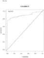
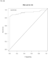
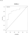

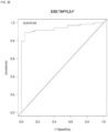

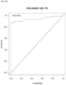
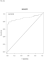
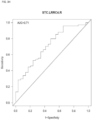
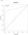
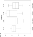
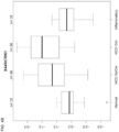

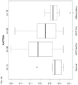
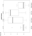


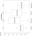
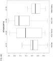


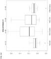


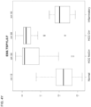


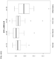
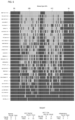

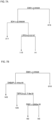

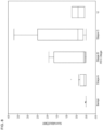
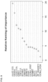
Classifications
-
- C—CHEMISTRY; METALLURGY
- C12—BIOCHEMISTRY; BEER; SPIRITS; WINE; VINEGAR; MICROBIOLOGY; ENZYMOLOGY; MUTATION OR GENETIC ENGINEERING
- C12Q—MEASURING OR TESTING PROCESSES INVOLVING ENZYMES, NUCLEIC ACIDS OR MICROORGANISMS; COMPOSITIONS OR TEST PAPERS THEREFOR; PROCESSES OF PREPARING SUCH COMPOSITIONS; CONDITION-RESPONSIVE CONTROL IN MICROBIOLOGICAL OR ENZYMOLOGICAL PROCESSES
- C12Q1/00—Measuring or testing processes involving enzymes, nucleic acids or microorganisms; Compositions therefor; Processes of preparing such compositions
- C12Q1/68—Measuring or testing processes involving enzymes, nucleic acids or microorganisms; Compositions therefor; Processes of preparing such compositions involving nucleic acids
- C12Q1/6876—Nucleic acid products used in the analysis of nucleic acids, e.g. primers or probes
- C12Q1/6883—Nucleic acid products used in the analysis of nucleic acids, e.g. primers or probes for diseases caused by alterations of genetic material
- C12Q1/6886—Nucleic acid products used in the analysis of nucleic acids, e.g. primers or probes for diseases caused by alterations of genetic material for cancer
-
- C—CHEMISTRY; METALLURGY
- C12—BIOCHEMISTRY; BEER; SPIRITS; WINE; VINEGAR; MICROBIOLOGY; ENZYMOLOGY; MUTATION OR GENETIC ENGINEERING
- C12Q—MEASURING OR TESTING PROCESSES INVOLVING ENZYMES, NUCLEIC ACIDS OR MICROORGANISMS; COMPOSITIONS OR TEST PAPERS THEREFOR; PROCESSES OF PREPARING SUCH COMPOSITIONS; CONDITION-RESPONSIVE CONTROL IN MICROBIOLOGICAL OR ENZYMOLOGICAL PROCESSES
- C12Q1/00—Measuring or testing processes involving enzymes, nucleic acids or microorganisms; Compositions therefor; Processes of preparing such compositions
- C12Q1/68—Measuring or testing processes involving enzymes, nucleic acids or microorganisms; Compositions therefor; Processes of preparing such compositions involving nucleic acids
- C12Q1/6813—Hybridisation assays
- C12Q1/6827—Hybridisation assays for detection of mutation or polymorphism
-
- C—CHEMISTRY; METALLURGY
- C12—BIOCHEMISTRY; BEER; SPIRITS; WINE; VINEGAR; MICROBIOLOGY; ENZYMOLOGY; MUTATION OR GENETIC ENGINEERING
- C12Q—MEASURING OR TESTING PROCESSES INVOLVING ENZYMES, NUCLEIC ACIDS OR MICROORGANISMS; COMPOSITIONS OR TEST PAPERS THEREFOR; PROCESSES OF PREPARING SUCH COMPOSITIONS; CONDITION-RESPONSIVE CONTROL IN MICROBIOLOGICAL OR ENZYMOLOGICAL PROCESSES
- C12Q2521/00—Reaction characterised by the enzymatic activity
- C12Q2521/30—Phosphoric diester hydrolysing, i.e. nuclease
- C12Q2521/331—Methylation site specific nuclease
-
- C—CHEMISTRY; METALLURGY
- C12—BIOCHEMISTRY; BEER; SPIRITS; WINE; VINEGAR; MICROBIOLOGY; ENZYMOLOGY; MUTATION OR GENETIC ENGINEERING
- C12Q—MEASURING OR TESTING PROCESSES INVOLVING ENZYMES, NUCLEIC ACIDS OR MICROORGANISMS; COMPOSITIONS OR TEST PAPERS THEREFOR; PROCESSES OF PREPARING SUCH COMPOSITIONS; CONDITION-RESPONSIVE CONTROL IN MICROBIOLOGICAL OR ENZYMOLOGICAL PROCESSES
- C12Q2565/00—Nucleic acid analysis characterised by mode or means of detection
- C12Q2565/60—Detection means characterised by use of a special device
- C12Q2565/627—Detection means characterised by use of a special device being a mass spectrometer
-
- C—CHEMISTRY; METALLURGY
- C12—BIOCHEMISTRY; BEER; SPIRITS; WINE; VINEGAR; MICROBIOLOGY; ENZYMOLOGY; MUTATION OR GENETIC ENGINEERING
- C12Q—MEASURING OR TESTING PROCESSES INVOLVING ENZYMES, NUCLEIC ACIDS OR MICROORGANISMS; COMPOSITIONS OR TEST PAPERS THEREFOR; PROCESSES OF PREPARING SUCH COMPOSITIONS; CONDITION-RESPONSIVE CONTROL IN MICROBIOLOGICAL OR ENZYMOLOGICAL PROCESSES
- C12Q2600/00—Oligonucleotides characterized by their use
- C12Q2600/154—Methylation markers
-
- C—CHEMISTRY; METALLURGY
- C12—BIOCHEMISTRY; BEER; SPIRITS; WINE; VINEGAR; MICROBIOLOGY; ENZYMOLOGY; MUTATION OR GENETIC ENGINEERING
- C12Q—MEASURING OR TESTING PROCESSES INVOLVING ENZYMES, NUCLEIC ACIDS OR MICROORGANISMS; COMPOSITIONS OR TEST PAPERS THEREFOR; PROCESSES OF PREPARING SUCH COMPOSITIONS; CONDITION-RESPONSIVE CONTROL IN MICROBIOLOGICAL OR ENZYMOLOGICAL PROCESSES
- C12Q2600/00—Oligonucleotides characterized by their use
- C12Q2600/16—Primer sets for multiplex assays
-
- C—CHEMISTRY; METALLURGY
- C12—BIOCHEMISTRY; BEER; SPIRITS; WINE; VINEGAR; MICROBIOLOGY; ENZYMOLOGY; MUTATION OR GENETIC ENGINEERING
- C12Q—MEASURING OR TESTING PROCESSES INVOLVING ENZYMES, NUCLEIC ACIDS OR MICROORGANISMS; COMPOSITIONS OR TEST PAPERS THEREFOR; PROCESSES OF PREPARING SUCH COMPOSITIONS; CONDITION-RESPONSIVE CONTROL IN MICROBIOLOGICAL OR ENZYMOLOGICAL PROCESSES
- C12Q2600/00—Oligonucleotides characterized by their use
- C12Q2600/166—Oligonucleotides used as internal standards, controls or normalisation probes
Definitions
- hepatocellular carcinoma screening provides technology for hepatocellular carcinoma screening and particularly, but not exclusively, to methods, compositions, and related uses for detecting the presence of hepatocellular carcinoma.
- Hepatocellular carcinoma is a primary malignancy of the liver and occurs predominantly in patients with underlying chronic liver disease and cirrhosis.
- the cell(s) of origin are believed to be the hepatic stem cells (see, Alison MR. Stem Cell Rev. 2005. 1(3):253-60 ). Tumors progress with local expansion, intrahepatic spread, and distant metastases.
- HCC is now the third leading cause of cancer deaths worldwide, with over 500,000 people affected.
- the incidence of HCC is highest in Asia and Africa, where the endemic high prevalence of hepatitis B and hepatitis C strongly predisposes to the development of chronic liver disease and subsequent development of HCC.
- HCC human liver cancer
- US 2011/256538 A1 relates to a method for detecting the presence or absence of a cancer in an individual, by determining the level of methylation of the sense strand of a selected regulatory region of the tumor suppressor gene adenomatous polyposis coli (APC). Hypermethylation has been described in the HOXA1 gene in cholangiocarcinoma ( US 2016/090634 A1 ) and adenocarcinoma ( Tsou et al., Molecular Cancer, Biomed Central, vol. 6, no. 1, 29 October 2007, pages 70 ).
- Hepatocellular carcinoma is the 2nd most fatal cancer worldwide. Survival improves with early stage detection, and accurate noninvasive screening tools are needed. There is an imperative for innovation that will deliver accurate, affordable, and safe screening tools for the pre-symptomatic detection of the earliest stage of HCC.
- the present invention addresses this need. Indeed, the present invention provides novel methylated DNA markers that discriminate HCC from normal controls (controls with and without cirrhosis).
- Methylated DNA has been studied as a potential class of biomarkers in the tissues of most tumor types.
- DNA methyltransferases add a methyl group to DNA at cytosine-phosphate-guanine (CpG) island sites as an epigenetic control of gene expression.
- CpG cytosine-phosphate-guanine
- DNA methylation may be a more chemically and biologically stable diagnostic tool than RNA or protein expression ( Laird (2010) Nat Rev Genet 11: 191-203 ).
- methylation markers offer excellent specificity and are more broadly informative and sensitive than are individual DNA mutations (see, Zou et al (2007) Cancer Epidemiol Biomarkers Prev 16: 2686-96 ).
- HCP high CpG density promoters
- areas of low CpG density defined as having ⁇ 5% CpG sequence within a 300 base pair region
- HCPs include promoters for ubiquitous housekeeping genes and highly regulated developmental genes. Among the HCP sites methylated at >50% were several established markers such as Wnt 2, NDRG2, SFRP2, and BMP3 ( Meissner et al. (2008) Nature 454: 766-70 ).
- HCC screening e.g., surveilling
- methods, compositions, and related uses for detecting the presence of HCC e.g., surveilling
- Markers and/or panels of markers were identified capable of detecting HCC (see, Examples I, II and III) (ACP1, BDH1, Chr12.133, CLEC11A, DAB2IP, DBNL, EMX1, EFNB2, HOXA1, LRRC4, SPINT2, TSPYL5, CCNJ 3707, CCNJ 3124, PFKP, SCRN1, and ECE1).
- the invention describes a method for characterizing a biological sample comprising: measuring a methylation level of a CpG site for the gene HOXA1 and one or more genes selected from either EMX1, DAB2IP, TSPYL5, ACP1, BDH1, Chr12.133, CLEC11A, DBNL, EFNB2, LRRC4, SPINT2, CCNJ 3707, CCNJ 3124, PFKP, SCRN1, and ECE1 in a biological sample of a human individual through treating genomic DNA in the biological sample with bisulfite; amplifying the bisulfite-treated genomic DNA using a set of primers for the genes; and determining the methylation level of the CpG site by methylation-specific PCR, quantitative methylation-specific PCR, methylation-sensitive DNA restriction enzyme analysis, quantitative bisulfite pyrosequencing, or bisulfite genomic sequencing PCR, and comparing the methylation level to a methylation level of a corresponding set of genes in control samples without HCC
- the technology provides a number of methylated DNA markers and subsets thereof (e.g., sets of 2, 3, 4, 5, 6, 7, 8, 9, 10, 11, 12 markers) with high discrimination for detecting the presence of HCC in subjects.
- Experiments applied a selection filter to candidate markers to identify markers that provide a high signal to noise ratio and a low background level to provide high specificity, e.g., when assaying media (e.g., plasma) for purposes of screening or diagnosis (e.g., HCC screening or diagnosis).
- the technology is related to assessing the presence of and methylation state of one or more of the markers identified herein in a biological sample (e.g., a plasma sample).
- markers comprise one or more differentially methylated regions (DMR) as discussed herein, e.g., as provided in Tables 1 and 4.
- DMR differentially methylated regions
- Methylation state is assessed in embodiments of the technology.
- the technology provided herein is not restricted in the method by which a gene's methylation state is measured.
- the methylation state is measured by a genome scanning method.
- one method involves restriction landmark genomic scanning (see, Kawai et al. (1994) Mol. Cell. Biol.
- methylation-sensitive arbitrarily primed PCR see, Gonzalgo et al. (1997) Cancer Res. 57: 594-599 ).
- changes in methylation patterns at specific CpG sites are monitored by digestion of genomic DNA with methylation-sensitive restriction enzymes followed by Southern analysis of the regions of interest (digestion-Southern method).
- analyzing changes in methylation patterns involves a PCR-based process that involves digestion of genomic DNA with methylation-sensitive restriction enzymes prior to PCR amplification (see, Singer-Sam et al. (1990) Nucl. Acids Res. 18: 687 ).
- the methylation state is often expressed as the fraction or percentage of individual strands of DNA that is methylated at a particular site (e.g., at a single nucleotide, at a particular region or locus, at a longer sequence of interest, e.g., up to a ⁇ 100-bp, 200-bp, 500-bp, 1000-bp subsequence of a DNA or longer) relative to the total population of DNA in the sample comprising that particular site.
- the amount of the unmethylated nucleic acid is determined by PCR using calibrators.
- a known amount of DNA is bisulfite treated and the resulting methylation-specific sequence is determined using either a real-time PCR or other exponential amplification, e.g., a QuARTS assay (e.g., as provided by U.S. Pat. Nos. 8,361,720 , 8,715,937 , and 8,916,344 ).
- methods comprise generating a standard curve for the unmethylated target by using external standards.
- the standard curve is constructed from at least two points and relates the real-time Ct value for unmethylated DNA to known quantitative standards.
- a second standard curve for the methylated target is constructed from at least two points and external standards. This second standard curve relates the Ct for methylated DNA to known quantitative standards.
- the test sample Ct values are determined for the methylated and unmethylated populations and the genomic equivalents of DNA are calculated from the standard curves produced by the first two steps.
- the percentage of methylation at the site of interest is calculated from the amount of methylated DNAs relative to the total amount of DNAs in the population, e.g., (number of methylated DNAs) / (the number of methylated DNAs + number of unmethylated DNAs) ⁇ 100.
- compositions and kits for practicing the methods.
- reagents e.g., primers, probes
- sets e.g., sets of primers pairs for amplifying a plurality of markers.
- Additional reagents for conducting a detection assay may also be provided (e.g., enzymes, buffers, positive and negative controls for conducting QuARTS, PCR, sequencing, bisulfite, or other assays).
- the kits containing one or more reagent necessary, sufficient, or useful for conducting a method are provided.
- reactions mixtures containing the reagents.
- master mix reagent sets containing a plurality of reagents that may be added to each other and/or to a test sample to complete a reaction mixture.
- the technology described herein is associated with a programmable machine designed to perform a sequence of arithmetic or logical operations as provided by the methods described herein.
- some aspects of the technology are associated with (e.g., implemented in) computer software and/or computer hardware.
- the technology relates to a computer comprising a form of memory, an element for performing arithmetic and logical operations, and a processing element (e.g., a microprocessor) for executing a series of instructions (e.g., a method as provided herein) to read, manipulate, and store data.
- a microprocessor is part of a system for determining a methylation state (e.g., of one or more DMR, e.g., DMR 1-400 as provided in Tables 1 and 4); comparing methylation states (e.g., of one or more DMR, e.g., DMR 1-400 as provided in Tables 1 and 4); generating standard curves; determining a Ct value; calculating a fraction, frequency, or percentage of methylation (e.g., of one or more DMR, e.g., DMR 1-400 as provided in Tables 1 and 4); identifying a CpG island; determining a specificity and/or sensitivity of an assay or marker; calculating an ROC curve and an associated AUC; sequence analysis; all as described herein or is known in the art.
- a methylation state e.g., of one or more DMR, e.g., DMR 1-400 as provided in Tables 1 and 4
- comparing methylation states e.
- a microprocessor or computer uses methylation state data in an algorithm to predict a site of a cancer.
- a software or hardware component receives the results of multiple assays and determines a single value result to report to a user that indicates a cancer risk based on the results of the multiple assays (e.g., determining the methylation state of multiple DMR, e.g., as provided in Tables 1 and 4).
- Related aspects calculate a risk factor based on a mathematical combination (e.g., a weighted combination, a linear combination) of the results from multiple assays, e.g., determining the methylation states of multiple markers (such as multiple DMR, e.g., as provided in Tables 1 and 4).
- the methylation state of a DMR defines a dimension and may have values in a multidimensional space and the coordinate defined by the methylation states of multiple DMR is a result, e.g., to report to a user.
- Some aspects comprise a storage medium and memory components.
- Memory components e.g., volatile and/or nonvolatile memory find use in storing instructions (e.g., an aspect of a process as provided herein) and/or data (e.g., a work piece such as methylation measurements, sequences, and statistical descriptions associated therewith).
- Some aspects relate to systems also comprising one or more of a CPU, a graphics card, and a user interface (e.g., comprising an output device such as display and an input device such as a keyboard).
- Programmable machines associated with the technology comprise conventional extant technologies and technologies in development or yet to be developed (e.g., a quantum computer, a chemical computer, a DNA computer, an optical computer, a spintronics based computer, etc.).
- the technology comprises a wired (e.g., metallic cable, fiber optic) or wireless transmission medium for transmitting data.
- a wired e.g., metallic cable, fiber optic
- wireless transmission medium for transmitting data.
- some aspects relate to data transmission over a network (e.g., a local area network (LAN), a wide area network (WAN), an ad-hoc network, the internet, etc.).
- a network e.g., a local area network (LAN), a wide area network (WAN), an ad-hoc network, the internet, etc.
- programmable machines are present on such a network as peers and in some aspects the programmable machines have a client/server relationship.
- data are stored on a computer-readable storage medium such as a hard disk, flash memory, optical media, a floppy disk, etc.
- the technology provided herein is associated with a plurality of programmable devices that operate in concert to perform a method as described herein.
- a plurality of computers e.g., connected by a network
- may work in parallel to collect and process data e.g., in an implementation of cluster computing or grid computing or some other distributed computer architecture that relies on complete computers (with onboard CPUs, storage, power supplies, network interfaces, etc.) connected to a network (private, public, or the internet) by a conventional network interface, such as Ethernet, fiber optic, or by a wireless network technology.
- some aspects provide a computer that includes a computer-readable medium.
- the aspect includes a random access memory (RAM) coupled to a processor.
- the processor executes computer-executable program instructions stored in memory.
- processors may include a microprocessor, an ASIC, a state machine, or other processor, and can be any of a number of computer processors, such as processors from Intel Corporation of Santa Clara, California and Motorola Corporation of Schaumburg, Illinois.
- processors include, or may be in communication with, media, for example computer-readable media, which stores instructions that, when executed by the processor, cause the processor to perform the steps described herein.
- Computer-readable media include, but are not limited to, an electronic, optical, magnetic, or other storage or transmission device capable of providing a processor with computer-readable instructions.
- suitable media include, but are not limited to, a floppy disk, CD-ROM, DVD, magnetic disk, memory chip, ROM, RAM, an ASIC, a configured processor, all optical media, all magnetic tape or other magnetic media, or any other medium from which a computer processor can read instructions.
- various other forms of computer-readable media may transmit or carry instructions to a computer, including a router, private or public network, or other transmission device or channel, both wired and wireless.
- the instructions may comprise code from any suitable computerprogramming language, including, for example, C, C++, C#, Visual Basic, Java, Python, Perl, and JavaScript.
- Computers are connected in some aspects to a network. Computers may also include a number of external or internal devices such as a mouse, a CD-ROM, DVD, a keyboard, a display, or other input or output devices. Examples of computers are personal computers, digital assistants, personal digital assistants, cellular phones, mobile phones, smart phones, pagers, digital tablets, laptop computers, internet appliances, and other processor-based devices. In general, the computers related to aspects of the technology provided herein may be any type of processor-based platform that operates on any operating system, such as Microsoft Windows, Linux, UNIX, Mac OS X, etc., capable of supporting one or more programs comprising the technology provided herein. Some aspects comprise a personal computer executing other application programs (e.g., applications).
- application programs e.g., applications
- the applications can be contained in memory and can include, for example, a word processing application, a spreadsheet application, an email application, an instant messenger application, a presentation application, an Internet browser application, a calendar/organizer application, and any other application capable of being executed by a client device.
- a method of screening for HCC in a sample obtained from a subject comprising assaying a methylation state of a marker in a sample obtained from a subject; and identifying the subject as having HCC when the methylation state of the marker is different than a methylation state of the marker assayed in a subject that does not have HCC (e.g., a subject that does not have HCC) (e.g., a subject that does not have HCC but does have liver cirrhosis), wherein the marker comprises one or more bases in a differentially methylated region (DMR) selected from ACP1, BDH1, Chr12.133, CLEC11A, DAB2IP, DBNL, EMX1, EFNB2, HOXA1, LRRC4, SPINT2, TSPYL5, CCNJ_3707, CCNJ_3124, PFKP, SCRN1, and ECE1 as provided in Tables 1 and 4.
- DMR differentially methylated region
- assessing the methylation state of the marker in the sample comprises determining the methylation state of one base.
- assaying the methylation state of the marker in the sample comprises determining the extent of methylation at a plurality of bases.
- the methylation state of the marker comprises an increased methylation of the marker relative to a normal methylation state of the marker.
- the methylation state of the marker comprises a decreased methylation of the marker relative to a normal methylation state of the marker.
- the methylation state of the marker comprises a different pattern of methylation of the marker relative to a normal methylation state of the marker.
- the marker is a region of 100 or fewer bases, the marker is a region of 500 or fewer bases, the marker is a region of 1000 or fewer bases, the marker is a region of 5000 or fewer bases, or, in some embodiments, the marker is one base. In some embodiments the marker is in a high CpG density promoter.
- the sample is a blood sample (e.g., plasma, serum, whole blood), a stool sample, a tissue sample (e.g., stomach tissue, pancreatic tissue, bile duct / liver tissue, pancreatic juice, and colorectal tissue), an excretion, or a urine sample.
- a blood sample e.g., plasma, serum, whole blood
- a stool sample e.g., a tissue sample (e.g., stomach tissue, pancreatic tissue, bile duct / liver tissue, pancreatic juice, and colorectal tissue), an excretion, or a urine sample.
- tissue sample e.g., stomach tissue, pancreatic tissue, bile duct / liver tissue, pancreatic juice, and colorectal tissue
- the technology is not limited in the method used to determine methylation state.
- the assaying comprises using methylation specific polymerase chain reaction, nucleic acid sequencing, mass spectrometry, methylation specific nuclease, mass-based separation, or target capture.
- the assaying comprises use of a methylation specific oligonucleotide.
- the technology uses massively parallel sequencing (e.g., next-generation sequencing) to determine methylation state, e.g., sequencing-by-synthesis, real-time (e.g., single-molecule) sequencing, bead emulsion sequencing, nanopore sequencing, etc.
- the technology provides reagents for detecting a DMR, e.g., in some aspects are provided a set of oligonucleotides comprising the sequences provided by SEQ ID NO: 1-94 (Tables 2 and 5). In some aspects are provided an oligonucleotide comprising a sequence complementary to a chromosomal region having a base in a DMR, e.g., an oligonucleotide sensitive to methylation state of a DMR.
- the technology provides various panels of markers, e.g., in some aspects the marker comprises a chromosomal region having an annotation that is provided in Tables 1 or 3, and that comprises the marker. In addition, aspects provide a method of analyzing a DMR from Tables 1 and/or 4 that one or more of DMR Nos. 1-400.
- Kit aspects are provided in the disclosure, e.g., a kit comprising a bisulfite reagent; and a control nucleic acid comprising a sequence from a DMR selected from a group consisting of DMR 1-400 (from Tables 1 and 4) and having a methylation state associated with a subject who does not have HCC (e.g., a subject that does not have HCC and does not have liver cirrhosis) (e.g., a subject that does not have HCC but does have liver cirrhosis).
- a kit comprising a bisulfite reagent; and a control nucleic acid comprising a sequence from a DMR selected from a group consisting of DMR 1-400 (from Tables 1 and 4) and having a methylation state associated with a subject who does not have HCC (e.g., a subject that does not have HCC and does not have liver cirrhosis) (e.g., a subject that does not have HCC but does
- Kit aspects are provided, e.g., a kit comprising a bisulfite reagent; and a control nucleic acid comprising a sequence from a DMR selected from a group consisting of DMR 1-400 (from Tables 1 and 4) and having a methylation state associated with a subject who does not have HCC.
- kits aspects comprise a sample collector for obtaining a sample from a subject (e.g., a stool sample); reagents for isolating a nucleic acid from the sample; a bisulfite reagent; and an oligonucleotide as described herein.
- a sample collector for obtaining a sample from a subject (e.g., a stool sample); reagents for isolating a nucleic acid from the sample; a bisulfite reagent; and an oligonucleotide as described herein.
- compositions e.g., reaction mixtures.
- a composition comprising a nucleic acid comprising a DMR and a bisulfite reagent.
- compositions comprising a nucleic acid comprising a DMR and an oligonucleotide as described herein.
- composition comprising a nucleic acid comprising a DMR and a methylation-sensitive restriction enzyme.
- composition comprising a nucleic acid comprising a DMR and a polymerase.
- Additional related method embodiments are provided for screening for HCC in a sample obtained from a subject (e.g., a plasma sample), e.g., a method comprising determining a methylation state of a marker in the sample comprising a base in a DMR that is one or more of DMR 1-400 (from Tables 1 and 4); comparing the methylation state of the marker from the subject sample to a methylation state of the marker from a normal control sample from a subject who does not have HCC; and determining a confidence interval and/or a p value of the difference in the methylation state of the subject sample and the normal control sample.
- a sample obtained from a subject e.g., a plasma sample
- a method comprising determining a methylation state of a marker in the sample comprising a base in a DMR that is one or more of DMR 1-400 (from Tables 1 and 4); comparing the methylation state of the marker from the subject sample to a methylation state of the
- the confidence interval is 90%, 95%, 97.5%, 98%, 99%, 99.5%, 99.9% or 99.99% and the p value is 0.1, 0.05, 0.025, 0.02, 0.01, 0.005, 0.001, or 0.0001.
- Some embodiments of methods provide steps of reacting a nucleic acid comprising a DMR with a bisulfite reagent to produce a bisulfite-reacted nucleic acid; sequencing the bisulfite-reacted nucleic acid to provide a nucleotide sequence of the bisulfite-reacted nucleic acid; comparing the nucleotide sequence of the bisulfite-reacted nucleic acid with a nucleotide sequence of a nucleic acid comprising the DMR from a subject who does not have a cancer to identify differences in the two sequences; and identifying the subject as having a neoplasm when a difference is present.
- Systems for screening for HCC in a sample obtained from a subject are provided by the technology.
- exemplary embodiments of systems include, e.g., a system for screening for HCC in a sample obtained from a subject, the system comprising an analysis component configured to determine the methylation state of a sample, a software component configured to compare the methylation state of the sample with a control sample or a reference sample methylation state recorded in a database, and an alert component configured to alert a user of a HCC-associated methylation state (e.g., a methylation state for no HCC; a methylation state for HCC).
- a HCC-associated methylation state e.g., a methylation state for no HCC; a methylation state for HCC.
- An alert is determined in some aspects by a software component that receives the results from multiple assays (e.g., determining the methylation states of multiple markers, e.g., DMR, e.g., as provided in Tables 1 and 4) and calculating a value or result to report based on the multiple results.
- Some aspects provide a database of weighted parameters associated with each DMR provided herein for use in calculating a value or result and/or an alert to report to a user (e.g., such as a physician, nurse, clinician, etc.).
- all results from multiple assays are reported and in some aspects one or more results are used to provide a score, value, or result based on a composite of one or more results from multiple assays that is indicative of a HCC risk in a subject.
- a sample comprises a nucleic acid comprising a DMR.
- the system further comprises a component for isolating a nucleic acid, a component for collecting a sample such as a component for collecting a plasma sample.
- the system comprises nucleic acid sequences comprising a DMR.
- the database comprises nucleic acid sequences from subjects who do not have HCC.
- nucleic acids e.g., a set of nucleic acids, each nucleic acid having a sequence comprising a DMR.
- the set of nucleic acids wherein each nucleic acid has a sequence from a subject who does not have HCC.
- Related system aspects comprise a set of nucleic acids as described and a database of nucleic acid sequences associated with the set of nucleic acids. Some aspects further comprise a bisulfite reagent. And, some aspects further comprise a nucleic acid sequencer.
- methods for detecting HCC in a sample obtained from a subject comprising a) obtaining a sample comprising DNA from a subject; b) treating the obtained DNA with a reagent which selectively modifies unmethylated cytosine residues in the obtained DNA to produce modified residues but which does not modify methylated cytosine residues; c) determining the methylation level of one or more DNA methylation markers in the DNA having undergone the treating of step b), wherein one or more DNA methylation markers comprises a base in a differentially methylated region (DMR) as provided by DMR 1-400 (from Tables 1 and 4), d) comparing the determined methylation level of the one or more DNA methylation markers with methylation level references for the one or more DNA methylation markers for subjects who do not have HCC; and e) identifying the subject as having HCC when differences are present.
- DMR differentially methylated region
- a determination of elevated methylation in one or more of the DNA methylation markers comprises a determination of altered methylation within a region selected from the group consisting of a CpG island and a CpG island shore.
- a determination of elevated methylation within the CpG island or CpG shore comprises elevated methylation within a coding region or a regulatory region of the DNA methylation marker.
- the determining the methylation level of one or more DNA methylation markers in the DNA having undergone the treating of step b) comprises determining the methylation score and/or the methylation frequency of the one or more DNA methylation markers.
- the treating of step b) is accomplished through bisulfite modification of the obtained DNA.
- the determining the methylation level of one or more DNA methylation markers in the DNA having undergone the treating of step b) is achieved by a technique selected from the group consisting of methylation-specific PCR, quantitative methylation-specific PCR, methylation-sensitive DNA restriction enzyme analysis, quantitative bisulfite pyrosequencing, and bisulfite genomic sequencing PCR.
- hepatocellular carcinoma screening provides technology for hepatocellular carcinoma screening and particularly, but not exclusively, to methods, compositions, and related uses for detecting the presence of hepatocellular carcinoma.
- nucleic acid or “nucleic acid molecule” generally refers to any ribonucleic acid or deoxyribonucleic acid, which may be unmodified or modified DNA or RNA.
- Nucleic acids include, without limitation, single- and double-stranded nucleic acids.
- nucleic acid also includes DNA as described above that contains one or more modified bases. Thus, DNA with a backbone modified for stability or for other reasons is a “nucleic acid”.
- the term “nucleic acid” as it is used herein embraces such chemically, enzymatically, or metabolically modified forms of nucleic acids, as well as the chemical forms of DNA characteristic of viruses and cells, including for example, simple and complex cells.
- oligonucleotide or “polynucleotide” or “nucleotide” or “nucleic acid” refer to a molecule having two or more deoxyribonucleotides or ribonucleotides, preferably more than three, and usually more than ten. The exact size will depend on many factors, which in turn depends on the ultimate function or use of the oligonucleotide.
- the oligonucleotide may be generated in any manner, including chemical synthesis, DNA replication, reverse transcription, or a combination thereof.
- Typical deoxyribonucleotides for DNA are thymine, adenine, cytosine, and guanine.
- Typical ribonucleotides for RNA are uracil, adenine, cytosine, and guanine.
- locus or "region" of a nucleic acid refer to a subregion of a nucleic acid, e.g., a gene on a chromosome, a single nucleotide, a CpG island, etc.
- complementarity refers to nucleotides (e.g., 1 nucleotide) or polynucleotides (e.g., a sequence of nucleotides) related by the base-pairing rules.
- sequence 5'-A-G-T-3' is complementary to the sequence 3'-T-C-A-5'.
- Complementarity may be "partial,” in which only some of the nucleic acids' bases are matched according to the base pairing rules. Or, there may be “complete” or “total” complementarity between the nucleic acids.
- the degree of complementarity between nucleic acid strands effects the efficiency and strength of hybridization between nucleic acid strands. This is of particular importance in amplification reactions and in detection methods that depend upon binding between nucleic acids.
- the term "gene” refers to a nucleic acid (e.g., DNA or RNA) sequence that comprises coding sequences necessary for the production of an RNA, or of a polypeptide or its precursor.
- a functional polypeptide can be encoded by a full length coding sequence or by any portion of the coding sequence as long as the desired activity or functional properties (e.g., enzymatic activity, ligand binding, signal transduction, etc.) of the polypeptide are retained.
- portion when used in reference to a gene refers to fragments of that gene. The fragments may range in size from a few nucleotides to the entire gene sequence minus one nucleotide. Thus, "a nucleotide comprising at least a portion of a gene” may comprise fragments of the gene or the entire gene.
- the term "gene” also encompasses the coding regions of a structural gene and includes sequences located adjacent to the coding region on both the 5' and 3' ends, e.g., for a distance of about 1 kb on either end, such that the gene corresponds to the length of the fulllength mRNA (e.g., comprising coding, regulatory, structural and other sequences).
- the sequences that are located 5' of the coding region and that are present on the mRNA are referred to as 5' non-translated or untranslated sequences.
- the sequences that are located 3' or downstream of the coding region and that are present on the mRNA are referred to as 3' non-translated or 3' untranslated sequences.
- genomic form or clone of a gene contains the coding region interrupted with non-coding sequences termed "introns” or “intervening regions” or “intervening sequences.”
- Introns are segments of a gene that are transcribed into nuclear RNA (hnRNA); introns may contain regulatory elements such as enhancers. Introns are removed or “spliced out” from the nuclear or primary transcript; introns therefore are absent in the messenger RNA (mRNA) transcript.
- mRNA messenger RNA
- genomic forms of a gene may also include sequences located on both the 5' and 3' ends of the sequences that are present on the RNA transcript. These sequences are referred to as "flanking" sequences or regions (these flanking sequences are located 5' or 3' to the non-translated sequences present on the mRNA transcript).
- the 5' flanking region may contain regulatory sequences such as promoters and enhancers that control or influence the transcription of the gene.
- the 3' flanking region may contain sequences that direct the termination of transcription, posttranscriptional cleavage, and polyadenylation.
- allele refers to a variation of a gene; the variations include but are not limited to variants and mutants, polymorphic loci, and single nucleotide polymorphic loci, frameshift, and splice mutations. An allele may occur naturally in a population or it might arise during the lifetime of any particular individual of the population.
- variant and mutant when used in reference to a nucleotide sequence refer to a nucleic acid sequence that differs by one or more nucleotides from another, usually related, nucleotide acid sequence.
- a “variation” is a difference between two different nucleotide sequences; typically, one sequence is a reference sequence.
- Amplification is a special case of nucleic acid replication involving template specificity. It is to be contrasted with non-specific template replication (e.g., replication that is template-dependent but not dependent on a specific template). Template specificity is here distinguished from fidelity of replication (e.g., synthesis of the proper polynucleotide sequence) and nucleotide (ribo- or deoxyribo-) specificity. Template specificity is frequently described in terms of “target” specificity. Target sequences are “targets” in the sense that they are sought to be sorted out from other nucleic acid. Amplification techniques have been designed primarily for this sorting out.
- Amplification of nucleic acids generally refers to the production of multiple copies of a polynucleotide, or a portion of the polynucleotide, typically starting from a small amount of the polynucleotide (e.g., a single polynucleotide molecule, 10 to 100 copies of a polynucleotide molecule, which may or may not be exactly the same), where the amplification products or amplicons are generally detectable.
- Amplification of polynucleotides encompasses a variety of chemical and enzymatic processes.
- PCR polymerase chain reaction
- LCR ligase chain reaction
- Additional types of amplification include, but are not limited to, allele-specific PCR (see, e.g., U.S. Patent No. 5,639,611 ), assembly PCR (see, e.g., U.S. Patent No. 5,965,408 ), helicase-dependent amplification (see, e.g., U.S. Patent No.
- Hot-start PCR see, e.g., U.S. Patent Nos. 5,773,258 and 5,338,671
- intersequence-specfic PCR see, e.g., Triglia, et alet al. (1988) Nucleic Acids Res., 16:8186
- ligation-mediated PCR see, e.g., Guilfoyle, R. et al., Nucleic Acids Research, 25:1854-1858 (1997 ); U.S. Patent No.
- PCR polymerase chain reaction
- the mixture is denatured and the primers then annealed to their complementary sequences within the target molecule.
- the primers are extended with a polymerase so as to form a new pair of complementary strands.
- the steps of denaturation, primer annealing, and polymerase extension can be repeated many times (i.e., denaturation, annealing and extension constitute one "cycle”; there can be numerous "cycles") to obtain a high concentration of an amplified segment of the desired target sequence.
- the length of the amplified segment of the desired target sequence is determined by the relative positions of the primers with respect to each other, and therefore, this length is a controllable parameter.
- PCR polymerase chain reaction
- Amplification enzymes are enzymes that, under conditions they are used, will process only specific sequences of nucleic acid in a heterogeneous mixture of nucleic acid.
- MDV-1 RNA is the specific template for the replicase ( Kacian et al., Proc. Natl. Acad. Sci. USA, 69:3038 [1972 ]).
- Other nucleic acid will not be replicated by this amplification enzyme.
- this amplification enzyme has a stringent specificity for its own promoters ( Chamberlin et al, Nature, 228:227 [1970 ]).
- thermostable template-dependant DNA polymerases e.g., Taq and Pfu DNA polymerases
- thermodynamic conditions that favor primer hybridization with the target sequences and not hybridization with non-target sequences
- nucleic acid detection assay refers to any method of determining the nucleotide composition of a nucleic acid of interest.
- Nucleic acid detection assay include but are not limited to, DNA sequencing methods, probe hybridization methods, structure specific cleavage assays (e.g., the INVADER assay, Hologic, Inc.) and are described, e.g., in U.S. Patent Nos. 5,846,717 , 5,985,557 , 5,994,069 , 6,001,567 , 6,090,543 , and 6,872,816 ; Lyamichev et al., Nat.
- NASBA e.g., U.S. Pat. No. 5,409,818
- molecular beacon technology e.g., U.S. Pat. No. 6,150,097
- E-sensor technology Motorola, U.S. Pat. Nos. 6,248,229 , 6,221,583 , 6,013,170 , and 6,063,573
- cycling probe technology e.g., U.S. Pat. Nos. 5,403,711 , 5,011,769 , and 5,660,988
- Dade Behring signal amplification methods e.g., U.S. Pat. Nos.
- amplifiable nucleic acid refers to a nucleic acid that may be amplified by any amplification method. It is contemplated that "amplifiable nucleic acid” will usually comprise "sample template.”
- sample template refers to nucleic acid originating from a sample that is analyzed for the presence of "target” (defined below).
- background template is used in reference to nucleic acid other than sample template that may or may not be present in a sample. Background template is most often inadvertent. It may be the result of carryover or it may be due to the presence of nucleic acid contaminants sought to be purified away from the sample. For example, nucleic acids from organisms other than those to be detected may be present as background in a test sample.
- primer refers to an oligonucleotide, whether occurring naturally as in a purified restriction digest or produced synthetically, that is capable of acting as a point of initiation of synthesis when placed under conditions in which synthesis of a primer extension product that is complementary to a nucleic acid strand is induced, (e.g., in the presence of nucleotides and an inducing agent such as a DNA polymerase and at a suitable temperature and pH).
- the primer is preferably single stranded for maximum efficiency in amplification, but may alternatively be double stranded. If double stranded, the primer is first treated to separate its strands before being used to prepare extension products.
- the primer is an oligodeoxyribonucleotide.
- the primer must be sufficiently long to prime the synthesis of extension products in the presence of the inducing agent. The exact lengths of the primers will depend on many factors, including temperature, source of primer, and the use of the method.
- probe refers to an oligonucleotide (e.g., a sequence of nucleotides), whether occurring naturally as in a purified restriction digest or produced synthetically, recombinantly, or by PCR amplification, that is capable of hybridizing to another oligonucleotide of interest.
- a probe may be single-stranded or double-stranded. Probes are useful in the detection, identification, and isolation of particular gene sequences (e.g., a "capture probe").
- any probe used in the present invention may, in some embodiments, be labeled with any "reporter molecule,” so that is detectable in any detection system, including, but not limited to enzyme (e.g., ELISA, as well as enzyme-based histochemical assays), fluorescent, radioactive, and luminescent systems. It is not intended that the present invention be limited to any particular detection system or label.
- methylation refers to cytosine methylation at positions C5 or N4 of cytosine, the N6 position of adenine, or other types of nucleic acid methylation.
- In vitro amplified DNA is usually unmethylated because typical in vitro DNA amplification methods do not retain the methylation pattern of the amplification template.
- unmethylated DNA or “methylated DNA” can also refer to amplified DNA whose original template was unmethylated or methylated, respectively.
- a "methylated nucleotide” or a “methylated nucleotide base” refers to the presence of a methyl moiety on a nucleotide base, where the methyl moiety is not present in a recognized typical nucleotide base.
- cytosine does not contain a methyl moiety on its pyrimidine ring, but 5-methylcytosine contains a methyl moiety at position 5 of its pyrimidine ring. Therefore, cytosine is not a methylated nucleotide and 5-methylcytosine is a methylated nucleotide.
- thymine contains a methyl moiety at position 5 of its pyrimidine ring; however, for purposes herein, thymine is not considered a methylated nucleotide when present in DNA since thymine is a typical nucleotide base of DNA.
- a "methylated nucleic acid molecule” refers to a nucleic acid molecule that contains one or more methylated nucleotides.
- a "methylation state”, “methylation profile”, and “methylation status" of a nucleic acid molecule refers to the presence of absence of one or more methylated nucleotide bases in the nucleic acid molecule.
- a nucleic acid molecule containing a methylated cytosine is considered methylated (e.g., the methylation state of the nucleic acid molecule is methylated).
- a nucleic acid molecule that does not contain any methylated nucleotides is considered unmethylated.
- the methylation state of a particular nucleic acid sequence can indicate the methylation state of every base in the sequence or can indicate the methylation state of a subset of the bases (e.g., of one or more cytosines) within the sequence, or can indicate information regarding regional methylation density within the sequence with or without providing precise information of the locations within the sequence the methylation occurs.
- the methylation state of a nucleotide locus in a nucleic acid molecule refers to the presence or absence of a methylated nucleotide at a particular locus in the nucleic acid molecule.
- the methylation state of a cytosine at the 7th nucleotide in a nucleic acid molecule is methylated when the nucleotide present at the 7th nucleotide in the nucleic acid molecule is 5-methylcytosine.
- the methylation state of a cytosine at the 7th nucleotide in a nucleic acid molecule is unmethylated when the nucleotide present at the 7th nucleotide in the nucleic acid molecule is cytosine (and not 5-methylcytosine).
- the methylation status can optionally be represented or indicated by a "methylation value" (e.g., representing a methylation frequency, fraction, ratio, percent, etc.)
- a methylation value can be generated, for example, by quantifying the amount of intact nucleic acid present following restriction digestion with a methylation dependent restriction enzyme or by comparing amplification profiles after bisulfite reaction or by comparing sequences of bisulfite-treated and untreated nucleic acids. Accordingly, a value, e.g., a methylation value, represents the methylation status and can thus be used as a quantitative indicator of methylation status across multiple copies of a locus. This is of particular use when it is desirable to compare the methylation status of a sequence in a sample to a threshold or reference value.
- methylation frequency or “methylation percent (%)” refer to the number of instances in which a molecule or locus is methylated relative to the number of instances the molecule or locus is unmethylated.
- the methylation state describes the state of methylation of a nucleic acid (e.g., a genomic sequence).
- the methylation state refers to the characteristics of a nucleic acid segment at a particular genomic locus relevant to methylation. Such characteristics include, but are not limited to, whether any of the cytosine (C) residues within this DNA sequence are methylated, the location of methylated C residue(s), the frequency or percentage of methylated C throughout any particular region of a nucleic acid, and allelic differences in methylation due to, e.g., difference in the origin of the alleles.
- C cytosine
- methylation state also refer to the relative concentration, absolute concentration, or pattern of methylated C or unmethylated C throughout any particular region of a nucleic acid in a biological sample.
- cytosine (C) residue(s) within a nucleic acid sequence are methylated it may be referred to as “hypermethylated” or having "increased methylation”
- cytosine (C) residue(s) within a DNA sequence are not methylated it may be referred to as “hypomethylated” or having "decreased methylation”.
- cytosine (C) residue(s) within a nucleic acid sequence are methylated as compared to another nucleic acid sequence (e.g., from a different region or from a different individual, etc.) that sequence is considered hypermethylated or having increased methylation compared to the other nucleic acid sequence.
- the cytosine (C) residue(s) within a DNA sequence are not methylated as compared to another nucleic acid sequence (e.g., from a different region or from a different individual, etc.) that sequence is considered hypomethylated or having decreased methylation compared to the other nucleic acid sequence.
- methylation pattern refers to the collective sites of methylated and unmethylated nucleotides over a region of a nucleic acid.
- Two nucleic acids may have the same or similar methylation frequency or methylation percent but have different methylation patterns when the number of methylated and unmethylated nucleotides are the same or similar throughout the region but the locations of methylated and unmethylated nucleotides are different.
- Sequences are said to be "differentially methylated” or as having a "difference in methylation” or having a "different methylation state” when they differ in the extent (e.g., one has increased or decreased methylation relative to the other), frequency, or pattern of methylation.
- differential methylation refers to a difference in the level or pattern of nucleic acid methylation in a cancer positive sample as compared with the level or pattern of nucleic acid methylation in a cancer negative sample. It may also refer to the difference in levels or patterns between patients that have recurrence of cancer after surgery versus patients who not have recurrence. Differential methylation and specific levels or patterns of DNA methylation are prognostic and predictive biomarkers, e.g., once the correct cut-off or predictive characteristics have been defined.
- Methylation state frequency can be used to describe a population of individuals or a sample from a single individual.
- a nucleotide locus having a methylation state frequency of 50% is methylated in 50% of instances and unmethylated in 50% of instances.
- Such a frequency can be used, for example, to describe the degree to which a nucleotide locus or nucleic acid region is methylated in a population of individuals or a collection of nucleic acids.
- the methylation state frequency of the first population or pool will be different from the methylation state frequency of the second population or pool.
- Such a frequency also can be used, for example, to describe the degree to which a nucleotide locus or nucleic acid region is methylated in a single individual.
- a frequency can be used to describe the degree to which a group of cells from a tissue sample are methylated or unmethylated at a nucleotide locus or nucleic acid region.
- nucleotide locus refers to the location of a nucleotide in a nucleic acid molecule.
- a nucleotide locus of a methylated nucleotide refers to the location of a methylated nucleotide in a nucleic acid molecule.
- methylation of human DNA occurs on a dinucleotide sequence including an adjacent guanine and cytosine where the cytosine is located 5' of the guanine (also termed CpG dinucleotide sequences).
- CpG dinucleotide sequences also termed CpG dinucleotide sequences.
- Most cytosines within the CpG dinucleotides are methylated in the human genome, however some remain unmethylated in specific CpG dinucleotide rich genomic regions, known as CpG islands (see, e.g, Antequera et al. (1990) Cell 62: 503-514 ).
- a "CpG island” refers to a G:C-rich region of genomic DNA containing an increased number of CpG dinucleotides relative to total genomic DNA.
- a CpG island can be at least 100, 200, or more base pairs in length, where the G:C content of the region is at least 50% and the ratio of observed CpG frequency over expected frequency is 0.6; in some instances, a CpG island can be at least 500 base pairs in length, where the G:C content of the region is at least 55%) and the ratio of observed CpG frequency over expected frequency is 0.65.
- the observed CpG frequency over expected frequency can be calculated according to the method provided in Gardiner-Garden et al (1987) J. Mol. Biol.
- Methylation state is typically determined in CpG islands, e.g., at promoter regions.
- a reagent that modifies a nucleotide of the nucleic acid molecule as a function of the methylation state of the nucleic acid molecule, or a methylation-specific reagent refers to a compound or composition or other agent that can change the nucleotide sequence of a nucleic acid molecule in a manner that reflects the methylation state of the nucleic acid molecule.
- Methods of treating a nucleic acid molecule with such a reagent can include contacting the nucleic acid molecule with the reagent, coupled with additional steps, if desired, to accomplish the desired change of nucleotide sequence.
- Such a change in the nucleic acid molecule's nucleotide sequence can result in a nucleic acid molecule in which each methylated nucleotide is modified to a different nucleotide.
- Such a change in the nucleic acid nucleotide sequence can result in a nucleic acid molecule in which each unmethylated nucleotide is modified to a different nucleotide.
- Such a change in the nucleic acid nucleotide sequence can result in a nucleic acid molecule in which each of a selected nucleotide which is unmethylated (e.g., each unmethylated cytosine) is modified to a different nucleotide.
- a reagent to change the nucleic acid nucleotide sequence can result in a nucleic acid molecule in which each nucleotide that is a methylated nucleotide (e.g., each methylated cytosine) is modified to a different nucleotide.
- use of a reagent that modifies a selected nucleotide refers to a reagent that modifies one nucleotide of the four typically occurring nucleotides in a nucleic acid molecule (C, G, T, and A for DNA and C, G, U, and A for RNA), such that the reagent modifies the one nucleotide without modifying the other three nucleotides.
- such a reagent modifies an unmethylated selected nucleotide to produce a different nucleotide.
- such a reagent can deaminate unmethylated cytosine nucleotides.
- An exemplary reagent is bisulfite.
- bisulfite reagent refers to a reagent comprising in some embodiments bisulfite, disulfite, hydrogen sulfite, or combinations thereof to distinguish between methylated and unmethylated cytidines, e.g., in CpG dinucleotide sequences.
- methylation assay refers to any assay for determining the methylation state of one or more CpG dinucleotide sequences within a sequence of a nucleic acid.
- MS AP-PCR Methods of PCR (Methylation-Sensitive Arbitrarily-Primed Polymerase Chain Reaction) refers to the art-recognized technology that allows for a global scan of the genome using CG-rich primers to focus on the regions most likely to contain CpG dinucleotides, and described by Gonzalgo et al. (1997) Cancer Research 57: 594-599 .
- Method “MethyLight TM” refers to the art-recognized fluorescence-based real-time PCR technique described by Eads et al. (1999) Cancer Res. 59: 2302-2306 .
- HeavyMethyl TM refers to an assay wherein methylation specific blocking probes (also referred to herein as blockers) covering CpG positions between, or covered by, the amplification primers enable methylation-specific selective amplification of a nucleic acid sample.
- HeavyMethyl TM MethyLight TM refers to a HeavyMethyl TM MethyLight TM assay, which is a variation of the MethyLight TM assay, wherein the MethyLight TM assay is combined with methylation specific blocking probes covering CpG positions between the amplification primers.
- Ms-SNuPE Metal-sensitive Single Nucleotide Primer Extension
- MSP Metal-specific PCR
- COBRA combined Bisulfite Restriction Analysis
- MCA Metal CpG Island Amplification
- a "selected nucleotide” refers to one nucleotide of the four typically occurring nucleotides in a nucleic acid molecule (C, G, T, and A for DNA and C, G, U, and A for RNA), and can include methylated derivatives of the typically occurring nucleotides (e.g., when C is the selected nucleotide, both methylated and unmethylated C are included within the meaning of a selected nucleotide), whereas a methylated selected nucleotide refers specifically to a methylated typically occurring nucleotide and an unmethylated selected nucleotides refers specifically to an unmethylated typically occurring nucleotide.
- methylation-specific restriction enzyme or "methylation-sensitive restriction enzyme” refers to an enzyme that selectively digests a nucleic acid dependent on the methylation state of its recognition site.
- a restriction enzyme that specifically cuts if the recognition site is not methylated or is hemimethylated, the cut will not take place or will take place with a significantly reduced efficiency if the recognition site is methylated.
- a restriction enzyme that specifically cuts if the recognition site is methylated, the cut will not take place or will take place with a significantly reduced efficiency if the recognition site is not methylated.
- methylation-specific restriction enzymes the recognition sequence of which contains a CG dinucleotide (for instance a recognition sequence such as CGCG or CCCGGG). Further preferred for some embodiments are restriction enzymes that do not cut if the cytosine in this dinucleotide is methylated at the carbon atom C5.
- a "different nucleotide” refers to a nucleotide that is chemically different from a selected nucleotide, typically such that the different nucleotide has Watson-Crick base-pairing properties that differ from the selected nucleotide, whereby the typically occurring nucleotide that is complementary to the selected nucleotide is not the same as the typically occurring nucleotide that is complementary to the different nucleotide.
- C is the selected nucleotide
- U or T can be the different nucleotide, which is exemplified by the complementarity of C to G and the complementarity of U or T to A.
- a nucleotide that is complementary to the selected nucleotide or that is complementary to the different nucleotide refers to a nucleotide that base-pairs, under high stringency conditions, with the selected nucleotide or different nucleotide with higher affinity than the complementary nucleotide's base-paring with three of the four typically occurring nucleotides.
- An example of complementarity is Watson-Crick base pairing in DNA (e.g., A-T and C-G) and RNA (e.g., A-U and C-G).
- G base-pairs under high stringency conditions, with higher affinity to C than G base-pairs to G, A, or T and, therefore, when C is the selected nucleotide, G is a nucleotide complementary to the selected nucleotide.
- the "sensitivity" of a given marker refers to the percentage of samples that report a DNA methylation value above a threshold value that distinguishes between neoplastic and non-neoplastic samples.
- a positive is defined as a histology-confirmed neoplasia that reports a DNA methylation value above a threshold value (e.g., the range associated with disease)
- a false negative is defined as a histology-confirmed neoplasia that reports a DNA methylation value below the threshold value (e.g., the range associated with no disease).
- the value of sensitivity therefore, reflects the probability that a DNA methylation measurement for a given marker obtained from a known diseased sample will be in the range of disease-associated measurements.
- the clinical relevance of the calculated sensitivity value represents an estimation of the probability that a given marker would detect the presence of a clinical condition when applied to a subject with that condition.
- the "specificity" of a given marker refers to the percentage of non-neoplastic samples that report a DNA methylation value below a threshold value that distinguishes between neoplastic and non-neoplastic samples.
- a negative is defined as a histology-confirmed non-neoplastic sample that reports a DNA methylation value below the threshold value (e.g., the range associated with no disease) and a false positive is defined as a histology-confirmed non-neoplastic sample that reports a DNA methylation value above the threshold value (e.g., the range associated with disease).
- the value of specificity therefore, reflects the probability that a DNA methylation measurement for a given marker obtained from a known non-neoplastic sample will be in the range of nondisease associated measurements.
- the clinical relevance of the calculated specificity value represents an estimation of the probability that a given marker would detect the absence of a clinical condition when applied to a patient without that condition.
- AUC is an abbreviation for the "area under a curve". In particular it refers to the area under a Receiver Operating Characteristic (ROC) curve.
- the ROC curve is a plot of the true positive rate against the false positive rate for the different possible cut points of a diagnostic test. It shows the trade-off between sensitivity and specificity depending on the selected cut point (any increase in sensitivity will be accompanied by a decrease in specificity).
- the area under an ROC curve (AUC) is a measure for the accuracy of a diagnostic test (the larger the area the better; the optimum is 1; a random test would have a ROC curve lying on the diagonal with an area of 0.5; for reference: J. P. Egan. (1975) Signal Detection Theory and ROC Analysis, Academic Press, New York ).
- neoplasm refers to "an abnormal mass of tissue, the growth of which exceeds and is uncoordinated with that of the normal tissues" See, e.g., Willis RA, "The Spread of Tumors in the Human Body", London, Butterworth & Co, 1952 .
- adenoma refers to a benign tumor of glandular origin. Although these growths are benign, over time they may progress to become malignant.
- pre-cancerous or "pre-neoplastic” and equivalents thereof refer to any cellular proliferative disorder that is undergoing malignant transformation.
- a "site” or "region” of a neoplasm, adenoma, cancer, etc. is the tissue, organ, cell type, anatomical area, body part, etc. in a subject's body where the neoplasm, adenoma, cancer, etc. is located.
- a "diagnostic" test application includes the detection or identification of a disease state or condition of a subject, determining the likelihood that a subject will contract a given disease or condition, determining the likelihood that a subject with a disease or condition will respond to therapy, determining the prognosis of a subject with a disease or condition (or its likely progression or regression), and determining the effect of a treatment on a subject with a disease or condition.
- a diagnostic can be used for detecting the presence or likelihood of a subject contracting a neoplasm or the likelihood that such a subject will respond favorably to a compound (e.g., a pharmaceutical, e.g., a drug) or other treatment.
- marker refers to a substance (e.g., a nucleic acid or a region of a nucleic acid) that is able to diagnose a disorder (e.g., a non-cancerous disorder) (e.g., a cancerous disorder) by distinguishing disorder-associated cells (e.g., non-cancerous cells associated with the disorder) (e.g., cancerous cells associated with the disorder) from normal cells, e.g., based its methylation state.
- a disorder e.g., a non-cancerous disorder
- a cancerous disorder e.g., a cancerous disorder
- isolated when used in relation to a nucleic acid, as in “an isolated oligonucleotide” refers to a nucleic acid sequence that is identified and separated from at least one contaminant nucleic acid with which it is ordinarily associated in its natural source. Isolated nucleic acid is present in a form or setting that is different from that in which it is found in nature. In contrast, non-isolated nucleic acids, such as DNA and RNA, are found in the state they exist in nature.
- non-isolated nucleic acids include: a given DNA sequence (e.g., a gene) found on the host cell chromosome in proximity to neighboring genes; RNA sequences, such as a specific mRNA sequence encoding a specific protein, found in the cell as a mixture with numerous other mRNAs which encode a multitude of proteins.
- isolated nucleic acid encoding a particular protein includes, by way of example, such nucleic acid in cells ordinarily expressing the protein, where the nucleic acid is in a chromosomal location different from that of natural cells, or is otherwise flanked by a different nucleic acid sequence than that found in nature.
- the isolated nucleic acid or oligonucleotide may be present in single-stranded or double-stranded form.
- the oligonucleotide will contain at a minimum the sense or coding strand (i.e., the oligonucleotide may be single-stranded), but may contain both the sense and anti-sense strands (i.e., the oligonucleotide may be double-stranded).
- An isolated nucleic acid may, after isolation from its natural or typical environment, by be combined with other nucleic acids or molecules.
- an isolated nucleic acid may be present in a host cell in which into which it has been placed, e.g., for heterologous expression.
- purified refers to molecules, either nucleic acid or amino acid sequences that are removed from their natural environment, isolated, or separated.
- An "isolated nucleic acid sequence” may therefore be a purified nucleic acid sequence.
- substantially purified molecules are at least 60% free, preferably at least 75% free, and more preferably at least 90% free from other components with which they are naturally associated.
- purified or “to purify” also refer to the removal of contaminants from a sample. The removal of contaminating proteins results in an increase in the percent of polypeptide or nucleic acid of interest in the sample.
- recombinant polypeptides are expressed in plant, bacterial, yeast, or mammalian host cells and the polypeptides are purified by the removal of host cell proteins; the percent of recombinant polypeptides is thereby increased in the sample.
- composition comprising refers broadly to any composition containing the given polynucleotide sequence or polypeptide.
- the composition may comprise an aqueous solution containing salts (e.g., NaCl), detergents (e.g., SDS), and other components (e.g., Denhardt's solution, dry milk, salmon sperm DNA, etc.).
- sample is used in its broadest sense. In one sense it can refer to an animal cell or tissue. In another sense, it is meant to include a specimen or culture obtained from any source, as well as biological and environmental samples. Biological samples may be obtained from plants or animals (including humans) and encompass fluids, solids, tissues, and gases. Environmental samples include environmental material such as surface matter, soil, water, and industrial samples. These examples are not to be construed as limiting the sample types applicable to the present invention
- a "remote sample” as used in some contexts relates to a sample indirectly collected from a site that is not the cell, tissue, or organ source of the sample. For instance, when sample material originating from the pancreas is assessed in a stool sample (e.g., not from a sample taken directly from a pancreas), the sample is a remote sample.
- the terms "patient” or “subject” refer to organisms to be subject to various tests provided by the technology.
- the term “subject” includes animals, preferably mammals, including humans.
- the subject is a primate.
- the subject is a human.
- kits refers to any delivery system for delivering materials.
- delivery systems include systems that allow for the storage, transport, or delivery of reaction reagents (e.g., oligonucleotides, enzymes, etc. in the appropriate containers) and/or supporting materials (e.g., buffers, written instructions for performing the assay etc.) from one location to another.
- reaction reagents e.g., oligonucleotides, enzymes, etc. in the appropriate containers
- supporting materials e.g., buffers, written instructions for performing the assay etc.
- kits include one or more enclosures (e.g., boxes) containing the relevant reaction reagents and/or supporting materials.
- fragment kit refers to delivery systems comprising two or more separate containers that each contain a subportion of the total kit components. The containers may be delivered to the intended recipient together or separately.
- a first container may contain an enzyme for use in an assay, while a second container contains oligonucleotides.
- fragment kit is intended to encompass kits containing Analyte specific reagents (ASR's) regulated under section 520(e) of the Federal Food, Drug, and Cosmetic Act, but are not limited thereto. Indeed, any delivery system comprising two or more separate containers that each contains a subportion of the total kit components are included in the term “fragmented kit.”
- a “combined kit” refers to a delivery system containing all of the components of a reaction assay in a single container (e.g., in a single box housing each of the desired components).
- kit includes both fragmented and combined kits.
- HCC screening e.g., surveilling
- methods, compositions, and related uses for detecting the presence of HCC in subjects are provided herein.
- Markers and/or panels of markers were identified (e.g., a chromosomal region having an annotation provided in Tables 1 and 4) capable of detecting HCC (see, Examples I, II, and III) (e.g., ACP1, BDH1, Chr12.133, CLEC11A, DAB2IP, DBNL, EMX1, EFNB2, HOXA1, LRRC4, SPINT2, TSPYL5, CCNJ 3707, CCNJ 3124, PFKP, SCRN1, and ECE1).
- HCC see, Examples I, II, and III
- the methods comprise determining the methylation status of at least one methylation marker in a biological sample isolated from a subject, wherein a change in the methylation state of the marker is indicative of the presence, or class of HCC.
- markers comprising a differentially methylated region (DMR, e.g., DMR 1-400 (from Tables 1 and 4)) that are used for diagnosis (e.g., screening) of HCC.
- DMR differentially methylated region
- the technology also provides panels of markers comprising at least one marker, region of a marker, or base of a marker comprising a DMR with utility for the detection of HCC in a subject.
- Some aspects of the technology are based upon the analysis of the CpG methylation status of at least one marker, region of a marker, or base of a marker comprising a DMR.
- the present technology provides for the use of the bisulfite technique in combination with one or more methylation assays to determine the methylation status of CpG dinucleotide sequences within at least one marker comprising a DMR (e.g., as provided in Tables 1 and 4 (e.g., DMR 1-400)).
- Genomic CpG dinucleotides can be methylated or unmethylated (alternatively known as up- and down-methylated respectively).
- the methods of the present invention are suitable for the analysis of biological samples of a heterogeneous nature, e.g., a low concentration of tumor cells, or biological materials therefrom, within a background of a remote sample (e.g., blood, organ effluent, or stool).
- a quantitative assay for determining the level (e.g., percent, fraction, ratio, proportion, or degree) of methylation at a particular CpG position.
- determination of the methylation status of CpG dinucleotide sequences in markers comprising a DMR has utility both in the diagnosis and characterization of HCC in subjects.
- the technology relates to assessing the methylation state of combinations of markers comprising two or more DMRs from Tables 1 and/or 4 (e.g., two or more DMRs from DMR Nos. 1-400). In some aspects, assessing the methylation state of more than one marker increases the specificity and/or sensitivity of a screen or diagnostic for identifying the presence of HCC in a subject.
- Various cancers are predicted by various combinations of markers, e.g., as identified by statistical techniques related to specificity and sensitivity of prediction.
- the technology provides methods for identifying predictive combinations and validated predictive combinations for some cancers.
- markers and/or panels of markers were identified (e.g., a chromosomal region having an annotation provided in Tables 1 and 4) capable of detecting HCC (see, Examples I, II and III) (e.g., ACP1, BDH1, Chr12.133, CLEC11A, DAB2IP, DBNL, EMX1, EFNB2, HOXA1, LRRC4, SPINT2, TSPYL5, CCNJ 3707, CCNJ 3124, PFKP, SCRN1, and ECE1).
- HCC see, Examples I, II and III
- the most frequently used method for analyzing a nucleic acid for the presence of 5-methylcytosine is based upon the bisulfite method described by Frommer, et al. for the detection of 5-methylcytosines in DNA ( Frommer et al. (1992) Proc. Natl. Acad. Sci. USA 89: 1827-31 ) or variations thereof.
- the bisulfite method of mapping 5-methylcytosines is based on the observation that cytosine, but not 5-methylcytosine, reacts with hydrogen sulfite ion (also known as bisulfite). The reaction is usually performed according to the following steps: first, cytosine reacts with hydrogen sulfite to form a sulfonated cytosine.
- uracil forms base pairs with adenine (thus behaving like thymine), whereas 5-methylcytosine base pairs with guanine (thus behaving like cytosine).
- MSP methylation-specific PCR
- Some conventional technologies are related to methods comprising enclosing the DNA to be analyzed in an agarose matrix, thereby preventing the diffusion and renaturation of the DNA (bisulfite only reacts with single-stranded DNA), and replacing precipitation and purification steps with a fast dialysis ( Olek A, et al. (1996) "A modified and improved method for bisulfite based cytosine methylation analysis” Nucleic Acids Res. 24: 5064-6 ). It is thus possible to analyze individual cells for methylation status, illustrating the utility and sensitivity of the method.
- An overview of conventional methods for detecting 5-methylcytosine is provided by Rein, T., et al. (1998) Nucleic Acids Res. 26: 2255 .
- the bisulfite technique typically involves amplifying short, specific fragments of a known nucleic acid subsequent to a bisulfite treatment, then either assaying the product by sequencing ( Olek & Walter (1997) Nat. Genet. 17: 275-6 ) or a primer extension reaction ( Gonzalgo & Jones (1997) Nucleic Acids Res. 25: 2529-31 ; WO 95/00669 ; U.S. Pat. No. 6,251,594 ) to analyze individual cytosine positions. Some methods use enzymatic digestion ( Xiong & Laird (1997) Nucleic Acids Res. 25: 2532-4 ).
- methylation assay procedures are known in the art and can be used in conjunction with bisulfite treatment according to the present technology. These assays allow for determination of the methylation state of one or a plurality of CpG dinucleotides (e.g., CpG islands) within a nucleic acid sequence. Such assays involve, among other techniques, sequencing of bisulfite-treated nucleic acid, PCR (for sequence-specific amplification), Southern blot analysis, and use of methylation-sensitive restriction enzymes.
- genomic sequencing has been simplified for analysis of methylation patterns and 5-methylcytosine distributions by using bisulfite treatment ( Frommer et al. (1992) Proc. Natl. Acad. Sci. USA 89: 1827-1831 ).
- restriction enzyme digestion of PCR products amplified from bisulfite-converted DNA finds use in assessing methylation state, e.g., as described by Sadri & Hornsby (1997) Nucl. Acids Res. 24: 5058-5059 or as embodied in the method known as COBRA (Combined Bisulfite Restriction Analysis) ( Xiong & Laird (1997) Nucleic Acids Res. 25: 2532-2534 ).
- COBRA TM analysis is a quantitative methylation assay useful for determining DNA methylation levels at specific loci in small amounts of genomic DNA ( Xiong & Laird, Nucleic Acids Res. 25:2532-2534, 1997 ). Briefly, restriction enzyme digestion is used to reveal methylation-dependent sequence differences in PCR products of sodium bisulfite-treated DNA. Methylation-dependent sequence differences are first introduced into the genomic DNA by standard bisulfite treatment according to the procedure described by Frommer et al. (Proc. Natl. Acad. Sci. USA 89:1827-1831, 1992 ).
- PCR amplification of the bisulfite converted DNA is then performed using primers specific for the CpG islands of interest, followed by restriction endonuclease digestion, gel electrophoresis, and detection using specific, labeled hybridization probes.
- Methylation levels in the original DNA sample are represented by the relative amounts of digested and undigested PCR product in a linearly quantitative fashion across a wide spectrum of DNA methylation levels.
- this technique can be reliably applied to DNA obtained from microdissected paraffin-embedded tissue samples.
- Typical reagents for COBRA TM analysis may include, but are not limited to: PCR primers for specific loci (e.g., specific genes, markers, DMR, regions of genes, regions of markers, bisulfite treated DNA sequence , CpG island, etc.); restriction enzyme and appropriate buffer; gene-hybridization oligonucleotide; control hybridization oligonucleotide; kinase labeling kit for oligonucleotide probe; and labeled nucleotides.
- bisulfite conversion reagents may include: DNA denaturation buffer; sulfonation buffer; DNA recovery reagents or kits (e.g., precipitation, ultrafiltration, affinity column); desulfonation buffer; and DNA recovery components.
- assays such as "MethyLight TM” (a fluorescence-based real-time PCR technique) ( Eads et al., Cancer Res. 59:2302-2306, 1999 ), Ms-SNuPE TM (Methylation-sensitive Single Nucleotide Primer Extension) reactions ( Gonzalgo & Jones, Nucleic Acids Res. 25:2529-2531, 1997 ), methylation-specific PCR ("MSP”; Herman et al., Proc. Natl. Acad. Sci. USA 93:9821-9826, 1996 ; U.S. Pat. No. 5,786,146 ), and methylated CpG island amplification ("MCA”; Toyota et al., Cancer Res. 59:2307-12, 1999 ) are used alone or in combination with one or more of these methods.
- Methods such assays such as "MethyLight TM” (a fluorescence-based real-time PCR technique) ( Eads et al
- the "HeavyMethyl TM " assay, technique is a quantitative method for assessing methylation differences based on methylation-specific amplification of bisulfite-treated DNA.
- Methylation-specific blocking probes covering CpG positions between, or covered by, the amplification primers enable methylation-specific selective amplification of a nucleic acid sample.
- HeavyMethyl TM MethyLight TM refers to a HeavyMethyl TM MethyLight TM assay, which is a variation of the MethyLight TM assay, wherein the MethyLight TM assay is combined with methylation specific blocking probes covering CpG positions between the amplification primers.
- the HeavyMethyl TM assay may also be used in combination with methylation specific amplification primers.
- Typical reagents for HeavyMethyl TM analysis may include, but are not limited to: PCR primers for specific loci (e.g., specific genes, markers, DMR, regions of genes, regions of markers, bisulfite treated DNA sequence , CpG island, or bisulfite treated DNA sequence or CpG island, etc.); blocking oligonucleotides; optimized PCR buffers and deoxynucleotides; and Taq polymerase.
- specific loci e.g., specific genes, markers, DMR, regions of genes, regions of markers, bisulfite treated DNA sequence , CpG island, or bisulfite treated DNA sequence or CpG island, etc.
- blocking oligonucleotides e.g., specific genes, markers, DMR, regions of genes, regions of markers, bisulfite treated DNA sequence , CpG island, or bisulfite treated DNA sequence or CpG island, etc.
- blocking oligonucleotides e.g.,
- MSP methylation-specific PCR
- DNA is modified by sodium bisulfite, which converts unmethylated, but not methylated cytosines, to uracil, and the products are subsequently amplified with primers specific for methylated versus unmethylated DNA.
- MSP requires only small quantities of DNA, is sensitive to 0.1% methylated alleles of a given CpG island locus, and can be performed on DNA extracted from paraffin-embedded samples.
- Typical reagents e.g., as might be found in a typical MSP-based kit
- MSP analysis may include, but are not limited to: methylated and unmethylated PCR primers for specific loci (e.g., specific genes, markers, DMR, regions of genes, regions of markers, bisulfite treated DNA sequence , CpG island, etc.); optimized PCR buffers and deoxynucleotides, and specific probes.
- the MethyLight TM assay is a high-throughput quantitative methylation assay that utilizes fluorescence-based real-time PCR (e.g., TaqMan ® ) that requires no further manipulations after the PCR step ( Eads et al., Cancer Res. 59:2302-2306, 1999 ).
- fluorescence-based real-time PCR e.g., TaqMan ®
- the MethyLight TM process begins with a mixed sample of genomic DNA that is converted, in a sodium bisulfite reaction, to a mixed pool of methylation-dependent sequence differences according to standard procedures (the bisulfite process converts unmethylated cytosine residues to uracil).
- Fluorescence-based PCR is then performed in a "biased" reaction, e.g., with PCR primers that overlap known CpG dinucleotides. Sequence discrimination occurs both at the level of the amplification process and at the level of the fluorescence detection process.
- the MethyLight TM assay is used as a quantitative test for methylation patterns in a nucleic acid, e.g., a genomic DNA sample, wherein sequence discrimination occurs at the level of probe hybridization.
- a quantitative version the PCR reaction provides for a methylation specific amplification in the presence of a fluorescent probe that overlaps a particular putative methylation site.
- An unbiased control for the amount of input DNA is provided by a reaction in which neither the primers, nor the probe, overlie any CpG dinucleotides.
- a qualitative test for genomic methylation is achieved by probing the biased PCR pool with either control oligonucleotides that do not cover known methylation sites (e.g., a fluorescence-based version of the HeavyMethyl TM and MSP techniques) or with oligonucleotides covering potential methylation sites.
- the MethyLight TM process is used with any suitable probe (e.g. a "TaqMan ® " probe, a Lightcycler ® probe, etc.)
- any suitable probe e.g. a "TaqMan ® " probe, a Lightcycler ® probe, etc.
- double-stranded genomic DNA is treated with sodium bisulfite and subjected to one of two sets of PCR reactions using TaqMan ® probes, e.g., with MSP primers and/or HeavyMethyl blocker oligonucleotides and a TaqMan ® probe.
- the TaqMan ® probe is dual-labeled with fluorescent "reporter” and “quencher” molecules and is designed to be specific for a relatively high GC content region so that it melts at about a 10°C higher temperature in the PCR cycle than the forward or reverse primers. This allows the TaqMan ® probe to remain fully hybridized during the PCR annealing/extension step. As the Taq polymerase enzymatically synthesizes a new strand during PCR, it will eventually reach the annealed TaqMan ® probe. The Taq polymerase 5' to 3' endonuclease activity will then displace the TaqMan ® probe by digesting it to release the fluorescent reporter molecule for quantitative detection of its now unquenched signal using a real-time fluorescent detection system.
- Typical reagents for MethyLight TM analysis may include, but are not limited to: PCR primers for specific loci (e.g., specific genes, markers, DMR, regions of genes, regions of markers, bisulfite treated DNA sequence, CpG island, etc.); TaqMan ® or Lightcycler ® probes; optimized PCR buffers and deoxynucleotides; and Taq polymerase.
- specific loci e.g., specific genes, markers, DMR, regions of genes, regions of markers, bisulfite treated DNA sequence, CpG island, etc.
- TaqMan ® or Lightcycler ® probes e.g., optimized PCR buffers and deoxynucleotides
- Taq polymerase e.g., as might be found in a typical MethyLight TM -based kit
- the QM TM (quantitative methylation) assay is an alternative quantitative test for methylation patterns in genomic DNA samples, wherein sequence discrimination occurs at the level of probe hybridization.
- the PCR reaction provides for unbiased amplification in the presence of a fluorescent probe that overlaps a particular putative methylation site.
- An unbiased control for the amount of input DNA is provided by a reaction in which neither the primers, nor the probe, overlie any CpG dinucleotides.
- a qualitative test for genomic methylation is achieved by probing the biased PCR pool with either control oligonucleotides that do not cover known methylation sites (a fluorescence-based version of the HeavyMethyl TM and MSP techniques) or with oligonucleotides covering potential methylation sites.
- the QM TM process can be used with any suitable probe, e.g., "TaqMan ® " probes, Lightcycler ® probes, in the amplification process.
- any suitable probe e.g., "TaqMan ® " probes, Lightcycler ® probes, in the amplification process.
- double-stranded genomic DNA is treated with sodium bisulfite and subjected to unbiased primers and the TaqMan ® probe.
- the TaqMan ® probe is dual-labeled with fluorescent "reporter” and “quencher” molecules, and is designed to be specific for a relatively high GC content region so that it melts out at about a 10°C higher temperature in the PCR cycle than the forward or reverse primers. This allows the TaqMan ® probe to remain fully hybridized during the PCR annealing/extension step.
- Taq polymerase As the Taq polymerase enzymatically synthesizes a new strand during PCR, it will eventually reach the annealed TaqMan ® probe. The Taq polymerase 5' to 3' endonuclease activity will then displace the TaqMan ® probe by digesting it to release the fluorescent reporter molecule for quantitative detection of its now unquenched signal using a real-time fluorescent detection system.
- Typical reagents for QM TM analysis may include, but are not limited to: PCR primers for specific loci (e.g., specific genes, markers, DMR, regions of genes, regions of markers, bisulfite treated DNA sequence, CpG island, etc.); TaqMan ® or Lightcycler ® probes; optimized PCR buffers and deoxynucleotides; and Taq polymerase.
- specific loci e.g., specific genes, markers, DMR, regions of genes, regions of markers, bisulfite treated DNA sequence, CpG island, etc.
- TaqMan ® or Lightcycler ® probes e.g., optimized PCR buffers and deoxynucleotides
- Taq polymerase e.g., as might be found in a typical QM TM -based kit
- the Ms-SNuPE TM technique is a quantitative method for assessing methylation differences at specific CpG sites based on bisulfite treatment of DNA, followed by single-nucleotide primer extension ( Gonzalgo & Jones, Nucleic Acids Res. 25:2529-2531, 1997 ). Briefly, genomic DNA is reacted with sodium bisulfite to convert unmethylated cytosine to uracil while leaving 5-methylcytosine unchanged. Amplification of the desired target sequence is then performed using PCR primers specific for bisulfite-converted DNA, and the resulting product is isolated and used as a template for methylation analysis at the CpG site of interest. Small amounts of DNA can be analyzed (e.g., microdissected pathology sections) and it avoids utilization of restriction enzymes for determining the methylation status at CpG sites.
- Typical reagents for Ms-SNuPE TM analysis may include, but are not limited to: PCR primers for specific loci (e.g., specific genes, markers, DMR, regions of genes, regions of markers, bisulfite treated DNA sequence, CpG island, etc.); optimized PCR buffers and deoxynucleotides; gel extraction kit; positive control primers; Ms-SNuPE TM primers for specific loci; reaction buffer (for the Ms-SNuPE reaction); and labeled nucleotides.
- bisulfite conversion reagents may include: DNA denaturation buffer; sulfonation buffer; DNA recovery reagents or kit (e.g., precipitation, ultrafiltration, affinity column); desulfonation buffer; and DNA recovery components.
- RRBS Reduced Representation Bisulfite Sequencing
- every fragment produced by the restriction enzyme digestion contains DNA methylation information for at least one CpG dinucleotide.
- RRBS enriches the sample for promoters, CpG islands, and other genomic features with a high frequency of restriction enzyme cut sites in these regions and thus provides an assay to assess the methylation state of one or more genomic loci.
- a typical protocol for RRBS comprises the steps of digesting a nucleic acid sample with a restriction enzyme such as MspI, filling in overhangs and A-tailing, ligating adaptors, bisulfite conversion, and PCR.
- a restriction enzyme such as MspI
- a quantitative allele-specific real-time target and signal amplification (QuARTS) assay is used to evaluate methylation state.
- Three reactions sequentially occur in each QuARTS assay, including amplification (reaction 1) and target probe cleavage (reaction 2) in the primary reaction; and FRET cleavage and fluorescent signal generation (reaction 3) in the secondary reaction.
- amplification reaction 1
- target probe cleavage reaction 2
- FRET cleavage and fluorescent signal generation reaction 3
- target nucleic acid is amplified with specific primers
- a specific detection probe with a flap sequence loosely binds to the amplicon.
- the presence of the specific invasive oligonucleotide at the target binding site causes cleavase to release the flap sequence by cutting between the detection probe and the flap sequence.
- the flap sequence is complementary to a nonhairpin portion of a corresponding FRET cassette. Accordingly, the flap sequence functions as an invasive oligonucleotide on the FRET cassette and effects a cleavage between the FRET cassette fluorophore and a quencher, which produces a fluorescent signal.
- the cleavage reaction can cut multiple probes per target and thus release multiple fluorophore per flap, providing exponential signal amplification.
- QuARTS can detect multiple targets in a single reaction well by using FRET cassettes with different dyes. See, e.g., in Zou et al. (2010) "Sensitive quantification of methylated markers with a novel methylation specific technology" Clin Chem 56: A199 ; U.S. Pat. Appl. Ser. Nos. 12/946,737 , 12/946,745 , 12/946,752 , and 61/548,639 .
- bisulfite reagent refers to a reagent comprising bisulfite, disulfite, hydrogen sulfite, or combinations thereof, useful as disclosed herein to distinguish between methylated and unmethylated CpG dinucleotide sequences.
- Methods of said treatment are known in the art (e.g., PCT/EP2004/011715 ). It is preferred that the bisulfite treatment is conducted in the presence of denaturing solvents such as but not limited to n-alkylenglycol or diethylene glycol dimethyl ether (DME), or in the presence of dioxane or dioxane derivatives. In some embodiments the denaturing solvents are used in concentrations between 1% and 35% (v/v).
- the bisulfite reaction is carried out in the presence of scavengers such as but not limited to chromane derivatives, e.g., 6-hydroxy-2,5,7,8,-tetramethylchromane 2-carboxylic acid or trihydroxybenzone acid and derivates thereof, e.g., Gallic acid (see: PCT/EP2004/011715 ).
- scavengers such as but not limited to chromane derivatives, e.g., 6-hydroxy-2,5,7,8,-tetramethylchromane 2-carboxylic acid or trihydroxybenzone acid and derivates thereof, e.g., Gallic acid (see: PCT/EP2004/011715 ).
- the bisulfite conversion is preferably carried out at a reaction temperature between 30°C and 70°C, whereby the temperature is increased to over 85°C for short times during the reaction (see: PCT/EP2004/011715 ).
- the bisulfite treated DNA is preferably purified prior to the
- This may be conducted by any means known in the art, such as but not limited to ultrafiltration, e.g., by means of Microcon TM columns (manufactured by Millipore TM ).
- the purification is carried out according to a modified manufacturer's protocol (see, e.g., PCT/EP2004/011715 ).
- fragments of the treated DNA are amplified using sets of primer oligonucleotides according to the present invention (e.g., see Table 2) and an amplification enzyme.
- the amplification of several DNA segments can be carried out simultaneously in one and the same reaction vessel.
- the amplification is carried out using a polymerase chain reaction (PCR).
- Amplicons are typically 100 to 2000 base pairs in length.
- the methylation status of CpG positions within or near a marker comprising a DMR may be detected by use of methylation-specific primer oligonucleotides.
- This technique has been described in U.S. Pat. No. 6,265,171 to Herman .
- the use of methylation status specific primers for the amplification of bisulfite treated DNA allows the differentiation between methylated and unmethylated nucleic acids.
- MSP primer pairs contain at least one primer that hybridizes to a bisulfite treated CpG dinucleotide. Therefore, the sequence of said primers comprises at least one CpG dinucleotide.
- MSP primers specific for non-methylated DNA contain a "T" at the position of the C position in the CpG.
- the fragments obtained by means of the amplification can carry a directly or indirectly detectable label.
- the labels are fluorescent labels, radionuclides, or detachable molecule fragments having a typical mass that can be detected in a mass spectrometer.
- the labeled amplicons have a single positive or negative net charge, allowing for better delectability in the mass spectrometer.
- the detection may be carried out and visualized by means of, e.g., matrix assisted laser desorption/ionization mass spectrometry (MALDI) or using electron spray mass spectrometry (ESI).
- MALDI matrix assisted laser desorption/ionization mass spectrometry
- ESI electron spray mass spectrometry
- Methods for isolating DNA suitable for these assay technologies are known in the art.
- some embodiments comprise isolation of nucleic acids as described in U.S. Pat. Appl. Ser. No. 13/470,251 ("Isolation of Nucleic Acids").
- Genomic DNA may be isolated by any means, including the use of commercially available kits. Briefly, wherein the DNA of interest is encapsulated in by a cellular membrane the biological sample must be disrupted and lysed by enzymatic, chemical or mechanical means. The DNA solution may then be cleared of proteins and other contaminants, e.g., by digestion with proteinase K. The genomic DNA is then recovered from the solution. This may be carried out by means of a variety of methods including salting out, organic extraction, or binding of the DNA to a solid phase support. The choice of method will be affected by several factors including time, expense, and required quantity of DNA.
- neoplastic matter or pre-neoplastic matter are suitable for use in the present method, e.g., cell lines, histological slides, biopsies, paraffin-embedded tissue, body fluids, stool, colonic effluent, urine, blood plasma, blood serum, whole blood, isolated blood cells, cells isolated from the blood, and combinations thereof.
- a DNA is isolated from a stool sample or from blood or from a plasma sample using direct gene capture, e.g., as detailed in U.S. Pat. Appl. Ser. No. 61/485386 or by a related method.
- the genomic DNA sample is then treated with at least one reagent, or series of reagents, that distinguishes between methylated and non-methylated CpG dinucleotides within at least one marker comprising a DMR (e.g., DMR 1-400, e.g., as provided by Tables 1 and 4).
- a DMR e.g., DMR 1-400, e.g., as provided by Tables 1 and 4
- the reagent converts cytosine bases which are unmethylated at the 5'-position to uracil, thymine, or another base which is dissimilar to cytosine in terms of hybridization behavior.
- the reagent may be a methylation sensitive restriction enzyme.
- the genomic DNA sample is treated in such a manner that cytosine bases that are unmethylated at the 5' position are converted to uracil, thymine, or another base that is dissimilar to cytosine in terms of hybridization behavior.
- this treatment is carried out with bisulfate (hydrogen sulfite, disulfite) followed byt alkaline hydrolysis.
- the treated nucleic acid is then analyzed to determine the methylation state of the target gene sequences (at least one gene, genomic sequence, or nucleotide from a marker comprising a DMR, e.g., at least one DMR chosen from DMR 1-400, e.g., as provided in Tables 1 and 4).
- the method of analysis may be selected from those known in the art, including those listed herein, e.g., QuARTS and MSP as described herein.
- the sample comprises a plasma sample from a patient.
- the sample comprises a tissue and/or biological fluid obtained from a patient.
- the sample comprises liver tissue.
- the sample comprises a secretion.
- the sample comprises blood, plasma and/or serum.
- the subject is human. These samples may originate from the upper gastrointestinal tract, the lower gastrointestinal tract, or comprise cells, tissues, and/or secretions from both the upper gastrointestinal tract and the lower gastrointestinal tract.
- the sample may include cells, secretions, or tissues from the liver, bile ducts, pancreas, stomach, colon, rectum, esophagus, small intestine, appendix, duodenum, polyps, gall bladder, anus, and/or peritoneum.
- the sample comprises cellular fluid, ascites, urine, feces, pancreatic fluid, fluid obtained during endoscopy, blood, mucus, or saliva.
- the sample is a stool sample.
- Such samples can be obtained by any number of means known in the art, such as will be apparent to the skilled person. For instance, urine and fecal samples are easily attainable, while blood, ascites, serum, or pancreatic fluid samples can be obtained parenterally by using a needle and syringe, for instance.
- Cell free or substantially cell free samples can be obtained by subjecting the sample to various techniques known to those of skill in the art which include, but are not limited to, centrifugation and filtration. Although it is generally preferred that no invasive techniques are used to obtain the sample, it still may be preferable to obtain samples such as tissue homogenates, tissue sections, and biopsy specimens.
- a method for diagnosing HCC in a subject is provided.
- diagnosis and “diagnosis” as used herein refer to methods by which the skilled artisan can estimate and even determine whether or not a subject is suffering from a given disease or condition or may develop a given disease or condition in the future.
- the skilled artisan often makes a diagnosis on the basis of one or more diagnostic indicators, such as for example a biomarker (e.g., a DMR as disclosed herein), the methylation state of which is indicative of the presence, severity, or absence of the condition.
- a biomarker e.g., a DMR as disclosed herein
- clinical cancer prognosis e.g., for HCC
- Assessment e.g., determining methylation state
- cancer biomarkers is useful to separate subjects with good prognosis and/or low risk of developing cancer who will need no therapy or limited therapy from those more likely to develop cancer or suffer a recurrence of cancer who might benefit from more intensive treatments.
- “making a diagnosis” or “diagnosing”, as used herein, is further inclusive of making determining a risk of developing cancer or determining a prognosis, which can provide for predicting a clinical outcome (with or without medical treatment), selecting an appropriate treatment (or whether treatment would be effective), or monitoring a current treatment and potentially changing the treatment, based on the measure of the diagnostic biomarkers (e.g., DMR) disclosed herein.
- the diagnostic biomarkers e.g., DMR
- multiple determinations of the biomarkers over time can be made to facilitate diagnosis and/or prognosis.
- a temporal change in the biomarker can be used to predict a clinical outcome, monitor the progression of HCC, and/or monitor the efficacy of appropriate therapies directed against the cancer.
- one or more biomarkers e.g., DMR
- additional biomarker(s) if monitored
- the presently disclosed subject matter further provides in some embodiments a method for determining whether to initiate or continue prophylaxis or treatment of HCC in a subject.
- the method comprises providing a series of biological samples over a time period from the subject; analyzing the series of biological samples to determine a methylation state of at least one biomarker disclosed herein in each of the biological samples; and comparing any measurable change in the methylation states of one or more of the biomarkers in each of the biological samples. Any changes in the methylation states of biomarkers over the time period can be used to predict risk of developing HCC, predict clinical outcome, determine whether to initiate or continue the prophylaxis or therapy of the cancer, and whether a current therapy is effectively treating the HCC.
- a first time point can be selected prior to initiation of a treatment and a second time point can be selected at some time after initiation of the treatment.
- Methylation states can be measured in each of the samples taken from different time points and qualitative and/or quantitative differences noted.
- a change in the methylation states of the biomarker levels from the different samples can be correlated with risk (e.g., risk of HCC), prognosis, determining treatment efficacy, and/or progression of the disorder in the subject.
- the methods and compositions of the invention are for treatment or diagnosis of disease at an early stage, for example, before symptoms of the disease appear. In some embodiments, the methods and compositions of the invention are for treatment or diagnosis of disease at a clinical stage.
- a diagnostic marker can be determined at an initial time, and again at a second time.
- an increase in the marker from the initial time to the second time can be diagnostic of a particular type or severity of the disorder, or a given prognosis.
- a decrease in the marker from the initial time to the second time can be indicative of a particular type or severity of a disorder, or a given prognosis.
- the degree of change of one or more markers can be related to the severity of the disorder and future adverse events.
- comparative measurements can be made of the same biomarker at multiple time points, one can also measure a given biomarker at one time point, and a second biomarker at a second time point, and a comparison of these markers can provide diagnostic information.
- the phrase "determining the prognosis” refers to methods by which the skilled artisan can predict the course or outcome of a condition in a subject.
- the term “prognosis” does not refer to the ability to predict the course or outcome of a condition with 100% accuracy, or even that a given course or outcome is predictably more or less likely to occur based on the methylation state of a biomarker (e.g., a DMR).
- a biomarker e.g., a DMR
- the term “prognosis” refers to an increased probability that a certain course or outcome will occur; that is, that a course or outcome is more likely to occur in a subject exhibiting a given condition, when compared to those individuals not exhibiting the condition. For example, in individuals not exhibiting the condition (e.g., having a normal methylation state of one or more DMR), the chance of a given outcome may be very low.
- a statistical analysis associates a prognostic indicator with a predisposition to an adverse outcome. For example, in some embodiments, a methylation state different from that in a normal control sample obtained from a patient who does not have a disorder can signal that a subject is more likely to suffer from a disorder than subjects with a level that is more similar to the methylation state in the control sample, as determined by a level of statistical significance. Additionally, a change in methylation state from a baseline (e.g., "normal”) level can be reflective of subject prognosis, and the degree of change in methylation state can be related to the severity of adverse events.
- a baseline e.g., "normal”
- Statistical significance is often determined by comparing two or more populations and determining a confidence interval and/or a p value (see, e.g., Dowdy and Wearden, Statistics for Research, John Wiley & Sons, New York, 1983 ).
- Exemplary confidence intervals of the present subject matter are 90%, 95%, 97.5%, 98%, 99%, 99.5%, 99.9% and 99.99%, while exemplary p values are 0.1, 0.05, 0.025, 0.02, 0.01, 0.005, 0.001, and 0.0001.
- a threshold degree of change in the methylation state of a prognostic or diagnostic biomarker disclosed herein can be established, and the degree of change in the methylation state of the biomarker in a biological sample is simply compared to the threshold degree of change in the methylation state.
- a preferred threshold change in the methylation state for biomarkers provided herein is about 5%, about 10%, about 15%, about 20%, about 25%, about 30%, about 50%, about 75%, about 100%, and about 150%.
- a "nomogram" can be established, by which a methylation state of a prognostic or diagnostic indicator (biomarker or combination of biomarkers) is directly related to an associated disposition towards a given outcome.
- a prognostic or diagnostic indicator biomarker or combination of biomarkers
- the skilled artisan is acquainted with the use of such nomograms to relate two numeric values with the understanding that the uncertainty in this measurement is the same as the uncertainty in the marker concentration because individual sample measurements are referenced, not population averages.
- a control sample is analyzed concurrently with the biological sample, such that the results obtained from the biological sample can be compared to the results obtained from the control sample.
- standard curves can be provided, with which assay results for the biological sample may be compared. Such standard curves present methylation states of a biomarker as a function of assay units, e.g., fluorescent signal intensity, if a fluorescent label is used. Using samples taken from multiple donors, standard curves can be provided for control methylation states of the one or more biomarkers in normal tissue, as well as for "at-risk" levels of the one or more biomarkers in tissue taken from donors with metaplasia or from donors with a disorder (e.g., HCC).
- a disorder e.g., HCC
- a subject is identified as having HCC upon identifying an aberrant methylation state of one or more DMR provided herein in a biological sample obtained from the subject.
- the detection of an aberrant methylation state of one or more of such biomarkers in a biological sample obtained from the subject results in the subject being identified as having HCC.
- markers can be carried out separately or simultaneously with additional markers within one test sample. For example, several markers can be combined into one test for efficient processing of a multiple of samples and for potentially providing greater diagnostic and/or prognostic accuracy.
- one skilled in the art would recognize the value of testing multiple samples (for example, at successive time points) from the same subject.
- Such testing of serial samples can allow the identification of changes in marker methylation states over time. Changes in methylation state, as well as the absence of change in methylation state, can provide useful information about the disease status that includes, but is not limited to, identifying the approximate time from onset of the event, the presence and amount of salvageable tissue, the appropriateness of drug therapies, the effectiveness of various therapies, and identification of the subject's outcome, including risk of future events.
- biomarkers can be carried out in a variety of physical formats.
- the use of microtiter plates or automation can be used to facilitate the processing of large numbers of test samples.
- single sample formats could be developed to facilitate immediate treatment and diagnosis in a timely fashion, for example, in ambulatory transport or emergency room settings.
- the subject is diagnosed as having HCC if, when compared to a control methylation state, there is a measurable difference in the methylation state of at least one biomarker in the sample.
- the subject can be identified as not HCC, not being at risk for HCC, or as having a low risk of HCC.
- subjects having HCC or risk thereof can be differentiated from subjects having low to substantially no HCC or risk thereof. Those subjects having a risk of developing HCC can be placed on a more intensive and/or regular screening schedule.
- detecting a change in methylation state of the one or more biomarkers can be a qualitative determination or it can be a quantitative determination.
- the step of diagnosing a subject as having, or at risk of developing, HCC indicates that certain threshold measurements are made, e.g., the methylation state of the one or more biomarkers in the biological sample varies from a predetermined control methylation state.
- the control methylation state is any detectable methylation state of the biomarker.
- the predetermined methylation state is the methylation state in the control sample.
- the predetermined methylation state is based upon and/or identified by a standard curve. In other embodiments of the method, the predetermined methylation state is a specifically state or range of state. As such, the predetermined methylation state can be chosen, within acceptable limits that will be apparent to those skilled in the art, based in part on the embodiment of the method being practiced and the desired specificity, etc.
- a preferred subject is a vertebrate subject.
- a preferred vertebrate is warm-blooded; a preferred warm-blooded vertebrate is a mammal.
- a preferred mammal is most preferably a human.
- the term "subject' includes both human and animal subjects.
- veterinary therapeutic uses are provided herein.
- the present technology provides for the diagnosis of mammals such as humans, as well as those mammals of importance due to being endangered, such as Siberian tigers; of economic importance, such as animals raised on farms for consumption by humans; and/or animals of social importance to humans, such as animals kept as pets or in zoos.
- Examples of such animals include but are not limited to: carnivores such as cats and dogs; swine, including pigs, hogs, and wild boars; ruminants and/or ungulates such as cattle, oxen, sheep, giraffes, deer, goats, bison, and camels; and horses.
- carnivores such as cats and dogs
- swine including pigs, hogs, and wild boars
- ruminants and/or ungulates such as cattle, oxen, sheep, giraffes, deer, goats, bison, and camels
- horses include, but are not limited to: domesticated swine, ruminants, ungulates, horses (including race horses), and the like.
- the presently-disclosed subject matter further includes a system for diagnosing HCC in a subject.
- the system can be provided, for example, as a commercial kit that can be used to screen for a risk of such a disorder in a subject from whom a biological sample has been collected.
- An exemplary system provided in accordance with the present technology includes assessing the methylation state of a DMR as provided in Tables 1 and/or 4.
- This example describes the identification of 311 differentially methylated regions (DMRs) for discriminating HCC DNA samples from DNA derived from normal controls (e.g., non-HCC individuals with or without liver cirrhosis).
- DMRs differentially methylated regions
- DNA methylation marker discovery was performed using RRBS on DNA extracted from frozen tumor HCC tissues (with and without cirrhosis) and from frozen normal liver tissues (with and without cirrhosis) and buffy coat samples from healthy volunteers.
- DMRs differentially methylated regions
- qMSP real-time methylation specific quantitative PCR assay
- candidate markers were selected by further ranking criteria for biological validation by blinded qMSP assays on DNA extracted from independent archival case and control tissues.
- Fig. 1 summarizes these four phases.
- Un-biased whole methylome sequencing was performed on DNA extracted from 18 HCC and 35 control (9 cirrhosis, 26 normal liver) tissues. Through tissue validation, the best DMRs were confirmed in DNA extracted from independent tissues from 75 HCCs and 29 controls (16 cirrhosis, 13 normal liver) using methylation specific PCR. Blinded quantitative allele-specific real time target and signal amplification assays targeting top DMRs were then performed on plasma DNA from independent patient sets comprising 21 HCC cases (9 BCLC [Barcelona Clinic Liver Cancer staging] stage A, 6 stage B, 6 stage C) and 33 cirrhotic controls. Recursive partitioning decision analysis was used to identify best DMR combinations. Initial sequencing identified 311 DMRs with AUCs greater than 0.75.
- DMRs ACP1, BDH1, Chr12.133, CLEC11A, DAB2IP, DBNL, EMX1, EFNB2, HOXA1, LRRC4, SPINT2, TSPYL5, CCNJ_3707, CCNJ_3124, PFKP, SCRN1, and ECE1
- EMX1 alone had an AUC of 0.89.
- a complementary 3-marker combination (EMX1, LRRC4 and BDH1) identified 20/21 HCC and 32/33 controls in plasma; 1 HCC had low levels of BDH1 and 1 control had elevated LRRC4.
- the panel was 95% sensitive (95% CI, 74%-100%) for HCC at 97% specificity (95% CI, 82%-100%) and achieved an AUC of 0.98 (see, Fig. 2 ).
- Area under the receiver operating characteristic curve information for ACP1, Chr12.133, CLEC11A, DAB2IP, DBNL, EMX1, HOXA1, LRRC4, SPINT2, and TSPYL5 from the biological tissue validation phase is provided at Fig. 3A-I .
- Fig. 4A-CC provide box plots (log scale) of 27 gastric cancer markers (29 with fit point analyses added) from biological tissue validation data. Samples are arranged with normal liver on the far left, followed by HCC w/o cirrhosis, HCC w/ cirrhosis, and cirrhosis controls (Inflammatory). The vertical axis is fractional methylation (normalized to ⁇ -actin strands).
- Fig. 5 shows the performance of 27 HCC cancer markers in 75 HCC tissue samples and 29 controls (16 cirrhosis, 13 normal liver) in a matrix format at 95% normal specificity. Markers are listed vertically and samples horizontally. Samples are arranged with normal liver (NI) on the far left, followed by HCC w/o cirrhosis (HN), HCC w/ cirrhosis (HC), and cirrhosis controls (In). Positive hits are in light grey and misses in dark grey. This plot allows markers to be assessed in a complementary fashion. Note: 2 markers, TBX15 and EGR2, were analyzed a second time using a fit points method on the qMSP data and are included here.
- Table 1 provides information for DMRs distinguishing HCC from normal controls.
- Table 2 provides primer information for selected DMRs for from Table 1.
- Table 3 provides AUC and fold-change information for specific DMRs in the comparison between HCC versus normal liver, wherein fold-change is the ratio of the fractional methylation of the cases over the fractional methylation of the controls.
- Table 1 DMR No. Gene Annotation Chromosome No.
- liver epithelial DMRs which are methylated in liver (cancer and normal) but not in normal leukocyte DNA samples.
- Table 4 provides information for liver epithelial DMRs which are methylated in liver (cancer and normal) but not in normal leukocyte DNA samples.
- Table 4 provides information for liver epithelial DMRs which are methylated in liver (cancer and normal) but not in normal leukocyte DNA samples.
- Table 4 provides information for liver epithelial DMRs which are methylated in liver (cancer and normal) but not in normal leukocyte DNA samples.
- Sequencing libraries were prepared using a modified version of a previously published method. Genomic DNA (300 ng) was digested overnight with 10 U of MspI. All enzymes used for this and subsequent steps were provided by New England Biolabs (NEB) unless otherwise specified. Fragments were end-repaired and A-tailed with 5 U of Klenow fragment (3'-5' exo-), and ligated overnight to TruSeq adapters (Illumina) containing barcode sequences and universally methylated cytosines. SYBR Green qPCR (LightCycler 480 - Roche) was used to gauge ligation efficiency and fragment quality.
- Tertiary analysis consisted of removing non-informative or low sample coverage CpGs, and identifying methylated CpG regions with low background and dense clusters within sliding 100 bp windows.
- Read-depth criteria were based on the desired statistical power to detect a 10% difference in the %-methylation between cases and controls.
- Statistical significance was determined by logistic regression of the methylation percentage per DMR, based on read counts. To account for varying read depths across individual subjects, an over-dispersed logistic regression model was used, where dispersion parameter was estimated using the Pearson Chi-square statistic of the residuals from fitted model. DMRs, ranked according to their significance level, were further considered if %-methylation in control groups was ⁇ 1% and ⁇ 10% in cancers.
- Methylation specific PCR (MSP) marker assays were developed for 30 of the most promising DMRs from the liver discovery dataset - as determined by the criteria listed above. Primers were designed either by software (Methprimer - University of California, San Francisco CA; MSP Primer - Johns Hopkins University, Baltimore, MD) or by hand. Assays were rigorously tested and optimized by SYBR Green qPCR on bisulfite converted (methylated and unmethylated genomic DNA), unconverted, and non-template controls. Assays which cross-reacted with negative controls were either redesigned or discarded. In addition, melting curve analysis was performed to ensure specific amplification was occurring. For the technical validation phase, the same samples used for the RRBS discovery were retested by qMSP.
- a ⁇ -actin assay designed to be methylation blind was used as a denominator representing total DNA copies.
- experiments included 11 methylation cancer markers which were identified and validated in earlier sequencing studies on other GI cancers (colon, esopagus, pancreas, bile duct) and which are strong multi-organ cancer markers.
- the outcome metrics were AUC and fold change ratio (Table 3). Box plots and a complementarity matrix for the assayed markers are portrayed in figures 4 and 5 , respectively.
- experiments constructed a comparitive CpG %methylation matrix using the sequencing reads for the validation DMRs across the HCC samples and other major GI cancers that were sequenced earlier - colon, pancreatic, esophageal, and stomach.
- a final panel of markers was chosen to test in plasma based on 1) overall performance in the biological tissue validation phase and 2) the site specific characteristics of the markers across other cancers.
- a robust 12 marker panel was chosen that would exhibit both universal and liver specific cancer signals. 10 of the markers were from the tissue validation; 2 additional markers which demonstrated extraordinary liver site specificity, EFNB2 and BDH1, were directly designed and ulilized from the RRBS data without subsequent tissue validation.
- Plasma DNA was extracted from 2mL fractions by an automated silica bead method developed at Exact Sciences. 1 2ml of Te buffer (1mM Tris 0.1mM EDTA) 2 100 ⁇ l of 120 cp/ul Zebrafish DNA in 0.4 ng/ ⁇ l of Fish DNA diluent 3 7ml of plasma Lysis buffer (4.3M GTC 10% IGEPAL) 4 2ml of plasma 5 incubate at 55°C for 1 hour 6 Add 200 ⁇ l binding beads 7 Add 2.8ml of 100% Isopropanol 8 Incubate at 30°C for 30 minutes.
- the DNA was then bisulfite converted and purified, using the proprietary method outlined below. 1 5ul 0.36% BSA 2 70ul of Sample 3 5ul 1.6N NaOH 4 Incubate (denaturation) 20' @ 42°C 5 cool for 8' 6 add 120ul of the Ammonium Bisulfite 7 Incubate (conversion) 75' @ 65°C (shake 3') 8 add 750ul of the 7M GuHCl 9 add 50ul binding beads 10 Incubate for 30' @ 30°C with shaking 11 Bind the beads 12 Aspirate supernatent to waste 13 add 1000ul 80% ETOH 14 Incubate for 3' @ 30°C with shaking 15 Bind the beads 16 Aspirate supernatent to waste 17 Dispense 200ul Desulphonation solution 18 Incubate for 7' @ 30°C with shaking 19 Bind the beads 20 Aspirate supernatent to waste 21 add 250ul 80% ETOH 22 Incubate for 3' @ 30°C with shaking 23 Bind the beads 24 Aspirate superna
- Fig. 6 provides the oligonucleotide sequences for the FRET cassettes, used in the detection of methylated DNA signatures by QuARTs (quantitative allele-specific real-time target and signal amplification) assay.
- Each FRET sequence includes a fluorophore and quencher which can be multiplexed together into 3 separate assays.
- Plasmids containing the marker sequence of interest were obtained from Genscript and diluted in IX QuARTs reagents to a nominal concentration of 1 copy per 15 ul reaction. The reaction mix was distributed to each of 384 wells, cycled for 45 cycles on a LightCycler, and data collected. Wells were given a call of either containing or not containing sample. The Poisson random variable was set at 1 and average rate of success values were entered by trial and error and used to calculate the cumulative probability for that value. When the cumulative probability equals the percent of wells with signal, the correct average rate of success, in this case copy number, has been found. These plasmids were diluted and used as assay standards.
- QuARTs-X (see, U.S. Provisional Patent No. 62/249,097 ) is performed by first creating a pre-amplification plate of samples performed with primers for up to 12 targets that undergo 11 cycles of amplification. This product is then diluted 1:9 and used as template for subsequent QuARTs reactions that contain only three targets in a triplex reaction. Standards used to calculate strand counts do not go through the pre-amplification. By pre-amplifying the samples but not the standards the sensitivity of the assays are increased.
- Results were analyzed by regressive partitioning (rPart).
- rPart Using logistic regression to combine multiple methylation markers into a single risk score is a standard technique. However, it is difficult to discover and/or model high order interactions between markers within the logistic model. This limits the prediction capabilities of our panel of markers when such effects exist.
- Regression partitioning trees (rPart) is a decision tree approach that is able to discover high order interactions between the markers in such a way as to maximize the predictive accuracy of a panel of markers.
- EMX1, BDH1, LRRC4 the sensitivity and specificity of HCC blood samples for the top 3 marker combination (EMX1, BDH1, LRRC4) was 97% and 95%, respectively.
- Fig. 7A Three alternative marker combinations were modelled:
- EMX1 The best performing single marker in plasma, EMX1, had an AUC of 0.89 with 77% sensitivity at 100% specificity. Signals for EMX1 showed higher beta actin normalized signal with increasing stage. ( Fig. 8 ).
- Tissue samples (75 HCC, 20 cirrhosis, and 30 normal) were run on a real-time PCR instrument (Roche LC480) in the QuARTs format (see, U.S. Patent No. 8,361,720 ) using primers and probes developed from the DMR sequence (see, Table 5), GoTaq DNA Polymerase (Promega), Cleavase 2.0 (Hologic), and fluorescence resonance energy transfer reporter cassettes (FRETs) containing FAM, HEX, and Quasar 670 dyes (Biosearch Technologies). Table 6 shows the ability of each marker to discriminate HCC from cirrhosis and normal at 100% sensitivity. Table 5.
- HCC hepatocellar carcinoma
- Fig. 9 depicts the relative importance of each of the methylation markers considered in this analysis.
- the cross validated estimate of sensitivity and specificity for the entire panel of markers was 75% and 96%, respectively.
- Plasma lysis buffer 2 is prepared by mixing a ratio of 60% of lysis buffer 1 with 40% ispropanol.
- Magnetic beads Promega MagneSil Paramagnetic Particles, Promega catalogue number AS1050, 16 ⁇ g/ ⁇ L).
- Binding buffer 6.5-7 M guanidine hydrochoride.
- Post-conversion Wash buffer 80% ethanol with 10 mM Tris HCl (pH 8.0).
- Desulfonation buffer 70% isopropyl alcohol, 0.1 N NaOH was selected for the desulfonation buffer.
- Samples are mixed using any appropriate device or technology to mix or incubate samples at the temperatures and mixing speeds essentially as described below.
- a Thermomixer Eppendorf
- An exemplary desulfonation is as follows:
- the converted DNA is then used in pre-amplification and/or flap endonuclease assays.
Landscapes
- Chemical & Material Sciences (AREA)
- Life Sciences & Earth Sciences (AREA)
- Health & Medical Sciences (AREA)
- Organic Chemistry (AREA)
- Proteomics, Peptides & Aminoacids (AREA)
- Engineering & Computer Science (AREA)
- Zoology (AREA)
- Wood Science & Technology (AREA)
- Immunology (AREA)
- Analytical Chemistry (AREA)
- Genetics & Genomics (AREA)
- Pathology (AREA)
- Physics & Mathematics (AREA)
- Biotechnology (AREA)
- Microbiology (AREA)
- Molecular Biology (AREA)
- Biophysics (AREA)
- Biochemistry (AREA)
- Bioinformatics & Cheminformatics (AREA)
- General Engineering & Computer Science (AREA)
- General Health & Medical Sciences (AREA)
- Hospice & Palliative Care (AREA)
- Oncology (AREA)
- Measuring Or Testing Involving Enzymes Or Micro-Organisms (AREA)
- Investigating Or Analysing Biological Materials (AREA)
Claims (8)
- Procédé de caractérisation d'un échantillon biologique comprenant :la mesure d'un niveau de méthylation d'un site CpG pour le gène HOXA1 et un ou plusieurs gènes choisis parmi soitEMX1, DAB2IP, TSPYL5, ACP1, BDH1, Chr12.133, CLEC11A, DBNL, EFNB2, LRRC4, SPINT2, CCNJ_3707, CCNJ_3124, PFKP, SCRN1 et ECE1 dans un échantillon biologique d'un individu humain parle traitement de l'ADN génomique dans l'échantillon biologique avec du bisulfite ;l'amplification de l'ADN génomique traité au bisulfite à l'aide d'un ensemble d'amorces pour les gènes ; etla détermination du niveau de méthylation du site CpG par ACP spécifique à la méthylation, ACP quantitative en méthylation, analyse enzymatique de restriction de l'ADN sensible à la méthylation, pyroséquençage quantitatif au bisulfite ou ACP de séquençage génomique au bisulfite, et la comparaison du niveau de méthylation à un niveau de méthylation d'un ensemble correspondant de gènes dans des échantillons témoins sans HCC ; etla détermination que l'individu a HCC lorsque le niveau de méthylation mesuré dans les gènes est supérieur au niveau de méthylation mesuré dans les échantillons témoins respectifs.
- Procédé selon la revendication 1, dans lequel l'ensemble d'amorces suivantes pour les gènes sont utilisés si l'échantillon biologique est un échantillon de tissu ou de plasma :pour EMX1, un ensemble d'amorces constitué de SEQ ID N° : 16 et 17 ou SEQ ID N° : 91 et 92,pour DAB2IP, un ensemble d'amorces constitué de SEQ ID N° : 4 et 5 ou SEQ ID N° : 46 et 47,pour TSPYL5, un ensemble d'amorces constitué de SEQ ID N° : 1 et 2 ou SEQ ID N° : 43 et 44,pour HOXA1, un ensemble d'amorces constitué de SEQ ID N° : 28 et 29 ou SEQ ID N° : 88 et 89,pour ACP1, un ensemble d'amorces constitué de SEQ ID N° : 10 et 11 ou SEQ ID N° : 58 et 59,pour BDH1, un ensemble d'amorces constitué de SEQ ID N° : 13 et 14 ou SEQ ID N° : 61 et 62,pour Chr12.133, un ensemble d'amorces constitué de SEQ ID N° : 25 et 26 ou SEQ ID N° : 49 et 50,pour CLEC11A, un ensemble d'amorces constitué de SEQ ID N° : 7 et 8 ou SEQ ID N° : 52 et 53,pour DBNL un ensemble d'amorces constitué de SEQ ID N° : 34 et 35 ou SEQ ID N° : 82 et 83,pour EFNB2, un ensemble d'amorces constitué de SEQ ID N° : 37 et 38 ou SEQ ID N° : 64 et 65,pour LRRC4, un ensemble d'amorces constitué de SEQ ID N° : 40 et 41 est SEQ ID N° : 85 et 86,pour SPINT2, un ensemble d'amorces constitué de SEQ ID N° : 31 et 32 est SEQ ID N° : 55 et 56,pour CCNJ_3707, un ensemble d'amorces constitué de SEQ ID N° : 67 et 68,pour CCNJ_3124, un ensemble d'amorces constitué de SEQ ID N° : 70 et 71,pour PFKP un ensemble d'amorces constitué de SEQ ID N° : 73 et 74,pour SCRN1, un ensemble d'amorces constitué de SEQ ID N° : 76 et 77, etpour ECE1, un ensemble d'amorces constitué de SEQ ID N° : 79 et 80.
- Procédé selon la revendication 1, dans lequel l'échantillon biologique est un échantillon de plasma ou un échantillon de tissu.
- Procédé selon la revendication 1, dans lequel ledit site CpG est présent dans une région codante ou une région régulatrice.
- Procédé selon la revendication 1, dans lequel ladite mesure du niveau de méthylation d'un site CpG pour deux gènes ou plus comprend une détermination choisie dans le groupe consistant en la détermination du score de méthylation dudit site CpG et la détermination de la fréquence de méthylation dudit site CpG.
- Procédé selon la revendication 2, dans lequel l'échantillon biologique est un échantillon de plasma.
- Procédé selon la revendication 6, dans lequel ledit site CpG est présent dans une région codante ou une région régulatrice.
- Procédé selon la revendication 6, dans lequel ladite mesure de la méthylation au niveau d'un site CpG pour les gènes comprend une détermination choisie dans le groupe consistant en la détermination du score de méthylation dudit site CpG et la détermination de la fréquence de méthylation dudit site CpG.
Priority Applications (1)
| Application Number | Priority Date | Filing Date | Title |
|---|---|---|---|
| EP23191397.1A EP4293128A3 (fr) | 2016-09-02 | 2017-09-01 | Détection d'un carcinome hépatocellulaire |
Applications Claiming Priority (2)
| Application Number | Priority Date | Filing Date | Title |
|---|---|---|---|
| US201662383165P | 2016-09-02 | 2016-09-02 | |
| PCT/US2017/049915 WO2018045322A1 (fr) | 2016-09-02 | 2017-09-01 | Détection d'un carcinome hépatocellulaire |
Related Child Applications (1)
| Application Number | Title | Priority Date | Filing Date |
|---|---|---|---|
| EP23191397.1A Division EP4293128A3 (fr) | 2016-09-02 | 2017-09-01 | Détection d'un carcinome hépatocellulaire |
Publications (3)
| Publication Number | Publication Date |
|---|---|
| EP3507298A1 EP3507298A1 (fr) | 2019-07-10 |
| EP3507298A4 EP3507298A4 (fr) | 2021-03-10 |
| EP3507298B1 true EP3507298B1 (fr) | 2023-08-30 |
Family
ID=61282051
Family Applications (2)
| Application Number | Title | Priority Date | Filing Date |
|---|---|---|---|
| EP17847642.0A Active EP3507298B1 (fr) | 2016-09-02 | 2017-09-01 | Détection d'un carcinome hépatocellulaire |
| EP23191397.1A Pending EP4293128A3 (fr) | 2016-09-02 | 2017-09-01 | Détection d'un carcinome hépatocellulaire |
Family Applications After (1)
| Application Number | Title | Priority Date | Filing Date |
|---|---|---|---|
| EP23191397.1A Pending EP4293128A3 (fr) | 2016-09-02 | 2017-09-01 | Détection d'un carcinome hépatocellulaire |
Country Status (9)
| Country | Link |
|---|---|
| US (2) | US20180066320A1 (fr) |
| EP (2) | EP3507298B1 (fr) |
| JP (2) | JP7293109B2 (fr) |
| KR (2) | KR102648965B1 (fr) |
| CN (2) | CN116064795A (fr) |
| AU (2) | AU2017318700B2 (fr) |
| CA (1) | CA3034903A1 (fr) |
| ES (1) | ES2956257T3 (fr) |
| WO (1) | WO2018045322A1 (fr) |
Families Citing this family (17)
| Publication number | Priority date | Publication date | Assignee | Title |
|---|---|---|---|---|
| EP4234722A3 (fr) | 2014-03-31 | 2023-09-20 | Mayo Foundation for Medical Education and Research | Détection de néoplasme colorectal |
| CN113811622A (zh) * | 2019-04-03 | 2021-12-17 | 梅约医学教育与研究基金会 | 在血浆中检测胰腺导管腺癌 |
| WO2020214798A1 (fr) * | 2019-04-17 | 2020-10-22 | The Brigham And Women's Hospital, Inc. | Signatures épigénétiques de la maladie d'alzheimer |
| KR102155044B1 (ko) * | 2019-10-08 | 2020-09-11 | 주식회사 레피다인 | 생물학적 시료의 간암 조직 기원 여부를 판별하는 방법 |
| KR102103885B1 (ko) * | 2019-10-08 | 2020-04-24 | 주식회사 레피다인 | 생물학적 시료의 간 조직 유래 여부를 판별하는 방법 |
| EP4047102A4 (fr) * | 2019-10-14 | 2024-04-17 | Gencurix Inc | Composition pour le diagnostic de cancer du foie à l'aide de modifications de la méthylation de cpg dans des gènes spécifiques et son utilisation |
| CN112176419B (zh) * | 2019-10-16 | 2022-03-22 | 中国医学科学院肿瘤医院 | 一种检测ctDNA中肿瘤特异基因的变异和甲基化的方法 |
| US20230257821A1 (en) * | 2020-01-17 | 2023-08-17 | The Board Of Trustees Of The Leland Stanford Junior University | Methods for diagnosing hepatocellular carcinoma |
| US20230287509A1 (en) * | 2020-07-21 | 2023-09-14 | Epigenomics Ag | Methods for detecting liver cancer |
| CN112011619A (zh) * | 2020-09-17 | 2020-12-01 | 山东大学深圳研究院 | Sfrp2作为标志物在乙型肝炎病毒相关肝细胞癌早期诊断中的应用及检测试剂盒 |
| CN114634981B (zh) * | 2020-12-16 | 2024-01-26 | 广州达健生物科技有限公司 | 肝癌基因甲基化检测引物探针组合及其试剂盒与应用 |
| JP2024504603A (ja) * | 2021-01-12 | 2024-02-01 | ザ ボード オブ トラスティーズ オブ ザ レランド スタンフォード ジュニア ユニバーシティー | がんの診断および予後予測において使用するためのメチル化バイオマーカーの層解析 |
| EP4341441A1 (fr) * | 2021-05-21 | 2024-03-27 | Ophiomics - Investigação e Desenvolvimento em Biotecnologia | Biomarqueurs de méthylation de l'adn pour le carcinome hépatocellulaire |
| WO2023066972A1 (fr) * | 2021-10-19 | 2023-04-27 | INSERM (Institut National de la Santé et de la Recherche Médicale) | Signature de méthylation de l'adn pour le diagnostic du carcinome hépatocellulaire |
| CN113897434A (zh) * | 2021-11-02 | 2022-01-07 | 北京艾克伦医疗科技有限公司 | 鉴定肝癌状态的方法和试剂盒 |
| CN113862370B (zh) * | 2021-12-02 | 2022-03-22 | 广州滴纳生物科技有限公司 | 用于筛查肝癌的引物和探针、试剂盒及其应用 |
| CN116287254B (zh) * | 2023-02-22 | 2024-04-30 | 中国人民解放军海军军医大学 | Scrn1在制备肝细胞癌分型诊断或索拉非尼疗效预测试剂盒中的应用 |
Family Cites Families (65)
| Publication number | Priority date | Publication date | Assignee | Title |
|---|---|---|---|---|
| US5288609A (en) | 1984-04-27 | 1994-02-22 | Enzo Diagnostics, Inc. | Capture sandwich hybridization method and composition |
| US4683202A (en) | 1985-03-28 | 1987-07-28 | Cetus Corporation | Process for amplifying nucleic acid sequences |
| US4683195A (en) | 1986-01-30 | 1987-07-28 | Cetus Corporation | Process for amplifying, detecting, and/or-cloning nucleic acid sequences |
| US4965188A (en) | 1986-08-22 | 1990-10-23 | Cetus Corporation | Process for amplifying, detecting, and/or cloning nucleic acid sequences using a thermostable enzyme |
| US5011769A (en) | 1985-12-05 | 1991-04-30 | Meiogenics U.S. Limited Partnership | Methods for detecting nucleic acid sequences |
| US5359100A (en) | 1987-10-15 | 1994-10-25 | Chiron Corporation | Bifunctional blocked phosphoramidites useful in making nucleic acid mutimers |
| US5124246A (en) | 1987-10-15 | 1992-06-23 | Chiron Corporation | Nucleic acid multimers and amplified nucleic acid hybridization assays using same |
| US5403711A (en) | 1987-11-30 | 1995-04-04 | University Of Iowa Research Foundation | Nucleic acid hybridization and amplification method for detection of specific sequences in which a complementary labeled nucleic acid probe is cleaved |
| CA1340807C (fr) | 1988-02-24 | 1999-11-02 | Lawrence T. Malek | Procede d'amplification d'une sequence d'acide nucleique |
| US5639611A (en) | 1988-12-12 | 1997-06-17 | City Of Hope | Allele specific polymerase chain reaction |
| CA2036946C (fr) | 1990-04-06 | 2001-10-16 | Kenneth V. Deugau | Molecules de liaison pour indexation |
| US5494810A (en) | 1990-05-03 | 1996-02-27 | Cornell Research Foundation, Inc. | Thermostable ligase-mediated DNA amplifications system for the detection of genetic disease |
| US5849481A (en) | 1990-07-27 | 1998-12-15 | Chiron Corporation | Nucleic acid hybridization assays employing large comb-type branched polynucleotides |
| US5994069A (en) | 1996-01-24 | 1999-11-30 | Third Wave Technologies, Inc. | Detection of nucleic acids by multiple sequential invasive cleavages |
| US6872816B1 (en) | 1996-01-24 | 2005-03-29 | Third Wave Technologies, Inc. | Nucleic acid detection kits |
| US5846717A (en) | 1996-01-24 | 1998-12-08 | Third Wave Technologies, Inc. | Detection of nucleic acid sequences by invader-directed cleavage |
| US5338671A (en) | 1992-10-07 | 1994-08-16 | Eastman Kodak Company | DNA amplification with thermostable DNA polymerase and polymerase inhibiting antibody |
| SE501439C2 (sv) | 1993-06-22 | 1995-02-13 | Pharmacia Lkb Biotech | Sätt och anordning för analys av polynukleotidsekvenser |
| AU8102694A (en) | 1993-11-17 | 1995-06-06 | Id Biomedical Corporation | Cycling probe cleavage detection of nucleic acid sequences |
| DE69417918T2 (de) | 1993-11-30 | 2000-01-05 | Univ Montreal Mcgill | Dna methyltransferase inhibierung |
| US5851770A (en) | 1994-04-25 | 1998-12-22 | Variagenics, Inc. | Detection of mismatches by resolvase cleavage using a magnetic bead support |
| EP0760002B1 (fr) | 1994-04-25 | 2003-08-27 | Avitech Diagnostics, Inc. | Detection de mutations par clivage par resolvase |
| ES2185719T3 (es) | 1994-12-23 | 2003-05-01 | Dade Behring Inc | Deteccion de acidos nucleicos por formacion de un producto catalizada por diana. |
| US5882867A (en) | 1995-06-07 | 1999-03-16 | Dade Behring Marburg Gmbh | Detection of nucleic acids by formation of template-dependent product |
| US5773258A (en) | 1995-08-25 | 1998-06-30 | Roche Molecular Systems, Inc. | Nucleic acid amplification using a reversibly inactivated thermostable enzyme |
| US5854033A (en) | 1995-11-21 | 1998-12-29 | Yale University | Rolling circle replication reporter systems |
| US5965408A (en) | 1996-07-09 | 1999-10-12 | Diversa Corporation | Method of DNA reassembly by interrupting synthesis |
| US5914230A (en) | 1995-12-22 | 1999-06-22 | Dade Behring Inc. | Homogeneous amplification and detection of nucleic acids |
| US5985557A (en) | 1996-01-24 | 1999-11-16 | Third Wave Technologies, Inc. | Invasive cleavage of nucleic acids |
| AU713667B2 (en) | 1996-04-12 | 1999-12-09 | Phri Properties, Inc. | Detection probes, kits and assays |
| US6017704A (en) | 1996-06-03 | 2000-01-25 | The Johns Hopkins University School Of Medicine | Method of detection of methylated nucleic acid using agents which modify unmethylated cytosine and distinguishing modified methylated and non-methylated nucleic acids |
| US5786146A (en) | 1996-06-03 | 1998-07-28 | The Johns Hopkins University School Of Medicine | Method of detection of methylated nucleic acid using agents which modify unmethylated cytosine and distinguishing modified methylated and non-methylated nucleic acids |
| US6096273A (en) | 1996-11-05 | 2000-08-01 | Clinical Micro Sensors | Electrodes linked via conductive oligomers to nucleic acids |
| AU7829398A (en) | 1997-06-09 | 1998-12-30 | University Of Southern California | A cancer diagnostic method based upon dna methylation differences |
| US6013459A (en) | 1997-06-12 | 2000-01-11 | Clinical Micro Sensors, Inc. | Detection of analytes using reorganization energy |
| DE19754482A1 (de) | 1997-11-27 | 1999-07-01 | Epigenomics Gmbh | Verfahren zur Herstellung komplexer DNA-Methylierungs-Fingerabdrücke |
| US6063573A (en) | 1998-01-27 | 2000-05-16 | Clinical Micro Sensors, Inc. | Cycling probe technology using electron transfer detection |
| EP1053352B1 (fr) | 1998-02-04 | 2002-09-18 | Variagenics, Inc. | Procedes de detection d'un mauvais appariement |
| US6235502B1 (en) | 1998-09-18 | 2001-05-22 | Molecular Staging Inc. | Methods for selectively isolating DNA using rolling circle amplification |
| US7700324B1 (en) | 1998-11-03 | 2010-04-20 | The Johns Hopkins University School Of Medicine | Methylated CpG island amplification (MCA) |
| JP2004501666A (ja) * | 2000-06-30 | 2004-01-22 | エピゲノミクス アーゲー | 薬理ゲノミクスのメチル化状態分析のための方法及び核酸 |
| US7662594B2 (en) | 2002-09-20 | 2010-02-16 | New England Biolabs, Inc. | Helicase-dependent amplification of RNA |
| EP2354253A3 (fr) | 2003-09-05 | 2011-11-16 | Trustees of Boston University | Procede de diagnostic prenatal non effractif |
| US20080039413A1 (en) * | 2003-10-21 | 2008-02-14 | Morris David W | Novel compositions and methods in cancer |
| TR201910868T4 (tr) | 2006-02-02 | 2019-08-21 | Univ Leland Stanford Junior | Dijital analizle invazif olmayan fetal genetik tarama. |
| JP2009095262A (ja) * | 2007-10-15 | 2009-05-07 | Japan Found Cancer Res | Znフィンガータンパク質 |
| CA2718011C (fr) | 2008-03-15 | 2015-05-19 | Hologic, Inc. | Compositions et procedes pour analyse de molecules d'acide nucleique pendant des reactions d'amplification utilisant des sondes d'empreinte |
| EP2233590A1 (fr) * | 2009-01-28 | 2010-09-29 | AIT Austrian Institute of Technology GmbH | Analyse de méthylation |
| US20110256538A1 (en) * | 2010-04-15 | 2011-10-20 | Philadelphia Health & Education Corporation D/B/A Drexel University College Of Medicine | Epigenomic dna modifications for tissue typing, early cancer detection, and disease management |
| US20130237445A1 (en) * | 2010-09-14 | 2013-09-12 | The University Of North Carolina At Chapel Hill | Methods and kits for detecting melanoma |
| US8361720B2 (en) | 2010-11-15 | 2013-01-29 | Exact Sciences Corporation | Real time cleavage assay |
| US8715937B2 (en) | 2010-11-15 | 2014-05-06 | Exact Sciences Corporation | Mutation detection assay |
| US8916344B2 (en) | 2010-11-15 | 2014-12-23 | Exact Sciences Corporation | Methylation assay |
| WO2013033627A2 (fr) * | 2011-09-01 | 2013-03-07 | The Regents Of The University Of California | Diagnostic et traitement de l'arthrite à l'aide de l'épigénétique |
| WO2013174432A1 (fr) | 2012-05-24 | 2013-11-28 | Fundació Institut D'investigació Biomèdica De Bellvitge (Idibell) | Procédé pour l'identification de l'origine d'un cancer d'origine primaire inconnue par analyse de méthylation |
| WO2014046198A1 (fr) * | 2012-09-19 | 2014-03-27 | シスメックス株式会社 | Méthode permettant d'obtenir des informations sur le carcinome hépatocellulaire, marqueur et kit associés |
| WO2014052855A1 (fr) * | 2012-09-27 | 2014-04-03 | Population Diagnostics, Inc. | Procédés et compositions de dépistage et de traitement de troubles du développement |
| US9506116B2 (en) * | 2013-03-14 | 2016-11-29 | Mayo Foundation For Medical Education And Research | Detecting neoplasm |
| JP6369857B2 (ja) * | 2013-05-29 | 2018-08-08 | シスメックス株式会社 | 肝細胞癌に関する情報の取得方法、ならびに肝細胞癌に関する情報を取得するためのマーカーおよびキット |
| CA2938451C (fr) * | 2014-01-30 | 2023-10-17 | The Regents Of The University Of California | Haplotypage de methylation pour le diagnostic non invasif (monod) |
| US10184154B2 (en) * | 2014-09-26 | 2019-01-22 | Mayo Foundation For Medical Education And Research | Detecting cholangiocarcinoma |
| EP3034624A1 (fr) * | 2014-12-18 | 2016-06-22 | Hospital Clínic de Barcelona | Procédé pour le pronostic d'un carcinome hépatocellulaire |
| CN107532124B (zh) * | 2015-03-27 | 2022-08-09 | 精密科学公司 | 检测食管疾病 |
| CN104762301B (zh) * | 2015-04-24 | 2017-12-26 | 武汉大学 | 一种检测肝癌风险基因tspyl5甲基化水平的试剂盒及方法 |
| CN116024334A (zh) * | 2016-04-14 | 2023-04-28 | 梅约医学教育与研究基金会 | 检测DMR试剂筛选IPMN-HGD、PanIN-3或PDAC的用途及试剂盒 |
-
2017
- 2017-09-01 ES ES17847642T patent/ES2956257T3/es active Active
- 2017-09-01 KR KR1020197005873A patent/KR102648965B1/ko active IP Right Grant
- 2017-09-01 US US15/694,300 patent/US20180066320A1/en not_active Abandoned
- 2017-09-01 AU AU2017318700A patent/AU2017318700B2/en active Active
- 2017-09-01 WO PCT/US2017/049915 patent/WO2018045322A1/fr unknown
- 2017-09-01 CA CA3034903A patent/CA3034903A1/fr active Pending
- 2017-09-01 CN CN202210999151.7A patent/CN116064795A/zh active Pending
- 2017-09-01 EP EP17847642.0A patent/EP3507298B1/fr active Active
- 2017-09-01 CN CN201780053034.9A patent/CN109790198B/zh active Active
- 2017-09-01 EP EP23191397.1A patent/EP4293128A3/fr active Pending
- 2017-09-01 JP JP2019512308A patent/JP7293109B2/ja active Active
- 2017-09-01 KR KR1020247008512A patent/KR20240038151A/ko not_active Application Discontinuation
-
2019
- 2019-10-08 US US16/596,513 patent/US20200115761A1/en active Pending
-
2022
- 2022-12-21 AU AU2022291502A patent/AU2022291502A1/en active Pending
-
2023
- 2023-02-13 JP JP2023019916A patent/JP2023058645A/ja active Pending
Also Published As
| Publication number | Publication date |
|---|---|
| CN109790198B (zh) | 2022-09-09 |
| AU2017318700A1 (en) | 2019-03-14 |
| AU2017318700A2 (en) | 2020-01-16 |
| CN109790198A (zh) | 2019-05-21 |
| EP4293128A3 (fr) | 2024-03-20 |
| EP3507298A4 (fr) | 2021-03-10 |
| US20180066320A1 (en) | 2018-03-08 |
| JP2023058645A (ja) | 2023-04-25 |
| AU2022291502A1 (en) | 2023-02-09 |
| ES2956257T3 (es) | 2023-12-18 |
| EP3507298A1 (fr) | 2019-07-10 |
| KR102648965B1 (ko) | 2024-03-18 |
| KR20240038151A (ko) | 2024-03-22 |
| KR20190045188A (ko) | 2019-05-02 |
| JP7293109B2 (ja) | 2023-06-19 |
| CA3034903A1 (fr) | 2018-03-08 |
| AU2017318700B2 (en) | 2022-09-22 |
| EP4293128A2 (fr) | 2023-12-20 |
| WO2018045322A1 (fr) | 2018-03-08 |
| CN116064795A (zh) | 2023-05-05 |
| US20200115761A1 (en) | 2020-04-16 |
| JP2019528704A (ja) | 2019-10-17 |
Similar Documents
| Publication | Publication Date | Title |
|---|---|---|
| EP3507298B1 (fr) | Détection d'un carcinome hépatocellulaire | |
| US11859254B2 (en) | Detecting gastric neoplasm | |
| US10975443B2 (en) | Detecting breast cancer | |
| US20230212691A1 (en) | Detecting colorectal neoplasia | |
| US11384401B2 (en) | Detecting gastrointestinal neoplasms | |
| US11697853B2 (en) | Detecting prostate cancer | |
| WO2020206256A1 (fr) | Détection d'un adénocarcinome canalaire pancréatique dans un plasma |
Legal Events
| Date | Code | Title | Description |
|---|---|---|---|
| STAA | Information on the status of an ep patent application or granted ep patent |
Free format text: STATUS: THE INTERNATIONAL PUBLICATION HAS BEEN MADE |
|
| PUAI | Public reference made under article 153(3) epc to a published international application that has entered the european phase |
Free format text: ORIGINAL CODE: 0009012 |
|
| STAA | Information on the status of an ep patent application or granted ep patent |
Free format text: STATUS: REQUEST FOR EXAMINATION WAS MADE |
|
| 17P | Request for examination filed |
Effective date: 20190301 |
|
| AK | Designated contracting states |
Kind code of ref document: A1 Designated state(s): AL AT BE BG CH CY CZ DE DK EE ES FI FR GB GR HR HU IE IS IT LI LT LU LV MC MK MT NL NO PL PT RO RS SE SI SK SM TR |
|
| AX | Request for extension of the european patent |
Extension state: BA ME |
|
| DAV | Request for validation of the european patent (deleted) | ||
| DAX | Request for extension of the european patent (deleted) | ||
| RIC1 | Information provided on ipc code assigned before grant |
Ipc: C12Q 1/6886 20180101AFI20201012BHEP |
|
| REG | Reference to a national code |
Ref document number: 602017073538 Country of ref document: DE Ref country code: DE Ref legal event code: R079 Free format text: PREVIOUS MAIN CLASS: C07H0021040000 Ipc: C12Q0001688600 |
|
| A4 | Supplementary search report drawn up and despatched |
Effective date: 20210210 |
|
| RIC1 | Information provided on ipc code assigned before grant |
Ipc: C12Q 1/6886 20180101AFI20210204BHEP |
|
| STAA | Information on the status of an ep patent application or granted ep patent |
Free format text: STATUS: EXAMINATION IS IN PROGRESS |
|
| 17Q | First examination report despatched |
Effective date: 20211223 |
|
| RAP1 | Party data changed (applicant data changed or rights of an application transferred) |
Owner name: EXACT SCIENCES CORPORATION Owner name: MAYO FOUNDATION FOR MEDICAL EDUCATION AND RESEARCH |
|
| GRAP | Despatch of communication of intention to grant a patent |
Free format text: ORIGINAL CODE: EPIDOSNIGR1 |
|
| STAA | Information on the status of an ep patent application or granted ep patent |
Free format text: STATUS: GRANT OF PATENT IS INTENDED |
|
| INTG | Intention to grant announced |
Effective date: 20230316 |
|
| GRAS | Grant fee paid |
Free format text: ORIGINAL CODE: EPIDOSNIGR3 |
|
| P01 | Opt-out of the competence of the unified patent court (upc) registered |
Effective date: 20230615 |
|
| GRAA | (expected) grant |
Free format text: ORIGINAL CODE: 0009210 |
|
| STAA | Information on the status of an ep patent application or granted ep patent |
Free format text: STATUS: THE PATENT HAS BEEN GRANTED |
|
| P02 | Opt-out of the competence of the unified patent court (upc) changed |
Effective date: 20230615 |
|
| AK | Designated contracting states |
Kind code of ref document: B1 Designated state(s): AL AT BE BG CH CY CZ DE DK EE ES FI FR GB GR HR HU IE IS IT LI LT LU LV MC MK MT NL NO PL PT RO RS SE SI SK SM TR |
|
| REG | Reference to a national code |
Ref country code: GB Ref legal event code: FG4D |
|
| REG | Reference to a national code |
Ref country code: CH Ref legal event code: EP |
|
| REG | Reference to a national code |
Ref country code: DE Ref legal event code: R096 Ref document number: 602017073538 Country of ref document: DE |
|
| REG | Reference to a national code |
Ref country code: IE Ref legal event code: FG4D |
|
| PGFP | Annual fee paid to national office [announced via postgrant information from national office to epo] |
Ref country code: IT Payment date: 20230921 Year of fee payment: 7 Ref country code: IE Payment date: 20230927 Year of fee payment: 7 Ref country code: GB Payment date: 20230927 Year of fee payment: 7 |
|
| PGFP | Annual fee paid to national office [announced via postgrant information from national office to epo] |
Ref country code: FR Payment date: 20230925 Year of fee payment: 7 Ref country code: DE Payment date: 20230927 Year of fee payment: 7 |
|
| REG | Reference to a national code |
Ref country code: ES Ref legal event code: FG2A Ref document number: 2956257 Country of ref document: ES Kind code of ref document: T3 Effective date: 20231218 |
|
| REG | Reference to a national code |
Ref country code: LT Ref legal event code: MG9D |
|
| REG | Reference to a national code |
Ref country code: NL Ref legal event code: MP Effective date: 20230830 |
|
| REG | Reference to a national code |
Ref country code: AT Ref legal event code: MK05 Ref document number: 1605520 Country of ref document: AT Kind code of ref document: T Effective date: 20230830 |
|
| PG25 | Lapsed in a contracting state [announced via postgrant information from national office to epo] |
Ref country code: GR Free format text: LAPSE BECAUSE OF FAILURE TO SUBMIT A TRANSLATION OF THE DESCRIPTION OR TO PAY THE FEE WITHIN THE PRESCRIBED TIME-LIMIT Effective date: 20231201 |
|
| PGFP | Annual fee paid to national office [announced via postgrant information from national office to epo] |
Ref country code: ES Payment date: 20231002 Year of fee payment: 7 |
|
| PG25 | Lapsed in a contracting state [announced via postgrant information from national office to epo] |
Ref country code: IS Free format text: LAPSE BECAUSE OF FAILURE TO SUBMIT A TRANSLATION OF THE DESCRIPTION OR TO PAY THE FEE WITHIN THE PRESCRIBED TIME-LIMIT Effective date: 20231230 |
|
| PG25 | Lapsed in a contracting state [announced via postgrant information from national office to epo] |
Ref country code: SE Free format text: LAPSE BECAUSE OF FAILURE TO SUBMIT A TRANSLATION OF THE DESCRIPTION OR TO PAY THE FEE WITHIN THE PRESCRIBED TIME-LIMIT Effective date: 20230830 Ref country code: RS Free format text: LAPSE BECAUSE OF FAILURE TO SUBMIT A TRANSLATION OF THE DESCRIPTION OR TO PAY THE FEE WITHIN THE PRESCRIBED TIME-LIMIT Effective date: 20230830 Ref country code: NO Free format text: LAPSE BECAUSE OF FAILURE TO SUBMIT A TRANSLATION OF THE DESCRIPTION OR TO PAY THE FEE WITHIN THE PRESCRIBED TIME-LIMIT Effective date: 20231130 Ref country code: LV Free format text: LAPSE BECAUSE OF FAILURE TO SUBMIT A TRANSLATION OF THE DESCRIPTION OR TO PAY THE FEE WITHIN THE PRESCRIBED TIME-LIMIT Effective date: 20230830 Ref country code: LT Free format text: LAPSE BECAUSE OF FAILURE TO SUBMIT A TRANSLATION OF THE DESCRIPTION OR TO PAY THE FEE WITHIN THE PRESCRIBED TIME-LIMIT Effective date: 20230830 Ref country code: IS Free format text: LAPSE BECAUSE OF FAILURE TO SUBMIT A TRANSLATION OF THE DESCRIPTION OR TO PAY THE FEE WITHIN THE PRESCRIBED TIME-LIMIT Effective date: 20231230 Ref country code: HR Free format text: LAPSE BECAUSE OF FAILURE TO SUBMIT A TRANSLATION OF THE DESCRIPTION OR TO PAY THE FEE WITHIN THE PRESCRIBED TIME-LIMIT Effective date: 20230830 Ref country code: GR Free format text: LAPSE BECAUSE OF FAILURE TO SUBMIT A TRANSLATION OF THE DESCRIPTION OR TO PAY THE FEE WITHIN THE PRESCRIBED TIME-LIMIT Effective date: 20231201 Ref country code: FI Free format text: LAPSE BECAUSE OF FAILURE TO SUBMIT A TRANSLATION OF THE DESCRIPTION OR TO PAY THE FEE WITHIN THE PRESCRIBED TIME-LIMIT Effective date: 20230830 Ref country code: AT Free format text: LAPSE BECAUSE OF FAILURE TO SUBMIT A TRANSLATION OF THE DESCRIPTION OR TO PAY THE FEE WITHIN THE PRESCRIBED TIME-LIMIT Effective date: 20230830 |
|
| PG25 | Lapsed in a contracting state [announced via postgrant information from national office to epo] |
Ref country code: PL Free format text: LAPSE BECAUSE OF FAILURE TO SUBMIT A TRANSLATION OF THE DESCRIPTION OR TO PAY THE FEE WITHIN THE PRESCRIBED TIME-LIMIT Effective date: 20230830 Ref country code: NL Free format text: LAPSE BECAUSE OF FAILURE TO SUBMIT A TRANSLATION OF THE DESCRIPTION OR TO PAY THE FEE WITHIN THE PRESCRIBED TIME-LIMIT Effective date: 20230830 |
|
| PG25 | Lapsed in a contracting state [announced via postgrant information from national office to epo] |
Ref country code: SM Free format text: LAPSE BECAUSE OF FAILURE TO SUBMIT A TRANSLATION OF THE DESCRIPTION OR TO PAY THE FEE WITHIN THE PRESCRIBED TIME-LIMIT Effective date: 20230830 Ref country code: RO Free format text: LAPSE BECAUSE OF FAILURE TO SUBMIT A TRANSLATION OF THE DESCRIPTION OR TO PAY THE FEE WITHIN THE PRESCRIBED TIME-LIMIT Effective date: 20230830 Ref country code: EE Free format text: LAPSE BECAUSE OF FAILURE TO SUBMIT A TRANSLATION OF THE DESCRIPTION OR TO PAY THE FEE WITHIN THE PRESCRIBED TIME-LIMIT Effective date: 20230830 Ref country code: DK Free format text: LAPSE BECAUSE OF FAILURE TO SUBMIT A TRANSLATION OF THE DESCRIPTION OR TO PAY THE FEE WITHIN THE PRESCRIBED TIME-LIMIT Effective date: 20230830 Ref country code: CZ Free format text: LAPSE BECAUSE OF FAILURE TO SUBMIT A TRANSLATION OF THE DESCRIPTION OR TO PAY THE FEE WITHIN THE PRESCRIBED TIME-LIMIT Effective date: 20230830 Ref country code: SK Free format text: LAPSE BECAUSE OF FAILURE TO SUBMIT A TRANSLATION OF THE DESCRIPTION OR TO PAY THE FEE WITHIN THE PRESCRIBED TIME-LIMIT Effective date: 20230830 Ref country code: PT Free format text: LAPSE BECAUSE OF FAILURE TO SUBMIT A TRANSLATION OF THE DESCRIPTION OR TO PAY THE FEE WITHIN THE PRESCRIBED TIME-LIMIT Effective date: 20240102 |
|
| REG | Reference to a national code |
Ref country code: CH Ref legal event code: PL |
|
| PG25 | Lapsed in a contracting state [announced via postgrant information from national office to epo] |
Ref country code: LU Free format text: LAPSE BECAUSE OF NON-PAYMENT OF DUE FEES Effective date: 20230901 |