EP3497204B1 - Method for generating mesoderm and/or endothelial colony forming cell-like cells having in vivo blood vessel forming capacity - Google Patents
Method for generating mesoderm and/or endothelial colony forming cell-like cells having in vivo blood vessel forming capacity Download PDFInfo
- Publication number
- EP3497204B1 EP3497204B1 EP17840057.8A EP17840057A EP3497204B1 EP 3497204 B1 EP3497204 B1 EP 3497204B1 EP 17840057 A EP17840057 A EP 17840057A EP 3497204 B1 EP3497204 B1 EP 3497204B1
- Authority
- EP
- European Patent Office
- Prior art keywords
- cells
- mesoderm
- mir
- differentiation
- ecfc
- Prior art date
- Legal status (The legal status is an assumption and is not a legal conclusion. Google has not performed a legal analysis and makes no representation as to the accuracy of the status listed.)
- Active
Links
Images


Classifications
-
- A—HUMAN NECESSITIES
- A61—MEDICAL OR VETERINARY SCIENCE; HYGIENE
- A61K—PREPARATIONS FOR MEDICAL, DENTAL OR TOILETRY PURPOSES
- A61K35/00—Medicinal preparations containing materials or reaction products thereof with undetermined constitution
- A61K35/12—Materials from mammals; Compositions comprising non-specified tissues or cells; Compositions comprising non-embryonic stem cells; Genetically modified cells
-
- A—HUMAN NECESSITIES
- A61—MEDICAL OR VETERINARY SCIENCE; HYGIENE
- A61P—SPECIFIC THERAPEUTIC ACTIVITY OF CHEMICAL COMPOUNDS OR MEDICINAL PREPARATIONS
- A61P11/00—Drugs for disorders of the respiratory system
-
- A—HUMAN NECESSITIES
- A61—MEDICAL OR VETERINARY SCIENCE; HYGIENE
- A61P—SPECIFIC THERAPEUTIC ACTIVITY OF CHEMICAL COMPOUNDS OR MEDICINAL PREPARATIONS
- A61P13/00—Drugs for disorders of the urinary system
- A61P13/12—Drugs for disorders of the urinary system of the kidneys
-
- A—HUMAN NECESSITIES
- A61—MEDICAL OR VETERINARY SCIENCE; HYGIENE
- A61P—SPECIFIC THERAPEUTIC ACTIVITY OF CHEMICAL COMPOUNDS OR MEDICINAL PREPARATIONS
- A61P9/00—Drugs for disorders of the cardiovascular system
-
- C—CHEMISTRY; METALLURGY
- C12—BIOCHEMISTRY; BEER; SPIRITS; WINE; VINEGAR; MICROBIOLOGY; ENZYMOLOGY; MUTATION OR GENETIC ENGINEERING
- C12N—MICROORGANISMS OR ENZYMES; COMPOSITIONS THEREOF; PROPAGATING, PRESERVING, OR MAINTAINING MICROORGANISMS; MUTATION OR GENETIC ENGINEERING; CULTURE MEDIA
- C12N5/00—Undifferentiated human, animal or plant cells, e.g. cell lines; Tissues; Cultivation or maintenance thereof; Culture media therefor
- C12N5/06—Animal cells or tissues; Human cells or tissues
- C12N5/0602—Vertebrate cells
- C12N5/0603—Embryonic cells ; Embryoid bodies
- C12N5/0606—Pluripotent embryonic cells, e.g. embryonic stem cells [ES]
-
- C—CHEMISTRY; METALLURGY
- C12—BIOCHEMISTRY; BEER; SPIRITS; WINE; VINEGAR; MICROBIOLOGY; ENZYMOLOGY; MUTATION OR GENETIC ENGINEERING
- C12N—MICROORGANISMS OR ENZYMES; COMPOSITIONS THEREOF; PROPAGATING, PRESERVING, OR MAINTAINING MICROORGANISMS; MUTATION OR GENETIC ENGINEERING; CULTURE MEDIA
- C12N5/00—Undifferentiated human, animal or plant cells, e.g. cell lines; Tissues; Cultivation or maintenance thereof; Culture media therefor
- C12N5/06—Animal cells or tissues; Human cells or tissues
- C12N5/0602—Vertebrate cells
- C12N5/069—Vascular Endothelial cells
-
- C—CHEMISTRY; METALLURGY
- C12—BIOCHEMISTRY; BEER; SPIRITS; WINE; VINEGAR; MICROBIOLOGY; ENZYMOLOGY; MUTATION OR GENETIC ENGINEERING
- C12N—MICROORGANISMS OR ENZYMES; COMPOSITIONS THEREOF; PROPAGATING, PRESERVING, OR MAINTAINING MICROORGANISMS; MUTATION OR GENETIC ENGINEERING; CULTURE MEDIA
- C12N2501/00—Active agents used in cell culture processes, e.g. differentation
- C12N2501/10—Growth factors
- C12N2501/115—Basic fibroblast growth factor (bFGF, FGF-2)
-
- C—CHEMISTRY; METALLURGY
- C12—BIOCHEMISTRY; BEER; SPIRITS; WINE; VINEGAR; MICROBIOLOGY; ENZYMOLOGY; MUTATION OR GENETIC ENGINEERING
- C12N—MICROORGANISMS OR ENZYMES; COMPOSITIONS THEREOF; PROPAGATING, PRESERVING, OR MAINTAINING MICROORGANISMS; MUTATION OR GENETIC ENGINEERING; CULTURE MEDIA
- C12N2501/00—Active agents used in cell culture processes, e.g. differentation
- C12N2501/10—Growth factors
- C12N2501/155—Bone morphogenic proteins [BMP]; Osteogenins; Osteogenic factor; Bone inducing factor
-
- C—CHEMISTRY; METALLURGY
- C12—BIOCHEMISTRY; BEER; SPIRITS; WINE; VINEGAR; MICROBIOLOGY; ENZYMOLOGY; MUTATION OR GENETIC ENGINEERING
- C12N—MICROORGANISMS OR ENZYMES; COMPOSITIONS THEREOF; PROPAGATING, PRESERVING, OR MAINTAINING MICROORGANISMS; MUTATION OR GENETIC ENGINEERING; CULTURE MEDIA
- C12N2501/00—Active agents used in cell culture processes, e.g. differentation
- C12N2501/10—Growth factors
- C12N2501/16—Activin; Inhibin; Mullerian inhibiting substance
-
- C—CHEMISTRY; METALLURGY
- C12—BIOCHEMISTRY; BEER; SPIRITS; WINE; VINEGAR; MICROBIOLOGY; ENZYMOLOGY; MUTATION OR GENETIC ENGINEERING
- C12N—MICROORGANISMS OR ENZYMES; COMPOSITIONS THEREOF; PROPAGATING, PRESERVING, OR MAINTAINING MICROORGANISMS; MUTATION OR GENETIC ENGINEERING; CULTURE MEDIA
- C12N2501/00—Active agents used in cell culture processes, e.g. differentation
- C12N2501/10—Growth factors
- C12N2501/165—Vascular endothelial growth factor [VEGF]
-
- C—CHEMISTRY; METALLURGY
- C12—BIOCHEMISTRY; BEER; SPIRITS; WINE; VINEGAR; MICROBIOLOGY; ENZYMOLOGY; MUTATION OR GENETIC ENGINEERING
- C12N—MICROORGANISMS OR ENZYMES; COMPOSITIONS THEREOF; PROPAGATING, PRESERVING, OR MAINTAINING MICROORGANISMS; MUTATION OR GENETIC ENGINEERING; CULTURE MEDIA
- C12N2501/00—Active agents used in cell culture processes, e.g. differentation
- C12N2501/65—MicroRNA
-
- C—CHEMISTRY; METALLURGY
- C12—BIOCHEMISTRY; BEER; SPIRITS; WINE; VINEGAR; MICROBIOLOGY; ENZYMOLOGY; MUTATION OR GENETIC ENGINEERING
- C12N—MICROORGANISMS OR ENZYMES; COMPOSITIONS THEREOF; PROPAGATING, PRESERVING, OR MAINTAINING MICROORGANISMS; MUTATION OR GENETIC ENGINEERING; CULTURE MEDIA
- C12N2506/00—Differentiation of animal cells from one lineage to another; Differentiation of pluripotent cells
- C12N2506/02—Differentiation of animal cells from one lineage to another; Differentiation of pluripotent cells from embryonic cells
-
- C—CHEMISTRY; METALLURGY
- C12—BIOCHEMISTRY; BEER; SPIRITS; WINE; VINEGAR; MICROBIOLOGY; ENZYMOLOGY; MUTATION OR GENETIC ENGINEERING
- C12N—MICROORGANISMS OR ENZYMES; COMPOSITIONS THEREOF; PROPAGATING, PRESERVING, OR MAINTAINING MICROORGANISMS; MUTATION OR GENETIC ENGINEERING; CULTURE MEDIA
- C12N2506/00—Differentiation of animal cells from one lineage to another; Differentiation of pluripotent cells
- C12N2506/03—Differentiation of animal cells from one lineage to another; Differentiation of pluripotent cells from non-embryonic pluripotent stem cells
-
- C—CHEMISTRY; METALLURGY
- C12—BIOCHEMISTRY; BEER; SPIRITS; WINE; VINEGAR; MICROBIOLOGY; ENZYMOLOGY; MUTATION OR GENETIC ENGINEERING
- C12N—MICROORGANISMS OR ENZYMES; COMPOSITIONS THEREOF; PROPAGATING, PRESERVING, OR MAINTAINING MICROORGANISMS; MUTATION OR GENETIC ENGINEERING; CULTURE MEDIA
- C12N2506/00—Differentiation of animal cells from one lineage to another; Differentiation of pluripotent cells
- C12N2506/45—Differentiation of animal cells from one lineage to another; Differentiation of pluripotent cells from artificially induced pluripotent stem cells
-
- C—CHEMISTRY; METALLURGY
- C12—BIOCHEMISTRY; BEER; SPIRITS; WINE; VINEGAR; MICROBIOLOGY; ENZYMOLOGY; MUTATION OR GENETIC ENGINEERING
- C12N—MICROORGANISMS OR ENZYMES; COMPOSITIONS THEREOF; PROPAGATING, PRESERVING, OR MAINTAINING MICROORGANISMS; MUTATION OR GENETIC ENGINEERING; CULTURE MEDIA
- C12N2533/00—Supports or coatings for cell culture, characterised by material
- C12N2533/50—Proteins
- C12N2533/54—Collagen; Gelatin
Definitions
- the present disclosure relates to the fields of cell and tissue biology. More particularly, the present disclosure relates to lineage-specific differentiation of pluripotent stem cells into mesoderm cells and/or endothelial colony forming cell-like cells (ECFC-like cells) that can form blood vessels in vivo.
- ECFC-like cells endothelial colony forming cell-like cells
- Endothelial colony forming cells are rare circulating endothelial cells, particularly abundant in umbilical cord blood, with clonal proliferative potential and intrinsic in vivo vessel forming ability ( Yoder, M.C. et al. Blood 109, 1801-1809 (2007 ); Ingram, D.A. et al. Blood 105, 2783-2786 (2005 ); Ingram, D.A. et al. Blood 104, 2752-2760 (2004 ); Critser, P.J. et al. Microvasc Res 80, 23-30 (2010 ); Au, P. et al. Blood 111, 1302-1305 (2008 ); Melero-Martin, J.M. et al.
- Circ Res 103, 194-202 (2008 ). It is not understood what type of cell within umbilicial cord blood or donor marrow gives rise to ECFCs.
- cultured primary ECFCs When cultured primary ECFCs are injected intravenously into rodent vascular injury models, they are recruited the site of vascular injury or tissue ischemia to orchestrate initiation of a vasculogenic response ( Moubarik, C. et al. Stem Cell Rev 7, 208-220 (2011 ); Schwarz, T.M. et al. Arterioscler Thromb Vase Biol 32, e13-21 (2012 ); Saif, J. et al. Arterioscler Thromb Vase Biol 30, 1897-1904 (2010 ).
- hPSCs Human pluripotent stem cells display virtually unlimited self-renewal capacity and ability to differentiate into any cell type in the animal body ( Robbins, R.D. et al. Curr Opin Organ Transplant 15, 61-67 (2010 ); Broxmeyer, H.E. et al. Blood 117, 4773-4777 (2011 ); Lee, M.R. et al. Stem Cells 31, 666-681 (2013 ).
- the present inventors have previously determined a method for in vitro derivation of ECFC-like cells from hPSCs, in which the ECFC- like cells can form blood vessels in vivo ( PCT/US2015/020008 ; Prasain, N. et al.
- This method described the use of the growth factors Activin-A, BMP-4, FGF-2, and VEGF to direct hPSCs to mesodermal cells that subsequently led to the emergence of ECFC-like cells with high clonal proliferative potential that was greater than or equal to that of ECFCs isolated from human cord blood.
- the ECFC-like cells displayed a stable endothelial phenotype with the capacity to form human vessels in mice and to repair the ischemic mouse retina and limb.
- the method of generating mesodermal cells and ECFC-like cells does not require embryoid body formation, TGF- ⁇ inhibition or co-culture with supportive cells.
- APLNR apelin receptor
- PGRF ⁇ platelet-derived growth factor receptor alpha
- KDR endothelial marker VEGF receptor 2
- the present disclosure is broadly summarized as relating to methods for generating lineage-specific mesoderm cells and/or endothelial colony forming cell-like cells (ECFC-like cells) from hPSCs. Protocols for reproducibly differentiating hPSCs into populations of lineage- specific mesoderm and/or ECFC-like cells having in vivo blood vessel formation capacity are provided.
- the disclosure is set out in the appended set of claims.
- the present disclosure provides a method for generating an isolated population of human KDR+NCAM+APLNR+ mesoderm cells from human pluripotent stem cells.
- the method comprises providing pluripotent stem cells (PSCs); (a) inducing the pluripotent stem cells to undergo mesodermal differentiation, wherein the mesodermal induction is carried out in the absence of co-cultured supportive cells, and further comprises: i) culturing the pluripotent stem cells for about 24 hours in a mesoderm differentiation medium comprising Activin A, BMP-4, VEGF and FGF-2; and ii) replacing the medium of step i) with a mesoderm differentiation medium comprising BMP-4, VEGF and FGF-2 about every 24-48 hours thereafter for about 72 hours, and comprising contacting the cells with one or more of a miRNA inhibitor selected from the group consisting of miR-221-3p, miR-1271-5p, miR-559, miR-543, miR-361-3p, miR-30
- the sorting further comprises selection of SSEA5- KDR+NCAM+APLNR+ cells.
- the mesodermal induction further comprises contacting the cells undergoing mesodermal induction with Fc-NRP-1.
- the mesodermal induction further comprises contacting the cells undergoing mesodermal induction with one or more miRNA inhibitor, wherein the one or more miRNA inhibitor inhibits an miRNA selected from the group consisting of: miR-221-3p, miR-1271-5p, miR-559, miR543, miR-361-3p, miR-30d-5p, miR-124-3p and miR-185-5p.
- the cells undergoing mesodermal induction are contacting with one or more of an miRNA inhibitor of miR-221-3p, miR-1271-5p and miR-559, preferably miR-221-3p.
- the mesodermal induction further comprises contacting the cells undergoing mesodermal induction with one or more miRNA mimic, wherein the one or more miRNA mimic mimics an miRNA selected from the group consisting of: miR-330-5p, miR-145-5p, miR-214-3p and miR-497-5p.
- the cells undergoing mesodermal induction are cultured with one or more of an miRNA mimic of miR-330-Sp, miR-145-5p and miR-214-3p, preferably miR-330-5p.
- the mesodermal induction further comprises contacting the cells undergoing mesodermal induction with a miR-214 mimic.
- the isolated mesoderm cells have a capacity to form blood vessels when implanted into a mammal.
- Another aspect of the present disclosure provides a method for generating a population of human endothelial colony forming-like (ECFC-like) cells from human pluripotent stem cells, the method comprising: inducing the isolated human KDR+NCAM+APLNR+ mesoderm cells to undergo endothelial differentiation, wherein the endothelial induction comprises: culturing the isolated mesoderm cells in an endothelial differentiation medium comprising BMP- 4, VEGF and FGF-2 for about 6-8 days; and isolating from the cells induced to undergo endothelial differentiation endothelial colony forming-like (ECFC-like) cells, wherein the ECFC- like cells are CD31+NRP-1+ and exhibit a cobblestone morphology.
- the isolated ECFC-like cells are further characterized by one or more of CD144+, KDR+ and a-SMA- expression.
- the endothelial inducing step is carried out in the absence of one or more of: co-culture cells, embryoid body formation and exogenous TGF- ⁇ inhibition.
- the isolated ECFC-like cells have a capacity to form blood vessels when implanted into a mammal in the absence of co-implanted cells.
- the present disclosure generally relates to methods for in vitro differentiation of pluripotent cells, such as, for example, human embryonic stem cells (hESCs; disclosed for reference only) or induced pluripotent stem cells (iPSCs) (collectively, human pluripotent stem cells (hPSCs)), into lineage- specific mesoderm cells and, additionally, further differentiating the lineage-specific mesoderm cells into endothelial colony forming cell-like cells (ECFC-like cells).
- hESCs human embryonic stem cells
- iPSCs induced pluripotent stem cells
- ECFC-like cells endothelial colony forming cell-like cells
- the inventors have found that the mesoderm cells generated and isolated using the method provided herein can generate blood vessels in vivo.
- the resulting ECFC-like cells may be further grown into blood vessels in vivo in the absence of co- culture and/or co-implantation cells.
- pluripotent cell refers to a cell that has the potential to differentiate into any cell type, for example, cells of any one of the three germ layers: endoderm, mesoderm, or ectoderm.
- embryonic stem cells As used herein, “embryonic stem cells”, “ES cells” or “ESCs” refer to pluripotent stem cells derived from early embryos.
- induced pluripotent stem cells refer to a type of pluripotent stem cell that has been prepared from a non-pluripotent cell, such as, for example, an adult somatic cell, or a terminally differentiated cell, such as, for example, a fibroblast, a hematopoietic cell, a myocyte, a neuron, an epidermal cell, or the like, by introducing into the non-pluripotent cell or contacting the non-pluripotent cell with one or more reprogramming factors.
- a non-pluripotent cell such as, for example, an adult somatic cell, or a terminally differentiated cell, such as, for example, a fibroblast, a hematopoietic cell, a myocyte, a neuron, an epidermal cell, or the like.
- meodermal differentiation medium refers to any nutrient medium that supports and/or enhances differentiation of pluripotent cells into cells of the mesoderm lineage.
- meoderm refers to the middle of three primary germ layers in an early embryo (the other two layers being ectoderm and endoderm). There are four components or classes of mesoderm, including axial mesoderm, paraxial mesoderm, intermediate mesoderm and lateral plate/extra-embryonic mesoderm. Mesoderm comprises “mesoderm cells”, also referred to as “mesodermal cells”.
- miRNA mimic refers to double-stranded RNA oligonucleotides designed to "mimic" native/endogenous miRNA activity. miRNA mimics supplement endogenous microRNA activity to discover the functional roles of individual microRNAs.
- miRNA inhibitor refers to single-stranded RNA oligonucleotides designed to "inhibit" native/endogenous miRNA activity. miRNA inhibitors suppress the function of endogenous miRNAs, increase the expression of the target gene, and attenuate the presentation of the phenotype.
- endothelial differentiation medium refers to any nutrient medium that supports and/or enhances differentiation of pluripotent cells into cells of the endothelial lineage.
- endothelial growth medium refers to any medium that is suitable for maintaining cells of the endothelial lineage.
- endothelial colony forming cell and "ECFC” refer to primary endothelial cells found in the blood that display the potential to proliferate and form an endothelial colony from a single cell and have a capacity to form blood vessels in vivo in the absence of co-implanted or co-cultured cells.
- cord blood ECFC and “CB-ECFC” refer to primary ECFCs that are derived from umbilical cord blood.
- endothelial colony forming cell-like cell and "ECFC-like cell” refer to non-primary endothelial cells that are generated in vitro from human pluripotent stem cells (hPSCs).
- ECFC-like cells have various characteristics of ECFCs, at least including the potential to proliferate and form an endothelial colony from a single cell and have a capacity to form blood vessels in vivo in the absence of co-implanted or co-cultured cells.
- proliferation potential and “proliferative potential” refer to the capacity of a cell to divide when provided appropriate growth promoting signals.
- HPP cells have a capacity to self-replenish.
- HPP- ECFC-like cells provided herein have a capacity to self-replenish, meaning that an HPP-ECFC-like cell can give rise to one or more HPP-ECFC-like cells within a secondary HPP-ECFC-like colony when replated in vitro.
- HPP-ECFC-like cells may also have the ability to give rise to one or more of LPP-ECFC-like cells and ECFC-like cell clusters within a secondary HPP-ECFC-like colony when replated in vitro.
- LPP-ECFC-like cells may also have the ability to give rise to ECFC-like cell clusters. However, LPP-ECFC-like cells do not have a capacity to give rise to secondary LPP-ECFC-like cells or HPP-ECFC-like cells.
- the method provided herein involves at least three steps:
- the method includes one or more of the following further steps:
- a method for generating an isolated population of mesoderm and/or ECFC-like cells in vitro from pluripotent cells is provided.
- Pluripotent cells that are suitable for use in the methods of the present disclosure can be obtained from a variety of sources.
- one type of suitable pluripotent cell can be an embryonic stem (ES) cell derived from the inner cell mass of a blastocyst.
- ES embryonic stem
- Methods for obtaining various types of ES cells such as mouse, rhesus monkey, and common marmoset, are well known.
- the source of ES cells used in the method may be, for example, one or more established non-human ES cell lines. Various non-human ES cell lines are known and the conditions for their growth and propagation have been defined.
- pluripotent cells are cultured under conditions suitable for maintaining pluripotent cells in an undifferentiated state. Methods for maintaining pluripotent cells in vitro, i.e., in an undifferentiated state, are well known. In one embodiment, pluripotent cells are cultured for about two days under conditions suitable for maintaining pluripotent cells in an undifferentiated state. For example, in the Examples below, hES and hiPS cells were maintained in mTeSR1 complete medium on Matrigel TM in 10 cm 2 tissue culture dishes at 37°C and 5 % CO 2 for about two days.
- any of TeSR, mTeSR1 alpha.MEM, BME, BGJb, CMRL 1066, DMEM, Eagle MEM, Fischer's media, Glasgow MEM, Ham, IMDM, Improved MEM Zinc Option, Medium 199 and RPMI 1640, or combinations thereof, may be used for culturing and or maintaining pluripotent cells.
- the pluripotent cell culture medium used may contain serum or it may be serum-free.
- Serum-free refers to a medium comprising no unprocessed or unpurified serum.
- Serum-free media can include purified blood-derived components or animal tissue-derived components, such as, for example, growth factors.
- the pluripotent cell medium used may contain one or more alternatives to serum, such as, for example, knockout Serum Replacement (KSR), chemically-defined lipid concentrated (Gibco) or glutamax (Gibco).
- KSR knockout Serum Replacement
- Gabco chemically-defined lipid concentrated
- glutamax Gibco
- pluripotent cells are well known. For example, in the Examples below, after pluripotent cells were plated, medium was changed on days 2, 3, and 4 and cells were passaged on day 5. Generally, once a culture container is full (i.e., 70-100% confluence), the cell mass in the container is split into aggregated cells or single cells by any method suitable for dissociation and the aggregated or single cells are transferred into new culture containers for passaging. Cell "passaging” or “splitting” is a well-known technique for keeping cells alive and growing cells in vitro for extended periods of time.
- Activin A vascular endothelial growth factor (VEGF), basic fibroblast growth factor (FGF-2) and bone morphogenetic protein 4 (BMP-4) are included in a chemically defined differentiation medium to induce differentiation of pluripotent cells into cells of the mesodermal lineage.
- VEGF vascular endothelial growth factor
- FGF-2 basic fibroblast growth factor
- BMP-4 bone morphogenetic protein 4
- a basal culture medium e.g., mTeSR1
- differentiation of pluripotent cells was directed toward the mesodermal lineage by contacting the cells for 24 hours with an endothelial differentiation medium comprising an effective amount of Activin A, BMP-4, VEGF and FGF-2.
- Activin A was removed from the culture by replacing the mesodermal differentiation medium with an mesodermal differentiation medium comprising an effective amount of BMP-4, VEGF and FGF-2.
- effective amount we mean an amount effective to promote differentiation of pluripotent cells into cells of the mesodermal lineage. Further replacement of the mesodermal differentiation medium comprising an effective amount of BMP- 4, VEGF and FGF-2 may be done every 1-2 days for about 3 days (i.e., to D4).
- Activin A is a member of the TGF- ⁇ superfamily that is known to activate cell differentiation via multiple pathways. Activin-A facilitates activation of mesodermal specification but is not critical for endothelial specification and subsequent endothelial amplification.
- the mesodermal differentiation medium comprises Activin A in a concentration of about 5-25 ng/ml. In one preferred embodiment, the mesodermal differentiation medium comprises Activin A in a concentration of about 10ng/ml.
- Bone morphogenetic protein-4 is a ventral mesoderm inducer that is expressed in adult human bone marrow (BM) and is involved in modulating proliferative and differentiative potential of hematopoietic progenitor cells. Additionally, BMP-4 can modulate early hematopoietic cell development in human fetal, neonatal, and adult hematopoietic progenitor cells.
- the mesodermal differentiation medium comprises BMP-4 in a concentration of about 5-25 ng/ml. In one preferred embodiment, the mesodermal differentiation medium comprises BMP-4 in a concentration of about 10ng/ml.
- VEGF Vascular endothelial growth factor
- the mesodermal differentiation medium comprises VEGF in a concentration of about 5-50 ng/ml. In one preferred embodiment, the mesodermal differentiation medium comprises VEGF in a concentration of about 10ng/ml. In one particularly preferred embodiment, the mesodermal differentiation medium comprises VEGF 165 in a concentration of about 10ng/ml.
- the mesodermal differentiation medium comprises FGF-2 in a concentration of about 5-25 ng/ml. In one preferred embodiment, the mesodermal differentiation medium comprises FGF-2 in a concentration of about 10ng/ml.
- the method disclosed herein does not require co-culture with supportive cells, such as, for example, OP9 stromal cells, does not require embryoid body (EB) formation and does not require exogenous TGF- ⁇ inhibition.
- supportive cells such as, for example, OP9 stromal cells
- KDR+NCAM+APLNR+ cells are selected and isolated from the population of cells induced to undergo mesodermal differentiation.
- Methods for selecting cells having one or more specific molecular markers are known in the art. For example, cells may be selected based on expression of various transcripts by flow cytometry, including fluorescence-activated cell sorting, or magnetic-activated cell sorting.
- KDR+NCAM+APLNR+ cells are selected from a population of cells undergoing mesodermal differentiation, as described herein, on day 4 of differentiation. In one preferred embodiment, KDR+ cells are selected from a population of cells undergoing mesodermal differentiation and then from the selected KDR+ cells, NCAM+APLNR+ cells are selected, thereby yielding a population of KDR+NCAM+APLNR+ cells.
- mesoderm cells were harvested after day 4 of differentiation and made into a single cell suspension. Cells were counted and prepared for antibody staining with anti-human antibodies to KDR, NCAM and APLNR. KDR+NCAM+APLNR+ cells were gated/selected and sorted using flow cytometry.
- the mesoderm subsets identified herein display gene products consistent with known subsets of human mesoderm. These specific mesoderm subsets have not previously been noted to give rise to human ECFCs.
- the selected cells have a capacity for in vivo vessel formation. This result was unexpected. Specific types of mesoderm are expressed in early human development, and those cells that give rise to angioblast cells (the first mesoderm-derived cells that further differentiate into endothelial cells) are predicted to be derived from extra- embryonic/lateral plate mesoderm. Accordingly, a skilled person would predict that mesoderm cells would differentiate at specific times in specific places to form the first blood vessels via vasculogenesis.
- mesoderm cells would display the ability to give rise to ECFCs and form human blood vessels in an adult immunodeficient mouse, at least because there are no mesoderm cells that exist in adult mice (they have already committed to lineage fates during embryogenesis and are now only represented by their specified lineage progeny).
- SSEA5-KDR+NCAM+APLNR+ cells are selected from a population of cells undergoing mesodermal differentiation, as described herein, on day 4 of differentiation. In one preferred embodiment, SSEA5-KDR+ cells are selected from a population of cells undergoing mesodermal differentiation and then from the selected SSEA5-KDR+ cells, NCAM+APLNR+ cells are selected, thereby yielding a population of SSEA5-KDR+NCAM+APLNR+ cells.
- One abdominal side received SSEA5-KDR+NCAM+APLNR+ cells and other abdominal side of the same animal received KDR+NCAM+APLNR+ cells.
- Implanted KDR+NCAM+APLNR+ cells formed blood vessels and endoderm derivatives
- implanted SSEA5-KDR+NCAM+APLNR+ cells formed blood vessels, and did not form endoderm and/or teratomas. Accordingly, one advantage of the SSEA5-KDR+NCAM+APLNR+ mesoderm cells provided herein is that they can be used to achieve selective ECFC blood vessel formation in vivo without formation of endodermal derivatives.
- the mesodermal differentiation medium was replaced with a mesodermal differentiation medium comprising an effective amount of Fc-NRP-1, BMP-4, VEGF and FGF-2.
- effective amount we mean an amount effective to promote differentiation of pluripotent cells into cells of the mesodermal lineage.
- Further replacement of the mesodermal differentiation medium comprising an effective amount of Fc-NRP-1, BMP-4, VEGF and FGF-2 may be done, for example, every 1-2 days for about 3 days (i.e., to D4).
- cells being induced to undergo mesodermal differentiation are exposed to one or more miRNA inhibitors, mimics, or a combination thereof.
- the inventors have identified a set of miRNAs that are downregulated in SSEA5-KDR+NCAM+APLNR+ cells (miR-221-3p, miR-1271-Sp, miR-559, miR-543, miR-361-3p, miR-30d-5p, miR-124-3p and miR-185-Sp) and a set of miRNAs that were identified as being upregulated in SSEA5-KDR+NCAM+APLNR+ cells (miR-330-Sp, miR-145-Sp, miR-214-3p and miR-497-Sp).
- the inventors have found that transfecting the cells undergoing mesoderm induction with one or more agents that mimic specific miRNAs that are upregulated in SSEA5- KDR+NCAM+ APLNR+ cells increases the frequency of SSEA5-KDR+NCAM+APLNR+ cells generated from PSCs.
- the inventors have found that transfecting the cells undergoing mesoderm induction with one or more agents that inhibit specific miRNAs that were identified as being downregulated in SSEA5-KDR+NCAM+APLNR+ cells increases the frequency of SSEA5-KDR+NCAM+APLNR+ cells generated from PSCs.
- the inventors have also found that transfecting the cells undergoing mesoderm induction with a combination of specific miRNA mimics and inhibitors increases the frequency of SSEA5-KDR+NCAM+APLNR+ cells generated from PSCs.
- differentiation of pluripotent cells was directed toward the mesodermal lineage by contacting the cells for 24 hours with an endothelial differentiation medium comprising an effective amount of Activin A, BMP-4, VEGF and FGF-2.
- an endothelial differentiation medium comprising an effective amount of Activin A, BMP-4, VEGF and FGF-2.
- the cells were transfected with one of 4 treatments: i) 3 miRNA mimics; ii) 3 miRNA inhibitors; iii) 3 miRNA mimics and 3 miRNA inhibitors; or iv) a control.
- G Directed differentiation of isolated mesoderm cells into cells of the endothelial lineage .
- the isolated mesoderm cells are induced to undergo endothelial differentiation.
- Various methods, including culture conditions, for inducing differentiation of mesoderm cells into cells of the endothelial lineage are known in the art.
- Stemline II serum-free hematopoietic expansion medium can be used as a basal endothelial differentiation medium.
- various growth factors are used to promote differentiation of pluripotent cells into cells of the endothelial lineage, including ECFC-like cells.
- VEGF, FGF-2 and BMP-4 are included in a chemically defined differentiation medium to induce differentiation of isolated mesoderm cells into cells of the endothelial lineage, including ECFC- like cells.
- isolated mesoderm cells are cultured with an endothelial differentiation medium comprising an effective amount of BMP-4, VEGF and FGF-2.
- effective amount we mean an amount effective to promote differentiation of isolated mesoderm cells into cells of the endothelial lineage, including ECFC-like cells. Further replacement of the endothelial differentiation medium comprising an effective amount of BMP-4, VEGF and FGF-2 may be done every 1-2 days.
- CD31+NRP-1+ cells are selected and isolated from the population of cells undergoing endothelial differentiation.
- CD31+NRP-1+ cells are selected from a population of cells undergoing endothelial differentiation, as described herein, on day 10, 11 or 12 of differentiation.
- CD31+NRP-1+ cells are selected from the population of cells undergoing endothelial differentiation on day 12 of differentiation. The inventors have found that the day 12 population of cells undergoing endothelial differentiation contains a higher percentage of NRP-1+ cells relative to cell populations that are present on other days of differentiation.
- adherent ECs were harvested after day 12 of differentiation and made into a single cell suspension. Cells were counted and prepared for antibody staining with anti-human CD31, CD144 and NRP-1. CD31+CD144+NRP-1+ cells were sorted and selected using flow cytometry.
- the selected cells exhibit a cobblestone morphology, which is typical of ECs, including ECFCs.
- the selected cells have a capacity for in vivo vessel formation in the absence of co-culture and/or co-implanted cells, which is typical of ECFCs.
- an isolated population of human KDR+NCAM+APLNR+ mesoderm cells is provided.
- the purified human cell population of KDR+NCAM+APLNR+ mesoderm cells provided is generated using the in vitro method for generating mesoderm cells from hPSCs disclosed herein.
- the isolated KDR+NCAM+APLNR+ mesoderm cells of the population have a capacity to give rise to ECFCs and the capacity for blood vessel formation in vivo.
- the KDR+NCAM+APLNR+ mesoderm cells of the population are further characterized by increased expression of one or more lateral plate- extra-embryonic mesoderm markers (e.g., BMP4, WNTSA, NKX2-5 and/or HAND1) relative to PSCs.
- one or more lateral plate- extra-embryonic mesoderm markers e.g., BMP4, WNTSA, NKX2-5 and/or HAND1
- the KDR+NCAM+APLNR+ mesoderm cells of the population are further characterized by a lack of increased expression of one or more axial mesoderm markers (e.g., CHIRD and/or SHH), paraxial mesoderm markers (e.g., PAX1, MEOX1, and TCF15) and/or intermediate mesoderm markers (e.g., GOSR1, PAX2 and PAX8), relative to PSCs.
- axial mesoderm markers e.g., CHIRD and/or SHH
- paraxial mesoderm markers e.g., PAX1, MEOX1, and TCF15
- intermediate mesoderm markers e.g., GOSR1, PAX2 and PAX8
- an isolated population of human SSEA5-KDR+NCAM+APLNR+ mesoderm cells is provided.
- the purified human cell population of SSEA5- KDR+NCAM+ APLNR+ mesoderm cells provided is generated using the in vitro method for generating mesoderm cells from hPSCs disclosed herein.
- the isolated SSEA5-KDR+NCAM+APLNR+ mesoderm cells of the population have a capacity for ECFC formation and blood vessel formation in vivo.
- the SSEA5-KDR+NCAM+APLNR+ mesoderm cells of the population are further characterized by increased expression of one or more lateral plate-extra-embryonic mesoderm markers (e.g., BMP4, WNTSA, NKX2-5 and/or HAND1) relative to PSCs.
- one or more lateral plate-extra-embryonic mesoderm markers e.g., BMP4, WNTSA, NKX2-5 and/or HAND1
- the SSEA5-KDR+NCAM+APLNR+ mesoderm cells of the population are further characterized by a lack of increased expression of one or more axial mesoderm markers (e.g., CHIRD and/or SHH), paraxial mesoderm markers (e.g., PAX1, MEOX1, and TCF15) and/or intermediate mesoderm markers (e.g., GOSR1, PAX2 and PAX8), relative to PSCs.
- axial mesoderm markers e.g., CHIRD and/or SHH
- paraxial mesoderm markers e.g., PAX1, MEOX1, and TCF15
- intermediate mesoderm markers e.g., GOSR1, PAX2 and PAX8
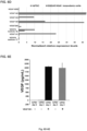
- the isolated mesoderm cell population is substantially pure. In one embodiment, 28% of total live cells at day 4 of mesoderm differentiation are KDR+NCAM+APLNR+. In one embodiment, 18% of total live cells at day 4 of mesoderm differentiation are SSEA5-KDR+NCAM+APLNR+.
- an isolated population of human NRP-1+/CD31+ ECFC-like cells is provided.
- the purified human cell population of NRP-1+/CD31+ ECFC-like cells provided is generated using the in vitro method for generating ECFC-like cells from hPSCs disclosed herein.
- the method disclosed herein is used to generate a purified human cell population of NRP-1+ and CD31+ ECFC-like cells, from an isolated subset of mesoderm cells.
- the isolated ECFC-like cells of the population exhibit cobblestone morphology and have a capacity for blood vessel formation in vivo without co-culture and/or co-implanted cells.
- the ECFC-like cells of the population are further characterized by one or more of CD144+, KDR+ and a-SMA-.
- At least some of the ECFC-like cells in the population generated from the isolated mesoderm cells have a high proliferation potential that is similar to the proliferation potential of ECFC's generated in vitro using the inventor's previous hPSC-ECFC- lice cell protocol.
- the isolated ECFC-like cell population is substantially pure.
- cells in the ECFC-like cell populations generated from the disclosed mesoderm cells can form blood vessels when implanted in vivo in a mammal, even in the absence of supportive cells.
- the capacity to form blood vessels in vivo in the absence of exogenous supportive cells is one indicator that the cells produced using the methods disclosed herein are ECFCs.
- the mesoderm cells generated using the method disclosed herein can be generated in vitro and used to form blood vessel tissue in vivo for various clinical applications, as described below.
- the ECFC-like cells generated using the method disclosed herein can be generated in vitro in a volume that can be useful for various clinical applications, as described below.
- hES Human Embryonic stem cell
- fibroblast-derived human iPS cell line DF19-9-11T
- WiCell Research institute WiCell Research institute (Madison, Wisconsin).
- Both hES and hiPSCs were maintained in mTeSR1 complete media (Stem Cell Technologies) on Matrigel in 10 cm 2 tissue culture dishes at 37°C and 5 % CO 2 . After the plating of cells, media was changed on days 2, 3, and 4. Cells were passaged on Day 5.
- Dispase containing media was aspirated from the plate and cells were gently washed with DMEM-F12 (Gibco) 3 times to remove any residual amount of enzyme. Fresh media was then used to collect colonies from the plate using a forceful wash and scraping with a 5 ml disposable pipette taking care to avoid bubbles. Collected colonies were then centrifuged at 300 x g for 5 minutes. The supernatant was aspirated and pellet was resuspended in mTeSR1 complete media.
- KNA+ mesoderm cells or SSEA5-KNA+ mesoderm cells into the EC lineage, including ECFC-like cells Day 4 sorted mesoderm cells (KDR+NCAM+APLNR+ or SSEA5-KDR+NCAM+APLNR+) were further cultured with 8 ml of Stemline II complete media (Sigma) containing FGF-2 (Stemgent), VEGF 165 (R&D) and BMP4 (R&D), which was replaced on days 6, 8, 10 and 12. On day 10 and thereafter media was changed with 10 ml of Stemline II differentiation media.
- Flow cytometry At day 12 after differentiation, adherent cells were harvested using TrypleE and made into a single cell suspension in EGM-2 medium. Cells were counted and aliquots of the cell suspension were prepared for antibody staining. FcR blocking reagent (Miltyni Biotech) was added to prevent the non-specific binding of antibodies.
- Anti-human CD31 CD31-FITC, clone WM59 from BO Pharmingen
- CD144 CD144-PE, clone 16B1 from ebioscience
- NRP-1 NRP-1-APC, clone AD5-176 from Miltenyi Biotech
- Propidium Iodide (PI, Sigma) was added to the cell suspension for dead cell staining.
- Flow cytometric detection of the cell surface antigens and cells sorting were performed on an ISR II and FACS Aria (Becton Dickinson) respectively. Compensation was set by single positive controls using cord blood derived ECFCs. A gating of targeted cell population was determined based on fluorescent minus one (FMO) controls for each fluorescent color.
- Ce ll culture of sorted cells CD31 ⁇ CD144+ or KDR+ and NRP-1+ sorted cells were centrifuged at 300 X g for 5 minutes then resuspended in 50% EGM-2 and 50% complete Stemline II differentiation media.
- EGM-2 EGM-2
- complete Stemline II differentiation media EGM-2 and 50% complete Stemline II differentiation media.
- 2500 cells per well were seeded on rat tail type I collagen coated 12 well plates. After 2 days, the media was aspirated and three parts of EGM-2 and one part of differentiation media was added to the cultures.
- ECFC colonies appeared as tightly adherent cells and exhibited cobblestone morphology on day 7. On occasion, cloning cylinders were used to isolate ECFC colonies from heterogeneous cell populations.
- Endothelial cell clusters Cloning of endothelial cell clusters was performed to isolate pure populations of highly proliferative endothelial cells as described previously (Yoder et al., 2007; Ingram et al., 2005). Confluent ECFCS were passage by plating 10,000 cells per cm 2 as a seeding density and maintain ECFCs in complete endothelial growth media (collagen coated plates and cEGM-2 media) with media change every other day as described previously (Yoder et al., 2007; Ingram et al., 2005).
- ECFCs were fixed with 4% (w/v) paraformaldehyde for 30 minutes and permeabilized with 0.1% (v/v) TritonX-100 in PBS for 5 minutes. After blocking with 10% (v/v) goat serum for 30 min, cells were incubated with primary following antibodies; anti- CD31 (Santa Cruz), anti-CD144 (ebioscience), anti-NRP-1 (Santa Cruz) and anti-a-SMA, (Chemicon) overnight at 4°C.
- mice All animal procedures were carried in accordance with the Guidelines for the Care and Use of Laboratory Animals and were approved by the Institutional Animal Care and Use Committees (IACUCs) at Indiana University School of Medicine (IACUC protocol# 10850). Both male and female 6-12 week old NOD/SCID mice (T- and B-cell deficient, impaired complement) were used for all animal studies. NOD-SCID mice were maintained under specific- pathogen-free conditions at the Indiana University Laboratory Animal Resource Center (LARC). Previous work with this animal model was used to determine the minimum number of animals needed to obtain statistically significant results (Yoder et al., 2007).
- IACUCs Institutional Animal Care and Use Committees
- LOC Indiana University Laboratory Animal Resource Center
- Pig skin type I collagen was used to generate three-dimensional (3D) cellularized collagen matrices as previously described (Critser et al., 2010). Briefly, type 1 collagen gel mixture was prepared by mixing together ice-cold porcine skin collagen solution, 10% v/v human platelet lysate in 0.01N HCL, and neutralized with phosphate buffered saline and 0.1N NaOH to achieve neutral pH (7.4). Neutralized gel mixtures ( ⁇ 1.5 mg/ml) were kept on ice before induction of polymerization by warming at 37°C, in 5% CO 2 .
- KNA+ mesoderm cells or SSEA-KNA+ mesoderm cells or SSEA-KNA+-derived NRP-1+CD31+ ECFCs were added to the collagen mixture to a final concentration of four million cells/ml collagen.
- the collagen mixture (250 ⁇ L) containing the cell suspension was added to 48-well tissue culture dishes and was allowed to polymerize to form gels by incubation in a CO 2 incubator at 37°C for 30 minutes. The gels were then overlaid with 500 ⁇ l of culture medium for 30 min at 37°C, in 5% CO 2 .
- cellularized gels were implanted into the flanks (a bluntly dissected subcutaneous pouch of anterior abdominal wall with close proximity of host vasculature) of 6- to 12-week-old NOD/SCID mice as previously described ⁇ Yoder, 2007 #71 ⁇ . Surgical procedures to implant collagen gels were conducted under anesthesia and constant supply of oxygen. Incisions were sutured and mice monitored for recovery. Various days after implantation, gels were recovered by excising engrafts in animals that had been humanely sacrificed per approved IACUC protocol.
- RT Reverse transcriptase
- mRNA RT reactions were performed in a GeneAmp PCR 9700 Thermocycler (Applied Biosystems). mRNA RT reactions were performed using Transcriptor Univessal cDNA Master (Roche). Specific miRNA primers were used for specific miRNA of interest for generating cDNA. RT reactions without templates or primer were used as controls. Gene and miRNA expression levels were quantified using the ABI 7300 RT-PCR System (Applied Biosytems). Quantitative PCR for mRNA was performed using FastStart Universal SYBR green master (Rox) (Roche). Comparative real-time PCR with or without specific primers for miRNAs and mRNAs was performed in triplicates.
- mRNA reactions were performed at 95°C for 1O min, followed by 40 cycles of 95°C for 15 s and 60°C for 1 min. miRNA reactions were performed by 40 cycles of 95°C for 15 sand 60°C for 1 min. Relative expression levels were calculated using the comparative Ct method (Lee et al., 2013).
- ECFC single cell proliferation assays KNA+ or SSEA5-KNA+ mesoderm cell-derived ECFCs were subjected to a single cell assay to evaluate clonogenic proliferative potential.
- endothelial cells were treated with trypLE Express (Invitrogen) to obtain a single cell suspension.
- Cell counts and serial dilutions were performed to obtain a concentration of 0.68 cells per well in individual wells of 96-well culture plates.
- Wells were examined the day after plating to ensure the presence of a single cell per well.
- Culture media was changed on days 4, 8, and 12.
- cells were stained with Sytox reagent (Invitrogen), and each well was examined to quantitate the number of cells using a fluorescent microscope.
- Nonspecific binding was blocked with blocking buffer for 1 hr at room temperature and incubated overnight at 4°C with primary antibodies against phospho-PYK2 (1:1,000; Cell Signaling) and phospho-p130cas (1:1,000; Cell Signaling) in Odyssey blocking buffer. Blots were washed with PBS containing 0.1% Tween20, followed by incubation for 1 hour at room temperature with anti-rabbit antibody (1:10,000; LI-COR). Immunoreactive bands were detected using the Odyssey Infrared Imager (LI-COR).
- Fc-NRP-1 treatment assay After 2 days (-D2) of culture of hiPSC clumps in mTeSR1 media, cultures were directed toward the mesodermal lineage with addition of activin A (10 ng/ml) in the presence of FGF-2, VEGF 165 , and BMP4 (10 ng/ml) for 24 hrs. The following day (D1), activin-A containing media was removed and replaced with 8 ml of Stemline II complete media (Sigma) containing FGF-2 (Stemgent), VEGF 165 (R&D) and BMP4 (R&D) and 500 ng/ml of Fc-NRP-1 (R&D).
- miRNA microarray and RNA sequence analysis Total RNA was isolated from the samples using Trizol reagent (Invitrogen) and the RNA quality was examined as previously described ( Ginsberg et al., 2012, Cell 151:559-575 ).
- Trizol reagent Invitrogen
- miRNome miRNA PCR Array plates miScript 11 Reverse Transcription reaction kits, and miScript SYBR Green PCR Kits (all from Qiagen) were used to examine the expression profiles of the 1008 most abundantly expressed and best characterized miRNA sequences in the human miRNA genome (miRNome) as annotated in miRBase Release 16.
- RNA sequence library was generated using 1 ⁇ g of high quality total RNA and sequencing was performed using Illumina HiSeq2000 sequencer as previously described (Ginsberg et al., 2012). The resulting sequence reads were mapped to the human genome (hg18) using TopHat with default parameters, and the RefSeq (June 2010) transcript levels (FPKMs) were quantified using Cufflinks.
- miRNA mimic / inhibitor treatment assay After 1 day (-D1) of culture of single cell suspension of hiPSCs in mTeSR1 media, cultures were directed toward the mesodermal lineage with addition of activin A (10 ng/ml) in the presence of FGF-2, VEGF 165 , and BMP4 (10 ng/ml) and 2.5 ⁇ g mimic or inhibitor/100,000 seeded cells/well of 6 well plate (GE Dharacon) for 24 hrs. The following day (D1), activin-A and miRNA mimic or inhibitor containing media was removed and replaced with 2 ml of Stemline II complete media (Sigma) containing FGF-2 (Stemgent), VEGF 165 (R&D) and BMP4 (R&D).
- NCAM and APLNR co-expressing cells within day 4 gave rise to NRP-1+CD31+ endothelial cells with ECFC competence.
- PSCs cultured in mTeSR1 were induced to differentiate into mesoderm cells under 2D, serum and feeder-free conditions.
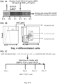
- PSCs were cultured in Stemline-ll medium with FGF-2 (10ng/ml), BMP4 (20 ng/ml), VEGF165 (10 ng/ml) and Activin-A (10 ng/ml) for 24 hours (i.e., from D0-D1) ( FIG. 1A ).
- the cell culture medium was replaced with Stemline- II medium with FGF-2 (10ng/ml), BMP4 (20 ng/ml), VEGF 165 (10 ng/ml). This replacement medium was used to culture the cells undergoing mesodermal induction for 3 days (i.e., from D1-D4).
- KDR+ cells were gated for NCAM and APLNR expression.
- KDR+NCAM+APLNR+ (K+N+A+, also referred to herein as KNA+) and KDR+NCAM+APLNR- (K+N+A) KDR+NCAM-APLNR- (K+N-A ) cells were sorted for further differentiation and examination for the emergence of NRP- 1+CD31+ ECFC-like cells ( FIG. 1C ).
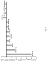
- K+N+A+ mesoderm-derived NRP-1+CD31+ cells exhibited high clonal proliferative potential with a hierarchy of colonies ranging from clusters of 2-50 cells up to colonies of >2001 similar to that of hiPSC-ECFC-like cells ( FIG. 1E ).
- cells isolated from the other two subsets i.e., K+N+A and K+N-A
- K+N+A+ mesoderm-derived NRP-1+CD31+ cells produced robust in vivo human blood vessels filled with host murine red blood cells ( FIG. 1F , top right panel; arrows point to blood vessels) similar to those produced by hiPSC-ECFC-like cells ( FIG.
- FIG. 1F top left panel
- K+N+A and K+N-A cells isolated from the other two subsets
- FIG. 1F bottom left and right panels, respectfully; arrows point to hCD31+ functional blood vessels.
- functional hCD31+ blood vessels were generated by K+N+A+ mesoderm-derived NRP-1+CD31+ cells hiPSC-ECFC-like cells, but not by K+N+A or K+N-A mesoderm cells ( FIG. 1G ).
- human iPS cells after 3-5 days of differentiation using the above culture protocol generate cells expressing mesoderm markers and lacking typical endothelial surface expression.
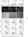
- APLNR+ cells were found only in the KDR+NCAM+ mesoderm sub-set and were absent in KDR-NCAM- sub-set ( FIG. 2A ) and KDR+ cells were found at highest levels at day 3 and 4 and decreased over time ( FIG. 2B ).
- Typical endothelial marker (CD31, NRP-1 and CD144) expression was not found in any day 4 differentiated cells ( FIG. 2C ).
- KDR+ cells were gated for NCAM and APLNR expression.
- APLNR+ and APLNR- mesoderm cells were sorted for further direct in vivo implantation and examination ( FIG. 3A ). Sorted APLNR- mesoderm cells produced teratomas after 2 months of in vivo implantation ( FIG. 3B , left panel).
- the APLNR+ mesoderm sub-set ( FIG. 3B , right panel) formed robust in vivo human blood vessels filled with host murine red blood cells (blue open arrows) with accompanying endoderm-derived cell like derivatives (pink closed arrows).
- SSEA5-KDR+NCAM+APLNR+ D4 SSEA5 depleted KNA+ mesoderm cells
- SSEA5-KNA+ D4 SSEA5 depleted KNA+ mesoderm cells
- SSEA5-KDR+ cells Day 4 differentiated hiPSCs were first gated for SSEA5 and KDR expression ( FIG. 4A , left).
- the SSEA5-KDR+ cells were gated for NCAM and APLNR expression ( FIG. 4A , right).
- SSEA5-KDR+NCAM+APLNR+ (SSEA5-KNA+) and SSEA5-KDR+NCAM+APLNR- (SSEA5-KNA) cells were sorted for further analysis.
- SSEA5-KNA+ cells formed robust functional in vivo vessels ( FIG. 4B , blue arrows, left panel), SSEA5-KNA cells failed to form robust in vivo vessels ( FIG. 4B , white arrows, right panel).
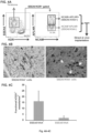
- day 4 SSEA5 depleted KNA+ mesoderm cells displayed transcripts typically enriched in lateral plate/extra-embryonic mesoderm cells, and exhibited enhanced formation of NRP-1+CD31+ cells with ECFC competence upon in vitro ECFC differentiation.
- SSEA5-KNA+ cells over-expressed lateral plate-extra-embryonic mesoderm markers, relative to hiPSCs, and lacked expression of axial, paraxial and intermediate mesoderm markers ( FIG. 5A ). Sorted SSEA5-KNA+ cells were further differentiated into the ECFC-like lineage for another 8 days (4 plus 8, total of 12 days).
- NRP1+CD31+ cells derived from the SSEA5-KNA+ mesoderm cells formed a homogenous cobblestone endothelial monolayer ( FIG. 5B , left middle panel), displayed uniform co-expression for CD31 and CD144 endothelial markers ( FIG. 5B , left middle panel), displayed uniform co-expression for CD31 and CD144 endothelial markers ( FIG. 5B , left middle panel).
- SSEA5-KNA+ mesoderm-derived NRP-1+CD31+ cells exhibited high clonal proliferative potential with a hierarchy of colonies ranging from clusters of 2-50 cells up to colonies of >2001 similar to that of hiPSC-ECFCs ( FIG. 5C ).
- cells isolated from SSEA5-KNA mesoderm sub-set failed to exhibit high clonal proliferative potential ( FIG. 5C ).
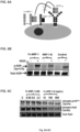
- Fc-NRP-1 mediates VEGF- KDR signaling through p130cas/Pyk2 activation and enhances formation of SSEA5-KNA+ mesoderm cells from hiPSCs.
- NRP-1, KDR, Fc-NRP-1, NRP-1-B and VEGF 165 functions in endothelial cells are shown in FIG. 6A .
- NRP-1 functions as a VEGF 165 co-receptor and brings VEGF 165 to its receptor (KDR).
- Fc-NRP-1 acts as surrogate for membrane NRP-1 and binds and brings VEGF 165 to KDR.
- NRP-1-B blocking antibody selectively binds to the VEGF 165 binding site of NRP-1, thereby specifically blocking binding of VEGF 165 to NRP-1.
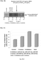
- KDR and p130cas/Pyk2 phosphorylation were carried out by Western blotting ( FIGS. 6B and 6C ).
- KDR phosphorylation was observed in VEGF stimulated groups and Fc-NRP-1 dimer treatment increased phosphorylation of KDR compared to control treated cells.
- decreased phosphorylation was observed in NRP-1-B treated cells.
- increased p130cas/Pyk2 phosphorylation was observed in Fc-NRP-1 dimer treated cells compared to NRP-1-B treated cells.
- VEGF-A isoforms Gene expression of VEGF-A isoforms in hiPSCs and SSEA5-KNA+ mesoderm cells was investigated. VEGF-A isoforms were not up-regulated in SSEA5-KNA+ mesoderm cells compared to hiPSCs ( FIGS. 6D and 6E ).
- FIG. 6F A mesoderm lineage differentiation protocol that involves culturing the cells undergoing mesoderm induction in the presence of Fc-NRP-1 dimer and growth factors is shown in FIG. 6F .
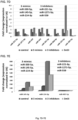
- Dimeric Fc-NRP-1 treatment to differentiating hiPSCs caused more than a 2- fold increase in the production of SSEA5-KNA+ mesoderm cells compared to Fc-NRP-1- untreated cells ( FIG. 6G ).
- miRNA and mRNA transcripts revealed 12 miRs (miR-221-3p, miR-1271-Sp, miR-559, miR-543, miR-361-3p, miR-30d-5p, miR-124-3p, miR-185-Sp, miR-330-Sp, miR-145-Sp, miR-214-3p and miR-497-Sp) with published and validated targets that potentially regulate transcription factors relevant to SSEA5-KNA+ mesoderm formation from hiPSCs.
- miRNAs (miR-221-3p, miR-1271-Sp, miR-559, miR-543, miR-361-3p, miR-30d-5p, miR-124-3p and miR-185-Sp) were expressed at lower levels in Day 4 SSEA5-KNA+ mesoderm cells compared to Day O undifferentiated hiPSCs, and four miRNAs (miR-330-Sp, miR-145-Sp, miR-214-3p and miR-497-Sp) were expressed at higher levels in Day 4 SSEA5-KNA+ mesoderm cells compared to Day O undifferentiated hiPSCs.
- miR-214-3p targets CLDN6 in differentiating hiPSC and enhances formation of SSEA5-KNA+ mesoderm cells.
- miR-214 is highly expressed in SSEA5-KNA+ cells compared to hiPSCs ( FIG. 8A , left bar) and CLDN6 is expressed at lower levels in SSEA5-KNA+ cells compared to hiPSCs ( FIG. 8A , right bar).
Landscapes
- Health & Medical Sciences (AREA)
- Life Sciences & Earth Sciences (AREA)
- Engineering & Computer Science (AREA)
- Biomedical Technology (AREA)
- Chemical & Material Sciences (AREA)
- Organic Chemistry (AREA)
- Bioinformatics & Cheminformatics (AREA)
- Biotechnology (AREA)
- Zoology (AREA)
- General Health & Medical Sciences (AREA)
- Genetics & Genomics (AREA)
- Wood Science & Technology (AREA)
- Cell Biology (AREA)
- Pharmacology & Pharmacy (AREA)
- Animal Behavior & Ethology (AREA)
- Public Health (AREA)
- Veterinary Medicine (AREA)
- Medicinal Chemistry (AREA)
- Developmental Biology & Embryology (AREA)
- General Engineering & Computer Science (AREA)
- Biochemistry (AREA)
- Microbiology (AREA)
- Nuclear Medicine, Radiotherapy & Molecular Imaging (AREA)
- General Chemical & Material Sciences (AREA)
- Chemical Kinetics & Catalysis (AREA)
- Reproductive Health (AREA)
- Gynecology & Obstetrics (AREA)
- Epidemiology (AREA)
- Virology (AREA)
- Immunology (AREA)
- Vascular Medicine (AREA)
- Cardiology (AREA)
- Heart & Thoracic Surgery (AREA)
- Urology & Nephrology (AREA)
- Pulmonology (AREA)
- Micro-Organisms Or Cultivation Processes Thereof (AREA)
- Medicines Containing Material From Animals Or Micro-Organisms (AREA)
- Measuring Or Testing Involving Enzymes Or Micro-Organisms (AREA)
- Materials For Medical Uses (AREA)
Applications Claiming Priority (2)
| Application Number | Priority Date | Filing Date | Title |
|---|---|---|---|
| US201662372907P | 2016-08-10 | 2016-08-10 | |
| PCT/US2017/045496 WO2018031404A1 (en) | 2016-08-10 | 2017-08-04 | Method for generating mesoderm and/or endothelial colony forming cell-like cells having in vivo blood vessel forming capacity |
Publications (3)
| Publication Number | Publication Date |
|---|---|
| EP3497204A1 EP3497204A1 (en) | 2019-06-19 |
| EP3497204A4 EP3497204A4 (en) | 2020-05-06 |
| EP3497204B1 true EP3497204B1 (en) | 2025-04-30 |
Family
ID=61162479
Family Applications (1)
| Application Number | Title | Priority Date | Filing Date |
|---|---|---|---|
| EP17840057.8A Active EP3497204B1 (en) | 2016-08-10 | 2017-08-04 | Method for generating mesoderm and/or endothelial colony forming cell-like cells having in vivo blood vessel forming capacity |
Country Status (10)
| Country | Link |
|---|---|
| US (2) | US11739293B2 (enExample) |
| EP (1) | EP3497204B1 (enExample) |
| JP (2) | JP7045363B2 (enExample) |
| KR (1) | KR102456031B1 (enExample) |
| CN (1) | CN109689858B (enExample) |
| AU (1) | AU2017308738B2 (enExample) |
| BR (1) | BR112019002584A2 (enExample) |
| CA (1) | CA3033381A1 (enExample) |
| IL (1) | IL264273B2 (enExample) |
| WO (1) | WO2018031404A1 (enExample) |
Families Citing this family (8)
| Publication number | Priority date | Publication date | Assignee | Title |
|---|---|---|---|---|
| CA3025517A1 (en) * | 2018-02-21 | 2019-08-21 | Indiana University Research And Technology Corporation | Compositions and methods for the treatment or prophylaxis of a perfusion disorder |
| EP4011518A4 (en) * | 2019-08-05 | 2022-09-28 | Nippon Steel Corporation | METHOD OF MANUFACTURE OF PRESS-MOLDED PRODUCT, PRESS-MOLDED PRODUCT AND PRESS-MOLDING APPARATUS |
| AU2020337444A1 (en) * | 2019-08-28 | 2022-03-24 | Astellas Institute For Regenerative Medicine | Compositions and methods of treating vascular diseases |
| GB202003309D0 (en) * | 2020-03-06 | 2020-04-22 | Imp College Innovations Ltd | Methods of generating endothelial cells |
| WO2022040798A1 (en) * | 2020-08-25 | 2022-03-03 | University Health Network | Compositions and methods for generating human yolk sac-like hematopoietic cells |
| CN115137740B (zh) * | 2022-03-29 | 2023-12-12 | 广州大学 | miRNA-497b或miRNA-5106在制备治疗缺血心肌的药物中的应用 |
| CN116769710A (zh) * | 2023-01-28 | 2023-09-19 | 深圳市寰宇生物科技有限公司 | 一种从人诱导性多能干细胞分化的巨噬细胞及其制备方法和应用 |
| CN117070442B (zh) * | 2023-10-18 | 2024-01-30 | 北京大学 | 静脉内皮细胞及其制备方法、静脉血管畸形模型及其制备方法 |
Family Cites Families (7)
| Publication number | Priority date | Publication date | Assignee | Title |
|---|---|---|---|---|
| KR20100110905A (ko) | 2007-07-20 | 2010-10-13 | 동국대학교 산학협력단 | 중간엽 줄기세포를 이용하여 모유두 조직을 제조하는 방법 |
| US20110236971A2 (en) | 2007-09-25 | 2011-09-29 | Maksym Vodyanyk | Generation of Clonal Mesenchymal Progenitors and Mesenchymal Stem Cell Lines Under Serum-Free Conditions |
| AU2009274517B2 (en) | 2008-07-25 | 2015-03-26 | The University Of Georgia Research Foundation, Inc. | Compositions for Mesoderm derived ISL1+ Multipotent cells (IMPs), epicardial progenitor cells (EPCs) and multipotent CXCR4+CD56+ cells (C56Cs) and methods of use |
| WO2013028684A1 (en) * | 2011-08-23 | 2013-02-28 | Wisconsin Alumni Research Foundation | Angiohematopoietic progenitor cells |
| US20150329821A1 (en) | 2012-10-19 | 2015-11-19 | Agency For Science, Technology And Research | Methods of differentiating stem cells into one or more cell lineages |
| WO2014145871A1 (en) * | 2013-03-15 | 2014-09-18 | The Johns Hopkins University | Self-organized vascular networks from human pluripotent stem cells in a synthetic matrix |
| CA2940691C (en) * | 2014-03-11 | 2022-10-25 | Indiana University Research And Technology Corporation | Method for generating endothelial colony forming cell-like cells |
-
2017
- 2017-08-04 WO PCT/US2017/045496 patent/WO2018031404A1/en not_active Ceased
- 2017-08-04 CA CA3033381A patent/CA3033381A1/en active Pending
- 2017-08-04 US US16/323,722 patent/US11739293B2/en active Active
- 2017-08-04 KR KR1020197006515A patent/KR102456031B1/ko active Active
- 2017-08-04 IL IL264273A patent/IL264273B2/en unknown
- 2017-08-04 JP JP2019507218A patent/JP7045363B2/ja active Active
- 2017-08-04 EP EP17840057.8A patent/EP3497204B1/en active Active
- 2017-08-04 AU AU2017308738A patent/AU2017308738B2/en active Active
- 2017-08-04 BR BR112019002584-7A patent/BR112019002584A2/pt not_active Application Discontinuation
- 2017-08-04 CN CN201780048596.4A patent/CN109689858B/zh active Active
-
2022
- 2022-03-18 JP JP2022044157A patent/JP2022081657A/ja active Pending
-
2023
- 2023-01-26 US US18/159,725 patent/US20230174929A1/en not_active Abandoned
Non-Patent Citations (1)
| Title |
|---|
| MAXIM A VODYANIK ET AL: "A mesoderm-derived precursor for mesenchymal stem and endothelial cells", CELL STEM CELL, ELSEVIER, CELL PRESS, AMSTERDAM, NL, vol. 7, no. 6, 3 December 2010 (2010-12-03), pages 718 - 729, XP002638197, ISSN: 1934-5909, [retrieved on 20101202], DOI: 10.1016/J.STEM.2010.11.011 * |
Also Published As
| Publication number | Publication date |
|---|---|
| KR102456031B1 (ko) | 2022-10-17 |
| CN109689858B (zh) | 2024-07-26 |
| JP2022081657A (ja) | 2022-05-31 |
| JP2019528057A (ja) | 2019-10-10 |
| IL264273B1 (en) | 2024-04-01 |
| EP3497204A1 (en) | 2019-06-19 |
| US20190211304A1 (en) | 2019-07-11 |
| IL264273B2 (en) | 2024-08-01 |
| KR20190037299A (ko) | 2019-04-05 |
| AU2017308738A1 (en) | 2019-01-31 |
| CA3033381A1 (en) | 2018-02-15 |
| WO2018031404A1 (en) | 2018-02-15 |
| JP7045363B2 (ja) | 2022-03-31 |
| US11739293B2 (en) | 2023-08-29 |
| US20230174929A1 (en) | 2023-06-08 |
| BR112019002584A2 (pt) | 2019-05-21 |
| IL264273A (en) | 2019-02-28 |
| AU2017308738B2 (en) | 2023-09-21 |
| CN109689858A (zh) | 2019-04-26 |
| EP3497204A4 (en) | 2020-05-06 |
Similar Documents
| Publication | Publication Date | Title |
|---|---|---|
| EP3497204B1 (en) | Method for generating mesoderm and/or endothelial colony forming cell-like cells having in vivo blood vessel forming capacity | |
| Bruce et al. | In vitro differentiation of murine embryonic stem cells toward a renal lineage | |
| US7247477B2 (en) | Methods for the in-vitro identification, isolation and differentiation of vasculogenic progenitor cells | |
| US8507275B2 (en) | Method of inducing differentiation of embryonic stem cells into hemangioblast | |
| KR102368751B1 (ko) | 모양체 주연부 간세포의 제조 방법 | |
| WO2009116893A1 (ru) | Способ получения эндотелиальных клеток (варианты) | |
| KR102379045B1 (ko) | 내피 집락 형성 세포유사 세포를 생성하는 방법 | |
| JP6893633B2 (ja) | 分化細胞の抽出方法 | |
| WO2006066320A1 (en) | Differentiation of human embryonic stem cells and cardiomyocytes and cardiomyocyte progenitors derived therefrom | |
| HK40007852A (en) | Method for generating mesoderm and/or endothelial colony forming cell-like cells having in vivo blood vessel forming capacity | |
| HK40007852B (zh) | 用於产生具有体内血管形成能力的中胚层和/或内皮集落形成细胞样细胞的方法 | |
| Poudel et al. | Increased cardiogenesis in P19-GFP teratocarcinoma cells expressing the propeptide IGF-1Ea | |
| Collins-Hooper et al. | Efficient myogenic reprogramming of adult white fat stem cells and bone marrow stem cells by freshly isolated skeletal muscle fibers | |
| EP4715043A1 (en) | Method for producing neural crest cells | |
| Akram et al. | Activindirected differentiation of human embryonic stem cells differentially modulates alveolar epithelial wound repair via paracrine mechanism | |
| Luo | MicroRNAs play a role in human embryonic stem cell differentiation into endothelial cells | |
| Dewing | Reprogramming of primary human fetal fibroblasts towards cardiomyoctes | |
| Bauwens | A scalable bioprocess for generating embyronic stem cell derived cardiomyocytes | |
| Dawson | Cardiac Tissue Engineering | |
| Shafa et al. | Stirred Suspension Bioreactor Suppresses ESC Differentiation | |
| AU2005318931A1 (en) | Differentiation of human embryonic stem cells and cardiomyocytes and cardiomyocyte progenitors derived therefrom | |
| KR20120051907A (ko) | 유도만능줄기세포를 cd34 양성 세포로 분화시키는 방법 |
Legal Events
| Date | Code | Title | Description |
|---|---|---|---|
| STAA | Information on the status of an ep patent application or granted ep patent |
Free format text: STATUS: THE INTERNATIONAL PUBLICATION HAS BEEN MADE |
|
| PUAI | Public reference made under article 153(3) epc to a published international application that has entered the european phase |
Free format text: ORIGINAL CODE: 0009012 |
|
| STAA | Information on the status of an ep patent application or granted ep patent |
Free format text: STATUS: REQUEST FOR EXAMINATION WAS MADE |
|
| 17P | Request for examination filed |
Effective date: 20190305 |
|
| AK | Designated contracting states |
Kind code of ref document: A1 Designated state(s): AL AT BE BG CH CY CZ DE DK EE ES FI FR GB GR HR HU IE IS IT LI LT LU LV MC MK MT NL NO PL PT RO RS SE SI SK SM TR |
|
| AX | Request for extension of the european patent |
Extension state: BA ME |
|
| DAV | Request for validation of the european patent (deleted) | ||
| DAX | Request for extension of the european patent (deleted) | ||
| A4 | Supplementary search report drawn up and despatched |
Effective date: 20200408 |
|
| RIC1 | Information provided on ipc code assigned before grant |
Ipc: C12N 5/0735 20100101ALI20200402BHEP Ipc: A61K 35/12 20150101ALI20200402BHEP Ipc: C12N 5/074 20100101ALI20200402BHEP Ipc: C12N 5/071 20100101ALI20200402BHEP Ipc: C12N 5/07 20100101AFI20200402BHEP |
|
| STAA | Information on the status of an ep patent application or granted ep patent |
Free format text: STATUS: EXAMINATION IS IN PROGRESS |
|
| 17Q | First examination report despatched |
Effective date: 20210726 |
|
| REG | Reference to a national code |
Ref country code: DE Ref legal event code: R079 Ref document number: 602017089233 Country of ref document: DE Free format text: PREVIOUS MAIN CLASS: C12N0005070000 Ipc: A61P0011000000 |
|
| RIC1 | Information provided on ipc code assigned before grant |
Ipc: A61K 35/12 20150101ALI20241010BHEP Ipc: C12N 5/071 20100101ALI20241010BHEP Ipc: A61P 9/00 20060101ALI20241010BHEP Ipc: A61P 13/12 20060101ALI20241010BHEP Ipc: A61P 11/00 20060101AFI20241010BHEP |
|
| GRAP | Despatch of communication of intention to grant a patent |
Free format text: ORIGINAL CODE: EPIDOSNIGR1 |
|
| STAA | Information on the status of an ep patent application or granted ep patent |
Free format text: STATUS: GRANT OF PATENT IS INTENDED |
|
| INTG | Intention to grant announced |
Effective date: 20241128 |
|
| GRAS | Grant fee paid |
Free format text: ORIGINAL CODE: EPIDOSNIGR3 |
|
| GRAA | (expected) grant |
Free format text: ORIGINAL CODE: 0009210 |
|
| STAA | Information on the status of an ep patent application or granted ep patent |
Free format text: STATUS: THE PATENT HAS BEEN GRANTED |
|
| AK | Designated contracting states |
Kind code of ref document: B1 Designated state(s): AL AT BE BG CH CY CZ DE DK EE ES FI FR GB GR HR HU IE IS IT LI LT LU LV MC MK MT NL NO PL PT RO RS SE SI SK SM TR |
|
| REG | Reference to a national code |
Ref country code: CH Ref legal event code: EP Ref country code: GB Ref legal event code: FG4D |
|
| REG | Reference to a national code |
Ref country code: DE Ref legal event code: R096 Ref document number: 602017089233 Country of ref document: DE |
|
| REG | Reference to a national code |
Ref country code: IE Ref legal event code: FG4D |
|
| P01 | Opt-out of the competence of the unified patent court (upc) registered |
Free format text: CASE NUMBER: APP_26716/2025 Effective date: 20250605 |
|
| REG | Reference to a national code |
Ref country code: NL Ref legal event code: MP Effective date: 20250430 |
|
| REG | Reference to a national code |
Ref country code: AT Ref legal event code: MK05 Ref document number: 1789508 Country of ref document: AT Kind code of ref document: T Effective date: 20250430 |
|
| PG25 | Lapsed in a contracting state [announced via postgrant information from national office to epo] |
Ref country code: PT Free format text: LAPSE BECAUSE OF FAILURE TO SUBMIT A TRANSLATION OF THE DESCRIPTION OR TO PAY THE FEE WITHIN THE PRESCRIBED TIME-LIMIT Effective date: 20250901 Ref country code: FI Free format text: LAPSE BECAUSE OF FAILURE TO SUBMIT A TRANSLATION OF THE DESCRIPTION OR TO PAY THE FEE WITHIN THE PRESCRIBED TIME-LIMIT Effective date: 20250430 Ref country code: ES Free format text: LAPSE BECAUSE OF FAILURE TO SUBMIT A TRANSLATION OF THE DESCRIPTION OR TO PAY THE FEE WITHIN THE PRESCRIBED TIME-LIMIT Effective date: 20250430 |
|
| PGFP | Annual fee paid to national office [announced via postgrant information from national office to epo] |
Ref country code: DE Payment date: 20250827 Year of fee payment: 9 |
|
| REG | Reference to a national code |
Ref country code: LT Ref legal event code: MG9D |
|
| PG25 | Lapsed in a contracting state [announced via postgrant information from national office to epo] |
Ref country code: GR Free format text: LAPSE BECAUSE OF FAILURE TO SUBMIT A TRANSLATION OF THE DESCRIPTION OR TO PAY THE FEE WITHIN THE PRESCRIBED TIME-LIMIT Effective date: 20250731 Ref country code: NO Free format text: LAPSE BECAUSE OF FAILURE TO SUBMIT A TRANSLATION OF THE DESCRIPTION OR TO PAY THE FEE WITHIN THE PRESCRIBED TIME-LIMIT Effective date: 20250730 |
|
| PG25 | Lapsed in a contracting state [announced via postgrant information from national office to epo] |
Ref country code: PL Free format text: LAPSE BECAUSE OF FAILURE TO SUBMIT A TRANSLATION OF THE DESCRIPTION OR TO PAY THE FEE WITHIN THE PRESCRIBED TIME-LIMIT Effective date: 20250430 Ref country code: NL Free format text: LAPSE BECAUSE OF FAILURE TO SUBMIT A TRANSLATION OF THE DESCRIPTION OR TO PAY THE FEE WITHIN THE PRESCRIBED TIME-LIMIT Effective date: 20250430 |
|
| PG25 | Lapsed in a contracting state [announced via postgrant information from national office to epo] |
Ref country code: BG Free format text: LAPSE BECAUSE OF FAILURE TO SUBMIT A TRANSLATION OF THE DESCRIPTION OR TO PAY THE FEE WITHIN THE PRESCRIBED TIME-LIMIT Effective date: 20250430 |
|
| PGFP | Annual fee paid to national office [announced via postgrant information from national office to epo] |
Ref country code: GB Payment date: 20250827 Year of fee payment: 9 |
|
| PG25 | Lapsed in a contracting state [announced via postgrant information from national office to epo] |
Ref country code: HR Free format text: LAPSE BECAUSE OF FAILURE TO SUBMIT A TRANSLATION OF THE DESCRIPTION OR TO PAY THE FEE WITHIN THE PRESCRIBED TIME-LIMIT Effective date: 20250430 |
|
| PG25 | Lapsed in a contracting state [announced via postgrant information from national office to epo] |
Ref country code: AT Free format text: LAPSE BECAUSE OF FAILURE TO SUBMIT A TRANSLATION OF THE DESCRIPTION OR TO PAY THE FEE WITHIN THE PRESCRIBED TIME-LIMIT Effective date: 20250430 |
|
| PGFP | Annual fee paid to national office [announced via postgrant information from national office to epo] |
Ref country code: FR Payment date: 20250825 Year of fee payment: 9 |
|
| PGFP | Annual fee paid to national office [announced via postgrant information from national office to epo] |
Ref country code: CH Payment date: 20250901 Year of fee payment: 9 |
|
| PG25 | Lapsed in a contracting state [announced via postgrant information from national office to epo] |
Ref country code: RS Free format text: LAPSE BECAUSE OF FAILURE TO SUBMIT A TRANSLATION OF THE DESCRIPTION OR TO PAY THE FEE WITHIN THE PRESCRIBED TIME-LIMIT Effective date: 20250731 |
|
| PG25 | Lapsed in a contracting state [announced via postgrant information from national office to epo] |
Ref country code: IS Free format text: LAPSE BECAUSE OF FAILURE TO SUBMIT A TRANSLATION OF THE DESCRIPTION OR TO PAY THE FEE WITHIN THE PRESCRIBED TIME-LIMIT Effective date: 20250830 |
|
| PG25 | Lapsed in a contracting state [announced via postgrant information from national office to epo] |
Ref country code: LV Free format text: LAPSE BECAUSE OF FAILURE TO SUBMIT A TRANSLATION OF THE DESCRIPTION OR TO PAY THE FEE WITHIN THE PRESCRIBED TIME-LIMIT Effective date: 20250430 |
|
| PG25 | Lapsed in a contracting state [announced via postgrant information from national office to epo] |
Ref country code: SM Free format text: LAPSE BECAUSE OF FAILURE TO SUBMIT A TRANSLATION OF THE DESCRIPTION OR TO PAY THE FEE WITHIN THE PRESCRIBED TIME-LIMIT Effective date: 20250430 Ref country code: DK Free format text: LAPSE BECAUSE OF FAILURE TO SUBMIT A TRANSLATION OF THE DESCRIPTION OR TO PAY THE FEE WITHIN THE PRESCRIBED TIME-LIMIT Effective date: 20250430 |
|
| PG25 | Lapsed in a contracting state [announced via postgrant information from national office to epo] |
Ref country code: CZ Free format text: LAPSE BECAUSE OF FAILURE TO SUBMIT A TRANSLATION OF THE DESCRIPTION OR TO PAY THE FEE WITHIN THE PRESCRIBED TIME-LIMIT Effective date: 20250430 |
|
| PG25 | Lapsed in a contracting state [announced via postgrant information from national office to epo] |
Ref country code: EE Free format text: LAPSE BECAUSE OF FAILURE TO SUBMIT A TRANSLATION OF THE DESCRIPTION OR TO PAY THE FEE WITHIN THE PRESCRIBED TIME-LIMIT Effective date: 20250430 |
|
| PG25 | Lapsed in a contracting state [announced via postgrant information from national office to epo] |
Ref country code: SK Free format text: LAPSE BECAUSE OF FAILURE TO SUBMIT A TRANSLATION OF THE DESCRIPTION OR TO PAY THE FEE WITHIN THE PRESCRIBED TIME-LIMIT Effective date: 20250430 Ref country code: RO Free format text: LAPSE BECAUSE OF FAILURE TO SUBMIT A TRANSLATION OF THE DESCRIPTION OR TO PAY THE FEE WITHIN THE PRESCRIBED TIME-LIMIT Effective date: 20250430 |
|
| PG25 | Lapsed in a contracting state [announced via postgrant information from national office to epo] |
Ref country code: IT Free format text: LAPSE BECAUSE OF FAILURE TO SUBMIT A TRANSLATION OF THE DESCRIPTION OR TO PAY THE FEE WITHIN THE PRESCRIBED TIME-LIMIT Effective date: 20250430 |
|
| REG | Reference to a national code |
Ref country code: DE Ref legal event code: R097 Ref document number: 602017089233 Country of ref document: DE |
|
| PLBE | No opposition filed within time limit |
Free format text: ORIGINAL CODE: 0009261 |
|
| STAA | Information on the status of an ep patent application or granted ep patent |
Free format text: STATUS: NO OPPOSITION FILED WITHIN TIME LIMIT |
|
| REG | Reference to a national code |
Ref country code: CH Ref legal event code: L10 Free format text: ST27 STATUS EVENT CODE: U-0-0-L10-L00 (AS PROVIDED BY THE NATIONAL OFFICE) Effective date: 20260311 |
|
| PG25 | Lapsed in a contracting state [announced via postgrant information from national office to epo] |
Ref country code: MC Free format text: LAPSE BECAUSE OF FAILURE TO SUBMIT A TRANSLATION OF THE DESCRIPTION OR TO PAY THE FEE WITHIN THE PRESCRIBED TIME-LIMIT Effective date: 20250430 |
|
| 26N | No opposition filed |
Effective date: 20260202 |
|
| PG25 | Lapsed in a contracting state [announced via postgrant information from national office to epo] |
Ref country code: LU Free format text: LAPSE BECAUSE OF NON-PAYMENT OF DUE FEES Effective date: 20250804 |