EP3355907B1 - Protéines thérapeutiques bispécifiques pour la réparation de tissus - Google Patents
Protéines thérapeutiques bispécifiques pour la réparation de tissus Download PDFInfo
- Publication number
- EP3355907B1 EP3355907B1 EP16852701.8A EP16852701A EP3355907B1 EP 3355907 B1 EP3355907 B1 EP 3355907B1 EP 16852701 A EP16852701 A EP 16852701A EP 3355907 B1 EP3355907 B1 EP 3355907B1
- Authority
- EP
- European Patent Office
- Prior art keywords
- cases
- seq
- tissue
- acid sequence
- variant
- Prior art date
- Legal status (The legal status is an assumption and is not a legal conclusion. Google has not performed a legal analysis and makes no representation as to the accuracy of the status listed.)
- Active
Links
- 108090000623 proteins and genes Proteins 0.000 title claims description 373
- 102000004169 proteins and genes Human genes 0.000 title claims description 368
- 230000017423 tissue regeneration Effects 0.000 title claims description 28
- 230000001225 therapeutic effect Effects 0.000 title description 45
- 210000004027 cell Anatomy 0.000 claims description 320
- 108090000765 processed proteins & peptides Proteins 0.000 claims description 236
- 239000012190 activator Substances 0.000 claims description 229
- 230000008685 targeting Effects 0.000 claims description 222
- 125000003275 alpha amino acid group Chemical group 0.000 claims description 206
- 210000001519 tissue Anatomy 0.000 claims description 192
- 230000027455 binding Effects 0.000 claims description 160
- 102000037865 fusion proteins Human genes 0.000 claims description 159
- 108020001507 fusion proteins Proteins 0.000 claims description 159
- 108090000672 Annexin A5 Proteins 0.000 claims description 106
- 102000004121 Annexin A5 Human genes 0.000 claims description 103
- 101000599951 Homo sapiens Insulin-like growth factor I Proteins 0.000 claims description 100
- 150000007523 nucleic acids Chemical class 0.000 claims description 89
- 108091006905 Human Serum Albumin Proteins 0.000 claims description 65
- 102000008100 Human Serum Albumin Human genes 0.000 claims description 65
- 238000006467 substitution reaction Methods 0.000 claims description 65
- 239000008194 pharmaceutical composition Substances 0.000 claims description 52
- 102000005962 receptors Human genes 0.000 claims description 51
- 108020003175 receptors Proteins 0.000 claims description 51
- 102000044162 human IGF1 Human genes 0.000 claims description 39
- 241000282414 Homo sapiens Species 0.000 claims description 36
- 230000006907 apoptotic process Effects 0.000 claims description 35
- 230000004083 survival effect Effects 0.000 claims description 31
- 230000004913 activation Effects 0.000 claims description 30
- 102220506264 N-alpha-acetyltransferase 50_Y31A_mutation Human genes 0.000 claims description 27
- 102000038455 IGF Type 1 Receptor Human genes 0.000 claims description 24
- 108010031794 IGF Type 1 Receptor Proteins 0.000 claims description 24
- 108091008611 Protein Kinase B Proteins 0.000 claims description 22
- 101000780122 Homo sapiens Annexin A5 Proteins 0.000 claims description 19
- 230000007423 decrease Effects 0.000 claims description 19
- 210000000130 stem cell Anatomy 0.000 claims description 18
- 102100033810 RAC-alpha serine/threonine-protein kinase Human genes 0.000 claims description 17
- 102000054215 human ANXA5 Human genes 0.000 claims description 17
- 102000039446 nucleic acids Human genes 0.000 claims description 17
- 108020004707 nucleic acids Proteins 0.000 claims description 17
- 230000026731 phosphorylation Effects 0.000 claims description 15
- 238000006366 phosphorylation reaction Methods 0.000 claims description 15
- 230000035772 mutation Effects 0.000 claims description 13
- 231100000491 EC50 Toxicity 0.000 claims description 12
- 102220567165 Ornithine decarboxylase antizyme 1_R63A_mutation Human genes 0.000 claims description 12
- 108090000723 Insulin-Like Growth Factor I Proteins 0.000 claims description 10
- 102220567171 Ornithine decarboxylase antizyme 1_K70A_mutation Human genes 0.000 claims description 9
- 210000005003 heart tissue Anatomy 0.000 claims description 9
- 230000004663 cell proliferation Effects 0.000 claims description 8
- 210000000988 bone and bone Anatomy 0.000 claims description 7
- 210000000845 cartilage Anatomy 0.000 claims description 7
- 210000005084 renal tissue Anatomy 0.000 claims description 7
- 102000004218 Insulin-Like Growth Factor I Human genes 0.000 claims description 6
- 210000000601 blood cell Anatomy 0.000 claims description 6
- 210000001503 joint Anatomy 0.000 claims description 6
- 210000004072 lung Anatomy 0.000 claims description 6
- 210000003491 skin Anatomy 0.000 claims description 6
- 102000018071 Immunoglobulin Fc Fragments Human genes 0.000 claims description 5
- 108010091135 Immunoglobulin Fc Fragments Proteins 0.000 claims description 5
- 230000033115 angiogenesis Effects 0.000 claims description 5
- 230000005779 cell damage Effects 0.000 claims description 5
- 230000010261 cell growth Effects 0.000 claims description 5
- 208000037887 cell injury Diseases 0.000 claims description 5
- 239000012636 effector Substances 0.000 claims description 5
- 210000005228 liver tissue Anatomy 0.000 claims description 5
- 210000004923 pancreatic tissue Anatomy 0.000 claims description 5
- 210000005013 brain tissue Anatomy 0.000 claims description 4
- 230000024245 cell differentiation Effects 0.000 claims description 4
- 230000004069 differentiation Effects 0.000 claims description 4
- 230000004899 motility Effects 0.000 claims description 4
- 230000002035 prolonged effect Effects 0.000 claims description 4
- 101710150709 Serine/threonine-protein kinase B Proteins 0.000 claims description 2
- 235000018102 proteins Nutrition 0.000 description 339
- 102000004196 processed proteins & peptides Human genes 0.000 description 149
- 229920001184 polypeptide Polymers 0.000 description 142
- 235000001014 amino acid Nutrition 0.000 description 104
- 229940024606 amino acid Drugs 0.000 description 98
- 150000001413 amino acids Chemical class 0.000 description 88
- 230000036515 potency Effects 0.000 description 84
- 125000005647 linker group Chemical group 0.000 description 76
- 108091028043 Nucleic acid sequence Proteins 0.000 description 73
- 238000000034 method Methods 0.000 description 73
- 102100037852 Insulin-like growth factor I Human genes 0.000 description 61
- 239000003102 growth factor Substances 0.000 description 60
- 108020004414 DNA Proteins 0.000 description 43
- 230000002829 reductive effect Effects 0.000 description 41
- 210000004413 cardiac myocyte Anatomy 0.000 description 40
- 208000028867 ischemia Diseases 0.000 description 40
- TZCPCKNHXULUIY-RGULYWFUSA-N 1,2-distearoyl-sn-glycero-3-phosphoserine Chemical compound CCCCCCCCCCCCCCCCCC(=O)OC[C@H](COP(O)(=O)OC[C@H](N)C(O)=O)OC(=O)CCCCCCCCCCCCCCCCC TZCPCKNHXULUIY-RGULYWFUSA-N 0.000 description 39
- ZWZWYGMENQVNFU-UHFFFAOYSA-N Glycerophosphorylserin Natural products OC(=O)C(N)COP(O)(=O)OCC(O)CO ZWZWYGMENQVNFU-UHFFFAOYSA-N 0.000 description 39
- 230000011664 signaling Effects 0.000 description 38
- 206010021143 Hypoxia Diseases 0.000 description 36
- 230000000451 tissue damage Effects 0.000 description 36
- 231100000827 tissue damage Toxicity 0.000 description 35
- 230000007954 hypoxia Effects 0.000 description 33
- 102000009465 Growth Factor Receptors Human genes 0.000 description 31
- 108010009202 Growth Factor Receptors Proteins 0.000 description 31
- 239000000203 mixture Substances 0.000 description 31
- 238000002965 ELISA Methods 0.000 description 30
- 230000000694 effects Effects 0.000 description 30
- 210000000557 podocyte Anatomy 0.000 description 30
- 210000002216 heart Anatomy 0.000 description 27
- 239000002953 phosphate buffered saline Substances 0.000 description 27
- 238000012217 deletion Methods 0.000 description 25
- 230000037430 deletion Effects 0.000 description 25
- 230000009467 reduction Effects 0.000 description 24
- 210000004899 c-terminal region Anatomy 0.000 description 23
- 239000013598 vector Substances 0.000 description 23
- 239000003446 ligand Substances 0.000 description 21
- 230000001965 increasing effect Effects 0.000 description 20
- 230000006378 damage Effects 0.000 description 18
- 230000014509 gene expression Effects 0.000 description 18
- 238000011282 treatment Methods 0.000 description 18
- 108010047041 Complementarity Determining Regions Proteins 0.000 description 17
- 231100000673 dose–response relationship Toxicity 0.000 description 17
- 239000012634 fragment Substances 0.000 description 17
- 210000003734 kidney Anatomy 0.000 description 17
- 102000000412 Annexin Human genes 0.000 description 15
- 108050008874 Annexin Proteins 0.000 description 15
- 238000007792 addition Methods 0.000 description 15
- 210000000170 cell membrane Anatomy 0.000 description 15
- 230000003247 decreasing effect Effects 0.000 description 15
- 238000011069 regeneration method Methods 0.000 description 15
- 239000000758 substrate Substances 0.000 description 15
- 206010028851 Necrosis Diseases 0.000 description 14
- 229920001213 Polysorbate 20 Polymers 0.000 description 14
- 230000004071 biological effect Effects 0.000 description 14
- 230000017074 necrotic cell death Effects 0.000 description 14
- 210000000056 organ Anatomy 0.000 description 14
- 239000000256 polyoxyethylene sorbitan monolaurate Substances 0.000 description 14
- 235000010486 polyoxyethylene sorbitan monolaurate Nutrition 0.000 description 14
- 125000002924 primary amino group Chemical group [H]N([H])* 0.000 description 14
- 230000008929 regeneration Effects 0.000 description 14
- 210000002966 serum Anatomy 0.000 description 14
- 102100025682 Dystroglycan 1 Human genes 0.000 description 13
- 206010061216 Infarction Diseases 0.000 description 13
- 102100032825 Integrin alpha-8 Human genes 0.000 description 13
- 102100037265 Podoplanin Human genes 0.000 description 13
- 101710118150 Podoplanin Proteins 0.000 description 13
- 241000700159 Rattus Species 0.000 description 13
- 230000007574 infarction Effects 0.000 description 13
- 239000000243 solution Substances 0.000 description 13
- 102100032759 Cysteine-rich motor neuron 1 protein Human genes 0.000 description 12
- 101000942095 Homo sapiens Cysteine-rich motor neuron 1 protein Proteins 0.000 description 12
- 101001139019 Homo sapiens Kin of IRRE-like protein 1 Proteins 0.000 description 12
- 101000824318 Homo sapiens Protocadherin Fat 1 Proteins 0.000 description 12
- 101000591211 Homo sapiens Receptor-type tyrosine-protein phosphatase O Proteins 0.000 description 12
- 102100020687 Kin of IRRE-like protein 1 Human genes 0.000 description 12
- 102100023195 Nephrin Human genes 0.000 description 12
- 102100022095 Protocadherin Fat 1 Human genes 0.000 description 12
- 102100034086 Receptor-type tyrosine-protein phosphatase O Human genes 0.000 description 12
- 239000000427 antigen Substances 0.000 description 12
- 108091007433 antigens Proteins 0.000 description 12
- 102000036639 antigens Human genes 0.000 description 12
- 230000000302 ischemic effect Effects 0.000 description 12
- 238000012423 maintenance Methods 0.000 description 12
- 230000004048 modification Effects 0.000 description 12
- 238000012986 modification Methods 0.000 description 12
- 208000010125 myocardial infarction Diseases 0.000 description 12
- 230000001105 regulatory effect Effects 0.000 description 12
- 235000004400 serine Nutrition 0.000 description 12
- XLYOFNOQVPJJNP-UHFFFAOYSA-N water Substances O XLYOFNOQVPJJNP-UHFFFAOYSA-N 0.000 description 12
- MTCFGRXMJLQNBG-REOHCLBHSA-N (2S)-2-Amino-3-hydroxypropansäure Chemical compound OC[C@H](N)C(O)=O MTCFGRXMJLQNBG-REOHCLBHSA-N 0.000 description 11
- 102000011727 Caspases Human genes 0.000 description 11
- 108010076667 Caspases Proteins 0.000 description 11
- WQZGKKKJIJFFOK-GASJEMHNSA-N Glucose Natural products OC[C@H]1OC(O)[C@H](O)[C@@H](O)[C@@H]1O WQZGKKKJIJFFOK-GASJEMHNSA-N 0.000 description 11
- MTCFGRXMJLQNBG-UHFFFAOYSA-N Serine Natural products OCC(N)C(O)=O MTCFGRXMJLQNBG-UHFFFAOYSA-N 0.000 description 11
- 206010000891 acute myocardial infarction Diseases 0.000 description 11
- 210000004369 blood Anatomy 0.000 description 11
- 229960001031 glucose Drugs 0.000 description 11
- 238000001727 in vivo Methods 0.000 description 11
- 102000028416 insulin-like growth factor binding Human genes 0.000 description 11
- 108091022911 insulin-like growth factor binding Proteins 0.000 description 11
- 230000001575 pathological effect Effects 0.000 description 11
- FAPWRFPIFSIZLT-UHFFFAOYSA-M Sodium chloride Chemical compound [Na+].[Cl-] FAPWRFPIFSIZLT-UHFFFAOYSA-M 0.000 description 10
- 102220577258 Stromal cell-derived factor 1_R62A_mutation Human genes 0.000 description 10
- 125000000539 amino acid group Chemical group 0.000 description 10
- 239000008280 blood Substances 0.000 description 10
- 230000006870 function Effects 0.000 description 10
- 239000008103 glucose Substances 0.000 description 10
- 230000008439 repair process Effects 0.000 description 10
- 230000010410 reperfusion Effects 0.000 description 10
- 239000004017 serum-free culture medium Substances 0.000 description 10
- 108010051609 Cardiac Myosins Proteins 0.000 description 9
- 102000013602 Cardiac Myosins Human genes 0.000 description 9
- DHMQDGOQFOQNFH-UHFFFAOYSA-N Glycine Chemical compound NCC(O)=O DHMQDGOQFOQNFH-UHFFFAOYSA-N 0.000 description 9
- 235000004279 alanine Nutrition 0.000 description 9
- -1 anionic phospholipids Chemical class 0.000 description 9
- 230000030833 cell death Effects 0.000 description 9
- 230000001413 cellular effect Effects 0.000 description 9
- 235000018417 cysteine Nutrition 0.000 description 9
- 208000037265 diseases, disorders, signs and symptoms Diseases 0.000 description 9
- 238000000684 flow cytometry Methods 0.000 description 9
- 230000004927 fusion Effects 0.000 description 9
- 238000000338 in vitro Methods 0.000 description 9
- 102000040430 polynucleotide Human genes 0.000 description 9
- 108091033319 polynucleotide Proteins 0.000 description 9
- 239000002157 polynucleotide Substances 0.000 description 9
- 230000001737 promoting effect Effects 0.000 description 9
- 108091026890 Coding region Proteins 0.000 description 8
- 208000007342 Diabetic Nephropathies Diseases 0.000 description 8
- AOJJSUZBOXZQNB-TZSSRYMLSA-N Doxorubicin Chemical compound O([C@H]1C[C@@](O)(CC=2C(O)=C3C(=O)C=4C=CC=C(C=4C(=O)C3=C(O)C=21)OC)C(=O)CO)[C@H]1C[C@H](N)[C@H](O)[C@H](C)O1 AOJJSUZBOXZQNB-TZSSRYMLSA-N 0.000 description 8
- 108010071885 Dystroglycans Proteins 0.000 description 8
- WHUUTDBJXJRKMK-UHFFFAOYSA-N Glutamic acid Natural products OC(=O)C(N)CCC(O)=O WHUUTDBJXJRKMK-UHFFFAOYSA-N 0.000 description 8
- 241000282412 Homo Species 0.000 description 8
- QNAYBMKLOCPYGJ-REOHCLBHSA-N L-alanine Chemical compound C[C@H](N)C(O)=O QNAYBMKLOCPYGJ-REOHCLBHSA-N 0.000 description 8
- 238000003556 assay Methods 0.000 description 8
- QVGXLLKOCUKJST-UHFFFAOYSA-N atomic oxygen Chemical compound [O] QVGXLLKOCUKJST-UHFFFAOYSA-N 0.000 description 8
- XUJNEKJLAYXESH-UHFFFAOYSA-N cysteine Natural products SCC(N)C(O)=O XUJNEKJLAYXESH-UHFFFAOYSA-N 0.000 description 8
- 125000000151 cysteine group Chemical group N[C@@H](CS)C(=O)* 0.000 description 8
- 238000001514 detection method Methods 0.000 description 8
- 238000010790 dilution Methods 0.000 description 8
- 239000012895 dilution Substances 0.000 description 8
- 230000002163 immunogen Effects 0.000 description 8
- 208000014674 injury Diseases 0.000 description 8
- 230000003993 interaction Effects 0.000 description 8
- 210000004379 membrane Anatomy 0.000 description 8
- 239000012528 membrane Substances 0.000 description 8
- 239000013642 negative control Substances 0.000 description 8
- 229910052760 oxygen Inorganic materials 0.000 description 8
- 239000001301 oxygen Substances 0.000 description 8
- 102000009027 Albumins Human genes 0.000 description 7
- 108010088751 Albumins Proteins 0.000 description 7
- VYLJAYXZTOTZRR-BTPDVQIOSA-N CC(C)(O)[C@H]1CC[C@@]2(C)[C@H]1CC[C@]1(C)[C@@H]2CC[C@@H]2[C@@]3(C)CCCC(C)(C)[C@@H]3[C@@H](O)[C@H](O)[C@@]12C Chemical compound CC(C)(O)[C@H]1CC[C@@]2(C)[C@H]1CC[C@]1(C)[C@@H]2CC[C@@H]2[C@@]3(C)CCCC(C)(C)[C@@H]3[C@@H](O)[C@H](O)[C@@]12C VYLJAYXZTOTZRR-BTPDVQIOSA-N 0.000 description 7
- ZDXPYRJPNDTMRX-VKHMYHEASA-N L-glutamine Chemical compound OC(=O)[C@@H](N)CCC(N)=O ZDXPYRJPNDTMRX-VKHMYHEASA-N 0.000 description 7
- 241001465754 Metazoa Species 0.000 description 7
- 208000027418 Wounds and injury Diseases 0.000 description 7
- 238000009825 accumulation Methods 0.000 description 7
- VYLJAYXZTOTZRR-UHFFFAOYSA-N hopane-6alpha,7beta,22-triol Natural products C12CCC3C4(C)CCCC(C)(C)C4C(O)C(O)C3(C)C1(C)CCC1C2(C)CCC1C(C)(O)C VYLJAYXZTOTZRR-UHFFFAOYSA-N 0.000 description 7
- 230000003278 mimic effect Effects 0.000 description 7
- 108010027531 nephrin Proteins 0.000 description 7
- 230000037361 pathway Effects 0.000 description 7
- 239000000546 pharmaceutical excipient Substances 0.000 description 7
- 230000004044 response Effects 0.000 description 7
- 239000011780 sodium chloride Substances 0.000 description 7
- 239000007787 solid Substances 0.000 description 7
- 125000001493 tyrosinyl group Chemical group [H]OC1=C([H])C([H])=C(C([H])=C1[H])C([H])([H])C([H])(N([H])[H])C(*)=O 0.000 description 7
- DCXYFEDJOCDNAF-UHFFFAOYSA-N Asparagine Natural products OC(=O)C(N)CC(N)=O DCXYFEDJOCDNAF-UHFFFAOYSA-N 0.000 description 6
- 102220548546 Caspase recruitment domain-containing protein 14_K69A_mutation Human genes 0.000 description 6
- PEDCQBHIVMGVHV-UHFFFAOYSA-N Glycerine Chemical compound OCC(O)CO PEDCQBHIVMGVHV-UHFFFAOYSA-N 0.000 description 6
- 101000695838 Homo sapiens Receptor-type tyrosine-protein phosphatase U Proteins 0.000 description 6
- 102000012355 Integrin beta1 Human genes 0.000 description 6
- 108010022222 Integrin beta1 Proteins 0.000 description 6
- 241000699666 Mus <mouse, genus> Species 0.000 description 6
- 102000003505 Myosin Human genes 0.000 description 6
- 108060008487 Myosin Proteins 0.000 description 6
- 239000004480 active ingredient Substances 0.000 description 6
- 125000003295 alanine group Chemical group N[C@@H](C)C(=O)* 0.000 description 6
- 238000010171 animal model Methods 0.000 description 6
- 235000009582 asparagine Nutrition 0.000 description 6
- 229960001230 asparagine Drugs 0.000 description 6
- 239000003795 chemical substances by application Substances 0.000 description 6
- 230000001419 dependent effect Effects 0.000 description 6
- 208000033679 diabetic kidney disease Diseases 0.000 description 6
- 208000035475 disorder Diseases 0.000 description 6
- 238000005516 engineering process Methods 0.000 description 6
- 239000013604 expression vector Substances 0.000 description 6
- 238000010166 immunofluorescence Methods 0.000 description 6
- 238000002347 injection Methods 0.000 description 6
- 239000007924 injection Substances 0.000 description 6
- 108010024081 integrin alpha8 Proteins 0.000 description 6
- 238000001990 intravenous administration Methods 0.000 description 6
- 210000001804 kidney proximal tubule epithelial cell Anatomy 0.000 description 6
- 210000005240 left ventricle Anatomy 0.000 description 6
- 239000012139 lysis buffer Substances 0.000 description 6
- 230000007959 normoxia Effects 0.000 description 6
- 239000002773 nucleotide Chemical group 0.000 description 6
- 125000003729 nucleotide group Chemical group 0.000 description 6
- 102000004401 podocalyxin Human genes 0.000 description 6
- 108090000917 podocalyxin Proteins 0.000 description 6
- 230000001172 regenerating effect Effects 0.000 description 6
- DAEPDZWVDSPTHF-UHFFFAOYSA-M sodium pyruvate Chemical compound [Na+].CC(=O)C([O-])=O DAEPDZWVDSPTHF-UHFFFAOYSA-M 0.000 description 6
- 238000010561 standard procedure Methods 0.000 description 6
- UCSJYZPVAKXKNQ-HZYVHMACSA-N streptomycin Chemical compound CN[C@H]1[C@H](O)[C@@H](O)[C@H](CO)O[C@H]1O[C@@H]1[C@](C=O)(O)[C@H](C)O[C@H]1O[C@@H]1[C@@H](NC(N)=N)[C@H](O)[C@@H](NC(N)=N)[C@H](O)[C@H]1O UCSJYZPVAKXKNQ-HZYVHMACSA-N 0.000 description 6
- 239000004475 Arginine Substances 0.000 description 5
- UXVMQQNJUSDDNG-UHFFFAOYSA-L Calcium chloride Chemical compound [Cl-].[Cl-].[Ca+2] UXVMQQNJUSDDNG-UHFFFAOYSA-L 0.000 description 5
- 102000047934 Caspase-3/7 Human genes 0.000 description 5
- 108700037887 Caspase-3/7 Proteins 0.000 description 5
- 239000004471 Glycine Substances 0.000 description 5
- 101000855983 Homo sapiens Dystroglycan 1 Proteins 0.000 description 5
- 101000978730 Homo sapiens Nephrin Proteins 0.000 description 5
- 101000595198 Homo sapiens Podocalyxin Proteins 0.000 description 5
- DCXYFEDJOCDNAF-REOHCLBHSA-N L-asparagine Chemical compound OC(=O)[C@@H](N)CC(N)=O DCXYFEDJOCDNAF-REOHCLBHSA-N 0.000 description 5
- 241000699670 Mus sp. Species 0.000 description 5
- 102100036031 Podocalyxin Human genes 0.000 description 5
- 108010071690 Prealbumin Proteins 0.000 description 5
- 108020004511 Recombinant DNA Proteins 0.000 description 5
- 206010063837 Reperfusion injury Diseases 0.000 description 5
- 102000009190 Transthyretin Human genes 0.000 description 5
- 102000050760 Vitamin D-binding protein Human genes 0.000 description 5
- 101710179590 Vitamin D-binding protein Proteins 0.000 description 5
- 230000003172 anti-dna Effects 0.000 description 5
- ODKSFYDXXFIFQN-UHFFFAOYSA-N arginine Natural products OC(=O)C(N)CCCNC(N)=N ODKSFYDXXFIFQN-UHFFFAOYSA-N 0.000 description 5
- 230000015572 biosynthetic process Effects 0.000 description 5
- 239000001110 calcium chloride Substances 0.000 description 5
- 235000011148 calcium chloride Nutrition 0.000 description 5
- 229910001628 calcium chloride Inorganic materials 0.000 description 5
- 230000000747 cardiac effect Effects 0.000 description 5
- 239000000969 carrier Substances 0.000 description 5
- 210000004351 coronary vessel Anatomy 0.000 description 5
- 210000001723 extracellular space Anatomy 0.000 description 5
- 238000009472 formulation Methods 0.000 description 5
- ZDXPYRJPNDTMRX-UHFFFAOYSA-N glutamine Natural products OC(=O)C(N)CCC(N)=O ZDXPYRJPNDTMRX-UHFFFAOYSA-N 0.000 description 5
- 230000013595 glycosylation Effects 0.000 description 5
- 238000006206 glycosylation reaction Methods 0.000 description 5
- 238000003384 imaging method Methods 0.000 description 5
- 230000001939 inductive effect Effects 0.000 description 5
- 238000002372 labelling Methods 0.000 description 5
- 210000000107 myocyte Anatomy 0.000 description 5
- 150000003904 phospholipids Chemical class 0.000 description 5
- 210000001778 pluripotent stem cell Anatomy 0.000 description 5
- 238000002360 preparation method Methods 0.000 description 5
- XJMOSONTPMZWPB-UHFFFAOYSA-M propidium iodide Chemical compound [I-].[I-].C12=CC(N)=CC=C2C2=CC=C(N)C=C2[N+](CCC[N+](C)(CC)CC)=C1C1=CC=CC=C1 XJMOSONTPMZWPB-UHFFFAOYSA-M 0.000 description 5
- 238000010079 rubber tapping Methods 0.000 description 5
- 239000000126 substance Substances 0.000 description 5
- 230000007755 survival signaling Effects 0.000 description 5
- 238000003786 synthesis reaction Methods 0.000 description 5
- 238000013519 translation Methods 0.000 description 5
- 239000003981 vehicle Substances 0.000 description 5
- YBJHBAHKTGYVGT-ZKWXMUAHSA-N (+)-Biotin Chemical compound N1C(=O)N[C@@H]2[C@H](CCCCC(=O)O)SC[C@@H]21 YBJHBAHKTGYVGT-ZKWXMUAHSA-N 0.000 description 4
- 206010007559 Cardiac failure congestive Diseases 0.000 description 4
- 102000014914 Carrier Proteins Human genes 0.000 description 4
- LFQSCWFLJHTTHZ-UHFFFAOYSA-N Ethanol Chemical compound CCO LFQSCWFLJHTTHZ-UHFFFAOYSA-N 0.000 description 4
- 206010019280 Heart failures Diseases 0.000 description 4
- 101000827785 Homo sapiens Alpha-fetoprotein Proteins 0.000 description 4
- 101000848653 Homo sapiens Tripartite motif-containing protein 26 Proteins 0.000 description 4
- 108010054477 Immunoglobulin Fab Fragments Proteins 0.000 description 4
- 102000001706 Immunoglobulin Fab Fragments Human genes 0.000 description 4
- XUJNEKJLAYXESH-REOHCLBHSA-N L-Cysteine Chemical compound SC[C@H](N)C(O)=O XUJNEKJLAYXESH-REOHCLBHSA-N 0.000 description 4
- 101710191666 Lactadherin Proteins 0.000 description 4
- 102100039648 Lactadherin Human genes 0.000 description 4
- 101001055320 Myxine glutinosa Insulin-like growth factor Proteins 0.000 description 4
- 206010028980 Neoplasm Diseases 0.000 description 4
- 102220484940 Protein CIP2A_N159A_mutation Human genes 0.000 description 4
- 102000013275 Somatomedins Human genes 0.000 description 4
- 230000001640 apoptogenic effect Effects 0.000 description 4
- 230000001580 bacterial effect Effects 0.000 description 4
- 108091008324 binding proteins Proteins 0.000 description 4
- 230000036770 blood supply Effects 0.000 description 4
- 239000000872 buffer Substances 0.000 description 4
- 239000004294 calcium hydrogen sulphite Substances 0.000 description 4
- 230000008859 change Effects 0.000 description 4
- 239000003153 chemical reaction reagent Substances 0.000 description 4
- 230000004087 circulation Effects 0.000 description 4
- 238000010367 cloning Methods 0.000 description 4
- 230000000295 complement effect Effects 0.000 description 4
- 230000008021 deposition Effects 0.000 description 4
- 206010012601 diabetes mellitus Diseases 0.000 description 4
- 238000010494 dissociation reaction Methods 0.000 description 4
- 230000005593 dissociations Effects 0.000 description 4
- 229960004679 doxorubicin Drugs 0.000 description 4
- 210000002919 epithelial cell Anatomy 0.000 description 4
- 235000013922 glutamic acid Nutrition 0.000 description 4
- 239000004220 glutamic acid Substances 0.000 description 4
- 102000046101 human AFP Human genes 0.000 description 4
- 238000011534 incubation Methods 0.000 description 4
- 230000006698 induction Effects 0.000 description 4
- 238000001802 infusion Methods 0.000 description 4
- 239000003112 inhibitor Substances 0.000 description 4
- 230000003834 intracellular effect Effects 0.000 description 4
- 208000012947 ischemia reperfusion injury Diseases 0.000 description 4
- 230000004807 localization Effects 0.000 description 4
- 238000004519 manufacturing process Methods 0.000 description 4
- 229920002704 polyhistidine Polymers 0.000 description 4
- 239000013641 positive control Substances 0.000 description 4
- RXWNCPJZOCPEPQ-NVWDDTSBSA-N puromycin Chemical compound C1=CC(OC)=CC=C1C[C@H](N)C(=O)N[C@H]1[C@@H](O)[C@H](N2C3=NC=NC(=C3N=C2)N(C)C)O[C@@H]1CO RXWNCPJZOCPEPQ-NVWDDTSBSA-N 0.000 description 4
- 102200132660 rs137853221 Human genes 0.000 description 4
- 230000019491 signal transduction Effects 0.000 description 4
- 239000003826 tablet Substances 0.000 description 4
- 210000003462 vein Anatomy 0.000 description 4
- 239000000080 wetting agent Substances 0.000 description 4
- 230000009614 wildtype growth Effects 0.000 description 4
- 108091032973 (ribonucleotides)n+m Proteins 0.000 description 3
- GOJUJUVQIVIZAV-UHFFFAOYSA-N 2-amino-4,6-dichloropyrimidine-5-carbaldehyde Chemical group NC1=NC(Cl)=C(C=O)C(Cl)=N1 GOJUJUVQIVIZAV-UHFFFAOYSA-N 0.000 description 3
- 102400000310 Alpha-dystroglycan Human genes 0.000 description 3
- 101800000379 Alpha-dystroglycan Proteins 0.000 description 3
- 102000002110 C2 domains Human genes 0.000 description 3
- 108050009459 C2 domains Proteins 0.000 description 3
- 241000282472 Canis lupus familiaris Species 0.000 description 3
- 102000053602 DNA Human genes 0.000 description 3
- 108010016626 Dipeptides Proteins 0.000 description 3
- 239000006144 Dulbecco’s modified Eagle's medium Substances 0.000 description 3
- KCXVZYZYPLLWCC-UHFFFAOYSA-N EDTA Chemical compound OC(=O)CN(CC(O)=O)CCN(CC(O)=O)CC(O)=O KCXVZYZYPLLWCC-UHFFFAOYSA-N 0.000 description 3
- 108010017080 Granulocyte Colony-Stimulating Factor Proteins 0.000 description 3
- 102100039619 Granulocyte colony-stimulating factor Human genes 0.000 description 3
- 101000898034 Homo sapiens Hepatocyte growth factor Proteins 0.000 description 3
- 101001076408 Homo sapiens Interleukin-6 Proteins 0.000 description 3
- 101000868152 Homo sapiens Son of sevenless homolog 1 Proteins 0.000 description 3
- 102100026120 IgG receptor FcRn large subunit p51 Human genes 0.000 description 3
- 101710177940 IgG receptor FcRn large subunit p51 Proteins 0.000 description 3
- 108010064593 Intercellular Adhesion Molecule-1 Proteins 0.000 description 3
- 102100037877 Intercellular adhesion molecule 1 Human genes 0.000 description 3
- 102220476533 Interleukin-18_K40A_mutation Human genes 0.000 description 3
- AYFVYJQAPQTCCC-GBXIJSLDSA-N L-threonine Chemical compound C[C@@H](O)[C@H](N)C(O)=O AYFVYJQAPQTCCC-GBXIJSLDSA-N 0.000 description 3
- KZSNJWFQEVHDMF-BYPYZUCNSA-N L-valine Chemical compound CC(C)[C@H](N)C(O)=O KZSNJWFQEVHDMF-BYPYZUCNSA-N 0.000 description 3
- 108060001084 Luciferase Proteins 0.000 description 3
- 239000005089 Luciferase Substances 0.000 description 3
- 241000124008 Mammalia Species 0.000 description 3
- 102000018697 Membrane Proteins Human genes 0.000 description 3
- 108010052285 Membrane Proteins Proteins 0.000 description 3
- 230000004988 N-glycosylation Effects 0.000 description 3
- 108010035766 P-Selectin Proteins 0.000 description 3
- 102100023472 P-selectin Human genes 0.000 description 3
- 229930182555 Penicillin Natural products 0.000 description 3
- JGSARLDLIJGVTE-MBNYWOFBSA-N Penicillin G Chemical compound N([C@H]1[C@H]2SC([C@@H](N2C1=O)C(O)=O)(C)C)C(=O)CC1=CC=CC=C1 JGSARLDLIJGVTE-MBNYWOFBSA-N 0.000 description 3
- 102000004160 Phosphoric Monoester Hydrolases Human genes 0.000 description 3
- 108090000608 Phosphoric Monoester Hydrolases Proteins 0.000 description 3
- 239000002202 Polyethylene glycol Substances 0.000 description 3
- 102100032929 Son of sevenless homolog 1 Human genes 0.000 description 3
- 108010055170 Synaptotagmin I Proteins 0.000 description 3
- 102100036417 Synaptotagmin-1 Human genes 0.000 description 3
- 238000012288 TUNEL assay Methods 0.000 description 3
- 206010067584 Type 1 diabetes mellitus Diseases 0.000 description 3
- KZSNJWFQEVHDMF-UHFFFAOYSA-N Valine Natural products CC(C)C(N)C(O)=O KZSNJWFQEVHDMF-UHFFFAOYSA-N 0.000 description 3
- 102000013529 alpha-Fetoproteins Human genes 0.000 description 3
- 108010026331 alpha-Fetoproteins Proteins 0.000 description 3
- 206010002026 amyotrophic lateral sclerosis Diseases 0.000 description 3
- 239000007900 aqueous suspension Substances 0.000 description 3
- 125000000637 arginyl group Chemical group N[C@@H](CCCNC(N)=N)C(=O)* 0.000 description 3
- 230000008901 benefit Effects 0.000 description 3
- 230000017531 blood circulation Effects 0.000 description 3
- 230000037396 body weight Effects 0.000 description 3
- 201000011510 cancer Diseases 0.000 description 3
- 125000003178 carboxy group Chemical group [H]OC(*)=O 0.000 description 3
- 230000020411 cell activation Effects 0.000 description 3
- 239000013592 cell lysate Substances 0.000 description 3
- 208000020832 chronic kidney disease Diseases 0.000 description 3
- 150000001875 compounds Chemical class 0.000 description 3
- 230000001086 cytosolic effect Effects 0.000 description 3
- 239000003085 diluting agent Substances 0.000 description 3
- 201000010099 disease Diseases 0.000 description 3
- 239000002270 dispersing agent Substances 0.000 description 3
- 239000003814 drug Substances 0.000 description 3
- 239000000839 emulsion Substances 0.000 description 3
- 230000002068 genetic effect Effects 0.000 description 3
- 230000001434 glomerular Effects 0.000 description 3
- 229930195712 glutamate Natural products 0.000 description 3
- WHUUTDBJXJRKMK-VKHMYHEASA-L glutamate group Chemical group N[C@@H](CCC(=O)[O-])C(=O)[O-] WHUUTDBJXJRKMK-VKHMYHEASA-L 0.000 description 3
- 230000012010 growth Effects 0.000 description 3
- 210000000987 immune system Anatomy 0.000 description 3
- 239000004615 ingredient Substances 0.000 description 3
- 238000003780 insertion Methods 0.000 description 3
- 230000037431 insertion Effects 0.000 description 3
- 238000007914 intraventricular administration Methods 0.000 description 3
- 210000003141 lower extremity Anatomy 0.000 description 3
- 230000001404 mediated effect Effects 0.000 description 3
- 239000002207 metabolite Substances 0.000 description 3
- 239000002480 mineral oil Substances 0.000 description 3
- 235000010446 mineral oil Nutrition 0.000 description 3
- 230000001338 necrotic effect Effects 0.000 description 3
- 230000006654 negative regulation of apoptotic process Effects 0.000 description 3
- 239000003921 oil Substances 0.000 description 3
- 235000019198 oils Nutrition 0.000 description 3
- 229940049954 penicillin Drugs 0.000 description 3
- 229920001223 polyethylene glycol Polymers 0.000 description 3
- 239000000843 powder Substances 0.000 description 3
- 239000003755 preservative agent Substances 0.000 description 3
- 230000001686 pro-survival effect Effects 0.000 description 3
- 230000008569 process Effects 0.000 description 3
- 230000000069 prophylactic effect Effects 0.000 description 3
- 125000006239 protecting group Chemical group 0.000 description 3
- 238000000159 protein binding assay Methods 0.000 description 3
- 108020001580 protein domains Proteins 0.000 description 3
- 238000000746 purification Methods 0.000 description 3
- 230000010076 replication Effects 0.000 description 3
- 230000028327 secretion Effects 0.000 description 3
- 229940054269 sodium pyruvate Drugs 0.000 description 3
- 239000002904 solvent Substances 0.000 description 3
- 229960005322 streptomycin Drugs 0.000 description 3
- 238000001356 surgical procedure Methods 0.000 description 3
- 239000000375 suspending agent Substances 0.000 description 3
- 239000000725 suspension Substances 0.000 description 3
- 201000000596 systemic lupus erythematosus Diseases 0.000 description 3
- 238000012360 testing method Methods 0.000 description 3
- 238000002560 therapeutic procedure Methods 0.000 description 3
- 230000000699 topical effect Effects 0.000 description 3
- 238000013518 transcription Methods 0.000 description 3
- 230000035897 transcription Effects 0.000 description 3
- 238000012384 transportation and delivery Methods 0.000 description 3
- 239000004474 valine Substances 0.000 description 3
- VBICKXHEKHSIBG-UHFFFAOYSA-N 1-monostearoylglycerol Chemical compound CCCCCCCCCCCCCCCCCC(=O)OCC(O)CO VBICKXHEKHSIBG-UHFFFAOYSA-N 0.000 description 2
- FWMNVWWHGCHHJJ-SKKKGAJSSA-N 4-amino-1-[(2r)-6-amino-2-[[(2r)-2-[[(2r)-2-[[(2r)-2-amino-3-phenylpropanoyl]amino]-3-phenylpropanoyl]amino]-4-methylpentanoyl]amino]hexanoyl]piperidine-4-carboxylic acid Chemical compound C([C@H](C(=O)N[C@H](CC(C)C)C(=O)N[C@H](CCCCN)C(=O)N1CCC(N)(CC1)C(O)=O)NC(=O)[C@H](N)CC=1C=CC=CC=1)C1=CC=CC=C1 FWMNVWWHGCHHJJ-SKKKGAJSSA-N 0.000 description 2
- 208000009304 Acute Kidney Injury Diseases 0.000 description 2
- GUBGYTABKSRVRQ-XLOQQCSPSA-N Alpha-Lactose Chemical compound O[C@@H]1[C@@H](O)[C@@H](O)[C@@H](CO)O[C@H]1O[C@@H]1[C@@H](CO)O[C@H](O)[C@H](O)[C@H]1O GUBGYTABKSRVRQ-XLOQQCSPSA-N 0.000 description 2
- 102100034283 Annexin A5 Human genes 0.000 description 2
- 208000023275 Autoimmune disease Diseases 0.000 description 2
- 201000006474 Brain Ischemia Diseases 0.000 description 2
- 102220469886 CTP synthase 1_K100A_mutation Human genes 0.000 description 2
- 101100112922 Candida albicans CDR3 gene Proteins 0.000 description 2
- 108010035532 Collagen Proteins 0.000 description 2
- 102000008186 Collagen Human genes 0.000 description 2
- 102220480441 Deoxyhypusine synthase_E137A_mutation Human genes 0.000 description 2
- QOSSAOTZNIDXMA-UHFFFAOYSA-N Dicylcohexylcarbodiimide Chemical compound C1CCCCC1N=C=NC1CCCCC1 QOSSAOTZNIDXMA-UHFFFAOYSA-N 0.000 description 2
- 102000004190 Enzymes Human genes 0.000 description 2
- 108090000790 Enzymes Proteins 0.000 description 2
- LYCAIKOWRPUZTN-UHFFFAOYSA-N Ethylene glycol Chemical compound OCCO LYCAIKOWRPUZTN-UHFFFAOYSA-N 0.000 description 2
- 108091006020 Fc-tagged proteins Proteins 0.000 description 2
- 239000004277 Ferrous carbonate Substances 0.000 description 2
- 108090000379 Fibroblast growth factor 2 Proteins 0.000 description 2
- 102000003974 Fibroblast growth factor 2 Human genes 0.000 description 2
- 108010010803 Gelatin Proteins 0.000 description 2
- 108700028146 Genetic Enhancer Elements Proteins 0.000 description 2
- 108010093488 His-His-His-His-His-His Proteins 0.000 description 2
- 101000924530 Homo sapiens Protein arginine N-methyltransferase 5 Proteins 0.000 description 2
- 101000772194 Homo sapiens Transthyretin Proteins 0.000 description 2
- 108060003951 Immunoglobulin Proteins 0.000 description 2
- 208000032382 Ischaemic stroke Diseases 0.000 description 2
- ONIBWKKTOPOVIA-BYPYZUCNSA-N L-Proline Chemical compound OC(=O)[C@@H]1CCCN1 ONIBWKKTOPOVIA-BYPYZUCNSA-N 0.000 description 2
- AGPKZVBTJJNPAG-WHFBIAKZSA-N L-isoleucine Chemical compound CC[C@H](C)[C@H](N)C(O)=O AGPKZVBTJJNPAG-WHFBIAKZSA-N 0.000 description 2
- ROHFNLRQFUQHCH-YFKPBYRVSA-N L-leucine Chemical compound CC(C)C[C@H](N)C(O)=O ROHFNLRQFUQHCH-YFKPBYRVSA-N 0.000 description 2
- GUBGYTABKSRVRQ-QKKXKWKRSA-N Lactose Natural products OC[C@H]1O[C@@H](O[C@H]2[C@H](O)[C@@H](O)C(O)O[C@@H]2CO)[C@H](O)[C@@H](O)[C@H]1O GUBGYTABKSRVRQ-QKKXKWKRSA-N 0.000 description 2
- ROHFNLRQFUQHCH-UHFFFAOYSA-N Leucine Natural products CC(C)CC(N)C(O)=O ROHFNLRQFUQHCH-UHFFFAOYSA-N 0.000 description 2
- KDXKERNSBIXSRK-UHFFFAOYSA-N Lysine Natural products NCCCCC(N)C(O)=O KDXKERNSBIXSRK-UHFFFAOYSA-N 0.000 description 2
- 239000004472 Lysine Substances 0.000 description 2
- 102000048238 Neuregulin-1 Human genes 0.000 description 2
- 108090000556 Neuregulin-1 Proteins 0.000 description 2
- 102000008299 Nitric Oxide Synthase Human genes 0.000 description 2
- 108010021487 Nitric Oxide Synthase Proteins 0.000 description 2
- 241000283973 Oryctolagus cuniculus Species 0.000 description 2
- 102000035195 Peptidases Human genes 0.000 description 2
- 108091005804 Peptidases Proteins 0.000 description 2
- ONIBWKKTOPOVIA-UHFFFAOYSA-N Proline Natural products OC(=O)C1CCCN1 ONIBWKKTOPOVIA-UHFFFAOYSA-N 0.000 description 2
- 239000004365 Protease Substances 0.000 description 2
- 102100034607 Protein arginine N-methyltransferase 5 Human genes 0.000 description 2
- 108010008281 Recombinant Fusion Proteins Proteins 0.000 description 2
- 208000033626 Renal failure acute Diseases 0.000 description 2
- 229920002472 Starch Polymers 0.000 description 2
- AYFVYJQAPQTCCC-UHFFFAOYSA-N Threonine Natural products CC(O)C(N)C(O)=O AYFVYJQAPQTCCC-UHFFFAOYSA-N 0.000 description 2
- 239000004473 Threonine Substances 0.000 description 2
- 102000004142 Trypsin Human genes 0.000 description 2
- 108090000631 Trypsin Proteins 0.000 description 2
- 239000013543 active substance Substances 0.000 description 2
- 201000011040 acute kidney failure Diseases 0.000 description 2
- 239000000443 aerosol Substances 0.000 description 2
- 101150045355 akt1 gene Proteins 0.000 description 2
- 239000003708 ampul Substances 0.000 description 2
- 238000004458 analytical method Methods 0.000 description 2
- 230000009830 antibody antigen interaction Effects 0.000 description 2
- 239000008365 aqueous carrier Substances 0.000 description 2
- 210000001367 artery Anatomy 0.000 description 2
- 239000007640 basal medium Substances 0.000 description 2
- 210000000227 basophil cell of anterior lobe of hypophysis Anatomy 0.000 description 2
- 230000009286 beneficial effect Effects 0.000 description 2
- WQZGKKKJIJFFOK-VFUOTHLCSA-N beta-D-glucose Chemical compound OC[C@H]1O[C@@H](O)[C@H](O)[C@@H](O)[C@@H]1O WQZGKKKJIJFFOK-VFUOTHLCSA-N 0.000 description 2
- 239000011230 binding agent Substances 0.000 description 2
- 230000033228 biological regulation Effects 0.000 description 2
- 229960002685 biotin Drugs 0.000 description 2
- 235000020958 biotin Nutrition 0.000 description 2
- 239000011616 biotin Substances 0.000 description 2
- 230000000903 blocking effect Effects 0.000 description 2
- 210000001124 body fluid Anatomy 0.000 description 2
- 239000010839 body fluid Substances 0.000 description 2
- 239000006172 buffering agent Substances 0.000 description 2
- 230000007910 cell fusion Effects 0.000 description 2
- 230000006037 cell lysis Effects 0.000 description 2
- 230000012292 cell migration Effects 0.000 description 2
- 230000006041 cell recruitment Effects 0.000 description 2
- 238000003776 cleavage reaction Methods 0.000 description 2
- 229920001436 collagen Polymers 0.000 description 2
- 238000002648 combination therapy Methods 0.000 description 2
- 239000002299 complementary DNA Substances 0.000 description 2
- 238000013270 controlled release Methods 0.000 description 2
- 230000008878 coupling Effects 0.000 description 2
- 238000010168 coupling process Methods 0.000 description 2
- 238000005859 coupling reaction Methods 0.000 description 2
- CVSVTCORWBXHQV-UHFFFAOYSA-N creatine Chemical compound NC(=[NH2+])N(C)CC([O-])=O CVSVTCORWBXHQV-UHFFFAOYSA-N 0.000 description 2
- 230000009260 cross reactivity Effects 0.000 description 2
- 238000012258 culturing Methods 0.000 description 2
- 230000009089 cytolysis Effects 0.000 description 2
- 230000001627 detrimental effect Effects 0.000 description 2
- 230000003205 diastolic effect Effects 0.000 description 2
- 238000009792 diffusion process Methods 0.000 description 2
- 238000009826 distribution Methods 0.000 description 2
- 239000003995 emulsifying agent Substances 0.000 description 2
- 208000028208 end stage renal disease Diseases 0.000 description 2
- 201000000523 end stage renal failure Diseases 0.000 description 2
- 239000003623 enhancer Substances 0.000 description 2
- 230000002708 enhancing effect Effects 0.000 description 2
- 210000003527 eukaryotic cell Anatomy 0.000 description 2
- 238000002474 experimental method Methods 0.000 description 2
- 238000001914 filtration Methods 0.000 description 2
- 235000020280 flat white Nutrition 0.000 description 2
- 239000011888 foil Substances 0.000 description 2
- 229930182830 galactose Natural products 0.000 description 2
- 239000008273 gelatin Substances 0.000 description 2
- 229920000159 gelatin Polymers 0.000 description 2
- 239000007903 gelatin capsule Substances 0.000 description 2
- 235000019322 gelatine Nutrition 0.000 description 2
- 235000011852 gelatine desserts Nutrition 0.000 description 2
- 210000000585 glomerular basement membrane Anatomy 0.000 description 2
- 239000008187 granular material Substances 0.000 description 2
- 230000036541 health Effects 0.000 description 2
- 102000056556 human TTR Human genes 0.000 description 2
- 230000005847 immunogenicity Effects 0.000 description 2
- 102000018358 immunoglobulin Human genes 0.000 description 2
- 239000007943 implant Substances 0.000 description 2
- 208000015181 infectious disease Diseases 0.000 description 2
- 230000000977 initiatory effect Effects 0.000 description 2
- NOESYZHRGYRDHS-UHFFFAOYSA-N insulin Chemical compound N1C(=O)C(NC(=O)C(CCC(N)=O)NC(=O)C(CCC(O)=O)NC(=O)C(C(C)C)NC(=O)C(NC(=O)CN)C(C)CC)CSSCC(C(NC(CO)C(=O)NC(CC(C)C)C(=O)NC(CC=2C=CC(O)=CC=2)C(=O)NC(CCC(N)=O)C(=O)NC(CC(C)C)C(=O)NC(CCC(O)=O)C(=O)NC(CC(N)=O)C(=O)NC(CC=2C=CC(O)=CC=2)C(=O)NC(CSSCC(NC(=O)C(C(C)C)NC(=O)C(CC(C)C)NC(=O)C(CC=2C=CC(O)=CC=2)NC(=O)C(CC(C)C)NC(=O)C(C)NC(=O)C(CCC(O)=O)NC(=O)C(C(C)C)NC(=O)C(CC(C)C)NC(=O)C(CC=2NC=NC=2)NC(=O)C(CO)NC(=O)CNC2=O)C(=O)NCC(=O)NC(CCC(O)=O)C(=O)NC(CCCNC(N)=N)C(=O)NCC(=O)NC(CC=3C=CC=CC=3)C(=O)NC(CC=3C=CC=CC=3)C(=O)NC(CC=3C=CC(O)=CC=3)C(=O)NC(C(C)O)C(=O)N3C(CCC3)C(=O)NC(CCCCN)C(=O)NC(C)C(O)=O)C(=O)NC(CC(N)=O)C(O)=O)=O)NC(=O)C(C(C)CC)NC(=O)C(CO)NC(=O)C(C(C)O)NC(=O)C1CSSCC2NC(=O)C(CC(C)C)NC(=O)C(NC(=O)C(CCC(N)=O)NC(=O)C(CC(N)=O)NC(=O)C(NC(=O)C(N)CC=1C=CC=CC=1)C(C)C)CC1=CN=CN1 NOESYZHRGYRDHS-UHFFFAOYSA-N 0.000 description 2
- 229960000310 isoleucine Drugs 0.000 description 2
- AGPKZVBTJJNPAG-UHFFFAOYSA-N isoleucine Natural products CCC(C)C(N)C(O)=O AGPKZVBTJJNPAG-UHFFFAOYSA-N 0.000 description 2
- 235000014705 isoleucine Nutrition 0.000 description 2
- 239000008101 lactose Substances 0.000 description 2
- 235000005772 leucine Nutrition 0.000 description 2
- PTVWPYVOOKLBCG-ZDUSSCGKSA-N levodropropizine Chemical compound C1CN(C[C@H](O)CO)CCN1C1=CC=CC=C1 PTVWPYVOOKLBCG-ZDUSSCGKSA-N 0.000 description 2
- 230000000670 limiting effect Effects 0.000 description 2
- 239000002502 liposome Substances 0.000 description 2
- 239000007788 liquid Substances 0.000 description 2
- 210000004185 liver Anatomy 0.000 description 2
- 239000003589 local anesthetic agent Substances 0.000 description 2
- 230000033001 locomotion Effects 0.000 description 2
- 238000004020 luminiscence type Methods 0.000 description 2
- HQKMJHAJHXVSDF-UHFFFAOYSA-L magnesium stearate Chemical compound [Mg+2].CCCCCCCCCCCCCCCCCC([O-])=O.CCCCCCCCCCCCCCCCCC([O-])=O HQKMJHAJHXVSDF-UHFFFAOYSA-L 0.000 description 2
- 239000003550 marker Substances 0.000 description 2
- 239000002609 medium Substances 0.000 description 2
- 108020004999 messenger RNA Proteins 0.000 description 2
- 230000002503 metabolic effect Effects 0.000 description 2
- 229930182817 methionine Natural products 0.000 description 2
- 125000001360 methionine group Chemical group N[C@@H](CCSC)C(=O)* 0.000 description 2
- 230000005012 migration Effects 0.000 description 2
- 238000013508 migration Methods 0.000 description 2
- 108091005601 modified peptides Proteins 0.000 description 2
- 238000010172 mouse model Methods 0.000 description 2
- 210000004165 myocardium Anatomy 0.000 description 2
- 239000000346 nonvolatile oil Substances 0.000 description 2
- 230000006320 pegylation Effects 0.000 description 2
- 238000010647 peptide synthesis reaction Methods 0.000 description 2
- 230000035699 permeability Effects 0.000 description 2
- 150000002978 peroxides Chemical class 0.000 description 2
- 239000008196 pharmacological composition Substances 0.000 description 2
- 239000012071 phase Substances 0.000 description 2
- 230000004962 physiological condition Effects 0.000 description 2
- 238000007747 plating Methods 0.000 description 2
- 239000000047 product Substances 0.000 description 2
- 125000001500 prolyl group Chemical group [H]N1C([H])(C(=O)[*])C([H])([H])C([H])([H])C1([H])[H] 0.000 description 2
- 229950010131 puromycin Drugs 0.000 description 2
- 239000003642 reactive oxygen metabolite Substances 0.000 description 2
- 238000004064 recycling Methods 0.000 description 2
- 102220105280 rs879254406 Human genes 0.000 description 2
- 230000036573 scar formation Effects 0.000 description 2
- 230000007017 scission Effects 0.000 description 2
- 238000013207 serial dilution Methods 0.000 description 2
- 239000008159 sesame oil Substances 0.000 description 2
- 235000011803 sesame oil Nutrition 0.000 description 2
- 238000004904 shortening Methods 0.000 description 2
- 238000002603 single-photon emission computed tomography Methods 0.000 description 2
- 239000007790 solid phase Substances 0.000 description 2
- 230000009870 specific binding Effects 0.000 description 2
- 238000010186 staining Methods 0.000 description 2
- 239000012192 staining solution Substances 0.000 description 2
- 239000008107 starch Substances 0.000 description 2
- 235000019698 starch Nutrition 0.000 description 2
- 230000004936 stimulating effect Effects 0.000 description 2
- 230000000638 stimulation Effects 0.000 description 2
- 239000006228 supernatant Substances 0.000 description 2
- 239000000829 suppository Substances 0.000 description 2
- 208000024891 symptom Diseases 0.000 description 2
- 102000003137 synaptotagmin Human genes 0.000 description 2
- 108060008004 synaptotagmin Proteins 0.000 description 2
- XOAAWQZATWQOTB-UHFFFAOYSA-N taurine Chemical compound NCCS(O)(=O)=O XOAAWQZATWQOTB-UHFFFAOYSA-N 0.000 description 2
- 229940124597 therapeutic agent Drugs 0.000 description 2
- 235000008521 threonine Nutrition 0.000 description 2
- 230000008733 trauma Effects 0.000 description 2
- 239000012588 trypsin Substances 0.000 description 2
- 235000015112 vegetable and seed oil Nutrition 0.000 description 2
- 239000008158 vegetable oil Substances 0.000 description 2
- 230000035899 viability Effects 0.000 description 2
- 239000013603 viral vector Substances 0.000 description 2
- 230000003612 virological effect Effects 0.000 description 2
- VRYALKFFQXWPIH-PBXRRBTRSA-N (3r,4s,5r)-3,4,5,6-tetrahydroxyhexanal Chemical compound OC[C@@H](O)[C@@H](O)[C@H](O)CC=O VRYALKFFQXWPIH-PBXRRBTRSA-N 0.000 description 1
- HKSJKXOOBAVPKR-SSDOTTSWSA-N (4s)-2-(6-amino-1,3-benzothiazol-2-yl)-4,5-dihydro-1,3-thiazole-4-carboxylic acid Chemical compound S1C2=CC(N)=CC=C2N=C1C1=N[C@@H](C(O)=O)CS1 HKSJKXOOBAVPKR-SSDOTTSWSA-N 0.000 description 1
- WRIDQFICGBMAFQ-UHFFFAOYSA-N (E)-8-Octadecenoic acid Natural products CCCCCCCCCC=CCCCCCCC(O)=O WRIDQFICGBMAFQ-UHFFFAOYSA-N 0.000 description 1
- PHIQHXFUZVPYII-ZCFIWIBFSA-N (R)-carnitine Chemical compound C[N+](C)(C)C[C@H](O)CC([O-])=O PHIQHXFUZVPYII-ZCFIWIBFSA-N 0.000 description 1
- JKMHFZQWWAIEOD-UHFFFAOYSA-N 2-[4-(2-hydroxyethyl)piperazin-1-yl]ethanesulfonic acid Chemical compound OCC[NH+]1CCN(CCS([O-])(=O)=O)CC1 JKMHFZQWWAIEOD-UHFFFAOYSA-N 0.000 description 1
- LQJBNNIYVWPHFW-UHFFFAOYSA-N 20:1omega9c fatty acid Natural products CCCCCCCCCCC=CCCCCCCCC(O)=O LQJBNNIYVWPHFW-UHFFFAOYSA-N 0.000 description 1
- WOVKYSAHUYNSMH-RRKCRQDMSA-N 5-bromodeoxyuridine Chemical compound C1[C@H](O)[C@@H](CO)O[C@H]1N1C(=O)NC(=O)C(Br)=C1 WOVKYSAHUYNSMH-RRKCRQDMSA-N 0.000 description 1
- QSBYPNXLFMSGKH-UHFFFAOYSA-N 9-Heptadecensaeure Natural products CCCCCCCC=CCCCCCCCC(O)=O QSBYPNXLFMSGKH-UHFFFAOYSA-N 0.000 description 1
- 101150079978 AGRN gene Proteins 0.000 description 1
- 102000007469 Actins Human genes 0.000 description 1
- 108010085238 Actins Proteins 0.000 description 1
- 102100040026 Agrin Human genes 0.000 description 1
- 108700019743 Agrin Proteins 0.000 description 1
- HJCMDXDYPOUFDY-WHFBIAKZSA-N Ala-Gln Chemical compound C[C@H](N)C(=O)N[C@H](C(O)=O)CCC(N)=O HJCMDXDYPOUFDY-WHFBIAKZSA-N 0.000 description 1
- 208000024827 Alzheimer disease Diseases 0.000 description 1
- APKFDSVGJQXUKY-KKGHZKTASA-N Amphotericin-B Natural products O[C@H]1[C@@H](N)[C@H](O)[C@@H](C)O[C@H]1O[C@H]1C=CC=CC=CC=CC=CC=CC=C[C@H](C)[C@@H](O)[C@@H](C)[C@H](C)OC(=O)C[C@H](O)C[C@H](O)CC[C@@H](O)[C@H](O)C[C@H](O)C[C@](O)(C[C@H](O)[C@H]2C(O)=O)O[C@H]2C1 APKFDSVGJQXUKY-KKGHZKTASA-N 0.000 description 1
- 206010002660 Anoxia Diseases 0.000 description 1
- 241000976983 Anoxia Species 0.000 description 1
- 235000003911 Arachis Nutrition 0.000 description 1
- 244000105624 Arachis hypogaea Species 0.000 description 1
- 206010003210 Arteriosclerosis Diseases 0.000 description 1
- 108090001008 Avidin Proteins 0.000 description 1
- 102000004506 Blood Proteins Human genes 0.000 description 1
- 108010017384 Blood Proteins Proteins 0.000 description 1
- 101100149380 Bombyx mori SGF1 gene Proteins 0.000 description 1
- 102000007350 Bone Morphogenetic Proteins Human genes 0.000 description 1
- 108010007726 Bone Morphogenetic Proteins Proteins 0.000 description 1
- 241000283690 Bos taurus Species 0.000 description 1
- 125000001433 C-terminal amino-acid group Chemical group 0.000 description 1
- 238000011746 C57BL/6J (JAX™ mouse strain) Methods 0.000 description 1
- 101001007681 Candida albicans (strain WO-1) Kexin Proteins 0.000 description 1
- 238000003731 Caspase Glo 3/7 Assay Methods 0.000 description 1
- 102000000844 Cell Surface Receptors Human genes 0.000 description 1
- 108010001857 Cell Surface Receptors Proteins 0.000 description 1
- 102100035361 Cerebellar degeneration-related protein 2 Human genes 0.000 description 1
- 206010008120 Cerebral ischaemia Diseases 0.000 description 1
- 208000032064 Chronic Limb-Threatening Ischemia Diseases 0.000 description 1
- 108020004705 Codon Proteins 0.000 description 1
- 102000012422 Collagen Type I Human genes 0.000 description 1
- 108010022452 Collagen Type I Proteins 0.000 description 1
- 102000005927 Cysteine Proteases Human genes 0.000 description 1
- 108010005843 Cysteine Proteases Proteins 0.000 description 1
- FBPFZTCFMRRESA-KVTDHHQDSA-N D-Mannitol Chemical compound OC[C@@H](O)[C@@H](O)[C@H](O)[C@H](O)CO FBPFZTCFMRRESA-KVTDHHQDSA-N 0.000 description 1
- 102000016928 DNA-directed DNA polymerase Human genes 0.000 description 1
- 108010014303 DNA-directed DNA polymerase Proteins 0.000 description 1
- 241000702421 Dependoparvovirus Species 0.000 description 1
- 102000012545 EGF-like domains Human genes 0.000 description 1
- 108050002150 EGF-like domains Proteins 0.000 description 1
- YQYJSBFKSSDGFO-UHFFFAOYSA-N Epihygromycin Natural products OC1C(O)C(C(=O)C)OC1OC(C(=C1)O)=CC=C1C=C(C)C(=O)NC1C(O)C(O)C2OCOC2C1O YQYJSBFKSSDGFO-UHFFFAOYSA-N 0.000 description 1
- 241000283073 Equus caballus Species 0.000 description 1
- 241000588724 Escherichia coli Species 0.000 description 1
- 102000010834 Extracellular Matrix Proteins Human genes 0.000 description 1
- 108010037362 Extracellular Matrix Proteins Proteins 0.000 description 1
- 108010014172 Factor V Proteins 0.000 description 1
- 108010054218 Factor VIII Proteins 0.000 description 1
- 101150050927 Fcgrt gene Proteins 0.000 description 1
- 241000282326 Felis catus Species 0.000 description 1
- 206010016654 Fibrosis Diseases 0.000 description 1
- 101710198884 GATA-type zinc finger protein 1 Proteins 0.000 description 1
- 102100025101 GATA-type zinc finger protein 1 Human genes 0.000 description 1
- 241000287828 Gallus gallus Species 0.000 description 1
- 229930182566 Gentamicin Natural products 0.000 description 1
- CEAZRRDELHUEMR-URQXQFDESA-N Gentamicin Chemical compound O1[C@H](C(C)NC)CC[C@@H](N)[C@H]1O[C@H]1[C@H](O)[C@@H](O[C@@H]2[C@@H]([C@@H](NC)[C@@](C)(O)CO2)O)[C@H](N)C[C@@H]1N CEAZRRDELHUEMR-URQXQFDESA-N 0.000 description 1
- 206010018364 Glomerulonephritis Diseases 0.000 description 1
- DTHNMHAUYICORS-KTKZVXAJSA-N Glucagon-like peptide 1 Chemical compound C([C@@H](C(=O)N[C@@H]([C@@H](C)CC)C(=O)N[C@@H](C)C(=O)N[C@@H](CC=1C2=CC=CC=C2NC=1)C(=O)N[C@@H](CC(C)C)C(=O)N[C@@H](C(C)C)C(=O)N[C@@H](CCCCN)C(=O)NCC(=O)N[C@@H](CCCNC(N)=N)C(N)=O)NC(=O)[C@H](CCC(O)=O)NC(=O)[C@H](CCCCN)NC(=O)[C@H](C)NC(=O)[C@H](C)NC(=O)[C@H](CCC(N)=O)NC(=O)CNC(=O)[C@H](CCC(O)=O)NC(=O)[C@H](CC(C)C)NC(=O)[C@H](CC=1C=CC(O)=CC=1)NC(=O)[C@H](CO)NC(=O)[C@H](CO)NC(=O)[C@@H](NC(=O)[C@H](CC(O)=O)NC(=O)[C@H](CO)NC(=O)[C@@H](NC(=O)[C@H](CC=1C=CC=CC=1)NC(=O)[C@@H](NC(=O)CNC(=O)[C@H](CCC(O)=O)NC(=O)[C@H](C)NC(=O)[C@@H](N)CC=1N=CNC=1)[C@@H](C)O)[C@@H](C)O)C(C)C)C1=CC=CC=C1 DTHNMHAUYICORS-KTKZVXAJSA-N 0.000 description 1
- 102000003886 Glycoproteins Human genes 0.000 description 1
- 108090000288 Glycoproteins Proteins 0.000 description 1
- 108060003393 Granulin Proteins 0.000 description 1
- 239000007995 HEPES buffer Substances 0.000 description 1
- 108091008603 HGF receptors Proteins 0.000 description 1
- 102100022623 Hepatocyte growth factor receptor Human genes 0.000 description 1
- 101000737796 Homo sapiens Cerebellar degeneration-related protein 2 Proteins 0.000 description 1
- 101000976075 Homo sapiens Insulin Proteins 0.000 description 1
- 101000840566 Homo sapiens Insulin-like growth factor-binding protein 5 Proteins 0.000 description 1
- 101000995832 Homo sapiens Nephronectin Proteins 0.000 description 1
- 101000617130 Homo sapiens Stromal cell-derived factor 1 Proteins 0.000 description 1
- 101000635938 Homo sapiens Transforming growth factor beta-1 proprotein Proteins 0.000 description 1
- 101000803709 Homo sapiens Vitronectin Proteins 0.000 description 1
- 102000003839 Human Proteins Human genes 0.000 description 1
- 108090000144 Human Proteins Proteins 0.000 description 1
- 206010020880 Hypertrophy Diseases 0.000 description 1
- 208000013016 Hypoglycemia Diseases 0.000 description 1
- 102000008394 Immunoglobulin Fragments Human genes 0.000 description 1
- 108010021625 Immunoglobulin Fragments Proteins 0.000 description 1
- 108700005091 Immunoglobulin Genes Proteins 0.000 description 1
- 208000026350 Inborn Genetic disease Diseases 0.000 description 1
- 206010061218 Inflammation Diseases 0.000 description 1
- 102000004877 Insulin Human genes 0.000 description 1
- 108090001061 Insulin Proteins 0.000 description 1
- 102100025087 Insulin receptor substrate 1 Human genes 0.000 description 1
- 101710201824 Insulin receptor substrate 1 Proteins 0.000 description 1
- 102000048143 Insulin-Like Growth Factor II Human genes 0.000 description 1
- 108090001117 Insulin-Like Growth Factor II Proteins 0.000 description 1
- 102100029225 Insulin-like growth factor-binding protein 5 Human genes 0.000 description 1
- CKLJMWTZIZZHCS-REOHCLBHSA-N L-aspartic acid Chemical compound OC(=O)[C@@H](N)CC(O)=O CKLJMWTZIZZHCS-REOHCLBHSA-N 0.000 description 1
- 229930182816 L-glutamine Natural products 0.000 description 1
- FFEARJCKVFRZRR-BYPYZUCNSA-N L-methionine Chemical compound CSCC[C@H](N)C(O)=O FFEARJCKVFRZRR-BYPYZUCNSA-N 0.000 description 1
- FBOZXECLQNJBKD-ZDUSSCGKSA-N L-methotrexate Chemical compound C=1N=C2N=C(N)N=C(N)C2=NC=1CN(C)C1=CC=C(C(=O)N[C@@H](CCC(O)=O)C(O)=O)C=C1 FBOZXECLQNJBKD-ZDUSSCGKSA-N 0.000 description 1
- OUYCCCASQSFEME-QMMMGPOBSA-N L-tyrosine Chemical compound OC(=O)[C@@H](N)CC1=CC=C(O)C=C1 OUYCCCASQSFEME-QMMMGPOBSA-N 0.000 description 1
- JVTAAEKCZFNVCJ-UHFFFAOYSA-M Lactate Chemical compound CC(O)C([O-])=O JVTAAEKCZFNVCJ-UHFFFAOYSA-M 0.000 description 1
- NNJVILVZKWQKPM-UHFFFAOYSA-N Lidocaine Chemical compound CCN(CC)CC(=O)NC1=C(C)C=CC=C1C NNJVILVZKWQKPM-UHFFFAOYSA-N 0.000 description 1
- 239000000232 Lipid Bilayer Substances 0.000 description 1
- NPBGTPKLVJEOBE-IUCAKERBSA-N Lys-Arg Chemical compound NCCCC[C@H](N)C(=O)N[C@H](C(O)=O)CCCNC(N)=N NPBGTPKLVJEOBE-IUCAKERBSA-N 0.000 description 1
- 102000043136 MAP kinase family Human genes 0.000 description 1
- 108091054455 MAP kinase family Proteins 0.000 description 1
- 101150049891 MCA1 gene Proteins 0.000 description 1
- 229930195725 Mannitol Natural products 0.000 description 1
- 108010047230 Member 1 Subfamily B ATP Binding Cassette Transporter Proteins 0.000 description 1
- 102000002151 Microfilament Proteins Human genes 0.000 description 1
- 108010040897 Microfilament Proteins Proteins 0.000 description 1
- 102000007298 Mucin-1 Human genes 0.000 description 1
- 108010008707 Mucin-1 Proteins 0.000 description 1
- 108010093825 Mucoproteins Proteins 0.000 description 1
- 102000001621 Mucoproteins Human genes 0.000 description 1
- 206010028570 Myelopathy Diseases 0.000 description 1
- 208000007201 Myocardial reperfusion injury Diseases 0.000 description 1
- GXCLVBGFBYZDAG-UHFFFAOYSA-N N-[2-(1H-indol-3-yl)ethyl]-N-methylprop-2-en-1-amine Chemical compound CN(CCC1=CNC2=C1C=CC=C2)CC=C GXCLVBGFBYZDAG-UHFFFAOYSA-N 0.000 description 1
- 102100034595 Nephronectin Human genes 0.000 description 1
- 206010029164 Nephrotic syndrome Diseases 0.000 description 1
- 206010056677 Nerve degeneration Diseases 0.000 description 1
- 108091093105 Nuclear DNA Proteins 0.000 description 1
- 108010089610 Nuclear Proteins Proteins 0.000 description 1
- 102000007999 Nuclear Proteins Human genes 0.000 description 1
- 239000005642 Oleic acid Substances 0.000 description 1
- ZQPPMHVWECSIRJ-UHFFFAOYSA-N Oleic acid Natural products CCCCCCCCC=CCCCCCCCC(O)=O ZQPPMHVWECSIRJ-UHFFFAOYSA-N 0.000 description 1
- 206010053159 Organ failure Diseases 0.000 description 1
- 240000007594 Oryza sativa Species 0.000 description 1
- 235000007164 Oryza sativa Nutrition 0.000 description 1
- 102100040557 Osteopontin Human genes 0.000 description 1
- 102000038030 PI3Ks Human genes 0.000 description 1
- 108091007960 PI3Ks Proteins 0.000 description 1
- 208000018737 Parkinson disease Diseases 0.000 description 1
- 235000019483 Peanut oil Nutrition 0.000 description 1
- 206010034576 Peripheral ischaemia Diseases 0.000 description 1
- BELBBZDIHDAJOR-UHFFFAOYSA-N Phenolsulfonephthalein Chemical compound C1=CC(O)=CC=C1C1(C=2C=CC(O)=CC=2)C2=CC=CC=C2S(=O)(=O)O1 BELBBZDIHDAJOR-UHFFFAOYSA-N 0.000 description 1
- 102000003993 Phosphatidylinositol 3-kinases Human genes 0.000 description 1
- 108090000430 Phosphatidylinositol 3-kinases Proteins 0.000 description 1
- 108091000080 Phosphotransferase Proteins 0.000 description 1
- 206010035226 Plasma cell myeloma Diseases 0.000 description 1
- 102100036037 Podocin Human genes 0.000 description 1
- 101710162479 Podocin Proteins 0.000 description 1
- ZLMJMSJWJFRBEC-UHFFFAOYSA-N Potassium Chemical compound [K] ZLMJMSJWJFRBEC-UHFFFAOYSA-N 0.000 description 1
- 102000003923 Protein Kinase C Human genes 0.000 description 1
- 108090000315 Protein Kinase C Proteins 0.000 description 1
- 108010076504 Protein Sorting Signals Proteins 0.000 description 1
- 102000004022 Protein-Tyrosine Kinases Human genes 0.000 description 1
- 108090000412 Protein-Tyrosine Kinases Proteins 0.000 description 1
- 102000016971 Proto-Oncogene Proteins c-kit Human genes 0.000 description 1
- 108010014608 Proto-Oncogene Proteins c-kit Proteins 0.000 description 1
- 108091005682 Receptor kinases Proteins 0.000 description 1
- 206010062237 Renal impairment Diseases 0.000 description 1
- 206010061481 Renal injury Diseases 0.000 description 1
- 240000004808 Saccharomyces cerevisiae Species 0.000 description 1
- 101100060862 Saccharomyces cerevisiae (strain ATCC 204508 / S288c) COG4 gene Proteins 0.000 description 1
- 206010039509 Scab Diseases 0.000 description 1
- 101100052669 Schizosaccharomyces pombe (strain 972 / ATCC 24843) N118 gene Proteins 0.000 description 1
- 101100290371 Schizosaccharomyces pombe (strain 972 / ATCC 24843) pca1 gene Proteins 0.000 description 1
- 102000007562 Serum Albumin Human genes 0.000 description 1
- 108010071390 Serum Albumin Proteins 0.000 description 1
- 102000007365 Sialoglycoproteins Human genes 0.000 description 1
- 108010032838 Sialoglycoproteins Proteins 0.000 description 1
- VYPSYNLAJGMNEJ-UHFFFAOYSA-N Silicium dioxide Chemical compound O=[Si]=O VYPSYNLAJGMNEJ-UHFFFAOYSA-N 0.000 description 1
- 101710168942 Sphingosine-1-phosphate phosphatase 1 Proteins 0.000 description 1
- 108010090804 Streptavidin Proteins 0.000 description 1
- 102100021669 Stromal cell-derived factor 1 Human genes 0.000 description 1
- 229930006000 Sucrose Natural products 0.000 description 1
- CZMRCDWAGMRECN-UGDNZRGBSA-N Sucrose Chemical compound O[C@H]1[C@H](O)[C@@H](CO)O[C@@]1(CO)O[C@@H]1[C@H](O)[C@@H](O)[C@H](O)[C@@H](CO)O1 CZMRCDWAGMRECN-UGDNZRGBSA-N 0.000 description 1
- 241000282887 Suidae Species 0.000 description 1
- 241000282898 Sus scrofa Species 0.000 description 1
- 210000001744 T-lymphocyte Anatomy 0.000 description 1
- 102100030306 TBC1 domain family member 9 Human genes 0.000 description 1
- 102000043124 TIM family Human genes 0.000 description 1
- 108091054435 TIM family Proteins 0.000 description 1
- 102000013530 TOR Serine-Threonine Kinases Human genes 0.000 description 1
- 108010065917 TOR Serine-Threonine Kinases Proteins 0.000 description 1
- 102000004338 Transferrin Human genes 0.000 description 1
- 108090000901 Transferrin Proteins 0.000 description 1
- 102000004887 Transforming Growth Factor beta Human genes 0.000 description 1
- 108090001012 Transforming Growth Factor beta Proteins 0.000 description 1
- 102100030742 Transforming growth factor beta-1 proprotein Human genes 0.000 description 1
- 102000056172 Transforming growth factor beta-3 Human genes 0.000 description 1
- 108090000097 Transforming growth factor beta-3 Proteins 0.000 description 1
- 206010052779 Transplant rejections Diseases 0.000 description 1
- 208000026723 Urinary tract disease Diseases 0.000 description 1
- 102000005789 Vascular Endothelial Growth Factors Human genes 0.000 description 1
- 108010019530 Vascular Endothelial Growth Factors Proteins 0.000 description 1
- 102100035140 Vitronectin Human genes 0.000 description 1
- 210000001766 X chromosome Anatomy 0.000 description 1
- 238000010521 absorption reaction Methods 0.000 description 1
- 230000009471 action Effects 0.000 description 1
- 230000003213 activating effect Effects 0.000 description 1
- 239000011149 active material Substances 0.000 description 1
- 230000009692 acute damage Effects 0.000 description 1
- 230000001154 acute effect Effects 0.000 description 1
- 239000000853 adhesive Substances 0.000 description 1
- 230000001070 adhesive effect Effects 0.000 description 1
- 239000002671 adjuvant Substances 0.000 description 1
- 230000002411 adverse Effects 0.000 description 1
- 238000001042 affinity chromatography Methods 0.000 description 1
- 230000009824 affinity maturation Effects 0.000 description 1
- 239000000556 agonist Substances 0.000 description 1
- GZCGUPFRVQAUEE-SLPGGIOYSA-N aldehydo-D-glucose Chemical compound OC[C@@H](O)[C@@H](O)[C@H](O)[C@@H](O)C=O GZCGUPFRVQAUEE-SLPGGIOYSA-N 0.000 description 1
- 125000001931 aliphatic group Chemical group 0.000 description 1
- 230000004075 alteration Effects 0.000 description 1
- 150000001408 amides Chemical class 0.000 description 1
- 229940126575 aminoglycoside Drugs 0.000 description 1
- 238000012870 ammonium sulfate precipitation Methods 0.000 description 1
- APKFDSVGJQXUKY-INPOYWNPSA-N amphotericin B Chemical compound O[C@H]1[C@@H](N)[C@H](O)[C@@H](C)O[C@H]1O[C@H]1/C=C/C=C/C=C/C=C/C=C/C=C/C=C/[C@H](C)[C@@H](O)[C@@H](C)[C@H](C)OC(=O)C[C@H](O)C[C@H](O)CC[C@@H](O)[C@H](O)C[C@H](O)C[C@](O)(C[C@H](O)[C@H]2C(O)=O)O[C@H]2C1 APKFDSVGJQXUKY-INPOYWNPSA-N 0.000 description 1
- 229960003942 amphotericin b Drugs 0.000 description 1
- 229960000723 ampicillin Drugs 0.000 description 1
- AVKUERGKIZMTKX-NJBDSQKTSA-N ampicillin Chemical compound C1([C@@H](N)C(=O)N[C@H]2[C@H]3SC([C@@H](N3C2=O)C(O)=O)(C)C)=CC=CC=C1 AVKUERGKIZMTKX-NJBDSQKTSA-N 0.000 description 1
- 238000002399 angioplasty Methods 0.000 description 1
- 150000008064 anhydrides Chemical class 0.000 description 1
- 125000000129 anionic group Chemical group 0.000 description 1
- 230000007953 anoxia Effects 0.000 description 1
- 230000000181 anti-adherent effect Effects 0.000 description 1
- 238000003782 apoptosis assay Methods 0.000 description 1
- 238000013459 approach Methods 0.000 description 1
- 239000012062 aqueous buffer Substances 0.000 description 1
- 239000007864 aqueous solution Substances 0.000 description 1
- 150000001483 arginine derivatives Chemical class 0.000 description 1
- 230000006793 arrhythmia Effects 0.000 description 1
- 206010003119 arrhythmia Diseases 0.000 description 1
- 208000011775 arteriosclerosis disease Diseases 0.000 description 1
- 125000000613 asparagine group Chemical group N[C@@H](CC(N)=O)C(=O)* 0.000 description 1
- 229940009098 aspartate Drugs 0.000 description 1
- 108010055218 aspartyl-glutamyl-valyl-aspartyl-aminoluciferin Proteins 0.000 description 1
- 230000003305 autocrine Effects 0.000 description 1
- 230000004888 barrier function Effects 0.000 description 1
- 229950007843 bavituximab Drugs 0.000 description 1
- 239000011324 bead Substances 0.000 description 1
- 239000012148 binding buffer Substances 0.000 description 1
- 239000012867 bioactive agent Substances 0.000 description 1
- 230000008827 biological function Effects 0.000 description 1
- 210000004204 blood vessel Anatomy 0.000 description 1
- 238000006664 bond formation reaction Methods 0.000 description 1
- 210000001185 bone marrow Anatomy 0.000 description 1
- 229940112869 bone morphogenetic protein Drugs 0.000 description 1
- 210000004556 brain Anatomy 0.000 description 1
- 230000015916 branching morphogenesis of a tube Effects 0.000 description 1
- 239000007853 buffer solution Substances 0.000 description 1
- 239000002775 capsule Substances 0.000 description 1
- 210000002434 celiac artery Anatomy 0.000 description 1
- 238000010822 cell death assay Methods 0.000 description 1
- 230000004709 cell invasion Effects 0.000 description 1
- 230000003833 cell viability Effects 0.000 description 1
- 230000033077 cellular process Effects 0.000 description 1
- 210000003850 cellular structure Anatomy 0.000 description 1
- 239000001913 cellulose Substances 0.000 description 1
- 229920002678 cellulose Polymers 0.000 description 1
- 210000003169 central nervous system Anatomy 0.000 description 1
- 201000004559 cerebral degeneration Diseases 0.000 description 1
- 206010008118 cerebral infarction Diseases 0.000 description 1
- 238000006243 chemical reaction Methods 0.000 description 1
- 210000000349 chromosome Anatomy 0.000 description 1
- 208000022831 chronic renal failure syndrome Diseases 0.000 description 1
- 238000012411 cloning technique Methods 0.000 description 1
- 239000013599 cloning vector Substances 0.000 description 1
- 239000011248 coating agent Substances 0.000 description 1
- 238000000576 coating method Methods 0.000 description 1
- 239000003240 coconut oil Substances 0.000 description 1
- 235000019864 coconut oil Nutrition 0.000 description 1
- 239000003086 colorant Substances 0.000 description 1
- 230000002860 competitive effect Effects 0.000 description 1
- 239000012141 concentrate Substances 0.000 description 1
- 235000008504 concentrate Nutrition 0.000 description 1
- 238000004624 confocal microscopy Methods 0.000 description 1
- 238000010276 construction Methods 0.000 description 1
- 239000002872 contrast media Substances 0.000 description 1
- 238000007796 conventional method Methods 0.000 description 1
- 229960003624 creatine Drugs 0.000 description 1
- 239000006046 creatine Substances 0.000 description 1
- 210000004748 cultured cell Anatomy 0.000 description 1
- 238000011461 current therapy Methods 0.000 description 1
- 210000000172 cytosol Anatomy 0.000 description 1
- 231100000433 cytotoxic Toxicity 0.000 description 1
- 229940127089 cytotoxic agent Drugs 0.000 description 1
- 231100000599 cytotoxic agent Toxicity 0.000 description 1
- 239000002254 cytotoxic agent Substances 0.000 description 1
- 230000001472 cytotoxic effect Effects 0.000 description 1
- 230000034994 death Effects 0.000 description 1
- 230000002950 deficient Effects 0.000 description 1
- 230000007850 degeneration Effects 0.000 description 1
- 230000002939 deleterious effect Effects 0.000 description 1
- 238000010511 deprotection reaction Methods 0.000 description 1
- 238000001212 derivatisation Methods 0.000 description 1
- 239000003599 detergent Substances 0.000 description 1
- 238000011161 development Methods 0.000 description 1
- 230000018109 developmental process Effects 0.000 description 1
- 239000008121 dextrose Substances 0.000 description 1
- 238000003745 diagnosis Methods 0.000 description 1
- 235000005911 diet Nutrition 0.000 description 1
- 230000037213 diet Effects 0.000 description 1
- 235000014113 dietary fatty acids Nutrition 0.000 description 1
- 239000000539 dimer Substances 0.000 description 1
- 230000003292 diminished effect Effects 0.000 description 1
- LOKCTEFSRHRXRJ-UHFFFAOYSA-I dipotassium trisodium dihydrogen phosphate hydrogen phosphate dichloride Chemical compound P(=O)(O)(O)[O-].[K+].P(=O)(O)([O-])[O-].[Na+].[Na+].[Cl-].[K+].[Cl-].[Na+] LOKCTEFSRHRXRJ-UHFFFAOYSA-I 0.000 description 1
- GRWZHXKQBITJKP-UHFFFAOYSA-L dithionite(2-) Chemical compound [O-]S(=O)S([O-])=O GRWZHXKQBITJKP-UHFFFAOYSA-L 0.000 description 1
- 239000002552 dosage form Substances 0.000 description 1
- 230000007783 downstream signaling Effects 0.000 description 1
- 229940079593 drug Drugs 0.000 description 1
- 239000000890 drug combination Substances 0.000 description 1
- 238000007876 drug discovery Methods 0.000 description 1
- 230000009977 dual effect Effects 0.000 description 1
- 230000004064 dysfunction Effects 0.000 description 1
- 230000002526 effect on cardiovascular system Effects 0.000 description 1
- 238000005538 encapsulation Methods 0.000 description 1
- 239000002158 endotoxin Substances 0.000 description 1
- 230000007613 environmental effect Effects 0.000 description 1
- 239000003797 essential amino acid Substances 0.000 description 1
- 238000011156 evaluation Methods 0.000 description 1
- 230000029142 excretion Effects 0.000 description 1
- 230000001747 exhibiting effect Effects 0.000 description 1
- 239000000194 fatty acid Substances 0.000 description 1
- 229930195729 fatty acid Natural products 0.000 description 1
- 150000004665 fatty acids Chemical class 0.000 description 1
- 239000000835 fiber Substances 0.000 description 1
- 210000002950 fibroblast Anatomy 0.000 description 1
- 230000003328 fibroblastic effect Effects 0.000 description 1
- 230000004761 fibrosis Effects 0.000 description 1
- 239000000796 flavoring agent Substances 0.000 description 1
- 238000007667 floating Methods 0.000 description 1
- 235000013312 flour Nutrition 0.000 description 1
- 239000007850 fluorescent dye Substances 0.000 description 1
- 238000001215 fluorescent labelling Methods 0.000 description 1
- 235000013355 food flavoring agent Nutrition 0.000 description 1
- 230000037406 food intake Effects 0.000 description 1
- 235000003599 food sweetener Nutrition 0.000 description 1
- 230000005714 functional activity Effects 0.000 description 1
- 238000002825 functional assay Methods 0.000 description 1
- 208000016361 genetic disease Diseases 0.000 description 1
- 229960002518 gentamicin Drugs 0.000 description 1
- 210000004602 germ cell Anatomy 0.000 description 1
- 230000024924 glomerular filtration Effects 0.000 description 1
- 210000001282 glomerular podocyte Anatomy 0.000 description 1
- YQEMORVAKMFKLG-UHFFFAOYSA-N glycerine monostearate Natural products CCCCCCCCCCCCCCCCCC(=O)OC(CO)CO YQEMORVAKMFKLG-UHFFFAOYSA-N 0.000 description 1
- SVUQHVRAGMNPLW-UHFFFAOYSA-N glycerol monostearate Natural products CCCCCCCCCCCCCCCCC(=O)OCC(O)CO SVUQHVRAGMNPLW-UHFFFAOYSA-N 0.000 description 1
- 125000003630 glycyl group Chemical group [H]N([H])C([H])([H])C(*)=O 0.000 description 1
- 238000001631 haemodialysis Methods 0.000 description 1
- 239000007902 hard capsule Substances 0.000 description 1
- 210000002064 heart cell Anatomy 0.000 description 1
- 230000004217 heart function Effects 0.000 description 1
- 238000007490 hematoxylin and eosin (H&E) staining Methods 0.000 description 1
- 230000000322 hemodialysis Effects 0.000 description 1
- 210000002767 hepatic artery Anatomy 0.000 description 1
- 230000002440 hepatic effect Effects 0.000 description 1
- HNDVDQJCIGZPNO-UHFFFAOYSA-N histidine Natural products OC(=O)C(N)CC1=CN=CN1 HNDVDQJCIGZPNO-UHFFFAOYSA-N 0.000 description 1
- 235000003642 hunger Nutrition 0.000 description 1
- 238000009396 hybridization Methods 0.000 description 1
- 230000002209 hydrophobic effect Effects 0.000 description 1
- WGCNASOHLSPBMP-UHFFFAOYSA-N hydroxyacetaldehyde Natural products OCC=O WGCNASOHLSPBMP-UHFFFAOYSA-N 0.000 description 1
- 230000002218 hypoglycaemic effect Effects 0.000 description 1
- 230000001146 hypoxic effect Effects 0.000 description 1
- 230000008629 immune suppression Effects 0.000 description 1
- 210000004263 induced pluripotent stem cell Anatomy 0.000 description 1
- 230000006882 induction of apoptosis Effects 0.000 description 1
- 239000003701 inert diluent Substances 0.000 description 1
- 230000004054 inflammatory process Effects 0.000 description 1
- 229940125396 insulin Drugs 0.000 description 1
- PBGKTOXHQIOBKM-FHFVDXKLSA-N insulin (human) Chemical compound C([C@@H](C(=O)N[C@@H](CC(C)C)C(=O)N[C@H]1CSSC[C@H]2C(=O)N[C@H](C(=O)N[C@@H](CO)C(=O)N[C@H](C(=O)N[C@H](C(N[C@@H](CO)C(=O)N[C@@H](CC(C)C)C(=O)N[C@@H](CC=3C=CC(O)=CC=3)C(=O)N[C@@H](CCC(N)=O)C(=O)N[C@@H](CC(C)C)C(=O)N[C@@H](CCC(O)=O)C(=O)N[C@@H](CC(N)=O)C(=O)N[C@@H](CC=3C=CC(O)=CC=3)C(=O)N[C@@H](CSSC[C@H](NC(=O)[C@H](C(C)C)NC(=O)[C@H](CC(C)C)NC(=O)[C@H](CC=3C=CC(O)=CC=3)NC(=O)[C@H](CC(C)C)NC(=O)[C@H](C)NC(=O)[C@H](CCC(O)=O)NC(=O)[C@H](C(C)C)NC(=O)[C@H](CC(C)C)NC(=O)[C@H](CC=3NC=NC=3)NC(=O)[C@H](CO)NC(=O)CNC1=O)C(=O)NCC(=O)N[C@@H](CCC(O)=O)C(=O)N[C@@H](CCCNC(N)=N)C(=O)NCC(=O)N[C@@H](CC=1C=CC=CC=1)C(=O)N[C@@H](CC=1C=CC=CC=1)C(=O)N[C@@H](CC=1C=CC(O)=CC=1)C(=O)N[C@@H]([C@@H](C)O)C(=O)N1[C@@H](CCC1)C(=O)N[C@@H](CCCCN)C(=O)N[C@@H]([C@@H](C)O)C(O)=O)C(=O)N[C@@H](CC(N)=O)C(O)=O)=O)CSSC[C@@H](C(N2)=O)NC(=O)[C@H](CCC(N)=O)NC(=O)[C@H](CCC(O)=O)NC(=O)[C@H](C(C)C)NC(=O)[C@@H](NC(=O)CN)[C@@H](C)CC)[C@@H](C)CC)[C@@H](C)O)NC(=O)[C@H](CCC(N)=O)NC(=O)[C@H](CC(N)=O)NC(=O)[C@@H](NC(=O)[C@@H](N)CC=1C=CC=CC=1)C(C)C)C1=CN=CN1 PBGKTOXHQIOBKM-FHFVDXKLSA-N 0.000 description 1
- 230000002608 insulinlike Effects 0.000 description 1
- 102000006495 integrins Human genes 0.000 description 1
- 108010044426 integrins Proteins 0.000 description 1
- 210000004347 intestinal mucosa Anatomy 0.000 description 1
- 238000007918 intramuscular administration Methods 0.000 description 1
- 238000010255 intramuscular injection Methods 0.000 description 1
- 239000007927 intramuscular injection Substances 0.000 description 1
- 238000007913 intrathecal administration Methods 0.000 description 1
- 238000010253 intravenous injection Methods 0.000 description 1
- 230000002427 irreversible effect Effects 0.000 description 1
- 208000037906 ischaemic injury Diseases 0.000 description 1
- QXJSBBXBKPUZAA-UHFFFAOYSA-N isooleic acid Natural products CCCCCCCC=CCCCCCCCCC(O)=O QXJSBBXBKPUZAA-UHFFFAOYSA-N 0.000 description 1
- 238000005304 joining Methods 0.000 description 1
- 238000002843 lactate dehydrogenase assay Methods 0.000 description 1
- 229960004194 lidocaine Drugs 0.000 description 1
- 229940057995 liquid paraffin Drugs 0.000 description 1
- 239000007791 liquid phase Substances 0.000 description 1
- 229960005015 local anesthetics Drugs 0.000 description 1
- 230000007774 longterm Effects 0.000 description 1
- 239000007937 lozenge Substances 0.000 description 1
- 239000000314 lubricant Substances 0.000 description 1
- 206010025135 lupus erythematosus Diseases 0.000 description 1
- 239000008176 lyophilized powder Substances 0.000 description 1
- 210000002540 macrophage Anatomy 0.000 description 1
- ZLNQQNXFFQJAID-UHFFFAOYSA-L magnesium carbonate Chemical compound [Mg+2].[O-]C([O-])=O ZLNQQNXFFQJAID-UHFFFAOYSA-L 0.000 description 1
- 239000001095 magnesium carbonate Substances 0.000 description 1
- 229910000021 magnesium carbonate Inorganic materials 0.000 description 1
- 235000019359 magnesium stearate Nutrition 0.000 description 1
- 230000014759 maintenance of location Effects 0.000 description 1
- 239000000594 mannitol Substances 0.000 description 1
- 235000010355 mannitol Nutrition 0.000 description 1
- 239000000463 material Substances 0.000 description 1
- 230000008172 membrane trafficking Effects 0.000 description 1
- 229910052751 metal Inorganic materials 0.000 description 1
- 239000002184 metal Substances 0.000 description 1
- 229960000485 methotrexate Drugs 0.000 description 1
- HPNSFSBZBAHARI-UHFFFAOYSA-N micophenolic acid Natural products OC1=C(CC=C(C)CCC(O)=O)C(OC)=C(C)C2=C1C(=O)OC2 HPNSFSBZBAHARI-UHFFFAOYSA-N 0.000 description 1
- 239000003094 microcapsule Substances 0.000 description 1
- 239000011859 microparticle Substances 0.000 description 1
- 210000000110 microvilli Anatomy 0.000 description 1
- 235000013336 milk Nutrition 0.000 description 1
- 239000008267 milk Substances 0.000 description 1
- 210000004080 milk Anatomy 0.000 description 1
- 235000021243 milk fat Nutrition 0.000 description 1
- 238000010369 molecular cloning Methods 0.000 description 1
- 230000020474 morphogenesis of a polarized epithelium Effects 0.000 description 1
- 230000000877 morphologic effect Effects 0.000 description 1
- 210000002200 mouth mucosa Anatomy 0.000 description 1
- 201000006417 multiple sclerosis Diseases 0.000 description 1
- 229960000951 mycophenolic acid Drugs 0.000 description 1
- HPNSFSBZBAHARI-RUDMXATFSA-N mycophenolic acid Chemical compound OC1=C(C\C=C(/C)CCC(O)=O)C(OC)=C(C)C2=C1C(=O)OC2 HPNSFSBZBAHARI-RUDMXATFSA-N 0.000 description 1
- 201000000050 myeloid neoplasm Diseases 0.000 description 1
- 230000002107 myocardial effect Effects 0.000 description 1
- 208000031225 myocardial ischemia Diseases 0.000 description 1
- 210000004897 n-terminal region Anatomy 0.000 description 1
- 239000006199 nebulizer Substances 0.000 description 1
- 208000009928 nephrosis Diseases 0.000 description 1
- 231100001027 nephrosis Toxicity 0.000 description 1
- 210000000653 nervous system Anatomy 0.000 description 1
- 208000015122 neurodegenerative disease Diseases 0.000 description 1
- 238000006386 neutralization reaction Methods 0.000 description 1
- 210000004882 non-tumor cell Anatomy 0.000 description 1
- 230000009871 nonspecific binding Effects 0.000 description 1
- 231100000252 nontoxic Toxicity 0.000 description 1
- 230000003000 nontoxic effect Effects 0.000 description 1
- 230000009437 off-target effect Effects 0.000 description 1
- ZQPPMHVWECSIRJ-KTKRTIGZSA-N oleic acid Chemical compound CCCCCCCC\C=C/CCCCCCCC(O)=O ZQPPMHVWECSIRJ-KTKRTIGZSA-N 0.000 description 1
- 239000004006 olive oil Substances 0.000 description 1
- 235000008390 olive oil Nutrition 0.000 description 1
- 238000001543 one-way ANOVA Methods 0.000 description 1
- 230000008816 organ damage Effects 0.000 description 1
- 238000006213 oxygenation reaction Methods 0.000 description 1
- 239000003002 pH adjusting agent Substances 0.000 description 1
- 239000006179 pH buffering agent Substances 0.000 description 1
- 230000036407 pain Effects 0.000 description 1
- 210000000496 pancreas Anatomy 0.000 description 1
- 230000003076 paracrine Effects 0.000 description 1
- 238000007911 parenteral administration Methods 0.000 description 1
- 239000000312 peanut oil Substances 0.000 description 1
- 239000008188 pellet Substances 0.000 description 1
- 239000003208 petroleum Substances 0.000 description 1
- 229940124531 pharmaceutical excipient Drugs 0.000 description 1
- 239000000825 pharmaceutical preparation Substances 0.000 description 1
- 230000000144 pharmacologic effect Effects 0.000 description 1
- 229960003531 phenolsulfonphthalein Drugs 0.000 description 1
- 102000036213 phospholipid binding proteins Human genes 0.000 description 1
- 108091011000 phospholipid binding proteins Proteins 0.000 description 1
- 102000020233 phosphotransferase Human genes 0.000 description 1
- 239000006187 pill Substances 0.000 description 1
- 239000013612 plasmid Substances 0.000 description 1
- 239000013600 plasmid vector Substances 0.000 description 1
- 230000010287 polarization Effects 0.000 description 1
- 229920000642 polymer Polymers 0.000 description 1
- 230000017363 positive regulation of growth Effects 0.000 description 1
- 230000001323 posttranslational effect Effects 0.000 description 1
- 239000011591 potassium Substances 0.000 description 1
- 229910052700 potassium Inorganic materials 0.000 description 1
- 230000003389 potentiating effect Effects 0.000 description 1
- 230000007757 pro-survival signaling Effects 0.000 description 1
- 238000012545 processing Methods 0.000 description 1
- 230000035755 proliferation Effects 0.000 description 1
- QQONPFPTGQHPMA-UHFFFAOYSA-N propylene Natural products CC=C QQONPFPTGQHPMA-UHFFFAOYSA-N 0.000 description 1
- 125000004805 propylene group Chemical group [H]C([H])([H])C([H])([*:1])C([H])([H])[*:2] 0.000 description 1
- 230000004853 protein function Effects 0.000 description 1
- 230000009822 protein phosphorylation Effects 0.000 description 1
- 230000017854 proteolysis Effects 0.000 description 1
- 230000006337 proteolytic cleavage Effects 0.000 description 1
- 230000002685 pulmonary effect Effects 0.000 description 1
- 238000005086 pumping Methods 0.000 description 1
- 230000005855 radiation Effects 0.000 description 1
- 238000000163 radioactive labelling Methods 0.000 description 1
- 102000009929 raf Kinases Human genes 0.000 description 1
- 108010077182 raf Kinases Proteins 0.000 description 1
- 238000011084 recovery Methods 0.000 description 1
- 230000007115 recruitment Effects 0.000 description 1
- 230000000306 recurrent effect Effects 0.000 description 1
- 210000001567 regular cardiac muscle cell of ventricle Anatomy 0.000 description 1
- 210000002254 renal artery Anatomy 0.000 description 1
- 238000011160 research Methods 0.000 description 1
- 239000000837 restrainer Substances 0.000 description 1
- 230000000717 retained effect Effects 0.000 description 1
- 230000001177 retroviral effect Effects 0.000 description 1
- 235000009566 rice Nutrition 0.000 description 1
- CVHZOJJKTDOEJC-UHFFFAOYSA-N saccharin Chemical compound C1=CC=C2C(=O)NS(=O)(=O)C2=C1 CVHZOJJKTDOEJC-UHFFFAOYSA-N 0.000 description 1
- 238000003118 sandwich ELISA Methods 0.000 description 1
- 230000037390 scarring Effects 0.000 description 1
- 238000013391 scatchard analysis Methods 0.000 description 1
- 238000012216 screening Methods 0.000 description 1
- 238000011896 sensitive detection Methods 0.000 description 1
- 150000003355 serines Chemical class 0.000 description 1
- 125000003607 serino group Chemical group [H]N([H])[C@]([H])(C(=O)[*])C(O[H])([H])[H] 0.000 description 1
- 108091006024 signal transducing proteins Proteins 0.000 description 1
- 102000034285 signal transducing proteins Human genes 0.000 description 1
- 230000007781 signaling event Effects 0.000 description 1
- 102000035025 signaling receptors Human genes 0.000 description 1
- 108091005475 signaling receptors Proteins 0.000 description 1
- 239000000741 silica gel Substances 0.000 description 1
- 229910002027 silica gel Inorganic materials 0.000 description 1
- 238000005549 size reduction Methods 0.000 description 1
- 235000020183 skimmed milk Nutrition 0.000 description 1
- RYYKJJJTJZKILX-UHFFFAOYSA-M sodium octadecanoate Chemical compound [Na+].CCCCCCCCCCCCCCCCCC([O-])=O RYYKJJJTJZKILX-UHFFFAOYSA-M 0.000 description 1
- 239000007901 soft capsule Substances 0.000 description 1
- 238000010532 solid phase synthesis reaction Methods 0.000 description 1
- 239000003549 soybean oil Substances 0.000 description 1
- 235000012424 soybean oil Nutrition 0.000 description 1
- 210000000278 spinal cord Anatomy 0.000 description 1
- 230000037351 starvation Effects 0.000 description 1
- 239000008223 sterile water Substances 0.000 description 1
- 239000008227 sterile water for injection Substances 0.000 description 1
- 230000001954 sterilising effect Effects 0.000 description 1
- 238000004659 sterilization and disinfection Methods 0.000 description 1
- 238000007920 subcutaneous administration Methods 0.000 description 1
- 238000010254 subcutaneous injection Methods 0.000 description 1
- 239000007929 subcutaneous injection Substances 0.000 description 1
- 239000005720 sucrose Substances 0.000 description 1
- 238000002198 surface plasmon resonance spectroscopy Methods 0.000 description 1
- 230000004654 survival pathway Effects 0.000 description 1
- 238000013268 sustained release Methods 0.000 description 1
- 239000012730 sustained-release form Substances 0.000 description 1
- 239000003765 sweetening agent Substances 0.000 description 1
- 238000001308 synthesis method Methods 0.000 description 1
- 230000002194 synthesizing effect Effects 0.000 description 1
- 239000006188 syrup Substances 0.000 description 1
- 235000020357 syrup Nutrition 0.000 description 1
- 238000012385 systemic delivery Methods 0.000 description 1
- 230000009885 systemic effect Effects 0.000 description 1
- 239000000454 talc Substances 0.000 description 1
- 229910052623 talc Inorganic materials 0.000 description 1
- 229960003080 taurine Drugs 0.000 description 1
- ZRKFYGHZFMAOKI-QMGMOQQFSA-N tgfbeta Chemical compound C([C@H](NC(=O)[C@H](C(C)C)NC(=O)CNC(=O)[C@H](CCC(O)=O)NC(=O)[C@H](CCCNC(N)=N)NC(=O)[C@H](CC(N)=O)NC(=O)[C@H](CC(C)C)NC(=O)[C@H]([C@@H](C)O)NC(=O)[C@H](CCC(O)=O)NC(=O)[C@H]([C@@H](C)O)NC(=O)[C@H](CC(C)C)NC(=O)CNC(=O)[C@H](C)NC(=O)[C@H](CO)NC(=O)[C@H](CCC(N)=O)NC(=O)[C@@H](NC(=O)[C@H](C)NC(=O)[C@H](C)NC(=O)[C@@H](NC(=O)[C@H](CC(C)C)NC(=O)[C@@H](N)CCSC)C(C)C)[C@@H](C)CC)C(=O)N[C@@H]([C@@H](C)O)C(=O)N[C@@H](C(C)C)C(=O)N[C@@H](CC=1C=CC=CC=1)C(=O)N[C@@H](C)C(=O)N1[C@@H](CCC1)C(=O)N[C@@H]([C@@H](C)O)C(=O)N[C@@H](CC(N)=O)C(=O)N[C@@H](CCC(O)=O)C(=O)N[C@@H](C)C(=O)N[C@@H](CC=1C=CC=CC=1)C(=O)N[C@@H](CCCNC(N)=N)C(=O)N[C@@H](C)C(=O)N[C@@H](CC(C)C)C(=O)N1[C@@H](CCC1)C(=O)N1[C@@H](CCC1)C(=O)N[C@@H](CCCNC(N)=N)C(=O)N[C@@H](CCC(O)=O)C(=O)N[C@@H](CCCNC(N)=N)C(=O)N[C@@H](CO)C(=O)N[C@@H](CCCNC(N)=N)C(=O)N[C@@H](CC(C)C)C(=O)N[C@@H](CC(C)C)C(O)=O)C1=CC=C(O)C=C1 ZRKFYGHZFMAOKI-QMGMOQQFSA-N 0.000 description 1
- 230000002537 thrombolytic effect Effects 0.000 description 1
- 230000036962 time dependent Effects 0.000 description 1
- 230000025366 tissue development Effects 0.000 description 1
- 231100000167 toxic agent Toxicity 0.000 description 1
- 239000003440 toxic substance Substances 0.000 description 1
- 230000005030 transcription termination Effects 0.000 description 1
- 230000002103 transcriptional effect Effects 0.000 description 1
- 239000012581 transferrin Substances 0.000 description 1
- 230000007704 transition Effects 0.000 description 1
- 102000035160 transmembrane proteins Human genes 0.000 description 1
- 108091005703 transmembrane proteins Proteins 0.000 description 1
- 238000002054 transplantation Methods 0.000 description 1
- 150000003626 triacylglycerols Chemical class 0.000 description 1
- 231100000164 trypan blue assay Toxicity 0.000 description 1
- 230000007306 turnover Effects 0.000 description 1
- OUYCCCASQSFEME-UHFFFAOYSA-N tyrosine Natural products OC(=O)C(N)CC1=CC=C(O)C=C1 OUYCCCASQSFEME-UHFFFAOYSA-N 0.000 description 1
- 238000000108 ultra-filtration Methods 0.000 description 1
- 241000701161 unidentified adenovirus Species 0.000 description 1
- 241001430294 unidentified retrovirus Species 0.000 description 1
- 230000003827 upregulation Effects 0.000 description 1
- 230000002792 vascular Effects 0.000 description 1
- 230000024883 vasodilation Effects 0.000 description 1
- 235000013311 vegetables Nutrition 0.000 description 1
- 230000009278 visceral effect Effects 0.000 description 1
- 238000001262 western blot Methods 0.000 description 1
- 238000009736 wetting Methods 0.000 description 1
Images


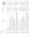
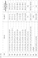
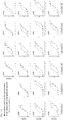


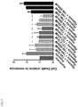

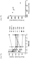
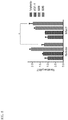
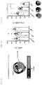
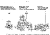
Classifications
-
- C—CHEMISTRY; METALLURGY
- C07—ORGANIC CHEMISTRY
- C07K—PEPTIDES
- C07K14/00—Peptides having more than 20 amino acids; Gastrins; Somatostatins; Melanotropins; Derivatives thereof
- C07K14/435—Peptides having more than 20 amino acids; Gastrins; Somatostatins; Melanotropins; Derivatives thereof from animals; from humans
- C07K14/575—Hormones
- C07K14/65—Insulin-like growth factors (Somatomedins), e.g. IGF-1, IGF-2
-
- A—HUMAN NECESSITIES
- A61—MEDICAL OR VETERINARY SCIENCE; HYGIENE
- A61P—SPECIFIC THERAPEUTIC ACTIVITY OF CHEMICAL COMPOUNDS OR MEDICINAL PREPARATIONS
- A61P1/00—Drugs for disorders of the alimentary tract or the digestive system
- A61P1/16—Drugs for disorders of the alimentary tract or the digestive system for liver or gallbladder disorders, e.g. hepatoprotective agents, cholagogues, litholytics
-
- A—HUMAN NECESSITIES
- A61—MEDICAL OR VETERINARY SCIENCE; HYGIENE
- A61P—SPECIFIC THERAPEUTIC ACTIVITY OF CHEMICAL COMPOUNDS OR MEDICINAL PREPARATIONS
- A61P1/00—Drugs for disorders of the alimentary tract or the digestive system
- A61P1/18—Drugs for disorders of the alimentary tract or the digestive system for pancreatic disorders, e.g. pancreatic enzymes
-
- A—HUMAN NECESSITIES
- A61—MEDICAL OR VETERINARY SCIENCE; HYGIENE
- A61P—SPECIFIC THERAPEUTIC ACTIVITY OF CHEMICAL COMPOUNDS OR MEDICINAL PREPARATIONS
- A61P11/00—Drugs for disorders of the respiratory system
-
- A—HUMAN NECESSITIES
- A61—MEDICAL OR VETERINARY SCIENCE; HYGIENE
- A61P—SPECIFIC THERAPEUTIC ACTIVITY OF CHEMICAL COMPOUNDS OR MEDICINAL PREPARATIONS
- A61P13/00—Drugs for disorders of the urinary system
- A61P13/12—Drugs for disorders of the urinary system of the kidneys
-
- A—HUMAN NECESSITIES
- A61—MEDICAL OR VETERINARY SCIENCE; HYGIENE
- A61P—SPECIFIC THERAPEUTIC ACTIVITY OF CHEMICAL COMPOUNDS OR MEDICINAL PREPARATIONS
- A61P17/00—Drugs for dermatological disorders
-
- A—HUMAN NECESSITIES
- A61—MEDICAL OR VETERINARY SCIENCE; HYGIENE
- A61P—SPECIFIC THERAPEUTIC ACTIVITY OF CHEMICAL COMPOUNDS OR MEDICINAL PREPARATIONS
- A61P19/00—Drugs for skeletal disorders
-
- A—HUMAN NECESSITIES
- A61—MEDICAL OR VETERINARY SCIENCE; HYGIENE
- A61P—SPECIFIC THERAPEUTIC ACTIVITY OF CHEMICAL COMPOUNDS OR MEDICINAL PREPARATIONS
- A61P19/00—Drugs for skeletal disorders
- A61P19/02—Drugs for skeletal disorders for joint disorders, e.g. arthritis, arthrosis
-
- A—HUMAN NECESSITIES
- A61—MEDICAL OR VETERINARY SCIENCE; HYGIENE
- A61P—SPECIFIC THERAPEUTIC ACTIVITY OF CHEMICAL COMPOUNDS OR MEDICINAL PREPARATIONS
- A61P19/00—Drugs for skeletal disorders
- A61P19/08—Drugs for skeletal disorders for bone diseases, e.g. rachitism, Paget's disease
-
- A—HUMAN NECESSITIES
- A61—MEDICAL OR VETERINARY SCIENCE; HYGIENE
- A61P—SPECIFIC THERAPEUTIC ACTIVITY OF CHEMICAL COMPOUNDS OR MEDICINAL PREPARATIONS
- A61P25/00—Drugs for disorders of the nervous system
-
- A—HUMAN NECESSITIES
- A61—MEDICAL OR VETERINARY SCIENCE; HYGIENE
- A61P—SPECIFIC THERAPEUTIC ACTIVITY OF CHEMICAL COMPOUNDS OR MEDICINAL PREPARATIONS
- A61P43/00—Drugs for specific purposes, not provided for in groups A61P1/00-A61P41/00
-
- A—HUMAN NECESSITIES
- A61—MEDICAL OR VETERINARY SCIENCE; HYGIENE
- A61P—SPECIFIC THERAPEUTIC ACTIVITY OF CHEMICAL COMPOUNDS OR MEDICINAL PREPARATIONS
- A61P7/00—Drugs for disorders of the blood or the extracellular fluid
-
- A—HUMAN NECESSITIES
- A61—MEDICAL OR VETERINARY SCIENCE; HYGIENE
- A61P—SPECIFIC THERAPEUTIC ACTIVITY OF CHEMICAL COMPOUNDS OR MEDICINAL PREPARATIONS
- A61P9/00—Drugs for disorders of the cardiovascular system
-
- C—CHEMISTRY; METALLURGY
- C07—ORGANIC CHEMISTRY
- C07K—PEPTIDES
- C07K14/00—Peptides having more than 20 amino acids; Gastrins; Somatostatins; Melanotropins; Derivatives thereof
- C07K14/435—Peptides having more than 20 amino acids; Gastrins; Somatostatins; Melanotropins; Derivatives thereof from animals; from humans
- C07K14/46—Peptides having more than 20 amino acids; Gastrins; Somatostatins; Melanotropins; Derivatives thereof from animals; from humans from vertebrates
- C07K14/47—Peptides having more than 20 amino acids; Gastrins; Somatostatins; Melanotropins; Derivatives thereof from animals; from humans from vertebrates from mammals
-
- C—CHEMISTRY; METALLURGY
- C07—ORGANIC CHEMISTRY
- C07K—PEPTIDES
- C07K14/00—Peptides having more than 20 amino acids; Gastrins; Somatostatins; Melanotropins; Derivatives thereof
- C07K14/435—Peptides having more than 20 amino acids; Gastrins; Somatostatins; Melanotropins; Derivatives thereof from animals; from humans
- C07K14/76—Albumins
- C07K14/765—Serum albumin, e.g. HSA
-
- A—HUMAN NECESSITIES
- A61—MEDICAL OR VETERINARY SCIENCE; HYGIENE
- A61K—PREPARATIONS FOR MEDICAL, DENTAL OR TOILETRY PURPOSES
- A61K38/00—Medicinal preparations containing peptides
-
- C—CHEMISTRY; METALLURGY
- C07—ORGANIC CHEMISTRY
- C07K—PEPTIDES
- C07K2319/00—Fusion polypeptide
- C07K2319/01—Fusion polypeptide containing a localisation/targetting motif
- C07K2319/035—Fusion polypeptide containing a localisation/targetting motif containing a signal for targeting to the external surface of a cell, e.g. to the outer membrane of Gram negative bacteria, GPI- anchored eukaryote proteins
-
- C—CHEMISTRY; METALLURGY
- C07—ORGANIC CHEMISTRY
- C07K—PEPTIDES
- C07K2319/00—Fusion polypeptide
- C07K2319/33—Fusion polypeptide fusions for targeting to specific cell types, e.g. tissue specific targeting, targeting of a bacterial subspecies
-
- C—CHEMISTRY; METALLURGY
- C07—ORGANIC CHEMISTRY
- C07K—PEPTIDES
- C07K2319/00—Fusion polypeptide
- C07K2319/70—Fusion polypeptide containing domain for protein-protein interaction
- C07K2319/74—Fusion polypeptide containing domain for protein-protein interaction containing a fusion for binding to a cell surface receptor
- C07K2319/75—Fusion polypeptide containing domain for protein-protein interaction containing a fusion for binding to a cell surface receptor containing a fusion for activation of a cell surface receptor, e.g. thrombopoeitin, NPY and other peptide hormones
Definitions
- the present invention relates generally to proteins that have therapeutic uses, and more specifically to bi-specific proteins, pharmaceutical compositions comprising such proteins, and methods for using such proteins to repair damaged tissue.
- Tissue regeneration is a multidisciplinary science in which the goal is to restore biological function of diseased or damaged tissues.
- Tissue regeneration addresses major clinical problems such as myocardial infarction.
- Myocardial infarction commonly known as a heart attack, occurs when coronary artery obstruction cuts off the blood supply to part of the heart.
- the resulting lack of oxygen causes irreversible tissue damage (necrosis and apoptosis), due to the inability of the heart to sufficiently activate endogenous regeneration programs and self-repair.
- tissue damage is a leading cause of congestive heart failure, a condition in which the heart is no longer capable of effectively pumping blood and can results in kidney acute injury.
- congestive heart failure In the United States, there are more than a million heart attacks every year, and nearly 5 million people are afflicted with congestive heart failure.
- the present invention which is defined in the accompanying claims provides bi-specific therapeutic proteins, nucleic acid molecules encoding bi-specific fusion proteins and therapeutic methods that employ such bi-specific therapeutic proteins to promote tissue survival, and/ or regeneration of damaged tissue.
- the bi-specific fusion protein promotes tissue regeneration, cell survival, cell differentiation, inhibits apoptosis, induces cell proliferation, promotes cell growth, promotes motility of stem cells, promotes differentiation of stem cells, prevents cell damage, and/ or promotes angiogenesis.
- the tissue can be cardiac tissue, kidney tissue, bone, cartilage, joints, skin, liver tissue, pancreatic tissue, blood cells, lung tissue, brain tissue, and nervous tissue.
- the present invention provides pharmaceutical compositions, comprising a bi-specific protein in combination with a physiologically acceptable carrier.
- methods for treating pathological tissue damage in a patient provided is a pharmaceutical composition for use in suffering from pathological tissue damage, thereby decreasing pathological tissue damage in the patient.
- the present disclosure provides bi-specific proteins comprising (1) a targeting domain having a binding specificity to a target molecule associated with the outer surface of a cell of a tissue, and (2) an engineered activator domain having a binding specificity to a receptor associated with the surface of a cell of the tissue, wherein the engineered activator domain has a modified amino acid sequence of an amino acid sequence of a wild-type activator domain, wherein the engineered activator domain decreases activation of the receptor relative to the wild-type activator domain.
- the activator domain is modified to decrease activation of the receptor relative to the wild-type activator domain by at least 3.5-fold.
- the activator domain when associated to the targeting domain in bi-specific protein exhibits a receptor activation at least twice stronger on cells containing the target molecule compared to cells that do not contain the target molecule as measured by phosphorylation of a receptor or a downstream effector molecule.
- the bi-specific protein exhibits a receptor activation at least twice stronger on cells containing the target molecule compared to cells that do not contain the target molecule, as measured by phosphorylation of AKT.
- the engineered activator domain comprises the wild-type amino acid sequence modified to comprise a deletion, a substitution, an addition, an additional amino acid sequence at an N- and/or C-terminus or a combination thereof. In some cases, the engineered activator domain comprises a wild-type activator domain fused to a non-immunogenic protein. In some cases, the engineered activator domain comprises a modified amino acid sequence of an amino acid sequence of the wild-type activator domain fused to a non-immunogenic protein.
- the bi-specific protein further comprises a half-life modulator, wherein the half-life modulator increases the half-life of the bi-specific protein.
- the half-life modulator can comprise the sequence of Human Serum Albumin, Fc, scFc, Albumin binding domain, PASylation, human alpha-fetoprotein, or variants thereof.
- the engineered activator domain has a binding affinity to a growth factor receptor.
- the activator domain and targeting domain are recombinantly fused. Yet in other cases, the activator domain and targeting domain are chemically coupled or joined.
- the engineered activator domain comprises a growth factor.
- the growth factor is IGF-1, NRG, or variants thereof.
- the targeting domain comprises Annexin A5 or variants thereof.
- the Annexin A5 comprises an amino acid sequence set forth in any one of SEQ ID NOs: 1-4 or 122.
- the engineered activator domain comprises IGF-1(LR3-Y31A). In some cases, the engineered activator domain comprises an amino acid sequence set forth in any one of SEQ ID NOs: 18, 19, 23, 24, 28, 29, or 120.
- the half-life modulator is Human Serum Albumin or variant thereof.
- the Human Serum Albumin comprises an amino acid sequence set forth in any one of SEQ ID NOs: 54-56 or 124.
- the half-life modulator comprises Fc or variant thereof.
- the Fc comprises an amino acid sequence set forth in any one of SEQ ID NOs: 53.
- the bi-specific protein further comprises a connector linking the engineered activator domain to the half-life modulator and a connector linking the half-life modulator to the targeting domain.
- the connector comprises an amino acid sequence set forth in any one of SEQ ID NOs: 60-62 or 126-127.
- the engineered activator domain can be joined via a peptide bond to the amino terminus of the targeting domain. In some cases, the engineered activator domain can be joined via peptide bond to the carboxy terminus of the targeting domain.
- the bi-specific protein comprises: (1) an activator domain, the activator domain comprising a growth factor, (2) a targeting domain, the targeting domain comprising a polypeptide that binds to phosphatidylserine at the outer surface of a damaged cell, the bi-specific protein having a half maximal effective concentration lower in the damaged cell (EC50 Damaged ) than a healthy cell (EC50 Healthy ).
- the damaged cell can be a cell undergoing apoptosis or necrosis.
- the growth factor is a modified IGF-1 protein (also referred herein as a variant of IGF-1).
- the bi-specific protein comprising the IGF-1 variant has an EC50 Healthy /EC50 Damaged ratio of at least 10:1.
- the activator domain comprises a variant of IGF-1.
- the targeting domain comprises human Annexin A5 or variant thereof.
- the activator domain comprises a variant of IGF-1 and the targeting domain comprises human annexin A5 or variant thereof.
- the bi-specific protein comprises (1) an activator domain, the activator domain comprising a variant of IGF-1, (2) a targeting domain, the targeting domain comprising Annexin A5 or variant thereof, wherein the Annexin A5 or variant thereof binds to phosphatidylserine at the outer surface of a cell within damaged tissue, wherein the bi-specific protein and has a half maximal effective concentration lower in the damaged tissue (EC50 Damaged ) than healthy tissue (EC50 Healthy ).
- the IGF-1 variant induces the phosphorylation of AKT.
- the bi-specific protein comprising IGF-1 variant has an EC50 Healthy /EC50 Damaged ratio of at least 10:1.
- the damaged tissue is an ischemic tissue.
- the targeted cell is a apoptotic or necrotic cell.
- the damaged tissue is a diabetic tissue damage caused by diabetes. In some cases, the damaged tissue is a diabetic tissue damage caused by diabetic nephropathy. In some cases, the damaged tissue caused podocyte-related disorder.
- the targeting domain is capable of binding a podocyte protein, such as nephrin (NPHS1), podoplanin (PDPN), podocalyxin (PODXL), dystroglycan (DAG1), GLEPP1 (PTPRO), NEPH1 (KIRREL), FAT atypical cadherin 1 (FAT1), cysteine rich transmembrane BMP regulator 1 (CRIM1), integrin alpha-8/beta 1 (ITGA8).
- the IGF-1 variant has an amino acid sequence set forth in any one of SEQ ID NOs: 10-30 or 120. In some cases, the IGF-1 variant induces survival signaling upon binding to the IGF-1 receptor.
- the targeting domain comprises a molecule capable of binding phosphatidylserine. In some cases, the targeting domain comprises Annexin A5. In some cases, Annexin A5 has an amino acid sequence set forth in any one of SEQ ID NOs: 1-4 or 122.
- the targeting domain comprises a molecule capable of binding a binding a podocyte protein, such as nephrin (NPHS1), podoplanin (PDPN), podocalyxin (PODXL), dystroglycan (DAG1), GLEPP1 (PTPRO), NEPH1 (KIRREL), FAT atypical cadherin 1 (FAT1), cysteine rich transmembrane BMP regulator 1 (CRIM1), integrin alpha-8/beta 1 (ITGA8).
- NPHS1 nephrin
- PDPN podoplanin
- PODXL podocalyxin
- DAG1 dystroglycan
- GLEPP1 GLEPP1
- KIRREL NEPH1
- FAT atypical cadherin 1 (FAT1) cysteine rich transmembrane BMP regulator 1
- CRIM1 cysteine rich transmembrane BMP regulator 1
- ITGA8 integrin alpha-8/
- the targeting domain comprises an antibody capable of binding a binding a podocyte protein, such as nephrin (NPHS1), podoplanin (PDPN), podocalyxin (PODXL), dystroglycan (DAG1), GLEPP1 (PTPRO), NEPH1 (KIRREL), FAT atypical cadherin 1 (FAT1), cysteine rich transmembrane BMP regulator 1 (CRIM1), integrin alpha-8/beta 1 (ITGA8).
- NPHS1 nephrin
- PDPN podoplanin
- PODXL podocalyxin
- DAG1 dystroglycan
- GLEPP1 GLEPP1
- KIRREL NEPH1
- FAT atypical cadherin 1 FAT atypical cadherin 1 (FAT1)
- cysteine rich transmembrane BMP regulator 1 CRIM1
- integrin alpha-8/beta 1 ITGA8
- the activator domain and the targeting domain are covalently linked by a peptide bond to form a single polypeptide.
- the IGF-1 variant and the Annexin A5 or variant thereof are covalently linked by a peptide bond to form a single polypeptide. In some cases, the variant of IGF-1 and the Annexin A5 or variant thereof are covalently linked to the peptide linker by a peptide bond to form a single polypeptide.
- the bi-specific protein further comprises a peptide linker.
- the peptide linker is a half-life modulator.
- the half-life modulator is a human serum albumin or variant thereof.
- the half-life modulator is an Fc fragment or variant thereof.
- the human serum albumin or variant thereof has an amino acid sequence set forth in any one of SEQ ID NOs: 54-56 or 124.
- the Fc fragment has an amino acid sequence set forth in SEQ ID NO: 53.
- the activator domain is linked to the amino terminus of the peptide linker and the targeting domain is linked to the carboxy terminus of the peptide linker. In some cases, the activator domain is linked to the carboxy terminus of the peptide linker and the targeting domain thereof is linked to the amino terminus of the peptide linker. In some cases, the bi-specific protein further comprises a peptide connector between the activator domain and peptide linker and a peptide connector between the targeting domain and peptide linker.
- the IGF-1 variant is linked to the amino terminus of the peptide linker and the annexin A5 or variant thereof is linked to the carboxy terminus of the peptide linker. In some cases, the IGF-1 variant is linked to the carboxy terminus of the peptide linker and the annexin A5 or variant thereof is linked to the amino terminus of the peptide linker.
- the bi-specific protein further comprises a peptide connector between the IGF-1 variant and peptide linker and a peptide connector between the Annexin A5 or variant thereof and peptide linker.
- the peptide connector has an amino acid sequence set forth in any one of SEQ ID NOs: 60-62 or 126-127.
- the engineered protein has an amino acid sequence recited in SEQ ID NO: 84.
- the nucleic acid has a sequence recited in SEQ ID NO: 102.
- the engineered protein has an amino acid sequence recited in SEQ ID NO: 118.
- the nucleic acid has a sequence recited in SEQ ID NO: 119.
- bi-specific protein comprising: (1) an IGF-1 variant comprising an amino acid sequence set forth in any one of SEQ ID NOs: 18, 19, 23, 24, 28, 29 or 120, and (2) Annexin A5 or variant thereof comprising an amino acid sequence set forth in any one of SEQ ID NOs: 1-4 or 122.
- the bi-specific protein comprises a Human Serum Albumin or variant thereof comprising an amino acid sequence set forth in any one of SEQ ID NOs: 54-56 or 124.
- the Human Serum Albumin or variant thereof is linked to a C-terminus of Annexin A5 or variant thereof and to the N-terminus of the IGF-1 variant.
- the bi-specific protein further comprises a connector peptide linking a N-terminus of the Human Serum Albumin or variant thereof to the C-terminus of Annexin A5 or variant thereof and a peptide linking a C-terminus of the Human Serum Albumin or variant thereof to the N-terminus of the IGF-1 variant.
- the connector peptide comprises an amino acid sequence set forth in any one of SEQ ID NOs: 60-62 or 126-127.
- the bi-specific protein further comprises a leader polypeptide.
- the bi-specific protein further comprises polypeptide affinity tag.
- the affinity tag is at the amino terminus of the fusion protein, at the carboxy terminus of the fusion protein, or in the middle of the fusion protein.
- the bi-specific protein comprises a histidine-comprising polypeptide.
- the bi-specific protein comprises an amino acid sequence of a non-internalizing variant of human Annexin A5 and the bi-specific protein has a prolonged half-life as compared to a bi-specific protein comprising the amino acid sequence of wild-type human Annexin A5.
- the bi-specific protein comprises an amino acid sequence set forth in SEQ ID NO: 4.
- aspects of the disclosure relate to bi-specific protein comprising an amino acid sequence set forth in any one of SEQ ID NOs: 67, 70, 73-86, 108, 110, 116, or 118.
- the bi-specific proteins provided herein are not necessarily limited to two binding specificities.
- the bi-specific protein in addition to the targeting domain, the bi-specific protein comprises two or more activator domains that are linked directly or indirectly via peptide bonds.
- the bi-specific protein in addition to the activator domain, the bi-specific protein comprises two or more targeting domains that are linked directly or indirectly via peptide bonds.
- aspects of the disclosure relate to a method of promoting tissue regeneration or survival in a subject, the method comprising administering in a patient in need thereof a therapeutically effective amount of the bi-specific protein, whereby the targeting domain specifically binds to the target molecule associated with a damaged cell of a damaged tissue, thereby targeting the bi-specific fusion protein to the damaged tissue and whereby upon exposure of the activator domain to the growth factor receptor, the activator domain specifically activates the growth factor receptor so as to promote regeneration or survival of the damaged tissue.
- aspects of the disclosure relate to a method of treating a patient in need thereof, the method comprising providing a bi-specific protein, and administering in the patient a therapeutically effective amount of the bi-specific protein, wherein the bi-specific proteins binds to phosphatidylserine on the outer leaflet of the plasma membrane of a cell of a tissue and to a IGF-1 growth factor receptor at the surface of the cell of the tissue.
- the bi-specific protein binds to molecules associated with the surface of the same cell of the tissue.
- the bi-specific protein binds to molecules associated with the surface of different cells of the tissue.
- aspects of the disclosure relate to a method of promoting tissue regeneration or survival in a subject, the method comprising (a) providing a bi-specific protein having a targeting domain having a binding specificity to a target molecule associated with the outer surface of a first cell of a tissue and an engineered activator domain having a binding specificity to a receptor associated with the surface of a second cell of the tissue, wherein the engineered activator domain has a modified amino acid sequence of an amino acid sequence of a wild-type activator domain, wherein the engineered activator domain decreases activation of the receptor relative to the wild-type activator domain; and (b) administering in a patient in need thereof a therapeutically effective amount of the bi-specific protein, whereby the targeting domain targets the bi-specific fusion protein to the first cell of the tissue and whereby upon exposure of the activator domain to a growth factor receptor at the surface of a second cell, the activator domain specifically activates the growth factor receptor of so as to promote tissue regeneration, wherein the bi-specific protein exhibits a receptor
- aspects of the disclosure relate to a method of promoting tissue regeneration or survival in a subject, the method comprising (a) providing a bi-specific protein having (1) an activator domain, wherein the activator domain comprises a variant of IGF-1 and (2) a targeting domain, wherein the targeting domain comprises annexin A5 or variant thereof; and (b) administering in a patient in need thereof a therapeutically effective amount of the bi-specific protein whereby the Annexin A5 or variant thereof targets the bi-specific fusion protein to a first cell of the tissue, wherein the cell expresses phosphatidylserine on the outer leaflet of the plasma membrane, and whereby upon exposure of the IGF-1 variant to a IGF-1 receptor at the surface of a second cell, the IGF-1 variant specifically activates the IGF-1 receptor of so as to promote tissue regeneration.
- the targeted cell and activated cell are the same. Yet in other embodiments, the targeted cell and activated cell are different. In some cases, the targeted cell is a damaged cell and the activated cell is a viable cell. In some cases, the targeted cell is a damaged cell and the activated cell is a damaged cell.
- the pathological tissue damage is heart tissue damage associated with myocardial infarction. In other cases, the pathological tissue damage is kidney tissue damage. In other cases, the pathological tissue damage is in bone, cartilage, joints, skin, liver tissue, pancreatic tissue, blood cells, lung tissue, or nervous tissue. In certain embodiments, such methods further comprise the administration of stem cells to the patient.
- nucleic acid molecules encoding a bi-specific fusion protein as described herein.
- the nucleic acid molecule is DNA
- the DNA further comprises transcriptional and translational regulatory sequences operably linked to the bi-specific fusion protein coding sequence, such that transcription and translation of the coding sequence occurs in at least one eukaryotic cell type.
- Figure 1A and Figure 1B are schematics of representative therapeutic bi-specific proteins (also referred herein as Smart Growth Factors or SGFs) and non-targeted control proteins, according to some embodiments.
- Figure 1A and Figure 1B are schematics of representative (1) targeted, potency-reduced IGF-1-based proteins, (2) targeted, potency-reduced Nrg1 ⁇ -based proteins, (3) non-targeted, potency-reduced IGF-1-based proteins, (4) non-targeted, non-potency-reduced IGF-1-based proteins, (5) targeted, non-potency-reduced IGF-1-based proteins, (6) signaling arms, (7) targeting arms, (8) half-life modulators, and (9) linkers, according to some embodiments.
- FIG. 2A and 2B are tables listing therapeutic bi-specific proteins and the potency and the fold decreased potency in healthy cells compared to wild-type growth factors (wt GFs) according to some embodiments.
- FIG. 2A and 2B show that engineered Growth factors according to embodiments of the invention have decreased potency (i.e., increased pAKT EC50's) compared to wild type growth factors.
- EC50 is defined as the concentration needed to achieve the half maximal level of pAKT signaling.
- iPSC-derived cardiomyocytes (CDI) were stimulated with (S)GFs for 10 min and pAKT levels were measured by ELISA.
- FIG. 2A and 2B show that engineering GFs, either by addition, deletion, or mutation of amino acids or by fusing to other protein domains, causes reduced potency compared to wt GF..
- Figure 3A is a set of graphs depicting pAKT (protein kinase B) dose response in healthy and damaged cardiomyocytes using different therapeutic bi-specific proteins and non-targeted control proteins according to some embodiments.
- the potencies of candidate Smart Growth Factors (SGF) are measured in pluripotent stem cell derived cardiomyoctyes (Cellular Dynamics International) and signaling is quantified by the accumulation of phosphorylated Akt.
- SGF Smart Growth Factors
- Dose response curves are subsequently fit to a three parameter EC50 activation model and the calculated EC50s are compared between the Healthy (circle, blue color) and Damaged (square, red color) contexts.
- Healthy circle, blue color
- Damaged square, red color
- Figure 3B is a graph depicting the potency shift calculated by EC50 healthy/EC50 damaged for different therapeutic bi-specific proteins compared to non-targeted control proteins on a logarithmic scale according to some embodiments.
- Fit EC50 values are represented for both the Healthy (filled circles) and Damaged (incubation with 12.5 ⁇ g/mL doxorubicin for 24 hours to induce apoptosis; filled triangle) contexts. Error bars represent the 95% confidence interval for the parameters.
- the calculated Potency Shift for each of the engineered proteins is taken as the ratio of fit EC50 values between the Healthy and Damaged dose response curves (EC50Healthy/EC50Damaged).
- the Potency Shift is annotated and expressed as the fold increase in Damaged context signaling.
- FIG. 3B shows that non-targeted, non-potency reduced molecules (e.g., 688) and non-targeted, potency-reduced molecules (e.g., 704, 602, 703) have no appreciable potency shift.
- targeted, non-potency reduced molecules e.g., 649) also have no appreciable potency shift.
- potency-reduced molecules e.g., 606, 683, 711, 713, 716, 727, 728, 729, 730, 731, 732, 733, 739, 740, 741, 743, 757) have an appreciable (>4-fold) potency shift.
- FIG. 3C is a graph depicting pAKT (protein kinase B) dose response in healthy and damaged cardiomyocytes using the therapeutic bi-specific protein 776 (sc776) and the corresponding non-targeted control protein 777 (sc777).
- pAKT protein kinase B
- sc776 therapeutic bi-specific protein 776
- sc777 non-targeted control protein 777
- the potencies of candidate Smart Growth Factors are measured at different concentrations (nM) in pluripotent stem cell derived cardiomyocytes and signaling (Y axis) is quantified by the accumulation of phosphorylated Akt.
- Dose response curves in the healthy and damaged contexts are fit to a three parameter EC50 activation model.
- Signaling for sc776 is depicted for Healthy (blue, filled circle) and Damaged (red, filled square) contexts, respectively.
- Signaling responses for sc777 is depicted for the Healthy (purple, filled triangle
- Figure 4 is a graph depicting reduction of caspase activity induced by hypoxia using therapeutic bi-specific protein SGF 740 in human cardiomyocytes.
- Figure 4 shows that targeted, engineered growth factors according to embodiments of the invention reduce apoptosis in human cardiomyocytes in a dose dependent manner. Apoptosis was induced by culturing cell at 1% oxygen for 48 hours. The therapeutic bi-specific protein 740 was added at the start of the hypoxia period. Caspase 3/7 activity was measured using capsaseGlo (Promega). Fusion protein 740 significantly (p ⁇ 0.01) reduces caspase activity induced by hypoxia in human cardiomyocytes.
- Figure 5 is a graph depicting reduction of hypoxia-induced cell death using therapeutic bi-specific proteins SGFs 727, 740, 734, and non-targeted control (746) in human kidney proximal tubule epithelial cells.
- Figure 5 shows that targeted engineered growth factors according to embodiments of the invention reduce hypoxia-induced cell death in human kidney proximal tubule epithelial cells while non-targeted controls show no effect.
- Cell death was measured by the percentage of cells staining positive for propidium iodide by flow cytometry.
- FIG. 6 is a table depicting the half-life and decay rate after intravenous dosing of different therapeutic bi-specific proteins. The half-life of different therapeutic bi-specific proteins and wt IGF-1 in mice was calculated using a single compartment model.
- SGF 727 has the structure IGF1(LR-3-R37X-3X)_lk40_mHSA_lk40_AnxV, whereas molecules 739-743 have the basic structure IGF1 ⁇ (LR3)_lk7_mHSA_lk7_AnxV(ni), where ⁇ denotes a potency reducing deletion or mutation of IGF1.
- SGF757 has the structure Nrgla_lk7_mHSA_lk7_AnxV(ni).
- Figure 6 shows that targeted engineered growth factors (SGFs) according to some embodiments have longer half-lives than wild type growth factor (wt IGF1)
- Figures 7A and 7B are a set of graphs depicting the effects of different therapeutic bi-specific proteins on blood glucose levels after intravenous dosing.
- Figure 7 shows that targeted, engineered growth factors (SGFs) according to some embodiments have reduced off target effects.
- SGFs engineered growth factors
- Anx-targeted, potency-reduced IGF1 fusion proteins have significantly reduced hypoglycemia compared to untargeted, high potency IGF1 fusions proteins.
- Figure 7A is a graph depicting the time course of blood glucose levels in mice after dosing with different therapeutic bi-specific proteins. Data are shown as mg/dL glucose level. SGFs 727-743 are targeted, potency-reduced IGF1-based fusion proteins, whereas 688 is a non-targeted high-potency IGF1 fusion protein. Mice were dosed with recombinant HSA as negative control and IGF-1 (LR3 variant) as positive control.
- Figure 7B is a graph depicting the relationship between SGF potency (defined as the concentration required to achieve half maximal pAKT levels, i.e., pAKT EC50 of therapeutic bi-specific proteins) vs. 3 hr blood glucose area under the curve (AUC).
- SGF potency defined as the concentration required to achieve half maximal pAKT levels, i.e., pAKT EC50 of therapeutic bi-specific proteins
- AUC blood glucose area under the curve
- Figure 8 is a graph depicting relative pAKT levels in damaged (infarct) vs. healthy (remote) rat heart regions.
- a rat ischemia/reperfusion model was employed to generate ischemic injury by ligation of the left anterior descending coronary artery (LAD) in rats. After 1 hour of ischemia and 2 hours of reperfusion, hearts were excised and microdissected into remote (healthy) and infarcted (damaged) regions informed by anatomical indicators. Tissue homogenates were generated for each region and analyzed for phospho-AKT using a total AKT/pAKT sandwich ELISA.
- IGF-1 does not result in increased pAKT in either remote or infarcted tissue whereas a non-targeted, highly potent IGF1 fusion protein (688) increases pAKT non-selectively in both remote and infarcted tissue.
- Targeted, potency-reduced IGF1 fusion protein, 606 selectively increases pAKT in the infarcted tissue (p ⁇ 0.05), compared to remote tissue.
- Figure 8 shows targeted, engineered growth factors (SGFs) activate pro-survival signaling in damaged tissue in vivo.
- Targeted, potency reduced IGF1 fusions protein 606 activates significantly more pAKT signaling in infarcted tissue vs. remote (healthy) tissue in vivo. Selective signaling is not observed with either wt IGF1 or a non-targeted, high potency fusion protein (688).
- FIGS. 9A, 9B and 9C showed that targeted engineered growth factors according to some embodiments are efficacious in vivo and reduce infarct size in rat ischemia/reperfusion model of acute myocardial infarction (AMI).
- Targeted, potency reduced IGF1 fusions protein (SGF 606) significantly reduces infract/area-at-risk (AAR) following acute myocardial infarction in rats.
- AAR area-at-risk
- FIG. 9A depicts an overview of the rat acute myocardial infarction (AMI) model.
- AMI acute myocardial infarction
- Figure 9B is a graph depicting the percent area of the left ventricle that is at risk after ischemia (area at risk (AAR)/LV %) using the model shown in Figure 9A . There was no significant difference in the size of injury produced by the surgical procedure between any of the groups as indicated by comparable size of the area at risk (AAR) with respect to the area of the left ventricle (LV).
- Figure 9C is a graph depicting the infarct/area at risk percentage in rats injured by AMI and treated with vehicle, wt IGF1, or a targeted therapeutic bi-specific protein (SGF 606).
- the targeted, potency-reduced SGF 606 is highly significantly efficacious in infarct size reduction at 72 hours compared to vehicle control (p ⁇ 0.001).
- IGF1 is also able to significantly reduce infarct size compared to vehicle (p ⁇ 0.05); however treatment with SGF 606 results larger infarct reduction compared to IGF1 (p ⁇ 0.05).
- Figure 10 is a schematic of a targeted engineered growth factor in accordance to some embodiments.
- peptide a sequence polymer of at least two amino acids covalently linked by an amide bond (also referred herein as peptide bond).
- bi-specific refers to the ability of the fusion protein to interact with two different ligands.
- the bi-specific protein interacts with a target molecule for the targeting domain and a receptor for the activator domain.
- target molecule refers to any molecule that is associated with a tissue (e.g. "at risk”, diseased or damaged tissue).
- a “target cell” is meant to be a cell to which a bi-specific protein or targeting domain thereof can specifically bind.
- Binding or “specific binding” are used interchangeably herein and indicates that a protein (or the targeting polypeptide domain thereof or the activator domain thereof) exhibits substantial affinity for a specific molecule (e.g., targeting domain exhibits substantial affinity for a target molecule, or an activator domain exhibits substantial affinity for a molecule associated with the surface of a cell such as a growth factor receptor) or a cell or tissue bearing the molecule and is said to occur when the protein (or the targeting polypeptide domain thereof or the activator domain thereof) has a substantial affinity for a specific molecule and is selective in that it does not exhibit significant cross-reactivity with other molecules.
- a specific molecule e.g., targeting domain exhibits substantial affinity for a target molecule, or an activator domain exhibits substantial affinity for a molecule associated with the surface of a cell such as a growth factor receptor
- polynucleotide as used herein, means a genetic entity distinct from that generally found in nature. As applied to a polynucleotide or gene, this means that the polynucleotide is the product of various combinations of cloning, restriction and/or ligation steps, and other procedures that result in the production of a construct that is distinct from a polynucleotide found in nature.
- operably linked refers to a nucleic acid sequence placed into a functional relationship with another nucleic acid sequence.
- a nucleic acid is “operably linked” when it is placed into a functional relationship with another nucleic acid sequence.
- vector is intended to refer to a nucleic acid molecule capable of transporting another nucleic acid to which it has been linked.
- plasmid refers to a circular double-stranded DNA loop into which additional DNA segments may be ligated.
- viral vector e.g., a replication defective retrovirus, adenovirus and adeno-associated virus
- additional DNA segments may be ligated into the viral genome so as to be operatively linked to a promoter (e.g., a viral promoter) that will drive the expression of a protein encoded by the DNA segment.
- vectors are capable of autonomous replication in a host cell into which they are introduced (e.g., bacterial vectors having a bacterial origin of replication and episomal mammalian vectors).
- Other vectors e.g., non-episomal mammalian vectors
- certain vectors are capable of directing the expression of genes to which they are operatively linked. Such vectors are referred to herein as "expression vectors.”
- host cell is intended to refer to a cell into which an expression vector has been introduced, which cell is capable of reproducing, and preferably expressing proteins encoded by, the vector. It should be understood that such terms are intended to refer not only to the particular subject cell but to the progeny of such a cell. Because certain modifications may occur in succeeding generations due to either mutation or environmental influences, such progeny may not, in fact, be identical to the parent cell, but are still included within the scope of the term "host cell” as used herein.
- Identity is a relationship between two or more polypeptide or protein sequences, as determined by comparing the sequences.
- identity also refers to the degree of sequence relatedness between polypeptides or proteins, as determined by the match between strings of such sequences. “Identity” can be readily calculated by any bioinformational methods known in the art.
- parent polypeptide refers to a wild-type polypeptide and the amino acid sequence or nucleotide sequence of the wild-type polypeptide is part of a publicly accessible protein database (e.g., EMBL Nucleotide Sequence Database, NCBI Entrez, ExPasy, Protein Data Bank and the like).
- EMBL Nucleotide Sequence Database NCBI Entrez, ExPasy, Protein Data Bank and the like.
- mutant polypeptide or “polypeptide variant” refers to a form of a polypeptide, wherein its amino acid sequence differs from the amino acid sequence of its corresponding wild-type (parent) form, naturally existing form or any other parent form.
- a mutant polypeptide can contain one or more mutations, e.g., substitution, insertion, deletion, addition etc... which result in the mutant polypeptide.
- corresponding to a parent polypeptide is used to describe a polypeptide of the invention, wherein the amino acid sequence of the polypeptide differs from the amino acid sequence of the corresponding parent polypeptide only by the presence of at least one amino acid variation. Typically, the amino acid sequences of the variant polypeptide and the parent polypeptide exhibit a high percentage of identity. In one example, “corresponding to a parent polypeptide” means that the amino acid sequence of the variant polypeptide has at least about 50% identity, at least about 60%, at least about 70%, at least about 80%, at least about 90%, at least about 95%, at least about 98% identity or at least about 99% identity to the amino acid sequence of the parent polypeptide.
- the nucleic acid sequence that encodes the variant polypeptide has at least about 50% identity, at least about 60%, at least about 70%, at least about 80%, at least about 90%, at least about 95%, at least about 98% identity or at least about 99% identity to the nucleic acid sequence encoding the parent polypeptide.
- nucleic acid or fragment thereof indicates that when optimally aligned with appropriate nucleotide insertions or deletions with another nucleic acid (or its complementary strand), there is nucleotide sequence identity in at least about 95 to 99% of the sequence.
- homologous refers to amino acid sequence similarity between two peptides. When an amino acid position in both of the peptides is occupied by identical amino acids, they are homologous at that position.
- substantially homologous as used herein means that a sequence is at least 50% identical, and preferably at least 75% and more preferably 95% homologous to the reference peptide and which retains most or all of the activity as the sequence to which it is homologous.
- damaged cell or “damaged tissue,” as used herein, means and includes biological cell or tissue; for example, but not limited to, cardiovascular cell or tissue damaged or injured by trauma or chemical insult, ischemic tissue, infarcted tissue or cell or tissue damaged by any means which results in interruption of normal blood flow to the tissue.
- terapéuticaally effective amount means the amount of the bi-specific protein that will elicit the biological or medical response of a tissue, system, animal or human that is being sought by the researcher, veterinarian, medical doctor or other clinician.
- pharmaceutically acceptable means the carrier, diluent or excipient must be compatible with the other ingredients of the formulation and not deleterious to the recipient thereof.
- aspects of the disclosure relate to bi-specific therapeutic proteins, pharmacological compositions and methods for repairing or regenerating damaged or diseased tissue or cells.
- the bi-specific therapeutic proteins can positively regulate survival of targeted cells or targeted tissue.
- the bi-specific therapeutic proteins can promote survival signaling.
- the pharmacological compositions can further include one or more additional bioactive agents or components to aid in the treatment of damaged tissue or cells and/or facilitate the tissue regenerative process.
- aspects of the disclosure also encompass polynucleotides encoding the therapeutic bi-specific proteins and variants thereof that may be in the form of RNA or in the form of DNA, which DNA includes cDNA and synthetic DNA.
- the DNA may be double-stranded or single-stranded.
- the coding sequences that encode the variants of the disclosure may vary as a result of the redundancy or degeneracy of the genetic code.
- aspects of the disclosure are directed to bi-specific therapeutic proteins comprising two binding domains, each specific for a different target molecule or "ligand”.
- the bi-specific protein comprises a targeting domain or targeting moiety and activator domain or therapeutic moiety.
- targeting moiety “targeting domain,” or “targeting polypeptide” are used herein interchangeably and refer to molecules that selectively localize the bi-specific therapeutic in a particular tissue or region of the body. The localization can be mediated by specific recognition of molecular determinants, molecular size of the targeting domain, ionic interactions, hydrophobic interactions and the like.
- therapeutic moiety As used herein, the terms "therapeutic moiety,” “activator domain,” “activator polypeptide” and “signaling arm” are used herein interchangeably and refers to any agents useful for therapy and that are non-toxic, do not have a cytotoxic effect or are not detrimental to the cells, including, but not limited to, growth factors.
- a "bi-specific protein” refers to a protein capable of specific binding to two or more different specific molecules.
- the bi-specific protein comprises a targeting domain having a binding specificity to a first specific target molecule and an activator domain having a binding specificity to a second target molecule.
- the activator domain has a binding specificity to a receptor.
- the activator domain has a binding specificity to a receptor that modulates/promotes tissue regeneration.
- the targeting domain serves to target the bi-specific protein to a target cell or tissue while the activator domain serves to activate a cell thereby to promote regeneration of the targeted tissue.
- the bi-specific therapeutic proteins are chimeric proteins having a targeting polypeptide connected to an activator polypeptide. In some aspects, the bi-specific therapeutic proteins are chimeric proteins having a targeting polypeptide connected to a growth factor variant.
- the targeting domain is generally used to target the bi-specific proteins to a cell of choice, also referred to as "target cell.”
- the binding of the targeting domain to its target molecule does not induce a significant biological effect in the target cell.
- the activator domain binds to a second target molecule or ligand on a cell.
- the binding of the activator domain to its ligand is intended to modulate a specific biological effect, such as, to increase that biological activity.
- binding of the activator domain to its ligand is intended the positively regulate survival of the targeted cells or tissue.
- the activator domain of the bi-specific proteins can promote survival signaling.
- the targeting domain and the activator domain are associated with different subunits of a multimeric protein.
- the activator domain is cross-linked to the targeting domain.
- the activator domain is directly or indirectly fused to the targeting domain.
- the bi-specific therapeutic protein has an activator domain engineered to have a decreased potency as compared to wild-type activator domain. It has been observed that decreasing the potency of the activator domain can increase the selectivity of the bi-specific therapeutic protein resulting in preferential activation of cells or tissue containing the target molecule (see FIG. 3A , FIG. 3B and FIG. 3C ).
- the activator domain is a therapeutic agent.
- the activator domain is a growth factor variant. This observed decrease in potency relative to the wild-type activator domain can be attributed to both steric hindrance of the activation domain and also a decreased rate of diffusion in the context of the larger multi-domain fusion protein.
- the activator domain is an IGF-1 variant. In some cases, the activator domain is a NRG variant.
- the bi-specific therapeutic protein has an activator domain that is fused to both a half-life modulating domain and to a targeting domain such that the activator domain possesses a decreased activation potency as compared to the wild-type activator domain. It has been observed that fusion of the activator domain to the half-life modulation and targeting domains decreases the potency of the activator domain, thereby increasing the selectivity of the bi-specific therapeutic protein resulting in preferential activation of cells or tissue containing the target molecule. This observed decrease in potency relative to the wild-type activator domain can be attributed to both steric hindrance of the activation domain and also a decreased rate of diffusion in the context of the larger multi-domain fusion protein.
- the activator domain is a therapeutic agent.
- the activator domain is a growth factor variant.
- the activator domain is an IGF-1 variant.
- the activator domain is a NRG variant.
- the disclosure provides engineered proteins having a targeting domain, an activator domain, and optionally a peptide linker or half-life modulator.
- the disclosure provides variants having this level of identity to a portion of the parent polypeptide sequence, e.g., Human Insulin Growth Factor 1 (IGF-1), Annexin A5 (Anx A5 or AnxV), Human Serum Albumin (HSA), as defined herein.
- the variant has at least about 80%, 85%, 90%, 95%, 96%, 97%, 98% or 99% sequence identity to the parent polypeptide or to a portion of the parent polypeptide sequence, e.g., IGF-1, Annexin A5, Human Serum Albumin, as defined herein.
- the activator domain (i.e., the growth factor) of the bi-specific protein is engineered to give, when fused to the targeting domain, the bi-specific fusion protein a half- maximal effective concentration (EC50) lower in damaged cells or tissue than in healthy cells or tissue.
- the activator domain (i.e., the growth factor) of the bi-specific protein is engineered to give, when fused to the targeting domain, the bi-specific fusion protein at least one order of magnitude lower EC50 in damaged cell or tissue than in healthy cells or tissue.
- the targeting domain(s) of the bi-specific protein can be selected to have at least an order of magnitude higher binding affinity for its ligand than the affinity the activator domain has for its ligand.
- the targeting domain has at least 10 times or greater affinity for its ligand than the activator domain has for its ligand.
- the affinity of the targeting domain for its ligand is at least 15 times higher or at least 20 times or more higher, 25 times or more higher, than that of the activator domain.
- the affinity of the targeting domain to its ligand is 30, 40, 50 or even 100 times or more higher than that the affinity of the activator domain for its ligand.
- the differential potency of the activator domain and/or the differential binding affinity between the targeting domain and the activator binding domain provides surprising and previously unrecognized advantages over prior bi-specific proteins.
- alteration addition, deletion, substitution
- one or more residues of the activator domain of the bi-specific protein can result in higher specificity for target cells together with a decreased potency in the activator domain for non-target cells.
- EC50s i.e., high potency
- growth factors cannot be effectively targeted.
- variants of growth factors can be made with significantly decreased potency (i.e., increased EC50).
- the targeting domain and the activator domain are directly linked. In some aspects of the disclosure, the targeting domain and the activator domain are indirectly linked. In some aspects of the disclosure, the targeting domain and the activator domain are covalently linked. Yet, in other aspects of the disclosure, the targeting domain and the activator domain are non-covalently associated.
- the linkages between the activator moiety and the targeting moiety, the activator moiety and the half-life modulator (or peptide linker) and the targeting moiety and the half-life modulator (or peptide linker) can be covalent linkages or non-covalent linkages.
- the linkages can be peptide bonds formed by derivatization of the components involved with peptides and the formation of a peptide linkage between the peptides.
- the linkages can be non-covalent linkages, such as biotin/avidin or biotin/streptavidin linkages or specific antigen/antibody or hapten/antibody linkages.
- the bi-specific protein comprises (1) a targeting domain having a binding specificity to a molecule associated with a damaged cell of a tissue, wherein the molecule is intracellular in a viable cell and exposed to the extracellular space in the damaged cell; and (2) an activator domain having a binding specificity to a growth factor receptor of a cell in the tissue, wherein upon exposure of the activator domain to the growth factor receptor, the activator domain binds the growth factor receptor so as to promote regeneration or survival of the tissue.
- the activator domain is a growth factor that is engineered to give, when fused with the targeting domain, the bi-specific fusion protein a half maximal effective concentration (EC50) lower in damaged cell or tissue than in healthy cells or tissue.
- the activator domain is a growth factor that is engineered to give, when fused with the targeting domain, the bi-specific fusion protein at least one order of magnitude lower EC50 in damaged cell or tissue than in healthy cells or tissue.
- the bi-specific fusion protein comprises (1) a targeting domain having a binding specificity to a molecule associated with a damaged cell of a tissue, wherein the molecule is intracellular in a viable cell and exposed to the extracellular space in the damaged cell; (2) an activator domain having a binding specificity to a molecule associated with the surface of a cell in the tissue, wherein upon exposure of the activator domain to membrane-associated molecule, the activator domain binds the membrane-associated molecule so as to modulate regeneration of the tissue and (3) a peptide linker.
- the activator domain is a growth factor that is engineered to give the bi-specific fusion protein a half maximal effective concentration (EC50) lower in damaged cell or tissue than in healthy cells or tissue.
- the activator domain is a growth factor that is engineered to give the bi-specific fusion protein at least one order of magnitude lower EC50 in damaged cell or tissue than in healthy cells or tissue.
- the linker is a non-immunogenic peptide.
- the peptide linker is a half-life modulator capable of modulating (e.g., increasing) the half-life of the bi-specific protein.
- the bi-specific proteins comprises: (1) a targeting polypeptide domain that binds to an ischemia-associated molecule; and (2) a growth factor polypeptide engineered to give the bi-specific fusion protein a half maximal effective concentration (EC50) lower in ischemic cell or tissue than in healthy cells or tissue while having an affinity to a receptor at the surface of a cell in a tissue so as to promote regeneration or survival of the tissue.
- EC50 half maximal effective concentration
- the bi-specific proteins comprises: (1) a targeting polypeptide domain that binds to a podocyte-associated protein; and (2) a growth factor polypeptide engineered to give the bi-specific fusion protein a half maximal effective concentration (EC50) lower in ischemic cell or tissue than in healthy cells or tissue while having an affinity to a receptor at the surface of a cell in a tissue so as to promote regeneration or survival of the tissue.
- EC50 half maximal effective concentration
- the bi-specific proteins comprises (1) at least one targeting domain having a binding specificity to at least one target molecule associated with a tissue; (2) at least one an activator domain having a binding specificity to at least one molecule associated with the surface of a cell in the tissue, wherein upon exposure of the binding domain to the molecule, the binding domain binds the molecule so as to promote regeneration or survival of the tissue; and (3) optionally a peptide linker.
- the fusion protein comprises two or more targeting domains, each targeting domain having a binding affinity to a target molecule associated with a tissue.
- Each of the targeting domains may have a same binding specificity (e.g., a binding specificity for the same target molecule) or a different binding specificity (e.g., a binding specificity for a different target molecule). Each of the targeting domains may have a same binding affinity or different binding affinities.
- the protein comprises two or more activator domains. Each of the activator domains may have the same binding specificity (e.g., a binding specificity to the same receptor on the cell) or different binding specificity (e.g., a binding specificity for a different receptor on a cell). Each of the activator domains may have the same binding affinity or different binding affinities.
- the linker is a peptide. In some cases, the linker is a non-immunogenic peptide. In some cases, the linker is a half-life modulator wherein the half-life modulator modulates the half-life of the bi-specific protein.
- the bi-specific protein comprises a half-life modulator (HLM).
- the half-life modulator is a polypeptide.
- the half-life modulator can have two termini, an N-terminus and a C-terminus, and is joined at one terminus via a peptide bond to the targeting polypeptide domain and is joined at the other terminus via a peptide bond to the activator domain.
- the half-life modulator is joined at one terminus (N-terminus or C-terminus) to the activator domain or to the targeting domain. Accordingly, the half-life modulator can be at the N-terminus or at the C-terminus of the bi-specific protein.
- the half-life modulator may be joined to the targeting domain or the activator domain via peptide bonds.
- bi-specific proteins can find use in tissue regeneration.
- bi-specific fusion proteins can be used in diseased cells, following tissue or organ injury or following an event in which the cells of a tissue may be damaged.
- the bi-specific fusion proteins can activate cells that express one or more growth factor receptors.
- the bi-specific fusion proteins find use, for example, in recruiting cells that express one or more growth factor receptors to tissue following for example, injury, or an event in which the cells of a tissue may be damaged or may become dysfunctional.
- bi-specific proteins may be used to facilitate repair, survival or regeneration of damaged tissue or organ.
- the bi-specific proteins disclosed herein can find use in modulating tissue survival.
- the bi-specific proteins can enhance or maintain the viability of a cell or tissue.
- the bi-specific fusion proteins can activate the pro-survival or the cell survival pathway.
- the bi-specific proteins can decrease apoptosis or decrease cell death.
- bi-specific proteins can have (1) a targeting polypeptide domain wherein the targeting domain binds to a target molecule thereby targeting the bi-specific fusion protein to a first cell of a tissue, and (2) an activator domain having a binding specificity to a growth factor receptor. Upon exposure of the activator domain to the growth factor receptor, the activator domain can activate the receptor of a second cell so as to promote cell recruitment, inhibition of apoptosis, induction of cell proliferation, activation of the pro-survival pathway, regeneration, and/or survival of the tissue.
- the bi-specific fusion protein can bind to a first cell population and act on the same cell population (e.g.
- the targeting domain binds specifically to a target molecule associated with a damaged first cell population and the activator domain binds specifically to a receptor of a second cell population of viable cells. In some cases, the targeting domain binds specifically to a target molecule associated with a damaged cell population and the activator domain binds specifically to a receptor of the same cell population. In some cases, the targeting domain binds specifically to a tissue specific target molecule at the surface of a first cell population and the activator domain acts specifically to a second cell population.
- the targeting domain binds specifically to a tissue specific target molecule at the surface of a cell population and the activator domain acts specifically on the same cell population.
- the first cell can be a viable cell, or an "at risk” cell.
- at risk refers to a viable cell that has not yet undergone apoptosis or is not damaged but is at risk to be damaged.
- the bi-specific protein has two different binding domains (such targeting domain and activator domain) which bind to different molecules on different cells in a tissue or organ. Yet in some cases, the bi-specific protein has two different binding domains which bind to different molecules on the same target cell in a tissue, the targeting domain being selected to bind specifically a target cell and the activator domain selected to bind to a receptor (e.g., growth factor receptor) at the surface of the cell to promote tissue regeneration, cell recruitment, inhibition of apoptosis, induction of cell proliferation, activation of the pro-survival pathway, regeneration, and/or survival of the tissue.
- a receptor e.g., growth factor receptor
- target molecules are exposed or enriched on the exterior of a target cell.
- the target molecule is associated with a damaged cell, the target molecule being intracellular in a viable or undamaged cell and being exposed to the extracellular space in a damaged cell.
- Such molecules include, for example, molecules that are exposed in cells that undergo necrosis (such as DNA) or apoptosis (e.g., phosphatidylserine), myosin (including the tissue type-specific subtypes thereof), ICAM-1 or P-selectin.
- the target molecule is a molecule that is present or enriched at the surface of a diseased or dysfunctional cell or tissue as compared to the level detected in a healthy or functional cell or tissue.
- the target cell is not a tumor or cancerous cell.
- Cells are bounded by a plasma membrane (or cell membrane) comprising a lipid bilayer.
- the cell membrane may be considered to have a surface facing the cytosol (cytosolic side or interior of the cell) and a surface facing the exterior of the cell, or the extracellular space.
- Trans-bilayer movement of anionic phospholipids from the inner to the outer leaflet of the plasma membrane occurs during apoptosis.
- the anionic phospholipid-binding protein such as Annexin A5, synaptotagmin I or lactadherin can be used to detect the presence of phosphatidylserine on the outer leaflet of the cell membrane.
- Phosphatidylserine is a phospholipid, that is usually restricted to the cytosolic side of the membrane in viable or undamaged cells, and that becomes exposed on the outer cell surface or to the extracellular space in damaged cells or apoptosis.
- the target molecule is an "ischemia-associated molecule".
- An "ischemia-associated molecule” is any molecule that is detected at a level that is significantly higher (e.g., at least 1.5 higher, at least 2-fold higher, at least 3-fold higher, at least 4-fold higher, at least 5-fold higher) following ischemia (which results in hypoxia) or hypoxia. Ischemia occurs when there is insufficient blood flow to provide adequate oxygenation, which results in tissue hypoxia (reduced oxygen) or anoxia (absence of oxygen) as the most severe form of hypoxia, and ultimately tissue necrosis, and apoptosis. Any suitable binding assay may be used to identify ischemia-associated molecules, including those provided herein.
- the increased level of molecule that is detected may be the result of upregulation or decreased turnover, or may be due to increased accessibility (e.g., resulting from cell damage) or increased extracellular exposure (e.g., trans-bilayer movement from the inner to the outer leaflet of the plasma membrane).
- the ischemia-associated molecule is detected in a cell of post-ischemic tissue at a significantly higher level (e.g., at least 1.5 higher, at least 2-fold higher, at least 3-fold higher, at least 4-fold higher, at least 5-fold higher) than in a cell of the same tissue that has not undergone an ischemic event (i.e., the molecule is specific to or enriched in the post-ischemic tissue).
- the ischemia-associated molecule is associated with cell damage (i.e., the molecule is detected at a significantly higher level in cells that are damaged than in undamaged cells of the same type).
- Certain ischemia-associated molecules are enriched (e.g., at least 1.5 higher, at least 2-fold higher, at least 3-fold higher, at least 4-fold higher, at least 5-fold higher) in the heart after an ischemic event (or in a model system that is used to mimic ischemia in the heart).
- the ischemia-associated molecules are about 1.5-fold enriched, about 2-fold enriched, about 3-fold enriched, about 4-fold enriched, about 5-fold enriched in the heart after an ischemic event (or in a model system that is used to mimic ischemia in the heart). In some cases, the ischemia-associated molecules are from about 1.5-fold to about 5 fold or more enriched in the heart after an ischemic event (or in a model system that is used to mimic ischemia in the heart).
- the ischemia-associated molecules are from about 1.5-fold to about 2 fold, about 2-fold to about 2.5 fold, about 2.5 fold to about 3-fold, about 3-fold to about 3.5 fold, about 3.5 fold to about 4-fold, about 4-fold to about 4.5 fold, about 4.5-fold to about 5-fold, or more enriched in the heart after an ischemic event (or in a model system that is used to mimic ischemia in the heart). ).
- such molecules include molecules that are exposed on myocytes or other cardiac cells that undergo necrosis (for example, but not limited to, DNA) or apoptosis (for example, but not limited to, phosphatidylserine).
- such molecules include molecules that are enriched in scarred heart tissue, such as collagen (collagen I, III), myosin (including the cell type-specific subtypes thereof), or other extracellular matrix proteins that are enriched in post ischemic hearts.
- Such molecules can be identified on the basis of enrichment following ischemia-reperfusion in vivo or in simulated ischemia-reperfusion in vitro, or following exposure to conditions such as hypoxia, decreased ATP, increased reactive oxygen species (ROS) or nitric oxide synthase (NOS) production, or serum starvation of cells cultured in vitro.
- ROS reactive oxygen species
- NOS nitric oxide synthase
- the target molecule is a podocyte-associated molecule.
- the target molecule is one of nephrin (NPHS1), podoplanin (PDPN), podocalyxin (PODXL), dystroglycan (DAG1), GLEPP1 (PTPRO), NEPH1 (KIRREL), FAT atypical cadherin 1 (FAT1), cysteine rich transmembrane BMP regulator 1 (CRIM1), integrin alpha-8/beta 1 (ITGA8).
- the activator domain can be any polypeptide that detectably modulates the activity of a cellular network or recruits cells from one location to another.
- the activator domain is capable of activating signal transduction pathways by binding to a receptor at the surface a cell.
- certain activator domains are growth factor polypeptides, or any agonist of the receptor. It will be apparent that such modulation may be an increase in the activity of the cellular network such as induction of proliferation of cells, induction of cell growth, promotion of cell survival and/or inhibition of apoptosis.
- the activator domain can recruit other factors or cells (e.g. stem cells).
- An activator domain for a particular application may be selected based on the desired therapeutic outcome. For example, to increase survival and/or for stem cell differentiation (regenerative) purposes, activator domains that comprise IGF, HGF, G-CSF, GLP-1, PDGF, SDF1, TB4, or NRG1 (or a portion or derivative thereof) may be used. To increase cell proliferation (regenerative) purposes, activator domains that comprise IGF, FGF2, G-CSF, GH, HGF, PDGF, TB4, or NRG1 (or a portion or derivative thereof) may be used.
- An activator domain that comprises FGF2, G-CSF, GH, HGF, SGF1, TB4, VEGF alpha, or a portion or derivative thereof, that substantially retains the ability to bind to cognate receptor, may generally be used to increase angiogenesis.
- the activator domain comprises a change in the amino acid sequence, the three-dimensional structure of the protein, and/or the activity of the protein, relative to the wild-type form of the protein. It will be understood that the selection of a suitable modification in the activator domain for the creation of bi-specific proteins having the desired therapeutic effect can depend on multiple factors.
- the activator domain is a growth factor having amino acid sequence modification relating to the wild-type growth factor (e.g. IGF-1) to decrease its binding to its natural receptor (e.g. IGF-1 receptor), to decrease its binding to binding proteins (IGF binding proteins) and/or decrease its activation of its natural receptor (e.g. IGF-1 receptor).
- the activator domain is a growth factor having amino acid sequence modification that reduce (e.g., for about 1- 5%, 5-10%, 10%-20%, about 20%-40%, about 50%, about 40%-60%, about 60%-80%, about 80%-90%, 90-95%) its binding to its natural receptor (e.g. IGF-1 receptor).
- a growth factor polypeptide detectably modulates activation of a growth factor receptor.
- the activator domain of the bi-specific protein is a growth factor, variant or derivative thereof that retains at least about 0.01 % of wild-type biological activity.
- the activator domain of the bi-specific protein is a growth factor, variant or derivatives thereof that retain at least about 0.1 %, at least about 1%, at least about 10%, of wild-type biological activity.
- the activator domain of the bi-specific protein is a growth factor, variant or derivative thereof that retains between about 0.01% to about 0.1% of wild-type biological activity.
- the activator domain of the bi-specific protein is a growth factor, variant or derivative thereof that retains between about 0.01% to about 1% of wild-type biological activity. In some cases, the activator domain of the bi-specific protein is a growth factor, variant or derivative thereof that retains between about 0.01% to about 10% of wild-type biological activity. In some cases, the activator domain of the bi-specific protein is a growth factor, variant or derivative thereof that retains between about 0.1% to about 1% of wild-type biological activity. In some cases, the activator domain of the bi-specific protein is a growth factor, variant or derivative thereof that retains between about 0.1% to about 10% of wild-type biological activity.
- the activator domain of the bi-specific protein is a growth factor, variant or derivative thereof that retains between about 01% to about 10% of wild-type biological activity.
- Biological activity in some cases can be determined by measuring activation of the corresponding growth factor receptor in appropriate cells. In some cases, activation may be assessed, for example, by measuring phosphorylation of receptor kinase or downstream effector proteins, such as, but not limited to, AKT, S6, ERK, JNK, mTOR, etc.
- IGFs Insulin-like growth factors
- the insulin-like growth factors constitute a family of proteins having insulin-like and growth stimulating properties.
- the IGFs Human IGF1 is a 70 amino acids basic peptide having the protein and DNA sequences shown in SEQ ID NOs: 9 and 31, respectively.
- IGF-1 and IGF-1 receptor is important for cellular processes such as cell proliferation and survival. Binding of IGF-1 or variant thereof to the IGF-1 receptor stimulates kinase activity, leading to phosphorylation of multiple substrate, thereby initiating signaling cascades. IGF-1 stimulates cell proliferation and survival through activation of the AKT pathway.
- a tyrosine kinase Upon binding of IGF-I to the IGF-1 receptor, a tyrosine kinase, phosphorylates tyrosine residues on two major substrates, IRS-1 and Shc, which subsequently signal through the Ras/Raf and PI 3-kinase/AKT pathways.
- IGFBPs IGF binding Proteins
- the bi-specific proteins can maintain the ability to signal through the IGF-1 receptor.
- the signaling ability can be determined by assessing whether a downstream intracellular target, e.g., AKT (serine/threonine protein kinase B), is phosphorylated in response to the binding of activator domain of the bi-specific protein to the receptor at the cell surface.
- AKT serine/threonine protein kinase B
- the activator domain (also referred herein as signaling arm) is human IGF-1 or a derivative of the human IGF-1. In some cases, the activator domain has an amino acid sequence recited in any one of SEQ ID NOs: 9-30 or 120.
- the activator domain is a variant of IGF-1 that is capable of maintaining selectivity to the IGF-1 receptor by assaying for receptor phosphorylation or downstream signaling protein phosphorylation in response to the binding of the variant of IGF-1 to the IGF-1 receptor.
- the activator domain is a variant of IGF-1 that is modified to reduce binding to IGF-1 binding proteins (IGFBPs) relative to wild-type IGF-1 while maintaining its ability to activate the AKT pathway.
- IGFBPs IGF-1 binding proteins
- the IGF-1 variant can activate the IGF-1 receptor with a decreased potency for non-target cells, as assessed by pAKT EC50.
- EC50 is defined as the concentration needed to achieve the half maximal level of pAKT signaling.
- the activator domain is a derivative of the human IGF-1 and is engineered to decrease the binding of the activator domain to the IGF binding proteins which are present in the serum and other body fluid.
- the activator domain is a derivative of the human IGF-1 and comprises an N-terminal 13-residue extension (also referred as IGF-1 LONG, SEQ ID NO: 11), a mutation E3R (SEQ ID NO: 12) or a combination thereof (LONG E3R, also referred as LR3, SEQ ID NO: 15).
- the IGF-1 variant comprises the E3R substitution, an N-terminal 13-residue extension, deletion of amino acids 1-3 ((Des1-3), SEQ ID NO: 10) or a combination thereof to decrease the binding of the activator domain to the IGF binding proteins which are present in the serum and other body fluid.
- the activator domain is a derivative of the human IGF-1 and comprises one or more of the following modifications: an N-terminal 13-residue extension (referred as IGF-1 LONG, SEQ ID NO: 11), a deletion of amino acids 1-3 (Des-1-3, SEQ ID NO: 10), a substitution replacing Arg for a Glu at the 3 position of the polypeptide (E3R, SEQ ID NO: 12), no Arginine at position 37 (R37X, SEQ ID NO: 13), a deletion of amino acids 68-70 (3X, SEQ ID NO: 14), or an N-terminal 13-residue extension and a substitution replacing Arg for a Glu at the 3 position of the wild -type polypeptide (LR3, SEQ ID NO: 15).
- IGF-1 LONG an N-terminal 13-residue extension
- the IGF-1 or the IGF-1 variant can comprise a substitution at one or more of the tyrosine residues.
- the IGF-1 or IGF-1 variant e.g. LR3, Des 1-3
- the IGF-1 or IGF-1 variant can comprise one or more of the following substitutions, Y24L (SEQ ID NOs: 17, 22, and 27), Y31A (SEQ ID NOs: 19, 24 and 29), and Y60L (SEQ ID NOs: 20, 25 and 30).
- the IGF-1 variant can comprise a Y24L substitution and a Y31A substitution (SEQ ID NOs: 18, 23 and 28).
- one or more tyrosine residues can be substituted for a short aliphatic amino acid. In some cases, one or more tyrosine residues (Y24, Y31, Y60 or combinations thereof) can be substituted for a polar amino acid. In some cases, one or more tyrosine residues (Y24, Y31, Y60 or combinations thereof) can be substituted for leucine, alanine, isoleucine, serine, threonine or any other amino acid.
- the activator domain is a derivative of the human IGF-1 comprising one or more of the following modifications: a N-terminal 13-residue extension (IGF-1 LONG), a deletion of amino acids 1-3 (Des-1-3), a substitution replacing Arg for a Glu at the 3 position of the polypeptide (E3R), no Arginine at position 37 (R37X), a deletion of amino acids 68-70 (3X), an N-terminal 13-residue extension and a substitution replacing Arg for a Glu at the 3 position of the wild -type polypeptide (LR3), substitutions of one or more of tyrosine residues (Y24, Y31, Y60 or combinations thereof (e.g. Y24L, Y31A, Y60L substitutions or combinations thereof).
- IGF-1 LONG N-terminal 13-residue extension
- Des-1-3 deletion of amino acids 1-3
- E3R substitution replacing Arg for a Glu at the 3 position
- the activator domain is derivative of the human IGF-1 comprising a substitution at the position 3 and 31.
- the activator domain can be derivative of the human IGF-1 comprising E3R and Y31A substitutions.
- the activator domain has an amino acid sequence having SEQ ID NO: 120.
- the activator domain is encoded by a nucleic acid sequence having SEQ ID NO: 121.
- the activator domain is derivative of the human IGF-1 comprising a mutation (e.g. substitution, deletion) at one or more residues 24 to 37.
- the IGF-1 variant can be modified by glycosylation of one or more glyscosylation site present in the IGF-1 variant.
- IGF-1(LR3) IGF-1 LONG E3R
- IGF1 Des1-3 IGF-1(LR3)
- IGF-1(LR3) IGF-1 LONG E3R
- IGF1 Des1-3 IGF1 Des1-3
- the bi-specific proteins that contain the IGF-1 variants described herein have a potency for non-target cells that is less than wild-type IGF-1 for non-target cells.
- Certain activator domains that bind to growth factor receptors are provided herein in SEQ ID NOs: 9-30 and 120.
- peptide sequence modifications can be included, such as variations, deletions, substitutions or derivitizations of the amino acid sequence of the sequences disclosed herein, so long as the peptide has substantially the same activity or function as the unmodified peptides.
- a modified peptide will retain activity or function associated with the unmodified peptide, the modified peptide will generally have an amino acid sequence "substantially homologous" with the amino acid sequence of the unmodified sequence.
- the IGF-1 variant can have an amino acid sequence having at least about 85%, at least about 90%, at least about 95%, at least about 98% identity or at least about 99% identity to the amino acid sequence provided in SEQ ID NOs: 9-30 and 120. In some cases, the IGF-1 variant can have an amino acid sequence having from about 85% to about 90%, from about 90% to about 95%, from about 95% to about 98%, from about 98% identity to about 99% identity to the amino acid sequence provided in SEQ ID NOs: 9-30 and 120. In some cases, the IGF-1 variant can comprise 10, 20, 30, 40, 50, 60 or more consecutive amino acid of any one of amino acids in SEQ ID NOs: 9-30 or 120.
- the IGF-1 variant can have an amino acid sequence recited in any one of SEQ ID NOs: 15-20. In some cases, the IGF-1 variant can have an amino acid sequence recited in any one of SEQ ID NOs: 10, or 26-30. In some cases, the IGF-1 variant can have an amino acid sequence recited in any one of SEQ ID NOs: 11-14, or 21-25 and 120.
- the bi-specific protein comprises an activator domain having a growth factor variant that is selected to give the bi-specific protein at least an order of magnitude lower EC50 in damaged tissue than in healthy tissue.
- the bi-specific protein domain comprises a growth factor variant and has an EC50 in damaged tissue that is at least 10 times lower, at least 15 times lower, at least 20 times lower, at least 25 times lower, at least 30 times lower, at least 35 times lower, at least 40 times lower, at least 45 times lower, at least 50 times lower, at least 55 times lower, at least 60 times lower, at least 65 times lower, at least 70 times lower, at least 75 times lower, at least 80 times lower, at least 85 times lower, at least 90 times lower, at least 95 times lower, at least 100 times lower, at least 110 times lower than the EC50 in healthy tissue.
- the bi-specific proteins that contain the IGF-1 variants have a half maximal effective concentration (EC50) that is lower in damaged tissue than in healthy tissue. In some cases, the bi-specific proteins that contain the IGF-1 variants have a half maximal effective concentration (EC50) that is at least 10 times lower, at least 15 times lower, at least 20 times lower, at least 25 times lower, at least 30 times lower, at least 35 times lower, at least 40 times lower, at least 45 times lower, at least 50 times lower, at least 55 times lower, at least 60 times lower, at least 65 times lower, at least 70 times lower, at least 75 times lower, at least 80 times lower, at least 85 times lower, at least 90 times lower, at least 95 times lower, at least 100 times lower, at least 110 times lower in damaged tissue than in healthy tissue.
- EC50 half maximal effective concentration
- the binding affinity and kinetic on and off rates for binding of the bi-specific fusion protein to the receptor(s) can be measured using standard techniques and compared to other negative control molecules (fusion protein with irrelevant control activator domain, fusion protein lacking an activator domain) and positive control molecules (recombinant wild-type receptor ligand, such as a growth factor).
- the equilibrium and kinetic binding parameters of the bi-specific fusion protein can also be compared to the same parameters measured for the un-fused wild-type ligand to determine whether fusion of the ligand to other molecules affects the normal binding of the ligand to its corresponding receptor. Such information may be used to determine the effective dose of the bi-specific fusion protein.
- a bi-specific fusion protein binds to immobilized growth factor receptor with a significantly higher affinity (e.g., at least 100-fold) than that observed for negative controls.
- a bi-specific fusion protein binds to immobilized growth factor receptor with a significantly higher affinity (e.g., at least 100-fold) than that observed for negative controls but with a lower affinity (e.g., at least 5-fold) than that observed for positive controls.
- binding to the immobilized receptor can be competed using excess soluble polypeptide, soluble receptor, or antibodies that bind to polypeptide or receptor and block their interaction.
- the bi-specific fusion protein binds to the growth factor receptor with an affinity within 1000-fold of the native ligand binding to its receptor.
- Native growth factors can be used as activator domains.
- bi-specific fusion proteins having growth factors having altered sequences designed to reduce potency but that retain the ability to activate the cognate growth factor receptor, can be used.
- the bi-specific fusion proteins have a modified IGF-1 signaling arm having altered sequences designed to reduce binding or interaction with IGF-1 binding protein and/or IGF-1 receptor.
- the bi-specific proteins having such modified growth factors have been shown to have a higher specificity to the damaged tissue targeted.
- a bi-specific fusion protein (and its activator domain) further has the capacity to mediate cognate receptor activation.
- Such activity may be assessed, for example, cellular models.
- ischemia a cellular model of ischemia reperfusion, which uses cultured cardiomyocytes such as neonatal rat ventricular myocytes (NRVM) or induced pluripotent stem cell derived cardiomyocytes or cell lines can be used.
- Simulated ischemia (SI) can be initiated by metabolic inhibitors (deoxyglucose and dithionite) and metabolites (high potassium, lactate, low pH) or by hypoxia in an anaerobic chamber or hypoxic bags.
- Reperfusion can be simulated by resuspension in an oxygenated buffer.
- Table 1 Activity Assessment Methods Aspect Assay Reference Localization and retention kinetics of activator domain • Detection of activator domain in cell lysate by ELISA Davis, Proc Natl Acad Sci USA 103(21):8155-60 (2006 ) • Detection of activator domain in cells by immunofluorescence (flow cytometry or microscopic) Urbanek, Proc. Natl. Acad. Sci.
- an ELISA may be conveniently used for this purpose.
- the substrate of the targeting polypeptide (e.g., Annexin A5) can be adsorbed to the ELISA plate, which is then blocked with appropriate BSA containing buffers.
- the bi-specific fusion protein can then be added, followed by addition of recombinant substrate for the activator domain (e.g., if the activator is a growth factor, then the substrate is recombinant cognate receptor or receptor fragment (ectodomain)).
- This substrate can either be fluorescently labeled for detection or detected using a labeled antibody to a region of the receptor that does not significantly affect ligand binding.
- the in vivo activity of the engineered bi-specific fusion protein is generally assessed by detecting signaling changes in molecules that are regulated by the activator domain of the bi-specific fusion protein. This can involve changes in cell surface receptor phosphorylation status or downstream mediators such as phospho-AKT or phospho-ERK as detected by flow cytometry, immunofluorescence, ELISA, phospho-labeling, or Western analysis of treated tissues. Other functional assessments include tests for the number of viable cells by staining and morphological identification, level of apoptosis by Annexin A5 binding (via immunofluorescence) or flow cytometry, detection of caspase activity, TUNEL-assay (reduced number of TUNEL-positive cells) or DNA laddering.
- a bi-specific fusion protein functions in vivo if it induces a significant (e.g., at least 20%) change in the level, functional activity, or phosphorylation of the regulated molecule detected by the assay.
- the repair of damaged tissue in a patient can be assessed using any clinically relevant standard.
- repair of infarcted tissue can be measured by quantitation of cell number, such as the number of myocytes, fibroblast, or amount of scarring, or with functional assays for output or structural aspects of heart function including, LVEDP, LVDP, +dp/dT, LV Weight, Chamber Volume, and Diastolic Wall Stress.
- LVEDP low-denothelial growth factor
- LVDP LVDP
- +dp/dT LV Weight
- Chamber Volume LV Weight
- Diastolic Wall Stress Diastolic Wall Stress
- the targeting domain is specific to a target molecule associated with a tissue (for example, an ischemia-associated molecule).
- the targeting domain of the bi-specific protein targets the bi-specific protein to a non-cancerous or non-tumor cell or tissue.
- the targeting domain is specific to podocyte-associated molecules.
- the targeting domain may be any polypeptide sequence that serves this function.
- binding of the targeting domain to the target molecule does not have or does not modulate a biological activity.
- biological activity refers to a defined, known activity performed by exposure of a molecule to a domain of the protein.
- the targeting domain is a non-antibody polypeptide, fragment thereof or variant thereof having a binding affinity to the target molecule, fragment thereof or variant thereof. In some cases, the targeting domain is a non-antibody polypeptide having a peptide sequence having a binding affinity to the target molecule, fragment thereof or variant thereof.
- the targeting polypeptide domain comprises one or more antibody variable regions.
- any targeting domain capable of binding directly or indirectly to the target molecule is contemplated.
- the targeting domain is an annexin.
- annexin refers to any protein capable of binding to phospholipids, especially phosphatidylserine (PS), and member of the annexin family.
- PS phosphatidylserine
- the annexin is Annexin A5 but other annexins can equally be used for producing and using the annexin variants of the invention.
- the targeting domain is human Annexin A5 (AnxV, SEQ ID NO: 1), a functional fragment thereof, or a variant thereof.
- a variant of Annexin A5 has at least one amino acid in at least one position in which this amino acid is not found in the parent Annexin A5 polypeptide (wild-type, SEQ ID NO: 1).
- the targeting domain is a variant of Annexin A5 (SEQ ID NOs: 2-4, 122).
- the annexin variants may comprise one or more amino acid substitutions, deletions, or additions, wherein the amino acid substitutions, deletions, or additions do not substantially affect the ability of the Annexin A5variant of the bi-specific protein to bind to at least one phospholipid, such as PS.
- the Annexin A5 variant can have an amino acid sequence having at least about 85%, at least about 90%, at least about 95%, at least about 98% identity or at least about 99% identity to the amino acid sequence provided in SEQ ID NOs: 1-4, 122.
- the Annexin A5 variant can comprise 50, 110, 200, 300, or more consecutive amino acid of any one of amino acids in SEQ ID NOs: 1-4, 122.
- Annexin A5 is modified to reduce internalization of annexin A5 while maintaining phosphatidylserine binding affinity.
- the annexin variant can bind to at least one phospholipid, in particular to phosphatidylserine (PS), and is not internalized into a cell or is internalized at a slower rate than wild-type annexin.
- PS phosphatidylserine
- one or more residues of Annexin A5 may be altered to modify binding to achieve a more favored on-rate of binding to the target molecule, or a more favored off-rate of binding to the target molecule.
- Some annexin variants according to the invention have amino acid sequences SEQ ID NO: 1, which is modified to inhibit the internalization into a cell.
- the targeting domain is a non-internalizing variant of Annexin A5, (also referred as ni-Annexin A5 or ni-AnxV, SEQ ID NO: 4).
- the non-internalizing mutant of Annexin A5 can have an amino acid sequence having at least about 85%, at least about 90%, at least about 95%, at least about 98% identity or at least about 99% identity to the amino acid sequence provided in SEQ ID cases NO: 4. In some embodiments, the non-internalizing mutant of Annexin A5 can have an amino acid sequence having from about 85% to about 90%, from about 90% to about 95%, from about 95% to about 98%, from about 98% to about 99% identity to the amino acid sequence provided in SEQ ID NO: 4. In some cases, the Annexin A5 variant can comprise 50, 110, 200, 300, or more consecutive amino acid of any one of amino acids in SEQ ID NO: 4.
- Annexin A5 Any variation of Annexin A5 that results in substantially no internalization is envisioned. It should be appreciated that the non-internalizing variant of annexin A5 can confer an extended half-life to the bi-specific protein as compared to a bi-specific protein that contains wild-type A5.
- variants of Annexin A5 that results in substantially no internalization can be used to extend the half-life of the annexin variant or protein associated with the annexin variant.
- the variants of annexin A5 that results in substantially no internalization, or fusion proteins containing variants of annexin A5 that results in substantially no internalization can have an extended half-life of 1.1 to 1.2, 1.1 to 1.3, 1.1. to 1.4, 1.1 to 1.5, 1.1 to 1.6, 1.1 to 1.7, 1.1 to 1.8, 1.1 to 1.9, 1.1 to 2 or greater as compared to wild-type annexin A5, or fusion proteins containing wild-type annexin A5.
- the extension in half-life of a bi-specific fusion protein containing ni-Annexin A5 is about 1.15-fold increased compared to a variant of this bi-specific fusion protein containing wt Annexin A5 (SGF 737).
- variants of Annexin A5 that result in substantially no internalization should be useful for extension of half-life of other Annexin A5-containing proteins or fusion molecules, such as those used in imaging studies or pre-targeting studies.
- non-internalizing and “substantially no internalization,” as used herein, refer to a lack of internalization of a substantial amount of the bi-specific proteins of the present invention.
- the phrase “substantially no internalization” will be understood as less than 50% of the bi-specific proteins of the present disclosure being internalized by a cell to which the bi-specific protein is bound, or less than 25% of the bi-specific proteins of the present invention being internalized by a cell to which the bi-specific protein is bound, or less than 10% of the bi-specific proteins of the present disclosure being internalized by a cell to which the bi-specific protein is bound, or less than 5% of the bi-specific proteins of the present disclosure being internalized by a cell to which the bi-specific protein is bound, or less than 3% of the bi-specific protein of the present disclosure being internalized by a cell to which the bi-specific protein is bound, or less than 1 % of the bi-specific proteins of the present disclosure being internalized by a cell to which the bi-specific protein is bound.
- the term "corresponding to” is often used to designate the position/identity of an amino acid residue in a polypeptide (e.g., Annexin A5).
- a canonical numbering system (based on wild-type Annexin A5) is utilized herein, so that an amino acid "corresponding to" a residue at position 316, for example, need not actually be the 316th amino acid in a particular amino acid chain but rather corresponds to the residue found at position 316 in a for example Annexin A5 before the post-translational removal of the N-terminal methionine; those of ordinary skill in the art readily appreciate how to identify corresponding amino acids.
- the amino acid sequence of wild-type Annexin A5 (SEQ ID NO: 1) do not start with a Methionine as the Methionine residue is cleaved during processing.
- Annexin A5 is modified to substitute cysteine at position 315 (corresponding to C316) with serine or alanine to reduce dimer formation.
- the Annexin A5 variant having the substitution of cysteine at position 315 to a serine has the amino-acid sequence of SEQ ID NO: 2.
- the Annexin A5 variant having the substitution of cysteine at position 315 to an alanine has the amino-acid sequence of SEQ ID NO: 2.
- the non-internalizing mutant of Annexin A5 can have an amino acid sequence having at least about 85%, at least about 90%, at least about 95%, at least about 98% identity or at least about 99% identity to Annexin A5 modified to substitute cysteine at position 315 (corresponding to C316) with serine or alanine.
- variants of annexin A5 in which D143 was substituted to N, and/or E227 was substituted with A can be used (see Mira, 1997; Kenis, 2004; Kenis 2010 and Ungethum, 2010).
- the Annexin A5 variant having the substitution of cysteine at position 315 can be modified to have a substitution at D143 and/or E227.
- Annexin A5 or Annexin A5 variants are modified to comprise one or more of the following substitutions R62A, K69A, K100A, E137A, D138G, N159A, L313E (corresponding to R63A, K70A, K101A, E138A, D139G, N160A, L314E).
- Annexin A5 having SEQ ID NO: 1 can be modified to have C315A or C315S substitution (corresponding to C316A or C316S relative to wild type Annexin A5) and one or more of the following substitutions R62A, K69A, K100A, E137A, D138G, N159A , L313E (corresponding to R63A, K70A, K101A, E138A, D139G, N160A, L314E relative to wild type Annexin A5).
- Annexin A5 (SEQ ID NO: 1) or Annexin A5 variants (for example having a substitution at C316, D143 and/or E227) are modified to comprise one or more of the following substitutions R62A, K69A, K100A, E137A, D138G, N159A, D143N, E227A, C315S or C315A (corresponding to R63A, K70A, K101A, E138A, D139G, D144N, N160A, E228A, C316S or C316A relative to wild type Annexin A5).
- the targeting domain is Annexin A5 which has been engineered to have R63A, K70A, K101A, E138A, D139G, N160A and C316A or C316S substitutions relative to wild type Annexin A5.
- the targeting domain can have the amino acid sequence of SEQ ID NO: 122.
- the Annexin A5 variant comprises one or more, two, or two or more substitutions in different regions, in order to further decrease the internalization of the annexin in a cell.
- the Annexin A5 variants may comprise R62A and K69A, R62A and K100A, R62A and E137A, R62A and D138G, R62A and N159A, R62A and K69A and K100A, R62A and K69A and E137A, etc...
- the annexin variants according may further comprise one or more amino acid substitutions, deletions, or additions, wherein the amino acid substitutions, deletions, or additions do not substantially affect the ability of the Annexin A5 variant of the bi-specific protein to bind to at least one phospholipid, such as PS.
- the targeting domain is synaptotagmin I, fragment thereof, or variant thereof.
- Synaptotagmin I (Sytl) has been shown to bind phosphatidylserine in a Ca ++ -dependent manner with a binding affinity of about 5 to 40 nM.
- one of the two C2 domains of synaptotagmin e.g., C2B
- the targeting domain is a C2 domain of Ca ++ -dependent membrane-targeting proteins involved in signal transduction or membrane trafficking (e.g., protein kinase C, blood coagulation factor V and VIII).
- the targeting domain has sequence recited in SEQ ID.
- Lactadherin also known as milk fat globule-EGF 8 is a 45 kDa phosphatidylserine-binding glycoprotein secreted by macrophages. Lactadherin contains EGF-like domains at the amino terminus and two C-domains at the carboxy terminus. Accordingly, in some cases, the targeting domain comprises the C-domain of lactadherin, fragment thereof or variant thereof. In some cases, one or more residues of the C2 domain may be altered to modify binding to achieve a more favored on-rate of binding to the target molecule, or to achieve a more favored off-rate of binding to the target molecule.
- the targeting domain has sequence recited in SEQ ID. NOs: 115 or 116 as provided in U.S. Patent Application No. 13/068,808 .
- the targeting polypeptide domain comprises a T cell immunoglobulin mucin 1 & 4 (TIM protein).
- the targeting polypeptide domain comprises a 3G4 antibody or antibody domain capable of binding indirectly to phosphatidylserine through plasma 2-glycoprotein 1.
- the targeting polypeptide domain comprises an anti-phosphatidylserine antibody (e.g. PS4A7, SEQ ID NO: 128) or antibody domain capable of binding phosphatidylserine as provided in U.S. Patent Application No. 13/068,808 .
- the targeting polypeptide domain comprises a polypeptide that binds to the target molecule.
- Representative such polypeptides comprise or have the sequences provided herein as SEQ ID NOs: 1-4 and 122.
- Representative polypeptides comprise or have an amino acid sequence having at least 85%, at least about 90%, at least about 95%, at least about 98% identity or at least about 99% identity to the sequences provided as SEQ ID NOs: 1-4 and 122.
- Representative such polypeptides nucleic acid sequences comprise or have the sequences provided herein as SEQ ID NOs: 5-8 and 123.
- Representative polypeptides nucleic acid sequences can comprise or have a nucleic acid sequence having at least 85%, at least about 90%, at least about 95%, at least about 98% identity or at least about 99% identity to the sequences provided as SEQ ID NOs: 5-8 and 123.
- Native polypeptide can be used as targeting domains. It will be apparent, however, that portions of such native sequences and polypeptides having altered sequences may also be used, provided that such polypeptides retain the ability to bind the target molecule with an appropriate binding affinity (Kd) as described in more details below.
- Kd binding affinity
- Antibody targeting domain
- an “antibody” is a protein consisting of one or more polypeptides substantially encoded by immunoglobulin genes.
- a typical antibody is a tetramer that is composed of two identical pairs of polypeptide chains, each pair having one "light” (about 25 kD) and one "heavy” chain (about 50-70 kD).
- VL and VH refer to these light and heavy chains respectively.
- An “antibody variable region” is an N-terminal region of an antibody variable chain (VL or VH) comprising amino acid residues that are primarily responsible for antigen recognition. Those of ordinary skill in the art are readily able to identify an antibody variable region and to determine the minimum size needed to confer antigen recognition.
- an antibody variable region comprises at least 70 amino acid residues, and more commonly at least 100 amino acid residues.
- a polypeptide that comprises an antibody variable region may (but need not) further comprise other light and/or heavy chain sequences, and may (but need not) further comprise sequences that are not antibody-derived. It will be apparent that the sequence of an antibody variable region may be naturally-occurring, or may be modified using standard techniques, provided that the function (antigen recognition) is retained.
- polypeptides that comprise an antibody variable region are single chain antibodies (antibodies that exist as a single polypeptide chain), more preferably single chain Fv antibodies (scFv) in which a variable heavy chain region and a variable light chain region are joined together (directly or through a peptide linker) to form a continuous polypeptide.
- the scFv antibody may be chemically synthesized or may be expressed from a nucleic acid including V H - and V L -encoding sequences either joined directly or joined by a peptide-encoding linker.
- Diabodies are also encompassed within the term "antibody”. Diabodies are bivalent, bispecific antibodies in which VH and VL domains are expressed on a single polypeptide chain, but using a linker that is too short to allow for pairing between the two domains on the same chain, thereby forcing the domains to pair with complementary domains of another chain and creating two antigen binding sites.
- the diabodies may be chemically synthesized or may be expressed from a nucleic acid including VH- and VL-encoding sequences either joined by a peptide-encoding linker.
- the "Fab region"/"Fab domain”/"Fab fragment contains variable regions that define the specific target that the antibody can bind.
- Fab fragments can be produced from intact antibodies using methods known in the art, such as by proteolytic cleavage with enzymes or may be produced recombinantly, using standard recombinant DNA and protein expression technologies.
- binding fragments encompassed within the term “antibody” thus include but are not limited to: (i) a Fab fragment, a monovalent fragment consisting of the VL, VH,
- F(ab)2 and F(ab')2 fragments a bivalent fragment comprising two Fab fragments linked by a disulfide bridge at the hinge region;
- a Fd fragment consisting of the VH and CH1 domains;
- a scFv fragment consisting of the VL and VH domains of a single arm of an antibody,
- a dAb fragment which consists of a VH domain; and
- an isolated complementarity determining region CDR
- Such antibodies may be produced from intact antibodies using methods known in the art, or may be produced recombinantly, using standard recombinant DNA and protein expression technologies.
- an anti-phosphatidylserine antibody such as chimeric antibody Bavituximab that binds to phosphatidylserine can be used as a targeting domain.
- antibodies that bind podocyte associated proteins can be used as a targeting domain.
- antibodies capable of biding to nephrin (NPHS1), podoplanin (PDPN), podocalyxin (PODXL), dystroglycan (DAG1), GLEPP1 (PTPRO), NEPH1 (KIRREL), FAT atypical cadherin 1 (FAT1), cysteine rich transmembrane BMP regulator 1 (CRIM1), integrin alpha-8/beta 1 (ITGA8) can be used.
- Nephrin a cell surface signaling receptor, regulates podocyte function. It is crucial podocyte molecule in the kidney glomerular filtration barrier. Nephrin is an Ig-like transmembrane protein. It is a major component of the podocyte slit diaphragm and is essential for maintaining normal glomerular permeability.
- Podoplanin is a glomerular podocyte membrane mucoprotein. Podoplanin plays a role in maintaining the unique shape of podocyte foot processes and glomerular permeability. In rats, the 43-kD integral membrane protein podoplanin is localized on the surface of podocytes, and transcriptionally downregulated puromycin nephrosis.
- Podocalyxin is the major sialoglycoprotein expressed on the apical membrane of the podocyte. It is involved in the regulation of both adhesion and cell morphology and cancer progression. It functions as an anti-adhesive molecule that can maintain an open filtration pathway between neighboring foot processes in the podocyte by charge repulsion. It acts as a pro-adhesive molecule, enhancing the adherence of cells to immobilized ligands, increasing the rate of migration and cell-cell contacts in an integrin-dependent manner. The protein induces the formation of apical actin-dependent microvilli.
- dystroglycan In kidney, dystroglycan (DG) has been shown to cover the basolateral and apical membranes of the podocyte.
- alpha-DG is heavily glycosilated, which is important for its binding to laminin and agrin in the glomerular basement membrane.
- Alpha-DG covers the whole podocyte cell membrane in the rat, and is expressed at both the basolateral and apical sides of the podocyte. This localization suggests that alpha-DG plays a dual role in the maintenance of the unique architecture of podocytes by its binding to the glomerular basement membrane, and in the maintenance of the integrity of the filtration slit, respectively.
- Dystroglycan was diffusely found over the entire cell surface of the podocytes.
- GLEPP1 is a podocyte receptor membrane protein tyrosine phosphatase located on the apical cell membrane of visceral glomerular epithelial cell and foot processes, has been used as a marker of acute podocyte injury.
- NEPH1 (KIRREL) is a podocyte membrane protein of the Ig superfamily. The cytoplasmic domains of these proteins interact with the C terminus of podocin. It is expressed in kidney podocytes, cells involved in ensuring size- and charge-selective ultrafiltration.
- FAT atypical cadherin 1 is an essential protein for cellular polarization, directed cell migration and modulating cell-cell contact and expressed in the highly polarized podocyte cell-type.
- Cysteine rich transmembrane BMP regulator 1 (CRIM1) has tissue enrichement expression in the renal glomeruli and is thought to play a role in tissue development through interactions with members of the transforming growth factor beta family, such as bone morphogenetic proteins.
- Integrin alpha-8/beta 1 functions in the genesis of kidney and probably of other organs by regulating the recruitment of mesenchymal cells into epithelial structures. It recognizes the sequence R-G-D in a wide array of ligands including TNC, FN1, SPP1 TGFB1, TGFB3 and VTN. NPNT is probably its functional ligand in kidney genesis. ITGA8 has been shown to accumulate in the renal glomeruli in response to renal injury, such as diabetic nephropathy.
- Preferred substantial binding includes binding with a dissociation constant (K d ) of 10 -6 , 10 -7 , 10 -8 , 10 -9 , 10 -10 , 10 -11 , 10 -12 M or better.
- K d dissociation constant
- the K d of an antibody-antigen interaction indicates the concentration of antibody (expressed as molarity) at which 50% of antibody and antigen molecules are bound together at thermodynamic equilibrium.
- 50% of a higher (i.e., stronger) affinity antibody will bind antigen molecules at a lower antibody concentration than would be required to achieve the same percent binding with a lower affinity antibody.
- K d k off /k on .
- a lower K d value indicates a higher (stronger) affinity.
- "better" affinities are stronger affinities, and are identified by dissociation constants of lower numeric value than their comparators, with a K d of 10 -10 M being of lower numeric value and therefore representing a better affinity than a K d of 10 -9 M. Affinities better (i.e., with a lower K d value and therefore stronger) than 10 -7 M, preferably better than 10 -8 M, are generally preferred.
- preferred binding affinity can be indicated as a range of dissociation constants, for example preferred binding affinities for antibodies disclosed herein are represented by K d values ranging from 10 -6 to 10 -12 M (i.e., micromolar to picomolar), preferably 10 -7 to 10 -12 M, more preferably 10 -8 to 10 -12 M or better.
- K d values ranging from 10 -6 to 10 -12 M (i.e., micromolar to picomolar), preferably 10 -7 to 10 -12 M, more preferably 10 -8 to 10 -12 M or better.
- An antibody that "does not exhibit significant cross-reactivity" is one that will not appreciably bind to an off-target antigen.
- an antibody that specifically and selectively binds to cardiac myosin will exhibit at least a two, and preferably three, or four or more orders of magnitude better binding affinity (i.e., binding exhibiting a two, three, or four or more orders of magnitude lower K d value) for cardiac myosin than for myosin molecules other than cardiac myosin or for non-myosin proteins or peptides.
- Binding affinity and selectivity can be determined using any art-recognized methods for determining such characteristics, including, for example, using Scatchard analysis and/or competitive (competition) binding assays.
- Binding may be assessed, and K d values determined, using any of a variety of techniques that are well known in the art. For example, binding to an ischemia-associated DNA molecule is commonly assessed by coating an appropriate solid support (e.g ., beads, ELISA plate or BIACORE chip) with target DNA fragments. For a targeting polypeptide domain that binds to any sequence of DNA, DNA fragments (single or double-stranded) of 10 base pairs or larger are immobilized on the solid substrate. For a targeting polypeptide domain that binds to a specific sequence or DNA complex (e.g ., DNA-histone complex) the appropriate corresponding target is immobilized.
- an appropriate solid support e.g ., beads, ELISA plate or BIACORE chip
- non-specific binding sites for protein Prior to adding the ischemia-associated molecule, non-specific binding sites for protein are blocked with BSA, milk, or any other appropriate blocker. Uncoated wells or wells coated with a non-target molecule serve as specificity controls. Increasing concentrations of the bi-specific fusion protein (or targeting polypeptide domain) are incubated with target-coated substrate or control substrate. A fusion protein or domain that does not bind to the target is also tested as a specificity control. Target specific, dose-dependent binding of the bi-specific fusion protein (or targeting polypeptide domain) is then assessed by measuring the amount of bi-specific fusion protein (or targeting polypeptide domain) binding to target versus controls as a function of increasing dose using standard protocols corresponding to the solid support and binding technology being used.
- the binding affinity and kinetic on and off rates for binding to the target molecule are measured using standard techniques and compared to other negative control molecules (e.g., fusion protein with irrelevant targeting polypeptide or fusion protein lacking a targeting polypeptide or fusion proteins with non-binding targeting polypeptide) and positive control molecules (e.g., parental antibody that targets the target molecule, or other antibodies or antibody fragments that are known to bind to the target molecule).
- the non-binding targeting polypeptide can be a non-binding Annexin A5 variant, a non-binding synaptotagmin variant or a non-binding scFv.
- the K d is determined using a biosensor (e.g., by surface plasmon resonance (e.g., BIAcore) or resonant mirror analysis (IAsys)).
- a biosensor e.g., by surface plasmon resonance (e.g., BIAcore) or resonant mirror analysis (IAsys)
- Such determinations may be performed as described by Hefta et al., Measuring Affinity Using Biosensors, in "Antibody Engineering: A Practical Approach,” McCafferty et al. (eds), pp. 99-116 (Oxford University Press, 1996 ), and references cited therein. Briefly, kinetic on and off rates (k on and k off ) are determined using a sensor chip to which the ischemia-associated molecule has been coupled.
- a bi-specific fusion protein binds to the target molecule if it binds with a K d of less than 10 -6 M, preferably less than 10 -7 M, 10 -8 M, 10 -9 M or 10 -10 M.
- the binding of the bi-specific fusion protein to the target molecule in this assay is significantly higher (e.g., at least 2-, 10- or 100-fold higher) than binding of the bi-specific fusion protein to negative controls.
- binding to the immobilized target can also be competed using excess soluble target.
- target molecules are specific to (or enriched in) damaged cells.
- Representative target molecules include but are not limited to phosphatidylserine, DNA, myosin, cardiac myosin, c-Met (HGF receptor), phosphatidylserine, P-selectin, and ICAM-1. Binding to damaged cells is conveniently demonstrated in vitro using cultured cells that are exposed to conditions that induce necrosis or apoptosis.
- necrosis can be induced in cultured cardiomyocytes by simulated ischemia/reperfusion, and monitored using a LDH release assay, or trypan blue assay followed by subtraction of the number of cells undergoing apoptosis, essentially as described in Shan et al., Am. J. Physiol. Cell. Physiol. 294:833-841 (2008 ). This assay quantitates the total dead cells and the difference between the total and the number of apoptotic cells is attributed to necrosis, as discussed in more detail below.
- Conditions that induce apoptosis include exposure to H 2 O 2 or hypoxia, and apoptosis can be monitored using any of a variety of techniques known in the art including, for example, Annexin A5 binding, cleavage of target peptide sequences by known caspases that are activated by apoptosis, or DNA laddering (measured by TUNEL assay, essentially as described in Kuramochi, J. Biol. Chem. 279(49): 51141-47 (2004 )).
- Binding to the cells undergoing necrosis or apoptosis may be assessed by adding fluorescently labeled bi-specific fusion protein (or targeting polypeptide domain) or appropriate control proteins to cells following the induction of apoptosis or necrosis. After incubation of the proteins with the cells for times ranging from a few minutes to one day, the cells are washed and then the cell-bound fluorescence is measured using immunofluorescence, flow cytometry, or similar techniques.
- bi-specific fusion protein or targeting polypeptide domain
- methods of detecting the bound bi-specific fusion protein (or targeting polypeptide domain) including radiolabeling or using enzymes conjugated to the bi-specific fusion protein (or targeting polypeptide domain) or to antibodies that bind to the fusion protein (or targeting polypeptide domain), which is common practice in ELISA protocols.
- the bi-specific fusion protein (or targeting polypeptide domain) binds to target cells if significantly higher (e.g., 2-fold higher) binding to cells following ischemia (e.g., cells undergoing necrosis or apoptosis) is detected, as compared to cells that have not experienced injury ( e.g ., cells not undergoing apoptosis or necrosis).
- In vivo targeting may be demonstrated by inducing, for example, ischemia in an animal model and comparing the level of administered bi-specific fusion protein (or targeting polypeptide domain) in a target tissue before and after ischemia.
- In vivo targeting to damaged cells may be demonstrated by inducing tissue damage in an animal model, administering the bi-specific fusion protein (or targeting polypeptide domain), and comparing the level of bi-specific fusion protein (or targeting polypeptide domain) in damaged versus undamaged cells.
- the bi-specific fusion proteins are designed to target areas of tissue damage following ischemia-reperfusion injury.
- demonstration of in vivo targeting may be accomplished by inducing tissue damage, preferably by a method that causes ischemia followed by re-establishment of blood supply.
- tissue damage preferably by a method that causes ischemia followed by re-establishment of blood supply.
- Numerous methods are available to do this in different tissues.
- blood flow to the hindlimb of the mouse can be transiently blocked with a simple tourniquet.
- temporary clamp on the artery leading into the kidney can be employed.
- Ischemia-reperfusion injury can be induced in the heart through temporary blockage of the coronary artery as demonstrated in mice, rats, dogs, and pigs. Representative methods for inducing tissue damage in an animal model are summarized in Table 2 below.
- Table 2 Representative Methods used to Induce Ischemia-Reperfusion Damage Organ or tissue Methods used to induce damage
- Reference Heart Mouse left anterior descending artery (LAD) clamped for up to 30 to minutes followed by reperfusion Rat: coronary artery ligation Dumont et al., Circulation 102(13):1564-8 (2000 ) Davis, Proc. Natl. Acad. Sci. USA 23:103(21):8155-60 (2006 ) Kidney Mouse: Renal artery clamped with pediatric suture for 1- 6 hrs Chen et al., FASEB J.
- LAD left anterior descending artery
- Rat coronary artery ligation Dumont et al., Circulation 102(13):1564-8 (2000 ) Davis, Proc. Natl. Acad. Sci. USA 23:103(21):8155-60 (2006 )
- Kidney Mouse Renal artery clamped with pediatric suture for 1- 6 hrs Chen et al., FASEB J
- Liver Dog The hepatic pedicle and hepatic artery (close to the celiac artery) were cross-clamped with vascular clamps.
- Pig Details in reference Miranda et al., Braz. J. Med. Biol. Res. 40(6): 857-65 (2007 ) Kobayashi et al., World J. Gastroenterol. 13(25): 3487-92 (2007 ) Hindlimb Zbinden et al., Am. J. Physiol. Heart Circ. Physiol. 292: H1891-H1897 (2007 )
- the specificity of targeting can be established by comparing the bi-specific fusion protein (or targeting polypeptide domain) deposition in the clamped versus unclamped kidney as shown in Chen et al., FASEB J. 4(12): 3033-39 (1990 ), or in the treated versus untreated hindlimb as shown in Zbinden et al., Am. J. Physiol. Heart Circ. Physiol. 292: H1891-H1897 (2007 ), using radiolabeled bi-specific fusion protein (or targeting polypeptide domain).
- bi-specific fusion protein (or targeting polypeptide domain) can be detected in homogenized tissue using ELISA, or can be imaged in real time using bi-specific fusion protein (or targeting polypeptide domain) labeled with the appropriate metal for imaging (e.g., Tc99, Y or Gd).
- the appropriate metal for imaging e.g., Tc99, Y or Gd.
- Specific deposition in the damaged area of the heart can be measured as described in Dumont et al., Circulation 102(13):1564-8 (2000 ). Representative methods for demonstrating targeting of proteins to damaged tissue are shown in Table 3 below.
- Heart Humans Tc99 labeling of annexin A5 followed by imaging in humans using SPECT in patients with myocardial infarction followed by reperfusion attempts via angioplasty or thrombolysis Hofstra et al., The Lancet 356 (9225): 209-12 (2000 ) Heart Mouse: Fluorescent labeling of annexin A5 in murine model of ischemia reperfusion with distribution in the myocardium detected histologically Dumont et al., Circulation 102(13): 1564-8 (2000 ) Heart Humans: Tc99 labeling of annexin A5 followed by imaging in humans using SPECT in patients undergoing cardiac transplant rejection Hofstra et al., The Lancet 356 (9225): 209-12 (2000 ) Heart Mouse: Fluorescently-labeled growth factor imaged in heart tissue using confocal microscopy Urbanek, Pro
- targeting polypeptide domains comprise an antibody that binds to the target molecule (e.g., DNA, myosin, cardiac myosin, c-Met, P-selectin, ICAM-1, phosphatidylserine).
- the targeting domain is an anti-myosin antibody (e.g. R11D-10 against human cardiac myosin, 2G4-sD7 against cardiac myosin heavy chain, 1B2 and 5C2 against human cardiac myosin heavy chain, 2F4 against human cardiac myosin, monoclonal antibodies against myosin, B7 antibody, B7 scFv, or other antibodies known in the art).
- the certain targeting polypeptide domains comprise a scFv antibody that binds to the target molecule.
- the targeting domain can be an anti-DNA S1-1 scFv an anti-DNA SI-22 scFv.
- Representative such antibodies and scFv antibodies comprise or have the sequences provided as SEQ ID NOs: 128-136.
- representative such antibodies and scFv antibodies nucleic acid sequences comprise or have the sequences provided as SEQ ID NOs 220-224 in U.S. Patent Application No. 13/068,808 ,.
- Antibodies may also, or alternatively, be used as a targeting polypeptide domain.
- Antibodies interact with target antigens predominantly through amino acid residues that are located in the six heavy and light chain complementarity determining regions (CDRs). For this reason, the amino acid sequences within CDRs are more diverse between individual antibodies than sequences outside of CDRs. Because CDR sequences are responsible for most antibody-antigen interactions, it is possible to generate modified antibodies that mimic the properties of an original antibody by combining CDR sequences from one antibody with framework sequences from a different antibody.
- Such framework sequences can be obtained from public DNA databases that include germline antibody gene sequences.
- one or more CDRs of a targeting polypeptide domain sequence provided herein can be used to create functionally related antibodies that retain the binding characteristics of the original targeting polypeptide domain.
- the heavy and light chain variable framework regions can be derived from the same or different antibody sequences. CDR regions are readily identified using alignments with known sequences in databases such as Vbase and IMGT.
- antibodies are generated that include the heavy and/or light chain CDR3s of the particular antibodies described herein.
- the antibodies can further include the heavy and/or light chain CDR1 and/or CDR2s of the antibodies disclosed herein.
- the CDR 1, 2, and/or 3 regions of the engineered antibodies described above can comprise the exact amino acid sequence(s) as those disclosed herein.
- the ordinarily skilled artisan will appreciate that some deviation from the exact CDR sequences may be possible, particularly for CDR1 and CDR2 sequences, which can tolerate more variation than CDR3 sequences without altering epitope specificity (such deviations are, e.g., conservative amino acid substitutions).
- the engineered antibody may be composed of one or more CDR1s and CDR2s that are, for example, 80%, 90%, 95%, 98%, 99% or 99.5% identical to the corresponding CDRs of an antibody named herein.
- one or more residues of a CDR may be altered to modify binding to achieve a more favored on-rate of binding, or a more favored off-rate of binding.
- Affinity maturation techniques can be used to alter the CDR region(s) followed by screening of the resultant binding molecules for the desired change in binding. Accordingly, as CDR(s) are altered, changes in binding affinity as well as immunogenicity can be monitored and scored such that an antibody optimized for the best combined binding and low immunogenicity are achieved.
- Modifications can also be made within one or more of the framework or joining regions (i.e., non-CDR residues) of the heavy and/or the light chain variable regions of an antibody, so long as antigen binding affinity subsequent to these modifications is not substantially diminished.
- bi-specific proteins used in therapeutic applications may not exhibit optimal serum half-lives due to their relatively low molecular weight. In some therapeutic applications, it may therefore be desirable to modulate the half-life of the bi-specific proteins.
- the bi-specific protein is conjugated, operatively associated or fused with a peptide linker.
- the bi-specific protein is conjugated operatively associated or fused with a half-life modulator.
- the peptide linker or the half-life modulator is non-immunogenic in humans.
- the half-life modulators can increase the in vivo half-life of the fusion proteins.
- the half-life of the bi-specific proteins comprising the half-life modulator is about 1 hour, 2 hour, 3 hours, 4 hours, 5 hours, 6 hours or greater.
- the half-life of the bi-specific proteins comprising the half-life modulator is about 24 hours, or greater. In some cases, the half-life of the bi-specific proteins comprising the half-life modulator is about a week or greater.
- the targeting polypeptide domain and activator domain may be directly joined via a peptide bond. In some cases, they may be joined via a half-life modulator. In preferred cases, the half-life modulator is a polypeptide. Accordingly, the half-life modulator can have two termini, an N-terminus and a C-terminus. In some cases, the half -life modulator is joined at one terminus via a peptide bond to the targeting polypeptide domain and is joined at the other terminus via a peptide bond to the activator domain. In certain cases, the linker is joined at the N-terminus to the C-terminus of the targeting polypeptide domain and at the C-terminus to the N-terminus of the activator domain.
- the linker is joined at the C-terminus to the targeting polypeptide domain and at the N-terminus to the activator domain.
- the half-life modulator is joined at one of the termini of the bi-specific protein.
- the half-life modulator is joined at the C-terminus to the N-terminus of the activator domain.
- the half-life modulator is joined at the C-terminus of the targeting domain.
- the half-life modulator can be joined at the N-terminus to the C-terminus of the activator domain.
- the half-life modulator can be joined at the N-terminus to the C-terminus of the targeting domain.
- the half-life modulator is designed to drive the size of the bi-specific fusion protein beyond about 70 kDa or equivalent radius to minimize renal clearance. In some cases, the half-life modulator is designed to extend the half-life of the bi-specific fusion protein through FcRn receptor-mediated recycling or through binding to serum components such as Human Serum Albumin (HSA).
- HSA Human Serum Albumin
- the peptide linker or the half-life modulator is non-immunogenic in humans.
- the half-life modulator can be a human serum protein or a derivative thereof that retains at least 50% sequence identity over a region that consists of at least 100 consecutive amino acids.
- sequence identity means, in the context of comparing a polynucleotide or a polypeptide sequence to a reference sequence, that the polynucleotide or polypeptide sequence is the same or has a specified percentage of nucleotides or residues that are the same at the corresponding locations within the reference sequence when the polynucleotide or polypeptide sequences are optimally aligned.
- the half-life modulator can be modified by glycosylation of one or more glyscosylation site present in the half-life modulator.
- the following amino acids asparagine, serine, threonine can be added or removed to alter the glycosylation of the half-life modulator.
- glycosylation of the half-life modulator in the bi-specific protein can modulate the half-life of the bi-specific protein.
- the half-life modulator sequence is modified to reduce glycosylation. Such modification comprising the substitution of Asn (N) by Gln (Q) or Ala (A), and/or the substitution of Ser (S) or Thr (T) by Ala (A).
- HSA Human serum albumin
- the half-life modulator is a HSA variant. In some cases the half-life modulator comprises at least 100 consecutive amino acids that are at least 70%, 80%, 85%, 90% or 95% identical to wild type human serum albumin amino acid sequence. In some cases, the half-life modulator comprises at least 200 consecutive amino acids that are at least 70%, 80%, 85%, 90% or 95% identical to wild type human serum albumin amino acid sequence. In some cases, the half-life modulator comprises at least 300 consecutive amino acids that are at least 70%, 80%, 85%, 90% or 95% identical to wild type human serum albumin amino acid sequence.
- the half-life modulator comprises at least 400 consecutive amino acids that are at least 70%, 80%, 85%, 90% or 95% identical to wild type human serum albumin amino acid sequence. In some cases, the half-life modulator comprises at least 500 consecutive amino acids that are at least 70%, 80%, 85%, 90% or 95% identical to wild type human serum albumin amino acid sequence.
- the half-life modulator can comprise a human serum albumin sequence or variant thereof.
- the human serum albumin sequence can have a 3 aa, a 4aa, a 5aa, a 6aa or more deletion at the C-terminal end of the HSA.
- the HSA variant can have one of more of the following substitutions:
- the HSA variant can have amino acids 26-609 and have one of more of the following substitutions:
- the HSA variant (referred herein as mHSA) has the following substitutions: C34S, N503Q (SEQ ID NO: 55). In some cases, the HSA variant (referred herein as mHSA7) has the following substitutions C34S, N503Q, E505G and V547A (SEQ ID NO: 56). In some cases, the HSA variant has amino acids 26-609 and the following substitutions C58S and N527Q (SEQ ID NO: 124).
- the asparagine at position 503 and/or 527 of HSA which may be deamidated and decrease half-life, can be removed by the N503Q substitution and/or the N527Q.
- the cysteine C34 of HSA may be substituted to serine or alanine (S or A) to remove the free cysteine and minimize alternate disulfide-bond formation.
- the half-life modulator is a modified version of the domain III (mHSA_dIII) of a modified HSA with the N503Q substitution and an additional terminal glycine. Such a modified version retains the HSA property of binding to FcRn and increased serum half-life.
- the half-life modulator comprises at least 100 consecutive amino acids that are at least 70%, 80%, 85%, 90% or 95% identical to a human Fc amino acid sequence (SEQ ID NO: 21 provided in U.S. Patent Application No. 13/068,808 ). ). In some cases the half-life modulator comprises at least 100 consecutive amino acids that are about 70%, about 75%, about 80%, about 85%, about 90% or about 95% identical to a wild-type human Fc amino acid sequence.
- the Fc domain of an antibody has a natural capability to bind FcRn, resulting in an extended half-life.
- the Fc domain of an antibody is engineered not to bind Fc(gamma)R
- the Fc domain is engineered to substitute N297 with Q (N297Q variant).
- the half-life modulator is a monomeric variant form of Fc, named scFc.
- the subset of IgG heavy chain which naturally dimerizes to form Fc is hinge-CH2-CH3.
- the Fc domain is engineered to form a single chain by linking the hinge-CH2-CH3 with a flexible linker such as GGGGSGGGGSGGGGSGGGGS to create a hinge-CH2-CH3-linker-hinge-CH2-CH3 chain.
- the single chain Fc (scFc) is engineered to substitute N297 with Q and C220 with S (N297Q, C220S).
- the proteins can comprise the Fc regions of an immunoglobulin molecule (e.g. IgG) as the half-life modulator.
- IgG immunoglobulin molecule
- the primary translation product of the nucleic acid coding for the Fc-fusion protein is a single molecule comprising the signaling and/or the targeting arm linked to single chain of Fc derived from, for example, human IgG1. Following translation, but prior to secretion, this fusion molecule dimerizes via 3 cysteine residues in the Fc region to form dimeric fusions protein.
- Fc-fusion proteins can be homodimers having two signaling arms, two targeting arms or two signaling arms and two targeting arms (see FIG. 1A )
- the half-life modulator comprises at least 100 consecutive amino acids that are at least 70%, 80%, 85%, 90% or 95% identical to wild type human alpha-fetoprotein (AFP) amino acid sequence. In some cases the half-life modulator comprises at least 100 consecutive amino acids that are about 70%, 75%, 80%, 85%, 90% or 95% identical to wild type human alpha-fetoprotein (AFP) amino acid sequence. In some cases the N-linked glycosylation site of the AFP is removed by the N251Q substitution.
- the half-life modulator comprises at least 100 consecutive amino acids that are at least 70%, 80%, 85%, 90% or 95% identical wild-type vitamin D-binding protein (VDBP) amino acid sequence. In some cases, the half-life modulator comprises at least 100 consecutive amino acids that are about 70%, 75%, 80%, 85%, 90% or 95% identical wild-type vitamin D-binding protein (VDBP) amino acid sequence. In some case, the N-linked glycosylation site of the VDBP can be removed by the N288Q or N288T substitution.
- the half-life modulator comprises at least 100 consecutive amino acids that are at least 70%, 80%, 85%, 90% or 95% identical to wild-type human transthyretin (TTR) amino acid sequence. In some cases the half-life modulator comprises at least 100 consecutive amino acids that are about 70%, 75%, 80%, 85%, 90% or 95% identical to wild type human transthyretin (TTR) amino acid sequence. In some cases, the transthyretin is modified to remove the N118 N-glycosylation site. In some cases, the half-life modulator is a monomeric form of TTR.
- the half-life modulator comprises at least 100 consecutive amino acids that are at least 70%, 80%, 85%, 90% or 95% identical to a PASylation amino acid sequence.
- PASylation are proline-, alanine-, and/or serine-rich sequences that mimic PEGylation (see WO/2008/155134 ).
- the half-life modulator comprises at least 100 consecutive amino acids that are about 70%, 75%, 80%, 85%, 90% or 95% identical to a PASylation amino acid sequence.
- PASylation are proline-, alanine-, and/or serine-rich sequences that mimic PEGylation (see WO/2008/155134 ).
- Polypeptide stretches of proline, alanine, and/or serine form semi-structured three-dimensional domains with large hydrodynamic radius, thereby reducing clearance of fusion proteins.
- the PASylation amino acid sequence is about 200, 300, 400, 500 or 600 amino acids long.
- the PASylation is a 20 times repeat of the amino acid sequence ASPAAPAPASPAAPAPSAPA (SEQ ID NO: 137).
- the half-life modulator comprises the attachment of polyethylene glycol (PEG) chain or chains to the fusion proteins through chemical attachment either to the N- and/or C-terminus and/or to an amino acid side chain (e.g., PEG-maleimide attachment to cysteines).
- PEG chains form semi-structured three-dimensional domains with large hydrodynamic radius, thereby reducing clearance of fusion proteins.
- the half-life modulator comprises at least 100 consecutive amino acids that are at least 70%, 80%, 85%, 90% or 95% identical to an albumin-binding domain human antibody (albudAb) amino acid sequence (SEQ ID NO: 138). In some cases, the half-life modulator comprises at least 100 consecutive amino acids that are about 70%, 75%, 80%, 85%, 90% or 95% identical to an albumin-binding domain human antibody (albudAb) amino acid sequence.
- Albumin-binding domain antibodies can increase the fusion protein half-life by binding non-covalently to serum albumin (see WO2008/096158 ). In some cases, the albumin-binding domain human antibody is engineered to remove the C-terminal arginine to remove the Lys-Arg Kex2 protease site.
- half-life modulators include those recited in any one of SEQ ID NOs: 57-59.
- the half-life modulators can be modified to substitute the cysteine residues to serine or alanine residues to reduce the ability to form disulfide bonds.
- the half-life modulators provide a prolonged half-life of the bi-specific fusion protein, as compared to fusion protein without half-life modulator.
- the effect of a half-life modulator can be evaluated using an assay that determines stability under physiological conditions.
- bi-specific fusion protein can be incubated at 37°C in serum (e.g., human serum) for 120 hours, with samples removed at the start of incubation and every 24 hours thereafter. Binding assays as described above are then performed to detect the level of functional bi-specific fusion protein at each time point. This level is then compared to the level of bi-specific fusion protein constructed without half-life modulator (or using a different half-life modulator) to provide a serum stability comparison.
- serum e.g., human serum
- the half-life modulator may be incorporated or conjugated into a bi-specific fusion protein alone or using a short (e.g., from 2 to 40, 2-50, 2-100 amino acid residues) connector peptide.
- the connector polypeptide is present at the N-terminus, at the C-terminus or at both the N-terminus and the C-terminus of the half-life modulator at one or both ends.
- Suitable short connector polypeptides for use at the N-terminal end of the linker include, for example, dipeptides such as -Gly-Ser- (GS), -Gly-Ala- (GA) and -Ala-Ser-(AS).
- Suitable short connector polypeptides for use at the C-terminal end of the linker include, for example, dipeptides such as -Leu-Gln- (LQ) and -Thr-Gly- (TG).
- the connectors are longer than 2 amino acids.
- the connectors are 5, 6, 7, 8, 9, 10, 11, 12, 13, 14, 15, 16, 17, 18, 19 or 20 amino acids long or longer.
- the connectors are 20 or more 30 or more, 40 or more, 50 or more, 60 or more, 70 or more, 80 or more, 90 or more, 100 or more amino acids long.
- such connectors are flexible (for example glycine-rich) or structured (e.g., alpha-helix rich).
- the connector linkers have a sequence recited in SEQ ID NOs: 60-62. In some cases, the connector linkers can have a sequence recited in SEQ ID NOs: 60-62 in which the serines are substituted with glutamate. For example, the linker can have a sequence recited in SEQ ID NO: 126. In some cases, the connector linkers have a sequence recited in SEQ ID NOs: 28-30 provided in U.S. Patent Application No: 13/068,808 ,. Such short connector polypeptides and connector recited in SEQ ID NOs: 28-30, if present, may be located at either one or both termini of the half-life modulator.
- the connector polypeptides can be aliphatic linkers. i.e. linkers having aliphatic groups such as alanine, leucine, valine, isoleucine, proline or glycine.
- the connector can have the following sequences AAALAAA (SEQ ID NO: 127).
- the connectors are based on human proteins such as transthyretin.
- bi-specific fusion proteins may optionally be included in the bi-specific fusion proteins provided herein. Such elements may be present for a variety of purposes, including to facilitate expression, preparation or purification of the bi-specific fusion protein, or to perform targeting functions.
- the bi-specific fusion proteins have an N-terminal secretion signal that can be cleaved during expression.
- an N-terminal leader polypeptide may be present.
- the N-terminal leader polypeptide has a sequence recited in SEQ ID NO: 105.
- a bi-specific fusion protein may also, or alternatively, comprise a polyhistidine (e.g., hexahistidine) tag to facilitate purification.
- a polyhistidine e.g., hexahistidine
- Such a tag comprises at least six histidine consecutive amino acid residues, and may be located at the C- or N-terminus.
- a hexahistidine tag is included at the C-terminus of the bi-specific protein. Additional amino acid residues may also be present at the junction of the polyhistidine to the remainder of the bi-specific protein.
- the bi-specific proteins have aN-terminal activator (also referred herein as signaling arm), a C-terminal targeting arm and a central peptide linker or half-life modulator. Yet in some aspects of the disclosure the bi-specific proteins have an N-terminal activator (also referred herein as signaling arm), and a C-terminal targeting arm. Yet in other aspects of the disclosure the bi-specific proteins have a C-terminal activator (also referred herein as signaling arm), and a N-terminal targeting arm. Yet in other aspects of the disclosure, the bi-specific proteins have a C-terminal activator (also referred herein as signaling arm), an N-terminal targeting arm and a central peptide linker or half-life modulator.
- the bi-specific proteins can further have a linker or connector linking the targeting arm to the half-life modulator and/or the activator domain to the half-life modulator.
- bi-specific fusion proteins comprise (from N-terminal to C-terminal):
- bi-specific fusion proteins comprise (from N-terminal to C-terminal):
- bi-specific proteins include, but are not limited to,
- Representative controls include, but are not limited to:
- IGF1(LR3-R37x)_Ik40_mHSA_Ik8 His6 604 IGF1(LR3-R37x-3x)_Ik2_mHSA_Ik8_ His6 703 IGF1(LR3)_Ik15_mHSA 704 IGF1(LR3)_Ik15_mHSA7 746 IGF1(LR3-Y31A)_Ik7_mHSA
- bi-specific proteins include, but are not limited to, the proteins SGF 606 (SEQ ID NO: 70), SGF 683 (SEQ ID NO: 67), SGF 711 (SEQ ID NO: 73), SGF 713 (SEQ ID NO: 74), SGF 716 (SEQ ID NO: 75), SGF 727 (SEQ ID NO: 76), SGF 728 (SEQ ID NO: 77), SGF 729 (SEQ ID NO: 78), SGF 730 (SEQ ID NO: 79), SGF 731 (SEQ ID NO: 80), SGF 732 (SEQ ID NO: 81), SGF 733 (SEQ ID NO: 82), SGF 739 (SEQ ID NO: 83), SGF 740 (SEQ ID NO: 84), SGF 741 (SEQ ID NO: 85), SGF 743 (SEQ ID NO: 86), SGF 734 (SEQ ID NO: 108), SGF 737 (SEQ ID NO: 116), SGF 757 (SEQ ID NO:
- the bi-specific protein is an engineered protein having from the C-terminus to N-terminus, an activator domain having SEQ ID NO: 120, a connector having SEQ ID NOs: 60-62, 126-127, a linker having SEQ ID NO: 124, a connector having SEQ ID NOs: 60-62, 126-127, and a targeting domain having SEQ ID NO: 122.
- the bi-specific protein is IGF1(E2R/Y31A)_Ik7_HSA 26-609(C58S/N527Q)_lk7_AnxV 2-320 (R63A/K70A/K101A/E138A/D139G/N160A/C316A).
- the bi-specific protein has SEQ ID NO: 118.
- Representative bi-specific fusion proteins can have a sequence recited in SEQ ID NOs: 67, 70, 73-86, 108, 110, or 116. In some cases the proteins do not have a targeting arm and serve as negative controls. In some cases, the non-targeted controls proteins include, but are not limited to, the proteins SGF 604 (SEQ ID NO: 69), SGF 688 (SEQ ID NO: 72), SGF 703 (SEQ ID NO: 68), SGF 704 (SEQ ID NO: 107), SGF 602 (SEQ ID NO: 66), SGF 746 (SEQ ID NO; 109), as illustrated in FIG.
- the proteins without the targeting arm can be used as negative control, for example in potency shift assay.
- the proteins can comprise the Fc regions of the IgG as the half-life modulator. Using such a framework results in a constitutively dimeric protein.
- the engineered proteins of the disclosure may be synthesized by conventional techniques known in the art, for example, by chemical synthesis such as solid phase peptide synthesis. Such methods are known to those skilled in the art. In general, these methods employ solid or solution phase synthesis methods, well known in the art. Specifically, the methods comprise the sequential addition of one or more amino acids or suitably protected amino acids to a growing peptide chain. Normally, either the amino or carboxyl group of the first amino acid is protected by a suitable protecting group. The protected or derivatized amino acid can then be either attached to an inert solid support or utilized in solution by adding the next amino acid in the sequence having the complementary (amino or carboxyl) group suitably protected, under conditions suitable for forming the amide linkage.
- the protecting group is then removed from this newly added amino acid residue and the next amino acid (suitably protected) is then added, and so forth. After all the desired amino acids have been linked in the proper sequence, any remaining protecting groups and any solid support are removed either sequentially or concurrently to afford the final polypeptide.
- this general procedure it is possible to add more than one amino acid at a time to a growing chain, for example, by coupling (under condition that do not racemize chiral centers) a protected tripeptide with a properly protected dipeptide to form, after deprotection, a pentapeptide.
- Bi-specific proteins may be synthesized using standard techniques, including liquid- and solid-phase peptide synthesis and recombinant DNA techniques.
- solid phase synthesis the C-terminal amino acid of the sequence is attached to an insoluble support, and the remaining amino acids are added in sequence.
- shorter regions may be synthesized in this fashion and then condensed to form the longer polypeptide.
- the polypeptides can be produced by recombinant DNA techniques by synthesizing DNA encoding the desired polypeptide. Once coding sequences for the desired polypeptides have been synthesized or isolated, they can be cloned into any suitable vector for expression. Numerous cloning vectors are known to those of skill in the art, and the selection of an appropriate cloning vector is a matter of choice.
- the gene can be placed under the control of a promoter, ribosome binding site (for bacterial expression) and, optionally, an operator (collectively referred to herein as "control" elements), so that the DNA sequence encoding the desired polypeptide is transcribed into RNA in the host cell transformed by a vector containing this expression construction.
- the coding sequence may or may not contain a signal peptide or leader sequence.
- Heterologous leader sequences can be added to the coding sequence that causes the secretion of the expressed polypeptide from the host organism.
- Other regulatory sequences may also be desirable which allow for regulation of expression of the protein sequences relative to the growth of the host cell. Such regulatory sequences are known to those of skill in the art, and examples include those which cause the expression of a gene to be turned on or off in response to a chemical or physical stimulus, including the presence of a regulatory compound.
- Other types of regulatory elements may also be present in the vector, for example, enhancer sequences.
- control sequences and other regulatory sequences may be ligated to the coding sequence prior to insertion into a vector, such as the cloning vectors described above.
- a vector such as the cloning vectors described above.
- the coding sequence can be cloned directly into an expression vector which already contains the control sequences and an appropriate restriction site.
- the present invention also encompasses polynucleotides encoding the above-described proteins and protein variants that may be in the form of RNA or in the form of DNA, which DNA includes cDNA and synthetic DNA.
- the DNA may be double-stranded or single-stranded.
- the coding sequences that encode the variants of the present invention may vary as a result of the redundancy or degeneracy of the genetic code.
- DNA encoding the bi-specific fusion protein is prepared chemically or by isolating and ligating DNA encoding each portion of the fusion protein.
- the DNA coding for each segment of the bi-specific fusion protein may be isolated from known genes or synthesized de novo. Methods for direct chemical synthesis of DNA are well known in the art, and such syntheses are routinely performed using an automated synthesizer. Chemical synthesis produces a single stranded polynucleotide, which is converted into double stranded DNA by hybridization with a complementary sequence or using DNA polymerase.
- DNA sequences encoding the bi-specific fusion protein are prepared by cloning. Cloning techniques are well known in the art, and are amply described, for example, by standard references such as Sambrook et al., Molecular Cloning: A Laboratory Manual (3rd ed.), Cold Spring Harbor Laboratory Press (2001 ). Portions of the DNA may be ligated together in frame to generate the full length coding sequence.
- the DNA may be cloned into a vector for expression in a prokaryotic or eukaryotic host cell.
- a vector for expression e.g., a prokaryotic or eukaryotic host cell.
- the DNA encoding the bi-specific fusion protein is operably linked to the nucleotide sequences necessary for expression (e.g., a suitable promoter and, if necessary, a terminating signal).
- a promoter is a nucleotide sequence (typically located 5' to the coding sequence) that directs the transcription of adjacently linked coding sequences.
- a terminating signal may be a stop codon to end translation and/or a transcription termination signal.
- Additional regulatory element(s) may also be present within an expression vector.
- a vector is preferably a plasmid or viral vector.
- an expression vector further comprises a selectable marker, which confers resistance to a selection. This allows cells to stably integrate the vector into their chromosomes and grow to form foci, which in turn can be cloned and expanded into cell lines.
- selectable markers are known in the art, including, for example, genes that provide resistance to ampicillin, methotrexate, mycophenolic acid, the aminoglycoside G-418, hygromycin and puromycin.
- Host cells are transformed or transfected with the vector that comprises the DNA encoding the bi-specific fusion protein using standard methods. Expression in the host cell results in transcription of the DNA into the corresponding mRNA, followed by translation of the mRNA to generate the bi-specific fusion protein.
- the bi-specific fusion protein can be purified according to standard procedures, including, for example, ammonium sulfate precipitation or affinity column chromatography. Substantially pure compositions of at least about 90 to 95% homogeneity are preferred, and 98 to 99% or more homogeneity is most preferred for pharmaceutical uses. Once purified, partially or to homogeneity as desired, if to be used therapeutically, the polypeptides should be substantially free of endotoxin.
- the present invention also provides pharmaceutical compositions comprising at least one bi-specific fusion protein as described herein, together with at least one physiologically acceptable carrier.
- Such compositions may be used for treating patients who are suffering from, or at risk for, tissue damage, in order to prevent tissue damage, or to repair or regenerate damaged tissue.
- Such patients include, for example, patients who have experienced myocardial infarction, kidney damage, and/or ischemic stroke.
- other active ingredients may also be included within the pharmaceutical composition, such as stem cells or other agents that facilitate repair of damaged tissue.
- a “patient” is a mammal, preferably a human.
- the term “treating” means slowing, reducing, or reversing the progression or severity of a symptom, disorder, condition, or disease.
- terapéuticaally effective amount refers to the amount or dose of bi-specific proteins of this invention which, upon single or multiple dose administration to a patient, provides the desired treatment.
- physiologically acceptable means approved by a regulatory agency of the Federal or a state government or listed in the U.S. Pharmacopeia or other generally recognized pharmacopeia for use in animals, and more particularly in humans.
- carrier refers to a diluent, adjuvant, excipient, or vehicle with which the bi-specific fusion protein is administered.
- Physiologically acceptable carriers can be sterile liquids, such as water and oils, including those of petroleum, animal, vegetable or synthetic origin (e.g., peanut oil, soybean oil, mineral oil, or sesame oil). Water is a preferred carrier when the pharmaceutical composition is administered intravenously.
- Saline solutions and aqueous dextrose and glycerol solutions can also be employed as liquid carriers, particularly for injectable solutions.
- suitable pharmaceutical excipients include, for example, starch, glucose, lactose, sucrose, gelatin, malt, rice, flour, chalk, silica gel, sodium stearate, glycerol monostearate, talc, sodium chloride, dried skim milk, glycerol, propylene, glycol, water and ethanol.
- the composition if desired, can also contain minor amounts of wetting or emulsifying agents, or pH buffering agents.
- compositions may be formulated for any appropriate manner of administration, including, for example, parenteral, intranasal, topical, oral, or local administration, such as by a transdermal means, for prophylactic and/or therapeutic treatment.
- These compositions can take any of a variety of well-known forms that suit the mode of administration, such as solutions, suspensions, emulsions, tablets, pills, capsules, powders, aerosols and sustained-release formulations.
- the composition can be formulated as a suppository, with traditional binders and carriers such as triglycerides.
- Oral formulation can include standard carriers such as pharmaceutical grades of mannitol, lactose, starch, magnesium stearate, sodium saccharine, cellulose, magnesium carbonate, etc. Examples of suitable pharmaceutical modes of administration and carriers are described in " Remington: The Science and Practice of Pharmacy," A.R. Gennaro, ed. Lippincott Williams & Wilkins, Philadelphia, PA (21st ed., 2005 ).
- compositions provided herein are administered parenterally (e.g., by intravenous, intramuscular, or subcutaneous injection), or by oral ingestion or topical application.
- administering is defined as the actual physical introduction of the composition into or onto (as appropriate) the host subject. Any and all methods of introducing the composition into the subject are contemplated according to the present invention; the method is not dependent on any particular means of introduction and is not to be so construed. Means of introduction are well-known to those skilled in the art, and preferably, the composition is administered subcutaneously or intratumorally. One skilled in the art will recognize that, although more than one route can be used for administration, a particular route can provide a more immediate and more effective reaction than another route.
- Local or systemic delivery can be accomplished by administration comprising application or instillation into body cavities, inhalation or insufflation of an aerosol, or by parenteral introduction, comprising intramuscular, intravenous, intraportal, intrahepatic, peritoneal, subcutaneous, or intradermal administration.
- the bi-specific fusion protein can either be suspended or dissolved in the carrier.
- a sterile aqueous carrier is generally preferred, such as water, buffered water, saline or phosphate-buffered saline.
- sterile, fixed oils may be employed as a solvent or suspending medium.
- any bland fixed oil may be employed, including synthetic mono- or diglycerides.
- fatty acids such as oleic acid find use in the preparation of injectible compositions.
- auxiliary substances may also be included to approximate physiological conditions, such as pH adjusting and buffering agents, tonicity adjusting agents, dispersing agents, suspending agents, wetting agents, detergents, preservatives, local anesthetics and buffering agents.
- the pharmaceutical composition is formulated for intravenous administration to a patient (e.g., a human).
- a patient e.g., a human
- compositions for intravenous administration are solutions in sterile isotonic aqueous buffer.
- the composition may also include a solubilizing agent and a local anesthetic such as lignocaine to ease pain at the site of the injection.
- the ingredients are supplied either separately or mixed together in unit dosage form, for example, as a dry lyophilized powder or water free concentrate in a sealed (e.g., hermetically sealed) container such as an ampoule or sachette indicating the quantity of active agent.
- composition is to be administered by infusion, it can be dispensed with an infusion bottle containing sterile pharmaceutical grade water or saline.
- an ampoule of sterile water for injection or saline can be provided so that the ingredients may be mixed prior to administration.
- compositions intended for oral use may be presented as, for example, tablets, troches, lozenges, aqueous or oily suspensions, dispersible powders or granules, emulsion, hard or soft capsules, or syrups or elixirs. Such compositions may further comprise one or more components such as sweetening agents flavoring agents, coloring agents and preserving agents. Tablets contain the active ingredient in admixture with physiologically acceptable excipients that are suitable for the manufacture of tablets. Such excipients include, for example, inert diluents, granulating and disintegrating agents, binding agents and lubricating agents.
- Formulations for oral use may also be presented as hard gelatin capsules wherein the active ingredient is mixed with an inert solid diluent, or as soft gelatin capsules wherein the active ingredient is mixed with water or an oil medium.
- Aqueous suspensions comprise the active materials in admixture with one or more excipients suitable for the manufacture of aqueous suspensions. Such excipients include suspending agents and dispersing or wetting agents. Dispersible powders and granules suitable for preparation of an aqueous suspension by the addition of water provide the active ingredient in admixture with a dispersing or wetting agent, suspending agent and one or more preservatives.
- Oily suspensions may be formulated by suspending the active ingredients in a vegetable oil (e.g., arachis oil, olive oil, sesame oil or coconut oil) or in a mineral oil such as liquid paraffin.
- Pharmaceutical compositions may also be in the form of oil-in-water emulsions.
- the oily phase may be a vegetable oil or a mineral oil or mixture thereof.
- Suitable emulsifying agents include, for example, naturally-occurring gums, naturally-occurring phosphatides and anhydrides.
- compositions may be sterilized by conventional sterilization techniques, or may be sterile filtered. Sterile aqueous solutions may be packaged for use as is, or lyophilized, the lyophilized preparation being combined with a sterile aqueous carrier prior to administration.
- the pH of an aqueous pharmaceutical composition typically will be between 3 and 11, more preferably between 5 and 9 or between 6 and 8, and most preferably between 7 and 8, such as 7 to 7.5.
- Bi-specific fusion proteins provided herein are generally present within a pharmaceutical composition at a concentration such that administration of a single dose to a patient delivers a therapeutically effective amount.
- a therapeutically effective amount is an amount that results in a discernible patient benefit, such as detectable repair or regeneration of damaged tissue or diminution of symptoms of tissue damage.
- Therapeutically effective amounts can be approximated from the amounts sufficient to achieve detectable tissue repair or regeneration in one or more animal models exemplified in Table 3.
- dosage unit forms contain between from about 10 mg to about 100 mg of bi-specific fusion protein.
- compositions may be packaged for treating or preventing tissue damage (e.g ., for treatment of myocardial infarction or kidney damage).
- Packaged pharmaceutical preparations include a container holding a therapeutically effective amount of at least one pharmaceutical composition as described herein and instructions (e.g., labeling) indicating that the contained composition is to be used for treating tissue damage (such as myocardial infarction or kidney damage) in a patient.
- Pharmaceutical compositions may be packaged in multiple single dose units, each containing a fixed amount of bi-specific fusion protein in a sealed package. Alternatively, the container may hold multiple doses of the pharmaceutical composition.
- Kits comprising one or more of the bi-specific proteins described herein, as well as instructions for use of such agents to treat tissue damage, are also encompassed.
- the pharmaceutical compositions can be administered to a patient (preferably a mammal such as a cow, pig, horse, chicken, cat, dog, or more preferably a human) to treat pathological tissue damage in the patient.
- a patient preferably a mammal such as a cow, pig, horse, chicken, cat, dog, or more preferably a human
- treatment encompasses both prophylactic and therapeutic administration.
- a pharmaceutical composition as described herein is administered to a patient susceptible to or otherwise at risk for developing pathological tissue damage, in order to prevent, delay or reduce the severity of tissue damage.
- treatment is performed in order to reduce the severity of the pathological tissue damage or regenerate tissue after damage.
- the pharmaceutical composition can be administered in combination with other therapeutic compositions.
- Representative pathological tissue damage includes heart tissue damage (e.g., damage associated with myocardial infarction), kidney tissue damage and tissue damage following a ischemic stroke (e.g. cerebral ischemia, also known as brain ischemia, critical limb ischemia or other ischemia).
- the pharmaceutical composition can be used to protect tissue from damage and/or to regenerate tissue and/or blood supply after tissue or organ damage.
- the pharmaceutical composition can be used to prevent or protect kidney tissue from damage and/or to regenerate tissue and/or blood supply after kidney damage or tissue damage following an acute myocardial infarction (AMI).
- the pharmaceutical composition can be administered to prevent, delay, reduce or treat autoimmune diseases, for example, Systemic Lupus Erythematosus (SLE), also known as Lupus.
- SLE is an autoimmune disease where many tissues or systems are attacked and become inflamed, for example, joints, skin, liver, kidneys, blood cells, heart, lungs, nervous system, blood vessels.
- the immune system produces antibodies against self, particular against nuclear proteins and DNA.
- the pharmaceutical compositions can be administered to a subject in need thereof to protect tissue from damage and regenerating tissue after damage.
- the pharmaceutical composition can be administered in combination with existing immune-suppression or other treatments.
- the pharmaceutical compositions can be administered to a subject in need thereof to prevent, delay, reduce or treat Type I diabetes.
- Type I diabetes the body's own immune system destroys the insulin-producing beta cells in the pancreas.
- the pharmaceutical compositions can be administered to a subject in need thereof to regenerate beta cells.
- the pharmaceutical compositions can be administered in combination with Type I diabetes treatments known in the art.
- the pharmaceutical compositions can be administered to a subject in need thereof to prevent, delay, reduce or treat diabetic nephropathy or podocyte-related disorders.
- Diabetic nephropathy also known as Kimmelstiel-Wilson syndrome, or nodular diabetic glomerulo-sclerosis, or intercapillary glomerulonephritis
- Diabetic nephropathy is one of the three major complications of diabetes, and has been the leading cause for initiation of hemodialysis and is the most common cause of chronic kidney failure and end-stage kidney disease in the Western world.
- Podocyte-related disease or disorder can be due to a podocyte injury (due to mechanical stress, ischemia, lack of oxygen supply, a toxic substance, an endocrinologic disorder, an infection, a contrast agent, a mechanical trauma, a cytotoxic agent, a medication, an inflammation, radiation, an infection, a dysfunction of the immune system, a genetic disorder, an organ failure, an organ transplantation, or uropathy.)
- the pharmaceutical compositions can be administered to a subject in need thereof to treat diabetic nephropathy or podocyte-related disorders.
- the pharmaceutical compositions can be administered in combination with diabetic nephropathy treatments known in the art.
- the pharmaceutical compositions can be administered to a subject in need thereof to prevent, delay, reduce or treat tissue or organ degeneration.
- the pharmaceutical compositions can be used to treat brain, spinal cord or nerve degeneration such as Alzheimer's disease, Parkinson's disease, Multiple sclerosis, or Amyotrophic lateral sclerosis (ALS), also known as Lou Gehrig's disease.
- the pharmaceutical compositions can be administered in combination with existing treatments known in the art.
- the pharmaceutical compositions can be administered to a subject in need thereof to prevent, delay, reduce or treat bone and/or cartilage associated disease. In some cases the pharmaceutical compositions can be used to regenerate bone and/or cartilage tissues.
- the pharmaceutical compositions can be administered in combination with existing treatments known in the art.
- bi-specific fusion protein including, for example, encapsulation in liposomes, microparticles, microcapsules, recombinant cells capable of expressing the bi-specific fusion protein, receptor-mediated, or a retroviral or other nucleic acid vector.
- the bi-specific fusion protein may be administered by any convenient route, for example by infusion or bolus injection, by absorption through epithelial or mucocutaneous linings ( e.g ., oral mucosa, rectal and intestinal mucosa, etc.), and may be administered together with other biologically active agents. Administration can be systemic or local.
- intraventricular injection may be facilitated by an intraventricular catheter, for example, attached to a reservoir, such as an Ommaya reservoir.
- Pulmonary administration can also be employed, e.g ., by use of an inhaler or nebulizer, and formulation with an aerosolizing agent.
- the bi-specific fusion protein of the invention may be desirable to administer locally to the area in need of treatment; this may be achieved by, for example, local infusion during surgery, topical application (e.g., in conjunction with a wound dressing after surgery), by injection, by means of a catheter, by means of a suppository, or by means of an implant, said implant being of a porous, non- porous, or gelatinous material, including membranes, such as sialastic membranes, or fibers.
- a vesicle such as a liposome, can be used to deliver the bi-specific fusion protein.
- the bi-specific fusion protein is delivered in a controlled release system; for example, such a controlled release system may be placed at or near the therapeutic target (e.g., an organ of the body that has experienced or is at risk for tissue damage).
- a controlled release system may be placed at or near the therapeutic target (e.g., an organ of the body that has experienced or is at risk for tissue damage).
- the therapeutic target e.g., an organ of the body that has experienced or is at risk for tissue damage.
- the bi-specific fusion proteins provided herein are effective for treating pathological tissue damage at least in part due to their ability to recruit stem cells to the damaged tissue.
- sufficient stem cells may reside within the patient (e.g., resident cardiac stem cells).
- stem cells e.g., bone marrow-derived autologous stem cells.
- Such stem cells may be administered before or after the bi-specific fusion protein, or may be administered simultaneously (either in the same pharmaceutical composition or in separate compositions).
- the bi-specific proteins provided herein are effective in enhancing tissue survival.
- the bi-specific proteins can be administered and target a specific tissue or organ (e.g. heart).
- the bi-specific proteins can then accumulate in the specific tissue or organ (e.g. heart as opposed to another organ) through binding of the targeting domain to the tissue associated target molecule.
- the bi-specific fusion protein can dissociate from the target molecule, move away and reassociate to a target molecule, a growth factor receptor of a different cell of the tissue in a paracrine-like manner (e.g. a damaged cell or an "at risk" cell).
- the optimal dose depends on certain factors known in the art, but generally ranges from about 0.5 mg to about 400 mg of bi-specific fusion protein per dose (e.g., 10 mg, 50 mg, 100 mg, 200 mg, 300 mg, or 400 mg per dose).
- a dose of bi-specific fusion protein (within a pharmaceutical composition as described above) can be administered therapeutically to a patient one or more times per hour, day, week, month, or year (e.g., 2, 4, 5, 6, 7, 8, 9, 10, 11, or 12 times per hour, day, week, month, or year). More commonly, a single dose per day or per week comprising an amount of bi-specific fusion protein ranging from about 0.1 mg to about 100 mg per kilogram of body weight is administered.
- a pharmaceutical composition comprising a bi-specific fusion protein may be administered to a patient in a dosage that ranges from about 0.1 mg per week to about 2500 mg per week, about 0.1 mg per week to about 10 mg per week, about 1 mg per week to about 100 mg per week, about 10 mg per week to about 500 mg per week, about 100 mg per week to about 2500 mg per week, about 10 mg per week to about 100 mg per week, or about 100 mg per week to about 1000 mg per week.
- a pharmaceutical composition comprising a bi-specific fusion protein may be administered at a dose that ranges from about 0.1 mg every other day to about 500 mg every other day, about 1 mg every other day to about 75 mg every other day, about 10 mg every other day to about 50 mg every other day, or about 20 mg every other day to about 40 mg every other day.
- a pharmaceutical composition comprising a bi-specific fusion protein may alternatively be administered at a dose that ranges from about 0.1 mg three times per week to about 100 mg three times per week, about 1 mg three times per week to about 75 mg three times per week, about 10 mg three times per week to about 50 mg three times per week, or about 20 mg three times per week to about 40 mg three times per week.
- a pharmaceutical composition comprising a bi-specific fusion protein is administered to a mammal (e.g., a human) continuously for 1, 2, 3, or 4 hours; 1, 2, 3, or 4 times a day; every other day or every third, fourth, fifth, or sixth day; 1, 2, 3, 4, 5, 6, 7, 8, 9, or 10 times a week; biweekly; 1, 2, 3, 4, 5, 6, 7, 8, 9, 10, 11, 12, 13, 14, 15, 16, 17, 18, 19, 20, 21, 22, 23, 24, 25, 26, 27, 28, 29, or 30 times a month; bimonthly; 1, 2, 3, 4, 5, 6, 7, 8, 9, or 10 times every six months; 1, 2, 3, 4, 5, 6, 7, 8, 9, 10, 11, 12, 13, 14, 15, 16, 17, 18, 19, or 20 times a year; or biannually.
- a pharmaceutical composition comprising a bi-specific fusion protein may, but need not, be administered at different frequencies during a therapeutic regime.
- the proteins of the present disclosure can be administered in combination with one or more additional compounds or therapies.
- the one or more proteins of the present disclosure can be co-administered in conjunction with one or more therapeutic compounds.
- the combination therapy may encompass simultaneous or alternating administration.
- a bi-specific protein comprising (a) a targeting domain having a binding specificity to a target molecule associated with the outer surface of a cell of a tissue; and (b) an engineered activator domain having a binding specificity to a receptor associated with the surface of a cell of the tissue, wherein the engineered activator domain has a modified amino acid sequence of an amino acid sequence of a wild-type activator domain, wherein the engineered activator domain decreases activation of the receptor relative to the wild-type activator domain, and wherein the bi-specific protein exhibits a receptor activation at least twice stronger on cells containing the target molecule compared to cells that do not contain the target molecule as measured by phosphorylation of a receptor or a downstream effector molecule.
- the engineered activator domain comprises the wild-type amino acid sequence modified to comprise a deletion, a substitution, an addition, an additional amino acid sequence at an N- and/or C-terminus or a combination thereof.
- the engineered activator domain can comprise a wild-type activator domain fused to a non-immunogenic protein.
- the engineered activator domain can comprise a modified amino acid sequence of an amino acid sequence of the wild-type activator domain fused to a non-immunogenic protein.
- the engineered activator domain decreases activation of the receptor relative to the wild-type activator domain by at least 3.5-fold.
- the bi-specific protein exhibits a receptor activation at least twice stronger on cells containing the target molecule compared to cells that do not contain the target molecule as measured by phosphorylation of AKT.
- the bi-specific protein can further comprise a half-life modulator wherein the half-life modulator increases the half-life of the bi-specific protein.
- the half-life modulator comprises the sequence of Human Serum Albumin, Fc, scFc, Albumin binding domain, PASylation, human alpha-fetoprotein, or variants thereof.
- the engineered activator domain has a binding affinity to a growth factor receptor. In some cases, the engineered activator domain and targeting domain are recombinantly fused. In some cases, the engineered activator domain and targeting domain are chemically coupled.
- the bi-specific fusion protein promotes tissue regeneration, cell survival, cell differentiation, inhibits apoptosis, induces cell proliferation, promotes cell growth, promotes motility of stem cells, promotes differentiation of stem cells, prevents cell damage, and/ or promotes angiogenesis.
- the tissue is cardiac tissue, kidney tissue, bone, cartilage, joints, skin, liver tissue, pancreatic tissue, blood cells, lung tissue, brain tissue, and nervous tissue.
- the engineered activator domain comprises a growth factor.
- the growth factor comprises IGF-1, NRG, or variants thereof.
- the targeting domain comprises Annexin A5 or variants thereof.
- the Annexin A5 comprises an amino acid sequence set forth in any one of SEQ ID NOs: 1-4, or 122.
- the engineered activator domain comprises IGF-1(LR3-Y31A).
- the engineered activator domain comprises an amino acid sequence set forth in any one of SEQ ID NOs: 18, 19, 23, 24, 28, 29, or 120.
- the half-life modulator is Human Serum Albumin or variants thereof.
- the Human Serum Albumin comprises an amino acid sequence set forth in any one of SEQ ID NOs: 54-56, or 124.
- the half-life modulator comprises Fc or variant thereof.
- the Fc or variant thereof comprises an amino acid sequence set forth in SEQ ID NO: 53.
- the bi-specific protein of further comprises a connector linking the engineered activator domain to the half-life modulator and a connector linking the half-life modulator to the targeting domain.
- the linker comprises an amino acid sequence set forth in any one of SEQ ID NOs: 60-62, or 126-127.
- the engineered activator domain is joined via a peptide bond to the amino terminus of the targeting domain or the activator domain is joined via peptide bond to the carboxy terminus of the targeting domain.
- the targeting domain has a binding specificity to phosphatidylserine. In some cases, the targeting domain has a binding specificity to a podocyte-associated molecule.
- a bi-specific protein comprising: (1) an activator domain, wherein the activator domain comprises a growth factor, (2) a targeting domain, wherein the targeting domain comprises a polypeptide that binds to phosphatidylserine at the outer surface of a damaged cell, wherein the bi-specific protein has a half maximal effective concentration lower in the damaged cell (EC50 Damaged ) than a healthy cell (EC50 Healthy ).
- the damaged cell is a cell undergoing apoptosis or necrosis.
- the activator domain comprises a variant of IGF-1.
- the targeting domain comprises human Annexin A5 or variant thereof.
- the activator domain comprises a variant of IGF-1 and the targeting domain comprises human annexin A5 or variant thereof.
- the IGF-1 variant has an EC50 Healthy /EC50 Damaged ratio of at least 10:1.
- the IGF-1 variant induces survival signaling upon binding to the IGF-1 receptor.
- the IGF-1 variant induces the phosphorylation of AKT.
- the Annexin A5 has an amino acid sequence set forth in any one of SEQ ID NOs: 1-4, or 122.
- the IGF-1 variant and the Annexin A5 or variant thereof are covalently linked by a peptide bond to form a single polypeptide.
- the variant of IGF-1 and the Annexin A5 or variant thereof are covalently linked to the peptide linker by a peptide bond to form a single polypeptide.
- the IGF-1 variant is linked to the amino terminus of the peptide linker and the annexin A5 or variant thereof is linked to the carboxy terminus of the peptide linker.
- the IGF-1 variant is linked to the carboxy terminus of the peptide linker and the annexin A5 or variant thereof is linked to the amino terminus of the peptide linker.
- the bi-specific protein further comprises a peptide connector between the IGF-1 variant and peptide linker and a peptide connector between the Annexin A5 or variant thereof and peptide linker.
- a bi-specific protein comprising: (1) an activator domain, wherein the activator domain comprises a variant of IGF-1; and (2) a targeting domain, wherein the targeting domain comprises annexin A5 or variant thereof, wherein the Annexin A5 or variant thereof binds to phosphatidylserine at the outer surface of a cell within damaged tissue, wherein the bi-specific protein and has a half maximal effective concentration lower in the damaged tissue (EC50 Damaged ) than healthy tissue (EC50 Healthy ).
- the damaged tissue is an ischemic tissue.
- the IGF-1 variant has an EC50 Healthy /EC50 Damaged ratio of at least 10:1.
- the IGF-1 variant has an amino acid sequence set forth in any one of SEQ ID NOs: 10-30, or 120. In some cases, the IGF-1 variant induces survival signaling upon binding to the IGF-1 receptor. In some cases, the IGF-1 variant induces the phosphorylation of AKT. In some cases, the Annexin A5 has an amino acid sequence set forth in any one of SEQ ID NOs: 1-4, or 122. In some cases, the IGF-1 variant and the Annexin A5 or variant thereof are covalently linked by a peptide bond to form a single polypeptide.
- the variant of IGF-1 and the Annexin A5 or variant thereof are covalently linked to the peptide linker by a peptide bond to form a single polypeptide.
- the IGF-1 variant is linked to the amino terminus of the peptide linker and the annexin A5 or variant thereof is linked to the carboxy terminus of the peptide linker.
- the IGF-1 variant is linked to the carboxy terminus of the peptide linker and the annexin A5 or variant thereof is linked to the amino terminus of the peptide linker.
- the bi-specific protein further comprises a peptide connector between the IGF-1 variant and peptide linker and a peptide connector between the Annexin A5 or variant thereof and peptide linker.
- the bi-specific protein further comprises a peptide linker.
- the peptide linker is a half-life modulator.
- the half-life modulator is a human serum albumin or variant thereof.
- the half-life modulator is a Fc fragment or variant thereof.
- the human serum albumin or variant thereof has an amino acid sequence set forth in any one of SEQ ID NOs: 54-56, or 124.
- the Fc fragment has an amino acid sequence set forth in SEQ ID NO: 53.
- the peptide connector has an amino acid sequence set forth in any one of SEQ ID NOs: 60-62, or 126-127.
- aspects of the disclosure relate to a bi-specific protein comprising: (1) an IGF-1 variant comprising an amino acid sequence set forth in any one of SEQ ID NOs: 18, 19, 23, 24, 28, 29, or 120; and (2) Annexin A5 or variant thereof comprising an amino acid sequence set forth in any one of SEQ ID NOs: 1-4, or 122.
- the bi-specific protein further comprises a Human Serum Albumin or variant thereof comprising an amino acid sequence set forth in any one of SEQ ID NOs: 54-56, or 124.
- the Human Serum Albumin or variant thereof is linked to a C-terminus of Annexin A5 or variant thereof and to a N-terminus of the IGF-1 variant.
- the bi-specific protein further comprises a peptide connector linking a N-terminus of the Human Serum Albumin or variant thereof to the C-terminus of Annexin A5 or variant thereof and a peptide connector linking a C-terminus of the Human Serum Albumin or variant thereof to the N-terminus of the IGF-1 variant.
- the peptide connector comprising an amino acid sequence set forth in any one of SEQ ID NOs: 60-62, or 126-127.
- aspects of the disclosure relate to a pharmaceutical composition comprising the bi-specific protein described herein.
- aspects of the disclosure relate to an isolated recombinant nucleic acid sequence encoding the bi-specific protein described herein.
- aspects of the disclosure relate to an engineered protein having SEQ ID NO: 84. Aspects of the disclosure relate to an isolated recombinant nucleic acid having SEQ ID NO: 102. Aspects of the disclosure relate to a pharmaceutical composition comprising the bi-specific fusion protein having SEQ ID NO: 84.
- aspects of the disclosure relate to an engineered protein having SEQ ID NO: 118. Aspects of the disclosure relate to an isolated recombinant nucleic acid having SEQ ID NO: 119. Aspects of the disclosure relate to a pharmaceutical composition comprising the bi-specific fusion protein having SEQ ID NO: 118.
- aspects of the disclosure relate to a method of promoting tissue regeneration or survival in a subject, the method comprising: (a) providing a bi-specific protein having a targeting domain as described herein; and(b) administering in a patient in need thereof a therapeutically effective amount of the bi-specific protein, whereby the targeting domain targets the bi-specific fusion protein to a cell of the tissue and whereby upon exposure of the activator domain to a growth factor receptor at the surface of the cell, the activator domain specifically activates the growth factor receptor of so as to promote tissue regeneration.
- aspects of the disclosure relate to a method of promoting tissue regeneration or survival in a subject, the method comprising: (a) providing a bi-specific protein having a targeting domain described herein; and (b) administering in a patient in need thereof a therapeutically effective amount of the bi-specific protein, whereby the targeting domain targets the bi-specific fusion protein to a first cell of the tissue and whereby upon exposure of the activator domain to a growth factor receptor at the surface of a second cell, the activator domain specifically activates the growth factor receptor of so as to promote tissue regeneration.
- the targeting domain and the activator domain bind to molecules associated with the surface of the same cell of the tissue. In other cases, the targeting domain and the activator domain bind to molecules associated with the surface of different cells of the tissue.
- the tissue is cardiac tissue, kidney tissue, bone, cartilage, joints, skin, liver tissue, pancreatic tissue, blood cells, lung tissue, brain tissue, or nervous tissue.
- aspects of the disclosure relate to a method of promoting tissue regeneration or survival in a subject, the method comprising: (a) providing a bi-specific protein having a targeting domain as described herein; an (b) administering in a patient in need thereof a therapeutically effective amount of the bi-specific protein whereby the Annexin A5 or variant thereof targets the bi-specific fusion protein to a cell of the tissue, wherein the cell expresses phosphatidylserine on the outer leaflet of the plasma membrane, and whereby upon exposure of the IGF-1 variant to a IGF-1 receptor at the surface of the cell, the IGF-1 variant specifically activates the IGF-1 receptor of so as to promote tissue regeneration.
- aspects of the disclosure relate to a method of promoting tissue regeneration or survival in a subject, the method comprising (a) providing a bi-specific protein having a targeting domain as described herein; and (b) administering in a patient in need thereof a therapeutically effective amount of the bi-specific protein whereby the Annexin A5 or variant thereof targets the bi-specific fusion protein to a first cell of the tissue, wherein the cell expresses phosphatidylserine on the outer leaflet of the plasma membrane, and whereby upon exposure of the IGF-1 variant to a IGF-1 receptor at the surface of a second cell, the IGF-1 variant specifically activates the IGF-1 receptor of so as to promote tissue regeneration.
- the Annexin A5 or variant thereof and the IGF-1 variant bind to different molecules associated with the surface of the same cell of the tissue. In other cases, the Annexin A5 or variant thereof and the IGF-1 variant bind to different molecules associated with the surface of different cells of the tissue.
- aspects of the disclosure relate to a method of promoting tissue regeneration or survival in a subject, the method comprising: (a) providing a bi-specific protein having a targeting domain as described herein; and (b) administering in a patient in need thereof a therapeutically effective amount of the bi-specific protein wherein the bi-specific proteins binds to phosphatidylserine on the outer leaflet of the plasma membrane of a cell of a tissue and to a IGF-1 growth factor receptor at the surface of the cell of the tissue.
- aspects of the disclosure relate to a method of promoting tissue regeneration or survival in a subject, the method comprising (a) providing a bi-specific protein having a targeting domain as described herein. And (b) administering in a patient in need thereof a therapeutically effective amount of the bi-specific protein wherein the bi-specific proteins binds to phosphatidylserine on the outer leaflet of the plasma membrane of a first cell of a tissue and to a IGF-1 growth factor receptor at the surface of a second cell of the tissue.
- the bi-specific protein binds to molecules associated with the surface of the same cell of the tissue. In other cases, the bi-specific protein binds to molecules associated with the surface of different cells of the tissue.
- the bi-specific protein comprises an amino acid sequence of a non-internalizing variant of human Annexin A5 and wherein the bi-specific protein has a prolonged half-life as compared to a bi-specific protein comprising the amino acid sequence of wild-type human Annexin A5.
- the bi-specific protein comprises a targeting domain having an amino acid sequence set forth in SEQ ID NO: 4.
- the bi-specific protein comprising an amino acid sequence set forth in any one of SEQ ID NOs: 67, 70, 73-86, 108, 110, 116, or 118.
- Bi-specific fusion proteins can be engineered to have reduced potency on healthy cells.
- IGF1 was engineered to have reduced potency on healthy cells as compared to wt IGF-1 ( FIGS. 2A-2B ). Potency is defined as the concentration needed to achieve the half maximal level of pAKT signaling (pAKT EC50).
- the IGF1 engineered variants were engineered using the IGF-1 (LR3) variant which contains a 13 amino acid N-terminal extension and a substitution of Arginine for Glutamate at position 3. The Arginine for Glutamate substitution was added to prevent binding of the fusion protein comprising the IGF-1 variant to IGF binding proteins (IGFBPs) and does not significantly affect potency (see FIGS. 2A-2B , EC50 of wt IGFlis 1.22 ⁇ 0.74 vs. EC50 of IGF-1 (LR3) is 0.73 ⁇ 0.35).
- a potency reduction of 6 fold or more was achieved by:
- the bi-specific proteins were measured in pluripotent stem cell derived cardiomyocytes (iPSC-derived cardiomyocytes from Cellular Dynamics International (CDI)) and signaling was quantified by accumulation of phosphorylated AKT (pAKT).
- iPSC-derived cardiomyocytes from Cellular Dynamics International (CDI)
- pAKT phosphorylated AKT
- cardiomyocytes were thawed according to standard protocol and seeded at 1.5e4 cells/well in Plating Media (CDI catalog # CMM-100-110-005).
- low serum media was prepared (DMEM no glucose (Invitrogen 11966-025), 1 mM sodium pyruvate, 10 mM galactose, 0.5% serum (supplied by CDI), 0.7 mM CaCl2). Maintenance Media was aspirated and replaced with 100 ⁇ L/well low serum media.
- a lysis solution was prepared [complete M-PER lysis buffer: M-PER lysis buffer (Pierce/ThermoScientific Cat # 78501) + 150 mM NaCl + protease (Roche Complete) and phosphatase inhibitors (Roche PhosSTOP))] and the bi-specific proteins were prepared in low serum media with 0.7 mM CaCl2. Different serial dilutions (1:7 dilutions) were prepared. The cells were stimulated with diluted solutions of bi-specific proteins by adding 25 ⁇ L/well of diluted bi-specific proteins to the existing 100 ⁇ L in each well and tapping the plate to mix for 10 seconds. The cells were incubated for 10 minutes at 37°C.
- the stimulation was stopped by removing the media from wells.
- the cells were washed with 200 ⁇ L/well cold PBS and tapping the plate upside down to remove excess PBS.
- the cells were lysed in 25 ⁇ L/well complete M-PER lysis buffer.
- the plate was sealed with foil plate seal and placed on orbital shaker 30 minutes at 4°C. The plate was then stored at -80°C until ready for ELISA.
- ELISA plates were washed 3 times with 80 ⁇ L/well 0.05% Tween 20/PBS.
- 20 ⁇ L/well samples and standards were added to ELISA plate and incubated for 2 hr at room temperature.
- the ELISA plates were washed 3 times with 80 ⁇ L/well 0.05% Tween 20/PBS.
- 20 ⁇ L/well detection antibody (CST 4060 diluted 1:1000 in 2% BSA/0.1% Tween20/PBS) was added and incubated for 1.5 hr at room temperature.
- the ELISA plates were washed 3 times with 80 ⁇ L/well 0.05% Tween 20/PBS.
- the dose response curves were fit to a three parameter EC50 activation model and the calculated EC50s were compared between wt IGF-1 and the bi-specific proteins (SGFs 649, 711, 683, 713, 729, 716, 727, 606, 743, 730, 740, 733, 739, 732, 728, 741, 731, 757) and the non-targeted control proteins (688, 703, 704, 602, FIGS. 2A-2B ).
- the calculated potency reduction for each of the fusion proteins is taken as the ratio of fit EC50 values between the fusion protein and wt IGF-1 dose response curves (EC50 fusion /EC50 wt IGF1 ).
- Example 2 Targeted, potency reduced bi-specific fusion proteins selectively signal (i.e, exhibit a potency shift) in cells containing target molecule compared to cells without target molecule
- PS phosphatidylserine
- FIGS. 3A , 3B and 3C The ability of phosphatidylserine (PS)-targeted, potency-reduced bi-specific proteins to selectively signal on cells containing the target molecule PS was measured in healthy (which does not display PS at the cell surface) vs. damaged (which displays PS at the cell surface) pluripotent stem cell derived cardiomyocytes (Cellular Dynamics International) and signaling was quantified by accumulation of phosphorylated AKT ( FIGS. 3A , 3B and 3C ).
- PS phosphatidylserine
- Phosphatidylserine(PS)-targeted, potency-reduced bi-specific proteins having a 6-fold or greater potency reduction compared to wt IGF-1, (e.g. SGFs 743, 741, 740, 739, 733, 732, 731, 730, 729, 728, 727, 716, 713, 711, 606, SEQ ID NOs: 86, 85, 84, 83, 82, 81, 80, 79, 78, 77, 76, 75, 74, 73, and 70, respectively) were compared against non-targeted potency reduced fusion proteins (having 6-fold or greater potency reduction compared to wt IGF-1, e.g.
- non-targeted non-potency reduced fusion protein (2-fold or lesser reduction compared to wt IGF-1, e.g. SGF 688, SEQ ID NO: 72), and targeted non-potency reduced 2-fold or lesser reduction compared to wt IGF-1(e.g. SGF 649, SEQ ID NO: 71).
- FIG.1B for protein identities
- FIGS. 2A-2B for table showing potency reduction vs. wt IGF-1.
- cardiomyocytes were thawed according to standard protocol and seeded at 1.5e4 cells/well in Plating Media (CDI catalog # CMM-100-110-005).
- low serum media was prepared (DMEM no glucose (Invitrogen 11966-025), 1 mM sodium pyruvate, 10 mM galactose, 0.5% serum (supplied by CDI), 0.7 mM CaCl2).
- Low serum media was prepared with 12.5 ⁇ g/ml doxorubicin.
- the Maintenance Media was aspirated and replaced with 100 ⁇ L/well low serum media + 12.5 ⁇ g/mL doxorubicin.
- the Maintenance Media was aspirated and replaced with 100 ⁇ L/well low serum media.
- a lysis solution was prepared [complete M-PER lysis buffer: M-PER lysis buffer (Pierce/ThermoScientific Cat # 78501) + 150 mM NaCl + protease (Roche Complete) and phosphatase inhibitors (Roche PhosSTOP))] and the bi-specific proteins were prepared in low serum media with 0.7 mM CaCl2. Different serial dilutions (1:7 dilutions) were prepared. The cells were stimulated with diluted solutions of bi-specific proteins by adding 25 ⁇ L/well of diluted bi-specific proteins to the existing 100 ⁇ L in each well and tapping the plate to mix for 10 seconds. The cells were incubated for 10 minutes at 37°C.
- the stimulation was stopped by removing the media from wells.
- the cells were washed with 200 ⁇ L/well cold PBS and tapping the plate upside down to remove excess PBS.
- the cells were lysed in 25 ⁇ L/well complete M-PER lysis buffer.
- the plate was sealed with foil plate seal and placed on orbital shaker 30 minutes at 4°C. The plate was then stored at -80°C until ready for ELISA.
- ELISA plates were washed 3 times with 80 ⁇ L/well 0.05% Tween 20/PBS.
- 20 ⁇ L/well samples and standards were added to ELISA plate and incubated for 2 hr at room temperature.
- the ELISA plates were washed 3 times with 80 ⁇ L/well 0.05% Tween 20/PBS.
- 20 ⁇ L/well detection antibody (CST 4060 diluted 1:1000 in 2% BSA/0.1% Tween20/PBS) was added and incubated for 1.5 hr at room temperature.
- the ELISA plates were washed 3 times with 80 ⁇ L/well 0.05% Tween 20/PBS.
- the dose response curves were compared in healthy and damaged cardiomyocytes ( FIG. 3A ).
- the dose response curves were subsequently fit to a three parameter EC50 activation model and the calculated EC50s were compared between the healthy (circles, blue color) and damaged (square, red color) cardiomyocytes.
- FIG. 3B shows the potency shift for twenty-two bi-specific proteins on a logarithmic scale.
- the fit EC50 values are represented for both healthy (filled circles) and damaged (filled triangles) cardiomyocytes. Error bars represent the 95% confidence interval for the parameters.
- the calculated potency shift for each of the bi-specific proteins is taken as the ratio of fit EC50 values between the healthy and damaged dose response curves (EC50Healthy/EC50Damaged).
- the potency shift is annotated and expressed as the fold increase in Damaged context signaling.
- Figure 3C is a graph (signaling in function of base 10 log concentration in nM) depicting pAKT (protein kinase B) dose response in healthy and damaged cardiomyocytes using the therapeutic bi-specific protein 776 (sc776) and the corresponding non-targeted control protein 777 (sc777).
- pAKT protein kinase B
- the potencies of Smart Growth Factors sc776 and non-targeted control sc777 are measured in pluripotent stem cell derived cardiomyocytes and signaling is quantified by the accumulation of phosphorylated Akt.
- Dose response curves in the healthy and damaged contexts are fit to a three parameter EC50 activation model. Signaling for sc776 is depicted for Healthy (blue, filled circle) and Damaged (red, filled square) contexts, respectively. Signaling responses for sc777 is depicted for the Healthy (purple, filled triangle) and Damaged (green, filled inverse triangle) contexts, respectively.
- the composition of sc776 is IGF1(E3R/Y31A)_lk7_HSA(C58S/N527Q)_lk7_AnxV(R63A/K70A/KL101A/E138A/D139G /N160A/C316A).
- the non-targeted control sc777 is comprised of IGF1(E3R/Y31A)_lk7_HSA(C58S/N527Q).
- the specificity for signaling in the damaged context is taken as the ratio of fit EC50 values between the Healthy and Damaged dose response curves (EC50Healthy / EC50Damaged).
- the non-targeted, potency-reduced molecule sc777 does not display a Potency Shift, whereas the targeted, potency-reduced molecule sc776 has a Potency Shift of 57-fold.
- PS-targeted, potency-reduced ( ⁇ 6-fold potency reduction compared to wt IGF-1) variants of IGF-1 e.g., SGFs 743, 741, 740, 739, 733, 732, 731, 730, 729, 728, 727, 716, 713, 711, 606, 776) show preferential (10- to 92-fold increased) signaling in damaged cardiomyocytes over healthy cardiomyocytes by the addition of an AnxV-based targeting arm.
- a PS-targeted, potency-reduced ( ⁇ 4-fold potency reduction compared to wt Nrgla) variant of Nrg1a show preferential (5-fold increased) signaling in damaged cardiomyocytes over healthy cardiomyocytes by the addition of an AnxV-based targeting arm.
- Control fusion proteins lacking a targeting arm e.g., SGFs 704, 602, 688, 703 show negligible ( ⁇ 2-fold) preferential signaling in damaged cardiomyocytes compared to healthy cardiomyocytes, proving the importance of the PS-selective targeting arm in eliciting selective signaling in damaged cells.
- SGF 649 also shows negligible ( ⁇ 2-fold) preferential signaling in damaged cardiomyocytes compared to healthy cardiomyocytes, proving the importance of potency reduction in eliciting selective signaling in damaged cells.
- Example 3 Reduction of hypoxia-induced apoptosis in human cardiomyocytes in vitro using bi-specific proteins
- FIG. 4 shows the reduction of apoptosis using fusions protein SGF 740 in an in vitro human cardiomyocyte hypoxia-induced apoptosis assay.
- the bi-specific protein SGF 740 (SEQ ID NO: 84) comprising from the N terminus to the C-terminus: a variant of IGF-1 (LR3, Y31A), a 7 amino acid linker lk7, a variant of HSA (mHSA: C58S, K420E, and N527Q), a 7 amino acid linker lk7, and a non-internalizing variant of annexin A5 (ni-AnxV: R63A, K40A, K101A, E138A, D139G, N160A) was used in vitro to assess its efficacy. Apoptosis was induced by culturing cells at 1% oxygen for 48 hours.
- Fusion protein SGF 740 was added at the start of the hypoxia period. Caspase activity was measured. Caspases are a family of aspartate-specific, cysteine proteases that serve as the primary mediators of apoptosis. Apoptotic caspases are activated upon the receipt of either an extrinsic or an intrinsic death signal.
- iCell Cardiomyocytes Cellular Dynamics, Inc, (CDI) human induced pluripotent stem (iPS) cell-derived cardiomyocytes, catalog # CMC-100-010-001
- CDI Cellular Dynamics, Inc,
- iPS human induced pluripotent stem
- hypoxia assay media (HAM, DMEM no glucose, no glutamine, no phenol red (Life Technologies, A14430-01) + 2mM L-Carnitine, 5mM Taurine, 5mM Creatine, 1X Non-essential amino acids (Life Technologies, 11140-50), 10mM HEPES, 1mM sodium pyruvate, 1X GlutaMax (Life Technologies, 35050-061), 2.75mM D-(+) Glucose, 1X Linoleic Acid-Oleic Acid-Albumin (Sigma L9655)) was prepared fresh. The cells were washed twice with 80 ⁇ L HAM to replace maintenance media. 100 ⁇ L HAM was then added to wells of the plates and the plates were placed in the 37°C/5% CO2 incubator for 2 days.
- the hypoxia chamber in the 37°C/5% CO2 incubator was set to 1% O2.
- the media was replaced with fresh HAM (90uL).
- 10X concentrated SGF (bi-specific protein SGF 740) stocks were prepared in HAM (sterile filtered before adding to cells). 10 ⁇ L SGF or HAM was added to wells.
- the hypoxia plate was placed in the hypoxia chamber in the 37°C/5% CO2 incubator at 1% O2 for 48h.
- Normoxia plate was placed in 37°C/5% CO2 incubator (equilibrated with atmospheric oxygen) for 48h.
- the assay uses a proluminescent caspase-3/7 DEVD-aminoluciferin substrate and a thermostable luciferase in a reagent optimized for caspase-3/7 activity, luciferase activity and cell lysis.
- the addition of the reagent results in cell lysis, followed by caspase cleavage of the substrate. This liberates free aminoluciferin, which is consumed by the luciferase, generating a luminescent signal which is proportional to caspase-3/7 activity.
- FIG. 4 shows the caspase activity in control samples [no hypoxia (i.e., normoxia)] and in hypoxia samples treated with different concentrations of bi-specific protein SGF 740: 330 nM bi-specific protein SGF 740, 50 nM bi-specific protein SGF 740, 7.8 nM bi-specific protein SGF 740, 1.2 nM bi-specific protein SGF 740, and 0.18 nM bi-specific protein SGF 740.
- FIG. 4 shows that bi-specific protein SGF 740 significantly reduces caspase activity and apoptosis induced by hypoxia (p ⁇ 0.01) in human cardiomyocytes in vitro.
- Example 4 Reduction of hypoxia-induced cell death in kidney proximal tubule epithelial cells in vitro using bi-specific proteins
- the bi-specific proteins SGFs 740, 727, and 734 were used to assess efficacy in a hypoxia induced cell death assay compared to a non-targeted control, SGF 746 (SEQ ID NO: 109) ( FIG. 5 ).
- the bi-specific protein SGF 740 (SEQ ID NO: 84) comprises from the N terminus to the C-terminus: a variant of IGF-1 (LR3, Y31A), a 7 amino acid linker lk7, a variant of HSA (mHSA: C58S, K420E, and N527Q), a 7 amino acid linker lk7, and a non-internalizing variant of Annexin A5 (ni-AnxV: R63A, K40A, K101A, E138A, D139G, N160A).
- the bi-specific protein SGF 727 (SEQ ID NO: 76) comprises from the N terminus to the C-terminus: a variant of IGF-1 (LR3-R37X-3X), a 40 amino acid linker lk40, a Human Serum Albumin variant mHSA half-life modulator, a 40 amino acid linker lk40, and Annexin A5.
- the bi-specific protein SGF 734 (SEQ ID NO: 108) comprises from the N terminus to the C-terminus: a variant of IGF-1 (LR3, Y24L/Y31A), a 7 amino acid linker lk7, a variant of HSA (mHSA7: C58S, K420E, and N527Q, E505G, V547A), a 7 amino acid linker lk7, and a non-internalizing variant of Annexin A5 (AnxV(ni): R63A, K40A, K101A, E138A, D139G, N160A).
- the non-targeted control SGF 746 comprises from the N terminus to the C-terminus: a variant of IGF-1 (LR3, Y31A), a 7 amino acid linker lk7, and a variant of HSA (mHSA: C58S, K420E, and N527Q).
- human kidney proximal tubule epithelial cells (ATCC, PCS-400-010) were seeded at 10,000 cells/well in 96 well plates in complete media (Renal Epithelial Cell Basal Medium, Renal Epithelial Cell Growth kit, 10 Units/mL penicillin, 10 ⁇ g/mL streptomycin, 10 ⁇ g/mL gentamicin, 0.25 ⁇ g/mL amphotericin B, ATCC). Sterile water was added to edge wells.
- cells were put into anaerobic pouches (GasPak EZ Anaerobe Pouch System with Indicator, BD 260683) to induce hypoxia and placed in a 37°C/5% CO2 incubator or left under normoxia (i.e., equilibrated with atmospheric oxygen) in the 37°C/5% CO2 incubator (as a control). The cells were incubated for 18 hours.
- anaerobic pouches GasPak EZ Anaerobe Pouch System with Indicator, BD 260683
- normoxia i.e., equilibrated with atmospheric oxygen
- the cells were collected for flow cytometry.
- the media and floating cells were aspirated and transferred to a V-bottom plate.
- the cells were washed with 20 ⁇ L /well PBS.
- 30 ⁇ L/well trypsin/EDTA was added for primary cells (ATCC PCS-999-003).
- the plate was returned to the 37°C incubator for 10 minutes.
- the cells were dislodged by tapping the plate. 30 ⁇ L/well trypsin neutralization solution was added to collect cells and the cells were transferred to a V-bottom plate. The plate was centrifuged at 700g at 4°C for 5 minutes. The supernatant was removed and the cells were resuspended in 100 ⁇ L/well. The cells were washed with PBS + 0.02% EDTA (0.5mM EDTA) to remove any bound bi-specific protein.
- AnxV-FITC staining solution (with propidium iodide (PI)) was prepared; 0.3 ⁇ g/mL AnxV-FITC (766.6X dilution from 230 ⁇ g/mL stock) + 1 ⁇ g/mL PI (1000X dilution from 1 mg/mL stock). The plate was centrifuged at 700g at 4C for 5 minutes. The supernatant was removed and the cells were resuspended in 50 ⁇ L/well AnxV-FITC staining solution. The plate was incubated for 15 minutes at room temperature. 200 ⁇ L/well AnxV binding buffer was added to the cells. Cell death was measured as the percent of propidium iodide positive cells using flow cytometry.
- FIG. 5 shows that bi-specific proteins SGFs 740, 727, and 734 significantly reduce cell death induced by hypoxia in human kidney proximal tubule epithelial cells.
- the non-targeted control protein 746 did not reduce cell death.
- SGF 727 has the structure IGF-1 (LR3-R37X-3X)-lk40-mHSA-lk40-AnxV (SEQ ID NO: 76),
- the bi-specific proteins 739-743 have the basic structure of IGF-1 *(LR3)-lk7-mHSA-lk7-AnxV(ni) wherein * denotes a potency reducing deletion or mutation of IGF-1.
- the bi-specific protein 757 has the structure Nrg1a_lk7_mHSA_lk7_ni-AnxV (SEQ ID NO: 110).
- C57BL/6J mice (8-12 weeks old) were weighed and warmed for 5-10 minutes under a heat lamp to allow for vasodilation of lateral tail vein.
- Animals were placed in a restrainer, their tails were cleaned with alcohol pads and then injected via tail vein with 100 ⁇ l of SGF at 40nmol/ml formulated in PBS+0.1% MSA.
- a small volume (5-10 ⁇ L) of blood was collected into serum collection tubes after nicking the lateral tail vein at 1, 3, 6, 9, 12, 26.5, 28, 31.5, 33.5, 35.75, and 51 hours post dose. For each subsequent collections, the scab that has formed over the tail was removed and the blood collection. Blood was allowed to clot for 10 minutes after collection, then spun at 10,000xg for 10-15 minutes at 4°C.
- the plate was then blocked with 80 ⁇ L/well protein-free blocking buffer (Pierce, 37572) for 1-2 h at room temperature (RT) while preparing standard curves for each SGF and serum samples (diluted 25x, 100x, 400x,1600x, 6400x in PBS).
- Wells were washed with 3X with PBS-T (PBS, 0.05% Tween 20) using plate washer, 20ul each sample or standard was added to the appropriate plate well and plates were sealed with AluminaSeal and incubated 2h at RT on shaker.
- Plate was washed 3X with PBS-T (PBS, 0.05% Tween 20) and then 40 ⁇ L/well cross-adsorbed goat anti-HSA-HRP detection antibody (Bethyl labs, A80-229P) diluted 1:25 000 in PBS-T was added. Plate was incubated 30 min at RT, on a shaker platform, protected from light then washed 3X with PBS-T (PBS, 0.05% Tween 20) using plate washer. Bound antibodies were detected with Super Signal ELISA Pico chemiluminescent substrate product # 37069, Thermo (20ul/well). Plate luminescence was then analyzed on a Tecan Infinite 200 Pro.
- a standard one- or two-compartment PK model was calibrated to experimental drug-serum decay data in mice using custom scripts written for the Simbiology MATLAB software platform (Mathworks, Inc., Natick, MA). Calibration was performed using the built-in nonlinear fitting algorithm (nlinfit) with the exponential error function (exp).
- FIG. 6 lists the calculated half-lives and decay rates for wt IGF-1, wt Nrg and SGFs 727, 739, 740, 741, 743 and 757. The values were determined in MATLAB using a one compartmental model as described above. IGF-1-based SGF half-life was increased between 8.45 and 24.6 fold compared to wt IGF-1. In addition, SGFs had reduced decay rates ranging from 2.1 to 5.66 fold compared to wt IGF-1.
- an important benefit of the bi-specific proteins is that, due to the presence of a reduced potency IGF-1 signaling arm for efficient targeting, these molecules have the potential to be much safer with respect to hypoglycemia.
- Bi-specific proteins containing a signaling arm comprised of insulin-like growth factor-1 (IGF-1), a half-life modulating arm (HLM) and an Annexin A5 (AnxV) targeting arm (TA) designed to bind to phosphatidylserine (PS) exposed on the surface of apoptotic cells were used to assess their effect on blood glucose levels in mice when used at a 160nmol/kg dose.
- mice C57BL6/J1 between 24 and 27 g were used. They were allowed 3-5 days after arrival for acclimation to the animal housing facility before use. The animals had access to food and water ad libitum since these high doses could potentially result in dangerous hypoglycemia.
- the animals were warmed with heat lamp for 5-10 minutes prior to injection to allow for vasodilation and easier identification of tail vein.
- the animals were secured in an appropriate restrainer, the tail was cleaned with a sterile alcohol wipe and a dose was injected into the lateral tail vein.
- the tail and razor blade were sterilized with an ethanol wipe.
- a small amount of blood (2-3 ⁇ L) was applied to glucose test trips (Abbott, AlphaTrak2 Glucose Meter, dog setting).
- the scab that has formed over the tail was removed and the blood collection and glucose measurement were repeated.
- FIG. 7a shows the time-course of blood glucose levels in mice after dosing with targeted reduced potency bi-specific proteins (SGFs 727 (SEQ ID NO: 76), 739 (SEQ ID NO: 83), 740 (SEQ ID NO: 84), 741 (SEQ ID NO: 85) and 743(SEQ ID NO: 86)) as blood concentration of glucose in mg/dL.
- Mice were dosed with recombinant Human serum albumin, IGF1 (LR3 variant), or a non-potency-reduced, non-targeted, half-life extended growth factor (protein 688: IGF1 (LR3-R37X-3X)-Fc) as controls.
- SGFs 727, 739, 740, 741 and 743 have vastly improved safety profiles with respect to hypoglycemia compared to a non-targeted, non-potency-reduced, half-life extended growth factor (protein 688) and IGF1 (LR3).
- Animals receiving potency-reduced bi-specific molecules do not experience hypoglycemia (defined as ⁇ 70mg/dL) whereas animals dosed with IGF1(LR3) or non-potency-reduced, half-life extended growth factor 688 and at this dose level do experience hypoglycemia.
- FIG. 7b shows the relationship between SGF potency (defined as the concentration required to achieve half maximal pAKT levels, i.e., pAKT EC50) vs. 3 hr blood glucose area under the curve (AUC).
- SGF potency defined as the concentration required to achieve half maximal pAKT levels, i.e., pAKT EC50
- AUC blood glucose area under the curve
- IGF-1-based SGFs Patient response to hypoglycemia induced by IGF-1-based SGFs is likely to be highly heterogeneous and these data suggest that, especially for indications that may require chronic treatment, high doses of either wt IGF1 or non-targeted, non-potency-reduced, half-life extended GFs could pose a serious safety risk. These results highlight the importance of considering potency for the generation of targeted molecules. According to some embodiments, targeted molecules with reduced potency on non-target cells and enhanced potency on cells containing target molecules of interest are likely to have much more desirable safety profiles than native or simply half-life extended GFs.
- Example 7 Analysis of signaling levels of bi-specific proteins in healthy and ischemic rat tissues
- the signaling (phosphorylation of AKT) in healthy and damaged tissue in a rat ischemia/reperfusion (I/R) model of acute myocardial infarction (AMI) was evaluated using 4 test compounds: 1) vehicle (PBS + 0.1% mouse serum albumin), 2) wt IGF1 (RnD Systems), 3) anon-targeted, non-potency-reduced control protein (688, IGF1(LR3-R37x-3x)_lk40_Fc, SEq ID NO: 72), and 4) a targeted, potency-reduced bi-specific protein (SGF 606, GF1(LR3-R37x-3x)_lk40_mHSA_lk40_AnxVC316S_lk8_His6, SEQ ID NO: 70), see FIG. 8 .
- Test compounds were dosed at 16 nmol/kg via intravenous administration at the time of reperfusion, and the tissue was collected for analysis 2 hours post reperfusion. Time and dose were chosen based on the pharmacodynamics of the bi-specific proteins described herein over a period of 6 hours in the hearts of healthy mice. Based on these data, it was reasoned that this dose and time point would allow for the identification of the bi-specific proteins with favorable pharmacodynamic signals in the damaged heart.
- Rat I/R surgery with 1 hour of ischemia followed by reperfusion and immediate intravenous (tail vein) injection of vehicle, IGF-1 or SGF dose was performed.
- the animal was anesthetized with isoflurane and maintained under deep anesthesia via nose cone during tissue harvest.
- the thoracic cavity was opened, the right atrium was clipped with dissection scissors and the animal was perfused with 15-20ml 0.9% NaCl through the apex of the left ventricle to clear the circulatory system and well-perfused tissue of blood.
- the heart was removed and the heart tissue was transversely sectioned with a razor or microtome blade, cutting into 4 sections: apex, middle, top and basal.
- the middle section had little right ventricle, consisting mostly of left ventricle, and contained the largest amount of infarct which was often slightly visible by its pallor.
- the healthy tissue was carefully dissected out from the region containing the infarct and border zone (infarct section) and each piece of tissue was placed in separate tubes labeled remote and infarct.
- 384-well flat white plate (LIA High Binding (Greiner bio-one, REF 781074) were coated with 20 ⁇ L/well anti-Akt capture antibody (clone SKB1, Millipore), diluted 1:250 in PBS.
- the tissue was thawed on ice.
- the ELISA plates were washed 3 times with 80 ⁇ L/well 0.05% PBS-T.
- the ELISA plates were blocked with 50 ⁇ L/well 2% BSA/PBS for 1 hr at room temperature.
- a recombinant human active Akt standard curve was prepared in 14.44% RIPA in PBS in 96 well plates (non-binding surface (NBS) Corning plate).
- the rh active Akt1/PKB ⁇ (Millipore) was diluted 200X to 9165 ng/mL. After blocking, the ELISA plates were washed 3 times with PBS-T with plate washer.
- Non-biotinylated rabbit anti-AKT mAb Cell Signaling for tissue lysates was diluted 1:1000 in 2% BSA/0.1% Tween20/PBS. The ELISA plates were washed 3 times with 80 ⁇ L/well 0.05% Tween 20/PBS. 20 ⁇ L/well diluted anti-phospho Akt detection Ab was added and the plates were incubated for 2 hr at room temperature.
- the secondary detection antibody Anti-Rabbit-IgG-HRP Ab (CST 7074) dilute d 1:1000 in 2% BSA/0.1% Tween 20/PBS. Wash ELISA plates 3 times with 80 ⁇ L/well 0.05% Tween 20/PBS. 20 ⁇ L/well diluted secondary detection reagent was added and the plates were incubated for 30min at room temperature with shaking. The ELISA plates were washed 3 times with 80 ⁇ L/well 0.05% Tween 20/PBS. Detection was performed with 20 ⁇ l/well Super Signal ELISA Pico chemiluminescent substrate (Thermo). The plates were shaken 1 min and luminescence was read on plate reader.
- CST 7074 Anti-Rabbit-IgG-HRP Ab
- a targeted, potency-reduced SGF causes selective pAKT elevation only in the infarct tissue at 2 hours post dosing; SGF 606 causes no significant pAKT elevation in remote tissue at this same time point (compared to vehicle). Because there are considerably more apoptotic cells exposing surface phosphatidylserine in the infarcted region that the remote region, these data indicate that targeted, potency-reduced SGFs selectively signal in tissue containing target (e.g., infarct tissue) and, importantly, do not signal in tissue not containing target (e.g. remote tissue). These results show that in vitro selectivity on damaged cardiomyocytes translates to in vivo selectivity in damaged heart tissue. (See FIG.
- Example 8 Efficacy of a potency-reduced, targeted HSA-based SGF 606 in a rat ischemia/reperfusion model
- infarct/area-at-risk (AAR) in a rat ischemia/reperfusion (I/R) model of acute myocardial infarction (AMI) was evaluated using 3 test compounds: 1) vehicle (PBS + 0.1% mouse serum albumin), 2) wt IGF1 (RnD Systems), 3) a targeted, potency-reduced SGF (606, GF1(LR3-R37x-3x)_1k40_mHSA_1k40_AnxVC316S_1k8_His6, SEQ ID NO: 70), see FIGS. 9A-C .
- FIG. 9A The study design is shown on FIG. 9A . After 72 hours of reperfusion, animals were sacrificed and tissue was harvested for infarct/area-at-risk (AAR) analysis as the primary endpoint.
- AAR area-at-risk
- AMI Acute myocardial infarction
- rats Male CD IGS (200-300g) rats were ordered to arrive at least 72 hours prior to study to allow for acclimation. All surgical instruments were autoclaved prior to each surgical session. Instrument tips were cleaned by immersion in alcohol, wiped clean with alcohol gauze pad and placed in a glass bead sterilizer between each animal.
- the surgical site was shaved and cleaned with alcohol and betadine. Once an appropriate surgical plane of anesthesia was confirmed, a skin incision was made over the 4th to 5th intercostal space on the left side of the animal. The underlying muscles were bluntly dissected to reveal the intercostal muscles. Bupivacaine (0.25%, 0.2ml) was administered subcutaneously along the incision site. Next, a rib spreader was carefully placed to allow for visualization of the heart. The pericardial sac was opened, the left auricle gently retracted and a suture (silk or Prolene, size 6-0 or 7-0) was placed around the left coronary artery (LCA), approximately 1 mm from its origin.
- a suture suture
- the suture was ligated over a piece of sterile polyethylene tubing and maintained for 60 minutes. Proper occlusion was verified by blanching of the myocardium and ischemic changes on the ECG waveform (i.e. ST elevation). If ventricular fibrillation was noted, the heart was gently massaged to restore normal rhythm. After the ischemic period, the tubing was removed and the suture loosened to allow for reperfusion. Immediately after, the tail was warmed and test articles (vehicles, controls or SGFs) suspended in a total volume of 200 ⁇ l PBS containing 0.1% serum albumin as a carrier protein was administered via a single intravenous injection through the lateral tail vein.
- test articles vehicle, controls or SGFs
- the chest wall was closed with 4-0 vicryl suture (one layer through the chest wall and muscle).
- the skin wound was closed with wound clips or a suitable skin suture, being careful to minimize dead space.
- Animals were taken off the ventilator when they began to awaken once anesthetics began to wear off. They were then transferred to a warm recovery unit until they exhibited normal ambulatory and exploratory behavior.
- Buprenorphine (0.1-0.2 mg/animal) was administered in supplementary soft food following surgery for up to 5 days post-surgery, refreshed as needed. Supplemental oxygen may be supplied to the recovery unit.
- the animal was then be placed in a clean cage and returned to the animal room. The animals were allowed free access to food and water until they were sacrificed.
- mice were deeply anesthetized with 5% isoflurane and the thoracic cavity was opened. Animals were perfused with 10-20 mls saline and 1-2ml of 2% Evans Blue was injected into the left ventricle. The heart was then dried with gauze and briefly frozen for 5 minutes -80°C. The heart was then sliced transversely into approximately 2mm sections using a rat heart slicer. These sections were then be incubated in 1%TTC at 37°C for 15-20 minutes and photographed on both sides with a digital camera for image analysis.
- the images were analyzed using computer assisted image analysis.
- results were analyzed using a One-way ANOVA with Tukey's multiple comparisons test performed in Prism. There was no significant difference in the size of injury produced by the surgical procedure between any of the groups as indicated by comparable size of the area at risk (AAR) with respect to the area of the left ventricle (LV).
- FIG. 9B and FIG. 9C show that both IGF-1 and SGF 606 were able to significantly reduce infarct size compared to vehicle control dosed animals, as expected.
- the results show that SGF 606 leads to more significant infarct/AAR reduction than wt IGF1, indicating that a targeted, potency-reduced SGF (e.g. SGF 606), is more efficacious than wt IGF1 at reducing infarct size.
- a targeted, potency-reduced SGF e.g. SGF 606
Landscapes
- Health & Medical Sciences (AREA)
- Chemical & Material Sciences (AREA)
- Organic Chemistry (AREA)
- Life Sciences & Earth Sciences (AREA)
- Medicinal Chemistry (AREA)
- General Health & Medical Sciences (AREA)
- Nuclear Medicine, Radiotherapy & Molecular Imaging (AREA)
- Engineering & Computer Science (AREA)
- Veterinary Medicine (AREA)
- Public Health (AREA)
- Animal Behavior & Ethology (AREA)
- Pharmacology & Pharmacy (AREA)
- General Chemical & Material Sciences (AREA)
- Chemical Kinetics & Catalysis (AREA)
- Bioinformatics & Cheminformatics (AREA)
- Gastroenterology & Hepatology (AREA)
- Proteomics, Peptides & Aminoacids (AREA)
- Molecular Biology (AREA)
- Toxicology (AREA)
- Genetics & Genomics (AREA)
- Zoology (AREA)
- Biophysics (AREA)
- Biochemistry (AREA)
- Physical Education & Sports Medicine (AREA)
- Diabetes (AREA)
- Endocrinology (AREA)
- Orthopedic Medicine & Surgery (AREA)
- Rheumatology (AREA)
- Hematology (AREA)
- Biomedical Technology (AREA)
- Neurosurgery (AREA)
- Immunology (AREA)
- Neurology (AREA)
- Cardiology (AREA)
- Heart & Thoracic Surgery (AREA)
- Pulmonology (AREA)
- Dermatology (AREA)
- Urology & Nephrology (AREA)
- Peptides Or Proteins (AREA)
- Medicinal Preparation (AREA)
Claims (17)
- Protéine bispécifique comprenant :a) un domaine de ciblage ayant une spécificité de liaison à une molécule cible associée à la surface externe d'une cellule d'un tissu où le domaine de ciblage comprend un variant d'annexine A5 humaine comprenant une ou plusieurs mutations, où les une ou plusieurs mutations sont constituées d'une substitution à la position correspondant à C316 et facultativement à une ou plusieurs positions correspondant à R63, K70, K101, E138, D139, N160 et des combinaisons de celles-ci ; etb) un domaine d'activateur modifié ayant une spécificité de liaison à un récepteur associé à la surface d'une cellule du tissu, où le domaine d'activateur modifié comprend un variant de facteur de croissance insulinomimétique IGF-1 humain comprenant une ou plusieurs mutations, où les une ou plusieurs mutations sont constituées d'une substitution à une ou plusieurs positions correspondant à E3, Y24, Y31, Y60 et des combinaisons de celles-ci, où le domaine d'activateur modifié diminue l'activation du récepteur d'IGF-1 par rapport à l'IGF-1 de type sauvage,où la protéine bispécifique présente une activation du récepteur d'IGF-1 au moins deux fois plus forte sur des cellules contenant la molécule cible par rapport à des cellules qui ne contiennent pas la molécule cible telle que mesurée par phosphorylation d'un récepteur ou d'une molécule effectrice en aval.
- Protéine bispécifique selon la revendication 1, dans laquelle :(a) les une ou plusieurs mutations dans le variant d'annexine A5 humaine sont constituées d'une substitution de C316A ou C316S et facultativement d'un ou plusieurs de R63A, K70A, K101A, E138A, D139G et N160A et des combinaisons de celles-ci ;(b) les une ou plusieurs mutations dans le variant d'IGF-1 humain sont constitués d'une substitution à l'un ou plusieurs de E3R, Y24L, Y31A, Y60L et des combinaisons de celles-ci.
- Protéine bispécifique selon la revendication 1 ou la revendication 2, où la protéine bispécifique produit une activation du récepteur d'IGF-1 au moins deux fois plus forte sur des cellules contenant la molécule cible par rapport à des cellules qui ne contiennent pas la molécule cible telle que mesurée par phosphorylation de sérine/thréonine protéine kinase B (AKT).
- Protéine bispécifique des revendications 1 à 3, où la protéine bispécifique de fusion favorise la régénération tissulaire, la survie cellulaire, la différenciation cellulaire, inhibe l'apoptose, induit la prolifération cellulaire, favorise la croissance cellulaire, favorise la motilité de cellules souches, favorise la différenciation de cellules souches, prévient les dommages cellulaires et/ou favorise l'angiogenèse, et où le tissu est facultativement un tissu cardiaque, un tissu rénal, un os, des articulations cartilagineuses, la peau, un tissu hépatique, un tissu pancréatique, des cellules sanguines, un tissu pulmonaire, un tissu cérébral ou un tissu nerveux.
- Protéine bispécifique selon l'une quelconque des revendications précédentes, où le variant de facteur de croissance insulinomimétique IGF-1 humain comprend une séquence d'acides aminés décrite dans l'un quelconque de SEQ ID NO : 12, 15, 17-20, 22-25, 27-30 ou 120.
- Protéine bispécifique selon l'une quelconque des revendications précédentes, où le variant d'annexine A5 humaine comprend une séquence d'acides aminés décrite dans l'un quelconque de SEQ ID NO : 2-4, 122.
- Protéine bispécifique selon l'une quelconque des revendications précédentes,
où la protéine bispécifique a une concentration efficace médiane plus faible dans le tissu endommagé (CE50Endommagé) que dans le tissu sain (CE50Sain),
où, facultativement, le variant d'IGF-1 a un rapport CE50Sain/CE50Endommagé d'au moins 10:1. - Protéine bispécifique selon la revendication 7, dans laquelle le variant d'IGF-1 a une séquence d'acides aminés décrite dans l'un quelconque de SEQ ID NO : 12, 15, 17-20, 22, 25, 27, 30 ou 120.
- Protéine bispécifique selon la revendication 7 ou la revendication 8, dans laquelle l'annexine A5 a une séquence d'acides aminés décrite dans l'un quelconque de SEQ ID NO : 2-4, ou 122.
- Protéine bispécifique selon l'une quelconque des revendications 7 à 9, où la protéine bispécifique comprend en outre un lieur peptidique, où le lieur peptidique est facultativement un modulateur de demi-vie, où le modulateur de demi-vie est facultativement une sérum-albumine humaine, un fragment Fc ou un variant de celui-ci, où le modulateur de demi-vie présente facultativement au moins 80 % d'identité avec la sérum-albumine humaine de type sauvage, où la sérum-albumine humaine ou un variant de celle-ci a facultativement une séquence d'acides aminés décrite dans l'un quelconque de SEQ ID NO : 54-56, ou 124.
- Protéine bispécifique selon l'une quelconque des revendications 7 à 10, où la protéine bispécifique comprend une séquence d'acides aminés d'un variant non internalisé d'annexine A5 humaine et où la protéine bispécifique a une demi-vie prolongée par rapport à une protéine bispécifique comprenant la séquence d'acides aminés d'annexine A5 humaine de type sauvage.
- Protéine bispécifique selon la revendication 11, où la protéine bispécifique comprend un domaine de ciblage ayant une séquence d'acides aminés décrite dans SEQ ID NO : 4 et un domaine d'activateur ayant une séquence d'acides aminés décrite dans SEQ ID NO : 120.
- Protéine bispécifique selon la revendication 11, où la protéine bispécifique comprend un domaine de ciblage ayant une séquence d'acides aminés décrite dans SEQ ID NO : 122 et un domaine d'activateur ayant une séquence d'acides aminés décrite dans SEQ ID NO : 120.
- Protéine bispécifique comprenant une séquence d'acides aminés décrite dans SEQ ID NO : 118.
- Composition pharmaceutique comprenant la protéine bispécifique selon l'une quelconque des revendications 1 à 14 et un véhicule physiologiquement acceptable.
- Composition pharmaceutique pour utilisation dans le traitement de sujets afin de favoriser la régénération d'un tissu ou la survie du tissu, la composition pharmaceutique comprenant une quantité thérapeutiquement efficace de la protéine bispécifique selon les revendications 1 à 14.
- Acide nucléique codant pour la protéine bispécifique selon l'une quelconque des revendications 1 à 14.
Priority Applications (1)
| Application Number | Priority Date | Filing Date | Title |
|---|---|---|---|
| EP21152085.3A EP3865147A1 (fr) | 2015-10-02 | 2016-09-30 | Protéines thérapeutiques bispécifiques pour la réparation de tissus |
Applications Claiming Priority (4)
| Application Number | Priority Date | Filing Date | Title |
|---|---|---|---|
| US201562236169P | 2015-10-02 | 2015-10-02 | |
| US201562237889P | 2015-10-06 | 2015-10-06 | |
| US201662322910P | 2016-04-15 | 2016-04-15 | |
| PCT/US2016/054744 WO2017059231A1 (fr) | 2015-10-02 | 2016-09-30 | Protéines thérapeutiques bispécifiques pour la réparation de tissus |
Related Child Applications (1)
| Application Number | Title | Priority Date | Filing Date |
|---|---|---|---|
| EP21152085.3A Division EP3865147A1 (fr) | 2015-10-02 | 2016-09-30 | Protéines thérapeutiques bispécifiques pour la réparation de tissus |
Publications (3)
| Publication Number | Publication Date |
|---|---|
| EP3355907A1 EP3355907A1 (fr) | 2018-08-08 |
| EP3355907A4 EP3355907A4 (fr) | 2019-08-07 |
| EP3355907B1 true EP3355907B1 (fr) | 2021-01-20 |
Family
ID=58427983
Family Applications (2)
| Application Number | Title | Priority Date | Filing Date |
|---|---|---|---|
| EP21152085.3A Pending EP3865147A1 (fr) | 2015-10-02 | 2016-09-30 | Protéines thérapeutiques bispécifiques pour la réparation de tissus |
| EP16852701.8A Active EP3355907B1 (fr) | 2015-10-02 | 2016-09-30 | Protéines thérapeutiques bispécifiques pour la réparation de tissus |
Family Applications Before (1)
| Application Number | Title | Priority Date | Filing Date |
|---|---|---|---|
| EP21152085.3A Pending EP3865147A1 (fr) | 2015-10-02 | 2016-09-30 | Protéines thérapeutiques bispécifiques pour la réparation de tissus |
Country Status (8)
| Country | Link |
|---|---|
| US (5) | US10040840B2 (fr) |
| EP (2) | EP3865147A1 (fr) |
| JP (2) | JP7005019B2 (fr) |
| CN (2) | CN115960249A (fr) |
| CA (1) | CA3000742A1 (fr) |
| ES (1) | ES2863278T3 (fr) |
| HK (1) | HK1258376A1 (fr) |
| WO (1) | WO2017059231A1 (fr) |
Families Citing this family (8)
| Publication number | Priority date | Publication date | Assignee | Title |
|---|---|---|---|---|
| ES2863278T3 (es) | 2015-10-02 | 2021-10-11 | Silver Creek Pharmaceuticals Inc | Proteínas terapéuticas biespecíficas para la reparación de tejidos |
| CN111763680A (zh) * | 2019-05-22 | 2020-10-13 | 杭州杰迪生物科技有限公司 | 一种人胰岛素样生长因子-i或其类似物的制备方法、编码基因、表达载体及宿主细胞 |
| JP7355326B2 (ja) * | 2019-08-05 | 2023-10-03 | デンカ株式会社 | 経粘膜投与薬剤 |
| CN111195350A (zh) * | 2020-01-15 | 2020-05-26 | 重庆大学 | IGF1和IGF1Ec24联合在制备促进组织修复与再生的药物中的应用 |
| CA3187734A1 (fr) * | 2020-07-30 | 2022-02-03 | Samuel J. Pfaff | Proteines chimeriques et methodes d'utilisation pour le traitement de troubles du systeme nerveux central |
| EP4355765A2 (fr) | 2021-06-14 | 2024-04-24 | Institut National de la Santé et de la Recherche Médicale (INSERM) | Polypeptides d'annexine a5 mutants et leurs utilisations à des fins thérapeutiques |
| EP4363439A1 (fr) * | 2021-06-30 | 2024-05-08 | Silver Creek Pharmaceuticals, Inc. | Protéines chimériques pour l'administration ciblée de facteurs de croissance au glomérule |
| WO2023159067A2 (fr) * | 2022-02-15 | 2023-08-24 | Silver Creek Pharmaceuticals, Inc. | Protéines chimériques pour le traitement du syndrome d'irradiation aiguë |
Family Cites Families (73)
| Publication number | Priority date | Publication date | Assignee | Title |
|---|---|---|---|---|
| NL269416A (fr) | 1960-09-21 | |||
| GB8819826D0 (en) | 1988-08-20 | 1988-09-21 | Kabivitrum Ab | Glycosylated igf-1 |
| US5679771A (en) | 1990-02-13 | 1997-10-21 | Gropep Pty. Ltd. | Method for treating intestinal diseases |
| AU660297B2 (en) | 1990-11-09 | 1995-06-22 | Stephen D. Gillies | Cytokine immunoconjugates |
| US5632986A (en) | 1991-05-09 | 1997-05-27 | The University Of Washington | Phospholipid-targeted thrombolytic agents |
| US5444045A (en) * | 1992-09-17 | 1995-08-22 | Gropep, Pty. Ltd. | Method of administering IGF-1, IGF-2, and analogs thereof to birds |
| DE69530375T2 (de) * | 1994-01-24 | 2004-02-05 | Neorx Corp., Seattle | Radioaktiv-markierte annexine |
| WO1995032003A1 (fr) | 1994-05-24 | 1995-11-30 | Amgen Boulder Inc. | Facteurs de croissance proches de l'insuline modifies |
| US5780052A (en) | 1995-04-24 | 1998-07-14 | Northeastern University | Compositions and methods useful for inhibiting cell death and for delivering an agent into a cell |
| EP0854884A1 (fr) | 1996-04-17 | 1998-07-29 | Neurocrine Biosciences, Inc. | Inhibiteurs de ligands inhibant les proteines de liaison du facteur de croissance du type insuline et leurs procedes d'utilisation |
| US6121416A (en) | 1997-04-04 | 2000-09-19 | Genentech, Inc. | Insulin-like growth factor agonist molecules |
| BR9912053A (pt) | 1998-07-13 | 2001-04-03 | Univ Texas | Processos de tratamento de câncer utilizando conjugados terapêuticos que se ligam a aminofosfolipìdios |
| US6387663B1 (en) | 1998-07-31 | 2002-05-14 | University Of Southern California | Targeting pharmaceutical agents to injured tissues |
| AUPP785098A0 (en) | 1998-12-21 | 1999-01-21 | Victor Chang Cardiac Research Institute, The | Treatment of heart disease |
| JP2002535967A (ja) | 1999-01-06 | 2002-10-29 | ジェネンテック・インコーポレーテッド | インシュリン様成長因子(igf)i変異体 |
| US7087411B2 (en) | 1999-06-08 | 2006-08-08 | Regeneron Pharmaceuticals, Inc. | Fusion protein capable of binding VEGF |
| EP1223953B1 (fr) | 1999-07-21 | 2006-02-01 | University of Southern California | Polypeptides de fusion a ciblage matriciel aux fins de la regeneration tissulaire et de la cicatrisation des plaies |
| JP2001251104A (ja) | 2000-03-03 | 2001-09-14 | Murata Mfg Co Ltd | 非可逆回路素子及び通信機装置 |
| AU8844301A (en) | 2000-08-29 | 2002-03-13 | Univ Colorado State Res Found | Method for treating the central nervous system by administration of igf structural analogs |
| HUP0302525A2 (hu) | 2001-01-05 | 2003-10-28 | Abgenix, Inc. | Az inzulinszerű növekedési faktor I receptor elleni ellenanyagok |
| US7635680B2 (en) | 2001-02-21 | 2009-12-22 | Alavita Pharmaceuticals, Inc. | Attenuation of reperfusion injury |
| JP4641705B2 (ja) | 2001-04-30 | 2011-03-02 | ザイストール セラピューティクス, インコーポレイテッド | 治療的タンパク質の亜細胞標的化 |
| US7049286B2 (en) | 2001-08-30 | 2006-05-23 | Diatos, S.A. | Insulin conjugates and methods of use thereof |
| AU2008200706B2 (en) | 2001-10-16 | 2010-05-27 | Biomarin Pharmaceutical Inc. | Methods and compositions for targeting underglycosylated proteins across the blood brain barrier |
| US20030072761A1 (en) | 2001-10-16 | 2003-04-17 | Lebowitz Jonathan | Methods and compositions for targeting proteins across the blood brain barrier |
| AU2003241313A1 (en) | 2002-04-23 | 2003-11-10 | Roger Williams Hospital | Compositions and methods for stem cell delivery |
| EP1509541B1 (fr) * | 2002-06-06 | 2011-11-23 | Tze Chein Wun | Nouvelles proteines de recombinaison anticoagulantes |
| US9321832B2 (en) | 2002-06-28 | 2016-04-26 | Domantis Limited | Ligand |
| US20040213738A1 (en) | 2003-03-31 | 2004-10-28 | Susan Croll-Kalish | CIRL3-Like proteins, nucleic acids, and methods of modulating CIRL3-L-mediated activity |
| WO2005033134A2 (fr) | 2003-09-30 | 2005-04-14 | Regeneron Pharmaceuticals, Inc. | Therapeutique et utilisations d'une proteine secretee |
| BRPI0406605B8 (pt) * | 2003-11-13 | 2021-05-25 | Hanmi Holdings Co Ltd | conjugado de proteína, método para a preparação do mesmo e composição farmacêutica para intensificar a duração e estabilidade in vivo de um polipeptídeo fisiologicamente ativo |
| WO2005117973A2 (fr) | 2004-05-05 | 2005-12-15 | Merrimack Pharmaceuticals, Inc. | Agents de liaison bispecifiques pour la modulation de l'activite biologique |
| WO2006004910A2 (fr) | 2004-06-28 | 2006-01-12 | Transtarget Inc. | Anticorps bispecifiques ameliores |
| US7511016B2 (en) | 2004-07-07 | 2009-03-31 | Mosamedix B.V. | Annexins, derivatives thereof, and annexin-cys variants, as well as therapeutic and diagnostic uses thereof |
| JP2008510732A (ja) | 2004-08-20 | 2008-04-10 | ザ・ボード・オブ・リージェンツ・オブ・ザ・ユニバーシティ・オブ・テキサス・システム | 心臓組織の損傷を処置、予防、阻害、または緩和する方法 |
| US7531318B2 (en) | 2004-08-20 | 2009-05-12 | Board Of Regents, The University Of Texas System | Screening of agents for activity against ischemic myocardial insults |
| CA2582552A1 (fr) * | 2004-10-21 | 2006-05-04 | Igf Oncology, Llc | Toxines et radionucleides couples aux ligands du recepteur igf-1 pour le traitement d'un cancer |
| WO2006074390A2 (fr) | 2005-01-07 | 2006-07-13 | Regeneron Pharmaceuticals, Inc. | Polypeptides de fusion contenant igf-1 et utilisations therapeutiques de ces polypeptides |
| EP1841467A4 (fr) | 2005-01-14 | 2009-01-28 | Cytogen Corp | Therapie du cancer de combinaison avec des anticorps diriges contre l'antigene membranaire specifique de la prostate |
| AU2006206187B2 (en) | 2005-01-24 | 2011-03-10 | Board Of Regents, The University Of Texas System | Fc-fusion constructs binding to phosphatidylserine and their therapeutic use |
| CA2599606A1 (fr) | 2005-02-23 | 2006-08-31 | Merrimack Pharmaceuticals, Inc. | Agents de liaison bispecifiques utilises pour moduler une activite biologique |
| US7521211B2 (en) | 2005-04-05 | 2009-04-21 | Regeneron Pharmaceuticals, Inc | IGF-1 and IGF-2 chimeric polypeptides and therapeutic uses thereof |
| EP1890722A2 (fr) | 2005-05-27 | 2008-02-27 | Five Prime Therapeutics, Inc. | Methodes et compositions pour stimuler le captage du glucose dans des cellules musculaires et pour traiter des maladies |
| JP2009504157A (ja) | 2005-08-12 | 2009-02-05 | ヒューマン ジノーム サイエンシーズ, インコーポレイテッド | アルブミン融合タンパク質 |
| US7855279B2 (en) | 2005-09-27 | 2010-12-21 | Amunix Operating, Inc. | Unstructured recombinant polymers and uses thereof |
| WO2007044887A2 (fr) | 2005-10-11 | 2007-04-19 | Transtarget, Inc. | Procede de production d'une population homogene d'anticorps bispecifiques tetravalents |
| JP5558719B2 (ja) | 2005-12-12 | 2014-07-23 | モサメディックス・ビー.ブイ. | 治療および診断におけるプレターゲッティングに適したアネキシン誘導体 |
| US7776816B2 (en) | 2006-01-20 | 2010-08-17 | Board Of Regents, The University Of Texas System | Preserving hypoxic tissue |
| CA2638811A1 (fr) | 2006-02-03 | 2007-08-16 | Medimmune, Llc | Formulations de proteines |
| WO2007109254A2 (fr) | 2006-03-17 | 2007-09-27 | Biogen Idec Ma Inc. | Compositions polypeptidiques stabilisées |
| US9187517B2 (en) | 2006-11-13 | 2015-11-17 | The Brigham And Women's Hospital, Inc. | Methods of promoting cardiac repair using growth factors fused to heparin binding sequences |
| US8003761B2 (en) | 2007-01-23 | 2011-08-23 | Hoffmann-La Roche Inc. | Cancerous disease modifying antibodies |
| SE530073C2 (sv) | 2007-01-23 | 2008-02-26 | Teknikbolaget K Samuelsson Ab | Sprutmunstycksanordning för brandsläckningssystem |
| US11535673B2 (en) | 2007-04-05 | 2022-12-27 | President and Fellows of Harvard CoHege | Chimeric activators: quantitatively designed protein therapeutics and uses thereof |
| EP2152751A1 (fr) | 2007-05-31 | 2010-02-17 | Genmab A/S | Protéines de fusion ou liéee à demi-vie étendue |
| US8889140B2 (en) | 2007-05-31 | 2014-11-18 | Transtarget, Inc. | Compositions and methods for tissue repair |
| CN101970678B (zh) | 2007-06-21 | 2014-08-20 | 慕尼黑科技大学 | 具有增加的体内和/或体外稳定性的生物学活性蛋白 |
| WO2009030720A2 (fr) | 2007-09-06 | 2009-03-12 | Novozymes Biopharma Dk A/S | Procédé pour produire une protéine recombinée |
| CA2721093A1 (fr) | 2008-04-11 | 2009-10-15 | Merrimack Pharmaceuticals, Inc. | Lieurs d'albumine de serum humain, et ses conjugues |
| WO2010059315A1 (fr) | 2008-11-18 | 2010-05-27 | Merrimack Pharmaceuticals, Inc. | Lieurs de sérum-albumine humaine et conjugués de ceux-ci |
| US20100197890A1 (en) | 2009-01-31 | 2010-08-05 | Mctavish Hugh | Anti-cancer protein-platinum conjugates |
| WO2011011071A2 (fr) | 2009-07-22 | 2011-01-27 | Ipsen Pharma S.A.S. | Analogues au facteur-1 de croissance analogue à l'insuline (igf-1) |
| KR101417872B1 (ko) | 2009-07-22 | 2014-07-09 | 입센 파마 에스.에이.에스 | 59번 위치에 아미노산 치환을 포함하는 인슐린 유사 성장 인자-1(igf-1)의 유사체 |
| AU2010306774A1 (en) | 2009-10-14 | 2012-05-03 | Merrimack Pharmaceuticals, Inc. | Bispecific binding agents targeting IGF-1R and ErbB3 signalling and uses thereof |
| CN102741280B (zh) | 2009-10-30 | 2015-12-02 | 诺维信生物制药丹麦公司 | 白蛋白变体 |
| US9238080B2 (en) | 2010-05-21 | 2016-01-19 | Merrimack Pharmaceuticals, Inc. | Bi-specific fusion proteins |
| EP2649177B1 (fr) | 2010-12-08 | 2018-09-05 | ViaCyte, Inc. | Agents et procédés permettant d'inhiber la croissance de cellules souches pluripotentes humaines |
| EP2780364A2 (fr) | 2011-11-18 | 2014-09-24 | Eleven Biotherapeutics, Inc. | Protéines ayant une demi-vie et d'autres propriétés améliorées |
| CN109180802A (zh) | 2011-12-15 | 2019-01-11 | 爱德迪安(北京)生物技术有限公司 | 具有降血糖作用的化合物、组合物及其用途 |
| AU2013323669B2 (en) | 2012-09-26 | 2018-03-01 | Indiana University Research And Technology Corporation | Insulin analog dimers |
| KR20150134417A (ko) | 2013-03-29 | 2015-12-01 | 메리맥 파마슈티컬즈, 인크. | 연골-결합 융합 단백질 |
| US9675671B2 (en) | 2014-01-12 | 2017-06-13 | Igf Oncology, Llc | Fusion proteins containing insulin-like growth factor-1 and epidermal growth factor and variants thereof and uses thereof |
| ES2863278T3 (es) * | 2015-10-02 | 2021-10-11 | Silver Creek Pharmaceuticals Inc | Proteínas terapéuticas biespecíficas para la reparación de tejidos |
-
2016
- 2016-09-30 ES ES16852701T patent/ES2863278T3/es active Active
- 2016-09-30 CA CA3000742A patent/CA3000742A1/fr active Pending
- 2016-09-30 EP EP21152085.3A patent/EP3865147A1/fr active Pending
- 2016-09-30 US US15/281,795 patent/US10040840B2/en active Active
- 2016-09-30 CN CN202210868944.5A patent/CN115960249A/zh active Pending
- 2016-09-30 EP EP16852701.8A patent/EP3355907B1/fr active Active
- 2016-09-30 WO PCT/US2016/054744 patent/WO2017059231A1/fr active Application Filing
- 2016-09-30 CN CN201680070850.6A patent/CN108367048B/zh active Active
- 2016-09-30 JP JP2018536700A patent/JP7005019B2/ja active Active
-
2018
- 2018-07-03 US US16/026,319 patent/US10633425B2/en active Active
-
2019
- 2019-01-16 HK HK19100748.4A patent/HK1258376A1/zh unknown
-
2020
- 2020-03-17 US US16/820,960 patent/US11155593B2/en active Active
-
2021
- 2021-09-28 US US17/487,120 patent/US11879002B2/en active Active
- 2021-09-29 JP JP2021159345A patent/JP7365715B2/ja active Active
-
2022
- 2022-02-10 US US17/668,825 patent/US20220162283A1/en active Pending
Non-Patent Citations (1)
| Title |
|---|
| None * |
Also Published As
| Publication number | Publication date |
|---|---|
| CN115960249A (zh) | 2023-04-14 |
| CN108367048B (zh) | 2022-08-12 |
| EP3865147A1 (fr) | 2021-08-18 |
| US10040840B2 (en) | 2018-08-07 |
| CN108367048A (zh) | 2018-08-03 |
| EP3355907A4 (fr) | 2019-08-07 |
| US20170096469A1 (en) | 2017-04-06 |
| WO2017059231A1 (fr) | 2017-04-06 |
| US20200207826A1 (en) | 2020-07-02 |
| EP3355907A1 (fr) | 2018-08-08 |
| HK1258376A1 (zh) | 2019-11-08 |
| US20190169258A1 (en) | 2019-06-06 |
| ES2863278T3 (es) | 2021-10-11 |
| CA3000742A1 (fr) | 2017-04-06 |
| JP7005019B2 (ja) | 2022-02-04 |
| JP7365715B2 (ja) | 2023-10-20 |
| US20220009991A1 (en) | 2022-01-13 |
| JP2018538356A (ja) | 2018-12-27 |
| US11155593B2 (en) | 2021-10-26 |
| US11879002B2 (en) | 2024-01-23 |
| JP2022002528A (ja) | 2022-01-11 |
| US20220162283A1 (en) | 2022-05-26 |
| US10633425B2 (en) | 2020-04-28 |
Similar Documents
| Publication | Publication Date | Title |
|---|---|---|
| US11879002B2 (en) | Bi-specific therapeutic proteins, in vivo methods of use thereof and encoding nucleic acids thereof | |
| US20240101713A1 (en) | Bi-Specific Fusion Proteins | |
| KR101216008B1 (ko) | 바이포달 펩타이드 바인더 | |
| CN105524176B (zh) | 双特异性融合蛋白 | |
| CN107810195B (zh) | 重组丛生蛋白及其在疾病治疗和预防中的应用 | |
| JP5878922B2 (ja) | 糖尿病血管合併症の治療 |
Legal Events
| Date | Code | Title | Description |
|---|---|---|---|
| STAA | Information on the status of an ep patent application or granted ep patent |
Free format text: STATUS: THE INTERNATIONAL PUBLICATION HAS BEEN MADE |
|
| PUAI | Public reference made under article 153(3) epc to a published international application that has entered the european phase |
Free format text: ORIGINAL CODE: 0009012 |
|
| STAA | Information on the status of an ep patent application or granted ep patent |
Free format text: STATUS: REQUEST FOR EXAMINATION WAS MADE |
|
| 17P | Request for examination filed |
Effective date: 20180322 |
|
| AK | Designated contracting states |
Kind code of ref document: A1 Designated state(s): AL AT BE BG CH CY CZ DE DK EE ES FI FR GB GR HR HU IE IS IT LI LT LU LV MC MK MT NL NO PL PT RO RS SE SI SK SM TR |
|
| AX | Request for extension of the european patent |
Extension state: BA ME |
|
| DAV | Request for validation of the european patent (deleted) | ||
| DAX | Request for extension of the european patent (deleted) | ||
| RIC1 | Information provided on ipc code assigned before grant |
Ipc: A61K 38/16 20060101AFI20190220BHEP Ipc: C12N 15/62 20060101ALI20190220BHEP Ipc: C07K 14/00 20060101ALI20190220BHEP Ipc: C07K 14/65 20060101ALI20190220BHEP Ipc: A61K 38/00 20060101ALI20190220BHEP Ipc: A61P 43/00 20060101ALI20190220BHEP |
|
| RIC1 | Information provided on ipc code assigned before grant |
Ipc: C12N 15/62 20060101ALI20190627BHEP Ipc: C07K 14/65 20060101ALI20190627BHEP Ipc: A61K 38/16 20060101AFI20190627BHEP Ipc: A61P 43/00 20060101ALI20190627BHEP Ipc: C07K 14/00 20060101ALI20190627BHEP Ipc: A61K 38/00 20060101ALI20190627BHEP |
|
| A4 | Supplementary search report drawn up and despatched |
Effective date: 20190705 |
|
| REG | Reference to a national code |
Ref country code: HK Ref legal event code: DE Ref document number: 1258376 Country of ref document: HK |
|
| STAA | Information on the status of an ep patent application or granted ep patent |
Free format text: STATUS: EXAMINATION IS IN PROGRESS |
|
| 17Q | First examination report despatched |
Effective date: 20200702 |
|
| REG | Reference to a national code |
Ref country code: DE Ref legal event code: R079 Ref document number: 602016051842 Country of ref document: DE Free format text: PREVIOUS MAIN CLASS: A61K0038160000 Ipc: A61P0001160000 |
|
| GRAP | Despatch of communication of intention to grant a patent |
Free format text: ORIGINAL CODE: EPIDOSNIGR1 |
|
| STAA | Information on the status of an ep patent application or granted ep patent |
Free format text: STATUS: GRANT OF PATENT IS INTENDED |
|
| RIC1 | Information provided on ipc code assigned before grant |
Ipc: A61P 13/12 20060101ALI20200805BHEP Ipc: C07K 14/65 20060101ALI20200805BHEP Ipc: A61P 25/00 20060101ALI20200805BHEP Ipc: A61P 9/00 20060101ALI20200805BHEP Ipc: A61P 19/00 20060101ALI20200805BHEP Ipc: A61P 7/00 20060101ALI20200805BHEP Ipc: A61P 43/00 20060101ALI20200805BHEP Ipc: A61P 1/16 20060101AFI20200805BHEP Ipc: A61P 11/00 20060101ALI20200805BHEP |
|
| INTG | Intention to grant announced |
Effective date: 20200819 |
|
| GRAS | Grant fee paid |
Free format text: ORIGINAL CODE: EPIDOSNIGR3 |
|
| GRAA | (expected) grant |
Free format text: ORIGINAL CODE: 0009210 |
|
| STAA | Information on the status of an ep patent application or granted ep patent |
Free format text: STATUS: THE PATENT HAS BEEN GRANTED |
|
| AK | Designated contracting states |
Kind code of ref document: B1 Designated state(s): AL AT BE BG CH CY CZ DE DK EE ES FI FR GB GR HR HU IE IS IT LI LT LU LV MC MK MT NL NO PL PT RO RS SE SI SK SM TR |
|
| REG | Reference to a national code |
Ref country code: GB Ref legal event code: FG4D |
|
| REG | Reference to a national code |
Ref country code: CH Ref legal event code: EP |
|
| REG | Reference to a national code |
Ref country code: DE Ref legal event code: R096 Ref document number: 602016051842 Country of ref document: DE |
|
| REG | Reference to a national code |
Ref country code: AT Ref legal event code: REF Ref document number: 1355857 Country of ref document: AT Kind code of ref document: T Effective date: 20210215 |
|
| REG | Reference to a national code |
Ref country code: IE Ref legal event code: FG4D |
|
| REG | Reference to a national code |
Ref country code: NL Ref legal event code: FP |
|
| REG | Reference to a national code |
Ref country code: LT Ref legal event code: MG9D |
|
| REG | Reference to a national code |
Ref country code: AT Ref legal event code: MK05 Ref document number: 1355857 Country of ref document: AT Kind code of ref document: T Effective date: 20210120 |
|
| PG25 | Lapsed in a contracting state [announced via postgrant information from national office to epo] |
Ref country code: BG Free format text: LAPSE BECAUSE OF FAILURE TO SUBMIT A TRANSLATION OF THE DESCRIPTION OR TO PAY THE FEE WITHIN THE PRESCRIBED TIME-LIMIT Effective date: 20210420 Ref country code: LT Free format text: LAPSE BECAUSE OF FAILURE TO SUBMIT A TRANSLATION OF THE DESCRIPTION OR TO PAY THE FEE WITHIN THE PRESCRIBED TIME-LIMIT Effective date: 20210120 Ref country code: FI Free format text: LAPSE BECAUSE OF FAILURE TO SUBMIT A TRANSLATION OF THE DESCRIPTION OR TO PAY THE FEE WITHIN THE PRESCRIBED TIME-LIMIT Effective date: 20210120 Ref country code: GR Free format text: LAPSE BECAUSE OF FAILURE TO SUBMIT A TRANSLATION OF THE DESCRIPTION OR TO PAY THE FEE WITHIN THE PRESCRIBED TIME-LIMIT Effective date: 20210421 Ref country code: HR Free format text: LAPSE BECAUSE OF FAILURE TO SUBMIT A TRANSLATION OF THE DESCRIPTION OR TO PAY THE FEE WITHIN THE PRESCRIBED TIME-LIMIT Effective date: 20210120 Ref country code: NO Free format text: LAPSE BECAUSE OF FAILURE TO SUBMIT A TRANSLATION OF THE DESCRIPTION OR TO PAY THE FEE WITHIN THE PRESCRIBED TIME-LIMIT Effective date: 20210420 Ref country code: PT Free format text: LAPSE BECAUSE OF FAILURE TO SUBMIT A TRANSLATION OF THE DESCRIPTION OR TO PAY THE FEE WITHIN THE PRESCRIBED TIME-LIMIT Effective date: 20210520 |
|
| PG25 | Lapsed in a contracting state [announced via postgrant information from national office to epo] |
Ref country code: SE Free format text: LAPSE BECAUSE OF FAILURE TO SUBMIT A TRANSLATION OF THE DESCRIPTION OR TO PAY THE FEE WITHIN THE PRESCRIBED TIME-LIMIT Effective date: 20210120 Ref country code: LV Free format text: LAPSE BECAUSE OF FAILURE TO SUBMIT A TRANSLATION OF THE DESCRIPTION OR TO PAY THE FEE WITHIN THE PRESCRIBED TIME-LIMIT Effective date: 20210120 Ref country code: PL Free format text: LAPSE BECAUSE OF FAILURE TO SUBMIT A TRANSLATION OF THE DESCRIPTION OR TO PAY THE FEE WITHIN THE PRESCRIBED TIME-LIMIT Effective date: 20210120 Ref country code: RS Free format text: LAPSE BECAUSE OF FAILURE TO SUBMIT A TRANSLATION OF THE DESCRIPTION OR TO PAY THE FEE WITHIN THE PRESCRIBED TIME-LIMIT Effective date: 20210120 Ref country code: AT Free format text: LAPSE BECAUSE OF FAILURE TO SUBMIT A TRANSLATION OF THE DESCRIPTION OR TO PAY THE FEE WITHIN THE PRESCRIBED TIME-LIMIT Effective date: 20210120 |
|
| PG25 | Lapsed in a contracting state [announced via postgrant information from national office to epo] |
Ref country code: IS Free format text: LAPSE BECAUSE OF FAILURE TO SUBMIT A TRANSLATION OF THE DESCRIPTION OR TO PAY THE FEE WITHIN THE PRESCRIBED TIME-LIMIT Effective date: 20210520 |
|
| REG | Reference to a national code |
Ref country code: ES Ref legal event code: FG2A Ref document number: 2863278 Country of ref document: ES Kind code of ref document: T3 Effective date: 20211011 |
|
| REG | Reference to a national code |
Ref country code: DE Ref legal event code: R097 Ref document number: 602016051842 Country of ref document: DE |
|
| PG25 | Lapsed in a contracting state [announced via postgrant information from national office to epo] |
Ref country code: EE Free format text: LAPSE BECAUSE OF FAILURE TO SUBMIT A TRANSLATION OF THE DESCRIPTION OR TO PAY THE FEE WITHIN THE PRESCRIBED TIME-LIMIT Effective date: 20210120 Ref country code: CZ Free format text: LAPSE BECAUSE OF FAILURE TO SUBMIT A TRANSLATION OF THE DESCRIPTION OR TO PAY THE FEE WITHIN THE PRESCRIBED TIME-LIMIT Effective date: 20210120 Ref country code: SM Free format text: LAPSE BECAUSE OF FAILURE TO SUBMIT A TRANSLATION OF THE DESCRIPTION OR TO PAY THE FEE WITHIN THE PRESCRIBED TIME-LIMIT Effective date: 20210120 |
|
| PLBE | No opposition filed within time limit |
Free format text: ORIGINAL CODE: 0009261 |
|
| STAA | Information on the status of an ep patent application or granted ep patent |
Free format text: STATUS: NO OPPOSITION FILED WITHIN TIME LIMIT |
|
| PG25 | Lapsed in a contracting state [announced via postgrant information from national office to epo] |
Ref country code: DK Free format text: LAPSE BECAUSE OF FAILURE TO SUBMIT A TRANSLATION OF THE DESCRIPTION OR TO PAY THE FEE WITHIN THE PRESCRIBED TIME-LIMIT Effective date: 20210120 Ref country code: RO Free format text: LAPSE BECAUSE OF FAILURE TO SUBMIT A TRANSLATION OF THE DESCRIPTION OR TO PAY THE FEE WITHIN THE PRESCRIBED TIME-LIMIT Effective date: 20210120 Ref country code: SK Free format text: LAPSE BECAUSE OF FAILURE TO SUBMIT A TRANSLATION OF THE DESCRIPTION OR TO PAY THE FEE WITHIN THE PRESCRIBED TIME-LIMIT Effective date: 20210120 |
|
| 26N | No opposition filed |
Effective date: 20211021 |
|
| PG25 | Lapsed in a contracting state [announced via postgrant information from national office to epo] |
Ref country code: AL Free format text: LAPSE BECAUSE OF FAILURE TO SUBMIT A TRANSLATION OF THE DESCRIPTION OR TO PAY THE FEE WITHIN THE PRESCRIBED TIME-LIMIT Effective date: 20210120 |
|
| PG25 | Lapsed in a contracting state [announced via postgrant information from national office to epo] |
Ref country code: SI Free format text: LAPSE BECAUSE OF FAILURE TO SUBMIT A TRANSLATION OF THE DESCRIPTION OR TO PAY THE FEE WITHIN THE PRESCRIBED TIME-LIMIT Effective date: 20210120 |
|
| PG25 | Lapsed in a contracting state [announced via postgrant information from national office to epo] |
Ref country code: IS Free format text: LAPSE BECAUSE OF FAILURE TO SUBMIT A TRANSLATION OF THE DESCRIPTION OR TO PAY THE FEE WITHIN THE PRESCRIBED TIME-LIMIT Effective date: 20210520 Ref country code: MC Free format text: LAPSE BECAUSE OF FAILURE TO SUBMIT A TRANSLATION OF THE DESCRIPTION OR TO PAY THE FEE WITHIN THE PRESCRIBED TIME-LIMIT Effective date: 20210120 |
|
| PG25 | Lapsed in a contracting state [announced via postgrant information from national office to epo] |
Ref country code: LU Free format text: LAPSE BECAUSE OF NON-PAYMENT OF DUE FEES Effective date: 20210930 |
|
| P01 | Opt-out of the competence of the unified patent court (upc) registered |
Effective date: 20230506 |
|
| PG25 | Lapsed in a contracting state [announced via postgrant information from national office to epo] |
Ref country code: CY Free format text: LAPSE BECAUSE OF FAILURE TO SUBMIT A TRANSLATION OF THE DESCRIPTION OR TO PAY THE FEE WITHIN THE PRESCRIBED TIME-LIMIT Effective date: 20210120 |
|
| PG25 | Lapsed in a contracting state [announced via postgrant information from national office to epo] |
Ref country code: HU Free format text: LAPSE BECAUSE OF FAILURE TO SUBMIT A TRANSLATION OF THE DESCRIPTION OR TO PAY THE FEE WITHIN THE PRESCRIBED TIME-LIMIT; INVALID AB INITIO Effective date: 20160930 |
|
| PGFP | Annual fee paid to national office [announced via postgrant information from national office to epo] |
Ref country code: NL Payment date: 20230926 Year of fee payment: 8 Ref country code: IT Payment date: 20230921 Year of fee payment: 8 Ref country code: IE Payment date: 20230927 Year of fee payment: 8 Ref country code: GB Payment date: 20230927 Year of fee payment: 8 |
|
| PGFP | Annual fee paid to national office [announced via postgrant information from national office to epo] |
Ref country code: FR Payment date: 20230925 Year of fee payment: 8 Ref country code: DE Payment date: 20230927 Year of fee payment: 8 Ref country code: BE Payment date: 20230927 Year of fee payment: 8 |
|
| PGFP | Annual fee paid to national office [announced via postgrant information from national office to epo] |
Ref country code: ES Payment date: 20231002 Year of fee payment: 8 |
|
| PGFP | Annual fee paid to national office [announced via postgrant information from national office to epo] |
Ref country code: CH Payment date: 20231004 Year of fee payment: 8 |
|
| PG25 | Lapsed in a contracting state [announced via postgrant information from national office to epo] |
Ref country code: MK Free format text: LAPSE BECAUSE OF FAILURE TO SUBMIT A TRANSLATION OF THE DESCRIPTION OR TO PAY THE FEE WITHIN THE PRESCRIBED TIME-LIMIT Effective date: 20210120 |